EP2666412A1 - Pulmonary pulse oximetry method for the measurement of oxygen saturation in the mixed venous blood, and corresponding device - Google Patents
Pulmonary pulse oximetry method for the measurement of oxygen saturation in the mixed venous blood, and corresponding device Download PDFInfo
- Publication number
- EP2666412A1 EP2666412A1 EP13168402.9A EP13168402A EP2666412A1 EP 2666412 A1 EP2666412 A1 EP 2666412A1 EP 13168402 A EP13168402 A EP 13168402A EP 2666412 A1 EP2666412 A1 EP 2666412A1
- Authority
- EP
- European Patent Office
- Prior art keywords
- light
- blood
- pulmonary
- tissue
- location
- Prior art date
- Legal status (The legal status is an assumption and is not a legal conclusion. Google has not performed a legal analysis and makes no representation as to the accuracy of the status listed.)
- Withdrawn
Links
- 210000004369 blood Anatomy 0.000 title claims abstract description 112
- 239000008280 blood Substances 0.000 title claims abstract description 112
- 230000002685 pulmonary effect Effects 0.000 title claims abstract description 73
- 238000000034 method Methods 0.000 title claims abstract description 65
- QVGXLLKOCUKJST-UHFFFAOYSA-N atomic oxygen Chemical compound [O] QVGXLLKOCUKJST-UHFFFAOYSA-N 0.000 title claims abstract description 64
- 229910052760 oxygen Inorganic materials 0.000 title claims abstract description 64
- 239000001301 oxygen Substances 0.000 title claims abstract description 64
- 238000005259 measurement Methods 0.000 title claims description 55
- 238000002106 pulse oximetry Methods 0.000 title description 32
- 210000004072 lung Anatomy 0.000 claims abstract description 44
- 210000001367 artery Anatomy 0.000 claims abstract description 32
- 210000000779 thoracic wall Anatomy 0.000 claims abstract description 24
- 210000003238 esophagus Anatomy 0.000 claims abstract description 13
- 210000001147 pulmonary artery Anatomy 0.000 claims description 25
- 230000008033 biological extinction Effects 0.000 claims description 24
- 230000005540 biological transmission Effects 0.000 claims description 19
- 238000000295 emission spectrum Methods 0.000 claims description 12
- 210000004204 blood vessel Anatomy 0.000 claims description 10
- 238000012545 processing Methods 0.000 claims description 9
- 238000001228 spectrum Methods 0.000 claims description 9
- 230000004088 pulmonary circulation Effects 0.000 claims description 5
- 238000005070 sampling Methods 0.000 claims description 2
- 238000003780 insertion Methods 0.000 abstract description 5
- 230000037431 insertion Effects 0.000 abstract description 5
- 238000009795 derivation Methods 0.000 abstract description 2
- 238000013186 photoplethysmography Methods 0.000 description 90
- 210000001519 tissue Anatomy 0.000 description 66
- 239000000523 sample Substances 0.000 description 24
- 239000000835 fiber Substances 0.000 description 19
- 230000001839 systemic circulation Effects 0.000 description 15
- 230000003287 optical effect Effects 0.000 description 11
- 238000010521 absorption reaction Methods 0.000 description 10
- 230000008859 change Effects 0.000 description 10
- 210000004879 pulmonary tissue Anatomy 0.000 description 10
- 230000009885 systemic effect Effects 0.000 description 10
- 108010054147 Hemoglobins Proteins 0.000 description 9
- 102000001554 Hemoglobins Human genes 0.000 description 9
- 210000000038 chest Anatomy 0.000 description 8
- 230000004089 microcirculation Effects 0.000 description 8
- 230000031700 light absorption Effects 0.000 description 7
- 210000000056 organ Anatomy 0.000 description 7
- 210000005241 right ventricle Anatomy 0.000 description 7
- 230000000747 cardiac effect Effects 0.000 description 6
- 210000004224 pleura Anatomy 0.000 description 6
- 230000029058 respiratory gaseous exchange Effects 0.000 description 6
- 210000003462 vein Anatomy 0.000 description 6
- 230000007423 decrease Effects 0.000 description 5
- 230000002792 vascular Effects 0.000 description 5
- 230000017531 blood circulation Effects 0.000 description 4
- 210000005240 left ventricle Anatomy 0.000 description 4
- 230000002093 peripheral effect Effects 0.000 description 4
- 210000004556 brain Anatomy 0.000 description 3
- 230000004087 circulation Effects 0.000 description 3
- 230000036284 oxygen consumption Effects 0.000 description 3
- 238000000149 argon plasma sintering Methods 0.000 description 2
- 230000036770 blood supply Effects 0.000 description 2
- 238000004364 calculation method Methods 0.000 description 2
- 230000002612 cardiopulmonary effect Effects 0.000 description 2
- 210000004027 cell Anatomy 0.000 description 2
- 238000013461 design Methods 0.000 description 2
- 230000002526 effect on cardiovascular system Effects 0.000 description 2
- 210000003743 erythrocyte Anatomy 0.000 description 2
- 210000000245 forearm Anatomy 0.000 description 2
- 210000003128 head Anatomy 0.000 description 2
- 238000005286 illumination Methods 0.000 description 2
- 238000000338 in vitro Methods 0.000 description 2
- 238000002955 isolation Methods 0.000 description 2
- 239000013307 optical fiber Substances 0.000 description 2
- 230000035515 penetration Effects 0.000 description 2
- 230000005855 radiation Effects 0.000 description 2
- 230000004202 respiratory function Effects 0.000 description 2
- 210000005245 right atrium Anatomy 0.000 description 2
- 238000000926 separation method Methods 0.000 description 2
- 239000000126 substance Substances 0.000 description 2
- 210000001631 vena cava inferior Anatomy 0.000 description 2
- 210000002620 vena cava superior Anatomy 0.000 description 2
- 240000007124 Brassica oleracea Species 0.000 description 1
- 238000004566 IR spectroscopy Methods 0.000 description 1
- 208000009378 Low Cardiac Output Diseases 0.000 description 1
- 238000002835 absorbance Methods 0.000 description 1
- 238000000862 absorption spectrum Methods 0.000 description 1
- 238000004458 analytical method Methods 0.000 description 1
- 230000004872 arterial blood pressure Effects 0.000 description 1
- 210000002565 arteriole Anatomy 0.000 description 1
- 230000008901 benefit Effects 0.000 description 1
- 244000309466 calf Species 0.000 description 1
- 238000003759 clinical diagnosis Methods 0.000 description 1
- 238000012937 correction Methods 0.000 description 1
- 238000000354 decomposition reaction Methods 0.000 description 1
- 230000003247 decreasing effect Effects 0.000 description 1
- 238000010586 diagram Methods 0.000 description 1
- 230000004069 differentiation Effects 0.000 description 1
- 210000000624 ear auricle Anatomy 0.000 description 1
- 230000000694 effects Effects 0.000 description 1
- 238000011156 evaluation Methods 0.000 description 1
- 108010036302 hemoglobin AS Proteins 0.000 description 1
- 238000003384 imaging method Methods 0.000 description 1
- 238000012623 in vivo measurement Methods 0.000 description 1
- 210000004731 jugular vein Anatomy 0.000 description 1
- 238000012544 monitoring process Methods 0.000 description 1
- 210000003463 organelle Anatomy 0.000 description 1
- 230000010355 oscillation Effects 0.000 description 1
- 238000002496 oximetry Methods 0.000 description 1
- 230000000149 penetrating effect Effects 0.000 description 1
- RGCLLPNLLBQHPF-HJWRWDBZSA-N phosphamidon Chemical compound CCN(CC)C(=O)C(\Cl)=C(/C)OP(=O)(OC)OC RGCLLPNLLBQHPF-HJWRWDBZSA-N 0.000 description 1
- 230000008569 process Effects 0.000 description 1
- 230000035485 pulse pressure Effects 0.000 description 1
- 238000011160 research Methods 0.000 description 1
- 210000003625 skull Anatomy 0.000 description 1
- 238000004611 spectroscopical analysis Methods 0.000 description 1
- 238000007920 subcutaneous administration Methods 0.000 description 1
- 210000000115 thoracic cavity Anatomy 0.000 description 1
- 238000012549 training Methods 0.000 description 1
- 238000012546 transfer Methods 0.000 description 1
- 238000002834 transmittance Methods 0.000 description 1
- 238000012285 ultrasound imaging Methods 0.000 description 1
- 238000009423 ventilation Methods 0.000 description 1
Images


Classifications
-
- A—HUMAN NECESSITIES
- A61—MEDICAL OR VETERINARY SCIENCE; HYGIENE
- A61B—DIAGNOSIS; SURGERY; IDENTIFICATION
- A61B5/00—Measuring for diagnostic purposes; Identification of persons
- A61B5/0059—Measuring for diagnostic purposes; Identification of persons using light, e.g. diagnosis by transillumination, diascopy, fluorescence
-
- G—PHYSICS
- G01—MEASURING; TESTING
- G01N—INVESTIGATING OR ANALYSING MATERIALS BY DETERMINING THEIR CHEMICAL OR PHYSICAL PROPERTIES
- G01N21/00—Investigating or analysing materials by the use of optical means, i.e. using sub-millimetre waves, infrared, visible or ultraviolet light
- G01N21/17—Systems in which incident light is modified in accordance with the properties of the material investigated
- G01N21/25—Colour; Spectral properties, i.e. comparison of effect of material on the light at two or more different wavelengths or wavelength bands
- G01N21/31—Investigating relative effect of material at wavelengths characteristic of specific elements or molecules, e.g. atomic absorption spectrometry
- G01N21/314—Investigating relative effect of material at wavelengths characteristic of specific elements or molecules, e.g. atomic absorption spectrometry with comparison of measurements at specific and non-specific wavelengths
- G01N21/3151—Investigating relative effect of material at wavelengths characteristic of specific elements or molecules, e.g. atomic absorption spectrometry with comparison of measurements at specific and non-specific wavelengths using two sources of radiation of different wavelengths
-
- A—HUMAN NECESSITIES
- A61—MEDICAL OR VETERINARY SCIENCE; HYGIENE
- A61B—DIAGNOSIS; SURGERY; IDENTIFICATION
- A61B5/00—Measuring for diagnostic purposes; Identification of persons
- A61B5/145—Measuring characteristics of blood in vivo, e.g. gas concentration, pH value; Measuring characteristics of body fluids or tissues, e.g. interstitial fluid, cerebral tissue
- A61B5/1455—Measuring characteristics of blood in vivo, e.g. gas concentration, pH value; Measuring characteristics of body fluids or tissues, e.g. interstitial fluid, cerebral tissue using optical sensors, e.g. spectral photometrical oximeters
- A61B5/14551—Measuring characteristics of blood in vivo, e.g. gas concentration, pH value; Measuring characteristics of body fluids or tissues, e.g. interstitial fluid, cerebral tissue using optical sensors, e.g. spectral photometrical oximeters for measuring blood gases
- A61B5/14552—Details of sensors specially adapted therefor
-
- A—HUMAN NECESSITIES
- A61—MEDICAL OR VETERINARY SCIENCE; HYGIENE
- A61B—DIAGNOSIS; SURGERY; IDENTIFICATION
- A61B5/00—Measuring for diagnostic purposes; Identification of persons
- A61B5/145—Measuring characteristics of blood in vivo, e.g. gas concentration, pH value; Measuring characteristics of body fluids or tissues, e.g. interstitial fluid, cerebral tissue
- A61B5/1455—Measuring characteristics of blood in vivo, e.g. gas concentration, pH value; Measuring characteristics of body fluids or tissues, e.g. interstitial fluid, cerebral tissue using optical sensors, e.g. spectral photometrical oximeters
- A61B5/1459—Measuring characteristics of blood in vivo, e.g. gas concentration, pH value; Measuring characteristics of body fluids or tissues, e.g. interstitial fluid, cerebral tissue using optical sensors, e.g. spectral photometrical oximeters invasive, e.g. introduced into the body by a catheter
-
- A—HUMAN NECESSITIES
- A61—MEDICAL OR VETERINARY SCIENCE; HYGIENE
- A61B—DIAGNOSIS; SURGERY; IDENTIFICATION
- A61B5/00—Measuring for diagnostic purposes; Identification of persons
- A61B5/68—Arrangements of detecting, measuring or recording means, e.g. sensors, in relation to patient
- A61B5/6801—Arrangements of detecting, measuring or recording means, e.g. sensors, in relation to patient specially adapted to be attached to or worn on the body surface
- A61B5/6813—Specially adapted to be attached to a specific body part
- A61B5/6823—Trunk, e.g., chest, back, abdomen, hip
-
- G—PHYSICS
- G01—MEASURING; TESTING
- G01N—INVESTIGATING OR ANALYSING MATERIALS BY DETERMINING THEIR CHEMICAL OR PHYSICAL PROPERTIES
- G01N21/00—Investigating or analysing materials by the use of optical means, i.e. using sub-millimetre waves, infrared, visible or ultraviolet light
- G01N21/17—Systems in which incident light is modified in accordance with the properties of the material investigated
- G01N21/25—Colour; Spectral properties, i.e. comparison of effect of material on the light at two or more different wavelengths or wavelength bands
- G01N21/31—Investigating relative effect of material at wavelengths characteristic of specific elements or molecules, e.g. atomic absorption spectrometry
- G01N21/314—Investigating relative effect of material at wavelengths characteristic of specific elements or molecules, e.g. atomic absorption spectrometry with comparison of measurements at specific and non-specific wavelengths
- G01N2021/3144—Investigating relative effect of material at wavelengths characteristic of specific elements or molecules, e.g. atomic absorption spectrometry with comparison of measurements at specific and non-specific wavelengths for oxymetry
-
- G—PHYSICS
- G01—MEASURING; TESTING
- G01N—INVESTIGATING OR ANALYSING MATERIALS BY DETERMINING THEIR CHEMICAL OR PHYSICAL PROPERTIES
- G01N21/00—Investigating or analysing materials by the use of optical means, i.e. using sub-millimetre waves, infrared, visible or ultraviolet light
- G01N21/17—Systems in which incident light is modified in accordance with the properties of the material investigated
- G01N21/25—Colour; Spectral properties, i.e. comparison of effect of material on the light at two or more different wavelengths or wavelength bands
- G01N21/31—Investigating relative effect of material at wavelengths characteristic of specific elements or molecules, e.g. atomic absorption spectrometry
- G01N21/314—Investigating relative effect of material at wavelengths characteristic of specific elements or molecules, e.g. atomic absorption spectrometry with comparison of measurements at specific and non-specific wavelengths
- G01N2021/3181—Investigating relative effect of material at wavelengths characteristic of specific elements or molecules, e.g. atomic absorption spectrometry with comparison of measurements at specific and non-specific wavelengths using LEDs
-
- G—PHYSICS
- G01—MEASURING; TESTING
- G01N—INVESTIGATING OR ANALYSING MATERIALS BY DETERMINING THEIR CHEMICAL OR PHYSICAL PROPERTIES
- G01N21/00—Investigating or analysing materials by the use of optical means, i.e. using sub-millimetre waves, infrared, visible or ultraviolet light
- G01N21/17—Systems in which incident light is modified in accordance with the properties of the material investigated
- G01N21/25—Colour; Spectral properties, i.e. comparison of effect of material on the light at two or more different wavelengths or wavelength bands
- G01N21/31—Investigating relative effect of material at wavelengths characteristic of specific elements or molecules, e.g. atomic absorption spectrometry
- G01N21/35—Investigating relative effect of material at wavelengths characteristic of specific elements or molecules, e.g. atomic absorption spectrometry using infrared light
- G01N21/359—Investigating relative effect of material at wavelengths characteristic of specific elements or molecules, e.g. atomic absorption spectrometry using infrared light using near infrared light
-
- G—PHYSICS
- G01—MEASURING; TESTING
- G01N—INVESTIGATING OR ANALYSING MATERIALS BY DETERMINING THEIR CHEMICAL OR PHYSICAL PROPERTIES
- G01N2201/00—Features of devices classified in G01N21/00
- G01N2201/08—Optical fibres; light guides
-
- G—PHYSICS
- G01—MEASURING; TESTING
- G01N—INVESTIGATING OR ANALYSING MATERIALS BY DETERMINING THEIR CHEMICAL OR PHYSICAL PROPERTIES
- G01N33/00—Investigating or analysing materials by specific methods not covered by groups G01N1/00 - G01N31/00
- G01N33/48—Biological material, e.g. blood, urine; Haemocytometers
- G01N33/483—Physical analysis of biological material
- G01N33/487—Physical analysis of biological material of liquid biological material
- G01N33/49—Blood
- G01N33/4925—Blood measuring blood gas content, e.g. O2, CO2, HCO3
Definitions
- the assessment of SaO 2 is mainly important for clinical evaluation of proper respiratory function, since SaO 2 depends on the adequacy of the ventilation and respiratory function.
- the blood from the veins of the whole body is drained into the pulmonary artery after being mixed by the right ventricle.
- the pulmonary artery blood is named mixed venous blood.
- the value of the mixed venous oxygen saturation, SmvO 2 represents the mean oxygen saturation in the veins of the whole body and is of particular interest. Low values of SmvO 2 indicate inadequate oxygen supply to the body, either because of low blood flow or because of improper respiration. If the latter failure is eliminated by using SaO 2 measurement, low values of SmvO 2 indicate low total blood flow, which is equivalent to low cardiac output.
- the mixed venous oxygen saturation, SmvO 2 is used for the quantitative assessment of the cardiac output by means of the Fick method: the cardiac output is determined from the values of total oxygen consumption, arterial oxygen content and venous oxygen content.
- the total oxygen consumption can be obtained from oxygen consumption measurements in the inhaled and exhaled air and the values of the arterial and venous oxygen content can be derived from the values of SaO 2 and SmvO 2 respectively.
- the measurement of SaO 2 the oxygen saturation in the arterial blood
- pulse oximetry which is based on the different light absorption spectrum for oxygenated and de-oxygenated hemoglobin, as shown in Figure 1 .
- Oximetry is a technique in which the attenuation in a substance of light in two wavelengths is measured and the difference in the attenuation of the light between the wavelengths is attributed to the difference in their absorption in the substance.
- the contribution of the arterial blood to the light absorption must be isolated from the contribution of the venous blood.
- Pulse oximetry uses the PPG signal in two wavelengths for the assessment of the oxygen saturation in the arterial blood, SaO 2 (Yoshiya, 1980), as will be described later.
- SmvO 2 While the measurement of SaO 2 can be performed noninvasively by pulse oximetry, the available techniques for the measurement of SmvO 2 , the mixed venous oxygen saturation, are invasive and require the insertion of a catheter into the pulmonary artery (Swan-Ganz catheter). After inserting the catheter SmvO 2 can be measured either in vitro by extracting mixed venous blood from the pulmonary artery or by in vivo measurements, using an optical probe attached to the tip of the catheter. The invasive measurement of SmvO 2 is the reason for the invasiveness of the Fick method for the measurement of cardiac output.
- the PPG probe consists of a light source emitting light into the tissue and a photodetector measuring the light transmitted through the tissue.
- Two kinds of PPG probes are used for clinical diagnosis and research: transmission and reflection.
- transmission PPG the light source and the detector are attached to two sides of a small organ, such as a finger or an ear lobe, and the detector measures the light transmitted through the organ.
- reflection PPG the light source and the detector are attached to the same side of the organ and the detector measures the scattered light from the tissue under the probe.
- the detector output oscillates at the heart rate: during systole, blood is ejected from the left ventricle into the peripheral vascular system, thereby increasing the arterial blood content, and consequently decreasing the light intensity transmitted through the tissue.
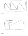
- Figure 3 shows the direct PPG signal.
- the maximal and minimal values of the PPG signal, I D and Is, respectively, are proportional to the light irradiance transmitted through the tissue when the tissue blood volume is minimal or maximal, respectively.
- the amplitude of the PPG signal, (I D -I S ) is related to the maximal change in arterial blood volume during systole, ⁇ V a .
- I D -I S is much smaller than I S
- the relative change in of the PPG signal (I D -I S )/I S is proportional to ⁇ V a :
- I D - I S / I S ⁇ a ⁇ ⁇ V a
- ⁇ a is the effective absorption coefficient of the arterial blood, which is a function of the absorption and scattering attenuation constants.
- ⁇ V a the maximal change in arterial blood volume during systole, is equal to the product of the pulse pressure and the arterial compliance.
- the amplitude of the PPG signal provides information on the arterial compliance of the systemic circulation (Babchenko et al. 2001, Shelley 2007).
- Equation (1) can be applied only for relatively homogenous tissue geometry, as occurs in pure microcirculatory bed.
- the presence of large blood vessels, where significant light absorption occurs in a single vessel, can limit the validity of Equation (1).
- Nitzan et al (1998) also showed that the linear relationship between blood volume changes and the PPG pulses holds only for small vessels, in which the absorbed light is small relative to the incident light.
- Figure 1 presents the extinction coefficient of oxygenated and de-oxygenated hemoglobin as a function of the light wavelength, where the extinction coefficient of a hemoglobin solution is defined as the absorption constant of the hemoglobin solution divided by the concentration of the hemoglobin in the solution.
- ⁇ a is the extinction coefficient for the arterial blood
- ⁇ c a is the increase of blood concentration (in the tissue) due to the maximal systolic increase of blood volume.
- ⁇ I a I D - I S ⁇ I S
- ln(I D /I S ) can be approximated by ⁇ I a /I S .
- ⁇ I a / I S 1 ⁇ a ⁇ 1 ⁇ ⁇ ⁇ c a ⁇ 1 ⁇ l 1
- ⁇ I a / I S 2 ⁇ a ⁇ 2 ⁇ ⁇ ⁇ c a ⁇ 2 ⁇ l 2
- the physiological parameter SaO 2 can be derived from the measured parameter R by using Equation 9 and the various values of the extinction
- the clinical parameter oxygen saturation SaO 2 cannot be derived from the measured parameter R and Equation 9.
- the actual relationship between the measured parameter R and oxygen saturation SaO 2 is achieved for each type of pulse oximeter sensor by calibration (Schowalter, 1997): R is measured in several persons simultaneously with in vitro SaO 2 measurement in extracted arterial blood by means of co-oximeter which is the gold-standard for SaO 2 measurements. For each person R and SaO 2 measurements are taken for several values of SaO 2 , achieved by changing the partial pressure of oxygen in the inspired air.
- the table of the simultaneous measurements of R and SaO 2 provides the required calibration for the derivation of the clinical parameter oxygen saturation SaO 2 from the measured parameter R.
- the reliability of the calibration is based on the assumption that l 2 / l 1 does not change between different persons and different physiological and clinical situations. The validity of this assumption is limited and deviations from this assumption are the likely origin of the inherent inaccuracy of the pulse oximetry technique for the assessment of the oxygen saturation, SaO 2 , in the arterial blood.
- l 2 / l 1 is expected to be about 1 and SaO 2 can be derived from Equation 9 without calibration.
- the SpO 2 values were somewhat lower than that obtained by commercial calibrated pulse oximeters using red and infrared light, probably because small deviations from the assumption of l 2 / l 1 1 and because the emission spectrum of the LED is broad, and the extinction coefficient of its light is not accurately definite.
- the accuracy of the technique is also increased by using two infrared laser diodes of narrow emission spectrum instead of LEDs.
- the calibration which is required in the conventional pulse oximetry, is done by extracting blood from the big, conduit arteries.
- the calibration procedure is valid, because the value of oxygen saturation is the same in all arteries, from the big ones to the arterioles, since oxygen dissipation occurs only in the capillaries.
- the present invention is a method for obtaining physiological information relating to the lungs of a subject, and a corresponding system, as set out in the appended claims.
- a method for the measurement of the pulmonary PPG signal, presenting the oscillations at the heart rate of the transmission of light through a region in the microcirculation of the pulmonary system.
- the pulmonary PPG signal can be utilized for the assessment of the cardiopulmonary circulatory system. It can also be used for the determination of the oxygen saturation in the pulmonary arteries, which is actually the oxygen saturation in mixed venous blood. The latter parameter enables the noninvasive (or minimally invasive) determination of cardiac output by the Fick method.
- the conventional PPG technique is based on measurements in the arterial system of the systemic circulation conducting blood from the left ventricle to the different organs of the body, except the lungs.
- the blood volume in the systemic arteries increases by the blood which is ejected from the left ventricle during systole and decreases by the relatively constant blood flow through the capillaries to the veins.
- the lungs are supplied with venous blood from the right ventricle through the pulmonary artery and, similarly to the systemic circulation, the pulmonary arterial blood volume increases during systole and decreases during diastole.
- Pulmonary blood volume increase during systole has been demonstrated in several prior studies, using several techniques, including N 2 O body plethysmography (Karatzas 1969), analysis of pulmonary arterial pressure curves (Her 1987), ECG-gated radionuclide scintigraphy (Nitzan 1992, 1994), and cardiovascular magnetic resonance (Ugander 2009).
- N 2 O body plethysmography Kermanzas 1969
- analysis of pulmonary arterial pressure curves (Her 1987)
- ECG-gated radionuclide scintigraphy (Nitzan 1992, 1994), and cardiovascular magnetic resonance (Ugander 2009).
- One of these techniques (Her 1987) is invasive; the other techniques require either special training of the examinees (Karatzas 1969), or a sophisticated imaging device (Nitzan 1992, 1994, Ugander 2009).
- the present invention provides a pulmonary PPG method, in which the pulmonary blood volume increase during systole is detected by the resultant decrease in light transmission through the lungs.
- the method requires that light emitted from a light-source reaches the lungs and that the light transmitted through the lung tissue is detected.
- the light transmitted through the lung tissue oscillates at the heart rate, like the systemic PPG, and like the latter the pulmonary PPG is related to the blood volume change in the pulmonary arteries through Equation 1.
- the systolic blood volume change in the pulmonary arteries is related to the stroke volume and to the arterial compliance and small arteries resistance in the pulmonary system.
- the pulmonary PPG signal can provide information on these cardiovascular parameters in the cardio-pulmonary system.
- the present invention relates generically to any and all techniques in which a pulmonary PPG signal is derived from measurements of light transmission through microcirculatory tissue of the lungs, whether by non-invasive, minimally invasive or fully invasive procedures. While it is simple to achieve the PPG signal in the systemic circulation, because of the proximity of the tissue under examination to the skin, the achievement of the pulmonary PPG is significantly more difficult, since the lungs lay inside the thoracic wall.
- the present invention will be exemplified herein with reference to four non-invasive or minimally invasive techniques that enable the measurement of the light transmitted through the pulmonary tissue, each of which is believed to be of value in its own right:
- the light in its way to the pulmonary tissue, also passes through the thoracic wall or the esophageal wall, which are supplied by the systemic circulation. Nevertheless, the main contribution to the PPG signal is by the pulmonary circulation, because the stroke volumes from the right and left ventricles are equal while the former is distributed in the relatively small pulmonary tissue volume and the latter is distributed in the relatively high systemic tissue volume.
- the light source and the detector are preferably separated by at least 10 mm in the esophageal probe and by at least 15 mm in the thoracic probe.
- a second detector can be attached to the esophageal wall or the thoracic wall, where the second detector and the light source are separated by less than 8 mm.
- a second light source is attached to the thoracic wall, where the second light source and the first detector are separated by less than 8 mm.
- Light transmission measurement by a light-source and a detector of relatively short separation provides information of the tissue of short depth relative to the measurement surface.
- smaller spacing between the two optic fibers is preferably used in order to use a single penetrating catheter.
- the pathlength of the light is long, and in order to have significant amount of transmitted light intensity for the measurement, infrared light which is less absorbed than visible light, is preferred.
- Figure 1a presents the extinction coefficients of oxi- and deoxi-hemoglobin. Red light, in the wavelength region of 600-700 nm, is more absorbed by deoxi-hemoglobin, than infrared light, of 700-1000 nm wavelength. For a probe adjacent to the pulmonary pleura (option 4, above), other wavelengths, such as visible wavelengths, may be used.
- the term "light source” is used herein to refer to the light delivering element from which light is released into the tissue.
- the "light source” thus defined may be a light generating element, such as a laser diode or LED, brought directly to the required location for delivering light, or may be the end of an optic fiber, an applicator connected to such a fiber, or any other waveguide or the like that conveys light to the required site from one or more remotely located light generating device.
- the term “detector” is used herein to refer to the light detecting element which detects the light scattered from the tissue.
- the “detector” thus defined may be an electro-optic light detecting element, such as a PIN diode or avalanche photodiode, brought directly to the required site of measurement for detecting light, or may be the end of an optic fiber, an applicator connected to such a fiber, or any other waveguide or the like that conveys light to the more remotely electro-optic detecting element from the required site of measurement.
- the measurement of pulmonary PPG in two wavelengths provides information on the oxygen saturation of the arterial blood in the pulmonary tissue, SvO 2 , which in fact is equal to the mixed venous blood saturation, SmvO 2 .
- SmvO 2 provides information on the adequacy of the systemic blood supply and is an essential component in the quantitative determination of cardiac output by the Fick method, as mentioned in Section 1.1.
- the current method for SmvO 2 measurement in the pulmonary artery is invasive in the sense that it includes insertion of a Swan-Ganz balloon catheter in the pulmonary artery. SmvO 2 is then measured, either intermittently, by extracting blood from the pulmonary artery or continuously, by means of oximetric measurements through optic fibers in the pulmonary artery blood.
- the oxygen saturation in the upper vena cava central venous oxygen saturation
- the invasive insertion of a catheter in the vena cava is of lower hazard than that through the right ventricle into the pulmonary artery, but the values of oxygen saturation in the two vessels may be different.
- Deep vascular structure are major blood vessels which are not superficially located, and include the inferior and superior vena cava, the right atrium, the right ventricle and central and peripheral parts of the pulmonary arteries.
- the method includes placing emitter and receiver elements of light oximeter device on the skin in the vicinity of the deep vascular structure of interest, wherein placement of the elements is achieved through matching of the plethysmography trace obtained from the oximeter device to known plethysmography characteristics of the deep vascular structure.
- the pulmonary pulse oximetry is preferably performed by illuminating the pulmonary tissue, while avoiding scattering of light from the major blood vessels in the thorax, which include the inferior and superior vena cava, the right atrium, the right ventricle and central and peripheral parts of the pulmonary arteries.
- Certain embodiments of the present invention perform pulmonary pulse oximetry using pulmonary PPG signals in two wavelengths obtained by a PPG probe applied either on the thoracic wall or on the esophageal wall.
- two wavelengths in the infrared are preferably used for the measurement of the pulmonary PPG signals and SvO 2 due to the long path of the light from the light-source to the detector.
- This is in contrast to the pulse oximetry in the systemic circulation, which is generally done by red and infrared light, taking the advantage of the relatively high difference between the values of the extinction coefficients of oxi- and deoxi-hemoglobin for red light (see Figure 1 ).
- the use of red light is possible because the pathlength required for measurements in skin is small, in the order of a few millimeters.
- the use of red light is possible in the fourth above-mentioned embodiment of the pulmonary pulse oximetry, which uses optic fibers to convey the light to and from the lung.
- the pathlengths of the red and infrared light are significantly different, so that calibration by extracted arterial blood is required for the determination of the relationship between SaO 2 and the measured parameter R derived from the two PPG signals.
- SvO 2 is obtained from R, which was defined as the ratio of the ratios ⁇ I a /I S for the two wavelengths.
- SvO 2 can also be obtained from the ratio of two values of a parameter related to the change in the PPG signal for the two wavelengths, which can be different than ⁇ I a /I S . It can be chosen as ln(I D /I S ) (as in US patent 4,773,422 and 4,167,331 ) or the derivative of I divided by I (as in US patent 6,505,060 ).
- the esophageal pulmonary pulse oximetry presented in the current patent application differs from the esophageal pulse oximeters presented by Atlee ( US patent No. 5,329,922 ) and Kyriacou et al (Kyriacou 2006), for the measurement of SaO 2 in the systemic microcirculation of the esophagus. Accordingly, the distance between the light source and the detector was less than 8 mm enabling the measurement of the PPG signal in the esophageal wall, where the light source and the detector were applied.
- the two light sources in each wavelength in the esophageal systemic pulse oximeters are required for the increase the PPG signal, while the two detectors in each wavelength in the esophageal pulmonary pulse oximeters are required for the differentiation between the contributions of the systemic and the pulmonary circulations to the PPG signal.
- a short distance, of few mms, between the detector and the light-sources or between the ends of the optic fibers of the detector and the light-sources is required for measuring the light absorption in the tissue in contact to the pulse oximeter.
- the light-sources included a red emitter, which is highly absorbed in the tissue and can therefore be used only for measurements in tissue of short distance to the pulse oximeter, like the esophageal wall.
- the light-sources emit infrared light and the light sources-detector separation is higher than 10-20 mm, to allow penetration of light to depth of 10-20 mm relative to the measurement surface, which contains pulmonary tissue.
- the preferred pulse oximeter is a device, which includes two laser diodes of two peak wavelengths in the infrared region and of narrow spectrum and a detector which can detect, for each wavelength, the transmitted light through a portion of the thorax.
- the light from the laser diodes is conveyed to the thoracic wall by an optic fiber and the light transmitted through the thorax is conveyed to the detector by another optic fiber.
- For each laser diode a PPG curve is obtained and from each of the two PPG curves the ratio between the PPG pulse amplitude and its baseline is derived.
- Another preferred pulse oximeter is an esophageal probe which includes the tips of two optic fibers, one of them conveying infrared light from two laser diodes of two peak wavelengths in the infrared region and of narrow spectrum and the second optic fiber conveying light to a detector.
- the probe is applied to the esophageal wall, in close proximity to the lung tissue.
- the two optic fiber tips are separated by more than 10 mm, so that a significant quantity of the light reaching the detector have been scattered by the lung tissue and the PPG pulse will mainly represent the blood volume changes in the pulmonary circulation.
- the value of the extinction coefficients for oxygenated blood ⁇ o and for deoxygenated blood ⁇ d can be retrieved from the literature data-bases, for each peak wavelength.
- SvO 2 is obtained from the ratio of two values of a parameter related to the change in light transmission for the two wavelengths, and this parameter can be different than amplitude-to-baseline ratio. It can be chosen as ln(I D /I S ) or the derivative of I divided by I.
- LEDs light emitting diodes
- the emission spectrum of LED is broad, so that the calculation of the mean extinction coefficients over the spectrum band of the emitted light is required.
- the LEDs with narrow-band filter are used, so that the calculation of the mean extinction coefficients over the spectrum band of the emitted light is simpler and more accurate.
- the pulmonary PPG signal is obtained from several pulmonary PPG pulses, summed together, where the start of each PPG pulse is determined by the corresponding PPG pulse of the systemic circulation.
- the pulmonary PPG signal is obtained during specific phase of the respiration.
- the pulmonary PPG signal is obtained during the time of minimal movement of the lungs, such as at end expiration or at end inspiration.
- the present invention provides a method and corresponding system for obtaining physiological information relating to the lungs of a subject comprising the steps of: directing into tissue of the lungs of the subject light of a first wavelength; detecting part of the light that has passed primarily through microcirculatory tissue of the lungs and generating a signal which is a function of intensity of the detected light; and processing the signal to derive a PPG curve for pulmonary microcirculatory arteries.
- the directing and the detecting are preferably performed without breaching any layer of tissue.
- the light is in the infrared region.
- the directing and the detecting are performed using a light source and a detector deployed so as to detect light that has passed primarily through microcirculatory tissue of the lungs primarily avoiding major blood vessels in the thorax.
- infrared light of a second wavelength different from the first wavelength is directed into the tissue of the lungs, and the processing is performed so as to derive a PPG curve for pulmonary microcirculatory arteries for each of the first and second wavelengths.
- the directing and the detecting are performed without breaching any layer of tissue, using light sources and a detector deployed so as to detect light that has passed primarily through microcirculatory tissue of the lungs primarily avoiding major blood vessels.
- intensities of the light at the first and second wavelengths are modulated at two different frequencies.
- delivery of light at the first and second wavelengths is time-multiplexed.
- a ratio R between values of a parameter related to relative changes in light transmission for the first and second wavelengths is derived from the data, and the oxygen saturation in the blood of the pulmonary microcirculatory arteries is determined from the ratio R.
- oxygen saturation SvO 2 in the blood of the pulmonary microcirculatory arteries is determined from R based on a relationship derived experimentally from invasive measurements of oxygen saturation in the pulmonary artery and R from PPG curves of the pulmonary microcirculatory arteries using the first and second wavelengths.
- the light of the first wavelength is the peak of an emission spectrum of a first infrared light source and the infrared light of the second wavelength is a peak of an emission spectrum of a second infrared light source, wherein oxygen saturation SvO 2 in the blood of the pulmonary microcirculatory arteries is determined from R based on a relationship including the mean value of the extinction coefficient for each of the two emission spectra of the two light sources, for oxygenated blood ⁇ o and for deoxygenated blood ⁇ d .
- the relationship between oxygen saturation in the blood of the pulmonary microcirculatory arteries SvO 2 and the ratio R also includes the mean optical path-lengths l 1 and l 2 for each of the two wavelengths.
- the parameter related to the relative change in light transmission is selected from the group comprising:
- the light is directed into the tissue of the lungs by a light source in contact with a first location on the thoracic wall of the subject, and the detecting is performed by a detector in contact with a second location on the thoracic wall of the subject.
- the light for each of the first and second wavelengths is directed into the tissue of the lungs by light sources in contact with a first location on the thoracic wall of the subject, and the detecting is performed by a detector in contact with a second location on the thoracic wall of the subject.
- the second location is preferably at least 15 millimeters from the first location.
- the light for each of the first and second wavelengths is directed into the tissue of the lungs by light sources in contact with a first location on the inner surface of the esophagus of the subject, and the detecting is performed by a detector in contact with a second location on the inner surface of the esophagus of the subject.
- the second location is preferably at least 10 millimeters from the first location.
- the light is directed into the tissue of the lungs by at least one light source in contact with the thoracic wall of the subject, and the detecting is performed by a detector in contact with the inner surface of the esophagus of the subject.
- the directing of the light of the first and second wavelengths and the detecting part of the light are performed using a light delivering element and a light receiving element positioned in adjacent relation to the pulmonary pleura.
- the light for each of the first and second wavelengths is directed into the tissue of the lungs by light sources in contact with a first location on an anatomical wall and wherein the detecting is performed by a detector in contact with a second location on the anatomical wall, the first and second locations being spaced apart by a first spacing
- the method further comprises: sampling additional measurements using one of: a second detector in contact with a third location on the anatomical wall spaced from the first location by a distance less than the first spacing, and a second light source in contact with a third location on the anatomical wall spaced from the second location by a distance less than the first spacing, and employing the additional measurements to assess a contribution to the PPG curve due to the blood in the tissue of the anatomical wall.
- the signal-to-noise ratio of the shape of the pulmonary PPG curve is increased by adding together several pulmonary PPG signals, where the time-segments of these pulmonary PPG curves are determined by corresponding PPG curves from the systemic circulation, obtained simultaneously with the PPG curves of the pulmonary microcirculatory arteries.
- the adding together several pulmonary PPG curves is performed separately for pulmonary PPG curves sampled during different phases of the respiration.
- the different phases of the respiration include one of the group: end inspiration, end expiration, end inspiration and end expiration.
- the PPG curve for pulmonary microcirculatory arteries is obtained during a specific phase of the respiration.
- the specific phase of the respiration includes one of the group: end inspiration, end expiration, end inspiration and end expiration.
Abstract
Description
- Transfer of oxygen from the lungs to the tissue cells is done mainly via the hemoglobin molecules in the red blood cells and only small part of it is dissolved in arterial plasma. Oxygen saturation (SO2) is the ratio of oxygenated hemoglobin to total hemoglobin (SO2=HbO2/(HbO2+Hb)), and its value in the arterial blood, SaO2, is 94-99%. The assessment of SaO2 is mainly important for clinical evaluation of proper respiratory function, since SaO2 depends on the adequacy of the ventilation and respiratory function.
- Most of the hemoglobin in venous blood is still oxygenated: normal values of the oxygen saturation in the peripheral venous blood are 70-80%. The value of the oxygen saturation in the venous blood also has physiological and clinical significance, as lower blood flow to the tissue results in higher utilization of the oxygen in the blood and lower value of venous oxygen saturation. The measurement of oxygen saturation in the venous blood of an organ provides therefore information on the adequacy of its blood supply.
- The blood from the veins of the whole body is drained into the pulmonary artery after being mixed by the right ventricle. The pulmonary artery blood is named mixed venous blood. The value of the mixed venous oxygen saturation, SmvO2, represents the mean oxygen saturation in the veins of the whole body and is of particular interest. Low values of SmvO2 indicate inadequate oxygen supply to the body, either because of low blood flow or because of improper respiration. If the latter failure is eliminated by using SaO2 measurement, low values of SmvO2 indicate low total blood flow, which is equivalent to low cardiac output. In fact the mixed venous oxygen saturation, SmvO2, is used for the quantitative assessment of the cardiac output by means of the Fick method: the cardiac output is determined from the values of total oxygen consumption, arterial oxygen content and venous oxygen content. The total oxygen consumption can be obtained from oxygen consumption measurements in the inhaled and exhaled air and the values of the arterial and venous oxygen content can be derived from the values of SaO2 and SmvO2 respectively.
- The measurement of SaO2, the oxygen saturation in the arterial blood, can be performed noninvasively by pulse oximetry, which is based on the different light absorption spectrum for oxygenated and de-oxygenated hemoglobin, as shown in
Figure 1 . Oximetry is a technique in which the attenuation in a substance of light in two wavelengths is measured and the difference in the attenuation of the light between the wavelengths is attributed to the difference in their absorption in the substance. However, in order to quantitatively assess SaO2 the contribution of the arterial blood to the light absorption must be isolated from the contribution of the venous blood. In pulse oximetry the technique for the isolation of the arterial contribution is based on photoplethysmography (PPG) - the measurement of light absorption changes due to the cardiac induced blood volume changes. During systole the blood volume in tissue arteries increases and the transmission through the tissue decreases.Figure 2 shows the inverted PPG signal: during systole there is fast increase in arterial blood volume. Pulse oximetry uses the PPG signal in two wavelengths for the assessment of the oxygen saturation in the arterial blood, SaO2 (Yoshiya, 1980), as will be described later. - In order to assess SaO2 by pulse oximetry there is also a need to consider the effect of scattering on the attenuation of the light. The light traversing a given tissue is not only subject to absorption by the hemoglobin but also to scattering by the various organelles in the tissue cells and in the red blood cells, which diverts the light from its original direct path and also increases its path-length. In order to assess SaO2 or SvO2 by optical means the contribution of the absorption to the attenuation has to be isolated.
- While the measurement of SaO2 can be performed noninvasively by pulse oximetry, the available techniques for the measurement of SmvO2, the mixed venous oxygen saturation, are invasive and require the insertion of a catheter into the pulmonary artery (Swan-Ganz catheter). After inserting the catheter SmvO2 can be measured either in vitro by extracting mixed venous blood from the pulmonary artery or by in vivo measurements, using an optical probe attached to the tip of the catheter. The invasive measurement of SmvO2 is the reason for the invasiveness of the Fick method for the measurement of cardiac output.
- The PPG probe consists of a light source emitting light into the tissue and a photodetector measuring the light transmitted through the tissue. Two kinds of PPG probes are used for clinical diagnosis and research: transmission and reflection. In transmission PPG, the light source and the detector are attached to two sides of a small organ, such as a finger or an ear lobe, and the detector measures the light transmitted through the organ. In reflection PPG, the light source and the detector are attached to the same side of the organ and the detector measures the scattered light from the tissue under the probe. In both kinds of PPG probe, the detector output oscillates at the heart rate: during systole, blood is ejected from the left ventricle into the peripheral vascular system, thereby increasing the arterial blood content, and consequently decreasing the light intensity transmitted through the tissue.
Figure 3 shows the direct PPG signal. The maximal and minimal values of the PPG signal, ID and Is, respectively, are proportional to the light irradiance transmitted through the tissue when the tissue blood volume is minimal or maximal, respectively. The amplitude of the PPG signal, (ID-IS), is related to the maximal change in arterial blood volume during systole, ΔVa. if ID-IS is much smaller than IS, then the relative change in of the PPG signal (ID-IS)/IS is proportional to ΔVa:
where αa is the effective absorption coefficient of the arterial blood, which is a function of the absorption and scattering attenuation constants. ΔVa, the maximal change in arterial blood volume during systole, is equal to the product of the pulse pressure and the arterial compliance. Hence the amplitude of the PPG signal provides information on the arterial compliance of the systemic circulation (Babchenko et al. 2001, Shelley 2007). - Equation (1) can be applied only for relatively homogenous tissue geometry, as occurs in pure microcirculatory bed. The presence of large blood vessels, where significant light absorption occurs in a single vessel, can limit the validity of Equation (1). Nitzan et al (1998) also showed that the linear relationship between blood volume changes and the PPG pulses holds only for small vessels, in which the absorbed light is small relative to the incident light.
-
Figure 1 presents the extinction coefficient of oxygenated and de-oxygenated hemoglobin as a function of the light wavelength, where the extinction coefficient of a hemoglobin solution is defined as the absorption constant of the hemoglobin solution divided by the concentration of the hemoglobin in the solution. - The theory of deriving arterial oxygen saturation, SaO2, from the PPG curves at two wavelengths is described in several articles (see Wieben, 1997, Mannheimer et al, 1997, Nitzan et al, 2000). The transmitted light intensity, It, through a tissue sample which includes vessels with whole blood is based on Beer-Lambert law and is given by:

where l is the effective optical path-length (which is higher than the width of the tissue sample because of scattering in the tissue) α is the absorption constant of the tissue, c is the concentration of the blood in the tissue, and ε is the extinction coefficient of the blood, defined as the absorption constant of the blood divided by the concentration of the blood in the tissue. I0 is the incident light intensity. - During systole the tissue blood volume increases and consequently the light transmission through the tissue decreases, creating the PPG signal/curve. If IS is the light transmission through the tissue at the maximal increase in tissue blood volume and ID is the light transmitted through the tissue at end diastole (when the tissue blood volume has its minimal value), then

where εa is the extinction coefficient for the arterial blood, and Δca is the increase of blood concentration (in the tissue) due to the maximal systolic increase of blood volume. For small blood volume changes ΔIa = ID - IS << IS, and ln(ID/IS) can be approximated by ΔIa/IS. If the light transmission is measured for two wavelengths λ1 and λ2, then
and the ratio R, defined by
satisfies the equation
assuming that the difference in the blood concentration change, Δca, between the two wavelengths can be neglected (Δca1≈Δca2). Note that the last assumption is not always satisfied in reflection PPG, if the penetration depth for the two wavelengths is different. If the two wavelengths are close to each other then the difference of the effective optical path-length is small and
- The relationship between the ratio R - the measured parameter - and the oxygen saturation SaO2 can be derived from the decomposition of the extinction coefficient ε into its two components, the extinction coefficients for oxygenated blood εo and for deoxygenated blood εd:

Then
and
The physiological parameter SaO2 can be derived from the measured parameter R by using Equation 9 and the various values of the extinction coefficient (which are known from the literature) provided that the difference between the pathlengths for the two wavelengths can be neglected. However, commercial pulse oximeters choose one of the wavelengths in the infrared region and the other in the red region (where the difference in the extinction coefficient between oxygenated and deoxygenated blood is maximal) in order to have a higher difference in light transmittance between the two wavelengths. For this choice, the red light scattering constant significantly differs from that of the infrared light resulting in non-negligible difference in optical path-lengths for the two wavelengths. - Due to the difference in scattering between the red and infrared wavelengths, the clinical parameter oxygen saturation SaO2 cannot be derived from the measured parameter R and Equation 9. The actual relationship between the measured parameter R and oxygen saturation SaO2 is achieved for each type of pulse oximeter sensor by calibration (Schowalter, 1997): R is measured in several persons simultaneously with in vitro SaO2 measurement in extracted arterial blood by means of co-oximeter which is the gold-standard for SaO2 measurements. For each person R and SaO2 measurements are taken for several values of SaO2, achieved by changing the partial pressure of oxygen in the inspired air. The table of the simultaneous measurements of R and SaO2 provides the required calibration for the derivation of the clinical parameter oxygen saturation SaO2 from the measured parameter R. The relationship between R and SaO2 can be determined by proposing an analytical formula, such as

and obtaining the values of the constants a,b,c, and d in the calibration process described above. - The reliability of the calibration is based on the assumption that l 2/l 1 does not change between different persons and different physiological and clinical situations. The validity of this assumption is limited and deviations from this assumption are the likely origin of the inherent inaccuracy of the pulse oximetry technique for the assessment of the oxygen saturation, SaO2, in the arterial blood.
- If the two wavelengths are close to each other, l 2/l 1 is expected to be about 1 and SaO2 can be derived from Equation 9 without calibration. This was shown in our study (Nitzan et al 2000), in which we measured SpO2, using pulse oximetry based on two infrared light emitting diodes (LEDs) (of peak wavelengths 767 and 811 nm) and Equation 9, without calibration. The SpO2 values were somewhat lower than that obtained by commercial calibrated pulse oximeters using red and infrared light, probably because small deviations from the assumption of l 2/l 1=1 and because the emission spectrum of the LED is broad, and the extinction coefficient of its light is not accurately definite. The accuracy of the technique increased by using Equation (12) and assessing (l 2/l 1) value from published data (like that of Duncan et al., 1995 for the forearm, (l 2/l 1) = 0.97). The accuracy of the technique is also increased by using two infrared laser diodes of narrow emission spectrum instead of LEDs.
- At the end of Section 1.2 it was claimed that the linear relationship between blood volume changes and the PPG pulses holds only for the small blood vessels of the microcirculation, where the absorbed light in a single vessel is small relative to the incident light. Since pulse oximetry is based on PPG measurements and on the linear relationship between blood volume changes and the PPG signal amplitude, it can be inferred that measurement of oxygen saturation in arterial blood by pulse oximetry can be properly performed only on the microcirculation in tissue. In their articles Mannheimer et al. (2004) and Reuss and Siker (2004) claimed that pulse oximetry is affected by large subcutaneous blood vessels, so that reflectance pulse oximetry should be avoided in sites with palpable arterial pulsatility. While the pulse oximetry is performed on the microcirculatory bed, the calibration, which is required in the conventional pulse oximetry, is done by extracting blood from the big, conduit arteries. The calibration procedure is valid, because the value of oxygen saturation is the same in all arteries, from the big ones to the arterioles, since oxygen dissipation occurs only in the capillaries.
- In contrast to the well-established non-invasive measurement of SaO2 by pulse oximetry in the systemic microcirculation , the available techniques for the measurement of SvO2, the mixed venous oxygen saturation, are invasive and require the insertion of a Swan-Ganz catheter into the pulmonary artery. Accordingly, there is a need for an innovative method for the less invasive measurement of the oxygen saturation in mixed venous blood, SvO2.
- The present invention is a method for obtaining physiological information relating to the lungs of a subject, and a corresponding system, as set out in the appended claims.
- The invention is herein described, by way of example only, with reference to the accompanying drawings, wherein:
-
FIG. 1a is a plot of the extinction coefficients of the oxi- and deoxi-hemoglobin as a function of the wavelength, in the visible and near-infrared regions. The figure was published by Scott Prahl, Oregon Medical Laser Center, 1999, as a best estimate of the spectrum of Hb and HbO2 from a variety of sources. (Appears in M. Nitzan and H. Taitelbaum, 2008. IEEE Instrumentation and Measurement Magazine, 11:9-15.) -
FIG. 1b is a plot of the extinction coefficients of the oxi- and deoxi-hemoglobin as a function of the wavelength, in the near-infrared region, published by Kim and Liu, 2007, Phys. Med. Biol. 52:6295-6322. -
FIG. 2 is a plot of an inverted PPG signal in the systemic circulation, which presents the light absorption in the tissue against time, showing variations at the heart rate, wherein minimal light absorption occurs at end-diastole when the tissue blood volume is at minimum. -
FIG. 3 is a plot of a direct PPG signal in the systemic circulation, which presents the light transmission through the tissue against time, showing variations at the heart rate, wherein maximal intensity of transmitted light occurs at end-diastole when the tissue blood volume is at minimum. -
FIG. 4 presents a cross-section of the thorax, with an element for emitting infrared light into the thorax and an element for detecting the light transmitted through the thorax wall and through a portion of the pulmonary tissue. -
FIG. 5 presents a cross-section of transmitter and receiver elements, which include optic fiber and reflection prism, for emitting infrared light from a laser diode into the skin and for detecting the light transmitted through the tissue. -
FIG. 6a presents a cross-section of the esophagus and a catheter with a PPG probe, which includes a LED and a detector. The PPG probe is applied on a site in the inner esophageal wall, where the lung tissue is in close proximity to the outer esophageal wall. The balloon pressure causes the PPG probe to be in close contact with the inner esophageal wall. -
FIG. 6b presents a cross-section of the esophagus and a catheter with a PPG probe, which includes two optic fibers leading from a laser diode and to a detector. The PPG probe is applied on a site in the inner esophageal wall, where the lung tissue is in close proximity to the outer esophageal wall. The balloon pressure causes the PPG probe to be in close contact with the inner esophageal wall. -
FIG. 7 is a block diagram schematically showing attachment of a controller including a processing means to the light source and detector ofFIGS. 4-6 . - In the current invention, a method is presented for the measurement of the pulmonary PPG signal, presenting the oscillations at the heart rate of the transmission of light through a region in the microcirculation of the pulmonary system. The pulmonary PPG signal can be utilized for the assessment of the cardiopulmonary circulatory system. It can also be used for the determination of the oxygen saturation in the pulmonary arteries, which is actually the oxygen saturation in mixed venous blood. The latter parameter enables the noninvasive (or minimally invasive) determination of cardiac output by the Fick method. The conventional PPG technique is based on measurements in the arterial system of the systemic circulation conducting blood from the left ventricle to the different organs of the body, except the lungs. The blood volume in the systemic arteries increases by the blood which is ejected from the left ventricle during systole and decreases by the relatively constant blood flow through the capillaries to the veins. The lungs are supplied with venous blood from the right ventricle through the pulmonary artery and, similarly to the systemic circulation, the pulmonary arterial blood volume increases during systole and decreases during diastole. Pulmonary blood volume increase during systole has been demonstrated in several prior studies, using several techniques, including N2O body plethysmography (Karatzas 1969), analysis of pulmonary arterial pressure curves (Her 1987), ECG-gated radionuclide scintigraphy (Nitzan 1992, 1994), and cardiovascular magnetic resonance (Ugander 2009). One of these techniques (Her 1987) is invasive; the other techniques require either special training of the examinees (Karatzas 1969), or a sophisticated imaging device (Nitzan 1992, 1994, Ugander 2009).
- The present invention provides a pulmonary PPG method, in which the pulmonary blood volume increase during systole is detected by the resultant decrease in light transmission through the lungs. The method requires that light emitted from a light-source reaches the lungs and that the light transmitted through the lung tissue is detected. The light transmitted through the lung tissue oscillates at the heart rate, like the systemic PPG, and like the latter the pulmonary PPG is related to the blood volume change in the pulmonary arteries through
Equation 1. Like the systemic circulation the systolic blood volume change in the pulmonary arteries is related to the stroke volume and to the arterial compliance and small arteries resistance in the pulmonary system. Hence the pulmonary PPG signal can provide information on these cardiovascular parameters in the cardio-pulmonary system. - The present invention relates generically to any and all techniques in which a pulmonary PPG signal is derived from measurements of light transmission through microcirculatory tissue of the lungs, whether by non-invasive, minimally invasive or fully invasive procedures. While it is simple to achieve the PPG signal in the systemic circulation, because of the proximity of the tissue under examination to the skin, the achievement of the pulmonary PPG is significantly more difficult, since the lungs lay inside the thoracic wall. By way of exemplary but non-limiting preferred examples, the present invention will be exemplified herein with reference to four non-invasive or minimally invasive techniques that enable the measurement of the light transmitted through the pulmonary tissue, each of which is believed to be of value in its own right:
- 1. A light source and a detector are applied to the thoracic wall of a patient and the detector measures the light which was emitted from the light source and transmitted through the thorax. See
Figure 4 and 5 . The light source and the detector are separated by at least 20 mm so that the region of illumination overlaps a portion of the pulmonary microcirculation beneath the PPG probe. The technique is especially suitable for infants, whose thoracic wall is relatively thin. - 2. A light source and a detector are inserted into the esophagus and brought into close contact with its wall, in a site where the pulmonary tissue is in tight proximity to the esophageal wall, and the detector measures the light which was emitted from the light source and transmitted through the esophageal wall and part of the lungs. See
Figure 6 . The light source and the detector are separated by at least 10 mm so that the region of illumination overlaps a portion of the pulmonary microcirculation in the neighborhood of the PPG probe. - 3. A light source is applied to the thoracic wall of a patient and a detector is inserted into the esophagus and brought into close contact with its wall in a site where the pulmonary tissue is in tight proximity to the esophageal wall. The detector measures the light which was emitted from the light source and transmitted through the thoracic wall and the esophageal wall and through part of the lungs. By suitable choice of the locations of the light-source and the detector the detected light is mainly affected by absorption in the pulmonary microcirculation and the contribution of the attenuation by other organs can be neglected.
- 4. A catheter with two optic fibers is inserted through the thoracic wall and brought into adjacent relation (i.e., with minimal intervening tissue) with the pulmonary pleura, which covers the lung tissue. The light delivering element and light receiving element in the probe (catheter) are typically brought into contact with the pulmonary pleura or preferably into close proximity to the pulmonary pleura, without contacting it, in order to avoid harm to the vulnerable organ. One of the optic fibers conveys light from a light source into the lung tissue and the other one conveys light which was scattered by the tissue to a detector. Both the light source and the detector are located out of the body.
- In the first three above-mentioned pulmonary PPG techniques, referred to herein as "remote PPG techniques", the light, in its way to the pulmonary tissue, also passes through the thoracic wall or the esophageal wall, which are supplied by the systemic circulation. Nevertheless, the main contribution to the PPG signal is by the pulmonary circulation, because the stroke volumes from the right and left ventricles are equal while the former is distributed in the relatively small pulmonary tissue volume and the latter is distributed in the relatively high systemic tissue volume. The relationship between the increase in pulmonary blood volume during systole and the right stroke volume (which is the blood volume ejected from the right ventricle during systole) was determined in several studies: it is 50-67% of the total stroke volume (Karatzas 1969, Nitzan 1992, 1994, Ugander 2009, Her 1987). Hence the systolic blood volume increase in a volume element in the pulmonary circulation is much higher than in a typical volume element in the systemic circulation, enabling the pulmonary PPG measurement
- Because of the depth of pulmonary tissue relative to the measurement surface in the remote PPG techniques, the light source and the detector are preferably separated by at least 10 mm in the esophageal probe and by at least 15 mm in the thoracic probe. In order to assess the contribution of the esophageal wall or the thoracic wall circulation to the PPG signal, a second detector can be attached to the esophageal wall or the thoracic wall, where the second detector and the light source are separated by less than 8 mm. In another technique for the assessment of the contribution of the esophageal wall circulation or the thoracic wall circulation to the PPG signal, a second light source is attached to the thoracic wall, where the second light source and the first detector are separated by less than 8 mm. Light transmission measurement by a light-source and a detector of relatively short separation provides information of the tissue of short depth relative to the measurement surface. For a probe adjacent to the pulmonary pleura (option 4, above), smaller spacing between the two optic fibers is preferably used in order to use a single penetrating catheter.
- In each of the remote pulmonary PPG techniques, the pathlength of the light is long, and in order to have significant amount of transmitted light intensity for the measurement, infrared light which is less absorbed than visible light, is preferred.
Figure 1a presents the extinction coefficients of oxi- and deoxi-hemoglobin. Red light, in the wavelength region of 600-700 nm, is more absorbed by deoxi-hemoglobin, than infrared light, of 700-1000 nm wavelength. For a probe adjacent to the pulmonary pleura (option 4, above), other wavelengths, such as visible wavelengths, may be used. - Parenthetically, it should be noted that the term "light source" is used herein to refer to the light delivering element from which light is released into the tissue. The "light source" thus defined may be a light generating element, such as a laser diode or LED, brought directly to the required location for delivering light, or may be the end of an optic fiber, an applicator connected to such a fiber, or any other waveguide or the like that conveys light to the required site from one or more remotely located light generating device.
- Similarly the term "detector" is used herein to refer to the light detecting element which detects the light scattered from the tissue. The "detector" thus defined may be an electro-optic light detecting element, such as a PIN diode or avalanche photodiode, brought directly to the required site of measurement for detecting light, or may be the end of an optic fiber, an applicator connected to such a fiber, or any other waveguide or the like that conveys light to the more remotely electro-optic detecting element from the required site of measurement.
- Similar to conventional pulse oximetry technique, which provides information on SaO2 via the measurement of systemic PPG in two wavelengths, the measurement of pulmonary PPG in two wavelengths provides information on the oxygen saturation of the arterial blood in the pulmonary tissue, SvO2, which in fact is equal to the mixed venous blood saturation, SmvO2. SmvO2 provides information on the adequacy of the systemic blood supply and is an essential component in the quantitative determination of cardiac output by the Fick method, as mentioned in Section 1.1.
- The current method for SmvO2 measurement in the pulmonary artery is invasive in the sense that it includes insertion of a Swan-Ganz balloon catheter in the pulmonary artery. SmvO2 is then measured, either intermittently, by extracting blood from the pulmonary artery or continuously, by means of oximetric measurements through optic fibers in the pulmonary artery blood. In another invasive technique, the oxygen saturation in the upper vena cava (central venous oxygen saturation) is measured. The invasive insertion of a catheter in the vena cava is of lower hazard than that through the right ventricle into the pulmonary artery, but the values of oxygen saturation in the two vessels may be different.
- Several optical methods have been proposed for the non-invasive or minimally invasive measurement of oxygen saturation of blood within the pulmonary artery or in a central vein. These methods derive the required parameter from spectroscopic absorption measurements utilizing scattered light from the pulmonary artery or the central vein. The proposed various methods try to isolate the contribution of the scattered light from the pulmonary artery (or the other vessel) from that of the surrounding tissue.
- Cheng et al in US patent No.
US 2006/0253007 presented a method for the assessment of the oxygen saturation in a blood vessel such as the interior jugular vein by illuminating it from the skin. They suggest transmitting of the radiation into two regions, containing different portions of the target structure, for the isolation of the scattered radiation from the target vessel. They also suggest using ultrasound imaging for optimal placement of the optical transmitters and the receivers above the target structure. - Dixon in US Patent No.
US2010/0198027 proposed a non-invasive method for the determination of oxygen saturation of blood within a deep vascular structure. Deep vascular structure are major blood vessels which are not superficially located, and include the inferior and superior vena cava, the right atrium, the right ventricle and central and peripheral parts of the pulmonary arteries. The method includes placing emitter and receiver elements of light oximeter device on the skin in the vicinity of the deep vascular structure of interest, wherein placement of the elements is achieved through matching of the plethysmography trace obtained from the oximeter device to known plethysmography characteristics of the deep vascular structure. - Kohl et al in US patent No.
US6,961,600B2 presented a minimally-invasive technique for the determination of mixed venous oxygen saturation by introducing catheter with an optical fiber in the bronchia, in the vicinity of the pulmonary artery. - These inventions suggest measuring SmvO2 in the blood within the big arteries or veins, using non-invasive or minimally-invasive techniques, similar to the conventional invasive technique, which measures SmvO2 in the pulmonary artery. However, the measurement of SmvO2 by pulse oximetry, based on light scattering from big vessels is not accurate, as was found in the systemic circulation, that pulse oximetry cannot be used in the vicinity of big blood vessels (Mannheimer 2004, Reuss 2004). Pulse oximetry in the systemic circulation has to be performed on the microcirculatory bed, and the same must be done in the pulmonary system. In preferred implementations of the present invention, the pulmonary pulse oximetry is preferably performed by illuminating the pulmonary tissue, while avoiding scattering of light from the major blood vessels in the thorax, which include the inferior and superior vena cava, the right atrium, the right ventricle and central and peripheral parts of the pulmonary arteries.
- Certain embodiments of the present invention perform pulmonary pulse oximetry using pulmonary PPG signals in two wavelengths obtained by a PPG probe applied either on the thoracic wall or on the esophageal wall. As was explained above, two wavelengths in the infrared are preferably used for the measurement of the pulmonary PPG signals and SvO2 due to the long path of the light from the light-source to the detector. This is in contrast to the pulse oximetry in the systemic circulation, which is generally done by red and infrared light, taking the advantage of the relatively high difference between the values of the extinction coefficients of oxi- and deoxi-hemoglobin for red light (see
Figure 1 ). In the pulse oximetry in the systemic circulation the use of red light is possible because the pathlength required for measurements in skin is small, in the order of a few millimeters. Similarly, in the fourth above-mentioned embodiment of the pulmonary pulse oximetry, which uses optic fibers to convey the light to and from the lung, the use of red light is possible. As was explained in Section 1.3, the pathlengths of the red and infrared light are significantly different, so that calibration by extracted arterial blood is required for the determination of the relationship between SaO2 and the measured parameter R derived from the two PPG signals. Similarly, in the pulmonary pulse oximetry technique, if the difference between the pathlengths of the two wavelengths in the infrared is significant, calibration by extracted blood from the pulmonary artery is required. This calibration is typically not required in pulse oximeter which uses two wavelengths in the infrared region, if they are close enough so that the difference between their pathlengths can be neglected. It is therefore preferable to use two adjacent wavelengths in the infrared, of small difference between their pathlength, and use Equation 9, after neglecting the small difference in their pathlength or correcting it by a suitable correction factor (see Section 1.3). - In the description of the pulse oximetry method presented above, SvO2 is obtained from R, which was defined as the ratio of the ratios ΔIa/IS for the two wavelengths. SvO2 can also be obtained from the ratio of two values of a parameter related to the change in the PPG signal for the two wavelengths, which can be different than ΔIa/IS. It can be chosen as ln(ID/IS) (as in
US patent 4,773,422 and4,167,331 ) or the derivative of I divided by I (as inUS patent 6,505,060 ). - The esophageal pulmonary pulse oximetry presented in the current patent application differs from the esophageal pulse oximeters presented by Atlee (
US patent No. 5,329,922 ) and Kyriacou et al (Kyriacou 2006), for the measurement of SaO2 in the systemic microcirculation of the esophagus. Accordingly, the distance between the light source and the detector was less than 8 mm enabling the measurement of the PPG signal in the esophageal wall, where the light source and the detector were applied. The two light sources in each wavelength in the esophageal systemic pulse oximeters are required for the increase the PPG signal, while the two detectors in each wavelength in the esophageal pulmonary pulse oximeters are required for the differentiation between the contributions of the systemic and the pulmonary circulations to the PPG signal. - An esophageal systemic pulse oximetry, based on fiber-optic reflectance sensor was presented by Phillips et al (Phillips 2011) and an apparatus for measuring the oxygen saturation level of blood at an internal measurement site, based also on fiber-optic reflectance pulse oximetry was presented by
Phillips et al in US patent 2008/0045822 . In the former device, the distance between the ends of the detector and the light-sources optic fibers was 4 mm. In the latter apparatus the optical centers of the first and second optical fibers are separated by at least 1 mm at their distal ends. A short distance, of few mms, between the detector and the light-sources or between the ends of the optic fibers of the detector and the light-sources is required for measuring the light absorption in the tissue in contact to the pulse oximeter. In both articles and in the patent application the light-sources included a red emitter, which is highly absorbed in the tissue and can therefore be used only for measurements in tissue of short distance to the pulse oximeter, like the esophageal wall. In the pulmonary pulse oximeter the light-sources emit infrared light and the light sources-detector separation is higher than 10-20 mm, to allow penetration of light to depth of 10-20 mm relative to the measurement surface, which contains pulmonary tissue. It should be noted that the pulse oximeter presented by Phillips et al in theirUS patent 2008/0045822 was also suggested for the measurement of oxygen saturation in internal tissue like brain, but the probe must be inserted through the skull and applied adjacent to the brain surface in order to measure the oxygen saturation in the brain blood. - The preferred pulse oximeter is a device, which includes two laser diodes of two peak wavelengths in the infrared region and of narrow spectrum and a detector which can detect, for each wavelength, the transmitted light through a portion of the thorax. The light from the laser diodes is conveyed to the thoracic wall by an optic fiber and the light transmitted through the thorax is conveyed to the detector by another optic fiber. For each laser diode a PPG curve is obtained and from each of the two PPG curves the ratio between the PPG pulse amplitude and its baseline is derived. The ratio R of the two values of this amplitude-to-baseline ratio for the two wavelengths is calculated and SvO2 is determined from the equation

- The value of the extinction coefficients for oxygenated blood εo and for deoxygenated blood εd can be retrieved from the literature data-bases, for each peak wavelength. Another preferred pulse oximeter is an esophageal probe which includes the tips of two optic fibers, one of them conveying infrared light from two laser diodes of two peak wavelengths in the infrared region and of narrow spectrum and the second optic fiber conveying light to a detector. The probe is applied to the esophageal wall, in close proximity to the lung tissue. The two optic fiber tips are separated by more than 10 mm, so that a significant quantity of the light reaching the detector have been scattered by the lung tissue and the PPG pulse will mainly represent the blood volume changes in the pulmonary circulation. For each laser diode a PPG curve is obtained and from each of the two PPG curves the ratio between the PPG pulse amplitude and its baseline is derived. The ratio R of the two values of this amplitude-to-baseline ratio for the two wavelengths is calculated and SvO2 is determined from the equation

- The value of the extinction coefficients for oxygenated blood εo and for deoxygenated blood εd can be retrieved from the literature data-bases, for each peak wavelength.
- In another preferred embodiment SvO2 is obtained from the ratio of two values of a parameter related to the change in light transmission for the two wavelengths, and this parameter can be different than amplitude-to-baseline ratio. It can be chosen as ln(ID/IS) or the derivative of I divided by I.
- In another preferred embodiment light emitting diodes (LEDs) are used instead of laser diodes and inserted directly into the esophagus with no need for optic fibers. The emission spectrum of LED is broad, so that the calculation of the mean extinction coefficients over the spectrum band of the emitted light is required.
- In another preferred embodiment the LEDs with narrow-band filter are used, so that the calculation of the mean extinction coefficients over the spectrum band of the emitted light is simpler and more accurate.
- In another preferred embodiment the pulmonary PPG signal is obtained from several pulmonary PPG pulses, summed together, where the start of each PPG pulse is determined by the corresponding PPG pulse of the systemic circulation.
- In another preferred embodiment the pulmonary PPG signal is obtained during specific phase of the respiration.
- In another preferred embodiment the pulmonary PPG signal is obtained during the time of minimal movement of the lungs, such as at end expiration or at end inspiration.
- In summary, in view of the above, it will be understood that the present invention provides a method and corresponding system for obtaining physiological information relating to the lungs of a subject comprising the steps of: directing into tissue of the lungs of the subject light of a first wavelength; detecting part of the light that has passed primarily through microcirculatory tissue of the lungs and generating a signal which is a function of intensity of the detected light; and processing the signal to derive a PPG curve for pulmonary microcirculatory arteries. The directing and the detecting are preferably performed without breaching any layer of tissue. The light is in the infrared region.
- According to a further feature of an embodiment of the present invention, the directing and the detecting are performed using a light source and a detector deployed so as to detect light that has passed primarily through microcirculatory tissue of the lungs primarily avoiding major blood vessels in the thorax.
- According to a further feature of an embodiment of the present invention, infrared light of a second wavelength different from the first wavelength is directed into the tissue of the lungs, and the processing is performed so as to derive a PPG curve for pulmonary microcirculatory arteries for each of the first and second wavelengths. According to a further feature of an embodiment of the present invention, the directing and the detecting are performed without breaching any layer of tissue, using light sources and a detector deployed so as to detect light that has passed primarily through microcirculatory tissue of the lungs primarily avoiding major blood vessels.
- According to a further feature of an embodiment of the present invention, intensities of the light at the first and second wavelengths are modulated at two different frequencies.
- According to a further feature of an embodiment of the present invention, delivery of light at the first and second wavelengths is time-multiplexed.
- According to a further feature of an embodiment of the present invention, a ratio R between values of a parameter related to relative changes in light transmission for the first and second wavelengths is derived from the data, and the oxygen saturation in the blood of the pulmonary microcirculatory arteries is determined from the ratio R.
- According to a further feature of an embodiment of the present invention, oxygen saturation SvO2 in the blood of the pulmonary microcirculatory arteries is determined from R based on a relationship derived experimentally from invasive measurements of oxygen saturation in the pulmonary artery and R from PPG curves of the pulmonary microcirculatory arteries using the first and second wavelengths.
- According to a further feature of an embodiment of the present invention, the relationship is expressed by the formula:

where at least one of the constants a, b, c, and d is obtained experimentally from invasive measurements of oxygen saturation in the pulmonary artery and R from PPG curves of the pulmonary microcirculatory arteries using the first and second wavelengths. - According to a further feature of an embodiment of the present invention, the light of the first wavelength is the peak of an emission spectrum of a first infrared light source and the infrared light of the second wavelength is a peak of an emission spectrum of a second infrared light source, wherein oxygen saturation SvO2 in the blood of the pulmonary microcirculatory arteries is determined from R based on a relationship including the mean value of the extinction coefficient for each of the two emission spectra of the two light sources, for oxygenated blood εo and for deoxygenated blood εd.
- According to a further feature of an embodiment of the present invention, the relationship between oxygen saturation in the blood of the pulmonary microcirculatory arteries SvO2 and the ratio R which includes the mean value of the extinction coefficient for each of the two wavelength spectra for oxygenated blood εo and for deoxygenated blood εd, is

- According to a further feature of an embodiment of the present invention, the relationship between oxygen saturation in the blood of the pulmonary microcirculatory arteries SvO2 and the ratio R also includes the mean optical path-lengths l1 and l2 for each of the two wavelengths.
- According to a further feature of an embodiment of the present invention, the relationship between oxygen saturation in the blood of the pulmonary microcirculatory arteries SvO2 and the ratio R which includes the mean value of the extinction coefficient for each of the two wavelength spectra for oxygenated blood εo and for deoxygenated blood εd and the mean optical path-lengths l1 and l2 for each of the two wavelengths, is

- According to a further feature of an embodiment of the present invention, the parameter related to the relative change in light transmission is selected from the group comprising:
- (ID-IS)/IS, where ID is the maximal light transmission and IS is the minimal light transmission;
- [I(t1)-I(t2)]/ I(t2), where I(t1) and I(t2) are light transmission values at two time points, t1 and t2, along the rise-time of the PPG pulse;
- ln (ID/IS);
- ln [I(t1)/I(t2)], where t1 and t2 are two time points along the rise-time of the PPG pulse;
- (dI(t)/dt)/I(t) at some point along the rise-time of the PPG pulse; and
- mean of the values of (dI(t)/dt)/I(t) at some points along the rise-time of the PPG pulse.
- According to a further feature of an embodiment of the present invention, the light is directed into the tissue of the lungs by a light source in contact with a first location on the thoracic wall of the subject, and the detecting is performed by a detector in contact with a second location on the thoracic wall of the subject.
- According to a further feature of an embodiment of the present invention, the light for each of the first and second wavelengths is directed into the tissue of the lungs by light sources in contact with a first location on the thoracic wall of the subject, and the detecting is performed by a detector in contact with a second location on the thoracic wall of the subject. In this case, the second location is preferably at least 15 millimeters from the first location.
- According to a further feature of an embodiment of the present invention, the light for each of the first and second wavelengths is directed into the tissue of the lungs by light sources in contact with a first location on the inner surface of the esophagus of the subject, and the detecting is performed by a detector in contact with a second location on the inner surface of the esophagus of the subject. In this case, the second location is preferably at least 10 millimeters from the first location.
- According to a further feature of an embodiment of the present invention, the light is directed into the tissue of the lungs by at least one light source in contact with the thoracic wall of the subject, and the detecting is performed by a detector in contact with the inner surface of the esophagus of the subject.
- According to a further feature of an embodiment of the present invention, the directing of the light of the first and second wavelengths and the detecting part of the light are performed using a light delivering element and a light receiving element positioned in adjacent relation to the pulmonary pleura.
- According to a further feature of an embodiment of the present invention, the light for each of the first and second wavelengths is directed into the tissue of the lungs by light sources in contact with a first location on an anatomical wall and wherein the detecting is performed by a detector in contact with a second location on the anatomical wall, the first and second locations being spaced apart by a first spacing, and the method further comprises: sampling additional measurements using one of: a second detector in contact with a third location on the anatomical wall spaced from the first location by a distance less than the first spacing, and a second light source in contact with a third location on the anatomical wall spaced from the second location by a distance less than the first spacing, and employing the additional measurements to assess a contribution to the PPG curve due to the blood in the tissue of the anatomical wall.
- According to a further feature of an embodiment of the present invention, the signal-to-noise ratio of the shape of the pulmonary PPG curve is increased by adding together several pulmonary PPG signals, where the time-segments of these pulmonary PPG curves are determined by corresponding PPG curves from the systemic circulation, obtained simultaneously with the PPG curves of the pulmonary microcirculatory arteries.
- According to a further feature of an embodiment of the present invention, the adding together several pulmonary PPG curves is performed separately for pulmonary PPG curves sampled during different phases of the respiration.
- According to a further feature of an embodiment of the present invention, the different phases of the respiration include one of the group: end inspiration, end expiration, end inspiration and end expiration.
- According to a further feature of an embodiment of the present invention, the PPG curve for pulmonary microcirculatory arteries is obtained during a specific phase of the respiration.
- According to a further feature of an embodiment of the present invention, the specific phase of the respiration includes one of the group: end inspiration, end expiration, end inspiration and end expiration.
-
- 1. I. Yoshiya, Y. Shimady and K. Tanake, 1980. Spectrophotometric monitoring of arterial oxygen saturation on the fingertip. Med. Biol. Eng, Comput. 18:27-32.
- 2. A. Babchenko, E. Davidson, Y. Ginosar, V. Kurtz, I. Feib, D. Adler and M. Nitzan, 2001. Photoplethysmographic measurement of changes in total and pulsatile tissue blood volume following sympathetic blockade. Physiol. Meas. 22:389-396.
- 3. K. H. Shelley, 2007. Photoplethysmography: beyond the calculation of arterial oxygen saturation and heart rate. Anesth Analg, 105:S31-6.
- 4. M.N. Nitzan, A. Babchenko, B. Khanokh and D. Landau, 1998. The variability of the photoplethysmographic signal - a potential method for the evaluation of the autonomic nervous system. Physiol. Meas. 19:93-102.
- 5. O. Wieben. Light absorbance in pulse oximetry. In: Design of Pulse Oximeters. J. G. Webster, editor, 1997. Institute of Physics Publishing, Bristol, pp. 40-55.
- 6. P. D. Mannheimer, J. R. Casciani, M. E. Fein, S. L. Nierlich, 1997. Wavelength selection for low-saturation pulse oximetry. IEEE Trans. Biomed. Eng. 44(3): 148-158.
- 7. M. Nitzan, A. Babchenko, B. Khanokh, and H. Taitelbaum, 2000. The measurement of oxygen saturation in venous blood by dynamic near IR spectroscopy. J. Biomed. Optics. 5:155-162.
- 8. Schowalter JS, Calibration. In: Design of Pulse Oximeters. J. G. Webster, editor, 1997. Institute of Physics Publishing, Bristol, pp. 159-175.
- 9. A. Duncan , J.H. Meek, M. Clemence, C.E. Elwell. L. Tyszczuk , M. Cope and D.T. Delpy, 1995. Optical pathlength measurements on adult head, calf and forearm and the head of the newborn infant using phase resolved spectroscopy. Phys. Med. Biol. 40:295-304.
- 10. J.L. Reuss and D. Siker, 2004. The pulse in reflectance pulse oximetry: modeling and experimental studies. J Clin Monit Comput. 18:289-99. 11. P. D. Mannheimer , O' NM, E. Konecny, 2004. The influence of larger subcutaneous blood vessels on pulse oximetry. J Clin Monit Comput. 18:179-88.
- 12. N.B. Karatzas and G.J. Lee, 1969. Propagation of blood flow pulse in the normal human pulmonary arterial system. Circ, Res. 15:11-21.
- 13. C. Her, D. Hays and D.E. Lees, 1987. Elevated pulmonary artery systolic storage volume associated with redistribution of pulmonary perfusion. Crit. Care Med. 15:1023-9.
- 14. M. Nitzan, Y. Mahler, S. Yaffe, R. Marziano, M. Bocher and R. Chishin, 1992. ECG-gated radionuclide plethysmography - a method for the assessment of pulmonary systolic blood volume increase. Clin. Phys. Physiol Meas. 13:21-8.
- 15. M. Nitzan, Y. Mahler, D. Schechter, S. Yaffe, M. Bocher and R. Chishin, 1994. A measurement of pulmonary blood volume increase during systole in humans. Physiol Meas. 15:489-498
- 16. M. Ugander, E. Jense and H. Arheden, 2009. Pulmonary intravascular blood volume changes through the cardiac cycle in healthy volunteers studied by cardivascular magnetic resonance measurements of arterial and venous flow. J. Cardiovasc. magnetic resonance, 11:42
- 17. P.A. Kyriacou, 2006. Pulse oximetry in the esophagus. Physiol Meas. 27:R1-35
- 18. J.P. Phillips, R.M. Langford, S.H. Chang, P.A. Kyriacou, D.P. Jones, 2011. Photoplethysmographic measurements from the esophagus using a new fiber-optic reflectance sensor. J Biomed Opt. 16(7):077005.
-
- 1. B. Dixon. Non-invasive measurement of blood oxygen saturation. US Patent Application Publication No.
US 2010/0198027, 2010 . - 2. X. Cheng et al., Apparatus and method for non-invasive and minimally-invasive sensing of parameters relating to blood. US Patent Application Publication No.
US 2006/0253007 . - 3. B. A. Kohl et al., Transbronchial reflectance oximetric measurement of mixed venous saturation and device therefore.n US patent No.
US6,961,600B2, 2005 . - 4. P. O. Isaacson et al., Single channel pulse oximeter,
US patent 4,773,422, 1988 - 5. L. L. Nielsen, Multi-wavelength incremental absorbence oximeter
US patent 4,167,3311979 . - 6. M. A. Norris, Method and apparatus for determining pulse oximetry differential values
US patent 6,505,060, 2003 . - 7. J.L. Atlee, Oximetric esophageal probe.
US patent No. 5,329,922 1934 . - 8. J.P. Phillips, R.M. Langford, D.P. Jones, and P.A. Kyriacou, 2008. Optical fiber catheter pulse oximeter. US Patent Application Publication No.
US 2008/0045822 .
Claims (15)
- A method for deriving a PPG curve for pulmonary microcirculatory arteries of a subject, the method comprising the steps of:(a) directing into tissue of the lungs of the subject light of a first wavelength;(b) detecting part of said light that has passed primarily through microcirculatory tissue of the lungs and generating a signal which is a function of intensity of the detected light; and(c) processing said signal to derive a PPG curve for pulmonary microcirculatory arteries.
- The method of claim 1, wherein said PPG curve is used for obtaining physiological information relating to the pulmonary circulation of the subject.
- The method of claim 1, wherein said first wavelength is a wavelength of infrared light, the method further comprising directing into the tissue of the lungs infrared light of a second wavelength different from said first wavelength, and wherein said processing is performed so as to derive a PPG curve for pulmonary microcirculatory arteries for each of said first and second wavelengths.
- The method of claim 3, wherein said directing and said detecting are performed without breaching any layer of tissue, using light sources and a detector deployed so as to detect light that has passed primarily through microcirculatory tissue of the lungs primarily avoiding major blood vessels.
- The method of claim 3, further comprising deriving from said PPG curves a ratio R between values of a parameter related to relative changes in light transmission for said first and second wavelengths, and determining from said ratio R the oxygen saturation in the blood of the pulmonary microcirculatory arteries.
- The method of claim 5, wherein oxygen saturation SvO2 in the blood of the pulmonary microcirculatory arteries is determined from R based on a relationship derived experimentally from invasive measurements of oxygen saturation in the pulmonary artery blood and R from PPG curves of the pulmonary microcirculatory arteries using said first and second wavelengths.
- The method of claim 5, wherein said light of said first wavelength is the peak of an emission spectrum of a first infrared light source and said infrared light of said second wavelength is a peak of an emission spectrum of a second infrared light source, wherein oxygen saturation SvO2 in the blood of the pulmonary microcirculatory arteries is determined from R based on a relationship including the mean value of the extinction coefficient for each of the two emission spectra of the two light sources, for oxygenated blood εo and for deoxygenated blood εd.
- The method of claim 7, wherein the relationship between oxygen saturation in the blood of the pulmonary microcirculatory arteries SvO2 and said ratio R which includes the mean value of the extinction coefficient for each of the two wavelength spectra for oxygenated blood εo and for deoxygenated blood εd, is

- The method of claim 3, wherein said light for each of said first and second wavelengths is directed into the tissue of the lungs by light sources in contact with a first location on the thoracic wall of the subject, and said detecting is performed by a detector in contact with a second location on the thoracic wall of the subject, said second location being at least 15 millimeters from said first location.
- The method of claim 3, wherein said light for each of said first and second wavelengths is directed into the tissue of the lungs by light sources in contact with a first location on the inner surface of the esophagus of the subject, and said detecting is performed by a detector in contact with a second location on the inner surface of the esophagus of the subject, said second location being at least 10 millimeters from said first location.
- The method of claim 3, wherein said light is directed into the tissue of the lungs by at least one light source in contact with the thoracic wall of the subject, and said detecting is performed by a detector in contact with the inner surface of the esophagus of the subject.
- The method of claim 3, wherein said light for each of said first and second wavelengths is directed into the tissue of the lungs by light sources in contact with a first location on an anatomical wall and wherein said detecting is performed by a detector in contact with a second location on the anatomical wall, said first and second locations being spaced apart by a first spacing, the method further comprising:(a) sampling additional measurements using one of:(i) a second detector in contact with a third location on the anatomical wall spaced from said first location by a distance less than said first spacing, and(ii) a second light source in contact with a third location on the anatomical wall spaced from said second location by a distance less than said first spacing, and(b) employing said additional measurements to assess a contribution to said PPG curve due to the blood in the tissue of said anatomical wall.
- A device for determining oxygen saturation SvO2 in the blood of the pulmonary microcirculatory arteries of a subject, the device comprising the components:(a) a first light source of a first wavelength in the infrared;(b) a second light source of a second wavelength in the infrared, different from said first wavelength;(c) means for directing the light of the two wavelengths into the same tissue of the lungs;(d) a detector for detecting part of the light for each of said first and second wavelengths that has passed through microcirculatory tissue of the lungs and generating two signals which are functions of intensities of the detected light for each of said first and second wavelengths; and(e) processing means for processing said signals to derive PPG curves for pulmonary microcirculatory arteries and for processing said PPG curves for deriving a ratio R between values of a parameter related to relative changes in light transmission for said first and second wavelengths, and determining from said ratio R the oxygen saturation in the blood of the pulmonary microcirculatory arteries;wherein said first and second light source are in contact with a first location on the thoracic wall of the subject, and said detector is in contact with a second location on the thoracic wall of the subject wherein said second location is at least 15 millimeters from said first location.
- The device of claim 13, wherein said light of said first wavelength is the peak of an emission spectrum of a first infrared light source and said infrared light of said second wavelength is a peak of an emission spectrum of a second infrared light source, and wherein said processing means determines said oxygen saturation SvO2 in the blood of the pulmonary microcirculatory arteries from R based on a relationship including the mean value of the extinction coefficient for each of the two emission spectra of the two light sources, for oxygenated blood εo and for deoxygenated blood εd.
- The device of claim 14, wherein the relationship between oxygen saturation in the blood of the pulmonary microcirculatory arteries SvO2 and said ratio R which includes the mean value of the extinction coefficient for each of the two wavelength spectra for oxygenated blood εo and for deoxygenated blood εd, is

Applications Claiming Priority (1)
| Application Number | Priority Date | Filing Date | Title |
|---|---|---|---|
| US13/475,988 US20130310669A1 (en) | 2012-05-20 | 2012-05-20 | Pulmonary pulse oximetry method for the measurement of oxygen saturation in the mixed venous blood |
Publications (1)
| Publication Number | Publication Date |
|---|---|
| EP2666412A1 true EP2666412A1 (en) | 2013-11-27 |
Family
ID=48463813
Family Applications (1)
| Application Number | Title | Priority Date | Filing Date |
|---|---|---|---|
| EP13168402.9A Withdrawn EP2666412A1 (en) | 2012-05-20 | 2013-05-20 | Pulmonary pulse oximetry method for the measurement of oxygen saturation in the mixed venous blood, and corresponding device |
Country Status (2)
| Country | Link |
|---|---|
| US (1) | US20130310669A1 (en) |
| EP (1) | EP2666412A1 (en) |
Cited By (2)
| Publication number | Priority date | Publication date | Assignee | Title |
|---|---|---|---|---|
| PL424514A1 (en) * | 2018-02-05 | 2019-08-12 | Sensoriumlab Spółka Z Ograniczoną Odpowiedzialnością | Method for measurement of pleural exudate |
| CN112903609A (en) * | 2020-10-09 | 2021-06-04 | 重庆大学 | Dual-wavelength venous blood oxygen saturation measuring method without correction |
Families Citing this family (30)
| Publication number | Priority date | Publication date | Assignee | Title |
|---|---|---|---|---|
| JP6762293B2 (en) | 2014-10-02 | 2020-09-30 | コーニンクレッカ フィリップス エヌ ヴェKoninklijke Philips N.V. | Optical vital sign sensor |
| WO2016069909A1 (en) * | 2014-10-29 | 2016-05-06 | Zoll Medical Corporation | Transesophageal or transtracheal cardiac monitoring by optical spectroscopy |
| US20160174887A1 (en) * | 2014-12-18 | 2016-06-23 | Koninklijke Philips N.V. | System and method for extracting physiological information and medical instrument for use in the system |
| US10952682B2 (en) | 2015-07-19 | 2021-03-23 | Sanmina Corporation | System and method of a biosensor for detection of health parameters |
| US10888280B2 (en) | 2016-09-24 | 2021-01-12 | Sanmina Corporation | System and method for obtaining health data using a neural network |
| US10238346B2 (en) | 2015-09-25 | 2019-03-26 | Sanmina Corporation | System and method for a biosensor integrated in a vehicle |
| US10744261B2 (en) | 2015-09-25 | 2020-08-18 | Sanmina Corporation | System and method of a biosensor for detection of vasodilation |
| US9788767B1 (en) | 2015-09-25 | 2017-10-17 | Sanmina Corporation | System and method for monitoring nitric oxide levels using a non-invasive, multi-band biosensor |
| US9636457B2 (en) | 2015-07-19 | 2017-05-02 | Sanmina Corporation | System and method for a drug delivery and biosensor patch |
| US9642538B2 (en) | 2015-07-19 | 2017-05-09 | Sanmina Corporation | System and method for a biosensor monitoring and tracking band |
| US10750981B2 (en) | 2015-09-25 | 2020-08-25 | Sanmina Corporation | System and method for health monitoring including a remote device |
| US10194871B2 (en) | 2015-09-25 | 2019-02-05 | Sanmina Corporation | Vehicular health monitoring system and method |
| US10932727B2 (en) | 2015-09-25 | 2021-03-02 | Sanmina Corporation | System and method for health monitoring including a user device and biosensor |
| US10321860B2 (en) | 2015-07-19 | 2019-06-18 | Sanmina Corporation | System and method for glucose monitoring |
| US10736580B2 (en) | 2016-09-24 | 2020-08-11 | Sanmina Corporation | System and method of a biosensor for detection of microvascular responses |
| US9642578B2 (en) | 2015-07-19 | 2017-05-09 | Sanmina Corporation | System and method for health monitoring using a non-invasive, multi-band biosensor |
| US10973470B2 (en) | 2015-07-19 | 2021-04-13 | Sanmina Corporation | System and method for screening and prediction of severity of infection |
| US10667705B2 (en) | 2015-09-23 | 2020-06-02 | Qualcomm Incorporated | System and method for obtaining blood pressure measurement |
| US10667706B2 (en) | 2015-09-23 | 2020-06-02 | Qualcomm Incorporated | System and method for obtaining blood pressure measurement |
| US10945676B2 (en) | 2015-09-25 | 2021-03-16 | Sanmina Corporation | System and method for blood typing using PPG technology |
| US10039500B2 (en) | 2015-09-25 | 2018-08-07 | Sanmina Corporation | System and method for blood typing using PPG technology |
| US11209358B2 (en) | 2016-03-14 | 2021-12-28 | Analog Devices, Inc. | Blocking specular reflections |
| WO2017160766A1 (en) * | 2016-03-14 | 2017-09-21 | Analog Devices, Inc. | Optical evaluation of skin type and condition |
| WO2017199251A1 (en) * | 2016-05-17 | 2017-11-23 | Jerusalem Colleague Of Technology | Oxygen saturation measurement in a single vein blood |
| US10492684B2 (en) | 2017-02-21 | 2019-12-03 | Arc Devices Limited | Multi-vital-sign smartphone system in an electronic medical records system |
| WO2019026062A1 (en) * | 2017-07-31 | 2019-02-07 | Jerusalem College Of Technology | A method for oxygen saturation measurement in arterial blood and an apparatus therefor |
| US10602987B2 (en) | 2017-08-10 | 2020-03-31 | Arc Devices Limited | Multi-vital-sign smartphone system in an electronic medical records system |
| US10466783B2 (en) | 2018-03-15 | 2019-11-05 | Sanmina Corporation | System and method for motion detection using a PPG sensor |
| US10751003B2 (en) * | 2018-07-12 | 2020-08-25 | Analog Devices Global Unlimited Company | Reducing motion-related artifacts in optical measurements for vital signs monitoring |
| CN113951850A (en) * | 2021-11-09 | 2022-01-21 | 刘明明 | Lung tissue overall microcirculation function evaluation and visual presentation method |
Citations (13)
| Publication number | Priority date | Publication date | Assignee | Title |
|---|---|---|---|---|
| US4167331A (en) | 1976-12-20 | 1979-09-11 | Hewlett-Packard Company | Multi-wavelength incremental absorbence oximeter |
| US4773422A (en) | 1987-04-30 | 1988-09-27 | Nonin Medical, Inc. | Single channel pulse oximeter |
| US5193544A (en) * | 1991-01-31 | 1993-03-16 | Board Of Trustees Of The Leland Stanford Junior University | System for conveying gases from and to a subject's trachea and for measuring physiological parameters in vivo |
| US5329922A (en) | 1992-10-19 | 1994-07-19 | Atlee Iii John L | Oximetric esophageal probe |
| DE4442260A1 (en) * | 1994-11-28 | 1996-05-30 | Mipm Mammendorfer Inst Fuer Ph | Non-invasive oxygen saturation level in haemoglobin measurement |
| US6505060B1 (en) | 2000-09-29 | 2003-01-07 | Datex-Ohmeda, Inc. | Method and apparatus for determining pulse oximetry differential values |
| US20040024297A1 (en) * | 2002-07-26 | 2004-02-05 | Cas Medical Systems, Inc. | Method for spectrophotometric blood oxygenation monitoring |
| US20040082841A1 (en) * | 2002-10-24 | 2004-04-29 | Furnary Anthony P. | Method and apparatus for monitoring blood condition and cardiopulmonary function |
| US6961600B2 (en) | 2002-02-13 | 2005-11-01 | Trustees Of The University Of Pennsylvania | Transbronchial reflectance oximetric measurement of mixed venous oxygen saturation, and device therefor |
| US20060253007A1 (en) | 2005-03-30 | 2006-11-09 | Skyline Biomedical, Inc. | Apparatus and method for non-invasive and minimally-invasive sensing of parameters relating to blood |
| US20080045822A1 (en) | 2003-12-22 | 2008-02-21 | Phillips Justin P | Optical Fibre Catheter Pulse Oximeter |
| WO2008098353A1 (en) * | 2007-02-16 | 2008-08-21 | Acmed Technology Inc. | Method and device for measuring parameters of cardiac function |
| US20100198027A1 (en) | 2007-05-02 | 2010-08-05 | Barry Dixon | Non-invasive measurement of blood oxygen saturation |
Family Cites Families (8)
| Publication number | Priority date | Publication date | Assignee | Title |
|---|---|---|---|---|
| US4281645A (en) * | 1977-06-28 | 1981-08-04 | Duke University, Inc. | Method and apparatus for monitoring metabolism in body organs |
| JP3433498B2 (en) * | 1993-06-02 | 2003-08-04 | 浜松ホトニクス株式会社 | Method and apparatus for measuring internal information of scattering medium |
| US5421329A (en) * | 1994-04-01 | 1995-06-06 | Nellcor, Inc. | Pulse oximeter sensor optimized for low saturation |
| JP2780935B2 (en) * | 1994-09-22 | 1998-07-30 | 浜松ホトニクス株式会社 | Method and apparatus for measuring concentration of absorption component of scattering absorber |
| US5524617A (en) * | 1995-03-14 | 1996-06-11 | Nellcor, Incorporated | Isolated layer pulse oximetry |
| US6397092B1 (en) * | 1999-12-17 | 2002-05-28 | Datex-Ohmeda, Inc. | Oversampling pulse oximeter |
| US20070142715A1 (en) * | 2005-12-20 | 2007-06-21 | Triage Wireless, Inc. | Chest strap for measuring vital signs |
| US20080208019A1 (en) * | 2007-02-22 | 2008-08-28 | Jerusalem College Of Technology | Modified Pulse Oximetry Technique For Measurement Of Oxygen Saturation In Arterial And Venous Blood |
-
2012
- 2012-05-20 US US13/475,988 patent/US20130310669A1/en not_active Abandoned
-
2013
- 2013-05-20 EP EP13168402.9A patent/EP2666412A1/en not_active Withdrawn
Patent Citations (13)
| Publication number | Priority date | Publication date | Assignee | Title |
|---|---|---|---|---|
| US4167331A (en) | 1976-12-20 | 1979-09-11 | Hewlett-Packard Company | Multi-wavelength incremental absorbence oximeter |
| US4773422A (en) | 1987-04-30 | 1988-09-27 | Nonin Medical, Inc. | Single channel pulse oximeter |
| US5193544A (en) * | 1991-01-31 | 1993-03-16 | Board Of Trustees Of The Leland Stanford Junior University | System for conveying gases from and to a subject's trachea and for measuring physiological parameters in vivo |
| US5329922A (en) | 1992-10-19 | 1994-07-19 | Atlee Iii John L | Oximetric esophageal probe |
| DE4442260A1 (en) * | 1994-11-28 | 1996-05-30 | Mipm Mammendorfer Inst Fuer Ph | Non-invasive oxygen saturation level in haemoglobin measurement |
| US6505060B1 (en) | 2000-09-29 | 2003-01-07 | Datex-Ohmeda, Inc. | Method and apparatus for determining pulse oximetry differential values |
| US6961600B2 (en) | 2002-02-13 | 2005-11-01 | Trustees Of The University Of Pennsylvania | Transbronchial reflectance oximetric measurement of mixed venous oxygen saturation, and device therefor |
| US20040024297A1 (en) * | 2002-07-26 | 2004-02-05 | Cas Medical Systems, Inc. | Method for spectrophotometric blood oxygenation monitoring |
| US20040082841A1 (en) * | 2002-10-24 | 2004-04-29 | Furnary Anthony P. | Method and apparatus for monitoring blood condition and cardiopulmonary function |
| US20080045822A1 (en) | 2003-12-22 | 2008-02-21 | Phillips Justin P | Optical Fibre Catheter Pulse Oximeter |
| US20060253007A1 (en) | 2005-03-30 | 2006-11-09 | Skyline Biomedical, Inc. | Apparatus and method for non-invasive and minimally-invasive sensing of parameters relating to blood |
| WO2008098353A1 (en) * | 2007-02-16 | 2008-08-21 | Acmed Technology Inc. | Method and device for measuring parameters of cardiac function |
| US20100198027A1 (en) | 2007-05-02 | 2010-08-05 | Barry Dixon | Non-invasive measurement of blood oxygen saturation |
Non-Patent Citations (22)
| Title |
|---|
| 0. WIEBEN: "Design of Pulse Oximeters", 1997, INSTITUTE OF PHYSICS PUBLISHING, article "Light absorbance in pulse oximetry", pages: 40 - 55 |
| 1. YOSHIYA; Y. SHIMADY; K. TANAKE: "Spectrophotometric monitoring of arterial oxygen saturation on the fingertip", MED. BIOL. ENG. COMPUT., vol. 18, 1980, pages 27 - 32 |
| A. BABCHENKO; E. DAVIDSON; Y. GINOSAR; V. KURTZ; 1. FEIB; D. ADLER; M. NITZAN: "Photoplethysmographic measurement of changes in total and pulsatile tissue blood volume following sympathetic blockade", PHYSIOL. MEAS., vol. 22, 2001, pages 389 - 396 |
| A. DUNCAN; J.H. MEEK; M. CLEMENCE; C.E. ELWELL.; L. TYSZCZUK; M. COPE; D.T. DELPY: "Optical pathlength measurements on adult head, calf and forearm and the head of the newborn infant using phase resolved spectroscopy", PHYS. MED. BIOL., vol. 40, 1995, pages 295 - 304 |
| C. HER; D. HAYS; D.E. LEES: "Elevated pulmonary artery systolic storage volume associated with redistribution of pulmonary perfusion", CRIT. CARE MED., vol. 15, 1987, pages 1023 - 9 |
| J.L. REUSS; D. SIKER: "The pulse in reflectance pulse oximetry: modeling and experimental studies", J CLIN MONIT CAMPUT., vol. 18, 2004, pages 289 - 99 |
| J.P. PHILLIPS; R.M. LANGFORD; S.H. CHANG; P.A. KYRIACOU; D.P. JONES: "Photoplethysmographic measurements from the esophagus using a new fiber-optic reflectance sensor", J BIOTIIED OPT., vol. 16, no. 7, 2011, pages 077005 |
| K. H. SHELLEY: "Photoplethysmography: beyond the calculation of arterial oxygen saturation and heart rate", ANESTH ANALG, vol. 145, 2007, pages 531 - 6 |
| KIM; LIU, PHYS. MED. BIOL, vol. 52, 2007, pages 6295 - 6322 |
| M. NITZAN; A. BABCHENKO; B. KHANOKH; H, TAITELBAUM: "Measurement of oxygen saturation in venous blood by dynamic near IR spectroscopy", J. BIOMED. OPTICS., vol. 5, 2000, pages 155 - 162, XP002711580 * |
| M. NITZAN; A. BABCHENKO; B. KHANOKH; H, TAITELBAUM: "The measurement of oxygen saturation in venous blood by dynamic near IR spectroscopy", J. BIOMED. OPTICS., vol. 5, 2000, pages 155 - 162 |
| M. NITZAN; H. TAITELBAUM: "IEEE Instrumentation and Measurement Magazine", vol. 11, 2008, article "Appears", pages: 9 - 15 |
| M. NITZAN; Y. MAHLER; D. SCHECHTER; S. YAFFE; M. BOCHER; R. CHISHIN: "A measurement of pulmonary blood volume increase during systole in humans", PHYSIOL MEAS, vol. 15, 1994, pages 489 - 498 |
| M. NITZAN; Y. MAHLER; S. YAFFE; R. MARZIANO; M. BOCHER; R. CHISHIN: "ECG-gated radionuclide plethysmography - a method for the assessment of pulmonary systolic blood volume increase", CLIN. PHYS. PHYSIOL MEAS, vol. 13, 1992, pages 21 - 8 |
| M. UGANDER; E. JENSE; H. ARHEDEN: "Pulmonary intravascular blood volume changes through the cardiac cycle in healthy volunteers studied by cardivascular magnetic resonance measurements of arterial and venous flow", J. CARDIOVASC. MAGNETIC IESONANCE, vol. 11, 2009, pages 42 |
| M.N. NITZAN; A. BABCHENKO; B. KHANOKH; D. LANDAU: "The variability of the photoplethysmographic signal - a potential method for the evaluation of the autonomic nervous system", PHYSIOL, MEAS., vol. 19, 1998, pages 93 - 102 |
| N.B. KARATZAS; G.J. LEE: "Propagation of blood flow pulse in the normal human pulmonary arterial system", CIRC. RES., vol. 15, 1969, pages 11 - 21 |
| P. D, MANNHEIMER; J. R. CASCIANI; M, E. FEIN; S. L. NIERLICH: "Wavelength selection for low-saturation pulse oximetry", IEEE, TRANS. BIOMED ENG., vol. 44, no. 3, 1997, pages 148 - 158 |
| P. D. MANNHEIMER; O' NM, E. KONECNY: "The influence of larger subcutaneous blood vessels on pulse oximetry", J CLIN MONIT COMPUT., vol. 18, 2004, pages 179 - 88 |
| P.A. KYRIACOU: "Pulse oximetry in the esophagus", PHYSIOL MEAS., vol. 27, 2006, pages R1 - 35 |
| SCHOWALTER JS; CALIBRATION: "Design of Pulse Oximeters", 1997, INSTITUTE OF PHYSICS PUBLISHING, pages: 159 - 175 |
| SCOTT PRAHL, OREGON MEDICAL LASER CENTER, 1999 |
Cited By (2)
| Publication number | Priority date | Publication date | Assignee | Title |
|---|---|---|---|---|
| PL424514A1 (en) * | 2018-02-05 | 2019-08-12 | Sensoriumlab Spółka Z Ograniczoną Odpowiedzialnością | Method for measurement of pleural exudate |
| CN112903609A (en) * | 2020-10-09 | 2021-06-04 | 重庆大学 | Dual-wavelength venous blood oxygen saturation measuring method without correction |
Also Published As
| Publication number | Publication date |
|---|---|
| US20130310669A1 (en) | 2013-11-21 |
Similar Documents
| Publication | Publication Date | Title |
|---|---|---|
| EP2666412A1 (en) | Pulmonary pulse oximetry method for the measurement of oxygen saturation in the mixed venous blood, and corresponding device | |
| US20080208019A1 (en) | Modified Pulse Oximetry Technique For Measurement Of Oxygen Saturation In Arterial And Venous Blood | |
| JP4903980B2 (en) | Pulse oximeter and operation method thereof | |
| Mendelson et al. | Noninvasive pulse oximetry utilizing skin reflectance photoplethysmography | |
| Abay et al. | Reflectance photoplethysmography as noninvasive monitoring of tissue blood perfusion | |
| US6285896B1 (en) | Fetal pulse oximetry sensor | |
| US7236813B2 (en) | Optical device | |
| Neuman | Pulse oximetry: physical principles, technical realization and present limitations | |
| Nitzan et al. | Measurement of oxygen saturation in venous blood by dynamic near IR spectroscopy | |
| US20070265513A1 (en) | Optical measurement of mitochondrial function in blood perfused tissue | |
| Kyriacou et al. | The origin of photoplethysmography | |
| Theodor et al. | Implantable pulse oximetry on subcutaneous tissue | |
| Claassen et al. | Reproducibility of cerebral blood volume measurements by near infrared spectroscopy in 16 healthy elderly subjects | |
| WO2016149107A1 (en) | Systems and methods for measuring neonatal cerebral oxygenation | |
| WO2019026062A1 (en) | A method for oxygen saturation measurement in arterial blood and an apparatus therefor | |
| Abay | Reflectance photoplethysmography for non-invasive monitoring of tissue perfusion | |
| Nitzan et al. | Pulse oximetry in the pulmonary tissue for the non-invasive measurement of mixed venous oxygen saturation | |
| Budidha et al. | Investigation of photoplethysmography, laser doppler flowmetry and near infrared spectroscopy during induced thermal stress | |
| Budidha | In vivo investigations of photoplethysmograms and arterial oxygen saturation from the auditory canal in conditions of compromised peripheral perfusion | |
| Gisbrecht et al. | Optoelectronic method for optical diagnosis of the state of the vascular system | |
| Yesman et al. | Optical diagnostics of the condition of the cardio vascular system on the basis of optoelectronic methods | |
| Cysewska-Sobusiak et al. | Examples of the application of light-tissue interaction to biomedical engineering | |
| Shi | Photoplethysmography in noninvasive cardiovascular assessment | |
| Komalla | Pulse Oximetry: An Introduction | |
| Esenaliev et al. | Cerebral and Central Venous Oxygenation Monitoring with Optoacoustic Spectroscopy: Clinical studies |
Legal Events
| Date | Code | Title | Description |
|---|---|---|---|
| PUAI | Public reference made under article 153(3) epc to a published international application that has entered the european phase |
Free format text: ORIGINAL CODE: 0009012 |
|
| AK | Designated contracting states |
Kind code of ref document: A1 Designated state(s): AL AT BE BG CH CY CZ DE DK EE ES FI FR GB GR HR HU IE IS IT LI LT LU LV MC MK MT NL NO PL PT RO RS SE SI SK SM TR |
|
| AX | Request for extension of the european patent |
Extension state: BA ME |
|
| STAA | Information on the status of an ep patent application or granted ep patent |
Free format text: STATUS: REQUEST FOR EXAMINATION WAS MADE |
|
| 17P | Request for examination filed |
Effective date: 20140527 |
|
| RBV | Designated contracting states (corrected) |
Designated state(s): AL AT BE BG CH CY CZ DE DK EE ES FI FR GB GR HR HU IE IS IT LI LT LU LV MC MK MT NL NO PL PT RO RS SE SI SK SM TR |
|
| STAA | Information on the status of an ep patent application or granted ep patent |
Free format text: STATUS: THE APPLICATION IS DEEMED TO BE WITHDRAWN |
|
| 18D | Application deemed to be withdrawn |
Effective date: 20161201 |