RELATED APPLICATIONS
-
This application claims the benefit of
U.S. Provisional Patent Application No. 60/887,318 , entitled "Methods for determining cancer resistance to histone deacetylase inhibitors," filed January 30, 2007, and
U.S. Provisional Patent Application No. 60/911,855 entitled "Methods for determining cancer resistance to histone deacetylase inhibitors," filed April 13, 2007, the contents of both of which are incorporated by reference in their entirety.
BACKGROUND OF THE INVENTION
-
The highly heterogeneous response of the same type of cancer (e.g., colon cancer) to a given anti-cancer compound in different patients is one of the most vexing and tragic problems of modem medicine. It is widely thought that human genetic and epigenetic diversity underlies much of the variation in response to chemotherapy. Thus, there is an ongoing effort to identify in the human population the molecular genetic correlates (i.e., molecular signatures) of cancer resistance and sensitivity to specific therapeutic agents. It is hoped that such efforts will ultimately enable physicians to predetermine the likelihood that a patient's cancer can be effectively treated with a particular anti-cancer compound.
SUMMARY OF THE INVENTION
-
Described herein are methods and compositions for classifying a cancer in a patient as resistant or sensitive to a histone deacetylase inhibitor (HDACi) compound by (i) comparing the expression levels of at least four biomarker genes to a first set of biomarker gene expression level values, which was determined in cancer cells known to be resistant to the HDACi compound, or by comparing the expression levels to a second set of biomarker gene expression level values, which was determined in cancer cells known to be sensitive to the HDACi compound, and (ii) indicating that the cancer is sensitive to the HDACi compound if the biomarker gene expression levels are significantly lower than the first set of expression level values, or indicating that the cancer is resistant to the HDACi compound if the biomarker gene expression levels are greater than the second set of expression level values. The referred-to biomarker genes include PTPN3, ABCC3, SARG, PPAP2C, NPDC1, CTEN, RAB25, HEPH, TPMT, PKP3, GALNT5, CALML4, GALNT12, TPK1, DEFA6, EPLIN, CLIC5, PERP, SYK, SLC12A2, GUCY2C, TM4SF4, TGFA, FGFBP1, PTK6, EVA1, EPHA2, ITGA6, TNFRSF21, TM4SF3, IL18, BMP4, SMPDL3B, TMPRSS2, GDA, MST1R, ITGB4, ANXA3, CCL15, DPEP1, NOXO1, IFI27, CYP3A43, and PKP2.
-
Accordingly, in one aspect provided herein is a method for classifying a cancer in a patient, comprising comparing the expression levels of at least four biomarker genes in the cancer to expression level to a first or second set of expression level threshold values for the biomarker genes, and indicating that the cancer is sensitive to a HDAC inhibitor if the expression levels of the biomarker genes are lower than the first set of expression level threshold values, or indicating that the cancer is resistant to a HDAC inhibitor if the expression levels are greater than the second set of expression level threshold values, wherein the at least four biomarker genes are selected from PTPN3, ABCC3, SARG, PPAP2C, NPDC1, CTEN, RAB25, HEPH, TPMT, PKP3, GALNT5, CALML4, GALNT12, TPK1, DEFA6, EPLIN, CLIC5, PERP, SYK, SLC12A2, GUCY2C, TM4SF4, TGFA, FGFBP1, PTK6, EVA1, EPHA2, ITGA6, TNFRSF21, TM4SF3, IL18, BMP4, SMPDL3B, TMPRSS2, GDA, MST1R, ITGB4, ANXA3, CCL15, DPEP1, NOXO1, IFI27, CYP3A43, and PKP2. In some embodiments, the at least four marker genes are selected from DEFA6, ITGB4, TM4SF4, SYK, PPAP2C, RAB25, HEPH, NOXO1, TM4SF4, PTPN3, EPHA2, FGFBP1, ABCC3, TPMT, IL18, and DPEP 1. In some embodiments, the at least four biomarker genes include at least one of DEFA6, RAB25, TM4SF4, or IL18. In some embodiments, the at least four biomarker genes include DEFA6, ITGB4, TM4SF3, SYK, PPAP2C, and RAB25. In some embodiments, the at least four biomarker genes include DEFA6, ITGB4, TM4SF4, SYK, PPAP2C, RAB25, HEPH, NOXO1, TM4SF4, PTPN3, EPHA2, FGFBP1, ABCC3, TPMT, IL18, and DPEP1. In some embodiments, one or more of the above-mentioned expression levels is an mRNA expression level. In some embodiments, one or more of the expression levels is a polypeptide expression level. In some embodiments, the patient's cancer is a colon cancer. In some embodiments, the method for classifying the cancer further comprises determining the level of expression of the at least four biomarker genes in the cancer prior to the step of comparing. In some embodiments, the referred-to HDAC inhibitor is PCI-24781. In some embodiments, the expression levels of the at least four biomarker genes are compared to the first set and the second set of biomarker gene expression level threshold level values.
-
In another aspect provided herein is a method for classifying a cancer in a patient, comprising determining the expression levels of at least four biomarker genes in the cancer, comparing the expression levels of the at least four biomarker genes in the cancer to expression level to a first or second set of expression level threshold values for the biomarker genes, and indicating that the cancer is sensitive to a HDAC inhibitor if the expression levels of the biomarker genes are lower than the first set of expression level threshold values, or indicating that the cancer is resistant to a HDAC inhibitor if the expression levels are greater than the second set of expression level threshold values, wherein the at least four biomarker genes are selected from PTPN3, ABCC3, SARG, PPAP2C, NPDC1, CTEN, RAB25, HEPH, TPMT, PKP3, GALNT5, CALML4, GALNT12, TPK1, DEFA6, EPLIN, CLIC5, PERP, SYK, SLC12A2, GUCY2C, TM4SF4, TGFA, FGFBP1, PTK6, EVA1, EPHA2, ITGA6, TNFRSF21, TM4SF3, IL18, BMP4, SMPDL3B, TMPRSS2, GDA, MST1R, ITGB4, ANXA3, CCL15, DPEP1, NOXO1, IFI27, CYP3A43, and PKP2.
-
In some embodiments, at least one of the at least four marker genes are selected from DEFA6, ITGB4, TM4SF4, SYK, PPAP2C, RAB25, HEPH, NOXO1, TM4SF4, PTPN3, EPHA2, FGFBP1, ABCC3, TPMT, IL18, and DPEP1. In some embodiments, the at least four biomarker genes include at least one of DEFA6, RAB25, TM4SF4, or IL18. In some embodiments, the at least four biomarker genes include DEFA6, ITGB4, TM4SF3, SYK, PPAP2C, and RAB25. In some embodiments, the at least four biomarker genes include DEFA6, ITGB4, TM4SF4, SYK, PPAP2C, RAB25, HEPH, NOXO1, TM4SF4, PTPN3, EPHA2, FGFBP1, ABCC3, TPMT, IL18, and DPEP1. In some embodiments, wherein one or more of the expression levels of the referred-to biomarker genes is an mRNA expression level. In some embodiments, one or more of the expression levels is a polypeptide expression level. In some embodiments, the patient's cancer is a colon cancer. In some embodiments, the HDAC inhibitor is PCI-24781. In some embodiments, the method further comprises prescribing or administering an HDAC inhibitor to the patient based on the comparison of the biomarker gene expression levels. In some embodiments, the expression levels of the at least four biomarker genes are compared to the first set and the second set of biomarker gene expression level threshold level values.
-
In a further aspect provided herein is an isolated population of nucleic acids comprising a plurality of nucleic acids derived from a cancer cell, wherein the cancer cell is a type of cancer cell that is sensitive to an HDAC inhibitor compound. In some embodiments, the isolated population contains RNAs. In some embodiments, the isolated population contains cDNAs. In some embodiments, the referred-to HDAC inhibitor is PCI-24781. In some embodiments, the referred-to cancer cell was isolated from a population of cells grown in vitro. In some embodiments, the cancer cell is a colon carcinoma cell. In some embodiments, the colon carcinoma cell is derived from colon carcinoma R1059261097, R4498160614, R5456781761, R7424107588, or R0948311023. In some embodiments, the nucleotide sequences of at least four of DEFA6, ITGB4, TM4SF4, SYK, PPAP2C, RAB25, HEPH, NOXO1, TM4SF4, PTPN3, EPHA2, FGFBP1, ABCC3, TPMT, IL18, or DPEP1 are represented in the isolated population of nucleic acids.
-
In a related aspect provided herein is an isolated population of nucleic acids comprising a plurality of nucleic acids derived from a cancer cell, wherein the cancer cell is a type of cancer cell that is resistant to an HDAC inhibitor compound. In some embodiments, the isolated population contains RNAs. In some embodiments, the isolated population contains cDNAs. In some embodiments, the referred-to HDAC inhibitor is PCI-24781. In some embodiments, the referred-to cancer cell was isolated from a population of cells grown in vitro. In some embodiments, the cancer cell is a colon carcinoma cell. In some embodiments, the colon carcinoma cell is derived from colon carcinoma R1059261097, R4498160614, R5456781761, R7424107588, or R0948311023. In some embodiments, the nucleotide sequences of at least four of DEFA6, ITGB4, TM4SF4, SYK, PPAP2C, RAB25, HEPH, NOXO1, TM4SF4, PTPN3, EPHA2, FGFBP1, ABCC3, TPMT, IL18, or DPEP1 are represented in the isolated population of nucleic acids.
-
In some embodiments provided herein is a kit comprising the above referred-to isolated population of nucleic acids and an insert indicating the ratio of a biomarker gene nucleic acid level in the population to an internal expression control gene nucleic acid level in the population.
-
In some embodiments provided herein is a kit comprising the above referred-to isolated population of nucleic acids and an insert indicating the ratio of a biomarker gene nucleic acid level in the population to a nucleic acid level of the biomarker gene in a population of nucleic acids derived from a cancer cell, wherein the cancer cell is a type of cancer cell that is sensitive to the HDAC inhibitor compound.
-
In another aspect provided herein is a method for generating an expression level reference population of nucleic acids for expression profiling, comprising deriving an isolated population of nucleic acids from a cancer cell, wherein the cancer cell is a type of cancer cell that is sensitive to an HDAC inhibitor compound. In some embodiments, the isolated population contains RNAs. In some embodiments, the isolated population contains cDNAs. In some embodiments, the just-referred to HDAC inhibitor compound is PCI-24781. In some embodiments, the cancer cell is present in a biopsy sample. In some embodiments, the cancer cell is present in a population of cells grown in vitro. In some embodiments, the cancer cell is a colon carcinoma cell. In some embodiments, the carcinoma cell is derived from colon carcinoma R1059261097, R4498160614, R5456781761, R7424107588, or R0948311023. In some embodiments, the nucleotide sequences of at least four of DEFA6, ITGB4, TM4SF4, SYK, PPAP2C, RAB25, HEPH, NOXO1, TM4SF4, PTPN3, EPHA2, FGFBP1, ABCC3, TPMT, IL18, or DPEP1 are represented in the above referred-to isolated population of nucleic acids. In some embodiments, the method further comprises determining, prior to the isolating step, that the type of cancer cell is sensitive to an HDAC inhibitor compound. In some embodiments, the type of cancer cell determined to be sensitive to an HDAC inhibitor compound HDAC inhibitor compound in vitro. In some embodiments, the HDAC inhibitor compound is PCI-24781.
-
In a related aspect provided herein is a method for generating an expression level reference sample for expression profiling, comprising deriving an isolated population of nucleic acids from a cancer cell, wherein the cancer cell is a type of cancer cell that is resistant to an HDAC inhibitor compound. In some embodiments, the isolated population contains RNAs. In some embodiments, the isolated population contains cDNAs. In some embodiments, the just-referred to HDAC inhibitor compound is PCI-24781. In some embodiments, the cancer cell is present in a biopsy sample. In some embodiments, the cancer cell is present in a population of cells grown in vitro. In some embodiments, the cancer cell is a colon carcinoma cell. In some embodiments, the carcinoma cell is derived from colon carcinoma R1059261097, R4498160614, R5456781761, R7424107588, or R0948311023. In some embodiments, the nucleotide sequences of at least four of DEFA6, ITGB4, TM4SF4, SYK, PPAP2C, RAB25, HEPH, NOXO1, TM4SF4, PTPN3, EPHA2, FGFBP1, ABCC3, TPMT, IL18, or DPEP1 are represented in the above referred-to isolated population of nucleic acids. In some embodiments, the method further comprises determining, prior to the isolating step, that the type of cancer cell is resistant to an HDAC inhibitor compound. In some embodiments, the type of cancer cell determined to be resistant to an HDAC inhibitor compound HDAC inhibitor compound in vitro. In some embodiments, the HDAC inhibitor compound is PCI-24781.
-
In another aspect provided herein is a human cancer cell line that is resistant to an HDAC inhibitor compound in vitro. In some embodiments, the human cell line expresses DEFA6, ITGB4, TM4SF4, SYK, PPAP2C, RAB25, HEPH, NOXO1, TM4SF4, PTPN3, EPHA2, FGFBP1, ABCC3, TPMT, IL18, and DPEP1. In some embodiments, the HDAC inhibitor compound to which the referred-to human cancer cell line is resistant is PCI 24781. In some embodiments, the PCI 24781-resistant human cancer cell line is resistant to a PCI 24781 concentration of at least about 1 µM. In some embodiments, the human cancer cell line is a colon carcinoma cell line. In some embodiments, the colon carcinoma cell line is R5247682266, R9866135153, R1078103114, or R4712781606.
-
In a further aspect provided herein is a method for increasing the likelihood of therapeutically effective treatment of a cancer with an HDAC inhibitor, comprising providing an indication that a cancer in a patient is sensitive to treatment with an HDAC inhibitor if expression levels of at least four biomarker genes in a sample from the patient's cancer are lower than expression level threshold values for the four biomarker genes, or providing an indication that the cancer is resistant to treatment with the HDAC inhibitor if the expression levels of the biomarker genes are higher than the expression level threshold values, wherein the at least four biomarker genes are selected from PTPN3, ABCC3, SARG, PPAP2C, NPDC1, CTEN, RAB25, HEPH, TPMT, PKP3, GALNT5, CALML4, GALNT12, TPK1, DEFA6, EPLIN, CLIC5, PERP, SYK, SLC12A2, GUCY2C, TM4SF4, TGFA, FGFBP1, PTK6, EVA1, EPHA2, ITGA6, TNFRSF21, TM4SF3, IL18, BMP4, SMPDL3B, TMPRSS2, GDA, MST1R, ITGB4, ANXA3, CCL15, DPEP1, NOXO1, IFI27, CYP3A43, and PKP2, whereby the likelihood of therapeutically effective treatment of the cancer with the HDAC inhibitor is increased. In some embodiments, the indication is provided in a digital medium. In some embodiments, the indication is provided in a hardcopy medium. In some embodiments, the indication is a biomedical publication reference. In some embodiments, the indication refers to expression levels of at least two of the biomarker genes. In some embodiments, the at least four biomarker genes include DEFA6, RAB25, TM4SF4, or IL18. In some embodiments, the at least four biomarker genes include DEFA6, ITGB4, TM4SF3, SYK, PPAP2C, and RAB25. In some embodiments, the at least four biomarker genes include DEFA6, ITGB4, TM4SF4, SYK, PPAP2C, RAB25, HEPH, NOXO1, TM4SF4, PTPN3, EPHA2, FGFBP1, ABCC3, TPMT, IL18, and DPEP1. In some embodiments, the cancer is colon cancer. In some embodiments, the HDAC inhibitor is PCI-24781.
-
In yet another aspect provided herein is a method for optimizing selection of an anti-cancer agent for treating a cancer in combination with an HDAC inhibitor compound, by: (i) comparing a first set of biomarker genes the expression of which is correlated to resistance or sensitivity of the cancer to the anti-cancer agent to a second set of biomarker genes the expression of which is correlated with resistance to the HDAC inhibitor compound; and (ii) selecting the anti-cancer agent for treatment of the cancer in combination with the HDAC inhibitor if the biomarker genes in the first set are different from the biomarker genes in the second set, where the biomarker genes in the second set are DEFA6, ITGB4, TM4SF4, SYK, PPAP2C, RAB25, HEPH, NOXO1, TM4SF4, PTPN3, EPHA2, FGFBP1, ABCC3, TPMT, IL18, and DPEP1. In some embodiments, the method further comprises comparing the expression level of the second set of biomarker genes in a plurality of cancer cells treated with the HDAC inhibitor together with a second anti-cancer agent.
-
In a further aspect provided herein is an indication of the likelihood of a therapeutically effective treatment of a cancer with an HDAC inhibitor compound, comprising a means of communicating an interpretation of expression levels of at least four biomarker genes selected from DEFA6, ITGB4, TM4SF4, SYK, PPAP2C, RAB25, HEPH, NOXO1, TM4SF4, PTPN3, EPHA2, FGFBP1, ABCC3, TPMT, IL18, and DPEP. In some embodiments, the indication further comprises the expression levels of the at least four biomarker genes. In some embodiments, the means of communicating is a paper document or an electronic document. In some embodiments, the interpretation includes a biomedical publication reference. In some embodiments, the interpretation includes a graph. In some embodiments, the interpretation includes information that indicates that a cancer in a patient is sensitive to treatment with an HDAC inhibitor if expression levels of the biomarker genes in a sample from the patient's cancer are lower than expression level threshold values for the four biomarker genes, or information that indicates that the cancer is resistant to treatment with the HDAC inhibitor if the expression levels of the biomarker genes are higher than the expression level threshold values.
-
In another aspect provided herein is a method for determining the likelihood of effectively treating a cancer in a patient with an HDAC inhibitor compound, comprising: (i) determining in the cancer the expression levels of at least four biomarker genes selected from DEFA6, ITGB4, TM4SF4, SYK, PPAP2C, RAB25, HEPH, NOXO1, TM4SF4, PTPN3, EPHA2, FGFBP1, ABCC3, TPMT, IL18, and DPEP; and (ii) comparing the expression levels of that at least four biomarker genes in the cancer to expression levels of the at least four biomarker genes in an expression level reference sample derived from cancer cells previously determined to be resistant to the HDAC inhibitor compound, wherein the likelihood of effectively treating the cancer is higher if the expression level of the at least four biomarkers in the cancer from the patient is lower than the expression levels of the biomarker genes in the expression level reference sample. In some embodiments, the method further comprises selecting an anti-cancer agent other than an HDAC inhibitor compound for treating the cancer.
-
In yet another aspect provided herein is a method for classifying a cancer in a patient, comprising comparing the expression levels of at least four biomarker genes in the cancer to to a first or second set of expression level values for the biomarker genes, and for each comparison assigning a probability to the biomarker gene expression level that the cancer in the patient is resistant to a histone deacetylase inhibitor compound, where: (i) the first set of expression level values were measured in cancer cells determined to be resistant to the histone deacetylase inhibitor compound; (ii) the second set of expression level values were measured in cancer cells determined to be sensitive to the histone deacetylase inhibitor compound; (iii) the assigned probability is inversely proportional to a negative deviation of the biomarker gene expression level from the first set of expression level values and directly proportional to a positive deviation of the biomarker gene expression level from the second set of expression level values; and (iv) the at least four biomarker genes are selected from PTPN3, ABCC3, SARG, PPAP2C, NPDC1, CTEN, RAB25, HEPH, TPMT, PKP3, GALNT5, CALML4, GALNT12, TPK1, DEFA6, EPLIN, CLIC5, PERP, SYK, SLC12A2, GUCY2C, TM4SF4, TGFA, FGFBP1, PTK6, EVA1, EPHA2, ITGA6, TNFRSF21, TM4SF3, IL18, BMP4, SMPDL3B, TMPRSS2, GDA, MST1R, ITGB4, ANXA3, CCL15, DPEP1, NOXO1, IFI27, CYP3A43, and PKP2.
-
In another aspect provided herein is a method for classifying a population of cells, comprising comparing the expression levels of at least four biomarker genes in the population of cells to a first or second set of expression level threshold values for the biomarker genes, and indicating that the population of cells is sensitive to a HDAC inhibitor if the expression levels of the biomarker genes are lower than the first set of expression level threshold values, or indicating that the population of cells is resistant to a HDAC inhibitor if the expression levels are greater than the second set of expression level threshold values, wherein the at least four biomarker genes are selected from PTPN3, ABCC3, SARG, PPAP2C, NPDC1, CTEN, RAB25, HEPH, TPMT, PKP3, GALNT5, CALML4, GALNT12, TPK1, DEFA6, EPLIN, CLIC5, PERP, SYK, SLC12A2, GUCY2C, TM4SF4, TGFA, FGFBP1, PTK6, EVA1, EPHA2, ITGA6, TNFRSF21, TM4SF3, IL18, BMP4, SMPDL3B, TMPRSS2, GDA, MST1R, ITGB4, ANXA3, CCL15, DPEP1, NOXO1, IFI27, CYP3A43, and PKP2.
-
In another aspect provided herein is a method for determining HDAC inhibition in vivo, comprising determining the expression level of an HDAC inhibitor-responsive biomarker gene in a biological sample obtained from a subject after the subject had been administered an HDAC inhibitor compound, wherein the HDAC inhibitor-responsive biomarker genes are any of the genes listed in Table 5.
-
In another aspect provided herein is a method for determining the most responsive tissues and the tumors derived therefrom to an HDAC inhibitor, comprising: (i) providing a first tissue of the tissue type (including blood) at a first time point and administration of HDAC inhibitor compound to the first tissue by any applicable route at a first time point, (ii) providing a second tissue of the tissue type (including blood) at a second time point and administration of HDAC inhibitor compound to the second tissue by any applicable route at a second time point, and (iii) determining expression profiles in the first and second tissues for any of the genes listed in Table 5.
-
In a further aspect provided herein is a method for classifying one or more cells, comprising determining the expression levels of no more than four to fifty biomarker genes in the one or more cells, wherein at least four of the biomarker genes are selected from PTPN3, ABCC3, SARG, PPAP2C, NPDC1, CTEN, RAB25, HEPH, TPMT, PKP3, GALNT5, CALML4, GALNT12, TPK1, DEFA6, EPLIN, CLIC5, PERP, SYK, SLC12A2, GUCY2C, TM4SF4, TGFA, FGFBP1, PTK6, EVA1, EPHA2, ITGA6, TNFRSF21, TM4SF3, IL18, BMP4, SMPDL3B, TMPRSS2, GDA, MST1R, ITGB4, ANXA3, CCL15, DPEP1, NOXO1, IFI27, CYP3A43, and PKP2. In some embodiments, the method further comprises comparing the expression levels of the four to fifty biomarker genes to a first or second set of expression level threshold values for the biomarker genes, and indicating that the cancer is sensitive to a HDAC inhibitor if the expression levels of the biomarker genes are lower than the first set of expression level threshold values, or indicating that the cancer is resistant to a HDAC inhibitor if the expression levels are.greater than the second set of expression level threshold values. In some embodiments, the one or more cells are cancer cells. In some embodiments, the at least four biomarker genes are selected from DEFA6, ITGB4, TM4SF4, SYK, PPAP2C, RAB25, HEPH, NOXO1, TM4SF4, PTPN3, EPHA2, FGFBP1, ABCC3, TPMT, IL18, and DPEP. In some embodiments, the method further comprises determining determining the expression levels of no more than four to twenty biomarker genes. In some embodiments, the method comprises determining the expression levels of no more than four biomarker genes. In some embodiments, the four biomarker genes consist of DEFA6, RAB25, TM4SF4, and IL18.
-
In yet another aspect provided herein is a nucleic acid hybridization array comprising nucleic acid probes that hybridize under high stringency hybridization conditions to nucleic acids of no more than four to fifty biomarker genes, wherein at least four of the biomarker genes are selected from PTPN3, ABCC3, SARG, PPAP2C, NPDC1, CTEN, RAB25, HEPH, TPMT, PKP3, GALNT5, CALML4, GALNT12, TPK1, DEFA6, EPLIN, CLIC5, PERP, SYK, SLC12A2, GUCY2C, TM4SF4, TGFA, FGFBP1, PTK6, EVA1, EPHA2, ITGA6, TNFRSF21, TM4SF3, IL18, BMP4, SMPDL3B, TMPRSS2, GDA, MST1R, ITGB4, ANXA3, CCL15, DPEP1, NOXO1, IFI27, CYP3A43, and PKP2. In some embodiments, the nucleic acid hybridization array comprises at least four biomarker genes selected from DEFA6, ITGB4, TM4SF4, SYK, PPAP2C, RAB25, HEPH, NOXO1, TM4SF4, PTPN3, EPHA2, FGFBP1, ABCC3, TPMT, IL18, and DPEP. In some embodiments, the at least four biomarker genes consist of DEFA6, RAB25, TM4SF4, and IL18.
-
It is to be understood that the methods and compositions described herein are not limited to the particular methodology, protocols, cell lines, constructs, and reagents described herein and as such may vary. It is also to be understood that the terminology used herein is for the purpose of describing particular embodiments only, and is not intended to limit the scope of the methods and compositions described herein, which will be limited only by the appended claims.
-
As used herein and in the appended claims, the singular forms "a," "an," and "the" include plural reference unless the context clearly indicates otherwise.
-
The term "biomarker gene" refers to a gene whose expression or activity yields at least one expression product the level of which is quantitatively correlated to a phenotypic state of interest (e.g., drug resistance, pathology).
-
The term "detectable label" refers to a label which is observable using analytical techniques including, but not limited to, fluorescence, chemiluminescence, electron-spin resonance, ultraviolet/visible absorbance spectroscopy, mass spectrometry, nuclear magnetic resonance, magnetic resonance, and electrochemical methods.
-
The terms "differentially expressed gene," "differential gene expression," and their synonyms, which are used interchangeably, refer to a gene whose expression is upregulated or downregulated in a first cell population relative to the expression of the same gene in a second population of cells. Such differences are evidenced by, e.g., a change in mRNA levels, surface expression, secretion or other partitioning of a polypeptide. Differential gene expression includes, in some embodiments, a comparison of expression between two or more genes or their gene products, or a comparison of the ratios of the expression between two or more genes or their gene products, or even a comparison of two differently processed products of the same gene, which differ between two populations of cells. Differential expression includes both quantitative, as well as qualitative, differences in the temporal or cellular expression pattern in a gene or its expression products among, for example, normal and diseased cells, or among cells which have undergone different disease events or disease stages, or cells that are significantly sensitive or resistant to certain therapeutic drugs.
-
The term "fluorophore" refers to a molecule which upon excitation emits photons and is thereby fluorescent.
-
The phrase "gene amplification" refers to a process by which multiple copies of a gene or gene fragment are formed in a particular cell or cell line. The duplicated region (a stretch of amplified DNA) is often referred to as "amplicon." Frequently, the amount of the messenger RNA (mRNA) produced, i.e., the level of gene expression, also increases in proportion to the number of copies made of the particular gene.
-
The term "gene expression profiling," unless otherwise specified, is used in the broadest sense, and includes methods of quantification of a gene's mRNA or nucleic acids derived therefrom, and/or protein levels or peptides derived therefrom and/or protein functions in a biological sample.
-
The term "high stringency hybridization" refers to hybridization conditions of incubating at 68 °C for an hour, followed by washing 3 times for 20 minutes each at room temperature in 2X SSC and 0.1% SDS and twice at 50 °C in 0.1X SSC and 0.1% SDS, or any art-recognized equivalent hybridization conditions.
-
The term "internal expression control gene" refers to a gene the expression level of which is known to or expected to be very similar in cells that differ in one or more phenotypes, or which have been subjected to differing experimental treatments. For example, the expression of the gene HDAC3 is shown to be to very similar in colon cancer cells that are resistant or sensitive to treatment with an HDACi compound.
-
The term "isolated" refers to separating and removing a component of interest from components not of interest. Isolated substances are optionally in either a dry or semi-dry state, or in solution, including but not limited to an aqueous solution. The isolated component is optionally in a homogeneous state or the isolated component is optionally a part of a pharmaceutical composition that comprises additional pharmaceutically acceptable carriers and/or excipients. Purity and homogeneity are determined, for example, using analytical chemistry techniques including, but not limited to, polyacrylamide gel electrophoresis or high performance liquid chromatography. In addition, when a component of interest is isolated and is the predominant species present in a preparation, the component is described herein as substantially purified. The term "purified," as used herein, refers to a component of interest which is at least 85% pure, at least 90% pure, at least 95% pure, at least 99% or greater pure. By way of example only, nucleic acids or proteins are "isolated" when such nucleic acids or proteins are free of at least some of the cellular components with which it is associated in the natural state, or that the nucleic acid or protein has been concentrated to a level greater than the concentration of its in vivo or in vitro production.
-
The term "label" refers to a substance which is incorporated into a compound and is readily detected, whereby its physical distribution is detected and/or monitored.
-
The term "microarray" refers to an ordered arrangement of hybridizable array elements, preferably polynucleotide probes, on a substrate.
-
The term "nucleic acid" or "nucleic acid probe," when used in singular or plural, generally refers to any polyribonucleotide or polydeoxribonucleotide, which includes unmodified RNA or DNA or modified RNA or DNA. Thus, for instance, nucleic acids as defined herein include, without limitation, single- and double-stranded DNA, DNA including single- and double-stranded regions, single- and double-stranded RNA, and RNA including single- and double-stranded regions, hybrid molecules comprising DNA and RNA that are optionally single-stranded or, more typically, double-stranded or include single- and double-stranded regions. In addition, the term "nucleic acid" as used herein refers to triple-stranded regions comprising RNA or DNA or both RNA and DNA. The strands in such regions are optionally from the same molecule or from different molecules. The regions optionally include all of one or more of the molecules, but more typically involve only a region of some of the molecules. One of the molecules of a triple-helical region often is an oligonucleotide. The term "nucleic acid" specifically includes cDNAs. The term includes DNAs (including cDNAs) and RNAs that contain one or more modified bases. Thus, DNAs or RNAs with backbones modified for stability or for other reasons are "nucleic acids" as referred to herein. DNAs or RNAs comprising unusual bases, such as inosine, or modified bases, such as tritiated bases, are included within the term "nucleic acid" as defined herein. In general, the term "nucleic acid" embraces all chemically, enzymatically and/or metabolically modified forms of unmodified polynucleotides, as well as the chemical forms of DNA and RNA characteristic of viruses and cells, including simple and complex cells.
-
The term "oligonucleotide" refers to a relatively short polynucleotide, including, without limitation, single-stranded deoxyribonucleotides, single- or double-stranded ribonucleotides, RNA:DNA hybrids and double-stranded DNAs. Oligonucleotides, such as single-stranded DNA probe oligonucleotides, are often synthesized by chemical methods, for example using automated oligonucleotide synthesizers that are commercially available. However, oligonucleotides are optionally made by a variety of other methods, including in vitro recombinant DNA-mediated techniques and by expression of DNAs in cells and organisms.
-
The terms "prediction," "predicting," "prognostic," or "prognosis" are used herein to refer to the likelihood that a patient will respond either favorably or unfavorably to a drug (e.g., an anti-cancer compound) or set of drugs, and also the extent of those responses. The predictive methods of described herein are valuable tools in predicting if a patient suffering from a cancer is likely to respond favorably to an HDAC inhibitor compound treatment regimen alone or in combination with another therapeutic agent (e.g., a second anti-cancer compound).
-
The term "subject" or "patient" refers to an animal which is the object of treatment, observation or experiment. By way of example only, a subject includes, but is not limited to, a mammal including, but not limited to, a human.
-
The term "substantially purified" refers to a component of interest that is substantially or essentially free of other components which normally accompany or interact with the component of interest prior to purification. By way of example only, a component of interest is "substantially purified" when the preparation of the component of interest contains less than about 30%, less than about 25%, less than about 20%, less than about 15%, less than about 10%, less than about 5%, less than about 4%, less than about 3%, less than about 2%, or less than about 1% (by dry weight) of contaminating components. Thus, a "substantially purified" component of interest optionally has a purity level of about 70%, about 75%, about 80%, about 85%, about 90%, about 95%, about 96%, about 97%, about 98%, about 99% or greater.
-
The term "therapeutically effective amount" refers to the amount of a composition administered to a patient already suffering from a disease, condition or disorder, sufficient to cure or at least partially arrest, or relieve to some extent one or more of the symptoms of the disease, disorder or condition being treated. The effectiveness of such compositions depend conditions including, but not limited to, the severity and course of the disease, disorder or condition, previous therapy, the patient's health status and response to the drugs, and the judgment of the treating physician. By way of example only, therapeutically effective amounts are determined by methods, including but not limited to a dose escalation clinical trial.
-
The terms "treat," "treating" or "treatment," include alleviating, abating or ameliorating a disease or condition symptoms, preventing additional symptoms, ameliorating or preventing the underlying metabolic causes of symptoms, inhibiting the disease or condition, e.g., arresting the development of the disease or condition, relieving the disease or condition, causing regression of the disease or condition, relieving a condition caused by the disease or condition, or stopping the symptoms of the disease or condition. The terms "treat," "treating" or "treatment", include, but are not limited to, prophylactic and/or therapeutic treatments.
-
The term "tumor" or "cancer" refers to all neoplastic cell growth and proliferation, whether malignant or benign, and all pre-cancerous and cancerous cells and tissues.
-
Unless otherwise indicated, conventional methods of cell culture, protein chemistry, biochemistry, recombinant DNA techniques including gene amplification and hybridization techiques, mass spectroscopy, and pharmacology, are employed.
BRIEF DESCRIPTION OF THE DRAWINGS
-
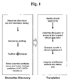
- FIG. 1 is an illustrative schematic flow diagram of a method for identifying biomarker genes for HDACi compound.resistance in cancer cells based on gene expression profiling, and the clinical application of expression profiling of the identified biomarker genes.
- FIG. 2 is an illustrative graph showing in vitro inhibition of cell proliferation versus concentration of the HDACi compound PCI-24781 for a series of colon carcinoma cell lines.
- FIG. 3 is an illustrative flow diagram illustrating the statistical approach used to analyze microarray data to identify differentially expressed genes in populations of cancer cells resistant to a HDACi compound versus cancer cells that are sensitive to the compound.
- FIG. 4 is an illustrative scatter plot illustrating principal component analysis of gene expression microarray data in HDACi compound-treated and untreated cancer cells, and sensitive and resistant cancer cells.
- FIG. 5 is an illustrative bar graph comparing the results of a microarray method versus TaqMan® quantitative RT-PCR method for determining the ratio of mRNA expression levels for a series of identified HDACi compound resistance biomarker genes in PCI-24781-resistant versus PCI-24781 colon carcinoma cells.
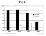
- FIG. 6 is an illustrative bar graph comparing relative expression levels of four HDACi compound resistance biomarker genes in cancer cells that are resistant to the HDAC inhibitor compound (PCI-24781) versus expression of the biomarker genes in cancer cells that are sensitive to the compound.
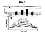
- FIG. 7 (A) is an illustrative bar graph showing the time course of tubulin acetylation in peripheral blood mononuclear cells from mice treated with the HDAC inhibitor compound PCI-24781; (B) is a time course of the expression profile of genes whose mRNA levels are correlated with changes in tubulin acetylation.
- FIG. 8 is an illustrative set of two line graphs illustrating the expression profiles of two HDAC inhibitor-responsive biomarker genes as determined by microarray analysis, quantitative RT-PCR, and immunoblotting.
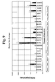
- FIG. 9 is an illustrative bar graph showing average in vivo mRNA levels in various tissues of five of the HDAC inhibitor-responsive biomarker genes at 3 and 8 hours post-HDAC inhibitor treatment.
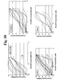
- FIG. 10 is an illustrative series of dose response curves for the effect of the HDAC inhibitor PCI-24781 on tumors derived from the indicated tumors.
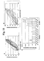
- FIG. 11 (A) is a series of line graphs illustrating the amount of in vitro growth inhibition by the HDAC inhibitor PCI-24781 of primary colon tumor cells derived from newly diagnosed, naive colon cancer patients; (B) is a series of line graphs illustrating the amount of in vitro growth inhibition by the HDAC inhibitor PCI-24781 of colon cancer cells derived from patients having advanced, metastatic colon tumors; (C) is a bar graph illustrating the correlation between tumor cell resistance to an HDAC inhibitor in vitro and the mRNA expression level of the HDAC resistance biomarker gene DEFA6.
DETAILED DESCRIPTION OF THE INVENTION
-
The methods described herein include classifying a cancer in a patient as resistant or sensitive to a histone deacetylase inhibitor (HDACi) compound by comparing the expression levels of at least four biomarker genes expressed in the cancer to biomarker gene expression level threshold values, as described herein. Where the expression levels of at least four biomarker genes are greater than the expression level threshold values, the cancer is indicated as being resistant to the HDACi compound. Conversely, if the expression levels of the at least four biomarker genes are lower than the expression level threshold values, the cancer is indicated to be sensitive to the HDACi compound.
-
Also described herein is a population of nucleic acids derived from a cancer cell, where the cancer cell is a type of cancer cell that is resistant to an HDACi compound. Further described herein is a population of nucleic acids derived from a cancer cell, where the cancer cell is a type of cancer cell that is sensitive to an HDACi compound. Also described herein are methods for generating these populations of nucleic acids. Such populations of nucleic acids are optionally used as expression level reference standards for setting biomarker gene expression threshold levels as described herein. Further described herein are cell lines determined to be resistant to an HDACi compound. Also described herein are cell lines determined to be sensitive to an HDACi compound.
-
Also described herein is a method for increasing the likelihood of therapeutically effective treatment of a cancer with an HDACi compound by providing an indication that a cancer is sensitive to treatment with an HDACi compound if the expression levels of at least four of the biomarker genes described herein are lower than the expression level threshold values for those biomarker genes, or providing an indication that a cancer is resistant to treatment with an HDACi compound if the expression levels of at least four of the biomarker genes described herein are higher than the expression level threshold values for those biomarker genes.
-
Further described herein are methods for optimizing selection of an anti-cancer agent for treating cancer in combination with an HDACi compound by comparing a first set of biomarker genes the expression of which is correlated to resistance or sensitivity of the cancer to the anti-cancer agent to a second set of biomarker genes the expression of which is correlated with resistance to the HDACi compound, and then selecting the anti-cancer agent for treatment of the cancer in combination with the HDAC inhibitor only if all of the biomarker genes in the first set are different from the biomarker genes in the second set.
Identification of HDACi compound resistance biomarker genes (HDACiR-BGs)
-
Described herein are methods for identifying genes whose expression levels in cancer cells are significantly and consistently correlated with resistance of the cells to an HDACi compound. Such genes are termed HDACi compound resistance biomarker genes (HDACiR-BGs). In an exemplary embodiment, HDACiR-BGs are identified as follows.
-
The ex-vivo response of primary tumor cells (e.g., colon cancer cells) from various patients to an HDAC inhibitor is determined by culturing the cells in the presence of varying concentrations of the HDACi compound.
-
After determining the HDACi compound sensitivity the cancer cells from each patient, mRNA expression profiles are determined for HDACi-resistant and sensitive tumors. Total RNA is isolated and fluorescent probes are prepared and hybridized to a whole genome cDNA microarray (e.g., Codelink Human Whole Genome oligonucleotide microarrays containing ∼55,000 unique probes; GE Healthcare Bio-Sciences Corp., Piscataway, NJ) according to the manufacturer's instructions. Following hybridization, the microarrays are scanned (e.g., in a GenePix 4000B scanner; Molecular Devices Corporation, Sunnyvale CA). The images are then processed with Codelink software and the data are normalized to the median.
-
The median-normalized microarray data are imported into a microarray data analysis program for principal component analysis (PCA) and hierarchical clustering analysis (e.g., Genespring software from Agilent). Multiple analysis methods are employed to provide additional confidence in the mRNA expression analysis. For multiple hypothesis correction, the q-values approach for false discovery rates (FDR) are optionally used as described in Storey et al. (2003), Proc. Nat. Acad. Sci. USA, 100:9440-9445. As a second analytical approach the Bayesian ANOVA approach described in Ishwaran et al. (2003), J. Amer. Stat. Assoc., 98:438-455 is optionally used.
-
In the Bayesian ANOVA method, the contributions of irrelevant genes to the ANOVA model are selectively shrunk to balance total false detections against total false non-detections. The output is a Zcut score which identifies genes whose contribution to the ANOVA model is larger than the standard z-score. See Ishwaran et al., ibid., and the website at bamarray.com.
-
The just-described method and variants thereof is optionally used to identify biomarker genes for other specific phenotypic states, e.g., resistance to anti-cancer agents other than HDACi compounds.
-
HDACiR-BGs identified by the just-described methods include those listed in Table 1. The sequence for the mRNA of each of the listed genes is included herein in an appendix.
| Table 1 |
| HDACi Compound Resistance Biomarker Genes (HDACiR-BGs) |
| Gene Name | Gene Symbol | GenBank Accession # | SEQ ID NO |
| PTPN3 | PTPN3 | AK096975 | 1 |
| ATP-binding cassette, subfamily C (CFTR/MRP), member 3 | ABCC3 | NM_020037 | 2 |
| specifically androgen-regulated protein | SARG | NM_023938 | 3 |
| phosphatidic acid phosphatase type 2C | PPAP2C | NM_177526 | 4 |
| neural proliferation, differentiation and control, 1 | NPDC1 | NM_015392 | 5 |
| C-terminal tensin-like | CTEN | NM_032865 | 6 |
| RAB25, member RAS oncogene family | RAB25 | NM_020387 | 7 |
| Hephaestin | HEPH | NM_138737 | 8 |
| thiopurine S-methyltransferase | TPMT | NM_000367 | 9 |
| plakophilin 3 | PKP3 | NM_007183 | 10 |
| UDP-N-acetyl-alpha-D-galactosamine:polypeptide N-acetylgalactosaminyltransferase 5 (GalNAc-T5) | GALNT5 | NM_014568 | 11 |
| calmodulin-like 4 | CALML4 | NM_033429 | 12 |
| UDP-N-acetyl-alpha-D-galactosamine:polypeptide N-acetylgalactosaminyltransferase 12 (GalNAc-T12) | GALNT12 | AK024865 | 13 |
| thiamin pyrophosphokinase 1 | TPK1 | NM_022445 | 14 |
| defensin, alpha 6, Paneth cell-specific | DEFA6 | NM_001926 | 15 |
| epithelial protein lost in neoplasm beta | EPLIN | NM_016357 | 16 |
| chloride intracellular channel 5 | CLIC5 | NM_016929 | 17 |
| PERP, TP53 apoptosis effector | PERP | NM_022121 | 18 |
| spleen tyrosine kinase | SYK | NM_003177 | 19 |
| solute carrier family 12 (sodium/potassium/chloride transporters), member 2 | SLC12A2 | NM_001046 | 20 |
| guanylate cyclase 2C (heat stable enterotoxin receptor) | GUCY2C | NM_004963 | 21 |
| transmembrane 4 superfamily member 4 | TM4SF4 | NM_004617 | 22 |
| transforming growth factor, alpha | TGFA | NM_003236 | 23 |
| fibroblast growth factor binding protein 1 | FGFBP1 | NM_005130 | 24 |
| PTK6 protein tyrosine kinase 6 | PTK6 | NM_005975 | 25 |
| epithelial V-like antigen 1 | EVA1 | NM_005797 | 26 |
| EPH receptor A2 | EPHA2 | NM_004431 | 27 |
| integrin, alpha 6 | ITGA6 | NM_000210 | 28 |
| tumor necrosis factor receptor superfamily, member 21 | TNFRSF21 | NM_014452 | 29 |
| transmembrane 4 superfamily member 3 | TM4SF3 | NM_004616 | 30 |
| interleukin 18 (interferon-gamma-inducing factor) | IL18 | NM_001562 | 31 |
| bone morphogenetic protein 4 | BMP4 | NM_130850 | 32 |
| sphingomyelin phosphodiesterase, acid-like 3B | SMPDL3B | NM_014474 | 33 |
| transmembrane protease, serine 2 | TMPRSS2 | NM_005656 | 34 |
| guanine deaminase | GDA | NM_004293 | 35 |
| macrophage stimulating 1 receptor (c-met-related tyrosine kinase) | MST1R | NM_002447 | 36 |
| integrin, beta 4 | ITGB4 | NM_000213 | 37 |
| annexin A3 | ANXA3 | NM_005139 | 38 |
| chemokine (C-C motif) ligand 15 | CCL15 | NM_032965 | 39 |
| dipeptidase 1 (renal) | DPEP1 | NM_004413 | 40 |
| NADPH oxidase organizer 1 | NOX01 | NM_172167 | 41 |
| interferon, alpha-inducible protein 27 | IFI27 | NM_005532 | 42 |
| cytochrome P450, family 3, subfamily A, polypeptide 43 | CYP3A43 | NM_057095 | 43 |
| plakophilin 2 | PKP2 | NM_004572 | 44 |
Classification of individual patient cancers as resistant or sensitive to an HDACi compound
-
In some embodiments, gene expression profiling is performed on a biological sample obtained from an individual patient suffering from a cancer (e.g., a colon cancer tumor) to classify the cancer in the patient as resistant or sensitive to an HDACi compound. The gene expression profiling includes profiling the expression of at least one of the HDACi compound resistance biomarker genes (HDACiR-BGs) listed in Table 1, which were identified as described herein.
-
In some embodiments the HDACiR-BG is selected from among DEFA6, TM4SF4, TGFA, FGFBP1, EPHA2, TNFRSF2, TM4SF3, IL18, TMPRSS2, and CCL15.
-
In some embodiments, at least four of the HDACiR-BGs are expression profiled. In some embodiments, at least one of the four HDACiR-BGs are selected from among DEFA6, ITGB4, TM4SF4, SYK, PPAP2C, RAB25, HEPH, NOXO1, TM4SF3, PTPN3, EPHA2, FGFBP1, ABCC3, TPMT, IL18, or DPEP1. In some embodiments, all of the at least four HDACiR-BGs are selected from among DEFA6, ITGB4, TM4SF4, SYK, PPAP2C, RAB25, HEPH, NOXO1, TM4SF3, PTPN3, EPHA2, FGFBP1, ABCC3, TPMT, IL18, or DPEP1.
-
In some embodiments, the expression of at least sixteen of the HDACiR-BGs is profiled. In some embodiments, the at least sixteen HDACiR-BGs include one or more of DEFA6, ITGB4, TM4SF4, SYK, PPAP2C, RAB25, HEPH, NOXO1, TM4SF3, PTPN3, EPHA2, FGFBP1, ABCC3, TPMT, IL18, or DPEP1. In some embodiments, the at least 16 HDACiR-BGs include DEFA6, ITGB4, TM4SF4, SYK, PPAP2C, RAB25, HEPH, NOXO1, TM4SF3, PTPN3, EPHA2, FGFBP1, ABCC3, TPMT, IL18, or DPEP1.
-
In various embodiments, the types of cancers and tumors that are optionally classified (from individual patients) for resistance or sensitivity to an HDACi compound include, but are not limited to, colorectal cancer, ovarian cancer, pancreatic cancer biliary tract cancer; bladder cancer; bone cancer; brain and CNS cancer; breast cancer; cervical cancer; choriocarcinoma; connective tissue cancer; cancer of the digestive system; endometrial cancer; esophageal cancer; eye cancer; cancer of the head and neck; gastric cancer; intra-epithelial neoplasm; kidney cancer; larynx cancer; leukemia; liver cancer; lung cancer (e.g., small cell and non-small cell); lymphoma including Hodgkin's and non-Hodgkin's lymphoma; melanoma; myeloma; neuroblastoma; oral cavity cancer (e.g., lip, tongue, mouth, and pharynx); prostate cancer; retinoblastoma; rhabdomyosarcoma; rectal cancer; renal cancer; cancer of the respiratory system; sarcoma; skin cancer; stomach cancer; testicular cancer; thyroid cancer; uterine cancer; cancer of the urinary system, as well as other carcinomas and sarcomas.
-
Types of cancer cells that are optionally classified in various embodiments include, but are not limited to, squamous cell papilloma, squamous cell carcinoma, basal cell tumor, basal cell carcinoma, transitional cell papilloma, transitional cell carcinoma, glandular epithelium adenoma, melanocytes glomus tumor, melanocytic nevus, malignant melanoma, fibroma, fibrosacroma, an adenocarcinoma, gastrinoma, malignant gastrinoma, an oncocytoma, cholangiocellular adenoma, cholangiocellular carcinoma, hepatocellular adenoma, hepatocellular carcinoma, renal tubular adenoma, renal cell carcinom(Grawitz tumor), myxoma, myxosarcoma, lipoma, liposarcoma, leiomyoma, leiomyosarcoma, rhabdomyoma, rhabdomyosarcoma, benign teratoma, malignant teratoma, hemangioma, hemangiosarcoma, Kaposi sarcoma, lymphangioma, lymphangiosarcoma, an osteoma, an osteosarcoma, an osteogenic sarcoma, cartilage chondroma, chondrosarcoma, meninges meningioma, malignant meningioma, oligoastrocytoma, an ependymoma, an astrocytoma, pilocytic astrocytoma, glioblastommultiforme, an oligodendroglioma, neuroblastoma, schwanoma, retinoblastoma, or neurofibroma. Other types of cancers and tumors include those described in reference sources, e.g., the "International Classification of Diseases for Oncology," 3rd Edition, International Association of Cancer Registries.
-
A biological sample is any biological sample that includes cellular material from which DNA, RNA or protein are optionally isolated, e.g., solid tissue samples, such as a biopsy specimen or tissue cultures or cells derived therefrom and the progeny thereof, blood and other liquid samples of biological origin, e.g., sputum (including saliva, buccal wash, or bronchial brush), stool, semen, urine, ascitic fluid, cerebral spinal fluid, bladder wash, or pleural fluid. The term "biological sample" also encompasses samples that have been manipulated in any way after their procurement, such as by treatment with reagents, solubilization, or enrichment for certain components. The term encompasses a clinical sample, and also includes cells in cell culture, cell supernatants, cell lysates, serum, plasma, biological fluids, and tissue samples, e.g, freshly collected tissue, frozen tissue, archived tissue, orbiological fluids
-
In some embodiments, the biological sample is a tumor biopsy (e.g., a core biopsy, a needle biopsy, or an excisional biopsy) containing one or more cancer cells. In one embodiment the biological sample is a population of cancer cells obtained by laser capture dissection from a tumor tissue section as described in, e.g.,
U.S. Patent No. 6,040,139 . Methods for optimizing tissue sample preparation and processing for expression profiling include, e.g.,
Bova et al. (2005), Methods Mol. Med., 103:15-66.
-
In some embodiments, one or more cells (e.g., from a cultured cancer cell line), are classified by determining the expression levels of no more than four to fifty biomarker genes described herein., e.g., 5, 6, 7, 8, 9, 10, 12, 16, 18, 20, 24, 30, 32, 35, 40, 44, 45, 47, or any other number of biomarker genes from four to fifty. In some embodiments, four to fourty four of the biomarker genes are selected from Table 3, e.g., 5, 6, 7, 8, 9, 10, 12, 16, 18, 20, 24, 30, 32, 35,40, or any other number of biomarker genes from four to fourty four is selected from Table 3. In some embodiments, at least four of the biomarker genes are selected from PTPN3, ABCC3, SARG, PPAP2C, NPDC1, CTEN, RAB25, HEPH, TPMT, PKP3, GALNT5, CALML4, GALNT12, TPK1, DEFA6, EPLIN, CLIC5, PERP, SYK, SLC12A2, GUCY2C, TM4SF4, TGFA, FGFBP1, PTK6, EVA1, EPHA2, ITGA6, TNFRSF21, TM4SF3, IL18, BMP4, SMPDL3B, TMPRSS2, GDA, MST1R, ITGB4, ANXA3, CCL15, DPEP1, NOXO1, IFI27, CYP3A43, and PKP2. In some embodiments, the four to fifty biomarker comprises one or more genes selected from DEFA6, ITGB4, TM4SF4, SYK, PPAP2C, RAB25, HEPH, NOXO1, TM4SF4, PTPN3, EPHA2, FGFBP1, ABCC3, TPMT, IL18, and DPEP In some embodiments, classification of the cells comprises comparing the determined expression levels to a first or second set of expression level threshold values for the biomarker genes, and indicating that the one or more cells are sensitive to a HDAC inhibitor if the expression levels of the biomarker genes are lower than the first set of expression level threshold values, or indicating that the one or more cells are resistant to a HDAC inhibitor if the expression levels are greater than the second set of expression level threshold values. In some embodiments, the expression of no more than four to twenty biomarker genes is determined. In some embodiments, the expression levels of no more than four biomarker genes is determined. In some embodiments, the four biomarker genes the expression level of which is determined are: DEFA6, RAB25, TM4SF4, and IL18.
Methods for HDACiR-BG Expression Profiling
-
HDACiR-BG expression profiles are optionally generated by any convenient means for determining differential gene expression between two samples, e.g. quantitative hybridization of mRNA, labeled mRNA, amplified mRNA, cRNA, etc., quantitative PCR, ELISA for protein quantitation, and the like.
-
In some embodiments, HDACiR-BG mRNA levels (including cDNA copy or aRNA copies) are quantified. The expression profile is optionally generated from the initial nucleic acid sample using any convenient protocol. While a variety of different manners of generating expression profiles are known, such as those employed in the field of differential gene expression analysis, one representative and convenient type of protocol for generating expression profiles is array based gene expression profile generation protocols. Such applications are hybridization assays in which a nucleic acid that displays "probe" nucleic acids for each of the genes to be assayed/profiled in the profile to be generated is employed. In these assays, a sample of target nucleic acids is first prepared from the initial nucleic acid sample being assayed, where preparation optionally includes labeling of the target nucleic acids with a label, e.g., a member of signal producing system. Following target nucleic acid sample preparation, the sample is contacted with the array under hybridization conditions, whereby complexes are formed between target nucleic acids that are complementary to probe sequences attached to the array surface. HDACiR-BG hybridization complexes are then detected and quantified.
-
Specific hybridization technologies which are optionally practiced to generate the HDACiR-BG expression profiles employed in the methods described herein includes the technology described in
U.S. Pat. Nos. 5,143,854 ;
5,288,644 ;
5,324,633 ;
5,432,049 ;
5,470,710 ;
5,492,806 ;
5,503,980 ;
5,510,270 ;
5,525,464 ;
5,547,839 ;
5,580,732 ;
5,661,028 ;
5,800,992 ; as well as
WO 95/21265 ;
WO 96/31622 ;
WO 97/10365 ;
WO 97/27317 ;
EP 373 203 ; and
EP 785 280 . In these methods, an array of "probe" nucleic acids that includes a probe for each of the phenotype determinative genes whose expression is being assayed is contacted with target nucleic acids as described above. Contact is carried out under hybridization conditions, e.g., stringent hybridization conditions as those conditions are practiced in the art, and unbound nucleic acid is then removed. The resultant pattern of hybridized nucleic acid provides quantitative information regarding expression for each of the HDACiR-BGs that have been probed.
-
Evaluation of differences in expression values is optionally performed using any convenient methodology, e.g., by comparing digital images of the expression profiles, by comparing databases of expression data, etc. Patents describing ways of comparing expression profiles include, but are not limited to,
U.S. Pat. Nos. 6,308,170 and
6,228,575 and
U.S. Patent Application Serial No. 10/858,867 .
-
In some embodiments, the methods described herein are performed on nucleic acid hybridization arrays comprising nucleic acid probes that hybridize under high stringency hybridization conditions to nucleic acids of no more than four to fifty biomarker genes, e.g., 5, 6, 7, 8, 9, 10, 12, 16, 18, 20, 24, 30, 32, 35, 40, 44, 45, 47, or any other number of biomarker genes from four to fifty. In some embodiments, four to fourty four of the biomarker genes are selected from Table 3, e.g., 5, 6, 7, 8, 9, 10, 12, 16, 18, 20, 24, 30, 32, 35, 40, or any other number of biomarker genes from four to fourty four is selected from Table 3. In some embodiments, at least four of the biomarker genes for the array probes are selected from PTPN3, ABCC3, SARG, PPAP2C, NPDC1, CTEN, RAB25, HEPH, TPMT, PKP3, GALNT5, CALML4, GALNT12, TPK1, DEFA6, EPLIN, CLIC5, PERP, SYK, SLC12A2, GUCY2C, TM4SF4, TGFA, FGFBP1, PTK6, EVA1, EPHA2, ITGA6, TNFRSF21, TM4SF3, IL18, BMP4, SMPDL3B, TMPRSS2, GDA, MST1R, ITGB4, ANXA3, CCL15, DPEP1, NOXO1, IFI27, CYP3A43, and PKP2. In some embodiments, the at least four biomarker genes are selected from DEFA6, ITGB4, TM4SF4, SYK, PPAP2C, RAB25, HEPH, NOXO1, TM4SF4, PTPN3, EPHA2, FGFBP1, ABCC3, TPMT, IL18, and DPEP. In some embodiments, the at least four biomarker genes are DEFA6, RAB25, TM4SF4, and IL18.
-
Alternatively, non-array based methods for quantitating the levels of one or more nucleic acids in a sample are employed, including quantitative PCR, and the like.
-
In some embodiments, expression profiling of HDACiR-BGs expressed in a biological sample (e.g., a tumor biopsy) is done by a quantitative reverse transcription PCR assay (qRT-PCR). In this method, RNA from a biological sample is reverse transcribed to generate segments of cDNA which are then be amplified by gene-specific quantitative PCR. The rate of accumulation of specific PCR products is optionally correlated to the abundance of the corresponding RNA species in the original sample and thereby provide an indication of gene expression levels.
-
In one embodiment, the qPCR assay is a TaqMan™ assay. In brief, PCR typically utilizes the 5' exonuclease activity of Taq or Tth polymerase to hydrolyze a fluorescently-labelled hybridization probe bound to its target amplicon, but any enzyme with equivalent 5' exonuclease activity is optionally used. Two oligonucleotide primers are used to generate an amplicon typical of a PCR reaction. A third oligonucleotide, or probe, is designed to hybridize to a nucleotide sequence located between the two PCR primers. The probe is non-extendible by Taq DNA polymerase enzyme, and is 5' labeled with a reporter fluorescent dye and a 3' labeled with a quencher fluorescent dye. Any laser-induced emission from the reporter dye is quenched by the quenching dye when the two dyes are located close together as they are on the probe. During the amplification reaction, the Taq DNA polymerase enzyme cleaves the probe in a template-dependent manner. The resultant probe fragments disassociate in solution, and signal from the released reporter dye is free from the quenching effect of the second chromophore. One molecule of reporter dye is liberated for each new molecule synthesized, and detection of the unquenched reporter dye provides the basis for quantitative interpretation of the data.
-
qRT-PCR is optionally performed using commercially available equipment, such as, for example, the ABI PRISM 7900™ Sequence Detection System™ (Perkin-Elmer-Applied Biosystems, Foster City, CA), or LightCycler™. (Roche Molecular Biochemicals, Mannheim, Germany). In one embodiment, the 5' exonuclease procedure is run on a real-time quantitative PCR device such as the ABI PRISM 7900™ Sequence Detection System™ or one of the similar systems in this family of instruments. The system consists of a thermocycler, laser, charge-coupled device (CCD), camera and computer. The system amplifies samples in 96-well or 384 well formats on a thermocycler. During amplification, laser-induced fluorescent signal is collected in real-time through fiber optic cables for all reaction wells, and detected at the CCD. The system includes software for running the instrument and for analyzing the data.
-
Exonuclease assay data are initially expressed as a CT value, i.e., the PCR cycle at which the fluorescent signal is first recorded as statistically significant.
-
In order to minimize errors and the effects of sample-to-sample variation and process variability mRNA level measurements are generally normalized to the expression level of an internal expression control gene. Methods for normalizing qPCR assays include, see, e.g., the website at normalisation.gene-quantification.info. The ideal internal expression control gene is one that is expressed at a relatively constant level among different patients or subjects, and is unaffected by the experimental treatment.
-
In some embodiments, the internal expression control gene is RNA polymerase II (GenBank Accession No. X74870).
-
In other embodiments, the internal expression control gene is HDAC3 (NM_003883).
-
In further embodiments, the internal expression control gene is ZNF217 (NM_006526).
-
In some embodiments, HDAiR-BG mRNA expression levels for each sample are normalized by the total amount of RNA in each sample. The amount of RNA in a sample is optionally determined, e.g., by UV-spectrophotometry or by using an RNA detection reagent, e.g., RiboGreen® from Invitrogen (Carlsbad, CA).
-
Where the HDACiR-BG expression profile to be determined is a protein expression profile, any convenient protein quantitation protocol is optionally employed, where the levels of one or more proteins in the assayed sample are determined. Representative methods include, but are not limited to; proteomic arrays, mass spectrometry, or standard immunoassays (e.g., RIA or ELISA). See, e.g., the methods set forth in R. Scopes, Protein Purification, Springer-Verlag, N.Y. (1982); Sandana (1997) Bioseparation of Proteins, Academic Press, Inc.; Bollag et al. (1996) Protein Methods. 2nd Edition Wiley-Liss, NY; Walker (1996) The Protein Protocols Handbook Humana Press, NJ, Harris and Angal (1990) Protein Purification: Principles and Practice 3rd Edition Springer Verlag, NY; Janson and Ryden (1998) Protein Purification: Principles, High Resolution Methods and Applications, Second Edition Wiley-VCH, NY; and Satinder Ahuja ed., Handbook of Bioseparations, Academic Press (2000); Harlow et al., Antibodies: A Laboratory Manual, Cold Spring Harbor Laboratory, Cold Spring Harbor, NY, 353-355 (1988).
-
Proteomic expression profiling methods detection methods include various multidimensional electrophoresis methods (e.g., 2-D gel electrophoresis), mass spectrometry based methods e.g., SELDI, MALDI, electrospray, etc.), or surface plasmon reasonance methods. For example, in MALDI, a sample is usually mixed with an appropriate matrix, placed on the surface of a probe and examined by laser desorption/ionization. See, e.g.,
U.S. Pat. Nos. 5,045,694, 5,202,561 , and
6,111,251 . Similarly, for SELDI, a first aliquot is contacted with a solid support-bound (e.g., substrate-bound) adsorbent. A substrate is typically a probe (e.g., a biochip) that is optionally positioned in an interrogatable relationship with a gas phase ion spectrometer. SELDI has been applied to diagnostic proteomics. See, e.g.
Issaq et al. (2003), Anal. Chem. 75: 149A-155A.
-
In one embodiment, any of the just-described protein detection methods are used to determine the expression level of one or more HDACiR-BG proteins that are known to be secreted proteins, e.g., DEFA6, TM4SF4, TM4SF3,TGFA, FGFBP1, EPHA2, TNFRSF2, IL18, CCL15, or TMPRSS2.
Expression Level Reference Samples
-
In some embodiments, expression profiles of HDACiR-BGs in a biological sample of interest (e.g., a colon cancer biopsy) are compared to HDACiR-BG expression profiles in an expression level reference sample. The expression level reference sample is a biological sample derived from one or more cancer patients determined to be suffering from a particular cancer or tumor for which sensitivity or resistance to treatment with an HDACi compound (e.g., PCI-24781) has been determined. In other words, the expression level reference sample serves as a standard with which to compare expression level values for each HDACiR-BG in a test sample. The deviation of HDACiR-BG expression levels from the expression level values in a reference sample indicates whether the cancer in the patient from the biological sample was derived is sensitive or resistant to treatment with an HDACi compound. In some embodiments, HDACiR-BG threshold expression level values are optionally set based on one or more statistical criteria for deviation from HDACiR-BG expression level values in an expression level reference sample, e.g., two or more SDs away from the value for a reference sample HDACiR-BG expression level.
-
In some embodiments, the expression level reference sample is a "negative" reference sample, i.e., a sample derived from a patient having a cancer or tumor determined to be sensitive to an HDACi compound. Thus, where expression levels of multiple HDACiR-BGs (e.g. at least 4, 5, 6, 8, 10, 12, or 16) are significantly greater than the threshold expression level values based on the negative reference sample, the patient's cancer is indicated as resistant to the HDACi compound.
-
In some embodiments, the expression level reference sample is a "positive" reference sample, i.e., a sample derived from a patient having a cancer or tumor determined to be resistant to an HDACi compound. Thus, where expression levels of multiple HDACiR-BGs (e.g. at least 4, 5, 6, 8, 10, 12, or 16) are significantly lower than the threshold expression level values based on the negative reference sample, the patient's cancer is indicated as sensitive to the HDACi compound.
-
In some embodiments, HDACiR-BG expression profiles are compared to those in both positive and negative reference samples.
-
In some embodiments, HDACiR-BGs expression level measurements are performed in parallel for the biological sample of interest and the (positive or negative) expression level reference. For example, where an array hybridization method is used, HDACiR-BG mRNA levels in the biological sample of interest and in an expression level reference sample are optionally measured simultaneously by separately labeling nucleic acid populations (e.g., mRNA, cDNA, aRNA populations) from each with a detectably distinct fluorophore, and then hybridizing the fluorescently labeled nucleic acids to the same array.
-
In some embodiments an expression level reference sample is a population of nucleic acids (e.g., mRNAs, aRNAs, cDNAs, or aRNAs) derived from a cancer biopsy sample within which the sequences of at least four HDACiR-BGs are represented, and for which sensitivity to an HDACi compound has been determined. In some embodiments, the population of nucleic acids is derived from patient tumor cells cultivated in culture. In other embodiments, the population is derived directly from a biopsy without a cell culture step.
-
In some embodiments, the population of nucleic acids serving as an expression level reference sample is generated as follows. A cancer biopsy is obtained from a patient as described above, and afterwards viable tumors cells are then isolated and grown in culture as described in, e.g., Kern et al. (1990), J. Natl. Cancer Inst., 82:582-588. In order to determine if cancer cells are sensitive to an HDACi compound, they are then grown in the presence of the HDACi compound at a range of concentrations, e.g., (0-10 µM), and cell proliferation is measured by any number of methods, e.g., tritiated thymidine incorporation. Inhibition of tumor cell proliferation by the HDACi compound is measured relative to tumor cell proliferation in the absence of the compound (i.e., no inhibition). Assignment of the cancer as sensitive or resistant is optionally determined based on a number of cell proliferation criteria. For example, if the IC50 of the HDACi compound in the tested cancer cells is significantly lower (e.g., by 2 SDs) than that observed for cells known to be sensitive to the compound, the cancer is characterized as resistant. Thus, cells derived from the resistant cancer (e.g., directly or after passage in culture) are optionally used to generate a population of nucleic acids serving as an expression level (positive) reference sample used for setting HDACiR-BG expression level threshold values as described above. Conversely, tumor cells found to be sensitive to an HDACi compound are used generate a population of nucleic acids serving as an expression level (negative) reference sample.
-
Methods for obtaining RNA from biological samples (e.g., tissues or cells) including linear aRNA amplification from single cells include, e.g., Luzzi et al. (2005), Methods Mol Biol., 293:187-207. Further, diverse kits for high quality RNA purification are available commercially, e.g., from Qiagen (Valencia, CA), Invitrogen (Carlsbad, CA), Clontech (Palo Alto, CA), and Stratagene (La Jolla, CA).
-
In some embodiments, the expression level reference sample is an RNA sample isolated from one or more HDACi compound-resistant colon cancer cells. In one embodiment, the cells were derived from colon carcinoma biopsy R5247682266, R9866135153, R1078103114, or R4712781606 described herein.
HDACi inhibitor Compounds
-
In another embodiment, HDACi inhibitor tumor compounds for which cancer resistance or sensitivity include, but are not limited to carboxylates, short-chain fatty acids, hydroxamic acids, electrophilic ketones, epoxides, cyclic peptides, and benzamides. In a further embodiment, HDACi inhibitor tumor compounds for which cancer resistance or sensitivity include, but are not limited to hydroxamic acids having the structure of Formula (A):
wherein
- Q is an optionally substituted C5-12 aryl or an optionally substituted C5-12 heteroaryl;
- L is a linker having at least 4 atoms;
- R1 is H or alkyl;
and a pharmaceutically acceptable salt, pharmaceutically acceptable N-oxide, pharmaceutically active metabolite, pharmaceutically acceptable prodrug, pharmaceutically acceptable solvate thereof.
-
HDACi inhibitor tumor compounds for which cancer resistance or sensitivity include, but are not limited to compounds having the structure of Formula (I):
wherein:
- R1 is hydrogen or alkyl;
- X is -O-, -NR2-, or -S(O)n where n is 0-2 and R2 is hydrogen or alkyl;
- Y is alkylene optionally substituted with cycloalkyl, optionally substituted phenyl, alkylthio, alkylsulfinyl, alkysulfonyl, optionally substituted phenylalkylthio, optionally substituted phenylalkylsulfonyl, hydroxy, or optionally substituted phenoxy;
- Ar1 is phenylene or heteroarylene wherein said Ar1 is optionally substituted with one or two groups independently selected from alkyl, halo, hydroxy, alkoxy, haloalkoxy, or haloalkyl;
- R3 is hydrogen, alkyl, hydroxyalkyl, or optionally substituted phenyl; and
- Ar2 is aryl, aralkyl, aralkenyl, heteroaryl, heteroaralkyl, heteroaralkenyl, cycloalkyl, cycloalkylalkyl, heterocycloalkyl, or heterocycloalkylalkyl;
and individual stereoisomers, individual geometric isomers, or mixtures thereof; or a pharmaceutically acceptable salt thereof.
-
In another embodiment, HDACi inhibitor tumor compounds for which cancer resistance or sensitivity include, but are not limited to, PCI-24781.
-
In some embodiments, a patient is prescribed or administered an HDAC inhibitor to the patient based on a classification of the patient's cancer as being sensitive or resistant to an HDAC inhibitor according to the methods described herein.
-
In some embodiments, the methods described herein are used to optimize the selection of an anti-cancer agent for use in combination with an HDACi compound. In some embodiments, optimized selection of the second anti-cancer agent is performed by first comparing the set of known biomarker genes for resistance to the HDACi compound to sets of biomarker genes identified for other anti-cancer agents. The second anti-cancer agent is then selected for use in combination with the HDACi compound based on minimal overlap of the respective sets of resistance biomarker genes.
-
Examples of anti-cancer agents that are optionally used in combination with an HDACi compound include, but are not limited to, any of the following: gossyphol, genasense, polyphenol E, Chlorofusin, all trans-retinoic acid (ATRA), bryostatin, tumor necrosis factor-related apoptosis-inducing ligand (TRAIL), 5-aza-2'-deoxycytidine, all trans retinoic acid, doxorubicin, vincristine, etoposide, gemcitabine, imatinib (Gleevec®), geldanamycin, 17-N-Allylamino-17-Demethoxygeldanamycin (17-AAG), flavopiridol, LY294002, bortezomib, trastuzumab, BAY 11-7082, PKC412, or PD184352, Taxol™, also referred to as "paclitaxel", is an anti-cancer drug which acts by enhancing and stabilizing microtubule formation, and analogs of Taxol™, such as Taxotere™. Compounds that have the basic taxane skeleton as a common structure feature, have also been shown to have the ability to arrest cells in the G2-M phases due to stabilized microtubules and are optionall useful for treating cancer in combination with the compounds described herein.
-
Further examples of anti-cancer agents for use in combination with an HDACi compound include inhibitors of mitogen-activated protein kinase signaling, e.g., U0126, PD98059, PD184352, PD0325901, ARRY-142886, SB239063, SP600125, BAY 43-9006, wortmannin, or LY294002.
-
Other anti-cancer agents that are optionally employed in combination with an HDACi compound include Adriamycin, Dactinomycin, Bleomycin, Vinblastine, Cisplatin, acivicin; aclarubicin; acodazole hydrochloride; acronine; adozelesin; aldesleukin; altretamine; ambomycin; ametantrone acetate; aminoglutethimide; amsacrine; anastrozole; anthramycin; asparaginase; asperlin; azacitidine; azetepa; azotomycin; batimastat; benzodepa; bicalutamide; bisantrene hydrochloride; bisnafide dimesylate; bizelesin; bleomycin sulfate; brequinar sodium; bropirimine; busulfan; cactinomycin; calusterone; caracemide; carbetimer; carboplatin; carmustine; carubicin hydrochloride; carzelesin; cedefingol; chlorambucil; cirolemycin; cladribine; crisnatol mesylate; cyclophosphamide; cytarabine; dacarbazine; daunorubicin hydrochloride; decitabine; dexormaplatin; dezaguanine; dezaguanine mesylate; diaziquone; doxorubicin; doxorubicin hydrochloride; droloxifene; droloxifene citrate; dromostanolone propionate; duazomycin; edatrexate; eflornithine hydrochloride; elsamitrucin; enloplatin; enpromate; epipropidine; epirubicin hydrochloride; erbulozole; esorubicin hydrochloride; estramustine; estramustine phosphate sodium; etanidazole; etoposide; etoposide phosphate; etoprine; fadrozole hydrochloride; fazarabine; fenretinide; floxuridine; fludarabine phosphate; fluorouracil; flurocitabine; fosquidone; fostriecin sodium; gemcitabine; gemcitabine hydrochloride; hydroxyurea; idarubicin hydrochloride; ifosfamide; iimofosine; interleukin II (including recombinant interleukin II, or rlL2), interferon alfa-2a; interferon alfa-2b; interferon alfa-n1 ; interferon alfa-n3; interferon beta-1 a; interferon gamma-1 b; iproplatin; irinotecan hydrochloride; lanreotide acetate; letrozole; leuprolide acetate; liarozole hydrochloride; lometrexol sodium; lomustine; losoxantrone hydrochloride; masoprocol; maytansine; mechlorethamine hydrochloride; megestrol acetate; melengestrol acetate; melphalan; menogaril; mercaptopurine; methotrexate; methotrexate sodium; metoprine; meturedepa; mitindomide; mitocarcin; mitocromin; mitogillin; mitomalcin; mitomycin; mitosper; mitotane; mitoxantrone hydrochloride; mycophenolic acid; nocodazoie; nogalamycin; ormaplatin; oxisuran; pegaspargase; peliomycin; pentamustine; peplomycin sulfate; perfosfamide; pipobroman; piposulfan; piroxantrone hydrochloride; plicamycin; plomestane; porfimer sodium; porfiromycin; prednimustine; procarbazine hydrochloride; puromycin; puromycin hydrochloride; pyrazofurin; riboprine; rogletimide; safingol; safmgol hydrochloride; semustine; simtrazene; sparfosate sodium; sparsomycin; spirogermanium hydrochloride; spiromustine; spiroplatin; streptonigrin; streptozocin; sulofenur; talisomycin; tecogalan sodium; tegafur; teloxantrone hydrochloride; temoporfin; teniposide; teroxirone; testolactone; thiamiprine; thioguanine; thiotepa; tiazofurin; tirapazamine; toremifene citrate; trestolone acetate; triciribine phosphate; trimetrexate; trimetrexate glucuronate; triptorelin; tubulozole hydrochloride; uracil mustard; uredepa; vapreotide; verteporfin; vinblastine sulfate; vincristine sulfate; vindesine; vindesine sulfate; vinepidine sulfate; vinglycinate sulfate; vinleurosine sulfate; vinorelbine tartrate; vinrosidine sulfate; vinzolidine sulfate; vorozole; zeniplatin; zinostatin; zorubicin hydrochloride.
-
Other anti-cancer agents that are optionally employed in combination with an HDACi compound include: 20-epi-1 ,25 dihydroxyvitamin D3; 5-ethynyluracil; abiraterone; aclarubicin; acylfulvene; adecypenol; adozelesin; aldesleukin; ALL-TK antagonists; altretamine; ambamustine; amidox; amifostine; aminolevulinic acid; amrubicin; amsacrine; anagrelide; anastrozole; andrographolide; angiogenesis inhibitors; antagonist D; antagonist G; antarelix; anti-dorsalizing morphogenetic protein-1 ; antiandrogen, prostatic carcinoma; antiestrogen; antineoplaston; antisense oligonucleotides; aphidicolin glycinate; apoptosis gene modulators; apoptosis regulators; apurinic acid; ara-CDP-DL-PTBA; arginine deaminase; asulacrine; atamestane; atrimustine; axinastatin 1 ; axinastatin 2; axinastatin 3; azasetron; azatoxin; azatyrosine; baccatin III derivatives; balanol; batimastat; BCR/ABL antagonists; benzochlorins; benzoylstaurosporine; beta lactam derivatives; beta-alethine; betaclamycin B; betulinic acid; bFGF inhibitor; bicalutamide; bisantrene; bisaziridinylspermine; bisnafide; bistratene A; bizelesin; breflate; bropirimine; budotitane; buthionine sulfoximine; calcipotriol; calphostin C; camptothecin derivatives; canarypox IL-2; capecitabine; carboxamide-amino-triazole; carboxyamidotriazole; CaRest M3; CARN 700; cartilage derived inhibitor; carzelesin; casein kinase inhibitors (ICOS); castanospermine; cecropin B; cetrorelix; chlorlns; chloroquinoxaline sulfonamide; cicaprost; cis-porphyrin; cladribine; clomifene analogues; clotrimazole; collismycin A; collismycin B; combretastatin A4; combretastatin analogue; conagenin; crambescidin 816; crisnatol; cryptophycin 8; cryptophycin A derivatives; curacin A; cyclopentanthraquinones; cycloplatam; cypemycin; cytarabine ocfosfate; cytolytic factor; cytostatin; dacliximab; decitabine; dehydrodidemnin B; deslorelin; dexamethasone; dexifosfamide; dexrazoxane; dexverapamil; diaziquone; didemnin B; didox; diethylnorspermine; dihydro-5-azacytidine; 9-dioxamycin; diphenyl spiromustine; docosanol; dolasetron; doxifluridine; droloxifene; dronabinol; duocarmycin SA; ebselen; ecomustine; edelfosine; edrecolomab; eflornithine; elemene; emitefur; epirubicin; epristeride; estramustine analogue; estrogen agonists; estrogen antagonists; etanidazole; etoposide phosphate; exemestane; fadrozole; fazarabine; fenretinide; filgrastim; finasteride; flavopiridol; flezelastine; fluasterone; fludarabine; fluorodaunorunicin hydrochloride; forfenimex; formestane; fostriecin; fotemustine; gadolinium texaphyrin; gallium nitrate; galocitabine; ganirelix; gelatinase inhibitors; gemcitabine; glutathione inhibitors; hepsulfam; heregulin; hexamethylene bisacetamide; hypericin; ibandronic acid; idarubicin; idoxifene; idramantone; ilmofosine; ilomastat; imidazoacridones; imiquimod; immunostimulant peptides; insulin-like growth factor-1 receptor inhibitor; interferon agonists; interferons; interleukins; iobenguane; iododoxorubicin; ipomeanol, 4-; iroplact; irsogladine; isobengazole; isohomohalicondrin B; itasetron; jasplakinolide; kahalalide F; lamellarin-N triacetate; lanreotide; leinamycin; lenograstim; lentinan sulfate; leptolstatin; letrozole; leukemia inhibiting factor; leukocyte alpha interferon; leuprolide+estrogen+progesterone; leuprorelin; levamisole; liarozole; linear polyamine analogue; lipophilic disaccharide peptide; lipophilic platinum compounds; lissoclinamide 7; lobaplatin; lombricine; lometrexol; lonidamine; losoxantrone; lovastatin; loxoribine; lurtotecan; lutetium texaphyrin; lysofylline; lytic peptides; maitansine; mannostatin A; marimastat; masoprocol; maspin; matrilysin inhibitors; matrix metalloproteinase inhibitors; menogaril; merbarone; meterelin; methioninase; metoclopramide; MIF inhibitor; mifepristone; miltefosine; mirimostim; mismatched double stranded RNA; mitoguazone; mitolactol; mitomycin analogues; mitonafide; mitotoxin fibroblast growth factor-saporin; mitoxantrone; mofarotene; molgramostim; monoclonal antibody, human chorionic gonadotrophin; monophosphoryl lipid A+myobacterium cell wall sk; mopidamol; multiple drug resistance gene inhibitor; multiple tumor suppressor 1 -based therapy; mustard anticancer agent; mycaperoxide B; mycobacterial cell wall extract; myriaporone; N-acetyldinaline; N-substituted benzamides; nafarelin; nagrestip; naloxone+pentazocine; napavin; naphterpin; nartograstim; nedaplatin; nemorubicin; neridronic acid; neutral endopeptidase; nilutamide; nisamycin; nitric oxide modulators; nitroxide antioxidant; nitrullyn; O6-benzylguanine; octreotide; okicenone; oligonucleotides; onapristone; ondansetron; ondansetron; oracin; oral cytokine inducer; ormaplatin; osaterone; oxaliplatin; oxaunomycin; palauamine; palmitoylrhizoxin; pamidronic acid; panaxytriol; panomifene; parabactin; pazelliptine; pegaspargase; peldesine; pentosan polysulfate sodium; pentostatin; pentrozole; perflubron; perfosfamide; perillyl alcohol; phenazinomycin; phenylacetate; phosphatase inhibitors; picibanil; pilocarpine hydrochloride; pirarubicin; piritrexim; placetin A; placetin B; plasminogen activator inhibitor; platinum complex; platinum compounds; platinum-triamine complex; porfimer sodium; porfiromycin; prednisone; propyl bis-acridone; prostaglandin J2; proteasome inhibitors; protein Abased immune modulator; protein kinase C inhibitor; protein kinase C inhibitors, microalgal; protein tyrosine phosphatase inhibitors; purine nucleoside phosphorylase inhibitors; purpurins; pyrazoloacridine; pyridoxylated hemoglobin polyoxyethylerie conjugate; raf antagonists; raltitrexed; ramosetron; ras farnesyl protein transferase inhibitors; ras inhibitors; ras-GAP inhibitor; retelliptine demethylated; rhenium Re 186 etidronate; rhizoxin; ribozymes; RII retinamide; rogletimide; rohitukine; romurtide; roquinimex; rubiginone B1 ; ruboxyl; safingol; saintopin; SarCNU; sarcophytol A; sargramostim; Sdi 1 mimetics; semustine; senescence derived inhibitor 1 ; sense oligonucleotides; signal transduction inhibitors; signal transduction modulators; single chain antigen-binding protein; sizofiran; sobuzoxane; sodium borocaptate; sodium phenylacetate; solverol; somatomedin binding protein; sonermin; sparfosic acid; spicamycin D; spiromustine; splenopentin; spongistatin 1 ; squalamine; stem cell inhibitor; stem-cell division inhibitors; stipiamide; stromelysin inhibitors; sulfinosine; superactive vasoactive intestinal peptide antagonist; suradista; suramin; swainsonine; synthetic glycosaminoglycans; tallimustine; tamoxifen methiodide; tauromustine; tazarotene; tecogalan sodium; tegafur; tellurapyrylium; telomerase inhibitors; temoporfin; temozolomide; teniposide; tetrachlorodecaoxide; tetrazomine; thaliblastine; thiocoraline; thrombopoietin; thrombopoietin mimetic; thymalfasin; thymopoietin receptor agonist; thymotrinan; thyroid stimulating hormone; tin ethyl etiopurpurin; tirapazamine; titanocene bichloride; topsentin; toremifene; totipotent stem cell factor; translation inhibitors; tretinoin; triacetyluridine; triciribine; trimetrexate; triptorelin; tropisetron; turosteride; tyrosine kinase inhibitors; tyrphostins; UBC inhibitors; ubenimex; urogenital sinus-derived growth inhibitory factor; urokinase receptor antagonists; vapreotide; variolin B; vector system, erythrocyte gene therapy; velaresol; veramine; verdins; verteporfin; vinorelbine; vinxaltine; vitaxin; vorozole; zanoterone; zeniplatin; zilascorb; and zinostatin stimalamer.
-
Yet other anticancer agents that are optionally employed in combination with an HDACi compound include alkylating agents, antimetabolites, natural products, or hormones, e.g., nitrogen mustards (e.g., mechloroethamine, cyclophosphamide, chlorambucil, etc.), alkyl sulfonates (e.g., busulfan), nitrosoureas (e.g., carmustine, lomusitne, ete.), or triazenes (decarbazine, etc.). Examples of antimetabolites include but are not limited to folic acid analog (e.g., methotrexate), or pyrimidine analogs (e.g., Cytarabine), purine analogs (e.g., mercaptopurine, thioguanine, pentostatin).
-
Examples of natural products useful in combination with an HDACi compound include but are not limited to vinca alkaloids (e.g., vinblastin, vincristine), epipodophyllotoxins (e.g., etoposide), antibiotics (e.g., daunorubicin, doxorubicin, bleomycin), enzymes (e.g., L-asparaginase), or biological response modifiers (e.g., interferon alpha).
-
Examples of alkylating agents that are optionally employed in combination an HDACi compound include, but are not limited to, nitrogen mustards (e.g., mechloroethamine, cyclophosphamide, chlorambucil, meiphalan, etc.), ethylenimine and methylmelamines (e.g., hexamethlymelamine, thiotepa), alkyl sulfonates (e.g., busulfan), nitrosoureas (e.g., carmustine, lomusitne, semustine, streptozocin, etc.), or triazenes (decarbazine, ete.). Examples of antimetabolites include, but are not limited to folic acid analog (e.g., methotrexate), or pyrimidine analogs (e.g., fluorouracil, floxouridine, Cytarabine), purine analogs (e.g., mercaptopurine, thioguanine, pentostatin.
-
Examples of hormones and antagonists useful in combination with an HDACi compound include, but are not limited to, adrenocorticosteroids (e.g., prednisone), progestins (e.g., hydroxyprogesterone caproate, megestrol acetate, medroxyprogesterone acetate), estrogens (e.g., diethlystilbestrol, ethinyl estradiol), antiestrogen (e.g., tamoxifen), androgens (e.g., testosterone propionate, fluoxymesterone), antiandrogen (e.g., flutamide), gonadotropin releasing hormone analog (e.g., leuprolide). Other agents that are optionally used in the methods and compositions described herein for the treatment or prevention of cancer include platinum coordination complexes (e.g., cisplatin, carboblatin), anthracenedione (e.g., mitoxantrone), substituted urea (e.g., hydroxyurea), methyl hydrazine derivative (e.g., procarbazine), adrenocortical suppressant (e.g., mitotane, aminoglutefhimide).
-
Examples of anti-cancer agents which act by arresting cells in the G2-M phases due to stabilized microtubules and which are optionally used in combination with an HDACi compound include without limitation the following marketed drugs and drugs in development: Erbulozole (also known as R-55104), Dolastatin 10 (also known as DLS-10 and NSC-376128), Mivobulin isethibnate (also known as CI-980), Vincristine, NSC-639829, Discodermolide (also known as NVP-XX-A-296), ABT-751 (Abbott, also known as E-7010), Altorhyrtins (such as Altorhyrtin A and Altorhyrtin C), Spongistatins (such as Spongistatin 1, Spongistatin 2, Spongistatin 3, Spongistatin 4, Spongistatin 5, Spongistatin 6, Spongistatin 7, Spongistatin 8, and Spongistatin 9), Cemadotin hydrochloride (also known as LU-103793 and NSC-D-669356), Epothilones (such as Epothilone A, Epothilone B, Epothilone C (also known as desoxyepothilone A or dEpoA), Epothilone D (also referred to as KOS-862, dEpoB, and desoxyepothilone B), Epothilone E, Epothilone F, Epothilone B N-oxide, Epothilone A N-oxide, 16-aza-epothilone B, 21-aminoepothilone B (also known as BMS-310705), 21-hydroxyepothilone D (also known as Desoxyepothilone F and dEpoF), 26-fluoroepothilone), Auristatin PE (also known as NSC-654663), Soblidotin (also known as TZT-1027), LS-4559-P (Pharmacia, also known as LS-4577), LS-4578 (Pharmacia, also known as LS-477-P), LS-4477 (Pharmacia), LS-4559 (Pharmacia), RPR-112378 (Aventis), Vincristine sulfate, DZ-3358 (Daiichi), FR-182877 (Fujisawa, also known as WS-9885B), GS-164 (Takeda), GS-198 (Takeda), KAR-2 (Hungarian Academy of Sciences), BSF-223651 (BASF, also known as ILX-651 and LU-223651), SAH-49960 (Lilly/Novartis), SDZ-268970 (Lilly/Novartis), AM-97 (Armad/Kyowa Hakko), AM-132 (Armad), AM-138 (Armad/Kyowa Hakko), IDN-5005 (Indena), Cryptophycin 52 (also known as LY-355703), AC-7739 (Ajinomoto, also known as AVE-8063A and CS-39.HCI), AC-7700 (Ajinomoto, also known as AVE-8062, AVE-8062A, CS-39-L-Ser.HCI, and RPR-258062A), Vitilevuamide, Tubulysin A, Canadensol, Centaureidin (also known as NSC-106969), T-138067 (Tularik, also known as T-67, TL-138067 and TI-138067), COBRA-1 (Parker Hughes Institute, also known as DDE-261 and WHI-261), H10 (Kansas State University), H16 (Kansas State University), Oncocidin A1 (also known as BTO-956 and DIME), DDE-313 (Parker Hughes Institute), Fijianolide B, Laulimalide, SPA-2 (Parker Hughes Institute), SPA-1 (Parker Hughes Institute, also known as SPIKET-P), 3-IAABU (Cytoskeleton/Mt. Sinai School of Medicine, also known as MF-569), Narcosine (also known as NSC-5366), Nascapine, D-24851 (Asta Medica), A-105972 (Abbott), Hemiasterlin, 3-BAABU (Cytoskeleton/Mt. Sinai School of Medicine, also known as MF-191), TMPN (Arizona State University), Vanadocene acetylacetonate, T-138026 (Tularik), Monsatrol, Inanocine (also known as NSC-698666), 3-1AABE (Cytoskeleton/Mt. Sinai School of Medicine), A-204197 (Abbott), T-607 (Tuiarik, also known as T-900607), RPR- 115781 (Aventis), Eleutherobins (such as Desmethyleleutherobin, Desaetyleleutherobin, Isoeleutherobin A, and Z-Eleutherobin), Caribaeoside, Caribaeolin, Halichondrin B, D-64131 (Asta Medica), D-68144 (Asta Medica), Diazonamide A, A-293620 (Abbott), NPI-2350 (Nereus), Taccalonolide A, TUB-245 (Aventis), A-259754 (Abbott), Diozostatin, (-)-Phenylahistin (also known as NSCL-96F037), D-68838 (Asta Medica), D-68836 (Asta Medica), Myoseverin B, D-43411 (Zentaris, also known as D-81862), A-289099 (Abbott), A-318315 (Abbott), HTI-286 (also known as SPA-110, trifluoroacetate salt) (Wyeth), D-82317 (Zentaris), D-82318 (Zentaris), SC-12983 (NCI), Resverastatin phosphate sodium, BPR-OY-007 (National Health Research Institutes), and SSR-250411 (Sanofi).
Applications of HDACiR-BGs
-
The methods and compositions described herein are optionally used to increase the likelihood of a therapeutically effective treatment of a patient's cancer with an HDACi compound by providing an indication (e.g. by oral or written communication in any analog or digital medium) of which genes are HDACiR-BGs, as well as HDACiR-BG expression level reference values (e.g., expression level threshold values) above which HDACi compound resistance is likely (i.e., greater than the probability by chance) or below which HDACi compound sensitivity is likely.
-
In some embodiments, the indication includes a document with an interpretation of expression levels of at least four biomarker genes selected from Table 1 as to the likelihood that a patient's cancer is resistant or sensitive to treatment with an HDACi compound.
-
In some embodiments, the document includes an interpretation of the expression levels of at least one HDACiR-BG selected from DEFA6, ITGB4, TM4SF4, SYK, PPAP2C, RAB25, HEPH, NOXO1, TM4SF4, PTPN3, EPHA2, FGFBP1, ABCC3, TPMT, IL18, and DPEP1.
-
In some embodiments an indication is provided in one or more databases containing information concerning one or more HDACiR-BGs, including one or more expression level threshold values that permit the interpretation of the effect of HDACiR-BG expression levels on the resistance or sensitivity of a cancer to an HDACi compound according to any of the methods described herein. Such expression level threshold values include those set based on, e.g., deviation of HDACiR-BG expression levels in a test sample from the corresponding HDACiR-BG expression levels in an expression level (positive or negative) reference sample as described herein. Alternatively, or in addition, expression level threshold values are optionally set based on deviation of the expression ratios of HDACiR-BGs to one or more internal expression control genes (e.g., RNA polymerase II, HDAC3, or ZNF217). For example, as described herein, the mean expression ratio (based on TaqMan fluorescence intensity) of the HDACiR-BG DEFA6 to the internal expression control gene ZNF217 is 5.83 in HDACi-resistant colon cancer cells and 0.24 in HDACi-sensitive colon cancer cells.
-
In some embodiments, the databases include HDACiR-BG expression level profiles or thresholds associated with resistance to one or more HDACi compounds for one or more types of cancer.
-
Other information that is optionally included in the databases or in other types of indication include, but are not limited to, HDACiR-BG sequence information, frequency distributions of HDACiR-BG expression levels in a particular cancer population, descriptive information concerning the clinical status of a biological sample analyzed for HDACiR-BG expression profiles, or the clinical status of the patient from which the sample was derived. The database is optionally be designed to include different parts, for instance an HDACiR-BG list database, and an informative HDACiR-BG expression profile database, e.g., a database associating with each HDACiR-BG expression profile record the probability that the expressin profile is associated with resistance to an HDACi compound. Methods for the configuration and construction of databases are widely available, for instance, see
U.S. Pat. No. 5,953,727 .
-
The databases described herein are optionally linked to an outside or external database. In some embodiments, the database optionally communicates with outside data sources, such as database of the developmental therapeutics program of the national cancer institute or the National Center for Biotechnology Information through the internet.
-
Any appropriate computer platform is used to perform the methods for interpreting one or more HDACiR-BG expression profiles by the methods described herein. In some embodiments, the computer platform receive direct input from a database, e.g., one of the databases described herein. For example, a large number of computer workstations are available from a variety of manufacturers, such has those available from Silicon Graphics. Client-server environments, database servers and networks are also widely available and are appropriate platforms for the databases described herein.
-
The databases described herein are optionally used to present information identifying a set of HDACiR-BG expression profiles in an individual and such a presentation is optionally used to predict or diagnose the likelihood of a effective therapeutic treatment of the individual's cancer with a particular HDACi compound based on a statistical comparison of the individual's expression profile to HDACiR-BG expression level thresholds as desribed herein. Accordingly, one chooses to partition cancer patients into subgroups at any threshold value of the measured HDACiR-BG expression, where all patients with expression values above the threshold have higher risk, and all patients with expression values below the threshold have lower risk, of and HDACi compound-resistant cancer resistanceor vice versa, depending on whether the expression level threshold is based on an expression level in a cancer determined to be resistant to an HDACi compound treatment (i.e., a positive reference sample) or sensitive to the HDACi compound treatment (i.e., a negative reference sample). Alternatively, HDACiR-BG expression profiles ranked on a probability continuum, where the more an HDACiR-BG expression level deviates negatively from (i.e., is less than) an expression level positive reference value, the higher the probability that the cancer is sensitive to treatment with an HDACi compound. Conversely, the more an HDACiR-BG expression level deviates positively from (i.e., is greater than) an expression level negative reference value, the higher the probability that the cancer is resistant to treatment with an HDACi compound.
EXAMPLES
-
The following specific examples are to be construed as merely illustrative, and not limitative of the remainder of the disclosure in any way whatsoever. Without further elaboration, it is believed that one skilled in the art can, based on the description herein, utilize the present invention to its fullest extent.
Example 1: mRNA expression profiling of HDACi sensitive versus resistant colorectal tumor cells ex vivo
-
We and others previously developed several pharmacodynamic markers for HDACi compounds (such as tubulin or histone acetylation, p21 expression etc). However, there is currently no clinically predictive biomarker for response to these agents available. In this work, we developed a strategy to identify such biomarkers for the HDACi compound PCI-24781 in primary human colorectal tumors.
-
The method used soft agar chemosensitivity assays in which primary human tumors were exposed in culture to PCI-24781. Either a trititated thymidine or alamar blue assay was then used to estimate the percentage of resistance to PCI-24781. For example in the trititated thymidine assay, sensitive tumor cells affected by the drug divided less and therefore incorporated less thymidine, whereas resistant tumor cells continued to grow and divide and therefore incorporated more thymidine into their DNA. It has been shown historically that under the optimized conditions of this assay, a patient whose tumor is classified as resistant to a given drug has < 1% probability of response to that drug in the clinic (in published correlations to clinical outcome, these assays predicted resistance with an accuracy of 99% in solid cancers and 92% in blood cancers). For example. a recent paper correlated in vitro sensitivity or resistance to fludarabine in the DiSC assay in B-cell CLL patients with clinical outcome (median survival 7.9 months in resistant vs 41.7 months in sensitive patients). Similar data has also been published for solid tumors : e.g., for sensitivity or resistance to Pt in ovarian tumors, and to CPX and DOX in breast tumors.
-
After determining ex vivo sensitivity or resistance to PCI-24781 for each tumor, RNA isolated from tumor cells was then profiled on microarrays and a marker set was identified by statistical analysis of the data. This marker set was validated by RT-PCR (TaqMan™) analysis. Such pharmacogenomic biomarkers that are used for patient stratification in the clinic provide a competitive advantage in the development of PCI-24781. A graphic summary of the method and its clinical applications is illustrated in Fig. 1.
-
We examined the ex-vivo response of primary colorectal tumors from various patients to an HDAC inhibitor, PCI-24781, and subsequently determined whether there were robust differences in the mRNA expression profiles of sensitive versus resistant tumor cells prior to HDACi treatment.
-
Primary colorectal cancer (CRC) samples were obtained from patient biopsies (Table 2). Viable tumor cells were plated and cultured in soft agar as described in
Kern et al. (1990), J. Natl. Cancer Inst., 82:582-588, and were treated with a range of PCI-24781 concentrations (0.01-2µM). Tritiated thymidine was added to the culture after 3 days of exposure to the drug, and the amount of radioactivity incorporated into the cells after a further 2 days was quantified. The percentage of cell growth inhibition (%GI) was calculated by comparing the treated cells to the control cells, and from these growth profiles the tumors were classified as either sensitive or resistant based on deviation from the median profile. As shown in
Fig. 2, primary tumors displayed a spectrum of growth inhibition phenotypes from 100% to 0% relative to control at the PCI-24781concentrations tested (up to 2 µM).
Table 2 | Research ID | Cancer Name | Age | Sex | She | Clinical Diagnosis | Histology | Specimen Type |
| R1078103114 | Colon Carcinoma | 54 | F | R Ovary | Colon Carcinoma | ADENOCARCINOMA | Solid Tumor Biopsy |
| R1105698672 | Colon Carcinoma | 72 | F | Portion of Terminal Ileum | Colon Carcinoma | NA | Solid Tumor Biopsy |
| R2163560366 | Colon Carcinoma | 58 | F | Uterus | Rectal Cancer | NA | Solid Tumor Biopsy |
| R4712T81605 | Colon Carcinoma | 59 | M | Colon Resection | Colon Carcinoma | NA | Solid Tumor Biopsy |
| R5247682266 | Colon Carcinoma | 51 | F | Upper Lobe Lung | Colon Carcinoma | ADENOCARCINOMA | Solid Tumor Biopsy |
| R5891015174 | Colon Carcinoma | 43 | F | Colon | Cecal Carcinoma | NA | Solid Tumor Biopsy |
| R6173297194 | Colon Carcinoma | 65 | M | Omentum | Colon Carcinoma | ADENOCARCINOMA | Solid Tumor Biopsy |
| R7103644976 | Colon Carcinoma | 52 | F | R Tube & Ovary | Colon Cancer | NA | Solid Tumor Biopsy |
| R9866135153 | Colon Carcinoma | 55 | F | R Hepatic Lobe | Colon Carcinoma | ADENOCARCINOMA | Solid Tumor Biopsy |
| R2881036089 | Colon Carcinoma | 79 | F | Colon | Colon Carcinoma | CARCINOMA, PD | Solid Tumor Biopsy |
| R5492724373 | Colon Carcinoma | 55 | F | Cecum | Colon Carcinoma | COLON CARCINOMA | Solid Tumor Biopsy |
| R8624442989 | Colon Carcinoma | 47 | F | Brain | Colon Carcinoma | NA | Solid Tumor Biopsy |
| R0948311023 | Colon Carcinoma | 33 | F | L Lower Lung Lobe Nodule | Colon Carcinoma | ADENOCARCINOMA | Solid Tumor Biopsy |
| R1059261097 | Colon Carcinoma | 50 | M | Liver | Colon Cancer | ADENOCARCINOMA | Solid Tumor Biopsy |
| R2191729233 | Colon Carcinoma | 62 | F | Ovary | Colon Cancer | ADENOCARCINOMA | Solid Tumor Biopsy |
| R4498160614 | Colon Carcinoma | 40 | F | L Ovary | Colon Carcinoma | ADENOCARCINOMA | Solid Tumor Biopsy |
| R4891777011 | Colon Carcinoma | 53 | F | R Abdominal Sidewall | Colon Carcinoma | ADENOCARCINOMA | Solid Tumor Biopsy |
| R5456781761 | Colon Carcinoma | 65 | F | Liver Lobes 5&6 | Met. Colon CA to L | NA | Solid Tumor Biopsy |
| R5978110794 | Colon Carcinoma | 63 | F | Sigmoid Rectum | Colon Carcinoma | NA | Solid Tumor Biopsy |
| R6269195776 | Colon Carcinoma | 56 | M | Liver | Colon Carcinoma | ADENOCARCINOMA | Solid Tumor Biopsy |
| R6324805249 | Colon Carcinoma | 55 | F | Ovary | Colon Carcinoma | ADENOCARCINOMA | Solid Tumor Biopsy |
| R7424107588 | Colon Carcinoma | 48 | M | Lumbar/Spine Biopsy | Colon Carcinoma | NA | Solid Tumor Biopsy |
| R8701041232 | Colon Carcinoma | 65 | M | Sigmoid Colon | Colon Carcinoma | ADENOCARCINOMA | Solid Tumor Biopsy |
| R9418488310 | Colon Carcinoma | 55 | F | Cecum | Colon Carcinoma | ADENOCARCINOMA | Solid Tumor Biopsy |
-
After determining tumor sensitivity to PCI-24781, gene expression profiles were determined for resistant and sensitive tumors that were treated with PCI-24781 (2 µM) or untreated. Total RNA was isolated using Qiagen procedures (Qiagen, Inc., Valencia, CA) and fluorescent probes were prepared and hybridized to Codelink Human Whole Genome oligonucleotide microarrays containing ∼55,000 unique probes (GE Healthcare Bio-Sciences Corp., Piscataway, NJ) according to the manufacturer's instructions. The microarrays were scanned in a GenePix 4000B scanner (Molecular Devices Corporation, Sunnyvale CA). The images were processed with Codelink software and the exported data was analyzed as follows.
-
The median-normalized microarray data were imported into Genespring software (Agilent), and principal component analysis (PCA) and hierarchical clustering analysis were performed. We looked for consistent results from multiple analysis methods to provide additional confidence in our results. For multiple hypothesis correction, we used the q-values approach for false discovery rates (FDR) as described in Storey et al. (2003), Proc. Nat. Acad. Sci. USA, 100:9440-9445. As a second analytical approach we adopted the Bayesian ANOVA approach described in Ishwaran et al. (2003), J. Amer. Stat. Assoc., 98:438-455.
-
In the Bayesian ANOVA method, the contribution of irrelevant genes to the ANOVA model are selectively shrunk to balance total false detections against total false non-detections. The output is a Zcut score which identifies genes whose contribution to the ANOVA model is larger than the standard z-score. See Ishwaran et al., ibid., and the website at bamarray.com. For the identification of biomarkers predictive of PCI-24781 resistance, we used only the untreated control samples divided into pools based on the sensitivity or resistance classification in the assay described above. This analytical approach is summarized in Fig. 3.
-
As shown in Fig. 4, principal components analysis clearly distinguished untreated cell expression profiles from treated cell expression profiles. Controls (arrowhead) are more similar to each other and well separated from the treated samples. The major component PCA1 clearly resolves treated from control samples. Interestingly, the resistant cell expression profiles (circled in both the treated and untreated samples) clustered together before and after treatment, whereas the sensitive samples varied widely in their profiles after treatment with PCI-24781. This suggested that it is easier to identify patients with the most resistant tumors and exclude them from a clinical trial rather than to identifying patients with sensitive tumors.
-
Based on the microarray analysis, we identified a total of 44 genes (see table 3) whose level of expression was significantly higher (z-score greater than 3.5) in PCI-24781 resistant cells than in PCI-24781 sensitive cells (data not shown). Of note, the expression of the identified biomarker genes was not altered by treatment with PCI-24781.
| Table 3 |
| Microarray Analysis: Upregulated Genes in PCI-24781 -resistant Colorectal Tumor Cells |
| Gene Name | Gene Symbol | GenBank Accession # | z-score | Res./Sens. Fold Expression Difference |
| PTPN3 | PTPN3 | AK096975 | 14.19 | 2.58 |
| ATP-binding cassette, sub-family C (CFTR/MRP), member 3 | ABCC3 | NM_020037 | 13.24 | 2.37 |
| specifically androgen-regulated protein | SARG | NM_023938 | 13.04 | 4.00 |
| phosphatidic acid phosphatase type 2C | PPAP2C | NM_177526 | 12.95 | 4.75 |
| neural proliferation, differentiation and control, 1 | NPDC1 | NM_015392 | 11.88 | 2.45 |
| C-terminal tensin-like | CTEN | NM_032865 | 11.32 | 3.83 |
| RAB25, member RAS oncogene family | RAB25 | NM_020387 | 10.96 | 3.51 |
| hephaestin | HEPH | NM_138737 | 10.49 | 3.38 |
| thiopurine S-methyltransferase | TPMT | NM_000367 | 9.97 | 2.56 |
| plakophilin 3 | PKP3 | NM_007183 | 9.31 | 3.13 |
| UDP-N-acetyl-alpha-D-galactosamine:polypeptide N-acetylgalactosaminyltransferase 5 (GalNAc-T5) | GALNT5 | NM_014568 | 9.31 | 2.54 |
| calmodulin-like 4 | CALML4 | NM_033429 | 9.14 | 3.51 |
| UDP-N-acetyl-alpha-D-galactosamine:polypeptide N-acetylgalactosaminyltransferase 12 (GalNAc-T12) | GALNT12 | AK024865 | 8.86 | 2.51 |
| thiamin pyrophosphokinase 1 | TPK1 | NM_022445 | 8.81 | 3.55 |
| defensin, alpha 6, Paneth cell-specific | DEFA6 | NM_001926 | 8.58 | 12.92 |
| epithelial protein lost in neoplasm beta | EPLIN | NM_016357 | 8.49 | 2.33 |
| chloride intracellular channel 5 | CLIC5 | NM_016929 | 7.20 | 3.60 |
| PERP, TP53 apoptosis effector | PERP | NM_022121 | 6.94 | 2.60 |
| spleen tyrosine kinase | SYK | NM_003177 | 6.90 | 3.59 |
| solute carrier family 12 (sodium/potassium/chloride transporters), member 2 | SLC12A2 | NM_001046 | 6.75 | 4.85 |
| guanylate cyclase 2C (heat stable enterotoxin receptor) | GUCY2C | NM_004963 | 6.72 | 3.53 |
| transmembrane 4 superfamily member 4 | TM4SF4 | NM_004617 | 6.54 | 12.09 |
| transforming growth factor, alpha | TGFA | NM_003236 | 6.44 | 3.11 |
| fibroblast growth factor binding protein 1 | FGFBP1 | NM_005130 | 6.27 | 5.35 |
| PTK6 protein tyrosine kinase 6 | PTK6 | NM_005975 | 6.24 | 3.10 |
| epithelial V-like antigen 1 | EVA1 | NM_005797 | 5.96 | 4.55 |
| EPH receptor A2 | EPHA2 | NM_004431 | 5.90 | 2.18 |
| integrin, alpha 6 | ITGA6 | NM_000210 | 5.53 | 4.09 |
| tumor necrosis factor receptor superfamily, member 21 | TNFRSF21 | NM_014452 | 5.47 | 2.16 |
| transmembrane 4 superfamily member 3 | TM4SF3 | NM_004616 | 5.32 | 3.75 |
| interleukin 18 (interferon-gamma-inducing factor) | IL18 | NM_001562 | 5.24 | 5.22 |
| bone morphogenetic protein 4 | BMP4 | NM_130850 | 4.82 | 3.91 |
| sphingomyelin phosphodiesterase, acid-like 3B | SMPDL3B | NM_014474 | 4.62 | 5.49 |
| transmembrane protease, serine 2 | TMPRSS2 | NM_005656 | 4.62 | 3.51 |
| guanine deaminase | GDA | NM_004293 | 4.56 | 6.52 |
| macrophage stimulating 1 receptor (c-met-related tyrosine kinase) | MST1 R | NM_002447 | 4.49 | 4.52 |
| integrin, beta 4 | ITGB4 | NM_000213 | 4.41 | 3.98 |
| annexin A3 | ANXA3 | NM_005139 | 4.11 | 3.34 |
| chemokine (C-C motif) ligand 15 | CCL15 | NM_032965 | 3.87 | 3.74 |
| dipeptidase 1 (renal) | DPEP1 | NM_004413 | 3.72 | 5.53 |
| NADPH oxidase organizer 1 | NOX01 | NM_172167 | 3.71 | 8.92 |
| interferon, alpha-inducible protein 27 | IFI27 | NM_005532 | 3.69 | 3.65 |
| cytochrome P450, family 3, subfamily A, polypeptide 43 | CYP3A43 | NM_057095 | 3.65 | 3.40 |
| plakophilin 2 | PKP2 | NM_004572 | 3.54 | 3.45 |
-
Analysis of the biological pathways associated with these genes showed that homologous recombination, nucleotide excision repair, cell cycle, and apoptosis were among those that affect sensitivity to PCI-24781.
-
In order to validate the higher expression of each resistance biomarker gene identified by microarray analysis, we analyzed the expression of each biomarker gene by the TaqMan® quantitave RT-PCR method as described below.
-
TaqMan
® Gene Expression Assays for selected genes were obtained from Applied Biosystems (Foster City, CA). One-step RT-PCR was carried out in triplicate on 25 ng of total RNA from each sample on an ABI PRISM
® 7900HT sequence detection system. The mRNA levels for each gene were normalized to the amount of RNA in the well as measured in parallel using Ribogreen (Invitrogen, Inc., Carlsbad, CA). We then calculated the ratios of expression levels of the biomarker genes in the resistant & sensitive samples (R/S) and compared them to the corresponding ratios obtained from the microarray analysis. The comparative analysis for 16 of the biomarker genes listed in Table 3 is shown in Table 4. As a further validation of our microarray analysis, we performed TaqMan assays for three genes whose expression, as measured by microarray hybridization, was not found to correlate with PCI-24781 resistance (see last three genes in Table 3).
| Table 4 |
| Microarray vs TaqMan Analysis of Genes Upregulated in PCI-24781-Resistant vs Sensitive Colorectal Tumor Cells |
| | | | | | | | | | | |
| | | Microarrays | Taqman | | |
| GeneName | GeneCards | Zcut | Resist mean | Sens mean | Ratio ArR/S | Sens Ct | ResistAvg | SensAvg | RatioTaqR/S | Taq/Arr |
| defensin alpha 6, Paneth cell-specific | DEFA6 | 8.58 | 8.57 | 0.65 | 12.92 | 37.20 | 1.34 | 0.06 | 23.94 | 1.85 |
| integrin beta 4 | ITGB4 | 4.41 | 0.67 | 0.17 | 3.98 | 28.99 | 86.18 | 16.59 | 5.20 | 1.31 |
| transmembrane 4 superfamily member 3 | TM4SF3 | 6.32 | 239.99 | 65.01 | 3.76 | 29.21 | 108.96 | 14.30 | 7.62 | 2.03 |
| spleen tyrosine kinase | SYK | 6.90 | 5.16 | 1.48 | 3.59 | 35.45 | 1.50 | 0.19 | 7.90 | 2.20 |
| phosphatidic acid phosphatase type 2C | PPAP2C | 12.95 | 5.35 | 1.14 | 4.75 | 36.45 | 1.26 | 0.09 | 13.31 | 2.80 |
| RAB25, member RAS oncogene family | RAB25 | 10.96 | 55.31 | 15.92 | 3.51 | 32.56 | 16.97 | 1.40 | 12.10 | 3.45 |
| hephaeslin | HEPH | 10.49 | 8.11 | 2.46 | 3.38 | 32.90 | 4.34 | 1.11 | 3.93 | 1.16 |
| NADPH oxidase organizer 1 | NOXO1 | 3.71 | 0.98 | 0.11 | 8.92 | 35.41 | 4.60 | 0.19 | 23.76 | 2.66 |
| transmembrane 4 superfamily member 4 | TM4SF4 | 6.54 | 2.06 | 0.18 | 12.09 | 40.00 | 0.22 | 0.01 | 27.22 | 2.25 |
| PTPN3 | PTPN3 | 14.19 | 5.45 | 2.16 | 2.68 | 30.71 | 6.60 | 5.04 | 1.31 | 0.61 |
| EPH receptor A2 | EPHA2 | 5.90 | 29.27 | 13.49 | 2.18 | 31.91 | 25.80 | 2.20 | 11.73 | 6:37 |
| fibroblast growth factor binding protein 1 | FGFBP1 | 6.27 | 27.93 | 5.30 | 5.35 | 37.76 | 0.84 | 0.04 | 22.08 | 4.13 |
| ATP-binding cassette, sub-family C, member 3 | ABCC3 | 13.24 | 4.14 | 1.82 | 2.37 | 40.00 | 0.01 | 0.01 | 0.96 | 0.41 |
| thiopurine S-methyttransferase | TPMT | 9.97 | 26.21 | 10.11 | 2.56 | 40.00 | 0.01 | 0.01 | 0.96 | 0.38 |
| interleukin 18 (interferon-gamma-inducing factor) | IL18 | 6.24 | 26.57 | 5.04 | 5.22 | 40.00 | 0.62 | 0.01 | 77.06 | 14.77 |
| dipeptidase 1 (renal) | DPEP1 | 3.72 | 2.93 | 0.54 | 5.63 | 40.00 | 0.01 | 0.01 | 0.96 | 0.17 |
| HDAC3 | HDAC3 | Not significant | 25.66 | 141.70 | 167.11 | 0.85 | |
| Zinc Finger Protein znf217 | ZNF217 | Not significant | 35.07 | 0.23 | 0.25 | 0.93 | |
| TSG101 | TSG101 | Not significant | 40.00 | 0.01 | 0.01 | 0.96 | |
-
The comparison of microarray versus results is graphically summarized in Fig. 2. As shown in Table 4 and Fig. 2, genes found to be significantly upregulated by the microarray method were also found to be upregulated by the TaqMan method, though the latter generally yielded higher R/S ratios. Likewise, three genes whose expression did not differ significantly in the microarray analysis also showed no significant difference in the TaqMan assay.
-
Interestingly, several of the identified biomarker genes have previously been studied in relation to cancer, e.g., DEFA6, RAB25 small GTPase, MRP3 (ABCC3), and TM4SF4. Further, a number of the identified genes encode secreted proteins or transmembrane proteins that shed their extracellular domains. Genes encoding secretable proteins include, e.g., DEFA6 (NM_001926), TM4SF4 (NM_004617), TGFA (NM_003236), FGFBP1 (NM_005130), EPHA2 (NM_004431), TNFRSF21 (NM_014452), TMF4SF3 (NM_004616), IL18 (NM_001562), TMPRSS2 (NM_005656), and CCL15 (NM_032965).
-
Based on these data, we concluded that the expression pattern of subsets (e.g., four or more) of the identified biomarker genes provide "resistance signatures" that are optionally used to reliably identify colorectal tumors that are resistant or susceptible to the HDAC inhibitor PCI-24781.
-
In a validation experiment, we found that ex vivo cultured primary colon tumor cells from twelve newly diagnosed, naive patients were all sensitive to growth inhibition by the HDAC inhibitor PCI-24781 (Fig. 11 A). In contrast, we found that in a number of cases, advanced metastatic colon tumor cells were resistant to growth inhibition by the HDAC inhibitor PCI-24781 (Fig. 11B), and the DEFA6 mRNA expression levels were higher in HDAC-resistant cells than in HDAC-sensitive cells (Fig. 11C).
Example 2: Identification and cross-validation of functional biomarkers for HDAC inhibitor compounds and selection of clinical indications
-
In order to determine relevant tumor types and to identify pharmacodynamic (PD) markers that are useful in the clinic, we first identified biomarkers of HDAC inhibition in mice and used these to identify HDACi- "sensitive" tissues. This was done by identifying, in HDACi-treated mice, genes in peripheral blood mononuclear cells (PBMC) whose mRNA levels showed the same timecourse as acetylated tubulin levels, an index of HDAC inhibition. These biomarker genes were then used to to identify HDACi responsive mouse tissues. Primary human tumors corresponding to sensitive tissues were then tested ex-vivo with PCI-24781, and it was found that tumors from tissues that showed higher levels of activity were sensitive to inhibition by PCI-24781, thus validating that this technique does indeed predict sensitive tumor types.
-
In brief, female BALB/c mice were injected IV with 50 mg/kg PCI-24781 or vehicle. Blood and various tissues were collected at 0.25, 0.5, 1, 2, 3 & 8 hours after dosing. For acetylated histone and tubulin detection, organs/tissues were pooled for each vehicle and drug-treated organ group. RNA and protein were extracted from the samples with the PARIS Protein and RNA Isolation System (Ambion). Levels of acetylated and total a-tubulin & histones were evaluated by immunoblotting.
-
RNA expression profiles were determined using on a GE-Codelink Mouse Unisetl 10K oligonucleotide arrays in duplicate. Each treated sample was normalized to the corresponding vehicle control.
In order to validate the expression profile of HDADi-responsive genes identified by the gene expression array assays, Taqman gene expression assays were performed using Applied Biosystems Inc. assays. One-step RT-PCR was carried out in triplicate on 25 ng of total RNA from each sample on a ABI PRISM 7700 instrument. The mRNA levels for each gene were normalized to the amount of RNA in the well as measured in parallel using Ribogreen (Molecular Probes). The treated samples were then normalized to the vehicle control at that time point.
-
A set of 16 genes (Table 5) whose expression profile in PBMC (
Fig. 7A) closely tracked increases in tubulin acetylation levels (
Fig. 7B) following treatment with the HDAC inhibitor PCI-24781.
Table 5: HDAC Inhibitor (HDACi)-Responsive Biomarker Genes | Common | Description | Function |
| Slc9a3r1 | solute carrier family 9 isoform 3 regulator 1 | ION TRANSPORT |
| Ing1l | inhibitor of growth family, member 1-like | CELL PROLIFERATION AND DIFFERENTIATION |
| Gadd45g | growth arrest and DNA-damage-inducible 45 gamma | CELL PROLIFERATION AND DIFFERENTIATION;APOPTOSIS |
| Plaur | urokinase plasminogen activator receptor | MULTIPLE |
| EST | RIKEN cDNA 2810405022 gene | UNKNOWN |
| Insl6 | insulin-like 6 | BIOLOGICAL PROCESS UNKNOWN |
| Luc7l | Luc7 homolog (S. cerevisiae)-like | RNA PROCESSING |
| Taf9 | TAF9 RNA polymerase II | MRNA TRANSCRIPTION |
| Gadd45b | growth arrest and DNA-damage-inducible 45 beta | CELL PROLIFERATION AND DIFFERENTIATION |
| Syngr2 | synaptogyrin |
| 2 | UNKNOWN |
| Polr2e | polymerase (RNA) II (DNA directed) polypeptide E | MRNA TRANSCRIPTION |
| Kras2 | Mouse c-Ki-ras oncogene | ONCOGENE |
| Hspa5 | heat shock 70kD protein 5 | STRESS RESPONSE |
| Fgf15 | fibroblast growth factor 15 | CELL PROLIFERATION AND DIFFERENTIATION |
| Tuba4 | tubulin, alpha 4 | CELL STRUCTURE |
| H2afz | H2A histone family, member Z | CHROMATIN PACKAGING |
-
Subsequently, we validated the expression profile of two of HDACi-responsive genes, Fgf15 and Syngr2, by quantitative RT-PCR and immunoblotting. As shown in Fig. 8, the expression profiles obtained the three different methods closely matched one another, suggesting that the microarray analysis identified HDACi-responsive genes reliably.
-
We then determined the in vivo expression levels for five of the HDACi-responsive biomarker genes in various tissues following 3 hours or 8 hours following administration of PCI-24781 (50 mg/kg). A Taqman assay was performed to determine mRNA expression levels in brain, colon, kidney, liver, stomach, ovary, uterus, mammary, muscle, heart, lung, spleen, and pancreas. The mean and SD for mRNA expression levels of all 5 genes in each tissue at each time point are shown in Fig. 9. The issue distribution pattern was very reproducible across the biomarker set. Ovary showed the highest level of induction, followed by uterus.
-
Subsequently, primary human tumor samples were obtained and viable tumor cells were plated in soft agar and treated with the HDAC inhibitor PCI-24781. Tritiated thymidine was added after 3 days, and 2 days later the radioactivity incorporated into the DNA was quantified. The tumors were then classified as either resistant (EDR: Extreme Drug Resistance), sensitive (LDR) or intermediate (IDR) based on deviation from the median profile (Oncotech, Inc. Tustin, CA). As predicted based on the HDACi responsive biomarker gene profiles hematopoietic tumors had the lowest proportion of resistant (EDR) tumors, and colon the most (38%). See
Fig. 10 and Table 6. Among the solid tumors, ovarian had the lowest proportion of resistant tumors, consistent with the high HDACi- biomarker responsiveness of this tissue.
-
Based on the above results, we concluded that expression profiles of the orthologous human biomarkers will reflect PCI-24781 activity in human blood, and serve as PD markers in the clinic. Further, the identified set of HDACi-responsiveness biomarker genes accurately predicts tumor sensitivity to treatment with HDAC inhibitors.
Paragraphs of the Invention:
-
- 1. A method for classifying a cancer in a patient, comprising comparing the expression levels of at least four biomarker genes in the cancer to expression level to a first or second set of expression level threshold values for the biomarker genes, and indicating that the cancer is sensitive to a HDAC inhibitor if the expression levels of the biomarker genes are lower than the first set of expression level threshold values, or indicating that the cancer is resistant to a HDAC inhibitor if the expression levels are greater than the second set of expression level threshold values, wherein the at least four biomarker genes are selected from PTPN3, ABCC3, SARG, PPAP2C, NPDC1, CTEN, RAB25, HEPH, TPMT, PKP3, GALNT5, CALML4, GALNT12, TPK1, DEFA6, EPLIN, CLIC5, PERP, SYK, SLC12A2, GUCY2C, TM4SF4, TGFA, FGFBP1, PTK6, EVA1, EPHA2, ITGA6, TNFRSF21, TM4SF3, IL18, BMP4, SMPDL3B, TMPRSS2, GDA, MST1R, ITGB4, ANXA3, CCL15, DPEP1, NOXO1, IFI27, CYP3A43, and PKP2.
- 2. The method of paragraph 1, wherein at least one of the at least four marker genes are selected from DEFA6, ITGB4, TM4SF4, SYK, PPAP2C, RAB25, HEPH, NOXO1, TM4SF4, PTPN3, EPHA2, FGFBP1, ABCC3, TPMT, IL18, and DPEP1.
- 3. The method of paragraph 1, wherein the at least four biomarker genes include at least one of DEFA6, RAB25, TM4SF4, or IL18.
- 4. The method of paragraph 1, wherein the at least four biomarker genes include DEFA6, ITGB4, TM4SF3, SYK, PPAP2C, and RAB25.
- 5. The method of paragraph 1, wherein the at least four biomarker genes include DEFA6, ITGB4, TM4SF4, SYK, PPAP2C, RAB25, HEPH, NOXO1, TM4SF4, PTPN3, EPHA2, FGFBP1, ABCC3, TPMT, IL18, and DPEP1.
- 6. The method of paragraph 1, wherein one or more of the expression levels is an mRNA expression level.
- 7. The method of paragraph 1, wherein one or more of the expression levels is a polypeptide expression level.
- 8. The method of paragraph 1, wherein the cancer is a colon cancer.
- 9. The method of paragraph 1, further comprising determining the level of expression of the at least four biomarker genes in the cancer prior to the step of comparing.
- 10. The method of paragraph 1, wherein the HDAC inhibitor is PCI-24781.
- 11. The method of paragraph 1, wherein the expression levels of the at least four biomarker genes are compared to the first set and the second set of biomarker gene expression level threshold level values.
- 12. A method for classifying a cancer in a patient, comprising determining the expression levels of at least four biomarker genes in the cancer, comparing the expression levels of the at least four biomarker genes in the cancer to expression level to a first or second set of expression level threshold values for the biomarker genes, and indicating that the cancer is sensitive to a HDAC inhibitor if the expression levels of the biomarker genes are lower than the first set of expression level threshold values, or indicating that the cancer is resistant to a HDAC inhibitor if the expression levels are greater than the second set of expression level threshold values, wherein the at least four biomarker genes are selected from PTPN3, ABCC3, SARG, PPAP2C, NPDC1, CTEN, RAB25, HEPH, TPMT, PKP3, GALNT5, CALML4, GALNT12, TPK1, DEFA6, EPLIN, CLIC5, PERP, SYK, SLC12A2, GUCY2C, TM4SF4, TGFA, FGFBP1, PTK6, EVA1, EPHA2, ITGA6, TNFRSF21, TM4SF3, IL18, BMP4, SMPDL3B, TMPRSS2, GDA, MST1R, ITGB4, ANXA3, CCL15, DPEP1, NOXO1, IFI27, CYP3A43, and PKP2.
- 13. The method of paragraph 12, wherein at least one of the at least four marker genes are selected from DEFA6, ITGB4, TM4SF4, SYK, PPAP2C, RAB25, HEPH, NOXO1, TM4SF4, PTPN3, EPHA2, FGFBP1, ABCC3, TPMT, IL18, and DPEP1.
- 14. The method of paragraph 12, wherein the at least four biomarker genes include at least one of DEFA6, RAB25, TM4SF4, or IL18.
- 15. The method of paragraph 12, wherein the at least four biomarker genes include DEFA6, ITGB4, TM4SF3, SYK, PPAP2C, and RAB25.
- 16. The method of paragraph 12, wherein the at least four biomarker genes include DEFA6, ITGB4, TM4SF4, SYK, PPAP2C, RAB25, HEPH, NOXO1, TM4SF4, PTPN3, EPHA2, FGFBP1, ABCC3, TPMT, IL18, and DPEP1.
- 17. The method of paragraph 12, wherein one or more of the expression levels is an mRNA expression level.
- 18. The method of paragraph 12, wherein one or more of the expression levels is a polypeptide expression level.
- 19. The method of paragraph 12, wherein the cancer is a colon cancer.
- 20. The method of paragraph 12, further comprising prescribing or administering an HDAC inhibitor to the patient based on the comparison.
- 21. The method of paragraph 12, wherein the HDAC inhibitor is PCI-24781.
- 22. The method of paragraph 12, wherein the expression levels of the at least four biomarker genes are compared to the first set and the second set of biomarker gene expression level threshold level values.
- 23. An isolated population of nucleic acids comprising a plurality of nucleic acids derived from a cancer cell, wherein the cancer cell is a type of cancer cell that is sensitive to an HDAC inhibitor compound.
- 24. The isolated population of paragraph 23, wherein the isolated population contains RNAs.
- 25. The isolated population of paragraph 23, wherein the isolated population contains cDNAs.
- 26. The isolated population of paragraph 23, wherein the HDAC inhibitor is PCI-24781.
- 27. The isolated population of paragraph 23, wherein the cell was isolated from a population of cells grown in vitro.
- 28. The isolated population of paragraph 23, wherein the cancer cell is a colon carcinoma cell.
- 29. The isolated population of paragraph 28, wherein the colon carcinoma cell is derived from colon carcinoma R1059261097, R4498160614, R5456781761, R7424107588, or R0948311023.
- 30. The isolated population of paragraph 23, wherein the nucleotide sequences of at least four of DEFA6, ITGB4, TM4SF4, SYK, PPAP2C, RAB25, HEPH, NOXO1, TM4SF4, PTPN3, EPHA2, FGFBP1, ABCC3, TPMT, IL18, or DPEP1 are represented in the isolated population.
- 31. An isolated population of nucleic acids comprising a plurality of nucleic acids derived from a cancer cell, wherein the cancer cell is a type of cancer cell that is resistant to an HDAC inhibitor compound.
- 32. The isolated population of paragraph 31, wherein the isolated population contains RNAs.
- 33. The isolated population of paragraph 31, wherein the isolated population contains cDNAs.
- 34. The isolated population of paragraph 31, wherein the HDAC inhibitor is PCI-24781.
- 35. The isolated population of paragraph 31, wherein the cell was isolated from a population of cells grown in vitro.
- 36. The isolated population of paragraph 31, wherein the cancer cell is a colon carcinoma cell.
- 37. The isolated population of paragraph 36, wherein the colon carcinoma cell is derived from colon carcinoma R5247682266, R9866135153, R1078103114, or R4712781606.
- 38. The isolated population of paragraph 31, wherein the nucleotide sequences of at least four of DEFA6, ITGB4, TM4SF4, SYK, PPAP2C, RAB25, HEPH, NOXO1, TM4SF4, PTPN3, EPHA2, FGFBP1, ABCC3, TPMT, IL18, or DPEP1 are represented in the isolated population.
- 39. A kit comprising the isolated population of paragraph 31 and an insert indicating the ratio of a biomarker gene nucleic acid level in the population to an internal expression control gene nucleic acid level in the population.
- 40. The kit of paragraph 31 and an insert indicating the ratio of a biomarker gene nucleic acid level in the population to a nucleic acid level of the biomarker gene in a population of nucleic acids derived from a cancer cell, wherein the cancer cell is a type of cancer cell that is sensitive to the HDAC inhibitor compound.
- 41. A method for generating an expression level reference population of nucleic acids for expression profiling, comprising deriving an isolated population of nucleic acids from a cancer cell, wherein the cancer cell is a type of cancer cell that is sensitive to an HDAC inhibitor compound.
- 42. The method of paragraph 41, wherein the isolated population contains RNAs.
- 43. The method of paragraph 41, wherein the isolated population contains cDNAs.
- 44. The method of paragraph 41, wherein the HDAC inhibitor compound is PCI-24781.
- 45. The method of paragraph 41, wherein the cancer cell is present in a biopsy sample.
- 46. The method of paragraph 41, wherein the cancer cell is present in a population of cells grown in vitro.
- 47. The method of paragraph 41, wherein the cancer cell is a colon carcinoma cell.
- 48. The method of paragraph 47, wherein the carcinoma cell is derived from colon carcinoma R1059261097, R4498160614, R5456781761, R7424107588, or R0948311023.
- 49. The method of paragraph 41, wherein the nucleotide sequences of at least four of DEFA6, ITGB4, TM4SF4, SYK, PPAP2C, RAB25, HEPH, NOXO1, TM4SF4, PTPN3, EPHA2, FGFBP1, ABCC3, TPMT, IL18, or DPEP1 are represented in the isolated population.
- 50. The method of paragraph 41, further comprising determining, prior to the isolating step, that the type of cancer cell is sensitive to an HDAC inhibitor compound.
- 51. The method of paragraph 50, wherein the cancer cell determined to be sensitive to an HDAC inhibitor compound is determined to be resistant to an HDAC inhibitor compound in vitro.
- 52. The method of paragraph 51, wherein the HDAC inhibitor compound is PCI-24781.
- 53. A method for generating an expression level reference sample for expression profiling, comprising deriving an isolated population of nucleic acids from a cancer cell, wherein the cancer cell is a type of cancer cell that is resistant to an HDAC inhibitor compound.
- 54. The method of paragraph 53, wherein the isolated population contains RNAs.
- 55. The method of paragraph 53, wherein the isolated population contains cDNAs.
- 56. The method of paragraph 53, wherein the HDAC inhibitor compound is PCI-24781.
- 57. The method of paragraph 52, wherein the cell is present in a biopsy sample.
- 58. The method of paragraph 53, wherein the cell is present in a population of cells grown in vitro.
- 59. The method of paragraph 53, wherein the cancer cell is a colon carcinoma cell.
- 60. The method of paragraph 59, wherein the carcinoma cell is derived from colon carcinoma R5247682266, R9866135153, R1078103114, or R4712781606.
- 61. The method of paragraph 53, wherein the nucleotide sequences of at least four of DEFA6, ITGB4, TM4SF4, SYK, PPAP2C, RAB25, HEPH, NOXO1, TM4SF4, PTPN3, EPHA2, FGFBP1, ABCC3, TPMT, IL18, or DPEP1 are represented in the isolated population.
- 62. The method of paragraph 53, further comprising, prior to the isolating step, determining that the type of cancer cell is sensitive to an HDAC inhibitor compound.
- 63. The method of paragraph 51, wherein the HDAC inhibitor compound is PCI-24781.
- 64. A human cancer cell line that is resistant to an HDAC inhibitor compound in vitro.
- 65. The human cancer cell line of paragraph 64, wherein the human cell line expresses DEFA6, ITGB4, TM4SF4, SYK, PPAP2C, RAB25, HEPH, NOXO1, TM4SF4, PTPN3, EPHA2, FGFBP1, ABCC3, TPMT, IL18, and DPEP1.
- 66. The human cancer cell line of paragraph 64, wherein the HDAC inhibitor compound is PCI-24781.
- 67. The human cancer cell line of paragraph 66, wherein the human cell line is resistant to a PCI-24781 concentration of at least about 1 µM.
- 68. The human cancer cell line of paragraph 64, wherein the human cancer cell line is a colon carcinoma cell line.
- 69. The colon carcinoma cell line of paragraph 68, wherein the colon carcinoma cell line is R5247682266, R9866135153, R1078103114, or R4712781606.
- 70. A method for increasing the likelihood of therapeutically effective treatment of a cancer with an HDAC inhibitor, comprising providing an indication that a cancer in a patient is sensitive to treatment with an HDAC inhibitor if expression levels of at least four biomarker genes in a sample from the patient's cancer are lower than expression level threshold values for the four biomarker genes, or providing an indication that the cancer is resistant to treatment with the HDAC inhibitor if the expression levels of the biomarker genes are higher than the expression level threshold values, wherein the at least four biomarker genes are selected from PTPN3, ABCC3, SARG, PPAP2C, NPDC1, CTEN, RAB25, HEPH, TPMT, PKP3, GALNT5, CALML4, GALNT12, TPK1, DEFA6, EPLIN, CLIC5, PERP, SYK, SLC12A2, GUCY2C, TM4SF4, TGFA, FGFBP1, PTK6, EVA1, EPHA2, ITGA6, TNFRSF21, TM4SF3, IL18, BMP4, SMPDL3B, TMPRSS2, GDA, MST1R, ITGB4, ANXA3, CCL15, DPEP1, NOXO1, IFI27, CYP3A43, and PKP2, whereby the likelihood of therapeutically effective treatment of the cancer with the HDAC inhibitor is increased.
- 71. The method of paragraph 70, wherein the indication is provided in a digital medium.
- 72. The method of paragraph 70, wherein the indication is provided in a hardcopy medium.
- 73. The method of paragraph 70, wherein the indication is a biomedical publication reference.
- 74. The method of paragraph 70, wherein the indication refers to expression levels of at least two of the biomarker genes.
- 75. The method of paragraph 70, wherein the at least four biomarker genes include DEFA6, RAB25, TM4SF4, or IL18.
- 76. The method of paragraph 70, wherein the at least four biomarker genes include DEFA6, ITGB4, TM4SF3, SYK, PPAP2C, and RAB25.
- 77. The method of paragraph 70, wherein the at least four biomarker genes include DEFA6, ITGB4, TM4SF4, SYK, PPAP2C, RAB25, HEPH, NOXO1, TM4SF4, PTPN3, EPHA2, FGFBP1, ABCC3, TPMT, IL18, and DPEP1.
- 78. The method of paragraph 70, wherein the cancer is colon cancer.
- 79. The method of paragraph 70, wherein the HDAC inhibitor is PCI-24781.
- 80. A method for optimizing selection of an anti-cancer agent for treating a cancer in combination with an HDAC inhibitor compound, the method comprising
- (i) comparing a first set of biomarker genes the expression of which is correlated to resistance or sensitivity of the cancer to the anti-cancer agent to a second set of biomarker genes the expression of which is correlated with resistance to the HDAC inhibitor compound; and
- (ii) selecting the anti-cancer agent for treatment of the cancer in combination with the HDAC inhibitor if the biomarker genes in the first set are different from the biomarker genes in the second set, wherein
the biomarker genes in the second set are DEFA6, ITGB4, TM4SF4, SYK, PPAP2C, RAB25, HEPH, NOXO1, TM4SF4, PTPN3, EPHA2, FGFBP1, ABCC3, TPMT, IL18, and DPEP1.
- 81. The method of paragraph 80, further comprising comparing the expression level of the second set of biomarker genes in a plurality of cancer cells treated with the HDAC inhibitor together with a second anti-cancer agent.
- 82. An indication of the likelihood of a therapeutically effective treatment of a cancer with an HDAC inhibitor compound, comprising a means of communicating an interpretation of expression levels of at least four biomarker genes selected from DEFA6, ITGB4, TM4SF4, SYK, PPAP2C, RAB25, HEPH, NOXO1, TM4SF4, PTPN3, EPHA2, FGFBP1, ABCC3, TPMT, IL18, and DPEP.
- 83. The indication of paragraph 82, further comprising the expression levels of the at least four biomarker genes.
- 84. The indication of paragraph 82, wherein the means of communicating is a paper document or an electronic document.
- 85. The indication of paragraph 82, wherein the interpretation includes a biomedical publication reference.
- 86. The indication of paragraph 82, wherein the interpretation includes a graph.
- 87. The indication of paragraph 82, wherein the interpretation includes information that indicates that a cancer in a patient is sensitive to treatment with an HDAC inhibitor if expression levels of the biomarker genes in a sample from the patient's cancer are lower than expression level threshold values for the four biomarker genes, or information that indicates that the cancer is resistant to treatment with the HDAC inhibitor if the expression levels of the biomarker genes are higher than the expression level threshold values.
- 88. A method for determining the likelihood of effectively treating a cancer in a patient with an HDAC inhibitor compound, comprising
- (i) determining in the cancer the expression levels of at least four biomarker genes selected from DEFA6, ITGB4, TM4SF4, SYK, PPAP2C, RAB25, HEPH, NOXO1, TM4SF4, PTPN3, EPHA2, FGFBP1, ABCC3, TPMT, IL18, and DPEP; and
- (ii) comparing the expression levels of that at least four biomarker genes in the cancer to expression levels of the at least four biomarker genes in an expression level reference sample derived from cancer cells previously determined to be resistant to the HDAC inhibitor compound, wherein the likelihood of effectively treating the cancer is higher if the expression level of the at least four biomarkers in the cancer from the patient is lower than the expression levels of the biomarker genes in the expression level reference sample.
- 89. The method of paragraph 88, further comprising selecting an anti-cancer agent other than an HDAC inhibitor compound for treating the cancer.
- 90. A method for classifying a cancer in a patient, comprising comparing the expression levels of at least four biomarker genes in the cancer to to a first or second set of expression level values for the biomarker genes, and for each comparison assigning a probability to the biomarker gene expression level that the cancer in the patient is resistant to a histone deacetylase inhibitor compound, wherein
- (i) the first set of expression level values were measured in cancer cells determined to be resistant to the histone deacetylase inhibitor compound;
- (ii) the second set of expression level values were measured in cancer cells determined to be sensitive to the histone deacetylase inhibitor compound;
- (iii) the assigned probability is inversely proportional to a negative deviation of the biomarker gene expression level from the first set of expression level values and directly proportional to a positive deviation of the biomarker gene expression level from the second set of expression level values; and
- (iv) the at least four biomarker genes are selected from PTPN3, ABCC3, SARG, PPAP2C, NPDC1, CTEN, RAB25, HEPH, TPMT, PKP3, GALNT5, CALML4, GALNT12, TPK1, DEFA6, EPLIN, CLIC5, PERP, SYK, SLC12A2, GUCY2C, TM4SF4, TGFA, FGFBP1, PTK6, EVA1, EPHA2, ITGA6, TNFRSF21, TM4SF3, IL18, BMP4, SMPDL3B, TMPRSS2, GDA, MST1R, ITGB4, ANXA3, CCL15, DPEP1, NOXO1, IFI27, CYP3A43, and PKP2.
- 91. A method for classifying a population of cells, comprising comparing the expression levels of at least four biomarker genes in the population of cells to a first or second set of expression level threshold values for the biomarker genes, and indicating that the population of cells is sensitive to a HDAC inhibitor if the expression levels of the biomarker genes are lower than the first set of expression level threshold values, or indicating that the population of cells is resistant to a HDAC inhibitor if the expression levels are greater than the second set of expression level threshold values, wherein the at least four biomarker genes are selected from PTPN3, ABCC3, SARG, PPAP2C, NPDC1, CTEN, RAB25, HEPH, TPMT, PKP3, GALNT5, CALML4, GALNT12, TPK1, DEFA6, EPLIN, CLIC5, PERP, SYK, SLC12A2, GUCY2C, TM4SF4, TGFA, FGFBP1, PTK6, EVA1, EPHA2, ITGA6, TNFRSF21, TM4SF3, IL18, BMP4, SMPDL3B, TMPRSS2, GDA, MST1R, ITGB4, ANXA3, CCL15, DPEP1, NOXO1, IFI27, CYP3A43, and PKP2.
- 92. A method for determining HDAC inhibition in vivo, comprising determining the expression level of an HDAC inhibitor-responsive biomarker gene in a biological sample obtained from a subject after the subject had been administered an HDAC inhibitor compound, wherein the HDAC inhibitor-responsive biomarker genes are any of the genes listed in Table 5.
- 93. A method for determining the most responsive tissues and the tumors derived therefrom to an HDAC inhibitor, comprising
- (i) providing a first tissue of the tissue type (including blood) at a first time point and administration of HDAC inhibitor compound to the first tissue by any applicable route at a first time point,
- (ii) providing a second tissue of the tissue type (including blood) at a second time point and administration of HDAC inhibitor compound to the second tissue by any applicable route at a second time point,
- (iii) determining expression profiles in the first and second tissues for any of the genes listed in Table 5.
- 94. A method for classifying one or more cells, comprising determining the expression levels of no more than four to fifty biomarker genes in the one or more cells, wherein at least four of the biomarker genes are selected from PTPN3, ABCC3, SARG, PPAP2C, NPDC1, CTEN, RAB25, HEPH, TPMT, PKP3, GALNT5, CALML4, GALNT12, TPK1, DEFA6, EPLIN, CLIC5, PERP, SYK, SLC12A2, GUCY2C, TM4SF4, TGFA, FGFBP1, PTK6, EVA1, EPHA2, ITGA6, TNFRSF21, TM4SF3, IL18, BMP4, SMPDL3B, TMPRSS2, GDA, MST1R, ITGB4, ANXA3, CCL15, DPEP1, NOXO1, IFI27, CYP3A43, and PKP2.
- 95. The method of paragraph 94, further comprising comparing the expression levels to a first or second set of expression level threshold values for the biomarker genes, and indicating that the one or more cells are sensitive to a HDAC inhibitor if the expression levels of the biomarker genes are lower than the first set of expression level threshold values, or indicating that the one or more cells are resistant to a HDAC inhibitor if the expression levels are greater than the second set of expression level threshold values.
- 96. The method of paragraph 94, wherein the one or more cells are cancer cells.
- 97. The method of paragraph 94, wherein the four to fifty biomarker comprises one or more genes selected from DEFA6, ITGB4, TM4SF4, SYK, PPAP2C, RAB25, HEPH, NOXO1, TM4SF4, PTPN3, EPHA2, FGFBP1, ABCC3, TPMT, IL18, and DPEP
- 98. The method of paragraph 94, comprising determining determining the expression levels of no more than four to twenty biomarker genes.
- 99. The method of paragraph 98, comprising determining the expression levels of no more than four biomarker genes.
- 100. The method of paragraph 99, wherein the four biomarker genes consist of DEFA6, RAB25, TM4SF4, and IL18.
- 101. A nucleic acid hybridization array, comprising nucleic acid probes that hybridize under high stringency hybridization conditions to nucleic acids of no more than four to fifty biomarker genes, wherein at least four of the biomarker genes are selected from PTPN3, ABCC3, SARG, PPAP2C, NPDC1, CTEN, RAB25, HEPH, TPMT, PKP3, GALNT5, CALML4, GALNT12, TPK1, DEFA6, EPLIN, CLIC5, PERP, SYK, SLC12A2, GUCY2C, TM4SF4, TGFA, FGFBP1, PTK6, EVA1, EPHA2, ITGA6, TNFRSF21, TM4SF3, IL18, BMP4, SMPDL3B, TMPRSS2, GDA, MST1R, ITGB4, ANXA3, CCL15, DPEP1, NOXO1, IFI27, CYP3A43, and PKP2.
- 102. The nucleic acid hybridization array of paragraph 101, wherein the at least four biomarker genes are selected from DEFA6, ITGB4, TM4SF4, SYK, PPAP2C, RAB25, HEPH, NOXO1, TM4SF4, PTPN3, EPHA2, FGFBP1, ABCC3, TPMT, IL18, and DPEP.
- 103. The nucleic acid hybridization array of paragraph 102, wherein the at least four biomarker genes consist of DEFA6, RAB25, TM4SF4, and IL18.