EP3009455A1 - Class i anti-cea antibodies and uses thereof - Google Patents
Class i anti-cea antibodies and uses thereof Download PDFInfo
- Publication number
- EP3009455A1 EP3009455A1 EP15003169.8A EP15003169A EP3009455A1 EP 3009455 A1 EP3009455 A1 EP 3009455A1 EP 15003169 A EP15003169 A EP 15003169A EP 3009455 A1 EP3009455 A1 EP 3009455A1
- Authority
- EP
- European Patent Office
- Prior art keywords
- antibody
- cea
- cancer
- seq
- fragment
- Prior art date
- Legal status (The legal status is an assumption and is not a legal conclusion. Google has not performed a legal analysis and makes no representation as to the accuracy of the status listed.)
- Ceased
Links
- 230000002494 anti-cea effect Effects 0.000 title claims abstract description 24
- 239000012634 fragment Substances 0.000 claims abstract description 136
- 241000282414 Homo sapiens Species 0.000 claims abstract description 129
- 238000000034 method Methods 0.000 claims abstract description 123
- 230000027455 binding Effects 0.000 claims abstract description 97
- 239000000427 antigen Substances 0.000 claims abstract description 93
- 108091007433 antigens Proteins 0.000 claims abstract description 93
- 102000036639 antigens Human genes 0.000 claims abstract description 93
- 108010047041 Complementarity Determining Regions Proteins 0.000 claims abstract description 63
- 239000000203 mixture Substances 0.000 claims abstract description 51
- 102100025475 Carcinoembryonic antigen-related cell adhesion molecule 5 Human genes 0.000 claims description 261
- 101000914324 Homo sapiens Carcinoembryonic antigen-related cell adhesion molecule 5 Proteins 0.000 claims description 255
- 206010028980 Neoplasm Diseases 0.000 claims description 235
- 102100025473 Carcinoembryonic antigen-related cell adhesion molecule 6 Human genes 0.000 claims description 203
- 101000914326 Homo sapiens Carcinoembryonic antigen-related cell adhesion molecule 6 Proteins 0.000 claims description 203
- 201000011510 cancer Diseases 0.000 claims description 108
- 239000003814 drug Substances 0.000 claims description 100
- 229940124597 therapeutic agent Drugs 0.000 claims description 71
- -1 anthracyclines Chemical compound 0.000 claims description 59
- 108090000765 processed proteins & peptides Proteins 0.000 claims description 50
- 102000008394 Immunoglobulin Fragments Human genes 0.000 claims description 48
- 108010021625 Immunoglobulin Fragments Proteins 0.000 claims description 47
- 108020001507 fusion proteins Proteins 0.000 claims description 43
- 102000037865 fusion proteins Human genes 0.000 claims description 43
- 206010009944 Colon cancer Diseases 0.000 claims description 35
- 238000011282 treatment Methods 0.000 claims description 33
- 229940079593 drug Drugs 0.000 claims description 29
- 125000003275 alpha amino acid group Chemical group 0.000 claims description 28
- 102000004127 Cytokines Human genes 0.000 claims description 26
- 108090000695 Cytokines Proteins 0.000 claims description 26
- 229940039227 diagnostic agent Drugs 0.000 claims description 26
- 239000000032 diagnostic agent Substances 0.000 claims description 26
- FDKXTQMXEQVLRF-ZHACJKMWSA-N (E)-dacarbazine Chemical compound CN(C)\N=N\c1[nH]cnc1C(N)=O FDKXTQMXEQVLRF-ZHACJKMWSA-N 0.000 claims description 23
- 208000026310 Breast neoplasm Diseases 0.000 claims description 23
- 108091034117 Oligonucleotide Proteins 0.000 claims description 23
- 210000003714 granulocyte Anatomy 0.000 claims description 23
- 102100039620 Granulocyte-macrophage colony-stimulating factor Human genes 0.000 claims description 21
- 229940127089 cytotoxic agent Drugs 0.000 claims description 21
- 125000000539 amino acid group Chemical group 0.000 claims description 20
- 108060008682 Tumor Necrosis Factor Proteins 0.000 claims description 19
- 102000000852 Tumor Necrosis Factor-alpha Human genes 0.000 claims description 19
- 229940127121 immunoconjugate Drugs 0.000 claims description 19
- 206010006187 Breast cancer Diseases 0.000 claims description 18
- 208000001333 Colorectal Neoplasms Diseases 0.000 claims description 18
- 206010061902 Pancreatic neoplasm Diseases 0.000 claims description 18
- 102000014150 Interferons Human genes 0.000 claims description 16
- 108010050904 Interferons Proteins 0.000 claims description 16
- 229940121354 immunomodulator Drugs 0.000 claims description 16
- 201000002528 pancreatic cancer Diseases 0.000 claims description 16
- 208000008443 pancreatic carcinoma Diseases 0.000 claims description 16
- 239000002955 immunomodulating agent Substances 0.000 claims description 15
- 208000023356 medullary thyroid gland carcinoma Diseases 0.000 claims description 15
- 108010017213 Granulocyte-Macrophage Colony-Stimulating Factor Proteins 0.000 claims description 13
- 229960002949 fluorouracil Drugs 0.000 claims description 13
- 239000003102 growth factor Substances 0.000 claims description 13
- 150000002500 ions Chemical class 0.000 claims description 13
- 208000015486 malignant pancreatic neoplasm Diseases 0.000 claims description 13
- 108010071942 Colony-Stimulating Factors Proteins 0.000 claims description 12
- GHASVSINZRGABV-UHFFFAOYSA-N Fluorouracil Chemical compound FC1=CNC(=O)NC1=O GHASVSINZRGABV-UHFFFAOYSA-N 0.000 claims description 12
- 102000015696 Interleukins Human genes 0.000 claims description 12
- 108010063738 Interleukins Proteins 0.000 claims description 12
- 206010061535 Ovarian neoplasm Diseases 0.000 claims description 12
- 102000003951 Erythropoietin Human genes 0.000 claims description 11
- 108090000394 Erythropoietin Proteins 0.000 claims description 11
- 102000036693 Thrombopoietin Human genes 0.000 claims description 11
- 108010041111 Thrombopoietin Proteins 0.000 claims description 11
- 229940105423 erythropoietin Drugs 0.000 claims description 11
- 229940088597 hormone Drugs 0.000 claims description 11
- 230000002584 immunomodulator Effects 0.000 claims description 11
- 229940079322 interferon Drugs 0.000 claims description 11
- OXCMYAYHXIHQOA-UHFFFAOYSA-N potassium;[2-butyl-5-chloro-3-[[4-[2-(1,2,4-triaza-3-azanidacyclopenta-1,4-dien-5-yl)phenyl]phenyl]methyl]imidazol-4-yl]methanol Chemical compound [K+].CCCCC1=NC(Cl)=C(CO)N1CC1=CC=C(C=2C(=CC=CC=2)C2=N[N-]N=N2)C=C1 OXCMYAYHXIHQOA-UHFFFAOYSA-N 0.000 claims description 11
- 239000003053 toxin Substances 0.000 claims description 11
- 231100000765 toxin Toxicity 0.000 claims description 11
- 108700012359 toxins Proteins 0.000 claims description 11
- 108010074328 Interferon-gamma Proteins 0.000 claims description 10
- 208000009018 Medullary thyroid cancer Diseases 0.000 claims description 10
- 208000000236 Prostatic Neoplasms Diseases 0.000 claims description 10
- 239000002254 cytotoxic agent Substances 0.000 claims description 10
- 239000005556 hormone Substances 0.000 claims description 10
- 229910052751 metal Inorganic materials 0.000 claims description 10
- 239000002184 metal Substances 0.000 claims description 10
- 108010017080 Granulocyte Colony-Stimulating Factor Proteins 0.000 claims description 9
- 206010058467 Lung neoplasm malignant Diseases 0.000 claims description 9
- 206010033128 Ovarian cancer Diseases 0.000 claims description 9
- 231100000599 cytotoxic agent Toxicity 0.000 claims description 9
- 208000020816 lung neoplasm Diseases 0.000 claims description 9
- 102100035360 Cerebellar degeneration-related antigen 1 Human genes 0.000 claims description 8
- 108010079345 Follicle Stimulating Hormone Proteins 0.000 claims description 8
- 102000012673 Follicle Stimulating Hormone Human genes 0.000 claims description 8
- 101000737793 Homo sapiens Cerebellar degeneration-related antigen 1 Proteins 0.000 claims description 8
- 108010000521 Human Growth Hormone Proteins 0.000 claims description 8
- 102000002265 Human Growth Hormone Human genes 0.000 claims description 8
- 239000000854 Human Growth Hormone Substances 0.000 claims description 8
- 102100037852 Insulin-like growth factor I Human genes 0.000 claims description 8
- 108010047761 Interferon-alpha Proteins 0.000 claims description 8
- 102000006992 Interferon-alpha Human genes 0.000 claims description 8
- 102000009151 Luteinizing Hormone Human genes 0.000 claims description 8
- 108010073521 Luteinizing Hormone Proteins 0.000 claims description 8
- 102000004083 Lymphotoxin-alpha Human genes 0.000 claims description 8
- 108090000542 Lymphotoxin-alpha Proteins 0.000 claims description 8
- 206010060862 Prostate cancer Diseases 0.000 claims description 8
- 102000011923 Thyrotropin Human genes 0.000 claims description 8
- 108010061174 Thyrotropin Proteins 0.000 claims description 8
- 229940028334 follicle stimulating hormone Drugs 0.000 claims description 8
- NOESYZHRGYRDHS-UHFFFAOYSA-N insulin Chemical compound N1C(=O)C(NC(=O)C(CCC(N)=O)NC(=O)C(CCC(O)=O)NC(=O)C(C(C)C)NC(=O)C(NC(=O)CN)C(C)CC)CSSCC(C(NC(CO)C(=O)NC(CC(C)C)C(=O)NC(CC=2C=CC(O)=CC=2)C(=O)NC(CCC(N)=O)C(=O)NC(CC(C)C)C(=O)NC(CCC(O)=O)C(=O)NC(CC(N)=O)C(=O)NC(CC=2C=CC(O)=CC=2)C(=O)NC(CSSCC(NC(=O)C(C(C)C)NC(=O)C(CC(C)C)NC(=O)C(CC=2C=CC(O)=CC=2)NC(=O)C(CC(C)C)NC(=O)C(C)NC(=O)C(CCC(O)=O)NC(=O)C(C(C)C)NC(=O)C(CC(C)C)NC(=O)C(CC=2NC=NC=2)NC(=O)C(CO)NC(=O)CNC2=O)C(=O)NCC(=O)NC(CCC(O)=O)C(=O)NC(CCCNC(N)=N)C(=O)NCC(=O)NC(CC=3C=CC=CC=3)C(=O)NC(CC=3C=CC=CC=3)C(=O)NC(CC=3C=CC(O)=CC=3)C(=O)NC(C(C)O)C(=O)N3C(CCC3)C(=O)NC(CCCCN)C(=O)NC(C)C(O)=O)C(=O)NC(CC(N)=O)C(O)=O)=O)NC(=O)C(C(C)CC)NC(=O)C(CO)NC(=O)C(C(C)O)NC(=O)C1CSSCC2NC(=O)C(CC(C)C)NC(=O)C(NC(=O)C(CCC(N)=O)NC(=O)C(CC(N)=O)NC(=O)C(NC(=O)C(N)CC=1C=CC=CC=1)C(C)C)CC1=CN=CN1 NOESYZHRGYRDHS-UHFFFAOYSA-N 0.000 claims description 8
- 229940040129 luteinizing hormone Drugs 0.000 claims description 8
- 102100032528 C-type lectin domain family 11 member A Human genes 0.000 claims description 7
- 101710167766 C-type lectin domain family 11 member A Proteins 0.000 claims description 7
- 101000853002 Homo sapiens Interleukin-25 Proteins 0.000 claims description 7
- 101001128431 Homo sapiens Myeloid-derived growth factor Proteins 0.000 claims description 7
- 108090000467 Interferon-beta Proteins 0.000 claims description 7
- 108010065805 Interleukin-12 Proteins 0.000 claims description 7
- 102000013462 Interleukin-12 Human genes 0.000 claims description 7
- 108010002350 Interleukin-2 Proteins 0.000 claims description 7
- 102000000588 Interleukin-2 Human genes 0.000 claims description 7
- 108090001005 Interleukin-6 Proteins 0.000 claims description 7
- 102000004889 Interleukin-6 Human genes 0.000 claims description 7
- 102100031789 Myeloid-derived growth factor Human genes 0.000 claims description 7
- 206010073071 hepatocellular carcinoma Diseases 0.000 claims description 7
- 231100000844 hepatocellular carcinoma Toxicity 0.000 claims description 7
- 102000006495 integrins Human genes 0.000 claims description 7
- 108010044426 integrins Proteins 0.000 claims description 7
- 102400000068 Angiostatin Human genes 0.000 claims description 6
- 108010079709 Angiostatins Proteins 0.000 claims description 6
- 102000019034 Chemokines Human genes 0.000 claims description 6
- 108010012236 Chemokines Proteins 0.000 claims description 6
- 102100031162 Collagen alpha-1(XVIII) chain Human genes 0.000 claims description 6
- 108010079505 Endostatins Proteins 0.000 claims description 6
- 102000008070 Interferon-gamma Human genes 0.000 claims description 6
- 108090000172 Interleukin-15 Proteins 0.000 claims description 6
- 102000003812 Interleukin-15 Human genes 0.000 claims description 6
- 102000004890 Interleukin-8 Human genes 0.000 claims description 6
- 108090001007 Interleukin-8 Proteins 0.000 claims description 6
- 229930012538 Paclitaxel Natural products 0.000 claims description 6
- 108010083644 Ribonucleases Proteins 0.000 claims description 6
- 102000006382 Ribonucleases Human genes 0.000 claims description 6
- 108020004459 Small interfering RNA Proteins 0.000 claims description 6
- FZCSTZYAHCUGEM-UHFFFAOYSA-N aspergillomarasmine B Natural products OC(=O)CNC(C(O)=O)CNC(C(O)=O)CC(O)=O FZCSTZYAHCUGEM-UHFFFAOYSA-N 0.000 claims description 6
- 229960005277 gemcitabine Drugs 0.000 claims description 6
- SDUQYLNIPVEERB-QPPQHZFASA-N gemcitabine Chemical compound O=C1N=C(N)C=CN1[C@H]1C(F)(F)[C@H](O)[C@@H](CO)O1 SDUQYLNIPVEERB-QPPQHZFASA-N 0.000 claims description 6
- 230000003394 haemopoietic effect Effects 0.000 claims description 6
- 201000005202 lung cancer Diseases 0.000 claims description 6
- GLVAUDGFNGKCSF-UHFFFAOYSA-N mercaptopurine Chemical compound S=C1NC=NC2=C1NC=N2 GLVAUDGFNGKCSF-UHFFFAOYSA-N 0.000 claims description 6
- VVIAGPKUTFNRDU-UHFFFAOYSA-N 6S-folinic acid Natural products C1NC=2NC(N)=NC(=O)C=2N(C=O)C1CNC1=CC=C(C(=O)NC(CCC(O)=O)C(O)=O)C=C1 VVIAGPKUTFNRDU-UHFFFAOYSA-N 0.000 claims description 5
- 102000003996 Interferon-beta Human genes 0.000 claims description 5
- 108050003558 Interleukin-17 Proteins 0.000 claims description 5
- 102000013691 Interleukin-17 Human genes 0.000 claims description 5
- 102000003810 Interleukin-18 Human genes 0.000 claims description 5
- 108090000171 Interleukin-18 Proteins 0.000 claims description 5
- 108010057464 Prolactin Proteins 0.000 claims description 5
- 102000003946 Prolactin Human genes 0.000 claims description 5
- 108060008245 Thrombospondin Proteins 0.000 claims description 5
- 102000002938 Thrombospondin Human genes 0.000 claims description 5
- 208000007097 Urinary Bladder Neoplasms Diseases 0.000 claims description 5
- 208000002495 Uterine Neoplasms Diseases 0.000 claims description 5
- 229960003901 dacarbazine Drugs 0.000 claims description 5
- VVIAGPKUTFNRDU-ABLWVSNPSA-N folinic acid Chemical compound C1NC=2NC(N)=NC(=O)C=2N(C=O)C1CNC1=CC=C(C(=O)N[C@@H](CCC(O)=O)C(O)=O)C=C1 VVIAGPKUTFNRDU-ABLWVSNPSA-N 0.000 claims description 5
- 235000008191 folinic acid Nutrition 0.000 claims description 5
- 239000011672 folinic acid Substances 0.000 claims description 5
- 208000014829 head and neck neoplasm Diseases 0.000 claims description 5
- 229960001691 leucovorin Drugs 0.000 claims description 5
- 208000002154 non-small cell lung carcinoma Diseases 0.000 claims description 5
- 229960001592 paclitaxel Drugs 0.000 claims description 5
- 229940097325 prolactin Drugs 0.000 claims description 5
- RCINICONZNJXQF-MZXODVADSA-N taxol Chemical compound O([C@@H]1[C@@]2(C[C@@H](C(C)=C(C2(C)C)[C@H](C([C@]2(C)[C@@H](O)C[C@H]3OC[C@]3([C@H]21)OC(C)=O)=O)OC(=O)C)OC(=O)[C@H](O)[C@@H](NC(=O)C=1C=CC=CC=1)C=1C=CC=CC=1)O)C(=O)C1=CC=CC=C1 RCINICONZNJXQF-MZXODVADSA-N 0.000 claims description 5
- FSPQCTGGIANIJZ-UHFFFAOYSA-N 2-[[(3,4-dimethoxyphenyl)-oxomethyl]amino]-4,5,6,7-tetrahydro-1-benzothiophene-3-carboxamide Chemical compound C1=C(OC)C(OC)=CC=C1C(=O)NC1=C(C(N)=O)C(CCCC2)=C2S1 FSPQCTGGIANIJZ-UHFFFAOYSA-N 0.000 claims description 4
- 108010059616 Activins Proteins 0.000 claims description 4
- 108010005853 Anti-Mullerian Hormone Proteins 0.000 claims description 4
- 102100021809 Chorionic somatomammotropin hormone 1 Human genes 0.000 claims description 4
- XUIIKFGFIJCVMT-GFCCVEGCSA-N D-thyroxine Chemical compound IC1=CC(C[C@@H](N)C(O)=O)=CC(I)=C1OC1=CC(I)=C(O)C(I)=C1 XUIIKFGFIJCVMT-GFCCVEGCSA-N 0.000 claims description 4
- AOJJSUZBOXZQNB-TZSSRYMLSA-N Doxorubicin Chemical compound O([C@H]1C[C@@](O)(CC=2C(O)=C3C(=O)C=4C=CC=C(C=4C(=O)C3=C(O)C=21)OC)C(=O)CO)[C@H]1C[C@H](N)[C@H](O)[C@H](C)O1 AOJJSUZBOXZQNB-TZSSRYMLSA-N 0.000 claims description 4
- 108050007372 Fibroblast Growth Factor Proteins 0.000 claims description 4
- 102000018233 Fibroblast Growth Factor Human genes 0.000 claims description 4
- 102000006771 Gonadotropins Human genes 0.000 claims description 4
- 108010086677 Gonadotropins Proteins 0.000 claims description 4
- 102000002746 Inhibins Human genes 0.000 claims description 4
- 108010004250 Inhibins Proteins 0.000 claims description 4
- 102000004877 Insulin Human genes 0.000 claims description 4
- 108090001061 Insulin Proteins 0.000 claims description 4
- 108090000723 Insulin-Like Growth Factor I Proteins 0.000 claims description 4
- 108090001117 Insulin-Like Growth Factor II Proteins 0.000 claims description 4
- 102000000589 Interleukin-1 Human genes 0.000 claims description 4
- 108010002352 Interleukin-1 Proteins 0.000 claims description 4
- 102000003814 Interleukin-10 Human genes 0.000 claims description 4
- 108090000174 Interleukin-10 Proteins 0.000 claims description 4
- 108090000177 Interleukin-11 Proteins 0.000 claims description 4
- 102000003815 Interleukin-11 Human genes 0.000 claims description 4
- 108090000176 Interleukin-13 Proteins 0.000 claims description 4
- 102000003816 Interleukin-13 Human genes 0.000 claims description 4
- 101800003050 Interleukin-16 Proteins 0.000 claims description 4
- 102000049772 Interleukin-16 Human genes 0.000 claims description 4
- 102100030703 Interleukin-22 Human genes 0.000 claims description 4
- 108010002386 Interleukin-3 Proteins 0.000 claims description 4
- 102000000646 Interleukin-3 Human genes 0.000 claims description 4
- 102000004388 Interleukin-4 Human genes 0.000 claims description 4
- 108090000978 Interleukin-4 Proteins 0.000 claims description 4
- 108010002616 Interleukin-5 Proteins 0.000 claims description 4
- 102100039897 Interleukin-5 Human genes 0.000 claims description 4
- 108010002586 Interleukin-7 Proteins 0.000 claims description 4
- 102100021592 Interleukin-7 Human genes 0.000 claims description 4
- 108010002335 Interleukin-9 Proteins 0.000 claims description 4
- 102000000585 Interleukin-9 Human genes 0.000 claims description 4
- 102000016267 Leptin Human genes 0.000 claims description 4
- 108010092277 Leptin Proteins 0.000 claims description 4
- 102100032352 Leukemia inhibitory factor Human genes 0.000 claims description 4
- 108090000581 Leukemia inhibitory factor Proteins 0.000 claims description 4
- 102000003982 Parathyroid hormone Human genes 0.000 claims description 4
- 108090000445 Parathyroid hormone Proteins 0.000 claims description 4
- 108010003044 Placental Lactogen Proteins 0.000 claims description 4
- 239000000381 Placental Lactogen Substances 0.000 claims description 4
- 108010076181 Proinsulin Proteins 0.000 claims description 4
- 102100020718 Receptor-type tyrosine-protein kinase FLT3 Human genes 0.000 claims description 4
- 101710151245 Receptor-type tyrosine-protein kinase FLT3 Proteins 0.000 claims description 4
- 108090000103 Relaxin Proteins 0.000 claims description 4
- 102000003743 Relaxin Human genes 0.000 claims description 4
- 108010084592 Saporins Proteins 0.000 claims description 4
- 208000005718 Stomach Neoplasms Diseases 0.000 claims description 4
- NKANXQFJJICGDU-QPLCGJKRSA-N Tamoxifen Chemical compound C=1C=CC=CC=1C(/CC)=C(C=1C=CC(OCCN(C)C)=CC=1)/C1=CC=CC=C1 NKANXQFJJICGDU-QPLCGJKRSA-N 0.000 claims description 4
- 108010073929 Vascular Endothelial Growth Factor A Proteins 0.000 claims description 4
- 108010019530 Vascular Endothelial Growth Factors Proteins 0.000 claims description 4
- 102000005789 Vascular Endothelial Growth Factors Human genes 0.000 claims description 4
- RJURFGZVJUQBHK-UHFFFAOYSA-N actinomycin D Natural products CC1OC(=O)C(C(C)C)N(C)C(=O)CN(C)C(=O)C2CCCN2C(=O)C(C(C)C)NC(=O)C1NC(=O)C1=C(N)C(=O)C(C)=C2OC(C(C)=CC=C3C(=O)NC4C(=O)NC(C(N5CCCC5C(=O)N(C)CC(=O)N(C)C(C(C)C)C(=O)OC4C)=O)C(C)C)=C3N=C21 RJURFGZVJUQBHK-UHFFFAOYSA-N 0.000 claims description 4
- 239000000488 activin Substances 0.000 claims description 4
- 239000000868 anti-mullerian hormone Substances 0.000 claims description 4
- 108010006025 bovine growth hormone Proteins 0.000 claims description 4
- 229960004562 carboplatin Drugs 0.000 claims description 4
- 229940111134 coxibs Drugs 0.000 claims description 4
- 239000003255 cyclooxygenase 2 inhibitor Substances 0.000 claims description 4
- VSJKWCGYPAHWDS-UHFFFAOYSA-N dl-camptothecin Natural products C1=CC=C2C=C(CN3C4=CC5=C(C3=O)COC(=O)C5(O)CC)C4=NC2=C1 VSJKWCGYPAHWDS-UHFFFAOYSA-N 0.000 claims description 4
- 229940126864 fibroblast growth factor Drugs 0.000 claims description 4
- ODKNJVUHOIMIIZ-RRKCRQDMSA-N floxuridine Chemical compound C1[C@H](O)[C@@H](CO)O[C@H]1N1C(=O)NC(=O)C(F)=C1 ODKNJVUHOIMIIZ-RRKCRQDMSA-N 0.000 claims description 4
- 206010017758 gastric cancer Diseases 0.000 claims description 4
- 230000009368 gene silencing by RNA Effects 0.000 claims description 4
- 239000002622 gonadotropin Substances 0.000 claims description 4
- 201000005787 hematologic cancer Diseases 0.000 claims description 4
- 208000024200 hematopoietic and lymphoid system neoplasm Diseases 0.000 claims description 4
- 230000002440 hepatic effect Effects 0.000 claims description 4
- 239000000893 inhibin Substances 0.000 claims description 4
- ZPNFWUPYTFPOJU-LPYSRVMUSA-N iniprol Chemical compound C([C@H]1C(=O)NCC(=O)NCC(=O)N[C@H]2CSSC[C@H]3C(=O)N[C@@H](CCCCN)C(=O)N[C@@H](C)C(=O)N[C@@H](CCCNC(N)=N)C(=O)N[C@H](C(N[C@H](C(=O)N[C@@H](CCCNC(N)=N)C(=O)N[C@@H](CC=4C=CC(O)=CC=4)C(=O)N[C@@H](CC=4C=CC=CC=4)C(=O)N[C@@H](CC=4C=CC(O)=CC=4)C(=O)N[C@@H](CC(N)=O)C(=O)N[C@@H](C)C(=O)N[C@@H](CCCCN)C(=O)N[C@@H](C)C(=O)NCC(=O)N[C@@H](CC(C)C)C(=O)N[C@@H](CSSC[C@H](NC(=O)[C@H](CC(O)=O)NC(=O)[C@H](CCC(O)=O)NC(=O)[C@H](C)NC(=O)[C@H](CO)NC(=O)[C@H](CCCCN)NC(=O)[C@H](CC=4C=CC=CC=4)NC(=O)[C@H](CC(N)=O)NC(=O)[C@H](CC(N)=O)NC(=O)[C@H](CCCNC(N)=N)NC(=O)[C@H](CCCCN)NC(=O)[C@H](C)NC(=O)[C@H](CCCNC(N)=N)NC2=O)C(=O)N[C@@H](CCSC)C(=O)N[C@@H](CCCNC(N)=N)C(=O)N[C@@H]([C@@H](C)O)C(=O)N[C@@H](CSSC[C@H](NC(=O)[C@H](CC=2C=CC=CC=2)NC(=O)[C@H](CC(O)=O)NC(=O)[C@H]2N(CCC2)C(=O)[C@@H](N)CCCNC(N)=N)C(=O)N[C@@H](CC(C)C)C(=O)N[C@@H](CCC(O)=O)C(=O)N2[C@@H](CCC2)C(=O)N2[C@@H](CCC2)C(=O)N[C@@H](CC=2C=CC(O)=CC=2)C(=O)N[C@@H]([C@@H](C)O)C(=O)NCC(=O)N2[C@@H](CCC2)C(=O)N3)C(=O)NCC(=O)NCC(=O)N[C@@H](C)C(O)=O)C(=O)N[C@@H](CCC(N)=O)C(=O)N[C@H](C(=O)N[C@@H](CC=2C=CC=CC=2)C(=O)N[C@H](C(=O)N1)C(C)C)[C@@H](C)O)[C@@H](C)CC)=O)[C@@H](C)CC)C1=CC=C(O)C=C1 ZPNFWUPYTFPOJU-LPYSRVMUSA-N 0.000 claims description 4
- 229940125396 insulin Drugs 0.000 claims description 4
- 229960003130 interferon gamma Drugs 0.000 claims description 4
- 108010074108 interleukin-21 Proteins 0.000 claims description 4
- 208000032839 leukemia Diseases 0.000 claims description 4
- 208000014018 liver neoplasm Diseases 0.000 claims description 4
- 229960001428 mercaptopurine Drugs 0.000 claims description 4
- 230000002138 osteoinductive effect Effects 0.000 claims description 4
- 230000005298 paramagnetic effect Effects 0.000 claims description 4
- 229960001319 parathyroid hormone Drugs 0.000 claims description 4
- 239000000199 parathyroid hormone Substances 0.000 claims description 4
- 239000000906 photoactive agent Substances 0.000 claims description 4
- 108010087851 prorelaxin Proteins 0.000 claims description 4
- 150000003180 prostaglandins Chemical class 0.000 claims description 4
- 201000011549 stomach cancer Diseases 0.000 claims description 4
- 229940034208 thyroxine Drugs 0.000 claims description 4
- XUIIKFGFIJCVMT-UHFFFAOYSA-N thyroxine-binding globulin Natural products IC1=CC(CC([NH3+])C([O-])=O)=CC(I)=C1OC1=CC(I)=C(O)C(I)=C1 XUIIKFGFIJCVMT-UHFFFAOYSA-N 0.000 claims description 4
- 208000029729 tumor suppressor gene on chromosome 11 Diseases 0.000 claims description 4
- 206010046766 uterine cancer Diseases 0.000 claims description 4
- 229960004528 vincristine Drugs 0.000 claims description 4
- OGWKCGZFUXNPDA-XQKSVPLYSA-N vincristine Chemical compound C([N@]1C[C@@H](C[C@]2(C(=O)OC)C=3C(=CC4=C([C@]56[C@H]([C@@]([C@H](OC(C)=O)[C@]7(CC)C=CCN([C@H]67)CC5)(O)C(=O)OC)N4C=O)C=3)OC)C[C@@](C1)(O)CC)CC1=C2NC2=CC=CC=C12 OGWKCGZFUXNPDA-XQKSVPLYSA-N 0.000 claims description 4
- OGWKCGZFUXNPDA-UHFFFAOYSA-N vincristine Natural products C1C(CC)(O)CC(CC2(C(=O)OC)C=3C(=CC4=C(C56C(C(C(OC(C)=O)C7(CC)C=CCN(C67)CC5)(O)C(=O)OC)N4C=O)C=3)OC)CN1CCC1=C2NC2=CC=CC=C12 OGWKCGZFUXNPDA-UHFFFAOYSA-N 0.000 claims description 4
- UEJJHQNACJXSKW-UHFFFAOYSA-N 2-(2,6-dioxopiperidin-3-yl)-1H-isoindole-1,3(2H)-dione Chemical compound O=C1C2=CC=CC=C2C(=O)N1C1CCC(=O)NC1=O UEJJHQNACJXSKW-UHFFFAOYSA-N 0.000 claims description 3
- WYWHKKSPHMUBEB-UHFFFAOYSA-N 6-Mercaptoguanine Natural products N1C(N)=NC(=S)C2=C1N=CN2 WYWHKKSPHMUBEB-UHFFFAOYSA-N 0.000 claims description 3
- 108010066676 Abrin Proteins 0.000 claims description 3
- 101710092462 Alpha-hemolysin Proteins 0.000 claims description 3
- 101710197219 Alpha-toxin Proteins 0.000 claims description 3
- 108010006654 Bleomycin Proteins 0.000 claims description 3
- ZOXJGFHDIHLPTG-UHFFFAOYSA-N Boron Chemical group [B] ZOXJGFHDIHLPTG-UHFFFAOYSA-N 0.000 claims description 3
- CMSMOCZEIVJLDB-UHFFFAOYSA-N Cyclophosphamide Chemical compound ClCCN(CCCl)P1(=O)NCCCO1 CMSMOCZEIVJLDB-UHFFFAOYSA-N 0.000 claims description 3
- 102000007260 Deoxyribonuclease I Human genes 0.000 claims description 3
- 108010008532 Deoxyribonuclease I Proteins 0.000 claims description 3
- 108010053187 Diphtheria Toxin Proteins 0.000 claims description 3
- 102000016607 Diphtheria Toxin Human genes 0.000 claims description 3
- 206010014759 Endometrial neoplasm Diseases 0.000 claims description 3
- 108700004714 Gelonium multiflorum GEL Proteins 0.000 claims description 3
- 101710124951 Phospholipase C Proteins 0.000 claims description 3
- 241000589516 Pseudomonas Species 0.000 claims description 3
- 101000762949 Pseudomonas aeruginosa (strain ATCC 15692 / DSM 22644 / CIP 104116 / JCM 14847 / LMG 12228 / 1C / PRS 101 / PAO1) Exotoxin A Proteins 0.000 claims description 3
- 108091030071 RNAI Proteins 0.000 claims description 3
- 108010039491 Ricin Proteins 0.000 claims description 3
- 102000004887 Transforming Growth Factor beta Human genes 0.000 claims description 3
- 108090001012 Transforming Growth Factor beta Proteins 0.000 claims description 3
- 229940122803 Vinca alkaloid Drugs 0.000 claims description 3
- 239000002776 alpha toxin Substances 0.000 claims description 3
- 229940045799 anthracyclines and related substance Drugs 0.000 claims description 3
- 229960001561 bleomycin Drugs 0.000 claims description 3
- OYVAGSVQBOHSSS-UAPAGMARSA-O bleomycin A2 Chemical compound N([C@H](C(=O)N[C@H](C)[C@@H](O)[C@H](C)C(=O)N[C@@H]([C@H](O)C)C(=O)NCCC=1SC=C(N=1)C=1SC=C(N=1)C(=O)NCCC[S+](C)C)[C@@H](O[C@H]1[C@H]([C@@H](O)[C@H](O)[C@H](CO)O1)O[C@@H]1[C@H]([C@@H](OC(N)=O)[C@H](O)[C@@H](CO)O1)O)C=1N=CNC=1)C(=O)C1=NC([C@H](CC(N)=O)NC[C@H](N)C(N)=O)=NC(N)=C1C OYVAGSVQBOHSSS-UAPAGMARSA-O 0.000 claims description 3
- 239000002872 contrast media Substances 0.000 claims description 3
- 229960004397 cyclophosphamide Drugs 0.000 claims description 3
- 239000002961 echo contrast media Substances 0.000 claims description 3
- 239000002158 endotoxin Substances 0.000 claims description 3
- 201000010536 head and neck cancer Diseases 0.000 claims description 3
- 229960004768 irinotecan Drugs 0.000 claims description 3
- CFCUWKMKBJTWLW-BKHRDMLASA-N mithramycin Chemical compound O([C@@H]1C[C@@H](O[C@H](C)[C@H]1O)OC=1C=C2C=C3C[C@H]([C@@H](C(=O)C3=C(O)C2=C(O)C=1C)O[C@@H]1O[C@H](C)[C@@H](O)[C@H](O[C@@H]2O[C@H](C)[C@H](O)[C@H](O[C@@H]3O[C@H](C)[C@@H](O)[C@@](C)(O)C3)C2)C1)[C@H](OC)C(=O)[C@@H](O)[C@@H](C)O)[C@H]1C[C@@H](O)[C@H](O)[C@@H](C)O1 CFCUWKMKBJTWLW-BKHRDMLASA-N 0.000 claims description 3
- 229960003171 plicamycin Drugs 0.000 claims description 3
- 108700028325 pokeweed antiviral Proteins 0.000 claims description 3
- NRUKOCRGYNPUPR-QBPJDGROSA-N teniposide Chemical compound COC1=C(O)C(OC)=CC([C@@H]2C3=CC=4OCOC=4C=C3[C@@H](O[C@H]3[C@@H]([C@@H](O)[C@@H]4O[C@@H](OC[C@H]4O3)C=3SC=CC=3)O)[C@@H]3[C@@H]2C(OC3)=O)=C1 NRUKOCRGYNPUPR-QBPJDGROSA-N 0.000 claims description 3
- ZRKFYGHZFMAOKI-QMGMOQQFSA-N tgfbeta Chemical compound C([C@H](NC(=O)[C@H](C(C)C)NC(=O)CNC(=O)[C@H](CCC(O)=O)NC(=O)[C@H](CCCNC(N)=N)NC(=O)[C@H](CC(N)=O)NC(=O)[C@H](CC(C)C)NC(=O)[C@H]([C@@H](C)O)NC(=O)[C@H](CCC(O)=O)NC(=O)[C@H]([C@@H](C)O)NC(=O)[C@H](CC(C)C)NC(=O)CNC(=O)[C@H](C)NC(=O)[C@H](CO)NC(=O)[C@H](CCC(N)=O)NC(=O)[C@@H](NC(=O)[C@H](C)NC(=O)[C@H](C)NC(=O)[C@@H](NC(=O)[C@H](CC(C)C)NC(=O)[C@@H](N)CCSC)C(C)C)[C@@H](C)CC)C(=O)N[C@@H]([C@@H](C)O)C(=O)N[C@@H](C(C)C)C(=O)N[C@@H](CC=1C=CC=CC=1)C(=O)N[C@@H](C)C(=O)N1[C@@H](CCC1)C(=O)N[C@@H]([C@@H](C)O)C(=O)N[C@@H](CC(N)=O)C(=O)N[C@@H](CCC(O)=O)C(=O)N[C@@H](C)C(=O)N[C@@H](CC=1C=CC=CC=1)C(=O)N[C@@H](CCCNC(N)=N)C(=O)N[C@@H](C)C(=O)N[C@@H](CC(C)C)C(=O)N1[C@@H](CCC1)C(=O)N1[C@@H](CCC1)C(=O)N[C@@H](CCCNC(N)=N)C(=O)N[C@@H](CCC(O)=O)C(=O)N[C@@H](CCCNC(N)=N)C(=O)N[C@@H](CO)C(=O)N[C@@H](CCCNC(N)=N)C(=O)N[C@@H](CC(C)C)C(=O)N[C@@H](CC(C)C)C(O)=O)C1=CC=C(O)C=C1 ZRKFYGHZFMAOKI-QMGMOQQFSA-N 0.000 claims description 3
- 229960003433 thalidomide Drugs 0.000 claims description 3
- 201000005112 urinary bladder cancer Diseases 0.000 claims description 3
- PXOMSWXCVZBBIV-PQKSKRJKSA-N (2S,3S,4S,5R,6R)-6-[(2S,3R,4S,6R)-4-amino-2-methyl-6-[[(1S,3S)-3,5,12-trihydroxy-3-(2-hydroxyacetyl)-10-methoxy-6,11-dioxo-2,4-dihydro-1H-tetracen-1-yl]oxy]oxan-3-yl]oxy-3,4,5-trihydroxyoxane-2-carboxylic acid Chemical compound C[C@H]1[C@@H]([C@H](C[C@@H](O1)O[C@H]2C[C@@](CC3=C2C(=C4C(=C3O)C(=O)C5=C(C4=O)C(=CC=C5)OC)O)(C(=O)CO)O)N)O[C@H]6[C@@H]([C@H]([C@@H]([C@H](O6)C(=O)O)O)O)O PXOMSWXCVZBBIV-PQKSKRJKSA-N 0.000 claims description 2
- APOKYMYZOKIMLM-LUMVZWMBSA-N (2s,3s,4s,5r,6s)-3,4,5-trihydroxy-6-[4-[[(2s,3s,4s,6r)-3-hydroxy-2-methyl-6-[[(1s,3s)-3,5,12-trihydroxy-3-(2-hydroxyacetyl)-10-methoxy-6,11-dioxo-2,4-dihydro-1h-tetracen-1-yl]oxy]oxan-4-yl]carbamoyloxymethyl]-2-nitrophenoxy]oxane-2-carboxylic acid Chemical compound N([C@H]1C[C@@H](O[C@@H](C)[C@H]1O)O[C@H]1C[C@@](O)(CC=2C(O)=C3C(=O)C=4C=CC=C(C=4C(=O)C3=C(O)C=21)OC)C(=O)CO)C(=O)OCC(C=C1[N+]([O-])=O)=CC=C1O[C@@H]1O[C@H](C(O)=O)[C@@H](O)[C@H](O)[C@H]1O APOKYMYZOKIMLM-LUMVZWMBSA-N 0.000 claims description 2
- URCVASXWNJQAEH-HDWVWLDDSA-N (2s,3s,4s,5r,6s)-6-[4-[(5s,5ar,8ar,9r)-5-[[(2r,4ar,6r,7r,8r,8as)-7,8-dihydroxy-2-methyl-4,4a,6,7,8,8a-hexahydropyrano[3,2-d][1,3]dioxin-6-yl]oxy]-8-oxo-5a,6,8a,9-tetrahydro-5h-[2]benzofuro[5,6-f][1,3]benzodioxol-9-yl]-2,6-dimethoxyphenoxy]-3,4,5-trihydrox Chemical compound COC1=CC([C@@H]2C3=CC=4OCOC=4C=C3[C@@H](O[C@H]3[C@@H]([C@@H](O)[C@@H]4O[C@H](C)OC[C@H]4O3)O)[C@@H]3[C@@H]2C(OC3)=O)=CC(OC)=C1O[C@@H]1O[C@H](C(O)=O)[C@@H](O)[C@H](O)[C@H]1O URCVASXWNJQAEH-HDWVWLDDSA-N 0.000 claims description 2
- FPVKHBSQESCIEP-UHFFFAOYSA-N (8S)-3-(2-deoxy-beta-D-erythro-pentofuranosyl)-3,6,7,8-tetrahydroimidazo[4,5-d][1,3]diazepin-8-ol Natural products C1C(O)C(CO)OC1N1C(NC=NCC2O)=C2N=C1 FPVKHBSQESCIEP-UHFFFAOYSA-N 0.000 claims description 2
- 102100025573 1-alkyl-2-acetylglycerophosphocholine esterase Human genes 0.000 claims description 2
- VSNHCAURESNICA-NJFSPNSNSA-N 1-oxidanylurea Chemical compound N[14C](=O)NO VSNHCAURESNICA-NJFSPNSNSA-N 0.000 claims description 2
- HAWSQZCWOQZXHI-FQEVSTJZSA-N 10-Hydroxycamptothecin Chemical compound C1=C(O)C=C2C=C(CN3C4=CC5=C(C3=O)COC(=O)[C@]5(O)CC)C4=NC2=C1 HAWSQZCWOQZXHI-FQEVSTJZSA-N 0.000 claims description 2
- YIMDLWDNDGKDTJ-QLKYHASDSA-N 3'-deamino-3'-(3-cyanomorpholin-4-yl)doxorubicin Chemical compound N1([C@H]2C[C@@H](O[C@@H](C)[C@H]2O)O[C@H]2C[C@@](O)(CC=3C(O)=C4C(=O)C=5C=CC=C(C=5C(=O)C4=C(O)C=32)OC)C(=O)CO)CCOCC1C#N YIMDLWDNDGKDTJ-QLKYHASDSA-N 0.000 claims description 2
- IDPUKCWIGUEADI-UHFFFAOYSA-N 5-[bis(2-chloroethyl)amino]uracil Chemical compound ClCCN(CCCl)C1=CNC(=O)NC1=O IDPUKCWIGUEADI-UHFFFAOYSA-N 0.000 claims description 2
- STQGQHZAVUOBTE-UHFFFAOYSA-N 7-Cyan-hept-2t-en-4,6-diinsaeure Natural products C1=2C(O)=C3C(=O)C=4C(OC)=CC=CC=4C(=O)C3=C(O)C=2CC(O)(C(C)=O)CC1OC1CC(N)C(O)C(C)O1 STQGQHZAVUOBTE-UHFFFAOYSA-N 0.000 claims description 2
- FJHBVJOVLFPMQE-QFIPXVFZSA-N 7-Ethyl-10-Hydroxy-Camptothecin Chemical compound C1=C(O)C=C2C(CC)=C(CN3C(C4=C([C@@](C(=O)OC4)(O)CC)C=C33)=O)C3=NC2=C1 FJHBVJOVLFPMQE-QFIPXVFZSA-N 0.000 claims description 2
- 108010024976 Asparaginase Proteins 0.000 claims description 2
- 206010005003 Bladder cancer Diseases 0.000 claims description 2
- COVZYZSDYWQREU-UHFFFAOYSA-N Busulfan Chemical compound CS(=O)(=O)OCCCCOS(C)(=O)=O COVZYZSDYWQREU-UHFFFAOYSA-N 0.000 claims description 2
- HAWSQZCWOQZXHI-UHFFFAOYSA-N CPT-OH Natural products C1=C(O)C=C2C=C(CN3C4=CC5=C(C3=O)COC(=O)C5(O)CC)C4=NC2=C1 HAWSQZCWOQZXHI-UHFFFAOYSA-N 0.000 claims description 2
- FVLVBPDQNARYJU-XAHDHGMMSA-N C[C@H]1CCC(CC1)NC(=O)N(CCCl)N=O Chemical compound C[C@H]1CCC(CC1)NC(=O)N(CCCl)N=O FVLVBPDQNARYJU-XAHDHGMMSA-N 0.000 claims description 2
- KLWPJMFMVPTNCC-UHFFFAOYSA-N Camptothecin Natural products CCC1(O)C(=O)OCC2=C1C=C3C4Nc5ccccc5C=C4CN3C2=O KLWPJMFMVPTNCC-UHFFFAOYSA-N 0.000 claims description 2
- DLGOEMSEDOSKAD-UHFFFAOYSA-N Carmustine Chemical compound ClCCNC(=O)N(N=O)CCCl DLGOEMSEDOSKAD-UHFFFAOYSA-N 0.000 claims description 2
- JWBOIMRXGHLCPP-UHFFFAOYSA-N Chloditan Chemical compound C=1C=CC=C(Cl)C=1C(C(Cl)Cl)C1=CC=C(Cl)C=C1 JWBOIMRXGHLCPP-UHFFFAOYSA-N 0.000 claims description 2
- PTOAARAWEBMLNO-KVQBGUIXSA-N Cladribine Chemical compound C1=NC=2C(N)=NC(Cl)=NC=2N1[C@H]1C[C@H](O)[C@@H](CO)O1 PTOAARAWEBMLNO-KVQBGUIXSA-N 0.000 claims description 2
- UHDGCWIWMRVCDJ-CCXZUQQUSA-N Cytarabine Chemical compound O=C1N=C(N)C=CN1[C@H]1[C@@H](O)[C@H](O)[C@@H](CO)O1 UHDGCWIWMRVCDJ-CCXZUQQUSA-N 0.000 claims description 2
- 108010092160 Dactinomycin Proteins 0.000 claims description 2
- 206010014733 Endometrial cancer Diseases 0.000 claims description 2
- XDXDZDZNSLXDNA-TZNDIEGXSA-N Idarubicin Chemical compound C1[C@H](N)[C@H](O)[C@H](C)O[C@H]1O[C@@H]1C2=C(O)C(C(=O)C3=CC=CC=C3C3=O)=C3C(O)=C2C[C@@](O)(C(C)=O)C1 XDXDZDZNSLXDNA-TZNDIEGXSA-N 0.000 claims description 2
- XDXDZDZNSLXDNA-UHFFFAOYSA-N Idarubicin Natural products C1C(N)C(O)C(C)OC1OC1C2=C(O)C(C(=O)C3=CC=CC=C3C3=O)=C3C(O)=C2CC(O)(C(C)=O)C1 XDXDZDZNSLXDNA-UHFFFAOYSA-N 0.000 claims description 2
- FBOZXECLQNJBKD-ZDUSSCGKSA-N L-methotrexate Chemical compound C=1N=C2N=C(N)N=C(N)C2=NC=1CN(C)C1=CC=C(C(=O)N[C@@H](CCC(O)=O)C(O)=O)C=C1 FBOZXECLQNJBKD-ZDUSSCGKSA-N 0.000 claims description 2
- GQYIWUVLTXOXAJ-UHFFFAOYSA-N Lomustine Chemical compound ClCCN(N=O)C(=O)NC1CCCCC1 GQYIWUVLTXOXAJ-UHFFFAOYSA-N 0.000 claims description 2
- 229930192392 Mitomycin Natural products 0.000 claims description 2
- NWIBSHFKIJFRCO-WUDYKRTCSA-N Mytomycin Chemical compound C1N2C(C(C(C)=C(N)C3=O)=O)=C3[C@@H](COC(N)=O)[C@@]2(OC)[C@@H]2[C@H]1N2 NWIBSHFKIJFRCO-WUDYKRTCSA-N 0.000 claims description 2
- ZDZOTLJHXYCWBA-VCVYQWHSSA-N N-debenzoyl-N-(tert-butoxycarbonyl)-10-deacetyltaxol Chemical compound O([C@H]1[C@H]2[C@@](C([C@H](O)C3=C(C)[C@@H](OC(=O)[C@H](O)[C@@H](NC(=O)OC(C)(C)C)C=4C=CC=CC=4)C[C@]1(O)C3(C)C)=O)(C)[C@@H](O)C[C@H]1OC[C@]12OC(=O)C)C(=O)C1=CC=CC=C1 ZDZOTLJHXYCWBA-VCVYQWHSSA-N 0.000 claims description 2
- FOCVUCIESVLUNU-UHFFFAOYSA-N Thiotepa Chemical compound C1CN1P(N1CC1)(=S)N1CC1 FOCVUCIESVLUNU-UHFFFAOYSA-N 0.000 claims description 2
- 101800004564 Transforming growth factor alpha Proteins 0.000 claims description 2
- 102400001320 Transforming growth factor alpha Human genes 0.000 claims description 2
- JXLYSJRDGCGARV-WWYNWVTFSA-N Vinblastine Natural products O=C(O[C@H]1[C@](O)(C(=O)OC)[C@@H]2N(C)c3c(cc(c(OC)c3)[C@]3(C(=O)OC)c4[nH]c5c(c4CCN4C[C@](O)(CC)C[C@H](C3)C4)cccc5)[C@@]32[C@H]2[C@@]1(CC)C=CCN2CC3)C JXLYSJRDGCGARV-WWYNWVTFSA-N 0.000 claims description 2
- RJURFGZVJUQBHK-IIXSONLDSA-N actinomycin D Chemical compound C[C@H]1OC(=O)[C@H](C(C)C)N(C)C(=O)CN(C)C(=O)[C@@H]2CCCN2C(=O)[C@@H](C(C)C)NC(=O)[C@H]1NC(=O)C1=C(N)C(=O)C(C)=C2OC(C(C)=CC=C3C(=O)N[C@@H]4C(=O)N[C@@H](C(N5CCC[C@H]5C(=O)N(C)CC(=O)N(C)[C@@H](C(C)C)C(=O)O[C@@H]4C)=O)C(C)C)=C3N=C21 RJURFGZVJUQBHK-IIXSONLDSA-N 0.000 claims description 2
- 229960002932 anastrozole Drugs 0.000 claims description 2
- YBBLVLTVTVSKRW-UHFFFAOYSA-N anastrozole Chemical compound N#CC(C)(C)C1=CC(C(C)(C#N)C)=CC(CN2N=CN=C2)=C1 YBBLVLTVTVSKRW-UHFFFAOYSA-N 0.000 claims description 2
- QQOBRRFOVWGIMD-OJAKKHQRSA-N azaribine Chemical compound CC(=O)O[C@@H]1[C@H](OC(C)=O)[C@@H](COC(=O)C)O[C@H]1N1C(=O)NC(=O)C=N1 QQOBRRFOVWGIMD-OJAKKHQRSA-N 0.000 claims description 2
- 229950010054 azaribine Drugs 0.000 claims description 2
- 229960002707 bendamustine Drugs 0.000 claims description 2
- YTKUWDBFDASYHO-UHFFFAOYSA-N bendamustine Chemical compound ClCCN(CCCl)C1=CC=C2N(C)C(CCCC(O)=O)=NC2=C1 YTKUWDBFDASYHO-UHFFFAOYSA-N 0.000 claims description 2
- 239000011230 binding agent Substances 0.000 claims description 2
- GXJABQQUPOEUTA-RDJZCZTQSA-N bortezomib Chemical compound C([C@@H](C(=O)N[C@@H](CC(C)C)B(O)O)NC(=O)C=1N=CC=NC=1)C1=CC=CC=C1 GXJABQQUPOEUTA-RDJZCZTQSA-N 0.000 claims description 2
- 229960001467 bortezomib Drugs 0.000 claims description 2
- 229960005539 bryostatin 1 Drugs 0.000 claims description 2
- MJQUEDHRCUIRLF-TVIXENOKSA-N bryostatin 1 Chemical compound C([C@@H]1CC(/[C@@H]([C@@](C(C)(C)/C=C/2)(O)O1)OC(=O)/C=C/C=C/CCC)=C\C(=O)OC)[C@H]([C@@H](C)O)OC(=O)C[C@H](O)C[C@@H](O1)C[C@H](OC(C)=O)C(C)(C)[C@]1(O)C[C@@H]1C\C(=C\C(=O)OC)C[C@H]\2O1 MJQUEDHRCUIRLF-TVIXENOKSA-N 0.000 claims description 2
- 229960002092 busulfan Drugs 0.000 claims description 2
- 229940127093 camptothecin Drugs 0.000 claims description 2
- VSJKWCGYPAHWDS-FQEVSTJZSA-N camptothecin Chemical compound C1=CC=C2C=C(CN3C4=CC5=C(C3=O)COC(=O)[C@]5(O)CC)C4=NC2=C1 VSJKWCGYPAHWDS-FQEVSTJZSA-N 0.000 claims description 2
- 229960005243 carmustine Drugs 0.000 claims description 2
- 229940047495 celebrex Drugs 0.000 claims description 2
- RZEKVGVHFLEQIL-UHFFFAOYSA-N celecoxib Chemical compound C1=CC(C)=CC=C1C1=CC(C(F)(F)F)=NN1C1=CC=C(S(N)(=O)=O)C=C1 RZEKVGVHFLEQIL-UHFFFAOYSA-N 0.000 claims description 2
- 229960004630 chlorambucil Drugs 0.000 claims description 2
- JCKYGMPEJWAADB-UHFFFAOYSA-N chlorambucil Chemical compound OC(=O)CCCC1=CC=C(N(CCCl)CCCl)C=C1 JCKYGMPEJWAADB-UHFFFAOYSA-N 0.000 claims description 2
- DQLATGHUWYMOKM-UHFFFAOYSA-L cisplatin Chemical compound N[Pt](N)(Cl)Cl DQLATGHUWYMOKM-UHFFFAOYSA-L 0.000 claims description 2
- 229960004316 cisplatin Drugs 0.000 claims description 2
- 229960002436 cladribine Drugs 0.000 claims description 2
- 229960000684 cytarabine Drugs 0.000 claims description 2
- 229960000640 dactinomycin Drugs 0.000 claims description 2
- STQGQHZAVUOBTE-VGBVRHCVSA-N daunorubicin Chemical compound O([C@H]1C[C@@](O)(CC=2C(O)=C3C(=O)C=4C=CC=C(C=4C(=O)C3=C(O)C=21)OC)C(C)=O)[C@H]1C[C@H](N)[C@H](O)[C@H](C)O1 STQGQHZAVUOBTE-VGBVRHCVSA-N 0.000 claims description 2
- 229960000975 daunorubicin Drugs 0.000 claims description 2
- CFCUWKMKBJTWLW-UHFFFAOYSA-N deoliosyl-3C-alpha-L-digitoxosyl-MTM Natural products CC=1C(O)=C2C(O)=C3C(=O)C(OC4OC(C)C(O)C(OC5OC(C)C(O)C(OC6OC(C)C(O)C(C)(O)C6)C5)C4)C(C(OC)C(=O)C(O)C(C)O)CC3=CC2=CC=1OC(OC(C)C1O)CC1OC1CC(O)C(O)C(C)O1 CFCUWKMKBJTWLW-UHFFFAOYSA-N 0.000 claims description 2
- 229960003668 docetaxel Drugs 0.000 claims description 2
- 229960004679 doxorubicin Drugs 0.000 claims description 2
- FRPJXPJMRWBBIH-RBRWEJTLSA-N estramustine Chemical compound ClCCN(CCCl)C(=O)OC1=CC=C2[C@H]3CC[C@](C)([C@H](CC4)O)[C@@H]4[C@@H]3CCC2=C1 FRPJXPJMRWBBIH-RBRWEJTLSA-N 0.000 claims description 2
- 229960001842 estramustine Drugs 0.000 claims description 2
- 102000015694 estrogen receptors Human genes 0.000 claims description 2
- 108010038795 estrogen receptors Proteins 0.000 claims description 2
- VJJPUSNTGOMMGY-MRVIYFEKSA-N etoposide Chemical compound COC1=C(O)C(OC)=CC([C@@H]2C3=CC=4OCOC=4C=C3[C@@H](O[C@H]3[C@@H]([C@@H](O)[C@@H]4O[C@H](C)OC[C@H]4O3)O)[C@@H]3[C@@H]2C(OC3)=O)=C1 VJJPUSNTGOMMGY-MRVIYFEKSA-N 0.000 claims description 2
- 229960000752 etoposide phosphate Drugs 0.000 claims description 2
- LIQODXNTTZAGID-OCBXBXKTSA-N etoposide phosphate Chemical compound COC1=C(OP(O)(O)=O)C(OC)=CC([C@@H]2C3=CC=4OCOC=4C=C3[C@@H](O[C@H]3[C@@H]([C@@H](O)[C@@H]4O[C@H](C)OC[C@H]4O3)O)[C@@H]3[C@@H]2C(OC3)=O)=C1 LIQODXNTTZAGID-OCBXBXKTSA-N 0.000 claims description 2
- 229960000961 floxuridine Drugs 0.000 claims description 2
- 229960000390 fludarabine Drugs 0.000 claims description 2
- GIUYCYHIANZCFB-FJFJXFQQSA-N fludarabine phosphate Chemical compound C1=NC=2C(N)=NC(F)=NC=2N1[C@@H]1O[C@H](COP(O)(O)=O)[C@@H](O)[C@@H]1O GIUYCYHIANZCFB-FJFJXFQQSA-N 0.000 claims description 2
- 239000007850 fluorescent dye Substances 0.000 claims description 2
- 229960002074 flutamide Drugs 0.000 claims description 2
- MKXKFYHWDHIYRV-UHFFFAOYSA-N flutamide Chemical compound CC(C)C(=O)NC1=CC=C([N+]([O-])=O)C(C(F)(F)F)=C1 MKXKFYHWDHIYRV-UHFFFAOYSA-N 0.000 claims description 2
- 229960000908 idarubicin Drugs 0.000 claims description 2
- 229960001101 ifosfamide Drugs 0.000 claims description 2
- HOMGKSMUEGBAAB-UHFFFAOYSA-N ifosfamide Chemical compound ClCCNP1(=O)OCCCN1CCCl HOMGKSMUEGBAAB-UHFFFAOYSA-N 0.000 claims description 2
- 229960001388 interferon-beta Drugs 0.000 claims description 2
- 201000007270 liver cancer Diseases 0.000 claims description 2
- 229960002247 lomustine Drugs 0.000 claims description 2
- 229960004961 mechlorethamine Drugs 0.000 claims description 2
- HAWPXGHAZFHHAD-UHFFFAOYSA-N mechlorethamine Chemical compound ClCCN(C)CCCl HAWPXGHAZFHHAD-UHFFFAOYSA-N 0.000 claims description 2
- SGDBTWWWUNNDEQ-LBPRGKRZSA-N melphalan Chemical compound OC(=O)[C@@H](N)CC1=CC=C(N(CCCl)CCCl)C=C1 SGDBTWWWUNNDEQ-LBPRGKRZSA-N 0.000 claims description 2
- 229960001924 melphalan Drugs 0.000 claims description 2
- 229960000485 methotrexate Drugs 0.000 claims description 2
- 229960004857 mitomycin Drugs 0.000 claims description 2
- 229960000350 mitotane Drugs 0.000 claims description 2
- KKZJGLLVHKMTCM-UHFFFAOYSA-N mitoxantrone Chemical compound O=C1C2=C(O)C=CC(O)=C2C(=O)C2=C1C(NCCNCCO)=CC=C2NCCNCCO KKZJGLLVHKMTCM-UHFFFAOYSA-N 0.000 claims description 2
- 229960001156 mitoxantrone Drugs 0.000 claims description 2
- 229940086322 navelbine Drugs 0.000 claims description 2
- 229960002340 pentostatin Drugs 0.000 claims description 2
- FPVKHBSQESCIEP-JQCXWYLXSA-N pentostatin Chemical compound C1[C@H](O)[C@@H](CO)O[C@H]1N1C(N=CNC[C@H]2O)=C2N=C1 FPVKHBSQESCIEP-JQCXWYLXSA-N 0.000 claims description 2
- 229950008499 plitidepsin Drugs 0.000 claims description 2
- UUSZLLQJYRSZIS-LXNNNBEUSA-N plitidepsin Chemical compound CN([C@H](CC(C)C)C(=O)N[C@@H]1C(=O)N[C@@H]([C@H](CC(=O)O[C@H](C(=O)[C@H](C)C(=O)N[C@@H](CC(C)C)C(=O)N2CCC[C@H]2C(=O)N(C)[C@@H](CC=2C=CC(OC)=CC=2)C(=O)O[C@@H]1C)C(C)C)O)[C@@H](C)CC)C(=O)[C@@H]1CCCN1C(=O)C(C)=O UUSZLLQJYRSZIS-LXNNNBEUSA-N 0.000 claims description 2
- 108010049948 plitidepsin Proteins 0.000 claims description 2
- CPTBDICYNRMXFX-UHFFFAOYSA-N procarbazine Chemical compound CNNCC1=CC=C(C(=O)NC(C)C)C=C1 CPTBDICYNRMXFX-UHFFFAOYSA-N 0.000 claims description 2
- 229960000624 procarbazine Drugs 0.000 claims description 2
- 239000003528 protein farnesyltransferase inhibitor Substances 0.000 claims description 2
- 229960004622 raloxifene Drugs 0.000 claims description 2
- GZUITABIAKMVPG-UHFFFAOYSA-N raloxifene Chemical compound C1=CC(O)=CC=C1C1=C(C(=O)C=2C=CC(OCCN3CCCCC3)=CC=2)C2=CC=C(O)C=C2S1 GZUITABIAKMVPG-UHFFFAOYSA-N 0.000 claims description 2
- 108010061338 ranpirnase Proteins 0.000 claims description 2
- 229960003440 semustine Drugs 0.000 claims description 2
- 229960001052 streptozocin Drugs 0.000 claims description 2
- ZSJLQEPLLKMAKR-GKHCUFPYSA-N streptozocin Chemical compound O=NN(C)C(=O)N[C@H]1[C@@H](O)O[C@H](CO)[C@@H](O)[C@@H]1O ZSJLQEPLLKMAKR-GKHCUFPYSA-N 0.000 claims description 2
- 229960001603 tamoxifen Drugs 0.000 claims description 2
- 229960001278 teniposide Drugs 0.000 claims description 2
- 229960001196 thiotepa Drugs 0.000 claims description 2
- 229960003087 tioguanine Drugs 0.000 claims description 2
- 229960000303 topotecan Drugs 0.000 claims description 2
- UCFGDBYHRUNTLO-QHCPKHFHSA-N topotecan Chemical compound C1=C(O)C(CN(C)C)=C2C=C(CN3C4=CC5=C(C3=O)COC(=O)[C@]5(O)CC)C4=NC2=C1 UCFGDBYHRUNTLO-QHCPKHFHSA-N 0.000 claims description 2
- 206010044412 transitional cell carcinoma Diseases 0.000 claims description 2
- 229960001055 uracil mustard Drugs 0.000 claims description 2
- 229960003048 vinblastine Drugs 0.000 claims description 2
- JXLYSJRDGCGARV-XQKSVPLYSA-N vincaleukoblastine Chemical compound C([C@@H](C[C@]1(C(=O)OC)C=2C(=CC3=C([C@]45[C@H]([C@@]([C@H](OC(C)=O)[C@]6(CC)C=CCN([C@H]56)CC4)(O)C(=O)OC)N3C)C=2)OC)C[C@@](C2)(O)CC)N2CCC2=C1NC1=CC=CC=C21 JXLYSJRDGCGARV-XQKSVPLYSA-N 0.000 claims description 2
- GBABOYUKABKIAF-IELIFDKJSA-N vinorelbine Chemical compound C1N(CC=2C3=CC=CC=C3NC=22)CC(CC)=C[C@H]1C[C@]2(C(=O)OC)C1=CC([C@]23[C@H]([C@@]([C@H](OC(C)=O)[C@]4(CC)C=CCN([C@H]34)CC2)(O)C(=O)OC)N2C)=C2C=C1OC GBABOYUKABKIAF-IELIFDKJSA-N 0.000 claims description 2
- 229960002066 vinorelbine Drugs 0.000 claims description 2
- CILBMBUYJCWATM-PYGJLNRPSA-N vinorelbine ditartrate Chemical compound OC(=O)[C@H](O)[C@@H](O)C(O)=O.OC(=O)[C@H](O)[C@@H](O)C(O)=O.C1N(CC=2C3=CC=CC=C3NC=22)CC(CC)=C[C@H]1C[C@]2(C(=O)OC)C1=CC([C@]23[C@H]([C@@]([C@H](OC(C)=O)[C@]4(CC)C=CCN([C@H]34)CC2)(O)C(=O)OC)N2C)=C2C=C1OC CILBMBUYJCWATM-PYGJLNRPSA-N 0.000 claims description 2
- NOPNWHSMQOXAEI-PUCKCBAPSA-N (7s,9s)-7-[(2r,4s,5s,6s)-4-(2,3-dihydropyrrol-1-yl)-5-hydroxy-6-methyloxan-2-yl]oxy-6,9,11-trihydroxy-9-(2-hydroxyacetyl)-4-methoxy-8,10-dihydro-7h-tetracene-5,12-dione Chemical compound N1([C@H]2C[C@@H](O[C@@H](C)[C@H]2O)O[C@H]2C[C@@](O)(CC=3C(O)=C4C(=O)C=5C=CC=C(C=5C(=O)C4=C(O)C=32)OC)C(=O)CO)CCC=C1 NOPNWHSMQOXAEI-PUCKCBAPSA-N 0.000 claims 2
- 102000004269 Granulocyte Colony-Stimulating Factor Human genes 0.000 claims 2
- 190000008236 carboplatin Chemical compound 0.000 claims 2
- YJGVMLPVUAXIQN-LGWHJFRWSA-N (5s,5ar,8ar,9r)-5-hydroxy-9-(3,4,5-trimethoxyphenyl)-5a,6,8a,9-tetrahydro-5h-[2]benzofuro[5,6-f][1,3]benzodioxol-8-one Chemical compound COC1=C(OC)C(OC)=CC([C@@H]2C3=CC=4OCOC=4C=C3[C@@H](O)[C@@H]3[C@@H]2C(OC3)=O)=C1 YJGVMLPVUAXIQN-LGWHJFRWSA-N 0.000 claims 1
- 102000005606 Activins Human genes 0.000 claims 1
- 206010008342 Cervix carcinoma Diseases 0.000 claims 1
- 208000000461 Esophageal Neoplasms Diseases 0.000 claims 1
- 102100025947 Insulin-like growth factor II Human genes 0.000 claims 1
- 206010030155 Oesophageal carcinoma Diseases 0.000 claims 1
- 208000006105 Uterine Cervical Neoplasms Diseases 0.000 claims 1
- 201000010881 cervical cancer Diseases 0.000 claims 1
- YJGVMLPVUAXIQN-UHFFFAOYSA-N epipodophyllotoxin Natural products COC1=C(OC)C(OC)=CC(C2C3=CC=4OCOC=4C=C3C(O)C3C2C(OC3)=O)=C1 YJGVMLPVUAXIQN-UHFFFAOYSA-N 0.000 claims 1
- 201000004101 esophageal cancer Diseases 0.000 claims 1
- UWKQSNNFCGGAFS-XIFFEERXSA-N irinotecan Chemical compound C1=C2C(CC)=C3CN(C(C4=C([C@@](C(=O)OC4)(O)CC)C=4)=O)C=4C3=NC2=CC=C1OC(=O)N(CC1)CCC1N1CCCCC1 UWKQSNNFCGGAFS-XIFFEERXSA-N 0.000 claims 1
- GOTYRUGSSMKFNF-UHFFFAOYSA-N lenalidomide Chemical compound C1C=2C(N)=CC=CC=2C(=O)N1C1CCC(=O)NC1=O GOTYRUGSSMKFNF-UHFFFAOYSA-N 0.000 claims 1
- 229960004942 lenalidomide Drugs 0.000 claims 1
- 201000001441 melanoma Diseases 0.000 claims 1
- OSTGTTZJOCZWJG-UHFFFAOYSA-N nitrosourea Chemical compound NC(=O)N=NO OSTGTTZJOCZWJG-UHFFFAOYSA-N 0.000 claims 1
- 238000006467 substitution reaction Methods 0.000 claims 1
- MNRILEROXIRVNJ-UHFFFAOYSA-N tioguanine Chemical compound N1C(N)=NC(=S)C2=NC=N[C]21 MNRILEROXIRVNJ-UHFFFAOYSA-N 0.000 claims 1
- 210000004027 cell Anatomy 0.000 description 158
- 230000014509 gene expression Effects 0.000 description 90
- 241001529936 Murinae Species 0.000 description 64
- 108090000623 proteins and genes Proteins 0.000 description 60
- 210000004881 tumor cell Anatomy 0.000 description 44
- 210000001519 tissue Anatomy 0.000 description 42
- 206010027476 Metastases Diseases 0.000 description 38
- 229940027941 immunoglobulin g Drugs 0.000 description 37
- 241000699670 Mus sp. Species 0.000 description 35
- 235000018102 proteins Nutrition 0.000 description 35
- 102000004169 proteins and genes Human genes 0.000 description 35
- 230000001225 therapeutic effect Effects 0.000 description 35
- 238000002560 therapeutic procedure Methods 0.000 description 34
- 230000000694 effects Effects 0.000 description 31
- 239000013604 expression vector Substances 0.000 description 31
- 230000008685 targeting Effects 0.000 description 31
- 230000000903 blocking effect Effects 0.000 description 30
- 208000029742 colonic neoplasm Diseases 0.000 description 30
- 101001090860 Homo sapiens Myeloblastin Proteins 0.000 description 29
- 102100034681 Myeloblastin Human genes 0.000 description 28
- 238000004519 manufacturing process Methods 0.000 description 27
- 230000004083 survival effect Effects 0.000 description 27
- 230000009401 metastasis Effects 0.000 description 25
- 230000009545 invasion Effects 0.000 description 22
- 210000004072 lung Anatomy 0.000 description 22
- 210000000481 breast Anatomy 0.000 description 21
- 210000004408 hybridoma Anatomy 0.000 description 21
- 239000012636 effector Substances 0.000 description 20
- 210000002889 endothelial cell Anatomy 0.000 description 20
- 238000003556 assay Methods 0.000 description 19
- 208000037265 diseases, disorders, signs and symptoms Diseases 0.000 description 19
- 125000005647 linker group Chemical group 0.000 description 19
- 241000699666 Mus <mouse, genus> Species 0.000 description 18
- 230000021164 cell adhesion Effects 0.000 description 18
- 201000010099 disease Diseases 0.000 description 18
- 239000002953 phosphate buffered saline Substances 0.000 description 18
- 238000010186 staining Methods 0.000 description 18
- 239000013598 vector Substances 0.000 description 18
- 102000010834 Extracellular Matrix Proteins Human genes 0.000 description 17
- 108010037362 Extracellular Matrix Proteins Proteins 0.000 description 17
- 108060003951 Immunoglobulin Proteins 0.000 description 17
- 102000018358 immunoglobulin Human genes 0.000 description 17
- 206010061289 metastatic neoplasm Diseases 0.000 description 17
- 102000004196 processed proteins & peptides Human genes 0.000 description 17
- 239000000523 sample Substances 0.000 description 17
- 102100037362 Fibronectin Human genes 0.000 description 16
- 108010067306 Fibronectins Proteins 0.000 description 16
- 230000001394 metastastic effect Effects 0.000 description 16
- 230000005012 migration Effects 0.000 description 16
- 238000013508 migration Methods 0.000 description 16
- 239000002246 antineoplastic agent Substances 0.000 description 15
- 210000001072 colon Anatomy 0.000 description 15
- 210000004185 liver Anatomy 0.000 description 15
- 238000000338 in vitro Methods 0.000 description 14
- 210000002966 serum Anatomy 0.000 description 14
- 108020004414 DNA Proteins 0.000 description 13
- 241001465754 Metazoa Species 0.000 description 13
- 101001041615 Oryctolagus cuniculus Corticostatin-4 Proteins 0.000 description 13
- JLCPHMBAVCMARE-UHFFFAOYSA-N [3-[[3-[[3-[[3-[[3-[[3-[[3-[[3-[[3-[[3-[[3-[[5-(2-amino-6-oxo-1H-purin-9-yl)-3-[[3-[[3-[[3-[[3-[[3-[[5-(2-amino-6-oxo-1H-purin-9-yl)-3-[[5-(2-amino-6-oxo-1H-purin-9-yl)-3-hydroxyoxolan-2-yl]methoxy-hydroxyphosphoryl]oxyoxolan-2-yl]methoxy-hydroxyphosphoryl]oxy-5-(5-methyl-2,4-dioxopyrimidin-1-yl)oxolan-2-yl]methoxy-hydroxyphosphoryl]oxy-5-(6-aminopurin-9-yl)oxolan-2-yl]methoxy-hydroxyphosphoryl]oxy-5-(6-aminopurin-9-yl)oxolan-2-yl]methoxy-hydroxyphosphoryl]oxy-5-(6-aminopurin-9-yl)oxolan-2-yl]methoxy-hydroxyphosphoryl]oxy-5-(6-aminopurin-9-yl)oxolan-2-yl]methoxy-hydroxyphosphoryl]oxyoxolan-2-yl]methoxy-hydroxyphosphoryl]oxy-5-(5-methyl-2,4-dioxopyrimidin-1-yl)oxolan-2-yl]methoxy-hydroxyphosphoryl]oxy-5-(4-amino-2-oxopyrimidin-1-yl)oxolan-2-yl]methoxy-hydroxyphosphoryl]oxy-5-(5-methyl-2,4-dioxopyrimidin-1-yl)oxolan-2-yl]methoxy-hydroxyphosphoryl]oxy-5-(5-methyl-2,4-dioxopyrimidin-1-yl)oxolan-2-yl]methoxy-hydroxyphosphoryl]oxy-5-(6-aminopurin-9-yl)oxolan-2-yl]methoxy-hydroxyphosphoryl]oxy-5-(6-aminopurin-9-yl)oxolan-2-yl]methoxy-hydroxyphosphoryl]oxy-5-(4-amino-2-oxopyrimidin-1-yl)oxolan-2-yl]methoxy-hydroxyphosphoryl]oxy-5-(4-amino-2-oxopyrimidin-1-yl)oxolan-2-yl]methoxy-hydroxyphosphoryl]oxy-5-(4-amino-2-oxopyrimidin-1-yl)oxolan-2-yl]methoxy-hydroxyphosphoryl]oxy-5-(6-aminopurin-9-yl)oxolan-2-yl]methoxy-hydroxyphosphoryl]oxy-5-(4-amino-2-oxopyrimidin-1-yl)oxolan-2-yl]methyl [5-(6-aminopurin-9-yl)-2-(hydroxymethyl)oxolan-3-yl] hydrogen phosphate Polymers Cc1cn(C2CC(OP(O)(=O)OCC3OC(CC3OP(O)(=O)OCC3OC(CC3O)n3cnc4c3nc(N)[nH]c4=O)n3cnc4c3nc(N)[nH]c4=O)C(COP(O)(=O)OC3CC(OC3COP(O)(=O)OC3CC(OC3COP(O)(=O)OC3CC(OC3COP(O)(=O)OC3CC(OC3COP(O)(=O)OC3CC(OC3COP(O)(=O)OC3CC(OC3COP(O)(=O)OC3CC(OC3COP(O)(=O)OC3CC(OC3COP(O)(=O)OC3CC(OC3COP(O)(=O)OC3CC(OC3COP(O)(=O)OC3CC(OC3COP(O)(=O)OC3CC(OC3COP(O)(=O)OC3CC(OC3COP(O)(=O)OC3CC(OC3COP(O)(=O)OC3CC(OC3COP(O)(=O)OC3CC(OC3COP(O)(=O)OC3CC(OC3CO)n3cnc4c(N)ncnc34)n3ccc(N)nc3=O)n3cnc4c(N)ncnc34)n3ccc(N)nc3=O)n3ccc(N)nc3=O)n3ccc(N)nc3=O)n3cnc4c(N)ncnc34)n3cnc4c(N)ncnc34)n3cc(C)c(=O)[nH]c3=O)n3cc(C)c(=O)[nH]c3=O)n3ccc(N)nc3=O)n3cc(C)c(=O)[nH]c3=O)n3cnc4c3nc(N)[nH]c4=O)n3cnc4c(N)ncnc34)n3cnc4c(N)ncnc34)n3cnc4c(N)ncnc34)n3cnc4c(N)ncnc34)O2)c(=O)[nH]c1=O JLCPHMBAVCMARE-UHFFFAOYSA-N 0.000 description 13
- 230000004709 cell invasion Effects 0.000 description 13
- 239000003795 chemical substances by application Substances 0.000 description 13
- 238000002512 chemotherapy Methods 0.000 description 13
- 229940088598 enzyme Drugs 0.000 description 13
- 230000001965 increasing effect Effects 0.000 description 13
- 239000013605 shuttle vector Substances 0.000 description 13
- 102000004190 Enzymes Human genes 0.000 description 12
- 108090000790 Enzymes Proteins 0.000 description 12
- 108700020796 Oncogene Proteins 0.000 description 12
- 239000008280 blood Substances 0.000 description 12
- 230000008569 process Effects 0.000 description 12
- 102100029470 Apolipoprotein E Human genes 0.000 description 11
- 101000771674 Homo sapiens Apolipoprotein E Proteins 0.000 description 11
- 210000004369 blood Anatomy 0.000 description 11
- 238000001727 in vivo Methods 0.000 description 11
- 230000009257 reactivity Effects 0.000 description 11
- 108010011122 A Kinase Anchor Proteins Proteins 0.000 description 10
- 102000014022 A Kinase Anchor Proteins Human genes 0.000 description 10
- 108091003079 Bovine Serum Albumin Proteins 0.000 description 10
- PXIPVTKHYLBLMZ-UHFFFAOYSA-N Sodium azide Chemical compound [Na+].[N-]=[N+]=[N-] PXIPVTKHYLBLMZ-UHFFFAOYSA-N 0.000 description 10
- 238000006243 chemical reaction Methods 0.000 description 10
- 238000005516 engineering process Methods 0.000 description 10
- 238000001802 infusion Methods 0.000 description 10
- 239000002609 medium Substances 0.000 description 10
- 230000001613 neoplastic effect Effects 0.000 description 10
- 102100039619 Granulocyte colony-stimulating factor Human genes 0.000 description 9
- 241000699660 Mus musculus Species 0.000 description 9
- 235000001014 amino acid Nutrition 0.000 description 9
- 230000006907 apoptotic process Effects 0.000 description 9
- 210000002744 extracellular matrix Anatomy 0.000 description 9
- 230000006870 function Effects 0.000 description 9
- 239000000047 product Substances 0.000 description 9
- 230000003442 weekly effect Effects 0.000 description 9
- 201000009030 Carcinoma Diseases 0.000 description 8
- 208000009956 adenocarcinoma Diseases 0.000 description 8
- 230000001093 anti-cancer Effects 0.000 description 8
- 238000013459 approach Methods 0.000 description 8
- 210000003719 b-lymphocyte Anatomy 0.000 description 8
- 150000001720 carbohydrates Chemical class 0.000 description 8
- 239000010949 copper Substances 0.000 description 8
- 230000007423 decrease Effects 0.000 description 8
- 238000002474 experimental method Methods 0.000 description 8
- PCHJSUWPFVWCPO-UHFFFAOYSA-N gold Chemical group [Au] PCHJSUWPFVWCPO-UHFFFAOYSA-N 0.000 description 8
- 239000010931 gold Substances 0.000 description 8
- 238000009169 immunotherapy Methods 0.000 description 8
- 238000002347 injection Methods 0.000 description 8
- 239000007924 injection Substances 0.000 description 8
- 238000002360 preparation method Methods 0.000 description 8
- 230000009467 reduction Effects 0.000 description 8
- 239000003981 vehicle Substances 0.000 description 8
- KCXVZYZYPLLWCC-UHFFFAOYSA-N EDTA Chemical compound OC(=O)CN(CC(O)=O)CCN(CC(O)=O)CC(O)=O KCXVZYZYPLLWCC-UHFFFAOYSA-N 0.000 description 7
- 108010001336 Horseradish Peroxidase Proteins 0.000 description 7
- 108010085895 Laminin Proteins 0.000 description 7
- 102000007547 Laminin Human genes 0.000 description 7
- 208000007433 Lymphatic Metastasis Diseases 0.000 description 7
- 206010027457 Metastases to liver Diseases 0.000 description 7
- 206010027459 Metastases to lymph nodes Diseases 0.000 description 7
- 206010035226 Plasma cell myeloma Diseases 0.000 description 7
- 150000001413 amino acids Chemical class 0.000 description 7
- 230000025164 anoikis Effects 0.000 description 7
- 230000015572 biosynthetic process Effects 0.000 description 7
- 230000000295 complement effect Effects 0.000 description 7
- 230000029087 digestion Effects 0.000 description 7
- 239000000539 dimer Substances 0.000 description 7
- 239000012091 fetal bovine serum Substances 0.000 description 7
- 238000003364 immunohistochemistry Methods 0.000 description 7
- 230000003993 interaction Effects 0.000 description 7
- 210000001165 lymph node Anatomy 0.000 description 7
- 230000007246 mechanism Effects 0.000 description 7
- 230000001404 mediated effect Effects 0.000 description 7
- 201000000050 myeloid neoplasm Diseases 0.000 description 7
- 238000011580 nude mouse model Methods 0.000 description 7
- 210000001672 ovary Anatomy 0.000 description 7
- 230000002829 reductive effect Effects 0.000 description 7
- 230000004044 response Effects 0.000 description 7
- 102100032937 CD40 ligand Human genes 0.000 description 6
- 108010022366 Carcinoembryonic Antigen Proteins 0.000 description 6
- MHAJPDPJQMAIIY-UHFFFAOYSA-N Hydrogen peroxide Chemical compound OO MHAJPDPJQMAIIY-UHFFFAOYSA-N 0.000 description 6
- OKKJLVBELUTLKV-UHFFFAOYSA-N Methanol Chemical compound OC OKKJLVBELUTLKV-UHFFFAOYSA-N 0.000 description 6
- FAPWRFPIFSIZLT-UHFFFAOYSA-M Sodium chloride Chemical compound [Na+].[Cl-] FAPWRFPIFSIZLT-UHFFFAOYSA-M 0.000 description 6
- HEMHJVSKTPXQMS-UHFFFAOYSA-M Sodium hydroxide Chemical compound [OH-].[Na+] HEMHJVSKTPXQMS-UHFFFAOYSA-M 0.000 description 6
- 230000002001 anti-metastasis Effects 0.000 description 6
- 230000000890 antigenic effect Effects 0.000 description 6
- 239000000074 antisense oligonucleotide Substances 0.000 description 6
- 238000012230 antisense oligonucleotides Methods 0.000 description 6
- 238000004113 cell culture Methods 0.000 description 6
- 238000010367 cloning Methods 0.000 description 6
- 238000010276 construction Methods 0.000 description 6
- 230000003247 decreasing effect Effects 0.000 description 6
- 230000004927 fusion Effects 0.000 description 6
- 229940047122 interleukins Drugs 0.000 description 6
- 238000005259 measurement Methods 0.000 description 6
- 210000000056 organ Anatomy 0.000 description 6
- 229960001756 oxaliplatin Drugs 0.000 description 6
- DWAFYCQODLXJNR-BNTLRKBRSA-L oxaliplatin Chemical compound O1C(=O)C(=O)O[Pt]11N[C@@H]2CCCC[C@H]2N1 DWAFYCQODLXJNR-BNTLRKBRSA-L 0.000 description 6
- 229920000642 polymer Polymers 0.000 description 6
- 229920001184 polypeptide Polymers 0.000 description 6
- 230000002285 radioactive effect Effects 0.000 description 6
- 108091008146 restriction endonucleases Proteins 0.000 description 6
- 239000011780 sodium chloride Substances 0.000 description 6
- 239000000126 substance Substances 0.000 description 6
- 239000006228 supernatant Substances 0.000 description 6
- 239000000725 suspension Substances 0.000 description 6
- XLYOFNOQVPJJNP-UHFFFAOYSA-N water Substances O XLYOFNOQVPJJNP-UHFFFAOYSA-N 0.000 description 6
- 102100022704 Amyloid-beta precursor protein Human genes 0.000 description 5
- 102100037086 Bone marrow stromal antigen 2 Human genes 0.000 description 5
- 102000039968 CEA family Human genes 0.000 description 5
- 108091069214 CEA family Proteins 0.000 description 5
- 108020004705 Codon Proteins 0.000 description 5
- 102000012422 Collagen Type I Human genes 0.000 description 5
- 108010022452 Collagen Type I Proteins 0.000 description 5
- WSFSSNUMVMOOMR-UHFFFAOYSA-N Formaldehyde Chemical compound O=C WSFSSNUMVMOOMR-UHFFFAOYSA-N 0.000 description 5
- 101000823051 Homo sapiens Amyloid-beta precursor protein Proteins 0.000 description 5
- 101000740785 Homo sapiens Bone marrow stromal antigen 2 Proteins 0.000 description 5
- 108010054477 Immunoglobulin Fab Fragments Proteins 0.000 description 5
- 102000001706 Immunoglobulin Fab Fragments Human genes 0.000 description 5
- 102100037792 Interleukin-6 receptor subunit alpha Human genes 0.000 description 5
- 208000037196 Medullary thyroid carcinoma Diseases 0.000 description 5
- 208000003788 Neoplasm Micrometastasis Diseases 0.000 description 5
- 102000043276 Oncogene Human genes 0.000 description 5
- 239000002202 Polyethylene glycol Substances 0.000 description 5
- 102000001253 Protein Kinase Human genes 0.000 description 5
- 241001416177 Vicugna pacos Species 0.000 description 5
- 108010031318 Vitronectin Proteins 0.000 description 5
- 102100035140 Vitronectin Human genes 0.000 description 5
- 239000002671 adjuvant Substances 0.000 description 5
- 238000004458 analytical method Methods 0.000 description 5
- 238000003491 array Methods 0.000 description 5
- 230000008901 benefit Effects 0.000 description 5
- 210000001185 bone marrow Anatomy 0.000 description 5
- 125000003178 carboxy group Chemical group [H]OC(*)=O 0.000 description 5
- 239000002738 chelating agent Substances 0.000 description 5
- 230000035572 chemosensitivity Effects 0.000 description 5
- 230000021615 conjugation Effects 0.000 description 5
- 230000000875 corresponding effect Effects 0.000 description 5
- 238000011161 development Methods 0.000 description 5
- 230000018109 developmental process Effects 0.000 description 5
- 239000000975 dye Substances 0.000 description 5
- 210000001671 embryonic stem cell Anatomy 0.000 description 5
- 239000008098 formaldehyde solution Substances 0.000 description 5
- 230000002163 immunogen Effects 0.000 description 5
- 230000001939 inductive effect Effects 0.000 description 5
- 230000002401 inhibitory effect Effects 0.000 description 5
- 230000005764 inhibitory process Effects 0.000 description 5
- 229940047124 interferons Drugs 0.000 description 5
- 201000003445 large cell neuroendocrine carcinoma Diseases 0.000 description 5
- 210000001006 meconium Anatomy 0.000 description 5
- 238000010369 molecular cloning Methods 0.000 description 5
- 238000003032 molecular docking Methods 0.000 description 5
- 239000002245 particle Substances 0.000 description 5
- 102000013415 peroxidase activity proteins Human genes 0.000 description 5
- 108040007629 peroxidase activity proteins Proteins 0.000 description 5
- 229920001223 polyethylene glycol Polymers 0.000 description 5
- 239000002243 precursor Substances 0.000 description 5
- 210000002307 prostate Anatomy 0.000 description 5
- 108060006633 protein kinase Proteins 0.000 description 5
- 230000003248 secreting effect Effects 0.000 description 5
- 210000000952 spleen Anatomy 0.000 description 5
- 208000013818 thyroid gland medullary carcinoma Diseases 0.000 description 5
- 230000009261 transgenic effect Effects 0.000 description 5
- 238000011830 transgenic mouse model Methods 0.000 description 5
- RZVAJINKPMORJF-UHFFFAOYSA-N Acetaminophen Chemical compound CC(=O)NC1=CC=C(O)C=C1 RZVAJINKPMORJF-UHFFFAOYSA-N 0.000 description 4
- 102100022005 B-lymphocyte antigen CD20 Human genes 0.000 description 4
- 102100025248 C-X-C motif chemokine 10 Human genes 0.000 description 4
- 101710098275 C-X-C motif chemokine 10 Proteins 0.000 description 4
- 108010076667 Caspases Proteins 0.000 description 4
- 102000011727 Caspases Human genes 0.000 description 4
- 108091026890 Coding region Proteins 0.000 description 4
- 102000007644 Colony-Stimulating Factors Human genes 0.000 description 4
- 241000588724 Escherichia coli Species 0.000 description 4
- 102000003886 Glycoproteins Human genes 0.000 description 4
- 108090000288 Glycoproteins Proteins 0.000 description 4
- 101000897405 Homo sapiens B-lymphocyte antigen CD20 Proteins 0.000 description 4
- 101000935587 Homo sapiens Flavin reductase (NADPH) Proteins 0.000 description 4
- 101000599951 Homo sapiens Insulin-like growth factor I Proteins 0.000 description 4
- 102100037850 Interferon gamma Human genes 0.000 description 4
- XEEYBQQBJWHFJM-UHFFFAOYSA-N Iron Chemical compound [Fe] XEEYBQQBJWHFJM-UHFFFAOYSA-N 0.000 description 4
- 241000124008 Mammalia Species 0.000 description 4
- 108091028043 Nucleic acid sequence Proteins 0.000 description 4
- 241001440127 Phyllodes Species 0.000 description 4
- 241000283984 Rodentia Species 0.000 description 4
- 102000007000 Tenascin Human genes 0.000 description 4
- 108010008125 Tenascin Proteins 0.000 description 4
- 108010009583 Transforming Growth Factors Proteins 0.000 description 4
- 102000009618 Transforming Growth Factors Human genes 0.000 description 4
- 238000001042 affinity chromatography Methods 0.000 description 4
- 230000010056 antibody-dependent cellular cytotoxicity Effects 0.000 description 4
- 239000000872 buffer Substances 0.000 description 4
- 230000024245 cell differentiation Effects 0.000 description 4
- 230000012292 cell migration Effects 0.000 description 4
- 239000006285 cell suspension Substances 0.000 description 4
- 238000012512 characterization method Methods 0.000 description 4
- 239000003153 chemical reaction reagent Substances 0.000 description 4
- 238000003776 cleavage reaction Methods 0.000 description 4
- 230000000112 colonic effect Effects 0.000 description 4
- 229940047120 colony stimulating factors Drugs 0.000 description 4
- 239000002299 complementary DNA Substances 0.000 description 4
- 231100000433 cytotoxic Toxicity 0.000 description 4
- 230000001472 cytotoxic effect Effects 0.000 description 4
- 238000006471 dimerization reaction Methods 0.000 description 4
- 239000012530 fluid Substances 0.000 description 4
- 238000011534 incubation Methods 0.000 description 4
- 230000003902 lesion Effects 0.000 description 4
- 150000002739 metals Chemical class 0.000 description 4
- 230000001617 migratory effect Effects 0.000 description 4
- 238000011275 oncology therapy Methods 0.000 description 4
- 210000000496 pancreas Anatomy 0.000 description 4
- 238000002823 phage display Methods 0.000 description 4
- BASFCYQUMIYNBI-UHFFFAOYSA-N platinum Chemical compound [Pt] BASFCYQUMIYNBI-UHFFFAOYSA-N 0.000 description 4
- 230000000644 propagated effect Effects 0.000 description 4
- 230000000717 retained effect Effects 0.000 description 4
- 230000007017 scission Effects 0.000 description 4
- 238000012216 screening Methods 0.000 description 4
- 230000035945 sensitivity Effects 0.000 description 4
- 241000894007 species Species 0.000 description 4
- 210000004989 spleen cell Anatomy 0.000 description 4
- UCSJYZPVAKXKNQ-HZYVHMACSA-N streptomycin Chemical compound CN[C@H]1[C@H](O)[C@@H](O)[C@H](CO)O[C@H]1O[C@@H]1[C@](C=O)(O)[C@H](C)O[C@H]1O[C@@H]1[C@@H](NC(N)=N)[C@H](O)[C@@H](NC(N)=N)[C@H](O)[C@H]1O UCSJYZPVAKXKNQ-HZYVHMACSA-N 0.000 description 4
- 238000010254 subcutaneous injection Methods 0.000 description 4
- 230000001988 toxicity Effects 0.000 description 4
- 231100000419 toxicity Toxicity 0.000 description 4
- 230000004614 tumor growth Effects 0.000 description 4
- MZOFCQQQCNRIBI-VMXHOPILSA-N (3s)-4-[[(2s)-1-[[(2s)-1-[[(1s)-1-carboxy-2-hydroxyethyl]amino]-4-methyl-1-oxopentan-2-yl]amino]-5-(diaminomethylideneamino)-1-oxopentan-2-yl]amino]-3-[[2-[[(2s)-2,6-diaminohexanoyl]amino]acetyl]amino]-4-oxobutanoic acid Chemical compound OC[C@@H](C(O)=O)NC(=O)[C@H](CC(C)C)NC(=O)[C@H](CCCN=C(N)N)NC(=O)[C@H](CC(O)=O)NC(=O)CNC(=O)[C@@H](N)CCCCN MZOFCQQQCNRIBI-VMXHOPILSA-N 0.000 description 3
- JHALWMSZGCVVEM-UHFFFAOYSA-N 2-[4,7-bis(carboxymethyl)-1,4,7-triazonan-1-yl]acetic acid Chemical compound OC(=O)CN1CCN(CC(O)=O)CCN(CC(O)=O)CC1 JHALWMSZGCVVEM-UHFFFAOYSA-N 0.000 description 3
- 102100031585 ADP-ribosyl cyclase/cyclic ADP-ribose hydrolase 1 Human genes 0.000 description 3
- 102100035248 Alpha-(1,3)-fucosyltransferase 4 Human genes 0.000 description 3
- 108020000948 Antisense Oligonucleotides Proteins 0.000 description 3
- 102100038080 B-cell receptor CD22 Human genes 0.000 description 3
- 102100024222 B-lymphocyte antigen CD19 Human genes 0.000 description 3
- 208000007690 Brenner tumor Diseases 0.000 description 3
- 206010073258 Brenner tumour Diseases 0.000 description 3
- 102100021943 C-C motif chemokine 2 Human genes 0.000 description 3
- 102100032367 C-C motif chemokine 5 Human genes 0.000 description 3
- 102100039398 C-X-C motif chemokine 2 Human genes 0.000 description 3
- 102100024217 CAMPATH-1 antigen Human genes 0.000 description 3
- 108010029697 CD40 Ligand Proteins 0.000 description 3
- 101150013553 CD40 gene Proteins 0.000 description 3
- 108010065524 CD52 Antigen Proteins 0.000 description 3
- 102100022002 CD59 glycoprotein Human genes 0.000 description 3
- 102100025221 CD70 antigen Human genes 0.000 description 3
- 108010055166 Chemokine CCL5 Proteins 0.000 description 3
- 108010035532 Collagen Proteins 0.000 description 3
- 102000008186 Collagen Human genes 0.000 description 3
- 102100025680 Complement decay-accelerating factor Human genes 0.000 description 3
- 102100032768 Complement receptor type 2 Human genes 0.000 description 3
- IAZDPXIOMUYVGZ-UHFFFAOYSA-N Dimethylsulphoxide Chemical compound CS(C)=O IAZDPXIOMUYVGZ-UHFFFAOYSA-N 0.000 description 3
- 239000006144 Dulbecco’s modified Eagle's medium Substances 0.000 description 3
- 208000007033 Dysgerminoma Diseases 0.000 description 3
- YQYJSBFKSSDGFO-UHFFFAOYSA-N Epihygromycin Natural products OC1C(O)C(C(=O)C)OC1OC(C(=C1)O)=CC=C1C=C(C)C(=O)NC1C(O)C(O)C2OCOC2C1O YQYJSBFKSSDGFO-UHFFFAOYSA-N 0.000 description 3
- LFQSCWFLJHTTHZ-UHFFFAOYSA-N Ethanol Chemical compound CCO LFQSCWFLJHTTHZ-UHFFFAOYSA-N 0.000 description 3
- 102100030595 HLA class II histocompatibility antigen gamma chain Human genes 0.000 description 3
- 102000006354 HLA-DR Antigens Human genes 0.000 description 3
- 108010058597 HLA-DR Antigens Proteins 0.000 description 3
- 101000800023 Homo sapiens 4F2 cell-surface antigen heavy chain Proteins 0.000 description 3
- 101000777636 Homo sapiens ADP-ribosyl cyclase/cyclic ADP-ribose hydrolase 1 Proteins 0.000 description 3
- 101001022185 Homo sapiens Alpha-(1,3)-fucosyltransferase 4 Proteins 0.000 description 3
- 101000884305 Homo sapiens B-cell receptor CD22 Proteins 0.000 description 3
- 101000980825 Homo sapiens B-lymphocyte antigen CD19 Proteins 0.000 description 3
- 101000868215 Homo sapiens CD40 ligand Proteins 0.000 description 3
- 101000897400 Homo sapiens CD59 glycoprotein Proteins 0.000 description 3
- 101000934356 Homo sapiens CD70 antigen Proteins 0.000 description 3
- 101000856022 Homo sapiens Complement decay-accelerating factor Proteins 0.000 description 3
- 101000941929 Homo sapiens Complement receptor type 2 Proteins 0.000 description 3
- 101001082627 Homo sapiens HLA class II histocompatibility antigen gamma chain Proteins 0.000 description 3
- 101000599852 Homo sapiens Intercellular adhesion molecule 1 Proteins 0.000 description 3
- 101001057504 Homo sapiens Interferon-stimulated gene 20 kDa protein Proteins 0.000 description 3
- 101001055144 Homo sapiens Interleukin-2 receptor subunit alpha Proteins 0.000 description 3
- 101000599048 Homo sapiens Interleukin-6 receptor subunit alpha Proteins 0.000 description 3
- 101000777628 Homo sapiens Leukocyte antigen CD37 Proteins 0.000 description 3
- 101000878605 Homo sapiens Low affinity immunoglobulin epsilon Fc receptor Proteins 0.000 description 3
- 101000917858 Homo sapiens Low affinity immunoglobulin gamma Fc region receptor III-A Proteins 0.000 description 3
- 101000917839 Homo sapiens Low affinity immunoglobulin gamma Fc region receptor III-B Proteins 0.000 description 3
- 101000961414 Homo sapiens Membrane cofactor protein Proteins 0.000 description 3
- 101000946889 Homo sapiens Monocyte differentiation antigen CD14 Proteins 0.000 description 3
- 101001133056 Homo sapiens Mucin-1 Proteins 0.000 description 3
- 101001133081 Homo sapiens Mucin-2 Proteins 0.000 description 3
- 101000972284 Homo sapiens Mucin-3A Proteins 0.000 description 3
- 101000972286 Homo sapiens Mucin-4 Proteins 0.000 description 3
- 101000934338 Homo sapiens Myeloid cell surface antigen CD33 Proteins 0.000 description 3
- 101000610551 Homo sapiens Prominin-1 Proteins 0.000 description 3
- 101000738771 Homo sapiens Receptor-type tyrosine-protein phosphatase C Proteins 0.000 description 3
- 101000874179 Homo sapiens Syndecan-1 Proteins 0.000 description 3
- 101000716102 Homo sapiens T-cell surface glycoprotein CD4 Proteins 0.000 description 3
- 101000914484 Homo sapiens T-lymphocyte activation antigen CD80 Proteins 0.000 description 3
- 101000611023 Homo sapiens Tumor necrosis factor receptor superfamily member 6 Proteins 0.000 description 3
- 101000851376 Homo sapiens Tumor necrosis factor receptor superfamily member 8 Proteins 0.000 description 3
- 108700005091 Immunoglobulin Genes Proteins 0.000 description 3
- 108010067060 Immunoglobulin Variable Region Proteins 0.000 description 3
- 102000017727 Immunoglobulin Variable Region Human genes 0.000 description 3
- 102100026818 Inhibin beta E chain Human genes 0.000 description 3
- 102000048143 Insulin-Like Growth Factor II Human genes 0.000 description 3
- 102100037877 Intercellular adhesion molecule 1 Human genes 0.000 description 3
- 102100027268 Interferon-stimulated gene 20 kDa protein Human genes 0.000 description 3
- 102100031586 Leukocyte antigen CD37 Human genes 0.000 description 3
- 102100038007 Low affinity immunoglobulin epsilon Fc receptor Human genes 0.000 description 3
- 102100029185 Low affinity immunoglobulin gamma Fc region receptor III-B Human genes 0.000 description 3
- 102000034655 MIF Human genes 0.000 description 3
- 108060004872 MIF Proteins 0.000 description 3
- 102100039373 Membrane cofactor protein Human genes 0.000 description 3
- 206010027458 Metastases to lung Diseases 0.000 description 3
- 102100035877 Monocyte differentiation antigen CD14 Human genes 0.000 description 3
- 102100034256 Mucin-1 Human genes 0.000 description 3
- 102100034263 Mucin-2 Human genes 0.000 description 3
- 102100022497 Mucin-3A Human genes 0.000 description 3
- 102100022693 Mucin-4 Human genes 0.000 description 3
- 101100335081 Mus musculus Flt3 gene Proteins 0.000 description 3
- 102100025243 Myeloid cell surface antigen CD33 Human genes 0.000 description 3
- QPCDCPDFJACHGM-UHFFFAOYSA-N N,N-bis{2-[bis(carboxymethyl)amino]ethyl}glycine Chemical compound OC(=O)CN(CC(O)=O)CCN(CC(=O)O)CCN(CC(O)=O)CC(O)=O QPCDCPDFJACHGM-UHFFFAOYSA-N 0.000 description 3
- 102100040120 Prominin-1 Human genes 0.000 description 3
- 102100037422 Receptor-type tyrosine-protein phosphatase C Human genes 0.000 description 3
- 102000007056 Recombinant Fusion Proteins Human genes 0.000 description 3
- 108010008281 Recombinant Fusion Proteins Proteins 0.000 description 3
- 108010003723 Single-Domain Antibodies Proteins 0.000 description 3
- 102100035721 Syndecan-1 Human genes 0.000 description 3
- 102100036011 T-cell surface glycoprotein CD4 Human genes 0.000 description 3
- 102100034922 T-cell surface glycoprotein CD8 alpha chain Human genes 0.000 description 3
- 102100027222 T-lymphocyte activation antigen CD80 Human genes 0.000 description 3
- WDLRUFUQRNWCPK-UHFFFAOYSA-N Tetraxetan Chemical compound OC(=O)CN1CCN(CC(O)=O)CCN(CC(O)=O)CCN(CC(O)=O)CC1 WDLRUFUQRNWCPK-UHFFFAOYSA-N 0.000 description 3
- 102000004142 Trypsin Human genes 0.000 description 3
- 108090000631 Trypsin Proteins 0.000 description 3
- 102100040245 Tumor necrosis factor receptor superfamily member 5 Human genes 0.000 description 3
- 102100040403 Tumor necrosis factor receptor superfamily member 6 Human genes 0.000 description 3
- 102100036857 Tumor necrosis factor receptor superfamily member 8 Human genes 0.000 description 3
- 108091008605 VEGF receptors Proteins 0.000 description 3
- 102100033177 Vascular endothelial growth factor receptor 2 Human genes 0.000 description 3
- 206010052428 Wound Diseases 0.000 description 3
- 208000027418 Wounds and injury Diseases 0.000 description 3
- 238000002835 absorbance Methods 0.000 description 3
- 230000004913 activation Effects 0.000 description 3
- 230000000340 anti-metabolite Effects 0.000 description 3
- 229940100197 antimetabolite Drugs 0.000 description 3
- 239000002256 antimetabolite Substances 0.000 description 3
- 230000001640 apoptogenic effect Effects 0.000 description 3
- 210000001106 artificial yeast chromosome Anatomy 0.000 description 3
- 239000002585 base Substances 0.000 description 3
- 229940098773 bovine serum albumin Drugs 0.000 description 3
- 210000000170 cell membrane Anatomy 0.000 description 3
- 239000013522 chelant Substances 0.000 description 3
- 239000002975 chemoattractant Substances 0.000 description 3
- 230000003034 chemosensitisation Effects 0.000 description 3
- 229920001436 collagen Polymers 0.000 description 3
- 201000010897 colon adenocarcinoma Diseases 0.000 description 3
- 201000011024 colonic benign neoplasm Diseases 0.000 description 3
- 238000002648 combination therapy Methods 0.000 description 3
- 238000004132 cross linking Methods 0.000 description 3
- 239000012228 culture supernatant Substances 0.000 description 3
- 125000000151 cysteine group Chemical group N[C@@H](CS)C(=O)* 0.000 description 3
- 230000002950 deficient Effects 0.000 description 3
- 230000001419 dependent effect Effects 0.000 description 3
- 238000013461 design Methods 0.000 description 3
- 239000002552 dosage form Substances 0.000 description 3
- 238000004520 electroporation Methods 0.000 description 3
- 208000001991 endodermal sinus tumor Diseases 0.000 description 3
- 230000002255 enzymatic effect Effects 0.000 description 3
- 102000052116 epidermal growth factor receptor activity proteins Human genes 0.000 description 3
- 108700015053 epidermal growth factor receptor activity proteins Proteins 0.000 description 3
- 208000021045 exocrine pancreatic carcinoma Diseases 0.000 description 3
- 239000000284 extract Substances 0.000 description 3
- 238000009472 formulation Methods 0.000 description 3
- 230000002496 gastric effect Effects 0.000 description 3
- 230000030279 gene silencing Effects 0.000 description 3
- 238000010353 genetic engineering Methods 0.000 description 3
- 239000000122 growth hormone Substances 0.000 description 3
- 238000003306 harvesting Methods 0.000 description 3
- RAXXELZNTBOGNW-UHFFFAOYSA-N imidazole Natural products C1=CNC=N1 RAXXELZNTBOGNW-UHFFFAOYSA-N 0.000 description 3
- 230000003053 immunization Effects 0.000 description 3
- 238000002649 immunization Methods 0.000 description 3
- 230000016784 immunoglobulin production Effects 0.000 description 3
- 238000002513 implantation Methods 0.000 description 3
- 230000001976 improved effect Effects 0.000 description 3
- 238000001990 intravenous administration Methods 0.000 description 3
- GURKHSYORGJETM-WAQYZQTGSA-N irinotecan hydrochloride (anhydrous) Chemical compound Cl.C1=C2C(CC)=C3CN(C(C4=C([C@@](C(=O)OC4)(O)CC)C=4)=O)C=4C3=NC2=CC=C1OC(=O)N(CC1)CCC1N1CCCCC1 GURKHSYORGJETM-WAQYZQTGSA-N 0.000 description 3
- 230000000670 limiting effect Effects 0.000 description 3
- 201000005296 lung carcinoma Diseases 0.000 description 3
- 230000036210 malignancy Effects 0.000 description 3
- 239000011159 matrix material Substances 0.000 description 3
- 125000002496 methyl group Chemical group [H]C([H])([H])* 0.000 description 3
- 238000002493 microarray Methods 0.000 description 3
- 229950003734 milatuzumab Drugs 0.000 description 3
- 235000013336 milk Nutrition 0.000 description 3
- 239000008267 milk Substances 0.000 description 3
- 210000004080 milk Anatomy 0.000 description 3
- 201000010879 mucinous adenocarcinoma Diseases 0.000 description 3
- 210000004877 mucosa Anatomy 0.000 description 3
- 238000002703 mutagenesis Methods 0.000 description 3
- 231100000350 mutagenesis Toxicity 0.000 description 3
- YOHYSYJDKVYCJI-UHFFFAOYSA-N n-[3-[[6-[3-(trifluoromethyl)anilino]pyrimidin-4-yl]amino]phenyl]cyclopropanecarboxamide Chemical compound FC(F)(F)C1=CC=CC(NC=2N=CN=C(NC=3C=C(NC(=O)C4CC4)C=CC=3)C=2)=C1 YOHYSYJDKVYCJI-UHFFFAOYSA-N 0.000 description 3
- 230000009826 neoplastic cell growth Effects 0.000 description 3
- 108020004707 nucleic acids Proteins 0.000 description 3
- 102000039446 nucleic acids Human genes 0.000 description 3
- 150000007523 nucleic acids Chemical class 0.000 description 3
- 229960003330 pentetic acid Drugs 0.000 description 3
- 239000013600 plasmid vector Substances 0.000 description 3
- 238000002203 pretreatment Methods 0.000 description 3
- 230000000861 pro-apoptotic effect Effects 0.000 description 3
- 229940002612 prodrug Drugs 0.000 description 3
- 239000000651 prodrug Substances 0.000 description 3
- 238000000746 purification Methods 0.000 description 3
- 238000003127 radioimmunoassay Methods 0.000 description 3
- 230000001105 regulatory effect Effects 0.000 description 3
- 230000000241 respiratory effect Effects 0.000 description 3
- 210000002345 respiratory system Anatomy 0.000 description 3
- 208000000649 small cell carcinoma Diseases 0.000 description 3
- 239000000243 solution Substances 0.000 description 3
- 238000007920 subcutaneous administration Methods 0.000 description 3
- 239000000758 substrate Substances 0.000 description 3
- 238000001356 surgical procedure Methods 0.000 description 3
- 125000003396 thiol group Chemical group [H]S* 0.000 description 3
- 210000001685 thyroid gland Anatomy 0.000 description 3
- 238000001890 transfection Methods 0.000 description 3
- 239000013638 trimer Substances 0.000 description 3
- 239000012588 trypsin Substances 0.000 description 3
- 210000002229 urogenital system Anatomy 0.000 description 3
- 210000003462 vein Anatomy 0.000 description 3
- VILCJCGEZXAXTO-UHFFFAOYSA-N 2,2,2-tetramine Chemical compound NCCNCCNCCN VILCJCGEZXAXTO-UHFFFAOYSA-N 0.000 description 2
- 102100033400 4F2 cell-surface antigen heavy chain Human genes 0.000 description 2
- 208000036832 Adenocarcinoma of ovary Diseases 0.000 description 2
- 102100023635 Alpha-fetoprotein Human genes 0.000 description 2
- 102100021569 Apoptosis regulator Bcl-2 Human genes 0.000 description 2
- IJGRMHOSHXDMSA-UHFFFAOYSA-N Atomic nitrogen Chemical compound N#N IJGRMHOSHXDMSA-UHFFFAOYSA-N 0.000 description 2
- 102100032412 Basigin Human genes 0.000 description 2
- 241000283690 Bos taurus Species 0.000 description 2
- 101710155857 C-C motif chemokine 2 Proteins 0.000 description 2
- 108700012439 CA9 Proteins 0.000 description 2
- 102000017420 CD3 protein, epsilon/gamma/delta subunit Human genes 0.000 description 2
- 108050005493 CD3 protein, epsilon/gamma/delta subunit Proteins 0.000 description 2
- 102100035793 CD83 antigen Human genes 0.000 description 2
- 101150045267 CEA gene Proteins 0.000 description 2
- 101150030298 CEACAM5 gene Proteins 0.000 description 2
- 241000283707 Capra Species 0.000 description 2
- 102100024423 Carbonic anhydrase 9 Human genes 0.000 description 2
- 108010067225 Cell Adhesion Molecules Proteins 0.000 description 2
- 102000016289 Cell Adhesion Molecules Human genes 0.000 description 2
- 206010048832 Colon adenoma Diseases 0.000 description 2
- 208000035984 Colonic Polyps Diseases 0.000 description 2
- 102000016550 Complement Factor H Human genes 0.000 description 2
- 108010053085 Complement Factor H Proteins 0.000 description 2
- 239000004971 Cross linker Substances 0.000 description 2
- 238000002965 ELISA Methods 0.000 description 2
- 238000012286 ELISA Assay Methods 0.000 description 2
- 102100031940 Epithelial cell adhesion molecule Human genes 0.000 description 2
- 206010015866 Extravasation Diseases 0.000 description 2
- 108010029961 Filgrastim Proteins 0.000 description 2
- 102100038651 Four and a half LIM domains protein 1 Human genes 0.000 description 2
- 101710127220 Four and a half LIM domains protein 1 Proteins 0.000 description 2
- 229910052688 Gadolinium Inorganic materials 0.000 description 2
- 102100041003 Glutamate carboxypeptidase 2 Human genes 0.000 description 2
- DHMQDGOQFOQNFH-UHFFFAOYSA-N Glycine Chemical compound NCC(O)=O DHMQDGOQFOQNFH-UHFFFAOYSA-N 0.000 description 2
- WZUVPPKBWHMQCE-UHFFFAOYSA-N Haematoxylin Chemical compound C12=CC(O)=C(O)C=C2CC2(O)C1C1=CC=C(O)C(O)=C1OC2 WZUVPPKBWHMQCE-UHFFFAOYSA-N 0.000 description 2
- 102100026122 High affinity immunoglobulin gamma Fc receptor I Human genes 0.000 description 2
- 241000282412 Homo Species 0.000 description 2
- 101000971171 Homo sapiens Apoptosis regulator Bcl-2 Proteins 0.000 description 2
- 101000798441 Homo sapiens Basigin Proteins 0.000 description 2
- 101000946856 Homo sapiens CD83 antigen Proteins 0.000 description 2
- 101100061856 Homo sapiens CXCL2 gene Proteins 0.000 description 2
- 101000920667 Homo sapiens Epithelial cell adhesion molecule Proteins 0.000 description 2
- 101000892862 Homo sapiens Glutamate carboxypeptidase 2 Proteins 0.000 description 2
- 101000913074 Homo sapiens High affinity immunoglobulin gamma Fc receptor I Proteins 0.000 description 2
- 101001046683 Homo sapiens Integrin alpha-L Proteins 0.000 description 2
- 101000935043 Homo sapiens Integrin beta-1 Proteins 0.000 description 2
- 101000935040 Homo sapiens Integrin beta-2 Proteins 0.000 description 2
- 101000972282 Homo sapiens Mucin-5AC Proteins 0.000 description 2
- 101000972276 Homo sapiens Mucin-5B Proteins 0.000 description 2
- 101000934346 Homo sapiens T-cell surface antigen CD2 Proteins 0.000 description 2
- 101000934341 Homo sapiens T-cell surface glycoprotein CD5 Proteins 0.000 description 2
- 101000946843 Homo sapiens T-cell surface glycoprotein CD8 alpha chain Proteins 0.000 description 2
- 206010021143 Hypoxia Diseases 0.000 description 2
- 101710123134 Ice-binding protein Proteins 0.000 description 2
- 101710082837 Ice-structuring protein Proteins 0.000 description 2
- 102100022339 Integrin alpha-L Human genes 0.000 description 2
- 102100025304 Integrin beta-1 Human genes 0.000 description 2
- 102100025390 Integrin beta-2 Human genes 0.000 description 2
- 102100026720 Interferon beta Human genes 0.000 description 2
- 102100020793 Interleukin-13 receptor subunit alpha-2 Human genes 0.000 description 2
- 102100020880 Kit ligand Human genes 0.000 description 2
- ZDXPYRJPNDTMRX-VKHMYHEASA-N L-glutamine Chemical compound OC(=O)[C@@H](N)CCC(N)=O ZDXPYRJPNDTMRX-VKHMYHEASA-N 0.000 description 2
- TWRXJAOTZQYOKJ-UHFFFAOYSA-L Magnesium chloride Chemical compound [Mg+2].[Cl-].[Cl-] TWRXJAOTZQYOKJ-UHFFFAOYSA-L 0.000 description 2
- 102000000440 Melanoma-associated antigen Human genes 0.000 description 2
- 108050008953 Melanoma-associated antigen Proteins 0.000 description 2
- 102100022494 Mucin-5B Human genes 0.000 description 2
- 125000000729 N-terminal amino-acid group Chemical group 0.000 description 2
- 108091007491 NSP3 Papain-like protease domains Proteins 0.000 description 2
- 108010025020 Nerve Growth Factor Proteins 0.000 description 2
- 102000007072 Nerve Growth Factors Human genes 0.000 description 2
- 241001181114 Neta Species 0.000 description 2
- KUIFHYPNNRVEKZ-VIJRYAKMSA-N O-(N-acetyl-alpha-D-galactosaminyl)-L-threonine Chemical compound OC(=O)[C@@H](N)[C@@H](C)O[C@H]1O[C@H](CO)[C@H](O)[C@H](O)[C@H]1NC(C)=O KUIFHYPNNRVEKZ-VIJRYAKMSA-N 0.000 description 2
- 241000283973 Oryctolagus cuniculus Species 0.000 description 2
- 206010061328 Ovarian epithelial cancer Diseases 0.000 description 2
- 238000012408 PCR amplification Methods 0.000 description 2
- 108090000526 Papain Proteins 0.000 description 2
- 241001494479 Pecora Species 0.000 description 2
- 229930182555 Penicillin Natural products 0.000 description 2
- JGSARLDLIJGVTE-MBNYWOFBSA-N Penicillin G Chemical compound N([C@H]1[C@H]2SC([C@@H](N2C1=O)C(O)=O)(C)C)C(=O)CC1=CC=CC=C1 JGSARLDLIJGVTE-MBNYWOFBSA-N 0.000 description 2
- 102000057297 Pepsin A Human genes 0.000 description 2
- 108090000284 Pepsin A Proteins 0.000 description 2
- 108010082093 Placenta Growth Factor Proteins 0.000 description 2
- 102100035194 Placenta growth factor Human genes 0.000 description 2
- 239000004365 Protease Substances 0.000 description 2
- 108010076504 Protein Sorting Signals Proteins 0.000 description 2
- 240000004808 Saccharomyces cerevisiae Species 0.000 description 2
- 239000002262 Schiff base Substances 0.000 description 2
- 150000004753 Schiff bases Chemical class 0.000 description 2
- 208000009125 Sigmoid Neoplasms Diseases 0.000 description 2
- 108010039445 Stem Cell Factor Proteins 0.000 description 2
- 102100025237 T-cell surface antigen CD2 Human genes 0.000 description 2
- 102100025244 T-cell surface glycoprotein CD5 Human genes 0.000 description 2
- 210000001744 T-lymphocyte Anatomy 0.000 description 2
- PZBFGYYEXUXCOF-UHFFFAOYSA-N TCEP Chemical compound OC(=O)CCP(CCC(O)=O)CCC(O)=O PZBFGYYEXUXCOF-UHFFFAOYSA-N 0.000 description 2
- 108010000449 TNF-Related Apoptosis-Inducing Ligand Receptors Proteins 0.000 description 2
- 102000002259 TNF-Related Apoptosis-Inducing Ligand Receptors Human genes 0.000 description 2
- 108010006785 Taq Polymerase Proteins 0.000 description 2
- 229940123237 Taxane Drugs 0.000 description 2
- IQFYYKKMVGJFEH-XLPZGREQSA-N Thymidine Chemical compound O=C1NC(=O)C(C)=CN1[C@@H]1O[C@H](CO)[C@@H](O)C1 IQFYYKKMVGJFEH-XLPZGREQSA-N 0.000 description 2
- 102100027212 Tumor-associated calcium signal transducer 2 Human genes 0.000 description 2
- 206010054094 Tumour necrosis Diseases 0.000 description 2
- 101710107540 Type-2 ice-structuring protein Proteins 0.000 description 2
- 239000002253 acid Substances 0.000 description 2
- 230000002411 adverse Effects 0.000 description 2
- 229930013930 alkaloid Natural products 0.000 description 2
- 150000003797 alkaloid derivatives Chemical class 0.000 description 2
- 229940100198 alkylating agent Drugs 0.000 description 2
- 239000002168 alkylating agent Substances 0.000 description 2
- 230000002152 alkylating effect Effects 0.000 description 2
- SRHNADOZAAWYLV-XLMUYGLTSA-N alpha-L-Fucp-(1->2)-beta-D-Galp-(1->4)-[alpha-L-Fucp-(1->3)]-beta-D-GlcpNAc Chemical compound O[C@H]1[C@H](O)[C@H](O)[C@H](C)O[C@H]1O[C@H]1[C@H](O[C@H]2[C@@H]([C@@H](NC(C)=O)[C@H](O)O[C@@H]2CO)O[C@H]2[C@H]([C@H](O)[C@H](O)[C@H](C)O2)O)O[C@H](CO)[C@H](O)[C@@H]1O SRHNADOZAAWYLV-XLMUYGLTSA-N 0.000 description 2
- 150000001412 amines Chemical group 0.000 description 2
- 238000004873 anchoring Methods 0.000 description 2
- 239000004037 angiogenesis inhibitor Substances 0.000 description 2
- 238000000137 annealing Methods 0.000 description 2
- 239000005557 antagonist Substances 0.000 description 2
- 230000000181 anti-adherent effect Effects 0.000 description 2
- 230000001772 anti-angiogenic effect Effects 0.000 description 2
- 230000002927 anti-mitotic effect Effects 0.000 description 2
- 230000000259 anti-tumor effect Effects 0.000 description 2
- 230000006023 anti-tumor response Effects 0.000 description 2
- 238000009175 antibody therapy Methods 0.000 description 2
- VSRXQHXAPYXROS-UHFFFAOYSA-N azanide;cyclobutane-1,1-dicarboxylic acid;platinum(2+) Chemical compound [NH2-].[NH2-].[Pt+2].OC(=O)C1(C(O)=O)CCC1 VSRXQHXAPYXROS-UHFFFAOYSA-N 0.000 description 2
- 230000003115 biocidal effect Effects 0.000 description 2
- 230000037396 body weight Effects 0.000 description 2
- 230000003197 catalytic effect Effects 0.000 description 2
- 238000003352 cell adhesion assay Methods 0.000 description 2
- 230000003915 cell function Effects 0.000 description 2
- 230000017455 cell-cell adhesion Effects 0.000 description 2
- 230000008614 cellular interaction Effects 0.000 description 2
- 230000008859 change Effects 0.000 description 2
- 229940044683 chemotherapy drug Drugs 0.000 description 2
- 210000000349 chromosome Anatomy 0.000 description 2
- 230000000052 comparative effect Effects 0.000 description 2
- 230000009137 competitive binding Effects 0.000 description 2
- 238000010668 complexation reaction Methods 0.000 description 2
- 238000002591 computed tomography Methods 0.000 description 2
- 230000001268 conjugating effect Effects 0.000 description 2
- 239000003246 corticosteroid Substances 0.000 description 2
- 230000009260 cross reactivity Effects 0.000 description 2
- 239000000824 cytostatic agent Substances 0.000 description 2
- 230000001085 cytostatic effect Effects 0.000 description 2
- 230000006378 damage Effects 0.000 description 2
- 230000034994 death Effects 0.000 description 2
- 238000003745 diagnosis Methods 0.000 description 2
- 238000000502 dialysis Methods 0.000 description 2
- ZZVUWRFHKOJYTH-UHFFFAOYSA-N diphenhydramine Chemical compound C=1C=CC=CC=1C(OCCN(C)C)C1=CC=CC=C1 ZZVUWRFHKOJYTH-UHFFFAOYSA-N 0.000 description 2
- 229960000520 diphenhydramine Drugs 0.000 description 2
- LOKCTEFSRHRXRJ-UHFFFAOYSA-I dipotassium trisodium dihydrogen phosphate hydrogen phosphate dichloride Chemical compound P(=O)(O)(O)[O-].[K+].P(=O)(O)([O-])[O-].[Na+].[Na+].[Cl-].[K+].[Cl-].[Na+] LOKCTEFSRHRXRJ-UHFFFAOYSA-I 0.000 description 2
- 238000010494 dissociation reaction Methods 0.000 description 2
- 230000005593 dissociations Effects 0.000 description 2
- 231100000673 dose–response relationship Toxicity 0.000 description 2
- 238000012377 drug delivery Methods 0.000 description 2
- 210000002919 epithelial cell Anatomy 0.000 description 2
- 230000036251 extravasation Effects 0.000 description 2
- 229960004177 filgrastim Drugs 0.000 description 2
- 238000000684 flow cytometry Methods 0.000 description 2
- MHMNJMPURVTYEJ-UHFFFAOYSA-N fluorescein-5-isothiocyanate Chemical compound O1C(=O)C2=CC(N=C=S)=CC=C2C21C1=CC=C(O)C=C1OC1=CC(O)=CC=C21 MHMNJMPURVTYEJ-UHFFFAOYSA-N 0.000 description 2
- 108020005243 folate receptor Proteins 0.000 description 2
- 102000006815 folate receptor Human genes 0.000 description 2
- 239000012737 fresh medium Substances 0.000 description 2
- UIWYJDYFSGRHKR-UHFFFAOYSA-N gadolinium atom Chemical compound [Gd] UIWYJDYFSGRHKR-UHFFFAOYSA-N 0.000 description 2
- 210000001035 gastrointestinal tract Anatomy 0.000 description 2
- 239000000499 gel Substances 0.000 description 2
- 238000007429 general method Methods 0.000 description 2
- 230000002068 genetic effect Effects 0.000 description 2
- 210000004602 germ cell Anatomy 0.000 description 2
- 239000011521 glass Substances 0.000 description 2
- 229910052737 gold Inorganic materials 0.000 description 2
- 238000004128 high performance liquid chromatography Methods 0.000 description 2
- 206010020718 hyperplasia Diseases 0.000 description 2
- 230000002390 hyperplastic effect Effects 0.000 description 2
- 230000007954 hypoxia Effects 0.000 description 2
- 150000002466 imines Chemical class 0.000 description 2
- 210000002865 immune cell Anatomy 0.000 description 2
- 230000036737 immune function Effects 0.000 description 2
- 210000000987 immune system Anatomy 0.000 description 2
- 238000003018 immunoassay Methods 0.000 description 2
- 238000010166 immunofluorescence Methods 0.000 description 2
- 230000002637 immunotoxin Effects 0.000 description 2
- 239000002596 immunotoxin Substances 0.000 description 2
- 229940051026 immunotoxin Drugs 0.000 description 2
- 231100000608 immunotoxin Toxicity 0.000 description 2
- 208000015181 infectious disease Diseases 0.000 description 2
- 238000003780 insertion Methods 0.000 description 2
- 230000037431 insertion Effects 0.000 description 2
- 230000008611 intercellular interaction Effects 0.000 description 2
- 108040003607 interleukin-13 receptor activity proteins Proteins 0.000 description 2
- 108040002039 interleukin-15 receptor activity proteins Proteins 0.000 description 2
- 102000008616 interleukin-15 receptor activity proteins Human genes 0.000 description 2
- 108040001304 interleukin-17 receptor activity proteins Proteins 0.000 description 2
- 102000053460 interleukin-17 receptor activity proteins Human genes 0.000 description 2
- 108040002014 interleukin-18 receptor activity proteins Proteins 0.000 description 2
- 102000008625 interleukin-18 receptor activity proteins Human genes 0.000 description 2
- 108040006852 interleukin-4 receptor activity proteins Proteins 0.000 description 2
- 108040006858 interleukin-6 receptor activity proteins Proteins 0.000 description 2
- 238000010253 intravenous injection Methods 0.000 description 2
- 229910052742 iron Inorganic materials 0.000 description 2
- 238000002955 isolation Methods 0.000 description 2
- 238000002372 labelling Methods 0.000 description 2
- 229950000518 labetuzumab Drugs 0.000 description 2
- 239000003446 ligand Substances 0.000 description 2
- 239000002502 liposome Substances 0.000 description 2
- 208000037841 lung tumor Diseases 0.000 description 2
- 210000004698 lymphocyte Anatomy 0.000 description 2
- 239000012528 membrane Substances 0.000 description 2
- 238000010208 microarray analysis Methods 0.000 description 2
- 238000012737 microarray-based gene expression Methods 0.000 description 2
- 230000004048 modification Effects 0.000 description 2
- 238000012986 modification Methods 0.000 description 2
- 238000012243 multiplex automated genomic engineering Methods 0.000 description 2
- OHDXDNUPVVYWOV-UHFFFAOYSA-N n-methyl-1-(2-naphthalen-1-ylsulfanylphenyl)methanamine Chemical compound CNCC1=CC=CC=C1SC1=CC=CC2=CC=CC=C12 OHDXDNUPVVYWOV-UHFFFAOYSA-N 0.000 description 2
- 230000000955 neuroendocrine Effects 0.000 description 2
- 210000000440 neutrophil Anatomy 0.000 description 2
- 230000002611 ovarian Effects 0.000 description 2
- 208000013371 ovarian adenocarcinoma Diseases 0.000 description 2
- 201000006588 ovary adenocarcinoma Diseases 0.000 description 2
- 230000002018 overexpression Effects 0.000 description 2
- 230000003647 oxidation Effects 0.000 description 2
- 238000007254 oxidation reaction Methods 0.000 description 2
- 229940055729 papain Drugs 0.000 description 2
- 235000019834 papain Nutrition 0.000 description 2
- 229960005489 paracetamol Drugs 0.000 description 2
- 230000036961 partial effect Effects 0.000 description 2
- 230000035515 penetration Effects 0.000 description 2
- 229940049954 penicillin Drugs 0.000 description 2
- 229940111202 pepsin Drugs 0.000 description 2
- VLTRZXGMWDSKGL-UHFFFAOYSA-N perchloric acid Chemical compound OCl(=O)(=O)=O VLTRZXGMWDSKGL-UHFFFAOYSA-N 0.000 description 2
- 238000002428 photodynamic therapy Methods 0.000 description 2
- 238000001126 phototherapy Methods 0.000 description 2
- 229910052697 platinum Inorganic materials 0.000 description 2
- 229920001481 poly(stearyl methacrylate) Polymers 0.000 description 2
- 150000004032 porphyrins Chemical class 0.000 description 2
- 239000003755 preservative agent Substances 0.000 description 2
- 230000002335 preservative effect Effects 0.000 description 2
- 238000012545 processing Methods 0.000 description 2
- 230000002062 proliferating effect Effects 0.000 description 2
- 238000010791 quenching Methods 0.000 description 2
- 230000000171 quenching effect Effects 0.000 description 2
- 239000011541 reaction mixture Substances 0.000 description 2
- 239000011347 resin Substances 0.000 description 2
- 229920005989 resin Polymers 0.000 description 2
- CXMXRPHRNRROMY-UHFFFAOYSA-N sebacic acid Chemical compound OC(=O)CCCCCCCCC(O)=O CXMXRPHRNRROMY-UHFFFAOYSA-N 0.000 description 2
- 150000003335 secondary amines Chemical class 0.000 description 2
- 208000019694 serous adenocarcinoma Diseases 0.000 description 2
- 208000004548 serous cystadenocarcinoma Diseases 0.000 description 2
- 201000003825 sigmoid colon cancer Diseases 0.000 description 2
- 230000019491 signal transduction Effects 0.000 description 2
- AJPJDKMHJJGVTQ-UHFFFAOYSA-M sodium dihydrogen phosphate Chemical compound [Na+].OP(O)([O-])=O AJPJDKMHJJGVTQ-UHFFFAOYSA-M 0.000 description 2
- 229910000162 sodium phosphate Inorganic materials 0.000 description 2
- 125000006850 spacer group Chemical group 0.000 description 2
- 230000002269 spontaneous effect Effects 0.000 description 2
- 206010041823 squamous cell carcinoma Diseases 0.000 description 2
- 238000010561 standard procedure Methods 0.000 description 2
- 229960005322 streptomycin Drugs 0.000 description 2
- 239000007929 subcutaneous injection Substances 0.000 description 2
- 230000004654 survival pathway Effects 0.000 description 2
- 230000002195 synergetic effect Effects 0.000 description 2
- 238000003786 synthesis reaction Methods 0.000 description 2
- DKPFODGZWDEEBT-QFIAKTPHSA-N taxane Chemical class C([C@]1(C)CCC[C@@H](C)[C@H]1C1)C[C@H]2[C@H](C)CC[C@@H]1C2(C)C DKPFODGZWDEEBT-QFIAKTPHSA-N 0.000 description 2
- 238000012546 transfer Methods 0.000 description 2
- 230000014616 translation Effects 0.000 description 2
- 238000002054 transplantation Methods 0.000 description 2
- 230000004565 tumor cell growth Effects 0.000 description 2
- 210000003932 urinary bladder Anatomy 0.000 description 2
- 238000005406 washing Methods 0.000 description 2
- 210000001325 yolk sac Anatomy 0.000 description 2
- 108091032973 (ribonucleotides)n+m Proteins 0.000 description 1
- GEYOCULIXLDCMW-UHFFFAOYSA-N 1,2-phenylenediamine Chemical compound NC1=CC=CC=C1N GEYOCULIXLDCMW-UHFFFAOYSA-N 0.000 description 1
- NHBKXEKEPDILRR-UHFFFAOYSA-N 2,3-bis(butanoylsulfanyl)propyl butanoate Chemical compound CCCC(=O)OCC(SC(=O)CCC)CSC(=O)CCC NHBKXEKEPDILRR-UHFFFAOYSA-N 0.000 description 1
- FZDFGHZZPBUTGP-UHFFFAOYSA-N 2-[[2-[bis(carboxymethyl)amino]-3-(4-isothiocyanatophenyl)propyl]-[2-[bis(carboxymethyl)amino]propyl]amino]acetic acid Chemical compound OC(=O)CN(CC(O)=O)C(C)CN(CC(O)=O)CC(N(CC(O)=O)CC(O)=O)CC1=CC=C(N=C=S)C=C1 FZDFGHZZPBUTGP-UHFFFAOYSA-N 0.000 description 1
- QKNYBSVHEMOAJP-UHFFFAOYSA-N 2-amino-2-(hydroxymethyl)propane-1,3-diol;hydron;chloride Chemical compound Cl.OCC(N)(CO)CO QKNYBSVHEMOAJP-UHFFFAOYSA-N 0.000 description 1
- GOJUJUVQIVIZAV-UHFFFAOYSA-N 2-amino-4,6-dichloropyrimidine-5-carbaldehyde Chemical group NC1=NC(Cl)=C(C=O)C(Cl)=N1 GOJUJUVQIVIZAV-UHFFFAOYSA-N 0.000 description 1
- CQOQDQWUFQDJMK-SSTWWWIQSA-N 2-methoxy-17beta-estradiol Chemical compound C([C@@H]12)C[C@]3(C)[C@@H](O)CC[C@H]3[C@@H]1CCC1=C2C=C(OC)C(O)=C1 CQOQDQWUFQDJMK-SSTWWWIQSA-N 0.000 description 1
- KJDSORYAHBAGPP-UHFFFAOYSA-N 4-(3,4-diaminophenyl)benzene-1,2-diamine;hydron;tetrachloride Chemical compound Cl.Cl.Cl.Cl.C1=C(N)C(N)=CC=C1C1=CC=C(N)C(N)=C1 KJDSORYAHBAGPP-UHFFFAOYSA-N 0.000 description 1
- HUDPLKWXRLNSPC-UHFFFAOYSA-N 4-aminophthalhydrazide Chemical compound O=C1NNC(=O)C=2C1=CC(N)=CC=2 HUDPLKWXRLNSPC-UHFFFAOYSA-N 0.000 description 1
- SGOOQMRIPALTEL-UHFFFAOYSA-N 4-hydroxy-N,1-dimethyl-2-oxo-N-phenyl-3-quinolinecarboxamide Chemical compound OC=1C2=CC=CC=C2N(C)C(=O)C=1C(=O)N(C)C1=CC=CC=C1 SGOOQMRIPALTEL-UHFFFAOYSA-N 0.000 description 1
- LPXQRXLUHJKZIE-UHFFFAOYSA-N 8-azaguanine Chemical compound NC1=NC(O)=C2NN=NC2=N1 LPXQRXLUHJKZIE-UHFFFAOYSA-N 0.000 description 1
- 229960005508 8-azaguanine Drugs 0.000 description 1
- 102000007469 Actins Human genes 0.000 description 1
- 108010085238 Actins Proteins 0.000 description 1
- 208000010507 Adenocarcinoma of Lung Diseases 0.000 description 1
- 206010052747 Adenocarcinoma pancreas Diseases 0.000 description 1
- 229920000936 Agarose Polymers 0.000 description 1
- 102100034608 Angiopoietin-2 Human genes 0.000 description 1
- 108010048036 Angiopoietin-2 Proteins 0.000 description 1
- 235000002198 Annona diversifolia Nutrition 0.000 description 1
- 241001156002 Anthonomus pomorum Species 0.000 description 1
- 206010003445 Ascites Diseases 0.000 description 1
- 101710192393 Attachment protein G3P Proteins 0.000 description 1
- NOWKCMXCCJGMRR-UHFFFAOYSA-N Aziridine Chemical class C1CN1 NOWKCMXCCJGMRR-UHFFFAOYSA-N 0.000 description 1
- 206010055113 Breast cancer metastatic Diseases 0.000 description 1
- 108010031540 CCGCGG-specific type II deoxyribonucleases Proteins 0.000 description 1
- 101150096430 CEACAM6 gene Proteins 0.000 description 1
- 241000282832 Camelidae Species 0.000 description 1
- 102400000730 Canstatin Human genes 0.000 description 1
- 101800000626 Canstatin Proteins 0.000 description 1
- 101710169873 Capsid protein G8P Proteins 0.000 description 1
- 208000005623 Carcinogenesis Diseases 0.000 description 1
- 102000014914 Carrier Proteins Human genes 0.000 description 1
- 108010076119 Caseins Proteins 0.000 description 1
- 108090000994 Catalytic RNA Proteins 0.000 description 1
- 102000053642 Catalytic RNA Human genes 0.000 description 1
- 102000009410 Chemokine receptor Human genes 0.000 description 1
- 108050000299 Chemokine receptor Proteins 0.000 description 1
- 241001432959 Chernes Species 0.000 description 1
- VWFCHDSQECPREK-LURJTMIESA-N Cidofovir Chemical compound NC=1C=CN(C[C@@H](CO)OCP(O)(O)=O)C(=O)N=1 VWFCHDSQECPREK-LURJTMIESA-N 0.000 description 1
- 206010055114 Colon cancer metastatic Diseases 0.000 description 1
- 108020004635 Complementary DNA Proteins 0.000 description 1
- RYGMFSIKBFXOCR-UHFFFAOYSA-N Copper Chemical compound [Cu] RYGMFSIKBFXOCR-UHFFFAOYSA-N 0.000 description 1
- JPVYNHNXODAKFH-UHFFFAOYSA-N Cu2+ Chemical compound [Cu+2] JPVYNHNXODAKFH-UHFFFAOYSA-N 0.000 description 1
- 102000008130 Cyclic AMP-Dependent Protein Kinases Human genes 0.000 description 1
- 108010049894 Cyclic AMP-Dependent Protein Kinases Proteins 0.000 description 1
- 102000053602 DNA Human genes 0.000 description 1
- 238000001712 DNA sequencing Methods 0.000 description 1
- 206010012735 Diarrhoea Diseases 0.000 description 1
- BWGNESOTFCXPMA-UHFFFAOYSA-N Dihydrogen disulfide Chemical compound SS BWGNESOTFCXPMA-UHFFFAOYSA-N 0.000 description 1
- 206010061818 Disease progression Diseases 0.000 description 1
- 206010059866 Drug resistance Diseases 0.000 description 1
- 206010013886 Dysaesthesia Diseases 0.000 description 1
- 229910052692 Dysprosium Inorganic materials 0.000 description 1
- 108010024212 E-Selectin Proteins 0.000 description 1
- 102100023471 E-selectin Human genes 0.000 description 1
- 102000001301 EGF receptor Human genes 0.000 description 1
- 108060006698 EGF receptor Proteins 0.000 description 1
- 241000196324 Embryophyta Species 0.000 description 1
- 208000005431 Endometrioid Carcinoma Diseases 0.000 description 1
- 102100031780 Endonuclease Human genes 0.000 description 1
- 229910052691 Erbium Inorganic materials 0.000 description 1
- CWYNVVGOOAEACU-UHFFFAOYSA-N Fe2+ Chemical compound [Fe+2] CWYNVVGOOAEACU-UHFFFAOYSA-N 0.000 description 1
- 241000724791 Filamentous phage Species 0.000 description 1
- MPJKWIXIYCLVCU-UHFFFAOYSA-N Folinic acid Natural products NC1=NC2=C(N(C=O)C(CNc3ccc(cc3)C(=O)NC(CCC(=O)O)CC(=O)O)CN2)C(=O)N1 MPJKWIXIYCLVCU-UHFFFAOYSA-N 0.000 description 1
- GYHNNYVSQQEPJS-UHFFFAOYSA-N Gallium Chemical compound [Ga] GYHNNYVSQQEPJS-UHFFFAOYSA-N 0.000 description 1
- 108010010803 Gelatin Proteins 0.000 description 1
- 108010078752 Glc-(1-4)-norMurNAc-Abu-isoGln Proteins 0.000 description 1
- WQZGKKKJIJFFOK-GASJEMHNSA-N Glucose Natural products OC[C@H]1OC(O)[C@H](O)[C@@H](O)[C@@H]1O WQZGKKKJIJFFOK-GASJEMHNSA-N 0.000 description 1
- SXRSQZLOMIGNAQ-UHFFFAOYSA-N Glutaraldehyde Chemical compound O=CCCCC=O SXRSQZLOMIGNAQ-UHFFFAOYSA-N 0.000 description 1
- 108010024636 Glutathione Proteins 0.000 description 1
- 108010053070 Glutathione Disulfide Proteins 0.000 description 1
- 239000004471 Glycine Substances 0.000 description 1
- 229920002306 Glycocalyx Polymers 0.000 description 1
- 206010053240 Glycogen storage disease type VI Diseases 0.000 description 1
- 208000005234 Granulosa Cell Tumor Diseases 0.000 description 1
- HTTJABKRGRZYRN-UHFFFAOYSA-N Heparin Chemical compound OC1C(NC(=O)C)C(O)OC(COS(O)(=O)=O)C1OC1C(OS(O)(=O)=O)C(O)C(OC2C(C(OS(O)(=O)=O)C(OC3C(C(O)C(O)C(O3)C(O)=O)OS(O)(=O)=O)C(CO)O2)NS(O)(=O)=O)C(C(O)=O)O1 HTTJABKRGRZYRN-UHFFFAOYSA-N 0.000 description 1
- 206010019695 Hepatic neoplasm Diseases 0.000 description 1
- MCAHMSDENAOJFZ-UHFFFAOYSA-N Herbimycin A Natural products N1C(=O)C(C)=CC=CC(OC)C(OC(N)=O)C(C)=CC(C)C(OC)C(OC)CC(C)C(OC)C2=CC(=O)C=C1C2=O MCAHMSDENAOJFZ-UHFFFAOYSA-N 0.000 description 1
- 108010025076 Holoenzymes Proteins 0.000 description 1
- 101000897480 Homo sapiens C-C motif chemokine 2 Proteins 0.000 description 1
- 101000889128 Homo sapiens C-X-C motif chemokine 2 Proteins 0.000 description 1
- 101001012157 Homo sapiens Receptor tyrosine-protein kinase erbB-2 Proteins 0.000 description 1
- 101001059454 Homo sapiens Serine/threonine-protein kinase MARK2 Proteins 0.000 description 1
- 101000610605 Homo sapiens Tumor necrosis factor receptor superfamily member 10A Proteins 0.000 description 1
- 101000610604 Homo sapiens Tumor necrosis factor receptor superfamily member 10B Proteins 0.000 description 1
- 102000018071 Immunoglobulin Fc Fragments Human genes 0.000 description 1
- 108010091135 Immunoglobulin Fc Fragments Proteins 0.000 description 1
- 206010061218 Inflammation Diseases 0.000 description 1
- 102000003777 Interleukin-1 beta Human genes 0.000 description 1
- 108090000193 Interleukin-1 beta Proteins 0.000 description 1
- 206010022998 Irritability Diseases 0.000 description 1
- 108010044467 Isoenzymes Proteins 0.000 description 1
- 241000581650 Ivesia Species 0.000 description 1
- 229930182816 L-glutamine Natural products 0.000 description 1
- OUYCCCASQSFEME-QMMMGPOBSA-N L-tyrosine Chemical compound OC(=O)[C@@H](N)CC1=CC=C(O)C=C1 OUYCCCASQSFEME-QMMMGPOBSA-N 0.000 description 1
- 108010060630 Lactoglobulins Proteins 0.000 description 1
- 241000282838 Lama Species 0.000 description 1
- 102000008072 Lymphokines Human genes 0.000 description 1
- 108010074338 Lymphokines Proteins 0.000 description 1
- 108010048043 Macrophage Migration-Inhibitory Factors Proteins 0.000 description 1
- 102100037791 Macrophage migration inhibitory factor Human genes 0.000 description 1
- 101710125418 Major capsid protein Proteins 0.000 description 1
- 101710156564 Major tail protein Gp23 Proteins 0.000 description 1
- WAEMQWOKJMHJLA-UHFFFAOYSA-N Manganese(2+) Chemical compound [Mn+2] WAEMQWOKJMHJLA-UHFFFAOYSA-N 0.000 description 1
- 108010052285 Membrane Proteins Proteins 0.000 description 1
- 102000018697 Membrane Proteins Human genes 0.000 description 1
- 241000699673 Mesocricetus auratus Species 0.000 description 1
- 206010027454 Metastases to breast Diseases 0.000 description 1
- 231100000757 Microbial toxin Toxicity 0.000 description 1
- 108010020004 Microtubule-Associated Proteins Proteins 0.000 description 1
- 102000009664 Microtubule-Associated Proteins Human genes 0.000 description 1
- 102000013967 Monokines Human genes 0.000 description 1
- 108010050619 Monokines Proteins 0.000 description 1
- 108010063954 Mucins Proteins 0.000 description 1
- 206010028116 Mucosal inflammation Diseases 0.000 description 1
- 201000010927 Mucositis Diseases 0.000 description 1
- 101000686934 Mus musculus Prolactin-7D1 Proteins 0.000 description 1
- VEQPNABPJHWNSG-UHFFFAOYSA-N Nickel(2+) Chemical compound [Ni+2] VEQPNABPJHWNSG-UHFFFAOYSA-N 0.000 description 1
- 206010067482 No adverse event Diseases 0.000 description 1
- 101710163270 Nuclease Proteins 0.000 description 1
- MSHZHSPISPJWHW-PVDLLORBSA-N O-(chloroacetylcarbamoyl)fumagillol Chemical compound C([C@H]([C@H]([C@@H]1[C@]2(C)[C@H](O2)CC=C(C)C)OC)OC(=O)NC(=O)CCl)C[C@@]21CO2 MSHZHSPISPJWHW-PVDLLORBSA-N 0.000 description 1
- MSHZHSPISPJWHW-UHFFFAOYSA-N O-(chloroacetylcarbamoyl)fumagillol Chemical compound O1C(CC=C(C)C)C1(C)C1C(OC)C(OC(=O)NC(=O)CCl)CCC21CO2 MSHZHSPISPJWHW-UHFFFAOYSA-N 0.000 description 1
- CTQNGGLPUBDAKN-UHFFFAOYSA-N O-Xylene Chemical compound CC1=CC=CC=C1C CTQNGGLPUBDAKN-UHFFFAOYSA-N 0.000 description 1
- HBPQPBSTHOHSFP-UHFFFAOYSA-N OC(=O)C([Pt])=O Chemical compound OC(=O)C([Pt])=O HBPQPBSTHOHSFP-UHFFFAOYSA-N 0.000 description 1
- 229930040373 Paraformaldehyde Natural products 0.000 description 1
- BYPFEZZEUUWMEJ-UHFFFAOYSA-N Pentoxifylline Chemical compound O=C1N(CCCCC(=O)C)C(=O)N(C)C2=C1N(C)C=N2 BYPFEZZEUUWMEJ-UHFFFAOYSA-N 0.000 description 1
- 102000007079 Peptide Fragments Human genes 0.000 description 1
- 108010033276 Peptide Fragments Proteins 0.000 description 1
- 206010034620 Peripheral sensory neuropathy Diseases 0.000 description 1
- 108010053210 Phycocyanin Proteins 0.000 description 1
- 231100000742 Plant toxin Toxicity 0.000 description 1
- 108010077971 Plasminogen Inactivators Proteins 0.000 description 1
- 102000010752 Plasminogen Inactivators Human genes 0.000 description 1
- 102000004211 Platelet factor 4 Human genes 0.000 description 1
- 108090000778 Platelet factor 4 Proteins 0.000 description 1
- 241000276498 Pollachius virens Species 0.000 description 1
- 229920001054 Poly(ethylene‐co‐vinyl acetate) Polymers 0.000 description 1
- 229920002732 Polyanhydride Polymers 0.000 description 1
- 108010039918 Polylysine Proteins 0.000 description 1
- 102100038280 Prostaglandin G/H synthase 2 Human genes 0.000 description 1
- 108050003267 Prostaglandin G/H synthase 2 Proteins 0.000 description 1
- 108010029485 Protein Isoforms Proteins 0.000 description 1
- 102000001708 Protein Isoforms Human genes 0.000 description 1
- 206010056342 Pulmonary mass Diseases 0.000 description 1
- 239000012614 Q-Sepharose Substances 0.000 description 1
- 238000012228 RNA interference-mediated gene silencing Methods 0.000 description 1
- 108010092799 RNA-directed DNA polymerase Proteins 0.000 description 1
- 239000012980 RPMI-1640 medium Substances 0.000 description 1
- 101100327358 Rattus norvegicus Cdk12 gene Proteins 0.000 description 1
- 102100030086 Receptor tyrosine-protein kinase erbB-2 Human genes 0.000 description 1
- 108010005173 SERPIN-B5 Proteins 0.000 description 1
- 102000001332 SRC Human genes 0.000 description 1
- 108060006706 SRC Proteins 0.000 description 1
- 229920005654 Sephadex Polymers 0.000 description 1
- 239000012507 Sephadex™ Substances 0.000 description 1
- 229920002684 Sepharose Polymers 0.000 description 1
- MTCFGRXMJLQNBG-UHFFFAOYSA-N Serine Natural products OCC(N)C(O)=O MTCFGRXMJLQNBG-UHFFFAOYSA-N 0.000 description 1
- 102100028904 Serine/threonine-protein kinase MARK2 Human genes 0.000 description 1
- 102100030333 Serpin B5 Human genes 0.000 description 1
- 108020004682 Single-Stranded DNA Proteins 0.000 description 1
- 206010041067 Small cell lung cancer Diseases 0.000 description 1
- 230000024932 T cell mediated immunity Effects 0.000 description 1
- 229910052771 Terbium Inorganic materials 0.000 description 1
- 241000473945 Theria <moth genus> Species 0.000 description 1
- 108700019146 Transgenes Proteins 0.000 description 1
- 239000007983 Tris buffer Substances 0.000 description 1
- GLNADSQYFUSGOU-GPTZEZBUSA-J Trypan blue Chemical compound [Na+].[Na+].[Na+].[Na+].C1=C(S([O-])(=O)=O)C=C2C=C(S([O-])(=O)=O)C(/N=N/C3=CC=C(C=C3C)C=3C=C(C(=CC=3)\N=N\C=3C(=CC4=CC(=CC(N)=C4C=3O)S([O-])(=O)=O)S([O-])(=O)=O)C)=C(O)C2=C1N GLNADSQYFUSGOU-GPTZEZBUSA-J 0.000 description 1
- 102100040113 Tumor necrosis factor receptor superfamily member 10A Human genes 0.000 description 1
- 102100040112 Tumor necrosis factor receptor superfamily member 10B Human genes 0.000 description 1
- 101150117115 V gene Proteins 0.000 description 1
- 108010073923 Vascular Endothelial Growth Factor C Proteins 0.000 description 1
- 108010073919 Vascular Endothelial Growth Factor D Proteins 0.000 description 1
- 102100038232 Vascular endothelial growth factor C Human genes 0.000 description 1
- 102100038234 Vascular endothelial growth factor D Human genes 0.000 description 1
- 206010047700 Vomiting Diseases 0.000 description 1
- 239000005862 Whey Substances 0.000 description 1
- 102000007544 Whey Proteins Human genes 0.000 description 1
- 108010046377 Whey Proteins Proteins 0.000 description 1
- 208000012018 Yolk sac tumor Diseases 0.000 description 1
- 229910052769 Ytterbium Inorganic materials 0.000 description 1
- 108010041979 accutin Proteins 0.000 description 1
- 239000008351 acetate buffer Substances 0.000 description 1
- DZBUGLKDJFMEHC-UHFFFAOYSA-N acridine Chemical class C1=CC=CC2=CC3=CC=CC=C3N=C21 DZBUGLKDJFMEHC-UHFFFAOYSA-N 0.000 description 1
- 229940125666 actinium-225 Drugs 0.000 description 1
- 230000009471 action Effects 0.000 description 1
- 239000004480 active ingredient Substances 0.000 description 1
- 230000001464 adherent effect Effects 0.000 description 1
- 239000000853 adhesive Substances 0.000 description 1
- 230000001070 adhesive effect Effects 0.000 description 1
- 230000001780 adrenocortical effect Effects 0.000 description 1
- 230000002776 aggregation Effects 0.000 description 1
- 238000004220 aggregation Methods 0.000 description 1
- 235000004279 alanine Nutrition 0.000 description 1
- 125000003295 alanine group Chemical group N[C@@H](C)C(=O)* 0.000 description 1
- 229940045714 alkyl sulfonate alkylating agent Drugs 0.000 description 1
- 150000008052 alkyl sulfonates Chemical class 0.000 description 1
- 108010004469 allophycocyanin Proteins 0.000 description 1
- 229910052782 aluminium Inorganic materials 0.000 description 1
- XAGFODPZIPBFFR-UHFFFAOYSA-N aluminium Chemical compound [Al] XAGFODPZIPBFFR-UHFFFAOYSA-N 0.000 description 1
- 125000003277 amino group Chemical group 0.000 description 1
- 230000003321 amplification Effects 0.000 description 1
- 239000003708 ampul Substances 0.000 description 1
- 230000033115 angiogenesis Effects 0.000 description 1
- 231100000659 animal toxin Toxicity 0.000 description 1
- 239000003242 anti bacterial agent Substances 0.000 description 1
- 230000003527 anti-angiogenesis Effects 0.000 description 1
- 230000003474 anti-emetic effect Effects 0.000 description 1
- 230000001740 anti-invasion Effects 0.000 description 1
- 230000002946 anti-pancreatic effect Effects 0.000 description 1
- 230000002788 anti-peptide Effects 0.000 description 1
- 230000001028 anti-proliverative effect Effects 0.000 description 1
- 230000002137 anti-vascular effect Effects 0.000 description 1
- 229940088710 antibiotic agent Drugs 0.000 description 1
- 238000011230 antibody-based therapy Methods 0.000 description 1
- 210000000628 antibody-producing cell Anatomy 0.000 description 1
- 238000011319 anticancer therapy Methods 0.000 description 1
- 239000002111 antiemetic agent Substances 0.000 description 1
- 102000025171 antigen binding proteins Human genes 0.000 description 1
- 108091000831 antigen binding proteins Proteins 0.000 description 1
- 229940045687 antimetabolites folic acid analogs Drugs 0.000 description 1
- 229940045719 antineoplastic alkylating agent nitrosoureas Drugs 0.000 description 1
- 239000008135 aqueous vehicle Substances 0.000 description 1
- 125000003118 aryl group Chemical group 0.000 description 1
- 238000011717 athymic nude mouse Methods 0.000 description 1
- 239000012298 atmosphere Substances 0.000 description 1
- 238000000211 autoradiogram Methods 0.000 description 1
- 150000001553 barium compounds Chemical class 0.000 description 1
- 238000013357 binding ELISA Methods 0.000 description 1
- 108091008324 binding proteins Proteins 0.000 description 1
- 229920000249 biocompatible polymer Polymers 0.000 description 1
- 230000004071 biological effect Effects 0.000 description 1
- 229960002685 biotin Drugs 0.000 description 1
- 239000011616 biotin Substances 0.000 description 1
- 229910052797 bismuth Inorganic materials 0.000 description 1
- JCXGWMGPZLAOME-UHFFFAOYSA-N bismuth atom Chemical compound [Bi] JCXGWMGPZLAOME-UHFFFAOYSA-N 0.000 description 1
- 210000002459 blastocyst Anatomy 0.000 description 1
- 238000006664 bond formation reaction Methods 0.000 description 1
- 229910052796 boron Inorganic materials 0.000 description 1
- 150000001639 boron compounds Chemical class 0.000 description 1
- 201000008275 breast carcinoma Diseases 0.000 description 1
- 201000000135 breast papillary carcinoma Diseases 0.000 description 1
- 238000009395 breeding Methods 0.000 description 1
- 230000001488 breeding effect Effects 0.000 description 1
- 210000004899 c-terminal region Anatomy 0.000 description 1
- 239000003560 cancer drug Substances 0.000 description 1
- 230000036952 cancer formation Effects 0.000 description 1
- 230000009400 cancer invasion Effects 0.000 description 1
- 208000035269 cancer or benign tumor Diseases 0.000 description 1
- 239000012830 cancer therapeutic Substances 0.000 description 1
- 235000013877 carbamide Nutrition 0.000 description 1
- 125000000837 carbohydrate group Chemical group 0.000 description 1
- 231100000504 carcinogenesis Toxicity 0.000 description 1
- 230000023402 cell communication Effects 0.000 description 1
- 239000006143 cell culture medium Substances 0.000 description 1
- 230000022131 cell cycle Effects 0.000 description 1
- 230000006369 cell cycle progression Effects 0.000 description 1
- 230000010261 cell growth Effects 0.000 description 1
- 230000022534 cell killing Effects 0.000 description 1
- 230000004663 cell proliferation Effects 0.000 description 1
- 230000001413 cellular effect Effects 0.000 description 1
- 230000036755 cellular response Effects 0.000 description 1
- 239000003638 chemical reducing agent Substances 0.000 description 1
- 230000000973 chemotherapeutic effect Effects 0.000 description 1
- 239000012829 chemotherapy agent Substances 0.000 description 1
- 238000004587 chromatography analysis Methods 0.000 description 1
- BFGKITSFLPAWGI-UHFFFAOYSA-N chromium(3+) Chemical compound [Cr+3] BFGKITSFLPAWGI-UHFFFAOYSA-N 0.000 description 1
- 230000002759 chromosomal effect Effects 0.000 description 1
- 229960000724 cidofovir Drugs 0.000 description 1
- 208000009060 clear cell adenocarcinoma Diseases 0.000 description 1
- 239000013599 cloning vector Substances 0.000 description 1
- 238000012761 co-transfection Methods 0.000 description 1
- XLJKHNWPARRRJB-UHFFFAOYSA-N cobalt(2+) Chemical compound [Co+2] XLJKHNWPARRRJB-UHFFFAOYSA-N 0.000 description 1
- 201000010989 colorectal carcinoma Diseases 0.000 description 1
- 238000004440 column chromatography Methods 0.000 description 1
- 238000011284 combination treatment Methods 0.000 description 1
- 238000004891 communication Methods 0.000 description 1
- 238000012875 competitive assay Methods 0.000 description 1
- 230000002860 competitive effect Effects 0.000 description 1
- 230000001276 controlling effect Effects 0.000 description 1
- 238000007796 conventional method Methods 0.000 description 1
- 229910052802 copper Inorganic materials 0.000 description 1
- 230000002596 correlated effect Effects 0.000 description 1
- 239000003431 cross linking reagent Substances 0.000 description 1
- 150000003983 crown ethers Chemical class 0.000 description 1
- 238000012258 culturing Methods 0.000 description 1
- 125000000113 cyclohexyl group Chemical group [H]C1([H])C([H])([H])C([H])([H])C([H])(*)C([H])([H])C1([H])[H] 0.000 description 1
- 230000009089 cytolysis Effects 0.000 description 1
- 210000004292 cytoskeleton Anatomy 0.000 description 1
- 230000003013 cytotoxicity Effects 0.000 description 1
- 231100000135 cytotoxicity Toxicity 0.000 description 1
- 230000007812 deficiency Effects 0.000 description 1
- 239000008367 deionised water Substances 0.000 description 1
- 229910021641 deionized water Inorganic materials 0.000 description 1
- 230000001934 delay Effects 0.000 description 1
- 230000003111 delayed effect Effects 0.000 description 1
- 238000012217 deletion Methods 0.000 description 1
- 230000037430 deletion Effects 0.000 description 1
- 238000004925 denaturation Methods 0.000 description 1
- 230000036425 denaturation Effects 0.000 description 1
- 229960003957 dexamethasone Drugs 0.000 description 1
- UREBDLICKHMUKA-CXSFZGCWSA-N dexamethasone Chemical compound C1CC2=CC(=O)C=C[C@]2(C)[C@]2(F)[C@@H]1[C@@H]1C[C@@H](C)[C@@](C(=O)CO)(O)[C@@]1(C)C[C@@H]2O UREBDLICKHMUKA-CXSFZGCWSA-N 0.000 description 1
- 239000008121 dextrose Substances 0.000 description 1
- 238000010790 dilution Methods 0.000 description 1
- 239000012895 dilution Substances 0.000 description 1
- 238000007598 dipping method Methods 0.000 description 1
- 229940042396 direct acting antivirals thiosemicarbazones Drugs 0.000 description 1
- 230000026058 directional locomotion Effects 0.000 description 1
- 230000005750 disease progression Effects 0.000 description 1
- BFMYDTVEBKDAKJ-UHFFFAOYSA-L disodium;(2',7'-dibromo-3',6'-dioxido-3-oxospiro[2-benzofuran-1,9'-xanthene]-4'-yl)mercury;hydrate Chemical compound O.[Na+].[Na+].O1C(=O)C2=CC=CC=C2C21C1=CC(Br)=C([O-])C([Hg])=C1OC1=C2C=C(Br)C([O-])=C1 BFMYDTVEBKDAKJ-UHFFFAOYSA-L 0.000 description 1
- 208000035475 disorder Diseases 0.000 description 1
- 239000002270 dispersing agent Substances 0.000 description 1
- 238000002224 dissection Methods 0.000 description 1
- 239000012153 distilled water Substances 0.000 description 1
- 238000009826 distribution Methods 0.000 description 1
- KBQHZAAAGSGFKK-UHFFFAOYSA-N dysprosium atom Chemical compound [Dy] KBQHZAAAGSGFKK-UHFFFAOYSA-N 0.000 description 1
- 235000013601 eggs Nutrition 0.000 description 1
- 239000000839 emulsion Substances 0.000 description 1
- 108010044076 endodeoxyribonuclease XmaIII Proteins 0.000 description 1
- 201000003908 endometrial adenocarcinoma Diseases 0.000 description 1
- 230000002357 endometrial effect Effects 0.000 description 1
- 208000028730 endometrioid adenocarcinoma Diseases 0.000 description 1
- 210000002472 endoplasmic reticulum Anatomy 0.000 description 1
- 210000003038 endothelium Anatomy 0.000 description 1
- 239000003623 enhancer Substances 0.000 description 1
- 230000002708 enhancing effect Effects 0.000 description 1
- 239000002532 enzyme inhibitor Substances 0.000 description 1
- 229940125532 enzyme inhibitor Drugs 0.000 description 1
- 230000008029 eradication Effects 0.000 description 1
- UYAHIZSMUZPPFV-UHFFFAOYSA-N erbium Chemical compound [Er] UYAHIZSMUZPPFV-UHFFFAOYSA-N 0.000 description 1
- 230000007717 exclusion Effects 0.000 description 1
- 230000001747 exhibiting effect Effects 0.000 description 1
- 239000013613 expression plasmid Substances 0.000 description 1
- 238000000605 extraction Methods 0.000 description 1
- 238000000556 factor analysis Methods 0.000 description 1
- 239000012467 final product Substances 0.000 description 1
- 238000007667 floating Methods 0.000 description 1
- ZFKJVJIDPQDDFY-UHFFFAOYSA-N fluorescamine Chemical compound C12=CC=CC=C2C(=O)OC1(C1=O)OC=C1C1=CC=CC=C1 ZFKJVJIDPQDDFY-UHFFFAOYSA-N 0.000 description 1
- 150000002224 folic acids Chemical class 0.000 description 1
- 125000000524 functional group Chemical group 0.000 description 1
- 229910052733 gallium Inorganic materials 0.000 description 1
- 150000002259 gallium compounds Chemical class 0.000 description 1
- 239000007789 gas Substances 0.000 description 1
- 210000005095 gastrointestinal system Anatomy 0.000 description 1
- 238000012817 gel-diffusion technique Methods 0.000 description 1
- 229920000159 gelatin Polymers 0.000 description 1
- 239000008273 gelatin Substances 0.000 description 1
- 235000019322 gelatine Nutrition 0.000 description 1
- 235000011852 gelatine desserts Nutrition 0.000 description 1
- 229940045109 genistein Drugs 0.000 description 1
- TZBJGXHYKVUXJN-UHFFFAOYSA-N genistein Natural products C1=CC(O)=CC=C1C1=COC2=CC(O)=CC(O)=C2C1=O TZBJGXHYKVUXJN-UHFFFAOYSA-N 0.000 description 1
- 235000006539 genistein Nutrition 0.000 description 1
- ZCOLJUOHXJRHDI-CMWLGVBASA-N genistein 7-O-beta-D-glucoside Chemical compound O[C@@H]1[C@@H](O)[C@H](O)[C@@H](CO)O[C@H]1OC1=CC(O)=C2C(=O)C(C=3C=CC(O)=CC=3)=COC2=C1 ZCOLJUOHXJRHDI-CMWLGVBASA-N 0.000 description 1
- 238000009650 gentamicin protection assay Methods 0.000 description 1
- ZDXPYRJPNDTMRX-UHFFFAOYSA-N glutamine Natural products OC(=O)C(N)CCC(N)=O ZDXPYRJPNDTMRX-UHFFFAOYSA-N 0.000 description 1
- RWSXRVCMGQZWBV-WDSKDSINSA-N glutathione Chemical compound OC(=O)[C@@H](N)CCC(=O)N[C@@H](CS)C(=O)NCC(O)=O RWSXRVCMGQZWBV-WDSKDSINSA-N 0.000 description 1
- YPZRWBKMTBYPTK-BJDJZHNGSA-N glutathione disulfide Chemical compound OC(=O)[C@@H](N)CCC(=O)N[C@H](C(=O)NCC(O)=O)CSSC[C@@H](C(=O)NCC(O)=O)NC(=O)CC[C@H](N)C(O)=O YPZRWBKMTBYPTK-BJDJZHNGSA-N 0.000 description 1
- 150000004676 glycans Chemical class 0.000 description 1
- 150000002333 glycines Chemical class 0.000 description 1
- 210000004517 glycocalyx Anatomy 0.000 description 1
- 125000003827 glycol group Chemical group 0.000 description 1
- 125000003630 glycyl group Chemical group [H]N([H])C([H])([H])C(*)=O 0.000 description 1
- CBMIPXHVOVTTTL-UHFFFAOYSA-N gold(3+) Chemical compound [Au+3] CBMIPXHVOVTTTL-UHFFFAOYSA-N 0.000 description 1
- 210000002503 granulosa cell Anatomy 0.000 description 1
- 230000012010 growth Effects 0.000 description 1
- 239000001963 growth medium Substances 0.000 description 1
- 229960002897 heparin Drugs 0.000 description 1
- 229920000669 heparin Polymers 0.000 description 1
- MCAHMSDENAOJFZ-BVXDHVRPSA-N herbimycin Chemical compound N1C(=O)\C(C)=C\C=C/[C@H](OC)[C@@H](OC(N)=O)\C(C)=C\[C@H](C)[C@@H](OC)[C@@H](OC)C[C@H](C)[C@@H](OC)C2=CC(=O)C=C1C2=O MCAHMSDENAOJFZ-BVXDHVRPSA-N 0.000 description 1
- 208000016356 hereditary diffuse gastric adenocarcinoma Diseases 0.000 description 1
- 230000002962 histologic effect Effects 0.000 description 1
- 239000012510 hollow fiber Substances 0.000 description 1
- SCKNFLZJSOHWIV-UHFFFAOYSA-N holmium(3+) Chemical compound [Ho+3] SCKNFLZJSOHWIV-UHFFFAOYSA-N 0.000 description 1
- 238000000265 homogenisation Methods 0.000 description 1
- 238000002744 homologous recombination Methods 0.000 description 1
- 230000006801 homologous recombination Effects 0.000 description 1
- 239000003667 hormone antagonist Substances 0.000 description 1
- 210000003917 human chromosome Anatomy 0.000 description 1
- 230000028996 humoral immune response Effects 0.000 description 1
- 230000007062 hydrolysis Effects 0.000 description 1
- 238000006460 hydrolysis reaction Methods 0.000 description 1
- 230000005661 hydrophobic surface Effects 0.000 description 1
- 238000003384 imaging method Methods 0.000 description 1
- 230000001900 immune effect Effects 0.000 description 1
- 239000012642 immune effector Substances 0.000 description 1
- 230000028993 immune response Effects 0.000 description 1
- 238000000760 immunoelectrophoresis Methods 0.000 description 1
- 230000005847 immunogenicity Effects 0.000 description 1
- 238000013388 immunohistochemistry analysis Methods 0.000 description 1
- 238000012744 immunostaining Methods 0.000 description 1
- 238000000099 in vitro assay Methods 0.000 description 1
- 230000000415 inactivating effect Effects 0.000 description 1
- 230000004054 inflammatory process Effects 0.000 description 1
- 239000004615 ingredient Substances 0.000 description 1
- 230000000977 initiatory effect Effects 0.000 description 1
- 229940117681 interleukin-12 Drugs 0.000 description 1
- 230000017306 interleukin-6 production Effects 0.000 description 1
- 210000000936 intestine Anatomy 0.000 description 1
- 206010073095 invasive ductal breast carcinoma Diseases 0.000 description 1
- 238000005342 ion exchange Methods 0.000 description 1
- 238000004255 ion exchange chromatography Methods 0.000 description 1
- 230000002427 irreversible effect Effects 0.000 description 1
- 108010045069 keyhole-limpet hemocyanin Proteins 0.000 description 1
- 210000001865 kupffer cell Anatomy 0.000 description 1
- CZMAIROVPAYCMU-UHFFFAOYSA-N lanthanum(3+) Chemical compound [La+3] CZMAIROVPAYCMU-UHFFFAOYSA-N 0.000 description 1
- 238000011031 large-scale manufacturing process Methods 0.000 description 1
- 239000012669 liquid formulation Substances 0.000 description 1
- 239000007791 liquid phase Substances 0.000 description 1
- 239000006193 liquid solution Substances 0.000 description 1
- 244000144972 livestock Species 0.000 description 1
- 238000011068 loading method Methods 0.000 description 1
- 230000004807 localization Effects 0.000 description 1
- 230000007774 longterm Effects 0.000 description 1
- 210000003141 lower extremity Anatomy 0.000 description 1
- HWYHZTIRURJOHG-UHFFFAOYSA-N luminol Chemical compound O=C1NNC(=O)C2=C1C(N)=CC=C2 HWYHZTIRURJOHG-UHFFFAOYSA-N 0.000 description 1
- 125000003588 lysine group Chemical group [H]N([H])C([H])([H])C([H])([H])C([H])([H])C([H])([H])C([H])(N([H])[H])C(*)=O 0.000 description 1
- 210000002540 macrophage Anatomy 0.000 description 1
- 229910001629 magnesium chloride Inorganic materials 0.000 description 1
- 230000014759 maintenance of location Effects 0.000 description 1
- 230000003211 malignant effect Effects 0.000 description 1
- 239000011572 manganese Substances 0.000 description 1
- WPBNNNQJVZRUHP-UHFFFAOYSA-L manganese(2+);methyl n-[[2-(methoxycarbonylcarbamothioylamino)phenyl]carbamothioyl]carbamate;n-[2-(sulfidocarbothioylamino)ethyl]carbamodithioate Chemical compound [Mn+2].[S-]C(=S)NCCNC([S-])=S.COC(=O)NC(=S)NC1=CC=CC=C1NC(=S)NC(=O)OC WPBNNNQJVZRUHP-UHFFFAOYSA-L 0.000 description 1
- 229950008959 marimastat Drugs 0.000 description 1
- OCSMOTCMPXTDND-OUAUKWLOSA-N marimastat Chemical compound CNC(=O)[C@H](C(C)(C)C)NC(=O)[C@H](CC(C)C)[C@H](O)C(=O)NO OCSMOTCMPXTDND-OUAUKWLOSA-N 0.000 description 1
- 239000000463 material Substances 0.000 description 1
- 231100000682 maximum tolerated dose Toxicity 0.000 description 1
- 210000004379 membrane Anatomy 0.000 description 1
- 108020004999 messenger RNA Proteins 0.000 description 1
- 239000003475 metalloproteinase inhibitor Substances 0.000 description 1
- 208000037819 metastatic cancer Diseases 0.000 description 1
- 208000011575 metastatic malignant neoplasm Diseases 0.000 description 1
- 238000001471 micro-filtration Methods 0.000 description 1
- 230000000813 microbial effect Effects 0.000 description 1
- 238000010232 migration assay Methods 0.000 description 1
- 230000003278 mimic effect Effects 0.000 description 1
- 231100000324 minimal toxicity Toxicity 0.000 description 1
- DYKFCLLONBREIL-KVUCHLLUSA-N minocycline Chemical compound C([C@H]1C2)C3=C(N(C)C)C=CC(O)=C3C(=O)C1=C(O)[C@@]1(O)[C@@H]2[C@H](N(C)C)C(O)=C(C(N)=O)C1=O DYKFCLLONBREIL-KVUCHLLUSA-N 0.000 description 1
- 229960004023 minocycline Drugs 0.000 description 1
- 210000003470 mitochondria Anatomy 0.000 description 1
- 238000002156 mixing Methods 0.000 description 1
- 238000001823 molecular biology technique Methods 0.000 description 1
- 238000004264 monolayer culture Methods 0.000 description 1
- HDZGCSFEDULWCS-UHFFFAOYSA-N monomethylhydrazine Chemical class CNN HDZGCSFEDULWCS-UHFFFAOYSA-N 0.000 description 1
- 230000004899 motility Effects 0.000 description 1
- 208000022669 mucinous neoplasm Diseases 0.000 description 1
- 238000011228 multimodal treatment Methods 0.000 description 1
- 210000003205 muscle Anatomy 0.000 description 1
- 230000035772 mutation Effects 0.000 description 1
- 230000003039 myelosuppressive effect Effects 0.000 description 1
- QEFYFXOXNSNQGX-UHFFFAOYSA-N neodymium atom Chemical compound [Nd] QEFYFXOXNSNQGX-UHFFFAOYSA-N 0.000 description 1
- 201000011519 neuroendocrine tumor Diseases 0.000 description 1
- 229910052757 nitrogen Inorganic materials 0.000 description 1
- 231100000252 nontoxic Toxicity 0.000 description 1
- 230000003000 nontoxic effect Effects 0.000 description 1
- 238000003199 nucleic acid amplification method Methods 0.000 description 1
- 238000007899 nucleic acid hybridization Methods 0.000 description 1
- 238000001668 nucleic acid synthesis Methods 0.000 description 1
- 210000004940 nucleus Anatomy 0.000 description 1
- 238000013392 nude mouse xenograft model Methods 0.000 description 1
- QIQXTHQIDYTFRH-UHFFFAOYSA-N octadecanoic acid Chemical class CCCCCCCCCCCCCCCCCC(O)=O QIQXTHQIDYTFRH-UHFFFAOYSA-N 0.000 description 1
- 238000006384 oligomerization reaction Methods 0.000 description 1
- YPZRWBKMTBYPTK-UHFFFAOYSA-N oxidized gamma-L-glutamyl-L-cysteinylglycine Natural products OC(=O)C(N)CCC(=O)NC(C(=O)NCC(O)=O)CSSCC(C(=O)NCC(O)=O)NC(=O)CCC(N)C(O)=O YPZRWBKMTBYPTK-UHFFFAOYSA-N 0.000 description 1
- 201000002094 pancreatic adenocarcinoma Diseases 0.000 description 1
- 239000012188 paraffin wax Substances 0.000 description 1
- 229920002866 paraformaldehyde Polymers 0.000 description 1
- 208000035824 paresthesia Diseases 0.000 description 1
- 239000008188 pellet Substances 0.000 description 1
- 229960001476 pentoxifylline Drugs 0.000 description 1
- 239000012071 phase Substances 0.000 description 1
- RXNXLAHQOVLMIE-UHFFFAOYSA-N phenyl 10-methylacridin-10-ium-9-carboxylate Chemical compound C12=CC=CC=C2[N+](C)=C2C=CC=CC2=C1C(=O)OC1=CC=CC=C1 RXNXLAHQOVLMIE-UHFFFAOYSA-N 0.000 description 1
- ZWLUXSQADUDCSB-UHFFFAOYSA-N phthalaldehyde Chemical compound O=CC1=CC=CC=C1C=O ZWLUXSQADUDCSB-UHFFFAOYSA-N 0.000 description 1
- 230000001766 physiological effect Effects 0.000 description 1
- 230000035479 physiological effects, processes and functions Effects 0.000 description 1
- 239000013612 plasmid Substances 0.000 description 1
- 239000002797 plasminogen activator inhibitor Substances 0.000 description 1
- 229920001200 poly(ethylene-vinyl acetate) Polymers 0.000 description 1
- 230000008488 polyadenylation Effects 0.000 description 1
- 229920000768 polyamine Polymers 0.000 description 1
- 239000004417 polycarbonate Substances 0.000 description 1
- 229920000515 polycarbonate Polymers 0.000 description 1
- 229920000570 polyether Polymers 0.000 description 1
- 229920000656 polylysine Polymers 0.000 description 1
- 238000003752 polymerase chain reaction Methods 0.000 description 1
- 238000006116 polymerization reaction Methods 0.000 description 1
- 229920001282 polysaccharide Polymers 0.000 description 1
- 239000005017 polysaccharide Substances 0.000 description 1
- 235000020004 porter Nutrition 0.000 description 1
- 238000012636 positron electron tomography Methods 0.000 description 1
- 230000003389 potentiating effect Effects 0.000 description 1
- 239000000843 powder Substances 0.000 description 1
- 238000011533 pre-incubation Methods 0.000 description 1
- 229960004618 prednisone Drugs 0.000 description 1
- XOFYZVNMUHMLCC-ZPOLXVRWSA-N prednisone Chemical compound O=C1C=C[C@]2(C)[C@H]3C(=O)C[C@](C)([C@@](CC4)(O)C(=O)CO)[C@@H]4[C@@H]3CCC2=C1 XOFYZVNMUHMLCC-ZPOLXVRWSA-N 0.000 description 1
- 229940071643 prefilled syringe Drugs 0.000 description 1
- 125000002924 primary amino group Chemical group [H]N([H])* 0.000 description 1
- 238000004393 prognosis Methods 0.000 description 1
- 230000035755 proliferation Effects 0.000 description 1
- 238000011321 prophylaxis Methods 0.000 description 1
- 208000023958 prostate neoplasm Diseases 0.000 description 1
- 238000000159 protein binding assay Methods 0.000 description 1
- 230000006916 protein interaction Effects 0.000 description 1
- 238000001814 protein method Methods 0.000 description 1
- 230000002797 proteolythic effect Effects 0.000 description 1
- 150000003212 purines Chemical class 0.000 description 1
- 150000003230 pyrimidines Chemical class 0.000 description 1
- 238000011002 quantification Methods 0.000 description 1
- 229940051022 radioimmunoconjugate Drugs 0.000 description 1
- 229960005562 radium-223 Drugs 0.000 description 1
- 239000002464 receptor antagonist Substances 0.000 description 1
- 229940044551 receptor antagonist Drugs 0.000 description 1
- 238000010188 recombinant method Methods 0.000 description 1
- 238000011084 recovery Methods 0.000 description 1
- 238000002271 resection Methods 0.000 description 1
- 230000004043 responsiveness Effects 0.000 description 1
- 238000003757 reverse transcription PCR Methods 0.000 description 1
- 230000002441 reversible effect Effects 0.000 description 1
- 238000012552 review Methods 0.000 description 1
- PYWVYCXTNDRMGF-UHFFFAOYSA-N rhodamine B Chemical compound [Cl-].C=12C=CC(=[N+](CC)CC)C=C2OC2=CC(N(CC)CC)=CC=C2C=1C1=CC=CC=C1C(O)=O PYWVYCXTNDRMGF-UHFFFAOYSA-N 0.000 description 1
- 108091092562 ribozyme Proteins 0.000 description 1
- 229960003522 roquinimex Drugs 0.000 description 1
- DOSGOCSVHPUUIA-UHFFFAOYSA-N samarium(3+) Chemical compound [Sm+3] DOSGOCSVHPUUIA-UHFFFAOYSA-N 0.000 description 1
- 208000011581 secondary neoplasm Diseases 0.000 description 1
- 230000028327 secretion Effects 0.000 description 1
- 201000005572 sensory peripheral neuropathy Diseases 0.000 description 1
- 238000000926 separation method Methods 0.000 description 1
- 125000003607 serino group Chemical group [H]N([H])[C@]([H])(C(=O)[*])C(O[H])([H])[H] 0.000 description 1
- 239000012679 serum free medium Substances 0.000 description 1
- 230000011664 signaling Effects 0.000 description 1
- 230000007727 signaling mechanism Effects 0.000 description 1
- 238000003998 size exclusion chromatography high performance liquid chromatography Methods 0.000 description 1
- 238000001542 size-exclusion chromatography Methods 0.000 description 1
- 238000004513 sizing Methods 0.000 description 1
- 210000002027 skeletal muscle Anatomy 0.000 description 1
- 239000004055 small Interfering RNA Substances 0.000 description 1
- 208000000587 small cell lung carcinoma Diseases 0.000 description 1
- 239000007974 sodium acetate buffer Substances 0.000 description 1
- 238000002415 sodium dodecyl sulfate polyacrylamide gel electrophoresis Methods 0.000 description 1
- 239000007909 solid dosage form Substances 0.000 description 1
- 239000011537 solubilization buffer Substances 0.000 description 1
- 238000001228 spectrum Methods 0.000 description 1
- 239000003381 stabilizer Substances 0.000 description 1
- 230000000087 stabilizing effect Effects 0.000 description 1
- 238000007447 staining method Methods 0.000 description 1
- 230000004936 stimulating effect Effects 0.000 description 1
- 230000000638 stimulation Effects 0.000 description 1
- 230000001629 suppression Effects 0.000 description 1
- 239000000375 suspending agent Substances 0.000 description 1
- 208000024891 symptom Diseases 0.000 description 1
- 239000008399 tap water Substances 0.000 description 1
- 235000020679 tap water Nutrition 0.000 description 1
- 238000010809 targeting technique Methods 0.000 description 1
- 150000004579 taxol derivatives Chemical class 0.000 description 1
- GZCRRIHWUXGPOV-UHFFFAOYSA-N terbium atom Chemical compound [Tb] GZCRRIHWUXGPOV-UHFFFAOYSA-N 0.000 description 1
- 238000012360 testing method Methods 0.000 description 1
- 150000003476 thallium compounds Chemical class 0.000 description 1
- 230000004797 therapeutic response Effects 0.000 description 1
- 150000003573 thiols Chemical class 0.000 description 1
- 238000004448 titration Methods 0.000 description 1
- 238000003151 transfection method Methods 0.000 description 1
- 230000032258 transport Effects 0.000 description 1
- 238000011269 treatment regimen Methods 0.000 description 1
- 150000004654 triazenes Chemical class 0.000 description 1
- 230000001960 triggered effect Effects 0.000 description 1
- LENZDBCJOHFCAS-UHFFFAOYSA-N tris Chemical compound OCC(N)(CO)CO LENZDBCJOHFCAS-UHFFFAOYSA-N 0.000 description 1
- 230000006433 tumor necrosis factor production Effects 0.000 description 1
- 238000013414 tumor xenograft model Methods 0.000 description 1
- 238000001521 two-tailed test Methods 0.000 description 1
- OUYCCCASQSFEME-UHFFFAOYSA-N tyrosine Natural products OC(=O)C(N)CC1=CC=C(O)C=C1 OUYCCCASQSFEME-UHFFFAOYSA-N 0.000 description 1
- 238000000108 ultra-filtration Methods 0.000 description 1
- 210000003606 umbilical vein Anatomy 0.000 description 1
- 241001430294 unidentified retrovirus Species 0.000 description 1
- 238000011144 upstream manufacturing Methods 0.000 description 1
- 150000003672 ureas Chemical class 0.000 description 1
- 210000004291 uterus Anatomy 0.000 description 1
- 229910052720 vanadium Inorganic materials 0.000 description 1
- LEONUFNNVUYDNQ-UHFFFAOYSA-N vanadium atom Chemical compound [V] LEONUFNNVUYDNQ-UHFFFAOYSA-N 0.000 description 1
- 210000005166 vasculature Anatomy 0.000 description 1
- 230000035899 viability Effects 0.000 description 1
- 239000011800 void material Substances 0.000 description 1
- 229910001868 water Inorganic materials 0.000 description 1
- 230000029663 wound healing Effects 0.000 description 1
- 239000008096 xylene Substances 0.000 description 1
- NAWDYIZEMPQZHO-UHFFFAOYSA-N ytterbium Chemical compound [Yb] NAWDYIZEMPQZHO-UHFFFAOYSA-N 0.000 description 1
- 229910052727 yttrium Inorganic materials 0.000 description 1
- VWQVUPCCIRVNHF-UHFFFAOYSA-N yttrium atom Chemical compound [Y] VWQVUPCCIRVNHF-UHFFFAOYSA-N 0.000 description 1
Images


Classifications
-
- C—CHEMISTRY; METALLURGY
- C07—ORGANIC CHEMISTRY
- C07K—PEPTIDES
- C07K16/00—Immunoglobulins [IGs], e.g. monoclonal or polyclonal antibodies
- C07K16/18—Immunoglobulins [IGs], e.g. monoclonal or polyclonal antibodies against material from animals or humans
- C07K16/28—Immunoglobulins [IGs], e.g. monoclonal or polyclonal antibodies against material from animals or humans against receptors, cell surface antigens or cell surface determinants
- C07K16/30—Immunoglobulins [IGs], e.g. monoclonal or polyclonal antibodies against material from animals or humans against receptors, cell surface antigens or cell surface determinants from tumour cells
- C07K16/3007—Carcino-embryonic Antigens
-
- A—HUMAN NECESSITIES
- A61—MEDICAL OR VETERINARY SCIENCE; HYGIENE
- A61K—PREPARATIONS FOR MEDICAL, DENTAL OR TOILETRY PURPOSES
- A61K39/00—Medicinal preparations containing antigens or antibodies
- A61K39/395—Antibodies; Immunoglobulins; Immune serum, e.g. antilymphocytic serum
- A61K39/39533—Antibodies; Immunoglobulins; Immune serum, e.g. antilymphocytic serum against materials from animals
- A61K39/39558—Antibodies; Immunoglobulins; Immune serum, e.g. antilymphocytic serum against materials from animals against tumor tissues, cells, antigens
-
- A—HUMAN NECESSITIES
- A61—MEDICAL OR VETERINARY SCIENCE; HYGIENE
- A61K—PREPARATIONS FOR MEDICAL, DENTAL OR TOILETRY PURPOSES
- A61K47/00—Medicinal preparations characterised by the non-active ingredients used, e.g. carriers or inert additives; Targeting or modifying agents chemically bound to the active ingredient
- A61K47/50—Medicinal preparations characterised by the non-active ingredients used, e.g. carriers or inert additives; Targeting or modifying agents chemically bound to the active ingredient the non-active ingredient being chemically bound to the active ingredient, e.g. polymer-drug conjugates
- A61K47/51—Medicinal preparations characterised by the non-active ingredients used, e.g. carriers or inert additives; Targeting or modifying agents chemically bound to the active ingredient the non-active ingredient being chemically bound to the active ingredient, e.g. polymer-drug conjugates the non-active ingredient being a modifying agent
- A61K47/54—Medicinal preparations characterised by the non-active ingredients used, e.g. carriers or inert additives; Targeting or modifying agents chemically bound to the active ingredient the non-active ingredient being chemically bound to the active ingredient, e.g. polymer-drug conjugates the non-active ingredient being a modifying agent the modifying agent being an organic compound
- A61K47/55—Medicinal preparations characterised by the non-active ingredients used, e.g. carriers or inert additives; Targeting or modifying agents chemically bound to the active ingredient the non-active ingredient being chemically bound to the active ingredient, e.g. polymer-drug conjugates the non-active ingredient being a modifying agent the modifying agent being an organic compound the modifying agent being also a pharmacologically or therapeutically active agent, i.e. the entire conjugate being a codrug, i.e. a dimer, oligomer or polymer of pharmacologically or therapeutically active compounds
-
- A—HUMAN NECESSITIES
- A61—MEDICAL OR VETERINARY SCIENCE; HYGIENE
- A61K—PREPARATIONS FOR MEDICAL, DENTAL OR TOILETRY PURPOSES
- A61K47/00—Medicinal preparations characterised by the non-active ingredients used, e.g. carriers or inert additives; Targeting or modifying agents chemically bound to the active ingredient
- A61K47/50—Medicinal preparations characterised by the non-active ingredients used, e.g. carriers or inert additives; Targeting or modifying agents chemically bound to the active ingredient the non-active ingredient being chemically bound to the active ingredient, e.g. polymer-drug conjugates
- A61K47/51—Medicinal preparations characterised by the non-active ingredients used, e.g. carriers or inert additives; Targeting or modifying agents chemically bound to the active ingredient the non-active ingredient being chemically bound to the active ingredient, e.g. polymer-drug conjugates the non-active ingredient being a modifying agent
- A61K47/56—Medicinal preparations characterised by the non-active ingredients used, e.g. carriers or inert additives; Targeting or modifying agents chemically bound to the active ingredient the non-active ingredient being chemically bound to the active ingredient, e.g. polymer-drug conjugates the non-active ingredient being a modifying agent the modifying agent being an organic macromolecular compound, e.g. an oligomeric, polymeric or dendrimeric molecule
- A61K47/59—Medicinal preparations characterised by the non-active ingredients used, e.g. carriers or inert additives; Targeting or modifying agents chemically bound to the active ingredient the non-active ingredient being chemically bound to the active ingredient, e.g. polymer-drug conjugates the non-active ingredient being a modifying agent the modifying agent being an organic macromolecular compound, e.g. an oligomeric, polymeric or dendrimeric molecule obtained otherwise than by reactions only involving carbon-to-carbon unsaturated bonds, e.g. polyureas or polyurethanes
- A61K47/60—Medicinal preparations characterised by the non-active ingredients used, e.g. carriers or inert additives; Targeting or modifying agents chemically bound to the active ingredient the non-active ingredient being chemically bound to the active ingredient, e.g. polymer-drug conjugates the non-active ingredient being a modifying agent the modifying agent being an organic macromolecular compound, e.g. an oligomeric, polymeric or dendrimeric molecule obtained otherwise than by reactions only involving carbon-to-carbon unsaturated bonds, e.g. polyureas or polyurethanes the organic macromolecular compound being a polyoxyalkylene oligomer, polymer or dendrimer, e.g. PEG, PPG, PEO or polyglycerol
-
- A—HUMAN NECESSITIES
- A61—MEDICAL OR VETERINARY SCIENCE; HYGIENE
- A61K—PREPARATIONS FOR MEDICAL, DENTAL OR TOILETRY PURPOSES
- A61K47/00—Medicinal preparations characterised by the non-active ingredients used, e.g. carriers or inert additives; Targeting or modifying agents chemically bound to the active ingredient
- A61K47/50—Medicinal preparations characterised by the non-active ingredients used, e.g. carriers or inert additives; Targeting or modifying agents chemically bound to the active ingredient the non-active ingredient being chemically bound to the active ingredient, e.g. polymer-drug conjugates
- A61K47/51—Medicinal preparations characterised by the non-active ingredients used, e.g. carriers or inert additives; Targeting or modifying agents chemically bound to the active ingredient the non-active ingredient being chemically bound to the active ingredient, e.g. polymer-drug conjugates the non-active ingredient being a modifying agent
- A61K47/68—Medicinal preparations characterised by the non-active ingredients used, e.g. carriers or inert additives; Targeting or modifying agents chemically bound to the active ingredient the non-active ingredient being chemically bound to the active ingredient, e.g. polymer-drug conjugates the non-active ingredient being a modifying agent the modifying agent being an antibody, an immunoglobulin or a fragment thereof, e.g. an Fc-fragment
- A61K47/6801—Drug-antibody or immunoglobulin conjugates defined by the pharmacologically or therapeutically active agent
- A61K47/6803—Drugs conjugated to an antibody or immunoglobulin, e.g. cisplatin-antibody conjugates
- A61K47/6807—Drugs conjugated to an antibody or immunoglobulin, e.g. cisplatin-antibody conjugates the drug or compound being a sugar, nucleoside, nucleotide, nucleic acid, e.g. RNA antisense
-
- A—HUMAN NECESSITIES
- A61—MEDICAL OR VETERINARY SCIENCE; HYGIENE
- A61K—PREPARATIONS FOR MEDICAL, DENTAL OR TOILETRY PURPOSES
- A61K47/00—Medicinal preparations characterised by the non-active ingredients used, e.g. carriers or inert additives; Targeting or modifying agents chemically bound to the active ingredient
- A61K47/50—Medicinal preparations characterised by the non-active ingredients used, e.g. carriers or inert additives; Targeting or modifying agents chemically bound to the active ingredient the non-active ingredient being chemically bound to the active ingredient, e.g. polymer-drug conjugates
- A61K47/51—Medicinal preparations characterised by the non-active ingredients used, e.g. carriers or inert additives; Targeting or modifying agents chemically bound to the active ingredient the non-active ingredient being chemically bound to the active ingredient, e.g. polymer-drug conjugates the non-active ingredient being a modifying agent
- A61K47/68—Medicinal preparations characterised by the non-active ingredients used, e.g. carriers or inert additives; Targeting or modifying agents chemically bound to the active ingredient the non-active ingredient being chemically bound to the active ingredient, e.g. polymer-drug conjugates the non-active ingredient being a modifying agent the modifying agent being an antibody, an immunoglobulin or a fragment thereof, e.g. an Fc-fragment
- A61K47/6801—Drug-antibody or immunoglobulin conjugates defined by the pharmacologically or therapeutically active agent
- A61K47/6803—Drugs conjugated to an antibody or immunoglobulin, e.g. cisplatin-antibody conjugates
- A61K47/6811—Drugs conjugated to an antibody or immunoglobulin, e.g. cisplatin-antibody conjugates the drug being a protein or peptide, e.g. transferrin or bleomycin
- A61K47/6813—Drugs conjugated to an antibody or immunoglobulin, e.g. cisplatin-antibody conjugates the drug being a protein or peptide, e.g. transferrin or bleomycin the drug being a peptidic cytokine, e.g. an interleukin or interferon
-
- A—HUMAN NECESSITIES
- A61—MEDICAL OR VETERINARY SCIENCE; HYGIENE
- A61K—PREPARATIONS FOR MEDICAL, DENTAL OR TOILETRY PURPOSES
- A61K47/00—Medicinal preparations characterised by the non-active ingredients used, e.g. carriers or inert additives; Targeting or modifying agents chemically bound to the active ingredient
- A61K47/50—Medicinal preparations characterised by the non-active ingredients used, e.g. carriers or inert additives; Targeting or modifying agents chemically bound to the active ingredient the non-active ingredient being chemically bound to the active ingredient, e.g. polymer-drug conjugates
- A61K47/51—Medicinal preparations characterised by the non-active ingredients used, e.g. carriers or inert additives; Targeting or modifying agents chemically bound to the active ingredient the non-active ingredient being chemically bound to the active ingredient, e.g. polymer-drug conjugates the non-active ingredient being a modifying agent
- A61K47/68—Medicinal preparations characterised by the non-active ingredients used, e.g. carriers or inert additives; Targeting or modifying agents chemically bound to the active ingredient the non-active ingredient being chemically bound to the active ingredient, e.g. polymer-drug conjugates the non-active ingredient being a modifying agent the modifying agent being an antibody, an immunoglobulin or a fragment thereof, e.g. an Fc-fragment
- A61K47/6801—Drug-antibody or immunoglobulin conjugates defined by the pharmacologically or therapeutically active agent
- A61K47/6803—Drugs conjugated to an antibody or immunoglobulin, e.g. cisplatin-antibody conjugates
- A61K47/6811—Drugs conjugated to an antibody or immunoglobulin, e.g. cisplatin-antibody conjugates the drug being a protein or peptide, e.g. transferrin or bleomycin
- A61K47/6815—Enzymes
-
- A—HUMAN NECESSITIES
- A61—MEDICAL OR VETERINARY SCIENCE; HYGIENE
- A61K—PREPARATIONS FOR MEDICAL, DENTAL OR TOILETRY PURPOSES
- A61K47/00—Medicinal preparations characterised by the non-active ingredients used, e.g. carriers or inert additives; Targeting or modifying agents chemically bound to the active ingredient
- A61K47/50—Medicinal preparations characterised by the non-active ingredients used, e.g. carriers or inert additives; Targeting or modifying agents chemically bound to the active ingredient the non-active ingredient being chemically bound to the active ingredient, e.g. polymer-drug conjugates
- A61K47/51—Medicinal preparations characterised by the non-active ingredients used, e.g. carriers or inert additives; Targeting or modifying agents chemically bound to the active ingredient the non-active ingredient being chemically bound to the active ingredient, e.g. polymer-drug conjugates the non-active ingredient being a modifying agent
- A61K47/68—Medicinal preparations characterised by the non-active ingredients used, e.g. carriers or inert additives; Targeting or modifying agents chemically bound to the active ingredient the non-active ingredient being chemically bound to the active ingredient, e.g. polymer-drug conjugates the non-active ingredient being a modifying agent the modifying agent being an antibody, an immunoglobulin or a fragment thereof, e.g. an Fc-fragment
- A61K47/6801—Drug-antibody or immunoglobulin conjugates defined by the pharmacologically or therapeutically active agent
- A61K47/6803—Drugs conjugated to an antibody or immunoglobulin, e.g. cisplatin-antibody conjugates
- A61K47/6811—Drugs conjugated to an antibody or immunoglobulin, e.g. cisplatin-antibody conjugates the drug being a protein or peptide, e.g. transferrin or bleomycin
- A61K47/6817—Toxins
- A61K47/6819—Plant toxins
-
- A—HUMAN NECESSITIES
- A61—MEDICAL OR VETERINARY SCIENCE; HYGIENE
- A61K—PREPARATIONS FOR MEDICAL, DENTAL OR TOILETRY PURPOSES
- A61K47/00—Medicinal preparations characterised by the non-active ingredients used, e.g. carriers or inert additives; Targeting or modifying agents chemically bound to the active ingredient
- A61K47/50—Medicinal preparations characterised by the non-active ingredients used, e.g. carriers or inert additives; Targeting or modifying agents chemically bound to the active ingredient the non-active ingredient being chemically bound to the active ingredient, e.g. polymer-drug conjugates
- A61K47/51—Medicinal preparations characterised by the non-active ingredients used, e.g. carriers or inert additives; Targeting or modifying agents chemically bound to the active ingredient the non-active ingredient being chemically bound to the active ingredient, e.g. polymer-drug conjugates the non-active ingredient being a modifying agent
- A61K47/68—Medicinal preparations characterised by the non-active ingredients used, e.g. carriers or inert additives; Targeting or modifying agents chemically bound to the active ingredient the non-active ingredient being chemically bound to the active ingredient, e.g. polymer-drug conjugates the non-active ingredient being a modifying agent the modifying agent being an antibody, an immunoglobulin or a fragment thereof, e.g. an Fc-fragment
- A61K47/6801—Drug-antibody or immunoglobulin conjugates defined by the pharmacologically or therapeutically active agent
- A61K47/6803—Drugs conjugated to an antibody or immunoglobulin, e.g. cisplatin-antibody conjugates
- A61K47/6811—Drugs conjugated to an antibody or immunoglobulin, e.g. cisplatin-antibody conjugates the drug being a protein or peptide, e.g. transferrin or bleomycin
- A61K47/6817—Toxins
- A61K47/6829—Bacterial toxins, e.g. diphteria toxins or Pseudomonas exotoxin A
-
- A—HUMAN NECESSITIES
- A61—MEDICAL OR VETERINARY SCIENCE; HYGIENE
- A61K—PREPARATIONS FOR MEDICAL, DENTAL OR TOILETRY PURPOSES
- A61K47/00—Medicinal preparations characterised by the non-active ingredients used, e.g. carriers or inert additives; Targeting or modifying agents chemically bound to the active ingredient
- A61K47/50—Medicinal preparations characterised by the non-active ingredients used, e.g. carriers or inert additives; Targeting or modifying agents chemically bound to the active ingredient the non-active ingredient being chemically bound to the active ingredient, e.g. polymer-drug conjugates
- A61K47/51—Medicinal preparations characterised by the non-active ingredients used, e.g. carriers or inert additives; Targeting or modifying agents chemically bound to the active ingredient the non-active ingredient being chemically bound to the active ingredient, e.g. polymer-drug conjugates the non-active ingredient being a modifying agent
- A61K47/68—Medicinal preparations characterised by the non-active ingredients used, e.g. carriers or inert additives; Targeting or modifying agents chemically bound to the active ingredient the non-active ingredient being chemically bound to the active ingredient, e.g. polymer-drug conjugates the non-active ingredient being a modifying agent the modifying agent being an antibody, an immunoglobulin or a fragment thereof, e.g. an Fc-fragment
- A61K47/6835—Medicinal preparations characterised by the non-active ingredients used, e.g. carriers or inert additives; Targeting or modifying agents chemically bound to the active ingredient the non-active ingredient being chemically bound to the active ingredient, e.g. polymer-drug conjugates the non-active ingredient being a modifying agent the modifying agent being an antibody, an immunoglobulin or a fragment thereof, e.g. an Fc-fragment the modifying agent being an antibody or an immunoglobulin bearing at least one antigen-binding site
- A61K47/6849—Medicinal preparations characterised by the non-active ingredients used, e.g. carriers or inert additives; Targeting or modifying agents chemically bound to the active ingredient the non-active ingredient being chemically bound to the active ingredient, e.g. polymer-drug conjugates the non-active ingredient being a modifying agent the modifying agent being an antibody, an immunoglobulin or a fragment thereof, e.g. an Fc-fragment the modifying agent being an antibody or an immunoglobulin bearing at least one antigen-binding site the antibody targeting a receptor, a cell surface antigen or a cell surface determinant
-
- A—HUMAN NECESSITIES
- A61—MEDICAL OR VETERINARY SCIENCE; HYGIENE
- A61K—PREPARATIONS FOR MEDICAL, DENTAL OR TOILETRY PURPOSES
- A61K47/00—Medicinal preparations characterised by the non-active ingredients used, e.g. carriers or inert additives; Targeting or modifying agents chemically bound to the active ingredient
- A61K47/50—Medicinal preparations characterised by the non-active ingredients used, e.g. carriers or inert additives; Targeting or modifying agents chemically bound to the active ingredient the non-active ingredient being chemically bound to the active ingredient, e.g. polymer-drug conjugates
- A61K47/51—Medicinal preparations characterised by the non-active ingredients used, e.g. carriers or inert additives; Targeting or modifying agents chemically bound to the active ingredient the non-active ingredient being chemically bound to the active ingredient, e.g. polymer-drug conjugates the non-active ingredient being a modifying agent
- A61K47/68—Medicinal preparations characterised by the non-active ingredients used, e.g. carriers or inert additives; Targeting or modifying agents chemically bound to the active ingredient the non-active ingredient being chemically bound to the active ingredient, e.g. polymer-drug conjugates the non-active ingredient being a modifying agent the modifying agent being an antibody, an immunoglobulin or a fragment thereof, e.g. an Fc-fragment
- A61K47/6835—Medicinal preparations characterised by the non-active ingredients used, e.g. carriers or inert additives; Targeting or modifying agents chemically bound to the active ingredient the non-active ingredient being chemically bound to the active ingredient, e.g. polymer-drug conjugates the non-active ingredient being a modifying agent the modifying agent being an antibody, an immunoglobulin or a fragment thereof, e.g. an Fc-fragment the modifying agent being an antibody or an immunoglobulin bearing at least one antigen-binding site
- A61K47/6851—Medicinal preparations characterised by the non-active ingredients used, e.g. carriers or inert additives; Targeting or modifying agents chemically bound to the active ingredient the non-active ingredient being chemically bound to the active ingredient, e.g. polymer-drug conjugates the non-active ingredient being a modifying agent the modifying agent being an antibody, an immunoglobulin or a fragment thereof, e.g. an Fc-fragment the modifying agent being an antibody or an immunoglobulin bearing at least one antigen-binding site the antibody targeting a determinant of a tumour cell
- A61K47/6853—Carcino-embryonic antigens
-
- A—HUMAN NECESSITIES
- A61—MEDICAL OR VETERINARY SCIENCE; HYGIENE
- A61K—PREPARATIONS FOR MEDICAL, DENTAL OR TOILETRY PURPOSES
- A61K49/00—Preparations for testing in vivo
- A61K49/001—Preparation for luminescence or biological staining
- A61K49/0013—Luminescence
- A61K49/0017—Fluorescence in vivo
- A61K49/005—Fluorescence in vivo characterised by the carrier molecule carrying the fluorescent agent
- A61K49/0058—Antibodies
-
- A—HUMAN NECESSITIES
- A61—MEDICAL OR VETERINARY SCIENCE; HYGIENE
- A61K—PREPARATIONS FOR MEDICAL, DENTAL OR TOILETRY PURPOSES
- A61K49/00—Preparations for testing in vivo
- A61K49/22—Echographic preparations; Ultrasound imaging preparations ; Optoacoustic imaging preparations
- A61K49/221—Echographic preparations; Ultrasound imaging preparations ; Optoacoustic imaging preparations characterised by the targeting agent or modifying agent linked to the acoustically-active agent
-
- A—HUMAN NECESSITIES
- A61—MEDICAL OR VETERINARY SCIENCE; HYGIENE
- A61K—PREPARATIONS FOR MEDICAL, DENTAL OR TOILETRY PURPOSES
- A61K51/00—Preparations containing radioactive substances for use in therapy or testing in vivo
- A61K51/02—Preparations containing radioactive substances for use in therapy or testing in vivo characterised by the carrier, i.e. characterised by the agent or material covalently linked or complexing the radioactive nucleus
- A61K51/04—Organic compounds
- A61K51/08—Peptides, e.g. proteins, carriers being peptides, polyamino acids, proteins
- A61K51/088—Peptides, e.g. proteins, carriers being peptides, polyamino acids, proteins conjugates with carriers being peptides, polyamino acids or proteins
-
- A—HUMAN NECESSITIES
- A61—MEDICAL OR VETERINARY SCIENCE; HYGIENE
- A61K—PREPARATIONS FOR MEDICAL, DENTAL OR TOILETRY PURPOSES
- A61K51/00—Preparations containing radioactive substances for use in therapy or testing in vivo
- A61K51/02—Preparations containing radioactive substances for use in therapy or testing in vivo characterised by the carrier, i.e. characterised by the agent or material covalently linked or complexing the radioactive nucleus
- A61K51/04—Organic compounds
- A61K51/08—Peptides, e.g. proteins, carriers being peptides, polyamino acids, proteins
- A61K51/10—Antibodies or immunoglobulins; Fragments thereof, the carrier being an antibody, an immunoglobulin or a fragment thereof, e.g. a camelised human single domain antibody or the Fc fragment of an antibody
- A61K51/1027—Antibodies or immunoglobulins; Fragments thereof, the carrier being an antibody, an immunoglobulin or a fragment thereof, e.g. a camelised human single domain antibody or the Fc fragment of an antibody against receptors, cell-surface antigens or cell-surface determinants
-
- A—HUMAN NECESSITIES
- A61—MEDICAL OR VETERINARY SCIENCE; HYGIENE
- A61K—PREPARATIONS FOR MEDICAL, DENTAL OR TOILETRY PURPOSES
- A61K51/00—Preparations containing radioactive substances for use in therapy or testing in vivo
- A61K51/02—Preparations containing radioactive substances for use in therapy or testing in vivo characterised by the carrier, i.e. characterised by the agent or material covalently linked or complexing the radioactive nucleus
- A61K51/04—Organic compounds
- A61K51/08—Peptides, e.g. proteins, carriers being peptides, polyamino acids, proteins
- A61K51/10—Antibodies or immunoglobulins; Fragments thereof, the carrier being an antibody, an immunoglobulin or a fragment thereof, e.g. a camelised human single domain antibody or the Fc fragment of an antibody
- A61K51/1093—Antibodies or immunoglobulins; Fragments thereof, the carrier being an antibody, an immunoglobulin or a fragment thereof, e.g. a camelised human single domain antibody or the Fc fragment of an antibody conjugates with carriers being antibodies
- A61K51/1096—Antibodies or immunoglobulins; Fragments thereof, the carrier being an antibody, an immunoglobulin or a fragment thereof, e.g. a camelised human single domain antibody or the Fc fragment of an antibody conjugates with carriers being antibodies radioimmunotoxins, i.e. conjugates being structurally as defined in A61K51/1093, and including a radioactive nucleus for use in radiotherapeutic applications
-
- A—HUMAN NECESSITIES
- A61—MEDICAL OR VETERINARY SCIENCE; HYGIENE
- A61P—SPECIFIC THERAPEUTIC ACTIVITY OF CHEMICAL COMPOUNDS OR MEDICINAL PREPARATIONS
- A61P29/00—Non-central analgesic, antipyretic or antiinflammatory agents, e.g. antirheumatic agents; Non-steroidal antiinflammatory drugs [NSAID]
-
- A—HUMAN NECESSITIES
- A61—MEDICAL OR VETERINARY SCIENCE; HYGIENE
- A61P—SPECIFIC THERAPEUTIC ACTIVITY OF CHEMICAL COMPOUNDS OR MEDICINAL PREPARATIONS
- A61P31/00—Antiinfectives, i.e. antibiotics, antiseptics, chemotherapeutics
-
- A—HUMAN NECESSITIES
- A61—MEDICAL OR VETERINARY SCIENCE; HYGIENE
- A61P—SPECIFIC THERAPEUTIC ACTIVITY OF CHEMICAL COMPOUNDS OR MEDICINAL PREPARATIONS
- A61P35/00—Antineoplastic agents
-
- A—HUMAN NECESSITIES
- A61—MEDICAL OR VETERINARY SCIENCE; HYGIENE
- A61P—SPECIFIC THERAPEUTIC ACTIVITY OF CHEMICAL COMPOUNDS OR MEDICINAL PREPARATIONS
- A61P35/00—Antineoplastic agents
- A61P35/02—Antineoplastic agents specific for leukemia
-
- A—HUMAN NECESSITIES
- A61—MEDICAL OR VETERINARY SCIENCE; HYGIENE
- A61P—SPECIFIC THERAPEUTIC ACTIVITY OF CHEMICAL COMPOUNDS OR MEDICINAL PREPARATIONS
- A61P5/00—Drugs for disorders of the endocrine system
-
- C—CHEMISTRY; METALLURGY
- C07—ORGANIC CHEMISTRY
- C07K—PEPTIDES
- C07K14/00—Peptides having more than 20 amino acids; Gastrins; Somatostatins; Melanotropins; Derivatives thereof
- C07K14/435—Peptides having more than 20 amino acids; Gastrins; Somatostatins; Melanotropins; Derivatives thereof from animals; from humans
- C07K14/52—Cytokines; Lymphokines; Interferons
- C07K14/54—Interleukins [IL]
-
- C—CHEMISTRY; METALLURGY
- C07—ORGANIC CHEMISTRY
- C07K—PEPTIDES
- C07K16/00—Immunoglobulins [IGs], e.g. monoclonal or polyclonal antibodies
- C07K16/18—Immunoglobulins [IGs], e.g. monoclonal or polyclonal antibodies against material from animals or humans
- C07K16/28—Immunoglobulins [IGs], e.g. monoclonal or polyclonal antibodies against material from animals or humans against receptors, cell surface antigens or cell surface determinants
- C07K16/2803—Immunoglobulins [IGs], e.g. monoclonal or polyclonal antibodies against material from animals or humans against receptors, cell surface antigens or cell surface determinants against the immunoglobulin superfamily
-
- C—CHEMISTRY; METALLURGY
- C07—ORGANIC CHEMISTRY
- C07K—PEPTIDES
- C07K16/00—Immunoglobulins [IGs], e.g. monoclonal or polyclonal antibodies
- C07K16/18—Immunoglobulins [IGs], e.g. monoclonal or polyclonal antibodies against material from animals or humans
- C07K16/28—Immunoglobulins [IGs], e.g. monoclonal or polyclonal antibodies against material from animals or humans against receptors, cell surface antigens or cell surface determinants
- C07K16/30—Immunoglobulins [IGs], e.g. monoclonal or polyclonal antibodies against material from animals or humans against receptors, cell surface antigens or cell surface determinants from tumour cells
- C07K16/3076—Immunoglobulins [IGs], e.g. monoclonal or polyclonal antibodies against material from animals or humans against receptors, cell surface antigens or cell surface determinants from tumour cells against structure-related tumour-associated moieties
-
- A—HUMAN NECESSITIES
- A61—MEDICAL OR VETERINARY SCIENCE; HYGIENE
- A61K—PREPARATIONS FOR MEDICAL, DENTAL OR TOILETRY PURPOSES
- A61K39/00—Medicinal preparations containing antigens or antibodies
- A61K2039/505—Medicinal preparations containing antigens or antibodies comprising antibodies
-
- A—HUMAN NECESSITIES
- A61—MEDICAL OR VETERINARY SCIENCE; HYGIENE
- A61K—PREPARATIONS FOR MEDICAL, DENTAL OR TOILETRY PURPOSES
- A61K39/00—Medicinal preparations containing antigens or antibodies
- A61K2039/545—Medicinal preparations containing antigens or antibodies characterised by the dose, timing or administration schedule
-
- C—CHEMISTRY; METALLURGY
- C07—ORGANIC CHEMISTRY
- C07K—PEPTIDES
- C07K2317/00—Immunoglobulins specific features
- C07K2317/20—Immunoglobulins specific features characterized by taxonomic origin
- C07K2317/24—Immunoglobulins specific features characterized by taxonomic origin containing regions, domains or residues from different species, e.g. chimeric, humanized or veneered
-
- C—CHEMISTRY; METALLURGY
- C07—ORGANIC CHEMISTRY
- C07K—PEPTIDES
- C07K2317/00—Immunoglobulins specific features
- C07K2317/30—Immunoglobulins specific features characterized by aspects of specificity or valency
- C07K2317/31—Immunoglobulins specific features characterized by aspects of specificity or valency multispecific
-
- C—CHEMISTRY; METALLURGY
- C07—ORGANIC CHEMISTRY
- C07K—PEPTIDES
- C07K2317/00—Immunoglobulins specific features
- C07K2317/30—Immunoglobulins specific features characterized by aspects of specificity or valency
- C07K2317/33—Crossreactivity, e.g. for species or epitope, or lack of said crossreactivity
-
- C—CHEMISTRY; METALLURGY
- C07—ORGANIC CHEMISTRY
- C07K—PEPTIDES
- C07K2317/00—Immunoglobulins specific features
- C07K2317/50—Immunoglobulins specific features characterized by immunoglobulin fragments
- C07K2317/515—Complete light chain, i.e. VL + CL
-
- C—CHEMISTRY; METALLURGY
- C07—ORGANIC CHEMISTRY
- C07K—PEPTIDES
- C07K2317/00—Immunoglobulins specific features
- C07K2317/50—Immunoglobulins specific features characterized by immunoglobulin fragments
- C07K2317/52—Constant or Fc region; Isotype
- C07K2317/53—Hinge
-
- C—CHEMISTRY; METALLURGY
- C07—ORGANIC CHEMISTRY
- C07K—PEPTIDES
- C07K2317/00—Immunoglobulins specific features
- C07K2317/50—Immunoglobulins specific features characterized by immunoglobulin fragments
- C07K2317/55—Fab or Fab'
-
- C—CHEMISTRY; METALLURGY
- C07—ORGANIC CHEMISTRY
- C07K—PEPTIDES
- C07K2317/00—Immunoglobulins specific features
- C07K2317/50—Immunoglobulins specific features characterized by immunoglobulin fragments
- C07K2317/56—Immunoglobulins specific features characterized by immunoglobulin fragments variable (Fv) region, i.e. VH and/or VL
- C07K2317/565—Complementarity determining region [CDR]
-
- C—CHEMISTRY; METALLURGY
- C07—ORGANIC CHEMISTRY
- C07K—PEPTIDES
- C07K2317/00—Immunoglobulins specific features
- C07K2317/50—Immunoglobulins specific features characterized by immunoglobulin fragments
- C07K2317/56—Immunoglobulins specific features characterized by immunoglobulin fragments variable (Fv) region, i.e. VH and/or VL
- C07K2317/567—Framework region [FR]
-
- C—CHEMISTRY; METALLURGY
- C07—ORGANIC CHEMISTRY
- C07K—PEPTIDES
- C07K2317/00—Immunoglobulins specific features
- C07K2317/70—Immunoglobulins specific features characterized by effect upon binding to a cell or to an antigen
- C07K2317/76—Antagonist effect on antigen, e.g. neutralization or inhibition of binding
-
- C—CHEMISTRY; METALLURGY
- C07—ORGANIC CHEMISTRY
- C07K—PEPTIDES
- C07K2317/00—Immunoglobulins specific features
- C07K2317/90—Immunoglobulins specific features characterized by (pharmaco)kinetic aspects or by stability of the immunoglobulin
- C07K2317/92—Affinity (KD), association rate (Ka), dissociation rate (Kd) or EC50 value
-
- C—CHEMISTRY; METALLURGY
- C07—ORGANIC CHEMISTRY
- C07K—PEPTIDES
- C07K2319/00—Fusion polypeptide
-
- C—CHEMISTRY; METALLURGY
- C07—ORGANIC CHEMISTRY
- C07K—PEPTIDES
- C07K2319/00—Fusion polypeptide
- C07K2319/30—Non-immunoglobulin-derived peptide or protein having an immunoglobulin constant or Fc region, or a fragment thereof, attached thereto
Definitions
- the invention relates to compositions for and methods of treating cancers that express CEACAM 5 (carcinoembryonic antigen, "CEA") and/or CEACAM6 (NCA-90), such as medullary thyroid cancer (MTC), colorectal cancers, hepatocellular carcinoma, gastric cancer, lung cancer, head-and-neck cancers, bladder cancer, prostate cancer, breast cancer, pancreatic cancer, uterine cancer, ovarian cancer, hematopoietic cancers, leukemia and other cancers in which CEACAM5 and/or CEACAM6 are expressed.
- MTC medullary thyroid cancer
- MTC medullary thyroid cancer
- MTC medullary thyroid cancer
- MTC medullary thyroid cancer
- MTC medullary thyroid cancer
- MTC medullary thyroid cancer
- MTC medullary thyroid cancer
- MTC medullary thyroid cancer
- MTC medullary thyroid cancer
- MTC medullary thyroid cancer
- MTC medullary thyroid
- the methods comprise administering a Class I anti-CEA antibody or fragment that targets both CEACAM5 and CEACAM6, preferably in combination with at least one other therapeutic agent, such as another antibody, a chemotherapeutic agent, a radioactive agent, an antisense oligonucleotide, an immunomodulator, an immunoconjugate or a combination thereof.
- the Class I anti-CEA MAb may be administered prior to, with or after administering the therapeutic agent.
- the Class I anti-CEA antibody is a chimeric, humanized or human monoclonal antibody (MAb) comprising the light chain variable region CDR sequences SASSRVSYIH (SEQ ID NO: 1); GTSTLAS (SEQ ID NO:2); and QQWSYNPPT (SEQ ID NO:3); and heavy chain variable region CDR sequences DYYMS (SEQ ID NO:4); FIANKANGHTTDYSPSVKG (SEQ ID NO:5); and DMGIRWNFDV (SEQ ID NO:6). More preferably, the chimeric, humanized or human Class I anti-CEA MAb retains the binding affinity characteristics and specificities of a parental murine Class I anti-CEA MAb, but possesses more of the antigenic and effector properties of a human antibody.
- MAb monoclonal antibody
- CEA (CEACAM5) is an oncofetal antigen commonly expressed in a number of epithelial cancers, most commonly those arising in the colon but also in the breast, lung, pancreas, thyroid (medullary type) and ovary ( Goldenberg et al., J. Natl. Cancer Inst. 57: 11-22, 1976 ; Shively, et al., Crit. Rev. Oncol. Hematol. 2:355-399, 1985 ).
- the human CEA gene family is composed of 7 known genes belonging to the CEACAM subgroup. These subgroup members are mainly associated with the cell membrane and show a complex expression pattern in normal and cancerous tissues.
- CEACAM5 also known as CD66e, codes for the CEA protein ( Beauchemin et al., Exp Cell Res 252:243, 1999 ).
- CEACAM5 was first described in 1965 as a gastrointestinal oncofetal antigen ( Gold et al., J Exp Med 122:467-481, 1965 ), but is now known to be overexpressed in a majority of carcinomas, including those of the gastrointestinal tract, the respiratory and genitourinary systems, and breast cancer ( Goldenberg et al., J Natl Cancer Inst. 57:11-22, 1976 ; Shively and Beatty, Crit Rev Oncol Hematol 2:355-99, 1985 ).
- CEACAM6 (also called CD66c or NCA-90) is a non-specific cross-reacting glycoprotein antigen that shares some, but not all, antigenic determinants with CEACAM5 ( Kuroki et al., Biochem Biophys Res Comm 182:501-06, 1992 ).
- CEACAM6 is expressed on granulocytes and epithelia from various organs, and has a broader expression zone in proliferating cells of hyperplastic colonic polyps and adenomas, compared with normal mucosa, as well as by many human cancers ( Scholzel et al., Am J Pathol 157:1051-52, 2000 ; Kuroki et al., Anticancer Res 19:5599-5606, 1999 ). Relatively high serum levels of CEACAM6 are found in patients with lung, pancreatic, breast, colorectal, and hepatocellular carcinomas. The amount of CEACAM6 does not correlate with the amount of CEACAM5 expressed ( Kuroki et al., Anticancer Res 19:5599-5606, 1999 ).
- CEACAM6 in colorectal cancer correlates inversely with cellular differentiation ( Ilantzis et al., Neoplasia 4:151-63, 2002 ) and is an independent prognostic factor associated with a higher risk of relapse ( Jantscheff et al., J Clin Oncol 21:3638-46, 2003 ). Both CEACAM5 and CEACAM6 have a role in cell adhesion, invasion and metastasis.
- CEACAM5 has been shown to be involved in both homophilic (CEA to CEA) and heterophilic (CEA binding to non-CEA molecules) interactions ( Bechimol et al., Cell 57:327-34, 1989 ; Oikawa et al., Biochem Biophys Res Comm 164:39-45, 1989 ), suggesting to some that it is an intercellular adhesion molecule involved in cancer invasion and metastasis ( Thomas et al., Cancer Lett 92:59-66, 1995 ). These reactions were completely inhibited by the Fab' fragment of an anti-CEACAM5 antibody ( Oikawa et al., Biochem Biophys Res Comm 164:39-45, 1989 ).
- CEACAM6 also exhibits homotypic binding with other members of the CEA family and heterotypic interactions with integrin receptors ( Stanners and Fuks, In: Cell Adhesion and Communication by the CEA Family, (Stanners ed.) Vol. 5, pp. 57-72, Harwood Academic Publ., Amsterdam, 1998 ). Antibodies that target the N-domain of CEACAM6 interfere with cell-cell interactions ( Yamanka et al. Biochem Biophys Res Comm 219:842-47, 1996 ).
- NSCLC non-small-cell lung cancer
- Anti-CEA antibodies are classified into different categories, depending on their cross-reactivity with antigens other than CEA. Anti-CEA antibody classification was described by Primus and Goldenberg, U.S. Patent No. 4,818,709 (incorporated herein by reference from Col. 3, line 5 through Col. 26, line 49 of US 4,818,709 ). The classification of anti-CEA antibodies is determined by their binding to CEA, meconium antigen (MA) and nonspecific crossreacting antigen (NCA). Class I anti-CEA antibodies bind to all three antigens. Class II antibodies bind to MA and CEA, but not to NCA. Class III antibodies bind only to CEA ( U.S. 4,818,709 ).
- each class of anti-CEA antibody examples include MN-3, MN-15 and NP-1 (Class I); MN-2, NP-2 and NP-3 (Class II); and MN-14 and NP-4 (Class III) ( U.S. 4,818,709 ; Blumenthal et al. BMC Cancer 7:2 (2007 )).
- the epitopic binding sites of various anti-CEA antibodies have also been identified.
- the MN-15 antibody binds to the A1B1 domain of CEA
- the MN-3 antibody binds to the N-terminal domain of CEA
- the MN-14 antibody binds to the A3B3 (CD66e) domain of CEA ( Blumenthal et al. BMC Cancer 7:2 (2007 )).
- MN-3 and MN-15 are both Class I anti-CEA antibodies, reactive with NCA, MA and CEA, but bind respectively to the N-terminal and A1B1 domains of CEA. Primus and Goldenberg ( U.S.
- Anti-CEA antibodies have been suggested for therapeutic treatment of a variety of cancers.
- MTC medullary thyroid cancer
- confined to the thyroid gland is generally treated by total thyroidectomy and central lymph node dissection.
- disease recurs in approximately 50% of these patients.
- prognosis of patients with unresectable disease or distant metastases is poor, less than 30% survive 10 years ( Rossi et al., Amer. J. Surgery, 139:554 (1980 ); Samaan et al., J. Clin. Endocrinol. Metab., 67:801 (1988 ); Schroder et al., Cancer, 61:806 (1988 )).
- the Class III anti-CEA antibody reportedly sensitized cancer cells to therapy with chemotherapeutic agents and the combination of antibody and chemotherapeutic agent was reported to have synergistic effects on tumors compared with either antibody or chemotherapeutic agent alone ( USSN 10/680,734 ).
- Anti-CEA antibodies of different classes (such as MN-3, MN-14 and MN-15) have been proposed for use in treating a variety of tumors.
- the present invention provides compositions and methods for effective anti-cancer therapy utilizing Class I anti-CEA MAbs, such as chimeric, humanized or human antibodies comprising the light chain variable region CDR sequences SASSRVSYIH (SEQ ID NO:1); GTSTLAS (SEQ ID NO:2); and QQWSYNPPT (SEQ ID NO:3); and heavy chain variable region CDR sequences DYYMS (SEQ ID NO:4); FIANKANGHTTDYSPSVKG (SEQ ID NO:5); and DMGIRWNFDV (SEQ ID NO:6), which are capable of binding to both CEACAM5 and CEACAM6.
- Class I anti-CEA MAbs such as chimeric, humanized or human antibodies comprising the light chain variable region CDR sequences SASSRVSYIH (SEQ ID NO:1); GTSTLAS (SEQ ID NO:2); and QQWSYNPPT (SEQ ID NO:3); and heavy chain variable region CDR sequences DYYMS (SEQ ID NO:4);
- the Class I anti-CEA MAb is humanized, and used in combination with a therapeutic agent, particularly a chemotherapeutic agent, to yield an effective therapeutic treatment for CEACAM5- or CEACAM6-expressing cancers with minimal toxicity.
- a therapeutic agent particularly a chemotherapeutic agent
- the separate administration of Class I antibody and therapeutic agent provides enhanced results and the versatility and the flexibility to tailor individual treatment methods.
- the present invention concerns a composition
- a composition comprising at least one chimeric, humanized or human Class I anti-CEA MAb or antigen-binding fragment thereof.
- the fragment may be selected from the group consisting of F(ab') 2 , Fab', Fab, Fv, scFv fragments and single domain antibodies (VHH).
- the Class I anti-CEA MAb is a chimeric, humanized or human monoclonal antibody or fragment thereof that comprises the light chain variable region CDR sequences SASSRVSYIH (SEQ ID NO:1); GTSTLAS (SEQ ID NO:2); and QQWSYNPPT (SEQ ID NO:3); and heavy chain variable region CDR sequences DYYMS (SEQ ID NO:4); FIANKANGHTTDYSPSVKG (SEQ ID NO:5); and DMGIRWNFDV (SEQ ID NO:6). More preferably, the variable region sequences of the chimeric, humanized or human Class I anti-CEA antibody are attached to human IgG1 or IgG4 constant region sequences.
- the chimeric, humanized or human Class I anti-CEA MAb or antigen-binding fragment thereof may be one that blocks or competes for binding to CEACAM5 and/or CEACAM6 with a monoclonal antibody having light chain CDRs comprising CDR1 having an amino acid sequence SASSRVSYIH (SEQ ID NO:1); CDR2 having an amino acid sequence GTSTLAS (SEQ ID NO:2); and CDR3 having an amino acid sequence QQWSYNPPT (SEQ ID NO:3); and heavy chain CDRs comprising CDR1 having an amino acid sequence DYYMS (SEQ ID NO:4); CDR2 having an amino acid sequence FIANKANGHTTDYSPSVKG (SEQ ID NO:5); and CDR3 having an amino acid sequence DMGIRWNFDV (SEQ ID NO:6).
- Such competitive binding or blocking studies may be performed by any of a wide variety of known binding assays, as exemplified in FIG. 1 and FIG. 4 and their corresponding Examples.
- the humanized Class I anti-CEA MAb comprises the heavy chain FR sequences of the human KOL antibody and the light chain FR sequences of the human REI antibody.
- certain murine FR amino acid residues may be substituted for corresponding human FR amino acid residues.
- the substituted murine FR residues may include one or more amino acid residues selected from heavy chain amino acid residues 28, 29, 30, 48 and 49 of SEQ ID NO:10 and light chain amino acid residues 21, 47 and 60 of SEQ ID NO:9.
- the humanized Class I anti-CEA MAb comprises the variable region sequences of SEQ ID NO:7 and SEQ ID NO:8, while the chimeric Class I anti-CEA MAb comprises the variable region sequences of SEQ ID NO:9 and SEQ ID NO:10.
- the Class I anti-CEA antibody will bind to CEA, MA and NCA and will also bind to human granulocytes.
- the Class I anti-CEA MAb binds to both CEACAM5 and CEACAM6.
- the Class I anti-CEA MAb or fragment thereof is chimeric, humanized, or human, the antibody will preferably retain the binding specificity of a parental murine Class I anti-CEA MAb.
- the Class I anti-CEA MAb may be conjugated to at least one therapeutic agent and/or diagnostic agent to form an immunoconjugate.
- immunoconjugates are of use for delivering therapeutic and/or diagnostic agents to a CEACAM5- or CEACAM6-expressing cancer cell and/or for therapy or diagnosis of cancer.
- the Class 1 anti-CEA MAb may be administered as a "naked" (unconjugated) antibody. Either naked antibodies or immunoconjugates may be administered before, simultaneously with or after another therapeutic anti-cancer agent.
- kits for diagnosing or treating cancer comprising administering a Class I anti-CEA antibody or fragment thereof to a subject.
- the antibody may be conjugated to at least one diagnostic agent. After allowing the labeled antibody to bind to CEA-expressing cells, the distribution of bound antibody may be imaged or otherwise determined.
- the Class I anti-CEA antibody may be conjugated to at least one therapeutic agent and the immunoconjugate administered to a patient.
- the Class I anti-CEA antibody may be part of a bispecific or multispecific antibody.
- Such antibodies may contain at least one binding site for a tumor-associated antigen, such as CEA, and at least one other binding site for a hapten attached to a targetable construct.
- a tumor-associated antigen such as CEA
- Such bispecific or multispecific antibodies may be used in pretargeting methods for diagnosis or treatment of cancer, as discussed in more detail below.
- the bispecific or multispecific antibody may be administered to a subject and allowed to localize to a CEACAM5- or CEACAM6-expressing tumor.
- a clearing agent may optionally be added to enhance clearance of unbound antibody from the circulation.
- a targetable construct conjugated to a therapeutic and/or diagnostic agent may be administered to the subject to bind to the antibody localized at the tumor site. Delivery of diagnostic agents using a Class I anti-CEA antibody may be performed as part of an endoscopic, intravascular or intraoperative procedure.
- the therapeutic agent is selected from the group consisting of a naked antibody, a cytotoxic agent, a drug, a radionuclide, boron atoms, an immunomodulator, a photoactive therapeutic agent, an immunoconjugate, a hormone, an enzyme, an antisense oligonucleotide or a combination thereof, optionally formulated in a pharmaceutically acceptable vehicle.
- the therapeutic agent is a cytotoxic agent selected from a drug or a toxin. It is contemplated that the drug may possess a pharmaceutical property selected from the group consisting of antimitotic, alkylating, antimetabolite, antiangiogenic, apoptotic, alkaloid, COX-2, and antibiotic agents and combinations thereof.
- the drug is selected from the group consisting of nitrogen mustards, ethylenimine derivatives, alkyl sulfonates, nitrosoureas, triazenes, folic acid analogs, anthracyclines, taxanes, COX-2 inhibitors, pyrimidine analogs, purine analogs, antimetabolites, antibiotics, epipodophyllotoxins, platinum coordination complexes, vinca alkaloids, substituted ureas, methyl hydrazine derivatives, adrenocortical suppressants, antagonists, endostatin, taxols, camptothecins, oxaliplatin, doxorubicins and their analogs.
- Exemplary oligonucleotides may include siRNA or RNAi molecules. Many examples of therapeutic oligonucleotides are known in the art and any such known example may be attached to a subject Class I anti-CEA antibody or fragment thereof.
- the toxin can be selected from the group consisting of ricin, abrin, alpha toxin, saporin, ribonuclease (RNase), DNase I, Staphylococcal enterotoxin-A, pokeweed antiviral protein, gelonin, diphtheria toxin, Pseudomonas exotoxin and Pseudomonas endotoxin.
- an immunomodulator is administered prior to the administration of a therapeutically effective amount of a Class I anti-CEA monoclonal antibody or fragment thereof alone or a Class I anti-CEA monoclonal antibody and at least one therapeutic agent.
- Immunomodulators may be selected from the group consisting of a cytokine, a stem cell growth factor, a lymphotoxin, an hematopoietic factor, a colony stimulating factor (CSF), an interferon (IFN), a stem cell growth factor, erythropoietin, thrombopoietin and a combination thereof.
- the lymphotoxin is tumor necrosis factor (TNF)
- the hematopoietic factor is an interleukin (IL)
- the colony stimulating factor is granulocyte-colony stimulating factor (G-CSF) or granulocyte macrophage-colony stimulating factor (GM-CSF)
- the interferon is interferon- ⁇ , -ß or - ⁇
- the stem cell growth factor is designated "S1 factor.”
- cytokines include growth hormones such as human growth hormone, N-methionyl human growth hormone, and bovine growth hormone; parathyroid hormone; thyroxine; insulin; proinsulin; relaxin; prorelaxin; glycoprotein hormones such as follicle stimulating hormone (FSH), thyroid stimulating hormone (TSH), and luteinizing hormone (LH); hepatic growth factor; prostaglandin, fibroblast growth factor; prolactin; placental lactogen, OB protein; tumor necrosis factor- ⁇ and - ß; mullerian-inhibiting substance; mouse gonadotropin-associated peptide; inhibin; activin; vascular endothelial growth factor; integrin; thrombopoietin (TPO); nerve growth factors such as NGF-ß; platelet-growth factor; transforming growth factors (TGFs) such as TGF- ⁇ and TGF- 6; insulin-like growth factor-I and -II; erythropoiet
- cytokine includes proteins from natural sources or from recombinant cell culture and biologically active equivalents of the native sequence cytokines.
- Administration of a cytokine prior to, simultaneous with, or subsequent to exposure to a cytotoxic agent that results in myeloid or hematopoietic toxicity is described in U.S. Patent No. 5,120,525 , the Examples section of which is incorporated herein by reference.
- the therapeutic agent is a radionuclide that has an energy between 20 and 10,000 keV.
- the radionuclide is selected from the group consisting of 111 In, 177 Lu, 212 Bi, 213 Bi, 211 At, 62 Cu, 67 Cu, 90 Y, 125 I, 131 I, 32 P, 33 P, 47 Sc, 111 Ag, 67 Ga, 142 Pr, 153 Sm, 161 Tb, 166 Dy, 166 Ho, 186 Re, 188 Re, 189 Re, 212 Pb, 223 Ra, 225 Ac, 59 Fe, 75 Se, 77 As, 89 Sr, 99 Mo, 105 Rh, 109 Pd, 143 Pr, 149 Pm, 169 Er, 194 Ir, 198 Au, 199 Au, and 211 Pb.
- the methods of the instant invention may further comprise administering to a subject, either concurrently or sequentially, a therapeutically effective amount of a second humanized, chimeric, human or murine monoclonal antibody or fragment thereof.
- the second MAb may bind to a tumor-associated antigen (TAA) selected from the group consisting of carbonic anhydrase IX, CCCL19, CCCL21, CSAp, CD1, CD1a, CD2, CD3, CD4, CD5, CD8, CD11A, CD14, CD15, CD16, CD18, CD19, IGF-1R, CD20, CD21, CD22, CD23, CD25, CD29, CD30, CD32b, CD33, CD37, CD38, CD40, CD40L, CD45, CD46, CD52, CD54, CD55, CD59, CD64, CD66a-e, CD67, CD70, CD74, CD79a, CD80, CD83, CD95, CD126, CD133, CD138, CD147, CD154, AFP, PS
- the methods may comprise administering to a subject, either concurrently or sequentially, a therapeutically effective amount of a second humanized, chimeric or human monoclonal antibody or fragment thereof selected from the group consisting of a Class I or Class II or Class III anti-CEA monoclonal antibody or fragment thereof.
- a second antibody or fragment is administered concurrently with the Class I anti-CEA antibody, it may be administered as a separate molecule or alternatively as part of a bispecific or multispecific antibody construct with the Class I anti-CEA antibody.
- the second MAb may be selected from any of a wide variety of anti-cancer antibodies known in the art, including but not limited to hPAM4 ( U.S. Patent No. 7,282,567 ), hA20 ( U.S. Patent No. 7,151,164 ), hA19 ( U.S. Patent No. 7,109,304 ), hIMMU31 ( U.S. Patent No. 7,300,655 ), hLL1 ( U.S. Patent No. 7,312,318 ,), hLL2 ( U.S. Patent No. 7,074,403 ), hMu-9 ( U.S. Patent No. 7,387,772 ), hL243 ( U.S. Patent No.
- the second MAb may also be selected from any anti-hapten antibody known in the art, including but not limited to h679 ( U.S. Patent No. 7,429,381 ) and 734 ( U.S. Patent No. 7,405,320 ) or h734 ( U.S. Patent No. 7,405,320 ), the Examples section of each of which is incorporated herein by reference.
- the bispecific or multispecific antibodies or other antibody constructs may be produced as fusion proteins or by use of the Dock-and-Lock (DNL) technology, as described in more detail below.
- DNL Dock-and-Lock
- Compositions and methods for production and use of DNL constructs have been reported (see, e.g., U.S. Patent Nos. 7,521,056 ; 7,527,787 ; 7,534,866 ; 7,550,143 and 7,666,400 and U.S. Patent Application Serial Nos. 12/418,877 ; 12/544,476 ; 12/731,781 ; 12/752,649 ; and 12/754,740 , the Examples section of each of which is incorporated herein by reference).
- DNL complexes are formed by attachment of a selected effector to an anchoring domain (AD) or dimerization and docking domain (DDD) peptide sequence.
- the DNL complex forms when the DDD sequence spontaneously dimerizes and binds to the AD sequence.
- Virtually any effector moiety may be attached to a DDD or AD sequence, including antibodies or antibody fragments, peptides, proteins, enzymes, toxins, therapeutic agents, diagnostic agents, immunomodulators, polymers such as polyethylene glycol (PEG), cytokines, chemokines, growth factors, hormones and any other type of molecule or complex.
- the DNL complex may comprise an antibody or fragment thereof comprising light chain variable region CDR sequences SASSRVSYIH (SEQ ID NO:1); GTSTLAS (SEQ ID NO:2); and QQWSYNPPT (SEQ ID NO:3); and heavy chain variable region CDR sequences DYYMS (SEQ ID NO:4); FIANKANGHTTDYSPSVKG (SEQ ID NO:5); and DMGIRWNFDV (SEQ ID NO:6).
- the DNL complex may further comprise a cytokine or a polyethylene glycol moiety.
- the present invention provides methods of treatment in which a chimeric, humanized or human Class I anti-CEA antibody or fragment thereof is administered to a subject with cancer.
- the methods are of use to treat cancers that express CEACAM5 (CEA) and/or CEACAM6, since Class I anti-CEA antibodies bind to both antigens.
- CEA CEACAM5
- CEACAM6 CEACAM5
- the antibodies may be administered as naked (unconjugated) antibodies or as immunoconjugates attached to one or more therapeutic agents.
- the naked antibodies may be administered prior to, simultaneously with or after one or more other therapeutic anti-cancer agents.
- the method is useful for treating a wide variety of cancers, including but not limited to medullary thyroid carcinoma, colorectal cancers, hepatocellular carcinoma, pancreatic, breast, lung, head-and-neck, bladder, uterine and ovarian cancers, and even cancers that do not express CEACAM5 or CEACAM6 at very high levels.
- cancers that express CEA at levels of at least 100 ng/g of tissue.
- Class I anti-CEA antibody or antibody fragment means an antibody or fragment that binds the CEA antigen (or CD66e) and also with normal cross-reactive antigen (NCA), meconium antigen (MA), granulocytes and CD66a-d (see, Primus et al., U.S. Patent No. 4,818,709 , incorporated herein by reference).
- the Class I anti-CEA antibody is a humanized, chimeric or human antibody having light chain variable region CDR sequences SASSRVSYIH (SEQ ID NO:1); GTSTLAS (SEQ ID NO:2); and QQWSYNPPT (SEQ ID NO:3); and heavy chain variable region CDR sequences DYYMS (SEQ ID NO:4); FIANKANGHTTDYSPSVKG (SEQ ID NO:5); and DMGIRWNFDV (SEQ ID NO:6).
- SASSRVSYIH SEQ ID NO:1
- GTSTLAS SEQ ID NO:2
- QQWSYNPPT SEQ ID NO:3
- heavy chain variable region CDR sequences DYYMS SEQ ID NO:4
- FIANKANGHTTDYSPSVKG SEQ ID NO:5
- DMGIRWNFDV SEQ ID NO:6
- the mechanism of tumor cell killing by the naked Class I anti-CEA antibody is not known with certainty and likely involves several mechanisms. It is hypothesized that the naked antibody alone or in combination with a therapeutic agent may affect tumor growth by blocking biological activities of their respective antigen or by stimulating natural immunological functions, such as antibody-dependent cell-mediated cytotoxicity (ADCC) or complement-mediated lysis. Additionally, the naked antibody alone or in combination with the therapeutic agent may treat and control the cancer by inhibiting cell growth and cell cycle progression, inducing apoptosis, inhibiting angiogenesis, inhibiting metastatic activity, and/or affecting tumor cell adhesion.
- ADCC antibody-dependent cell-mediated cytotoxicity
- the naked antibody alone or in combination with the therapeutic agent may treat and control the cancer by inhibiting cell growth and cell cycle progression, inducing apoptosis, inhibiting angiogenesis, inhibiting metastatic activity, and/or affecting tumor cell adhesion.
- the anti-CEA antibody or fragment thereof may be more effective in treating metastases than primary cancers, since the metastases may be more susceptible to antagonists of tumor cell adhesion.
- the present treatment method provides a treatment plan that may be optimized to provide the maximum anti-tumor activity for individual patients by allowing the titration of the antibody and one or more different therapeutic agents to provide an effective treatment regimen.
- a preferred naked murine, chimeric, humanized or human Class I anti-CEA antibody may be a monovalent construct, comprising only one binding site for CEACAM5 or CEACAM6.
- a Fab, Fab' or scFv antibody fragment comprising light chain variable region CDR sequences SASSRVSYIH (SEQ ID NO:1); GTSTLAS (SEQ ID NO:2); and QQWSYNPPT (SEQ ID NO:3); and heavy chain variable region CDR sequences DYYMS (SEQ ID NO:4); FIANKANGHTTDYSPSVKG (SEQ ID NO:5); and DMGIRWNFDV (SEQ ID NO:6) may be utilized.
- Such monovalent constructs may be conjugated to a polymer, such as PEG, to extend its serum half-life, using techniques described in detail below.
- a monovalent antibody fragment may contain a human IgG4 constant region and hinge.
- the native IgG4 sequence may be modified by replacing cysteine residues with serine residues, as discussed in McDonagh et al., (2006, Protein Eng Des Sel 19:299-307 ).
- a monovalent antibody fragment may be constructed by the DNL technique, also described in detail below.
- an antibody refers to a full-length (i.e., naturally occurring or formed by normal immunoglobulin gene fragment recombinatorial processes) immunoglobulin molecule (e.g., an IgG antibody) or an immunologically active (i.e., specifically binding) portion of an immunoglobulin molecule, like an antibody fragment.
- immunoglobulin molecule e.g., an IgG antibody
- immunologically active i.e., specifically binding
- an antibody fragment is a portion of an antibody such as F(ab') 2 , Fab', Fab, Fv, scFv (single chain Fv) and the like. Regardless of structure, an antibody fragment binds with the same antigen that is recognized by the intact antibody.
- the term "antibody fragment” includes isolated fragments consisting of the variable regions, such as the "Fv” fragments consisting of the variable regions of the heavy and light chains, recombinant single chain polypeptide molecules in which light and heavy variable regions are connected by a peptide linker ("scFv proteins”), and minimal recognition units consisting of the amino acid residues that mimic the hypervariable region.
- the Fv fragments may be constructed in different ways as to yield multivalent and/or multispecific binding forms. In the former case of multivalent, they react with more than one binding site against the CEA epitope, whereas with multispecific forms, more than one epitope (either of CEA or even against CEA and a different antigen) is bound.
- a naked antibody is generally an entire antibody which is not conjugated to a therapeutic agent. This is so because the Fc portion of the antibody molecule provides effector or immunological functions, such as complement fixation and ADCC (antibody dependent cell cytotoxicity). However, the Fc portion may not be required for therapeutic function of the antibody, but rather other mechanisms, such as apoptosis, anti-angiogenesis, anti-metastatic activity, anti-adhesion activity, such as inhibition of heterotypic or homotypic adhesion, and interference in signaling pathways, may come into play and interfere with the disease progression.
- ADCC antibody dependent cell cytotoxicity
- Naked antibodies include both polyclonal and monoclonal antibodies, and fragments thereof, that include murine antibodies, as well as certain recombinant antibodies, such as chimeric, humanized or human antibodies and fragments thereof. As defined herein, “naked” is synonymous with “unconjugated,” and means not linked or conjugated to any therapeutic agent.
- a chimeric antibody is a recombinant protein that contains the variable domains of the heavy and light antibody chains, including the complementarity determining regions (CDRs) of an antibody derived from one species, preferably a rodent antibody, while the constant domains of the antibody molecule are derived from those of a human antibody.
- CDRs complementarity determining regions
- a humanized antibody is a recombinant protein in which the CDRs from an antibody from one species; e.g., a rodent antibody, are transferred from the heavy and light variable chains of the rodent antibody into human heavy and light variable domains.
- the constant domains of the antibody molecule are derived from those of a human antibody.
- a human antibody is an antibody obtained from transgenic mice that have been "engineered” to produce specific human antibodies in response to antigenic challenge.
- elements of the human heavy and light chain locus are introduced into strains of mice derived from embryonic stem cell lines that contain targeted disruptions of the endogenous heavy chain and light chain loci.
- the transgenic mice can synthesize human antibodies specific for human antigens, and the mice can be used to produce human antibody-secreting hybridomas.
- Methods for obtaining human antibodies from transgenic mice are described by Green et al., Nature Genet. 7:13 (1994 ), Lonberg et al., Nature 368:856 (1994 ), and Taylor et al., Int. Immun. 6:579 (1994 ).
- a fully human antibody also can be constructed by genetic or chromosomal transfection methods, as well as phage display technology, all of which are known in the art. See for example, McCafferty et al., Nature 348:552-553 (1990 ) for the production of human antibodies and fragments thereof in vitro, from immunoglobulin variable domain gene repertoires from unimmunized donors.
- antibody variable domain genes are cloned in-frame into either a major or minor coat protein gene of a filamentous bacteriophage, and displayed as functional antibody fragments on the surface of the phage particle.
- the filamentous particle contains a single-stranded DNA copy of the phage genome, selections based on the functional properties of the antibody also result in selection of the gene encoding the antibody exhibiting those properties. In this way, the phage mimics some of the properties of the B cell.
- Phage display can be performed in a variety of formats, for their review, see e.g. Johnson and Chiswell, Current Opinion in Structural Biology 3:5564-571 (1993 ). Human antibodies may also be generated by in vitro activated B cells. See U.S. Patent Nos. 5,567,610 and 5,229,275 , which are incorporated in their entirety by reference.
- a therapeutic agent is a molecule or atom which is administered separately, concurrently or sequentially with an antibody or antibody fragment, and is useful in the treatment of a disease.
- therapeutic agents include antibodies, antibody fragments, immunoconjugates, drugs, enzymes, cytotoxic agents, toxins, nucleases, hormones, immunomodulators, chelators, boron compounds, photoactive agents or dyes, radioisotopes or radionuclides, antisense oligonucleotides or combinations thereof.
- antibody fusion protein is a recombinantly produced antigen-binding molecule in which one or more of the same or different natural antibody, single-chain antibody or antibody fragment segments with the same or different specificities are linked.
- a Class I anti-CEA fusion protein comprises at least one CEA binding site.
- the Class I anti-CEA fusion protein comprises the light chain variable region CDR sequences SASSRVSYIH (SEQ ID NO:1); GTSTLAS (SEQ ID NO:2); and QQWSYNPPT (SEQ ID NO:3); and heavy chain variable region CDR sequences DYYMS (SEQ ID NO:4); FIANKANGHTTDYSPSVKG (SEQ ID NO:5); and DMGIRWNFDV (SEQ ID NO:6).
- Valency of the fusion protein indicates the total number of binding arms or sites the fusion protein has to antigen(s) or epitope(s); i.e., monovalent, bivalent, trivalent or mutlivalent.
- the multivalency of the antibody fusion protein means that it can take advantage of multiple interactions in binding to an antigen, thus increasing the avidity of binding to the antigen, or to different antigens. Specificity indicates how many different types of antigen or epitope an antibody fusion protein is able to bind; i.e., monospecific, bispecific, trispecific, multispecific.
- a natural antibody e.g., an IgG
- IgG is bivalent because it has two binding arms but is monospecific because it binds to one type of antigen or epitope.
- a fusion protein may comprise one or more antibodies or fragments thereof linked to a different effector protein or peptide, such as a cytokine, hormone, growth factor, binding protein, binding peptide or other effector.
- An exemplary fusion protein may comprise an antibody or fragment thereof attached to an AD or DDD peptide, as discussed in detail below.
- An immunomodulator is a therapeutic agent that when present, alters, suppresses or stimulates the body's immune system.
- an immunomodulator of use acts to stimulate immune cells to proliferate or become activated in an immune response cascade, such as macrophages, B-cells, and/or T-cells.
- An example of an immunomodulator as described herein is a cytokine, which is a soluble small protein of approximately 5-20 kDa that is released by one cell population (e.g., primed T-Iymphocytes) on contact with specific antigens, and which acts as an intercellular mediator between cells.
- cytokines include lymphokines, monokines, interleukins, and several related signalling molecules, such as tumor necrosis factor (TNF) and interferons.
- TNF tumor necrosis factor
- Chemokines are a subset of cytokines.
- Certain interleukins and interferons are examples of cytokines that stimulate T cell or other immune cell proliferation.
- MAbs Monoclonal antibodies to specific antigens may be obtained by methods known to those skilled in the art. See, for example, Kohler and Milstein, Nature 256: 495 (1975 ), and Coligan et al. (eds.), CURRENT PROTOCOLS IN IMMUNOLOGY, VOL. 1, pages 2.5.1-2.6.7 (John Wiley & Sons 1991 ) [hereinafter "Coligan”].
- monoclonal antibodies can be obtained by injecting mice with a composition comprising an antigen, verifying the presence of antibody production by removing a serum sample, removing the spleen to obtain B-lymphocytes, fusing the B-lymphocytes with myeloma cells to produce hybridomas, cloning the hybridomas, selecting positive clones which produce antibodies to the antigen, culturing the clones that produce antibodies to the antigen, and isolating the antibodies from the hybridoma cultures.
- MAbs can be isolated and purified from hybridoma cultures by a variety of well-established techniques. Such isolation techniques include affinity chromatography with Protein-A Sepharose, size-exclusion chromatography, and ion-exchange chromatography. See, for example, Coligan at pages 2.7.1-2.7.12 and pages 2.9.1-2.9.3. Also, see Baines et al., "Purification of Immunoglobulin G (IgG)," in METHODS IN MOLECULAR BIOLOGY, VOL. 10, pages 79-104 (The Humana Press, Inc. 1992 ).
- complete Freund's adjuvant followed by two subsequent injections of the same immunogen suspended in incomplete Freund's adju
- Fine specificity of generated MAbs can be analyzed for by using peptide fragments of the original immunogen. These fragments can be prepared readily using an automated peptide synthesizer.
- enzyme-deficient hybridomas are isolated to enable selection of fused cell lines.
- This technique also can be used to raise antibodies to one or more chelating or other hapten moieties, such as In(III)-DTPA chelates.
- Monoclonal mouse antibodies to an In(III)-di-DTPA are known ( U.S. Patent No. 5,256,395 to Barbet ).
- Another method for producing antibodies is by production in the milk of transgenic livestock. See, e.g., Colman, A., Biochem. Soc. Symp., 63: 141-147, 1998 ; U.S. Patent 5,827,690 , both of which are incorporated in their entirety by reference.
- Two DNA constructs are prepared which contain, respectively, DNA segments encoding paired immunoglobulin heavy and light chains. The DNA segments are cloned into expression vectors that contain a promoter sequence that is preferentially expressed in mammary epithelial cells.
- Examples include, but are not limited to, promoters from rabbit, cow and sheep casein genes, the cow ⁇ -lactoglobulin gene, the sheep ß-lactoglobulin gene and the mouse whey acid protein gene.
- the inserted fragment is flanked on its 3' side by cognate genomic sequences from a mammary-specific gene. This provides a polyadenylation site and transcript-stabilizing sequences.
- the expression cassettes are coinjected into the pronuclei of fertilized, mammalian eggs, which are then implanted into the uterus of a recipient female and allowed to gestate. After birth, the progeny are screened for the presence of both transgenes by Southern analysis.
- both heavy and light chain genes must be expressed concurrently in the same cell.
- Milk from transgenic females is analyzed for the presence and functionality of the antibody or antibody fragment using standard immunological methods known in the art.
- the antibody can be purified from the milk using standard methods known in the art.
- variable genes of the monoclonal antibodies can be cloned from the hybridoma cells, sequenced and subsequently prepared by recombinant techniques.
- General techniques for cloning murine immunoglobulin variable domains are described, for example, by the publication of Orlandi et al., Proc. Nat'l Acad. Sci. USA 86: 3833 (1989 ). Humanization and chimerization of murine antibodies and antibody fragments are well known to those skilled in the art.
- a chimeric antibody is a recombinant protein that contains the variable domains including the CDRs derived from one species of animal, such as a rodent antibody, while the remainder of the antibody molecule; i.e., the constant domains, is derived from a human antibody.
- the use of antibody components derived from humanized and chimeric monoclonal antibodies alleviates potential problems associated with the immunogenicity of murine constant regions. Techniques for constructing chimeric antibodies are well known to those of skill in the art.
- a chimeric monoclonal antibody can be humanized by replacing the sequences of the murine FR in the variable domains of the chimeric MAb with one or more different human FR.
- additional modification might be required in order to restore the original affinity of the murine antibody. This can be accomplished by the replacement of one or more human residues in the FR regions with their murine counterparts to obtain an antibody that possesses good binding affinity to its epitope. See, for example, Tempest et al., Biotechnology 9:266 (1991 ) and Verhoeyen et al., Science 239: 1534 (1988 ).
- chimeric anti-CEA exhibits a binding affinity comparable to that of its murine counterpart
- defective designs, if any, in the original version of the humanized anti-CEA MAb can be identified by mixing and matching the light and heavy chains of the chimeric anti-CEA to those of the humanized version.
- the humanized anti-CEA antibody comprises the light chain variable region CDR sequences CDR1 having an amino acid sequence SASSRVSYIH (SEQ ID NO:1); CDR2 having an amino acid sequence GTSTLAS (SEQ ID NO:2); and CDR3 having an amino acid sequence QQWSYNPPT (SEQ ID NO:3); and the heavy chain variable region CDR sequences CDR1 having an amino acid sequence DYYMS (SEQ ID NO:4); CDR2 having an amino acid sequence FIANKANGHTTDYSPSVKG (SEQ ID NO:5); and CDR3 having an amino acid sequence DMGIRWNFDV (SEQ ID NO:6).
- the humanized anti-CEA antibody comprises the light chain FR sequences of the human REI antibody and the heavy chain FR sequences of the human KOL antibody.
- the humanized anti-CEA antibody comprises one or more murine FR amino acid residues selected from the heavy chain amino acid residues 28, 29, 30, 48 and 49 of SEQ ID NO: 10 and light chain amino acid residues 21, 47 and 60 of SEQ ID NO:9.
- a combination of framework sequences from 2 or more different human antibodies can be used for V H and V K FR sequences.
- the production of humanized MAbs is described, for example, by Jones et al., Nature 321:522 (1986 ), Riechmann et al., Nature 332:323 (1988 ), Verhoeyen et al., Science 239:1534 (1988 ), Carter et al., Proc. Nat'l Acad Sci. USA 89:4285 (1992 ), Sandhu, Crit. Rev. Biotech. 12:437 (1992 ), and Singer et al., J. Immun. 150:2844 (1993 ).
- the affinity of humanized, chimeric and human MAbs to a specific epitope can be increased by mutagenesis of the CDRs, so that a lower dose of antibody may be as effective as a higher dose of a lower affinity MAb prior to mutagenesis. See for example, WO0029584A1 .
- a Class I anti-CEA monoclonal antibody is a human antibody.
- the human anti-CEA MAb, or another human antibody can be obtained from a transgenic non-human animal. See, e.g., Mendez et al., Nature Genetics, 15: 146-156 (1997 ) and U.S. Patent No. 5,633,425 .
- a human antibody can be recovered from a transgenic mouse possessing human immunoglobulin loci.
- the mouse humoral immune system is humanized by inactivating the endogenous immunoglobulin genes and introducing human immunoglobulin loci.
- the human immunoglobulin loci are exceedingly complex and comprise a large number of discrete segments which together occupy almost 0.2% of the human genome.
- YACs yeast artificial chromosomes
- each insert is approximately 1 Mb in size
- YAC construction requires homologous recombination of overlapping fragments of the immunoglobulin loci.
- the two YACs, one containing the heavy-chain loci and one containing the light-chain loci, are introduced separately into mice via fusion of YAC containing yeast spheroblasts with mouse embryonic stem cells.
- Embryonic stem cell clones are then microinjected into mouse blastocysts. Resulting chimeric males are screened for their ability to transmit the YAC through their germline and are bred with mice deficient in murine antibody production. Breeding the two transgenic strains, one containing the human heavy-chain loci and the other containing the human light-chain loci, creates progeny which produce human antibodies in response to immunization.
- Unrearranged human immunoglobulin genes also can be introduced into mouse embryonic stem cells via microcell-mediated chromosome transfer (MMCT). See, e.g., Tomizuka et al., Nature Genetics, 16: 133 (1997 ). In this methodology microcells containing human chromosomes are fused with mouse embryonic stem cells. Transferred chromosomes are stably retained, and adult chimeras exhibit proper tissue-specific expression.
- MMCT microcell-mediated chromosome transfer
- human antibody fragments may be isolated from a combinatorial immunoglobulin library. See, e.g., Barbas et al., METHODS: A Companion to Methods in Enzymology 2: 119 (1991 ), and Winter et al., Ann. Rev. Immunol. 12: 433 (1994 ). Many of the difficulties associated with generating monoclonal antibodies by B-cell immortalization can be overcome by engineering and expressing antibody fragments in E. coli, using phage display. To ensure the recovery of high affinity, monoclonal antibodies a combinatorial immunoglobulin library must contain a large repertoire size.
- a typical strategy utilizes mRNA obtained from lymphocytes or spleen cells of immunized mice to synthesize cDNA using reverse transcriptase.
- the heavy- and light-chain genes are amplified separately by PCR and ligated into phage cloning vectors.
- Two different libraries are produced, one containing the heavy-chain genes and one containing the light-chain genes.
- Phage DNA is isolated from each library, and the heavy-and light-chain sequences are ligated together and packaged to form a combinatorial library.
- Each phage contains a random pair of heavy- and light-chain cDNAs and upon infection of E. coli directs the expression of the antibody chains in infected cells.
- the phage library is plated, and the antibody molecules present in the plaques are transferred to filters.
- the filters are incubated with radioactively labeled antigen and then washed to remove excess unbound ligand.
- a radioactive spot on the autoradiogram identifies a plaque that contains an antibody that binds the antigen.
- Cloning and expression vectors that are useful for producing a human immunoglobulin phage library can be obtained, for example, from STRATAGENE Cloning Systems (La Jolla, CA).
- the antibodies are produced as described in Hansen et al., Cancer, 71:3478 (1993 ); Hansen et al., U.S. Patent No. 5,874,540 ; Primus et al., U.S. Patent No. 4,818,709 , and Shively et al., U.S. Patent No. 5,081,235 , the examples section of each of which is incorporated herein by reference.
- Certain embodiments concern the use of fragments of a Class I anti-CEA antibody, preferably comprising light chain variable region CDR sequences SASSRVSYIH (SEQ ID NO:1); GTSTLAS (SEQ ID NO:2); and QQWSYNPPT (SEQ ID NO:3); and heavy chain variable region CDR sequences DYYMS (SEQ ID NO:4); FIANKANGHTTDYSPSVKG (SEQ ID NO:5); and DMGIRWNFDV (SEQ ID NO:6).
- Antibody fragments which recognize the same epitope as a parent antibody can be generated by known techniques. For example, antibody fragments can be prepared by proteolytic hydrolysis of an antibody or by expression in E. coli of the DNA coding for the fragment.
- the antibody fragments are antigen binding portions of an antibody, such as F(ab') 2 , Fab', Fab, Fv, scFv and the like, and can be obtained by pepsin or papain digestion of whole antibodies by conventional methods or by genetic engineering techniques.
- an antibody fragment can be produced by enzymatic cleavage of antibodies with pepsin to provide a 100 Kd fragment denoted F(ab') 2 .
- This fragment can be further cleaved using a thiol reducing agent, and optionally a blocking group for the sulfhydryl groups resulting from cleavage of disulfide linkages, to produce 50 Kd Fab' monovalent fragments.
- an enzymatic cleavage using papain produces two monovalent Fab fragments and an Fc fragment directly.
- cleaving antibodies such as separation of heavy chains to form monovalent light-heavy chain fragments, further cleavage of fragments, or other enzymatic, chemical or genetic techniques may also be used, so long as the fragments bind to the antigen that is recognized by the intact antibody.
- Fv fragments comprise an association of V H and V L chains. This association can be noncovalent, as described in Inbar et al., Proc. Nat'l. Acad. Sci. U.S.A. 69:2659 (1972 ).
- the variable chains can be linked by an intermolecular disulfide bond or cross-linked by chemicals such as glutaraldehyde. See, for example, Sandhu, Crit. Rev. Biotech. 12:437 (1992 ).
- the Fv fragments comprise V H and V L chains which are connected by a peptide linker.
- scFv single-chain antigen binding proteins
- a CDR is a segment of the variable region of an antibody that is complementary in structure to the epitope to which the antibody binds and is more variable than the rest of the variable region. Accordingly, a CDR is sometimes referred to as hypervariable region.
- a variable region comprises three CDRs.
- CDR peptides can be obtained by constructing genes encoding the CDR of an antibody of interest. Such genes are prepared, for example, by using the polymerase chain reaction to synthesize the variable region from RNA of antibody producing cells.
- VHH Single domain antibodies
- Single domain antibodies may be obtained, for example, from camels, alpacas or llamas by standard immunization techniques. (See, e.g., Muyldermans et al., TIBS 26:230-235, 2001 ; Yau et al., J Immunol Methods 281:161-75, 2003 ; Maass et al., J Immunol Methods 324:13-25, 2007 ).
- the VHH may have potent antigen-binding capacity and can interact with novel epitopes that are inacessible to conventional VH-VL pairs.
- Alpaca serum IgG contains about 50% camelid heavy chain-only IgG antibodies (HCAbs) (Maass et al., 2007).
- Alpacas may be immunized with known antigens, such as TNF- ⁇ , and VHHs can be isolated that bind to and neutralize the target antigen (Maass et al., 2007).
- PCR primers that amplify virtually all alpaca VHH coding sequences have been identified and may be used to construct alpaca VHH phage display libraries, which can be used for antibody fragment isolation by standard biopanning techniques well known in the art (Maass et al., 2007).
- compositions and methods using murine, chimeric, humanized or human Class I anti-CEA antibodies and fragments thereof for treatment of disease states, such as cancer
- the Class I anti-CEA antibody or fragment thereof comprises light chain variable region CDR sequences SASSRVSYIH (SEQ ID NO:1); GTSTLAS (SEQ ID NO:2); and QQWSYNPPT (SEQ ID NO:3); and heavy chain variable region CDR sequences DYYMS (SEQ ID NO:4); FIANKANGHTTDYSPSVKG (SEQ ID NO:5); and DMGIRWNFDV (SEQ ID NO:6).
- the antibodies can be used to treat any cancer that expresses CEACAM5 and/or CEACAM6.
- Exemplary CEACAM5 or CEACAM6 expressing carcinomas include without limitation medullary thyroid cancer, colorectal cancer, pancreatic cancer, hepatocellular carcinoma, gastric cancer, lung cancer, head-and neck cancers, urinary bladder cancer, prostate cancer, uterine cancer, endometrial cancer, breast cancer, hematopoietic cancer, leukemia and ovarian cancer.
- a composition as claimed herein may comprise at least one Class I anti-CEA monoclonal antibody (MAb) or fragment thereof.
- the composition or method of use may also include at least one therapeutic agent, which may be conjugated to the Class I anti-CEA antibody or not conjugated.
- the second antibody is non-blocking (i.e., does not block binding of the first Class I anti-CEA antibody or antibody fragment to its target antigen).
- the Class I anti-CEA monoclonal antibody or fragment thereof is humanized, human or chimeric, wherein the humanized, human or chimeric MAb retains substantially the Class I anti-CEA binding specificity of a murine Class I anti-CEA MAb.
- the Class I anti-CEA monoclonal antibody or fragment thereof comprises the light chain variable region CDR sequences SASSRVSYIH (SEQ ID NO:1); GTSTLAS (SEQ ID NO:2); and QQWSYNPPT (SEQ ID NO:3); and heavy chain variable region CDR sequences DYYMS (SEQ ID NO:4); FIANKANGHTTDYSPSVKG (SEQ ID NO:5); and DMGIRWNFDV (SEQ ID NO:6).
- the Class I anti-CEA monoclonal antibody or fragment thereof is a humanized antibody that comprises human antibody FR and constant region sequences, preferably with FR sequences from the human REI and KOL antibodies.
- the framework regions (FRs) preferably comprise at least one amino acid substituted from the corresponding FRs of a murine monoclonal antibody.
- the humanized antibody or fragment thereof comprises at least one amino acid selected from the group consisting of heavy chain amino acid residues 28, 29, 30, 48 and 49 of SEQ ID NO:10 and light chain amino acid residues 21, 47 and 60 of SEQ ID NO:9.
- the amino acid sequence of a preferred humanized antibody comprises the variable region sequences of SEQ ID NO:7 and SEQ ID NO:8.
- compositions comprising a chimeric Class I anti-CEA monoclonal antibody or fragment thereof, optionally with at least one therapeutic agent, which may be conjugated or unconjugated.
- the chimeric antibody or fragment thereof comprises light chain variable region CDR sequences SASSRVSYIH (SEQ ID NO:1); GTSTLAS (SEQ ID NO:2); and QQWSYNPPT (SEQ ID NO:3); and heavy chain variable region CDR sequences DYYMS (SEQ ID NO:4); FIANKANGHTTDYSPSVKG (SEQ ID NO:5); and DMGIRWNFDV (SEQ ID NO:6).
- the chimeric antibody comprises the variable region sequences of SEQ ID NO:9 and SEQ ID NO: 10.
- compositions comprising a naked murine, humanized, chimeric or human Class I anti- CEA antibody or fragment thereof.
- the composition may optionally comprise a therapeutic agent, such as a second naked or conjugated anti- CEA antibody or antibody fragment thereof.
- a second anti-CEA antibody is used, it is non-blocking, i.e., does not block binding of the first Class I anti-CEA antibody or fragment thereof.
- both anti-CEA antibodies or fragments thereof are non-blocking to each other, allowing both antibodies or fragments thereof to bind to CEA (CD66e).
- compositions and methods may comprise an antibody or fragment that binds to the same epitope of CEACAM5 or CEACAM6 as a Class I anti-CEA antibody comprising light chain variable region CDR sequences SASSRVSY1H (SEQ ID NO:1); GTSTLAS (SEQ ID NO:2); and QQWSYNPPT (SEQ ID NO:3); and heavy chain variable region CDR sequences DYYMS (SEQ ID NO:4); FIANKANGHTTDYSPSVKG (SEQ ID NO:5); and DMGIRWNFDV (SEQ ID NO:6).
- Evidence of epitope binding may be determined, for example, by competitive binding assays that are well known in the art.
- anti-CEA antibodies such as Class II or Class III anti-CEA antibodies
- Class II or Class III anti-CEA antibodies can be used in combination with the Class I anti-CEA antibody , in either a naked or conjugated form.
- one or more chimeric or humanized Class II or Class III anti-CEA antibodies or fragments thereof may be combined with a Class I anti-CEA antibody or fragment thereof.
- the second antibody or antibody fragment may be either unconjugated (naked) or conjugated to at least one therapeutic agent (immunocougate).
- Immunoconjugates can be prepared by indirectly conjugating a therapeutic agent to an antibody component. General techniques are described in Shih et al., Int. J. Cancer, 41 :832 (1988 ); Shih et al., Int. J. Cancer, 46:1101 (1990 ); and Shih et al., U.S. Patent No. 5,057,313 .
- the general method involves reacting an antibody component having an oxidized carbohydrate portion with a carrier polymer that has at least one free amine function and that is loaded with a plurality of drug, toxin, chelator, boron addends, or other therapeutic agents. This reaction results in an initial Schiff base (imine) linkage, which can be stabilized by reduction to a secondary amine to form the final conjugate.
- a carrier polymer that has at least one free amine function and that is loaded with a plurality of drug, toxin, chelator, boron addends, or other therapeutic agents.
- compositions and methods of use comprising a humanized, chimeric, murine or human Class I anti-CEA antibody or fragment thereof and a second antibody or fragment that binds to a different tumor-associated antigen (TAA).
- TAA tumor-associated antigen
- the second antibody or fragment thereof binds to a TAA selected from the group consisting of carbonic anhydrase IX, CCCL19, CCCL21, CSAp, CD1, CD1a, CD2, CD3, CD4, CD5, CD8, CD11A, CD14, CD15, CD16, CD18, CD19, IGF-1R, CD20, CD21, CD22, CD23, CD25, CD29, CD30, CD32b, CD33, CD37, CD38, CD40, CD40L, CD45, CD46, CD52, CD54, CD55, CD59, CD64, CD66a-e, CD67, CD70, CD74, CD79a, CD80, CD83, CD95, CD126, CD133, CD138, CD147, CD
- carcinomas include medullary thyroid carcinoma, colorectal cancer, pancreatic cancer, breast cancer, hepatocellular carcinoma, ovarian cancer, gastric cancer, prostate cancer, uterine cancer, hematopoietic cancer, leukemia and various lung, head-and-neck, endometrial, bladder, and liver cancers that express CEACAM5 and/or CEACAM6.
- the CEA levels in these types of cancers are lower than present in medullary thyroid carcinomas but all that is necessary is that the CEACAM5 and/or CEACAM6 levels be sufficiently high so that the Class I anti-CEA therapy provides an effective treatment.
- Normal colon mucosa has about 100-500 ng/gram but carcinomas expressing CEA at levels of about 5 mcg/gram of tissue are suitable for treatment with the methods described herein.
- contemplated herein is a method for treating cancer comprising administering to a subject, either concurrently or sequentially, a therapeutically effective amount of a Class I anti-CEA monoclonal antibody or fragment thereof and at least one therapeutic agent, optionally formulated in a pharmaceutically acceptable vehicle.
- a therapeutically effective amount of a Class I anti-CEA monoclonal antibody or fragment thereof and at least one therapeutic agent, optionally formulated in a pharmaceutically acceptable vehicle.
- the Class I anti-CEA monoclonal antibody or fragment thereof is chimeric, murine, humanized or human, wherein the Class I anti-CEA MAb retains substantially the Class I anti-CEA binding specificity of a parental murine MAb.
- the Class I anti-CEA antibody comprises light chain variable region CDR sequences SASSRVSYIH (SEQ ID NO: 1); GTSTLAS (SEQ ID NO:2); and QQWSYNPPT (SEQ ID NO:3); and heavy chain variable region CDR sequences DYYMS (SEQ ID NO:4); FIANKANGHTTDYSPSVKG (SEQ ID NO:5); and DMGIRWNFDV (SEQ ID NO:6).
- the therapeutic agent is a cytotoxic agent, more preferably an alkylating agent, and most preferably, dacarbazine (DTIC). But in another embodiment, the therapeutic agent may not be DTIC.
- anti-cancer cytostatic and cytotoxic agents such as 5-fluorouracil, CPT-11 and oxaliplatin can also be used in combinations with these antibodies, especially in the therapy of colorectal cancers.
- cancer drugs that are known to be effective are also good candidates for combining with the antibody therapies proposed herein.
- Also contemplated herein is a method for treating CEACAM5 and/or CEACAM6-expressing cancers comprising administering to a subject, either concurrently or sequentially, a therapeutically effective amount of a first Class I anti-CEA monoclonal antibody or fragment thereof and at least one therapeutic agent, and a naked or conjugated second humanized, chimeric, human or murine monoclonal antibody or fragment thereof, optionally formulated in a pharmaceutically acceptable vehicle.
- the second antibody or fragment thereof binds to a tumor-associated antigen selected from the group consisting human growth hormone, N-methionyl human growth hormone, bovine growth hormone, parathyroid hormone, thyroxine, insulin, proinsulin, relaxin, prorelaxin, follicle stimulating hormone (FSH), thyroid stimulating hormone (TSH), luteinizing hormone (LH), hepatic growth factor, prostaglandin, fibroblast growth factor, prolactin, placental lactogen, OB protein, tumor necrosis factor- ⁇ , tumor necrosis factor-ß, mullerian-inhibiting substance, mouse gonadotropin-associated peptide, inhibin, activin, vascular endothelial growth factor, integrin, thrombopoietin (TPO), NGF-ß, platelet-growth factor, TGF- ⁇ , TGF-ß, insulin-like growth factor-I, insulin-like growth factor-II, erythropoietin (EPO),
- the second antibody or fragment thereof can be a different Class I anti-CEA antibody or fragment thereof that is non-blocking.
- the second antibody or fragment thereof may be a Class II or Class III anti-CEA antibody or fragment.
- the antibodies and fragments thereof may be administered either concurrently or sequentially with each other or the therapeutic agent.
- a naked Class I anti-CEA antibody as described herein can significantly increase the chemosensitivity of cancer cells to one or more therapeutic agents.
- treatment of colon cancer cells with a naked Class I, anti-CEA antibody comprising light chain variable region CDR sequences SASSRVSYIH (SEQ ID NO: 1); GTSTLAS (SEQ ID NO:2); and QQWSYNPPT (SEQ ID NO:3); and heavy chain variable region CDR sequences DYYMS (SEQ ID NO:4); FIANKANGHTTDYSPSVKG (SEQ ID NO:5); and DMGIRWNFDV (SEQ ID NO:6), as described herein, either before or concurrently with a therapeutic agent, such as DTIC, CPT-11, 5'-fluorouracil (5-FU) or oxaliplatin, improves a cell's response to a therapeutic agent, such as a cytotoxic drug.
- a therapeutic agent such as DTIC, CPT-11, 5'-fluorouracil (5-FU) or oxa
- these therapeutic methods of treatment with a naked Class I, anti-CEA antibody alone or in combination with a therapeutic agent can be further enhanced by administering an immunomodulator as described herein, prior to the administration of the naked antibody or the administration of the naked antibody and at least one of the therapeutic agents.
- Multimodal therapies include immunotherapy with a Class I anti-CEA antibody or fragment thereof, and a therapeutic agent, supplemented with administration of an unconjugated or conjugated antibody, unconjugated or conjugated fusion protein, or fragment thereof.
- an unconjugated humanized, chimeric, murine or human Class I anti-CEA MAb or fragment thereof comprising light chain variable region CDR sequences SASSRVSYIH (SEQ ID NO:1); GTSTLAS (SEQ ID NO:2); and QQWSYNPPT (SEQ ID NO:3); and heavy chain variable region CDR sequences DYYMS (SEQ ID NO:4); FIANKANGHTTDYSPSVKG (SEQ ID NO:5); and DMGIRWNFDV (SEQ ID NO:6) may be combined with another naked humanized, murine, chimeric or human Class I anti-CEA antibody (such as an antibody against a different epitope on CEA), or a humanized, chimeric, murine or human Class I anti-CEA antibody immuno
- a therapeutic agent of use is a drug used in standard cancer chemotherapy, such as taxane or platinum drugs in ovarian cancer, fluorouracil, CPT-11, and oxaloplatin drugs in colorectal cancer, gemcitabine in pancreatic and other cancers, or taxane derivatives in breast cancers.
- COX-2 inhibitors represent still another class of agents that show activity in combination with typical cytotoxic agents in cancer chemotherapy, and can be used in the same way, but combined in addition with anti-CEA antibodies alone or in combination with other anti-TAA antibodies.
- these drugs can be used in combination with radiolabeled antibodies, either anti-CEA antibody conjugates or radioimmunoconjugates with other anti-TAA antibodies.
- a naked Class I anti-CEA antibody or fragment thereof is administered sequentially (either prior to or after) or concurrently with dacarbazine (DTIC), doxorubin, cyclophosphamide or vincristine, or any combination of these.
- DTIC dacarbazine
- doxorubin doxorubin
- cyclophosphamide doxorubin
- vincristine vincristine
- DTIC and cylcophosphamide may be administered sequentially or concurrently with a naked Class I anti-CEA antibody or fragment thereof.
- 5-fluorouracil in combination with folinic acid, alone or in combination with irinotecan (CPT-11) or oxaliplatin is a regimen used to treat colorectal cancer.
- combination chemotherapeutic regimens are well known, such as with oxaliplatin alone, or in combination with these other drugs, to those of skill in the art. Accordingly, combination therapy with any of these chemotherapeutic agents and a naked Class I anti-CEA antibody or fragment thereof can be used to treat cancer.
- other chemotherapeutic agents may be preferred, such as one of the alkylating agents (e.g., DTIC), as well as gemcitabine and other more recent classes of cytotoxic drugs.
- the chemotherapeutic drugs and a naked Class I anti-CEA antibody or fragment thereof can be administered in any order, or together.
- both chemotherapeutic drugs and naked Class I anti-CEA antibodies or fragments thereof are administered before, after, or co-administered with a conjugated or unconjugated anti-CEA antibody, fusion protein, or fragment thereof.
- the Class I anti-CEA antibody or fragment thereof is a humanized antibody or fragment thereof.
- a preferred treatment schedule of multimodal treatment is administering both hMN-15 and DTIC for 3 days, and administering only hMN-15 on days 7, 14, 21 and then every 21 days for a treatment duration of 12 months.
- the doses of hMN-15 are 0.5 - 15 mg/kg body weight per infusion, more preferably 2-8, and still more preferably 3-5 mg/kg per infusion, and the doses of DTIC are as currently applied at the preferred dose clinically, but could also be given at two-thirds or less of the maximum preferred dose in use, thereby decreasing drug-related adverse events.
- Repeated drug cycles can be given, such as every 1-6 months, with continuation of the naked antibody therapy, or with different schedules of radiolabeled antibody, drug-conjugated antibody, and inclusion of certain cytokines, such as G-CSF and/or GM-CSF, each dose adjusted so that toxicity to the patient is not enhanced by the therapeutic combination.
- cytokines such as G-CSF and/or GM-CSF
- the application of a cytokine growth factor, such as G-CSF may enable even higher doses of myelosuppressive agents, such as radiolabeled antibody or cytotoxic drugs, to be administered, and these schedules and doses may be adjusted for the patients individually, depending on their disease status and prior therapy, bone marrow status and tolerability to additional cytotoxic therapies.
- the Class I anti-CEA antibody or fragment thereof is administered in a dosage of 100-600 milligrams protein per dose per injection. Still more preferred, the Class I anti-CEA antibody or fragment thereof is administered in a dosage of 300-400 milligrams of protein per dose per injection, with repeated doses preferred.
- the preferred antibody schedule is infusing once weekly or even less frequently, such as once every other week or even every third week, depending on a number of factors, including the extent of the disease and the amount of CEA circulating in the patient's blood.
- the Class I anti-CEA antibody comprising light chain variable region CDR sequences SASSRVSYIH (SEQ ID NO: 1); GTSTLAS (SEQ ID NO:2); and QQWSYNPPT (SEQ ID NO:3); and heavy chain variable region CDR sequences DYYMS (SEQ ID NO:4); FIANKANGHTTDYSPSVKG (SEQ ID NO:5); and DMGIRWNFDV (SEQ ID NO:6) may be incorporated into a bispecific or multispecific antibody. Bispecific antibodies are useful in a number of biomedical applications.
- bispecific antibodies comprising at least one binding site for a tumor-associated antigen (TAA), such as CEACAM 5 and/or CEACAM6, as well as at least one binding site for a targetable construct conjugated to therapeutic or diagnostic agents, are also well known in the art (see, e.g., U.S. Patent Nos. 7,300,644 ; 7,138,103 ; 7,074,405 ; 7,052,872 ; 6,962,702 ; 6,458,933 , the Examples section of each of which is incorporated herein by reference).
- bispecific antibodies comprising binding moieties targeting two different TAAs, or different epitopes of the same TAA, may be of therapeutic use.
- Bispecific antibodies comprising the antigen-binding variable region sequences of any known anti-TAA antibody may be utilized, including but not limited to hPAM4 ( U.S. Patent No. 7,282,567 ), hA20 ( U.S. Patent No. 7,151,164 ), hA19 ( U.S. Patent No. 7,109,304 ), hIMMU31 ( U.S. Patent No. 7,300,655 ), hLL1 ( U.S. Patent No. 7,312,318 ), hLL2 ( U.S. Patent No. 7,074,403 ), hMu-9 ( U.S. Patent No. 7,387,772 ), hL243 ( U.S. Patent No.
- antibodies of use may be commercially obtained from a wide variety of known sources.
- a variety of antibody secreting hybridoma lines are available from the American Type Culture Collection (ATCC, Manassas, VA).
- ATCC American Type Culture Collection
- VA Manassas
- a large number of antibodies against various disease targets, including but not limited to tumor-associated antigens, have been deposited at the ATCC and/or have published variable region sequences and are available for use in the claimed methods and compositions. See, e.g., U.S. Patent Nos.
- antibody sequences or antibody-secreting hybridomas against almost any disease-associated antigen may be obtained by a simple search of the ATCC, NCBI and/or USPTO databases for antibodies against a selected disease-associated target of interest.
- the antigen binding domains of the cloned antibodies may be amplified, excised, ligated into an expression vector, transfected into an adapted host cell and used for protein production, using standard techniques well known in the art.
- Bispecific antibodies can be produced by the quadroma method, which involves the fusion of two different hybridomas, each producing a monoclonal antibody recognizing a different antigenic site ( Milstein and Cuello, Nature, 1983; 305:537-540 ).
- bispecific antibodies Another method for producing bispecific antibodies uses heterobifunctional crosslinkers to chemically tether two different monoclonal antibodies ( Staerz, et al. Nature. 1985; 314:628-631 ; Perez, et al. Nature. 1985; 316:354-356 ). Bispecific antibodies can also be produced by reduction of each of two parental monoclonal antibodies to the respective half molecules, which are then mixed and allowed to reoxidize to obtain the hybrid structure ( Staerz and Bevan. Proc Natl Acad Sci U S A. 1986; 83:1453-1457 ). Another alternative involves chemically cross-linking two or three separately purified Fab' fragments using appropriate linkers. (See, e.g., European Patent Application 0453082 ).
- Other methods include improving the efficiency of generating hybrid hybridomas by gene transfer of distinct selectable markers via retrovirus-derived shuttle vectors into respective parental hybridomas, which are fused subsequently ( DeMonte, et al. Proc Natl Acad Sci USA. 1990, 87:2941-2945 ); or transfection of a hybridoma cell line with expression plasmids containing the heavy and light chain genes of a different antibody.
- Cognate V H and V L domains can be joined with a peptide linker of appropriate composition and length (usually consisting of more than 12 amino acid residues) to form a single-chain Fv (scFv) with binding activity.
- a peptide linker of appropriate composition and length usually consisting of more than 12 amino acid residues
- Methods of manufacturing scFvs are disclosed in U.S. Pat. No. 4,946,778 and U.S. Pat. No. 5,132,405 , the Examples section of each of which is incorporated herein by reference. Reduction of the peptide linker length to less than 12 amino acid residues prevents pairing of V H and V L domains on the same chain and forces pairing of V H and V L domains with complementary domains on other chains, resulting in the formation of functional multimers.
- Polypeptide chains of V H and V L domains that are joined with linkers between 3 and 12 amino acid residues form predominantly dimers (termed diabodies). With linkers between 0 and 2 amino acid residues, trimers (termed triabodies) and tetramers (termed tetrabodies) are favored, but the exact patterns of oligomerization appear to depend on the composition as well as the orientation of V-domains (V H -linker-V L or V L -linker-V H ), in addition to the linker length.
- bispecific or multispecific antibodies or other constructs may be produced using the dock-and-lock technology.
- the DNL method exploits specific protein/protein interactions that occur between the regulatory (R) subunits of cAMP-dependent protein kinase (PKA) and the anchoring domain (AD) of A-kinase anchoring proteins (AKAPs) ( Baillie et al., FEBS Letters. 2005; 579: 3264 . Wong and Scott, Nat. Rev. Mol. Cell Biol. 2004 ; 5: 959).
- PKA which plays a central role in one of the best studied signal transduction pathways triggered by the binding of the second messenger cAMP to the R subunits, was first isolated from rabbit skeletal muscle in 1968 ( Walsh et al., J. Biol. Chem. 1968;243:3763 ).
- the structure of the holoenzyme consists of two catalytic subunits held in an inactive form by the R subunits ( Taylor, J. Biol. Chem. 1989;264:8443 ).
- Isozymes of PKA are found with two types of R subunits (RI and RII), and each type has ⁇ and ⁇ isoforms ( Scott, Pharmacol. Ther. 1991;50:123 ).
- the R subunits have been isolated only as stable dimers and the dimerization domain has been shown to consist of the first 44 amino-terminal residues ( Newlon et al., Nat. Struct. Biol. 1999; 6:222 ). Binding of cAMP to the R subunits leads to the release of active catalytic subunits for a broad spectrum of serine/threonine kinase activities, which are oriented toward selected substrates through the compartmentalization of PKA via its docking with AKAPs ( Scott et al., J. Biol. Chem. 1990;265;21561 ).
- AKAP microtubule-associated protein-2
- the amino acid sequences of the AD are quite varied among individual AKAPs, with the binding affinities reported for RII dimers ranging from 2 to 90 nM ( Alto et al., Proc. Natl. Acad. Sci. USA. 2003;100:4445 ). AKAPs will only bind to dimeric R subunits.
- human RII ⁇ the AD binds to a hydrophobic surface formed by the 23 amino-terminal residues ( Colledge and Scott, Trends Cell Biol. 1999; 6:216 ).
- the dimerization domain and AKAP binding domain of human RII ⁇ are both located within the same N-terminal 44 amino acid sequence ( Newlon et al., Nat. Struct. Biol. 1999;6:222 ; Newlon et al., EMBO J. 2001;20:1651 ), which is termed the DDD herein.
- Entity B is constructed by linking an AD sequence to a precursor of B , resulting in a second component hereafter referred to as b.
- the dimeric motif of DDD contained in a 2 will create a docking site for binding to the AD sequence contained in b, thus facilitating a ready association of a 2 and b to form a binary, trimeric complex composed of a z b .
- This binding event is made irreversible with a subsequent reaction to covalently secure the two entities via disulfide bridges, which occurs very efficiently based on the principle of effective local concentration because the initial binding interactions should bring the reactive thiol groups placed onto both the DDD and AD into proximity ( Chimura et al., Proc. Natl. Acad. Sci. USA.
- DNL constructs of different stoichiometry may be produced and used, including but not limited to dimeric, trimeric, tetrameric, pentameric and hexameric DNL constructs.
- fusion proteins A variety of methods are known for making fusion proteins, including nucleic acid synthesis, hybridization and/or amplification to produce a synthetic double-stranded nucleic acid encoding a fusion protein of interest.
- double-stranded nucleic acids may be inserted into expression vectors for fusion protein production by standard molecular biology techniques (see, e.g. Sambrook et al., Molecular Cloning, A laboratory manual, 2nd Ed, 1989 ).
- the AD and/or DDD moiety may be attached to either the N-terminal or C-terminal end of an effector protein or peptide.
- site of attachment of an AD or DDD moiety to an effector moiety may vary, depending on the chemical nature of the effector moiety and the part(s) of the effector moiety involved in its physiological activity.
- Site-specific attachment of a variety of effector moieties may be performed using techniques known in the art, such as the use of bivalent cross-linking reagents and/or other chemical conjugation techniques.
- Bispecific or multispecific antibodies may be utilized in pre-targeting techniques.
- Pre-targeting is a multistep process originally developed to resolve the slow blood clearance of directly targeting antibodies, which contributes to undesirable toxicity to normal tissues such as bone marrow.
- a radionuclide or other therapeutic agent is attached to a small delivery molecule (targetable construct or targetable conjugate) that is cleared within minutes from the blood.
- a pre-targeting bispecific or multispecific antibody, which has binding sites for the targetable construct as well as a target antigen, is administered first, free antibody is allowed to clear from circulation and then the targetable construct is administered.
- Pre-targeting methods are well known in the art, for example, as disclosed in Goodwin et al., U.S. Pat. No. 4,863,713 ; Goodwin et al., J. Nucl. Med. 29:226, 1988 ; Hnatowich et al., J. Nucl. Med. 28:1294, 1987 ; Oehr et al., J. Nucl. Med. 29:728, 1988 ; Klibanov et al., J. Nucl. Med. 29:1951, 1988 ; Sinitsyn et al., J. Nucl. Med. 30:66, 1989 ; Kalofonos et al., J. Nucl. Med.
- a pre-targeting method of treating or diagnosing a disease or disorder in a subject may be provided by: (1) administering to the subject a bispecific antibody or antibody fragment; (2) optionally administering to the subject a clearing composition, and allowing the composition to clear the antibody from circulation; and (3) administering to the subject the targetable construct, containing one or more chelated or chemically bound therapeutic or diagnostic agents.
- the technique may also be utilized for antibody dependent enzyme prodrug therapy (ADEPT) by administering an enzyme conjugated to a targetable construct, followed by a prodrug that is converted into active form by the enzyme.
- ADPT antibody dependent enzyme prodrug therapy
- the antibodies, antibody fragments or fusion proteins described herein may be administered alone, as a "naked" antibody, fragment or fusion protein.
- the antibody, fragment or fusion protein may be administered before, concurrently with, or after at least one other therapeutic agent.
- an antibody, fragment or fusion protein may be covalently or non-covalently attached to at least one therapeutic and/or diagnostic agent to form an immunoconjugate.
- Diagnostic agents are preferably selected from the group consisting of a radionuclide, a radiological contrast agent, a paramagnetic ion, a metal, a fluorescent label, a chemiluminescent label, an ultrasound contrast agent and a photoactive agent. Such diagnostic agents are well known and any such known diagnostic agent may be used.
- Non-limiting examples of diagnostic agents may include a radionuclide such as 110 In, 111 In, 177 Lu, 18 F, 52 Fe, 62 Cu, 64 Cu, 67 Cu, 67 Ga, 68 Ga, 86 Y, 90 Y, 89 Zr, 94m Tc, 94 Tc, 99m Tc, 120 I, 123 I, 124 I, 125 I, 131 I, 154-158 Gd, 32 P, 11 C, 13 N, 15 O, 186 Re, 188 Re, 51 Mn, 52m Mn, 55 Co, 72 As, 75 Br, 76 Br, 82m Rb, 83 Sr, or other gamma-, beta-, or positron-emitters.
- a radionuclide such as 110 In, 111 In, 177 Lu, 18 F, 52 Fe, 62 Cu, 64 Cu, 67 Cu, 67 Ga, 68 Ga, 86 Y, 90 Y, 89 Zr, 94m Tc, 94 Tc
- Paramagnetic ions of use may include chromium (III), manganese (II), iron (III), iron (II), cobalt (II), nickel (II), copper (II), neodymium (III), samarium (III), ytterbium (III), gadolinium (III), vanadium (II), terbium (III), dysprosium (III), holmium (III) or erbium (III).
- Metal contrast agents may include lanthanum (III), gold (III), lead (II) or bismuth (III).
- Ultrasound contrast agents may comprise liposomes, such as gas filled liposomes. Radiopaque diagnostic agents may be selected from barium compounds, gallium compounds and thallium compounds.
- fluorescent labels are known in the art, including but not limited to fluorescein isothiocyanate, rhodamine, phycoerytherin, phycocyanin, allophycocyanin, o-phthaldehyde and fluorescamine.
- Chemiluminescent labels of use may include luminol, isoluminol, an aromatic acridinium ester, an imidazole, an acridinium salt or an oxalate ester.
- Therapeutic agents are preferably selected from the group consisting of a radionuclide, an immunomodulator, an anti-angiogenic agent, a cytokine, a chemokine, a growth factor, a hormone, a drug, a prodrug, an enzyme, an oligonucleotide, a pro-apoptotic agent, a photoactive therapeutic agent, a cytotoxic agent, which may be a chemotherapeutic agent or a toxin, and a combination thereof.
- the drugs of use may possess a pharmaceutical property selected from the group consisting of antimitotic, antikinase, alkylating, antimetabolite, antibiotic, alkaloid, anti-angiogenic, pro-apoptotic agents and combinations thereof.
- Exemplary drugs of use include, but are not limited to, 5-fluorouracil, aplidin, azaribine, anastrozole, anthracyclines, bendamustine, bleomycin, bortezomib, bryostatin-1, busulfan, calicheamycin, camptothecin, carboplatin, 10-hydroxycamptothecin, carmustine, celebrex, chlorambucil, cisplatin (CDDP), COX-2 inhibitors, irinotecan (CPT-11), SN-38, carboplatin, cladribine, camptothecans, cyclophosphamide, cytarabine, dacarbazine, docetaxel, dactinomycin, daunorubicin, doxorubicin, 2-pyrrolinodoxorubicine (2P-DOX), cyano-morpholino doxorubicin, doxorubicin glucuronide, epirubicin glucur
- Toxins of use may include ricin, abrin, alpha toxin, saporin, ribonuclease (RNase), e.g., onconase, DNase I, Staphylococcal enterotoxin-A, pokeweed antiviral protein, gelonin, diphtheria toxin, Pseudomonas exotoxin, and Pseudomonas endotoxin.
- RNase ribonuclease
- Immunomodulators of use may be selected from a cytokine, a stem cell growth factor, a lymphotoxin, a hematopoietic factor, a colony stimulating factor (CSF), an interferon (IFN), erythropoietin, thrombopoietin and a combination thereof.
- cytokine a stem cell growth factor
- lymphotoxin a lymphotoxin
- hematopoietic factor hematopoietic factor
- CSF colony stimulating factor
- IFN interferon
- erythropoietin erythropoietin
- thrombopoietin thrombopoietin
- lymphotoxins such as tumor necrosis factor (TNF), hematopoietic factors, such as interleukin (IL), colony stimulating factor, such as granulocyte-colony stimulating factor (G-CSF) or granulocyte macrophage-colony stimulating factor (GM-CSF), interferon, such as interferons- ⁇ , -P or - ⁇ , and stem cell growth factor, such as that designated "S1 factor”.
- TNF tumor necrosis factor
- IL interleukin
- colony stimulating factor such as granulocyte-colony stimulating factor (G-CSF) or granulocyte macrophage-colony stimulating factor (GM-CSF)
- interferon such as interferons- ⁇ , -P or - ⁇
- stem cell growth factor such as that designated "S1 factor”.
- cytokines include growth hormones such as human growth hormone, N-methionyl human growth hormone, and bovine growth hormone; parathyroid hormone; thyroxine; insulin; proinsulin; relaxin; prorelaxin; glycoprotein hormones such as follicle stimulating hormone (FSH), thyroid stimulating hormone (TSH), and luteinizing hormone (LH); hepatic growth factor; prostaglandin, fibroblast growth factor; prolactin; placental lactogen, OB protein; tumor necrosis factor- ⁇ and - ß; mullerian-inhibiting substance; mouse gonadotropin-associated peptide; inhibin; activin; vascular endothelial growth factor; integrin; thrombopoietin (TPO); nerve growth factors such as NGF-ß; platelet-growth factor; transforming growth factors (TGFs) such as TGF- ⁇ and TGF-ß; insulin-like growth factor-I and -II; erythropoiet
- Chemokines of use include RANTES, MCAF, MIP1-alpha, MIP1-Beta and IP-10.
- Radioactive isotopes useful for treating diseased tissue include, but are not limited to- 111 In, 177 Lu, 212 Bi, 213 Bi, 211 At, 62 Cu, 67 Cu, 90 Y, 125 I, 131 I, 32 P, 33 P, 47 Sc, 111 Ag, 67 Ga, 142 Pr, 153 Sm, 161 Tb, 166 Dy, 166 Ho, 186 Re, 188 Re, 189 Re, 212 Pb, 223 Ra, 225 Ac, 59 Fe, 75 Se, 77 As, 89 Sr, 99 Mo, 105 Rh, 109 Pd, 143 Pr, 149 Pm, 169 Er, 194 Ir, 198 Au, 199 Au, and 211 Pb.
- the therapeutic radionuclide preferably has a decay energy in the range of 20 to 6,000 keV, preferably in the ranges 60 to 200 keV for an Auger emitter, 100-2,500 keV for a beta emitter, and 4,000-6,000 keV for an alpha emitter.
- Maximum decay energies of useful beta-particle-emitting nuclides are preferably 20-5,000 keV, more preferably 100-4,000 keV, and most preferably 500-2,500 keV. Also preferred are radionuclides that substantially decay with Auger-emitting particles.
- Radionuclides that substantially decay with generation of alpha-particles. Such radionuclides include, but are not limited to: Dy-152, At-211, Bi-212, Ra-223, Rn-219, Po-215, Bi-211, Ac-225, Fr-221, At-217, Bi-213 and Fm-255.
- Decay energies of useful alpha-particle-emitting radionuclides are preferably 2,000-10,000 keV, more preferably 3,000-8,000 keV, and most preferably 4,000-7,000 keV.
- Additional potential radioisotopes of use include 11 C, 13 N, 15 O, 75 Br, 198 Au, 224 Ac, 126 I, 133 I, 77 Br, 113m In, 95 Ru, 97 Ru, 103 Ru, 105 Ru, 107 Hg, 203 Hg, 121m Te, 122m Te, 125m Te, 165 Tm, 167 Tm, 168 Tm, 197 Pt, 109 Pd, 105 Rh, 142 Pr, 143 Pr, 161 Tb, 166 Ho, 199 Au, 57 Co, 58 Co, 51 Cr, 59 Fe, 75 Se, 201 Tl, 225 Ac, 76 Br, 169 Yb, and the like.
- Some useful diagnostic nuclides may include 124 I, 123 I, 131 I, 18 F, 52 Fe, 62 Cu, 64 Cu, 67 Cu, 67 Ga, 68 Ga, 86 Y, 89 Zr, 94 Tc, 94m Tc, 99m Tc, or 111 In.
- Therapeutic agents may include a photoactive agent or dye.
- Fluorescent compositions such as fluorochrome, and other chromogens, or dyes, such as porphyrins sensitive to visible light, have been used to detect and to treat lesions by directing the suitable light to the lesion. In therapy, this has been termed photoradiation, phototherapy, or photodynamic therapy. See Jori et al. (eds.), PHOTODYNAMIC THERAPY OF TUMORS AND OTHER DISEASES (Libreria Progetto 1985 ); van den Bergh, Chem. Britain (1986), 22:430 . Moreover, monoclonal antibodies have been coupled with photoactivated dyes for achieving phototherapy. See Mew et al., J. Immunol.
- Corticosteroid hormones can increase the effectiveness of other chemotherapy agents, and consequently, they are frequently used in combination treatments.
- Prednisone and dexamethasone are examples of corticosteroid hormones.
- anti-angiogenic agents such as angiostatin, baculostatin, canstatin, maspin, anti-VEGF antibodies, anti-PlGF peptides and antibodies, anti-vascular growth factor antibodies, anti-Flk-1 antibodies, anti-Flt-1 antibodies and peptides, anti-Kras antibodies, anti-cMET antibodies, anti-MIF (macrophage migration-inhibitory factor) antibodies, laminin peptides, fibronectin peptides, plasminogen activator inhibitors, tissue metalloproteinase inhibitors, interferons, interleukin-12, IP-10, Gro-ß, thrombospondin, 2-methoxyoestradiol, proliferin-related protein, carboxiamidotriazole, CM101, Marimastat, pentosan polysulphate, angiopoietin-2, interferon-alpha, herbimycin A, PNU145156E, 16K prolactin fragment
- oligonucleotides especially antisense oligonucleotides that preferably are directed against oncogenes and oncogene products of B-cell malignancies, such as bcl-2.
- Preferred antisense oligonucleotides include those known as siRNA or RNAi.
- any of the antibodies, antibody fragments or antibody fusion proteins described herein may be conjugated to one or more therapeutic or diagnostic agents.
- the therapeutic agents do not need to be the same but can be different, e.g. a drug and a radioisotope.
- 131 I can be incorporated into a tyrosine of an antibody or fusion protein and a drug attached to an epsilon amino group of a lysine residue.
- Therapeutic and diagnostic agents also can be attached, for example to reduced SH groups and/or to carbohydrate side chains. Many methods for making covalent or non-covalent conjugates of therapeutic or diagnostic agents with antibodies or fusion proteins are known in the art and any such known method may be utilized.
- a therapeutic or diagnostic agent can be attached at the hinge region of a reduced antibody component via disulfide bond formation.
- such agents can be attached using a heterobifunctional cross-linker, such as N- succinyl 3-(2-pyridyldithio)propionate (SPDP). Yu et al., Int. J. Cancer 56: 244 (1994 ). General techniques for such conjugation are well-known in the art.
- the therapeutic or diagnostic agent can be conjugated via a carbohydrate moiety in the Fc region of the antibody.
- the carbohydrate group can be used to increase the loading of the same agent that is bound to a thiol group, or the carbohydrate moiety can be used to bind a different therapeutic or diagnostic agent.
- the Fc region may be absent if the antibody used as the antibody component of the immunoconjugate is an antibody fragment.
- a carbohydrate moiety into the light chain variable region of a full length antibody or antibody fragment. See, for example, Leung et al., J. Immunol. 154: 5919 (1995 ); Hansen et al., U.S. Patent No. 5,443,953 (1995 ), Leung et al., U.S. Patent No. 6,254,868 , each incorporated herein by reference.
- the engineered carbohydrate moiety is used to attach the therapeutic or diagnostic agent.
- a chelating agent may be attached to an antibody, antibody fragment or fusion protein or to a targetable construct and used to chelate a therapeutic or diagnostic agent, such as a radionuclide.
- exemplary chelators include but are not limited to DTPA (such as Mx-DTPA), DOTA, TETA, NETA or NOTA.
- Methods of conjugation and use of chelating agents to attach metals or other ligands to proteins are well known in the art (see, e.g., U.S. Patent No. 7,563,433 , the Examples section of which is incorporated herein by reference).
- radioactive metals or paramagnetic ions may be attached to proteins or peptides by reaction with a reagent having a long tail, to which may be attached a multiplicity of chelating groups for binding ions.
- a tail can be a polymer such as a polylysine, polysaccharide, or other derivatized or derivatizable chains having pendant groups to which can be bound chelating groups such as, e.g., ethylenediaminetetraacetic acid (EDTA), diethylenetriaminepentaacetic acid (DTPA), porphyrins, polyamines, crown ethers, bis-thiosemicarbazones, polyoximes, and like groups known to be useful for this purpose.
- EDTA ethylenediaminetetraacetic acid
- DTPA diethylenetriaminepentaacetic acid
- porphyrins polyamines, crown ethers, bis-thiosemicarbazones, polyoximes, and like groups known to be useful for
- Chelates may be directly linked to antibodies or peptides, for example as disclosed in U.S. Patent 4,824,659 , incorporated herein by reference.
- Particularly useful metal-chelate combinations include 2-benzyl-DTPA and its monomethyl and cyclohexyl analogs, used with diagnostic isotopes in the general energy range of 60 to 4,000 keV, such as 125 I, 131 I, 123 I, 124 I, 62 Cu, 64 Cu, 18 F, 111 In, 67 Ga 68 Ga, 99m Tc, 94m Tc, 11 C, 13 N, 15 O, 76 Br, for radio-imaging.
- chelates when complexed with non-radioactive metals, such as manganese, iron and gadolinium are useful for MRI.
- Macrocyclic chelates such as NOTA, DOTA, and TETA are of use with a variety of metals and radiometals, most particularly with radionuclides of gallium, yttrium and copper, respectively.
- metal-chelate complexes can be made very stable by tailoring the ring size to the metal of interest.
- Other ring-type chelates such as macrocyclic polyethers, which are of interest for stably binding nuclides, such as 223 Ra for RAIT are encompassed.
- compositions comprising murine, humanized, chimeric or human Class I anti-CEA MAbs to be delivered to a subject can comprise one or more pharmaceutically acceptable vehicles, one or more additional ingredients, or some combination of these.
- the Class 1 anti-CEA antibodies and fragments thereof can be formulated according to known methods to prepare pharmaceutically useful compositions.
- Sterile phosphate-buffered saline is one example of a pharmaceutically acceptable vehicle.
- Other acceptable vehicles are well-known to those in the art.
- the Class I anti-CEA antibody or fragment thereof can be formulated for intravenous administration via, for example, bolus injection or continuous infusion.
- Formulations for injection can be presented in unit dosage form, e.g., in ampules or in multi-dose containers, with an added preservative.
- the compositions can take such forms as suspensions, solutions or emulsions in oily or aqueous vehicles, and can contain formulatory agents such as suspending, stabilizing and/or dispersing agents.
- the active ingredient can be in powder form for reconstitution with a suitable vehicle, e.g., sterile pyrogen-free water, before use.
- Control release preparations can be prepared through the use of polymers to complex or adsorb the antibody.
- biocompatible polymers include matrices of poly (ethylene-co-vinyl acetate) and matrices of a polyanhydride copolymer of a stearic acid dimer and sebacic acid. Sherwood et al., Bio/Technology 10: 1446 (1992 ). The rate of release of an antibody or fragment thereof from such a matrix depends upon the molecular weight ofthe immunoconjugate or antibody, the amount of antibody within the matrix, and the size of dispersed particles. Saltzman et al., Biophys. J.
- the Class I anti-CEA antibody or fragment thereof may also be administered to a mammal subcutaneously or by other parenteral routes. Moreover, the administration may be by continuous infusion or by single or multiple boluses. In general, the dosage of an administered antibody or fragment thereof for humans will vary depending upon such factors as the patient's age, weight, height, sex, general medical condition and previous medical history. Typically, it is desirable to provide the recipient with a dosage of antibody or fragment thereof that is in the range of from about 0.5 mg/kg to 20 mg/kg as a single intravenous infusion, although a lower or higher dosage also may be administered as circumstances dictate.
- This dosage may be repeated as needed, for example, once per month for 4-10 months, preferably once per every other week for 16 weeks, and more preferably, once per week for 8 weeks. It may also be given less frequently, such as every other week for several months or given more frequently and/or over a longer duration.
- the dosage may be given through various parenteral routes, with appropriate adjustment of the dose and schedule.
- the Class I anti-CEA antibody or fragment thereof is administered to a mammal in a therapeutically effective amount to reduce the size of a tumor as compared to untreated controls.
- the Class I anti CEA antibody or fragment thereof is a humanized antibody or fragment thereof.
- a suitable subject for the present invention is usually a human, although a non-human mammal or animal subject is also contemplated.
- An antibody preparation is administered in a "therapeutically effective amount" if the amount administered is physiologically significant.
- An agent is physiologically significant if its presence results in a detectable change in the physiology of a recipient mammal.
- an antibody preparation is physiologically significant if its presence invokes an antitumor response.
- a physiologically significant effect could also be the evocation of a humoral and/or cellular immune response in the recipient mammal.
- kits containing components suitable for treating or diagnosing diseased tissue in a patient.
- Exemplary kits may contain at least one antibody, antibody fragment or fusion protein as described herein. If the composition containing components for administration is not formulated for delivery via the alimentary canal, such as by oral delivery, a device capable of delivering the kit components through some other route may be included.
- a device capable of delivering the kit components through some other route may be included.
- an Class I anti-CEA antibody or fragment thereof may be provided in the form of a prefilled syringe or autoinjection pen containing a sterile, liquid formulation or lyophilized preparation of antibody (e.g., Kivitz et al., Clin. Ther. 2006, 28:1619-29 ).
- the kit components may be packaged together or separated into two or more containers.
- the containers may be vials that contain sterile, lyophilized formulations of a composition that are suitable for reconstitution.
- a kit may also contain one or more buffers suitable for reconstitution and/or dilution of other reagents.
- Other containers that may be used include, but are not limited to, a pouch, tray, box, tube, or the like. Kit components may be packaged and maintained sterilely within the containers. Another component that can be included is instructions to a person for use of the kit.
- Still other embodiments may concern DNA sequences comprising a nucleic acid encoding an antibody, antibody fragment, fusion protein or bispecific antibody.
- Exemplary sequences that may be encoded and expressed include a Class I anti-CEA MAb or fragment thereof, a fusion protein comprising at least one Class I anti-CEA antibody or fragments thereof, a fusion protein comprising at least one first antibody or fragment and at least one second antibody or fragment.
- the first and second antibodies may comprise an Class I anti-CEA antibody and/or an antibody against a tumor or B-cell associated antigen such as B7, CD4, CD5, CD8 CD14, CD15, CD16, CD19, CD20, CD21, CD22, CD23, CD25, CD30, CD32b, CD33, CD37, CD38, CD40, CD40L, CD45, CD46, CD52, CD54, CD55, CD59, CD70, CD74, CD80, CD95, CD126, CD133, CD138, CD154, CEACAM6, ED-B fibronectin, IL-2, IL-6, IL-25, MUC1, MUC2, MUC3, MUC4, MIF, NCA-66, Ia, HM1.24, HLA-DR, tenascin, T101, TAC, TRAIL-R1, TRAIL-R2, VEGFR, EGFR, PlGF, ILGF, Flt-3, tenascin, complement factor C5, an oncogene product, Kra
- the Class I antibody, antibody fragment, fusion protein or bispecific antibody comprises the light chain variable region CDR sequences SASSRVSYIH (SEQ ID NO:1); GTSTLAS (SEQ ID NO:2); and QQWSYNPPT (SEQ ID NO:3); and heavy chain variable region CDR sequences DYYMS (SEQ ID NO:4); FIANKANGHTTDYSPSVKG (SEQ ID NO:5); and DMGIRWNFDV (SEQ ID NO:6).
- inventions relate to expression vectors comprising the coding DNA sequences.
- the vectors may contain sequences encoding the light and heavy chain constant regions and the hinge region of a human immunoglobulin to which may be attached chimeric, humanized or human variable region sequences.
- the vectors may additionally contain promoters that express MAbs in a selected host cell, immunoglobulin enhancers and signal or leader sequences.
- Vectors that are particularly useful are pdHL2 or GS.
- the light and heavy chain constant regions and hinge region may be from a human EU myeloma immunoglobulin, where optionally at least one of the amino acid in the allotype positions is changed to that found in a different IgG1 allotype, and wherein optionally amino acid 253 of the heavy chain of EU based on the EU number system may be replaced with alanine. See Edelman et al., Proc. Natl. Acad. Sci USA 63: 78-85 (1969 ).
- the skilled artisan will realize that methods of genetically engineering expression constructs and insertion into host cells to express engineered proteins are well known in the art and a matter of routine experimentation. Host cells and methods of expression of cloned antibodies or fragments have been described, for example, in U.S. Patent Nos. 7,531,327 ; 7,537,930 and 7,608,425 , the Examples section of each of which is incorporated herein by reference.
- V ⁇ and V H (variable heavy chain) sequences for Class I anti-CEA antibodies may be obtained by a variety of molecular cloning procedures, such as RT-PCR, 5'-RACE, and cDNA library screening.
- V genes of a Class I anti-CEA MAb from a cell that expresses a murine Class I anti-CEA MAb can be identified by PCR amplification and DNA sequencing.
- the cloned V L and V H genes can be expressed in cell culture as a chimeric Ab as described by Orlandi et al., (Proc. Natl. Acad. Sci., USA, 86: 3833 (1989 )).
- a humanized Class I anti-CEA MAb can then be designed and constructed as described by Leung et al. (Mol. Immunol., 32: 1413 (1995 )).
- cDNA can be prepared from any known hybridoma line or transfected cell line producing a murine Class I anti-CEA MAb by general molecular cloning techniques ( Sambrook et al., Molecular Cloning, A laboratory manual, 2nd Ed (1989 )).
- the V ⁇ sequence for the MAb may be amplified using the primers VK1BACK and VK1FOR (Orlandi et al., 1989) or the extended primer set described by Leung et al. (BioTechniques, 15: 286 (1993 )).
- V H sequences can be amplified using the primer pair VH1BACK/VH1FOR (Orlandi et al., 1989) or the primers annealing to the constant region of murine IgG described by Leung et al. (Hybridoma, 13:469 (1994 )).
- V ⁇ and V H fragments can be purified on 2% agarose (BioRad, Richmond, CA).
- the humanized V genes can be constructed by a combination of long oligonucleotide template syntheses and PCR amplification as described by Leung et al. (Mol. Immunol., 32: 1413 (1995 )).
- PCR products for V ⁇ can be subcloned into a staging vector, such as a pBR327-based staging vector, VKpBR, that contains an Ig promoter, a signal peptide sequence and convenient restriction sites to facilitate in-frame ligation of the V ⁇ PCR products.
- PCR products for V H can be subcloned into a similar staging vector, such as the pBluescript-based VHpBS. Individual clones containing the respective PCR products may be sequenced by, for example, the method of Sanger et al. (Proc. Natl. Acad. Sci., USA, 74: 5463 (1977 )).
- V ⁇ and V H sequences can be excised from VKpBR and VHpBS, respectively, by double restriction digestion as HindIII-BamHI fragments.
- the V ⁇ and V H expression cassettes can be ligated into appropriate expression vectors, such as pKh and pG1g, respectively ( Leung et al., Hybridoma, 13:469 (1994 )).
- the expression vectors can be co-transfected into an appropriate cell, e.g., myeloma Sp2/0-Ag14 (ATCC, VA), colonies selected for hygromycin resistance, and supernatant fluids monitored for production of a chimeric, humanized or human Class I anti-CEA MAb by, for example, an ELISA assay.
- the V ⁇ and V H expression cassettes can be assembled in the modified staging vectors, VKpBR2 and VHpBS2, excised as XbaI/BamHI and XhoI/BamHI fragments, respectively, and subcloned into a single expression vector, such as pdHL2, as described by Gilles et al. (J. Immunol.
- Co-transfection and assay for antibody secreting clones by ELISA can be carried out as follows. About 10 ⁇ g of VKpKh (light chain expression vector) and 20 ⁇ g of VHpG1g (heavy chain expression vector) can be used for the transfection of 5 X 10 6 SP2/0 myeloma cells by electroporation (BioRad, Richmond, CA) according to Co et al., J. Immunol., 148: 1149 (1992 ). Following transfection, cells may be grown in 96-well microtiter plates in complete HSFM medium (Life Technologies, Inc., Grand Island, NY) at 37°C, 5% CO 2 .
- complete HSFM medium Life Technologies, Inc., Grand Island, NY
- the selection process can be initiated after two days by the addition of hygromycin selection medium (Calbiochem, San Diego, CA) at a final concentration of 500 units/ml of hygromycin. Colonies typically emerge 2-3 weeks post-electroporation. The cultures can then be expanded for further analysis. Transfectoma clones that are positive for the secretion of chimeric, humanized or human heavy chain can be identified by ELISA assay.
- Antibodies can be isolated from cell culture media as follows. Transfectoma cultures are adapted to serum-free medium. For production of chimeric antibody, cells are grown as a 500 ml culture in roller bottles using HSFM. Cultures are centrifuged and the supernatant filtered through a 0.2 ⁇ membrane. The filtered medium is passed through a protein A column (1 x 3 cm) at a flow rate of 1 ml/min. The resin is then washed with about 10 column volumes of PBS and protein Abound antibody is eluted from the column with 0.1 M glycine buffer (pH 3.5) containing 10 mM EDTA.
- 0.1 M glycine buffer pH 3.5
- Fractions of 1.0 ml are collected in tubes containing 10 ⁇ l of 3 M Tris (pH 8.6), and protein concentrations determined from the absorbance at 280/260 nm. Peak fractions are pooled, dialyzed against PBS, and the antibody concentrated, for example, with the Centricon 30 (Amicon, Beverly, MA). The antibody concentration is determined by ELISA and its concentration adjusted to about 1 mg/ml using PBS. Sodium azide, 0.01% (w/v), is conveniently added to the sample as preservative.
- NP-1 reacts with NCA (normal cross-reactive antigen), MA (meconium antigen), and CEA (carcinoembryonic antigen) and, in a liquid solution of decreasing ion strength, demonstrates increasing affinity for CEA, a property shared with unadsorbed goat anti-CEA serum and affinity-purified NCA crossreactive antibody purified from goat anti-CEA serum (Primus et al., 1983).
- NCA normal cross-reactive antigen
- MA meconium antigen
- CEA carcinoembryonic antigen
- NP-2 and NP-3 react with MA and CEA but not with NCA, whereas NP-4 reacts only with CEA (Primus et al., 1983).
- NP-2 and NP-3 differ because NP-2 binding to CEA is blocked by NP-1, whereas NP-1 does not block binding of NP-3 to CEA.
- MAb reactive with NCA, MA, and CEA were designated as Class I MAb;
- MAb reactive with MA and CEA but unreactive with NCA were designated as Class II MAb;
- MAb specific for CEA, being unreactive with NCA and MA were designated as Class III MAb (see, e.g., U.S. Patent No. 4,818,709 , the Examples section of which is incorporated herein by reference).
- the present Example describes the production and characterization of a second generation of anti-CEA antibodies, including the MN-15 Class I anti-CEA MAb.
- the immunogen used to produce MN-15 and other second generation anti-CEA antibodies was a partially purified CEA preparation derived from the GW-39 human colon adenocarcinoma xenograft ( Hansen et al., 1993, Cancer 71:3478-85 ). Tumor was grown in the hind-leg muscle of the golden hamster, excised, frozen, and stored at -20°C. Homogenization of tumor, extraction with perchloric acid, and concentration of the dialyzed extract by ultrafiltration were performed as described previously ( Newman et al., Cancer Res 1974, 34:2125-530 ).
- the concentrated extract was equilibrated by dialysis with saline adjusted to 0.05 M NaH 2 PO 4 , and chromatographed on a 5.0 X 90.0 cm G-200 SEPHADEX® gel column. The void peak was collected, equilibrated with saline by dialysis, and frozen at -20°C until used.
- concentration of CEA in the extract was determined with the IMMUNOMEDICS® NP-1/NP-3 and NP-1/NP-4 enzyme immunoassays (EIA) (Immunomedics, Inc., Morris Plains, NJ).
- mice Harlan, Madison, WI
- mice Harlan, Madison, WI
- mice The following protocol was used to immunize 20-g BALB/c female mice (Harlan, Madison, WI), from which the second-generation MN series of anti-CEA MAb were derived. Mice first were immunized subcutaneously with 7.5 ⁇ g of CEA in complete Freund adjuvant and then boosted subcutaneously with 7.5 ⁇ g of CEA in incomplete Freund adjuvant on day 3 and boosted intravenously with 7.5 ⁇ g of CEA in saline on days 6 and 9.
- the first mouse was given 50 ⁇ g of CEA intravenously in saline; on day 23 one animal was killed, the spleen cell suspensions were prepared, and the cells were fused with murine myeloma cells SP2/O-Ag 14 with the use of polyethylene glycol and then cultured in medium containing 8-azaguanine.
- Hybridoma supernatants were screened for CEA reactive antibody by the ROCHE® 125 I-CEA radioimmunoassay (RIA) in 0.01 and 0.10 M NH 4 acetate buffer. Positive clones were recloned.
- the granulocyte reactivity of CEA-reactive MAb was determined by whole-blood indirect fluorescent flow cytometric analysis ( Sharkey et al. Cancer Res., 1990, 50:2823-31 ).
- Two MAb with properties similar to those of NP-2 and NP-3, designated MN-2 and MN-6, were selected for additional development.
- a third MAb, MN-3 reacted with granulocytes but, unlike NP-1 and NP-2, did not demonstrate ion sensitivity for CEA binding; it also was selected for development.
- the mouse used in the second fusion was immunized on days 1, 3, 6, and 9, as described above. It was given 65 ⁇ g of CEA in saline intravenously on day 278 and 90 ⁇ g of CEA in saline on day 404. It was killed on day 407.
- the fusion and screening protocols were the same as those used for the first mouse.
- the second fusion yielded one clone (MN-14) that had properties consistent with those of NP-4 in that it was unreactive with granulocytes, bound only 30-40% of 125 I-CEA in the ROCHE® CEA RIA, and demonstrated no ion sensitivity of CEA binding in the RIA.
- the MAb were purified from ascites fluid raised in CD2F1 mice (Charles River Laboratories, Wilmington, MA) by protein A and ion-exchange column chromatography at 4°C. All of the MAb were of the IgG1 isotype with kappa light chains, as determined by double gel diffusion.
- the amount of probe bound to CEA was determined by washing out the unbound probe, adding substrate (o-phenylenediamine/hydrogen peroxide), adding acid to stop the reaction, and quantitating the color product by reading the adsorbance at 490 nm.
- Horseradish peroxidase was conjugated to MAb NP-4, MN-3, MN-6, and MN-14, as described previously for NP-3 ( Hansen et al., Clin Chem 1989, 35:146-51 ). All conjugates were diluted to match the dose-response curve established previously with the NP-3-horseradish peroxidase probe. Probes made with NP-4, MN-3, MN-6, and MN-14 all gave linear dose-response curves with the GW-39 CEA standard.
- Meconium was homogenized in 10 volumes of deionized water and the mixture centrifuged at 40,000 X g for 30 minutes. The supernatant was decanted from the pellet and stored at -20°C.
- the amount of MA plus CEA (in ng/ml) in the MA standard was determined by assay with the NP-1/NP-3 EIA.
- the amount of CEA (in ng/ml) in the MA standard was determined with the NP-1/NP-4 assay and found to be 15% of the total MA/CEA activity determined with the NP-1/NP-3 EIA.
- Blocking studies were performed with the same conditions described for quantification of CEA. To assess blocking of binding of the enzyme probes, microwells were charged with CEA by use of 25 ng/ml of the CEA standard. Unlabeled MAb were added to MAb-enzyme conjugates, and the EIA assay was completed as described for quantitation of CEA with the NP-3 probe.
- Immunoreactivity with blood lymphocytes was determined by live-cell indirect immunofluorescence, with the use of the whole blood staining method developed for use with the ORTHO SPECTRUM® I11 flow cytometer (Ortho Diagnostic Systems, Inc., Raritan, NJ). Tissue reactivity of the MAb was determined by immunohistologic examination performed on frozen sections and 5- ⁇ m sections cut from tissue embedded in Paraplast-II. Indirect immunofluorescence on frozen sections was performed as previously described, with MAb concentrations of 20 ⁇ g/ml ( Pawlak-Byczkowska et al., Cancer Res 1989,49:4568-77 ).
- Immunoperoxidase staining of sections was performed with the avidin-biotin horseradish peroxidase complex method (Vector Laboratories, Burlingame, CA) according to the protocol suggested by the manufacturer, with MAb concentrations of 10 ⁇ g/ml.
- mice Female athymic mice (nu/nu; Harlan, Indianapolis, IN) were given subcutaneous injections of 0.2 ml of a 10% suspension of GW-39, prepared from the tumor serially propagated in athymic mice. After 2 weeks, the mice were given intravenous injections of approximately I ⁇ g of 131 I-labeled NP-4 or MN-14. The animals were killed 7 and 14 days later; the organs were removed and radioactivity in the organs determined as described previously ( Sharkey et al., Cancer Res 1990, 50:828s-34s ).
- MAb MN-3 and MN-6 reacted with MA, whereas MN-14 did not detect MA.
- All of the MAb-horseradish peroxidase probes were titrated with NP-1-coated microwells charged with 25 ng/ml of CEA to obtain an absorbance of 0.6-0.8.
- Microwells coated with MN- 15 yielded results identical to those from microwells coated with NP-1.
- CEA and MA standards were sent as coded specimens to commercial laboratories performing the respective commercial assays.
- ROCHE® CEA EIA would be specific for CEA because the ROCHE® assay uses MAb T86.44, a MAb that reacts with CEA but not with MA ( Neumaier et al., J Immunol 1985, 135:3604-09 ). Both the HYBRITECH® and ABBOTT® commercial CEA EIA demonstrated cross-reactivity with MA (not shown).
- the affinity of radioiodinated MN-14 for CEA was 10 times that of NP-4 as determined by adding increasing amounts of CEA to fixed amounts of MAb and determining free versus bound antibody by high pressure liquid chromatography sizing gel analysis.
- the 100 ng/ml of radioactive antibody used in this study to determine the affinity of the MAb approximates the serum concentration of antibody present in blood after infusion of 1 ⁇ g of antibody.
- NP-4 and MN-14 The normal tissue reactivity of NP-4 and MN-14 was found to be identical, with both being unreactive with blood granulocytes, spleen, normal liver, and all other normal tissues except the colon (not shown). Staining of normal colon with both MAb was localized to the glycocalyx of the epithelial cells (not shown). Staining of sections of colorectal carcinoma from 10 different patients and of one lung carcinoma demonstrated identical staining patterns with the two MAb, but more intense staining was observed consistently with MN-14 (not shown).
- results of immunoperoxidase staining of spleen sections with MN-2 and MN-3 were determined.
- the granulocyte antigen reactive with MN-3 (spleen sections) was stained in sections fixed in formaldehyde solution and embedded in Paraplast, whereas the antigen reactive with MN-2 was not detected in granulocytes in the section fixed in formaldehyde solution and embedded in Paraplast (not shown).
- MN-2 strongly stained CEA present in the section of colon carcinoma fixed in formaldehyde solution and embedded in Paraplast (not shown).
- the pattern of MN-2 staining was similar to that observed with MN-3 and MN-14.
- microwells coated with NP-1 were incubated with 25 ⁇ g of CEA, with reagents described above. The microwells then were washed and the NP-4-horseradish peroxidase probe added, with and without 50 ⁇ g/ml of MN-14. After incubation, the supernatants in the wells were decanted, the wells were washed, and the assay was completed. Binding of the NP-4 probe was inhibited by greater than 50%. No blocking of binding of the NP-3 probe was demonstrated under similar experimental conditions. Blocking of binding of the NP-3 probe was evaluated with the same protocol used above for NP-4. Both MN-3 and MN-6 blocked binding of the NP-3 probe to CEA.
- NP-1 and MN-15 appeared to have similar properties, demonstrating high granulocyte reactivity, binding to epitopes of the ion-sensitive CEA determinant, and rapidly effecting capture of CEA from solution when coated on microwells (not shown).
- NP-2 and MN-2 bind to the ion-sensitive determinant but differ from NP-1 in having much lower reactivity with blood granulocytes and reacting with a granulocyte antigen that was destroyed by conventional tissue processing used in histologic examination (not shown).
- MN-3 reacted strongly with granulocytes but did not react with the ion-sensitive determinant (not shown).
- NP-3, MN-6, NP-4, and MN-14 were not reactive with granulocytes or the ion sensitive determinant (not shown).
- MN-14 meets all of the criteria of a Class III anti-CEA MAb, being unreactive with MA by EIA and not staining normal tissues, with the exception of normal colon. This study adds support to the suggestion that a lack of reactivity with MA is the single most useful parameter for selecting Class III anti-CEA MAb. Blocking experiments alone can be misleading in the classification of CEA MAb, as is apparent from the extensive data published from the
- MAb into the Gold Group 1 (T84.66,II-16, II-7, II-10), CEA 66, which is strongly reactive with MA and reacts weakly with NCA, cross-blocked CEA binding of a Class III MAb, II-16.
- MAb B7.8.5 also placed in Gold Group 1, blocked binding of II-16 yet reacts with NCA.
- the Gold Group 1 constructed on the basis of cross-blocking of binding of MAb to CEA, contains Class I, II, and III MAb.
- MN-14 The affinity of MN-14 is approximately 10-fold greater than that of NP-4, when determined with radioiodinated MAb and identical assay conditions to determine free versus antigen-bound antibody.
- the affinity determined for NP-4 in this study was significantly lower than reported previously with 125 I-CEA in place of radiolabeled NP-4 (Primus et al., 1983).
- MN-14 like NP-4 and T86.44, binds less than 50% of radioiodinated CEA. In the case of T86.44, the low binding of 125 I has been ascribed to damage of the T86.44-reactive epitope by radioiodination.
- MN-3 strongly targets bone marrow granulocytes and is being investigated for radioimmunodetection of occult infection and inflammation.
- MN-2 and MN-6 demonstrated similar deficiencies in radioimmunodetection of CEA-containing cancers, as observed for NP-2 and NP-3.
- 131 I-MN-2 targeted bone marrow, and 131 I -MN-6 accreted in the lumen of the normal colon (unpublished results).
- a second-generation panel of anti-carcinoembryonic antigen (anti-CEA) monoclonal antibodies (MAb) has been generated, and the specificity has been compared with that of the first panel of MAb used to differentiate meconium antigen (MA) from CEA.
- MN-15 like its first-generation equivalent, NP-1, reacted with normal cross-reactive antigen (NCA), MA, and CEA. Both MN-15 and NP-1 reacted strongly with granulocytes.
- MN-2 had properties similar to Class II NP-2, being reactive with MA and CEA, cross-blocking binding to CEA by NP-1, and having low reactivity with granulocytes. Both NP-2 and MN-2 stained granulocytes in frozen tissue sections but showed minimal staining of granulocytes in sections fixed in formaldehyde solution and embedded in Paraplast.
- MN-14 demonstrated properties similar to the Class III anti-CEA-specific MAb, NP-4, being unreactive with NCA and MA. MN-14, as compared with NP-4, demonstrated significantly superior tumor targeting in a human colon tumor xenograft model and consistently stronger staining of frozen sections of colon cancer. A fifth MAb, MN-3, had properties uniquely different from the NP series of MAb, reacting strongly with granulocytes but not demonstrating the liquid-phase ion-sensitivity binding of CEA exhibited by MN-15 and NP-1.
- FIG. 1 shows the results of a competitive binding study of murine vs. chimeric MN-15, competing with horseradish peroxidase (HRP) labeled murine MN-15.
- HRP horseradish peroxidase
- cMN-15 construct has an affinity for CEA that is virtually identical to the parent murine MN-15 antibody, with a dissociation constant in the nanomolar range.
- MN-3 (amino terminal) and MN-14 (A3-B3) antibodies which bind to different epitopes of CEA than MN-15 (A1B1) did not compete for binding with HRP-labeled murine MN-15.
- human variable FRs were selected based on sequence homology with their murine counterparts. The greatest homology was found with the human KOL V H and REI V K FRs. Oligonucleotides encoding murine MN-15 CDRs were grafted onto these heavy and light chain variable region domains, respectively.
- the MN-15 CDR sequences included light chain CDR1 (SASSRVSYIH, SEQ ID NO:1); CDR2 (GTSTLAS, SEQ ID NO:2); and CDR3 (QQWSYNPPT, SEQ ID NO:3); and heavy chain variable CDR1 (DYYMS, SEQ ID NO:4); CDR2 (FIANKANGHTTDYSPSVKG, SEQ ID NO:5); and CDR3 (DMGIRWNFDV, SEQ ID NO:6).
- the humanized variable region genes were cloned into expression vectors along side human IgG1 constant region genes according to Orlandi et al., Proc. Nat'l Acad. Sci. USA 86: 3833 (1989 ).
- Certain murine FR amino acid residues were substituted into the KOL and REI FR region sequences. Specifically, heavy chain amino acid residues 28, 29, 30, 48 and 49 and light chain amino acid residues 21, 47 and 60 of the murine MN-15 antibody were substituted into the human FR sequences by standard mutagenesis techniques ( Sambrook et al., Molecular Cloning, A laboratory manual, 2nd Ed (1989 )). The resulting hMN-15 V K (SEQ ID NO:7) and V H (SEQ ID NO:8) amino acid sequences are shown in FIG. 2 and FIG. 3 . The murine MN-15 FR region amino acids that were retained in the hMN-15 sequences are indicated by underlining in the hMN-15 sequences. CDR sequences are bolded and boxed.
- FIG. 4 shows a comparison of the binding affinities of murine, chimeric and humanized MN-15, in competition with HRP-labeled murine MN-15.
- the cMN-15 and hMN-15 constructs have virtually identical binding affinities for CEA with the parental murine MN-15 antibody, with dissociation constants in the nanomolar range.
- Expression vector DNAs may be transfected into SP2/0 by electroporation. Transfectomas secreting the various versions of chimeric or humanized MN-15 are selected and analyzed.
- the cell line is grown in a 16-liter stirred tank bioreactor system (Sulzer-Chemtech, Woodbury, NY). The culture medium is continuously harvested from the bioreactor through a 0.2 ⁇ m tangential flow microfiltration hollow fiber unit. The filtered harvest is collected continuously into a 50-liter reservoir, stored at 4°C, from which the medium stream is pumped to a protein-A column to collect the antibody from the product stream.
- the harvested antibody is purified by a second pass through a protein-A column and, finally, Q-Sepharose (Pharmacia, Inc., Piscatway, NJ).
- the final product purity is assessed by SDS-PAGE, immunoelectrophoresis, HPLC, and immunoreactivity.
- Example 3 Inhibition of Adhesion, Invasion and Metastasis by Antibodies Targeting CEACAM6 (NCA-90) and CEACAM5 (CEA)
- CEACAM5 and CEACAM6 are overexpressed in many cancers and are associated with adhesion and invasion.
- the effects of three monoclonal antibodies targeting different epitopes on these antigens (NH 2 -terminal [MN-3] and A1B1 domains [MN-15] shared by CEACAM5 and CEACAM6 and the A3B3 domain [MN-14] restricted to CEACAM5) were evaluated in migration, invasion, and adhesion assays in vitro using a panel of human pancreatic, breast, and colonic cancer cell lines, and in the GW-39 human colonic micrometastasis model in vivo.
- MN-3 Fab' and MN-15 Fab' were both effective at inhibiting cell migration.
- MN-15 Fab' treatment inhibited invasion, reducing cell penetration through an extracellular matrix (ECM).
- ECM extracellular matrix
- MN-3 Fab' also decreased invasion but was less effective than MN-15 Fab' in four of five cell lines. All three monoclonal antibody Fabs decreased adhesion of tumor cells to endothelial cells by 49% to 58%.
- MN-15 Fab' but not MN-3 or MN-14 Fabs induced a decrease in adhesion of three of six cell lines to the ECM protein, fibronectin, but adhesion to vitronectin, laminin, collagen-I, and collagen-IV was not affected.
- the eradication of metastatic disease is crucial for achieving survival in most patients with cancer.
- the metastatic process consists of a series of sequential steps, including invasion of extracellular matrix (ECM), extravasation into vessels, transport in the circulation, adhesion to endothelial cells in a new tissue, extravasation through the vessel wall, and migration and proliferation in response to organ-specific factors at the new site ( Fidler, Cancer Res 1990, 50:6130-8 ).
- ECM extracellular matrix
- the present Example deals with the development of an antibody-based therapeutic approach to impede metastasis.
- Carcinoembryonic antigen (CEA, CEACAM5, and CD66e) is expressed on many cancers ( Goldenberg et al., J Natl Cancer Inst 1976, 57:11-22 ; Gold & Goldenberg, McGill J Med 1997, 3:46-66 ; Hammarstrom, Semin Cancer Biol 1999, 9:67-81 ) and has been implicated with malignancy, particularly enhanced metastasis ( Yoshioka et al., Jpn J Cancer Res 1998, 89:177-85 ; Hashino et al., Clin Exp Metastasis 1994, 12:324-28 ).
- CEACAM5 functions as a chemoattractant and as an adhesion molecule ( Kim et al., Int J Cancer 1999, 82:880-85 ), and has been reported to promote the metastatic potential in some experimental tumors ( Minami et al., Cancer Res 2001, 61:2732-5 ).
- CEACAM5 has been shown to be involved in both homophilic (CEACAM5 to CEACAM5) binding between the N domains ofantiparallel CEACAM5 molecules on opposite cell surfaces and heterophilic binding (CEACAM5 binding to non-CEACAM5 molecules).
- Nonspecific cross-reacting antigen (NCA-90, CEACAM6, and CD66c) is a member of the CEA family that is also expressed on many human cancers ( Kuroki et al., Anticancer Res 1999, 19:5599-606 ).
- CEACAM6 in tumors correlates inversely with cellular differentiation ( Ilantzis et al., Neoplasia 2002, 4:151-63 ) and is an independent prognostic factor associated with a higher risk of colorectal cancer relapse ( Jantscheff et al., J Clin Oncol 2003, 21:3638-46 ).
- CEACAM6 is also an important protein associated with cell adhesion; its expression on neutrophils is involved in adhesion to cytokine-activated endothelial cells ( Kuijpers et al., J Cell Biol 1992, 118:457-66 ). This molecule exhibits homotypic and heterotypic interactions with other members of the CEA family and with integrin receptors. Antibodies targeting the N domain interfere with cell-cell interactions ( Yamanka et al., Biochem Biophys Res Commun 1996, 219:842-7 ).
- CEACAM6 Silencing the CEACAM6 gene by small interfering RNA enhances cell anoikis, increases caspase activation in response to anchorage-independent conditions, down-regulates the Akt cell survival pathway, and inhibits metastasis in vivo ( Duxbury et al., Cancer Res 2004, 64:3987-93 ). Expression of this membrane protein is also associated with increased invasiveness through a c-src-dependent mechanism ( Duxbury et al., Br J Cancer 2004, 91:1384-90 ). Thus, CEACAM6 may represent a therapeutic target to control malignancy and/or metastasis.
- the concentration of peptide required to block cell aggregation was 1 mg/mL or a 25-fold higher molar concentration than the complete antibody, despite the predicted greater penetration of a smaller peptide. This inefficient dose requirement could be due to instability of the small peptide or due to its low affinity.
- monovalent antibody fragments would be a more effective approach for targeting these domains, because stability of the MAb will likely be greater and the affinity higher than corresponding peptides.
- Our studies have primarily been done with monovalent fragments of the antibodies of interest, rather than intact IgGs, because the single binding arm of the Fab will prevent complexation of tumor cells, which might occur with a bivalent IgG.
- CEA/NCA-90-targeted antibody without the potential effector cell-inducing activity of the Fc region.
- Antibody production - MN-15 binds to the A1B1 domain (Gold group 4) and MN-3 binds to the N domain (Gold group 5) found on both CEACAM5 and CEACAM6, while MN-14 binds to the A3B3 domain (Gold group 3) only found on CEACAM5 ( FIG. 5 ).
- MN-3 and MN-15 were used as murine MAbs, whereas MN-14 was included in its humanized form, hMN-14 or labetuzumab ( Sharkey et al., Cancer Res 1995, 55:5935-45 ).
- Cell culture All cell lines were maintained in monolayer culture having RPMI 1640 supplemented with 10% fetal bovine serum (FBS) plus 100 units/mL penicillin, 100 units/mL streptomycin, 4 mmol/L glutamine, and 1% nonessential amino acids and grown at 37°C in 95% air and 5% CO 2 . Cells were harvested using 1% trypsin and counted by hemocytometer. Viability was determined by trypan blue exclusion.
- FBS fetal bovine serum
- CEACAM5 and CEACAM6 expression - Cells were harvested from culture and aliquoted into FACSCANTM tubes containing 2 mL Dulbecco's PBS with 0.2% sodium azide and 1% appropriate blocking serum. Cells were incubated with 10 ⁇ g/mL of hMN-14 anti-CEACAM5 in PBS (+ NaN 3 + 1% blocking serum) for 1 hour on ice, pelleted (1,440 rpm x 5 minutes), and supernatant decanted. Some tubes were then incubated with 10 ⁇ g/mL murine MN-15 IgG or MN-3 IgG (to determine CEACAM6 expression) for 1 hour on ice and pelleted.
- FITC-goat-anti-human for CEACAM5 determination and FITC-goat-anti-mouse for CEACAM6 determination was added (1:500 secondary antibody in PBS + NaN 3 + 1% blocking serum) for 30 minutes on ice in the dark. Cells were pelleted, washed twice with 2 mL ice-cold PBS + NaN 3 , and resuspended in 1.5% paraformaldehyde. Fluorescence was read on a flow cytometer.
- CEACAM5 and CEACAM6 expression on GW-39 tumor xenografts was determined by immunohistochemistry using a similar approach of hMN-14 followed by biotinylated GAH to detect CEACAM5 and preincubation with hMN-14 followed by mMN-15 or mMN-3 and biotinylated GAM second antibody to detect CEACAM6 on paraffin sections.
- Spontaneous migration assay - Glass coverslips were placed in 100 x 15 mm Petri dishes and UV sterilized overnight. Suspensions of cancer cells (2-4 x 10 5 cells/mL) were prepared from 80% to 90% confluent monolayers. Cells were plated on each coverslip and incubated for 2 to 5 days to reach 70% to 80% confluence. Two diagonal cell-free paths ("wounds") were created by dragging a sterile yellow pipette tip across the surface. Monolayers were rinsed several times to remove floating cells and 4 mL of fresh medium were added back in the absence or presence of antibody IgG or Fab' (10 ⁇ g/mL) and incubated at 37°C.
- Endothelial cell adhesion assay Human umbilical vein endothelial cells (HUVEC; Cambrex, San Diego, CA.) were grown in collagen-coated dishes in EGM Media in a humidified atmosphere with 5% CO 2 at 37°C. At passages 2 to 5, cells were plated at a density of 4 x 10 4 cells per well in 96-well plates 24 hours before the assay. Interleukin-1ß (IL-1ß, 1 ng/mL) was added 4 hours before the assay. At the start of the study, the medium with the IL-1ß was removed and fresh DMEM with 1% bovine serum albumin (BSA) added and incubated for 30 minutes.
- BSA bovine serum albumin
- Fresh medium without antibodies or with 10 ⁇ g/mL of MN-15 Fab', MN-3 Fab', or Ag8 Fab' was added.
- Tumor cells (1 x 10 6 cells/mL) prelabeled overnight with 3 H-thymidine (100 ⁇ L per well using 1.0 ⁇ Ci/mL) were added to HUVEC cultures and incubated for 30 minutes at 37°C with rotation in medium with 20% FBS. Samples were washed thrice with PBS to remove unattached cells. Attached cells were solubilized with 0.1 N NaOH and radioactivity was measured in a ß scintillation counter. The cpm attached/total cpm added (attaching potential) was determined.
- Adhesion to extracellular matrix proteins assay - The assay was done using the CYTOMATRIXTM screening kit from CHEMICON® (Kit #ECM205, Temecula, CA). The kit contains 96-well plates with strips coated with fibronectin, vitronectin, laminin, collagen-I, or collagen-IV. Subconfluent cell cultures were used for these studies. Cells (1 x 10 6 cells/mL) were seeded onto coated substrate and incubated at 37°C for 1 hour in a CO 2 incubator in PBS containing Ca 2+ /Mg 2+ (200 ⁇ L per well). Adherent cells were fixed and stained.
- the plate was washed to remove unadhered cells and stained with 100 ⁇ L per well of MTS (Cell Titer Aq 96, PROMEGA®, Madison, WI) for 5 minutes at room temperature. Excess stain was removed with three PBS washes. Solubilization buffer [100 ⁇ L of a 50:50 mixture of 0.1 mol/L NaH 2 PO 4 (pH 4.5) and 50% ethanol] was added to each well. Relative attachment was determined using absorbance readings ( A 540 nm - A 570 nm ).
- Collagen-based invasion assay The assay was done using CHEMICON® Kit (#ECM551, MILLIPORE®, Billerica, MA). Tumor cells at 80% confluence and were serum-starved for 18 to 24 hours before the assay was used. Cells were harvested with 5 mL of 2 mmol/L EDTA/PBS per 100-mm dish and incubated at 37°C for 5 to 15 minutes. Cells were collected into 10 to 20 mL of quenching medium (serum-free DMEM containing 5% BSA) to inactivate trypsin/EDTA from the harvesting buffer.
- quenching medium serum-free DMEM containing 5% BSA
- Cells were pelleted, resuspended in quenching medium (1 x 10 6 cells/mL), and appropriate antibody Fabs were added to individual cell aliquots.
- Cell invasion potential was determined as follows. Baseline invasion and invasion in the presence of a chemoattractant (10% FBS) were measured after a 72-hour incubation period at 37°C in a 5% CO 2 incubator. The bottom side of the collagen-coated polycarbonate membrane insert of the invasion chamber was placed in 400 ⁇ L of cell stain for 20 minutes at room temperature and washed. The dye was extracted and transferred into a 96-well microtiter plate for colorimetric measurements ( A 560 nm ) ⁇
- GW-39 tumors grown in nude mice were used to prepare a 10% cell suspension.
- Cells (30 ⁇ L) were injected i.v. into the caudal vein of 5-to 6-week-old female nude mice. This results in ⁇ 50 to 100 tumor nodules developing in the lungs and a median survival time of 7 to 9 weeks.
- Cells were pretreated with antibody in vitro (10 ⁇ g/mL) before implantation, and then mice were given one additional dose (100 ⁇ g per mouse i.v.) 1 day after implantation. Body weight was monitored weekly and animal survival recorded. Results were analyzed by the Kaplan-Meier estimated survival curves, and significance was determined with the log-rank comparison of survival curves. Median survival time for each treatment group also was determined. All studies used 10 mice per treatment group.
- CEACAM5 and CEACAM6 expression in cell lines - Flow cytometric analysis of CEACAM5 and CEACAM6 expression in a panel of 31 to 33 commonly used solid tumor cell lines revealed that only 6 of 29 (20.7%) expressed significant amounts of CEACAM5, whereas 16 of 30 (53.3%) lines were positive for CEACAM6 (Table 1).
- Two cancer cell lines were CEACAM5+/CEACAM6- (Moser and LNCAP), four were CEACAM5+/CEACAM6+, and 12 were CEACAM5-/CEANCAM6+.
- the CEACAM6+ cell lines included 7 of 10 breast cancers, one of four ovarian cancers, three of four colon cancers, three of four pancreatic cancers, one of six prostate cancers, and two of four non-small cell lung cancer lines. Many of these tumor lines had >95% of cells expressing the CEACAM6 antigen and, in some cases, expression was very high (MCF of 641 for ZR75-30, 702 for BXPC3, and 476 for CaPAN-1). In contrast, the most positive CEACAM5-expressing lines had ⁇ 60% of cells expressing the antigen, with a MCF ⁇ 120. Table 1. Number of Tissue Cores Analyzed for Each Histotype.
- both antibodies reduced endothelial cell binding of MCA38cea (a human CEA-transfected line) from 11.68 ⁇ 0.77% to 6.42 ⁇ 2.1% (MN-3) and 5.53 ⁇ 1.15% (MN-15), being significant ( P ⁇ 0.05) for both Fabs ( FIG. 6 ).
- Both antibodies induced a 49% to 58% adhesion-inhibition in four other cell lines ( P ⁇ 0.01 for MN-3 Fab' on MCF-7, HT-29, and BT-20; P ⁇ 0.02 for MN-15 on the same three lines; and P ⁇ 0.05 for both Fabs on Moser cell adhesion to endothelial cells, FIG. 6 ).
- An isotype-matched irrelevant antibody Fab' (Ag8) did not affect tumor cell-endothelial cell adhesion nor did the MN-14 anti-CEA Fab'.
- the magnitude of the anti-adhesion effect did not seem strictly correlated with the amount of CEACAM5 or CEACAM6 expressed, because MN-15 Fab' resulted in a greater decrease in adhesion in HT-29 cells (48%) than with MCF-7 cells (41%) that express much more CEACAM6.
- MN-15 IgG decreased adhesion of MCF-7, MDA-468, and CaPAN-1 to fibronectin by 29% ( P ⁇ 0.01), 51% (P ⁇ 0.001), and 47% (P ⁇ 0.02), respectively, while not affecting the binding of ZR75-30, BXPC3, or Moser to fibronectin.
- Two of these three MAb-unresponsive cell lines had the lowest baseline adhesion to fibronectin.
- MN-3, MN-15, and MN-14 Fabs on tumor cell invasion - Specific invasion in response to FBS as a chemoattractant was 1.9-fold (LS174T) to 7.4-fold (BXPC3) higher than in the absence of FBS (not shown).
- MN-15 Fab' was more effective than MN-3 Fab'at reducing tumor cell invasion in vitro in five cell lines that expressed CEACAM5, CEACAM6, or both antigens.
- MN-14 anti-CEA (CEACAM5) had no effect on tumor cell invasion in most cell lines.
- MDA-231 expresses neither antigen, and its invasion was not reduced by either MN-3 or MN-15 IgG or Fab'.
- both the intact IgG and the monovalent Fab' for a given antibody were equally effective.
- MN-15 Fab' reduced cell invasion of LS174T, MCF-7, ZR75-30, BXPC3, and CaPAN-1 cells by 30% (P ⁇ 0.02), 77% ( P ⁇ 0.01), 49% (P ⁇ 0.01), 44% (P ⁇ 0.01), and 73% (P ⁇ 0.002), respectively.
- the A1B1 domain of CEACAM6 seems to be a more important target than the N domain for the process of tumor cell invasion.
- a long-term goal of immunotherapy has been to induce antitumor responses against tumor-related antigens.
- Antibodies that directly perturb signaling mechanisms have shown clinical benefit, such as those directed against the extracellular domains of HER-2/ neu , epidermal growth factor receptor, and CD20 ( Weiner & Adams, Oncogene 2000, 19:6144-51 ; Alas et al., Cancer Res 2001, 61:5137-44 ).
- the studies presented herein suggest that instead of being directly therapeutic by immune effector or direct cytostatic mechanisms, MAbs against CEACAM6 inhibit migration, invasion, and adhesion, thereby limiting metastasis.
- the monovalent Fab' form was used for most of our studies to avoid effector cell function from the Fc region of the MAb and focus on mechanisms implicated in the metastatic process.
- CEACAM5 is overexpressed in a majority of carcinomas, including those of the gastrointestinal, respiratory and genitourinary systems, and the breast.
- CEACAM6 may be as good, if not better, as a target for solid tumor antimetastatic therapy.
- CEACAM6 is expressed in a larger percentage of solid tumor cell lines than CEACAM5, and the high MCF on many of these lines suggests that more anti-CEACAM6 MAb can be delivered to these tumor cells.
- the MN-15 and MN-3 MAbs are useful for tumors that express CEACAM6 or CEACAM5, because they target epitopes that are shared by both antigens.
- MAbs may therefore have advantages over MAbs like MN-14, which only target CEACAM5, or MAbs that are specific for only CEACAM6 and do not cross-react with CEACAM5.
- Our data are consistent with previous reports that showed a relatively high level of CEACAM6 in the sera of a large number of lung, liver, pancreatic, breast, and colorectal cancer patients ( Kuroki et al., Anticancer Res 1999, 19:5599-606 ).
- some patients were CEACAM5-/CEACAM6+, further suggesting that CEACAM6 might be a useful independent target.
- CEACAM6 is expressed on the surface of neutrophils, thus modulating adherence to endothelial-leukocyte adhesion molecule-1 on activated endothelial cells ( Kuijpers et al., J Cell Biol 1992, 118:457-66 ).
- MAbs to different epitopes on CEACAM6 N and A1B1 domains
- CEACAM5 can affect cell adhesion to endothelial cells ( Gangopadhyay et al.
- Tumor cells can adhere to other tumor cells, to endothelial cells, as well as to ECM proteins. We have found that the amount of adhesion to a panel of these proteins varied between cell lines and was not related to the type of tumor (e.g., breast and pancreatic) or to the amount of CEACAM5 or CEACAM6 expressed. Tumor cell interactions with ECM proteins are important for migration and invasion and therefore metastasis. For example, tumor cell interactions with fibronectin are involved in the development of secondary tumors inside the bone marrow stromavia the ⁇ 5B1 integrin ( Van der Velde-Zimmermann et al., Exp Cell Res 1997, 230:111-20 ).
- MAbs targeting the N or A3B3 domain did not affect cell adhesion to fibronectin.
- MAb-mediated CEACAM6 cross-linking resulted in increased ECM adhesion.
- the targeted epitope was different from the ones studied here.
- MN-15 or MN-3 MAb therapy compared with our previously reported results with MN-14 anti-CEA IgG ( Blumenthal et al., Cancer Immunol Immunother 2005, 54:315-27 ), is the ability to target tumors that express either CEACAM6, CEACAM5, or both, whereas MN-14 can only be used for CEACAM5+ tumors.
- MN-14 can only be used for CEACAM5+ tumors.
- Table 1 many solid tumor lines express CEACAM6 but not CEACAM5 or express more CEACAM6 than CEACAM5. These tumor types are candidates for metastasis-directed MAb therapy with CEACAM6 MAbs.
- the anti-metastasis and MAb inhibition of adhesion, invasion, and migration is a technology that should be relatively nontoxic, not limited by issues of drug resistance, and easy to apply as an adjuvant with other standard and/or experimental therapy approaches.
- CEACAM6 is also expressed in normal lung, spleen, and granulocytes ( Grunert et al., Int J Cancer 1995, 63:349-55 )
- the effect of anti-CEACAM6 MAb on normal tissues is one consideration for therapeutic use.
- CEACAM6-targeted immunotoxin therapy was effective in a tumor-bearing nude mouse model ( Duxbury et al., Biochem Biophys Res Commun 2004, 317:837-43 ), but this model does not express CEACAM6 on normal tissues.
- anti-CEACAM6/CEACAM5 MAb fragments devoid of effector cell functions and targeting the N and A1B1 domains of these antigens block migration, adhesion to endothelial cells and ECM, and invasion, and also increase the median survival of mice with intrapulmonary micrometastases of human colonic cancer.
- antibodies against CEACAM5 and CEACAM6, such as MN-15, may be efficacious in human cancer therapy.
- CEACAM6 NCA-90
- CEACAM5 carcinoembryonic antigen, CEA
- CEA cancer epidermal growth factor
- Antigen expression was determined by immunohistochemistry (IHC) using tissue microarrays with MN-15 and MN-3 antibodies targeting the A1B1- and N-domains of CEACAM6, respectively, and the MN-14 antibody targeting the A3B3 domain of CEACAM5.
- CEACAM6 For all tumors, the amount of CEACAM6 expressed was greater than that of CEACAM5, and reflected tumor histotype.
- CEACAM6 expression was similar in prostate cancer and normal tissues. The amount of CEACAM6 in metastatic colon tumors found in liver was higher than in many primary colon tumors. In contrast, CEACAM6 immunostaining of lymph node metastases from breast, colon, or lung tumors was similar to the primary tumor.
- CEACAM6 expression is elevated in many solid tumors, but variable as a function of histotype. Based on previous work demonstrating a role for CEACAM6 in tumor cell migration, invasion and adhesion, and formation of distant metastases ( Blumenthal et al., Cancer Res 65: 8809-8817, 2005 ), it may be an important target for antibody-based therapy.
- CEA human carcinoembryonic antigen family has 7 genes belonging to the CEACAM subgroup. These subgroup members are mainly associated with the cell membrane and show a complex expression pattern in normal and cancerous tissues.
- CEACAM5 was first described in 1965 as a gastrointestinal oncofetal antigen ( Gold & Freedman, J Exp Med.
- CEACAM6 also called CD66c or NCA-90
- CEACAM5 is a non-specific cross-reacting glycoprotein antigen that shares some antigenic determinants with CEACAM5 ( Kuroki et al., Biochem Biophys Res Comm.
- CEACAM6 also is expressed on granulocytes and epithelia from various organs, and has a broader expression zone in proliferating cells of hyperplastic colonic polyps and adenomas, compared with normal mucosa ( Scholzelet al., Am J Pathol. 2000, 157:1051-1052 ), as well as by many human cancers ( Kurokiet al., Anticancer Res. 1999, 19:5599-5606 ; Hinoda et al. J Gastroenterol. 1997, 32:200-205 ). Relatively high serum levels of CEACAM6 are found in patients with lung, pancreatic, breast, colorectal, and hepatocellular carcinomas. The amount of CEACAM6 does not correlate with the amount of CEACAM5 expressed ( Kuroki et al., Anticancer Res. 1999, 19:5599-5606 ).
- CEACAM6 in colorectal cancer correlates inversely with cellular differentiation ( Ilantzis et al., Neoplasia. 2002, 4:151-163 ) and is an independent prognostic factor associated with a higher risk of relapse ( Jantscheff et al., J Clin Oncol. 2003, 21:3638-3646 ). Both CEACAM5 and CEACAM6 have a role in cell adhesion, invasion and metastasis, as discussed in Example 3. The present study used tissue microarray analysis to compare the relative expression of CEACAM5 and CEACAM6 in different histotypes of solid tumors, and compared expression between primary sites and matched metastases in the same patients.
- MN-15 binds to the A1B1-domain and MN-3 binds to the N-domain found on both CEACAM5 and CEACAM6. MN-14 binds to the A3B3 domain only found on CEACAM5. These antibodies have similar affinities for their target antigens ( Hansen et al., Cancer. 1993, 71:3478-3485 ). MN-3 and MN-15 were used as murine MAbs, while MN-14 was included in its humanized form, hMN-14 (labetuzumab).
- Tissue microarrays - ACCUMAXTM tissue arrays were from ISU ABXIS® (Seoul, Korea). The following arrays were used: Breast A202 (II), colon with matching liver metastases A203 (II), lung A206, pancreatic A207, prostate A208, and ovary A213. Additional breast BR1001, colorectal C0991, and lung LC810 arrays of matching primary tumor and lymph node metastases were purchased from US Biomax, Inc. (Rockville, MD). All arrays consisted of duplicate cancer tissue cores of varying histotypes and four non-neoplastic corresponding samples on each slide. There were 45 breast, 40 lung, 26 pancreatic, 40 prostate, and 45 ovarian cancer specimens.
- metastasis arrays consisted of the following matched cases: 18 normal colon, primary colon cancer and liver metastases, 38 breast and lymph node metastases, 33 colon and lymph node metastases, and 37 lung and lymph node metastases.
- the method of Kawai was used to calculate a semi-quantitative score from 0 to 8 for staining of each tissue core ( Kawai et al., Clin Cancer Res. 2005, 11:5084-5089 ).
- the first and second scores were then added together resulting in a maximum staining score of 8 for any tissue core.
- Two independent blinded investigators performed IHC analysis and results were strongly consistent between the two readings.
- Results were recorded as the mean ⁇ standard deviation for each group. Comparisons between CEACM5 and CEACAM6 scores for a given histotype or between histotypes for each antigen were assessed by a one-factor analysis of variance with the use of a two-tailed F test and a 95% confidence limit.
- a two-tailed test takes into account an extreme value in any one group that deviates from the population mean in either the high or low direction (two-sided). The F value is a measure of the probability that this difference in groups could occur by chance alone.
- CEACAM6 Expression in solid tumors as a function of histotype - For all tumor cores evaluated, the amount of CEACAM6 was greater than that of CEACAM5. However, the homogeneity of expression and staining intensity varied between tissue histotypes and between samples within the same histological type.
- CEACAM6 levels were higher than CEACAM5 levels for all histotypes (P ⁇ 0.001). The highest CEACAM6 expression was found in papillary (6.0 ⁇ 2.1) > infiltrating ductal (5.1 ⁇ 2.5) > lobular (4.0 ⁇ 0.8) > phyllodes (2.0 ⁇ 1.0).
- CEACAM5 and CEACAM6 expression was assessed in 6 different lung cancers: 5 each of well, moderately and poorly differentiated adenocarcinoma, 5 each of well, moderately and poorly differentiated squamous carcinoma, 3 each of large cell and bronchioalveolar, and 2 each of large cell neuroendocrine and small cell cancer.
- adenocarcinoma expressed more CEACAM6 than squamous cancer (P ⁇ 0.001).
- the highest expression of CEACAM5 was in small-cell lung cancer specimens (5.5 ⁇ 0.7), followed by large-cell neuroendocrine tumors (4.75 ⁇ 3.18). Large-cell tumors were CEA-negative and all adenocarcinomas and serous tumors scored ⁇ 2.60.
- Pancreatic cancer has been the most extensively studied neoplasm with respect to CEACAM6 expression.
- CEACAM5 and CEACAM6 in pancreatic cancer as a function of tumor cell differentiation.
- One well-differentiated, 3 well-moderately differentiated, 13 moderately-differentiated, 2 moderately- to poorly-differentiated, and 7 poorly-differentiated tumor cores were studied.
- CEACAM6 expression did not correlate with disease stage.
- CEACAM5 expression was lower than CEACAM6 for all histotypes; the highest expression being found in moderately differentiated tumors (4.0 ⁇ 1.4) and the least in the moderate-poor (0.92 ⁇ 1.92) and poorly-differentiated (1.4 ⁇ 1.5) tumors. Only the moderately and the well-moderately differentiated tumors expressed significantly more CEACAM5 than non-neoplastic tissues (P ⁇ 0.002 and P ⁇ 0.005, respectively).
- stage-II, 15 stage-III, and 4 stage-IV prostate tumor cores were stained for CEACAM5 and CEACAM6.
- ovarian cancer types were studied: 5 each of serous adenocarcinoma, mucinous adenocarcinoma, clear cell carcinoma, and transitional cell carcinoma; 4 each of endometrioid adenocarcinoma and Brenner tumor; and 3 each of yolk sac tumor, granulosa cell tumor, and dysgerminoma.
- the amount of CEACAM6 in ovarian cancer was highest in mucinous adenocarcinoma (5.6 ⁇ 1.7) > transitional cell (3.8 ⁇ 1.6) > endometrioid (3.6 ⁇ 2.8) > clear cell (3.4 ⁇ 1.8) > yolk sac tumors (2.5 ⁇ 0.5).
- CEACAM5 in all other ovarian samples scored below 0.6, and non-neoplastic ovary scores were 0.5 ⁇ 1.0. Mucinous CEACAM5 levels were significantly higher than all other histotypes (P ⁇ 0.002 compared with endometroid and Brenner tumors, and P ⁇ 0.001 compared with serous, clear cell, transitional and yolk sac).
- CEACAM6 Much larger amounts of CEACAM6 were found in colon adenocarcinoma (6.2 ⁇ 1.4) compared with non-neoplastic colon (3.0 ⁇ 0.0; P ⁇ 0.002) and CEACAM6 expression exceeded CEACAM5 expression (3.4 ⁇ 0.5; P ⁇ 0.001).
- CEACAM6 In contrast to the higher expression of CEACAM6 in many secondary liver sites from colon cancer, there was no pattern for CEACAM6 expression between primary tumor and lymph node metastases. For breast samples, the lymph node sites had higher CEACAM6 expression in 7 pairs, lower CEACAM6 in 6, and no difference in 25 pairs. For lung samples, the lymph node sites had higher CEACAM6 expression in 10 pairs, lower CEACAM6 in 11, and no difference in 16 pairs. For colon samples, the lymph node sites had higher CEACAM6 expression in 7 pairs, lower CEACAM6 in 10, and no difference in 11 pairs.
- CEACAM5 and CEACAM6 are two tumor-associated antigens that play important regulatory roles in cell adhesion and in tumor cell chemosensitivity ( Glinsky, Cancer Metastasis Rev. 1998, 17:177-186 ; Kraus et al., Oncogene. 2002, 21:8683-8695 ; Zhou et al., Cancer Res. 1993, 53:3817-3822 ).
- CEACAM6 overexpression independently predicts poor overall survival and poor disease-free survival, whereas CEACAM5 has not been related significantly to these outcomes ( Jantscheff et al., Vol. 37. Eur J Cancer, 2001, 37:S290 ).
- CEACAM5 affects expression of various groups of cancer-related genes, especially cell cycle and apoptotic genes, protecting colonic tumor cells from various apoptotic stimuli, such as treatment with 5-fluorouracil ( Soeth et al., Clin Cancer Res. 2001, 7:2022-2030 ). Therefore, CEACAM5 expression may be a means for cancer cells to overcome apoptosis-inducing therapies. Ordonez et al. have reported that expression of both CEACAM5 and CEACAM6 plays a role in inhibiting apoptosis of cells when deprived of their anchorage to the extracellular matrix, a process known as anoikis ( Ordonez et al., Cancer Res.
- CEACAM6 Increased expression of CEACAM6 correlates with a decrease in sensitivity to drugs, like gemcitabine ( Duxbury et al., Cancer Res. 2004, 64:3987-3993 ).
- Targeting CEACAM5 and/or CEACAM6 may therefore be a novel method of modulating cancer cell chemosensitivity and apoptosis. It has been reported that siRNA to CEACAM6 impairs resistance to anoikis and increases caspase-mediated apoptosis of xenografted tumors ( Duxbury et al., Oncogene. 2004, 23:465-473 ).
- Antibody-directed targeting of CEACAM6 may provide a clinically feasible alternative to RNA interference silencing to enhance responsiveness to chemotherapeutic agents in those tumors that express CEACAM6.
- CEACAM6 in a panel of solid tumors: breast, lung, ovary and prostate cancer, in addition to expanding on pancreatic and colonic tumors, and used tissue microarrays to further define tumors that are CEACAM6+ as a function of histological type in all six solid tumor categories.
- Our results show that expression is strongly dependent on the histotype of the tumor. Antigen expression in some subtypes is 2-4-fold higher than in normal tissues, while in others, expression is similar to non-neoplastic tissues.
- VEGF-C and VEGF-D induced lymphogenesis The mechanism by which malignant tumors invade lymphatics and metastasize to regional lymph nodes appears to be regulated by VEGF-C and VEGF-D induced lymphogenesis ( Detmar & Hirakawa, J Exp Med. 2002, 196:713-718 ) and a chemokine gradient.
- Directional movement is related to chemokine receptor expression on tumor cells ( Nathanson, Cancer. 2003, 98:413-423 ), but does not involve members of the CEACAM family.
- CEACAM6 plays an important role in migration, invasion and adhesion ( Duxbury et al., Oncogene. 2004, 23:465-473 ), steps that are important in the metastatic spread to secondary tissue sites other than lymph nodes.
- Anti-adhesive molecules that disrupt cell-matrix and cell-cell attachments have been proposed as potential cancer therapeutics based on their ability to interfere with motility, adhesion, and metastatic progression ( Blumenthal et al., Cancer Res. 2005, 65:8809-8817 ; Glinsky, Cancer Metastasis Rev. 1998, 17:177-186 ; Kerbel et al., Bulletin de I'institut Pasteur. 1995, 92:248-256 ).
- the humanized anti-CEA (CEACAM5) antibody, hMN-14 can enhance the therapeutic effects of two cytotoxic drugs used frequently in colorectal cancer therapy, fluorouracil and CPT-11, in both subcutaneous and metastatic human colonic tumor cells propagated in nude mice ( Blumenthal et al., Cancer Immuno Immunother. 2004, 54:315-27 ).
- MN-14 anti-CEA IgG can inhibit tumor cell growth and also augment the effects of dacarbazine, a drug that is active in this cancer type ( Stein et al., Mol Cancer Ther. 2004, 2:1559-1564 ).
- One explanation may involve a role in antibody blocking adhesion ( Zhou et al., Cancer Res. 1993, 53:3817-3822 ) and thereby chemosensitizing the tumor cells.
- silencing CEACAM6 by siRNA (a) enhances cell anoikis, (b) increases caspase activation in response to anchorage independent conditions, (c) downregulates the Akt cell survival pathway, (d) inhibits metastasis in vivo, and (e) enhances gemcitabine induced chemosensitivity ( Duxbury et al., Cancer Res. 2004, 64:3987-3993 ; Duxbury et al., Oncogene. 2004, 23:465-473 ; Duxbury et al., Biochem Biophys Res Comm. 2004, 317:133-141 ; Duxbury et al, Ann Surg.
- CEACAM6 may also be a useful therapeutic target.
- Blocking CEACAM6-mediated homotypic and/or heterotypic adhesion may have anti-metastatic and chemosensitizing effects.
- An alternative approach is to develop an anti-CEACAM6 immunoconjugate as a therapeutic agent for CEACAM6+ tumors ( Duxbury et al. Biochem Biophys Res Comm. 2004, 317:837-843 ).
- CEACAM6 may be a more promising target for antibody-based anti-metastatic and chemosensitizing therapy than CEACAM5 in the solid tumors studied. Furthermore, CEACAM6 may be a useful antigen to target in select subtypes of solid tumors. In colonic cancer, CEACAM6 may play an important role in the development of distant metastases.
- mice with GW-39 tumors expressing higher CEA levels are evaluated, initiated together in mice with GW-39 tumors expressing higher CEA levels, as a result of pretreatment of GW-39 stock tumors (10% GW-39 cell suspension) with interferon- ⁇ (IFN ⁇ ).
- IFN ⁇ interferon- ⁇
- GW-39 human colon cancer is grown subcutaneously in a mouse that receives 100,000 units of IFN-gamma twice a day for 4 days.
- a control mouse with GW-39 tumor is not given IFN.
- Experimental mice are injected i.v. with a 5% suspension of GW-39 (w/v) from either of the two mice (i.e., with or without IFN treatment) into two groups of eight. Four of each receive tumor from the IFN-treated mice and four from the untreated mice.
- an irrelvant IgG such as Ag8
- IFN ⁇ pretreatment increases the expression of CEA in GW-39 tumors.
- the combination of naked hMN-15 with CPT-11 is more effective at reducing tumor growth and increasing survival than the sum of the effects of either therapeutic agent alone. This effect is more pronounced in the tumors pretreated with IFN ⁇ .
- JR is a 62-year-old man who is refractive to chemotherapy with 5-fluorouracil and leucovorin to reduce his metastases to the liver found at the time of removal of his sigmoid colon cancer.
- His plasma titer of carcinoembryonic antigen (CEA) at presentation is 34 ng/mL, and computed tomography of the liver shows several small lesions measuring between 2 and 4 cm in diameter in the right lobe. Other radiological studies appear to be normal.
- Immunotherapy with humanized anti-CEA IgG (hMN-15) monoclonal antibody is begun on a weekly basis for 4 weeks, at an intravenous dose of 300 mg/m 2 infused over 2 hours.
- hMN-15 One week prior to hMN-15 therapy, the patient receives 2 subcutaneous injections of 200 ⁇ g/m 2 GM-CSF, 3 days apart, and continued twice weekly during the 4 weeks of hMN-15 therapy. After these four weeks, both hMN-15 and GM-CSF are given at the same doses every other week for an additional 3 months, but the dose of GM-CSF is increased to 250 ⁇ g/m 2 .
- diphenhydramine 50 mg orally
- acetaminophen 500 mg orally.
- the patient is restaged, with CT measurements made of the liver metastases and diverse radiological scans of the rest of the body. Blood is also taken for chemistries and for determination of his blood CEA titer. No areas of disease outside of the liver are noted, but the sum of the diameters of the measurable tumors in the liver appear to decrease by 40 percent, and the patient's blood CEA titer decreases to 18 ng/mL, thus indicating a therapeutic response.
- Immunotherapy with hMN-15 and GM-CSF are administered for another 2 months, and restaging shows additional decrease in the sum of the diameters of the liver tumors and a fall in the CEA titer to 10 ng/mL. Since tumor decrease is measured as being >65% over the pre-therapy baseline, the therapy is considered to have provided a partial response. After this, the doses are made less frequent, once every month for the next six months, and all studies indicate no change in disease. The patient is then followed for another 10 months, and remains in a partial remission, with no adverse reactions to the therapy, and generally without any symptoms of disease.
- ST is a 52-year-old woman presenting with liver and lung metastases of colon cancer following resection of the primary tumor. She is placed on a combined chemotherapy and immunotherapy protocol based on the Gramont schedule ( de Gramont et al., J Clin Oncol. 2000, 18:2938-47 ), but with the addition of humanized anti-CEA monoclonal antibody IgG 1 (hMN-15). Prior to infusions of the antibody, she receives 50 mg orally of diphenhydramine and 500 mg orally of acetaminophen.
- She receives a 2-hr infusion of leucovorin (200 mg/m 2 /day) followed by a bolus of 5-fluorouracil (400 mg/m 2 /day) and 22-hour continuous infusion of 5-fluorouracil (600 mg/m 2 /day) for 2 consecutive days every 2 weeks, together with oxaliplatin at 85 mg/m 2 as a 2-hr infusion in 250 mL of dextrose 5%, concurrent with leukovorin on day 1 (FOLFOX4 schedule).
- the patient also receives anti-emetic prophylaxis with a 5-hydroxyltryptamine-3-receptor antagonist.
- hMN-15 monoclonal anti-CEA antibody is infused over 2 hrs at a dose of 200 mg/m 2 , and repeated each week of the 2-week chemotherapy cycle, and every week thereafter for the next month with another chemotherapy cycle.
- a subcutaneous dose of 5 ⁇ g/kg/day of G-CSF is administered once weekly beginning with the second chemotherapy cycle, and continued at this dose for the duration of immunotherapy with hMN-15 antibody, over the next 3 months.
- hMN-15 antibody and filgrastim therapy is given, at the same doses, every other week for the next 3 months, without chemotherapy.
- the patient is staged 2 months later, and her liver and lung metastases show shrinkage by computed tomography measurements of >80 percent of disease measured in the liver and lungs, as compared to the measurements made prior to therapy.
- Her blood CEA titer also shows a drop from the pre-therapy level of 63 ng/mL to 9 ng/mL. She is followed over the next 6 months, and her disease appears to be stable, with no new lesions found and no increase in the disease remaining in the liver and lungs.
- peripheral sensory neuropathy which consists of laryngeopharyngeal dysesthesia.
- the patient also experiences diarrhea, mucositis, nausea and vomiting during the chemotherapy cycles, but these are not excessive. She does not experience any adverse events when only immunotherapy is administered, and is able to return to full-time activities without any significant restrictions.
- TT a human medullary thyroid cell line
- DMEM Life Technologies, Gaithersburg, MD
- penicillin 100 u/ml
- streptomycin 100 ⁇ g/ml
- L-glutamine 2 mM
- Tumors are propagated in female nu / nu mice (Taconic Farms, Germantown, NY) at 6-8 weeks of age by s.c. injection of 2 x 10 8 washed TT cells, which is propagated in tissue culture.
- Antibodies are injected i.v., via the lateral tail vein, into the tumor-bearing animals. Tumor size is monitored by weekly measurements of the length, width, and depth of the tumor using a caliper. Tumor volume is calculated as the product of the three measurements.
- TT bearing nude mice are given DTIC (75 ⁇ g/dose) in combination with a course of treatment of the unlabeled MAb.
- DTIC is administered for 3 consecutive days at 75 ⁇ g/dose as one course, beginning 2 days after s.c. injection of TT cells.
- hMN-15 MAb treatment is initiated 3 days prior to the first dose of DTIC, at 100 ⁇ g/dose/day for 5 days in the first two weeks, then twice weekly. Significant delays in tumor growth are caused by these schedules of either MAb therapy or chemotherapy alone.
- the 75- ⁇ g dose of DTIC in combination with this schedule of hMN-15 is more effective than either treatment alone.
- mice in the 75 ⁇ g DTIC + MAb group have no palpable tumor, compared to 1/10 in the 75 ⁇ g DTIC-only group and 0/10 in the untreated and MAb-only groups.
- Mean tumor volumes at 7 weeks are 0.018+0.039 cm 3 (75 ⁇ g DTIC+hMN-15), 0.284+0.197 cm 3 (75 ⁇ g DTIC), 0.899+0.545 cm 3 (hMN-15) and 1.578+0.959 cm 3 (untreated).
- the DNL technique may be used to make dimers, trimers, tetramers, hexamers, etc. comprising virtually any antibodies or fragments thereof or other effector moieties, such as cytokines.
- IgG antibodies or Fab antibody fragments may be produced as fusion proteins containing either a DDD or AD sequence.
- Bispecific antibodies may be formed by combining a Fab-DDD fusion protein of a first antibody with a Fab-AD fusion protein of a second antibody.
- constructs may be made that combine IgG-AD fusion proteins with Fab-DDD fusion proteins.
- an antibody or fragment containing a binding site for an antigen associated with a target tissue to be treated may be combined with a second antibody or fragment that binds a hapten on a targetable construct.
- the tumor targeting antibody or fragment is a chimeric, humanized or human MN-15 antibody comprising the 6 MN-15 CDR sequences, while the hapten binding moiety may be an anti-HSG or anti-DTPA antibody.
- the bispecific antibody (DNL construct) is administered to a subject, circulating antibody is allowed to clear from the blood and localize to target tissue, and a targetable construct attached to at least one therapeutic and/or diagnostic agent is added and binds to the localized antibody.
- Independent transgenic cell lines may be developed for each Fab or IgG fusion protein. Once produced, the modules can be purified if desired or maintained in the cell culture supernatant fluid. Following production, any DDD 2 -fusion protein module can be combined with any AD-fusion protein module to generate a bispecific DNL construct. For different types of constructs, different AD or DDD sequences may be utilized.
- DDD1 SHIQIPPGLTELLQGYTVEVLRQQPPDLVEFAVEYFTRLREARA (SEQ ID NO:11)
- DDD2 CGHIQIPPGLTELLQGYTVEVLRQQPPDLVEFAVEYFTRLREARA (SEQ ID NO:12)
- AD 1 QIEYLAKQIVDNAIQQA (SEQ ID NO:13)
- AD2 CGQIEYLAKQIVDNAIQQAGC (SEQ ID NO:14)
- the plasmid vector pdHL2 has been used to produce a number of antibodies and antibody-based constructs.
- the di-cistronic mammalian expression vector directs the synthesis of the heavy and light chains of IgG.
- the vector sequences are mostly identical for many different IgG-pdHL2 constructs, with the only differences existing in the variable domain (VH and VL) sequences. Using molecular biology tools known to those skilled in the art, these IgG expression vectors can be converted into Fab-DDD or Fab-AD expression vectors.
- Fab-DDD expression vectors To generate Fab-DDD expression vectors, the coding sequences for the hinge, CH2 and CH3 domains of the heavy chain are replaced with a sequence encoding the first 4 residues of the hinge, a 14 residue Gly-Ser linker and the first 44 residues of human RII ⁇ (referred to as DDD1, SEQ ID NO:11).
- AD 1 SEQ ID NO: 13
- Two shuttle vectors were designed to facilitate the conversion of IgG-pdHL2 vectors to either Fab-DDD1 or Fab-AD1 expression vectors, as described below.
- the CH1 domain was amplified by PCR using the pdHL2 plasmid vector as a template.
- the left PCR primer consisted of the upstream (5') end of the CH1 domain and a SacII restriction endonuclease site, which is 5' of the CH1 coding sequence.
- the right primer consisted of the sequence coding for the first 4 residues of the hinge (PKSC) followed by four glycines and a serine, with the final two codons (GS) comprising a Bam HI restriction site.
- the 410 bp PCR amplimer was cloned into the pGemT PCR cloning vector (Promega, Inc.) and clones were screened for inserts in the T7 (5') orientation.
- a duplex oligonucleotide designated (G 4 S) 2 DDD1 ((G 4 S) 2 disclosed as SEQ ID NO: 15), was synthesized by Sigma Genosys (Haverhill, UK) to code for the amino acid sequence of DDD1 preceded by 11 residues of the linker peptide, with the first two codons comprising a BamHI restriction site. A stop codon and an EagI restriction site are appended to the 3'end. The encoded polypeptide sequence is shown below. GSGGGGSGGGG SHIQIPPGLTELLQGYTVEVLRQQPPDLVEFAVEYFTRLREARA (SEQ ID NO:16)
- oligonucleotides designated RIIA1-44 top and RIIA1-44 bottom, that overlap by 30 base pairs on their 3' ends, were synthesized (Sigma Genosys) and combined to comprise the central 154 base pairs of the 174 bp DDD1 sequence.
- the oligonucleotides were annealed and subjected to a primer extension reaction with Taq polymerase. Following primer extension, the duplex was amplified by PCR. The amplimer was cloned into pGemT and screened for inserts in the T7 (5') orientation.
- a duplex oligonucleotide designated (G 4 S) 2 -AD1 ((G 4 S) 2 disclosed as SEQ ID NO: 15), was synthesized (Sigma Genosys) to code for the amino acid sequence of AD1 preceded by 11 residues of the linker peptide with the first two codons comprising a BamHI restriction site. A stop codon and an EagI restriction site are appended to the 3'end.
- the encoded polypeptide sequence is shown below.
- AKAP-IS Top and AKAP-IS Bottom Two complimentary overlapping oligonucleotides encoding the above peptide sequence, designated AKAP-IS Top and AKAP-IS Bottom, were synthesized and annealed. The duplex was amplified by PCR. The amplimer was cloned into the pGemT vector and screened for inserts in the T7 (5') orientation.
- a 190 bp fragment encoding the DDD1 sequence was excised from pGemT with BamHI and NotI restriction enzymes and then ligated into the same sites in CH1-pGemT to generate the shuttle vector CH1-DDD1-pGemT.
- a 110 bp fragment containing the AD1 sequence was excised from pGemT with BamHI and NotI and then ligated into the same sites in CH1-pGemT to generate the shuttle vector CH1-AD1-pGemT.
- CH1-DDD1 or CH1-AD1 can be incorporated into any IgG construct in the pdHL2 vector.
- the entire heavy chain constant domain is replaced with one of the above constructs by removing the SacII/EagI restriction fragment (CH1-CH3) from pdHL2 and replacing it with the SacII/EagI fragment of CH1-DDD1 or CH1-AD1, which is excised from the respective pGemT shuttle vector.
- h679-Fd-AD1-pdHL2 is an expression vector for production of h679 Fab with AD1 coupled to the carboxyl terminal end of the CH1 domain of the Fd via a flexible Gly/Ser peptide spacer composed of 14 amino acid residues.
- a pdHL2-based vector containing the variable domains of h679 was converted to h679-Fd-AD1-pdHL2 by replacement of the SacII/EagI fragment with the CH1-AD1 fragment, which was excised from the CH1-AD1-SV3 shuttle vector with SacII and EagI.
- C-DDD1-Fd-hMN-15-pdHL2 is an expression vector for production of a stable dimer that comprises two copies of a fusion protein C-DDD1-Fab-hMN-15, in which DDD1 is linked to hMN-15 Fab at the carboxyl terminus of CH1 via a flexible peptide spacer.
- the plasmid vector hMN15-pdHL2 which is used to produce hMN-15 IgG, is converted to C-DDD1-Fd-hMN-15-pdHL2 by digestion with SacII and EagI restriction endonucleases to remove the CH1-CH3 domains and insertion of the CH1-DDD1 fragment, which is excised from the CH1-DDD1-SV3 shuttle vector with SacII and EagI.
- the same technique has been utilized to produce plasmids for Fab expression of a wide variety of known antibodies, such as hLL1, hLL2, hPAM4, hR1, hRS7, hMN-14, hMN-15, hA19, hA20 and many others.
- the antibody variable region coding sequences were present in a pdHL2 expression vector and the expression vector was converted for production of an AD- or DDD-fusion protein as described above.
- AD- and DDD-fusion proteins comprising a Fab fragment of any of such antibodies may be combined, in an approximate ratio of two DDD-fusion proteins per one AD-fusion protein, to generate a trimeric DNL construct comprising two Fab fragments of a first antibody and one Fab fragment of a second antibody.
- C-DDD2-Fd-hMN-15-pdHL2 is an expression vector for production of C-DDD2-Fab-hMN-15, which possesses a dimerization and docking domain sequence of DDD2 appended to the carboxyl terminus of the Fd of hMN-15 via a 14 amino acid residue Gly/Ser peptide linker.
- the fusion protein secreted is composed of two identical copies of hMN-15 Fab held together by non-covalent interaction of the DDD2 domains.
- the expression vector is engineered as follows. Two overlapping, complimentary oligonucleotides, which comprise the coding sequence for part of the linker peptide (GGGGSGGGCG, SEQ ID NO:18) and residues 1-13 of DDD2 (SEQ ID NO:12), are made synthetically. The oligonucleotides are annealed and phosphorylated with T4 PNK, resulting in overhangs on the 5' and 3' ends that are compatible for ligation with DNA digested with the restriction endonucleases BamHI and PstI, respectively.
- the duplex DNA is ligated with the shuttle vector CH1-DDD1-pGemT, which is prepared by digestion with BamHI and PstI, to generate the shuttle vector CH1-DDD2-pGemT.
- a 507 bp fragment is excised from CH1-DDD2-pGemT with SacII and EagI and ligated with the IgG expression vector hMN15-pdHL2, which is prepared by digestion with SacII and EagI.
- the final expression construct is designated C-DDD2-Fd-hMN-15-pdHL2. Similar techniques have been utilized to generated DDD2-fusion proteins of the Fab fragments of a number of different humanized antibodies.
- h679-Fab-AD2 was designed to pair as B to C-DDD2-Fab-hMN-15 as A.
- h679-Fd-AD2-pdHL2 is an expression vector for the production of h679-Fab-AD2, which possesses an anchor domain sequence of AD2 (SEQ ID NO: 14) appended to the carboxyl terminal end of the CH1 domain via a 14 amino acid residue Gly/Ser peptide linker.
- AD2 has one cysteine residue preceding and another one following the anchor domain sequence of AD1.
- the expression vector was engineered as follows. Two overlapping, complimentary oligonucleotides (AD2 Top and AD2 Bottom), which comprise the coding sequence for AD2 and part of the linker sequence, were made synthetically. The oligonucleotides were annealed and phosphorylated with T4 PNK, resulting in overhangs on the 5' and 3' ends that are compatible for ligation with DNA digested with the restriction endonucleases BamHI and SpeI, respectively.
- duplex DNA was ligated into the shuttle vector CH1-AD1-pGemT, which was prepared by digestion with BamHI and SpeI, to generate the shuttle vector CH1-AD2-pGemT.
- a 429 base pair fragment containing CH1 and AD2 coding sequences was excised from the shuttle vector with SacII and EagI restriction enzymes and ligated into h679-pdHL2 vector that prepared by digestion with those same enzymes.
- the final expression vector is h679-Fd-AD2-pdHL2.
- a trimeric DNL construct is obtained by reacting C-DDD2-Fab-hMN-15 with h679-Fab-AD2.
- a pilot batch of DNL trimer is generated with >90% yield as follows.
- Protein L-purified C-DDD2-Fab-hMN-15 200 mg
- h679-Fab-AD2 60 mg
- the total protein concentration is 1.5 mg/ml in PBS containing 1 mM EDTA.
- IMP 291 is a synthetic peptide containing the HSG hapten to which the 679 Fab binds ( Rossi et al., 2005, Clin Cancer Res 11:7122s-29s ). SE-HPLC analysis of the IMP 291 unbound fraction demonstrates the removal of a 4 , a 2 and free kappa chains from the product. Binding studies indicate that the trimeric DNL construct incorporating the hMN-15 antibody binds to CEA and HSG, with affinities similar to the parent MN-15 and 679 antibodies.
- TF10 DNL construct comprising two copies of a C-DDD2-Fab-hPAM4 and one copy of C-AD2-Fab-679.
- the cancer-targeting antibody component in TF10 is derived from hPAM4, a humanized anti-pancreatic cancer mucin MAb that has been studied in detail as a radiolabeled MAb ( e.g., Gold et al., Clin. Cancer Res. 13: 7380-7387, 2007 ).
- the hapten-binding component is derived from h679, a humanized anti-histaminyl-succinyl-glycine (HSG) MAb discussed above.
- the TF10 bispecific ([hPAM4] 2 x h679) antibody was produced using the method disclosed for production of the (anti CEA) 2 x anti HSG bsAb, as described above.
- the TF10 construct bears two humanized PAM4 Fabs and one humanized 679 Fab.
- the two fusion proteins (hPAM4-DDD and h679-AD2) were expressed independently in stably transfected myeloma cells.
- the tissue culture supernatant fluids were combined, resulting in a two-fold molar excess of hPAM4-DDD.
- the reaction mixture was incubated at room temperature for 24 hours under mild reducing conditions using 1 mM reduced glutathione. Following reduction, the DNL reaction was completed by mild oxidation using 2 mM oxidized glutathione.
- TF10 was isolated by affinity chromatography using IMP 291-affigel resin, which binds with high specificity to the h679 Fab.
- an hMN-15-Fab-AD2 fusion protein which possesses an anchor domain sequence of AD2 (SEQ ID NO: 14) appended to the carboxyl terminal end of the CH1 domain via a 14 amino acid residue Gly/Ser peptide linker.
- An hMN-15-Fab-AD2 construct may be utilized, for example, to prepare a DNL construct comprising a lower number of hMN-15 subunits and a greater number of other effector moiety subunits, such as a toxin, cytokine, drug or another antibody or antibody fragment.
- the expression vector is engineered as described above for the h679-Fab-AD2 fusion protein.
- Two overlapping, complimentary oligonucleotides (AD2 Top and AD2 Bottom), which comprise the coding sequence for AD2 and part of the linker sequence, are made synthetically.
- the oligonucleotides are annealed and phosphorylated with T4 PNK, resulting in overhangs on the 5' and 3' ends that are compatible for ligation with DNA digested with the restriction endonucleases BamHI and Spel, respectively.
- duplex DNA is ligated into the shuttle vector CH1-AD1-pGemT, which is prepared by digestion with BamHI and SpeI, to generate the shuttle vector CH1-AD2-pGemT.
- a 429 base pair fragment containing CH1 and AD2 coding sequences is excised from the shuttle vector with SacII and EagI restriction enzymes and ligated into hMN-15-pdHL2 vector prepared by digestion with those same enzymes.
- the final expression vector is hMN-15-Fd-AD2-pdHL2.
- the following study demonstrates the feasibility of in vivo imaging using the pretargeting technique with labeled targeting peptides and bispecific antibodies incorporating hMN-15.
- the hMN-15-679 DNL construct comprising two copies of a C-DDD2-Fab-hMN-15 and one copy of C-AD2-Fab-679, is prepared as described in the preceding Example.
- Nude mice bearing 0.2 to 0.3 g human colon cancer xenografts are imaged, using pretargeting with the hMN-15-679 DNL construct and an 111 In-IMP-288 peptide.
- the results show clearly delineated tumors in animal models using a bsMAb pretargeting method.
- mice receive different doses of hMN-15-679 DNL construct at 10:1 or 20:1 mole ratio to the moles of peptide given, and the next day they are given an 111 In-labeled diHSG peptide (IMP 288).
- the control animals receive only the 111 In-IMP-288 (no pretargeting).
- the images are taken 3 h after the injection of the labeled peptide and show clear localization of 0.2 - 0.3 g tumors in the pretargeted animals with both doses of DNL construct, with no localization in the animals given the 111 In-peptide alone.
- constructs comprising PEGylated forms of antibody or immunoconjugate, for example to increase the serum half-life of the antibody or immunoconjugate moiety.
- PEGylated constructs may be prepared by the DNL technique.
- the effector moiety to be PEGylated such as hMN-15 Fab
- a DDD sequence to generate the DDD module.
- a PEG reagent of a selected molecular size is derivatized with a complementary AD sequence and the resulting PEG-AD module is combined with the DDD module to produce a PEGylated conjugate that consists of a single PEG tethered site-specifically to two copies of the hMN-15 Fab or other effector moiety via the disulfide bonds formed between DDD and AD.
- the PEG reagents may be capped at one end with a methoxy group (m-PEG), can be linear or branched, and may contain one of the following functional groups: propionic aldehyde, butyric aldehyde, ortho-pyridylthioester (OPTE), N-hydroxysuccinimide (NHS), thiazolidine-2-thione, succinimidyl carbonate (SC), maleimide, or ortho-pyridyldisulfide (OPPS).
- OPTE ortho-pyridylthioester
- NHS N-hydroxysuccinimide
- SC thiazolidine-2-thione
- SC succinimidyl carbonate
- OPPS ortho-pyridyldisulfide
- effector moieties that may be of interest for PEGylation are enzymes, cytokines, chemokines, growth factors, peptides, aptamers, hemoglobins, antibodies and antibody fragments.
- PEGylated may be obtained from commercial sources, such as NEKTAR® Therapeutics (Huntsville, AL).
- IMP350 CGQIEYLAKQIVDNAIQQAGC(SS-tbu)-NH 2 (SEQ ID NO: 19)
- IMP350 incorporating the sequence of AD2
- Fmoc-Cys(t-Buthio)-OH Fmoc-Gly-OH, Fmoc-Ala-OH, Fmoc-Gln(Trt)-OH, Fmoc-Gln(Trt)-OH, Fmoc-Ile-OH, Fmoc-Ala-OH, Fmoc-Asn(Trt)-OH, Fmoc-Asp(OBut)-OH, Fmoc-Val-OH, Fmoc-Ile-OH, Fmoc-Gln(Trt)-OH, Fmoc-Lys(Boc)-OH, Fmoc-Ala-OH, Fmoc-Leu-OH, Fmoc-
- IMP350 (0.0104 g) was mixed with 0.1022 g of mPEG-OPTE (20kDa, NEKTAR® Therapeutics) in 7 mL of 1 M Tris buffer at pH 7.81. Acetonitrile, 1 mL, was then added to dissolve some suspended material. The reaction was stirred at room temperature for 3 h and then 0.0527 g of TCEP was added along with 0.0549 g of cysteine. The reaction mixture was stirred for 1.5 h and then purified on a PD-10 desalting column, which was equilibrated with 20% methanol in water. The sample was eluted, frozen and lyophilized to obtain 0.0924 g of crude PEG 20 -IMP350 (MH+ 23508 by MALDI).
- IMP 360 incorporating the AD2 sequence, was synthesized on a 0.1 mmol scale with Fmoc-Gly-EDANS resin using Fmoc methodology on a peptide synthesizer.
- the Fmoc-Gly-OH was added to the resin manually using 0.23 g of Fmoc-Gly-OH, 0.29 g of HATU, 26 ⁇ L of DIEA, 7.5 mL of DMF and 0.57 g of EDANS resin (NOVABIOCHEM®).
- the reagents were mixed and added to the resin.
- the reaction was mixed at room temperature for 2.5 hr and the resin was washed with DMF and IPA to remove the excess reagents.
- the protected amino acids used were Fmoc-Cys(t-Buthio)-OH, Fmoc-Gly-OH, Fmoc-Ala-OH, Fmoc-Gln(Trt)-OH, Fmoc-Gln(Trt)-OH, Fmoc-Ile-OH, Fmoc-Ala-OH, Fmoc-Asn(Trt)-OH, Fmoc-Asp(OBut)-OH, Fmoc-Val-OH, Fmoc-Ile-OH, Fmoc-Gln(Trt)-OH, Fmoc-Lys(Boc)-OH, Fmoc-Ala-OH, Fmoc-Leu-OH, Fmoc-Tyr(But)-OH, Fmoc-Glu(OBut)-OH, Fmoc-Ile-OH, Fmoc-Gln(Trt)
- IMP360 (0.0115 g) was mixed with 0.1272 g of mPEG-OPTE (20kDa, NEKTAR® Therapeutics) in 7 mL of 1 M tris buffer, pH 7.81. Acetonitrile (1 mL) was then added to dissolve some suspended material. The reaction was stirred at room temperature for 4 h and then 0.0410 g of TCEP was added along with 0.0431 g of cysteine. The reaction mixture was stirred for 1 h and purified on a PD-10 desalting column, which was equilibrated with 20% methanol in water. The sample was eluted, frozen and lyophilized to obtain 0.1471 g of crude IMP362 (MH+ 23713).
- IMP 360 (0.0103 g) was mixed with 0.1601 g of mPEG-OPTE (30kDa, NEKTAR® Therapeutics) in 7 mL of 1 M tris buffer at pH 7.81. Acetonitrile (1 mL) was then added to dissolve some suspended material. The reaction was stirred at room temperature for 4.5 h and then 0.0423 g of TCEP was added along with 0.0473 g of cysteine. The reaction mixture was stirred for 2 h followed by dialysis for two days. The dialyzed material was frozen and lyophilized to obtain 0.1552 g of crude IMP413 (MH + 34499).
- the peptide IMP421, MH + 2891 was made on NOVASYN® TGR resin (487.6 mg, 0.112 mmol) by adding the following amino acids to the resin in the order shown: Fmoc-Gly-OH, Fmoc-Cys(t-Buthio)-OH, Fmoc-Gly-OH, Fmoc-Ala-OH, Fmoc-Gln(Trt)-OH, Fmoc-Gln(Trt)-OH, Fmoc-Ile-OH, Fmoc-Ala-OH, Fmoc-Asn(Trt)-OH, Fmoc-Asp(OBut)-OH, Fmoc-Val-OH, Fmoc-Ile-OH, Fmoc-Gln(Trt)-OH, Fmoc-Lys(Boc)-OH, Fmoc-Ala-OH, Fmoc-Leu-OH,
- IMP 421 (SEQ ID NO:21), incorporating the sequence of AD2, was synthesized by standard chemical means. To a solution of 15.2 mg (5.26 ⁇ mol) IMP 421 (F.W. 2890.50) and 274.5 mg (6.86 ⁇ mol) mPEG2-MAL-40K in 1 mL of acetonitrile was added 7 mL 1 M Tris pH 7.8 and allowed to react at room temperature for 3 h. The excess mPEG2-MAL-40K was quenched with 49.4 mg L-cysteine, followed by S-S- t Bu deprotection over one hour with 59.1 mg TCEP.
- the reaction mixture was dialyzed overnight at 2-8 °C using two 3-12 mL capacity 10K SLIDE-A-LYZER® dialysis cassettes (4 ml into each cassette) into 5 L of 5 mM ammonium acetate, pH 5.0. Three more 5 L buffer changes of 5 mM ammonium acetate, pH 5.0 were made the next day with each dialysis lasting at least 21 ⁇ 2 h.
- the purified product (19.4 mL) was transferred into two 20 mL scintillation vials, frozen and lyophilized to yield 246.7 mg of a white solid.
- MALDI-TOF gave results of mPEG2-MAL-40K 42,982 and IMP-457 45,500.
- a DNL structure is prepared having two copies of hMN-15 Fab coupled to a 20 kDa PEG.
- a DNL reaction is performed by the addition of reduced and lyophilized IMP362 in 10-fold molar excess to hMN-15 Fab-DDD2 in 250 mM imidazole, 0.02% Tween 20, 150 mM NaCl, 1 mM EDTA, 50 mM NaH 2 PO 4 , pH 7.5. After 6 h at room temperature in the dark, the reaction mixture is dialyzed against CM Loading Buffer (150 mM NaCl, 20 mM NaAc, pH 4.5) at 4°C in the dark.
- CM Loading Buffer 150 mM NaCl, 20 mM NaAc, pH 4.5
- CM-FF column Hi-Trap CM-FF column
- AMERSHAM® Hi-Trap CM-FF column
- CM Loading buffer CM Loading buffer
- the column is washed with CM loading buffer to baseline, followed by washing with 15 mL of 0.25 M NaCl, 20 mM NaAc, pH 4.5.
- the PEGylated hMN-15 is eluted with 12.5 mL of 0.5 M NaCl, 20 mM NaAc, pH 4.5.
- the conjugation process is analyzed by SDS-PAGE with Coomassie blue staining. Under non-reducing conditions, the Coomassie blue-stained gel reveals the presence of a major band in the reaction mixture, which is absent in the unbound or 0.25 M NaCl wash fraction, but evident in the 0.5 M NaCl fraction. Fluorescence imaging, which is used to detect the EDANS tag on IMP362, demonstrates that the band contains IMP362 and the presence of excess IMP362 in the reaction mixture and the unbound fraction.
- the DNL reaction results in the site-specific and covalent conjugation of IMP362 with a dimer of hMN-15 Fab. Under reducing conditions, which breaks the disulfide linkage, the components of the DNL structures are resolved.
- the calculated MW of the (hMN-15 Fab) 2 -PEG construct matches that determined by MALDI TOF. Overall, the DNL reaction results in a near quantitative yield of a homogeneous product that is > 90% pure after purification by cation-exchange chromatography.
- Another DNL reaction is performed by the addition of reduced and lyophilized IMP457 in 10-fold molar excess to hMN-15 Fab-DDD2 in 250 mM imidazole, 0.02% Tween 20, 150 mM NaCl, 1 mM EDTA, 50 mM NaH 2 PO 4 , pH 7.5. After 60 h at room temperature, 1 mM oxidized glutathione is added to the reaction mixture, which is then held for an additional 2 h. The mixture is diluted 1:20 with CM Loading Buffer (150 mM NaCl, 20 mM NaAc, pH 4.5) and titrated to pH 4.5 with acetic acid.
- CM Loading Buffer 150 mM NaCl, 20 mM NaAc, pH 4.5
- the solution is loaded onto a 1-mL Hi-Trap CM-FF column (AMERSHAM®), which is pre-equilibrated with CM Loading Buffer. After sample loading, the column is washed with CM Loading Buffer to baseline, followed by washing with 15 mL of 0.25 M NaCl, 20 mM NaAc, pH 4.5.
- the PEGylated product is eluted with 20 mL of 0.5 M NaCl, 20 mM NaAc, pH 4.5.
- the DNL construct is concentrated to 2 mL and diafiltered into 0.4 M PBS, pH 7.4.
- the final PEGylated hMN-15 Fab 2 construct is approximately 90% purity as determined by SDS-PAGE.
- a DNL construct having two copies of hMN-15 Fab coupled to a 30 kDa PEG is prepared as described immediately above using IMP413 instead of IMP362.
- the PEGylated hMN-15 Fab 2 DNL construct is purified as described above and obtained in approximately 90% purity.
- the PEGylated DNL constructs may be used for therapeutic methods as described above for non-PEGylated forms of hMN-15. It is observed that PEGylation of hMN-15 Fab 2 increases the serum half-life of the hMN-15 moiety, as expected.
- Example 15 Generation of DDD module based on Interferon (IFN)- ⁇ 2b
- the cDNA sequence for IFN- ⁇ 2b was amplified by PCR, resulting in a sequence comprising the following features, in which XbaI and BamHI are restriction sites, the signal peptide is native to IFN- ⁇ 2b, and 6 His is a hexahistidine tag (SEQ ID NO:37): XbaI---Signal peptide--- IFN ⁇ 2b ---6 His---BamHI ("6 His" disclosed as SEQ ID NO:37).
- the resulting secreted protein consists of IFN- ⁇ 2b fused at its C-terminus to a polypeptide consisting of SEQ ID NO:22.
- PCR amplification was accomplished using a full length human IFN ⁇ 2b cDNA clone (INVITROGEN® Ultimate ORF human clone cat# HORFO1Clone ID IOH35221) as a template and the following oligonucleotides as primers:
- the PCR amplimer was cloned into the PGEMT® vector (PROMEGA®).
- a DDD2-pdHL2 mammalian expression vector was prepared for ligation with IFN- ⁇ 2b by digestion with XbaI and Bam HI restriction endonucleases.
- the IFN- ⁇ 2b amplimer was excised from PGEMT® with XbaI and Bam HI and ligated into the DDD2-pdHL2 vector to generate the expression vector IFN- ⁇ 2b-DDD2-pdHL2.
- IFN- ⁇ 2b-DDD2-pdHL2 was linearized by digestion with SalI enzyme and stably transfected into Sp/EEE myeloma cells by electroporation (see, e.g., U.S. Patent 7,537,930 , the Examples section of which is incorporated herein by reference).
- Two clones were found to have detectable levels of IFN- ⁇ 2b by ELISA.
- One of the two clones, designated 95, was adapted to growth in serum-free media without substantial decrease in productivity.
- the clone was subsequently amplified with increasing methotrexate (MTX) concentrations from 0.1 to 0.8 ⁇ M over five weeks.
- MTX methotrexate
- Clone 95-5 was expanded to 34 roller bottles containing a total of 20 L of serum-free Hybridoma SFM with 0.8 ⁇ M MTX and allowed to reach terminal culture.
- the supernatant fluid was clarified by centrifugation and filtered (0.2 ⁇ M).
- the filtrate was diafiltered into 1X Binding buffer (10 mM imidazole, 0.5 M NaCl, 50 mM NaH 2 PO 4 , pH 7.5) and concentrated to 310 mL in preparation for purification by immobilized metal affinity chromatography (IMAC).
- IMAC immobilized metal affinity chromatography
- the concentrate was loaded onto a 30-mL Ni-NTA column, which was washed with 500 mL of 0.02% Tween 20 in 1X binding buffer and then 290 mL of 30 mM imidazole, 0.02% Tween 20, 0.5 M NaCl, 50 mM NaH 2 PO 4 , pH 7.5.
- the product was eluted with 110 mL of 250 mM imidazole, 0.02% Tween 20, 150 mM NaCl, 50 mM NaH 2 PO 4 , pH 7.5. Approximately 6 mg of IFN ⁇ 2b-DDD2 was purified.
- IFN- ⁇ 2b-DDD2 The purity of IFN- ⁇ 2b-DDD2 was assessed by SDS-PAGE under reducing conditions (not shown). IFN- ⁇ 2b-DDD2 was the most heavily stained band and accounted for approximately 50% of the total protein (not shown). The product resolved as a doublet with an M r of ⁇ 26 kDa, which is consistent with the calculated MW of IFN- ⁇ 2b-DDD2-SP (26 kDa). There was one major contaminant with a M r of 34 kDa and many faint contaminating bands (not shown).
- the pdHL2 mammalian expression vector has been used to mediate the expression of many recombinant IgGs.
- a plasmid shuttle vector was produced to facilitate the conversion of any IgG-pdHL2 vector into a C-H-AD2-IgG-pdHL2 vector.
- the gene for the Fc was amplified using the pdHL2 vector as a template and the oligonucleotides Fc BglII Left and Fc Bam-EcoRI Right as primers.
- Fc BglII Left 5'-AGATCTGGCGCACCTGAACTCCTG-3' (SEQ ID NO:25)
- Fc Bam-EcoRI Right 5'-GAATTCGGATCCTTTACCCGGAGACAGGGAGAG-3' (SEQ ID NO:26)
- the amplimer was cloned in the PGEMT® PCR cloning vector.
- the Fc insert fragment was excised from PGEMT® and ligated with AD2-pdHL2 vector to generate the shuttle vector Fc-AD2-pdHL2.
- an 861 bp BsrGI / NdeI restriction fragment is excised from the former and replaced with a 952 bp BsrGI / NdeI restriction fragment excised from the Fc-AD2-pdHL2 vector.
- BsrGI cuts in the CH3 domain and NdeI cuts downstream (3') of the expression cassette. This method is used to generate an hMN-15 IgG-AD2 protein.
- a DNL reaction is performed by the addition of reduced and lyophilized hMN-15 IgG-AD2 to IFN- ⁇ 2b-DDD2 in 250 mM imidazole, 0.02% Tween 20, 150 mM NaCl, 1 mM EDTA, 50 mM NaH 2 PO 4 , pH 7.5. After 6 h at room temperature in the dark, the reaction mixture is dialyzed against CM Loading Buffer (150 mM NaCl, 20 mM NaAc, pH 4.5) at 4°C in the dark. The solution is loaded onto a 1-mL Hi-Trap CM-FF column (AMERSHAM®), which is pre-equilibrated with CM Loading buffer.
- CM Loading Buffer 150 mM NaCl, 20 mM NaAc, pH 4.5
- the column is washed with CM loading buffer to baseline, followed by washing with 15 mL of 0.25 M NaCl, 20 mM NaAc, pH 4.5.
- the product is eluted with 12.5 mL of 0.5 M NaCl, 20 mM NaAc, pH 4.5.
- the DNL reaction results in the site-specific and covalent conjugation of hMN-15 IgG with a dimer of IFN- ⁇ 2b. Both the IgG and IFN- ⁇ 2b moieties retain their respective physiological activities in the DNL construct.
- This technique may be used to attach any cytokine or other physiologically active protein or peptide to hMN-15 for targeted delivery to colon cancer or other cancers that express the CEA antigen.
- C-DDD1-Fd-hMN-14-pdHL2 is an expression vector for production of a stable dimer that comprises two copies of a fusion protein C-DDD1-Fab-hMN-14, in which DDD1 is linked to hMN-14 Fab at the carboxyl terminus of CH1 via a flexible peptide spacer.
- the plasmid vector hMN14(I)-pdHL2 which has been used to produce hMN-14 IgG, was converted to C-DDD1-Fd-hMN-14-pdHL2 by digestion with SacII and EagI restriction endonucleases to remove the CH1-CH3 domains and insertion of the CH1-DDD1 fragment, which was excised from the CH1-DDD1-SV3 shuttle vector with SacII and EagI.
- C-DDD2-Fd-hMN-14-pdHL2 is an expression vector for production of C-DDD2-Fab-hMN-14, which possesses a dimerization and docking domain sequence of DDD2 appended to the carboxyl terminus of the Fd via a 14 amino acid residue Gly/Ser peptide linker.
- the fusion protein secreted is composed of two identical copies of hMN-14 Fab held together by non-covalent interaction of the DDD2 domains.
- the C-DDD2-Fd-hMN-14-pdHL2 vector was transfected into Sp/EEE myeloma cells by electroporation.
- the di-cistronic expression vector directs the synthesis and secretion of both hMN-14 kappa light chain and C-DDD2-Fd-hMN-14, which combine to form C-DDD2-Fab-hMN14. Dimerization occurs via the DDD2 moiety, resulting in two reactive sulfhydryl groups provided by the cysteine residue in each DDD2.
- the cells were plated in 96-well tissue culture plates and transfectant clones were selected with 0.05 .mu.M methotrexate (MTX).
- Clones were screened for protein expression by ELISA using microtitre plates coated with W12 (hMN-14 anti-Id) and detection with goat anti-human Fab-HRP. The highest producing clones had an initial productivity of approximately 100 mg/L. A total of 200 mg of C-DDD2-hMN-14 was purified by protein L affinity chromatography from 1.8 liters of roller bottle culture.
- hMN-15-Fab-AD2 is prepared as described in Example 12.
- hMN-14-Fab-DDD2 is prepared as described above.
- the two fusion proteins (hMN-14-Fab-DDD2 and hMN-15-Fab-AD2) are expressed independently in stably transfected myeloma cells.
- the tissue culture supernatant fluids are combined, resulting in a two-fold molar excess of hMN-14-Fab-DDD2.
- the reaction mixture is incubated at room temperature for 24 hours under mild reducing conditions using 1 mM reduced glutathione.
- the DNL reaction is completed by mild oxidation using 2 mM oxidized glutathione.
- the trimeric bispecific DNL construct is isolated by affinity chromatography using an anti-idiotypic antibody against hMN-14. The complex retains the binding affinities of both the hMN-14 and hMN-15 antibodies.
- hMN-15-Fab-AD2 and hPAM4-Fab-DDD2 are prepared as described in Example 11.
- the two fusion proteins (hPAM4-Fab-DDD2 and hMN-15-Fab-AD2) are expressed independently in stably transfected myeloma cells.
- the tissue culture supernatant fluids are combined, resulting in a two-fold molar excess of hPAM4-Fab-DDD2.
- the reaction mixture is incubated at room temperature for 24 hours under mild reducing conditions using 1 mM reduced glutathione.
- the DNL reaction is completed by mild oxidation using 2 mM oxidized glutathione.
- the trimeric bispecific DNL construct is isolated by affinity chromatography using an anti-idiotypic antibody against hMN-15. The complex retains the binding affinities of both the hPAM4 and hMN-15 antibodies.
- a trimeric Fab construct can be prepared using virtually any antibodies that have been cloned in an expression vector, using appropriate restriction endonucleases and standard molecular cloning techniques.
- combinations of hMN-15 with any other antibody or antibody fragment may be prepared by DNL.
- 18 F-labeled proteins or peptides are prepared by a novel technique and used for diagnostic and/or imaging studies, such as PET imaging.
- the novel technique for 18 F labeling involves preparation of an 18 F-metal complex, preferably an 18 F-aluminum complex, which is chelated to a chelating moiety, such as DOTA, NOTA or NETA or derivatives thereof.
- Chelating moieties may be attached to proteins, peptides or any other molecule using conjugation techniques well known in the art.
- the 18 F-Al complex is formed in solution first and then attached to a chelating moiety that is already conjugated to a protein or peptide.
- the aluminum may be first attached to the chelating moiety and the 18 F added later.
- Peptides were synthesized by solid phase peptide synthesis using the Fmoc strategy. Groups were added to the side chains of diamino amino acids by using Fmoc/Aloc protecting groups to allow differential deprotection. The Aloc groups were removed by the method of Dangles et. al. (J. Org. Chem. 1987, 52:4984-4993 ) except that piperidine was added in a 1:1 ratio to the acetic acid used. The unsymmetrical tetra-t-butyl DTPA was made as described in McBride et al. (US Patent No. 7,405,320 , the Examples section of which is incorporated herein by reference).
- the tri- t -butyl DOTA, symmetrical tetra- t -butyl DTPA, ITC-benzyl DTPA, p-SCN-Bn-NOTA and TACN were obtained from MACROCYCLICS® (Dallas, TX).
- the DiBocTACN, NODA-GA( t Bu) 3 and the NO2A t Bu were purchased from CheMatech (Dijon, France).
- the Aloc/Fmoc Lysine and Dap (diaminopropionic acid derivatives (also Dpr)) were obtained from CREOSALUS® (Louisville, KY) or BACHEM® (Torrance, CA).
- the Sieber Amide resin was obtained from NOVABIOCHEM® (San Diego, CA).
- the remaining Fmoc amino acids were obtained from CREOSALUS®, BACHEM®, PEPTECH® (Burlington, MA), EMD BIOSCIENCES(® (San Diego, CA), CHEM IMPEX® (Wood Dale, IL) or NOVABIOCHEM®.
- the aluminum chloride hexahydrate was purchased from SIGMA-ALDRICH® (Milwaukee, WI).
- the remaining solvents and reagents were purchased from FISHER SCIENTIFIC® (Pittsburgh, PA) or SIGMA-ALDRICH® (Milwaukee, WI). 18 F was supplied by IBA MOLECULAR® (Somerset, NJ)
- the first peptide that was prepared and 18 F-labeled was IMP 272:
- IMP 272 was synthesized as described ( McBride et al., US Patent No. 7,534,431 , the Examples section of which is incorporated herein by reference).
- Acetate buffer solution - Acetic acid 1.509 g was diluted in ⁇ 160 mL water and the pH was adjusted by the addition of 1 M NaOH then diluted to 250 mL to make a 0.1 M solution at pH 4.03.
- Aluminum acetate buffer solution - A solution of aluminum was prepared by dissolving 0.1028 g of AlCl 3 hexahydrate in 42.6 mL DI water. A 4 mL aliquot of the aluminum solution was mixed with 16 mL of a 0.1 M NaOAc solution at pH 4 to provide a 2 mM Al stock solution.
- IMP 272 acetate buffer solution - Peptide, 0.0011 g, 7.28 ⁇ 10 -7 mol IMP 272 was dissolved in 364 ⁇ L of the 0.1 M pH 4 acetate buffer solution to obtain a 2 mM stock solution of the peptide.
- the labeled peptide was purified by applying the labeled peptide solution onto a 1 cc (30 mg) WATERS® HLB column (Part # 186001879) and washing with 300 ⁇ L water to remove unbound F-18.
- the peptide was eluted by washing the column with 2 x 100 ⁇ L 1:1 EtOH/H 2 O.
- the purified peptide was incubated in water at 25 °C and analyzed by reverse phase HPLC (not shown). The HPLC analysis showed that the 18 F-labeled IMP 272 was not stable in water. After 40 min incubation in water about 17% of the 18 F was released from the peptide, while 83% was retained (not shown).
- the peptide (16 ⁇ L 2 mM IMP 272, 48 ⁇ g) was labeled with 18 F and analyzed for antibody binding by size exclusion HPLC.
- the size exclusion HPLC showed that the peptide bound hMN-14 x 679 but did not bind to the irrelevant bispecific antibody hMN-14 x 734 (not shown).
- the peptide, IMP 448 D-Ala-D-Lys(HSG)-D-Tyr-D-Lys(HSG)-NH 2 MH + 1009 was made on Sieber Amide resin by adding the following amino acids to the resin in the order shown: Aloc-D-Lys(Fmoc)-OH, Trt-HSG-OH, the Aloc was cleaved, Fmoc-D-Tyr(But)-OH, Aloc-D-Lys(Fmoc)-OH, Trt-HSG-OH, the Aloc was cleaved, Fmoc-D-Ala-OH with final Fmoc cleavage to make the desired peptide.
- the peptide was then cleaved from the resin and purified by HPLC to produce IMP 448, which was then coupled to ITC-benzyl NOTA.
- the peptide, IMP 448, 0.0757g (7.5 x 10 -5 mol) was mixed with 0.0509 g (9.09 x 10 -5 mol) ITC benzyl NOTA and dissolved in 1 mL water.
- Potassium carbonate anhydrous (0.2171 g) was then slowly added to the stirred peptide/NOTA solution.
- the reaction solution was pH 10.6 after the addition of all the carbonate.
- the reaction was allowed to stir at room temperature overnight.
- the reaction was carefully quenched with 1 M HCl after 14 hr and purified by HPLC to obtain 48 mg of IMP 449.
- the peptide IMP 449 (0.002 g, 1.37 x 10 -6 mol) was dissolved in 686 ⁇ L (2 mM peptide solution) 0.1 M NaOAc pH 4.02. Three microliters of a 2 mM solution of Al in a pH 4 acetate buffer was mixed with 15 ⁇ L, 1.3 mCi of 18 F. The solution was then mixed with 20 ⁇ L of the 2 mM IMP 449 solution and heated at 105 °C for 15 min.
- Reverse phase HPLC analysis showed 35% ( t R ⁇ 10 min) of the activity was attached to the peptide and 65% of the activity was eluted at the void volume of the column (3.1 min, not shown) indicating that the majority of activity was not associated with the peptide.
- the crude labeled mixture (5 ⁇ L) was mixed with pooled human serum and incubated at 37 °C. An aliquot was removed after 15 min and analyzed by HPLC. The HPLC showed 9.8% of the activity was still attached to the peptide (down from 35%). Another aliquot was removed after 1 hr and analyzed by HPLC. The HPLC showed 7.6% of the activity was still attached to the peptide (down from 35%), which was essentially the same as the 15 min trace (data not shown).
- the crude reaction solution was pulled through the column into a waste vial and the column was washed with 3 x 1 mL fractions of water (18.97 mCi).
- the HLB column was then placed on a new vial and eluted with 2 x 200 ⁇ L 1:1 EtOH/H 2 O to collect the labeled peptide (1.83 mCi).
- the column retained 0.1 mCi of activity after all of the elutions were complete.
- An aliquot of the purified 18 F-labeled peptide (20 ⁇ L) was mixed with 200 ⁇ L of pooled human serum and heated at 37 °C. Aliquots were analyzed by reverse phase HPLC.
- 18 F-labeled IMP 449 is prepared as follows. The 18 F, 54.7 mCi in ⁇ 0.5 mL is mixed with 3 ⁇ L 2 mM Al in 0.1 M NaOAc pH 4 buffer. After 3 min 10 ⁇ L of 0.05 M IMP 449 in 0.5 M pH 4 NaOAc buffer is added and the reaction is heated in a 96°C heating block for 15 min. The contents of the reaction are removed with a syringe. The crude labeled peptide is then purified by HPLC on a C 18 column. The flow rate is 3 mL/min. Buffer A is 0.1% TFA in water and Buffer B is 90% acetonitrile in water with 0.1% TFA.
- the gradient goes from 100% A to 75/25 A:B over 15 min. There is about 1 min difference in retention time ( t R ) between the labeled peptide, which eluted first and the unlabeled peptide.
- the HPLC eluent is collected in 0.5 min (mL) fractions.
- the labeled peptide has a t R between 6 to 9 min depending on the column used.
- the HPLC purified peptide sample is further processed by diluting the fractions of interest two fold in water and placing the solution in the barrel of a 1 cc WATERS® HLB column.
- the cartridge is eluted with 3 x 1 mL water to remove acetonitrile and TFA followed by 400 ⁇ L 1:1 EtOH/H 2 0 to elute the 18 F-labeled peptide.
- the purified [Al 18 F] IMP 449 elutes as a single peak on an analytical HPLC C 18 column (not shown).
- mice Female athymic mice (nu/nu) bearing GW-39 xenografts are used for in vivo studies. Three of the mice are injected with hMN-15-679 DNL construct (162 ⁇ g) followed with [Al 18 F] IMP 449 18 h later.
- the hMN-15-679 DNL construct is a humanized bispecific antibody of use for tumor imaging studies, with divalent binding to the CEA tumor antigen and monovalent binding to HSG.
- One mouse is injected with peptide alone. All of the mice are necropsied at 1 h post peptide injection. Tissues are counted immediately.
- the AD and DDD sequences incorporated into the DNL complexes comprise the amino acid sequences of AD2 (SEQ ID NO: 14) and DDD2 (SEQ ID NO: 12), as described above.
- sequence variants of the AD and/or DDD moieties may be utilized in construction of the cytokine-MAb DNL complexes. The structure-function relationships of the AD and DDD domains have been the subject of investigation.
- AKAP-IS RII selective AD sequence
- the AKAP-IS sequence was designed as a peptide antagonist of AKAP binding to PKA. Residues in the AKAP-IS sequence where substitutions tended to decrease binding to DDD are underlined in SEQ ID NO: 13 below.
- AKAP-IS sequence QIEYL A KQ IV DN AI QQA (SEQ ID NO:13)
- the SuperAKAP-IS sequence may be substituted for the AKAP-IS AD moiety sequence to prepare cytokine-MAb DNL constructs.
- Other alternative sequences that might be substituted for the AKAP-IS AD sequence are shown in SEQ ID NO:28-30. Substitutions relative to the AKAP-IS sequence are underlined. It is anticipated that, as with the AKAP-IS sequence shown in SEQ ID NO: 14, the AD moiety may also include the additional N-terminal residues cysteine and glycine and C-terminal residues glycine and cysteine.
- Ht31 SEQ ID NO:31
- RIAD SEQ ID NO:32
- PV-38 SEQ ID NO:33
- Ht31 DLIEEAASRIVDAVIEQVKAAGAY SEQ ID NO:31
- RIAD LEQYANQLADQIIKEATE SEQ ID NO:32
- PV-38 FEELAWKIAKMIWSDVFQQC
- Hundsrucker et al. (2006) developed still other peptide competitors for AKAP binding to PKA, with a binding constant as low as 0.4 nM to the DDD of the RII form of PKA.
- the sequences of various AKAP antagonistic peptides is provided in Table 1 of Hundsrucker et al. (incorporated herein by reference). Residues that were highly conserved among the AD domains of different AKAP proteins are indicated below by underlining with reference to the AKAP IS sequence (SEQ ID NO:13). The residues are the same as observed by Alto et al. (2003), with the addition of the C-tenninal alanine residue. (See FIG. 4 of Hundsrucker et al.
- SEQ ID NO:34-36 The sequences of peptide antagonists with particularly high affinities for the RII DDD sequence are shown in SEQ ID NO:34-36.
- AKAP-IS QIEYL A KQ IV DN AI QQ A (SEQ ID NO:13)
- AKAP7 ⁇ -wt-pep PEDAELVRLSKRLVENAVLKAVQQY (SEQ ID NO:34)
- AKAP7 ⁇ -L304T-pep PEDAELVRTSKRLVENAVLKAVQQY SEQ ID NO:35
- AKAP7 ⁇ -L308D-pep PEDAELVRLSKRDVENAVLKAVQQY SEQ ID NO:36
- amino acid residues that are highly conserved in the DDD and AD sequences from different proteins are ones that it may be preferred to remain constant in making amino acid substitutions, while residues that are less highly conserved may be more easily varied to produce sequence variants of the AD and/or DDD sequences described herein.
- Example 21 Reactivity of MN-3 and MN-15 with CEA, NCA-90 and NCA-95 by Indirect Flow Cytometry
- the reacitivty of antibodies was tested by flow cytometry with a Becton-Dickinson FACSCAN®.
- the biotinylated second antibody was directed against mouse IgG and was detected by a streptavidin/phycoerythrin conjugate. Tests were carried out with antibodies at 1/50 dilution of 1 mg/ml stocks.
- the antibodies MN-3 and MN-15 were teseted for recognition of HeLa cells transfected with cDNA coding for CEA (HeLa-CEA2), NCA-90 (HeLa-KNC6/S44) and NCA-95 (HeLa-CGM6/1).
- HeLa-CEA2 cDNA coding for CEA
- NCA-90 HeLa-KNC6/S44
- NCA-95 HeLa-CGM6/1
- HeLa cells transfected with the plasmid pSV2-Neo control HeLa cells, HeLa-NeoA
- three control antibodies were included:
Abstract
Description
- The present application claims the benefit under 35 U.S.C. 119(e) of Provisional
U.S. Patent Application Serial No. 61/242,872, filed 9/16/2009 - The instant application contains a Sequence Listing which has been submitted via EFS-Web and is hereby incorporated by reference in its entirety. Said ASCII copy, created on July 9, 2010, is named IMM322WO.txt, and is 17,746 bytes in size.
- The invention relates to compositions for and methods of treating cancers that express CEACAM 5 (carcinoembryonic antigen, "CEA") and/or CEACAM6 (NCA-90), such as medullary thyroid cancer (MTC), colorectal cancers, hepatocellular carcinoma, gastric cancer, lung cancer, head-and-neck cancers, bladder cancer, prostate cancer, breast cancer, pancreatic cancer, uterine cancer, ovarian cancer, hematopoietic cancers, leukemia and other cancers in which CEACAM5 and/or CEACAM6 are expressed. The methods comprise administering a Class I anti-CEA antibody or fragment that targets both CEACAM5 and CEACAM6, preferably in combination with at least one other therapeutic agent, such as another antibody, a chemotherapeutic agent, a radioactive agent, an antisense oligonucleotide, an immunomodulator, an immunoconjugate or a combination thereof. The Class I anti-CEA MAb may be administered prior to, with or after administering the therapeutic agent. In preferred embodiments the Class I anti-CEA antibody is a chimeric, humanized or human monoclonal antibody (MAb) comprising the light chain variable region CDR sequences SASSRVSYIH (SEQ ID NO: 1); GTSTLAS (SEQ ID NO:2); and QQWSYNPPT (SEQ ID NO:3); and heavy chain variable region CDR sequences DYYMS (SEQ ID NO:4); FIANKANGHTTDYSPSVKG (SEQ ID NO:5); and DMGIRWNFDV (SEQ ID NO:6). More preferably, the chimeric, humanized or human Class I anti-CEA MAb retains the binding affinity characteristics and specificities of a parental murine Class I anti-CEA MAb, but possesses more of the antigenic and effector properties of a human antibody.
- CEA (CEACAM5) is an oncofetal antigen commonly expressed in a number of epithelial cancers, most commonly those arising in the colon but also in the breast, lung, pancreas, thyroid (medullary type) and ovary (Goldenberg et al., J. Natl. Cancer Inst. 57: 11-22, 1976; Shively, et al., Crit. Rev. Oncol. Hematol. 2:355-399, 1985). The human CEA gene family is composed of 7 known genes belonging to the CEACAM subgroup. These subgroup members are mainly associated with the cell membrane and show a complex expression pattern in normal and cancerous tissues. The CEACAM5 gene, also known as CD66e, codes for the CEA protein (Beauchemin et al., Exp Cell Res 252:243, 1999). CEACAM5 was first described in 1965 as a gastrointestinal oncofetal antigen (Gold et al., J Exp Med 122:467-481, 1965), but is now known to be overexpressed in a majority of carcinomas, including those of the gastrointestinal tract, the respiratory and genitourinary systems, and breast cancer (Goldenberg et al., J Natl Cancer Inst. 57:11-22, 1976; Shively and Beatty, Crit Rev Oncol Hematol 2:355-99, 1985).
- CEACAM6 (also called CD66c or NCA-90) is a non-specific cross-reacting glycoprotein antigen that shares some, but not all, antigenic determinants with CEACAM5 (Kuroki et al., Biochem Biophys Res Comm 182:501-06, 1992). CEACAM6 is expressed on granulocytes and epithelia from various organs, and has a broader expression zone in proliferating cells of hyperplastic colonic polyps and adenomas, compared with normal mucosa, as well as by many human cancers (Scholzel et al., Am J Pathol 157:1051-52, 2000; Kuroki et al., Anticancer Res 19:5599-5606, 1999). Relatively high serum levels of CEACAM6 are found in patients with lung, pancreatic, breast, colorectal, and hepatocellular carcinomas. The amount of CEACAM6 does not correlate with the amount of CEACAM5 expressed (Kuroki et al., Anticancer Res 19:5599-5606, 1999).
- Expression of CEACAM6 in colorectal cancer correlates inversely with cellular differentiation (Ilantzis et al., Neoplasia 4:151-63, 2002) and is an independent prognostic factor associated with a higher risk of relapse (Jantscheff et al., J Clin Oncol 21:3638-46, 2003). Both CEACAM5 and CEACAM6 have a role in cell adhesion, invasion and metastasis. CEACAM5 has been shown to be involved in both homophilic (CEA to CEA) and heterophilic (CEA binding to non-CEA molecules) interactions (Bechimol et al., Cell 57:327-34, 1989; Oikawa et al., Biochem Biophys Res Comm 164:39-45, 1989), suggesting to some that it is an intercellular adhesion molecule involved in cancer invasion and metastasis (Thomas et al., Cancer Lett 92:59-66, 1995). These reactions were completely inhibited by the Fab' fragment of an anti-CEACAM5 antibody (Oikawa et al., Biochem Biophys Res Comm 164:39-45, 1989). CEACAM6 also exhibits homotypic binding with other members of the CEA family and heterotypic interactions with integrin receptors (Stanners and Fuks, In: Cell Adhesion and Communication by the CEA Family, (Stanners ed.) Vol. 5, pp. 57-72, Harwood Academic Publ., Amsterdam, 1998). Antibodies that target the N-domain of CEACAM6 interfere with cell-cell interactions (Yamanka et al. Biochem Biophys Res Comm 219:842-47, 1996). Many breast, pancreatic, colonic and non-small-cell lung cancer (NSCLC) cell lines express CEACAM6 and anti-CEACAM6 antibody inhibits in vitro migration, invasion, and adhesion of antigen-positive cells (Blumenthal et al, Cancer Res 65:8809-17, 2005).
- Anti-CEA antibodies are classified into different categories, depending on their cross-reactivity with antigens other than CEA. Anti-CEA antibody classification was described by Primus and Goldenberg,
U.S. Patent No. 4,818,709 (incorporated herein by reference from Col. 3, line 5 through Col. 26, line 49 ofUS 4,818,709 ). The classification of anti-CEA antibodies is determined by their binding to CEA, meconium antigen (MA) and nonspecific crossreacting antigen (NCA). Class I anti-CEA antibodies bind to all three antigens. Class II antibodies bind to MA and CEA, but not to NCA. Class III antibodies bind only to CEA (U.S. 4,818,709 ). Examples of each class of anti-CEA antibody are known, such as MN-3, MN-15 and NP-1 (Class I); MN-2, NP-2 and NP-3 (Class II); and MN-14 and NP-4 (Class III) (U.S. 4,818,709 ; Blumenthal et al. BMC Cancer 7:2 (2007)). - The epitopic binding sites of various anti-CEA antibodies have also been identified. The MN-15 antibody binds to the A1B1 domain of CEA, the MN-3 antibody binds to the N-terminal domain of CEA and the MN-14 antibody binds to the A3B3 (CD66e) domain of CEA (Blumenthal et al. BMC Cancer 7:2 (2007)). There is no direct correlation between epitopic binding site and class of anti-CEA antibody. For example, MN-3 and MN-15 are both Class I anti-CEA antibodies, reactive with NCA, MA and CEA, but bind respectively to the N-terminal and A1B1 domains of CEA. Primus and Goldenberg (
U.S. 4,818,709 ) reported a complicated pattern of cross-blocking activity between the different anti-CEA antibodies, with NP-1 (Class I) and NP-2 (Class II) cross-blocking binding to CEA of each other, but neither blocking binding of NP-3 (Class II). However, by definition Class I anti-CEA antibodies bind to both CEACAM5 and CEACAM6, while Class III anti-CEA antibodies bind only to CEACAM5. - Anti-CEA antibodies have been suggested for therapeutic treatment of a variety of cancers. For example, medullary thyroid cancer (MTC) confined to the thyroid gland is generally treated by total thyroidectomy and central lymph node dissection. However, disease recurs in approximately 50% of these patients. In addition, the prognosis of patients with unresectable disease or distant metastases is poor, less than 30% survive 10 years (Rossi et al., Amer. J. Surgery, 139:554 (1980); Samaan et al., J. Clin. Endocrinol. Metab., 67:801 (1988); Schroder et al., Cancer, 61:806 (1988)). These patients are left with few therapeutic choices (Principles and Practice of Oncology, DeVita, Hellman and Rosenberg (eds.), New York: JB Lippincott Co., pp. 1333-1435 (1989); Cancer et al., Current Problems Surgery, 22: 1 (1985)). The Class III anti-CEA antibody MN-14 has been reported to be effective for therapy of human medullary thyroid carcinoma in an animal xenograft model system, when used in conjunction with pro-apoptotic agents such as DTIC, CPT-11 and 5-fluorouracil (
U.S. Patent Application Serial No. 10/680,734 , the Examples section of which is incorporated herein by reference). The Class III anti-CEA antibody reportedly sensitized cancer cells to therapy with chemotherapeutic agents and the combination of antibody and chemotherapeutic agent was reported to have synergistic effects on tumors compared with either antibody or chemotherapeutic agent alone (USSN 10/680,734 - There still exists a need to provide more effective methods of treating CEA-expressing cancers. The present invention provides compositions and methods for effective anti-cancer therapy utilizing Class I anti-CEA MAbs, such as chimeric, humanized or human antibodies comprising the light chain variable region CDR sequences SASSRVSYIH (SEQ ID NO:1); GTSTLAS (SEQ ID NO:2); and QQWSYNPPT (SEQ ID NO:3); and heavy chain variable region CDR sequences DYYMS (SEQ ID NO:4); FIANKANGHTTDYSPSVKG (SEQ ID NO:5); and DMGIRWNFDV (SEQ ID NO:6), which are capable of binding to both CEACAM5 and CEACAM6. Preferably, the Class I anti-CEA MAb is humanized, and used in combination with a therapeutic agent, particularly a chemotherapeutic agent, to yield an effective therapeutic treatment for CEACAM5- or CEACAM6-expressing cancers with minimal toxicity. The separate administration of Class I antibody and therapeutic agent provides enhanced results and the versatility and the flexibility to tailor individual treatment methods.
- In certain embodiments, the present invention concerns a composition comprising at least one chimeric, humanized or human Class I anti-CEA MAb or antigen-binding fragment thereof. Where antibody fragments of a Class I anti-CEA antibody are utilized, the fragment may be selected from the group consisting of F(ab')2, Fab', Fab, Fv, scFv fragments and single domain antibodies (VHH). Preferably, the Class I anti-CEA MAb is a chimeric, humanized or human monoclonal antibody or fragment thereof that comprises the light chain variable region CDR sequences SASSRVSYIH (SEQ ID NO:1); GTSTLAS (SEQ ID NO:2); and QQWSYNPPT (SEQ ID NO:3); and heavy chain variable region CDR sequences DYYMS (SEQ ID NO:4); FIANKANGHTTDYSPSVKG (SEQ ID NO:5); and DMGIRWNFDV (SEQ ID NO:6). More preferably, the variable region sequences of the chimeric, humanized or human Class I anti-CEA antibody are attached to human IgG1 or IgG4 constant region sequences.
- In alternative embodiments, the chimeric, humanized or human Class I anti-CEA MAb or antigen-binding fragment thereof may be one that blocks or competes for binding to CEACAM5 and/or CEACAM6 with a monoclonal antibody having light chain CDRs comprising CDR1 having an amino acid sequence SASSRVSYIH (SEQ ID NO:1); CDR2 having an amino acid sequence GTSTLAS (SEQ ID NO:2); and CDR3 having an amino acid sequence QQWSYNPPT (SEQ ID NO:3); and heavy chain CDRs comprising CDR1 having an amino acid sequence DYYMS (SEQ ID NO:4); CDR2 having an amino acid sequence FIANKANGHTTDYSPSVKG (SEQ ID NO:5); and CDR3 having an amino acid sequence DMGIRWNFDV (SEQ ID NO:6). Such competitive binding or blocking studies may be performed by any of a wide variety of known binding assays, as exemplified in
FIG. 1 andFIG. 4 and their corresponding Examples. - The skilled artisan will be aware, as discussed in more detail below, that chimeric antibodies retain the framework region (FR) sequences of a parent murine antibody, while humanized and human antibodies will generally have human antibody FR sequences. Preferably, the humanized Class I anti-CEA MAb comprises the heavy chain FR sequences of the human KOL antibody and the light chain FR sequences of the human REI antibody. However, where appropriate, certain murine FR amino acid residues may be substituted for corresponding human FR amino acid residues. In preferred embodiments, the substituted murine FR residues may include one or more amino acid residues selected from heavy chain
amino acid residues amino acid residues 21, 47 and 60 of SEQ ID NO:9. In more preferred embodiments, the humanized Class I anti-CEA MAb comprises the variable region sequences of SEQ ID NO:7 and SEQ ID NO:8, while the chimeric Class I anti-CEA MAb comprises the variable region sequences of SEQ ID NO:9 and SEQ ID NO:10. - In various embodiments, the Class I anti-CEA antibody will bind to CEA, MA and NCA and will also bind to human granulocytes. The Class I anti-CEA MAb binds to both CEACAM5 and CEACAM6. Where the Class I anti-CEA MAb or fragment thereof is chimeric, humanized, or human, the antibody will preferably retain the binding specificity of a parental murine Class I anti-CEA MAb.
- In certain embodiments the Class I anti-CEA MAb may be conjugated to at least one therapeutic agent and/or diagnostic agent to form an immunoconjugate. Such immunoconjugates are of use for delivering therapeutic and/or diagnostic agents to a CEACAM5- or CEACAM6-expressing cancer cell and/or for therapy or diagnosis of cancer. In alternative embodiments, the
Class 1 anti-CEA MAb may be administered as a "naked" (unconjugated) antibody. Either naked antibodies or immunoconjugates may be administered before, simultaneously with or after another therapeutic anti-cancer agent. - Other embodiments concern methods of diagnosing or treating cancer, comprising administering a Class I anti-CEA antibody or fragment thereof to a subject. For diagnostic purposes, the antibody may be conjugated to at least one diagnostic agent. After allowing the labeled antibody to bind to CEA-expressing cells, the distribution of bound antibody may be imaged or otherwise determined. For treatment of CEA-expressing tumors, the Class I anti-CEA antibody may be conjugated to at least one therapeutic agent and the immunoconjugate administered to a patient.
- In alternative embodiments, the Class I anti-CEA antibody may be part of a bispecific or multispecific antibody. Such antibodies may contain at least one binding site for a tumor-associated antigen, such as CEA, and at least one other binding site for a hapten attached to a targetable construct. Such bispecific or multispecific antibodies may be used in pretargeting methods for diagnosis or treatment of cancer, as discussed in more detail below. Where pretargeting is used, the bispecific or multispecific antibody may be administered to a subject and allowed to localize to a CEACAM5- or CEACAM6-expressing tumor. A clearing agent may optionally be added to enhance clearance of unbound antibody from the circulation. After allowing a sufficient time for unbound antibody to clear from the circulation, a targetable construct conjugated to a therapeutic and/or diagnostic agent may be administered to the subject to bind to the antibody localized at the tumor site. Delivery of diagnostic agents using a Class I anti-CEA antibody may be performed as part of an endoscopic, intravascular or intraoperative procedure.
- In various embodiments, the therapeutic agent is selected from the group consisting of a naked antibody, a cytotoxic agent, a drug, a radionuclide, boron atoms, an immunomodulator, a photoactive therapeutic agent, an immunoconjugate, a hormone, an enzyme, an antisense oligonucleotide or a combination thereof, optionally formulated in a pharmaceutically acceptable vehicle. Preferably, the therapeutic agent is a cytotoxic agent selected from a drug or a toxin. It is contemplated that the drug may possess a pharmaceutical property selected from the group consisting of antimitotic, alkylating, antimetabolite, antiangiogenic, apoptotic, alkaloid, COX-2, and antibiotic agents and combinations thereof. Preferably, the drug is selected from the group consisting of nitrogen mustards, ethylenimine derivatives, alkyl sulfonates, nitrosoureas, triazenes, folic acid analogs, anthracyclines, taxanes, COX-2 inhibitors, pyrimidine analogs, purine analogs, antimetabolites, antibiotics, epipodophyllotoxins, platinum coordination complexes, vinca alkaloids, substituted ureas, methyl hydrazine derivatives, adrenocortical suppressants, antagonists, endostatin, taxols, camptothecins, oxaliplatin, doxorubicins and their analogs. Exemplary oligonucleotides may include siRNA or RNAi molecules. Many examples of therapeutic oligonucleotides are known in the art and any such known example may be attached to a subject Class I anti-CEA antibody or fragment thereof.
- When the therapeutic agent is a microbial, plant or animal toxin, the toxin can be selected from the group consisting of ricin, abrin, alpha toxin, saporin, ribonuclease (RNase), DNase I, Staphylococcal enterotoxin-A, pokeweed antiviral protein, gelonin, diphtheria toxin, Pseudomonas exotoxin and Pseudomonas endotoxin.
- In another embodiment, an immunomodulator is administered prior to the administration of a therapeutically effective amount of a Class I anti-CEA monoclonal antibody or fragment thereof alone or a Class I anti-CEA monoclonal antibody and at least one therapeutic agent. Immunomodulators may be selected from the group consisting of a cytokine, a stem cell growth factor, a lymphotoxin, an hematopoietic factor, a colony stimulating factor (CSF), an interferon (IFN), a stem cell growth factor, erythropoietin, thrombopoietin and a combination thereof. Preferably, the lymphotoxin is tumor necrosis factor (TNF), the hematopoietic factor is an interleukin (IL), the colony stimulating factor is granulocyte-colony stimulating factor (G-CSF) or granulocyte macrophage-colony stimulating factor (GM-CSF)), the interferon is interferon-α, -ß or -γ, and the stem cell growth factor is designated "S1 factor."
- Included among the cytokines are growth hormones such as human growth hormone, N-methionyl human growth hormone, and bovine growth hormone; parathyroid hormone; thyroxine; insulin; proinsulin; relaxin; prorelaxin; glycoprotein hormones such as follicle stimulating hormone (FSH), thyroid stimulating hormone (TSH), and luteinizing hormone (LH); hepatic growth factor; prostaglandin, fibroblast growth factor; prolactin; placental lactogen, OB protein; tumor necrosis factor-α and - ß; mullerian-inhibiting substance; mouse gonadotropin-associated peptide; inhibin; activin; vascular endothelial growth factor; integrin; thrombopoietin (TPO); nerve growth factors such as NGF-ß; platelet-growth factor; transforming growth factors (TGFs) such as TGF- α and TGF- 6; insulin-like growth factor-I and -II; erythropoietin (EPO); osteoinductive factors; interferons such as interferon-α, -β, and - y; colony stimulating factors (CSFs) such as macrophage-CSF (M-CSF); interleukins (ILs) such as IL-1, IL-1α, IL-2, IL-3, IL-4, IL-5, IL-6, IL-7, IL-8, IL-9, IL-10, IL-11, IL-12; IL-13, IL-14, IL-15, IL-16, IL-17,1L-18, IL-21, IL-25, LIF, kit-ligand or FLT-3, angiostatin, thrombospondin, endostatin, tumor necrosis factor and LT. As used herein, the term cytokine includes proteins from natural sources or from recombinant cell culture and biologically active equivalents of the native sequence cytokines. Administration of a cytokine prior to, simultaneous with, or subsequent to exposure to a cytotoxic agent that results in myeloid or hematopoietic toxicity is described in
U.S. Patent No. 5,120,525 , the Examples section of which is incorporated herein by reference. - In certain preferred embodiments, the therapeutic agent is a radionuclide that has an energy between 20 and 10,000 keV. Preferably, the radionuclide is selected from the group consisting of 111In, 177Lu, 212Bi, 213Bi, 211At, 62Cu, 67Cu, 90Y, 125I, 131I, 32P, 33P, 47Sc, 111Ag, 67Ga, 142Pr, 153Sm, 161Tb, 166Dy, 166Ho, 186Re, 188Re, 189Re, 212Pb, 223Ra, 225Ac, 59Fe, 75Se, 77As, 89Sr, 99Mo, 105Rh, 109Pd, 143Pr, 149Pm, 169Er, 194Ir, 198Au, 199Au, and 211Pb.
- The methods of the instant invention may further comprise administering to a subject, either concurrently or sequentially, a therapeutically effective amount of a second humanized, chimeric, human or murine monoclonal antibody or fragment thereof. The second MAb may bind to a tumor-associated antigen (TAA) selected from the group consisting of carbonic anhydrase IX, CCCL19, CCCL21, CSAp, CD1, CD1a, CD2, CD3, CD4, CD5, CD8, CD11A, CD14, CD15, CD16, CD18, CD19, IGF-1R, CD20, CD21, CD22, CD23, CD25, CD29, CD30, CD32b, CD33, CD37, CD38, CD40, CD40L, CD45, CD46, CD52, CD54, CD55, CD59, CD64, CD66a-e, CD67, CD70, CD74, CD79a, CD80, CD83, CD95, CD126, CD133, CD138, CD147, CD154, AFP, PSMA, CEACAM5, CEACAM6, B7, ED-B of fibronectin, Factor H, FHL-1, Flt-3, folate receptor, GROB, HMGB-1, hypoxia inducible factor (HIF), HM1.24, insulin-like growth factor-1 (ILGF-1), IFN-γ, IFN-α, IFN-β, IL-2, IL-4R, IL-6R, IL-13R, IL-15R, IL-17R, IL-18R, IL-6, IL-8, IL-12, IL-15, IL-17, IL-18, IL-25, IP-10, MAGE, mCRP, MCP-1, MIP-1A, MIP-1B, MIF, MUC1, MUC2, MUC3, MUC4, MUC5, PAM4 antigen, NCA-95, NCA-90, Ia, HM1.24, EGP-1, EGP-2, HLA-DR, tenascin, Le(y), RANTES, T101, TAC, Tn antigen, Thomson-Friedenreich antigens, tumor necrosis antigens, TNF-α, TRAIL receptor (R1 and R2), VEGFR, EGFR, PlGF, complement factors C3, C3a, C3b, C5a, C5 and an oncogene product. Similarly, the methods may comprise administering to a subject, either concurrently or sequentially, a therapeutically effective amount of a second humanized, chimeric or human monoclonal antibody or fragment thereof selected from the group consisting of a Class I or Class II or Class III anti-CEA monoclonal antibody or fragment thereof. Where a second antibody or fragment is administered concurrently with the Class I anti-CEA antibody, it may be administered as a separate molecule or alternatively as part of a bispecific or multispecific antibody construct with the Class I anti-CEA antibody.
- The second MAb may be selected from any of a wide variety of anti-cancer antibodies known in the art, including but not limited to hPAM4 (
U.S. Patent No. 7,282,567 ), hA20 (U.S. Patent No. 7,151,164 ), hA19 (U.S. Patent No. 7,109,304 ), hIMMU31 (U.S. Patent No. 7,300,655 ), hLL1 (U.S. Patent No. 7,312,318 ,), hLL2 (U.S. Patent No. 7,074,403 ), hMu-9 (U.S. Patent No. 7,387,772 ), hL243 (U.S. Patent No. 7,612,180 ), hMN-14 (U.S. Patent No. 6,676,924 ), hRS7 (U.S. Patent No. 7,238,785 ), hMN-3 (U.S. Patent No. 7,541,440 ) and hR1 (U.S. Patent Application Serial No. 12/722,645, filed 3/12/10 U.S. Patent No. 7,429,381 ) and 734 (U.S. Patent No. 7,405,320 ) or h734 (U.S. Patent No. 7,405,320 ), the Examples section of each of which is incorporated herein by reference. - In various embodiments, the bispecific or multispecific antibodies or other antibody constructs may be produced as fusion proteins or by use of the Dock-and-Lock (DNL) technology, as described in more detail below. Compositions and methods for production and use of DNL constructs have been reported (see, e.g.,
U.S. Patent Nos. 7,521,056 ;7,527,787 ;7,534,866 ;7,550,143 and7,666,400 andU.S. Patent Application Serial Nos. 12/418,877 ;12/544,476 12/731,781 12/752,649 12/754,740 -
-
FIG. 1 illustrates an exemplary comparative CEA binding curve for chimeric MN-15 (cMN-15) vs. the parental murine MN-15 antibody. -
FIG. 2 shows a comparison of the heavy chain variable region sequences of the human KOL antibody (SEQ ID NO:38), the murine MN-15 antibody (SEQ ID NO:10), and the humanized MN-15 antibody (hMN-15) (SEQ ID NO:8). CDR sequences are boxed and differences between the human KOL and murine MN-15 FR sequences are indicated by underlining in the murine MN-15 sequence. Murine FR amino acids that are retained in the hMN-15 sequence are indicated by underlining in the hMN-15 sequence. -
FIG. 3 shows a comparison of the light chain variable region sequences of the human REI antibody (SEQ ID NO:39), the murine MN-15 antibody (SEQ ID NO:9), and the humanized MN-15 antibody (hMN-15) (SEQ ID NO:7). CDR sequences are boxed and differences between the human REI and murine MN-15 FR sequences are indicated by underlining in the murine MN-15 sequence. Murine FR amino acids that are retained in the hMN-15 sequence are indicated by underlining in the hMN-15 sequence. -
FIG. 4 illustrates an exemplary comparative CEA binding curve for humanized MN-15 (hMN-15) vs. the parental murine MN-15 antibody. -
FIG. 5 shows a schematic structural drawing of the domains of CEACAM5 (left) and CEACAM 6 (right). The epitopes recognized by MN-15, MN-3 and MN-14 antibodies on CEACAM5 and CEACAM6 are as noted. -
FIG. 6 shows the results of an in vitro endothelial cell adhesion assay. Percentage adhesion of various tumor cells with varying amounts of expressed CEACAM5 and CEACAM6 to HUVEC cells in the absence or presence of MN-15 Fab', MN-3 Fab', or Ag8 Fab' control. Cells were labeled with 1 µCi/mL of 3H-thymidine and added to HUVEC cultures and incubated for 30 minutes at 37°C. Samples were washed thrice with PBS to remove unattached cells. Attached tumor cells were solubilized with 0.1 N NaOH and radioactivity was measured in a ß-scintillation counter. The cpm attached/total cpm added (attaching potential) was determined. Results of a typical study are presented. Cell lines used include BT-20 (CEACAM5+/CEACAM6+), MCF-7 (CEACAM5-/CEACAM6+), HT-29 (CEACAM5-/CEACAM6+), Moser (CEACAM5+/CEACAM6-), MCA38cea (CEACAM5+/CEACAM6-), and MCA38 (CEACAM5-/CEACAM6-). Both MN-15 and MN-3 induced a 49% to 58% inhibition in adhesion in four cell lines (P < 0.01 for MN-3 Fab' on MCF-7, HT-29, and BT-20; P < 0.02 for MN-15 on the same three lines; and P < 0.05 for both Fabs on Moser adhesion to endothelial cells). -
FIG. 7 illustrates an exemplary in vivo micrometastasis study. Top, immunohistochemistry of GW-39 tumor sections stained with a nonspecific antibody (A), MN-14 anti-CEACAM5 (B), and MN-15 anti-CEACAM6 (C) and photographed at 100x. Bottom, survival of nude mice implanted with 30 µL of a 10% suspension of GW-39 human colonic cancer cells. Mice were implanted with cells that were preincubated for 30 minutes with MN-3-, MN-15-, or hMN-14-Fab' (10 µg/mL). Mice also received a single 100 µg dose of the same Fab' 1 day after cell implantation. - The present invention provides methods of treatment in which a chimeric, humanized or human Class I anti-CEA antibody or fragment thereof is administered to a subject with cancer. The methods are of use to treat cancers that express CEACAM5 (CEA) and/or CEACAM6, since Class I anti-CEA antibodies bind to both antigens. The antibodies may be administered as naked (unconjugated) antibodies or as immunoconjugates attached to one or more therapeutic agents. The naked antibodies may be administered prior to, simultaneously with or after one or more other therapeutic anti-cancer agents.
- The method is useful for treating a wide variety of cancers, including but not limited to medullary thyroid carcinoma, colorectal cancers, hepatocellular carcinoma, pancreatic, breast, lung, head-and-neck, bladder, uterine and ovarian cancers, and even cancers that do not express CEACAM5 or CEACAM6 at very high levels. For example, treatment is contemplated in cancers that express CEA at levels of at least 100 ng/g of tissue.
- As used herein, the phrase "Class I anti-CEA" antibody or antibody fragment means an antibody or fragment that binds the CEA antigen (or CD66e) and also with normal cross-reactive antigen (NCA), meconium antigen (MA), granulocytes and CD66a-d (see,
Primus et al., U.S. Patent No. 4,818,709 , incorporated herein by reference). In a preferred embodiment, the Class I anti-CEA antibody is a humanized, chimeric or human antibody having light chain variable region CDR sequences SASSRVSYIH (SEQ ID NO:1); GTSTLAS (SEQ ID NO:2); and QQWSYNPPT (SEQ ID NO:3); and heavy chain variable region CDR sequences DYYMS (SEQ ID NO:4); FIANKANGHTTDYSPSVKG (SEQ ID NO:5); and DMGIRWNFDV (SEQ ID NO:6). - The mechanism of tumor cell killing by the naked Class I anti-CEA antibody is not known with certainty and likely involves several mechanisms. It is hypothesized that the naked antibody alone or in combination with a therapeutic agent may affect tumor growth by blocking biological activities of their respective antigen or by stimulating natural immunological functions, such as antibody-dependent cell-mediated cytotoxicity (ADCC) or complement-mediated lysis. Additionally, the naked antibody alone or in combination with the therapeutic agent may treat and control the cancer by inhibiting cell growth and cell cycle progression, inducing apoptosis, inhibiting angiogenesis, inhibiting metastatic activity, and/or affecting tumor cell adhesion. In fact, the anti-CEA antibody or fragment thereof may be more effective in treating metastases than primary cancers, since the metastases may be more susceptible to antagonists of tumor cell adhesion. The present treatment method provides a treatment plan that may be optimized to provide the maximum anti-tumor activity for individual patients by allowing the titration of the antibody and one or more different therapeutic agents to provide an effective treatment regimen.
- In certain alternative embodiments, a preferred naked murine, chimeric, humanized or human Class I anti-CEA antibody may be a monovalent construct, comprising only one binding site for CEACAM5 or CEACAM6. For example, a Fab, Fab' or scFv antibody fragment comprising light chain variable region CDR sequences SASSRVSYIH (SEQ ID NO:1); GTSTLAS (SEQ ID NO:2); and QQWSYNPPT (SEQ ID NO:3); and heavy chain variable region CDR sequences DYYMS (SEQ ID NO:4); FIANKANGHTTDYSPSVKG (SEQ ID NO:5); and DMGIRWNFDV (SEQ ID NO:6) may be utilized. Such monovalent constructs may be conjugated to a polymer, such as PEG, to extend its serum half-life, using techniques described in detail below. Another alternative monovalent antibody fragment may contain a human IgG4 constant region and hinge. In certain embodiments, the native IgG4 sequence may be modified by replacing cysteine residues with serine residues, as discussed in McDonagh et al., (2006, Protein Eng Des Sel 19:299-307). In still other alternative embodiments, a monovalent antibody fragment may be constructed by the DNL technique, also described in detail below.
- As used herein, the terms "a", "an" and "the" may refer to either the singular or plural, unless the context otherwise makes clear that only the singular is meant.
- An antibody, as described herein, refers to a full-length (i.e., naturally occurring or formed by normal immunoglobulin gene fragment recombinatorial processes) immunoglobulin molecule (e.g., an IgG antibody) or an immunologically active (i.e., specifically binding) portion of an immunoglobulin molecule, like an antibody fragment.
- An antibody fragment is a portion of an antibody such as F(ab')2, Fab', Fab, Fv, scFv (single chain Fv) and the like. Regardless of structure, an antibody fragment binds with the same antigen that is recognized by the intact antibody. The term "antibody fragment" includes isolated fragments consisting of the variable regions, such as the "Fv" fragments consisting of the variable regions of the heavy and light chains, recombinant single chain polypeptide molecules in which light and heavy variable regions are connected by a peptide linker ("scFv proteins"), and minimal recognition units consisting of the amino acid residues that mimic the hypervariable region. The Fv fragments may be constructed in different ways as to yield multivalent and/or multispecific binding forms. In the former case of multivalent, they react with more than one binding site against the CEA epitope, whereas with multispecific forms, more than one epitope (either of CEA or even against CEA and a different antigen) is bound.
- A naked antibody is generally an entire antibody which is not conjugated to a therapeutic agent. This is so because the Fc portion of the antibody molecule provides effector or immunological functions, such as complement fixation and ADCC (antibody dependent cell cytotoxicity). However, the Fc portion may not be required for therapeutic function of the antibody, but rather other mechanisms, such as apoptosis, anti-angiogenesis, anti-metastatic activity, anti-adhesion activity, such as inhibition of heterotypic or homotypic adhesion, and interference in signaling pathways, may come into play and interfere with the disease progression. Naked antibodies include both polyclonal and monoclonal antibodies, and fragments thereof, that include murine antibodies, as well as certain recombinant antibodies, such as chimeric, humanized or human antibodies and fragments thereof. As defined herein, "naked" is synonymous with "unconjugated," and means not linked or conjugated to any therapeutic agent.
- A chimeric antibody is a recombinant protein that contains the variable domains of the heavy and light antibody chains, including the complementarity determining regions (CDRs) of an antibody derived from one species, preferably a rodent antibody, while the constant domains of the antibody molecule are derived from those of a human antibody.
- A humanized antibody is a recombinant protein in which the CDRs from an antibody from one species; e.g., a rodent antibody, are transferred from the heavy and light variable chains of the rodent antibody into human heavy and light variable domains. The constant domains of the antibody molecule are derived from those of a human antibody.
- A human antibody is an antibody obtained from transgenic mice that have been "engineered" to produce specific human antibodies in response to antigenic challenge. In this technique, elements of the human heavy and light chain locus are introduced into strains of mice derived from embryonic stem cell lines that contain targeted disruptions of the endogenous heavy chain and light chain loci. The transgenic mice can synthesize human antibodies specific for human antigens, and the mice can be used to produce human antibody-secreting hybridomas. Methods for obtaining human antibodies from transgenic mice are described by Green et al., Nature Genet. 7:13 (1994), Lonberg et al., Nature 368:856 (1994), and Taylor et al., Int. Immun. 6:579 (1994). A fully human antibody also can be constructed by genetic or chromosomal transfection methods, as well as phage display technology, all of which are known in the art. See for example, McCafferty et al., Nature 348:552-553 (1990) for the production of human antibodies and fragments thereof in vitro, from immunoglobulin variable domain gene repertoires from unimmunized donors. In this technique, antibody variable domain genes are cloned in-frame into either a major or minor coat protein gene of a filamentous bacteriophage, and displayed as functional antibody fragments on the surface of the phage particle. Because the filamentous particle contains a single-stranded DNA copy of the phage genome, selections based on the functional properties of the antibody also result in selection of the gene encoding the antibody exhibiting those properties. In this way, the phage mimics some of the properties of the B cell. Phage display can be performed in a variety of formats, for their review, see e.g. Johnson and Chiswell, Current Opinion in Structural Biology 3:5564-571 (1993). Human antibodies may also be generated by in vitro activated B cells. See
U.S. Patent Nos. 5,567,610 and5,229,275 , which are incorporated in their entirety by reference. - A therapeutic agent is a molecule or atom which is administered separately, concurrently or sequentially with an antibody or antibody fragment, and is useful in the treatment of a disease. Examples of therapeutic agents include antibodies, antibody fragments, immunoconjugates, drugs, enzymes, cytotoxic agents, toxins, nucleases, hormones, immunomodulators, chelators, boron compounds, photoactive agents or dyes, radioisotopes or radionuclides, antisense oligonucleotides or combinations thereof.
- As used herein, the term antibody fusion protein is a recombinantly produced antigen-binding molecule in which one or more of the same or different natural antibody, single-chain antibody or antibody fragment segments with the same or different specificities are linked. A Class I anti-CEA fusion protein comprises at least one CEA binding site. Preferably, the Class I anti-CEA fusion protein comprises the light chain variable region CDR sequences SASSRVSYIH (SEQ ID NO:1); GTSTLAS (SEQ ID NO:2); and QQWSYNPPT (SEQ ID NO:3); and heavy chain variable region CDR sequences DYYMS (SEQ ID NO:4); FIANKANGHTTDYSPSVKG (SEQ ID NO:5); and DMGIRWNFDV (SEQ ID NO:6). Valency of the fusion protein indicates the total number of binding arms or sites the fusion protein has to antigen(s) or epitope(s); i.e., monovalent, bivalent, trivalent or mutlivalent. The multivalency of the antibody fusion protein means that it can take advantage of multiple interactions in binding to an antigen, thus increasing the avidity of binding to the antigen, or to different antigens. Specificity indicates how many different types of antigen or epitope an antibody fusion protein is able to bind; i.e., monospecific, bispecific, trispecific, multispecific. Using these definitions, a natural antibody, e.g., an IgG, is bivalent because it has two binding arms but is monospecific because it binds to one type of antigen or epitope. In certain embodiments, a fusion protein may comprise one or more antibodies or fragments thereof linked to a different effector protein or peptide, such as a cytokine, hormone, growth factor, binding protein, binding peptide or other effector. An exemplary fusion protein may comprise an antibody or fragment thereof attached to an AD or DDD peptide, as discussed in detail below.
- An immunomodulator is a therapeutic agent that when present, alters, suppresses or stimulates the body's immune system. Typically, an immunomodulator of use acts to stimulate immune cells to proliferate or become activated in an immune response cascade, such as macrophages, B-cells, and/or T-cells. An example of an immunomodulator as described herein is a cytokine, which is a soluble small protein of approximately 5-20 kDa that is released by one cell population (e.g., primed T-Iymphocytes) on contact with specific antigens, and which acts as an intercellular mediator between cells. As the skilled artisan will understand, examples of cytokines include lymphokines, monokines, interleukins, and several related signalling molecules, such as tumor necrosis factor (TNF) and interferons. Chemokines are a subset of cytokines. Certain interleukins and interferons are examples of cytokines that stimulate T cell or other immune cell proliferation.
- Monoclonal antibodies (MAbs) to specific antigens may be obtained by methods known to those skilled in the art. See, for example, Kohler and Milstein, Nature 256: 495 (1975), and Coligan et al. (eds.), CURRENT PROTOCOLS IN IMMUNOLOGY, VOL. 1, pages 2.5.1-2.6.7 (John Wiley & Sons 1991) [hereinafter "Coligan"]. Briefly, monoclonal antibodies can be obtained by injecting mice with a composition comprising an antigen, verifying the presence of antibody production by removing a serum sample, removing the spleen to obtain B-lymphocytes, fusing the B-lymphocytes with myeloma cells to produce hybridomas, cloning the hybridomas, selecting positive clones which produce antibodies to the antigen, culturing the clones that produce antibodies to the antigen, and isolating the antibodies from the hybridoma cultures.
- MAbs can be isolated and purified from hybridoma cultures by a variety of well-established techniques. Such isolation techniques include affinity chromatography with Protein-A Sepharose, size-exclusion chromatography, and ion-exchange chromatography. See, for example, Coligan at pages 2.7.1-2.7.12 and pages 2.9.1-2.9.3. Also, see Baines et al., "Purification of Immunoglobulin G (IgG)," in METHODS IN MOLECULAR BIOLOGY, VOL. 10, pages 79-104 (The Humana Press, Inc. 1992).
- MAbs to peptide backbones are generated by well-known methods for Ab production. For example, injection of an immunogen, such as (peptide)N-KLH, wherein KLH is keyhole limpet hemocyanin, and N=1-30, in complete Freund's adjuvant, followed by two subsequent injections of the same immunogen suspended in incomplete Freund's adjuvant into immunocompetent animals. The animals are given a final i.v. boost of antigen, followed by spleen cell harvesting three days later. Harvested spleen cells are then fused with Sp2/0-Ag14 myeloma cells and culture supernatants of the resulting clones analyzed for anti-peptide reactivity using a direct binding ELISA. Fine specificity of generated MAbs can be analyzed for by using peptide fragments of the original immunogen. These fragments can be prepared readily using an automated peptide synthesizer. For MAb production, enzyme-deficient hybridomas are isolated to enable selection of fused cell lines. This technique also can be used to raise antibodies to one or more chelating or other hapten moieties, such as In(III)-DTPA chelates. Monoclonal mouse antibodies to an In(III)-di-DTPA are known (
U.S. Patent No. 5,256,395 to Barbet ). - Another method for producing antibodies is by production in the milk of transgenic livestock. See, e.g., Colman, A., Biochem. Soc. Symp., 63: 141-147, 1998;
U.S. Patent 5,827,690 , both of which are incorporated in their entirety by reference. Two DNA constructs are prepared which contain, respectively, DNA segments encoding paired immunoglobulin heavy and light chains. The DNA segments are cloned into expression vectors that contain a promoter sequence that is preferentially expressed in mammary epithelial cells. Examples include, but are not limited to, promoters from rabbit, cow and sheep casein genes, the cow α-lactoglobulin gene, the sheep ß-lactoglobulin gene and the mouse whey acid protein gene. Preferably, the inserted fragment is flanked on its 3' side by cognate genomic sequences from a mammary-specific gene. This provides a polyadenylation site and transcript-stabilizing sequences. The expression cassettes are coinjected into the pronuclei of fertilized, mammalian eggs, which are then implanted into the uterus of a recipient female and allowed to gestate. After birth, the progeny are screened for the presence of both transgenes by Southern analysis. In order for the antibody to be present, both heavy and light chain genes must be expressed concurrently in the same cell. Milk from transgenic females is analyzed for the presence and functionality of the antibody or antibody fragment using standard immunological methods known in the art. The antibody can be purified from the milk using standard methods known in the art. - After the initial raising of antibodies to the immunogen, the variable genes of the monoclonal antibodies can be cloned from the hybridoma cells, sequenced and subsequently prepared by recombinant techniques. General techniques for cloning murine immunoglobulin variable domains are described, for example, by the publication of Orlandi et al., Proc. Nat'l Acad. Sci. USA 86: 3833 (1989). Humanization and chimerization of murine antibodies and antibody fragments are well known to those skilled in the art. A chimeric antibody is a recombinant protein that contains the variable domains including the CDRs derived from one species of animal, such as a rodent antibody, while the remainder of the antibody molecule; i.e., the constant domains, is derived from a human antibody. The use of antibody components derived from humanized and chimeric monoclonal antibodies alleviates potential problems associated with the immunogenicity of murine constant regions. Techniques for constructing chimeric antibodies are well known to those of skill in the art. As an example, Leung et al., Hybridoma 13:469 (1994), describe how they produced an LL2 chimera by combining DNA sequences encoding the VK and VH domains of the murine LL2 monoclonal antibody, an anti-CD22 antibody, with respective human K and IgG1 constant region domains.
- A chimeric monoclonal antibody (MAb) can be humanized by replacing the sequences of the murine FR in the variable domains of the chimeric MAb with one or more different human FR. As simply transferring mouse CDRs into human FRs often results in a reduction or even loss of antibody affinity, additional modification might be required in order to restore the original affinity of the murine antibody. This can be accomplished by the replacement of one or more human residues in the FR regions with their murine counterparts to obtain an antibody that possesses good binding affinity to its epitope. See, for example, Tempest et al., Biotechnology 9:266 (1991) and Verhoeyen et al., Science 239: 1534 (1988).
- Additionally, knowing that chimeric anti-CEA exhibits a binding affinity comparable to that of its murine counterpart, defective designs, if any, in the original version of the humanized anti-CEA MAb can be identified by mixing and matching the light and heavy chains of the chimeric anti-CEA to those of the humanized version. Preferably, the humanized anti-CEA antibody comprises the light chain variable region CDR sequences CDR1 having an amino acid sequence SASSRVSYIH (SEQ ID NO:1); CDR2 having an amino acid sequence GTSTLAS (SEQ ID NO:2); and CDR3 having an amino acid sequence QQWSYNPPT (SEQ ID NO:3); and the heavy chain variable region CDR sequences CDR1 having an amino acid sequence DYYMS (SEQ ID NO:4); CDR2 having an amino acid sequence FIANKANGHTTDYSPSVKG (SEQ ID NO:5); and CDR3 having an amino acid sequence DMGIRWNFDV (SEQ ID NO:6). More preferably, the humanized anti-CEA antibody comprises the light chain FR sequences of the human REI antibody and the heavy chain FR sequences of the human KOL antibody. Most preferably, the humanized anti-CEA antibody comprises one or more murine FR amino acid residues selected from the heavy chain
amino acid residues amino acid residues 21, 47 and 60 of SEQ ID NO:9. - Alternatively, a combination of framework sequences from 2 or more different human antibodies can be used for VH and VK FR sequences. The production of humanized MAbs is described, for example, by Jones et al., Nature 321:522 (1986), Riechmann et al., Nature 332:323 (1988), Verhoeyen et al., Science 239:1534 (1988), Carter et al., Proc. Nat'l Acad Sci. USA 89:4285 (1992), Sandhu, Crit. Rev. Biotech. 12:437 (1992), and Singer et al., J. Immun. 150:2844 (1993). Further, the affinity of humanized, chimeric and human MAbs to a specific epitope can be increased by mutagenesis of the CDRs, so that a lower dose of antibody may be as effective as a higher dose of a lower affinity MAb prior to mutagenesis. See for example,
WO0029584A1 - In another embodiment, a Class I anti-CEA monoclonal antibody is a human antibody. The human anti-CEA MAb, or another human antibody, can be obtained from a transgenic non-human animal. See, e.g., Mendez et al., Nature Genetics, 15: 146-156 (1997) and
U.S. Patent No. 5,633,425 . For example, a human antibody can be recovered from a transgenic mouse possessing human immunoglobulin loci. The mouse humoral immune system is humanized by inactivating the endogenous immunoglobulin genes and introducing human immunoglobulin loci. The human immunoglobulin loci are exceedingly complex and comprise a large number of discrete segments which together occupy almost 0.2% of the human genome. To ensure that transgenic mice are capable of producing adequate repertoires of antibodies, large portions of human heavy- and light-chain loci must be introduced into the mouse genome. This is accomplished in a stepwise process beginning with the formation of yeast artificial chromosomes (YACs) containing either human heavy- or light-chain immunoglobulin loci in germline configuration. Since each insert is approximately 1 Mb in size, YAC construction requires homologous recombination of overlapping fragments of the immunoglobulin loci. The two YACs, one containing the heavy-chain loci and one containing the light-chain loci, are introduced separately into mice via fusion of YAC containing yeast spheroblasts with mouse embryonic stem cells. Embryonic stem cell clones are then microinjected into mouse blastocysts. Resulting chimeric males are screened for their ability to transmit the YAC through their germline and are bred with mice deficient in murine antibody production. Breeding the two transgenic strains, one containing the human heavy-chain loci and the other containing the human light-chain loci, creates progeny which produce human antibodies in response to immunization. - Unrearranged human immunoglobulin genes also can be introduced into mouse embryonic stem cells via microcell-mediated chromosome transfer (MMCT). See, e.g., Tomizuka et al., Nature Genetics, 16: 133 (1997). In this methodology microcells containing human chromosomes are fused with mouse embryonic stem cells. Transferred chromosomes are stably retained, and adult chimeras exhibit proper tissue-specific expression.
- As an alternative, human antibody fragments may be isolated from a combinatorial immunoglobulin library. See, e.g., Barbas et al., METHODS: A Companion to Methods in Enzymology 2: 119 (1991), and Winter et al., Ann. Rev. Immunol. 12: 433 (1994). Many of the difficulties associated with generating monoclonal antibodies by B-cell immortalization can be overcome by engineering and expressing antibody fragments in E. coli, using phage display. To ensure the recovery of high affinity, monoclonal antibodies a combinatorial immunoglobulin library must contain a large repertoire size. A typical strategy utilizes mRNA obtained from lymphocytes or spleen cells of immunized mice to synthesize cDNA using reverse transcriptase. The heavy- and light-chain genes are amplified separately by PCR and ligated into phage cloning vectors. Two different libraries are produced, one containing the heavy-chain genes and one containing the light-chain genes. Phage DNA is isolated from each library, and the heavy-and light-chain sequences are ligated together and packaged to form a combinatorial library. Each phage contains a random pair of heavy- and light-chain cDNAs and upon infection of E. coli directs the expression of the antibody chains in infected cells. To identify an antibody that recognizes the antigen of interest, the phage library is plated, and the antibody molecules present in the plaques are transferred to filters. The filters are incubated with radioactively labeled antigen and then washed to remove excess unbound ligand. A radioactive spot on the autoradiogram identifies a plaque that contains an antibody that binds the antigen. Cloning and expression vectors that are useful for producing a human immunoglobulin phage library can be obtained, for example, from STRATAGENE Cloning Systems (La Jolla, CA).
- In one embodiment, the antibodies are produced as described in Hansen et al., Cancer, 71:3478 (1993);
Hansen et al., U.S. Patent No. 5,874,540 ;Primus et al., U.S. Patent No. 4,818,709 , andShively et al., U.S. Patent No. 5,081,235 , the examples section of each of which is incorporated herein by reference. - Certain embodiments concern the use of fragments of a Class I anti-CEA antibody, preferably comprising light chain variable region CDR sequences SASSRVSYIH (SEQ ID NO:1); GTSTLAS (SEQ ID NO:2); and QQWSYNPPT (SEQ ID NO:3); and heavy chain variable region CDR sequences DYYMS (SEQ ID NO:4); FIANKANGHTTDYSPSVKG (SEQ ID NO:5); and DMGIRWNFDV (SEQ ID NO:6). Antibody fragments which recognize the same epitope as a parent antibody can be generated by known techniques. For example, antibody fragments can be prepared by proteolytic hydrolysis of an antibody or by expression in E. coli of the DNA coding for the fragment. The antibody fragments are antigen binding portions of an antibody, such as F(ab')2, Fab', Fab, Fv, scFv and the like, and can be obtained by pepsin or papain digestion of whole antibodies by conventional methods or by genetic engineering techniques.
- For example, an antibody fragment can be produced by enzymatic cleavage of antibodies with pepsin to provide a 100 Kd fragment denoted F(ab')2. This fragment can be further cleaved using a thiol reducing agent, and optionally a blocking group for the sulfhydryl groups resulting from cleavage of disulfide linkages, to produce 50 Kd Fab' monovalent fragments. Alternatively, an enzymatic cleavage using papain produces two monovalent Fab fragments and an Fc fragment directly. These methods are described, for example, by
Goldenberg, U.S. Patent Nos. 4,036,945 and4,331,647 and references contained therein. Also, see Nisonoffet al., Arch Biochem. Biophys. 89: 230 (1960); Porter, Biochem. J. 73: 119 (1959), Edelman et al., in METHODS IN ENZYMOLOGY VOL. I, page 422 (Academic Press 1967), and Coligan at pages 2.8.1-2.8.10 and 2.10.-2.10.4. - Other methods of cleaving antibodies, such as separation of heavy chains to form monovalent light-heavy chain fragments, further cleavage of fragments, or other enzymatic, chemical or genetic techniques may also be used, so long as the fragments bind to the antigen that is recognized by the intact antibody.
- For example, Fv fragments comprise an association of VH and VL chains. This association can be noncovalent, as described in Inbar et al., Proc. Nat'l. Acad. Sci. U.S.A. 69:2659 (1972). Alternatively, the variable chains can be linked by an intermolecular disulfide bond or cross-linked by chemicals such as glutaraldehyde. See, for example, Sandhu, Crit. Rev. Biotech. 12:437 (1992). Preferably, the Fv fragments comprise VH and VL chains which are connected by a peptide linker. These single-chain antigen binding proteins (scFv) are prepared by constructing a structural gene comprising DNA sequences encoding the VH and VL domains which are connected by an oligonucleotide. The structural gene is inserted into an expression vector that is subsequently introduced into a host cell, such as E. coli. The recombinant host cells synthesize a single polypeptide chain with a linker peptide bridging the two V domains. Methods for producing scFvs are described, for example, by Whitlow et al., Methods: A Companion to Methods in Enzymology, 2:97 (1991). Also see Bird et al., Science 242:423 (1988),
Ladner et al., U.S. Patent No. 4,946,778 ; Pack et al., Bio Technology 11: 1271 (1993) and Sandhu, supra. - Another form of an antibody fragment is a peptide coding for a single complementarity-determining region (CDR). A CDR is a segment of the variable region of an antibody that is complementary in structure to the epitope to which the antibody binds and is more variable than the rest of the variable region. Accordingly, a CDR is sometimes referred to as hypervariable region. A variable region comprises three CDRs. CDR peptides can be obtained by constructing genes encoding the CDR of an antibody of interest. Such genes are prepared, for example, by using the polymerase chain reaction to synthesize the variable region from RNA of antibody producing cells. See, for example, Larrick et at., Methods: A Companion to Methods in Enzymology 2: 106 (1991); Courtenay-Luck, "Genetic Manipulation of Monoclonal Antibodies," in MONOCLONAL ANTIBODIES: PRODUCTION, ENGINEERING AND CLINICAL APPLICATION, Ritter et al. (eds.), pages 166-179 (Cambridge University Press 1995); and Ward et al., "Genetic Manipulation and Expression of Antibodies," in MONOCLONAL ANTIBODIES: PRINCIPLES AND APPLICATIONS, Birch et al., (eds.), pages 137-185 (Wiley-Liss, Inc. 1995).
- Other antibody fragments, for example single domain antibody fragments, are known in the art and may be used in the claimed constructs. Single domain antibodies (VHH) may be obtained, for example, from camels, alpacas or llamas by standard immunization techniques. (See, e.g., Muyldermans et al., TIBS 26:230-235, 2001; Yau et al., J Immunol Methods 281:161-75, 2003; Maass et al., J Immunol Methods 324:13-25, 2007). The VHH may have potent antigen-binding capacity and can interact with novel epitopes that are inacessible to conventional VH-VL pairs. (Muyldermans et al., 2001). Alpaca serum IgG contains about 50% camelid heavy chain-only IgG antibodies (HCAbs) (Maass et al., 2007). Alpacas may be immunized with known antigens, such as TNF-α, and VHHs can be isolated that bind to and neutralize the target antigen (Maass et al., 2007). PCR primers that amplify virtually all alpaca VHH coding sequences have been identified and may be used to construct alpaca VHH phage display libraries, which can be used for antibody fragment isolation by standard biopanning techniques well known in the art (Maass et al., 2007).
- Various embodiments concern compositions and methods using murine, chimeric, humanized or human Class I anti-CEA antibodies and fragments thereof for treatment of disease states, such as cancer. Preferably, the Class I anti-CEA antibody or fragment thereof comprises light chain variable region CDR sequences SASSRVSYIH (SEQ ID NO:1); GTSTLAS (SEQ ID NO:2); and QQWSYNPPT (SEQ ID NO:3); and heavy chain variable region CDR sequences DYYMS (SEQ ID NO:4); FIANKANGHTTDYSPSVKG (SEQ ID NO:5); and DMGIRWNFDV (SEQ ID NO:6). The antibodies can be used to treat any cancer that expresses CEACAM5 and/or CEACAM6. Exemplary CEACAM5 or CEACAM6 expressing carcinomas include without limitation medullary thyroid cancer, colorectal cancer, pancreatic cancer, hepatocellular carcinoma, gastric cancer, lung cancer, head-and neck cancers, urinary bladder cancer, prostate cancer, uterine cancer, endometrial cancer, breast cancer, hematopoietic cancer, leukemia and ovarian cancer.
- A composition as claimed herein may comprise at least one Class I anti-CEA monoclonal antibody (MAb) or fragment thereof. In certain embodiments, the composition or method of use may also include at least one therapeutic agent, which may be conjugated to the Class I anti-CEA antibody or not conjugated. In compositions comprising more than one antibody or antibody fragments, such as a second Class I anti-CEA antibody, the second antibody is non-blocking (i.e., does not block binding of the first Class I anti-CEA antibody or antibody fragment to its target antigen).
- In one embodiment, the Class I anti-CEA monoclonal antibody or fragment thereof is humanized, human or chimeric, wherein the humanized, human or chimeric MAb retains substantially the Class I anti-CEA binding specificity of a murine Class I anti-CEA MAb. In a preferred embodiment, the Class I anti-CEA monoclonal antibody or fragment thereof comprises the light chain variable region CDR sequences SASSRVSYIH (SEQ ID NO:1); GTSTLAS (SEQ ID NO:2); and QQWSYNPPT (SEQ ID NO:3); and heavy chain variable region CDR sequences DYYMS (SEQ ID NO:4); FIANKANGHTTDYSPSVKG (SEQ ID NO:5); and DMGIRWNFDV (SEQ ID NO:6).
- In a preferred embodiment, the Class I anti-CEA monoclonal antibody or fragment thereof is a humanized antibody that comprises human antibody FR and constant region sequences, preferably with FR sequences from the human REI and KOL antibodies. The framework regions (FRs) preferably comprise at least one amino acid substituted from the corresponding FRs of a murine monoclonal antibody. Still more preferred, the humanized antibody or fragment thereof comprises at least one amino acid selected from the group consisting of heavy chain
amino acid residues amino acid residues 21, 47 and 60 of SEQ ID NO:9. The amino acid sequence of a preferred humanized antibody comprises the variable region sequences of SEQ ID NO:7 and SEQ ID NO:8. - Another embodiment is a composition comprising a chimeric Class I anti-CEA monoclonal antibody or fragment thereof, optionally with at least one therapeutic agent, which may be conjugated or unconjugated. Preferably, the chimeric antibody or fragment thereof comprises light chain variable region CDR sequences SASSRVSYIH (SEQ ID NO:1); GTSTLAS (SEQ ID NO:2); and QQWSYNPPT (SEQ ID NO:3); and heavy chain variable region CDR sequences DYYMS (SEQ ID NO:4); FIANKANGHTTDYSPSVKG (SEQ ID NO:5); and DMGIRWNFDV (SEQ ID NO:6). More preferably, the chimeric antibody comprises the variable region sequences of SEQ ID NO:9 and SEQ ID NO: 10.
- Also described herein is a composition comprising a naked murine, humanized, chimeric or human Class I anti- CEA antibody or fragment thereof. The composition may optionally comprise a therapeutic agent, such as a second naked or conjugated anti- CEA antibody or antibody fragment thereof. Where a second anti-CEA antibody is used, it is non-blocking, i.e., does not block binding of the first Class I anti-CEA antibody or fragment thereof. In other words, both anti-CEA antibodies or fragments thereof are non-blocking to each other, allowing both antibodies or fragments thereof to bind to CEA (CD66e).
- In still other embodiments, the claimed compositions and methods may comprise an antibody or fragment that binds to the same epitope of CEACAM5 or CEACAM6 as a Class I anti-CEA antibody comprising light chain variable region CDR sequences SASSRVSY1H (SEQ ID NO:1); GTSTLAS (SEQ ID NO:2); and QQWSYNPPT (SEQ ID NO:3); and heavy chain variable region CDR sequences DYYMS (SEQ ID NO:4); FIANKANGHTTDYSPSVKG (SEQ ID NO:5); and DMGIRWNFDV (SEQ ID NO:6). Evidence of epitope binding may be determined, for example, by competitive binding assays that are well known in the art. Additionally, other anti-CEA antibodies, such as Class II or Class III anti-CEA antibodies, can be used in combination with the Class I anti-CEA antibody , in either a naked or conjugated form. For example, one or more chimeric or humanized Class II or Class III anti-CEA antibodies or fragments thereof may be combined with a Class I anti-CEA antibody or fragment thereof.
- A number of publications disclose MAbs that recognize CEA and different members of the CEA gene family, such as Thompson et al., J. Clin. Lab. Anal. 5:344 (1991); Kuroki et al., J. Biol. Chem. 266:11810 (1991); Nagel et al., Eur. J. Biochem. 214:27 (1993); Skubitz et al., J. Immunol. 155:5382 (1995); Skubitz et al., J. Leukoc. Biol. 60:106 (1996); and Chen et al., Proc. Natl. Acad Sci. 93: 14851 (1996).
- The second antibody or antibody fragment may be either unconjugated (naked) or conjugated to at least one therapeutic agent (immunocougate). Immunoconjugates can be prepared by indirectly conjugating a therapeutic agent to an antibody component. General techniques are described in Shih et al., Int. J. Cancer, 41 :832 (1988); Shih et al., Int. J. Cancer, 46:1101 (1990); and
Shih et al., U.S. Patent No. 5,057,313 . The general method involves reacting an antibody component having an oxidized carbohydrate portion with a carrier polymer that has at least one free amine function and that is loaded with a plurality of drug, toxin, chelator, boron addends, or other therapeutic agents. This reaction results in an initial Schiff base (imine) linkage, which can be stabilized by reduction to a secondary amine to form the final conjugate. However, as discussed below many methods of preparing immunoconjugates are known in the art and any such known method may be used. - Also contemplated are compositions and methods of use comprising a humanized, chimeric, murine or human Class I anti-CEA antibody or fragment thereof and a second antibody or fragment that binds to a different tumor-associated antigen (TAA). In one embodiment, the second antibody or fragment thereof binds to a TAA selected from the group consisting of carbonic anhydrase IX, CCCL19, CCCL21, CSAp, CD1, CD1a, CD2, CD3, CD4, CD5, CD8, CD11A, CD14, CD15, CD16, CD18, CD19, IGF-1R, CD20, CD21, CD22, CD23, CD25, CD29, CD30, CD32b, CD33, CD37, CD38, CD40, CD40L, CD45, CD46, CD52, CD54, CD55, CD59, CD64, CD66a-e, CD67, CD70, CD74, CD79a, CD80, CD83, CD95, CD126, CD133, CD138, CD147, CD154, AFP, PSMA, CEACAM5, CEACAM-6, B7, ED-B of fibronectin, Factor H, FHL-1, Flt-3, folate receptor, GROB, HMGB-1, hypoxia inducible factor (HIF), HM1.24, insulin-like growth factor-1 (ILGF-1), IFN-γ, IFN-α, IFN-β, IL-2, IL-4R, IL-6R, IL-13R, IL-15R, IL-17R, IL-18R, IL-6, IL-8, IL-12, IL-15, IL-17, IL-18, IL-25, IP-10, MAGE, mCRP, MCP-1, MIP-1A, MIP-1B, MIF, MUC1, MUC2, MUC3, MUC4, MUC5, PAM4 antigen, NCA-95, NCA-90, Ia, HM1.24, EGP-1, EGP-2, HLA-DR, tenascin, Le(y), RANTES, T101, TAC, Tn antigen, Thomson-Friedenreich antigens, tumor necrosis antigens, TNF-α, TRAIL receptor (R1 and R2), VEGFR, EGFR, P1GF, complement factors C3, C3a, C3b, C5a, C5 and an oncogene product.
- Also described herein are methods for treating carcinomas. Exemplary carcinomas include medullary thyroid carcinoma, colorectal cancer, pancreatic cancer, breast cancer, hepatocellular carcinoma, ovarian cancer, gastric cancer, prostate cancer, uterine cancer, hematopoietic cancer, leukemia and various lung, head-and-neck, endometrial, bladder, and liver cancers that express CEACAM5 and/or CEACAM6. The CEA levels in these types of cancers are lower than present in medullary thyroid carcinomas but all that is necessary is that the CEACAM5 and/or CEACAM6 levels be sufficiently high so that the Class I anti-CEA therapy provides an effective treatment. Normal colon mucosa has about 100-500 ng/gram but carcinomas expressing CEA at levels of about 5 mcg/gram of tissue are suitable for treatment with the methods described herein.
- For example, contemplated herein is a method for treating cancer comprising administering to a subject, either concurrently or sequentially, a therapeutically effective amount of a Class I anti-CEA monoclonal antibody or fragment thereof and at least one therapeutic agent, optionally formulated in a pharmaceutically acceptable vehicle. Preferably, the Class I anti-CEA monoclonal antibody or fragment thereof is chimeric, murine, humanized or human, wherein the Class I anti-CEA MAb retains substantially the Class I anti-CEA binding specificity of a parental murine MAb. More preferably, the Class I anti-CEA antibody comprises light chain variable region CDR sequences SASSRVSYIH (SEQ ID NO: 1); GTSTLAS (SEQ ID NO:2); and QQWSYNPPT (SEQ ID NO:3); and heavy chain variable region CDR sequences DYYMS (SEQ ID NO:4); FIANKANGHTTDYSPSVKG (SEQ ID NO:5); and DMGIRWNFDV (SEQ ID NO:6). Preferably the therapeutic agent is a cytotoxic agent, more preferably an alkylating agent, and most preferably, dacarbazine (DTIC). But in another embodiment, the therapeutic agent may not be DTIC. Other classes of anti-cancer cytostatic and cytotoxic agents, such as 5-fluorouracil, CPT-11 and oxaliplatin can also be used in combinations with these antibodies, especially in the therapy of colorectal cancers. In other cancer types, cancer drugs that are known to be effective are also good candidates for combining with the antibody therapies proposed herein.
- Also contemplated herein is a method for treating CEACAM5 and/or CEACAM6-expressing cancers comprising administering to a subject, either concurrently or sequentially, a therapeutically effective amount of a first Class I anti-CEA monoclonal antibody or fragment thereof and at least one therapeutic agent, and a naked or conjugated second humanized, chimeric, human or murine monoclonal antibody or fragment thereof, optionally formulated in a pharmaceutically acceptable vehicle. In one embodiment, the second antibody or fragment thereof binds to a tumor-associated antigen selected from the group consisting human growth hormone, N-methionyl human growth hormone, bovine growth hormone, parathyroid hormone, thyroxine, insulin, proinsulin, relaxin, prorelaxin, follicle stimulating hormone (FSH), thyroid stimulating hormone (TSH), luteinizing hormone (LH), hepatic growth factor, prostaglandin, fibroblast growth factor, prolactin, placental lactogen, OB protein, tumor necrosis factor-α, tumor necrosis factor-ß, mullerian-inhibiting substance, mouse gonadotropin-associated peptide, inhibin, activin, vascular endothelial growth factor, integrin, thrombopoietin (TPO), NGF-ß, platelet-growth factor, TGF-α, TGF-ß, insulin-like growth factor-I, insulin-like growth factor-II, erythropoietin (EPO), osteoinductive factors, interferon-α, interferon-β, interferon-γ, macrophage-CSF (M-CSF), IL-1, IL-1α, IL-2, IL-3, IL-4, IL-5, IL-6, IL-7, IL-8, IL-9, IL-10, IL-11, IL-12, IL-13, IL-14, IL-15, IL-16, IL-17, IL-18, IL-21, IL-25, LIF, FLT-3, angiostatin, thrombospondin, endostatin, tumor necrosis factor and LT. In another embodiment, the second antibody or fragment thereof can be a different Class I anti-CEA antibody or fragment thereof that is non-blocking. Alternatively the second antibody or fragment thereof may be a Class II or Class III anti-CEA antibody or fragment. The antibodies and fragments thereof may be administered either concurrently or sequentially with each other or the therapeutic agent.
- A naked Class I anti-CEA antibody as described herein can significantly increase the chemosensitivity of cancer cells to one or more therapeutic agents. For example, treatment of colon cancer cells with a naked Class I, anti-CEA antibody comprising light chain variable region CDR sequences SASSRVSYIH (SEQ ID NO: 1); GTSTLAS (SEQ ID NO:2); and QQWSYNPPT (SEQ ID NO:3); and heavy chain variable region CDR sequences DYYMS (SEQ ID NO:4); FIANKANGHTTDYSPSVKG (SEQ ID NO:5); and DMGIRWNFDV (SEQ ID NO:6), as described herein, either before or concurrently with a therapeutic agent, such as DTIC, CPT-11, 5'-fluorouracil (5-FU) or oxaliplatin, improves a cell's response to a therapeutic agent, such as a cytotoxic drug. Further, these therapeutic methods of treatment with a naked Class I, anti-CEA antibody alone or in combination with a therapeutic agent can be further enhanced by administering an immunomodulator as described herein, prior to the administration of the naked antibody or the administration of the naked antibody and at least one of the therapeutic agents.
- Multimodal therapies include immunotherapy with a Class I anti-CEA antibody or fragment thereof, and a therapeutic agent, supplemented with administration of an unconjugated or conjugated antibody, unconjugated or conjugated fusion protein, or fragment thereof. For example, an unconjugated humanized, chimeric, murine or human Class I anti-CEA MAb or fragment thereof comprising light chain variable region CDR sequences SASSRVSYIH (SEQ ID NO:1); GTSTLAS (SEQ ID NO:2); and QQWSYNPPT (SEQ ID NO:3); and heavy chain variable region CDR sequences DYYMS (SEQ ID NO:4); FIANKANGHTTDYSPSVKG (SEQ ID NO:5); and DMGIRWNFDV (SEQ ID NO:6) may be combined with another naked humanized, murine, chimeric or human Class I anti-CEA antibody (such as an antibody against a different epitope on CEA), or a humanized, chimeric, murine or human Class I anti-CEA antibody immunoconjugate conjugated to a radioisotope, chemotherapeutic agent, cytokine, enzyme, enzyme-inhibitor, hormone or hormone antagonist, metal, toxin, antisense oligonucleotide (e.g., anti-bcl-2), or a combination thereof.
- Preferably, a therapeutic agent of use is a drug used in standard cancer chemotherapy, such as taxane or platinum drugs in ovarian cancer, fluorouracil, CPT-11, and oxaloplatin drugs in colorectal cancer, gemcitabine in pancreatic and other cancers, or taxane derivatives in breast cancers. COX-2 inhibitors represent still another class of agents that show activity in combination with typical cytotoxic agents in cancer chemotherapy, and can be used in the same way, but combined in addition with anti-CEA antibodies alone or in combination with other anti-TAA antibodies. Optionally, these drugs can be used in combination with radiolabeled antibodies, either anti-CEA antibody conjugates or radioimmunoconjugates with other anti-TAA antibodies.
- In a preferred embodiment, a naked Class I anti-CEA antibody or fragment thereof is administered sequentially (either prior to or after) or concurrently with dacarbazine (DTIC), doxorubin, cyclophosphamide or vincristine, or any combination of these. For example, DTIC and cylcophosphamide may be administered sequentially or concurrently with a naked Class I anti-CEA antibody or fragment thereof. Similarly, 5-fluorouracil in combination with folinic acid, alone or in combination with irinotecan (CPT-11) or oxaliplatin, is a regimen used to treat colorectal cancer. Other suitable combination chemotherapeutic regimens are well known, such as with oxaliplatin alone, or in combination with these other drugs, to those of skill in the art. Accordingly, combination therapy with any of these chemotherapeutic agents and a naked Class I anti-CEA antibody or fragment thereof can be used to treat cancer. In medullary thyroid carcinoma, other chemotherapeutic agents may be preferred, such as one of the alkylating agents (e.g., DTIC), as well as gemcitabine and other more recent classes of cytotoxic drugs. The chemotherapeutic drugs and a naked Class I anti-CEA antibody or fragment thereof, can be administered in any order, or together. In a preferred multimodal therapy, both chemotherapeutic drugs and naked Class I anti-CEA antibodies or fragments thereof are administered before, after, or co-administered with a conjugated or unconjugated anti-CEA antibody, fusion protein, or fragment thereof. Preferably, the Class I anti-CEA antibody or fragment thereof is a humanized antibody or fragment thereof.
- A preferred treatment schedule of multimodal treatment is administering both hMN-15 and DTIC for 3 days, and administering only hMN-15 on
days - In various embodiments, the Class I anti-CEA antibody comprising light chain variable region CDR sequences SASSRVSYIH (SEQ ID NO: 1); GTSTLAS (SEQ ID NO:2); and QQWSYNPPT (SEQ ID NO:3); and heavy chain variable region CDR sequences DYYMS (SEQ ID NO:4); FIANKANGHTTDYSPSVKG (SEQ ID NO:5); and DMGIRWNFDV (SEQ ID NO:6) may be incorporated into a bispecific or multispecific antibody. Bispecific antibodies are useful in a number of biomedical applications. For instance, pre-targeting methods with bispecific antibodies comprising at least one binding site for a tumor-associated antigen (TAA), such as CEACAM 5 and/or CEACAM6, as well as at least one binding site for a targetable construct conjugated to therapeutic or diagnostic agents, are also well known in the art (see, e.g.,
U.S. Patent Nos. 7,300,644 ;7,138,103 ;7,074,405 ;7,052,872 ;6,962,702 ;6,458,933 , the Examples section of each of which is incorporated herein by reference). In other embodiments, bispecific antibodies comprising binding moieties targeting two different TAAs, or different epitopes of the same TAA, may be of therapeutic use. - Bispecific antibodies comprising the antigen-binding variable region sequences of any known anti-TAA antibody may be utilized, including but not limited to hPAM4 (
U.S. Patent No. 7,282,567 ), hA20 (U.S. Patent No. 7,151,164 ), hA19 (U.S. Patent No. 7,109,304 ), hIMMU31 (U.S. Patent No. 7,300,655 ), hLL1 (U.S. Patent No. 7,312,318 ), hLL2 (U.S. Patent No. 7,074,403 ), hMu-9 (U.S. Patent No. 7,387,772 ), hL243 (U.S. Patent No. 7,612,180 ), hMN-14 (U.S. Patent No. 6,676,924 ), hRS7 (U.S. Patent No. 7,238,785 ), hMN-3 (U.S. Patent No. 7,541,440 ) and hR1 (U.S. Patent Application Serial No. 12/722,645, filed 3/12/10 ) the Examples section of each cited patent or application incorporated herein by reference. - Other antibodies of use may be commercially obtained from a wide variety of known sources. For example, a variety of antibody secreting hybridoma lines are available from the American Type Culture Collection (ATCC, Manassas, VA). A large number of antibodies against various disease targets, including but not limited to tumor-associated antigens, have been deposited at the ATCC and/or have published variable region sequences and are available for use in the claimed methods and compositions. See, e.g.,
U.S. Patent Nos. 7,312,318 ;7,282,567 ;7,151,164 ;7,074,403 ;7,060,802 ;7,056,509 ;7,049,060 ;7,045,132 ;7,041,803 ;7,041,802 ;7,041,293 ;7,038,018 ;7,037,498 ;7,012,133 ;7,001,598 ;6,998,468 ;6,994,976 ;6,994,852 ;6,989,241 ;6,974,863 ;6,965,018 ;6,964,854 ;6,962,981 ;6,962,813 ;6,956,107 ;6,951,924 ;6,949,244 ;6,946,129 ;6,943,020 ;6,939,547 ;6,921,645 ;6,921,645 ;6,921,533 ;6,919,433 ;6,919,078 ;6,916,475 ;6,905,681 ;6,899,879 ;6,893,625 ;6,887,468 ;6,887,466 ;6,884,594 ;6,881,405 ;6,878,812 ;6,875,580 ;6,872,568 ;6,867,006 ;6,864,062 ;6,861,511 ;6,861,227 ;6,861,226 ;6,838,282 ;6,835,549 ;6,835,370 ;6,824,780 ;6,824,778 ;6,812,206 ;6,793,924 ;6,783,758 ;6,770,450 ;6,767,711 ;6,764,688 ;6,764,681 ;6,764,679 ;6,743,898 ;6,733,981 ;6,730,307 ;6,720,15 ;6,716,966 ;6,709,653 ;6,693,176 ;6,692,908 ;6,689,607 ;6,689,362 ;6,689,355 ;6,682,737 ;6,682,736 ;6,682,734 ;6,673,344 ;6,653,104 ;6,652,852 ;6,635,482 ;6,630,144 ;6,610,833 ;6,610,294 ;6,605,441 ;6,605,279 ;6,596,852 ;6,592,868 ;6,576,745 ;6,572;856 ;6,566,076 ;6,562,618 ;6,545,130 ;6,544,749 ;6,534,058 ;6,528,625 ;6,528,269 ;6,521,227 ;6,518,404 ;6,511,665 ;6,491,915 ;6,488,930 ;6,482,598 ;6,482,408 ;6,479,247 ;6,468,531 ;6,468,529 ;6,465,173 ;6,461,823 ;6,458,356 ;6,455,044 ;6,455,040 ,6,451,310 ;6,444,206 '6,441,143 ;6,432,404 ;6,432,402 ;6,419,928 ;6,413,726 ;6,406,694 ;6,403,770 ;6,403,091 ;6,395,276 ;6,395,274 ;6,387,350 ;6,383,759 ;6,383,484 ;6,376,654 ;6,372,215 ;6,359,126 ;6,355,481 ;6,355,444 ;6,355,245 ;6,355,244 ;6,346,246 ;6,344,198 ;6,340,571 ;6,340,459 ;6,331,175 ;6,306,393 ;6,254,868 ;6,187,287 ;6,183,744 ;6,129,914 ;6,120,767 ;6,096,289 ;6,077,499 ;5,922,302 ;5,874,540 ;5,814,440 ;5,798,229 ;5,789,554 ;5,776,456 ;5,736,119 ;5,716,595 ;5,677,136 ;5,587,459 ;5,443,953 ,5,525,338 , the Examples section of each of which is incorporated herein by reference. These are exemplary only and a wide variety of other antibodies and their hybridomas are known in the art. The skilled artisan will realize that antibody sequences or antibody-secreting hybridomas against almost any disease-associated antigen may be obtained by a simple search of the ATCC, NCBI and/or USPTO databases for antibodies against a selected disease-associated target of interest. The antigen binding domains of the cloned antibodies may be amplified, excised, ligated into an expression vector, transfected into an adapted host cell and used for protein production, using standard techniques well known in the art. - Numerous methods to produce bispecific or multispecific antibodies are known, as disclosed, for example, in
U.S. Patent No. 7,405,320 , the Examples section of which is incorporated herein by reference. Bispecific antibodies can be produced by the quadroma method, which involves the fusion of two different hybridomas, each producing a monoclonal antibody recognizing a different antigenic site (Milstein and Cuello, Nature, 1983; 305:537-540). - Another method for producing bispecific antibodies uses heterobifunctional crosslinkers to chemically tether two different monoclonal antibodies (Staerz, et al. Nature. 1985; 314:628-631; Perez, et al. Nature. 1985; 316:354-356). Bispecific antibodies can also be produced by reduction of each of two parental monoclonal antibodies to the respective half molecules, which are then mixed and allowed to reoxidize to obtain the hybrid structure (Staerz and Bevan. Proc Natl Acad Sci U S A. 1986; 83:1453-1457). Another alternative involves chemically cross-linking two or three separately purified Fab' fragments using appropriate linkers. (See, e.g., European Patent Application
0453082 ). - Other methods include improving the efficiency of generating hybrid hybridomas by gene transfer of distinct selectable markers via retrovirus-derived shuttle vectors into respective parental hybridomas, which are fused subsequently (DeMonte, et al. Proc Natl Acad Sci USA. 1990, 87:2941-2945); or transfection of a hybridoma cell line with expression plasmids containing the heavy and light chain genes of a different antibody.
- Cognate VH and VL domains can be joined with a peptide linker of appropriate composition and length (usually consisting of more than 12 amino acid residues) to form a single-chain Fv (scFv) with binding activity. Methods of manufacturing scFvs are disclosed in
U.S. Pat. No. 4,946,778 andU.S. Pat. No. 5,132,405 , the Examples section of each of which is incorporated herein by reference. Reduction of the peptide linker length to less than 12 amino acid residues prevents pairing of VH and VL domains on the same chain and forces pairing of VH and VL domains with complementary domains on other chains, resulting in the formation of functional multimers. Polypeptide chains of VH and VL domains that are joined with linkers between 3 and 12 amino acid residues form predominantly dimers (termed diabodies). With linkers between 0 and 2 amino acid residues, trimers (termed triabodies) and tetramers (termed tetrabodies) are favored, but the exact patterns of oligomerization appear to depend on the composition as well as the orientation of V-domains (VH-linker-VL or VL-linker-VH), in addition to the linker length. - These techniques for producing multispecific or bispecific antibodies exhibit various difficulties in terms of low yield, necessity for purification, low stability or the labor-intensiveness of the technique. More recently, a technique known as "dock and lock" (DNL) has been utilized to produce combinations of virtually any desired antibodies, antibody fragments and other effector molecules (see, e.g.,
U.S. Patent Nos. 7,521,056 ;7,527,787 ;7,534,866 ;7,550,143 and7,666,400 andU.S. Patent Application Serial Nos. 12/418,877 ;12/544,476 12/731,781 12/752,649 12/754,740 - In preferred embodiments, bispecific or multispecific antibodies or other constructs may be produced using the dock-and-lock technology. The DNL method exploits specific protein/protein interactions that occur between the regulatory (R) subunits of cAMP-dependent protein kinase (PKA) and the anchoring domain (AD) of A-kinase anchoring proteins (AKAPs) (Baillie et al., FEBS Letters. 2005; 579: 3264. Wong and Scott, Nat. Rev. Mol. Cell Biol. 2004; 5: 959). PKA, which plays a central role in one of the best studied signal transduction pathways triggered by the binding of the second messenger cAMP to the R subunits, was first isolated from rabbit skeletal muscle in 1968 (Walsh et al., J. Biol. Chem. 1968;243:3763). The structure of the holoenzyme consists of two catalytic subunits held in an inactive form by the R subunits (Taylor, J. Biol. Chem. 1989;264:8443). Isozymes of PKA are found with two types of R subunits (RI and RII), and each type has α and β isoforms (Scott, Pharmacol. Ther. 1991;50:123). The R subunits have been isolated only as stable dimers and the dimerization domain has been shown to consist of the first 44 amino-terminal residues (Newlon et al., Nat. Struct. Biol. 1999; 6:222). Binding of cAMP to the R subunits leads to the release of active catalytic subunits for a broad spectrum of serine/threonine kinase activities, which are oriented toward selected substrates through the compartmentalization of PKA via its docking with AKAPs (Scott et al., J. Biol. Chem. 1990;265;21561).
- Since the first AKAP, microtubule-associated protein-2, was characterized in 1984 (Lohmann et al., Proc. Natl. Acad. Sci USA. 1984;81:6723), more than 50 AKAPs that localize to various sub-cellular sites, including plasma membrane, actin cytoskeleton, nucleus, mitochondria, and endoplasmic reticulum, have been identified with diverse structures in species ranging from yeast to humans (Wong and Scott, Nat. Rev. Mol. Cell Biol. 2004;5:959). The AD of AKAPs for PKA is an amphipathic helix of 14-18 residues (Carr et al., J. Biol. Chern. 1991; 266:14188). The amino acid sequences of the AD are quite varied among individual AKAPs, with the binding affinities reported for RII dimers ranging from 2 to 90 nM (Alto et al., Proc. Natl. Acad. Sci. USA. 2003;100:4445). AKAPs will only bind to dimeric R subunits. For human RIIα, the AD binds to a hydrophobic surface formed by the 23 amino-terminal residues (Colledge and Scott, Trends Cell Biol. 1999; 6:216). Thus, the dimerization domain and AKAP binding domain of human RIIα are both located within the same N-terminal 44 amino acid sequence (Newlon et al., Nat. Struct. Biol. 1999;6:222; Newlon et al., EMBO J. 2001;20:1651), which is termed the DDD herein.
- We have developed a platform technology to utilize the DDD of human RIIα and the AD of AKAP as an excellent pair of linker modules for docking any two entities, referred to hereafter as A and B, into a noncovalent complex, which could be further locked into a stably tethered structure through the introduction of cysteine residues into both the DDD and AD at strategic positions to facilitate the formation of disulfide bonds. The general methodology of the "dock-and-lock" approach is as follows. Entity A is constructed by linking a DDD sequence to a precursor of A, resulting in a first component hereafter referred to as a. Because the DDD sequence would effect the spontaneous formation of a dimer, A would thus be composed of a2 . Entity B is constructed by linking an AD sequence to a precursor of B, resulting in a second component hereafter referred to as b. The dimeric motif of DDD contained in a2 will create a docking site for binding to the AD sequence contained in b, thus facilitating a ready association of a2 and b to form a binary, trimeric complex composed of azb. This binding event is made irreversible with a subsequent reaction to covalently secure the two entities via disulfide bridges, which occurs very efficiently based on the principle of effective local concentration because the initial binding interactions should bring the reactive thiol groups placed onto both the DDD and AD into proximity (Chimura et al., Proc. Natl. Acad. Sci. USA. 2001;98:8480) to ligate site-specifically. Using various combinations of linkers, adaptor modules and precursors, a wide variety of DNL constructs of different stoichiometry may be produced and used, including but not limited to dimeric, trimeric, tetrameric, pentameric and hexameric DNL constructs.
- By attaching the DDD and AD away from the functional groups of the two precursors, such site-specific ligations are also expected to preserve the original activities of the two precursors. This approach is modular in nature and potentially can be applied to link, site-specifically and covalently, a wide range of substances, including peptides, proteins, antibodies, antibody fragments, and other effector moieties with a wide range of activities. Utilizing the fusion protein method of constructing AD and DDD conjugated effectors described in the Examples below, virtually any protein or peptide may be incorporated into a DNL construct. However, the technique is not limiting and other methods of conjugation may be utilized.
- A variety of methods are known for making fusion proteins, including nucleic acid synthesis, hybridization and/or amplification to produce a synthetic double-stranded nucleic acid encoding a fusion protein of interest. Such double-stranded nucleic acids may be inserted into expression vectors for fusion protein production by standard molecular biology techniques (see, e.g. Sambrook et al., Molecular Cloning, A laboratory manual, 2nd Ed, 1989). In such preferred embodiments, the AD and/or DDD moiety may be attached to either the N-terminal or C-terminal end of an effector protein or peptide. However, the skilled artisan will realize that the site of attachment of an AD or DDD moiety to an effector moiety may vary, depending on the chemical nature of the effector moiety and the part(s) of the effector moiety involved in its physiological activity. Site-specific attachment of a variety of effector moieties may be performed using techniques known in the art, such as the use of bivalent cross-linking reagents and/or other chemical conjugation techniques.
- Bispecific or multispecific antibodies may be utilized in pre-targeting techniques. Pre-targeting is a multistep process originally developed to resolve the slow blood clearance of directly targeting antibodies, which contributes to undesirable toxicity to normal tissues such as bone marrow. With pre-targeting, a radionuclide or other therapeutic agent is attached to a small delivery molecule (targetable construct or targetable conjugate) that is cleared within minutes from the blood. A pre-targeting bispecific or multispecific antibody, which has binding sites for the targetable construct as well as a target antigen, is administered first, free antibody is allowed to clear from circulation and then the targetable construct is administered.
- Pre-targeting methods are well known in the art, for example, as disclosed in
Goodwin et al., U.S. Pat. No. 4,863,713 ; Goodwin et al., J. Nucl. Med. 29:226, 1988; Hnatowich et al., J. Nucl. Med. 28:1294, 1987; Oehr et al., J. Nucl. Med. 29:728, 1988; Klibanov et al., J. Nucl. Med. 29:1951, 1988; Sinitsyn et al., J. Nucl. Med. 30:66, 1989; Kalofonos et al., J. Nucl. Med. 31:1791, 1990; Schechter et al., Int. J. Cancer 48:167, 1991; Paganelli et al., Cancer Res. 51:5960, 1991; Paganelli et al., Nucl. Med. Commun. 12:211, 1991;U.S. Pat. No. 5,256,395 ; Stickney et al., Cancer Res. 51:6650, 1991; Yuan et al., Cancer Res. 51:3119, 1991;U.S. Pat. Nos. 6,077,499 ;7,011,812 ;7,300,644 ;7,074,405 ;6,962,702 ;7,387,772 ;7,052,872 ;7,138,103 ;6,090,381 ; and6,472,511 . - A pre-targeting method of treating or diagnosing a disease or disorder in a subject may be provided by: (1) administering to the subject a bispecific antibody or antibody fragment; (2) optionally administering to the subject a clearing composition, and allowing the composition to clear the antibody from circulation; and (3) administering to the subject the targetable construct, containing one or more chelated or chemically bound therapeutic or diagnostic agents. The technique may also be utilized for antibody dependent enzyme prodrug therapy (ADEPT) by administering an enzyme conjugated to a targetable construct, followed by a prodrug that is converted into active form by the enzyme.
- In certain embodiments, the antibodies, antibody fragments or fusion proteins described herein may be administered alone, as a "naked" antibody, fragment or fusion protein. In alternative embodiments, the antibody, fragment or fusion protein may be administered before, concurrently with, or after at least one other therapeutic agent. In other alternatives, an antibody, fragment or fusion protein may be covalently or non-covalently attached to at least one therapeutic and/or diagnostic agent to form an immunoconjugate.
- Diagnostic agents are preferably selected from the group consisting of a radionuclide, a radiological contrast agent, a paramagnetic ion, a metal, a fluorescent label, a chemiluminescent label, an ultrasound contrast agent and a photoactive agent. Such diagnostic agents are well known and any such known diagnostic agent may be used. Non-limiting examples of diagnostic agents may include a radionuclide such as 110In, 111In, 177Lu, 18F, 52Fe, 62Cu, 64Cu, 67Cu, 67Ga, 68Ga, 86Y, 90Y, 89Zr, 94mTc, 94Tc, 99mTc, 120I, 123I, 124I, 125I, 131I, 154-158Gd, 32P, 11C, 13N, 15O, 186Re, 188Re, 51Mn, 52mMn, 55Co, 72As, 75Br, 76Br, 82mRb, 83Sr, or other gamma-, beta-, or positron-emitters. Paramagnetic ions of use may include chromium (III), manganese (II), iron (III), iron (II), cobalt (II), nickel (II), copper (II), neodymium (III), samarium (III), ytterbium (III), gadolinium (III), vanadium (II), terbium (III), dysprosium (III), holmium (III) or erbium (III). Metal contrast agents may include lanthanum (III), gold (III), lead (II) or bismuth (III). Ultrasound contrast agents may comprise liposomes, such as gas filled liposomes. Radiopaque diagnostic agents may be selected from barium compounds, gallium compounds and thallium compounds. A wide variety of fluorescent labels are known in the art, including but not limited to fluorescein isothiocyanate, rhodamine, phycoerytherin, phycocyanin, allophycocyanin, o-phthaldehyde and fluorescamine. Chemiluminescent labels of use may include luminol, isoluminol, an aromatic acridinium ester, an imidazole, an acridinium salt or an oxalate ester.
- Therapeutic agents are preferably selected from the group consisting of a radionuclide, an immunomodulator, an anti-angiogenic agent, a cytokine, a chemokine, a growth factor, a hormone, a drug, a prodrug, an enzyme, an oligonucleotide, a pro-apoptotic agent, a photoactive therapeutic agent, a cytotoxic agent, which may be a chemotherapeutic agent or a toxin, and a combination thereof. The drugs of use may possess a pharmaceutical property selected from the group consisting of antimitotic, antikinase, alkylating, antimetabolite, antibiotic, alkaloid, anti-angiogenic, pro-apoptotic agents and combinations thereof.
- Exemplary drugs of use include, but are not limited to, 5-fluorouracil, aplidin, azaribine, anastrozole, anthracyclines, bendamustine, bleomycin, bortezomib, bryostatin-1, busulfan, calicheamycin, camptothecin, carboplatin, 10-hydroxycamptothecin, carmustine, celebrex, chlorambucil, cisplatin (CDDP), COX-2 inhibitors, irinotecan (CPT-11), SN-38, carboplatin, cladribine, camptothecans, cyclophosphamide, cytarabine, dacarbazine, docetaxel, dactinomycin, daunorubicin, doxorubicin, 2-pyrrolinodoxorubicine (2P-DOX), cyano-morpholino doxorubicin, doxorubicin glucuronide, epirubicin glucuronide, estramustine, epidophyllotoxin, estrogen receptor binding agents, etoposide (VP16), etoposide glucuronide, etoposide phosphate, floxuridine (FUdR), 3',5'-O-dioleoyl-FudR (FUdR-dO), fludarabine, flutamide, farnesyl-protein transferase inhibitors, gemcitabine, hydroxyurea, idarubicin, ifosfamide, L-asparaginase, lenolidamide, leucovorin, lomustine, mechlorethamine, melphalan, mercaptopurine, 6-mercaptopurine, methotrexate, mitoxantrone, mithramycin, mitomycin, mitotane, navelbine, nitrosurea, plicomycin, procarbazine, paclitaxel, pentostatin, PSI-341, raloxifene, semustine, streptozocin, tamoxifen, taxol, temazolomide, transplatinum, thalidomide, thioguanine, thiotepa, teniposide, topotecan, uracil mustard, vinorelbine, vinblastine, vincristine and vinca alkaloids.
- Toxins of use may include ricin, abrin, alpha toxin, saporin, ribonuclease (RNase), e.g., onconase, DNase I, Staphylococcal enterotoxin-A, pokeweed antiviral protein, gelonin, diphtheria toxin, Pseudomonas exotoxin, and Pseudomonas endotoxin.
- Immunomodulators of use may be selected from a cytokine, a stem cell growth factor, a lymphotoxin, a hematopoietic factor, a colony stimulating factor (CSF), an interferon (IFN), erythropoietin, thrombopoietin and a combination thereof. Specifically useful are lymphotoxins such as tumor necrosis factor (TNF), hematopoietic factors, such as interleukin (IL), colony stimulating factor, such as granulocyte-colony stimulating factor (G-CSF) or granulocyte macrophage-colony stimulating factor (GM-CSF), interferon, such as interferons-α, -P or -γ, and stem cell growth factor, such as that designated "S1 factor". Included among the cytokines are growth hormones such as human growth hormone, N-methionyl human growth hormone, and bovine growth hormone; parathyroid hormone; thyroxine; insulin; proinsulin; relaxin; prorelaxin; glycoprotein hormones such as follicle stimulating hormone (FSH), thyroid stimulating hormone (TSH), and luteinizing hormone (LH); hepatic growth factor; prostaglandin, fibroblast growth factor; prolactin; placental lactogen, OB protein; tumor necrosis factor-α and - ß; mullerian-inhibiting substance; mouse gonadotropin-associated peptide; inhibin; activin; vascular endothelial growth factor; integrin; thrombopoietin (TPO); nerve growth factors such as NGF-ß; platelet-growth factor; transforming growth factors (TGFs) such as TGF- α and TGF-ß; insulin-like growth factor-I and -II; erythropoietin (EPO); osteoinductive factors; interferons such as interferon-α, -β, and -γ; colony stimulating factors (CSFs) such as macrophage-CSF (M-CSF); interleukins (ILs) such as IL-1, IL-1α, IL-2, IL-3, IL-4, IL-5, IL-6, IL-7, IL-8, IL-9, IL-10, IL-11, IL-12; IL-13, IL-14, IL-15, IL-16, IL-17, IL-18, IL-21, IL-25, LIF, kit-ligand or FLT-3, angiostatin, thrombospondin, endostatin and LT. As used herein, the term cytokine includes proteins from natural sources or from recombinant cell culture and biologically active equivalents of the native sequence cytokines.
- Chemokines of use include RANTES, MCAF, MIP1-alpha, MIP1-Beta and IP-10.
- Radioactive isotopes useful for treating diseased tissue include, but are not limited to-111In, 177Lu, 212Bi, 213Bi, 211At, 62Cu, 67Cu, 90Y, 125I, 131I, 32P, 33P, 47Sc, 111Ag, 67Ga, 142Pr, 153Sm, 161Tb, 166Dy, 166Ho, 186Re, 188Re, 189Re, 212Pb, 223Ra, 225Ac, 59Fe, 75Se, 77As, 89Sr, 99Mo, 105Rh, 109Pd, 143Pr, 149Pm, 169Er, 194Ir, 198Au, 199Au, and 211Pb. The therapeutic radionuclide preferably has a decay energy in the range of 20 to 6,000 keV, preferably in the
ranges 60 to 200 keV for an Auger emitter, 100-2,500 keV for a beta emitter, and 4,000-6,000 keV for an alpha emitter. Maximum decay energies of useful beta-particle-emitting nuclides are preferably 20-5,000 keV, more preferably 100-4,000 keV, and most preferably 500-2,500 keV. Also preferred are radionuclides that substantially decay with Auger-emitting particles. For example, Co-58, Ga-67, Br-80m, Tc-99m, Rh-103m, Pt-109, In-111, Sb-119, 1-125, Ho-161, Os-189m and Ir-192. Decay energies of useful beta-particle-emitting nuclides are preferably <1,000 keV, more preferably <100 keV, and most preferably <70 keV. Also preferred are radionuclides that substantially decay with generation of alpha-particles. Such radionuclides include, but are not limited to: Dy-152, At-211, Bi-212, Ra-223, Rn-219, Po-215, Bi-211, Ac-225, Fr-221, At-217, Bi-213 and Fm-255. Decay energies of useful alpha-particle-emitting radionuclides are preferably 2,000-10,000 keV, more preferably 3,000-8,000 keV, and most preferably 4,000-7,000 keV. Additional potential radioisotopes of use include 11C, 13N, 15O, 75Br, 198Au, 224Ac, 126I, 133I, 77Br, 113mIn, 95Ru, 97Ru, 103Ru, 105Ru, 107Hg, 203Hg, 121mTe, 122mTe, 125mTe, 165Tm, 167Tm, 168Tm, 197Pt, 109Pd, 105Rh, 142Pr, 143Pr, 161Tb, 166Ho, 199Au, 57Co, 58Co, 51Cr, 59Fe, 75Se, 201Tl, 225Ac, 76Br, 169Yb, and the like. Some useful diagnostic nuclides may include 124I, 123I, 131I, 18F, 52Fe, 62Cu, 64Cu, 67Cu, 67Ga, 68Ga, 86Y, 89Zr, 94Tc, 94mTc, 99mTc, or 111In. - Therapeutic agents may include a photoactive agent or dye. Fluorescent compositions, such as fluorochrome, and other chromogens, or dyes, such as porphyrins sensitive to visible light, have been used to detect and to treat lesions by directing the suitable light to the lesion. In therapy, this has been termed photoradiation, phototherapy, or photodynamic therapy. See Jori et al. (eds.), PHOTODYNAMIC THERAPY OF TUMORS AND OTHER DISEASES (Libreria Progetto 1985); van den Bergh, Chem. Britain (1986), 22:430. Moreover, monoclonal antibodies have been coupled with photoactivated dyes for achieving phototherapy. See Mew et al., J. Immunol. (1983),130:1473; idem., Cancer Res. (1985), 45:4380; Oseroff et al., Proc. Natl. Acad. Sci. USA (1986), 83:8744; idem., Photochem. Photobiol. (1987), 46:83; Hasan et al., Prog. Clin. Biol. Res. (1989), 288:471; Tatsuta et al., Lasers Surg. Med. (1989), 9:422; Pelegrin et al., Cancer (1991), 67:2529.
- Corticosteroid hormones can increase the effectiveness of other chemotherapy agents, and consequently, they are frequently used in combination treatments. Prednisone and dexamethasone are examples of corticosteroid hormones.
- In certain embodiments, anti-angiogenic agents, such as angiostatin, baculostatin, canstatin, maspin, anti-VEGF antibodies, anti-PlGF peptides and antibodies, anti-vascular growth factor antibodies, anti-Flk-1 antibodies, anti-Flt-1 antibodies and peptides, anti-Kras antibodies, anti-cMET antibodies, anti-MIF (macrophage migration-inhibitory factor) antibodies, laminin peptides, fibronectin peptides, plasminogen activator inhibitors, tissue metalloproteinase inhibitors, interferons, interleukin-12, IP-10, Gro-ß, thrombospondin, 2-methoxyoestradiol, proliferin-related protein, carboxiamidotriazole, CM101, Marimastat, pentosan polysulphate, angiopoietin-2, interferon-alpha, herbimycin A, PNU145156E, 16K prolactin fragment, Linomide, thalidomide, pentoxifylline, genistein, TNP-470, endostatin, paclitaxel, accutin, angiostatin, cidofovir, vincristine, bleomycin, AGM-1470, platelet factor 4 or minocycline may be of use.
- Other useful therapeutic agents comprise oligonucleotides, especially antisense oligonucleotides that preferably are directed against oncogenes and oncogene products of B-cell malignancies, such as bcl-2. Preferred antisense oligonucleotides include those known as siRNA or RNAi.
- Any of the antibodies, antibody fragments or antibody fusion proteins described herein may be conjugated to one or more therapeutic or diagnostic agents. The therapeutic agents do not need to be the same but can be different, e.g. a drug and a radioisotope. For example, 131I can be incorporated into a tyrosine of an antibody or fusion protein and a drug attached to an epsilon amino group of a lysine residue. Therapeutic and diagnostic agents also can be attached, for example to reduced SH groups and/or to carbohydrate side chains. Many methods for making covalent or non-covalent conjugates of therapeutic or diagnostic agents with antibodies or fusion proteins are known in the art and any such known method may be utilized.
- A therapeutic or diagnostic agent can be attached at the hinge region of a reduced antibody component via disulfide bond formation. Alternatively, such agents can be attached using a heterobifunctional cross-linker, such as N-succinyl 3-(2-pyridyldithio)propionate (SPDP). Yu et al., Int. J. Cancer 56: 244 (1994). General techniques for such conjugation are well-known in the art. See, for example, Wong, CHEMISTRY OF PROTEIN CONJUGATION AND CROSS-LINKING (CRC Press 1991); Upeslacis et al., "Modification of Antibodies by Chemical Methods," in MONOCLONAL ANTIBODIES: PRINCIPLES AND APPLICATIONS, Birch et al. (eds.), pages 187-230 (Wiley-Liss, Inc. 1995); Price, "Production and Characterization of Synthetic Peptide-Derived Antibodies," in MONOCLONAL ANTIBODIES: PRODUCTION, ENGINEERING AND CLINICAL APPLICATION, Ritter et al. (eds.), pages 60-84 (Cambridge University Press 1995). Alternatively, the therapeutic or diagnostic agent can be conjugated via a carbohydrate moiety in the Fc region of the antibody. The carbohydrate group can be used to increase the loading of the same agent that is bound to a thiol group, or the carbohydrate moiety can be used to bind a different therapeutic or diagnostic agent.
- Methods for conjugating peptides to antibody components via an antibody carbohydrate moiety are well-known to those of skill in the art. See, for example, Shih et al., Int. J. Cancer 41: 832 (1988); Shih et al., Int. J. Cancer 46: 1101 (1990); and
Shih et al., U.S. Patent No. 5,057,313 , incorporated herein by reference. The general method involves reacting an antibody component having an oxidized carbohydrate portion with a carrier polymer that has at least one free amine function. This reaction results in an initial Schiff base (imine) linkage, which can be stabilized by reduction to a secondary amine to form the final conjugate. - The Fc region may be absent if the antibody used as the antibody component of the immunoconjugate is an antibody fragment. However, it is possible to introduce a carbohydrate moiety into the light chain variable region of a full length antibody or antibody fragment. See, for example, Leung et al., J. Immunol. 154: 5919 (1995);
Hansen et al., U.S. Patent No. 5,443,953 (1995 ),Leung et al., U.S. Patent No. 6,254,868 , each incorporated herein by reference. The engineered carbohydrate moiety is used to attach the therapeutic or diagnostic agent. - In some embodiments, a chelating agent may be attached to an antibody, antibody fragment or fusion protein or to a targetable construct and used to chelate a therapeutic or diagnostic agent, such as a radionuclide. Exemplary chelators include but are not limited to DTPA (such as Mx-DTPA), DOTA, TETA, NETA or NOTA. Methods of conjugation and use of chelating agents to attach metals or other ligands to proteins are well known in the art (see, e.g.,
U.S. Patent No. 7,563,433 , the Examples section of which is incorporated herein by reference). - In certain embodiments, radioactive metals or paramagnetic ions may be attached to proteins or peptides by reaction with a reagent having a long tail, to which may be attached a multiplicity of chelating groups for binding ions. Such a tail can be a polymer such as a polylysine, polysaccharide, or other derivatized or derivatizable chains having pendant groups to which can be bound chelating groups such as, e.g., ethylenediaminetetraacetic acid (EDTA), diethylenetriaminepentaacetic acid (DTPA), porphyrins, polyamines, crown ethers, bis-thiosemicarbazones, polyoximes, and like groups known to be useful for this purpose.
- Chelates may be directly linked to antibodies or peptides, for example as disclosed in
U.S. Patent 4,824,659 , incorporated herein by reference. Particularly useful metal-chelate combinations include 2-benzyl-DTPA and its monomethyl and cyclohexyl analogs, used with diagnostic isotopes in the general energy range of 60 to 4,000 keV, such as 125I, 131I, 123I, 124I, 62Cu, 64Cu, 18F, 111In, 67Ga 68Ga, 99mTc, 94mTc, 11C, 13N, 15O, 76Br, for radio-imaging. The same chelates, when complexed with non-radioactive metals, such as manganese, iron and gadolinium are useful for MRI. Macrocyclic chelates such as NOTA, DOTA, and TETA are of use with a variety of metals and radiometals, most particularly with radionuclides of gallium, yttrium and copper, respectively. Such metal-chelate complexes can be made very stable by tailoring the ring size to the metal of interest. Other ring-type chelates such as macrocyclic polyethers, which are of interest for stably binding nuclides, such as 223Ra for RAIT are encompassed. - More recently, methods of 18F-labeling of use in PET scanning techniques have been disclosed, for example by reaction of F-18 with a metal or other atom, such as aluminum. The 18F-Al conjugate may be complexed with chelating groups, such as DOTA, NOTA or NETA that are attached directly to antibodies or used to label targetable constructs in pre-targeting methods. Such F-18 labeling techniques are disclosed in
U.S. Patent No. 7,563,433 , the Examples section of which is incorporated herein by reference. - The compositions comprising murine, humanized, chimeric or human Class I anti-CEA MAbs to be delivered to a subject can comprise one or more pharmaceutically acceptable vehicles, one or more additional ingredients, or some combination of these. The
Class 1 anti-CEA antibodies and fragments thereof can be formulated according to known methods to prepare pharmaceutically useful compositions. Sterile phosphate-buffered saline is one example of a pharmaceutically acceptable vehicle. Other acceptable vehicles are well-known to those in the art. See, for example, Ansel et al., PHARMACEUTICAL DOSAGE FORMS AND DRUG DELIVERY SYSTEMS, 5th Edition (Lea & Febiger 1990), and Gennaro (ed.), REMINGTON'S PHARMACEUTICAL SCIENCES, 18th Edition (Mack Publishing Company 1990), and revised editions thereof. - The Class I anti-CEA antibody or fragment thereof can be formulated for intravenous administration via, for example, bolus injection or continuous infusion. Formulations for injection can be presented in unit dosage form, e.g., in ampules or in multi-dose containers, with an added preservative. The compositions can take such forms as suspensions, solutions or emulsions in oily or aqueous vehicles, and can contain formulatory agents such as suspending, stabilizing and/or dispersing agents. Alternatively, the active ingredient can be in powder form for reconstitution with a suitable vehicle, e.g., sterile pyrogen-free water, before use.
- Additional pharmaceutical methods may be employed to control the duration of action of the therapeutic agent and/or antibody or fragment thereof. Control release preparations can be prepared through the use of polymers to complex or adsorb the antibody. For example, biocompatible polymers include matrices of poly (ethylene-co-vinyl acetate) and matrices of a polyanhydride copolymer of a stearic acid dimer and sebacic acid. Sherwood et al., Bio/Technology 10: 1446 (1992). The rate of release of an antibody or fragment thereof from such a matrix depends upon the molecular weight ofthe immunoconjugate or antibody, the amount of antibody within the matrix, and the size of dispersed particles. Saltzman et al., Biophys. J. 55: 163 (1989); Sherwood et al., supra. Other solid dosage forms are described in Ansel et al., PHARMACEUTICAL DOSAGE FORMS AND DRUG DELIVERY SYSTEMS, 5th Edition (Lea & Febiger 1990), and Gennaro (ed.), REMINGTON'S PHARMACEUTICAL SCIENCES, 18th Edition (Mack Publishing Company 1990), and revised editions thereof.
- The Class I anti-CEA antibody or fragment thereof may also be administered to a mammal subcutaneously or by other parenteral routes. Moreover, the administration may be by continuous infusion or by single or multiple boluses. In general, the dosage of an administered antibody or fragment thereof for humans will vary depending upon such factors as the patient's age, weight, height, sex, general medical condition and previous medical history. Typically, it is desirable to provide the recipient with a dosage of antibody or fragment thereof that is in the range of from about 0.5 mg/kg to 20 mg/kg as a single intravenous infusion, although a lower or higher dosage also may be administered as circumstances dictate. This dosage may be repeated as needed, for example, once per month for 4-10 months, preferably once per every other week for 16 weeks, and more preferably, once per week for 8 weeks. It may also be given less frequently, such as every other week for several months or given more frequently and/or over a longer duration. The dosage may be given through various parenteral routes, with appropriate adjustment of the dose and schedule.
- For purposes of therapy, the Class I anti-CEA antibody or fragment thereof is administered to a mammal in a therapeutically effective amount to reduce the size of a tumor as compared to untreated controls. Preferably, the Class I anti CEA antibody or fragment thereof is a humanized antibody or fragment thereof. A suitable subject for the present invention is usually a human, although a non-human mammal or animal subject is also contemplated. An antibody preparation is administered in a "therapeutically effective amount" if the amount administered is physiologically significant. An agent is physiologically significant if its presence results in a detectable change in the physiology of a recipient mammal.
- In particular, an antibody preparation is physiologically significant if its presence invokes an antitumor response. A physiologically significant effect could also be the evocation of a humoral and/or cellular immune response in the recipient mammal.
- Various embodiments may concern kits containing components suitable for treating or diagnosing diseased tissue in a patient. Exemplary kits may contain at least one antibody, antibody fragment or fusion protein as described herein. If the composition containing components for administration is not formulated for delivery via the alimentary canal, such as by oral delivery, a device capable of delivering the kit components through some other route may be included. One type of device, for applications such as parenteral delivery, is a syringe that is used to inject the composition into the body of a subject. Inhalation devices may also be used. In certain embodiments, an Class I anti-CEA antibody or fragment thereof may be provided in the form of a prefilled syringe or autoinjection pen containing a sterile, liquid formulation or lyophilized preparation of antibody (e.g., Kivitz et al., Clin. Ther. 2006, 28:1619-29).
- The kit components may be packaged together or separated into two or more containers. In some embodiments, the containers may be vials that contain sterile, lyophilized formulations of a composition that are suitable for reconstitution. A kit may also contain one or more buffers suitable for reconstitution and/or dilution of other reagents. Other containers that may be used include, but are not limited to, a pouch, tray, box, tube, or the like. Kit components may be packaged and maintained sterilely within the containers. Another component that can be included is instructions to a person for use of the kit.
- Still other embodiments may concern DNA sequences comprising a nucleic acid encoding an antibody, antibody fragment, fusion protein or bispecific antibody. Exemplary sequences that may be encoded and expressed include a Class I anti-CEA MAb or fragment thereof, a fusion protein comprising at least one Class I anti-CEA antibody or fragments thereof, a fusion protein comprising at least one first antibody or fragment and at least one second antibody or fragment. The first and second antibodies may comprise an Class I anti-CEA antibody and/or an antibody against a tumor or B-cell associated antigen such as B7, CD4, CD5, CD8 CD14, CD15, CD16, CD19, CD20, CD21, CD22, CD23, CD25, CD30, CD32b, CD33, CD37, CD38, CD40, CD40L, CD45, CD46, CD52, CD54, CD55, CD59, CD70, CD74, CD80, CD95, CD126, CD133, CD138, CD154, CEACAM6, ED-B fibronectin, IL-2, IL-6, IL-25, MUC1, MUC2, MUC3, MUC4, MIF, NCA-66, Ia, HM1.24, HLA-DR, tenascin, T101, TAC, TRAIL-R1, TRAIL-R2, VEGFR, EGFR, PlGF, ILGF, Flt-3, tenascin, complement factor C5, an oncogene product, Kras, cMET, bcl-2, and/or a hapten on a targetable construct. In preferred embodiments, the Class I antibody, antibody fragment, fusion protein or bispecific antibody comprises the light chain variable region CDR sequences SASSRVSYIH (SEQ ID NO:1); GTSTLAS (SEQ ID NO:2); and QQWSYNPPT (SEQ ID NO:3); and heavy chain variable region CDR sequences DYYMS (SEQ ID NO:4); FIANKANGHTTDYSPSVKG (SEQ ID NO:5); and DMGIRWNFDV (SEQ ID NO:6).
- Various embodiments relate to expression vectors comprising the coding DNA sequences. The vectors may contain sequences encoding the light and heavy chain constant regions and the hinge region of a human immunoglobulin to which may be attached chimeric, humanized or human variable region sequences. The vectors may additionally contain promoters that express MAbs in a selected host cell, immunoglobulin enhancers and signal or leader sequences. Vectors that are particularly useful are pdHL2 or GS. More preferably, the light and heavy chain constant regions and hinge region may be from a human EU myeloma immunoglobulin, where optionally at least one of the amino acid in the allotype positions is changed to that found in a different IgG1 allotype, and wherein optionally amino acid 253 of the heavy chain of EU based on the EU number system may be replaced with alanine. See Edelman et al., Proc. Natl. Acad. Sci USA 63: 78-85 (1969).
- Also encompassed is a method of expressing antibodies or fragments thereof or fusion proteins. The skilled artisan will realize that methods of genetically engineering expression constructs and insertion into host cells to express engineered proteins are well known in the art and a matter of routine experimentation. Host cells and methods of expression of cloned antibodies or fragments have been described, for example, in
U.S. Patent Nos. 7,531,327 ;7,537,930 and7,608,425 , the Examples section of each of which is incorporated herein by reference. - The Vκ (variable light chain) and VH (variable heavy chain) sequences for Class I anti-CEA antibodies may be obtained by a variety of molecular cloning procedures, such as RT-PCR, 5'-RACE, and cDNA library screening. Specifically, the V genes of a Class I anti-CEA MAb from a cell that expresses a murine Class I anti-CEA MAb can be identified by PCR amplification and DNA sequencing. To confirm their authenticity, the cloned VL and VH genes can be expressed in cell culture as a chimeric Ab as described by Orlandi et al., (Proc. Natl. Acad. Sci., USA, 86: 3833 (1989)). Based on the V gene sequences, a humanized Class I anti-CEA MAb can then be designed and constructed as described by Leung et al. (Mol. Immunol., 32: 1413 (1995)).
- cDNA can be prepared from any known hybridoma line or transfected cell line producing a murine Class I anti-CEA MAb by general molecular cloning techniques (Sambrook et al., Molecular Cloning, A laboratory manual, 2nd Ed (1989)). The Vκ sequence for the MAb may be amplified using the primers VK1BACK and VK1FOR (Orlandi et al., 1989) or the extended primer set described by Leung et al. (BioTechniques, 15: 286 (1993)). The VH sequences can be amplified using the primer pair VH1BACK/VH1FOR (Orlandi et al., 1989) or the primers annealing to the constant region of murine IgG described by Leung et al. (Hybridoma, 13:469 (1994)).
- PCR reaction mixtures containing 10 µl of the first strand cDNA product, 10 µl of 10X PCR buffer [500 mM KCl, 100 mM Tris-HCl (pH 8.3), 15 mM MgCl2, and 0.01% (w/v) gelatin] (Perkin Elmer Cetus, Norwalk, CT), 250 µM of each dNTP, 200 nM of the primers, and 5 units of Taq DNA polymerase (Perkin Elmer Cetus) can be subjected to 30 cycles of PCR. Each PCR cycle preferably consists of denaturation at 94°C for 1 min, annealing at 50°C for 1.5 min, and polymerization at 72°C for 1.5 min. Amplified Vκ and VH fragments can be purified on 2% agarose (BioRad, Richmond, CA). The humanized V genes can be constructed by a combination of long oligonucleotide template syntheses and PCR amplification as described by Leung et al. (Mol. Immunol., 32: 1413 (1995)).
- PCR products for Vκ can be subcloned into a staging vector, such as a pBR327-based staging vector, VKpBR, that contains an Ig promoter, a signal peptide sequence and convenient restriction sites to facilitate in-frame ligation of the Vκ PCR products. PCR products for VH can be subcloned into a similar staging vector, such as the pBluescript-based VHpBS. Individual clones containing the respective PCR products may be sequenced by, for example, the method of Sanger et al. (Proc. Natl. Acad. Sci., USA, 74: 5463 (1977)).
- Expression cassettes containing the Vκ and VH sequences, together with the promoter and signal peptide sequences, can be excised from VKpBR and VHpBS, respectively, by double restriction digestion as HindIII-BamHI fragments. The Vκ and VH expression cassettes can be ligated into appropriate expression vectors, such as pKh and pG1g, respectively (Leung et al., Hybridoma, 13:469 (1994)). The expression vectors can be co-transfected into an appropriate cell, e.g., myeloma Sp2/0-Ag14 (ATCC, VA), colonies selected for hygromycin resistance, and supernatant fluids monitored for production of a chimeric, humanized or human Class I anti-CEA MAb by, for example, an ELISA assay. Alternatively, the Vκ and VH expression cassettes can be assembled in the modified staging vectors, VKpBR2 and VHpBS2, excised as XbaI/BamHI and XhoI/BamHI fragments, respectively, and subcloned into a single expression vector, such as pdHL2, as described by Gilles et al. (J. Immunol. Methods 125:191 (1989) and also shown in Losman et al., Cancer, 80:2660 (1997)). Another vector that is useful is the GS vector, as described in Barnes et al., Cytotechnology 32:109-123 (2000). Other appropriate mammalian expression systems are described in Werner et al., Arzneim.-Forsch./Drug Res. 48(11), Nr. 8, 870-880(1998).
- Co-transfection and assay for antibody secreting clones by ELISA can be carried out as follows. About 10 µg of VKpKh (light chain expression vector) and 20 µg of VHpG1g (heavy chain expression vector) can be used for the transfection of 5 X 10⁶ SP2/0 myeloma cells by electroporation (BioRad, Richmond, CA) according to Co et al., J. Immunol., 148: 1149 (1992). Following transfection, cells may be grown in 96-well microtiter plates in complete HSFM medium (Life Technologies, Inc., Grand Island, NY) at 37°C, 5% CO2. The selection process can be initiated after two days by the addition of hygromycin selection medium (Calbiochem, San Diego, CA) at a final concentration of 500 units/ml of hygromycin. Colonies typically emerge 2-3 weeks post-electroporation. The cultures can then be expanded for further analysis. Transfectoma clones that are positive for the secretion of chimeric, humanized or human heavy chain can be identified by ELISA assay.
- Antibodies can be isolated from cell culture media as follows. Transfectoma cultures are adapted to serum-free medium. For production of chimeric antibody, cells are grown as a 500 ml culture in roller bottles using HSFM. Cultures are centrifuged and the supernatant filtered through a 0.2 µ membrane. The filtered medium is passed through a protein A column (1 x 3 cm) at a flow rate of 1 ml/min. The resin is then washed with about 10 column volumes of PBS and protein Abound antibody is eluted from the column with 0.1 M glycine buffer (pH 3.5) containing 10 mM EDTA. Fractions of 1.0 ml are collected in tubes containing 10 µl of 3 M Tris (pH 8.6), and protein concentrations determined from the absorbance at 280/260 nm. Peak fractions are pooled, dialyzed against PBS, and the antibody concentrated, for example, with the Centricon 30 (Amicon, Beverly, MA). The antibody concentration is determined by ELISA and its concentration adjusted to about 1 mg/ml using PBS. Sodium azide, 0.01% (w/v), is conveniently added to the sample as preservative.
- The invention is further illustrated by, though in no way limited to, the following examples.
- In 1983, Primus et al. described the first panel of MAbs (NP-1, NP-2, NP-3, and NP-4) that defined NCA-cross-reactive, MA-cross-reactive, and CEA-specific epitopes on the CEA molecule (Primus et al., Cancer Res 1983, 43:686-92). NP-1 reacts with NCA (normal cross-reactive antigen), MA (meconium antigen), and CEA (carcinoembryonic antigen) and, in a liquid solution of decreasing ion strength, demonstrates increasing affinity for CEA, a property shared with unadsorbed goat anti-CEA serum and affinity-purified NCA crossreactive antibody purified from goat anti-CEA serum (Primus et al., 1983). The "ion-sensitive" determinant on CEA first was delineated by Hansen et al. (Clin Res 1971, 19:143) using unadsorbed anti-CEA polyclonal serum, and subsequently by Haskell et al. (Cancer Res 1983, 43:3857-64) with MAb. NP-2 and NP-3 react with MA and CEA but not with NCA, whereas NP-4 reacts only with CEA (Primus et al., 1983). NP-2 and NP-3 differ because NP-2 binding to CEA is blocked by NP-1, whereas NP-1 does not block binding of NP-3 to CEA. MAb reactive with NCA, MA, and CEA were designated as Class I MAb; MAb reactive with MA and CEA but unreactive with NCA were designated as Class II MAb; and MAb specific for CEA, being unreactive with NCA and MA, were designated as Class III MAb (see, e.g.,
U.S. Patent No. 4,818,709 , the Examples section of which is incorporated herein by reference). The present Example describes the production and characterization of a second generation of anti-CEA antibodies, including the MN-15 Class I anti-CEA MAb. - The immunogen used to produce MN-15 and other second generation anti-CEA antibodies was a partially purified CEA preparation derived from the GW-39 human colon adenocarcinoma xenograft (Hansen et al., 1993, Cancer 71:3478-85). Tumor was grown in the hind-leg muscle of the golden hamster, excised, frozen, and stored at -20°C. Homogenization of tumor, extraction with perchloric acid, and concentration of the dialyzed extract by ultrafiltration were performed as described previously (Newman et al., Cancer Res 1974, 34:2125-530). The concentrated extract was equilibrated by dialysis with saline adjusted to 0.05 M NaH2PO4, and chromatographed on a 5.0 X 90.0 cm G-200 SEPHADEX® gel column. The void peak was collected, equilibrated with saline by dialysis, and frozen at -20°C until used. The concentration of CEA in the extract was determined with the IMMUNOMEDICS® NP-1/NP-3 and NP-1/NP-4 enzyme immunoassays (EIA) (Immunomedics, Inc., Morris Plains, NJ).
- The following protocol was used to immunize 20-g BALB/c female mice (Harlan, Madison, WI), from which the second-generation MN series of anti-CEA MAb were derived. Mice first were immunized subcutaneously with 7.5 µg of CEA in complete Freund adjuvant and then boosted subcutaneously with 7.5 µg of CEA in incomplete Freund adjuvant on
day 3 and boosted intravenously with 7.5 µg of CEA in saline ondays 6 and 9. Onday 20, the first mouse was given 50 µg of CEA intravenously in saline; on day 23 one animal was killed, the spleen cell suspensions were prepared, and the cells were fused with murine myeloma cells SP2/O-Ag 14 with the use of polyethylene glycol and then cultured in medium containing 8-azaguanine. - Hybridoma supernatants were screened for CEA reactive antibody by the ROCHE® 125I-CEA radioimmunoassay (RIA) in 0.01 and 0.10 M NH4 acetate buffer. Positive clones were recloned. The granulocyte reactivity of CEA-reactive MAb was determined by whole-blood indirect fluorescent flow cytometric analysis (Sharkey et al. Cancer Res., 1990, 50:2823-31). Two MAb with properties similar to those of NP-2 and NP-3, designated MN-2 and MN-6, were selected for additional development. A third MAb, MN-3, reacted with granulocytes but, unlike NP-1 and NP-2, did not demonstrate ion sensitivity for CEA binding; it also was selected for development.
- The mouse used in the second fusion was immunized on
days day 278 and 90 µg of CEA in saline on day 404. It was killed on day 407. The fusion and screening protocols were the same as those used for the first mouse. The second fusion yielded one clone (MN-14) that had properties consistent with those of NP-4 in that it was unreactive with granulocytes, bound only 30-40% of 125I-CEA in the ROCHE® CEA RIA, and demonstrated no ion sensitivity of CEA binding in the RIA. A second clone producing a granulocyte-reactive antibody with properties identical to those of NP- 1, MN-15, also was derived from this fusion. - The MAb were purified from ascites fluid raised in CD2F1 mice (Charles River Laboratories, Wilmington, MA) by protein A and ion-exchange column chromatography at 4°C. All of the MAb were of the IgG1 isotype with kappa light chains, as determined by double gel diffusion.
- The reagents, formulation, standards, and assay protocol of the sandwich EIA used in these studies have been described elsewhere (Hansen et al., Clin Chem 1989, 35:146-51). All assays were performed with microwells coated with NP-1, except in one experiment, in which microwells were coated with MN-15. All assays were performed in a sequential mode, with addition of CEA first, incubation, washing of the wells, and addition of the MAb-horseradish peroxidase probe. After the second incubation, the amount of probe bound to CEA was determined by washing out the unbound probe, adding substrate (o-phenylenediamine/hydrogen peroxide), adding acid to stop the reaction, and quantitating the color product by reading the adsorbance at 490 nm. Horseradish peroxidase was conjugated to MAb NP-4, MN-3, MN-6, and MN-14, as described previously for NP-3 (Hansen et al., Clin Chem 1989, 35:146-51). All conjugates were diluted to match the dose-response curve established previously with the NP-3-horseradish peroxidase probe. Probes made with NP-4, MN-3, MN-6, and MN-14 all gave linear dose-response curves with the GW-39 CEA standard.
- Meconium was homogenized in 10 volumes of deionized water and the mixture centrifuged at 40,000 X g for 30 minutes. The supernatant was decanted from the pellet and stored at -20°C. The amount of MA plus CEA (in ng/ml) in the MA standard was determined by assay with the NP-1/NP-3 EIA. The amount of CEA (in ng/ml) in the MA standard was determined with the NP-1/NP-4 assay and found to be 15% of the total MA/CEA activity determined with the NP-1/NP-3 EIA.
- Blocking studies were performed with the same conditions described for quantification of CEA. To assess blocking of binding of the enzyme probes, microwells were charged with CEA by use of 25 ng/ml of the CEA standard. Unlabeled MAb were added to MAb-enzyme conjugates, and the EIA assay was completed as described for quantitation of CEA with the NP-3 probe.
- Immunoreactivity with blood lymphocytes was determined by live-cell indirect immunofluorescence, with the use of the whole blood staining method developed for use with the ORTHO SPECTRUM® I11 flow cytometer (Ortho Diagnostic Systems, Inc., Raritan, NJ). Tissue reactivity of the MAb was determined by immunohistologic examination performed on frozen sections and 5-µm sections cut from tissue embedded in Paraplast-II. Indirect immunofluorescence on frozen sections was performed as previously described, with MAb concentrations of 20 µg/ml (Pawlak-Byczkowska et al., Cancer Res 1989,49:4568-77). Immunoperoxidase staining of sections was performed with the avidin-biotin horseradish peroxidase complex method (Vector Laboratories, Burlingame, CA) according to the protocol suggested by the manufacturer, with MAb concentrations of 10 µg/ml.
- For radiolocalization studies, at 4-5 weeks female athymic mice (nu/nu; Harlan, Indianapolis, IN) were given subcutaneous injections of 0.2 ml of a 10% suspension of GW-39, prepared from the tumor serially propagated in athymic mice. After 2 weeks, the mice were given intravenous injections of approximately I µg of 131I-labeled NP-4 or MN-14. The animals were killed 7 and 14 days later; the organs were removed and radioactivity in the organs determined as described previously (Sharkey et al., Cancer Res 1990, 50:828s-34s).
- MAb MN-3 and MN-6 reacted with MA, whereas MN-14 did not detect MA. All of the MAb-horseradish peroxidase probes were titrated with NP-1-coated microwells charged with 25 ng/ml of CEA to obtain an absorbance of 0.6-0.8. Microwells coated with MN- 15 yielded results identical to those from microwells coated with NP-1. CEA and MA standards were sent as coded specimens to commercial laboratories performing the respective commercial assays. It was expected that the ROCHE® CEA EIA would be specific for CEA because the ROCHE® assay uses MAb T86.44, a MAb that reacts with CEA but not with MA (Neumaier et al., J Immunol 1985, 135:3604-09). Both the HYBRITECH® and ABBOTT® commercial CEA EIA demonstrated cross-reactivity with MA (not shown).
- The affinity of radioiodinated MN-14 for CEA was 10 times that of NP-4 as determined by adding increasing amounts of CEA to fixed amounts of MAb and determining free versus bound antibody by high pressure liquid chromatography sizing gel analysis. The 100 ng/ml of radioactive antibody used in this study to determine the affinity of the MAb approximates the serum concentration of antibody present in blood after infusion of 1 µg of antibody. Between 1 and 2 µg/ml of CEA was required to complex 50% of NP-4, with 50% complexation of MN-14 being obtained with approximately 0.2 µg/ml of CEA (results not shown).
- The normal tissue reactivity of NP-4 and MN-14 was found to be identical, with both being unreactive with blood granulocytes, spleen, normal liver, and all other normal tissues except the colon (not shown). Staining of normal colon with both MAb was localized to the glycocalyx of the epithelial cells (not shown). Staining of sections of colorectal carcinoma from 10 different patients and of one lung carcinoma demonstrated identical staining patterns with the two MAb, but more intense staining was observed consistently with MN-14 (not shown).
- Results of immunoperoxidase staining of spleen sections with MN-2 and MN-3, performed on frozen sections and sections prepared by conventional tissue processing (fixed in formaldehyde solution and embedded in Paraplast) were determined. The granulocyte antigen reactive with MN-3 (spleen sections) was stained in sections fixed in formaldehyde solution and embedded in Paraplast, whereas the antigen reactive with MN-2 was not detected in granulocytes in the section fixed in formaldehyde solution and embedded in Paraplast (not shown). By contrast, MN-2 strongly stained CEA present in the section of colon carcinoma fixed in formaldehyde solution and embedded in Paraplast (not shown). The pattern of MN-2 staining was similar to that observed with MN-3 and MN-14. Although blocking studies cannot prove that two MAb are reactive with the same epitope, failure to bock provides evidence of reactivity with separate epitopes.
- To evaluate cross-blocking of NP-4 by MN-14, microwells coated with NP-1 were incubated with 25 µg of CEA, with reagents described above. The microwells then were washed and the NP-4-horseradish peroxidase probe added, with and without 50 µg/ml of MN-14. After incubation, the supernatants in the wells were decanted, the wells were washed, and the assay was completed. Binding of the NP-4 probe was inhibited by greater than 50%. No blocking of binding of the NP-3 probe was demonstrated under similar experimental conditions. Blocking of binding of the NP-3 probe was evaluated with the same protocol used above for NP-4. Both MN-3 and MN-6 blocked binding of the NP-3 probe to CEA.
- NP-1 and MN-15 appeared to have similar properties, demonstrating high granulocyte reactivity, binding to epitopes of the ion-sensitive CEA determinant, and rapidly effecting capture of CEA from solution when coated on microwells (not shown). NP-2 and MN-2 bind to the ion-sensitive determinant but differ from NP-1 in having much lower reactivity with blood granulocytes and reacting with a granulocyte antigen that was destroyed by conventional tissue processing used in histologic examination (not shown). MN-3 reacted strongly with granulocytes but did not react with the ion-sensitive determinant (not shown). It also reacted with CEA bound to NP-1, whereas NP-2 demonstrated cross-blocking by NP-1 (not shown). NP-3, MN-6, NP-4, and MN-14 were not reactive with granulocytes or the ion sensitive determinant (not shown). NP-3 and MN-6 reacted with MA, whereas NP-4 and MN-14 were not reactive with MA (not shown).
- Several tumor localization studies have been performed to compare the effectiveness of radioiodinated NP-4 with that of MN-14, with superior targeting being demonstrated for MN-14 in two studies and equivalent targeting demonstrated in a third study (not shown). Twenty animals were injected for each time point for each of the antibodies. As was observed, MN-14, as compared with NP-4, provided significantly improved tumor uptake at both 7 and 14 days after injection (P < 0.02 and P < 0.001, respectively). The uptake of the two antibodies in normal organs or blood was not significantly different (not shown).
- MN-14 meets all of the criteria of a Class III anti-CEA MAb, being unreactive with MA by EIA and not staining normal tissues, with the exception of normal colon. This study adds support to the suggestion that a lack of reactivity with MA is the single most useful parameter for selecting Class III anti-CEA MAb. Blocking experiments alone can be misleading in the classification of CEA MAb, as is apparent from the extensive data published from the
- International Workshop on epitope reactivity of MAb reactive with CEA (Hammarstrom et al., Cancer Res 1989, 49:4852-58). Although blocking experiments placed a number of Class III
- MAb into the Gold Group 1 (T84.66,II-16, II-7, II-10), CEA 66, which is strongly reactive with MA and reacts weakly with NCA, cross-blocked CEA binding of a Class III MAb, II-16. MAb B7.8.5, also placed in
Gold Group 1, blocked binding of II-16 yet reacts with NCA. Thus, theGold Group 1, constructed on the basis of cross-blocking of binding of MAb to CEA, contains Class I, II, and III MAb. - The affinity of MN-14 is approximately 10-fold greater than that of NP-4, when determined with radioiodinated MAb and identical assay conditions to determine free versus antigen-bound antibody. The affinity determined for NP-4 in this study was significantly lower than reported previously with 125I-CEA in place of radiolabeled NP-4 (Primus et al., 1983). MN-14, like NP-4 and T86.44, binds less than 50% of radioiodinated CEA. In the case of T86.44, the low binding of 125I has been ascribed to damage of the T86.44-reactive epitope by radioiodination. If radioiodination results in damage to the NP-4-reactive epitope, unlabeled CEA would displace NP-4 more effectively, resulting in a falsely elevated affinity constant. For this reason, we elected to compare the affinity of NP-4 and MN-14 by a direct binding method using radioiodinated antibody, rather than by competitive blocking of binding of 125I-CEA to antibody.
- This study also confirms the findings of Bormer et al. (Clin Chem 1991, 37:1736-39), that the T84.66 MAb, used as a probe in the commercial ROCHE® CEA EIA, is unreactive with MA, whereas commercial ABBOTT® and HYBRITECH® CEA EIA use MAb that react equally with MA and CEA. Experiments in athymic mice bearing human colon cancer xenografts have demonstrated consistently improved targeting with MN-14 compared with NP-4 (not shown). The results of targeting with NP-4 are highly variable between experiments, however; and it has not been possible to conclude whether the superior targeting resulted from increased binding or longer retention of the antibody in the tumor, as suggested by the data presented herein.
- Four of the MN MAb have been evaluated by clinical radioscintigraphy studies. MN-3 strongly targets bone marrow granulocytes and is being investigated for radioimmunodetection of occult infection and inflammation. MN-2 and MN-6 demonstrated similar deficiencies in radioimmunodetection of CEA-containing cancers, as observed for NP-2 and NP-3. 131I-MN-2 targeted bone marrow, and 131I -MN-6 accreted in the lumen of the normal colon (unpublished results). Excellent specificity and sensitivity were demonstrated with 131I -MN-14 in a Phase I clinical study to detect CEA containing tumors by radioimmunodetection, and results of this study have been described (Sharkey et al., Cancer 1993, 71:2082-96).
- A second-generation panel of anti-carcinoembryonic antigen (anti-CEA) monoclonal antibodies (MAb) has been generated, and the specificity has been compared with that of the first panel of MAb used to differentiate meconium antigen (MA) from CEA.
- Four of the MAb had similar specificities to the first-generation panel of NP MAb. MN-15, like its first-generation equivalent, NP-1, reacted with normal cross-reactive antigen (NCA), MA, and CEA. Both MN-15 and NP-1 reacted strongly with granulocytes. MN-2 had properties similar to Class II NP-2, being reactive with MA and CEA, cross-blocking binding to CEA by NP-1, and having low reactivity with granulocytes. Both NP-2 and MN-2 stained granulocytes in frozen tissue sections but showed minimal staining of granulocytes in sections fixed in formaldehyde solution and embedded in Paraplast. MN-14 demonstrated properties similar to the Class III anti-CEA-specific MAb, NP-4, being unreactive with NCA and MA. MN-14, as compared with NP-4, demonstrated significantly superior tumor targeting in a human colon tumor xenograft model and consistently stronger staining of frozen sections of colon cancer. A fifth MAb, MN-3, had properties uniquely different from the NP series of MAb, reacting strongly with granulocytes but not demonstrating the liquid-phase ion-sensitivity binding of CEA exhibited by MN-15 and NP-1.
- To make a chimeric MN-15 antibody, the murine MN-15 variable region sequences were attached to human IgG1 constant region sequences, as described in Leung et al., Hybridoma 13:469 (1994). The sequences of the murine MN-15 VK (SEQ ID NO:9) and MN-15 VH (SEQ ID NO: 10) used to construct the chimeric MN-15 (cMN-15) are shown in
FIG. 2 andFIG. 3 .FIG. 1 shows the results of a competitive binding study of murine vs. chimeric MN-15, competing with horseradish peroxidase (HRP) labeled murine MN-15.FIG. 1 demonstrates that the cMN-15 construct has an affinity for CEA that is virtually identical to the parent murine MN-15 antibody, with a dissociation constant in the nanomolar range. As expected, the MN-3 (amino terminal) and MN-14 (A3-B3) antibodies, which bind to different epitopes of CEA than MN-15 (A1B1) did not compete for binding with HRP-labeled murine MN-15. - To humanize the MN-15 antibody, human variable FRs were selected based on sequence homology with their murine counterparts. The greatest homology was found with the human KOL VH and REI VK FRs. Oligonucleotides encoding murine MN-15 CDRs were grafted onto these heavy and light chain variable region domains, respectively. The MN-15 CDR sequences included light chain CDR1 (SASSRVSYIH, SEQ ID NO:1); CDR2 (GTSTLAS, SEQ ID NO:2); and CDR3 (QQWSYNPPT, SEQ ID NO:3); and heavy chain variable CDR1 (DYYMS, SEQ ID NO:4); CDR2 (FIANKANGHTTDYSPSVKG, SEQ ID NO:5); and CDR3 (DMGIRWNFDV, SEQ ID NO:6). The humanized variable region genes were cloned into expression vectors along side human IgG1 constant region genes according to Orlandi et al., Proc. Nat'l Acad. Sci. USA 86: 3833 (1989).
- Certain murine FR amino acid residues were substituted into the KOL and REI FR region sequences. Specifically, heavy chain
amino acid residues amino acid residues 21, 47 and 60 of the murine MN-15 antibody were substituted into the human FR sequences by standard mutagenesis techniques (Sambrook et al., Molecular Cloning, A laboratory manual, 2nd Ed (1989)). The resulting hMN-15 VK (SEQ ID NO:7) and VH (SEQ ID NO:8) amino acid sequences are shown inFIG. 2 andFIG. 3 . The murine MN-15 FR region amino acids that were retained in the hMN-15 sequences are indicated by underlining in the hMN-15 sequences. CDR sequences are bolded and boxed. -
FIG. 4 shows a comparison of the binding affinities of murine, chimeric and humanized MN-15, in competition with HRP-labeled murine MN-15. As shown inFIG. 4 , the cMN-15 and hMN-15 constructs have virtually identical binding affinities for CEA with the parental murine MN-15 antibody, with dissociation constants in the nanomolar range. - Expression vector DNAs may be transfected into SP2/0 by electroporation. Transfectomas secreting the various versions of chimeric or humanized MN-15 are selected and analyzed. For large-scale production, the cell line is grown in a 16-liter stirred tank bioreactor system (Sulzer-Chemtech, Woodbury, NY). The culture medium is continuously harvested from the bioreactor through a 0.2 µm tangential flow microfiltration hollow fiber unit. The filtered harvest is collected continuously into a 50-liter reservoir, stored at 4°C, from which the medium stream is pumped to a protein-A column to collect the antibody from the product stream. The harvested antibody is purified by a second pass through a protein-A column and, finally, Q-Sepharose (Pharmacia, Inc., Piscatway, NJ). The final product purity is assessed by SDS-PAGE, immunoelectrophoresis, HPLC, and immunoreactivity.
- CEACAM5 and CEACAM6 are overexpressed in many cancers and are associated with adhesion and invasion. The effects of three monoclonal antibodies targeting different epitopes on these antigens (NH2-terminal [MN-3] and A1B1 domains [MN-15] shared by CEACAM5 and CEACAM6 and the A3B3 domain [MN-14] restricted to CEACAM5) were evaluated in migration, invasion, and adhesion assays in vitro using a panel of human pancreatic, breast, and colonic cancer cell lines, and in the GW-39 human colonic micrometastasis model in vivo. MN-3 Fab' and MN-15 Fab' were both effective at inhibiting cell migration. MN-15 Fab' treatment inhibited invasion, reducing cell penetration through an extracellular matrix (ECM). MN-3 Fab' also decreased invasion but was less effective than MN-15 Fab' in four of five cell lines. All three monoclonal antibody Fabs decreased adhesion of tumor cells to endothelial cells by 49% to 58%. MN-15 Fab' but not MN-3 or MN-14 Fabs induced a decrease in adhesion of three of six cell lines to the ECM protein, fibronectin, but adhesion to vitronectin, laminin, collagen-I, and collagen-IV was not affected. In vivo studies showed that treatment with MN-3 Fab' or MN-15 Fab' of mice implanted with GW-39 human colonic cancer cells increased their survival (P < 0.025 and P < 0.01, respectively). These studies show that antibody Fabs that target either CEACAM5 or CEACAM6 affect cell migration, cell invasion, and cell adhesion in vitro, and that MN-15 and MN-3 Fabs have antimetastatic effects in vivo, resulting in improved survival of mice with metastases. Thus, blocking the N and A1B1 domains of CEACAM5/CEACAM6 can impede the metastatic process.
- The eradication of metastatic disease is crucial for achieving survival in most patients with cancer. The metastatic process consists of a series of sequential steps, including invasion of extracellular matrix (ECM), extravasation into vessels, transport in the circulation, adhesion to endothelial cells in a new tissue, extravasation through the vessel wall, and migration and proliferation in response to organ-specific factors at the new site (Fidler, Cancer Res 1990, 50:6130-8). The present Example deals with the development of an antibody-based therapeutic approach to impede metastasis.
- Carcinoembryonic antigen (CEA, CEACAM5, and CD66e) is expressed on many cancers (Goldenberg et al., J Natl Cancer Inst 1976, 57:11-22; Gold & Goldenberg, McGill J Med 1997, 3:46-66; Hammarstrom, Semin Cancer Biol 1999, 9:67-81) and has been implicated with malignancy, particularly enhanced metastasis (Yoshioka et al., Jpn J Cancer Res 1998, 89:177-85; Hashino et al., Clin Exp Metastasis 1994, 12:324-28). CEACAM5 functions as a chemoattractant and as an adhesion molecule (Kim et al., Int J Cancer 1999, 82:880-85), and has been reported to promote the metastatic potential in some experimental tumors (Minami et al., Cancer Res 2001, 61:2732-5). CEACAM5 has been shown to be involved in both homophilic (CEACAM5 to CEACAM5) binding between the N domains ofantiparallel CEACAM5 molecules on opposite cell surfaces and heterophilic binding (CEACAM5 binding to non-CEACAM5 molecules). Ribozyme-mediated suppression of CEACAM5 expression reduces metastases in a nude mouse tail vein injection model (Wirth et al., Clin Exp Metastasis 2002, 19:155-60), whereas anti-CEACAM5 intact MAb, MN-14, also delayed death due to metastasis in a human colorectal cancer model (Blumenthal et al., Cancer Immuno Immunother 2005, 54:315-27) and reduced growth of the TT human medullary thyroid cancer xenograft (Stein & Goldenberg, Mol Cancer Ther 2005, 3:1559-64).
- Nonspecific cross-reacting antigen (NCA-90, CEACAM6, and CD66c) is a member of the CEA family that is also expressed on many human cancers (Kuroki et al., Anticancer Res 1999, 19:5599-606). CEACAM6 in tumors correlates inversely with cellular differentiation (Ilantzis et al., Neoplasia 2002, 4:151-63) and is an independent prognostic factor associated with a higher risk of colorectal cancer relapse (Jantscheff et al., J Clin Oncol 2003, 21:3638-46). CEACAM6 is also an important protein associated with cell adhesion; its expression on neutrophils is involved in adhesion to cytokine-activated endothelial cells (Kuijpers et al., J Cell Biol 1992, 118:457-66). This molecule exhibits homotypic and heterotypic interactions with other members of the CEA family and with integrin receptors. Antibodies targeting the N domain interfere with cell-cell interactions (Yamanka et al., Biochem Biophys Res Commun 1996, 219:842-7). Normal cells routinely undergo apoptosis in the absence of adhesive interactions with ECM, a phenomenon known as "anoikis" (Frisch et al., J Cell Biol 1994, 124:616-26). Resistance to anoikis, a characteristic of tumor cells, facilitates tumorigenesis and metastasis. CEACAM5 and CEACAM6 both inhibit anoikis and modulation of CEACAM6 expression alters the malignant phenotype of cancer cells (Duxbury et al., Oncogene 2004). Silencing the CEACAM6 gene by small interfering RNA enhances cell anoikis, increases caspase activation in response to anchorage-independent conditions, down-regulates the Akt cell survival pathway, and inhibits metastasis in vivo (Duxbury et al., Cancer Res 2004, 64:3987-93). Expression of this membrane protein is also associated with increased invasiveness through a c-src-dependent mechanism (Duxbury et al., Br J Cancer 2004, 91:1384-90). Thus, CEACAM6 may represent a therapeutic target to control malignancy and/or metastasis.
- Antigenic sites on CEACAM5 and CEACAM6 have been characterized, and panels of antibodies recognizing specific epitopes have been generated (Audette et al., Mol Immunol 1987, 24:1177-86; Bjerner et al., Tumour Biol 2002, 23:249-62). Three subdomains in the N region that are required for intercellular homotypic adhesion have been identified by site-directed deletions and point mutations (Taheri et al., J Biol Chem 2000, 275:26935-43). Binding peptides have been developed to these regions in an effort to block adhesion (Taheri et al., J Biol Chem 2003, 278:14632-9). The concentration of peptide required to block cell aggregation was 1 mg/mL or a 25-fold higher molar concentration than the complete antibody, despite the predicted greater penetration of a smaller peptide. This inefficient dose requirement could be due to instability of the small peptide or due to its low affinity. We postulated that using monovalent antibody fragments would be a more effective approach for targeting these domains, because stability of the MAb will likely be greater and the affinity higher than corresponding peptides. Our studies have primarily been done with monovalent fragments of the antibodies of interest, rather than intact IgGs, because the single binding arm of the Fab will prevent complexation of tumor cells, which might occur with a bivalent IgG. In addition, it allowed us to study the efficacy of a CEA/NCA-90-targeted antibody without the potential effector cell-inducing activity of the Fc region. We have assessed the expression of CEACAM5 and CEACAM6 on a panel of tumor cell lines and have determined the effects of several antibodies targeting different epitopes of CEACAM5 and CEACAM6 for their ability to affect tumor cell migration, invasion, and cell adhesion in vitro. We have also evaluated the therapeutic potential of these antibody Fabs in controlling metastasis and survival of mice bearing a CEACAM5/CEACAM6-expressing human colonic carcinoma.
- Antibody production - MN-15 binds to the A1B1 domain (Gold group 4) and MN-3 binds to the N domain (Gold group 5) found on both CEACAM5 and CEACAM6, while MN-14 binds to the A3B3 domain (Gold group 3) only found on CEACAM5 (
FIG. 5 ). MN-3 and MN-15 were used as murine MAbs, whereas MN-14 was included in its humanized form, hMN-14 or labetuzumab (Sharkey et al., Cancer Res 1995, 55:5935-45). - Cell culture - All cell lines were maintained in monolayer culture having RPMI 1640 supplemented with 10% fetal bovine serum (FBS) plus 100 units/mL penicillin, 100 units/mL streptomycin, 4 mmol/L glutamine, and 1% nonessential amino acids and grown at 37°C in 95% air and 5% CO2. Cells were harvested using 1% trypsin and counted by hemocytometer. Viability was determined by trypan blue exclusion.
- CEACAM5 and CEACAM6 expression - Cells were harvested from culture and aliquoted into FACSCAN™ tubes containing 2 mL Dulbecco's PBS with 0.2% sodium azide and 1% appropriate blocking serum. Cells were incubated with 10 µg/mL of hMN-14 anti-CEACAM5 in PBS (+ NaN3 + 1% blocking serum) for 1 hour on ice, pelleted (1,440 rpm x 5 minutes), and supernatant decanted. Some tubes were then incubated with 10 µg/mL murine MN-15 IgG or MN-3 IgG (to determine CEACAM6 expression) for 1 hour on ice and pelleted. Neither MN-15 nor MN-3 bind CEACAM5 once MN-14 is bound so that only CEACAM6 was detected by MN-15 or MN-3 in the presence of MN-14. Second antibody conjugated with FITC (FITC-goat-anti-human for CEACAM5 determination and FITC-goat-anti-mouse for CEACAM6 determination) was added (1:500 secondary antibody in PBS + NaN3 + 1% blocking serum) for 30 minutes on ice in the dark. Cells were pelleted, washed twice with 2 mL ice-cold PBS + NaN3, and resuspended in 1.5% paraformaldehyde. Fluorescence was read on a flow cytometer. Percentage of cells that were positive and mean channel fluorescence (MCF) were recorded. CEACAM5 and CEACAM6 expression on GW-39 tumor xenografts was determined by immunohistochemistry using a similar approach of hMN-14 followed by biotinylated GAH to detect CEACAM5 and preincubation with hMN-14 followed by mMN-15 or mMN-3 and biotinylated GAM second antibody to detect CEACAM6 on paraffin sections.
- Spontaneous migration assay - Glass coverslips were placed in 100 x 15 mm Petri dishes and UV sterilized overnight. Suspensions of cancer cells (2-4
x 105 cells/mL) were prepared from 80% to 90% confluent monolayers. Cells were plated on each coverslip and incubated for 2 to 5 days to reach 70% to 80% confluence. Two diagonal cell-free paths ("wounds") were created by dragging a sterile yellow pipette tip across the surface. Monolayers were rinsed several times to remove floating cells and 4 mL of fresh medium were added back in the absence or presence of antibody IgG or Fab' (10 µg/mL) and incubated at 37°C. After 18 to 24 hours, the medium was removed and coverslips stained with 1 mL Wright-Giemsa for 1 minute. The stain was washed off with distilled water, air-dried, and mounted onto slides withCytoseal 60. Repopulation of the wound space was evaluated by counting the number of cells that migrated into the wound area in 10 representative fields. Regions of migration were photographed for documentation. - Endothelial cell adhesion assay - Human umbilical vein endothelial cells (HUVEC; Cambrex, San Diego, CA.) were grown in collagen-coated dishes in EGM Media in a humidified atmosphere with 5% CO2 at 37°C. At
passages 2 to 5, cells were plated at a density of 4 x 10⁴ cells per well in 96-well plates 24 hours before the assay. Interleukin-1ß (IL-1ß, 1 ng/mL) was added 4 hours before the assay. At the start of the study, the medium with the IL-1ß was removed and fresh DMEM with 1% bovine serum albumin (BSA) added and incubated for 30 minutes. Fresh medium without antibodies or with 10 µg/mL of MN-15 Fab', MN-3 Fab', or Ag8 Fab'was added. Tumor cells (1 x 10⁶ cells/mL) prelabeled overnight with 3H-thymidine (100 µL per well using 1.0 µCi/mL) were added to HUVEC cultures and incubated for 30 minutes at 37°C with rotation in medium with 20% FBS. Samples were washed thrice with PBS to remove unattached cells. Attached cells were solubilized with 0.1 N NaOH and radioactivity was measured in a ß scintillation counter. The cpm attached/total cpm added (attaching potential) was determined. - Adhesion to extracellular matrix proteins assay - The assay was done using the CYTOMATRIX™ screening kit from CHEMICON® (Kit #ECM205, Temecula, CA). The kit contains 96-well plates with strips coated with fibronectin, vitronectin, laminin, collagen-I, or collagen-IV. Subconfluent cell cultures were used for these studies. Cells (1 x 10⁶ cells/mL) were seeded onto coated substrate and incubated at 37°C for 1 hour in a CO2 incubator in PBS containing Ca2+/Mg2+ (200 µL per well). Adherent cells were fixed and stained. The plate was washed to remove unadhered cells and stained with 100 µL per well of MTS (Cell Titer Aq 96, PROMEGA®, Madison, WI) for 5 minutes at room temperature. Excess stain was removed with three PBS washes. Solubilization buffer [100 µL of a 50:50 mixture of 0.1 mol/L NaH2PO4 (pH 4.5) and 50% ethanol] was added to each well. Relative attachment was determined using absorbance readings (A 540 nm - A 570 nm).
- Collagen-based invasion assay - The assay was done using CHEMICON® Kit (#ECM551, MILLIPORE®, Billerica, MA). Tumor cells at 80% confluence and were serum-starved for 18 to 24 hours before the assay was used. Cells were harvested with 5 mL of 2 mmol/L EDTA/PBS per 100-mm dish and incubated at 37°C for 5 to 15 minutes. Cells were collected into 10 to 20 mL of quenching medium (serum-free DMEM containing 5% BSA) to inactivate trypsin/EDTA from the harvesting buffer. Cells were pelleted, resuspended in quenching medium (1 x 10⁶ cells/mL), and appropriate antibody Fabs were added to individual cell aliquots. Cell invasion potential was determined as follows. Baseline invasion and invasion in the presence of a chemoattractant (10% FBS) were measured after a 72-hour incubation period at 37°C in a 5% CO2 incubator. The bottom side of the collagen-coated polycarbonate membrane insert of the invasion chamber was placed in 400 µL of cell stain for 20 minutes at room temperature and washed. The dye was extracted and transferred into a 96-well microtiter plate for colorimetric measurements (A 560 nm)·
- In vivo therapy studies - Female athymic nude mice (6 to 8 weeks old) were purchased from TACONIC® (Germantown, NY). Therapy studies were done using our CEACAM5+/CEACAM6+ GW-39 intrapulmonary micrometastasis model, GW-39iv (Sharkey et al., J Natl Cancer Inst 1991, 83:627-32; Blumenthal et al., Cancer Res 1992, 52:6036-44). The GW-39 human colonic carcinoma has been maintained as a serially transplanted signet-ring cell cancer line since 1966 (Goldenberg et al., Transplantation 1966, 4:760-4). Stock s.c. GW-39 tumors grown in nude mice were used to prepare a 10% cell suspension. Cells (30 µL) were injected i.v. into the caudal vein of 5-to 6-week-old female nude mice. This results in ∼50 to 100 tumor nodules developing in the lungs and a median survival time of 7 to 9 weeks. Cells were pretreated with antibody in vitro (10 µg/mL) before implantation, and then mice were given one additional dose (100 µg per mouse i.v.) 1 day after implantation. Body weight was monitored weekly and animal survival recorded. Results were analyzed by the Kaplan-Meier estimated survival curves, and significance was determined with the log-rank comparison of survival curves. Median survival time for each treatment group also was determined. All studies used 10 mice per treatment group.
- CEACAM5 and CEACAM6 expression in cell lines - Flow cytometric analysis of CEACAM5 and CEACAM6 expression in a panel of 31 to 33 commonly used solid tumor cell lines revealed that only 6 of 29 (20.7%) expressed significant amounts of CEACAM5, whereas 16 of 30 (53.3%) lines were positive for CEACAM6 (Table 1). Two cancer cell lines were CEACAM5+/CEACAM6- (Moser and LNCAP), four were CEACAM5+/CEACAM6+, and 12 were CEACAM5-/CEANCAM6+. The CEACAM6+ cell lines included 7 of 10 breast cancers, one of four ovarian cancers, three of four colon cancers, three of four pancreatic cancers, one of six prostate cancers, and two of four non-small cell lung cancer lines. Many of these tumor lines had >95% of cells expressing the CEACAM6 antigen and, in some cases, expression was very high (MCF of 641 for ZR75-30, 702 for BXPC3, and 476 for CaPAN-1). In contrast, the most positive CEACAM5-expressing lines had <60% of cells expressing the antigen, with a MCF <120.
Table 1. Number of Tissue Cores Analyzed for Each Histotype. Tumor Histotype N Tumor Histotype N Breast Infiltrating Ductal 30 Colon Adenocarcinoma 41 Papillary 8 Pancreas Well Diff 1 Lobular 4 Well-Mod Diff 16 Phyllodes 4 Mod Diff 2 Lung Well Diff Adeno 5 Mod- Poorly Diff 6 Mod Diff Adeno 5 Poorly Diff 1 Poorly Diff Adeno 5 Ovary Serous Adeno 5 Well Diff Squamous 5 Mucinous Adeno 4 Mod Diff Squamous 5 Clear Cell 5 Poorly Diif Squamous 5 Transitional Cell 5 Bronchioalveolar 3 Endometroid 4 Large- Cell Neuroendocrine 2 Brenner 4 Large- Cell 3 Yolk Sac 3 Small- Cell 2 Granulosa 3 Prostate Stage II 21 Dysgerminoma 3 Stage III 15 Stage IV 4 - Effect of anti-CEACAM5 and anti-CEACAM6 antibodies on cell migration- Using the wound-healing assay, we assessed cell migratory activity in vitro in a panel of cell lines. LS174T and HT-29 cells showed the most migratory activity, whereas ZR75-30, MCF-7, and BXPC3 cells showed very little migration (results not shown). Compared with an irrelevant antibody, MN-3 and MN-15 intact IgG and Fab' all reduced migration in both cell lines. In HT-29, the number of migrating cells per field decreased from 14.9 ± 9.1 to 6.1 ± 0.9 and 3.7 ± 1.6 cells with MN-3 IgG and Fab', respectively, and to 5.5 ± 1.8 and 5.4 ± 0.8 with MN-15 IgG and Fab', respectively (P < 0.01 for all comparisons of MAb treatment with untreated cells; n = 10 for each arm). In LS174T studies, untreated cells had 38.8± 10.9 migrating cells per field, whereas cells treated with MN-3 IgG or Fab' had 21.4 ± 5.2 and 18.1 ± 1.6 migrating cells, respectively (P < 0.001 versus untreated), and cells treated with MN-15 IgG or Fab' had 20.3 ± 0.1 and 21.6 ± 1.2 migrating cells, respectively (P < 0.001 versus untreated). Thus, blocking either the N or A1B1 domain of CEACAM6 with either an intact IgG or a monovalent Fab inhibited migration in vitro.
- Effect of MN-15 and MN-3 on tumor cell adhesion to endothelial cells - Because both CEACAM5 and CEACAM6 are known to have an adhesion role, we evaluated whether blocking these antigens reduces adhesion to endothelial cells (
FIG. 6 ). In the CEACAM5-/CEACAM6- MCA38 murine colonic cancer line, neither MN-3 Fab' targeting the N domain nor MN-15 Fab' targeting the A1B1 domain of CEACAM6 had an effect on tumor cell-endothelial cell adhesion (not shown). In contrast, both antibodies reduced endothelial cell binding of MCA38cea (a human CEA-transfected line) from 11.68 ± 0.77% to 6.42 ± 2.1% (MN-3) and 5.53 ± 1.15% (MN-15), being significant (P < 0.05) for both Fabs (FIG. 6 ). Both antibodies induced a 49% to 58% adhesion-inhibition in four other cell lines (P < 0.01 for MN-3 Fab' on MCF-7, HT-29, and BT-20; P < 0.02 for MN-15 on the same three lines; and P < 0.05 for both Fabs on Moser cell adhesion to endothelial cells,FIG. 6 ). An isotype-matched irrelevant antibody Fab' (Ag8) did not affect tumor cell-endothelial cell adhesion nor did the MN-14 anti-CEA Fab'. The magnitude of the anti-adhesion effect did not seem strictly correlated with the amount of CEACAM5 or CEACAM6 expressed, because MN-15 Fab' resulted in a greater decrease in adhesion in HT-29 cells (48%) than with MCF-7 cells (41%) that express much more CEACAM6. - Effect of anti-CEACAM5 and anti-CEACAM6 antibodies on tumor cell adhesion to extracellular matrix proteins - In addition to tumor cell binding to endothelial cells, these cells can also bind ECM proteins. The extent of tumor cell binding to ECM proteins varies among different cell lines. MCF-7 bound well to four of five proteins (A 560 nm > 1.1) except laminin (A 560 nm = 0.2 ± 0.04), whereas ZR75-30 attached weakly to all five ECM proteins evaluated (A 560 nm < 0.45). MDA-468 bound quite well to collagen-I and collagen-IV (A 560 nm = 1.25 ± 0.07 and 0.97 ± 0.03, respectively) but not as well to fibronectin, vitronectin, or laminin. The reverse pattern was seen with CaPAN-1 (A 560 nm = 1.78 ± 0.21 for fibronectin, 0.88 ± 0.11 for vitronectin, 1.14 ± 0.09 for laminin, 0.07 ± 0.00 for collagen-I, and 0.13 ± 0.08 for collagen-IV). None of the antibodies (IgG or Fab') modulated adhesion to vitronectin, laminin, collagen-I, or collagen-IV (results not shown). However, MN-15 IgG decreased adhesion of MCF-7, MDA-468, and CaPAN-1 to fibronectin by 29% (P < 0.01), 51% (P < 0.001), and 47% (P < 0.02), respectively, while not affecting the binding of ZR75-30, BXPC3, or Moser to fibronectin. Two of these three MAb-unresponsive cell lines had the lowest baseline adhesion to fibronectin.
- Effect of MN-3, MN-15, and MN-14 Fabs on tumor cell invasion - Specific invasion in response to FBS as a chemoattractant was 1.9-fold (LS174T) to 7.4-fold (BXPC3) higher than in the absence of FBS (not shown). MN-15 Fab' was more effective than MN-3 Fab'at reducing tumor cell invasion in vitro in five cell lines that expressed CEACAM5, CEACAM6, or both antigens. MN-14 anti-CEA (CEACAM5) had no effect on tumor cell invasion in most cell lines. MDA-231 expresses neither antigen, and its invasion was not reduced by either MN-3 or MN-15 IgG or Fab'. For the five antigen-positive lines, both the intact IgG and the monovalent Fab' for a given antibody were equally effective. For example, MN-15 Fab' reduced cell invasion of LS174T, MCF-7, ZR75-30, BXPC3, and CaPAN-1 cells by 30% (P < 0.02), 77% (P < 0.01), 49% (P < 0.01), 44% (P < 0.01), and 73% (P < 0.002), respectively. The effect of MN-3 Fab' on the same five lines was a reduction in invasion of 3% (P = NS), 47% (P < 0.01), 59% (P < 0.01), 0% (P = NS), and 55% (P < 0.05), respectively. Thus, the A1B1 domain of CEACAM6 seems to be a more important target than the N domain for the process of tumor cell invasion.
- Effect of MN-3, MN-15, and MN-14, Fab' on survival of mice bearing GW-39 intrapulmonary colonic micrometastases - GW-39 expresses both CEACAM5 and CEACAM6, as shown by immunohistochemistry (
FIG. 7 , top). Based on the in vitro results suggesting that anti-CEACAM6 antibodies had limited antiproliferative effects yet showed significant anti-invasive and anti-adhesive properties, we pretreated GW-39 tumor cell suspensions with each of the antibody fragments (10 µg/mL) for 15 minutes before i.v. injection of 30 µL of cells and then dosed with 100 µg of antibody on the day after transplantation. This design simulates the effect of a continuous exposure to antibody that would be available at any time that a cell from a primary tumor might initiate the metastatic cascade. The results presented inFIG. 7 illustrate that both MN-15 and MN-3 Fabs increased median survival time (>77 and 77 days, respectively; P < 0.001 versus untreated cells) of these mice, whereas hMN-14 Fab' (49 days) did not affect survival significantly (42 days for untreated mice). Although the study was continued until mice were near death, rather than sacrificing them at a defined time after treatment and counting the number of lung nodules, median survival time should correlate with amount of lung metastatic disease. The results with hMN-14 Fab' in this study are similar to what we reported for hMN-14 F(ab)2 in this metastatic model (Blumenthal et al., Cancer Immuno Immunother 2005, 54:315-27), indicating that targeting the A3B3 domain does not affect median survival in this model, whereas targeting the N and A1B1 domains shared by CEACAM5 and CEACAM6 did affect metastasis and host survival when the respective Fabs are given. - A long-term goal of immunotherapy has been to induce antitumor responses against tumor-related antigens. Antibodies that directly perturb signaling mechanisms have shown clinical benefit, such as those directed against the extracellular domains of HER-2/neu, epidermal growth factor receptor, and CD20 (Weiner & Adams, Oncogene 2000, 19:6144-51; Alas et al., Cancer Res 2001, 61:5137-44). The studies presented herein suggest that instead of being directly therapeutic by immune effector or direct cytostatic mechanisms, MAbs against CEACAM6 inhibit migration, invasion, and adhesion, thereby limiting metastasis. The monovalent Fab' form was used for most of our studies to avoid effector cell function from the Fc region of the MAb and focus on mechanisms implicated in the metastatic process.
- CEACAM5 is overexpressed in a majority of carcinomas, including those of the gastrointestinal, respiratory and genitourinary systems, and the breast. Our results show that another CEA family member, CEACAM6, may be as good, if not better, as a target for solid tumor antimetastatic therapy. We have shown that CEACAM6 is expressed in a larger percentage of solid tumor cell lines than CEACAM5, and the high MCF on many of these lines suggests that more anti-CEACAM6 MAb can be delivered to these tumor cells. Thus, the MN-15 and MN-3 MAbs are useful for tumors that express CEACAM6 or CEACAM5, because they target epitopes that are shared by both antigens. These MAbs may therefore have advantages over MAbs like MN-14, which only target CEACAM5, or MAbs that are specific for only CEACAM6 and do not cross-react with CEACAM5. Our data are consistent with previous reports that showed a relatively high level of CEACAM6 in the sera of a large number of lung, liver, pancreatic, breast, and colorectal cancer patients (Kuroki et al., Anticancer Res 1999, 19:5599-606). Interestingly, some patients were CEACAM5-/CEACAM6+, further suggesting that CEACAM6 might be a useful independent target.
- It is known that CEACAM6 is expressed on the surface of neutrophils, thus modulating adherence to endothelial-leukocyte adhesion molecule-1 on activated endothelial cells (Kuijpers et al., J Cell Biol 1992, 118:457-66). We have shown that MAbs to different epitopes on CEACAM6 (N and A1B1 domains)affect cell adhesion with endothelial cells. There is also evidence that CEACAM5 can affect cell adhesion to endothelial cells (Gangopadhyay et al. Clin Exp Metastasis 1998, 16:703-12) via activation of Kupffer cells and stimulation of IL-113, tumor necrosis factor-α, and IL-6 production. These cytokines then induce the expression of intercellular adhesion molecules on endothelial cells, thus increasing adhesion of tumor cells to endothelium. Our results have shown that MN-3 and MN-15 Fab'sare more active than MN-14 Fab' at reducing adhesion of CEACAM5+/CEACAM6+tumor cells to endothelial cells. Thus, both the N and the A1B1 domains but not the A3B3 domain are involved in tumor cell to endothelial cell adhesion.
- Tumor cells can adhere to other tumor cells, to endothelial cells, as well as to ECM proteins. We have found that the amount of adhesion to a panel of these proteins varied between cell lines and was not related to the type of tumor (e.g., breast and pancreatic) or to the amount of CEACAM5 or CEACAM6 expressed. Tumor cell interactions with ECM proteins are important for migration and invasion and therefore metastasis. For example, tumor cell interactions with fibronectin are involved in the development of secondary tumors inside the bone marrow stromavia the α5B1 integrin (Van der Velde-Zimmermann et al., Exp Cell Res 1997, 230:111-20). Blocking adhesion with polypeptide fragments of heparin-binding domains of fibronectin inhibited metastasis (Matsumoto et al., Jpn J Cancer Res 1991, 82:1130-8). Similar results have been obtained with a peptide blocking tumor cell-laminin adhesion (Islam et al., Surgery 1993, 113:676-82). Surprisingly, only MN-15 (A1B1 domain) was able to reduce adhesion to fibronectin in three of six tumor cell lines tested. The percent inhibition in fibronectin adhesion in this panel of cell lines did not correlate with the amount of adhesion in untreated cell samples. MAbs targeting the N or A3B3 domain did not affect cell adhesion to fibronectin. In one report by Duxbury et al. (J Biol Chem 2004, 279:23176-82), MAb-mediated CEACAM6 cross-linking resulted in increased ECM adhesion. However, the targeted epitope was different from the ones studied here.
- Active migration of tumor cells is a prerequisite of tumor cell invasion and metastasis. Adhesion molecules that increase invasion also enhance the migratory process (Hazan et al., J Cell Biol 2000, 148:779-90). Overexpression of CEACAM6 has been reported to promote cellular invasiveness of pancreatic cancer (Duxbury et al., Oncogene 2004, 23:465-73). Agents that inhibit metastases often affect several steps including migration, adhesion, and invasion. Because our data suggest that CEACAM6 has a role in adhesion and invasion, it is important to also assess the ability of these MAbs to impede migration. We have shown that in cell lines with strong migratory tendencies, MAb blocking of CEACAM5 and/or CEACAM6, with MN-3 > MN-15, decreases the number of migrating cells. In our in vitro assay, the process of cell invasion, which involves adhesion to ECM and migration steps, was inhibited by MN-15 Fab' > MN-3 Fab', suggesting that the A1B1 domain of CEACAM6 is more important for this step but that the N domain also plays a role.
- One of the notable advantages of MN-15 or MN-3 MAb therapy, compared with our previously reported results with MN-14 anti-CEA IgG (Blumenthal et al., Cancer Immunol Immunother 2005, 54:315-27), is the ability to target tumors that express either CEACAM6, CEACAM5, or both, whereas MN-14 can only be used for CEACAM5+ tumors. As shown in Table 1, many solid tumor lines express CEACAM6 but not CEACAM5 or express more CEACAM6 than CEACAM5. These tumor types are candidates for metastasis-directed MAb therapy with CEACAM6 MAbs.
- An important consideration based on the in vivo experiments is the availability of MAb when cells first enter the circulation. MN-15 Fab' and MN-3 Fab' showed therapeutic efficacy if cells were exposed to MAb before the initiation of the metastatic process. However, if MAb was delivered after cancer cells had exited the vasculature and had begun to seed in the lung, MAbs alone were not therapeutic (data not shown). Therefore, in certain preferred embodiments, anti-CEACAM5/CEACAM6 MAbs would be available continuously, perhaps using implantable pumps, to maintain a desired level in the circulation.
- Overall, the anti-metastasis and MAb inhibition of adhesion, invasion, and migration is a technology that should be relatively nontoxic, not limited by issues of drug resistance, and easy to apply as an adjuvant with other standard and/or experimental therapy approaches. Because CEACAM6 is also expressed in normal lung, spleen, and granulocytes (Grunert et al., Int J Cancer 1995, 63:349-55), the effect of anti-CEACAM6 MAb on normal tissues is one consideration for therapeutic use. In one report, CEACAM6-targeted immunotoxin therapy was effective in a tumor-bearing nude mouse model (Duxbury et al., Biochem Biophys Res Commun 2004, 317:837-43), but this model does not express CEACAM6 on normal tissues.
- In summary, we have shown that anti-CEACAM6/CEACAM5 MAb fragments devoid of effector cell functions and targeting the N and A1B1 domains of these antigens block migration, adhesion to endothelial cells and ECM, and invasion, and also increase the median survival of mice with intrapulmonary micrometastases of human colonic cancer. These results indicate that antibodies against CEACAM5 and CEACAM6, such as MN-15, may be efficacious in human cancer therapy.
- Many breast, pancreatic, colonic and non-small-cell lung carcinoma lines express CEACAM6 (NCA-90) and CEACAM5 (carcinoembryonic antigen, CEA), and antibodies to both can affect tumor cell growth in vitro and in vivo. Here, we compare both antigens as a function of histological phenotype in breast, pancreatic, lung, ovarian, and prostatic cancers, including patient-matched normal, primary tumor, and metastatic breast and colonic cancer specimens.
- Antigen expression was determined by immunohistochemistry (IHC) using tissue microarrays with MN-15 and MN-3 antibodies targeting the A1B1- and N-domains of CEACAM6, respectively, and the MN-14 antibody targeting the A3B3 domain of CEACAM5. IHC was performed using avidin-biotin-diaminobenzide staining. The average score ± SD (0 = negative/8 = highest) for each histotype was recorded.
- For all tumors, the amount of CEACAM6 expressed was greater than that of CEACAM5, and reflected tumor histotype. In breast tumors, CEACAM6 was highest in papillary > infiltrating ductal > lobular > phyllodes; in pancreatic tumors, moderately-differentiated > well-differentiated > poorly-differentiated tumors; mucinous ovarian adenocarcinomas had almost 3-fold more CEACAM6 than serous ovarian adenocarcinomas; lung adenocarcinomas > squamous tumors; and liver metastases of colonic carcinoma > primary tumors = lymph nodes metastases > normal intestine. However, CEACAM6 expression was similar in prostate cancer and normal tissues. The amount of CEACAM6 in metastatic colon tumors found in liver was higher than in many primary colon tumors. In contrast, CEACAM6 immunostaining of lymph node metastases from breast, colon, or lung tumors was similar to the primary tumor.
- CEACAM6 expression is elevated in many solid tumors, but variable as a function of histotype. Based on previous work demonstrating a role for CEACAM6 in tumor cell migration, invasion and adhesion, and formation of distant metastases (Blumenthal et al., Cancer Res 65: 8809-8817, 2005), it may be an important target for antibody-based therapy.
- The human carcinoembryonic antigen (CEA) family has 7 genes belonging to the CEACAM subgroup. These subgroup members are mainly associated with the cell membrane and show a complex expression pattern in normal and cancerous tissues. The CEACAM5 gene, also known as CD66e, codes for the protein, CEA (Beauchemin et al., Exp Cell Res. 1999, 252:243-249). CEACAM5 was first described in 1965 as a gastrointestinal oncofetal antigen (Gold & Freedman, J Exp Med. 1965, 122:467-481), but is now known to be overexpressed in a majority of carcinomas, including those of the gastrointestinal tract, the respiratory and genitourinary systems, and breast cancer (Goldenberg et al., J Natl Cancer Inst. 1976, 57:11-22; Shively et al., Crit Rev Oncol Hematol. 1985, 2:355-399; Hammarstrom, Semin Cancer Biol. 1999, 9:67-81). CEACAM6 (also called CD66c or NCA-90) is a non-specific cross-reacting glycoprotein antigen that shares some antigenic determinants with CEACAM5 (Kuroki et al., Biochem Biophys Res Comm. 1992, 182:501-506). CEACAM6 also is expressed on granulocytes and epithelia from various organs, and has a broader expression zone in proliferating cells of hyperplastic colonic polyps and adenomas, compared with normal mucosa (Scholzelet al., Am J Pathol. 2000, 157:1051-1052), as well as by many human cancers (Kurokiet al., Anticancer Res. 1999, 19:5599-5606; Hinoda et al. J Gastroenterol. 1997, 32:200-205). Relatively high serum levels of CEACAM6 are found in patients with lung, pancreatic, breast, colorectal, and hepatocellular carcinomas. The amount of CEACAM6 does not correlate with the amount of CEACAM5 expressed (Kuroki et al., Anticancer Res. 1999, 19:5599-5606).
- Expression of CEACAM6 in colorectal cancer correlates inversely with cellular differentiation (Ilantzis et al., Neoplasia. 2002, 4:151-163) and is an independent prognostic factor associated with a higher risk of relapse (Jantscheff et al., J Clin Oncol. 2003, 21:3638-3646). Both CEACAM5 and CEACAM6 have a role in cell adhesion, invasion and metastasis, as discussed in Example 3. The present study used tissue microarray analysis to compare the relative expression of CEACAM5 and CEACAM6 in different histotypes of solid tumors, and compared expression between primary sites and matched metastases in the same patients.
- Antibodies - As discussed above, MN-15 binds to the A1B1-domain and MN-3 binds to the N-domain found on both CEACAM5 and CEACAM6. MN-14 binds to the A3B3 domain only found on CEACAM5. These antibodies have similar affinities for their target antigens (Hansen et al., Cancer. 1993, 71:3478-3485). MN-3 and MN-15 were used as murine MAbs, while MN-14 was included in its humanized form, hMN-14 (labetuzumab).
- Tissue microarrays - ACCUMAX™ tissue arrays were from ISU ABXIS® (Seoul, Korea). The following arrays were used: Breast A202 (II), colon with matching liver metastases A203 (II), lung A206, pancreatic A207, prostate A208, and ovary A213. Additional breast BR1001, colorectal C0991, and lung LC810 arrays of matching primary tumor and lymph node metastases were purchased from US Biomax, Inc. (Rockville, MD). All arrays consisted of duplicate cancer tissue cores of varying histotypes and four non-neoplastic corresponding samples on each slide. There were 45 breast, 40 lung, 26 pancreatic, 40 prostate, and 45 ovarian cancer specimens. Some histotypes are well represented (e.g., 30 infiltrating ductal breast tumors), while others had only 3-6 cores per histotype. The metastasis arrays consisted of the following matched cases: 18 normal colon, primary colon cancer and liver metastases, 38 breast and lymph node metastases, 33 colon and lymph node metastases, and 37 lung and lymph node metastases.
- Immunohistochemistry - Slides were deparaffinized in xylene, rehydrated, and treated with fresh 0.3% hydrogen peroxide in methanol for 15 min. Following a wash in 1 × phosphate-buffered saline (PBS, pH 7.4), slides were blocked with normal serum in a humid chamber for 20 min at room temperature (RT). Excess serum was rinsed off with 1X PBS and slides were incubated in a humid chamber with 25-50 µl of primary antibody (10 µg/ml) for 45 min at RT. For CEACAM5 staining, the primary antibody was murine mMN-14 IgG. For CEACAM6 staining, slides were first blocked with humanized hMN-14 IgG and then incubated with primary antibody, either murine mMN-15 or mMN-3 IgG. Excess primary antibody was washed off and sections were covered with biotinylated goat-anti-mouse for 30 min in a humid chamber at RT. Slides were then flooded with 0.3% H2O2 in methanol and 25 µl avidin-horseradish peroxidase (HRP) conjugate was added. Slides were incubated for 45 min at RT, washed in 1X PBS, and covered with 100
µl 3,3'-diaminobenzidine tetrahydrochloride solution (100 mg/ml diaminobenzide in 0.1 M sodium acetate buffer, pH 6.0, with 0.01% (v/v) H2O2) for 15 min. Slides were washed twice by dipping in tap water and counterstained with 4 quick dips in hematoxylin (filtered through WHATMAN® #4 filter paper). Slides were rinsed, air-dried, and mounted with 1-2 drops of cytoseal and a glass coverslip. The method of Kawai was used to calculate a semi-quantitative score from 0 to 8 for staining of each tissue core (Kawai et al., Clin Cancer Res. 2005, 11:5084-5089). The number of positive cells/file was estimated and assigned a number: 0 = none, 1 = 1/100 cells, 2 = 1/100 to 1/10 cells, 3 = 1/10 to 1/3 cells, 4 = 1/3 to 2/3 cells, and 5 = >2/3 cells. The intensity of staining was then determined where 0 = none, 1 = weak, 2 = intermediate, and 3 = strong. The first and second scores were then added together resulting in a maximum staining score of 8 for any tissue core. Two independent blinded investigators performed IHC analysis and results were strongly consistent between the two readings. Results were recorded as the mean ± standard deviation for each group. Comparisons between CEACM5 and CEACAM6 scores for a given histotype or between histotypes for each antigen were assessed by a one-factor analysis of variance with the use of a two-tailed F test and a 95% confidence limit. The null hypothesis Ho: µ1 = µ2 = 1/4 µk, where k equals the number of experimental groups, was used. A two-tailed test takes into account an extreme value in any one group that deviates from the population mean in either the high or low direction (two-sided). The F value is a measure of the probability that this difference in groups could occur by chance alone. - Expression in solid tumors as a function of histotype - For all tumor cores evaluated, the amount of CEACAM6 was greater than that of CEACAM5. However, the homogeneity of expression and staining intensity varied between tissue histotypes and between samples within the same histological type. We evaluated 45 breast tumor cores: 30 infiltrating ductal carcinoma, 8 papillary, 4 lobular, and 3 phyllodes. CEACAM6 levels were higher than CEACAM5 levels for all histotypes (P < 0.001). The highest CEACAM6 expression was found in papillary (6.0 ± 2.1) > infiltrating ductal (5.1 ± 2.5) > lobular (4.0 ± 0.8) > phyllodes (2.0 ± 1.0). The differences between papillary and lobular breast cancers were significant at the P < 0.01 level. The highest CEACAM5 expression was found in papillary samples (1.4 ± 1.4), but was not statistically different from infiltrating ductal or lobular samples. Pyllodes breast cancer is a stromal tumor, usually benign, and should therefore not express CEACAM5 or CEACAM6.
- CEACAM5 and CEACAM6 expression was assessed in 6 different lung cancers: 5 each of well, moderately and poorly differentiated adenocarcinoma, 5 each of well, moderately and poorly differentiated squamous carcinoma, 3 each of large cell and bronchioalveolar, and 2 each of large cell neuroendocrine and small cell cancer. Among these, adenocarcinoma expressed more CEACAM6 than squamous cancer (P < 0.001). The highest CEACAM6 expression was found in moderately-differentiated adenocarcinoma (7.8 ± 0.4) > well-differentiated adenocarcinoma (7.3 ± 1.1) = bronchioalveolar (7.2 ± 0.8) > poorly-differentiated adenocarcinoma (6.8 ± 1.0) > small-cell (5.5 ± 0.7) > well-differentiated squamous (5.2 ± 1.0) > moderately-differentiated squamous cancer (4.9 ± 1.1). CEACAM6 levels in large-cell (4.5 ± 0.9) and poorly-differentiated squamous carcinomas (3.8 ± 1.3) were similar to non-neoplastic lung tissue (P = NS), suggesting that anti-CEACAM6 antibodies would not be effective with these histotypes of lung cancer. The highest expression of CEACAM5 was in small-cell lung cancer specimens (5.5 ± 0.7), followed by large-cell neuroendocrine tumors (4.75 ± 3.18). Large-cell tumors were CEA-negative and all adenocarcinomas and serous tumors scored ≤ 2.60.
- Pancreatic cancer has been the most extensively studied neoplasm with respect to CEACAM6 expression. We evaluated CEACAM5 and CEACAM6 in pancreatic cancer as a function of tumor cell differentiation. One well-differentiated, 3 well-moderately differentiated, 13 moderately-differentiated, 2 moderately- to poorly-differentiated, and 7 poorly-differentiated tumor cores were studied. The highest expression of CEACAM6 in pancreatic tumors was found in moderately- (7.5 ± 0.7) > moderately-poor (5.9 ± 1.9) = well-moderately differentiated (5.8 ± 1.8) > poorly-differentiated tumors (5.1 ± 2.5) > well-differentiated (4.0 ± 0.0) adenocarcinomas (P = NS between the subtypes). Non-neoplastic pancreas CEACAM6 expression was 2.25 ± 0.5. The well-moderately, moderately, and moderately-poor adenocarcinomas were significantly higher than non-neoplastic pancreas (P < 0.001). CEACAM6 expression did not correlate with disease stage. Samples with high (8) and low (3-4) expression could be found in stages IA-IB, IIA-IIB, and IV. CEACAM5 expression was lower than CEACAM6 for all histotypes; the highest expression being found in moderately differentiated tumors (4.0 ± 1.4) and the least in the moderate-poor (0.92 ± 1.92) and poorly-differentiated (1.4 ± 1.5) tumors. Only the moderately and the well-moderately differentiated tumors expressed significantly more CEACAM5 than non-neoplastic tissues (P < 0.002 and P < 0.005, respectively).
- Eighteen stage-II, 15 stage-III, and 4 stage-IV prostate tumor cores were stained for CEACAM5 and CEACAM6. Gleason scores of 4 to 9 were represented in the stage-II samples, and Gleason scores of 6 to 10 were found in the stage-III specimens. All stage-IV samples were Gleason 9-10. Expression did not correlate with Gleason score of the sample within any stage. Similar expression of CEACAM6 was found in stage-II, -III, and -IV prostate cancer (3.3-3.8), and was not significantly different from non-neoplastic prostate tissue (P = NS). CEACAM5 expression was consistently below 0.9 for all stages of prostate cancer and was not greater than expression levels in non-neoplastic prostate tissue (0.5 ± 1.0; P = NS).
- Nine ovarian cancer types were studied: 5 each of serous adenocarcinoma, mucinous adenocarcinoma, clear cell carcinoma, and transitional cell carcinoma; 4 each of endometrioid adenocarcinoma and Brenner tumor; and 3 each of yolk sac tumor, granulosa cell tumor, and dysgerminoma. The amount of CEACAM6 in ovarian cancer was highest in mucinous adenocarcinoma (5.6 ± 1.7) > transitional cell (3.8 ± 1.6) > endometrioid (3.6 ± 2.8) > clear cell (3.4 ± 1.8) > yolk sac tumors (2.5 ± 0.5). Mucinous tumor CEACAM6 expression was significantly higher than transitional and endometroid (P < 0.02), clear cell (P < 0.01), and yolk sac tumors (P < 0.005). Much lower levels were found in serous adenocarcinoma (1.8 ± 1.3) > Brenner tumor (1.3 ± 1.4) = dysgerminoma (1.3 ± 1.5) > granulosa cell (0.7 ± 1.2). Normal ovary samples were negative for CEACAM6. Thus, all tumor histotypes expressed significantly more CEACAM6 than non-neoplastic ovary. The highest CEACAM5 expression also was found in the mucinous adenocarcinoma type (1.6 ± 1.5). Expression of CEACAM5 in all other ovarian samples scored below 0.6, and non-neoplastic ovary scores were 0.5 ± 1.0. Mucinous CEACAM5 levels were significantly higher than all other histotypes (P < 0.002 compared with endometroid and Brenner tumors, and P < 0.001 compared with serous, clear cell, transitional and yolk sac).
- Much larger amounts of CEACAM6 were found in colon adenocarcinoma (6.2 ± 1.4) compared with non-neoplastic colon (3.0 ± 0.0; P < 0.002) and CEACAM6 expression exceeded CEACAM5 expression (3.4 ± 0.5; P < 0.001).
- CEACAM6 expression has been associated with cell adhesion, a key step in the metastatic cascade. We have shown above that antibody to CEACAM6 expression can block adhesion. Therefore, we assessed whether CEACAM6 expression was similar or different between matched primary colon and metastatic liver sites. In half of the matched cases (N = 6), CEACAM6 expression was much greater in the liver metastasis than in the primary colon tumors, and in the remaining 6 cases, the amounts were comparable between the primary and the metastatic liver sites.
- In contrast to the higher expression of CEACAM6 in many secondary liver sites from colon cancer, there was no pattern for CEACAM6 expression between primary tumor and lymph node metastases. For breast samples, the lymph node sites had higher CEACAM6 expression in 7 pairs, lower CEACAM6 in 6, and no difference in 25 pairs. For lung samples, the lymph node sites had higher CEACAM6 expression in 10 pairs, lower CEACAM6 in 11, and no difference in 16 pairs. For colon samples, the lymph node sites had higher CEACAM6 expression in 7 pairs, lower CEACAM6 in 10, and no difference in 11 pairs.
- CEACAM5 and CEACAM6 are two tumor-associated antigens that play important regulatory roles in cell adhesion and in tumor cell chemosensitivity (Glinsky, Cancer Metastasis Rev. 1998, 17:177-186; Kraus et al., Oncogene. 2002, 21:8683-8695; Zhou et al., Cancer Res. 1993, 53:3817-3822). CEACAM6 overexpression independently predicts poor overall survival and poor disease-free survival, whereas CEACAM5 has not been related significantly to these outcomes (Jantscheff et al., Vol. 37. Eur J Cancer, 2001, 37:S290).
- Studies have shown that CEACAM5 affects expression of various groups of cancer-related genes, especially cell cycle and apoptotic genes, protecting colonic tumor cells from various apoptotic stimuli, such as treatment with 5-fluorouracil (Soeth et al., Clin Cancer Res. 2001, 7:2022-2030). Therefore, CEACAM5 expression may be a means for cancer cells to overcome apoptosis-inducing therapies. Ordonez et al. have reported that expression of both CEACAM5 and CEACAM6 plays a role in inhibiting apoptosis of cells when deprived of their anchorage to the extracellular matrix, a process known as anoikis (Ordonez et al., Cancer Res. 2000, 60:3419-3424). Increased expression of CEACAM6 correlates with a decrease in sensitivity to drugs, like gemcitabine (Duxbury et al., Cancer Res. 2004, 64:3987-3993). Targeting CEACAM5 and/or CEACAM6 may therefore be a novel method of modulating cancer cell chemosensitivity and apoptosis. It has been reported that siRNA to CEACAM6 impairs resistance to anoikis and increases caspase-mediated apoptosis of xenografted tumors (Duxbury et al., Oncogene. 2004, 23:465-473). Antibody-directed targeting of CEACAM6 may provide a clinically feasible alternative to RNA interference silencing to enhance responsiveness to chemotherapeutic agents in those tumors that express CEACAM6.
- To determine which solid tumors and histological types would be most amenable to antibody blocking of CEACAM5 and CEACAM6, we studied expression of these antigens using tissue microarray analysis. To date, pancreatic and colonic cancer have been the focus of CEACAM6 expression in the literature (Duxbury et al., Ann Surg. 2005, 241:491-496; Kodera et al,. Br J Cancer. 1993, 68:130-136). Here, we have further explored the expression of CEACAM6 in a panel of solid tumors: breast, lung, ovary and prostate cancer, in addition to expanding on pancreatic and colonic tumors, and used tissue microarrays to further define tumors that are CEACAM6+ as a function of histological type in all six solid tumor categories. Our results show that expression is strongly dependent on the histotype of the tumor. Antigen expression in some subtypes is 2-4-fold higher than in normal tissues, while in others, expression is similar to non-neoplastic tissues.
- The demonstration of higher CEACAM6 expression compared with CEACAM5 across most solid tumors, and the differential expression as a function of histotype, are important observations for translating anti-CEACAM6 therapy to patients. This analysis is a step towards elucidating the importance of CEACAM6 as a tumor target in a variety of solid tumors that extend the many important studies reported for pancreatic cancer (Duxbury et al., J Biol Chem. 2004, 279:23176-23182; Duxbury et al., Cancer Res. 2004, 64:3987-3993; Duxbury et al., Biochem Biophys Res Comm. 2004, 317:837-843; Duxbury et al., Ann Surg. 2005, 241:491-496). It also reveals that expression level varies as a function of tumor histotype.
- We have also addressed the expression pattern of CEACAM6 in primary tumors and in matched metastases in the same patients. Our results show that in half of the clinical specimens, liver metastases had a much higher expression of CEACAM6 than the primary colorectal tumors, suggesting that in such patients, blocking adhesion and invasion that results from CEACAM6 expression might influence the ability of tumor cells to metastasize, as we have in fact shown experimentally (Goldenberg et al., J Natl Cancer Inst. 1976, 57:11-22). However, CEACAM6 expression in lymph node metastases was similar to the amount of antigen in primary breast, colon or lung tumor samples. The mechanism by which malignant tumors invade lymphatics and metastasize to regional lymph nodes appears to be regulated by VEGF-C and VEGF-D induced lymphogenesis (Detmar & Hirakawa, J Exp Med. 2002, 196:713-718) and a chemokine gradient. Directional movement is related to chemokine receptor expression on tumor cells (Nathanson, Cancer. 2003, 98:413-423), but does not involve members of the CEACAM family. In contrast, CEACAM6 plays an important role in migration, invasion and adhesion (Duxbury et al., Oncogene. 2004, 23:465-473), steps that are important in the metastatic spread to secondary tissue sites other than lymph nodes. Anti-adhesive molecules that disrupt cell-matrix and cell-cell attachments have been proposed as potential cancer therapeutics based on their ability to interfere with motility, adhesion, and metastatic progression (Blumenthal et al., Cancer Res. 2005, 65:8809-8817; Glinsky, Cancer Metastasis Rev. 1998, 17:177-186; Kerbel et al., Bulletin de I'institut Pasteur. 1995, 92:248-256).
- We have reported that the humanized anti-CEA (CEACAM5) antibody, hMN-14, can enhance the therapeutic effects of two cytotoxic drugs used frequently in colorectal cancer therapy, fluorouracil and CPT-11, in both subcutaneous and metastatic human colonic tumor cells propagated in nude mice (Blumenthal et al., Cancer Immuno Immunother. 2004, 54:315-27). In another high CEA-expressing human medullary thyroid cancer xenograft, we have also shown that MN-14 anti-CEA IgG can inhibit tumor cell growth and also augment the effects of dacarbazine, a drug that is active in this cancer type (Stein et al., Mol Cancer Ther. 2004, 2:1559-1564). One explanation may involve a role in antibody blocking adhesion (Zhou et al., Cancer Res. 1993, 53:3817-3822) and thereby chemosensitizing the tumor cells.
- In a series of studies, Duxbury and associates have shown that silencing CEACAM6 by siRNA: (a) enhances cell anoikis, (b) increases caspase activation in response to anchorage independent conditions, (c) downregulates the Akt cell survival pathway, (d) inhibits metastasis in vivo, and (e) enhances gemcitabine induced chemosensitivity (Duxbury et al., Cancer Res. 2004, 64:3987-3993; Duxbury et al., Oncogene. 2004, 23:465-473; Duxbury et al., Biochem Biophys Res Comm. 2004, 317:133-141; Duxbury et al, Ann Surg. 2005, 241:491-496). Thus, in addition to CEACAM5, CEACAM6 may also be a useful therapeutic target. Blocking CEACAM6-mediated homotypic and/or heterotypic adhesion may have anti-metastatic and chemosensitizing effects. In ongoing preclinical therapy studies, we are examining the therapeutic effects of unconjugated anti-CEACAM6 antibody alone or combined with standard chemotherapeutic agents in colon, breast, and lung metastasis models. An alternative approach is to develop an anti-CEACAM6 immunoconjugate as a therapeutic agent for CEACAM6+ tumors (Duxbury et al. Biochem Biophys Res Comm. 2004, 317:837-843). In vitro targeting with an anti-CEACAM6 antibody, followed by secondary saporin-conjugated immunoglobulin (IgG), induced marked cytotoxicity via caspase-mediated apoptosis. In an in vivo nude mouse xenograft model, this indirect immunotoxin approach markedly suppressed pancreatic adenocarcinoma tumor growth and enhanced tumor apoptosis.
- Based on expression level, CEACAM6 may be a more promising target for antibody-based anti-metastatic and chemosensitizing therapy than CEACAM5 in the solid tumors studied. Furthermore, CEACAM6 may be a useful antigen to target in select subtypes of solid tumors. In colonic cancer, CEACAM6 may play an important role in the development of distant metastases.
- The effect of combined treatment of hMN-15 with CPT-11 is evaluated, initiated together in mice with GW-39 tumors expressing higher CEA levels, as a result of pretreatment of GW-39 stock tumors (10% GW-39 cell suspension) with interferon-γ (IFNγ). The experiments involving interferon-gamma enhancing the antitumor effects of naked CEA antibody (hMN-15) are conducted as follows.
- GW-39 human colon cancer is grown subcutaneously in a mouse that receives 100,000 units of IFN-gamma twice a day for 4 days. A control mouse with GW-39 tumor is not given IFN. Experimental mice are injected i.v. with a 5% suspension of GW-39 (w/v) from either of the two mice (i.e., with or without IFN treatment) into two groups of eight. Four of each receive tumor from the IFN-treated mice and four from the untreated mice. One group of 8 mice then receive hMN-15 (100 µg per day x 14 days and then twice weekly thereafter until experiment is ended), another group receive CPT-11 at 160 µg/day x 5 days (= 20% of maximum tolerated dose), a third group receives the same doses of antibody + drug combined, and a fourth group is not treated at all. Animal weights are measured and survival determined weekly. Also, samples of stock tumor treated with IFN in the mice that are later implanted are also processed for immunohistology to assess increase in CEA expression in the tumors from mice treated with IFN-gamma, and this is controlled by also treating the suspensions by immunohistology with an irrelvant IgG, such as Ag8, which shows no CEA staining.
- It is observed that IFNγ pretreatment increases the expression of CEA in GW-39 tumors. The combination of naked hMN-15 with CPT-11 is more effective at reducing tumor growth and increasing survival than the sum of the effects of either therapeutic agent alone. This effect is more pronounced in the tumors pretreated with IFNγ.
- JR is a 62-year-old man who is refractive to chemotherapy with 5-fluorouracil and leucovorin to reduce his metastases to the liver found at the time of removal of his sigmoid colon cancer. His plasma titer of carcinoembryonic antigen (CEA) at presentation is 34 ng/mL, and computed tomography of the liver shows several small lesions measuring between 2 and 4 cm in diameter in the right lobe. Other radiological studies appear to be normal. Immunotherapy with humanized anti-CEA IgG (hMN-15) monoclonal antibody is begun on a weekly basis for 4 weeks, at an intravenous dose of 300 mg/m2 infused over 2 hours. One week prior to hMN-15 therapy, the patient receives 2 subcutaneous injections of 200 µg/m2 GM-CSF, 3 days apart, and continued twice weekly during the 4 weeks of hMN-15 therapy. After these four weeks, both hMN-15 and GM-CSF are given at the same doses every other week for an additional 3 months, but the dose of GM-CSF is increased to 250 µg/m2. Prior to each administration of the humanized anti-CEA antibody, the patient is given diphenhydramine (50 mg orally), and acetaminophen (500 mg orally).
- At this time, the patient is restaged, with CT measurements made of the liver metastases and diverse radiological scans of the rest of the body. Blood is also taken for chemistries and for determination of his blood CEA titer. No areas of disease outside of the liver are noted, but the sum of the diameters of the measurable tumors in the liver appear to decrease by 40 percent, and the patient's blood CEA titer decreases to 18 ng/mL, thus indicating a therapeutic response.
- Immunotherapy with hMN-15 and GM-CSF, given once every other week at 200 mg/m2 for hMN-15 and 250 µg/m2 for GM-CSF, are administered for another 2 months, and restaging shows additional decrease in the sum of the diameters of the liver tumors and a fall in the CEA titer to 10 ng/mL. Since tumor decrease is measured as being >65% over the pre-therapy baseline, the therapy is considered to have provided a partial response. After this, the doses are made less frequent, once every month for the next six months, and all studies indicate no change in disease. The patient is then followed for another 10 months, and remains in a partial remission, with no adverse reactions to the therapy, and generally without any symptoms of disease.
- ST is a 52-year-old woman presenting with liver and lung metastases of colon cancer following resection of the primary tumor. She is placed on a combined chemotherapy and immunotherapy protocol based on the Gramont schedule (de Gramont et al., J Clin Oncol. 2000, 18:2938-47), but with the addition of humanized anti-CEA monoclonal antibody IgG1 (hMN-15). Prior to infusions of the antibody, she receives 50 mg orally of diphenhydramine and 500 mg orally of acetaminophen. She receives a 2-hr infusion of leucovorin (200 mg/m2/day) followed by a bolus of 5-fluorouracil (400 mg/m2/day) and 22-hour continuous infusion of 5-fluorouracil (600 mg/m2/day) for 2 consecutive days every 2 weeks, together with oxaliplatin at 85 mg/m2 as a 2-hr infusion in 250 mL of dextrose 5%, concurrent with leukovorin on day 1 (FOLFOX4 schedule). The patient also receives anti-emetic prophylaxis with a 5-hydroxyltryptamine-3-receptor antagonist. One week prior to this 2- week chemotherapy cycle, hMN-15 monoclonal anti-CEA antibody is infused over 2 hrs at a dose of 200 mg/m2, and repeated each week of the 2-week chemotherapy cycle, and every week thereafter for the next month with another chemotherapy cycle. Also, a subcutaneous dose of 5 µg/kg/day of G-CSF is administered once weekly beginning with the second chemotherapy cycle, and continued at this dose for the duration of immunotherapy with hMN-15 antibody, over the next 3 months.
- A total of 5 cycles of chemotherapy is performed with continued administration of hMN-15 antibody and filgrastim. Thereafter, hMN-15 and filgrastim therapy is given, at the same doses, every other week for the next 3 months, without chemotherapy. The patient is staged 2 months later, and her liver and lung metastases show shrinkage by computed tomography measurements of >80 percent of disease measured in the liver and lungs, as compared to the measurements made prior to therapy. Her blood CEA titer also shows a drop from the pre-therapy level of 63 ng/mL to 9 ng/mL. She is followed over the next 6 months, and her disease appears to be stable, with no new lesions found and no increase in the disease remaining in the liver and lungs. The patient's predominant toxicity is peripheral sensory neuropathy, which consists of laryngeopharyngeal dysesthesia. The patient also experiences diarrhea, mucositis, nausea and vomiting during the chemotherapy cycles, but these are not excessive. She does not experience any adverse events when only immunotherapy is administered, and is able to return to full-time activities without any significant restrictions.
- The effects of pre-treatment with naked hMN-15 CEA Mab given 3 days prior to CPT-11 treatment is examined in a SUM1315 breast cancer model (Kuperwasser et al., Cancer Res. 2005, 65:6130-38). CPT-11 alone, hMN-15 alone, and combination therapy of hMN-15 + CPT-11 where the hMN-15 is administered 3 days prior to the CPT-11 are compared. Dosages are as indicated in Example 5. hMN-15 alone increases median survival time by 21 % under these conditions. CPT 11 alone increases survival by 76%. By contrast, the combination therapy where hMN-15 is administered 3 days prior to CPT-11 produces a median survival time increase of an additional 58% above CPT-11 alone. Pre-treatment with hMN-15 significantly prolongs survival of animals with low tumor burden in a metastatic model of human breast cancer. The synergistic effect of hMN-15 antibody with CPT-11 therapy is surprising.
- A comparison is made of various administration schedules of naked hMN-15 CEA Mab and CPT-11 in a human colon cancer model. Giving hMN-15 3 days before CPT-11 is the most effective. Dosages are as indicated Example 5. When the order is reversed (CPT-11 is given 3 days before hMN-15) or when both are given together at the same time, median survival time of 70 days is an increase over the untreated control group (35 days) but is still less than the median survival time of 105 days with the hMN-15
pre-treatment 3 days before CPT-11. - TT, a human medullary thyroid cell line, is purchased from the American Type Culture Collection. The cells are grown as monolayers in DMEM (Life Technologies, Gaithersburg, MD) supplemented with 10% fetal bovine serum, penicillin (100 u/ml), streptomycin (100 µg/ml), and L-glutamine (2 mM). The cells are routinely passaged after detachment with trypsin, 0.2% EDTA.
- Tumors are propagated in female nu/nu mice (Taconic Farms, Germantown, NY) at 6-8 weeks of age by s.c. injection of 2 x 10⁸ washed TT cells, which is propagated in tissue culture. Antibodies are injected i.v., via the lateral tail vein, into the tumor-bearing animals. Tumor size is monitored by weekly measurements of the length, width, and depth of the tumor using a caliper. Tumor volume is calculated as the product of the three measurements.
- To study whether naked hMN-15 can add to the efficacy of DTIC, TT bearing nude mice are given DTIC (75 µg/dose) in combination with a course of treatment of the unlabeled MAb. DTIC is administered for 3 consecutive days at 75 µg/dose as one course, beginning 2 days after s.c. injection of TT cells. hMN-15 MAb treatment is initiated 3 days prior to the first dose of DTIC, at 100 µg/dose/day for 5 days in the first two weeks, then twice weekly. Significant delays in tumor growth are caused by these schedules of either MAb therapy or chemotherapy alone. Surprisingly, the 75-µg dose of DTIC in combination with this schedule of hMN-15 is more effective than either treatment alone. At 7 weeks, 8/10 mice in the 75µ g DTIC + MAb group have no palpable tumor, compared to 1/10 in the 75 µg DTIC-only group and 0/10 in the untreated and MAb-only groups. Mean tumor volumes at 7 weeks are 0.018+0.039 cm3 (75 µg DTIC+hMN-15), 0.284+0.197 cm3 (75 µg DTIC), 0.899+0.545 cm3 (hMN-15) and 1.578+0.959 cm3 (untreated).
- In various forms, the DNL technique may be used to make dimers, trimers, tetramers, hexamers, etc. comprising virtually any antibodies or fragments thereof or other effector moieties, such as cytokines. For certain preferred embodiments, IgG antibodies or Fab antibody fragments may be produced as fusion proteins containing either a DDD or AD sequence. Bispecific antibodies may be formed by combining a Fab-DDD fusion protein of a first antibody with a Fab-AD fusion protein of a second antibody. Alternatively, constructs may be made that combine IgG-AD fusion proteins with Fab-DDD fusion proteins. For purposes of pre-targeting, an antibody or fragment containing a binding site for an antigen associated with a target tissue to be treated, such as a tumor, may be combined with a second antibody or fragment that binds a hapten on a targetable construct. In exemplary embodiments, the tumor targeting antibody or fragment is a chimeric, humanized or human MN-15 antibody comprising the 6 MN-15 CDR sequences, while the hapten binding moiety may be an anti-HSG or anti-DTPA antibody. The bispecific antibody (DNL construct) is administered to a subject, circulating antibody is allowed to clear from the blood and localize to target tissue, and a targetable construct attached to at least one therapeutic and/or diagnostic agent is added and binds to the localized antibody.
- Independent transgenic cell lines may be developed for each Fab or IgG fusion protein. Once produced, the modules can be purified if desired or maintained in the cell culture supernatant fluid. Following production, any DDD2-fusion protein module can be combined with any AD-fusion protein module to generate a bispecific DNL construct. For different types of constructs, different AD or DDD sequences may be utilized.
DDD1: SHIQIPPGLTELLQGYTVEVLRQQPPDLVEFAVEYFTRLREARA (SEQ ID NO:11)
DDD2: CGHIQIPPGLTELLQGYTVEVLRQQPPDLVEFAVEYFTRLREARA (SEQ ID NO:12)
AD 1: QIEYLAKQIVDNAIQQA (SEQ ID NO:13)
AD2: CGQIEYLAKQIVDNAIQQAGC (SEQ ID NO:14) - The plasmid vector pdHL2 has been used to produce a number of antibodies and antibody-based constructs. (See, Gillies et al., J Immunol Methods (1989), 125:191-202; Losman et al., Cancer (Phila) (1997), 80:2660-6.) The di-cistronic mammalian expression vector directs the synthesis of the heavy and light chains of IgG. The vector sequences are mostly identical for many different IgG-pdHL2 constructs, with the only differences existing in the variable domain (VH and VL) sequences. Using molecular biology tools known to those skilled in the art, these IgG expression vectors can be converted into Fab-DDD or Fab-AD expression vectors. To generate Fab-DDD expression vectors, the coding sequences for the hinge, CH2 and CH3 domains of the heavy chain are replaced with a sequence encoding the first 4 residues of the hinge, a 14 residue Gly-Ser linker and the first 44 residues of human RIIα (referred to as DDD1, SEQ ID NO:11). To generate Fab-AD expression vectors, the sequences for the hinge, CH2 and CH3 domains of IgG are replaced with a sequence encoding the first 4 residues of the hinge, a 15 residue Gly-Ser linker and a 17 residue synthetic AD called AKAP-IS (referred to as
AD 1, SEQ ID NO: 13), which was generated using bioinformatics and peptide array technology and shown to bind RIIα dimers with a very high affinity (0.4 nM). See Alto, et al. Proc. Natl. Acad. Sci., U.S.A (2003), 100:4445-50. - Two shuttle vectors were designed to facilitate the conversion of IgG-pdHL2 vectors to either Fab-DDD1 or Fab-AD1 expression vectors, as described below.
- The CH1 domain was amplified by PCR using the pdHL2 plasmid vector as a template. The left PCR primer consisted of the upstream (5') end of the CH1 domain and a SacII restriction endonuclease site, which is 5' of the CH1 coding sequence. The right primer consisted of the sequence coding for the first 4 residues of the hinge (PKSC) followed by four glycines and a serine, with the final two codons (GS) comprising a Bam HI restriction site. The 410 bp PCR amplimer was cloned into the pGemT PCR cloning vector (Promega, Inc.) and clones were screened for inserts in the T7 (5') orientation.
- A duplex oligonucleotide, designated (G4S)2DDD1 ((G4S)2 disclosed as SEQ ID NO: 15), was synthesized by Sigma Genosys (Haverhill, UK) to code for the amino acid sequence of DDD1 preceded by 11 residues of the linker peptide, with the first two codons comprising a BamHI restriction site. A stop codon and an EagI restriction site are appended to the 3'end. The encoded polypeptide sequence is shown below.
GSGGGGSGGGGSHIQIPPGLTELLQGYTVEVLRQQPPDLVEFAVEYFTRLREARA (SEQ ID NO:16) - Two oligonucleotides, designated RIIA1-44 top and RIIA1-44 bottom, that overlap by 30 base pairs on their 3' ends, were synthesized (Sigma Genosys) and combined to comprise the central 154 base pairs of the 174 bp DDD1 sequence. The oligonucleotides were annealed and subjected to a primer extension reaction with Taq polymerase. Following primer extension, the duplex was amplified by PCR. The amplimer was cloned into pGemT and screened for inserts in the T7 (5') orientation.
- A duplex oligonucleotide, designated (G4S)2-AD1 ((G4S)2 disclosed as SEQ ID NO: 15), was synthesized (Sigma Genosys) to code for the amino acid sequence of AD1 preceded by 11 residues of the linker peptide with the first two codons comprising a BamHI restriction site. A stop codon and an EagI restriction site are appended to the 3'end. The encoded polypeptide sequence is shown below.
- Two complimentary overlapping oligonucleotides encoding the above peptide sequence, designated AKAP-IS Top and AKAP-IS Bottom, were synthesized and annealed. The duplex was amplified by PCR. The amplimer was cloned into the pGemT vector and screened for inserts in the T7 (5') orientation.
- A 190 bp fragment encoding the DDD1 sequence was excised from pGemT with BamHI and NotI restriction enzymes and then ligated into the same sites in CH1-pGemT to generate the shuttle vector CH1-DDD1-pGemT.
- A 110 bp fragment containing the AD1 sequence was excised from pGemT with BamHI and NotI and then ligated into the same sites in CH1-pGemT to generate the shuttle vector CH1-AD1-pGemT.
- With this modular design either CH1-DDD1 or CH1-AD1 can be incorporated into any IgG construct in the pdHL2 vector. The entire heavy chain constant domain is replaced with one of the above constructs by removing the SacII/EagI restriction fragment (CH1-CH3) from pdHL2 and replacing it with the SacII/EagI fragment of CH1-DDD1 or CH1-AD1, which is excised from the respective pGemT shuttle vector.
- h679-Fd-AD1-pdHL2 is an expression vector for production of h679 Fab with AD1 coupled to the carboxyl terminal end of the CH1 domain of the Fd via a flexible Gly/Ser peptide spacer composed of 14 amino acid residues. A pdHL2-based vector containing the variable domains of h679 was converted to h679-Fd-AD1-pdHL2 by replacement of the SacII/EagI fragment with the CH1-AD1 fragment, which was excised from the CH1-AD1-SV3 shuttle vector with SacII and EagI.
- C-DDD1-Fd-hMN-15-pdHL2 is an expression vector for production of a stable dimer that comprises two copies of a fusion protein C-DDD1-Fab-hMN-15, in which DDD1 is linked to hMN-15 Fab at the carboxyl terminus of CH1 via a flexible peptide spacer. The plasmid vector hMN15-pdHL2, which is used to produce hMN-15 IgG, is converted to C-DDD1-Fd-hMN-15-pdHL2 by digestion with SacII and EagI restriction endonucleases to remove the CH1-CH3 domains and insertion of the CH1-DDD1 fragment, which is excised from the CH1-DDD1-SV3 shuttle vector with SacII and EagI.
- The same technique has been utilized to produce plasmids for Fab expression of a wide variety of known antibodies, such as hLL1, hLL2, hPAM4, hR1, hRS7, hMN-14, hMN-15, hA19, hA20 and many others. Generally, the antibody variable region coding sequences were present in a pdHL2 expression vector and the expression vector was converted for production of an AD- or DDD-fusion protein as described above. The AD- and DDD-fusion proteins comprising a Fab fragment of any of such antibodies may be combined, in an approximate ratio of two DDD-fusion proteins per one AD-fusion protein, to generate a trimeric DNL construct comprising two Fab fragments of a first antibody and one Fab fragment of a second antibody.
- C-DDD2-Fd-hMN-15-pdHL2 is an expression vector for production of C-DDD2-Fab-hMN-15, which possesses a dimerization and docking domain sequence of DDD2 appended to the carboxyl terminus of the Fd of hMN-15 via a 14 amino acid residue Gly/Ser peptide linker. The fusion protein secreted is composed of two identical copies of hMN-15 Fab held together by non-covalent interaction of the DDD2 domains.
- The expression vector is engineered as follows. Two overlapping, complimentary oligonucleotides, which comprise the coding sequence for part of the linker peptide (GGGGSGGGCG, SEQ ID NO:18) and residues 1-13 of DDD2 (SEQ ID NO:12), are made synthetically. The oligonucleotides are annealed and phosphorylated with T4 PNK, resulting in overhangs on the 5' and 3' ends that are compatible for ligation with DNA digested with the restriction endonucleases BamHI and PstI, respectively.
- The duplex DNA is ligated with the shuttle vector CH1-DDD1-pGemT, which is prepared by digestion with BamHI and PstI, to generate the shuttle vector CH1-DDD2-pGemT. A 507 bp fragment is excised from CH1-DDD2-pGemT with SacII and EagI and ligated with the IgG expression vector hMN15-pdHL2, which is prepared by digestion with SacII and EagI. The final expression construct is designated C-DDD2-Fd-hMN-15-pdHL2. Similar techniques have been utilized to generated DDD2-fusion proteins of the Fab fragments of a number of different humanized antibodies.
- h679-Fab-AD2, was designed to pair as B to C-DDD2-Fab-hMN-15 as A. h679-Fd-AD2-pdHL2 is an expression vector for the production of h679-Fab-AD2, which possesses an anchor domain sequence of AD2 (SEQ ID NO: 14) appended to the carboxyl terminal end of the CH1 domain via a 14 amino acid residue Gly/Ser peptide linker. AD2 has one cysteine residue preceding and another one following the anchor domain sequence of AD1.
- The expression vector was engineered as follows. Two overlapping, complimentary oligonucleotides (AD2 Top and AD2 Bottom), which comprise the coding sequence for AD2 and part of the linker sequence, were made synthetically. The oligonucleotides were annealed and phosphorylated with T4 PNK, resulting in overhangs on the 5' and 3' ends that are compatible for ligation with DNA digested with the restriction endonucleases BamHI and SpeI, respectively.
- The duplex DNA was ligated into the shuttle vector CH1-AD1-pGemT, which was prepared by digestion with BamHI and SpeI, to generate the shuttle vector CH1-AD2-pGemT. A 429 base pair fragment containing CH1 and AD2 coding sequences was excised from the shuttle vector with SacII and EagI restriction enzymes and ligated into h679-pdHL2 vector that prepared by digestion with those same enzymes. The final expression vector is h679-Fd-AD2-pdHL2.
- A trimeric DNL construct is obtained by reacting C-DDD2-Fab-hMN-15 with h679-Fab-AD2. A pilot batch of DNL trimer is generated with >90% yield as follows. Protein L-purified C-DDD2-Fab-hMN-15 (200 mg) is mixed with h679-Fab-AD2 (60 mg) at a 1.4:1 molar ratio. The total protein concentration is 1.5 mg/ml in PBS containing 1 mM EDTA. Subsequent steps involved TCEP reduction, HIC chromatography, DMSO oxidation, and IMP 291 affinity chromatography. Addition of 5 mM TCEP rapidly results in the formation of a2b complex consistent with a 157 kDa protein expected for the binary structure. The trimeric DNL construct is purified to near homogeneity by IMP 291 affinity chromatography . IMP 291 is a synthetic peptide containing the HSG hapten to which the 679 Fab binds (Rossi et al., 2005, Clin Cancer Res 11:7122s-29s). SE-HPLC analysis of the IMP 291 unbound fraction demonstrates the removal of a4, a2 and free kappa chains from the product. Binding studies indicate that the trimeric DNL construct incorporating the hMN-15 antibody binds to CEA and HSG, with affinities similar to the parent MN-15 and 679 antibodies.
- A similar protocol was used to generate a trimeric TF10 DNL construct, comprising two copies of a C-DDD2-Fab-hPAM4 and one copy of C-AD2-Fab-679. The cancer-targeting antibody component in TF10 is derived from hPAM4, a humanized anti-pancreatic cancer mucin MAb that has been studied in detail as a radiolabeled MAb (e.g., Gold et al., Clin. Cancer Res. 13: 7380-7387, 2007). The hapten-binding component is derived from h679, a humanized anti-histaminyl-succinyl-glycine (HSG) MAb discussed above. The TF10 bispecific ([hPAM4]2 x h679) antibody was produced using the method disclosed for production of the (anti CEA)2 x anti HSG bsAb, as described above. The TF10 construct bears two humanized PAM4 Fabs and one humanized 679 Fab.
- The two fusion proteins (hPAM4-DDD and h679-AD2) were expressed independently in stably transfected myeloma cells. The tissue culture supernatant fluids were combined, resulting in a two-fold molar excess of hPAM4-DDD. The reaction mixture was incubated at room temperature for 24 hours under mild reducing conditions using 1 mM reduced glutathione. Following reduction, the DNL reaction was completed by mild oxidation using 2 mM oxidized glutathione. TF10 was isolated by affinity chromatography using IMP 291-affigel resin, which binds with high specificity to the h679 Fab.
- For certain DNL constructs, it is preferred to use an hMN-15-Fab-AD2 fusion protein, which possesses an anchor domain sequence of AD2 (SEQ ID NO: 14) appended to the carboxyl terminal end of the CH1 domain via a 14 amino acid residue Gly/Ser peptide linker. An hMN-15-Fab-AD2 construct may be utilized, for example, to prepare a DNL construct comprising a lower number of hMN-15 subunits and a greater number of other effector moiety subunits, such as a toxin, cytokine, drug or another antibody or antibody fragment.
- The expression vector is engineered as described above for the h679-Fab-AD2 fusion protein. Two overlapping, complimentary oligonucleotides (AD2 Top and AD2 Bottom), which comprise the coding sequence for AD2 and part of the linker sequence, are made synthetically. The oligonucleotides are annealed and phosphorylated with T4 PNK, resulting in overhangs on the 5' and 3' ends that are compatible for ligation with DNA digested with the restriction endonucleases BamHI and Spel, respectively.
- The duplex DNA is ligated into the shuttle vector CH1-AD1-pGemT, which is prepared by digestion with BamHI and SpeI, to generate the shuttle vector CH1-AD2-pGemT. A 429 base pair fragment containing CH1 and AD2 coding sequences is excised from the shuttle vector with SacII and EagI restriction enzymes and ligated into hMN-15-pdHL2 vector prepared by digestion with those same enzymes. The final expression vector is hMN-15-Fd-AD2-pdHL2.
- The following study demonstrates the feasibility of in vivo imaging using the pretargeting technique with labeled targeting peptides and bispecific antibodies incorporating hMN-15. The hMN-15-679 DNL construct, comprising two copies of a C-DDD2-Fab-hMN-15 and one copy of C-AD2-Fab-679, is prepared as described in the preceding Example. Nude mice bearing 0.2 to 0.3 g human colon cancer xenografts are imaged, using pretargeting with the hMN-15-679 DNL construct and an 111In-IMP-288 peptide. The results show clearly delineated tumors in animal models using a bsMAb pretargeting method. Experimental animals receive different doses of hMN-15-679 DNL construct at 10:1 or 20:1 mole ratio to the moles of peptide given, and the next day they are given an 111In-labeled diHSG peptide (IMP 288). The control animals receive only the 111In-IMP-288 (no pretargeting). The images are taken 3 h after the injection of the labeled peptide and show clear localization of 0.2 - 0.3 g tumors in the pretargeted animals with both doses of DNL construct, with no localization in the animals given the 111In-peptide alone.
- In certain embodiments, it may be preferred to prepare constructs comprising PEGylated forms of antibody or immunoconjugate, for example to increase the serum half-life of the antibody or immunoconjugate moiety. Such PEGylated constructs may be prepared by the DNL technique.
- In a preferred method, the effector moiety to be PEGylated, such as hMN-15 Fab, is linked to a DDD sequence to generate the DDD module. A PEG reagent of a selected molecular size is derivatized with a complementary AD sequence and the resulting PEG-AD module is combined with the DDD module to produce a PEGylated conjugate that consists of a single PEG tethered site-specifically to two copies of the hMN-15 Fab or other effector moiety via the disulfide bonds formed between DDD and AD. The PEG reagents may be capped at one end with a methoxy group (m-PEG), can be linear or branched, and may contain one of the following functional groups: propionic aldehyde, butyric aldehyde, ortho-pyridylthioester (OPTE), N-hydroxysuccinimide (NHS), thiazolidine-2-thione, succinimidyl carbonate (SC), maleimide, or ortho-pyridyldisulfide (OPPS). Among the effector moieties that may be of interest for PEGylation are enzymes, cytokines, chemokines, growth factors, peptides, aptamers, hemoglobins, antibodies and antibody fragments. The method is not limiting and a wide variety of agents may be PEGylated using the disclosed methods and compositions. PEG of various sizes and derivatized with a variety of reactive moieties may be obtained from commercial sources, such as NEKTAR® Therapeutics (Huntsville, AL).
- IMP350, incorporating the sequence of AD2, was made on a 0.1 mmol scale with Sieber Amide resin using Fmoc methodology on a peptide synthesizer. Starting from the C-terminus the protected amino acids used were Fmoc-Cys(t-Buthio)-OH, Fmoc-Gly-OH, Fmoc-Ala-OH, Fmoc-Gln(Trt)-OH, Fmoc-Gln(Trt)-OH, Fmoc-Ile-OH, Fmoc-Ala-OH, Fmoc-Asn(Trt)-OH, Fmoc-Asp(OBut)-OH, Fmoc-Val-OH, Fmoc-Ile-OH, Fmoc-Gln(Trt)-OH, Fmoc-Lys(Boc)-OH, Fmoc-Ala-OH, Fmoc-Leu-OH, Fmoc-Tyr(But)-OH, Fmoc-Glu(OBut)-OH, Fmoc-Ile-OH, Fmoc-Gln(Trt)-OH, Fmoc-Gly-OH and Fmoc-Cys(Trt)-OH. The peptide was cleaved from the resin and purified by reverse phase (RP)-HPLC.
- IMP350 (0.0104 g) was mixed with 0.1022 g of mPEG-OPTE (20kDa, NEKTAR® Therapeutics) in 7 mL of 1 M Tris buffer at pH 7.81. Acetonitrile, 1 mL, was then added to dissolve some suspended material. The reaction was stirred at room temperature for 3 h and then 0.0527 g of TCEP was added along with 0.0549 g of cysteine. The reaction mixture was stirred for 1.5 h and then purified on a PD-10 desalting column, which was equilibrated with 20% methanol in water. The sample was eluted, frozen and lyophilized to obtain 0.0924 g of crude PEG20-IMP350 (MH+ 23508 by MALDI).
- IMP 360, incorporating the AD2 sequence, was synthesized on a 0.1 mmol scale with Fmoc-Gly-EDANS resin using Fmoc methodology on a peptide synthesizer. The Fmoc-Gly-OH was added to the resin manually using 0.23 g of Fmoc-Gly-OH, 0.29 g of HATU, 26 µL of DIEA, 7.5 mL of DMF and 0.57 g of EDANS resin (NOVABIOCHEM®). The reagents were mixed and added to the resin. The reaction was mixed at room temperature for 2.5 hr and the resin was washed with DMF and IPA to remove the excess reagents. Starting from the C-terminus the protected amino acids used were Fmoc-Cys(t-Buthio)-OH, Fmoc-Gly-OH, Fmoc-Ala-OH, Fmoc-Gln(Trt)-OH, Fmoc-Gln(Trt)-OH, Fmoc-Ile-OH, Fmoc-Ala-OH, Fmoc-Asn(Trt)-OH, Fmoc-Asp(OBut)-OH, Fmoc-Val-OH, Fmoc-Ile-OH, Fmoc-Gln(Trt)-OH, Fmoc-Lys(Boc)-OH, Fmoc-Ala-OH, Fmoc-Leu-OH, Fmoc-Tyr(But)-OH, Fmoc-Glu(OBut)-OH, Fmoc-Ile-OH, Fmoc-Gln(Trt)-OH, Fmoc-Gly-OH and Fmoc-Cys(Trt)-OH. The peptide was cleaved from the resin and purified by RP-HPLC.
- For synthesis of IMP362, IMP360 (0.0115 g) was mixed with 0.1272 g of mPEG-OPTE (20kDa, NEKTAR® Therapeutics) in 7 mL of 1 M tris buffer, pH 7.81. Acetonitrile (1 mL) was then added to dissolve some suspended material. The reaction was stirred at room temperature for 4 h and then 0.0410 g of TCEP was added along with 0.0431 g of cysteine. The reaction mixture was stirred for 1 h and purified on a PD-10 desalting column, which was equilibrated with 20% methanol in water. The sample was eluted, frozen and lyophilized to obtain 0.1471 g of crude IMP362 (MH+ 23713).
- For synthesis of IMP 413, IMP 360 (0.0103 g) was mixed with 0.1601 g of mPEG-OPTE (30kDa, NEKTAR® Therapeutics) in 7 mL of 1 M tris buffer at pH 7.81. Acetonitrile (1 mL) was then added to dissolve some suspended material. The reaction was stirred at room temperature for 4.5 h and then 0.0423 g of TCEP was added along with 0.0473 g of cysteine. The reaction mixture was stirred for 2 h followed by dialysis for two days. The dialyzed material was frozen and lyophilized to obtain 0.1552 g of crude IMP413 (MH+ 34499).
- The peptide IMP421, MH+ 2891 was made on NOVASYN® TGR resin (487.6 mg, 0.112 mmol) by adding the following amino acids to the resin in the order shown: Fmoc-Gly-OH, Fmoc-Cys(t-Buthio)-OH, Fmoc-Gly-OH, Fmoc-Ala-OH, Fmoc-Gln(Trt)-OH, Fmoc-Gln(Trt)-OH, Fmoc-Ile-OH, Fmoc-Ala-OH, Fmoc-Asn(Trt)-OH, Fmoc-Asp(OBut)-OH, Fmoc-Val-OH, Fmoc-Ile-OH, Fmoc-Gln(Trt)-OH, Fmoc-Lys(Boc)-OH, Fmoc-Ala-OH, Fmoc-Leu-OH, Fmoc-Tyr(But)-OH, Fmoc-Glu(OBut)-OH, Fmoc-Ile-OH, Fmoc-Gln(Trt)-OH, Fmoc-Gly-OH, Fmoc-Cys(t-Buthio)-OH, Fmoc-NH-PEG3-COOH, Fmoc-Cys(Trt)-OH. The N-terminal amino acid was protected as an acetyl derivative. The peptide was then cleaved from the resin and purified by RP-HPLC to yield 32.7 mg of a white solid.
- IMP 421 (SEQ ID NO:21), incorporating the sequence of AD2, was synthesized by standard chemical means. To a solution of 15.2 mg (5.26 µmol) IMP 421 (F.W. 2890.50) and 274.5 mg (6.86 µmol) mPEG2-MAL-40K in 1 mL of acetonitrile was added 7 mL 1 M Tris pH 7.8 and allowed to react at room temperature for 3 h. The excess mPEG2-MAL-40K was quenched with 49.4 mg L-cysteine, followed by S-S-tBu deprotection over one hour with 59.1 mg TCEP. The reaction mixture was dialyzed overnight at 2-8 °C using two 3-12 mL capacity 10K SLIDE-A-LYZER® dialysis cassettes (4 ml into each cassette) into 5 L of 5 mM ammonium acetate, pH 5.0. Three more 5 L buffer changes of 5 mM ammonium acetate, pH 5.0 were made the next day with each dialysis lasting at least 2½ h. The purified product (19.4 mL) was transferred into two 20 mL scintillation vials, frozen and lyophilized to yield 246.7 mg of a white solid. MALDI-TOF gave results of mPEG2-MAL-40K 42,982 and IMP-457 45,500.
- A DNL structure is prepared having two copies of hMN-15 Fab coupled to a 20 kDa PEG. A DNL reaction is performed by the addition of reduced and lyophilized IMP362 in 10-fold molar excess to hMN-15 Fab-DDD2 in 250 mM imidazole, 0.02
% Tween - The conjugation process is analyzed by SDS-PAGE with Coomassie blue staining. Under non-reducing conditions, the Coomassie blue-stained gel reveals the presence of a major band in the reaction mixture, which is absent in the unbound or 0.25 M NaCl wash fraction, but evident in the 0.5 M NaCl fraction. Fluorescence imaging, which is used to detect the EDANS tag on IMP362, demonstrates that the band contains IMP362 and the presence of excess IMP362 in the reaction mixture and the unbound fraction. The DNL reaction results in the site-specific and covalent conjugation of IMP362 with a dimer of hMN-15 Fab. Under reducing conditions, which breaks the disulfide linkage, the components of the DNL structures are resolved. The calculated MW of the (hMN-15 Fab)2-PEG construct matches that determined by MALDI TOF. Overall, the DNL reaction results in a near quantitative yield of a homogeneous product that is > 90% pure after purification by cation-exchange chromatography.
- Another DNL reaction is performed by the addition of reduced and lyophilized IMP457 in 10-fold molar excess to hMN-15 Fab-DDD2 in 250 mM imidazole, 0.02
% Tween - A DNL construct having two copies of hMN-15 Fab coupled to a 30 kDa PEG is prepared as described immediately above using IMP413 instead of IMP362. The PEGylated hMN-15 Fab2 DNL construct is purified as described above and obtained in approximately 90% purity. The PEGylated DNL constructs may be used for therapeutic methods as described above for non-PEGylated forms of hMN-15. It is observed that PEGylation of hMN-15 Fab2 increases the serum half-life of the hMN-15 moiety, as expected.
- The cDNA sequence for IFN-α2b was amplified by PCR, resulting in a sequence comprising the following features, in which XbaI and BamHI are restriction sites, the signal peptide is native to IFN-α2b, and 6 His is a hexahistidine tag (SEQ ID NO:37): XbaI---Signal peptide--- IFNα2b ---6 His---BamHI ("6 His" disclosed as SEQ ID NO:37). The resulting secreted protein consists of IFN-α2b fused at its C-terminus to a polypeptide consisting of SEQ ID NO:22.
- PCR amplification was accomplished using a full length human IFNα2b cDNA clone (INVITROGEN® Ultimate ORF human clone cat# HORFO1Clone ID IOH35221) as a template and the following oligonucleotides as primers:
- IFNA2 Xba I Left
5'-TCTAGACACAGGACCTCATCATGGCCTTGACCTTTGCTTTACTGG-3' (SEQ ID NO:23) - IFNA2 BamHI right
5'GGATCCATGATGGTGATGATGGTGTGACTTTTCCTTACTTCTTAAACTTTCT TGC-3' (SEQ ID NO:24) - The PCR amplimer was cloned into the PGEMT® vector (PROMEGA®). A DDD2-pdHL2 mammalian expression vector was prepared for ligation with IFN-α2b by digestion with XbaI and Bam HI restriction endonucleases. The IFN-α2b amplimer was excised from PGEMT® with XbaI and Bam HI and ligated into the DDD2-pdHL2 vector to generate the expression vector IFN-α2b-DDD2-pdHL2.
- IFN-α2b-DDD2-pdHL2 was linearized by digestion with SalI enzyme and stably transfected into Sp/EEE myeloma cells by electroporation (see, e.g.,
U.S. Patent 7,537,930 , the Examples section of which is incorporated herein by reference). Two clones were found to have detectable levels of IFN-α2b by ELISA. One of the two clones, designated 95, was adapted to growth in serum-free media without substantial decrease in productivity. The clone was subsequently amplified with increasing methotrexate (MTX) concentrations from 0.1 to 0.8 µM over five weeks. At this stage, it was sub-cloned by limiting dilution and the highest producing sub-clone (95-5) was expanded. The productivity of 95-5 grown in shake-flasks was estimated to be 2.5 mg/L using commercial rIFN-α2b (CHEMICON® IF007, Lot 06008039084) as a standard. - Clone 95-5 was expanded to 34 roller bottles containing a total of 20 L of serum-free Hybridoma SFM with 0.8 µM MTX and allowed to reach terminal culture. The supernatant fluid was clarified by centrifugation and filtered (0.2 µM). The filtrate was diafiltered into 1X Binding buffer (10 mM imidazole, 0.5 M NaCl, 50 mM NaH2PO4, pH 7.5) and concentrated to 310 mL in preparation for purification by immobilized metal affinity chromatography (IMAC). The concentrate was loaded onto a 30-mL Ni-NTA column, which was washed with 500 mL of 0.02
% Tween 20 in 1X binding buffer and then 290 mL of 30 mM imidazole, 0.02% Tween 20, 0.5 M NaCl, 50 mM NaH2PO4, pH 7.5. The product was eluted with 110 mL of 250 mM imidazole, 0.02% Tween - The purity of IFN-α2b-DDD2 was assessed by SDS-PAGE under reducing conditions (not shown). IFN-α2b-DDD2 was the most heavily stained band and accounted for approximately 50% of the total protein (not shown). The product resolved as a doublet with an Mr of ∼26 kDa, which is consistent with the calculated MW of IFN-α2b-DDD2-SP (26 kDa). There was one major contaminant with a Mr of 34 kDa and many faint contaminating bands (not shown).
- Creation of C-H-AD2-IgG-pdHL2 expression vectors.
- The pdHL2 mammalian expression vector has been used to mediate the expression of many recombinant IgGs. A plasmid shuttle vector was produced to facilitate the conversion of any IgG-pdHL2 vector into a C-H-AD2-IgG-pdHL2 vector. The gene for the Fc (CH2 and CH3 domains) was amplified using the pdHL2 vector as a template and the oligonucleotides Fc BglII Left and Fc Bam-EcoRI Right as primers.
Fc BglII Left
5'-AGATCTGGCGCACCTGAACTCCTG-3' (SEQ ID NO:25)
Fc Bam-EcoRI Right
5'-GAATTCGGATCCTTTACCCGGAGACAGGGAGAG-3' (SEQ ID NO:26) - The amplimer was cloned in the PGEMT® PCR cloning vector. The Fc insert fragment was excised from PGEMT® and ligated with AD2-pdHL2 vector to generate the shuttle vector Fc-AD2-pdHL2.
- To convert any IgG-pdHL2 expression vector to a C-H-AD2-IgG-pdHL2 expression vector, an 861 bp BsrGI / NdeI restriction fragment is excised from the former and replaced with a 952 bp BsrGI / NdeI restriction fragment excised from the Fc-AD2-pdHL2 vector. BsrGI cuts in the CH3 domain and NdeI cuts downstream (3') of the expression cassette. This method is used to generate an hMN-15 IgG-AD2 protein.
- A DNL reaction is performed by the addition of reduced and lyophilized hMN-15 IgG-AD2 to IFN-α2b-DDD2 in 250 mM imidazole, 0.02
% Tween - Methods of preparing bispecific DNL antibody constructs are described, for example, in
U.S. Patent No. 7,521,056 , the methods section of which is incorporated herein by reference. - C-DDD1-Fd-hMN-14-pdHL2 is an expression vector for production of a stable dimer that comprises two copies of a fusion protein C-DDD1-Fab-hMN-14, in which DDD1 is linked to hMN-14 Fab at the carboxyl terminus of CH1 via a flexible peptide spacer. The plasmid vector hMN14(I)-pdHL2, which has been used to produce hMN-14 IgG, was converted to C-DDD1-Fd-hMN-14-pdHL2 by digestion with SacII and EagI restriction endonucleases to remove the CH1-CH3 domains and insertion of the CH1-DDD1 fragment, which was excised from the CH1-DDD1-SV3 shuttle vector with SacII and EagI.
- C-DDD2-Fd-hMN-14-pdHL2 is an expression vector for production of C-DDD2-Fab-hMN-14, which possesses a dimerization and docking domain sequence of DDD2 appended to the carboxyl terminus of the Fd via a 14 amino acid residue Gly/Ser peptide linker. The fusion protein secreted is composed of two identical copies of hMN-14 Fab held together by non-covalent interaction of the DDD2 domains.
- The C-DDD2-Fd-hMN-14-pdHL2 vector was transfected into Sp/EEE myeloma cells by electroporation. The di-cistronic expression vector directs the synthesis and secretion of both hMN-14 kappa light chain and C-DDD2-Fd-hMN-14, which combine to form C-DDD2-Fab-hMN14. Dimerization occurs via the DDD2 moiety, resulting in two reactive sulfhydryl groups provided by the cysteine residue in each DDD2. Following electroporation, the cells were plated in 96-well tissue culture plates and transfectant clones were selected with 0.05 .mu.M methotrexate (MTX).
- Clones were screened for protein expression by ELISA using microtitre plates coated with W12 (hMN-14 anti-Id) and detection with goat anti-human Fab-HRP. The highest producing clones had an initial productivity of approximately 100 mg/L. A total of 200 mg of C-DDD2-hMN-14 was purified by protein L affinity chromatography from 1.8 liters of roller bottle culture.
- hMN-15-Fab-AD2 is prepared as described in Example 12. hMN-14-Fab-DDD2 is prepared as described above. The two fusion proteins (hMN-14-Fab-DDD2 and hMN-15-Fab-AD2) are expressed independently in stably transfected myeloma cells. The tissue culture supernatant fluids are combined, resulting in a two-fold molar excess of hMN-14-Fab-DDD2. The reaction mixture is incubated at room temperature for 24 hours under mild reducing conditions using 1 mM reduced glutathione. Following reduction, the DNL reaction is completed by mild oxidation using 2 mM oxidized glutathione. The trimeric bispecific DNL construct is isolated by affinity chromatography using an anti-idiotypic antibody against hMN-14. The complex retains the binding affinities of both the hMN-14 and hMN-15 antibodies.
- hMN-15-Fab-AD2 and hPAM4-Fab-DDD2 are prepared as described in Example 11. The two fusion proteins (hPAM4-Fab-DDD2 and hMN-15-Fab-AD2) are expressed independently in stably transfected myeloma cells. The tissue culture supernatant fluids are combined, resulting in a two-fold molar excess of hPAM4-Fab-DDD2. The reaction mixture is incubated at room temperature for 24 hours under mild reducing conditions using 1 mM reduced glutathione. Following reduction, the DNL reaction is completed by mild oxidation using 2 mM oxidized glutathione. The trimeric bispecific DNL construct is isolated by affinity chromatography using an anti-idiotypic antibody against hMN-15. The complex retains the binding affinities of both the hPAM4 and hMN-15 antibodies.
- The skilled artisan will realize that a trimeric Fab construct can be prepared using virtually any antibodies that have been cloned in an expression vector, using appropriate restriction endonucleases and standard molecular cloning techniques. Thus, combinations of hMN-15 with any other antibody or antibody fragment may be prepared by DNL.
- In certain embodiments, 18F-labeled proteins or peptides are prepared by a novel technique and used for diagnostic and/or imaging studies, such as PET imaging. The novel technique for 18F labeling involves preparation of an 18F-metal complex, preferably an 18F-aluminum complex, which is chelated to a chelating moiety, such as DOTA, NOTA or NETA or derivatives thereof. Chelating moieties may be attached to proteins, peptides or any other molecule using conjugation techniques well known in the art. In certain preferred embodiments, the 18F-Al complex is formed in solution first and then attached to a chelating moiety that is already conjugated to a protein or peptide. However, in alternative embodiments the aluminum may be first attached to the chelating moiety and the 18F added later.
- Peptides were synthesized by solid phase peptide synthesis using the Fmoc strategy. Groups were added to the side chains of diamino amino acids by using Fmoc/Aloc protecting groups to allow differential deprotection. The Aloc groups were removed by the method of Dangles et. al. (J. Org. Chem. 1987, 52:4984-4993) except that piperidine was added in a 1:1 ratio to the acetic acid used. The unsymmetrical tetra-t-butyl DTPA was made as described in
McBride et al. (US Patent No. 7,405,320 , the Examples section of which is incorporated herein by reference). - The tri-t-butyl DOTA, symmetrical tetra-t-butyl DTPA, ITC-benzyl DTPA, p-SCN-Bn-NOTA and TACN were obtained from MACROCYCLICS® (Dallas, TX). The DiBocTACN, NODA-GA(tBu)3 and the NO2AtBu were purchased from CheMatech (Dijon, France). The Aloc/Fmoc Lysine and Dap (diaminopropionic acid derivatives (also Dpr)) were obtained from CREOSALUS® (Louisville, KY) or BACHEM® (Torrance, CA). The Sieber Amide resin was obtained from NOVABIOCHEM® (San Diego, CA). The remaining Fmoc amino acids were obtained from CREOSALUS®, BACHEM®, PEPTECH® (Burlington, MA), EMD BIOSCIENCES(® (San Diego, CA), CHEM IMPEX® (Wood Dale, IL) or NOVABIOCHEM®. The aluminum chloride hexahydrate was purchased from SIGMA-ALDRICH® (Milwaukee, WI). The remaining solvents and reagents were purchased from FISHER SCIENTIFIC® (Pittsburgh, PA) or SIGMA-ALDRICH® (Milwaukee, WI). 18F was supplied by IBA MOLECULAR® (Somerset, NJ)
- The first peptide that was prepared and 18F-labeled was IMP 272:
- DTPA-Gln-Ala-Lys(HSG)-D-Tyr-Lys(HSG)-NH2 MH+ 1512
- IMP 272 was synthesized as described (
McBride et al., US Patent No. 7,534,431 , the Examples section of which is incorporated herein by reference). - Acetate buffer solution - Acetic acid, 1.509 g was diluted in ∼ 160 mL water and the pH was adjusted by the addition of 1 M NaOH then diluted to 250 mL to make a 0.1 M solution at pH 4.03.
- Aluminum acetate buffer solution - A solution of aluminum was prepared by dissolving 0.1028 g of AlCl3 hexahydrate in 42.6 mL DI water. A 4 mL aliquot of the aluminum solution was mixed with 16 mL of a 0.1 M NaOAc solution at pH 4 to provide a 2 mM Al stock solution.
- IMP 272 acetate buffer solution - Peptide, 0.0011 g, 7.28 × 10⁻⁷ mol IMP 272 was dissolved in 364 µL of the 0.1 M pH 4 acetate buffer solution to obtain a 2 mM stock solution of the peptide.
- F-18 Labeling of IMP 272 -
A 3 µL aliquot of the aluminum stock solution was placed in a REACTI-VIAL™ and mixed with 50 µL 18F (as received) and 3 µL of the IMP 272 solution. The solution was heated in a heating block at 110°C for 15 min and analyzed by reverse phase HPLC. HPLC analysis (not shown) showed 93% free 18F and 7% bound to the peptide. An additional 10 µL of the IMP 272 solution was added to the reaction and it was heated again and analyzed by reverse phase HPLC (not shown). The HPLC trace showed 8% 18F at the void volume and 92% of the activity attached to the peptide. The remainder of the peptide solution was incubated at room temperature with 150 µL PBS for ∼ 1hr and then examined by reverse phase HPLC. The HPLC (not shown) showed 58% 18F unbound and 42% still attached to the peptide. The data indicate that 18F-Al-DTPA complex may be unstable when mixed with phosphate. - The labeled peptide was purified by applying the labeled peptide solution onto a 1 cc (30 mg) WATERS® HLB column (Part # 186001879) and washing with 300 µL water to remove unbound F-18. The peptide was eluted by washing the column with 2 x 100 µL 1:1 EtOH/H2O. The purified peptide was incubated in water at 25 °C and analyzed by reverse phase HPLC (not shown). The HPLC analysis showed that the 18F-labeled IMP 272 was not stable in water. After 40 min incubation in water about 17% of the 18F was released from the peptide, while 83% was retained (not shown).
- The peptide (16
µL 2 mM IMP 272, 48 µg) was labeled with 18F and analyzed for antibody binding by size exclusion HPLC. The size exclusion HPLC showed that the peptide bound hMN-14 x 679 but did not bind to the irrelevant bispecific antibody hMN-14 x 734 (not shown). - A ∼3 µL aliquot of the metal stock solution (6 x 10⁻⁹ mol) was placed in a polypropylene cone vial and mixed with 75 µL 18F (as received), incubated at room temperature for ∼ 2 min and then mixed with 20 µL of a 2 mM (4 x 10⁻⁸ mol) IMP 272 solution in 0.1 M NaOAc pH 4 buffer. The solution was heated in a heating block at 100°C for 15 min and analyzed by reverse phase HPLC. IMP 272 was labeled with indium (24%), gallium (36%), zirconium (15%), lutetium (37%) and yttrium (2%) (not shown). These results demonstrate that the 18F metal labeling technique is not limited to an aluminum ligand, but can also utilize other metals as well. With different metal ligands, different chelating moieties may be utilized to optimize binding of an F-18-metal conjugate.
- The peptide, IMP 448 D-Ala-D-Lys(HSG)-D-Tyr-D-Lys(HSG)-NH2 MH+ 1009 was made on Sieber Amide resin by adding the following amino acids to the resin in the order shown: Aloc-D-Lys(Fmoc)-OH, Trt-HSG-OH, the Aloc was cleaved, Fmoc-D-Tyr(But)-OH, Aloc-D-Lys(Fmoc)-OH, Trt-HSG-OH, the Aloc was cleaved, Fmoc-D-Ala-OH with final Fmoc cleavage to make the desired peptide. The peptide was then cleaved from the resin and purified by HPLC to produce IMP 448, which was then coupled to ITC-benzyl NOTA. The peptide, IMP 448, 0.0757g (7.5 x 10⁻⁵ mol) was mixed with 0.0509 g (9.09 x 10⁻⁵ mol) ITC benzyl NOTA and dissolved in 1 mL water. Potassium carbonate anhydrous (0.2171 g) was then slowly added to the stirred peptide/NOTA solution. The reaction solution was pH 10.6 after the addition of all the carbonate. The reaction was allowed to stir at room temperature overnight. The reaction was carefully quenched with 1 M HCl after 14 hr and purified by HPLC to obtain 48 mg of IMP 449.
- The peptide IMP 449 (0.002 g, 1.37 x 10⁻⁶ mol) was dissolved in 686 µL (2 mM peptide solution) 0.1 M NaOAc pH 4.02. Three microliters of a 2 mM solution of Al in a pH 4 acetate buffer was mixed with 15 µL, 1.3 mCi of 18F. The solution was then mixed with 20 µL of the 2 mM IMP 449 solution and heated at 105 °C for 15 min. Reverse phase HPLC analysis showed 35% (t R ∼ 10 min) of the activity was attached to the peptide and 65% of the activity was eluted at the void volume of the column (3.1 min, not shown) indicating that the majority of activity was not associated with the peptide. The crude labeled mixture (5 µL) was mixed with pooled human serum and incubated at 37 °C. An aliquot was removed after 15 min and analyzed by HPLC. The HPLC showed 9.8% of the activity was still attached to the peptide (down from 35%). Another aliquot was removed after 1 hr and analyzed by HPLC. The HPLC showed 7.6% of the activity was still attached to the peptide (down from 35%), which was essentially the same as the 15 min trace (data not shown).
- Further studies with purified IMP 449 demonstrated that the 18F-labeled peptide was highly stable (91%, not shown) in human serum at 37 °C for at least one hour and was partially stable (76%, not shown) in human serum at 37 °C for at least four hours. Additional studies were performed in which the IMP 449 was prepared in the presence of ascorbic acid as a stabilizing agent. In those studies (not shown), the metal-18F-peptide complex showed no detectable decomposition in serum after 4 hr at 37 °C. The
mouse urine 30 min after injection of 18F-labeled peptide was found to contain 18F bound to the peptide (not shown). These results demonstrate that the 18F-labeled peptides disclosed herein exhibit sufficient stability under approximated in vivo conditions to be used for 18F imaging studies. - For studies in the absence of ascorbic acid, 18F ∼ 21 mCi in ∼ 400 µL of water was mixed with 9 µL of 2 mM AlCl3 in 0.1 M pH 4 NaOAc. The peptide,
IMP 449, 60 µL (0.01 M, 6 x 10⁻⁷ mol in 0.5 NaOH pH 4.13) was added and the solution was heated to 110 °C for 15 min. The crude labeled peptide was then purified by placing the reaction solution in the barrel of a 1 cc WATERS® HLB column and eluting with water to remove unbound 18F followed by 1:1 EtOH/H20 to elute the 18F-labeled peptide. The crude reaction solution was pulled through the column into a waste vial and the column was washed with 3 x 1 mL fractions of water (18.97 mCi). The HLB column was then placed on a new vial and eluted with 2 x 200 µL 1:1 EtOH/H2O to collect the labeled peptide (1.83 mCi). The column retained 0.1 mCi of activity after all of the elutions were complete. An aliquot of the purified 18F-labeled peptide (20 µL) was mixed with 200 µL of pooled human serum and heated at 37 °C. Aliquots were analyzed by reverse phase HPLC. The results showed the relative stability of 18F-labeled purified IMP 449 at 37 °C at time zero, one hour (91% labeled peptide), two hours (77% labeled peptide) and four hours (76% labeled peptide) of incubation in human serum (not shown). It was also observed that 18F-labeled IMP 449 was stable in TFA solution, which is occasionally used during reverse phase HPLC chromatography. There appears to be a general correlation between stability in TFA and stability in human serum observed for the exemplary 18F-labeled molecules described herein. These results demonstrate that 18F-labeled peptide, produced according to the methods disclosed herein, shows sufficient stability in human serum to be successfully used for in vivo labeling and imaging studies, for example using PET scanning to detect labeled cells or tissues. Finally, since IMP 449 peptide contains a thiourea linkage, which is sensitive to radiolysis, several products are observed by RP-HPLC. However, when ascorbic acid is added to the reaction mixture, the side products generated were markedly reduced. - 18F-labeled IMP 449 is prepared as follows. The 18F, 54.7 mCi in ~ 0.5 mL is mixed with 3
µL 2 mM Al in 0.1 M NaOAc pH 4 buffer. After 3min 10 µL of 0.05 M IMP 449 in 0.5 M pH 4 NaOAc buffer is added and the reaction is heated in a 96°C heating block for 15 min. The contents of the reaction are removed with a syringe. The crude labeled peptide is then purified by HPLC on a C18 column. The flow rate is 3 mL/min. Buffer A is 0.1% TFA in water and Buffer B is 90% acetonitrile in water with 0.1% TFA. The gradient goes from 100% A to 75/25 A:B over 15 min. There is about 1 min difference in retention time (t R) between the labeled peptide, which eluted first and the unlabeled peptide. The HPLC eluent is collected in 0.5 min (mL) fractions. The labeled peptide has a t R between 6 to 9 min depending on the column used. The HPLC purified peptide sample is further processed by diluting the fractions of interest two fold in water and placing the solution in the barrel of a 1 cc WATERS® HLB column. The cartridge is eluted with 3 x 1 mL water to remove acetonitrile and TFA followed by 400 µL 1:1 EtOH/H20 to elute the 18F-labeled peptide. The purified [Al18F] IMP 449 elutes as a single peak on an analytical HPLC C18 column (not shown). - Female athymic mice (nu/nu) bearing GW-39 xenografts are used for in vivo studies. Three of the mice are injected with hMN-15-679 DNL construct (162 µg) followed with [Al18F] IMP 449 18 h later. The hMN-15-679 DNL construct is a humanized bispecific antibody of use for tumor imaging studies, with divalent binding to the CEA tumor antigen and monovalent binding to HSG. One mouse is injected with peptide alone. All of the mice are necropsied at 1 h post peptide injection. Tissues are counted immediately. Comparison of mean distributions shows substantially higher levels of 18F-labeled peptide localized in the tumor than in any normal tissues in the presence of tumor-targeting bispecific antibody. The results demonstrate that 18F labeled peptide used in conjunction with an hMN-15 containing antibody construct, such the the hMN-15-679 DNL construct, provide suitable targeting of the 18F label to perform in vivo imaging, such as PET imaging analysis.
- In certain preferred embodiments, the AD and DDD sequences incorporated into the DNL complexes comprise the amino acid sequences of AD2 (SEQ ID NO: 14) and DDD2 (SEQ ID NO: 12), as described above. However, in alternative embodiments sequence variants of the AD and/or DDD moieties may be utilized in construction of the cytokine-MAb DNL complexes. The structure-function relationships of the AD and DDD domains have been the subject of investigation. (See, e.g., Burns-Hamuro et al., 2005, Protein Sci 14:2982-92; Carr et al., 2001, J Biol Chem 276:17332-38; Alto et al., 2003, Proc Natl Acad Sci USA 100:4445-50; Hundsrucker et al., 2006, Biochem J 396:297-306; Stokka et al., 2006, Biochem J 400:493-99; Gold et al., 2006, Mol Cell 24:383-95; Kinderman et al., 2006, Mol Cell 24:397-408.)
- For example, Kinderman et al. (2006) examined the crystal structure of the AD-DDD binding interaction and concluded that the human DDD sequence contained a number of conserved amino acid residues that were important in either dimer formation or AKAP binding, underlined in SEQ ID NO:11 below. (See
Figure 1 of Kinderman et al., 2006.) The skilled artisan will realize that in designing sequence variants of the DDD sequence, one would desirably avoid changing any of the underlined residues, while conservative amino acid substitutions might be made for residues that are less critical for dimerization and AKAP binding.
Human DDD sequence from protein kinase A
SHIQIPPGLTELLQGYTVEVLRQQPPDLVEFAVEYFTRLREARA (SEQ ID NO:11) - Alto et al. (2003) performed a bioinformatic analysis of the AD sequence of various AKAP proteins to design an RII selective AD sequence called AKAP-IS (NO: 13), with a binding constant for DDD of 0.4 nM. The AKAP-IS sequence was designed as a peptide antagonist of AKAP binding to PKA. Residues in the AKAP-IS sequence where substitutions tended to decrease binding to DDD are underlined in SEQ ID NO: 13 below.
AKAP-IS sequence
QIEYLAKQIVDNAIQQA (SEQ ID NO:13) - Similarly, Gold (2006) utilized crystallography and peptide screening to develop a SuperAKAP-IS sequence (SEQ ID NO:27), exhibiting a five order of magnitude higher selectivity for the RII isoform of PKA compared with the RI isoform. Underlined residues indicate the positions of amino acid substitutions, relative to the AKAP-IS sequence, which increased binding to the DDD moiety of RIIα. In this sequence, the N-terminal Q residue is numbered as residue number 4 and the C-terminal A residue is
residue number 20. Residues where substitutions could be made to affect the affinity for RIIα wereresidues
SuperAKAP-IS
QIEYVAKQIVDYAIHQA (SEQ ID NO:27)
Alternative AKAP sequences
QIEYKAKQTVDHAIHQA (SEQ ID NO:28)
QIEYHAKQIVDHAIHQA (SEQ ID NO:29)
QIEYVAKQIVDHAIHQA (SEQ ID NO:30) - Stokka et al. (2006) also developed peptide competitors of AKAP binding to PKA, shown in SEQ ID NO:31-33. The peptide antagonists were designated as Ht31 (SEQ ID NO:31), RIAD (SEQ ID NO:32) and PV-38 (SEQ ID NO:33). The Ht-31 peptide exhibited a greater affinity for the RII isoform of PKA, while the RIAD and PV-38 showed higher affinity for RI.
Ht31
DLIEEAASRIVDAVIEQVKAAGAY (SEQ ID NO:31)
RIAD
LEQYANQLADQIIKEATE (SEQ ID NO:32)
PV-38
FEELAWKIAKMIWSDVFQQC (SEQ ID NO:33) - Hundsrucker et al. (2006) developed still other peptide competitors for AKAP binding to PKA, with a binding constant as low as 0.4 nM to the DDD of the RII form of PKA. The sequences of various AKAP antagonistic peptides is provided in Table 1 of Hundsrucker et al. (incorporated herein by reference). Residues that were highly conserved among the AD domains of different AKAP proteins are indicated below by underlining with reference to the AKAP IS sequence (SEQ ID NO:13). The residues are the same as observed by Alto et al. (2003), with the addition of the C-tenninal alanine residue. (See
FIG. 4 of Hundsrucker et al. (2006), incorporated herein by reference.) The sequences of peptide antagonists with particularly high affinities for the RII DDD sequence are shown in SEQ ID NO:34-36.
AKAP-IS
QIEYLAKQIVDNAIQQA (SEQ ID NO:13)
AKAP7δ-wt-pep
PEDAELVRLSKRLVENAVLKAVQQY (SEQ ID NO:34)
AKAP7δ-L304T-pep
PEDAELVRTSKRLVENAVLKAVQQY (SEQ ID NO:35)
AKAP7δ-L308D-pep
PEDAELVRLSKRDVENAVLKAVQQY (SEQ ID NO:36) - Carr et al. (2001) examined the degree of sequence homology between different AKAP-binding DDD sequences from human and non-human proteins and identified residues in the DDD sequences that appeared to be the most highly conserved among different DDD moieties. These are indicated below by underlining with reference to the human PKA RIIα DDD sequence of SEQ ID NO: 11. Residues that were particularly conserved are further indicated by italics. The residues overlap with, but are not identical to those suggested by Kinderman et al. (2006) to be important for binding to AKAP proteins.
SHIQ IP P GL TELLQGYT V EVLR QQP DLVEFA VE YF TR L REA R A (SEQ ID NO:11) - The skilled artisan will realize that in general, those amino acid residues that are highly conserved in the DDD and AD sequences from different proteins are ones that it may be preferred to remain constant in making amino acid substitutions, while residues that are less highly conserved may be more easily varied to produce sequence variants of the AD and/or DDD sequences described herein.
- The skilled artisan will realize that these and other amino acid substitutions in the antibody moiety or linker portions of the DNL constructs may be utilized to enhance the therapeutic and/or pharmacokinetic properties of the resulting DNL constructs.
- The reacitivty of antibodies was tested by flow cytometry with a Becton-Dickinson FACSCAN®. The biotinylated second antibody was directed against mouse IgG and was detected by a streptavidin/phycoerythrin conjugate. Tests were carried out with antibodies at 1/50 dilution of 1 mg/ml stocks.
- The antibodies MN-3 and MN-15 were teseted for recognition of HeLa cells transfected with cDNA coding for CEA (HeLa-CEA2), NCA-90 (HeLa-KNC6/S44) and NCA-95 (HeLa-CGM6/1). As a negative control, HeLa cells transfected with the plasmid pSV2-Neo (control HeLa cells, HeLa-NeoA) were used. In addition to the antibodies MN-3 and MN-15, three control antibodies were included:
- MAb 47 which cross reacts with CEA and NCA-95, but not NCA-90
- MAb A which reacts with CEA
- MAb NA which reacts with NCA-90
- The results of the analysis by indirect fluorescence flow cytometry showed that MN-3 binds NCA-90 and also CEA, but not NCA-95 (not shown). In contrast, MN-15 binds to all three antigens - CEA, NCA-90 and NCA-95 (not shown). Control antibodies included in the panel tested showed the expected cross-reactivities, as predicted from the literature (Berling et al., Cancer Res 1990, 50:6534-39).
-
- Embodiment 1: A composition comprising a chimeric, humanized or human Class I anti-CEA antibody or antigen-binding fragment thereof, comprising the light chain variable region CDR (complementarity determining region) sequences SASSRVSYIH (SEQ ID NO:1); GTSTLAS (SEQ ID NO:2); and QQWSYNPPT (SEQ ID NO:3); and heavy chain variable region CDR sequences DYYMS (SEQ ID NO:4); FIANKANGHTTDYSPSVKG (SEQ ID NO:5); and DMGIRWNFDV (SEQ ID NO:6).
- Embodiment 2: The composition of
embodiment 1, wherein the chimeric, humanized or human Class I anti-CEA antibody or fragment thereof binds to CEACAM5, CEACAM6 and granulocytes. - Embodiment 3: The composition of
embodiment 1, wherein administering the Class I anti-CEA antibody or fragment thereof to a subject with cancer sensitizes the cancer to a therapeutic agent. - Embodiment 4: The composition of
embodiment 1, wherein the Class I anti-CEA antibody or fragment thereof comprises human antibody IgG1 constant region sequences. - Embodiment 5: The composition of
embodiment 1, wherein the composition is a pharmaceutical composition. - Embodiment 6: The composition of
embodiment 1, wherein the Class I anti-CEA antibody or fragment thereof is a chimeric antibody or fragment thereof. - Embodiment 7: The composition of
embodiment 6, wherein the chimeric Class I anti-CEA antibody or fragment thereof comprises the variable region amino acid sequences of SEQ ID NO:9 and SEQ ID NO:10. - Embodiment 8: The composition of
embodiment 1, wherein Class I anti-CEA antibody or fragment thereof is a humanized antibody or fragment thereof. - Embodiment 9: The composition of
embodiment 8, wherein the humanized Class I anti-CEA antibody or fragment thereof comprises the amino acid sequences of SEQ ID NO:7 and SEQ ID NO:8. - Embodiment 10: The composition of
embodiment 8, wherein the humanized Class I anti-CEA antibody or fragment thereof comprises the light chain FR sequences of a human REI antibody and the heavy chain FR sequences of a human KOL antibody. - Embodiment 11: The composition of
embodiment 8, wherein the framework regions of the light and heavy chain variable regions of the humanized antibody or fragment thereof comprise at least one amino acid substitution selected from the heavy chainamino acid residues amino acid residues 21, 47 and 60 of SEQ ID NO:9. - Embodiment 12: The composition of
embodiment 1, wherein the antibody fragment is selected from the group consisting of F(ab')2, Fab', Fab, Fv and scFv. - Embodiment 13: The composition of
embodiment 1, wherein the Class I anti-CEA antibody or fragment thereof is conjugated to at least one diagnostic and/or therapeutic agent to form an immunoconjugate. - Embodiment 14: The composition of
embodiment 1, wherein Class I anti-CEA antibody or fragment thereof is a naked antibody or fragment thereof. - Embodiment 15: The composition of embodiment 13, wherein the therapeutic agent is selected from the group consisting of a naked antibody, a cytotoxic agent, a drug, a radionuclide, boron atoms, an immunomodulator, a photoactive therapeutic agent, an immunoconjugate, an oligonucleotide and a hormone.
- Embodiment 16: The composition of
embodiment 15, wherein the cytotoxic agent is a drug or toxin. - Embodiment 17: The composition of embodiment 16, wherein the drug has a pharmaceutical property selected from the group consisting of antimitotic, alkylating, antimetabolite, antiangiogenic, apoptotic, alkaloid, COX-2 inhibitor and antibiotic agents.
- Embodiment 18: The composition of embodiment 16, wherein the drug is selected from the group consisting of nitrogen mustards, ethylenimine derivatives, alkyl sulfonates, nitrosoureas, triazenes, folic acid analogs, anthracyclines, taxanes, COX-2 inhibitors, pyrimidine analogs, purine analogs, antimetabolites, antibiotics, enzymes, epipodophyllotoxins, platinum coordination complexes, vinca alkaloids, substituted ureas, methyl hydrazine derivatives, adrenocortical suppressants, antagonists, endostatin, taxols, camptothecins, oxaliplatin and doxorubicins.
- Embodiment 19: The composition of embodiment 16, wherein the drug is selected from the group consisting of 5-fluorouracil, aplidin, azaribine, anastrozole, anthracyclines, bendamustine, bleomycin, bortezomib, bryostatin-1, busulfan, calicheamycin, camptothecin, carboplatin, 10-hydroxycamptothecin, carmustine, celebrex, chlorambucil, cisplatin (CDDP), COX-2 inhibitors, irinotecan (CPT-11), SN-38, carboplatin, cladribine, camptothecans, cyclophosphamide, cytarabine, dacarbazine, docetaxel, dactinomycin, daunorubicin, doxorubicin, 2-pyrrolinodoxorubicin (2P-DOX), cyano-morpholino doxorubicin, doxorubicin glucuronide, epirubicin glucuronide, estramustine, epipodophyllotoxin, estrogen receptor binding agents, etoposide (VP16), etoposide glucuronide, etoposide phosphate, floxuridine (FUdR), 3',5'-O-dioleoyl-FudR (FUdR-dO), fludarabine, flutamide, farnesyl-protein transferase inhibitors, gemcitabine, hydroxyurea, idarubicin, ifosfamide, L-asparaginase, lenolidamide, leucovorin, lomustine, mechlorethamine, melphalan, mercaptopurine, 6-mercaptopurine, methotrexate, mitoxantrone, mithramycin, mitomycin, mitotane, navelbine, nitrosurea, plicomycin, procarbazine, paclitaxel, pentostatin, PSI-341, raloxifene, semustine, streptozocin, tamoxifen, taxol, temazolomide, transplatinum, thalidomide, thioguanine, thiotepa, teniposide, topotecan, uracil mustard, vinorelbine, vinblastine, vincristine and vinca alkaloids.
- Embodiment 20: The composition of embodiment 16, wherein the toxin is selected from the group consisting of ricin, abrin, alpha toxin, saporin, ribonuclease (RNase), onconase, DNase I, Staphylococcal enterotoxin-A, pokeweed antiviral protein, gelonin, diphtheria toxin, Pseudomonas exotoxin and Pseudomonas endotoxin.
- Embodiment 21: The composition of
embodiment 15, wherein the oligonucleotide is selected from the group consisting of an RNAi and a siRNA. - Embodiment 22: The composition of
embodiment 15, wherein the radionuclide is selected from the group consisting of 111In, 111At, 177Lu, 212Bi, 212Bi, 213Bi, 211At, 62Cu, 67Cu, 90Y, 125I, 131I, 133I, 32P, 33P, 47Sc, 111Ag, 67Ga, 153Sm, 161Tb, 152Dy, 166Dy, 161Ho, 166Ho, 186Re, 188Re, 189Re, 211Pb, 212Pb, 223Ra, 225Ac, 77As, 89Sr, 99Mo, 105Rh, 149Pm, 169Er, 194Ir, 58Co, 80mBr, 99mTc, 103mRh, 109Pt, 119Sb, 189mOs, 192Ir, 219Rn, 215Po, 221Fr, 255Fm, 11C, 13N, 15O, 75Br, 198Au, 199Au, 224Ac, 77Br, 113mIn, 95Ru, 97Ru, 103Ru, 105Ru, 107Hg, 203Hg, 121mTe, 122mTe, 125mTe, 165Tm, 167Tm, 168Tm, 197Pt, 109Pd, 142Pr, 143Pr, 161Tb, 57Co, 58Co, 51Cr, 59Fe, 75Se, 201T1, 76Br and 169Yb, - Embodiment 23: The composition of
embodiment 15, wherein the immunomodulator is selected from the group consisting of a cytokine, a chemokine, a stem cell growth factor, a lymphotoxin, an hematopoietic factor, a colony stimulating factor (CSF), an interferon, erythropoietin, thrombopoietin, a tumor necrosis factor (TNF), an interleukin (IL), granulocyte-colony stimulating factor (G-CSF), granulocyte macrophage-colony stimulating factor (GM-CSF) and a stem cell growth factor designated "S1 factor". - Embodiment 24: The composition of embodiment 23, wherein the cytokine is selected from the group consisting of human growth hormone, N-methionyl human growth hormone, bovine growth hormone, parathyroid hormone, thyroxine, insulin, proinsulin, relaxin, prorelaxin, follicle stimulating hormone (FSH), thyroid stimulating hormone (TSH), luteinizing hormone (LH), hepatic growth factor, prostaglandin, fibroblast growth factor, prolactin, placental lactogen, OB protein, tumor necrosis factor-α, tumor necrosis factor-ß, mullerian-inhibiting substance, mouse gonadotropin-associated peptide, inhibin, activin, vascular endothelial growth factor, integrin, thrombopoietin (TPO), NGF-ß, platelet-growth factor, TGF-α, TGF-ß, insulin-like growth factor-I, insulin-like growth factor-II, erythropoietin (EPO), osteoinductive factors, interferon-α, interferon-β, interferon-γ, macrophage-CSF (M-CSF), IL-1, IL-1α, IL-2, IL-3, IL-4, IL-5, IL-6, IL-7, IL-8, IL-9, IL-10, IL-11, IL-12, IL-13, IL-14, IL-15, IL-16, IL-17, IL-18, IL-21, IL-25, LIF, FLT-3, angiostatin, thrombospondin, endostatin, tumor necrosis factor and LT.
- Embodiment 25: The composition of embodiment 23, wherein the chemokine is selected from the group consisting of RANTES, MCAF, MIP1-alpha, MIP1-Beta and IP-10.
- Embodiment 26: The composition of embodiment 13, wherein the diagnostic agent is selected from the group consisting of a radionuclide, a radiological contrast agent, a paramagnetic ion, a metal, a fluorescent label, a chemiluminescent label, an ultrasound contrast agent and a photoactive agent.
- Embodiment 27: The composition of embodiment 26, wherein the radionuclide is selected from the group consisting of 110In, 111In, 177Lu, 18F, 52Fe, 62Cu, 64Cu, 67Cu, 67Ga, 68Ga, 86Y, 90Y, 89Zr, 94mTc, 94Tc, 99mTc, 120I, 123I, 124I, 125I, 131I, 154-158Gd, 32P, 11C, 13N, 15O, 186Re, 188Re, 51Mn, 52mMn, 55Co, 72As, 75Br, 76Br, 82mRb and 83Sr.
- Embodiment 28: The composition of embodiment 26, wherein the paramagnetic ion is selected from the group consisting of chromium (III), manganese (II), iron (III), iron (II), cobalt (II), nickel (II), copper (II), neodymium (III), samarium (III), ytterbium (III), gadolinium (III), vanadium (II), terbium (III), dysprosium (III), holmium (III) and erbium (III).
- Embodiment 29: The composition of embodiment 26, wherien the fluorescent label is selected from the group consisting of Alexa 350, Alexa 430, AMCA, aminoacridine, BODIPY 630/650, BODIPY 650/665, BODIPY-FL, BODIPY-R6G, BODIPY-TMR, BODIPY-TRX, 5-carboxy-4', 5'-dichloro-2',7'-dimethoxy fluorescein, 5-carboxy-2',4',5',7'-tetrachlorofluorescein, 5-carboxyfluorescein, 5-carboxyrhodamine, 6-carboxyrhodamine, 6-carboxytetramethyl rhodamine, Cascade Blue, Cy2, Cy3, Cy5,6-FAM, dansyl chloride, Fluorescein, HEX, 6-JOE, NBD (7-nitrobenz-2-oxa-1,3-diazole), Oregon Green 488, Oregon Green 500, Oregon Green 514, Pacific Blue, phthalic acid, terephthalic acid, isophthalic acid, cresyl fast violet, cresyl blue violet, brilliant cresyl blue, para-aminobenzoic acid, erythrosine, phthalocyanines, azomethines, cyanines, xanthines, succinylfluoresceins, rare earth metal cryptates, europium trisbipyridine diamine, a europium cryptate or chelate, diamine, dicyanins, La Jolla blue dye, allopycocyanin, allococyanin B, phycocyanin C, phycocyanin R, thiamine, phycoerythrocyanin, phycoerythrin R, REG, Rhodamine Green, rhodamine isothiocyanate, Rhodamine Red, ROX, TAMRA, TET, TRIT (tetramethyl rhodamine isothiol), Tetramethylrhodamine, and Texas Red.
- Embodiment 30: The composition of
embodiment 14, further comprising one or more therapeutic agents. - Embodiment 31: The composition of
embodiment 1, wherein the chimeric, humanized or human Class I anti-CEA antibody or antigen-binding fragment thereof is monovalent. - Embodiment 32: A bispecific or multispecific antibody comprising a first chimeric, humanized or human Class I anti-CEA antibody or antigen-binding fragment thereof according to
embodiment 1 and a second antibody or antigen-binding fragment thereof. - Embodiment 33: The bispecific or multispecific antibody of embodiment 32, wherein the bispecific or multispecific antibody is a fusion protein.
- Embodiment 34: The bispecific or multispecific antibody of embodiment 32, wherein the second antibody or fragment thereof binds to a tumor-associated antigen (TAA) or a hapten on a targetable construct.
- Embodiment 35: The bispecific or multispecific antibody of embodiment 34, wherein the TAA is selected from the group consisting of carbonic anhydrase IX, CCCL19, CCCL21, CSAp, CD1, CD1a, CD2, CD3, CD4, CD5, CD8, CD11A, CD14, CD15, CD16, CD18, CD19, IGF-1R, CD20, CD21, CD22, CD23, CD25, CD29, CD30, CD32b, CD33, CD37, CD38, CD40, CD40L, CD45, CD46, CD52, CD54, CD55, CD59, CD64, CD66a-e, CD67, CD70, CD74, CD79a, CD80, CD83, CD95, CD126, CD133, CD138, CD147, CD154, AFP, PSMA, CEACAM5, CEACAM6, B7, ED-B of fibronectin, Factor H, FHL-1, Flt-3, folate receptor, GROB, HMGB-1, hypoxia inducible factor (HIF), HM1.24, insulin-like growth factor-1 (ILGF-1), IFN-γ, IFN-α, IFN-β, IL-2, IL-4R, IL-6R, IL-13R, IL-15R, IL-17R, IL-18R, IL-6, IL-8, IL-12, IL-15, IL-17, IL-18, IL-25, IP-10, MAGE, mCRP, MCP-1, MIP-1A, MIP-1B, MIF, MUC1, MUC2, MUC3, MUC4, MUC5, PAM4 antigen, NCA-95, NCA-90, Ia, HM1.24, EGP-1, EGP-2, HLA-DR, tenascin, Le(y), RANTES, T101, TAC, Tn antigen, Thomson-Friedenreich antigens, tumor necrosis antigens, TNF-α, TRAIL receptor (R1 and R2), VEGFR, EGFR, P1GF, complement factors C3, C3a, C3b, C5a, C5 and an oncogene product.
- Embodiment 36: The bispecific or multispecific antibody of embodiment 32, wherein the second antibody is selected from the group consisting of hPAM4, hA20, hA19, hIMMU31, hLL1, hLL2, hMu-9, hL243, hMN-14, hMN-3, hR1, h679 and 734.
- Embodiment 37: The bispecific or multispecific antibody of embodiment 32, wherein the antibody is attached to at least one therapeutic and/or diagnostic agent.
- Embodiment 38: The bispecific or multispecific antibody of embodiment 32, wherein the second antibody or fragment thereof binds to a targetable construct.
- Embodiment 39: The bispecific or multispecific antibody of
embodiment 38, wherein the targetable construct is attached to at least one therapeutic and/or diagnostic agent. - Embodiment 40: A method of delivering a diagnostic and/or therapeutic agent, comprising administering to a subject a chimeric, humanized or human Class I anti-CEA antibody or fragment thereof, wherein the Class I anti-CEA antibody or fragment thereof comprises the light chain variable region CDR sequences SASSRVSYIH (SEQ ID NO:1); GTSTLAS (SEQ ID NO:2); and QQWSYNPPT (SEQ ID NO:3); and the heavy chain variable region CDR sequences DYYMS (SEQ ID NO:4); FIANKANGHTTDYSPSVKG (SEQ ID NO:5); and DMGIRWNFDV (SEQ ID NO:6), wherein the Class I anti-CEA antibody or fragment thereof is conjugated to at least one therapeutic and/or diagnostic agent.
- Embodiment 41: A method of treating a disease, comprising administering to a subject a chimeric, humanized or human Class I anti-CEA antibody or fragment thereof, wherein the Class I anti-CEA antibody or fragment thereof comprises the light chain variable region CDR sequences SASSRVSYIH (SEQ ID NO:1); GTSTLAS (SEQ ID NO:2); and QQWSYNPPT (SEQ ID NO:3); and the heavy chain variable region CDR sequences DYYMS (SEQ ID NO:4); FIANKANGHTTDYSPSVKG (SEQ ID NO:5); and DMGIRWNFDV (SEQ ID NO:6).
- Embodiment 42: The method of embodiment 41, wherein the Class I anti-CEA antibody or fragment thereof is a naked antibody or fragment thereof.
- Embodiment 43: The method of embodiment 42, further comprising administering at least one therapeutic agent to the subject.
- Embodiment 44: The method of embodiment 43, wherein the therapeutic agent is selected from the group consisting of a naked antibody, an antibody fragment, a cytotoxic agent, a drug, a radionuclide, boron atoms, an immunomodulator, a photoactive therapeutic agent, an immunoconjugate, an oligonucleotide and a hormone.
- Embodiment 45: The method of embodiment 41, wherein the Class I anti-CEA antibody or fragment thereof is conjugated to at least one therapeutic agent.
- Embodiment 46: The method of embodiment 45, wherein the therapeutic agent is selected from the group consisting of an antibody, an antibody fragment, a cytotoxic agent, a drug, a radionuclide, boron atoms, an immunomodulator, a photoactive therapeutic agent, an immunoconjugate, an oligonucleotide and a hormone.
- Embodiment 47: The method of embodiment 46, wherein the cytotoxic agent is a drug or toxin.
- Embodiment 48: The method of embodiment 47, wherein the drug has a pharmaceutical property selected from the group consisting of antimitotic, alkylating, antimetabolite, antiangiogenic, apoptotic, alkaloid, COX-2, and antibiotic agents.
- Embodiment 49: The method of embodiment 47, wherein the drug is selected from the group consisting of nitrogen mustards, ethylenimine derivatives, alkyl sulfonates, nitrosoureas, triazenes, folic acid analogs, anthracyclines, taxanes, COX-2 inhibitors, pyrimidine analogs, purine analogs, antimetabolites, antibiotics, enzymes, epipodophyllotoxins, platinum coordination complexes, vinca alkaloids, substituted ureas, methyl hydrazine derivatives, adrenocortical suppressants, antagonists, endostatin, taxols, camptothecins, oxaliplatin and doxorubicins.
- Embodiment 50: The method of embodiment 47, wherein the drug is selected from the group consisting of 5-fluorouracil, aplidin, azaribine, anastrozole, anthracyclines, bendamustine, bleomycin, bortezomib, bryostatin-1, busulfan, calicheamycin, camptothecin, carboplatin, 10-hydroxycamptothecin, carmustine, celebrex, chlorambucil, cisplatin (CDDP), COX-2 inhibitors, irinotecan (CPT-11), SN-38, carboplatin, cladribine, camptothecans, cyclophosphamide, cytarabine, dacarbazine, docetaxel, dactinomycin, daunorubicin, doxorubicin, 2-pyrrolinodoxorubicin (2P-DOX), cyano-morpholino doxorubicin, doxorubicin glucuronide, epirubicin glucuronide, estramustine, epidophyllotoxin, estrogen receptor binding agents, etoposide (VP16), etoposide glucuronide, etoposide phosphate, floxuridine (FUdR), 3',5'-O-dioleoyl-FudR (FUdR-dO), fludarabine, flutamide, farnesyl-protein transferase inhibitors, gemcitabine, hydroxyurea, idarubicin, ifosfamide, L-asparaginase, lenolidamide, leucovorin, lomustine, mechlorethamine, melphalan, mercaptopurine, 6-mercaptopurine, methotrexate, mitoxantrone, mithramycin, mitomycin, mitotane, navelbine, nitrosurea, plicomycin, procarbazine, paclitaxel, pentostatin, PSI-341, raloxifene, semustine, streptozocin, tamoxifen, taxol, temazolomide, transplatinum, thalidomide, thioguanine, thiotepa, teniposide, topotecan, uracil mustard, vinorelbine, vinblastine, vincristine and vinca alkaloids.
- Embodiment 51: The method of embodiment 47, wherein the toxin is selected from the group consisting of ricin, abrin, alpha toxin, saporin, ribonuclease (RNase), onconase, DNase I, Staphylococcal enterotoxin-A, pokeweed antiviral protein, gelonin, diphtheria toxin, Pseudomonas exotoxin and Pseudomonas endotoxin.
- Embodiment 52: The method of embodiment 46, wherein the oligonucleotide is selected from the group consisting of an RNAi and a siRNA.
- Embodiment 53: The method of embodiment 46, wherein the radionuclide is selected from the group consisting of 111In, 111At, 177Lu, 211Bi, 212Bi, 213Bi, 211At, 62Cu, 67Cu, 90Y, 125I, 131I, 133I, 32P, 33P, 47Sc, 111Ag, 67Ga, 153Sm, 161Tb, 152Dy, 166Dy, 161Ho, 166Ho, 186Re, 188Re, 189Re, 211Pb, 212Pb, 223Ra, 225Ac, 77As, 89Sr, 99Mo, 105Rh, 149Pm, 169Er, 194Ir, 58Co, 80mBr, 99mTc, 103mRh, 109Pt, 119Sb, 125I, 189mOs, 192Ir, 215Po, 221Fr, 255Fm, 11C, 13N, 15O, 75Br, 198Au, 199Au, 224Ac, 77Br, 113mIn, 95Ru, 97Ru, 103Ru, 105Ru, 107Hg, 203Hg, 121mTe, 122mTe, 125mTe, 165Tm, 167Tm, 168Tm, 197Pt, 109Pd, 142Pr, 143Pr, 161Tb, 57Co, 58Co, 51Cr, 59Fe, 75Se, 201T1, 76Br and 169Yb,
- Embodiment 54: The method of embodiment 46, wherein the immunomodulator is selected from the group consisting of a cytokine, a chemokine, a stem cell growth factor, a lymphotoxin, an hematopoietic factor, a colony stimulating factor (CSF), an interferon, erythropoietin, thrombopoietin, a tumor necrosis factor (TNF), an interleukin (IL), granulocyte-colony stimulating factor (G-CSF), granulocyte macrophage-colony stimulating factor (GM-CSF) and a stem cell growth factor designated "S1 factor".
- Embodiment 55: The method of embodiment 54, wherein the cytokine is selected from the group consisting of human growth hormone, N-methionyl human growth hormone, bovine growth hormone, parathyroid hormone, thyroxine, insulin, proinsulin, relaxin, prorelaxin, follicle stimulating hormone (FSH), thyroid stimulating hormone (TSH), luteinizing hormone (LH), hepatic growth factor, prostaglandin, fibroblast growth factor, prolactin, placental lactogen, OB protein, tumor necrosis factor-α, tumor necrosis factor-ß, mullerian-inhibiting substance, mouse gonadotropin-associated peptide, inhibin, activin, vascular endothelial growth factor, integrin, thrombopoietin (TPO), NGF-ß, platelet-growth factor, TGF-α, TGF-ß, insulin-like growth factor-I, insulin-like growth factor-II, erythropoietin (EPO), osteoinductive factors, interferon-α, interferon-ß, interferon-γ, macrophage-CSF (M-CSF), IL-1, IL-1α, IL-2, IL-3, IL-4, IL-5, IL-6, IL-7, IL-8, IL-9, IL-10, IL-11, IL-12, IL-13, IL-14, IL-15, IL-16, IL-17, IL-18, IL-21, IL-25, LIF, FLT-3, angiostatin, thrombospondin, endostatin, tumor necrosis factor and LT.
- Embodiment 56: The composotion of embodiment 54, wherein the chemokine is selected from the group consisting of RANTES, MCAF, MIP1-alpha, MIP1-beta and IP-10.
- Embodiment 57: The method of embodiment 41, wherein the chimeric, humanized or human Class I anti-CEA antibody or fragment is part of a bispecific antibody further comprising a second antibody or fragment thereof that binds to a targetable construct and the method further comprises administering a targetable construct to the subject, wherein the targetable construct is conjugated to at least one therapeutic agent.
- Embodiment 58: The method of embodiment 57, wherein the therapeutic agent is selected from the group consisting of an antibody, an antibody fragment, a cytotoxic agent, a drug, a radionuclide, boron atoms, an immunomodulator, a photoactive therapeutic agent, an immunoconjugate, an oligonucleotide and a hormone.
- Embodiment 59: The method of embodiment 41, wherein the disease is selected from the group consisting of medullary thyroid cancer (MTC), colorectal cancer, hepatocellular carcinoma, liver cancer, gastric cancer, esophogeal cancer, lung cancer, non-small cell lung cancer, breast cancer, pancreatic cancer, ovarian cancer, uterine cancer, cervical cancer, endometrial cancer, head-and-neck cancer, bladder cancer, urothelial cancer, prostate cancer, hematopoietic cancer, leukemia and melanoma.
- Embodiment 60: A fusion protein comprising comprising a chimeric, humanized or human Class I anti-CEA antibody or fragment thereof according to
embodiment 1. - Embodiment 61: The fusion protein of
embodiment 60, further comprising a DDD (dimerization and docking domain) moiety or an AD (anchor domain) moiety, wherein said DDD moiety has a peptide sequence from a dimerization and docking domain of protein kinase A and said AD moiety has a peptide sequence from an anchoring domain of an AKAP (A-kinase anchoring protein). - Embodiment 62: The fusion protein of embodiment 61, wherein the DDD moiety as an amino acid sequence selected from the group consisting of DDD1 (SEQ ID NO:11) and DDD2 (SEQ ID NO:12).
- Embodiment 63: The fusion protein of embodiment 61, wherein the AD moiety as an amino acid sequence selected from the group consisting of AD1 (SEQ ID NO:13) and AD2 (SEQ ID NO: 14).
- Embodiment 64: The fusion protein of embodiment 61, wherein the fusion protein is part of a dock-and-lock (DNL) complex.
- Embodiment 65: The fusion protein of
embodiment 60, wherein said fusion protein is attached to at least one diagnostic or therapeutic agent. - Embodiment 66: The fusion protein of embodiment 64 wherein the DNL complex further comprises an effector moiety selected from the group consisting of an antibody, antigen-binding antibody fragment, cytokine, toxin and siRNA.

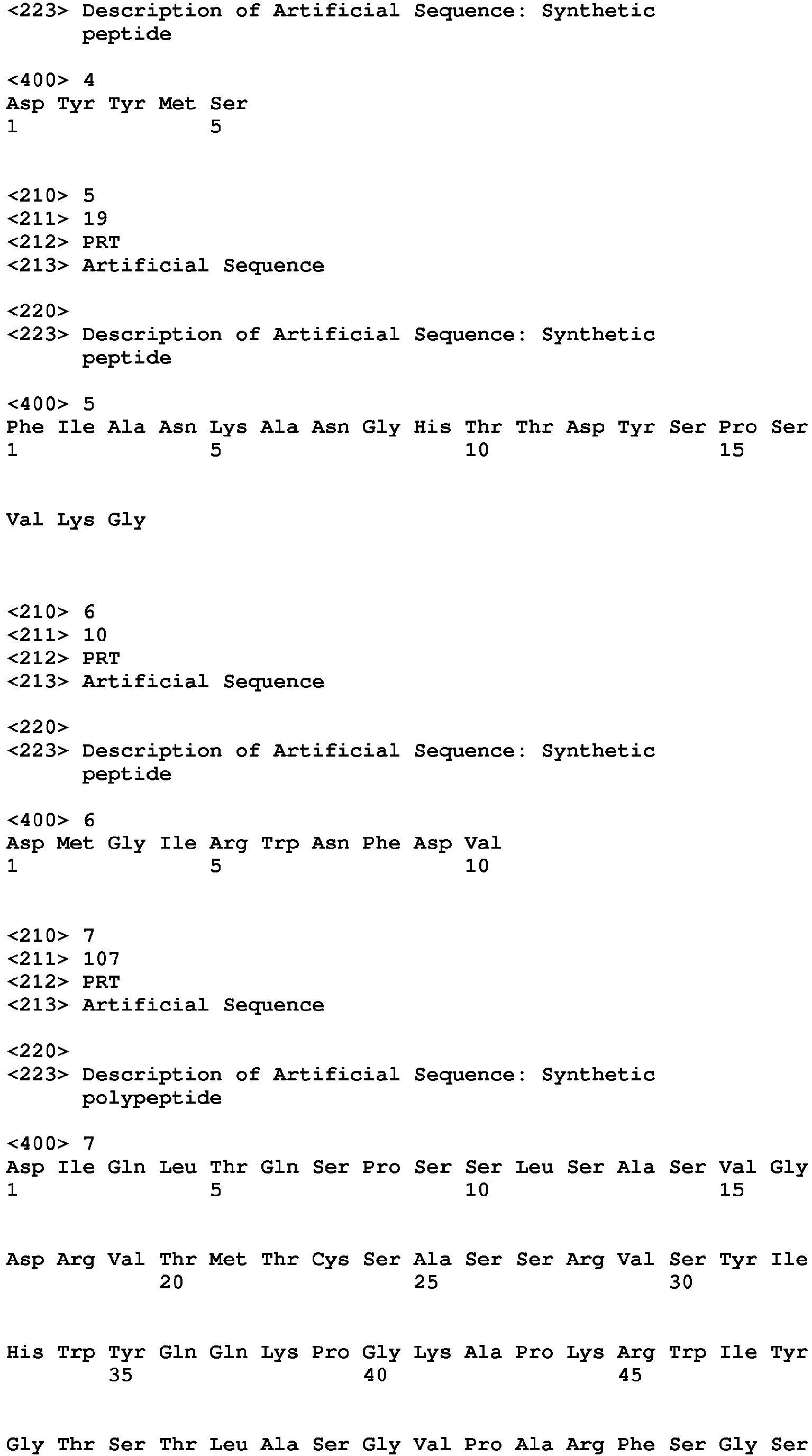


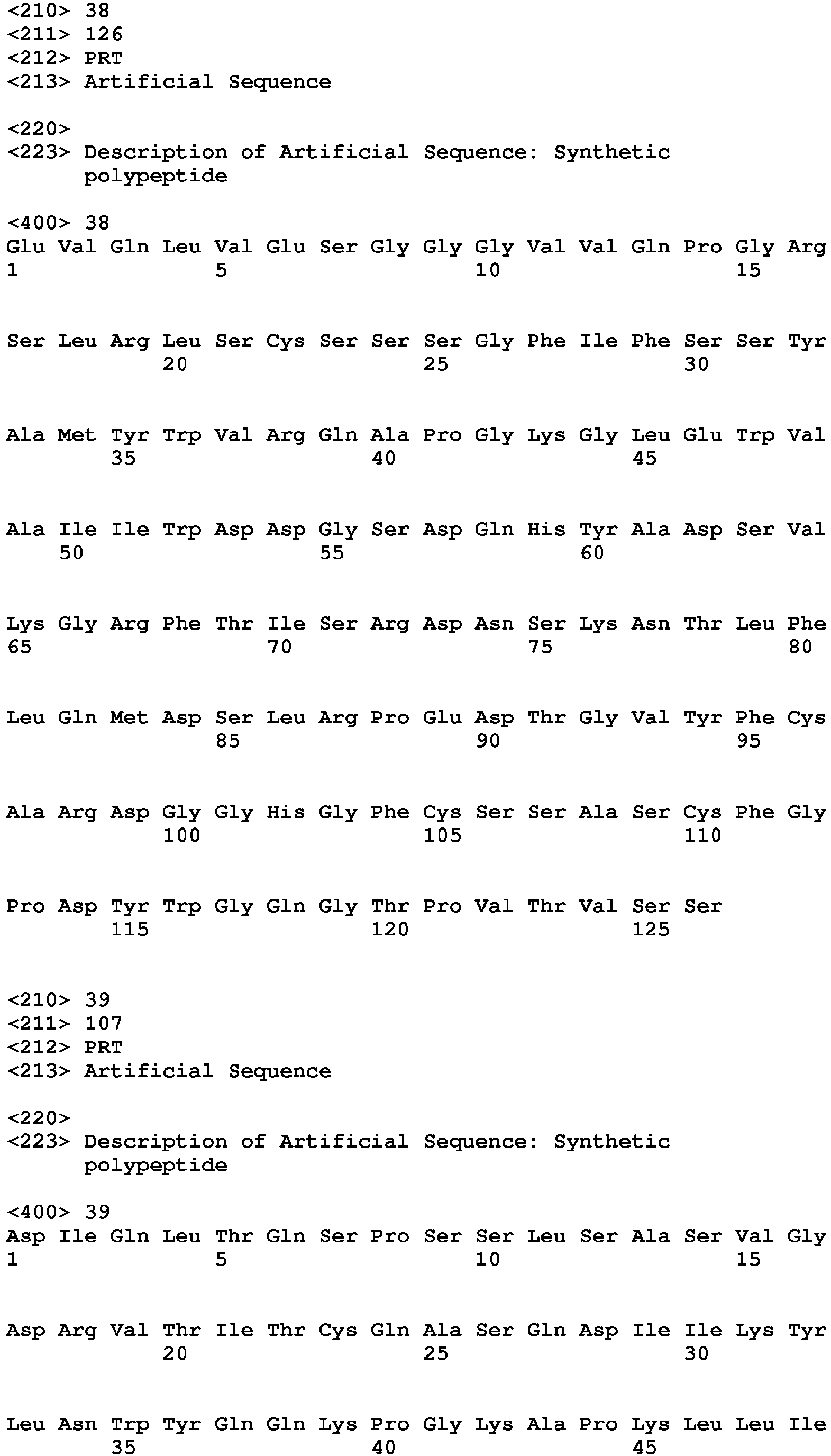

Claims (18)
- A composition comprising a chimeric, humanized or human Class I anti-CEA antibody or antigen-binding fragment thereof that blocks the binding to CEACAM5 and/or CEACAM6 of or that competes for binding to CEACAM5 and/or CEACAM6 with a monoclonal antibody comprising the light chain variable region CDR (complementarity determining region) sequences CDR1 having an amino acid sequence SASSRVSYIH (SEQ ID NO: 1); CDR2 having an amino acid sequence GTSTLAS (SEQ ID NO:2); and CDR3 having an amino acid sequence QQWSYNPPT (SEQ ID NO:3); and heavy chain variable region CDR sequences CDR1 having an amino acid sequence DYYMS (SEQ ID NO:4); CDR2 having an amino acid sequence FIANKANGHTTDYSPSVKG (SEQ ID NO:5); and CDR3 having an amino acid sequence DMGIRWNFDV (SEQ ID NO:6).
- The composition of claim 1, wherein the chimeric, humanized or human Class I anti-CEA antibody or fragment thereof binds to CEACAM5, CEACAM6 and granulocytes.
- The composition of claim 1, wherein the Class I anti-CEA antibody or fragment thereof sensitizes cancer to a therapeutic agent, preferably sensitizes CEA-expressing cancer to a therapeutic agent.
- The composition of claim 1, wherein the Class I anti-CEA antibody or fragment thereof comprises human antibody IgG1 constant region sequences.
- The composition of claim 1, wherein the chimeric Class I anti-CEA antibody or fragment thereof comprises the variable region amino acid sequences of SEQ ID NO:9 and SEQ ID NO: 10 or wherein the humanized Class I anti-CEA antibody or fragment thereof comprises the amino acid sequences of SEQ ID NO:7 and SEQ ID NO:8.
- The composition of claim 1, wherein the framework regions of the light and heavy chain variable regions of the humanized antibody or fragment thereof comprise at least one amino acid substitution selected from the heavy chain amino acid residues 28, 29, 30, 48 and 49 of SEQ ID NO: 10 and the light chain amino acid residues 21, 47 and 60 of SEQ ID NO:9.
- The composition of claim 1, wherein the antibody fragment is selected from the group consisting of F(ab')2, Fab', Fab, Fv and scFv.
- The composition of claim 1, wherein the Class I anti-CEA antibody or fragment thereof is conjugated to at least one diagnostic and/or therapeutic agent to form an immunoconjugate.
- The composition of claim 1, wherein Class I anti-CEA antibody or fragment thereof is a naked antibody or fragment thereof.
- The composition of claim 8, wherein the therapeutic agent is selected from the group consisting of an antibody, an antigen-binding antibody fragment, a cytotoxic agent, a drug, a toxin, a radionuclide, boron atoms, an immunomodulator, a photoactive therapeutic agent, an immunoconjugate, an oligonucleotide and a hormone.
- The composition of claim 10, wherein the drug is selected from the group consisting of 5-fluorouracil, aplidin, azaribine, anastrozole, anthracyclines, bendamustine, bleomycin, bortezomib, bryostatin-1, busulfan, calicheamycin, camptothecin, carboplatin, 10-hydroxycamptothecin, carmustine, celebrex, chlorambucil, cisplatin (CDDP), COX-2 inhibitors, irinotecan (CPT-11), SN-38, carboplatin, cladribine, camptothecans, cyclophosphamide, cytarabine, dacarbazine, docetaxel, dactinomycin, daunorubicin, doxorubicin, 2-pyrrolinodoxorubicin (2P-DOX), cyano-morpholino doxorubicin, doxorubicin glucuronide, epirubicin glucuronide, estramustine, epipodophyllotoxin, estrogen receptor binding agents, etoposide (VP 16), etoposide glucuronide, etoposide phosphate, floxuridine (FUdR), 3',5'-0-dioleoyl-FudR (FUdR-dO), fludarabine, flutamide, farnesyl-protein transferase inhibitors, gemcitabine, hydroxyurea, idarubicin, ifosfamide, L-asparaginase, lenalidomide, leucovorin, lomustine, mechlorethamine, melphalan, mercaptopurine, 6-mercaptopurine, methotrexate, mitoxantrone, mithramycin, mitomycin, mitotane, navelbine, nitrosourea, plicamycin, procarbazine, paclitaxel, pentostatin, PSI-341, raloxifene, semustine, streptozocin, tamoxifen, taxol, temazolomide, transplatinum, thalidomide, thioguanine, thiotepa, teniposide, topotecan, uracil mustard, vinorelbine, vinblastine, vincristine and vinca alkaloids; the toxin is selected from the group consisting of ricin, abrin, alpha toxin, saporin, ribonuclease (RNase), onconase, DNase I, Staphylococcal enterotoxin-A, pokeweed antiviral protein, gelonin, diphtheria toxin, Pseudomonas exotoxin and Pseudomonas endotoxin; the oligonucleotide is selected from the group consisting of RNAi and siRNA; the radionuclide is selected from the group consisting of 111, In, 111At, 177Lu, 211Bi, 212Bi, 213Bi, 211At, 62Cu, 67Cu, 90Y, ,125I, 131I, 133I, 32P, 35P, 47Sc, 111Ag, 67Ga, 153Sm, 161Tb, 152Dy, 166Dy, 161Ho, 166Ho, 186Re, 188Re, 189Re, 211Pb, 212Pb, 223Ra, 225Ac, 77As, 89Sr, 99Mo, 105Rh, 149Pm, 169Er, 194Ir, 58Co, 80mBr, 99mTc, 103mRh, 109Pt, 119Sb, 189mOs, 192Ir, 219Rn, 215Po, 221Fr, 255Fm, 11C, 13N, 15O, 75Br, 198Au, 199Au, 224Ac, 77Br, 113mIn, 95Ru, 97Ru, 103Ru, 105Ru, 107Hg, 203Hg, 121Te, 122mTe, 125mTe, 165Tm, 167Tm, 168Tm, 197Pt, 109Pd, 142Pr, 143Pr, 161Tb, 57Co, 58Co, 51Cr, 59Fe, 75Se, 201T1, 76Br and 169Yb; and the immunomodulator is selected from the group consisting of a cytokine, a chemokine, a stem cell growth factor, a lymphotoxin, an hematopoietic factor, a colony stimulating factor (CSF), an interferon, erythropoietin, thrombopoietin, a tumor necrosis factor (TNF), an interleukin (IL), granulocyte-colony stimulating factor (G-CSF), granulocyte macrophage-colony stimulating factor (GM-CSF) and a stem cell growth factor designated "S1 factor".
- The composition of claim 11, wherein the cytokine is selected from the group consisting of human growth hormone, N-methionyl human growth hormone, bovine growth hormone, parathyroid hormone, thyroxine, insulin, proinsulin, relaxin, prorelaxin, follicle stimulating hormone (FSH), thyroid stimulating hormone (TSH), luteinizing hormone (LH), hepatic growth factor, prostaglandin, fibroblast growth factor, prolactin, placental lactogen, OB protein, tumor necrosis factor-α, tumor necrosis factor-β, mullerian-inhibiting substance, mouse gonadotropin-associated peptide, inhibin, activin, vascular endothelial growth factor, integrin, thrombopoietin (TPO), NGF-β, platelet-growth factor, TGF-α, TGF-β, insulin-like growth factor-I, insulin-like growth factor-II, erythropoietin (EPO), osteoinductive factors, interferon- α, interferon-β, interferon-γ, macrophage-CSF (M-CSF), IL-1, IL-1α, IL-2, IL-3, IL-4, IL-5, IL-6, IL-7, IL-8, IL-9, IL-10, IL-11, IL-12, IL-13, IL-14, IL-15, IL-16, IL-17, IL-18, IL-21, IL-25, LIF, FLT-3, angiostatin, thrombospondin, endostatin, tumor necrosis factor and LT.
- The composition of claim 8, wherein the diagnostic agent is selected from the group consisting of a radionuclide, a radiological contrast agent, a paramagnetic ion, a metal, a fluorescent label, a chemiluminescent label, an ultrasound contrast agent and a photoactive agent.
- The composition of claim 9, further comprising one or more therapeutic agents.
- The composition of claim 1, wherein the chimeric, humanized or human Class I anti-CEA antibody or antigen-binding fragment thereof is monovalent.
- A bispecific or multispecific antibody comprising a first chimeric, humanized or human Class I anti-CEA antibody or antigen-binding fragment thereof according to claim 1 and a second antibody or antigen-binding fragment thereof.
- A fusion protein comprising a chimeric, humanized or human Class I anti-CEA antibody or fragment thereof according to claim 1.
- A composition according to any of claims 1-15, or a bispecific or multispecific antibody according to claim 16, or a fusion protein according to claim 17, for use in a method for the treatment of medullary thyroid cancer (MTC), colorectal cancer, hepatocellular carcinoma, liver cancer, gastric cancer, esophageal cancer, lung cancer, non-small cell lung cancer, breast cancer, pancreatic cancer, ovarian cancer, uterine cancer, cervical cancer, endometrial cancer, head-and-neck cancer, bladder cancer, urothelial cancer, prostate cancer, hematopoietic cancer, leukemia or melanoma.
Applications Claiming Priority (2)
| Application Number | Priority Date | Filing Date | Title |
|---|---|---|---|
| US24287209P | 2009-09-16 | 2009-09-16 | |
| EP10817617.3A EP2478110B1 (en) | 2009-09-16 | 2010-07-29 | Class i anti-cea antibodies and uses thereof |
Related Parent Applications (2)
| Application Number | Title | Priority Date | Filing Date |
|---|---|---|---|
| EP10817617.3A Division-Into EP2478110B1 (en) | 2009-09-16 | 2010-07-29 | Class i anti-cea antibodies and uses thereof |
| EP10817617.3A Division EP2478110B1 (en) | 2009-09-16 | 2010-07-29 | Class i anti-cea antibodies and uses thereof |
Publications (1)
| Publication Number | Publication Date |
|---|---|
| EP3009455A1 true EP3009455A1 (en) | 2016-04-20 |
Family
ID=43730776
Family Applications (2)
| Application Number | Title | Priority Date | Filing Date |
|---|---|---|---|
| EP10817617.3A Not-in-force EP2478110B1 (en) | 2009-09-16 | 2010-07-29 | Class i anti-cea antibodies and uses thereof |
| EP15003169.8A Ceased EP3009455A1 (en) | 2009-09-16 | 2010-07-29 | Class i anti-cea antibodies and uses thereof |
Family Applications Before (1)
| Application Number | Title | Priority Date | Filing Date |
|---|---|---|---|
| EP10817617.3A Not-in-force EP2478110B1 (en) | 2009-09-16 | 2010-07-29 | Class i anti-cea antibodies and uses thereof |
Country Status (6)
| Country | Link |
|---|---|
| US (7) | US8287865B2 (en) |
| EP (2) | EP2478110B1 (en) |
| CN (1) | CN102482701B (en) |
| CA (1) | CA2774260C (en) |
| IN (1) | IN2012DN01663A (en) |
| WO (1) | WO2011034660A1 (en) |
Cited By (1)
| Publication number | Priority date | Publication date | Assignee | Title |
|---|---|---|---|---|
| EP3981793A4 (en) * | 2019-06-04 | 2023-06-07 | Biotheus Inc. | Ceacam5-resistant monoclonal antibody and preparation method thereof and use thereof |
Families Citing this family (88)
| Publication number | Priority date | Publication date | Assignee | Title |
|---|---|---|---|---|
| US7527787B2 (en) * | 2005-10-19 | 2009-05-05 | Ibc Pharmaceuticals, Inc. | Multivalent immunoglobulin-based bioactive assemblies |
| WO2004032857A2 (en) * | 2002-10-08 | 2004-04-22 | Immunomedics, Inc. | Antibody therapy |
| US20100226884A1 (en) | 2009-01-20 | 2010-09-09 | Immunomedics, Inc. | Novel Class of Monospecific and Bispecific Humanized Antibodies that Target the Insulin-like Growth Factor Type I Receptor (IGF-1R) |
| US9862770B2 (en) * | 2005-10-19 | 2018-01-09 | Ibc Pharmaceuticals, Inc. | Multivalent antibody complexes targeting IGF-1R show potent toxicity against solid tumors |
| US9272029B2 (en) | 2009-03-26 | 2016-03-01 | Ibc Pharmaceuticals, Inc. | Interferon lambada-antibody complexes |
| IN2012DN01663A (en) | 2009-09-16 | 2015-06-05 | Immunomedics Inc | |
| US9464136B2 (en) | 2010-08-20 | 2016-10-11 | Massachusetts Institute Of Technology | Antibody-based constructs directed against tyrosine kinase receptors |
| US11214610B2 (en) | 2010-12-01 | 2022-01-04 | H. Lundbeck A/S | High-purity production of multi-subunit proteins such as antibodies in transformed microbes such as Pichia pastoris |
| US9067988B2 (en) | 2010-12-01 | 2015-06-30 | Alderbio Holdings Llc | Methods of preventing or treating pain using anti-NGF antibodies |
| US9078878B2 (en) | 2010-12-01 | 2015-07-14 | Alderbio Holdings Llc | Anti-NGF antibodies that selectively inhibit the association of NGF with TrkA, without affecting the association of NGF with p75 |
| US9884909B2 (en) | 2010-12-01 | 2018-02-06 | Alderbio Holdings Llc | Anti-NGF compositions and use thereof |
| US9783602B2 (en) | 2010-12-01 | 2017-10-10 | Alderbio Holdings Llc | Anti-NGF compositions and use thereof |
| US9539324B2 (en) | 2010-12-01 | 2017-01-10 | Alderbio Holdings, Llc | Methods of preventing inflammation and treating pain using anti-NGF compositions |
| EP3252076B1 (en) | 2011-01-14 | 2019-09-04 | The Regents Of The University Of California | Diagnostic use of antibodies against ror-1 protein |
| JP6400470B2 (en) | 2011-05-16 | 2018-10-03 | ジェネロン(シャンハイ)コーポレイション リミテッド | Multispecific Fab fusion proteins and methods of use |
| EP2537933A1 (en) | 2011-06-24 | 2012-12-26 | Institut National de la Santé et de la Recherche Médicale (INSERM) | An IL-15 and IL-15Ralpha sushi domain based immunocytokines |
| CN102911248B (en) * | 2011-08-05 | 2014-12-10 | 上海原子科兴药业有限公司 | Method for synthesizing 188 Re(CO)3 marked compound with 1,4-disubstituted triazole structure |
| AU2013212000B2 (en) * | 2012-01-26 | 2017-03-30 | Ibc Pharmaceuticals, Inc. | Targeting interferon-lambda with antibodies potently enhances anti-tumor and anti-viral activities |
| EP2708556B1 (en) | 2012-09-12 | 2018-11-07 | Samsung Electronics Co., Ltd | Pharmaceutical composition for the use in a combination therapy for prevention or treatment of c-met or angiogenesis factor induced diseases |
| PT2766048E (en) | 2012-10-12 | 2015-02-25 | Spirogen Sarl | Pyrrolobenzodiazepines and conjugates thereof |
| EA039377B1 (en) * | 2012-11-20 | 2022-01-20 | Санофи | Anti-ceacam5 antibodies and uses thereof |
| EP3725807A1 (en) | 2012-12-03 | 2020-10-21 | NovImmune SA | Anti-cd47 antibodies and methods of use thereof |
| WO2017004144A1 (en) | 2015-07-01 | 2017-01-05 | Immunomedics, Inc. | Antibody-sn-38 immunoconjugates with a cl2a linker |
| AU2013204922B2 (en) | 2012-12-20 | 2015-05-14 | Celgene Corporation | Chimeric antigen receptors |
| US10329350B2 (en) | 2012-12-26 | 2019-06-25 | Industrial Technology Research Institute | Method for producing a multivalent fab fragment with collagen-like peptide |
| CA2905181C (en) | 2013-03-13 | 2020-06-02 | Medimmune Limited | Pyrrolobenzodiazepines and conjugates thereof for providing targeted therapy |
| CA2907397C (en) | 2013-03-15 | 2022-11-22 | Anthrogenesis Corporation | Modified t lymphocytes |
| WO2014170032A1 (en) | 2013-04-19 | 2014-10-23 | Cytune Pharma | Cytokine derived treatment with reduced vascular leak syndrome |
| US9717715B2 (en) | 2013-11-15 | 2017-08-01 | Samsung Electronics Co., Ltd. | Method of combination therapy using an anti-C-Met antibody |
| WO2015095398A1 (en) * | 2013-12-17 | 2015-06-25 | Kevin Hagedorn | Method and apparatus for manufacturing isotropic magnetic nanocolloids |
| EP2915569A1 (en) | 2014-03-03 | 2015-09-09 | Cytune Pharma | IL-15/IL-15Ralpha based conjugates purification method |
| RU2718692C2 (en) | 2014-05-29 | 2020-04-13 | Мэкроудженикс, Инк. | Trispecific binding molecules, which specifically bind antigens of a variety of malignant tumors, and methods of using them |
| WO2016037644A1 (en) | 2014-09-10 | 2016-03-17 | Medimmune Limited | Pyrrolobenzodiazepines and conjugates thereof |
| PL3204018T3 (en) | 2014-10-07 | 2022-01-03 | Immunomedics, Inc. | Neoadjuvant use of antibody-drug conjugates |
| SG10201906471PA (en) | 2015-01-14 | 2019-09-27 | Brigham & Womens Hospital Inc | Treatment of cancer with anti-lap monoclonal antibodies |
| HUE048111T2 (en) | 2015-03-23 | 2020-05-28 | Bayer Pharma AG | Anti-ceacam6 antibodies and uses thereof |
| ES2953441T3 (en) | 2015-06-25 | 2023-11-13 | Immunomedics Inc | Combination of anti-hla-dr or anti-Trop-2 antibodies with microtubule inhibitors, parp inhibitors, bruton kinase inhibitors or phosphoinositide 3-kinase inhibitors significantly improves therapeutic outcome in cancer |
| CN106432502B (en) * | 2015-08-10 | 2020-10-27 | 中山大学 | Bispecific nanobody for treating CEA positive expression tumor |
| GB201514875D0 (en) | 2015-08-20 | 2015-10-07 | Autolus Ltd | Receptor |
| EA201891178A1 (en) | 2015-12-14 | 2019-01-31 | Макродженикс, Инк. | Bispecific molecules possessing immunoreactivity with respect to PD-1 and CTLA-4, and methods of their use |
| CN110698559B (en) * | 2015-12-24 | 2021-11-26 | 凯惠科技发展(上海)有限公司 | TPBG antibody, preparation method thereof, conjugate thereof and application thereof |
| SG10201601719RA (en) | 2016-03-04 | 2017-10-30 | Agency Science Tech & Res | Anti-LAG-3 Antibodies |
| CN109476756B (en) | 2016-03-15 | 2022-05-31 | 埃泰美德(香港)有限公司 | Multi-specificity Fab fusion protein and application thereof |
| SG10201603721TA (en) | 2016-05-10 | 2017-12-28 | Agency Science Tech & Res | Anti-CTLA-4 Antibodies |
| KR20190015520A (en) | 2016-06-07 | 2019-02-13 | 마크로제닉스, 인크. | Combination Therapy |
| US11236160B2 (en) * | 2016-10-10 | 2022-02-01 | Singapore Health Services Pte Ltd | Anti-CEACAM6 antibodies and methods of use |
| JP2019534882A (en) | 2016-10-11 | 2019-12-05 | メドイミューン・リミテッドMedImmune Limited | Antibody-drug conjugates with immune-mediated therapeutics |
| CN109983027A (en) | 2016-10-20 | 2019-07-05 | 细胞基因公司 | Based on CEREBLON can heterodimerization Chimeric antigen receptor |
| HUE058689T2 (en) | 2016-11-18 | 2022-09-28 | Astellas Pharma Inc | Novel anti-human muc1 antibody fab fragment |
| CN108341876B (en) * | 2017-01-22 | 2021-06-04 | 中国人民解放军第四军医大学 | Anti-human CEACAM5 monoclonal antibody, and preparation method and application thereof |
| CN108440668A (en) * | 2017-02-16 | 2018-08-24 | 瑞阳(苏州)生物科技有限公司 | The fusion protein of FGF21 and IGF-1 and its application |
| JP7132232B2 (en) | 2017-02-24 | 2022-09-06 | マクロジェニクス,インコーポレーテッド | Bispecific binding molecules capable of binding CD137 and tumor antigens and uses thereof |
| EP3398967A1 (en) | 2017-05-04 | 2018-11-07 | B Cell Design | Antibodies against carcinoembryonic antigen for cancer therapy and diagnosis |
| JP2020530280A (en) | 2017-07-03 | 2020-10-22 | トルク セラピューティクス, インコーポレイテッド | Immunostimulatory fusion molecule and its use |
| TWI795415B (en) * | 2017-07-07 | 2023-03-11 | 日商安斯泰來製藥股份有限公司 | A novel anti-human CEACAM5 antibody Fab fragment |
| CA3067247A1 (en) | 2017-07-11 | 2019-01-17 | Alexion Pharmaceuticals, Inc. | Polypeptides that bind complement component c5 or serum albumin and fusion proteins thereof |
| US11230601B2 (en) | 2017-10-10 | 2022-01-25 | Tilos Therapeutics, Inc. | Methods of using anti-lap antibodies |
| WO2019118266A1 (en) | 2017-12-12 | 2019-06-20 | Macrogenics Inc. | Bispecific cd 16-binding molecules and their use in the treatment of disease |
| BR112020016485A2 (en) | 2018-02-15 | 2020-12-15 | Macrogenics, Inc. | BINDING MOLECULE, PHARMACEUTICAL COMPOSITION AND METHOD FOR TREATING A DISEASE |
| SG11202010469QA (en) | 2018-05-23 | 2020-11-27 | Adc Therapeutics Sa | Molecular adjuvant |
| EP3802599B1 (en) | 2018-06-03 | 2023-12-20 | LamKap Bio beta AG | Bispecific antibodies against ceacam5 and cd47 |
| GB201811415D0 (en) | 2018-07-12 | 2018-08-29 | F Star Beta Ltd | Anti-Mesothelin Anti bodies |
| GB201811410D0 (en) | 2018-07-12 | 2018-08-29 | F Star Beta Ltd | OX40 Binding molecules |
| AU2019348440A1 (en) | 2018-09-26 | 2021-03-25 | Ascendis Pharma A/S | Degradable hyaluronic acid hydrogels |
| EP3863722A2 (en) | 2018-10-10 | 2021-08-18 | Tilos Theapeutics, Inc. | Anti-lap antibody variants and uses thereof |
| CA3115747A1 (en) * | 2018-10-10 | 2020-04-16 | Astellas Pharma Inc. | Pharmaceutical composition containing tagged site-antihuman antibody fab fragment complex |
| CN110132937A (en) * | 2019-06-05 | 2019-08-16 | 福建师范大学 | A kind of DNA chain detects the preparation method of heavy metal ion as signaling switch |
| CN110327074A (en) * | 2019-08-02 | 2019-10-15 | 无锡海斯凯尔医学技术有限公司 | Liver evaluation method, device, equipment and computer readable storage medium |
| GB201911187D0 (en) | 2019-08-05 | 2019-09-18 | Autolus Ltd | Receptor |
| MX2022003110A (en) | 2019-09-18 | 2022-06-27 | Lamkap Bio Alpha AG | Bispecific antibodies against ceacam5 and cd3. |
| CN110713539B (en) * | 2019-09-23 | 2021-04-16 | 华道(上海)生物医药有限公司 | Anti-carcinoembryonic antigen antibody and preparation method and application thereof |
| JP2022550067A (en) | 2019-09-27 | 2022-11-30 | ヤンセン バイオテツク,インコーポレーテツド | Anti-CEACAM antibody and use thereof |
| EP3831849A1 (en) | 2019-12-02 | 2021-06-09 | LamKap Bio beta AG | Bispecific antibodies against ceacam5 and cd47 |
| WO2022048883A1 (en) * | 2020-09-04 | 2022-03-10 | Merck Patent Gmbh | Anti-ceacam5 antibodies and conjugates and uses thereof |
| AU2021400227A1 (en) | 2020-12-18 | 2023-04-27 | Lamkap Bio Beta Ltd | Bispecific antibodies against ceacam5 and cd47 |
| US11883432B2 (en) | 2020-12-18 | 2024-01-30 | Century Therapeutics, Inc. | Chimeric antigen receptor system with adaptable receptor specificity |
| GB202102396D0 (en) | 2021-02-19 | 2021-04-07 | Adc Therapeutics Sa | Molecular adjuvant |
| CN112557662B (en) * | 2021-02-22 | 2021-07-06 | 和卓生物科技(上海)有限公司 | Colorectal cancer detection kit |
| WO2023016488A1 (en) | 2021-08-13 | 2023-02-16 | 昆山新蕴达生物科技有限公司 | Microtubule inhibitor-based antibody-drug conjugate |
| WO2023024949A1 (en) | 2021-08-24 | 2023-03-02 | 昆山新蕴达生物科技有限公司 | Antibody-drug conjugate conjugated via breakable linker |
| CN115819596A (en) * | 2021-09-17 | 2023-03-21 | 优迈生物科技(连云港)有限公司 | Antibody targeting human CEACAM5/6, preparation method and application |
| GB202115329D0 (en) | 2021-10-25 | 2021-12-08 | Autolus Ltd | Chimeric cytokine receptor |
| CN114560944B (en) * | 2022-03-30 | 2022-08-09 | 北京科跃中楷生物技术有限公司 | Antibody labeled by colloidal gold particles and application thereof |
| CN114409789B (en) * | 2022-03-30 | 2022-06-07 | 北京科跃中楷生物技术有限公司 | Colloidal gold particle marking method and detection kit |
| WO2023198019A1 (en) * | 2022-04-12 | 2023-10-19 | 盛禾(中国)生物制药有限公司 | Antibody against ceacam5 and ceacam6, and use thereof |
| WO2023242351A1 (en) | 2022-06-16 | 2023-12-21 | Lamkap Bio Beta Ag | Combination therapy of bispecific antibodies against ceacam5 and cd47 and bispecific antibodies against ceacam5 and cd3 |
| WO2024035662A2 (en) | 2022-08-10 | 2024-02-15 | Merck Sharp & Dohme Llc | Proteins binding nkg2d, cd16, and ceacam5 |
| CN116731165A (en) * | 2023-03-23 | 2023-09-12 | 北京巴瑞医疗器械有限公司 | Anti-serotonin antibody and application thereof |
Citations (180)
| Publication number | Priority date | Publication date | Assignee | Title |
|---|---|---|---|---|
| US672015A (en) | 1900-10-05 | 1901-04-16 | Alfred Schlatter | Automatic switch for groups of transformers. |
| US4036945A (en) | 1976-05-03 | 1977-07-19 | The Massachusetts General Hospital | Composition and method for determining the size and location of myocardial infarcts |
| US4331647A (en) | 1980-03-03 | 1982-05-25 | Goldenberg Milton David | Tumor localization and therapy with labeled antibody fragments specific to tumor-associated markers |
| US4818709A (en) | 1983-01-21 | 1989-04-04 | Primus Frederick J | CEA-family antigens, Anti-CEA antibodies and CEA immunoassay |
| US4824659A (en) | 1985-06-07 | 1989-04-25 | Immunomedics, Inc. | Antibody conjugates |
| US4863713A (en) | 1986-06-23 | 1989-09-05 | The Board Of Trustees Of Leland Stanford Jr. Univ. | Method and system for administering therapeutic and diagnostic agents |
| US4946778A (en) | 1987-09-21 | 1990-08-07 | Genex Corporation | Single polypeptide chain binding molecules |
| US5057313A (en) | 1986-02-25 | 1991-10-15 | The Center For Molecular Medicine And Immunology | Diagnostic and therapeutic antibody conjugates |
| US5081235A (en) | 1989-07-26 | 1992-01-14 | City Of Hope | Chimeric anti-cea antibody |
| US5120525A (en) | 1988-03-29 | 1992-06-09 | Immunomedics, Inc. | Radiolabeled antibody cytotoxic therapy of cancer |
| US5132405A (en) | 1987-05-21 | 1992-07-21 | Creative Biomolecules, Inc. | Biosynthetic antibody binding sites |
| US5229275A (en) | 1990-04-26 | 1993-07-20 | Akzo N.V. | In-vitro method for producing antigen-specific human monoclonal antibodies |
| US5256395A (en) | 1986-09-19 | 1993-10-26 | Immunotech Partners | Affinity enhancement immunological reagents for in vivo detection and killing of specific target cells |
| US5443953A (en) | 1993-12-08 | 1995-08-22 | Immunomedics, Inc. | Preparation and use of immunoconjugates |
| US5525338A (en) | 1992-08-21 | 1996-06-11 | Immunomedics, Inc. | Detection and therapy of lesions with biotin/avidin conjugates |
| US5567610A (en) | 1986-09-04 | 1996-10-22 | Bioinvent International Ab | Method of producing human monoclonal antibodies and kit therefor |
| US5587459A (en) | 1994-08-19 | 1996-12-24 | Regents Of The University Of Minnesota | Immunoconjugates comprising tyrosine kinase inhibitors |
| US5633425A (en) | 1990-08-29 | 1997-05-27 | Genpharm International, Inc. | Transgenic non-human animals capable of producing heterologous antibodies |
| US5677136A (en) | 1994-11-14 | 1997-10-14 | Systemix, Inc. | Methods of obtaining compositions enriched for hematopoietic stem cells, compositions derived therefrom and methods of use thereof |
| US5716595A (en) | 1992-05-06 | 1998-02-10 | Immunomedics, Inc. | Intraperative, intravascular and endoscopic tumor and lesion detection and therapy with monovalent antibody fragments |
| US5736119A (en) | 1993-05-17 | 1998-04-07 | Immunomedics, Inc. | Detection and therapy of lesions with biotin/avidin-metal chelating protein conjugates |
| US5776456A (en) | 1992-11-13 | 1998-07-07 | Idec Pharmaceuticals Corporation | Therapeutic application of chimeric and radiolabeled antibodies to human B lymphocyte restricted differentiation antigen for treatment of B cell lymphoma |
| US5789554A (en) | 1994-08-12 | 1998-08-04 | Immunomedics, Inc. | Immunoconjugates and humanized antibodies specific for B-cell lymphoma and leukemia cells |
| US5798229A (en) | 1993-08-02 | 1998-08-25 | Merck Patent Gesellschaft Mit Beschrankter Haftung | Bispecific molecules recognizing lymphocyte antigen CD2 and tumor antigens |
| US5814440A (en) | 1995-06-07 | 1998-09-29 | Systemix, Inc. | Methods of obtaining compositions enriched for hematopoietic stem cells, antibodies for use therein, compositions derived therefrom and methods of use thereof |
| US5827690A (en) | 1993-12-20 | 1998-10-27 | Genzyme Transgenics Corporatiion | Transgenic production of antibodies in milk |
| US5874540A (en) | 1994-10-05 | 1999-02-23 | Immunomedics, Inc. | CDR-grafted type III anti-CEA humanized mouse monoclonal antibodies |
| WO2000029584A1 (en) | 1998-11-18 | 2000-05-25 | Genentech, Inc. | Antibody variants with higher binding affinity compared to parent antibodies |
| US6077499A (en) | 1996-05-03 | 2000-06-20 | Immunomedics, Inc. | Targeted combination immunotherapy of cancer |
| US6090381A (en) | 1997-02-11 | 2000-07-18 | Immunomedics, Inc. | Stimulation of an immune response with antibodies labeled with the . .alpha-galactosyl epitope |
| US6096289A (en) | 1992-05-06 | 2000-08-01 | Immunomedics, Inc. | Intraoperative, intravascular, and endoscopic tumor and lesion detection, biopsy and therapy |
| US6120767A (en) | 1986-10-27 | 2000-09-19 | Pharmaceutical Royalties, L.L.C. | Chimeric antibody with specificity to human B cell surface antigen |
| US6129914A (en) | 1992-03-27 | 2000-10-10 | Protein Design Labs, Inc. | Bispecific antibody effective to treat B-cell lymphoma and cell line |
| US6183744B1 (en) | 1997-03-24 | 2001-02-06 | Immunomedics, Inc. | Immunotherapy of B-cell malignancies using anti-CD22 antibodies |
| US6254868B1 (en) | 1996-03-20 | 2001-07-03 | Immunomedics, Inc. | Glycosylated humanized B-cell specific antibodies |
| US6306393B1 (en) | 1997-03-24 | 2001-10-23 | Immunomedics, Inc. | Immunotherapy of B-cell malignancies using anti-CD22 antibodies |
| US6331175B1 (en) | 1985-07-05 | 2001-12-18 | Immunomedics, Inc. | Method and kit for imaging and treating organs and tissues |
| US6340571B1 (en) | 1996-03-21 | 2002-01-22 | Bio Merieux | Antibodies specific for Staphylococcus aureus, and use thereof |
| US6340459B1 (en) | 1995-12-01 | 2002-01-22 | The Trustees Of Columbia University In The City Of New York | Therapeutic applications for the anti-T-BAM (CD40-L) monoclonal antibody 5C8 in the treatment of reperfusion injury in non-transplant recipients |
| US6344198B1 (en) | 1995-01-20 | 2002-02-05 | Human Genome Sciences, Inc. | Human prostatic specific reductase |
| US6346246B1 (en) | 1994-01-25 | 2002-02-12 | Human Genome Sciences, Inc. | Antibodies that bind human haemopoietic maturation factor |
| US6355244B1 (en) | 1997-11-17 | 2002-03-12 | University Of Kentucky Research Foundation | Methods and compositions for the treatment of psoriasis |
| US6355481B1 (en) | 1999-06-18 | 2002-03-12 | Emory University | Hybridoma cell line and monoclonal antibody for huntingtin protein |
| US6355444B1 (en) | 1992-01-29 | 2002-03-12 | Viventia Biotech Inc. | Carcinoma associated antigen (SK1) monoclonal antibodies against SK1, methods of producing these antibodies and uses therefor |
| US6355245B1 (en) | 1994-05-02 | 2002-03-12 | Alexion Pharmaceuticals, Inc. | C5-specific antibodies for the treatment of inflammatory diseases |
| US6359126B1 (en) | 1992-04-03 | 2002-03-19 | Genentech, Inc. | Antibodies to αvβ3 integrin |
| US6372215B1 (en) | 1997-03-03 | 2002-04-16 | Bristol-Myers Squibb Company | Monoclonal antibodies to human CD6 |
| US6376654B1 (en) | 1999-08-13 | 2002-04-23 | Molecular Discoveries, Llc | Myeloma cell and ovarian cancer cell surface glycoproteins, antibodies thereto, and uses thereof |
| US6383484B1 (en) | 1998-12-21 | 2002-05-07 | Ludwig Institute For Cancer Research | Antibodies to truncated VEGF-D and thereof |
| US6383759B1 (en) | 1998-05-04 | 2002-05-07 | Gerald P. Murphy Cancer Foundation | Non-invasive method to detect prostate cancer |
| US6395276B1 (en) | 1997-05-02 | 2002-05-28 | Immunomedics, Inc. | Immunotoxins directed against malignant cells |
| US6395274B1 (en) | 1993-11-19 | 2002-05-28 | Baylor College Of Medicine | Monoclonal antibodies specific for human interleukin-5 |
| US6403091B1 (en) | 1991-11-15 | 2002-06-11 | The Trustees Of Columbia University In The City Of New York | Methods for inhibiting the rejection of a transplant organ in a subject with 5C8-specific antibodies |
| US6403770B1 (en) | 1996-10-25 | 2002-06-11 | Human Genome Sciences, Inc. | Antibodies to neutrokine-alpha |
| US6406694B1 (en) | 1998-07-23 | 2002-06-18 | Millennium Pharmaceuticals, Inc. | Anti-CCR2 antibodies |
| US6413726B1 (en) | 1996-03-19 | 2002-07-02 | Human Genome Sciences, Inc. | Antibodies that specifically bind Cytostatin III |
| US6419928B1 (en) | 1988-12-22 | 2002-07-16 | Genzyme Corporation | Monoclonal antibodies to transforming growth factor-beta and methods of use |
| US6432402B1 (en) | 1989-05-25 | 2002-08-13 | Sloan-Kettering Institute For Cancer Research | Anti-idiotypic antibody which induces an immune response against a glycosphingolipid and use thereof |
| US6432404B1 (en) | 1993-12-23 | 2002-08-13 | Icos Corporation | Methods of inhibiting locomotor damage following spinal cord injury with α D-specific antibodies |
| US6441143B1 (en) | 1995-04-13 | 2002-08-27 | Amgen Inc | Methods and compositions for determining HER-2/neu expression |
| US6444206B1 (en) | 1993-05-28 | 2002-09-03 | The Scripps Research Institute | Methods and compositions for inhibiting CD14 mediated cell activation |
| US6455040B1 (en) | 1997-01-14 | 2002-09-24 | Human Genome Sciences, Inc. | Tumor necrosis factor receptor 5 |
| US6458356B1 (en) | 1995-12-05 | 2002-10-01 | Amgen Inc. | Antibody-induced apoptosis |
| US6458933B1 (en) | 1998-05-20 | 2002-10-01 | Immunomedics, Inc. | Therapeutic using a bispecific antibody |
| US6461823B1 (en) | 1997-01-28 | 2002-10-08 | Human Genome Sciences, Inc. | Death domain containing receptor-4 antibodies |
| US6465173B2 (en) | 1992-03-09 | 2002-10-15 | Immune Research, Ltd. | Anti-idiotypic antibody and its use in diagnosis and therapy in HIV-related disease |
| US6468531B1 (en) | 1993-09-09 | 2002-10-22 | Duke University | Method of promoting cellular function |
| US6468529B1 (en) | 1995-06-02 | 2002-10-22 | Genentech, Inc. | Hepatocyte growth factor receptor antagonists and uses thereof |
| US6479247B1 (en) | 1996-10-09 | 2002-11-12 | The Corporation Of The Trustees Of The Order Of The Sisters Of Mercy In Queensland | Dendritic cell-specific antibodies |
| US6482598B2 (en) | 1996-11-13 | 2002-11-19 | Morphogenesis, Inc. | Antibody recognizing a small subset of human hematopoietic cells |
| US6482408B2 (en) | 1995-06-05 | 2002-11-19 | Human Genome Sciences, Inc. | Fibroblast growth factor 15 antibodies |
| US6488930B1 (en) | 1999-01-15 | 2002-12-03 | Millennium Pharmaceuticals, Inc. | Anti-CCR4 antibodies and methods of use therefor |
| US6511665B1 (en) | 1987-11-25 | 2003-01-28 | Immunex Corporation | Antibodies to interleukin-1 receptors |
| US6518404B1 (en) | 1994-10-17 | 2003-02-11 | Human Genome Sciences, Inc. | Human endothelin-bombesin receptor antibodies |
| US6521227B1 (en) | 1999-11-18 | 2003-02-18 | Peter L. Hudson | Polynucleotides encoding prostatic growth factor and process for producing prostatic growth factor polypeptides |
| US6528269B1 (en) | 1998-06-22 | 2003-03-04 | Case Western Reserve University | Immunological agents specific for prion protein (PRP) |
| US6528625B1 (en) | 1996-10-28 | 2003-03-04 | Millennium Pharmaceuticals, Inc. | Anti-CCR5 antibodies and kits comprising same |
| US6534058B2 (en) | 2000-10-10 | 2003-03-18 | Tanox, Inc. | Anti-C5 monoclonal antibodies |
| US6544749B1 (en) | 1992-05-08 | 2003-04-08 | Genentech, Inc. | Antibodies to leukemia inhibitory factor and their use in immunoassays |
| US6545130B2 (en) | 1997-06-04 | 2003-04-08 | Albert Einstein College Of Medicine Of Yeshiva University | Monoclonal antibodies to mycobacterium tuberculosis and a modified ELISA assay |
| US6562618B1 (en) | 1997-12-25 | 2003-05-13 | Japan Tobacco, Inc. | Monoclonal antibody against connective tissue growth factor and medicinal uses thereof |
| US6566076B1 (en) | 1998-04-23 | 2003-05-20 | The Regents Of The University Of California | Detection and diagnosis of conditions associated with lung injury |
| US6572856B1 (en) | 1998-09-10 | 2003-06-03 | The University Of Virginia Patent Foundation | Methods for the prevention and treatment of cancer using anti-C3b(i) antibodies |
| US6576745B1 (en) | 1996-04-03 | 2003-06-10 | Human Genome Sciences, Inc. | Human cystatin F antibodies |
| US6596852B2 (en) | 1994-07-08 | 2003-07-22 | Immunex Corporation | Antibodies that bind the cytokine designated LERK-5 |
| US6605279B2 (en) | 1993-07-26 | 2003-08-12 | Genetics Institute, Inc. | Therapeutic compositions for inhibiting the interactions of B7-1 and B7-2 with their natural ligands |
| US6605441B1 (en) | 1995-06-05 | 2003-08-12 | Human Genome Sciences, Inc. | Antibodies against fibroblast growth factor 11 |
| US6610833B1 (en) | 1997-11-24 | 2003-08-26 | The Institute For Human Genetics And Biochemistry | Monoclonal human natural antibodies |
| US6630144B1 (en) | 1999-08-30 | 2003-10-07 | The United States Of America As Represented By The Secretary Of The Army | Monoclonal antibodies to Ebola glycoprotein |
| US6652852B1 (en) | 1986-10-27 | 2003-11-25 | Royalty Pharma Finance Trust | Chimeric antibody with specificity to human B cell surface antigen |
| US6653104B2 (en) | 1996-10-17 | 2003-11-25 | Immunomedics, Inc. | Immunotoxins, comprising an internalizing antibody, directed against malignant and normal cells |
| US6673344B1 (en) | 1994-08-23 | 2004-01-06 | Human Genome Sciences, Inc. | Antibodies to human CKβ-10/MCP-4 |
| US6682736B1 (en) | 1998-12-23 | 2004-01-27 | Abgenix, Inc. | Human monoclonal antibodies to CTLA-4 |
| US6682737B1 (en) | 1996-03-29 | 2004-01-27 | North Carolina State University | Anti-cryptosporidium parvum preparations |
| US6689607B2 (en) | 1997-10-21 | 2004-02-10 | Human Genome Sciences, Inc. | Human tumor, necrosis factor receptor-like proteins TR11, TR11SV1 and TR11SV2 |
| US6689355B2 (en) | 2000-05-11 | 2004-02-10 | Altarex Corp. | Therapeutic method and composition utilizing antigen-antibody complexation and presentation by dendritic cells |
| US6689362B1 (en) | 1997-06-03 | 2004-02-10 | Regents Of The University Of Minnesota | Method for treating T-lineage leukemias and lymphomas using a CD7-specific monoclonal antibody (TXU-7) linked to the pokeweed antiviral protein (PAP) |
| US6693176B1 (en) | 1999-07-23 | 2004-02-17 | University Of Massachusetts | Antitumor antibodies, proteins, and uses thereof |
| US6692908B1 (en) | 1998-11-05 | 2004-02-17 | Stanford University | Prevention and treatment of HCV infection employing antibodies that inhibit the interaction of HCV virions with their receptor |
| US6709653B1 (en) | 1994-09-16 | 2004-03-23 | Human Genome Sciences, Inc. | Antibodies specific for human inositol monophosphatase H1 |
| US6716966B1 (en) | 1999-08-18 | 2004-04-06 | Altarex Corp. | Therapeutic binding agents against MUC-1 antigen and methods for their use |
| US6733981B2 (en) | 1994-11-01 | 2004-05-11 | Human Genome Sciences, Inc. | Antibodies to interleukin-1 β converting enzyme like apoptosis protease-3 and 4 |
| US6743898B2 (en) | 2001-03-15 | 2004-06-01 | Ochsner Clinic Foundation | Monoclonal antibodies that suppress B cell growth and/or differentiation |
| US6764679B2 (en) | 1997-09-18 | 2004-07-20 | Genentech, Inc. | Antibodies to DcR3 Polypeptide, a TNFR Homolog |
| US6764681B2 (en) | 1991-10-07 | 2004-07-20 | Biogen, Inc. | Method of prophylaxis or treatment of antigen presenting cell driven skin conditions using inhibitors of the CD2/LFA-3 interaction |
| US6764688B2 (en) | 1996-09-03 | 2004-07-20 | Kaneka Corporation | Method for inducing immunosuppressive cells and a culture device to be used therefor |
| US6767711B2 (en) | 1995-06-05 | 2004-07-27 | Cornell Research Foundation, Inc. | Treatment and diagnosis of prostate cancer |
| US6770450B1 (en) | 1996-05-06 | 2004-08-03 | Cornell Research Foundation, Inc. | Treatment and diagnosis of cancer |
| US6783758B2 (en) | 1999-11-08 | 2004-08-31 | Rhode Island Hospital | Diagnosis and treatment of malignant neoplasms |
| US6824780B1 (en) | 1999-10-29 | 2004-11-30 | Genentech, Inc. | Anti-tumor antibody compositions and methods of use |
| US6824778B2 (en) | 2001-04-23 | 2004-11-30 | The United States Of America As Represented By The Secretary Of The Army | Prophylactic and therapeutic monoclonal antibodies |
| US6835370B2 (en) | 1999-11-08 | 2004-12-28 | Rhode Island Hospital | Diagnosis and treatment of malignant neoplasms |
| US6835549B2 (en) | 2000-02-24 | 2004-12-28 | University Of Medicine & Dentistry Of New Jersey | Immunoassay method for the diagnosis of gastric intestinal metaplasia associated with gastric carcinoma |
| US6861226B2 (en) | 1996-03-21 | 2005-03-01 | Human Genome Sciences, Inc. | Human endometrial specific steroid-binding factor I, II and III |
| US6861227B2 (en) | 1998-03-19 | 2005-03-01 | Human Genome Sciences, Inc. | Antibodies to cytokine receptor common gamma chain like |
| US6861511B1 (en) | 1986-06-04 | 2005-03-01 | Bayer Corporation | Detection and quantification of neu related proteins in the biological fluids of humans |
| US6864062B2 (en) | 1999-06-16 | 2005-03-08 | Molecular Geriatrics Corporation | Purified antigen for Alzheimer's disease and methods of obtaining and using same |
| US6867006B2 (en) | 1994-05-16 | 2005-03-15 | Human Genome Sciences, Inc. | Antibodies to human chemotactic protein |
| US6872568B1 (en) | 1997-03-17 | 2005-03-29 | Human Genome Sciences, Inc. | Death domain containing receptor 5 antibodies |
| US6875580B2 (en) | 2003-01-28 | 2005-04-05 | Schering Corporation | Antibodies specific for plasmacytoid dendritic cells |
| US6878812B2 (en) | 1997-01-21 | 2005-04-12 | Human Genome Science, Inc. | Metalloproteinases |
| US6881405B2 (en) | 1998-06-15 | 2005-04-19 | Altarex Medical Corp. | Reagents and methods for inducing an immune response to prostate specific antigen |
| US6884594B2 (en) | 1996-03-26 | 2005-04-26 | Human Genome Sciences, Inc. | Antibodies to growth factor HTTER36 |
| US6887466B2 (en) | 1988-11-23 | 2005-05-03 | Genetics Institute, Inc. | Methods for selectively stimulating proliferation of T cells |
| US6887468B1 (en) | 1999-04-28 | 2005-05-03 | Board Of Regents, The University Of Texas System | Antibody kits for selectively inhibiting VEGF |
| US6899879B2 (en) | 1992-07-09 | 2005-05-31 | Chiron Corporation | Method for treating an IgE-mediated disease in a patient using anti-CD40 monoclonal antibodies |
| US6919433B2 (en) | 1997-03-14 | 2005-07-19 | Human Genome Sciences, Inc. | Antibodies to protein HPMBQ91 |
| US6919078B2 (en) | 1997-06-11 | 2005-07-19 | Human Genome Sciences, Inc. | Antibodies to human tumor necrosis factor receptor TR9 |
| US6921645B2 (en) | 1994-08-23 | 2005-07-26 | Human Genome Sciences, Inc. | Antibodies to chemokine β-4 |
| US6921533B2 (en) | 1989-06-02 | 2005-07-26 | The Johns Hopkins University School Of Medicine | Method of using monoclonal antibodies against leukocyte adhesion receptor β-chain |
| US6939547B2 (en) | 2000-07-31 | 2005-09-06 | The United States Of America As Represented By The Department Of Health And Human Services | Specific binding agents for KSHV vIL-6 that neutralize a biological activity |
| US6946129B1 (en) | 1999-06-08 | 2005-09-20 | Seattle Genetics, Inc. | Recombinant anti-CD40 antibody and uses thereof |
| US6949244B1 (en) | 1995-12-20 | 2005-09-27 | The Board Of Trustees Of The University Of Kentucky | Murine monoclonal anti-idiotype antibody 11D10 and methods of use thereof |
| US6951924B2 (en) | 1997-03-14 | 2005-10-04 | Human Genome Sciences, Inc. | Antibodies against secreted protein HTEBYII |
| US6956107B2 (en) | 1998-02-20 | 2005-10-18 | Tanox, Inc. | Inhibitors of complement activation |
| US6962981B1 (en) | 1996-03-25 | 2005-11-08 | Medarex, Inc. | Monoclonal antibodies specific for the extracellular domain of prostate-specific membrane antigen |
| US6962813B2 (en) | 2001-05-21 | 2005-11-08 | The Brigham And Women's Hospital, Inc. | P. aeruginosa mucoid exopolysaccharide specific binding peptides |
| US6962702B2 (en) | 1998-06-22 | 2005-11-08 | Immunomedics Inc. | Production and use of novel peptide-based agents for use with bi-specific antibodies |
| US6964854B1 (en) | 1999-07-13 | 2005-11-15 | Science & Technology Corporation | Compositions and methods useful for the diagnosis and treatment of heparin induced thrombocytopenia/thrombosis |
| US6965018B2 (en) | 2000-06-06 | 2005-11-15 | Bristol-Myers Squibb Company | Antibodies directed to B7-related polypeptide, BSL-2 |
| US6974863B2 (en) | 1988-11-07 | 2005-12-13 | Indiana University Research And Technology Corporation | Antibody for 4-1BB |
| US6989241B2 (en) | 2000-10-02 | 2006-01-24 | Oklahoma Medical Research Foundation | Assay for rapid detection of human activated protein C and highly specific monoclonal antibody therefor |
| US6994976B1 (en) | 1999-11-19 | 2006-02-07 | Tittle Thomas V | Tr3-specific binding agents and methods for their use |
| US6994852B1 (en) | 1999-11-12 | 2006-02-07 | Temple University-Of The Commonwealth System Of Higher Education | Inhibition of angiogenesis by antibodies against high molecular weight kininogen domain 5 |
| US6998468B2 (en) | 2000-03-23 | 2006-02-14 | Tanox, Inc. | Anti-C2/C2a inhibitors of complement activation |
| US7001598B2 (en) | 1995-01-26 | 2006-02-21 | Biogen Idec Ma Inc. | Anti-lymphotoxin-beta receptor antibodies as anti-tumor agents |
| US7011812B1 (en) | 1996-05-03 | 2006-03-14 | Immunomedics, Inc. | Targeted combination immunotherapy of cancer and infectious diseases |
| US7012133B1 (en) | 1995-01-19 | 2006-03-14 | Children's Medical Center Corp. | Antibodies to C-C chemokine Receptor 3 Protein |
| US7037498B2 (en) | 2001-01-05 | 2006-05-02 | Abgenix, Inc. | Antibodies to insulin-like growth factor I receptor |
| US7038018B2 (en) | 1999-11-24 | 2006-05-02 | Millennium Pharmaceuticals, Inc. | Antibodies and ligands for “Bonzo” chemokine receptor |
| US7041802B2 (en) | 1998-12-23 | 2006-05-09 | Human Genome Sciences, Inc. | Peptidoglycan recognition proteins |
| US7041293B1 (en) | 1990-04-03 | 2006-05-09 | Genentech, Inc. | HIV env antibodies |
| US7041803B2 (en) | 1997-01-21 | 2006-05-09 | Human Genome Sciences, Inc. | Galectin 11 |
| US7045132B2 (en) | 1991-11-14 | 2006-05-16 | The Government Of The United States Of America, As Represented By The Secretary, Department Of Health And Human Services, C/O Centers For Disease Control And Prevention | Streptococcus pneumoniae 37-kDa surface adhesin a protein |
| US7049060B2 (en) | 2001-11-05 | 2006-05-23 | Ortho-Clinical Diagnostics, Inc. | HCV anti-core monoclonal antibodies |
| US7052872B1 (en) | 1999-06-22 | 2006-05-30 | Immunomedics, Inc. | Bi-specific antibodies for pre-targeting diagnosis and therapy |
| US7060802B1 (en) | 2000-09-18 | 2006-06-13 | The Trustees Of Columbia University In The City Of New York | Tumor-associated marker |
| US7074405B1 (en) | 1998-06-22 | 2006-07-11 | Immunomedics, Inc. | Use of bi-specific antibodies for pre-targeting diagnosis and therapy |
| US7074403B1 (en) | 1999-06-09 | 2006-07-11 | Immunomedics, Inc. | Immunotherapy of autoimmune disorders using antibodies which target B-cells |
| US7109304B2 (en) | 2003-07-31 | 2006-09-19 | Immunomedics, Inc. | Humanized anti-CD19 antibodies |
| US7138103B2 (en) | 1998-06-22 | 2006-11-21 | Immunomedics, Inc. | Use of bi-specific antibodies for pre-targeting diagnosis and therapy |
| US7151164B2 (en) | 2002-02-14 | 2006-12-19 | Immunomedics, Inc. | Anti-CD20 antibodies and fusion proteins thereof and methods of use |
| US7238785B2 (en) | 2002-03-01 | 2007-07-03 | Immunomedics, Inc. | RS7 antibodies |
| US7282567B2 (en) | 2002-06-14 | 2007-10-16 | Immunomedics, Inc. | Monoclonal antibody hPAM4 |
| US7300655B2 (en) | 2002-08-01 | 2007-11-27 | Immunomedics, Inc. | Alpha-fetoprotein Immu31 antibodies and fusion proteins and methods of use thereof |
| US7312318B2 (en) | 2002-03-01 | 2007-12-25 | Immunomedics, Inc. | Internalizing anti-CD74 antibodies and methods of use |
| US7387772B1 (en) | 1999-06-22 | 2008-06-17 | Immunimedics, Inc. | Chimeric, human and humanized anti-CSAP monoclonal antibodies |
| US7405320B2 (en) | 1998-06-22 | 2008-07-29 | Immunomedics, Inc. | Therapeutic and diagnostic conjugates for use with multispecific antibodies |
| US7521056B2 (en) | 2005-04-06 | 2009-04-21 | Ibc Pharmaceuticals, Inc. | Stably tethered structures of defined compositions with multiple functions or binding specificities |
| US7527787B2 (en) | 2005-10-19 | 2009-05-05 | Ibc Pharmaceuticals, Inc. | Multivalent immunoglobulin-based bioactive assemblies |
| US7531327B2 (en) | 2004-07-23 | 2009-05-12 | Immunomedics, Inc. | Methods and compositions for increasing longevity and protein yield from a cell culture |
| US7534866B2 (en) | 2005-10-19 | 2009-05-19 | Ibc Pharmaceuticals, Inc. | Methods and compositions for generating bioactive assemblies of increased complexity and uses |
| US7534431B2 (en) | 2003-01-31 | 2009-05-19 | Immunomedics, Inc. | Methods and compositions for administering therapeutic and diagnostic agents |
| US7537930B2 (en) | 2004-07-23 | 2009-05-26 | Immunomedics, Inc. | Mammalian cell lines for increasing longevity and protein yield from a cell culture |
| US7541440B2 (en) | 2002-09-30 | 2009-06-02 | Immunomedics, Inc. | Chimeric, human and humanized anti-granulocyte antibodies and methods of use |
| US7550143B2 (en) | 2005-04-06 | 2009-06-23 | Ibc Pharmaceuticals, Inc. | Methods for generating stably linked complexes composed of homodimers, homotetramers or dimers of dimers and uses |
| US7563433B2 (en) | 2007-01-11 | 2009-07-21 | Immunomedics, Inc. | Methods and compositions for F-18 labeling of proteins, peptides and other molecules |
| US7608425B2 (en) | 2004-07-23 | 2009-10-27 | Immunomedics, Inc. | Methods for protein expression in mammalian cells in serum-free medium |
| US7612180B2 (en) | 2005-03-03 | 2009-11-03 | Immunomedics, Inc. | Humanized L243 antibodies |
| US7666400B2 (en) | 2005-04-06 | 2010-02-23 | Ibc Pharmaceuticals, Inc. | PEGylation by the dock and lock (DNL) technique |
Family Cites Families (15)
| Publication number | Priority date | Publication date | Assignee | Title |
|---|---|---|---|---|
| DE3909799A1 (en) | 1989-03-24 | 1990-09-27 | Behringwerke Ag | MONOCLONAL ANTIBODIES (MAK) AGAINST TUMOR ASSOCIATED ANTIGENS, THEIR PRODUCTION AND USE |
| GB9015198D0 (en) * | 1990-07-10 | 1990-08-29 | Brien Caroline J O | Binding substance |
| US6342220B1 (en) | 1997-08-25 | 2002-01-29 | Genentech, Inc. | Agonist antibodies |
| US6759045B2 (en) | 2000-08-08 | 2004-07-06 | Immunomedics, Inc. | Immunotherapy for chronic myelocytic leukemia |
| GB0029360D0 (en) | 2000-12-01 | 2001-01-17 | Univ Nottingham | Humanised antibodies and uses thereof |
| WO2004032857A2 (en) | 2002-10-08 | 2004-04-22 | Immunomedics, Inc. | Antibody therapy |
| JP4511356B2 (en) * | 2002-10-08 | 2010-07-28 | イミューノメディクス、インコーポレイテッド | Combination therapy with class III anti-CEA monoclonal antibody and therapeutic agent |
| US7429382B2 (en) | 2002-10-16 | 2008-09-30 | Corixa Corporation | Antibodies that bind cell-associated CA 125/O772P and methods of use thereof |
| AR042485A1 (en) | 2002-12-16 | 2005-06-22 | Genentech Inc | HUMANIZED ANTIBODY THAT JOINS THE HUMAN CD20 |
| EP1805222B1 (en) | 2004-10-01 | 2011-02-02 | Max-Planck-Gesellschaft zur Förderung der Wissenschaften e.V. | Antibodies directed to the mammalian eag1 ion channel protein |
| DK1976880T3 (en) * | 2005-12-21 | 2016-09-26 | Amgen Res (Munich) Gmbh | Pharmaceutical compositions with resistance to soluble cea |
| WO2007098283A2 (en) | 2006-02-27 | 2007-08-30 | Biogen Idec Ma Inc. | Use of antagonists of maif receptor complex molecules and neurotrophic factors for treatment of neurological diseases and disorders |
| UA108735C2 (en) | 2008-07-21 | 2015-06-10 | STRUCTURAL OPTIONS OF ANTIBODIES TO IMPROVE THERAPEUTIC CHARACTERISTICS | |
| IN2012DN01663A (en) * | 2009-09-16 | 2015-06-05 | Immunomedics Inc | |
| WO2014124227A1 (en) * | 2013-02-07 | 2014-08-14 | Immunomedics, Inc. | Pro-drug form (p2pdox) of the highly potent 2-pyrrolinodoxorubicin conjugated to antibodies for targeted therapy of cancer |
-
2010
- 2010-07-29 IN IN1663DEN2012 patent/IN2012DN01663A/en unknown
- 2010-07-29 US US12/846,062 patent/US8287865B2/en active Active
- 2010-07-29 WO PCT/US2010/043663 patent/WO2011034660A1/en active Application Filing
- 2010-07-29 EP EP10817617.3A patent/EP2478110B1/en not_active Not-in-force
- 2010-07-29 EP EP15003169.8A patent/EP3009455A1/en not_active Ceased
- 2010-07-29 CN CN201080039267.1A patent/CN102482701B/en not_active Expired - Fee Related
- 2010-07-29 CA CA2774260A patent/CA2774260C/en not_active Expired - Fee Related
-
2012
- 2012-09-07 US US13/606,899 patent/US8470994B2/en not_active Expired - Fee Related
-
2013
- 2013-05-21 US US13/898,992 patent/US8603479B1/en active Active
- 2013-10-29 US US14/066,097 patent/US8771690B2/en not_active Expired - Fee Related
-
2014
- 2014-05-28 US US14/289,066 patent/US8945554B2/en not_active Expired - Fee Related
- 2014-12-19 US US14/576,656 patent/US9139657B2/en not_active Expired - Fee Related
-
2015
- 2015-08-13 US US14/825,446 patent/US9371393B2/en active Active
Patent Citations (203)
| Publication number | Priority date | Publication date | Assignee | Title |
|---|---|---|---|---|
| US672015A (en) | 1900-10-05 | 1901-04-16 | Alfred Schlatter | Automatic switch for groups of transformers. |
| US4036945A (en) | 1976-05-03 | 1977-07-19 | The Massachusetts General Hospital | Composition and method for determining the size and location of myocardial infarcts |
| US4331647A (en) | 1980-03-03 | 1982-05-25 | Goldenberg Milton David | Tumor localization and therapy with labeled antibody fragments specific to tumor-associated markers |
| US4818709A (en) | 1983-01-21 | 1989-04-04 | Primus Frederick J | CEA-family antigens, Anti-CEA antibodies and CEA immunoassay |
| US4824659A (en) | 1985-06-07 | 1989-04-25 | Immunomedics, Inc. | Antibody conjugates |
| US6331175B1 (en) | 1985-07-05 | 2001-12-18 | Immunomedics, Inc. | Method and kit for imaging and treating organs and tissues |
| US5057313A (en) | 1986-02-25 | 1991-10-15 | The Center For Molecular Medicine And Immunology | Diagnostic and therapeutic antibody conjugates |
| US6861511B1 (en) | 1986-06-04 | 2005-03-01 | Bayer Corporation | Detection and quantification of neu related proteins in the biological fluids of humans |
| US4863713A (en) | 1986-06-23 | 1989-09-05 | The Board Of Trustees Of Leland Stanford Jr. Univ. | Method and system for administering therapeutic and diagnostic agents |
| US5567610A (en) | 1986-09-04 | 1996-10-22 | Bioinvent International Ab | Method of producing human monoclonal antibodies and kit therefor |
| US5256395A (en) | 1986-09-19 | 1993-10-26 | Immunotech Partners | Affinity enhancement immunological reagents for in vivo detection and killing of specific target cells |
| US6652852B1 (en) | 1986-10-27 | 2003-11-25 | Royalty Pharma Finance Trust | Chimeric antibody with specificity to human B cell surface antigen |
| US6120767A (en) | 1986-10-27 | 2000-09-19 | Pharmaceutical Royalties, L.L.C. | Chimeric antibody with specificity to human B cell surface antigen |
| US6893625B1 (en) | 1986-10-27 | 2005-05-17 | Royalty Pharma Finance Trust | Chimeric antibody with specificity to human B cell surface antigen |
| US5132405A (en) | 1987-05-21 | 1992-07-21 | Creative Biomolecules, Inc. | Biosynthetic antibody binding sites |
| US4946778A (en) | 1987-09-21 | 1990-08-07 | Genex Corporation | Single polypeptide chain binding molecules |
| US6511665B1 (en) | 1987-11-25 | 2003-01-28 | Immunex Corporation | Antibodies to interleukin-1 receptors |
| US5120525A (en) | 1988-03-29 | 1992-06-09 | Immunomedics, Inc. | Radiolabeled antibody cytotoxic therapy of cancer |
| US6974863B2 (en) | 1988-11-07 | 2005-12-13 | Indiana University Research And Technology Corporation | Antibody for 4-1BB |
| US6887466B2 (en) | 1988-11-23 | 2005-05-03 | Genetics Institute, Inc. | Methods for selectively stimulating proliferation of T cells |
| US6419928B1 (en) | 1988-12-22 | 2002-07-16 | Genzyme Corporation | Monoclonal antibodies to transforming growth factor-beta and methods of use |
| US6432402B1 (en) | 1989-05-25 | 2002-08-13 | Sloan-Kettering Institute For Cancer Research | Anti-idiotypic antibody which induces an immune response against a glycosphingolipid and use thereof |
| US6921533B2 (en) | 1989-06-02 | 2005-07-26 | The Johns Hopkins University School Of Medicine | Method of using monoclonal antibodies against leukocyte adhesion receptor β-chain |
| US5081235A (en) | 1989-07-26 | 1992-01-14 | City Of Hope | Chimeric anti-cea antibody |
| US7041293B1 (en) | 1990-04-03 | 2006-05-09 | Genentech, Inc. | HIV env antibodies |
| US5229275A (en) | 1990-04-26 | 1993-07-20 | Akzo N.V. | In-vitro method for producing antigen-specific human monoclonal antibodies |
| US5633425A (en) | 1990-08-29 | 1997-05-27 | Genpharm International, Inc. | Transgenic non-human animals capable of producing heterologous antibodies |
| US6764681B2 (en) | 1991-10-07 | 2004-07-20 | Biogen, Inc. | Method of prophylaxis or treatment of antigen presenting cell driven skin conditions using inhibitors of the CD2/LFA-3 interaction |
| US7045132B2 (en) | 1991-11-14 | 2006-05-16 | The Government Of The United States Of America, As Represented By The Secretary, Department Of Health And Human Services, C/O Centers For Disease Control And Prevention | Streptococcus pneumoniae 37-kDa surface adhesin a protein |
| US6403091B1 (en) | 1991-11-15 | 2002-06-11 | The Trustees Of Columbia University In The City Of New York | Methods for inhibiting the rejection of a transplant organ in a subject with 5C8-specific antibodies |
| US6793924B2 (en) | 1991-11-15 | 2004-09-21 | The Trustees Of Columbia University In The City Of New York | Protein recognized by an antibody that specifically binds an epitope that is specifically bound by monoclonal antibody 5c8. |
| US6455044B1 (en) | 1991-11-15 | 2002-09-24 | The Trustees Of Columbia University In The City Of New York | 5c8 antigen |
| US6451310B1 (en) | 1991-11-15 | 2002-09-17 | The Trustees Of Columbia University In The City Of New York | Method for inhibiting an allergic response with a 5c8-specific antibody |
| US6592868B1 (en) | 1991-11-15 | 2003-07-15 | The Trustees Of Columbia University In The City Of New York | Methods for treating autoimmune diseases with 5C8-specific antibodies |
| US6610294B1 (en) | 1991-11-15 | 2003-08-26 | The Trustees Of Columbia University In The City Of New York | Methods of inhibiting an autoimmune response in a human suffering from an autoimmune disease by administering an antibody that binds to a protein to which monoclonal antibody 5C8 binds |
| US6355444B1 (en) | 1992-01-29 | 2002-03-12 | Viventia Biotech Inc. | Carcinoma associated antigen (SK1) monoclonal antibodies against SK1, methods of producing these antibodies and uses therefor |
| US6465173B2 (en) | 1992-03-09 | 2002-10-15 | Immune Research, Ltd. | Anti-idiotypic antibody and its use in diagnosis and therapy in HIV-related disease |
| US6916475B2 (en) | 1992-03-09 | 2005-07-12 | Hema Diagnostic Systems L.L.C. | Anti-idiotypic antibody and its use in regulating the composition of T cell lymphocytes |
| US6129914A (en) | 1992-03-27 | 2000-10-10 | Protein Design Labs, Inc. | Bispecific antibody effective to treat B-cell lymphoma and cell line |
| US6359126B1 (en) | 1992-04-03 | 2002-03-19 | Genentech, Inc. | Antibodies to αvβ3 integrin |
| US6096289A (en) | 1992-05-06 | 2000-08-01 | Immunomedics, Inc. | Intraoperative, intravascular, and endoscopic tumor and lesion detection, biopsy and therapy |
| US6387350B2 (en) | 1992-05-06 | 2002-05-14 | Immunomedics, Inc. | Intraoperative, intravascular and endoscopic tumor and lesion detection, biopsy and therapy |
| US5716595A (en) | 1992-05-06 | 1998-02-10 | Immunomedics, Inc. | Intraperative, intravascular and endoscopic tumor and lesion detection and therapy with monovalent antibody fragments |
| US6544749B1 (en) | 1992-05-08 | 2003-04-08 | Genentech, Inc. | Antibodies to leukemia inhibitory factor and their use in immunoassays |
| US6899879B2 (en) | 1992-07-09 | 2005-05-31 | Chiron Corporation | Method for treating an IgE-mediated disease in a patient using anti-CD40 monoclonal antibodies |
| US5525338A (en) | 1992-08-21 | 1996-06-11 | Immunomedics, Inc. | Detection and therapy of lesions with biotin/avidin conjugates |
| US6682734B1 (en) | 1992-11-13 | 2004-01-27 | Idec Pharmaceuticals Corporation | Therapeutic application of chimeric and radiolabeled antibodies to human B lymphocyte restricted differentiation antigen for treatment of B cell lymphoma |
| US5776456A (en) | 1992-11-13 | 1998-07-07 | Idec Pharmaceuticals Corporation | Therapeutic application of chimeric and radiolabeled antibodies to human B lymphocyte restricted differentiation antigen for treatment of B cell lymphoma |
| US5922302A (en) | 1993-05-17 | 1999-07-13 | Immunomedics, Inc. | Detection and therapy of lesions with biotin/avidin-metal chelating protein conjugates |
| US5736119A (en) | 1993-05-17 | 1998-04-07 | Immunomedics, Inc. | Detection and therapy of lesions with biotin/avidin-metal chelating protein conjugates |
| US6444206B1 (en) | 1993-05-28 | 2002-09-03 | The Scripps Research Institute | Methods and compositions for inhibiting CD14 mediated cell activation |
| US6605279B2 (en) | 1993-07-26 | 2003-08-12 | Genetics Institute, Inc. | Therapeutic compositions for inhibiting the interactions of B7-1 and B7-2 with their natural ligands |
| US5798229A (en) | 1993-08-02 | 1998-08-25 | Merck Patent Gesellschaft Mit Beschrankter Haftung | Bispecific molecules recognizing lymphocyte antigen CD2 and tumor antigens |
| US6468531B1 (en) | 1993-09-09 | 2002-10-22 | Duke University | Method of promoting cellular function |
| US6395274B1 (en) | 1993-11-19 | 2002-05-28 | Baylor College Of Medicine | Monoclonal antibodies specific for human interleukin-5 |
| US5443953A (en) | 1993-12-08 | 1995-08-22 | Immunomedics, Inc. | Preparation and use of immunoconjugates |
| US5827690A (en) | 1993-12-20 | 1998-10-27 | Genzyme Transgenics Corporatiion | Transgenic production of antibodies in milk |
| US6432404B1 (en) | 1993-12-23 | 2002-08-13 | Icos Corporation | Methods of inhibiting locomotor damage following spinal cord injury with α D-specific antibodies |
| US6346246B1 (en) | 1994-01-25 | 2002-02-12 | Human Genome Sciences, Inc. | Antibodies that bind human haemopoietic maturation factor |
| US6355245B1 (en) | 1994-05-02 | 2002-03-12 | Alexion Pharmaceuticals, Inc. | C5-specific antibodies for the treatment of inflammatory diseases |
| US6867006B2 (en) | 1994-05-16 | 2005-03-15 | Human Genome Sciences, Inc. | Antibodies to human chemotactic protein |
| US6905681B1 (en) | 1994-06-03 | 2005-06-14 | Genetics Institute, Inc. | Methods for selectively stimulating proliferation of T cells |
| US6596852B2 (en) | 1994-07-08 | 2003-07-22 | Immunex Corporation | Antibodies that bind the cytokine designated LERK-5 |
| US6187287B1 (en) | 1994-08-12 | 2001-02-13 | Immunomedics, Inc. | Immunoconjugates and humanized antibodies specific for B-cell lymphoma and leukemia cells |
| US5789554A (en) | 1994-08-12 | 1998-08-04 | Immunomedics, Inc. | Immunoconjugates and humanized antibodies specific for B-cell lymphoma and leukemia cells |
| US5587459A (en) | 1994-08-19 | 1996-12-24 | Regents Of The University Of Minnesota | Immunoconjugates comprising tyrosine kinase inhibitors |
| US6673344B1 (en) | 1994-08-23 | 2004-01-06 | Human Genome Sciences, Inc. | Antibodies to human CKβ-10/MCP-4 |
| US6921645B2 (en) | 1994-08-23 | 2005-07-26 | Human Genome Sciences, Inc. | Antibodies to chemokine β-4 |
| US6709653B1 (en) | 1994-09-16 | 2004-03-23 | Human Genome Sciences, Inc. | Antibodies specific for human inositol monophosphatase H1 |
| US5874540A (en) | 1994-10-05 | 1999-02-23 | Immunomedics, Inc. | CDR-grafted type III anti-CEA humanized mouse monoclonal antibodies |
| US6676924B2 (en) | 1994-10-05 | 2004-01-13 | Immunomedics, Inc. | CDR-grafted type III anti-CEA humanized mouse monoclonal antibodies |
| US6518404B1 (en) | 1994-10-17 | 2003-02-11 | Human Genome Sciences, Inc. | Human endothelin-bombesin receptor antibodies |
| US6733981B2 (en) | 1994-11-01 | 2004-05-11 | Human Genome Sciences, Inc. | Antibodies to interleukin-1 β converting enzyme like apoptosis protease-3 and 4 |
| US5677136A (en) | 1994-11-14 | 1997-10-14 | Systemix, Inc. | Methods of obtaining compositions enriched for hematopoietic stem cells, compositions derived therefrom and methods of use thereof |
| US7012133B1 (en) | 1995-01-19 | 2006-03-14 | Children's Medical Center Corp. | Antibodies to C-C chemokine Receptor 3 Protein |
| US6344198B1 (en) | 1995-01-20 | 2002-02-05 | Human Genome Sciences, Inc. | Human prostatic specific reductase |
| US7001598B2 (en) | 1995-01-26 | 2006-02-21 | Biogen Idec Ma Inc. | Anti-lymphotoxin-beta receptor antibodies as anti-tumor agents |
| US6441143B1 (en) | 1995-04-13 | 2002-08-27 | Amgen Inc | Methods and compositions for determining HER-2/neu expression |
| US6468529B1 (en) | 1995-06-02 | 2002-10-22 | Genentech, Inc. | Hepatocyte growth factor receptor antagonists and uses thereof |
| US6605441B1 (en) | 1995-06-05 | 2003-08-12 | Human Genome Sciences, Inc. | Antibodies against fibroblast growth factor 11 |
| US6767711B2 (en) | 1995-06-05 | 2004-07-27 | Cornell Research Foundation, Inc. | Treatment and diagnosis of prostate cancer |
| US6482408B2 (en) | 1995-06-05 | 2002-11-19 | Human Genome Sciences, Inc. | Fibroblast growth factor 15 antibodies |
| US5814440A (en) | 1995-06-07 | 1998-09-29 | Systemix, Inc. | Methods of obtaining compositions enriched for hematopoietic stem cells, antibodies for use therein, compositions derived therefrom and methods of use thereof |
| US6340459B1 (en) | 1995-12-01 | 2002-01-22 | The Trustees Of Columbia University In The City Of New York | Therapeutic applications for the anti-T-BAM (CD40-L) monoclonal antibody 5C8 in the treatment of reperfusion injury in non-transplant recipients |
| US6458356B1 (en) | 1995-12-05 | 2002-10-01 | Amgen Inc. | Antibody-induced apoptosis |
| US6949244B1 (en) | 1995-12-20 | 2005-09-27 | The Board Of Trustees Of The University Of Kentucky | Murine monoclonal anti-idiotype antibody 11D10 and methods of use thereof |
| US6413726B1 (en) | 1996-03-19 | 2002-07-02 | Human Genome Sciences, Inc. | Antibodies that specifically bind Cytostatin III |
| US6254868B1 (en) | 1996-03-20 | 2001-07-03 | Immunomedics, Inc. | Glycosylated humanized B-cell specific antibodies |
| US6340571B1 (en) | 1996-03-21 | 2002-01-22 | Bio Merieux | Antibodies specific for Staphylococcus aureus, and use thereof |
| US6861226B2 (en) | 1996-03-21 | 2005-03-01 | Human Genome Sciences, Inc. | Human endometrial specific steroid-binding factor I, II and III |
| US6962981B1 (en) | 1996-03-25 | 2005-11-08 | Medarex, Inc. | Monoclonal antibodies specific for the extracellular domain of prostate-specific membrane antigen |
| US6884594B2 (en) | 1996-03-26 | 2005-04-26 | Human Genome Sciences, Inc. | Antibodies to growth factor HTTER36 |
| US6730307B2 (en) | 1996-03-29 | 2004-05-04 | The Arizona Board Of Regents Acting On Behalf Of The University Of Arizona | Anti-cryptosporidium parvum preparations |
| US6682737B1 (en) | 1996-03-29 | 2004-01-27 | North Carolina State University | Anti-cryptosporidium parvum preparations |
| US6576745B1 (en) | 1996-04-03 | 2003-06-10 | Human Genome Sciences, Inc. | Human cystatin F antibodies |
| US7011812B1 (en) | 1996-05-03 | 2006-03-14 | Immunomedics, Inc. | Targeted combination immunotherapy of cancer and infectious diseases |
| US6077499A (en) | 1996-05-03 | 2000-06-20 | Immunomedics, Inc. | Targeted combination immunotherapy of cancer |
| US7300644B2 (en) | 1996-05-03 | 2007-11-27 | Immunomedics, Inc. | Targeted combination immunotherapy |
| US6770450B1 (en) | 1996-05-06 | 2004-08-03 | Cornell Research Foundation, Inc. | Treatment and diagnosis of cancer |
| US6764688B2 (en) | 1996-09-03 | 2004-07-20 | Kaneka Corporation | Method for inducing immunosuppressive cells and a culture device to be used therefor |
| US6479247B1 (en) | 1996-10-09 | 2002-11-12 | The Corporation Of The Trustees Of The Order Of The Sisters Of Mercy In Queensland | Dendritic cell-specific antibodies |
| US6653104B2 (en) | 1996-10-17 | 2003-11-25 | Immunomedics, Inc. | Immunotoxins, comprising an internalizing antibody, directed against malignant and normal cells |
| US6403770B1 (en) | 1996-10-25 | 2002-06-11 | Human Genome Sciences, Inc. | Antibodies to neutrokine-alpha |
| US6635482B1 (en) | 1996-10-25 | 2003-10-21 | Human Genome Sciences, Inc. | Monoclonal antibodies to membrane neutrokine-α |
| US6528625B1 (en) | 1996-10-28 | 2003-03-04 | Millennium Pharmaceuticals, Inc. | Anti-CCR5 antibodies and kits comprising same |
| US6482598B2 (en) | 1996-11-13 | 2002-11-19 | Morphogenesis, Inc. | Antibody recognizing a small subset of human hematopoietic cells |
| US6838282B2 (en) | 1996-11-13 | 2005-01-04 | Morphogenesis, Inc. | Antibody recognizing a small subset of human hematopoietic cells |
| US6455040B1 (en) | 1997-01-14 | 2002-09-24 | Human Genome Sciences, Inc. | Tumor necrosis factor receptor 5 |
| US6878812B2 (en) | 1997-01-21 | 2005-04-12 | Human Genome Science, Inc. | Metalloproteinases |
| US7041803B2 (en) | 1997-01-21 | 2006-05-09 | Human Genome Sciences, Inc. | Galectin 11 |
| US6943020B2 (en) | 1997-01-28 | 2005-09-13 | Human Genome Sciences, Inc. | Death domain containing receptor-4 antibodies |
| US6461823B1 (en) | 1997-01-28 | 2002-10-08 | Human Genome Sciences, Inc. | Death domain containing receptor-4 antibodies |
| US6472511B1 (en) | 1997-02-11 | 2002-10-29 | Immunomedics, Inc. | Stimulation of an immune response with antibodies labeled with the α-galactosyl epitope |
| US6090381A (en) | 1997-02-11 | 2000-07-18 | Immunomedics, Inc. | Stimulation of an immune response with antibodies labeled with the . .alpha-galactosyl epitope |
| US6372215B1 (en) | 1997-03-03 | 2002-04-16 | Bristol-Myers Squibb Company | Monoclonal antibodies to human CD6 |
| US6951924B2 (en) | 1997-03-14 | 2005-10-04 | Human Genome Sciences, Inc. | Antibodies against secreted protein HTEBYII |
| US6919433B2 (en) | 1997-03-14 | 2005-07-19 | Human Genome Sciences, Inc. | Antibodies to protein HPMBQ91 |
| US6872568B1 (en) | 1997-03-17 | 2005-03-29 | Human Genome Sciences, Inc. | Death domain containing receptor 5 antibodies |
| US6306393B1 (en) | 1997-03-24 | 2001-10-23 | Immunomedics, Inc. | Immunotherapy of B-cell malignancies using anti-CD22 antibodies |
| US6183744B1 (en) | 1997-03-24 | 2001-02-06 | Immunomedics, Inc. | Immunotherapy of B-cell malignancies using anti-CD22 antibodies |
| US6395276B1 (en) | 1997-05-02 | 2002-05-28 | Immunomedics, Inc. | Immunotoxins directed against malignant cells |
| US6689362B1 (en) | 1997-06-03 | 2004-02-10 | Regents Of The University Of Minnesota | Method for treating T-lineage leukemias and lymphomas using a CD7-specific monoclonal antibody (TXU-7) linked to the pokeweed antiviral protein (PAP) |
| US6545130B2 (en) | 1997-06-04 | 2003-04-08 | Albert Einstein College Of Medicine Of Yeshiva University | Monoclonal antibodies to mycobacterium tuberculosis and a modified ELISA assay |
| US6919078B2 (en) | 1997-06-11 | 2005-07-19 | Human Genome Sciences, Inc. | Antibodies to human tumor necrosis factor receptor TR9 |
| US6764679B2 (en) | 1997-09-18 | 2004-07-20 | Genentech, Inc. | Antibodies to DcR3 Polypeptide, a TNFR Homolog |
| US6689607B2 (en) | 1997-10-21 | 2004-02-10 | Human Genome Sciences, Inc. | Human tumor, necrosis factor receptor-like proteins TR11, TR11SV1 and TR11SV2 |
| US6355244B1 (en) | 1997-11-17 | 2002-03-12 | University Of Kentucky Research Foundation | Methods and compositions for the treatment of psoriasis |
| US6610833B1 (en) | 1997-11-24 | 2003-08-26 | The Institute For Human Genetics And Biochemistry | Monoclonal human natural antibodies |
| US6562618B1 (en) | 1997-12-25 | 2003-05-13 | Japan Tobacco, Inc. | Monoclonal antibody against connective tissue growth factor and medicinal uses thereof |
| US6956107B2 (en) | 1998-02-20 | 2005-10-18 | Tanox, Inc. | Inhibitors of complement activation |
| US6861227B2 (en) | 1998-03-19 | 2005-03-01 | Human Genome Sciences, Inc. | Antibodies to cytokine receptor common gamma chain like |
| US6566076B1 (en) | 1998-04-23 | 2003-05-20 | The Regents Of The University Of California | Detection and diagnosis of conditions associated with lung injury |
| US6383759B1 (en) | 1998-05-04 | 2002-05-07 | Gerald P. Murphy Cancer Foundation | Non-invasive method to detect prostate cancer |
| US6458933B1 (en) | 1998-05-20 | 2002-10-01 | Immunomedics, Inc. | Therapeutic using a bispecific antibody |
| US6881405B2 (en) | 1998-06-15 | 2005-04-19 | Altarex Medical Corp. | Reagents and methods for inducing an immune response to prostate specific antigen |
| US6962702B2 (en) | 1998-06-22 | 2005-11-08 | Immunomedics Inc. | Production and use of novel peptide-based agents for use with bi-specific antibodies |
| US7138103B2 (en) | 1998-06-22 | 2006-11-21 | Immunomedics, Inc. | Use of bi-specific antibodies for pre-targeting diagnosis and therapy |
| US6528269B1 (en) | 1998-06-22 | 2003-03-04 | Case Western Reserve University | Immunological agents specific for prion protein (PRP) |
| US7074405B1 (en) | 1998-06-22 | 2006-07-11 | Immunomedics, Inc. | Use of bi-specific antibodies for pre-targeting diagnosis and therapy |
| US7429381B2 (en) | 1998-06-22 | 2008-09-30 | Immunomedics, Inc. | Production and use of novel peptide-based agents for use with bi-specific antibodies |
| US7405320B2 (en) | 1998-06-22 | 2008-07-29 | Immunomedics, Inc. | Therapeutic and diagnostic conjugates for use with multispecific antibodies |
| US6491915B2 (en) | 1998-07-23 | 2002-12-10 | Millennium Pharmaceuticals, Inc. | Anti-CCR2 antibodies and methods of use therefor |
| US6406694B1 (en) | 1998-07-23 | 2002-06-18 | Millennium Pharmaceuticals, Inc. | Anti-CCR2 antibodies |
| US6572856B1 (en) | 1998-09-10 | 2003-06-03 | The University Of Virginia Patent Foundation | Methods for the prevention and treatment of cancer using anti-C3b(i) antibodies |
| US6692908B1 (en) | 1998-11-05 | 2004-02-17 | Stanford University | Prevention and treatment of HCV infection employing antibodies that inhibit the interaction of HCV virions with their receptor |
| WO2000029584A1 (en) | 1998-11-18 | 2000-05-25 | Genentech, Inc. | Antibody variants with higher binding affinity compared to parent antibodies |
| US6383484B1 (en) | 1998-12-21 | 2002-05-07 | Ludwig Institute For Cancer Research | Antibodies to truncated VEGF-D and thereof |
| US7041802B2 (en) | 1998-12-23 | 2006-05-09 | Human Genome Sciences, Inc. | Peptidoglycan recognition proteins |
| US6682736B1 (en) | 1998-12-23 | 2004-01-27 | Abgenix, Inc. | Human monoclonal antibodies to CTLA-4 |
| US6488930B1 (en) | 1999-01-15 | 2002-12-03 | Millennium Pharmaceuticals, Inc. | Anti-CCR4 antibodies and methods of use therefor |
| US6887468B1 (en) | 1999-04-28 | 2005-05-03 | Board Of Regents, The University Of Texas System | Antibody kits for selectively inhibiting VEGF |
| US7056509B2 (en) | 1999-04-28 | 2006-06-06 | Board Of Regents The University Of Texas System | Antibody methods for selectively inhibiting VEGF |
| US6946129B1 (en) | 1999-06-08 | 2005-09-20 | Seattle Genetics, Inc. | Recombinant anti-CD40 antibody and uses thereof |
| US7074403B1 (en) | 1999-06-09 | 2006-07-11 | Immunomedics, Inc. | Immunotherapy of autoimmune disorders using antibodies which target B-cells |
| US6864062B2 (en) | 1999-06-16 | 2005-03-08 | Molecular Geriatrics Corporation | Purified antigen for Alzheimer's disease and methods of obtaining and using same |
| US6355481B1 (en) | 1999-06-18 | 2002-03-12 | Emory University | Hybridoma cell line and monoclonal antibody for huntingtin protein |
| US7387772B1 (en) | 1999-06-22 | 2008-06-17 | Immunimedics, Inc. | Chimeric, human and humanized anti-CSAP monoclonal antibodies |
| US7052872B1 (en) | 1999-06-22 | 2006-05-30 | Immunomedics, Inc. | Bi-specific antibodies for pre-targeting diagnosis and therapy |
| US6964854B1 (en) | 1999-07-13 | 2005-11-15 | Science & Technology Corporation | Compositions and methods useful for the diagnosis and treatment of heparin induced thrombocytopenia/thrombosis |
| US6693176B1 (en) | 1999-07-23 | 2004-02-17 | University Of Massachusetts | Antitumor antibodies, proteins, and uses thereof |
| US6376654B1 (en) | 1999-08-13 | 2002-04-23 | Molecular Discoveries, Llc | Myeloma cell and ovarian cancer cell surface glycoproteins, antibodies thereto, and uses thereof |
| US6716966B1 (en) | 1999-08-18 | 2004-04-06 | Altarex Corp. | Therapeutic binding agents against MUC-1 antigen and methods for their use |
| US6630144B1 (en) | 1999-08-30 | 2003-10-07 | The United States Of America As Represented By The Secretary Of The Army | Monoclonal antibodies to Ebola glycoprotein |
| US6824780B1 (en) | 1999-10-29 | 2004-11-30 | Genentech, Inc. | Anti-tumor antibody compositions and methods of use |
| US6812206B2 (en) | 1999-11-08 | 2004-11-02 | Rhode Island Hospital | Diagnosis and treatment of malignant neoplasms |
| US6835370B2 (en) | 1999-11-08 | 2004-12-28 | Rhode Island Hospital | Diagnosis and treatment of malignant neoplasms |
| US6783758B2 (en) | 1999-11-08 | 2004-08-31 | Rhode Island Hospital | Diagnosis and treatment of malignant neoplasms |
| US6994852B1 (en) | 1999-11-12 | 2006-02-07 | Temple University-Of The Commonwealth System Of Higher Education | Inhibition of angiogenesis by antibodies against high molecular weight kininogen domain 5 |
| US6521227B1 (en) | 1999-11-18 | 2003-02-18 | Peter L. Hudson | Polynucleotides encoding prostatic growth factor and process for producing prostatic growth factor polypeptides |
| US6994976B1 (en) | 1999-11-19 | 2006-02-07 | Tittle Thomas V | Tr3-specific binding agents and methods for their use |
| US7038018B2 (en) | 1999-11-24 | 2006-05-02 | Millennium Pharmaceuticals, Inc. | Antibodies and ligands for “Bonzo” chemokine receptor |
| US6835549B2 (en) | 2000-02-24 | 2004-12-28 | University Of Medicine & Dentistry Of New Jersey | Immunoassay method for the diagnosis of gastric intestinal metaplasia associated with gastric carcinoma |
| US6998468B2 (en) | 2000-03-23 | 2006-02-14 | Tanox, Inc. | Anti-C2/C2a inhibitors of complement activation |
| US6689355B2 (en) | 2000-05-11 | 2004-02-10 | Altarex Corp. | Therapeutic method and composition utilizing antigen-antibody complexation and presentation by dendritic cells |
| US6965018B2 (en) | 2000-06-06 | 2005-11-15 | Bristol-Myers Squibb Company | Antibodies directed to B7-related polypeptide, BSL-2 |
| US6939547B2 (en) | 2000-07-31 | 2005-09-06 | The United States Of America As Represented By The Department Of Health And Human Services | Specific binding agents for KSHV vIL-6 that neutralize a biological activity |
| US7060802B1 (en) | 2000-09-18 | 2006-06-13 | The Trustees Of Columbia University In The City Of New York | Tumor-associated marker |
| US6989241B2 (en) | 2000-10-02 | 2006-01-24 | Oklahoma Medical Research Foundation | Assay for rapid detection of human activated protein C and highly specific monoclonal antibody therefor |
| US6534058B2 (en) | 2000-10-10 | 2003-03-18 | Tanox, Inc. | Anti-C5 monoclonal antibodies |
| US7037498B2 (en) | 2001-01-05 | 2006-05-02 | Abgenix, Inc. | Antibodies to insulin-like growth factor I receptor |
| US6743898B2 (en) | 2001-03-15 | 2004-06-01 | Ochsner Clinic Foundation | Monoclonal antibodies that suppress B cell growth and/or differentiation |
| US6824778B2 (en) | 2001-04-23 | 2004-11-30 | The United States Of America As Represented By The Secretary Of The Army | Prophylactic and therapeutic monoclonal antibodies |
| US6962813B2 (en) | 2001-05-21 | 2005-11-08 | The Brigham And Women's Hospital, Inc. | P. aeruginosa mucoid exopolysaccharide specific binding peptides |
| US7049060B2 (en) | 2001-11-05 | 2006-05-23 | Ortho-Clinical Diagnostics, Inc. | HCV anti-core monoclonal antibodies |
| US7151164B2 (en) | 2002-02-14 | 2006-12-19 | Immunomedics, Inc. | Anti-CD20 antibodies and fusion proteins thereof and methods of use |
| US7238785B2 (en) | 2002-03-01 | 2007-07-03 | Immunomedics, Inc. | RS7 antibodies |
| US7312318B2 (en) | 2002-03-01 | 2007-12-25 | Immunomedics, Inc. | Internalizing anti-CD74 antibodies and methods of use |
| US7282567B2 (en) | 2002-06-14 | 2007-10-16 | Immunomedics, Inc. | Monoclonal antibody hPAM4 |
| US7300655B2 (en) | 2002-08-01 | 2007-11-27 | Immunomedics, Inc. | Alpha-fetoprotein Immu31 antibodies and fusion proteins and methods of use thereof |
| US7541440B2 (en) | 2002-09-30 | 2009-06-02 | Immunomedics, Inc. | Chimeric, human and humanized anti-granulocyte antibodies and methods of use |
| US6875580B2 (en) | 2003-01-28 | 2005-04-05 | Schering Corporation | Antibodies specific for plasmacytoid dendritic cells |
| US7534431B2 (en) | 2003-01-31 | 2009-05-19 | Immunomedics, Inc. | Methods and compositions for administering therapeutic and diagnostic agents |
| US7109304B2 (en) | 2003-07-31 | 2006-09-19 | Immunomedics, Inc. | Humanized anti-CD19 antibodies |
| US7531327B2 (en) | 2004-07-23 | 2009-05-12 | Immunomedics, Inc. | Methods and compositions for increasing longevity and protein yield from a cell culture |
| US7537930B2 (en) | 2004-07-23 | 2009-05-26 | Immunomedics, Inc. | Mammalian cell lines for increasing longevity and protein yield from a cell culture |
| US7608425B2 (en) | 2004-07-23 | 2009-10-27 | Immunomedics, Inc. | Methods for protein expression in mammalian cells in serum-free medium |
| US7612180B2 (en) | 2005-03-03 | 2009-11-03 | Immunomedics, Inc. | Humanized L243 antibodies |
| US7521056B2 (en) | 2005-04-06 | 2009-04-21 | Ibc Pharmaceuticals, Inc. | Stably tethered structures of defined compositions with multiple functions or binding specificities |
| US7550143B2 (en) | 2005-04-06 | 2009-06-23 | Ibc Pharmaceuticals, Inc. | Methods for generating stably linked complexes composed of homodimers, homotetramers or dimers of dimers and uses |
| US7666400B2 (en) | 2005-04-06 | 2010-02-23 | Ibc Pharmaceuticals, Inc. | PEGylation by the dock and lock (DNL) technique |
| US7527787B2 (en) | 2005-10-19 | 2009-05-05 | Ibc Pharmaceuticals, Inc. | Multivalent immunoglobulin-based bioactive assemblies |
| US7534866B2 (en) | 2005-10-19 | 2009-05-19 | Ibc Pharmaceuticals, Inc. | Methods and compositions for generating bioactive assemblies of increased complexity and uses |
| US7563433B2 (en) | 2007-01-11 | 2009-07-21 | Immunomedics, Inc. | Methods and compositions for F-18 labeling of proteins, peptides and other molecules |
Non-Patent Citations (230)
| Title |
|---|
| ALAS ET AL., CANCER RES, vol. 61, 2001, pages 5137 - 44 |
| ALTO ET AL., PROC NATL ACAD SCI USA, vol. 100, 2003, pages 4445 - 50 |
| ALTO ET AL., PROC. NATL. ACAD. SCI. USA., vol. 100, 2003, pages 4445 |
| ALTO ET AL., PROC. NATL. ACAD. SCI., U.S.A, vol. 100, 2003, pages 4445 - 50 |
| ANSEL ET AL.: "PHARMACEUTICAL DOSAGE FORMS AND DRUG DELIVERY SYSTEMS, 5th Edition", 1990, LEA & FEBIGER |
| AUDETTE ET AL., MOL IMMUNOL, vol. 24, 1987, pages 1177 - 86 |
| BAILLIE ET AL., FEBS LETTERS., vol. 579, 2005, pages 3264 |
| BAINES ET AL.: "METHODS IN MOLECULAR BIOLOGY", vol. 10, 1992, THE HUMANA PRESS, INC., article "Purification of Immunoglobulin G (IgG", pages: 79 - 104 |
| BARBAS ET AL., METHODS: A COMPANION TO METHODS IN ENZYMOLOGY, vol. 2, 1991, pages 119 |
| BARNES ET AL., CYTOTECHNOLOGY, vol. 32, 2000, pages 109 - 123 |
| BEAUCHEMIN ET AL., EXP CELL RES, vol. 252, 1999, pages 243 |
| BEAUCHEMIN ET AL., EXP CELL RES., vol. 252, 1999, pages 243 - 249 |
| BECHIMOL ET AL., CELL, vol. 57, 1989, pages 327 - 34 |
| BERLING ET AL., CANCER RES, vol. 50, 1990, pages 6534 - 39 |
| BIRD ET AL., SCIENCE, vol. 242, 1988, pages 423 |
| BJERNER ET AL., TUMOUR BIOL, vol. 23, 2002, pages 249 - 62 |
| BJERNER J ET AL: "Protein epitopes in carcinoembryonic antigen. Report of the ISOBM TD8 workshop", TUMOR BIOLOGY, KARGER, BASEL, CH, vol. 23, no. 4, 1 July 2002 (2002-07-01), pages 249 - 261, XP009183629, ISSN: 1010-4283 * |
| BLUMENTHAL ET AL., BMC CANCER, vol. 7, 2007, pages 2 |
| BLUMENTHAL ET AL., CANCER IMMUNO IMMUNOTHER, vol. 54, 2005, pages 315 - 27 |
| BLUMENTHAL ET AL., CANCER IMMUNO IMMUNOTHER., vol. 54, 2004, pages 315 - 27 |
| BLUMENTHAL ET AL., CANCER IMMUNOL IMMUNOTHER, vol. 54, 2005, pages 315 - 27 |
| BLUMENTHAL ET AL., CANCER RES, vol. 52, 1992, pages 6036 - 44 |
| BLUMENTHAL ET AL., CANCER RES, vol. 65, 2005, pages 8809 - 17 |
| BLUMENTHAL ET AL., CANCER RES, vol. 65, 2005, pages 8809 - 8817 |
| BLUMENTHAL ET AL., CANCER RES., vol. 65, 2005, pages 8809 - 8817 |
| BLUMENTHAL RD ET AL: "In vitro and in vivo therapeutic effects of anti-NCA90 (CEACAM6) and anti-CEA (CEACAM5) antibody fab fragments", PROC AMER ASSOC CANCER RES, vol. 46, 691, 1 January 2005 (2005-01-01), XP002714626, Retrieved from the Internet <URL:http://www.aacrmeetingabstracts.org/cgi/content/abstract/2005/1/163-b> [retrieved on 20131014] * |
| BORMER ET AL., CLIN CHEM, vol. 37, 1991, pages 1736 - 39 |
| BURNS-HAMURO ET AL., PROTEIN SCI, vol. 14, 2005, pages 2982 - 92 |
| CANCER ET AL., CURRENT PROBLEMS SURGERY, vol. 22, 1985, pages 1 |
| CANCER RES., vol. 45, 1985, pages 4380 |
| CARR ET AL., J BIOL CHEM, vol. 276, 2001, pages 17332 - 38 |
| CARR ET AL., J. BIOL. CHEM., vol. 266, 1991, pages 14188 |
| CARTER ET AL., PROC. NAT'L ACAD SCI. USA, vol. 89, 1992, pages 4285 |
| CHEN ET AL., PROC. NATL. ACAD SCI., vol. 93, 1996, pages 14851 |
| CHIMURA ET AL., PROC. NATL. ACAD. SCI. USA., vol. 98, 2001, pages 8480 |
| CO ET AL., J. IMMUNOL., vol. 148, 1992, pages 1149 |
| COLIGAN ET AL.: "CURRENT PROTOCOLS IN IMMUNOLOGY", vol. 1, 1991, JOHN WILEY & SONS, pages: 2.5.1 - 2.6.7 |
| COLLEDGE; SCOTT, TRENDS CELL BIOL., vol. 6, 1999, pages 216 |
| COLMAN, A., BIOCHEM. SOC. SYMP., vol. 63, 1998, pages 141 - 147 |
| COURTENAY-LUCK ET AL.: "MONOCLONAL ANTIBODIES: PRODUCTION, ENGINEERING AND CLINICAL APPLICATION", 1995, CAMBRIDGE UNIVERSITY PRESS, article "Genetic Manipulation of Monoclonal Antibodies", pages: 166 - 179 |
| D. M. GOLDENBERG ET AL: "Multifunctional Antibodies by the Dock-and-Lock Method for Improved Cancer Imaging and Therapy by Pretargeting", THE JOURNAL OF NUCLEAR MEDICINE, vol. 49, no. 1, 1 January 2008 (2008-01-01), pages 158 - 163, XP055028152, ISSN: 0161-5505, DOI: 10.2967/jnumed.107.046185 * |
| DANGLES, J. ORG. CHEM., vol. 52, 1987, pages 4984 - 4993 |
| DE GRAMONT ET AL., J CLIN ONCOL., vol. 18, 2000, pages 2938 - 47 |
| DEMONTE ET AL., PROC NATL ACAD SCI USA., vol. 87, 1990, pages 2941 - 2945 |
| DETMAR; HIRAKAWA, JEXP MED., vol. 196, 2002, pages 713 - 718 |
| DUXBURY ET AL., ANN SURG., vol. 241, 2005, pages 491 - 496 |
| DUXBURY ET AL., BIOCHEM BIOPHYS RES COMM., vol. 317, 2004, pages 133 - 141 |
| DUXBURY ET AL., BIOCHEM BIOPHYS RES COMM., vol. 317, 2004, pages 837 - 843 |
| DUXBURY ET AL., BIOCHEM BIOPHYS RES COMMUN, vol. 317, 2004, pages 837 - 43 |
| DUXBURY ET AL., BR J CANCER, vol. 91, 2004, pages 1384 - 90 |
| DUXBURY ET AL., CANCER RES, vol. 64, 2004, pages 3987 - 93 |
| DUXBURY ET AL., CANCER RES., vol. 64, 2004, pages 3987 - 3993 |
| DUXBURY ET AL., J BIOL CHEM, vol. 279, 2004, pages 23176 - 82 |
| DUXBURY ET AL., J BIOL CHEM., vol. 279, 2004, pages 23176 - 23182 |
| DUXBURY ET AL., ONCOGENE, 2004 |
| DUXBURY ET AL., ONCOGENE, vol. 23, 2004, pages 465 - 473 |
| DUXBURY ET AL., ONCOGENE, vol. 23, 2004, pages 465 - 73 |
| EDELMAN ET AL., PROC. NATL. ACAD. SCI USA, vol. 63, 1969, pages 78 - 85 |
| EDELMAN ET AL.: "METHODS IN ENZYMOLOGY", vol. I, 1967, ACADEMIC PRESS, pages: 422 |
| FIDLER, CANCER RES, vol. 50, 1990, pages 6130 - 8 |
| FRISCH ET AL., J CELL BIOL, vol. 124, 1994, pages 616 - 26 |
| GANGOPADHYAY ET AL., CLIN EXP METASTASIS, vol. 16, 1998, pages 703 - 12 |
| GENNARO: "REMINGTON'S PHARMACEUTICAL SCIENCES, 18th Edition", 1990, MACK PUBLISHING COMPANY |
| GILLES ET AL., J. IMMUNOL. METHODS, vol. 125, 1989, pages 191 |
| GILLIES ET AL., J IMMUNOL METHODS, vol. 125, 1989, pages 191 - 202 |
| GLINSKY, CANCER METASTASIS REV., vol. 17, 1998, pages 177 - 186 |
| GOLD ET AL., CLIN. CANCER RES., vol. 13, 2007, pages 7380 - 7387 |
| GOLD ET AL., J EXP MED, vol. 122, 1965, pages 467 - 481 |
| GOLD ET AL., MOL CELL, vol. 24, 2006, pages 383 - 95 |
| GOLD; FREEDMAN, J EXP MED., vol. 122, 1965, pages 467 - 481 |
| GOLD; GOLDENBERG, MCGILL J MED, vol. 3, 1997, pages 46 - 66 |
| GOLDENBERG ET AL., J NATL CANCER INST, vol. 57, 1976, pages 11 - 22 |
| GOLDENBERG ET AL., J NATL CANCER INST., vol. 57, 1976, pages 11 - 22 |
| GOLDENBERG ET AL., J. NATL. CANCER INST., vol. 57, 1976, pages 11 - 22 |
| GOLDENBERG ET AL., JNATL CANCER INST., vol. 57, 1976, pages 11 - 22 |
| GOLDENBERG ET AL., TRANSPLANTATION, vol. 4, 1966, pages 760 - 4 |
| GOODWIN ET AL., J. NUCL. MED., vol. 29, 1988, pages 226 |
| GREEN ET AL., NATURE GENET., vol. 7, 1994, pages 13 |
| GRUNERT ET AL., INT J CANCER, vol. 63, 1995, pages 349 - 55 |
| HAMMARSTROM ET AL., CANCER RES, vol. 49, 1989, pages 4852 - 58 |
| HAMMARSTROM, SEMIN CANCER BIOL, vol. 9, 1999, pages 67 - 81 |
| HAMMARSTROM, SEMIN CANCER BIOL., vol. 9, 1999, pages 67 - 81 |
| HANSEN ET AL., CANCER, vol. 71, 1993, pages 3478 |
| HANSEN ET AL., CANCER, vol. 71, 1993, pages 3478 - 3485 |
| HANSEN ET AL., CANCER, vol. 71, 1993, pages 3478 - 85 |
| HANSEN ET AL., CLIN CHEM, vol. 35, 1989, pages 146 - 51 |
| HANSEN ET AL., CLIN RES, vol. 19, 1971, pages 143 |
| HASAN ET AL., PROG. CLIN. BIOL. RES., vol. 288, 1989, pages 471 |
| HASHINO ET AL., CLIN EXP METASTASIS, vol. 12, 1994, pages 324 - 28 |
| HASKELL ET AL., CANCER RES, vol. 43, 1983, pages 3857 - 64 |
| HAZAN ET AL., J CELL BIOL, vol. 148, 2000, pages 779 - 90 |
| HELLMAN AND ROSENBERG: "Principles and Practice of Oncology", 1989, JB LIPPINCOTT CO., pages: 1333 - 1435 |
| HINODA ET AL., J GASTROENTEROL., vol. 32, 1997, pages 200 - 205 |
| HNATOWICH ET AL., J. NUCL. MED., vol. 28, 1987, pages 1294 |
| HUNDSRUCKER ET AL., BIOCHEM J, vol. 396, 2006, pages 297 - 306 |
| ILANTZIS ET AL., NEOPLASIA, vol. 4, 2002, pages 151 - 163 |
| ILANTZIS ET AL., NEOPLASIA, vol. 4, 2002, pages 151 - 63 |
| INBAR ET AL., PROC. NAT'L. ACAD. SCI. U.S.A., vol. 69, 1972, pages 2659 |
| ISLAM ET AL., SURGERY, vol. 113, 1993, pages 676 - 82 |
| JANTSCHEFF ET AL., EUR J CANCER, vol. 37, no. 37, 2001, pages S290 |
| JANTSCHEFF ET AL., J CLIN ONCOL, vol. 21, 2003, pages 3638 - 46 |
| JANTSCHEFF ET AL., J CLIN ONCOL., vol. 21, 2003, pages 3638 - 3646 |
| JOHNSON; CHISWELL, CURRENT OPINION IN STRUCTURAL BIOLOGY, vol. 3, 1993, pages 5564 - 571 |
| JONES ET AL., NATURE, vol. 321, 1986, pages 522 |
| KALOFONOS ET AL., J. NUCL. MED., vol. 31, 1990, pages 1791 |
| KAWAI ET AL., CLIN CANCER RES., vol. 11, 2005, pages 5084 - 5089 |
| KERBEL ET AL., BULLETIN DE I'INSTITUT PASTEUR., vol. 92, 1995, pages 248 - 256 |
| KIM ET AL., INT J CANCER, vol. 82, 1999, pages 880 - 85 |
| KINDERMAN ET AL., MOL CELL, vol. 24, 2006, pages 397 - 408 |
| KIVITZ ET AL., CLIN. THER., vol. 28, 2006, pages 1619 - 29 |
| KLIBANOV ET AL., J. NUCL. MED., vol. 29, 1988, pages 1951 |
| KODERA ET AL., BR J CANCER., vol. 68, 1993, pages 130 - 136 |
| KOHLER; MILSTEIN, NATURE, vol. 256, 1975, pages 495 |
| KRAUS ET AL., ONCOGENE, vol. 21, 2002, pages 8683 - 8695 |
| KUIJPERS ET AL., J CELL BIOL, vol. 118, 1992, pages 457 - 66 |
| KUPERWASSER ET AL., CANCER RES., vol. 65, 2005, pages 6130 - 38 |
| KUROKI ET AL., ANTICANCER RES, vol. 19, 1999, pages 5599 - 5606 |
| KUROKI ET AL., ANTICANCER RES, vol. 19, 1999, pages 5599 - 606 |
| KUROKI ET AL., ANTICANCER RES., vol. 19, 1999, pages 5599 - 5606 |
| KUROKI ET AL., BIOCHEM BIOPHYS RES COMM, vol. 182, 1992, pages 501 - 06 |
| KUROKI ET AL., BIOCHEM BIOPHYS RES COMM., vol. 182, 1992, pages 501 - 506 |
| KUROKI ET AL., J. BIOL. CHEM., vol. 266, 1991, pages 11810 |
| LARRICK, METHODS: A COMPANION TO METHODS IN ENZYMOLOGY, vol. 2, 1991, pages 106 |
| LEUNG ET AL., BIOTECHNIQUES, vol. 15, 1993, pages 286 |
| LEUNG ET AL., HYBRIDOMA, vol. 13, 1994, pages 469 |
| LEUNG ET AL., J. IMMUNOL., vol. 154, 1995, pages 5919 |
| LEUNG ET AL., MOL. IMMUNOL., vol. 32, 1995, pages 1413 |
| LOHMANN ET AL., PROC. NATL. ACAD. SCI USA., vol. 81, 1984, pages 6723 |
| LONBERG ET AL., NATURE, vol. 368, 1994, pages 856 |
| LOSMAN ET AL., CANCER (PHILA, vol. 80, 1997, pages 2660 - 6 |
| LOSMAN ET AL., CANCER, vol. 80, 1997, pages 2660 |
| MAASS ET AL., J IMMUNOL METHODS, vol. 324, 2007, pages 13 - 25 |
| MATSUMOTO ET AL., JPN J CANCER RES, vol. 82, 1991, pages 1130 - 8 |
| MCCAFFERTY ET AL., NATURE, vol. 348, 1990, pages 552 - 553 |
| MCDONAGH ET AL., PROTEIN ENG DES SEL, vol. 19, 2006, pages 299 - 307 |
| MENDEZ ET AL., NATURE GENETICS, vol. 15, 1997, pages 146 - 156 |
| MEW ET AL., J. IMMUNOL., vol. 130, 1983, pages 1473 |
| MILSTEIN; CUELLO, NATURE, vol. 305, 1983, pages 537 - 540 |
| MINAMI ET AL., CANCER RES, vol. 61, 2001, pages 2732 - 5 |
| MUYLDERMANS ET AL., TIBS, vol. 26, 2001, pages 230 - 235 |
| NAGEL ET AL., EUR. J. BIOCHEM., vol. 214, 1993, pages 27 |
| NATHANSON, CANCER, vol. 98, 2003, pages 413 - 423 |
| NEUMAIER ET AL., J IMMUNOL, vol. 135, 1985, pages 3604 - 09 |
| NEWLON ET AL., EMBO J., vol. 20, 2001, pages 1651 |
| NEWLON ET AL., NAT. STRUCT. BIOL., vol. 6, 1999, pages 222 |
| NEWMAN ET AL., CANCER RES, vol. 34, 1974, pages 2125 - 530 |
| NISONOFF ET AL., ARCH BIOCHEM. BIOPHYS., vol. 89, 1960, pages 230 |
| OEHR ET AL., J. NUCL. MED., vol. 29, 1988, pages 728 |
| OIKAWA ET AL., BIOCHEM BIOPHYS RES COMM, vol. 164, 1989, pages 39 - 45 |
| ORDONEZ ET AL., CANCER RES., vol. 60, 2000, pages 3419 - 3424 |
| ORLANDI ET AL., PROC. NAT'L ACAD. SCI. USA, vol. 86, 1989, pages 3833 |
| ORLANDI ET AL., PROC. NATL. ACAD SCI., USA, vol. 86, 1989, pages 3833 |
| OSEROFFET, PROC. NATL. ACAD. SCI. USA, vol. 83, 1986, pages 8744 |
| PACK ET AL., BIO TECHNOLOGY, vol. 11, 1993, pages 1271 |
| PAGANELLI ET AL., CANCER RES., vol. 51, 1991, pages 5960 |
| PAGANELLI ET AL., NUCL. MED. COMMUN., vol. 12, 1991, pages 211 |
| PAWLAK-BYCZKOWSKA ET AL., CANCER RES, vol. 49, 1989, pages 4568 - 77 |
| PELEGRIN ET AL., CANCER, vol. 67, 1991, pages 2529 |
| PEREZ ET AL., NATURE, vol. 316, 1985, pages 354 - 356 |
| PHOTOCHEM. PHOTOBIOL., vol. 46, 1987, pages 83 |
| PORTER, BIOCHEM. J., vol. 73, 1959, pages 119 |
| PRICE ET AL.: "MONOCLONAL ANTIBODIES: PRODUCTION, ENGINEERING AND CLINICAL APPLICATION", 1995, CAMBRIDGE UNIVERSITY PRESS, article "Production and Characterization of Synthetic Peptide-Derived Antibodies", pages: 60 - 84 |
| PRIMUS ET AL., CANCER RES, vol. 43, 1983, pages 686 - 92 |
| QU ZHENGXING ET AL: "Development of humanized antibodies as cancer therapeutics", METHODS : A COMPANION TO METHODS IN ENZYMOLOGY, ACADEMIC PRESS INC., NEW YORK, NY, US, vol. 36, no. 1, 1 May 2005 (2005-05-01), pages 84 - 95, XP002442232, ISSN: 1046-2023, DOI: 10.1016/J.YMETH.2005.01.008 * |
| R. D. BLUMENTHAL: "Inhibition of Adhesion, Invasion, and Metastasis by Antibodies Targeting CEACAM6 (NCA-90) and CEACAM5 (Carcinoembryonic Antigen)", CANCER RESEARCH, vol. 65, no. 19, 1 October 2005 (2005-10-01), pages 8809 - 8817, XP055022386, ISSN: 0008-5472, DOI: 10.1158/0008-5472.CAN-05-0420 * |
| RIECHMANN ET AL., NATURE, vol. 332, 1988, pages 323 |
| ROSSI ET AL., AMER. J. SURGERY, vol. 139, 1980, pages 554 |
| ROSSI ET AL., CLIN CANCER RES, vol. 1 1, 2005, pages 7122S - 29S |
| SALTZMAN ET AL., BIOPHYS. J., vol. 55, 1989, pages 163 |
| SAMAAN ET AL.: "67", J. CLIN. ENDOCRINOL. METAB., 1988, pages 801 |
| SAMBROOK ET AL.: "Molecular Cloning, A laboratory manual, 2nd Ed,", 1989 |
| SANDHU, CRIT. REV. BIOTECH., vol. 12, 1992, pages 437 |
| SANGER ET AL., PROC. NATL. ACAD. SCI., USA, vol. 74, 1977, pages 5463 |
| SCHECHTER ET AL., INT. J. CANCER, vol. 48, 1991, pages 167 |
| SCHOLZEL ET AL., AM J PATHOL, vol. 157, 2000, pages 1051 - 52 |
| SCHOLZEL ET AL., AM J PATHOL., vol. 157, 2000, pages 1051 - 1052 |
| SCHRODER ET AL., CANCER, vol. 61, 1988, pages 806 |
| SCOTT ET AL., J. BIOL. CHEM., vol. 265, 1990, pages 21561 |
| SCOTT, PHARMACOL. THER., vol. 50, 1991, pages 123 |
| SHARKEY ET AL., CANCER RES, vol. 50, 1990, pages 828S - 34S |
| SHARKEY ET AL., CANCER RES, vol. 55, 1995, pages 5935 - 45 |
| SHARKEY ET AL., CANCER RES., vol. 50, 1990, pages 2823 - 31 |
| SHARKEY ET AL., CANCER, vol. 71, 1993, pages 2082 - 96 |
| SHARKEY ET AL., J NATL CANCER INST, vol. 83, 1991, pages 627 - 32 |
| SHERWOOD ET AL., BIO/TECHNOLOGY, vol. 10, 1992, pages 1446 |
| SHIH ET AL., INT. J. CANCER, vol. 41, 1988, pages 832 |
| SHIH ET AL., INT. J. CANCER, vol. 46, 1990, pages 1101 |
| SHIVELY ET AL., CRIT REV ONCOL HEMATOL., vol. 2, 1985, pages 355 - 399 |
| SHIVELY ET AL., CRIT. REV. ONCOL. HEMATOL., vol. 2, 1985, pages 355 - 399 |
| SHIVELY; BEATTY, CRIT REV ONCOL HEMATOL, vol. 2, 1985, pages 355 - 99 |
| SINGER ET AL., J. IMMUN., vol. 150, 1993, pages 2844 |
| SINITSYN ET AL., J. NUCL. MED., vol. 30, 1989, pages 66 |
| SKUBITZ ET AL., J. IMMUNOL., vol. 155, 1995, pages 5382 |
| SKUBITZ ET AL., J. LEUKOC. BIOL., vol. 60, 1996, pages 106 |
| SOETH ET AL., CLIN CANCER RES., vol. 7, 2001, pages 2022 - 2030 |
| STAERZ ET AL., NATURE, vol. 314, 1985, pages 628 - 631 |
| STAERZ; BEVAN, PROC NATL ACAD SCI USA., vol. 83, 1986, pages 1453 - 1457 |
| STANNERS; FUKS: "Cell Adhesion and Communication by the CEA Family", vol. 5, 1998, HARWOOD ACADEMIC PUBL., pages: 57 - 72 |
| STEIN ET AL., MOL CANCER THER., vol. 2, 2004, pages 1559 - 1564 |
| STEIN; GOLDENBERG, MOL CANCER THER, vol. 3, 2005, pages 1559 - 64 |
| STICKNEY ET AL., CANCER RES., vol. 51, 1991, pages 6650 |
| STOKKA ET AL., BIOCHEM J, vol. 400, 2006, pages 493 - 99 |
| TAHERI ET AL., J BIOL CHEM, vol. 275, 2000, pages 26935 - 43 |
| TAHERI ET AL., J BIOL CHEM, vol. 278, 2003, pages 14632 - 9 |
| TATSUTA ET AL., LASERS SURG. MED., vol. 9, 1989, pages 422 |
| TAYLOR ET AL., INT. IMMUN., vol. 6, 1994, pages 579 |
| TAYLOR, J. BIOL. CHEM., vol. 264, 1989, pages 8443 |
| TEMPEST ET AL., BIOTECHNOLOGY, vol. 9, 1991, pages 266 |
| THOMAS ET AL., CANCER LETT, vol. 92, 1995, pages 59 - 66 |
| THOMPSON ET AL., J. CLIN. LAB. ANAL., vol. 5, 1991, pages 344 |
| TOMIZUKA ET AL., NATURE GENETICS, vol. 16, 1997, pages 133 |
| UPESLACIS ET AL.: "MONOCLONAL ANTIBODIES: PRINCIPLES AND APPLICATIONS", 1995, WILEY-LISS, INC., article "Modification of Antibodies by Chemical Methods", pages: 187 - 230 |
| VAN DER VELDE-ZIMMERMANN ET AL., EXP CELL RES, vol. 230, 1997, pages 111 - 20 |
| VERHOEYEN ET AL., SCIENCE, vol. 239, 1988, pages 1534 |
| WALSH ET AL., J. BIOL. CHEM., vol. 243, 1968, pages 3763 |
| WARD ET AL.: "MONOCLONAL ANTIBODIES: PRINCIPLES AND APPLICATIONS", 1995, WILEY-LISS, INC., article "Genetic Manipulation and Expression of Antibodies", pages: 137 - 185 |
| WEINER; ADAMS, ONCOGENE, vol. 19, 2000, pages 6144 - 51 |
| WERNER ET AL., ARZNEIM.-FORSCH./DRUG RES., vol. 48, no. 8, 1998, pages 870 - 880 |
| WHITLOW ET AL., METHODS: A COMPANION TO METHODS IN ENZYMOLOGY, vol. 2, 1991, pages 97 |
| WINTER ET AL., ANN. REV. IMMUNOL., vol. 12, 1994, pages 433 |
| WIRTH ET AL., CLIN EXP METASTASIS, vol. 19, 2002, pages 155 - 60 |
| WONG: "CHEMISTRY OF PROTEIN CONJUGATION AND CROSS-LINKING", 1991, CRC PRESS |
| WONG; SCOTT, NAT. REV. MOL. CELL BIOL., vol. 5, 2004, pages 959 |
| YAMANKA ET AL., BIOCHEM BIOPHYS RES COMM, vol. 219, 1996, pages 842 - 47 |
| YAMANKA ET AL., BIOCHEM BIOPHYS RES COMMUN, vol. 219, 1996, pages 842 - 7 |
| YAU ET AL., J IMMUNOL METHODS, vol. 281, 2003, pages 161 - 75 |
| YOSHIOKA ET AL., JPN J CANCER RES, vol. 89, 1998, pages 177 - 85 |
| YU ET AL., INT. J. CANCER, vol. 56, 1994, pages 244 |
| YUAN ET AL., CANCER RES., vol. 51, 1991, pages 3119 |
| ZHOU ET AL., CANCER RES., vol. 53, 1993, pages 3817 - 3822 |
Cited By (1)
| Publication number | Priority date | Publication date | Assignee | Title |
|---|---|---|---|---|
| EP3981793A4 (en) * | 2019-06-04 | 2023-06-07 | Biotheus Inc. | Ceacam5-resistant monoclonal antibody and preparation method thereof and use thereof |
Also Published As
| Publication number | Publication date |
|---|---|
| US8603479B1 (en) | 2013-12-10 |
| US8945554B2 (en) | 2015-02-03 |
| WO2011034660A1 (en) | 2011-03-24 |
| US9139657B2 (en) | 2015-09-22 |
| US20130344071A1 (en) | 2013-12-26 |
| EP2478110A4 (en) | 2013-11-27 |
| US9371393B2 (en) | 2016-06-21 |
| CA2774260C (en) | 2018-10-09 |
| US8771690B2 (en) | 2014-07-08 |
| IN2012DN01663A (en) | 2015-06-05 |
| US20140079629A1 (en) | 2014-03-20 |
| CN102482701B (en) | 2015-05-13 |
| US20150111279A1 (en) | 2015-04-23 |
| EP2478110B1 (en) | 2016-01-06 |
| CA2774260A1 (en) | 2011-03-24 |
| US20130034492A1 (en) | 2013-02-07 |
| US20140308278A1 (en) | 2014-10-16 |
| CN102482701A (en) | 2012-05-30 |
| EP2478110A1 (en) | 2012-07-25 |
| US8470994B2 (en) | 2013-06-25 |
| US20110064653A1 (en) | 2011-03-17 |
| US20150343087A1 (en) | 2015-12-03 |
| US8287865B2 (en) | 2012-10-16 |
Similar Documents
| Publication | Publication Date | Title |
|---|---|---|
| US9371393B2 (en) | Class I anti-CEA antibodies and uses thereof | |
| US10377829B2 (en) | Isolated nucleic acid encoding an anti-IGF-1R antibody | |
| US9272057B2 (en) | Combining radioimmunotherapy and antibody-drug conjugates for improved cancer therapy | |
| EP2506881B1 (en) | Combining radioimmunotherapy and antibody-drug conjugates for improved cancer therapy | |
| JP5786204B2 (en) | Novel class of monospecific and bispecific humanized antibodies targeting insulin-like growth factor type I receptor (IGF-1R) |
Legal Events
| Date | Code | Title | Description |
|---|---|---|---|
| PUAI | Public reference made under article 153(3) epc to a published international application that has entered the european phase |
Free format text: ORIGINAL CODE: 0009012 |
|
| 17P | Request for examination filed |
Effective date: 20151105 |
|
| AC | Divisional application: reference to earlier application |
Ref document number: 2478110 Country of ref document: EP Kind code of ref document: P |
|
| AK | Designated contracting states |
Kind code of ref document: A1 Designated state(s): AL AT BE BG CH CY CZ DE DK EE ES FI FR GB GR HR HU IE IS IT LI LT LU LV MC MK MT NL NO PL PT RO SE SI SK SM TR |
|
| R17P | Request for examination filed (corrected) |
Effective date: 20151105 |
|
| 17Q | First examination report despatched |
Effective date: 20170607 |
|
| STAA | Information on the status of an ep patent application or granted ep patent |
Free format text: STATUS: THE APPLICATION HAS BEEN REFUSED |
|
| 18R | Application refused |
Effective date: 20180917 |