EP3032251A1 - Plastic microfluidic separation and detection platforms - Google Patents
Plastic microfluidic separation and detection platforms Download PDFInfo
- Publication number
- EP3032251A1 EP3032251A1 EP15191406.6A EP15191406A EP3032251A1 EP 3032251 A1 EP3032251 A1 EP 3032251A1 EP 15191406 A EP15191406 A EP 15191406A EP 3032251 A1 EP3032251 A1 EP 3032251A1
- Authority
- EP
- European Patent Office
- Prior art keywords
- chip
- separation
- poly
- cathode
- nucleic acid
- Prior art date
- Legal status (The legal status is an assumption and is not a legal conclusion. Google has not performed a legal analysis and makes no representation as to the accuracy of the status listed.)
- Granted
Links
- 238000000926 separation method Methods 0.000 title claims abstract description 101
- 238000001514 detection method Methods 0.000 title claims abstract description 81
- 229920003023 plastic Polymers 0.000 title claims abstract description 62
- 239000004033 plastic Substances 0.000 title claims abstract description 62
- 150000007523 nucleic acids Chemical class 0.000 claims description 60
- 108020004707 nucleic acids Proteins 0.000 claims description 57
- 102000039446 nucleic acids Human genes 0.000 claims description 57
- -1 polyethylene Polymers 0.000 claims description 55
- 238000002347 injection Methods 0.000 claims description 36
- 239000007924 injection Substances 0.000 claims description 36
- 239000000758 substrate Substances 0.000 claims description 34
- 229920000089 Cyclic olefin copolymer Polymers 0.000 claims description 25
- 238000007873 sieving Methods 0.000 claims description 24
- 239000012634 fragment Substances 0.000 claims description 19
- 238000004513 sizing Methods 0.000 claims description 17
- 239000011521 glass Substances 0.000 claims description 16
- 239000011159 matrix material Substances 0.000 claims description 16
- 229920000642 polymer Polymers 0.000 claims description 16
- 229920002401 polyacrylamide Polymers 0.000 claims description 12
- 239000004713 Cyclic olefin copolymer Substances 0.000 claims description 11
- 238000001712 DNA sequencing Methods 0.000 claims description 11
- 125000004122 cyclic group Chemical group 0.000 claims description 10
- WHNPOQXWAMXPTA-UHFFFAOYSA-N 3-methylbut-2-enamide Chemical compound CC(C)=CC(N)=O WHNPOQXWAMXPTA-UHFFFAOYSA-N 0.000 claims description 7
- 238000000576 coating method Methods 0.000 claims description 7
- JFNLZVQOOSMTJK-KNVOCYPGSA-N norbornene Chemical compound C1[C@@H]2CC[C@H]1C=C2 JFNLZVQOOSMTJK-KNVOCYPGSA-N 0.000 claims description 7
- 229920003171 Poly (ethylene oxide) Polymers 0.000 claims description 6
- 239000011248 coating agent Substances 0.000 claims description 6
- 229920003088 hydroxypropyl methyl cellulose Polymers 0.000 claims description 6
- 239000000203 mixture Substances 0.000 claims description 6
- 229920002451 polyvinyl alcohol Polymers 0.000 claims description 6
- 239000004698 Polyethylene Substances 0.000 claims description 5
- UFVKGYZPFZQRLF-UHFFFAOYSA-N hydroxypropyl methyl cellulose Chemical compound OC1C(O)C(OC)OC(CO)C1OC1C(O)C(O)C(OC2C(C(O)C(OC3C(C(O)C(O)C(CO)O3)O)C(CO)O2)O)C(CO)O1 UFVKGYZPFZQRLF-UHFFFAOYSA-N 0.000 claims description 5
- 235000010979 hydroxypropyl methyl cellulose Nutrition 0.000 claims description 5
- 239000001866 hydroxypropyl methyl cellulose Substances 0.000 claims description 5
- 229920000573 polyethylene Polymers 0.000 claims description 5
- 238000012408 PCR amplification Methods 0.000 claims description 4
- NIXOWILDQLNWCW-UHFFFAOYSA-N acrylic acid group Chemical group C(C=C)(=O)O NIXOWILDQLNWCW-UHFFFAOYSA-N 0.000 claims description 4
- 229920005597 polymer membrane Polymers 0.000 claims description 4
- UVRCNEIYXSRHNT-UHFFFAOYSA-N 3-ethylpent-2-enamide Chemical compound CCC(CC)=CC(N)=O UVRCNEIYXSRHNT-UHFFFAOYSA-N 0.000 claims description 3
- 230000000903 blocking effect Effects 0.000 claims description 3
- 229920000058 polyacrylate Polymers 0.000 claims description 3
- VHSHLMUCYSAUQU-UHFFFAOYSA-N 2-hydroxypropyl methacrylate Chemical compound CC(O)COC(=O)C(C)=C VHSHLMUCYSAUQU-UHFFFAOYSA-N 0.000 claims description 2
- 239000002985 plastic film Substances 0.000 claims 3
- 229920006255 plastic film Polymers 0.000 claims 3
- 238000000034 method Methods 0.000 abstract description 31
- 238000001962 electrophoresis Methods 0.000 abstract description 22
- 108020004414 DNA Proteins 0.000 description 39
- 238000004458 analytical method Methods 0.000 description 36
- 238000012163 sequencing technique Methods 0.000 description 24
- 239000000872 buffer Substances 0.000 description 21
- 230000005684 electric field Effects 0.000 description 17
- 108091092878 Microsatellite Proteins 0.000 description 16
- 239000012530 fluid Substances 0.000 description 14
- 241000894007 species Species 0.000 description 13
- 108700028369 Alleles Proteins 0.000 description 10
- 241000282414 Homo sapiens Species 0.000 description 10
- 238000005516 engineering process Methods 0.000 description 10
- 238000004891 communication Methods 0.000 description 9
- 238000004049 embossing Methods 0.000 description 8
- 239000011541 reaction mixture Substances 0.000 description 8
- 239000002699 waste material Substances 0.000 description 7
- 238000006243 chemical reaction Methods 0.000 description 6
- 238000013461 design Methods 0.000 description 6
- 238000009792 diffusion process Methods 0.000 description 6
- 239000000975 dye Substances 0.000 description 6
- 230000005284 excitation Effects 0.000 description 6
- 239000000463 material Substances 0.000 description 6
- 238000012986 modification Methods 0.000 description 6
- 230000004048 modification Effects 0.000 description 6
- 230000008569 process Effects 0.000 description 6
- XLYOFNOQVPJJNP-UHFFFAOYSA-N water Substances O XLYOFNOQVPJJNP-UHFFFAOYSA-N 0.000 description 6
- 208000037265 diseases, disorders, signs and symptoms Diseases 0.000 description 5
- 239000010408 film Substances 0.000 description 5
- 238000011160 research Methods 0.000 description 5
- 238000007480 sanger sequencing Methods 0.000 description 5
- 108091032973 (ribonucleotides)n+m Proteins 0.000 description 4
- IJGRMHOSHXDMSA-UHFFFAOYSA-N Atomic nitrogen Chemical compound N#N IJGRMHOSHXDMSA-UHFFFAOYSA-N 0.000 description 4
- 102000053602 DNA Human genes 0.000 description 4
- 238000000018 DNA microarray Methods 0.000 description 4
- 206010059866 Drug resistance Diseases 0.000 description 4
- LYCAIKOWRPUZTN-UHFFFAOYSA-N Ethylene glycol Chemical compound OCCO LYCAIKOWRPUZTN-UHFFFAOYSA-N 0.000 description 4
- 206010028980 Neoplasm Diseases 0.000 description 4
- 238000013459 approach Methods 0.000 description 4
- 238000009395 breeding Methods 0.000 description 4
- 230000001488 breeding effect Effects 0.000 description 4
- 238000010586 diagram Methods 0.000 description 4
- 239000002773 nucleotide Substances 0.000 description 4
- 125000003729 nucleotide group Chemical group 0.000 description 4
- 229920003229 poly(methyl methacrylate) Polymers 0.000 description 4
- 230000035945 sensitivity Effects 0.000 description 4
- 239000007787 solid Substances 0.000 description 4
- 108090000790 Enzymes Proteins 0.000 description 3
- 102000004190 Enzymes Human genes 0.000 description 3
- HEMHJVSKTPXQMS-UHFFFAOYSA-M Sodium hydroxide Chemical compound [OH-].[Na+] HEMHJVSKTPXQMS-UHFFFAOYSA-M 0.000 description 3
- 230000015572 biosynthetic process Effects 0.000 description 3
- 239000007853 buffer solution Substances 0.000 description 3
- 201000011510 cancer Diseases 0.000 description 3
- 230000001351 cycling effect Effects 0.000 description 3
- 238000003745 diagnosis Methods 0.000 description 3
- 201000010099 disease Diseases 0.000 description 3
- 238000005370 electroosmosis Methods 0.000 description 3
- 238000002474 experimental method Methods 0.000 description 3
- 239000007850 fluorescent dye Substances 0.000 description 3
- 230000003993 interaction Effects 0.000 description 3
- 238000004519 manufacturing process Methods 0.000 description 3
- YACKEPLHDIMKIO-UHFFFAOYSA-N methylphosphonic acid Chemical compound CP(O)(O)=O YACKEPLHDIMKIO-UHFFFAOYSA-N 0.000 description 3
- 239000000178 monomer Substances 0.000 description 3
- 230000003287 optical effect Effects 0.000 description 3
- 244000052769 pathogen Species 0.000 description 3
- 230000001717 pathogenic effect Effects 0.000 description 3
- 230000002441 reversible effect Effects 0.000 description 3
- 238000011282 treatment Methods 0.000 description 3
- HPZMWTNATZPBIH-UHFFFAOYSA-N 1-methyladenine Chemical compound CN1C=NC2=NC=NC2=C1N HPZMWTNATZPBIH-UHFFFAOYSA-N 0.000 description 2
- RFLVMTUMFYRZCB-UHFFFAOYSA-N 1-methylguanine Chemical compound O=C1N(C)C(N)=NC2=C1N=CN2 RFLVMTUMFYRZCB-UHFFFAOYSA-N 0.000 description 2
- FZWGECJQACGGTI-UHFFFAOYSA-N 2-amino-7-methyl-1,7-dihydro-6H-purin-6-one Chemical compound NC1=NC(O)=C2N(C)C=NC2=N1 FZWGECJQACGGTI-UHFFFAOYSA-N 0.000 description 2
- OVONXEQGWXGFJD-UHFFFAOYSA-N 4-sulfanylidene-1h-pyrimidin-2-one Chemical compound SC=1C=CNC(=O)N=1 OVONXEQGWXGFJD-UHFFFAOYSA-N 0.000 description 2
- 241000777300 Congiopodidae Species 0.000 description 2
- LFQSCWFLJHTTHZ-UHFFFAOYSA-N Ethanol Chemical compound CCO LFQSCWFLJHTTHZ-UHFFFAOYSA-N 0.000 description 2
- ZHNUHDYFZUAESO-UHFFFAOYSA-N Formamide Chemical compound NC=O ZHNUHDYFZUAESO-UHFFFAOYSA-N 0.000 description 2
- 108091093037 Peptide nucleic acid Proteins 0.000 description 2
- 244000208734 Pisonia aculeata Species 0.000 description 2
- 239000004696 Poly ether ether ketone Substances 0.000 description 2
- ATUOYWHBWRKTHZ-UHFFFAOYSA-N Propane Chemical compound CCC ATUOYWHBWRKTHZ-UHFFFAOYSA-N 0.000 description 2
- 108020004682 Single-Stranded DNA Proteins 0.000 description 2
- 230000004913 activation Effects 0.000 description 2
- 239000000853 adhesive Substances 0.000 description 2
- 230000001070 adhesive effect Effects 0.000 description 2
- 230000003321 amplification Effects 0.000 description 2
- 239000003242 anti bacterial agent Substances 0.000 description 2
- 230000000692 anti-sense effect Effects 0.000 description 2
- 229940088710 antibiotic agent Drugs 0.000 description 2
- 238000003556 assay Methods 0.000 description 2
- 230000008901 benefit Effects 0.000 description 2
- 230000003115 biocidal effect Effects 0.000 description 2
- 239000005352 borofloat Substances 0.000 description 2
- 239000001273 butane Substances 0.000 description 2
- 238000005251 capillar electrophoresis Methods 0.000 description 2
- 230000008859 change Effects 0.000 description 2
- 239000003795 chemical substances by application Substances 0.000 description 2
- 239000002299 complementary DNA Substances 0.000 description 2
- 239000008367 deionised water Substances 0.000 description 2
- 238000004925 denaturation Methods 0.000 description 2
- 230000036425 denaturation Effects 0.000 description 2
- 208000035475 disorder Diseases 0.000 description 2
- 238000005553 drilling Methods 0.000 description 2
- 239000003814 drug Substances 0.000 description 2
- 229940079593 drug Drugs 0.000 description 2
- 230000000694 effects Effects 0.000 description 2
- 238000005323 electroforming Methods 0.000 description 2
- 230000007613 environmental effect Effects 0.000 description 2
- 235000013305 food Nutrition 0.000 description 2
- 239000007789 gas Substances 0.000 description 2
- 230000002068 genetic effect Effects 0.000 description 2
- WGCNASOHLSPBMP-UHFFFAOYSA-N hydroxyacetaldehyde Natural products OCC=O WGCNASOHLSPBMP-UHFFFAOYSA-N 0.000 description 2
- 238000003780 insertion Methods 0.000 description 2
- 230000037431 insertion Effects 0.000 description 2
- 238000001499 laser induced fluorescence spectroscopy Methods 0.000 description 2
- 244000144972 livestock Species 0.000 description 2
- VNWKTOKETHGBQD-UHFFFAOYSA-N methane Chemical compound C VNWKTOKETHGBQD-UHFFFAOYSA-N 0.000 description 2
- 238000013508 migration Methods 0.000 description 2
- 230000005012 migration Effects 0.000 description 2
- 238000003801 milling Methods 0.000 description 2
- IJDNQMDRQITEOD-UHFFFAOYSA-N n-butane Chemical compound CCCC IJDNQMDRQITEOD-UHFFFAOYSA-N 0.000 description 2
- OFBQJSOFQDEBGM-UHFFFAOYSA-N n-pentane Natural products CCCCC OFBQJSOFQDEBGM-UHFFFAOYSA-N 0.000 description 2
- 229910052757 nitrogen Inorganic materials 0.000 description 2
- 238000003199 nucleic acid amplification method Methods 0.000 description 2
- 230000002974 pharmacogenomic effect Effects 0.000 description 2
- 150000004713 phosphodiesters Chemical class 0.000 description 2
- 229920002530 polyetherether ketone Polymers 0.000 description 2
- 239000004926 polymethyl methacrylate Substances 0.000 description 2
- 239000011148 porous material Substances 0.000 description 2
- 230000004043 responsiveness Effects 0.000 description 2
- 239000000243 solution Substances 0.000 description 2
- 239000000126 substance Substances 0.000 description 2
- 238000012360 testing method Methods 0.000 description 2
- 229920001169 thermoplastic Polymers 0.000 description 2
- RWQNBRDOKXIBIV-UHFFFAOYSA-N thymine Chemical compound CC1=CNC(=O)NC1=O RWQNBRDOKXIBIV-UHFFFAOYSA-N 0.000 description 2
- 230000004304 visual acuity Effects 0.000 description 2
- SATCOUWSAZBIJO-UHFFFAOYSA-N 1-methyladenine Natural products N=C1N(C)C=NC2=C1NC=N2 SATCOUWSAZBIJO-UHFFFAOYSA-N 0.000 description 1
- WJNGQIYEQLPJMN-IOSLPCCCSA-N 1-methylinosine Chemical compound C1=NC=2C(=O)N(C)C=NC=2N1[C@@H]1O[C@H](CO)[C@@H](O)[C@H]1O WJNGQIYEQLPJMN-IOSLPCCCSA-N 0.000 description 1
- SGAKLDIYNFXTCK-UHFFFAOYSA-N 2-[(2,4-dioxo-1h-pyrimidin-5-yl)methylamino]acetic acid Chemical compound OC(=O)CNCC1=CNC(=O)NC1=O SGAKLDIYNFXTCK-UHFFFAOYSA-N 0.000 description 1
- YSAJFXWTVFGPAX-UHFFFAOYSA-N 2-[(2,4-dioxo-1h-pyrimidin-5-yl)oxy]acetic acid Chemical compound OC(=O)COC1=CNC(=O)NC1=O YSAJFXWTVFGPAX-UHFFFAOYSA-N 0.000 description 1
- SVBOROZXXYRWJL-UHFFFAOYSA-N 2-[(4-oxo-2-sulfanylidene-1h-pyrimidin-5-yl)methylamino]acetic acid Chemical compound OC(=O)CNCC1=CNC(=S)NC1=O SVBOROZXXYRWJL-UHFFFAOYSA-N 0.000 description 1
- XMSMHKMPBNTBOD-UHFFFAOYSA-N 2-dimethylamino-6-hydroxypurine Chemical compound N1C(N(C)C)=NC(=O)C2=C1N=CN2 XMSMHKMPBNTBOD-UHFFFAOYSA-N 0.000 description 1
- SMADWRYCYBUIKH-UHFFFAOYSA-N 2-methyl-7h-purin-6-amine Chemical compound CC1=NC(N)=C2NC=NC2=N1 SMADWRYCYBUIKH-UHFFFAOYSA-N 0.000 description 1
- KOLPWZCZXAMXKS-UHFFFAOYSA-N 3-methylcytosine Chemical compound CN1C(N)=CC=NC1=O KOLPWZCZXAMXKS-UHFFFAOYSA-N 0.000 description 1
- GJAKJCICANKRFD-UHFFFAOYSA-N 4-acetyl-4-amino-1,3-dihydropyrimidin-2-one Chemical compound CC(=O)C1(N)NC(=O)NC=C1 GJAKJCICANKRFD-UHFFFAOYSA-N 0.000 description 1
- MQJSSLBGAQJNER-UHFFFAOYSA-N 5-(methylaminomethyl)-1h-pyrimidine-2,4-dione Chemical compound CNCC1=CNC(=O)NC1=O MQJSSLBGAQJNER-UHFFFAOYSA-N 0.000 description 1
- OCKGFTQIICXDQW-ZEQRLZLVSA-N 5-[(1r)-1-hydroxy-2-[4-[(2r)-2-hydroxy-2-(4-methyl-1-oxo-3h-2-benzofuran-5-yl)ethyl]piperazin-1-yl]ethyl]-4-methyl-3h-2-benzofuran-1-one Chemical compound C1=C2C(=O)OCC2=C(C)C([C@@H](O)CN2CCN(CC2)C[C@H](O)C2=CC=C3C(=O)OCC3=C2C)=C1 OCKGFTQIICXDQW-ZEQRLZLVSA-N 0.000 description 1
- WPYRHVXCOQLYLY-UHFFFAOYSA-N 5-[(methoxyamino)methyl]-2-sulfanylidene-1h-pyrimidin-4-one Chemical compound CONCC1=CNC(=S)NC1=O WPYRHVXCOQLYLY-UHFFFAOYSA-N 0.000 description 1
- LQLQRFGHAALLLE-UHFFFAOYSA-N 5-bromouracil Chemical compound BrC1=CNC(=O)NC1=O LQLQRFGHAALLLE-UHFFFAOYSA-N 0.000 description 1
- KELXHQACBIUYSE-UHFFFAOYSA-N 5-methoxy-1h-pyrimidine-2,4-dione Chemical compound COC1=CNC(=O)NC1=O KELXHQACBIUYSE-UHFFFAOYSA-N 0.000 description 1
- ZLAQATDNGLKIEV-UHFFFAOYSA-N 5-methyl-2-sulfanylidene-1h-pyrimidin-4-one Chemical compound CC1=CNC(=S)NC1=O ZLAQATDNGLKIEV-UHFFFAOYSA-N 0.000 description 1
- LRSASMSXMSNRBT-UHFFFAOYSA-N 5-methylcytosine Chemical compound CC1=CNC(=O)N=C1N LRSASMSXMSNRBT-UHFFFAOYSA-N 0.000 description 1
- HSPHKCOAUOJLIO-UHFFFAOYSA-N 6-(aziridin-1-ylamino)-1h-pyrimidin-2-one Chemical compound N1C(=O)N=CC=C1NN1CC1 HSPHKCOAUOJLIO-UHFFFAOYSA-N 0.000 description 1
- DCPSTSVLRXOYGS-UHFFFAOYSA-N 6-amino-1h-pyrimidine-2-thione Chemical compound NC1=CC=NC(S)=N1 DCPSTSVLRXOYGS-UHFFFAOYSA-N 0.000 description 1
- CKOMXBHMKXXTNW-UHFFFAOYSA-N 6-methyladenine Chemical compound CNC1=NC=NC2=C1N=CN2 CKOMXBHMKXXTNW-UHFFFAOYSA-N 0.000 description 1
- MSSXOMSJDRHRMC-UHFFFAOYSA-N 9H-purine-2,6-diamine Chemical compound NC1=NC(N)=C2NC=NC2=N1 MSSXOMSJDRHRMC-UHFFFAOYSA-N 0.000 description 1
- HRPVXLWXLXDGHG-UHFFFAOYSA-N Acrylamide Chemical compound NC(=O)C=C HRPVXLWXLXDGHG-UHFFFAOYSA-N 0.000 description 1
- 108091023037 Aptamer Proteins 0.000 description 1
- 241000193738 Bacillus anthracis Species 0.000 description 1
- 208000031729 Bacteremia Diseases 0.000 description 1
- 208000035143 Bacterial infection Diseases 0.000 description 1
- XDTMQSROBMDMFD-UHFFFAOYSA-N Cyclohexane Chemical compound C1CCCCC1 XDTMQSROBMDMFD-UHFFFAOYSA-N 0.000 description 1
- KCXVZYZYPLLWCC-UHFFFAOYSA-N EDTA Chemical compound OC(=O)CN(CC(O)=O)CCN(CC(O)=O)CC(O)=O KCXVZYZYPLLWCC-UHFFFAOYSA-N 0.000 description 1
- 241001115402 Ebolavirus Species 0.000 description 1
- 239000004593 Epoxy Substances 0.000 description 1
- OTMSDBZUPAUEDD-UHFFFAOYSA-N Ethane Chemical compound CC OTMSDBZUPAUEDD-UHFFFAOYSA-N 0.000 description 1
- GHASVSINZRGABV-UHFFFAOYSA-N Fluorouracil Chemical compound FC1=CNC(=O)NC1=O GHASVSINZRGABV-UHFFFAOYSA-N 0.000 description 1
- 208000024412 Friedreich ataxia Diseases 0.000 description 1
- 208000026350 Inborn Genetic disease Diseases 0.000 description 1
- UGQMRVRMYYASKQ-KQYNXXCUSA-N Inosine Chemical compound O[C@@H]1[C@H](O)[C@@H](CO)O[C@H]1N1C2=NC=NC(O)=C2N=C1 UGQMRVRMYYASKQ-KQYNXXCUSA-N 0.000 description 1
- 229930010555 Inosine Natural products 0.000 description 1
- 241001465754 Metazoa Species 0.000 description 1
- VVQNEPGJFQJSBK-UHFFFAOYSA-N Methyl methacrylate Chemical compound COC(=O)C(C)=C VVQNEPGJFQJSBK-UHFFFAOYSA-N 0.000 description 1
- 108020005196 Mitochondrial DNA Proteins 0.000 description 1
- SGSSKEDGVONRGC-UHFFFAOYSA-N N(2)-methylguanine Chemical compound O=C1NC(NC)=NC2=C1N=CN2 SGSSKEDGVONRGC-UHFFFAOYSA-N 0.000 description 1
- HYVABZIGRDEKCD-UHFFFAOYSA-N N(6)-dimethylallyladenine Chemical compound CC(C)=CCNC1=NC=NC2=C1N=CN2 HYVABZIGRDEKCD-UHFFFAOYSA-N 0.000 description 1
- 108091028043 Nucleic acid sequence Proteins 0.000 description 1
- 108091034117 Oligonucleotide Proteins 0.000 description 1
- ISWSIDIOOBJBQZ-UHFFFAOYSA-N Phenol Chemical compound OC1=CC=CC=C1 ISWSIDIOOBJBQZ-UHFFFAOYSA-N 0.000 description 1
- 239000004820 Pressure-sensitive adhesive Substances 0.000 description 1
- 238000001069 Raman spectroscopy Methods 0.000 description 1
- 102100023361 SAP domain-containing ribonucleoprotein Human genes 0.000 description 1
- 101710139423 THO complex subunit 1 Proteins 0.000 description 1
- 239000007984 Tris EDTA buffer Substances 0.000 description 1
- 239000007983 Tris buffer Substances 0.000 description 1
- XSQUKJJJFZCRTK-UHFFFAOYSA-N Urea Chemical compound NC(N)=O XSQUKJJJFZCRTK-UHFFFAOYSA-N 0.000 description 1
- 208000036142 Viral infection Diseases 0.000 description 1
- JLCPHMBAVCMARE-UHFFFAOYSA-N [3-[[3-[[3-[[3-[[3-[[3-[[3-[[3-[[3-[[3-[[3-[[5-(2-amino-6-oxo-1H-purin-9-yl)-3-[[3-[[3-[[3-[[3-[[3-[[5-(2-amino-6-oxo-1H-purin-9-yl)-3-[[5-(2-amino-6-oxo-1H-purin-9-yl)-3-hydroxyoxolan-2-yl]methoxy-hydroxyphosphoryl]oxyoxolan-2-yl]methoxy-hydroxyphosphoryl]oxy-5-(5-methyl-2,4-dioxopyrimidin-1-yl)oxolan-2-yl]methoxy-hydroxyphosphoryl]oxy-5-(6-aminopurin-9-yl)oxolan-2-yl]methoxy-hydroxyphosphoryl]oxy-5-(6-aminopurin-9-yl)oxolan-2-yl]methoxy-hydroxyphosphoryl]oxy-5-(6-aminopurin-9-yl)oxolan-2-yl]methoxy-hydroxyphosphoryl]oxy-5-(6-aminopurin-9-yl)oxolan-2-yl]methoxy-hydroxyphosphoryl]oxyoxolan-2-yl]methoxy-hydroxyphosphoryl]oxy-5-(5-methyl-2,4-dioxopyrimidin-1-yl)oxolan-2-yl]methoxy-hydroxyphosphoryl]oxy-5-(4-amino-2-oxopyrimidin-1-yl)oxolan-2-yl]methoxy-hydroxyphosphoryl]oxy-5-(5-methyl-2,4-dioxopyrimidin-1-yl)oxolan-2-yl]methoxy-hydroxyphosphoryl]oxy-5-(5-methyl-2,4-dioxopyrimidin-1-yl)oxolan-2-yl]methoxy-hydroxyphosphoryl]oxy-5-(6-aminopurin-9-yl)oxolan-2-yl]methoxy-hydroxyphosphoryl]oxy-5-(6-aminopurin-9-yl)oxolan-2-yl]methoxy-hydroxyphosphoryl]oxy-5-(4-amino-2-oxopyrimidin-1-yl)oxolan-2-yl]methoxy-hydroxyphosphoryl]oxy-5-(4-amino-2-oxopyrimidin-1-yl)oxolan-2-yl]methoxy-hydroxyphosphoryl]oxy-5-(4-amino-2-oxopyrimidin-1-yl)oxolan-2-yl]methoxy-hydroxyphosphoryl]oxy-5-(6-aminopurin-9-yl)oxolan-2-yl]methoxy-hydroxyphosphoryl]oxy-5-(4-amino-2-oxopyrimidin-1-yl)oxolan-2-yl]methyl [5-(6-aminopurin-9-yl)-2-(hydroxymethyl)oxolan-3-yl] hydrogen phosphate Polymers Cc1cn(C2CC(OP(O)(=O)OCC3OC(CC3OP(O)(=O)OCC3OC(CC3O)n3cnc4c3nc(N)[nH]c4=O)n3cnc4c3nc(N)[nH]c4=O)C(COP(O)(=O)OC3CC(OC3COP(O)(=O)OC3CC(OC3COP(O)(=O)OC3CC(OC3COP(O)(=O)OC3CC(OC3COP(O)(=O)OC3CC(OC3COP(O)(=O)OC3CC(OC3COP(O)(=O)OC3CC(OC3COP(O)(=O)OC3CC(OC3COP(O)(=O)OC3CC(OC3COP(O)(=O)OC3CC(OC3COP(O)(=O)OC3CC(OC3COP(O)(=O)OC3CC(OC3COP(O)(=O)OC3CC(OC3COP(O)(=O)OC3CC(OC3COP(O)(=O)OC3CC(OC3COP(O)(=O)OC3CC(OC3COP(O)(=O)OC3CC(OC3CO)n3cnc4c(N)ncnc34)n3ccc(N)nc3=O)n3cnc4c(N)ncnc34)n3ccc(N)nc3=O)n3ccc(N)nc3=O)n3ccc(N)nc3=O)n3cnc4c(N)ncnc34)n3cnc4c(N)ncnc34)n3cc(C)c(=O)[nH]c3=O)n3cc(C)c(=O)[nH]c3=O)n3ccc(N)nc3=O)n3cc(C)c(=O)[nH]c3=O)n3cnc4c3nc(N)[nH]c4=O)n3cnc4c(N)ncnc34)n3cnc4c(N)ncnc34)n3cnc4c(N)ncnc34)n3cnc4c(N)ncnc34)O2)c(=O)[nH]c1=O JLCPHMBAVCMARE-UHFFFAOYSA-N 0.000 description 1
- QXZUUHYBWMWJHK-UHFFFAOYSA-N [Co].[Ni] Chemical compound [Co].[Ni] QXZUUHYBWMWJHK-UHFFFAOYSA-N 0.000 description 1
- 238000004026 adhesive bonding Methods 0.000 description 1
- 125000000217 alkyl group Chemical group 0.000 description 1
- 230000000844 anti-bacterial effect Effects 0.000 description 1
- 239000003443 antiviral agent Substances 0.000 description 1
- 208000006673 asthma Diseases 0.000 description 1
- 230000002238 attenuated effect Effects 0.000 description 1
- 230000001580 bacterial effect Effects 0.000 description 1
- 208000022362 bacterial infectious disease Diseases 0.000 description 1
- JUPQTSLXMOCDHR-UHFFFAOYSA-N benzene-1,4-diol;bis(4-fluorophenyl)methanone Chemical compound OC1=CC=C(O)C=C1.C1=CC(F)=CC=C1C(=O)C1=CC=C(F)C=C1 JUPQTSLXMOCDHR-UHFFFAOYSA-N 0.000 description 1
- OGBVRMYSNSKIEF-UHFFFAOYSA-L benzyl-dioxido-oxo-$l^{5}-phosphane Chemical compound [O-]P([O-])(=O)CC1=CC=CC=C1 OGBVRMYSNSKIEF-UHFFFAOYSA-L 0.000 description 1
- 239000004202 carbamide Substances 0.000 description 1
- BVKZGUZCCUSVTD-UHFFFAOYSA-N carbonic acid Chemical compound OC(O)=O BVKZGUZCCUSVTD-UHFFFAOYSA-N 0.000 description 1
- 230000015556 catabolic process Effects 0.000 description 1
- 238000003486 chemical etching Methods 0.000 description 1
- 239000003153 chemical reaction reagent Substances 0.000 description 1
- 230000000973 chemotherapeutic effect Effects 0.000 description 1
- 239000003086 colorant Substances 0.000 description 1
- 239000002131 composite material Substances 0.000 description 1
- 238000011109 contamination Methods 0.000 description 1
- 230000008878 coupling Effects 0.000 description 1
- 238000010168 coupling process Methods 0.000 description 1
- 238000005859 coupling reaction Methods 0.000 description 1
- 238000007405 data analysis Methods 0.000 description 1
- 238000006731 degradation reaction Methods 0.000 description 1
- 229910021641 deionized water Inorganic materials 0.000 description 1
- 238000012217 deletion Methods 0.000 description 1
- 230000037430 deletion Effects 0.000 description 1
- 238000011161 development Methods 0.000 description 1
- 206010012601 diabetes mellitus Diseases 0.000 description 1
- 239000013024 dilution buffer Substances 0.000 description 1
- NAGJZTKCGNOGPW-UHFFFAOYSA-K dioxido-sulfanylidene-sulfido-$l^{5}-phosphane Chemical compound [O-]P([O-])([S-])=S NAGJZTKCGNOGPW-UHFFFAOYSA-K 0.000 description 1
- 231100000676 disease causative agent Toxicity 0.000 description 1
- 230000009977 dual effect Effects 0.000 description 1
- 238000005868 electrolysis reaction Methods 0.000 description 1
- 238000000295 emission spectrum Methods 0.000 description 1
- 238000012869 ethanol precipitation Methods 0.000 description 1
- 238000011049 filling Methods 0.000 description 1
- 238000001917 fluorescence detection Methods 0.000 description 1
- 238000002189 fluorescence spectrum Methods 0.000 description 1
- 229960002949 fluorouracil Drugs 0.000 description 1
- 238000004374 forensic analysis Methods 0.000 description 1
- 230000002538 fungal effect Effects 0.000 description 1
- 208000016361 genetic disease Diseases 0.000 description 1
- 150000002334 glycols Chemical class 0.000 description 1
- 208000019622 heart disease Diseases 0.000 description 1
- 238000009396 hybridization Methods 0.000 description 1
- 238000011534 incubation Methods 0.000 description 1
- 239000012678 infectious agent Substances 0.000 description 1
- 229960003786 inosine Drugs 0.000 description 1
- 239000007788 liquid Substances 0.000 description 1
- 238000011068 loading method Methods 0.000 description 1
- 238000002483 medication Methods 0.000 description 1
- 239000012528 membrane Substances 0.000 description 1
- 108020004999 messenger RNA Proteins 0.000 description 1
- IZAGSTRIDUNNOY-UHFFFAOYSA-N methyl 2-[(2,4-dioxo-1h-pyrimidin-5-yl)oxy]acetate Chemical compound COC(=O)COC1=CNC(=O)NC1=O IZAGSTRIDUNNOY-UHFFFAOYSA-N 0.000 description 1
- 238000012544 monitoring process Methods 0.000 description 1
- XTGGILXPEMRCFM-UHFFFAOYSA-N morpholin-4-yl carbamate Chemical compound NC(=O)ON1CCOCC1 XTGGILXPEMRCFM-UHFFFAOYSA-N 0.000 description 1
- XJVXMWNLQRTRGH-UHFFFAOYSA-N n-(3-methylbut-3-enyl)-2-methylsulfanyl-7h-purin-6-amine Chemical compound CSC1=NC(NCCC(C)=C)=C2NC=NC2=N1 XJVXMWNLQRTRGH-UHFFFAOYSA-N 0.000 description 1
- 238000007481 next generation sequencing Methods 0.000 description 1
- 125000003518 norbornenyl group Chemical group C12(C=CC(CC1)C2)* 0.000 description 1
- 230000007170 pathology Effects 0.000 description 1
- 238000000059 patterning Methods 0.000 description 1
- 150000002989 phenols Chemical class 0.000 description 1
- PTMHPRAIXMAOOB-UHFFFAOYSA-L phosphoramidate Chemical compound NP([O-])([O-])=O PTMHPRAIXMAOOB-UHFFFAOYSA-L 0.000 description 1
- 238000000206 photolithography Methods 0.000 description 1
- 239000013612 plasmid Substances 0.000 description 1
- 239000004417 polycarbonate Substances 0.000 description 1
- 229920000515 polycarbonate Polymers 0.000 description 1
- 229920000728 polyester Polymers 0.000 description 1
- 230000002035 prolonged effect Effects 0.000 description 1
- 239000001294 propane Substances 0.000 description 1
- 230000005180 public health Effects 0.000 description 1
- 150000003212 purines Chemical class 0.000 description 1
- 150000003230 pyrimidines Chemical class 0.000 description 1
- 238000012175 pyrosequencing Methods 0.000 description 1
- 230000009467 reduction Effects 0.000 description 1
- 238000007152 ring opening metathesis polymerisation reaction Methods 0.000 description 1
- 238000007841 sequencing by ligation Methods 0.000 description 1
- 230000035939 shock Effects 0.000 description 1
- 239000010703 silicon Substances 0.000 description 1
- 229910052710 silicon Inorganic materials 0.000 description 1
- 239000011343 solid material Substances 0.000 description 1
- 239000002904 solvent Substances 0.000 description 1
- 235000000346 sugar Nutrition 0.000 description 1
- 150000008163 sugars Chemical class 0.000 description 1
- IIACRCGMVDHOTQ-UHFFFAOYSA-M sulfamate Chemical compound NS([O-])(=O)=O IIACRCGMVDHOTQ-UHFFFAOYSA-M 0.000 description 1
- 238000001356 surgical procedure Methods 0.000 description 1
- 229920002994 synthetic fiber Polymers 0.000 description 1
- 229920001059 synthetic polymer Polymers 0.000 description 1
- 238000005382 thermal cycling Methods 0.000 description 1
- 239000004416 thermosoftening plastic Substances 0.000 description 1
- 239000010409 thin film Substances 0.000 description 1
- RYYWUUFWQRZTIU-UHFFFAOYSA-K thiophosphate Chemical compound [O-]P([O-])([O-])=S RYYWUUFWQRZTIU-UHFFFAOYSA-K 0.000 description 1
- 230000001988 toxicity Effects 0.000 description 1
- 231100000419 toxicity Toxicity 0.000 description 1
- LENZDBCJOHFCAS-UHFFFAOYSA-N tris Chemical compound OCC(N)(CO)CO LENZDBCJOHFCAS-UHFFFAOYSA-N 0.000 description 1
- 230000009385 viral infection Effects 0.000 description 1
- 230000003612 virological effect Effects 0.000 description 1
- 238000001039 wet etching Methods 0.000 description 1
Images


Classifications
-
- B—PERFORMING OPERATIONS; TRANSPORTING
- B01—PHYSICAL OR CHEMICAL PROCESSES OR APPARATUS IN GENERAL
- B01L—CHEMICAL OR PHYSICAL LABORATORY APPARATUS FOR GENERAL USE
- B01L7/00—Heating or cooling apparatus; Heat insulating devices
- B01L7/52—Heating or cooling apparatus; Heat insulating devices with provision for submitting samples to a predetermined sequence of different temperatures, e.g. for treating nucleic acid samples
-
- G—PHYSICS
- G01—MEASURING; TESTING
- G01N—INVESTIGATING OR ANALYSING MATERIALS BY DETERMINING THEIR CHEMICAL OR PHYSICAL PROPERTIES
- G01N33/00—Investigating or analysing materials by specific methods not covered by groups G01N1/00 - G01N31/00
- G01N33/48—Biological material, e.g. blood, urine; Haemocytometers
- G01N33/483—Physical analysis of biological material
-
- B—PERFORMING OPERATIONS; TRANSPORTING
- B01—PHYSICAL OR CHEMICAL PROCESSES OR APPARATUS IN GENERAL
- B01L—CHEMICAL OR PHYSICAL LABORATORY APPARATUS FOR GENERAL USE
- B01L3/00—Containers or dishes for laboratory use, e.g. laboratory glassware; Droppers
- B01L3/50—Containers for the purpose of retaining a material to be analysed, e.g. test tubes
- B01L3/502—Containers for the purpose of retaining a material to be analysed, e.g. test tubes with fluid transport, e.g. in multi-compartment structures
- B01L3/5027—Containers for the purpose of retaining a material to be analysed, e.g. test tubes with fluid transport, e.g. in multi-compartment structures by integrated microfluidic structures, i.e. dimensions of channels and chambers are such that surface tension forces are important, e.g. lab-on-a-chip
- B01L3/502715—Containers for the purpose of retaining a material to be analysed, e.g. test tubes with fluid transport, e.g. in multi-compartment structures by integrated microfluidic structures, i.e. dimensions of channels and chambers are such that surface tension forces are important, e.g. lab-on-a-chip characterised by interfacing components, e.g. fluidic, electrical, optical or mechanical interfaces
-
- B—PERFORMING OPERATIONS; TRANSPORTING
- B01—PHYSICAL OR CHEMICAL PROCESSES OR APPARATUS IN GENERAL
- B01L—CHEMICAL OR PHYSICAL LABORATORY APPARATUS FOR GENERAL USE
- B01L3/00—Containers or dishes for laboratory use, e.g. laboratory glassware; Droppers
- B01L3/50—Containers for the purpose of retaining a material to be analysed, e.g. test tubes
- B01L3/502—Containers for the purpose of retaining a material to be analysed, e.g. test tubes with fluid transport, e.g. in multi-compartment structures
- B01L3/5027—Containers for the purpose of retaining a material to be analysed, e.g. test tubes with fluid transport, e.g. in multi-compartment structures by integrated microfluidic structures, i.e. dimensions of channels and chambers are such that surface tension forces are important, e.g. lab-on-a-chip
- B01L3/50273—Containers for the purpose of retaining a material to be analysed, e.g. test tubes with fluid transport, e.g. in multi-compartment structures by integrated microfluidic structures, i.e. dimensions of channels and chambers are such that surface tension forces are important, e.g. lab-on-a-chip characterised by the means or forces applied to move the fluids
-
- B—PERFORMING OPERATIONS; TRANSPORTING
- B01—PHYSICAL OR CHEMICAL PROCESSES OR APPARATUS IN GENERAL
- B01L—CHEMICAL OR PHYSICAL LABORATORY APPARATUS FOR GENERAL USE
- B01L3/00—Containers or dishes for laboratory use, e.g. laboratory glassware; Droppers
- B01L3/50—Containers for the purpose of retaining a material to be analysed, e.g. test tubes
- B01L3/502—Containers for the purpose of retaining a material to be analysed, e.g. test tubes with fluid transport, e.g. in multi-compartment structures
- B01L3/5027—Containers for the purpose of retaining a material to be analysed, e.g. test tubes with fluid transport, e.g. in multi-compartment structures by integrated microfluidic structures, i.e. dimensions of channels and chambers are such that surface tension forces are important, e.g. lab-on-a-chip
- B01L3/502753—Containers for the purpose of retaining a material to be analysed, e.g. test tubes with fluid transport, e.g. in multi-compartment structures by integrated microfluidic structures, i.e. dimensions of channels and chambers are such that surface tension forces are important, e.g. lab-on-a-chip characterised by bulk separation arrangements on lab-on-a-chip devices, e.g. for filtration or centrifugation
-
- C—CHEMISTRY; METALLURGY
- C12—BIOCHEMISTRY; BEER; SPIRITS; WINE; VINEGAR; MICROBIOLOGY; ENZYMOLOGY; MUTATION OR GENETIC ENGINEERING
- C12Q—MEASURING OR TESTING PROCESSES INVOLVING ENZYMES, NUCLEIC ACIDS OR MICROORGANISMS; COMPOSITIONS OR TEST PAPERS THEREFOR; PROCESSES OF PREPARING SUCH COMPOSITIONS; CONDITION-RESPONSIVE CONTROL IN MICROBIOLOGICAL OR ENZYMOLOGICAL PROCESSES
- C12Q1/00—Measuring or testing processes involving enzymes, nucleic acids or microorganisms; Compositions therefor; Processes of preparing such compositions
- C12Q1/68—Measuring or testing processes involving enzymes, nucleic acids or microorganisms; Compositions therefor; Processes of preparing such compositions involving nucleic acids
- C12Q1/6844—Nucleic acid amplification reactions
- C12Q1/686—Polymerase chain reaction [PCR]
-
- C—CHEMISTRY; METALLURGY
- C12—BIOCHEMISTRY; BEER; SPIRITS; WINE; VINEGAR; MICROBIOLOGY; ENZYMOLOGY; MUTATION OR GENETIC ENGINEERING
- C12Q—MEASURING OR TESTING PROCESSES INVOLVING ENZYMES, NUCLEIC ACIDS OR MICROORGANISMS; COMPOSITIONS OR TEST PAPERS THEREFOR; PROCESSES OF PREPARING SUCH COMPOSITIONS; CONDITION-RESPONSIVE CONTROL IN MICROBIOLOGICAL OR ENZYMOLOGICAL PROCESSES
- C12Q1/00—Measuring or testing processes involving enzymes, nucleic acids or microorganisms; Compositions therefor; Processes of preparing such compositions
- C12Q1/68—Measuring or testing processes involving enzymes, nucleic acids or microorganisms; Compositions therefor; Processes of preparing such compositions involving nucleic acids
- C12Q1/6869—Methods for sequencing
-
- G—PHYSICS
- G01—MEASURING; TESTING
- G01N—INVESTIGATING OR ANALYSING MATERIALS BY DETERMINING THEIR CHEMICAL OR PHYSICAL PROPERTIES
- G01N21/00—Investigating or analysing materials by the use of optical means, i.e. using sub-millimetre waves, infrared, visible or ultraviolet light
- G01N21/62—Systems in which the material investigated is excited whereby it emits light or causes a change in wavelength of the incident light
- G01N21/63—Systems in which the material investigated is excited whereby it emits light or causes a change in wavelength of the incident light optically excited
- G01N21/64—Fluorescence; Phosphorescence
- G01N21/6402—Atomic fluorescence; Laser induced fluorescence
-
- G—PHYSICS
- G01—MEASURING; TESTING
- G01N—INVESTIGATING OR ANALYSING MATERIALS BY DETERMINING THEIR CHEMICAL OR PHYSICAL PROPERTIES
- G01N21/00—Investigating or analysing materials by the use of optical means, i.e. using sub-millimetre waves, infrared, visible or ultraviolet light
- G01N21/62—Systems in which the material investigated is excited whereby it emits light or causes a change in wavelength of the incident light
- G01N21/63—Systems in which the material investigated is excited whereby it emits light or causes a change in wavelength of the incident light optically excited
- G01N21/64—Fluorescence; Phosphorescence
- G01N21/6428—Measuring fluorescence of fluorescent products of reactions or of fluorochrome labelled reactive substances, e.g. measuring quenching effects, using measuring "optrodes"
-
- G—PHYSICS
- G01—MEASURING; TESTING
- G01N—INVESTIGATING OR ANALYSING MATERIALS BY DETERMINING THEIR CHEMICAL OR PHYSICAL PROPERTIES
- G01N21/00—Investigating or analysing materials by the use of optical means, i.e. using sub-millimetre waves, infrared, visible or ultraviolet light
- G01N21/62—Systems in which the material investigated is excited whereby it emits light or causes a change in wavelength of the incident light
- G01N21/63—Systems in which the material investigated is excited whereby it emits light or causes a change in wavelength of the incident light optically excited
- G01N21/64—Fluorescence; Phosphorescence
- G01N21/645—Specially adapted constructive features of fluorimeters
- G01N21/6452—Individual samples arranged in a regular 2D-array, e.g. multiwell plates
-
- G—PHYSICS
- G01—MEASURING; TESTING
- G01N—INVESTIGATING OR ANALYSING MATERIALS BY DETERMINING THEIR CHEMICAL OR PHYSICAL PROPERTIES
- G01N21/00—Investigating or analysing materials by the use of optical means, i.e. using sub-millimetre waves, infrared, visible or ultraviolet light
- G01N21/62—Systems in which the material investigated is excited whereby it emits light or causes a change in wavelength of the incident light
- G01N21/63—Systems in which the material investigated is excited whereby it emits light or causes a change in wavelength of the incident light optically excited
- G01N21/64—Fluorescence; Phosphorescence
- G01N21/6486—Measuring fluorescence of biological material, e.g. DNA, RNA, cells
-
- G—PHYSICS
- G01—MEASURING; TESTING
- G01N—INVESTIGATING OR ANALYSING MATERIALS BY DETERMINING THEIR CHEMICAL OR PHYSICAL PROPERTIES
- G01N27/00—Investigating or analysing materials by the use of electric, electrochemical, or magnetic means
- G01N27/26—Investigating or analysing materials by the use of electric, electrochemical, or magnetic means by investigating electrochemical variables; by using electrolysis or electrophoresis
- G01N27/416—Systems
- G01N27/447—Systems using electrophoresis
- G01N27/44704—Details; Accessories
- G01N27/44717—Arrangements for investigating the separated zones, e.g. localising zones
- G01N27/44721—Arrangements for investigating the separated zones, e.g. localising zones by optical means
- G01N27/44726—Arrangements for investigating the separated zones, e.g. localising zones by optical means using specific dyes, markers or binding molecules
-
- G—PHYSICS
- G01—MEASURING; TESTING
- G01N—INVESTIGATING OR ANALYSING MATERIALS BY DETERMINING THEIR CHEMICAL OR PHYSICAL PROPERTIES
- G01N27/00—Investigating or analysing materials by the use of electric, electrochemical, or magnetic means
- G01N27/26—Investigating or analysing materials by the use of electric, electrochemical, or magnetic means by investigating electrochemical variables; by using electrolysis or electrophoresis
- G01N27/416—Systems
- G01N27/447—Systems using electrophoresis
- G01N27/44704—Details; Accessories
- G01N27/44743—Introducing samples
-
- G—PHYSICS
- G01—MEASURING; TESTING
- G01N—INVESTIGATING OR ANALYSING MATERIALS BY DETERMINING THEIR CHEMICAL OR PHYSICAL PROPERTIES
- G01N27/00—Investigating or analysing materials by the use of electric, electrochemical, or magnetic means
- G01N27/26—Investigating or analysing materials by the use of electric, electrochemical, or magnetic means by investigating electrochemical variables; by using electrolysis or electrophoresis
- G01N27/416—Systems
- G01N27/447—Systems using electrophoresis
- G01N27/44756—Apparatus specially adapted therefor
- G01N27/44782—Apparatus specially adapted therefor of a plurality of samples
-
- G—PHYSICS
- G01—MEASURING; TESTING
- G01N—INVESTIGATING OR ANALYSING MATERIALS BY DETERMINING THEIR CHEMICAL OR PHYSICAL PROPERTIES
- G01N27/00—Investigating or analysing materials by the use of electric, electrochemical, or magnetic means
- G01N27/26—Investigating or analysing materials by the use of electric, electrochemical, or magnetic means by investigating electrochemical variables; by using electrolysis or electrophoresis
- G01N27/416—Systems
- G01N27/447—Systems using electrophoresis
- G01N27/44756—Apparatus specially adapted therefor
- G01N27/44791—Microapparatus
-
- G—PHYSICS
- G01—MEASURING; TESTING
- G01N—INVESTIGATING OR ANALYSING MATERIALS BY DETERMINING THEIR CHEMICAL OR PHYSICAL PROPERTIES
- G01N33/00—Investigating or analysing materials by specific methods not covered by groups G01N1/00 - G01N31/00
- G01N33/48—Biological material, e.g. blood, urine; Haemocytometers
- G01N33/50—Chemical analysis of biological material, e.g. blood, urine; Testing involving biospecific ligand binding methods; Immunological testing
- G01N33/53—Immunoassay; Biospecific binding assay; Materials therefor
- G01N33/531—Production of immunochemical test materials
- G01N33/532—Production of labelled immunochemicals
- G01N33/533—Production of labelled immunochemicals with fluorescent label
-
- B—PERFORMING OPERATIONS; TRANSPORTING
- B01—PHYSICAL OR CHEMICAL PROCESSES OR APPARATUS IN GENERAL
- B01L—CHEMICAL OR PHYSICAL LABORATORY APPARATUS FOR GENERAL USE
- B01L2200/00—Solutions for specific problems relating to chemical or physical laboratory apparatus
- B01L2200/06—Fluid handling related problems
- B01L2200/0684—Venting, avoiding backpressure, avoid gas bubbles
-
- B—PERFORMING OPERATIONS; TRANSPORTING
- B01—PHYSICAL OR CHEMICAL PROCESSES OR APPARATUS IN GENERAL
- B01L—CHEMICAL OR PHYSICAL LABORATORY APPARATUS FOR GENERAL USE
- B01L2200/00—Solutions for specific problems relating to chemical or physical laboratory apparatus
- B01L2200/10—Integrating sample preparation and analysis in single entity, e.g. lab-on-a-chip concept
-
- B—PERFORMING OPERATIONS; TRANSPORTING
- B01—PHYSICAL OR CHEMICAL PROCESSES OR APPARATUS IN GENERAL
- B01L—CHEMICAL OR PHYSICAL LABORATORY APPARATUS FOR GENERAL USE
- B01L2200/00—Solutions for specific problems relating to chemical or physical laboratory apparatus
- B01L2200/14—Process control and prevention of errors
- B01L2200/143—Quality control, feedback systems
- B01L2200/147—Employing temperature sensors
-
- B—PERFORMING OPERATIONS; TRANSPORTING
- B01—PHYSICAL OR CHEMICAL PROCESSES OR APPARATUS IN GENERAL
- B01L—CHEMICAL OR PHYSICAL LABORATORY APPARATUS FOR GENERAL USE
- B01L2300/00—Additional constructional details
- B01L2300/06—Auxiliary integrated devices, integrated components
- B01L2300/0627—Sensor or part of a sensor is integrated
- B01L2300/0654—Lenses; Optical fibres
-
- B—PERFORMING OPERATIONS; TRANSPORTING
- B01—PHYSICAL OR CHEMICAL PROCESSES OR APPARATUS IN GENERAL
- B01L—CHEMICAL OR PHYSICAL LABORATORY APPARATUS FOR GENERAL USE
- B01L2300/00—Additional constructional details
- B01L2300/06—Auxiliary integrated devices, integrated components
- B01L2300/069—Absorbents; Gels to retain a fluid
-
- B—PERFORMING OPERATIONS; TRANSPORTING
- B01—PHYSICAL OR CHEMICAL PROCESSES OR APPARATUS IN GENERAL
- B01L—CHEMICAL OR PHYSICAL LABORATORY APPARATUS FOR GENERAL USE
- B01L2300/00—Additional constructional details
- B01L2300/08—Geometry, shape and general structure
- B01L2300/0809—Geometry, shape and general structure rectangular shaped
- B01L2300/0816—Cards, e.g. flat sample carriers usually with flow in two horizontal directions
-
- B—PERFORMING OPERATIONS; TRANSPORTING
- B01—PHYSICAL OR CHEMICAL PROCESSES OR APPARATUS IN GENERAL
- B01L—CHEMICAL OR PHYSICAL LABORATORY APPARATUS FOR GENERAL USE
- B01L2300/00—Additional constructional details
- B01L2300/08—Geometry, shape and general structure
- B01L2300/0809—Geometry, shape and general structure rectangular shaped
- B01L2300/0819—Microarrays; Biochips
-
- B—PERFORMING OPERATIONS; TRANSPORTING
- B01—PHYSICAL OR CHEMICAL PROCESSES OR APPARATUS IN GENERAL
- B01L—CHEMICAL OR PHYSICAL LABORATORY APPARATUS FOR GENERAL USE
- B01L2300/00—Additional constructional details
- B01L2300/08—Geometry, shape and general structure
- B01L2300/0861—Configuration of multiple channels and/or chambers in a single devices
- B01L2300/0864—Configuration of multiple channels and/or chambers in a single devices comprising only one inlet and multiple receiving wells, e.g. for separation, splitting
-
- B—PERFORMING OPERATIONS; TRANSPORTING
- B01—PHYSICAL OR CHEMICAL PROCESSES OR APPARATUS IN GENERAL
- B01L—CHEMICAL OR PHYSICAL LABORATORY APPARATUS FOR GENERAL USE
- B01L2300/00—Additional constructional details
- B01L2300/08—Geometry, shape and general structure
- B01L2300/0887—Laminated structure
-
- B—PERFORMING OPERATIONS; TRANSPORTING
- B01—PHYSICAL OR CHEMICAL PROCESSES OR APPARATUS IN GENERAL
- B01L—CHEMICAL OR PHYSICAL LABORATORY APPARATUS FOR GENERAL USE
- B01L2300/00—Additional constructional details
- B01L2300/16—Surface properties and coatings
-
- B—PERFORMING OPERATIONS; TRANSPORTING
- B01—PHYSICAL OR CHEMICAL PROCESSES OR APPARATUS IN GENERAL
- B01L—CHEMICAL OR PHYSICAL LABORATORY APPARATUS FOR GENERAL USE
- B01L2300/00—Additional constructional details
- B01L2300/18—Means for temperature control
- B01L2300/1805—Conductive heating, heat from thermostatted solids is conducted to receptacles, e.g. heating plates, blocks
- B01L2300/1822—Conductive heating, heat from thermostatted solids is conducted to receptacles, e.g. heating plates, blocks using Peltier elements
-
- B—PERFORMING OPERATIONS; TRANSPORTING
- B01—PHYSICAL OR CHEMICAL PROCESSES OR APPARATUS IN GENERAL
- B01L—CHEMICAL OR PHYSICAL LABORATORY APPARATUS FOR GENERAL USE
- B01L2300/00—Additional constructional details
- B01L2300/18—Means for temperature control
- B01L2300/1838—Means for temperature control using fluid heat transfer medium
- B01L2300/1844—Means for temperature control using fluid heat transfer medium using fans
-
- B—PERFORMING OPERATIONS; TRANSPORTING
- B01—PHYSICAL OR CHEMICAL PROCESSES OR APPARATUS IN GENERAL
- B01L—CHEMICAL OR PHYSICAL LABORATORY APPARATUS FOR GENERAL USE
- B01L2300/00—Additional constructional details
- B01L2300/18—Means for temperature control
- B01L2300/1894—Cooling means; Cryo cooling
-
- B—PERFORMING OPERATIONS; TRANSPORTING
- B01—PHYSICAL OR CHEMICAL PROCESSES OR APPARATUS IN GENERAL
- B01L—CHEMICAL OR PHYSICAL LABORATORY APPARATUS FOR GENERAL USE
- B01L2400/00—Moving or stopping fluids
- B01L2400/04—Moving fluids with specific forces or mechanical means
- B01L2400/0403—Moving fluids with specific forces or mechanical means specific forces
- B01L2400/0415—Moving fluids with specific forces or mechanical means specific forces electrical forces, e.g. electrokinetic
- B01L2400/0421—Moving fluids with specific forces or mechanical means specific forces electrical forces, e.g. electrokinetic electrophoretic flow
-
- G—PHYSICS
- G01—MEASURING; TESTING
- G01N—INVESTIGATING OR ANALYSING MATERIALS BY DETERMINING THEIR CHEMICAL OR PHYSICAL PROPERTIES
- G01N21/00—Investigating or analysing materials by the use of optical means, i.e. using sub-millimetre waves, infrared, visible or ultraviolet light
- G01N21/62—Systems in which the material investigated is excited whereby it emits light or causes a change in wavelength of the incident light
- G01N21/63—Systems in which the material investigated is excited whereby it emits light or causes a change in wavelength of the incident light optically excited
- G01N21/64—Fluorescence; Phosphorescence
- G01N21/6428—Measuring fluorescence of fluorescent products of reactions or of fluorochrome labelled reactive substances, e.g. measuring quenching effects, using measuring "optrodes"
- G01N2021/6439—Measuring fluorescence of fluorescent products of reactions or of fluorochrome labelled reactive substances, e.g. measuring quenching effects, using measuring "optrodes" with indicators, stains, dyes, tags, labels, marks
- G01N2021/6441—Measuring fluorescence of fluorescent products of reactions or of fluorochrome labelled reactive substances, e.g. measuring quenching effects, using measuring "optrodes" with indicators, stains, dyes, tags, labels, marks with two or more labels
-
- G—PHYSICS
- G01—MEASURING; TESTING
- G01N—INVESTIGATING OR ANALYSING MATERIALS BY DETERMINING THEIR CHEMICAL OR PHYSICAL PROPERTIES
- G01N2201/00—Features of devices classified in G01N21/00
- G01N2201/06—Illumination; Optics
- G01N2201/061—Sources
- G01N2201/06113—Coherent sources; lasers
-
- Y—GENERAL TAGGING OF NEW TECHNOLOGICAL DEVELOPMENTS; GENERAL TAGGING OF CROSS-SECTIONAL TECHNOLOGIES SPANNING OVER SEVERAL SECTIONS OF THE IPC; TECHNICAL SUBJECTS COVERED BY FORMER USPC CROSS-REFERENCE ART COLLECTIONS [XRACs] AND DIGESTS
- Y10—TECHNICAL SUBJECTS COVERED BY FORMER USPC
- Y10T—TECHNICAL SUBJECTS COVERED BY FORMER US CLASSIFICATION
- Y10T436/00—Chemistry: analytical and immunological testing
- Y10T436/25—Chemistry: analytical and immunological testing including sample preparation
- Y10T436/2575—Volumetric liquid transfer
Definitions
- This invention is in the field of nucleic acid sequencing and fragment sizing by electrophoresis with detection by laser-induced fluorescence. The analysis is performed on plastic electrophoresis chips.
- next generation methods include pyrosequencing, sequencing-by-ligation, and single molecule sequencing.
- a major goal driving research into next generation sequencing technologies is to perform high-throughput genomic sequencing in general, and to reduce the cost of obtaining a complete genome sequence in particular.
- the cost per base pair of next-generation technologies may be less in some cases than that of Sanger sequencing, all these methods (including Sanger) are costly and require substantial time, labor, and laboratory equipment.
- Focused nucleic acid analysis will enable end-users to make near-real time clinical, forensic, or other decisions.
- focused nucleic acid analysis may be performed in a variety of settings, including hospital laboratories, physician's offices, the bedside, or, in the case of forensic or environmental applications, in the field.
- nucleic acid sequencing With respect to nucleic acid (DNA and RNA) sequencing, clinical applications include diagnosis of bacterial, fungal, and viral diseases (including the determination of drug resistance profiles of the organisms), cancer (including the determination of responsiveness to chemotherapeutic regimens), and inherited and other common diseases (including the determination of responsiveness to medications). Focused nucleic acid sequencing is also well suited for pharmacogenomic analysis and certain forensic applications (including, for example, mitochondrial DNA sequencing).
- nucleic acid fragment sizing focused nucleic acid analysis can be utilized in forensic and clinical applications.
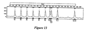
- one type of human identification is based on a short tandem repeat (STR) analysis ( Edwards et al., 1991, Am J Hum Genet 49(4)746:756 ).
- STR analysis a series of primers are utilized to amplify certain genomic regions that contain variable numbers of certain short tandem repeats. The sizes of the resulting bands are determined by nucleic acid fragment sizing (typically using capillary electrophoresis), and the size of each member of the set of STR alleles uniquely identifies an individual.
- nucleic acid fragment sizing can be used to diagnose a given disorder (e.g ., by searching for a characteristic deletion or insertion, or determining the size of nucleotide repeat regions as in Friedreich ataxia ( Pandolfo, M., 2006, Methods Mol. Med 126: 197-216 ). Fragment sizing is also useful for the identification of infectious agents; DNA fingerprinting can be utilized in pathogen diagnosis.
- Focused nucleic acid analysis can be utilized to identify biological weapons agents in clinical and environmental samples by both sequencing and fragment sizing.
- Veterinary and food testing applications also mirror those described above.
- Veterinary identification applications such as racehorse breeding and tracking, livestock breeding, and pet identification also are within the scope of the uses of the disclosed invention.
- Research applications of focused nucleic acid analysis are numerous. In short, focused nucleic acid analysis has the potential to dramatically transform several industries.
- plastics have been found to present several major obstacles for use in biochips designed for nucleic acid sequencing and fragment sizing. Autofluorescence of plastic materials interferes with the detection of wavelengths in the visible range of 450 to 800 nm ( Puriska, 2005, Lab Chip 5(12):1348 ; Wabuyele, 2001 Electrophoresis 22(18):3939-48 ; Hawkins and Yager 2003 Lab Chip, 3(4): 248-52 ).
- This invention provides inexpensive, multi-lane plastic biochips capable of performing focused nucleic acid analysis at high resolution and with a high signal to noise ratio and methods of using such chips.
- the invention provides plastic separation chips, and in particular electrophoresis chips comprising an anode portion, a cathode portion, and a center portion between the anode and cathode portions, wherein the cathode portion comprises at least one first via; the anode portion comprises at least one second via; and the center portion comprises a plurality of microfluidic channels and a detection window, each microfluidic channel having a separation region and a detection region; wherein each microfluidic channel is in fluid communication with at least one first via and at least one second via; wherein the plurality of microfluidic channels are in substantially the same plane; the plurality of microfluidic channels do not intersect one another within the center portion; the detection window comprises a thin plastic; and the detection window comprises the detection region of each microfluidic channel.
- the portions of the chip outside of the detection region can of the same thickness, or of a thickness that larger than that of the detection region.
- the invention provides devices comprising a support having a top and bottom surface, comprising an anode portion, a cathode portion, and a center portion between the anode and cathode portions, wherein the center portion comprises an aperture at the detection window, the anode portion comprises the at least one anode well, and the cathode portion comprises the at least one cathode well; the apparatus further comprising a chip according to the first aspect, having a top and bottom surface, wherein the top surface of the chip is in contact with the bottom surface of the support, the microfluidic channels are in fluid communication with the cathode and anode wells through the vias; and the chip is fixedly attached to the support.
- the invention provides methods for electrophoretically separating and detecting a plurality of samples simultaneously, comprising providing a plurality of samples into each of a plurality of microfluidic channels on a microchip according to the first aspect; applying an electric potential across the plurality of microfluidic channels to inject samples into the separation channel and to separate detectable species comprising each of the plurality of analysis samples; and detecting each of the detectable species comprising the plurality of separated samples at the detection window.
- the invention provides plastic separation chips that are capable of detecting separation of nucleic acid species differing in size by about 1 basepair, and at concentration levels of at least 1.0 ng of DNA template.
- the lowest level of sample to be analyzed for STR analysis consists of a nucleic acid template with less than 800 copies, less than 400 copies, less than 200 copies, less than 100 copies, less than 50 copies, less than 30 copies, less than 10 copies or 1 copy of nucleic acid template prior to the multiplexed PCR reaction.
- the lowest concentration sample to be analyzed for Sequencing consists of a nucleic acid template with less than 0.5 pmole, less than 0.1 pmole, less than 0.01 pmole as input to the Sanger sequencing reaction.
- injection channel means an intersecting channel that permits introduction of a sample into the microfluidic channel with which it intersects.
- the intersecting channel can be in a single cross-channel, a single T-junction, or an offset double-T junction configuration.
- fluid communication refers to two chambers, or other components or regions containing a fluid, connected together so that a fluid can flow between the two chambers, components, or regions. Therefore, two chambers which are in "fluid communication” can, for example, be connected together by a microfluidic channel between the two chambers, such that a fluid can flow freely between the two chambers. Such microfluidic channels can optionally include one or more valves therein which can be closed or occluded, in order to block and/or otherwise control fluid communication between the chambers.
- fluorescent dye means the dye, upon excitation with a light source, emits light having a wavelength of 380 - 850 nm.
- the dye emits light having a wavelength between about 450 - 800 nm; more preferably, the dye emits light having a wavelength between about 495 - 775 nm.
- autofluorescence means fluorescence produced by substances other than the fluorophore of interest under light irradiation.
- the phrase "essentially does not fluoresce” as used herein, means the background fluorescence signal (for example, between about 380 - 850 nm; 400 - 800 nm; 450 - 800 nm; 500 - 800 nm, or 495 - 775 nm) from the referenced object (e.g., solid or solution) when subjected to light irradiation (e.g., at one or more wavelengths between about 350 - 500 nm, 400 - 500 nm, or 450 - 500 nm; in particular, 488 nm; laser irradiation) has a background level that is lower than that from conventional glass microfluidic devices which consist of borofloat glass of 0.7 mm thick.
- light irradiation e.g., at one or more wavelengths between about 350 - 500 nm, 400 - 500 nm, or 450 - 500 nm; in particular, 488 nm; laser irradiation
- norbornene based polymers as used herein means a polymer prepared from at least one monomer comprising a norbornene moiety where the norbornene-containing monomers are polymerized according to ring-opening metathesis polymerization according to methods known to those skilled in the art (see, for example, U. S. Patent Nos. 4,945,135 ; 5,198,511 ; 5,312,940 ; and 5,342,909 ).
- poly(methyl methacrylate) or "PMMA,” as used herein, means the synthetic polymers of methyl methacrylate, including but not limited to, those sold under the tradenames PlexiglasTM, LimacrylTM, R-CastTM, PerspexTM, PlazcrylTM, AcrylexTM, ACryliteTM, ACrylplastTM, AltuglasTM, PolycastTM, and LuciteTM, as well as those polymers described in US Patent Nos. 5,561,208 , 5,462,995 , and 5,334,424 , each of which are hereby incorporated by reference.
- polycarbonate as used herein means a polyester of carbonic acid and glycol or a divalent phenol.
- glycols or divalent phenols are p-xylyene glycol, 2,2-bis(4-oxyphenyl)propane, bis(4-oxyphenyl)methane, 1,1-bis(4-oxyphenyl)ethane, 1,1-bis(oxyphenyl)butane, 1,1-bis(oxyphenyl)cyclohexane, 2,2-bis(oxyphenyl)butane, and mixtures thereof, including but not limited to, those sold under the tradenames CalibreTM, MakrolonTM, PanliteTM, MakroclearTM, CyrolonTM, LexanTM and TuffakTM.
- nucleic acid is intended to encompass single- and double-stranded DNA and RNA, as well as any and all forms of alternative nucleic acid containing modified bases, sugars, and backbones.
- nucleic acid thus will be understood to include, but not be limited to, single- or double-stranded DNA or RNA (and forms thereof that can be partially single-stranded or partially double-stranded), cDNA, aptamers, peptide nucleic acids (“PNA”), 2'-5' DNA (a synthetic material with a shortened backbone that has a base-spacing that matches the A conformation of DNA; 2'-5' DNA will not normally hybridize with DNA in the B form, but it will hybridize readily with RNA), and locked nucleic acids (“LNA”).
- PNA peptide nucleic acids
- Nucleic acid analogues include known analogues of natural nucleotides that have similar or improved binding, hybridization of base-pairing properties.
- "Analogous" forms of purines and pyrimidines are well known in the art, and include, but are not limited to aziridinylcytosine, 4-acetylcytosine, 5-fluorouracil, 5-bromouracil, 5-carboxymethylaminomethyl-2-thiouracil, 5-carboxymethylaminomethyluracil, inosine, N 6 -isopentenyladenine, 1-methyladenine, 1-methylpseudouracil, 1-methylguanine, 1-methylinosine, 2,2-dimethylguanine, 2-methyladenine, 2-methylguanine, 3-methylcytosine, 5-methylcytosine, N 6 -methyladenine, 7-methylguanine, 5-methylaminomethyluracil, 5-methoxyaminomethyl-2-thiouracil, beta-D
- DNA backbone analogues provided by the invention include phosphodiester, phosphorothioate, phosphorodithioate, methylphosphonate, phosphoramidate, alkyl phosphotriester, sulfamate, 3'-thioacetal, methylene(methylimino), 3'-N-carbamate, morpholino carbamate, and peptide nucleic acids (PNAs), methylphosphonate linkages or alternating methylphosphonate and phosphodiester linkages ( Strauss-Soukup, 1997, Biochemistry 36:8692-8698 ), and benzylphosphonate linkages, as discussed in US 6,664,057 ; see also OLIGONUCLEOTIDES AND ANALOGUES, A PRACTICAL APPROACH, edited by F.
- nucleic acids herein can be extracted from cells or synthetically prepared according to any means known to those skilled in the art; for example, the nucleic acids can be chemically synthesized or transcribed or reverse transcribed from cDNA or mRNA, among other sources.
- via means a through-hole formed in a solid material to allow fluidic connection between the top and bottom surfaces of the material.
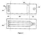
- the chip (100) comprises an anode portion (101), a cathode portion (102), and a center portion (103) between the anode and cathode portions.
- the cathode portion comprises at least one first via (104) and the anode portion comprises at least one second via (105).
- the center portion comprises a plurality of microfluidic channels (106) and a detection window (107), each microfluidic channel having a separation region and a detection region; wherein each microfluidic channel is in fluid communication with at least one first via and at least one second via.
- the plurality of microfluidic channels are substantially in the same plane and do not intersect one another within the center portion.
- Each microfluidic channel has a region in where excitation and/or detection of the sample can take place.
- the area in which encompasses the excitation and detection regions of the plurality of microfluidic channels is known as the detection window, and this window comprises a thin plastic.
- the phrase "thin plastic” as used herein means the referenced material comprises a plastic having a thickness of (its smallest dimension) less than 1 mm, less than 750 ⁇ m, less than 650 ⁇ m, less than 500 ⁇ m, less than 400 ⁇ m, less than 300 ⁇ m, less than 200 ⁇ m, or less than 100 ⁇ m; or the referenced material comprises a plastic having a thickness ranging from 25 - 2000 ⁇ m, 25 - 1000, 25 - 750 ⁇ m, 25 - 500 ⁇ m, 25 - 400 ⁇ m, 25 - 300 ⁇ m, or 25 - 200 ⁇ m.
- the chip is designed to be thin in the detection window, portions of the chip outside of the detection region can be of the same thickness, or of a thickness that is larger than that of the detection region.
- the chip of Figure 1 is shown for the sake of illustration as having four microfluidic channels, however such disclosure is not intended to be limiting, rather, one skilled in the art will readily recognize that the chip can contain alternate numbers of microfluidic channels ( infra ) including chips with one channel and chips with two or more channels.
- plural means two or more, four or more, eight or more, 16 or more, 32 or more, 48 or more, 64 or more, 96 or more, 128 or more, 256 or more, 384 or more, 512 or more, or 1024 or more; or 2 - 4, 2 - 8, 2 - 16, 2 - 32, 2 - 48, 2 - 64, 2 - 96, 2 - 128, 2 - 384, 2 - 512, 2 - 1024 microfluidic channels.
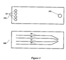
- the chip (250) comprises of a substrate layer (360) and a cover layer (370) as shown in Figure 3 .
- a plurality of grooves (361) are patterned into the substrate layer.
- a series of vias (i.e., through holes) (371, 372) are formed in the cover layer to provide fluidic access to the microfluidic channels, and can be located at the ends of the microfluidic channels in the anode and cathode portions of the chip.
- vias can be formed in the substrate layer instead of the cover layers to achieve the same functionality.
- the top surface of the substrate layer is bonded with the bottom surface of the cover layer to form the microfluidic channels.
- the present plastic separation chips can be prepared by hot embossing of thin thermoplastic films with a master die of the negative of the structure to be produced.
- the master die can be prepared by using electroforming to replicate the device prepared in a solid substrate.
- the solid substrate can be glass sheets that are patterned by standard photolithographic and chemical etching methods known to those skilled in the art.
- the substrate and cover layers are diffusion bonded by the application of heat and pressure.
- the substrate and cover layers of the chip can be constructed from a variety of plastic substrates including, but not limited to, polyethylene, poly(acrylates) (e.g., poly(methyl methacrylate)), poly(carbonate)s, and unsaturated, partially unsaturated or saturated cyclic olefin polymers (COP), or an unsaturated, partially unsaturated, or saturated cyclic olefin copolymers (COC) (e.g., ZEONORTM, ZEONEXTM or TOPASTM).
- COP and COC are advantageous for the present chip applications as they optically exhibit inherently lower autofluorescence in the visible wavelength range compared with other polymers.
- plastic substrate and cover layers utilized in the present process is kept thin to minimize autofluorescence from the chip.
- the plastic substrate and cover layers can each, independently, have a thickness of less than 2 mm, less than 1 mm, less than 750 ⁇ m, less than 650 ⁇ m, less than 500 ⁇ m, less than 400 ⁇ m, less than 300 ⁇ m, less than 200 ⁇ m, or less than 100 ⁇ m; or plastic substrate and cover layers can each, independently, comprise a plastic having a thickness ranging from 25 - 2000 ⁇ m, 25 - 1000, 25 - 750 ⁇ m, 25 - 650 mm, 25 - 500 ⁇ m, 25 - 400 ⁇ m, 25 - 300 ⁇ m, 25 - 200 ⁇ m, or 25 - 100 ⁇ m.
- the chip (250) is attached to a support (201) having a top and bottom surface, comprising an anode portion (202), a cathode portion (203), and a center portion (204) between the anode and cathode portions, wherein the center portion comprises a detection window (205), the anode portion comprises at least one anode well (206), and the cathode portion comprises at least one cathode well (207).
- the top surface of the chip, with the via holes up, is in contact with the bottom surface of the support, and the chip is fixedly attached to the support.
- the chip can be attached to the support according to methods known to those skilled in the art, for example, diffusion bonding, solvent bonding or adhesive bonding.
- the support layer can be constructed from a variety of plastic substrates including, but not limited to, polyethylene, poly(acrylates) (e.g ., poly(methyl methacrylate)), poly(carbonate)s, and unsaturated, partially unsaturated or saturated cyclic olefin polymers (COP), or an unsaturated, partially unsaturated, or saturated cyclic olefin copolymers (COC) (e.g., ZEONORTM, ZEONEXTM or TOPASTM).
- the thickness of a plastic support layers utilized in the present process is sufficiently thick in order to provide structural rigidity and to allow for sufficient volume of sample and buffers in the reservoirs.
- the thickness of the plastic support will range from 100 - 15,000 ⁇ m.
- the chip can be fabricated by patterning the grooves on the solid support to form both the chip substrate and support structures together.
- a cover layer can be bonded to the support to complete the structure.
- the thickness of a detection window of the support and chip coincident with the detection portion of the microfluidic channels is kept thin to minimize autofluorescence.
- the thickness of this portion of the chip is less than 1000 ⁇ m, less than 750 ⁇ m, less than 500 ⁇ m or less than 250 ⁇ m; or ranging from 25 -1000 ⁇ m, 25 - 750 ⁇ m, or 25 - 500 ⁇ m.
- Each of the plurality of microfluidic channels can have a depth of at least 10 ⁇ m, 50 ⁇ m, 100 ⁇ m, 200 ⁇ m, 500 ⁇ m or 1 mm; or have a depth ranging from 1 - 1000 ⁇ m, 10 - 100 ⁇ m, 10 - 50, or 25 - 50 ⁇ m.
- the plurality of microfluidic channels can have a width of at least 25 ⁇ m, 50 ⁇ m, 100 ⁇ m, 200 ⁇ m, 500 ⁇ m or 1 mm; or have a width ranging from 25 - 1000 ⁇ m, 25 - 200 ⁇ m, or 50 - 200 ⁇ m.
- microchannel cross-section of each channel can have a substantially square, rectangular, circular, semicircular, elliptical, triangular or trapezoidal cross-section.
- microfluidic channels may or may not be uniform in depth, width and cross-section.
- Each of the plurality of microfluidic channels (106) comprises a separation region (108) and a detection region (109).
- the separation region typically has channels with separation length of about 2 - 50 cm, 10 - 50 cm, 2 - 25 cm, 10 - 25 cm.
- the separation length is defined as the portion of the channel between the point of sample injection and the point of sample detection.
- the separation length is typically less than the total length of the separation channel which spans between the cathode and the anode reservoirs.
- Simultaneous analysis of a plurality of samples can be performed by injecting and stacking each of the samples in a separate separation channel into any of the separation chips described herein.
- the application of an electric field along the separation channel causes the samples to migrate along the channel from the cathode portion toward the anode portion or the anode portion to the cathode portion of the separation channel, depending, for example, on the charges present on the surfaces of the channel ( infra ), as will be familiar to those skilled in the art.
- Migration of the sample through a sieving matrix separates species on the basis of size.
- the detection window typically overlaps the detection region of each of the plurality of microchannel at the termini of the separation region of each of the channels.
- the detection region for each of the plurality of microfluidic channels are in substantially the same location along the channels, such that the detection window can be in a single location in the center portion of the support.
- An injector for simultaneously injecting a plurality samples into the plurality of sample or buffer wells is advantageously provided with the chip to enable simultaneous multiple sample separation and detection.
- Such injectors provide, for example, one sample of the plurality of samples to one microfluidic channel of the plurality of microfluidic channels.
- injectors can introduce the samples to the channels according to any methods known to those skilled in the art, for example, by electrophoretic transport, pneumatic actuation or liquid actuation through a needle or tube or channel that connects the sample to the separation channel
- samples can be loaded into the chip through the cathode reservoirs of the chip.
- An injection volume of each sample can be introduced through one of the cathode wells according to methods known to those skilled in the art.
- the sample can be injected via appropriate biasing of the separation channel and/or a cross-channel of the separation channel and the sample and waste wells such that a portion of the sample ( i.e., the injection volume) in the sample well is provided to the separation channel.
- additional buffer solution is introduced into each cathode well; sufficient volume can be provided to dilute any remaining sample in the well.
- a volume of buffer is introduced into the cathode wells that is about at least 5, 10, 25, 50, or 100 times the injection volume of the sample.
- a volume of buffer is introduced into the cathode wells that ranges from about 5 - 100 times, 5 - 50 times, or 10 - 50 times the injection volume of the sample.
- each of the plurality of microfluidic channels further comprises an injection channel for introducing samples.
- FIG 4 shown therein is an expanded view of a chip (400) showing the cathode portion (401) and adjoining section of the center portion (403).
- the cathode portion comprises at least one second via (405) and the center portion comprises a plurality of microfluidic channels (406).
- Each microfluidic channel further comprises, within the cathode portion of the chip, an injection channel (408) comprising a sample (409) and waste (410) well for each microfluidic channel.
- the injection channel can be in a single cross-channel (as illustrated in Figure 4 ), a single T-junction, or an offset double-T junction configuration.
- the injection channel is an offset double-T junction configuration that minimizes the injection volume of sample, thereby improving separation resolution.
- Injection of a sample from the injection channel to the microfluidic channel can be accomplished according to methods known to those skilled in the art, including electophoretic injection through application of the appropriate potentials at the sample, waste, anode and cathode wells.
- the chip (500) comprises an anode portion (501), a cathode portion (502), and a center portion (503).
- the cathode portion comprises one first via (504) for each microfluidic channel (506) and the anode portion comprises at least one second via (505) for each microfluidic channel (506).
- the center portion comprises a plurality of microfluidic channels (506) and a detection window (507), each microfluidic channel having a separation region and a detection region; wherein each microfluidic channel is in fluid communication with one first via and one second via.
- the plurality of microfluidic channels are essentially in the same plane and do not intersect one another within the center portion.
- the detection window comprises a thin plastic and overlaps the detection region of each microfluidic channel.
- the injection channels are omitted in favor of an anode (second) and cathode (first) via for each microfluidic channel.
- An injection volume of each sample is introduced through one of the cathode via according to methods known to those skilled in the art ( supra ) .
- additional buffer solution is introduced into each cathode buffer well; sufficient volume is advantageously provided to dilute any remaining sample in the well, thereby mediating any background signal introduced from prolonged sample injection and improving the signal-to-noise ratio observed at the detection window.
- a volume of buffer is introduced into the cathode wells that is about at least 5, 10, 25, 50, or 100 times the injection volume of the sample.
- a volume of buffer is introduced into the anode buffer wells that ranges from about 5 - 100 times, 5 - 50 times, or 10 - 50 times the injection volume of the sample.
- Electrophoretic separation of the samples within the microfluidic channels is provided by the application of a potential difference across the microchannels on the microchip.
- a high voltage can be applied across the ends of the microchannels, typically by placing a cathode and anode in the cathode well and the anode well, respectively, establishing an electric field along the separation portion of the microfluidic channel, and moving the sample (e . g ., nucleic acid) from the cathode end through the separation portion to the detection portion, and ultimately, to the anode.
- the electric field required for efficient separation often range from 50 V/cm to 600 V/cm.
- the voltage from the power supply is applied through electrodes and a buffer is used in the anode and cathode reservoir to provide electrical contact between the electrode and the sieving polymer.
- High voltages required for sample separation are applied to the separation channel with electrodes that are in contact with the buffer that is in the cathode and anode wells. Due to the high voltages present at the electrodes that are in contact with the buffer, the buffer water molecules hydrolyze resulting in the formation of OH - , H + , and H 2 gas. This formation results in a change in the pH of the buffer with time, and formation of bubbles within the buffer.
- the pH change of the buffer can be attenuated through sufficient use of a buffer solution in the anode and cathode reservoirs (e.g., 1X TTE; Amresco) to provide contact between the electrode and the sieving matrix.
- the bubbles formed within the buffer have a tendency to migrate into the sieving matrix blocking the channel, resulting in poor separation of nucleic acids.
- Bubbles that form at the electrode can be prevented from migrating into the channel by using one or a combination of the following methods.
- the electrode within the reservoir can be raised to move the source of bubble generation (electrode) away from the access holes in the channels.
- a glass frit, polymer frit, or polymer membrane or polymer filter can be inserted between the cathode access hole and the end of the electrode.
- a polymer frit e.g ., polyetheretherketone, PEEK
- PEEK polyetheretherketone
- the frit, membrane, or filter is selected to be non-conducting and have a pore size that prevents bubbles formed at the electrode from passing through the pores.
- a pore size that prevents bubbles formed at the electrode from passing through the pores.
- Separation devices for simultaneous analysis consisting of a plurality of samples are electrically connected so that a common power supply can be used to bias the plurality of channels simultaneously. Furthermore, physical constraints of the chip and instrument will usually not allow all the channels to have an identical physical layout with respect to length, depth and width.
- each channel segment of the individual devices should have essentially identical resistances and hence electric fields.
- a substantially identical electric field that is wherein the electric fields across each of the plurality of microfluidic channels does not differ by more than about +/- 5%, can be established by simultaneously adjusting both the length, width and depths of each of the plurality of microfluidic channels to adjust the resistance of each segment of the channels.
- HPMC hydroxypropylmethylcellulose
- PEO poly(ethylene oxide)
- PVA poly(vinyl alcohol)
- PDMA polydimethyl acrylamide
- DEA dimethylacrylamide
- DEA diethylacrylamide
- PDEA poly(diethylacrylamide)
- each of the plurality of microfluidic channels is advantageously filled with a sieving matrix.
- sieving matrices can comprise, in non-limiting example, a linear polyacrylamide (PAA), polydimethylacrylamide (PDMA), polydiethylacrylamide (PDEA), polyvinylpyrrolydinone (PVP), and combinations thereof, including for example, PVP:PAA, PDMA:PAA, PDEA:PAA, PDEA:PDMA:PAA.
- the sieving matrix comprises 0.1 - 50 wt.% polyacrylamide. A number of these sieving matrices also possess dynamic self-coating capability.
- nucleic acids move electrophoretically through a sieving matrix from the anode to the cathode end and are size-separated therein.
- the inner walls of the channels can be coated to minimize the influence of electroosmosis and nucleic acid-to-wall interactions.
- Resolution is the ability to unambiguously discriminate two peaks separated in time (or by base size).
- Single base pair resolution is defined at the point where R is greater than 0.4. Visually, two peaks are distinguishable from each other when the peak to valley ratio is greater than 0.7.
- Both R and peak-to-valley requirements must be met in order to have high resolution, and resolution can also be considered to be characteristic of a range of fragment sizes.
- the range of fragment sizes for alleles in STR analysis range from 90 to 400 bp and single basepair resolution across this range of fragment size is required for STR analysis.
- Fragment sizes for sequencing analysis range up to 1200 bp.
- the ability to achieve long read lengths and data throughput per lane is, in part, determined by the range over which the chip is able to generate single base-pair resolution.
- the limit-of-detection for an optical detection system is defined by the signal to noise ratio (SNR). This ratio is defined as the ratio of a signal power to the noise power (standard deviation of noise power) corrupting the signal. A high SNR indicates a higher certainty that a signal is present. A signal-to-noise ratio of 3 is generally defined as that which is acceptable for confidently identifying the presence of a signal ( Gilder, 2007, J Forensic Sci. 52(1): 97 ).
- an advantageous characteristic of the electrophoretic separation chips of the invention is that a thin detection window is used to minimize the background fluorescence from the plastic. This background level is compared to borofloat ® , which is a commonly-used substrate for fabricating microfluidic separation chips.
- borofloat ® which is a commonly-used substrate for fabricating microfluidic separation chips.
- the instruments of this invention can be ruggedized, and thereby operated in the field where results can be used in real-time.
- the instruments can be used at military checkpoints, borders and ports, airports, and mass casualty sites.
- nucleic acid sequencing can be divided into four areas: human clinical diagnostics, including, for example, bacterial infections and antibody sensitivities, viral infections (identification and drug resistance profiling), genetic diseases, complex disorders (asthma, heart disease, diabetes) and pharmacogenomics; veterinary clinical diagnostics; research sequencing, including re-sequencing and finishing; biological weapons agent identification, including, for example, B. anthracis and Ebola virus detection; and food safety.
- a patient with HIV needs drug resistance testing. Today, it can take weeks to establish resistance. A drug resistant strain can take hold during that time. There is an unmet need for an instrument and system that can provide the answer within 1-2 hours, while the patient waits in the physician's office.
- Use of an electrophoretic separation chip according to the invention permits frequent drug-resistance monitoring, more clinically- and cost effective usage of anti-viral agents and better patient outcomes.
- a patient with bacteremia is in shock. Today, it can take days to determine whether the causative agent is resistant to antibiotics and the identities thereof. In the interim, the patient must be treated with broad-spectrum antibiotics, which can cause serious side-effects to the patient and contributes to the increase in antibacterial resistance prevalent today. In addition, such treatments may be sub-optimal.
- Use of an electrophoretic separation chip according to the invention permits identification of the antibiotic resistance profile of the pathogen in 1-2 hours, leading to more effective, targeted treatment, reduction in antibiotic toxicities, and better patient outcomes. The benefits to the patient and to public health are complimentary.
- a patient with cancer is undergoing surgery.
- a tumor sample is taken to pathology while the patient is on the operating table.
- a decision is made concerning how aggressive the surgeon should be.
- Use of an electrophoretic separation chip according to the invention could replace histopathology with a definitive nucleic acid diagnosis of the cancer in less than an hour, allowing a better-informed surgical decision to be made.
- FIG. 6 A schematic diagram of a particular embodiment of the devices of the invention is illustrated in Figure 6 .
- This microfluidic device consisted of 16 microchannels, each with a double-T cross injector.
- the cross-sectional dimension of the channel (90 ⁇ m wide and 40 ⁇ m deep) and length of the channel between the anode and the cross-injector (25 cm) was equal for all channels.
- the separation lengths (distance between the intersection and the excitation/detection window) for each of the channels range from 16 to 20 cm long.
- the cross-sectional area of the channels between the cathode well and the injector was adjusted such that all the resistances and hence electric fields between the cathode and the intersection are essentially equal under bias.
- Example 1B Chip and Support fabrication
- the chip was patterned by hot-embossing, drilling to form access holes, and diffusion bonding to seal the channels.
- the master was fabricated in glass by photolithography, using a chemical wet etching process. This glass master was then used to fabricate a nickel-cobalt embossing tool by electroforming to generate a negative replicate of the glass master. Sheets of ZenorTM-1420R film (5" x 2" in size and 188 ⁇ m thick) were used as the substrate material. On these sheets, cathode, anode, sample and waste access holes were formed by drilling. This was followed by hot embossing the chip design features on the embossing tool into the substrate.
- Embossing was accomplished by placing the stack as illustrated in Figure 7 in a heated hydraulic press for 15 minutes at 135 °C and 1250 psi of compressive pressure. The stack was held under 1250 psi of compressive pressure and allowed to cool to 38 °C prior to release. Fabrication of this chip with thin thermoplastic polymers containing norbornene monomers resulted in a low background fluorescence at excitation and detection window. Achieving high bond strength diffusion bonding allowed the use of high viscosity sieving matrices.
- Diffusion bonding of the substrate was achieved by the aligning a sheet of ZenorTM-1420R film (5" x 2" in size and 188 ⁇ m thick) over the substrate and subjecting this stack to heat and pressure. No adhesive was applied between the sheets of film; bonding was accomplished entirely by heat and pressure. The final thickness of the chip was approximately 376 ⁇ m. Separation chips fabricated by this method were tested and demonstrated to be capable of withstanding at least 830 psi of pressure before failure.
- FIG 8 illustrates a chip support that was fabricated by CNC milling from a 3/8" thick acrylic sheet (GE Plastic).
- the chip support consisted of three main sections: the cathode board, the center part and the anode board.
- the cathode board contained the cathode well, sample and waste wells, and alignment holes.
- the anode board contained the anode well and alignment holes. Both the cathode board and the anode board were 3/8" thick to provide enough sample volume for sample injection and buffer volume for electrophoresis.
- the center portion was 0.04" thick and had an opening as the "detection window" for laser induced fluorescence detection in the microchannels. With this configuration, autofluorescence from the separation chip becomes dominated by the approximately 376 ⁇ m thick substrate.
- the separation chip was attached to the chip support with double-sided pressure sensitive adhesive.
- the adhesive was selected to be inert to the separation buffers and sieving matrices.
- the support and separation chip were attached with pressure sensitive epoxy. The thickness of the plastic in the excitation and detection area was minimized by fabricating a cut-out on the carrier in this region.
- the optical emission spectrum of ZenorTM-1420R has the Raman emission peak at 570 nm which has limited fluorescence detection with fluorescent dyes.
- Figure 9 demonstrates low autofluorescence of the plastic chip (PChipl and PChp2) compared with typical glass separation chips (Glass 1.4 mm and Glass 0.7 mm). The low autofluorescence of the plastic chip was achieved by selecting a COP polymer and minimizing the thickness of the device in the detection area and by fabricating the device with thin films.
- Example 1C Surface modification and sieving matrix
- Example 1D Electrophoresis STR sizing
- Electrophoretic separation and analysis of nucleic acid analysis was performed on the Genebench-FXTM Series 100 (Network Biosystems, Inc., Woburn, MA). This instrument was configured to accept the plastic separation chip and chip support to allow for good optical, electrical and thermal coupling between chip and instrument. The temperature of the chamber was maintained at 50°C throughout the operation.
- PCR product (2.7 ⁇ L) was mixed with 0.3 ⁇ L of sizing standard and 10 ⁇ L of formamide, and loaded into the sample wells for analysis.
- the assay consisted of pre-electrophoresis performed at 156 V/cm for 6 minutes prior to sample introduction by applying a potential difference of 3900 V at the anode well and grounding the cathode well.
- DNA samples were introduced by applying an electric field of 350 V/cm for 18 seconds, followed by a dual load of 1.2 minute by applying an electric field of 350 V/cm across the sample and waste wells and simultaneously applying an electric field of 15.6 V/cm across the cathode and anode wells.
- electrophoretic DNA separation was performed by applying an electric field of 156 V/cm across the cathode and anode well while maintaining a pullback voltage of 800 V for 40 minutes.
- M13 plasmid was cycle-sequenced with the GE Amersham DYEnamicTM ET dye terminator cycle sequencing kit (GE Healthcare), ethanol precipitated and resuspended in 10 ⁇ L deionized water.
- the separation assay consisted of a pre-electrophoresis performed at 156 V/cm for 6 minutes prior to sample introduction by applying a potential difference of 3900 V at the anode well and grounding the cathode well.
- DNA sample was introduced by applying an electric field of 350 V/cm for 60 seconds.
- electrophoretic DNA separation was performed by applying an electric field of 156 V/cm across the cathode and anode well while maintaining a pullback voltage of 400 V for 60 minutes.
- DNA separation resolution was calculated by extracting the peak information (peak spacing and peak width) from Peakfit ® .
- Figure 10 shows allele called profile for the allelic ladder from a 5-color labeled kit (ABI AmpFlSTR Identifiler kit). These results demonstrated that devices of the invention were able to separate with 5 colors in a plastic chip and clearly resolve alleles including ones that are spaced by a distance equivalent to only a single base pair (THO 1, allele 9.3 and 10).
- Figure 11 shows an allele called STR profile for 9947A human genomic DNA, showing that a full profile was achieved at 1.0 ng of DNA template.
- Figure 12 shows the resolution with R>0.4 for up to 480 bp, demonstrating single-base resolution up to 480 bp.
- Figure 13 illustrates this resolution by showing 2 alleles that are spaced by 1 nucleotide can be clearly resolved with no ambiguity.
- Figures 14 and 15 shows a DNA sequencing profile demonstrating single basepair resolution.
- FIG. 16 Another configuration of the electrophoretic separation chips of the invention uses a single channel for separation. Each sample is introduced into a separation channel by electrokinetic sample injection. This alternative approach allows for the use of small sample volumes and a significant simplification in the separation process.
- a schematic diagram of chip design for electrokinetic sample injection is shown in a breakaway view in Figure 16 , showing the support and separation chip sections. The device consists of 16 microchannels that are effectively 20 cm in separation length. Each channel has an access hole at each end. The channels are 90 ⁇ m wide and 40 ⁇ m deep.
- the device of Figure 16 is fabricated following the procedure described in the section above.
- access holes (1 mm in diameter) are formed in a COP film (ZeonorTM-1420R) with a thickness of 188 ⁇ m.
- Channel patterns (width 90 ⁇ m and depth 40 ⁇ m) are then formed by hot embossing.
- a cover of COP (ZeonorTM-1420R) is diffusion bonded to the substrate to seal the channels.
- the device is prepared for separation by applying a surface modification to the channels as described in the section above. This is followed by filling the channels with a sieving matrix. Samples are loaded into the sample/cathode reservoir. An injection field is applied through the electrodes to the sample to inject negatively charged DNA into the separation channel. Following injection of DNA into the channels, buffer (1X TTE; Ameresco) is added to the sample/cathode reservoir at a volume 10 times the volume of the sample. An electric field is applied across the cathode and anode to separate the DNA from the injection plug down the separation channel. The addition serves to dilute the sample that is in the sample/cathode and there is no need to remove the sample prior to loading the buffer. Separation and detection is performed on a Genebench-FXTM Series 100 instrument, and data analysis is performed with the software described in the previous examples.
- DNA template is amplified in a reaction mix consisting of PCR enzyme SpeedSTAR HS (Takara, Madison, WI) (U/ ⁇ L): 0.025, Fast Buffer 1: 1x, dNTPs : 0.25 mM, Primer (forward): 250n M, and Primer (reverse): 250 nM.
- a desired level of template DNA is added to the mix.
- DI water or TE buffer Tris 10 mM or EDTA 0.1 mM
- Thermal cycling of the PCR reaction mix following manufacturer's recommended protocols, consists of hot start activation of 60 seconds at 95 °C, 30 cycles of denaturation, anneal and extension (5 seconds at 98 °C, 10-15 seconds at 55 °C and 5-10 seconds/kbp at 72 °C) and a final extension of 60 seconds at 72 °C.
- the entire PCR product is cleaned up by using a 30K MWCO UF filter (Pall, East Hills, NY), following manufacturers protocol.
- the cleaned up product with consisting of DNA in DI water, is either diluted or applied in its entirety as template for the sequencing reaction.
- Cycle sequencing of PCR template was performed using the DYEnamicTM ET Terminator Cycle Sequencing Kit (GE Amersham Biosciences) at half strength reaction with the following reaction mix. Sequencing Premix: 4 ⁇ L, Dilution Buffer: 4 ⁇ L, Primer (10 ⁇ M): 5 pmol. DNA template was added to the sequencing reaction mix. DI water was added to the reaction mix to a total volume of 20 ⁇ L. Following manufacturer's recommended cycling protocols the cycling condition used consists of thirty cycles of (20 seconds at 95 °C, 15 seconds at 50 °C, 60 seconds at 60 °C).
- the sequencing reaction mix is cleaned up by ethanol precipitation.
- the precipitated product is resuspended in 13 ⁇ L of DI water and used as sample for separation and detection.
- amplification is carried out in 10 ⁇ L reactions with the following reaction mix consisting of: PCR enzyme SpeedSTAR HS (Takara, Madison, WI) (U/ ⁇ L): 0.0315, Fast Buffer 1: 1x, Primer set: 2 ⁇ l, Fast Buffer 1: 1X, dNTPs : 200 ⁇ M, Primer (forward/Reverse): 2 ⁇ L from AmpFlSTR ProfilerTM, COFilerTM or IdentifilerTM (Applied Biosystems, Foster City, CA).
- the cycling protocol follows enzyme manufacturers conditions consisting of a hot start activation of 60 seconds at 95 °C followed by 28 cycles of denaturation, anneal and extension (4 s at 98 °C, 15 s at 59 °C, 5 s at 72 °C) and a final extension of 60 seconds at 72 °C.
- PCR product is used as sample for separation and detection.
- the PCR product can also be purified and used as sample for separation and detection.
Abstract
Description
- This application claims the benefit of the filing date, under 35 U.S.C. §119(e), of
U.S. Provisional Application Serial No. 60/921,802, filed 4 April 2007 U.S. Provisional Application Serial No. 60/964,502 filed 13 August 2007 U.S. Provisional Application Serial No. 61/028,073, filed February 12, 2008 - This invention is in the field of nucleic acid sequencing and fragment sizing by electrophoresis with detection by laser-induced fluorescence. The analysis is performed on plastic electrophoresis chips.
- Since the advent of DNA sequencing technologies in the 1970's (Maxam & Gilbert, 1977, Proc Natl Acad Sci USA 74: 560-564; Sanger et al., 1977, Proc Natl Acad Sci USA 74: 5463-5467), a wide range of applications making use of these technologies has developed. In parallel, increasingly sophisticated instrumentation to perform DNA sequencing has been introduced. For example, in 1986, Applied Biosystems commercialized an automated DNA sequencer based on separation of DNA fragments generated by the Sanger sequencing method; DNA fragments were labeled with a set of four fluorescent dyes and separated by capillary electrophoresis (Smith et al., 1986, Nature 321: 674-679). As a result, Sanger sequencing has been the most widely utilized sequencing technology for the last three decades.
- More recently, a variety of new sequencing technologies and related instrumentation have been and continue to be developed. Termed "next generation" methods (reviewed in Metzker, 2005, Genome Research 15: 1767-1776), these chemistries include pyrosequencing, sequencing-by-ligation, and single molecule sequencing. A major goal driving research into next generation sequencing technologies is to perform high-throughput genomic sequencing in general, and to reduce the cost of obtaining a complete genome sequence in particular. Although the cost per base pair of next-generation technologies may be less in some cases than that of Sanger sequencing, all these methods (including Sanger) are costly and require substantial time, labor, and laboratory equipment.
- The current emphasis on obtaining very large amounts of sequence data from a given genome does not negate the value of obtaining relatively small amounts of genomic sequence quickly. For example, many common human diseases can be diagnosed based on less than 1000 base pairs of DNA sequences, orders of magnitude less than required to generate a complete human genome. Similarly, precise determination of the sizes of sets of less than 20 specific DNA fragments generated by short tandem repeat analysis is sufficient to identify a given individual.
- There is an unmet need for the development of instruments and technologies that would permit focused nucleic acid analysis, defined as the rapid identification (by nucleic acid sequencing or fragment sizing) of a subset of a given human, animal or pathogen genome. Focused nucleic acid analysis will enable end-users to make near-real time clinical, forensic, or other decisions. Depending on the application, focused nucleic acid analysis may be performed in a variety of settings, including hospital laboratories, physician's offices, the bedside, or, in the case of forensic or environmental applications, in the field.
- With respect to nucleic acid (DNA and RNA) sequencing, clinical applications include diagnosis of bacterial, fungal, and viral diseases (including the determination of drug resistance profiles of the organisms), cancer (including the determination of responsiveness to chemotherapeutic regimens), and inherited and other common diseases (including the determination of responsiveness to medications). Focused nucleic acid sequencing is also well suited for pharmacogenomic analysis and certain forensic applications (including, for example, mitochondrial DNA sequencing).
- With respect to nucleic acid fragment sizing, focused nucleic acid analysis can be utilized in forensic and clinical applications. For example, one type of human identification is based on a short tandem repeat (STR) analysis (Edwards et al., 1991, Am J Hum Genet 49(4)746:756). In STR analysis, a series of primers are utilized to amplify certain genomic regions that contain variable numbers of certain short tandem repeats. The sizes of the resulting bands are determined by nucleic acid fragment sizing (typically using capillary electrophoresis), and the size of each member of the set of STR alleles uniquely identifies an individual. STR typing has become the worldwide standard for human forensic genetic identification and is the only biometric technology that allows identification of an individual as well as genetic relatives of that individual. In clinical applications, nucleic acid fragment sizing can be used to diagnose a given disorder (e.g., by searching for a characteristic deletion or insertion, or determining the size of nucleotide repeat regions as in Friedreich ataxia (Pandolfo, M., 2006, Methods Mol. Med 126: 197-216). Fragment sizing is also useful for the identification of infectious agents; DNA fingerprinting can be utilized in pathogen diagnosis.
- The applications of focused nucleic acid analysis are not limited to those discussed above. Focused nucleic acid analysis can be utilized to identify biological weapons agents in clinical and environmental samples by both sequencing and fragment sizing. Veterinary and food testing applications also mirror those described above. Veterinary identification applications such as racehorse breeding and tracking, livestock breeding, and pet identification also are within the scope of the uses of the disclosed invention. Research applications of focused nucleic acid analysis are numerous. In short, focused nucleic acid analysis has the potential to dramatically transform several industries.
- The existing high throughput capillary-based sequencers and the next generation sequencers are not capable of performing focused nucleic acid analysis in a timely and cost-effective fashion. The economies of scale sought by those technologies are driven by reducing the costs of obtaining and analyzing very large amounts of sequence data. For instruments and systems capable of focused nucleic acid analysis to make their way into routine use, they should be designed to possess certain "ideal" properties and features. In particular, the instruments and systems should generate results rapidly (ideally within minutes) to allow the generation of actionable data as quickly as possible. They should be easy to operate and reagents and consumables should be inexpensive. In addition, for some applications it is useful for nucleic acid separations to be performed in disposables; this dramatically reduces the possibility of sample contamination. To achieve these properties, polymer-based biochips are better suited as separation substrates than other materials such as glass and silicon.
- An attempt to achieve DNA fragment sizing on plastic chips was reported by McCormick (Anal Chem 69(14):2626 1997) showing the separation of HaeIII restriction fragments of ΦX174 RF DNA. The separations were performed with single samples in single lane chips, but nevertheless exhibited poor resolution separations and poor sensitivity. Furthermore, the system was only able to detect emission from a single fluorophore. Sassi (J Chromatogr A, 894(1-2):203 2000) reported the use of acrylic chips consisting of 16 fluidically isolated separation lanes for STR sizing, but this approach also showed poor resolving power and low sensitivity. This low system sensitivity prevented the detection of allelic ladders (internal sizing standards strictly required in forensic analysis) when performing simultaneous 16-lane separation and detection. The use of a 2 Hz scanning rate, representing an attempt to increase the signal to noise ratio of the system, caused degradation of both resolving power and precision. Finally, the system was only able to detect emission from a single fluorophore. Shi (Electrophoresis 24(19-20):3371 2003 and Shi, 2006, Electrophoresis 27(10):3703) reported 2- and 4-color separation and detection in single sample, single lane plastic separation devices. While the 4.5 cm channel was reported to provide single base resolution, in actuality the resolution is poor as evidenced by the appearance of alleles spaced one base pair apart (the peak-to-valley ratio of the TH01 9.3 and 10 alleles approaches one). Devices with longer separation channels (6, 10 and 18-cm) were used in this study to achieve higher resolution for analysis compared to the 4.5 cm devices. Resolution of the 10 and 18-cm long devices were limited as the devices delaminated when sieving matrices compositions optimized for resolution were used.
- In practice, plastics have been found to present several major obstacles for use in biochips designed for nucleic acid sequencing and fragment sizing. Autofluorescence of plastic materials interferes with the detection of wavelengths in the visible range of 450 to 800 nm (Puriska, 2005, Lab Chip 5(12):1348; Wabuyele, 2001 Electrophoresis 22(18):3939-48; Hawkins and Yager 2003 Lab Chip, 3(4): 248-52).
- These wavelengths are used in commercial kits for Sanger sequencing and STR sizing. Furthermore, existing plastic devices have low bonding strengths to commonly-used substrates and poor performance results with commonly-used sieving matrices. Finally, inner surfaces of the channel interact with sieving matrices and the DNA samples resulting in poor resolution due to electroosmotic flow and DNA-to-wall interactions (Kan, 2004, Electrophoresis 25(21-22):3564).
- Accordingly, there is a substantial unmet need for an inexpensive, multi-lane plastic biochip capable of performing focused nucleic acid analysis at high resolution and with a high signal to noise ratio.
- This invention provides inexpensive, multi-lane plastic biochips capable of performing focused nucleic acid analysis at high resolution and with a high signal to noise ratio and methods of using such chips.
- In a first aspect, the invention provides plastic separation chips, and in particular electrophoresis chips comprising an anode portion, a cathode portion, and a center portion between the anode and cathode portions, wherein the cathode portion comprises at least one first via; the anode portion comprises at least one second via; and the center portion comprises a plurality of microfluidic channels and a detection window, each microfluidic channel having a separation region and a detection region; wherein each microfluidic channel is in fluid communication with at least one first via and at least one second via; wherein the plurality of microfluidic channels are in substantially the same plane; the plurality of microfluidic channels do not intersect one another within the center portion; the detection window comprises a thin plastic; and the detection window comprises the detection region of each microfluidic channel. The portions of the chip outside of the detection region can of the same thickness, or of a thickness that larger than that of the detection region.
- In a second aspect, the invention provides devices comprising a support having a top and bottom surface, comprising an anode portion, a cathode portion, and a center portion between the anode and cathode portions, wherein the center portion comprises an aperture at the detection window, the anode portion comprises the at least one anode well, and the cathode portion comprises the at least one cathode well; the apparatus further comprising a chip according to the first aspect, having a top and bottom surface, wherein the top surface of the chip is in contact with the bottom surface of the support, the microfluidic channels are in fluid communication with the cathode and anode wells through the vias; and the chip is fixedly attached to the support.
- In a third aspect, the invention provides methods for electrophoretically separating and detecting a plurality of samples simultaneously, comprising providing a plurality of samples into each of a plurality of microfluidic channels on a microchip according to the first aspect; applying an electric potential across the plurality of microfluidic channels to inject samples into the separation channel and to separate detectable species comprising each of the plurality of analysis samples; and detecting each of the detectable species comprising the plurality of separated samples at the detection window.
- Specific preferred embodiments of the present invention will become evident from the following more detailed description of certain preferred embodiments and the claims.
-
-
Figure 1 illustrates a microfluidic separation and detection chip according to the various embodiments of the invention. -
Figure 2 illustrates separate support and chip layers which can be used to construct a microfluidic separation and detection chip according to the various embodiments of the invention. -
Figure 3 illustrates separate device layers which can be used to construct a microfluidic separation and detection chip according to the various embodiments of the invention. -
Figure 4 illustrates an expanded view of the anode section of a microfluidic separation and detection chip according to the various embodiments of the invention. -
Figure 5 illustrates a microfluidic separation and detection chip having injection channels according to the various embodiments of the invention -
Figure 6 is a schematic diagram of a microfluidic separation and detection chip according to the various embodiments of the invention. -
Figure 7 illustrates the stack utilized for embossing. -
Figure 8 illustrates a chip support that is fabricated by CNC milling from a 3/8" thick acrylic sheet (GE Plastic); (top) top view; bottom (side view). -
Figure 9 is a fluorescence spectra demonstrating the low autofluorescence of the plastic chip compared to typical glass separation chips; (a) Assembled plastic chip (Pchip2); (b) Assembled plastic chip (Pchip1); (c) plastic cover layer only; (d) glass chip, 1.4 mm thick; (e) glass chip 0.7 mm thick; (f) plastic substrate only. -
Figure 10 is an allele-called profile for the allelic ladder from a 5-color labeled kit (ABI AmpF1STR Identifiler kit); top to bottom: blue, green, yellow, red, orange detector signals. -
Figure 11 is an allele-called STR profile for 9947A human genomic DNA, top to bottom: blue, green, yellow, red, orange detector signals; full profile is achieved at 1.0 ng of DNA template. -
Figure 12 shows the resolution with R>0.4 for to 480 bp, demonstrating that single base resolution to 480 bp; top to bottom: blue, green, yellow, red, orange detector signals. -
Figure 13 shows the resolution of 2 alleles (THO1 9.3 and 10) that are separated by 1 nucleotide. -
Figure 14 is a DNA sequencing analysis of pGEM fragment; top to bottom: blue, green, yellow, and red detector signals. -
Figure 15 is a composite four base-pair graph showing a DNA sequencing analysis of a pGEM fragment. -
Figure 16 is a breakaway schematic diagram of a chip design for direct electrokinetic sample injection showing the support (upper) and chip (bottom) layers. - The invention provides plastic separation chips that are capable of detecting separation of nucleic acid species differing in size by about 1 basepair, and at concentration levels of at least 1.0 ng of DNA template.
- The lowest level of sample to be analyzed for STR analysis consists of a nucleic acid template with less than 800 copies, less than 400 copies, less than 200 copies, less than 100 copies, less than 50 copies, less than 30 copies, less than 10 copies or 1 copy of nucleic acid template prior to the multiplexed PCR reaction. The lowest concentration sample to be analyzed for Sequencing consists of a nucleic acid template with less than 0.5 pmole, less than 0.1 pmole, less than 0.01 pmole as input to the Sanger sequencing reaction.
- The phrase "injection channel" as used herein, means an intersecting channel that permits introduction of a sample into the microfluidic channel with which it intersects. The intersecting channel can be in a single cross-channel, a single T-junction, or an offset double-T junction configuration.
- The phrase "fluid communication" as used herein, refers to two chambers, or other components or regions containing a fluid, connected together so that a fluid can flow between the two chambers, components, or regions. Therefore, two chambers which are in "fluid communication" can, for example, be connected together by a microfluidic channel between the two chambers, such that a fluid can flow freely between the two chambers. Such microfluidic channels can optionally include one or more valves therein which can be closed or occluded, in order to block and/or otherwise control fluid communication between the chambers.
- The phrase "fluorescent dye" as used herein, means the dye, upon excitation with a light source, emits light having a wavelength of 380 - 850 nm. Preferably, the dye emits light having a wavelength between about 450 - 800 nm; more preferably, the dye emits light having a wavelength between about 495 - 775 nm.
- The term "autofluorescence" as used herein, means fluorescence produced by substances other than the fluorophore of interest under light irradiation.
- The phrase "essentially does not fluoresce" as used herein, means the background fluorescence signal (for example, between about 380 - 850 nm; 400 - 800 nm; 450 - 800 nm; 500 - 800 nm, or 495 - 775 nm) from the referenced object (e.g., solid or solution) when subjected to light irradiation (e.g., at one or more wavelengths between about 350 - 500 nm, 400 - 500 nm, or 450 - 500 nm; in particular, 488 nm; laser irradiation) has a background level that is lower than that from conventional glass microfluidic devices which consist of borofloat glass of 0.7 mm thick.
- The term "norbornene based polymers" as used herein means a polymer prepared from at least one monomer comprising a norbornene moiety where the norbornene-containing monomers are polymerized according to ring-opening metathesis polymerization according to methods known to those skilled in the art (see, for example,
U. S. Patent Nos. 4,945,135 ;5,198,511 ;5,312,940 ; and5,342,909 ). - The term "poly(methyl methacrylate) or "PMMA," as used herein, means the synthetic polymers of methyl methacrylate, including but not limited to, those sold under the tradenames Plexiglas™, Limacryl™, R-Cast™, Perspex™, Plazcryl™, Acrylex™, ACrylite™, ACrylplast™, Altuglas™, Polycast™, and Lucite™, as well as those polymers described in
US Patent Nos. 5,561,208 ,5,462,995 , and5,334,424 , each of which are hereby incorporated by reference. - The term "polycarbonate" as used herein means a polyester of carbonic acid and glycol or a divalent phenol. Examples of such glycols or divalent phenols are p-xylyene glycol, 2,2-bis(4-oxyphenyl)propane, bis(4-oxyphenyl)methane, 1,1-bis(4-oxyphenyl)ethane, 1,1-bis(oxyphenyl)butane, 1,1-bis(oxyphenyl)cyclohexane, 2,2-bis(oxyphenyl)butane, and mixtures thereof, including but not limited to, those sold under the tradenames Calibre™, Makrolon™, Panlite™, Makroclear™, Cyrolon™, Lexan™ and Tuffak™.
- As used herein the term "nucleic acid" is intended to encompass single- and double-stranded DNA and RNA, as well as any and all forms of alternative nucleic acid containing modified bases, sugars, and backbones. The term "nucleic acid" thus will be understood to include, but not be limited to, single- or double-stranded DNA or RNA (and forms thereof that can be partially single-stranded or partially double-stranded), cDNA, aptamers, peptide nucleic acids ("PNA"), 2'-5' DNA (a synthetic material with a shortened backbone that has a base-spacing that matches the A conformation of DNA; 2'-5' DNA will not normally hybridize with DNA in the B form, but it will hybridize readily with RNA), and locked nucleic acids ("LNA"). Nucleic acid analogues include known analogues of natural nucleotides that have similar or improved binding, hybridization of base-pairing properties. "Analogous" forms of purines and pyrimidines are well known in the art, and include, but are not limited to aziridinylcytosine, 4-acetylcytosine, 5-fluorouracil, 5-bromouracil, 5-carboxymethylaminomethyl-2-thiouracil, 5-carboxymethylaminomethyluracil, inosine, N6-isopentenyladenine, 1-methyladenine, 1-methylpseudouracil, 1-methylguanine, 1-methylinosine, 2,2-dimethylguanine, 2-methyladenine, 2-methylguanine, 3-methylcytosine, 5-methylcytosine, N6-methyladenine, 7-methylguanine, 5-methylaminomethyluracil, 5-methoxyaminomethyl-2-thiouracil, beta-D-mannosylqueosine, 5-methoxyuracil, 2-methylthio-N-6-isopentenyladenine, uracil-5-oxyacetic acid methylester, pseudouracil, queosine, 2-thiocytosine, 5-methyl-2-thiouracil, 2-thiouracil, 4-thiouracil, 5-methyluracil, uracil-5-oxyacetic acid, and 2,6-diaminopurine. DNA backbone analogues provided by the invention include phosphodiester, phosphorothioate, phosphorodithioate, methylphosphonate, phosphoramidate, alkyl phosphotriester, sulfamate, 3'-thioacetal, methylene(methylimino), 3'-N-carbamate, morpholino carbamate, and peptide nucleic acids (PNAs), methylphosphonate linkages or alternating methylphosphonate and phosphodiester linkages (Strauss-Soukup, 1997, Biochemistry 36:8692-8698), and benzylphosphonate linkages, as discussed in
US 6,664,057 ; see also OLIGONUCLEOTIDES AND ANALOGUES, A PRACTICAL APPROACH, edited by F. Eckstein, IRL Press at Oxford University Press (1991); Antisense Strategies, Annals of the New York Academy of Sciences, ); Milligan, 1993, J. Med. Chem. 36:1923-1937; Antisense Research and Applications (1993, CRC Press). The nucleic acids herein can be extracted from cells or synthetically prepared according to any means known to those skilled in the art; for example, the nucleic acids can be chemically synthesized or transcribed or reverse transcribed from cDNA or mRNA, among other sources. - The term "via" as used herein means a through-hole formed in a solid material to allow fluidic connection between the top and bottom surfaces of the material.
- An exemplary electrophoresis chip according to various embodiments of the invention is shown in
Figure 1 . The chip (100) comprises an anode portion (101), a cathode portion (102), and a center portion (103) between the anode and cathode portions. The cathode portion comprises at least one first via (104) and the anode portion comprises at least one second via (105). The center portion comprises a plurality of microfluidic channels (106) and a detection window (107), each microfluidic channel having a separation region and a detection region; wherein each microfluidic channel is in fluid communication with at least one first via and at least one second via. The plurality of microfluidic channels are substantially in the same plane and do not intersect one another within the center portion. Each microfluidic channel has a region in where excitation and/or detection of the sample can take place. The area in which encompasses the excitation and detection regions of the plurality of microfluidic channels is known as the detection window, and this window comprises a thin plastic. - The phrase "thin plastic" as used herein, means the referenced material comprises a plastic having a thickness of (its smallest dimension) less than 1 mm, less than 750 µm, less than 650 µm, less than 500 µm, less than 400 µm, less than 300 µm, less than 200 µm, or less than 100 µm; or the referenced material comprises a plastic having a thickness ranging from 25 - 2000 µm, 25 - 1000, 25 - 750 µm, 25 - 500 µm, 25 - 400 µm, 25 - 300 µm, or 25 - 200 µm. Although the chip is designed to be thin in the detection window, portions of the chip outside of the detection region can be of the same thickness, or of a thickness that is larger than that of the detection region.
- The chip of
Figure 1 is shown for the sake of illustration as having four microfluidic channels, however such disclosure is not intended to be limiting, rather, one skilled in the art will readily recognize that the chip can contain alternate numbers of microfluidic channels (infra) including chips with one channel and chips with two or more channels. The term "plurality" as used herein, means two or more, four or more, eight or more, 16 or more, 32 or more, 48 or more, 64 or more, 96 or more, 128 or more, 256 or more, 384 or more, 512 or more, or 1024 or more; or 2 - 4, 2 - 8, 2 - 16, 2 - 32, 2 - 48, 2 - 64, 2 - 96, 2 - 128, 2 - 384, 2 - 512, 2 - 1024 microfluidic channels. - The chip (250) comprises of a substrate layer (360) and a cover layer (370) as shown in
Figure 3 . A plurality of grooves (361) are patterned into the substrate layer. A series of vias (i.e., through holes) (371, 372) are formed in the cover layer to provide fluidic access to the microfluidic channels, and can be located at the ends of the microfluidic channels in the anode and cathode portions of the chip. Alternatively, vias can be formed in the substrate layer instead of the cover layers to achieve the same functionality. The top surface of the substrate layer is bonded with the bottom surface of the cover layer to form the microfluidic channels. Techniques for fabricating polymer-based microfluidic systems, reviewed extensively by Becker and Gartner (Becker, 2000, Electrophoresis 21: 12-26 and Becker, 2008, Electrophoresis 390(1):89), which are hereby incorporated by reference in its entirety. Any number of these processes can be used to fabricate the plastic separation chip described herein. - In particular, the present plastic separation chips can be prepared by hot embossing of thin thermoplastic films with a master die of the negative of the structure to be produced. The master die can be prepared by using electroforming to replicate the device prepared in a solid substrate. The solid substrate can be glass sheets that are patterned by standard photolithographic and chemical etching methods known to those skilled in the art. The substrate and cover layers are diffusion bonded by the application of heat and pressure.
- The substrate and cover layers of the chip can be constructed from a variety of plastic substrates including, but not limited to, polyethylene, poly(acrylates) (e.g., poly(methyl methacrylate)), poly(carbonate)s, and unsaturated, partially unsaturated or saturated cyclic olefin polymers (COP), or an unsaturated, partially unsaturated, or saturated cyclic olefin copolymers (COC) (e.g., ZEONOR™, ZEONEX™ or TOPAS™). In particular, COP and COC are advantageous for the present chip applications as they optically exhibit inherently lower autofluorescence in the visible wavelength range compared with other polymers.
- The thickness of plastic substrate and cover layers utilized in the present process is kept thin to minimize autofluorescence from the chip. The plastic substrate and cover layers can each, independently, have a thickness of less than 2 mm, less than 1 mm, less than 750 µm, less than 650 µm, less than 500 µm, less than 400 µm, less than 300 µm, less than 200 µm, or less than 100 µm; or plastic substrate and cover layers can each, independently, comprise a plastic having a thickness ranging from 25 - 2000 µm, 25 - 1000, 25 - 750 µm, 25 - 650 mm, 25 - 500 µm, 25 - 400 µm, 25 - 300 µm, 25 - 200 µm, or 25 - 100 µm.
- In one embodiment, as exemplified in
Figure 2 , the chip (250) is attached to a support (201) having a top and bottom surface, comprising an anode portion (202), a cathode portion (203), and a center portion (204) between the anode and cathode portions, wherein the center portion comprises a detection window (205), the anode portion comprises at least one anode well (206), and the cathode portion comprises at least one cathode well (207). The top surface of the chip, with the via holes up, is in contact with the bottom surface of the support, and the chip is fixedly attached to the support. The chip can be attached to the support according to methods known to those skilled in the art, for example, diffusion bonding, solvent bonding or adhesive bonding. - The support layer can be constructed from a variety of plastic substrates including, but not limited to, polyethylene, poly(acrylates) (e.g., poly(methyl methacrylate)), poly(carbonate)s, and unsaturated, partially unsaturated or saturated cyclic olefin polymers (COP), or an unsaturated, partially unsaturated, or saturated cyclic olefin copolymers (COC) (e.g., ZEONOR™, ZEONEX™ or TOPAS™). The thickness of a plastic support layers utilized in the present process is sufficiently thick in order to provide structural rigidity and to allow for sufficient volume of sample and buffers in the reservoirs. The thickness of the plastic support will range from 100 - 15,000 µm.
- Alternatively, the chip can be fabricated by patterning the grooves on the solid support to form both the chip substrate and support structures together. A cover layer can be bonded to the support to complete the structure. In this configuration, the thickness of a detection window of the support and chip coincident with the detection portion of the microfluidic channels is kept thin to minimize autofluorescence. The thickness of this portion of the chip is less than 1000 µm, less than 750 µm, less than 500 µm or less than 250 µm; or ranging from 25 -1000 µm, 25 - 750 µm, or 25 - 500 µm.
- Each of the plurality of microfluidic channels can have a depth of at least 10 µm, 50 µm, 100 µm, 200 µm, 500 µm or 1 mm; or have a depth ranging from 1 - 1000 µm, 10 - 100 µm, 10 - 50, or 25 - 50 µm. The plurality of microfluidic channels can have a width of at least 25 µm, 50 µm, 100 µm, 200 µm, 500 µm or 1 mm; or have a width ranging from 25 - 1000 µm, 25 - 200 µm, or 50 - 200 µm. The microchannel cross-section of each channel can have a substantially square, rectangular, circular, semicircular, elliptical, triangular or trapezoidal cross-section. One skilled in the art will recognize that the microfluidic channels may or may not be uniform in depth, width and cross-section.
- Each of the plurality of microfluidic channels (106) comprises a separation region (108) and a detection region (109). The separation region typically has channels with separation length of about 2 - 50 cm, 10 - 50 cm, 2 - 25 cm, 10 - 25 cm. The separation length is defined as the portion of the channel between the point of sample injection and the point of sample detection. The separation length is typically less than the total length of the separation channel which spans between the cathode and the anode reservoirs.
- Simultaneous analysis of a plurality of samples can be performed by injecting and stacking each of the samples in a separate separation channel into any of the separation chips described herein. The application of an electric field along the separation channel causes the samples to migrate along the channel from the cathode portion toward the anode portion or the anode portion to the cathode portion of the separation channel, depending, for example, on the charges present on the surfaces of the channel (infra), as will be familiar to those skilled in the art. Migration of the sample through a sieving matrix separates species on the basis of size.
- As the separated samples pass through the detection window dye labels attached to each species within the sample can be excited and the resulting fluorescence can be detected. The detection window typically overlaps the detection region of each of the plurality of microchannel at the termini of the separation region of each of the channels. Typically, the detection region for each of the plurality of microfluidic channels are in substantially the same location along the channels, such that the detection window can be in a single location in the center portion of the support.
- An injector for simultaneously injecting a plurality samples into the plurality of sample or buffer wells is advantageously provided with the chip to enable simultaneous multiple sample separation and detection. Such injectors provide, for example, one sample of the plurality of samples to one microfluidic channel of the plurality of microfluidic channels. Injectors can introduce the samples to the channels according to any methods known to those skilled in the art, for example, by electrophoretic transport, pneumatic actuation or liquid actuation through a needle or tube or channel that connects the sample to the separation channel
- In certain embodiments, samples can be loaded into the chip through the cathode reservoirs of the chip. An injection volume of each sample can be introduced through one of the cathode wells according to methods known to those skilled in the art. For example, the sample can be injected via appropriate biasing of the separation channel and/or a cross-channel of the separation channel and the sample and waste wells such that a portion of the sample (i.e., the injection volume) in the sample well is provided to the separation channel. Following sample injection, additional buffer solution is introduced into each cathode well; sufficient volume can be provided to dilute any remaining sample in the well. For example, a volume of buffer is introduced into the cathode wells that is about at least 5, 10, 25, 50, or 100 times the injection volume of the sample. Alternatively, a volume of buffer is introduced into the cathode wells that ranges from about 5 - 100 times, 5 - 50 times, or 10 - 50 times the injection volume of the sample.
- In other embodiments, each of the plurality of microfluidic channels further comprises an injection channel for introducing samples. For example, reference is made to
Figure 4 ; shown therein is an expanded view of a chip (400) showing the cathode portion (401) and adjoining section of the center portion (403). The cathode portion comprises at least one second via (405) and the center portion comprises a plurality of microfluidic channels (406). Each microfluidic channel further comprises, within the cathode portion of the chip, an injection channel (408) comprising a sample (409) and waste (410) well for each microfluidic channel. - The injection channel can be in a single cross-channel (as illustrated in
Figure 4 ), a single T-junction, or an offset double-T junction configuration. In some embodiments, the injection channel is an offset double-T junction configuration that minimizes the injection volume of sample, thereby improving separation resolution. Injection of a sample from the injection channel to the microfluidic channel can be accomplished according to methods known to those skilled in the art, including electophoretic injection through application of the appropriate potentials at the sample, waste, anode and cathode wells. - An alternative embodiment of the microfluidic separation and detection chip is illustrated in
Figure 5 . The chip (500) comprises an anode portion (501), a cathode portion (502), and a center portion (503). The cathode portion comprises one first via (504) for each microfluidic channel (506) and the anode portion comprises at least one second via (505) for each microfluidic channel (506). The center portion comprises a plurality of microfluidic channels (506) and a detection window (507), each microfluidic channel having a separation region and a detection region; wherein each microfluidic channel is in fluid communication with one first via and one second via. The plurality of microfluidic channels are essentially in the same plane and do not intersect one another within the center portion. The detection window comprises a thin plastic and overlaps the detection region of each microfluidic channel. - In this instance, the injection channels are omitted in favor of an anode (second) and cathode (first) via for each microfluidic channel. An injection volume of each sample is introduced through one of the cathode via according to methods known to those skilled in the art (supra). Following sample injection, additional buffer solution is introduced into each cathode buffer well; sufficient volume is advantageously provided to dilute any remaining sample in the well, thereby mediating any background signal introduced from prolonged sample injection and improving the signal-to-noise ratio observed at the detection window. For example, a volume of buffer is introduced into the cathode wells that is about at least 5, 10, 25, 50, or 100 times the injection volume of the sample. Alternatively, a volume of buffer is introduced into the anode buffer wells that ranges from about 5 - 100 times, 5 - 50 times, or 10 - 50 times the injection volume of the sample.
- Electrophoretic separation of the samples within the microfluidic channels is provided by the application of a potential difference across the microchannels on the microchip. A high voltage can be applied across the ends of the microchannels, typically by placing a cathode and anode in the cathode well and the anode well, respectively, establishing an electric field along the separation portion of the microfluidic channel, and moving the sample (e.g., nucleic acid) from the cathode end through the separation portion to the detection portion, and ultimately, to the anode. The electric field required for efficient separation often range from 50 V/cm to 600 V/cm. The voltage from the power supply is applied through electrodes and a buffer is used in the anode and cathode reservoir to provide electrical contact between the electrode and the sieving polymer.
- High voltages required for sample separation are applied to the separation channel with electrodes that are in contact with the buffer that is in the cathode and anode wells. Due to the high voltages present at the electrodes that are in contact with the buffer, the buffer water molecules hydrolyze resulting in the formation of OH-, H+, and H2 gas. This formation results in a change in the pH of the buffer with time, and formation of bubbles within the buffer. The pH change of the buffer can be attenuated through sufficient use of a buffer solution in the anode and cathode reservoirs (e.g., 1X TTE; Amresco) to provide contact between the electrode and the sieving matrix. The bubbles formed within the buffer have a tendency to migrate into the sieving matrix blocking the channel, resulting in poor separation of nucleic acids.
- Bubbles that form at the electrode can be prevented from migrating into the channel by using one or a combination of the following methods. First, the electrode within the reservoir can be raised to move the source of bubble generation (electrode) away from the access holes in the channels. Secondly, a glass frit, polymer frit, or polymer membrane or polymer filter can be inserted between the cathode access hole and the end of the electrode. In particular, a polymer frit (e.g., polyetheretherketone, PEEK) can be inserted between the cathode access hole and the end of the electrode.
- The frit, membrane, or filter is selected to be non-conducting and have a pore size that prevents bubbles formed at the electrode from passing through the pores. As a result of insertion of the frit, polymer membrane, or filter between the electrode and the sieving matrix, bubbles formed from the electrolysis process are prevented from entering the channels. This implementation can reduce and/or eliminate failures resulting from bubble blockage in the channels.
- Separation devices for simultaneous analysis consisting of a plurality of samples are electrically connected so that a common power supply can be used to bias the plurality of channels simultaneously. Furthermore, physical constraints of the chip and instrument will usually not allow all the channels to have an identical physical layout with respect to length, depth and width.
- To achieve substantially identical electrophoretic injection and separation conditions for each of the plurality of microfluidic channels, each channel segment of the individual devices should have essentially identical resistances and hence electric fields. A substantially identical electric field, that is wherein the electric fields across each of the plurality of microfluidic channels does not differ by more than about +/- 5%, can be established by simultaneously adjusting both the length, width and depths of each of the plurality of microfluidic channels to adjust the resistance of each segment of the channels. The resistance, R, of each segment can be described by the following relationship:

- Surface charges resident on the wall of the channels of the separation chip can result in electroosmosis and sample-to-wall interactions. These effects can be can minimized by applying a surface coating to the inner walls of the microfluidic channels. Such surface coatings and modifications can be accomplished through methods known to those skilled in the art (for example, Ludwig and Belder, 2003 Electrophoresis 24(15):2481-6).
- A large number of candidates for surface modification are available including hydroxypropylmethylcellulose (HPMC), poly(ethylene oxide) (PEO), poly(vinyl alcohol) (PVA), polydimethyl acrylamide (PDMA), poly(vinylpyrrolidinone), dimethylacrylamide (DEA), diethylacrylamide (DEA), poly(diethylacrylamide) (PDEA), and mixtures thereof, such as PDMA:PDEA.
- Additionally, for use in electrophoretic applications, each of the plurality of microfluidic channels is advantageously filled with a sieving matrix. Such sieving matrices can comprise, in non-limiting example, a linear polyacrylamide (PAA), polydimethylacrylamide (PDMA), polydiethylacrylamide (PDEA), polyvinylpyrrolydinone (PVP), and combinations thereof, including for example, PVP:PAA, PDMA:PAA, PDEA:PAA, PDEA:PDMA:PAA. In certain embodiments, the sieving matrix comprises 0.1 - 50 wt.% polyacrylamide. A number of these sieving matrices also possess dynamic self-coating capability. As practiced using these embodiments of the electrophoretic separation chips of the invention, nucleic acids move electrophoretically through a sieving matrix from the anode to the cathode end and are size-separated therein. As set forth above, the inner walls of the channels can be coated to minimize the influence of electroosmosis and nucleic acid-to-wall interactions.
- Resolution, specifically herein electrophoretic resolution, is the ability to unambiguously discriminate two peaks separated in time (or by base size). The resolution (R) is defined by the following equation

- The limit-of-detection for an optical detection system is defined by the signal to noise ratio (SNR). This ratio is defined as the ratio of a signal power to the noise power (standard deviation of noise power) corrupting the signal. A high SNR indicates a higher certainty that a signal is present. A signal-to-noise ratio of 3 is generally defined as that which is acceptable for confidently identifying the presence of a signal (Gilder, 2007, J Forensic Sci. 52(1): 97).
- When analyzing and detecting a plurality of nucleic acid species in a nucleic acid sample, autofluorescence from the plastic in the detection window of the chip strongly contributes to the fluorescence background. An advantageous characteristic of the electrophoretic separation chips of the invention is that a thin detection window is used to minimize the background fluorescence from the plastic. This background level is compared to borofloat®, which is a commonly-used substrate for fabricating microfluidic separation chips. With the use of a thin plastic window, a minimum of 1000 copies, 300 copies, 100 copies, 30 copies, 10 copies, 1 copy of template nucleic acid in the PCR process that generates fluorescently-labeled fragments for analysis can be detected. Also a minimum of 0.5 pmoles, 0.1 pmoles, 0.01 pmoles, or 0.001 pmoles of nucleic acid template for the sequencing reaction can be detected.
- Applications of the various aspects of the invention extend broadly for both nucleic acid identification and sequencing. Examples of uses in human identification include criminal forensics and homeland security, for example identification at military checkpoints, borders and ports, airports and mass disaster sites. Veterinary identification applications including racehorse breeding and tracking, livestock breeding and pet identification also are within the scope of the uses of the disclosed electrophoretic chips.
- Moreover, the instruments of this invention can be ruggedized, and thereby operated in the field where results can be used in real-time. As such, the instruments can be used at military checkpoints, borders and ports, airports, and mass casualty sites.
- Applications of the technology to nucleic acid sequencing can be divided into four areas: human clinical diagnostics, including, for example, bacterial infections and antibody sensitivities, viral infections (identification and drug resistance profiling), genetic diseases, complex disorders (asthma, heart disease, diabetes) and pharmacogenomics; veterinary clinical diagnostics; research sequencing, including re-sequencing and finishing; biological weapons agent identification, including, for example, B. anthracis and Ebola virus detection; and food safety. Some examples follow.
- A patient with HIV needs drug resistance testing. Today, it can take weeks to establish resistance. A drug resistant strain can take hold during that time. There is an unmet need for an instrument and system that can provide the answer within 1-2 hours, while the patient waits in the physician's office. Use of an electrophoretic separation chip according to the invention permits frequent drug-resistance monitoring, more clinically- and cost effective usage of anti-viral agents and better patient outcomes.
- A patient with bacteremia is in shock. Today, it can take days to determine whether the causative agent is resistant to antibiotics and the identities thereof. In the interim, the patient must be treated with broad-spectrum antibiotics, which can cause serious side-effects to the patient and contributes to the increase in antibacterial resistance prevalent today. In addition, such treatments may be sub-optimal. Use of an electrophoretic separation chip according to the invention permits identification of the antibiotic resistance profile of the pathogen in 1-2 hours, leading to more effective, targeted treatment, reduction in antibiotic toxicities, and better patient outcomes. The benefits to the patient and to public health are complimentary.
- A patient with cancer is undergoing surgery. Today, a tumor sample is taken to pathology while the patient is on the operating table. Based on the results of the simple histopathology strains, a decision is made concerning how aggressive the surgeon should be. Use of an electrophoretic separation chip according to the invention could replace histopathology with a definitive nucleic acid diagnosis of the cancer in less than an hour, allowing a better-informed surgical decision to be made.
- The Examples which follow are illustrative of specific embodiments of the invention, and various uses thereof. They set forth for explanatory purposes only, and are not to be taken as limiting the invention.
- A schematic diagram of a particular embodiment of the devices of the invention is illustrated in
Figure 6 . This microfluidic device consisted of 16 microchannels, each with a double-T cross injector. The cross-sectional dimension of the channel (90 µm wide and 40 µm deep) and length of the channel between the anode and the cross-injector (25 cm) was equal for all channels. The separation lengths (distance between the intersection and the excitation/detection window) for each of the channels range from 16 to 20 cm long. The cross-sectional area of the channels between the cathode well and the injector was adjusted such that all the resistances and hence electric fields between the cathode and the intersection are essentially equal under bias. This ensured that the electric fields experienced by the samples were identical regardless of the separation channel into which a sample was loaded. The intersection voltages for all channels were essentially identical. The sample inlet and sample waste arms for sample injection were both 2.5 mm long. The offset between both channels was 500 µm. - The chip was patterned by hot-embossing, drilling to form access holes, and diffusion bonding to seal the channels. The master was fabricated in glass by photolithography, using a chemical wet etching process. This glass master was then used to fabricate a nickel-cobalt embossing tool by electroforming to generate a negative replicate of the glass master. Sheets of Zenor™-1420R film (5" x 2" in size and 188 µm thick) were used as the substrate material. On these sheets, cathode, anode, sample and waste access holes were formed by drilling. This was followed by hot embossing the chip design features on the embossing tool into the substrate. Embossing was accomplished by placing the stack as illustrated in
Figure 7 in a heated hydraulic press for 15 minutes at 135 °C and 1250 psi of compressive pressure. The stack was held under 1250 psi of compressive pressure and allowed to cool to 38 °C prior to release. Fabrication of this chip with thin thermoplastic polymers containing norbornene monomers resulted in a low background fluorescence at excitation and detection window. Achieving high bond strength diffusion bonding allowed the use of high viscosity sieving matrices. - Diffusion bonding of the substrate was achieved by the aligning a sheet of Zenor™-1420R film (5" x 2" in size and 188 µm thick) over the substrate and subjecting this stack to heat and pressure. No adhesive was applied between the sheets of film; bonding was accomplished entirely by heat and pressure. The final thickness of the chip was approximately 376 µm. Separation chips fabricated by this method were tested and demonstrated to be capable of withstanding at least 830 psi of pressure before failure.
-
Figure 8 illustrates a chip support that was fabricated by CNC milling from a 3/8" thick acrylic sheet (GE Plastic). The chip support consisted of three main sections: the cathode board, the center part and the anode board. The cathode board contained the cathode well, sample and waste wells, and alignment holes. The anode board contained the anode well and alignment holes. Both the cathode board and the anode board were 3/8" thick to provide enough sample volume for sample injection and buffer volume for electrophoresis. The center portion was 0.04" thick and had an opening as the "detection window" for laser induced fluorescence detection in the microchannels. With this configuration, autofluorescence from the separation chip becomes dominated by the approximately 376 µm thick substrate. The separation chip was attached to the chip support with double-sided pressure sensitive adhesive. The adhesive was selected to be inert to the separation buffers and sieving matrices. The support and separation chip were attached with pressure sensitive epoxy. The thickness of the plastic in the excitation and detection area was minimized by fabricating a cut-out on the carrier in this region. - The optical emission spectrum of Zenor™-1420R has the Raman emission peak at 570 nm which has limited fluorescence detection with fluorescent dyes.
Figure 9 demonstrates low autofluorescence of the plastic chip (PChipl and PChp2) compared with typical glass separation chips (Glass 1.4 mm and Glass 0.7 mm). The low autofluorescence of the plastic chip was achieved by selecting a COP polymer and minimizing the thickness of the device in the detection area and by fabricating the device with thin films. - Surface modification was accomplished by initially pre-treating microchannel surfaces with de-ionized water, followed by 1 M NaOH. A nitrogen flush was applied to remove fluids from the channels. Treatment of the surface was followed by flowing 0.1% (w/v) hydroxypropylmethylcellulose (HPMC) solution through the channels followed by incubation overnight at room temperature. High purity nitrogen was used to flush through the channels to remove fluids inside the channel.
- The sieving matrix used for these experiments was 4% linear polyacrylamide (LPA) in 7M Urea and 1X TTE (Amresco) buffer
- Electrophoretic separation and analysis of nucleic acid analysis was performed on the Genebench-FX™ Series 100 (Network Biosystems, Inc., Woburn, MA). This instrument was configured to accept the plastic separation chip and chip support to allow for good optical, electrical and thermal coupling between chip and instrument. The temperature of the chamber was maintained at 50°C throughout the operation.
- For DNA sizing experiments human genomic DNA was amplified with the ABI AmpFISTR kit (Applied Biosystems Inc., Foster City, CA). PCR product (2.7 µL) was mixed with 0.3 µL of sizing standard and 10 µL of formamide, and loaded into the sample wells for analysis. The assay consisted of pre-electrophoresis performed at 156 V/cm for 6 minutes prior to sample introduction by applying a potential difference of 3900 V at the anode well and grounding the cathode well. DNA samples were introduced by applying an electric field of 350 V/cm for 18 seconds, followed by a dual load of 1.2 minute by applying an electric field of 350 V/cm across the sample and waste wells and simultaneously applying an electric field of 15.6 V/cm across the cathode and anode wells. After sample injection, electrophoretic DNA separation was performed by applying an electric field of 156 V/cm across the cathode and anode well while maintaining a pullback voltage of 800 V for 40 minutes.
- For DNA sequencing experiments, M13 plasmid was cycle-sequenced with the GE Amersham DYEnamic™ ET dye terminator cycle sequencing kit (GE Healthcare), ethanol precipitated and resuspended in 10 µL deionized water. The separation assay consisted of a pre-electrophoresis performed at 156 V/cm for 6 minutes prior to sample introduction by applying a potential difference of 3900 V at the anode well and grounding the cathode well. DNA sample was introduced by applying an electric field of 350 V/cm for 60 seconds. After sample injection, electrophoretic DNA separation was performed by applying an electric field of 156 V/cm across the cathode and anode well while maintaining a pullback voltage of 400 V for 60 minutes. DNA separation resolution was calculated by extracting the peak information (peak spacing and peak width) from Peakfit®.
- Successful separation was achieved simultaneously in 16 lanes in the plastic chip.
Figure 10 shows allele called profile for the allelic ladder from a 5-color labeled kit (ABI AmpFlSTR Identifiler kit). These results demonstrated that devices of the invention were able to separate with 5 colors in a plastic chip and clearly resolve alleles including ones that are spaced by a distance equivalent to only a single base pair (THO 1, allele 9.3 and 10).Figure 11 shows an allele called STR profile for 9947A human genomic DNA, showing that a full profile was achieved at 1.0 ng of DNA template.Figure 12 shows the resolution with R>0.4 for up to 480 bp, demonstrating single-base resolution up to 480 bp.Figure 13 illustrates this resolution by showing 2 alleles that are spaced by 1 nucleotide can be clearly resolved with no ambiguity.Figures 14 and15 shows a DNA sequencing profile demonstrating single basepair resolution. - Another configuration of the electrophoretic separation chips of the invention uses a single channel for separation. Each sample is introduced into a separation channel by electrokinetic sample injection. This alternative approach allows for the use of small sample volumes and a significant simplification in the separation process. A schematic diagram of chip design for electrokinetic sample injection is shown in a breakaway view in
Figure 16 , showing the support and separation chip sections. The device consists of 16 microchannels that are effectively 20 cm in separation length. Each channel has an access hole at each end. The channels are 90 µm wide and 40 µm deep. - The device of
Figure 16 is fabricated following the procedure described in the section above. In summary, access holes (1 mm in diameter) are formed in a COP film (Zeonor™-1420R) with a thickness of 188 µm. Channel patterns (width 90 µm anddepth 40 µm) are then formed by hot embossing. A cover of COP (Zeonor™-1420R) is diffusion bonded to the substrate to seal the channels. - The device is prepared for separation by applying a surface modification to the channels as described in the section above. This is followed by filling the channels with a sieving matrix. Samples are loaded into the sample/cathode reservoir. An injection field is applied through the electrodes to the sample to inject negatively charged DNA into the separation channel. Following injection of DNA into the channels, buffer (1X TTE; Ameresco) is added to the sample/cathode reservoir at a
volume 10 times the volume of the sample. An electric field is applied across the cathode and anode to separate the DNA from the injection plug down the separation channel. The addition serves to dilute the sample that is in the sample/cathode and there is no need to remove the sample prior to loading the buffer. Separation and detection is performed on a Genebench-FX™ Series 100 instrument, and data analysis is performed with the software described in the previous examples. - For DNA sequencing analysis, DNA template is amplified in a reaction mix consisting of PCR enzyme SpeedSTAR HS (Takara, Madison, WI) (U/µL): 0.025, Fast Buffer 1: 1x, dNTPs : 0.25 mM, Primer (forward): 250n M, and Primer (reverse): 250 nM. A desired level of template DNA is added to the mix. DI water or TE buffer (
Tris 10 mM or EDTA 0.1 mM) is added to the reaction mix to a total volume of 10 µL. Thermal cycling of the PCR reaction mix, following manufacturer's recommended protocols, consists of hot start activation of 60 seconds at 95 °C, 30 cycles of denaturation, anneal and extension (5 seconds at 98 °C, 10-15 seconds at 55 °C and 5-10 seconds/kbp at 72 °C) and a final extension of 60 seconds at 72 °C. - The entire PCR product is cleaned up by using a 30K MWCO UF filter (Pall, East Hills, NY), following manufacturers protocol. The cleaned up product, with consisting of DNA in DI water, is either diluted or applied in its entirety as template for the sequencing reaction.
- Cycle sequencing of PCR template, was performed using the DYEnamic™ ET Terminator Cycle Sequencing Kit (GE Amersham Biosciences) at half strength reaction with the following reaction mix. Sequencing Premix: 4 µL, Dilution Buffer: 4 µL, Primer (10µM): 5 pmol. DNA template was added to the sequencing reaction mix. DI water was added to the reaction mix to a total volume of 20 µL. Following manufacturer's recommended cycling protocols the cycling condition used consists of thirty cycles of (20 seconds at 95 °C, 15 seconds at 50 °C, 60 seconds at 60 °C).
- The sequencing reaction mix is cleaned up by ethanol precipitation. The precipitated product is resuspended in 13 µL of DI water and used as sample for separation and detection.
- For STR analysis, amplification is carried out in 10 µL reactions with the following reaction mix consisting of: PCR enzyme SpeedSTAR HS (Takara, Madison, WI) (U/µL): 0.0315, Fast Buffer 1: 1x, Primer set: 2 µl, Fast Buffer 1: 1X, dNTPs : 200 µM, Primer (forward/Reverse): 2 µL from AmpFlSTR Profiler™, COFiler™ or Identifiler™ (Applied Biosystems, Foster City, CA).
- The cycling protocol follows enzyme manufacturers conditions consisting of a hot start activation of 60 seconds at 95 °C followed by 28 cycles of denaturation, anneal and extension (4 s at 98 °C, 15 s at 59 °C, 5 s at 72 °C) and a final extension of 60 seconds at 72 °C. PCR product is used as sample for separation and detection. Alternatively, the PCR product can also be purified and used as sample for separation and detection.
- It should be understood that the foregoing disclosure emphasizes certain specific embodiments of the invention and that all modifications or alternatives equivalent thereto are within the spirit and scope of the invention as set forth in the appended claims.
- Paragraphs of the invention:
- 1. An electrophoresis separation chip comprising an anode portion, a cathode portion, and a center portion between the anode and cathode portions, wherein
- the cathode portion comprises at least one first via;
- the anode portion comprises at least one second via; and
- the center portion comprises a one or plurality of microfluidic channels and a detection window, each microfluidic channel having a separation region and a detection region; wherein
the plurality of microfluidic channels are in substantially the same plane;
the plurality of microfluidic channels do not intersect one another within the center portion;
the detection window comprises a thin plastic; and
the detection window overlaps the detection region of each microfluidic channel. - 2. The chip of
paragraph 1 wherein the region of thin plastic comprises polyethylene, a poly(acrylate), a poly(carbonate), an unsaturated, partially unsaturated or saturated cyclic olefin polymer (COP), an unsaturated, partially unsaturated, or saturated cyclic olefin copolymer (COC), or a norbornene thermopolymer. - 3. The chip of
paragraph 1 wherein 1 the thin plastic comprises a ZEONOR™, ZEONEX™, or TOPASTM polymer. - 4. The chip of paragraph I wherein the thin plastic has a thickness of less than about 300 m.
- 5. The chip of
paragraph 1 wherein the thin plastic has a thickness of less than about 500 m. - 6. The chip of paragraph I wherein the thin plastic has a thickness of one millimeter or less.
- 7. The chip of paragraph I wherein each microfluidic channel has a separation length of from 2cm to 50 cm.
- 8. The chip of
paragraph 1, wherein each of the plurality of microfluidic channels further comprises an injection channel. - 9. The chip of
paragraph 1 wherein the thin plastic essentially does not fluoresce light having a wavelength between 500 and 800 run when excited at a wavelength between about 450 and 500 nm. - 10. The chip of
paragraph 9 wherein the thin plastic is excited at a wavelength of about 488 run. - 11. The chip of
paragraph 1, wherein a plurality of nucleic acid species in a nucleic acid sample generated for fragment sizing applications can be detected with a signal to noise of greater than 3 starting with a single copy of a nucleic acid template for PCR amplification. - 12. The chip of
paragraph 1, wherein a plurality of nucleic acid species in a nucleic acid sample generated for DNA sequencing application can be detected with a signal to noise of greater than 3 starting with a single copy of a DNA template for PCR amplification. - 13. The chip of
paragraph 1 wherein each of the plurality of microfluidic channels further comprises a surface coating. - 14. The chip of
paragraph 13 wherein the surface coating is hydroxypropylmethylcellulose (HPMA), poly(ethylene oxide) (PEO), poly(vinyl alcohol)(PVA), poly(dimethyl acrylamide) (PDMA), poly(vinylpyrrolidinone), dimethylacrylamide (DNA), diethylacrylamide DEA, poly(diethylacrylamide) and mixtures thereof. - 15. The chip of
paragraph 13 wherein the each of the plurality of microfluidic channels further comprises a sieving matrix. - 16. The chip of
paragraph 15 wherein the sieving matrix comprises a linear or cross-linked poly (N,N-dialkylacrylamide), linear polyacrylamide, polydimethylacrylamide, polyvinylpyrrolydinone, or combinations thereof. - 17. The chip of
paragraph 16 wherein the sieving matrix comprises 1 - 50 wt.% polyacrylamide. - 18. The chip of
paragraph 1 further comprising a porous layer between each cathode well and each microfluidic channel, wherein the porous layer is capable of substantially blocking passage of gas bubbles from the cathode wells into each microfluidic channel. - 19. The chip of
paragraph 18 wherein the porous layer comprises a glass frit, a polymer frit, a polymer membrane, or a polymer filter. - 20. The chip of
paragraph 1, wherein a plurality of nucleic acid species in a nucleic acid sample can be detected with single-base resolution after electrophoresis analysis of said nucleic acid sample. - 21. The chip of
paragraph 1, further comprising an injector for simultaneously injecting at least two samples into the plurality of microfluidic channels. - 22. An apparatus comprising a support having a top and bottom surface, comprising an anode portion, a cathode portion, and a center portion between the anode and cathode portions, wherein
- the center portion comprises an aperture at the detection window, the anode portion comprises the at least one anode well, and
- the cathode portion comprises the at least one cathode well;
a chip according toparagraph 1, having a top and bottom surface, wherein
the top surface of the chip is in contact with the bottom surface of the support, the microfluidic channels are in fluid communication with the cathode and anode wells through the vias;
and the chip is fixedly attached to the support. - 23. The apparatus of
paragraph 22, wherein the separation chip comprises a substrate layer and a cover layer each having a top and bottom surface, wherein- the substrate layer comprises a plurality of microfluidic grooves in its top surface; and
- the cover layer comprises one via for each anode and cathode well of the chip support, wherein
- the top surface of the substrate layer is in contact with the bottom surface of the cover layer, thereby together forming the plurality of microfluidic channels in the separation chip.
- 24. The apparatus of
paragraph 23, wherein the substrate layer and cover layer are thermally bonded. - 25. A method for electrophoretically separating and detecting a plurality of samples simultaneously comprising,
providing a plurality of samples into each of a plurality of microfluidic channels on a microchip according toparagraph 1;
applying an electric potential across the plurality of microfluidic channels to separate detectable species comprising each of the plurality of analysis samples;
detecting each of the detectable species comprising the plurality of separated samples at the detection window. - 26. The method of
paragraph 25, further comprising the step of maintaining a substantially identical electric field across each of the plurality of microfluidic channels. - 27. The method of
paragraph 26, wherein an essentially identical electric field is maintained across each of the plurality of microfluidic channels by balancing the resistance of each portion of each of the plurality of microfluidic channels. - 28. The method of
paragraph 25, wherein the detectable species comprise nucleic acids. - 29. The method of
paragraph 28, wherein the detectable species comprise dyes functionally attached to the nucleic acids.
Claims (15)
- An apparatus comprising:a separation chip (250) havinga substrate layer (360) anda cover layer (370)wherein one of the substrate layer and cover layer includes a plurality of grooves and the other of the substrate layer or the cover layer includes a series of through holeswherein a top surface of the substrate layer and a bottom surface of the cover layer are bonded together to form a plurality of microfluidic channels, said channels having a detection region at the termini of a separation region;wherein each of the substrate layer and the cover layer comprises a thin plastic film of polyethylene, a poly(acrylate), a poly(carbonate), an unsaturated, partially unsaturated or saturated cyclic olefin polymer (COP), an unsaturated, partially unsaturated, or saturated cyclic olefin copolymer (COC), or a norbornene thermopolymer, which essentially does not fluoresce light having a wavelength between 500 and 800 when excited at a wavelength between about 450 and 500 nm,and wherein each of the substrate layer and the cover layer independently has a thickness of less than 200µm; anda plastic support (201) fixedly attached to the chip havingan anode portion (202) with at least one anode well (207),a cathode portion (203) with at least one cathode well (206) anda center portion (204) there between, the center portion having a detection window (205) fabricated from a cut-out in the plastic support which overlaps with the detection region of each of the plurality of microfluidic channels such that the total thickness of plastic at the detection region is less than 400µm.
- The apparatus of claim 1, wherein the thin plastic film is a norbornene thermopolymer.
- The apparatus of claim 1, wherein the thin plastic film of the chip is ZENOR™
- The apparatus of Claim 1, wherein the plastic support comprises polyethylene, a poly(acrylate), a poly(carbonate), an unsaturated, partially unsaturated or saturated cyclic olefin polymer (COP), an unsaturated, partially unsaturated, or saturated cyclic olefin copolymer (COC), or a norbornene thermopolymer.
- The apparatus of Claim 3, wherein the plastic support is made from an acrylic sheet.
- The apparatus of Claim 1, wherein the detection region of each of the plurality of microfluidic channels is in substantially the same location along each channel such that the detection window is in a single location in the center portion of the support.
- The apparatus of Claim 1, wherein each of the plurality of microfluidic channel has a separation length of from 2cm to 50 cm.
- The apparatus of Claim 1, wherein each of the plurality of microfluidic channels further comprises an injection channel.
- The apparatus of Claim 3, wherein the thin plastic essentially does not fluoresce light having a wavelength at 570nm.
- The apparatus of Claim 1, wherein a plurality of nucleic acid species in a nucleic acid sample generated for fragment sizing applications can be detected with a signal to noise of greater than 3 starting with a single copy of a nucleic acid template for PCR amplification.
- The apparatus of Claim 1, wherein a plurality of nucleic acid species in a nucleic acid sample generated for DNA sequencing application can be detected with a signal to noise of greater than 3 starting with a single copy of a DNA template for PCR amplification.
- The apparatus of Claim 1, wherein each of the plurality of microfluidic channels further comprises a surface coating, preferably wherein the surface coating is hydroxypropylmethylcellulose (HPMA), poly(ethylene oxide) (PEO), poly(vinyl alcohol)(PVA), poly(dimethyl acrylamide) (PDMA), poly(vinylpyrrolidinone), dimethylacrylamide (DMA), diethylacrylamide DEA, poly(diethylacrylamide) and mixtures thereof.
- The apparatus of Claim 1, wherein the each of the plurality of microfluidic channels further comprises a sieving matrix, preferably wherein the sieving matrix comprises a linear or cross-linked poly (N,N-dialkylacrylamide), linear polyacrylamide, polydimethylacrylamide, polyvinylpyrrolydinone, or combinations thereof, more preferably wherein the sieving matrix comprises 1 - 50 wt.% polyacrylamide.
- The apparatus of Claim 1, further comprising a porous layer between each cathode well and each microfluidic channel, wherein the porous layer is capable of substantially blocking passage of gas bubbles from the cathode wells into each microfluidic channel,
- The apparatus of Claim 14, wherein the porous layer comprises a glass frit, a polymer frit, a polymer membrane, or a polymer filter.
Applications Claiming Priority (4)
| Application Number | Priority Date | Filing Date | Title |
|---|---|---|---|
| US92180207P | 2007-04-04 | 2007-04-04 | |
| US96450207P | 2007-08-13 | 2007-08-13 | |
| US2807308P | 2008-02-12 | 2008-02-12 | |
| EP08742557A EP2137523A1 (en) | 2007-04-04 | 2008-04-04 | Plastic microfluidic separation and detection platforms |
Related Parent Applications (1)
| Application Number | Title | Priority Date | Filing Date |
|---|---|---|---|
| EP08742557A Division EP2137523A1 (en) | 2007-04-04 | 2008-04-04 | Plastic microfluidic separation and detection platforms |
Publications (2)
| Publication Number | Publication Date |
|---|---|
| EP3032251A1 true EP3032251A1 (en) | 2016-06-15 |
| EP3032251B1 EP3032251B1 (en) | 2018-05-23 |
Family
ID=39591889
Family Applications (7)
| Application Number | Title | Priority Date | Filing Date |
|---|---|---|---|
| EP08742557A Ceased EP2137523A1 (en) | 2007-04-04 | 2008-04-04 | Plastic microfluidic separation and detection platforms |
| EP08727302.5A Active EP2137522B1 (en) | 2007-04-04 | 2008-04-04 | Integrated nucleic acid analysis |
| EP19194060.0A Withdrawn EP3626345A1 (en) | 2007-04-04 | 2008-04-04 | Systems for rapid multiplexed amplification of target nucleic acids |
| EP17179620.4A Withdrawn EP3284538A1 (en) | 2007-04-04 | 2008-04-04 | Methods for rapid multiplexed amplification of target nucleic acids |
| EP08742602.9A Active EP2155393B1 (en) | 2007-04-04 | 2008-04-04 | Systems for rapid multiplexed amplification of target nucleic acids |
| EP15191406.6A Active EP3032251B1 (en) | 2007-04-04 | 2008-04-04 | Plastic microfluidic separation and detection platforms |
| EP20157682.4A Pending EP3677905A1 (en) | 2007-04-04 | 2008-04-04 | Integrated nucleic acid analysis |
Family Applications Before (5)
| Application Number | Title | Priority Date | Filing Date |
|---|---|---|---|
| EP08742557A Ceased EP2137523A1 (en) | 2007-04-04 | 2008-04-04 | Plastic microfluidic separation and detection platforms |
| EP08727302.5A Active EP2137522B1 (en) | 2007-04-04 | 2008-04-04 | Integrated nucleic acid analysis |
| EP19194060.0A Withdrawn EP3626345A1 (en) | 2007-04-04 | 2008-04-04 | Systems for rapid multiplexed amplification of target nucleic acids |
| EP17179620.4A Withdrawn EP3284538A1 (en) | 2007-04-04 | 2008-04-04 | Methods for rapid multiplexed amplification of target nucleic acids |
| EP08742602.9A Active EP2155393B1 (en) | 2007-04-04 | 2008-04-04 | Systems for rapid multiplexed amplification of target nucleic acids |
Family Applications After (1)
| Application Number | Title | Priority Date | Filing Date |
|---|---|---|---|
| EP20157682.4A Pending EP3677905A1 (en) | 2007-04-04 | 2008-04-04 | Integrated nucleic acid analysis |
Country Status (9)
| Country | Link |
|---|---|
| US (12) | US8018593B2 (en) |
| EP (7) | EP2137523A1 (en) |
| JP (9) | JP5557733B2 (en) |
| KR (4) | KR20150143860A (en) |
| AU (5) | AU2008236739B2 (en) |
| CA (6) | CA3135182C (en) |
| ES (3) | ES2797951T3 (en) |
| NZ (5) | NZ580375A (en) |
| WO (4) | WO2008124104A1 (en) |
Cited By (1)
| Publication number | Priority date | Publication date | Assignee | Title |
|---|---|---|---|---|
| DE102016222035A1 (en) * | 2016-11-10 | 2018-05-17 | Robert Bosch Gmbh | Microfluidic device and method for analyzing samples |
Families Citing this family (122)
| Publication number | Priority date | Publication date | Assignee | Title |
|---|---|---|---|---|
| CN101073002B (en) | 2004-09-15 | 2012-08-08 | 英特基因有限公司 | Microfluidic devices |
| GB0421529D0 (en) | 2004-09-28 | 2004-10-27 | Landegren Gene Technology Ab | Microfluidic structure |
| EP1979079A4 (en) | 2006-02-03 | 2012-11-28 | Integenx Inc | Microfluidic devices |
| EP3450019A1 (en) | 2006-07-28 | 2019-03-06 | Diagnostics for the Real World, Ltd | Device and system for processing a sample |
| US20110039303A1 (en) | 2007-02-05 | 2011-02-17 | Stevan Bogdan Jovanovich | Microfluidic and nanofluidic devices, systems, and applications |
| JP5557733B2 (en) | 2007-04-04 | 2014-07-23 | ネットバイオ・インコーポレーテッド | Plastic microfluidic separation and detection platform |
| WO2009024773A1 (en) | 2007-08-17 | 2009-02-26 | Diagnostics For The Real World, Ltd | Device, system and method for processing a sample |
| KR20110030415A (en) * | 2008-01-22 | 2011-03-23 | 인터젠엑스 인크. | Universal sample preparation system and use in an integrated analysis system |
| CN102056838B (en) | 2008-04-11 | 2013-07-03 | 弗卢丁公司 | Microfluidic device and methods |
| GB0818609D0 (en) | 2008-10-10 | 2008-11-19 | Univ Hull | apparatus and method |
| CN102341691A (en) * | 2008-12-31 | 2012-02-01 | 尹特根埃克斯有限公司 | Instrument with microfluidic chip |
| US8058630B2 (en) | 2009-01-16 | 2011-11-15 | Fluidigm Corporation | Microfluidic devices and methods |
| KR101786506B1 (en) * | 2009-02-03 | 2017-10-18 | 네트바이오, 인코포레이티드 | Nucleic acid purification |
| WO2010141326A1 (en) | 2009-06-02 | 2010-12-09 | Integenx Inc. | Fluidic devices with diaphragm valves |
| AU2010257118B2 (en) * | 2009-06-04 | 2014-08-28 | Lockheed Martin Corporation | Multiple-sample microfluidic chip for DNA analysis |
| RU2559541C2 (en) | 2009-06-05 | 2015-08-10 | Интедженкс Инк. | Universal system for sample preparing and application in integrated system of analysis |
| WO2010147654A2 (en) | 2009-06-15 | 2010-12-23 | Netbio Inc. | Improved methods for forensic dna quantitation |
| US8584703B2 (en) | 2009-12-01 | 2013-11-19 | Integenx Inc. | Device with diaphragm valve |
| AU2011208946B2 (en) | 2010-01-28 | 2014-10-02 | Ellume Pty Ltd | Sampling and testing device for the human or animal body |
| ITMI20100163A1 (en) * | 2010-02-03 | 2011-08-04 | Consiglio Nazionale Ricerche | PROCEDURE FOR IDENTIFICATION AND TRACEABILITY OF PLANT COMPONENTS |
| US8720036B2 (en) | 2010-03-09 | 2014-05-13 | Netbio, Inc. | Unitary biochip providing sample-in to results-out processing and methods of manufacture |
| AU2013204367A1 (en) * | 2010-03-09 | 2013-05-16 | Netbio, Inc. | Unitary biochip providing sample-in to results-out processing and methods of manufacture |
| WO2011112746A2 (en) | 2010-03-09 | 2011-09-15 | Netbio, Inc. | Unitary biochip providing sample-in to results-out processing and methods of manufacture |
| JP5841315B2 (en) | 2010-04-28 | 2016-01-13 | ソニー株式会社 | Fine particle analyzer |
| US8512538B2 (en) | 2010-05-28 | 2013-08-20 | Integenx Inc. | Capillary electrophoresis device |
| US20110312654A1 (en) * | 2010-06-17 | 2011-12-22 | Geneasys Pty Ptd | Apparatus for loading oligonucleotide spotting devices and spotting oligonucleotide probes |
| US8763642B2 (en) | 2010-08-20 | 2014-07-01 | Integenx Inc. | Microfluidic devices with mechanically-sealed diaphragm valves |
| EP2606154B1 (en) | 2010-08-20 | 2019-09-25 | Integenx Inc. | Integrated analysis system |
| US10114020B2 (en) * | 2010-10-11 | 2018-10-30 | Mbio Diagnostics, Inc. | System and device for analyzing a fluidic sample |
| CA2814720C (en) | 2010-10-15 | 2016-12-13 | Lockheed Martin Corporation | Micro fluidic optic design |
| AU2012253271A1 (en) * | 2011-05-12 | 2014-01-09 | Netbio, Inc. | Methods and compositions for rapid multiplex amplification of STR loci |
| CA2835654A1 (en) | 2011-06-01 | 2012-12-06 | Streck, Inc. | Rapid thermocycler system for rapid amplification of nucleic acids and related methods |
| US20130048487A1 (en) * | 2011-08-25 | 2013-02-28 | The University Of British Columbia | Systems and methods for enrichment and detection of particles |
| US9207690B2 (en) * | 2011-09-23 | 2015-12-08 | Analogic Corporation | Thermal cycling |
| US10865440B2 (en) | 2011-10-21 | 2020-12-15 | IntegenX, Inc. | Sample preparation, processing and analysis systems |
| US20150136604A1 (en) | 2011-10-21 | 2015-05-21 | Integenx Inc. | Sample preparation, processing and analysis systems |
| CN103160430A (en) | 2011-12-13 | 2013-06-19 | 三星电子株式会社 | Cell capturing filter having high aspect ratio |
| TWI470224B (en) * | 2011-12-22 | 2015-01-21 | Univ Chang Gung | Detecting system and detecting method |
| US9322054B2 (en) | 2012-02-22 | 2016-04-26 | Lockheed Martin Corporation | Microfluidic cartridge |
| JP6027321B2 (en) * | 2012-03-06 | 2016-11-16 | 公益財団法人神奈川科学技術アカデミー | High-speed gene amplification detector |
| WO2013169915A1 (en) * | 2012-05-08 | 2013-11-14 | Brian Root | Self-coating sieving copolymers and plastic devices for nucleic acid separations on such devices |
| KR102054678B1 (en) * | 2012-07-12 | 2020-01-22 | 삼성전자주식회사 | Fluid analysis cartridge |
| CN104704357A (en) * | 2012-07-30 | 2015-06-10 | 加利福尼亚大学董事会 | Biomolecular detection test strip design |
| US20140200167A1 (en) | 2012-08-01 | 2014-07-17 | Nanomdx, Inc. | Functionally integrated device for multiplex genetic identification |
| WO2014025398A1 (en) | 2012-08-10 | 2014-02-13 | Streck, Inc. | Real-time optical system for polymerase chain reaction |
| US10890590B2 (en) | 2012-09-27 | 2021-01-12 | Ellume Limited | Diagnostic devices and methods |
| EP2911791A4 (en) | 2012-10-29 | 2016-11-02 | Mbio Diagnostics Inc | Biological particle identification system, cartridge and associated methods |
| CN107941706A (en) | 2012-12-17 | 2018-04-20 | 伦珂德克斯有限公司 | system and method for measuring chemical state |
| JP6257148B2 (en) * | 2013-02-20 | 2018-01-10 | キヤノン株式会社 | Image forming apparatus |
| CA2905432C (en) | 2013-03-14 | 2021-09-14 | Caribou Biosciences, Inc. | Compositions and methods of nucleic acid-targeting nucleic acids |
| US9146248B2 (en) | 2013-03-14 | 2015-09-29 | Intelligent Bio-Systems, Inc. | Apparatus and methods for purging flow cells in nucleic acid sequencing instruments |
| US9591268B2 (en) | 2013-03-15 | 2017-03-07 | Qiagen Waltham, Inc. | Flow cell alignment methods and systems |
| CA2916990C (en) | 2013-06-28 | 2023-05-23 | Streck, Inc. | Devices for real-time polymerase chain reaction |
| US9259823B2 (en) | 2013-08-26 | 2016-02-16 | Lawrence Livermore National Security, Llc | Boron nitride composites |
| JP2015053893A (en) * | 2013-09-11 | 2015-03-23 | 公益財団法人神奈川科学技術アカデミー | High-speed gene amplification detection device |
| EP3049867A4 (en) * | 2013-09-24 | 2017-06-21 | The Regents of The University of California | Producing a separation medium using grayscale mask |
| US10191071B2 (en) | 2013-11-18 | 2019-01-29 | IntegenX, Inc. | Cartridges and instruments for sample analysis |
| US9827566B2 (en) * | 2013-11-19 | 2017-11-28 | IDEA machine development design AND production ltd. | Multi-well plates and methods of use thereof |
| US9630182B2 (en) | 2013-12-04 | 2017-04-25 | Leidos Innovations Technology, Inc. | Non-contact infrared thermocycling |
| US20150182966A1 (en) * | 2013-12-31 | 2015-07-02 | Canon U.S. Life Sciences, Inc. | Field deployable small format fast first result microfluidic system |
| US20150203893A1 (en) * | 2014-01-22 | 2015-07-23 | Analogic Corporation | Concurrently Evaluating Multiple Disease States Via Processing A Bio-Sample With A Single Multi-Channel Micro-Channel Device |
| WO2015138343A1 (en) | 2014-03-10 | 2015-09-17 | Click Diagnostics, Inc. | Cartridge-based thermocycler |
| GB2591198B (en) * | 2014-04-04 | 2021-10-27 | It Is Int Ltd | Biochemical reaction system |
| CN106460233A (en) * | 2014-04-14 | 2017-02-22 | 思研(Sri)国际顾问与咨询公司 | Portable nucleic acid analysis systemand high-performance microfluidic electroactive polymer actuators |
| US10208332B2 (en) | 2014-05-21 | 2019-02-19 | Integenx Inc. | Fluidic cartridge with valve mechanism |
| CN105277725B (en) * | 2014-07-01 | 2017-03-29 | 清华大学 | A kind of integrated micro-flow control system for foranalysis of nucleic acids detection |
| EP3209410A4 (en) | 2014-10-22 | 2018-05-02 | IntegenX Inc. | Systems and methods for sample preparation, processing and analysis |
| GB201419879D0 (en) | 2014-11-07 | 2014-12-24 | Ibm | Self-limited, anisotropic wet etching of transverse vias in microfluidic chips |
| CN107429281B (en) | 2014-12-31 | 2022-05-27 | 维斯比医学公司 | Apparatus and method for molecular diagnostic testing |
| WO2016115608A1 (en) | 2015-01-22 | 2016-07-28 | Ellume Pty Ltd | Diagnostic devices and methods for mitigating hook effect and use thereof |
| GB2531616B (en) * | 2015-02-02 | 2017-11-22 | Atlas Genetics Ltd | Instrument for performing a diagnostic test on a fluidic cartridge |
| US10815524B2 (en) | 2015-02-23 | 2020-10-27 | Hitachi High-Tech Corporation | Nucleic acid analyzer |
| CN107636455B (en) | 2015-05-20 | 2021-01-22 | 普诺森公司 | System and method for electrophoretic separation and analysis of analytes |
| US10233491B2 (en) | 2015-06-19 | 2019-03-19 | IntegenX, Inc. | Valved cartridge and system |
| JP2017009435A (en) * | 2015-06-22 | 2017-01-12 | 富士電機株式会社 | Heating/cooling mechanism and heating/cooling system |
| KR101751913B1 (en) * | 2015-09-08 | 2017-07-03 | (주) 하임바이오텍 | A PCR-based point of care testing device coupled with electrophoretic microfluidic chip |
| US10732145B2 (en) | 2015-09-22 | 2020-08-04 | Bio-Rad Laboratories, Inc. | Electrophoresis receptacles and methods |
| WO2017066485A1 (en) | 2015-10-13 | 2017-04-20 | Landers James P | Devices and methods for extraction, separation and thermocycling |
| EP3408389B1 (en) | 2016-01-29 | 2021-03-10 | Purigen Biosystems, Inc. | Isotachophoresis for purification of nucleic acids |
| US20170282178A1 (en) * | 2016-03-30 | 2017-10-05 | Credo Biomedical Pte Ltd. | Portable qpcr and qrt-pcr apparatus |
| WO2017185067A1 (en) | 2016-04-22 | 2017-10-26 | Click Diagnostics, Inc. | Printed circuit board heater for an amplification module |
| WO2017197040A1 (en) | 2016-05-11 | 2017-11-16 | Click Diagnostics, Inc. | Devices and methods for nucleic acid extraction |
| KR101913208B1 (en) * | 2016-05-17 | 2018-10-30 | 울산대학교 산학협력단 | Extracting method of nucleic acid using solid phase object |
| CN106066303B (en) * | 2016-05-24 | 2019-04-19 | 中国科学院重庆绿色智能技术研究院 | Biomolecule Terahertz characteristic signal receives runner prisms waveguide and preparation method thereof in a kind of enhancing solution |
| USD800331S1 (en) | 2016-06-29 | 2017-10-17 | Click Diagnostics, Inc. | Molecular diagnostic device |
| CN110325652A (en) | 2016-06-29 | 2019-10-11 | 易捷仪器诊断股份有限公司 | Use the device and method of flow cell detection molecules |
| USD800914S1 (en) | 2016-06-30 | 2017-10-24 | Click Diagnostics, Inc. | Status indicator for molecular diagnostic device |
| USD800913S1 (en) | 2016-06-30 | 2017-10-24 | Click Diagnostics, Inc. | Detection window for molecular diagnostic device |
| EP3269444A1 (en) * | 2016-07-14 | 2018-01-17 | Base4 Innovation Ltd | Method of identifying droplets in a stack and an associated sequencer |
| DE102017100189A1 (en) * | 2017-01-06 | 2018-07-12 | Technische Universität Braunschweig | Method for two-dimensional protein separation and protein separation device |
| EP3583230A1 (en) | 2017-02-15 | 2019-12-25 | ANDE Corporation | Systems and processes for rapid nucleic acid extraction, purification, and analysis |
| US11602750B2 (en) * | 2017-05-30 | 2023-03-14 | Roche Molecular Systems, Inc. | Customizable sample processing device |
| KR20200029532A (en) | 2017-07-12 | 2020-03-18 | 에이엔디이 코포레이션 | Pattern recognition system |
| US11162130B2 (en) | 2017-11-09 | 2021-11-02 | Visby Medical, Inc. | Portable molecular diagnostic device and methods for the detection of target viruses |
| KR102114148B1 (en) * | 2018-01-03 | 2020-05-22 | 재단법인 구미전자정보기술원 | Method for Detecting Foodborne Microorganism |
| CN108220412B (en) * | 2018-01-03 | 2021-10-01 | 京东方科技集团股份有限公司 | Gene sequencing substrate, preparation method thereof and gene sequencing device |
| US11150180B1 (en) * | 2018-01-16 | 2021-10-19 | Redshift Bioanalytics Inc. | Actuated multi-chamber cells for fluid analysis |
| EP3748331B1 (en) * | 2018-01-30 | 2023-08-09 | Kyocera Corporation | Measurement apparatus |
| US10046322B1 (en) * | 2018-03-22 | 2018-08-14 | Talis Biomedical Corporation | Reaction well for assay device |
| US20200347437A1 (en) * | 2018-03-23 | 2020-11-05 | Hewlett-Packard Development Company, L.P. | Nucleic acid tests with temporal fluorescent signals |
| WO2019209776A1 (en) | 2018-04-27 | 2019-10-31 | Corning Incorporated | Microfluidic devices and methods for manufacturing microfluidic devices |
| CN108645850A (en) * | 2018-05-24 | 2018-10-12 | 惠州法路源基因科技有限公司 | A kind of swab and its preparation method and application |
| US20220177980A1 (en) | 2018-07-30 | 2022-06-09 | Ande Corporation | Multiplexed Fuel Analysis |
| CA3108777C (en) * | 2018-08-06 | 2024-01-16 | Siemens Healthcare Diagnostics Inc. | Method and device for determining the concentration of analytes in whole blood |
| US20210291172A1 (en) * | 2018-08-06 | 2021-09-23 | Corning Incorporated | Microfluidic devices and methods for manufacturing microfluidic devices |
| CN109813434B (en) * | 2018-12-19 | 2020-07-10 | 厦门赢科光电有限公司 | Human body identification method and device based on temperature detection and terminal equipment |
| WO2020142938A1 (en) * | 2019-01-09 | 2020-07-16 | 京东方科技集团股份有限公司 | Chip used for polymerase chain reaction and operation method therefor, and reaction device |
| RU2703776C9 (en) * | 2019-01-25 | 2020-02-18 | Российская Федерация в лице Министерства здравоохранения | One-time chip for pcr analysis |
| JP6741100B2 (en) * | 2019-02-28 | 2020-08-19 | ソニー株式会社 | Microparticle analyzer and microparticle analysis method |
| WO2020190454A1 (en) * | 2019-03-18 | 2020-09-24 | Cellular Research, Inc. | Precise delivery of components into fluids |
| WO2020222776A1 (en) * | 2019-04-30 | 2020-11-05 | Hewlett-Packard Development Company L.P. | Cell lysis with a microbead and thermal resistor |
| US11352675B2 (en) | 2020-01-03 | 2022-06-07 | Visby Medical, Inc. | Devices and methods for antibiotic susceptability testing |
| WO2021188877A1 (en) * | 2020-03-19 | 2021-09-23 | Mayo Foundation For Medical Education And Research | Point-of-care digital microfluidics for sepsis diagnosis |
| KR102288369B1 (en) * | 2020-12-23 | 2021-08-10 | 주식회사 미코바이오메드 | Integrated chip with lysis and amplifying nucleic acids, lysis module having the same, apparatus for point-of-care-testing having the same, and method for lysis and amplifying nucleic acids using the same |
| CN112782395B (en) | 2020-12-30 | 2021-11-09 | 北京中科生仪科技有限公司 | PCR detector and method thereof |
| CN113278509B (en) * | 2021-01-15 | 2022-04-26 | 北京中科生仪科技有限公司 | Chip device for nucleic acid detection |
| CN112903613B (en) * | 2021-02-24 | 2022-11-08 | 南昌大学 | Labview-based transient absorption spectrum control system design method |
| WO2023019105A1 (en) * | 2021-08-08 | 2023-02-16 | Keydev Inc. | Cartridge gel electrophoresis device for separating biomolecules |
| CN113702317B (en) * | 2021-08-30 | 2023-10-27 | 中国农业科学院农业信息研究所 | River basin non-point source pollution component sensor, monitoring system and method |
| CN114231401B (en) * | 2022-02-21 | 2022-05-03 | 北京芯迈微生物技术有限公司 | Nucleic acid detection kit and detection method |
| WO2023168085A1 (en) * | 2022-03-04 | 2023-09-07 | Catalog Technologies, Inc. | Dna microarrays and component level sequencing for nucleic acid-based data storage and processing |
| KR20230155183A (en) * | 2022-05-03 | 2023-11-10 | 경북대학교 산학협력단 | A portable device for isolationg nucleic acids in blood |
Citations (15)
| Publication number | Priority date | Publication date | Assignee | Title |
|---|---|---|---|---|
| US4945135A (en) | 1988-07-25 | 1990-07-31 | California Institute Of Technology | Ring opening metathesis polymerization of strained cyclic ethers |
| US5198511A (en) | 1991-12-20 | 1993-03-30 | Minnesota Mining And Manufacturing Company | Polymerizable compositions containing olefin metathesis catalysts and cocatalysts, and methods of use therefor |
| US5312940A (en) | 1992-04-03 | 1994-05-17 | California Institute Of Technology | Ruthenium and osmium metal carbene complexes for olefin metathesis polymerization |
| US5334424A (en) | 1991-06-17 | 1994-08-02 | Nippon Zeon Co., Ltd. | Thermoplastic norbornene resin formed articles and sustrates for liquid crystal display |
| US5462995A (en) | 1991-06-11 | 1995-10-31 | Nippon Zeon Co., Ltd. | Hydrogenated products of thermoplastic norbornene polymers, their production, substrates for optical elements obtained by molding them, optical elements and lenses |
| US5561208A (en) | 1992-03-03 | 1996-10-01 | Nippon Zeon Co., Ltd. | Medical implement, polymer composition, and optical material |
| WO1999019717A1 (en) * | 1997-10-15 | 1999-04-22 | Aclara Biosciences, Inc. | Laminate microstructure device and method for making same |
| US6207031B1 (en) * | 1997-09-15 | 2001-03-27 | Whitehead Institute For Biomedical Research | Methods and apparatus for processing a sample of biomolecular analyte using a microfabricated device |
| US20020056639A1 (en) * | 2000-07-21 | 2002-05-16 | Hilary Lackritz | Methods and devices for conducting electrophoretic analysis |
| US20020155485A1 (en) * | 1999-03-08 | 2002-10-24 | Pin Kao | Method and device for rapid color detection |
| US20030089605A1 (en) * | 2001-10-19 | 2003-05-15 | West Virginia University Research Corporation | Microfluidic system for proteome analysis |
| US6664057B2 (en) | 1998-08-14 | 2003-12-16 | The Regents Of The University Of California | Amplicon in the 20q13 region of human chromosome 20 and uses thereof |
| US20040084402A1 (en) * | 2000-10-05 | 2004-05-06 | Ashmead James William | Polymeric microfabricated fluidic device suitable for ultraviolet detection |
| WO2006124842A2 (en) * | 2005-05-19 | 2006-11-23 | Network Biosystems, Inc. | Ruggedized apparatus for analysis of nucleic acid and proteins |
| WO2007021809A2 (en) * | 2005-08-11 | 2007-02-22 | Eksigent Technologies, Llc | Microfluidic systems, devices and methods for reducing background autofluorescence and the effects thereof |
Family Cites Families (277)
| Publication number | Priority date | Publication date | Assignee | Title |
|---|---|---|---|---|
| US2817264A (en) | 1953-10-19 | 1957-12-24 | Beckman Instruments Inc | Electrophoresis apparatus |
| US4170616A (en) | 1978-05-30 | 1979-10-09 | Rca Corporation | Method of fabrication of a Fresnel lens |
| US5821058A (en) | 1984-01-16 | 1998-10-13 | California Institute Of Technology | Automated DNA sequencing technique |
| US5171534A (en) | 1984-01-16 | 1992-12-15 | California Institute Of Technology | Automated DNA sequencing technique |
| US5038852A (en) * | 1986-02-25 | 1991-08-13 | Cetus Corporation | Apparatus and method for performing automated amplification of nucleic acid sequences and assays using heating and cooling steps |
| DE3531570A1 (en) * | 1985-09-04 | 1987-03-05 | Glotur Trust | IGNITION DEVICE FOR INTERNAL COMBUSTION ENGINES |
| US4855225A (en) | 1986-02-07 | 1989-08-08 | Applied Biosystems, Inc. | Method of detecting electrophoretically separated oligonucleotides |
| US4811218A (en) * | 1986-06-02 | 1989-03-07 | Applied Biosystems, Inc. | Real time scanning electrophoresis apparatus for DNA sequencing |
| CA1340806C (en) | 1986-07-02 | 1999-11-02 | James Merrill Prober | Method, system and reagents for dna sequencing |
| US4865707A (en) | 1986-10-21 | 1989-09-12 | Northeastern University | Capillary gel electrophoresis columns |
| US5112460A (en) | 1986-10-21 | 1992-05-12 | Northeastern University | High performance microcapillary gel electrophoresis |
| JPH0799353B2 (en) | 1987-03-31 | 1995-10-25 | 株式会社島津製作所 | Nucleotide sequencer |
| JP2550106B2 (en) * | 1987-10-30 | 1996-11-06 | 株式会社日立製作所 | Optical dispersion detection type electrophoretic device |
| GB8807297D0 (en) * | 1988-03-26 | 1988-04-27 | Dean P D G | Intelligent heating block |
| US5281516A (en) * | 1988-08-02 | 1994-01-25 | Gene Tec Corporation | Temperature control apparatus and method |
| CA1339731C (en) | 1988-10-12 | 1998-03-17 | Charles T. Caskey | Multiplex genomic dna amplification for deletion detection |
| US5234809A (en) | 1989-03-23 | 1993-08-10 | Akzo N.V. | Process for isolating nucleic acid |
| FR2654552A1 (en) * | 1989-11-14 | 1991-05-17 | Radiotechnique Compelec | SEGMENTED PHOTOMULTIPLIER TUBE WITH HIGH COLLECTION EFFICIENCY AND LIMITED DIAPHYT. |
| US5164055A (en) | 1990-01-29 | 1992-11-17 | Applied Biosystems, Inc. | High-viscosity polymer matrix and methods |
| US5750015A (en) * | 1990-02-28 | 1998-05-12 | Soane Biosciences | Method and device for moving molecules by the application of a plurality of electrical fields |
| US5126022A (en) * | 1990-02-28 | 1992-06-30 | Soane Tecnologies, Inc. | Method and device for moving molecules by the application of a plurality of electrical fields |
| US5858188A (en) * | 1990-02-28 | 1999-01-12 | Aclara Biosciences, Inc. | Acrylic microchannels and their use in electrophoretic applications |
| US5770029A (en) * | 1996-07-30 | 1998-06-23 | Soane Biosciences | Integrated electrophoretic microdevices |
| JP2826366B2 (en) * | 1990-04-12 | 1998-11-18 | 株式会社日立製作所 | Fluorescence detection type electrophoresis device |
| US5307148A (en) * | 1990-04-05 | 1994-04-26 | Hitachi, Ltd. | Fluorescence detection type electrophoresis apparatus |
| US5085756A (en) * | 1990-08-21 | 1992-02-04 | Hewlett-Packard Company | Column separation system for electrophoresis with sample pretreatment |
| KR100236506B1 (en) * | 1990-11-29 | 2000-01-15 | 퍼킨-엘머시터스인스트루먼츠 | Apparatus for polymerase chain reaction |
| US5332481A (en) | 1991-01-29 | 1994-07-26 | Beckman Instruments, Inc. | Capillary electrophoresis using replaceable gels |
| US5994056A (en) | 1991-05-02 | 1999-11-30 | Roche Molecular Systems, Inc. | Homogeneous methods for nucleic acid amplification and detection |
| US5273638A (en) * | 1991-09-30 | 1993-12-28 | Beckman Instruments, Inc. | Nucleotide sequence determination employing matched dideoxynucleotide terminator concentrations |
| US5274602A (en) * | 1991-10-22 | 1993-12-28 | Florida Atlantic University | Large capacity solid-state memory |
| WO1993019205A1 (en) * | 1992-03-19 | 1993-09-30 | The Regents Of The University Of California | Multiple tag labeling method for dna sequencing |
| US5587128A (en) | 1992-05-01 | 1996-12-24 | The Trustees Of The University Of Pennsylvania | Mesoscale polynucleotide amplification devices |
| US5290418A (en) * | 1992-09-24 | 1994-03-01 | Applied Biosystems, Inc. | Viscous electrophoresis polymer medium and method |
| CA2110463C (en) | 1992-12-17 | 2005-02-08 | Jacob Emert | Gel-free ethylene interpolymer dispersant additives useful in oleaginous compositions |
| US5364790A (en) | 1993-02-16 | 1994-11-15 | The Perkin-Elmer Corporation | In situ PCR amplification system |
| EP0620432B1 (en) | 1993-04-15 | 2004-08-25 | Zeptosens AG | Method for controlling sample introduction in microcolumn separation techniques and sampling device |
| EP0635994B1 (en) * | 1993-07-08 | 1998-09-23 | Canon Kabushiki Kaisha | Method and apparatus for separating particles |
| US5925517A (en) | 1993-11-12 | 1999-07-20 | The Public Health Research Institute Of The City Of New York, Inc. | Detectably labeled dual conformation oligonucleotide probes, assays and kits |
| US6403367B1 (en) | 1994-07-07 | 2002-06-11 | Nanogen, Inc. | Integrated portable biological detection system |
| US5840338A (en) | 1994-07-18 | 1998-11-24 | Roos; Eric J. | Loading of biologically active solutes into polymer gels |
| US6001229A (en) * | 1994-08-01 | 1999-12-14 | Lockheed Martin Energy Systems, Inc. | Apparatus and method for performing microfluidic manipulations for chemical analysis |
| US5596226A (en) * | 1994-09-06 | 1997-01-21 | International Business Machines Corporation | Semiconductor chip having a chip metal layer and a transfer metal and corresponding electronic module |
| US7008771B1 (en) | 1994-09-30 | 2006-03-07 | Promega Corporation | Multiplex amplification of short tandem repeat loci |
| US5843660A (en) | 1994-09-30 | 1998-12-01 | Promega Corporation | Multiplex amplification of short tandem repeat loci |
| CA2118048C (en) | 1994-09-30 | 2003-04-08 | James W. Schumm | Multiplex amplification of short tandem repeat loci |
| US6479235B1 (en) * | 1994-09-30 | 2002-11-12 | Promega Corporation | Multiplex amplification of short tandem repeat loci |
| US6017434A (en) * | 1995-05-09 | 2000-01-25 | Curagen Corporation | Apparatus and method for the generation, separation, detection, and recognition of biopolymer fragments |
| US6485625B1 (en) * | 1995-05-09 | 2002-11-26 | Curagen Corporation | Apparatus and method for the generation, separation, detection, and recognition of biopolymer fragments |
| US5882856A (en) * | 1995-06-07 | 1999-03-16 | Genzyme Corporation | Universal primer sequence for multiplex DNA amplification |
| US5856174A (en) * | 1995-06-29 | 1999-01-05 | Affymetrix, Inc. | Integrated nucleic acid diagnostic device |
| US5994066A (en) | 1995-09-11 | 1999-11-30 | Infectio Diagnostic, Inc. | Species-specific and universal DNA probes and amplification primers to rapidly detect and identify common bacterial pathogens and associated antibiotic resistance genes from clinical specimens for routine diagnosis in microbiology laboratories |
| US20020068357A1 (en) * | 1995-09-28 | 2002-06-06 | Mathies Richard A. | Miniaturized integrated nucleic acid processing and analysis device and method |
| US6709869B2 (en) | 1995-12-18 | 2004-03-23 | Tecan Trading Ag | Devices and methods for using centripetal acceleration to drive fluid movement in a microfluidics system |
| US5830657A (en) * | 1996-05-01 | 1998-11-03 | Visible Genetics Inc. | Method for single-tube sequencing of nucleic acid polymers |
| US5723294A (en) * | 1996-03-05 | 1998-03-03 | Gull Laboratories | Methods for detection and discrimination of multiple analytes using fluorescent technology |
| JP3898228B2 (en) | 1996-04-12 | 2007-03-28 | ザ パブリック ヘルス リサーチ インスティチュート オブ ザ シティー オブ ニューヨーク インク | Detection probes, kits and assays |
| US5942443A (en) | 1996-06-28 | 1999-08-24 | Caliper Technologies Corporation | High throughput screening assay systems in microscale fluidic devices |
| US5885470A (en) | 1997-04-14 | 1999-03-23 | Caliper Technologies Corporation | Controlled fluid transport in microfabricated polymeric substrates |
| US6706875B1 (en) * | 1996-04-17 | 2004-03-16 | Affyemtrix, Inc. | Substrate preparation process |
| US5994064A (en) | 1996-04-24 | 1999-11-30 | Identigene, Inc. | Simple and complex tandem repeats with DNA typing method |
| US5847162A (en) | 1996-06-27 | 1998-12-08 | The Perkin Elmer Corporation | 4, 7-Dichlororhodamine dyes |
| US5800996A (en) | 1996-05-03 | 1998-09-01 | The Perkin Elmer Corporation | Energy transfer dyes with enchanced fluorescence |
| JP2000512744A (en) * | 1996-05-16 | 2000-09-26 | アフィメトリックス,インコーポレイテッド | System and method for detecting label material |
| US6156512A (en) | 1996-05-23 | 2000-12-05 | Promega Corp | Allelic ladders for short tandem repeat loci |
| US6361672B1 (en) * | 1996-06-10 | 2002-03-26 | Transgenomic, Inc. | Multiple laser diode electromagnetic radiation source in multiple electrophoresis channel systems |
| WO1998000231A1 (en) | 1996-06-28 | 1998-01-08 | Caliper Technologies Corporation | High-throughput screening assay systems in microscale fluidic devices |
| US5779868A (en) * | 1996-06-28 | 1998-07-14 | Caliper Technologies Corporation | Electropipettor and compensation means for electrophoretic bias |
| US5858187A (en) * | 1996-09-26 | 1999-01-12 | Lockheed Martin Energy Systems, Inc. | Apparatus and method for performing electrodynamic focusing on a microchip |
| WO1998017992A2 (en) * | 1996-10-25 | 1998-04-30 | Applied Imaging, Inc. | Multifluor-fluorescence in situ hybridization (m-fish) imaging techniques using multiple multiband filters with image registration |
| US6372142B1 (en) * | 1996-11-13 | 2002-04-16 | Transgenomic, Inc. | Column for DNA separation by matched ion polynucleotide chromatography |
| US6312886B1 (en) | 1996-12-06 | 2001-11-06 | The Secretary Of State For Defence In Her Brittanic Majesty's Government Of The United Kingdom Of Great Britain And Northern Ireland | Reaction vessels |
| EP0854362A3 (en) | 1997-01-16 | 2000-12-20 | Japan Science and Technology Corporation | Multi-capillary electrophoresis apparatus |
| JPH10239278A (en) * | 1997-02-24 | 1998-09-11 | Hitachi Ltd | Cataphoresis device |
| US6074868A (en) | 1997-03-03 | 2000-06-13 | Regents Of The University Of Minnesota | Alumina plate method and device for controlling temperature |
| US6391622B1 (en) * | 1997-04-04 | 2002-05-21 | Caliper Technologies Corp. | Closed-loop biochemical analyzers |
| US6664080B1 (en) | 1997-04-22 | 2003-12-16 | Bavarian Nordic Research Institute | TaqMan™-PCR for the detection of pathogenic E. coli strains |
| DE19717085C2 (en) | 1997-04-23 | 1999-06-17 | Bruker Daltonik Gmbh | Processes and devices for extremely fast DNA multiplication using polymerase chain reactions (PCR) |
| US7033474B1 (en) | 1997-04-25 | 2006-04-25 | Caliper Life Sciences, Inc. | Microfluidic devices incorporating improved channel geometries |
| US5976336A (en) | 1997-04-25 | 1999-11-02 | Caliper Technologies Corp. | Microfluidic devices incorporating improved channel geometries |
| EP0988529B1 (en) | 1997-04-25 | 2013-06-12 | Caliper Life Sciences, Inc. | Microfluidic devices incorporating improved channel geometries |
| US6410275B1 (en) | 1997-05-02 | 2002-06-25 | Biomerieux, Inc. | Disposable test devices for performing nucleic acid amplification reactions |
| US5786182A (en) | 1997-05-02 | 1998-07-28 | Biomerieux Vitek, Inc. | Dual chamber disposable reaction vessel for amplification reactions, reaction processing station therefor, and methods of use |
| US6429007B1 (en) | 1997-05-02 | 2002-08-06 | BIOMéRIEUX, INC. | Nucleic acid amplification reaction station for disposable test devices |
| US5989499A (en) | 1997-05-02 | 1999-11-23 | Biomerieux, Inc. | Dual chamber disposable reaction vessel for amplification reactions |
| US6632619B1 (en) * | 1997-05-16 | 2003-10-14 | The Governors Of The University Of Alberta | Microfluidic system and methods of use |
| US6063589A (en) | 1997-05-23 | 2000-05-16 | Gamera Bioscience Corporation | Devices and methods for using centripetal acceleration to drive fluid movement on a microfluidics system |
| US5876675A (en) | 1997-08-05 | 1999-03-02 | Caliper Technologies Corp. | Microfluidic devices and systems |
| US6143152A (en) | 1997-11-07 | 2000-11-07 | The Regents Of The University Of California | Microfabricated capillary array electrophoresis device and method |
| CA2312102C (en) | 1997-12-24 | 2007-09-04 | Cepheid | Integrated fluid manipulation cartridge |
| US6826524B1 (en) | 1998-01-08 | 2004-11-30 | Purdue Research Foundation | Sample-adaptive product quantization |
| US6857449B1 (en) | 1998-01-20 | 2005-02-22 | Caliper Life Sciences, Inc. | Multi-layer microfluidic devices |
| US6167910B1 (en) | 1998-01-20 | 2001-01-02 | Caliper Technologies Corp. | Multi-layer microfluidic devices |
| US6080868A (en) * | 1998-01-23 | 2000-06-27 | The Perkin-Elmer Corporation | Nitro-substituted non-fluorescent asymmetric cyanine dye compounds |
| US6210882B1 (en) * | 1998-01-29 | 2001-04-03 | Mayo Foundation For Medical Education And Reseach | Rapid thermocycling for sample analysis |
| US6420143B1 (en) | 1998-02-13 | 2002-07-16 | Caliper Technologies Corp. | Methods and systems for performing superheated reactions in microscale fluidic systems |
| US6100541A (en) | 1998-02-24 | 2000-08-08 | Caliper Technologies Corporation | Microfluidic devices and systems incorporating integrated optical elements |
| CA2230653A1 (en) | 1998-02-27 | 1999-08-27 | The Governors Of The University Of Alberta | Microchip based enzymatic analysis |
| US6251247B1 (en) * | 1998-04-01 | 2001-06-26 | Hitachi Chemical Co., Ltd. | Detection of degradation of RNA using microchannel electrophoresis |
| US6200531B1 (en) * | 1998-05-11 | 2001-03-13 | Igen International, Inc. | Apparatus for carrying out electrochemiluminescence test measurements |
| US6172997B1 (en) | 1998-06-16 | 2001-01-09 | Aculight Corporation | Integrated semiconductor diode laser pumped solid state laser |
| US6605472B1 (en) | 1998-10-09 | 2003-08-12 | The Governors Of The University Of Alberta | Microfluidic devices connected to glass capillaries with minimal dead volume |
| JP3613032B2 (en) * | 1998-10-26 | 2005-01-26 | 株式会社日立製作所 | Capillary array electrophoresis device |
| DE19856064C2 (en) | 1998-12-04 | 2000-11-30 | Invitek Gmbh | Universal method for the isolation of DNA from any starting material |
| US6431476B1 (en) | 1999-12-21 | 2002-08-13 | Cepheid | Apparatus and method for rapid ultrasonic disruption of cells or viruses |
| US6261431B1 (en) | 1998-12-28 | 2001-07-17 | Affymetrix, Inc. | Process for microfabrication of an integrated PCR-CE device and products produced by the same |
| US7261859B2 (en) | 1998-12-30 | 2007-08-28 | Gyros Ab | Microanalysis device |
| US6249341B1 (en) * | 1999-01-25 | 2001-06-19 | Amnis Corporation | Imaging and analyzing parameters of small moving objects such as cells |
| CA2297352A1 (en) | 1999-02-05 | 2000-08-05 | Robert J. Lipshutz | Multiplex genotyping of populations of individuals |
| US6225061B1 (en) * | 1999-03-10 | 2001-05-01 | Sequenom, Inc. | Systems and methods for performing reactions in an unsealed environment |
| US6303343B1 (en) | 1999-04-06 | 2001-10-16 | Caliper Technologies Corp. | Inefficient fast PCR |
| US6235479B1 (en) * | 1999-04-13 | 2001-05-22 | Bio Merieux, Inc. | Methods and devices for performing analysis of a nucleic acid sample |
| US20050191698A1 (en) | 1999-04-20 | 2005-09-01 | Illumina, Inc. | Nucleic acid sequencing using microsphere arrays |
| JP3597729B2 (en) * | 1999-05-11 | 2004-12-08 | 日立ソフトウエアエンジニアリング株式会社 | Fluorescence metering method and fluorescence meter |
| JP4078073B2 (en) | 1999-05-28 | 2008-04-23 | シーフィード | Fluid sample analysis apparatus and method |
| US6472155B1 (en) | 1999-06-08 | 2002-10-29 | The Regents Of The University Of California | Species specific identification of spore-producing microbes using the gene sequence of small acid-soluble spore coat proteins for amplification based diagnostics |
| US6706519B1 (en) * | 1999-06-22 | 2004-03-16 | Tecan Trading Ag | Devices and methods for the performance of miniaturized in vitro amplification assays |
| US6878540B2 (en) * | 1999-06-25 | 2005-04-12 | Cepheid | Device for lysing cells, spores, or microorganisms |
| GB9917307D0 (en) * | 1999-07-23 | 1999-09-22 | Sec Dep Of The Home Department | Improvements in and relating to analysis of DNA |
| US6864050B2 (en) * | 1999-07-30 | 2005-03-08 | Affymetrix, Inc. | Single-phase amplification of nucleic acids |
| US6472156B1 (en) | 1999-08-30 | 2002-10-29 | The University Of Utah | Homogeneous multiplex hybridization analysis by color and Tm |
| US6633785B1 (en) | 1999-08-31 | 2003-10-14 | Kabushiki Kaisha Toshiba | Thermal cycler and DNA amplifier method |
| GB9922971D0 (en) | 1999-09-29 | 1999-12-01 | Secr Defence | Reaction system |
| DE19964337B4 (en) | 1999-10-01 | 2004-09-16 | Agilent Technologies, Inc. (n.d.Ges.d.Staates Delaware), Palo Alto | Microfluidic microchip with bendable suction tube |
| US6630063B1 (en) * | 1999-10-06 | 2003-10-07 | I. Reich Family Limited Partnership | Uniform laser excitation and detection in capillary array electrophoresis system and method |
| KR20010099859A (en) | 1999-10-20 | 2001-11-09 | 시게오리 다케나카 | Gene detecting chip, detector, and detecting method |
| US6471916B1 (en) | 1999-11-09 | 2002-10-29 | Packard Instrument Company | Apparatus and method for calibration of a microarray scanning system |
| US6875619B2 (en) | 1999-11-12 | 2005-04-05 | Motorola, Inc. | Microfluidic devices comprising biochannels |
| US6432290B1 (en) | 1999-11-26 | 2002-08-13 | The Governors Of The University Of Alberta | Apparatus and method for trapping bead based reagents within microfluidic analysis systems |
| CA2290731A1 (en) | 1999-11-26 | 2001-05-26 | D. Jed Harrison | Apparatus and method for trapping bead based reagents within microfluidic analysis system |
| US6497977B1 (en) * | 1999-11-30 | 2002-12-24 | Ballard Power Systems Inc. | Method of making stripped porous polymer films |
| JP2003517591A (en) * | 1999-12-09 | 2003-05-27 | モトローラ・インコーポレイテッド | Multilayer microfluidic device for reaction of analytical samples |
| US6884395B2 (en) | 2000-05-12 | 2005-04-26 | Gyros Ab | Integrated microfluidic disc |
| AU2001238389B2 (en) * | 2000-02-16 | 2006-09-21 | Illumina, Inc. | Parallel genotyping of multiple patient samples |
| US6329661B1 (en) | 2000-02-29 | 2001-12-11 | The University Of Chicago | Biochip scanner device |
| US6407395B1 (en) * | 2000-02-29 | 2002-06-18 | The University Of Chicago | Portable biochip scanner device |
| ITCA20000004A1 (en) | 2000-03-07 | 2001-09-07 | Mario Raimondo Caria | METHOD AND SYSTEM FOR SIMULTANEOUS AND MULTIPLE DETECTION WITH QUANTIFICATION OF THE HYBRIDIZATION OF MOLECULAR COMPOUNDS SUCH AS CORE ACIDS |
| US8288128B2 (en) * | 2004-11-18 | 2012-10-16 | Eppendorf Array Technologies S.A. | Real-time quantification of multiple targets on a micro-array |
| US6358387B1 (en) * | 2000-03-27 | 2002-03-19 | Caliper Technologies Corporation | Ultra high throughput microfluidic analytical systems and methods |
| DE50015060D1 (en) * | 2000-04-04 | 2008-05-08 | Link Spine Group Inc | Intervertebral plastic implant |
| US6759235B2 (en) * | 2000-04-06 | 2004-07-06 | Quantum Dot Corporation | Two-dimensional spectral imaging system |
| US7306924B2 (en) | 2000-04-17 | 2007-12-11 | Purdue Research Foundation | Biosensor and related method |
| US6733645B1 (en) * | 2000-04-18 | 2004-05-11 | Caliper Technologies Corp. | Total analyte quantitation |
| US6448014B2 (en) | 2000-04-19 | 2002-09-10 | Research Development Foundation | PCR-hybridization assays specific for integrated retroviruses |
| US6787016B2 (en) | 2000-05-01 | 2004-09-07 | Aclara Biosciences, Inc. | Dynamic coating with linear polymer mixture for electrophoresis |
| AU6152301A (en) * | 2000-05-12 | 2001-11-26 | Caliper Techn Corp | Detection of nucleic acid hybridization by fluorescence polarization |
| US6607907B2 (en) | 2000-05-15 | 2003-08-19 | Biomicro Systems, Inc. | Air flow regulation in microfluidic circuits for pressure control and gaseous exchange |
| US6635487B1 (en) | 2000-05-17 | 2003-10-21 | Caliper Technologies Corp. | Fluorescence standard for use in microfluidic instruments |
| US6531282B1 (en) | 2000-05-30 | 2003-03-11 | Oligotrail, Llc | Multiplex amplification and analysis of selected STR loci |
| CA2352639A1 (en) * | 2000-07-14 | 2002-01-14 | John Joseph Cullen | A method and apparatus for monitoring a condition in chlorophyll containing matter |
| ATE402760T1 (en) * | 2000-08-15 | 2008-08-15 | Bioforce Nanosciences Inc | DEVICE FOR FORMING NANOMOLECULAR NETWORKS |
| AU2001296864A1 (en) | 2000-08-31 | 2002-03-13 | Mayo Foundation For Medical Education And Research | Detection of varicella-zoster virus |
| ATE316581T1 (en) * | 2000-09-05 | 2006-02-15 | Zeltz Patrick Dr | METHOD FOR THE SPECIFIC DETERMINATION OF DNA SEQUENCES USING PARALLEL AMPLIFICATION |
| US6939451B2 (en) | 2000-09-19 | 2005-09-06 | Aclara Biosciences, Inc. | Microfluidic chip having integrated electrodes |
| US6482641B2 (en) * | 2000-10-02 | 2002-11-19 | Boris Chen | DNA-based integrated circuit |
| WO2002061387A2 (en) | 2000-10-25 | 2002-08-08 | Exiqon A/S | Open substrate platforms suitable for analysis of biomolecules |
| DE60026363T2 (en) | 2000-10-25 | 2006-11-09 | Xiamen University, Xiamen | ELECTROFORETIC SEPARATION DEVICE AND RELATED USE METHOD |
| US7033475B2 (en) * | 2000-10-25 | 2006-04-25 | Shimadzu Corporation | Electrophoretic apparatus |
| US6824024B2 (en) | 2000-11-17 | 2004-11-30 | Tecan Trading Ag | Device for the take-up and/or release of liquid samples |
| JP3663125B2 (en) | 2000-11-28 | 2005-06-22 | 日立ソフトウエアエンジニアリング株式会社 | Fluorescence reading method and fluorescence reading apparatus |
| US6630680B2 (en) * | 2000-12-25 | 2003-10-07 | Fuji Photo Film Co., Ltd. | Scanner having confocal optical system, method for producing focus position data of confocal optical system of scanner having confocal optical system and method for producing digital data of scanner having confocal optical system |
| US6800438B2 (en) | 2000-12-28 | 2004-10-05 | Xerox Corporation | Imager for DNA sequencer |
| CA2434469A1 (en) | 2001-01-12 | 2002-07-18 | Syngenta Participations Ag | Thin film electrophoresis apparatus and method |
| WO2002063288A1 (en) | 2001-01-31 | 2002-08-15 | Sau Lan Tang Staats | Microfluidic devices |
| EP1364063B1 (en) | 2001-01-31 | 2010-04-21 | Mayo Foundation For Medical Education And Research | Detection of herpes simplex virus |
| AU2002250078A1 (en) | 2001-02-13 | 2002-08-28 | Children`S Hospital Research Foundation | Quantitative epstein barr virus pcr rapid assay |
| US20020146734A1 (en) * | 2001-02-21 | 2002-10-10 | Amnis Corporation | Method and apparatus for labeling and analyzing cellular components |
| DE10111457B4 (en) | 2001-03-09 | 2006-12-14 | Siemens Ag | diagnostic device |
| DE10116674C2 (en) | 2001-04-04 | 2003-08-14 | Eppendorf Ag | Device for the detection of fluids in a microfluidic component |
| US6598545B2 (en) | 2001-04-10 | 2003-07-29 | Newport Corporation | Vibration damper for optical tables and other structures |
| DE60220671T2 (en) | 2001-04-25 | 2008-03-06 | Cornell Research Foundation, Inc. | Plant and method for cell cultures based on pharmacokinetic |
| FR2823999B1 (en) | 2001-04-27 | 2003-06-27 | Commissariat Energie Atomique | MINIATURE DEVICE FOR SEPARATING AND ISOLATING BIOLOGICAL OBJECTS AND USES |
| US6929730B2 (en) | 2001-05-01 | 2005-08-16 | Cheng Sheng Lee | Two dimensional microfluidic gene scanner |
| US6596140B2 (en) * | 2001-05-01 | 2003-07-22 | Applera Corporation | Multi-channel capillary electrophoresis device and method |
| US20030007895A1 (en) * | 2001-06-19 | 2003-01-09 | Fuji Photo Film Co., Ltd. | Biochemical analysis unit |
| US7179423B2 (en) | 2001-06-20 | 2007-02-20 | Cytonome, Inc. | Microfluidic system including a virtual wall fluid interface port for interfacing fluids with the microfluidic system |
| WO2003004982A1 (en) * | 2001-07-05 | 2003-01-16 | Hamamatsu Photonics K.K. | Spectroscopic device |
| US6762049B2 (en) | 2001-07-05 | 2004-07-13 | Institute Of Microelectronics | Miniaturized multi-chamber thermal cycler for independent thermal multiplexing |
| JP2003028797A (en) * | 2001-07-11 | 2003-01-29 | Hitachi Software Eng Co Ltd | Fluorescence reader |
| US20030021016A1 (en) * | 2001-07-27 | 2003-01-30 | Grier David G. | Parallel scanned laser confocal microscope |
| EP1421360A1 (en) * | 2001-08-03 | 2004-05-26 | Cetek Corporation | Microscale affinity purification system |
| US6995841B2 (en) * | 2001-08-28 | 2006-02-07 | Rice University | Pulsed-multiline excitation for color-blind fluorescence detection |
| US6919058B2 (en) | 2001-08-28 | 2005-07-19 | Gyros Ab | Retaining microfluidic microcavity and other microfluidic structures |
| JP3921670B2 (en) | 2001-10-15 | 2007-05-30 | 日清食品株式会社 | Primer and bacteria detection method |
| US6617137B2 (en) | 2001-10-15 | 2003-09-09 | Molecular Staging Inc. | Method of amplifying whole genomes without subjecting the genome to denaturing conditions |
| US7069952B1 (en) | 2001-11-14 | 2006-07-04 | Caliper Life Sciences, Inc. | Microfluidic devices and methods of their manufacture |
| US6960298B2 (en) | 2001-12-10 | 2005-11-01 | Nanogen, Inc. | Mesoporous permeation layers for use on active electronic matrix devices |
| JP3537802B2 (en) * | 2001-12-18 | 2004-06-14 | 株式会社日立製作所 | Electrophoresis chip |
| JP2003180350A (en) | 2001-12-21 | 2003-07-02 | Terumo Corp | Temperature control device |
| JP2003194775A (en) * | 2001-12-28 | 2003-07-09 | Japan Science & Technology Corp | Electrophoresis method of protein |
| US7141372B2 (en) | 2002-01-18 | 2006-11-28 | Health Research Incorporated | Universal RT-coupled PCR method for the specific amplification of mRNA |
| US7846315B2 (en) * | 2002-01-28 | 2010-12-07 | Qiagen Sciences, Llc | Integrated bio-analysis and sample preparation system |
| US20040109793A1 (en) * | 2002-02-07 | 2004-06-10 | Mcneely Michael R | Three-dimensional microfluidics incorporating passive fluid control structures |
| US20030155966A1 (en) | 2002-02-20 | 2003-08-21 | Harrison Reid R. | Low-power, low-noise CMOS amplifier |
| DE10208770A1 (en) | 2002-02-28 | 2003-09-04 | Febit Ag | Increasing the sensitivity and specificity of hybridization experiments with nucleic acid chips |
| KR100459896B1 (en) | 2002-03-06 | 2004-12-04 | 삼성전자주식회사 | Thermostatic control Method and apparatus for Driving a PCR(polymerize chain reaction) chip |
| JP2003271735A (en) | 2002-03-12 | 2003-09-26 | Yokogawa Electric Corp | Gene diagnosing and analyzing device, and gene diagnosis support system using the same |
| AU2003214155A1 (en) | 2002-03-12 | 2003-09-29 | The Regents Of The University Of California | Vascularized tissue for transplantation |
| AU2003228395B2 (en) * | 2002-04-02 | 2006-12-21 | Caliper Life Sciences, Inc. | Methods and apparatus for separation and isolation of components from a biological sample |
| US6606273B1 (en) | 2002-04-11 | 2003-08-12 | Advanced Micro Devices, Inc. | Methods and systems for flash memory tunnel oxide reliability testing |
| US20040009518A1 (en) * | 2002-05-14 | 2004-01-15 | The Chinese University Of Hong Kong | Methods for evaluating a disease condition by nucleic acid detection and fractionation |
| JP2006501860A (en) * | 2002-05-22 | 2006-01-19 | プラティパス テクノロジーズ エルエルシー | Substrates, devices and methods for assaying cells |
| WO2003102559A1 (en) | 2002-05-31 | 2003-12-11 | Gyros Ab | Detector arrangement based on surface plasmon resonance |
| US6806460B2 (en) | 2002-05-31 | 2004-10-19 | Agilent Technologies, Inc. | Fluorescence detection with increased dynamic range |
| US6952651B2 (en) | 2002-06-17 | 2005-10-04 | Intel Corporation | Methods and apparatus for nucleic acid sequencing by signal stretching and data integration |
| US20040018611A1 (en) * | 2002-07-23 | 2004-01-29 | Ward Michael Dennis | Microfluidic devices for high gradient magnetic separation |
| US7452507B2 (en) | 2002-08-02 | 2008-11-18 | Sandia Corporation | Portable apparatus for separating sample and detecting target analytes |
| US20040197845A1 (en) | 2002-08-30 | 2004-10-07 | Arjang Hassibi | Methods and apparatus for pathogen detection, identification and/or quantification |
| EP1546382A1 (en) * | 2002-09-02 | 2005-06-29 | PamGene B.V. | Novel integrated microarray analysis |
| US6923895B2 (en) * | 2002-09-09 | 2005-08-02 | Beckman Coulter, Inc. | Coated capillary electrophoresis tubes and system |
| US6737742B2 (en) | 2002-09-11 | 2004-05-18 | International Business Machines Corporation | Stacked package for integrated circuits |
| WO2004029241A1 (en) * | 2002-09-24 | 2004-04-08 | Matsushita Electric Industrial Co., Ltd. | Method of amplifying nucleic acid by electromagnetic induction heating and reaction container and reaction device to be used therein |
| US7074598B2 (en) | 2002-09-25 | 2006-07-11 | Mayo Foundation For Medical Education And Research | Detection of vancomycin-resistant enterococcus spp. |
| US7074599B2 (en) | 2002-09-27 | 2006-07-11 | Mayo Foundation For Medical Education And Research | Detection of mecA-containing Staphylococcus spp. |
| TWI230257B (en) * | 2002-10-01 | 2005-04-01 | Univ Nat Cheng Kung | Integrated analytical biochip and manufacturing method thereof |
| US8871446B2 (en) * | 2002-10-02 | 2014-10-28 | California Institute Of Technology | Microfluidic nucleic acid analysis |
| JP3813566B2 (en) | 2002-10-15 | 2006-08-23 | 株式会社エンプラス | Resin chip |
| DE10258398A1 (en) | 2002-12-11 | 2004-08-12 | Hans-Knöll-Institut für Naturstoff-Forschung e.V. | DNA amplification method |
| US20050042639A1 (en) * | 2002-12-20 | 2005-02-24 | Caliper Life Sciences, Inc. | Single molecule amplification and detection of DNA length |
| EP1594694A4 (en) | 2002-12-30 | 2010-01-20 | Univ California | Methods and apparatus for pathogen detection and analysis |
| US20040150217A1 (en) * | 2003-01-23 | 2004-08-05 | Heffelfinger David M. | Identifying indicia and focusing target |
| US20040178971A1 (en) | 2003-03-10 | 2004-09-16 | Goodart Joseph Edgar | Enhanced brightness, color and contrast control for cathode ray tube monitor |
| DE10315074A1 (en) * | 2003-04-02 | 2004-10-14 | Clondiag Chip Technologies Gmbh | Device for the duplication and detection of nucleic acids |
| KR101203402B1 (en) * | 2003-06-06 | 2012-11-23 | 마이크로닉스 인코포레이티드. | System and method for heating, cooling and heat cycling on microfluidic device |
| US6864571B2 (en) * | 2003-07-07 | 2005-03-08 | Gelcore Llc | Electronic devices and methods for making same using nanotube regions to assist in thermal heat-sinking |
| US7205111B2 (en) | 2003-07-24 | 2007-04-17 | Marshfield Clinic | Rapid identification of bacteria from positive blood cultures |
| US7150299B2 (en) | 2003-09-12 | 2006-12-19 | Air Products And Chemicals, Inc. | Assembly and method for containing, receiving and storing fluids and for dispensing gas from a fluid control and gas delivery assembly having an integrated fluid flow restrictor |
| US7541166B2 (en) * | 2003-09-19 | 2009-06-02 | Microfluidic Systems, Inc. | Sonication to selectively lyse different cell types |
| JP2007506092A (en) | 2003-09-22 | 2007-03-15 | 株式会社島津製作所 | Electrophoresis apparatus and method, and electrophoresis member and sample dispensing probe |
| US20050074784A1 (en) * | 2003-10-07 | 2005-04-07 | Tuan Vo-Dinh | Integrated biochip with continuous sampling and processing (CSP) system |
| JP2005117988A (en) * | 2003-10-17 | 2005-05-12 | Thermogen Kk | Device for amplifying dna |
| US20050109621A1 (en) * | 2003-11-26 | 2005-05-26 | Peter C. Hauser | Method and apparatus for performing high-voltage contactless conductivity (HV-CCD) electrophoresis |
| GB2416030B (en) | 2004-01-28 | 2008-07-23 | Norchip As | A diagnostic system for carrying out a nucleic acid sequence amplification and detection process |
| US8765454B2 (en) * | 2004-02-18 | 2014-07-01 | Xiaochuan Zhou | Fluidic devices and methods for multiplex chemical and biochemical reactions |
| US7312036B2 (en) | 2004-03-22 | 2007-12-25 | Isis Pharmaceuticals, Inc. | Compositions for use in identification of viral hemorrhagic fever viruses |
| DE112004002811T5 (en) | 2004-03-30 | 2008-03-13 | Purdue Research Foundation, Lafayette | Improved microchannel heat sink |
| US7280204B2 (en) | 2004-04-08 | 2007-10-09 | Purdue Research Foundation | Multi-spectral detector and analysis system |
| US7741042B2 (en) | 2004-04-19 | 2010-06-22 | Genomictree, Inc. | Method for preparing a DNA chip and use thereof |
| US20060009106A1 (en) * | 2004-05-20 | 2006-01-12 | Daiwbo Co., Ltd. | Wiping sheet |
| US7799553B2 (en) | 2004-06-01 | 2010-09-21 | The Regents Of The University Of California | Microfabricated integrated DNA analysis system |
| EP1754257B1 (en) * | 2004-06-07 | 2013-12-25 | Fluidigm Corporation | Optical lens system and method for microfluidic devices |
| EP1614466A3 (en) | 2004-07-08 | 2006-05-10 | Tecan Trading AG | System and method to prevent air bubbles in a hybridization chamber |
| US7611840B2 (en) * | 2004-08-03 | 2009-11-03 | Agency For Science, Technology And Research | Method and device for the treatment of biological samples |
| JP4483469B2 (en) * | 2004-08-05 | 2010-06-16 | 日立電線株式会社 | Electrophoresis device |
| WO2006018044A1 (en) | 2004-08-18 | 2006-02-23 | Agilent Technologies, Inc. | Microfluidic assembly with coupled microfluidic devices |
| US8053214B2 (en) | 2004-09-09 | 2011-11-08 | Microfluidic Systems, Inc. | Apparatus and method of extracting and optically analyzing an analyte from a fluid-based sample |
| CN101073002B (en) | 2004-09-15 | 2012-08-08 | 英特基因有限公司 | Microfluidic devices |
| KR100668304B1 (en) | 2004-09-16 | 2007-01-12 | 삼성전자주식회사 | A device for the injection of PCR solution into a PCR channel and a PCR chip unit comprising the device |
| TWI273240B (en) * | 2004-09-27 | 2007-02-11 | Univ Nat Cheng Kung | Reverse transcription polymerase chain reaction chip |
| US20060166223A1 (en) | 2005-01-26 | 2006-07-27 | Reed Michael W | DNA purification and analysis on nanoengineered surfaces |
| WO2006099164A2 (en) * | 2005-03-10 | 2006-09-21 | Applera Corporation | Methods for multiplex amplification |
| US20060207880A1 (en) * | 2005-03-15 | 2006-09-21 | Joyce Timothy H | Microfluidic devices and methods of using microfluidic devices |
| US7275682B2 (en) | 2005-03-24 | 2007-10-02 | Varian, Inc. | Sample identification utilizing RFID tags |
| TWI252920B (en) * | 2005-03-25 | 2006-04-11 | Ind Tech Res Inst | Method of fabricating an integral device of a biochip integrated with micro thermo-electric elements and the apparatus thereof |
| EP1710017B1 (en) * | 2005-04-04 | 2012-12-19 | Roche Diagnostics GmbH | Thermocycling of a block comprising multiple samples |
| TW200637919A (en) * | 2005-04-21 | 2006-11-01 | Tang Chun Nan | With an intelligent controller PCR reactor |
| WO2006116362A2 (en) | 2005-04-25 | 2006-11-02 | The Trustees Of Boston University | Structured substrates for optical surface profiling |
| KR100695148B1 (en) | 2005-05-02 | 2007-03-14 | 삼성전자주식회사 | Module for polymerase chain reaction and multiple polymerase chain reaction system |
| US20060257958A1 (en) | 2005-05-13 | 2006-11-16 | Pronucleotein Biotechnologies, Llc | Magnetically-assisted test strip cartridge and method for using same |
| CN101203744B (en) | 2005-06-02 | 2011-06-29 | 博奥生物有限公司 | Laser micro array chip scanner |
| US7939252B2 (en) | 2005-06-17 | 2011-05-10 | University Of South Carolina | Direct quantification of gene expression using capillary electrophoresis with laser-induced fluorescence |
| US20070014699A1 (en) * | 2005-06-23 | 2007-01-18 | Beckman Coulter, Inc, | Methods and apparatus for improving the sensitivity of capillary zone electrophoresis |
| US20070009382A1 (en) * | 2005-07-05 | 2007-01-11 | William Bedingham | Heating element for a rotating multiplex fluorescence detection device |
| WO2007021814A2 (en) | 2005-08-11 | 2007-02-22 | Eksigent Technologies, Llc | Plastic surfaces and apparatuses for reduced adsorption of solutes and methods of preparing the same |
| EP1945815A4 (en) | 2005-10-11 | 2009-02-18 | Handylab Inc | Polynucleotide sample preparation device |
| US20070116607A1 (en) * | 2005-11-23 | 2007-05-24 | Pharmacom Microlelectronics, Inc. | Microsystems that integrate three-dimensional microarray and multi-layer microfluidics for combinatorial detection of bioagent at single molecule level |
| US8097421B2 (en) | 2005-12-29 | 2012-01-17 | Intel Corporation | Method for performing a multiplex immunoassay using label disassociation and an integrated substrate |
| US20070154895A1 (en) | 2005-12-30 | 2007-07-05 | Caliper Life Sciences, Inc. | Multi-assay microfluidic chips |
| US7715001B2 (en) * | 2006-02-13 | 2010-05-11 | Pacific Biosciences Of California, Inc. | Methods and systems for simultaneous real-time monitoring of optical signals from multiple sources |
| TW200808975A (en) * | 2006-04-28 | 2008-02-16 | Basf Ag | Molecular beacons for DNA-photography |
| US7629124B2 (en) * | 2006-06-30 | 2009-12-08 | Canon U.S. Life Sciences, Inc. | Real-time PCR in micro-channels |
| JP5557733B2 (en) | 2007-04-04 | 2014-07-23 | ネットバイオ・インコーポレーテッド | Plastic microfluidic separation and detection platform |
| EP2265375A1 (en) | 2007-10-12 | 2010-12-29 | Rheonix, Inc. | Integrated microfluidic device and methods |
| KR20110030415A (en) * | 2008-01-22 | 2011-03-23 | 인터젠엑스 인크. | Universal sample preparation system and use in an integrated analysis system |
| KR101786506B1 (en) | 2009-02-03 | 2017-10-18 | 네트바이오, 인코포레이티드 | Nucleic acid purification |
| WO2010147654A2 (en) | 2009-06-15 | 2010-12-23 | Netbio Inc. | Improved methods for forensic dna quantitation |
| WO2011112746A2 (en) | 2010-03-09 | 2011-09-15 | Netbio, Inc. | Unitary biochip providing sample-in to results-out processing and methods of manufacture |
-
2008
- 2008-04-04 JP JP2010502139A patent/JP5557733B2/en active Active
- 2008-04-04 EP EP08742557A patent/EP2137523A1/en not_active Ceased
- 2008-04-04 CA CA3135182A patent/CA3135182C/en active Active
- 2008-04-04 KR KR1020157033925A patent/KR20150143860A/en not_active Application Discontinuation
- 2008-04-04 NZ NZ580375A patent/NZ580375A/en unknown
- 2008-04-04 NZ NZ602697A patent/NZ602697A/en unknown
- 2008-04-04 WO PCT/US2008/004462 patent/WO2008124104A1/en active Application Filing
- 2008-04-04 KR KR1020097023091A patent/KR101530943B1/en active IP Right Grant
- 2008-04-04 JP JP2010502152A patent/JP2010523115A/en active Pending
- 2008-04-04 NZ NZ605885A patent/NZ605885A/en unknown
- 2008-04-04 NZ NZ580373A patent/NZ580373A/en unknown
- 2008-04-04 US US12/080,751 patent/US8018593B2/en active Active
- 2008-04-04 EP EP08727302.5A patent/EP2137522B1/en active Active
- 2008-04-04 CA CA2894945A patent/CA2894945C/en active Active
- 2008-04-04 KR KR1020097022841A patent/KR20100021565A/en not_active Application Discontinuation
- 2008-04-04 KR KR1020097022842A patent/KR101523260B1/en active IP Right Grant
- 2008-04-04 WO PCT/US2008/059508 patent/WO2009011942A2/en not_active Application Discontinuation
- 2008-04-04 ES ES08727302T patent/ES2797951T3/en active Active
- 2008-04-04 EP EP19194060.0A patent/EP3626345A1/en not_active Withdrawn
- 2008-04-04 AU AU2008236739A patent/AU2008236739B2/en active Active
- 2008-04-04 WO PCT/US2008/004487 patent/WO2008124116A1/en active Application Filing
- 2008-04-04 ES ES08742602T patent/ES2748134T3/en active Active
- 2008-04-04 AU AU2008236612A patent/AU2008236612B2/en active Active
- 2008-04-04 AU AU2008236691A patent/AU2008236691B2/en active Active
- 2008-04-04 EP EP17179620.4A patent/EP3284538A1/en not_active Withdrawn
- 2008-04-04 CA CA2682761A patent/CA2682761C/en active Active
- 2008-04-04 EP EP08742602.9A patent/EP2155393B1/en active Active
- 2008-04-04 JP JP2010502155A patent/JP5675338B2/en active Active
- 2008-04-04 WO PCT/US2008/004405 patent/WO2008124064A1/en active Application Filing
- 2008-04-04 EP EP15191406.6A patent/EP3032251B1/en active Active
- 2008-04-04 NZ NZ580374A patent/NZ580374A/en unknown
- 2008-04-04 EP EP20157682.4A patent/EP3677905A1/en active Pending
- 2008-04-04 US US12/080,746 patent/US8425861B2/en active Active
- 2008-04-04 ES ES15191406.6T patent/ES2674101T3/en active Active
- 2008-04-04 CA CA2682758A patent/CA2682758C/en active Active
- 2008-04-04 CA CA2682734A patent/CA2682734C/en active Active
- 2008-04-04 US US12/080,745 patent/US8858770B2/en active Active
- 2008-04-04 CA CA2984820A patent/CA2984820C/en active Active
-
2011
- 2011-03-03 US US13/040,156 patent/US9494519B2/en active Active
- 2011-07-27 US US13/191,952 patent/US9366631B2/en active Active
-
2012
- 2012-08-08 US US13/569,971 patent/US8961765B2/en active Active
-
2013
- 2013-04-22 JP JP2013089612A patent/JP2013215195A/en active Pending
- 2013-10-21 JP JP2013218594A patent/JP2014039562A/en active Pending
-
2014
- 2014-07-15 AU AU2014204426A patent/AU2014204426B2/en active Active
- 2014-08-06 JP JP2014160681A patent/JP6105525B2/en active Active
- 2014-12-04 AU AU2014271310A patent/AU2014271310B2/en active Active
-
2015
- 2015-01-13 US US14/595,785 patent/US9994895B2/en active Active
- 2015-07-23 JP JP2015146045A patent/JP6265948B2/en active Active
-
2016
- 2016-05-10 US US15/151,147 patent/US9889449B2/en active Active
- 2016-11-14 US US15/350,605 patent/US20170225164A1/en not_active Abandoned
-
2017
- 2017-08-17 JP JP2017157619A patent/JP2018029586A/en active Pending
-
2018
- 2018-02-12 US US15/894,630 patent/US11110461B2/en active Active
- 2018-06-07 US US16/002,079 patent/US11052398B2/en active Active
-
2019
- 2019-05-14 US US16/411,908 patent/US20200070173A1/en not_active Abandoned
- 2019-12-26 JP JP2019236739A patent/JP2020078301A/en active Pending
Patent Citations (16)
| Publication number | Priority date | Publication date | Assignee | Title |
|---|---|---|---|---|
| US4945135A (en) | 1988-07-25 | 1990-07-31 | California Institute Of Technology | Ring opening metathesis polymerization of strained cyclic ethers |
| US5462995A (en) | 1991-06-11 | 1995-10-31 | Nippon Zeon Co., Ltd. | Hydrogenated products of thermoplastic norbornene polymers, their production, substrates for optical elements obtained by molding them, optical elements and lenses |
| US5334424A (en) | 1991-06-17 | 1994-08-02 | Nippon Zeon Co., Ltd. | Thermoplastic norbornene resin formed articles and sustrates for liquid crystal display |
| US5198511A (en) | 1991-12-20 | 1993-03-30 | Minnesota Mining And Manufacturing Company | Polymerizable compositions containing olefin metathesis catalysts and cocatalysts, and methods of use therefor |
| US5561208A (en) | 1992-03-03 | 1996-10-01 | Nippon Zeon Co., Ltd. | Medical implement, polymer composition, and optical material |
| US5312940A (en) | 1992-04-03 | 1994-05-17 | California Institute Of Technology | Ruthenium and osmium metal carbene complexes for olefin metathesis polymerization |
| US5342909A (en) | 1992-04-03 | 1994-08-30 | California Institute Of Technology | Ruthenium and osmium metal carbene complexes for olefin metathesis polymerization |
| US6207031B1 (en) * | 1997-09-15 | 2001-03-27 | Whitehead Institute For Biomedical Research | Methods and apparatus for processing a sample of biomolecular analyte using a microfabricated device |
| WO1999019717A1 (en) * | 1997-10-15 | 1999-04-22 | Aclara Biosciences, Inc. | Laminate microstructure device and method for making same |
| US6664057B2 (en) | 1998-08-14 | 2003-12-16 | The Regents Of The University Of California | Amplicon in the 20q13 region of human chromosome 20 and uses thereof |
| US20020155485A1 (en) * | 1999-03-08 | 2002-10-24 | Pin Kao | Method and device for rapid color detection |
| US20020056639A1 (en) * | 2000-07-21 | 2002-05-16 | Hilary Lackritz | Methods and devices for conducting electrophoretic analysis |
| US20040084402A1 (en) * | 2000-10-05 | 2004-05-06 | Ashmead James William | Polymeric microfabricated fluidic device suitable for ultraviolet detection |
| US20030089605A1 (en) * | 2001-10-19 | 2003-05-15 | West Virginia University Research Corporation | Microfluidic system for proteome analysis |
| WO2006124842A2 (en) * | 2005-05-19 | 2006-11-23 | Network Biosystems, Inc. | Ruggedized apparatus for analysis of nucleic acid and proteins |
| WO2007021809A2 (en) * | 2005-08-11 | 2007-02-22 | Eksigent Technologies, Llc | Microfluidic systems, devices and methods for reducing background autofluorescence and the effects thereof |
Non-Patent Citations (25)
| Title |
|---|
| "Annals of the New York Academy of Sciences", vol. 600, 1992, article "Antisense Strategies" |
| "Antisense Research and Applications", 1993, CRC PRESS |
| "OLIGONUCLEOTIDES AND ANALOGUES, A PRACTICAL APPROACH", 1991, OXFORD UNIVERSITY PRESS |
| AIGARS PIRUSKA ET AL: "The autofluorescence of plastic materials and chips measured under laser irradiation", LAB ON A CHIP, vol. 5, no. 12, 1 November 2005 (2005-11-01), GB, pages 1348, XP055266817, ISSN: 1473-0197, DOI: 10.1039/b508288a * |
| BECKER, ELECTROPHORESIS, vol. 21, 2000, pages 12 - 26 |
| BECKER, ELECTROPHORESIS, vol. 390, no. 1, 2008, pages 89 |
| EDWARDS ET AL., AM J HUM GENET, vol. 49, no. 4, 1991, pages 746 - 756 |
| GILDER, J FORENSIC SCI., vol. 52, no. 1, 2007, pages 97 |
| HAWKINS; YAGER, LAB CHIP, vol. 3, no. 4, 2003, pages 248 - 52 |
| KAN, ELECTROPHORESIS, vol. 25, no. 21-22, 2004, pages 3564 |
| KENNETH R. HAWKINS ET AL: "Nonlinear decrease of background fluorescence in polymer thin-films - a survey of materials and how they can complicate fluorescence detection in [mu]TAS", LAB ON A CHIP, vol. 3, no. 4, 18 September 2003 (2003-09-18), GB, pages 248 - 252, XP055266816, ISSN: 1473-0197, DOI: 10.1039/B307772C * |
| LUDWIG; BELDER, ELECTROPHORESIS, vol. 24, no. 15, 2003, pages 2481 - 6 |
| MAXAM; GILBERT, PROC NATL ACAD SCI USA, vol. 74, 1977, pages 560 - 564 |
| MCCORMICK, ANAL CHEM, vol. 69, no. 14, 1997, pages 2626 |
| METZKER, GENOME RESEARCH, vol. 15, 2005, pages 1767 - 1776 |
| MILLIGAN, J MED. CHEM., vol. 36, 1993, pages 1923 - 1937 |
| PANDOLFO, M., METHODS MOL. MED, vol. 126, 2006, pages 197 - 216 |
| PURISKA, LAB CHIP, vol. 5, no. 12, 2005, pages 1348 |
| SANGER ET AL., PROC NATL ACAD SCI USA, vol. 74, 1977, pages 5463 - 5467 |
| SASSI, J CHROMATOGR A, vol. 894, no. 1-2, 2000, pages 203 |
| SHI, ELECTROPHORESIS, vol. 24, no. 19-20, 2003, pages 3371 |
| SHI, ELECTROPHORESIS, vol. 27, no. 10, 2006, pages 3703 |
| SMITH ET AL., NATURE, vol. 321, 1986, pages 674 - 679 |
| STRAUSS-SOUKUP, BIOCHEMISTRY, vol. 36, 1997, pages 8692 - 8698 |
| WABUYELE, ELECTROPHORESIS, vol. 22, no. 18, 2001, pages 3939 - 48 |
Cited By (2)
| Publication number | Priority date | Publication date | Assignee | Title |
|---|---|---|---|---|
| DE102016222035A1 (en) * | 2016-11-10 | 2018-05-17 | Robert Bosch Gmbh | Microfluidic device and method for analyzing samples |
| US11376583B2 (en) | 2016-11-10 | 2022-07-05 | Robert Bosch Gmbh | Microfluidic device and method for analysing samples |
Also Published As
Similar Documents
| Publication | Publication Date | Title |
|---|---|---|
| US11052398B2 (en) | Plastic microfluidic separation and detection platforms | |
| TWI470220B (en) | Plastic microfluidic separation and detection platforms | |
| AU2013204473B2 (en) | Plastic microfluidic separation and detection platforms | |
| TR201810681T4 (en) | Plastic microfluidic separation and detection platforms. |
Legal Events
| Date | Code | Title | Description |
|---|---|---|---|
| PUAI | Public reference made under article 153(3) epc to a published international application that has entered the european phase |
Free format text: ORIGINAL CODE: 0009012 |
|
| AC | Divisional application: reference to earlier application |
Ref document number: 2137523 Country of ref document: EP Kind code of ref document: P |
|
| AK | Designated contracting states |
Kind code of ref document: A1 Designated state(s): AT BE BG CH CY CZ DE DK EE ES FI FR GB GR HR HU IE IS IT LI LT LU LV MC MT NL NO PL PT RO SE SI SK TR |
|
| STAA | Information on the status of an ep patent application or granted ep patent |
Free format text: STATUS: THE APPLICATION HAS BEEN PUBLISHED |
|
| STAA | Information on the status of an ep patent application or granted ep patent |
Free format text: STATUS: REQUEST FOR EXAMINATION WAS MADE |
|
| 17P | Request for examination filed |
Effective date: 20170330 |
|
| RBV | Designated contracting states (corrected) |
Designated state(s): AT BE BG CH CY CZ DE DK EE ES FI FR GB GR HR HU IE IS IT LI LT LU LV MC MT NL NO PL PT RO SE SI SK TR |
|
| RAP1 | Party data changed (applicant data changed or rights of an application transferred) |
Owner name: ANDE CORPORATION |
|
| GRAP | Despatch of communication of intention to grant a patent |
Free format text: ORIGINAL CODE: EPIDOSNIGR1 |
|
| STAA | Information on the status of an ep patent application or granted ep patent |
Free format text: STATUS: GRANT OF PATENT IS INTENDED |
|
| RIC1 | Information provided on ipc code assigned before grant |
Ipc: G01N 21/64 20060101ALN20171108BHEP Ipc: C12Q 1/68 20060101ALI20171108BHEP Ipc: G01N 27/447 20060101AFI20171108BHEP Ipc: B01L 7/00 20060101ALN20171108BHEP Ipc: B01L 3/00 20060101ALN20171108BHEP |
|
| INTG | Intention to grant announced |
Effective date: 20171121 |
|
| GRAS | Grant fee paid |
Free format text: ORIGINAL CODE: EPIDOSNIGR3 |
|
| RAP1 | Party data changed (applicant data changed or rights of an application transferred) |
Owner name: ANDE CORPORATION |
|
| GRAA | (expected) grant |
Free format text: ORIGINAL CODE: 0009210 |
|
| STAA | Information on the status of an ep patent application or granted ep patent |
Free format text: STATUS: THE PATENT HAS BEEN GRANTED |
|
| AC | Divisional application: reference to earlier application |
Ref document number: 2137523 Country of ref document: EP Kind code of ref document: P |
|
| AK | Designated contracting states |
Kind code of ref document: B1 Designated state(s): AT BE BG CH CY CZ DE DK EE ES FI FR GB GR HR HU IE IS IT LI LT LU LV MC MT NL NO PL PT RO SE SI SK TR |
|
| REG | Reference to a national code |
Ref country code: GB Ref legal event code: FG4D |
|
| REG | Reference to a national code |
Ref country code: CH Ref legal event code: EP |
|
| REG | Reference to a national code |
Ref country code: IE Ref legal event code: FG4D |
|
| REG | Reference to a national code |
Ref country code: DE Ref legal event code: R096 Ref document number: 602008055393 Country of ref document: DE |
|
| REG | Reference to a national code |
Ref country code: AT Ref legal event code: REF Ref document number: 1001915 Country of ref document: AT Kind code of ref document: T Effective date: 20180615 |
|
| REG | Reference to a national code |
Ref country code: ES Ref legal event code: FG2A Ref document number: 2674101 Country of ref document: ES Kind code of ref document: T3 Effective date: 20180627 |
|
| REG | Reference to a national code |
Ref country code: SE Ref legal event code: TRGR |
|
| REG | Reference to a national code |
Ref country code: NL Ref legal event code: FP |
|
| REG | Reference to a national code |
Ref country code: NO Ref legal event code: T2 Effective date: 20180523 |
|
| REG | Reference to a national code |
Ref country code: LT Ref legal event code: MG4D |
|
| PG25 | Lapsed in a contracting state [announced via postgrant information from national office to epo] |
Ref country code: FI Free format text: LAPSE BECAUSE OF FAILURE TO SUBMIT A TRANSLATION OF THE DESCRIPTION OR TO PAY THE FEE WITHIN THE PRESCRIBED TIME-LIMIT Effective date: 20180523 Ref country code: BG Free format text: LAPSE BECAUSE OF FAILURE TO SUBMIT A TRANSLATION OF THE DESCRIPTION OR TO PAY THE FEE WITHIN THE PRESCRIBED TIME-LIMIT Effective date: 20180823 Ref country code: LT Free format text: LAPSE BECAUSE OF FAILURE TO SUBMIT A TRANSLATION OF THE DESCRIPTION OR TO PAY THE FEE WITHIN THE PRESCRIBED TIME-LIMIT Effective date: 20180523 |
|
| PG25 | Lapsed in a contracting state [announced via postgrant information from national office to epo] |
Ref country code: LV Free format text: LAPSE BECAUSE OF FAILURE TO SUBMIT A TRANSLATION OF THE DESCRIPTION OR TO PAY THE FEE WITHIN THE PRESCRIBED TIME-LIMIT Effective date: 20180523 Ref country code: HR Free format text: LAPSE BECAUSE OF FAILURE TO SUBMIT A TRANSLATION OF THE DESCRIPTION OR TO PAY THE FEE WITHIN THE PRESCRIBED TIME-LIMIT Effective date: 20180523 |
|
| REG | Reference to a national code |
Ref country code: AT Ref legal event code: MK05 Ref document number: 1001915 Country of ref document: AT Kind code of ref document: T Effective date: 20180523 |
|
| REG | Reference to a national code |
Ref country code: GR Ref legal event code: EP Ref document number: 20180402216 Country of ref document: GR Effective date: 20190109 |
|
| PG25 | Lapsed in a contracting state [announced via postgrant information from national office to epo] |
Ref country code: EE Free format text: LAPSE BECAUSE OF FAILURE TO SUBMIT A TRANSLATION OF THE DESCRIPTION OR TO PAY THE FEE WITHIN THE PRESCRIBED TIME-LIMIT Effective date: 20180523 Ref country code: PL Free format text: LAPSE BECAUSE OF FAILURE TO SUBMIT A TRANSLATION OF THE DESCRIPTION OR TO PAY THE FEE WITHIN THE PRESCRIBED TIME-LIMIT Effective date: 20180523 Ref country code: AT Free format text: LAPSE BECAUSE OF FAILURE TO SUBMIT A TRANSLATION OF THE DESCRIPTION OR TO PAY THE FEE WITHIN THE PRESCRIBED TIME-LIMIT Effective date: 20180523 Ref country code: RO Free format text: LAPSE BECAUSE OF FAILURE TO SUBMIT A TRANSLATION OF THE DESCRIPTION OR TO PAY THE FEE WITHIN THE PRESCRIBED TIME-LIMIT Effective date: 20180523 Ref country code: CZ Free format text: LAPSE BECAUSE OF FAILURE TO SUBMIT A TRANSLATION OF THE DESCRIPTION OR TO PAY THE FEE WITHIN THE PRESCRIBED TIME-LIMIT Effective date: 20180523 Ref country code: SK Free format text: LAPSE BECAUSE OF FAILURE TO SUBMIT A TRANSLATION OF THE DESCRIPTION OR TO PAY THE FEE WITHIN THE PRESCRIBED TIME-LIMIT Effective date: 20180523 Ref country code: DK Free format text: LAPSE BECAUSE OF FAILURE TO SUBMIT A TRANSLATION OF THE DESCRIPTION OR TO PAY THE FEE WITHIN THE PRESCRIBED TIME-LIMIT Effective date: 20180523 |
|
| REG | Reference to a national code |
Ref country code: DE Ref legal event code: R097 Ref document number: 602008055393 Country of ref document: DE |
|
| PLBE | No opposition filed within time limit |
Free format text: ORIGINAL CODE: 0009261 |
|
| STAA | Information on the status of an ep patent application or granted ep patent |
Free format text: STATUS: NO OPPOSITION FILED WITHIN TIME LIMIT |
|
| 26N | No opposition filed |
Effective date: 20190226 |
|
| PG25 | Lapsed in a contracting state [announced via postgrant information from national office to epo] |
Ref country code: SI Free format text: LAPSE BECAUSE OF FAILURE TO SUBMIT A TRANSLATION OF THE DESCRIPTION OR TO PAY THE FEE WITHIN THE PRESCRIBED TIME-LIMIT Effective date: 20180523 |
|
| REG | Reference to a national code |
Ref country code: CH Ref legal event code: PL |
|
| REG | Reference to a national code |
Ref country code: BE Ref legal event code: MM Effective date: 20190430 |
|
| PG25 | Lapsed in a contracting state [announced via postgrant information from national office to epo] |
Ref country code: MC Free format text: LAPSE BECAUSE OF FAILURE TO SUBMIT A TRANSLATION OF THE DESCRIPTION OR TO PAY THE FEE WITHIN THE PRESCRIBED TIME-LIMIT Effective date: 20180523 Ref country code: LU Free format text: LAPSE BECAUSE OF NON-PAYMENT OF DUE FEES Effective date: 20190404 |
|
| PG25 | Lapsed in a contracting state [announced via postgrant information from national office to epo] |
Ref country code: CH Free format text: LAPSE BECAUSE OF NON-PAYMENT OF DUE FEES Effective date: 20190430 Ref country code: LI Free format text: LAPSE BECAUSE OF NON-PAYMENT OF DUE FEES Effective date: 20190430 |
|
| PG25 | Lapsed in a contracting state [announced via postgrant information from national office to epo] |
Ref country code: BE Free format text: LAPSE BECAUSE OF NON-PAYMENT OF DUE FEES Effective date: 20190430 |
|
| PG25 | Lapsed in a contracting state [announced via postgrant information from national office to epo] |
Ref country code: PT Free format text: LAPSE BECAUSE OF FAILURE TO SUBMIT A TRANSLATION OF THE DESCRIPTION OR TO PAY THE FEE WITHIN THE PRESCRIBED TIME-LIMIT Effective date: 20180924 |
|
| PG25 | Lapsed in a contracting state [announced via postgrant information from national office to epo] |
Ref country code: CY Free format text: LAPSE BECAUSE OF FAILURE TO SUBMIT A TRANSLATION OF THE DESCRIPTION OR TO PAY THE FEE WITHIN THE PRESCRIBED TIME-LIMIT Effective date: 20180523 |
|
| PG25 | Lapsed in a contracting state [announced via postgrant information from national office to epo] |
Ref country code: MT Free format text: LAPSE BECAUSE OF FAILURE TO SUBMIT A TRANSLATION OF THE DESCRIPTION OR TO PAY THE FEE WITHIN THE PRESCRIBED TIME-LIMIT Effective date: 20180523 Ref country code: HU Free format text: LAPSE BECAUSE OF FAILURE TO SUBMIT A TRANSLATION OF THE DESCRIPTION OR TO PAY THE FEE WITHIN THE PRESCRIBED TIME-LIMIT; INVALID AB INITIO Effective date: 20080404 |
|
| PGFP | Annual fee paid to national office [announced via postgrant information from national office to epo] |
Ref country code: NO Payment date: 20230327 Year of fee payment: 16 Ref country code: IE Payment date: 20230327 Year of fee payment: 16 Ref country code: FR Payment date: 20230320 Year of fee payment: 16 |
|
| PGFP | Annual fee paid to national office [announced via postgrant information from national office to epo] |
Ref country code: SE Payment date: 20230315 Year of fee payment: 16 Ref country code: IS Payment date: 20230321 Year of fee payment: 16 Ref country code: GR Payment date: 20230327 Year of fee payment: 16 Ref country code: GB Payment date: 20230315 Year of fee payment: 16 |
|
| PGFP | Annual fee paid to national office [announced via postgrant information from national office to epo] |
Ref country code: NL Payment date: 20230323 Year of fee payment: 16 |
|
| PGFP | Annual fee paid to national office [announced via postgrant information from national office to epo] |
Ref country code: IT Payment date: 20230420 Year of fee payment: 16 Ref country code: ES Payment date: 20230509 Year of fee payment: 16 Ref country code: DE Payment date: 20230320 Year of fee payment: 16 |
|
| PGFP | Annual fee paid to national office [announced via postgrant information from national office to epo] |
Ref country code: TR Payment date: 20230403 Year of fee payment: 16 |
|
| PGFP | Annual fee paid to national office [announced via postgrant information from national office to epo] |
Ref country code: GR Payment date: 20240312 Year of fee payment: 17 |
|
| PGFP | Annual fee paid to national office [announced via postgrant information from national office to epo] |
Ref country code: IS Payment date: 20240307 Year of fee payment: 17 |
|
| PGFP | Annual fee paid to national office [announced via postgrant information from national office to epo] |
Ref country code: NL Payment date: 20240214 Year of fee payment: 17 Ref country code: IE Payment date: 20240209 Year of fee payment: 17 |