EP3050899A1 - Antibodies binding human collagen ii - Google Patents
Antibodies binding human collagen ii Download PDFInfo
- Publication number
- EP3050899A1 EP3050899A1 EP16157460.3A EP16157460A EP3050899A1 EP 3050899 A1 EP3050899 A1 EP 3050899A1 EP 16157460 A EP16157460 A EP 16157460A EP 3050899 A1 EP3050899 A1 EP 3050899A1
- Authority
- EP
- European Patent Office
- Prior art keywords
- seq
- antibody
- region
- human
- human collagen
- Prior art date
- Legal status (The legal status is an assumption and is not a legal conclusion. Google has not performed a legal analysis and makes no representation as to the accuracy of the status listed.)
- Granted
Links
- 108010035532 Collagen Proteins 0.000 title claims abstract description 53
- 102000008186 Collagen Human genes 0.000 title claims abstract description 53
- 229920001436 collagen Polymers 0.000 title claims abstract description 53
- 230000027455 binding Effects 0.000 title description 39
- 108091033319 polynucleotide Proteins 0.000 claims abstract description 23
- 102000040430 polynucleotide Human genes 0.000 claims abstract description 23
- 239000002157 polynucleotide Substances 0.000 claims abstract description 23
- 239000012634 fragment Substances 0.000 claims abstract description 20
- 108010047041 Complementarity Determining Regions Proteins 0.000 claims description 22
- 125000003275 alpha amino acid group Chemical group 0.000 claims description 21
- 239000013598 vector Substances 0.000 claims description 14
- 238000012258 culturing Methods 0.000 claims description 4
- 238000004519 manufacturing process Methods 0.000 claims description 4
- 108010054477 Immunoglobulin Fab Fragments Proteins 0.000 claims description 2
- 102000001706 Immunoglobulin Fab Fragments Human genes 0.000 claims description 2
- 238000000034 method Methods 0.000 abstract description 28
- 108090000765 processed proteins & peptides Proteins 0.000 abstract description 10
- 102000004196 processed proteins & peptides Human genes 0.000 abstract description 8
- 229920001184 polypeptide Polymers 0.000 abstract description 7
- 210000004027 cell Anatomy 0.000 description 26
- 108090000623 proteins and genes Proteins 0.000 description 19
- 108020004705 Codon Proteins 0.000 description 15
- 230000001225 therapeutic effect Effects 0.000 description 14
- 210000000845 cartilage Anatomy 0.000 description 13
- 239000000427 antigen Substances 0.000 description 12
- 108091007433 antigens Proteins 0.000 description 12
- 102000036639 antigens Human genes 0.000 description 12
- 235000018102 proteins Nutrition 0.000 description 12
- 102000004169 proteins and genes Human genes 0.000 description 12
- 241000700159 Rattus Species 0.000 description 10
- 239000006180 TBST buffer Substances 0.000 description 9
- 235000001014 amino acid Nutrition 0.000 description 9
- 230000014759 maintenance of location Effects 0.000 description 9
- 238000002965 ELISA Methods 0.000 description 8
- 108060003951 Immunoglobulin Proteins 0.000 description 7
- 229940024606 amino acid Drugs 0.000 description 7
- 150000001413 amino acids Chemical class 0.000 description 7
- 102000018358 immunoglobulin Human genes 0.000 description 7
- 239000000203 mixture Substances 0.000 description 7
- 238000006467 substitution reaction Methods 0.000 description 7
- 108020004414 DNA Proteins 0.000 description 6
- PEDCQBHIVMGVHV-UHFFFAOYSA-N Glycerine Chemical compound OCC(O)CO PEDCQBHIVMGVHV-UHFFFAOYSA-N 0.000 description 6
- 101000883515 Homo sapiens Chitinase-3-like protein 1 Proteins 0.000 description 6
- 230000003367 anti-collagen effect Effects 0.000 description 6
- 210000004602 germ cell Anatomy 0.000 description 6
- 102000054350 human CHI3L1 Human genes 0.000 description 6
- 238000002347 injection Methods 0.000 description 6
- 239000007924 injection Substances 0.000 description 6
- 201000008482 osteoarthritis Diseases 0.000 description 6
- NFGXHKASABOEEW-UHFFFAOYSA-N 1-methylethyl 11-methoxy-3,7,11-trimethyl-2,4-dodecadienoate Chemical compound COC(C)(C)CCCC(C)CC=CC(C)=CC(=O)OC(C)C NFGXHKASABOEEW-UHFFFAOYSA-N 0.000 description 5
- DHMQDGOQFOQNFH-UHFFFAOYSA-N Glycine Chemical compound NCC(O)=O DHMQDGOQFOQNFH-UHFFFAOYSA-N 0.000 description 5
- ZDXPYRJPNDTMRX-VKHMYHEASA-N L-glutamine Chemical compound OC(=O)[C@@H](N)CCC(N)=O ZDXPYRJPNDTMRX-VKHMYHEASA-N 0.000 description 5
- 241001465754 Metazoa Species 0.000 description 5
- 239000002202 Polyethylene glycol Substances 0.000 description 5
- FAPWRFPIFSIZLT-UHFFFAOYSA-M Sodium chloride Chemical compound [Na+].[Cl-] FAPWRFPIFSIZLT-UHFFFAOYSA-M 0.000 description 5
- 210000001503 joint Anatomy 0.000 description 5
- 229920001223 polyethylene glycol Polymers 0.000 description 5
- 102000012422 Collagen Type I Human genes 0.000 description 4
- 108010022452 Collagen Type I Proteins 0.000 description 4
- WHUUTDBJXJRKMK-VKHMYHEASA-N L-glutamic acid Chemical compound OC(=O)[C@@H](N)CCC(O)=O WHUUTDBJXJRKMK-VKHMYHEASA-N 0.000 description 4
- KDXKERNSBIXSRK-YFKPBYRVSA-N L-lysine Chemical compound NCCCC[C@H](N)C(O)=O KDXKERNSBIXSRK-YFKPBYRVSA-N 0.000 description 4
- COLNVLDHVKWLRT-QMMMGPOBSA-N L-phenylalanine Chemical compound OC(=O)[C@@H](N)CC1=CC=CC=C1 COLNVLDHVKWLRT-QMMMGPOBSA-N 0.000 description 4
- AYFVYJQAPQTCCC-GBXIJSLDSA-N L-threonine Chemical compound C[C@@H](O)[C@H](N)C(O)=O AYFVYJQAPQTCCC-GBXIJSLDSA-N 0.000 description 4
- OUYCCCASQSFEME-QMMMGPOBSA-N L-tyrosine Chemical compound OC(=O)[C@@H](N)CC1=CC=C(O)C=C1 OUYCCCASQSFEME-QMMMGPOBSA-N 0.000 description 4
- KDXKERNSBIXSRK-UHFFFAOYSA-N Lysine Natural products NCCCCC(N)C(O)=O KDXKERNSBIXSRK-UHFFFAOYSA-N 0.000 description 4
- 206010003246 arthritis Diseases 0.000 description 4
- 239000003795 chemical substances by application Substances 0.000 description 4
- 230000006378 damage Effects 0.000 description 4
- 238000011534 incubation Methods 0.000 description 4
- 239000006166 lysate Substances 0.000 description 4
- 235000013336 milk Nutrition 0.000 description 4
- 239000008267 milk Substances 0.000 description 4
- 210000004080 milk Anatomy 0.000 description 4
- 230000004048 modification Effects 0.000 description 4
- 238000012986 modification Methods 0.000 description 4
- 238000002823 phage display Methods 0.000 description 4
- 238000002360 preparation method Methods 0.000 description 4
- 230000000717 retained effect Effects 0.000 description 4
- 206010039073 rheumatoid arthritis Diseases 0.000 description 4
- 239000000725 suspension Substances 0.000 description 4
- 239000003981 vehicle Substances 0.000 description 4
- QTBSBXVTEAMEQO-UHFFFAOYSA-N Acetic acid Chemical compound CC(O)=O QTBSBXVTEAMEQO-UHFFFAOYSA-N 0.000 description 3
- 241000894006 Bacteria Species 0.000 description 3
- 238000012286 ELISA Assay Methods 0.000 description 3
- DCXYFEDJOCDNAF-REOHCLBHSA-N L-asparagine Chemical compound OC(=O)[C@@H](N)CC(N)=O DCXYFEDJOCDNAF-REOHCLBHSA-N 0.000 description 3
- AGPKZVBTJJNPAG-WHFBIAKZSA-N L-isoleucine Chemical compound CC[C@H](C)[C@H](N)C(O)=O AGPKZVBTJJNPAG-WHFBIAKZSA-N 0.000 description 3
- ROHFNLRQFUQHCH-YFKPBYRVSA-N L-leucine Chemical compound CC(C)C[C@H](N)C(O)=O ROHFNLRQFUQHCH-YFKPBYRVSA-N 0.000 description 3
- 241001529936 Murinae Species 0.000 description 3
- 239000007983 Tris buffer Substances 0.000 description 3
- 125000003295 alanine group Chemical group N[C@@H](C)C(=O)* 0.000 description 3
- 230000024203 complement activation Effects 0.000 description 3
- 230000004540 complement-dependent cytotoxicity Effects 0.000 description 3
- 230000009260 cross reactivity Effects 0.000 description 3
- 239000003814 drug Substances 0.000 description 3
- 210000003527 eukaryotic cell Anatomy 0.000 description 3
- 239000013604 expression vector Substances 0.000 description 3
- 230000006870 function Effects 0.000 description 3
- RAXXELZNTBOGNW-UHFFFAOYSA-N imidazole Natural products C1=CNC=N1 RAXXELZNTBOGNW-UHFFFAOYSA-N 0.000 description 3
- 230000002998 immunogenetic effect Effects 0.000 description 3
- 229940072221 immunoglobulins Drugs 0.000 description 3
- BPHPUYQFMNQIOC-NXRLNHOXSA-N isopropyl beta-D-thiogalactopyranoside Chemical compound CC(C)S[C@@H]1O[C@H](CO)[C@H](O)[C@H](O)[C@H]1O BPHPUYQFMNQIOC-NXRLNHOXSA-N 0.000 description 3
- 210000003127 knee Anatomy 0.000 description 3
- 210000000629 knee joint Anatomy 0.000 description 3
- 238000002703 mutagenesis Methods 0.000 description 3
- 231100000350 mutagenesis Toxicity 0.000 description 3
- 238000012216 screening Methods 0.000 description 3
- 239000011780 sodium chloride Substances 0.000 description 3
- 239000000243 solution Substances 0.000 description 3
- 239000000126 substance Substances 0.000 description 3
- 210000001179 synovial fluid Anatomy 0.000 description 3
- 238000012360 testing method Methods 0.000 description 3
- 238000011282 treatment Methods 0.000 description 3
- LENZDBCJOHFCAS-UHFFFAOYSA-N tris Chemical compound OCC(N)(CO)CO LENZDBCJOHFCAS-UHFFFAOYSA-N 0.000 description 3
- 108091032973 (ribonucleotides)n+m Proteins 0.000 description 2
- 102000004127 Cytokines Human genes 0.000 description 2
- 108090000695 Cytokines Proteins 0.000 description 2
- 102000053602 DNA Human genes 0.000 description 2
- 241000196324 Embryophyta Species 0.000 description 2
- 102000010834 Extracellular Matrix Proteins Human genes 0.000 description 2
- 108010037362 Extracellular Matrix Proteins Proteins 0.000 description 2
- 239000004471 Glycine Substances 0.000 description 2
- ZRALSGWEFCBTJO-UHFFFAOYSA-N Guanidine Chemical compound NC(N)=N ZRALSGWEFCBTJO-UHFFFAOYSA-N 0.000 description 2
- 108010021625 Immunoglobulin Fragments Proteins 0.000 description 2
- 102000008394 Immunoglobulin Fragments Human genes 0.000 description 2
- 125000000899 L-alpha-glutamyl group Chemical group [H]N([H])[C@]([H])(C(=O)[*])C([H])([H])C([H])([H])C(O[H])=O 0.000 description 2
- CKLJMWTZIZZHCS-REOHCLBHSA-N L-aspartic acid Chemical compound OC(=O)[C@@H](N)CC(O)=O CKLJMWTZIZZHCS-REOHCLBHSA-N 0.000 description 2
- 206010035226 Plasma cell myeloma Diseases 0.000 description 2
- 108010050808 Procollagen Proteins 0.000 description 2
- 208000027418 Wounds and injury Diseases 0.000 description 2
- 235000004279 alanine Nutrition 0.000 description 2
- 230000010056 antibody-dependent cellular cytotoxicity Effects 0.000 description 2
- 238000013459 approach Methods 0.000 description 2
- 238000003556 assay Methods 0.000 description 2
- 230000008901 benefit Effects 0.000 description 2
- 230000015572 biosynthetic process Effects 0.000 description 2
- 239000003153 chemical reaction reagent Substances 0.000 description 2
- 238000003776 cleavage reaction Methods 0.000 description 2
- 230000000295 complement effect Effects 0.000 description 2
- 230000004154 complement system Effects 0.000 description 2
- 150000001875 compounds Chemical class 0.000 description 2
- 230000021615 conjugation Effects 0.000 description 2
- 229940042399 direct acting antivirals protease inhibitors Drugs 0.000 description 2
- 238000009826 distribution Methods 0.000 description 2
- 229940079593 drug Drugs 0.000 description 2
- 239000003937 drug carrier Substances 0.000 description 2
- 230000000694 effects Effects 0.000 description 2
- 210000002744 extracellular matrix Anatomy 0.000 description 2
- 210000004408 hybridoma Anatomy 0.000 description 2
- 238000001727 in vivo Methods 0.000 description 2
- 230000002757 inflammatory effect Effects 0.000 description 2
- 208000014674 injury Diseases 0.000 description 2
- 230000026045 iodination Effects 0.000 description 2
- 238000006192 iodination reaction Methods 0.000 description 2
- 230000000670 limiting effect Effects 0.000 description 2
- 239000012139 lysis buffer Substances 0.000 description 2
- 230000005499 meniscus Effects 0.000 description 2
- 201000000050 myeloid neoplasm Diseases 0.000 description 2
- 230000003349 osteoarthritic effect Effects 0.000 description 2
- 238000004091 panning Methods 0.000 description 2
- 239000000137 peptide hydrolase inhibitor Substances 0.000 description 2
- 239000008194 pharmaceutical composition Substances 0.000 description 2
- 230000008488 polyadenylation Effects 0.000 description 2
- 230000005180 public health Effects 0.000 description 2
- 230000002829 reductive effect Effects 0.000 description 2
- 238000011160 research Methods 0.000 description 2
- 239000011347 resin Substances 0.000 description 2
- 229920005989 resin Polymers 0.000 description 2
- 230000007017 scission Effects 0.000 description 2
- 150000003384 small molecules Chemical class 0.000 description 2
- 239000000758 substrate Substances 0.000 description 2
- 230000009885 systemic effect Effects 0.000 description 2
- 230000008685 targeting Effects 0.000 description 2
- OUYCCCASQSFEME-UHFFFAOYSA-N tyrosine Natural products OC(=O)C(N)CC1=CC=C(O)C=C1 OUYCCCASQSFEME-UHFFFAOYSA-N 0.000 description 2
- KIUKXJAPPMFGSW-DNGZLQJQSA-N (2S,3S,4S,5R,6R)-6-[(2S,3R,4R,5S,6R)-3-Acetamido-2-[(2S,3S,4R,5R,6R)-6-[(2R,3R,4R,5S,6R)-3-acetamido-2,5-dihydroxy-6-(hydroxymethyl)oxan-4-yl]oxy-2-carboxy-4,5-dihydroxyoxan-3-yl]oxy-5-hydroxy-6-(hydroxymethyl)oxan-4-yl]oxy-3,4,5-trihydroxyoxane-2-carboxylic acid Chemical compound CC(=O)N[C@H]1[C@H](O)O[C@H](CO)[C@@H](O)[C@@H]1O[C@H]1[C@H](O)[C@@H](O)[C@H](O[C@H]2[C@@H]([C@@H](O[C@H]3[C@@H]([C@@H](O)[C@H](O)[C@H](O3)C(O)=O)O)[C@H](O)[C@@H](CO)O2)NC(C)=O)[C@@H](C(O)=O)O1 KIUKXJAPPMFGSW-DNGZLQJQSA-N 0.000 description 1
- 102000040650 (ribonucleotides)n+m Human genes 0.000 description 1
- FJQZXCPWAGYPSD-UHFFFAOYSA-N 1,3,4,6-tetrachloro-3a,6a-diphenylimidazo[4,5-d]imidazole-2,5-dione Chemical compound ClN1C(=O)N(Cl)C2(C=3C=CC=CC=3)N(Cl)C(=O)N(Cl)C12C1=CC=CC=C1 FJQZXCPWAGYPSD-UHFFFAOYSA-N 0.000 description 1
- 108020005345 3' Untranslated Regions Proteins 0.000 description 1
- FWMNVWWHGCHHJJ-SKKKGAJSSA-N 4-amino-1-[(2r)-6-amino-2-[[(2r)-2-[[(2r)-2-[[(2r)-2-amino-3-phenylpropanoyl]amino]-3-phenylpropanoyl]amino]-4-methylpentanoyl]amino]hexanoyl]piperidine-4-carboxylic acid Chemical compound C([C@H](C(=O)N[C@H](CC(C)C)C(=O)N[C@H](CCCCN)C(=O)N1CCC(N)(CC1)C(O)=O)NC(=O)[C@H](N)CC=1C=CC=CC=1)C1=CC=CC=C1 FWMNVWWHGCHHJJ-SKKKGAJSSA-N 0.000 description 1
- ZCYVEMRRCGMTRW-UHFFFAOYSA-N 7553-56-2 Chemical compound [I] ZCYVEMRRCGMTRW-UHFFFAOYSA-N 0.000 description 1
- HJCMDXDYPOUFDY-WHFBIAKZSA-N Ala-Gln Chemical compound C[C@H](N)C(=O)N[C@H](C(O)=O)CCC(N)=O HJCMDXDYPOUFDY-WHFBIAKZSA-N 0.000 description 1
- XZWXFWBHYRFLEF-FSPLSTOPSA-N Ala-His Chemical compound C[C@H](N)C(=O)N[C@H](C(O)=O)CC1=CN=CN1 XZWXFWBHYRFLEF-FSPLSTOPSA-N 0.000 description 1
- IPWKGIFRRBGCJO-IMJSIDKUSA-N Ala-Ser Chemical compound C[C@H]([NH3+])C(=O)N[C@@H](CO)C([O-])=O IPWKGIFRRBGCJO-IMJSIDKUSA-N 0.000 description 1
- 206010002091 Anaesthesia Diseases 0.000 description 1
- 239000004475 Arginine Substances 0.000 description 1
- RJUHZPRQRQLCFL-IMJSIDKUSA-N Asn-Asn Chemical compound NC(=O)C[C@H](N)C(=O)N[C@@H](CC(N)=O)C(O)=O RJUHZPRQRQLCFL-IMJSIDKUSA-N 0.000 description 1
- KLKHFFMNGWULBN-VKHMYHEASA-N Asn-Gly Chemical compound NC(=O)C[C@H](N)C(=O)NCC(O)=O KLKHFFMNGWULBN-VKHMYHEASA-N 0.000 description 1
- IQTUDDBANZYMAR-WDSKDSINSA-N Asn-Met Chemical compound CSCC[C@@H](C(O)=O)NC(=O)[C@@H](N)CC(N)=O IQTUDDBANZYMAR-WDSKDSINSA-N 0.000 description 1
- DCXYFEDJOCDNAF-UHFFFAOYSA-N Asparagine Natural products OC(=O)C(N)CC(N)=O DCXYFEDJOCDNAF-UHFFFAOYSA-N 0.000 description 1
- 241000271566 Aves Species 0.000 description 1
- 208000024172 Cardiovascular disease Diseases 0.000 description 1
- 102000000503 Collagen Type II Human genes 0.000 description 1
- 108010041390 Collagen Type II Proteins 0.000 description 1
- 241000699802 Cricetulus griseus Species 0.000 description 1
- WYVKPHCYMTWUCW-YUPRTTJUSA-N Cys-Thr Chemical compound C[C@@H]([C@@H](C(=O)O)NC(=O)[C@H](CS)N)O WYVKPHCYMTWUCW-YUPRTTJUSA-N 0.000 description 1
- GUBGYTABKSRVRQ-WFVLMXAXSA-N DEAE-cellulose Chemical compound OC1C(O)C(O)C(CO)O[C@H]1O[C@@H]1C(CO)OC(O)C(O)C1O GUBGYTABKSRVRQ-WFVLMXAXSA-N 0.000 description 1
- KCXVZYZYPLLWCC-UHFFFAOYSA-N EDTA Chemical compound OC(=O)CN(CC(O)=O)CCN(CC(O)=O)CC(O)=O KCXVZYZYPLLWCC-UHFFFAOYSA-N 0.000 description 1
- 102000004190 Enzymes Human genes 0.000 description 1
- 108090000790 Enzymes Proteins 0.000 description 1
- 241000588724 Escherichia coli Species 0.000 description 1
- 102000009109 Fc receptors Human genes 0.000 description 1
- 108010087819 Fc receptors Proteins 0.000 description 1
- SSHIXEILTLPAQT-WHFBIAKZSA-N Gln-Asp Chemical compound NC(=O)CC[C@H](N)C(=O)N[C@@H](CC(O)=O)C(O)=O SSHIXEILTLPAQT-WHFBIAKZSA-N 0.000 description 1
- PABVKUJVLNMOJP-WHFBIAKZSA-N Glu-Cys Chemical compound OC(=O)CC[C@H](N)C(=O)N[C@@H](CS)C(O)=O PABVKUJVLNMOJP-WHFBIAKZSA-N 0.000 description 1
- 241000238631 Hexapoda Species 0.000 description 1
- 101001037147 Homo sapiens Immunoglobulin heavy variable 1-69 Proteins 0.000 description 1
- 101001037140 Homo sapiens Immunoglobulin heavy variable 3-23 Proteins 0.000 description 1
- 101000989062 Homo sapiens Immunoglobulin heavy variable 5-51 Proteins 0.000 description 1
- 101001138089 Homo sapiens Immunoglobulin kappa variable 1-39 Proteins 0.000 description 1
- 101001047617 Homo sapiens Immunoglobulin kappa variable 3-11 Proteins 0.000 description 1
- 101001047619 Homo sapiens Immunoglobulin kappa variable 3-20 Proteins 0.000 description 1
- 101000604674 Homo sapiens Immunoglobulin kappa variable 4-1 Proteins 0.000 description 1
- WMDZARSFSMZOQO-DRZSPHRISA-N Ile-Phe Chemical compound CC[C@H](C)[C@H](N)C(=O)N[C@H](C(O)=O)CC1=CC=CC=C1 WMDZARSFSMZOQO-DRZSPHRISA-N 0.000 description 1
- 108700005091 Immunoglobulin Genes Proteins 0.000 description 1
- 102000012745 Immunoglobulin Subunits Human genes 0.000 description 1
- 108010079585 Immunoglobulin Subunits Proteins 0.000 description 1
- 102100040220 Immunoglobulin heavy variable 3-23 Human genes 0.000 description 1
- 102100029414 Immunoglobulin heavy variable 5-51 Human genes 0.000 description 1
- 102100020910 Immunoglobulin kappa variable 1-39 Human genes 0.000 description 1
- 102100022955 Immunoglobulin kappa variable 3-11 Human genes 0.000 description 1
- 102100022964 Immunoglobulin kappa variable 3-20 Human genes 0.000 description 1
- 102100038198 Immunoglobulin kappa variable 4-1 Human genes 0.000 description 1
- PIWKPBJCKXDKJR-UHFFFAOYSA-N Isoflurane Chemical compound FC(F)OC(Cl)C(F)(F)F PIWKPBJCKXDKJR-UHFFFAOYSA-N 0.000 description 1
- 208000012659 Joint disease Diseases 0.000 description 1
- FADYJNXDPBKVCA-UHFFFAOYSA-N L-Phenylalanyl-L-lysin Natural products NCCCCC(C(O)=O)NC(=O)C(N)CC1=CC=CC=C1 FADYJNXDPBKVCA-UHFFFAOYSA-N 0.000 description 1
- 125000000510 L-tryptophano group Chemical group [H]C1=C([H])C([H])=C2N([H])C([H])=C(C([H])([H])[C@@]([H])(C(O[H])=O)N([H])[*])C2=C1[H] 0.000 description 1
- 241000880493 Leptailurus serval Species 0.000 description 1
- ROHFNLRQFUQHCH-UHFFFAOYSA-N Leucine Natural products CC(C)CC(N)C(O)=O ROHFNLRQFUQHCH-UHFFFAOYSA-N 0.000 description 1
- 239000004472 Lysine Substances 0.000 description 1
- 241000699660 Mus musculus Species 0.000 description 1
- CHJJGSNFBQVOTG-UHFFFAOYSA-N N-methyl-guanidine Natural products CNC(N)=N CHJJGSNFBQVOTG-UHFFFAOYSA-N 0.000 description 1
- 101150052642 NDKR gene Proteins 0.000 description 1
- 206010028980 Neoplasm Diseases 0.000 description 1
- 108091034117 Oligonucleotide Proteins 0.000 description 1
- 108090000526 Papain Proteins 0.000 description 1
- 235000019483 Peanut oil Nutrition 0.000 description 1
- 102000057297 Pepsin A Human genes 0.000 description 1
- 108090000284 Pepsin A Proteins 0.000 description 1
- 108091005804 Peptidases Proteins 0.000 description 1
- 102000035195 Peptidases Human genes 0.000 description 1
- FADYJNXDPBKVCA-STQMWFEESA-N Phe-Lys Chemical compound NCCCC[C@@H](C(O)=O)NC(=O)[C@@H](N)CC1=CC=CC=C1 FADYJNXDPBKVCA-STQMWFEESA-N 0.000 description 1
- FSXRLASFHBWESK-HOTGVXAUSA-N Phe-Tyr Chemical compound C([C@H](N)C(=O)N[C@@H](CC=1C=CC(O)=CC=1)C(O)=O)C1=CC=CC=C1 FSXRLASFHBWESK-HOTGVXAUSA-N 0.000 description 1
- ONIBWKKTOPOVIA-UHFFFAOYSA-N Proline Natural products OC(=O)C1CCCN1 ONIBWKKTOPOVIA-UHFFFAOYSA-N 0.000 description 1
- 239000004365 Protease Substances 0.000 description 1
- 239000012564 Q sepharose fast flow resin Substances 0.000 description 1
- LZLREEUGSYITMX-JQWIXIFHSA-N Ser-Trp Chemical compound C1=CC=C2C(C[C@H](NC(=O)[C@H](CO)N)C(O)=O)=CNC2=C1 LZLREEUGSYITMX-JQWIXIFHSA-N 0.000 description 1
- ILVGMCVCQBJPSH-WDSKDSINSA-N Ser-Val Chemical compound CC(C)[C@@H](C(O)=O)NC(=O)[C@@H](N)CO ILVGMCVCQBJPSH-WDSKDSINSA-N 0.000 description 1
- MTCFGRXMJLQNBG-UHFFFAOYSA-N Serine Natural products OCC(N)C(O)=O MTCFGRXMJLQNBG-UHFFFAOYSA-N 0.000 description 1
- 108020004682 Single-Stranded DNA Proteins 0.000 description 1
- 101150052859 Slc9a1 gene Proteins 0.000 description 1
- 239000004268 Sodium erythorbin Substances 0.000 description 1
- AYFVYJQAPQTCCC-UHFFFAOYSA-N Threonine Natural products CC(O)C(N)C(O)=O AYFVYJQAPQTCCC-UHFFFAOYSA-N 0.000 description 1
- 239000004473 Threonine Substances 0.000 description 1
- QIVBCDIJIAJPQS-UHFFFAOYSA-N Tryptophan Natural products C1=CC=C2C(CC(N)C(O)=O)=CNC2=C1 QIVBCDIJIAJPQS-UHFFFAOYSA-N 0.000 description 1
- VNYDHJARLHNEGA-RYUDHWBXSA-N Tyr-Pro Chemical compound C([C@H](N)C(=O)N1[C@@H](CCC1)C(O)=O)C1=CC=C(O)C=C1 VNYDHJARLHNEGA-RYUDHWBXSA-N 0.000 description 1
- KZSNJWFQEVHDMF-UHFFFAOYSA-N Valine Chemical compound CC(C)C(N)C(O)=O KZSNJWFQEVHDMF-UHFFFAOYSA-N 0.000 description 1
- 241000700605 Viruses Species 0.000 description 1
- 238000005299 abrasion Methods 0.000 description 1
- 239000004480 active ingredient Substances 0.000 description 1
- 239000002671 adjuvant Substances 0.000 description 1
- 230000002411 adverse Effects 0.000 description 1
- 230000032683 aging Effects 0.000 description 1
- 238000012867 alanine scanning Methods 0.000 description 1
- 108010044940 alanylglutamine Proteins 0.000 description 1
- 108010070944 alanylhistidine Proteins 0.000 description 1
- 125000000539 amino acid group Chemical group 0.000 description 1
- 230000037005 anaesthesia Effects 0.000 description 1
- 239000002260 anti-inflammatory agent Substances 0.000 description 1
- 229940121363 anti-inflammatory agent Drugs 0.000 description 1
- 230000003110 anti-inflammatory effect Effects 0.000 description 1
- ODKSFYDXXFIFQN-UHFFFAOYSA-N arginine Natural products OC(=O)C(N)CCCNC(N)=N ODKSFYDXXFIFQN-UHFFFAOYSA-N 0.000 description 1
- 230000002917 arthritic effect Effects 0.000 description 1
- 210000001188 articular cartilage Anatomy 0.000 description 1
- 229960001230 asparagine Drugs 0.000 description 1
- 235000009582 asparagine Nutrition 0.000 description 1
- 229940009098 aspartate Drugs 0.000 description 1
- 230000001580 bacterial effect Effects 0.000 description 1
- 229960000074 biopharmaceutical Drugs 0.000 description 1
- 239000006172 buffering agent Substances 0.000 description 1
- 201000011510 cancer Diseases 0.000 description 1
- 230000015556 catabolic process Effects 0.000 description 1
- 238000004113 cell culture Methods 0.000 description 1
- 239000013592 cell lysate Substances 0.000 description 1
- 239000006285 cell suspension Substances 0.000 description 1
- 238000012512 characterization method Methods 0.000 description 1
- 238000010382 chemical cross-linking Methods 0.000 description 1
- 210000004439 collateral ligament Anatomy 0.000 description 1
- 239000003086 colorant Substances 0.000 description 1
- 238000012875 competitive assay Methods 0.000 description 1
- 230000008878 coupling Effects 0.000 description 1
- 238000010168 coupling process Methods 0.000 description 1
- 238000005859 coupling reaction Methods 0.000 description 1
- 235000018417 cysteine Nutrition 0.000 description 1
- XUJNEKJLAYXESH-UHFFFAOYSA-N cysteine Natural products SCC(N)C(O)=O XUJNEKJLAYXESH-UHFFFAOYSA-N 0.000 description 1
- 230000006735 deficit Effects 0.000 description 1
- 230000022811 deglycosylation Effects 0.000 description 1
- 238000006731 degradation reaction Methods 0.000 description 1
- 229940124447 delivery agent Drugs 0.000 description 1
- 230000001419 dependent effect Effects 0.000 description 1
- 238000011033 desalting Methods 0.000 description 1
- UQLDLKMNUJERMK-UHFFFAOYSA-L di(octadecanoyloxy)lead Chemical compound [Pb+2].CCCCCCCCCCCCCCCCCC([O-])=O.CCCCCCCCCCCCCCCCCC([O-])=O UQLDLKMNUJERMK-UHFFFAOYSA-L 0.000 description 1
- 230000029087 digestion Effects 0.000 description 1
- 239000003085 diluting agent Substances 0.000 description 1
- 239000013024 dilution buffer Substances 0.000 description 1
- SWSQBOPZIKWTGO-UHFFFAOYSA-N dimethylaminoamidine Natural products CN(C)C(N)=N SWSQBOPZIKWTGO-UHFFFAOYSA-N 0.000 description 1
- FSXRLASFHBWESK-UHFFFAOYSA-N dipeptide phenylalanyl-tyrosine Natural products C=1C=C(O)C=CC=1CC(C(O)=O)NC(=O)C(N)CC1=CC=CC=C1 FSXRLASFHBWESK-UHFFFAOYSA-N 0.000 description 1
- 201000010099 disease Diseases 0.000 description 1
- 208000037265 diseases, disorders, signs and symptoms Diseases 0.000 description 1
- 230000009977 dual effect Effects 0.000 description 1
- 239000012636 effector Substances 0.000 description 1
- 239000012149 elution buffer Substances 0.000 description 1
- 238000005516 engineering process Methods 0.000 description 1
- 229940088598 enzyme Drugs 0.000 description 1
- 238000002474 experimental method Methods 0.000 description 1
- 238000001914 filtration Methods 0.000 description 1
- 239000012530 fluid Substances 0.000 description 1
- 238000004108 freeze drying Methods 0.000 description 1
- 230000004927 fusion Effects 0.000 description 1
- 108020001507 fusion proteins Proteins 0.000 description 1
- 102000037865 fusion proteins Human genes 0.000 description 1
- 230000002068 genetic effect Effects 0.000 description 1
- 239000003862 glucocorticoid Substances 0.000 description 1
- 229930195712 glutamate Natural products 0.000 description 1
- ZDXPYRJPNDTMRX-UHFFFAOYSA-N glutamine Natural products OC(=O)C(N)CCC(N)=O ZDXPYRJPNDTMRX-UHFFFAOYSA-N 0.000 description 1
- 230000013595 glycosylation Effects 0.000 description 1
- 238000006206 glycosylation reaction Methods 0.000 description 1
- 239000003102 growth factor Substances 0.000 description 1
- 230000036541 health Effects 0.000 description 1
- HNDVDQJCIGZPNO-UHFFFAOYSA-N histidine Natural products OC(=O)C(N)CC1=CN=CN1 HNDVDQJCIGZPNO-UHFFFAOYSA-N 0.000 description 1
- 229920002674 hyaluronan Polymers 0.000 description 1
- 229960003160 hyaluronic acid Drugs 0.000 description 1
- 150000007857 hydrazones Chemical class 0.000 description 1
- 230000001900 immune effect Effects 0.000 description 1
- 238000000338 in vitro Methods 0.000 description 1
- 208000015181 infectious disease Diseases 0.000 description 1
- 230000004968 inflammatory condition Effects 0.000 description 1
- 230000002401 inhibitory effect Effects 0.000 description 1
- 230000002452 interceptive effect Effects 0.000 description 1
- 239000011630 iodine Substances 0.000 description 1
- 229910052740 iodine Inorganic materials 0.000 description 1
- 229960002725 isoflurane Drugs 0.000 description 1
- 238000002955 isolation Methods 0.000 description 1
- 229960000310 isoleucine Drugs 0.000 description 1
- AGPKZVBTJJNPAG-UHFFFAOYSA-N isoleucine Natural products CCC(C)C(N)C(O)=O AGPKZVBTJJNPAG-UHFFFAOYSA-N 0.000 description 1
- 238000006317 isomerization reaction Methods 0.000 description 1
- 210000005067 joint tissue Anatomy 0.000 description 1
- 229930027917 kanamycin Natural products 0.000 description 1
- SBUJHOSQTJFQJX-NOAMYHISSA-N kanamycin Chemical compound O[C@@H]1[C@@H](O)[C@H](O)[C@@H](CN)O[C@@H]1O[C@H]1[C@H](O)[C@@H](O[C@@H]2[C@@H]([C@@H](N)[C@H](O)[C@@H](CO)O2)O)[C@H](N)C[C@@H]1N SBUJHOSQTJFQJX-NOAMYHISSA-N 0.000 description 1
- 229960000318 kanamycin Drugs 0.000 description 1
- 229930182823 kanamycin A Natural products 0.000 description 1
- 238000012004 kinetic exclusion assay Methods 0.000 description 1
- 238000002898 library design Methods 0.000 description 1
- 230000029226 lipidation Effects 0.000 description 1
- 239000002502 liposome Substances 0.000 description 1
- 239000007788 liquid Substances 0.000 description 1
- 210000003141 lower extremity Anatomy 0.000 description 1
- 239000000314 lubricant Substances 0.000 description 1
- 230000001050 lubricating effect Effects 0.000 description 1
- 238000000504 luminescence detection Methods 0.000 description 1
- 238000012423 maintenance Methods 0.000 description 1
- 239000000463 material Substances 0.000 description 1
- 230000007246 mechanism Effects 0.000 description 1
- 230000001404 mediated effect Effects 0.000 description 1
- MYWUZJCMWCOHBA-VIFPVBQESA-N methamphetamine Chemical compound CN[C@@H](C)CC1=CC=CC=C1 MYWUZJCMWCOHBA-VIFPVBQESA-N 0.000 description 1
- 229930182817 methionine Natural products 0.000 description 1
- 230000003278 mimic effect Effects 0.000 description 1
- 235000010446 mineral oil Nutrition 0.000 description 1
- 239000002480 mineral oil Substances 0.000 description 1
- 238000010369 molecular cloning Methods 0.000 description 1
- 239000000041 non-steroidal anti-inflammatory agent Substances 0.000 description 1
- 229940021182 non-steroidal anti-inflammatory drug Drugs 0.000 description 1
- 239000002773 nucleotide Substances 0.000 description 1
- 125000003729 nucleotide group Chemical group 0.000 description 1
- 239000003921 oil Substances 0.000 description 1
- 235000019198 oils Nutrition 0.000 description 1
- 210000001672 ovary Anatomy 0.000 description 1
- 239000003002 pH adjusting agent Substances 0.000 description 1
- 235000019834 papain Nutrition 0.000 description 1
- 229940055729 papain Drugs 0.000 description 1
- 239000013618 particulate matter Substances 0.000 description 1
- 239000000312 peanut oil Substances 0.000 description 1
- 230000006320 pegylation Effects 0.000 description 1
- 239000008188 pellet Substances 0.000 description 1
- 229940111202 pepsin Drugs 0.000 description 1
- 239000003208 petroleum Substances 0.000 description 1
- 239000000546 pharmaceutical excipient Substances 0.000 description 1
- 230000003285 pharmacodynamic effect Effects 0.000 description 1
- COLNVLDHVKWLRT-UHFFFAOYSA-N phenylalanine Natural products OC(=O)C(N)CC1=CC=CC=C1 COLNVLDHVKWLRT-UHFFFAOYSA-N 0.000 description 1
- 230000004962 physiological condition Effects 0.000 description 1
- 239000013612 plasmid Substances 0.000 description 1
- 230000008569 process Effects 0.000 description 1
- 238000012545 processing Methods 0.000 description 1
- 235000019833 protease Nutrition 0.000 description 1
- 238000000159 protein binding assay Methods 0.000 description 1
- 230000002797 proteolythic effect Effects 0.000 description 1
- 238000000746 purification Methods 0.000 description 1
- 238000010188 recombinant method Methods 0.000 description 1
- 230000008439 repair process Effects 0.000 description 1
- 230000010076 replication Effects 0.000 description 1
- 229920006395 saturated elastomer Polymers 0.000 description 1
- 150000007659 semicarbazones Chemical class 0.000 description 1
- 239000008159 sesame oil Substances 0.000 description 1
- 235000011803 sesame oil Nutrition 0.000 description 1
- 239000003549 soybean oil Substances 0.000 description 1
- 235000012424 soybean oil Nutrition 0.000 description 1
- 238000013222 sprague-dawley male rat Methods 0.000 description 1
- 239000003381 stabilizer Substances 0.000 description 1
- 230000000087 stabilizing effect Effects 0.000 description 1
- 238000010561 standard procedure Methods 0.000 description 1
- 230000001954 sterilising effect Effects 0.000 description 1
- 238000004659 sterilization and disinfection Methods 0.000 description 1
- 238000003860 storage Methods 0.000 description 1
- 238000001356 surgical procedure Methods 0.000 description 1
- 238000003786 synthesis reaction Methods 0.000 description 1
- 229940037128 systemic glucocorticoids Drugs 0.000 description 1
- 229940124597 therapeutic agent Drugs 0.000 description 1
- 230000008719 thickening Effects 0.000 description 1
- 239000002562 thickening agent Substances 0.000 description 1
- 230000005030 transcription termination Effects 0.000 description 1
- 230000009466 transformation Effects 0.000 description 1
- 238000000844 transformation Methods 0.000 description 1
- 238000011830 transgenic mouse model Methods 0.000 description 1
- 238000013519 translation Methods 0.000 description 1
- 230000014621 translational initiation Effects 0.000 description 1
- 108010020532 tyrosyl-proline Proteins 0.000 description 1
- 235000013311 vegetables Nutrition 0.000 description 1
- XLYOFNOQVPJJNP-UHFFFAOYSA-N water Substances O XLYOFNOQVPJJNP-UHFFFAOYSA-N 0.000 description 1
Images


Classifications
-
- C—CHEMISTRY; METALLURGY
- C07—ORGANIC CHEMISTRY
- C07K—PEPTIDES
- C07K16/00—Immunoglobulins [IGs], e.g. monoclonal or polyclonal antibodies
- C07K16/18—Immunoglobulins [IGs], e.g. monoclonal or polyclonal antibodies against material from animals or humans
-
- A—HUMAN NECESSITIES
- A61—MEDICAL OR VETERINARY SCIENCE; HYGIENE
- A61K—PREPARATIONS FOR MEDICAL, DENTAL OR TOILETRY PURPOSES
- A61K39/00—Medicinal preparations containing antigens or antibodies
- A61K2039/505—Medicinal preparations containing antigens or antibodies comprising antibodies
-
- C—CHEMISTRY; METALLURGY
- C07—ORGANIC CHEMISTRY
- C07K—PEPTIDES
- C07K2317/00—Immunoglobulins specific features
- C07K2317/20—Immunoglobulins specific features characterized by taxonomic origin
- C07K2317/21—Immunoglobulins specific features characterized by taxonomic origin from primates, e.g. man
-
- C—CHEMISTRY; METALLURGY
- C07—ORGANIC CHEMISTRY
- C07K—PEPTIDES
- C07K2317/00—Immunoglobulins specific features
- C07K2317/30—Immunoglobulins specific features characterized by aspects of specificity or valency
- C07K2317/33—Crossreactivity, e.g. for species or epitope, or lack of said crossreactivity
-
- C—CHEMISTRY; METALLURGY
- C07—ORGANIC CHEMISTRY
- C07K—PEPTIDES
- C07K2317/00—Immunoglobulins specific features
- C07K2317/50—Immunoglobulins specific features characterized by immunoglobulin fragments
- C07K2317/55—Fab or Fab'
-
- C—CHEMISTRY; METALLURGY
- C07—ORGANIC CHEMISTRY
- C07K—PEPTIDES
- C07K2317/00—Immunoglobulins specific features
- C07K2317/90—Immunoglobulins specific features characterized by (pharmaco)kinetic aspects or by stability of the immunoglobulin
- C07K2317/92—Affinity (KD), association rate (Ka), dissociation rate (Kd) or EC50 value
Definitions
- the present invention relates to antibodies against human collagen II, polypeptides and polynucleotides encoding human collagen II antibodies or fragments thereof, and methods of making and using the foregoing.
- RA rheumatoid arthritis
- OA osteoarthritis
- One aspect of the invention is an isolated monoclonal antibody or fragment thereof that binds human collagen II, comprising a heavy chain variable region (VH region) and a light chain variable region (VL region), wherein the VH region comprises the heavy chain complementarity determining region (CDR) 1, 2 and 3 (HCDR1, HCDR2, and HCDR3) sequences as shown in
- Another aspect of the invention is an isolated monoclonal antibody or fragment thereof that bind human collagen II, comprising a VH region and a VL region, wherein the VH region comprises an amino acid sequence having a sequence shown in SEQ ID NO:s 56, 57, 58, 59, 60, 61, 62, 63, 64, 65, 66, 67, or 68, and the VL region comprises an amino acid sequence having a sequence shown in SEQ ID NO:s 69, 70, 71, 72, 73, 74, 75, 76, 5, or 7.
- Another aspect of the invention is an isolated antibody that binds human collagen II, comprising a VH region and a VL region, wherein the VH region comprises the HCDR1, HCDR2, and HCDR3 sequences as shown in SEQ ID NO:s 9, 15, and 28, and the VL regon comprises the LCDR1, LCDR2, and LCDR3 sequences as shown in SEQ ID NO:s 39, 42, and 53.
- Another aspect of the invention is an isolated monoclonal antibody or fragment thereof that binds human collagen II, comprising a VH region and a VL region, wherein the VH region comprises the HCDR1, HCDR2, and HCDR3 sequences as shown in SEQ ID NO:s 11, 17, and 30, and the VL regon comprises the LCDR1, LCDR2, and LCDR3 sequences as shown in SEQ ID NO:s 34, 42, and 47.
- Another aspect of the invention is an isolated antibody heavy chain variable region comprising the amino acid sequence shown in SEQ ID NO:s 56, 57, 58, 59, 60, 61, 62, 63, 64, 65, 66, 67, or 68.
- Another aspect of the invention is a isolated antibody light chain variable region comprising the amino acid sequence shown in SEQ ID NO:s 69, 70, 71, 72, 73, 74, 75, or 76.
- Another aspect of the invention is isolated polynucleotides encoding antibody heavy chain variable regions and antibody light chain variable regions of the invention.
- Another aspect of the invention is a vector comprising at least one polynucleotide of the invention.
- Another aspect of the invention is a host cell comprising the vector of the invention.
- Another aspect of the invention is a method of making an antibody that binds human collagen II, comprising culturing the host cell of the invention and recovering the antibody produced by the host cell.
- antibody includes whole antibodies and any fragments thereof. Antibody fragments comprise at least a portion of an immunoglobulin molecule, such as a complementarity determining region (CDR), a variable region, a constant region, or a framework region from either antibody heavy or light chain.
- An antibody may be a Fab, F(ab'), F(ab') 2 , scFv, dsFv, or diabody.
- An antibody may be a monoclonal antibody (mAb), chimeric, humanized, or human antibody, dimeric, tetrameric or multimeric.
- murine mAbs can be made by the hybridoma method of Kohler et al., Nature 256:495-497, 1975 .
- Chimeric mAbs can be prepared by the method disclosed in U.S. Pat. No. 4,816,567 .
- Human-adapted mAbs having CDRs derived from a non-human donor immunoglobulin (typically murine) and the remaining immunoglobulin-derived parts of the molecule being derived from one or more human immunoglobulins can be prepared by techniques known to those skilled in the art such as that disclosed in U.S. Pat. No. 5,225,539 .
- Human framework sequences useful for human-adaptation can be selected from relevant databases by those skilled in the art.
- human-adapted mAbs can be further modified by incorporating altered framework support residues to preserve binding affinity by techniques such as those disclosed in Queen et al., Proc. Natl. Acad. Sci. (USA), 86:10029-10032, 1989 and Hodgson et al., Bio/Technology, 9:421, 1991 .
- Fully human mAbs lacking any non-human sequences can be prepared from human immunoglobulin transgenic mice by techniques referenced in, e.g., Lonberg et al., Nature 368:856-859, 1994 ; Fishwild et al., Nature Biotechnology 14:845-851, 1996 ; and Mendez et al., Nature Genetics 15:146-156, 1997 .
- Human mAbs can also be prepared and optimized from phage display libraries by techniques referenced in, e.g., Knappik et al., J. Mol. Biol. 296:57-86, 2000 ; and Krebs et al., J. Immunol. Meth. 254:67-84 2001 ).
- Fragments of antibodies e.g., Fab, F(ab')2, Fd, and dAb fragments may be produced by cleavage of the antibodies or by recombinant engineering.
- Fab and F(ab')2 fragments may be generated by treating the antibodies with an enzyme such as pepsin.
- Immunoglobulins can be assigned to five major classes, namely IgA, IgD, IgE, IgG and IgM, depending on the heavy chain constant domain amino acid sequence.
- IgA and IgG are further sub-classified as the isotypes IgA 1 , IgA 2 , IgG 1 , IgG 2 , IgG 3 and IgG 4 .
- An antibody variable region consists of a framework" region interrupted by three "antigen-binding sites".
- the antigen-binding sites are defined using various terms: (i) Complementarity Determining Regions (CDRs), three in the VH (HCDR1, HCDR2, HCDR3), and three in the VL (LCDR1, LCDR2, LCDR3), are based on sequence variability ( Wu and Kabat, J. Exp. Med. 132:211-250, 1970 ; Kabat et al., Sequences of Proteins of Immunological Interest, 5th Ed. Public Health Service, National Institutes of Health, Bethesda, Md., 1991 ).
- Hypervariable regions three in the VH (H1, H2, H3) and three in the VL (L1, L2, L3), refer to the regions of an antibody variable domains which are hypervariable in structure as defined by Chothia and Lesk ( Chothia and Lesk, Mol. Biol. 196:901-917, 1987 ).
- Other terms include “IMGT-CDRs” ( Lefranc et al., Dev. Comparat. Immunol. 27:55-77, 2003 ) and “Specificity Determining Residue Usage” (SDRU) ( Almagro, Mol. Recognit. 17:132-143, 2004 ).
- IMGT International ImMunoGeneTics
- Framework or “framework sequences” are the remaining sequences of a variable region other than those defined to be antigen-binding site.
- the framework is typically divided into four regions, FR1, FR2, FR3, and FR3, which form a scaffold for the three antigen-binding sites in each variable reigon. Because the antigen-binding site can be defined by various terms as described above, the exact amino acid sequence of a framework depends on how the antigen-binding site was defined.
- antibody that binds human collagen II refers to an antibody that binds human collagen II with an EC50 of 1 ⁇ g/ml or less in an ELISA assay using plates coated with 10 ⁇ g/mL of human collagen II according to the method described in Example 2.
- human collagen II or huColII as used herein refers to human type II collagen isolated from cartilage.
- Human collagen II is synthesized as procollagen alpha Col2A1 chains (SEQ ID NO: 79).
- the procollagen molecule is secreted into the extracellular matrix where it forms fibrils. Fibril formation is accompanied by the removal of the C- and N-propeptides by specific proteinases. Processing of the fibrillar hucolII is well known.
- vector means a polynucleotide capable of being duplicated within a biological system or that can be moved between such systems.
- Vector polynucleotides typically contain elements, such as origins of replication, polyadenylation signal or selection markers, that function to facilitate the duplication or maintenance of these polynucleotides in a biological system.
- examples of such biological systems may include a cell, virus, animal, plant, and reconstituted biological systems utilizing biological components capable of duplicating a vector.
- the polynucleotide comprising a vector may be DNA or RNA molecules or a hybrid of these.
- expression vector means a vector that can be utilized in a biological system or in a reconstituted biological system to direct the translation of a polypeptide encoded by a polynucleotide sequence present in the expression vector.
- polynucleotide means a molecule comprising a chain of nucleotides covalently linked by a sugar-phosphate backbone or other equivalent covalent chemistry. Double and single-stranded DNAs and RNAs are typical examples of polynucleotides.
- polypeptide or "protein” means a molecule that comprises at least two amino acid residues linked by a peptide bond to form a polypeptide. Small polypeptides of less than 50 amino acids may be referred to as "peptides”.
- the present invention provides monoclonal antibodies that bind human collagen II. These antibodies are useful as research reagents, diagnostic reagents, and vehicles for delivering a therapeutic agent for example to a joint.
- the invention provides novel antigen-binding sites and immunoglobulin chains derived from human immunoglobulin gene libraries.
- One embodiment of the invention is an isolated monoclonal antibody or fragment thereof that binds human collagen II, comprising a heavy chain variable region (VH region) and a light chain variable region (VL region), wherein the VH region comprises the heavy chain complementarity determining region (CDR) 1, 2 and 3 (HCDR1, HCDR2, and HCDR3) sequences and the VL region comprises the light chain complementarity determining region (CDR) 1, 2 and 3 (LCDR1, LCDR2, and LCDR3) sequences as shown Table 1.
- VH region comprises the heavy chain complementarity determining region (CDR) 1, 2 and 3 (HCDR1, HCDR2, and HCDR3) sequences
- the VL region comprises the light chain complementarity determining region (CDR) 1, 2 and 3 (LCDR1, LCDR2, and LCDR3) sequences as shown Table 1.
- Table 1 The VH region comprises the heavy chain complementarity determining region (CDR) 1, 2 and 3 (HCDR1, HCDR2, and HCDR3) sequences
- Antibodies having conservative substitutions in the heavy and light chain sequences shown in Table 1 are encompassed within the scope of the invention.
- the conservative substitution may reside in the framework regions, or in antigen- binding sites, as long they do not adversely affect the properties of the antibody.
- Substitutions may be made to improve antibody properties, for example stability or affinity.
- Conservative substitutions will produce molecules having functional and chemical characteristics similar to those molecules into which such modifications are made. Exemplary amino acid substitutions are shown in Table 2.
- any native residue in the polypeptide may also be substituted with alanine, as has been previously described for alanine scanning mutagenesis ( MacLennan et al., Acta Physiol. Scand. Suppl.
- the invention provides an isolated monoclonal antibody or fragment thereof that bind human collagen II, comprising a VH region and a VL region, wherein the VH region comprises an amino acid sequence having a sequence shown in SEQ ID NO:s 56, 57, 58, 59,6 0, 61, 62, 63, 64, 65, 66, 67, or 68, and the VL region comprises an amino acid sequence having a sequence shown in SEQ ID NO:s 69, 70, 71, 72, 73, 74, 75, 76, 5, or 7.
- variable regions one from a heavy and one from a light chain
- alternative embodiments may comprise single heavy or light chain variable regions.
- the single variable region can be used to screen for a second variable region capable of forming a two-domain specific antigen-binding fragment capable of, for example, binding to human collagen II.
- the screening may be accomplished by phage display screening methods using for example hierarchical dual combinatorial approach disclosed in Intl. Publ. No. WO92/01047 .
- the invention provides isolated antibody heavy chains and light chains comprising the amino acid sequences shown in SEQ ID NO:s 56, 57, 58, 59, 60, 61, 62, 63, 64, 65, 66, 67, or 68 for heavy chains and SEQ ID NO:s 69, 70, 71, 72, 73, 74, 75, 76, 77, 78, and 79 for light chains.
- polynucleotides encoding any of the antibodies of the invention or their complement.
- Certain exemplary polynucleotides are disclosed herein, however, other polynucleotides which, given the degeneracy of the genetic code or codon preferences in a given expression system, encode the antibodies of the invention are also within the scope of the invention.
- Polynucleotides encoding antibodies of the invention are prepared by well known methods. These methods include, but are not limited to, isolation from a natural source (in the case of naturally occurring amino acid sequence variants) or preparation by oligonucleotide-mediated (or site-directed) mutagenesis, PCR mutagenesis, and chemical gene synthesis.
- Exemplary antibodies of the invention may be of the IgG, IgD, IgE, IgA or IgM isotypes. Additionally, the antibodies of the invention can be post-translationally modified by processes such as glycosylation, isomerization, deglycosylation or non-naturally occurring covalent modification such as the addition of polyethylene glycol (PEG) moieties (pegylation) and lipidation. Such modifications may occur in vivo or in vitro. For example, the antibodies of the invention can be conjugated to polyethylene glycol (PEGylated) to improve their pharmacokinetic profiles. Conjugation can be carried out by techniques known to those skilled in the art.
- the Fc of an antibody is not involved directly in binding of an antibody to an antigen, but exhibits various effector functions.
- An antibody Fc is a term well known and is defined on the basis of papain cleavage of antibodies.
- the Fc of an antibody is directly involved in ADCC (antibody-dependent cell-mediated cytotoxicity) and CDC (complement-dependent cytotoxicity) based on complement activation, Clq binding and Fc receptor binding.
- ADCC antibody-dependent cell-mediated cytotoxicity
- CDC complement-dependent cytotoxicity
- binding to Clq is caused by defined binding sites in the Fc.
- binding sites are known in the state of the art and described by, e.g., Boakle et al., Nature 282: 742-43, 1979 ; Lukas et al., J. Immunol. 127: 2555-60, 1981 ; Brunhouse and Cebra, Mol. Immunol. 16: 907-17, 1979 ; Burton et al., Nature 288:338-44, 1980 ; Tansen et al., Mol. Immunol. 37: 995-1004, 2000 ; Idusogie et al., J. Immunol.
- binding sites are, e.g., L234, L235, D270, N297, E318, K320, K322, P331, and P329 (numbering according to EU index of Kabat).
- Antibodies of subclass IgG1, IgG2 and IgG3 usually show complement activation and Clq binding, whereas IgG4 does not activate the complement system and does not bind Clq.
- the antibodies of the invention are characterized in that the constant chains are of human origin.
- Such constant chains are well known and described, e.g., by Kabat (see e.g. Johnson and Wu, Nuc Acids Res. 28, 214-18, 2000 ).
- a useful human heavy chain constant region comprises SEQ ID NO: 77.
- a useful human light chain constant region comprises an amino acid sequence of a kappa-light chain constant region of SEQ ID NO: 78.
- the antibodies of the invention may bind human collagen II with a K d less than or equal to about 10 -6 , 10 -7 , 10 -8 , 10 -9 , 10 -11 , 10 -11 or 10 -12 M.
- the affinity of an antibody to human collagen II can be determined experimentally using any suitable method. Such methods may utilize Biacore or KinExA instrumentation, ELISA or competitive binding assays known to those skilled in the art.
- Another embodiment of the invention is a vector comprising at least one polynucleotide of the invention.
- the heavy and light chain variable domains of the invention are combined with sequences of promoter, translation initiation, constant region, 3' untranslated region, polyadenylation, and transcription termination to form expression vector constructs.
- the heavy and light chain expression constructs can be combined into a single vector, co-transfected, serially transfected, or separately transfected into host cells which are then fused to form a single host cell expressing both chains.
- a host cell comprising a vector of the invention.
- Such host cells may be eukaryotic cells, bacterial cells, plant cells or archeal cells.
- Exemplary eukaryotic cells may be of mammalian, insect, avian or other animal origins.
- Mammalian eukaryotic cells include immortalized cell lines such as hybridomas or myeloma cell lines such as SP2/0 (American Type Culture Collection (ATCC), Manassas, VA, CRL-1581), NS0 (European Collection of Cell Cultures (ECACC), Salisbury, Wiltshire, UK, ECACC No. 85110503), FO (ATCC CRL-1646) and Ag653 (ATCC CRL-1580) murine cell lines.
- An exemplary human myeloma cell line is U266 (ATTC CRL-TIB-196).
- Other useful cell lines include those derived from Chinese Hamster Ovary (CHO) cells such as CHO-K1SV (Lonza Biologics, Walkersville, MD), CHO-K1 (ATCC CRL-61) or DG44.
- Another embodiment of the invention is a method of making an antibody binding human collagen II comprising culturing a host cell of the invention and recovering the antibody produced by the host cell. Methods of making antibodies and purifying them are well known in the art.
- the antibodies of the invention are useful as research agents, and as delivery agents of therapeutic molecules to sites expressing human collagen II, such as a joint.
- Arthritis including osteoarthritis, rheumatoid arthritis, arthritic joints as a result of injury, and the like, are common inflammatory conditions which would benefit from the local delivery and joint retention of antiinflammatory proteins and therapeutic molecules.
- Therapeutic molecules may be coupled to the antibodies of the invention for improved joint retention.
- Therapeutic molecules may be proteins or chemical compounds.
- Exemplary therapeutic molecules are growth factors, cytokines and antiinflammatory agents, proteins that induce growth and repair of collagen, as well as small molecules inhibiting proteolytic destriuction of joint tissue.
- Therapeutic proteins may be coupled to the anti-collagen antibodies of the invention or fragments thereof by generating fusion proteins using well known recombinant methods.
- the N-terminus of the therapeutic protein may be directly linked to the C-terminus of an antibody of the invention via an amide bond or a peptide linker.
- Exemplary fusion constructs are described in e.g. U.S. Pat. No. 5116964 , U.S. Pat. No. 5709859 , Intl. Publ. Nos. WO04/002417 and WO05/081687 .
- Therapeutic molecules may also be coupled to the antibodies of the invention using chemical crosslinking well known in the art, for example using hydrazone or
- the antibodies of the invention with optionally coupled therapeutic molecule may be prepared as pharmaceutical compositions containing an effective amount of the therapeutic molecule as an active ingredient in a pharmaceutically acceptable carrier.
- carrier refers to a diluent, adjuvant, excipient, or vehicle with which the active compound is administered.
- Such pharmaceutical vehicles can be liquids, such as water and oils, including those of petroleum, animal, vegetable or synthetic origin, such as peanut oil, soybean oil, mineral oil, sesame oil and the like. For example, 0.4% saline and 0.3% glycine can be used. These solutions are sterile and generally free of particulate matter. They may be sterilized by conventional, well-known sterilization techniques (e.g., filtration).
- compositions may contain pharmaceutically acceptable auxiliary substances as required to approximate physiological conditions such as pH adjusting and buffering agents, stabilizing, thickening, lubricating and coloring agents, etc.
- concentration of the agent of the invention in such pharmaceutical formulation can vary widely, i.e., from less than about 0.5%, usually at or at least about 1% to as much as 15 or 20% by weight and will be selected primarily based on required dose, fluid volumes, viscosities, etc., according to the particular mode of administration selected.
- Methods for preparing parenterally administrable compositions are well known and are described in more detail in, for example, " Remington's Pharmaceutical Science", 15th ed., Mack Publishing Company, Easton, PA .
- the antibodies of the invention can be lyophilized for storage and reconstituted in a suitable carrier prior to use. This technique has been shown to be effective with conventional immunoglobulins and protein preparations and art-known lyophilization and reconstitution techniques can be employed.
- Human collagen II-binding Fabs were selected from de novo pIX phage display libraries ( Shi et al., J. Mol. Biol. 397:385-396, 2010 ; WO2009085462A1 ; U.S. Ser. No. 12/546850 ).
- the libraries were generated by diversifying human germline VH genes IGHV1-69*01, IGHV3-23*01, and IGHV5-51*01, and human germline VLkappa genes 012 (IGKV1-39*01), L6 (IGKV3-11*01), A27 (IGKV3-20*01), and B3 (IGKV4-1*01).
- the IGHV genes were recombined with the human IGHJ-4 minigene via the H3 loop, and the IGKV genes were recombined with the IGKJ-1 minigene.
- the positions in the heavy and light chain variable regions around H1, H2, L1, L2 and L3 loops corresponding to positions identified to be frequently in contact with protein and peptide antigens were chosen for diversification. Sequence diversity at selected positions was limited to residues occurring at each position in the IGHV or IGLV germline gene families of the respective IGHV or IGLV genes. Diversity at the H3 loop was generated by utilizing short to mid-sized synthetic loops of lengths 7 - 14 amino acids.
- the amino acid distribution at H3 was designed to mimic the observed variation of amino acids in human antibodies.
- Library design is detailed in Shi et al., J. Mol. Biol. 397:385-396, 2010 .
- Diversity in the generated libraries for VH H1 and H2 is shown in Table 4, for H3 in Table 4, and for VL L1, L2 and L3 in Table 5.
- the scaffolds utilized to generate libraries were named according to their human VH and VL germline gene origin. Sequences of the constructed VH and VL scaffolds used for diversification are shown in SEQ ID NO:s 1-7.
- the libraries were blocked for one hour at room temperature with 3% milk in TBST. 100 ⁇ l of each library was combined by heavy chain to generate the 6 HV:HL library pairs, and applied to pre-blocked (1 hour in 3% milk in TBST) human collagen II coated plates. After 1 h incubation, the wells were washed in TBST and in PBS five times in each. 200 ⁇ L of MC1061 F'cells (OD 600 ⁇ 1.0) were added onto the wells for 30 minutes at 37°C, after which the infected cells were plated on 2xYT (Carb/Glu) plates and Table 4.
- B+BY Mixed primer set at this position.
- BY contains a tyrosine codon (TAT) sequentially replacing the B codon in these positions and is mixed at a ratio of 1:7 with a primer containing the B codon. Therefore, for CDR length 11 with one (B+BY) codon, two primers were used.
- TAT tyrosine codon
- the three (B+BY) codons required 4 primers, one with the B codon and three with the TAT codon in each of three positions and the B codon in the remaining 2 positions. In this case, the TAT primers were mixed at a ratio of 1:1:1:7 with the full B codon primer.
- CDR-12 and CDR-14 were prepared with 5 primers. placed at 37° C overnight. The colonies that had grown overnight were scraped off the plates in 2 mL per plate of 2xYT (Carb/Glu/20% glycerol).
- the human collagen II binding Fabs obtained from the initial panning were screened for cross-reactivity with human collagens I, IV and V, and rat collagens I and II.
- Colonies were picked from the pIX-excised transformations and grown in 2xYT Carb. The next day, 50 ⁇ L of the saturated cultures were used to inoculate an expression plate containing 400 ⁇ L per well of 2xYT Carb and the plate was grown at 37° C for 6 hours. Fab expression was induced with the addition of 1 mM IPTG and the plate was placed at 30° C overnight. The next day, the induced Fab cultures were spun at 2000 rpm for 10 minutes, and the cleared lysate was used in subsequent assays.
- Col II mAbs bind to human cartilage
- the Fab expression in Escherichia coli in 2xYT Carb was induced with 1 mM IPTG at 30° C overnight. Induced bacteria were pelleted 30 min 4500 rpm, the cell pellets resuspended in lysis buffer (20 mM Tris, pH 8.5, 350 mM NaCl, 7.5 mM imidazole) with protease inhibitors, and ruptured with two passes through a microfluidizer. The cell lysate was clarified with two spins at 10,000 rpm for 10 minutes.
- Talon resin (Clontech) was equilibrated with lysis buffer and two mLs were added to the clarified lysate. Bound Fabs were eluted with two incubations of 5 minutes each using elution buffer (150 mM EDTA, 20 mM Tris, pH 8.5) and dialyzed in 20 mM Tris, pH 8.5. The dialyzed Fabs were further purified using a Q-sepharose Fast Flow resin (QFF resin; GE Healthcare), and used for experiments.
- QFF resin Q-sepharose Fast Flow resin
- Human cartilage was obtained from osteoarthritis patients (Northland Laboratories). The cartilage was first pulverized and stored as a suspension in PBS with protease inhibitors at 4° C. To test for cartilage binding, 2.5 ⁇ L of cartilage suspension per data point was added to 97.5 ⁇ L of TBST with 3% milk and the mixture rotated at room temperature for one hour. Binding of Fabs to the cartilage was tested using a MultiScreen HTS plate (Millipore) using the vacuum manifold. Briefly, 100 ⁇ L of the suspension was added into prewetted MultiScreen plate, and vacuum was applied to settle the cartilage onto the well filter, after whihc the wells were washed twice with TBST.
- MultiScreen HTS plate MicroScreen HTS plate
- EC50 values were obtained for select Fabs using ELISA assay as described above. In the assays, 100 microliters of each Fab was added in a concentration range between 10 ng/ml - 10 ⁇ g/ml onto the wells. The EC50 values are shown in Table 6.
- Anti-collagen II mAbs are retained in the joints in vivo
- Fabs Two anti-collagen II Fabs (323-G1 (CNTO 3631) and 169-31 (CNTO 4093) and a control mAb (CNTO 4234) that did not bind to extracellular matrix components were iodinated and injected into the knees of menisectomized rats to evaluate the effect of mAb binding to collagen on joint residence time.
- Fabs were radiolabeled using Na 125 I (Perkin Elmer) and Iodo-GEN tubes (Pierce). Free iodine was removed using PD-10 desalting columns (GE Healthcare) and the Fabs were concentrated to 2.2 mg/mL using Amicon Ultra centrifugal filter devices (Millipore; 10,000 MWCO). After iodination, the Fabs were tested for binding of collagen II, and no significant impairment due to iodination was observed.
Abstract
Description
- The present invention relates to antibodies against human collagen II, polypeptides and polynucleotides encoding human collagen II antibodies or fragments thereof, and methods of making and using the foregoing.
- Diseases and conditions that cause the destruction of cartilage within the joints pose a significant public health concern, particularly in view of the demographics of an aging population. Multiple mechanisms are involved in the degradation of articular cartilage in arthritides such as rheumatoid arthritis (RA) and osteoarthritis (OA). RA is the most common form of inflammatory arthritis, affecting 3% of women and 1% of men. OA, a non-inflammatory arthritis, is the most common form of joint disease, and is second only to cardiovascular disease as a cause of early retirement and disability.
- Most treatments for joint ailments are generally systemic. Targeting a medication locally to a joint would have several advantages: increased efficacy, reduced side effects, an improved dosing schedule, and reduced cost of goods.
- Current local treatments including glucocorticoids, injectable hyaluronic acid solutions, NSAIDs or other small molecules have relatively short half lives as well as systemic distribution once injected into the joint (Gerwin, et al., Adv Drug Deliv Rev, 58:226-42, 2006; Lindenhayn et al., Eur J Clin Chem Biochem, 35:355-63, 1997). Joint retention of a therapeutic can be achived by coupling the therapeutic to a joint targeting agent (Rothenfluh et al., Nature Materials 7:248-54, 2008;
WO05/097073 U.S. Pat. No. 7,067,144 ). However, treatments may require intra-articular injection with delivery vehicles such as liposomes, adding a layer of complexity and possible abrasion of the articulating surface. - Thus, there is a need to develop additional vehicles for efficient delivery and subsequent retention of a therapeutic in the joint.
-
-
Fig 1 . Human scaffolds for the generation of de novo pIX libraries. Residue numbering according to Chothia. CDR sequences are underlined. -
Fig 2 . Sequences of the heavy chain variable regions (VH) and light chain variable regions (VL) of Fabs binding human collagen II. Residues differing from the wild type human scaffold are indicated. X denotes a deleted residue in the sequence when compared to the wild type. -
Fig 3 . Cross-reactivity of Fabs against human and rat collagens I and II. -
Fig 4 . Binding of anti-collagen II Fabs to human cartilage. -
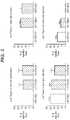
Fig 5 . Retention of anti-collagen II Fabs in osteoarthritic joints. * P<0.05, ** P< 0.01; comparison to control CNTO 4234. The counts at 10 min and 1 hour post injection for CNTO 3631 were too high to read. -
Fig 6 . Retention of anti-collagen II Fabs in osteoarthritic synovial fluid. * P<0.05; ** P< 0.01; comparison to control CNTO 4234. - One aspect of the invention is an isolated monoclonal antibody or fragment thereof that binds human collagen II, comprising a heavy chain variable region (VH region) and a light chain variable region (VL region), wherein the VH region comprises the heavy chain complementarity determining region (CDR) 1, 2 and 3 (HCDR1, HCDR2, and HCDR3) sequences as shown in
- i. SEQ ID NO:s 8, 14, and 20;
- ii. SEQ ID NO:s 9, 15, and 21;
- iii. SEQ ID NO:s 9, 15, and 22;
- iv. SEQ ID NO:s 9, 15, and 23;
- v. SEQ ID NO:s 9, 15, and 24;
- vi. SEQ ID NO:s 9, 15, and 25;
- vii. SEQ ID NO:s 9, 15, and 26;
- viii. SEQ ID NO:s 9, 15, and 27;
- ix. SEQ ID NO:s 9, 15, and 28;
- x. SEQ ID NO:s 10, 16, and 29;
- xi. SEQ ID NO:s 11, 17, and 30;
- xii. SEQ ID NO:s 12, 18, and 31; or
- xiii. SEQ ID NO:s 13, 19, and 32; and
the VL regon comprises thelight chain CDR - xiv. SEQ ID NO:s 33, 42, and 46;
- xv. SEQ
ID NO:s - xvi. SEQ
ID NO:s 35, 43, and 48; - xvii. SEQ
ID NO:s 36, 44, and 49; - xviii. SEQ
ID NO:s 37, 42, and 50; - xix. SEQ
ID NO:s 38, 42, and 51; - xx. SEQ
ID NO:s 35, 44, and 52; - xxi. SEQ
ID NO:s 39, 42, and 53; - xxii. SEQ
ID NO:s - xxiii. SEQ ID NO:s 41, 42, and 55.
- Another aspect of the invention is an isolated monoclonal antibody or fragment thereof that bind human collagen II, comprising a VH region and a VL region, wherein the VH region comprises an amino acid sequence having a sequence shown in SEQ
ID NO:s ID NO:s - Another aspect of the invention is an isolated antibody that binds human collagen II, comprising a VH region and a VL region, wherein the VH region comprises the HCDR1, HCDR2, and HCDR3 sequences as shown in SEQ ID NO
:s 9, 15, and 28, and the VL regon comprises the LCDR1, LCDR2, and LCDR3 sequences as shown in SEQID NO:s 39, 42, and 53. - Another aspect of the invention is an isolated monoclonal antibody or fragment thereof that binds human collagen II, comprising a VH region and a VL region, wherein the VH region comprises the HCDR1, HCDR2, and HCDR3 sequences as shown in SEQ
ID NO:s 11, 17, and 30, and the VL regon comprises the LCDR1, LCDR2, and LCDR3 sequences as shown in SEQID NO:s - Another aspect of the invention is an isolated antibody heavy chain variable region comprising the amino acid sequence shown in SEQ
ID NO:s - Another aspect of the invention is a isolated antibody light chain variable region comprising the amino acid sequence shown in SEQ
ID NO:s - Another aspect of the invention is isolated polynucleotides encoding antibody heavy chain variable regions and antibody light chain variable regions of the invention.
- Another aspect of the invention is a vector comprising at least one polynucleotide of the invention.
- Another aspect of the invention is a host cell comprising the vector of the invention.
- Another aspect of the invention is a method of making an antibody that binds human collagen II, comprising culturing the host cell of the invention and recovering the antibody produced by the host cell.
- All publications, including but not limited to patents and patent applications, cited in this specification are herein incorporated by reference as though fully set forth.
- Unless defined otherwise, all technical and scientific terms used herein have the same meaning as commonly understood by one of ordinary skill in the art to which an invention belongs. Although any compositions and methods similar or equivalent to those described herein can be used in the practice or testing of the invention, exemplary compositions and methods are described herein.
- The term "antibody" includes whole antibodies and any fragments thereof. Antibody fragments comprise at least a portion of an immunoglobulin molecule, such as a complementarity determining region (CDR), a variable region, a constant region, or a framework region from either antibody heavy or light chain. An antibody may be a Fab, F(ab'), F(ab')2, scFv, dsFv, or diabody. An antibody may be a monoclonal antibody (mAb), chimeric, humanized, or human antibody, dimeric, tetrameric or multimeric. Structures of the above mentioned antibody fragments, and techniques for the preparation and use of the antibodies and fragments thereof are well known in the art (Ausubel, et al., ed., Current Protocols in Molecular Biology, John Wiley & Sons, Inc., NY 1987-2001; Sambrook, et al., Molecular Cloning: A Laboratory Manual, 2nd Edition, Cold Spring Harbor, NY, 1989; Harlow and Lane, Antibodies, a Laboratory Manual, Cold Spring Harbor, NY, 1989; Colligan, et al., ed., Current Protocols in Immunology, John Wiley & Sons, Inc., NY 1994-2001; Colligan et al., Current Protocols in Protein Science, John Wiley & Sons, NY, NY, 1997-2001; Kohler et al., Nature, 256:495-497, 1975; Queen et al., Proc Natl Acad Sci, 86:10029-33, 1989;
U.S. Pat. No. 4,816,567 ). For example, murine mAbs can be made by the hybridoma method of Kohler et al., Nature 256:495-497, 1975. Chimeric mAbs can be prepared by the method disclosed inU.S. Pat. No. 4,816,567 . Human-adapted mAbs having CDRs derived from a non-human donor immunoglobulin (typically murine) and the remaining immunoglobulin-derived parts of the molecule being derived from one or more human immunoglobulins can be prepared by techniques known to those skilled in the art such as that disclosed inU.S. Pat. No. 5,225,539 . Human framework sequences useful for human-adaptation can be selected from relevant databases by those skilled in the art. Optionally, human-adapted mAbs can be further modified by incorporating altered framework support residues to preserve binding affinity by techniques such as those disclosed in Queen et al., Proc. Natl. Acad. Sci. (USA), 86:10029-10032, 1989 and Hodgson et al., Bio/Technology, 9:421, 1991. - Fully human mAbs lacking any non-human sequences can be prepared from human immunoglobulin transgenic mice by techniques referenced in, e.g., Lonberg et al., Nature 368:856-859, 1994; Fishwild et al., Nature Biotechnology 14:845-851, 1996; and Mendez et al., Nature Genetics 15:146-156, 1997. Human mAbs can also be prepared and optimized from phage display libraries by techniques referenced in, e.g., Knappik et al., J. Mol. Biol. 296:57-86, 2000; and Krebs et al., J. Immunol. Meth. 254:67-84 2001). Fragments of antibodies e.g., Fab, F(ab')2, Fd, and dAb fragments may be produced by cleavage of the antibodies or by recombinant engineering. For example, Fab and F(ab')2 fragments may be generated by treating the antibodies with an enzyme such as pepsin.
- Immunoglobulins can be assigned to five major classes, namely IgA, IgD, IgE, IgG and IgM, depending on the heavy chain constant domain amino acid sequence. IgA and IgG are further sub-classified as the isotypes IgA1, IgA2, IgG1, IgG2, IgG3 and IgG4.
- An antibody variable region consists of a framework" region interrupted by three "antigen-binding sites". The antigen-binding sites are defined using various terms: (i) Complementarity Determining Regions (CDRs), three in the VH (HCDR1, HCDR2, HCDR3), and three in the VL (LCDR1, LCDR2, LCDR3), are based on sequence variability (Wu and Kabat, J. Exp. Med. 132:211-250, 1970; Kabat et al., Sequences of Proteins of Immunological Interest, 5th Ed. Public Health Service, National Institutes of Health, Bethesda, Md., 1991). (ii) "Hypervariable regions", "HVR", or "HV", three in the VH (H1, H2, H3) and three in the VL (L1, L2, L3), refer to the regions of an antibody variable domains which are hypervariable in structure as defined by Chothia and Lesk (Chothia and Lesk, Mol. Biol. 196:901-917, 1987). Other terms include "IMGT-CDRs" (Lefranc et al., Dev. Comparat. Immunol. 27:55-77, 2003) and "Specificity Determining Residue Usage" (SDRU) (Almagro, Mol. Recognit. 17:132-143, 2004). The International ImMunoGeneTics (IMGT) database (http://www_imgt_org) provides a standardized numbering and definition of antigen-binding sites. The correspondence between CDRs, HVs and IMGT delineations is described in Lefranc et al., Dev. Comparat. Immunol. 27:55-77, 2003.
- "Framework" or "framework sequences" are the remaining sequences of a variable region other than those defined to be antigen-binding site. The framework is typically divided into four regions, FR1, FR2, FR3, and FR3, which form a scaffold for the three antigen-binding sites in each variable reigon. Because the antigen-binding site can be defined by various terms as described above, the exact amino acid sequence of a framework depends on how the antigen-binding site was defined.
- The term "antibody that binds human collagen II" as used herein refers to an antibody that binds human collagen II with an EC50 of 1 µg/ml or less in an ELISA assay using plates coated with 10 µg/mL of human collagen II according to the method described in Example 2.
- The term "human collagen II" or huColII as used herein refers to human type II collagen isolated from cartilage. Human collagen II is synthesized as procollagen alpha Col2A1 chains (SEQ ID NO: 79). The procollagen molecule is secreted into the extracellular matrix where it forms fibrils. Fibril formation is accompanied by the removal of the C- and N-propeptides by specific proteinases. Processing of the fibrillar hucolII is well known.
- The term "vector" means a polynucleotide capable of being duplicated within a biological system or that can be moved between such systems. Vector polynucleotides typically contain elements, such as origins of replication, polyadenylation signal or selection markers, that function to facilitate the duplication or maintenance of these polynucleotides in a biological system. Examples of such biological systems may include a cell, virus, animal, plant, and reconstituted biological systems utilizing biological components capable of duplicating a vector. The polynucleotide comprising a vector may be DNA or RNA molecules or a hybrid of these.
- The term "expression vector" means a vector that can be utilized in a biological system or in a reconstituted biological system to direct the translation of a polypeptide encoded by a polynucleotide sequence present in the expression vector.
- The term "polynucleotide" means a molecule comprising a chain of nucleotides covalently linked by a sugar-phosphate backbone or other equivalent covalent chemistry. Double and single-stranded DNAs and RNAs are typical examples of polynucleotides.
- The term "polypeptide" or "protein" means a molecule that comprises at least two amino acid residues linked by a peptide bond to form a polypeptide. Small polypeptides of less than 50 amino acids may be referred to as "peptides".
- Conventional one and three-letter amino acid codes are used herein as follows:
Amino acid Three-letter code One-letter code Alanine ala A Arginine arg R Asparagine asn N Aspartate asp D Cysteine cys C Glutamate glu E Glutamine gln Q Glycine gly G Histidine his H Isoleucine ile I Leucine leu L Lysine lys K Methionine met M Phenylalanine phe F Proline pro P Serine ser S Threonine thr T Tryptophan trp W Tyrosine tyr Y Valine val V - The present invention provides monoclonal antibodies that bind human collagen II. These antibodies are useful as research reagents, diagnostic reagents, and vehicles for delivering a therapeutic agent for example to a joint.
- The invention provides novel antigen-binding sites and immunoglobulin chains derived from human immunoglobulin gene libraries.
- One embodiment of the invention is an isolated monoclonal antibody or fragment thereof that binds human collagen II, comprising a heavy chain variable region (VH region) and a light chain variable region (VL region), wherein the VH region comprises the heavy chain complementarity determining region (CDR) 1, 2 and 3 (HCDR1, HCDR2, and HCDR3) sequences and the VL region comprises the light chain complementarity determining region (CDR) 1, 2 and 3 (LCDR1, LCDR2, and LCDR3) sequences as shown Table 1.
Table 1. Fab ID VH ID VL ID SEQ ID NO: HCDR1 HCDR2 HCDR3 LCDR1 LCDR2 LCDR3 VH VL Fab 169-76 VH169-76 VL 169-76 8 14 20 33 42 46 56 69 Fab169-C2 VH169-C2 VLL6 9 15 21 34 42 47 57 5 Fab169-22 VH169-22 VL169-22 9 15 22 35 43 48 58 70 Fab169-34 VH169-34 VL 169-34 9 15 23 36 44 49 59 71 Fab169-45 VH169-45 VL 169-45 9 15 24 37 42 50 60 72 Fab169-G11 VH169-G11 VLL6 9 15 25 34 42 47 61 5 Fab169-64 VH169-64 VL 169-64 9 15 26 38 42 51 62 73 Fab169-47 VH169-47 VL 169-47 9 15 27 35 44 52 63 74 Fab169-31 VH169-31 VL169-31 9 15 28 39 42 53 64 75 Fab323-G9 VH323- G9 VLB3 10 16 29 40 45 54 65 7 Fab323-G1 VH323-G1 VLL6 11 17 30 34 42 47 66 5 Fab323-B3 VH323-B3 VLB3 12 18 31 40 45 54 67 7 Fab551-3 VH551-3 VL551-3 13 19 32 41 42 55 68 76 - Antibodies having conservative substitutions in the heavy and light chain sequences shown in Table 1 (SEQ ID NO:s 8-76) are encompassed within the scope of the invention. The conservative substitution may reside in the framework regions, or in antigen- binding sites, as long they do not adversely affect the properties of the antibody. Substitutions may be made to improve antibody properties, for example stability or affinity. Conservative substitutions will produce molecules having functional and chemical characteristics similar to those molecules into which such modifications are made. Exemplary amino acid substitutions are shown in Table 2. Furthermore, any native residue in the polypeptide may also be substituted with alanine, as has been previously described for alanine scanning mutagenesis (MacLennan et al., Acta Physiol. Scand. Suppl. 643:55-67, 1998; Sasaki et al., Adv. Biophys. 35:1-24, 1998). Amino acid substitutions can be done for example by PCR mutagenesis (
US Pat. No. 4,683,195 ). Libraries of variants can be generated using well known methods, for example using random (NNK) or non-random codons, for example DVK codons, which encode 11 amino acids (ACDEGKNRSYW). The resulting variants can be characterized for their binding to human collagen II as described in Examples, or for other properties such as stability using well known methods.Table 2. Original residue Exemplary substitutions Ala (A) Val, Leu, Ile Arg (R) Lys, Gln, Asn Asn (N) Gln Asp (D) Glu Cys (C) Ser, Ala Gln (Q) Asn Gly (G) Pro, Ala His (H) Asn, Gln, Lys, Arg Ile (I) Leu, Val, Met, Ala, Phe Leu (L) Ile, Val, Met, Ala, Phe Lys (K) Arg, Gln, Asn Met (M) Leu, Phe, Ile Phe (F) Leu, Val, Ile, Ala, Tyr Pro (P) Ala Ser (S) Thr, Ala, Cys Thr(T) Ser Trp (W) Tyr, Phe Tyr (Y) Trp, Phe, Thr, Ser Val (V) Ile, Met, Leu, Phe, Ala - In other embodiments, the invention provides an isolated monoclonal antibody or fragment thereof that bind human collagen II, comprising a VH region and a VL region, wherein the VH region comprises an amino acid sequence having a sequence shown in SEQ
ID NO:s ID NO:s - Although the embodiments illustrated in the Examples comprise pairs of variable regions, one from a heavy and one from a light chain, a skilled artisan will recognize that alternative embodiments may comprise single heavy or light chain variable regions. The single variable region can be used to screen for a second variable region capable of forming a two-domain specific antigen-binding fragment capable of, for example, binding to human collagen II. The screening may be accomplished by phage display screening methods using for example hierarchical dual combinatorial approach disclosed in Intl. Publ. No.
WO92/01047 - In another aspect, the invention provides isolated antibody heavy chains and light chains comprising the amino acid sequences shown in SEQ
ID NO:s ID NO:s - Another aspect of the invention is isolated polynucleotides encoding any of the antibodies of the invention or their complement. Certain exemplary polynucleotides are disclosed herein, however, other polynucleotides which, given the degeneracy of the genetic code or codon preferences in a given expression system, encode the antibodies of the invention are also within the scope of the invention. Polynucleotides encoding antibodies of the invention are prepared by well known methods. These methods include, but are not limited to, isolation from a natural source (in the case of naturally occurring amino acid sequence variants) or preparation by oligonucleotide-mediated (or site-directed) mutagenesis, PCR mutagenesis, and chemical gene synthesis.
- Exemplary antibodies of the invention may be of the IgG, IgD, IgE, IgA or IgM isotypes. Additionally, the antibodies of the invention can be post-translationally modified by processes such as glycosylation, isomerization, deglycosylation or non-naturally occurring covalent modification such as the addition of polyethylene glycol (PEG) moieties (pegylation) and lipidation. Such modifications may occur in vivo or in vitro. For example, the antibodies of the invention can be conjugated to polyethylene glycol (PEGylated) to improve their pharmacokinetic profiles. Conjugation can be carried out by techniques known to those skilled in the art. Conjugation of therapeutic antibodies with PEG has been shown to enhance pharmacodynamics while not interfering with function. (Deckert et al., Int. J. Cancer 87:382-390, 2000; Knight et al., Platelets 15:409-418, 2004; Leong et al., Cytokine 16:106-119, 2001; Yang et al., Protein Eng. 16:761-770, 2003).
- Pharmacokinetic properties of the antibodies of the invention can be enhanced through Fc modifications by techniques known to those skilled in the art. The Fc" of an antibody is not involved directly in binding of an antibody to an antigen, but exhibits various effector functions. An antibody Fc" is a term well known and is defined on the basis of papain cleavage of antibodies. The Fc of an antibody is directly involved in ADCC (antibody-dependent cell-mediated cytotoxicity) and CDC (complement-dependent cytotoxicity) based on complement activation, Clq binding and Fc receptor binding. Complement activation (CDC) is initiated by binding of complement factor Clq to the Fc of most IgG antibody subclasses. While the influence of an antibody on the complement system is dependent on certain conditions, binding to Clq is caused by defined binding sites in the Fc. Such binding sites are known in the state of the art and described by, e.g., Boakle et al., Nature 282: 742-43, 1979; Lukas et al., J. Immunol. 127: 2555-60, 1981; Brunhouse and Cebra, Mol. Immunol. 16: 907-17, 1979; Burton et al., Nature 288:338-44, 1980; Thommesen et al., Mol. Immunol. 37: 995-1004, 2000; Idusogie et al., J. Immunol. 164:4178-84, 2000; Hezareh et al., J. Virology 75:12161-68, 2001; Morgan et al., Immunology 86:319-24, 1995;
EP 0307434 . Such binding sites are, e.g., L234, L235, D270, N297, E318, K320, K322, P331, and P329 (numbering according to EU index of Kabat). Antibodies of subclass IgG1, IgG2 and IgG3 usually show complement activation and Clq binding, whereas IgG4 does not activate the complement system and does not bind Clq. - The antibodies of the invention are characterized in that the constant chains are of human origin. Such constant chains are well known and described, e.g., by Kabat (see e.g. Johnson and Wu, Nuc Acids Res. 28, 214-18, 2000). For example, a useful human heavy chain constant region comprises SEQ ID NO: 77. For example, a useful human light chain constant region comprises an amino acid sequence of a kappa-light chain constant region of SEQ ID NO: 78.
- The antibodies of the invention may bind human collagen II with a Kd less than or equal to about 10-6, 10-7, 10-8, 10-9, 10-11, 10-11 or 10-12 M. The affinity of an antibody to human collagen II can be determined experimentally using any suitable method. Such methods may utilize Biacore or KinExA instrumentation, ELISA or competitive binding assays known to those skilled in the art.
- Another embodiment of the invention is a vector comprising at least one polynucleotide of the invention. The heavy and light chain variable domains of the invention are combined with sequences of promoter, translation initiation, constant region, 3' untranslated region, polyadenylation, and transcription termination to form expression vector constructs. The heavy and light chain expression constructs can be combined into a single vector, co-transfected, serially transfected, or separately transfected into host cells which are then fused to form a single host cell expressing both chains.
- Another embodiment of the invention is a host cell comprising a vector of the invention. Such host cells may be eukaryotic cells, bacterial cells, plant cells or archeal cells. Exemplary eukaryotic cells may be of mammalian, insect, avian or other animal origins. Mammalian eukaryotic cells include immortalized cell lines such as hybridomas or myeloma cell lines such as SP2/0 (American Type Culture Collection (ATCC), Manassas, VA, CRL-1581), NS0 (European Collection of Cell Cultures (ECACC), Salisbury, Wiltshire, UK, ECACC No. 85110503), FO (ATCC CRL-1646) and Ag653 (ATCC CRL-1580) murine cell lines. An exemplary human myeloma cell line is U266 (ATTC CRL-TIB-196). Other useful cell lines include those derived from Chinese Hamster Ovary (CHO) cells such as CHO-K1SV (Lonza Biologics, Walkersville, MD), CHO-K1 (ATCC CRL-61) or DG44.
- Another embodiment of the invention is a method of making an antibody binding human collagen II comprising culturing a host cell of the invention and recovering the antibody produced by the host cell. Methods of making antibodies and purifying them are well known in the art.
- The antibodies of the invention are useful as research agents, and as delivery agents of therapeutic molecules to sites expressing human collagen II, such as a joint.
- Arthritis, including osteoarthritis, rheumatoid arthritis, arthritic joints as a result of injury, and the like, are common inflammatory conditions which would benefit from the local delivery and joint retention of antiinflammatory proteins and therapeutic molecules.
- Therapeutic molecules may be coupled to the antibodies of the invention for improved joint retention. Therapeutic molecules may be proteins or chemical compounds. Exemplary therapeutic molecules are growth factors, cytokines and antiinflammatory agents, proteins that induce growth and repair of collagen, as well as small molecules inhibiting proteolytic destriuction of joint tissue. Therapeutic proteins may be coupled to the anti-collagen antibodies of the invention or fragments thereof by generating fusion proteins using well known recombinant methods. For example, the N-terminus of the therapeutic protein may be directly linked to the C-terminus of an antibody of the invention via an amide bond or a peptide linker. Exemplary fusion constructs are described in e.g.
U.S. Pat. No. 5116964 ,U.S. Pat. No. 5709859 , Intl. Publ. Nos.WO04/002417 WO05/081687 - The antibodies of the invention with optionally coupled therapeutic molecule may be prepared as pharmaceutical compositions containing an effective amount of the therapeutic molecule as an active ingredient in a pharmaceutically acceptable carrier. The term "carrier" refers to a diluent, adjuvant, excipient, or vehicle with which the active compound is administered. Such pharmaceutical vehicles can be liquids, such as water and oils, including those of petroleum, animal, vegetable or synthetic origin, such as peanut oil, soybean oil, mineral oil, sesame oil and the like. For example, 0.4% saline and 0.3% glycine can be used. These solutions are sterile and generally free of particulate matter. They may be sterilized by conventional, well-known sterilization techniques (e.g., filtration). The compositions may contain pharmaceutically acceptable auxiliary substances as required to approximate physiological conditions such as pH adjusting and buffering agents, stabilizing, thickening, lubricating and coloring agents, etc. The concentration of the agent of the invention in such pharmaceutical formulation can vary widely, i.e., from less than about 0.5%, usually at or at least about 1% to as much as 15 or 20% by weight and will be selected primarily based on required dose, fluid volumes, viscosities, etc., according to the particular mode of administration selected. Methods for preparing parenterally administrable compositions are well known and are described in more detail in, for example, "Remington's Pharmaceutical Science", 15th ed., Mack Publishing Company, Easton, PA.
- The antibodies of the invention can be lyophilized for storage and reconstituted in a suitable carrier prior to use. This technique has been shown to be effective with conventional immunoglobulins and protein preparations and art-known lyophilization and reconstitution techniques can be employed.
- The present invention will now be described with reference to the following specific, non-limiting examples.
- Human collagen II-binding Fabs were selected from de novo pIX phage display libraries (Shi et al., J. Mol. Biol. 397:385-396, 2010;
WO2009085462A1 ;U.S. Ser. No. 12/546850 Figure 1 ). The three heavy chain libraries were combined with the four germline light chains or germline light chain libraries to generate 24 unique VH:VL combinations for screening.Table 3. Loop Position* Scaffold 3-23 1-69 5-51 H1 31 SDNT S SNT 32 Y Y Y 33 AGW AG W 34 M I I 35 SH S SG H2 50 VANG GW IR 51 I I I 52 SNKW IS YD 52a YSGQ PA P 53 SD IY GS 54 G FN D 55 SG G S 56 S T DY *Residue numbering according to Kabat Table 4. H3 length Codon Configuration2 7 A, (NNS)x3, F/L, D, Y 8 A, (NNS)x4, F/L, D, Y 9 A, Bx5, F/L, D, Y 10 A, Bx6, F/L, D, Y 11 A, Bx6, (B+BY)x1, F/L, D, Y 12 A, Bx4, (B+BY)x4, F/L, D, Y 13 A, Bx6, (B+BY)x3, F/L, D, Y 14 A, Bx6, (B+BY)x4, F/L, D, Y Numbering according to Kabat and co-workers.
2Codon base compositions [A-C-G-T]
A:position 1 = [15-15-68-2]
position 2 = [43-11-33-13]
position 3 = [2-29-24-45]
B:position 1 = [19-14-41-26]
position 2 = [28-25-33-14]
position 3 = [0-34-36-30]
B+BY: Mixed primer set at this position. BY contains a tyrosine codon (TAT) sequentially replacing the B codon in these positions and is mixed at a ratio of 1:7 with a primer containing the B codon. Therefore, for CDR length 11 with one (B+BY) codon, two primers were used. One had the TAT codon at this position and the second had the B codon and these were mixed at a ratio of 1:7. For CDR length 13, the three (B+BY) codons required 4 primers, one with the B codon and three with the TAT codon in each of three positions and the B codon in the remaining 2 positions. In this case, the TAT primers were mixed at a ratio of 1:1:1:7 with the full B codon primer. Similarly, CDR-12 and CDR-14 were prepared with 5 primers.
The selection cycle was repeated four times. After the last selection cycle, the colonies were scraped off in 2mL 2xYT Carb/Glu/20% glycerol and 100 µL of the cell suspension was used to isolate plasmid DNA. The pIX was excised by Nhe1/SpeI digestion and self-ligation of the isolated DNA. After ligation, the DNA was electroporated into electrocompetent MC1061 F' cells and plated for single colonies on 2xYT Carb/Glu.Table 5. Loop Position* Scaffold 012 L6 A27 B3 L1 30 SRNAD SRNAD SRNTD L 30a - - SNR YSHFA 30e - - - KTNE 31 SNKDG NSKD SNRADH K 32 YHNDWFSA YWDFHSAN YFHQSEK YFHNWDAS L2 50 FYTNKADG ADKGYFTN ADGS WSRDYA L3 91 SAYHPD RYSGF YSHA YSHA 92 FIYHNDKGR RHNSL YNDSHIFKG YNDSHIFKG 93 STHNDRG NDKR SNTDGHR SNTDGHR 94 TYLVFSRGP WA TYLVFAS TYLVFAS 96 LWRFYIN WYFLIR WYFLIR WYFLIR *Residue numbering according to Kabat - The human collagen II binding Fabs obtained from the initial panning were screened for cross-reactivity with human collagens I, IV and V, and rat collagens I and II.
- Colonies were picked from the pIX-excised transformations and grown in 2xYT Carb. The next day, 50 µL of the saturated cultures were used to inoculate an expression plate containing 400 µL per well of 2xYT Carb and the plate was grown at 37° C for 6 hours. Fab expression was induced with the addition of 1 mM IPTG and the plate was placed at 30° C overnight. The next day, the induced Fab cultures were spun at 2000 rpm for 10 minutes, and the cleared lysate was used in subsequent assays.
- Maxisorp ELISA plates (Nunc) were coated with 10 µg/ml human collagen I (Chondrex), human collagen II (Chondrex), rat collagen I (Chondrex), rat collagen II (Chondrex) or with 5 µg/ml human collagen IV (Chemicon) or human collagen V (Chemicon), or with anti-Fd (the Binding Site) at 1 µg/ml, all in PBS, according to manufacturer's instructions. The wells were washed three times in TBST and blocked for one hour with 200
µL 3% milk in TBST. 100 µL of the Fab lysate was added onto the wells of the coated ELISA plates, and incubated 1 hour at room temperature. The wells were washed three times followed by addition of 100 µL of anti-kappa-HRP (Southern Biotech) at 1:5000 in PBS. The plates were incubated at room temperature for one hour, washed three times with TBST, and developed with BM Chemiluminescence ELISA Substrate (Roche Applied Science). The clones that bound to human and rat collagen II without binding to human collagen I, IV or V, or rat collagen I were sequenced and unique clones were further characterized.Figure 3 shows the ELISA data for the 13 clones chosen for further characterization. The sequences of the Fabs are shown inFigure 2 and Table 1. - The Fab expression in Escherichia coli in 2xYT Carb (except that TurboBroth (Athena ES) was used for expression of Fab 551-3) was induced with 1 mM IPTG at 30° C overnight. Induced bacteria were pelleted 30 min 4500 rpm, the cell pellets resuspended in lysis buffer (20 mM Tris, pH 8.5, 350 mM NaCl, 7.5 mM imidazole) with protease inhibitors, and ruptured with two passes through a microfluidizer. The cell lysate was clarified with two spins at 10,000 rpm for 10 minutes. Talon resin (Clontech) was equilibrated with lysis buffer and two mLs were added to the clarified lysate. Bound Fabs were eluted with two incubations of 5 minutes each using elution buffer (150 mM EDTA, 20 mM Tris, pH 8.5) and dialyzed in 20 mM Tris, pH 8.5. The dialyzed Fabs were further purified using a Q-sepharose Fast Flow resin (QFF resin; GE Healthcare), and used for experiments.
- Human cartilage was obtained from osteoarthritis patients (Northland Laboratories). The cartilage was first pulverized and stored as a suspension in PBS with protease inhibitors at 4° C. To test for cartilage binding, 2.5 µL of cartilage suspension per data point was added to 97.5 µL of TBST with 3% milk and the mixture rotated at room temperature for one hour. Binding of Fabs to the cartilage was tested using a MultiScreen HTS plate (Millipore) using the vacuum manifold. Briefly, 100 µL of the suspension was added into prewetted MultiScreen plate, and vacuum was applied to settle the cartilage onto the well filter, after whihc the wells were washed twice with TBST. 2.4 µg of each Fab in 100 µL of PBS was added to the wells and incubated at room temperature for 1 hour. The wells were washed three times with TBST, and anti-kappa-HRP (Southern Biotech) was added onto the wells. After one hour incubation, the wells were washed in PBST, the BM Chemiluminescence ELISA Substrate (Roche Applied Science) was added, and the resultant suspension of cartilage fragments was transferred to a black-well ELISA plate for luminescence detection. All 13 Fabs exhibited cartilage binding (
Figure 4 ). - EC50 values were obtained for select Fabs using ELISA assay as described above. In the assays, 100 microliters of each Fab was added in a concentration range between 10 ng/ml - 10 µg/ml onto the wells. The EC50 values are shown in Table 6.
- Biacore binding kinetics was performed using standard methods against human and rat collagen II (Table 6b).
Table 6a. Fab EC50 (ng/ml) 551-3 113.5 323-G1 123.1 169-31 113.1 Table 6b. human collagen II rat collagen II Fab Kax105 Kdx10-3 KD (nmol) Kax105 Kdx10-3 KD (nmol) 323-G1 1.84 1.08 6 2.94 1.39 5 169-31 0.97 5 50 1.05 7.89 75 CNTO 4234 n.a. n.a. >1000 n.a. n.a. >1000 - Two anti-collagen II Fabs (323-G1 (CNTO 3631) and 169-31 (CNTO 4093) and a control mAb (CNTO 4234) that did not bind to extracellular matrix components were iodinated and injected into the knees of menisectomized rats to evaluate the effect of mAb binding to collagen on joint residence time. Fabs were radiolabeled using Na125I (Perkin Elmer) and Iodo-GEN tubes (Pierce). Free iodine was removed using PD-10 desalting columns (GE Healthcare) and the Fabs were concentrated to 2.2 mg/mL using Amicon Ultra centrifugal filter devices (Millipore; 10,000 MWCO). After iodination, the Fabs were tested for binding of collagen II, and no significant impairment due to iodination was observed.
- 32 male Sprague-Dawley rats underwent meniscus transection on the right and left hind limb. The rats were anesthetized with isoflurane anesthesia 2-4%, and the medial collateral ligament was transected and the meniscus cut to simulate a tear injury. Joint damage resembling osteoarthritis developed over the next three weeks. Twenty-one days after surgery, intra-articular injections of 125I-labeled Fabs were performed. Animals received 11 µg of test article through 5 µl intra-articular injections into the right and left knee joint. Animals were euthanized at four time points (10 min, 1 hr, 6 hr and 24 hrs post-injection) and synovial fluid lavage and the knee joints were collected from bot knees. Radioactivity was measured from each sample. The counts per minute (cpm) were compared to the values of the non-injected dose of each respective Fab to calculate the % retention (e.g. as percent of injected dose). 323-G1 (CNTO 3631) and 169-31 (CNTO 4093) were retained both in the knee joint (
Figure 5 ) and synovial fluid (Figure 6 ) longer than the control antibody. Approximately 60% and 32% of Fab 323-G1 and 60% and 18% of Fab 169-31 were retained in the knee (combined joint and synovial retention) after 6 and 24 hours post injection, respectively. The control Fab was retained at 17% and 5%. Thus, the antibodies of the invention exhibited increased joint retention due to their binding to a resident joint protein. - The invention further provides the following non-limiting embodiments:
- 1. An isolated monoclonal antibody or fragment thereof that binds human collagen II, comprising a heavy chain variable region (VH region) and a light chain variable region (VL region), wherein the VH region comprises the heavy chain complementarity determining region (CDR) 1, 2 and 3 (HCDR1, HCDR2, and HCDR3) sequences as shown in
- i. SEQ ID NO
:s - ii. SEQ ID NO
:s 9, 15, and 21; - iii. SEQ ID NO
:s - iv. SEQ ID NO
:s 9, 15, and 23; - v. SEQ ID NO
:s - vi. SEQ ID NO
:s - vii. SEQ ID NO
:s 9, 15, and 26; - viii. SEQ ID NO
:s 9, 15, and 27; - ix. SEQ ID NO
:s 9, 15, and 28; - x. SEQ
ID NO:s - xi. SEQ
ID NO:s 11, 17, and 30; - xii. SEQ
ID NO:s 12, 18, and 31; or - xiii. SEQ ID NO:s 13, 19, and 32; and
the VL regon comprises thelight chain CDR - xiv. SEQ ID NO:s 33, 42, and 46;
- xv. SEQ
ID NO:s - xvi. SEQ
ID NO:s 35, 43, and 48; - xvii. SEQ
ID NO:s 36, 44, and 49; - xviii. SEQ
ID NO:s 37, 42, and 50; - xix. SEQ
ID NO:s 38, 42, and 51; - xx. SEQ
ID NO:s 35, 44, and 52; - xxi. SEQ
ID NO:s 39, 42, and 53; - xxii. SEQ
ID NO:s - xxiii. SEQ ID NO:s 41, 42, and 55.
- i. SEQ ID NO
- 2. An isolated monoclonal antibody or fragment thereof that bind human collagen II, comprising a VH region and a VL region, wherein the VH region comprises an amino acid sequence having a sequence shown in SEQ
ID NO:s ID NO:s - 3. An isolated antibody that binds human collagen II, comprising a VH region and a VL region, wherein the VH region comprises the HCDR1, HCDR2, and HCDR3 sequences as shown in SEQ ID NO
:s 9, 15, and 28, and the VL regon comprises the LCDR1, LCDR2, and LCDR3 sequences as shown in SEQID NO:s 39, 42, and 53. - 4. An isolated monoclonal antibody or fragment thereof that binds human collagen II, comprising a VH region and a VL region, wherein the VH region comprises the HCDR1, HCDR2, and HCDR3 sequences as shown in SEQ
ID NO:s 11, 17, and 30, and the VL regon comprises the LCDR1, LCDR2, and LCDR3 sequences as shown in SEQID NO:s - 5. The isolated antibody of
embodiment 1, wherein the antibody comprises a Fab fragment. - 6. An isolated antibody heavy chain variable region comprising the amino acid sequence shown in SEQ
ID NO:s - 7. An isolated antibody light chain variable region comprising the amino acid sequence shown in SEQ
ID NO:s - 8. An isolated polynucleotide encoding an antibody heavy chain variable region of
embodiment 6. - 9. An isolated polynucleotide encoding an antibody light chain variable region of embodiment 7.
- 10. A vector comprising at least one polynucleotide of
embodiment 8 or 9. - 11. A host cell comprising the vector of
embodiment 10. - 12. A method of making an antibody that binds human collagen II, comprising culturing the host cell of embodiment 11 and recovering the antibody produced by the host cell.


Claims (10)
- An isolated monoclonal antibody or fragment thereof that binds human collagen II, comprising a heavy chain variable region (VH region) and a light chain variable region (VL region), wherein:the VH region comprises the heavy chain complementarity determining region (CDR) 1, 2 and 3 (HCDR1, HCDR2, and HCDR3) sequences as shown in SEQ ID NO:s 11, 17, and 30; andthe VL region comprises the light chain CDR 1, 2 and 3 (LCDR1, LCDR2, and LCDR3) sequences as shown in SEQ ID NO:s 34, 42, and 47.
- The isolated monoclonal antibody or fragment of claim 1, wherein the VH region comprises an amino acid sequence having a sequence shown in SEQ ID NO: 66, and the VL region comprises an amino acid sequence having a sequence shown in SEQ ID NO: 5.
- The isolated antibody of any preceding claim, wherein the antibody comprises a Fab fragment.
- An isolated antibody heavy chain variable region comprising the amino acid sequence shown in SEQ ID NO: 66.
- An isolated polynucleotide encoding an antibody heavy chain variable region of claim 4.
- An isolated polynucleotide encoding an antibody light chain variable region of comprising the amino acid sequence shown in SEQ ID NO: 5.
- An isolated polynucleotide encoding an antibody according to claim 1.
- A vector comprising at least one polynucleotide of any one of claims 5 to 7.
- A host cell comprising the vector of claim 8.
- A method of making an antibody that binds human collagen II, comprising culturing the host cell of claim 9 and recovering the antibody produced by the host cell.
Applications Claiming Priority (3)
| Application Number | Priority Date | Filing Date | Title |
|---|---|---|---|
| US38679610P | 2010-09-27 | 2010-09-27 | |
| EP11831244.6A EP2621950A4 (en) | 2010-09-27 | 2011-09-26 | Antibodies binding human collagen ii |
| PCT/US2011/053250 WO2012047583A2 (en) | 2010-09-27 | 2011-09-26 | Antibodies binding human collagen ii |
Related Parent Applications (1)
| Application Number | Title | Priority Date | Filing Date |
|---|---|---|---|
| EP11831244.6A Division EP2621950A4 (en) | 2010-09-27 | 2011-09-26 | Antibodies binding human collagen ii |
Publications (2)
| Publication Number | Publication Date |
|---|---|
| EP3050899A1 true EP3050899A1 (en) | 2016-08-03 |
| EP3050899B1 EP3050899B1 (en) | 2019-09-04 |
Family
ID=45928304
Family Applications (2)
| Application Number | Title | Priority Date | Filing Date |
|---|---|---|---|
| EP11831244.6A Withdrawn EP2621950A4 (en) | 2010-09-27 | 2011-09-26 | Antibodies binding human collagen ii |
| EP16157460.3A Active EP3050899B1 (en) | 2010-09-27 | 2011-09-26 | Antibodies binding human collagen ii |
Family Applications Before (1)
| Application Number | Title | Priority Date | Filing Date |
|---|---|---|---|
| EP11831244.6A Withdrawn EP2621950A4 (en) | 2010-09-27 | 2011-09-26 | Antibodies binding human collagen ii |
Country Status (8)
| Country | Link |
|---|---|
| US (1) | US8394378B2 (en) |
| EP (2) | EP2621950A4 (en) |
| JP (1) | JP6126991B2 (en) |
| CN (1) | CN103429616B (en) |
| CA (1) | CA2812389C (en) |
| ES (1) | ES2757857T3 (en) |
| IL (1) | IL225397A0 (en) |
| WO (1) | WO2012047583A2 (en) |
Families Citing this family (23)
| Publication number | Priority date | Publication date | Assignee | Title |
|---|---|---|---|---|
| US9212227B2 (en) | 2012-04-30 | 2015-12-15 | Janssen Biotech, Inc. | ST2L antibody antagonists for the treatment of ST2L-mediated inflammatory pulmonary conditions |
| US20150329638A1 (en) * | 2012-12-28 | 2015-11-19 | Biogen Ma Inc. | Use of DR6 Antagonists to Improve Motor Neuron Disease |
| JP6466904B2 (en) | 2013-03-15 | 2019-02-06 | ヤンセン バイオテツク,インコーポレーテツド | Interferon alpha and omega antibody antagonists |
| WO2014210545A2 (en) * | 2013-06-27 | 2014-12-31 | The University Of Chicago | Compositions and methods related to recombinant antibodies to histone posttranslational modifications |
| KR102331054B1 (en) | 2013-11-06 | 2021-11-24 | 얀센 바이오테크 인코포레이티드 | Anti-ccl17 antibodies |
| WO2015191602A2 (en) * | 2014-06-09 | 2015-12-17 | Biomed Valley Discoveries, Inc. | Combination therapies targeting tumor-associated stroma or tumor cells |
| WO2015191610A2 (en) * | 2014-06-09 | 2015-12-17 | Biomed Valley Discoveries, Inc. | Combination therapies using agents that target tumor-associated stroma or tumor cells and other pathways |
| WO2015191568A2 (en) * | 2014-06-09 | 2015-12-17 | Biomed Valley Discoveries, Inc. | Combination therapies using agents that target tumor-associated stroma or tumor cells and tumor vasculature |
| WO2015191617A2 (en) * | 2014-06-09 | 2015-12-17 | Biomed Valley Discoveries, Inc. | Combination therapies using anti-metabolites and agents that target tumor-associated stroma or tumor cells |
| US10758614B2 (en) | 2014-06-09 | 2020-09-01 | The United States Of America, As Represented By The Secretary, Department Of Health And Human Services National Institutes Of Health | Combination therapies targeting tumor-associated stroma or tumor cells and topoisomerase |
| US10799584B2 (en) | 2014-06-09 | 2020-10-13 | The United States Of America, As Represented By The Secretary, Department Of Health And Human Services National Institutes Of Health | Combination therapies using agents that target tumor-associated stroma or tumor cells and alkylating agents |
| US11033620B2 (en) | 2014-06-09 | 2021-06-15 | Biomed Valley Discoveries, Inc. | Combination therapies targeting tumor-associated stroma or tumor cells and microtubules |
| GB201413357D0 (en) * | 2014-07-28 | 2014-09-10 | Philogen Spa | Antibodies for treatment and diagnosis |
| WO2016020791A1 (en) * | 2014-08-05 | 2016-02-11 | Novartis Ag | Ckit antibody drug conjugates |
| US9907858B2 (en) * | 2015-01-16 | 2018-03-06 | Immunwork Inc. | Peptide core-based multi-arm linkers |
| RS63125B1 (en) | 2015-11-03 | 2022-05-31 | Janssen Biotech Inc | Antibodies specifically binding pd-1 and their uses |
| KR20180087430A (en) * | 2015-12-17 | 2018-08-01 | 얀센 바이오테크 인코포레이티드 | Antibodies specifically binding to HLA-DR and uses thereof |
| EP3442579A4 (en) * | 2016-04-15 | 2019-12-18 | Immunext Inc. | Anti-human vista antibodies and use thereof |
| AU2017276604B2 (en) * | 2016-06-06 | 2020-02-27 | F. Hoffmann-La Roche Ag | Fusion proteins for ophthalmology with increased eye retention |
| JOP20190097A1 (en) | 2016-10-27 | 2019-04-28 | Janssen Pharmaceutica Nv | Immunoglobulins and uses thereof |
| AU2019232631A1 (en) | 2018-03-05 | 2020-09-24 | Janssen Pharmaceutica Nv | Anti-PHF-tau antibodies and uses thereof |
| JP7280586B2 (en) * | 2018-10-15 | 2023-05-24 | 学校法人東京理科大学 | Dendritic cell maturation inhibitor, method for inhibiting maturation, and pharmaceutical composition |
| CN113874394B (en) * | 2019-02-20 | 2024-01-19 | 和铂抗体有限公司 | Antibodies to |
Citations (14)
| Publication number | Priority date | Publication date | Assignee | Title |
|---|---|---|---|---|
| US4683195A (en) | 1986-01-30 | 1987-07-28 | Cetus Corporation | Process for amplifying, detecting, and/or-cloning nucleic acid sequences |
| EP0307434A1 (en) | 1987-03-18 | 1989-03-22 | Medical Res Council | Altered antibodies. |
| US4816567A (en) | 1983-04-08 | 1989-03-28 | Genentech, Inc. | Recombinant immunoglobin preparations |
| WO1992001047A1 (en) | 1990-07-10 | 1992-01-23 | Cambridge Antibody Technology Limited | Methods for producing members of specific binding pairs |
| US5116964A (en) | 1989-02-23 | 1992-05-26 | Genentech, Inc. | Hybrid immunoglobulins |
| US5225539A (en) | 1986-03-27 | 1993-07-06 | Medical Research Council | Recombinant altered antibodies and methods of making altered antibodies |
| US5709859A (en) | 1991-01-24 | 1998-01-20 | Bristol-Myers Squibb Company | Mixed specificity fusion proteins |
| WO2004002417A2 (en) | 2002-06-28 | 2004-01-08 | Centocor, Inc. | Mammalian ch1 deleted mimetibodies, compositions, methods and uses |
| US20050153442A1 (en) * | 1999-03-10 | 2005-07-14 | Adam Katz | Adipose-derived stem cells and lattices |
| WO2005081687A2 (en) | 2003-09-30 | 2005-09-09 | Centocor, Inc. | Human hinge core mimetibodies, compositions, methods and uses |
| WO2005097073A1 (en) | 2004-03-31 | 2005-10-20 | University Of Utah Research Foundation | Macromolecular delivery systems for non-invasive imaging, evaluation and treatment of arthritis and other inflammatory diseases |
| US7067144B2 (en) | 1998-10-20 | 2006-06-27 | Omeros Corporation | Compositions and methods for systemic inhibition of cartilage degradation |
| WO2008135734A1 (en) * | 2007-05-03 | 2008-11-13 | Queen Mary & Westfield College | Novel antibody and use in diagnosis and therapy of arthropathies |
| WO2009085462A1 (en) | 2007-12-19 | 2009-07-09 | Centocor, Inc. | Design and generation of human de novo pix phage display libraries via fusion to pix or pvii, vectors, antibodies and methods |
Family Cites Families (16)
| Publication number | Priority date | Publication date | Assignee | Title |
|---|---|---|---|---|
| GB9125979D0 (en) | 1991-12-06 | 1992-02-05 | Wellcome Found | Antibody |
| US20030108545A1 (en) * | 1994-02-10 | 2003-06-12 | Patricia Rockwell | Combination methods of inhibiting tumor growth with a vascular endothelial growth factor receptor antagonist |
| US20110093981A9 (en) | 1999-05-06 | 2011-04-21 | La Rosa Thomas J | Nucleic acid molecules and other molecules associated with transcription in plants and uses thereof for plant improvement |
| WO2001014558A1 (en) * | 1999-08-26 | 2001-03-01 | Morphosys Ag | Human antibodies or fragments thereof binding to hla-cw6 |
| US20040191260A1 (en) * | 2003-03-26 | 2004-09-30 | Technion Research & Development Foundation Ltd. | Compositions capable of specifically binding particular human antigen presenting molecule/pathogen-derived antigen complexes and uses thereof |
| KR20120053525A (en) * | 2000-06-16 | 2012-05-25 | 캠브리지 안티바디 테크놀로지 리미티드 | Antibodies that immunospecifically bind to blys |
| US7288390B2 (en) | 2000-08-07 | 2007-10-30 | Centocor, Inc. | Anti-dual integrin antibodies, compositions, methods and uses |
| CN1117986C (en) * | 2000-10-16 | 2003-08-13 | 北京市创伤骨科研究所 | II-type collegen enzyme-linked immunoassay analysis |
| DK1436404T3 (en) | 2001-09-19 | 2010-03-08 | Alexion Pharma Inc | Manipulated templates and their use in single-primer amplification |
| NZ534174A (en) | 2002-01-09 | 2007-03-30 | Medarex Inc | An isolated human monoclonal antibody which binds to human CD30 |
| CA2508660C (en) | 2002-12-23 | 2013-08-20 | Wyeth | Antibodies against pd-1 and uses therefor |
| WO2005047459A2 (en) * | 2003-08-04 | 2005-05-26 | University Of Massachusetts | Sars nucleic acids, proteins, antibodies, and uses thereof |
| CA2614972C (en) * | 2005-07-18 | 2014-08-19 | Amgen Inc. | Human anti-b7rp1 neutralizing antibodies |
| US20070134248A1 (en) * | 2005-12-08 | 2007-06-14 | Genitope Corporation | Combination therapy and antibody panels |
| FR2933702A1 (en) * | 2008-07-08 | 2010-01-15 | Sanofi Aventis | SPECIFIC ANTAGONISTS OF FGF-R4 RECEPTOR |
| CA2736429A1 (en) * | 2008-10-01 | 2010-04-08 | Genentech, Inc. | Anti-notch2 antibodies and methods of use |
-
2011
- 2011-09-26 EP EP11831244.6A patent/EP2621950A4/en not_active Withdrawn
- 2011-09-26 CA CA2812389A patent/CA2812389C/en active Active
- 2011-09-26 ES ES16157460T patent/ES2757857T3/en active Active
- 2011-09-26 US US13/245,299 patent/US8394378B2/en active Active
- 2011-09-26 JP JP2013530393A patent/JP6126991B2/en active Active
- 2011-09-26 CN CN201180046602.5A patent/CN103429616B/en active Active
- 2011-09-26 WO PCT/US2011/053250 patent/WO2012047583A2/en active Application Filing
- 2011-09-26 EP EP16157460.3A patent/EP3050899B1/en active Active
-
2013
- 2013-03-21 IL IL225397A patent/IL225397A0/en unknown
Patent Citations (15)
| Publication number | Priority date | Publication date | Assignee | Title |
|---|---|---|---|---|
| US4816567A (en) | 1983-04-08 | 1989-03-28 | Genentech, Inc. | Recombinant immunoglobin preparations |
| US4683195B1 (en) | 1986-01-30 | 1990-11-27 | Cetus Corp | |
| US4683195A (en) | 1986-01-30 | 1987-07-28 | Cetus Corporation | Process for amplifying, detecting, and/or-cloning nucleic acid sequences |
| US5225539A (en) | 1986-03-27 | 1993-07-06 | Medical Research Council | Recombinant altered antibodies and methods of making altered antibodies |
| EP0307434A1 (en) | 1987-03-18 | 1989-03-22 | Medical Res Council | Altered antibodies. |
| US5116964A (en) | 1989-02-23 | 1992-05-26 | Genentech, Inc. | Hybrid immunoglobulins |
| WO1992001047A1 (en) | 1990-07-10 | 1992-01-23 | Cambridge Antibody Technology Limited | Methods for producing members of specific binding pairs |
| US5709859A (en) | 1991-01-24 | 1998-01-20 | Bristol-Myers Squibb Company | Mixed specificity fusion proteins |
| US7067144B2 (en) | 1998-10-20 | 2006-06-27 | Omeros Corporation | Compositions and methods for systemic inhibition of cartilage degradation |
| US20050153442A1 (en) * | 1999-03-10 | 2005-07-14 | Adam Katz | Adipose-derived stem cells and lattices |
| WO2004002417A2 (en) | 2002-06-28 | 2004-01-08 | Centocor, Inc. | Mammalian ch1 deleted mimetibodies, compositions, methods and uses |
| WO2005081687A2 (en) | 2003-09-30 | 2005-09-09 | Centocor, Inc. | Human hinge core mimetibodies, compositions, methods and uses |
| WO2005097073A1 (en) | 2004-03-31 | 2005-10-20 | University Of Utah Research Foundation | Macromolecular delivery systems for non-invasive imaging, evaluation and treatment of arthritis and other inflammatory diseases |
| WO2008135734A1 (en) * | 2007-05-03 | 2008-11-13 | Queen Mary & Westfield College | Novel antibody and use in diagnosis and therapy of arthropathies |
| WO2009085462A1 (en) | 2007-12-19 | 2009-07-09 | Centocor, Inc. | Design and generation of human de novo pix phage display libraries via fusion to pix or pvii, vectors, antibodies and methods |
Non-Patent Citations (42)
| Title |
|---|
| ALMAGRO, MOL. RECOGNIT., vol. 17, 2004, pages 132 - 143 |
| AUSUBEL, ET AL.,: "Current Protocols in Molecular Biology", 1987, JOHN WILEY & SONS, INC. |
| BOAKLE ET AL., NATURE, vol. 282, 1979, pages 742 - 43 |
| BRUNHOUSE; CEBRA, MOL. IMMUNOL., vol. 16, 1979, pages 907 - 17 |
| BURTON ET AL., NATURE, vol. 288, 1980, pages 338 - 44 |
| CHOTHIA; LESK, MOL. BIOL., vol. 196, 1987, pages 901 - 917 |
| COLLIGAN ET AL.: "Current Protocols in Protein Science", 1997, JOHN WILEY & SONS |
| COLLIGAN, ET AL.,: "Current Protocols in Immunology", 1994, JOHN WILEY & SONS, INC. |
| DECKERT ET AL., INT. J. CANCER, vol. 87, 2000, pages 382 - 390 |
| FISHWILD ET AL., NATURE BIOTECHNOLOGY, vol. 14, 1996, pages 845 - 851 |
| GENNARO: "Remington's Pharmaceutical Science, 15th ed.,", MACK PUBLISHING COMPANY |
| GERWIN ET AL., ADV DRUG DELIV REV, vol. 58, 2006, pages 226 - 42 |
| HARLOW; LANE: "Antibodies, a Laboratory Manual", 1989, COLD SPRING HARBOR |
| HEZAREH ET AL., J. VIROLOGY, vol. 75, 2001, pages 12161 - 68 |
| HODGSON ET AL., BIO/TECHNOLOGY, vol. 9, 1991, pages 421 |
| IDUSOGIE ET AL., J. IMMUNOL., vol. 164, 2000, pages 4178 - 84 |
| JOHNSON; WU, NUC ACIDS RES., vol. 28, 2000, pages 214 - 18 |
| KABAT ET AL.: "Sequences of Proteins of Immunological Interest, 5th Ed.", 1991, NATIONAL INSTITUTES OF HEALTH |
| KNAPPIK ET AL., J. MOL. BIOL., vol. 296, 2000, pages 57 - 86 |
| KNIGHT ET AL., PLATELETS, vol. 15, 2004, pages 409 - 418 |
| KOHLER ET AL., NATURE, vol. 256, 1975, pages 495 - 497 |
| KREBS ET AL., J. IMMUNOL. METH., vol. 254, 2001, pages 67 - 84 |
| KUTTY SELVA NANDAKUMAR ET AL: "Induction of arthritis by single monoclonal IgG anti-collagen type II antibodies and enhancement of arthritis in mice lacking inhibitory Fc[gamma]RIIB", EUROPEAN JOURNAL OF IMMUNOLOGY, vol. 33, no. 8, 1 August 2003 (2003-08-01), DE, pages 2269 - 2277, XP055174056, ISSN: 0014-2980, DOI: 10.1002/eji.200323810 * |
| LEFRANC ET AL., DEV. COMPARAT. IMMUNOL., vol. 27, 2003, pages 55 - 77 |
| LEONG ET AL., CYTOKINE, vol. 16, 2001, pages 106 - 119 |
| LINDENHAYN ET AL., EUR J CLIN CHEM BIOCHEM, vol. 35, 1997, pages 355 - 63 |
| LONBERG ET AL., NATURE, vol. 368, 1994, pages 856 - 859 |
| LUKAS ET AL., J. IMMUNOL., vol. 127, 1981, pages 2555 - 60 |
| MACLENNAN ET AL., ACTA PHYSIOL. SCAND., vol. 643, 1998, pages 55 - 67 |
| MENDEZ ET AL., NATURE GENETICS, vol. 15, 1997, pages 146 - 156 |
| MORGAN ET AL., IMMUNOLOGY, vol. 86, 1995, pages 319 - 24 |
| QUEEN ET AL., PROC NATL ACAD SCI, vol. 86, 1989, pages 10029 - 33 |
| QUEEN ET AL., PROC. NATL. ACAD. SCI. (USA, vol. 86, 1989, pages 10029 - 10032 |
| ROTHENFLUH ET AL., NATURE MATERIALS, vol. 7, 2008, pages 248 - 54 |
| SAMBROOK ET AL.: "Molecular Cloning: A Laboratory Manual, 2nd Edition,", 1989, COLD SPRING HARBOR |
| SASAKI ET AL., ADV. BIOPHYS., vol. 35, 1998, pages 1 - 24 |
| SHI ET AL., J. MOL. BIOL., vol. 397, 2010, pages 385 - 396 |
| THOMMESEN ET AL., MOL. IMMUNOL., vol. 37, 2000, pages 995 - 1004 |
| UYSAL ET AL: "The crystal structure of the pathogenic collagen type II-specific mouse monoclonal antibody CIIC1 Fab: Structure to function analysis", MOLECULAR IMMUNOLOGY, PERGAMON, GB, vol. 45, no. 8, 1 February 2008 (2008-02-01), pages 2196 - 2204, XP022513099, ISSN: 0161-5890, DOI: 10.1016/J.MOLIMM.2007.12.005 * |
| WU; KABAT, J. EXP. MED., vol. 132, 1970, pages 211 - 250 |
| Y XU ET AL: "Two monoclonal antibodies to precisely the same epitope of type II collagen select non-crossreactive phage clones by phage display: implications for autoimmunity and molecular mimicry", MOLECULAR IMMUNOLOGY, vol. 41, no. 4, 1 June 2004 (2004-06-01), pages 411 - 419, XP055115818, ISSN: 0161-5890, DOI: 10.1016/j.molimm.2004.03.025 * |
| YANG ET AL., PROTEIN ENG., vol. 16, 2003, pages 761 - 770 |
Also Published As
| Publication number | Publication date |
|---|---|
| ES2757857T3 (en) | 2020-04-30 |
| US8394378B2 (en) | 2013-03-12 |
| CN103429616A (en) | 2013-12-04 |
| US20120108795A1 (en) | 2012-05-03 |
| EP2621950A2 (en) | 2013-08-07 |
| CA2812389C (en) | 2019-12-31 |
| WO2012047583A2 (en) | 2012-04-12 |
| EP2621950A4 (en) | 2015-09-02 |
| JP2013542722A (en) | 2013-11-28 |
| CN103429616B (en) | 2016-08-10 |
| CA2812389A1 (en) | 2012-04-12 |
| JP6126991B2 (en) | 2017-05-10 |
| EP3050899B1 (en) | 2019-09-04 |
| IL225397A0 (en) | 2013-06-27 |
| WO2012047583A3 (en) | 2013-10-03 |
Similar Documents
| Publication | Publication Date | Title |
|---|---|---|
| EP3050899B1 (en) | Antibodies binding human collagen ii | |
| CA2956000C (en) | An anti-ctla4 monoclonal antibody or antigen binding fragment thereof, a pharmaceutical composition and use | |
| WO2021043221A1 (en) | Antibodies binding tslp and uses thereof | |
| KR101836217B1 (en) | Il-17a antagonists | |
| KR102213694B1 (en) | Human cd30 ligand antigen binding proteins | |
| JP5941049B2 (en) | Antibodies against human TWEAK and uses thereof | |
| US20120020988A1 (en) | Antibodies specifically binding to human TSLPR and methods of use | |
| CN101001645A (en) | Il-17 antagonistic antibodies | |
| TW200927759A (en) | Anti-hepcidin antibodies and uses thereof | |
| CN101132811A (en) | High affinity antibodies against HMGB1 and methods of use thereof | |
| SK16762000A3 (en) | Antibodies to cd23, derivatives thereof, and their therapeutic uses | |
| KR20040030088A (en) | Antagonistic Anti-hTNFSF13b Human Antibodies | |
| JP5596777B2 (en) | Antibodies against human TWEAK and uses thereof | |
| KR20170110681A (en) | PCSK9 ANTIBODIES, AND PHARMACEUTICAL COMPOSITIONS AND USES THEREOF | |
| ZA200601842B (en) | Nogo-a binding molecules and pharmaceutical uses thereof | |
| US8906649B2 (en) | Antibodies binding human collagen II | |
| RU2716101C2 (en) | Sclerostin antibody, its antigen-binding fragment and medical application | |
| CN113939539A (en) | Antibodies that bind CSF-1R and uses thereof | |
| US20230167182A1 (en) | Antibodies binding il6r and uses thereof | |
| WO2024026351A2 (en) | Cannabinoid type 1 receptor binding proteins and uses thereof | |
| CN112203685A (en) | Treatment of autoimmune and inflammatory disorders using antibodies that bind interleukin-17A (IL-17A) | |
| ZA200602015B (en) | Antibodies against interleukin-1receptor and uses thereof | |
| CZ20004164A3 (en) | Antibodies against CD23, their derivatives as well as their therapeutic application |
Legal Events
| Date | Code | Title | Description |
|---|---|---|---|
| PUAI | Public reference made under article 153(3) epc to a published international application that has entered the european phase |
Free format text: ORIGINAL CODE: 0009012 |
|
| AC | Divisional application: reference to earlier application |
Ref document number: 2621950 Country of ref document: EP Kind code of ref document: P |
|
| AK | Designated contracting states |
Kind code of ref document: A1 Designated state(s): AL AT BE BG CH CY CZ DE DK EE ES FI FR GB GR HR HU IE IS IT LI LT LU LV MC MK MT NL NO PL PT RO RS SE SI SK SM TR |
|
| STAA | Information on the status of an ep patent application or granted ep patent |
Free format text: STATUS: REQUEST FOR EXAMINATION WAS MADE |
|
| 17P | Request for examination filed |
Effective date: 20170201 |
|
| RBV | Designated contracting states (corrected) |
Designated state(s): AL AT BE BG CH CY CZ DE DK EE ES FI FR GB GR HR HU IE IS IT LI LT LU LV MC MK MT NL NO PL PT RO RS SE SI SK SM TR |
|
| STAA | Information on the status of an ep patent application or granted ep patent |
Free format text: STATUS: EXAMINATION IS IN PROGRESS |
|
| 17Q | First examination report despatched |
Effective date: 20170619 |
|
| GRAP | Despatch of communication of intention to grant a patent |
Free format text: ORIGINAL CODE: EPIDOSNIGR1 |
|
| STAA | Information on the status of an ep patent application or granted ep patent |
Free format text: STATUS: GRANT OF PATENT IS INTENDED |
|
| INTG | Intention to grant announced |
Effective date: 20190429 |
|
| GRAS | Grant fee paid |
Free format text: ORIGINAL CODE: EPIDOSNIGR3 |
|
| GRAA | (expected) grant |
Free format text: ORIGINAL CODE: 0009210 |
|
| STAA | Information on the status of an ep patent application or granted ep patent |
Free format text: STATUS: THE PATENT HAS BEEN GRANTED |
|
| AC | Divisional application: reference to earlier application |
Ref document number: 2621950 Country of ref document: EP Kind code of ref document: P |
|
| AK | Designated contracting states |
Kind code of ref document: B1 Designated state(s): AL AT BE BG CH CY CZ DE DK EE ES FI FR GB GR HR HU IE IS IT LI LT LU LV MC MK MT NL NO PL PT RO RS SE SI SK SM TR |
|
| REG | Reference to a national code |
Ref country code: GB Ref legal event code: FG4D |
|
| REG | Reference to a national code |
Ref country code: CH Ref legal event code: EP |
|
| REG | Reference to a national code |
Ref country code: AT Ref legal event code: REF Ref document number: 1175200 Country of ref document: AT Kind code of ref document: T Effective date: 20190915 |
|
| REG | Reference to a national code |
Ref country code: DE Ref legal event code: R096 Ref document number: 602011061912 Country of ref document: DE |
|
| REG | Reference to a national code |
Ref country code: IE Ref legal event code: FG4D |
|
| REG | Reference to a national code |
Ref country code: NL Ref legal event code: MP Effective date: 20190904 |
|
| REG | Reference to a national code |
Ref country code: LT Ref legal event code: MG4D |
|
| PG25 | Lapsed in a contracting state [announced via postgrant information from national office to epo] |
Ref country code: LT Free format text: LAPSE BECAUSE OF FAILURE TO SUBMIT A TRANSLATION OF THE DESCRIPTION OR TO PAY THE FEE WITHIN THE PRESCRIBED TIME-LIMIT Effective date: 20190904 Ref country code: SE Free format text: LAPSE BECAUSE OF FAILURE TO SUBMIT A TRANSLATION OF THE DESCRIPTION OR TO PAY THE FEE WITHIN THE PRESCRIBED TIME-LIMIT Effective date: 20190904 Ref country code: BG Free format text: LAPSE BECAUSE OF FAILURE TO SUBMIT A TRANSLATION OF THE DESCRIPTION OR TO PAY THE FEE WITHIN THE PRESCRIBED TIME-LIMIT Effective date: 20191204 Ref country code: HR Free format text: LAPSE BECAUSE OF FAILURE TO SUBMIT A TRANSLATION OF THE DESCRIPTION OR TO PAY THE FEE WITHIN THE PRESCRIBED TIME-LIMIT Effective date: 20190904 Ref country code: FI Free format text: LAPSE BECAUSE OF FAILURE TO SUBMIT A TRANSLATION OF THE DESCRIPTION OR TO PAY THE FEE WITHIN THE PRESCRIBED TIME-LIMIT Effective date: 20190904 Ref country code: NO Free format text: LAPSE BECAUSE OF FAILURE TO SUBMIT A TRANSLATION OF THE DESCRIPTION OR TO PAY THE FEE WITHIN THE PRESCRIBED TIME-LIMIT Effective date: 20191204 |
|
| PG25 | Lapsed in a contracting state [announced via postgrant information from national office to epo] |
Ref country code: RS Free format text: LAPSE BECAUSE OF FAILURE TO SUBMIT A TRANSLATION OF THE DESCRIPTION OR TO PAY THE FEE WITHIN THE PRESCRIBED TIME-LIMIT Effective date: 20190904 Ref country code: LV Free format text: LAPSE BECAUSE OF FAILURE TO SUBMIT A TRANSLATION OF THE DESCRIPTION OR TO PAY THE FEE WITHIN THE PRESCRIBED TIME-LIMIT Effective date: 20190904 Ref country code: AL Free format text: LAPSE BECAUSE OF FAILURE TO SUBMIT A TRANSLATION OF THE DESCRIPTION OR TO PAY THE FEE WITHIN THE PRESCRIBED TIME-LIMIT Effective date: 20190904 Ref country code: GR Free format text: LAPSE BECAUSE OF FAILURE TO SUBMIT A TRANSLATION OF THE DESCRIPTION OR TO PAY THE FEE WITHIN THE PRESCRIBED TIME-LIMIT Effective date: 20191205 |
|
| REG | Reference to a national code |
Ref country code: AT Ref legal event code: MK05 Ref document number: 1175200 Country of ref document: AT Kind code of ref document: T Effective date: 20190904 |
|
| REG | Reference to a national code |
Ref country code: ES Ref legal event code: FG2A Ref document number: 2757857 Country of ref document: ES Kind code of ref document: T3 Effective date: 20200430 |
|
| PG25 | Lapsed in a contracting state [announced via postgrant information from national office to epo] |
Ref country code: AT Free format text: LAPSE BECAUSE OF FAILURE TO SUBMIT A TRANSLATION OF THE DESCRIPTION OR TO PAY THE FEE WITHIN THE PRESCRIBED TIME-LIMIT Effective date: 20190904 Ref country code: PL Free format text: LAPSE BECAUSE OF FAILURE TO SUBMIT A TRANSLATION OF THE DESCRIPTION OR TO PAY THE FEE WITHIN THE PRESCRIBED TIME-LIMIT Effective date: 20190904 Ref country code: PT Free format text: LAPSE BECAUSE OF FAILURE TO SUBMIT A TRANSLATION OF THE DESCRIPTION OR TO PAY THE FEE WITHIN THE PRESCRIBED TIME-LIMIT Effective date: 20200106 Ref country code: EE Free format text: LAPSE BECAUSE OF FAILURE TO SUBMIT A TRANSLATION OF THE DESCRIPTION OR TO PAY THE FEE WITHIN THE PRESCRIBED TIME-LIMIT Effective date: 20190904 Ref country code: NL Free format text: LAPSE BECAUSE OF FAILURE TO SUBMIT A TRANSLATION OF THE DESCRIPTION OR TO PAY THE FEE WITHIN THE PRESCRIBED TIME-LIMIT Effective date: 20190904 Ref country code: RO Free format text: LAPSE BECAUSE OF FAILURE TO SUBMIT A TRANSLATION OF THE DESCRIPTION OR TO PAY THE FEE WITHIN THE PRESCRIBED TIME-LIMIT Effective date: 20190904 |
|
| PG25 | Lapsed in a contracting state [announced via postgrant information from national office to epo] |
Ref country code: SM Free format text: LAPSE BECAUSE OF FAILURE TO SUBMIT A TRANSLATION OF THE DESCRIPTION OR TO PAY THE FEE WITHIN THE PRESCRIBED TIME-LIMIT Effective date: 20190904 Ref country code: SK Free format text: LAPSE BECAUSE OF FAILURE TO SUBMIT A TRANSLATION OF THE DESCRIPTION OR TO PAY THE FEE WITHIN THE PRESCRIBED TIME-LIMIT Effective date: 20190904 Ref country code: IS Free format text: LAPSE BECAUSE OF FAILURE TO SUBMIT A TRANSLATION OF THE DESCRIPTION OR TO PAY THE FEE WITHIN THE PRESCRIBED TIME-LIMIT Effective date: 20200224 Ref country code: CZ Free format text: LAPSE BECAUSE OF FAILURE TO SUBMIT A TRANSLATION OF THE DESCRIPTION OR TO PAY THE FEE WITHIN THE PRESCRIBED TIME-LIMIT Effective date: 20190904 |
|
| REG | Reference to a national code |
Ref country code: CH Ref legal event code: PL |
|
| REG | Reference to a national code |
Ref country code: DE Ref legal event code: R097 Ref document number: 602011061912 Country of ref document: DE |
|
| PLBE | No opposition filed within time limit |
Free format text: ORIGINAL CODE: 0009261 |
|
| STAA | Information on the status of an ep patent application or granted ep patent |
Free format text: STATUS: NO OPPOSITION FILED WITHIN TIME LIMIT |
|
| PG2D | Information on lapse in contracting state deleted |
Ref country code: IS |
|
| PG25 | Lapsed in a contracting state [announced via postgrant information from national office to epo] |
Ref country code: CH Free format text: LAPSE BECAUSE OF NON-PAYMENT OF DUE FEES Effective date: 20190930 Ref country code: LI Free format text: LAPSE BECAUSE OF NON-PAYMENT OF DUE FEES Effective date: 20190930 Ref country code: LU Free format text: LAPSE BECAUSE OF NON-PAYMENT OF DUE FEES Effective date: 20190926 Ref country code: IE Free format text: LAPSE BECAUSE OF NON-PAYMENT OF DUE FEES Effective date: 20190926 Ref country code: DK Free format text: LAPSE BECAUSE OF FAILURE TO SUBMIT A TRANSLATION OF THE DESCRIPTION OR TO PAY THE FEE WITHIN THE PRESCRIBED TIME-LIMIT Effective date: 20190904 Ref country code: IS Free format text: LAPSE BECAUSE OF FAILURE TO SUBMIT A TRANSLATION OF THE DESCRIPTION OR TO PAY THE FEE WITHIN THE PRESCRIBED TIME-LIMIT Effective date: 20200105 |
|
| REG | Reference to a national code |
Ref country code: BE Ref legal event code: MM Effective date: 20190930 |
|
| 26N | No opposition filed |
Effective date: 20200605 |
|
| PG25 | Lapsed in a contracting state [announced via postgrant information from national office to epo] |
Ref country code: SI Free format text: LAPSE BECAUSE OF FAILURE TO SUBMIT A TRANSLATION OF THE DESCRIPTION OR TO PAY THE FEE WITHIN THE PRESCRIBED TIME-LIMIT Effective date: 20190904 Ref country code: MC Free format text: LAPSE BECAUSE OF FAILURE TO SUBMIT A TRANSLATION OF THE DESCRIPTION OR TO PAY THE FEE WITHIN THE PRESCRIBED TIME-LIMIT Effective date: 20190904 Ref country code: BE Free format text: LAPSE BECAUSE OF NON-PAYMENT OF DUE FEES Effective date: 20190930 |
|
| PG25 | Lapsed in a contracting state [announced via postgrant information from national office to epo] |
Ref country code: CY Free format text: LAPSE BECAUSE OF FAILURE TO SUBMIT A TRANSLATION OF THE DESCRIPTION OR TO PAY THE FEE WITHIN THE PRESCRIBED TIME-LIMIT Effective date: 20190904 |
|
| PG25 | Lapsed in a contracting state [announced via postgrant information from national office to epo] |
Ref country code: HU Free format text: LAPSE BECAUSE OF FAILURE TO SUBMIT A TRANSLATION OF THE DESCRIPTION OR TO PAY THE FEE WITHIN THE PRESCRIBED TIME-LIMIT; INVALID AB INITIO Effective date: 20110926 Ref country code: MT Free format text: LAPSE BECAUSE OF FAILURE TO SUBMIT A TRANSLATION OF THE DESCRIPTION OR TO PAY THE FEE WITHIN THE PRESCRIBED TIME-LIMIT Effective date: 20190904 |
|
| PG25 | Lapsed in a contracting state [announced via postgrant information from national office to epo] |
Ref country code: TR Free format text: LAPSE BECAUSE OF FAILURE TO SUBMIT A TRANSLATION OF THE DESCRIPTION OR TO PAY THE FEE WITHIN THE PRESCRIBED TIME-LIMIT Effective date: 20190904 |
|
| PG25 | Lapsed in a contracting state [announced via postgrant information from national office to epo] |
Ref country code: MK Free format text: LAPSE BECAUSE OF FAILURE TO SUBMIT A TRANSLATION OF THE DESCRIPTION OR TO PAY THE FEE WITHIN THE PRESCRIBED TIME-LIMIT Effective date: 20190904 |
|
| PGFP | Annual fee paid to national office [announced via postgrant information from national office to epo] |
Ref country code: IT Payment date: 20220811 Year of fee payment: 12 Ref country code: GB Payment date: 20220804 Year of fee payment: 12 Ref country code: DE Payment date: 20220609 Year of fee payment: 12 |
|
| PGFP | Annual fee paid to national office [announced via postgrant information from national office to epo] |
Ref country code: FR Payment date: 20220808 Year of fee payment: 12 |
|
| PGFP | Annual fee paid to national office [announced via postgrant information from national office to epo] |
Ref country code: ES Payment date: 20221006 Year of fee payment: 12 |