EP3141279A1 - Closure element for placement and fixation in the septum of a heart and balloon catheter for fixation of a closure element in the septum of a heart - Google Patents
Closure element for placement and fixation in the septum of a heart and balloon catheter for fixation of a closure element in the septum of a heart Download PDFInfo
- Publication number
- EP3141279A1 EP3141279A1 EP16195063.9A EP16195063A EP3141279A1 EP 3141279 A1 EP3141279 A1 EP 3141279A1 EP 16195063 A EP16195063 A EP 16195063A EP 3141279 A1 EP3141279 A1 EP 3141279A1
- Authority
- EP
- European Patent Office
- Prior art keywords
- septum
- closure element
- heart
- electrode
- fixation
- Prior art date
- Legal status (The legal status is an assumption and is not a legal conclusion. Google has not performed a legal analysis and makes no representation as to the accuracy of the status listed.)
- Withdrawn
Links
Images


Classifications
-
- A—HUMAN NECESSITIES
- A61—MEDICAL OR VETERINARY SCIENCE; HYGIENE
- A61N—ELECTROTHERAPY; MAGNETOTHERAPY; RADIATION THERAPY; ULTRASOUND THERAPY
- A61N1/00—Electrotherapy; Circuits therefor
- A61N1/02—Details
- A61N1/04—Electrodes
- A61N1/05—Electrodes for implantation or insertion into the body, e.g. heart electrode
- A61N1/056—Transvascular endocardial electrode systems
- A61N1/057—Anchoring means; Means for fixing the head inside the heart
- A61N1/0573—Anchoring means; Means for fixing the head inside the heart chacterised by means penetrating the heart tissue, e.g. helix needle or hook
Definitions
- the invention relates to an implantable lead or lead assembly for implantation in the left ventricle of a heart with perforation of the atrial or ventricular septum.
- the state of the art for left ventricular stimulation and perception is currently the implantation of an electrode into a left ventricular vein via the coronary sinus. These so-called coronary sinus electrodes are mainly used for cardiac resynchronization therapy.
- Transseptal endocardial left ventricular pacing An alternative technique for coronary sinus lead placement in cardiac resynchronization therapy. van Gelder BM, Scheffer MG, Meijer A, et al., Heart Rhythm 2007 Apr; 4 (4): 454-60 ,
- WO 2006/105395 A2 describes a transseptal / trans-myocardial ventricular stimulation electrode.
- a left ventricular coronary sinus electrode In about 10-15% of the implantations due to the anatomical conditions, the reliable implantation of a left ventricular coronary sinus electrode is not possible. Furthermore, the rate of dislocation of left ventricular leads implanted across the coronary sinus for cardiac resynchronization therapy (CRT) is greater than that of a conventional right ventricular pacing lead. For these reasons, there is currently no introduction of left ventricular stimulation - using a coronary sinus electrode - for bradycardia therapy or automatic cardioverter / defibrillator (ICD) implantation, since both implantation success and safety with this type of left ventricular lead is not present . Another disadvantage of a coronary sinus electrode is the very limited placement options. Usually only 1 to 2 different positions for fixing the probe are given. This is considered a major cause of the z. Poor responder rate (60-70%) of the CRT.
- ICD automatic cardioverter / defibrillator
- the transmural LV pressure measurement here represents only the possibility of permanently introducing a system through the heart muscle into the left ventricle.
- a very short probe is placed in the left ventricle with a pressure sensor that can not be used for electrical stimulation of the heart.
- the probe described is designed in such a way that the active stimulation surface lies only in the region of the left ventricular septum and is not freely positionable in the left ventricle.
- WO 2006/105395 A2 did not address the repositioning and explantability of an electrode.
- the object of the invention is to construct a left ventricular probe which can be pushed through the atrial or ventricular septum from the right ventricle into the left ventricle and freely maneuvered and fixed within the left ventricle the implantation risk of a left-right shunt through appropriate design measures is minimized at the probe body. Furthermore, the possibility of repositioning such a probe and its explantation must be considered.
- the invention includes the essential idea of providing at the perforation site in the atrial or ventricular septum through which the electrode lead is laid a suitable element which ensures reliable mutual sealing of the areas of the heart adjacent to the septum, at least during an ingrowth phase.
- this is done by the electrode line itself, which then carries a suitable closure element.
- a separate closure element is provided for this purpose, which is placed in the septum prior to the introduction of the electrode line and is then pierced by the electrode line when it is laid.
- the invention is also applicable to a lead-in conduit provided for use in the left atrium that is passed through a septum into the left atrium.
- the closure body can be firmly connected to the electrode, so that a longitudinal movement of the electrode body in the septum is avoided.
- the closure body is pronounced in another embodiment as expandable Schirmchen.
- the closure body is shaped as an expandable screen and as an expandable, electrode-displaceable anchor (proximal to the screen).
- the probe is preferably coated in the region of the closure body in order to promote rapid formation of connective tissue in the region of the perforation site.
- the "working channel” preferably has a maximum free diameter of 2 mm when no electrode is pushed through (acceptable left-right shunt).
- the "working channel" is marked in a further embodiment with X-ray markers or made of an X-ray visible material.
- the "working channel” is z. B. by expandable fixators (such as stents) attached in the septum.
- the LV probe can also be fixed in the "working channel” by expandable fixators. These fixators are attached to the probe.
- the expandable fixators can also be used for uni- and bipolar stimulation of the septum.
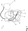
- FIG. 1 an arrangement 1 according to the invention is shown.
- a left ventricular electrode lead 3 is thereby advanced through an implantable transseptal passage ["working channel"] 5 into the left ventricle and fixed there by means of a conventional screw 7.
- the left ventricular electrode lead 3 has a bipolar stimulation electrode 9 and additionally two shock electrodes (distal: 11, proximal 13). The diameter of the distal shock electrode 11 is selected so that it can not slip through the passage 5 in the left ventricle.
- the lead 3 is connected to an implantable defibrillator 17 with an electrode plug 15 (IS-4 or IS-1 and DF-1).
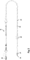
- Fig. 2 shows as a further embodiment of the invention specifically an electrode lead 19, which can be implanted via a guide wire 21 with proximal handle 22 and its structure with a flexible (here U-shaped bent shown) electrode lead body 23, a tip electrode 25 and distal fixation barbs 27 is known per se for anchoring in the wall of the ventricle.
- Electrode line is a - here symbolically as two sealing discs with a small distance from each other shown - closure element 29 for closing a perforation in the septum, via which the electrode line (as in Fig. 1 shown) is guided in the left ventricle.
- Fig. 3 shows as a special embodiment of this embodiment, an electrode line 19 ', in which the closure element 29 by two oppositely spreadable screens 29a' 29b 'is formed, which spreads thanks to an additional control wire 30' next to the guide wire 21, after insertion of the electrode line on both sides of the perforation in the septum can be and seal in the spread state, the environment of the puncture point.
- the proximal screen can additionally be displaced on the probe by means of the additional stylet 30 'so that the ventricular septum is fixed between the two screens.
- the two umbrellas 29a ', 29b' can also be used as (for example bipolar) active stimulation electrodes.
- FIG. 4a-c shown septal implementation (“working channel") 31 is first prepared by means of a Brockenbrough needle 33 by puncture of the ventricular septum VS.
- the implementation itself consists of 2 expandable stents 35a, 35b, embedded in a flexible biocompatible tube 35c, z. As silicone or polyurethane. This passage is mounted on an expandable balloon 37 of a balloon catheter 39 and is fixed by balloon expansion in the septum.
- the lead-through 31 the length L of which has been previously determined by echocardiography, is fixed by the expansion of the smaller balloon 37 by the expanding balloon 37 attaching the stems 35a, 35b to the tube section 35c in between against the wall of the hole formed by the Brockenbrough needle in the septum.
- the leadthrough 31 and possibly a guide wire 43 remains in the puncture site (FIG. Fig. 4b ).
- the two stents 35a, 35b are visible in the X-ray image, so that a subsequent navigation of the LV probe by this transsepthal implementation is easily possible.
- the left-right shunt volume is minimized by the flexible part of the bushing 31. Namely, when no pipe is inserted, the inside width d is reduced by inward buckling of the flexible hose portion 35c between the stems 35a, 35b.
- the transseptal passage 31 described above is shown along with a left ventricular lead 45.
- a self-expanding fixator 47 This fixator is realized in the embodiment as a radiopaque spring element, which is released by retracting a tube 49 and thus clamps the electrode line in the implementation. In this way, abrasion of the electrode line or the fixator is avoided.
- FIG. 5 An alternative to the LV probes described above in combination with a feedthrough is in Fig. 5 an LV probe 51 is shown, which can be attached independently, without a separate closure element by means of directly attached fixators 53 in the septum, these fixators 53 are first encased by a tube 55 and then expanded by retraction of this tube by springs 57 in the septum VS ,
- the tube 55 is designed such that it can be completely removed from the electrode line after implantation (eg by peeling).
- the fixators can be "retracted” by a suitable tube - after cutting off the electrode plug.
- the fixators can be used as active stimulation electrodes be connected by being connected to the electrical leads 57 of the electrode line.
- the implantation in this line is also done by Brockenbrough needle.
Abstract
Implantierbare Leitung, insbesondere Elektroden- und/oder Sensor- und/oder Medikamentenzufuhrleitung, zur Implantation im linken Ventrikel eines Herzens unter Perforation des atrialen oder ventrikulären Septums, mit einem langgestreckten, flexiblen Leitungskörper, einer Elektrode und/oder einem Sensor und/oder einer Medikamentenapplikationseinrichtung am oder nahe einem distalen Ende des Leitungskörpers und einem an den Leitungskörper angeformten oder mit diesem verbundenen Verschlusselement zur Abdichtung der Perforationsstelle im Septum.Implantable lead, in particular electrode and / or sensor and / or drug delivery lead, for implantation in the left ventricle of a heart with perforation of the atrial or ventricular septum, with an elongate, flexible lead body, an electrode and / or a sensor and / or a drug delivery device at or near a distal end of the lead body and a closure element integrally formed with or connected to the lead body for sealing the perforation site in the septum.
Description
Die Erfindung betrifft eine implantierbare Leitung oder Leitungsanordnung zur Implantation im linken Ventrikel eines Herzens unter Perforation des atrialen oder ventrikulären Septums.The invention relates to an implantable lead or lead assembly for implantation in the left ventricle of a heart with perforation of the atrial or ventricular septum.
Als Stand der Technik für eine linksventrikuläre Stimulation und Wahrnehmung ist derzeit die Implantation einer Elektrode in eine linksventrikuläre Vene via Koronarsinus anzusehen. Diese sog. Koronarsinuselektroden kommen vorwiegend für die kardiale Resynchronisationstherapie zum Einsatz.The state of the art for left ventricular stimulation and perception is currently the implantation of an electrode into a left ventricular vein via the coronary sinus. These so-called coronary sinus electrodes are mainly used for cardiac resynchronization therapy.
In der medizinischen Literatur finden sich vermehrt Fallberichte von transseptaler Implantation linksventrikulärer Stimulationselektroden zur Resynchronisationstherapie. Diese Implantationstechniken wurden immer unter Zuhilfenahme vorhandener Katheter, Führungsdrähte und Elektroden durchgeführt. Als Zugang zum linken Ventrikel wurden entweder die Punktion des atrialen Septums oder des Ventrikelseptums beschrieben, vgl. z. B. Transseptal endocardial left ventricular pacing: An alternative technique for coronary sinus lead placement in cardiac resynchronization therapy,
Ferner werden derzeit Konzepte zur transmuralen linksventrikulären Druckmessung in klinischen Studien untersucht. Hier werden Drucksensoren transmural im linken Ventrikel zur dauerhaften telemetrischen Druckmessung im linken Ventrikel platziert; vgl. unter www.transomamedical.com oder folgende Fachveröffentlichungen:
-
A Novel Technique For Assessing Load-Dependent Cardiac Function During LVAD Support Using Telemetered Left Ventricular Pressure. McConnell, PI, Del Rio, CL, Kwiatkowski, P, Farrar, D, Shipkowitz, T, Michler, RE, Sun, B. ASAIO Journal. 51(2): 31A, March/April 2005 -
In Vivo Safety and Accuracy of a Clinically Applicable Telemetered Left Ventricular Pressure Module: Intermediate-TermResults. McConnell, PI, de Cunha, D, Shipkowitz, T, Van Hee, J, Long, P and Hamlin, R. Heart Failure Society Meeting, September 2004 -
A System for Long-Term Measurement of Left Ventricular Pressure in Heart Failure Patients Living at Home. Sweitzer, N, Park, S. Heart Failure Society Meeting, September 2002 -
Automated Non-Invasive Monitoring of Left Ventricular Hemodynamics During Onset of Heart Failure in an Ambulatory Yucatan Mini Pig Model Using a New System Under Development for Assessing Heart Failure Patients at Home. Park, S, Sweitzer, N. Heart Failure Society Meeting, September 2002 -
A System For Long-Term Measurement Of Left Ventricular Pressure In Heart Failure Patients Living At Home. Park, S, Sweitzer, N and May, G. Heart Failure & Circulatory Support Summit, Cleveland, OH, August 2002
-
A Novel Technique For Assessing Load-Dependent Cardiac Function During LVAD Support Using Telemetered Left Ventricular Pressure. McConnell, PI, Del Rio, CL Kwiatkowski, P, Farrar, D, Shipkowitz, T, Michler, RE, Sun, B. ASAIO Journal. 51 (2): 31A, March / April 2005 -
In Vivo Safety and Accuracy of a Clinically Applicable Telemetered Left Ventricular Pressure Module: Intermediate TermResults. McConnell, PI, de Cunha, D, Shipkowitz, T, Van Hee, J, Long, P and Hamlin, R. Heart Failure Society Meeting, September 2004 -
A System for Long-Term Measurement of Left Ventricular Pressure in Heart Failure Patients Living at Home. Sweitzer, N, Park, S. Heart Failure Society Meeting, September 2002 -
Automated Non-Invasive Monitoring of Left Ventricular Hemodynamics During Onset of Heart Failure in Ambulatory Yucatan Mini Pig Model Using a New System Under Development for Assessing Heart Failure Patients at Home. Park, S, Sweitzer, N. Heart Failure Society Meeting, September 2002 -
A System For Long-Term Measurement Of Left Ventricular Pressure In Heart Failure Patients Living At Home. Park, S, Sweitzer, N and May, G. Heart Failure & Circulatory Support Summit, Cleveland, OH, August 2002
Für angeborene Atriumseptumdefekte, offene Foramen oder ovale und Ventrikelseptumdefekte werden derzeit eine Vielzahl von kommerziellen Verschlusssystemen (z. B. Premere™ PFO, SJM) angeboten, die mittels Kathetertechnik platziert werden können und einen zuverlässigen Verschluss des Septumdefektes gewährleisten; vgl. dazu
In
In ca. 10-15% der Implantationen ist aufgrund der anatomischen Gegebenheiten die zuverlässige Implantation einer linksventrikulären Koronarsinus-Elektrode nicht möglich. Des Weiteren ist die Dislokationsrate von linksventrikulären Elektroden, die für die kardiale Resynchronisationstherapie (CRT) über den Koronarsinus implantiert würden, größer als die einer herkömmlichen rechtventrikulären Schrittmacherelektrode. Aus diesen Gründen ist derzeit die Einführung einer rein linksventrikulären Stimulation - unter Verwendung einer Koronarsinuselektrode - für die Bradykardietherapie oder die Implantation von automatischen Kardiovertern/Defibrillatoren (ICD) nicht gegeben, da sowohl der Implantationserfolg als auch die Sicherheit mit dieser Art linksventrikulärer Elektrode nicht gegeben ist. Ein weiterer Nachteil einer Koronarsinuselektrode sind die sehr eingeschränkten Platzierungsoptionen. Meist sind nur 1 bis 2 verschiedene Positionen zur Fixierung der Sonde gegeben. Dies wird als eine wesentliche Ursache für die z. T. schlechte Responderrate (60-70%) der CRT diskutiert.In about 10-15% of the implantations due to the anatomical conditions, the reliable implantation of a left ventricular coronary sinus electrode is not possible. Furthermore, the rate of dislocation of left ventricular leads implanted across the coronary sinus for cardiac resynchronization therapy (CRT) is greater than that of a conventional right ventricular pacing lead. For these reasons, there is currently no introduction of left ventricular stimulation - using a coronary sinus electrode - for bradycardia therapy or automatic cardioverter / defibrillator (ICD) implantation, since both implantation success and safety with this type of left ventricular lead is not present , Another disadvantage of a coronary sinus electrode is the very limited placement options. Usually only 1 to 2 different positions for fixing the probe are given. This is considered a major cause of the z. Poor responder rate (60-70%) of the CRT.
Die weiter oben vorgestellten Techniken zur Elektrodenimplantation in den linken Ventrikel via atrialem oder ventrikulärem Septum sind sehr aufwändig und haben sich aufgrund der bestehen Risiken (RV Shunt, Thromben) bislang nicht durchgesetzt, obwohl hier eine freie Platzierung der Elektrode im linken Ventrikel möglich ist und damit die Nachteile der zuverlässigen Sondenfixierung, der Responderrate und anatomischen Einschränkungen behoben werden.The techniques described above for implanting electrodes into the left ventricle via the atrial or ventricular septum are very complex and have not yet been established due to the existing risks (RV shunt, thrombi), although a free placement of the electrode in the left ventricle is possible the disadvantages of reliable probe fixation, responder rate and anatomical limitations are addressed.
Die transmurale LV-Druckmessung stellt hier nur die Möglichkeit dar, durch den Herzmuskel ein System dauerhaft in den linken Ventrikel einzubringen. Allerdings wird hier eine sehr kurze Sonde mit einem Drucksensor in den linken Ventrikel gebracht, die nicht für die elektrische Stimulation des Herzens verwendet werden kann. Die in
Um die oben genannten Nachteile zu beheben, besteht die Aufgabe der Erfindung darin, eine linksventrikuläre Sonde zu konstruieren, die durch das atriale oder ventrikuläre Septum vom rechten Ventrikel in den linken Ventrikel geschoben werden und frei innerhalb des linken Ventrikel manövriert und fixiert werden kann, wobei das Implantationsrisiko eines Links-Rechts-Shunts durch geeignete konstruktive Maßnahmen am Sondenkörper minimiert wird. Des Weiteren ist die Möglichkeit der Repositionierung einer solchen Sonde und deren Explantation zu berücksichtigen.To overcome the above drawbacks, the object of the invention is to construct a left ventricular probe which can be pushed through the atrial or ventricular septum from the right ventricle into the left ventricle and freely maneuvered and fixed within the left ventricle the implantation risk of a left-right shunt through appropriate design measures is minimized at the probe body. Furthermore, the possibility of repositioning such a probe and its explantation must be considered.
Diese Aufgabe wird durch eine implantierbare Leitung gemäß Anspruch 1 bzw. eine Leitungsanordnung gemäß Anspruch 9 gelöst. Zweckmäßige Fortbildungen des Erfindungsgedankens sind Gegenstand der abhängigen Ansprüche.This object is achieved by an implantable line according to claim 1 or a line arrangement according to claim 9. Advantageous developments of the inventive concept are the subject of the dependent claims.
Die Erfindung schließt den wesentlichen Gedanken ein, an der Perforationsstelle im atrialen oder ventrikulären Septum, durch die die Elektrodenleitung verlegt wird, ein geeignetes Element vorzusehen, die zumindest während einer Einwachsphase eine zuverlässige gegenseitige Abdichtung der an das Septum angrenzenden Bereiche des Herzens sichert. In einer ersten Ausprägung des Erfindungsgedankens geschieht dies durch die Elektrodenleitung selbst, welche dann ein geeignetes Verschlusselement trägt. In einer relativ selbstständigen zweiten Ausprägung des Erfindungsgedankens ist hierzu ein separates Verschlusselement vorgesehen, welches vor der Einführung der Elektrodenleitung im Septum platziert und anschließend beim Verlegen der Elektrodenleitung von dieser durchstoßen wird.The invention includes the essential idea of providing at the perforation site in the atrial or ventricular septum through which the electrode lead is laid a suitable element which ensures reliable mutual sealing of the areas of the heart adjacent to the septum, at least during an ingrowth phase. In a first embodiment of the inventive concept, this is done by the electrode line itself, which then carries a suitable closure element. In a relatively independent second embodiment of the inventive concept, a separate closure element is provided for this purpose, which is placed in the septum prior to the introduction of the electrode line and is then pierced by the electrode line when it is laid.
Es wird ausdrücklich angemerkt, dass die Erfindung auch bei einer zum Einsatz im linken Atrium vorgesehenen Leitung bzw. Leitungsanordnung anwendbar ist, die durch ein Septum in das linke Atrium geführt ist.It is expressly understood that the invention is also applicable to a lead-in conduit provided for use in the left atrium that is passed through a septum into the left atrium.
Ein wichtiger Vorteil der erfindungsgemäßen Lösung ist der sichere und zuverlässige Zugang zum linken Ventrikel ohne die anatomischen Einschränkungen des Koronarsinus-Zugangs. Mit dieser Technik ist es dann möglich, primär linksventrikulär gesteuerte Herzschrittmacher, ICDs und CRT-Geräte anzubieten. Vorteile der primär linksgesteuerten Systeme sind:
- Physiologisch günstigerer Stimulationsort;
- bessere Sensingsignale aufgrund der größeren Muskelmasse;
- günstigere Bedingungen zur Sondenfixierung und geringeres Perforationsrisiko aufgrund der größeren Wandstärke;
- bessere Möglichkeiten der haemodynamischen Optimierung durch Stimulation;
- der Nachteil der RV-Stimulation wird weitgehend aufgehoben!
Gegenüber vorbekannten Lösungen ist zudem bei Verwendung der Durchführung ("Arbeitskanal") eine einfache Repositionierung und Explantation der transseptalen Sonde gegeben.
- Physiologically more favorable place of stimulation;
- better sensing signals due to the larger muscle mass;
- more favorable conditions for probe fixation and lower risk of perforation due to the greater wall thickness;
- better possibilities of haemodynamic optimization through stimulation;
- the disadvantage of RV stimulation is largely removed!
In contrast to previously known solutions, a simple repositioning and explantation of the transseptal probe is also given when using the implementation ("working channel").
Der Verschlusskörper kann fest mit der Elektrode verbunden werden, so dass eine longitudinale Bewegung des Elektrodenkörpers im Septum vermieden wird.The closure body can be firmly connected to the electrode, so that a longitudinal movement of the electrode body in the septum is avoided.
Der Verschlusskörper ist in einer weiteren Ausführung ausgeprägt als expandierbares Schirmchen.The closure body is pronounced in another embodiment as expandable Schirmchen.
In einer ähnlichen Ausführung ist der Verschlusskörper ausgeprägt als ein expandierbares Schirmchen und als ein expandierbarer, auf der Elektrode verschiebbarer Anker (proximal des Schirmchens).In a similar embodiment, the closure body is shaped as an expandable screen and as an expandable, electrode-displaceable anchor (proximal to the screen).
Die Sonde ist im Bereich des Verschlusskörpers bevorzugt beschichtet, um eine schnelle Bindegewebsbildung im Bereich der Perforationsstelle zu fördern.The probe is preferably coated in the region of the closure body in order to promote rapid formation of connective tissue in the region of the perforation site.
Der "Arbeitskanal" hat bevorzugt einen maximalen freien Durchmesser von 2 mm, wenn keine Elektrode durchgeschoben ist (akzeptabler Links-Rechts Shunt).The "working channel" preferably has a maximum free diameter of 2 mm when no electrode is pushed through (acceptable left-right shunt).
Der "Arbeitskanal" ist in einer weiteren Ausführung mit Röntgenmarkern gekennzeichnet bzw. aus einem Röntgensichtbaren Material gefertigt.The "working channel" is marked in a further embodiment with X-ray markers or made of an X-ray visible material.
Der "Arbeitskanal" wird z. B. durch expandierbare Fixatoren (wie z. B. Stents) im Septum befestigt. Auch die LV-Sonde kann im "Arbeitskanal" durch expandierbare Fixatoren befestigt werden. Diese Fixatoren sind an der Sonde angebracht. Die expandierbaren Fixatoren können zudem zur uni- und bipolaren Stimulation des Septums verwendet werden.The "working channel" is z. B. by expandable fixators (such as stents) attached in the septum. The LV probe can also be fixed in the "working channel" by expandable fixators. These fixators are attached to the probe. The expandable fixators can also be used for uni- and bipolar stimulation of the septum.
Vorteile und Zweckmäßigkeiten der Erfindung ergeben sich im Übrigen aus der nachfolgenden Beschreibung von Ausführungsbeispielen anhand der Figuren. Von diesen zeigen:
- Fig. 1
- die Gesamtansicht einer Defibrillationsanordnung mit einer Ausführungsform der Erfindung,
- Fig. 2
- eine Elektrodenleitung gemäß einer Ausführungsform der Erfindung,
- Fig. 3
- eine Elektrodenleitung gemäß einer alternativen Ausführungsform der Erfindung,
- Fig. 4a bis 4c
- Darstellungen einer weiteren Ausführung der Erfindung, in verschiedenen Realisierungsphasen, und
- Fig. 5
- eine Detailansicht einer weiteren Ausführungsform der Erfindung.
- Fig. 1
- the overall view of a defibrillation assembly with an embodiment of the invention,
- Fig. 2
- an electrode lead according to an embodiment of the invention,
- Fig. 3
- an electrode lead according to an alternative embodiment of the invention,
- Fig. 4a to 4c
- Representations of a further embodiment of the invention, in different stages of implementation, and
- Fig. 5
- a detailed view of another embodiment of the invention.
In
Das proximale Schirmchen kann mittels des zusätzlichen Mandrins 30' zusätzlich auf der Sonde verschoben werden, so dass das Ventrikelseptum zwischen den beiden Schirmchen fixiert wird. Die beiden Schirmchen 29a', 29b' können dabei auch als (z. B. bipolare) aktive Stimulationselektroden eingesetzt werden.The proximal screen can additionally be displaced on the probe by means of the additional stylet 30 'so that the ventricular septum is fixed between the two screens. The two
Die in
Um die korrekte Position der Durchführung zu gewährleisten, befindet sich auf dem Ballonkatheder ein zweiter Ballon 41 größeren Durchmessers, der nach Punktion des linken Ventrikels und Zurückziehen der Brockenbrough-Nadel expandiert wird. Anschließend wird der Ballonkatheter so weit zurückgezogen, dass der größere Ballon 41 am Ventrikelseptum VS anliegt. Danach wird die Durchführung 31, deren Länge L vorab mittels Echokardiographie bestimmt wurde, durch die Expansion des kleineren Ballons 37 fixiert, indem der expandierende Ballon 37 die Stens 35a, 35b mit dem Schlauchabschnitt 35c dazwischen gegen die Wandung des durch die Brockenbrough-Nadel gebildeten Loches im Septum rückt.To ensure correct placement of the lead, there is a second
Nach dem Entfernen des Ballonkatheters 39 verbleibt die Durchführung 31 und ggf. ein Führungsdraht 43 in der Punktionsstelle (
In der
Alternativ zu den weiter oben beschriebenen LV-Sonden in Kombination mit einer Durchführung ist in
Der Schlauch 55 ist dabei derart ausgeführt, dass er nach der Implantation (z. B. durch Peeling) vollständig von der Elektrodenleitung entfernt werden kann. Für den Fall einer Explantation einer solchen Elektrodenleitung können die Fixatoren durch einen entsprechenden Schlauch - nach Abschneiden des Elektrodensteckers - wieder "eingefahren" werden. Die Fixatoren können als aktive Stimulationselektroden eingesetzt werden, indem sie mit den elektrischen Zuleitungen 57 der Elektrodenleitung verbunden sind. Die Implantation in dieser Leitung erfolgt ebenfalls mittels Brockenbrough-Nadel.The
Die Ausführung der Erfindung ist nicht auf die oben beschriebenen Beispiele und hervorgehobenen Aspekte beschränkt, sondern ebenso in einer Vielzahl von Abwandlungen möglich, die im Rahmen fachgemäßen Handelns liegen.The embodiment of the invention is not limited to the examples and highlighted aspects described above, but also possible in a variety of modifications, which are within the scope of technical action.
Claims (7)
Applications Claiming Priority (2)
| Application Number | Priority Date | Filing Date | Title |
|---|---|---|---|
| DE102008040304A DE102008040304A1 (en) | 2008-07-10 | 2008-07-10 | Implantable electrode lead or electrode lead assembly |
| EP09162536.8A EP2143464B1 (en) | 2008-07-10 | 2009-06-12 | Implantable electrode lead or electrode lead device |
Related Parent Applications (2)
| Application Number | Title | Priority Date | Filing Date |
|---|---|---|---|
| EP09162536.8A Division-Into EP2143464B1 (en) | 2008-07-10 | 2009-06-12 | Implantable electrode lead or electrode lead device |
| EP09162536.8A Division EP2143464B1 (en) | 2008-07-10 | 2009-06-12 | Implantable electrode lead or electrode lead device |
Publications (1)
| Publication Number | Publication Date |
|---|---|
| EP3141279A1 true EP3141279A1 (en) | 2017-03-15 |
Family
ID=41226205
Family Applications (2)
| Application Number | Title | Priority Date | Filing Date |
|---|---|---|---|
| EP09162536.8A Not-in-force EP2143464B1 (en) | 2008-07-10 | 2009-06-12 | Implantable electrode lead or electrode lead device |
| EP16195063.9A Withdrawn EP3141279A1 (en) | 2008-07-10 | 2009-06-12 | Closure element for placement and fixation in the septum of a heart and balloon catheter for fixation of a closure element in the septum of a heart |
Family Applications Before (1)
| Application Number | Title | Priority Date | Filing Date |
|---|---|---|---|
| EP09162536.8A Not-in-force EP2143464B1 (en) | 2008-07-10 | 2009-06-12 | Implantable electrode lead or electrode lead device |
Country Status (3)
| Country | Link |
|---|---|
| US (1) | US8509921B2 (en) |
| EP (2) | EP2143464B1 (en) |
| DE (1) | DE102008040304A1 (en) |
Families Citing this family (7)
| Publication number | Priority date | Publication date | Assignee | Title |
|---|---|---|---|---|
| DE102008043513A1 (en) | 2008-11-06 | 2010-05-12 | Biotronik Crm Patent Ag | Implantable lead |
| DE102009030340B4 (en) * | 2009-06-25 | 2011-12-01 | Peter Osypka | Electrode arrangement with a stimulation electrode for the left ventricle |
| EP2384784B1 (en) | 2010-05-05 | 2012-09-26 | Sorin CRM SAS | Assembly for endocavitary stimulation/defibrillation of the left ventricle |
| EP2510975A1 (en) | 2011-04-14 | 2012-10-17 | BIOTRONIK SE & Co. KG | Cardiac stimulator |
| CN106999722B (en) * | 2014-11-19 | 2019-03-22 | 夏普株式会社 | Photodynamic therapy device |
| EP3870267A1 (en) | 2018-10-25 | 2021-09-01 | Pacesetter, Inc. | Biostimulator feedthrough having integrated electrode cup |
| US11918820B2 (en) | 2020-01-21 | 2024-03-05 | Pacesetter, Inc. | Leadless biostimulator having overmolded header assembly |
Citations (4)
| Publication number | Priority date | Publication date | Assignee | Title |
|---|---|---|---|---|
| EP1516644A1 (en) * | 2003-09-22 | 2005-03-23 | Ela Medical | Device for piercing the septum and for placing a stimulation transeptal device at the left part |
| WO2006105395A2 (en) | 2005-03-31 | 2006-10-05 | Medtronic, Inc. | Trans-septal/trans-myocardial ventricular pacing lead |
| US20080082132A1 (en) * | 2006-09-28 | 2008-04-03 | Chf Technologies, Inc., A California Corporation | Signal Transmitting and Lesion Excluding Heart Implants for Pacing Defibrillating and/or Sensing of Heart Beat |
| WO2008058265A2 (en) * | 2006-11-08 | 2008-05-15 | Emerge Medsystems Llc | Transmuscular left ventricular cardiac stimulation leads and related systems and methods |
Family Cites Families (19)
| Publication number | Priority date | Publication date | Assignee | Title |
|---|---|---|---|---|
| US4836204A (en) * | 1987-07-06 | 1989-06-06 | Landymore Roderick W | Method for effecting closure of a perforation in the septum of the heart |
| US5634936A (en) * | 1995-02-06 | 1997-06-03 | Scimed Life Systems, Inc. | Device for closing a septal defect |
| US5853422A (en) * | 1996-03-22 | 1998-12-29 | Scimed Life Systems, Inc. | Apparatus and method for closing a septal defect |
| US5728140A (en) * | 1996-06-17 | 1998-03-17 | Cardiac Pacemakers, Inc. | Method for evoking capture of left ventricle using transeptal pacing lead |
| US5976174A (en) * | 1997-12-15 | 1999-11-02 | Ruiz; Carlos E. | Medical hole closure device and methods of use |
| US6634364B2 (en) * | 2000-12-15 | 2003-10-21 | Cardiac Pacemakers, Inc. | Method of deploying a ventricular lead containing a hemostasis mechanism |
| US6309350B1 (en) * | 1999-05-03 | 2001-10-30 | Tricardia, L.L.C. | Pressure/temperature/monitor device for heart implantation |
| US6478776B1 (en) * | 2000-04-05 | 2002-11-12 | Biocardia, Inc. | Implant delivery catheter system and methods for its use |
| US6746404B2 (en) * | 2000-12-18 | 2004-06-08 | Biosense, Inc. | Method for anchoring a medical device between tissue |
| US20060122633A1 (en) * | 2002-06-13 | 2006-06-08 | John To | Methods and devices for termination |
| US9480839B2 (en) * | 2002-09-24 | 2016-11-01 | Medtronic, Inc. | Lead delivery device and method |
| US7515970B2 (en) * | 2004-08-18 | 2009-04-07 | Cardiac Pacemakers, Inc. | Transeptal lead |
| US7840266B2 (en) * | 2005-03-11 | 2010-11-23 | Cardiac Pacemakers, Inc. | Integrated lead for applying cardiac resynchronization therapy and neural stimulation therapy |
| US7305270B1 (en) * | 2005-04-21 | 2007-12-04 | Pacesetter, Inc. | Cardiac pacing/sensing lead providing far-field signal rejection |
| US7840281B2 (en) * | 2006-07-21 | 2010-11-23 | Boston Scientific Scimed, Inc. | Delivery of cardiac stimulation devices |
| US20080046059A1 (en) * | 2006-08-04 | 2008-02-21 | Zarembo Paul E | Lead including a heat fused or formed lead body |
| US8244377B1 (en) * | 2006-09-27 | 2012-08-14 | Boston Scientific Neuromodulation Corporation | Fixation arrangements for implantable leads and methods of making and using |
| AU2008200861A1 (en) * | 2007-03-12 | 2008-10-02 | Cathrx Ltd | A medical use electrical lead including a radio opaque marker |
| US8639357B2 (en) * | 2008-06-19 | 2014-01-28 | Cardiac Pacemakers, Inc. | Pacing catheter with stent electrode |
-
2008
- 2008-07-10 DE DE102008040304A patent/DE102008040304A1/en not_active Withdrawn
-
2009
- 2009-06-12 EP EP09162536.8A patent/EP2143464B1/en not_active Not-in-force
- 2009-06-12 EP EP16195063.9A patent/EP3141279A1/en not_active Withdrawn
- 2009-06-29 US US12/493,264 patent/US8509921B2/en active Active
Patent Citations (4)
| Publication number | Priority date | Publication date | Assignee | Title |
|---|---|---|---|---|
| EP1516644A1 (en) * | 2003-09-22 | 2005-03-23 | Ela Medical | Device for piercing the septum and for placing a stimulation transeptal device at the left part |
| WO2006105395A2 (en) | 2005-03-31 | 2006-10-05 | Medtronic, Inc. | Trans-septal/trans-myocardial ventricular pacing lead |
| US20080082132A1 (en) * | 2006-09-28 | 2008-04-03 | Chf Technologies, Inc., A California Corporation | Signal Transmitting and Lesion Excluding Heart Implants for Pacing Defibrillating and/or Sensing of Heart Beat |
| WO2008058265A2 (en) * | 2006-11-08 | 2008-05-15 | Emerge Medsystems Llc | Transmuscular left ventricular cardiac stimulation leads and related systems and methods |
Non-Patent Citations (7)
| Title |
|---|
| ANDREA DONTI; ALESSANDRO GIARDINI; LUISA SALOMONE; ROBERTO FORMIGARI; FERNANDO M. PICCHIO: "Transcatheter patent foramen ovale closure using the premere PFO occlusion system", CATHETERIZATION AND CARDIOVASCULAR INTERVENTIONS, vol. 68, 2006 |
| MCCONNELL, PI; DE CUNHA, D; SHIPKOWITZ, T; VAN HEE, J; LONG, P; HAMLIN, R: "In Vivo Safety and Accuracy of a Clinically Applicable Telemetered Left Ventricular Pressure Module: Intermediate-TermResults", HEART FAILURE SOCIETY MEETING, September 2004 (2004-09-01) |
| MCCONNELL; DEL RIO; KWIATKOWSKI, P; FARRAR, D; SHIPKOWITZ, T; MICHLER, RE; SUN, B: "A Novel Technique For Assessing Load-Dependent Cardiac Function During LVAD Support Using Telemetered Left Ventricular Pressure", ASAIO JOURNAL, vol. 51, no. 2, March 2005 (2005-03-01), pages 31A |
| PARK, S; SWEITZER, N.: "Automated Non-Invasive Monitoring of Left Ventricular Hemodynamics During Onset of Heart Failure in an Ambulatory Yucatan Mini Pig Model Using a New System Under Development for Assessing Heart Failure Patients at Home", HEART FAILURE SOCIETY MEETING, September 2002 (2002-09-01) |
| PARK, S; SWEITZER, N; MAY, G.: "A System For Long-Term Measurement Of Left Ventricular Pressure In Heart Failure Patients Living At Home", HEART FAILURE & CIRCULATORY SUPPORT SUMMIT, CLEVELAND, OH, August 2002 (2002-08-01) |
| SWEITZER, N; PARK, S.: "A System for Long-Term Measurement of Left Ventricular Pressure in Heart Failure Patients Living at Home", HEART FAILURE SOCIETY MEETING, September 2002 (2002-09-01) |
| VAN GELDER BM; SCHEFFER MG, MEIJER A ET AL., HEART RHYTHM, vol. 4, no. 4, April 2007 (2007-04-01), pages 454 - 60 |
Also Published As
| Publication number | Publication date |
|---|---|
| US20100010607A1 (en) | 2010-01-14 |
| US8509921B2 (en) | 2013-08-13 |
| EP2143464A3 (en) | 2010-10-20 |
| EP2143464B1 (en) | 2017-01-18 |
| DE102008040304A1 (en) | 2010-01-14 |
| EP2143464A2 (en) | 2010-01-13 |
Similar Documents
| Publication | Publication Date | Title |
|---|---|---|
| EP2143464B1 (en) | Implantable electrode lead or electrode lead device | |
| EP1477203B1 (en) | Epicardial electrode | |
| DE60311487T2 (en) | FEEDING SYSTEM FOR MEDICAL DEVICES | |
| EP0951920B1 (en) | Electrode cable for attachement to a vessel wall | |
| DE19957241B4 (en) | Electric cable for medical purposes and system for introducing same | |
| DE69725302T2 (en) | MEDICAL ELECTRICAL LINE | |
| DE2918741C2 (en) | A catheter with a sliding wire that can be inserted transvenously into the right atrium of the heart | |
| DE69628663T2 (en) | MINIMAL INVASIVE, MEDICAL, ELECTRICAL LINE | |
| DE69825450T2 (en) | MEDICAL ELECTRICAL SUPPLY | |
| DE60019908T2 (en) | coronary sinus | |
| EP2123323B1 (en) | Implantable electrode lead | |
| DE7504819U (en) | IMPLANTABLE ELECTRODE EQUIPPED WITH AN ELECTRICAL CONDUCTOR | |
| US9192317B2 (en) | Implantable active fixation lead with biodegradable helical tip | |
| EP2674189A1 (en) | Implantable electrode pole | |
| EP2740412A1 (en) | Implantable closure system with membrane-covered shape-memory braid | |
| EP0779079B1 (en) | Single electrode lead for double-chamber cardiac stimulators, especially for DD cardiac stimulators | |
| DE102008043513A1 (en) | Implantable lead | |
| US9227054B2 (en) | Active fixation leads and method of assembly | |
| EP1285678A2 (en) | Single electrode lead for pacemaker systems | |
| DE102004036397A1 (en) | Stimulation electrode lead | |
| DE60301439T2 (en) | Corona probe with improved holding means | |
| EP2862594A1 (en) | Active, reversible fixing of CRT electrodes | |
| EP1110578A2 (en) | Dual chamber single-pass electrode assembly | |
| EP2497526B1 (en) | Implantable electrode lead | |
| EP3007764B1 (en) | Cardiac pacemaker system comprising a mounting arrangement |
Legal Events
| Date | Code | Title | Description |
|---|---|---|---|
| PUAI | Public reference made under article 153(3) epc to a published international application that has entered the european phase |
Free format text: ORIGINAL CODE: 0009012 |
|
| AC | Divisional application: reference to earlier application |
Ref document number: 2143464 Country of ref document: EP Kind code of ref document: P |
|
| AK | Designated contracting states |
Kind code of ref document: A1 Designated state(s): AT BE BG CH CY CZ DE DK EE ES FI FR GB GR HR HU IE IS IT LI LT LU LV MC MK MT NL NO PL PT RO SE SI SK TR |
|
| STAA | Information on the status of an ep patent application or granted ep patent |
Free format text: STATUS: THE APPLICATION IS DEEMED TO BE WITHDRAWN |
|
| 18D | Application deemed to be withdrawn |
Effective date: 20170916 |