EP3217178A2 - Methods and systems for predicting whether a subject has a cervical intraepithelial neoplasia (cin) lesion from a suspension sample of cervical cells - Google Patents
Methods and systems for predicting whether a subject has a cervical intraepithelial neoplasia (cin) lesion from a suspension sample of cervical cells Download PDFInfo
- Publication number
- EP3217178A2 EP3217178A2 EP17157150.8A EP17157150A EP3217178A2 EP 3217178 A2 EP3217178 A2 EP 3217178A2 EP 17157150 A EP17157150 A EP 17157150A EP 3217178 A2 EP3217178 A2 EP 3217178A2
- Authority
- EP
- European Patent Office
- Prior art keywords
- data
- biomarker
- sample
- cells
- cell
- Prior art date
- Legal status (The legal status is an assumption and is not a legal conclusion. Google has not performed a legal analysis and makes no representation as to the accuracy of the status listed.)
- Ceased
Links
Images


Classifications
-
- C—CHEMISTRY; METALLURGY
- C12—BIOCHEMISTRY; BEER; SPIRITS; WINE; VINEGAR; MICROBIOLOGY; ENZYMOLOGY; MUTATION OR GENETIC ENGINEERING
- C12Q—MEASURING OR TESTING PROCESSES INVOLVING ENZYMES, NUCLEIC ACIDS OR MICROORGANISMS; COMPOSITIONS OR TEST PAPERS THEREFOR; PROCESSES OF PREPARING SUCH COMPOSITIONS; CONDITION-RESPONSIVE CONTROL IN MICROBIOLOGICAL OR ENZYMOLOGICAL PROCESSES
- C12Q1/00—Measuring or testing processes involving enzymes, nucleic acids or microorganisms; Compositions therefor; Processes of preparing such compositions
- C12Q1/68—Measuring or testing processes involving enzymes, nucleic acids or microorganisms; Compositions therefor; Processes of preparing such compositions involving nucleic acids
- C12Q1/6876—Nucleic acid products used in the analysis of nucleic acids, e.g. primers or probes
- C12Q1/6883—Nucleic acid products used in the analysis of nucleic acids, e.g. primers or probes for diseases caused by alterations of genetic material
- C12Q1/6886—Nucleic acid products used in the analysis of nucleic acids, e.g. primers or probes for diseases caused by alterations of genetic material for cancer
-
- C—CHEMISTRY; METALLURGY
- C12—BIOCHEMISTRY; BEER; SPIRITS; WINE; VINEGAR; MICROBIOLOGY; ENZYMOLOGY; MUTATION OR GENETIC ENGINEERING
- C12Q—MEASURING OR TESTING PROCESSES INVOLVING ENZYMES, NUCLEIC ACIDS OR MICROORGANISMS; COMPOSITIONS OR TEST PAPERS THEREFOR; PROCESSES OF PREPARING SUCH COMPOSITIONS; CONDITION-RESPONSIVE CONTROL IN MICROBIOLOGICAL OR ENZYMOLOGICAL PROCESSES
- C12Q1/00—Measuring or testing processes involving enzymes, nucleic acids or microorganisms; Compositions therefor; Processes of preparing such compositions
- C12Q1/70—Measuring or testing processes involving enzymes, nucleic acids or microorganisms; Compositions therefor; Processes of preparing such compositions involving virus or bacteriophage
- C12Q1/701—Specific hybridization probes
- C12Q1/708—Specific hybridization probes for papilloma
-
- G—PHYSICS
- G01—MEASURING; TESTING
- G01N—INVESTIGATING OR ANALYSING MATERIALS BY DETERMINING THEIR CHEMICAL OR PHYSICAL PROPERTIES
- G01N33/00—Investigating or analysing materials by specific methods not covered by groups G01N1/00 - G01N31/00
- G01N33/48—Biological material, e.g. blood, urine; Haemocytometers
- G01N33/50—Chemical analysis of biological material, e.g. blood, urine; Testing involving biospecific ligand binding methods; Immunological testing
- G01N33/5005—Chemical analysis of biological material, e.g. blood, urine; Testing involving biospecific ligand binding methods; Immunological testing involving human or animal cells
- G01N33/5091—Chemical analysis of biological material, e.g. blood, urine; Testing involving biospecific ligand binding methods; Immunological testing involving human or animal cells for testing the pathological state of an organism
-
- G—PHYSICS
- G01—MEASURING; TESTING
- G01N—INVESTIGATING OR ANALYSING MATERIALS BY DETERMINING THEIR CHEMICAL OR PHYSICAL PROPERTIES
- G01N33/00—Investigating or analysing materials by specific methods not covered by groups G01N1/00 - G01N31/00
- G01N33/48—Biological material, e.g. blood, urine; Haemocytometers
- G01N33/50—Chemical analysis of biological material, e.g. blood, urine; Testing involving biospecific ligand binding methods; Immunological testing
- G01N33/53—Immunoassay; Biospecific binding assay; Materials therefor
- G01N33/574—Immunoassay; Biospecific binding assay; Materials therefor for cancer
- G01N33/57407—Specifically defined cancers
- G01N33/57411—Specifically defined cancers of cervix
-
- G—PHYSICS
- G16—INFORMATION AND COMMUNICATION TECHNOLOGY [ICT] SPECIALLY ADAPTED FOR SPECIFIC APPLICATION FIELDS
- G16B—BIOINFORMATICS, i.e. INFORMATION AND COMMUNICATION TECHNOLOGY [ICT] SPECIALLY ADAPTED FOR GENETIC OR PROTEIN-RELATED DATA PROCESSING IN COMPUTATIONAL MOLECULAR BIOLOGY
- G16B25/00—ICT specially adapted for hybridisation; ICT specially adapted for gene or protein expression
-
- G—PHYSICS
- G16—INFORMATION AND COMMUNICATION TECHNOLOGY [ICT] SPECIALLY ADAPTED FOR SPECIFIC APPLICATION FIELDS
- G16H—HEALTHCARE INFORMATICS, i.e. INFORMATION AND COMMUNICATION TECHNOLOGY [ICT] SPECIALLY ADAPTED FOR THE HANDLING OR PROCESSING OF MEDICAL OR HEALTHCARE DATA
- G16H50/00—ICT specially adapted for medical diagnosis, medical simulation or medical data mining; ICT specially adapted for detecting, monitoring or modelling epidemics or pandemics
- G16H50/20—ICT specially adapted for medical diagnosis, medical simulation or medical data mining; ICT specially adapted for detecting, monitoring or modelling epidemics or pandemics for computer-aided diagnosis, e.g. based on medical expert systems
-
- G—PHYSICS
- G16—INFORMATION AND COMMUNICATION TECHNOLOGY [ICT] SPECIALLY ADAPTED FOR SPECIFIC APPLICATION FIELDS
- G16Z—INFORMATION AND COMMUNICATION TECHNOLOGY [ICT] SPECIALLY ADAPTED FOR SPECIFIC APPLICATION FIELDS, NOT OTHERWISE PROVIDED FOR
- G16Z99/00—Subject matter not provided for in other main groups of this subclass
-
- C—CHEMISTRY; METALLURGY
- C12—BIOCHEMISTRY; BEER; SPIRITS; WINE; VINEGAR; MICROBIOLOGY; ENZYMOLOGY; MUTATION OR GENETIC ENGINEERING
- C12Q—MEASURING OR TESTING PROCESSES INVOLVING ENZYMES, NUCLEIC ACIDS OR MICROORGANISMS; COMPOSITIONS OR TEST PAPERS THEREFOR; PROCESSES OF PREPARING SUCH COMPOSITIONS; CONDITION-RESPONSIVE CONTROL IN MICROBIOLOGICAL OR ENZYMOLOGICAL PROCESSES
- C12Q2600/00—Oligonucleotides characterized by their use
- C12Q2600/118—Prognosis of disease development
-
- C—CHEMISTRY; METALLURGY
- C12—BIOCHEMISTRY; BEER; SPIRITS; WINE; VINEGAR; MICROBIOLOGY; ENZYMOLOGY; MUTATION OR GENETIC ENGINEERING
- C12Q—MEASURING OR TESTING PROCESSES INVOLVING ENZYMES, NUCLEIC ACIDS OR MICROORGANISMS; COMPOSITIONS OR TEST PAPERS THEREFOR; PROCESSES OF PREPARING SUCH COMPOSITIONS; CONDITION-RESPONSIVE CONTROL IN MICROBIOLOGICAL OR ENZYMOLOGICAL PROCESSES
- C12Q2600/00—Oligonucleotides characterized by their use
- C12Q2600/158—Expression markers
-
- C—CHEMISTRY; METALLURGY
- C12—BIOCHEMISTRY; BEER; SPIRITS; WINE; VINEGAR; MICROBIOLOGY; ENZYMOLOGY; MUTATION OR GENETIC ENGINEERING
- C12Q—MEASURING OR TESTING PROCESSES INVOLVING ENZYMES, NUCLEIC ACIDS OR MICROORGANISMS; COMPOSITIONS OR TEST PAPERS THEREFOR; PROCESSES OF PREPARING SUCH COMPOSITIONS; CONDITION-RESPONSIVE CONTROL IN MICROBIOLOGICAL OR ENZYMOLOGICAL PROCESSES
- C12Q2600/00—Oligonucleotides characterized by their use
- C12Q2600/16—Primer sets for multiplex assays
Definitions
- the Papanicolaou (PAP) smear has been the cornerstone of cervical cancer screening since 1949.
- the PAP smear is a stain performed on cells smeared on a slide and visualized by microscopy.
- LBC liquid-based cervical cytology
- cells from the cervix were obtained using a brush, suspended in a fixative solution, and then applied to a slide prior to staining.
- Highly trained cytotechnologists and cytopathologists review the stained slides looking for evidence of abnormal cells as indicated by the characteristics in Table 1.
- LSIL Low grade squamous intraepithelial lesion
- HSIL High grade squamous intraepithelial lesion
- Caviation Moderate-Marked increase
- N/C* Ratio Mild increase
- N/C Ratio Coarse Chromatin Hyperchromasia Irregular Nuclear Contour Single Abn Cells Syncytial Aggregates
- Pleomorphism * N/C nuclear to cytoplasmic ratio.
- biomarkers used for cervical cancer screening are performed on a separate aliquot of the LBC.
- biomarkers such as p16 can be performed on a slide, the throughput is not desirable to accommodate the 60-70 million cervical cytology specimens obtained every year in the US and the 150+ million samples worldwide.
- molecular techniques performed on a slide are cumbersome and time consuming, characteristics that are not amenable to cervical cancer screening.
- the PAP smear and HPV testing are used together though they are very disparate technologies.
- the PAP smear has relatively low sensitivity (50%) and relatively high specificity (90%) for high grade cervical lesions (pre-cervical cancer and cervical cancer).
- HPV DNA testing has high sensitivity (>90%) but low specificity (30%) for high grade cervical lesions (pre-cervical cancer and cervical cancer. These performance characteristics have supported the combined use of these tests for effective cervical cancer screening.
- Some investigators have pushed for the sole use of HPV detection for cervical cancer screening (primary HPV screening), however, current HPV DNA tests lack the specificity afforded by morphologic assessment using PAP smear, raising a concern for an overwhelming number of unnecessary colposcopy/biopsy procedures. Thus the replacement of the PAP smear by HPV testing alone remains extremely controversial.
- Methods of predicting whether a subject has a cervical intraepithelial neoplasia (CIN) lesion are provided. Aspects of the methods include obtaining morphometric data as well as biomarker data and/or non-specific cell data from a liquid cervical cellular sample by assaying the sample in suspension, and then using the different types of data to predict whether the subject has a CIN lesion. Also provided are systems that find use in practicing the methods. The methods and systems find use in a variety of applications, including cervical cancer screening applications.
- aspects of the invention include methods of predicting whether a subject has a cervical intraepithelial neoplasia (CIN) lesion.
- the methods include obtaining data from a labeled liquid sample of cervical cells in suspension from the subject, wherein the data comprises morphometric data and data selected from the group consisting of: biomarker data, DNA content data, percent nucleation data, and combinations thereof; and predicting from the data whether the subject has a CIN lesion, e.g., a CIN2+ lesion.
- the predicting may be characterized by a sensitivity of 85 % or higher and/or a specificity of 85 % or higher.
- the data may be obtained by analyzing the liquid sample with a flow cytometric device, e.g., in a method which includes flowing the sample past an illumination source and one or more optical detectors.
- the morphometric data may include data selected from the group consisting of: forward light scatter data, side light scatter data, image data and combinations thereof.
- the labeled liquid sample is a biomarker labeled liquid sample, wherein the data comprises biomarker data, and wherein the biomarker data comprises per cell biomarker quantitation data, wherein the per cell biomarker quantitation data may include fluorescent label detection data.
- the method further comprises preparing the biomarker labeled liquid sample of cervical cells.
- the biomarker labeled liquid sample of cervical cells may be prepared by contacting an initial cervical cell sample with a fluorescently labeled biomarker probe that specifically binds to a cervical cancer biomarker.
- the cervical cancer biomarker may be a nucleic acid, e.g., HPV nucleic acid (such as E6,E7 nucleic acid) or protein (e.g., p16, E6,E7).
- the method may include contacting the initial cervical cell sample with two or more different fluorescently labeled biomarker probes that each specifically bind to a different cervical cancer biomarker.
- the cells in the initial cervical cell sample are fixed and permeabilized prior to being contacted with a fluorescently labeled biomarker probe that specifically binds to a cervical cancer biomarker.
- the method further comprises recommending a cervical biopsy if abnormal cells are determined to be present in the sample.
- the labeled liquid sample is a DNA labeled liquid sample, wherein the data comprises DNA content data, percent nucleation data, or both.
- aspects of the invention also include systems comprising:
- aspects of the invention further include methods of determining the presence of a cancerous cell in sample from a subject, the method comprising: obtaining data from a labeled liquid sample of cervical cells in suspension from the subject, wherein the data comprises morphometric data and data selected from the group consisting of: biomarker data, DNA content data, percent nucleation data and combinations thereof; a determining from the data whether a cancerous cell is present in the sample.
- the data may be obtained by analyzing the liquid sample with a flow cytometric device, e.g., in a method which includes flowing the sample past an illumination source and one or more optical detectors.
- the morphometric data may include data selected from the group consisting of: forward light scatter data, side light scatter data, image data and combinations thereof.
- the labeled liquid sample is a biomarker labeled liquid sample, wherein the data comprises biomarker data, and wherein the biomarker data comprises per cell biomarker quantitation data, wherein the per cell biomarker quantitation data may include fluorescent label detection data.
- the method further comprises preparing the biomarker labeled liquid sample of cervical cells.
- the biomarker labeled liquid sample of cervical cells may be prepared by contacting an initial cervical cell sample with a fluorescently labeled biomarker probe that specifically binds to a cervical cancer biomarker.
- the cervical cancer biomarker may be a nucleic acid, e.g., HPV nucleic acid (such as E6,E7 nucleic acid) or protein (e.g., p16, E6,E7).
- the method may include contacting the initial cervical cell sample with two or more different fluorescently labeled biomarker probes that each specifically bind to a different cervical cancer biomarker.
- the cells in the initial cervical cell sample are fixed and permeabilized prior to being contacted with a fluorescently labeled biomarker probe that specifically binds to a cervical cancer biomarker.
- the method further comprises recommending a cervical biopsy if abnormal cells are determined to be present in the sample.
- the labeled liquid sample is a DNA labeled liquid sample, wherein the data comprises DNA content data, percent nucleation data, or both.
- Methods of predicting whether a subject has a cervical intraepithelial neoplasia (CIN) lesion are provided. Aspects of the methods include obtaining morphometric as well as biomarker data and/or non-specific cell data from a liquid cervical cellular sample by assaying the sample in suspension, and then using the different types of data to predict whether the subject has a CIN lesion. Also provided are systems that find use in practicing the methods. The methods and systems find use in a variety of applications, including cervical cancer screening applications.
- embodiments of the invention are directed to methods of predicting whether a subject has a cervical intraepithelial neoplasia (CIN) lesion or cervical cancer.
- CIN lesion also referred to in the art as cervical dysplasia
- cervical dysplasia is used in its conventional sense to refer to the abnormal growth of squamous cells on the surface of the cervix.
- CIN lesions may be histologically graded as CIN1, CIN2/3, CIN2 and CIN3.
- CIN1 lesions are those lesions that are confined to the basal 1/3 of the epithelium, and have the least risk of developing into a cancerous lesion, relative to the other categories of lesions.
- CIN2 lesions are characterized by moderate dysplasia confined to the basal 2/3 of the epithelium.
- CIN3 lesions (sometimes referred to by those of skill in the art as cervical carcinoma in situ) are categorized by the presence of severe dysplasia that traverses more than 2/3 of the epithelium.
- the CIN2/3 category (i.e., CIN2+) collectively refers to both CIN2 and CIN3 lesions.
- Embodiments of the invention are characterized by predicting the presence of a CIN lesion in a subject with a high degree of sensitivity and specificity. By predicting is meant to prognosticate or foresee the presence of a CIN lesion in the subject without actually taking a biopsy of the cervix of the subject.
- sensitivity and specificity are used in their conventional sense. As such, sensitivity is a measure of the proportion of actual positives (as opposed to false positives) that are correctly identified while specificity is a measure of negatives which are correctly identified.
- Embodiments of the invention may predict the presence or absence of any type of CIN lesion.
- Embodiments of the invention may also predict the type of CIN lesion, e.g., whether the CIN lesion is a CIN1, CIN2+, CIN2 or CIN3 lesion.
- Embodiments of the methods make this prediction, e.g., whether a lesion is present, what type of lesion is present (such as whether a CIN2+ lesion is present) with a high degree of sensitivity and specificity.
- sensitivity is 85% or more, such as 90% or more, including 95% or more.
- specificity is 85% or more, such as 87% or more, including 90% or more.
- aspects of the methods include obtaining both: (1) morphometric data and (2) biomarker data and/or non-specific cell data from a labeled liquid sample of cervical cells in suspension from the subject, e.g., a liquid sample labeled with one or both of a biomarker label and a non-specific cell label.
- a liquid sample of cervical cells is collected from the subject, where the cervical cells are in suspension in a fluid medium (e.g., as described in greater detail below), is assayed to obtain both morphometric data and biomarker/non-specific cell data.
- a labeled liquid sample may be a biomarker labeled liquid sample, a non-specific cell labeled liquid sample, or a biomarker and non-specific cell labeled liquid sample.
- the labeled liquid sample of cervical cells that is assayed may be provided according to any convenient protocol.
- an initial fluid cervical cellular sample is prepared by taking a cellular sample from the cervix and combining it with a suitable fluid medium.
- Any convenient protocol for collecting cervical cells may be employed. Examples of protocols of interest include protocols that employ a cervical brush or broom device to collect cells from the surface of the cervix and the endocervix. Descriptions of examples of cervical cell collection devices that may find use in methods of the invention are provided in U.S. Patent Nos.
- Liquid mediums of interest include, but are not limited to: saline, or balanced salt, solutions (such as Hanks' balanced salt solution, a minimal essential (MEM) tissue culture medium, POLYSALTM solution, and normal saline); cytology mediums, e.g., Universal Collection Medium (UCM); the universal collection medium described in U.S. Patent No. 7,371,518 (the disclosure of which is herein incorporated by reference); Standard Transport Medium (STM), PRESERVCYTTM fluid medium (Cytyc, Inc. (Boxborough, Mass.)); CytoRichTM fluid medium (TriPath, Inc. (Burlington, N.C.); and the like.
- saline or balanced salt, solutions (such as Hanks' balanced salt solution, a minimal essential (MEM) tissue culture medium, POLYSALTM solution, and normal saline)
- cytology mediums e.g., Universal Collection Medium (UCM); the universal collection medium described in U.S. Patent No.
- the collected initial sample may be assessed for adequacy prior to proceeding further in the process.
- an aliquot of the sample may be subjected to light scatter analysis to determine whether adequate target cells are present in the sample, e.g., as described in U.S. Patent No. 6,329,167 ; the disclosure of which is herein incorporated by reference.
- the resultant initial fluid cervical cellular sample may be fixed and/or permeabilized as desired.
- methods of the invention include fixing the cellular sample by contacting the sample with a suitable fixation reagent.
- Fixation reagents of interest are those that fix the cells at a desired timepoint. Any convenient fixation reagent may be employed, where suitable fixation reagents include, but are not limited to: formaldehyde, paraformaldehyde, formaldehyde/acetone, methanol/acetone, IncellFP (IncellDx, Inc) etc.
- fixation reagents include, but are not limited to: formaldehyde, paraformaldehyde, formaldehyde/acetone, methanol/acetone, IncellFP (IncellDx, Inc) etc.
- paraformaldehyde used at a final concentration of about 1 to 2% has been found to be a good crosslinking fixative.
- the cells in the sample are permeabilized by contacting the cells with a permeabilizing reagent.
- Permeabilizing reagents of interest are reagents that allow the labeled biomarker probes, e.g., as described in greater detail below, to access to the intracellular environment. Any convenient permeabilizing reagent may be employed, where suitable reagents include, but are not limited to: mild detergents, such as Triton X-100, NP-40, saponin, etc.; methanol, and the like. It may also be desirable to label cells with a positive heavy metal control, e.g. a DNA intercalator labeled with a heavy metal, e.g. iridium, etc. Cells may also be stained with a viability dye prior to fixation, e.g. ethidium bromide, propidium iodide, DAPI, RhCl 3 , etc., as desired.
- a viability dye prior to fixation, e.g.
- the sample that is assayed to obtain morphometric and biomarker data is a biomarker labeled sampled.
- the sample is one that has been labeled for one or more biomarkers of interest.
- biomarker labeled sample is meant a sample which has been contacted with a labeled biomarker probe (e.g., as described in greater detail below) that specifically binds to a biomarker of interest if the biomarker is present in the cellular sample.
- Biomarkers of interest include, but are not limited to, cervical cancer biomarkers.
- Cervical cancer biomarkers are a distinctive biological or biologically derived indicator, for example nucleic acids or proteins, whose presence is associated or linked with either the propensity of a subject to develop cervical cancer or the presence of cervical cancer in a subject. Accordingly, biomarkers of interest include nucleic acid and protein analytes whose presence and/or amount in a cell can be used to make a prediction of at least the propensity of a subject to suffer from cervical cancer.
- Biomarkers of interest include, but are not limited to: HPV expression products of HPV genes, such as HPV genes L1, L2, E2, E4, E5, E6 or E7; cyclin-dependent kinase inhibitors, e.g., p14, p15 INK4b , p16 (i.e., p16 INK4a as described in Serrano, M., et al., Nature, 1993 Dec.
- the biomarker labeled liquid cervical cellular sample that is assayed in methods of the invention may be prepared using any convenient labeling protocol.
- preparation of the biomarker labeled liquid sample includes contacting an initial cervical cell sample with a labeled biomarker probe that specifically binds to a cervical cancer biomarker.
- the initial sample may be combined with a single labeled biomarker probe or two or more distinct labeled biomarker probes that bind to different biomarkers of different molecular composition, where the number of such distinct labeled biomarker probes may be two or more, e.g., three or more, four or more, five or more, etc; e.g., where the assay is a multiplex assay for two or more biomarkers.
- Labeled biomarker probes of interest include a specific binding domain and a label domain.
- the specific binding domain comprises a capture ligand that specifically binds to the biomarker of interest.
- the biomarker of interest may be a variety of different types of molecules, including but not limited to: proteins, polypeptides, proteoglycans, glycoproteins and the respective fragments of these molecules; nucleic acids, e.g., DNA and RNA, such as mRNA, etc.
- the capture ligand is therefore a ligand that binds to the biomarker molecule of interest, wherein this capture ligand may of course vary depending on the specific type of biomarker molecule to be detected, e.g., antibody for protein biomarker, oligonucleotide for mRNA biomarker.
- the affinity between a capture ligand and the biomarker molecule to which it specifically binds when they are specifically bound to each other in a binding complex is characterized by a K D (dissociation constant) of 10 -6 M or less, 10 -7 M or less, 10 -8 M or less, 10 -9 M or less, 10 -10 M or less, 10 -11 M or less, 10 -12 M or less, 10 -13 M or less, 10 -14 M or less, including 10 -15 M or less.
- K D dissociation constant
- Specific binding agents of interest include antibody binding agents, proteins, peptides, haptens, nucleic acids, etc.
- antibody binding agent includes polyclonal or monoclonal antibodies or fragments that are sufficient to bind to an analyte of interest.
- the antibody fragments can be, for example, monomeric Fab fragments, monomeric Fab' fragments, or dimeric F(ab)' 2 fragments.
- antibody binding agent molecules produced by antibody engineering, such as single-chain antibody molecules (scFv) or humanized or chimeric antibodies produced from monoclonal antibodies by replacement of the constant regions of the heavy and light chains to produce chimeric antibodies or replacement of both the constant regions and the framework portions of the variable regions to produce humanized antibodies.
- Nucleic acid binding agents of interest are nucleic acids that specifically bind to biomarker nucleic acids in a cell. The length of these nucleic acids may vary, so long as it is sufficient for the oligonucleotide to serve as a specific binding agent, and in some instances ranges from 13 to 100 nt, such as 14 to 50 nt, e.g., 15 to 25 nt.
- the oligonucleotides that make up these nucleic acid binding agents may be DNA or RNA, or a synthetic analogue thereof, as desired.
- the labeled biomarker probes further include a detectable label.
- detectable labels are fluorescent dyes. Fluorescent dyes (fluorophores) can be selected from any of the many dyes suitable for use in imaging applications (e.g., flow cytometry). A large number of dyes are commercially available from a variety of sources, such as, for example, Molecular Probes (Eugene, OR) and Exciton (Dayton, OH).
- fluorophores of interest include, but are not limited to, 4-acetamido-4'-isothiocyanatostilbene-2,2'disulfonic acid; acridine and derivatives such as acridine, acridine orange, acrindine yellow, acridine red, and acridine isothiocyanate; 5-(2'-aminoethyl)aminonaphthalene-1-sulfonic acid (EDANS); 4-amino-N-[3-vinylsulfonyl)phenyl]naphthalimide-3,5 disulfonate (Lucifer Yellow VS); N-(4-anilino-1-naphthyl)maleimide; anthranilamide; Brilliant Yellow; coumarin and derivatives such as coumarin, 7-amino-4-methylcoumarin (AMC, Coumarin 120), 7-amino-4-trifluoromethylcouluarin (Coumaran
- the label of each distinct probe may be chosen to provide a distinguishable signal.
- the label in the second probe is a fluorescent label which produces a fluorescent signal that is distinguishable from the first fluorescent signal of the label of the first probe.
- the first and second fluorescent signals produced upon excitation of the first and second fluorescent labels are distinguishable from each other, meaning that both can be detected at the same time and that the signal from one does not modify or change the signal from the other.
- Each distinct label may produce signals that are distinguishable from any other label.
- the cells may be stained with a third fluorescent label which produces a third fluorescent signal that is distinguishable from the first and second fluorescent signals.
- the labeled biomarker probe that is employed is an HPV E6, E7 oligonucleotide fluorescently labeled probe.
- Such probes may vary in length, ranging in some instances from 13 to 100 nt, such as 14 to 50 nt, e.g., 15 to 25 nt.
- the particular sequence of such probes may vary. Specific sequences of interest include, but are not limited to those found in the probe cocktail of the HPV OncoTectTM E6,E7 mRNA Detection Kit (incellDx, Menlo Park, CA).
- Exemplary probe sequences specific for HPV E6/E7 gene include those described in Faulkner-Jones et al. (J. Virol Methods 1993 vol. 41 pages 277-296 ; incorporated herein by reference).
- the sequences from Table 1 of Faulkner-Jones et al., which shows such exemplary HPV E6/E7 specific probe sequences, are provided below.
- Additional exemplary probe sequences for HPV E6/E7 include those described in Plummer et al. (Diagnostic Mol. Path. 1998 vol. 7, pages 76-84 ; incorporated herein by reference). The sequences from Table 1 in Plummer et al., which shows such exemplary HPV E6/E7 probe sequences, are provided below.
- HPV Type Region Sequence (5'-3') HPV16 E6 GTCCTGAAACATTGCAGTTCTCTTTTGGTG (SEQ ID NO:09) E6 CTGTGCATAACTGTGGTAACTTTCTGGGTC (SEQ ID NO:10) E6 TCACACAACGGTTTGTTGTATTGCTGTTCT (SEQ ID NO:11) E6/E7 TGGGTTTCTCTACGTGTTCTTGATGATCTG (SEQ ID NO:12) E7 TAACAGGTCTTCCAAAGTACGAATGTCTAC (SEQ ID NO:13) E7 TATGGTTTCTGAGAACAGATGGGGCACACA (SEQ ID NO:14) HPV18 E6 AGTGTTCAGTTCCGTGCACAGATCAGGTAG (SEQ ID NO:15) E6 CCTCTGTAAGTTCCAATACTGTCTTGCAAT (SEQ ID NO:16) E6 CCTCTATAGTGCCCAGCTATGTTGTGAAAT (SEQ ID NO:17) E6/E7 TTGT
- the sample may be combined with the labeled biomarker probes using any convenient protocol. Combination may be carried out with mixing, as desired. Contact of the sample with the labeled biomarker probes is performed under incubation conditions that provide for binding of probes to their respective biomarkers, if present, in the sample. In some instances, the probes and samples are contacted and combined at a temperature ranging from 15 to 50, such as from 20 to about 40 °C. Contact may be performed with mixing or agitation, e.g., with vortexing etc., to provide for sufficient combination of the reaction components and the sample.
- the resultant reaction mixture may then be maintained or incubated for a period of time prior to assay for morphometric and biomarker data, e.g., via flow cytometric analysis (e.g., as described in greater detail below).
- the reaction mixture is incubated at a temperature ranging from 15 to 50, such as from 20 to about 40 °C for a period of time ranging from about 30 minutes to 72 hours, such as 1 hour to 24 hours, including 1 hour to 3 hours.
- the sample may be assayed immediately or stored for assay at a later time. If stored, in some embodiments the sample is stored at a reduced temperature; e.g., on ice.
- the resultant reaction mixture may be washed, e.g., to remove any unbound probes and other sample components. Washing may be performed using any convenient protocol, e.g., by combining the reaction mixture with a suitable wash buffer and separating the cells from the fluid. A given washing protocol may include one or more distinct washing steps, as desired. Following any washing protocol, the labeled cells may be re-suspended in a suitable liquid, e.g., the washing buffer or another buffer, for subsequent analysis, e.g., via flow cytometric analysis.
- a suitable liquid e.g., the washing buffer or another buffer
- the labeled liquid cell sample is labeled (or stained) with a non-specific cell stain, either in addition to or in the absence of a biomarker label.
- the cells may be stained with a non-specific stain using any convenient protocol.
- non-specific cells stains are DNA specific stains.
- Dyes and stains that are specific for DNA (or preferentially bind double stranded polynucleotides in contrast to single-stranded polynucleotides) and therefore may be employed as non-specific stains include, but are not limited to: Hoechst 33342 (2'-[4-ethoxyphenyl]-5-[4-methyl-1-piperazinyl]-2,5'-bi-1H-benzimidazole) and Hoechst 33258 (2'-[4-ethoxyphenyl]-5-[4-methyl-1-piperazinyl]-2,5'-bi-1H-benzimidazole) and others of the Hoechst series; SYTO 40, SYTO 11, 12, 13, 14, 15, 16, 20, 21, 22, 23, 24, 25 (green); SYTO 17, 59 (red), DAPI, DRAQ5TM (an anthraquinone dye with high affinity for double stranded DNA), YOYO-1, propidium iodide,
- the sample is assayed to obtain both morphometric as well as biomarker and/or non-specific cell data.
- the same aliquot of sample i.e., the same physical quantity of sample, is assayed to obtain both the morphometric and biomarker/non-specific cell data. Accordingly, these embodiments are distinguished from protocols in which a first aliquot of a sample is assayed using one protocol, e.g., slide based protocol, for morphometric data and a second aliquot of the sample is assayed using another protocol, e.g., flow cytometric protocol.
- Morphometric data refers to any type of data from which cell morphology information, i.e., information about the size, shape and/or structure of the cells, may be derived.
- Morphometric data of interest includes, but is not limited to data selected from the group consisting of: forward light scatter data, side light scatter data, image data and combinations thereof.
- Morphological parameters of interest include, but are not limited to: nuclear area, perimeter, texture or spatial frequency content, centroid position, shape (i.e., round, elliptical, barbell-shaped, etc.), volume, and ratios of any of these parameters.
- the obtained morphometric date may be for cells as a whole or for subparts thereof, e.g., the cytoplasm of cells.
- the morphometric data may include an actual designation of whether a cell is normal or abnormal, including the type of abnormal cell.
- morphometric data may, in some instances, include a designation that a given cell is abnormal, for example that the cell is: an atypical squamous cell, e.g., atypical squamous cell of undetermined significance (ASC-US), atypical squamous cell - cannot exclude HSIL (ASC-H); low grade squamous intraepithelial lesion (LGSIL or LSIL); high grade squamous intraepithelial lesion (HGSIL or HSIL); squamous cell carcinoma; atypical glandular cell not otherwise specified (AGC-NOS); atypical clandular cell, suspicious for AIS or cancer (AGC-neoplastic); and adenocarcinoma in situ (AIS).
- ASC-US atypical squamous cell of undetermined significance
- ASC-H atypical squamous cell
- Biomarker data refers to any type of data from which biomarker information for the cell may be derived.
- biomarker data is data that includes a signal emitted by the label of the labeled biomarker probe that is employed in the assay.
- the biomarker data may be in the form of the presence and amplitude of emitted light, the number of discrete positions in a cell or other object from which the light signal(s) originate(s), the relative placement of the signal sources, and the color (wavelength or waveband) of the light emitted at each position in the cell.
- the biomarker data may take the form of qualitative, semi-quantitative or quantitative data. Qualitative data is simply the presence or absence of the biomarker.
- Semi-quantitative or quantitative data is data that provides some indication of the amount, e.g., copy number, concentration, etc., of the biomarker in the cell.
- semi-quantitative data may take the form of an indication that the copy number of a biomarker of interest is above a certain threshold number.
- Quantitative data provides an indication of an absolute value, e.g., copy number, amount, etc., of the biomarker in the cell.
- Semi-quantitative and quantitative data may be collectively referred to as biomarker quantitation data.
- the biomarker of interest is an mRNA that is present when a subject is infected with a high risk HPV strain.
- mRNAs of interest include an HPV E6, E7 mRNA.
- the absolute copy number of the mRNA species of interest may be determined, such that the quantitation data of the mRNA species of interest is obtained.
- the biomarker is HPV E6, E7
- the total amount of E6, E7 mRNA species per cell is determined.
- the biomarker data that is obtained in methods of the invention provides information that there are 2 to 1000 copies of HPV E6, E7 mRNA per cell, e.g., 5 to 750 copies of HPV E6, E7 mRNA per cell, including 10 to 500 copies of HPV E6, E7 mRNA per cell.
- the biomarker data that is obtained is the HPV E6, E7 mRNA copy number per cell data as described in U.S.
- Determination of the copy number may include comparing a single to a suitable control, e.g. as provided by a non-specific DNA stain (such as described below), by a reference value, etc.
- photometric measurements are also obtained. Photometric measurements enable the determination of nuclear optical density, cytoplasm optical density, background optical density, and the ratios of any of these values.
- Non-specific cell data is any type of data from which non-specific (i.e., non-biomarker specific) cell information for the cells in the sample may be derived.
- non-specific cell data is data that includes a signal emitted by a non-specific cell stain that is employed in the assay.
- the non-specific cell data may be in the form of the presence and amplitude of emitted light, the number of discrete positions in a cell or other object from which the light signal(s) originate(s), the relative placement of the signal sources, and the color (wavelength or waveband) of the light emitted at each position in the cell.
- the non-specific cell data may take the form of qualitative, semi-quantitative or quantitative data.
- non-specific cell data may include data on the DNA content of a cell obtained using a DNA stain (e.g., as described above).
- Other quantitative data includes electronic volume (EV).
- non-specific cell data may provide an indication of an absolute or relative value, e.g., copy number, amount, etc., of the non-specific cell component in the cell.
- Semi-quantitative and quantitative data may be collectively referred to as non-specific cell quantitation data.
- Qualitative non-specific cell data can provide information with respect to the presence of absence of a particular feature of a cell. For example, in cells stained with a DNA stain, the presence or absence of a cell nucleus can be determined (e.g., the number of nucleated cells in a sample, also referred to as percent nucleation).
- the labels and stains employed may be chosen to provide a distinguishable signal (as described above for using multiple biomarker probes in a single sample, above).
- the label for the biomarker probe is a fluorescent label which produces a fluorescent signal that is distinguishable from the fluorescent signal of the DNA stain.
- the fluorescent signals produced upon excitation of the biomarker probe label and the DNA stain are distinguishable from each other, meaning that both can be detected at the same time and that the signal from one does not modify or change the signal from the other.
- Each distinct label may produce signals that are distinguishable from any other label.
- the cells may be stained with three fluorescent labels which produce three distinct fluorescent signals that are distinguishable from each other upon excitation.
- the above types of data may be obtained from the same aliquot of sample using any convenient protocol.
- a flow cytometric protocol is employed which collects each type of data.
- Flow cytometry is a well-known methodology using multi-parameter data for identifying and distinguishing between different particle (e.g., cell) types i.e., particles that vary from one another terms of label (wavelength, intensity), size, etc., in a fluid medium.
- particle e.g., cell
- an aliquot of the sample is first introduced into the flow path of the flow cytometer.
- the cells in the sample are passed substantially one at a time through one or more sensing regions, where each of the cells is exposed separately individually to a source of light at a single wavelength (or in some instances two or more distinct sources of light) and measurements of morphometric parameters, e.g., light scatter parameters, and/or biomarker parameters, e.g., fluorescent emissions, as desired, are separately recorded for each cell.
- the data recorded for each cell is analyzed in real time or stored in a data storage and analysis means, such as a computer, for later analysis, as desired.
- the cells are passed, in suspension, substantially one at a time in a flow path through one or more sensing regions where in each region each cell is illuminated by an energy source.
- the energy source may include an illuminator that emits light of a single wavelength, such as that provided by a laser (e.g., He/Ne or argon) or a mercury arc lamp with appropriate filters.
- a laser e.g., He/Ne or argon
- a mercury arc lamp with appropriate filters.
- light at 488 nm may be used as a wavelength of emission in a flow cytometer having a single sensing region.
- additional wavelengths of emission light may be employed, where specific wavelengths of interest include, but are not limited to: 535 nm, 635 nm, and the like.
- detectors e.g., light collectors, such as photomultiplier tubes (or "PMT"), image collectors (e.g., in the form of charge-coupled devices (CCDs)), etc.
- PMT photomultiplier tubes
- CCDs charge-coupled devices
- a sensing region In series with a sensing region, detectors, e.g., light collectors, such as photomultiplier tubes (or "PMT"), image collectors (e.g., in the form of charge-coupled devices (CCDs)), etc., are used to record light that passes through each cell (generally referred to as forward light scatter), light that is reflected orthogonal to the direction of the flow of the cells through the sensing region (generally referred to as orthogonal or side light scatter) and fluorescent light emitted from the cells, if it is labeled with fluorescent marker(s), as the cells passes through the sensing region and is illuminated by the energy source.
- light collectors such as photomultiplier tubes (or
- Each type of data that is obtained e.g., forward light scatter (or FSC), orthogonal light scatter (SSC), and fluorescence emissions (FL1, FL2, etc.) and image, etc., comprise a separate parameter for each cell (or each "event").
- FSC forward light scatter
- SSC orthogonal light scatter
- FL1, FL2, etc. fluorescence emissions
- FL1, FL2, etc. fluorescence emissions
- Flow cytometers further include data acquisition, analysis and recording means, such as a computer, wherein multiple data channels record data from each detector for the morphometric and biomarker data emitted by each cell as it passes through the sensing region.
- the purpose of the analysis system is to classify and count cells wherein each cell presents itself as a set of digitized parameter values.
- the flow cytometer may be set to trigger on a selected parameter in order to distinguish the cells of interest from background and noise.
- Trigger refers to a preset threshold for detection of a parameter. It is typically used as a means for detecting passage of cell through the laser beam.
- Detection of an event which exceeds the threshold for the selected parameter triggers acquisition of light scatter and fluorescence data for the cell. Data is not acquired for cells or other components in the medium being assayed which cause a response below the threshold.
- the trigger parameter may be the detection of forward scattered light caused by passage of a cell through the light beam. The flow cytometer then detects and collects the light scatter and fluorescence data for cell.
- a particular subpopulation of interest is then further analyzed by "gating" based on the data collected for the entire population.
- the data is plotted so as to obtain the best separation of subpopulations possible. This procedure is typically done by plotting forward light scatter (FSC) vs. side (i.e., orthogonal) light scatter (SSC) on a two dimensional dot plot.
- FSC forward light scatter
- SSC orthogonal light scatter
- the flow cytometer operator selects the desired subpopulation of cells (i.e., those cells within the gate) and excludes cells which are not within the gate.
- the operator may select the gate by drawing a line around the desired subpopulation using a cursor on a computer screen. Only those cells within the gate are then further analyzed by plotting the other parameters for these cells, such as fluorescence.
- Any flow cytometer that is capable of obtaining both the morphometric and biomarker/non-specific cell data, e.g., as described above, from the same aliquot of a liquid sample may be employed.
- Various combinations of morphometric, biomarker, and non-specific cell data may be employed in making a prediction of whether a subject has a CIN lesion.
- Combinations of interest include, but are not limited to, the following: a) nuclear to cytoplasmic (N/C) ratio analysis (e.g., to identify abnormal cells) coupled with quantification of E6, E7 mRNA; b) N/C ratio analysis coupled with cell cycle analysis as determined by DAPI staining and green fluorescence of the E6, E7 mRNA hybridization signal; c) N/C ratio analysis coupled with cell cycle analysis as determined by p16 staining and green fluorescence of the E6, E7 mRNA hybridization signal; d) N/C ratio analysis coupled with cell cycle analysis as determined by DAPI (or other DNA stain) staining, etc.
- N/C ratios include N/C ratios that are 0.25 or higher, 0.30 or higher, 0.4 or higher, 0.5 or higher, 0.6 or higher, 0.7 or higher, 0.8 or higher, 0.9 or higher, 0.95 or higher, etc.
- nuclear area (NA) assessment may be used as morphometric data.
- Cells in a cervical cell sample having an increased nuclear area e.g., as compared to nuclei in normal intermediate squamous cells, can be identified cells as abnormal cells.
- Abnormal cells may have a nuclear area-to-nuclear area ratio (nuclear area of cell of interest/nuclear area of normal intermediate squamous cell) of 1.25 or more, 1.75 or more, 2.0 or more, 2.25 or more, 2.75 or more, 3.0 or more, 3.25 or more, 3.5 or more, etc.
- Additional data parameters may also be employed in analyzing the cells in the cervical cell sample, including side (orthogonal) light scatter, forward scatter, and electronic volume (EV; a measure of cell size; based on the Coulter principle).
- EV electronic volume
- Such parameters find use in identifying populations or sub-populations of cells in the sample that are to be analyzed for the presence of abnormal cells (see, e.g., FIG. 6 and its description herein).
- analysis of EV and DNA content e.g., using DAPI
- the methods include determining that the assayed cellular sample includes cancerous cells. In these embodiments, the methods include identifying the presence of one or more cancerous cells in the sample, where the identification is made based on morphometric and biomarker data, e.g., as described above. Such embodiments may or may not include predicting the presence of CIN in a subject, since the methods of these embodiments identify the presence of actual cancerous cells in the sample.
- results may be provided to a user in a period of 6 hours or less, such as 3 hours or less, e.g., 2 hours or less, including 1 hour or less.
- the overall assay time which ranges from obtainment of the sample from the subject to delivery of the result to the subject is 6 hours or less, such as 5 hours or less, e.g., 4 hours less, including 3 hours or less, e.g., 2 hours or less.
- the methods further include performing further analysis of a subject if the methods result in a prediction of a CIN lesion in the subject. For example, where methods of the invention result in prediction of a CIN2+ lesion in a subject, the methods may then further include providing a recommendation to a subject that further action be taken, e.g., in the form of further diagnostic procedures, such as biopsy. In some instances, the methods include taking further diagnostic action. Further diagnostic action may include a colposcopy, in which a magnified visual inspection of the cervix is performed to identify abnormal cells on the surface of the cervix. If the biopsy indicates that cancer or pre-cancerous lesions may be present, further diagnostic and treatment procedures may be taken, such as loop electrical excision procedure (LEEP) and conization, in which the inner lining of the cervix is removed to be examined pathologically.
- LEEP loop electrical excision procedure
- human female subjects such as human female subjects of 10 years age or older, e.g., 15 years age or older, including 20 years age or older.
- Systems of interest include a flow cytometer configured to assay a liquid sample for both morphometric and biomarker data, e.g., as described above.
- Flow cytometers of interest include, but are not limited, to those devices described in U.S.
- Patent Nos.: 4,704,891 ; 4,727,029 ; 4,745,285 ; 4,867,908 ; 5,342,790 ; 5,620,842 ; 5,627,037 ; 5,701,012 ; 5,895,922 ; and 6,287,791 which, if necessary, are modified to include the ability to obtain image data as described above, as well as those cytometers described in U.S. Patent Nos.
- the flow cytometer includes: a flow channel; at least a first light source configured to direct light to an assay region of the flow channel (where in some instances the cytometer includes two or more light sources); a first detector configured to receive light of a first wavelength from the assay region of the flow channel; and a second detector configured to receive light of a second wavelength from the assay region of the flow channel; and an image detector configured to obtain image data of cells.
- a cytometer would have at least two detection channels in addition to the image detector.
- the device may include more than two detection channels, e.g., 3 or more, 4 or more, 5 or more, 10 or more, etc.
- aspects of the invention further include signal processing module configured to receive the morphometric and biomarker data from the first and second detectors and output a result of a prediction of whether a subject has a cervical intraepithelial neoplasia (CIN) lesion based on both the morphometric data and biomarker data.
- the signal processing module may be integrated into the cytometer as a single device, or distributed from the cytometer where the signal processing module and cytometer are in communication with each other, e.g., via a wired or wireless communication protocol.
- aspects of the invention further include systems, e.g., computer based systems, which are configured to predict the presence of a CIN lesion in a subject, e.g., as described above.
- a "computer-based system” refers to the hardware means, software means, and data storage means used to analyze the information of the present invention.
- the minimum hardware of the computer-based systems of the present invention comprises a central processing unit (CPU), input means, output means, and data storage means.
- CPU central processing unit
- input means input means
- output means output means
- data storage means may comprise any manufacture comprising a recording of the present information as described above, or a memory access means that can access such a manufacture.
- Record data programming or other information on a computer readable medium refers to a process for storing information, using any such methods as known in the art. Any convenient data storage structure may be chosen, based on the means used to access the stored information. A variety of data processor programs and formats can be used for storage, e.g. word processing text file, database format, etc.
- a "processor” references any hardware and/or software combination that will perform the functions required of it.
- any processor herein may be a programmable digital microprocessor such as available in the form of a electronic controller, mainframe, server or personal computer (desktop or portable).
- suitable programming can be communicated from a remote location to the processor, or previously saved in a computer program product (such as a portable or fixed computer readable storage medium, whether magnetic, optical or solid state device based).
- a magnetic medium or optical disk may carry the programming, and can be read by a suitable reader communicating with each processor at its corresponding station.
- Embodiments of the subject systems include the following components: (a) a communications module for facilitating information transfer between the system and one or more users, e.g., via a user computer, as described below; and (b) a processing module for performing one or more tasks involved in the quantitative analysis methods of the invention.
- a computer program product comprising a computer usable medium having control logic (computer software program, including program code) stored therein.
- the control logic when executed by the processor of the computer, causes the processor to perform functions described herein.
- some functions are implemented primarily in hardware using, for example, a hardware state machine. Implementation of the hardware state machine so as to perform the functions described herein may be accomplished using any convenient method and techniques.
- systems of the invention may include a number of additional components, such as data output devices, e.g., monitors and/or speakers, data input devices, e.g., interface ports, keyboards, etc., fluid handling components, power sources, etc.
- data output devices e.g., monitors and/or speakers
- data input devices e.g., interface ports, keyboards, etc.
- fluid handling components e.g., power sources, etc.
- the systems may further include a reaction mixture derivative thereof (e.g., washed cells produced therefrom), where the reaction mixture is prepared as described above, e.g., by combining a sample, one or more labeled biomarker probes and, optional, a non-specific stain.
- a reaction mixture derivative thereof e.g., washed cells produced therefrom
- the reaction mixture is prepared as described above, e.g., by combining a sample, one or more labeled biomarker probes and, optional, a non-specific stain.
- the subject methods and systems find use in a variety of different applications where detection prediction of a CIN lesion in a subject is desired.
- Such applications include both research and diagnostic applications, e.g., applications where a subject is diagnosed with respect to the presence of or propensity to develop cervical cancer, e.g., as described above.
- the clinical utility of the methods and systems described herein provide powerful tools to detect and screen HPV related pathogenesis and cervical cancer development in both early and late stages, thus allowing therapeutic intervention to prevent disease progression as well as a chance to provide early treatment.
- the subject methods and systems finds use in the prognosis or risk assessment of an HPV-related disease or condition, for monitoring of the evolution of an HPV-related disease or condition, and for monitoring the efficiency of an anti-HPV drug or treatment, such as e.g., an anti-HPV vaccine or an anti-HPV vaccine candidate.
- aspects of the invention further include a variety of computer-related embodiments. Specifically, the data analysis methods described in the previous sections may be performed using a computer. Accordingly, the invention provides a computer-based system for analyzing data produced using the above methods in order to detect or predict a CIN lesion.
- the methods are coded onto a physical computer-readable medium in the form of "programming", where the term "computer readable medium” as used herein refers to any storage or transmission medium that participates in providing instructions and/or data to a computer for execution and/or processing.
- storage media include floppy disks, magnetic tape, CD-ROM, a hard disk drive, a ROM or integrated circuit, a magneto-optical disk, or a computer readable card such as a PCMCIA card and the like, whether or not such devices are internal or external to the computer.
- a file containing information may be "stored” on computer readable medium, where "storing” means recording information such that it is accessible and retrievable at a later date by a computer.
- non-transitory media i.e., physical media in which the programming is associated with, such as recorded onto, a physical structure.
- Non-transitory media does not include electronic signals in transit via a wireless protocol.
- permanent memory refers to memory that is permanent. Permanent memory is not erased by termination of the electrical supply to a computer or processor. Computer hard-drive, CD-ROM, floppy disk and DVD are all examples of permanent memory. Random Access Memory (RAM) is an example of non-permanent memory. A file in permanent memory may be editable and re-writable.
- kits for practicing the subject methods may include labeled biomarker probes, e.g., as described above.
- the kits may include non-specific stains, as described above.
- the kit may include one or more additional compositions that are employed, including but not limited to: buffers, diluents, fixing reagents, permeabilizing reagents, etc., which may be employed in a given assay. Kits may further include sample obtainment devices, e.g., cervical brooms, as described above.
- the above components may be present in separate containers or one or more components may be combined into a single container, e.g., a glass or plastic vial.
- the subject kits will further include instructions for practicing the subject methods. These instructions may be present in the subject kits in a variety of forms, one or more of which may be present in the kit.
- One form in which these instructions may be present is as printed information on a suitable medium or substrate, e.g., a piece or pieces of paper on which the information is printed, in the packaging of the kit, in a package insert, etc.
- Yet another means would be a computer readable medium, e.g., diskette, CD, etc., on which the information has been recorded.
- Yet another means that may be present is a website address which may be used via the internet to access the information at a removed site. Any convenient means may be present in the kits.
- LBC liquid-based cervical cytology
- the resultant fixed and permeabilized cells were washed twice using two different pre-hybridization buffers (PBS and 2XSSC) and a hybridization cocktail was prepared by mixing hybridization buffer (5XSSC, 30% formamide) with a fluorescence-labeled HPV E6, E7 mRNA probe cocktail for all known high risk HPV types (the probe cocktail was from the those found in the probe cocktail of the HPV OncoTectTM E6, E7 mRNA Detection Kit (incellDx, Menlo Park, CA).
- the hybridization reaction was performed in a preheated 43 ⁇ 1 °C water bath for 30 minutes and was followed by stringency washing of cells with two post-hybridization buffers (2XSSC, Triton X-100 and 0.1X SSC, Triton X-100) in order to remove the unbound probe.
- the cells were washed in 1 ml PBS containing 2% fetal calf serum and re-suspended in 60 ⁇ l PBS, 0.05% Triton X-100, and 1 ⁇ g/ml DAPI. Prior to running on the instrument, 200 ⁇ l 10 ⁇ M EDTA in PBS was added.
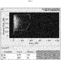
- the samples were homogenized with a syringe and run on an ImageStream instrument (Amnis Inc, Seattle, WA). The instrument was set-up to distinguish intact single cells using Aspect Ratio versus Area dot plot, e.g., as shown in FIG. 1 .
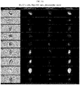
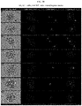
- This setting allowed: (1) identification of abnormal cells by N/C (nuclear to cytoplasmic) ratio analysis (see e.g., FIG. 2); and (2 ) quantification of E6, E7 mRNA as determined by DAPI staining and green fluorescence of the E6, E7 mRNA hybridization signal (see FIGS. 3A and 3B ).
- cell cycle analysis as determined by a DNA-staining reagent (e.g., DAPI or DRAQ5 staining of the cells) and green fluorescence of the E6, E7 mRNA hybridization signal can be used to detect abnormal cells.
- a DNA-staining reagent e.g., DAPI or DRAQ5 staining of the cells
- green fluorescence of the E6, E7 mRNA hybridization signal can be used to detect abnormal cells.
- a DNA-staining reagent e.g., DAPI or DRAQ5 staining of the cells
- green fluorescence of the E6, E7 mRNA hybridization signal can be used to detect abnormal cells.
- E6/E7 mRNA hybridization signal can be used to detect abnormal cells.
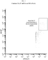
- Y-axis overexpression of E6/E7 mRNA (Y-axis) is found in cells having increased DNA content (X-axis; DNA stain is DRAQ5).
- This approach can be used substituting a p16 antibody for E6, E7 mRNA detection, or substituting E6, E7 mRNA probes with probes directed at specific microRNAs or chromosome alterations 3q- associated with cervical cancer, or substituting E6, E7 directed antibodies for E6, E7 mRNA probes.
- FIG. 5A and 5B show histograms of N/C ratios (Y-axis) versus DNA content of normal cervical cells ( FIG. 5A , top panel), LSIL cervical cells ( FIG. 5A , bottom panel), and HSIL cervical cells ( FIG. 5B , both panels).
- This alternative uses only 1 staining reagent, i.e., the non-specific DNA-staining reagent.
- a percent nucleation for squamous cells in a sample from a subject of about 80% or higher indicates that the subject has a cervical intraepithelial neoplasia (CIN) lesion (e.g., LSIL or HSIL), where a percent nucleation of about 90% or higher in the sample indicates that the subject has a high grade squamous epithelial lesion (HSIL).
- CIN cervical intraepithelial neoplasia
- HSIL high grade squamous epithelial lesion
- Table 2 Nucleation of the Squamous Cell Population as a Determinate of Abnormal Pap Smear Diagnosis Sample group Cytology % nucleated 1 Normal 47.33 2 LSIL 82.66 3 HSIL 94.38 4 HSIL 93.41 LSIL-low grade squamous epithelial lesion HSIL-high grade squamous epithelial lesion
- Cervical cell samples may include cell types (and other debris) that are not relevant to the screening assays described herein, and as such it would be advantageous to exclude them from the analysis.
- FIG. 6 shows a dot plot of combined side (orthogonal) light scatter (Y axis) and electronic volume (X axis) of cells in a cervical cytology sample. This plot shows that different cells (and other components) in the sample can be delineated using these morphometric parameters.
- the four different gates identify debris, ectocervical cells, endocervical cells, and polymorphonuclear leukocytes (PMNS). Gating can be employed to identify cells of interest to screen according to aspects of the present invention (e.g., endocervical and/or ectocervical cells).
Abstract
Description
- The Papanicolaou (PAP) smear has been the cornerstone of cervical cancer screening since 1949. By definition, the PAP smear is a stain performed on cells smeared on a slide and visualized by microscopy. Following the advent of liquid-based cervical cytology (LBC), cells from the cervix were obtained using a brush, suspended in a fixative solution, and then applied to a slide prior to staining. Highly trained cytotechnologists and cytopathologists review the stained slides looking for evidence of abnormal cells as indicated by the characteristics in Table 1.
TABLE 1 Low grade squamous intraepithelial lesion (LSIL) High grade squamous intraepithelial lesion (HSIL) Caviation (Moderate-Marked increase) N/C* Ratio (Mild increase) N/C Ratio Coarse Chromatin Hyperchromasia Hyperchromasia Irregular Nuclear Contour Single Abn Cells Syncytial Aggregates Pleomorphism * N/C = nuclear to cytoplasmic ratio. - Because of the necessity to use slides for the PAP smear, other biomarkers used for cervical cancer screening, especially those used for the molecular detection of HPV DNA, are performed on a separate aliquot of the LBC. Though some biomarkers such as p16 can be performed on a slide, the throughput is not desirable to accommodate the 60-70 million cervical cytology specimens obtained every year in the US and the 150+ million samples worldwide. Further, molecular techniques performed on a slide are cumbersome and time consuming, characteristics that are not amenable to cervical cancer screening.
- Clinically, the PAP smear and HPV testing are used together though they are very disparate technologies. The PAP smear has relatively low sensitivity (50%) and relatively high specificity (90%) for high grade cervical lesions (pre-cervical cancer and cervical cancer). Conversely, HPV DNA testing has high sensitivity (>90%) but low specificity (30%) for high grade cervical lesions (pre-cervical cancer and cervical cancer. These performance characteristics have supported the combined use of these tests for effective cervical cancer screening. Some investigators have pushed for the sole use of HPV detection for cervical cancer screening (primary HPV screening), however, current HPV DNA tests lack the specificity afforded by morphologic assessment using PAP smear, raising a concern for an overwhelming number of unnecessary colposcopy/biopsy procedures. Thus the replacement of the PAP smear by HPV testing alone remains extremely controversial.
- Methods of predicting whether a subject has a cervical intraepithelial neoplasia (CIN) lesion are provided. Aspects of the methods include obtaining morphometric data as well as biomarker data and/or non-specific cell data from a liquid cervical cellular sample by assaying the sample in suspension, and then using the different types of data to predict whether the subject has a CIN lesion. Also provided are systems that find use in practicing the methods. The methods and systems find use in a variety of applications, including cervical cancer screening applications.
- Aspects of the invention include methods of predicting whether a subject has a cervical intraepithelial neoplasia (CIN) lesion. In some instances, the methods include obtaining data from a labeled liquid sample of cervical cells in suspension from the subject, wherein the data comprises morphometric data and data selected from the group consisting of: biomarker data, DNA content data, percent nucleation data, and combinations thereof; and predicting from the data whether the subject has a CIN lesion, e.g., a CIN2+ lesion. The predicting may be characterized by a sensitivity of 85 % or higher and/or a specificity of 85 % or higher. The data may be obtained by analyzing the liquid sample with a flow cytometric device, e.g., in a method which includes flowing the sample past an illumination source and one or more optical detectors. The morphometric data may include data selected from the group consisting of: forward light scatter data, side light scatter data, image data and combinations thereof. In some instances, the labeled liquid sample is a biomarker labeled liquid sample, wherein the data comprises biomarker data, and wherein the biomarker data comprises per cell biomarker quantitation data, wherein the per cell biomarker quantitation data may include fluorescent label detection data. In some instances, the method further comprises preparing the biomarker labeled liquid sample of cervical cells. The biomarker labeled liquid sample of cervical cells may be prepared by contacting an initial cervical cell sample with a fluorescently labeled biomarker probe that specifically binds to a cervical cancer biomarker. The cervical cancer biomarker may be a nucleic acid, e.g., HPV nucleic acid (such as E6,E7 nucleic acid) or protein (e.g., p16, E6,E7). The method may include contacting the initial cervical cell sample with two or more different fluorescently labeled biomarker probes that each specifically bind to a different cervical cancer biomarker. In some instances, the cells in the initial cervical cell sample are fixed and permeabilized prior to being contacted with a fluorescently labeled biomarker probe that specifically binds to a cervical cancer biomarker. In some instances, the method further comprises recommending a cervical biopsy if abnormal cells are determined to be present in the sample. In some instances, the labeled liquid sample is a DNA labeled liquid sample, wherein the data comprises DNA content data, percent nucleation data, or both.
- Aspects of the invention also include systems comprising:
- a flow channel;
- a light source configured to direct light to an assay region of the flow channel;
- a first detector configured to receive light from the assay region of the flow channel and produce morphometric data;
a second detector configured to receive light from the assay region of the flow channel and produce additional data; and - a signal processing module configured to receive the morphometric and additional data from the first and second detectors and output a result of a prediction of whether a subject has a cervical intraepithelial neoplasia (CIN) lesion based on both the morphometric data and additional data. Systems may be configured to perform the methods of the invention, including those summarized above.
- Aspects of the invention further include methods of determining the presence of a cancerous cell in sample from a subject, the method comprising: obtaining data from a labeled liquid sample of cervical cells in suspension from the subject, wherein the data comprises morphometric data and data selected from the group consisting of: biomarker data, DNA content data, percent nucleation data and combinations thereof; a determining from the data whether a cancerous cell is present in the sample. The data may be obtained by analyzing the liquid sample with a flow cytometric device, e.g., in a method which includes flowing the sample past an illumination source and one or more optical detectors. The morphometric data may include data selected from the group consisting of: forward light scatter data, side light scatter data, image data and combinations thereof. In some instances, the labeled liquid sample is a biomarker labeled liquid sample, wherein the data comprises biomarker data, and wherein the biomarker data comprises per cell biomarker quantitation data, wherein the per cell biomarker quantitation data may include fluorescent label detection data. In some instances, the method further comprises preparing the biomarker labeled liquid sample of cervical cells. The biomarker labeled liquid sample of cervical cells may be prepared by contacting an initial cervical cell sample with a fluorescently labeled biomarker probe that specifically binds to a cervical cancer biomarker. The cervical cancer biomarker may be a nucleic acid, e.g., HPV nucleic acid (such as E6,E7 nucleic acid) or protein (e.g., p16, E6,E7). The method may include contacting the initial cervical cell sample with two or more different fluorescently labeled biomarker probes that each specifically bind to a different cervical cancer biomarker. In some instances, the cells in the initial cervical cell sample are fixed and permeabilized prior to being contacted with a fluorescently labeled biomarker probe that specifically binds to a cervical cancer biomarker. In some instances, the method further comprises recommending a cervical biopsy if abnormal cells are determined to be present in the sample. In some instances, the labeled liquid sample is a DNA labeled liquid sample, wherein the data comprises DNA content data, percent nucleation data, or both.
-
-
FIG. 1 shows instrument set-up for using Aspect Ratio versus Area dot plot to identify intact single cells (gated) in a liquid-based cervical cytology (LBC) specimen. -
FIG. 2 shows identification of abnormal cells in a LBC specimen by N/C (nuclear to cytoplasmic) ratio analysis using the instrument set up as inFIG. 1 . Increased N/C ratios are indicative of abnormal cells. -
FIG. 3A and3B show exemplary data obtained for single cells in a LBC specimen. For each cell, a brigthfield image, an image showing E6/E7 mRNA hybridization (FITC), an image showing the DNA quantification (DAPI), and an image showing a composite of the E6/E7 and DAPI images are shown.FIG. 3A shows that cells having a high N/C ratio are E6/E7 positive and have abnormal DNA content.FIG. 3B shows that cells having a low N/C ratio are E6/E7 negative and have a normal DNA content. -
FIG. 4 is a dot plot showing that overexpression of E6/E7 mRNA is found in cells having increased DNA content. Cell cycle analysis was determined using a DNA-staining reagent (DAPI ; X-axis) and E6/E7 mRNA was detected by hybridization of E6/E7 mRNA probes (FITC; Y-axis). Abnormal cells (having E6,E7 overexpression and increased DNA content) are indicated in the gate. -
FIG. 5A andB show dot plots showing N/C ratios (Y axis) and DNA content (x axis) of LBC specimens to identify abnormal cells.FIG. 5A shows dot plots of N/C ratios versus DNA content of normal cervical cells (top panel) and LSIL cervical cells (bottom panel).FIG. 5B shows dot plots of N/C ratios versus DNA content of HSIL cervical cells (top and bottom panels). This alternative uses only 1 staining reagent, i.e., the non-specific DNA-staining reagent. -
FIG. 6 shows a dot plot of combined side (orthogonal) light scatter (Y axis) and electronic volume (X axis) of cells in a cervical cytology sample. This plot shows that different cells in the sample can be delineated using these morphometric parameters. The four different gates identify debris, ectocervical cells, endocervical cells, and polymorphonuclear leukocytes (PMNS). Such gating can be employed to gate for cells of interest to analyze according to aspects of the present invention. - Methods of predicting whether a subject has a cervical intraepithelial neoplasia (CIN) lesion are provided. Aspects of the methods include obtaining morphometric as well as biomarker data and/or non-specific cell data from a liquid cervical cellular sample by assaying the sample in suspension, and then using the different types of data to predict whether the subject has a CIN lesion. Also provided are systems that find use in practicing the methods. The methods and systems find use in a variety of applications, including cervical cancer screening applications.
- Before the present invention is described in greater detail, it is to be understood that this invention is not limited to particular embodiments described, as such may, of course, vary. It is also to be understood that the terminology used herein is for the purpose of describing particular embodiments only, and is not intended to be limiting, since the scope of the present invention will be limited only by the appended claims.
- Where a range of values is provided, it is understood that each intervening value, to the tenth of the unit of the lower limit unless the context clearly dictates otherwise, between the upper and lower limit of that range and any other stated or intervening value in that stated range, is encompassed within the invention. The upper and lower limits of these smaller ranges may independently be included in the smaller ranges and are also encompassed within the invention, subject to any specifically excluded limit in the stated range. Where the stated range includes one or both of the limits, ranges excluding either or both of those included limits are also included in the invention.
- Certain ranges are presented herein with numerical values being preceded by the term "about." The term "about" is used herein to provide literal support for the exact number that it precedes, as well as a number that is near to or approximately the number that the term precedes. In determining whether a number is near to or approximately a specifically recited number, the near or approximating unrecited number may be a number which, in the context in which it is presented, provides the substantial equivalent of the specifically recited number.
- Unless defined otherwise, all technical and scientific terms used herein have the same meaning as commonly understood by one of ordinary skill in the art to which this invention belongs. Although any methods and materials similar or equivalent to those described herein can also be used in the practice or testing of the present invention, representative illustrative methods and materials are now described.
- All publications and patents cited in this specification are herein incorporated by reference as if each individual publication or patent were specifically and individually indicated to be incorporated by reference and are incorporated herein by reference to disclose and describe the methods and/or materials in connection with which the publications are cited. The citation of any publication is for its disclosure prior to the filing date and should not be construed as an admission that the present invention is not entitled to antedate such publication by virtue of prior invention. Further, the dates of publication provided may be different from the actual publication dates which may need to be independently confirmed.
- It is noted that, as used herein and in the appended claims, the singular forms "a", "an", and "the" include plural referents unless the context clearly dictates otherwise. It is further noted that the claims may be drafted to exclude any optional element. As such, this statement is intended to serve as antecedent basis for use of such exclusive terminology as "solely," "only" and the like in connection with the recitation of claim elements, or use of a "negative" limitation.
- It is appreciated that certain features of the invention, which are, for clarity, described in the context of separate embodiments, may also be provided in combination in a single embodiment. Conversely, various features of the invention, which are, for brevity, described in the context of a single embodiment, may also be provided separately or in any suitable sub-combination. All combinations of the embodiments are specifically embraced by the present invention and are disclosed herein just as if each and every combination was individually and explicitly disclosed, to the extent that such combinations embrace operable processes and/or devices/systems/kits. In addition, all sub-combinations listed in the embodiments describing such variables are also specifically embraced by the present invention and are disclosed herein just as if each and every such sub-combination of chemical groups was individually and explicitly disclosed herein.
- As will be apparent to those of skill in the art upon reading this disclosure, each of the individual embodiments described and illustrated herein has discrete components and features which may be readily separated from or combined with the features of any of the other several embodiments without departing from the scope or spirit of the present invention. Any recited method can be carried out in the order of events recited or in any other order which is logically possible.
- In further describing embodiments of the invention, aspects of embodiments of the methods will be described first in greater detail. Next, embodiments of systems that may be used in practicing methods of the invention are reviewed.
- As summarized above, embodiments of the invention are directed to methods of predicting whether a subject has a cervical intraepithelial neoplasia (CIN) lesion or cervical cancer. The term "CIN lesion" (also referred to in the art as cervical dysplasia) is used in its conventional sense to refer to the abnormal growth of squamous cells on the surface of the cervix. As is known in the art, CIN lesions may be histologically graded as CIN1, CIN2/3, CIN2 and CIN3. CIN1 lesions are those lesions that are confined to the basal 1/3 of the epithelium, and have the least risk of developing into a cancerous lesion, relative to the other categories of lesions. CIN2 lesions are characterized by moderate dysplasia confined to the basal 2/3 of the epithelium. CIN3 lesions (sometimes referred to by those of skill in the art as cervical carcinoma in situ) are categorized by the presence of severe dysplasia that traverses more than 2/3 of the epithelium. The CIN2/3 category (i.e., CIN2+) collectively refers to both CIN2 and CIN3 lesions.
- Embodiments of the invention are characterized by predicting the presence of a CIN lesion in a subject with a high degree of sensitivity and specificity. By predicting is meant to prognosticate or foresee the presence of a CIN lesion in the subject without actually taking a biopsy of the cervix of the subject. The terms sensitivity and specificity are used in their conventional sense. As such, sensitivity is a measure of the proportion of actual positives (as opposed to false positives) that are correctly identified while specificity is a measure of negatives which are correctly identified. Embodiments of the invention may predict the presence or absence of any type of CIN lesion. Embodiments of the invention may also predict the type of CIN lesion, e.g., whether the CIN lesion is a CIN1, CIN2+, CIN2 or CIN3 lesion. Embodiments of the methods make this prediction, e.g., whether a lesion is present, what type of lesion is present (such as whether a CIN2+ lesion is present) with a high degree of sensitivity and specificity. In some instances, sensitivity is 85% or more, such as 90% or more, including 95% or more. In some instances, specificity is 85% or more, such as 87% or more, including 90% or more.
- Aspects of the methods include obtaining both: (1) morphometric data and (2) biomarker data and/or non-specific cell data from a labeled liquid sample of cervical cells in suspension from the subject, e.g., a liquid sample labeled with one or both of a biomarker label and a non-specific cell label. In other words, a liquid sample of cervical cells is collected from the subject, where the cervical cells are in suspension in a fluid medium (e.g., as described in greater detail below), is assayed to obtain both morphometric data and biomarker/non-specific cell data. Thus, a labeled liquid sample according to aspects of the methods may be a biomarker labeled liquid sample, a non-specific cell labeled liquid sample, or a biomarker and non-specific cell labeled liquid sample. The labeled liquid sample of cervical cells that is assayed may be provided according to any convenient protocol.
- In one embodiment, an initial fluid cervical cellular sample is prepared by taking a cellular sample from the cervix and combining it with a suitable fluid medium. Any convenient protocol for collecting cervical cells may be employed. Examples of protocols of interest include protocols that employ a cervical brush or broom device to collect cells from the surface of the cervix and the endocervix. Descriptions of examples of cervical cell collection devices that may find use in methods of the invention are provided in
U.S. Patent Nos. 2,955,591 ;3,626,470 ;3,815,580 ;3,877,464 ;3,881,464 ;3,945,372 ;4,127,113 ;4,175,008 ;4,700,713 ;4,754,764 ;4,762,133 ;4,754,764 ;4,873,992 ;4,862,899 ;4,953,560 ;5,445,164 ;5,787,891 ;5,795,309 ;6,387,058 and6,740,049 . - Following collection, the cellular sample may be combined with a suitable liquid medium, as desired. Liquid mediums of interest include, but are not limited to: saline, or balanced salt, solutions (such as Hanks' balanced salt solution, a minimal essential (MEM) tissue culture medium, POLYSAL™ solution, and normal saline); cytology mediums, e.g., Universal Collection Medium (UCM); the universal collection medium described in
U.S. Patent No. 7,371,518 (the disclosure of which is herein incorporated by reference); Standard Transport Medium (STM), PRESERVCYT™ fluid medium (Cytyc, Inc. (Boxborough, Mass.)); CytoRich™ fluid medium (TriPath, Inc. (Burlington, N.C.); and the like. - Where desired, the collected initial sample may be assessed for adequacy prior to proceeding further in the process. For example, an aliquot of the sample may be subjected to light scatter analysis to determine whether adequate target cells are present in the sample, e.g., as described in
U.S. Patent No. 6,329,167 ; the disclosure of which is herein incorporated by reference. - Following preparation, the resultant initial fluid cervical cellular sample may be fixed and/or permeabilized as desired. As such, methods of the invention include fixing the cellular sample by contacting the sample with a suitable fixation reagent. Fixation reagents of interest are those that fix the cells at a desired timepoint. Any convenient fixation reagent may be employed, where suitable fixation reagents include, but are not limited to: formaldehyde, paraformaldehyde, formaldehyde/acetone, methanol/acetone, IncellFP (IncellDx, Inc) etc. For example, paraformaldehyde used at a final concentration of about 1 to 2% has been found to be a good crosslinking fixative. In some instances, the cells in the sample are permeabilized by contacting the cells with a permeabilizing reagent. Permeabilizing reagents of interest are reagents that allow the labeled biomarker probes, e.g., as described in greater detail below, to access to the intracellular environment. Any convenient permeabilizing reagent may be employed, where suitable reagents include, but are not limited to: mild detergents, such as Triton X-100, NP-40, saponin, etc.; methanol, and the like. It may also be desirable to label cells with a positive heavy metal control, e.g. a DNA intercalator labeled with a heavy metal, e.g. iridium, etc. Cells may also be stained with a viability dye prior to fixation, e.g. ethidium bromide, propidium iodide, DAPI, RhCl3, etc., as desired.
- In certain embodiments, and as reviewed above, the sample that is assayed to obtain morphometric and biomarker data is a biomarker labeled sampled. Accordingly, the sample is one that has been labeled for one or more biomarkers of interest. By "biomarker labeled sample" is meant a sample which has been contacted with a labeled biomarker probe (e.g., as described in greater detail below) that specifically binds to a biomarker of interest if the biomarker is present in the cellular sample. Biomarkers of interest include, but are not limited to, cervical cancer biomarkers. Cervical cancer biomarkers are a distinctive biological or biologically derived indicator, for example nucleic acids or proteins, whose presence is associated or linked with either the propensity of a subject to develop cervical cancer or the presence of cervical cancer in a subject. Accordingly, biomarkers of interest include nucleic acid and protein analytes whose presence and/or amount in a cell can be used to make a prediction of at least the propensity of a subject to suffer from cervical cancer. Biomarkers of interest include, but are not limited to: HPV expression products of HPV genes, such as HPV genes L1, L2, E2, E4, E5, E6 or E7; cyclin-dependent kinase inhibitors, e.g., p14, p15INK4b, p16 (i.e., p16INK4a as described in Serrano, M., et al., Nature, 1993 Dec. 16; 366(6456): 704-7), p18INK4c, p19INK4d, p21WAF1/CIP1 and p27KIP1; cell cycle regulatory proteins, e.g., p14ARF; specific microRNAs or chromosome alterations 3q- associated with cervical cancer, such as described in United States Patent Publication No.
20100234445 (the disclosure of which is herein incorporated by reference); etc. - The biomarker labeled liquid cervical cellular sample that is assayed in methods of the invention may be prepared using any convenient labeling protocol. In some embodiments, preparation of the biomarker labeled liquid sample includes contacting an initial cervical cell sample with a labeled biomarker probe that specifically binds to a cervical cancer biomarker. Depending on the particular assay to be performed, the initial sample may be combined with a single labeled biomarker probe or two or more distinct labeled biomarker probes that bind to different biomarkers of different molecular composition, where the number of such distinct labeled biomarker probes may be two or more, e.g., three or more, four or more, five or more, etc; e.g., where the assay is a multiplex assay for two or more biomarkers.
- In contacting the initial sample with the labeled biomarker probe(s), the sample is combined with one or more labeled biomarker probes to produce a reaction mixture. Labeled biomarker probes of interest include a specific binding domain and a label domain. The specific binding domain comprises a capture ligand that specifically binds to the biomarker of interest. Depending on the particular assay, the biomarker of interest may be a variety of different types of molecules, including but not limited to: proteins, polypeptides, proteoglycans, glycoproteins and the respective fragments of these molecules; nucleic acids, e.g., DNA and RNA, such as mRNA, etc. The capture ligand is therefore a ligand that binds to the biomarker molecule of interest, wherein this capture ligand may of course vary depending on the specific type of biomarker molecule to be detected, e.g., antibody for protein biomarker, oligonucleotide for mRNA biomarker. In certain embodiments, the affinity between a capture ligand and the biomarker molecule to which it specifically binds when they are specifically bound to each other in a binding complex is characterized by a KD (dissociation constant) of 10-6 M or less, 10-7 M or less, 10-8 M or less, 10-9 M or less, 10-10 M or less, 10-11 M or less, 10-12 M or less, 10-13 M or less, 10-14 M or less, including 10-15 M or less.
- As indicated above, a variety of different types of specific binding agents may be employed as the capture ligands, where the particular type of binding agent is selected based, at least in part, on the particular type of molecule of the biomarker of interest. Specific binding agents of interest include antibody binding agents, proteins, peptides, haptens, nucleic acids, etc. The term "antibody binding agent" as used herein includes polyclonal or monoclonal antibodies or fragments that are sufficient to bind to an analyte of interest. The antibody fragments can be, for example, monomeric Fab fragments, monomeric Fab' fragments, or dimeric F(ab)'2 fragments. Also within the scope of the term "antibody binding agent" are molecules produced by antibody engineering, such as single-chain antibody molecules (scFv) or humanized or chimeric antibodies produced from monoclonal antibodies by replacement of the constant regions of the heavy and light chains to produce chimeric antibodies or replacement of both the constant regions and the framework portions of the variable regions to produce humanized antibodies. Nucleic acid binding agents of interest are nucleic acids that specifically bind to biomarker nucleic acids in a cell. The length of these nucleic acids may vary, so long as it is sufficient for the oligonucleotide to serve as a specific binding agent, and in some instances ranges from 13 to 100 nt, such as 14 to 50 nt, e.g., 15 to 25 nt. The oligonucleotides that make up these nucleic acid binding agents may be DNA or RNA, or a synthetic analogue thereof, as desired.
- In addition to the specific binding domain, the labeled biomarker probes further include a detectable label. Of interest as detectable labels are fluorescent dyes. Fluorescent dyes (fluorophores) can be selected from any of the many dyes suitable for use in imaging applications (e.g., flow cytometry). A large number of dyes are commercially available from a variety of sources, such as, for example, Molecular Probes (Eugene, OR) and Exciton (Dayton, OH). Examples of fluorophores of interest include, but are not limited to, 4-acetamido-4'-isothiocyanatostilbene-2,2'disulfonic acid; acridine and derivatives such as acridine, acridine orange, acrindine yellow, acridine red, and acridine isothiocyanate; 5-(2'-aminoethyl)aminonaphthalene-1-sulfonic acid (EDANS); 4-amino-N-[3-vinylsulfonyl)phenyl]naphthalimide-3,5 disulfonate (Lucifer Yellow VS); N-(4-anilino-1-naphthyl)maleimide; anthranilamide; Brilliant Yellow; coumarin and derivatives such as coumarin, 7-amino-4-methylcoumarin (AMC, Coumarin 120), 7-amino-4-trifluoromethylcouluarin (Coumaran 151); cyanine and derivatives such as cyanosine, Cy3, Cy5, Cy5.5, and Cy7; 4',6-diaminidino-2-phenylindole (DAPI); 5', 5"-dibromopyrogallol-sulfonephthalein (Bromopyrogallol Red); 7-diethylamino-3-(4'-isothiocyanatophenyl)-4-methylcoumarin; diethylaminocoumarin; diethylenetriamine pentaacetate; 4,4'-diisothiocyanatodihydro-stilbene-2,2'-disulfonic acid; 4,4'-diisothiocyanatostilbene-2,2'-disulfonic acid; 5-[dimethylamino]naphthalene-1-sulfonyl chloride (DNS, dansyl chloride); 4-(4'-dimethylaminophenylazo)benzoic acid (DABCYL); 4-dimethylaminophenylazophenyl-4'-isothiocyanate (DABITC); eosin and derivatives such as eosin and eosin isothiocyanate; erythrosin and derivatives such as erythrosin B and erythrosin isothiocyanate; ethidium; fluorescein and derivatives such as 5-carboxyfluorescein (FAM), 5-(4,6-dichlorotriazin-2-yl)aminofluorescein (DTAF), 2'7'-dimethoxy-4'5'-dichloro-6-carboxyfluorescein (JOE), fluorescein isothiocyanate (FITC), fluorescein chlorotriazinyl, naphthofluorescein, and QFITC (XRITC); fluorescamine; IR144; IR1446; Green Fluorescent Protein (GFP); Reef Coral Fluorescent Protein (RCFP); Lissamine™; Lissamine rhodamine, Lucifer yellow; Malachite Green isothiocyanate; 4-methylumbelliferone; ortho cresolphthalein; nitrotyrosine; pararosaniline; Nile Red; Oregon Green; Phenol Red; B-phycoerythrin; o-phthaldialdehyde; pyrene and derivatives such as pyrene, pyrene butyrate and succinimidyl 1-pyrene butyrate; Reactive Red 4 (Cibacron™ Brilliant Red 3B-A); rhodamine and derivatives such as 6-carboxy-X-rhodamine (ROX), 6-carboxyrhodamine (R6G), 4,7-dichlororhodamine lissamine, rhodamine B sulfonyl chloride, rhodamine (Rhod), rhodamine B, rhodamine 123, rhodamine X isothiocyanate, sulforhodamine B, sulforhodamine 101, sulfonyl chloride derivative of sulforhodamine 101 (Texas Red), N,N,N',N'-tetramethyl-6-carboxyrhodamine (TAMRA), tetramethyl rhodamine, and tetramethyl rhodamine isothiocyanate (TRITC); riboflavin; rosolic acid and terbium chelate derivatives; xanthene; or combinations thereof. Other fluorophores or combinations thereof known to those skilled in the art may also be used, for example those available from Molecular Probes (Eugene, OR) and Exciton (Dayton, OH).
- Where multiple distinct labeled biomarker probes are employed, the label of each distinct probe may be chosen to provide a distinguishable signal. For example, in embodiments where first and second distinct labeled biomarker probes are employed, the label in the second probe is a fluorescent label which produces a fluorescent signal that is distinguishable from the first fluorescent signal of the label of the first probe. Accordingly, the first and second fluorescent signals produced upon excitation of the first and second fluorescent labels are distinguishable from each other, meaning that both can be detected at the same time and that the signal from one does not modify or change the signal from the other. Each distinct label may produce signals that are distinguishable from any other label. For example, the cells may be stained with a third fluorescent label which produces a third fluorescent signal that is distinguishable from the first and second fluorescent signals.
- In some instances, the labeled biomarker probe that is employed is an HPV E6, E7 oligonucleotide fluorescently labeled probe. Such probes may vary in length, ranging in some instances from 13 to 100 nt, such as 14 to 50 nt, e.g., 15 to 25 nt. The particular sequence of such probes may vary. Specific sequences of interest include, but are not limited to those found in the probe cocktail of the HPV OncoTect™ E6,E7 mRNA Detection Kit (incellDx, Menlo Park, CA).
- Exemplary probe sequences specific for HPV E6/E7 gene (or mRNA) include those described in Faulkner-Jones et al. (J. Virol Methods 1993 vol. 41 pages 277-296; incorporated herein by reference). The sequences from Table 1 of Faulkner-Jones et al., which shows such exemplary HPV E6/E7 specific probe sequences, are provided below.
HPV Type Region Sequence (5'-3') 6b/11 E6 + E7 TTAGGGTAACATGTCTTCCATGCATGTTGT (SEQ ID NO:01) E7 ACGTTGCTGTCACATCCACAGCAACAGGTCA (SEQ ID NO:02) 16 E6 TGCAACAAGACATACATCGACCGGTCCACCGAC (SEQ ID NO:03) E7 GGTTACAATATTGTAATGGGCTCTCTCCGG (SEQ ID NO:04) E6 TTTCAGGACCCACAGGAGCGACCCAGAAAG (SEQ ID NO:05) 18 E6 + E7 TCGAGCACGAATGGCACTGGCCTCTATAGTGCCCAG (SEQ ID NO:06) E7 GGTCAACCGGAATTTCATTTTGGGGCTCTAAATG (SEQ ID NO:07) 33 E6 CTTGGCACAAATCATGCAATGTTCGTGGTT (SEQ ID NO:08) - Additional exemplary probe sequences for HPV E6/E7 include those described in Plummer et al. (Diagnostic Mol. Path. 1998 vol. 7, pages 76-84; incorporated herein by reference). The sequences from Table 1 in Plummer et al., which shows such exemplary HPV E6/E7 probe sequences, are provided below.
HPV Type Region Sequence (5'-3') HPV16 E6 GTCCTGAAACATTGCAGTTCTCTTTTGGTG (SEQ ID NO:09) E6 CTGTGCATAACTGTGGTAACTTTCTGGGTC (SEQ ID NO:10) E6 TCACACAACGGTTTGTTGTATTGCTGTTCT (SEQ ID NO:11) E6/E7 TGGGTTTCTCTACGTGTTCTTGATGATCTG (SEQ ID NO:12) E7 TAACAGGTCTTCCAAAGTACGAATGTCTAC (SEQ ID NO:13) E7 TATGGTTTCTGAGAACAGATGGGGCACACA (SEQ ID NO:14) HPV18 E6 AGTGTTCAGTTCCGTGCACAGATCAGGTAG (SEQ ID NO:15) E6 CCTCTGTAAGTTCCAATACTGTCTTGCAAT (SEQ ID NO:16) E6 CCTCTATAGTGCCCAGCTATGTTGTGAAAT (SEQ ID NO:17) E6/E7 TTGTGTTTCTCTGCGTCGTTGGAGTCGTTC (SEQ ID NO:18) E7 CTGGCTTCACAC17ACAACACATACACAAC (SEQ ID NO:19) E7 TGCTCGAAGGTCGTCTGCTGAGCTTTCTAC (SEQ ID NO:20) - Additional HPV specific probe sequences that find use in the methods and systems described herein may be designed and tested using any convenient probe design methodology.
- In preparing the reaction mixture, the sample may be combined with the labeled biomarker probes using any convenient protocol. Combination may be carried out with mixing, as desired. Contact of the sample with the labeled biomarker probes is performed under incubation conditions that provide for binding of probes to their respective biomarkers, if present, in the sample. In some instances, the probes and samples are contacted and combined at a temperature ranging from 15 to 50, such as from 20 to about 40 °C. Contact may be performed with mixing or agitation, e.g., with vortexing etc., to provide for sufficient combination of the reaction components and the sample.
- The resultant reaction mixture may then be maintained or incubated for a period of time prior to assay for morphometric and biomarker data, e.g., via flow cytometric analysis (e.g., as described in greater detail below). In some instances, the reaction mixture is incubated at a temperature ranging from 15 to 50, such as from 20 to about 40 °C for a period of time ranging from about 30 minutes to 72 hours, such as 1 hour to 24 hours, including 1 hour to 3 hours. Following the above incubation step, the sample may be assayed immediately or stored for assay at a later time. If stored, in some embodiments the sample is stored at a reduced temperature; e.g., on ice.
- Where desired, the resultant reaction mixture may be washed, e.g., to remove any unbound probes and other sample components. Washing may be performed using any convenient protocol, e.g., by combining the reaction mixture with a suitable wash buffer and separating the cells from the fluid. A given washing protocol may include one or more distinct washing steps, as desired. Following any washing protocol, the labeled cells may be re-suspended in a suitable liquid, e.g., the washing buffer or another buffer, for subsequent analysis, e.g., via flow cytometric analysis.
- In some embodiments, as described above, the labeled liquid cell sample is labeled (or stained) with a non-specific cell stain, either in addition to or in the absence of a biomarker label. The cells may be stained with a non-specific stain using any convenient protocol. Of interest as non-specific cells stains are DNA specific stains. Dyes and stains that are specific for DNA (or preferentially bind double stranded polynucleotides in contrast to single-stranded polynucleotides) and therefore may be employed as non-specific stains include, but are not limited to: Hoechst 33342 (2'-[4-ethoxyphenyl]-5-[4-methyl-1-piperazinyl]-2,5'-bi-1H-benzimidazole) and Hoechst 33258 (2'-[4-ethoxyphenyl]-5-[4-methyl-1-piperazinyl]-2,5'-bi-1H-benzimidazole) and others of the Hoechst series; SYTO 40,
SYTO 11, 12, 13, 14, 15, 16, 20, 21, 22, 23, 24, 25 (green); SYTO 17, 59 (red), DAPI, DRAQ5™ (an anthraquinone dye with high affinity for double stranded DNA), YOYO-1, propidium iodide, YO-PRO-3, TO-PRO-3, YOYO-3 and TOTO-3, SYTOX Green, SYTOX, methyl green, acridine homodimer, 7-aminoactinomycin D, 9-amino-6-chloro-2-methoxyactridine. Depending on the particular stain and assay, the stain may serve in quantitation of biomarker, as an indication of cell cycle, etc. - Following preparation of the labeled sample, e.g., as described above, the sample is assayed to obtain both morphometric as well as biomarker and/or non-specific cell data. In some instances, the same aliquot of sample, i.e., the same physical quantity of sample, is assayed to obtain both the morphometric and biomarker/non-specific cell data. Accordingly, these embodiments are distinguished from protocols in which a first aliquot of a sample is assayed using one protocol, e.g., slide based protocol, for morphometric data and a second aliquot of the sample is assayed using another protocol, e.g., flow cytometric protocol.
- Morphometric data refers to any type of data from which cell morphology information, i.e., information about the size, shape and/or structure of the cells, may be derived. Morphometric data of interest includes, but is not limited to data selected from the group consisting of: forward light scatter data, side light scatter data, image data and combinations thereof. Morphological parameters of interest include, but are not limited to: nuclear area, perimeter, texture or spatial frequency content, centroid position, shape (i.e., round, elliptical, barbell-shaped, etc.), volume, and ratios of any of these parameters. The obtained morphometric date may be for cells as a whole or for subparts thereof, e.g., the cytoplasm of cells. In some instances, the morphometric data may include an actual designation of whether a cell is normal or abnormal, including the type of abnormal cell. For example, morphometric data may, in some instances, include a designation that a given cell is abnormal, for example that the cell is: an atypical squamous cell, e.g., atypical squamous cell of undetermined significance (ASC-US), atypical squamous cell - cannot exclude HSIL (ASC-H); low grade squamous intraepithelial lesion (LGSIL or LSIL); high grade squamous intraepithelial lesion (HGSIL or HSIL); squamous cell carcinoma; atypical glandular cell not otherwise specified (AGC-NOS); atypical clandular cell, suspicious for AIS or cancer (AGC-neoplastic); and adenocarcinoma in situ (AIS).
- Biomarker data refers to any type of data from which biomarker information for the cell may be derived. In some instances, biomarker data is data that includes a signal emitted by the label of the labeled biomarker probe that is employed in the assay. The biomarker data may be in the form of the presence and amplitude of emitted light, the number of discrete positions in a cell or other object from which the light signal(s) originate(s), the relative placement of the signal sources, and the color (wavelength or waveband) of the light emitted at each position in the cell. The biomarker data may take the form of qualitative, semi-quantitative or quantitative data. Qualitative data is simply the presence or absence of the biomarker. Semi-quantitative or quantitative data is data that provides some indication of the amount, e.g., copy number, concentration, etc., of the biomarker in the cell. For example, semi-quantitative data may take the form of an indication that the copy number of a biomarker of interest is above a certain threshold number. Quantitative data provides an indication of an absolute value, e.g., copy number, amount, etc., of the biomarker in the cell. Semi-quantitative and quantitative data may be collectively referred to as biomarker quantitation data.
- In some instances, the biomarker of interest is an mRNA that is present when a subject is infected with a high risk HPV strain. mRNAs of interest include an HPV E6, E7 mRNA. In these embodiments, the absolute copy number of the mRNA species of interest may be determined, such that the quantitation data of the mRNA species of interest is obtained. In some instances where the biomarker is HPV E6, E7, the total amount of E6, E7 mRNA species per cell is determined. In these instances, of interest is the identification of cells that 2 or more, such as 5 or more, including 10 or more, 50 or more, 100 or more, 200 or more, 500 or more HPV E6, E7 mRNA copies per cell, as these cells may be associated with the presence of a CIN lesion in the host. In some instances, the biomarker data that is obtained in methods of the invention provides information that there are 2 to 1000 copies of HPV E6, E7 mRNA per cell, e.g., 5 to 750 copies of HPV E6, E7 mRNA per cell, including 10 to 500 copies of HPV E6, E7 mRNA per cell. In some instances, the biomarker data that is obtained is the HPV E6, E7 mRNA copy number per cell data as described in
U.S. Patent No. 7,524,631 , the disclosure of which is herein incorporated by reference. Determination of the copy number may include comparing a single to a suitable control, e.g. as provided by a non-specific DNA stain (such as described below), by a reference value, etc. - In some instances, photometric measurements are also obtained. Photometric measurements enable the determination of nuclear optical density, cytoplasm optical density, background optical density, and the ratios of any of these values.
- Non-specific cell data is any type of data from which non-specific (i.e., non-biomarker specific) cell information for the cells in the sample may be derived. In some instances, non-specific cell data is data that includes a signal emitted by a non-specific cell stain that is employed in the assay. The non-specific cell data may be in the form of the presence and amplitude of emitted light, the number of discrete positions in a cell or other object from which the light signal(s) originate(s), the relative placement of the signal sources, and the color (wavelength or waveband) of the light emitted at each position in the cell. The non-specific cell data may take the form of qualitative, semi-quantitative or quantitative data. For example, quantitative or semi-quantitative non-specific cell data may include data on the DNA content of a cell obtained using a DNA stain (e.g., as described above). Other quantitative data includes electronic volume (EV). Thus, non-specific cell data may provide an indication of an absolute or relative value, e.g., copy number, amount, etc., of the non-specific cell component in the cell. Semi-quantitative and quantitative data may be collectively referred to as non-specific cell quantitation data. Qualitative non-specific cell data can provide information with respect to the presence of absence of a particular feature of a cell. For example, in cells stained with a DNA stain, the presence or absence of a cell nucleus can be determined (e.g., the number of nucleated cells in a sample, also referred to as percent nucleation).
- It is noted that where multiple distinct labels are to be detected in a single labeled liquid cell sample, e.g., biomarker probe labels and non-specific cell stains used in a single sample, the labels and stains employed may be chosen to provide a distinguishable signal (as described above for using multiple biomarker probes in a single sample, above). For example, in embodiments where a labeled biomarker probe and a DNA stain are employed, the label for the biomarker probe is a fluorescent label which produces a fluorescent signal that is distinguishable from the fluorescent signal of the DNA stain. Accordingly, the fluorescent signals produced upon excitation of the biomarker probe label and the DNA stain are distinguishable from each other, meaning that both can be detected at the same time and that the signal from one does not modify or change the signal from the other. Each distinct label may produce signals that are distinguishable from any other label. For example, the cells may be stained with three fluorescent labels which produce three distinct fluorescent signals that are distinguishable from each other upon excitation.
- The above types of data, e.g., the morphometric, biomarker, and non-specific cell data, may be obtained from the same aliquot of sample using any convenient protocol. In some instances, a flow cytometric protocol is employed which collects each type of data. Flow cytometry is a well-known methodology using multi-parameter data for identifying and distinguishing between different particle (e.g., cell) types i.e., particles that vary from one another terms of label (wavelength, intensity), size, etc., in a fluid medium. In flow cytometrically analyzing the sample prepared as described above, an aliquot of the sample is first introduced into the flow path of the flow cytometer. When in the flow path, the cells in the sample are passed substantially one at a time through one or more sensing regions, where each of the cells is exposed separately individually to a source of light at a single wavelength (or in some instances two or more distinct sources of light) and measurements of morphometric parameters, e.g., light scatter parameters, and/or biomarker parameters, e.g., fluorescent emissions, as desired, are separately recorded for each cell. The data recorded for each cell is analyzed in real time or stored in a data storage and analysis means, such as a computer, for later analysis, as desired.
- More specifically, in a flow cytometer, the cells are passed, in suspension, substantially one at a time in a flow path through one or more sensing regions where in each region each cell is illuminated by an energy source. The energy source may include an illuminator that emits light of a single wavelength, such as that provided by a laser (e.g., He/Ne or argon) or a mercury arc lamp with appropriate filters. For example, light at 488 nm may be used as a wavelength of emission in a flow cytometer having a single sensing region. For flow cytometers that emit light at two distinct wavelengths, additional wavelengths of emission light may be employed, where specific wavelengths of interest include, but are not limited to: 535 nm, 635 nm, and the like.
- In series with a sensing region, detectors, e.g., light collectors, such as photomultiplier tubes (or "PMT"), image collectors (e.g., in the form of charge-coupled devices (CCDs)), etc., are used to record light that passes through each cell (generally referred to as forward light scatter), light that is reflected orthogonal to the direction of the flow of the cells through the sensing region (generally referred to as orthogonal or side light scatter) and fluorescent light emitted from the cells, if it is labeled with fluorescent marker(s), as the cells passes through the sensing region and is illuminated by the energy source. Each type of data that is obtained, e.g., forward light scatter (or FSC), orthogonal light scatter (SSC), and fluorescence emissions (FL1, FL2, etc.) and image, etc., comprise a separate parameter for each cell (or each "event").
- Flow cytometers further include data acquisition, analysis and recording means, such as a computer, wherein multiple data channels record data from each detector for the morphometric and biomarker data emitted by each cell as it passes through the sensing region. The purpose of the analysis system is to classify and count cells wherein each cell presents itself as a set of digitized parameter values. In flow cytometrically assaying cells in methods of the invention, the flow cytometer may be set to trigger on a selected parameter in order to distinguish the cells of interest from background and noise. "Trigger" refers to a preset threshold for detection of a parameter. It is typically used as a means for detecting passage of cell through the laser beam. Detection of an event which exceeds the threshold for the selected parameter triggers acquisition of light scatter and fluorescence data for the cell. Data is not acquired for cells or other components in the medium being assayed which cause a response below the threshold. The trigger parameter may be the detection of forward scattered light caused by passage of a cell through the light beam. The flow cytometer then detects and collects the light scatter and fluorescence data for cell.
- A particular subpopulation of interest is then further analyzed by "gating" based on the data collected for the entire population. To select an appropriate gate, the data is plotted so as to obtain the best separation of subpopulations possible. This procedure is typically done by plotting forward light scatter (FSC) vs. side (i.e., orthogonal) light scatter (SSC) on a two dimensional dot plot. The flow cytometer operator then selects the desired subpopulation of cells (i.e., those cells within the gate) and excludes cells which are not within the gate. Where desired, the operator may select the gate by drawing a line around the desired subpopulation using a cursor on a computer screen. Only those cells within the gate are then further analyzed by plotting the other parameters for these cells, such as fluorescence.
- Any flow cytometer that is capable of obtaining both the morphometric and biomarker/non-specific cell data, e.g., as described above, from the same aliquot of a liquid sample may be employed. Of interest are those flow cytometer systems described in
U.S. Patent Nos. 6211955 ,6249341 ,6256096 ,6473176 ,6507391 ,6532061 ,6563583 ,6580504 ,6583865 ,6608680 ,6608682 ,6618140 ,6671044 ,6707551 ,6763149 ,6778263 ,6875973 ,6906792 ,6934408 ,6947128 ,6947136 ,6975400 ,7006710 ,7009651 ,7057732 ,7079708 ,7087877 ,7190832 ,7221457 ,7286719 ,7315357 ,7450229 ,7522758 ,7567695 ,7610942 ,7634125 ,7634126 ,7719598 ; the disclosures of which are herein incorporated by reference. - Morphometric and biomarker/non-specific cell data obtained from the same aliquot of cervical cellular sample, e.g., as described above, is employed to whether the subject has a CIN lesion, as described above. Various combinations of morphometric, biomarker, and non-specific cell data may be employed in making a prediction of whether a subject has a CIN lesion. Combinations of interest include, but are not limited to, the following: a) nuclear to cytoplasmic (N/C) ratio analysis (e.g., to identify abnormal cells) coupled with quantification of E6, E7 mRNA; b) N/C ratio analysis coupled with cell cycle analysis as determined by DAPI staining and green fluorescence of the E6, E7 mRNA hybridization signal; c) N/C ratio analysis coupled with cell cycle analysis as determined by p16 staining and green fluorescence of the E6, E7 mRNA hybridization signal; d) N/C ratio analysis coupled with cell cycle analysis as determined by DAPI (or other DNA stain) staining, etc.
- Increased, or high, N/C ratios include N/C ratios that are 0.25 or higher, 0.30 or higher, 0.4 or higher, 0.5 or higher, 0.6 or higher, 0.7 or higher, 0.8 or higher, 0.9 or higher, 0.95 or higher, etc.
- In addition to N/C ratio, nuclear area (NA) assessment may be used as morphometric data. Cells in a cervical cell sample having an increased nuclear area, e.g., as compared to nuclei in normal intermediate squamous cells, can be identified cells as abnormal cells. Abnormal cells may have a nuclear area-to-nuclear area ratio (nuclear area of cell of interest/nuclear area of normal intermediate squamous cell) of 1.25 or more, 1.75 or more, 2.0 or more, 2.25 or more, 2.75 or more, 3.0 or more, 3.25 or more, 3.5 or more, etc. It is noted here that using standard microscopy observations, the accuracy of estimating nuclear area is low (see, e.g., Schmidt et al., 2008, "Visual estimates of nucleus-to-nucleus ratios: can we trust our eyes to use the Bethesda ASCUS and LSIL size criteria?" Cancer 114(5):287-93. Aspects of the present invention provide a more accurate and reproducible assessment of nuclear area.
- Additional data parameters may also be employed in analyzing the cells in the cervical cell sample, including side (orthogonal) light scatter, forward scatter, and electronic volume (EV; a measure of cell size; based on the Coulter principle). Such parameters find use in identifying populations or sub-populations of cells in the sample that are to be analyzed for the presence of abnormal cells (see, e.g.,
FIG. 6 and its description herein). For example, analysis of EV and DNA content (e.g., using DAPI) allows the differentiation of enucleated from nucleated squamous cells. - In some instances, the methods include determining that the assayed cellular sample includes cancerous cells. In these embodiments, the methods include identifying the presence of one or more cancerous cells in the sample, where the identification is made based on morphometric and biomarker data, e.g., as described above. Such embodiments may or may not include predicting the presence of CIN in a subject, since the methods of these embodiments identify the presence of actual cancerous cells in the sample.
- In some instances, the prediction of the presence of a CIN lesion is made within a short period of time following introduction of the sample into the cytometer. Accordingly, results may be provided to a user in a period of 6 hours or less, such as 3 hours or less, e.g., 2 hours or less, including 1 hour or less. Where desired, the overall assay time which ranges from obtainment of the sample from the subject to delivery of the result to the subject is 6 hours or less, such as 5 hours or less, e.g., 4 hours less, including 3 hours or less, e.g., 2 hours or less.
- In some instances, the methods further include performing further analysis of a subject if the methods result in a prediction of a CIN lesion in the subject. For example, where methods of the invention result in prediction of a CIN2+ lesion in a subject, the methods may then further include providing a recommendation to a subject that further action be taken, e.g., in the form of further diagnostic procedures, such as biopsy. In some instances, the methods include taking further diagnostic action. Further diagnostic action may include a colposcopy, in which a magnified visual inspection of the cervix is performed to identify abnormal cells on the surface of the cervix. If the biopsy indicates that cancer or pre-cancerous lesions may be present, further diagnostic and treatment procedures may be taken, such as loop electrical excision procedure (LEEP) and conization, in which the inner lining of the cervix is removed to be examined pathologically.
- While the methods are suitable for use with a variety of different female mammalian subjects, of interest are use of the methods with human female subjects, such as human female subjects of 10 years age or older, e.g., 15 years age or older, including 20 years age or older.
- Aspects of the invention further include systems for use in practicing the subject methods. Systems of interest include a flow cytometer configured to assay a liquid sample for both morphometric and biomarker data, e.g., as described above. Flow cytometers of interest include, but are not limited, to those devices described in
U.S. Patent Nos.: 4,704,891 ;4,727,029 ;4,745,285 ;4,867,908 ;5,342,790 ;5,620,842 ;5,627,037 ;5,701,012 ;5,895,922 ; and6,287,791 which, if necessary, are modified to include the ability to obtain image data as described above, as well as those cytometers described inU.S. Patent Nos. 6211955 ,6249341 ,6256096 ,6473176 ,6507391 ,6532061 ,6563583 ,6580504 ,6583865 ,6608680 ,6608682 ,6618140 ,6671044 ,6707551 ,6763149 ,6778263 ,6875973 ,6906792 ,6934408 ,6947128 ,6947136 ,6975400 ,7006710 ,7009651 ,7057732 ,7079708 ,7087877 ,7190832 ,7221457 ,7286719 ,7315357 ,7450229 ,7522758 ,7567695 ,7610942 ,7634125 ,7634126 ,7719598 ; the disclosures of which are herein incorporated by reference. - In some instances, the flow cytometer includes: a flow channel; at least a first light source configured to direct light to an assay region of the flow channel (where in some instances the cytometer includes two or more light sources); a first detector configured to receive light of a first wavelength from the assay region of the flow channel; and a second detector configured to receive light of a second wavelength from the assay region of the flow channel; and an image detector configured to obtain image data of cells. Such a cytometer would have at least two detection channels in addition to the image detector. In some instances, the device may include more than two detection channels, e.g., 3 or more, 4 or more, 5 or more, 10 or more, etc.
- Aspects of the invention further include signal processing module configured to receive the morphometric and biomarker data from the first and second detectors and output a result of a prediction of whether a subject has a cervical intraepithelial neoplasia (CIN) lesion based on both the morphometric data and biomarker data. The signal processing module may be integrated into the cytometer as a single device, or distributed from the cytometer where the signal processing module and cytometer are in communication with each other, e.g., via a wired or wireless communication protocol.
- Accordingly, aspects of the invention further include systems, e.g., computer based systems, which are configured to predict the presence of a CIN lesion in a subject, e.g., as described above. A "computer-based system" refers to the hardware means, software means, and data storage means used to analyze the information of the present invention. The minimum hardware of the computer-based systems of the present invention comprises a central processing unit (CPU), input means, output means, and data storage means. A skilled artisan can readily appreciate that any one of the currently available computer-based system are suitable for use in the present invention. The data storage means may comprise any manufacture comprising a recording of the present information as described above, or a memory access means that can access such a manufacture.
- To "record" data, programming or other information on a computer readable medium refers to a process for storing information, using any such methods as known in the art. Any convenient data storage structure may be chosen, based on the means used to access the stored information. A variety of data processor programs and formats can be used for storage, e.g. word processing text file, database format, etc.
- A "processor" references any hardware and/or software combination that will perform the functions required of it. For example, any processor herein may be a programmable digital microprocessor such as available in the form of a electronic controller, mainframe, server or personal computer (desktop or portable). Where the processor is programmable, suitable programming can be communicated from a remote location to the processor, or previously saved in a computer program product (such as a portable or fixed computer readable storage medium, whether magnetic, optical or solid state device based). For example, a magnetic medium or optical disk may carry the programming, and can be read by a suitable reader communicating with each processor at its corresponding station.
- Embodiments of the subject systems include the following components: (a) a communications module for facilitating information transfer between the system and one or more users, e.g., via a user computer, as described below; and (b) a processing module for performing one or more tasks involved in the quantitative analysis methods of the invention.
- In certain embodiments, a computer program product is described comprising a computer usable medium having control logic (computer software program, including program code) stored therein. The control logic, when executed by the processor of the computer, causes the processor to perform functions described herein. In other embodiments, some functions are implemented primarily in hardware using, for example, a hardware state machine. Implementation of the hardware state machine so as to perform the functions described herein may be accomplished using any convenient method and techniques.
- In addition to the sensor device and signal processing module, e.g., as described above, systems of the invention may include a number of additional components, such as data output devices, e.g., monitors and/or speakers, data input devices, e.g., interface ports, keyboards, etc., fluid handling components, power sources, etc.
- In some instances, the systems may further include a reaction mixture derivative thereof (e.g., washed cells produced therefrom), where the reaction mixture is prepared as described above, e.g., by combining a sample, one or more labeled biomarker probes and, optional, a non-specific stain.
- The subject methods and systems find use in a variety of different applications where detection prediction of a CIN lesion in a subject is desired. Such applications include both research and diagnostic applications, e.g., applications where a subject is diagnosed with respect to the presence of or propensity to develop cervical cancer, e.g., as described above. The clinical utility of the methods and systems described herein provide powerful tools to detect and screen HPV related pathogenesis and cervical cancer development in both early and late stages, thus allowing therapeutic intervention to prevent disease progression as well as a chance to provide early treatment. In addition, the subject methods and systems finds use in the prognosis or risk assessment of an HPV-related disease or condition, for monitoring of the evolution of an HPV-related disease or condition, and for monitoring the efficiency of an anti-HPV drug or treatment, such as e.g., an anti-HPV vaccine or an anti-HPV vaccine candidate.
- Aspects of the invention further include a variety of computer-related embodiments. Specifically, the data analysis methods described in the previous sections may be performed using a computer. Accordingly, the invention provides a computer-based system for analyzing data produced using the above methods in order to detect or predict a CIN lesion.
- In certain embodiments, the methods are coded onto a physical computer-readable medium in the form of "programming", where the term "computer readable medium" as used herein refers to any storage or transmission medium that participates in providing instructions and/or data to a computer for execution and/or processing. Examples of storage media include floppy disks, magnetic tape, CD-ROM, a hard disk drive, a ROM or integrated circuit, a magneto-optical disk, or a computer readable card such as a PCMCIA card and the like, whether or not such devices are internal or external to the computer. A file containing information may be "stored" on computer readable medium, where "storing" means recording information such that it is accessible and retrievable at a later date by a computer. Of interest as media are non-transitory media, i.e., physical media in which the programming is associated with, such as recorded onto, a physical structure. Non-transitory media does not include electronic signals in transit via a wireless protocol.
- With respect to computer readable media, "permanent memory" refers to memory that is permanent. Permanent memory is not erased by termination of the electrical supply to a computer or processor. Computer hard-drive, CD-ROM, floppy disk and DVD are all examples of permanent memory. Random Access Memory (RAM) is an example of non-permanent memory. A file in permanent memory may be editable and re-writable.
- In yet another aspect, the present invention provides kits for practicing the subject methods, e.g., as described above. The subject kits may include labeled biomarker probes, e.g., as described above. In addition, the kits may include non-specific stains, as described above. In addition, the kit may include one or more additional compositions that are employed, including but not limited to: buffers, diluents, fixing reagents, permeabilizing reagents, etc., which may be employed in a given assay. Kits may further include sample obtainment devices, e.g., cervical brooms, as described above. The above components may be present in separate containers or one or more components may be combined into a single container, e.g., a glass or plastic vial.
- In addition to the above components, the subject kits will further include instructions for practicing the subject methods. These instructions may be present in the subject kits in a variety of forms, one or more of which may be present in the kit. One form in which these instructions may be present is as printed information on a suitable medium or substrate, e.g., a piece or pieces of paper on which the information is printed, in the packaging of the kit, in a package insert, etc. Yet another means would be a computer readable medium, e.g., diskette, CD, etc., on which the information has been recorded. Yet another means that may be present is a website address which may be used via the internet to access the information at a removed site. Any convenient means may be present in the kits.
- The following examples are offered by way of illustration and not by way of limitation.
- A 1 ml sample aliquot of the liquid-based cervical cytology (LBC) specimen obtained was and centrifuged at 1000xg for 5 minutes at room temperature. The resultant cell pellet was washed twice in phosphate buffer saline (PBS), pH 7.4 and the cells were fixed and permeabilized for 1 h at room temperature using IncellFP (Incelldx, Menlo Park, CA). The resultant fixed and permeabilized cells were washed twice using two different pre-hybridization buffers (PBS and 2XSSC) and a hybridization cocktail was prepared by mixing hybridization buffer (5XSSC, 30% formamide) with a fluorescence-labeled HPV E6, E7 mRNA probe cocktail for all known high risk HPV types (the probe cocktail was from the those found in the probe cocktail of the HPV OncoTect™ E6, E7 mRNA Detection Kit (incellDx, Menlo Park, CA). The hybridization reaction was performed in a preheated 43±1 °C water bath for 30 minutes and was followed by stringency washing of cells with two post-hybridization buffers (2XSSC, Triton X-100 and 0.1X SSC, Triton X-100) in order to remove the unbound probe. The cells were washed in 1 ml PBS containing 2% fetal calf serum and re-suspended in 60µl PBS, 0.05% Triton X-100, and 1µg/ml DAPI. Prior to running on the instrument, 200µl 10µM EDTA in PBS was added. The samples were homogenized with a syringe and run on an ImageStream instrument (Amnis Inc, Seattle, WA). The instrument was set-up to distinguish intact single cells using Aspect Ratio versus Area dot plot, e.g., as shown in
FIG. 1 . This setting allowed: (1) identification of abnormal cells by N/C (nuclear to cytoplasmic) ratio analysis (see e.g.,FIG. 2); and (2 ) quantification of E6, E7 mRNA as determined by DAPI staining and green fluorescence of the E6, E7 mRNA hybridization signal (seeFIGS. 3A and3B ). - Alternatively, cell cycle analysis as determined by a DNA-staining reagent (e.g., DAPI or DRAQ5 staining of the cells) and green fluorescence of the E6, E7 mRNA hybridization signal can be used to detect abnormal cells. For example, as can be seen in the histogram in
FIG. 4 , overexpression of E6/E7 mRNA (Y-axis) is found in cells having increased DNA content (X-axis; DNA stain is DRAQ5). - Combined use of morphometric measurements consistent with accepted slide based criteria in Table 1 and E6, E7 mRNA overexpression on a cell-by-cell basis all performed on cells while in suspension increased the sensitivity and specificity for detection of CIN 2+ to >95% and >90% respectively. This performance in a single assay greatly exceeds the test performance of the PAP smear and HPV DNA combined for the detection of CIN 2+ lesions.
- This approach can be used substituting a p16 antibody for E6, E7 mRNA detection, or substituting E6, E7 mRNA probes with probes directed at specific microRNAs or chromosome alterations 3q- associated with cervical cancer, or substituting E6, E7 directed antibodies for E6, E7 mRNA probes.
- As an additional alternative, cell cycle analysis as determined by a DNA-staining reagent coupled with morphometric measurements, e.g., nuclear/cytoplasmic ratio of the cells, can be used to detect abnormal cells. For example,
FIG. 5A and5B show histograms of N/C ratios (Y-axis) versus DNA content of normal cervical cells (FIG. 5A , top panel), LSIL cervical cells (FIG. 5A , bottom panel), and HSIL cervical cells (FIG. 5B , both panels). This alternative uses only 1 staining reagent, i.e., the non-specific DNA-staining reagent. - Furthermore, as shown in Table 2 below, we have found that degree of nucleation of squamous cells in the sample, as determined by using a DNA staining reagent, corresponds with the severity of the cytologic abnormality. Specifically, an increase in the percent nucleation of the squamous cells in the sample corresponds to an increased severity of the cytologic abnormality. In Table 2, normal squamous cells had a % nucleation of about 50%, LSIL squamous cells had a % nucleation of about 80%, and HSIL squamous cells had a % nucleation above 90%. As such, a percent nucleation for squamous cells in a sample from a subject of about 80% or higher indicates that the subject has a cervical intraepithelial neoplasia (CIN) lesion (e.g., LSIL or HSIL), where a percent nucleation of about 90% or higher in the sample indicates that the subject has a high grade squamous epithelial lesion (HSIL).
Table 2: Nucleation of the Squamous Cell Population as a Determinate of Abnormal Pap Smear Diagnosis Sample group Cytology % nucleated 1 Normal 47.33 2 LSIL 82.66 3 HSIL 94.38 4 HSIL 93.41 LSIL-low grade squamous epithelial lesion
HSIL-high grade squamous epithelial lesion - Cervical cell samples may include cell types (and other debris) that are not relevant to the screening assays described herein, and as such it would be advantageous to exclude them from the analysis.
FIG. 6 shows a dot plot of combined side (orthogonal) light scatter (Y axis) and electronic volume (X axis) of cells in a cervical cytology sample. This plot shows that different cells (and other components) in the sample can be delineated using these morphometric parameters. The four different gates identify debris, ectocervical cells, endocervical cells, and polymorphonuclear leukocytes (PMNS). Gating can be employed to identify cells of interest to screen according to aspects of the present invention (e.g., endocervical and/or ectocervical cells). - Although the foregoing invention has been described in some detail by way of illustration and example for purposes of clarity of understanding, it is readily apparent to those of ordinary skill in the art in light of the teachings of this invention that certain changes and modifications may be made thereto without departing from the spirit or scope of the appended claims.
- Accordingly, the preceding merely illustrates the principles of the invention. It will be appreciated that those skilled in the art will be able to devise various arrangements which, although not explicitly described or shown herein, embody the principles of the invention and are included within its spirit and scope. Furthermore, all examples and conditional language recited herein are principally intended to aid the reader in understanding the principles of the invention and the concepts contributed by the inventors to furthering the art, and are to be construed as being without limitation to such specifically recited examples and conditions. Moreover, all statements herein reciting principles, aspects, and embodiments of the invention as well as specific examples thereof, are intended to encompass both structural and functional equivalents thereof. Additionally, it is intended that such equivalents include both currently known equivalents and equivalents developed in the future, i.e., any elements developed that perform the same function, regardless of structure. The scope of the present invention, therefore, is not intended to be limited to the exemplary embodiments shown and described herein. Rather, the scope and spirit of present invention is embodied by the appended claims.
-
- 1. A method of predicting whether a subject has a cervical intraepithelial neoplasia (CIN) lesion and/or determining the presence of a cancerous cell in a sample from a subject, the method comprising:
- obtaining data from a labeled liquid sample of cervical cells in suspension from the subject, wherein the data comprises morphometric data and data selected from the group consisting of: biomarker data, DNA content data, percent nucleation data, and combinations thereof; and
- predicting from the data whether the subject has a CIN lesion and/or determining from the data whether a cancerous cell is present in the sample.
- 2. The method according to
Clause 1, wherein the CIN is a CIN2+ lesion. - 3. The method according to
Clauses 1 or 2, wherein the predicting is characterized by a sensitivity of 85 % or higher and/or a specificity of 85 % or higher. - 4. The method according to any of the preceding clauses, wherein the data are obtained by analyzing the liquid sample with a flow cytometric device.
- 5. The method according to any of the preceding clauses, wherein the morphometric data comprises data selected from the group consisting of: forward light scatter data, side light scatter data, image data and combinations thereof.
- 6. The method according to any of the preceding clauses, wherein the labeled liquid sample is a biomarker labeled liquid sample, wherein the data comprises biomarker data, and wherein the biomarker data comprises per cell biomarker quantitation data.
- 7. The method according to any of the preceding clauses, wherein the method further comprises preparing the biomarker labeled liquid sample of cervical cells.
- 8. The method according to
Clause 7, wherein the preparing comprises: combining an initial cervical cell sample with fixation and permeabilization reagents to fix and permeabilize the cells; and contacting the fixed and permeabilized cells with a fluorescently labeled biomarker probe that specifically binds to a cervical cancer biomarker. - 9. The method according to any of the preceding clauses, wherein the cervical cancer biomarker is a nucleic acid or protein.
- 10. The method according to any of the preceding clauses, wherein the method comprises contacting the initial cervical cell sample with two or more different fluorescently labeled biomarker probes that each specifically bind to a different cervical cancer biomarker.
- 11. The method according to any of the preceding clauses, wherein the method further comprises recommending a cervical biopsy if abnormal cells are determined to be present in the sample.
- 12. The method according to any of the preceding clauses, wherein the labeled liquid sample is a DNA labeled liquid sample, wherein the data comprises DNA content data, percent nucleation data, or both.
- 13. A system comprising:
- a flow channel;
- a light source configured to direct light to an assay region of the flow channel;
- a first detector configured to receive light from the assay region of the flow channel and produce morphometric data;
- a second detector configured to receive light from the assay region of the flow channel and produce additional data; and
- a signal processing module configured to receive the morphometric and additional data from the first and second detectors and output a result of a prediction of whether a subject has a cervical intraepithelial neoplasia (CIN) lesion based on both the morphometric data and additional data.
- 14. The system according to
Clause 13, wherein the system is configured to perform the method of any ofClauses 1 to 12.
Claims (15)
- A method of detecting the presence of a pre-cancerous cell in a biomarker labeled liquid cellular sample, the method comprising:flow cytometrically assaying an aliquot of the sample, the assaying comprising:a) identifying an abnormal cell based on morphometric data, cell cycle data or a combination thereof obtained from cells of the sample; andb) detecting whether the abnormal cell is a pre-cancerous cell based on biomarker quantitation data flow cytometrically obtained from the abnormal cell.
- The method according to Claim 1, wherein the abnormal cell is identified based on morphometric data comprising nuclear to cytoplasmic ratio quantitation, nuclear area quantitation, cell cycle data comprising DNA content quantitation or a combination thereof.
- The method according to Claims 1 or 2, wherein identifying the abnormal cell further comprises electronic volume quantitation.
- The method according to any of the preceding claims, wherein the method further comprises fixing and permeabilizing a liquid cellular sample obtained from a subject to produce a fixed and permeabilized liquid cellular sample used to generate the biomarker labeled liquid cellular sample.
- The method according to Claim 4, wherein the method comprises contacting the fixed and permeabilized liquid cellular sample with a biomarker probe to generate the biomarker labeled liquid cellular sample.
- The method according to Claim 5, wherein the biomarker probe is a nucleic acid biomarker probe.
- The method according to any of the preceding claims, wherein the method further comprises staining the biomarker labeled liquid cellular sample with a DNA specific stain.
- The method according to Claim 7, wherein the method comprises detecting whether the abnormal cell is a pre-cancerous cell based on biomarker quantitation data of two or more biomarker probes.
- The method according to Claim 8, wherein the method comprises contacting the fixed and permeabilized liquid cellular sample with a hybridization cocktail of two or more biomarker probes.
- The method according to any of the preceding claims, wherein the method further comprises flow cytometrically assaying the aliquot of the sample to measure the degree of nucleation of the cells of the aliquot or a subpopulation thereof.
- A system comprising:a flow channel;a light source configured to direct light to an assay region of the flow channel;a first detector configured to receive light from the assay region of the flow channel and produce morphometric data, cell cycle data or a combination thereof;a second detector configured to receive light from the assay region of the flow channel and produce biomarker data; anda signal processing module configured to:a) receive the morphometric data, cell cycle data or combination thereof from the first detector to detect when an abnormal cell is present in the flow channel based on the morphometric data, cell cycle data or combination thereof; andb) receive the biomarker data from the second detector and output a result of whether the detected abnormal cell is a pre-cancerous cell based on the biomarker data according to the method of any of Claims 1 to 10.
- A kit comprising:a fluorescently labeled nucleic acid biomarker probe that specifically binds to a nucleic acid biomarker; anda DNA specific stain.
- The kit according to Claim 12, wherein the kit further comprises a permeabilizing reagent.
- The kit according to Claim 12 or 13, wherein the kit further comprises a fixation reagent.
- The kit according to Claim 12, wherein the kit comprises a permeabilizing reagent and a fixation reagent present in a single container.
Applications Claiming Priority (3)
| Application Number | Priority Date | Filing Date | Title |
|---|---|---|---|
| US41330210P | 2010-11-12 | 2010-11-12 | |
| US201161484142P | 2011-05-09 | 2011-05-09 | |
| EP11839907.0A EP2638396B1 (en) | 2010-11-12 | 2011-11-10 | Methods and systems for predicting whether a subject has a cervical intraepithelial neoplasia (cin) lesion from a suspension sample of cervical cells |
Related Parent Applications (1)
| Application Number | Title | Priority Date | Filing Date |
|---|---|---|---|
| EP11839907.0A Division EP2638396B1 (en) | 2010-11-12 | 2011-11-10 | Methods and systems for predicting whether a subject has a cervical intraepithelial neoplasia (cin) lesion from a suspension sample of cervical cells |
Publications (2)
| Publication Number | Publication Date |
|---|---|
| EP3217178A2 true EP3217178A2 (en) | 2017-09-13 |
| EP3217178A3 EP3217178A3 (en) | 2017-11-22 |
Family
ID=46048105
Family Applications (2)
| Application Number | Title | Priority Date | Filing Date |
|---|---|---|---|
| EP11839907.0A Active EP2638396B1 (en) | 2010-11-12 | 2011-11-10 | Methods and systems for predicting whether a subject has a cervical intraepithelial neoplasia (cin) lesion from a suspension sample of cervical cells |
| EP17157150.8A Ceased EP3217178A3 (en) | 2010-11-12 | 2011-11-10 | Methods and systems for predicting whether a subject has a cervical intraepithelial neoplasia (cin) lesion from a suspension sample of cervical cells |
Family Applications Before (1)
| Application Number | Title | Priority Date | Filing Date |
|---|---|---|---|
| EP11839907.0A Active EP2638396B1 (en) | 2010-11-12 | 2011-11-10 | Methods and systems for predicting whether a subject has a cervical intraepithelial neoplasia (cin) lesion from a suspension sample of cervical cells |
Country Status (6)
| Country | Link |
|---|---|
| US (3) | US10011884B2 (en) |
| EP (2) | EP2638396B1 (en) |
| JP (3) | JP6109744B2 (en) |
| CN (2) | CN103270419B (en) |
| ES (1) | ES2627069T3 (en) |
| WO (1) | WO2012065004A2 (en) |
Families Citing this family (15)
| Publication number | Priority date | Publication date | Assignee | Title |
|---|---|---|---|---|
| US10434804B2 (en) | 2008-06-13 | 2019-10-08 | Kateeva, Inc. | Low particle gas enclosure systems and methods |
| WO2014110510A1 (en) | 2013-01-14 | 2014-07-17 | Incelldx, Inc. | Methods for determining whether a cervical cellular sample should be tested for cervical cancer, and devices and kits for practicing the same |
| AU2014237346B2 (en) * | 2013-03-15 | 2020-02-27 | Hologic, Inc. | System and method for reviewing and analyzing cytological specimens |
| EP3087623B1 (en) | 2013-12-26 | 2021-09-22 | Kateeva, Inc. | Thermal treatment of electronic devices |
| KR101813828B1 (en) | 2014-01-21 | 2017-12-29 | 카티바, 인크. | Apparatus and techniques for electronic device encapsulation |
| JP6461195B2 (en) | 2014-04-30 | 2019-01-30 | カティーバ, インコーポレイテッド | Gas cushion apparatus and technique for substrate coating |
| WO2016014861A1 (en) | 2014-07-25 | 2016-01-28 | Incelldx, Inc. | Methods of evaluating a cellular sample for latent cellular replication competent hiv-1, and compositions and kits for use in practicing the same |
| AU2015317692A1 (en) * | 2014-09-17 | 2017-04-06 | Hologic, Inc. | Method of partial lysis and assay |
| WO2016191504A1 (en) | 2015-05-26 | 2016-12-01 | Incelldx, Inc. | Methods of assessing cellular breast samples and compositions for use in practicing the same |
| US11480570B2 (en) * | 2015-10-23 | 2022-10-25 | Novartis Ag | Method of deriving a value for percent biomarker positivity for selected cells present in a field of view |
| JP7260960B2 (en) * | 2018-03-29 | 2023-04-19 | シスメックス株式会社 | Specimen pretreatment device, robot arm, and specimen pretreatment method |
| JP2021531790A (en) * | 2018-07-27 | 2021-11-25 | ベンタナ メディカル システムズ, インコーポレイテッド | System for automated in-situ hybrid formation analysis |
| US10769784B2 (en) * | 2018-12-21 | 2020-09-08 | Metal Industries Research & Development Centre | Image analyzing method and electrical device |
| CN110335686B (en) * | 2019-06-06 | 2023-07-04 | 广州金域医学检验中心有限公司 | Cell type identification method, cell type identification device, computer equipment and storage medium |
| CN112529843A (en) * | 2020-11-23 | 2021-03-19 | 集美大学 | Method and system for identifying abnormal epithelial cells |
Citations (53)
| Publication number | Priority date | Publication date | Assignee | Title |
|---|---|---|---|---|
| US2955591A (en) | 1954-05-20 | 1960-10-11 | Kenneth S Maclean | Abrasive cytologic instruments |
| US3626470A (en) | 1969-08-28 | 1971-12-07 | Armour Pharma | Diagnostic device for obtaining cytologic samples |
| US3815580A (en) | 1972-08-31 | 1974-06-11 | C Oster | Apparatus for and method of collecting and preserving cytologic samples |
| US3877464A (en) | 1972-06-07 | 1975-04-15 | Andrew R Vermes | Intra-uterine biopsy apparatus |
| US3881464A (en) | 1971-12-09 | 1975-05-06 | Max Moses Levene | Sampling device and method |
| US3945372A (en) | 1973-06-01 | 1976-03-23 | Milan Albert R | Medical tissue-obtaining system |
| US4127113A (en) | 1977-12-15 | 1978-11-28 | Pap Smear Center, Inc. | Multiple smear brush |
| US4175008A (en) | 1978-06-26 | 1979-11-20 | Bio-Pharmaceutical Packaging Corp. | Culture specimen collection and transport package |
| US4700713A (en) | 1985-12-31 | 1987-10-20 | Futura Nova B.V. | Device for obtaining a smear sample from a body cavity |
| US4704891A (en) | 1986-08-29 | 1987-11-10 | Becton, Dickinson And Company | Method and materials for calibrating flow cytometers and other analysis instruments |
| US4727029A (en) | 1982-12-16 | 1988-02-23 | Hitachi Koki Company, Limited | Apparatus and method for the pretreatment of biological specimens for use in scanning electron microscopes |
| US4745285A (en) | 1986-08-21 | 1988-05-17 | Becton Dickinson And Company | Multi-color fluorescence analysis with single wavelength excitation |
| US4754764A (en) | 1987-03-20 | 1988-07-05 | Medical Dynamics, Inc. | Cervical cytology device |
| US4762133A (en) | 1987-03-20 | 1988-08-09 | Medical Dynamics, Inc. | Cervical cytology device |
| US4862899A (en) | 1985-05-02 | 1989-09-05 | Bucaro Russell J | Kit for home use detection of cervical and vaginal cancer |
| US4867908A (en) | 1986-08-29 | 1989-09-19 | Becton, Dickinson And Company | Method and materials for calibrating flow cytometers and other analysis instruments |
| US4873992A (en) | 1987-03-20 | 1989-10-17 | Medical Dynamics, Inc. | Cervical cytology device |
| US4953560A (en) | 1989-12-07 | 1990-09-04 | Bernard Samuels | Apparatus, method, and test kit for diagnosis of vaginal yeast infections |
| US5342790A (en) | 1992-10-30 | 1994-08-30 | Becton Dickinson And Company | Apparatus for indirect fluorescent assay of blood samples |
| US5445164A (en) | 1993-05-11 | 1995-08-29 | Gynetech, Inc. | Cervical tissue sampling device |
| US5620842A (en) | 1995-03-29 | 1997-04-15 | Becton Dickinson And Company | Determination of the number of fluorescent molecules on calibration beads for flow cytometry |
| US5627037A (en) | 1990-08-07 | 1997-05-06 | Becton Dickinson And Company | One step method for detection and enumeration of absolute counts of one more cell populations in a sample |
| US5701012A (en) | 1996-03-19 | 1997-12-23 | Her Majesty The Queen In Right Of Canada, As Represented By The Minister Of National Defence | Fluorescent biological particle detection system |
| US5787891A (en) | 1995-03-16 | 1998-08-04 | Sak; Robert | Method and apparatus for sampling cervical tissue |
| US5795309A (en) | 1997-07-10 | 1998-08-18 | Leet; Richard A. | Cervical tissue sampling and containment device |
| US5895922A (en) | 1996-03-19 | 1999-04-20 | Her Majesty The Queen In Right Of Canada, As Represented By The Minister Of National Defence | Fluorescent biological particle detection system |
| US6211955B1 (en) | 2000-01-24 | 2001-04-03 | Amnis Corporation | Imaging and analyzing parameters of small moving objects such as cells |
| US6256096B1 (en) | 1999-01-11 | 2001-07-03 | Softray | Flow cytometry apparatus and method |
| US6287791B1 (en) | 1992-01-22 | 2001-09-11 | Becton Dickinson And Company | Multidimensional cell differential analysis |
| US6329167B1 (en) | 2000-12-05 | 2001-12-11 | Bruce K. Patterson | Method of testing adequacy of cells in a specimen |
| US6387058B1 (en) | 2000-10-13 | 2002-05-14 | Wallach Surgical Devices, Inc. | Self-sampling brush and method for use |
| US6473176B2 (en) | 1999-01-25 | 2002-10-29 | Amnis Corporation | Imaging and analyzing parameters of small moving objects such as cells |
| US6507391B2 (en) | 2000-08-25 | 2003-01-14 | Amnis Corporation | Measuring the velocity of small moving objects such as cells |
| US6563583B2 (en) | 2000-10-12 | 2003-05-13 | Amnis Corporation | Multipass cavity for illumination and excitation of moving objects |
| US6580504B1 (en) | 1999-01-25 | 2003-06-17 | Amnis Corporation | Multipass cavity for illumination and excitation of moving objects |
| US6583865B2 (en) | 2000-08-25 | 2003-06-24 | Amnis Corporation | Alternative detector configuration and mode of operation of a time delay integration particle analyzer |
| US6608680B2 (en) | 2000-08-25 | 2003-08-19 | Amnis Corporation | TDI imaging system for kinetic studies |
| US6608682B2 (en) | 1999-01-25 | 2003-08-19 | Amnis Corporation | Imaging and analyzing parameters of small moving objects such as cells |
| US6618140B2 (en) | 2001-06-18 | 2003-09-09 | Amnis Corporation | Spectral deconvolution of fluorescent markers |
| US6671044B2 (en) | 1999-01-25 | 2003-12-30 | Amnis Corporation | Imaging and analyzing parameters of small moving objects such as cells in broad flat flow |
| US6707551B2 (en) | 2000-01-24 | 2004-03-16 | Amnis Corporation | Multipass cavity for illumination and excitation of moving objects |
| US6763149B2 (en) | 2001-04-25 | 2004-07-13 | Amnis Corporation | Method and apparatus for correcting crosstalk and spatial resolution for multichannel imaging |
| US6778263B2 (en) | 2000-08-25 | 2004-08-17 | Amnis Corporation | Methods of calibrating an imaging system using calibration beads |
| US6875973B2 (en) | 2000-08-25 | 2005-04-05 | Amnis Corporation | Auto focus for a flow imaging system |
| US6934408B2 (en) | 2000-08-25 | 2005-08-23 | Amnis Corporation | Method and apparatus for reading reporter labeled beads |
| US6975400B2 (en) | 1999-01-25 | 2005-12-13 | Amnis Corporation | Imaging and analyzing parameters of small moving objects such as cells |
| US7009651B2 (en) | 2000-10-12 | 2006-03-07 | Amnis Corporation | System and method for high numeric aperture imaging systems |
| US7057732B2 (en) | 1999-01-25 | 2006-06-06 | Amnis Corporation | Imaging platform for nanoparticle detection applied to SPR biomolecular interaction analysis |
| US7190832B2 (en) | 2001-07-17 | 2007-03-13 | Amnis Corporation | Computational methods for the segmentation of images of objects from background in a flow imaging instrument |
| US7371518B2 (en) | 2000-06-21 | 2008-05-13 | Digene Corporation | Universal collection medium |
| US7450229B2 (en) | 1999-01-25 | 2008-11-11 | Amnis Corporation | Methods for analyzing inter-cellular phenomena |
| US7610942B2 (en) | 2003-01-15 | 2009-11-03 | Amnis Corporation | Cell suspension rotating fluidic pump |
| US20100234445A1 (en) | 2008-07-01 | 2010-09-16 | Weng Onn Lui | Patterns of known and novel small RNAS in human cervical cancer |
Family Cites Families (19)
| Publication number | Priority date | Publication date | Assignee | Title |
|---|---|---|---|---|
| US5422277A (en) * | 1992-03-27 | 1995-06-06 | Ortho Diagnostic Systems Inc. | Cell fixative composition and method of staining cells without destroying the cell surface |
| US5858683A (en) | 1996-08-30 | 1999-01-12 | Matritech, Inc. | Methods and compositions for the detection of cervical cancer |
| EP2372361B1 (en) * | 2000-11-17 | 2014-08-06 | Biomoda, Inc. | Compositions and methods for detecting pre-cancerous conditions in cell and tissue samples using 5, 10, 15, 20-tetrakis (carboxyphenyl) porphine |
| US20030017516A1 (en) * | 2001-06-26 | 2003-01-23 | Trustees Of Tufts College | Targeting tumor cell antigens: antibodies useful for the diagnosis, prognosis, and treatment of cancer |
| EP1422526A1 (en) * | 2002-10-28 | 2004-05-26 | MTM Laboratories AG | Method for improved diagnosis of dysplasias |
| US20040260157A1 (en) | 2003-06-20 | 2004-12-23 | Montes Miguel A. | Method for automated screening of cervical/endocervical malignant and premalignant epithelial lesions using flow cytometry with HPV DNA fluorescent in-situ hybridization ( FISH) technology |
| US7157233B2 (en) * | 2004-03-24 | 2007-01-02 | Tripath Imaging, Inc. | Methods and compositions for the detection of cervical disease |
| CN1677109A (en) * | 2004-03-30 | 2005-10-05 | 希森美康株式会社 | Method for screening cervical cancer |
| JP2005315862A (en) * | 2004-03-30 | 2005-11-10 | Sysmex Corp | Method for screening cervical carcinoma and diagnostic drug for the same |
| KR100633525B1 (en) * | 2004-10-04 | 2006-10-16 | 굿젠 주식회사 | Probe Of Human Papillomavirus Oligonucleotide Microarray And Genotyping Kit Comprising The Same And Genotyping Method For Human Papillomavirus Using The Same |
| DE102004052816A1 (en) | 2004-10-29 | 2006-05-04 | Mefro Räderwerk Ronneburg GmbH | Spurverstellrad and method for producing a Spurverstellrads |
| WO2006052822A2 (en) * | 2004-11-05 | 2006-05-18 | Cytolution, Inc. | Methods and devices for screening cervical cancer |
| US7524631B2 (en) | 2005-02-02 | 2009-04-28 | Patterson Bruce K | HPV E6, E7 mRNA assay and methods of use thereof |
| US20100003704A1 (en) * | 2008-06-13 | 2010-01-07 | Shuling Cheng | IN SITU detection of early stages and late stages HPV infection |
| JP5378228B2 (en) | 2007-10-29 | 2013-12-25 | シスメックス株式会社 | Cell analysis apparatus and cell analysis method |
| EP2320230A1 (en) * | 2008-07-30 | 2011-05-11 | Sysmex Corporation | Reagent for detecting abnormal cell in cervix of uterus, and method for detecting abnormal cell in cervix of uterus by using same |
| WO2010056337A2 (en) | 2008-11-12 | 2010-05-20 | Caris Mpi, Inc. | Methods and systems of using exosomes for determining phenotypes |
| CN101831490A (en) * | 2009-03-12 | 2010-09-15 | 上海市第八人民医院 | Method and kit for detecting cervical carcinoma by using PAX1 and CDH1 gene methylation |
| US8865882B2 (en) | 2010-09-08 | 2014-10-21 | Cancer Genetics, Inc. | Methods for detecting human papilloma virus-associated cancers |
-
2011
- 2011-11-10 US US13/294,101 patent/US10011884B2/en active Active
- 2011-11-10 EP EP11839907.0A patent/EP2638396B1/en active Active
- 2011-11-10 WO PCT/US2011/060270 patent/WO2012065004A2/en active Application Filing
- 2011-11-10 CN CN201180061996.1A patent/CN103270419B/en not_active Expired - Fee Related
- 2011-11-10 EP EP17157150.8A patent/EP3217178A3/en not_active Ceased
- 2011-11-10 JP JP2013538911A patent/JP6109744B2/en active Active
- 2011-11-10 ES ES11839907.0T patent/ES2627069T3/en active Active
- 2011-11-10 CN CN201711037694.6A patent/CN107699618B/en not_active Expired - Fee Related
-
2015
- 2015-05-19 US US14/716,493 patent/US10167526B2/en active Active
-
2016
- 2016-12-08 JP JP2016238454A patent/JP6505657B2/en active Active
-
2017
- 2017-01-25 US US15/415,476 patent/US20170130281A1/en active Pending
-
2019
- 2019-03-27 JP JP2019060424A patent/JP2019124701A/en active Pending
Patent Citations (71)
| Publication number | Priority date | Publication date | Assignee | Title |
|---|---|---|---|---|
| US2955591A (en) | 1954-05-20 | 1960-10-11 | Kenneth S Maclean | Abrasive cytologic instruments |
| US3626470A (en) | 1969-08-28 | 1971-12-07 | Armour Pharma | Diagnostic device for obtaining cytologic samples |
| US3881464A (en) | 1971-12-09 | 1975-05-06 | Max Moses Levene | Sampling device and method |
| US3877464A (en) | 1972-06-07 | 1975-04-15 | Andrew R Vermes | Intra-uterine biopsy apparatus |
| US3815580A (en) | 1972-08-31 | 1974-06-11 | C Oster | Apparatus for and method of collecting and preserving cytologic samples |
| US3945372A (en) | 1973-06-01 | 1976-03-23 | Milan Albert R | Medical tissue-obtaining system |
| US4127113A (en) | 1977-12-15 | 1978-11-28 | Pap Smear Center, Inc. | Multiple smear brush |
| US4175008A (en) | 1978-06-26 | 1979-11-20 | Bio-Pharmaceutical Packaging Corp. | Culture specimen collection and transport package |
| US4727029A (en) | 1982-12-16 | 1988-02-23 | Hitachi Koki Company, Limited | Apparatus and method for the pretreatment of biological specimens for use in scanning electron microscopes |
| US4862899A (en) | 1985-05-02 | 1989-09-05 | Bucaro Russell J | Kit for home use detection of cervical and vaginal cancer |
| US4700713A (en) | 1985-12-31 | 1987-10-20 | Futura Nova B.V. | Device for obtaining a smear sample from a body cavity |
| US4745285A (en) | 1986-08-21 | 1988-05-17 | Becton Dickinson And Company | Multi-color fluorescence analysis with single wavelength excitation |
| US4745285B1 (en) | 1986-08-21 | 1992-08-11 | Becton Dickinson Co | |
| US4704891A (en) | 1986-08-29 | 1987-11-10 | Becton, Dickinson And Company | Method and materials for calibrating flow cytometers and other analysis instruments |
| US4867908A (en) | 1986-08-29 | 1989-09-19 | Becton, Dickinson And Company | Method and materials for calibrating flow cytometers and other analysis instruments |
| US4754764A (en) | 1987-03-20 | 1988-07-05 | Medical Dynamics, Inc. | Cervical cytology device |
| US4762133A (en) | 1987-03-20 | 1988-08-09 | Medical Dynamics, Inc. | Cervical cytology device |
| US4873992A (en) | 1987-03-20 | 1989-10-17 | Medical Dynamics, Inc. | Cervical cytology device |
| US4953560A (en) | 1989-12-07 | 1990-09-04 | Bernard Samuels | Apparatus, method, and test kit for diagnosis of vaginal yeast infections |
| US5627037A (en) | 1990-08-07 | 1997-05-06 | Becton Dickinson And Company | One step method for detection and enumeration of absolute counts of one more cell populations in a sample |
| US6287791B1 (en) | 1992-01-22 | 2001-09-11 | Becton Dickinson And Company | Multidimensional cell differential analysis |
| US5342790A (en) | 1992-10-30 | 1994-08-30 | Becton Dickinson And Company | Apparatus for indirect fluorescent assay of blood samples |
| US5445164A (en) | 1993-05-11 | 1995-08-29 | Gynetech, Inc. | Cervical tissue sampling device |
| US5787891A (en) | 1995-03-16 | 1998-08-04 | Sak; Robert | Method and apparatus for sampling cervical tissue |
| US5620842A (en) | 1995-03-29 | 1997-04-15 | Becton Dickinson And Company | Determination of the number of fluorescent molecules on calibration beads for flow cytometry |
| US5701012A (en) | 1996-03-19 | 1997-12-23 | Her Majesty The Queen In Right Of Canada, As Represented By The Minister Of National Defence | Fluorescent biological particle detection system |
| US5895922A (en) | 1996-03-19 | 1999-04-20 | Her Majesty The Queen In Right Of Canada, As Represented By The Minister Of National Defence | Fluorescent biological particle detection system |
| US5795309A (en) | 1997-07-10 | 1998-08-18 | Leet; Richard A. | Cervical tissue sampling and containment device |
| US6256096B1 (en) | 1999-01-11 | 2001-07-03 | Softray | Flow cytometry apparatus and method |
| US6975400B2 (en) | 1999-01-25 | 2005-12-13 | Amnis Corporation | Imaging and analyzing parameters of small moving objects such as cells |
| US6947136B2 (en) | 1999-01-25 | 2005-09-20 | Amnis Corporation | Multipass cavity for illumination and excitation of moving objects |
| US7634125B2 (en) | 1999-01-25 | 2009-12-15 | Amnis Corporation | Blood and cell analysis using an imaging flow cytometer |
| US7634126B2 (en) | 1999-01-25 | 2009-12-15 | Amnis Corporation | Blood and cell analysis using an imaging flow cytometer |
| US6473176B2 (en) | 1999-01-25 | 2002-10-29 | Amnis Corporation | Imaging and analyzing parameters of small moving objects such as cells |
| US7522758B2 (en) | 1999-01-25 | 2009-04-21 | Amnis Corporation | Blood and cell analysis using an imaging flow cytometer |
| US7450229B2 (en) | 1999-01-25 | 2008-11-11 | Amnis Corporation | Methods for analyzing inter-cellular phenomena |
| US7315357B2 (en) | 1999-01-25 | 2008-01-01 | Amnis Corporation | Imaging and analyzing parameters of small moving objects such as cells |
| US6580504B1 (en) | 1999-01-25 | 2003-06-17 | Amnis Corporation | Multipass cavity for illumination and excitation of moving objects |
| US7221457B2 (en) | 1999-01-25 | 2007-05-22 | Amnis Corporation | Imaging platform for nanoparticle detection applied to SPR biomolecular interaction analysis |
| US7057732B2 (en) | 1999-01-25 | 2006-06-06 | Amnis Corporation | Imaging platform for nanoparticle detection applied to SPR biomolecular interaction analysis |
| US6608682B2 (en) | 1999-01-25 | 2003-08-19 | Amnis Corporation | Imaging and analyzing parameters of small moving objects such as cells |
| US6249341B1 (en) | 1999-01-25 | 2001-06-19 | Amnis Corporation | Imaging and analyzing parameters of small moving objects such as cells |
| US6671044B2 (en) | 1999-01-25 | 2003-12-30 | Amnis Corporation | Imaging and analyzing parameters of small moving objects such as cells in broad flat flow |
| US6211955B1 (en) | 2000-01-24 | 2001-04-03 | Amnis Corporation | Imaging and analyzing parameters of small moving objects such as cells |
| US6707551B2 (en) | 2000-01-24 | 2004-03-16 | Amnis Corporation | Multipass cavity for illumination and excitation of moving objects |
| US7371518B2 (en) | 2000-06-21 | 2008-05-13 | Digene Corporation | Universal collection medium |
| US6507391B2 (en) | 2000-08-25 | 2003-01-14 | Amnis Corporation | Measuring the velocity of small moving objects such as cells |
| US7567695B2 (en) | 2000-08-25 | 2009-07-28 | Amnis Corporation | Method and apparatus for reading reporter labeled beads |
| US6906792B2 (en) | 2000-08-25 | 2005-06-14 | Amnis Corporation | Methods of calibrating an imaging system using calibration beads |
| US6934408B2 (en) | 2000-08-25 | 2005-08-23 | Amnis Corporation | Method and apparatus for reading reporter labeled beads |
| US6875973B2 (en) | 2000-08-25 | 2005-04-05 | Amnis Corporation | Auto focus for a flow imaging system |
| US6947128B2 (en) | 2000-08-25 | 2005-09-20 | Amnis Corporation | Alternative detector configuration and mode of operation of a time delay integration particle analyzer |
| US6778263B2 (en) | 2000-08-25 | 2004-08-17 | Amnis Corporation | Methods of calibrating an imaging system using calibration beads |
| US6532061B2 (en) | 2000-08-25 | 2003-03-11 | Amnis Corporation | Measuring the velocity of small moving objects such as cells |
| US6583865B2 (en) | 2000-08-25 | 2003-06-24 | Amnis Corporation | Alternative detector configuration and mode of operation of a time delay integration particle analyzer |
| US6608680B2 (en) | 2000-08-25 | 2003-08-19 | Amnis Corporation | TDI imaging system for kinetic studies |
| US7087877B2 (en) | 2000-08-25 | 2006-08-08 | Amnis Corporation | Auto focus for a flow imaging system |
| US7719598B2 (en) | 2000-10-12 | 2010-05-18 | Amnis Corporation | System and method for high numeric aperture imaging systems |
| US7009651B2 (en) | 2000-10-12 | 2006-03-07 | Amnis Corporation | System and method for high numeric aperture imaging systems |
| US6563583B2 (en) | 2000-10-12 | 2003-05-13 | Amnis Corporation | Multipass cavity for illumination and excitation of moving objects |
| US6740049B2 (en) | 2000-10-13 | 2004-05-25 | Ronald M. Wallach | Self-sampling brush and method for use |
| US6387058B1 (en) | 2000-10-13 | 2002-05-14 | Wallach Surgical Devices, Inc. | Self-sampling brush and method for use |
| US6329167B1 (en) | 2000-12-05 | 2001-12-11 | Bruce K. Patterson | Method of testing adequacy of cells in a specimen |
| US7006710B2 (en) | 2001-04-25 | 2006-02-28 | Amnis Corporation | Method and apparatus for correcting crosstalk and spatial resolution for multichannel imaging |
| US7079708B2 (en) | 2001-04-25 | 2006-07-18 | Amnis Corporation | Method and apparatus for correcting crosstalk and spatial resolution for multichannel imaging |
| US6763149B2 (en) | 2001-04-25 | 2004-07-13 | Amnis Corporation | Method and apparatus for correcting crosstalk and spatial resolution for multichannel imaging |
| US7286719B2 (en) | 2001-04-25 | 2007-10-23 | Amnis Corporation | Method and apparatus for correcting crosstalk and spatial resolution for multichannel imaging |
| US6618140B2 (en) | 2001-06-18 | 2003-09-09 | Amnis Corporation | Spectral deconvolution of fluorescent markers |
| US7190832B2 (en) | 2001-07-17 | 2007-03-13 | Amnis Corporation | Computational methods for the segmentation of images of objects from background in a flow imaging instrument |
| US7610942B2 (en) | 2003-01-15 | 2009-11-03 | Amnis Corporation | Cell suspension rotating fluidic pump |
| US20100234445A1 (en) | 2008-07-01 | 2010-09-16 | Weng Onn Lui | Patterns of known and novel small RNAS in human cervical cancer |
Non-Patent Citations (4)
| Title |
|---|
| FAULKNER-JONES ET AL., J. VIROL METHODS, vol. 41, 1993, pages 277 - 296 |
| PLUMMER ET AL., DIAGNOSTIC MOL. PATH., vol. 7, 1998, pages 76 - 84 |
| SCHMIDT ET AL.: "Visual estimates of nucleus-to-nucleus ratios: can we trust our eyes to use the Bethesda ASCUS and LSIL size criteria?", CANCER, vol. 114, no. 5, 2008, pages 287 - 93 |
| SERRANO, M. ET AL., NATURE, vol. 366, no. 6456, 16 December 1993 (1993-12-16), pages 704 - 7 |
Also Published As
| Publication number | Publication date |
|---|---|
| WO2012065004A3 (en) | 2012-07-05 |
| ES2627069T3 (en) | 2017-07-26 |
| JP2017058379A (en) | 2017-03-23 |
| EP2638396B1 (en) | 2017-03-01 |
| EP2638396A4 (en) | 2014-04-30 |
| EP3217178A3 (en) | 2017-11-22 |
| JP2013542452A (en) | 2013-11-21 |
| CN103270419B (en) | 2017-12-05 |
| CN107699618A (en) | 2018-02-16 |
| US20120122078A1 (en) | 2012-05-17 |
| JP6109744B2 (en) | 2017-04-05 |
| CN103270419A (en) | 2013-08-28 |
| WO2012065004A2 (en) | 2012-05-18 |
| US20150259756A1 (en) | 2015-09-17 |
| JP6505657B2 (en) | 2019-04-24 |
| JP2019124701A (en) | 2019-07-25 |
| US10011884B2 (en) | 2018-07-03 |
| CN107699618B (en) | 2021-08-20 |
| EP2638396A2 (en) | 2013-09-18 |
| US20170130281A1 (en) | 2017-05-11 |
| US10167526B2 (en) | 2019-01-01 |
Similar Documents
| Publication | Publication Date | Title |
|---|---|---|
| US10167526B2 (en) | Methods and systems for predicting whether a subject has a cervical intraepithelial neoplasia (CIN) lesion from a suspension sample of cervical cells | |
| Coquillard et al. | Quantification of intracellular HPV E6/E7 mRNA expression increases the specificity and positive predictive value of cervical cancer screening compared to HPV DNA | |
| EP2783211B1 (en) | Method for detecting cancerous cells in a cell sample | |
| US8221970B2 (en) | Human papilloma virus probes for the diagnosis of cancer | |
| US20040260157A1 (en) | Method for automated screening of cervical/endocervical malignant and premalignant epithelial lesions using flow cytometry with HPV DNA fluorescent in-situ hybridization ( FISH) technology | |
| WO2011011527A2 (en) | Method and system for automated image analysis in cancer cells | |
| Sahebali et al. | Immunocytochemistry in liquid‐based cervical cytology: analysis of clinical use following a cross‐sectional study | |
| Zappacosta et al. | Chromogenic in situ hybridization and p16/Ki67 dual staining on formalin-fixed paraffin-embedded cervical specimens: correlation with HPV-DNA test, E6/E7 mRNA test, and potential clinical applications | |
| Li et al. | Double Staining Cytologic Samples with Quantitative Feulgen-Thionin and Anti–Ki-67 Immunocytochemistry as a Method of Distinguishing Cells with Abnormal DNA Content from Normal Cycling Cells | |
| Lorenzato et al. | DNA image cytometry and human papillomavirus (HPV) detection help to select smears at high risk of high‐grade cervical lesions | |
| US8586296B2 (en) | Method to identify and predict disease progression of human papilloma virus-infected lesions | |
| US7569344B2 (en) | Detection of human papilloma virus in papanicolaou (Pap) smears | |
| Ling et al. | Application of flow cytometry for biomarker‐based cervical cancer cells detection | |
| Hemstreet III et al. | Quantitative fluorescence image analysis in bladder cancer screening | |
| Goyal et al. | p16 positive histologically bland squamous metaplasia of the cervix: what does it signify? | |
| EP1510820A1 (en) | Method for detecting medically relevant conditions in a solubilized LBC sample | |
| Nassar et al. | Comparison of p16INK4A and Hybrid Capture® 2 human papillomavirus testing as adjunctive tests in liquid‐based gynecologic SurePath™ preparations | |
| Hajrović et al. | Detection of hrHPV DNA with Simulated HPV16 and HPV18 Typing Based on Real-Time PCR | |
| Afify et al. | The diagnostic utility of cell blocks prepared from residual SurePath Pap material for detection of human papilloma virus | |
| Bazzo et al. | APPLICATION OF COMPUTATIONAL METHOD FOR QUANTITATIVE ANALYSIS OF PROTEIN EXPRESSION OF p16 INK4a IN CERVICAL BIOPSIES THROUGH IMMUNOHISTOCHEMISTRY | |
| EP3426296A1 (en) | Methods of evaluating a cellular sample for her-2/neu expression and compositions for practicing the same | |
| Zappacosta et al. | Research Article Chromogenic In Situ Hybridization and p16/Ki67 Dual Staining on Formalin-Fixed Paraffin-Embedded Cervical Specimens: Correlation with HPV-DNA Test, E6/E7 mRNA Test, and Potential Clinical Applications | |
| Cohen et al. | Aziza Nassar, MD, FIAC,* Katie O’Reilly, MD, 2 |
Legal Events
| Date | Code | Title | Description |
|---|---|---|---|
| PUAI | Public reference made under article 153(3) epc to a published international application that has entered the european phase |
Free format text: ORIGINAL CODE: 0009012 |
|
| 17P | Request for examination filed |
Effective date: 20170221 |
|
| AC | Divisional application: reference to earlier application |
Ref document number: 2638396 Country of ref document: EP Kind code of ref document: P |
|
| AK | Designated contracting states |
Kind code of ref document: A2 Designated state(s): AL AT BE BG CH CY CZ DE DK EE ES FI FR GB GR HR HU IE IS IT LI LT LU LV MC MK MT NL NO PL PT RO RS SE SI SK SM TR |
|
| PUAL | Search report despatched |
Free format text: ORIGINAL CODE: 0009013 |
|
| AK | Designated contracting states |
Kind code of ref document: A3 Designated state(s): AL AT BE BG CH CY CZ DE DK EE ES FI FR GB GR HR HU IE IS IT LI LT LU LV MC MK MT NL NO PL PT RO RS SE SI SK SM TR |
|
| RIC1 | Information provided on ipc code assigned before grant |
Ipc: C12Q 1/68 20060101ALI20171017BHEP Ipc: G01N 33/574 20060101AFI20171017BHEP Ipc: G01N 33/68 20060101ALI20171017BHEP Ipc: G01N 33/52 20060101ALI20171017BHEP |
|
| 17Q | First examination report despatched |
Effective date: 20180627 |
|
| STAA | Information on the status of an ep patent application or granted ep patent |
Free format text: STATUS: EXAMINATION IS IN PROGRESS |
|
| STAA | Information on the status of an ep patent application or granted ep patent |
Free format text: STATUS: THE APPLICATION HAS BEEN REFUSED |
|
| 18R | Application refused |
Effective date: 20201205 |