US20070190599A1 - Anti-glypican 3 antibody - Google Patents
Anti-glypican 3 antibody Download PDFInfo
- Publication number
- US20070190599A1 US20070190599A1 US10/583,795 US58379505A US2007190599A1 US 20070190599 A1 US20070190599 A1 US 20070190599A1 US 58379505 A US58379505 A US 58379505A US 2007190599 A1 US2007190599 A1 US 2007190599A1
- Authority
- US
- United States
- Prior art keywords
- amino acid
- seq
- set forth
- acid sequence
- sequence set
- Prior art date
- Legal status (The legal status is an assumption and is not a legal conclusion. Google has not performed a legal analysis and makes no representation as to the accuracy of the status listed.)
- Granted
Links
- 108050001154 Glypican Proteins 0.000 claims abstract description 190
- 102000010956 Glypican Human genes 0.000 claims abstract description 189
- 108050007237 Glypican-3 Proteins 0.000 claims abstract description 189
- 230000000694 effects Effects 0.000 claims abstract description 91
- 230000027455 binding Effects 0.000 claims abstract description 73
- 230000010056 antibody-dependent cellular cytotoxicity Effects 0.000 claims abstract description 37
- 239000002246 antineoplastic agent Substances 0.000 claims abstract description 12
- 230000010261 cell growth Effects 0.000 claims abstract description 8
- 239000003966 growth inhibitor Substances 0.000 claims abstract description 6
- 125000003275 alpha amino acid group Chemical group 0.000 claims description 658
- 241000282414 Homo sapiens Species 0.000 claims description 53
- 108090000765 processed proteins & peptides Proteins 0.000 claims description 41
- 102000040430 polynucleotide Human genes 0.000 claims description 27
- 108091033319 polynucleotide Proteins 0.000 claims description 27
- 239000002157 polynucleotide Substances 0.000 claims description 27
- 125000000539 amino acid group Chemical group 0.000 claims description 23
- 206010073071 hepatocellular carcinoma Diseases 0.000 claims description 19
- 239000004480 active ingredient Substances 0.000 claims description 5
- 206010028980 Neoplasm Diseases 0.000 abstract description 14
- 238000003745 diagnosis Methods 0.000 abstract description 4
- 239000003795 chemical substances by application Substances 0.000 abstract description 3
- 210000004027 cell Anatomy 0.000 description 167
- 238000000034 method Methods 0.000 description 89
- 235000001014 amino acid Nutrition 0.000 description 74
- 229940024606 amino acid Drugs 0.000 description 74
- 150000001413 amino acids Chemical class 0.000 description 73
- 108090000623 proteins and genes Proteins 0.000 description 65
- 239000013598 vector Substances 0.000 description 46
- 239000000243 solution Substances 0.000 description 41
- 241000699666 Mus <mouse, genus> Species 0.000 description 35
- 230000004540 complement-dependent cytotoxicity Effects 0.000 description 34
- 239000000872 buffer Substances 0.000 description 33
- 210000004978 chinese hamster ovary cell Anatomy 0.000 description 31
- 238000012360 testing method Methods 0.000 description 30
- 239000000523 sample Substances 0.000 description 29
- 108020004414 DNA Proteins 0.000 description 28
- 239000013604 expression vector Substances 0.000 description 27
- 235000018102 proteins Nutrition 0.000 description 27
- 102000004169 proteins and genes Human genes 0.000 description 27
- 102000053602 DNA Human genes 0.000 description 25
- 239000012228 culture supernatant Substances 0.000 description 25
- 101001014668 Homo sapiens Glypican-3 Proteins 0.000 description 24
- 102000048373 human GPC3 Human genes 0.000 description 24
- 210000004408 hybridoma Anatomy 0.000 description 24
- 238000002965 ELISA Methods 0.000 description 23
- BRZYSWJRSDMWLG-CAXSIQPQSA-N geneticin Natural products O1C[C@@](O)(C)[C@H](NC)[C@@H](O)[C@H]1O[C@@H]1[C@@H](O)[C@H](O[C@@H]2[C@@H]([C@@H](O)[C@H](O)[C@@H](C(C)O)O2)N)[C@@H](N)C[C@H]1N BRZYSWJRSDMWLG-CAXSIQPQSA-N 0.000 description 21
- 239000000427 antigen Substances 0.000 description 20
- 102000036639 antigens Human genes 0.000 description 20
- 108091007433 antigens Proteins 0.000 description 20
- 238000002360 preparation method Methods 0.000 description 18
- CURLTUGMZLYLDI-UHFFFAOYSA-N Carbon dioxide Chemical compound O=C=O CURLTUGMZLYLDI-UHFFFAOYSA-N 0.000 description 17
- 230000003053 immunization Effects 0.000 description 17
- YBJHBAHKTGYVGT-ZKWXMUAHSA-N (+)-Biotin Chemical compound N1C(=O)N[C@@H]2[C@H](CCCCC(=O)O)SC[C@@H]21 YBJHBAHKTGYVGT-ZKWXMUAHSA-N 0.000 description 16
- 101710132601 Capsid protein Proteins 0.000 description 15
- 108010047041 Complementarity Determining Regions Proteins 0.000 description 15
- 241000588724 Escherichia coli Species 0.000 description 15
- 108091034117 Oligonucleotide Proteins 0.000 description 15
- FAPWRFPIFSIZLT-UHFFFAOYSA-M Sodium chloride Chemical compound [Na+].[Cl-] FAPWRFPIFSIZLT-UHFFFAOYSA-M 0.000 description 15
- 238000002649 immunization Methods 0.000 description 15
- 239000012634 fragment Substances 0.000 description 14
- 238000004519 manufacturing process Methods 0.000 description 14
- 239000002609 medium Substances 0.000 description 14
- 238000001262 western blot Methods 0.000 description 14
- 102000004190 Enzymes Human genes 0.000 description 13
- 108090000790 Enzymes Proteins 0.000 description 13
- 239000002299 complementary DNA Substances 0.000 description 13
- LOKCTEFSRHRXRJ-UHFFFAOYSA-I dipotassium trisodium dihydrogen phosphate hydrogen phosphate dichloride Chemical compound P(=O)(O)(O)[O-].[K+].P(=O)(O)([O-])[O-].[Na+].[Na+].[Cl-].[K+].[Cl-].[Na+] LOKCTEFSRHRXRJ-UHFFFAOYSA-I 0.000 description 13
- 229940088598 enzyme Drugs 0.000 description 13
- 239000002953 phosphate buffered saline Substances 0.000 description 13
- 238000005406 washing Methods 0.000 description 13
- 241000124008 Mammalia Species 0.000 description 12
- 230000003013 cytotoxicity Effects 0.000 description 12
- 231100000135 cytotoxicity Toxicity 0.000 description 12
- 238000001514 detection method Methods 0.000 description 12
- 238000011161 development Methods 0.000 description 12
- 230000018109 developmental process Effects 0.000 description 12
- 230000014509 gene expression Effects 0.000 description 12
- 241000283707 Capra Species 0.000 description 11
- VYZAMTAEIAYCRO-UHFFFAOYSA-N Chromium Chemical compound [Cr] VYZAMTAEIAYCRO-UHFFFAOYSA-N 0.000 description 11
- 241001465754 Metazoa Species 0.000 description 11
- 210000004899 c-terminal region Anatomy 0.000 description 11
- 229910002092 carbon dioxide Inorganic materials 0.000 description 11
- 230000007910 cell fusion Effects 0.000 description 11
- 229910052804 chromium Inorganic materials 0.000 description 11
- 239000011651 chromium Substances 0.000 description 11
- 230000000295 complement effect Effects 0.000 description 11
- 230000006240 deamidation Effects 0.000 description 11
- 210000004898 n-terminal fragment Anatomy 0.000 description 11
- 239000002773 nucleotide Substances 0.000 description 11
- 125000003729 nucleotide group Chemical group 0.000 description 11
- 102000004196 processed proteins & peptides Human genes 0.000 description 11
- 239000000047 product Substances 0.000 description 11
- 239000000126 substance Substances 0.000 description 11
- 241000196324 Embryophyta Species 0.000 description 10
- 206010035226 Plasma cell myeloma Diseases 0.000 description 10
- 239000012980 RPMI-1640 medium Substances 0.000 description 10
- 201000000050 myeloid neoplasm Diseases 0.000 description 10
- 238000000746 purification Methods 0.000 description 10
- 230000001235 sensitizing effect Effects 0.000 description 10
- XLYOFNOQVPJJNP-UHFFFAOYSA-N water Substances O XLYOFNOQVPJJNP-UHFFFAOYSA-N 0.000 description 10
- 210000004900 c-terminal fragment Anatomy 0.000 description 9
- 229920001184 polypeptide Polymers 0.000 description 9
- 239000000758 substrate Substances 0.000 description 9
- FTOAOBMCPZCFFF-UHFFFAOYSA-N 5,5-diethylbarbituric acid Chemical compound CCC1(CC)C(=O)NC(=O)NC1=O FTOAOBMCPZCFFF-UHFFFAOYSA-N 0.000 description 8
- TWRXJAOTZQYOKJ-UHFFFAOYSA-L Magnesium chloride Chemical compound [Mg+2].[Cl-].[Cl-] TWRXJAOTZQYOKJ-UHFFFAOYSA-L 0.000 description 8
- 108091028043 Nucleic acid sequence Proteins 0.000 description 8
- 229960002685 biotin Drugs 0.000 description 8
- 235000020958 biotin Nutrition 0.000 description 8
- 239000011616 biotin Substances 0.000 description 8
- 239000007924 injection Substances 0.000 description 8
- 238000002347 injection Methods 0.000 description 8
- 108020004999 messenger RNA Proteins 0.000 description 8
- 239000000203 mixture Substances 0.000 description 8
- 239000011780 sodium chloride Substances 0.000 description 8
- QAPSNMNOIOSXSQ-YNEHKIRRSA-N 1-[(2r,4s,5r)-4-[tert-butyl(dimethyl)silyl]oxy-5-(hydroxymethyl)oxolan-2-yl]-5-methylpyrimidine-2,4-dione Chemical compound O=C1NC(=O)C(C)=CN1[C@@H]1O[C@H](CO)[C@@H](O[Si](C)(C)C(C)(C)C)C1 QAPSNMNOIOSXSQ-YNEHKIRRSA-N 0.000 description 7
- 241000699670 Mus sp. Species 0.000 description 7
- 210000004102 animal cell Anatomy 0.000 description 7
- 239000001569 carbon dioxide Substances 0.000 description 7
- 238000006243 chemical reaction Methods 0.000 description 7
- 230000002860 competitive effect Effects 0.000 description 7
- 239000007789 gas Substances 0.000 description 7
- 238000009396 hybridization Methods 0.000 description 7
- 238000003018 immunoassay Methods 0.000 description 7
- 102000002260 Alkaline Phosphatase Human genes 0.000 description 6
- 108020004774 Alkaline Phosphatase Proteins 0.000 description 6
- DCXYFEDJOCDNAF-UHFFFAOYSA-N Asparagine Natural products OC(=O)C(N)CC(N)=O DCXYFEDJOCDNAF-UHFFFAOYSA-N 0.000 description 6
- WVDDGKGOMKODPV-UHFFFAOYSA-N Benzyl alcohol Chemical compound OCC1=CC=CC=C1 WVDDGKGOMKODPV-UHFFFAOYSA-N 0.000 description 6
- 101800001415 Bri23 peptide Proteins 0.000 description 6
- 101800000655 C-terminal peptide Proteins 0.000 description 6
- 102400000107 C-terminal peptide Human genes 0.000 description 6
- DCXYFEDJOCDNAF-REOHCLBHSA-N L-asparagine Chemical compound OC(=O)[C@@H](N)CC(N)=O DCXYFEDJOCDNAF-REOHCLBHSA-N 0.000 description 6
- 235000002637 Nicotiana tabacum Nutrition 0.000 description 6
- 239000002202 Polyethylene glycol Substances 0.000 description 6
- 239000002671 adjuvant Substances 0.000 description 6
- 238000004458 analytical method Methods 0.000 description 6
- 229960001230 asparagine Drugs 0.000 description 6
- 235000009582 asparagine Nutrition 0.000 description 6
- 201000011510 cancer Diseases 0.000 description 6
- 230000008859 change Effects 0.000 description 6
- 238000000684 flow cytometry Methods 0.000 description 6
- 238000001114 immunoprecipitation Methods 0.000 description 6
- 239000007758 minimum essential medium Substances 0.000 description 6
- 238000010172 mouse model Methods 0.000 description 6
- 102000039446 nucleic acids Human genes 0.000 description 6
- 108020004707 nucleic acids Proteins 0.000 description 6
- 150000007523 nucleic acids Chemical class 0.000 description 6
- 239000008194 pharmaceutical composition Substances 0.000 description 6
- 229920001223 polyethylene glycol Polymers 0.000 description 6
- 229920002477 rna polymer Polymers 0.000 description 6
- 238000003118 sandwich ELISA Methods 0.000 description 6
- 238000002415 sodium dodecyl sulfate polyacrylamide gel electrophoresis Methods 0.000 description 6
- 101150074155 DHFR gene Proteins 0.000 description 5
- 229920002971 Heparan sulfate Polymers 0.000 description 5
- 239000012901 Milli-Q water Substances 0.000 description 5
- 229920001213 Polysorbate 20 Polymers 0.000 description 5
- 108010076504 Protein Sorting Signals Proteins 0.000 description 5
- 238000002835 absorbance Methods 0.000 description 5
- 230000000259 anti-tumor effect Effects 0.000 description 5
- 229960002319 barbital Drugs 0.000 description 5
- 230000008901 benefit Effects 0.000 description 5
- 210000004369 blood Anatomy 0.000 description 5
- 239000008280 blood Substances 0.000 description 5
- 210000001185 bone marrow Anatomy 0.000 description 5
- 238000004587 chromatography analysis Methods 0.000 description 5
- 238000010367 cloning Methods 0.000 description 5
- 230000001419 dependent effect Effects 0.000 description 5
- 238000003113 dilution method Methods 0.000 description 5
- 239000012636 effector Substances 0.000 description 5
- 238000011156 evaluation Methods 0.000 description 5
- 238000001943 fluorescence-activated cell sorting Methods 0.000 description 5
- ZDXPYRJPNDTMRX-UHFFFAOYSA-N glutamine Natural products OC(=O)C(N)CCC(N)=O ZDXPYRJPNDTMRX-UHFFFAOYSA-N 0.000 description 5
- 230000000670 limiting effect Effects 0.000 description 5
- 238000005259 measurement Methods 0.000 description 5
- 239000000256 polyoxyethylene sorbitan monolaurate Substances 0.000 description 5
- 235000010486 polyoxyethylene sorbitan monolaurate Nutrition 0.000 description 5
- 102200044877 rs28931589 Human genes 0.000 description 5
- 238000012216 screening Methods 0.000 description 5
- 230000001225 therapeutic effect Effects 0.000 description 5
- 230000009261 transgenic effect Effects 0.000 description 5
- 108090001008 Avidin Proteins 0.000 description 4
- 241000255789 Bombyx mori Species 0.000 description 4
- 108091003079 Bovine Serum Albumin Proteins 0.000 description 4
- LFQSCWFLJHTTHZ-UHFFFAOYSA-N Ethanol Chemical compound CCO LFQSCWFLJHTTHZ-UHFFFAOYSA-N 0.000 description 4
- WHUUTDBJXJRKMK-UHFFFAOYSA-N Glutamic acid Natural products OC(=O)C(N)CCC(O)=O WHUUTDBJXJRKMK-UHFFFAOYSA-N 0.000 description 4
- 241000238631 Hexapoda Species 0.000 description 4
- 102220500391 Neutral and basic amino acid transport protein rBAT_G34Q_mutation Human genes 0.000 description 4
- 241000208125 Nicotiana Species 0.000 description 4
- 238000012181 QIAquick gel extraction kit Methods 0.000 description 4
- 239000012979 RPMI medium Substances 0.000 description 4
- PXIPVTKHYLBLMZ-UHFFFAOYSA-N Sodium azide Chemical compound [Na+].[N-]=[N+]=[N-] PXIPVTKHYLBLMZ-UHFFFAOYSA-N 0.000 description 4
- UIIMBOGNXHQVGW-UHFFFAOYSA-M Sodium bicarbonate Chemical compound [Na+].OC([O-])=O UIIMBOGNXHQVGW-UHFFFAOYSA-M 0.000 description 4
- 102220589038 Vacuole membrane protein 1_G34F_mutation Human genes 0.000 description 4
- 239000007864 aqueous solution Substances 0.000 description 4
- 238000005119 centrifugation Methods 0.000 description 4
- 238000012258 culturing Methods 0.000 description 4
- 229940079593 drug Drugs 0.000 description 4
- 239000003814 drug Substances 0.000 description 4
- 239000012894 fetal calf serum Substances 0.000 description 4
- 230000004927 fusion Effects 0.000 description 4
- 235000013922 glutamic acid Nutrition 0.000 description 4
- 239000004220 glutamic acid Substances 0.000 description 4
- 230000001900 immune effect Effects 0.000 description 4
- 238000000338 in vitro Methods 0.000 description 4
- 239000007788 liquid Substances 0.000 description 4
- 201000007270 liver cancer Diseases 0.000 description 4
- 208000014018 liver neoplasm Diseases 0.000 description 4
- 229910001629 magnesium chloride Inorganic materials 0.000 description 4
- 239000012528 membrane Substances 0.000 description 4
- 230000036961 partial effect Effects 0.000 description 4
- 210000003819 peripheral blood mononuclear cell Anatomy 0.000 description 4
- 239000008363 phosphate buffer Substances 0.000 description 4
- 102200044941 rs121913399 Human genes 0.000 description 4
- 102200044940 rs28931589 Human genes 0.000 description 4
- 102220198144 rs28931589 Human genes 0.000 description 4
- 102200111133 rs387907305 Human genes 0.000 description 4
- 210000002966 serum Anatomy 0.000 description 4
- 239000002904 solvent Substances 0.000 description 4
- 239000006228 supernatant Substances 0.000 description 4
- 239000000725 suspension Substances 0.000 description 4
- 239000003981 vehicle Substances 0.000 description 4
- UXVMQQNJUSDDNG-UHFFFAOYSA-L Calcium chloride Chemical compound [Cl-].[Cl-].[Ca+2] UXVMQQNJUSDDNG-UHFFFAOYSA-L 0.000 description 3
- VYZAMTAEIAYCRO-BJUDXGSMSA-N Chromium-51 Chemical compound [51Cr] VYZAMTAEIAYCRO-BJUDXGSMSA-N 0.000 description 3
- 239000006144 Dulbecco’s modified Eagle's medium Substances 0.000 description 3
- 101100337462 Homo sapiens GPC3 gene Proteins 0.000 description 3
- 108091006905 Human Serum Albumin Proteins 0.000 description 3
- 102000008100 Human Serum Albumin Human genes 0.000 description 3
- 108010036650 Immunoproteins Proteins 0.000 description 3
- 102000012214 Immunoproteins Human genes 0.000 description 3
- ZDXPYRJPNDTMRX-VKHMYHEASA-N L-glutamine Chemical compound OC(=O)[C@@H](N)CCC(N)=O ZDXPYRJPNDTMRX-VKHMYHEASA-N 0.000 description 3
- 241000276498 Pollachius virens Species 0.000 description 3
- 229920002535 Polyethylene Glycol 1500 Polymers 0.000 description 3
- DNIAPMSPPWPWGF-UHFFFAOYSA-N Propylene glycol Chemical compound CC(O)CO DNIAPMSPPWPWGF-UHFFFAOYSA-N 0.000 description 3
- 241000235070 Saccharomyces Species 0.000 description 3
- 101100221606 Saccharomyces cerevisiae (strain ATCC 204508 / S288c) COS7 gene Proteins 0.000 description 3
- 229920002684 Sepharose Polymers 0.000 description 3
- KZSNJWFQEVHDMF-UHFFFAOYSA-N Valine Natural products CC(C)C(N)C(O)=O KZSNJWFQEVHDMF-UHFFFAOYSA-N 0.000 description 3
- 230000000692 anti-sense effect Effects 0.000 description 3
- 210000000628 antibody-producing cell Anatomy 0.000 description 3
- 230000001580 bacterial effect Effects 0.000 description 3
- 239000001110 calcium chloride Substances 0.000 description 3
- 229910001628 calcium chloride Inorganic materials 0.000 description 3
- 230000006957 competitive inhibition Effects 0.000 description 3
- 201000010099 disease Diseases 0.000 description 3
- 208000037265 diseases, disorders, signs and symptoms Diseases 0.000 description 3
- 238000010494 dissociation reaction Methods 0.000 description 3
- 230000005593 dissociations Effects 0.000 description 3
- 239000002552 dosage form Substances 0.000 description 3
- 238000004520 electroporation Methods 0.000 description 3
- 238000005516 engineering process Methods 0.000 description 3
- 239000012530 fluid Substances 0.000 description 3
- 108020001507 fusion proteins Proteins 0.000 description 3
- 102000037865 fusion proteins Human genes 0.000 description 3
- 238000002523 gelfiltration Methods 0.000 description 3
- 229960001031 glucose Drugs 0.000 description 3
- RAXXELZNTBOGNW-UHFFFAOYSA-N imidazole Natural products C1=CNC=N1 RAXXELZNTBOGNW-UHFFFAOYSA-N 0.000 description 3
- 238000001727 in vivo Methods 0.000 description 3
- 238000011534 incubation Methods 0.000 description 3
- 210000001161 mammalian embryo Anatomy 0.000 description 3
- 239000003550 marker Substances 0.000 description 3
- 235000013336 milk Nutrition 0.000 description 3
- 239000008267 milk Substances 0.000 description 3
- 210000004080 milk Anatomy 0.000 description 3
- 230000035772 mutation Effects 0.000 description 3
- 239000013642 negative control Substances 0.000 description 3
- 230000009871 nonspecific binding Effects 0.000 description 3
- 210000005259 peripheral blood Anatomy 0.000 description 3
- 239000011886 peripheral blood Substances 0.000 description 3
- 239000002504 physiological saline solution Substances 0.000 description 3
- 239000013612 plasmid Substances 0.000 description 3
- 238000003127 radioimmunoassay Methods 0.000 description 3
- 108091008146 restriction endonucleases Proteins 0.000 description 3
- 230000002441 reversible effect Effects 0.000 description 3
- 102200006538 rs121913530 Human genes 0.000 description 3
- 102200111132 rs387907306 Human genes 0.000 description 3
- 235000020183 skimmed milk Nutrition 0.000 description 3
- VUYXVWGKCKTUMF-UHFFFAOYSA-N tetratriacontaethylene glycol monomethyl ether Chemical compound COCCOCCOCCOCCOCCOCCOCCOCCOCCOCCOCCOCCOCCOCCOCCOCCOCCOCCOCCOCCOCCOCCOCCOCCOCCOCCOCCOCCOCCOCCOCCOCCOCCOCCO VUYXVWGKCKTUMF-UHFFFAOYSA-N 0.000 description 3
- NFGXHKASABOEEW-UHFFFAOYSA-N 1-methylethyl 11-methoxy-3,7,11-trimethyl-2,4-dodecadienoate Chemical compound COC(C)(C)CCCC(C)CC=CC(C)=CC(=O)OC(C)C NFGXHKASABOEEW-UHFFFAOYSA-N 0.000 description 2
- QKNYBSVHEMOAJP-UHFFFAOYSA-N 2-amino-2-(hydroxymethyl)propane-1,3-diol;hydron;chloride Chemical compound Cl.OCC(N)(CO)CO QKNYBSVHEMOAJP-UHFFFAOYSA-N 0.000 description 2
- KLKHFFMNGWULBN-VKHMYHEASA-N Asn-Gly Chemical compound NC(=O)C[C@H](N)C(=O)NCC(O)=O KLKHFFMNGWULBN-VKHMYHEASA-N 0.000 description 2
- IJGRMHOSHXDMSA-UHFFFAOYSA-N Atomic nitrogen Chemical compound N#N IJGRMHOSHXDMSA-UHFFFAOYSA-N 0.000 description 2
- 244000063299 Bacillus subtilis Species 0.000 description 2
- 235000014469 Bacillus subtilis Nutrition 0.000 description 2
- 241000894006 Bacteria Species 0.000 description 2
- 241000701822 Bovine papillomavirus Species 0.000 description 2
- 241000725101 Clea Species 0.000 description 2
- 206010009944 Colon cancer Diseases 0.000 description 2
- 241000699800 Cricetinae Species 0.000 description 2
- FBPFZTCFMRRESA-KVTDHHQDSA-N D-Mannitol Chemical compound OC[C@@H](O)[C@@H](O)[C@H](O)[C@H](O)CO FBPFZTCFMRRESA-KVTDHHQDSA-N 0.000 description 2
- 229920002271 DEAE-Sepharose Polymers 0.000 description 2
- 230000004544 DNA amplification Effects 0.000 description 2
- IAZDPXIOMUYVGZ-UHFFFAOYSA-N Dimethylsulphoxide Chemical compound CS(C)=O IAZDPXIOMUYVGZ-UHFFFAOYSA-N 0.000 description 2
- 206010059866 Drug resistance Diseases 0.000 description 2
- XZWYTXMRWQJBGX-VXBMVYAYSA-N FLAG peptide Chemical compound NCCCC[C@@H](C(O)=O)NC(=O)[C@H](CC(O)=O)NC(=O)[C@H](CC(O)=O)NC(=O)[C@H](CC(O)=O)NC(=O)[C@H](CC(O)=O)NC(=O)[C@H](CCCCN)NC(=O)[C@@H](NC(=O)[C@@H](N)CC(O)=O)CC1=CC=C(O)C=C1 XZWYTXMRWQJBGX-VXBMVYAYSA-N 0.000 description 2
- 108010020195 FLAG peptide Proteins 0.000 description 2
- WQZGKKKJIJFFOK-GASJEMHNSA-N Glucose Natural products OC[C@H]1OC(O)[C@H](O)[C@@H](O)[C@@H]1O WQZGKKKJIJFFOK-GASJEMHNSA-N 0.000 description 2
- DHMQDGOQFOQNFH-UHFFFAOYSA-N Glycine Chemical compound NCC(O)=O DHMQDGOQFOQNFH-UHFFFAOYSA-N 0.000 description 2
- ZRALSGWEFCBTJO-UHFFFAOYSA-N Guanidine Chemical compound NC(N)=N ZRALSGWEFCBTJO-UHFFFAOYSA-N 0.000 description 2
- 102100034343 Integrase Human genes 0.000 description 2
- ONIBWKKTOPOVIA-BYPYZUCNSA-N L-Proline Chemical compound OC(=O)[C@@H]1CCCN1 ONIBWKKTOPOVIA-BYPYZUCNSA-N 0.000 description 2
- FBOZXECLQNJBKD-ZDUSSCGKSA-N L-methotrexate Chemical compound C=1N=C2N=C(N)N=C(N)C2=NC=1CN(C)C1=CC=C(C(=O)N[C@@H](CCC(O)=O)C(O)=O)C=C1 FBOZXECLQNJBKD-ZDUSSCGKSA-N 0.000 description 2
- KZSNJWFQEVHDMF-BYPYZUCNSA-N L-valine Chemical compound CC(C)[C@H](N)C(O)=O KZSNJWFQEVHDMF-BYPYZUCNSA-N 0.000 description 2
- OVRNDRQMDRJTHS-UHFFFAOYSA-N N-acelyl-D-glucosamine Natural products CC(=O)NC1C(O)OC(CO)C(O)C1O OVRNDRQMDRJTHS-UHFFFAOYSA-N 0.000 description 2
- MBLBDJOUHNCFQT-LXGUWJNJSA-N N-acetylglucosamine Natural products CC(=O)N[C@@H](C=O)[C@@H](O)[C@H](O)[C@H](O)CO MBLBDJOUHNCFQT-LXGUWJNJSA-N 0.000 description 2
- 244000061176 Nicotiana tabacum Species 0.000 description 2
- 241000283973 Oryctolagus cuniculus Species 0.000 description 2
- 102000003992 Peroxidases Human genes 0.000 description 2
- ISWSIDIOOBJBQZ-UHFFFAOYSA-N Phenol Chemical compound OC1=CC=CC=C1 ISWSIDIOOBJBQZ-UHFFFAOYSA-N 0.000 description 2
- ONIBWKKTOPOVIA-UHFFFAOYSA-N Proline Natural products OC(=O)C1CCCN1 ONIBWKKTOPOVIA-UHFFFAOYSA-N 0.000 description 2
- 239000012564 Q sepharose fast flow resin Substances 0.000 description 2
- 108010092799 RNA-directed DNA polymerase Proteins 0.000 description 2
- 241000700159 Rattus Species 0.000 description 2
- 238000011579 SCID mouse model Methods 0.000 description 2
- 240000004808 Saccharomyces cerevisiae Species 0.000 description 2
- 239000012505 Superdex™ Substances 0.000 description 2
- IQFYYKKMVGJFEH-XLPZGREQSA-N Thymidine Chemical compound O=C1NC(=O)C(C)=CN1[C@@H]1O[C@H](CO)[C@@H](O)C1 IQFYYKKMVGJFEH-XLPZGREQSA-N 0.000 description 2
- 102000006601 Thymidine Kinase Human genes 0.000 description 2
- 108020004440 Thymidine kinase Proteins 0.000 description 2
- JLCPHMBAVCMARE-UHFFFAOYSA-N [3-[[3-[[3-[[3-[[3-[[3-[[3-[[3-[[3-[[3-[[3-[[5-(2-amino-6-oxo-1H-purin-9-yl)-3-[[3-[[3-[[3-[[3-[[3-[[5-(2-amino-6-oxo-1H-purin-9-yl)-3-[[5-(2-amino-6-oxo-1H-purin-9-yl)-3-hydroxyoxolan-2-yl]methoxy-hydroxyphosphoryl]oxyoxolan-2-yl]methoxy-hydroxyphosphoryl]oxy-5-(5-methyl-2,4-dioxopyrimidin-1-yl)oxolan-2-yl]methoxy-hydroxyphosphoryl]oxy-5-(6-aminopurin-9-yl)oxolan-2-yl]methoxy-hydroxyphosphoryl]oxy-5-(6-aminopurin-9-yl)oxolan-2-yl]methoxy-hydroxyphosphoryl]oxy-5-(6-aminopurin-9-yl)oxolan-2-yl]methoxy-hydroxyphosphoryl]oxy-5-(6-aminopurin-9-yl)oxolan-2-yl]methoxy-hydroxyphosphoryl]oxyoxolan-2-yl]methoxy-hydroxyphosphoryl]oxy-5-(5-methyl-2,4-dioxopyrimidin-1-yl)oxolan-2-yl]methoxy-hydroxyphosphoryl]oxy-5-(4-amino-2-oxopyrimidin-1-yl)oxolan-2-yl]methoxy-hydroxyphosphoryl]oxy-5-(5-methyl-2,4-dioxopyrimidin-1-yl)oxolan-2-yl]methoxy-hydroxyphosphoryl]oxy-5-(5-methyl-2,4-dioxopyrimidin-1-yl)oxolan-2-yl]methoxy-hydroxyphosphoryl]oxy-5-(6-aminopurin-9-yl)oxolan-2-yl]methoxy-hydroxyphosphoryl]oxy-5-(6-aminopurin-9-yl)oxolan-2-yl]methoxy-hydroxyphosphoryl]oxy-5-(4-amino-2-oxopyrimidin-1-yl)oxolan-2-yl]methoxy-hydroxyphosphoryl]oxy-5-(4-amino-2-oxopyrimidin-1-yl)oxolan-2-yl]methoxy-hydroxyphosphoryl]oxy-5-(4-amino-2-oxopyrimidin-1-yl)oxolan-2-yl]methoxy-hydroxyphosphoryl]oxy-5-(6-aminopurin-9-yl)oxolan-2-yl]methoxy-hydroxyphosphoryl]oxy-5-(4-amino-2-oxopyrimidin-1-yl)oxolan-2-yl]methyl [5-(6-aminopurin-9-yl)-2-(hydroxymethyl)oxolan-3-yl] hydrogen phosphate Polymers Cc1cn(C2CC(OP(O)(=O)OCC3OC(CC3OP(O)(=O)OCC3OC(CC3O)n3cnc4c3nc(N)[nH]c4=O)n3cnc4c3nc(N)[nH]c4=O)C(COP(O)(=O)OC3CC(OC3COP(O)(=O)OC3CC(OC3COP(O)(=O)OC3CC(OC3COP(O)(=O)OC3CC(OC3COP(O)(=O)OC3CC(OC3COP(O)(=O)OC3CC(OC3COP(O)(=O)OC3CC(OC3COP(O)(=O)OC3CC(OC3COP(O)(=O)OC3CC(OC3COP(O)(=O)OC3CC(OC3COP(O)(=O)OC3CC(OC3COP(O)(=O)OC3CC(OC3COP(O)(=O)OC3CC(OC3COP(O)(=O)OC3CC(OC3COP(O)(=O)OC3CC(OC3COP(O)(=O)OC3CC(OC3COP(O)(=O)OC3CC(OC3CO)n3cnc4c(N)ncnc34)n3ccc(N)nc3=O)n3cnc4c(N)ncnc34)n3ccc(N)nc3=O)n3ccc(N)nc3=O)n3ccc(N)nc3=O)n3cnc4c(N)ncnc34)n3cnc4c(N)ncnc34)n3cc(C)c(=O)[nH]c3=O)n3cc(C)c(=O)[nH]c3=O)n3ccc(N)nc3=O)n3cc(C)c(=O)[nH]c3=O)n3cnc4c3nc(N)[nH]c4=O)n3cnc4c(N)ncnc34)n3cnc4c(N)ncnc34)n3cnc4c(N)ncnc34)n3cnc4c(N)ncnc34)O2)c(=O)[nH]c1=O JLCPHMBAVCMARE-UHFFFAOYSA-N 0.000 description 2
- 238000001042 affinity chromatography Methods 0.000 description 2
- 239000000556 agonist Substances 0.000 description 2
- 230000003321 amplification Effects 0.000 description 2
- 238000011091 antibody purification Methods 0.000 description 2
- 235000019445 benzyl alcohol Nutrition 0.000 description 2
- SESFRYSPDFLNCH-UHFFFAOYSA-N benzyl benzoate Chemical compound C=1C=CC=CC=1C(=O)OCC1=CC=CC=C1 SESFRYSPDFLNCH-UHFFFAOYSA-N 0.000 description 2
- WQZGKKKJIJFFOK-VFUOTHLCSA-N beta-D-glucose Chemical compound OC[C@H]1O[C@@H](O)[C@H](O)[C@@H](O)[C@@H]1O WQZGKKKJIJFFOK-VFUOTHLCSA-N 0.000 description 2
- 230000037396 body weight Effects 0.000 description 2
- 238000010370 cell cloning Methods 0.000 description 2
- 239000007795 chemical reaction product Substances 0.000 description 2
- 238000003776 cleavage reaction Methods 0.000 description 2
- 239000011248 coating agent Substances 0.000 description 2
- 238000000576 coating method Methods 0.000 description 2
- 208000029742 colonic neoplasm Diseases 0.000 description 2
- 239000013068 control sample Substances 0.000 description 2
- 230000007423 decrease Effects 0.000 description 2
- 230000029087 digestion Effects 0.000 description 2
- 238000010790 dilution Methods 0.000 description 2
- 239000012895 dilution Substances 0.000 description 2
- 239000013024 dilution buffer Substances 0.000 description 2
- 239000012153 distilled water Substances 0.000 description 2
- 230000002255 enzymatic effect Effects 0.000 description 2
- 210000003527 eukaryotic cell Anatomy 0.000 description 2
- 239000013613 expression plasmid Substances 0.000 description 2
- MHMNJMPURVTYEJ-UHFFFAOYSA-N fluorescein-5-isothiocyanate Chemical compound O1C(=O)C2=CC(N=C=S)=CC=C2C21C1=CC=C(O)C=C1OC1=CC(O)=CC=C21 MHMNJMPURVTYEJ-UHFFFAOYSA-N 0.000 description 2
- 230000002538 fungal effect Effects 0.000 description 2
- 238000010353 genetic engineering Methods 0.000 description 2
- 239000008103 glucose Substances 0.000 description 2
- 125000000291 glutamic acid group Chemical group N[C@@H](CCC(O)=O)C(=O)* 0.000 description 2
- 230000012010 growth Effects 0.000 description 2
- 239000001963 growth medium Substances 0.000 description 2
- 230000036541 health Effects 0.000 description 2
- 230000002209 hydrophobic effect Effects 0.000 description 2
- FDGQSTZJBFJUBT-UHFFFAOYSA-N hypoxanthine Chemical compound O=C1NC=NC2=C1NC=N2 FDGQSTZJBFJUBT-UHFFFAOYSA-N 0.000 description 2
- 238000002372 labelling Methods 0.000 description 2
- 210000004698 lymphocyte Anatomy 0.000 description 2
- 229960000485 methotrexate Drugs 0.000 description 2
- 238000002156 mixing Methods 0.000 description 2
- 238000003199 nucleic acid amplification method Methods 0.000 description 2
- 108040007629 peroxidase activity proteins Proteins 0.000 description 2
- 210000002381 plasma Anatomy 0.000 description 2
- 239000013641 positive control Substances 0.000 description 2
- 230000002265 prevention Effects 0.000 description 2
- 210000001236 prokaryotic cell Anatomy 0.000 description 2
- 239000000941 radioactive substance Substances 0.000 description 2
- 239000011541 reaction mixture Substances 0.000 description 2
- 230000010076 replication Effects 0.000 description 2
- 238000011160 research Methods 0.000 description 2
- 238000010839 reverse transcription Methods 0.000 description 2
- 230000007017 scission Effects 0.000 description 2
- 230000003248 secreting effect Effects 0.000 description 2
- 230000028327 secretion Effects 0.000 description 2
- 230000035945 sensitivity Effects 0.000 description 2
- 238000000926 separation method Methods 0.000 description 2
- 239000012090 serum-supplement Substances 0.000 description 2
- 238000002741 site-directed mutagenesis Methods 0.000 description 2
- 229910000030 sodium bicarbonate Inorganic materials 0.000 description 2
- 239000003381 stabilizer Substances 0.000 description 2
- 239000013589 supplement Substances 0.000 description 2
- 230000001629 suppression Effects 0.000 description 2
- 239000004094 surface-active agent Substances 0.000 description 2
- 208000024891 symptom Diseases 0.000 description 2
- 238000003786 synthesis reaction Methods 0.000 description 2
- 230000009466 transformation Effects 0.000 description 2
- 230000001131 transforming effect Effects 0.000 description 2
- 241000701447 unidentified baculovirus Species 0.000 description 2
- 239000004474 valine Substances 0.000 description 2
- 210000003462 vein Anatomy 0.000 description 2
- KSXTUUUQYQYKCR-LQDDAWAPSA-M 2,3-bis[[(z)-octadec-9-enoyl]oxy]propyl-trimethylazanium;chloride Chemical compound [Cl-].CCCCCCCC\C=C/CCCCCCCC(=O)OCC(C[N+](C)(C)C)OC(=O)CCCCCCC\C=C/CCCCCCCC KSXTUUUQYQYKCR-LQDDAWAPSA-M 0.000 description 1
- IVLXQGJVBGMLRR-UHFFFAOYSA-N 2-aminoacetic acid;hydron;chloride Chemical compound Cl.NCC(O)=O IVLXQGJVBGMLRR-UHFFFAOYSA-N 0.000 description 1
- XZKIHKMTEMTJQX-UHFFFAOYSA-N 4-Nitrophenyl Phosphate Chemical compound OP(O)(=O)OC1=CC=C([N+]([O-])=O)C=C1 XZKIHKMTEMTJQX-UHFFFAOYSA-N 0.000 description 1
- FWMNVWWHGCHHJJ-SKKKGAJSSA-N 4-amino-1-[(2r)-6-amino-2-[[(2r)-2-[[(2r)-2-[[(2r)-2-amino-3-phenylpropanoyl]amino]-3-phenylpropanoyl]amino]-4-methylpentanoyl]amino]hexanoyl]piperidine-4-carboxylic acid Chemical compound C([C@H](C(=O)N[C@H](CC(C)C)C(=O)N[C@H](CCCCN)C(=O)N1CCC(N)(CC1)C(O)=O)NC(=O)[C@H](N)CC=1C=CC=CC=1)C1=CC=CC=C1 FWMNVWWHGCHHJJ-SKKKGAJSSA-N 0.000 description 1
- TVZGACDUOSZQKY-LBPRGKRZSA-N 4-aminofolic acid Chemical compound C1=NC2=NC(N)=NC(N)=C2N=C1CNC1=CC=C(C(=O)N[C@@H](CCC(O)=O)C(O)=O)C=C1 TVZGACDUOSZQKY-LBPRGKRZSA-N 0.000 description 1
- UZOVYGYOLBIAJR-UHFFFAOYSA-N 4-isocyanato-4'-methyldiphenylmethane Chemical compound C1=CC(C)=CC=C1CC1=CC=C(N=C=O)C=C1 UZOVYGYOLBIAJR-UHFFFAOYSA-N 0.000 description 1
- CJIJXIFQYOPWTF-UHFFFAOYSA-N 7-hydroxycoumarin Natural products O1C(=O)C=CC2=CC(O)=CC=C21 CJIJXIFQYOPWTF-UHFFFAOYSA-N 0.000 description 1
- 229920000936 Agarose Polymers 0.000 description 1
- 241000589155 Agrobacterium tumefaciens Species 0.000 description 1
- 206010003445 Ascites Diseases 0.000 description 1
- 241000228212 Aspergillus Species 0.000 description 1
- 241000228245 Aspergillus niger Species 0.000 description 1
- 208000023275 Autoimmune disease Diseases 0.000 description 1
- DWRXFEITVBNRMK-UHFFFAOYSA-N Beta-D-1-Arabinofuranosylthymine Natural products O=C1NC(=O)C(C)=CN1C1C(O)C(O)C(CO)O1 DWRXFEITVBNRMK-UHFFFAOYSA-N 0.000 description 1
- 241000283690 Bos taurus Species 0.000 description 1
- 206010006187 Breast cancer Diseases 0.000 description 1
- 208000026310 Breast neoplasm Diseases 0.000 description 1
- 241000282836 Camelus dromedarius Species 0.000 description 1
- 102000011632 Caseins Human genes 0.000 description 1
- 108010076119 Caseins Proteins 0.000 description 1
- 241000282693 Cercopithecidae Species 0.000 description 1
- 108090000317 Chymotrypsin Proteins 0.000 description 1
- FBPFZTCFMRRESA-FSIIMWSLSA-N D-Glucitol Natural products OC[C@H](O)[C@H](O)[C@@H](O)[C@H](O)CO FBPFZTCFMRRESA-FSIIMWSLSA-N 0.000 description 1
- FBPFZTCFMRRESA-JGWLITMVSA-N D-glucitol Chemical compound OC[C@H](O)[C@@H](O)[C@H](O)[C@H](O)CO FBPFZTCFMRRESA-JGWLITMVSA-N 0.000 description 1
- -1 D-mannnose Chemical compound 0.000 description 1
- SHZGCJCMOBCMKK-UHFFFAOYSA-N D-mannomethylose Natural products CC1OC(O)C(O)C(O)C1O SHZGCJCMOBCMKK-UHFFFAOYSA-N 0.000 description 1
- 108010014303 DNA-directed DNA polymerase Proteins 0.000 description 1
- 102000016928 DNA-directed DNA polymerase Human genes 0.000 description 1
- XPDXVDYUQZHFPV-UHFFFAOYSA-N Dansyl Chloride Chemical compound C1=CC=C2C(N(C)C)=CC=CC2=C1S(Cl)(=O)=O XPDXVDYUQZHFPV-UHFFFAOYSA-N 0.000 description 1
- 229920002307 Dextran Polymers 0.000 description 1
- KCXVZYZYPLLWCC-UHFFFAOYSA-N EDTA Chemical compound OC(=O)CN(CC(O)=O)CCN(CC(O)=O)CC(O)=O KCXVZYZYPLLWCC-UHFFFAOYSA-N 0.000 description 1
- 108091006010 FLAG-tagged proteins Proteins 0.000 description 1
- PNNNRSAQSRJVSB-SLPGGIOYSA-N Fucose Natural products C[C@H](O)[C@@H](O)[C@H](O)[C@H](O)C=O PNNNRSAQSRJVSB-SLPGGIOYSA-N 0.000 description 1
- 241000233866 Fungi Species 0.000 description 1
- 108010073178 Glucan 1,4-alpha-Glucosidase Proteins 0.000 description 1
- 102100022624 Glucoamylase Human genes 0.000 description 1
- 102000004366 Glucosidases Human genes 0.000 description 1
- 108010056771 Glucosidases Proteins 0.000 description 1
- 239000004471 Glycine Substances 0.000 description 1
- 102000008055 Heparan Sulfate Proteoglycans Human genes 0.000 description 1
- 101001002657 Homo sapiens Interleukin-2 Proteins 0.000 description 1
- 108010001336 Horseradish Peroxidase Proteins 0.000 description 1
- UGQMRVRMYYASKQ-UHFFFAOYSA-N Hypoxanthine nucleoside Natural products OC1C(O)C(CO)OC1N1C(NC=NC2=O)=C2N=C1 UGQMRVRMYYASKQ-UHFFFAOYSA-N 0.000 description 1
- 102000009490 IgG Receptors Human genes 0.000 description 1
- 108010073807 IgG Receptors Proteins 0.000 description 1
- 102100029567 Immunoglobulin kappa light chain Human genes 0.000 description 1
- 101710189008 Immunoglobulin kappa light chain Proteins 0.000 description 1
- SHZGCJCMOBCMKK-DHVFOXMCSA-N L-fucopyranose Chemical compound C[C@@H]1OC(O)[C@@H](O)[C@H](O)[C@@H]1O SHZGCJCMOBCMKK-DHVFOXMCSA-N 0.000 description 1
- 108060001084 Luciferase Proteins 0.000 description 1
- 239000005089 Luciferase Substances 0.000 description 1
- 206010058467 Lung neoplasm malignant Diseases 0.000 description 1
- 206010025323 Lymphomas Diseases 0.000 description 1
- 108010053229 Lysyl endopeptidase Proteins 0.000 description 1
- 102000016943 Muramidase Human genes 0.000 description 1
- 108010014251 Muramidase Proteins 0.000 description 1
- 241000711408 Murine respirovirus Species 0.000 description 1
- 101100337463 Mus musculus Gpc3 gene Proteins 0.000 description 1
- 101000746372 Mus musculus Granulocyte-macrophage colony-stimulating factor Proteins 0.000 description 1
- 108010062010 N-Acetylmuramoyl-L-alanine Amidase Proteins 0.000 description 1
- OVRNDRQMDRJTHS-RTRLPJTCSA-N N-acetyl-D-glucosamine Chemical compound CC(=O)N[C@H]1C(O)O[C@H](CO)[C@@H](O)[C@@H]1O OVRNDRQMDRJTHS-RTRLPJTCSA-N 0.000 description 1
- OVRNDRQMDRJTHS-FMDGEEDCSA-N N-acetyl-beta-D-glucosamine Chemical compound CC(=O)N[C@H]1[C@H](O)O[C@H](CO)[C@@H](O)[C@@H]1O OVRNDRQMDRJTHS-FMDGEEDCSA-N 0.000 description 1
- CHJJGSNFBQVOTG-UHFFFAOYSA-N N-methyl-guanidine Natural products CNC(N)=N CHJJGSNFBQVOTG-UHFFFAOYSA-N 0.000 description 1
- 229930193140 Neomycin Natural products 0.000 description 1
- 102000004316 Oxidoreductases Human genes 0.000 description 1
- 108090000854 Oxidoreductases Proteins 0.000 description 1
- 241000609499 Palicourea Species 0.000 description 1
- 206010061902 Pancreatic neoplasm Diseases 0.000 description 1
- 241001494479 Pecora Species 0.000 description 1
- 241000235648 Pichia Species 0.000 description 1
- 241001505332 Polyomavirus sp. Species 0.000 description 1
- HCBIBCJNVBAKAB-UHFFFAOYSA-N Procaine hydrochloride Chemical compound Cl.CCN(CC)CCOC(=O)C1=CC=C(N)C=C1 HCBIBCJNVBAKAB-UHFFFAOYSA-N 0.000 description 1
- 206010060862 Prostate cancer Diseases 0.000 description 1
- 208000000236 Prostatic Neoplasms Diseases 0.000 description 1
- 102000001253 Protein Kinase Human genes 0.000 description 1
- 108020004511 Recombinant DNA Proteins 0.000 description 1
- 241000283984 Rodentia Species 0.000 description 1
- 239000006146 Roswell Park Memorial Institute medium Substances 0.000 description 1
- 241000282887 Suidae Species 0.000 description 1
- 108090000054 Syndecan-2 Proteins 0.000 description 1
- 101710137500 T7 RNA polymerase Proteins 0.000 description 1
- 239000004098 Tetracycline Substances 0.000 description 1
- 241000473945 Theria <moth genus> Species 0.000 description 1
- 108010022394 Threonine synthase Proteins 0.000 description 1
- 101710120037 Toxin CcdB Proteins 0.000 description 1
- 102000004357 Transferases Human genes 0.000 description 1
- 108090000992 Transferases Proteins 0.000 description 1
- 239000007983 Tris buffer Substances 0.000 description 1
- 108090000631 Trypsin Proteins 0.000 description 1
- 102000004142 Trypsin Human genes 0.000 description 1
- 101100068489 Vicia faba AGPC gene Proteins 0.000 description 1
- 241000700605 Viruses Species 0.000 description 1
- 108010027570 Xanthine phosphoribosyltransferase Proteins 0.000 description 1
- 241000269370 Xenopus <genus> Species 0.000 description 1
- 230000003187 abdominal effect Effects 0.000 description 1
- 238000011481 absorbance measurement Methods 0.000 description 1
- 238000005377 adsorption chromatography Methods 0.000 description 1
- 238000005273 aeration Methods 0.000 description 1
- 239000011543 agarose gel Substances 0.000 description 1
- 238000013019 agitation Methods 0.000 description 1
- WQZGKKKJIJFFOK-DVKNGEFBSA-N alpha-D-glucose Chemical compound OC[C@H]1O[C@H](O)[C@H](O)[C@@H](O)[C@@H]1O WQZGKKKJIJFFOK-DVKNGEFBSA-N 0.000 description 1
- 229940126575 aminoglycoside Drugs 0.000 description 1
- 229960003896 aminopterin Drugs 0.000 description 1
- 229960000723 ampicillin Drugs 0.000 description 1
- AVKUERGKIZMTKX-NJBDSQKTSA-N ampicillin Chemical compound C1([C@@H](N)C(=O)N[C@H]2[C@H]3SC([C@@H](N3C2=O)C(O)=O)(C)C)=CC=CC=C1 AVKUERGKIZMTKX-NJBDSQKTSA-N 0.000 description 1
- 238000012197 amplification kit Methods 0.000 description 1
- 239000003708 ampul Substances 0.000 description 1
- 230000003698 anagen phase Effects 0.000 description 1
- 239000003963 antioxidant agent Substances 0.000 description 1
- 230000003078 antioxidant effect Effects 0.000 description 1
- 238000007845 assembly PCR Methods 0.000 description 1
- 229960002903 benzyl benzoate Drugs 0.000 description 1
- 102000005936 beta-Galactosidase Human genes 0.000 description 1
- 108010005774 beta-Galactosidase Proteins 0.000 description 1
- 102000006995 beta-Glucosidase Human genes 0.000 description 1
- 108010047754 beta-Glucosidase Proteins 0.000 description 1
- IQFYYKKMVGJFEH-UHFFFAOYSA-N beta-L-thymidine Natural products O=C1NC(=O)C(C)=CN1C1OC(CO)C(O)C1 IQFYYKKMVGJFEH-UHFFFAOYSA-N 0.000 description 1
- 239000011230 binding agent Substances 0.000 description 1
- 239000012148 binding buffer Substances 0.000 description 1
- 230000004071 biological effect Effects 0.000 description 1
- 230000033228 biological regulation Effects 0.000 description 1
- 230000015572 biosynthetic process Effects 0.000 description 1
- 230000000903 blocking effect Effects 0.000 description 1
- 210000001124 body fluid Anatomy 0.000 description 1
- 239000010839 body fluid Substances 0.000 description 1
- 210000002798 bone marrow cell Anatomy 0.000 description 1
- 229940097706 buminate Drugs 0.000 description 1
- 238000010805 cDNA synthesis kit Methods 0.000 description 1
- LLSDKQJKOVVTOJ-UHFFFAOYSA-L calcium chloride dihydrate Chemical compound O.O.[Cl-].[Cl-].[Ca+2] LLSDKQJKOVVTOJ-UHFFFAOYSA-L 0.000 description 1
- 239000001506 calcium phosphate Substances 0.000 description 1
- 229910000389 calcium phosphate Inorganic materials 0.000 description 1
- 235000011010 calcium phosphates Nutrition 0.000 description 1
- 125000002091 cationic group Chemical group 0.000 description 1
- 238000004113 cell culture Methods 0.000 description 1
- 230000032823 cell division Effects 0.000 description 1
- 238000003163 cell fusion method Methods 0.000 description 1
- 210000000170 cell membrane Anatomy 0.000 description 1
- 230000004663 cell proliferation Effects 0.000 description 1
- 239000006285 cell suspension Substances 0.000 description 1
- 230000002490 cerebral effect Effects 0.000 description 1
- 239000003153 chemical reaction reagent Substances 0.000 description 1
- 229960005091 chloramphenicol Drugs 0.000 description 1
- WIIZWVCIJKGZOK-RKDXNWHRSA-N chloramphenicol Chemical compound ClC(Cl)C(=O)N[C@H](CO)[C@H](O)C1=CC=C([N+]([O-])=O)C=C1 WIIZWVCIJKGZOK-RKDXNWHRSA-N 0.000 description 1
- 210000000349 chromosome Anatomy 0.000 description 1
- 229960002376 chymotrypsin Drugs 0.000 description 1
- 239000005515 coenzyme Substances 0.000 description 1
- 230000009918 complex formation Effects 0.000 description 1
- RGWHQCVHVJXOKC-SHYZEUOFSA-J dCTP(4-) Chemical compound O=C1N=C(N)C=CN1[C@@H]1O[C@H](COP([O-])(=O)OP([O-])(=O)OP([O-])([O-])=O)[C@@H](O)C1 RGWHQCVHVJXOKC-SHYZEUOFSA-J 0.000 description 1
- HAAZLUGHYHWQIW-KVQBGUIXSA-N dGTP Chemical compound C1=NC=2C(=O)NC(N)=NC=2N1[C@H]1C[C@H](O)[C@@H](COP(O)(=O)OP(O)(=O)OP(O)(O)=O)O1 HAAZLUGHYHWQIW-KVQBGUIXSA-N 0.000 description 1
- NHVNXKFIZYSCEB-XLPZGREQSA-N dTTP Chemical compound O=C1NC(=O)C(C)=CN1[C@@H]1O[C@H](COP(O)(=O)OP(O)(=O)OP(O)(O)=O)[C@@H](O)C1 NHVNXKFIZYSCEB-XLPZGREQSA-N 0.000 description 1
- 230000007812 deficiency Effects 0.000 description 1
- 230000002950 deficient Effects 0.000 description 1
- 238000002405 diagnostic procedure Methods 0.000 description 1
- 238000000502 dialysis Methods 0.000 description 1
- 239000005546 dideoxynucleotide Substances 0.000 description 1
- 102000004419 dihydrofolate reductase Human genes 0.000 description 1
- SWSQBOPZIKWTGO-UHFFFAOYSA-N dimethylaminoamidine Natural products CN(C)C(N)=N SWSQBOPZIKWTGO-UHFFFAOYSA-N 0.000 description 1
- 238000004821 distillation Methods 0.000 description 1
- 239000003937 drug carrier Substances 0.000 description 1
- 238000001962 electrophoresis Methods 0.000 description 1
- 238000010828 elution Methods 0.000 description 1
- 239000012149 elution buffer Substances 0.000 description 1
- 239000003995 emulsifying agent Substances 0.000 description 1
- 239000003623 enhancer Substances 0.000 description 1
- 210000003722 extracellular fluid Anatomy 0.000 description 1
- 230000002349 favourable effect Effects 0.000 description 1
- 239000000706 filtrate Substances 0.000 description 1
- 238000001914 filtration Methods 0.000 description 1
- 239000000796 flavoring agent Substances 0.000 description 1
- 235000019634 flavors Nutrition 0.000 description 1
- GNBHRKFJIUUOQI-UHFFFAOYSA-N fluorescein Chemical compound O1C(=O)C2=CC=CC=C2C21C1=CC=C(O)C=C1OC1=CC(O)=CC=C21 GNBHRKFJIUUOQI-UHFFFAOYSA-N 0.000 description 1
- 239000007850 fluorescent dye Substances 0.000 description 1
- 238000009472 formulation Methods 0.000 description 1
- 239000000499 gel Substances 0.000 description 1
- 238000007429 general method Methods 0.000 description 1
- 125000000404 glutamine group Chemical group N[C@@H](CCC(N)=O)C(=O)* 0.000 description 1
- 238000004128 high performance liquid chromatography Methods 0.000 description 1
- 239000005556 hormone Substances 0.000 description 1
- 229940088597 hormone Drugs 0.000 description 1
- 230000003100 immobilizing effect Effects 0.000 description 1
- 230000002998 immunogenetic effect Effects 0.000 description 1
- 230000002401 inhibitory effect Effects 0.000 description 1
- 230000005764 inhibitory process Effects 0.000 description 1
- 230000003993 interaction Effects 0.000 description 1
- 238000010255 intramuscular injection Methods 0.000 description 1
- 239000007927 intramuscular injection Substances 0.000 description 1
- 239000007928 intraperitoneal injection Substances 0.000 description 1
- 238000010253 intravenous injection Methods 0.000 description 1
- 238000004255 ion exchange chromatography Methods 0.000 description 1
- 238000001155 isoelectric focusing Methods 0.000 description 1
- BPHPUYQFMNQIOC-NXRLNHOXSA-N isopropyl beta-D-thiogalactopyranoside Chemical compound CC(C)S[C@@H]1O[C@H](CO)[C@H](O)[C@H](O)[C@H]1O BPHPUYQFMNQIOC-NXRLNHOXSA-N 0.000 description 1
- 229960000318 kanamycin Drugs 0.000 description 1
- 229930027917 kanamycin Natural products 0.000 description 1
- SBUJHOSQTJFQJX-NOAMYHISSA-N kanamycin Chemical compound O[C@@H]1[C@@H](O)[C@H](O)[C@@H](CN)O[C@@H]1O[C@H]1[C@H](O)[C@@H](O[C@@H]2[C@@H]([C@@H](N)[C@H](O)[C@@H](CO)O2)O)[C@H](N)C[C@@H]1N SBUJHOSQTJFQJX-NOAMYHISSA-N 0.000 description 1
- 229930182823 kanamycin A Natural products 0.000 description 1
- 210000003734 kidney Anatomy 0.000 description 1
- 101150066555 lacZ gene Proteins 0.000 description 1
- 230000003902 lesion Effects 0.000 description 1
- 208000032839 leukemia Diseases 0.000 description 1
- 238000001638 lipofection Methods 0.000 description 1
- 239000007791 liquid phase Substances 0.000 description 1
- 238000004020 luminiscence type Methods 0.000 description 1
- 201000005202 lung cancer Diseases 0.000 description 1
- 208000020816 lung neoplasm Diseases 0.000 description 1
- 210000004880 lymph fluid Anatomy 0.000 description 1
- 239000004325 lysozyme Substances 0.000 description 1
- 229960000274 lysozyme Drugs 0.000 description 1
- 235000010335 lysozyme Nutrition 0.000 description 1
- 229920002521 macromolecule Polymers 0.000 description 1
- DHRRIBDTHFBPNG-UHFFFAOYSA-L magnesium dichloride hexahydrate Chemical compound O.O.O.O.O.O.[Mg+2].[Cl-].[Cl-] DHRRIBDTHFBPNG-UHFFFAOYSA-L 0.000 description 1
- 208000015486 malignant pancreatic neoplasm Diseases 0.000 description 1
- 210000004962 mammalian cell Anatomy 0.000 description 1
- 235000010355 mannitol Nutrition 0.000 description 1
- 108010082117 matrigel Proteins 0.000 description 1
- 239000012577 media supplement Substances 0.000 description 1
- 108010029942 microperoxidase Proteins 0.000 description 1
- 230000004048 modification Effects 0.000 description 1
- 238000012986 modification Methods 0.000 description 1
- 238000010369 molecular cloning Methods 0.000 description 1
- 210000002433 mononuclear leukocyte Anatomy 0.000 description 1
- 229950006780 n-acetylglucosamine Drugs 0.000 description 1
- 229960004927 neomycin Drugs 0.000 description 1
- 238000006386 neutralization reaction Methods 0.000 description 1
- 229910052757 nitrogen Inorganic materials 0.000 description 1
- 239000002736 nonionic surfactant Substances 0.000 description 1
- 235000016709 nutrition Nutrition 0.000 description 1
- 230000035764 nutrition Effects 0.000 description 1
- 210000000287 oocyte Anatomy 0.000 description 1
- 201000002528 pancreatic cancer Diseases 0.000 description 1
- 208000008443 pancreatic carcinoma Diseases 0.000 description 1
- 230000037361 pathway Effects 0.000 description 1
- 210000001322 periplasm Anatomy 0.000 description 1
- 239000000546 pharmaceutical excipient Substances 0.000 description 1
- 239000010773 plant oil Substances 0.000 description 1
- 210000004910 pleural fluid Anatomy 0.000 description 1
- 229920000642 polymer Polymers 0.000 description 1
- 229920000136 polysorbate Polymers 0.000 description 1
- 229950008882 polysorbate Drugs 0.000 description 1
- 238000001556 precipitation Methods 0.000 description 1
- 239000003755 preservative agent Substances 0.000 description 1
- 230000002335 preservative effect Effects 0.000 description 1
- 229960001309 procaine hydrochloride Drugs 0.000 description 1
- 238000012514 protein characterization Methods 0.000 description 1
- 108060006633 protein kinase Proteins 0.000 description 1
- 238000001742 protein purification Methods 0.000 description 1
- 239000012857 radioactive material Substances 0.000 description 1
- 239000011535 reaction buffer Substances 0.000 description 1
- 238000011084 recovery Methods 0.000 description 1
- 238000001953 recrystallisation Methods 0.000 description 1
- 230000002829 reductive effect Effects 0.000 description 1
- 238000004366 reverse phase liquid chromatography Methods 0.000 description 1
- 238000003757 reverse transcription PCR Methods 0.000 description 1
- PYWVYCXTNDRMGF-UHFFFAOYSA-N rhodamine B Chemical compound [Cl-].C=12C=CC(=[N+](CC)CC)C=C2OC2=CC(N(CC)CC)=CC=C2C=1C1=CC=CC=C1C(O)=O PYWVYCXTNDRMGF-UHFFFAOYSA-N 0.000 description 1
- 102220291259 rs761208782 Human genes 0.000 description 1
- 210000003296 saliva Anatomy 0.000 description 1
- 238000005185 salting out Methods 0.000 description 1
- 150000003839 salts Chemical class 0.000 description 1
- 238000012163 sequencing technique Methods 0.000 description 1
- 239000012679 serum free medium Substances 0.000 description 1
- 239000008159 sesame oil Substances 0.000 description 1
- 235000011803 sesame oil Nutrition 0.000 description 1
- 239000007974 sodium acetate buffer Substances 0.000 description 1
- 239000001488 sodium phosphate Substances 0.000 description 1
- 229910000162 sodium phosphate Inorganic materials 0.000 description 1
- 238000000638 solvent extraction Methods 0.000 description 1
- 238000000527 sonication Methods 0.000 description 1
- 229960002920 sorbitol Drugs 0.000 description 1
- 239000003549 soybean oil Substances 0.000 description 1
- 235000012424 soybean oil Nutrition 0.000 description 1
- 238000001228 spectrum Methods 0.000 description 1
- 210000000952 spleen Anatomy 0.000 description 1
- 210000004989 spleen cell Anatomy 0.000 description 1
- 210000004988 splenocyte Anatomy 0.000 description 1
- 230000006641 stabilisation Effects 0.000 description 1
- 238000011105 stabilization Methods 0.000 description 1
- 230000000087 stabilizing effect Effects 0.000 description 1
- 238000010561 standard procedure Methods 0.000 description 1
- 239000008174 sterile solution Substances 0.000 description 1
- 239000008223 sterile water Substances 0.000 description 1
- 230000001954 sterilising effect Effects 0.000 description 1
- 238000004659 sterilization and disinfection Methods 0.000 description 1
- 238000010254 subcutaneous injection Methods 0.000 description 1
- 239000007929 subcutaneous injection Substances 0.000 description 1
- 150000005846 sugar alcohols Polymers 0.000 description 1
- 238000002198 surface plasmon resonance spectroscopy Methods 0.000 description 1
- 230000002194 synthesizing effect Effects 0.000 description 1
- 229960002180 tetracycline Drugs 0.000 description 1
- 229930101283 tetracycline Natural products 0.000 description 1
- 235000019364 tetracycline Nutrition 0.000 description 1
- 150000003522 tetracyclines Chemical class 0.000 description 1
- 229940104230 thymidine Drugs 0.000 description 1
- 239000003053 toxin Substances 0.000 description 1
- 231100000765 toxin Toxicity 0.000 description 1
- 238000012546 transfer Methods 0.000 description 1
- 230000010474 transient expression Effects 0.000 description 1
- 230000014616 translation Effects 0.000 description 1
- 238000002054 transplantation Methods 0.000 description 1
- QORWJWZARLRLPR-UHFFFAOYSA-H tricalcium bis(phosphate) Chemical compound [Ca+2].[Ca+2].[Ca+2].[O-]P([O-])([O-])=O.[O-]P([O-])([O-])=O QORWJWZARLRLPR-UHFFFAOYSA-H 0.000 description 1
- LENZDBCJOHFCAS-UHFFFAOYSA-N tris Chemical compound OCC(N)(CO)CO LENZDBCJOHFCAS-UHFFFAOYSA-N 0.000 description 1
- RYFMWSXOAZQYPI-UHFFFAOYSA-K trisodium phosphate Chemical compound [Na+].[Na+].[Na+].[O-]P([O-])([O-])=O RYFMWSXOAZQYPI-UHFFFAOYSA-K 0.000 description 1
- 239000012588 trypsin Substances 0.000 description 1
- 230000004614 tumor growth Effects 0.000 description 1
- 238000000108 ultra-filtration Methods 0.000 description 1
- ORHBXUUXSCNDEV-UHFFFAOYSA-N umbelliferone Chemical compound C1=CC(=O)OC2=CC(O)=CC=C21 ORHBXUUXSCNDEV-UHFFFAOYSA-N 0.000 description 1
- HFTAFOQKODTIJY-UHFFFAOYSA-N umbelliferone Natural products Cc1cc2C=CC(=O)Oc2cc1OCC=CC(C)(C)O HFTAFOQKODTIJY-UHFFFAOYSA-N 0.000 description 1
- 241000701161 unidentified adenovirus Species 0.000 description 1
- 241001430294 unidentified retrovirus Species 0.000 description 1
- 210000000689 upper leg Anatomy 0.000 description 1
- 210000003501 vero cell Anatomy 0.000 description 1
- 235000021247 β-casein Nutrition 0.000 description 1
Images


Classifications
-
- C—CHEMISTRY; METALLURGY
- C07—ORGANIC CHEMISTRY
- C07K—PEPTIDES
- C07K14/00—Peptides having more than 20 amino acids; Gastrins; Somatostatins; Melanotropins; Derivatives thereof
- C07K14/435—Peptides having more than 20 amino acids; Gastrins; Somatostatins; Melanotropins; Derivatives thereof from animals; from humans
- C07K14/46—Peptides having more than 20 amino acids; Gastrins; Somatostatins; Melanotropins; Derivatives thereof from animals; from humans from vertebrates
- C07K14/47—Peptides having more than 20 amino acids; Gastrins; Somatostatins; Melanotropins; Derivatives thereof from animals; from humans from vertebrates from mammals
-
- C—CHEMISTRY; METALLURGY
- C07—ORGANIC CHEMISTRY
- C07K—PEPTIDES
- C07K16/00—Immunoglobulins [IGs], e.g. monoclonal or polyclonal antibodies
- C07K16/18—Immunoglobulins [IGs], e.g. monoclonal or polyclonal antibodies against material from animals or humans
- C07K16/28—Immunoglobulins [IGs], e.g. monoclonal or polyclonal antibodies against material from animals or humans against receptors, cell surface antigens or cell surface determinants
-
- A—HUMAN NECESSITIES
- A61—MEDICAL OR VETERINARY SCIENCE; HYGIENE
- A61K—PREPARATIONS FOR MEDICAL, DENTAL OR TOILETRY PURPOSES
- A61K39/00—Medicinal preparations containing antigens or antibodies
- A61K39/395—Antibodies; Immunoglobulins; Immune serum, e.g. antilymphocytic serum
- A61K39/39533—Antibodies; Immunoglobulins; Immune serum, e.g. antilymphocytic serum against materials from animals
- A61K39/39558—Antibodies; Immunoglobulins; Immune serum, e.g. antilymphocytic serum against materials from animals against tumor tissues, cells, antigens
-
- A—HUMAN NECESSITIES
- A61—MEDICAL OR VETERINARY SCIENCE; HYGIENE
- A61P—SPECIFIC THERAPEUTIC ACTIVITY OF CHEMICAL COMPOUNDS OR MEDICINAL PREPARATIONS
- A61P1/00—Drugs for disorders of the alimentary tract or the digestive system
-
- A—HUMAN NECESSITIES
- A61—MEDICAL OR VETERINARY SCIENCE; HYGIENE
- A61K—PREPARATIONS FOR MEDICAL, DENTAL OR TOILETRY PURPOSES
- A61K39/00—Medicinal preparations containing antigens or antibodies
- A61K39/395—Antibodies; Immunoglobulins; Immune serum, e.g. antilymphocytic serum
-
- A—HUMAN NECESSITIES
- A61—MEDICAL OR VETERINARY SCIENCE; HYGIENE
- A61P—SPECIFIC THERAPEUTIC ACTIVITY OF CHEMICAL COMPOUNDS OR MEDICINAL PREPARATIONS
- A61P1/00—Drugs for disorders of the alimentary tract or the digestive system
- A61P1/16—Drugs for disorders of the alimentary tract or the digestive system for liver or gallbladder disorders, e.g. hepatoprotective agents, cholagogues, litholytics
-
- A—HUMAN NECESSITIES
- A61—MEDICAL OR VETERINARY SCIENCE; HYGIENE
- A61P—SPECIFIC THERAPEUTIC ACTIVITY OF CHEMICAL COMPOUNDS OR MEDICINAL PREPARATIONS
- A61P35/00—Antineoplastic agents
-
- A—HUMAN NECESSITIES
- A61—MEDICAL OR VETERINARY SCIENCE; HYGIENE
- A61P—SPECIFIC THERAPEUTIC ACTIVITY OF CHEMICAL COMPOUNDS OR MEDICINAL PREPARATIONS
- A61P43/00—Drugs for specific purposes, not provided for in groups A61P1/00-A61P41/00
-
- C—CHEMISTRY; METALLURGY
- C07—ORGANIC CHEMISTRY
- C07K—PEPTIDES
- C07K16/00—Immunoglobulins [IGs], e.g. monoclonal or polyclonal antibodies
- C07K16/18—Immunoglobulins [IGs], e.g. monoclonal or polyclonal antibodies against material from animals or humans
-
- C—CHEMISTRY; METALLURGY
- C07—ORGANIC CHEMISTRY
- C07K—PEPTIDES
- C07K16/00—Immunoglobulins [IGs], e.g. monoclonal or polyclonal antibodies
- C07K16/18—Immunoglobulins [IGs], e.g. monoclonal or polyclonal antibodies against material from animals or humans
- C07K16/28—Immunoglobulins [IGs], e.g. monoclonal or polyclonal antibodies against material from animals or humans against receptors, cell surface antigens or cell surface determinants
- C07K16/30—Immunoglobulins [IGs], e.g. monoclonal or polyclonal antibodies against material from animals or humans against receptors, cell surface antigens or cell surface determinants from tumour cells
- C07K16/303—Liver or Pancreas
-
- C—CHEMISTRY; METALLURGY
- C07—ORGANIC CHEMISTRY
- C07K—PEPTIDES
- C07K7/00—Peptides having 5 to 20 amino acids in a fully defined sequence; Derivatives thereof
- C07K7/04—Linear peptides containing only normal peptide links
- C07K7/06—Linear peptides containing only normal peptide links having 5 to 11 amino acids
-
- C—CHEMISTRY; METALLURGY
- C12—BIOCHEMISTRY; BEER; SPIRITS; WINE; VINEGAR; MICROBIOLOGY; ENZYMOLOGY; MUTATION OR GENETIC ENGINEERING
- C12N—MICROORGANISMS OR ENZYMES; COMPOSITIONS THEREOF; PROPAGATING, PRESERVING, OR MAINTAINING MICROORGANISMS; MUTATION OR GENETIC ENGINEERING; CULTURE MEDIA
- C12N15/00—Mutation or genetic engineering; DNA or RNA concerning genetic engineering, vectors, e.g. plasmids, or their isolation, preparation or purification; Use of hosts therefor
- C12N15/09—Recombinant DNA-technology
-
- A—HUMAN NECESSITIES
- A61—MEDICAL OR VETERINARY SCIENCE; HYGIENE
- A61K—PREPARATIONS FOR MEDICAL, DENTAL OR TOILETRY PURPOSES
- A61K39/00—Medicinal preparations containing antigens or antibodies
- A61K2039/505—Medicinal preparations containing antigens or antibodies comprising antibodies
-
- C—CHEMISTRY; METALLURGY
- C07—ORGANIC CHEMISTRY
- C07K—PEPTIDES
- C07K2317/00—Immunoglobulins specific features
- C07K2317/20—Immunoglobulins specific features characterized by taxonomic origin
- C07K2317/24—Immunoglobulins specific features characterized by taxonomic origin containing regions, domains or residues from different species, e.g. chimeric, humanized or veneered
-
- C—CHEMISTRY; METALLURGY
- C07—ORGANIC CHEMISTRY
- C07K—PEPTIDES
- C07K2317/00—Immunoglobulins specific features
- C07K2317/30—Immunoglobulins specific features characterized by aspects of specificity or valency
- C07K2317/34—Identification of a linear epitope shorter than 20 amino acid residues or of a conformational epitope defined by amino acid residues
-
- C—CHEMISTRY; METALLURGY
- C07—ORGANIC CHEMISTRY
- C07K—PEPTIDES
- C07K2317/00—Immunoglobulins specific features
- C07K2317/50—Immunoglobulins specific features characterized by immunoglobulin fragments
- C07K2317/56—Immunoglobulins specific features characterized by immunoglobulin fragments variable (Fv) region, i.e. VH and/or VL
- C07K2317/565—Complementarity determining region [CDR]
-
- C—CHEMISTRY; METALLURGY
- C07—ORGANIC CHEMISTRY
- C07K—PEPTIDES
- C07K2317/00—Immunoglobulins specific features
- C07K2317/70—Immunoglobulins specific features characterized by effect upon binding to a cell or to an antigen
- C07K2317/73—Inducing cell death, e.g. apoptosis, necrosis or inhibition of cell proliferation
- C07K2317/732—Antibody-dependent cellular cytotoxicity [ADCC]
-
- C—CHEMISTRY; METALLURGY
- C07—ORGANIC CHEMISTRY
- C07K—PEPTIDES
- C07K2317/00—Immunoglobulins specific features
- C07K2317/70—Immunoglobulins specific features characterized by effect upon binding to a cell or to an antigen
- C07K2317/73—Inducing cell death, e.g. apoptosis, necrosis or inhibition of cell proliferation
- C07K2317/734—Complement-dependent cytotoxicity [CDC]
-
- C—CHEMISTRY; METALLURGY
- C07—ORGANIC CHEMISTRY
- C07K—PEPTIDES
- C07K2317/00—Immunoglobulins specific features
- C07K2317/90—Immunoglobulins specific features characterized by (pharmaco)kinetic aspects or by stability of the immunoglobulin
- C07K2317/92—Affinity (KD), association rate (Ka), dissociation rate (Kd) or EC50 value
Definitions
- the present invention relates to an anti-glypican 3 antibody, a cell growth inhibitor and an anticancer agent containing the antibody as an active ingredient.
- Glypican 3 is one of the glypican family of heparan sulfate proteoglycans that are present on cell surfaces. It is suggested that GPC3 may be involved in cell division in development or cancer cell growth, however, its function has not been well elucidated yet.
- GPC3 has a cell growth-inhibiting activity via an antibody-dependent cell-mediated cytotoxicity (ADCC) activity and a complement-dependent cytotoxicity (CDC) activity (International Patent Application WO 2003/000883).
- ADCC antibody-dependent cell-mediated cytotoxicity
- CDC complement-dependent cytotoxicity
- GPC3 is cleaved in vivo and secreted into blood as a secreted form of GPC3, and the diagnosis of cancers may be possible by using an antibody capable of detecting the secreted form of GPC3 (International Patent Applications WO 2004/022739, WO 03/100429 and WO 2004/018667).
- the antibody to be used has high ADCC activity or CDC activity. Accordingly, an anti-GPC3 antibody having a high cytotoxicity activity has been desired as an antibody recognizing GPC3.
- An object of the present invention is to provide an anti-GPC3 antibody having a higher ADCC activity and CDC activity compared with those of a conventional antibody.
- the present inventors have succeeded in obtaining an antibody having a higher cytotoxicity activity compared with that of a conventional anti-glypican 3 antibody. Furthermore, they analyzed epitopes for such an antibody and succeeded in determining the regions on GPC 3 recognized by the antibody with a high cytotoxicity activity.
- the present invention provides an antibody comprising a heavy chain variable region having CDRs 1, 2 and 3 of any one of (1)-(12):
- the invention provides an antibody comprising a light chain variable region having CDRs 1, 2 and 3 of any one of (1)-(13):
- the antibody of the invention is selected from the group consisting of any one of (1)-(13):
- the invention provides an antibody having a heavy chain variable region of any one of (1)-(7):
- the invention provides an antibody having a light chain variable region comprising the amino acid sequence set forth in SEQ ID NO: 92.
- the antibody of the invention is selected from the group consisting of the antibody of any one of (1)-(7):
- the invention provides an antibody comprising a light chain variable region having CDRs 1, 2 and 3 of any one of (1)-(15):
- the invention provides an antibody selected from the group consisting of the antibody of (1)-(15):
- the invention provides an antibody having a light chain variable region selected from (1)-(15):
- the invention provides an antibody having a light chain variable region selected from the group consisting of (1)-(15):
- the invention features an antibody having an activity equivalent to the activity of the antibody described above, wherein one or more amino acid residues are substituted, deleted or added and/or inserted from the amino acid sequences described above.
- the antibody of the invention is a humanized antibody.
- the invention provides a humanized antibody capable of binding to glypican 3.
- the invention provides an antibody capable of binding to a peptide consisting of the sequence of the amino acid residues 524-563 of glypican 3.
- the antibody of the invention is capable of binding to a peptide consisting of the sequence of the amino acid residues 537-563 of glypican 3. More preferably, the antibody of the invention does not bind to a peptide consisting of the sequence of the amino acid residues 550-563 of glypican 3.
- the antibody is capable of binding to a peptide consisting of the sequence of the amino acid residues 544-553 of glypican 3 or a peptide consisting of the sequence of the amino acid residues 546-551 of glypican 3.
- the invention provides an antibody capable of binding to an epitope to which a second antibody is capable of binding, wherein said second antibody comprises a heavy chain variable region having CDRs 1, 2 and 3 comprising the amino acid sequence set forth in SEQ ID NO: 123, 124 and 125, respectively, and a light chain variable region having CDRs 1, 2 and 3 comprising the amino acid sequence set forth in SEQ ID NO: 143, 144 and 158, respectively.
- the antibody of the invention is capable of competing in binding to GPC3 with the second antibody.
- the antibody of the invention is capable of binding to glypican 3 and has a high CDC activity against a cell expressing glypican 3 and/or has a high ADCC activity against a cell expressing glypican 3.
- the invention provides a polynucleotide coding for a heavy chain variable region or a light chain variable region of the antibody of the invention.
- the polynucleotide of the invention has the sequence set forth in SEQ ID NOs: 11-21, 33-43, 55-59, 65-70 and 77-83.
- the invention provides a cell-growth inhibitor and an anticancer agent comprising as an active ingredient the antibody of the invention.
- the anticancer agent of the invention is used for treatment of hepatoma.
- the invention provides a peptide comprising the sequence of the amino acid residues 524-563 of glypican 3, the sequence of the amino acid residues 537-563 of glypican 3, the sequence of the amino acid residues 544-553 of glypican 3 or the amino acid sequence of the amino acid residues 546-551 of glypican 3.
- FIG. 1 shows the binding activity of the anti-GPC3 antibody to a CHO cell, a CHO cell expressing full-length GPC3, HepG2 and HuH-7, which was evaluated by flow cytometry.
- M1E7 solid line
- M11F1 dashed line
- FIG. 2 is a table showing the results of epitope classification by a competitive ELISA. The degrees of competitive inhibition against the binding of the biotinylated anti-GPC3 antibody are indicated by percentage. The epitopes were classified into 5 groups, a to e, according to the competitive inhibition pattern.
- FIG. 3 shows the results of evaluating by Western blotting whether an anti-GPC3 antibody binds to the N-terminal fragment of 40 kDa of the soluble form of GPC3 core protein or to the C-terminal fragment of 30 kDa thereof. It was found that L9G11 binds to the N-terminal fragment and M3C11 binds to the C-terminal fragment.
- FIG. 4 shows the results of detecting a secreted form of GPC3 is present in the culture supernatant of HepG2 by a sandwich ELISA. It was strongly detected with the combination of antibodies that bind to the N-terminal fragment such as M6B1, M18D4 or M19B11, and it was not strongly detected with an antibody that binds to the C-terminal fragment such as M3C11, M13B3 or M3B8.
- FIG. 5 shows the results of immunoprecipitation of the culture supernatant of HepG2 with the use of an anti-GPC3 antibody and detecton of a secreted form of GPC3.
- Secretory GPC3 was detected by M10D2 that binds to the N-terminal fragment.
- FIG. 6 shows the results of analyzing the epitope of the antibodies that bind to the C-terminal fragment of GPC3 by Western blotting with the use of a fusion protein of the C-terminal peptide of GPC3 and GST.
- the soluble form of GPC3 core protein (lane 1), GST (lane 2), GC-1 (lane 3), GC-2 (lane 4), GC-3 (lane 5), GC-4 (lane 6) and GC-5 (lane 7) were subjected to SDS electrophoresis under reducing conditions, and detected by Western blotting using M3C11 and M11F1.
- FIG. 7 shows the results of evaluating the CDC activity of the anti-GPC3 mouse-human chimeric antibody to a CHO cell that expresses GPC3.
- FIG. 8 shows the results of evaluating the ADCC activity of the anti-GPC3 mouse-human chimeric antibody to a CHO cell that expresses GPC3 and HepG2.
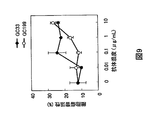
- FIG. 9 shows the results of evaluating the ADCC activity of GC33 to a human hepatoma cell line, HuH-7, using a mouse bone marrow-derived effector cell.
- FIG. 10 shows the results of evaluating the antitumor activity of GC33 antibody to a mouse model transplanted with human hepatoma.
- FIG. 11 shows the results of evaluating the CDC activity of the mouse-human chimeric antibody GC33 to a CHO cell that expresses GPC3.
- FIG. 12 shows the results of evaluating the ADCC activity of the mouse-human chimeric antibody GC33 to HepG2.
- FIG. 13 shows GPC3-derived sequences contained in GST-fusion proteins (GC-4, 5, 6, 7, 8, 9, 11, 12, 13 and 14) prepared for analyzing the epitope of GC33.
- FIG. 14 shows the results of Western blotting with the use of GC33 after separating GST, GC-7, 8, 9, 11, 12, 13 and 14 by SDS-PAGE under reducing conditions.
- FIG. 15 shows the results of evaluating the binding activity of humanized GC33 to GPC3 by an ELISA.
- FIG. 16 shows an antibody panel which summarizes isotypes and the results of an ELISA, BIAcore, FACS, an epitope analysis and an immunoprecipitation test for clones derived from a mouse immunized with a soluble form of GPC3.
- FIG. 17 shows an antibody panel in which isotypes and the results of an ELISA, FACS and an epitope analysis for clones derived from a mouse immunized with GC-3 are summarized.
- FIG. 18 shows the results of evaluating the binding activity of the modified antibodies to the soluble form of GPC3 core protein by an ELISA.
- Gly34 located at CDR1 in a humanized GC33 L chain variable region was replaced with any of 17 amino acids other than Cys and Met.
- FIG. 19 shows the results of evaluating the CDC activity of the mouse-human chimeric antibodies GC33, M3C11, and M1E7 to a CHO cell that expresses full-length GPC3.
- FIG. 20 shows the results of evaluating the ADCC activity of the mouse-human chimeric antibodies GC33, M3C11, and M1E7 to a human hepatoma cell line SK-03 that expresses full-length GPC3.
- the present invention provides antibodies described in the following (I) to (XI).
- the antibodies described in (1) to (12) preferred are the antibodies described in (1) to (8), more preferred are the antibodies described in (1) to (5), and particularly preferred is the antibody described in (1).
- the antibodies described in (1) to (8) recognize the C-terminal peptide of glypican 3 (a peptide comprising the 374th amino acid to the 580th amino acid of glypican 3); and are useful as a therapeutic antibody.
- the antibodies described in (9) to (12) recognize the N-terminal peptide of glypican 3 (a peptide comprising from the 1st amino acid to the 373rd amino acid of glypican 3); and are useful as a diagnostic antibody.
- the antibodies described in (1) to (13) preferred are the antibodies described in (1) to (8), more preferred are the antibodies described in (1) to (5), and particularly preferred is the antibody described in (1).
- the antibodies described in (1) to (8) recognize the C-terminal peptide of glypican 3 (a peptide comprising from the 374th amino acid to the 580th amino acid of glypican 3); and are useful as a therapeutic antibody.
- the antibodies described in (9) to (13) recognize the N-terminal peptide of glypican 3 (a peptide comprising from the 1st amino acid to the 373rd amino acid of glypican 3); and are useful as a diagnostic antibody.
- antibodies described in (1) to (7) particularly preferred are the antibodies described in (2) to (7).
- V An antibody having a light chain variable region containing the amino acid sequence set forth in SEQ ID NO: 92 (GC33 VL ver.a).
- antibodies described in (1) to (7) particularly preferred are the antibodies described in (2) to (7).
- the antibody having a light chain variable region containing the amino acid sequence set forth in SEQ ID NO: 205 and a heavy chain variable region containing the amino acid sequence set forth in SEQ ID NO: 90.
- the activity equivalent to that of the antibody described in any of (I) to (X) means that the binding activity to a human glypican 3 antibody or the cytotoxicity activity on a cell that expresses human glypican 3 (e.g., HepG2 or a recombinant CHO cells expressing human glypican 3, etc.) is equivalent.
- One preferred embodiment of the antibody according to the present invention is a humanized antibody that binds to glypican 3.
- the humanized antibody can be prepared by using a known method.
- the humanized antibody is also referred to as a reshaped human antibody, which is made by transplanting the complementarity determining region (CDR) of an antibody of a non-human mammal, for example a mouse antibody, into the CDR of a human antibody.
- CDR complementarity determining region
- the general recombinant DNA technology for preparation of such antibodies is also known (see European Patent Application EP 125023 and International Patent Application WO 96/02576).
- a DNA sequence which has been designed to link the CDRs of the mouse antibody with the framework region (FR) of a human antibody is synthesized by the PCR method using several oligonucleotides as primers, which have been prepared so as to have portions overlapping with one another at both ends of the CDR and the FR (see the method described in International Patent Application WO 98/13388).
- the framework region of a human antibody to be linked with the CDR the one which allows a complementarity determining region to form a favorable antigen-binding site is selected. If necessary, an amino acid in the framework region of a variable region of the antibody may be replaced so that the complementarity determining region of a reshaped human antibody may form an appropriate antigen-binding site (Sato, K. et al., Cancer Res. (1993) 53, 851-856).
- the C region of a human antibody may be used as the C region of a chimeric antibody or a humanized antibody, for example, C ⁇ 1, C ⁇ 2, C ⁇ 3, and C ⁇ 4 may be used in the H chain, and C ⁇ and C ⁇ may be used in the L chain.
- the C region of a human antibody may also be modified in order to improve the stability of the antibody or the production thereof.
- the human antibody to be used in the humanization may be any isotype of human antibody, for example, IgG, IgM, IgA, IgE and IgD, preferably, IgG, more preferably IgG1 or IgG3, and particularly preferably IgG1.
- IgG1 is effective when an antibody is used as an anticancer agent in terms of having a high cytotoxicity activity (Chemical immunology, 65: 88 (1997)).
- an amino acid in a variable region e.g., FR
- a constant region e.g., FR
- the origin of the CDR in a humanized antibody is not particularly limited, and the CDR may be derived from any animals.
- the CDR may be derived from any animals.
- humanization of an antibody it is generally difficult to humanize it while maintaining the agonist activity of the original antibody.
- a humanized antibody having an agonist activity equivalent to that of the original mouse antibody was successful acquired. Since the antigenicity of the humanized antibody in the human body is reduced, it is useful in administering it into the human for a therapeutic purpose.
- Preferred examples of the humanized anti-glypican 3 antibody in the present invention include, for example, an antibody having a heavy chain variable region set forth in SEQ ID NO: 84 (GC33 VH ver.a), SEQ ID NO: 85 (GC33 VH ver.c), SEQ ID NO: 86 (GC33 VH ver.f), SEQ ID NO: 87 (GC33 VH ver.h), SEQ ID NO: 88 (GC33 VH ver.i), SEQ ID NO: 89 (GC33 VH ver.j) or SEQ ID NO: 90 (GC33 VH ver.k) or an antibody having a light chain variable region set forth in SEQ ID NO: 92 (GC33 VL ver.a).
- Particularly preferred examples thereof include an antibody having a heavy chain variable region set forth in SEQ ID NO: 84 (GC33 VH ver.a), SEQ ID NO: 85 (GC33 VH ver.c), SEQ ID NO: 86 (GC33 VH ver.f), SEQ ID NO: 87 (GC33 VH ver.h), SEQ ID NO: 88 (GC33 VH ver.i), SEQ ID NO: 89 (GC33 VH ver.j) or SEQ ID NO: 90 (GC33 VH ver.k) and a light chain variable region set forth in SEQ ID NO: 92 (GC33 VL ver.a).
- a preferred example of the humanized anti-glypican 3 antibody includes an antibody having a heavy chain variable region containing the amino acid sequence set forth in SEQ ID NO: 90 and a light chain variable region containing the amino acid sequence set forth in SEQ ID NO: 205.
- a preferred embodiment of the antibody according to the present invention is an antibody that binds to the epitope to which the antibody set forth in any of the following (1) to (8) binds:
- the antibody that binds to the epitope to which any of the above-mentioned antibodies bind is useful because it has a particularly high cytotoxicity.
- the antibody described in any of (1) to (7) binds to a region from the 524th amino acid to the 580th amino acid of human glypican 3. In particular, it binds to a region from the 524th amino acid to the 563rd amino acid.
- the antibody described in any of (1) to (5) binds to a region from the 537th amino acid to the 563rd amino acid of human glypican 3.
- the antibody described in (1) binds to a region from the 544th amino acid to the 553rd amino acid of human glypican 3. In particular, it binds to a region from the 546th amino acid to the 551st amino acid.
- the antibodies recognizing the above-mentioned epitopes have a high cytotoxicity, therefore they are useful in the treatment of a disease such as cancer.
- the antibody which binds to a region from the 546th amino acid to the 551st amino acid is useful as it has a particularly high cytotoxicity
- the present invention includes the antibodies which binds to an epitope in a region from the 524th amino acid to the 580th amino acid of human glypican 3, preferably a region from the 524th amino acid to the 563rd amino acid, more preferably a region from the 537th amino acid to the 563rd amino acid, further more preferably a region from the 544th amino acid to the 553rd amino acid, particularly preferably a region from the 546th amino acid to the 551st amino acid.
- Another preferred embodiment according to the present invention is an antibody that recognizes a region from the 524th amino acid to the 563rd amino acid of human glypican 3 and does not recognize a region from the 537th amino acid to the 563rd amino acid.
- a further preferred embodiment according to the present invention is an antibody that recognizes a region from the 537th amino acid to the 563rd amino acid of human glypican 3 and does not recognize a region from the 550th amino acid to the 563rd amino acid.
- the analysis of an epitope recognized by an antibody can be carried out by a method known to those skilled in the art, for example, by Western blotting described in Examples below.
- the antibody that recognizes the above-mentioned regions as an epitope can be obtained by a method known to those skilled in the art. For example, it can be obtained by preparing a peptide containing an amino acid sequence of a target region based on an amino acid sequence of human glypican 3 and preparing an antibody with the use of the peptide as an immunogene, or by preparing an antibody by a usual method and determining an epitope that the obtained antibody recognizes, and then selecting an antibody that recognizes the target epitope.
- a preferred example of the anti-glypican 3 antibody of the present invention is an antibody having a high ADCC activity or an antibody having a high CDC activity to a cell that expresses glypican 3.
- a high ADCC activity or “a high CDC activity” as used herein means that the antibody of the invention has a higher ADCC activity or a higher CDC activity than that of a known anti-glypican 3 antibody.
- Known glypican 3 antibodies include, for example, M3C11 and M1E07 described in International Patent Application WO 2004/22739.
- the ADCC activity or the CDC activity can be measured by a method known to those skilled in the art. For example, it can be measured by the chromium release test. Specific conditions of the chromium release test for measuring the ADCC activity are not particularly limited, however, for example, it can be measured using the conditions described in the Examples below.
- Examples of the cells that express glypican 3 include, for example, a hepatoma cell line such as HepG2, a CHO cell line having a gene encoding glypican 3 incorporated therein and the like.
- a hepatoma cell line such as HepG2
- a CHO cell line having a gene encoding glypican 3 incorporated therein and the like To measure the ADCC activity, it is preferred to use a HepG2 cell line, and to measure the CDC activity, it is preferred to use a recombinant CHO cell line that expresses GPC3.
- the recombinant CHO cell line that expresses GPC3 may be prepared by any method, however, it can be prepared by, for example, the method described in the Examples below.
- the anti-glypican 3 antibody In the case where the anti-glypican 3 antibody is used as an anticancer agent, it is preferred that it has an ADCC activity at the same level as that of an antibody containing a heavy chain variable region having the amino acid sequence set forth in SEQ ID NO: 62 and a light chain variable region having the amino acid sequence set forth in SEQ ID NO:73 (GC33). In the case where the anti-glypican 3 antibody is used as an anticancer agent, it is preferred that it has a CDC activity at the same level as that of an antibody containing a heavy chain variable region having the amino acid sequence set forth in SEQ ID NO: 62 and a light chain variable region having the amino acid sequence set forth in SEQ ID NO: 73 (GC33).
- the present invention includes an antibody having a high binding activity to glypican 3.
- the binding activity of the antibody to glypican 3 can be measured by using a method known to those skilled in the art. For example, it can be measured by utilizing the surface plasmon resonance with BIAcore. Specifically, a glypican 3 protein is immobilized on a sensor chip to react with an antibody, and the interaction between the antibody and glypican 3 can be calculated as a reaction rate constant from the measurement value. In addition, with regard to the evaluation of the binding activity, an enzyme linked immunosorbent assay (ELISA), an enzyme immunoassay (EIA), a radioimmunoassay (RIA) or a fluorescent antibody technique can be used.
- ELISA enzyme linked immunosorbent assay
- EIA enzyme immunoassay
- RIA radioimmunoassay
- a sample containing an antibody to be tested for example, a culture supernatant of a cell producing an antibody to be tested or a purified antibody is added to a plate which has been coated with an antigen to which the antibody to be tested binds. Then, a secondary antibody labeled with an enzyme such as alkaline phosphatase is added, and the plated is incubated and washed. Then, an enzyme substrate such as p-nitrophenyl phosphate is added and the absorbance is measured, whereby an antigen binding activity can be evaluated.
- the upper limit of the binding activity is not particularly limited. However, for example, the upper limit can be defined within the range which is technically possible by those skilled in the art. It will be appreciated that the range which is technically possible will be expanded by the advancement of technology.
- an amino acid to be deamidated or an amino acid adjacent to an amino acid to be deamidated may be replaced with another amino acid for the purpose of, for example, suppressing deamidation to increase the stability of the antibody.
- the amino acid to be deamidated includes, asparagine and glutamine, preferably asparagine.
- An amino acid adjacent to asparagine is not particularly limited and may be any amino acid. It is known that an asparagine-glycine sequence is particularly susceptible to deamidation, thus, glycine is preferred as the amino acid adjacent to asparagine.
- An amino acid used for replacement is not particularly limited and may be any amino acid other than asparagine and glutamine. Preferred is an amino acid other than valine and proline.
- the antibody in the case where the antibody is deamidated, it is preferred to replace the amino acid with an amino acid other than asparagine, glutamine, valine and proline. Suppression of deamidation by amino acid replacement can be carried out with reference to, for example, International Patent Application WO 03/057881. In the case where amino acid replacement is carried out for the purpose of suppression of deamidation, it is preferred that the antigen binding activity before replacement is maintained.
- Another embodiment of stabilization of the antibody includes replacement of glutamic acid with another amino acid.
- the present invention it was found that, in the case where the 6th amino acid of the heavy chain of an antibody is glutamic acid, the antibody can be significantly stabilized by replacing the glutamic acid with glutamine. Accordingly, the present invention also relates to a method of stabilizing an antibody by replacing the glutamic acid at the 6th position of the heavy chain of the antibody with glutamine.
- the amino acid numbering of the antibody is known to those skilled in the art (e.g., Kabat, E. A. et al., “Sequences of Proteins of Immunological Interest”, US Dept. Health and Human Services 1983).
- the antibody of the invention may be a conjugated antibody in which the antibody is conjugated with various molecules, such as polyethyleneglycol (PEG), radioactive materials and toxin.
- a conjugated antibody may be prepared by chemically modifying the antibody obtained as above. Methods for modifying antibodies have already been established in the art.
- the antibody of the invention encompasses such a conjugated antibody.
- the antibody of the invention may also be a bispecific antibody (see, for example, Journal of Immunology, 1994, 152, 5368-5374).
- the bispecific antibody may recognize glypican 3 and another antigen, or may recognize different epitopes on a GPC3 molecule.
- the antibody of the invention may carry a certain protein fused to the N— or C-terminus of the antibody (Clinical Cancer Research, 2004, 10, 1274-1281).
- the protein to be fused to the antibody may be conveniently selected by those skilled in the art.
- the antibody of the invention includes an antibody with an enhanced cytotoxicity.
- the antibody with an enhanced cytotoxicity include an antibody lacking fucose, an antibody having bisecting N-acetyl glucosamine (GlcNAc) attached to its sugar chain, and an antibody having altered binding activity for Fc ⁇ receptor obtained by substituting one or more amino acids in the Fc region.
- Such antibodies with an enhanced cytotoxicity can be prepared by a method known in the art.
- the antibody that binds to glypican 3 can be prepared by a method known to those skilled in the art.
- a monoclonal antibody-producing hybridoma can be prepared as follows basically using a known technique. That is, the hybridoma can be prepared by immunizing a mammal in accordance with a usual immunization method using a glypican 3 protein or a cell that expresses glypican 3 as a sensitizing antigen. The thus obtained immunocyte is fused with a known parent cell by a usual cell fusion method, and then selecting a monoclonal antibody-producing cell by a usual screening method.
- a monoclonal antibody can be prepared as follows. First, a glypican 3 protein is obtained based on the glypican 3 gene/amino acid sequence shown in SEQ ID NOs: 3 and 4, which is used as a sensitizing antigen to obtain an antibody. More specifically, the gene sequence encoding glypican 3 is inserted into a known expression vector system, and an appropriate host cell is transformed with the vector, and then a target human glypican 3 protein is purified by a known method from the host cell or the culture supernatant.
- this purified glypican 3 protein is used as a sensitizing antigen.
- a partial peptide of glypican 3 can be used as a sensitizing antigen.
- the partial peptide can also be obtained by chemical synthesis according to the amino acid sequence of human glypican 3.
- the epitope on a glypican 3 molecule which is recognized by the anti-glypican 3 antibody of the present invention is not limited to a particular epitope.
- the anti-glypican 3 antibody may recognize any epitope, as long as the epitope is present on a glypican 3 molecule. Accordingly, any fragment can also be used as an antigen for producing the anti-glypican 3 antibody of the present invention, as long as it contains an epitope that is present on a glypican 3 molecule.
- a mammal to be immunized with a sensitizing antigen is not particularly limited, but it is preferably selected in view of compatibility with a parent cell to be used for cell fusion.
- rodents such as mice, rats and hamsters, rabbits or monkeys are generally used.
- Immunization of an animal with a sensitizing antigen is carried out in accordance with a known method.
- immunization is carried out by a general method in which a mammal is injected intraperitoneally or subcutaneously with a sensitizing antigen.
- a sensitizing antigen is diluted with or suspended in an appropriate amount of PBS (Phosphate-Buffered Saline), physiological saline or the like, an appropriate amount of a standard adjuvant such as a Freund's complete adjuvant is mixed with the product if necessary, and then the solution is emulsified and is administered to a mammal several times every 4 to 21 days.
- PBS Phosphate-Buffered Saline
- physiological saline physiological saline
- an appropriate carrier can also be used upon immunization with a sensitizing antigen.
- a mammal is immunized as described above, and then an increased level of a target antibody in the serum is confirmed. Subsequently, immunocytes are collected from the mammal, and then subjected to cell fusion.
- a particularly preferred immunocyte is a splenocyte.
- a mammalian myeloma cell As a parent partner cell to be fused with the above-mentioned immunocyte, a mammalian myeloma cell is used.
- a cell line of the myeloma cell that is preferably used herein include various known cell lines such as P3 (P3x63 Ag8.653) (J. Immnol. (1979) 123, 1548-1550), P3x63 Ag8U.1 (Current Topics in Microbiology and Immunology (1978) 81, 1-7), NS-1 (Kohler. G. and Milstein, C. Eur. J. Immunol. (1976) 6, 511-519), MPC-11 (Margulies. D. H.
- Cell fusion of the above-mentioned immunocytes with myeloma cells can be basically carried out in accordance with a known method, for example, the method of Kohler and Milstein et al. (Kohler. G. and Milstein, C., Methods Enzymol. (1981) 73, 3-46).
- the above-mentioned cell fusion is carried out in a normal nutrition culture solution in the presence of, for example, a cell-fusion accelerator.
- a cell-fusion accelerator for example, polyethylene glycol (PEG), a hemagglutinating virus of Japan (HVJ) is used.
- PEG polyethylene glycol
- HVJ hemagglutinating virus of Japan
- an adjuvant such as dimethylsulfoxide can be added to further enhance the fusion efficiency.
- the ratio of immunocytes to myeloma cells may be appropriately selected. For example, it is preferred that the number of immunocytes is 1 to 10 times greater than that of myeloma cells.
- the culture solution to be used for the above-mentioned cell fusion include, for example, a RPMI1640 culture solution or a MEM culture solution which is suitable for the growth of the above-mentioned myeloma cell line, or another normal culture solution which is used for this type of cell culture.
- a serum supplement such as fetal calf serum (FCS) can be used in combination therewith.
- Cell fusion is carried out as follows. Predetermined amounts of the above-mentioned immunocytes and myeloma cells are mixed well in the above-mentioned culture solution, a PEG (e.g., with an average molecular weight of approximately 1000 to 6000) solution (a general concentration of 30 to 60% (w/v)), which had been pre-heated at approximately 37° C., is added, and then the solution is mixed, whereby a target fusion cell (hybridoma) is formed.
- a PEG e.g., with an average molecular weight of approximately 1000 to 6000
- w/v target fusion cell
- the thus obtained hybridoma is selected by culturing the hybridoma in a standard selective culture solution such as a HAT culture solution (a culture solution containing hypoxanthine, aminopterin and thymidine). Cultivation in the above-mentioned HAT culture solution is continued for a time period sufficient for the cells (unfused cells) other than the target hybridoma to die (normally, several days to several weeks). Subsequently, a standard limiting dilution method is conducted to screen for and monoclone of hybridoma that produces a target antibody.
- a standard selective culture solution such as a HAT culture solution (a culture solution containing hypoxanthine, aminopterin and thymidine). Cultivation in the above-mentioned HAT culture solution is continued for a time period sufficient for the cells (unfused cells) other than the target hybridoma to die (normally, several days to several weeks).
- a standard limiting dilution method is conducted
- a desired human antibody having a binding activity to glypican 3 can also be obtained by sensitizing a human lymphocyte with glypican 3 in vitro, and allowing the sensitized lymphocyte to fuse with a human-derived myeloma cell having a permanent division potential (see JP-B-1-59878).
- glypican 3 antigen is administered to a transgenic animal having all the repertories of human antibody genes to obtain anti-glypican 3 antibody-producing cells, which are then immortalized, and a human antibody for glypican 3 may be obtained from the immortalized anti-glypican 3 antibody-producing cells (see International Patent Applications WO 94/25585, WO 93/12227, WO 92/03918 and WO 94/02602).
- the thus prepared hybridoma that produce a monoclonal antibody can be passage-cultured in a standard culture solution, or can be stored for a long period in liquid nitrogen.
- One example of a method employed to obtain a monoclonal antibody from the hybridoma involves culturing the hybridoma and obtaining a monoclonal antibody from the culture supernatant in accordance with a standard method. Another method involves administering the hybridoma to a mammal that is compatible with the hybridoma to allow it to proliferate, and obtaining a monoclonal antibody from the ascites.
- the former method is suitable to obtain an antibody of high purity, while the latter method is suitable for the mass production of antibodies.
- a recombinant antibody by cloning the antibody gene from the hybridoma, incorporating the gene into an appropriate vector, introducing the vector into a host, and then allowing the host to produce the recombinant antibody by a genetic engineering technique (e.g., see Vandamme, A. M. et al., Eur. J. Biochem. (1990) 192, 767-775, 1990).
- mRNA encoding the variable (V) region of an anti-glypican 3 antibody is isolated from a hybridoma producing the anti-glypican 3 antibody.
- mRNA is isolated by a known method such as a guanidine ultracentrifugal method (Chirgwin, J. M. et al., Biochemistry (1979) 18, 5294-5299) or an AGPC method (Chomczynski, P. et al., Anal. Biochem. (1987) 162, 156-159), and total RNA is prepared, and then target mRNA is prepared using an mRNA Purification Kit (Pharmacia) or the like.
- mRNA can also be directly prepared using a QuickPrep mRNA Purification Kit (Pharmacia).
- the cDNA of the antibody V region is synthesized using a reverse transcriptase from the thus obtained mRNA.
- cDNA may be synthesized using an AMV Reverse Transcriptase First-strand cDNA Synthesis Kit (SEIKAGAKU CORPORATION) or the like.
- SEIKAGAKU CORPORATION AMV Reverse Transcriptase First-strand cDNA Synthesis Kit
- 5′-Ampli FINDER RACE Kit (Clontech)
- the 5′-RACE method using PCR can be employed for synthesizing and amplifying cDNA.
- a target DNA fragment is purified from the thus obtained PCR product, and then ligated to a vector DNA.
- a recombinant vector is prepared from the product, and then the vector is introduced into E. coli or the like, and a colony is selected, thereby preparing a desired recombinant vector.
- the nucleotide sequence of the target DNA is then determined by a known method such as a dideoxynucleotide chain termination method.
- DNA encoding the V region of the target anti-glypican 3 antibody is obtained, this DNA is incorporated into an expression vector containing DNA encoding the constant region (C region) of the target antibody.
- the antibody gene is incorporated into an expression vector so that the gene is expressed under the regulation of the gene expression control region including, for example, an enhancer and a promoter.
- a host cell is transformed with the expression vector, thereby allowing the host to express the antibody.
- An antibody gene can be expressed by incorporating a polynucleotide encoding the H chain or a polynucleotide encoding the L chain separately into an expression vector, and then simultaneously transforming a host cell with the vectors, or by incorporating polynucleotides encoding the H chain and the L chain into a single expression vector, and then transforming a host cell with the vector (see International Patent Application WO 94/11523).
- the present invention provides a polynucleotide encoding a heavy chain variable region or a light chain variable region of the antibody of the present invention.
- the polynucleotide of the present invention has a nucleotide sequence described in any of SEQ ID NOs: 11-21, 33-43, 55-59, 65-70 and 77-83.
- a polynucleotide that is hybridized to the above-mentioned polynucleotide under stringent conditions and encodes an antibody having an activity equivalent to that of the antibody of the present invention is also within the scope of the present invention.
- the polynucleotide of the present invention is not particularly limited as long as it encodes the antibody of the present invention. It is a polymer composed of a plurality of nucleotides, such as deoxyribonucleic acids (DNA) or ribonucleic acids (RNA). It may contain a base other than a naturally occurring base.
- the polynucleotide of the present invention can be used for producing an antibody by a genetic engineering technique.
- the polynucleotide of the present invention can be used as a probe to screen for an antibody having a function equivalent to that of the antibody of the present invention.
- a polynucleotide encoding the antibody of the present invention or a partial fragment thereof may be used as a probe to obtain DNA that is hybridized to the polynucleotide under stringent conditions and encodes an antibody having an activity equivalent to that of the antibody of the present invention by techniques such as a hybridization technique, a gene amplification technique (e.g., PCR). Such DNA is also included in the polynucleotide of the present invention.
- hybridization conditions include, for example, low stringent conditions.
- the low stringent conditions are, for example, the conditions of 42° C., 0.1 ⁇ SSC and 0.1% SDS, preferably the conditions of 50° C., 0.1 ⁇ SSC and 0.1% SDS when washing is performed after hybridization.
- More preferred examples of the hybridization conditions include, for example, high stringent conditions.
- the high stringent conditions are, for example, the conditions of 65C, 5 ⁇ SSC and 0.1% SDS.
- An antibody functionally equivalent to the antibody of the present invention encoded by a polynucleotide obtained by such a hybridization technique and a gene amplification technique usually has a high homology with the antibody in terms of the amino acid sequence.
- the antibody of the present invention also includes an antibody that is functionally equivalent to the antibody of the present invention and has a high homology with the amino acid sequence of the antibody.
- a high homology means generally at least 50% or higher identity, preferably 75% or higher identity, more preferably 85% or higher identity, and further more preferably 95% or higher identity at the amino acid level.
- the present invention also provides a vector containing the polynucleotide of the present invention.
- a vector can be used for preparing the antibody of the present invention.
- the vector of the present invention in the case where E. coli is used as a host, for example, it is not particularly limited as long as it has “ori” for use in amplification in E. coli to produce and amplify the vector in a large amount in E. coli (e.g., JM109, DH5 ⁇ , HB101 or XL1Blue), and has a marker gene for selecting a transformed E. coli (e.g., a drug resistance gene that can be identified by a drug such as ampicillin, tetracycline, kanamycin or chloramphenicol).
- a marker gene for selecting a transformed E. coli
- vectors examples include M13-series vectors, pUC-series vectors, pBR322, pBluescript, pCR-Script and the like.
- pGEM-T, pDIRECT, and pT7 can also be used for subcloning and extracting cDNA as well as the vectors described above.
- an expression vector is particularly useful.
- an expression vector to be expressed in E. coli should have the above characteristics to be amplified in E. coli .
- E. coli such as JM109, DH5 ⁇ , HB101, or XL1-Blue
- it is indispensable that the vector should have a promoter, for example, lacZ promoter (Ward et al., Nature (1989) 341, 544-546; FASEB J. (1992) 6, 2422-2427), araB promoter (Better et al., Science (1988) 240, 1041-1043) T7 promoter or the like, that can efficiently express the desired product in E. coli .
- Such a vector examples include pGEX-5X-1 (Pharmacia), “QIAexpress system” (Qiagen), pEGFP, pET (in this case, the host is preferably BL21 which expresses T7 RNA polymerase) and the like, as well as the vectors described above.
- the vector may also contain a signal sequence for polypeptide secretion.
- a signal sequence for polypeptide secretion in the case where a polypeptide is produced in the periplasm of E. coli , the pe1B signal sequence (Lei S. P. et al., J. Bacteriol (1987) 169, 4379) can be used.
- Introduction of the vector into a host cell can be carried out by using, for example, the calcium chloride method and the electroporation method.
- expression vectors derived from mammals e.g., pcDNA3 (Invitrogen) and PEGF-BOS (Nucleic Acids Res. (1990) 18(17), p5322), pEF and pCDM8)
- expression vectors derived from insect cells e.g., “Bac-to-BAC baculovirus expression system” (GIBCO BRL) and pBacPAK8)
- expression vectors derived from plants e.g., pMH1 and pMH2
- expression vectors derived from animal viruses e.g., pHSV, pMV and pAdexLcw
- expression vectors derived from retroviruses e.g., pZIPneo
- expression vectors derived from yeast e.g., “Pichia Expression Kit” (Invitrogen), pNV11 and SP-Q01
- expression vectors derived from Bacillus subtilis e.g., pPL60
- the vector For the purpose of expressing the vector in an animal cell such as a CHO cell, a COS cell, an NIH3T3 cell or the like, it is indispensable for the vector to have a promoter required for expression in a cell such as SV40 promoter (Mulligan et al., Nature (1979) 277, 108), MMTV-LTR promoter, EF1 ⁇ promoter (Mizushima et al., Nucleic Acids Res. (1990) 18, 5322), CMV promoter or the like, and more preferably to have a marker gene (such as a drug resistance gene that can be identified by a drug such as neomycin or G418) for selecting transformation into the cell.
- a promoter required for expression in a cell such as SV40 promoter (Mulligan et al., Nature (1979) 277, 108), MMTV-LTR promoter, EF1 ⁇ promoter (Mizushima et al., Nucleic Acid
- a vector e.g., pCHOI, etc.
- MTX methotrexate
- transformation is effected with a vector (such as pcD) having the origin of replication for SV40 using a COS cell having on the chromosome a gene that expresses the SV40 T antigen.
- the expression vector can include, as a selectable marker, the aminoglycoside transferase (APH) gene, the thymidine kinase (TK) gene, E. coli xanthine guaninephosphoribosyl transferase (Ecogpt) gene, the dihydrofolate reductase (dhfr) gene and the like.
- APH aminoglycoside transferase
- TK thymidine kinase
- Ecogpt E. coli xanthine guaninephosphoribosyl transferase
- dhfr dihydrofolate reductase
- the vector is introduced into a host cell.
- the host cell into which the vector is introduced is not particularly limited, but includes, for example, E. coli or any of various animal cells.
- the host cell can be used as a production system for the production or expression of the antibody of the present invention.
- the production system of polypeptide preparation there are an in vitro production system and an in vivo production system.
- In vitro production system include a production system which employs eukaryotic cells and a production system which employs prokaryotic cells.
- the eukaryotic cell for example, an animal cell, a plant cell or a fungal cell can be used.
- animal cells include a mammalian cell such as a CHO cell (J. Exp. Med. (1995) 108, 945), a COS cell, a 3T3 cell, a myeloma cell, a baby hamster kidney (BHK) cell, a HeLa cell and a Vero cell, an amphibian cell such as a Xenopus oocyte (Valle, et al., Nature (1981) 291, 358-340), or an insect cell such as Sf9, Sf21, and Tn5.
- a mammalian cell such as a CHO cell (J. Exp. Med. (1995) 108, 945), a COS cell, a 3T3 cell, a myeloma cell, a baby hamster kidney (BHK) cell, a HeLa cell and a Vero cell
- CHO-DG44, CHO-DXB11, a COS7 cell, a BHK cell are preferably used.
- animal cells for the purpose of performing a large amount of expression, a CHO cell is particularly preferred.
- Introduction of the vector into the host cell can be carried out by, for example, the calcium phosphate method, the DEAE-dextran method, the cationic ribozome DOTAP (Boehringer Mannheim), the electroporation method, the lipofection method or the like.
- a cell derived from Nicotiana tabacum is known as a protein production system, which may be subjected to call us culture.
- the fungal cells include yeast such as the genus Saccharomyces, more specifically Saccharomyces cerevistae and Saccharomyces pombe, and filamentous fungi such as the genus Aspergillus, more specifically Aspergillus niger.
- the prokaryotic cell In the case where the prokaryotic cell is used, production system using a bacterial cell may be employed.
- the bacterial cells include Escherichia coli ( E. coli ) such as JM109, DH5 ⁇ and HB101, and Bacillus subtilis.
- the antibody of the present invention can be prepared by culturing the above-mentioned host cells.
- the antibody can be obtained by culturing in vitro a cell transformed with a desired polynucleotide. Cultivation can be carried out in accordance with a known method.
- Culture media for animal cells include, for example, DMEM, MEM, RPMI 1640, and IMDM.
- a serum supplement such as FBS or fetal calf serum (FCS) may be used in combination, or serum-free medium can be used.
- the pH during the cultivation is preferably about 6 to 8.
- Cultivation is usually carried out at about 30 to 40° C. for about 15 to 200 hours with, as needed, medium change, aeration, and agitation.
- systems for producing a polypeptide in vivo include, for example, a production system which employs an animal and a production system which employs a plant.
- the target polynucleotide is introduced into such an animal or a plant, and the polypeptide is produced in the body of the animal or the plant and recovered.
- the term “host cell” as used herein encompasses such an animal and a plant.
- production systems employing a mammal or an insect are available.
- a mammal goats, pigs, sheep, mice and cattle canbe used (Vicki Glaser, SPECTRUM Biotechnology Applications, 1993).
- a transgenic animal can also be used as a mammal.
- the target polynucleotide is prepared as a fusion gene with a gene encoding a polypeptide which is inherently produced in the milk such as goat ⁇ casein. Then, the DNA fragment containing this fusion gene is injected into a goat embryo, and the embryo is transplanted into a female goat.
- the target antibody can be obtained from the milk produced by the transgenic goat borne to the goat which received the embryo or the offspring thereof. To increase the amount of milk containing the antibody produced by the transgenic goat, hormone may be given to the transgenic goat as needed. (Ebert, K. M. et al., Bio/Technology (1994) 12, 699-702).
- a silkworm can be used as an insect.
- a silkworm is infected with a baculovirus into which the polynucleotide encoding the target antibody has been inserted.
- the target antibody can be obtained from the body fluid of the silkworm (Susumu, M. et al., Nature (1985) 315, 592-594).
- tobacco can be used.
- a polynucleotide encoding the target antibody is inserted into an expression vector for a plant, for example pMON 530, and then the vector is introduced into a bacterium such as Agrobacterium tumefaciens. Then, tobacco such as Nicotiana tabacum is infected with the bacterium, whereby the target antibody can be obtained from the leaves of the tobacco (Julian, K. -C. Ma et al., Eur. J. Immunol. (1994) 24, 131-138).
- the thus obtained antibody can be isolated from the inside or the outside (culture medium, etc.) of the host cell and then can be purified to a substantially pure and uniform antibody. Separation and purification of the antibody may be carried out by a separation and a purification method usually used in purification of polypeptides.
- polypeptides can be separated and purified by any methods including chromatography columns, filtration, ultrafiltration, salting-out, solvent precipitation, solvent extraction, distillation, immunoprecipitation, SDS-polyacrylamide gel electrophoresis, isoelectric focusing, dialysis, recrystallization, and a combination thereof.
- chromatography examples include, for example, affinity chromatography, ion exchange chromatography, hydrophobic chromatography, gel-filtration, reverse phase chromatography, adsorption chromatography (Strategies for Protein Purification and Characterization: A Laboratory Course Manual. Ed Daniel R. Marshak et al., Cold Spring Harbor Laboratory Press, 1996). These chromatographies can be carried out using a liquid phase chromatography such as HPLC and FPLC.
- a column to be used for affinity chromatography include a protein A column or a protein G column.
- the protein A column is Hyper D, POROS, Sepharose F. F. (Pharmacia).
- the antibody before or after purification of the antibody, can be modified or peptides can be partially removed as needed by allowing a suitable protein-modifying enzyme to act on.
- the protein-modifying enzyme for this purpose include, for example, trypsin, chymotrypsin, lysyl endopeptidase, protein kinase, glucosidase.
- the present invention provides a method of diagnosing a disease such as cancer by detecting GPC3 protein in a test sample with the use of the antibody of the present invention.
- the detection used herein includes quantitative detection and non-quantitative detection.
- the non-quantitative detection include, for example, determination of merely whether or not GPC3 protein is present, determination of whether or not a specific amount or more of GPC3 protein is present, determination for comparison of the amount of GPC3 protein with that of another sample (e.g., a control sample)
- the quantitative detection includes determination of the concentration of GPC3 protein, determination of the amount of GPC3 protein.
- test sample is not particularly limited as long as it is a sample that may possible contain GPC3 protein, however, preferred is a sample collected from the body of a living organism such as a mammal, and more preferred is a sample collected from human.
- Specific examples of the test sample may include, for example, blood, interstitial fluid, plasma, extravascular fluid, cerebral fluid, joint fluid, pleural fluid, serum, lymph fluid, saliva, preferably blood, serum and plasma.
- a sample obtained from the test sample such as culture solution of cells collected from the body of the living organism is also included in the test sample of the present invention.
- the cancer to be diagnosed is not particularly limited, and specific examples may include liver cancer, pancreatic cancer, lung cancer, colon cancer, mammary cancer, prostate cancer, leukemia and lymphoma, preferably liver cancer.
- GPC3 to be detected is not particularly limited, and may be either full-length GPC3 or a fragment thereof. In the case where a fragment of GPC3 is detected, it may be either the N-terminal fragment or the C-terminal fragment, however, the N-terminal fragment is preferred.
- the GPC3 protein may also be a heparan sulfate-added GPC3 or a GPC3 core protein.
- the method of detecting GPC3 protein contained in a test sample is not particularly limited, however, detection is preferably performed by an immunological method with the use of an anti-GPC3 antibody.
- the immunological method include, for example, a radioimmunoassay, an enzyme immunoassay, a fluorescence immunoassay, a luminescence immunoassay, immunoprecipitation, a turbidimetric immunoassay.
- Preferred is an enzyme immunoassay, and particularly preferred is an enzyme-linked immunosorbent assay (ELISA) (e.g., a sandwich ELISA).
- ELISA enzyme-linked immunosorbent assay
- the above-mentioned immunological method such as an ELISA can be carried out by a method known to those skilled in the art.
- a general detection method with the use of an anti-GPC3 antibody comprises immobilizing an anti-GPC3 antibody on a support, adding a test sample thereto, incubating the support to allow the anti-GPC3 antibody and GPC3 protein to bind to each other, washing the support, and detecting the GPC3 protein binding to the support via the anti-GPC3 antibody to detect GPC3 protein in a test sample.
- the binding between the anti-GPC3 antibody and the GPC3 protein is generally carried out in a buffer.
- Buffers used in the invention include, for example, a phosphate buffer, a Tris buffer.
- Incubation is carried out under the conditions generally employed, for example, at 4° C. to room temperature for 1 hour to 24 hours.
- the washing after incubation can be carried out by any method as long as it does not inhibit the binding between the GPC3 protein and the anti-GPC3 antibody, using for example a buffer containing a surfactant such as Tween 20.
- a control sample may be provided in addition to a test sample to be tested for GPC3 protein.
- the control samples include a negative control sample that does not contain GPC3 protein and a positive control sample that contains GPC3 protein.
- One preferred embodiment of detecting GPC3 protein binding to the support via an anti-GPC3 antibody is a method using an anti-GPC3 antibody labeled with a detectable label.
- GPC3 protein may be detected by contacting the test sample with an anti-GPC3 antibody immobilized on the support, washing the support, and then detecting GPC3 with the use of the labeled antibody that specifically binds to GPC3 protein.
- the labeling of an anti-GPC3 antibody can be carried out by a generally known method.
- Examples of the detectable label known to those skilled in the art include a fluorescent dye, an enzyme, a coenzyme, a chemiluminescent substance or a radioactive substance.
- radioisotopes 32 P, 14 C, 125 I, 3 H, 13 I and the like
- fluorescein 32 P, 14 C, 125 I, 3 H, 13 I and the like
- fluorescein 32 P, 14 C, 125 I, 3 H, 13 I and the like
- rhodamine dansyl chloride
- umbelliferone luciferase
- peroxidase alkaline phosphatase
- ⁇ -galactosidase ⁇ -glucosidase
- horseradish peroxidase glucoamylase
- lysozyme saccharide oxidase
- microperoxidase biotin and the like.
- biotin-labeled antibody it is preferred that a biotin-labeled antibody is added, and then avidin conjugated to an enzyme such as alkaline phosphatase is further added.
- a solution containing an anti-GPC3 antibody is added to a support such as a plate to allow the anti-GPC3 antibody to be immobilized. After washing, the plate is blocked with, for example, BSA in order to prevent the nonspecific binding of a protein. The plate is washed again, and then the test sample is added to the plate. After being incubated, the plate is washed, and then the labeled anti-GPC3 antibody is added. After being incubated appropriately, the plate is washed, and then the labeled anti-GPC3 antibody remaining on the plate is detected.
- the detection of the protein can be carried out by a method known to those skilled in the art.
- the protein in the case where the antibody is labeled with a radioactive substance, the protein may be detected by liquid scintillation or the RIA method.
- the protein in the case where the antibody is labeled with an enzyme, the protein may be detected by adding a substrate and detecting an enzymatic change of the substrate such as color development with an absorbance reader.
- the protein in the case where the antibody is labeled with a fluorescent substance, the protein may be detected with the use of a fluorometer.
- a particularly preferred embodiment of the method of detecting GPC3 protein of the present invention is a method using an anti-GPC3 antibody labeled with biotin and avidin. Specifically, a solution containing an anti-GPC3 antibody is added to a support such as a plate to allow the anti-GPC3 antibody to be immobilized thereon. After washing, the plate is blocked with, for example, BSA in order to prevent the nonspecific binding of a protein, The plate is washed again, and then the test sample is added to the plate. After being incubated, the plate is washed, and then the biotin-labeled anti-GPC3 antibody is added.
- the plate After being incubated appropriately, the plate is washed, and then avidin conjugated to an enzyme such as alkaline phosphatase or peroxidase is added. After being incubated, the plate is washed, and then a substrate of the enzyme conjugated to avidin is added. Then, GPC3 protein is detected by means of the enzymatic change of the substrate as an indicator.
- an enzyme such as alkaline phosphatase or peroxidase
- Another embodiment of the method of detecting GPC3 protein of the present invention is a method using a primary antibody that specifically binds to GPC3 protein and a secondary antibody that specifically binds to the primary antibody.
- the test sample is brought into contact with an anti-GPC3 antibody immobilized on the support, the support is incubated and washed, and the bound GPC3 protein after washing is detected with a primary anti-GPC3 antibody and a secondary antibody that specifically binds to the primary antibody.
- the secondary antibody is preferably labeled with a detectable label.
- a solution containing an anti-GPC3 antibody is added to a support such as a plate to allow the anti-GPC3 antibody to be immobilized thereon. After washing, the plate is blocked with, for example, BSA in order to prevent the nonspecific binding of a protein. The plate is washed again, and then the test sample is added to the plate. After being incubated, the plate is washed, and then a primary anti-GPC3 antibody is added. After being incubated appropriately, the plate is washed, and then a secondary antibody that specifically binds to the primary antibody is added. After being incubated appropriately, the plate is washed, and then the secondary antibody remaining on the plate is detected. The detection of the secondary antibody can be carried out by the above-mentioned method.
- the present invention provides a pharmaceutical composition containing the antibody of the present invention.
- the pharmaceutical composition containing the antibody of the present invention is useful in the treatment and/or prevention of a disease associated with cell proliferation such as cancer, and particularly it is useful in the treatment and/or prevention of liver cancer.
- the antibody of the present invention can be formulated into a dosage form by a method known to those skilled in the art.
- the pharmaceutical composition can be used parenterally in the form of an injection of a sterile solution or a suspension with water or another pharmaceutically acceptable solution.
- the antibody can be formulated into a dosage form by appropriately mixing it with a pharmaceutically acceptable carrier or solvent, such as sterile water, physiological saline, a plant-oil, an emulsifier, a suspension, a surfactant, a stabilizer, a flavor, an excipient, a vehicle, a preservative, a binder to prepare a unit dosage form required for generally accepted Drug Implementation.
- a pharmaceutically acceptable carrier or solvent such as sterile water, physiological saline, a plant-oil, an emulsifier, a suspension, a surfactant, a stabilizer, a flavor, an excipient, a vehicle, a preservative, a binder.
- the amount of active ingredients in these preparations is selected to allow for administration of a suitable dosage within the indicated range.
- a sterile composition for injection can be formulated by using a vehicle such as distilled water for injection in accordance with the general Drug Implementation.
- aqueous solution for injection examples include, for example, physiological saline, glucose, and other isotonic liquids including adjuvants, such as D-sorbitol, D-mannnose, D-mannitol and sodium chloride. They can be used in combination with a suitable solubilizer, such as an alcohol, specifically ethanol, a polyalcohol such as propylene glycol and polyethylene glycol, and a non-ionic surfactant such as Polysorbate 80TM and HCO-50.
- a suitable solubilizer such as an alcohol, specifically ethanol, a polyalcohol such as propylene glycol and polyethylene glycol, and a non-ionic surfactant such as Polysorbate 80TM and HCO-50.
- Sesame oil or soybean oil can be used as a oleaginous liquid and may be used in combination with benzyl benzoate or benzyl alcohol as a solubilizer. It may be formulated with a buffer such as a phosphate buffer or a sodium acetate buffer, a pain-killer such as procaine hydrochloride, a stabilizer such as benzyl alcohol or phenol, or an antioxidant.
- a buffer such as a phosphate buffer or a sodium acetate buffer
- a pain-killer such as procaine hydrochloride
- a stabilizer such as benzyl alcohol or phenol
- an antioxidant such as benzyl alcohol or phenol
- the method of administration is preferably parenteral, and specific examples thereof include injection, transnasal administration, transpulmonary administration, transdermal administration and the like.
- the injection formulation may be administered systemically or topically by intravenous injection, intramuscular injection, intraperitoneal injection, subcutaneous injection or the like.
- the method of administration can be appropriately selected according to the age and the symptoms of a patient.
- one dose of the pharmaceutical composition containing the antibody or the polynucleotide encoding the antibody can be selected from the range of 0.0001 mg to 1,000 mg per kg of body weight.
- the dose can be selected from the range of 0.001 mg to 100,000 mg/body per patient, although it is not always limited to these numerical values.
- the dose and the method of administration vary according to the body weight, the age and the symptoms of a patient, and are appropriately selected by those skilled in the art.
- a full-length cDNA encoding human GPC3 was amplified by PCR reaction with an Advantage 2 kit (CLONTECH) using 1st stranded cDNA prepared by a usual method from a colon cancer cell line, Caco2, as a template.
- a reaction mixture containing 2 ⁇ L of cDNA derived from Caco2 1 ⁇ L of a sense primer (GATATC-ATGGCCGGGACCGTGCGCACCGCGT: SEQ ID NO: 1), 1 ⁇ L of an antisense primer (GCTAGC-TCAGTGCACCAGGAAGAAGAAGCAC: SEQ ID NO: 2), 5 ⁇ L of Advantage 2 10 ⁇ PCR buffer, 8 ⁇ L of dNTP mix (1.25 mM) and 1.0 ⁇ L of Advantage polymerase Mix was subjected to 35 cycles consisting of 94° C. for 1 minute, 63° C. for 30 seconds and 68° C. for 3 minutes.
- the amplified product from the PCR reaction was inserted into a TA vector, PGEM-T Easy, using pGEM-T Easy Vector System I (Promega).
- the sequence was confirmed by using an ABI 3100 DNA sequencer. In this way, a cDNA encoding full-length human GPC3 was isolated.
- the sequence shown in SEQ ID NO: 3 indicates the nucleotide sequence of human GPC3 gene and the sequence shown in SEQ ID NO: 4 indicates the amino acid sequence of human GPC3 protein.
- a soluble form of GPC protein was prepared, in which a hydrophobic region at the C-terminal side (564-580 amino acids) was deleted.
- a PCR reaction was carried out using an antisense primer (ATA GAA TTC CAC CAT GGC CGG GAC CGT GCG C: SEQ ID NO: 5) and a sense primer, to which an EcoRI recognition sequence and a Kozak sequence were added, (ATA GGA TCC CTT CAG CGG GGA ATG AAC GTT C: SEQ ID NO: 6).
- the obtained PCR fragment (1711 bp) was cloned into pCXND2-Flag.
- the pCXND2-Flag was designed to express a Flag-tagged protein by inserting the region for DHFR gene expression of pCHOI (Hirata et al., FEBS letter 1994; 356; 244-248) into the HindIII site of pCXN2 (Niwa et al., Gene 1991; 108; 193-199) and adding a Flag tag sequence to the downstream of the multicloning site.
- the constructed expression plasmid DNA was introduced into a CHO cell line, DXB11, and a CHO cell line highly expressing the soluble form of GPC3 was obtained by selection with 500 ⁇ g/mL Geneticin.
- the large-scale cultivation of the CHO cell line highly expressing the soluble form of GPC3 was carried out using a 1700-cm 2 roller bottle, and the culture supernatant was recovered for the antibody purification.
- the culture supernatant was applied to a DEAE sepharose Fast Flow column (Amersham) and, after washing, the antibody was eluted with a buffer containing 500 mM NaCl, and affinity purified using Anti-Flag M2 agarose affinity gel (SIGMA).
- SIGMA Anti-Flag M2 agarose affinity gel
- the elution was carried out with 200 ⁇ g/mL FLAG peptide.
- the filtrate was concentrated using a DEAE sepharose Fast Flow column and eluted with PBS (containing 500 mM NaCl) without Tween 20 to
- GPC3 is modified by heparan sulfate to become a macromolecule.
- heparan sulfate To eliminate an antibody against heparan sulfate in a screening for an anti-GPC3 antibody, a soluble form of GPC3 core protein that had a point mutation in the heparan sulfate-binding site was prepared and used in the screening.
- a cDNA in which Ser residues at the 495th and 509th positions were replaced with Ala was prepared by the assembly PCR method, in which primers were designed to add His tag to the C-terminus.
- the obtained cDNA was cloned into pCXND3 vector.
- the pCXND3 was constructed by inserting the DHFR gene expressing region of pCHOI in the HindIII site of pCXN2.
- the constructed expression plasmid DNA was introduced into DXB11 cell line and a CHO cell line highly expressing a soluble form of GPC3 core protein was obtained by selection with 500 ⁇ g/mL Geneticin.
- the large-scale cultivation was carried out using a 1700-cm 2 roller bottle and the culture supernatant was recovered for antibody purification.
- the culture supernatant was applied to a Q sepharose Fast Flow column (Amersham).
- the antibody was eluted with a phosphate buffer containing 500 mM NaCl, and affinity purified using a Chelating sepharose Fast Flow column (Amersham).
- the antibody was eluted with a gradient of 10 to 150 mM imidazole.
- the eluate was concentrated using a Q sepharose Fast Flow column and, eluted with a phosphate buffer containing 500 mM NaCl.
- a CHO cell line expressing full-length GPC3 was established.
- the soluble form of GPC3 core protein was diluted to 1 ⁇ g/mL with a coating buffer (0.1 mol/L NaHCO 3 (pH 9.6), 0.02% (w/v) NaN 3 ) and added to an immunoplate and left at 4° C. overnight to coat the plate. After the plate was blocked with a dilution buffer (50 mmol/L Tris-HCl (pH 8.1), 1 mmol/L MgCl 2 , 150 mmol/L NaCl, 0.05% (v/v) Tween 20, 0.02% (w/v) NaN 3 , 1% (w/v) BSA), an anti-GPC3 antibody was added and left at room temperature for 1 hour.
- a coating buffer 0.1 mol/L NaHCO 3 (pH 9.6), 0.02% (w/v) NaN 3
- a coating buffer 0.1 mol/L NaHCO 3 (pH 9.6), 0.02% (w/v) NaN 3
- an anti-mouse IgG antibody labeled with alkaline phosphatase was added and left at room temperature for 1 hour.
- SIGMA 104 SIGMA diluted to 1 mg/mL with a substrate buffer (50 mmol/L NaHCO 3 (pH 9.8), 10 mmol/L MgCl 2 ) was added and left at room temperature for 1 hour for color development. Then the absorbance (at 405 nm, reference wavelength of 655 nm) was measured using a Benchmark Plus (BIO-RAD).
- MRL/lpr mouse Since human GPC3 and mouse GPC3 show a high homology of 94% at the amino acid level, it was considered difficult to obtain an anti-GPC3 antibody if a normal mouse was immunized. Therefore, an autoimmune disease mouse, MRL/MpjUmmCrj-lpr/lpr mouse, (hereinafter referred to as MRL/lpr mouse, purchased from Charles River Japan, Inc.) was used as an immunized animal. Immunization was started at the age of 7 weeks or 8 weeks. For the first immunization, a soluble form of GPC3 was prepared at 100 ⁇ g/head and emulsified using Freund's complete adjuvant (FCA, Becton Dickinson) and subcutaneously administered.
- FCA Freund's complete adjuvant
- a soluble form of GPC3 was prepared at 50 ⁇ g/head and emulsified using Freund's incomplete adjuvant (FIA, Becton Dickinson) and subcutaneously administered. After that, an additional immunization was carried out every other week for 5 times in total. To two of the immunized mice, a soluble form of GPC3 was diluted with PBS to 50 ⁇ g/head, and then administered intravenously via the tail as the final immunization.
- FIA Freund's incomplete adjuvant
- the spleen was excised to obtain a spleen cell, which was mixed with a mouse myeloma cell, P3-X63Ag8U1 (P3U1, purchased from ATCC), at a ratio of 2:1.
- Cell fusion was carried out by gradually adding PEG 1500 (Roche Diagnostic).
- RPMI 1640 medium (GIBCO BRL) was carefully added to dilute PEG 1500, and after PEG 1500 was removed by centrifugation, the cells were suspended in RPMI 1640 medium containing 10% FBS and inoculated into a 96-well culture plate at 100 ⁇ L/well.
- HAT medium RPMI 1640 medium containing 10% FBS, 1 ⁇ HAT media supplement (SIGMA) and 0.5 ⁇ BM-Condimed H1 Hybridoma cloning supplement (Roche Diagnostic) (hereinafter referred to as HAT medium) was added at 100 ⁇ L/well. After 2, 3 and 5 days, half of the culture solution was replaced with the HAT medium. After 7 days, screening was carried out using the culture supernatant. The screening was carried out by an ELISA using an immunoplate coated with the soluble form of GPC3 core protein. A positive clone was monocloned by the limiting dilution method.
- Isotype was determined by an antigen-dependent ELISA using an Immunopure Monoclonal Antibody Isotyping Kit I (PIERCE).
- the purification of antibodies was carried out as follows. The culture supernatant of hybridoma cultured with the HAT medium supplemented with FBS (Ultra low IgG) (GIBCO BRL) was adsorbed to Hi Trap ProteinG HP (Amersham), and washed with a binding buffer (20 mM sodium phosphate (pH 7.0)). The antibody was eluted with an elution buffer (0.1 M glycine-HCl (pH 2.7)). The eluate was immediately neutralized with a neutralization buffer (1 M Tris-HCl (pH 9.0)), and dialyzed against PBS for day and night for buffer exchange.
- concentration-dependent binding of the antibody was detected against an immunoplate containing the soluble form of GPC3 core protein immobilized thereon.
- a 3-fold dilution series (12 dilutions in total) of the purified antibody at a concentration of 10 ⁇ g/mL was added, and an anti-mouse IgG antibody was added as the secondary antibody.
- Color development was carried out using SIGMA 104. Since the degree of color development varies depending on the color development time, data measured precisely after 1 hour was analyzed. Every antibody showed a concentration-dependent color development. The correlation between the concentration of antibody and the degree of color development was plotted and an approximate curve was obtained by using an analyzing software, GraphPad Prism. Its EC50 value was determined as the index of the binding activity. EC50 values for all clones are shown in FIG. 16 .
- Cells were dissociated with 1 mM EDTA pH 8.0 (GIBCO)/PBS and suspended in FACS buffer (1% FBS/PBS) at 1 ⁇ 10 6 cells/mL. The suspension was dispensed to a Multiscreen-HV Filter Plate (Millopre) at 100 ⁇ L/well and the supernatant was removed by centrifugation. An anti-GPC3 antibody diluted to an appropriate concentration was added and reacted on ice for 30 minutes. The cells were washed once with FACS buffer and an FITC-labeled anti-mouse IgG antibody was added and reacted on ice for 30 minutes. After the reaction, the cells were centrifuged at 500 rpm for 1 minute, and the supernatant was removed.
- FACS buffer 1% FBS/PBS
- the cells were suspended in 400 ⁇ L of FACS buffer and subjected to flow cytometry.
- EPICS ELITE ESP (Beckman Coulter) was used as a flow cytometer.
- a gate was set on the living cell population with the histogram of forward scatter and side scatter.
- an anti-GPC3 antibody (M3C11, M11F1) bound strongly to the CHO cell expressing GPC3 and did not bind to the parent CHO cell, indicating that the antibody specifically binds to GPC3 presented on the cell membrane.
- the antibody showed the binding activity to a hepatoma cell line, HepG2 (purchased from ATCC) and HuH-7 (purchased from Health Science Research Resources Bank), suggesting that the antibody may specifically recognize hepatoma.
- the binding activity of the clones derived from the mouse immunized with a soluble form of GPC3 measured by flow cytometry is shown in FIG. 16 , where the X-mode values of histogram at the concentration of antibody of 5 ⁇ g/mL are indicated.
- the obtained antibodies were classified according to the epitopes by a competitive ELISA.
- the antibodies were biotinylated using a Biotin Labeling Kit (Roche).
- the soluble form of GPC3 core protein was diluted to 1 ⁇ g/mL with the coating buffer and added to a plate at 100 ⁇ L/well and stored at 4° C. overnight to coat the plate. On the next day, 200 ⁇ L of the substrate buffer was added for blocking. The plate was left at 4° C. overnight or longer and an anti-GPC3 antibody was added to the plate at 100 ⁇ L/well and reacted at room temperature for 1 hour.
- the results of the competitive ELISA are shown in FIG. 2 .
- the antibody that competitively inhibited the binding of the biotinylated antibody by 50% or more it was considered that its epitopes are located close together in the three-dimensional conformation.
- the 11 clones derived from the mouse immunized with a soluble form of GPC3 were classified into 5 groups (a, b, c, d and e) ( FIG. 16 ).
- the soluble form of GPC3 core protein was applied to a 10% SDS-PAGE mini (TEFCO) and electrophoresed under reducing conditions. It was transferred to Immobilon-P (Millipore) using Trans-Blot SD Semi-Dry Electrophoretic Transfer Cell (BIO-RAD). After the membrane was briefly washed with TBS-T (0.05% Tween 20, TBS), it was shaken in TBS-T containing 5% skim milk for 1 hour. The membrane was shaken in TBS-T for about 10 minutes, then each anti-GPC3 antibody diluted with TBS-T containing 1% skim milk was added and the membrane was shaken for 1 hour.
- TBS-T 0.05% Tween 20, TBS
- the membrane was washed with TBS-T and shaken in a solution of HRP-anti-mouse IgG antibody (Amersham) diluted with TBS-T containing 1% skim milk for 1 hour, and then washed with TBS-T. Color development was carried out using ECL-Plus (Amersham) and detected using Hyperfilm ECL (Amersham).
- L9G11 was determined to be an antibody binding to the N-terminal side because it bound to the band of about 40 kDa.
- M3C11 was determined to be an antibody binding to the C-terminal side because it bound to the band of about 30 kDa. All the antibodies belonging to c, d or e group based on the competitive ELISA bound to the N-terminal side, and all those belonging to a or b groups bound to the C-terminal side ( FIG. 16 ).
- L9G11 had higher detection sensitivity in Western blotting than the other antibodies that bind to the N-terminal side, suggesting that this antibody is a useful for detecting the N-terminal fragment by Western blotting.
- GPC3 Since it was found that GPC3 is cleaved at the 358th amino acid residue or the 374th amino acid residue, the inventors hypothesized that a secreted form of GPC3 is secreted into the blood of a patient with liver cancer. Therefore, a GPC3 sandwich ELISA system was constructed in order to detect a secretory form of GPC3.
- An immunoplate was coated with an anti-GPC3 antibody at 10 ⁇ g/mL and blocked by the substrate buffer. After the immunoplate was stored for several hours at room temperature or overnight at 4° C., the culture supernatant of HepG2 was added and incubated for 1 hour at room temperature. The immunoplate was washed with 300 ⁇ L/well of the rinse buffer for 3 times, and a biotin-labeled anti-GPC3 antibody diluted to 10 ⁇ g/mL was added and incubated for 1 hour at room temperature. The immunoplate was washed with 300 ⁇ L/well of the rinse buffer for 3 times, and AP-streptavidin was added and incubated for 1 hour at room temperature.
- the immunoplate was washed with 300 ⁇ L/well of the rinse buffer for 5 times. Color development was carried out using AMPAK (DAKO) in accordance with the attached protocol and the absorbance was measured using a microplate reader.
- AMPAK AMPAK
- the antibodies binding to the N-terminal side (M6B1, M19B11 and M18D4) and those binding to the C-terminal side (M3C11, M13B3 and M3B8) were combined to construct five sandwich ELISA systems. Each of these combinations showed an equivalent sensitivity in the standard curve using the secreted form of GPC3. These systems were evaluated using the culture supernatant of HepG2.
- the secreted form of GPC3 was detected at a high concentration of about 1 ⁇ g/mL with a combination of the antibodies binding to the N-terminal side ( FIG. 4 ).
- the concentration detected with a combination of the antibodies binding to the C-terminal side was low, suggesting that the N-terminal fragment was dominantly present in the secreted form of GPC3.
- the culture supernatant of HepG2 was immunoprecipitated using an anti-GPC3 antibody to detect the secreted form of GPC3.
- the secreted form of GPC3 of 40 kDa was detected ( FIG. 5 ).
- the secreted form of GPC3 was not detected.
- the immunoprecipitation test was carried out for all the obtained GPC3 antibodies.
- the antibody that can detect the secreted form of GPC3 by immunoprecipitation is expected to be useful as an antibody for diagnosing hepatoma.
- the antibody that can hardly detect the secreted form of GPC3 is expected to be useful in the development of a therapeutic antibody having an ADCC activity and a CDC activity, because such an antibody may migrate to the hepatoma lesion without being trapped in the secreted form of GPC3 present in the blood.
- a variable region of the anti-GPC3 antibody was amplified by the RT-PCR method using the total RNA extracted from an anti-GPC3 antibody-producing hybridoma.
- the total RNA was extracted from 1 ⁇ 10 7 cells of the hybridoma with the use of RNeasy Plant Mini Kits (QIAGEN).
- QIAGEN RNeasy Plant Mini Kits
- the 5′-terminal gene fragment was amplified with the use of a SMART RACE cDNA Amplification Kit (CLONTECH) and any of the following synthetic oligonucleotides:
- a reverse transcription reaction was carried out at 42° C. for 1 hour and 30 minutes.
- the PCR mixture (50 ⁇ L) contained 5 ⁇ L of 10x Advantage 2 PCR buffer, 5 ⁇ L of 10x Universal Primer A Mix, 0.2 mM dNTPs (DATP, dGTP, dCTP and dTTP), 1 ⁇ L of Advantage 2 Polymerase Mix (all from CLONTECH), 2.5 ⁇ L of the reverse transcription reaction product and 10 pmol of the synthetic oligonucleotide MHC-IgG1, MHC-IgG2a, MHC-IgG2b or kappa.
- PCR was carried out with 5 cycles consisting of 94° C. for 30 seconds, 94° C. for 5 seconds and 72° C.
- nucleotide sequences of the H chain variable regions of M3C11, M13B3, M1E7, M3B8, M11F1, M19B11, M6B1, M18D4, M5B9, M10D2 and L9G11 are shown in SEQ ID NOs: 11, 12, 13, 14, 15, 16, 17, 18, 19, 20 and 21, respectively, the amino acid sequences thereof are shown in SEQ ID NOs: 22, 23, 24, 25, 26, 27, 28, 29, 30, 31 and 32, respectively.
- nucleotide sequences of the L chain variable regions thereof are shown in SEQ ID NOs: 33, 34, 35, 36, 37, 38, 39, 40, 41, 42 and 43, respectively, and the amino acid sequences thereof are shown in SEQ ID NOs: 44, 45, 46, 47, 48, 49, 50, 51, 52, 53 and 54, respectively.
- fusion proteins of successively shortened C-terminal peptides of GPC3 with GST namely GC-1 (from Ser495 to Lys563), GC-2 (from Gly510 to Lys563), GC-3 (from Ala524 to Lys563), GC-4 (from Gly537 to Lys563) and GC-5 (from Ser550 to Lys563) were prepared.
- the C-terminal region of GPC3 was cloned into pGEX-4T-3 (Amersham) to construct a plasmid DNA in which the C-terminal region of GPC3 is ligated to the C-terminal side of GST.
- the plasmid DNA was introduced into DH5 ⁇ , whereby a transformant was obtained. Then, IPTG was added at 1 mM to a culture of the transformant in the logarithmic growth phase to induce the expression of a GST-fusion protein.
- the bacterial cells were collected after 2 hours cultivation. The cells were homogenized by sonication, and centrifuged at 35,000 rpm for 30minutes with XL-80ultracentrifuge (Beckman, 70.1 Ti rotor). Then, the culture supernatant was recovered and purified with GST Purification Modules (Amersham). The thus purified GST-fusion proteins were separated by SDS-PAGE under reducing conditions, and Western blotting was carried out with the anti-GPC3 antibodies ( FIG.
- M3C11 and M1E7 detected GC-1 and GC-2, while they did not detect GC-3, GC-4 and GC-5, indicating that the epitopes of these antibodies are contained in the region of GC-2, and that the region of GC-3 is not sufficient.
- M3B8 and M11F1 detected GC-1 GC-2, GC-3 and GC-4, while they did not detect GC-5, indicating that the epitopes of these antibodies are contained in the region of GC-4, and that the region of GC-5 is not sufficient.
- the minimum region of the GST-fusion protein to which each antibody can bind is listed in the column headed “Western blotting” of FIG. 16 .
- sequences of the H chain and the L chain variable regions of the anti-GPC3 antibodies were ligated to the sequences of a human IgG1 and a kappa chain constant regions.
- PCR was carried out by using a synthetic oligonucleotide, which is complementary to the 5′-terminal nucleotide sequence of the H chain variable region of each antibody and has a Kozak sequence, and a synthetic oligonucleotide, which is complementary to the 3′-terminal nucleotide sequence and has a NheI site.
- the obtained PCR product was cloned into pB-CH vector in which a human IgG1 constant region was inserted into pBluescript KS (+) vector (Toyobo).
- the mouse H chain variable region and the human H chain ( ⁇ 1 chain) constant region are ligated via the NheI site.
- the prepared H chain gene fragment was cloned into an expression vector, pCXND3.
- PCR was carried out by using a synthetic oligonucleotide, which is complementary to the 5′-terminal nucleotide sequence of the L chain variable region of each antibody and has a Kozak sequence, and a synthetic oligonucleotide, which is complementary to the 3′-terminal nucleotide sequence and has a BsiWI site.
- the obtained PCR product was cloned into pB-CL vector in which the human kappa chain constant region was inserted into pBluescript KS(+) vector (Toyobo).
- the human L chain variable region and the constant region are ligated via the BsiWI site.
- the prepared L chain gene fragment was cloned into an expression vector, pUCAG.
- This pUCAG vector was obtained by cloning a 2.6 kbp fragment obtained by digesting pCXN (Niwa et al., Gene 1991; 108: 193-200) with a restriction enzyme BamHI into the BamHI site of pUC19 vector (Toyobo).
- a gene fragment was obtained by digesting the pUCAG vector containing the L chain gene fragment with a restriction enzyme HindIII (Takara Shuzo), and cloned into the HindIII site of the pCXND3 containing the H chain gene.
- This plasmid will express a neomycin-resistance gene, DHFR gene and an anti-GPC3 mouse-human chimeric antibody in an animal cell.
- a CHO cell line (DG44 cell line) stably expressing the antibody was prepared as follows.
- the gene was introduced into the cells by the electroporation method using Gene Pulser II (Bio-Rad).
- a mixture obtained by mixing 25 ⁇ g of the expression vector for each anti-GPC3 mouse-human chimeric antibody and 0.75 mL of a solution of CHO cells suspended in PBS (1 ⁇ 10 7 cell/mL) was cooled on ice for 10 minutes, and transferred to a cuvette. Then, a pulse was applied at 1.5 kV and a capacitance of 25 ⁇ FD.
- the electroporated cells were suspended in 40 mL of CHO-S-SFM II medium (Invitrogen) containing 1 ⁇ HT supplement (Invitrogen). The suspension was diluted to 50-fold with the same medium, and dispensed to a 96-well culture plate at 100 ⁇ L/well. After a 24-hour culture in a CO 2 incubator (5% CO 2 ), Geneticin (Invitrogen) was added at 0.5 mg/mL and the cells were cultured for 2 weeks. The culture supernatant was taken from the well having a Geneticin resistant transformed cell colony and the amount of IgG was measured by the concentration determination method described below.
- a high-producing cell line was successively expanded to obtain a cell line that stably expresses an anti-GPC3 mouse-human chimeric antibody.
- the cell line was cultured at a large-scale and the culture supernatant was collected.
- the purification of each anti-GPC3 mouse-human chimeric antibody was carried out using Hi Trap ProteinG HP (Amersham).
- HAVB In milli-Q water, 100 mL of the 5 ⁇ veronal buffer, 4 mL of human serum albumin (25% Buminate (registered trademark), the concentration of human serum albumin: 250 mg/mL, Baxter Healthcare), 2.5 mL of the CaCl 2 solution, 2.5 mL of the MgCl 2 solution, 0.1 g of KCl (highest grade, Junsei Chemical) 0.5 g of glucose (D(+)-glucose, anhydrous glucose, highest grade, Wako Pure Chemicals) were dissolved to a final volume of 500 mL, which was used as HAVB. After filter sterilization, the HAVB was stored at a preset temperature of 50C.
- the CHO cell expressing full-length human GPC3 prepared in Example 4 was cultured in a-MEM medium containing nucleic acid (+) (GIBCO) supplemented with 10% FBS and 0.5 mg/mL Geneticin (GIBCO).
- the cells were dissociated from the dish using a cell dissociation buffer (Invitrogen Corp), dispensed to each well of a 96-well flat-bottomed plate (Falcon) at 1 ⁇ 10 4 cells/well, and cultured for 3 days. After the cultivation, 5.55 MBq of chromium-51 was added, and the cells were cultured in a 5% carbon dioxide gas incubator at 37° C. for 1 hour. These cells were washed with HAVB twice, and 50 ⁇ L of HAVB was added and used as a target cell.
- Each chimeric antibody was diluted with HAVB to make a 40 ⁇ g/mL antibody solution.
- 50 ⁇ L of each antibody solution was added, and left on ice for 15 minutes.
- 100 ⁇ L of the human serum from the peripheral blood of a healthy volunteer, which had been diluted with HAVB was added to a final concentration of 256 (the final concentration of antibody: 10 ⁇ g/mL), and left in a 5% carbon dioxide gas incubator at 37° C. for 90 minutes.
- 100 ⁇ L of the supernatant was collected from each well, the radioactivity was measured using a gamma counter.
- the specific chromium release rate was obtained by the following formula.
- Specific chromium release rate (%) ( A ⁇ C ) ⁇ 100/( B ⁇ C )
- A represents the radioactivity (cpm) in each well
- B represents the mean value of the radioactivities (cpm) in the wells in which 100 ⁇ L of 2% NP-40 aqueous solution (Nonidet P-40, Code No. 252-23, Nacalai Tesque) and 50 ⁇ L of HAVB were added to the target cell
- C represents the mean value of the radioactivities (cpm) in the wells in which 150 ⁇ L of HAVB was added to the target cell.
- the test was carried out in triplicate and the mean value and the standard deviation were calculated for CDC activity (%).
- M3B8 and M11F1 which are an antibody recognizing the C-terminal side, showed a strong CDC activity against the CHO cell expressing GPC3, however, the CDC activity was not observed in the other antibodies.
- M3B8 and M11F1 belong to the group called “b” based on the competitive ELISA, and an important epitope for showing a strong CDC activity could be found.
- the heparinized peripheral blood obtained from a healthy volunteer was diluted to 2-fold with PBS( ⁇ ), and overlayered on Ficoll-PaqueTM PLUS (Amersham). After centrifugation at 500 ⁇ g for 30 minutes at 20° C., the middle layer, which is the mononuclear leukocyte fraction, was collected. The cells were washed 3 times, suspended in 10% FBS/RPMI and used as a human PBMC solution.
- the HepG2 cells cultured in 10% FBS/RPMI 1640 medium were dissociated from the dish using Trypsin-EDTA (Invitrogen), dispensed to each well of a 96-well U-bottomed plate (Falcon) at 1 ⁇ 10 4 cells/well, and cultured for 2 days.
- the CHO cell expressing full-length human GPC3 prepared in Example 4 was cultured in ⁇ -MEM nucleic acids (+) medium (GIBCO) supplemented with 10% FBS and 0.5 mg/mL Geneticin (GIBCO).
- the cells were dissociated from the dish using a cell dissociation buffer (Invitrogen Corp), dispensed to each well of a 96-well flat-bottomed plate (Falcon) at 1 ⁇ 10 4 cells/well, and cultured for 3 days. Chromium-51 (5.55 MBq) was added to each cell and the cells were cultured in a 5% carbon dioxide gas incubator at 37° C. for 1 hour. These cells were washed with the medium once, and 50 ⁇ L of 10% FBS/RPMI 1640 medium was added and used as a target cell.
- a cell dissociation buffer Invitrogen Corp
- Falcon 96-well flat-bottomed plate
- a GST-fusion protein containing the epitope for M11F1 and M3B8, referred to as GC-3 was used for immunization.
- a large mount of GC-3 was purified by the above-mentioned method.
- the buffer was changed to PBS by gel filtration using Superdex 75 (Amersham).
- the obtained product was used as immunoprotein.
- GC-3 was prepared at 100 ⁇ g/head and emulsified using FCA, which was subcutaneously administered.
- FCA which was subcutaneously administered.
- FIA emulsified using FIA, which was subcutaneously administered.
- the final immunization 50 ⁇ g/head was carried out for all mice by intravenously administering the immunoprotein via the tail.
- hybridoma were screened by an ELISA using an immunoplate coated with the soluble form of GPC3 core protein.
- a positive clone was monocloned by the limiting dilution method.
- 5 clones of antibodies GC199, GC202, GC33, GC179 and GC194 that have a strong binding activity against GPC3 were obtained.
- the antibody was purified from the culture supernatant of the hybridoma using Hi Trap proteinG HP, and analyzed in accordance with the above-mentioned method.
- the EC50 value was calculated by an ELISA using an immunoplate coated with the soluble form of GPC3 core protein, and the X-mode value of histogram at 5 ⁇ g/mL was measured by flow cytometry ( FIG. 17 ).
- the antibodies were classified into the group b (GC199, GC202 and GC33) and a new epitope group f (GC179 and GC194)
- the epitope classification using the GST-fusion proteins indicated that GC199, GC202 and GC33 detected GC-1, GC-2, GC-3 and GC-4, but did not detect GC-5, suggesting that the epitopes for these antibodies are contained in the region of GC-4 in the same manner as the epitopes for M11F1 and M3B8, and that the region of GC-5 is not sufficient.
- GC179 and GC194 detected GC-1, GC-2 and GC-3, but did not detect GC-4 and GC-5, suggesting that the epitopes for these antibodies are contained in the region of GC-3, and that the region of GC-4 is not sufficient.
- the minimum region of the GST-fusion protein to which each antibody can bind is listed in the column headed “Western blotting” of FIG. 17 .
- the H chain and the L chain variable regions of GC199, GC202, GC33, GC179 and GC194 were cloned in accordance with the above-mentioned method, and their sequences were determined.
- the L chain of GC194 2 types of sequences were cloned.
- the nucleotide sequences of the H chain variable regions of GC199, GC202, GC33, GC179 and GC194 are shown in SEQ ID NOs: 55, 56, 57, 58 and 59, respectively, and the amino acid sequences thereof are shown in SEQ ID NOs: 60, 61, 62, 63 and 64, respectively.
- nucleotide sequences of the L chain variable regions of GC199, GC202, GC33, GC179, GC194(1) and GC194(2) are shown in SEQ ID NOs: 65, 66, 67, 68, 69 and 70 respectively, and the amino acid sequences thereof are shown in SEQ ID NOs: 71, 72, 73, 74, 75 and 76, respectively.
- Bone marrow cells were collected from the femur of an SCID mouse (CLEA Japan, Inc., male, 10 weeks old), and suspended in 10% FBS/RPMI 1640 medium at 5 ⁇ 10 5 cells/mL.
- Mouse GM-CSF (PeproTech) and human IL-2 (PeproTech) were added at 10 ng/mL and 50 ng/mL, respectively, and the cells were cultured in a 5% carbon dioxide gas incubator at 37° C. for 5 days. After the cultivation, the cells were scraped off with a scraper and washed with the medium once. Then, the cells were suspended in 10% FBS/RPMI 1640 medium at 5 ⁇ 10 6 cells/mL, and used as a mouse bone marrow derived effector cell solution.
- the cells were dissociated from the dish using Cell Dissociation Buffer (Invitrogen), dispensed to each well of a 96-well U-bottomed plate (Falcon) at 1 ⁇ 10 4 cells/well, and cultured for 1 day. After the cultivation, 5.55 MBq of chromium-51 was added, and the cells were cultured in a 5% carbon dioxide gas incubator at 37° C. for 1 hour. These cells were washed with the medium once, and 50 ⁇ L of 10% FBS/RPMI 1640 medium was added and used as a target cell.
- Specific chromium release rate (%) (A ⁇ C) ⁇ 100/(B ⁇ C)
- A represents the mean value of the radioactivities (cpm) in each well
- B represents the mean value of the radioactivities (cpm) in the wells in which 100 ⁇ L of 2% NP-40 aqueous solution (Nonidet P-40, Code No. 252-23, Nacalai Tesque) and 50 ⁇ L of 10% FBS/RPMI medium were added to the target cell
- C represents the mean value of the radioactivities (cpm) in the wells in which 150 ⁇ L of 10% FBS/RPMI medium was added to the target cell.
- the test was carried out in triplicate and the mean value and the standard deviation were calculated for ADCC activity (%).
- GC33 antibody shows an ADCC activity when the concentration of antibody is 0.1 ⁇ g/mL or higher, and shows stronger activity than GC199 antibody.
- a human hepatoma cell line, HuH-7 was prepared at 5 ⁇ 10 7 cells/mL in a solution containing DMEM medium and MATRIGEL (BD Bioscience) at a ratio of 1:1.
- 100 ⁇ L of an anti-asialo GM1 antibody solution (Wako Pure Chemicals, one vial was dissolved with 1 mL of distilled water for injection then added 4 mL of physiologic saline) was intraperitoneally administered to a SCID mouse (male, 5 weeks old, CLEA Japan, Inc.).
- the mouse was transplanted with 100 ⁇ L of the above-mentioned cell suspension (5 ⁇ 10 6 cells/mouse) subcutaneously in the abdominal area.
- an antibody solution prepared on the day of administration at 0.5 mg/mL (group of administration of 5 mg/kg) and at 0.1 mg/mL (group of administration of 1 mg/kg) with PBS( ⁇ ) was administered to the mouse model transplanted with a human hepatoma cells at 10 mL/kg via the tail vein once a week for 3 weeks.
- PBS( ⁇ ) vehicle
- Both groups consisted of 6 mice each.
- the antitumor effect of GC33 antibody on the mouse model transplanted with human hepatoma cells was evaluated with the change in tumor volume with time and tumor weight at 1 week after the final administration.
- GC33 was shown to have an antitumor effect on the mouse model transplanted with a human hepatoma cells.
- the H chain and the L chain of GC33 were amplified by PCR using a synthetic oligonucleotide, which is complementary to the 5′-terminal nucleotide sequences and has a Kozak sequence and a HindIII site, and a synthetic oligonucleotide, which is complementary to the 3′-terminal nucleotide sequences and has a BamHI site.
- a synthetic oligonucleotide which is complementary to the 3′-terminal nucleotide sequences and has a BamHI site.
- the vectors were introduced into a CHO cell (DG44 cell line) in accordance with the above-mentioned method, and a stably expressing cell line was established.
- the antibody was purified from the culture supernatant using Hi Trap ProteinG HP (Amersham).
- the concentration of IgG in the culture supernatant was measured by a human IgG sandwich ELISA using goat anti-human IgG (BIOSOURCE) and goat anti-human IgG alkaline phosphatase conjugate (BIOSOURCE), and the concentration was determined by the comparison with a commercially available human IgG (Cappel).
- the CDC activities and ADCC activities of GC33, M3C11 and M1E7 mouse-human chimeric antibodies were measured.
- the target cell the CHO cell expressing full-length GPC3 was used for measuring the CDC activity and HepG2 was used for measuring the ADCC activity.
- the results are shown in FIG. 11 and FIG. 12 , respectively. It was revealed that, in either test system, GC33 shows a strong CDC activity and ADCC activity compared with the other two antibodies.
- fusion proteins of a further shorter C-terminal peptide of GPC3 and GST were prepared, and analyzed by Western blotting.
- the prepared GPC3-derived peptide sequences contained in the GST-fusion protein are shown in FIG. 13 . Since GC33 can bind to GC-4 (aa 537-563), but cannot bind to GC-5 (aa 550-563), it was considered that the epitope is located in a region containing at least part of the aa 537-550 region.
- the peptides GC-6 (G N S Q Q A T P K D N E I S (SEQ ID NO: 93)), GC-7 (G N S Q Q A T P (SEQ ID NO: 94)), GC-8 (Q Q A T P K D N (SEQ ID NO: 95)) and GC-9 (T P K D N E I S (SEQ ID NO: 96)) were prepared.
- a forward oligo DNA and a reverse oligo DNA were prepared which were designed in such a manner that the cleavage site of EcoRI recognition sequence is attached to the 5′ end and the cleavage site of SalI recognition sequence is attached to the 3′ end, respectively.
- the synthesis of the oligo DNAs was done by Espec Oligo Service.
- the DNA was purified with C-18 cartridge, phosphorylated at the 5′ end and used for the analysis. Twenty-five microliters of the forward oligo DNA (10 ⁇ M) and 25 ⁇ L of the reverse oligo DNA (10 ⁇ M) were mixed and reacted at 94° C. for 5 minutes, at 37° C. for 10 minutes, and at room temperature for 15 minutes, then left at 4° C. for 10 minutes to anneal the forward oligo DNA and the reverse oligo DNA. The concentration of the oligos was determined by the absorbance measurement at the molar ratio of the insert to the vector of 3:1. The oligos were cloned into EcoRI- and SalI-digested pGEX4T-3, and the nucleotide sequence was confirmed.
- a GST-fusion protein was prepared in accordance with the above-mentioned method, and purified using Gluthatione Sepharose 4B.
- the purified proteins were separated by SDS-PAGE under reducing conditions, and analyzed by Western blotting using GC33.
- the antibody GC33 could not detect any GST-fusion protein strongly, suggesting that a longer sequence at the C-terminal side is needed for the binding of GC33 ( FIG. 14 ).
- GC-11 A T P K D N E I S T (SEQ ID NO: 97)
- GC-12 P K D N E I S T F H (SEQ ID NO: 98)
- GC-13 D N E I S T F H N L (SEQ ID NO: 99)
- GC-14 E I S T F H N L G N (SEQ ID NO: 100)
- GC-11, GC-12 and GC-13 bound to GC33 more strongly, suggesting that the epitope for GC33 is located in the sequence from 544th to 553rd (P K D N E I S T F H) at the C-terminus of GPC3.
- Antibody sequence data were obtained from publicly disclosed Kabat Database (ftp://ftp.ebi.ac.uk/pub/databases/kabat/) and from ImMunoGeneTics Database (IMGT).
- the H chain variable region and the L chain variable region were separately subjected to homology search.
- the H chain variable region was found to have a high homology with DN13 (Smithson et al., Mol Immunol. 1999; 36: 113-124), and the L chain variable region was found to have a high homology with homo sapiens IGK mRNA for immunoglobulin kappa light chain VLJ region, partial cds, clone: K64 of the accession number of AB064105.
- the signal sequence of the accession number of S40357 that has a high homology with AB064105 was used as a signal sequence of the L chain.
- the complementarity determining region (hereinafter referred to as CDR) of GC33 were transplanted into the frame work regions (hereinafter referred to as FR) of these human antibodies to prepare a humanized antibody.
- synthetic oligo DNAs of approximately 50 bases were designed in such a manner that approximately 20 bases of them were hybridized and these synthetic oligo DNAs were assembled together by the PCR method to prepare genes encoding each of the variable regions. They were digested at the HindIII site inserted in the end of the 5′-terminal synthetic oligo DNA and the BamHI site inserted in the end of the 3′-terminal synthetic oligo DNA. The fragments were cloned into an expression vector, HEFg ⁇ 1, in which a human IgG constant region was cloned, or an expression vector, HEFg ⁇ 1, in which a human kappa chain constant region was cloned (Sato et. al., Mol Immunol.
- the H chain and the L chain of the humanized GC33 constructed as above were named ver.a, respectively.
- the binding activity of the humanized GC33, whose H chain and the L chain were both ver.a (ver.a/ver.a) was lower than that of an antibody with mouse GC33 variable regions (mouse/mouse).
- Antibodies were constructed in which the mouse GC33 sequence and the ver.a sequence were chimerically combined (mouse/ver.a, ver.a/mouse) with regard to the H chain and the L chain, and their binding activities were evaluated. As a result, a decrease in binding activity was observed in ver.a/mouse, indicating that the decrease in binding activity due to amino acid replacement was attributed to the H chain ( FIG.
- modified H chains ver.c, ver.f, ver.h, ver.i, ver.j, ver.k were prepared. All these humanized GC33 showed a binding activity equivalent to that of a chimeric antibody having the mouse GC33 variable region ( FIG. 15 ).
- the nucleotide sequences of the humanized GC33 H chain variable regions, ver.a, ver.c, ver.f, ver.h, ver.i, ver.j, ver.k were shown in SEQ ID NOs: 77, 78, 79, 80, 81, 82 and 83, respectively, and the amino acid sequences thereof were shown in SEQ ID NOs: 84, 85, 86, 87, 88, 89 and 90, respectively.
- the nucleotide sequence and the amino acid sequence of a humanized GC33 L chain variable region, ver.a are shown in SEQ ID NOs: 91 and 92, respectively.
- humanized GC33 H chain variable regions ver.i, ver.j and ver.k the 6th glutamic acid residue is replaced with glutamine residue. The heat stability of these antibodies was significantly increased.
- the reaction rate constant of deamidation is known to be dependent on the primary sequence. It is also known that Asn-Gly is particularly susceptible to deamidation (Rocinson et. al., Proc. Natl. Acad. Sci. USA 2001; 98; 944-949). As for Asn33 in the CDR1 of a humanized GC33 L chain ver.a shown in SEQ ID NO: 91, the primary sequence is Asn-Gly, which is predicted to be very susceptible to deamidation.
- a modified antibody was prepared in which Asn33 was replaced with Asp.
- a point mutation was introduced using a Quick Change Site-Directed Mutagenesis Kit (Stratagene) was used. More specifically, 50 ⁇ L of a reaction mixture containing 125 ng of a sense primer (CTT GTA CAC AGT GAC GGA AAC ACC TAT: SEQ ID NO: 172), 125 ng of an antisense primer (ATA GGT GTT TCC GTC ACT GTG TAC AAG: SEQ ID NO: 173), 5 ⁇ L of 10 ⁇ reaction buffer, 1 ⁇ L of dNTP mix, 10 ng of HEFg ⁇ into which a humanized GC33 L chain ver.a had been cloned and 1 ⁇ L of Pfu Turbo DNA Polymerase was subjected to PCR of 12 cycles consisting of 95° C.
- the concentration of antibody was determined by a sandwich ELISA using the anti-human IgG antibody.
- the binding activity of the modified antibody was evaluated by an ELISA using an immunoplate coated with the soluble form of GPC3 core protein. As shown in FIG. 18 , the binding activity was lost in the modified antibody (N33D) in which Asn33 had been replaced with Asp, suggesting that the effect of the deamidation of Asn33 on the binding activity was significant.
- G34 was replaced with any of 17 amino acids other than Cys and Met using a Quick Change Site-Directed Mutagenesis Kit to prepare a series of modified antibodies, namely, G34A, G34D, G34E, G34F, G34H, G34N, G34P, G34Q, G34I, G34K, G34L, G34V, G34W, G34Y, G34R, G34S and G34T.
- modified antibodies namely, G34A, G34D, G34E, G34F, G34H, G34N, G34P, G34Q, G34I, G34K, G34L, G34V, G34W, G34Y, G34R, G34S and G34T.
- These antibodies were transiently expressed in COS7 cells, and the binding activity was evaluated using the culture supernatant. It was found that the binding activity is maintained even if G34 is replaced with another amino acid, except for Pro (G34P) and Val (G34V).
- amino acid sequences of the light chain CDR1 of the above-mentioned modified antibodies are shown in SEQ ID NO: 174 (G34A), SEQ ID NO: 175 (G34D), SEQ ID NO: 176 (G34E), SEQ ID NO: 177 (G34F), SEQ ID NO: 178 (G34H), SEQ ID NO: 179 (G34N), SEQ ID NO: 180 (G34T), SEQ ID NO: 181 (G34Q), SEQ ID NO: 182 (G34I), SEQ ID NO: 183 (G34K), SEQ ID NO: 184 (G34L), SEQ ID NO: 185 (G34S), SEQ ID NO: 186 (G34W), SEQ ID NO: 187 (G34Y), SEQ ID NO: 188 (G34R), SEQ ID NO: 189 (G34V) and SEQ ID NO: 190 (G34P), respectively.
- SEQ ID NO: 191 The amino acid sequences of the light chain variable regions of the above-mentioned modified antibodies are shown in SEQ ID NO: 191 (G34A), SEQ ID NO: 192 (G34D), SEQ ID NO: 193 (G34E), SEQ ID NO: 194 (G34F), SEQ ID NO: 195 (G34H), SEQ ID NO: 196 (G34N), SEQ ID NO: 197 (G34T) SEQ ID NO: 198 (G34Q), SEQ ID NO: 199 (G34I), SEQ ID NO: 200 (G34K), SEQ ID NO: 201 (G34L), SEQ ID NO: 202 (G34S), SEQ ID NO: 203 (G34W), SEQ ID NO: 204 (G34Y), SEQ ID NO: 205 (G34R), SEQ ID NO: 206 (G34V) and SEQ ID NO: 207 (G34P), respectively.
- the antibody of the present invention can be used as a cell growth inhibitor, an anticancer agent or an agent for diagnosis of cancers.
- a human hepatoma cell line expressing full-length GPC3 was established.
- the CDC activity and ADCC activity of the mouse-human chimeric antibodies GC33, M3C11, and M1E7 described in Example 22 were measured in the same test system according to the method described in Examples 16 and 17.
- the target cell the CHO cell expressing full-length GPC3 was used for measuring the CDC activity and SK-03 was used for measuring the ADCC activity.
- the results are shown in FIG. 19 and FIG. 20 , respectively. It was revealed that, in either test system, GC33 shows a stronger CDC activity and ADCC activity compared with the other two antibodies.
- the antibody of the present invention can be used as a cell growth inhibitor, an anticancer agent and an agent for diagnosis of cancers.
Abstract
Description
- 1. Field of the Invention
- The present invention relates to an anti-glypican 3 antibody, a cell growth inhibitor and an anticancer agent containing the antibody as an active ingredient.
- 2. Description of Related Art
- Glypican 3 (GPC3) is one of the glypican family of heparan sulfate proteoglycans that are present on cell surfaces. It is suggested that GPC3 may be involved in cell division in development or cancer cell growth, however, its function has not been well elucidated yet.
- It has been found that a certain type of antibody binding to GPC3 has a cell growth-inhibiting activity via an antibody-dependent cell-mediated cytotoxicity (ADCC) activity and a complement-dependent cytotoxicity (CDC) activity (International Patent Application WO 2003/000883). In addition, it has been suggested that GPC3 is cleaved in vivo and secreted into blood as a secreted form of GPC3, and the diagnosis of cancers may be possible by using an antibody capable of detecting the secreted form of GPC3 (International Patent Applications WO 2004/022739, WO 03/100429 and WO 2004/018667).
- When developing an anticancer agent based on the cytotoxicity activity of an antibody, it is preferred that the antibody to be used has high ADCC activity or CDC activity. Accordingly, an anti-GPC3 antibody having a high cytotoxicity activity has been desired as an antibody recognizing GPC3.
- An object of the present invention is to provide an anti-GPC3 antibody having a higher ADCC activity and CDC activity compared with those of a conventional antibody.
- The present inventors have succeeded in obtaining an antibody having a higher cytotoxicity activity compared with that of a conventional anti-glypican 3 antibody. Furthermore, they analyzed epitopes for such an antibody and succeeded in determining the regions on
GPC 3 recognized by the antibody with a high cytotoxicity activity. - In one aspect, the present invention provides an antibody comprising a heavy chain variable
region having CDRs - (1) CDR1 comprising the amino acid sequence set forth in SEQ ID NO: 123, CDR2 comprising the amino acid sequence set forth in SEQ ID NO: 124, and CDR3 comprising the amino acid sequence set forth in SEQ ID NO: 125;
- (2) CDR1 comprising the amino acid sequence set forth in SEQ ID NO: 109, CDR2 comprising the amino acid sequence set forth in SEQ ID NO: 110, and CDR3 comprising the amino acid sequence set forth in SEQ ID NO: 111;
- (3) CDR1 comprising the amino acid sequence set forth in SEQ ID NO: 106, CDR2 comprising the amino acid sequence set forth in SEQ ID NO: 107, and CDR3 comprising the amino acid sequence set forth in SEQ ID NO: 108;
- (4) CDR1 comprising the amino acid sequence set forth in SEQ ID NO: 132, CDR2 comprising the amino acid sequence set forth in SEQ ID NO: 133, and CDR3 comprising the amino acid sequence set forth in SEQ ID NO: 134;
- (5) CDR1 comprising the amino acid sequence set forth in SEQ ID NO: 106, CDR2 comprising the amino acid sequence set forth in SEQ ID NO: 135, and CDR3 comprising the amino acid sequence set forth in SEQ ID NO: 136;
- (6) CDR1 comprising the amino acid sequence set forth in SEQ ID NO: 126, CDR2 comprising the amino acid sequence set forth in SEQ ID NO: 127, and CDR3 comprising the amino acid sequence set forth in SEQ ID NO: 128;
- (7) CDR1 comprising the amino acid sequence set forth in SEQ ID NO: 129, CDR2 comprising the amino acid sequence set forth in SEQ ID NO: 130, and CDR3 comprising the amino acid sequence set forth in SEQ ID NO: 131;
- (8) CDR1 comprising the amino acid sequence set forth in SEQ ID NO: 103, CDR2 comprising the amino acid sequence set forth in SEQ ID NO: 104, and CDR3 comprising the amino acid sequence set forth in SEQ ID NO: 105;
- (9) CDR1 comprising the amino acid sequence set forth in SEQ ID NO: 118, CDR2 comprising the amino acid sequence set forth in SEQ ID NO: 121, and CDR3 comprising the amino acid sequence set forth in SEQ ID NO: 122;
- (10) CDR1 comprising the amino acid sequence set forth in SEQ ID NO: 115, CDR2 comprising the amino acid sequence set forth in SEQ ID NO: 116, and CDR3 comprising the amino acid sequence set forth in SEQ ID NO: 117;
- (11) CDR1 comprising the amino acid sequence set forth in SEQ ID NO: 112, CDR2 comprising the amino acid sequence set forth in SEQ ID NO: 113, and CDR3 comprising the amino acid sequence set forth in SEQ ID NO: 114; or
- (12) CDR1 comprising the amino acid sequence set forth in SEQ ID NO: 118, CDR2 comprising the amino acid sequence set forth in SEQ ID NO: 119, and CDR3 comprising the amino acid sequence set forth in SEQ ID NO: 120.
- In another aspect, the invention provides an antibody comprising a light chain variable
region having CDRs - (1) CDR1 comprising the amino acid sequence set forth in SEQ ID NO: 143, CDR2 comprising the amino acid sequence set forth in SEQ ID NO: 144, and CDR3 comprising the amino acid sequence set forth in SEQ ID NO: 158;
- (2) CDR1 comprising the amino acid sequence set forth in SEQ ID NO: 143, CDR2 comprising the amino acid sequence set forth in SEQ ID NO: 144, and CDR3 comprising the amino acid sequence set forth in SEQ ID NO: 145;
- (3) CDR1 comprising the amino acid sequence set forth in SEQ ID NO: 140, CDR2 comprising the amino acid sequence set forth in SEQ ID NO: 141, and CDR3 comprising the amino acid sequence set forth in SEQ ID NO: 142;
- (4) CDR1 comprising the amino acid sequence set forth in SEQ ID NO: 167, CDR2 comprising the amino acid sequence set forth in SEQ ID NO: 168, and CDR3 comprising the amino acid sequence set forth in SEQ ID NO: 169;
- (5) CDR1 comprising the amino acid sequence set forth in SEQ ID NO: 170, CDR2 comprising the amino acid sequence set forth in SEQ ID NO: 144, and CDR3 comprising the amino acid sequence set forth in SEQ ID NO: 171;
- (6) CDR1 comprising the amino acid sequence set forth in SEQ ID NO: 159, CDR2 comprising the amino acid sequence set forth in SEQ ID NO: 160, and CDR3 comprising the amino acid sequence set forth in SEQ ID NO: 161;
- (7) CDR1 comprising the amino acid sequence set forth in SEQ ID NO: 162, CDR2 comprising the amino acid sequence set forth in SEQ ID NO: 147, and CDR3 comprising the amino acid sequence set forth in SEQ ID NO: 163;
- (8) CDR1 comprising the amino acid sequence set forth in SEQ ID NO: 164, CDR2 comprising the amino acid sequence set forth in SEQ ID NO: 165, and CDR3 comprising the amino acid sequence set forth in SEQ ID NO: 166;
- (9) CDR1 comprising the amino acid sequence set forth in SEQ ID NO: 137, CDR2 comprising the amino acid sequence set forth in SEQ ID NO: 138, and CDR3 comprising the amino acid sequence set forth in SEQ ID NO: 139;
- (10) CDR1 comprising the amino acid sequence set forth in SEQ ID NO: 155, CDR2 comprising the amino acid sequence set forth in SEQ ID NO: 156, and CDR3 comprising the amino acid sequence set forth in SEQ ID NO: 157;
- (11) CDR1 comprising the amino acid sequence set forth in SEQ ID NO: 149, CDR2 comprising the amino acid sequence set forth in SEQ ID NO: 150, and CDR3 comprising the amino acid sequence set forth in SEQ ID NO: 151;
- (12) CDR1 comprising the amino acid sequence set forth in SEQ ID NO: 146, CDR2 comprising the amino acid sequence set forth in SEQ ID NO: 147, and CDR3 comprising the amino acid sequence set forth in SEQ ID NO: 148; or
- (13) CDR1 comprising the amino acid sequence set forth in SEQ ID NO: 152, CDR2 comprising the amino acid sequence set forth in SEQ ID NO: 153, and CDR3 comprising the amino acid sequence set forth in SEQ ID NO: 154.
- Preferably, the antibody of the invention is selected from the group consisting of any one of (1)-(13):
- (1) an antibody comprising a heavy chain variable
region having CDRs region having CDRs - (2) an antibody comprising a heavy chain variable
region having CDRs region having CDRs - (3) an antibody comprising a heavy chain variable
region having CDRs region having CDRs - (4) an antibody comprising a heavy chain variable
region having CDRs region having CDRs - (5) an antibody comprising a heavy chain variable
region having CDRs region having CDRs - (6) an antibody comprising a heavy chain variable
region having CDRs region having CDRs - (7) an antibody comprising a heavy chain variable
region having CDRs region having CDRs - (8) an antibody comprising a heavy chain variable
region having CDRs region having CDRs - (9) an antibody comprising a heavy chain variable
region having CDRs region having CDRs - (10) an antibody comprising a heavy chain variable
region having CDRs region having CDRs - (11) an antibody comprising a heavy chain variable
region having CDRs region having CDRs - (12) an antibody comprising a heavy chain variable
region having CDRs region having CDRs - (13) an antibody comprising a heavy chain variable
region having CDRs region having CDRs - In another aspect, the invention provides an antibody having a heavy chain variable region of any one of (1)-(7):
- (1) a heavy chain variable region comprising the amino acid sequence set forth in SEQ ID NO: 84;
- (2) a heavy chain variable region comprising the amino acid sequence set forth in SEQ ID NO: 85;
- (3) a heavy chain variable region comprising the amino acid sequence set forth in SEQ ID NO: 86;
- (4) a heavy chain variable region comprising the amino acid sequence set forth in SEQ ID NO: 87;
- (5) a heavy chain variable region comprising the amino acid sequence set forth in SEQ ID NO: 88;
- (6) a heavy chain variable region comprising the amino acid sequence set forth in SEQ ID NO: 89; or
- (7) a heavy chain variable region comprising the amino acid sequence set forth in SEQ ID NO: 90.
- In another aspect, the invention provides an antibody having a light chain variable region comprising the amino acid sequence set forth in SEQ ID NO: 92.
- Preferably, the antibody of the invention is selected from the group consisting of the antibody of any one of (1)-(7):
- (1) an antibody comprising a heavy chain variable region comprising the amino acid sequence set forth in SEQ ID NO: 84 and a light chain variable region comprising the amino acid sequence set forth in SEQ ID NO: 92;
- (2) an antibody comprising a heavy chain variable region comprising the amino acid sequence set forth in SEQ ID NO: 85 and a light chain variable region comprising the amino acid sequence set forth in SEQ ID NO: 92;
- (3) an antibody comprising a heavy chain variable region comprising the amino acid sequence set forth in SEQ ID NO: 86 and a light chain variable region comprising the amino acid sequence set forth in SEQ ID NO: 92;
- (4) an antibody comprising a heavy chain variable region comprising the amino acid sequence set forth in SEQ ID NO: 87 and a light chain variable region comprising the amino acid sequence set forth in SEQ ID NO: 92;
- (5) an antibody comprising a heavy chain variable region comprising the amino acid sequence set forth in SEQ ID NO: 88 and a light chain variable region comprising the amino acid sequence set forth in SEQ ID NO: 92;
- (6) an antibody comprising a heavy chain variable region comprising the amino acid sequence set forth in SEQ ID NO: 89 and a light chain variable region comprising the amino acid sequence set forth in SEQ ID NO: 92; and
- (7) an antibody comprising a heavy chain variable region comprising the amino acid sequence set forth in SEQ ID NO: 90 and a light chain variable region comprising the amino acid sequence set forth in SEQ ID NO: 92.
- In another aspect, the invention provides an antibody comprising a light chain variable
region having CDRs - (1) CDR1 comprising the amino acid sequence set forth in SEQ ID NO: 174, CDR2 comprising the amino acid sequence set forth in SEQ ID NO: 144, and CDR3 comprising the amino acid sequence set forth in SEQ ID NO: 158;
- (2) CDR1 comprising the amino acid sequence set forth in SEQ ID NO: 175, CDR2 comprising the amino acid sequence set forth in SEQ ID NO: 144, and CDR3 comprising the amino acid sequence set forth in SEQ ID NO: 158;
- (3) CDR1 comprising the amino acid sequence set forth in SEQ ID NO: 176, CDR2 comprising the amino acid sequence set forth in SEQ ID NO: 144, and CDR3 comprising the amino acid sequence set forth in SEQ ID NO: 158;
- (4) CDR1 comprising the amino acid sequence set forth in SEQ ID NO: 177, CDR2 comprising the amino acid sequence set forth in SEQ ID NO: 144, and CDR3 comprising the amino acid sequence set forth in SEQ ID NO: 158;
- (5) CDR1 comprising the amino acid sequence set forth in SEQ ID NO: 178, CDR2 comprising the amino acid sequence set forth in SEQ ID NO: 144, and CDR3 comprising the amino acid sequence set forth in SEQ ID NO: 158;
- (6) CDR1 comprising the amino acid sequence set forth in SEQ ID NO: 179, CDR2 comprising the amino acid sequence set forth in SEQ ID NO: 144, and CDR3 comprising the amino acid sequence set forth in SEQ ID NO: 158;
- (7) CDR1 comprising the amino acid sequence set forth in SEQ ID NO: 180, CDR2 comprising the amino acid sequence set forth in SEQ ID NO: 144, and CDR3 comprising the amino acid sequence set forth in SEQ ID NO: 158;
- (8) CDR1 comprising the amino acid sequence set forth in SEQ ID NO: 181, CDR2 comprising the amino acid sequence set forth in SEQ ID NO: 144, and CDR3 comprising the amino acid sequence set forth in SEQ ID NO: 158;
- (9) CDR1 comprising the amino acid sequence set forth in SEQ ID NO: 182, CDR2 comprising the amino acid sequence set forth in SEQ ID NO: 144, and CDR3 comprising the amino acid sequence set forth in SEQ ID NO: 158;
- (10) CDR1 comprising the amino acid sequence set forth in SEQ ID NO: 183, CDR2 comprising the amino acid sequence set forth in SEQ ID NO: 144, and CDR3 comprising the amino acid sequence set forth in SEQ ID NO: 158;
- (11) CDR1 comprising the amino acid sequence set forth in SEQ ID NO: 184, CDR2 comprising the amino acid sequence set forth in SEQ ID NO: 144, and CDR3 comprising the amino acid sequence set forth in SEQ ID NO: 158;
- (12) CDR1 comprising the amino acid sequence set forth in SEQ ID NO: 185, CDR2 comprising the amino acid sequence set forth in SEQ ID NO: 144, and CDR3 comprising the amino acid sequence set forth in SEQ ID NO: 158;
- (13) CDR1 comprising the amino acid sequence set forth in SEQ ID NO: 186, CDR2 comprising the amino acid sequence set forth in SEQ ID NO: 144, and CDR3 comprising the amino acid sequence set forth in SEQ ID NO: 158;
- (14) CDR1 comprising the amino acid sequence set forth in SEQ ID NO: 187, CDR2 comprising the amino acid sequence set forth in SEQ ID NO: 144, and CDR3 comprising the amino acid sequence set forth in SEQ ID NO: 158; or
- (15) CDR1 comprising the amino acid sequence set forth in SEQ ID NO: 188, CDR2 comprising the amino acid sequence set forth in SEQ ID NO: 144, and CDR3 comprising the amino acid sequence set forth in SEQ ID NO: 158.
- In another aspect, the invention provides an antibody selected from the group consisting of the antibody of (1)-(15):
- (1) an antibody comprising a heavy chain variable
region having CDRs region having CDRs - (2) an antibody comprising a heavy chain variable
region having CDRs region having CDRs - (3) an antibody comprising a heavy chain variable
region having CDRs region having CDRs - (4) an antibody comprising a heavy chain variable
region having CDRs region having CDRs - (5) an antibody comprising a heavy chain variable
region having CDRs region having CDRs - (6) an antibody comprising a heavy chain variable
region having CDRs region having CDRs - (7) an antibody comprising a heavy chain variable
region having CDRs region having CDRs - (8) an antibody comprising a heavy chain variable
region having CDRs region having CDRs - (9) an antibody comprising a heavy chain variable
region having CDRs region having CDRs - (10) an antibody comprising a heavy chain variable
region having CDRs region having CDRs - (11) an antibody comprising a heavy chain variable
region having CDRs region having CDRs - (12) an antibody comprising a heavy chain variable
region having CDRs region having CDRs - (13) an antibody comprising a heavy chain variable
region having CDRs region having CDRs - (14) an antibody comprising a heavy chain variable
region having CDRs region having CDRs - (15) an antibody comprising a heavy chain variable
region having CDRs region having CDRs - In further aspect, the invention provides an antibody having a light chain variable region selected from (1)-(15):
- (1) a light chain variable region comprising the amino acid sequence set forth in SEQ ID NO: 191;
- (2) a light chain variable region comprising the amino acid sequence set forth in SEQ ID NO: 192;
- (3) a light chain variable region comprising the amino acid sequence set forth in SEQ ID NO: 193;
- (4) a light chain variable region comprising the amino acid sequence set forth in SEQ ID NO: 194;
- (5) a light chain variable region comprising the amino acid sequence set forth in SEQ ID NO: 195;
- (6) a light chain variable region comprising the amino acid sequence set forth in SEQ ID NO: 196;
- (7) a light chain variable region comprising the amino acid sequence set forth in SEQ ID NO: 197;
- (8) a light chain variable region comprising the amino acid sequence set forth in SEQ ID NO: 198;
- (9) a light chain variable region comprising the amino acid sequence set forth in SEQ ID NO: 199;
- (10) a light chain variable region comprising the amino acid sequence set forth in SEQ ID NO: 200;
- (11) a light chain variable region comprising the amino acid sequence set forth in SEQ ID NO: 201;
- (12) a light chain variable region comprising the amino acid sequence set forth in SEQ ID NO: 202;
- (13) a light chain variable region comprising the amino acid sequence set forth in SEQ ID NO: 203;
- (14) a light chain variable region comprising the amino acid sequence set forth in SEQ ID NO: 204; and
- (15) a light chain variable region comprising the amino acid sequence set forth in SEQ ID NO: 205.
- In another aspect, the invention provides an antibody having a light chain variable region selected from the group consisting of (1)-(15):
- (1) a light chain variable region comprising the amino acid sequence set forth in SEQ ID NO: 191;
- (2) a light chain variable region comprising the amino acid sequence set forth in SEQ ID NO: 192;
- (3) a light chain variable region comprising the amino acid sequence set forth in SEQ ID NO: 193;
- (4) a light chain variable region comprising the amino acid sequence set forth in SEQ ID NO: 194;
- (5) a light chain variable region comprising the amino acid sequence set forth in SEQ ID NO: 195;
- (6) a light chain variable region comprising the amino acid sequence set forth in SEQ ID NO: 196;
- (7) a light chain variable region comprising the amino acid sequence set forth in SEQ ID NO: 197;
- (8) a light chain variable region comprising the amino acid sequence set forth in SEQ ID NO: 198;
- (9) a light chain variable region comprising the amino acid sequence set forth in SEQ ID NO: 199;
- (10) a light chain variable region comprising the amino acid sequence set forth in SEQ ID NO: 200;
- (11) a light chain variable region comprising the amino acid sequence set forth in SEQ ID NO: 201;
- (12) a light chain variable region comprising the amino acid sequence set forth in SEQ ID NO: 202;
- (13) a light chain variable region comprising the amino acid sequence set forth in SEQ ID NO: 203;
- (14) a light chain variable region comprising the amino acid sequence set forth in SEQ ID NO: 204; and
- (15) a light chain variable region comprising the amino acid sequence set forth in SEQ ID NO: 205;
and a heavy chain variable region selected from the group consisting of (1)-(7): - (1) a heavy chain variable region comprising the amino acid sequence set forth in SEQ ID NO: 84;
- (2) a heavy chain variable region comprising; the amino acid sequence set forth in SEQ ID NO: 85;
- (3) a heavy chain variable region comprising the amino acid sequence set forth in SEQ ID NO: 86;
- (4) a heavy chain variable region comprising the amino acid sequence set forth in SEQ ID NO: 87;
- (5) a heavy chain variable region comprising the amino acid sequence set forth in SEQ ID NO: 88;
- (6) a heavy chain variable region comprising the amino acid sequence set forth in SEQ ID NO: 89; and
- (7) a heavy chain variable region comprising the amino acid sequence set forth in SEQ ID NO: 90.
- The heavy chain variable region, a light chain variable region, and the amino acid sequence of the
CDRs Antibody and variable regions SEQ ID NO M3C11 H 22 M13B3 H 23 M1E7 H 24 M3B8 H 25 M11F1 H 26 M19B11 H 27 M6B1 H 28 M18D4 H 29 M5B9 H 30 M10D2 H 31 L9G11 H 32 M3C11 L 44 M13B3 L 45 M1E7 L 46 M3B8 L 47 M11F1 L 48 M19B11 L 49 M6B1 L 50 M18D4 L 51 M5B9 L 52 M10D2 L 53 L9G11 L 54 GC199 H 60 GC202 H 61 GC33 H 62 GC179 H 63 GC194 H 64 GC199 L 71 GC202 L 72 GC33 L 73 GC179 L 74 GC194(1) L 75 GC194(2) L 76 GC33.ver.a H 84 GC33.ver.c H 85 GC33.ver.f H 86 GC33.ver.h H 87 GC33.ver.i H 88 GC33.ver.j H 89 GCSS.ver.k H 90 GC33.ver.a L 92 M13B3(H) CDR1 103 CDR2 104 CDR3 105 M3B8(H) CDR1 106 CDR2 107 CDR3 108 M11F1(H) CDR1 109 CDR2 110 CDR3 111 M5B9(H) CDR1 112 CDR2 113 CDR3 114 M6B1(H) CDR1 115 CDR2 116 CDR3 117 M10D2(H) CDR1 118 CDR2 119 CDR3 120 L9G11(H) CDR1 118 CDR2 121 CDR3 122 GC33(H) CDR1 123 CDR2 124 CDR3 125 GC179(H) CDR1 126 CDR2 127 CDR3 128 GC194(H) CDR1 129 CDR2 130 CDR3 131 GC199(H) CDR1 132 CDR2 133 CDR3 134 GC202(H) CDR1 106 CDR2 135 CDR3 136 M13B3(L) CDR1 137 CDR2 138 CDR3 139 M3B8(L) CDR1 140 CDR2 141 CDR3 142 M11F1(L) CDR1 143 CDR2 144 CDR3 145 M5B9(L) CDR1 146 CDR2 147 CDR3 148 M6B1(L) CDR1 149 CDR2 150 CDR3 151 M10D2(L) CDR1 152 CDR2 153 CDR3 154 L9G11(L) CDR1 155 CDR2 156 CDR3 157 GC33(L) CDR1 143 CDR2 144 CDR3 158 GC179(L) CDR1 159 CDR2 160 CDR3 161 GC194(L)1 CDR1 162 CDR2 147 CDR3 163 GC194(L)2 CDR1 164 CDR2 165 CDR3 166 GC199(L) CDR1 167 CDR2 168 CDR3 169 GC202(L) CDR1 170 CDR2 144 CDR3 171 GC33(L) G34A 174 GC33(L) G34D 175 GC33(L) G34E 176 GC33(L) G34F 177 GC33(L) G34H 178 GC33(L) G34N 179 GC33(L) G34P 180 GC33(L) G34Q 181 GC33(L) G34I 182 GC33(L) G34K 183 GC33(L) G34L 184 GC33(L) G34V 185 GC33(L) G34W 186 GC33(L) G34Y 187 GC33(L) G34R 188 - Also the invention features an antibody having an activity equivalent to the activity of the antibody described above, wherein one or more amino acid residues are substituted, deleted or added and/or inserted from the amino acid sequences described above.
- Preferably, the antibody of the invention is a humanized antibody.
- Thus, in another aspect, the invention provides a humanized antibody capable of binding to
glypican 3. - In further aspect, the invention provides an antibody capable of binding to a peptide consisting of the sequence of the amino acid residues 524-563 of
glypican 3. - Preferably, the antibody of the invention is capable of binding to a peptide consisting of the sequence of the amino acid residues 537-563 of
glypican 3. More preferably, the antibody of the invention does not bind to a peptide consisting of the sequence of the amino acid residues 550-563 ofglypican 3. - Preferably, the antibody is capable of binding to a peptide consisting of the sequence of the amino acid residues 544-553 of
glypican 3 or a peptide consisting of the sequence of the amino acid residues 546-551 ofglypican 3. - In still another aspect, the invention provides an antibody capable of binding to an epitope to which a second antibody is capable of binding, wherein said second antibody comprises a heavy chain variable
region having CDRs region having CDRs - In a preferred embodiment, the antibody of the invention is capable of binding to
glypican 3 and has a high CDC activity against acell expressing glypican 3 and/or has a high ADCC activity against acell expressing glypican 3. - In another aspect, the invention provides a polynucleotide coding for a heavy chain variable region or a light chain variable region of the antibody of the invention.
- Preferably, the polynucleotide of the invention has the sequence set forth in SEQ ID NOs: 11-21, 33-43, 55-59, 65-70 and 77-83.
- In still another aspect, the invention provides a cell-growth inhibitor and an anticancer agent comprising as an active ingredient the antibody of the invention. Preferably, the anticancer agent of the invention is used for treatment of hepatoma.
- In further aspect, the invention provides a peptide comprising the sequence of the amino acid residues 524-563 of
glypican 3, the sequence of the amino acid residues 537-563 ofglypican 3, the sequence of the amino acid residues 544-553 ofglypican 3 or the amino acid sequence of the amino acid residues 546-551 ofglypican 3. -
FIG. 1 shows the binding activity of the anti-GPC3 antibody to a CHO cell, a CHO cell expressing full-length GPC3, HepG2 and HuH-7, which was evaluated by flow cytometry. M1E7 (solid line) and M11F1 (dashed line) were used at a concentration of 5 μg/mL, respectively. -
FIG. 2 is a table showing the results of epitope classification by a competitive ELISA. The degrees of competitive inhibition against the binding of the biotinylated anti-GPC3 antibody are indicated by percentage. The epitopes were classified into 5 groups, a to e, according to the competitive inhibition pattern. -
FIG. 3 shows the results of evaluating by Western blotting whether an anti-GPC3 antibody binds to the N-terminal fragment of 40 kDa of the soluble form of GPC3 core protein or to the C-terminal fragment of 30 kDa thereof. It was found that L9G11 binds to the N-terminal fragment and M3C11 binds to the C-terminal fragment. -
FIG. 4 shows the results of detecting a secreted form of GPC3 is present in the culture supernatant of HepG2 by a sandwich ELISA. It was strongly detected with the combination of antibodies that bind to the N-terminal fragment such as M6B1, M18D4 or M19B11, and it was not strongly detected with an antibody that binds to the C-terminal fragment such as M3C11, M13B3 or M3B8. -
FIG. 5 shows the results of immunoprecipitation of the culture supernatant of HepG2 with the use of an anti-GPC3 antibody and detecton of a secreted form of GPC3. The medium as a control (lanes 1 and 3) and the culture supernatant of HepG2 (lanes 2 and 4) were immunoprecipitated using M1E7 (lanes 1 and 2) and M10D2 (lanes 3 and 4). Secretory GPC3 was detected by M10D2 that binds to the N-terminal fragment. -
FIG. 6 shows the results of analyzing the epitope of the antibodies that bind to the C-terminal fragment of GPC3 by Western blotting with the use of a fusion protein of the C-terminal peptide of GPC3 and GST. The soluble form of GPC3 core protein (lane 1), GST (lane 2), GC-1 (lane 3), GC-2 (lane 4), GC-3 (lane 5), GC-4 (lane 6) and GC-5 (lane 7) were subjected to SDS electrophoresis under reducing conditions, and detected by Western blotting using M3C11 and M11F1. -
FIG. 7 shows the results of evaluating the CDC activity of the anti-GPC3 mouse-human chimeric antibody to a CHO cell that expresses GPC3. -
FIG. 8 shows the results of evaluating the ADCC activity of the anti-GPC3 mouse-human chimeric antibody to a CHO cell that expresses GPC3 and HepG2. -
FIG. 9 shows the results of evaluating the ADCC activity of GC33 to a human hepatoma cell line, HuH-7, using a mouse bone marrow-derived effector cell. -
FIG. 10 shows the results of evaluating the antitumor activity of GC33 antibody to a mouse model transplanted with human hepatoma. -
FIG. 11 shows the results of evaluating the CDC activity of the mouse-human chimeric antibody GC33 to a CHO cell that expresses GPC3. -
FIG. 12 shows the results of evaluating the ADCC activity of the mouse-human chimeric antibody GC33 to HepG2. -
FIG. 13 shows GPC3-derived sequences contained in GST-fusion proteins (GC-4, 5, 6, 7, 8, 9, 11, 12, 13 and 14) prepared for analyzing the epitope of GC33. -
FIG. 14 shows the results of Western blotting with the use of GC33 after separating GST, GC-7, 8, 9, 11, 12, 13 and 14 by SDS-PAGE under reducing conditions. -
FIG. 15 shows the results of evaluating the binding activity of humanized GC33 to GPC3 by an ELISA. -
FIG. 16 shows an antibody panel which summarizes isotypes and the results of an ELISA, BIAcore, FACS, an epitope analysis and an immunoprecipitation test for clones derived from a mouse immunized with a soluble form of GPC3. -
FIG. 17 shows an antibody panel in which isotypes and the results of an ELISA, FACS and an epitope analysis for clones derived from a mouse immunized with GC-3 are summarized. -
FIG. 18 shows the results of evaluating the binding activity of the modified antibodies to the soluble form of GPC3 core protein by an ELISA. Gly34 located at CDR1 in a humanized GC33 L chain variable region was replaced with any of 17 amino acids other than Cys and Met. -
FIG. 19 shows the results of evaluating the CDC activity of the mouse-human chimeric antibodies GC33, M3C11, and M1E7 to a CHO cell that expresses full-length GPC3. -
FIG. 20 shows the results of evaluating the ADCC activity of the mouse-human chimeric antibodies GC33, M3C11, and M1E7 to a human hepatoma cell line SK-03 that expresses full-length GPC3. - Antibody
- The present invention provides antibodies described in the following (I) to (XI).
- (I) An antibody containing heavy chain variable
regions having CDRs - (1) SEQ ID NOs: 123, 124 and 125 (GC33),
- (2) SEQ ID NOs: 109, 110 and 111 (M11F1),
- (3) SEQ ID NOs: 106, 107 and 108 (M3B8),
- (4) SEQ ID NOs: 132, 133 and 134 (GC199),
- (5) SEQ ID NOs: 106, 135 and 136 (GC202),
- (6) SEQ ID NOs: 126, 127 and 128 (GC179),
- (7) SEQ ID NOs: 129, 130 and 131 (GC194),
- (8) SEQ ID NOs: 103, 104 and 105 (M13B3),
- (9) SEQ ID NOs: 118, 121 and 122 (L9G11),
- (10) SEQ ID NOs: 115, 116 and 117 (M6B1),
- (11) SEQ ID NOs: 112, 113 and 114 (M5B9), and
- (12) SEQ ID NOs: 118, 119 and 120 (M10D2).
- Among the antibodies described in (1) to (12), preferred are the antibodies described in (1) to (8), more preferred are the antibodies described in (1) to (5), and particularly preferred is the antibody described in (1). The antibodies described in (1) to (8) recognize the C-terminal peptide of glypican 3 (a peptide comprising the 374th amino acid to the 580th amino acid of glypican 3); and are useful as a therapeutic antibody. In addition, the antibodies described in (9) to (12) recognize the N-terminal peptide of glypican 3 (a peptide comprising from the 1st amino acid to the 373rd amino acid of glypican 3); and are useful as a diagnostic antibody.
- (II) An antibody containing light chain variable
regions having CDRs - (1) SEQ ID NOs: 143, 144 and 158 (GC33),
- (2) SEQ ID NOs: 143, 144 and 145 (M11F1),
- (3) SEQ ID NOs: 140, 141 and 142 (M3B8),
- (4) SEQ ID NOs: 167, 168 and 169 (GC199),
- (5) SEQ ID NOs: 170, 144 and 171 (GC202),
- (6) SEQ ID NOs: 159, 160 and 161 (GC179),
- (7) SEQ ID NOs: 162, 147 and 163 (GC194 (1)),
- (8) SEQ ID NOs: 164, 165 and 166 (GC194 (2)),
- (9) SEQ ID NOs: 137, 138 and 139 (M13B3),
- (10) SEQ ID NOs: 155, 156 and 157 (L9G11),
- (11) SEQ ID NOs: 149, 150 and 151 (M6B1),
- (12) SEQ ID NOs: 146, 147 and 148 (M5B9), and
- (13) SEQ ID NOs: 152, 153 and 154 (M10D2).
- Among the antibodies described in (1) to (13), preferred are the antibodies described in (1) to (8), more preferred are the antibodies described in (1) to (5), and particularly preferred is the antibody described in (1). The antibodies described in (1) to (8) recognize the C-terminal peptide of glypican 3 (a peptide comprising from the 374th amino acid to the 580th amino acid of glypican 3); and are useful as a therapeutic antibody. In addition, the antibodies described in (9) to (13) recognize the N-terminal peptide of glypican 3 (a peptide comprising from the 1st amino acid to the 373rd amino acid of glypican 3); and are useful as a diagnostic antibody.
- (III) An antibody selected from the group consisting of the antibodies described in the following (1) to (13):
- (1) an antibody containing heavy chain variable
regions having CDRs regions having CDRs - (2) an antibody containing heavy chain variable
regions having CDRs regions having CDRs - (3) an antibody containing heavy chain variable
regions having CDRs regions having CDRs - (4) an antibody containing heavy chain variable
regions having CDRs regions having CDRs - (5) an antibody containing heavy chain variable
regions having CDRs regions having CDRs - (6) an antibody containing heavy chain variable
regions having CDRs regions having CDRs - (7) an antibody containing heavy chain variable
regions having CDRs regions having CDRs - (8) an antibody containing heavy chain variable
regions having CDRs regions having CDRs - (9) an antibody containing heavy chain variable
regions having CDRs regions having CDRs - (10) an antibody containing heavy chain variable
regions having CDRs regions having CDRs - (11) an antibody containing heavy chain variable
regions having CDRs regions having CDRs - (12) an antibody containing heavy chain variable
regions having CDRs regions having CDRs - (13) an antibody containing heavy chain variable
regions having CDRs regions having CDRs - Among the antibodies described in (1) to (13), preferred are the antibodies described in (1) to (8), more preferred are the antibodies described in (1) to (5), and particularly preferred is the antibody described in (1). The antibodies described in (1) to (8) recognize the C-terminal peptide of glypican 3 (a peptide comprising from the 374th amino acid to the 580th amino acid of glypican 3); and are useful as a therapeutic antibody. In addition, the antibodies described in (9) to (13) recognize the N-terminal peptide of glypican 3 (a peptide comprising from the 1st amino acid to the 373rd amino acid of glypican 3); and are useful as a diagnostic antibody.
- (IV) An antibody having a heavy chain variable region described in any of the following (1) to (7):
- (1) a heavy chain variable region containing the amino acid sequence set forth in SEQ ID NO: 84 (GC33 VH ver.a),
- (2) a heavy chain variable region containing the amino acid sequence set forth in SEQ ID NO: 85 (GC33 VH ver.c),
- (3) a heavy chain variable region containing the amino acid sequence set forth in SEQ ID NO: 86 (GC33 VH ver.f),
- (4) a heavy chain variable region containing the amino acid sequence set forth in SEQ ID NO: 87 (GC33 VH ver.h),
- (5) a heavy chain variable region containing the amino acid sequence set forth in SEQ ID NO: 88 (GC33 VH ver.i),
- (6) a heavy chain variable region containing the amino acid sequence set forth in SEQ ID NO: 89 (GC33 VH ver.j), and
- (7) a heavy chain variable region containing the amino acid sequence set forth in SEQ ID NO: 90 (GC33 VH ver.k).
-
- Among the antibodies described in (1) to (7), particularly preferred are the antibodies described in (2) to (7).
- (V) An antibody having a light chain variable region containing the amino acid sequence set forth in SEQ ID NO: 92 (GC33 VL ver.a).
- (VI) An antibody selected from the group consisting of the antibodies described in the following (1) to (7):
- (1) an antibody having a heavy chain variable region containing the amino acid sequence set forth in SEQ ID NO: 84 (GC33 VH ver.a) and a light chain variable region containing the amino acid sequence set forth in SEQ ID NO: 92 (GC33 VL ver.a),
- (2) an antibody having a heavy chain variable region containing the amino acid sequence set forth in SEQ ID NO: 85 (GC33 VH ver.c) and a light chain variable region containing the amino acid sequence set forth in SEQ ID NO: 92 (GC33 VL ver.a),
- (3) an antibody having a heavy chain variable region containing the amino acid sequence set forth in SEQ ID NO: 86 (GC33 VH ver.f) and a light chain variable region containing the amino acid sequence set forth in SEQ ID NO: 92 (GC33 VL ver.a),
- (4) an antibody having a heavy chain variable region containing the amino acid sequence set forth in SEQ ID NO: 87 (GC33 VH ver.h) and a light chain variable region containing the amino acid sequence set forth in SEQ ID NO: 92 (GC33 VL ver.a),
- (5) an antibody having a heavy chain variable region containing the amino acid sequence set forth in SEQ ID NO: 88 (GC33 VH ver.i) and a light chain variable region containing the amino acid sequence set forth in SEQ ID NO: 92 (GC33 VL ver.a),
- (6) an antibody having a heavy chain variable region containing the amino acid sequence set forth in SEQ ID NO: 89 (GC33 VH ver.j) and a light chain variable region containing the amino acid sequence set forth in SEQ ID NO: 92 (GC33 VL ver.a), and
- (7) an antibody having a heavy chain variable region containing the amino acid sequence set forth in SEQ ID NO: 90 (GC33 VH ver.k) and a light chain variable region containing the amino acid sequence set forth in SEQ ID NO: 92 (GC33 VL ver.a).
- Among the antibodies described in (1) to (7), particularly preferred are the antibodies described in (2) to (7).
- (VII) An antibody described in any of the following (1) to (15):
- (1) an antibody containing light chain variable
regions having CDRs - (2) an antibody containing light chain variable
regions having CDRs - (3) an antibody containing light chain variable
regions having CDRs - (4) an antibody containing light chain variable
regions having CDRs - (5) an antibody containing light chain variable
regions having CDRs - (6) an antibody containing light chain variable
regions having CDRs - (7) an antibody containing light chain variable
regions having CDRs - (8) an antibody containing light chain variable
regions having CDRs - (9) an antibody containing light chain variable
regions having CDRs - (10) an antibody containing light chain variable
regions having CDRs - (11) an antibody containing light chain variable
regions having CDRs - (12) an antibody containing light chain variable
regions having CDRs - (13) an antibody containing light chain variable
regions having CDRs - (14) an antibody containing light chain variable
regions having CDRs - (15) an antibody containing light chain variable
regions having CDRs - Among the antibodies described in (1) to (15), preferred is the antibody described in (15).
- (VIII) An antibody described in any of the following (1) to (15):
- (1) an antibody containing heavy chain variable
regions having CDRs regions having CDRs - (2) an antibody containing heavy chain variable
regions having CDRs regions having CDRs - (3) an antibody containing heavy chain variable
regions having CDRs regions having CDRs - (4) an antibody containing heavy chain variable
regions having CDRs regions having CDRs - (5) an antibody containing heavy chain variable
regions having CDRs regions having CDRs - (6) an antibody containing heavy chain variable
regions having CDRs regions having CDRs - (7) an antibody containing heavy chain variable
regions having CDRs regions having CDRs - (8) an antibody containing heavy chain variable
regions having CDRs regions having CDRs - (9) an antibody containing heavy chain variable
regions having CDRs regions having CDRs - (10) an antibody containing heavy chain variable
regions having CDRs regions having CDRs - (11) an antibody containing heavy chain variable
regions having CDRs regions having CDRs - (12) an antibody containing heavy chain variable
regions having CDRs regions having CDRs - (13) an antibody containing heavy chain variable
regions having CDRs regions having CDRs - (14) an antibody containing heavy chain variable
regions having CDRs regions having CDRs - (15) an antibody containing heavy chain variable
regions having CDRs regions having CDRs - Among the antibodies described in (1) to (15), preferred is the antibody described in (15).
- (IX) An antibody described in any of the following (1) to (15):
- (1) an antibody having a light chain variable region containing the amino acid sequence set forth in SEQ ID NO: 191,
- (2) an antibody having a light chain variable region containing the amino acid sequence set forth in SEQ ID NO: 192,
- (3) an antibody having a light chain variable region containing the amino acid sequence set forth in SEQ ID NO: 193,
- (4) an antibody having a light chain variable region containing the amino acid sequence set forth in SEQ ID NO: 194,
- (5) an antibody having a light chain variable region containing the amino acid sequence set forth in SEQ ID NO: 195,
- (6) an antibody having a light chain variable region containing the amino acid sequence set forth in SEQ ID NO: 196,
- (7) an antibody having a light chain variable region containing the amino acid sequence set forth in SEQ ID NO: 197,
- (8) an antibody having a light chain variable region containing the amino acid sequence set forth in SEQ ID NO: 198,
- (9) an antibody having a light chain variable region containing the amino acid sequence set forth in SEQ ID NO: 199,
- (10) an antibody having a light chain variable region containing the amino acid sequence set forth in SEQ ID NO: 200,
- (11) an antibody having a light chain variable region containing the amino acid sequence set forth in SEQ ID NO: 201,
- (12) an antibody having a light chain variable region containing the amino acid sequence set forth in SEQ ID NO: 202,
- (13) an antibody having a light chain variable region containing the amino acid sequence set forth in SEQ ID NO: 203,
- (14) an antibody having a light chain variable region containing the amino acid sequence set forth in SEQ ID NO: 204, and
- (15) an antibody having a light chain variable region containing the amino acid sequence set forth in SEQ ID NO: 205.
- Among the antibodies described in (1) to (15), preferred is the antibody described in (15).
- (X) An antibody having a light chain variable region selected from the group consisting of the light chain variable regions described in the following (1) to (15):
- (1) a light chain variable region containing the amino acid sequence set forth in SEQ ID NO: 191,
- (2) a light chain variable region containing the amino acid sequence set forth in SEQ ID NO: 192,
- (3) a light chain variable region containing the amino acid sequence set forth in SEQ ID NO: 193,
- (4) a light chain variable region containing the amino acid sequence set forth in SEQ ID NO: 194,
- (5) a light chain variable region containing the amino acid sequence set forth in SEQ ID NO: 195,
- (6) a light chain variable region containing the amino acid sequence set forth in SEQ ID NO: 196,
- (7) a light chain variable region containing the amino acid sequence set forth in SEQ ID NO: 197,
- (8) a light chain variable region containing the amino acid sequence set forth in SEQ ID NO: 198,
- (9) a light chain variable region containing the amino acid sequence set forth in SEQ ID NO: 199,
- (10) a light chain variable region containing the amino acid sequence set forth in SEQ ID NO: 200,
- (11) a light chain variable region containing the amino acid sequence set forth in SEQ ID NO: 201,
- (12) a light chain variable region containing the amino acid sequence set forth in SEQ ID NO: 202,
- (13) a light chain variable region containing the amino acid sequence set forth in SEQ ID NO: 203,
- (14) a light chain variable region containing the amino acid sequence set forth in SEQ ID NO: 204, and
- (15) a light chain variable region containing the amino acid sequence set forth in SEQ ID NO: 205, and a heavy chain variable region selected from the group consisting of the heavy chain variable regions described in the following (1) to (7):
- (1) a heavy chain variable region containing the amino acid sequence set forth in SEQ ID NO: 84,
- (2) a heavy chain variable region containing the amino acid sequence set forth in SEQ ID NO: 85,
- (3) a heavy chain variable region containing the amino acid sequence set forth in SEQ ID NO: 86,
- (4) a heavy chain variable region containing the amino acid sequence set forth in SEQ ID NO: 87,
- (5) a heavy chain variable region containing the amino acid sequence set forth in SEQ ID NO: 88,
- (6) a heavy chain variable region containing the amino acid sequence set forth in SEQ ID NO: 89, and
- Among the antibodies described above, preferred is the antibody having a light chain variable region containing the amino acid sequence set forth in SEQ ID NO: 205 and a heavy chain variable region containing the amino acid sequence set forth in SEQ ID NO: 90.
- (XI) An antibody, in which one or more amino acids have been replaced, deleted, added and/or inserted in the amino acid sequence described in any one of the above-mentioned (I) to (X), and which has an activity equivalent to that of the antibody described in any of (I) to (X).
- In the present invention, the activity equivalent to that of the antibody described in any of (I) to (X) means that the binding activity to a
human glypican 3 antibody or the cytotoxicity activity on a cell that expresses human glypican 3 (e.g., HepG2 or a recombinant CHO cells expressinghuman glypican 3, etc.) is equivalent. - Humanized Antibody
- One preferred embodiment of the antibody according to the present invention is a humanized antibody that binds to
glypican 3. The humanized antibody can be prepared by using a known method. - The humanized antibody is also referred to as a reshaped human antibody, which is made by transplanting the complementarity determining region (CDR) of an antibody of a non-human mammal, for example a mouse antibody, into the CDR of a human antibody. The general recombinant DNA technology for preparation of such antibodies is also known (see European Patent Application EP 125023 and International Patent Application WO 96/02576).
- Specifically, for example, in the case where a CDR is derived from a mouse antibody, a DNA sequence which has been designed to link the CDRs of the mouse antibody with the framework region (FR) of a human antibody is synthesized by the PCR method using several oligonucleotides as primers, which have been prepared so as to have portions overlapping with one another at both ends of the CDR and the FR (see the method described in International Patent Application WO 98/13388).
- As for the framework region of a human antibody to be linked with the CDR, the one which allows a complementarity determining region to form a favorable antigen-binding site is selected. If necessary, an amino acid in the framework region of a variable region of the antibody may be replaced so that the complementarity determining region of a reshaped human antibody may form an appropriate antigen-binding site (Sato, K. et al., Cancer Res. (1993) 53, 851-856).
- The C region of a human antibody may be used as the C region of a chimeric antibody or a humanized antibody, for example, Cγ1, Cγ2, Cγ3, and Cγ4 may be used in the H chain, and Cκ and Cλ may be used in the L chain. The C region of a human antibody may also be modified in order to improve the stability of the antibody or the production thereof. The human antibody to be used in the humanization may be any isotype of human antibody, for example, IgG, IgM, IgA, IgE and IgD, preferably, IgG, more preferably IgG1 or IgG3, and particularly preferably IgG1. the present invention IgG1 is effective when an antibody is used as an anticancer agent in terms of having a high cytotoxicity activity (Chemical immunology, 65: 88 (1997)).
- In addition, after the humanized antibody is prepared, an amino acid in a variable region (e.g., FR) or a constant region may be replaced with another amino acid.
- The origin of the CDR in a humanized antibody is not particularly limited, and the CDR may be derived from any animals. For example, it is possible to use a sequence derived from a mouse antibody, a rat antibody, a rabbit antibody, a camel antibody or the like. Preferred is a CDR sequence of a mouse antibody.
- With regard to the humanization of an antibody, it is generally difficult to humanize it while maintaining the agonist activity of the original antibody. In the present invention, however, a humanized antibody having an agonist activity equivalent to that of the original mouse antibody was successful acquired. Since the antigenicity of the humanized antibody in the human body is reduced, it is useful in administering it into the human for a therapeutic purpose.
- Preferred examples of the humanized anti-glypican 3 antibody in the present invention include, for example, an antibody having a heavy chain variable region set forth in SEQ ID NO: 84 (GC33 VH ver.a), SEQ ID NO: 85 (GC33 VH ver.c), SEQ ID NO: 86 (GC33 VH ver.f), SEQ ID NO: 87 (GC33 VH ver.h), SEQ ID NO: 88 (GC33 VH ver.i), SEQ ID NO: 89 (GC33 VH ver.j) or SEQ ID NO: 90 (GC33 VH ver.k) or an antibody having a light chain variable region set forth in SEQ ID NO: 92 (GC33 VL ver.a). Particularly preferred examples thereof include an antibody having a heavy chain variable region set forth in SEQ ID NO: 84 (GC33 VH ver.a), SEQ ID NO: 85 (GC33 VH ver.c), SEQ ID NO: 86 (GC33 VH ver.f), SEQ ID NO: 87 (GC33 VH ver.h), SEQ ID NO: 88 (GC33 VH ver.i), SEQ ID NO: 89 (GC33 VH ver.j) or SEQ ID NO: 90 (GC33 VH ver.k) and a light chain variable region set forth in SEQ ID NO: 92 (GC33 VL ver.a).
- In addition, a preferred example of the humanized anti-glypican 3 antibody includes an antibody having a heavy chain variable region containing the amino acid sequence set forth in SEQ ID NO: 90 and a light chain variable region containing the amino acid sequence set forth in SEQ ID NO: 205.
- A preferred embodiment of the antibody according to the present invention is an antibody that binds to the epitope to which the antibody set forth in any of the following (1) to (8) binds:
- (1) an antibody containing a heavy chain variable region having the amino acid sequence set forth in SEQ ID NO: 62 and a light chain variable region having the amino acid sequence set forth in SEQ ID NO: 73 (GC33),
- (2) an antibody containing a heavy chain variable region having the amino acid sequence set forth in SEQ ID NO: 26 and a light chain variable region the amino acid sequence set forth in SEQ ID NO: 48 (M11F1),
- (3) an antibody containing a heavy chain variable region having the amino acid sequence set forth in SEQ ID NO: 25 and a light chain variable region the amino acid sequence set forth in SEQ ID NO: 47 (M3B8),
- (4) an antibody containing a heavy chain variable region having the amino acid sequence set forth in SEQ ID NO: 60 and a light chain variable region the amino acid sequence set forth in SEQ ID NO: 71 (GC199),
- (5) an antibody containing a heavy chain variable region having the amino acid sequence set forth in SEQ ID NO: 61 and a light chain variable region the amino acid sequence set forth in SEQ ID NO: 72 (GC202),
- (6) an antibody containing a heavy chain variable region having the amino acid sequence set forth in SEQ ID NO: 63 and a light chain variable region the amino acid sequence set forth in SEQ ID NO: 74 (GC179),
- (7) an antibody containing a heavy chain variable region having the amino acid sequence set forth in SEQ ID NO: 64 and a light chain variable region the amino acid sequence set forth in SEQ ID NO: 75 (GC194 (1)), and
- (8) an antibody containing a heavy chain variable region having the amino acid sequence set forth in SEQ ID NO: 64 and a light chain variable region the amino acid sequence set forth in SEQ ID NO: 76 (GC194 (2)). More preferred is an antibody that binds to the epitope to which the antibody described in any of (1) to (5) binds, and particularly preferred is an antibody that binds to the epitope to which the antibody described in (1) binds.
- The antibody that binds to the epitope to which any of the above-mentioned antibodies bind is useful because it has a particularly high cytotoxicity.
- The antibody described in any of (1) to (7) binds to a region from the 524th amino acid to the 580th amino acid of
human glypican 3. In particular, it binds to a region from the 524th amino acid to the 563rd amino acid. The antibody described in any of (1) to (5) binds to a region from the 537th amino acid to the 563rd amino acid ofhuman glypican 3. The antibody described in (1) binds to a region from the 544th amino acid to the 553rd amino acid ofhuman glypican 3. In particular, it binds to a region from the 546th amino acid to the 551st amino acid. - The antibodies recognizing the above-mentioned epitopes have a high cytotoxicity, therefore they are useful in the treatment of a disease such as cancer. In particular, the antibody which binds to a region from the 546th amino acid to the 551st amino acid is useful as it has a particularly high cytotoxicity
- Accordingly, the present invention includes the antibodies which binds to an epitope in a region from the 524th amino acid to the 580th amino acid of
human glypican 3, preferably a region from the 524th amino acid to the 563rd amino acid, more preferably a region from the 537th amino acid to the 563rd amino acid, further more preferably a region from the 544th amino acid to the 553rd amino acid, particularly preferably a region from the 546th amino acid to the 551st amino acid. - Another preferred embodiment according to the present invention is an antibody that recognizes a region from the 524th amino acid to the 563rd amino acid of
human glypican 3 and does not recognize a region from the 537th amino acid to the 563rd amino acid. - A further preferred embodiment according to the present invention is an antibody that recognizes a region from the 537th amino acid to the 563rd amino acid of
human glypican 3 and does not recognize a region from the 550th amino acid to the 563rd amino acid. - The analysis of an epitope recognized by an antibody can be carried out by a method known to those skilled in the art, for example, by Western blotting described in Examples below.
- The antibody that recognizes the above-mentioned regions as an epitope can be obtained by a method known to those skilled in the art. For example, it can be obtained by preparing a peptide containing an amino acid sequence of a target region based on an amino acid sequence of
human glypican 3 and preparing an antibody with the use of the peptide as an immunogene, or by preparing an antibody by a usual method and determining an epitope that the obtained antibody recognizes, and then selecting an antibody that recognizes the target epitope. - A preferred example of the anti-glypican 3 antibody of the present invention is an antibody having a high ADCC activity or an antibody having a high CDC activity to a cell that expresses
glypican 3. - The phrase “a high ADCC activity” or “a high CDC activity” as used herein means that the antibody of the invention has a higher ADCC activity or a higher CDC activity than that of a known anti-glypican 3 antibody. Known
glypican 3 antibodies include, for example, M3C11 and M1E07 described in International Patent Application WO 2004/22739. - The ADCC activity or the CDC activity can be measured by a method known to those skilled in the art. For example, it can be measured by the chromium release test. Specific conditions of the chromium release test for measuring the ADCC activity are not particularly limited, however, for example, it can be measured using the conditions described in the Examples below.
- Examples of the cells that express
glypican 3 include, for example, a hepatoma cell line such as HepG2, a CHO cell line having agene encoding glypican 3 incorporated therein and the like. To measure the ADCC activity, it is preferred to use a HepG2 cell line, and to measure the CDC activity, it is preferred to use a recombinant CHO cell line that expresses GPC3. The recombinant CHO cell line that expresses GPC3 may be prepared by any method, however, it can be prepared by, for example, the method described in the Examples below. - In the case where the anti-glypican 3 antibody is used as an anticancer agent, it is preferred that it has an ADCC activity at the same level as that of an antibody containing a heavy chain variable region having the amino acid sequence set forth in SEQ ID NO: 62 and a light chain variable region having the amino acid sequence set forth in SEQ ID NO:73 (GC33). In the case where the anti-glypican 3 antibody is used as an anticancer agent, it is preferred that it has a CDC activity at the same level as that of an antibody containing a heavy chain variable region having the amino acid sequence set forth in SEQ ID NO: 62 and a light chain variable region having the amino acid sequence set forth in SEQ ID NO: 73 (GC33).
- Further, the present invention includes an antibody having a high binding activity to
glypican 3. - In the present invention, the binding activity of the antibody to
glypican 3 can be measured by using a method known to those skilled in the art. For example, it can be measured by utilizing the surface plasmon resonance with BIAcore. Specifically, aglypican 3 protein is immobilized on a sensor chip to react with an antibody, and the interaction between the antibody andglypican 3 can be calculated as a reaction rate constant from the measurement value. In addition, with regard to the evaluation of the binding activity, an enzyme linked immunosorbent assay (ELISA), an enzyme immunoassay (EIA), a radioimmunoassay (RIA) or a fluorescent antibody technique can be used. For example, in the case where an enzyme immunoassay is used, a sample containing an antibody to be tested, for example, a culture supernatant of a cell producing an antibody to be tested or a purified antibody is added to a plate which has been coated with an antigen to which the antibody to be tested binds. Then, a secondary antibody labeled with an enzyme such as alkaline phosphatase is added, and the plated is incubated and washed. Then, an enzyme substrate such as p-nitrophenyl phosphate is added and the absorbance is measured, whereby an antigen binding activity can be evaluated. The upper limit of the binding activity is not particularly limited. However, for example, the upper limit can be defined within the range which is technically possible by those skilled in the art. It will be appreciated that the range which is technically possible will be expanded by the advancement of technology. - Further, in the present invention, an amino acid to be deamidated or an amino acid adjacent to an amino acid to be deamidated may be replaced with another amino acid for the purpose of, for example, suppressing deamidation to increase the stability of the antibody. The amino acid to be deamidated includes, asparagine and glutamine, preferably asparagine. An amino acid adjacent to asparagine is not particularly limited and may be any amino acid. It is known that an asparagine-glycine sequence is particularly susceptible to deamidation, thus, glycine is preferred as the amino acid adjacent to asparagine. An amino acid used for replacement is not particularly limited and may be any amino acid other than asparagine and glutamine. Preferred is an amino acid other than valine and proline. Therefore, in the present invention, in the case where the antibody is deamidated, it is preferred to replace the amino acid with an amino acid other than asparagine, glutamine, valine and proline. Suppression of deamidation by amino acid replacement can be carried out with reference to, for example, International Patent Application WO 03/057881. In the case where amino acid replacement is carried out for the purpose of suppression of deamidation, it is preferred that the antigen binding activity before replacement is maintained.
- Another embodiment of stabilization of the antibody includes replacement of glutamic acid with another amino acid. In addition, in the present invention, it was found that, in the case where the 6th amino acid of the heavy chain of an antibody is glutamic acid, the antibody can be significantly stabilized by replacing the glutamic acid with glutamine. Accordingly, the present invention also relates to a method of stabilizing an antibody by replacing the glutamic acid at the 6th position of the heavy chain of the antibody with glutamine. The amino acid numbering of the antibody is known to those skilled in the art (e.g., Kabat, E. A. et al., “Sequences of Proteins of Immunological Interest”, US Dept. Health and Human Services 1983).
- The antibody of the invention may be a conjugated antibody in which the antibody is conjugated with various molecules, such as polyethyleneglycol (PEG), radioactive materials and toxin. Such a conjugated antibody may be prepared by chemically modifying the antibody obtained as above. Methods for modifying antibodies have already been established in the art. The antibody of the invention encompasses such a conjugated antibody.
- The antibody of the invention may also be a bispecific antibody (see, for example, Journal of Immunology, 1994, 152, 5368-5374). The bispecific antibody may recognize
glypican 3 and another antigen, or may recognize different epitopes on a GPC3 molecule. - Further, the antibody of the invention may carry a certain protein fused to the N— or C-terminus of the antibody (Clinical Cancer Research, 2004, 10, 1274-1281). The protein to be fused to the antibody may be conveniently selected by those skilled in the art.
- In addition, the antibody of the invention includes an antibody with an enhanced cytotoxicity. Examples of the antibody with an enhanced cytotoxicity include an antibody lacking fucose, an antibody having bisecting N-acetyl glucosamine (GlcNAc) attached to its sugar chain, and an antibody having altered binding activity for Fcγ receptor obtained by substituting one or more amino acids in the Fc region. Such antibodies with an enhanced cytotoxicity can be prepared by a method known in the art.
- Method of Preparing Antibody
- The antibody that binds to glypican 3 can be prepared by a method known to those skilled in the art. For example, a monoclonal antibody-producing hybridoma can be prepared as follows basically using a known technique. That is, the hybridoma can be prepared by immunizing a mammal in accordance with a usual immunization method using a
glypican 3 protein or a cell that expressesglypican 3 as a sensitizing antigen. The thus obtained immunocyte is fused with a known parent cell by a usual cell fusion method, and then selecting a monoclonal antibody-producing cell by a usual screening method. - Specifically, a monoclonal antibody can be prepared as follows. First, a
glypican 3 protein is obtained based on theglypican 3 gene/amino acid sequence shown in SEQ ID NOs: 3 and 4, which is used as a sensitizing antigen to obtain an antibody. More specifically, the genesequence encoding glypican 3 is inserted into a known expression vector system, and an appropriate host cell is transformed with the vector, and then a targethuman glypican 3 protein is purified by a known method from the host cell or the culture supernatant. - Subsequently, this
purified glypican 3 protein is used as a sensitizing antigen. Alternatively, a partial peptide ofglypican 3 can be used as a sensitizing antigen. In this case, the partial peptide can also be obtained by chemical synthesis according to the amino acid sequence ofhuman glypican 3. - The epitope on a
glypican 3 molecule which is recognized by the anti-glypican 3 antibody of the present invention is not limited to a particular epitope. The anti-glypican 3 antibody may recognize any epitope, as long as the epitope is present on aglypican 3 molecule. Accordingly, any fragment can also be used as an antigen for producing the anti-glypican 3 antibody of the present invention, as long as it contains an epitope that is present on aglypican 3 molecule. - A mammal to be immunized with a sensitizing antigen is not particularly limited, but it is preferably selected in view of compatibility with a parent cell to be used for cell fusion. For example, rodents such as mice, rats and hamsters, rabbits or monkeys are generally used.
- Immunization of an animal with a sensitizing antigen is carried out in accordance with a known method. For example, immunization is carried out by a general method in which a mammal is injected intraperitoneally or subcutaneously with a sensitizing antigen. Specifically, a sensitizing antigen is diluted with or suspended in an appropriate amount of PBS (Phosphate-Buffered Saline), physiological saline or the like, an appropriate amount of a standard adjuvant such as a Freund's complete adjuvant is mixed with the product if necessary, and then the solution is emulsified and is administered to a mammal several times every 4 to 21 days. In addition, an appropriate carrier can also be used upon immunization with a sensitizing antigen.
- A mammal is immunized as described above, and then an increased level of a target antibody in the serum is confirmed. Subsequently, immunocytes are collected from the mammal, and then subjected to cell fusion. A particularly preferred immunocyte is a splenocyte.
- As a parent partner cell to be fused with the above-mentioned immunocyte, a mammalian myeloma cell is used. Examples of a cell line of the myeloma cell that is preferably used herein include various known cell lines such as P3 (P3x63 Ag8.653) (J. Immnol. (1979) 123, 1548-1550), P3x63 Ag8U.1 (Current Topics in Microbiology and Immunology (1978) 81, 1-7), NS-1 (Kohler. G. and Milstein, C. Eur. J. Immunol. (1976) 6, 511-519), MPC-11 (Margulies. D. H. et al., Cell (1976) 8, 405-415), SP2/0 (Shulman, M. et al., Nature (1978) 276, 269-270), FO (de St. Groth, S. F. et al., J. Immunol. Methods (1980) 35, 1-21), S194 (Trowbridge, I. S. J. Exp. Med. (1978) 148, 313-323) and R210 (Galfre, G. et al., Nature (1979) 277, 131-133).
- Cell fusion of the above-mentioned immunocytes with myeloma cells can be basically carried out in accordance with a known method, for example, the method of Kohler and Milstein et al. (Kohler. G. and Milstein, C., Methods Enzymol. (1981) 73, 3-46).
- More specifically, the above-mentioned cell fusion is carried out in a normal nutrition culture solution in the presence of, for example, a cell-fusion accelerator. As the cell-fusion accelerator, for example, polyethylene glycol (PEG), a hemagglutinating virus of Japan (HVJ) is used. If desired, an adjuvant such as dimethylsulfoxide can be added to further enhance the fusion efficiency.
- The ratio of immunocytes to myeloma cells may be appropriately selected. For example, it is preferred that the number of immunocytes is 1 to 10 times greater than that of myeloma cells. The culture solution to be used for the above-mentioned cell fusion include, for example, a RPMI1640 culture solution or a MEM culture solution which is suitable for the growth of the above-mentioned myeloma cell line, or another normal culture solution which is used for this type of cell culture. Moreover, a serum supplement such as fetal calf serum (FCS) can be used in combination therewith.
- Cell fusion is carried out as follows. Predetermined amounts of the above-mentioned immunocytes and myeloma cells are mixed well in the above-mentioned culture solution, a PEG (e.g., with an average molecular weight of approximately 1000 to 6000) solution (a general concentration of 30 to 60% (w/v)), which had been pre-heated at approximately 37° C., is added, and then the solution is mixed, whereby a target fusion cell (hybridoma) is formed. Subsequently, an appropriate culture solution is added successively, and then a step of removing the supernatant by centrifugation is repeated to remove a reagent for cell fusion or the like that is unfavorable for the growth of the hybridoma.
- The thus obtained hybridoma is selected by culturing the hybridoma in a standard selective culture solution such as a HAT culture solution (a culture solution containing hypoxanthine, aminopterin and thymidine). Cultivation in the above-mentioned HAT culture solution is continued for a time period sufficient for the cells (unfused cells) other than the target hybridoma to die (normally, several days to several weeks). Subsequently, a standard limiting dilution method is conducted to screen for and monoclone of hybridoma that produces a target antibody.
- In addition to the method of immunizing a non-human animal with an antigen to obtain hybridoma, a desired human antibody having a binding activity to
glypican 3 can also be obtained by sensitizing a human lymphocyte withglypican 3 in vitro, and allowing the sensitized lymphocyte to fuse with a human-derived myeloma cell having a permanent division potential (see JP-B-1-59878). In another method,glypican 3 antigen is administered to a transgenic animal having all the repertories of human antibody genes to obtain anti-glypican 3 antibody-producing cells, which are then immortalized, and a human antibody forglypican 3 may be obtained from the immortalized anti-glypican 3 antibody-producing cells (see International Patent Applications WO 94/25585, WO 93/12227, WO 92/03918 and WO 94/02602). - The thus prepared hybridoma that produce a monoclonal antibody can be passage-cultured in a standard culture solution, or can be stored for a long period in liquid nitrogen.
- One example of a method employed to obtain a monoclonal antibody from the hybridoma involves culturing the hybridoma and obtaining a monoclonal antibody from the culture supernatant in accordance with a standard method. Another method involves administering the hybridoma to a mammal that is compatible with the hybridoma to allow it to proliferate, and obtaining a monoclonal antibody from the ascites. The former method is suitable to obtain an antibody of high purity, while the latter method is suitable for the mass production of antibodies.
- It is also possible to prepare a recombinant antibody by cloning the antibody gene from the hybridoma, incorporating the gene into an appropriate vector, introducing the vector into a host, and then allowing the host to produce the recombinant antibody by a genetic engineering technique (e.g., see Vandamme, A. M. et al., Eur. J. Biochem. (1990) 192, 767-775, 1990).
- Specifically, mRNA encoding the variable (V) region of an anti-glypican 3 antibody is isolated from a hybridoma producing the anti-glypican 3 antibody. mRNA is isolated by a known method such as a guanidine ultracentrifugal method (Chirgwin, J. M. et al., Biochemistry (1979) 18, 5294-5299) or an AGPC method (Chomczynski, P. et al., Anal. Biochem. (1987) 162, 156-159), and total RNA is prepared, and then target mRNA is prepared using an mRNA Purification Kit (Pharmacia) or the like. In addition, mRNA can also be directly prepared using a QuickPrep mRNA Purification Kit (Pharmacia).
- The cDNA of the antibody V region is synthesized using a reverse transcriptase from the thus obtained mRNA. cDNA may be synthesized using an AMV Reverse Transcriptase First-strand cDNA Synthesis Kit (SEIKAGAKU CORPORATION) or the like. In addition, for example, a 5′-Ampli FINDER RACE Kit (Clontech), the 5′-RACE method using PCR (Frohman, M. A. et al., Proc. Natl. Acad. Sci. USA (1988) 85, 8998-9002, Belyavsky, A. et al., Nucleic Acids Res. (1989) 17, 2919-2932) can be employed for synthesizing and amplifying cDNA.
- A target DNA fragment is purified from the thus obtained PCR product, and then ligated to a vector DNA. A recombinant vector is prepared from the product, and then the vector is introduced into E. coli or the like, and a colony is selected, thereby preparing a desired recombinant vector. The nucleotide sequence of the target DNA is then determined by a known method such as a dideoxynucleotide chain termination method.
- After DNA encoding the V region of the
target anti-glypican 3 antibody is obtained, this DNA is incorporated into an expression vector containing DNA encoding the constant region (C region) of the target antibody. - To produce the anti-glypican 3 antibody used in the present invention, the antibody gene is incorporated into an expression vector so that the gene is expressed under the regulation of the gene expression control region including, for example, an enhancer and a promoter. Next, a host cell is transformed with the expression vector, thereby allowing the host to express the antibody.
- An antibody gene can be expressed by incorporating a polynucleotide encoding the H chain or a polynucleotide encoding the L chain separately into an expression vector, and then simultaneously transforming a host cell with the vectors, or by incorporating polynucleotides encoding the H chain and the L chain into a single expression vector, and then transforming a host cell with the vector (see International Patent Application WO 94/11523).
- Polynucleotide
- In another aspect, the present invention provides a polynucleotide encoding a heavy chain variable region or a light chain variable region of the antibody of the present invention. Preferably, the polynucleotide of the present invention has a nucleotide sequence described in any of SEQ ID NOs: 11-21, 33-43, 55-59, 65-70 and 77-83. In addition, a polynucleotide that is hybridized to the above-mentioned polynucleotide under stringent conditions and encodes an antibody having an activity equivalent to that of the antibody of the present invention is also within the scope of the present invention.
- The polynucleotide of the present invention is not particularly limited as long as it encodes the antibody of the present invention. It is a polymer composed of a plurality of nucleotides, such as deoxyribonucleic acids (DNA) or ribonucleic acids (RNA). It may contain a base other than a naturally occurring base. The polynucleotide of the present invention can be used for producing an antibody by a genetic engineering technique. In addition, the polynucleotide of the present invention can be used as a probe to screen for an antibody having a function equivalent to that of the antibody of the present invention. That is, a polynucleotide encoding the antibody of the present invention or a partial fragment thereof may be used as a probe to obtain DNA that is hybridized to the polynucleotide under stringent conditions and encodes an antibody having an activity equivalent to that of the antibody of the present invention by techniques such as a hybridization technique, a gene amplification technique (e.g., PCR). Such DNA is also included in the polynucleotide of the present invention.
- The hybridization technique (Sambrook, J. et al., Molecular Cloning 2nd ed., 9.47-9.58, Cold Spring Harbor Lab. Press, 1989) is well known to those skilled in the art. Examples of the hybridization conditions include, for example, low stringent conditions. The low stringent conditions are, for example, the conditions of 42° C., 0.1×SSC and 0.1% SDS, preferably the conditions of 50° C., 0.1×SSC and 0.1% SDS when washing is performed after hybridization. More preferred examples of the hybridization conditions include, for example, high stringent conditions. The high stringent conditions are, for example, the conditions of 65C, 5×SSC and 0.1% SDS. Under these conditions, it can be expected that a polynucleotide having a higher homology can be efficiently obtained under higher temperature. Incidentally, there are plural factors that affect the stringency of hybridization, such as temperature and the concentration of salt, and those skilled in the art can achieve a similar stringency by appropriately selecting these factors.
- An antibody functionally equivalent to the antibody of the present invention encoded by a polynucleotide obtained by such a hybridization technique and a gene amplification technique usually has a high homology with the antibody in terms of the amino acid sequence. The antibody of the present invention also includes an antibody that is functionally equivalent to the antibody of the present invention and has a high homology with the amino acid sequence of the antibody. A high homology means generally at least 50% or higher identity, preferably 75% or higher identity, more preferably 85% or higher identity, and further more preferably 95% or higher identity at the amino acid level. To determine the homology of polypeptides, the algorithm described in the literature (Wilbur, W. J. and Lipman, D. J., Proc. Natl. Acad. Sci. USA (1983) 80, 726-730) may be employed.
- The present invention also provides a vector containing the polynucleotide of the present invention. Such a vector can be used for preparing the antibody of the present invention. As for the vector of the present invention, in the case where E. coli is used as a host, for example, it is not particularly limited as long as it has “ori” for use in amplification in E. coli to produce and amplify the vector in a large amount in E. coli (e.g., JM109, DH5α, HB101 or XL1Blue), and has a marker gene for selecting a transformed E. coli (e.g., a drug resistance gene that can be identified by a drug such as ampicillin, tetracycline, kanamycin or chloramphenicol). Examples of the vector include M13-series vectors, pUC-series vectors, pBR322, pBluescript, pCR-Script and the like. In addition, pGEM-T, pDIRECT, and pT7 can also be used for subcloning and extracting cDNA as well as the vectors described above.
- As the vector of the present invention, an expression vector is particularly useful. For example, an expression vector to be expressed in E. coli should have the above characteristics to be amplified in E. coli. In addition, in the case where E. coli, such as JM109, DH5α, HB101, or XL1-Blue is used as a host cell, it is indispensable that the vector should have a promoter, for example, lacZ promoter (Ward et al., Nature (1989) 341, 544-546; FASEB J. (1992) 6, 2422-2427), araB promoter (Better et al., Science (1988) 240, 1041-1043) T7 promoter or the like, that can efficiently express the desired product in E. coli. Examples of such a vector include pGEX-5X-1 (Pharmacia), “QIAexpress system” (Qiagen), pEGFP, pET (in this case, the host is preferably BL21 which expresses T7 RNA polymerase) and the like, as well as the vectors described above.
- In addition, the vector may also contain a signal sequence for polypeptide secretion. As for the signal sequence for protein secretion, in the case where a polypeptide is produced in the periplasm of E. coli, the pe1B signal sequence (Lei S. P. et al., J. Bacteriol (1987) 169, 4379) can be used. Introduction of the vector into a host cell can be carried out by using, for example, the calcium chloride method and the electroporation method.
- In addition to E. coli, for example, expression vectors derived from mammals (e.g., pcDNA3 (Invitrogen) and PEGF-BOS (Nucleic Acids Res. (1990) 18(17), p5322), pEF and pCDM8), expression vectors derived from insect cells (e.g., “Bac-to-BAC baculovirus expression system” (GIBCO BRL) and pBacPAK8), expression vectors derived from plants (e.g., pMH1 and pMH2), expression vectors derived from animal viruses (e.g., pHSV, pMV and pAdexLcw), expression vectors derived from retroviruses (e.g., pZIPneo), expression vectors derived from yeast (e.g., “Pichia Expression Kit” (Invitrogen), pNV11 and SP-Q01), and expression vectors derived from Bacillus subtilis (e.g., pPL608 and pKTH50) can be used as the vector of the present invention.
- For the purpose of expressing the vector in an animal cell such as a CHO cell, a COS cell, an NIH3T3 cell or the like, it is indispensable for the vector to have a promoter required for expression in a cell such as SV40 promoter (Mulligan et al., Nature (1979) 277, 108), MMTV-LTR promoter, EF1α promoter (Mizushima et al., Nucleic Acids Res. (1990) 18, 5322), CMV promoter or the like, and more preferably to have a marker gene (such as a drug resistance gene that can be identified by a drug such as neomycin or G418) for selecting transformation into the cell. Examples of the vector having such characteristics include, for example, pMAM, pDR2, pBK-RSV, pBK-CMV, pOPRSV and pOP13.
- Further, for the purpose of stably expressing a gene and, at the same time, amplifying the gene copy numbers in the cell, a vector (e.g., pCHOI, etc.) having the DHFR gene is introduced into the CHO cell deficient in the nucleic acid synthetic pathway to complement the deficiency and is amplified with methotrexate (MTX). In addition, for the purpose of transient expression of a gene, transformation is effected with a vector (such as pcD) having the origin of replication for SV40 using a COS cell having on the chromosome a gene that expresses the SV40 T antigen. As the origin of replication, the one derived from a polyoma virus, an adenovirus, a bovine papilloma virus (BPV) and the like can also be used. Further, for the amplification of gene copy numbers in the host cell system, the expression vector can include, as a selectable marker, the aminoglycoside transferase (APH) gene, the thymidine kinase (TK) gene, E. coli xanthine guaninephosphoribosyl transferase (Ecogpt) gene, the dihydrofolate reductase (dhfr) gene and the like.
- To prepare the antibody of the present invention, the vector is introduced into a host cell. The host cell into which the vector is introduced is not particularly limited, but includes, for example, E. coli or any of various animal cells. For example, the host cell can be used as a production system for the production or expression of the antibody of the present invention. As for the production system of polypeptide preparation, there are an in vitro production system and an in vivo production system. In vitro production system include a production system which employs eukaryotic cells and a production system which employs prokaryotic cells.
- In the case where the eukaryotic cell is used, for example, an animal cell, a plant cell or a fungal cell can be used. Known animal cells include a mammalian cell such as a CHO cell (J. Exp. Med. (1995) 108, 945), a COS cell, a 3T3 cell, a myeloma cell, a baby hamster kidney (BHK) cell, a HeLa cell and a Vero cell, an amphibian cell such as a Xenopus oocyte (Valle, et al., Nature (1981) 291, 358-340), or an insect cell such as Sf9, Sf21, and Tn5. In the present invention, CHO-DG44, CHO-DXB11, a COS7 cell, a BHK cell are preferably used. Among the animal cells, for the purpose of performing a large amount of expression, a CHO cell is particularly preferred. Introduction of the vector into the host cell can be carried out by, for example, the calcium phosphate method, the DEAE-dextran method, the cationic ribozome DOTAP (Boehringer Mannheim), the electroporation method, the lipofection method or the like.
- As for the plant cell, for example, a cell derived from Nicotiana tabacum is known as a protein production system, which may be subjected to call us culture. Examples of the fungal cells include yeast such as the genus Saccharomyces, more specifically Saccharomyces cerevistae and Saccharomyces pombe, and filamentous fungi such as the genus Aspergillus, more specifically Aspergillus niger.
- In the case where the prokaryotic cell is used, production system using a bacterial cell may be employed. Examples of the bacterial cells include Escherichia coli (E. coli) such as JM109, DH5α and HB101, and Bacillus subtilis.
- Preparation of Recombinant Antibody
- The antibody of the present invention can be prepared by culturing the above-mentioned host cells. The antibody can be obtained by culturing in vitro a cell transformed with a desired polynucleotide. Cultivation can be carried out in accordance with a known method. Culture media for animal cells include, for example, DMEM, MEM, RPMI 1640, and IMDM. A serum supplement such as FBS or fetal calf serum (FCS) may be used in combination, or serum-free medium can be used. The pH during the cultivation is preferably about 6 to 8. Cultivation is usually carried out at about 30 to 40° C. for about 15 to 200 hours with, as needed, medium change, aeration, and agitation.
- On the other hand, systems for producing a polypeptide in vivo include, for example, a production system which employs an animal and a production system which employs a plant. The target polynucleotide is introduced into such an animal or a plant, and the polypeptide is produced in the body of the animal or the plant and recovered. The term “host cell” as used herein encompasses such an animal and a plant.
- When the animal is used, production systems employing a mammal or an insect are available. As the mammal, goats, pigs, sheep, mice and cattle canbe used (Vicki Glaser, SPECTRUM Biotechnology Applications, 1993). A transgenic animal can also be used as a mammal.
- For example, the target polynucleotide is prepared as a fusion gene with a gene encoding a polypeptide which is inherently produced in the milk such as goat β casein. Then, the DNA fragment containing this fusion gene is injected into a goat embryo, and the embryo is transplanted into a female goat. The target antibody can be obtained from the milk produced by the transgenic goat borne to the goat which received the embryo or the offspring thereof. To increase the amount of milk containing the antibody produced by the transgenic goat, hormone may be given to the transgenic goat as needed. (Ebert, K. M. et al., Bio/Technology (1994) 12, 699-702).
- In addition, as an insect, for example, a silkworm can be used. In the case where a silkworm is used, a silkworm is infected with a baculovirus into which the polynucleotide encoding the target antibody has been inserted. The target antibody can be obtained from the body fluid of the silkworm (Susumu, M. et al., Nature (1985) 315, 592-594).
- In the case where a plant is used, for example, tobacco can be used. In the case where tobacco is used, a polynucleotide encoding the target antibody is inserted into an expression vector for a plant, for example pMON 530, and then the vector is introduced into a bacterium such as Agrobacterium tumefaciens. Then, tobacco such as Nicotiana tabacum is infected with the bacterium, whereby the target antibody can be obtained from the leaves of the tobacco (Julian, K. -C. Ma et al., Eur. J. Immunol. (1994) 24, 131-138).
- The thus obtained antibody can be isolated from the inside or the outside (culture medium, etc.) of the host cell and then can be purified to a substantially pure and uniform antibody. Separation and purification of the antibody may be carried out by a separation and a purification method usually used in purification of polypeptides. For example, polypeptides can be separated and purified by any methods including chromatography columns, filtration, ultrafiltration, salting-out, solvent precipitation, solvent extraction, distillation, immunoprecipitation, SDS-polyacrylamide gel electrophoresis, isoelectric focusing, dialysis, recrystallization, and a combination thereof.
- Examples of the chromatography include, for example, affinity chromatography, ion exchange chromatography, hydrophobic chromatography, gel-filtration, reverse phase chromatography, adsorption chromatography (Strategies for Protein Purification and Characterization: A Laboratory Course Manual. Ed Daniel R. Marshak et al., Cold Spring Harbor Laboratory Press, 1996). These chromatographies can be carried out using a liquid phase chromatography such as HPLC and FPLC. Examples of a column to be used for affinity chromatography include a protein A column or a protein G column. One example of the protein A column is Hyper D, POROS, Sepharose F. F. (Pharmacia).
- Further, before or after purification of the antibody, the antibody can be modified or peptides can be partially removed as needed by allowing a suitable protein-modifying enzyme to act on. The protein-modifying enzyme for this purpose include, for example, trypsin, chymotrypsin, lysyl endopeptidase, protein kinase, glucosidase.
- Diagnostic Method
- In another aspect, the present invention provides a method of diagnosing a disease such as cancer by detecting GPC3 protein in a test sample with the use of the antibody of the present invention.
- The detection used herein includes quantitative detection and non-quantitative detection. The non-quantitative detection include, for example, determination of merely whether or not GPC3 protein is present, determination of whether or not a specific amount or more of GPC3 protein is present, determination for comparison of the amount of GPC3 protein with that of another sample (e.g., a control sample) The quantitative detection includes determination of the concentration of GPC3 protein, determination of the amount of GPC3 protein.
- The test sample is not particularly limited as long as it is a sample that may possible contain GPC3 protein, however, preferred is a sample collected from the body of a living organism such as a mammal, and more preferred is a sample collected from human. Specific examples of the test sample may include, for example, blood, interstitial fluid, plasma, extravascular fluid, cerebral fluid, joint fluid, pleural fluid, serum, lymph fluid, saliva, preferably blood, serum and plasma. In addition, a sample obtained from the test sample such as culture solution of cells collected from the body of the living organism is also included in the test sample of the present invention.
- The cancer to be diagnosed is not particularly limited, and specific examples may include liver cancer, pancreatic cancer, lung cancer, colon cancer, mammary cancer, prostate cancer, leukemia and lymphoma, preferably liver cancer. GPC3 to be detected is not particularly limited, and may be either full-length GPC3 or a fragment thereof. In the case where a fragment of GPC3 is detected, it may be either the N-terminal fragment or the C-terminal fragment, however, the N-terminal fragment is preferred. In addition, the GPC3 protein may also be a heparan sulfate-added GPC3 or a GPC3 core protein.
- The method of detecting GPC3 protein contained in a test sample is not particularly limited, however, detection is preferably performed by an immunological method with the use of an anti-GPC3 antibody. Examples of the immunological method include, for example, a radioimmunoassay, an enzyme immunoassay, a fluorescence immunoassay, a luminescence immunoassay, immunoprecipitation, a turbidimetric immunoassay. Preferred is an enzyme immunoassay, and particularly preferred is an enzyme-linked immunosorbent assay (ELISA) (e.g., a sandwich ELISA). The above-mentioned immunological method such as an ELISA can be carried out by a method known to those skilled in the art.
- A general detection method with the use of an anti-GPC3 antibody comprises immobilizing an anti-GPC3 antibody on a support, adding a test sample thereto, incubating the support to allow the anti-GPC3 antibody and GPC3 protein to bind to each other, washing the support, and detecting the GPC3 protein binding to the support via the anti-GPC3 antibody to detect GPC3 protein in a test sample.
- The binding between the anti-GPC3 antibody and the GPC3 protein is generally carried out in a buffer. Buffers used in the invention include, for example, a phosphate buffer, a Tris buffer. Incubation is carried out under the conditions generally employed, for example, at 4° C. to room temperature for 1 hour to 24 hours. The washing after incubation can be carried out by any method as long as it does not inhibit the binding between the GPC3 protein and the anti-GPC3 antibody, using for example a buffer containing a surfactant such as
Tween 20. - In the method of detecting GPC3 protein of the present invention, a control sample may be provided in addition to a test sample to be tested for GPC3 protein. The control samples include a negative control sample that does not contain GPC3 protein and a positive control sample that contains GPC3 protein. In this case, it is possible to detect GPC3 protein in the test sample by comparing the result obtained with the negative control sample that does not contain GPC3 protein with the result obtained with the positive control sample that contains GPC3 protein. It is also possible to quantitatively detect GPC3 protein contained in the test sample by obtaining the detection results of the control samples and the test sample as numerical values, and comparing these numerical values.
- One preferred embodiment of detecting GPC3 protein binding to the support via an anti-GPC3 antibody is a method using an anti-GPC3 antibody labeled with a detectable label. For example, GPC3 protein may be detected by contacting the test sample with an anti-GPC3 antibody immobilized on the support, washing the support, and then detecting GPC3 with the use of the labeled antibody that specifically binds to GPC3 protein.
- The labeling of an anti-GPC3 antibody can be carried out by a generally known method. Examples of the detectable label known to those skilled in the art include a fluorescent dye, an enzyme, a coenzyme, a chemiluminescent substance or a radioactive substance. Specific examples may include radioisotopes (32P, 14C, 125I, 3H, 13I and the like), fluorescein, rhodamine, dansyl chloride, umbelliferone, luciferase, peroxidase, alkaline phosphatase, β-galactosidase, β-glucosidase, horseradish peroxidase, glucoamylase, lysozyme, saccharide oxidase, microperoxidase, biotin and the like. In the case where biotin is used as a detectable label, it is preferred that a biotin-labeled antibody is added, and then avidin conjugated to an enzyme such as alkaline phosphatase is further added.
- Specifically, a solution containing an anti-GPC3 antibody is added to a support such as a plate to allow the anti-GPC3 antibody to be immobilized. After washing, the plate is blocked with, for example, BSA in order to prevent the nonspecific binding of a protein. The plate is washed again, and then the test sample is added to the plate. After being incubated, the plate is washed, and then the labeled anti-GPC3 antibody is added. After being incubated appropriately, the plate is washed, and then the labeled anti-GPC3 antibody remaining on the plate is detected. The detection of the protein can be carried out by a method known to those skilled in the art. For example, in the case where the antibody is labeled with a radioactive substance, the protein may be detected by liquid scintillation or the RIA method. In the case where the antibody is labeled with an enzyme, the protein may be detected by adding a substrate and detecting an enzymatic change of the substrate such as color development with an absorbance reader. In the case where the antibody is labeled with a fluorescent substance, the protein may be detected with the use of a fluorometer.
- A particularly preferred embodiment of the method of detecting GPC3 protein of the present invention is a method using an anti-GPC3 antibody labeled with biotin and avidin. Specifically, a solution containing an anti-GPC3 antibody is added to a support such as a plate to allow the anti-GPC3 antibody to be immobilized thereon. After washing, the plate is blocked with, for example, BSA in order to prevent the nonspecific binding of a protein, The plate is washed again, and then the test sample is added to the plate. After being incubated, the plate is washed, and then the biotin-labeled anti-GPC3 antibody is added. After being incubated appropriately, the plate is washed, and then avidin conjugated to an enzyme such as alkaline phosphatase or peroxidase is added. After being incubated, the plate is washed, and then a substrate of the enzyme conjugated to avidin is added. Then, GPC3 protein is detected by means of the enzymatic change of the substrate as an indicator.
- Another embodiment of the method of detecting GPC3 protein of the present invention is a method using a primary antibody that specifically binds to GPC3 protein and a secondary antibody that specifically binds to the primary antibody. For example, the test sample is brought into contact with an anti-GPC3 antibody immobilized on the support, the support is incubated and washed, and the bound GPC3 protein after washing is detected with a primary anti-GPC3 antibody and a secondary antibody that specifically binds to the primary antibody. In this case, the secondary antibody is preferably labeled with a detectable label.
- Specifically, a solution containing an anti-GPC3 antibody is added to a support such as a plate to allow the anti-GPC3 antibody to be immobilized thereon. After washing, the plate is blocked with, for example, BSA in order to prevent the nonspecific binding of a protein. The plate is washed again, and then the test sample is added to the plate. After being incubated, the plate is washed, and then a primary anti-GPC3 antibody is added. After being incubated appropriately, the plate is washed, and then a secondary antibody that specifically binds to the primary antibody is added. After being incubated appropriately, the plate is washed, and then the secondary antibody remaining on the plate is detected. The detection of the secondary antibody can be carried out by the above-mentioned method.
- Pharmaceutical Composition
- In another aspect, the present invention provides a pharmaceutical composition containing the antibody of the present invention. The pharmaceutical composition containing the antibody of the present invention is useful in the treatment and/or prevention of a disease associated with cell proliferation such as cancer, and particularly it is useful in the treatment and/or prevention of liver cancer. In the case where the antibody of the present invention is used as a pharmaceutical composition, the antibody can be formulated into a dosage form by a method known to those skilled in the art. For example, the pharmaceutical composition can be used parenterally in the form of an injection of a sterile solution or a suspension with water or another pharmaceutically acceptable solution. For example, the antibody can be formulated into a dosage form by appropriately mixing it with a pharmaceutically acceptable carrier or solvent, such as sterile water, physiological saline, a plant-oil, an emulsifier, a suspension, a surfactant, a stabilizer, a flavor, an excipient, a vehicle, a preservative, a binder to prepare a unit dosage form required for generally accepted Drug Implementation. The amount of active ingredients in these preparations is selected to allow for administration of a suitable dosage within the indicated range.
- A sterile composition for injection can be formulated by using a vehicle such as distilled water for injection in accordance with the general Drug Implementation.
- Examples of the aqueous solution for injection include, for example, physiological saline, glucose, and other isotonic liquids including adjuvants, such as D-sorbitol, D-mannnose, D-mannitol and sodium chloride. They can be used in combination with a suitable solubilizer, such as an alcohol, specifically ethanol, a polyalcohol such as propylene glycol and polyethylene glycol, and a non-ionic surfactant such as
Polysorbate 80™ and HCO-50. - Sesame oil or soybean oil can be used as a oleaginous liquid and may be used in combination with benzyl benzoate or benzyl alcohol as a solubilizer. It may be formulated with a buffer such as a phosphate buffer or a sodium acetate buffer, a pain-killer such as procaine hydrochloride, a stabilizer such as benzyl alcohol or phenol, or an antioxidant. The prepared injection is generally filled into a suitable ampule.
- The method of administration is preferably parenteral, and specific examples thereof include injection, transnasal administration, transpulmonary administration, transdermal administration and the like. The injection formulation may be administered systemically or topically by intravenous injection, intramuscular injection, intraperitoneal injection, subcutaneous injection or the like.
- The method of administration can be appropriately selected according to the age and the symptoms of a patient. For example, one dose of the pharmaceutical composition containing the antibody or the polynucleotide encoding the antibody can be selected from the range of 0.0001 mg to 1,000 mg per kg of body weight. Alternatively, for example, the dose can be selected from the range of 0.001 mg to 100,000 mg/body per patient, although it is not always limited to these numerical values. The dose and the method of administration vary according to the body weight, the age and the symptoms of a patient, and are appropriately selected by those skilled in the art.
- All patents and references cited in this specification are incorporated by reference. All the contents disclosed in the specifications and drawings of Japanese Patent Application No. 2004-203637, on which the application claims priority, are incorporated herein by reference.
- The present invention will be described in more detail with reference to Examples below. However, the present invention is not limited to these Examples.
- cDNA Cloning of Human Glypican 3 (GPC3)
- A full-length cDNA encoding human GPC3 was amplified by PCR reaction with an
Advantage 2 kit (CLONTECH) using 1st stranded cDNA prepared by a usual method from a colon cancer cell line, Caco2, as a template. More specifically, 50 μL of a reaction mixture containing 2 μL of cDNA derived from Caco2, 1 μL of a sense primer (GATATC-ATGGCCGGGACCGTGCGCACCGCGT: SEQ ID NO: 1), 1 μL of an antisense primer (GCTAGC-TCAGTGCACCAGGAAGAAGAAGCAC: SEQ ID NO: 2), 5 μL ofAdvantage 2 10× PCR buffer, 8 μL of dNTP mix (1.25 mM) and 1.0 μL of Advantage polymerase Mix was subjected to 35 cycles consisting of 94° C. for 1 minute, 63° C. for 30 seconds and 68° C. for 3 minutes. The amplified product from the PCR reaction was inserted into a TA vector, PGEM-T Easy, using pGEM-T Easy Vector System I (Promega). The sequence was confirmed by using an ABI 3100 DNA sequencer. In this way, a cDNA encoding full-length human GPC3 was isolated. The sequence shown in SEQ ID NO: 3 indicates the nucleotide sequence of human GPC3 gene and the sequence shown in SEQ ID NO: 4 indicates the amino acid sequence of human GPC3 protein. - Preparation of Soluble Form of GPC3
- As immunoprotein for the generation of an anti-GPC3 antibody, a soluble form of GPC protein was prepared, in which a hydrophobic region at the C-terminal side (564-580 amino acids) was deleted.
- By using the full-length human GPC3 cDNA as a template, a PCR reaction was carried out using an antisense primer (ATA GAA TTC CAC CAT GGC CGG GAC CGT GCG C: SEQ ID NO: 5) and a sense primer, to which an EcoRI recognition sequence and a Kozak sequence were added, (ATA GGA TCC CTT CAG CGG GGA ATG AAC GTT C: SEQ ID NO: 6). The obtained PCR fragment (1711 bp) was cloned into pCXND2-Flag. The pCXND2-Flag was designed to express a Flag-tagged protein by inserting the region for DHFR gene expression of pCHOI (Hirata et al., FEBS letter 1994; 356; 244-248) into the HindIII site of pCXN2 (Niwa et al., Gene 1991; 108; 193-199) and adding a Flag tag sequence to the downstream of the multicloning site. The constructed expression plasmid DNA was introduced into a CHO cell line, DXB11, and a CHO cell line highly expressing the soluble form of GPC3 was obtained by selection with 500 μg/mL Geneticin. The large-scale cultivation of the CHO cell line highly expressing the soluble form of GPC3 was carried out using a 1700-cm2 roller bottle, and the culture supernatant was recovered for the antibody purification. The culture supernatant was applied to a DEAE sepharose Fast Flow column (Amersham) and, after washing, the antibody was eluted with a buffer containing 500 mM NaCl, and affinity purified using Anti-Flag M2 agarose affinity gel (SIGMA). The elution was carried out with 200 μg/mL FLAG peptide. After the eluate was concentrated with Centriprep-10 (Millipore), FLAG peptide was removed by gel filtration using Superdex 200
HR 10/30 (Amersham). Lastly, the filtrate was concentrated using a DEAE sepharose Fast Flow column and eluted with PBS (containing 500 mM NaCl) withoutTween 20 to effect buffer exchange. - Preparation of Soluble Form of GPC3 Core Protein
- GPC3 is modified by heparan sulfate to become a macromolecule. To eliminate an antibody against heparan sulfate in a screening for an anti-GPC3 antibody, a soluble form of GPC3 core protein that had a point mutation in the heparan sulfate-binding site was prepared and used in the screening.
- Using the above-mentioned soluble form of GPC3 (1-563) as a template, a cDNA in which Ser residues at the 495th and 509th positions were replaced with Ala was prepared by the assembly PCR method, in which primers were designed to add His tag to the C-terminus. The obtained cDNA was cloned into pCXND3 vector. The pCXND3 was constructed by inserting the DHFR gene expressing region of pCHOI in the HindIII site of pCXN2. The constructed expression plasmid DNA was introduced into DXB11 cell line and a CHO cell line highly expressing a soluble form of GPC3 core protein was obtained by selection with 500 μg/mL Geneticin.
- The large-scale cultivation was carried out using a 1700-cm2 roller bottle and the culture supernatant was recovered for antibody purification. The culture supernatant was applied to a Q sepharose Fast Flow column (Amersham). After washing, the antibody was eluted with a phosphate buffer containing 500 mM NaCl, and affinity purified using a Chelating sepharose Fast Flow column (Amersham). The antibody was eluted with a gradient of 10 to 150 mM imidazole. Lastly, the eluate was concentrated using a Q sepharose Fast Flow column and, eluted with a phosphate buffer containing 500 mM NaCl.
- SDS polyacrylamide gel electrophoresis under reducing conditions showed three bands of 70 kDa, 40 kDa and 30 kDa. The result of amino acid sequencing using an ABI492 protein sequencer (Applied Biosystems) indicated that the 30 kDa band corresponded to the amino acid sequence of the 359th and its downstream or the 375th and its downstream of GPC3, suggesting that GPC3 was cleaved between Arg358 and Ser359 or between Lys374 and Val375, hence, it was separated into 40 kDa of the N-terminal fragment and 30 kDa of the C-terminal fragment.
- Preparation of CHO Cell Line Expressing Full-Length Human GPC3
- To obtain a cell line for evaluating a binding activity using flow cytometry, a CHO cell line expressing full-length GPC3 was established.
- Ten microgram of a full-length human GPC3 gene expression vector and 60 μL of SuperFect (QIAGEN) were mixed. After a complex was formed, gene introduction was carried out by adding it to a CHO cell line, DXB11. After a 24-hour cultivation in a CO2 incubator, selection was started using αMEM (GIBCO BRL) containing Geneticin at a final concentration of 0.5 mg/mL and 10% FBS. The resulting Geneticin-resistant colonies were collected and cell cloning was carried out by the limiting dilution method. Each cell clone was solubilized and the expression of full-length human GPC3 was confirmed by Western blotting using an anti-GPC3 antibody. In this way, a stably expressing cell line was obtained.
- The soluble form of GPC3 core protein was diluted to 1 μg/mL with a coating buffer (0.1 mol/L NaHCO3 (pH 9.6), 0.02% (w/v) NaN3) and added to an immunoplate and left at 4° C. overnight to coat the plate. After the plate was blocked with a dilution buffer (50 mmol/L Tris-HCl (pH 8.1), 1 mmol/L MgCl2, 150 mmol/L NaCl, 0.05% (v/v)
Tween 20, 0.02% (w/v) NaN3, 1% (w/v) BSA), an anti-GPC3 antibody was added and left at room temperature for 1 hour. After washing with a rinse buffer (0.05% (v/v) Tween20, PBS), an anti-mouse IgG antibody (ZYMED) labeled with alkaline phosphatase was added and left at room temperature for 1 hour. After washing with the rinse buffer, SIGMA 104 (SIGMA) diluted to 1 mg/mL with a substrate buffer (50 mmol/L NaHCO3 (pH 9.8), 10 mmol/L MgCl2) was added and left at room temperature for 1 hour for color development. Then the absorbance (at 405 nm, reference wavelength of 655 nm) was measured using a Benchmark Plus (BIO-RAD). - Immunization with Soluble Form of GPC3 and Selection of Hybridoma
- Since human GPC3 and mouse GPC3 show a high homology of 94% at the amino acid level, it was considered difficult to obtain an anti-GPC3 antibody if a normal mouse was immunized. Therefore, an autoimmune disease mouse, MRL/MpjUmmCrj-lpr/lpr mouse, (hereinafter referred to as MRL/lpr mouse, purchased from Charles River Japan, Inc.) was used as an immunized animal. Immunization was started at the age of 7 weeks or 8 weeks. For the first immunization, a soluble form of GPC3 was prepared at 100 μg/head and emulsified using Freund's complete adjuvant (FCA, Becton Dickinson) and subcutaneously administered. Two weeks later, a soluble form of GPC3 was prepared at 50 μg/head and emulsified using Freund's incomplete adjuvant (FIA, Becton Dickinson) and subcutaneously administered. After that, an additional immunization was carried out every other week for 5 times in total. To two of the immunized mice, a soluble form of GPC3 was diluted with PBS to 50 μg/head, and then administered intravenously via the tail as the final immunization. On the forth day after the final immunization, the spleen was excised to obtain a spleen cell, which was mixed with a mouse myeloma cell, P3-X63Ag8U1 (P3U1, purchased from ATCC), at a ratio of 2:1. Cell fusion was carried out by gradually adding PEG 1500 (Roche Diagnostic). RPMI 1640 medium (GIBCO BRL) was carefully added to dilute
PEG 1500, and afterPEG 1500 was removed by centrifugation, the cells were suspended in RPMI 1640 medium containing 10% FBS and inoculated into a 96-well culture plate at 100 μL/well. On the next day, RPMI 1640 medium containing 10% FBS, 1× HAT media supplement (SIGMA) and 0.5× BM-Condimed H1 Hybridoma cloning supplement (Roche Diagnostic) (hereinafter referred to as HAT medium) was added at 100 μL/well. After 2, 3 and 5 days, half of the culture solution was replaced with the HAT medium. After 7 days, screening was carried out using the culture supernatant. The screening was carried out by an ELISA using an immunoplate coated with the soluble form of GPC3 core protein. A positive clone was monocloned by the limiting dilution method. As a result, 11 clones of antibodies (M3C11, M13B3, M1E7, M3B8, M11F1, L9G11, M19B11, M6B1, M18D4, M5B9 and M10D2) that have a strong binding activity against GPC3 were obtained. - Isotype Determination and Purification of Anti-GPC3 Antibody
- Isotype was determined by an antigen-dependent ELISA using an Immunopure Monoclonal Antibody Isotyping Kit I (PIERCE). The purification of antibodies was carried out as follows. The culture supernatant of hybridoma cultured with the HAT medium supplemented with FBS (Ultra low IgG) (GIBCO BRL) was adsorbed to Hi Trap ProteinG HP (Amersham), and washed with a binding buffer (20 mM sodium phosphate (pH 7.0)). The antibody was eluted with an elution buffer (0.1 M glycine-HCl (pH 2.7)). The eluate was immediately neutralized with a neutralization buffer (1 M Tris-HCl (pH 9.0)), and dialyzed against PBS for day and night for buffer exchange.
- Evaluation of Binding Activity by ELISA
- In order to conveniently evaluate the binding activity of the anti-GPC3 antibody thus obtained, concentration-dependent binding of the antibody was detected against an immunoplate containing the soluble form of GPC3 core protein immobilized thereon. A 3-fold dilution series (12 dilutions in total) of the purified antibody at a concentration of 10 μg/mL was added, and an anti-mouse IgG antibody was added as the secondary antibody. Color development was carried out using SIGMA 104. Since the degree of color development varies depending on the color development time, data measured precisely after 1 hour was analyzed. Every antibody showed a concentration-dependent color development. The correlation between the concentration of antibody and the degree of color development was plotted and an approximate curve was obtained by using an analyzing software, GraphPad Prism. Its EC50 value was determined as the index of the binding activity. EC50 values for all clones are shown in
FIG. 16 . - Evaluation of Binding Activity by Flow Cytometry
- Cells were dissociated with 1 mM EDTA pH 8.0 (GIBCO)/PBS and suspended in FACS buffer (1% FBS/PBS) at 1×10⁶ cells/mL. The suspension was dispensed to a Multiscreen-HV Filter Plate (Millopre) at 100 μL/well and the supernatant was removed by centrifugation. An anti-GPC3 antibody diluted to an appropriate concentration was added and reacted on ice for 30 minutes. The cells were washed once with FACS buffer and an FITC-labeled anti-mouse IgG antibody was added and reacted on ice for 30 minutes. After the reaction, the cells were centrifuged at 500 rpm for 1 minute, and the supernatant was removed. The cells were suspended in 400 μL of FACS buffer and subjected to flow cytometry. EPICS ELITE ESP (Beckman Coulter) was used as a flow cytometer. A gate was set on the living cell population with the histogram of forward scatter and side scatter. As shown in
FIG. 1 , an anti-GPC3 antibody (M3C11, M11F1) bound strongly to the CHO cell expressing GPC3 and did not bind to the parent CHO cell, indicating that the antibody specifically binds to GPC3 presented on the cell membrane. In addition, the antibody showed the binding activity to a hepatoma cell line, HepG2 (purchased from ATCC) and HuH-7 (purchased from Health Science Research Resources Bank), suggesting that the antibody may specifically recognize hepatoma. The binding activity of the clones derived from the mouse immunized with a soluble form of GPC3 measured by flow cytometry is shown inFIG. 16 , where the X-mode values of histogram at the concentration of antibody of 5 μg/mL are indicated. - Epitope Classification by Competitive ELISA
- The obtained antibodies were classified according to the epitopes by a competitive ELISA. The antibodies were biotinylated using a Biotin Labeling Kit (Roche). The soluble form of GPC3 core protein was diluted to 1 μg/mL with the coating buffer and added to a plate at 100 μL/well and stored at 4° C. overnight to coat the plate. On the next day, 200 μL of the substrate buffer was added for blocking. The plate was left at 4° C. overnight or longer and an anti-GPC3 antibody was added to the plate at 100 μL/well and reacted at room temperature for 1 hour. After that, without washing of the plate, 10 μL of 10 μg/mL biotin-labeled anti-GPC3 antibody was added and further reacted for 1 hour. The plate was washed with 300 μL/well of the rinse buffer for 3 times. AP-streptavidin conjugate (ZYMED) was diluted to 1000-fold with the dilution buffer and added at 100 μL/well and reacted at room temperature for 1 hour. The plate was washed with 300 μL/well of the rinse buffer for 5 times. SIGMA 104 was diluted to 1 mg/mL with the substrate buffer and added at 100 μL/well. After incubating for 1 hour at room temperature, the absorbance (at 405 nm, reference wavelength of 655 nm) was measured.
- The results of the competitive ELISA are shown in
FIG. 2 . As for the antibody that competitively inhibited the binding of the biotinylated antibody by 50% or more, it was considered that its epitopes are located close together in the three-dimensional conformation. As a result of classification according to the competitive inhibition pattern of color development against the binding of the 8 types of biotinylated antibodies, the 11 clones derived from the mouse immunized with a soluble form of GPC3 were classified into 5 groups (a, b, c, d and e) (FIG. 16 ). - Epitope Classification by Western Blotting
- The soluble form of GPC3 core protein was applied to a 10% SDS-PAGE mini (TEFCO) and electrophoresed under reducing conditions. It was transferred to Immobilon-P (Millipore) using Trans-Blot SD Semi-Dry Electrophoretic Transfer Cell (BIO-RAD). After the membrane was briefly washed with TBS-T (0.05
% Tween 20, TBS), it was shaken in TBS-T containing 5% skim milk for 1 hour. The membrane was shaken in TBS-T for about 10 minutes, then each anti-GPC3 antibody diluted with TBS-T containing 1% skim milk was added and the membrane was shaken for 1 hour. The membrane was washed with TBS-T and shaken in a solution of HRP-anti-mouse IgG antibody (Amersham) diluted with TBS-T containing 1% skim milk for 1 hour, and then washed with TBS-T. Color development was carried out using ECL-Plus (Amersham) and detected using Hyperfilm ECL (Amersham). - As shown in
FIG. 3 , L9G11 was determined to be an antibody binding to the N-terminal side because it bound to the band of about 40 kDa. M3C11 was determined to be an antibody binding to the C-terminal side because it bound to the band of about 30 kDa. All the antibodies belonging to c, d or e group based on the competitive ELISA bound to the N-terminal side, and all those belonging to a or b groups bound to the C-terminal side (FIG. 16 ). L9G11 had higher detection sensitivity in Western blotting than the other antibodies that bind to the N-terminal side, suggesting that this antibody is a useful for detecting the N-terminal fragment by Western blotting. - Detection of Secreted Form of GPC3
- Since it was found that GPC3 is cleaved at the 358th amino acid residue or the 374th amino acid residue, the inventors hypothesized that a secreted form of GPC3 is secreted into the blood of a patient with liver cancer. Therefore, a GPC3 sandwich ELISA system was constructed in order to detect a secretory form of GPC3.
- An immunoplate was coated with an anti-GPC3 antibody at 10 μg/mL and blocked by the substrate buffer. After the immunoplate was stored for several hours at room temperature or overnight at 4° C., the culture supernatant of HepG2 was added and incubated for 1 hour at room temperature. The immunoplate was washed with 300 μL/well of the rinse buffer for 3 times, and a biotin-labeled anti-GPC3 antibody diluted to 10 μg/mL was added and incubated for 1 hour at room temperature. The immunoplate was washed with 300 μL/well of the rinse buffer for 3 times, and AP-streptavidin was added and incubated for 1 hour at room temperature. The immunoplate was washed with 300 μL/well of the rinse buffer for 5 times. Color development was carried out using AMPAK (DAKO) in accordance with the attached protocol and the absorbance was measured using a microplate reader. The antibodies binding to the N-terminal side (M6B1, M19B11 and M18D4) and those binding to the C-terminal side (M3C11, M13B3 and M3B8) were combined to construct five sandwich ELISA systems. Each of these combinations showed an equivalent sensitivity in the standard curve using the secreted form of GPC3. These systems were evaluated using the culture supernatant of HepG2. The secreted form of GPC3 was detected at a high concentration of about 1 μg/mL with a combination of the antibodies binding to the N-terminal side (
FIG. 4 ). The concentration detected with a combination of the antibodies binding to the C-terminal side was low, suggesting that the N-terminal fragment was dominantly present in the secreted form of GPC3. - Subsequently, the culture supernatant of HepG2 was immunoprecipitated using an anti-GPC3 antibody to detect the secreted form of GPC3. In the case where M10D2 that binds to the N-terminal fragment was used, the secreted form of GPC3 of 40 kDa was detected (
FIG. 5 ). On the other hand, in the case where M1E7 that binds to the C-terminal fragment was used, the secreted form of GPC3 was not detected. The immunoprecipitation test was carried out for all the obtained GPC3 antibodies. Every antibody binding to the N-terminal fragment strongly detected the secreted form of GPC3, while the secreted form of GPC3 was not detected or was weakly detected with the use of the antibodies binding to the C-terminal fragment (FIG. 16 ). The antibody that can detect the secreted form of GPC3 by immunoprecipitation is expected to be useful as an antibody for diagnosing hepatoma. In addition, the antibody that can hardly detect the secreted form of GPC3 is expected to be useful in the development of a therapeutic antibody having an ADCC activity and a CDC activity, because such an antibody may migrate to the hepatoma lesion without being trapped in the secreted form of GPC3 present in the blood. - Cloning of Variable Region of Anti-GPC3 Antibody
- A variable region of the anti-GPC3 antibody was amplified by the RT-PCR method using the total RNA extracted from an anti-GPC3 antibody-producing hybridoma. The total RNA was extracted from 1×10⁷ cells of the hybridoma with the use of RNeasy Plant Mini Kits (QIAGEN). By using 1 μg of the total RNA, the 5′-terminal gene fragment was amplified with the use of a SMART RACE cDNA Amplification Kit (CLONTECH) and any of the following synthetic oligonucleotides:
- a synthetic oligonucleotide MHC-IgG1 complementary to the sequence of a mouse IgG1 constant region:
- GGG CCA GTG GAT AGACAG ATG (SEQ ID NO: 7);
- a synthetic oligonucleotide MHC-IgG2a complementary to the sequence of a mouse IgG2a constant region:
- CAG GGG CCA GTG GAT AGA CCG ATG (SEQ ID NO: 8);
- a synthetic oligonucleotide MHC-IgG2b complementary to the sequence of a mouse IgG2b constant region:
- CAG GGG CCA GTG GAT AGA CTG ATG (SEQ ID NO: 9); and
- a synthetic oligonucleotide kappa complementary to the sequence of a mouse kappa chain constant region:
- GCT CAC TGG ATG GTG GGA AGA TG (SEQ ID NO: 10).
- A reverse transcription reaction was carried out at 42° C. for 1 hour and 30 minutes. The PCR mixture (50 μL) contained 5 μL of
10x Advantage 2 PCR buffer, 5 μL of 10x Universal Primer A Mix, 0.2 mM dNTPs (DATP, dGTP, dCTP and dTTP), 1 μL ofAdvantage 2 Polymerase Mix (all from CLONTECH), 2.5 μL of the reverse transcription reaction product and 10 pmol of the synthetic oligonucleotide MHC-IgG1, MHC-IgG2a, MHC-IgG2b or kappa. PCR was carried out with 5 cycles consisting of 94° C. for 30 seconds, 94° C. for 5 seconds and 72° C. for 3 minutes, 5 cycles consisting of 94° C. for 5 seconds, 70° C. for 10 seconds and 72° C. for 3 minutes, and 25 cycles consisting of 94° C. for 5 seconds, 68° C. for 10 seconds and 72° C. for 3 minutes. Lastly, the reaction product was heated at 72° C. for 7 minutes. Each PCR product was purified from the agarose gel using a QIAquick Gel Extraction Kit (QIAGEN), cloned into pGEM-T Easy vector (Promega), and the nucleotide sequence was determined. - The nucleotide sequences of the H chain variable regions of M3C11, M13B3, M1E7, M3B8, M11F1, M19B11, M6B1, M18D4, M5B9, M10D2 and L9G11 are shown in SEQ ID NOs: 11, 12, 13, 14, 15, 16, 17, 18, 19, 20 and 21, respectively, the amino acid sequences thereof are shown in SEQ ID NOs: 22, 23, 24, 25, 26, 27, 28, 29, 30, 31 and 32, respectively. The nucleotide sequences of the L chain variable regions thereof are shown in SEQ ID NOs: 33, 34, 35, 36, 37, 38, 39, 40, 41, 42 and 43, respectively, and the amino acid sequences thereof are shown in SEQ ID NOs: 44, 45, 46, 47, 48, 49, 50, 51, 52, 53 and 54, respectively.
- Epitope Classification Using GST-Fusion Protein
- To carry out a detail analysis of the epitopes for the antibodies binding to the C-terminal fragment, fusion proteins of successively shortened C-terminal peptides of GPC3 with GST, namely GC-1 (from Ser495 to Lys563), GC-2 (from Gly510 to Lys563), GC-3 (from Ala524 to Lys563), GC-4 (from Gly537 to Lys563) and GC-5 (from Ser550 to Lys563) were prepared. The C-terminal region of GPC3 was cloned into pGEX-4T-3 (Amersham) to construct a plasmid DNA in which the C-terminal region of GPC3 is ligated to the C-terminal side of GST. The plasmid DNA was introduced into DH5α, whereby a transformant was obtained. Then, IPTG was added at 1 mM to a culture of the transformant in the logarithmic growth phase to induce the expression of a GST-fusion protein. The bacterial cells were collected after 2 hours cultivation. The cells were homogenized by sonication, and centrifuged at 35,000 rpm for 30minutes with XL-80ultracentrifuge (Beckman, 70.1 Ti rotor). Then, the culture supernatant was recovered and purified with GST Purification Modules (Amersham). The thus purified GST-fusion proteins were separated by SDS-PAGE under reducing conditions, and Western blotting was carried out with the anti-GPC3 antibodies (
FIG. 6 ). M3C11 and M1E7 detected GC-1 and GC-2, while they did not detect GC-3, GC-4 and GC-5, indicating that the epitopes of these antibodies are contained in the region of GC-2, and that the region of GC-3 is not sufficient. M3B8 and M11F1 detected GC-1 GC-2, GC-3 and GC-4, while they did not detect GC-5, indicating that the epitopes of these antibodies are contained in the region of GC-4, and that the region of GC-5 is not sufficient. The minimum region of the GST-fusion protein to which each antibody can bind is listed in the column headed “Western blotting” ofFIG. 16 . - Preparation of Anti-GPC3 Mouse-Human Chimeric Antibody
- The sequences of the H chain and the L chain variable regions of the anti-GPC3 antibodies were ligated to the sequences of a human IgG1 and a kappa chain constant regions. PCR was carried out by using a synthetic oligonucleotide, which is complementary to the 5′-terminal nucleotide sequence of the H chain variable region of each antibody and has a Kozak sequence, and a synthetic oligonucleotide, which is complementary to the 3′-terminal nucleotide sequence and has a NheI site. The obtained PCR product was cloned into pB-CH vector in which a human IgG1 constant region was inserted into pBluescript KS (+) vector (Toyobo). The mouse H chain variable region and the human H chain (γ1 chain) constant region are ligated via the NheI site. The prepared H chain gene fragment was cloned into an expression vector, pCXND3. On the other hand, PCR was carried out by using a synthetic oligonucleotide, which is complementary to the 5′-terminal nucleotide sequence of the L chain variable region of each antibody and has a Kozak sequence, and a synthetic oligonucleotide, which is complementary to the 3′-terminal nucleotide sequence and has a BsiWI site. The obtained PCR product was cloned into pB-CL vector in which the human kappa chain constant region was inserted into pBluescript KS(+) vector (Toyobo). The human L chain variable region and the constant region are ligated via the BsiWI site. The prepared L chain gene fragment was cloned into an expression vector, pUCAG. This pUCAG vector was obtained by cloning a 2.6 kbp fragment obtained by digesting pCXN (Niwa et al., Gene 1991; 108: 193-200) with a restriction enzyme BamHI into the BamHI site of pUC19 vector (Toyobo).
- To prepare an expression vector for an anti-GPC3 mouse-human chimeric antibody, a gene fragment was obtained by digesting the pUCAG vector containing the L chain gene fragment with a restriction enzyme HindIII (Takara Shuzo), and cloned into the HindIII site of the pCXND3 containing the H chain gene. This plasmid will express a neomycin-resistance gene, DHFR gene and an anti-GPC3 mouse-human chimeric antibody in an animal cell.
- A CHO cell line (DG44 cell line) stably expressing the antibody was prepared as follows. The gene was introduced into the cells by the electroporation method using Gene Pulser II (Bio-Rad). A mixture obtained by mixing 25 μg of the expression vector for each anti-GPC3 mouse-human chimeric antibody and 0.75 mL of a solution of CHO cells suspended in PBS (1×10⁷ cell/mL) was cooled on ice for 10 minutes, and transferred to a cuvette. Then, a pulse was applied at 1.5 kV and a capacitance of 25 μFD. After a 10-minute recovery period at room temperature, the electroporated cells were suspended in 40 mL of CHO-S-SFM II medium (Invitrogen) containing 1× HT supplement (Invitrogen). The suspension was diluted to 50-fold with the same medium, and dispensed to a 96-well culture plate at 100 μL/well. After a 24-hour culture in a CO2 incubator (5% CO2), Geneticin (Invitrogen) was added at 0.5 mg/mL and the cells were cultured for 2 weeks. The culture supernatant was taken from the well having a Geneticin resistant transformed cell colony and the amount of IgG was measured by the concentration determination method described below. A high-producing cell line was successively expanded to obtain a cell line that stably expresses an anti-GPC3 mouse-human chimeric antibody. The cell line was cultured at a large-scale and the culture supernatant was collected. The purification of each anti-GPC3 mouse-human chimeric antibody was carried out using Hi Trap ProteinG HP (Amersham).
- Measurement of Complement-Dependent Cytotoxicity Activity (CDC Activity)
- 16.1 Preparation of Human Albumin Veronal Buffer (HAVB)
- In milli-Q water, 12.75 g of NaCl (highest grade, Wako Pure Chemicals), 0.5625 g of Na-Barbital (highest grade, Wako Pure Chemicals) and 0.8625 g of Barbital (highest grade, Wako Pure Chemicals) were dissolved to a final volume of 200 mL and autoclaved at 121° C. for 20 minutes. Then, 100 mL of autoclaved hot milli-Q water was added. The pH was 7.43 (recommended pH: 7.5). The solution was used as a 5× veronal buffer. In 50 mL of milli-Q water, 0.2205 g of CaCl2.2H2O (highest grade, Junsei Chemical) was dissolved to a final concentration of 0.03 mol/L, which was used as a CaCl2 solution. In 50 mL of milli-Q water, 1.0165 g of MgCl2.6H2O (highest grade, Junsei Chemical) was dissolved to a final concentration of 0.1 mol/L, which was used as a MgCl2 solution. In milli-Q water, 100 mL of the 5× veronal buffer, 4 mL of human serum albumin (25% Buminate (registered trademark), the concentration of human serum albumin: 250 mg/mL, Baxter Healthcare), 2.5 mL of the CaCl2 solution, 2.5 mL of the MgCl2 solution, 0.1 g of KCl (highest grade, Junsei Chemical) 0.5 g of glucose (D(+)-glucose, anhydrous glucose, highest grade, Wako Pure Chemicals) were dissolved to a final volume of 500 mL, which was used as HAVB. After filter sterilization, the HAVB was stored at a preset temperature of 50C.
- 16.2 Preparation of Target Cell
- The CHO cell expressing full-length human GPC3 prepared in Example 4 was cultured in a-MEM medium containing nucleic acid (+) (GIBCO) supplemented with 10% FBS and 0.5 mg/mL Geneticin (GIBCO). The cells were dissociated from the dish using a cell dissociation buffer (Invitrogen Corp), dispensed to each well of a 96-well flat-bottomed plate (Falcon) at 1×10⁴ cells/well, and cultured for 3 days. After the cultivation, 5.55 MBq of chromium-51 was added, and the cells were cultured in a 5% carbon dioxide gas incubator at 37° C. for 1 hour. These cells were washed with HAVB twice, and 50 μL of HAVB was added and used as a target cell.
- 16.3 Chromium Release Test (CDC Activity)
- Each chimeric antibody was diluted with HAVB to make a 40 μg/mL antibody solution. To the target cell, 50 μL of each antibody solution was added, and left on ice for 15 minutes. Subsequently, to each well, 100 μL of the human serum from the peripheral blood of a healthy volunteer, which had been diluted with HAVB, was added to a final concentration of 256 (the final concentration of antibody: 10 μg/mL), and left in a 5% carbon dioxide gas incubator at 37° C. for 90 minutes. After the plate was centrifuged, 100 μL of the supernatant was collected from each well, the radioactivity was measured using a gamma counter. The specific chromium release rate was obtained by the following formula.
Specific chromium release rate (%)=(A−C)×100/(B−C)
“A” represents the radioactivity (cpm) in each well, “B” represents the mean value of the radioactivities (cpm) in the wells in which 100 μL of 2% NP-40 aqueous solution (Nonidet P-40, Code No. 252-23, Nacalai Tesque) and 50 μL of HAVB were added to the target cell, and “C” represents the mean value of the radioactivities (cpm) in the wells in which 150 μL of HAVB was added to the target cell. The test was carried out in triplicate and the mean value and the standard deviation were calculated for CDC activity (%). - The results are shown in
FIG. 7 . Among 9 types of the anti-GPC3 chimeric antibodies, M3B8 and M11F1, which are an antibody recognizing the C-terminal side, showed a strong CDC activity against the CHO cell expressing GPC3, however, the CDC activity was not observed in the other antibodies. M3B8 and M11F1 belong to the group called “b” based on the competitive ELISA, and an important epitope for showing a strong CDC activity could be found. - Measurement of ADCC Activity Using PBMC Derived from Human Peripheral Blood
- 17.1 Preparation of Human PBMC Solution
- The heparinized peripheral blood obtained from a healthy volunteer was diluted to 2-fold with PBS(−), and overlayered on Ficoll-Paque™ PLUS (Amersham). After centrifugation at 500×g for 30 minutes at 20° C., the middle layer, which is the mononuclear leukocyte fraction, was collected. The cells were washed 3 times, suspended in 10% FBS/RPMI and used as a human PBMC solution.
- 17.2 Preparation of target Cell
- The HepG2 cells cultured in 10% FBS/RPMI 1640 medium were dissociated from the dish using Trypsin-EDTA (Invitrogen), dispensed to each well of a 96-well U-bottomed plate (Falcon) at 1×10⁴ cells/well, and cultured for 2 days. The CHO cell expressing full-length human GPC3 prepared in Example 4 was cultured in α-MEM nucleic acids (+) medium (GIBCO) supplemented with 10% FBS and 0.5 mg/mL Geneticin (GIBCO). The cells were dissociated from the dish using a cell dissociation buffer (Invitrogen Corp), dispensed to each well of a 96-well flat-bottomed plate (Falcon) at 1×10⁴ cells/well, and cultured for 3 days. Chromium-51 (5.55 MBq) was added to each cell and the cells were cultured in a 5% carbon dioxide gas incubator at 37° C. for 1 hour. These cells were washed with the medium once, and 50 μL of 10% FBS/RPMI 1640 medium was added and used as a target cell.
- 17.3 Chromium Release Test (ADCC Activity)
- To the target cell, 50 μL of an antibody solution prepared at different concentrations was added, and reacted on ice for 15 minutes. Subsequently, 100 μL of the human PBMC solution was added at 5×10⁵ cells/well, and cells were cultured in a 5% carbon dioxide gas incubator at 37° C. for 4 hours. After the cultivation, the plate was centrifuged, and the radioactivity in 100 μL of the culture supernatant was measured using a gamma counter. The specific chromium release rate was obtained by the following formula.
specific chromium release rate (%)=(A−C)'100/(B−C)
“A” represents the mean value of the radioactivities (cpm) in each well, “B” represents the mean value of the radioactivities (cpm) in the wells in which 100 μL of 2% NP-40 aqueous solution (Nonidet P-40, Code No. 252-23, Nacalai Tesque) and 50 μL of 10% FBS/RPMI medium were added to the target cell, and “C” represents the mean value of the radioactivities (cpm) in the wells in which 150 μL of 10% FBS/RPMI medium was added to the target cell. The test was carried out in triplicate and the mean value and the standard deviation were calculated for ADCC activity (%). The results are shown inFIG. 8 . Among 9 types of the anti-GPC3 chimeric antibodies, the antibodies recognizing the C-terminal side had a tendency of showing a strong ADCC activity. - Immunization with GC-3 and Selection of Hybridoma
- Among the obtained anti-GPC3 antibodies, only M11F1 and M3B8 showed a strong CDC activity, indicating that the CDC activity is epitope dependent. To obtain an antibody having both ADCC activity and CDC activity, a GST-fusion protein containing the epitope for M11F1 and M3B8, referred to as GC-3, was used for immunization. A large mount of GC-3 was purified by the above-mentioned method. The buffer was changed to PBS by gel filtration using Superdex 75 (Amersham). The obtained product was used as immunoprotein. using three Balb/c mice (purchased from Charles River Japan, Inc.) and three MRL/lpr mice were immunized with GC-3 in accordance with the above-mentioned method. For the first immunization, GC-3 was prepared at 100 μg/head and emulsified using FCA, which was subcutaneously administered. Two weeks later, GC-3 was prepared at 50 μg/head and emulsified using FIA, which was subcutaneously administered. After the fifth immunization, the final immunization (50 μg/head) was carried out for all mice by intravenously administering the immunoprotein via the tail. After cell fusion, hybridoma were screened by an ELISA using an immunoplate coated with the soluble form of GPC3 core protein. A positive clone was monocloned by the limiting dilution method. As a result, 5 clones of antibodies (GC199, GC202, GC33, GC179 and GC194) that have a strong binding activity against GPC3 were obtained.
- The antibody was purified from the culture supernatant of the hybridoma using Hi Trap proteinG HP, and analyzed in accordance with the above-mentioned method. The EC50 value was calculated by an ELISA using an immunoplate coated with the soluble form of GPC3 core protein, and the X-mode value of histogram at 5 μg/mL was measured by flow cytometry (
FIG. 17 ). According to the epitope classification by a competitive ELISA, the antibodies were classified into the group b (GC199, GC202 and GC33) and a new epitope group f (GC179 and GC194) The epitope classification using the GST-fusion proteins indicated that GC199, GC202 and GC33 detected GC-1, GC-2, GC-3 and GC-4, but did not detect GC-5, suggesting that the epitopes for these antibodies are contained in the region of GC-4 in the same manner as the epitopes for M11F1 and M3B8, and that the region of GC-5 is not sufficient. On the other hand, GC179 and GC194 detected GC-1, GC-2 and GC-3, but did not detect GC-4 and GC-5, suggesting that the epitopes for these antibodies are contained in the region of GC-3, and that the region of GC-4 is not sufficient. The minimum region of the GST-fusion protein to which each antibody can bind is listed in the column headed “Western blotting” ofFIG. 17 . - The H chain and the L chain variable regions of GC199, GC202, GC33, GC179 and GC194 were cloned in accordance with the above-mentioned method, and their sequences were determined. As for the L chain of GC194, 2 types of sequences were cloned. The nucleotide sequences of the H chain variable regions of GC199, GC202, GC33, GC179 and GC194 are shown in SEQ ID NOs: 55, 56, 57, 58 and 59, respectively, and the amino acid sequences thereof are shown in SEQ ID NOs: 60, 61, 62, 63 and 64, respectively. The nucleotide sequences of the L chain variable regions of GC199, GC202, GC33, GC179, GC194(1) and GC194(2) are shown in SEQ ID NOs: 65, 66, 67, 68, 69 and 70 respectively, and the amino acid sequences thereof are shown in SEQ ID NOs: 71, 72, 73, 74, 75 and 76, respectively.
- Further, these amino acid sequences were examined for homology by comparing with the database of the amino acid sequences of known antibodies, whereby their CDR regions were determined as follows.
SEQ Antibody CDR Amino Acid Sequence ID NO M13B3 (H) CDR1 NYAMS 103 CDR2 AINNNGDDTYYLDTVKD 104 CDR3 QGGAY 105 M3B8 (H) CDR1 TYGMGVG 106 CDR2 NIWWYDAKYYNSDLKS 107 CDR3 MGLAWFAY 108 M11F1 (H) CDR1 IYGMGVG 109 CDR2 NIWWNDDKYYNSALKS 110 CDR3 IGYFYFDY 111 M5B9 (H) CDR1 GYWMH 112 CDR2 AIYPGNSDTNYNQKFKG 113 CDR3 SGDLTGGLAY 114 M6B1 (H) CDR1 SYAMS 115 CDR2 AINSNGGTTYYPDTMKD 116 CDR3 HNGGYENYGWFAY 117 M10D2 (H) CDR1 SYWMH 118 CDR2 EIDPSDSYTYYNQKFRG 119 CDR3 SNLGDGHYRFPAFPY 120 L9G11 (H) CDR1 SYWMH 118 CDR2 TIDPSDSETHYNLQFKD 121 CDR3 GAFYSSYSYWAWFAY 122 GC33 (H) CDR1 DYEMH 123 CDR2 ALDPKTGDTAYSQKFKG 124 CDR3 FYSYTY 125 GC179 (H) CDR1 INAMN 126 CDR2 RIRSESNNYATYYGDSVKD 127 CDR3 EVTTSFAY 128 GC194 (H) CDR1 ASAMN 129 CDR2 RIRSKSNNYAIYYADSVKD 130 CDR3 DPGYYGNPWFAY 131 GC199 (H) CDR1 DYSMH 132 CDR2 WINTETGEPTYADDFKG 133 CDR3 LY 134 GC202 (H) CDR1 TYGMGVG 106 CDR2 NIWWHDDKYYNSALKS 135 CDR3 IAPRYNKYEGFFAF 136 M13B3 (L) CDR1 KSSQSLLDSDGKTYLN 137 CDR2 LVSKLDS 138 CDR3 WQGTHFPLT 139 M3B8 (L) CDR1 KASQDINNYLS 140 CDR2 RANRLVD 141 CDR3 LQCDEFPPWT 142 M11F1 (L) CDR1 RSSQSLVHSNGNTYLH 143 CDR2 KVSNRFS 144 CDR3 SQSTHVPWT 145 M5B9 (L) CDR1 RSSKSLLHSNGITYLY 146 CDR2 QMSNLAS 147 CDR3 AQNLELPYT 148 M6B1 (L) CDR1 KASQDINKNII 149 CDR2 YTSTLQP 150 CDR3 LQYDNLPRT 151 M10D2 (L) CDR1 RASHSISNFLH 152 CDR2 YASQSIS 153 CDR3 QQSNIWSLT 154 L9G11 (L) CDR1 RASESVEYYGTSLMQ 155 CDR2 GASNVES 156 CDR3 QQSRKVPYT 157 GC33 (L) CDR1 RSSQSLVHSNGNTYLH 143 CDR2 KVSNRFS 144 CDR3 SQNTHVPPT 158 GC179 (L) CDR1 KSSKSLLHSNGNTYLN 159 CDR2 WMSNLAS 160 CDR3 MQHIEYPFT 161 GC194 (L) 1 CDR1 RSSKSLLHSYDITYLY 162 CDR2 QMSNLAS 147 CDR3 AQNLELPPT 163 GC194 (L) 2 CDR1 SASSSVSYMY 164 CDR2 DTSNLAS 165 CDR3 QQWSSYPLT 166 GC199 (L) CDR1 KSSQSLLHSDGKTFLN 167 CDR2 LVSRLDS 168 CDR3 CQGTHFPRT 169 GC202 (L) CDR1 RSSQSIVHSNGNTYLE 170 CDR2 KVSNRFS 144 CDR3 FQGSHVPWT 171 - Measurement of ADCC Activity Using Mouse Bone Marrow Derived Effector Cell
- 19.1 Preparation of Mouse Bone Marrow Derived Effector Cell Solution
- Bone marrow cells were collected from the femur of an SCID mouse (CLEA Japan, Inc., male, 10 weeks old), and suspended in 10% FBS/RPMI 1640 medium at 5×10⁵ cells/mL. Mouse GM-CSF (PeproTech) and human IL-2 (PeproTech) were added at 10 ng/mL and 50 ng/mL, respectively, and the cells were cultured in a 5% carbon dioxide gas incubator at 37° C. for 5 days. After the cultivation, the cells were scraped off with a scraper and washed with the medium once. Then, the cells were suspended in 10% FBS/RPMI 1640 medium at 5×10⁶ cells/mL, and used as a mouse bone marrow derived effector cell solution.
- 19.2 Preparation of Target Cell
- A human hepatoma cell line, HuH-7, was maintained and subcultured with DMEM medium (SIGMA) containing 10% FBS (ThermoTrace). The cells were dissociated from the dish using Cell Dissociation Buffer (Invitrogen), dispensed to each well of a 96-well U-bottomed plate (Falcon) at 1×10⁴ cells/well, and cultured for 1 day. After the cultivation, 5.55 MBq of chromium-51 was added, and the cells were cultured in a 5% carbon dioxide gas incubator at 37° C. for 1 hour. These cells were washed with the medium once, and 50 μL of 10% FBS/RPMI 1640 medium was added and used as a target cell.
- 19.3 Chromium Release Test (ADCC Activity)
- To the target cell, 50 μL of an antibody solution prepared at different concentrations was added, and reacted on ice for 15 minutes. Subsequently, 100 μL of the mouse bone marrow derived effector cell solution (5×10⁵ cells/well) was added, and cells were cultured in a 5% carbon dioxide gas incubator at 37° C. for 4 hours. After the cultivation, the plate was centrifuged, and the radioactivity in 100 μL of the culture supernatant was measured using a gamma counter. The specific chromium release rate was obtained by the following formula.
Specific chromium release rate (%)=(A−C)×100/(B−C)
“A” represents the mean value of the radioactivities (cpm) in each well, “B” represents the mean value of the radioactivities (cpm) in the wells in which 100 μL of 2% NP-40 aqueous solution (Nonidet P-40, Code No. 252-23, Nacalai Tesque) and 50 μL of 10% FBS/RPMI medium were added to the target cell, and “C” represents the mean value of the radioactivities (cpm) in the wells in which 150 μL of 10% FBS/RPMI medium was added to the target cell. The test was carried out in triplicate and the mean value and the standard deviation were calculated for ADCC activity (%). - The results are shown in
FIG. 9 . It was revealed that GC33 antibody shows an ADCC activity when the concentration of antibody is 0.1 μg/mL or higher, and shows stronger activity than GC199 antibody. - Antitumor Activity of GC33 Antibody to Mouse Model Transplanted With Human Hepatoma
- 20.1 Preparation of Mouse Model Transplanted With Human Hepatoma
- A human hepatoma cell line, HuH-7, was prepared at 5×10⁷ cells/mL in a solution containing DMEM medium and MATRIGEL (BD Bioscience) at a ratio of 1:1. On the previous day, 100 μL of an anti-asialo GM1 antibody solution (Wako Pure Chemicals, one vial was dissolved with 1 mL of distilled water for injection then added 4 mL of physiologic saline) was intraperitoneally administered to a SCID mouse (male, 5 weeks old, CLEA Japan, Inc.). The mouse was transplanted with 100 μL of the above-mentioned cell suspension (5×10⁶ cells/mouse) subcutaneously in the abdominal area.
- 20.2 Preparation and Administration of Antibody
- Starting from the
day 20 after the cell transplantation, an antibody solution prepared on the day of administration at 0.5 mg/mL (group of administration of 5 mg/kg) and at 0.1 mg/mL (group of administration of 1 mg/kg) with PBS(−) was administered to the mouse model transplanted with a human hepatoma cells at 10 mL/kg via the tail vein once a week for 3 weeks. As a negative control, PBS(−) (vehicle) was administered at 10 mL/kg via the tail vein once a week for 3 weeks in a similar manner. Both groups consisted of 6 mice each. - 20.3 Evaluation of Antitumor Effect
- The antitumor effect of GC33 antibody on the mouse model transplanted with human hepatoma cells was evaluated with the change in tumor volume with time and tumor weight at 1 week after the final administration. The tumor volume was calculated by the following formula.
Tumor volume=(major axis)×(minor axis)×(minor axis)/2 - As shown in
FIG. 10 , a significant inhibition of tumor growth was observed in the GC33 antibody group compared with the vehicle group. - Consequently, GC33 was shown to have an antitumor effect on the mouse model transplanted with a human hepatoma cells.
- Preparation of GC33 Mouse-Human Chimeric Antibody
- The H chain and the L chain of GC33 were amplified by PCR using a synthetic oligonucleotide, which is complementary to the 5′-terminal nucleotide sequences and has a Kozak sequence and a HindIII site, and a synthetic oligonucleotide, which is complementary to the 3′-terminal nucleotide sequences and has a BamHI site. After digestion with HindIII and BamHI, the obtained PCR product was cloned into an expression vector, HEFgγ1, in which a human IgG1 constant region was inserted, and an expression vector, HEFgκ, in which a human kappa chain constant region was inserted (Sato et al., Mol Immunol. 1994; 371-381). The vectors were introduced into a CHO cell (DG44 cell line) in accordance with the above-mentioned method, and a stably expressing cell line was established. The antibody was purified from the culture supernatant using Hi Trap ProteinG HP (Amersham). The concentration of IgG in the culture supernatant was measured by a human IgG sandwich ELISA using goat anti-human IgG (BIOSOURCE) and goat anti-human IgG alkaline phosphatase conjugate (BIOSOURCE), and the concentration was determined by the comparison with a commercially available human IgG (Cappel).
- Measurement of CDC Activity and ADCC Activity Using GC33 Mouse-Human Chimeric Antibody
- In accordance with the methods described in Examples 16 and 17, the CDC activities and ADCC activities of GC33, M3C11 and M1E7 mouse-human chimeric antibodies were measured. As for the target cell, the CHO cell expressing full-length GPC3 was used for measuring the CDC activity and HepG2 was used for measuring the ADCC activity. The results are shown in
FIG. 11 andFIG. 12 , respectively. It was revealed that, in either test system, GC33 shows a strong CDC activity and ADCC activity compared with the other two antibodies. - Epitope Analysis for GC33
- To determine the epitope for GC33 in detail, fusion proteins of a further shorter C-terminal peptide of GPC3 and GST were prepared, and analyzed by Western blotting. The prepared GPC3-derived peptide sequences contained in the GST-fusion protein are shown in
FIG. 13 . Since GC33 can bind to GC-4 (aa 537-563), but cannot bind to GC-5 (aa 550-563), it was considered that the epitope is located in a region containing at least part of the aa 537-550 region. First, the peptides GC-6 (G N S Q Q A T P K D N E I S (SEQ ID NO: 93)), GC-7 (G N S Q Q A T P (SEQ ID NO: 94)), GC-8 (Q Q A T P K D N (SEQ ID NO: 95)) and GC-9 (T P K D N E I S (SEQ ID NO: 96)) were prepared. A forward oligo DNA and a reverse oligo DNA were prepared which were designed in such a manner that the cleavage site of EcoRI recognition sequence is attached to the 5′ end and the cleavage site of SalI recognition sequence is attached to the 3′ end, respectively. The synthesis of the oligo DNAs was done by Espec Oligo Service. The DNA was purified with C-18 cartridge, phosphorylated at the 5′ end and used for the analysis. Twenty-five microliters of the forward oligo DNA (10 μM) and 25 μL of the reverse oligo DNA (10 μM) were mixed and reacted at 94° C. for 5 minutes, at 37° C. for 10 minutes, and at room temperature for 15 minutes, then left at 4° C. for 10 minutes to anneal the forward oligo DNA and the reverse oligo DNA. The concentration of the oligos was determined by the absorbance measurement at the molar ratio of the insert to the vector of 3:1. The oligos were cloned into EcoRI- and SalI-digested pGEX4T-3, and the nucleotide sequence was confirmed. A GST-fusion protein was prepared in accordance with the above-mentioned method, and purified using Gluthatione Sepharose 4B. The purified proteins were separated by SDS-PAGE under reducing conditions, and analyzed by Western blotting using GC33. As a result, the antibody GC33 could not detect any GST-fusion protein strongly, suggesting that a longer sequence at the C-terminal side is needed for the binding of GC33 (FIG. 14 ). Based on the above prediction, GC-11 (A T P K D N E I S T (SEQ ID NO: 97)), GC-12 (P K D N E I S T F H (SEQ ID NO: 98)), GC-13 (D N E I S T F H N L (SEQ ID NO: 99)) and GC-14 (E I S T F H N L G N (SEQ ID NO: 100)) were prepared and evaluated in the same manner. As a result, GC-11, GC-12 and GC-13 bound to GC33 more strongly, suggesting that the epitope for GC33 is located in the sequence from 544th to 553rd (P K D N E I S T F H) at the C-terminus of GPC3. - Humanization of GC33
- Antibody sequence data were obtained from publicly disclosed Kabat Database (ftp://ftp.ebi.ac.uk/pub/databases/kabat/) and from ImMunoGeneTics Database (IMGT). The H chain variable region and the L chain variable region were separately subjected to homology search. As a result, the H chain variable region was found to have a high homology with DN13 (Smithson et al., Mol Immunol. 1999; 36: 113-124), and the L chain variable region was found to have a high homology with homo sapiens IGK mRNA for immunoglobulin kappa light chain VLJ region, partial cds, clone: K64 of the accession number of AB064105. The signal sequence of the accession number of S40357 that has a high homology with AB064105 was used as a signal sequence of the L chain. The complementarity determining region (hereinafter referred to as CDR) of GC33 were transplanted into the frame work regions (hereinafter referred to as FR) of these human antibodies to prepare a humanized antibody.
- Specifically, synthetic oligo DNAs of approximately 50 bases were designed in such a manner that approximately 20 bases of them were hybridized and these synthetic oligo DNAs were assembled together by the PCR method to prepare genes encoding each of the variable regions. They were digested at the HindIII site inserted in the end of the 5′-terminal synthetic oligo DNA and the BamHI site inserted in the end of the 3′-terminal synthetic oligo DNA. The fragments were cloned into an expression vector, HEFgγ1, in which a human IgG constant region was cloned, or an expression vector, HEFgκ1, in which a human kappa chain constant region was cloned (Sato et. al., Mol Immunol. 1994; 371-381). The H chain and the L chain of the humanized GC33 constructed as above were named ver.a, respectively. The binding activity of the humanized GC33, whose H chain and the L chain were both ver.a (ver.a/ver.a) was lower than that of an antibody with mouse GC33 variable regions (mouse/mouse). Antibodies were constructed in which the mouse GC33 sequence and the ver.a sequence were chimerically combined (mouse/ver.a, ver.a/mouse) with regard to the H chain and the L chain, and their binding activities were evaluated. As a result, a decrease in binding activity was observed in ver.a/mouse, indicating that the decrease in binding activity due to amino acid replacement was attributed to the H chain (
FIG. 15 ). Then, modified H chains, ver.c, ver.f, ver.h, ver.i, ver.j, ver.k were prepared. All these humanized GC33 showed a binding activity equivalent to that of a chimeric antibody having the mouse GC33 variable region (FIG. 15 ). The nucleotide sequences of the humanized GC33 H chain variable regions, ver.a, ver.c, ver.f, ver.h, ver.i, ver.j, ver.k were shown in SEQ ID NOs: 77, 78, 79, 80, 81, 82 and 83, respectively, and the amino acid sequences thereof were shown in SEQ ID NOs: 84, 85, 86, 87, 88, 89 and 90, respectively. The nucleotide sequence and the amino acid sequence of a humanized GC33 L chain variable region, ver.a are shown in SEQ ID NOs: 91 and 92, respectively. In humanized GC33 H chain variable regions ver.i, ver.j and ver.k, the 6th glutamic acid residue is replaced with glutamine residue. The heat stability of these antibodies was significantly increased. - Modification of Humanized GC33 L Chain
- As for the deamidation of protein, the reaction rate constant of deamidation is known to be dependent on the primary sequence. It is also known that Asn-Gly is particularly susceptible to deamidation (Rocinson et. al., Proc. Natl. Acad. Sci. USA 2001; 98; 944-949). As for Asn33 in the CDR1 of a humanized GC33 L chain ver.a shown in SEQ ID NO: 91, the primary sequence is Asn-Gly, which is predicted to be very susceptible to deamidation.
- To evaluate the effect of deamidation of Asn33 on the binding activity, a modified antibody was prepared in which Asn33 was replaced with Asp. A point mutation was introduced using a Quick Change Site-Directed Mutagenesis Kit (Stratagene) was used. More specifically, 50 μL of a reaction mixture containing 125 ng of a sense primer (CTT GTA CAC AGT GAC GGA AAC ACC TAT: SEQ ID NO: 172), 125 ng of an antisense primer (ATA GGT GTT TCC GTC ACT GTG TAC AAG: SEQ ID NO: 173), 5 μL of 10× reaction buffer, 1 μL of dNTP mix, 10 ng of HEFgκ into which a humanized GC33 L chain ver.a had been cloned and 1 μL of Pfu Turbo DNA Polymerase was subjected to PCR of 12 cycles consisting of 95° C. for 30 seconds, 55° C. for 1 minute and 68° C. for 9 minutes. Subsequently, a restriction enzyme, DpnI, was added and digestion was carried out at 37° C. for 2 hours, and the digested product was introduced into XL1-Blue competent cell attached to the kit, whereby a transformant was obtained. The variable region was cleaved out from the clone in which each mutation was properly introduced, and cloned into HEFgκ again. It was introduced into a COS7 cell using Fugene 6 (Roche) together with HEFgγ1, in which a humanized GC33 H chain ver.k had been cloned. The antibody transiently expressed in the cell was recovered from the culture supernatant. The concentration of antibody was determined by a sandwich ELISA using the anti-human IgG antibody. The binding activity of the modified antibody was evaluated by an ELISA using an immunoplate coated with the soluble form of GPC3 core protein. As shown in
FIG. 18 , the binding activity was lost in the modified antibody (N33D) in which Asn33 had been replaced with Asp, suggesting that the effect of the deamidation of Asn33 on the binding activity was significant. - As a method of suppressing deamidation of Asn33, replacement of Gly34 with another amino acid has been reported (International Patent Application WO 03057881A1). In accordance with the above-mentioned method, G34 was replaced with any of 17 amino acids other than Cys and Met using a Quick Change Site-Directed Mutagenesis Kit to prepare a series of modified antibodies, namely, G34A, G34D, G34E, G34F, G34H, G34N, G34P, G34Q, G34I, G34K, G34L, G34V, G34W, G34Y, G34R, G34S and G34T. These antibodies were transiently expressed in COS7 cells, and the binding activity was evaluated using the culture supernatant. It was found that the binding activity is maintained even if G34 is replaced with another amino acid, except for Pro (G34P) and Val (G34V).
- The amino acid sequences of the light chain CDR1 of the above-mentioned modified antibodies are shown in SEQ ID NO: 174 (G34A), SEQ ID NO: 175 (G34D), SEQ ID NO: 176 (G34E), SEQ ID NO: 177 (G34F), SEQ ID NO: 178 (G34H), SEQ ID NO: 179 (G34N), SEQ ID NO: 180 (G34T), SEQ ID NO: 181 (G34Q), SEQ ID NO: 182 (G34I), SEQ ID NO: 183 (G34K), SEQ ID NO: 184 (G34L), SEQ ID NO: 185 (G34S), SEQ ID NO: 186 (G34W), SEQ ID NO: 187 (G34Y), SEQ ID NO: 188 (G34R), SEQ ID NO: 189 (G34V) and SEQ ID NO: 190 (G34P), respectively. The amino acid sequences of the light chain variable regions of the above-mentioned modified antibodies are shown in SEQ ID NO: 191 (G34A), SEQ ID NO: 192 (G34D), SEQ ID NO: 193 (G34E), SEQ ID NO: 194 (G34F), SEQ ID NO: 195 (G34H), SEQ ID NO: 196 (G34N), SEQ ID NO: 197 (G34T) SEQ ID NO: 198 (G34Q), SEQ ID NO: 199 (G34I), SEQ ID NO: 200 (G34K), SEQ ID NO: 201 (G34L), SEQ ID NO: 202 (G34S), SEQ ID NO: 203 (G34W), SEQ ID NO: 204 (G34Y), SEQ ID NO: 205 (G34R), SEQ ID NO: 206 (G34V) and SEQ ID NO: 207 (G34P), respectively.
- The antibody of the present invention can be used as a cell growth inhibitor, an anticancer agent or an agent for diagnosis of cancers.
- Preparation of Human Hepatoma Cell Line (SK-03) Expressing Full-Length Human GPC3
- To obtain a cell line for evaluating a biological activity of the anti-GPC3 antibodies, a human hepatoma cell line expressing full-length GPC3 was established.
- One microgram of a full-length human GPC3 gene expression vector treated with Pvu I was mixed with 2 μL of FuGENE (Roche) to allow for complex formation. The complex was added to SK-HEP-1 cells (purchased from ATCC) for gene introduction. After incubation in C02 incubator for 24 hours, GPC3 expressing cells were selected using Dulbecco's MEM (D-MEM, SIGMA) containing Geneticin at a final concentration of 1 mg/mL and 10% FBS. The resulting Geneticin-resistant colonies were collected and cell cloning was carried out by the limiting dilution method. The expression of human GPC3 of each cell clone was assayed by flow cytometory using the chimeric antibody GC33 and FITC-labeled goat anti-human IgG antibody (ICN). In this way, a stably expressing cell line SK-03 was obtained.
- Comparison of CDC Activity and ADCC Activity of mouse-Human Chimeric Antibodies
- In order to directly compare the CDC activity and ADCC activity of the mouse-human chimeric antibodies GC33, M3C11, and M1E7 described in Example 22, the CDC activity and ADCC activity of three antibodies were measured in the same test system according to the method described in Examples 16 and 17. As for the target cell, the CHO cell expressing full-length GPC3 was used for measuring the CDC activity and SK-03 was used for measuring the ADCC activity. The results are shown in
FIG. 19 andFIG. 20 , respectively. It was revealed that, in either test system, GC33 shows a stronger CDC activity and ADCC activity compared with the other two antibodies. - The antibody of the present invention can be used as a cell growth inhibitor, an anticancer agent and an agent for diagnosis of cancers.
Claims (33)
Priority Applications (2)
| Application Number | Priority Date | Filing Date | Title |
|---|---|---|---|
| US12/797,349 US20100248359A1 (en) | 2004-07-09 | 2010-06-09 | Anti-Glypican 3 Antibody |
| US14/713,416 US20150259417A1 (en) | 2004-07-09 | 2015-05-15 | Anti-Glypican 3 Antibody |
Applications Claiming Priority (3)
| Application Number | Priority Date | Filing Date | Title |
|---|---|---|---|
| JP2004203637 | 2004-07-09 | ||
| JP2004-203637 | 2004-07-09 | ||
| PCT/JP2005/013103 WO2006006693A1 (en) | 2004-07-09 | 2005-07-08 | Anti-glypican 3 antibody |
Related Parent Applications (1)
| Application Number | Title | Priority Date | Filing Date |
|---|---|---|---|
| PCT/JP2005/013103 A-371-Of-International WO2006006693A1 (en) | 2004-07-09 | 2005-07-08 | Anti-glypican 3 antibody |
Related Child Applications (1)
| Application Number | Title | Priority Date | Filing Date |
|---|---|---|---|
| US12/797,349 Division US20100248359A1 (en) | 2004-07-09 | 2010-06-09 | Anti-Glypican 3 Antibody |
Publications (2)
| Publication Number | Publication Date |
|---|---|
| US20070190599A1 true US20070190599A1 (en) | 2007-08-16 |
| US7919086B2 US7919086B2 (en) | 2011-04-05 |
Family
ID=35784028
Family Applications (3)
| Application Number | Title | Priority Date | Filing Date |
|---|---|---|---|
| US10/583,795 Active 2026-02-28 US7919086B2 (en) | 2004-07-09 | 2005-07-08 | Anti-glypican 3 antibody |
| US12/797,349 Abandoned US20100248359A1 (en) | 2004-07-09 | 2010-06-09 | Anti-Glypican 3 Antibody |
| US14/713,416 Abandoned US20150259417A1 (en) | 2004-07-09 | 2015-05-15 | Anti-Glypican 3 Antibody |
Family Applications After (2)
| Application Number | Title | Priority Date | Filing Date |
|---|---|---|---|
| US12/797,349 Abandoned US20100248359A1 (en) | 2004-07-09 | 2010-06-09 | Anti-Glypican 3 Antibody |
| US14/713,416 Abandoned US20150259417A1 (en) | 2004-07-09 | 2015-05-15 | Anti-Glypican 3 Antibody |
Country Status (26)
| Country | Link |
|---|---|
| US (3) | US7919086B2 (en) |
| EP (4) | EP1674111B1 (en) |
| JP (3) | JP4011100B2 (en) |
| KR (2) | KR101300545B1 (en) |
| CN (4) | CN105820249A (en) |
| AT (1) | ATE486610T1 (en) |
| AU (1) | AU2005256113B2 (en) |
| BR (1) | BRPI0506125B8 (en) |
| CA (1) | CA2544692C (en) |
| CY (1) | CY1111649T1 (en) |
| DE (1) | DE602005024502D1 (en) |
| DK (1) | DK1674111T3 (en) |
| ES (1) | ES2353967T3 (en) |
| HK (1) | HK1092161A1 (en) |
| IL (1) | IL172938A (en) |
| MX (1) | MXPA06002890A (en) |
| MY (1) | MY145073A (en) |
| NO (1) | NO343105B1 (en) |
| NZ (2) | NZ545720A (en) |
| PL (1) | PL1674111T3 (en) |
| PT (1) | PT1674111E (en) |
| RU (2) | RU2427588C2 (en) |
| SI (1) | SI1674111T1 (en) |
| TW (1) | TWI455946B (en) |
| UA (1) | UA94019C2 (en) |
| WO (1) | WO2006006693A1 (en) |
Cited By (46)
| Publication number | Priority date | Publication date | Assignee | Title |
|---|---|---|---|---|
| US20060018909A1 (en) * | 2001-10-11 | 2006-01-26 | Oliner Jonathan D | Angiopoietin-2 specific binding agents |
| US20060167232A1 (en) * | 2002-09-04 | 2006-07-27 | Chugai Seiyaku Kabushiki Kaisha | Antibody against n-terminal peptide or c-terminal peptide of gpc3 solubilized in blood |
| US20070036802A1 (en) * | 1997-09-18 | 2007-02-15 | Ivan Bergstein | Novel methods of cancer diagnosis and therapy targeted against a cancer stem line |
| US20070269444A1 (en) * | 2004-08-24 | 2007-11-22 | Chugai Seiyaku Kabushiki Kaisha | Adjuvant Therapy with the Use of Anti-Glypican 3 Antibody |
| US20080124330A1 (en) * | 2004-10-26 | 2008-05-29 | Chugai Seiyaku Kabushiki Kaisha | Anti-Glypican 3 Antibody Having Modified Sugar Chain |
| US20080267979A1 (en) * | 2005-10-14 | 2008-10-30 | Gregory Alan Lazar | Anti-Glypican-3 Antibody |
| US20090191591A1 (en) * | 2006-04-13 | 2009-07-30 | Chugai Seiyaku Kabushiki Kaisha | Taurine Transporter Gene |
| US20090226447A1 (en) * | 2008-02-20 | 2009-09-10 | Amgen, Inc. | Antibodies directed to angiopoietin-1 and angiopoietin-2 and uses thereof |
| WO2009114335A3 (en) * | 2008-03-12 | 2010-06-03 | Merck Sharp & Dohme Corp. | Pd-1 binding proteins |
| US20100167346A1 (en) * | 2007-08-07 | 2010-07-01 | Chugai Seiyaku Kabushiki Kaisha | Method of producing heterogeneous protein |
| WO2010083470A1 (en) * | 2009-01-15 | 2010-07-22 | Laboratory Corporation Of America Holdings | Methods of determining patient response by measurement of her-3 |
| US20100233732A1 (en) * | 2009-01-15 | 2010-09-16 | Laboratory Corporation Of America Holdings | Methods of Determining Patient Response By Measurement of HER-2 Expression |
| US20100239577A1 (en) * | 2007-09-28 | 2010-09-23 | Chugai Seiyaku Kabushiki Kaisha | Anti-glypican-3 antibody having improved kinetics in plasma |
| US20100248359A1 (en) * | 2004-07-09 | 2010-09-30 | Chugai Seiyaku Kabushiki Kaisha | Anti-Glypican 3 Antibody |
| US20110003334A1 (en) * | 2007-10-15 | 2011-01-06 | Hisahiro Tabuchi | Method for producing a cell capable of high-yield production of heteroproteins |
| US20110014654A1 (en) * | 2007-10-24 | 2011-01-20 | Chugai Seiyaku Kabushiki Kaisha | Cell for use in prouduction of heteroproteins and production method using the same |
| US20110070248A1 (en) * | 2009-09-24 | 2011-03-24 | Seattle Genetics, Inc. | Dr5 ligand drug conjugates |
| US20110104157A1 (en) * | 2008-04-04 | 2011-05-05 | Chugai Seiyaku Kabushiki Kaisha | Liver cancer drug |
| WO2012031099A3 (en) * | 2010-09-02 | 2012-06-07 | Vaccinex, Inc. | Anti-cxcl13 antibodies and methods of using the same |
| WO2013070468A1 (en) * | 2011-11-08 | 2013-05-16 | The Trustees Of The University Of Pennsylvania | Glypican-3-specific antibody and uses thereof |
| US8470542B2 (en) | 2008-12-01 | 2013-06-25 | Laboratory Corporation Of America Holdings | Methods and assays for measuring p95 and/or p95 complexes in a sample and antibodies specific for p95 |
| US20130280275A1 (en) * | 2010-08-23 | 2013-10-24 | Board Of Regents, The University Of Texas System | Anti-ox40 antibodies and methods of using the same |
| WO2013163229A1 (en) * | 2012-04-24 | 2013-10-31 | Seattle Genetics, Inc. | Dr5 ligand drug conjugates |
| US8741601B2 (en) | 2009-04-22 | 2014-06-03 | Chugai Seiyaku Kabushiki Kaisha | Method for producing a cell capable of high-yield production of heteroproteins |
| US8796007B2 (en) | 2007-03-15 | 2014-08-05 | Chugai Seiyaku Kabushiki Kaisha | Method for production of polypeptide |
| WO2016036973A1 (en) * | 2014-09-04 | 2016-03-10 | The Trustees Of The University Of Pennsylvania | Glypican-3 antibody and uses thereof |
| WO2016049183A1 (en) * | 2014-09-24 | 2016-03-31 | Aerpio Therapeutics, Inc. | Ve-ptp extracellular domain antibodies delivered by a gene therapy vector |
| US20160215261A1 (en) * | 2013-05-08 | 2016-07-28 | Carsgen Therapeutics Limited | Nucleic Acid Of Coded GPC3 Chimeric Antigen Receptor Protein And T Lymphocyte Expressing GPC3 Chimeric Antigen Receptor Protein |
| KR20170003972A (en) * | 2014-05-08 | 2017-01-10 | 추가이 세이야쿠 가부시키가이샤 | Gpc3-targeted therapeutic agent for administration to patients for whom gpc3-targeted therapeutic agent therapy is effective |
| US9550824B2 (en) | 2008-07-08 | 2017-01-24 | Geneuro Sa | Therapeutic use of specific ligand in MSRV associated diseases |
| US9683027B2 (en) | 2007-10-15 | 2017-06-20 | Chugai Seiyaku Kabushiki Kaisha | Method for production of antibody |
| US9790271B2 (en) | 2013-01-31 | 2017-10-17 | Vaccinex, Inc. | Methods for increasing immunoglobulin A levels |
| US9890213B2 (en) | 2012-03-02 | 2018-02-13 | Vaccinex, Inc. | Methods for the treatment of B cell-mediated inflammatory diseases |
| US9975966B2 (en) | 2014-09-26 | 2018-05-22 | Chugai Seiyaku Kabushiki Kaisha | Cytotoxicity-inducing theraputic agent |
| WO2018129261A1 (en) * | 2017-01-05 | 2018-07-12 | Brown University | Methods and compositions relating to anti-chi3l1 antibody reagents |
| US10451627B2 (en) | 2012-12-21 | 2019-10-22 | Chugai Seiyaku Kabushiki Kaisha | Method for assaying soluble GPC3 protein |
| US10766968B2 (en) | 2017-08-23 | 2020-09-08 | Brown University | Methods and compositions relating to anti-CHI3L1 antibody reagents to treat cancer |
| US10781249B2 (en) | 2017-01-10 | 2020-09-22 | Yamaguchi University | Anti-GPC3 antibody |
| TWI759287B (en) * | 2016-03-15 | 2022-04-01 | 日商中外製藥股份有限公司 | Methods of treating cancers using pd-1 axis binding antagonists and anti-gpc3 antibodies |
| US11376326B2 (en) | 2015-07-01 | 2022-07-05 | Chugai Seiyaku Kabushiki Kaisha | GPC3-targeting therapeutic agent which is administered to patient for whom the GPC3-targeting therapeutic agent is effective |
| WO2022148383A1 (en) * | 2021-01-05 | 2022-07-14 | National Institute Of Biological Sciences, Beijing | A bispecific antibody targeting gpc3 and cd47 |
| WO2022236049A1 (en) * | 2021-05-07 | 2022-11-10 | Senti Biosciences, Inc. | Chimeric antigen receptors and methods of use |
| US11596653B2 (en) | 2016-04-22 | 2023-03-07 | Crage Medical Co., Limited | Compositions and methods of cellular immunotherapy |
| US20230071098A1 (en) * | 2018-07-17 | 2023-03-09 | Noile-Immune Biotech, Inc. | Anti-gpc3 single-chain antibody-containing car |
| CN116333172A (en) * | 2023-05-04 | 2023-06-27 | 广州百暨基因科技有限公司 | Fusion proteins and uses thereof |
| CN116444653A (en) * | 2023-03-09 | 2023-07-18 | 中国农业科学院上海兽医研究所(中国动物卫生与流行病学中心上海分中心) | Preparation and application of blocking African swine fever virus monoclonal antibody hybridoma cell strain |
Families Citing this family (103)
| Publication number | Priority date | Publication date | Assignee | Title |
|---|---|---|---|---|
| ES2592271T3 (en) | 2005-03-31 | 2016-11-29 | Chugai Seiyaku Kabushiki Kaisha | Polypeptide production methods by regulating the association of polypeptides |
| EP1977763A4 (en) | 2005-12-28 | 2010-06-02 | Chugai Pharmaceutical Co Ltd | Antibody-containing stabilizing preparation |
| EP1991274A4 (en) * | 2006-01-20 | 2009-06-10 | Women S And Children S Health | Method of treatment, prophylaxis and diagnosis of pathologies of the bone |
| WO2007114325A1 (en) | 2006-03-31 | 2007-10-11 | Chugai Seiyaku Kabushiki Kaisha | Antibody modification method for purifying bispecific antibody |
| EP4001409A1 (en) | 2006-03-31 | 2022-05-25 | Chugai Seiyaku Kabushiki Kaisha | Methods for controlling blood pharmacokinetics of antibodies |
| KR101574355B1 (en) | 2007-04-26 | 2015-12-11 | 추가이 세이야쿠 가부시키가이샤 | Cell culture method using amino acid-enriched medium |
| KR101570252B1 (en) * | 2007-07-17 | 2015-11-19 | 메다렉스, 엘.엘.시. | Monoclonal Antibodies Against Glypican-3 |
| CN101874042B9 (en) | 2007-09-26 | 2019-01-01 | 中外制药株式会社 | Method for changing isoelectric point of antibody by using amino acid substitution of CDR |
| JP5619603B2 (en) * | 2008-03-17 | 2014-11-05 | 国立大学法人宮崎大学 | Method for detecting hepatoma cells using anti-glypican 3 antibody |
| EP2351833B1 (en) | 2008-10-28 | 2018-07-18 | Chugai Seiyaku Kabushiki Kaisha | Peptide-containing culture medium for culturing animal cell |
| FR2945538B1 (en) * | 2009-05-12 | 2014-12-26 | Sanofi Aventis | HUMANIZED ANTIBODIES SPECIFIC TO THE PROTOFIBRILLARY FORM OF THE BETA-AMYLOID PEPTIDE. |
| DK2463368T3 (en) * | 2009-08-07 | 2018-01-22 | Kyowa Hakko Kirin Co Ltd | HUMANIZED ANTI-AMYLOID-BETA OLIGOMER ANTIBODY |
| TWI505838B (en) | 2010-01-20 | 2015-11-01 | Chugai Pharmaceutical Co Ltd | Stabilized antibody |
| TW201134488A (en) * | 2010-03-11 | 2011-10-16 | Ucb Pharma Sa | PD-1 antibodies |
| CN103429737B (en) | 2010-11-30 | 2020-07-14 | 中外制药株式会社 | Cytotoxic induction therapeutic agent |
| KR102385507B1 (en) | 2010-11-30 | 2022-04-12 | 추가이 세이야쿠 가부시키가이샤 | Antigen-binding molecule capable of binding to plurality of antigen molecules repeatedly |
| CN103282509A (en) * | 2010-12-28 | 2013-09-04 | 中外制药株式会社 | Animal cell culturing method |
| CN102180969B (en) * | 2011-01-30 | 2013-04-10 | 中国人民解放军军事医学科学院微生物流行病研究所 | Monoclonal antibody with liver cancer resisting activity and application thereof |
| EP3825325A3 (en) | 2011-03-30 | 2021-10-13 | Chugai Seiyaku Kabushiki Kaisha | Retention of antigen-binding molecules in blood plasma and method for modifying immunogenicity |
| BR112013024781B1 (en) | 2011-04-01 | 2021-09-08 | Chugai Seiyaku Kabushiki Kaisha | METHODS OF PRODUCTION OF AN ANTIBODY AND A PHARMACEUTICAL COMPOSITION |
| EP2699603B1 (en) | 2011-04-19 | 2016-03-02 | The United States of America As Represented by the Secretary Department of Health and Human Services | Human monoclonal antibodies specific for glypican-3 and use thereof |
| AR087364A1 (en) | 2011-07-29 | 2014-03-19 | Pf Medicament | ANTI-CXCR4 ANTIBODY AND ITS USE FOR CANCERES DETECTION AND DIANOSTIC |
| CA2850322C (en) | 2011-09-30 | 2023-10-10 | Chugai Seiyaku Kabushiki Kaisha | Antigen-binding molecule inducing immune response to target antigen |
| US20140335089A1 (en) | 2011-09-30 | 2014-11-13 | Chugai Seiyaku Kabushiki Kaisha | Antigen-binding molecule for promoting elimination of antigens |
| US20150299313A1 (en) | 2011-10-05 | 2015-10-22 | Chugai Seiyaku Kabushiki Kaisha | Antigen-binding molecule for promoting clearance from plasma of antigen comprising suger chain receptor-binding domain |
| CN104011207B (en) | 2011-10-31 | 2018-09-18 | 中外制药株式会社 | Control the antigen binding molecules of the association of heavy chain and light chain |
| TR201819828T4 (en) * | 2012-01-31 | 2019-01-21 | Sbi Biotech Co Ltd | Anti-phospholipase d4 antibody. |
| SG10201805584YA (en) | 2012-02-24 | 2018-08-30 | Chugai Pharmaceutical Co Ltd | ANTIGEN-BINDING MOLECULE FOR PROMOTING DISAPPEARANCE OF ANTIGEN VIA FcγRIIB |
| DK2857420T3 (en) | 2012-05-30 | 2020-11-23 | Chugai Pharmaceutical Co Ltd | TARGET TISSUE SPECIFIC ANTIGIN BINDING MOLECULE |
| JP6494507B2 (en) * | 2012-06-01 | 2019-04-03 | ザ ユナイテッド ステイツ オブ アメリカ, アズ リプレゼンテッド バイ ザ セクレタリー, デパートメント オブ ヘルス アンド ヒューマン サービシーズ | High affinity monoclonal antibody against glypican-3 and use thereof |
| CN104718291B (en) | 2012-10-10 | 2020-12-01 | 中外制药株式会社 | Method for establishing modified host cell |
| CN103833852A (en) * | 2012-11-23 | 2014-06-04 | 上海市肿瘤研究所 | Bispecific antibody aiming at phosphatidylinositols protein polysaccharide-3 and T cell antigen |
| WO2015097928A1 (en) | 2013-12-24 | 2015-07-02 | 中外製薬株式会社 | Method for measuring soluble gpc3 protein |
| EP3557260B1 (en) | 2012-12-21 | 2022-05-18 | Chugai Seiyaku Kabushiki Kaisha | Gpc3-targeting drug which is administered to patient responsive to gpc3-targeting drug therapy |
| US20160000946A1 (en) * | 2013-03-14 | 2016-01-07 | The Board Of Trustees Of The Leland Stanford Junior University | Radiolabeled anti-glypican-3 immunoconjugates for immuno-pet imaging of hepatocellular carcinoma |
| SG11201602261VA (en) | 2013-09-27 | 2016-04-28 | Chugai Pharmaceutical Co Ltd | Method for producing polypeptide heteromultimer |
| MX2016007312A (en) | 2013-12-04 | 2017-01-13 | Chugai Pharmaceutical Co Ltd | Antigen-binding molecules, the antigen-binding activity of which varies according to the concentration of compounds, and libraries of said molecules. |
| TWI726842B (en) | 2014-04-07 | 2021-05-11 | 日商中外製藥股份有限公司 | Immune activation antigen binding molecule |
| EP3144388B1 (en) | 2014-05-13 | 2020-07-01 | Chugai Seiyaku Kabushiki Kaisha | T cell-redirecting antigen-binding molecule for cells having immunosuppression function |
| WO2015172341A1 (en) * | 2014-05-14 | 2015-11-19 | 上海市肿瘤研究所 | Bispecific antibody for glypican-3 and t cell antigen |
| EP3185004A4 (en) | 2014-08-20 | 2018-05-30 | Chugai Seiyaku Kabushiki Kaisha | Method for measuring viscosity of protein solution |
| BR112017003263A2 (en) * | 2014-09-16 | 2017-11-28 | Ovascience Inc | anti-vasa antibodies, and methods of production and use thereof |
| WO2016070001A1 (en) * | 2014-10-31 | 2016-05-06 | Jounce Therapeutics, Inc. | Methods of treating conditions with antibodies that bind b7-h4 |
| EP3245219B1 (en) * | 2015-01-16 | 2020-04-08 | GlyP Holdings Pty Limited | Glypican epitopes and uses thereof |
| CN104610441B (en) * | 2015-03-04 | 2018-02-13 | 首都医科大学 | For preparing artificial semiantigen, preparation method and the monoclonal antibody of acquisition of glypican-3 (GPC3) monoclonal antibody |
| WO2016159213A1 (en) | 2015-04-01 | 2016-10-06 | 中外製薬株式会社 | Method for producing polypeptide hetero-oligomer |
| ES2883240T3 (en) * | 2015-08-03 | 2021-12-07 | Cafa Therapeutics Ltd | Glypican-3 antibody and its application |
| CN105112046A (en) * | 2015-09-29 | 2015-12-02 | 南通大学 | Method for preparing quantum dots with nuclear shell structures, fluorescent nanometer probe for target tumor markers GPC-3 and method for preparing fluorescent nanometer probe |
| CN105126123B (en) * | 2015-09-29 | 2018-04-03 | 南通大学 | The preparation method of nano-probe, the preparation method and application of Nano medication based on natural products monomer and nano-probe |
| KR101796688B1 (en) * | 2015-10-29 | 2017-12-01 | 재단법인 목암생명과학연구소 | Novel Antibody Against Glypican 3, and Pharmaceuitical Composition Comprising the Same |
| JP6925278B2 (en) | 2015-11-18 | 2021-08-25 | 中外製薬株式会社 | Method of enhancing humoral immune response |
| JP6931329B2 (en) | 2015-11-18 | 2021-09-01 | 中外製薬株式会社 | Combination therapy using T cell redirection antigen-binding molecule for cells with immunosuppressive function |
| CA3004288A1 (en) | 2015-12-28 | 2017-07-06 | Nobuyuki Tanaka | Method for promoting efficiency of purification of fc region-containing polypeptide |
| EP3428648B1 (en) * | 2016-03-10 | 2020-12-02 | National Cancer Center | Method for assisting prediction of recurrence risk in hepatocellular carcinoma patient, and use of a kit |
| RU2746754C2 (en) | 2016-03-14 | 2021-04-20 | Чугаи Сейяку Кабусики Кайся | Cell damage inducing therapeutic medicinal product for anticancer therapy |
| US11046778B2 (en) * | 2016-03-31 | 2021-06-29 | Tohoku University | Anti-podocalyxin antibody that targets tumor microenvironment |
| EP3490589A1 (en) | 2016-07-26 | 2019-06-05 | Tessa Therapeutics Pte. Ltd. | Chimeric antigen receptor |
| US11793180B2 (en) | 2016-08-22 | 2023-10-24 | Chugai Seiyaku Kabushiki Kaisha | Gene-modified mouse expressing human GPC3 polypeptide |
| TWI776827B (en) | 2016-11-28 | 2022-09-11 | 日商中外製藥股份有限公司 | Ligand-binding molecules capable of modulating ligand-binding activity |
| JP2018093823A (en) | 2016-12-15 | 2018-06-21 | Heartseed株式会社 | Undifferentiated stem cell removing agent and method of removing undifferentiated stem cells |
| WO2018195339A1 (en) | 2017-04-19 | 2018-10-25 | Board Of Regents, The University Of Texas System | Immune cells expressing engineered antigen receptors |
| SG11201909728XA (en) * | 2017-04-26 | 2019-11-28 | Eureka Therapeutics Inc | Constructs specifically recognizing glypican 3 and uses thereof |
| WO2018235855A1 (en) * | 2017-06-20 | 2018-12-27 | 国立大学法人東京医科歯科大学 | Antisulfataed glycosaminoglycan antibody |
| EP3658185A4 (en) * | 2017-07-28 | 2021-07-14 | Phanes Therapeutics, Inc. | Anti-tim-3 antibodies and uses thereof |
| WO2019059411A1 (en) | 2017-09-20 | 2019-03-28 | Chugai Seiyaku Kabushiki Kaisha | Dosage regimen for combination therapy using pd-1 axis binding antagonists and gpc3 targeting agent |
| EP3719036A4 (en) | 2017-11-28 | 2021-09-08 | Chugai Seiyaku Kabushiki Kaisha | Ligand-binding molecule having adjustable ligand-binding activity |
| AR114112A1 (en) * | 2018-02-15 | 2020-07-22 | Seattle Genetics Inc | GLIPICAN 3 ANTIBODIES AND CONJUGATES THEREOF |
| EP3797752A4 (en) | 2018-05-21 | 2022-03-02 | Chugai Seiyaku Kabushiki Kaisha | Lyophilized formulation sealed in glass vial |
| CN115636141A (en) | 2018-05-28 | 2023-01-24 | 中外制药株式会社 | Filling nozzle |
| JPWO2019230868A1 (en) | 2018-05-30 | 2021-06-24 | 中外製薬株式会社 | Single domain antibody-containing ligand-binding molecule |
| CN110760005A (en) * | 2018-07-25 | 2020-02-07 | 上海细胞治疗集团有限公司 | Chimeric antigen receptor modified T cell of targeted Glypican-3 antigen and application thereof |
| EP3860716A2 (en) | 2018-10-01 | 2021-08-11 | Adicet Bio Inc. | Compositions and methods regarding engineered and non-engineered gamma-delta -t cells for treatment of solid tumors |
| PE20211279A1 (en) | 2018-10-23 | 2021-07-19 | Dragonfly Therapeutics Inc | HETERODIMERIC PROTEINS FUSED WITH FC |
| CN113613676A (en) | 2019-03-19 | 2021-11-05 | 中外制药株式会社 | Antigen binding molecule comprising antigen binding domain whose binding activity to antigen is changed by MTA and library for obtaining the same |
| CN113891897A (en) * | 2019-03-21 | 2022-01-04 | 新加坡科技研究局 | Composition comprising a metal oxide and a metal oxide |
| JPWO2020230901A1 (en) | 2019-05-15 | 2020-11-19 | ||
| US20220324975A1 (en) | 2019-06-05 | 2022-10-13 | Chugai Seiyaku Kabushiki Kaisha | Antibody cleavage site binding molecule |
| CN114127277A (en) | 2019-06-05 | 2022-03-01 | 中外制药株式会社 | Protease substrates and polypeptides comprising protease cleavage sequences |
| CN114364694A (en) * | 2019-07-12 | 2022-04-15 | 国立大学法人京都大学 | Neutralizing antibodies targeting the USAG-1 molecule for dental restorative treatment |
| EP4007604A4 (en) * | 2019-08-01 | 2023-07-19 | R.P. Scherer Technologies, LLC | Antibody specific for gpc3 and methods of use thereof |
| CN116063528A (en) * | 2019-08-16 | 2023-05-05 | 原启生物科技(上海)有限责任公司 | Isolated antigen binding proteins and uses thereof |
| TW202134286A (en) * | 2019-12-05 | 2021-09-16 | 大陸商上海翰森生物醫藥科技有限公司 | An anti-gpc3 antibody, antigen-binding fragment and the pharmaceutical use thereof |
| CN111116749B (en) * | 2019-12-31 | 2023-02-17 | 南京拂晓生物科技有限公司 | Recombinant humanized GPC3 antibody, and preparation and application thereof |
| KR20230004746A (en) | 2020-04-22 | 2023-01-06 | 드래곤플라이 쎄라퓨틱스, 인크. | Formulations, dosing regimens and manufacturing methods for heterodimeric FC-fusion proteins |
| CN111909902B (en) * | 2020-06-24 | 2021-12-21 | 天津金虹生物科技开发有限公司 | Hybridoma cell strain, monoclonal antibody, preparation method and application of monoclonal antibody, method for preparing sensitized latex stock solution and kit |
| CN116194124A (en) | 2020-07-31 | 2023-05-30 | 中外制药株式会社 | Pharmaceutical composition comprising cells expressing chimeric receptors |
| WO2022057909A1 (en) | 2020-09-17 | 2022-03-24 | 上海霖羲致企业管理有限公司 | Bispecific recombinant protein and use thereof |
| CN114478780A (en) * | 2020-10-23 | 2022-05-13 | 华中农业大学 | Antibody for recognizing multiple different epitopes of glypican 3 and application thereof |
| CN114478788A (en) | 2020-11-11 | 2022-05-13 | 高新 | Fusion protein simultaneously targeting CD3 and CD137 and preparation method and application thereof |
| KR20220080381A (en) * | 2020-12-07 | 2022-06-14 | (주)이노베이션바이오 | Antibody specific for GPC3 and uses thereof |
| IL303783A (en) | 2020-12-18 | 2023-08-01 | Sanofi Sa | Polypeptides comprising immunoglobulin single variable domains targeting glypican-3 and t cell receptor |
| CN112608907B (en) * | 2020-12-18 | 2023-01-17 | 十堰市太和医院(湖北医药学院附属医院) | Phosphatidylinoglycan 3 monoclonal antibody, hybridoma cell strain and application |
| CN117083301A (en) * | 2021-02-03 | 2023-11-17 | 先声再明医药有限公司 | Monoclonal antibody capable of specifically recognizing glypican 3 and application thereof |
| WO2022268196A1 (en) * | 2021-06-25 | 2022-12-29 | 天辰生物医药(苏州)有限公司 | Gpc3-targeted antigen-binding protein |
| CN113248616B (en) * | 2021-07-07 | 2022-04-12 | 北京艺妙神州医药科技有限公司 | Chimeric antigen receptor targeting GPC3 and uses thereof |
| WO2023283361A1 (en) * | 2021-07-07 | 2023-01-12 | Triumvira Immunologics Usa, Inc. | Gpc3 t cell-antigen couplers and uses thereof |
| CN113354737B (en) * | 2021-07-20 | 2022-11-18 | 广州爱思迈生物医药科技有限公司 | Glypican 3 antibody and application thereof |
| WO2023016564A1 (en) | 2021-08-12 | 2023-02-16 | 上海才致药成生物科技有限公司 | GPC3-TARGETED ANTIBODY INTERFERON α FUSION PROTEIN AND USE THEREOF |
| TW202317638A (en) * | 2021-08-30 | 2023-05-01 | 台灣浩鼎生技股份有限公司 | Bispecific dendritic cell engager and uses thereof |
| WO2023030539A1 (en) * | 2021-09-06 | 2023-03-09 | Nanjing Legend Biotech Co., Ltd. | Anti-gpc3 chimeric antigen receptor and methods of use thereof |
| CA3233924A1 (en) | 2021-10-08 | 2023-04-13 | Kengo ARAI | Method for preparing prefilled syringe formulation |
| WO2023091909A1 (en) | 2021-11-16 | 2023-05-25 | Sotio Biotech Inc. | Treatment of myxoid/round cell liposarcoma patients |
| WO2023092099A1 (en) * | 2021-11-19 | 2023-05-25 | Ardeagen Corporation | Gpc3 binding agents, conjugates thereof and methods of using the same |
Citations (20)
| Publication number | Priority date | Publication date | Assignee | Title |
|---|---|---|---|---|
| US5530101A (en) * | 1988-12-28 | 1996-06-25 | Protein Design Labs, Inc. | Humanized immunoglobulins |
| US6165745A (en) * | 1992-04-24 | 2000-12-26 | Board Of Regents, The University Of Texas System | Recombinant production of immunoglobulin-like domains in prokaryotic cells |
| US6210670B1 (en) * | 1994-06-14 | 2001-04-03 | Protein Design Labs, Inc. | Cross-reacting monoclonal antibodies specific for E-selectin and P-selectin |
| US20040024320A1 (en) * | 2001-11-14 | 2004-02-05 | Hirokazu Karasawa | Ultrasonograph, ultrasonic transducer, examining instrument, and ultrasonographing device |
| US6737056B1 (en) * | 1999-01-15 | 2004-05-18 | Genentech, Inc. | Polypeptide variants with altered effector function |
| US20040236080A1 (en) * | 2001-06-22 | 2004-11-25 | Hiroyuki Aburatani | Cell proliferation inhibitors containing anti-glypican 3 antibody |
| US20050171339A1 (en) * | 2001-12-28 | 2005-08-04 | Izumi Sugo | Method of stabilizing protein |
| US20050233392A1 (en) * | 2002-05-23 | 2005-10-20 | Jorge Filmus | Diagnosis of hepatocellular carcinoma |
| US20060024298A1 (en) * | 2002-09-27 | 2006-02-02 | Xencor, Inc. | Optimized Fc variants |
| US20060040325A1 (en) * | 2004-08-16 | 2006-02-23 | Medimmune, Inc. | Integrin antagonists with enhanced antibody dependent cell-mediated cytoxicity activity |
| US20060167232A1 (en) * | 2002-09-04 | 2006-07-27 | Chugai Seiyaku Kabushiki Kaisha | Antibody against n-terminal peptide or c-terminal peptide of gpc3 solubilized in blood |
| US20070087005A1 (en) * | 2005-10-14 | 2007-04-19 | Lazar Gregory A | Anti-glypican-3 antibody |
| US7297775B2 (en) * | 1998-04-02 | 2007-11-20 | Genentech, Inc. | Polypeptide variants |
| US20070269444A1 (en) * | 2004-08-24 | 2007-11-22 | Chugai Seiyaku Kabushiki Kaisha | Adjuvant Therapy with the Use of Anti-Glypican 3 Antibody |
| US20080008710A1 (en) * | 2003-09-04 | 2008-01-10 | Hiroyuki Aburatani | Therapeutic Agent And Diagnostic Agent For Cholangiocarcinoma |
| US20080051563A1 (en) * | 2003-03-03 | 2008-02-28 | Xencor, Inc. | Fc Variants with Increased Affinity for FcyRIIc |
| US7361336B1 (en) * | 1997-09-18 | 2008-04-22 | Ivan Bergstein | Methods of cancer therapy targeted against a cancer stem line |
| US20080124330A1 (en) * | 2004-10-26 | 2008-05-29 | Chugai Seiyaku Kabushiki Kaisha | Anti-Glypican 3 Antibody Having Modified Sugar Chain |
| US20080181890A1 (en) * | 2002-03-01 | 2008-07-31 | Xencor, Inc. | Optimized Fc Variants and Methods for Their Generation |
| US20100248359A1 (en) * | 2004-07-09 | 2010-09-30 | Chugai Seiyaku Kabushiki Kaisha | Anti-Glypican 3 Antibody |
Family Cites Families (42)
| Publication number | Priority date | Publication date | Assignee | Title |
|---|---|---|---|---|
| JPS58201994A (en) | 1982-05-21 | 1983-11-25 | Hideaki Hagiwara | Method for producing antigen-specific human immunoglobulin |
| US4816567A (en) | 1983-04-08 | 1989-03-28 | Genentech, Inc. | Recombinant immunoglobin preparations |
| JPH0242355A (en) | 1988-08-02 | 1990-02-13 | Hitachi Constr Mach Co Ltd | Ultrasonic inspecting device |
| ATE158021T1 (en) | 1990-08-29 | 1997-09-15 | Genpharm Int | PRODUCTION AND USE OF NON-HUMAN TRANSGENT ANIMALS FOR THE PRODUCTION OF HETEROLOGUE ANTIBODIES |
| JPH04336051A (en) | 1991-05-10 | 1992-11-24 | Toshiba Corp | Ultrasonic diagnosing device |
| CA2124967C (en) | 1991-12-17 | 2008-04-08 | Nils Lonberg | Transgenic non-human animals capable of producing heterologous antibodies |
| ES2301158T3 (en) | 1992-07-24 | 2008-06-16 | Amgen Fremont Inc. | XENOGENIC ANTIBODY PRODUCTION. |
| US5648267A (en) | 1992-11-13 | 1997-07-15 | Idec Pharmaceuticals Corporation | Impaired dominant selectable marker sequence and intronic insertion strategies for enhancement of expression of gene product and expression vector systems comprising same |
| AU6819494A (en) | 1993-04-26 | 1994-11-21 | Genpharm International, Inc. | Transgenic non-human animals capable of producing heterologous antibodies |
| KR100261941B1 (en) | 1994-07-13 | 2000-07-15 | 나가야마 오사무 | Reconstituted human antibody against human interleukin-8 |
| WO1996004925A1 (en) * | 1994-08-12 | 1996-02-22 | Immunomedics, Inc. | Immunoconjugates and humanized antibodies specific for b-cell lymphoma and leukemia cells |
| IL129164A (en) | 1996-09-26 | 2007-05-15 | Chugai Pharmaceutical Co Ltd | Antibody against human parathormone related peptides |
| WO1998023289A1 (en) | 1996-11-27 | 1998-06-04 | The General Hospital Corporation | MODULATION OF IgG BINDING TO FcRn |
| US6617156B1 (en) * | 1997-08-15 | 2003-09-09 | Lynn A. Doucette-Stamm | Nucleic acid and amino acid sequences relating to Enterococcus faecalis for diagnostics and therapeutics |
| RU2221809C2 (en) * | 1997-10-03 | 2004-01-20 | Тугаи Сейяку Кабусики Кайся | Method for preparing natural humanized antibody |
| JPH11118775A (en) | 1997-10-09 | 1999-04-30 | Canon Inc | Ultrasonic inspection device |
| ES2340112T3 (en) | 1998-04-20 | 2010-05-28 | Glycart Biotechnology Ag | ANTIBODY GLICOSILATION ENGINEERING FOR THE IMPROVEMENT OF DEPENDENT CELLULAR CYTOTOXICITY OF ANTIBODIES. |
| DK1156823T3 (en) * | 1999-02-12 | 2009-01-19 | Scripps Research Inst | Methods for treating tumors and metastases using a combination of anti-angiogenic therapies and immunotherapies |
| EP1176195B1 (en) | 1999-04-09 | 2013-05-22 | Kyowa Hakko Kirin Co., Ltd. | Method for controlling the activity of immunologically functional molecule |
| JP3606132B2 (en) | 1999-10-14 | 2005-01-05 | Jfeエンジニアリング株式会社 | Ultrasonic flaw detection method and apparatus |
| JP2002048867A (en) | 2000-08-07 | 2002-02-15 | Mitsubishi Heavy Ind Ltd | Acoustic survey device |
| US6825273B2 (en) | 2000-09-12 | 2004-11-30 | Union Carbide Chemicals & Plastics Technology Corporation | Polymer composites containing alkylene oxide copolymers |
| PL218428B1 (en) | 2000-10-06 | 2014-12-31 | Kyowa Hakko Kogyo Kk | Cells producing antibody compositions |
| JP4336051B2 (en) | 2001-01-31 | 2009-09-30 | 株式会社エヌ・ティ・ティ・ドコモ | Wireless communication terminal, calling restriction method and program |
| CA2442801A1 (en) | 2001-04-02 | 2002-10-10 | Idec Pharmaceutical Corporation | Recombinant antibodies coexpressed with gntiii |
| US20020194747A1 (en) * | 2001-06-21 | 2002-12-26 | Passke Joel L. | Footwear with bladder filter |
| EP2180044A1 (en) * | 2001-08-03 | 2010-04-28 | GlycArt Biotechnology AG | Antibody glycosylation variants having increased anti-body-dependent cellular cytotoxicity |
| JP4087098B2 (en) | 2001-11-14 | 2008-05-14 | 株式会社東芝 | Ultrasonic inspection equipment |
| JP3961359B2 (en) | 2002-07-18 | 2007-08-22 | 株式会社東芝 | Ultrasonic imaging device |
| US20040132101A1 (en) * | 2002-09-27 | 2004-07-08 | Xencor | Optimized Fc variants and methods for their generation |
| AU2003217912A1 (en) | 2002-03-01 | 2003-09-16 | Xencor | Antibody optimization |
| US7662925B2 (en) * | 2002-03-01 | 2010-02-16 | Xencor, Inc. | Optimized Fc variants and methods for their generation |
| US20040259150A1 (en) | 2002-04-09 | 2004-12-23 | Kyowa Hakko Kogyo Co., Ltd. | Method of enhancing of binding activity of antibody composition to Fcgamma receptor IIIa |
| JP4406607B2 (en) | 2002-08-26 | 2010-02-03 | オンコセラピー・サイエンス株式会社 | Peptide and pharmaceutical containing the same |
| WO2004023145A1 (en) | 2002-09-04 | 2004-03-18 | Perseus Proteomics Inc. | Method of diagnosing cancer by detecting gpc3 |
| WO2004022595A1 (en) * | 2002-09-04 | 2004-03-18 | Chugai Seiyaku Kabushiki Kaisha | CONSTRUCTION OF ANTIBODY USING MRL/lpr MOUSE |
| JP4090345B2 (en) | 2002-12-24 | 2008-05-28 | 株式会社グラノプト | Liquid phase crystal growth method |
| ITBO20040008U1 (en) | 2004-02-03 | 2004-05-03 | Tonazzi S R L | MACHINE FOR FILLING AND CLOSING TUBES |
| JPWO2006067847A1 (en) * | 2004-12-22 | 2008-06-12 | 中外製薬株式会社 | Antibody production method using cells in which fucose transporter function is inhibited |
| WO2007137170A2 (en) * | 2006-05-20 | 2007-11-29 | Seattle Genetics, Inc. | Anti-glypican-3 antibody drug conjugates |
| JP5619603B2 (en) * | 2008-03-17 | 2014-11-05 | 国立大学法人宮崎大学 | Method for detecting hepatoma cells using anti-glypican 3 antibody |
| CL2009000647A1 (en) * | 2008-04-04 | 2010-06-04 | Chugai Pharmaceutical Co Ltd | Pharmaceutical composition for treating or preventing liver cancer comprising a combination of a chemotherapeutic agent and an anti-glypican 3 antibody; agent for attenuating a side effect comprising said antibody; method of treating or preventing liver cancer of a subject. |
-
2005
- 2005-07-08 RU RU2006104842/10A patent/RU2427588C2/en active
- 2005-07-08 CN CN201610183223.5A patent/CN105820249A/en active Pending
- 2005-07-08 NZ NZ545720A patent/NZ545720A/en unknown
- 2005-07-08 DK DK05760156.9T patent/DK1674111T3/en active
- 2005-07-08 MX MXPA06002890A patent/MXPA06002890A/en active IP Right Grant
- 2005-07-08 CA CA2544692A patent/CA2544692C/en active Active
- 2005-07-08 PL PL05760156T patent/PL1674111T3/en unknown
- 2005-07-08 CN CN2005800008074A patent/CN1842540B/en active Active
- 2005-07-08 EP EP05760156A patent/EP1674111B1/en active Active
- 2005-07-08 DE DE602005024502T patent/DE602005024502D1/en active Active
- 2005-07-08 AT AT05760156T patent/ATE486610T1/en active
- 2005-07-08 NZ NZ579543A patent/NZ579543A/en unknown
- 2005-07-08 BR BRPI0506125A patent/BRPI0506125B8/en active IP Right Grant
- 2005-07-08 KR KR1020067004261A patent/KR101300545B1/en active IP Right Grant
- 2005-07-08 WO PCT/JP2005/013103 patent/WO2006006693A1/en not_active Application Discontinuation
- 2005-07-08 MY MYPI20053148A patent/MY145073A/en unknown
- 2005-07-08 CN CN202010826784.9A patent/CN111925445A/en active Pending
- 2005-07-08 KR KR1020137001503A patent/KR101413402B1/en active IP Right Grant
- 2005-07-08 EP EP10003424.8A patent/EP2216046B1/en not_active Not-in-force
- 2005-07-08 EP EP15153329.6A patent/EP2898897A3/en not_active Withdrawn
- 2005-07-08 PT PT05760156T patent/PT1674111E/en unknown
- 2005-07-08 EP EP10011845A patent/EP2314317A3/en not_active Withdrawn
- 2005-07-08 ES ES05760156T patent/ES2353967T3/en active Active
- 2005-07-08 US US10/583,795 patent/US7919086B2/en active Active
- 2005-07-08 AU AU2005256113A patent/AU2005256113B2/en active Active
- 2005-07-08 SI SI200531212T patent/SI1674111T1/en unknown
- 2005-07-08 CN CN2012101528190A patent/CN102702353A/en active Pending
- 2005-07-08 JP JP2006524550A patent/JP4011100B2/en active Active
- 2005-07-08 UA UAA200602628A patent/UA94019C2/en unknown
- 2005-07-11 TW TW094123428A patent/TWI455946B/en active
-
2006
- 2006-01-02 IL IL172938A patent/IL172938A/en active IP Right Grant
- 2006-08-03 NO NO20063539A patent/NO343105B1/en unknown
- 2006-11-17 HK HK06112685.9A patent/HK1092161A1/en unknown
-
2009
- 2009-05-08 JP JP2009113159A patent/JP2009232848A/en active Pending
-
2010
- 2010-06-09 US US12/797,349 patent/US20100248359A1/en not_active Abandoned
-
2011
- 2011-02-03 CY CY20111100122T patent/CY1111649T1/en unknown
- 2011-04-21 RU RU2011115845A patent/RU2611751C2/en active
-
2012
- 2012-09-10 JP JP2012198364A patent/JP5715603B2/en active Active
-
2015
- 2015-05-15 US US14/713,416 patent/US20150259417A1/en not_active Abandoned
Patent Citations (26)
| Publication number | Priority date | Publication date | Assignee | Title |
|---|---|---|---|---|
| US5530101A (en) * | 1988-12-28 | 1996-06-25 | Protein Design Labs, Inc. | Humanized immunoglobulins |
| US6165745A (en) * | 1992-04-24 | 2000-12-26 | Board Of Regents, The University Of Texas System | Recombinant production of immunoglobulin-like domains in prokaryotic cells |
| US6210670B1 (en) * | 1994-06-14 | 2001-04-03 | Protein Design Labs, Inc. | Cross-reacting monoclonal antibodies specific for E-selectin and P-selectin |
| US7427400B2 (en) * | 1997-09-18 | 2008-09-23 | Ivan Bergstein | Methods of cancer diagnosis and therapy targeted against a cancer stem line |
| US7361336B1 (en) * | 1997-09-18 | 2008-04-22 | Ivan Bergstein | Methods of cancer therapy targeted against a cancer stem line |
| US7297775B2 (en) * | 1998-04-02 | 2007-11-20 | Genentech, Inc. | Polypeptide variants |
| US6737056B1 (en) * | 1999-01-15 | 2004-05-18 | Genentech, Inc. | Polypeptide variants with altered effector function |
| US20070172488A1 (en) * | 2001-06-22 | 2007-07-26 | Chugai Seiyaku Kabushiki Kaisha | Cell growth inhibitors containing anti-glypican 3 antibody |
| US20040236080A1 (en) * | 2001-06-22 | 2004-11-25 | Hiroyuki Aburatani | Cell proliferation inhibitors containing anti-glypican 3 antibody |
| US20040024320A1 (en) * | 2001-11-14 | 2004-02-05 | Hirokazu Karasawa | Ultrasonograph, ultrasonic transducer, examining instrument, and ultrasonographing device |
| US20050171339A1 (en) * | 2001-12-28 | 2005-08-04 | Izumi Sugo | Method of stabilizing protein |
| US20080181890A1 (en) * | 2002-03-01 | 2008-07-31 | Xencor, Inc. | Optimized Fc Variants and Methods for Their Generation |
| US20050233392A1 (en) * | 2002-05-23 | 2005-10-20 | Jorge Filmus | Diagnosis of hepatocellular carcinoma |
| US20060167232A1 (en) * | 2002-09-04 | 2006-07-27 | Chugai Seiyaku Kabushiki Kaisha | Antibody against n-terminal peptide or c-terminal peptide of gpc3 solubilized in blood |
| US20060188510A1 (en) * | 2002-09-04 | 2006-08-24 | Chugai Seiyaku Kabushiki Kaisha | Antibody against secreted N-terminal peptide of GPC3 present in blood or C-terminal peptide of GPC3 |
| US20060024298A1 (en) * | 2002-09-27 | 2006-02-02 | Xencor, Inc. | Optimized Fc variants |
| US20080154025A1 (en) * | 2003-03-03 | 2008-06-26 | Xencor, Inc. | Fc Variants with Increased Affinity for FcyRIIc |
| US20080051563A1 (en) * | 2003-03-03 | 2008-02-28 | Xencor, Inc. | Fc Variants with Increased Affinity for FcyRIIc |
| US20080161541A1 (en) * | 2003-03-03 | 2008-07-03 | Xencor, Inc. | Fc Variants with Increased Affinity for FcyRIIc |
| US20080008710A1 (en) * | 2003-09-04 | 2008-01-10 | Hiroyuki Aburatani | Therapeutic Agent And Diagnostic Agent For Cholangiocarcinoma |
| US20100248359A1 (en) * | 2004-07-09 | 2010-09-30 | Chugai Seiyaku Kabushiki Kaisha | Anti-Glypican 3 Antibody |
| US20060040325A1 (en) * | 2004-08-16 | 2006-02-23 | Medimmune, Inc. | Integrin antagonists with enhanced antibody dependent cell-mediated cytoxicity activity |
| US20070269444A1 (en) * | 2004-08-24 | 2007-11-22 | Chugai Seiyaku Kabushiki Kaisha | Adjuvant Therapy with the Use of Anti-Glypican 3 Antibody |
| US20080124330A1 (en) * | 2004-10-26 | 2008-05-29 | Chugai Seiyaku Kabushiki Kaisha | Anti-Glypican 3 Antibody Having Modified Sugar Chain |
| US20070087005A1 (en) * | 2005-10-14 | 2007-04-19 | Lazar Gregory A | Anti-glypican-3 antibody |
| US20080267979A1 (en) * | 2005-10-14 | 2008-10-30 | Gregory Alan Lazar | Anti-Glypican-3 Antibody |
Cited By (98)
| Publication number | Priority date | Publication date | Assignee | Title |
|---|---|---|---|---|
| US20070036802A1 (en) * | 1997-09-18 | 2007-02-15 | Ivan Bergstein | Novel methods of cancer diagnosis and therapy targeted against a cancer stem line |
| US7427400B2 (en) | 1997-09-18 | 2008-09-23 | Ivan Bergstein | Methods of cancer diagnosis and therapy targeted against a cancer stem line |
| US7658924B2 (en) | 2001-10-11 | 2010-02-09 | Amgen Inc. | Angiopoietin-2 specific binding agents |
| US20060018909A1 (en) * | 2001-10-11 | 2006-01-26 | Oliner Jonathan D | Angiopoietin-2 specific binding agents |
| US20060167232A1 (en) * | 2002-09-04 | 2006-07-27 | Chugai Seiyaku Kabushiki Kaisha | Antibody against n-terminal peptide or c-terminal peptide of gpc3 solubilized in blood |
| US20060188510A1 (en) * | 2002-09-04 | 2006-08-24 | Chugai Seiyaku Kabushiki Kaisha | Antibody against secreted N-terminal peptide of GPC3 present in blood or C-terminal peptide of GPC3 |
| US20090191212A1 (en) * | 2002-10-10 | 2009-07-30 | Amgen, Inc. | Angiopoietin-2 Specific Binding Agents |
| US20100248359A1 (en) * | 2004-07-09 | 2010-09-30 | Chugai Seiyaku Kabushiki Kaisha | Anti-Glypican 3 Antibody |
| US20070269444A1 (en) * | 2004-08-24 | 2007-11-22 | Chugai Seiyaku Kabushiki Kaisha | Adjuvant Therapy with the Use of Anti-Glypican 3 Antibody |
| US7871613B2 (en) | 2004-08-24 | 2011-01-18 | Chugai Seiyaku Kabushiki Kaisha | Adjuvant therapy with the use of anti-glypican 3 antibody |
| US20110033452A1 (en) * | 2004-10-26 | 2011-02-10 | Chugai Seiyaku Kabushiki Kaisha | Anti-Glypican 3 Antibody Having Modified Sugar Chain |
| US20080124330A1 (en) * | 2004-10-26 | 2008-05-29 | Chugai Seiyaku Kabushiki Kaisha | Anti-Glypican 3 Antibody Having Modified Sugar Chain |
| US7867734B2 (en) | 2004-10-26 | 2011-01-11 | Chugai Seiyaku Kabushiki Kaisha | Anti-glypican 3 antibody having modified sugar chain |
| US10118959B2 (en) | 2005-10-14 | 2018-11-06 | Chugai Seiyaku Kabushiki Kaisha | Anti-glypican-3 antibody |
| US9102739B2 (en) | 2005-10-14 | 2015-08-11 | Chugai Seiyaku Kabushiki Kaisha | Anti-glypican-3 antibody |
| US20080267979A1 (en) * | 2005-10-14 | 2008-10-30 | Gregory Alan Lazar | Anti-Glypican-3 Antibody |
| US9238687B2 (en) | 2006-04-13 | 2016-01-19 | Chugai Seiyaku Kabushiki Kaisha | Method for recombinant production of a desired polypeptide using a mammalian cell co-expressing a taurine transporter polypeptide |
| US20090191591A1 (en) * | 2006-04-13 | 2009-07-30 | Chugai Seiyaku Kabushiki Kaisha | Taurine Transporter Gene |
| US8796007B2 (en) | 2007-03-15 | 2014-08-05 | Chugai Seiyaku Kabushiki Kaisha | Method for production of polypeptide |
| US9181540B2 (en) | 2007-03-15 | 2015-11-10 | Chugai Seiyaku Kabushiki Kaisha | Method for production of polypeptide |
| US9284374B2 (en) | 2007-08-07 | 2016-03-15 | Chugai Seiyaku Kabushiki Kaisha | Method of producing heterogeneous protein |
| US20100167346A1 (en) * | 2007-08-07 | 2010-07-01 | Chugai Seiyaku Kabushiki Kaisha | Method of producing heterogeneous protein |
| US8697397B2 (en) | 2007-08-07 | 2014-04-15 | Chugai Seiyaku Kabushiki Kaisha | Method of producing heterogeneous protein |
| US9499618B2 (en) | 2007-08-07 | 2016-11-22 | Chugai Seiyaku Kabushiki Kaisha | Method of producing heterogeneous protein |
| US8497355B2 (en) | 2007-09-28 | 2013-07-30 | Chugai Seiyaku Kabushiki Kaisha | Anti-glypican-3 antibody having improved kinetics in plasma |
| US20100239577A1 (en) * | 2007-09-28 | 2010-09-23 | Chugai Seiyaku Kabushiki Kaisha | Anti-glypican-3 antibody having improved kinetics in plasma |
| US11136375B2 (en) | 2007-10-15 | 2021-10-05 | Chugai Seiyaku Kabushiki Kaisha | Method for production of antibody |
| US9802993B2 (en) | 2007-10-15 | 2017-10-31 | Chugai Seiyaku Kabushiki Kaisha | Method for producing a cell for protein production by treating a cell overexpressing a taurine transporter with methotrexate |
| US9683027B2 (en) | 2007-10-15 | 2017-06-20 | Chugai Seiyaku Kabushiki Kaisha | Method for production of antibody |
| US20110003334A1 (en) * | 2007-10-15 | 2011-01-06 | Hisahiro Tabuchi | Method for producing a cell capable of high-yield production of heteroproteins |
| US9068212B2 (en) | 2007-10-24 | 2015-06-30 | Chugai Seiyaku Kabushiki Kaisha | Method for producing a polypeptide using a cell that overexpresses a bicarbonate transporter |
| US20110014654A1 (en) * | 2007-10-24 | 2011-01-20 | Chugai Seiyaku Kabushiki Kaisha | Cell for use in prouduction of heteroproteins and production method using the same |
| US8221749B2 (en) | 2008-02-20 | 2012-07-17 | Amgen Inc. | Antibodies directed to angiopoietin-1 and angiopoietin-2 and uses thereof |
| US20090226447A1 (en) * | 2008-02-20 | 2009-09-10 | Amgen, Inc. | Antibodies directed to angiopoietin-1 and angiopoietin-2 and uses thereof |
| US10336820B2 (en) | 2008-02-20 | 2019-07-02 | Amgen Inc. | Antibodies directed to angiopoietin-1 and angiopoietin-2 and uses thereof |
| US8030025B2 (en) | 2008-02-20 | 2011-10-04 | Amgen Inc. | Antibodies directed to angiopoietin-1 and angiopoietin-2 and uses thereof |
| WO2009114335A3 (en) * | 2008-03-12 | 2010-06-03 | Merck Sharp & Dohme Corp. | Pd-1 binding proteins |
| US20110104157A1 (en) * | 2008-04-04 | 2011-05-05 | Chugai Seiyaku Kabushiki Kaisha | Liver cancer drug |
| US9815888B2 (en) | 2008-07-08 | 2017-11-14 | Geneuro Sa | Therapeutic use of specific ligand in MSRV associated diseases |
| US9550824B2 (en) | 2008-07-08 | 2017-01-24 | Geneuro Sa | Therapeutic use of specific ligand in MSRV associated diseases |
| US10059758B2 (en) | 2008-07-08 | 2018-08-28 | Geneuro Sa | Therapeutic use of specific ligand in MSRV associated diseases |
| US8470542B2 (en) | 2008-12-01 | 2013-06-25 | Laboratory Corporation Of America Holdings | Methods and assays for measuring p95 and/or p95 complexes in a sample and antibodies specific for p95 |
| US10273308B2 (en) | 2008-12-01 | 2019-04-30 | Laboratory Corporation Of America Holdings | Methods of producing antibodies specific for p95 |
| US9081019B2 (en) | 2008-12-01 | 2015-07-14 | Laboratory Corporation Of America Holdings | Methods and assays for measuring p95 and/or p95 complexes in a sample and antibodies specific for p95 |
| US9110066B2 (en) | 2009-01-15 | 2015-08-18 | Laboratory Corporation Of America Holdings | HER-3 antibodies and methods of use |
| US20100233732A1 (en) * | 2009-01-15 | 2010-09-16 | Laboratory Corporation Of America Holdings | Methods of Determining Patient Response By Measurement of HER-2 Expression |
| WO2010083470A1 (en) * | 2009-01-15 | 2010-07-22 | Laboratory Corporation Of America Holdings | Methods of determining patient response by measurement of her-3 |
| US9766242B2 (en) | 2009-01-15 | 2017-09-19 | Laboratory Corporation Of America Holdings | Methods of determining patient response by measurement of HER-3 and P95 |
| US8349574B2 (en) | 2009-01-15 | 2013-01-08 | Laboratory Corporation Of America Holdings | Methods of determining patient response by measurement of Her-3 |
| US10775382B2 (en) | 2009-01-15 | 2020-09-15 | Laboratory Corporation Of America Holdings | Methods of determining patient response by measurement of HER-3 |
| US8741601B2 (en) | 2009-04-22 | 2014-06-03 | Chugai Seiyaku Kabushiki Kaisha | Method for producing a cell capable of high-yield production of heteroproteins |
| US20110070248A1 (en) * | 2009-09-24 | 2011-03-24 | Seattle Genetics, Inc. | Dr5 ligand drug conjugates |
| US20130280275A1 (en) * | 2010-08-23 | 2013-10-24 | Board Of Regents, The University Of Texas System | Anti-ox40 antibodies and methods of using the same |
| US9695246B2 (en) | 2010-08-23 | 2017-07-04 | Board Of Regents, The University Of Texas System | Anti-OX40 antibodies and methods of using the same |
| US9527917B2 (en) | 2010-08-23 | 2016-12-27 | Board Of Regents, The University Of Texas System | Nucleic acid encoding anti-OX40 antibodies |
| US10851173B2 (en) | 2010-08-23 | 2020-12-01 | Board Of Regents, The University Of Texas System | Anti-OX40 antibodies and methods of using the same |
| US9163085B2 (en) | 2010-08-23 | 2015-10-20 | Board Of Regents, The University Of Texas System | Anti-OX40 antibodies and methods of treating cancer |
| US9006399B2 (en) * | 2010-08-23 | 2015-04-14 | Board Of Regents, The University Of Texas System | Anti-OX40 antibodies and methods of using the same |
| US10196450B2 (en) | 2010-08-23 | 2019-02-05 | Board Of Regents, The University Of Texas System | Anti-OX40 antibodies and methods of using the same |
| WO2012031099A3 (en) * | 2010-09-02 | 2012-06-07 | Vaccinex, Inc. | Anti-cxcl13 antibodies and methods of using the same |
| US10829550B2 (en) | 2010-09-02 | 2020-11-10 | Vaccinex, Inc. | Polynucleotides encoding anti-CXCL13 antibodies |
| US9963504B2 (en) | 2010-09-02 | 2018-05-08 | Vaccinex, Inc. | Anti-CXCL13 antibodies and methods of using the same |
| US9790267B2 (en) | 2011-11-08 | 2017-10-17 | The Trustees Of The University Of Pennsylvania | Glypican-3-specific antibody and uses thereof |
| WO2013070468A1 (en) * | 2011-11-08 | 2013-05-16 | The Trustees Of The University Of Pennsylvania | Glypican-3-specific antibody and uses thereof |
| US9890213B2 (en) | 2012-03-02 | 2018-02-13 | Vaccinex, Inc. | Methods for the treatment of B cell-mediated inflammatory diseases |
| WO2013163229A1 (en) * | 2012-04-24 | 2013-10-31 | Seattle Genetics, Inc. | Dr5 ligand drug conjugates |
| AU2013365430B2 (en) * | 2012-12-21 | 2019-10-31 | Chugai Seiyaku Kabushiki Kaisha | GPC3-targeting drug which is administered to patient responsive to GPC3-targeting drug therapy |
| US10451627B2 (en) | 2012-12-21 | 2019-10-22 | Chugai Seiyaku Kabushiki Kaisha | Method for assaying soluble GPC3 protein |
| US10782300B2 (en) | 2012-12-21 | 2020-09-22 | Chugai Seiyaku Kabushiki Kaisha | GPC3-targeting drug which is administered to patient responsive to GPC3-targeting drug therapy |
| US9790271B2 (en) | 2013-01-31 | 2017-10-17 | Vaccinex, Inc. | Methods for increasing immunoglobulin A levels |
| US20160215261A1 (en) * | 2013-05-08 | 2016-07-28 | Carsgen Therapeutics Limited | Nucleic Acid Of Coded GPC3 Chimeric Antigen Receptor Protein And T Lymphocyte Expressing GPC3 Chimeric Antigen Receptor Protein |
| US10731127B2 (en) * | 2013-05-08 | 2020-08-04 | Carsgen Therapeutics Limited | Chimeric antigen receptors targeting GPC3 and uses thereof |
| KR102409014B1 (en) | 2014-05-08 | 2022-06-14 | 추가이 세이야쿠 가부시키가이샤 | Gpc3-targeted therapeutic agent for administration to patients for whom gpc3-targeted therapeutic agent therapy is effective |
| US11760807B2 (en) | 2014-05-08 | 2023-09-19 | Chugai Seiyaku Kabushiki Kaisha | GPC3-targeting drug which is administered to patient responsive to GPC3-targeting drug therapy |
| KR20170003972A (en) * | 2014-05-08 | 2017-01-10 | 추가이 세이야쿠 가부시키가이샤 | Gpc3-targeted therapeutic agent for administration to patients for whom gpc3-targeted therapeutic agent therapy is effective |
| WO2016036973A1 (en) * | 2014-09-04 | 2016-03-10 | The Trustees Of The University Of Pennsylvania | Glypican-3 antibody and uses thereof |
| US10093746B2 (en) | 2014-09-04 | 2018-10-09 | The Trustees Of The University Of Pennsylvania | Glypican-3 antibody and uses thereof |
| WO2016049183A1 (en) * | 2014-09-24 | 2016-03-31 | Aerpio Therapeutics, Inc. | Ve-ptp extracellular domain antibodies delivered by a gene therapy vector |
| US9975966B2 (en) | 2014-09-26 | 2018-05-22 | Chugai Seiyaku Kabushiki Kaisha | Cytotoxicity-inducing theraputic agent |
| US11001643B2 (en) | 2014-09-26 | 2021-05-11 | Chugai Seiyaku Kabushiki Kaisha | Cytotoxicity-inducing therapeutic agent |
| US11376326B2 (en) | 2015-07-01 | 2022-07-05 | Chugai Seiyaku Kabushiki Kaisha | GPC3-targeting therapeutic agent which is administered to patient for whom the GPC3-targeting therapeutic agent is effective |
| US11767362B1 (en) * | 2016-03-15 | 2023-09-26 | Chugai Seiyaku Kabushiki Kaisha | Methods of treating cancers using PD-1 axis binding antagonists and anti-GPC3 antibodies |
| TWI759287B (en) * | 2016-03-15 | 2022-04-01 | 日商中外製藥股份有限公司 | Methods of treating cancers using pd-1 axis binding antagonists and anti-gpc3 antibodies |
| US11596653B2 (en) | 2016-04-22 | 2023-03-07 | Crage Medical Co., Limited | Compositions and methods of cellular immunotherapy |
| EP3565596A4 (en) * | 2017-01-05 | 2020-12-16 | Brown University | Methods and compositions relating to anti-chi3l1 antibody reagents |
| US10752700B2 (en) | 2017-01-05 | 2020-08-25 | Brown University | Methods and compositions relating to anti-CHI3L1 antibody reagents to treat nonalcoholic steatohepatitis (NASH), nonalcoholic fatty liver disease (NFALD) or metabolic syndrome |
| US11667725B2 (en) | 2017-01-05 | 2023-06-06 | Brown University | Methods and compositions relating to anti-CHI3L1 antibody reagents for the treatment of cancer |
| US10253111B2 (en) | 2017-01-05 | 2019-04-09 | Brown University | Methods and compositions relating to anti-Chi3l1 antibody reagents |
| WO2018129261A1 (en) * | 2017-01-05 | 2018-07-12 | Brown University | Methods and compositions relating to anti-chi3l1 antibody reagents |
| US11718663B2 (en) | 2017-01-10 | 2023-08-08 | Yamaguchi University | Anti-GPC3 antibody |
| US10781249B2 (en) | 2017-01-10 | 2020-09-22 | Yamaguchi University | Anti-GPC3 antibody |
| US10766968B2 (en) | 2017-08-23 | 2020-09-08 | Brown University | Methods and compositions relating to anti-CHI3L1 antibody reagents to treat cancer |
| US11667726B2 (en) | 2017-08-23 | 2023-06-06 | Brown University | Methods and compositions relating to bispecific anti-CHI3L1 antibody reagents for the treatment of cancer |
| US20230071098A1 (en) * | 2018-07-17 | 2023-03-09 | Noile-Immune Biotech, Inc. | Anti-gpc3 single-chain antibody-containing car |
| WO2022148383A1 (en) * | 2021-01-05 | 2022-07-14 | National Institute Of Biological Sciences, Beijing | A bispecific antibody targeting gpc3 and cd47 |
| WO2022236049A1 (en) * | 2021-05-07 | 2022-11-10 | Senti Biosciences, Inc. | Chimeric antigen receptors and methods of use |
| CN116444653A (en) * | 2023-03-09 | 2023-07-18 | 中国农业科学院上海兽医研究所(中国动物卫生与流行病学中心上海分中心) | Preparation and application of blocking African swine fever virus monoclonal antibody hybridoma cell strain |
| CN116333172A (en) * | 2023-05-04 | 2023-06-27 | 广州百暨基因科技有限公司 | Fusion proteins and uses thereof |
Also Published As
Similar Documents
| Publication | Publication Date | Title |
|---|---|---|
| US7919086B2 (en) | Anti-glypican 3 antibody | |
| JP4794457B2 (en) | Glycoengineered anti-glypican 3 antibody | |
| US20080166756A1 (en) | Method of Producing an Antibody Using a Cell in Which the Function of Fucose Transporter Is Inhibited | |
| KR20100122530A (en) | Antibody against c-terminal peptide of gpc3 | |
| JP4331227B2 (en) | Anti-glypican 3 antibody | |
| JP4794303B2 (en) | Solid tumor treatment |
Legal Events
| Date | Code | Title | Description |
|---|---|---|---|
| AS | Assignment |
Owner name: CHUGAI SEIYAKU KABUSHIKI KAISHA, JAPAN Free format text: ASSIGNMENT OF ASSIGNORS INTEREST;ASSIGNORS:NAKANO, KIYOTAKA;YOSHINO, TAKESHI;NEZU, JUN-ICHI;AND OTHERS;REEL/FRAME:018978/0294;SIGNING DATES FROM 20051215 TO 20051228 Owner name: CHUGAI SEIYAKU KABUSHIKI KAISHA, JAPAN Free format text: ASSIGNMENT OF ASSIGNORS INTEREST;ASSIGNORS:NAKANO, KIYOTAKA;YOSHINO, TAKESHI;NEZU, JUN-ICHI;AND OTHERS;SIGNING DATES FROM 20051215 TO 20051228;REEL/FRAME:018978/0294 |
|
| CC | Certificate of correction | ||
| STCV | Information on status: appeal procedure |
Free format text: COURT PROCEEDINGS TERMINATED |
|
| FPAY | Fee payment |
Year of fee payment: 4 |
|
| FEPP | Fee payment procedure |
Free format text: PETITION RELATED TO MAINTENANCE FEES GRANTED (ORIGINAL EVENT CODE: PTGR); ENTITY STATUS OF PATENT OWNER: LARGE ENTITY |
|
| MAFP | Maintenance fee payment |
Free format text: PAYMENT OF MAINTENANCE FEE, 12TH YEAR, LARGE ENTITY (ORIGINAL EVENT CODE: M1553); ENTITY STATUS OF PATENT OWNER: LARGE ENTITY Year of fee payment: 12 |