US4897348A - Recombinant materials and methods for producing human connective tissue-activating peptide-III and analogs thereof - Google Patents
Recombinant materials and methods for producing human connective tissue-activating peptide-III and analogs thereof Download PDFInfo
- Publication number
- US4897348A US4897348A US07/117,916 US11791687A US4897348A US 4897348 A US4897348 A US 4897348A US 11791687 A US11791687 A US 11791687A US 4897348 A US4897348 A US 4897348A
- Authority
- US
- United States
- Prior art keywords
- iii
- ctap
- gene
- structural gene
- dna
- Prior art date
- Legal status (The legal status is an assumption and is not a legal conclusion. Google has not performed a legal analysis and makes no representation as to the accuracy of the status listed.)
- Expired - Fee Related
Links
Images


Classifications
-
- C—CHEMISTRY; METALLURGY
- C07—ORGANIC CHEMISTRY
- C07K—PEPTIDES
- C07K14/00—Peptides having more than 20 amino acids; Gastrins; Somatostatins; Melanotropins; Derivatives thereof
- C07K14/435—Peptides having more than 20 amino acids; Gastrins; Somatostatins; Melanotropins; Derivatives thereof from animals; from humans
- C07K14/575—Hormones
-
- C—CHEMISTRY; METALLURGY
- C07—ORGANIC CHEMISTRY
- C07K—PEPTIDES
- C07K14/00—Peptides having more than 20 amino acids; Gastrins; Somatostatins; Melanotropins; Derivatives thereof
- C07K14/195—Peptides having more than 20 amino acids; Gastrins; Somatostatins; Melanotropins; Derivatives thereof from bacteria
- C07K14/24—Peptides having more than 20 amino acids; Gastrins; Somatostatins; Melanotropins; Derivatives thereof from bacteria from Enterobacteriaceae (F), e.g. Citrobacter, Serratia, Proteus, Providencia, Morganella, Yersinia
- C07K14/245—Escherichia (G)
-
- C—CHEMISTRY; METALLURGY
- C12—BIOCHEMISTRY; BEER; SPIRITS; WINE; VINEGAR; MICROBIOLOGY; ENZYMOLOGY; MUTATION OR GENETIC ENGINEERING
- C12N—MICROORGANISMS OR ENZYMES; COMPOSITIONS THEREOF; PROPAGATING, PRESERVING, OR MAINTAINING MICROORGANISMS; MUTATION OR GENETIC ENGINEERING; CULTURE MEDIA
- C12N15/00—Mutation or genetic engineering; DNA or RNA concerning genetic engineering, vectors, e.g. plasmids, or their isolation, preparation or purification; Use of hosts therefor
- C12N15/09—Recombinant DNA-technology
- C12N15/63—Introduction of foreign genetic material using vectors; Vectors; Use of hosts therefor; Regulation of expression
- C12N15/70—Vectors or expression systems specially adapted for E. coli
-
- A—HUMAN NECESSITIES
- A61—MEDICAL OR VETERINARY SCIENCE; HYGIENE
- A61K—PREPARATIONS FOR MEDICAL, DENTAL OR TOILETRY PURPOSES
- A61K38/00—Medicinal preparations containing peptides
-
- C—CHEMISTRY; METALLURGY
- C07—ORGANIC CHEMISTRY
- C07K—PEPTIDES
- C07K2319/00—Fusion polypeptide
-
- C—CHEMISTRY; METALLURGY
- C07—ORGANIC CHEMISTRY
- C07K—PEPTIDES
- C07K2319/00—Fusion polypeptide
- C07K2319/50—Fusion polypeptide containing protease site
-
- C—CHEMISTRY; METALLURGY
- C07—ORGANIC CHEMISTRY
- C07K—PEPTIDES
- C07K2319/00—Fusion polypeptide
- C07K2319/70—Fusion polypeptide containing domain for protein-protein interaction
- C07K2319/74—Fusion polypeptide containing domain for protein-protein interaction containing a fusion for binding to a cell surface receptor
- C07K2319/75—Fusion polypeptide containing domain for protein-protein interaction containing a fusion for binding to a cell surface receptor containing a fusion for activation of a cell surface receptor, e.g. thrombopoeitin, NPY and other peptide hormones
Definitions
- This invention is in the field of genetic engineering. More particularly, it relates to synthetic genes for human connective tissue-activating peptide-III and novel analogs thereof, plasmid vectors for cloning and expressing such genes, and the microbial production of connective tissue activating peptide-III and analogs thereof.
- one object of the present invention is to provide the means for producing CTAP-III and CTAP-III analogs via bacterial expression.
- These means include novel synthetic genes encoding CTAP-III and CTAP-III analogs that are designed for efficient expression of CTAP-III in bacteria and expression plasmids that contain these genes in association with the colicin El expression control region.
- U.S. Pat. No. 4,366,247 generally describes recombinant materials and procedures for microbially producing fusion proteins of an exogenous polypeptide linked to an endogenous amino acid sequence via an amino acid that defines a specific cleavage site.
- CTAP-III-Leu21 a synthetic gene that encodes CTAP-III or an analog (mutein) of CTAP-III in which the methionine at position 21 is replaced with leucine (hereinafter called CTAP-III-Leu21) and comprises a DNA sequence whose coding strand has the following sequence:
- Another aspect of the invention is a recombinant plasmid vector comprising a replicator, a phenotypic marker gene, the colicin El expression control sequence comprising a promoter, an operator site for repressor binding, a ribosome binding site, and translation start codon and the colicin El structural gene is correct reading phase with the expression control sequence.
- a preferred embodiment of the above described recombinant plasmid vector is a pBR322 derivative comprising pBR322 and a DNA fragment that includes the colicin El expression control sequence and the structural gene for colicin El inserted into pBR322 at the PstI site of pBR322.
- Another aspect of the invention is E. coli transformed with the above described recombinant plasmid expression vectors.
- Another aspect of the invention is a protein having CTAP-III activity comprising a mutein of CTAP-III wherein the methionine at position 21 of CTAP-III has been replaced by another amino acid.
- Another aspect of the invention is a fusion protein comprising:
- Another aspect of the invention is a method of producing a useful heterologous polypeptide microbially comprising:
- step (g) recovering the useful heterologous polypeptide from the cleaved product of step (f).
- FIG. 1 is the amino acid sequence of CTAP-III with the added pentapeptide of an N-terminal modified analog (called CTAP-III-NMOD) shown in braces and the Met ⁇ Leu replacement of CTAP-III-Leu21 shown in brackets;
- CTAP-III-NMOD N-terminal modified analog
- FIG. 3 is the nucleotide sequence of the coding strand of the preferred embodiment of the synthetic gene for CTAP-III-Leu21 including the 3'- and 5'-adaptor sequences for use in cloning and expressing the gene.
- the corresponding amino acid sequence is shown below the nucleotide sequence;
- FIG. 5 is the complete double-stranded sequence of the preferred synthetic gene for CTAP-III-Leu21 including the 3'- and 5'-adaptor sequences;
- FIG. 6 is a diagram showing the ligation strategy for constructing the synthetic gene shown in FIG. 5;
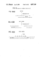
- FIG. 7 is a diagram of the plasmid vector pUC8/CTAP-III used in making an expression vector containing the synthetic gene of FIG. 5;
- FIG. 8 is a diagram of the plasmid cloning vector pNP6 that is used in making an expression vector containing the synthetic gene of FIG. 5;
- FIG. 9 is a schematic flow diagram for preparing a linearized derivative of pNP6, pNP6-LBR, that is ligated with a DNA fragment containing the synthetic CTAP-III gene or genes encoding CTAP-III analogs;
- FIG. 10 is a schematic flow diagram for making the CTAP-III-Leu21 gene insert for ligation with pNP6-LBR to make the inducible plasmid expression vector of the invention.
- FIG. 12 is a schematic flow diagram of the procedure used to make the expression plasmid pNP6 ⁇ RI/Col:CTAP-III-Leu21.
- Such insensitivity permits the mutein to be purified from cellular materials or extracts by digesting the materials or extracts with such agents or be separated from partner fusion sequences when the mutein is produced as a fusion protein in which it is linked to the partner by a methionine.
- Another modification of native CTAP-III involves altering the amino acid sequence in a manner that causes the resulting mutein to be less susceptible to in vivo conversion to ⁇ -TG than native CTAP-III without affecting biological properties adversely.
- Such alteration may involve the addition of a generally nonpolar polypeptide fragment to the amino terminus of native CTAP-III which fragment itself tends to form an alpha helical configuration and thus preserves the configuration of the terminus and/or deleting or replacing one or more of the amino acids that define the trypsin-sensitive site proximate to the amino terminus of native CTAP-III.
- nonpolar fragments to the amino terminus may be accomplished by synthesizing the gene to include codons for the fragment or by synthesizing the polypeptide in the form of a fusion protein in which the leader sequence or endogenous portion of the protein defines such a fragment.
- the fragment is composed predominantly of hydrophobic or ambivalent, acyclic amino acid residues such as leucine, methionine, and isoleucine. It may contain nonhydrophobic residues such as threonine and glutamine provided the fragment as a whole is nonpolar.
- CTAP-III or CTAP-III muteins in the form of fusion proteins in which the endogenous portion of the protein is a biologically inactive colicin El fragment that possesses colicin's charge properties and the fusion site is readily cleavable is yet another modification.
- Such proteins may be produced using expression vectors in which the CTAP-III or CTAP-III mutein gene with suitable terminators is inserted into a vector containing the colicin expression control sequence and structural gene at a convenient restriction site near the end of the colicin structural gene that encodes the carboxy-terminus of colicin.
- fusion proteins of other useful polypeptides and colicin fragments may be made, separated by ion exchange chromatography and cleaved to provide the useful polypeptide in a similar manner.
- CTAP-III and CTAP-III analogs that are produced via expression of recombinant DNA in bacteria may have an initial methionine residue at their amino terminus.
- CTAP-III The amino acid sequence of CTAP-III (FIG. 1) was reverse translated into the ambiguous coding sequence shown in FIG. 2 using the genetic code.
- the final unambiguous nucleotide sequence shown in FIG. 3 for the structural gene encoding CTAP-III-Leu21 was determined as follows.
- the ambiguous nucleotide sequence was reduced to a single nonambiguous sequence by the selection of codons that are preferred for high levels of expression in bacteria. Changes were made to introduce two unique restriction sites into this sequence for the endonucleases BamHI and XbaI. These changes involve using nonoptimal codons for the Gly at position 28 (GGG), and the Leu at position 63 (CTA). These sites were added to permit alternative strategies for synthesizing the gene, sub-cloning the gene, confirming the sequence of the synthesized gene and modifying the gene to encode muteins (analogs). The resulting sequence was analyzed with a computer program designed to detect both complementary and repeated sequences. This information was used to identify regions of intrasequence homology that could interfere with the current ligation of oligonucleotides used to synthesize the gene and to modify the gene accordingly.
- Adaptor sequences were designed for the termini of the structural gene to provide suitable start and stop signals, restriction sites, and cohesive EcoRI ends for cloning the gene as follows.
- the adaptor sequence that was added to the carboxy-terminal coding end of the gene was designed to include two tandem translation termination condons (TAA, TGA), a PstI recognition sequence (CTGCAG), and part of an EcoRI recognition sequence (AATT).
- the adapter sequence that was added to the amino-terminal end of the gene was designed to include a translation start codon (ATG), a PuvII recognition sequence (CAGCTG), and part of an EcoRi recognition sequence (AATT).
- FIG. 5 shows the complete DNA sequence for the synthetic CTAP-III-Leu21 structural gene plus adapter sequences.
- the gene was divided into three fragments: fragment I EcoRI to BamHI,k fragment II BamHI to XbaI and fragment III XbaI to EcoRI. Each fragment was further divided into small oligonucleotides as indicated. There were 38 oligonucleotides in all. The asterisks in FIG. 6 indicate the oligonucleotide boundaries.
- the oligonucleotides were synthesized by the phosphotriester method (Ohtsuka, E., et al, Nucleic Acid Res (1982) 10:6553-6570).
- the crude oligonucleotides were purified by ion-exchange HPLC with a 30-45 min gradient of 0.1M-0.4M KH 2 PO 4 buffer containing 20% acetonitrile. The product peaks were collected, desalted, dried and reconstituted in distilled H 2 O. The size and homogeneity of the purified oligonucleotides were confirmed by gel electrophoresis using 20% acrylamide gels with 3-3.5% cross linker. Oligonucleotides with less than 95% purity were further purified to the desired purity level either by reverse-phase HPLC or by preparative gel electrophoresis.
- Oligonucleotides 1-14 (FIG. 6) were individually radiolabeled in a reaction with 32 P-ATP and polynucleotide kinase using the following protocol:
- oligonucleotides and ATP were dried and dissolved in kinase buffer.
- the enzyme was added and incubated at 37° C. for 1 hr.
- the kinased oligonucleotides were combined into a single vial and placed in a 95° C. water bath for 10 min and then the bath was allowed to cool slowly to room temperature. 10 ⁇ solution of 100 mM DTT/10 mM ATP was added followed by 5 units of T4 DNA ligase.
- the reaction mixture was incubated at 12° C. overnight ( ⁇ 16 hr).
- the entire reaction mixture was purified on a 15% 1.5 mm nondenaturing polyacrylamide gel overnight, run at 250 volts.
- the product was located by autoradiography in comparison with a HaeIII digest of pBR322 DNA.
- the appropriate region on the gel was transferred to DE 81 paper by electrophoresis (Transblot).
- the appropriate band was then eluted from the paper using 1M NaCl in TE buffer.
- the DNA was ethanol precipitated and dissolved in water.
- Synthetic fragment I (EcoRI, BamHI) was cloned into M13 mp8 and sequenced. Three of 16 independent clones had the correct sequence. The others had at least one base substitution.
- Synthetic fragment II (BamHI, XbaI) was cloned into M13 mp11 and sequenced. The correct sequence was verified on 14 of 16 independent clones. The other two had a single base substitution.
- Synthetic fragment III (XbaI, EcoRI) was cloned into M13 mp11 and sequenced. Eight of eight clones had the correct sequence.
- Single stranded M13 for cloned fragments I or II were made double stranded with the universal primer and Klenow polymerase, then digested with EcoRI and BamHI for fragment I or BamHI and XbaI for fragment II.
- the inserts were electroeluted from a 6% acrylamide mini slab gel and ligated with EcoRI XbaI cut mp8. Several transformants were identified that had the correct (I+II) insert.
- Single stranded M13 for cloned fragment (I+II) and III were double stranded as before and digested with EcoRI and XbaI.
- the purified inserts were gel purified, electroeluted and combined and ligated into EcoRI cut mp8. Seven clones were identified with the correct (I+II+III) insert. Both orientations were present.
- Single stranded M13 with the I+II+III insert was double stranded as described and digested with EcoRI.
- a 284 bp insert was purified from the digest by gel electrophoresis.
- plasmid pUC8 (P-L Biochemicals, Milwaukee, WI) was digested for 60 min at 37° C. with EcoRI in a 50 ⁇ l solution containing 50 mM Tris-HCl (pH 7.5), 6 mM MgCl 2 , 50 mM NaCl, and 1 mM DTT. Reactions were terminated by adding EDTA to a final concentration of 20 mM, then extracted with phenol/chloroform (3/1) and precipitated with ethanol.
- the host for transformation experiments was E. coli K-12 strain JM83, which carries the lac Z ⁇ M15 on a ⁇ 80 integrated into the chromosome (ara, ⁇ lac-pro, str A, thi, ⁇ 80 lac Z ⁇ M15) (U. Messing, "A multi-purpose cloning system based on the single-stranded DNA bacteriophage M13", Recombinant DNA Technical Bulletin, NIH Publication No. 79-99, 2, (1979) No. 2:43-48).
- L-broth An overnight culture grown in L-broth (per liter: 10 g tryptone, 5 g yeast extract, 10 g NaCl, 2 ml 1.0 NaOH, and 10 ml 20% glucose added after autoclaving) was diluted 1:100 into fresh L-broth medium and was incubated with shaking at 37° C. until the OD 600 was 0.6. At that time, 35 ml of culture was centrifuged at 6,000 rpm for 10 min at 4° C. and the pellet resuspended in 10 ml of 0.05M CaCl 2 . The cells were incubated on ice for 15 min before they were collected by centrifugation at 4,000 rpm for 10 min.
- FIG. 7 is a diagram of the plasmid showing the location of the CTAP-III-Leu21 insert. A sample of the transformed E.
- cells transformed with recombinant vectors in which a structural gene, such as the CTAP-III gene, has been inserted into the ColEl gene will not produce colicin El. This permits such transformants to be discerned from cells transformed with vectors that do not contain an insert, since the latter product colicin which kills the cells.
- SacII site of plasmid pNP6 is located 8 bp from the start of the colicin El gene. Exogenous (foreign) genes cloned into this site are under control of the colicin El regulatory sequence. Gene expression can be selectively induced by treating transformants with mitomycin C or other SOS system activating agents such as alkylating agents and the like.
- pNP6 ⁇ RI was constructed by: partially digesting pNP6 with EcoRI: filling in the single stranded EcoRI ends of the linearized plasmid using a DNA polymerase reaction; blunt end ligating the resulting fragment; and identifying constructs that contain the colicin gene using the procedure described above.
- pNP6-LBR The construction of plasmid pNP6-LBR, which may be used to make an expression vector containing the synthetic CTAP-III gene or CTAP-III analog genes, involves the following steps. pNP6 was first cleaved into a linear molecule (pNP6-L) by SacII endonuclease. pNP6-L is then treated with T 4 polymerase in the presence of dTTP followed by treatment with Sl nuclease to form a blunt ended linear molecule (pNP6-LB).
- pNP6-LB was next cleaved with EcoRI endonuclease to remove the segment of DNA from the remainder of the fifth codon of the colicin El gene to the EcoRI site in the pBR322 portion of pNP6 (pNP6-LBR).
- Strain JC411 (Col EL-D30) was grown in 60 liters of M9 medium (per liter: 1 g NH 4 Cl, 6 g Na 2 HPO 4 .H 2 O, 3 g KH 2 PO 4 , 5 g NaCl, 3 g casamino acids, 1 ml 10% MgSO 4 supplemented with 10 ml 20% glucose and 0.5 ml 1M CaCl 2 added after autoclaving) in a fermentor at 37° C. to a cell density of approximately 5 ⁇ 10 8 CFU/ml. Chloramphenicol was added to a final concentration of 100 ⁇ g/ml, and the incubation at 37° C. was continued for another 6 hr.
- M9 medium per liter: 1 g NH 4 Cl, 6 g Na 2 HPO 4 .H 2 O, 3 g KH 2 PO 4 , 5 g NaCl, 3 g casamino acids, 1 ml 10% MgSO 4 supplemented with 10 m
- pancreatic ribonuclease A Four mg was added to the supernatant and the mixture was incubated at 37° C. for 1 hr. The sample was extracted twice with an equal volume of phenol saturated with 0.1M Tris, pH 8.0, and the DNA was precipitated by adding 1/10 sample volume of 3.0M sodium acetate and 2.5 volumes of cold ethanol, followed by an overnight incubation at -20° C. The resulting precipitate was recovered by centrifugation at 7,000 rpm for 50 min in a refrigerated Sorvall centrifuge using an HB-4 rotor.
- the pellet was dissolved in 50 ml of 10 mM Tris-HCl buffer (pH 7.5) containing 0.3M NaCl and 5 mM EDTA (NE Buffer). This sample was then applied to a Bio-Gel A.5 column (Bio-Rad Laboratories, Richmond, California), 5 ⁇ 100 cm, equilibrated with NE buffer. The DNA was eluted with NE buffer. Twenty-ml fractions were collected at a flow rate of 60 ml/hr. The elution of DNA was monitored by measuring the absorbance of each fraction at 260 nm using a Gilford 2600 UV-VIS spectrophotometer. The host DNA and the plasmid DNA was recovered together in the void volume.
- the elution of DNA was monitored by measuring the conductivity of each collected sample using a conductivity meter (Radiometer, Copenhagen), and by agarose gel electrophoresis using vertical agarose slab gels (0.25 ⁇ 14 ⁇ 15.5 cm).
- the samples were applied to 1% agarose gels prepared in 40 mM Tris base buffer, pH 8.2, containing 1 mM EDTA and 5 mM sodium acetate (TAE buffer), and electrophoresed for 3 hr at a constant applied voltage of 5 V/cm.
- Fractions containing the supercoiled and the nicked circular DNA were pooled separately and were precipitated with cold ethanol.
- the resulting precipitates of ColEl DNA molecules were dissolved in 1.0 and 0.6 ml TEN buffer, respectively.
- Plasmid pBR322 was isolated from E. coli strain 294 (pBR322) by the procedure used for isolating ColEl plasmid described above. PREPARATION OF SHEARED FRAGMENTS OF COLEL DNA
- Each reaction mixture contained 380 ⁇ l distilled water, 50 ⁇ l 1M Tris-HCl buffer (pH 8.0), 5 ⁇ l 10 mM zinc sulfate, 5 ⁇ l CIT (10 U/ ⁇ l). After incubation at 37° C. for 30 min, an additional 5 ⁇ l of CIT was added and the incubation at 37° C. continued for another 30 min.
- the reaction mixtures were extracted twice with an equal volume of buffer-saturated phenol, and the DNA was precipitated with ethanol. The heterogeneous population of DNA fragments was further purified and separated according to size by sucrose gradient velocity centrifugation.
- a discontinuous sucrose gradient was prepared by sequential layering of 3.4 ml of 20%, 15%, 10%, and 5% sucrose in 0.3M sodium acetate buffer (pH 7.0) containing 1 mM EDTA in centrifuge tubes for the SW40 rotor (Beckman).
- the DNA sample in 100 ⁇ l (0.25 ⁇ g/ ⁇ l) was layered on the sucrose gradient and centrifuged at 35,000 rpm for 20 hr at 10° C. using an L8-70 Beckman ultracentrifuge. Fractions of 0.5 ml each were collected and preipitated with ethanol. The precipitates were redissolved in 50 ⁇ l of TEN buffer and analyzed by agarose gel electrophoresis.
- Poly(dG) homopolymer extensions were added to linear pBR322 molecules in a reaction mixture containing 6 ⁇ l distilled water, 20 ⁇ l 500 mM potassium cacodylate, 10 ⁇ l 10 mM cobalt chloride, 10 ⁇ l 1 mM DTT, 2 ⁇ l 10 mM dGTP, 20 ⁇ l 3 H-dGTP (New England Nuclear Corporation), 25 ⁇ l DNA (0.04 ⁇ g/ ⁇ l), and 5 ⁇ l (12 U/ ⁇ l) terminal deoxynucleotidyl transferase (Bethesda Research Laboratories, Inc., Gaithersburg, Maryland).
- ColEl-[poly(dC)]fragments were redissolved in 115 ⁇ l distilled water and were annealed to linear pBR322-[poly(dG)] molecules by adding 40 ⁇ l 0.5M NaCl, 40 ⁇ l 50 mM EDTA (pH 7.25), and 3 ⁇ l linear pBR322-[poly(dG)] DNA solution (0.1 ⁇ g/ ⁇ l).
- the annealing mixture was incubated at 70° C. for 15 min and then cooled to 40° C. over a 5-hr period. The mixture was kept at 45° C. overnight, then cooled to room temperature.
- E. coli 294 For transformation into E. coli 294, an overnight culture grown in L-broth was diluted 1:100 into fresh L-broth medium and incubated with shaking at 37° C. until the OD 600 was 0.6. At this time, 35 ml of culture was centrifuged at 6,000 rpm for 10 min at 4° C., and the pellet was resuspended in 20 ml of 0.05M CaCl 2 . The cells were incubated on ice for 15 min before they were collected by centrifugation at 4,000 rpm for 10 min.
- the cells were resuspended in 4 ml of 0.05M CaCl 2 and mixed with 200 ⁇ l of a DNA solution prepared by adding 50 ⁇ l of the annealing mixture and 150 ⁇ l 10 mM Tris-HCl (pH 7.5). containing 10 mM MgCl 2 and 10 mM CaCl 2 . This mixture was incubated at 0° C. for 25 min, followed by incubation at 50° C. for 10 sec and at room temperature for 10 min. At this point, 14 mL of L-broth was added and the culture was shaken at 37° C. for 30 min.
- the Tc r Ap s transformant colonies were then screened for the spontaneous production of colicin. Single colonies were spotted on L-agar plates and were incubated at 37° C. overnight. The colonies were killed by exposing them to chloroform vapor, then overlayed with 5 ml L-broth containing 0.7% agar and 0.1 ml of an overnight culture of E. coli K-12, CL142. After the agar was allowed to harden, the plates were incubated at 37° C. overnight. Colonies with a zone of inhibition around them were scored as colicin producers.
- the Tc r Ap s Col + transformant colonies were screened for the presence of recombinant plasmids by analyzing a small amount of cleared lysate by agarose gel electrophoresis as described above with respect to pUC8/CTAP-III.
- the size of the plasmids were determined by measuring the electrophoretic migration of DNA through an agarose gel using 8 plasmid standards, ranging in size from 1.36 ⁇ 10 6 to 35.8 ⁇ 10 6 daltons (F. L. Marcina, D. J. Kopecko, K. R. Jones, D. J. Ayers, and S. M. McCowen. "A multiple plasmid-containing Escherichia coli strain: Convenient source of size reference plasmid molecules" Plasmid (1978) 1:417-420).
- Transformed clones were grown in 2-liter cultures. Cleared lysates were prepared as described above. The supernatants were treated with pancreatic RNase A (100 ⁇ g/ml at 37° C. for 30 min) and then were extracted with phenol. The DNA was precipitated with ethanol and redissolved in TEN buffer.
- Restriction enzymes were obtained as commercial preparations from Bethesda Research Laboratories, Inc. (BRL). The DNA was digested with PstI, EcoRI, SmaI, and SacII, using the conditions specified by BRL. Samples were applied to 1% agarose gels and electrophresed for 4 hr at a constant applied voltage of 5 V/cm. The molecular weights of restriction fragments were determined relative to the standard migration patterns of bacteriophase ⁇ DNA digested with HindIII and HaeIII.
- FIG. 8 is a restriction map of the recombinant plasmid, designated pNP6, of one of the transformed clones.
- E. coli strain NP6-294 (pNP6) was grown and plasmid DNA isolated as described above for ColEl.
- the supercoiled DNA was further purified by adjusting 500 ⁇ g of DNA to 3.9 ml of TEN buffer and adding 3.45 g CsCl and 0.1 ml ethidium bromide stock solution (5 mg/ml).
- the mixture was transferred into a cellulose nitrate tube for an SW50.1 rotor (Beckman) and centrifuged at 36,000 rpm at 10° C. for 40 hr.
- the plasmid DNA band was located under a longwave UV light and was removed with a syringe by puncturing the tube from the side.
- the DNA sample was extracted five times with butanol and dialyzed against 100 volumes ( ⁇ 3) of TEN buffer for 24 hr at 4° C.
- the DNA was then precipitated with 2.5 volumes of ethanol and 1/10 volume of 3M sodium acetate.
- Plasmid pNP6 contains two EcoRI restriction sites, one located in the carboxy terminal region of the colicin El gene, the other located near the tetracycline resistance gene of the original pBR322 vector.
- a derivative of pNP6 lacking the second site was constructed as follows. pNP6 was digested with EcoRI under limited reaction conditions so that linear molecules (cleaved at only one of the two sites) were produced. Linear molecules of pNP6 were purified by agarose gel electrophoresis and subsequently reacted with DNA polymerase I and deoxyribonucleotide triphosphates to fill in the single-stranded ends. The resulting molecules were circularized in a blunt-end ligation reaction using T 4 ligase and were used to transform E. coli 294 as described previously.
- Colicin-producing transformants were selected as described previously. DNA was isolated from individual clones and digested with EcoRI to identify those that contained a single, intact EcoRI site within the colicin gene. The location of the single EcoRI site was confirmed by additional restriction endonuclease mapping.
- the DNA was then precipitated with ethanol and redissolved in 100 ⁇ l of a solution containing 50 mM Tris-HCl (pH 8.5), 7 mM MgCl 2 , 10 mM - ⁇ mercaptoethanol, 15 mM ammonium sulfate, 7 mM Na 2 EDTA, 20 ⁇ g BSA, and 0.1 mM dTTP.
- T4 DNA polymerase BRL
- the solution was purified of dTTP by binding the DNA in 0.12M sodium phosphate (PB), 15% formamide, to 0.5 g of hydroxyapatite at 65° C., washing with 30 ml of 0.12M PB, eluting the DNA with 0.5M PB, dialyzing against water, and precipitating with ethanol.
- PB sodium phosphate
- the DNA was further purified by binding to a 1-cm column of diethylaminoethyl (DEAE) cellulose, washing with 0.15M NaCl, eluting with 1M NaCl, and precipitating with ethanol.
- the DNA was finally dissolved in TEN beffer and stored at -85° C. until used.
- Enzyme reactions using T 4 polymerase (with dTTP and dCTP individually), S1 nuclease, and endonuclease EcoRI were performed as described above for pNP6 vector DNA.
- the resulting fragment has one blunt end and one cohesive EcoRI end. Ligation of the blunt end of this fragment to the blunt end of pNP6-LBR forms a start condon, ATG, in phase with the structural gene for CTAP-III-Leu21.
- vector pNP6-LBR and the CTAP-III-Leu21 gene insert were ligated in a 100 ⁇ l reaction, transformed into E. coli 294, and selected with tetracycline, using the general procedures described above under "Cloning of the Synthetic CTAP-III Fragment in pUC8". Colonies that contain recombinant plasmid were screened by detecting the loss of ability to produce colicin El.
- Purified plasmid pNP6-LBR/CTAP-III-Leu21 was digested with endonuclease PstI to produce two fragments, the smaller of which ( ⁇ 2100 bp) contains the colicin regularity region and the CTAP-III-Leu21 structural gene. This fragment was purified and cloned into the single PstI site of plasmid pUC8 and transformants were selected with ampicillin.
- the resulting recombinant plasmid designated pUC8-Col/CTAP-III-Leu21 has a greater than tenfold increase in copy number than pBR322 and thus provides substantially greater quantities of CTAP-III-Leu21 upon induction, due to a gene dosage effect.
- Purified pUC8/CTAP-III-Leu21 plasmid DNA was digested to completion with endonuclease PvuII, followed by endonuclease EcoRI. The products were separated by agarose gel electrophoresis, and the 273 bp fragment containing the CTAP gene was electroeluted, extracted with phenol, and precipitated with ethanol (part (A) of FIG. 11).
- Purified pNP6 plasmid DNA was digested to completion with endonuclease SacII, followed by treatment with T 4 polymerase and deoxycytidine triphosphate to remove the 3' single-stranded ends. The product of this reaction was digested with endonuclease EcoRi and the large DNA fragment produced was purified by agarose gel electrophoresis (part (B) of FIG. 11).
- part (C) of FIG. 11 The final products of parts (A) and (B) were mixed in an equimolar ratio and ligated with T 4 ligase (part (C) of FIG. 11).
- the reaction mixture was used to transform E. coli 294, and several tetracycline-resistant transformants were selected. Plasmid DNA from these clones was analyzed by restriction endonuclease digestion and DNA sequence analysis to identify recombinants having the correct structure.
- FIG. 12 outlines the steps involved in preparing pNP6 ⁇ RI/CTAP-III-Leu21 from pUC8/CTAP-III-Leu21 and pNP6 ⁇ RI.
- Purified pUC8/CTAP-III-Leu21 plasmid DNA was digested to completion with endonuclease EcoRI to produce the 284 bp fragment containing the CTAP-III gene. This fragment was purified by agarose gel electrophoresis (part (A) of FIG. 12).
- Purified plasmid pNP6 ⁇ RI (or pNP6) was digested to completion with endonuclease EcoRI and the vector fragment was purified by agarose gel electrophoresis (part (B) or FIG. 12).
- CTAP-III-NMOD synthesis and its purification are accomplished as described previously for CTAP-III-Leu21, using cells containing the expression plasmid pNP6/CTAP-III-Leu21-NMOD.
- the amino acid sequence of CTAP-III-NMOD is depicted in FIG. 1 with the pentapeptide addition to the amino terminus shown in braces.
- the terminal methionine of the illustrated pentapeptide is present as a result of the bacterial production of the protein. It will be appreciated that this methionine may be absent due to post-expression processing.
- Col:CTAP-III-Leu21 Cell extracts containing CTAP-III-Leu21 fused to ColEl(1-502) (Col:CTAP-III-Leu21) were prepared by lysing cells in 50 mM sodium borate, pH 9.5, at a concentration of 1-5 ⁇ 10 10 cells/ml. Cellular debris was removed by centrifugation. Col:CTAP-III-Leu21 was purified by either of two methods using cation-exchange chromatography:
- Col:CTAP-III-Leu21 was purified from cell extracts using the Pharmacia fast protein liquid chromatography (FPLC) system. 10 to 100 ml of extract was applied to a mono S column in 50 mM sodium borate, pH 9.5, and eluted with a linear gradient of 0 to 0.5M NaCl. The fusion protein eluted at approximately 0.25M NaCl.
- FPLC Pharmacia fast protein liquid chromatography
- Col:CTAP-III-Leu21 prepared by either method was generally greater than 90%, as judged by analysis using polyacrylamide gel electrophoresis under denaturing conditions.
- fusion protein Purified fusion protein was dialyzed extensively against distilled water, and lyophilized. The protein was dissolved in 70% formic acid; cyanogen bromide was added at a ratio of 300 moles of cyanogen bromide per mole of methionine (approximately 2 mg cyanogen bromide per mg of fusion protein).
- CTAP-III-Leu21 can be converted to ⁇ -TG-Leu17 by proteolytic cleavage of the amino terminal tetrapeptide at Lys4-Gly5 using either plasmin or trypsin and the following protocol.
- CTAP-III-Leu21 Approximately 0.5 mg/ml of CTAP-III-Leu21 in 0.15M NaCl/0.01M Tris-HCl, pH 7.5 is incubated at 37° C. for 1 hr with 1.5 I.U./ml plasmin or for 10 min with 0.5% w/w trypsin.
- ⁇ -TG-Leu17 is purified by FPLC using a mono S column as described above.
- the purified CTAP-III or CTAP-III analogs will typically be formulated with conventional pharmaceutically acceptable carriers for administration to humans to regenerate connective tissue (e.g., heal wounds). It will usually be administered topically at the desired site of regeneration using conventional topical formulations such as creams, pastes, gels, sprays, ointments, and salves.
- Carriers used in such formulations are well known and include, without limitation, petrolatum, polyethylene glycol, gelatin, isopropyl myristate, polyvinyl alcohol, and the like.
Abstract
Description
__________________________________________________________________________
AACCTGGCLAAPGGQAAPGAAGAATCQCTGGAQTCQGAQCTGTACGCLGA
ACTGCGLTGCJTGTGCATCAAPACQACQTCQGGHATCCAQCCGAAPAACA
TCCAGTCQCTMGAPGTLATCGGQAAPGGQACQCAQTGCAACCAGGTLGAA
GTLATCGCLACQCTGAAPGAQGGQCGQAAPATCTGCCTGGAQCCGGAQGC
LCCGCGQATCAAPAAPATCGTLCAGAAPAAPCTGGCLGGQGAQGAATCQG
CLGAQ,
       wherein:
       A = Adenine     H = either T, C or G
       G = Guanine     J = either C or A
       C = Cytosine    L = either T or A
       T = Thymine     M = either C or G
       P = either A or G
       Q = either C or T.
__________________________________________________________________________
__________________________________________________________________________
AACCTGGCTAAAGGTAAAGAAGAATCTCTGGACTCTGACTTATACGCTGA
TTGGACCGATTTCCATTTCTTCTTAGAGACCTGAGACTGAATATGCGACT
ACTGCGTTGCJTGTGCATCAAAACTACTTCTGGGATCCACCCGAAAAACA
TGACGCAACGKACACGTAGTTTTGATGAAGACCCTAGGTGGGCTTTTTGT
TCCAGTCTCTGGAAGTTATCGGTAAAGGCACTCACTGCAACCAGGTTGAA
AGGTCAGAGACCTTCAATAGCCATTTCCGTGAGTGACGTTGGTCCAACTT
GTTATCGCTACTCTGAAAGACGGTCGTAAAATCTGTCTAGATCCGGACGC
CAATAGCGATGAGACTTTCTGCCAGCATTTTAGACAGATCTAGGCCTGCG
TCCACGTATCAAGAAGATCGTTCAGAAAAAACTGGCTGGTGACGAATCTG
AGGTGCATAGTTCTTCTAGCAAGTCTTTTTTGACCGACCACTGCTTAGAC
CTGAC
GACTG,
__________________________________________________________________________
______________________________________
Oligonucleotides          0.04   OD
.sup.32 P-ATP (10 μCi/mmole)
                          10     μl
Kinase/ligase buffer      15     μl
(50 mM Tris-HCl (pH 7.6), 10 mM MgCl.sub.2'
                          10     mM
dithiothreitol)
Polynucleotide Kinase     1      μl
______________________________________
Claims (12)
Priority Applications (1)
| Application Number | Priority Date | Filing Date | Title |
|---|---|---|---|
| US07/117,916 US4897348A (en) | 1983-08-25 | 1987-11-04 | Recombinant materials and methods for producing human connective tissue-activating peptide-III and analogs thereof |
Applications Claiming Priority (3)
| Application Number | Priority Date | Filing Date | Title |
|---|---|---|---|
| US52636983A | 1983-08-25 | 1983-08-25 | |
| US64625984A | 1984-08-30 | 1984-08-30 | |
| US07/117,916 US4897348A (en) | 1983-08-25 | 1987-11-04 | Recombinant materials and methods for producing human connective tissue-activating peptide-III and analogs thereof |
Related Parent Applications (1)
| Application Number | Title | Priority Date | Filing Date |
|---|---|---|---|
| US64625984A Continuation | 1983-08-25 | 1984-08-30 |
Publications (1)
| Publication Number | Publication Date |
|---|---|
| US4897348A true US4897348A (en) | 1990-01-30 |
Family
ID=27382064
Family Applications (1)
| Application Number | Title | Priority Date | Filing Date |
|---|---|---|---|
| US07/117,916 Expired - Fee Related US4897348A (en) | 1983-08-25 | 1987-11-04 | Recombinant materials and methods for producing human connective tissue-activating peptide-III and analogs thereof |
Country Status (1)
| Country | Link |
|---|---|
| US (1) | US4897348A (en) |
Cited By (36)
| Publication number | Priority date | Publication date | Assignee | Title |
|---|---|---|---|---|
| WO1990013647A1 (en) * | 1989-05-04 | 1990-11-15 | Sri International | Method and vectors for stabilizing heterologous protein expression |
| WO1992006112A1 (en) * | 1990-10-09 | 1992-04-16 | Sri International | Activated ctap and analogs thereof and their use |
| US5118790A (en) * | 1990-07-24 | 1992-06-02 | Sri International | Analogs of hirudin |
| US5179196A (en) * | 1989-05-04 | 1993-01-12 | Sri International | Purification of proteins employing ctap-iii fusions |
| US5187075A (en) * | 1989-01-26 | 1993-02-16 | Sri International | Cloning and expression of a variant gene of platelet factor 4 and compositions thereof to modulate immune responses |
| US5202239A (en) * | 1990-08-07 | 1993-04-13 | Scios Nova Inc. | Expression of recombinant polypeptides with improved purification |
| US5302384A (en) * | 1988-08-15 | 1994-04-12 | Brigham And Women's Hospital | Endothelial-derived II-8 adhesion inhibitor |
| US5401643A (en) * | 1988-08-29 | 1995-03-28 | Dainippon Pharmaceutical Co., Ltd. | Method of preparing an active human neutrophil chemotactic factor polypeptide |
| US5451399A (en) * | 1989-11-29 | 1995-09-19 | Brigham And Women's Hospital | [ALA IL-8]77 and [SER IL-8]72 as Leukocyte adhesion inhibitors |
| WO1996024668A1 (en) * | 1995-02-08 | 1996-08-15 | Human Genome Sciences, Inc. | Human chemokine beta-11 and human chemokine alpha-1 |
| US5578569A (en) * | 1993-03-12 | 1996-11-26 | Tam; Cherk S. | Method of increasing bone growth |
| US5652338A (en) * | 1988-03-16 | 1997-07-29 | The United States Of America As Represented By The Department Of Health And Human Services | Neutrophil chemotactic factor |
| US5709864A (en) * | 1993-07-28 | 1998-01-20 | Parfums Christian Dior | Cosmetic or pharmaceutical and particularly dermatological, composition containing an extract of tephrosia, particularly Tephrosia purpurea |
| US5759533A (en) * | 1988-12-08 | 1998-06-02 | Novartis Ag | Neutrophil-activating peptide-2 |
| US5910431A (en) * | 1996-03-19 | 1999-06-08 | Human Genome Sciences, Inc. | Polynucleotides encoding chemokine α-2 |
| US5981230A (en) * | 1994-08-23 | 1999-11-09 | Human Genome Sciences, Inc. | Polynucleotide encoding chemokine β-4 |
| US6001606A (en) * | 1994-03-08 | 1999-12-14 | Human Genome Sciences, Inc. | Polynucleotides encoding myeloid progenitor inhibitory factor-1 (MPIF-1) and polypeptides encoded thereby |
| US6018037A (en) * | 1986-09-12 | 2000-01-25 | Kyowa Hakko Kogyo Co., Ltd | DNA coding for (Leu13) motilin |
| US6139832A (en) * | 1995-02-08 | 2000-10-31 | Human Genome Sciences, Inc. | Leukocyte adhesion inhibitor-1 (LAI-1) Polypeptides |
| US6174995B1 (en) | 1994-08-23 | 2001-01-16 | Haodong Li | Human chemokines, CKβ4 and CKβ10/MCP-4 |
| US6391589B1 (en) | 1994-08-23 | 2002-05-21 | Human Genome Sciences, Inc. | Human chemokine beta-10 mutant polypeptides |
| US6410268B1 (en) | 1996-03-18 | 2002-06-25 | Human Genome Sciences, Inc. | Polynucleotides encoding chemokine alpha-3 |
| US6451562B1 (en) | 1993-12-22 | 2002-09-17 | Human Genome Sciences, Inc. | Polypeptides encoding myeloid progenitor inhibitory factor-1 (MPIF-1) polynucleotides |
| US6458349B1 (en) | 1995-06-02 | 2002-10-01 | Human Genome Sciences, Inc. | Chemokine β-4 polypeptides |
| US20020141972A1 (en) * | 1996-05-14 | 2002-10-03 | Smithkline Beecham Corporation | Method of treating sepsis and ARDS using chemokine alpha-2 |
| US6488925B2 (en) | 1993-12-22 | 2002-12-03 | Human Genome Sciences, Inc. | Macrophage inflammatory protein-4 (MIP-4) polypeptides |
| US6495129B1 (en) | 1994-03-08 | 2002-12-17 | Human Genome Sciences, Inc. | Methods of inhibiting hematopoietic stem cells using human myeloid progenitor inhibitory factor-1 (MPIF-1) (Ckbeta-8/MIP-3) |
| US6596498B1 (en) | 1993-03-12 | 2003-07-22 | Osteopharm Inc. | Bone stimulating factor |
| US6693081B2 (en) | 1995-09-26 | 2004-02-17 | Osteopharm Inc. | Bone stimulating factor |
| US6743895B1 (en) | 1995-06-07 | 2004-06-01 | Osteopharm Inc. | Bone stimulating factor |
| US20040152169A1 (en) * | 1993-12-22 | 2004-08-05 | Human Genome Sciences, Inc. | Therapeutic compositions and methods for treating disease states with myeloid progenitor inhibitory factor-1 (MPIF-1), monocyte colony inhibitory factor (M-CIF), and macrophage inhibitory factor-4 (MIP-4) |
| US20040214778A1 (en) * | 2001-03-22 | 2004-10-28 | Tam Cherk Shing | Polypeptides for use in ameliorating effects of aging in mammals |
| US20060052307A1 (en) * | 2002-12-05 | 2006-03-09 | Tam Cherk S | Bone growth factor |
| US20060275210A1 (en) * | 2002-05-01 | 2006-12-07 | Rosen Craig A | Antibodies that specifically bind to chemokine beta-4 |
| US8491901B2 (en) | 2010-11-19 | 2013-07-23 | Toshio Imai | Neutralizing anti-CCL20 antibodies |
| US9365625B1 (en) | 2011-03-31 | 2016-06-14 | David Gordon Bermudes | Bacterial methionine analogue and methionine synthesis inhibitor anticancer, antiinfective and coronary heart disease protective microcins and methods of treatment therewith |
Citations (2)
| Publication number | Priority date | Publication date | Assignee | Title |
|---|---|---|---|---|
| US4366246A (en) * | 1977-11-08 | 1982-12-28 | Genentech, Inc. | Method for microbial polypeptide expression |
| US4568640A (en) * | 1981-05-11 | 1986-02-04 | Harvey Rubin | Method of inserting amino acid analogs into proteins |
-
1987
- 1987-11-04 US US07/117,916 patent/US4897348A/en not_active Expired - Fee Related
Patent Citations (2)
| Publication number | Priority date | Publication date | Assignee | Title |
|---|---|---|---|---|
| US4366246A (en) * | 1977-11-08 | 1982-12-28 | Genentech, Inc. | Method for microbial polypeptide expression |
| US4568640A (en) * | 1981-05-11 | 1986-02-04 | Harvey Rubin | Method of inserting amino acid analogs into proteins |
Non-Patent Citations (21)
| Title |
|---|
| Beatty et al, The Journal of Biological Chemistry, vol. 255, pp. 3931 3934, May 10, 1980. * |
| Beatty et al, The Journal of Biological Chemistry, vol. 255, pp. 3931-3934, May 10, 1980. |
| Brot et al, Archives of Biochemistry and Biophysics, vol. 223, pp. 271 281, 1983. * |
| Brot et al, Archives of Biochemistry and Biophysics, vol. 223, pp. 271-281, 1983. |
| Brot et al, TIBS, Apr. 1982. * |
| Caruthers et al, Genetic Engineering Principles and Methods, vol. 4, Ed. by Setlow et al, Plenum Press, pp. 1 17 (1982). * |
| Caruthers et al, Genetic Engineering Principles and Methods, vol. 4, Ed. by Setlow et al, Plenum Press, pp. 1-17 (1982). |
| Castor et al, PNAS, U.S.A., vol. 80, pp. 765 769, Feb. 1983. * |
| Castor et al, PNAS, U.S.A., vol. 80, pp. 765-769, Feb. 1983. |
| Gouz et al, Nucleic Acids Research, vol. 10, No. 22, pp. 7055 7074, Nov. 25, 1982. * |
| Gouz et al, Nucleic Acids Research, vol. 10, No. 22, pp. 7055-7074, Nov. 25, 1982. |
| Johnson et al, The Journal of Biological Chemistry, vol. 254, pp. 4022 4026, May 25, 1979. * |
| Johnson et al, The Journal of Biological Chemistry, vol. 254, pp. 4022-4026, May 25, 1979. |
| Selker et al, Journal of Bacteriology, vol. 129, pp. 388 394, Jan. 1977. * |
| Selker et al, Journal of Bacteriology, vol. 129, pp. 388-394, Jan. 1977. |
| Wilkinson et al, Nature, vol. 307, pp. 187 188, Jan. 1984. * |
| Wilkinson et al, Nature, vol. 307, pp. 187-188, Jan. 1984. |
| Yamada et al, PNAS, U.S.A., vol. 79, pp. 2827 2831, May 1982. * |
| Yamada et al, PNAS, U.S.A., vol. 79, pp. 2827-2831, May 1982. |
| Zoller et al, Nucleic Acids Research, vol. 10, pp. 6487 6500, 1982. * |
| Zoller et al, Nucleic Acids Research, vol. 10, pp. 6487-6500, 1982. |
Cited By (72)
| Publication number | Priority date | Publication date | Assignee | Title |
|---|---|---|---|---|
| US6018037A (en) * | 1986-09-12 | 2000-01-25 | Kyowa Hakko Kogyo Co., Ltd | DNA coding for (Leu13) motilin |
| US5652338A (en) * | 1988-03-16 | 1997-07-29 | The United States Of America As Represented By The Department Of Health And Human Services | Neutrophil chemotactic factor |
| US5925352A (en) * | 1988-03-16 | 1999-07-20 | The United States Of America As Represented By The Department Of Health And Human Services | Method of treating inflammation with antibodies to neutrophil chemotactic factor |
| US6376659B1 (en) | 1988-03-16 | 2002-04-23 | The United States Of America, As Represented By The Department Of Health And Human Services | Nucleic acids encoding a novel neutrophil chemotactic factor |
| US6475741B1 (en) | 1988-03-16 | 2002-11-05 | The United States Of America As Represented By The Department Of Health And Human Services | Neutrophil chemotactic factor cloned cDNA and monoclonal antibodies thereto |
| US5698196A (en) * | 1988-03-16 | 1997-12-16 | The United States Of America As Represented By The Secretary, Department Of Health And Human Services | Antibodies specific for neutrophic chemotactic factor |
| US5302384A (en) * | 1988-08-15 | 1994-04-12 | Brigham And Women's Hospital | Endothelial-derived II-8 adhesion inhibitor |
| US5401643A (en) * | 1988-08-29 | 1995-03-28 | Dainippon Pharmaceutical Co., Ltd. | Method of preparing an active human neutrophil chemotactic factor polypeptide |
| US5759533A (en) * | 1988-12-08 | 1998-06-02 | Novartis Ag | Neutrophil-activating peptide-2 |
| US5187075A (en) * | 1989-01-26 | 1993-02-16 | Sri International | Cloning and expression of a variant gene of platelet factor 4 and compositions thereof to modulate immune responses |
| WO1990013647A1 (en) * | 1989-05-04 | 1990-11-15 | Sri International | Method and vectors for stabilizing heterologous protein expression |
| US5179196A (en) * | 1989-05-04 | 1993-01-12 | Sri International | Purification of proteins employing ctap-iii fusions |
| US5451399A (en) * | 1989-11-29 | 1995-09-19 | Brigham And Women's Hospital | [ALA IL-8]77 and [SER IL-8]72 as Leukocyte adhesion inhibitors |
| US5118790A (en) * | 1990-07-24 | 1992-06-02 | Sri International | Analogs of hirudin |
| US5322930A (en) * | 1990-08-07 | 1994-06-21 | Scios Nova Inc. | Expression of recombinant polypeptides with improved purification |
| US5202239A (en) * | 1990-08-07 | 1993-04-13 | Scios Nova Inc. | Expression of recombinant polypeptides with improved purification |
| WO1992006112A1 (en) * | 1990-10-09 | 1992-04-16 | Sri International | Activated ctap and analogs thereof and their use |
| US6596498B1 (en) | 1993-03-12 | 2003-07-22 | Osteopharm Inc. | Bone stimulating factor |
| US5578569A (en) * | 1993-03-12 | 1996-11-26 | Tam; Cherk S. | Method of increasing bone growth |
| US5709864A (en) * | 1993-07-28 | 1998-01-20 | Parfums Christian Dior | Cosmetic or pharmaceutical and particularly dermatological, composition containing an extract of tephrosia, particularly Tephrosia purpurea |
| US7364865B2 (en) | 1993-12-22 | 2008-04-29 | Human Genome Sciences, Inc. | Antibodies to myeloid progenitor inhibitory factor-1 (MPIF-1) |
| US20060228359A1 (en) * | 1993-12-22 | 2006-10-12 | Human Genome Sciences, Inc. | Antibodies to Myeloid Progenitor Inhibitory Factor-1 (MPIF-1) |
| US7041460B2 (en) | 1993-12-22 | 2006-05-09 | Human Genome Sciences, Inc. | Antibodies to monocyte-colony inhibitory factor (M-CIF) |
| US20080274109A1 (en) * | 1993-12-22 | 2008-11-06 | Human Genome Sciences, Inc. | Myeloid Progenitor Inhibitory Factor-1 (MPIF-1), Monocyte Colony Inhibitory Factor (M-CIF), and Macrophage Inhibitory Factor-4 (MIP-4) |
| US6811773B1 (en) | 1993-12-22 | 2004-11-02 | Human Genome Sciences, Inc. | Human monocyte colony inhibitory factor (M-CIF) polypeptides |
| US20040152169A1 (en) * | 1993-12-22 | 2004-08-05 | Human Genome Sciences, Inc. | Therapeutic compositions and methods for treating disease states with myeloid progenitor inhibitory factor-1 (MPIF-1), monocyte colony inhibitory factor (M-CIF), and macrophage inhibitory factor-4 (MIP-4) |
| US6451562B1 (en) | 1993-12-22 | 2002-09-17 | Human Genome Sciences, Inc. | Polypeptides encoding myeloid progenitor inhibitory factor-1 (MPIF-1) polynucleotides |
| US7851596B2 (en) | 1993-12-22 | 2010-12-14 | Human Genome Sciences, Inc. | Myeloid progenitor inhibitory factor-1 (MPIF-1) polypeptides |
| US6488925B2 (en) | 1993-12-22 | 2002-12-03 | Human Genome Sciences, Inc. | Macrophage inflammatory protein-4 (MIP-4) polypeptides |
| US6001606A (en) * | 1994-03-08 | 1999-12-14 | Human Genome Sciences, Inc. | Polynucleotides encoding myeloid progenitor inhibitory factor-1 (MPIF-1) and polypeptides encoded thereby |
| US6495129B1 (en) | 1994-03-08 | 2002-12-17 | Human Genome Sciences, Inc. | Methods of inhibiting hematopoietic stem cells using human myeloid progenitor inhibitory factor-1 (MPIF-1) (Ckbeta-8/MIP-3) |
| US6623942B2 (en) | 1994-03-08 | 2003-09-23 | Human Genome Sciences, Inc. | Macrophage inflammatory protein-4 (MIP-4) polynucleotides |
| US5981230A (en) * | 1994-08-23 | 1999-11-09 | Human Genome Sciences, Inc. | Polynucleotide encoding chemokine β-4 |
| US6174995B1 (en) | 1994-08-23 | 2001-01-16 | Haodong Li | Human chemokines, CKβ4 and CKβ10/MCP-4 |
| US20040265974A1 (en) * | 1994-08-23 | 2004-12-30 | Human Genome Sciences, Inc. | Human chemokine polypeptides |
| US6921645B2 (en) | 1994-08-23 | 2005-07-26 | Human Genome Sciences, Inc. | Antibodies to chemokine β-4 |
| US6391589B1 (en) | 1994-08-23 | 2002-05-21 | Human Genome Sciences, Inc. | Human chemokine beta-10 mutant polypeptides |
| US20070238644A1 (en) * | 1994-08-23 | 2007-10-11 | Human Genome Sciences, Inc. | Human Chemokines CKBeta-4 and CKBeta-10/MCP-4 |
| US7183081B2 (en) | 1994-08-23 | 2007-02-27 | Human Genome Sciences, Inc. | Human Ckβ-10 polynucleotides |
| US7138498B2 (en) | 1994-08-23 | 2006-11-21 | Human Genome Sciences, Inc. | Antibodies to MCP-4 |
| US6673344B1 (en) | 1994-08-23 | 2004-01-06 | Human Genome Sciences, Inc. | Antibodies to human CKβ-10/MCP-4 |
| US6139832A (en) * | 1995-02-08 | 2000-10-31 | Human Genome Sciences, Inc. | Leukocyte adhesion inhibitor-1 (LAI-1) Polypeptides |
| US20090036370A1 (en) * | 1995-02-08 | 2009-02-05 | Human Genome Sciences, Inc. | Leukocyte Adhesion Inhibitor-1 (LAI-1) |
| WO1996024668A1 (en) * | 1995-02-08 | 1996-08-15 | Human Genome Sciences, Inc. | Human chemokine beta-11 and human chemokine alpha-1 |
| US6485719B1 (en) | 1995-02-08 | 2002-11-26 | Human Genome Sciences, Inc. | Methods for inhibiting angiogenesis with leukocyte adhesion inhibitor-1 (LAI-1) polypeptides |
| US6458349B1 (en) | 1995-06-02 | 2002-10-01 | Human Genome Sciences, Inc. | Chemokine β-4 polypeptides |
| US6743895B1 (en) | 1995-06-07 | 2004-06-01 | Osteopharm Inc. | Bone stimulating factor |
| US6693081B2 (en) | 1995-09-26 | 2004-02-17 | Osteopharm Inc. | Bone stimulating factor |
| US20040147450A1 (en) * | 1995-09-26 | 2004-07-29 | Tam Cherk Shing | Bone stimulating factor |
| US7419662B2 (en) | 1996-03-18 | 2008-09-02 | Human Genome Sciences, Inc. | Methods of making chemokine alpha 3 antibodies |
| US20020150994A1 (en) * | 1996-03-18 | 2002-10-17 | Human Genome Sciences, Inc. | Chemokine alpha 3 |
| US6410268B1 (en) | 1996-03-18 | 2002-06-25 | Human Genome Sciences, Inc. | Polynucleotides encoding chemokine alpha-3 |
| US20050019834A1 (en) * | 1996-03-18 | 2005-01-27 | Human Genome Sciences, Inc. | Chemokine alpha 3 |
| US6908986B2 (en) | 1996-03-18 | 2005-06-21 | Human Genome Sciences, Inc. | Chemokine alpha 3 |
| US20030082748A1 (en) * | 1996-03-19 | 2003-05-01 | Human Genome Sciences, Inc. | Chemokine alpha-2 |
| US7122639B2 (en) | 1996-03-19 | 2006-10-17 | Human Genome Sciences, Inc. | Chemokine alpha-2 antibodies |
| US6479633B1 (en) | 1996-03-19 | 2002-11-12 | Human Genome Sciences, Inc. | Chemokine alpha 2 |
| US5910431A (en) * | 1996-03-19 | 1999-06-08 | Human Genome Sciences, Inc. | Polynucleotides encoding chemokine α-2 |
| US20020155094A1 (en) * | 1996-05-14 | 2002-10-24 | Smithkline Beecham Corporation | Method of treating sepsis and ARDS using chemokine beta-9 |
| US20020141972A1 (en) * | 1996-05-14 | 2002-10-03 | Smithkline Beecham Corporation | Method of treating sepsis and ARDS using chemokine alpha-2 |
| US20020146390A1 (en) * | 1996-05-14 | 2002-10-10 | White John Richard | Method of treating sepsis and ards using chemokine beta-6 |
| US6815421B1 (en) | 2001-03-22 | 2004-11-09 | Osteopharm Inc. | Polypeptides for use in ameliorating effects of aging in mammals |
| US20040214778A1 (en) * | 2001-03-22 | 2004-10-28 | Tam Cherk Shing | Polypeptides for use in ameliorating effects of aging in mammals |
| US20080233134A1 (en) * | 2002-05-01 | 2008-09-25 | Human Genome Sciences, Inc. | Antibodies That Specifically Bind to Chemokine Beta-4 |
| US7375192B2 (en) | 2002-05-01 | 2008-05-20 | Human Genome Sciences, Inc. | Antibodies that specifically bind to chemokine beta-4 |
| US20060275210A1 (en) * | 2002-05-01 | 2006-12-07 | Rosen Craig A | Antibodies that specifically bind to chemokine beta-4 |
| US7943741B2 (en) | 2002-05-01 | 2011-05-17 | Human Genome Sciences, Inc. | Antibodies that specifically bind to chemokine β-4 |
| US20060052307A1 (en) * | 2002-12-05 | 2006-03-09 | Tam Cherk S | Bone growth factor |
| US8491901B2 (en) | 2010-11-19 | 2013-07-23 | Toshio Imai | Neutralizing anti-CCL20 antibodies |
| US9133273B2 (en) | 2010-11-19 | 2015-09-15 | Eisai R&D Management Co., Ltd. | Nucleic acids encoding neutralizing anti-CCL20 antibodies |
| US9809647B2 (en) | 2010-11-19 | 2017-11-07 | Eisai R&D Management Co., Ltd. | Neutralizing anti-CCL20 antibodies |
| US9365625B1 (en) | 2011-03-31 | 2016-06-14 | David Gordon Bermudes | Bacterial methionine analogue and methionine synthesis inhibitor anticancer, antiinfective and coronary heart disease protective microcins and methods of treatment therewith |
Similar Documents
| Publication | Publication Date | Title |
|---|---|---|
| US4897348A (en) | Recombinant materials and methods for producing human connective tissue-activating peptide-III and analogs thereof | |
| US6610830B1 (en) | Microbial production of mature human leukocyte interferons | |
| Kaster et al. | Analysis of a bacterial hygromycin B resistance gene by transcriptional and translational fusions and by DNA sequencing | |
| US4678751A (en) | Hybrid human leukocyte interferons | |
| US4456748A (en) | Hybrid human leukocyte interferons | |
| Amann et al. | Tightly regulated tac promoter vectors useful for the expression of unfused and fused proteins in Escherichia coli | |
| US4743679A (en) | Process for producing human epidermal growth factor and analogs thereof | |
| US4865974A (en) | Bacterial methionine N-terminal peptidase | |
| EP0211148A1 (en) | Mature human leukozyte interferons, process for their bacterial production, intermediates therefor and compositions containing them | |
| EP0075444A2 (en) | Methods and products for facile microbial expression of DNA sequences | |
| AU614933B2 (en) | Functional DNA block and plasmid coding for hirudin, transformed yeast, method for preparing hirudin, hirudin obtained and its pharmaceutical use | |
| US5180811A (en) | Proteins having a tnf action comprising tnf-fibromectin fusion protein | |
| US4801685A (en) | Microbial production of mature human leukocyte interferon K and L | |
| JPS62501538A (en) | Replicable expression vehicle containing the araB promoter | |
| US5013662A (en) | Bacterial methionine n-terminal peptidase | |
| JP2970811B2 (en) | Expression systems for preparing polypeptides in prokaryotic cells | |
| US5026639A (en) | Method to improve mRNA translation and use thereof for production of platelet factor-4 | |
| EP0153961B1 (en) | Recombinant materials and methods for producing human connective tissue-activating peptide-iii and analogs thereof | |
| US5187075A (en) | Cloning and expression of a variant gene of platelet factor 4 and compositions thereof to modulate immune responses | |
| van den Berg et al. | The structure and function of the regulatory elements of the Escherichia coli uvrB gene | |
| WO1988006625A2 (en) | Arginine-depleted human tumor necrosis factor | |
| JP4117033B2 (en) | Production method by biosynthesis of chemical substances | |
| US5260201A (en) | Methods and products for facile microbial expression of DNA sequences | |
| EP0196762A1 (en) | Recombinant ricin fragments, vectors and transformed hosts expressing the same, the modification of DNA sequences, and isolation of mRNA | |
| US6194200B1 (en) | Expression systems for preparation of polypeptides in prokaryotic cells |
Legal Events
| Date | Code | Title | Description |
|---|---|---|---|
| FEPP | Fee payment procedure |
Free format text: PAYOR NUMBER ASSIGNED (ORIGINAL EVENT CODE: ASPN); ENTITY STATUS OF PATENT OWNER: SMALL ENTITY |
|
| REFU | Refund |
Free format text: REFUND PROCESSED. MAINTENANCE FEE HAS ALREADY BEEN PAID (ORIGINAL EVENT CODE: R160); ENTITY STATUS OF PATENT OWNER: SMALL ENTITY |
|
| FEPP | Fee payment procedure |
Free format text: PAT HLDR NO LONGER CLAIMS SMALL ENT STAT AS INDIV INVENTOR (ORIGINAL EVENT CODE: LSM1); ENTITY STATUS OF PATENT OWNER: SMALL ENTITY |
|
| FPAY | Fee payment |
Year of fee payment: 4 |
|
| AS | Assignment |
Owner name: PROTEIN DEVELOPMENT ASSOCIATES, CALIFORNIA Free format text: ASSIGNMENT OF ASSIGNORS INTEREST;ASSIGNOR:COMMTECH INTERNATIONAL;REEL/FRAME:006676/0458 Effective date: 19930728 Owner name: COMMTECH INTERNATIONAL, CALIFORNIA Free format text: ASSIGNMENT OF ASSIGNORS INTEREST;ASSIGNOR:S.R.I. INTERNATIONAL;REEL/FRAME:006676/0455 Effective date: 19930706 |
|
| FEPP | Fee payment procedure |
Free format text: PAT HOLDER CLAIMS SMALL ENTITY STATUS - SMALL BUSINESS (ORIGINAL EVENT CODE: SM02); ENTITY STATUS OF PATENT OWNER: SMALL ENTITY |
|
| REMI | Maintenance fee reminder mailed | ||
| FPAY | Fee payment |
Year of fee payment: 8 |
|
| SULP | Surcharge for late payment | ||
| REMI | Maintenance fee reminder mailed | ||
| LAPS | Lapse for failure to pay maintenance fees | ||
| STCH | Information on status: patent discontinuation |
Free format text: PATENT EXPIRED DUE TO NONPAYMENT OF MAINTENANCE FEES UNDER 37 CFR 1.362 |
|
| FP | Lapsed due to failure to pay maintenance fee |
Effective date: 20020130 |