US4924875A - Cardiac biopotential analysis system and method - Google Patents
Cardiac biopotential analysis system and method Download PDFInfo
- Publication number
- US4924875A US4924875A US07/107,419 US10741987A US4924875A US 4924875 A US4924875 A US 4924875A US 10741987 A US10741987 A US 10741987A US 4924875 A US4924875 A US 4924875A
- Authority
- US
- United States
- Prior art keywords
- bispectral
- array
- parameters
- heart disorders
- determining
- Prior art date
- Legal status (The legal status is an assumption and is not a legal conclusion. Google has not performed a legal analysis and makes no representation as to the accuracy of the status listed.)
- Expired - Lifetime
Links
Images


Classifications
-
- A—HUMAN NECESSITIES
- A61—MEDICAL OR VETERINARY SCIENCE; HYGIENE
- A61B—DIAGNOSIS; SURGERY; IDENTIFICATION
- A61B5/00—Measuring for diagnostic purposes; Identification of persons
- A61B5/24—Detecting, measuring or recording bioelectric or biomagnetic signals of the body or parts thereof
- A61B5/316—Modalities, i.e. specific diagnostic methods
- A61B5/318—Heart-related electrical modalities, e.g. electrocardiography [ECG]
- A61B5/346—Analysis of electrocardiograms
- A61B5/349—Detecting specific parameters of the electrocardiograph cycle
- A61B5/35—Detecting specific parameters of the electrocardiograph cycle by template matching
-
- A—HUMAN NECESSITIES
- A61—MEDICAL OR VETERINARY SCIENCE; HYGIENE
- A61B—DIAGNOSIS; SURGERY; IDENTIFICATION
- A61B5/00—Measuring for diagnostic purposes; Identification of persons
- A61B5/24—Detecting, measuring or recording bioelectric or biomagnetic signals of the body or parts thereof
- A61B5/316—Modalities, i.e. specific diagnostic methods
- A61B5/318—Heart-related electrical modalities, e.g. electrocardiography [ECG]
- A61B5/346—Analysis of electrocardiograms
- A61B5/349—Detecting specific parameters of the electrocardiograph cycle
-
- A—HUMAN NECESSITIES
- A61—MEDICAL OR VETERINARY SCIENCE; HYGIENE
- A61B—DIAGNOSIS; SURGERY; IDENTIFICATION
- A61B5/00—Measuring for diagnostic purposes; Identification of persons
- A61B5/72—Signal processing specially adapted for physiological signals or for diagnostic purposes
- A61B5/7235—Details of waveform analysis
- A61B5/7253—Details of waveform analysis characterised by using transforms
- A61B5/7257—Details of waveform analysis characterised by using transforms using Fourier transforms
-
- Y—GENERAL TAGGING OF NEW TECHNOLOGICAL DEVELOPMENTS; GENERAL TAGGING OF CROSS-SECTIONAL TECHNOLOGIES SPANNING OVER SEVERAL SECTIONS OF THE IPC; TECHNICAL SUBJECTS COVERED BY FORMER USPC CROSS-REFERENCE ART COLLECTIONS [XRACs] AND DIGESTS
- Y10—TECHNICAL SUBJECTS COVERED BY FORMER USPC
- Y10S—TECHNICAL SUBJECTS COVERED BY FORMER USPC CROSS-REFERENCE ART COLLECTIONS [XRACs] AND DIGESTS
- Y10S128/00—Surgery
- Y10S128/908—Patient protection from electric shock
Definitions
- the present invention relates to a cardiac biopotential analysis system and more particularly to a cardiac biopotential analysis system utilizing bispectral analysis to determine in a noninvasive manner, important myocardial physiologic properties.
- cardiac signals arise from the discharge of hundreds of thousands of electrically active cells, these potentials produce a complicated resultant electrical signal. Imbedded in that signal is information regarding frequency content, non-linearities and phase relationships, all arising from the complex conduction dynamics that take place between the various regions of the cardiac tissue. When transmitted to the surface, where the cardiac signal is picked up by the electrodes, the cardiac signal undergoes alteration in morphology and frequency content as a result of factors including body fat content, rib cage size, and position of the heart relative to the lungs. All these variables lead to challenging signal processing problems that conventional time and frequency domain analyses fail to address, since information regarding non-linearities and phase relationships is suppressed.
- the electrical signal of the normal heart is a composite of the multitude of individual signals which are repetitively active in a relatively synchronized, organized pattern.
- Coronary artery disease may lead to episodes of ischemia, which alter the normal pattern of electrical activity of the heart.
- the conventional scalar ECG displays ischemia as a shift in the ST segment, but this technique is not sensitive or specific enough to allow a reliable noninvasive diagnosis of coronary disease.
- VT ventricular tachycardia
- VF ventricular fibrillation
- the propensity for VF is determined by the degree of heterogeneity or disorganization of cardiac electrical conduction and repolarization. Such heterogeneity, however, cannot be discerned using conventional scalar electrocardiography. Therefore, the ability to predict noninvasively, reliably and quantitatively this propensity for VF would be highly desirable, and the further ability to predict VT, an initiator of VF, would enhance overall predictive power.
- Reentrant circuits capable of sustaining ventricular tachyarrhythmias require heterogeneous conduction pathways, a substrate commonly occurring in individuals with myocardial infarction. Delayed ventricular activation is often seen as an electrophysiological consequence of myocardial infarction. The detection of delayed ventricular potentials identifies a subset of patients at higher risk for ventricular tachyarrhythmias, as compared to those patients not having late potentials.
- a further object of the present invention is to provide a noninvasive system and method for the detection and quantification of myocardial ischemia.
- Another object of the present invention is to provide a noninvasive system and method for the detection and quantification of cardiac electrical instability.
- a still further object of the present invention is to provide a cardiac biopotential analysis system and method which obtains information in a noninvasive fashion that is comparable to information obtained through invasive electrophysiologic testing and coronary angiography.
- the cardiac biopotential analysis system and method of the present invention detects, in a noninvasive manner, the degree of myocardial ischemia and the amount of cardiac electrophysiologic stability present in a subject.
- a suitable body surface electrode acquires the signal from a region of interest.
- the body surface electrocardiographic signals are then amplified, digitized and transmitted on a serial RS232C line to a host computer where a sinus normally conducted QRST complex template is chosen interactively.
- a preselected number of subsequent complexes or records that match the template are extracted.
- a Fast Fourier Transform (FFT) is then performed on every beat.
- the FFT results of every QRST are used to produce a bispectral complex triple product array and a bispectral real triple product array.
- the arrays from all of the preselected number of records are added point by point and then divided by the preselected number to create an average complex triple product array and a real triple product array.
- the magnitude of each averaged point in the complex triple product arrays is then squared to produce a bispectral density array, which when divided by the real triple product array forms a bicoherence array.
- the bicoherence array for that ECG lead is displayed on a video terminal or plotted on paper.
- This array provides a figure of merit for the detection and quantification of myocardial ischemia in the region probed by that lead.
- the array also provides a quantification of cardiac electrical properties. In particular the array quantifies the contribution to overall cardiac electrophysiologic stability provided by the region probed by that lead.
- the array also permits the identification of abnormalities of accessory pathway atrioventricular conduction as well as abnormalities in intraventricular conduction. These capabilities are present whether the entire QRST complex is analyzed or only a pre-selected portion of the QRST complex, such as the initial 40 milliseconds or the terminal 40 milliseconds of the QRS complex, is examined.
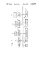
- FIG. 1 is a schematic diagram of the components of the cardiac biopotential analysis system of the present invention
- FIG. 2 is a schematic diagram of the sixteen channel EKG data acquisition system, utilizing a serial interface, of the cardiac biopotential analysis system shown in FIG. 1;
- FIG. 3 is a schematic diagram of the microcomputer utilized by the cardiac biopotential analysis system of FIG. 1;
- FIG. 4A is a diagram of a sample QRST complex utilized by the system and method of the present invention.
- FIGS. 4B-4D are diagrams of possible extraction templates utilized for bispectral analysis by the method and system of the present invention.
- FIGS. 5(a) and 5(b) are diagrams of sample simulated three-dimensional plots of a difference bicoherence array and a difference bispectral array respectively;
- FIG. 6 is a flow chart of the operation of the system and method of the present invention for performing bispectral analysis on electrocardiographic signals
- FIG. 7 is a flow chart showing the process used by the system and method of present invention for the interactive determination of the extraction template from each lead;
- FIG. 8 is a flow chart of the process utilized by the system and method of the present invention for extracting 50 complexes from each lead to match the pre-selected template;
- FIG. 9 is a flow chart of the process utilized by the system and method of the present invention for bispectral processing
- FIG. 10 is a flow chart of the process utilized by the system and method of the present invention for data analysis after all bispectral processing has been completed.
- the present invention utilizes the Fourier transform of the third order autocorrelation function, known as the bispectrum, which is an analytic process that quantifies non-linearities and phase relationships intrinsic to any waveform.
- Bispectral decomposition of the cardiac signal produces a result independent of rib cage size, body fat content, and position of the heart relative to the lungs. This independent result occurs because the bispectral process involves an evaluation of the relational component of the fundamental constituents comprising a signal, without regard for their absolute magnitudes. Since the altered phase relationships among ischemic cardiac cells are reflected in alterations in the phase relationships of the frequency components of the ECG signal, bispectral analysis, by providing a quantitative index of these relationships, measures the alteration in the "fine fingerprint" imbedded in the structure of the surface signal. By providing this quantitative fingerprint, the present invention provides a unique quantitative approach to the diagnosis of ischemia and other coronary malfunctions.
- the bispectral analysis system 10 of the present invention includes a sixteen channel EKG data acquisition system 12 having a serial port.
- the electrocardiograph (ECG) leads I, II, III, AVR, AVF, AVL, V1, V2, V3, V4, V5, V6, V7, V8, V9, V10, X, Y and Z are connected to a patient 14, and signals pass through cable 16 to the sixteen channel data acquisition system 12.
- the data acquisition system 12 amplifies and digitizes the EKG waveforms and sends the digitized data to a microcomputer 18 via a serial port over line 20 for analysis.
- the serial line 20 can be used to download programs.
- the microcomputer 18 analyzes the serial data stream and calculates the bispectrum which is displayed on the graphics display 22. Hard copy output of the bispectrum waveform is available on plotter 24 which is also connected to microcomputer 18. Interaction between the acquisition and analysis process in the system is provided by means of a keyboard 26 with feedback on the graphics display 22.
- the EKG surface potential from the patient 14 passes through an electrosurgery protection circuit 28 and a defibrillator protection circuit 30 before being transmitted to an amplifier circuit 32 that includes an amplifier for each channel.
- the electrosurgery protection circuit 28 includes a radio frequency (rf) filter which limits the rf current to the patient leads, and thus protects the patient 14 from rf burns and protects the amplifiers in amplification circuit 32 from excessive voltage and/or current.
- rf radio frequency
- the defibrillator protection circuit 30 limits the voltage to the amplifier circuit 32 to a safe level (on the order of ⁇ 15 volts) when a defibrillator is applied to the patient and discharged (on the order of 5 kilovolts).
- the defibrillator protection circuitry also limits the current through patient leads during a defibrillator discharge.
- each of the 16 channels of amplification is fed into a respective one of the 16 sample-and-hold circuits 34 which are under program control of a microprocessor 36.
- the outputs of the sample-and-hold circuits are multiplexed by multiplexer 38 and digitized by 16 bit analog-to-digital converter 40 at a rate of 1024/sec.
- the output of the analog-to-digital converter 40 is optically coupled to a data bus 42 with an optical isolator 44. All control lines to the sample-and-hold circuit 34, multiplexer 38 and analog-to-digital converter 40 are optically isolated by optical isolator 45. All DC power lines going to the amplifiers 32, sample-and-hold circuits 34, multiplexer 38 and analog-to-digital converter 40 are isolated from AC mains with a DC/DC converter 46 in order to provide complete patient isolation from ground.
- the microprocessor 36 controls data acquisition in the system 12.
- the basic control instructions for the microprocessor 36 are stored in the read only memory (ROM) 48.
- the random access memory (RAM) 50 is used as a buffer memory for data, and a portion of the RAM 50 can also be used as program memory when the control program is being downloaded from the host computer 18 which is connected to the system 12 by the serial line 20.
- the serial interface 52 operates under the control of the host computer 18.
- the serial interface 52 is optically coupled by optical isolator 54 to RS232C drivers 56 to provide a serial interface between the sixteen channel data acquisition system 12 and any standard RS232C serial port on a computer.
- the serial lines are isolated by optical isolator 54 and DC/DC converter 58 to provide increased patient safety and to protect the host computer 18 from any transients.
- the host or microcomputer 18 of FIG. 1 is shown in greater detail in FIG. 3.
- the entire microcomputer system 18 runs under the control of microprocessor 60.
- the program memory for the microprocessor 60 is stored in ROM 62, and RAM 64 is provided for storage of intermediate data.
- the microcomputer 18 contains an array processor 66 on which complex arithmetic calculations can be performed on entire arrays of data simultaneously.
- the preferred embodiment also includes a math coprocessor 68 which is connected to the microprocessor 60.
- the math coprocessor 68 is used for scalar and graphic calculations whereas the array processor 66 is used to calculate the bispectrum and other vector computations as will be described below.
- the graphics controller 70 operates under program control of the microprocessor 60 and drives a video monitor 72.
- the keyboard controller 74 interfaces directly to the operator's keyboard 26, and operator control of the entire acquisition, analysis and display procedure is controlled by the keyboard 26 with feedback on the video monitor 22.
- One serial port 76 is provided to interface with the sixteen channel data acquisition system 12.
- the serial port 76 can be used to send control data to the system (e.g., start acquisition) and to receive EKG data from the system, as well as to download program data to the system.
- Another serial port 78 is provided to drive a plotter 24 for hard copy output of the bispectrum data.
- the system initializes all data arrays and opens the relevant data files in step 102.
- the data arrays that are opened include arrays used to store the digitized EKG, the template, the extracted complexes and the bispectral data of each lead.
- the opened data files include files for final storage or comparison with existing bispectral density and bicoherence arrays.
- step 104 acquires 5 minutes of raw electrocardiographic data (in the form of PQRST waves, as shown in FIG. 4A) from the data acquisition system 12.
- This raw data passes through a general purpose RS232C communication port utilizing a standard protocol, such as Kermit.
- QRS detection is performed on the digital EKG of each lead using any publicly available QRS detection program (see for example "A Single Scan Algorithm for QRS-Detection and Feature Extraction," W. A. H. Engelse et al, Computers in Cardiology, IEEE Press, 1979.)
- step 108 the system determines the template for interactive QRST or a portion of QRST (see FIGS.
- step 110 the system normalizes each potential complex of the 50 complexes of each lead.
- step 112 after normalizing each potential complex, the system extracts 50 matching complexes from the five minutes of EKG data from each lead using cross-correlation as will be described in greater detail with reference to FIG. 8.
- the bispectral density array and the bicoherence array for each lead is computed using the 50 complexes of that lead in step 114.
- step 116 the bispectral results are produced relating to the cardiac electrical function as will be described with reference to FIG. 10.
- the process utilized by the system 10 for determining templates for each lead begins by initializing the template arrays in step 122.
- the determined templates are used for the extraction of 50 similar complexes from the digital EKG data of each lead.
- the first lead is extracted for processing in step 124.
- the first 100 QRST complexes are displayed on the screen using information provided by the QRS detection program in step 126.
- the display graphics can be implemented by utilizing many commercially available software packages for micro and minicomputers. (For example, systems sold by IBM, Precision Visuals Corp., Microcompatibles, Inc.) From the display, the operator in step 128 picks the most artifact-free sinus QRST complex by moving the cursor to that position on the screen.
- Step 130 tests if a complex is picked from the displayed 100 complexes and if one is not picked, the next 100 QRST complexes is displayed in step 132. If a QRST complex is chosen, then it is displayed on the screen 22 with a time scale relative to the peak of the QRS which is set to zero in step 134. In step 136 the operator enters the location in msec of the start and end of the template, relative to the peak of the QRS. In step 138 the template is then displayed on a full screen.
- step 140 the operator decides whether he is satisfied with the chosen template and if he is not then control returns to step 126 and the next 100 QRST complexes are displayed on the screen. If the template is acceptable then it is stored in a template array for that lead in step 142 with the first point of the template stored in the first location and the rest extending over the remaining 512 (in a preferred embodiment) available locations (0.5 seconds). Prior to placing the template samples in the array, the mean is subtracted from each sample and it is then divided by the standard deviation. If the number of samples for the template is less than 512, all subsequent locations are padded with the value of the last sample in the template. Step 144 tests to determine which lead is currently being processed and if all the leads have not been considered step 146 selects the next lead in the sequence, and steps 126 through 142 are repeated for each lead.
- step 172 the data arrays containing the 50 complexes of each lead are initialized.
- step 174 the data in the first lead is selected for processing, and in step 176 the number of matching complexes for that lead is set to 0.
- step 178 the portion of the current QRST complex that corresponds to the template is extracted in step 178.
- the mean of all samples in the extracted portion is subtracted from each sample, and each sample is then divided by the standard deviation of the samples in that portion in step 180. The division by the standard deviation is used to normalize the energy present in a portion to 1, making this process completely independent of the absolute amount of energy present in the signal.
- a cross-correlation is carried out between the current normalized extracted portion and the template using only the non-padded values in the template array in step 182. If the cross-correlation coefficient is not greater than or equal to 0.95, which is tested in step 184, the next QRST complex is selected in step 186 and steps 178 through 184 are repeated. If the correlation coefficient is greater than or equal to 0.95, the number of matches for that lead is increased by 1, and the extracted portion is stored in the leads array for the fifty selected matches in step 188. Step 190 tests whether all the matches have been processed. If they have not, the next QRST signal is obtained in step 186, and then processing continues in step 178. If all the matches have been processed, step 192 tests to see if all the leads have been processed. If all leads have not been processed, the next lead is selected, and control returns to step 176. If all the leads have been processed, then the process for selecting the fifty complexes for each lead is concluded.
- step 202 the system initializes the spectral arrays, and in step 204 the data for the first lead is selected.
- each QRST in question is a record x i (t) where 0 ⁇ i ⁇ 50.
- the system then computes the fast Fourier transform (FFT) X i (f) of each record or in other words for all 50 complexes in step 206.
- the system then computes the power spectrum P i (f) of each record or in other words for all 50 complexes in step 208.
- step 210 the individual bc i (f 1 ,f 2 ) and average BC(f 1 ,f 2 ) complex triple product arrays are computed for each record such that:
- step 212 the system computes the individual br i (f 1 ,f 2 ) and average BR(f 1 ,f 2 ) real triple product arrays with the real triple product for each record being specified as follows:
- the average real triple product BR(f 1 ,f 2 ) is calculated as follows: ##EQU2## Utilizing these average figures, the system in step 214 computes the bispectral density array of the average records:
- step 216 the bicoherence array R 2 (f 1 ,f 2 ) is computed as follows:
- step 218 tests to determine whether all the leads have been processed and if they have not the data for the next lead is selected in step 220 and control of the operation returns to step 206. If all the leads have been processed the determination of the bicoherence arrays has been concluded.
- step 232 the necessary arrays are initialized. These arrays include data arrays that contain the difference between the control bispectral values and the current bispectral values. The operator is then asked to enter into the system the file name containing the control data which can be from a group of normal subjects or preintervention data from the current subject.
- step 234 both the bispectral density arrays and the bicoherence arrays for the 15 leads are retrieved from the control file in step 234 and the current arrays of the 15 leads are also stored for further off-line analysis.
- the current bispectral density and current bicoherence arrays are subtracted from the corresponding control arrays in step 238.
- the absolute values of the differences are stored in a difference bispectral density array and a difference bicoherence array.
- the difference arrays are then displayed simultaneously on the screen in a three dimensional format for qualitative assessment of the affected frequency bands in step 240.
- the grids in FIG. 5 represents the difference between two bispectral density arrays or bicoherence arrays. Each point on the grid is the absolute value of the difference between the same points in the control bicoherence array and the current bicoherence array, or the control bispectral density array and the current bispectral density array.
- the axes are F1, ranging from 0 to 512 Hz; F2, ranging from 0 to 256 Hz; and the magnitude of the change at each point (for bicoherence, a dimensionless number ranging between 0 and 1; for bispectral density, a very large number with the unit volt 6 ).
- the operator will choose these regions by entering the corresponding frequency pair bands. A numerical output is then produced and printed for quantitative evaluation by the operator.
- the operator can then enter the values for all the frequency bands of interest in step 242.
- the sum of the absolute values of the changes in each selected frequency band is then computed for both the bispectral density data as well as the bicoherence data in step 244, and the printed output is generated for the quantitative assessment by the operator.
- a two dimensional cross-correlation between the control bispectral density and/or bicoherence arrays (and/or a subsection thereof) and the current bispectral density and/or bicoherence arrays is performed to generate a cross-correlation coefficient representing the distortion and/or deviation from the original bispectral structures.
- the difference arrays may also be plotted using a high resolution plotter.
- step 248 the data for the next lead is selected and control of the system returns to step 238. If the test in step 246 determines that all the leads have been processed, the display and data analysis portion of the system has concluded its operation.
- the resultant bispectral density array and the bicoherence array are examined, and a figure of merit representing a fingerprint of the electrical activity in each lead may be displayed in a three-dimensional wire frame format.
- the current bispectral density array is subtracted from and/or correlated with the control bispectral density array or any subsections thereof, and the current bicoherence array is subtracted from and/or correlated with the control bicoherence array or any subsections thereof.
- the control arrays are produced prior to the procedure or intervention. The greater the reduction in bicoherence and/or bispectral density and/or the lower the cross correlation coefficient in the frequency pairs in the bands between 0.05 to 100 Hz and 200 to 300 Hz, the greater the heterogeneity in conduction and repolarization.
- bispectral density arrays and bicoherence arrays from individuals with intraventricular conduction disturbances or accessory atrioventricular pathways will be reliably and quantitatively discernable from the template bispectral density array and the template bicoherence array of a population of normal individuals.
- the bispectral density array is subtracted from a template bispectral density array, and the bicoherence array is subtracted from a template bicoherence array (the templates having been produced by averaging multiple arrays obtained from a large group of normals).
- the greater the change in bicoherence and/or bispectral density in the frequency pairs in the band between 0 to 200 Hz the greater the magnitude of the steady state ischemia.
- Each lead is representative of a certain region in the myocardium. Therefore, the analysis of the bispectral results accounting for amplitude variation in each lead will localize non-invasively the areas which are most susceptible to ischemia and/or which exhibit the greatest heterogeneity in conduction and repolarization.
- the cardiac biopotential analysis system of the present invention quantitatively represents beat to beat consistency (reproducibility) of cardiac electrical activation and repolarization. As such, normal ranges can be established for a population of human subjects. Also, alterations, whether due to disease or drugs, in the homogeneity of electrical activation and repolarization can be detected and the degree of offset can be quantified, providing an indirect quantification of the perturbing factor.
- bispectral density and bicoherence in particular frequency bands will be reduced in ischemia, thereby permitting the detection of ischemia.
- bispectral density and bicoherence will vary inversely with degree of ischemia, bispectral density and bicoherence will provide a noninvasive quantitative measure of the amount of ischemia.
- bispectral density and bicoherence in conjunction with amplitude-domain characteristics, varies with electrode lead position on the body surface, and therefore alterations of bispectral density and bicoherence in relation to amplitude will vary with lead position, myocardial ischemia can be localized to particular regions of the heart.
- bispectral density and bicoherence provide a quantitative, composite measure of homogeneity of activation, conduction and repolarization of cardiac tissue, and since the propensity to ventricular fibrillation integrates said cardiac electrical properties of activation, conduction and repolarization, bispectral density and bicoherence of particular frequency bands will provide a quantitative, noninvasive measure of the propensity for ventricular fibrillation. As such, bispectral density and bicoherence will allow one to distinguish between two situations, in one of which the myocardium is electrically stable and in the other of which the myocardium is electrically unstable, with the situations being indistinguishable by any other noninvasive measure currently available.
- bispectral density and bicoherence will distinguish between those individuals with the high likelihood of ventricular tachyarrhythmias and those with the low likelihood of ventricular tachyarrhythmias who are otherwise indistinguishable by any other noninvasive measure currently available.
- Those individuals who develop ventricular tachyarrhythmias will have more beat-to-beat heterogeneity of conduction than those individuals who will not develop ventricular tachyarrhythmias.
- bispectral density and bicoherence will be lower in those individuals having a high propensity for the development of ventricular tachyarrhythmias, and will be higher in those individuals having a low propensity for the development for ventricular tachyarrhythmias.
Abstract
Description
bc.sub.i (f.sub.1,f.sub.2)=X.sub.i (f.sub.1)*X.sub.i (f.sub.2)*X.sub.i (f.sub.1 +f.sub.2) (1)
f.sub.1 +f.sub.2 ≦N/2 (2)
≦f.sub.2 ≦f.sub.1 (3)
br.sub.i (f.sub.1,f.sub.2)=P.sub.i (f.sub.1)*P.sub.i (f.sub.2)*P.sub.i (f.sub.1 +f.sub.2) (5)
f.sub.1 +f.sub.2 ≦N/2 (6)
0≦f.sub.2 ≦f.sub.2 (7)
BD(f.sub.1,f.sub.2)=|BC(f.sub.1,f.sub.2)|.sup.2 (9)
R.sup.2 (f.sub.1,f.sub.2)=BD(f.sub.1,f.sub.2)/BR (f.sub.1,f.sub.2) 0≦R.sup.2 ≦1 (10)
Claims (25)
Priority Applications (2)
| Application Number | Priority Date | Filing Date | Title |
|---|---|---|---|
| US07/107,419 US4924875A (en) | 1987-10-09 | 1987-10-09 | Cardiac biopotential analysis system and method |
| US07/396,990 US5020540A (en) | 1987-10-09 | 1989-08-22 | Cardiac biopotential analysis system and method |
Applications Claiming Priority (1)
| Application Number | Priority Date | Filing Date | Title |
|---|---|---|---|
| US07/107,419 US4924875A (en) | 1987-10-09 | 1987-10-09 | Cardiac biopotential analysis system and method |
Related Child Applications (1)
| Application Number | Title | Priority Date | Filing Date |
|---|---|---|---|
| US07/396,990 Continuation-In-Part US5020540A (en) | 1987-10-09 | 1989-08-22 | Cardiac biopotential analysis system and method |
Publications (1)
| Publication Number | Publication Date |
|---|---|
| US4924875A true US4924875A (en) | 1990-05-15 |
Family
ID=22316557
Family Applications (1)
| Application Number | Title | Priority Date | Filing Date |
|---|---|---|---|
| US07/107,419 Expired - Lifetime US4924875A (en) | 1987-10-09 | 1987-10-09 | Cardiac biopotential analysis system and method |
Country Status (1)
| Country | Link |
|---|---|
| US (1) | US4924875A (en) |
Cited By (145)
| Publication number | Priority date | Publication date | Assignee | Title |
|---|---|---|---|---|
| US5012815A (en) * | 1989-02-02 | 1991-05-07 | Yale University | Dynamic spectral phonocardiograph |
| US5046504A (en) * | 1989-02-01 | 1991-09-10 | Corazonix Corporation | Method and apparatus for analyzing and interpreting electrocardiograms using spectro-temporal mapping |
| WO1991019452A1 (en) * | 1990-06-20 | 1991-12-26 | Cedars-Sinai Medical Center | Methods for detecting and evaluating heart disorders |
| US5077667A (en) * | 1989-07-10 | 1991-12-31 | The Ohio State University | Measurement of the approximate elapsed time of ventricular fibrillation and monitoring the response of the heart to therapy |
| US5092341A (en) * | 1990-06-18 | 1992-03-03 | Del Mar Avionics | Surface ecg frequency analysis system and method based upon spectral turbulence estimation |
| WO1992005831A1 (en) * | 1989-03-28 | 1992-04-16 | Erwin Roy John | Ekg system using statistic and topographic mapping |
| US5109862A (en) * | 1990-03-19 | 1992-05-05 | Del Mar Avionics | Method and apparatus for spectral analysis of electrocardiographic signals |
| US5117833A (en) * | 1990-11-13 | 1992-06-02 | Corazonix Corporation | Bi-spectral filtering of electrocardiogram signals to determine selected QRS potentials |
| EP0489010A1 (en) * | 1989-08-22 | 1992-06-10 | Aspect Medical Systems, Inc. | Cardiac biopotential analysis system and method |
| US5161539A (en) * | 1991-05-09 | 1992-11-10 | Physio-Control | Method and apparatus for performing mapping-type analysis including use of limited electrode sets |
| US5201321A (en) * | 1991-02-11 | 1993-04-13 | Fulton Keith W | Method and apparatus for diagnosing vulnerability to lethal cardiac arrhythmias |
| US5226431A (en) * | 1991-06-20 | 1993-07-13 | Caliber Medical Corporation | Optical/electrical transceiver |
| EP0570895A2 (en) * | 1992-05-18 | 1993-11-24 | Cardiac Pacemakers, Inc. | Method and apparatus for event processing in biological applications |
| US5271411A (en) * | 1990-09-21 | 1993-12-21 | Colin Electronics Co., Ltd. | Method and apparatus for ECG signal analysis and cardiac arrhythmia detection |
| US5323783A (en) * | 1992-11-12 | 1994-06-28 | Del Mar Avionics | Dynamic ST segment estimation and adjustment |
| EP0606301A1 (en) * | 1991-09-12 | 1994-07-20 | Drexel University | Methods of impedance cardiography and heartbeat determination as well as an apparatus for implementing said methods |
| US5361307A (en) * | 1993-03-25 | 1994-11-01 | General Electric Company | Correlation methods of identifying defects in imaging devices |
| WO1995015116A1 (en) * | 1993-11-30 | 1995-06-08 | Georgetown University | Sudden cardiac death prediction |
| EP0684784A1 (en) * | 1993-12-16 | 1995-12-06 | Marquette Electronics, Inc. | Cardiac monitoring and diagnostic system |
| US5483968A (en) * | 1991-06-25 | 1996-01-16 | Technion Research And Development Foundation Ltd. | Method and apparatus for analyzing the electrical activity of the heart |
| US5509425A (en) * | 1989-10-30 | 1996-04-23 | Feng; Genquan | Arrangement for and method of diagnosing and warning of a heart attack |
| EP0711531A1 (en) * | 1994-10-07 | 1996-05-15 | Per Karlsson | Myocardial ischemia and infarction analysis and monitoring method |
| US5555889A (en) * | 1990-06-20 | 1996-09-17 | Cedars-Sinai Medical Center | Methods for detecting propensity fibrillation |
| EP0745942A2 (en) * | 1995-05-22 | 1996-12-04 | Paolo Alcidi | Method and apparatus for acquiring and processing electrocardiographic signals |
| US5609158A (en) * | 1995-05-01 | 1997-03-11 | Arrhythmia Research Technology, Inc. | Apparatus and method for predicting cardiac arrhythmia by detection of micropotentials and analysis of all ECG segments and intervals |
| US5613496A (en) * | 1995-11-29 | 1997-03-25 | Hewlett-Packard Company | Method and apparatus for creating a representative heartbeat from an ECG waveform |
| WO1997024062A1 (en) * | 1994-08-30 | 1997-07-10 | The Ohio State University Research Foundation | Non-invasive monitoring and treatment of subjects in cardiac arrest using ecg parameters predictive of outcome |
| US5649544A (en) * | 1989-10-30 | 1997-07-22 | Feng; Genquan | Method of and arrangement for diagnosing heart disease |
| US5683424A (en) * | 1994-08-30 | 1997-11-04 | The Ohio State University Research Foundation | Non-invasive monitoring and treatment of subjects in cardiac arrest using ECG parameters predictive of outcome |
| WO1998030145A1 (en) * | 1997-01-10 | 1998-07-16 | Micromedical Industries Limited | Universal ecg interface cable |
| EP0864294A2 (en) * | 1997-03-14 | 1998-09-16 | Cambridge Heart, Inc. | Detecting abnormal activation of the heart |
| US5842997A (en) * | 1991-02-20 | 1998-12-01 | Georgetown University | Non-invasive, dynamic tracking of cardiac vulnerability by simultaneous analysis of heart rate variability and T-wave alternans |
| US5951485A (en) * | 1994-09-28 | 1999-09-14 | Heartstream, Inc. | Method and apparatus for recording and replaying time-correlated medical event data |
| WO1999052591A1 (en) * | 1998-04-14 | 1999-10-21 | Intermedics Inc. | Implantable cardiac stimulator with physiologic sensor based on mechanical-electric phase relation |
| US6021345A (en) * | 1991-05-17 | 2000-02-01 | Cedars-Sinai Medical Center | Methods for detecting propensity for fibrillation using an electrical restitution curve |
| US6038469A (en) * | 1994-10-07 | 2000-03-14 | Ortivus Ab | Myocardial ischemia and infarction analysis and monitoring method and apparatus |
| US6094593A (en) * | 1991-05-17 | 2000-07-25 | Cedars-Sinai Medical Center | Method and apparatus for detecting prospenity for ventricular fibrillation using action potential curves |
| US6169919B1 (en) | 1999-05-06 | 2001-01-02 | Beth Israel Deaconess Medical Center, Inc. | System and method for quantifying alternation in an electrocardiogram signal |
| US6171256B1 (en) * | 1998-04-30 | 2001-01-09 | Physio-Control Manufacturing Corporation | Method and apparatus for detecting a condition associated with acute cardiac ischemia |
| US6193668B1 (en) | 1997-11-10 | 2001-02-27 | Medacoustics, Inc. | Acoustic sensor array for non-invasive detection of coronary artery disease |
| US6217525B1 (en) | 1998-04-30 | 2001-04-17 | Medtronic Physio-Control Manufacturing Corp. | Reduced lead set device and method for detecting acute cardiac ischemic conditions |
| US6243599B1 (en) | 1997-11-10 | 2001-06-05 | Medacoustics, Inc. | Methods, systems and computer program products for photogrammetric sensor position estimation |
| US6261237B1 (en) | 1998-08-20 | 2001-07-17 | Medacoustics, Inc. | Thin film piezoelectric polymer sensor |
| US6266554B1 (en) | 1999-02-12 | 2001-07-24 | Cardiac Pacemakers, Inc. | System and method for classifying cardiac complexes |
| US6272377B1 (en) | 1999-10-01 | 2001-08-07 | Cardiac Pacemakers, Inc. | Cardiac rhythm management system with arrhythmia prediction and prevention |
| NL1015847C2 (en) * | 2000-07-31 | 2002-02-01 | Hendrik Jan Ritsema Van Eck | Method for analysis of electrocardiogram involves taking electrocardiogram and its storage electronically, suppression of murmurs in the stored ECG tracks and identification of characteristic time points in the stored ECG tracks |
| EP1191877A1 (en) * | 1999-05-06 | 2002-04-03 | Shell, Allan Michael | Physiological signal acquisition cable |
| US20020049474A1 (en) * | 1999-03-12 | 2002-04-25 | Cardiac Pacemakers, Inc. | Method and system for verifying the integrity of normal sinus rhythm templates |
| AU749181B2 (en) * | 1997-01-10 | 2002-06-20 | Ventracor Limited | Universal ECG interface cable |
| US6424860B1 (en) | 1994-10-07 | 2002-07-23 | Ortivus Ab | Myocardial ischemia and infarction analysis and monitoring method and apparatus |
| US20020107552A1 (en) * | 1997-04-30 | 2002-08-08 | Cardiac Pacemakers, Inc. | Apparatus and method for treating ventricular tachyarrhythmias |
| US6478744B2 (en) | 1996-12-18 | 2002-11-12 | Sonomedica, Llc | Method of using an acoustic coupling for determining a physiologic signal |
| US6484055B1 (en) | 1999-03-12 | 2002-11-19 | Cardiac Pacemakers, Inc. | Discrimination of supraventricular tachycardia and ventricular tachycardia events |
| US6501983B1 (en) | 1998-08-07 | 2002-12-31 | Infinite Biomedical Technologies, Llc | Implantable myocardial ischemia detection, indication and action technology |
| US20030055461A1 (en) * | 1999-10-01 | 2003-03-20 | Girouard Steven D. | Cardiac rhythm management systems and methods predicting congestive heart failure status |
| US20030060849A1 (en) * | 1999-07-14 | 2003-03-27 | Cardiac Pacemakers, Inc. | Classification of supraventricular and ventricular cardiac rhythms using cross channel timing algorithm |
| US20030060854A1 (en) * | 2001-09-25 | 2003-03-27 | Qingsheng Zhu | Evoked response sensing for ischemia detection |
| US20030097612A1 (en) * | 2000-11-23 | 2003-05-22 | Magnolia Medical Technologies, Ltd. | System and method for analyzing and evaluation of an electric signal record |
| US20030233132A1 (en) * | 2002-06-14 | 2003-12-18 | Pastore Joseph M. | Method and apparatus for detecting oscillations in cardiac rhythm |
| US20040010200A1 (en) * | 2002-07-15 | 2004-01-15 | Sweeney Robert J. | Use of curvature based features for beat detection |
| US6699201B2 (en) | 1998-11-09 | 2004-03-02 | Medacoustics, Inc. | Acoustic window identification |
| US20040073262A1 (en) * | 2000-11-02 | 2004-04-15 | Cardiac Pacemakers, Inc. | Technique for discriminating between coordinated and uncoordinated cardiac rhythms |
| US20040093034A1 (en) * | 2002-11-12 | 2004-05-13 | Girouard Steven D. | Implantable device for delivering cardiac drug therapy |
| US20040127806A1 (en) * | 2000-10-31 | 2004-07-01 | Cardiac Pacemakers, Inc. | Curvature based method for selecting features from an electrophysiologic signals for purpose of complex identification and classification |
| US20040138648A1 (en) * | 2000-12-18 | 2004-07-15 | Cardiac Pacemakers, Inc. | Drug delivery system for implantable medical device |
| US20040167381A1 (en) * | 1997-02-28 | 2004-08-26 | Qrs Diagnostic, Llc | Personal computer card for collection of real-time biological data |
| US20040186392A1 (en) * | 1999-06-22 | 2004-09-23 | The University Of Queensland | Method and device for measuring tissue oedema |
| US20040193028A1 (en) * | 2003-03-28 | 2004-09-30 | Vascular Control Systems, Inc. | Uterine tissue monitoring device and method |
| US20040219600A1 (en) * | 2002-12-13 | 2004-11-04 | Williams Robert Wood | Method for determining sensitivity to environmental toxins and susceptibility to parkinson's disease |
| US20050004482A1 (en) * | 2003-07-01 | 2005-01-06 | Budimir Drakulic | Amplified system for determining parameters of a patient |
| US20050043675A1 (en) * | 2003-08-21 | 2005-02-24 | Pastore Joseph M. | Method and apparatus for modulating cellular metabolism during post-ischemia or heart failure |
| US20050059896A1 (en) * | 2003-09-17 | 2005-03-17 | Budimir Drakulic | Apparatus for, and method of, determining the condition of a patient's heart |
| US20050137626A1 (en) * | 2003-12-19 | 2005-06-23 | Pastore Joseph M. | Drug delivery system and method employing external drug delivery device in conjunction with computer network |
| US20050234362A1 (en) * | 2004-04-15 | 2005-10-20 | Ge Medical Systems Information Technologies, Inc. | Method and apparatus for displaying alternans data |
| US20050234353A1 (en) * | 2004-04-15 | 2005-10-20 | Ge Medical Systems Information Technologies, Inc. | Method and apparatus for analysis of non-invasive cardiac parameters |
| US20050234357A1 (en) * | 2004-04-15 | 2005-10-20 | Ge Medical Systems Information Technologies, Inc. | Method and apparatus for detecting cardiac repolarization abnormality |
| US20050234363A1 (en) * | 2004-04-15 | 2005-10-20 | Ge Medical Systems Information Technologies, Inc. | Method and apparatus for determining alternans data of an ECG signal |
| US20050234356A1 (en) * | 2004-04-15 | 2005-10-20 | Rowlandson G I | System and method for correlating implant and non-implant data |
| US20050234355A1 (en) * | 2004-04-15 | 2005-10-20 | Rowlandson G I | System and method for sudden cardiac death prediction |
| US6959212B2 (en) | 1999-02-12 | 2005-10-25 | Cardiac Pacemakers, Inc. | System and method for arrhythmia discrimination |
| US20050256544A1 (en) * | 2004-05-12 | 2005-11-17 | Cardiac Pacemakers, Inc. | Template based av/va interval comparison for the discrimination of cardiac arrhythmias |
| US6978177B1 (en) | 2000-11-14 | 2005-12-20 | Cardiac Pacemakers, Inc. | Method and apparatus for using atrial discrimination algorithms to determine optimal pacing therapy and therapy timing |
| US7009492B1 (en) * | 2003-01-30 | 2006-03-07 | Combustion Dynamics Corp. | Individual quantitative identification by means of human dynamic rhythmic electric activity spectra |
| US20060095083A1 (en) * | 2004-10-28 | 2006-05-04 | Cardiac Pacemakers, Inc. | Methods and apparatuses for arrhythmia detection and classification using wireless ECG |
| US20060129052A1 (en) * | 2004-12-09 | 2006-06-15 | Budimir Drakulic | System for, and method of, monitoring heartbeats of a patient |
| US7089055B2 (en) | 2002-06-28 | 2006-08-08 | Cardiac Pacemakers, Inc. | Method and apparatus for delivering pre-shock defibrillation therapy |
| US20060211949A1 (en) * | 2001-06-05 | 2006-09-21 | Cardiac Pacemakers, Inc. | System and method for classifying cardiac depolarization complexes with multi-dimensional correlation |
| US20060247543A1 (en) * | 2002-10-09 | 2006-11-02 | Bruce Cornish | High resoution bio-impedance device |
| US20060271024A1 (en) * | 2005-01-25 | 2006-11-30 | Michael Gertner | Nasal Cavity Treatment Apparatus |
| US20060281998A1 (en) * | 2005-06-13 | 2006-12-14 | Cardiac Pacemakers, Inc. | Method and apparatus for rate-dependent morphology-based cardiac arrhythmia classification |
| US7162294B2 (en) | 2004-04-15 | 2007-01-09 | Ge Medical Systems Information Technologies, Inc. | System and method for correlating sleep apnea and sudden cardiac death |
| US20070054871A1 (en) * | 2005-09-06 | 2007-03-08 | Pastore Joseph M | Method and apparatus for device controlled gene expression for cardiac protection |
| US7203535B1 (en) | 1999-04-01 | 2007-04-10 | Cardiac Pacemakers, Inc. | System and method for classifying tachycardia arrhythmias having 1:1 atrial-to-ventricular rhythms |
| US20070203419A1 (en) * | 2003-06-27 | 2007-08-30 | Caridac Pacemakers, Inc. | Tachyarrhythmia detection and discrimination based on curvature parameters |
| US7299083B2 (en) | 2004-12-09 | 2007-11-20 | Signalife, Inc. | Electrode for, and method of, indicating signal characteristics at particular positions in a patient's body |
| US20080082135A1 (en) * | 2006-10-02 | 2008-04-03 | Cardiac Pacemakers, Inc. | Method and apparatus for identification of ischemic/infarcted regions and therapy optimization |
| US20080081354A1 (en) * | 2006-10-02 | 2008-04-03 | Cardiac Pacemakers, Inc. | Devices, vectors and methods for inducible ischemia cardioprotection |
| US20080091080A1 (en) * | 2003-04-22 | 2008-04-17 | Patrick Leahy | Device and Method for Use in Surgery |
| US20080117203A1 (en) * | 2006-11-16 | 2008-05-22 | David Thomas Gering | Methods and Apparatus for Visualizing Data |
| US20080177194A1 (en) * | 2007-01-19 | 2008-07-24 | Cardiac Pacemakers, Inc. | Heart attack detector |
| US7430446B2 (en) | 2005-01-20 | 2008-09-30 | Cardiac Pacemakers, Inc. | Methods and apparatuses for cardiac arrhythmia classification using morphology stability |
| US20080287823A1 (en) * | 2005-07-20 | 2008-11-20 | Scott Matthew Chetham | Index Determination |
| US20090043222A1 (en) * | 2005-10-11 | 2009-02-12 | Scott Chetham | Hydration status monitoring |
| US7500955B2 (en) | 2003-06-27 | 2009-03-10 | Cardiac Pacemaker, Inc. | Signal compression based on curvature parameters |
| US20090143663A1 (en) * | 2005-07-01 | 2009-06-04 | Impedance Cardiology Systems Inc. | Pulmonary Monitoring System |
| US7567841B2 (en) | 2004-08-20 | 2009-07-28 | Cardiac Pacemakers, Inc. | Method and apparatus for delivering combined electrical and drug therapies |
| US20090234211A1 (en) * | 2008-03-13 | 2009-09-17 | Cardiac Pacemakers, Inc. | Systems and Methods for Myocardial Ischemia Detection |
| US7627373B2 (en) | 2002-11-30 | 2009-12-01 | Cardiac Pacemakers, Inc. | Method and apparatus for cell and electrical therapy of living tissue |
| US20100087750A1 (en) * | 2006-05-30 | 2010-04-08 | Mcgree James Matthew | Impedance measurements |
| US20100152605A1 (en) * | 2007-04-20 | 2010-06-17 | Impedimed Limited | Monitoring system and probe |
| US20100152598A1 (en) * | 2008-12-11 | 2010-06-17 | Siemens Medical Solutions Usa, Inc. | System for Heart Performance Characterization and Abnormality Detection |
| US20100168530A1 (en) * | 2006-11-30 | 2010-07-01 | Impedimed Limited | Measurement apparatus |
| US7764995B2 (en) | 2004-06-07 | 2010-07-27 | Cardiac Pacemakers, Inc. | Method and apparatus to modulate cellular regeneration post myocardial infarct |
| US20100191118A1 (en) * | 2009-01-29 | 2010-07-29 | The General Electric Company | System and Method for Measuring the Instantaneous Period of a Quasi-Periodic Signal |
| US7840263B2 (en) | 2004-02-27 | 2010-11-23 | Cardiac Pacemakers, Inc. | Method and apparatus for device controlled gene expression |
| US20100312130A1 (en) * | 2006-06-27 | 2010-12-09 | Yi Zhang | Graded response to myocardial ischemia |
| US20110046505A1 (en) * | 2007-08-09 | 2011-02-24 | Impedimed Limited | Impedance measurement process |
| US7981065B2 (en) | 2004-12-20 | 2011-07-19 | Cardiac Pacemakers, Inc. | Lead electrode incorporating extracellular matrix |
| US8000780B2 (en) | 2006-06-27 | 2011-08-16 | Cardiac Pacemakers, Inc. | Detection of myocardial ischemia from the time sequence of implanted sensor measurements |
| US8060219B2 (en) | 2004-12-20 | 2011-11-15 | Cardiac Pacemakers, Inc. | Epicardial patch including isolated extracellular matrix with pacing electrodes |
| US8068906B2 (en) | 2004-06-21 | 2011-11-29 | Aorora Technologies Pty Ltd | Cardiac monitoring system |
| US20120108911A1 (en) * | 2009-03-30 | 2012-05-03 | DanMedical Ltd. | Medical apparatus |
| US8684942B2 (en) | 2011-05-25 | 2014-04-01 | Siemens Medical Solutions Usa, Inc. | System for cardiac impairment detection based on signal regularity |
| US8700121B2 (en) | 2011-12-14 | 2014-04-15 | Intersection Medical, Inc. | Devices for determining the relative spatial change in subsurface resistivities across frequencies in tissue |
| US8818494B2 (en) | 2010-11-29 | 2014-08-26 | Siemens Medical Solutions Usa, Inc. | System for ventricular function abnormality detection and characterization |
| US8868168B2 (en) | 2010-11-11 | 2014-10-21 | Siemens Medical Solutions Usa, Inc. | System for cardiac condition characterization using electrophysiological signal data |
| US8903480B2 (en) | 2012-04-11 | 2014-12-02 | Siemens Medical Solutions Usa, Inc. | System for cardiac condition detection using heart waveform area associated analysis |
| US9020583B2 (en) | 2013-03-13 | 2015-04-28 | Siemens Medical Solutions Usa, Inc. | Patient signal analysis and characterization |
| US9585593B2 (en) | 2009-11-18 | 2017-03-07 | Chung Shing Fan | Signal distribution for patient-electrode measurements |
| US9592391B2 (en) | 2014-01-10 | 2017-03-14 | Cardiac Pacemakers, Inc. | Systems and methods for detecting cardiac arrhythmias |
| US9615766B2 (en) | 2008-11-28 | 2017-04-11 | Impedimed Limited | Impedance measurement process |
| US9615767B2 (en) | 2009-10-26 | 2017-04-11 | Impedimed Limited | Fluid level indicator determination |
| US9669230B2 (en) | 2015-02-06 | 2017-06-06 | Cardiac Pacemakers, Inc. | Systems and methods for treating cardiac arrhythmias |
| US10449361B2 (en) | 2014-01-10 | 2019-10-22 | Cardiac Pacemakers, Inc. | Systems and methods for treating cardiac arrhythmias |
| US10463866B2 (en) | 2014-07-11 | 2019-11-05 | Cardiac Pacemakers, Inc. | Systems and methods for treating cardiac arrhythmias |
| US10758737B2 (en) | 2016-09-21 | 2020-09-01 | Cardiac Pacemakers, Inc. | Using sensor data from an intracardially implanted medical device to influence operation of an extracardially implantable cardioverter |
| US11273283B2 (en) | 2017-12-31 | 2022-03-15 | Neuroenhancement Lab, LLC | Method and apparatus for neuroenhancement to enhance emotional response |
| US20220172847A1 (en) * | 2019-04-04 | 2022-06-02 | Gen Shinozaki | Apparatus, systems and methods for predicting, screening and monitoring of mortality and other conditions uirf 19054 |
| US11364361B2 (en) | 2018-04-20 | 2022-06-21 | Neuroenhancement Lab, LLC | System and method for inducing sleep by transplanting mental states |
| US11452839B2 (en) | 2018-09-14 | 2022-09-27 | Neuroenhancement Lab, LLC | System and method of improving sleep |
| US11660013B2 (en) | 2005-07-01 | 2023-05-30 | Impedimed Limited | Monitoring system |
| US11717686B2 (en) | 2017-12-04 | 2023-08-08 | Neuroenhancement Lab, LLC | Method and apparatus for neuroenhancement to facilitate learning and performance |
| US11723579B2 (en) | 2017-09-19 | 2023-08-15 | Neuroenhancement Lab, LLC | Method and apparatus for neuroenhancement |
| US11737678B2 (en) | 2005-07-01 | 2023-08-29 | Impedimed Limited | Monitoring system |
| US11786694B2 (en) | 2019-05-24 | 2023-10-17 | NeuroLight, Inc. | Device, method, and app for facilitating sleep |
Citations (5)
| Publication number | Priority date | Publication date | Assignee | Title |
|---|---|---|---|---|
| US3499124A (en) * | 1966-11-07 | 1970-03-03 | Ibm | Fm recording and reproducing arrangement with single carrier and proportional compensation |
| US4336810A (en) * | 1980-09-30 | 1982-06-29 | Del Mar Avionics | Method and apparatus for arrhythmia analysis of ECG recordings |
| US4665485A (en) * | 1983-07-22 | 1987-05-12 | Lundy Research Laboratories, Inc. | Method and apparatus for characterizing the unknown state of a physical system |
| US4732158A (en) * | 1980-11-12 | 1988-03-22 | Ramot University Authority For Applied Research & Industrial Development Ltd. | Method and apparatus for monitoring electrocardiogram (ECG) signals |
| US4742831A (en) * | 1985-11-21 | 1988-05-10 | Siemens-Pacesetter, Inc. | Selection and isolation apparatus for use with ECG device |
-
1987
- 1987-10-09 US US07/107,419 patent/US4924875A/en not_active Expired - Lifetime
Patent Citations (5)
| Publication number | Priority date | Publication date | Assignee | Title |
|---|---|---|---|---|
| US3499124A (en) * | 1966-11-07 | 1970-03-03 | Ibm | Fm recording and reproducing arrangement with single carrier and proportional compensation |
| US4336810A (en) * | 1980-09-30 | 1982-06-29 | Del Mar Avionics | Method and apparatus for arrhythmia analysis of ECG recordings |
| US4732158A (en) * | 1980-11-12 | 1988-03-22 | Ramot University Authority For Applied Research & Industrial Development Ltd. | Method and apparatus for monitoring electrocardiogram (ECG) signals |
| US4665485A (en) * | 1983-07-22 | 1987-05-12 | Lundy Research Laboratories, Inc. | Method and apparatus for characterizing the unknown state of a physical system |
| US4742831A (en) * | 1985-11-21 | 1988-05-10 | Siemens-Pacesetter, Inc. | Selection and isolation apparatus for use with ECG device |
Non-Patent Citations (38)
| Title |
|---|
| Automatic Reel Time Arrhythmia Monitoring in the Intensive Coronary Care Unit. American Journal of Cardiology. * |
| Balm, G., Crosscorrelation Techniquies Applied to the Electrocardiogram Interpretation Problem. * |
| Barnett, T. P., Bispectrum Analysis of Electroencephalogram Signals During Walking and Sleeping. * |
| Brillinger, D. R. An Introduction to Polyspecta. * |
| Cain, M. E. et al., Fast Fourier Transform Analysis of Signal Averaged Electrocardiograms for Identification of Patients Prone to Sustained Ventricular Tachycardia. * |
| Cain, M. E. et al., Fast-Fourier Transform Analysis of Signal-Averaged Electrocardiograms for Identification of Patients Prone to Sustained Ventricular Tachycardia. |
| Drawing No.: 261101 873. Name: Pre Amp Iso. * |
| Drawing No.: 261101-873. Name: Pre Amp Iso. |
| Dumermuth, G., Analysis of the Interrelations Between Frequency Bands of the EEG by means of the Bispectrum. * |
| Holmer, N. G., Isolation Amplifier Energized by Ultrasound. * |
| Huber, P. J., Statistical Method for Investigating Phase Relations in Stationary Stochastic Processes. * |
| J. N. Hershleb. Signal Analysis of Ventricular Fibrillation. * |
| Kleiner, N., Analysis of the Interelations Between Frequency Bands of the EEG by means of the Bispectrum. * |
| Klijn, J. A., The Isolation Amplifier, An Interface Between EEG Recorder and Data Processor. * |
| Meade, C. N., et al., ARGUS Algorithm Development. * |
| Moseley, H., et al., Removal of AC Interface from the Electrocardiogram. * |
| N. G. Chamoun. Autonomic Control of Phase Locking in the R R Interval Series as Assessed by the Bispectrum. * |
| N. G. Chamoun. Autonomic Control of Phase Locking in the R--R Interval Series as Assessed by the Bispectrum. |
| N. G. Chamoun. Bispectral Analysis of Phase Lockin in the R R Interval Series. * |
| N. G. Chamoun. Bispectral Analysis of Phase Lockin in the R--R Interval Series. |
| N. G. Chamoun. Bispectral Properties of the R R Interval Time Series. * |
| N. G. Chamoun. Bispectral Properties of the R--R Interval Time Series. |
| Nikias, C. L., Bispectrum Estimation: A Digital Signal Processing Framework. * |
| Nolle, F. M. A Clinical Computer System for Monitoring EKG Rythm. * |
| Nolle, F. M. et al., The ARGUS/H System for Rapid Analysis of Ventricular Arrhythmias. * |
| Oliver, G. C., et al., Detection of Premature Ventricular Contractions with a Clinical System for Monitoring Electrocardiographic Rhythms. * |
| Patient Safety. Hewlett Packard. * |
| Patient Safety. Hewlett-Packard. |
| Raghuveer, M. R., Bispectrum Estimation: A Parametric Approach. * |
| Simson, M. Use of Signals in the Terminal QRS Complex to Identify Patients with Ventricular Tachycardia after Myocardial Infarction. * |
| Solar Cells Make Monitoring Safe. * |
| Spitz, A. L. et al., Automated Family Classification in Ambulatory Arrhythmia Monitoring. * |
| Spitz, A. L., et al., Ambulatory Arrhythmia Quantification by a Correlation Technique. * |
| Susumum, T., Analysis of Wave Shapes of Alphas Wave on EEG by means of the Bispectrum. * |
| Teichholz, L. E., et al., The Cardiointegram: Detection of Coronary Artery Disease in Males with Chest Pain and a Normal Resting Electrocardiogram. * |
| Tyron, P. V., The Bispectrum and Higher Order Spectra: A Bibliography. * |
| Tyron, P. V., The Bispectrum and Higher-Order Spectra: A Bibliography. |
| Whitton, J. L., Genetic Dependence of the Electroencephalogram Bispectrum. * |
Cited By (263)
| Publication number | Priority date | Publication date | Assignee | Title |
|---|---|---|---|---|
| US5046504A (en) * | 1989-02-01 | 1991-09-10 | Corazonix Corporation | Method and apparatus for analyzing and interpreting electrocardiograms using spectro-temporal mapping |
| US5012815A (en) * | 1989-02-02 | 1991-05-07 | Yale University | Dynamic spectral phonocardiograph |
| WO1992005831A1 (en) * | 1989-03-28 | 1992-04-16 | Erwin Roy John | Ekg system using statistic and topographic mapping |
| US5077667A (en) * | 1989-07-10 | 1991-12-31 | The Ohio State University | Measurement of the approximate elapsed time of ventricular fibrillation and monitoring the response of the heart to therapy |
| EP0489010A1 (en) * | 1989-08-22 | 1992-06-10 | Aspect Medical Systems, Inc. | Cardiac biopotential analysis system and method |
| EP0489010A4 (en) * | 1989-08-22 | 1993-01-27 | Aspect Systems Corporation | Cardiac biopotential analysis system and method |
| US5649544A (en) * | 1989-10-30 | 1997-07-22 | Feng; Genquan | Method of and arrangement for diagnosing heart disease |
| US5509425A (en) * | 1989-10-30 | 1996-04-23 | Feng; Genquan | Arrangement for and method of diagnosing and warning of a heart attack |
| US5109862A (en) * | 1990-03-19 | 1992-05-05 | Del Mar Avionics | Method and apparatus for spectral analysis of electrocardiographic signals |
| US5092341A (en) * | 1990-06-18 | 1992-03-03 | Del Mar Avionics | Surface ecg frequency analysis system and method based upon spectral turbulence estimation |
| US5643325A (en) * | 1990-06-20 | 1997-07-01 | Cedars-Sinai Medical Center | Defibrillator with shock energy based on EKG transform |
| WO1991019452A1 (en) * | 1990-06-20 | 1991-12-26 | Cedars-Sinai Medical Center | Methods for detecting and evaluating heart disorders |
| US5555889A (en) * | 1990-06-20 | 1996-09-17 | Cedars-Sinai Medical Center | Methods for detecting propensity fibrillation |
| US5678561A (en) * | 1990-06-20 | 1997-10-21 | Cedars-Sinai Medical Center | Methods for detecting propensity for fibrillation |
| US5271411A (en) * | 1990-09-21 | 1993-12-21 | Colin Electronics Co., Ltd. | Method and apparatus for ECG signal analysis and cardiac arrhythmia detection |
| US5117833A (en) * | 1990-11-13 | 1992-06-02 | Corazonix Corporation | Bi-spectral filtering of electrocardiogram signals to determine selected QRS potentials |
| US5201321A (en) * | 1991-02-11 | 1993-04-13 | Fulton Keith W | Method and apparatus for diagnosing vulnerability to lethal cardiac arrhythmias |
| US5437285A (en) * | 1991-02-20 | 1995-08-01 | Georgetown University | Method and apparatus for prediction of sudden cardiac death by simultaneous assessment of autonomic function and cardiac electrical stability |
| US5560370A (en) * | 1991-02-20 | 1996-10-01 | Georgetown University | Method and apparatus for prediction of cardiac electrical instability by simultaneous assessment of T-wave alternans and QT interval dispersion |
| US5921940A (en) * | 1991-02-20 | 1999-07-13 | Georgetown University | Method and apparatus for using physiologic stress in assessing myocardial electrical stability |
| US5842997A (en) * | 1991-02-20 | 1998-12-01 | Georgetown University | Non-invasive, dynamic tracking of cardiac vulnerability by simultaneous analysis of heart rate variability and T-wave alternans |
| US5318037A (en) * | 1991-05-09 | 1994-06-07 | Physio-Control Corporation | Method and apparatus for performing mapping-type analysis including use of limited electrode sets |
| US5377687A (en) * | 1991-05-09 | 1995-01-03 | Physio-Control Corporation | Method and apparatus for performing mapping-type analysis including use of limited electrode sets |
| US5161539A (en) * | 1991-05-09 | 1992-11-10 | Physio-Control | Method and apparatus for performing mapping-type analysis including use of limited electrode sets |
| US6021345A (en) * | 1991-05-17 | 2000-02-01 | Cedars-Sinai Medical Center | Methods for detecting propensity for fibrillation using an electrical restitution curve |
| US6094593A (en) * | 1991-05-17 | 2000-07-25 | Cedars-Sinai Medical Center | Method and apparatus for detecting prospenity for ventricular fibrillation using action potential curves |
| US5226431A (en) * | 1991-06-20 | 1993-07-13 | Caliber Medical Corporation | Optical/electrical transceiver |
| US5483968A (en) * | 1991-06-25 | 1996-01-16 | Technion Research And Development Foundation Ltd. | Method and apparatus for analyzing the electrical activity of the heart |
| EP0606301A4 (en) * | 1991-09-12 | 1997-12-03 | Univ Drexel | Methods of impedance cardiography and heartbeat determination. |
| EP0606301A1 (en) * | 1991-09-12 | 1994-07-20 | Drexel University | Methods of impedance cardiography and heartbeat determination as well as an apparatus for implementing said methods |
| EP0570895A3 (en) * | 1992-05-18 | 1996-08-07 | Cardiac Pacemakers Inc | Method and apparatus for event processing in biological applications |
| US5788645A (en) * | 1992-05-18 | 1998-08-04 | Cardiac Pacemakers, Inc. | Method and apparatus for event processing in biological application |
| EP0570895A2 (en) * | 1992-05-18 | 1993-11-24 | Cardiac Pacemakers, Inc. | Method and apparatus for event processing in biological applications |
| US5323783A (en) * | 1992-11-12 | 1994-06-28 | Del Mar Avionics | Dynamic ST segment estimation and adjustment |
| US5361307A (en) * | 1993-03-25 | 1994-11-01 | General Electric Company | Correlation methods of identifying defects in imaging devices |
| AU683974B2 (en) * | 1993-11-30 | 1997-11-27 | Georgetown University | Sudden cardiac death prediction |
| WO1995015116A1 (en) * | 1993-11-30 | 1995-06-08 | Georgetown University | Sudden cardiac death prediction |
| EP0684784A4 (en) * | 1993-12-16 | 1997-10-22 | Marquette Electronics Inc | Cardiac monitoring and diagnostic system. |
| EP0684784A1 (en) * | 1993-12-16 | 1995-12-06 | Marquette Electronics, Inc. | Cardiac monitoring and diagnostic system |
| WO1997024062A1 (en) * | 1994-08-30 | 1997-07-10 | The Ohio State University Research Foundation | Non-invasive monitoring and treatment of subjects in cardiac arrest using ecg parameters predictive of outcome |
| US5683424A (en) * | 1994-08-30 | 1997-11-04 | The Ohio State University Research Foundation | Non-invasive monitoring and treatment of subjects in cardiac arrest using ECG parameters predictive of outcome |
| US5951485A (en) * | 1994-09-28 | 1999-09-14 | Heartstream, Inc. | Method and apparatus for recording and replaying time-correlated medical event data |
| EP1459681A1 (en) * | 1994-10-07 | 2004-09-22 | Ortivus Medical Ab | Myocardial ischemia and infarction analysis and monitoring method |
| US5819741A (en) * | 1994-10-07 | 1998-10-13 | Ortivus Medical Ab | Cardiac monitoring system and method |
| EP0711531A1 (en) * | 1994-10-07 | 1996-05-15 | Per Karlsson | Myocardial ischemia and infarction analysis and monitoring method |
| US6424860B1 (en) | 1994-10-07 | 2002-07-23 | Ortivus Ab | Myocardial ischemia and infarction analysis and monitoring method and apparatus |
| US6038469A (en) * | 1994-10-07 | 2000-03-14 | Ortivus Ab | Myocardial ischemia and infarction analysis and monitoring method and apparatus |
| US5609158A (en) * | 1995-05-01 | 1997-03-11 | Arrhythmia Research Technology, Inc. | Apparatus and method for predicting cardiac arrhythmia by detection of micropotentials and analysis of all ECG segments and intervals |
| EP0745942A2 (en) * | 1995-05-22 | 1996-12-04 | Paolo Alcidi | Method and apparatus for acquiring and processing electrocardiographic signals |
| EP0745942A3 (en) * | 1995-05-22 | 1997-06-11 | Paolo Alcidi | Method and apparatus for acquiring and processing electrocardiographic signals |
| US5613496A (en) * | 1995-11-29 | 1997-03-25 | Hewlett-Packard Company | Method and apparatus for creating a representative heartbeat from an ECG waveform |
| US7416531B2 (en) | 1996-12-18 | 2008-08-26 | Mohler Sailor H | System and method of detecting and processing physiological sounds |
| US6478744B2 (en) | 1996-12-18 | 2002-11-12 | Sonomedica, Llc | Method of using an acoustic coupling for determining a physiologic signal |
| AU749181B2 (en) * | 1997-01-10 | 2002-06-20 | Ventracor Limited | Universal ECG interface cable |
| WO1998030145A1 (en) * | 1997-01-10 | 1998-07-16 | Micromedical Industries Limited | Universal ecg interface cable |
| US20040167381A1 (en) * | 1997-02-28 | 2004-08-26 | Qrs Diagnostic, Llc | Personal computer card for collection of real-time biological data |
| EP0864294A3 (en) * | 1997-03-14 | 1999-06-09 | Cambridge Heart, Inc. | Detecting abnormal activation of the heart |
| EP0864294A2 (en) * | 1997-03-14 | 1998-09-16 | Cambridge Heart, Inc. | Detecting abnormal activation of the heart |
| US8046068B2 (en) | 1997-04-30 | 2011-10-25 | Cardiac Pacemakers, Inc. | Apparatus and method for treating ventricular tachyarrhythmias |
| US7113824B2 (en) | 1997-04-30 | 2006-09-26 | Cardiac Pacemakers, Inc. | Apparatus and method for treating ventricular tachyarrhythmias |
| US7522956B2 (en) | 1997-04-30 | 2009-04-21 | Cardiac Pacemakers, Inc. | Apparatus and method for treating ventricular tachyarrhythmias |
| US20090234400A1 (en) * | 1997-04-30 | 2009-09-17 | Krig David B | Apparatus and method for treating ventricular tachyarrhythmias |
| US8306619B2 (en) | 1997-04-30 | 2012-11-06 | Cardiac Pacemakers, Inc. | Apparatus and method for treating ventricular tachyarrhythmias |
| US20020107552A1 (en) * | 1997-04-30 | 2002-08-08 | Cardiac Pacemakers, Inc. | Apparatus and method for treating ventricular tachyarrhythmias |
| US6193668B1 (en) | 1997-11-10 | 2001-02-27 | Medacoustics, Inc. | Acoustic sensor array for non-invasive detection of coronary artery disease |
| US6243599B1 (en) | 1997-11-10 | 2001-06-05 | Medacoustics, Inc. | Methods, systems and computer program products for photogrammetric sensor position estimation |
| US6574494B2 (en) | 1997-11-10 | 2003-06-03 | Medacoustics, Inc. | Methods, systems and computer program products for photogrammetric sensor position estimation |
| WO1999052591A1 (en) * | 1998-04-14 | 1999-10-21 | Intermedics Inc. | Implantable cardiac stimulator with physiologic sensor based on mechanical-electric phase relation |
| US6217525B1 (en) | 1998-04-30 | 2001-04-17 | Medtronic Physio-Control Manufacturing Corp. | Reduced lead set device and method for detecting acute cardiac ischemic conditions |
| US6171256B1 (en) * | 1998-04-30 | 2001-01-09 | Physio-Control Manufacturing Corporation | Method and apparatus for detecting a condition associated with acute cardiac ischemia |
| US20030013974A1 (en) * | 1998-08-07 | 2003-01-16 | Ananth Natarajan | Implantable myocardial ischemia detection, indication and action technology |
| US20070244403A1 (en) * | 1998-08-07 | 2007-10-18 | Ananth Natarajan | Implantable myocardial ischemia detection, indication and action technology |
| US8755870B2 (en) | 1998-08-07 | 2014-06-17 | Infinite Biomedical Technologies, Llc | Implantable myocardial ischemia detection, indication and action technology |
| US7277745B2 (en) | 1998-08-07 | 2007-10-02 | Infinite Biomedical Technologies, Llc | Implantable myocardial ischemia detection, indication and action technology |
| US6501983B1 (en) | 1998-08-07 | 2002-12-31 | Infinite Biomedical Technologies, Llc | Implantable myocardial ischemia detection, indication and action technology |
| US6261237B1 (en) | 1998-08-20 | 2001-07-17 | Medacoustics, Inc. | Thin film piezoelectric polymer sensor |
| US6699201B2 (en) | 1998-11-09 | 2004-03-02 | Medacoustics, Inc. | Acoustic window identification |
| US20030069506A1 (en) * | 1998-11-09 | 2003-04-10 | Chassaing Charles E. | Acoustic sensor array for non-invasive detection of coronary artery disease |
| US6478746B2 (en) | 1998-11-09 | 2002-11-12 | Medacoustics, Inc. | Acoustic sensor array for non-invasive detection of coronary artery disease |
| US6278890B1 (en) | 1998-11-09 | 2001-08-21 | Medacoustics, Inc. | Non-invasive turbulent blood flow imaging system |
| US6939308B2 (en) | 1998-11-09 | 2005-09-06 | Medacoustics, Inc. | Acoustic sensor array for non-invasive detection of coronary artery disease |
| US6959212B2 (en) | 1999-02-12 | 2005-10-25 | Cardiac Pacemakers, Inc. | System and method for arrhythmia discrimination |
| US6438410B2 (en) | 1999-02-12 | 2002-08-20 | Cardiac Pacemakers, Inc. | System and method for a classifying cardiac complexes |
| US6266554B1 (en) | 1999-02-12 | 2001-07-24 | Cardiac Pacemakers, Inc. | System and method for classifying cardiac complexes |
| US6728572B2 (en) | 1999-02-12 | 2004-04-27 | Cardiac Pacemakers, Inc. | System and method for classifying cardiac complexes |
| US20060079796A1 (en) * | 1999-03-12 | 2006-04-13 | Cardiac Pacemakers, Inc. | Method and system for verifying the integrity of normal sinus rhythm templates |
| US20020049474A1 (en) * | 1999-03-12 | 2002-04-25 | Cardiac Pacemakers, Inc. | Method and system for verifying the integrity of normal sinus rhythm templates |
| US7039463B2 (en) | 1999-03-12 | 2006-05-02 | Cardiac Pacemakers, Inc. | Discrimination of supraventricular tachycardia and ventricular tachycardia events |
| US6484055B1 (en) | 1999-03-12 | 2002-11-19 | Cardiac Pacemakers, Inc. | Discrimination of supraventricular tachycardia and ventricular tachycardia events |
| US8019408B2 (en) | 1999-03-12 | 2011-09-13 | Cardiac Pacemakers, Inc. | System for verifying the integrity of cardiac complex templates |
| US8548575B2 (en) | 1999-03-12 | 2013-10-01 | Cardiac Pacemakers, Inc. | System for verifying the integrity of cardiac complex templates |
| US6996434B2 (en) | 1999-03-12 | 2006-02-07 | Cardiac Pacemakers, Inc. | Method and system for verifying the integrity of normal sinus rhythm templates |
| US7653430B2 (en) | 1999-03-12 | 2010-01-26 | Cardiac Pacemakers, Inc. | Method and system for verifying the integrity of normal sinus rhythm templates |
| US7991457B2 (en) | 1999-03-12 | 2011-08-02 | Cardiac Pacemakers, Inc. | Discrimination of supraventricular tachycardia and ventricular tachycardia events |
| US6687540B2 (en) | 1999-03-12 | 2004-02-03 | Cardiac Pacemakers, Inc. | Discrimination of supraventricular tachycardia and ventricular tachycardia events |
| US7953476B2 (en) | 1999-03-12 | 2011-05-31 | Cardiac Pacemakers, Inc. | Discrimination of supraventricular tachycardia and ventricular tachycardia events |
| US20110034817A1 (en) * | 1999-03-12 | 2011-02-10 | Marcovecchio Alan F | Discrimination of supraventricular tachycardia and ventricular tachycardia events |
| US8229552B2 (en) | 1999-03-12 | 2012-07-24 | Cardiac Pacemakers, Inc. | Discrimination of supraventricular tachycardia and ventricular tachycardia events |
| US7203535B1 (en) | 1999-04-01 | 2007-04-10 | Cardiac Pacemakers, Inc. | System and method for classifying tachycardia arrhythmias having 1:1 atrial-to-ventricular rhythms |
| EP1191877A1 (en) * | 1999-05-06 | 2002-04-03 | Shell, Allan Michael | Physiological signal acquisition cable |
| US6169919B1 (en) | 1999-05-06 | 2001-01-02 | Beth Israel Deaconess Medical Center, Inc. | System and method for quantifying alternation in an electrocardiogram signal |
| EP1191877A4 (en) * | 1999-05-06 | 2003-05-02 | Shell Allan Michael | Physiological signal acquisition cable |
| US8233974B2 (en) | 1999-06-22 | 2012-07-31 | Impedimed Limited | Method and device for measuring tissue oedema |
| US20040186392A1 (en) * | 1999-06-22 | 2004-09-23 | The University Of Queensland | Method and device for measuring tissue oedema |
| US20050159781A1 (en) * | 1999-07-14 | 2005-07-21 | Cardiac Pacemakers, Inc. | Classification of supraventricular and ventricular cardiac rhythms using cross channel timing algorithm |
| US20090312811A1 (en) * | 1999-07-14 | 2009-12-17 | William Hsu | Classification of supraventricular and ventricular cardiac rhythms using cross channel timing algorithm |
| US20030060849A1 (en) * | 1999-07-14 | 2003-03-27 | Cardiac Pacemakers, Inc. | Classification of supraventricular and ventricular cardiac rhythms using cross channel timing algorithm |
| US6889081B2 (en) | 1999-07-14 | 2005-05-03 | Cardiac Pacemakers, Inc. | Classification of supraventricular and ventricular cardiac rhythms using cross channel timing algorithm |
| US8050757B2 (en) | 1999-07-14 | 2011-11-01 | Cardiac Pacemakers, Inc. | Classification of supraventricular and ventricular cardiac rhythms using cross channel timing algorithm |
| US8315697B2 (en) | 1999-07-14 | 2012-11-20 | Cardiac Pacemakers, Inc. | Classification of supraventricular and ventricular cardiac rhythms using cross channel timing algorithm |
| US7580744B2 (en) | 1999-07-14 | 2009-08-25 | Cardiac Pacemakers, Inc. | Classification of supraventricular and ventricular cardiac rhythms using cross channel timing algorithm |
| US7127290B2 (en) | 1999-10-01 | 2006-10-24 | Cardiac Pacemakers, Inc. | Cardiac rhythm management systems and methods predicting congestive heart failure status |
| US7050846B2 (en) | 1999-10-01 | 2006-05-23 | Cardiac Pacemakers, Inc. | Cardiac rhythm management system with arrhythmia prediction and prevention |
| US6272377B1 (en) | 1999-10-01 | 2001-08-07 | Cardiac Pacemakers, Inc. | Cardiac rhythm management system with arrhythmia prediction and prevention |
| US6400982B2 (en) | 1999-10-01 | 2002-06-04 | Cardiac Pacemakers, Inc. | Cardiac rhythm management system with arrhythmia prediction and prevention |
| US20030055461A1 (en) * | 1999-10-01 | 2003-03-20 | Girouard Steven D. | Cardiac rhythm management systems and methods predicting congestive heart failure status |
| US20020016550A1 (en) * | 1999-10-01 | 2002-02-07 | Cardiac Pacemakers, Inc. | Cardiac rhythm management system with arrhythmia prediction and prevention |
| NL1015847C2 (en) * | 2000-07-31 | 2002-02-01 | Hendrik Jan Ritsema Van Eck | Method for analysis of electrocardiogram involves taking electrocardiogram and its storage electronically, suppression of murmurs in the stored ECG tracks and identification of characteristic time points in the stored ECG tracks |
| US7289845B2 (en) | 2000-10-31 | 2007-10-30 | Cardiac Pacemakers, Inc. | Curvature based method for selecting features from an electrophysiologic signal for purpose of complex identification and classification |
| US20040127806A1 (en) * | 2000-10-31 | 2004-07-01 | Cardiac Pacemakers, Inc. | Curvature based method for selecting features from an electrophysiologic signals for purpose of complex identification and classification |
| US20040073262A1 (en) * | 2000-11-02 | 2004-04-15 | Cardiac Pacemakers, Inc. | Technique for discriminating between coordinated and uncoordinated cardiac rhythms |
| US7369890B2 (en) | 2000-11-02 | 2008-05-06 | Cardiac Pacemakers, Inc. | Technique for discriminating between coordinated and uncoordinated cardiac rhythms |
| US20100305639A1 (en) * | 2000-11-14 | 2010-12-02 | Chen Victor T | Method and apparatus for using atrial discrimination algorithms to determine optimal pacing therapy and therapy timing |
| US7764997B2 (en) | 2000-11-14 | 2010-07-27 | Cardiac Pacemakers, Inc. | Method and apparatus for using atrial discrimination algorithms to determine optimal pacing therapy and therapy timing |
| US6978177B1 (en) | 2000-11-14 | 2005-12-20 | Cardiac Pacemakers, Inc. | Method and apparatus for using atrial discrimination algorithms to determine optimal pacing therapy and therapy timing |
| US20030097612A1 (en) * | 2000-11-23 | 2003-05-22 | Magnolia Medical Technologies, Ltd. | System and method for analyzing and evaluation of an electric signal record |
| US7425210B2 (en) | 2000-12-18 | 2008-09-16 | Cardiac Pacemakers, Inc. | Drug delivery system for implantable medical device |
| US20040138648A1 (en) * | 2000-12-18 | 2004-07-15 | Cardiac Pacemakers, Inc. | Drug delivery system for implantable medical device |
| US7610084B2 (en) | 2001-06-05 | 2009-10-27 | Cardiac Pacemakers, Inc. | System and method for classifying cardiac depolarization complexes with multi-dimensional correlation |
| US20060211949A1 (en) * | 2001-06-05 | 2006-09-21 | Cardiac Pacemakers, Inc. | System and method for classifying cardiac depolarization complexes with multi-dimensional correlation |
| US7340303B2 (en) | 2001-09-25 | 2008-03-04 | Cardiac Pacemakers, Inc. | Evoked response sensing for ischemia detection |
| US20030060854A1 (en) * | 2001-09-25 | 2003-03-27 | Qingsheng Zhu | Evoked response sensing for ischemia detection |
| US20030233132A1 (en) * | 2002-06-14 | 2003-12-18 | Pastore Joseph M. | Method and apparatus for detecting oscillations in cardiac rhythm |
| US7039462B2 (en) | 2002-06-14 | 2006-05-02 | Cardiac Pacemakers, Inc. | Method and apparatus for detecting oscillations in cardiac rhythm |
| US7089055B2 (en) | 2002-06-28 | 2006-08-08 | Cardiac Pacemakers, Inc. | Method and apparatus for delivering pre-shock defibrillation therapy |
| US6950702B2 (en) | 2002-07-15 | 2005-09-27 | Cardiac Pacemakers, Inc. | Use of curvature based features for beat detection |
| US20040010200A1 (en) * | 2002-07-15 | 2004-01-15 | Sweeney Robert J. | Use of curvature based features for beat detection |
| US20060247543A1 (en) * | 2002-10-09 | 2006-11-02 | Bruce Cornish | High resoution bio-impedance device |
| US20040093034A1 (en) * | 2002-11-12 | 2004-05-13 | Girouard Steven D. | Implantable device for delivering cardiac drug therapy |
| US7072711B2 (en) | 2002-11-12 | 2006-07-04 | Cardiac Pacemakers, Inc. | Implantable device for delivering cardiac drug therapy |
| WO2004043522A1 (en) * | 2002-11-12 | 2004-05-27 | Cardiac Pacemakers, Inc. | Implantable device for delivering cardiac drug therapy |
| US20060247686A1 (en) * | 2002-11-12 | 2006-11-02 | Cardiac Pacemakers, Inc. | Implantable device for delivering cardiac drug therapy |
| US8275456B2 (en) * | 2002-11-12 | 2012-09-25 | Cardiac Pacemakers, Inc. | Implantable device for delivering cardiac drug therapy |
| US7627373B2 (en) | 2002-11-30 | 2009-12-01 | Cardiac Pacemakers, Inc. | Method and apparatus for cell and electrical therapy of living tissue |
| US20040219600A1 (en) * | 2002-12-13 | 2004-11-04 | Williams Robert Wood | Method for determining sensitivity to environmental toxins and susceptibility to parkinson's disease |
| US7009492B1 (en) * | 2003-01-30 | 2006-03-07 | Combustion Dynamics Corp. | Individual quantitative identification by means of human dynamic rhythmic electric activity spectra |
| US20040193028A1 (en) * | 2003-03-28 | 2004-09-30 | Vascular Control Systems, Inc. | Uterine tissue monitoring device and method |
| US20080091080A1 (en) * | 2003-04-22 | 2008-04-17 | Patrick Leahy | Device and Method for Use in Surgery |
| US7792571B2 (en) | 2003-06-27 | 2010-09-07 | Cardiac Pacemakers, Inc. | Tachyarrhythmia detection and discrimination based on curvature parameters |
| US7500955B2 (en) | 2003-06-27 | 2009-03-10 | Cardiac Pacemaker, Inc. | Signal compression based on curvature parameters |
| US8280508B2 (en) | 2003-06-27 | 2012-10-02 | Cardiac Pacemakers, Inc. | Signal compression based on curvature parameters |
| US8409107B2 (en) | 2003-06-27 | 2013-04-02 | Cardiac Pacemakers, Inc. | Tachyarrhythmia detection and discrimination based on curvature parameters |
| US20070203419A1 (en) * | 2003-06-27 | 2007-08-30 | Caridac Pacemakers, Inc. | Tachyarrhythmia detection and discrimination based on curvature parameters |
| US20090192395A1 (en) * | 2003-06-27 | 2009-07-30 | Sweeney Robert J | Signal compression based on curvature parameters |
| US20050004482A1 (en) * | 2003-07-01 | 2005-01-06 | Budimir Drakulic | Amplified system for determining parameters of a patient |
| US20050043675A1 (en) * | 2003-08-21 | 2005-02-24 | Pastore Joseph M. | Method and apparatus for modulating cellular metabolism during post-ischemia or heart failure |
| US8016783B2 (en) | 2003-08-21 | 2011-09-13 | Cardiac Pacemakers, Inc. | Method and apparatus for modulating cellular metabolism during post-ischemia or heart failure |
| US7320675B2 (en) | 2003-08-21 | 2008-01-22 | Cardiac Pacemakers, Inc. | Method and apparatus for modulating cellular metabolism during post-ischemia or heart failure |
| US20050059896A1 (en) * | 2003-09-17 | 2005-03-17 | Budimir Drakulic | Apparatus for, and method of, determining the condition of a patient's heart |
| US20050137626A1 (en) * | 2003-12-19 | 2005-06-23 | Pastore Joseph M. | Drug delivery system and method employing external drug delivery device in conjunction with computer network |
| US7840263B2 (en) | 2004-02-27 | 2010-11-23 | Cardiac Pacemakers, Inc. | Method and apparatus for device controlled gene expression |
| US7272435B2 (en) | 2004-04-15 | 2007-09-18 | Ge Medical Information Technologies, Inc. | System and method for sudden cardiac death prediction |
| US7187966B2 (en) | 2004-04-15 | 2007-03-06 | Ge Medical Systems Information Technologies, Inc. | Method and apparatus for displaying alternans data |
| US7415304B2 (en) | 2004-04-15 | 2008-08-19 | Ge Medical Systems Information Technologies, Inc. | System and method for correlating implant and non-implant data |
| US7162294B2 (en) | 2004-04-15 | 2007-01-09 | Ge Medical Systems Information Technologies, Inc. | System and method for correlating sleep apnea and sudden cardiac death |
| US7072709B2 (en) | 2004-04-15 | 2006-07-04 | Ge Medical Information Technologies, Inc. | Method and apparatus for determining alternans data of an ECG signal |
| US20050234363A1 (en) * | 2004-04-15 | 2005-10-20 | Ge Medical Systems Information Technologies, Inc. | Method and apparatus for determining alternans data of an ECG signal |
| US20050234356A1 (en) * | 2004-04-15 | 2005-10-20 | Rowlandson G I | System and method for correlating implant and non-implant data |
| US8068900B2 (en) | 2004-04-15 | 2011-11-29 | Ge Medical Systems Information Technologies, Inc. | Method and apparatus for determining alternans data of an ECG signal |
| US20050234362A1 (en) * | 2004-04-15 | 2005-10-20 | Ge Medical Systems Information Technologies, Inc. | Method and apparatus for displaying alternans data |
| US20060173371A1 (en) * | 2004-04-15 | 2006-08-03 | Ge Medical Systems Information Technologies, Inc. | Method and apparatus for determining alternans data of an ECG signal |
| US20050234357A1 (en) * | 2004-04-15 | 2005-10-20 | Ge Medical Systems Information Technologies, Inc. | Method and apparatus for detecting cardiac repolarization abnormality |
| US20050234355A1 (en) * | 2004-04-15 | 2005-10-20 | Rowlandson G I | System and method for sudden cardiac death prediction |
| US20050234353A1 (en) * | 2004-04-15 | 2005-10-20 | Ge Medical Systems Information Technologies, Inc. | Method and apparatus for analysis of non-invasive cardiac parameters |
| US7509159B2 (en) | 2004-04-15 | 2009-03-24 | Ge Medical Systems Information Technologies, Inc. | Method and apparatus for detecting cardiac repolarization abnormality |
| US7515956B2 (en) | 2004-05-12 | 2009-04-07 | Cardiac Pacemakers, Inc. | Template based AV/VA interval comparison for the discrimination of cardiac arrhythmias |
| US20050256544A1 (en) * | 2004-05-12 | 2005-11-17 | Cardiac Pacemakers, Inc. | Template based av/va interval comparison for the discrimination of cardiac arrhythmias |
| US7764995B2 (en) | 2004-06-07 | 2010-07-27 | Cardiac Pacemakers, Inc. | Method and apparatus to modulate cellular regeneration post myocardial infarct |
| US8509886B2 (en) | 2004-06-21 | 2013-08-13 | Aorora Technologies Pty Ltd | Cardiac monitoring system |
| US8068906B2 (en) | 2004-06-21 | 2011-11-29 | Aorora Technologies Pty Ltd | Cardiac monitoring system |
| US8805492B2 (en) | 2004-08-20 | 2014-08-12 | Cardiac Pacemakers, Inc. | Method and apparatus for delivering combined electrical and drug therapies |
| US20090259268A1 (en) * | 2004-08-20 | 2009-10-15 | Gregory Waimong Chan | Method and apparatus for delivering combined electrical and drug therapies |
| US7567841B2 (en) | 2004-08-20 | 2009-07-28 | Cardiac Pacemakers, Inc. | Method and apparatus for delivering combined electrical and drug therapies |
| US20070167849A1 (en) * | 2004-10-28 | 2007-07-19 | Cardiac Pacemakers, Inc. | Implantable medical device sensing wireless ecg as substitute for intracardiac electrogram |
| US20060095083A1 (en) * | 2004-10-28 | 2006-05-04 | Cardiac Pacemakers, Inc. | Methods and apparatuses for arrhythmia detection and classification using wireless ECG |
| US9138590B2 (en) | 2004-10-28 | 2015-09-22 | Cardiac Pacemakers, Inc. | Implantable medical device sensing and selecting wireless ECG and intracardiac electrogram |
| US8239020B2 (en) | 2004-10-28 | 2012-08-07 | Cardiac Pacemakers, Inc. | Implantable medical device sensing wireless ECG as substitute for intracardiac electrogram |
| US7212849B2 (en) | 2004-10-28 | 2007-05-01 | Cardiac Pacemakers, Inc. | Methods and apparatuses for arrhythmia detection and classification using wireless ECG |
| US7395109B2 (en) | 2004-12-09 | 2008-07-01 | Signalife, Inc. | System for, and method of, monitoring heartbeats of a patient |
| US20090143691A1 (en) * | 2004-12-09 | 2009-06-04 | Signalife, Inc. | System for, and method of, monitoring heartbeats of a patient |
| US7299083B2 (en) | 2004-12-09 | 2007-11-20 | Signalife, Inc. | Electrode for, and method of, indicating signal characteristics at particular positions in a patient's body |
| US20060129052A1 (en) * | 2004-12-09 | 2006-06-15 | Budimir Drakulic | System for, and method of, monitoring heartbeats of a patient |
| US7981065B2 (en) | 2004-12-20 | 2011-07-19 | Cardiac Pacemakers, Inc. | Lead electrode incorporating extracellular matrix |
| US8060219B2 (en) | 2004-12-20 | 2011-11-15 | Cardiac Pacemakers, Inc. | Epicardial patch including isolated extracellular matrix with pacing electrodes |
| US20090005826A1 (en) * | 2005-01-20 | 2009-01-01 | Cardiac Pacemakers, Inc. | Method and apparatus for cardiac arrhythmia classification using template band-based morphology analysis |
| US7430446B2 (en) | 2005-01-20 | 2008-09-30 | Cardiac Pacemakers, Inc. | Methods and apparatuses for cardiac arrhythmia classification using morphology stability |
| US8244348B2 (en) | 2005-01-20 | 2012-08-14 | Cardiac Pacemakers, Inc. | Method and apparatus for cardiac arrhythmia classification using template band-based morphology analysis |
| US20060271024A1 (en) * | 2005-01-25 | 2006-11-30 | Michael Gertner | Nasal Cavity Treatment Apparatus |
| US9314210B2 (en) | 2005-06-13 | 2016-04-19 | Cardiac Pacemakers, Inc. | Method and apparatus for rate-dependent morphology-based cardiac arrhythmia classification |
| US20060281998A1 (en) * | 2005-06-13 | 2006-12-14 | Cardiac Pacemakers, Inc. | Method and apparatus for rate-dependent morphology-based cardiac arrhythmia classification |
| US8781551B2 (en) | 2005-07-01 | 2014-07-15 | Impedimed Limited | Apparatus for connecting impedance measurement apparatus to an electrode |
| US11660013B2 (en) | 2005-07-01 | 2023-05-30 | Impedimed Limited | Monitoring system |
| US20090143663A1 (en) * | 2005-07-01 | 2009-06-04 | Impedance Cardiology Systems Inc. | Pulmonary Monitoring System |
| US11737678B2 (en) | 2005-07-01 | 2023-08-29 | Impedimed Limited | Monitoring system |
| US20080287823A1 (en) * | 2005-07-20 | 2008-11-20 | Scott Matthew Chetham | Index Determination |
| US7774057B2 (en) | 2005-09-06 | 2010-08-10 | Cardiac Pacemakers, Inc. | Method and apparatus for device controlled gene expression for cardiac protection |
| US20070054871A1 (en) * | 2005-09-06 | 2007-03-08 | Pastore Joseph M | Method and apparatus for device controlled gene expression for cardiac protection |
| US8538520B2 (en) | 2005-09-06 | 2013-09-17 | Cardiac Pacemakers, Inc. | Method and apparatus for device controlled gene expression for cardiac protection |
| US20090043222A1 (en) * | 2005-10-11 | 2009-02-12 | Scott Chetham | Hydration status monitoring |
| US11612332B2 (en) | 2005-10-11 | 2023-03-28 | Impedimed Limited | Hydration status monitoring |
| US9724012B2 (en) | 2005-10-11 | 2017-08-08 | Impedimed Limited | Hydration status monitoring |
| US8761870B2 (en) | 2006-05-30 | 2014-06-24 | Impedimed Limited | Impedance measurements |
| US20100087750A1 (en) * | 2006-05-30 | 2010-04-08 | Mcgree James Matthew | Impedance measurements |
| US20100312130A1 (en) * | 2006-06-27 | 2010-12-09 | Yi Zhang | Graded response to myocardial ischemia |
| US8000780B2 (en) | 2006-06-27 | 2011-08-16 | Cardiac Pacemakers, Inc. | Detection of myocardial ischemia from the time sequence of implanted sensor measurements |
| US8219210B2 (en) | 2006-10-02 | 2012-07-10 | Cardiac Pacemakers, Inc. | Method and apparatus for identification of ischemic/infarcted regions and therapy optimization |
| US20080082135A1 (en) * | 2006-10-02 | 2008-04-03 | Cardiac Pacemakers, Inc. | Method and apparatus for identification of ischemic/infarcted regions and therapy optimization |
| US8489204B2 (en) | 2006-10-02 | 2013-07-16 | Caridac Pacemakers, Inc. | Method and apparatus for identification of ischemic/infarcted regions and therapy optimization |
| US20080081354A1 (en) * | 2006-10-02 | 2008-04-03 | Cardiac Pacemakers, Inc. | Devices, vectors and methods for inducible ischemia cardioprotection |
| US20080117203A1 (en) * | 2006-11-16 | 2008-05-22 | David Thomas Gering | Methods and Apparatus for Visualizing Data |
| US8363048B2 (en) | 2006-11-16 | 2013-01-29 | General Electric Company | Methods and apparatus for visualizing data |
| US9504406B2 (en) | 2006-11-30 | 2016-11-29 | Impedimed Limited | Measurement apparatus |
| US20100168530A1 (en) * | 2006-11-30 | 2010-07-01 | Impedimed Limited | Measurement apparatus |
| US20080177194A1 (en) * | 2007-01-19 | 2008-07-24 | Cardiac Pacemakers, Inc. | Heart attack detector |
| US8014863B2 (en) | 2007-01-19 | 2011-09-06 | Cardiac Pacemakers, Inc. | Heart attack or ischemia detector |
| US20100152605A1 (en) * | 2007-04-20 | 2010-06-17 | Impedimed Limited | Monitoring system and probe |
| US10307074B2 (en) | 2007-04-20 | 2019-06-04 | Impedimed Limited | Monitoring system and probe |
| US10070800B2 (en) | 2007-08-09 | 2018-09-11 | Impedimed Limited | Impedance measurement process |
| US20110046505A1 (en) * | 2007-08-09 | 2011-02-24 | Impedimed Limited | Impedance measurement process |
| US20090234211A1 (en) * | 2008-03-13 | 2009-09-17 | Cardiac Pacemakers, Inc. | Systems and Methods for Myocardial Ischemia Detection |
| US8864677B2 (en) * | 2008-03-13 | 2014-10-21 | Cardiac Pacemakers, Inc. | Systems and methods for myocardial ischemia detection |
| US9615766B2 (en) | 2008-11-28 | 2017-04-11 | Impedimed Limited | Impedance measurement process |
| US8457724B2 (en) | 2008-12-11 | 2013-06-04 | Siemens Medical Solutions Usa, Inc. | System for heart performance characterization and abnormality detection |
| US20100152598A1 (en) * | 2008-12-11 | 2010-06-17 | Siemens Medical Solutions Usa, Inc. | System for Heart Performance Characterization and Abnormality Detection |
| US20100191118A1 (en) * | 2009-01-29 | 2010-07-29 | The General Electric Company | System and Method for Measuring the Instantaneous Period of a Quasi-Periodic Signal |
| US8491481B2 (en) * | 2009-01-29 | 2013-07-23 | General Electric Company | System and method for measuring the instantaneous period of a quasi-periodic signal |
| US20120108911A1 (en) * | 2009-03-30 | 2012-05-03 | DanMedical Ltd. | Medical apparatus |
| US10146912B2 (en) * | 2009-03-30 | 2018-12-04 | DanMedical Ltd. | Medical apparatus |
| US9615767B2 (en) | 2009-10-26 | 2017-04-11 | Impedimed Limited | Fluid level indicator determination |
| US9585593B2 (en) | 2009-11-18 | 2017-03-07 | Chung Shing Fan | Signal distribution for patient-electrode measurements |
| US8868168B2 (en) | 2010-11-11 | 2014-10-21 | Siemens Medical Solutions Usa, Inc. | System for cardiac condition characterization using electrophysiological signal data |
| US8818494B2 (en) | 2010-11-29 | 2014-08-26 | Siemens Medical Solutions Usa, Inc. | System for ventricular function abnormality detection and characterization |
| US8684942B2 (en) | 2011-05-25 | 2014-04-01 | Siemens Medical Solutions Usa, Inc. | System for cardiac impairment detection based on signal regularity |
| US9149225B2 (en) | 2011-12-14 | 2015-10-06 | Intesection Medical, Inc. | Methods for determining the relative spatial change in subsurface resistivities across frequencies in tissue |
| US8700121B2 (en) | 2011-12-14 | 2014-04-15 | Intersection Medical, Inc. | Devices for determining the relative spatial change in subsurface resistivities across frequencies in tissue |
| US8903480B2 (en) | 2012-04-11 | 2014-12-02 | Siemens Medical Solutions Usa, Inc. | System for cardiac condition detection using heart waveform area associated analysis |
| US9020583B2 (en) | 2013-03-13 | 2015-04-28 | Siemens Medical Solutions Usa, Inc. | Patient signal analysis and characterization |
| US10449361B2 (en) | 2014-01-10 | 2019-10-22 | Cardiac Pacemakers, Inc. | Systems and methods for treating cardiac arrhythmias |
| US9592391B2 (en) | 2014-01-10 | 2017-03-14 | Cardiac Pacemakers, Inc. | Systems and methods for detecting cardiac arrhythmias |
| US10463866B2 (en) | 2014-07-11 | 2019-11-05 | Cardiac Pacemakers, Inc. | Systems and methods for treating cardiac arrhythmias |
| US10238882B2 (en) | 2015-02-06 | 2019-03-26 | Cardiac Pacemakers | Systems and methods for treating cardiac arrhythmias |
| US11020595B2 (en) | 2015-02-06 | 2021-06-01 | Cardiac Pacemakers, Inc. | Systems and methods for treating cardiac arrhythmias |
| US9669230B2 (en) | 2015-02-06 | 2017-06-06 | Cardiac Pacemakers, Inc. | Systems and methods for treating cardiac arrhythmias |
| US10758737B2 (en) | 2016-09-21 | 2020-09-01 | Cardiac Pacemakers, Inc. | Using sensor data from an intracardially implanted medical device to influence operation of an extracardially implantable cardioverter |
| US11723579B2 (en) | 2017-09-19 | 2023-08-15 | Neuroenhancement Lab, LLC | Method and apparatus for neuroenhancement |
| US11717686B2 (en) | 2017-12-04 | 2023-08-08 | Neuroenhancement Lab, LLC | Method and apparatus for neuroenhancement to facilitate learning and performance |
| US11478603B2 (en) | 2017-12-31 | 2022-10-25 | Neuroenhancement Lab, LLC | Method and apparatus for neuroenhancement to enhance emotional response |
| US11318277B2 (en) | 2017-12-31 | 2022-05-03 | Neuroenhancement Lab, LLC | Method and apparatus for neuroenhancement to enhance emotional response |
| US11273283B2 (en) | 2017-12-31 | 2022-03-15 | Neuroenhancement Lab, LLC | Method and apparatus for neuroenhancement to enhance emotional response |
| US11364361B2 (en) | 2018-04-20 | 2022-06-21 | Neuroenhancement Lab, LLC | System and method for inducing sleep by transplanting mental states |
| US11452839B2 (en) | 2018-09-14 | 2022-09-27 | Neuroenhancement Lab, LLC | System and method of improving sleep |
| US20220172847A1 (en) * | 2019-04-04 | 2022-06-02 | Gen Shinozaki | Apparatus, systems and methods for predicting, screening and monitoring of mortality and other conditions uirf 19054 |
| US11786694B2 (en) | 2019-05-24 | 2023-10-17 | NeuroLight, Inc. | Device, method, and app for facilitating sleep |
Similar Documents
| Publication | Publication Date | Title |
|---|---|---|
| US4924875A (en) | Cardiac biopotential analysis system and method | |
| EP0489010B1 (en) | Cardiac biopotential analysis system and method | |
| US4907597A (en) | Cerebral biopotential analysis system and method | |
| US7142907B2 (en) | Method and apparatus for algorithm fusion of high-resolution electrocardiograms | |
| Cain et al. | Quantification of differences in frequency content of signal-averaged electrocardiograms in patients with compared to those without sustained ventricular tachycardia | |
| US4680708A (en) | Method and apparatus for analyzing electrocardiographic signals | |
| JP3242981B2 (en) | Method and apparatus for performing mapping analysis with a limited number of electrodes | |
| US4422459A (en) | Electrocardiographic means and method for detecting potential ventricular tachycardia | |
| Langley et al. | Frequency analysis of atrial fibrillation | |
| US5609158A (en) | Apparatus and method for predicting cardiac arrhythmia by detection of micropotentials and analysis of all ECG segments and intervals | |
| US5772604A (en) | Method, system and apparatus for determining prognosis in atrial fibrillation | |
| EP0462690B1 (en) | An improved surface ECG frequency analysis system and method based upon spectral turbulence estimation | |
| US5109862A (en) | Method and apparatus for spectral analysis of electrocardiographic signals | |
| US20090112106A1 (en) | Signal Analysis of Cardiac and Other Patient Medical Signals | |
| Zabel et al. | Rate-dependence of QT dispersion and the QT interval: comparison of atrial pacing and exercise testing | |
| US6607480B1 (en) | Evaluation system for obtaining diagnostic information from the signals and data of medical sensor systems | |
| Pettersson et al. | Spatial, individual, and temporal variation of the high-frequency QRS amplitudes in the 12 standard electrocardiographic leads | |
| US5357976A (en) | Method of and arrangement for diagnosing brain disease | |
| AU549716B2 (en) | Electrocardiographic means for detecting potential ventricular trachycardia | |
| EP0448196B1 (en) | Method and apparatus for spectral analysis of electrocardiographic signals | |
| WO2016166627A1 (en) | Method and system for ecg based cardiac ischemia detection | |
| Lindsay et al. | Improved differentiation of patients with and without ventricular tachycardia by frequency analysis of multiple electrocardiographic leads | |
| JP2000509618A (en) | Prediction of cardiac arrhythmia by detecting weak potential | |
| Batchvarov et al. | Assessment of noise in digital electrocardiograms | |
| Jokiniemi et al. | Baseline reconstruction for localization of rapid ventricular tachycardia from body surface potential maps |
Legal Events
| Date | Code | Title | Description |
|---|---|---|---|
| AS | Assignment |
Owner name: BIOMETRAK CORPORATION, 1 KENDALL SQUARE, SUITE 220 Free format text: ASSIGNMENT OF ASSIGNORS INTEREST.;ASSIGNOR:CHAMOUN, NASSIB G.;REEL/FRAME:004777/0981 Effective date: 19871023 Owner name: BIOMETRAK CORPORATION, A CORP. OF DE,MASSACHUSETTS Free format text: ASSIGNMENT OF ASSIGNORS INTEREST;ASSIGNOR:CHAMOUN, NASSIB G.;REEL/FRAME:004777/0981 Effective date: 19871023 |
|
| STCF | Information on status: patent grant |
Free format text: PATENTED CASE |
|
| AS | Assignment |
Owner name: ASPECT MEDICAL SYSTEMS, INC. Free format text: CHANGE OF NAME;ASSIGNOR:BIOMETRAK CORPORATION A CORP. OF DELAWARE;REEL/FRAME:005872/0721 Effective date: 19910918 |
|
| FPAY | Fee payment |
Year of fee payment: 4 |
|
| AS | Assignment |
Owner name: CATALYST VENTURES, LIMITED PARTNERSHIP, MARYLAND Free format text: SECURITY INTEREST;ASSIGNOR:ASPECT MEDICAL SYSTEMS, INC.;REEL/FRAME:007677/0152 Effective date: 19951003 Owner name: ONE LIBERTY FUND III, L.P., MASSACHUSETTS Free format text: SECURITY INTEREST;ASSIGNOR:ASPECT MEDICAL SYSTEMS, INC.;REEL/FRAME:007677/0152 Effective date: 19951003 Owner name: CHARLES RIVER PARTNERSHIP VII, LIMITED PARTNERSHIP Free format text: SECURITY INTEREST;ASSIGNOR:ASPECT MEDICAL SYSTEMS, INC.;REEL/FRAME:007677/0152 Effective date: 19951003 Owner name: NEW ENTERPRISE ASSOCIATES IV, LIMITED PARTNERSHIP, Free format text: SECURITY INTEREST;ASSIGNOR:ASPECT MEDICAL SYSTEMS, INC.;REEL/FRAME:007677/0152 Effective date: 19951003 |
|
| AS | Assignment |
Owner name: ASPECT MEDICAL SYSTEMS, INC., MASSACHUSETTS Free format text: TERMINATION OF SECURITY INTEREST;ASSIGNORS:ONE LIBERTY FUND III, L.P.;CHARLES RIVER PARTNERSHIP VII, LIMITED;CATALYST VENTURES, LIMITED PARTNERSHIP;AND OTHERS;REEL/FRAME:008067/0888 Effective date: 19951102 |
|
| FPAY | Fee payment |
Year of fee payment: 8 |
|
| AS | Assignment |
Owner name: IMPERIAL BANK, CALIFORNIA Free format text: PATENT COLLATERAL AND PLEDGE AGREEMENT;ASSIGNOR:ASPECT MEDICAL SYSTEMS, INC.;REEL/FRAME:009360/0801 Effective date: 19980622 |
|
| FPAY | Fee payment |
Year of fee payment: 12 |
|
| AS | Assignment |
Owner name: ASPECT MEDICAL SYSTEMS, INC,, MASSACHUSETTS Free format text: REASSIGNMENT AND RELEASE OF SECURITY INTEREST AT REEL 9360 AND FRAME 0801;ASSIGNOR:COMERICA BANK AS SUCCESSOR IN INTEREST TO IMPERIAL BANK;REEL/FRAME:023379/0663 Effective date: 20091015 |