US4988348A - Method for reshaping the cornea - Google Patents
Method for reshaping the cornea Download PDFInfo
- Publication number
- US4988348A US4988348A US07/357,705 US35770589A US4988348A US 4988348 A US4988348 A US 4988348A US 35770589 A US35770589 A US 35770589A US 4988348 A US4988348 A US 4988348A
- Authority
- US
- United States
- Prior art keywords
- eye
- recited
- reshaping
- photoablation
- laser
- Prior art date
- Legal status (The legal status is an assumption and is not a legal conclusion. Google has not performed a legal analysis and makes no representation as to the accuracy of the status listed.)
- Expired - Lifetime
Links
Images

Classifications
-
- A—HUMAN NECESSITIES
- A61—MEDICAL OR VETERINARY SCIENCE; HYGIENE
- A61F—FILTERS IMPLANTABLE INTO BLOOD VESSELS; PROSTHESES; DEVICES PROVIDING PATENCY TO, OR PREVENTING COLLAPSING OF, TUBULAR STRUCTURES OF THE BODY, e.g. STENTS; ORTHOPAEDIC, NURSING OR CONTRACEPTIVE DEVICES; FOMENTATION; TREATMENT OR PROTECTION OF EYES OR EARS; BANDAGES, DRESSINGS OR ABSORBENT PADS; FIRST-AID KITS
- A61F9/00—Methods or devices for treatment of the eyes; Devices for putting-in contact lenses; Devices to correct squinting; Apparatus to guide the blind; Protective devices for the eyes, carried on the body or in the hand
- A61F9/007—Methods or devices for eye surgery
- A61F9/008—Methods or devices for eye surgery using laser
- A61F9/00802—Methods or devices for eye surgery using laser for photoablation
- A61F9/00804—Refractive treatments
-
- A—HUMAN NECESSITIES
- A61—MEDICAL OR VETERINARY SCIENCE; HYGIENE
- A61F—FILTERS IMPLANTABLE INTO BLOOD VESSELS; PROSTHESES; DEVICES PROVIDING PATENCY TO, OR PREVENTING COLLAPSING OF, TUBULAR STRUCTURES OF THE BODY, e.g. STENTS; ORTHOPAEDIC, NURSING OR CONTRACEPTIVE DEVICES; FOMENTATION; TREATMENT OR PROTECTION OF EYES OR EARS; BANDAGES, DRESSINGS OR ABSORBENT PADS; FIRST-AID KITS
- A61F9/00—Methods or devices for treatment of the eyes; Devices for putting-in contact lenses; Devices to correct squinting; Apparatus to guide the blind; Protective devices for the eyes, carried on the body or in the hand
- A61F9/007—Methods or devices for eye surgery
- A61F9/008—Methods or devices for eye surgery using laser
- A61F2009/00861—Methods or devices for eye surgery using laser adapted for treatment at a particular location
- A61F2009/00872—Cornea
-
- A—HUMAN NECESSITIES
- A61—MEDICAL OR VETERINARY SCIENCE; HYGIENE
- A61F—FILTERS IMPLANTABLE INTO BLOOD VESSELS; PROSTHESES; DEVICES PROVIDING PATENCY TO, OR PREVENTING COLLAPSING OF, TUBULAR STRUCTURES OF THE BODY, e.g. STENTS; ORTHOPAEDIC, NURSING OR CONTRACEPTIVE DEVICES; FOMENTATION; TREATMENT OR PROTECTION OF EYES OR EARS; BANDAGES, DRESSINGS OR ABSORBENT PADS; FIRST-AID KITS
- A61F9/00—Methods or devices for treatment of the eyes; Devices for putting-in contact lenses; Devices to correct squinting; Apparatus to guide the blind; Protective devices for the eyes, carried on the body or in the hand
- A61F9/007—Methods or devices for eye surgery
- A61F9/008—Methods or devices for eye surgery using laser
- A61F2009/00878—Planning
- A61F2009/00882—Planning based on topography
Definitions
- the present invention pertains to ophthalmic surgical procedures for reshaping the cornea of the eye in order to correct vision deficiencies. More particularly, the present invention pertains to ophthalmic surgical procedures which incorporate use of a pulsed laser beam for the photoablation and removal of corneal tissue. The present invention is particularly, but not exclusively, useful for reshaping the cornea to attain the desired vision correction by photoablating a predetermined volume of corneal tissue.
- an object of the present invention to provide a method for reshaping the cornea of the eye in which the pulse energy density or the wavelength of a pulsed laser beam can be varied to precisely control the photoablation of corneal tissue.
- Another object of the present invention is to provide a method for reshaping the cornea of the eye in which the removal of a precisely predetermined volume of corneal tissue is accomplished by a two-stage photoablation procedure that first takes away relatively large portions of corneal tissue and subsequently takes away relatively small portions of corneal tissue.
- Still another object of the present invention is to provide a method for reshaping the cornea of the eye using a pulsed laser beam in which the pulse energy density is relatively low.
- the present invention pertains to a method for reshaping the cornea of an eye using photoablation techniques. More specifically, in accordance with the present invention, the methods for reshaping the cornea employ lasers which can be controllably varied in wavelength, pulse energy density and focused spot size to effectively photoablate the various tissues in the stroma which require removal.
- the precise volume of corneal tissue which must be removed in order to attain the desired vision correction can be predetermined.
- a one (1) diopter correction will be realized by the removal of corneal tissue which corresponds to an extent of approximately eight (8) microns in depth along the eye's visual axis.
- the methods of the present invention contemplate removal of tissue by photoablation from the epithelium, Bowman's membrane and the stroma. Further, the present invention contemplates this photoablation can be accomplished in two stages. First, there is the removal or grinding of relatively large portions of tissue from the predetermined volume to establish a corrected surface.
- a pulsed laser beam is used that has a pulse energy density which will cause photoablation near the threshold of the plasma regime of the corneal tissue being removed.
- a pulsed laser beam is used which has a pulse energy density that is substantially below the threshold of the plasma regime but which will still cause photoablation of corneal tissue.
- each stage of the procedure is accomplished using pulsed laser beams of different wavelengths.
- the wavelength of the laser is selected according to its efficacy for removing the particular tissue. Generally, it is preferred that a 0.527 micron wavelength (green) laser which is generated by a Nd:YLF crystal be used in this stage for the relatively rapid removal of selected portions of epithelial tissue, Bowman's membrane, and stroma.
- This removal or grinding stage is continued until substantially all of the predetermined volume of corneal tissue is removed and a corrected surface is exposed. Once the predetermined volume has been removed, the corrected surface which has been exposed is then smoothed or polished.
- This so-called second stage is accomplished by scanning the entire corrected surface with a laser generated by an erbium crystal having the longer 2.94 micron wavelength.
- tissue removal and tissue smoothing can be accomplished using a single crystal and, hence, a single wavelength.
- the pulsed beam generated by a Nd:YLF crystal may also be used to smooth the corrected surface. Smoothing this surface with the Nd:YLF, however, requires additional elements in the beam generator which are able to compress the pulses. Addition of these elements may be undesirable.
- a pulsed laser beam generated by an erbium crystal can be used both for removing corneal tissue to expose a corrected surface and for smoothing this surface.
- the smoothed or polished surface can be sealed.
- the corrected surface may be heat treated by semiliquification after it has been smoothed. Further, it may be possible to chemically treat and seal the corrected surface after the cornea has been reshaped.
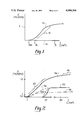
- FIG. 1 is a theoretical curve relating tissue ablation depth to the pulse energy density of a pulsed laser beam
- FIG. 2 is an empirical curve relating tissue ablation depth to pulse energy densities for various pulsed laser beams of selected wavelength
- FIG. 3 is a cross-sectional view of the cornea of the eye
- FIG. 4 is a cross-sectional view of the cornea shown in FIG. 3 with portions of a predetermined volume of corneal tissue removed by photoablation;
- FIG. 5 is an enlarged cross-sectional view of a portion of the corrected surface shown in FIG. 4.
- FIG. 1 shows a curve 10 which indicates the theoretical relationship between the pulse energy density (I) of the laser beam and the resultant ablation depth (d) into the tissue. Specifically, ablation depth (d) (measured in microns) is indicated along ordinate 12 and the pulse energy density (measured in joules per square centimeter) is indicated along abscissa 14. As shown, curve 10 identifies several points and regions of particular interest. For instance, there is some initial pulse energy density (I min ) which is required before the pulsed laser beam has any affect on the tissue.
- I min initial pulse energy density
- I min will be approximately one (1.0) J/cm 2 .
- tissue begins to photoablate.
- photoablation begins to occur when I ph is equal to approximately one and one half (1.5) J/cm 2 .
- FIG. 1 also indicates there is a substantially linear region 16 on curve 10 which extends from I ph through higher pulse energy densities until the photoablation process begins to create a plasma at I pl .
- pulse energy densities above I pl it is generally accepted that curve 10 will begin to flatten out in accordance with a logarithmic relationship. The focus of the present invention, however, is not on elevated pulse energy densities.
- the present invention is concerned with the resultant ablation depth of tissue for pulse energy densities which are substantially between I ph and a value slightly greater than I pl .
- the pulse energy density I for values of I in this range.
- FIG. 2 shows several ablation curves which generally indicate the respective causal relationships between ablation depth (d) and pulse energy density (I) for several laser beams of differing wavelengths.
- d ablation depth
- I pulse energy density
- the present invention is concerned with the well known Nd:YLF laser crystal which emits laser light with a wavelength of approximately 0.527 microns.
- the present invention is concerned with the well known erbium laser crystal which generates laser light at a wavelength of 2.94 microns. Both mediums have certain beneficial characteristics.
- the general objectives of the method and procedure according to the present invention are to remove a predetermined volume of corneal tissue which will effectively reshape the cornea in order to obtain a desired vision correction. To accomplish this it is necessary to first determine the volume of corneal tissue which must be removed. Several methods for determining this volume are well known in the pertinent art and all of these methods need not be disclosed here in detail. One in particular, however, helps to more fully understand the method of the present invention. According to this particular calculation, it is known that removal of an eight (8) micron thick layer of stroma tissue along the visual axis will result in an approximately one (1) diopter correction for the eye. To more fully appreciate what this means, consider FIG. 3.
- FIG. 3 depicts a cross section of the cornea of an eye, generally designated 18.
- cornea 18 comprises an epithelium 20, Bowman's membrane 22, stroma 24, Decimet's membrane 26, and an endothelium 28.
- the eye's visual axis 32 is shown in FIG. 3 as being along a line which extends in the direction of sight and which is substantially normal to the external surface 34 of cornea 18.
- certain vision deficiencies may be corrected by removal of tissue from stroma 24. Specifically, the amount of correction will depend on how much tissue is removed and from where.
- a distance 36 measured along visual axis 32 in stroma 24 can be used to calculate the amount of tissue to be removed for a desired diopter correction.
- a one (1) diopter correction will result.
- the exact predetermined volume of tissue to be removed is a matter of choice which can be determined on a case-by-case basis without consequence to the remainder of the procedure.
- the result after removal of the predetemined volume of corneal tissue is the exposure of a corrected surface 38.
- reshaping of the cornea is accomplished by a two-stage photoablation procedure. After the volume of corneal tissue to be removed has been determined, relatively large portions of this volume are removed in the first stage by photoablation. Ideally, portions of tissue in the size of up to one thousand (1,000) cubic microns are removed during this stage with each pulse of the laser beam.
- This removal or grinding step can be controlled by a device, such as the one disclosed in U.S. Pat. No. 4,901,718 for an invention entitled "3-Dimensional Laser Beam Guidance System,” filed Feb. 2, 1988, which issued on Feb. 20, 1990 is assigned to the same assignee as the present invention.
- corrected surface 38 of the reshaped cornea 18 remains somewhat uneven and irregular.
- corrected surface 38 is characterized by ridges 40 and depressions 42, as generally shown in FIG. 5. It is expected that the elevational difference 44 between a ridge 40 and a depression 42 will be on the order of one (1) or two (2) microns. This difference 44 can, however, cause hazy vision and in order to avoid hazy, albeit corrected, vision for the patient corrected surface 38 needs to be smoothed or polished.
- corrected surface 38 is smoothed or polished by removing relatively small portions of corneal tissue by photoablation. Ideally, only the ridges 40 are removed and therefore, during this smoothing stage, it is necessary to use laser pulses which photoablate stroma 24 tissue to a depth of one (1) micron or less. Ideally, as envisioned by the present invention, the depth of photoablation in the smoothing stage will be variable within the range of one (1) to one tenth (0.1) micron.
- lasers with different wavelengths may be used for the present invention.
- one wavelength could be used for both the first stage (i.e., removal of relatively large portions of corneal tissue) and the second stage (i.e., smoothing of the corrected surface by removal of relatively small portions of corneal tissue). This may, however, be impractical. Nevertheless, the use of single wavelength procedures should be considered along with the presently preferred dual wavelength procedure.
- an erbium (Er) laser crystal As is well known, an erbium crystal emits laser light at a wavelength of approximately 2.94 microns. Further, it has been determined that the light emitted by an erbium laser crystal has photoablation characteristics which are generally as depicted by curve 46 in FIG. 2. Specifically, as with other pulsed laser beams, there is a region which extends from a pulse energy density of approximately thirty (30) J/cm 2 upward to higher pulse energy densities in which operation of an erbium pulsed laser beam will cause formation of a plasma during photoablation.
- the erbium laser also has characteristics in range 50 of pulse energy densities below approximately ten (10) J/cm 2 wherein photoablation will result without any significant formation of a plasma. Equally important are the indications that the shape of curve 46 in the range 50 is moderate and the curve 46 itself is substantially linear. Consequently, an erbium pulsed laser beam lends itself to being precisely controlled during operation in the range 50. Unfortunately, an erbium laser medium has relatively low efficiencies and generates a great amount of heat during operation at pulse energy densities above approximately ten (10) J/cm 2 .
- an erbium laser in order for an erbium laser to be efficient and have greater efficacy at the higher pulse energy densities necessary for the removal of relatively large portions of corneal tissue in the first stage of the process of the present invention, the erbium medium must be cooled. Further, it is known that this requires cooling the erbium laser medium to approximately minus fifty degrees centigrade (-50° C.) Thus, an erbium laser medium can be used for performing the methods of the present invention if the components necessary to cool the medium to these temperatures in the first stage are provided. On the other hand, for the purposes of the present invention, an Nd:YLF laser medium exhibits good operating characteristics at the higher pulse energy densities required for removal of corneal tissue during the first stage.
- a standard Nd:YLF laser medium emits light at a wavelength of approximately 0.527 microns in a pulsed beam which has photoablation characteristics that are substantially as depicted by curve 52.
- an Nd:YLF laser medium does not require cooling for operating the higher pulse energy densities required for efficient removal of relatively large portions of corneal tissue.
- an Nd:YLF laser beam is preferred for operation during the first stage removal of corneal tissue.
- An unmodified Nd:YLF laser however, has less desirable operating characteristics at the lower pulse energy densities.
- the shape of curve 52 is relatively steep in region 54 where operation of the Nd:YLF laser would result in removal of relatively small portions of corneal tissue.
- the photoablation curve 58 for the well known excimer laser is provided in FIG. 2 for comparison purposes only. As clearly seen in FIGS. 1 and 2, the fact that the excimer laser has little, if any, ability to operate at pulse energy densities in region 16 below the plasma formation threshold makes it unsuitable for the present invention.
- an Nd:YLF laser medium be used to remove relatively large portions of corneal tissue in the first stage and that an erbium laser medium be used to smooth the corrected surface 38 by removing relatively small portions of corneal tissue therefrom.
- the Nd:YLF laser be operated during the method's first stage in a regime wherein the resultant beam is pulsed to generate ten thousand pulses per second (10,000 pps) with each pulse having approximately one hundred micro joules (100 ⁇ J) of energy (and being approximately thirty pico seconds (30 psec)) in duration.
- an erbium laser be operated during the method's second stage in a regime wherein the resultant beam is pulsed to generate two thousand pulses per second (2,000 pps) with each pulse having approximately five hundred micro joules (500 ⁇ J) of energy and being approximately one hundred pico seconds (100 psec) in duration.
- operation of the Nd:YLF laser during the first stage be accomplished at pulse energy densities generally at or above ten (10) J/cm 2 and that operation of the erbium laser during the second stage be accomplished at pulse energy densities generally below two (2) or three (3) J/cm 2 .
Abstract
Description
Claims (25)
Priority Applications (3)
| Application Number | Priority Date | Filing Date | Title |
|---|---|---|---|
| US07/357,705 US4988348A (en) | 1989-05-26 | 1989-05-26 | Method for reshaping the cornea |
| CA002013954A CA2013954A1 (en) | 1989-05-26 | 1990-04-05 | Method for reshaping the cornea |
| JP2131483A JPH037152A (en) | 1989-05-26 | 1990-05-23 | Reformation of cornea |
Applications Claiming Priority (1)
| Application Number | Priority Date | Filing Date | Title |
|---|---|---|---|
| US07/357,705 US4988348A (en) | 1989-05-26 | 1989-05-26 | Method for reshaping the cornea |
Publications (1)
| Publication Number | Publication Date |
|---|---|
| US4988348A true US4988348A (en) | 1991-01-29 |
Family
ID=23406702
Family Applications (1)
| Application Number | Title | Priority Date | Filing Date |
|---|---|---|---|
| US07/357,705 Expired - Lifetime US4988348A (en) | 1989-05-26 | 1989-05-26 | Method for reshaping the cornea |
Country Status (3)
| Country | Link |
|---|---|
| US (1) | US4988348A (en) |
| JP (1) | JPH037152A (en) |
| CA (1) | CA2013954A1 (en) |
Cited By (106)
| Publication number | Priority date | Publication date | Assignee | Title |
|---|---|---|---|---|
| WO1993014817A2 (en) * | 1992-01-15 | 1993-08-05 | Premier Laser Systems, Inc. | Corneal sculpting using laser energy |
| EP0700310A1 (en) * | 1993-04-20 | 1996-03-13 | Novatec Laser Systems, Inc. | Improved ophthalmic surgical laser and method |
| EP0714646A1 (en) * | 1994-11-30 | 1996-06-05 | Herbert Schwind GmbH & Co. KG | Device for the removal of corneal tissue |
| US5533997A (en) * | 1994-06-29 | 1996-07-09 | Ruiz; Luis A. | Apparatus and method for performing presbyopia corrective surgery |
| US5549599A (en) * | 1994-04-28 | 1996-08-27 | Nidek Co., Ltd. | Apparatus for laser surgery on a cornea |
| US5656186A (en) * | 1994-04-08 | 1997-08-12 | The Regents Of The University Of Michigan | Method for controlling configuration of laser induced breakdown and ablation |
| US5722427A (en) * | 1993-05-10 | 1998-03-03 | Eyesys Technologies, Inc. | Method of refractive surgery |
| US5920373A (en) * | 1997-09-24 | 1999-07-06 | Heidelberg Engineering Optische Messysteme Gmbh | Method and apparatus for determining optical characteristics of a cornea |
| US5928129A (en) * | 1994-06-29 | 1999-07-27 | Ruiz; Luis Antonio | Apparatus and method for performing presbyopia corrective surgery |
| US5993438A (en) * | 1993-11-12 | 1999-11-30 | Escalon Medical Corporation | Intrastromal photorefractive keratectomy |
| US6050687A (en) * | 1999-06-11 | 2000-04-18 | 20/10 Perfect Vision Optische Geraete Gmbh | Method and apparatus for measurement of the refractive properties of the human eye |
| US6110166A (en) * | 1995-03-20 | 2000-08-29 | Escalon Medical Corporation | Method for corneal laser surgery |
| EP1060710A2 (en) * | 1994-04-25 | 2000-12-20 | Autonomous Technologies Corporation | Laser sculpting system |
| US6271915B1 (en) | 1996-11-25 | 2001-08-07 | Autonomous Technologies Corporation | Objective measurement and correction of optical systems using wavefront analysis |
| US6270221B1 (en) | 1998-08-19 | 2001-08-07 | Alcon Universal Ltd. | Apparatus and method for measuring vision defects of a human eye |
| US6287296B1 (en) | 1995-11-30 | 2001-09-11 | Herbert Schwind Gmbh & Co. Kg | Device for the removal of tissue from the cornea of an eye |
| US6293938B1 (en) * | 1994-04-08 | 2001-09-25 | Summit Technology, Inc. | Photo-refractive keratectomy |
| US6302877B1 (en) | 1994-06-29 | 2001-10-16 | Luis Antonio Ruiz | Apparatus and method for performing presbyopia corrective surgery |
| US6324191B1 (en) | 2000-01-12 | 2001-11-27 | Intralase Corp. | Oscillator with mode control |
| US6341009B1 (en) | 2000-02-24 | 2002-01-22 | Quantronix Corporation | Laser delivery system and method for photolithographic mask repair |
| US20020045890A1 (en) * | 1996-04-24 | 2002-04-18 | The Regents Of The University O F California | Opto-acoustic thrombolysis |
| US6428533B1 (en) | 2000-10-17 | 2002-08-06 | 20/10 Perfect Vision Optische Geraete Gmbh | Closed loop control for refractive laser surgery (LASIK) |
| US6497483B2 (en) | 2000-05-08 | 2002-12-24 | Alcon, Inc. | Apparatus and method for objective measurement of optical systems using wavefront analysis |
| US6579282B2 (en) | 2001-04-25 | 2003-06-17 | 20/10 Perfect Vision Optische Geraete Gmbh | Device and method for creating a corneal reference for an eyetracker |
| US6578963B2 (en) | 2000-04-19 | 2003-06-17 | Alcon Universal Ltd. | Wavefront sensor for objective measurement of an optical system and associated methods |
| US6610050B2 (en) | 2001-07-27 | 2003-08-26 | 20/10 Perfect Vision, Optische Geraete Gmbh | Laser beam delivery system with multiple focal points |
| US6610051B2 (en) | 2001-10-12 | 2003-08-26 | 20/10 Perfect Vision Optische Geraete Gmbh | Device and method for performing refractive surgery |
| US6626894B2 (en) | 1994-04-25 | 2003-09-30 | Alcon, Inc. | Method of ablating a moving eye |
| WO2003082146A2 (en) | 2002-03-23 | 2003-10-09 | Intralase Corp. | System and method for improved material processing using a laser beam |
| US6641577B2 (en) | 2001-11-28 | 2003-11-04 | 20/10 Perfect Vision Optische Geraete Gmbh | Apparatus and method for creating a corneal flap |
| US20040002697A1 (en) * | 2002-06-27 | 2004-01-01 | Gerhard Youssefi | Biconic ablation with controlled spherical aberration |
| EP1428470A2 (en) | 2000-09-15 | 2004-06-16 | Ligi Tecnologie Medicali S.p.A. | Apparatus for determining and ablating a corneal tissue volume of a receiving cornea for corneal lamellar grafting transplantation |
| US20040130677A1 (en) * | 1998-08-19 | 2004-07-08 | Alcon, Inc. | Apparatus and method for measuring vision defects of a human eye |
| US20040199149A1 (en) * | 1996-03-21 | 2004-10-07 | Myers Raymond I. | Lenticular refractive surgery of presbyopia, other refractive errors, and cataract retardation |
| US20040199150A1 (en) * | 1991-08-02 | 2004-10-07 | Lai Shui T. | Method and apparatus for laser surgery of the cornea |
| US20050065502A1 (en) * | 2003-08-11 | 2005-03-24 | Richard Stoltz | Enabling or blocking the emission of an ablation beam based on color of target |
| US6887232B2 (en) | 2002-11-13 | 2005-05-03 | 20/10 Perfect Vision Optische Geraete Gmbh | Closed loop control for intrastromal wavefront-guided ablation |
| US20050124983A1 (en) * | 1996-11-25 | 2005-06-09 | Frey Rudolph W. | Method for determining and correcting vision |
| US20050149005A1 (en) * | 2002-11-13 | 2005-07-07 | Josef Bille | Closed loop control for intrastromal wavefront-guided ablation with fractionated treatment program |
| US7022117B1 (en) * | 1999-10-21 | 2006-04-04 | Bausch & Lomb Incorporated | Customized refractive correction |
| US20060084957A1 (en) * | 2003-08-11 | 2006-04-20 | Peter Delfyett | Laser ablation method and apparatus having a feedback loop and control unit |
| US20060084954A1 (en) * | 2004-08-17 | 2006-04-20 | Intralase Corp. | Apparatus and method for correction of abberations in laser system optics |
| US20060095023A1 (en) * | 2004-11-01 | 2006-05-04 | Frieder Loesel | Time-resolved scanning patterns for intrastromal surgery |
| US20060106372A1 (en) * | 2004-11-12 | 2006-05-18 | Tobias Kuhn | Systems and methods for intrastromal scanning patterns |
| US20060155265A1 (en) * | 1995-03-20 | 2006-07-13 | Intralase Corp. | Method of corneal surgery by laser incising a contoured corneal flap |
| US7101364B2 (en) | 2001-10-12 | 2006-09-05 | 20/10 Perfect Vision Optische Geraete Gmbh | Method and apparatus for intrastromal refractive surgery |
| US20070027438A1 (en) * | 2005-07-26 | 2007-02-01 | Frieder Loesel | System and method for compensating a corneal dissection |
| US20070038202A1 (en) * | 2004-06-11 | 2007-02-15 | Celestino Susana M | Method of preventing the induction of aberrations in laser refractive surgery systems |
| US20070078447A1 (en) * | 2004-12-17 | 2007-04-05 | Martin Weinacht | Devices and methods for separating layers of materials having different ablation thresholds |
| US20070088409A1 (en) * | 2005-10-14 | 2007-04-19 | Carl Zeiss Meditec Ag | Device and method for material processing by means of laser radiation |
| US20070173794A1 (en) * | 2006-01-20 | 2007-07-26 | Frey Rudolph W | System and method for treating the structure of the human lens with a laser |
| US20070179479A1 (en) * | 2002-11-13 | 2007-08-02 | Josef Bille | System and Method for Photoablation Using Multiple Focal Points with Rotating Beam Splitter |
| US20070219541A1 (en) * | 2006-03-14 | 2007-09-20 | Intralase Corp. | System and method for ophthalmic laser surgery on a cornea |
| US20070253455A1 (en) * | 2006-04-26 | 2007-11-01 | Stadler Andrew D | Intelligent Laser Interlock System |
| US20080025351A1 (en) * | 2006-07-27 | 2008-01-31 | Frieder Loesel | Material processing system with variable repetition rate laser |
| EP1941849A2 (en) | 1996-10-02 | 2008-07-09 | AMO Development, LLC | Method for corneal laser surgery |
| US20080212623A1 (en) * | 2005-10-14 | 2008-09-04 | Mark Bischoff | Device and method for material processing by means of laser radiation |
| WO2008125173A2 (en) * | 2007-04-11 | 2008-10-23 | Carl Zeiss Meditec Ag | Device and method for processing material by means of laser radiation |
| US20080287935A1 (en) * | 2002-11-13 | 2008-11-20 | Josef Bille | System and method for photoablation using multiple focal points using cyclical phase modulation |
| US20090157063A1 (en) * | 2007-12-17 | 2009-06-18 | Luis Antonio Ruiz | Method patterns for intrastromal refractive surgery |
| US20090157061A1 (en) * | 2007-12-17 | 2009-06-18 | Luis Antonio Ruiz | Method for intrastromal refractive surgery |
| US20090234335A1 (en) * | 2006-03-17 | 2009-09-17 | Amo Manufacturing Usa, Llc | Intrastromal refractive correction systems and methods |
| DE102008017771A1 (en) * | 2008-04-04 | 2009-10-08 | Carl Zeiss Meditec Ag | Curved sectional areas smoothing method for cornea of eye of patient, involves defining partial volume by curved sectional areas produced by excimer laser, and interrupting sectional areas after removing of partial volume |
| US20090289382A1 (en) * | 2008-05-22 | 2009-11-26 | Raydiance, Inc. | System and method for modifying characteristics of a contact lens utilizing an ultra-short pulsed laser |
| US20090299345A1 (en) * | 2008-05-27 | 2009-12-03 | Bille Josef F | System and method for reshaping a cornea using a combination of liob and structural change procedures |
| US20090323740A1 (en) * | 2006-01-23 | 2009-12-31 | Stadler Andrew D | Systems And Methods For Control Of Ultra Short Pulse Amplification |
| US20100004643A1 (en) * | 2006-01-20 | 2010-01-07 | Frey Rudolph W | System and method for improving the accommodative amplitude and increasing the refractive power of the human lens with a laser |
| US20100004641A1 (en) * | 2006-01-20 | 2010-01-07 | Frey Rudolph W | System and apparatus for delivering a laser beam to the lens of an eye |
| US20100022994A1 (en) * | 2008-07-25 | 2010-01-28 | Frey Rudolph W | Liquid filled index matching device for ophthalmic laser procedures |
| US20100022995A1 (en) * | 2008-07-25 | 2010-01-28 | Frey Rudolph W | Method and system for removal and replacement of lens material from the lens of an eye |
| US20100022996A1 (en) * | 2008-07-25 | 2010-01-28 | Frey Rudolph W | Method and system for creating a bubble shield for laser lens procedures |
| US20100040095A1 (en) * | 2008-08-18 | 2010-02-18 | Raydiance, Inc. | Systems and methods for controlling a pulsed laser by combining laser signals |
| US20100082018A1 (en) * | 2008-09-26 | 2010-04-01 | Daryus Panthakey | Method and system for reshaping the cornea |
| US20100174274A1 (en) * | 2009-01-06 | 2010-07-08 | Bille Josef F | Minimizing the side-effects of refractive corrections using statistically determined irregularities in intrastromal incisions |
| US20100191229A1 (en) * | 2009-01-27 | 2010-07-29 | Bille Josef F | Methods for Employing Intrastromal Corrections in Combination with Surface Refractive Surgery to Correct Myopic/Hyperopic Presbyopia |
| US20100191228A1 (en) * | 2009-01-27 | 2010-07-29 | Luis Antonio Ruiz | System and Method for Refractive Surgery with Augmentation by Intrastromal Corrective Procedures |
| US20100191227A1 (en) * | 2009-01-27 | 2010-07-29 | Bille Josef F | System and Method for Correcting Higher Order Aberrations with Changes in Intrastromal Biomechanical Stress Distributions |
| US20100217247A1 (en) * | 2009-02-20 | 2010-08-26 | Bille Josef F | System and Methods for Minimizing Higher Order Aberrations Introduced During Refractive Surgery |
| US20100241108A1 (en) * | 2009-03-23 | 2010-09-23 | Wuellner Christian | Apparatus and Method for LASIK |
| US20100249762A1 (en) * | 2007-12-17 | 2010-09-30 | Bille Josef F | System for Performing Intrastromal Refractive Surgery |
| US20100249761A1 (en) * | 2007-12-17 | 2010-09-30 | Luis Antonio Ruiz | System and method for altering the optical properties of a material |
| US20100292678A1 (en) * | 2006-01-20 | 2010-11-18 | Frey Rudolph W | System and method for providing laser shot patterns to the lens of an eye |
| US20110022036A1 (en) * | 2009-07-24 | 2011-01-27 | Frey Rudolph W | System and method for performing ladar assisted procedures on the lens of an eye |
| US20110022035A1 (en) * | 2009-07-24 | 2011-01-27 | Porter Gerrit N | Liquid holding interface device for ophthalmic laser procedures |
| US20110022037A1 (en) * | 2009-01-06 | 2011-01-27 | Bille Josef F | System and Method for Minimizing the Side Effects of Refractive Corrections Using Line or Dot Cuts for Incisions |
| US20110073584A1 (en) * | 2003-05-20 | 2011-03-31 | Richard Stoltz | Portable Optical Ablation System |
| US20110160710A1 (en) * | 2009-07-24 | 2011-06-30 | Frey Rudolph W | Laser system and method for performing and sealing corneal incisions in the eye |
| US20110166557A1 (en) * | 2009-07-24 | 2011-07-07 | Naranjo-Tackman Ramon | Laser system and method for astigmatic corrections in asssociation with cataract treatment |
| US20110190740A1 (en) * | 2010-02-01 | 2011-08-04 | Lensar, Inc. | Placido ring measurement of astigmatism axis and laser marking of astigmatism axis |
| US8135050B1 (en) | 2005-07-19 | 2012-03-13 | Raydiance, Inc. | Automated polarization correction |
| US8150271B1 (en) | 2006-03-28 | 2012-04-03 | Raydiance, Inc. | Active tuning of temporal dispersion in an ultrashort pulse laser system |
| US8173929B1 (en) | 2003-08-11 | 2012-05-08 | Raydiance, Inc. | Methods and systems for trimming circuits |
| US8189971B1 (en) | 2006-01-23 | 2012-05-29 | Raydiance, Inc. | Dispersion compensation in a chirped pulse amplification system |
| US8262646B2 (en) | 2006-01-20 | 2012-09-11 | Lensar, Inc. | System and method for providing the shaped structural weakening of the human lens with a laser |
| US8556425B2 (en) | 2010-02-01 | 2013-10-15 | Lensar, Inc. | Purkinjie image-based alignment of suction ring in ophthalmic applications |
| USD694890S1 (en) | 2010-10-15 | 2013-12-03 | Lensar, Inc. | Laser system for treatment of the eye |
| USD695408S1 (en) | 2010-10-15 | 2013-12-10 | Lensar, Inc. | Laser system for treatment of the eye |
| US8617146B2 (en) | 2009-07-24 | 2013-12-31 | Lensar, Inc. | Laser system and method for correction of induced astigmatism |
| US8663208B2 (en) | 2009-02-09 | 2014-03-04 | Amo Development, Llc | System and method for intrastromal refractive correction |
| US8801186B2 (en) | 2010-10-15 | 2014-08-12 | Lensar, Inc. | System and method of scan controlled illumination of structures within an eye |
| US8884184B2 (en) | 2010-08-12 | 2014-11-11 | Raydiance, Inc. | Polymer tubing laser micromachining |
| US8921733B2 (en) | 2003-08-11 | 2014-12-30 | Raydiance, Inc. | Methods and systems for trimming circuits |
| US9114482B2 (en) | 2010-09-16 | 2015-08-25 | Raydiance, Inc. | Laser based processing of layered materials |
| US9393154B2 (en) | 2011-10-28 | 2016-07-19 | Raymond I Myers | Laser methods for creating an antioxidant sink in the crystalline lens for the maintenance of eye health and physiology and slowing presbyopia development |
| AU2016210633B2 (en) * | 2005-10-14 | 2018-09-06 | Carl Zeiss Meditec Ag | Device and method for materials processing using laser radiation |
| US10463541B2 (en) | 2011-03-25 | 2019-11-05 | Lensar, Inc. | System and method for correcting astigmatism using multiple paired arcuate laser generated corneal incisions |
Families Citing this family (2)
| Publication number | Priority date | Publication date | Assignee | Title |
|---|---|---|---|---|
| US7131968B2 (en) * | 2003-06-02 | 2006-11-07 | Carl Zeiss Meditec Ag | Apparatus and method for opthalmologic surgical procedures using a femtosecond fiber laser |
| DE102005049281A1 (en) * | 2005-10-14 | 2007-04-19 | Carl Zeiss Meditec Ag | Apparatus and method for material processing by means of laser radiation |
Citations (6)
| Publication number | Priority date | Publication date | Assignee | Title |
|---|---|---|---|---|
| US4669466A (en) * | 1985-01-16 | 1987-06-02 | Lri L.P. | Method and apparatus for analysis and correction of abnormal refractive errors of the eye |
| US4729372A (en) * | 1983-11-17 | 1988-03-08 | Lri L.P. | Apparatus for performing ophthalmic laser surgery |
| US4732148A (en) * | 1983-11-17 | 1988-03-22 | Lri L.P. | Method for performing ophthalmic laser surgery |
| US4770172A (en) * | 1983-11-17 | 1988-09-13 | Lri L.P. | Method of laser-sculpture of the optically used portion of the cornea |
| US4773414A (en) * | 1983-11-17 | 1988-09-27 | Lri L.P. | Method of laser-sculpture of the optically used portion of the cornea |
| US4887592A (en) * | 1987-06-02 | 1989-12-19 | Hanspeter Loertscher | Cornea laser-cutting apparatus |
-
1989
- 1989-05-26 US US07/357,705 patent/US4988348A/en not_active Expired - Lifetime
-
1990
- 1990-04-05 CA CA002013954A patent/CA2013954A1/en not_active Abandoned
- 1990-05-23 JP JP2131483A patent/JPH037152A/en active Pending
Patent Citations (6)
| Publication number | Priority date | Publication date | Assignee | Title |
|---|---|---|---|---|
| US4729372A (en) * | 1983-11-17 | 1988-03-08 | Lri L.P. | Apparatus for performing ophthalmic laser surgery |
| US4732148A (en) * | 1983-11-17 | 1988-03-22 | Lri L.P. | Method for performing ophthalmic laser surgery |
| US4770172A (en) * | 1983-11-17 | 1988-09-13 | Lri L.P. | Method of laser-sculpture of the optically used portion of the cornea |
| US4773414A (en) * | 1983-11-17 | 1988-09-27 | Lri L.P. | Method of laser-sculpture of the optically used portion of the cornea |
| US4669466A (en) * | 1985-01-16 | 1987-06-02 | Lri L.P. | Method and apparatus for analysis and correction of abnormal refractive errors of the eye |
| US4887592A (en) * | 1987-06-02 | 1989-12-19 | Hanspeter Loertscher | Cornea laser-cutting apparatus |
Non-Patent Citations (4)
| Title |
|---|
| "Enodexcimer Laser Intraocular Ablative Photodecomposition", by Marshall et al. Am. J. Apthal. 1/86, vol. 101, No. 1, pp. 130-131. |
| "Photoablative Reprofiling of the Cornea Using an Excimer Laser: Photorefractive Keratechtomy", Marshall et al.; Lasers in Ophthal., vol. 1, No. 1, pp. 21-48 (1986). |
| Enodexcimer Laser Intraocular Ablative Photodecomposition , by Marshall et al. Am. J. Apthal. 1/86, vol. 101, No. 1, pp. 130 131. * |
| Photoablative Reprofiling of the Cornea Using an Excimer Laser: Photorefractive Keratechtomy , Marshall et al.; Lasers in Ophthal., vol. 1, No. 1, pp. 21 48 (1986). * |
Cited By (172)
| Publication number | Priority date | Publication date | Assignee | Title |
|---|---|---|---|---|
| US7220255B2 (en) | 1991-08-02 | 2007-05-22 | Lai Shui T | Method and apparatus for laser surgery of the cornea |
| US20040199150A1 (en) * | 1991-08-02 | 2004-10-07 | Lai Shui T. | Method and apparatus for laser surgery of the cornea |
| US20060217688A1 (en) * | 1991-11-06 | 2006-09-28 | Lai Shui T | Method and Apparatus for Laser Surgery of the Cornea |
| US5741245A (en) * | 1992-01-15 | 1998-04-21 | Premier Laser Systems, Inc. | Corneal sculpting using laser energy |
| WO1993014817A2 (en) * | 1992-01-15 | 1993-08-05 | Premier Laser Systems, Inc. | Corneal sculpting using laser energy |
| WO1993014817A3 (en) * | 1992-01-15 | 1993-10-14 | Premier Laser Systems Inc | Corneal sculpting using laser energy |
| EP1402860A2 (en) * | 1993-04-20 | 2004-03-31 | LAI, Shui, T. | Improved ophthalmic surgical laser |
| EP1402860A3 (en) * | 1993-04-20 | 2004-08-18 | LAI, Shui, T. | Improved ophthalmic surgical laser |
| EP0700310A4 (en) * | 1993-04-20 | 1997-10-15 | Novatec Laser Systems Inc | Improved ophthalmic surgical laser and method |
| EP0700310A1 (en) * | 1993-04-20 | 1996-03-13 | Novatec Laser Systems, Inc. | Improved ophthalmic surgical laser and method |
| US5722427A (en) * | 1993-05-10 | 1998-03-03 | Eyesys Technologies, Inc. | Method of refractive surgery |
| US5970984A (en) * | 1993-05-10 | 1999-10-26 | Eyesys-Premier, Inc. | Method of refractive surgery |
| US5993438A (en) * | 1993-11-12 | 1999-11-30 | Escalon Medical Corporation | Intrastromal photorefractive keratectomy |
| US5656186A (en) * | 1994-04-08 | 1997-08-12 | The Regents Of The University Of Michigan | Method for controlling configuration of laser induced breakdown and ablation |
| US6293938B1 (en) * | 1994-04-08 | 2001-09-25 | Summit Technology, Inc. | Photo-refractive keratectomy |
| USRE37585E1 (en) * | 1994-04-08 | 2002-03-19 | The Regents Of The University Of Michigan | Method for controlling configuration of laser induced breakdown and ablation |
| US6626898B2 (en) | 1994-04-25 | 2003-09-30 | Alcon, Inc. | Flying spot laser ablation method |
| US6626897B2 (en) | 1994-04-25 | 2003-09-30 | Alcon, Inc. | Method of redirecting an ablating laser beam |
| US6626895B2 (en) | 1994-04-25 | 2003-09-30 | Alcon, Inc. | Laser beam delivery system |
| EP1060710A2 (en) * | 1994-04-25 | 2000-12-20 | Autonomous Technologies Corporation | Laser sculpting system |
| US6626896B2 (en) | 1994-04-25 | 2003-09-30 | Alcon, Inc. | Method of correcting vision |
| US6626894B2 (en) | 1994-04-25 | 2003-09-30 | Alcon, Inc. | Method of ablating a moving eye |
| EP1512379A1 (en) * | 1994-04-25 | 2005-03-09 | Alcon Inc. | Laser corneal sculpting system |
| EP1060710A3 (en) * | 1994-04-25 | 2003-08-20 | Alcon Inc. | Laser sculpting system |
| US5549599A (en) * | 1994-04-28 | 1996-08-27 | Nidek Co., Ltd. | Apparatus for laser surgery on a cornea |
| US5928129A (en) * | 1994-06-29 | 1999-07-27 | Ruiz; Luis Antonio | Apparatus and method for performing presbyopia corrective surgery |
| US6302877B1 (en) | 1994-06-29 | 2001-10-16 | Luis Antonio Ruiz | Apparatus and method for performing presbyopia corrective surgery |
| US5533997A (en) * | 1994-06-29 | 1996-07-09 | Ruiz; Luis A. | Apparatus and method for performing presbyopia corrective surgery |
| US6843787B2 (en) | 1994-06-29 | 2005-01-18 | Luis Antonio Ruiz | Apparatus and method for performing presbyopia corrective surgery |
| EP0714646A1 (en) * | 1994-11-30 | 1996-06-05 | Herbert Schwind GmbH & Co. KG | Device for the removal of corneal tissue |
| US7892226B2 (en) | 1995-03-20 | 2011-02-22 | Amo Development, Llc. | Method of corneal surgery by laser incising a contoured corneal flap |
| US20060155265A1 (en) * | 1995-03-20 | 2006-07-13 | Intralase Corp. | Method of corneal surgery by laser incising a contoured corneal flap |
| US6110166A (en) * | 1995-03-20 | 2000-08-29 | Escalon Medical Corporation | Method for corneal laser surgery |
| US6287296B1 (en) | 1995-11-30 | 2001-09-11 | Herbert Schwind Gmbh & Co. Kg | Device for the removal of tissue from the cornea of an eye |
| US20100114079A1 (en) * | 1996-03-21 | 2010-05-06 | Second Sight Laser Technologies, Inc. | Lenticular refractive surgery of presbyopia, other refractive errors, and cataract retardation |
| US20040199149A1 (en) * | 1996-03-21 | 2004-10-07 | Myers Raymond I. | Lenticular refractive surgery of presbyopia, other refractive errors, and cataract retardation |
| US7655002B2 (en) | 1996-03-21 | 2010-02-02 | Second Sight Laser Technologies, Inc. | Lenticular refractive surgery of presbyopia, other refractive errors, and cataract retardation |
| US20020045890A1 (en) * | 1996-04-24 | 2002-04-18 | The Regents Of The University O F California | Opto-acoustic thrombolysis |
| EP1941849A2 (en) | 1996-10-02 | 2008-07-09 | AMO Development, LLC | Method for corneal laser surgery |
| US20050124983A1 (en) * | 1996-11-25 | 2005-06-09 | Frey Rudolph W. | Method for determining and correcting vision |
| US6271915B1 (en) | 1996-11-25 | 2001-08-07 | Autonomous Technologies Corporation | Objective measurement and correction of optical systems using wavefront analysis |
| US6271914B1 (en) * | 1996-11-25 | 2001-08-07 | Autonomous Technologies Corporation | Objective measurement and correction of optical systems using wavefront analysis |
| US5920373A (en) * | 1997-09-24 | 1999-07-06 | Heidelberg Engineering Optische Messysteme Gmbh | Method and apparatus for determining optical characteristics of a cornea |
| US20040130677A1 (en) * | 1998-08-19 | 2004-07-08 | Alcon, Inc. | Apparatus and method for measuring vision defects of a human eye |
| US6270221B1 (en) | 1998-08-19 | 2001-08-07 | Alcon Universal Ltd. | Apparatus and method for measuring vision defects of a human eye |
| US6050687A (en) * | 1999-06-11 | 2000-04-18 | 20/10 Perfect Vision Optische Geraete Gmbh | Method and apparatus for measurement of the refractive properties of the human eye |
| US6155684A (en) * | 1999-06-11 | 2000-12-05 | Perfect Vision Optische Geraete Gmbh | Method and apparatus for precompensating the refractive properties of the human eye with adaptive optical feedback control |
| US7022117B1 (en) * | 1999-10-21 | 2006-04-04 | Bausch & Lomb Incorporated | Customized refractive correction |
| US6324191B1 (en) | 2000-01-12 | 2001-11-27 | Intralase Corp. | Oscillator with mode control |
| US6341009B1 (en) | 2000-02-24 | 2002-01-22 | Quantronix Corporation | Laser delivery system and method for photolithographic mask repair |
| US6578963B2 (en) | 2000-04-19 | 2003-06-17 | Alcon Universal Ltd. | Wavefront sensor for objective measurement of an optical system and associated methods |
| US6497483B2 (en) | 2000-05-08 | 2002-12-24 | Alcon, Inc. | Apparatus and method for objective measurement of optical systems using wavefront analysis |
| EP1428470A2 (en) | 2000-09-15 | 2004-06-16 | Ligi Tecnologie Medicali S.p.A. | Apparatus for determining and ablating a corneal tissue volume of a receiving cornea for corneal lamellar grafting transplantation |
| US6428533B1 (en) | 2000-10-17 | 2002-08-06 | 20/10 Perfect Vision Optische Geraete Gmbh | Closed loop control for refractive laser surgery (LASIK) |
| US6579282B2 (en) | 2001-04-25 | 2003-06-17 | 20/10 Perfect Vision Optische Geraete Gmbh | Device and method for creating a corneal reference for an eyetracker |
| US6610050B2 (en) | 2001-07-27 | 2003-08-26 | 20/10 Perfect Vision, Optische Geraete Gmbh | Laser beam delivery system with multiple focal points |
| US6610051B2 (en) | 2001-10-12 | 2003-08-26 | 20/10 Perfect Vision Optische Geraete Gmbh | Device and method for performing refractive surgery |
| US7101364B2 (en) | 2001-10-12 | 2006-09-05 | 20/10 Perfect Vision Optische Geraete Gmbh | Method and apparatus for intrastromal refractive surgery |
| US6641577B2 (en) | 2001-11-28 | 2003-11-04 | 20/10 Perfect Vision Optische Geraete Gmbh | Apparatus and method for creating a corneal flap |
| WO2003082146A2 (en) | 2002-03-23 | 2003-10-09 | Intralase Corp. | System and method for improved material processing using a laser beam |
| US20040002697A1 (en) * | 2002-06-27 | 2004-01-01 | Gerhard Youssefi | Biconic ablation with controlled spherical aberration |
| US6887232B2 (en) | 2002-11-13 | 2005-05-03 | 20/10 Perfect Vision Optische Geraete Gmbh | Closed loop control for intrastromal wavefront-guided ablation |
| US20070179479A1 (en) * | 2002-11-13 | 2007-08-02 | Josef Bille | System and Method for Photoablation Using Multiple Focal Points with Rotating Beam Splitter |
| US20080287935A1 (en) * | 2002-11-13 | 2008-11-20 | Josef Bille | System and method for photoablation using multiple focal points using cyclical phase modulation |
| US20060173445A1 (en) * | 2002-11-13 | 2006-08-03 | Josef Bille | Customized corneal flap |
| US20050149005A1 (en) * | 2002-11-13 | 2005-07-07 | Josef Bille | Closed loop control for intrastromal wavefront-guided ablation with fractionated treatment program |
| US7662149B2 (en) | 2002-11-13 | 2010-02-16 | Technolas Perfect Vision Gmbh | Customized corneal flap |
| US7717906B2 (en) | 2002-11-13 | 2010-05-18 | Technolas Perfect Vision Gmbh | System and method for photoablation using multiple focal points with rotating beam splitter |
| US7232436B2 (en) | 2002-11-13 | 2007-06-19 | Josef Bille | Closed loop control for intrastromal wavefront-guided ablation with fractionated treatment program |
| US20110073584A1 (en) * | 2003-05-20 | 2011-03-31 | Richard Stoltz | Portable Optical Ablation System |
| US8398622B2 (en) | 2003-05-20 | 2013-03-19 | Raydiance, Inc. | Portable optical ablation system |
| US8173929B1 (en) | 2003-08-11 | 2012-05-08 | Raydiance, Inc. | Methods and systems for trimming circuits |
| US20060084957A1 (en) * | 2003-08-11 | 2006-04-20 | Peter Delfyett | Laser ablation method and apparatus having a feedback loop and control unit |
| US8921733B2 (en) | 2003-08-11 | 2014-12-30 | Raydiance, Inc. | Methods and systems for trimming circuits |
| US20050065502A1 (en) * | 2003-08-11 | 2005-03-24 | Richard Stoltz | Enabling or blocking the emission of an ablation beam based on color of target |
| US9022037B2 (en) | 2003-08-11 | 2015-05-05 | Raydiance, Inc. | Laser ablation method and apparatus having a feedback loop and control unit |
| US20070038202A1 (en) * | 2004-06-11 | 2007-02-15 | Celestino Susana M | Method of preventing the induction of aberrations in laser refractive surgery systems |
| US7584756B2 (en) | 2004-08-17 | 2009-09-08 | Amo Development, Llc | Apparatus and method for correction of aberrations in laser system optics |
| US20060084954A1 (en) * | 2004-08-17 | 2006-04-20 | Intralase Corp. | Apparatus and method for correction of abberations in laser system optics |
| US20060095023A1 (en) * | 2004-11-01 | 2006-05-04 | Frieder Loesel | Time-resolved scanning patterns for intrastromal surgery |
| US7717905B2 (en) | 2004-11-01 | 2010-05-18 | Technolas Perfect Vision Gmbh | Time-resolved scanning patterns for intrastromal surgery |
| US20060106372A1 (en) * | 2004-11-12 | 2006-05-18 | Tobias Kuhn | Systems and methods for intrastromal scanning patterns |
| US7662148B2 (en) | 2004-11-12 | 2010-02-16 | Technolas Perfect Vision Gmbh | Systems and methods for intrastromal scanning patterns |
| US20070078447A1 (en) * | 2004-12-17 | 2007-04-05 | Martin Weinacht | Devices and methods for separating layers of materials having different ablation thresholds |
| US7892225B2 (en) | 2004-12-17 | 2011-02-22 | Technolas Perfect Vision Gmbh | Devices and methods for separating layers of materials having different ablation thresholds |
| US8135050B1 (en) | 2005-07-19 | 2012-03-13 | Raydiance, Inc. | Automated polarization correction |
| US20070027438A1 (en) * | 2005-07-26 | 2007-02-01 | Frieder Loesel | System and method for compensating a corneal dissection |
| US20080212623A1 (en) * | 2005-10-14 | 2008-09-04 | Mark Bischoff | Device and method for material processing by means of laser radiation |
| US8553735B2 (en) | 2005-10-14 | 2013-10-08 | Carl Zeiss Meditec Ag | Device and method for material processing by means of laser radiation |
| US20130119029A1 (en) * | 2005-10-14 | 2013-05-16 | Carl Zeiss Meditec Ag | Device and method for material processing by means of laser radiation |
| US20070088409A1 (en) * | 2005-10-14 | 2007-04-19 | Carl Zeiss Meditec Ag | Device and method for material processing by means of laser radiation |
| US11033980B2 (en) * | 2005-10-14 | 2021-06-15 | Carl Zeiss Meditec Ag | Device and method for material processing by means of laser radiation |
| US8092446B2 (en) * | 2005-10-14 | 2012-01-10 | Carl Zeiss Meditec Ag | Device and method for material processing by means of laser radiation |
| AU2016210633B2 (en) * | 2005-10-14 | 2018-09-06 | Carl Zeiss Meditec Ag | Device and method for materials processing using laser radiation |
| US20080065052A1 (en) * | 2005-10-14 | 2008-03-13 | Carl Zeiss Meditec Ag | Device and method for material processing by means of laser radiation |
| US20100004643A1 (en) * | 2006-01-20 | 2010-01-07 | Frey Rudolph W | System and method for improving the accommodative amplitude and increasing the refractive power of the human lens with a laser |
| US9889043B2 (en) | 2006-01-20 | 2018-02-13 | Lensar, Inc. | System and apparatus for delivering a laser beam to the lens of an eye |
| US9545338B2 (en) | 2006-01-20 | 2017-01-17 | Lensar, Llc. | System and method for improving the accommodative amplitude and increasing the refractive power of the human lens with a laser |
| US10842675B2 (en) | 2006-01-20 | 2020-11-24 | Lensar, Inc. | System and method for treating the structure of the human lens with a laser |
| US20100004641A1 (en) * | 2006-01-20 | 2010-01-07 | Frey Rudolph W | System and apparatus for delivering a laser beam to the lens of an eye |
| US9180051B2 (en) | 2006-01-20 | 2015-11-10 | Lensar Inc. | System and apparatus for treating the lens of an eye |
| US20070173795A1 (en) * | 2006-01-20 | 2007-07-26 | Frey Rudolph W | System and apparatus for treating the lens of an eye |
| US20100292678A1 (en) * | 2006-01-20 | 2010-11-18 | Frey Rudolph W | System and method for providing laser shot patterns to the lens of an eye |
| US20070173794A1 (en) * | 2006-01-20 | 2007-07-26 | Frey Rudolph W | System and method for treating the structure of the human lens with a laser |
| US8262646B2 (en) | 2006-01-20 | 2012-09-11 | Lensar, Inc. | System and method for providing the shaped structural weakening of the human lens with a laser |
| US9375349B2 (en) | 2006-01-20 | 2016-06-28 | Lensar, Llc | System and method for providing laser shot patterns to the lens of an eye |
| US8139910B2 (en) | 2006-01-23 | 2012-03-20 | Raydiance, Inc. | Systems and methods for control of ultra short pulse amplification |
| US20090323740A1 (en) * | 2006-01-23 | 2009-12-31 | Stadler Andrew D | Systems And Methods For Control Of Ultra Short Pulse Amplification |
| US8189971B1 (en) | 2006-01-23 | 2012-05-29 | Raydiance, Inc. | Dispersion compensation in a chirped pulse amplification system |
| US20070219541A1 (en) * | 2006-03-14 | 2007-09-20 | Intralase Corp. | System and method for ophthalmic laser surgery on a cornea |
| US20100179519A1 (en) * | 2006-03-14 | 2010-07-15 | Amo Development, Llc | System and Method For Ophthalmic Laser Surgery on a Cornea |
| US20090234335A1 (en) * | 2006-03-17 | 2009-09-17 | Amo Manufacturing Usa, Llc | Intrastromal refractive correction systems and methods |
| US8182471B2 (en) | 2006-03-17 | 2012-05-22 | Amo Manufacturing Usa, Llc. | Intrastromal refractive correction systems and methods |
| US8182472B2 (en) | 2006-03-17 | 2012-05-22 | Amo Manufacturing Usa, Llc. | Intrastromal refractive correction systems and methods |
| US8150271B1 (en) | 2006-03-28 | 2012-04-03 | Raydiance, Inc. | Active tuning of temporal dispersion in an ultrashort pulse laser system |
| US20070253455A1 (en) * | 2006-04-26 | 2007-11-01 | Stadler Andrew D | Intelligent Laser Interlock System |
| US8232687B2 (en) | 2006-04-26 | 2012-07-31 | Raydiance, Inc. | Intelligent laser interlock system |
| US9281653B2 (en) | 2006-04-26 | 2016-03-08 | Coherent, Inc. | Intelligent laser interlock system |
| US20080025351A1 (en) * | 2006-07-27 | 2008-01-31 | Frieder Loesel | Material processing system with variable repetition rate laser |
| US7643521B2 (en) | 2006-07-27 | 2010-01-05 | Technolas Perfect Vision Gmbh | Material processing system with variable repetition rate laser |
| WO2008125173A3 (en) * | 2007-04-11 | 2009-04-02 | Zeiss Carl Meditec Ag | Device and method for processing material by means of laser radiation |
| WO2008125173A2 (en) * | 2007-04-11 | 2008-10-23 | Carl Zeiss Meditec Ag | Device and method for processing material by means of laser radiation |
| US20090157063A1 (en) * | 2007-12-17 | 2009-06-18 | Luis Antonio Ruiz | Method patterns for intrastromal refractive surgery |
| US20090157061A1 (en) * | 2007-12-17 | 2009-06-18 | Luis Antonio Ruiz | Method for intrastromal refractive surgery |
| US20100249761A1 (en) * | 2007-12-17 | 2010-09-30 | Luis Antonio Ruiz | System and method for altering the optical properties of a material |
| US7717907B2 (en) | 2007-12-17 | 2010-05-18 | Technolas Perfect Vision Gmbh | Method for intrastromal refractive surgery |
| US7717908B2 (en) | 2007-12-17 | 2010-05-18 | Technolas Perfect Vision Gmbh | Method patterns for intrastromal refractive surgery |
| US20100249762A1 (en) * | 2007-12-17 | 2010-09-30 | Bille Josef F | System for Performing Intrastromal Refractive Surgery |
| US8409179B2 (en) | 2007-12-17 | 2013-04-02 | Technolas Perfect Vision Gmbh | System for performing intrastromal refractive surgery |
| DE102008017771A1 (en) * | 2008-04-04 | 2009-10-08 | Carl Zeiss Meditec Ag | Curved sectional areas smoothing method for cornea of eye of patient, involves defining partial volume by curved sectional areas produced by excimer laser, and interrupting sectional areas after removing of partial volume |
| US20090289382A1 (en) * | 2008-05-22 | 2009-11-26 | Raydiance, Inc. | System and method for modifying characteristics of a contact lens utilizing an ultra-short pulsed laser |
| US20090299345A1 (en) * | 2008-05-27 | 2009-12-03 | Bille Josef F | System and method for reshaping a cornea using a combination of liob and structural change procedures |
| US20100022994A1 (en) * | 2008-07-25 | 2010-01-28 | Frey Rudolph W | Liquid filled index matching device for ophthalmic laser procedures |
| US8480659B2 (en) | 2008-07-25 | 2013-07-09 | Lensar, Inc. | Method and system for removal and replacement of lens material from the lens of an eye |
| US20100022995A1 (en) * | 2008-07-25 | 2010-01-28 | Frey Rudolph W | Method and system for removal and replacement of lens material from the lens of an eye |
| US20100022996A1 (en) * | 2008-07-25 | 2010-01-28 | Frey Rudolph W | Method and system for creating a bubble shield for laser lens procedures |
| US8500723B2 (en) | 2008-07-25 | 2013-08-06 | Lensar, Inc. | Liquid filled index matching device for ophthalmic laser procedures |
| US20100042079A1 (en) * | 2008-07-25 | 2010-02-18 | Frey Rudolph W | Method and System for Removal and Replacement of Lens Material fron the Lens of an Eye |
| US8708491B2 (en) | 2008-07-25 | 2014-04-29 | Lensar, Inc. | Method and system for measuring an eye |
| US8125704B2 (en) | 2008-08-18 | 2012-02-28 | Raydiance, Inc. | Systems and methods for controlling a pulsed laser by combining laser signals |
| US20100040095A1 (en) * | 2008-08-18 | 2010-02-18 | Raydiance, Inc. | Systems and methods for controlling a pulsed laser by combining laser signals |
| US20100082018A1 (en) * | 2008-09-26 | 2010-04-01 | Daryus Panthakey | Method and system for reshaping the cornea |
| US8377048B2 (en) | 2009-01-06 | 2013-02-19 | Technolas Perfect Vision Gmbh | Minimizing the side-effects of refractive corrections using statistically determined irregularities in intrastromal incisions |
| US20110022037A1 (en) * | 2009-01-06 | 2011-01-27 | Bille Josef F | System and Method for Minimizing the Side Effects of Refractive Corrections Using Line or Dot Cuts for Incisions |
| US20100174274A1 (en) * | 2009-01-06 | 2010-07-08 | Bille Josef F | Minimizing the side-effects of refractive corrections using statistically determined irregularities in intrastromal incisions |
| US20100191227A1 (en) * | 2009-01-27 | 2010-07-29 | Bille Josef F | System and Method for Correcting Higher Order Aberrations with Changes in Intrastromal Biomechanical Stress Distributions |
| US8496651B2 (en) | 2009-01-27 | 2013-07-30 | Technolas Perfect Vision Gmbh | System and method for refractive surgery with augmentation by intrastromal corrective procedure |
| US20100191228A1 (en) * | 2009-01-27 | 2010-07-29 | Luis Antonio Ruiz | System and Method for Refractive Surgery with Augmentation by Intrastromal Corrective Procedures |
| US8366701B2 (en) | 2009-01-27 | 2013-02-05 | Technolas Perfect Vision Gmbh | System and method for correcting higher order aberrations with changes in intrastromal biomechanical stress distributions |
| US20100191229A1 (en) * | 2009-01-27 | 2010-07-29 | Bille Josef F | Methods for Employing Intrastromal Corrections in Combination with Surface Refractive Surgery to Correct Myopic/Hyperopic Presbyopia |
| US8663208B2 (en) | 2009-02-09 | 2014-03-04 | Amo Development, Llc | System and method for intrastromal refractive correction |
| US20100217247A1 (en) * | 2009-02-20 | 2010-08-26 | Bille Josef F | System and Methods for Minimizing Higher Order Aberrations Introduced During Refractive Surgery |
| US20100241108A1 (en) * | 2009-03-23 | 2010-09-23 | Wuellner Christian | Apparatus and Method for LASIK |
| US9974690B2 (en) * | 2009-03-23 | 2018-05-22 | Wavelight Gmbh | Apparatus and method for LASIK |
| US20110160710A1 (en) * | 2009-07-24 | 2011-06-30 | Frey Rudolph W | Laser system and method for performing and sealing corneal incisions in the eye |
| US8382745B2 (en) | 2009-07-24 | 2013-02-26 | Lensar, Inc. | Laser system and method for astigmatic corrections in association with cataract treatment |
| US8758332B2 (en) | 2009-07-24 | 2014-06-24 | Lensar, Inc. | Laser system and method for performing and sealing corneal incisions in the eye |
| US8617146B2 (en) | 2009-07-24 | 2013-12-31 | Lensar, Inc. | Laser system and method for correction of induced astigmatism |
| US20110022036A1 (en) * | 2009-07-24 | 2011-01-27 | Frey Rudolph W | System and method for performing ladar assisted procedures on the lens of an eye |
| US20110022035A1 (en) * | 2009-07-24 | 2011-01-27 | Porter Gerrit N | Liquid holding interface device for ophthalmic laser procedures |
| US20110166557A1 (en) * | 2009-07-24 | 2011-07-07 | Naranjo-Tackman Ramon | Laser system and method for astigmatic corrections in asssociation with cataract treatment |
| US8465478B2 (en) | 2009-07-24 | 2013-06-18 | Lensar, Inc. | System and method for performing LADAR assisted procedures on the lens of an eye |
| US8556425B2 (en) | 2010-02-01 | 2013-10-15 | Lensar, Inc. | Purkinjie image-based alignment of suction ring in ophthalmic applications |
| US20110190740A1 (en) * | 2010-02-01 | 2011-08-04 | Lensar, Inc. | Placido ring measurement of astigmatism axis and laser marking of astigmatism axis |
| US8884184B2 (en) | 2010-08-12 | 2014-11-11 | Raydiance, Inc. | Polymer tubing laser micromachining |
| US9114482B2 (en) | 2010-09-16 | 2015-08-25 | Raydiance, Inc. | Laser based processing of layered materials |
| USD694890S1 (en) | 2010-10-15 | 2013-12-03 | Lensar, Inc. | Laser system for treatment of the eye |
| USD695408S1 (en) | 2010-10-15 | 2013-12-10 | Lensar, Inc. | Laser system for treatment of the eye |
| US8801186B2 (en) | 2010-10-15 | 2014-08-12 | Lensar, Inc. | System and method of scan controlled illumination of structures within an eye |
| US10463541B2 (en) | 2011-03-25 | 2019-11-05 | Lensar, Inc. | System and method for correcting astigmatism using multiple paired arcuate laser generated corneal incisions |
| US9937078B2 (en) | 2011-10-28 | 2018-04-10 | Raymond I Myers | Laser methods for creating an antioxidant sink in the crystalline lens for the maintenance of eye health and physiology and slowing presbyopia development |
| US9393154B2 (en) | 2011-10-28 | 2016-07-19 | Raymond I Myers | Laser methods for creating an antioxidant sink in the crystalline lens for the maintenance of eye health and physiology and slowing presbyopia development |
Also Published As
| Publication number | Publication date |
|---|---|
| CA2013954A1 (en) | 1990-11-26 |
| JPH037152A (en) | 1991-01-14 |
Similar Documents
| Publication | Publication Date | Title |
|---|---|---|
| US4988348A (en) | Method for reshaping the cornea | |
| US4718418A (en) | Apparatus for ophthalmological surgery | |
| CA1243732A (en) | Method and apparatus for ophthalmological surgery | |
| US6572606B2 (en) | Laser fluence compensation of a curved surface | |
| US4665913A (en) | Method for ophthalmological surgery | |
| US5411501A (en) | Laser reprofiling system for correction of astigmatisms | |
| JP6204435B2 (en) | Methods and equipment for precision machining of materials | |
| US6059772A (en) | Apparatus and method for treating glaucoma using a gonioscopic laser trabecular ablation procedure | |
| US5505723A (en) | Photo-refractive keratectomy | |
| USRE40184E1 (en) | Refractive surgery and presbyopia correction using infrared and ultraviolet lasers | |
| US5741245A (en) | Corneal sculpting using laser energy | |
| US4856513A (en) | Laser reprofiling systems and methods | |
| EP0790807B1 (en) | Laser corneal sculpting system | |
| EP0662810B1 (en) | Apparatus for shaping the cornea by large beam laser polishing | |
| US6263879B1 (en) | Treatment of presbyopia and other eye disorders using a scanning laser system | |
| EP1540403B1 (en) | Ophthalmic laser system | |
| US5599340A (en) | Laser beam ophthalmological surgery method and apparatus | |
| US6530916B1 (en) | Uniform large area ablation system and method | |
| Hill et al. | Free-electron laser (FEL) ablation of ocular tissues | |
| USRE40002E1 (en) | Treatment of presbyopia and other eye disorders using a scanning laser system | |
| Juhasz et al. | Applications of femtosecond lasers in corneal surgery | |
| JP2007328349A (en) | Apparatus and method for shaping surface | |
| CA2127029C (en) | Intrastromal photorefractive keratectomy | |
| Ren et al. | Laser refractive surgery: a review and current status | |
| US20220409434A1 (en) | Method for controlling an eye surgical laser, treatment apparatus, computer program as well as computer-readable medium |
Legal Events
| Date | Code | Title | Description |
|---|---|---|---|
| AS | Assignment |
Owner name: INTELLIGENT SURGICAL LASERS, INC., 10955 JOHN JAY Free format text: ASSIGNMENT OF ASSIGNORS INTEREST.;ASSIGNOR:BILLE, JOSEF F.;REEL/FRAME:005108/0512 Effective date: 19890526 |
|
| STCF | Information on status: patent grant |
Free format text: PATENTED CASE |
|
| FEPP | Fee payment procedure |
Free format text: PAYOR NUMBER ASSIGNED (ORIGINAL EVENT CODE: ASPN); ENTITY STATUS OF PATENT OWNER: LARGE ENTITY |
|
| FPAY | Fee payment |
Year of fee payment: 4 |
|
| FPAY | Fee payment |
Year of fee payment: 8 |
|
| AS | Assignment |
Owner name: PNC BANK - NATIONAL ASSOCIATION, PENNSYLVANIA Free format text: RIDER TO SECURITY AGREEMENT;ASSIGNOR:ESCALON MEDICAL CORP.;REEL/FRAME:012530/0614 Effective date: 20011116 |
|
| FPAY | Fee payment |
Year of fee payment: 12 |
|
| AS | Assignment |
Owner name: SILICON VALLEY BANK, CALIFORNIA Free format text: SECURITY INTEREST;ASSIGNOR:INTRALASE CORP.;REEL/FRAME:013705/0441 Effective date: 20021231 |
|
| FEPP | Fee payment procedure |
Free format text: PAT HOLDER NO LONGER CLAIMS SMALL ENTITY STATUS, ENTITY STATUS SET TO UNDISCOUNTED (ORIGINAL EVENT CODE: STOL); ENTITY STATUS OF PATENT OWNER: LARGE ENTITY |
|
| AS | Assignment |
Owner name: INTRALASE CORPORATION, CALIFORNIA Free format text: RELEASE;ASSIGNOR:SILICON VALLEY BANK;REEL/FRAME:018711/0119 Effective date: 20061208 |
|
| AS | Assignment |
Owner name: ESCALON MEDICAL CORP., PENNSYLVANIA Free format text: RELEASE BY SECURED PARTY;ASSIGNOR:PNC BANK N.A.;REEL/FRAME:018898/0309 Effective date: 20070216 Owner name: ESCALON MEDICAL CORP., PENNSYLVANIA Free format text: CHANGE OF NAME;ASSIGNOR:INTELLIGENT SURGICAL LASERS, INC.;REEL/FRAME:018898/0396 Effective date: 19960212 |
|
| AS | Assignment |
Owner name: ESCALON HOLDINGS, INC., DELAWARE Free format text: ASSIGNMENT OF ASSIGNORS INTEREST;ASSIGNOR:ESCALON MEDICAL CORP.;REEL/FRAME:018904/0534 Effective date: 20041101 |
|
| AS | Assignment |
Owner name: INTRALASE CORP., CALIFORNIA Free format text: ASSIGNMENT OF ASSIGNORS INTEREST;ASSIGNORS:ESCALON HOLDINGS, INC.;ESCALON MEDICAL CORP.;REEL/FRAME:019009/0317 Effective date: 20070227 |
|
| AS | Assignment |
Owner name: BANK OF AMERICA, N.A., AS ADMINISTRATIVE AGENT, NO Free format text: INTELLECTUAL PROPERTY SECURITY AGREEMENT;ASSIGNOR:INTRALASE CORP.;REEL/FRAME:019501/0208 Effective date: 20070402 |
|
| AS | Assignment |
Owner name: AMO DEVELOPMENT, LLC, CALIFORNIA Free format text: CHANGE OF NAME;ASSIGNOR:INTRALASE CORP.;REEL/FRAME:020309/0349 Effective date: 20080101 |
|
| AS | Assignment |
Owner name: AMO DEVELOPMENT, LLC, CALIFORNIA Free format text: CORRECTIVE ASSIGNMENT TO CORRECT THE U.S. PATENT NO. 5466233 WAS INCORRECTLY ASSIGNED TO AMO DEVELOPMENT, LLC PREVIOUSLY RECORDED ON REEL 020309 FRAME 0349;ASSIGNOR:INTRALASE CORP.;REEL/FRAME:020679/0026 Effective date: 20080101 |
|
| AS | Assignment |
Owner name: AMO DEVELOPMENT, LLC, FORMERLY INTRALASE CORP., CA Free format text: RELEASE BY SECURED PARTY;ASSIGNOR:BANK OF AMERICA, N.A. AS ADMINISTRATIVE AGENT;REEL/FRAME:022320/0483 Effective date: 20090225 |