US5348941A - Stabilizers for fibroblast growth factors - Google Patents
Stabilizers for fibroblast growth factors Download PDFInfo
- Publication number
- US5348941A US5348941A US07/861,060 US86106092A US5348941A US 5348941 A US5348941 A US 5348941A US 86106092 A US86106092 A US 86106092A US 5348941 A US5348941 A US 5348941A
- Authority
- US
- United States
- Prior art keywords
- afgf
- heparin
- compounds
- add
- poly
- Prior art date
- Legal status (The legal status is an assumption and is not a legal conclusion. Google has not performed a legal analysis and makes no representation as to the accuracy of the status listed.)
- Expired - Lifetime
Links
Images


Classifications
-
- A—HUMAN NECESSITIES
- A61—MEDICAL OR VETERINARY SCIENCE; HYGIENE
- A61K—PREPARATIONS FOR MEDICAL, DENTAL OR TOILETRY PURPOSES
- A61K38/00—Medicinal preparations containing peptides
- A61K38/16—Peptides having more than 20 amino acids; Gastrins; Somatostatins; Melanotropins; Derivatives thereof
- A61K38/17—Peptides having more than 20 amino acids; Gastrins; Somatostatins; Melanotropins; Derivatives thereof from animals; from humans
- A61K38/18—Growth factors; Growth regulators
- A61K38/1825—Fibroblast growth factor [FGF]
-
- A—HUMAN NECESSITIES
- A61—MEDICAL OR VETERINARY SCIENCE; HYGIENE
- A61K—PREPARATIONS FOR MEDICAL, DENTAL OR TOILETRY PURPOSES
- A61K47/00—Medicinal preparations characterised by the non-active ingredients used, e.g. carriers or inert additives; Targeting or modifying agents chemically bound to the active ingredient
- A61K47/02—Inorganic compounds
-
- A—HUMAN NECESSITIES
- A61—MEDICAL OR VETERINARY SCIENCE; HYGIENE
- A61K—PREPARATIONS FOR MEDICAL, DENTAL OR TOILETRY PURPOSES
- A61K47/00—Medicinal preparations characterised by the non-active ingredients used, e.g. carriers or inert additives; Targeting or modifying agents chemically bound to the active ingredient
- A61K47/06—Organic compounds, e.g. natural or synthetic hydrocarbons, polyolefins, mineral oil, petrolatum or ozokerite
- A61K47/24—Organic compounds, e.g. natural or synthetic hydrocarbons, polyolefins, mineral oil, petrolatum or ozokerite containing atoms other than carbon, hydrogen, oxygen, halogen, nitrogen or sulfur, e.g. cyclomethicone or phospholipids
-
- A—HUMAN NECESSITIES
- A61—MEDICAL OR VETERINARY SCIENCE; HYGIENE
- A61K—PREPARATIONS FOR MEDICAL, DENTAL OR TOILETRY PURPOSES
- A61K47/00—Medicinal preparations characterised by the non-active ingredients used, e.g. carriers or inert additives; Targeting or modifying agents chemically bound to the active ingredient
- A61K47/30—Macromolecular organic or inorganic compounds, e.g. inorganic polyphosphates
- A61K47/34—Macromolecular compounds obtained otherwise than by reactions only involving carbon-to-carbon unsaturated bonds, e.g. polyesters, polyamino acids, polysiloxanes, polyphosphazines, copolymers of polyalkylene glycol or poloxamers
-
- A—HUMAN NECESSITIES
- A61—MEDICAL OR VETERINARY SCIENCE; HYGIENE
- A61K—PREPARATIONS FOR MEDICAL, DENTAL OR TOILETRY PURPOSES
- A61K47/00—Medicinal preparations characterised by the non-active ingredients used, e.g. carriers or inert additives; Targeting or modifying agents chemically bound to the active ingredient
- A61K47/30—Macromolecular organic or inorganic compounds, e.g. inorganic polyphosphates
- A61K47/36—Polysaccharides; Derivatives thereof, e.g. gums, starch, alginate, dextrin, hyaluronic acid, chitosan, inulin, agar or pectin
- A61K47/40—Cyclodextrins; Derivatives thereof
-
- A—HUMAN NECESSITIES
- A61—MEDICAL OR VETERINARY SCIENCE; HYGIENE
- A61K—PREPARATIONS FOR MEDICAL, DENTAL OR TOILETRY PURPOSES
- A61K9/00—Medicinal preparations characterised by special physical form
- A61K9/0012—Galenical forms characterised by the site of application
- A61K9/0014—Skin, i.e. galenical aspects of topical compositions
Definitions
- Fibroblast growth factors are angiogenic polypeptide mitogens with mitogenic activity for a wide variety of cell types, see reviews: Gospodarowicz et al., Endocrine Reviews 8:95-113 (1987); Burgess and Maciag, Annu. Rev. Biochem. 58:575-606 (1989); Gospodarowicz, Clin. Orthop. Relat. R. 257:231-248 (1990); Baird and Bohlen, In Peptide Growth Factors and Their Receptors 1, pp. 369-418, Springer-Verlag, Berlin (1990).
- FGF Fibroblast growth factors
- Fibroblast growth factors are classified either as acidic fibroblast growth factor (aFGF) with a molecular mass of about 15.9 kDa or basic fibroblast growth factor (bFGF) with a molecular mass of about 16.3 kDa.
- a distinctive feature of FGF is a dependence on the presence of polyanions such as heparin for in vitro biological activity and structural integrity: Gospodarowicz et al., Endocrine Reviews 8: 95-113 (1987); Burgess and Maciag, Annu. Rev. Biochem. 58: 575-606 (1989); Gospodarowicz, Clin. Orthop. Relat. R.
- FGF FGF interacts strongly with heparin and is often isolated by affinity chromatography using heparin as a ligand with different salt concentration required for elution: aFGF, 1.0 M; bFGF, 1.6 M.
- aFGF 1.0 M
- bFGF 1.6 M.
- One striking manifestation of the FGF/heparin interaction is protection of the growth factor from proteolytic, acidic and thermal inactivation: Gospodarowicz and Cheng, J. Cell Physiol. 128: 475-484 (1986); Rosenbart et al., Biochem. Biophys.
- a further object is to provide phosphorylated compounds as FGF stabilizers.
- Another object is to provide linear and cyclic polyphosphate, including phosphate buffer ions, as FGF stabilizers.
- Another object is to provide phosphorylated nucleotide aFGF stabilizers.
- a further object is to provide polyaspartic acid, poly-adenylic-guanylic add and related compounds, double stranded deoxyribonucleic acid, single stranded deoxyribonucleic add and poly-glutamic acid as FGF stabilizers.
- a further object is to provide stabilized aFGF or bFGF by combining the FGF with phosphorylated compounds. Another object is to provide stabilized aFGF by combining aFGF with linear and cyclic polyphosphates. Another object is to provide stabilized aFGF by combining aFGF with poly-aspartic acid, poly-adenylic-guanylic add and related compounds, double stranded deoxyribonucleic acid, single stranded deoxyribonucleic acid and poly-glutamic add.
- a further object is to provide a combination of excipients that when mixed with aFGF and a stabilizer results in a viscous formulation stable at room temperature and exhibits full mitogenic activity in cell culture and demonstrates biological activity in vivo to accelerate wound healing or tissue repair.
- Another object is to provide a viscous film of aFGF on a non-horizontal surface which will retain bioactivity following drying of the film.
- Medicinal compositions containing acidic fibroblast growth factor are stabilized against loss of biological activity by including in said composition a stabilizing amount of phytic add, phosvitin, phosphate buffer, (NH 4 ) 6 P 4 O 13 , Na 5 P 3 O 10 , Na 4 P 2 O 7 and Na 3 P 3 O 9 ), adenosine tetra, tri, di and mono phosphate and related diadenosine compounds plus other phosphorylated nucleotides, poly-aspartic add, poly-adenylic-guanylic add and related compounds, double stranded deoxyribonucleic add, single stranded deoxyribonucleic acid and poly-glutamic acid.
- Medicinal compositions for topical use are combined with a viscous excipient such as a water soluble polysaccharide with said combination with a second stabilizier.
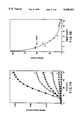
- FIG. 1 Time course of the heat-induced aggregation of acidic aFGF at 55° C. as monitored by turbidity measurements at 350 nm in the presence of varying amounts of heparin.
- FIG. 2 Effect of heparin concentration on the heat-induced aggregation of acidic FGF at 40° C.
- FIG. 3 Effect of heparin concentration on the thermal melting temperature (T m ) of aFGF as measured by fluorescence spectroscopy.
- FIG. 4 Effect of heparin on the copper-catalyzed oxidation of aFGF.
- FIG. 5 Effect of the size (in monomeric hexose units) of well-defined heparin fragments on the thermal melting temperature of aFGF as measured by fluorescence spectroscopy and circular dichroism.
- FIG. 6 Stabilization of aFGF against heat-induced aggregation at 40° C. by various heparin-like molecules.
- FIG. 7 Stabilization of aFGF against heat-induced aggregation at 40° C. by various polyanions.
- FIG. 8 Stabilization of aFGF against heat-induced aggregation at 40° C. by various small sulfated molecules.
- FIG. 9 Effect of the molar ratio of various ligands to aFGF on the thermal denaturation of acidic FGF as measured by fluorescence spectroscopy is shown for inositol hexasulfate in panel A; tetrapolyphosphate in panel B; inositol hexaphosphate in panel C; and sulfated beta-cyclodextrin in panel D.
- FIG. 10 Thermal denaturation of aFGF as a function of the number of phosphate groups on the ligand inositol as measured by fluorescence spectroscopy and circular dichroism.
- the present invention relates to stable topical or parenteral formulations of human acidic fibroblast growth factor (aFGF).
- aFGF human acidic fibroblast growth factor
- the invention further relates to a very diversified group of unique aFGF stabilizers and compositions containing said stabilizers combined with aFGF.
- the stabilized aFGF combinations are useful for wound healing and tissue repair.
- the embodiment of this invention results in a stable formulation that exhibits full mitogenic activity in cell culture and demonstrates full biological activity in vivo to accelerate wound healing.
- Microheterogeneous forms as used herein refer to a single gene product, that is a protein produced from a single gene unit of DNA, which is structurally modified following translation. These structural modifications, however, do not result in any significant alterations of biological activity of the polypeptide.
- Biological activity and biologically active are used interchangeably and are herein defined as the ability of native and recombinant aFGF to stimulate DNA synthesis in quiescent BALB/c 3T3 fibroblasts or to stimulate any of the cell types described in the art or to carry out any of the functions described in the art, most specifically topical wound healing or tissue repair.
- the modifications may take place either in vivo or during the isolation and purification process.
- In vivo modification results in, but is not limited to, acetylation at the N-terminus, proteolysis, glycosylation or phosphorylation.
- Proteolysis may include exoproteolysis wherein one or more terminal amino acids are sequentially, enzymatically cleaved to produce microheterogeneous forms which have fewer amino acids than the original gene product.
- Proteolysis may also include endoproteolytic modification that results from the action of endoproteases which cleave the polypeptide at specific locations within the amino acid sequence. Similar modifications can occur during the purification process which also results in the production of microheterogeneous forms.
- Native aFGF refers to aFGF isolated and purified from tissues or cells that contain aFGF.
- Native human aFGF exists in the following microheterogeneous forms.
- the most preferred microheterogeneous forms of human aFGF include a 154 amino acid form, a 140 amino acid form and a 139 amino acid form.
- the amino acid sequence for the human 139, 140 and 154 amino acid forms of aFGF are described in U.S. Patent No. 4,868,113 and European Patent Application, Publication No. 259,953.
- the various forms of human aFGF can be synthesized by either recombinant biotechnological procedures as described in European Patent Application, Publication No. 259,953 or purified from human tissue as described by Gimenez- Gallego et al., Blochem. Biophys. Res. Commun.
- the recombinant derived, 140 amino add form is the preferred form of aFGF.
- the preferred embodiment of this invention will include recombinant human aFGF produced in microbial cells and will have attached to the first amino add residue of the amino terminus a methionine residue (MetHaFGF).
- MetHaFGF methionine residue
- the most preferred form of human aFGF will be a 141 amino acid form.
- the genes and methods of preparing the aFGF are well known in the art, see above references.
- Native human bFGF also may exist in microheterogeneous forms.
- Basic FGF can be obtained by the methods described in U.S. Pat. Nos. 4,785,079 and 4,956,455.
- the various forms of human bFGF can be synthesized by either recombinant biotechnological procedures as described in Patent Cooperation Treaty/U.S. No. 86/01879 or by Squires et al., J. Biol. Chem. 263:16297-16302 (1988). These procedures can also be used to produce any microheterogeneous form of bFGF which is active as a wound healing agent.
- the preferred embodiment of this invention will include recombinant human bFGF produced in microbial cells and will have attached to the first amino add residue of the amio terminus a methionine residue (MetHbFGF).
- the concentration of aFGF in the following formulations, for topical use is usually within the range of from about 0.1 ng/ml to about 1500 ⁇ g/ml of aqueous formulation (this includes either the initial aqueous formulation or a formulation that has been reconstituted after dehydration).
- the preferred concentration of FGF for topical formulation is from about 25 ⁇ g to about 800 ⁇ g/ml.
- the most preferred concentration of FGF for topical formulations is from about 50 ⁇ g/ml to about 250 ⁇ g/ml.
- the concentration for parenteral use is usually within the range of from about 1 ng/ml to about 1500 ⁇ g/ml of the aqueous formulation (this includes either the initial aqueous formulation of a formulation that has been reconstituted after dehydration.
- the most preferred concentration of FGF for parenteral formulations is from about 25 ⁇ g to about 800 ⁇ g.
- Homogeneously pure human aFGF is not chemically and/or conformationally stable or biologically active without being stabilized.
- Stabilization refers to the addition of chemicals capable of interacting directly with aFGF to maintain a stable and biologically active molecule and chemicals which can maintain stability without direct interaction with aFGF.
- the present invention includes a formulation in which at least one or both types of stabilizing chemicals is present.
- Heparin was purchased from Hepar®, low molecular weight heparin from Calbiochem, and the enzymatically prepared heparin fragments were prepared by procedures disclosed in the following references: Rice and Linhardt; Carbo. Res. 190:219-233 (1989), Hakim and Linhardt, Eletroph. 11: 23-28 (1990), Sharath et al., Immunopharm. 9:73-80 (1985). Sulfated lactobionic acid amide was prepared by the method of Raake et al, Thrombosis. R. 56:719-730 (1989).
- the isomeric purity of the di, tetra and hexasaccharides of heparin is >95% while the octa and deca fragments were mixtures.
- Chemical structures of di, tetra and hexa heparin fragments are as follows:
- ⁇ Ap 4-deoxy- ⁇ -L-threo-hex-4-enopyranosyl uronic acid
- p is pyranose
- GlcA and IdoA are glucuronic and iduronic acid, respectively
- S is sulfate.
- Sulfated and nonsulfated ⁇ - and ⁇ -cyclodextrins were obtained from American Maize. Fucoidan fractions 1 and 2 (precipitates from increasing amounts of organic solvent) were purchased from Kelco. All other sulfated polysaccharides, polyanions and other compounds are purchased from Sigma. Other reagents are purchased commercially and of the highest grade available.
- Stabilization of aFGF can be determined by several independent methods including turbidity measurements, spectroscopic techniques, maintenance of protein mass, mitogenic activity and copper catalyzed oxidation. Since acidic fibroblast growth factor is unstable at physiological temperatures (undergoing large structural changes, including aggregation, which results in a loss of mitogenic and wound healing activity), human aFGF stability can be monitored by evaluating the turbidity of the pharmaceutical composition by following the kinetics of temperature induced aggregation of unfolded protein.
- the kinetics of protein aggregation are monitored by the degree of light scattering at about OD 350 nm using a Perkin Elmer Lambda 6 UV-visible spectrophotometer equipped with a six-cuvette holder. Temperature is controlled by circulating a water/ethylene glycol solution through the cell holder. About 0.9 ml of a PBS solution (6 mM sodium phosphate, 120 mM NaCl, pH 7.2) was placed into a cuvette and incubated at the appropriate temperature within the spectrophotometer. Once equilibrated, 100 ⁇ l of a 1 mg/ml aFGF solution containing the appropriate amount of ligand (stabilizer) is added to the cuvette and mixed manually by inversion. The change in optical density at 350 nm over time is continuously monitored. A dead time of 30s was present due to the mixing of the six samples in each experiment. Aggregate formation was irreversible.
- Circular Dichroism (CD) spectra are measured with an AVIV 62 DS spectropolarimeter.
- Samples of aFGF at about 100 ⁇ g/ml in a PBS buffer (6 mM sodium phosphate, 120 mM NaCl, pH 7.2) containing various ligands are placed into 1 mm pathlength cells and the cell temperature is controlled by circulating a water/ethylene glycol mixture through the cell holder.
- Thermal denaturation is carded out under computer control by increasing the temperature of the water bath in 2° C. increments, followed by a two-minute equilibration period at each temperature to allow the sample to reach thermal equilibrium. Reproducibility of the midpoint of these temperature melting curves (T m ) is ⁇ 2° C.
- Fluorescence spectra are obtained with a Spex Fluorolog-2 spectrofiuorometer using a 1 mm pathlength cuvette. Band passes from 1-2 nm are employed with sample absorptivities maintained below 0.1 at 280 nm. The temperature is controlled either manually or automatically as described above. Reproducibility of thermal denaturation temperatures (T m ) is ⁇ 2° C.
- Size exclusion high performance liquid chromatography is also used to monitor human aFGF stability by determining the per cent protein mass of a sample.
- This technique incorporates a phosphate-cesium chloride mobile phase with detection at about 215 nm. Test samples are diluted to about one to ten (1/10) in mobile phase and the aFGF peak areas are compared to a aFGF standard of known concentration.
- Biological activity of the formulation of the instant invention is determined by a modification of the fibroblast mitogenic assay as described by Linemeyer et al. in European Patent Application, Publication No. 259,953.
- BALB/c 3T3 A31 fibroblasts (American Type Culture Collection) are plated at about 3 ⁇ 10 5 cells per 32 cm 2 wells in culture media containing about 10% heat-inactivated calf serum and incubated in about 7% CO 2 (pH 7.35 ⁇ 0.05). The cells become fully quiescent by replacing the media with serum free media at about 6, about 24 hours and about 48 hours later. At about 53 hours after plating samples of the various formulations are added and about 0.12 ⁇ g of dexamethasone are added.
- each well is supplemented with about 0.4 ⁇ Ci of [methyl- 3 H]-thymidine (20 Ci/mmole, Dupont) and 0.6 ⁇ g of unlabeled thymidine (Sigma), and at 80 hours the cells are processed for determination of radiolabel incorporation into DNA.
- Each dose-response point is the average of at least quadruplicate determinations.
- Other cell types such vascular endothelial cells and corneal endothelial cells can be employed to determine in vitro mitogenicity. The procedures are described in detail by Thomas et al., Proc. Natl. Acad. Sci. USA 82:6409-6431 (1985).
- In vitro mitogenicity is a direct correlate of cell division which can result in tissue growth. It is well known in growth factor research that potent in vitro mitogens are also effective as in vivo growth stimulators.
- Epidermal growth factor (EGF) is a promoter of keratinocyte growth in vitro and also accelerates epidermal regeneration in vivo, Brown et al., J. Exp. Meal. 163: 1319-1324 (1986).
- Insulin-like growth factors also stimulate division and growth of many different cultured cells and also stimulate growth in vivo, Foresch et al., Ann. Rev. Physiol. 47: 443-467 (1985).
- Acidic fibroblast growth factor stimulates various cells to divide in vitro, such as fibroblasts, vascular and comeal endothelial cells, as described above, chondrocytes, osteoblasts, myeloblasts, smooth muscle, glial cells and neuroblasts, European Patent Application, Publication No. 319,052. Thomas et al., Proc. Natl. Acad. Sci. USA 82:6409-6413 (1985), has shown a direct correlation between in vitro mitogenic stimulation and an angiogenic response of chicken egg chorioallantoic membrane which is an example of tissue growth.
- Clotting times are monitored by either a one-stage plasma prothrombin time assay (PT assay) or an activated partial thromboplastin time assay (aPTT) using an automated optical detection system (coag-a-Mate®-XC) by General Diagnostics. Clotting times are measured in the presence of varying amounts of heparin (0-50 ⁇ g heparin/ml plasma) to generate a standard curve and dotting times of other compounds (at equal wt. amounts) are obtained relative to these standardized values.
- PT assay plasma prothrombin time assay
- aPTT activated partial thromboplastin time assay
- Copper-catalyzed oxidation of aFGF is carried out in the following manner.
- Acidic FGF (about 80 ⁇ g/ml) is incubated in a 20 mM Tris, 0.15M potassium chloride, pH 8 solution containing about 20 ⁇ M cuptic chloride for varying periods of time at room temperature.
- the reaction was terminated by the addition of 100 mM EDTA in the same buffer.
- a solution of about 0.7 ml of 0.25M Tris (about pH 8) containing about 2 mM EDTA and about 7 M GuHC1 is then added followed by about 35 ⁇ l of 4 mg/ml of DTNB (Sigma).
- the in vivo animal wound healing model employs genetically diabetic c57GBL/Ks- db+/db+ female mice.
- the assay follows that described by Marsella et al., Wounds: A Compendium of Clinical Research and Practice, 2, (4) July/August 1990, p. 135-147, except that a single 2 cm 2 full thickness wound is used instead of the two 6 mm biopsy wounds described by Marsella. Another difference is that the wounds are covered with a polyurethane dressing.
- Addic FGF is applied to wounds on days 0, 3 and 7.
- Matching placebo formulations are used in a second group of animals. Dressings are changed every three to four days, at which time wound perimeters are traced for assessment of healing. Comparison of healing rate va. a placebo control is made and evaluated for statistical significance at the 90% healed stage.
- the processes as described above are used to identify the human aFGF stabilizers of the present invention.
- the turbidity assay is a rapid screening technique used to examine the ability of a wide variety of excipients and ligands to stabilize aFGF.
- the most promising compounds are then examined by both detailed biophysical studies including, but not limited to, circular dichroism, differential scanning colormetry and fluorescence spectroscopy, and also real-time stability studies (up to 3 months).
- compounds are used to stabilize human aFGF for the testing of efficacy in an in vivo animal wound healing model.
- Stabilizers of human aFGF can be separated into essentially two major categories: specific ligands that directly interact with and stabilize aFGF and nonspecific excipients that either bind weakly to aFGF or exert their stabilizing effect through perturbation of solvent structure.
- the stabilizers of the present invention are specific ligands which are polyanions and can be further grouped as heparin-like molecules, sulfated polysaccharides, small sulfated molecules, phosphorylated polyanions and other highly charged polyanions.
- Heparin-like compounds include, but are not limited to, chondroitin sulfate A, chondroitin sulfate B, chondroitin sulfate C, enzymatically and synthetically prepared heparin fragments consisting of di, tetra, hexa, octa and decasaccharides.
- Sulfated polysaccharides include, but are not limited to, low molecular weight heparin, sulodexide, dextran sulfate, sulfated lactobionic acid amide and sulfated bis-aldonic acid amide, sucrose octasulfate, fucoidan 1, fucoidan 2, sulfated ⁇ -cyclodextrin, sulfated ⁇ -cyclodextrin and pentosan polysulfate.
- Small sulfated compounds include, but are not limited to, inositol hexasulfate, tiron, ammonium sulfate and sodium sulfate.
- compounds such as phosphorylated inositol compounds (especially phytic acid), phosvitin, phosphate buffer, linear and cyclic pollyphosphates (such as (NH 4 ) 6 P 4 O 13 , Na.sub. 5 P 3 O 10 , Na 4 P 2 O 7 and Na 3 P 3 O 9
- polyaspartic acid include, but are not limited to, polyaspartic acid, poly-adenylic-guanylic acid and related compounds, double stranded deoxyribonucleic acid, single stranded deoxyribonucleic acid and poly-glutamic acid.
- Stability of the aFGF formulation of this invention is determined by real time stabilization studies in which sterile samples of the formulation are stored at specific temperatures for periods of time up to 3 months. Accelerated stabilization determinations, as described above, are made by maintaining the formulation of this invention at temperatures between about 400° C. and about 550° C.
- Topical formulations of aFGF may require relative long derreal contact dosing and thus require a formulation which prevents loss of the drug due to run off.
- aFGF combined with a aFGF stabilizer are combined with a polymer which forms a stable viscous solution even after a freeze/thaw cycle at +70° C.
- An acceptable polymer is one which dissolves easily and forms a viscous solution in both water and phosphate buffered saline. The upper concentration limit is about 1.5%.
- the solution (without aFGF) must be able to withstand autoclave sterilization without apparent changes in the inherent properties.
- the final formulation containing aFGF must withstand freeze-thaw cycles without any significant change in viscosity. Indeed, the ideal excipient will be one which results in an elastic moisture-retaining film that remains on the wound for extended periods of time and releases the aFGF into the wound environment.
- the viscous excipients or polymers of the present invention are water soluble polymers such as xanthan gum, alignates or cellulose derivatives such as alkyl celluloses, hydroxyalkyl celluloses and alkylhydroxyalkyl celluloses.
- examples of viscous excipients include methyl cellulose, hydroxyethyl cellulose, carboxymethyl cellulose, hydroxypropyl methylcellulose with hydroxyethyl cellulose (HEC) being preferred.
- the concentration of the preferred excipient, HEC will range form about 0.25% to about 2% on a weight/volume basis with a concentration of about 0.75% to about 1.25% being the most preferred.
- Formulations containing aFGF in combination with an aFGF stabilizer and HEC may not be completely stable at temperatures above about 4° C. for extended periods of time. Indeed, aFGF plus heparin and HEC becomes unstable at room temperature for extended periods of time and loses both in vitro and in vivo activity following extended storage.
- the addition of HEC may cause a destabilization of the aFGF-heparin combination at temperatures above about 4° C. which results in the loss of biological activity.
- the aFGF-stabilizer-HEC combination must be further stabilized by compounds that bind trace metal ions. This is extremely important because aFGF contains three free cysteine residues within the polypeptide chain. Thiol groups can be oxidized spontaneously by atmospheric oxygen.
- the process is catalyzed by trace metal ions, especially copper and iron. Free metal ions must be sequestered and unavailable to catalyze oxidation to maintain stabilized aFGF.
- One way of blocking the metal ions is to add a chelating agent which can bind the free metal ions. Consequently, an integral part of the present invention is the addition of a chelating agent to the combination of aFGF, stabilizer and HEC to maintain the ultimate stability of the combination.
- the chelating agents may include, but are not limited to, ethylenediaminetetraacetic add (EDTA) or EDTA salts and ethyleneglycol-bis( ⁇ -aminoethyl ether) N,N,N', N'-tetraacetic add (EGTA) and EGTA salts along with related compounds.
- the salts include, but are not limited to, calciumdisodium, disodium, tetrasodium, trisodium.
- the preferred chelater is EDTA at a concentration ranging from about 0.05 mM to about 10 mM, with the most preferred concentrations being from about 0.075 mM to about 0.5 mM.
- Weaker chelating agents such as citrate or histidine are not as effective as EDTA or EGTA and the respective salts in stabilizing the aFGF-stabilizer-HEC combination.
- Stabilized, biologically active aFGF is useful in promoting the repair or healing of, but not limited to, soft tissue wounds resulting from burns, cuts or lacerations, and cutaneous ulcerations along with musculo-skeletal wounds such as bone fractures, ligament and tendon tears, and inflammation of bursas and tendons.
- Stabilized aFGF stimulates division in various cell types including fibroblasts, vascular and corneal endothelial cells, tympanic membrane cells and the like rendering stabilized aFGF and bFGF useful for vascular repair, the production of artificial vessels and typanic membrane repair.
- Stabilized aFGF of the present invention is also useful in promoting the healing and regeneration of cartilage and cartilageneous tissue.
- the stabilizers of the present invention are able to maintain aFGF in a biologically active state under adverse conditions such as increased temperature and oxidation.
- a spectrophotometric turbidity assay was developed. This technique monitors the structural stability of aFGF by following the kinetics of temperature induced aggregation of unfolded protein by measurement of the degree of light scattering (turbidity) at 350 nm. The kinetics of protein aggregation were monitored by the degree of light scattering at 350 nm using a Perkin Elmer Lambda 6 UV-visible spectrophotometer equipped with a six-cuvette holder. Temperature was controlled by circulating a water/ethylene glycol solution through the cell holder.
- aFGF undergoes rapid aggregation when heated at 55° C.
- Samples contained 100 ⁇ g/ml aFGF in a PBS buffer at pH 7.2 with the indicated ratio of heparin to aFGF (wt:wt).
- heparin average molecular weight of 16,000
- the stabilization of aFGF is dramatically enhanced with no aggregation observed after 25 minutes at 55° C. in the presence of a 3-fold excess of heparin.
- the rate of aFGF aggregation is strongly dependent on temperature. At lower temperatures, the time course of aFGF aggregation can be followed in the absence of heparin. For example, at 40° C. the rate of aFGF aggregation both with and without polyanions present can be measured as shown in FIG. 2A.
- Samples contained 100 ⁇ g/ml protein in a PBS buffer, pH 7.2 with (a) no heparin, (b) 1.5 ⁇ g/ml, (c) 3 ⁇ g/ml, (d) 5 ⁇ g/ml, (e) 10 ⁇ g/ml and (f) 50 ⁇ g/ml heparin. Only representative data points are shown for clarity.
- the insert displays a representative experiment in which the fluorescence wavelength maximum of acidic FGF is measured as a function of temperature; (a) buffer alone and (b) buffer containing 3X (wt) heparin.
- T m thermal denaturation temperature
- aFGF denatures at 27° C. in a PBS buffer, pH 7.2.
- aFGF thermal stability is significantly enhanced with T m values increasing to 59° C.
- excess heparin is added (3X by weight)
- the maximum stabilization of aFGF is at a T m of 61° C.
- the concentration of heparin necessary to obtain one-half the T m maximum is 0.1X (by weight).
- Acidic FGF contains three free cysteine residues at positions 47, 83 and 117 in the polypeptide chain.
- Site-directed mutagenesis experiments have shown that routants in which serine is substituted for cysteine are more stable and less heparin dependent than wild-type protein, Linemeyer, et al., Growth Factors 3:287-298 (1990). It has also been shown that the copper catalyzed oxidative formation of a disulfide bond in the wild-type aFGF inactivates the protein in an in vitro mitogenic assay, Ortega et al., J. Biol. Chem. 266:5842-5846 (1991).
- heparin can stabilize aFGF against inactivation by oxidation.
- 50-60% of the aFGF's thiol groups are oxidized within several minutes followed by a slower reaction (in which after 6 hours 70-75% of total thiol groups reacted).
- Samples contained 80 ⁇ g/ml aFGF with (a) 3X heparin by weight, (b) 0.1X heparin by weight and (c) buffer alone. As heparin was introduced and its concentration increased, the rate of copper-catalyzed oxidation decreased.
- FIG. 4B illustrates the fraction of remaining reactive thiol groups after a 10-minute incubation with cupric chloride as a function of heparin concentration.
- the data show a heparin concentration dependence similar to that seen in the thermal denaturation experiments with stabilization essentially complete at 1/3 weight levels of heparin.
- T m The T m of aFGF as a function of the molar concentration of the hexasaccharide was measured by fluorescence spectroscopy. Fluorescence spectra were obtained with a Spex Fluorolog-2 spectrofiuorometer using a 1mm pathlength cuvette. Band passes from 1-2 nm were employed with sample absorptivities maintained below 0.1 at 280 nm. The temperature was controlled either manually or automatically as described above. Reproducibility of thermal denaturation temperatures (T m ) was ⁇ 2° C.
- the stabilization of aFGF increases rapidly between 0 and 1 moles ligand/mole aFGF and then slowly plateaus in the presence of excess ligand.
- the 50% stabilization concentration (the value at one-half of the total T m shift) occurs at approximately 0.5 moles heparin hexasaccharide per mole aFGF.
- Sulfated polysaccharides such as low MW heparin, sulodexide, dextran sulfate, fucoidan, and pentosan polysulfate all dramatically stabilize aFGF from heat-induced aggregation. In fact, with many of these compounds, no aggregation was detected within the time course of the experiment. Other sulfated polymers such as polyvinyl sulfate and keratan sulfate are much less effective.
- FIG. 7 shows that negatively charged species as varied as ATP, inorganic phosphates, phosphorylated inositols and polyamino acids (poly-Asp and poly-Glu)can all significantly enhance the thermal stability of aFGF.
- positively charged polymers such as poly-Arg alestabilize aFGF.
- Small sulfated molecules such as inositol hexasulfate also stabilize aFGF against heat-induced aggregation.
- Other small sulfated compounds however, such as a sulfated tyrosine peptide and adenine sulfate actually alestabilize the growth factor (see FIG. 8, [*] indicates no aggregation and [#] indicates very rapid aggregation during the time course of the experiment.
- concentration dependencies seem to fall into three categories.
- Polymers such as heparin, dextran sulfate and phosvitin require molar ratios of polyanion to aFGF substantially below one, suggesting binding of multiple growth factor molecules to a single polymer.
- Small, multiply negatively charged compounds such as tetrapolyphosphate and inositol hexasulfate appear to interact with aFGF with high affinity with only one or a few ligands per aFGF molecule providing the observed stabilization.
- T m maximum vary from 50° C. (tetrapolyphosphate) to 53° C. (inositol hexasulfate) to 56° C. (sulfated ⁇ -cyclodextrin) to 60° C. (phytic add). These values should be compared to those observed with heparin (T m maximum of 61° C.). Since these low molecular weight ligands have well defined molecular weights, the molar excess of ligand to aFGF required for 50% stabilization could be determined as approximately 6, 3.5, 1.5 and 2, respectively.
- inositol hexaphosphate (phytic acid) is more potent in protecting the growth factor from oxidation ( ⁇ 3% vs. 22% of thiol groups lost in 10 minutes).
- concentration of each of the ligands required for maximum protection against oxidation was similar to the amount needed to stabilize aFGF against thermal denaturation.
- Clotting times were monitored by either a one-stage plasma prothrombin time assay (PT assay) or an activated partial thromboplastin time assay (aPTT) using an automated optical detection system (coag-a-Mate*-XC) by General Diagnostics. Clotting times were measured in the presence of varying amounts of heparin (0-50 ⁇ g heparin/ml plasma) to generate a standard curve and dotting times of other compounds (at equal wt. amounts) were obtained relative to these standardized values. The ability of these molecules to lengthen clotting times was roughly proportional to their molecular weight with values from 1/2 to less than 1/10 of heparin.
- Biological activity of the formulation of the instant invention was determined by a modification of the fibroblast mitogenic assay as described by Linemeyer et al. in European Patent Application, Publication No. 259,953.
- BALB/c 3T3 A31 fibroblasts (American Type Culture Collection) were plated at 3 ⁇ 10 5 cells per 32 cm 2 well in culture media containing about 10% heat-inactivated calf serum and incubated in 7% CO 2 (pH 7.35 ⁇ 0.05). The cells become fully quiescent by replacing the media with serum free media at 6, 24 hours and 48 hours later.
- each well was supplemented with 0.4 ⁇ Ci of [methyl- 3 H]-thymidine (20 Ci/mmole, Dupont) and 0.6 ⁇ g of unlabeled thymidine (Sigma), and at 80 hours the cells were processed for determination of radiolabel incorporation into DNA.
- Each dose-response point is the average of at least quadruplicate determinations.
- Other cell types such vascular endothelial cells and corneal endothelial cells can be employed to determine in vitro mitogenidty. The procedures are described in detail by Thomas et al., Proc. Natl. Acad. Sci. USA 82:6409-6431 (1985).
- Acidic fibroblast growth factor was prepared and purified as described above. Sterile aFGF stabilizers were added at the desired concentration to aFGF. This solution was aseptically mixed with a solution of hydroxyethyl cellulose (HEC) and subdivided into previously sterilized 1 ml polyethylene tubes. The tubes were heat sealed and stored at either 5° or 30° C. for various lengths of time.
- HEC hydroxyethyl cellulose
- a formulation containing human aFGF at 100 ⁇ g/ml, 10 mM tetrapolyphosphate, 1% HEC in physiological saline was stored in low density polyethylene tubes for various lengths of times at 30° C. This formulation shows no loss in protein mass or biological activity after storage for 3 months.
- a formulation containing aFGF at 50 ⁇ g/ml combined with sulfated ⁇ -cydodextfin (3X by weight), 1% HEC and 1 mM EDTA in phosphate buffered saline (pH about 7.2) was stored as described above. This formulation was stable for at least 6 months at both 30° C. and 4° C. (there was no loss of protein mass or biological activity). This formulation also showed biological efficacy in the in vivo wound healing model (Example 6).
- Human aFGF as described above was combined with various stabilizers to evaluate wound healing activity of the stabilized aFGF.
- the in vivo animal wound healing model employs genetically diabetic C57GBL/Ks- db+/db + female mice (Jackson Laboratory).
- the assay follows that described by Marsella et. al., Wounds: A Compendium of Clinical Research and Practice, 2, (4) July/August 1990, p. 135-147 except that a single 2 cm 2 full thickness wound is used instead of the two 6 mm biopsy wounds described by Marsella. Another difference is that the wounds are covered with a polyurethane dressing. Acidic FGF is applied to wounds on days 0, 3 and 7. Matching placebo formulations are used in a second group of animals.
- Dressings are changed every three to four days, at which time wound perimeters are traced for assessment of healing. Comparison of healing rate vs. a placebo control is made and evaluated for statistical significance at the 90% healed stage.
- the wound healing capability of various formulations of stabilized aFGF are shown below.
- Both inositol hexasulfate and sulfated ⁇ -cyclodextrin are as efficacious as heparin in stimulating, presumably through stabilization) the wound healing activity of aFGF as manifested in the shortening of healing times.
Abstract
Description
di=Δ∪Ap2S(1→4)-α-D-GlcNp2S6S
tetra=Δ∪Ap2S(1→4)-α-D-GlcNp2S6S(1→4)-.alpha.-L-IdoAp2S(1→4)-α-D-GlcNp2S6S
hexa=Δ∪Ap2S(1→4)-α-D-GlcNp2S6S(1→4)-.alpha.-L-IdoAp2S(1→4)-α-D-GlcNp2S6S(1→4)-α-L-IdoAp2S(1→4)-α-D-GlcNp2S6S
              TABLE 1
______________________________________
Inhibition of heat induced aggregation of acidic FGF in the
presence of
various amounts of stabilizing ligands at 40° C.
                             Approx. Molar
           Approx. MW        Ratio
______________________________________
                        IC50*
                        μg/ml
Heparin      16,000     2        0.03
Low MW heparin
             5,000      3        0.10
Phosvitin    40,000     3        0.02
Dextran sulfate
             8,000      3        0.06
Fucoidin     N/A        6        --
Chondroitin Sulfate B
             N/A        50       --
Inositol hexasulfate
             890        3        0.6
Tetrapolyphosphate
             440        3        1.2
Ammonium sulfate
             132        70       80
Lactobionic acid
             358        180      80
                        IC50*
                        (μM)
Phosphate buffer
             142        6000     1000
Nucleotides
ADP          427        200      33
ATP          551        30       5
AT4P         587        10       1.6
Diadenosine
Nucleotides
Ap3A         756        250      42
Ap4A         836        30       5
Ap5A         916        10       1.6
Ap6A         996        3        0.5
______________________________________
 *IC50 is the concentration of ligand at which the rate of heatinduced
 aggregation (ΔOD.sub.350 /min) of aFGF is 50% of the sample without
 ligand. Experimental conditions are described in FIG. 2.
              TABLE 2
______________________________________
Biophysical and biochemical characterization of aFGF with
stabilizing ligands.
                             Anticoagulant
                             Activity of
                 Cu Oxidation.sup.(b)
                             Ligand.sup.(c)
                 (% SH Lost in
                             (% Heparin
                                      Bio-
Additive  T.sub.m.sup.(a)
                 10 Mins)    by Wt)   activity.sup.(d)
______________________________________
Buffer alone
          45     60%         0        <0.25
Sodium    57     25%         N.P.      0*
Sulfate
MW 142
Tetrapoly-
          66     <3%         +        <0.25
phosphate
MW 440
Inositol  70     <3%         +        <0.6
hexaphosphate
MW 660
Inositol  66     22%         +        2.3
hexasulfate
MW 890
Sucrose   65     N.P.        N.P.     2.5
octasulfate
MW 1300
Sulfated  62     N.P.        N.P.     4.2
lactobionic acid
amide
MW 2600
Sulfated  65     <3%         ++       3.7
β-cyclodextrin
MW 2,500
Sulfated  70      5%         ++       N.P.
γ-cyclodextrin
MW 3,000
Pentosan  60      6%         N.P.m    N.P.
polyphosphate
MW 5,000
Low MW    66     <3%         ++       4.0
heparin
MW 5,000
Dextran sulfate
          62      7%         ++       5.0
MW 8,000
Heparin   64     <3%         + ++     4.2
MW 16,000
______________________________________
 .sup.(a) Thermal melting temperatures were measured by circular dichroism
 Samples contained 100 mg/ml aFGF in PBS buffer pH 7.2 with excess ligand
 (1-3× by weight except for 120 mM sodium sulfate and 10 mM
 tetrapolyphosphate.
 .sup.(b) Percentage of aFGF thiols remaining in solution after 10 min
 incubation with cupric chloride. Experimental conditions are described in
 FIG. 10. All samples contained ligand at 3× (by weight) except
 sodium sulfate (120 mM) and tetrapolyphosphate (10 mM).
 .sup.(c) Anticoagulant activity is expressed as a fraction of activity
 observed with an equal weight of heparin (+ less than 1/10; ++ 1/10 to
 1/2; +++ heparin). No aFGF is present during these measurements.
 .sup.(d) Mitogenic activity of aFGF was measured in the presence of ligan
 indicated in the complete absence of heparin.
 <indicates mitogenic response did not plateau and
 *indicates cell death observed during assay.
 N.P. indicates experiment not performed.
              TABLE 3
______________________________________
DB/DB Mouse Would Healing
Estimated Treatment Group Median
           HT50       HT70     HT90    HT100
Treatment  (Days)     (Days)   (Days)  (Days)
______________________________________
DB/DB Mouse Wound Healing
Estimated Treatment Group Median
aFGF + Hep.
           8.49       10.82    13.76   24.00
aFGF + Inos.
           8.07       10.61    14.19   21.00
aFGF + Sul. β
           8.36       10.32    14.36   22.50
Plac. + Hep.
           8.88       11.75    16.38   24.00
Plac. + Inos.
           9.16       12.76    16.22   24.00
Plac. + Sul. β
           8.78       11.39    15.61   24.00
P-Values (2-sided) for Pairwise Comparisons
aFGF vs. Placebo
Heparin     0.417      0.128    0.068   0.079
Inositol    0.002*     <0.001*  0.094   0.283
Sul. β
            0.055      0.028    0.061   0.203
______________________________________
 Hep. = heparin,
 Inos. = inositol hexasulfate,
 Sul. β = sulfated β-cyclodextrin,
 Plac. = placebo and
 * = two treatments were statistically significantly different (P ≦
 0.05).
Claims (1)
Priority Applications (1)
| Application Number | Priority Date | Filing Date | Title |
|---|---|---|---|
| US07/861,060 US5348941A (en) | 1992-04-01 | 1992-04-01 | Stabilizers for fibroblast growth factors |
Applications Claiming Priority (1)
| Application Number | Priority Date | Filing Date | Title |
|---|---|---|---|
| US07/861,060 US5348941A (en) | 1992-04-01 | 1992-04-01 | Stabilizers for fibroblast growth factors |
Publications (1)
| Publication Number | Publication Date |
|---|---|
| US5348941A true US5348941A (en) | 1994-09-20 |
Family
ID=25334766
Family Applications (1)
| Application Number | Title | Priority Date | Filing Date |
|---|---|---|---|
| US07/861,060 Expired - Lifetime US5348941A (en) | 1992-04-01 | 1992-04-01 | Stabilizers for fibroblast growth factors |
Country Status (1)
| Country | Link |
|---|---|
| US (1) | US5348941A (en) |
Cited By (19)
| Publication number | Priority date | Publication date | Assignee | Title |
|---|---|---|---|---|
| US5580856A (en) * | 1994-07-15 | 1996-12-03 | Prestrelski; Steven J. | Formulation of a reconstituted protein, and method and kit for the production thereof |
| US5827826A (en) * | 1986-03-03 | 1998-10-27 | Rhone-Poulenc Rorer Pharmaceuticals Inc. | Compositions of human endothelial cell growth factor |
| US5942499A (en) * | 1996-03-05 | 1999-08-24 | Orquest, Inc. | Method of promoting bone growth with hyaluronic acid and growth factors |
| US6060461A (en) * | 1999-02-08 | 2000-05-09 | Drake; James Franklin | Topically applied clotting material |
| US6221854B1 (en) | 1996-03-05 | 2001-04-24 | Orquest, Inc. | Method of promoting bone growth with hyaluronic acid and growth factors |
| US6331392B1 (en) | 1997-03-05 | 2001-12-18 | Anadys Pharmaceutical, Inc. | Screen employing fluorescence anisotropy to identify compounds with affinity for nucleic acids |
| US6337183B1 (en) | 1995-09-08 | 2002-01-08 | Scriptgen Pharmaceuticals, Inc. | Screen for compounds with affinity for nucleic acids |
| US20020146409A1 (en) * | 2001-01-30 | 2002-10-10 | Herring Steven W. | Methods for stabilizing lyophilized blood proteins |
| US6566504B2 (en) | 1996-04-19 | 2003-05-20 | Alpha Therapeutic Corporation | Process for viral inactivation of lyophilized blood proteins |
| US6645945B1 (en) | 1996-03-05 | 2003-11-11 | Depuy Acromed, Inc. | Method of treating diseased, injured or abnormal cartilage with hyaluronic acid and growth factors |
| US20080138379A1 (en) * | 2006-11-01 | 2008-06-12 | Jennings-Spring Barbara L | Methods, treatments, and compositions for modulating Hedgehog pathways |
| US20080293624A1 (en) * | 2000-08-31 | 2008-11-27 | Chiron Corporation | Stabilized FGF formulations containing reducing agents |
| US20090214474A1 (en) * | 2006-11-01 | 2009-08-27 | Barbara Brooke Jennings | Compounds, methods, and treatments for abnormal signaling pathways for prenatal and postnatal development |
| US20090221491A1 (en) * | 2006-01-25 | 2009-09-03 | Andrea Neisser-Svae | Purification and Use of a Factor for Supporting Wound Healing |
| US20110207666A1 (en) * | 1996-03-05 | 2011-08-25 | Depuy Spine, Inc. | Method of promoting bone growth with hyaluronic acid and growth factors |
| CN103720642A (en) * | 2012-10-16 | 2014-04-16 | 北京泰德制药股份有限公司 | Bioprotein-containing solution for external use and preparation method thereof |
| CN110151977A (en) * | 2019-05-29 | 2019-08-23 | 新乡医学院 | A kind of protection aFGF protein medicine stability and active method |
| CN113274486A (en) * | 2021-04-15 | 2021-08-20 | 上海腾瑞制药股份有限公司 | Stable acidic fibroblast growth factor preparation and preparation method and application thereof |
| US11096950B2 (en) | 2006-11-01 | 2021-08-24 | Barbara Brooke Jennings | Compounds, methods, and treatments for abnormal signaling pathways for prenatal and postnatal development |
Citations (15)
| Publication number | Priority date | Publication date | Assignee | Title |
|---|---|---|---|---|
| WO1987001728A1 (en) * | 1985-09-12 | 1987-03-26 | Biotechnology Research Partners, Ltd. | Recombinant fibroblast growth factors |
| EP0259953A1 (en) * | 1986-07-11 | 1988-03-16 | Merck & Co. Inc. | Cloning and expression of acidic fibroblast growth factor |
| EP0267015A2 (en) * | 1986-11-05 | 1988-05-11 | Ethicon, Inc. | Stabilized compositions containing epidermal growth factor |
| US4785079A (en) * | 1984-11-09 | 1988-11-15 | The Salk Institute For Biological Studies | Isolation of fibroblast growth factor |
| EP0312208A1 (en) * | 1987-09-18 | 1989-04-19 | Ethicon, Inc. | Gel formulations containing growth factors |
| EP0319052A2 (en) * | 1987-10-22 | 1989-06-07 | Merck & Co. Inc. | Mutant acidic fibroblast growth factor |
| US4868113A (en) * | 1986-03-03 | 1989-09-19 | Rorer Biotechnology, Inc. | Recombinant DNA vector encoding human endothelial cell growth factor |
| EP0345680A1 (en) * | 1988-06-10 | 1989-12-13 | Agfa-Gevaert AG | Column dispenser |
| WO1990003797A1 (en) * | 1988-10-07 | 1990-04-19 | Universite Paris-Val De Marne | Stabilized composition based on growth factors of the family fgf and on dextrane sulfate, and its applications |
| US4956455A (en) * | 1984-03-05 | 1990-09-11 | The Salk Institute For Biological Studies | Bovine fibroblast growth factor |
| EP0392605A1 (en) * | 1989-04-13 | 1990-10-17 | Merck & Co. Inc. | Method of enhancing acidic fibroblast growth factor expression |
| EP0406856A2 (en) * | 1989-07-07 | 1991-01-09 | Takeda Chemical Industries, Ltd. | Stabilized FGF composition and production thereof |
| EP0408146A2 (en) * | 1989-07-13 | 1991-01-16 | Merck & Co. Inc. | Method for the purification of therapeutically active recombinant acidic fibroblast growth factor |
| US5130418A (en) * | 1989-05-02 | 1992-07-14 | California Biotechnology Inc. | Method to stabilize basic fibroblast growth factor |
| US5175147A (en) * | 1988-08-19 | 1992-12-29 | Takeda Chemical Industries, Ltd | Acid-resistant fgf composition and method of treating ulcerating diseases of the gastrointestinal tract |
-
1992
- 1992-04-01 US US07/861,060 patent/US5348941A/en not_active Expired - Lifetime
Patent Citations (15)
| Publication number | Priority date | Publication date | Assignee | Title |
|---|---|---|---|---|
| US4956455A (en) * | 1984-03-05 | 1990-09-11 | The Salk Institute For Biological Studies | Bovine fibroblast growth factor |
| US4785079A (en) * | 1984-11-09 | 1988-11-15 | The Salk Institute For Biological Studies | Isolation of fibroblast growth factor |
| WO1987001728A1 (en) * | 1985-09-12 | 1987-03-26 | Biotechnology Research Partners, Ltd. | Recombinant fibroblast growth factors |
| US4868113A (en) * | 1986-03-03 | 1989-09-19 | Rorer Biotechnology, Inc. | Recombinant DNA vector encoding human endothelial cell growth factor |
| EP0259953A1 (en) * | 1986-07-11 | 1988-03-16 | Merck & Co. Inc. | Cloning and expression of acidic fibroblast growth factor |
| EP0267015A2 (en) * | 1986-11-05 | 1988-05-11 | Ethicon, Inc. | Stabilized compositions containing epidermal growth factor |
| EP0312208A1 (en) * | 1987-09-18 | 1989-04-19 | Ethicon, Inc. | Gel formulations containing growth factors |
| EP0319052A2 (en) * | 1987-10-22 | 1989-06-07 | Merck & Co. Inc. | Mutant acidic fibroblast growth factor |
| EP0345680A1 (en) * | 1988-06-10 | 1989-12-13 | Agfa-Gevaert AG | Column dispenser |
| US5175147A (en) * | 1988-08-19 | 1992-12-29 | Takeda Chemical Industries, Ltd | Acid-resistant fgf composition and method of treating ulcerating diseases of the gastrointestinal tract |
| WO1990003797A1 (en) * | 1988-10-07 | 1990-04-19 | Universite Paris-Val De Marne | Stabilized composition based on growth factors of the family fgf and on dextrane sulfate, and its applications |
| EP0392605A1 (en) * | 1989-04-13 | 1990-10-17 | Merck & Co. Inc. | Method of enhancing acidic fibroblast growth factor expression |
| US5130418A (en) * | 1989-05-02 | 1992-07-14 | California Biotechnology Inc. | Method to stabilize basic fibroblast growth factor |
| EP0406856A2 (en) * | 1989-07-07 | 1991-01-09 | Takeda Chemical Industries, Ltd. | Stabilized FGF composition and production thereof |
| EP0408146A2 (en) * | 1989-07-13 | 1991-01-16 | Merck & Co. Inc. | Method for the purification of therapeutically active recombinant acidic fibroblast growth factor |
Non-Patent Citations (68)
| Title |
|---|
| Baird & Bohlen, Growth Factors 1: 369 418, 1990. * |
| Baird & Bohlen, Growth Factors 1: 369-418, 1990. |
| Brown et al., J. Exp. Med. 163: 1319 1324, 1986. * |
| Brown et al., J. Exp. Med. 163: 1319-1324, 1986. |
| Burgess & Maciag, Annu. Rev. Biochem. 58: 575 606, 1989. * |
| Burgess & Maciag, Annu. Rev. Biochem. 58: 575-606, 1989. |
| Eriksson et al., Proc. Natl. Acad. Sci. USA 88: 3441 3445, 1991. * |
| Eriksson et al., Proc. Natl. Acad. Sci. USA 88: 3441-3445, 1991. |
| Feldhaus, et al., Chemical Abstracts , 83, 1975, Abst. No. 159849u. * |
| Feldhaus, et al., Chemical Abstracts, 83, 1975, Abst. No. 159849u. |
| Foresch et al., Ann. Rev. Physiol. 47: 443 467, 1985. * |
| Foresch et al., Ann. Rev. Physiol. 47: 443-467, 1985. |
| Gimenez Gallego et al., Biochem Biophys. Res. Comm. 138: 611 617, 1986. * |
| Gimenez-Gallego et al., Biochem Biophys. Res. Comm. 138: 611-617, 1986. |
| Gospodarowicz & Cheng, J. Cell Physiol. 128: 475 484, 1986. * |
| Gospodarowicz & Cheng, J. Cell Physiol. 128: 475-484, 1986. |
| Gospodarowicz et al., Endocrine Reviews 8: 95 113, 1987. * |
| Gospodarowicz et al., Endocrine Reviews 8: 95-113, 1987. |
| Gospodarowicz, Clin. Orth. Relat. R. 257:231 248, 1990. * |
| Gospodarowicz, Clin. Orth. Relat. R. 257:231-248, 1990. |
| Guranowski, et al., Chemical Abstracts , 107, 1987, Abst No. 19872k. * |
| Guranowski, et al., Chemical Abstracts, 107, 1987, Abst No. 19872k. |
| Hakim & Linhardt, Electroph. 11: 23 28, 1990. * |
| Hakim & Linhardt, Electroph. 11: 23-28, 1990. |
| Jackson et al., Physiological Reviews 71: 481 539, 1991. * |
| Jackson et al., Physiological Reviews 71: 481-539, 1991. |
| Klagsburn, Cur Opin Cell Biol. 2: 857 863, 1990. * |
| Klagsburn, Cur Opin Cell Biol. 2: 857-863, 1990. |
| Lane & Lindahl, Heparin 25 49, 1989. * |
| Lane & Lindahl, Heparin 25-49, 1989. |
| Linemeyer et al., Growth Factors 3: 287 298, 1990. * |
| Linemeyer et al., Growth Factors 3: 287-298, 1990. |
| Marzella et al., Wounds: A Compendium of Clinical Research & Practice 2: 135 147, 1990. * |
| Marzella et al., Wounds: A Compendium of Clinical Research & Practice 2: 135-147, 1990. |
| Mueller et al., J. Cell Physiol. 140: 439 448, 1989. * |
| Mueller et al., J. Cell Physiol. 140: 439-448, 1989. |
| Ortega et al., J. Biol. Chem. 266: 5842 5846, 1991. * |
| Ortega et al., J. Biol. Chem. 266: 5842-5846, 1991. |
| Raake et al., Thrombosis R. 56: 719 730, 1989. * |
| Raake et al., Thrombosis R. 56: 719-730, 1989. |
| Rice & Linhardt, Carbo. Res. 190: 219 233, 1989. * |
| Rice & Linhardt, Carbo. Res. 190: 219-233, 1989. |
| Riddles, et al., Meth Enzymol. 9: 49 60, 1983. * |
| Riddles, et al., Meth Enzymol. 9: 49-60, 1983. |
| Rosengart et al., Biochem Biophys. Res. Comm. 152: 432 440, 1988. * |
| Rosengart et al., Biochem Biophys. Res. Comm. 152: 432-440, 1988. |
| Sakaguchi et al., J. Biol. Chem. 266: 7270 7278, 1991. * |
| Sakaguchi et al., J. Biol. Chem. 266: 7270-7278, 1991. |
| Sakesla et al., J. Cell Biol. 107: 743 751, 1988. * |
| Sakesla et al., J. Cell Biol. 107: 743-751, 1988. |
| Sharath et al., Immunopharm. 9: 73 80, 1985. * |
| Sharath et al., Immunopharm. 9: 73-80, 1985. |
| Sommer & Rifkin, J. Cell Physiol. 138: 215 220, 1989. * |
| Sommer & Rifkin, J. Cell Physiol. 138: 215-220, 1989. |
| Squires et al., J. Biol. Chem. 263: 16297 16302, 1988. * |
| Squires et al., J. Biol. Chem. 263: 16297-16302, 1988. |
| Tarusono, et al., Chemical Abstracts , 105, 1986, Abst. No. 227204e. * |
| Tarusono, et al., Chemical Abstracts, 105, 1986, Abst. No. 227204e. |
| Thomas et al., Proc. Natl. Acad. Sci. USA 82: 6409 6413, 1985. * |
| Thomas et al., Proc. Natl. Acad. Sci. USA 82: 6409-6413, 1985. |
| Vlodavsky et al., J. Cell. Biochem. 45: 167 176, 1991. * |
| Vlodavsky et al., J. Cell. Biochem. 45: 167-176, 1991. |
| Yayon et al., Cell 64: 841 848, 1991. * |
| Yayon et al., Cell 64: 841-848, 1991. |
| Zhang et al., Proc. Natl. Acad. Scie. USA 88: 3446 3450, 1991. * |
| Zhang et al., Proc. Natl. Acad. Scie. USA 88: 3446-3450, 1991. |
| Zhu et al., Science 251: 90 93, 1991. * |
| Zhu et al., Science 251: 90-93, 1991. |
Cited By (32)
| Publication number | Priority date | Publication date | Assignee | Title |
|---|---|---|---|---|
| US5827826A (en) * | 1986-03-03 | 1998-10-27 | Rhone-Poulenc Rorer Pharmaceuticals Inc. | Compositions of human endothelial cell growth factor |
| US5849538A (en) * | 1986-03-03 | 1998-12-15 | Rhone-Poulenc Rorer Pharmaceuticals Inc. | DNA encoding human endothelial cell growth factors and plasmids comprising said DNA |
| USRE38240E1 (en) | 1986-03-03 | 2003-08-26 | Aventis Pharmaceuticals, Inc. | DNA encoding human endothelial cell growth factors and plasmids comprising said DNA |
| US5580856A (en) * | 1994-07-15 | 1996-12-03 | Prestrelski; Steven J. | Formulation of a reconstituted protein, and method and kit for the production thereof |
| US6337183B1 (en) | 1995-09-08 | 2002-01-08 | Scriptgen Pharmaceuticals, Inc. | Screen for compounds with affinity for nucleic acids |
| US20030113779A1 (en) * | 1995-09-08 | 2003-06-19 | Arenas Jaime E. | Screen for compounds with affinity for nucleic acids |
| US6503721B2 (en) | 1995-09-08 | 2003-01-07 | Anadys Pharmaceuticals, Inc. | Screen for compounds with affinity for nucleic acids |
| US7902172B2 (en) | 1996-03-05 | 2011-03-08 | Depuy Spine, Inc. | Method of promoting bone growth with hyaluronic acid and growth factors |
| US6703377B2 (en) | 1996-03-05 | 2004-03-09 | Depuy Acromed, Inc. | Method of promoting bone growth with hyaluronic acid and growth factors |
| US5942499A (en) * | 1996-03-05 | 1999-08-24 | Orquest, Inc. | Method of promoting bone growth with hyaluronic acid and growth factors |
| US20040176295A1 (en) * | 1996-03-05 | 2004-09-09 | Depuy Acromed, Inc. | Method of promoting bone growth with hyaluronic acid and growth factors |
| US20110207666A1 (en) * | 1996-03-05 | 2011-08-25 | Depuy Spine, Inc. | Method of promoting bone growth with hyaluronic acid and growth factors |
| US6221854B1 (en) | 1996-03-05 | 2001-04-24 | Orquest, Inc. | Method of promoting bone growth with hyaluronic acid and growth factors |
| US6645945B1 (en) | 1996-03-05 | 2003-11-11 | Depuy Acromed, Inc. | Method of treating diseased, injured or abnormal cartilage with hyaluronic acid and growth factors |
| US6566504B2 (en) | 1996-04-19 | 2003-05-20 | Alpha Therapeutic Corporation | Process for viral inactivation of lyophilized blood proteins |
| US20030152992A1 (en) * | 1997-03-05 | 2003-08-14 | Laing Lance G. | Screen employing fluorescence anisotropy to identify compounds with affinity for nucleic acids |
| US6569628B2 (en) | 1997-03-05 | 2003-05-27 | Anadys Pharmaceuticals, Inc. | Screen employing fluorescence anisotropy to identify compounds with affinity for nucleic acids |
| US6331392B1 (en) | 1997-03-05 | 2001-12-18 | Anadys Pharmaceutical, Inc. | Screen employing fluorescence anisotropy to identify compounds with affinity for nucleic acids |
| US6060461A (en) * | 1999-02-08 | 2000-05-09 | Drake; James Franklin | Topically applied clotting material |
| US20080293624A1 (en) * | 2000-08-31 | 2008-11-27 | Chiron Corporation | Stabilized FGF formulations containing reducing agents |
| US7754686B2 (en) | 2000-08-31 | 2010-07-13 | Novartis Vaccines And Diagnostics, Inc. | Stabilized FGF formulations containing reducing agents |
| US20020146409A1 (en) * | 2001-01-30 | 2002-10-10 | Herring Steven W. | Methods for stabilizing lyophilized blood proteins |
| US8962813B2 (en) * | 2006-01-25 | 2015-02-24 | Octapharma Ag | Purification and use of a factor for supporting wound healing |
| US20090221491A1 (en) * | 2006-01-25 | 2009-09-03 | Andrea Neisser-Svae | Purification and Use of a Factor for Supporting Wound Healing |
| US20100261651A9 (en) * | 2006-01-25 | 2010-10-14 | Andrea Neisser-Svae | Purification and Use of a Factor for Supporting Wound Healing |
| US20090214474A1 (en) * | 2006-11-01 | 2009-08-27 | Barbara Brooke Jennings | Compounds, methods, and treatments for abnormal signaling pathways for prenatal and postnatal development |
| US20080138379A1 (en) * | 2006-11-01 | 2008-06-12 | Jennings-Spring Barbara L | Methods, treatments, and compositions for modulating Hedgehog pathways |
| US11096950B2 (en) | 2006-11-01 | 2021-08-24 | Barbara Brooke Jennings | Compounds, methods, and treatments for abnormal signaling pathways for prenatal and postnatal development |
| CN103720642A (en) * | 2012-10-16 | 2014-04-16 | 北京泰德制药股份有限公司 | Bioprotein-containing solution for external use and preparation method thereof |
| CN110151977A (en) * | 2019-05-29 | 2019-08-23 | 新乡医学院 | A kind of protection aFGF protein medicine stability and active method |
| CN110151977B (en) * | 2019-05-29 | 2022-11-29 | 新乡医学院 | Method for protecting stability and activity of aFGF protein medicament |
| CN113274486A (en) * | 2021-04-15 | 2021-08-20 | 上海腾瑞制药股份有限公司 | Stable acidic fibroblast growth factor preparation and preparation method and application thereof |
Similar Documents
| Publication | Publication Date | Title |
|---|---|---|
| US5348941A (en) | Stabilizers for fibroblast growth factors | |
| Volkin et al. | Physical stabilization of acidic fibroblast growth factor by polyanions | |
| Delehedde et al. | Fibroblast growth factor-2 binds to small heparin-derived oligosaccharides and stimulates a sustained phosphorylation of p42/44 mitogen-activated protein kinase and proliferation of rat mammary fibroblasts | |
| Atha et al. | Evaluation of critical groups required for the binding of heparin to antithrombin. | |
| Linhardt et al. | Differential anticoagulant activity of heparin fragments prepared using microbial heparinase. | |
| US20070148158A1 (en) | Rationally designed heparinases derived from heparinase II | |
| Ishihara et al. | Importance of 6-0-sulfate groups of glucosamine residues in heparin for activation of FGF-1 and FGF-2 | |
| Brown et al. | Histidine-rich glycoprotein and platelet factor 4 mask heparan sulfate proteoglycans recognized by acidic and basic fibroblast growth factor | |
| Drozd et al. | Comparison of antithrombin activity of the polysulphate chitosan derivatives in in vivo and in vitro system | |
| Coltrini et al. | Biochemical bases of the interaction of human basic fibroblast growth factor with glycosaminoglycans: new insights from trypsin digestion studies | |
| KR20010030803A (en) | Novel glycosaminoglycan and drug compositions containing the same | |
| US7056504B1 (en) | Rationally designed heparinases derived from heparinase I and II | |
| CA2678168A1 (en) | Low molecular weight heparins including at least one covalent bond with biotin or a biotin derivative, method for making same and use thereof | |
| EP0769502B1 (en) | Process for producing desulfated polysaccharide and desulfated heparin | |
| Peng et al. | Glycosaminoglycans from bovine eye vitreous humour and interaction with collagen type II | |
| Wang et al. | Glycosaminoglycans can influence fibroblast growth factor-2 mitogenicity without significant growth factor binding | |
| Gambarini et al. | Mitogenic activity of acidic fibroblast growth factor is enhanced by highly sulfated oligosaccharides derived from heparin and heparan sulfate | |
| Wang et al. | Characterization, stability, and formulations of basic fibroblast growth factor | |
| Muzzarelli et al. | The blood anticoagulant activity of N-carboxymethylchitosan trisulfate | |
| Rapraeger | Heparan sulfate-growth factor interactions | |
| US20080207895A1 (en) | Methods for synthesizing polysaccharides | |
| CA2171465A1 (en) | Highly sulfated maltooligosaccharides with heparin-like properties | |
| US20050233453A1 (en) | 6-O-sulfated N-acetylheparosan and hematopoietic stem cell growth auxiliary agent | |
| KR20090109105A (en) | Heparins including at least one covalent bond with biotin or a biotin derivative, method for preparing same and use thereof | |
| Bertolesi et al. | Growth inhibition in vitro of murine mammary adenocarcinoma cells by heparin and chemically modified heparins |
Legal Events
| Date | Code | Title | Description |
|---|---|---|---|
| AS | Assignment |
Owner name: MERCK & CO., INC., NEW JERSEY Free format text: ASSIGNMENT OF ASSIGNORS INTEREST;ASSIGNORS:MIDDAUGH, C. RUSSELL;TSAI, PEI-KUO;VOLKIN, DAVID B.;REEL/FRAME:006991/0656 Effective date: 19920519 |
|
| STCF | Information on status: patent grant |
Free format text: PATENTED CASE |
|
| FEPP | Fee payment procedure |
Free format text: PAYOR NUMBER ASSIGNED (ORIGINAL EVENT CODE: ASPN); ENTITY STATUS OF PATENT OWNER: LARGE ENTITY |
|
| FPAY | Fee payment |
Year of fee payment: 4 |
|
| FPAY | Fee payment |
Year of fee payment: 8 |
|
| FPAY | Fee payment |
Year of fee payment: 12 |
|
| AS | Assignment |
Owner name: MERCK SHARP & DOHME CORP.,NEW JERSEY Free format text: CHANGE OF NAME;ASSIGNOR:MERCK & CO., INC.;REEL/FRAME:023852/0595 Effective date: 20091102 Owner name: MERCK SHARP & DOHME CORP., NEW JERSEY Free format text: CHANGE OF NAME;ASSIGNOR:MERCK & CO., INC.;REEL/FRAME:023852/0595 Effective date: 20091102 |
|
| AS | Assignment |
Owner name: SCHERING CORPORATION, NEW JERSEY Free format text: MERGER;ASSIGNOR:MERCK SHARP & DOHME CORP.;REEL/FRAME:028850/0515 Effective date: 20120426 |
|
| AS | Assignment |
Owner name: MERCK SHARP & DOHME CORP., NEW JERSEY Free format text: CHANGE OF NAME;ASSIGNOR:SCHERING CORPORATION;REEL/FRAME:028866/0511 Effective date: 20120502 |