US5484399A - Process and device to reduce interstitial fluid pressure in tissue - Google Patents
Process and device to reduce interstitial fluid pressure in tissue Download PDFInfo
- Publication number
- US5484399A US5484399A US08/181,356 US18135694A US5484399A US 5484399 A US5484399 A US 5484399A US 18135694 A US18135694 A US 18135694A US 5484399 A US5484399 A US 5484399A
- Authority
- US
- United States
- Prior art keywords
- tumor
- interstitial fluid
- fluid pressure
- aspiration
- suction
- Prior art date
- Legal status (The legal status is an assumption and is not a legal conclusion. Google has not performed a legal analysis and makes no representation as to the accuracy of the status listed.)
- Expired - Fee Related
Links
Images


Classifications
-
- A—HUMAN NECESSITIES
- A61—MEDICAL OR VETERINARY SCIENCE; HYGIENE
- A61M—DEVICES FOR INTRODUCING MEDIA INTO, OR ONTO, THE BODY; DEVICES FOR TRANSDUCING BODY MEDIA OR FOR TAKING MEDIA FROM THE BODY; DEVICES FOR PRODUCING OR ENDING SLEEP OR STUPOR
- A61M1/00—Suction or pumping devices for medical purposes; Devices for carrying-off, for treatment of, or for carrying-over, body-liquids; Drainage systems
- A61M1/84—Drainage tubes; Aspiration tips
-
- A—HUMAN NECESSITIES
- A61—MEDICAL OR VETERINARY SCIENCE; HYGIENE
- A61M—DEVICES FOR INTRODUCING MEDIA INTO, OR ONTO, THE BODY; DEVICES FOR TRANSDUCING BODY MEDIA OR FOR TAKING MEDIA FROM THE BODY; DEVICES FOR PRODUCING OR ENDING SLEEP OR STUPOR
- A61M1/00—Suction or pumping devices for medical purposes; Devices for carrying-off, for treatment of, or for carrying-over, body-liquids; Drainage systems
- A61M1/71—Suction drainage systems
- A61M1/74—Suction control
-
- A—HUMAN NECESSITIES
- A61—MEDICAL OR VETERINARY SCIENCE; HYGIENE
- A61M—DEVICES FOR INTRODUCING MEDIA INTO, OR ONTO, THE BODY; DEVICES FOR TRANSDUCING BODY MEDIA OR FOR TAKING MEDIA FROM THE BODY; DEVICES FOR PRODUCING OR ENDING SLEEP OR STUPOR
- A61M25/00—Catheters; Hollow probes
- A61M2025/0001—Catheters; Hollow probes for pressure measurement
- A61M2025/0002—Catheters; Hollow probes for pressure measurement with a pressure sensor at the distal end
-
- A—HUMAN NECESSITIES
- A61—MEDICAL OR VETERINARY SCIENCE; HYGIENE
- A61M—DEVICES FOR INTRODUCING MEDIA INTO, OR ONTO, THE BODY; DEVICES FOR TRANSDUCING BODY MEDIA OR FOR TAKING MEDIA FROM THE BODY; DEVICES FOR PRODUCING OR ENDING SLEEP OR STUPOR
- A61M2205/00—General characteristics of the apparatus
- A61M2205/33—Controlling, regulating or measuring
- A61M2205/3331—Pressure; Flow
- A61M2205/3344—Measuring or controlling pressure at the body treatment site
-
- A—HUMAN NECESSITIES
- A61—MEDICAL OR VETERINARY SCIENCE; HYGIENE
- A61M—DEVICES FOR INTRODUCING MEDIA INTO, OR ONTO, THE BODY; DEVICES FOR TRANSDUCING BODY MEDIA OR FOR TAKING MEDIA FROM THE BODY; DEVICES FOR PRODUCING OR ENDING SLEEP OR STUPOR
- A61M2205/00—General characteristics of the apparatus
- A61M2205/33—Controlling, regulating or measuring
- A61M2205/3331—Pressure; Flow
- A61M2205/3351—Controlling upstream pump pressure
Definitions
- the present invention relates to a method and apparatus for reducing interstitial fluid pressure in tissue, particularly in tumors, and for removing interstitial fluid from confined spaces where drainage of such fluids is impaired, such as to reduce edema accumulation.
- Cancer is the second leading cause of death in this country and still continues to be a public health problem of increasing significance (28). Cancer therapy may be categorized into three major approaches, surgical excision, radiotherapy and chemotherapy. Chemotherapy is defined as the treatment of cancer by a systemic administration of drugs.
- Interstitial fluid accumulation is a known phenomenon in peritumoral areas in the brain and other organs, and where an adequate drainage system such as a lymphatic system is absent.
- This invention is aimed to facilitate clearance of interstitial fluid in circumstances such as brain edema from a variety of causes or lymphedema in the limbs after procedures such as mastectomy or lymphadenectomy.
- Toxicity to normal tissues limits both the dose and frequency of drug administration.
- Antibody targeting specific tumor-associated antigens reduced some of the side effects of chemotherapy. Since the discovery of hybridoma technology by Kohler, G. and Milstein, C. (24), the application of MoAbs has rejuvenated efforts with chemotherapeutic treatment of tumors inaccessible as well as inoperable by moderate techniques.
- the monomer forms of basic immunoglobulin molecules range from 150 to 180 kd (4).
- MoAbs bind very specifically to antigens.
- the theory is to use MoAbs that are specific to tumor-associated antigens.
- MoAbs have been conjugated with radionuclides, toxins, drugs, cytokines, enzymes, effector antibodies, growth factors, biological response modifiers, extracellular matrix peptide products, immunotoxins, lymphokine activated killer cells, and tumor-infiltrating lymphocytes. (19).
- MoAb therapy using conjugated pharmaceuticals has not lived up to early expectations due to problems of hindered transport or delivery of MoAbs to tumors (See, e.g., 19, 21). Numerous factors can affect MoAb delivery. Jain et al. (16, 17, 18, 21) have emphasized the importance of an Interstitial Fluid Pressure (IFP) barrier against the effective transport of macromolecules such as MoAbs.
- IFP Interstitial Fluid Pressure
- the large permeability resulting from the number of fenestrated capillaries and low hyaluronic acid content in the tumor interstitial fluid should, in principle, improve the transport of therapeutic drugs. In reality, however, the delivered fraction of infused dosage has not been up to the expected level. Heterogeneous blood flow, binding hindrance at the cell surface and heterogeneous antigen distribution, particularly in MoAb delivery, are not reasons enough to explain retarded transport in tumor tissue (22).
- IFP Interstitial Fluid Pressure
- a functioning lymphatic system as an anatomical entity has not been demonstrated in the tumor.
- the absence of lymphatics affords no alternative way circulating macromolecules can re-enter the circulation after their extravasation through capillary.
- the potential of fluid flow into the tumor is hindered by the opposing force with equivalent magnitude from IFP. This opposite force increases the IFP until all the forces in Starling's Law are balanced. Consequently, the interstitial fluid within the tumor becomes trapped.
- the fluid with MoAb therapeutic agents has insufficient force to flow into the tumor except for the slow advance by molecular diffusion because of a concentration gradient. Instead, it oozes out toward the tumor surface by the effective convection, diffusion and draining advantage by lymphatics at the tumor periphery. This outward fluid velocity at the tumor periphery additionally hinders the diffusional movement of molecules into the tumor center (19). Decreased intravascular pressure and/or increased interstitial pressure in tumors has been demonstrated by several investigators (1, 9, 14, 31, 37, 41).
- BPR Blood Perfusion Rate
- vasoactive agents such as angiotensin and interleukin.
- radiation and heat treatment has been shown to increase the tumor blood flow.
- a key problem with these approaches is that the increase in blood flow is short-lived and usually confined to well vascularized regions (17, 19, 21).
- Osmotic agents as mannitol have also been used to reduce IFP by exerting higher vascular osmotic pressure (17, 19, 21, 33).
- An object of the present invention is to provide a new mechanical approach to decrease IFP, and/or facilitate clearing of interstitial fluid when such clearing is impaired (e.g., brain edema, lymph edema).
- an artificial lymphatic system ALS
- the object is to enhance convection through the tumor by creating a new pressure gradient within the tumor.
- a suction force is used to create a negative pressure (less than the IFP) inside the tumor.
- the method and apparatus of this invention will be suitable for reducing IFP for inoperable tumors (including brain and liver tumors) as well as operable tumors, and thereby enhance penetration of drugs, in particular macromolecules (e.g. MoAbs), into tumors.
- drugs in particular macromolecules (e.g. MoAbs)
- a process for reducing interstitial fluid pressure in tissue comprising placing an aspiration device within tissue, and applying, to the aspiration device, suction having a pressure value less than the interstitial fluid pressure, to thereby reduce the interstitial fluid pressure within the tissue.
- an apparatus for reducing interstitial fluid pressure in tissue comprising an aspiration device having a plurality of aspiration tubes connected to a manifold means, each of said tubes having a plurality of aspiration holes, and means for connecting the manifold means to a suction source for creating suction inside the aspiration tubes, having a pressure value less than the interstitial fluid pressure to thereby reduce the fluid pressure inside the tissue when the tubes are located in the tissue.
- a process for reducing interstitial pressure in and withdrawing fluid from tissue comprising placing an aspiration device within tissue having excess fluid accumulation, and applying, to the aspiration device, suction having a pressure value less than the interstitial pressure, to thereby reduce interstitial pressure and withdraw fluid from the tissue.
- FIG. 1A is a perspective view of an aspiration device according to the invention for reducing interstitial fluid pressure
- FIG. 1B shows a probe having a spiral or corkscrew shape
- FIG. 2 is a pictorial illustration of a wick-in-needle (WIN) probe
- FIG. 3 is a pictorial illustration of an electrode for clearance of locally generated hydrogen (CLGH);
- FIG. 4 is a graph illustrating interstitial fluid pressure (IFP) variation with respect to radius, before any aspiration
- FIG. 5 is a graph illustrating the relationship between blood perfusion rate (BPR) and radius
- FIG. 6 is a graph illustrating manipulation impacts on interstitial fluid pressure (IFP) with respect to time
- FIG. 7 is a graph illustrating manipulation impacts on blood perfusion rate (BPR) with respect to time.
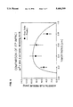
- FIG. 8 is a graph illustrating the interstitial fluid pressure (IFP) distribution profile within the tumor two hours after aspiration.
- IFP interstitial fluid pressure
- a process for reducing interstitial fluid pressure in tissue comprising placing an aspiration device within tissue, and applying, to the aspiration device, suction having a pressure value less than the interstial fluid pressure, to thereby reduce interstitial fluid pressure within the tissue.
- the tissue may be a tumor.
- the step of placing an aspiration device preferably comprises placing an aspiration device having a plurality of aspiration holes.
- the step of placing an aspiration device preferably comprises placing an aspiration device having a plurality of aspiration tubes, each tube having at least one aspiration hole. The number of aspiration tubes and distance therebetween is preferably dependent on the size of the tumor. Each tube preferably has a plurality of aspiration holes.
- the step of applying suction may comprise applying suction having a substantially constant value.
- the process may include measuring the interstitial fluid pressure inside the tissue and regulating the suction applied to the aspiration device in dependence on the measured interstitial fluid pressure.
- the suction applied may decrease in value over time, i.e. be time varying.
- the aspiration device may have means for measuring the interstitial fluid pressure.
- an apparatus for reducing interstitial fluid pressure in tissue comprising an aspiration device having a plurality of aspiration tubes connected to a manifold means, each of said aspiration tubes having a plurality of aspiration holes, and means for connecting the manifold means to a suction source for creating suction, inside the aspiration tubes, having a pressure value less than the interstitial fluid pressure, to thereby reduce the interstitial fluid pressure inside the tissue when the tubes are located in the tissue.
- the aspiration device preferably comprises a plurality of elongated tubes each having a distal end for insertion into a tumor, the elongated tubes being arranged in parallel and being connected at their near ends to a central manifold. There may be five elongated tubes arranged with one central tube and four equally spaced peripheral tubes. The aspiration holes are formed along at least part of the length of the aspiration tubes.
- suction pump means are provided, connected to the manifold means, for creating a suction.
- the suction pump means may be implantable.
- the apparatus may include means for measuring the interstitial fluid pressure inside the tissue. Control means may be provided, connected to the measurement probe and suction source, for regulating the suction created in dependence on the suction measured.
- the suction source may have a pressure value which is substantially constant or time-varying.
- the number of aspiration tubes and distance therebetween may be dependent on the size of the tumor.
- One of the tubes may include means for anchoring the tube inside the tissue, such as by way of having a spiral or corkscrew shape.
- a process for reducing interstitial fluid pressure in and withdrawing fluid from tissue comprising placing an aspiration device within tissue having excess fluid accumulation, and applying, to the aspiration device, suction having a pressure value less than the interstitial fluid pressure, to thereby reduce interstitial fluid pressure and withdraw fluid from the tissue.
- an apparatus for reducing interstitial fluid pressure comprising an aspiration device 10 comprising a plurality of aspiration tubes 12 which terminate in a rounded cone (at the bottom of FIG. 1A).
- the near ends are connected to the manifold 14, which in turn is connected to a suction pump 20.
- the suction pump preferably generates a negative pressure selectively adjustable in the -50 to -100 mmHg range.
- a number of different pumps or pump systems may be used.
- One is the house vacuum in a hospital or the like, the suction for which can be adjustable at the patient area.
- Another pump arrangement is a portable suction pump, which may be the suction side of a suction/delivery pump.
- Yet another is an implantable pump, which can be an osmotic passive pump, or an electronic pump. Other pump arrangements will readily occur to those skilled in the art.
- the aspiration device may also comprise a plurality of tubes or needles each being selectively and individually positionable inside the tissue or tumor.
- the tubes would be connected to a manifold or suction source using flexible conduits. Such an arrangement would enable the tubes to be placed at strategic locations inside the tumor or tissue for the best effects given the particular fluid pressure conditions and tumor/tissue conditions.
- the IFP in a 3 cm diameter tumor has been observed to be 12.3 ⁇ 0.26 mm Hg at its center.
- suction is intended to mean suction pressure value less than the IFP.
- suction would then mean a pressure of ⁇ 12 mm Hg, although a sub atmospheric pressure value of e.g. -50 mm Hg would preferably be used.
- the aspiration device may include a measurement probe 22 for measuring the interstitial fluid pressure.
- This measurement probe 22 could be attached to an aspiration tube 12, such as the central aspiration probe shown, or could replace any one of the aspiration tubes.
- the measurement probe could have holes only at or near the tip, and would be filled with a porous material, such as a sponge, to prevent tissue aspiration.
- the measurement probe 22 could be connected to a gauge 24 indicating the measured pressure, and can be connected to a feedback control means 26, which could control the applied suction of the pump in dependence on the measured pressure.
- control means would be useful not only in keeping the applied suction in a certain range, but could also conserve power, which is especially beneficial if the pump were osmotic.
- the pump could be turned on only when the measured suction pressure fell outside one boundary of the range and turned off when the measured suction pressure fell outside the other boundary of the range.
- the target suction pressure could be substantially constant, or could be time-varying, tapering from on initial value of say -80 mm Hg toward a zero value, the trend being for example linear, exponential or step wise, and could terminate before the zero value is reached.
- the time-varying negative pressure control could be used if the pump were osmotic.
- the aspiration device including tubes, may be made of biocompatible material, such as rigid plastic (e.g. polyurethane or polyethylene) which is typically disposable after use, or metal, such as 316 stainless steel which can be reused. Other material may be used as will occur to those skilled in the art.
- biocompatible material such as rigid plastic (e.g. polyurethane or polyethylene) which is typically disposable after use, or metal, such as 316 stainless steel which can be reused.
- rigid plastic e.g. polyurethane or polyethylene
- metal such as 316 stainless steel which can be reused.
- Other material may be used as will occur to those skilled in the art.
- each tube may be on the order of 2 to 8 cm, and a set could be made available with lengths varying every 0.5 cm, from 2 to 8 cm, for example, for different size tumors.
- the holes may be only along the end of the tube, for example along the 2 cm end of an 8 cm tube, where the tumor has a size of about 2 cm and is 6 cm or more below the entry point of the body.
- the diameter of the tube can be on the order of a 18 or 20 gauge needle. A factor in selecting the diameter is the bloodiness of the tumor. For relatively bloody tumors, an 18 gauge size needle or tube could be used.
- the tubes may be straight, but one or more can also have other shapes, such as a spiral or corkscrew shape as shown in FIG.
- the hole size would be dependent on the size or length of the tubing, and would typically range from 0.1 mm to 0.5 mm. The hole size should be selected to minimize or avoid clogging. Of course, these values are exemplary and other values may be employed.
- the tubes may contain porous packing material to prevent tissue removal. Such a tube would be chosen for necrotic or soft tissue. The intention would be to provide a route for fluid removal or pressure relief. Tissue suction would be potentially dangerous and counter productive, i.e. bleeding, followed by clotting around or within a tube.
- IFP Interstitial Fluid Pressure
- the Blood Perfusion Rate (BPR) was measured using the clearance of locally generated hydrogen (CLGH) technique (2, 21, 25, 36).
- CLGH locally generated hydrogen
- Stosseck et al. (36) devised a method for the measurement of cerebral blood flow using electrochemically generated hydrogen.
- Laser Doppler Flowmetry noninvasively measures capillary blood perfusion parameters (blood flow, volume, and velocity) in real time (8, 26, 35).
- the neuroblastoma was selected as our experimental model because it is a stabilized animal model and has a well-established monoclonal antibody.
- MoAb 3F8 is a murine IgG 3 antibody specific for the disaloganglioside G D2 , an antigen expressed on the surface of a wide spectrum of human tumors including neuroblastoma and melanoma cells (7).
- the MoAb was developed using human cell hybridoma technology by Cheung (6).
- MoAb 3F8 are not rapidly modulated after binding with antigen. Antigenic modulation has been a major obstacle in tumor immunotherapy, especially leukemia, but despite prolonged incubation with 3F8 in vitro, the target antigen GD2 does not undergo modulation (6). These properties provide distinct advantages to MoAb 3F8 for targeting selectively to neuroblastomas in vivo.
- Athymic nude rats ranging from about 250 to about 300 grams body weight were purchased from National Cancer Institute, fed with autoclaved rat diet, and kept under pathogen-free conditions. Suppression of the immune response against the immunologic xenograft was performed using 1.2 ml cyclophosphamide/kg body weight injected i.p. before the day of implantation. Neuroblastoma tissue were harvested from a tumor-bearing rat and chopped into cubes with the volume of 5-10 mm 3 . These tumor tissues were stored in MEM cell culture medium until used. All the surgical instruments were sterilized by steam autoclaving.
- Each animal was anesthetized by i.m. injection of a mixture of xylazine (Mobay Corp., Kansas), 80 mg/kg and ketamine (Parke-Davis, New Jersey), 5 mg/kg body weight.
- a pouch was created under the skin on the caudal muscles of the lower back using surgical scissors.
- the prepared tumor tissue was then inserted into this pouch using forceps.
- a surgical stapler was employed to close the opening.
- the rat was returned to its cage after the application of betadine preps over the surface of tumor for sterilization.
- Tumors were quickly excised and frozen in Freon (La Roche Chemicals, LA) contained in a dry ice bucket.
- the tumor was sliced into 20 ⁇ m thick sections using a cryotome (Bright Inc., New Jersey). The sections were stained with hematoxylin and eosin (H and E), in which nuclear structures are stained dark purple or blue and practically all cytoplasmic structures and intercellular substances are stained pink (27).
- H and E hematoxylin and eosin
- the WIN technique for measuring IFP comprised a thin hypodermic needle (outside diameter 500 ⁇ m) with a 1-2 mm long sidehole was filled with multifilamentous polyester thread (FIG. 2). The wick material was packed to ensure a response time of two minutes or less.
- WIN probes were tested using a water-head pressure regulator and subsequently treated with gas sterilization. Preconditioning was performed by filling the WIN probe with distilled water using a syringe and immersing into a chamber with distilled water a day before use. The probe was calibrated, then positioned by the guiding assembly of a Kopf stereotactic positioner. The WIN probes were advanced slowly downward toward the tumor center. At each 2 mm depth, the pressure was recorded continuously for ten minutes to ensure equilibrium with surrounding pressure.
- Tumor blood flow was measured using a clearance of locally generated hydrogen (CLGH) and Laser Doppler Flowmetry (LDF) methods.
- CLGH was employed to measure radial distribution of Blood Perfusion Rate (BPR).
- BPR Blood Perfusion Rate
- the CLGH method was implemented as follows: A miniaturized electrode was developed as shown FIG. 3 which could be placed in the tissue at the region of interest.
- a Teflon coated 50 ⁇ m platinum wire was inserted through the center of 500 ⁇ m outside diameter (o.d.) stainless steel hypodermic needle and secured within the needle with "2 Ton" Epoxy (Devcon Inc., Massachusetts).
- the barrel of the needle was coated with Epoxylite (Epoxylite Ltd., California) for electrical insulation. The coating was removed from the tip and the assembly was polished.
- the barrel served as our hydrogen generator.
- the cylindrical geometry of generator was a unique fabrication for our experiments. It eliminated the dependence of the mean flow vector to the generator and sensor electrode orientation.
- the concentration measured by the sensor reflects the diffusive-convective transport processes present in the tissue and responsible for carrying the H 2 away from the generator.
- the local tissue becomes saturated, that is, the sensor's response plateaus.
- the generator is turned off and the H 2 concentration declines by the local transport phenomena.
- the time constant reflecting the decline is directly related to an "effective" diffusion coefficient. This coefficient is in turn directly related to the capillary blood flow in the region.
- Quantitative flow measurement requires that the zero-flow diffusion coefficient be measured and then subtracted from the previous measurements. This value is a function of the tissue structure and temperature and was determined for each animal when it was sacrificed.
- the Laser Doppler Flowmeter used in our studies was the BPM403 (Vasamedics, Inc., Minnesota) with needle and combination LDF-CLGH probes. With the combination probes, the correlation of CLGH and LDF could be evaluated. After careful removal of skin over the tumor, the LDF probe was placed using a flexible probe guider. The probe guide had a spiral wire so that it could absorb the respiratory movement artifacts.
- an artificial lymphatic system was employed to remove fluid from tumor's core by imposing suction to the center of the tumor.
- the object is to enhance convection through the tumor by creating a new pressure gradient within the tumor.
- a portable reverse water-head regulator provided a stable and continuous vacuum pressure ranging from about (-50) mmHg to about (-100) mmHg.
- the vacuum pressure was delivered to the center of the tumor by a 20 gauge needle through a rigid tubing system.
- IFP and BPR were measured simultaneously by WIN and LDF methods respectively.
- IFP was measured at the center of the tumor.
- BPR was measured at the surface of the tumor.
- Either angiotensin II, 0.5 ⁇ g/kg or mixture of the angiotensin with mannitol, 1000 mg/kg was infused with a micro-infusion pump for five minutes.
- aspiration manipulation the rat was subject to aspiration after an hour of anesthesia to ensure similar physiological condition with drug groups. The vacuum pressure was continuously observed using a Camino pressure monitor.
- Interstitial Fluid Pressure was measured as a function of tumor size at the center of the tumor. It was observed that IFP increased with the size of the tumor. IFP with respect to size appeared to plateau to 12.3 ⁇ 2.6 mmHg for a 3 cm diameter tumor. The theoretical and experimental IFP were compared to see the model and parameter sensitivity. To generalize the results, the radial variable was normalized to be a dimensionless radius, and the interstitial pressure was also normalized with respect to the effective vascular pressure (see FIG. 4). As a result of these normalizations the pressure profiles can be described in terms of a single parameter ⁇ , which includes the ratio of vascular to interstitial hydraulic conductivities.
- the calculated pressure is highest in the center of a tumor and decreases down to the normal value at the tumor's edge.
- IFP rapidly reaches the effective interstitial pressure which is required to stop transcapillary filtration.
- BPR Blood Perfusion Rate
- FIGS. 6 and 7 show IFP and BPR variation by the manipulation of drug and aspiration method, and shows how these variables change at one spatial point over time (temporal distribution.) Although IFP was decreased and BPR was increased in all three cases, drug groups were not significantly different from the control group when the slopes of decreased tendency were compared by Student's t-test. On the other hand, aspiration group was significantly different from both drug and control group. (p ⁇ 0.05).
- FIG. 8 shows IFP distribution profile within the tumor at one point in time (2 hours after aspiration commenced) at different radius points (spatial distribution). It is clearly seen that by comparing FIGS. 4 and 8, the aspiration reduced the IFP, especially at the center of the tumor.
- the IFP and BPR impact of chemical agents that is, angiotensin II and mannitol, were compared to that of aspiration. While the chemical impact was small and short-term, the aspiration impact with (-80) mmHg showed statistically significant IFP decrease and BPR increase.
Abstract
A method and apparatus are provided for reducing interstitial fluid pressure in tissues, particularly in tumors, by applying suction to the interior of the tissue. The method comprises inserting into the tissue one or more needle-like, elongated tubes, each having at least one hole at or near the end that is inserted into the tissue and each having means to apply suction to the protruding end. The reduced pressure produced inside the tissue by this method is useful to: facilitate penetration by drugs and therapeutic macromolecules into the tissue; enhance radiation treatment of tumors by increasing the oxygen supply in the tumor; and remove fluid to reduce edema in tissues, e.g. brain edema. Means may be provided to measure the pressure within the tissue and to use this measurement to control the suction applied to the tissue through the tubes.
Description
This is a continuation of application Ser. No. 07/842,267, filed Feb. 27, 1992 now abandoned.
The present invention relates to a method and apparatus for reducing interstitial fluid pressure in tissue, particularly in tumors, and for removing interstitial fluid from confined spaces where drainage of such fluids is impaired, such as to reduce edema accumulation.
Throughout this application various publications are referenced and citations are provided in parentheses. The disclosure of these publications in their entireties are hereby incorporated by reference into this application in order to more fully describe the state of the art to which this invention pertains.
Cancer is the second leading cause of death in this country and still continues to be a public health problem of increasing significance (28). Cancer therapy may be categorized into three major approaches, surgical excision, radiotherapy and chemotherapy. Chemotherapy is defined as the treatment of cancer by a systemic administration of drugs.
Unfortunately, most drugs that showed promising effects in vitro have failed to be as effective in vivo, particularly in solid tumors. It has recently been proposed that one of the major reasons for this failure is the impediment of drug transport into tumors. In particular, a physiological barrier created by raised interstitial fluid pressure appears to be responsible. The interstitial fluid pressure is raised in tumors primarily because of the lack of lymphatics in tumors and the growth in confined spaces. The raised interstitial fluid pressure in tumors is a principal transport-retarding factor for the delivery of drugs such as macromolecules, i.e. large molecular weight molecules such as monoclonal antibodies (MoAb), tumor nucrosis factor and other chemotherapeutic agents.
Interstitial fluid accumulation is a known phenomenon in peritumoral areas in the brain and other organs, and where an adequate drainage system such as a lymphatic system is absent. This invention is aimed to facilitate clearance of interstitial fluid in circumstances such as brain edema from a variety of causes or lymphedema in the limbs after procedures such as mastectomy or lymphadenectomy.
Toxicity to normal tissues limits both the dose and frequency of drug administration. Antibody targeting specific tumor-associated antigens reduced some of the side effects of chemotherapy. Since the discovery of hybridoma technology by Kohler, G. and Milstein, C. (24), the application of MoAbs has rejuvenated efforts with chemotherapeutic treatment of tumors inaccessible as well as inoperable by moderate techniques. The monomer forms of basic immunoglobulin molecules range from 150 to 180 kd (4).
Use of MoAbs in the treatment of cancer is potentially very attractive. MoAbs bind very specifically to antigens. In cancer treatment, the theory is to use MoAbs that are specific to tumor-associated antigens. In attempts to enhance or extend therapeutic and diagnostic applications of MoAbs, MoAbs have been conjugated with radionuclides, toxins, drugs, cytokines, enzymes, effector antibodies, growth factors, biological response modifiers, extracellular matrix peptide products, immunotoxins, lymphokine activated killer cells, and tumor-infiltrating lymphocytes. (19).
MoAb therapy using conjugated pharmaceuticals, however, has not lived up to early expectations due to problems of hindered transport or delivery of MoAbs to tumors (See, e.g., 19, 21). Numerous factors can affect MoAb delivery. Jain et al. (16, 17, 18, 21) have emphasized the importance of an Interstitial Fluid Pressure (IFP) barrier against the effective transport of macromolecules such as MoAbs.
The large permeability resulting from the number of fenestrated capillaries and low hyaluronic acid content in the tumor interstitial fluid should, in principle, improve the transport of therapeutic drugs. In reality, however, the delivered fraction of infused dosage has not been up to the expected level. Heterogeneous blood flow, binding hindrance at the cell surface and heterogeneous antigen distribution, particularly in MoAb delivery, are not reasons enough to explain retarded transport in tumor tissue (22).
Jain et al. (19, 21) suspected that elevated Interstitial Fluid Pressure (IFP) is a principal transport retarding factor in MoAb delivery because of the lack of lymphatics in the tumor. In normal tissue, maintenance of the fluid balance within the tissue spaces is dependent upon the interplay of hydrostatic and colloid osmotic pressures operating on a vascular network with uneven permeability and with dissimilar exchange area with the aid of the lymphatic system. The lymphatics are responsible for macromolecular circulation throughout the body. Most macromolecules including proteins cannot return to the circulation without the lymphatic system after they percolate through the endothelial wall (11). Accordingly, lymphatic drainage is a factor of primary importance in maintaining fluid balance (12). Once the equilibrium is disrupted, dehydration or edema in the tissue would result from the imbalanced colloid osmotic and hydrostatic vascular pressure. These relations are well described by Starling's hypothesis (29).
A functioning lymphatic system as an anatomical entity has not been demonstrated in the tumor. The absence of lymphatics affords no alternative way circulating macromolecules can re-enter the circulation after their extravasation through capillary. The potential of fluid flow into the tumor is hindered by the opposing force with equivalent magnitude from IFP. This opposite force increases the IFP until all the forces in Starling's Law are balanced. Consequently, the interstitial fluid within the tumor becomes trapped. The fluid with MoAb therapeutic agents has insufficient force to flow into the tumor except for the slow advance by molecular diffusion because of a concentration gradient. Instead, it oozes out toward the tumor surface by the effective convection, diffusion and draining advantage by lymphatics at the tumor periphery. This outward fluid velocity at the tumor periphery additionally hinders the diffusional movement of molecules into the tumor center (19). Decreased intravascular pressure and/or increased interstitial pressure in tumors has been demonstrated by several investigators (1, 9, 14, 31, 37, 41).
Findings to date indicate that elevated IFP has been attributed to the absence of a well-defined lymphatic system in the tumor (5, 21), and to increased permeability of tumor vessels (16, 20). Researchers reported that IFP increases with tumor size (13, 30, 38, 42). The increase in IFP has also been shown to correlate with reduction in tumor blood flow (lower blood perfusion rate) and the development of necrosis in a growing tumor (13, 15, 30, 38).
Jain et al. (22) presented a mathematical model describing the possible relationship between the distribution of monoclonal antibody and elevated interstitial pressure. They proposed that enhanced IFP might be responsible for the poor penetration of MoAbs into tumors including the heterogeneous blood perfusion, hindered diffusion in the interstitium, and extravascular binding of MoAbs. Furthermore, they stated that the elevated interstitial pressure principally reduces the driving force for extravasation of fluid and macromolecules in tumors, and leads to an experimentally verifiable, radially outward convection which opposed the inward diffusion. They have presented results from several mathematical models and the models' implications (3, 17, 19, 21) to support their hypothesis.
Several investigators (10, 23, 32, 34) have attempted to increase Blood Perfusion Rate (BPR) via administration of vasoactive agents such as angiotensin and interleukin. In addition, radiation and heat treatment has been shown to increase the tumor blood flow. A key problem with these approaches is that the increase in blood flow is short-lived and usually confined to well vascularized regions (17, 19, 21). Osmotic agents as mannitol have also been used to reduce IFP by exerting higher vascular osmotic pressure (17, 19, 21, 33).
An object of the present invention is to provide a new mechanical approach to decrease IFP, and/or facilitate clearing of interstitial fluid when such clearing is impaired (e.g., brain edema, lymph edema). According to the present invention, an artificial lymphatic system (ALS) is employed to reduce IFP within tissue, particularly tumors. The object is to enhance convection through the tumor by creating a new pressure gradient within the tumor. For reducing IFP in tumors, a suction force is used to create a negative pressure (less than the IFP) inside the tumor.
The method and apparatus of this invention will be suitable for reducing IFP for inoperable tumors (including brain and liver tumors) as well as operable tumors, and thereby enhance penetration of drugs, in particular macromolecules (e.g. MoAbs), into tumors.
It is another object of the present invention to reduce interstitial fluid pressure in tumors, to thereby enable blood to be drawn into the tumor, which blood will transport oxygen into the tumor and enhance radiation treatment of the tumor.
It is another object of the present invention to provide an adjunct treatment to chemotherapy involving large macromolecules.
According to one aspect of the present invention, a process for reducing interstitial fluid pressure in tissue is provided, comprising placing an aspiration device within tissue, and applying, to the aspiration device, suction having a pressure value less than the interstitial fluid pressure, to thereby reduce the interstitial fluid pressure within the tissue.
According to another aspect of the present invention, an apparatus for reducing interstitial fluid pressure in tissue is provided, comprising an aspiration device having a plurality of aspiration tubes connected to a manifold means, each of said tubes having a plurality of aspiration holes, and means for connecting the manifold means to a suction source for creating suction inside the aspiration tubes, having a pressure value less than the interstitial fluid pressure to thereby reduce the fluid pressure inside the tissue when the tubes are located in the tissue.
According to another aspect of the invention, a process for reducing interstitial pressure in and withdrawing fluid from tissue is provided, comprising placing an aspiration device within tissue having excess fluid accumulation, and applying, to the aspiration device, suction having a pressure value less than the interstitial pressure, to thereby reduce interstitial pressure and withdraw fluid from the tissue.
Other objects and advantages will become apparent from the detailed description, claims and accompanying drawing.
FIG. 1A is a perspective view of an aspiration device according to the invention for reducing interstitial fluid pressure;
FIG. 1B shows a probe having a spiral or corkscrew shape;
FIG. 2 is a pictorial illustration of a wick-in-needle (WIN) probe;
FIG. 3 is a pictorial illustration of an electrode for clearance of locally generated hydrogen (CLGH);
FIG. 4 is a graph illustrating interstitial fluid pressure (IFP) variation with respect to radius, before any aspiration;
FIG. 5 is a graph illustrating the relationship between blood perfusion rate (BPR) and radius;
FIG. 6 is a graph illustrating manipulation impacts on interstitial fluid pressure (IFP) with respect to time;
FIG. 7 is a graph illustrating manipulation impacts on blood perfusion rate (BPR) with respect to time; and
FIG. 8 is a graph illustrating the interstitial fluid pressure (IFP) distribution profile within the tumor two hours after aspiration.
According to one aspect of the present invention, a process for reducing interstitial fluid pressure in tissue is provided, comprising placing an aspiration device within tissue, and applying, to the aspiration device, suction having a pressure value less than the interstial fluid pressure, to thereby reduce interstitial fluid pressure within the tissue.
The tissue may be a tumor. The step of placing an aspiration device preferably comprises placing an aspiration device having a plurality of aspiration holes. The step of placing an aspiration device preferably comprises placing an aspiration device having a plurality of aspiration tubes, each tube having at least one aspiration hole. The number of aspiration tubes and distance therebetween is preferably dependent on the size of the tumor. Each tube preferably has a plurality of aspiration holes. The step of applying suction may comprise applying suction having a substantially constant value. The process may include measuring the interstitial fluid pressure inside the tissue and regulating the suction applied to the aspiration device in dependence on the measured interstitial fluid pressure. The suction applied may decrease in value over time, i.e. be time varying. The aspiration device may have means for measuring the interstitial fluid pressure.
According to another aspect of the present invention, an apparatus for reducing interstitial fluid pressure in tissue is provided, comprising an aspiration device having a plurality of aspiration tubes connected to a manifold means, each of said aspiration tubes having a plurality of aspiration holes, and means for connecting the manifold means to a suction source for creating suction, inside the aspiration tubes, having a pressure value less than the interstitial fluid pressure, to thereby reduce the interstitial fluid pressure inside the tissue when the tubes are located in the tissue.
The aspiration device preferably comprises a plurality of elongated tubes each having a distal end for insertion into a tumor, the elongated tubes being arranged in parallel and being connected at their near ends to a central manifold. There may be five elongated tubes arranged with one central tube and four equally spaced peripheral tubes. The aspiration holes are formed along at least part of the length of the aspiration tubes. Preferably, suction pump means are provided, connected to the manifold means, for creating a suction. The suction pump means may be implantable. The apparatus may include means for measuring the interstitial fluid pressure inside the tissue. Control means may be provided, connected to the measurement probe and suction source, for regulating the suction created in dependence on the suction measured. The suction source may have a pressure value which is substantially constant or time-varying. The number of aspiration tubes and distance therebetween may be dependent on the size of the tumor. One of the tubes may include means for anchoring the tube inside the tissue, such as by way of having a spiral or corkscrew shape.
According to another aspect of the invention, a process for reducing interstitial fluid pressure in and withdrawing fluid from tissue is provided, comprising placing an aspiration device within tissue having excess fluid accumulation, and applying, to the aspiration device, suction having a pressure value less than the interstitial fluid pressure, to thereby reduce interstitial fluid pressure and withdraw fluid from the tissue.
With reference to FIG. 1A, an apparatus for reducing interstitial fluid pressure is provided, comprising an aspiration device 10 comprising a plurality of aspiration tubes 12 which terminate in a rounded cone (at the bottom of FIG. 1A). The near ends are connected to the manifold 14, which in turn is connected to a suction pump 20. The suction pump preferably generates a negative pressure selectively adjustable in the -50 to -100 mmHg range. A number of different pumps or pump systems may be used. One is the house vacuum in a hospital or the like, the suction for which can be adjustable at the patient area. Another pump arrangement is a portable suction pump, which may be the suction side of a suction/delivery pump. Yet another is an implantable pump, which can be an osmotic passive pump, or an electronic pump. Other pump arrangements will readily occur to those skilled in the art.
The aspiration device may also comprise a plurality of tubes or needles each being selectively and individually positionable inside the tissue or tumor. The tubes would be connected to a manifold or suction source using flexible conduits. Such an arrangement would enable the tubes to be placed at strategic locations inside the tumor or tissue for the best effects given the particular fluid pressure conditions and tumor/tissue conditions.
As discussed below, the IFP in a 3 cm diameter tumor has been observed to be 12.3±0.26 mm Hg at its center. As used herein, the term "suction" is intended to mean suction pressure value less than the IFP. For tumor having an IFP of 12 mm Hg at its center, "suction" would then mean a pressure of <12 mm Hg, although a sub atmospheric pressure value of e.g. -50 mm Hg would preferably be used.
The aspiration device may include a measurement probe 22 for measuring the interstitial fluid pressure. This measurement probe 22 could be attached to an aspiration tube 12, such as the central aspiration probe shown, or could replace any one of the aspiration tubes. The measurement probe could have holes only at or near the tip, and would be filled with a porous material, such as a sponge, to prevent tissue aspiration. The measurement probe 22 could be connected to a gauge 24 indicating the measured pressure, and can be connected to a feedback control means 26, which could control the applied suction of the pump in dependence on the measured pressure.
The control means would be useful not only in keeping the applied suction in a certain range, but could also conserve power, which is especially beneficial if the pump were osmotic. The pump could be turned on only when the measured suction pressure fell outside one boundary of the range and turned off when the measured suction pressure fell outside the other boundary of the range.
The target suction pressure could be substantially constant, or could be time-varying, tapering from on initial value of say -80 mm Hg toward a zero value, the trend being for example linear, exponential or step wise, and could terminate before the zero value is reached. The time-varying negative pressure control could be used if the pump were osmotic.
The aspiration device, including tubes, may be made of biocompatible material, such as rigid plastic (e.g. polyurethane or polyethylene) which is typically disposable after use, or metal, such as 316 stainless steel which can be reused. Other material may be used as will occur to those skilled in the art.
The length of each tube may be on the order of 2 to 8 cm, and a set could be made available with lengths varying every 0.5 cm, from 2 to 8 cm, for example, for different size tumors. Further, the holes may be only along the end of the tube, for example along the 2 cm end of an 8 cm tube, where the tumor has a size of about 2 cm and is 6 cm or more below the entry point of the body. The diameter of the tube can be on the order of a 18 or 20 gauge needle. A factor in selecting the diameter is the bloodiness of the tumor. For relatively bloody tumors, an 18 gauge size needle or tube could be used. The tubes may be straight, but one or more can also have other shapes, such as a spiral or corkscrew shape as shown in FIG. 1B, which would aid in anchoring the probe. The hole size would be dependent on the size or length of the tubing, and would typically range from 0.1 mm to 0.5 mm. The hole size should be selected to minimize or avoid clogging. Of course, these values are exemplary and other values may be employed. The tubes may contain porous packing material to prevent tissue removal. Such a tube would be chosen for necrotic or soft tissue. The intention would be to provide a route for fluid removal or pressure relief. Tissue suction would be potentially dangerous and counter productive, i.e. bleeding, followed by clotting around or within a tube.
The efficacy of the present invention was demonstrated by the following experimental model.
I.1 Measurement of Interstitial Fluid Pressure:
Interstitial Fluid Pressure (IFP) was measured by the Wick-In-Needle (WIN) technique (38, 39, 40), which is described more fully below.
I.2 Measurement of Blood Perfusion Rate:
The Blood Perfusion Rate (BPR) was measured using the clearance of locally generated hydrogen (CLGH) technique (2, 21, 25, 36). Stosseck et al. (36) devised a method for the measurement of cerebral blood flow using electrochemically generated hydrogen.
Temporal changes of BPR were made with the Laser Doppler Flowmetry (LDF) technique. Laser Doppler Flowmetry noninvasively measures capillary blood perfusion parameters (blood flow, volume, and velocity) in real time (8, 26, 35).
I.3 Tumor Selection:
The neuroblastoma was selected as our experimental model because it is a stabilized animal model and has a well-established monoclonal antibody. MoAb 3F8 is a murine IgG3 antibody specific for the disaloganglioside GD2, an antigen expressed on the surface of a wide spectrum of human tumors including neuroblastoma and melanoma cells (7). The MoAb was developed using human cell hybridoma technology by Cheung (6).
MoAb 3F8 are not rapidly modulated after binding with antigen. Antigenic modulation has been a major obstacle in tumor immunotherapy, especially leukemia, but despite prolonged incubation with 3F8 in vitro, the target antigen GD2 does not undergo modulation (6). These properties provide distinct advantages to MoAb 3F8 for targeting selectively to neuroblastomas in vivo.
I.4 Tumor Implantation:
Athymic nude rats ranging from about 250 to about 300 grams body weight were purchased from National Cancer Institute, fed with autoclaved rat diet, and kept under pathogen-free conditions. Suppression of the immune response against the immunologic xenograft was performed using 1.2 ml cyclophosphamide/kg body weight injected i.p. before the day of implantation. Neuroblastoma tissue were harvested from a tumor-bearing rat and chopped into cubes with the volume of 5-10 mm3. These tumor tissues were stored in MEM cell culture medium until used. All the surgical instruments were sterilized by steam autoclaving.
Each animal was anesthetized by i.m. injection of a mixture of xylazine (Mobay Corp., Kansas), 80 mg/kg and ketamine (Parke-Davis, New Jersey), 5 mg/kg body weight. A pouch was created under the skin on the caudal muscles of the lower back using surgical scissors. The prepared tumor tissue was then inserted into this pouch using forceps. A surgical stapler was employed to close the opening. The rat was returned to its cage after the application of betadine preps over the surface of tumor for sterilization.
I.5 Histological Observation:
Tumors were quickly excised and frozen in Freon (La Roche Chemicals, LA) contained in a dry ice bucket. The tumor was sliced into 20 μm thick sections using a cryotome (Bright Inc., New Jersey). The sections were stained with hematoxylin and eosin (H and E), in which nuclear structures are stained dark purple or blue and practically all cytoplasmic structures and intercellular substances are stained pink (27).
I.5 Interstitial Fluid Pressure:
The WIN technique for measuring IFP comprised a thin hypodermic needle (outside diameter 500 μm) with a 1-2 mm long sidehole was filled with multifilamentous polyester thread (FIG. 2). The wick material was packed to ensure a response time of two minutes or less. A polyethylene catheter, PE90, was attached to the opposite end of the WIN probe and connected to the Camino pressure transducer. WIN probes were tested using a water-head pressure regulator and subsequently treated with gas sterilization. Preconditioning was performed by filling the WIN probe with distilled water using a syringe and immersing into a chamber with distilled water a day before use. The probe was calibrated, then positioned by the guiding assembly of a Kopf stereotactic positioner. The WIN probes were advanced slowly downward toward the tumor center. At each 2 mm depth, the pressure was recorded continuously for ten minutes to ensure equilibrium with surrounding pressure.
I.6 Blood Perfusion Rate:
Tumor blood flow was measured using a clearance of locally generated hydrogen (CLGH) and Laser Doppler Flowmetry (LDF) methods. CLGH was employed to measure radial distribution of Blood Perfusion Rate (BPR). LDF was used to evaluate BPR at the surface of tumor with time while manipulating IFP or BPR.
The CLGH method was implemented as follows: A miniaturized electrode was developed as shown FIG. 3 which could be placed in the tissue at the region of interest. As a sensor for cleared hydrogen, a Teflon coated 50 μm platinum wire was inserted through the center of 500 μm outside diameter (o.d.) stainless steel hypodermic needle and secured within the needle with "2 Ton" Epoxy (Devcon Inc., Massachusetts). The barrel of the needle was coated with Epoxylite (Epoxylite Ltd., California) for electrical insulation. The coating was removed from the tip and the assembly was polished. The barrel served as our hydrogen generator. The cylindrical geometry of generator was a unique fabrication for our experiments. It eliminated the dependence of the mean flow vector to the generator and sensor electrode orientation.
The concentration measured by the sensor reflects the diffusive-convective transport processes present in the tissue and responsible for carrying the H2 away from the generator. Several minutes after the generator is turned on, the local tissue becomes saturated, that is, the sensor's response plateaus. At this point, the generator is turned off and the H2 concentration declines by the local transport phenomena. The time constant reflecting the decline is directly related to an "effective" diffusion coefficient. This coefficient is in turn directly related to the capillary blood flow in the region. Quantitative flow measurement requires that the zero-flow diffusion coefficient be measured and then subtracted from the previous measurements. This value is a function of the tissue structure and temperature and was determined for each animal when it was sacrificed.
The Laser Doppler Flowmeter used in our studies was the BPM403 (Vasamedics, Inc., Minnesota) with needle and combination LDF-CLGH probes. With the combination probes, the correlation of CLGH and LDF could be evaluated. After careful removal of skin over the tumor, the LDF probe was placed using a flexible probe guider. The probe guide had a spiral wire so that it could absorb the respiratory movement artifacts.
Interstitial Fluid Pressure and Blood Perfusion Rate Manipulation:
II.1 Purpose:
In our study, chemical methods using angiotensin II and mannitol were tested to examine the possibility of manipulating Interstitial Fluid Pressure (IFP) and Blood Perfusion Rate (BPR) in our tumor model. We also investigated the possibility of IFP reduction using an aspiration system to create an artificial lymphatic system (ALS).
To decrease IFP, an artificial lymphatic system (ALS) was employed to remove fluid from tumor's core by imposing suction to the center of the tumor. The object is to enhance convection through the tumor by creating a new pressure gradient within the tumor. A portable reverse water-head regulator provided a stable and continuous vacuum pressure ranging from about (-50) mmHg to about (-100) mmHg. The vacuum pressure was delivered to the center of the tumor by a 20 gauge needle through a rigid tubing system.
II.2 Experimental Procedures:
In order to examine the impacts of pharmaceutical and mechanical manipulations, IFP and BPR were measured simultaneously by WIN and LDF methods respectively. In evaluating drug effects, IFP was measured at the center of the tumor. However, when aspiration was exerted at the tumor's center, IFP was evaluated at the peripheral. BPR was measured at the surface of the tumor. After a tumor-bearing rat was anesthetized, femoral artery and vein cannulation were performed to measure blood pressure and for drug infusion respectively. Either angiotensin II, 0.5 μg/kg or mixture of the angiotensin with mannitol, 1000 mg/kg was infused with a micro-infusion pump for five minutes. In the case of aspiration manipulation, the rat was subject to aspiration after an hour of anesthesia to ensure similar physiological condition with drug groups. The vacuum pressure was continuously observed using a Camino pressure monitor.
II.3 Results: II.3.a Interstitial Fluid Pressure Characterization:
Interstitial Fluid Pressure (IFP) was measured as a function of tumor size at the center of the tumor. It was observed that IFP increased with the size of the tumor. IFP with respect to size appeared to plateau to 12.3±2.6 mmHg for a 3 cm diameter tumor. The theoretical and experimental IFP were compared to see the model and parameter sensitivity. To generalize the results, the radial variable was normalized to be a dimensionless radius, and the interstitial pressure was also normalized with respect to the effective vascular pressure (see FIG. 4). As a result of these normalizations the pressure profiles can be described in terms of a single parameter α, which includes the ratio of vascular to interstitial hydraulic conductivities. Similar to the experimental data, the calculated pressure is highest in the center of a tumor and decreases down to the normal value at the tumor's edge. In addition, as parameter α increases, IFP rapidly reaches the effective interstitial pressure which is required to stop transcapillary filtration.
The calculated values with α2 =24.4 were the most highly correlated with the experimental data in our neuroblastoma xenograft.
II.3.b Blood Perfusion Rate:
Blood Perfusion Rate (BPR) was 35.8±5.3 ml/min/100 g at the radius [r/R], 0.8 after subtracting zero flow, 12.5±3.8 ml/min/100 g. BPR profiles in neuroblastoma xenograft are shown in FIG. 5. To generalize the results, the BPR and radial axis was normalized with respect to the dimensionless radius and dimensionless BPR. The experimental data profile was fit with theoretical values based on the analytical solution when α2 =24.4.
II.3.c Manipulation Impact on Interstitial Fluid Pressure and Blood Perfusion Rate: Interstitial Fluid Pressure Impact in Chemical Agents:
FIGS. 6 and 7 show IFP and BPR variation by the manipulation of drug and aspiration method, and shows how these variables change at one spatial point over time (temporal distribution.) Although IFP was decreased and BPR was increased in all three cases, drug groups were not significantly different from the control group when the slopes of decreased tendency were compared by Student's t-test. On the other hand, aspiration group was significantly different from both drug and control group. (p<0.05).
FIG. 8 shows IFP distribution profile within the tumor at one point in time (2 hours after aspiration commenced) at different radius points (spatial distribution). It is clearly seen that by comparing FIGS. 4 and 8, the aspiration reduced the IFP, especially at the center of the tumor.
II.4 Conclusions:
The IFP and BPR impact of chemical agents, that is, angiotensin II and mannitol, were compared to that of aspiration. While the chemical impact was small and short-term, the aspiration impact with (-80) mmHg showed statistically significant IFP decrease and BPR increase.
Enhanced Monoclonal Antibody Transport:
Four tumor-bearing rats were divided into two groups: control (no aspiration) and aspiration group (vacuum pressure=(-80 mmHg)). 350 μCi MoAb 3F8 conjugated with I131 was i.v. injected into the control group and into the aspiration group following two hours of aspiration. The aspiration group received an additional six hour aspiration postinjection. On the second day, we performed another six hour aspiration. At 72 hours postinjection of conjugated MoAb, the rats were sacrificed and the tumor's I125 accumulation was measured by scintillography. Control group animals were subjected to identical experimental conditions except that no negative pressure was delivered to the tumor center. The control group animals were placed under general anesthesia and a hyperdermic needle was inserted into the tumor's center. While the sample was relatively small (N=2), the results showed a 75% increase on average in the aspiration group compared to control group. Statistical analysis was not performed because the sample population was too small.
Although a preferred embodiment has been shown and described, numerous variations and modifications will readily occur to these skilled in the art without departing from the spirit and scope of the invention. The invention is not limited to the embodiment illustrated, and its scope is limited only by way of the following claims.
1. Algire, G. H., Legallais, F. Y., Vascular Reactions of Normal and Malignant Tissues In Vivo, IV. The Effect of Peripheral Hypotension on Transplanted Tumors. J. Nat'l. Cancer Inst. 12: 399-421, 1951
2. Aukland, K., et al., Measurement of local blood flow with hydrogen gas. Circulation Res., 14:164, 1964.
3. Baxter, L. T., Jain, R. K., Transport of Fluid and Macromolecules in Tumors: I. Role of Interstitial Pressure and Convection. Microvas. Res. 37: 77-104, 1989.
4. Bogard, W. C., Jr., Dean, R. T., Deo, Y., Fuchs, R., Mattis, J. A., McLean, A. A., Berger, H. J. Practical Considerations in the Production, Purification, and Formulation of Monoclonal Antibodies for Immunoscintigraphy and Immunotherapy. Sem. In Nucl. Med., 19(3): 202-220, 1989.
5. Butler, T. P., Grantham, F. H., Gullino, P. M. Bulk Transfer of Fluid in the Interstitial Compartment of Mammary Tumors. Cancer Res. 35: 512-516, 1975.
6. Cheung, N. K., Saarinen, U. M., Neely, J. E., Landmeier, B., Donovan, D., Coccia, P. F. Monoclonal Antibodies to a Glycolipid Antigen on Human Neuroblastoma Cells. Cancer Res., 45: 2642-2649, 1985.
7. Cliff, W. J. Kinetics of Wound Healing in Rabbit Ear Chambers: A Time Lapse Cinemicroscopic Study. Q.J. Exp. Physiol. Log. Med. Sic., 50: 79-89, 1965.
8. DiResta, G. R., Kiel, J. W., Riedel, G. L., Kaplan, P., Shepherd, A. P. Hybrid Blood Flow Probe for Simultaneous H2 Clearance and Laser-Doppler Velocimetry. Am. J. Physiol., 16: G573-G581, 1987.
9. Eddy, H. A., Casarett, G. W. Development of the Vascular System in the Hamster Malignant Neurilemmoma. Microvasc. Res. 6: 63-82, 1973.
10. Gerlowski, L. E., Jain, R. K. Microvascular Permeability of Normal and Neoplastic Tissues. Microvasc. Res. 31: 288-305, 1986.
11. Hammersen, F. The Terminal Vascular Bed in Skeletal Muscle with Special Regard to the Problem of Shunts, in: Capillary Permeability, Crone, C., Lassen, N. A., eds. Academic Press, 351-365, 1970.
12. Hauck, G. Physiology of the microvascular system. Angiologica 8:236-260, 1971.
13. Hori, K., Suzuki, M., Abe, I., Saito, S. Increased Tumor Pressure in Association With the Growth of Rat Tumors. Japan J. Cancer Res. (Gann) 77: 65-73, 1986.
14. Ide, A. D., Baker, N. H., Warren, S. H. Vascularization of the Brown-Pearce Rabbit Epithelioma Transplant as Seen in the Transparent Ear Chamber. Am. J. Roentgenol. 42: 891-899, 1939.
15. Intaglietta, M., Richardson, D. R., Tompkins, W. R. Blood Pressure, Flow, and Elastic Properties of Microvessels of Cat Omentum. Am. J. Physiol., 221: 922-928, 1971.
16. Jain, R. K. Transport of Molecules in the Tumor Interstitium: A Review. Cancer Res., 47: 3039-3051, 1987.
17. Jain, R. K. Commentary: Delivery of Novel Therapeutic Agents in Tumors: Physiological Barriers and Strategies. J. Nat'l. Cancer Inst., 81: 570-576, 1989.
18. Jain, R. K. Determinants of Tumor Blood Flow; A Review. Cancer Res. 48: 2641-2658, 1988.
19. Jain, R. K. Physiological Barriers to Delivery of Monoclonal Antibodies and Other Macromolecules in Tumors. Cancer Res. (Suppl.), 50: 814s-819s, 1990.
20. Jain, R. K. Transport of Macromolecules in Tumor Micro-Circulation. Biotechnol. Prog., 1: 81-94, 1985.
21. Jain, R. K. Vascular and Interstitial Barriers to Delivery of Therapeutic Agents in Tumors. Cancer and Metas. Rev., 9: 253-266, 1990.
22. Jain, R. K., Baxter, L. T. Mechanisms of Heterogeneous Distribution of Monoclonal Antibodies and Other Macromolecules in Tumors: Significance of Elevated Interstitial Pressure. Cancer Res., 48: 7022-7032, 1988.
23. Jain, R. K., and Wei, J. Dynamics of Drug Transport in Solid Tumors: Distributed Parameter Model. J. Bioengineering, 1: 313-330, 1977.
24. Kohler, G., and Milstein, C., Continuous Cultures of Fused Cells Secreting Antibody of Predefined Specificity. Nature 256:495-497, 1975.
25. Koshu, K., Kamiyama, K., Oka, N., Endo, S., Takaku, A., Saito, T. Measurement of regional blood flow using hydrogen gas generated by electrolysis. Stroke 13:483-487, 1982.
26. Lockwood, A. H., Kenny, P. J. Serial Measurements of Positron-Emitting Isotope Activity in Rat Brain. Stroke, 12(2): 173-176, 1981.
27. Neuwelt, E. A, Sprecht, H. D., Barnett, P. A., Dahlborg, S. A., Miley, A., Larson, S. M., Brown, P., Eckerman, K. F., Hellstrom, K. E., Hellstrom, I. Increased Delivery of Tumor Specific Monoclonal Antibodies to Brain After Osmotic Blood-Brain Barrier Modification in Patients With Melanoma Metastatic to Central Nervous System. Neurosurgery, 20: 885-895, 1987.
28. Newman, M. E. New Cancer Statistics Show Losses, Gains. J. Nat'l. Cancer Inst. 82(15): 1238-1239, 1990.
29. Ogston, A. G., Michel, C. C. General Descriptions of Passive Transport of Neutral Solute and Solvent Through Membranes. Prog. Biophys. Mol. Biol. 34: 197-217, 1978.
30. Paskins-Hurlbart, A. J., Hollenberg, N. K., Abrams, H. L. Tumor Perfusion in Relation to the Rapid Growth Phase and Necrosis, Studies on the Walker Carcinoma in the Rat Testicle. Microv. Res. 24: 15-24, 1982.
31. Peters, W., Teixeira, M., Intaglietta, M., Gross, J. f. Microcirculatory Studies in Rat Mammary Carcinoma, I. Transport Chamber Method, Development of Microvasculature and Pressures in Tumor Vessels. J. Nat'l. Cancer Inst. 65: 631-642, 1980.
32. Phelps, M. E., Hoffman, E. J., Selin, C., Huang, S. C., Robinson, G., McDonald, N., Schelbert, H., Kuhl, D. E. Investigation of [18 F]2-fluoro-2-deoxyglucose for the Measure of Myocardial Glucose Metabolism. J. Nucl. Med., 19: 1311-1319, 1978.
33. Roberts, P. A., Pollay, M., Engles, C., Pendleton, B., Reynolds, E., Stevens, F. A. Effect on Intracranial Pressure of Furosemide Combined With Varying Doses and Administration Rates of Mannitol. J. Neurosurg., 66: 440-446, 1987.
34. Shenkin, H. A., Goluboff, B., Haft, H. The Use of Mannitol for the Reduction of Intracranial Pressure in Intracranial Pressure in Intracranial Surgery. J. Neurosurg., 19: 897-901, 1962 .
35. Shepherd, A. P., Riedel, G. L., Ward, W. F. Laser-Doppler Measurements of Blood Flow Within the Intestinal Wall and on the Surface of the Liver. in: Microcirculation of the Alimentary Tract, edited by Koo, A., et al., Singapore: Word Scientific, p. 115-129, 1983.
36. Stosseck, K., Lubbers D. W., Cottin, N. Determination of local blood flow (microflow) by electrochemically generated hydrogen. Pflugers Archiv--European Journal of Physiology, 348:225-238, 1974.
37. Warren, B. A. The Ultrastructure of the Microcirculation of the Advancing Edge of Walker 256 Carcinoma. Microvasc. Res.,2: 443-453, 1970.
38. Wiig, H., Tveit, E., Hultborn, R., Reed, R. K., Weiss, L. Interstitial Fluid Pressure in DMBA--Induced Rat Mammary Tumors. Scand. J. Clin. Lab. Invest., 42: 159-164, 1982
39. Wikstrand, C. J., and Bigner, D. D. Cancer Research 42: 267-275, 1982.
40. Wilkinson, G. R., Shand, D. G. A Physiological Approach to Hepatic Drug Clearance. Clin. Pharmacol. Ther., 18, 377-390, 1975.
41. Yamaura, H., Sato, H. Quantitative Studies on the Developing Vascular System of Rat Hepatoma. J. Nat'l. Cancer Inst. 53: 1229-1240, 1974.
42. Young, J. S., Griffith, H. D. The Dynamics of Parenchymatous Embolism in Relation to the Dissemination of Malignant Tumors. J. Pathol. Bacteriol., 62: 293-311, 1950.
Claims (9)
1. An apparatus for reducing interstitial fluid pressure in a tumor comprising:
an aspiration device having a plurality of aspiration tubes connected to a manifold means, each of said tubes having a plurality of aspiration holes;
means for connecting the manifold means to a suction source for creating suction inside the aspiration tubes, having a pressure value less than the interstitial fluid pressure but having a pressure value which will not result in removing or damaging the tumor, to thereby reduce the interstitial fluid pressure inside the tumor when the tubes are located in the tumor; and
a measurement probe mounted to one of the plurality of aspiration tubes for measuring the interstitial fluid pressure within the tumor, wherein an aperture of the measurement probe is spaced apart from the aspiration tube to which it is mounted at a sufficient distance so that a pressure measured by the measurement probe is not substantially affected by the suction present at the plurality of aspiration holes of the plurality of aspiration tubes.
2. The apparatus according to claim 1, further comprising:
control means connected to said measurement probe; and
a suction source operatively connected to said control means being responsive to a signal sent therefrom, said suction source being adapted to regulate the suction created in dependence on the signal sent from the control means, said signal being indicative of the pressure measured by the measurement probe, wherein the suction has a pressure value less than the interstitial fluid pressure, to thereby reduce the interstitial fluid pressure within the tissue.
3. A process for reducing interstitial fluid pressure in a tumor comprising:
placing an aspiration device within the tumor; and
applying, to the aspiration device, suction having a pressure value less than the interstitial fluid pressure but having a pressure value which will not result in removing or damaging the tumor, to thereby reduce interstitial fluid pressure within the tumor,
wherein the step of placing an aspiration device comprises locating within the tumor an aspiration device of diameter not greater than that of an 18 gauge needle having a plurality of aspiration holes, and wherein the pressure value of the suction applied in the step of applying suction is greater than -100 mmHg.
4. The process according to claim 3, wherein the step of placing an aspiration device comprises locating an aspiration device having a plurality of aspiration tubes for locating in the tumor, each tube having at least one aspiration hole.
5. The process according to claim 4, wherein each tube has a plurality of aspiration holes.
6. The process of claim 3, used in conjunction with chemotherapy to enhance treatment of inoperable tumors.
7. The process of claim 3, used in conjunction with radiation therapy to enhance treatment of inoperable tumors.
8. A process for reducing interstitial fluid pressure in a tumor comprising:
placing an aspiration device within the tumor;
applying, to the aspiration device, suction having a pressure value less than the interstitial fluid pressure but having a pressure value which will not result in removing or damaging the tumor, to thereby reduce interstitial fluid pressure within the tumor;
measuring the interstitial fluid pressure within the tumor; and
regulating suction applied to the aspiration device in dependence on the measured interstitial pressure wherein the suction has a pressure value less than the interstitial fluid pressure but has a pressure value which will not result in removing or damaging the tumor, to thereby reduce the interstitial fluid pressure within the tumor.
9. A process for reducing interstitial fluid pressure in a tumor comprising:
placing an aspiration device within the tumor; and
applying, to the aspiration device, suction having a pressure value less than the interstitial fluid pressure but having a pressure value which will not result in removing or damaging the tumor, to thereby reduce interstitial fluid pressure within the tumor,
wherein the aspiration device has means for measuring the interstitial fluid pressure, and wherein through control means connected to the means for measuring the interstitial fluid pressure and suction source, the suction created may be regulated in dependence on the measured interstitial fluid pressure so that the suction is maintained at a pressure value below the interstitial fluid pressure.
Priority Applications (1)
| Application Number | Priority Date | Filing Date | Title |
|---|---|---|---|
| US08/181,356 US5484399A (en) | 1992-02-27 | 1994-01-13 | Process and device to reduce interstitial fluid pressure in tissue |
Applications Claiming Priority (2)
| Application Number | Priority Date | Filing Date | Title |
|---|---|---|---|
| US84226792A | 1992-02-27 | 1992-02-27 | |
| US08/181,356 US5484399A (en) | 1992-02-27 | 1994-01-13 | Process and device to reduce interstitial fluid pressure in tissue |
Related Parent Applications (1)
| Application Number | Title | Priority Date | Filing Date |
|---|---|---|---|
| US84226792A Continuation | 1992-02-27 | 1992-02-27 |
Publications (1)
| Publication Number | Publication Date |
|---|---|
| US5484399A true US5484399A (en) | 1996-01-16 |
Family
ID=25286909
Family Applications (1)
| Application Number | Title | Priority Date | Filing Date |
|---|---|---|---|
| US08/181,356 Expired - Fee Related US5484399A (en) | 1992-02-27 | 1994-01-13 | Process and device to reduce interstitial fluid pressure in tissue |
Country Status (1)
| Country | Link |
|---|---|
| US (1) | US5484399A (en) |
Cited By (48)
| Publication number | Priority date | Publication date | Assignee | Title |
|---|---|---|---|---|
| US5983136A (en) * | 1996-09-17 | 1999-11-09 | Deka Products Limited Partnership | System for delivery of drugs by transport |
| WO2000048669A1 (en) * | 1999-02-18 | 2000-08-24 | Biovalve Technologies, Inc. | Electroactive pore |
| WO2001093930A1 (en) * | 2000-06-02 | 2001-12-13 | The University Of Utah Research Foundation | Active needle devices with integrated functionality |
| US6406455B1 (en) | 1998-12-18 | 2002-06-18 | Biovalve Technologies, Inc. | Injection devices |
| US6537264B1 (en) * | 1996-06-18 | 2003-03-25 | Alza Corp | Device and method for enhancing transdermal flux of agents being sampled |
| WO2003028786A2 (en) * | 2001-09-28 | 2003-04-10 | Jan Otto Solem | A method, a device, and a system for organ reconditioning and a device for preserving an internal bodyorgan |
| US6547777B2 (en) * | 2000-02-17 | 2003-04-15 | Sloan-Kettering Institute For Cancer Research | Apparatus and method for reducing interstitial fluid pressure and enhancing delivery of a therapeutic agent |
| US20030129084A1 (en) * | 1998-11-20 | 2003-07-10 | Intuitive Surgical, Inc. | Cardiopulmonary bypass device and method |
| US20030167031A1 (en) * | 1997-08-08 | 2003-09-04 | Odland Rick Mathew | System and method for site specific therapy |
| US20030181824A1 (en) * | 2002-03-22 | 2003-09-25 | Odland Rick Mathew | System for treating tissue swelling |
| US20040253708A1 (en) * | 1996-01-09 | 2004-12-16 | Genentech, Inc. | Apo-2 ligand |
| US20050089958A1 (en) * | 1996-01-09 | 2005-04-28 | Genentech, Inc. | Apo-2 ligand |
| US20060122575A1 (en) * | 2002-06-11 | 2006-06-08 | Akio Wakabayashi | System and efficient drainage of body cavity |
| US7077822B1 (en) * | 1994-02-09 | 2006-07-18 | The University Of Iowa Research Foundation | Stereotactic hypothalamic obesity probe |
| US20070060834A1 (en) * | 2005-08-23 | 2007-03-15 | Odland Rick M | Catheter control console |
| US20080132819A1 (en) * | 2006-11-28 | 2008-06-05 | Boehringer Technologies, Lp | Tunnel dressing for use with negative pressure wound therapy system |
| US20080208171A1 (en) * | 2007-02-23 | 2008-08-28 | Argenta Louis C | Device and method for removing edema |
| US20080221503A1 (en) * | 2007-03-08 | 2008-09-11 | Mark Chak | Method of treating diseases |
| US20080243044A1 (en) * | 2000-05-09 | 2008-10-02 | Kenneth Hunt | Abdominal wound dressing |
| US20080294147A1 (en) * | 2003-08-28 | 2008-11-27 | Boehringer Technologies, L.P. | Device for treating a wound |
| US20100022990A1 (en) * | 2008-07-25 | 2010-01-28 | Boehringer Technologies, L.P. | Pump system for negative pressure wound therapy and improvements thereon |
| US20100069829A1 (en) * | 2008-09-18 | 2010-03-18 | George Hutchinson | Therapy delivery systems and methods |
| US20100106187A1 (en) * | 2008-10-29 | 2010-04-29 | Keith Patrick Heaton | Modular, reduced-pressure, wound-closure systems and methods |
| US20100106116A1 (en) * | 2008-10-29 | 2010-04-29 | Tyler Simmons | Medical canister connectors |
| US20100179377A1 (en) * | 2007-09-28 | 2010-07-15 | Vitrolife Sweden Ab | Sampling needle |
| US20100179516A1 (en) * | 2006-06-02 | 2010-07-15 | Surgical Design Solutions, Llc | Assemblies, systems, and methods for vacuum assisted internal drainage during wound healing |
| US7815616B2 (en) | 2002-09-16 | 2010-10-19 | Boehringer Technologies, L.P. | Device for treating a wound |
| US20110086005A1 (en) * | 2008-05-27 | 2011-04-14 | Oncolytics Biotech Inc. | Modulating interstitial pressure and oncolytic viral delivery and distribution |
| US7931651B2 (en) | 2006-11-17 | 2011-04-26 | Wake Lake University Health Sciences | External fixation assembly and method of use |
| WO2011083266A1 (en) * | 2010-01-08 | 2011-07-14 | Assistance Publique - Hôpitaux De Paris | Drainage device and uses |
| US7981098B2 (en) | 2002-09-16 | 2011-07-19 | Boehringer Technologies, L.P. | System for suction-assisted wound healing |
| WO2011092379A1 (en) | 2010-01-29 | 2011-08-04 | Hld Healthy Life Devices Oy | Method and device for measuring tissue pressure |
| US8267960B2 (en) | 2008-01-09 | 2012-09-18 | Wake Forest University Health Sciences | Device and method for treating central nervous system pathology |
| US8377016B2 (en) | 2007-01-10 | 2013-02-19 | Wake Forest University Health Sciences | Apparatus and method for wound treatment employing periodic sub-atmospheric pressure |
| US8551075B2 (en) | 2006-06-02 | 2013-10-08 | Kci Medical Resources | Assemblies, systems, and methods for vacuum assisted internal drainage during wound healing |
| EP2670451A2 (en) * | 2011-02-04 | 2013-12-11 | The Research Foundation Of State University Of New York | Compositions and methods for removing ascitic fluid from the abdomen |
| WO2014062679A1 (en) * | 2012-10-19 | 2014-04-24 | Boston Scientific Scimed, Inc. | Lymphedema medical device |
| US8715267B2 (en) | 2006-06-02 | 2014-05-06 | Kci Medical Resources | Assemblies, systems, and methods for vacuum assisted internal drainage during wound healing |
| US8834520B2 (en) | 2007-10-10 | 2014-09-16 | Wake Forest University | Devices and methods for treating spinal cord tissue |
| US9289193B2 (en) | 2008-07-18 | 2016-03-22 | Wake Forest University Health Sciences | Apparatus and method for cardiac tissue modulation by topical application of vacuum to minimize cell death and damage |
| US20210153848A1 (en) * | 2019-11-21 | 2021-05-27 | Imcomet B.V. | Skin cancer treatment |
| US11090460B2 (en) * | 2015-03-31 | 2021-08-17 | Surefire Medical, Inc. | Method for infusing an immunotherapy agent to a solid tumor for treatment |
| US11338117B2 (en) | 2018-10-08 | 2022-05-24 | Trisalus Life Sciences, Inc. | Implantable dual pathway therapeutic agent delivery port |
| WO2022155666A1 (en) * | 2021-01-15 | 2022-07-21 | Nxt Biomedical, Llc | Methods and devices for assessing and modifying physiologic status via the interstitial space |
| US11400263B1 (en) | 2016-09-19 | 2022-08-02 | Trisalus Life Sciences, Inc. | System and method for selective pressure-controlled therapeutic delivery |
| US11419610B2 (en) | 2018-08-17 | 2022-08-23 | Empress Medical, Inc. | Device and method for passing tension member around tissue mass |
| US11419634B2 (en) | 2018-08-17 | 2022-08-23 | Empress Medical, Inc. | Causing ischemia in tumors |
| US11850398B2 (en) | 2018-08-01 | 2023-12-26 | Trisalus Life Sciences, Inc. | Systems and methods for pressure-facilitated therapeutic agent delivery |
Citations (10)
| Publication number | Priority date | Publication date | Assignee | Title |
|---|---|---|---|---|
| US3191600A (en) * | 1962-05-04 | 1965-06-29 | Hazen F Everett | Blood suction apparatus |
| US3430628A (en) * | 1965-02-23 | 1969-03-04 | Reatha L Wiggins | Plurality of aspirators |
| FR2309242A1 (en) * | 1975-05-02 | 1976-11-26 | Pistor Michel | Medical instrument for area treatments in body - with distributor for use with syringe and several needle probes |
| US4192319A (en) * | 1977-09-30 | 1980-03-11 | Regents Of University Of California | Wick catheter pressure sensing probe and method of use |
| US4423740A (en) * | 1981-03-30 | 1984-01-03 | Howmedica, Inc. | Slit catheter method for measuring interstitial pressure |
| US4692153A (en) * | 1986-04-03 | 1987-09-08 | Berlin Richard B | Surgical wound drain device |
| US4739769A (en) * | 1985-02-07 | 1988-04-26 | Board Of Reagents Of The University Of Michigan | Tissue pressure measurement transducer system |
| US4792328A (en) * | 1983-06-11 | 1988-12-20 | Walter Beck | Method and appparatus for aspirating secreted fluids from a wound |
| US4878900A (en) * | 1988-07-27 | 1989-11-07 | Sundt Thoralf M | Surgical probe and suction device |
| US4913698A (en) * | 1987-10-26 | 1990-04-03 | Marui Ika Company, Limited | Aqua-stream and aspirator for brain surgery |
-
1994
- 1994-01-13 US US08/181,356 patent/US5484399A/en not_active Expired - Fee Related
Patent Citations (10)
| Publication number | Priority date | Publication date | Assignee | Title |
|---|---|---|---|---|
| US3191600A (en) * | 1962-05-04 | 1965-06-29 | Hazen F Everett | Blood suction apparatus |
| US3430628A (en) * | 1965-02-23 | 1969-03-04 | Reatha L Wiggins | Plurality of aspirators |
| FR2309242A1 (en) * | 1975-05-02 | 1976-11-26 | Pistor Michel | Medical instrument for area treatments in body - with distributor for use with syringe and several needle probes |
| US4192319A (en) * | 1977-09-30 | 1980-03-11 | Regents Of University Of California | Wick catheter pressure sensing probe and method of use |
| US4423740A (en) * | 1981-03-30 | 1984-01-03 | Howmedica, Inc. | Slit catheter method for measuring interstitial pressure |
| US4792328A (en) * | 1983-06-11 | 1988-12-20 | Walter Beck | Method and appparatus for aspirating secreted fluids from a wound |
| US4739769A (en) * | 1985-02-07 | 1988-04-26 | Board Of Reagents Of The University Of Michigan | Tissue pressure measurement transducer system |
| US4692153A (en) * | 1986-04-03 | 1987-09-08 | Berlin Richard B | Surgical wound drain device |
| US4913698A (en) * | 1987-10-26 | 1990-04-03 | Marui Ika Company, Limited | Aqua-stream and aspirator for brain surgery |
| US4878900A (en) * | 1988-07-27 | 1989-11-07 | Sundt Thoralf M | Surgical probe and suction device |
Non-Patent Citations (90)
| Title |
|---|
| Algire, G. H., Legallais, F. Y., Journal of the National Cancer Institute , (1951), vol. 12, No. 1, pp. 399 421, (Exhibit 2). * |
| Algire, G. H., Legallais, F. Y., Journal of the National Cancer Institute, (1951), vol. 12, No. 1, pp. 399-421, (Exhibit 2). |
| Aukland K., et al., Circulation Research , (1964), vol. 14, pp. 164 187, (Exhibit 3). * |
| Aukland K., et al., Circulation Research, (1964), vol. 14, pp. 164-187, (Exhibit 3). |
| Baxter, L. T., and Jain, R. K., Microvascular Research , (1990), vol. 40, pp. 246 263, (Exhibit 40). * |
| Baxter, L. T., and Jain, R. K., Microvascular Research , (1991), vol. 41, pp. 252 272, (Exhibit 42). * |
| Baxter, L. T., and Jain, R. K., Microvascular Research , (1991), vol. 41, pp. 5 23, (Exhibit 41). * |
| Baxter, L. T., and Jain, R. K., Microvascular Research, (1990), vol. 40, pp. 246-263, (Exhibit 40). |
| Baxter, L. T., and Jain, R. K., Microvascular Research, (1991), vol. 41, pp. 252-272, (Exhibit 42). |
| Baxter, L. T., and Jain, R. K., Microvascular Research, (1991), vol. 41, pp. 5-23, (Exhibit 41). |
| Baxter, L. T., Jain, R. K., Microvascular Research , (1989), vol. 37, pp. 77 104, (Exhibit 4). * |
| Baxter, L. T., Jain, R. K., Microvascular Research, (1989), vol. 37, pp. 77-104, (Exhibit 4). |
| Boucher, Y., Baxter, L. T., Jain, R. K., Cancer Research , (1990), vol. 50, pp. 4478 4484, (Exhibit 43). * |
| Boucher, Y., Baxter, L. T., Jain, R. K., Cancer Research, (1990), vol. 50, pp. 4478-4484, (Exhibit 43). |
| Boucher, Y., et al., Cancer Research , (1991), vol. 51, No. 24, pp. 6691 6694, (Exhibit 44). * |
| Boucher, Y., et al., Cancer Research, (1991), vol. 51, No. 24, pp. 6691-6694, (Exhibit 44). |
| Butler, T. P., et al., Cancer Research , (1975), vol. 35, pp. 512 516, (Exhibit 5). * |
| Butler, T. P., et al., Cancer Research, (1975), vol. 35, pp. 512-516, (Exhibit 5). |
| Cheung, N. K. V., et al., Cancer Research , (1985), vol. 45, pp. 2642 2649, (Exhibit 6). * |
| Cheung, N. K. V., et al., Cancer Research, (1985), vol. 45, pp. 2642-2649, (Exhibit 6). |
| Cliff, W. J., Quarterly Journal of Experimental Physiology And Cognate Medical Sciences , (1965), vol. 50, pp. 79 89, (Exhibit 7). * |
| Cliff, W. J., Quarterly Journal of Experimental Physiology And Cognate Medical Sciences, (1965), vol. 50, pp. 79-89, (Exhibit 7). |
| DiResta, G. R., et al., American Journal of Physiology , (1987), vol. 16, No. 4, pp. G573 G581, (Exhibit 8). * |
| DiResta, G. R., et al., American Journal of Physiology, (1987), vol. 16, No. 4, pp. G573-G581, (Exhibit 8). |
| Eddy, H. A., Casarett, G. W., Microvascular Research , (1973), vol. 6, No. 1, pp. 63 82, (Exhibit 9). * |
| Eddy, H. A., Casarett, G. W., Microvascular Research, (1973), vol. 6, No. 1, pp. 63-82, (Exhibit 9). |
| Gerlowski, L. E., Jain, R. K., Microvascular Research , (1986), vol. 31, No. 3, pp. 288 305, (Exhibit 10). * |
| Gerlowski, L. E., Jain, R. K., Microvascular Research, (1986), vol. 31, No. 3, pp. 288-305, (Exhibit 10). |
| Hammersen, F., in: Capillary Permeability , Crone, C., Lassen, N. A., editors, Academic Press, New York, (1970), pp. 351 365, (Exhibit 11). * |
| Hammersen, F., in: Capillary Permeability, Crone, C., Lassen, N. A., editors, Academic Press, New York, (1970), pp. 351-365, (Exhibit 11). |
| Hauck, G., Angiologica , (1971), vol. 8, pp. 236 260, (Exhibit 12). * |
| Hauck, G., Angiologica, (1971), vol. 8, pp. 236-260, (Exhibit 12). |
| Hori, K., et al., Japanese Journal Center Research ( Gann ), (1986), vol. 77, No. 1, pp. 65 73, (Exhibit 13). * |
| Hori, K., et al., Japanese Journal Center Research (Gann), (1986), vol. 77, No. 1, pp. 65-73, (Exhibit 13). |
| Ide, A. G., et al., The American Journal of Roentgenology and Radium Therapy , (1939), vol. 42, No. 6, pp. 891 899, (Exhibit 14). * |
| Ide, A. G., et al., The American Journal of Roentgenology and Radium Therapy, (1939), vol. 42, No. 6, pp. 891-899, (Exhibit 14). |
| Intaglietta, M., et al., American Journal of Physiology , (1971), vol. 221, No. 3, pp. 922 928, (Exhibit 15). * |
| Intaglietta, M., et al., American Journal of Physiology, (1971), vol. 221, No. 3, pp. 922-928, (Exhibit 15). |
| Jain, R. K. Baxter, L. T., Cancer Research , (1988), vol. 48, pp. 7022 7032, (Exhibit 22). * |
| Jain, R. K. Baxter, L. T., Cancer Research, (1988), vol. 48, pp. 7022-7032, (Exhibit 22). |
| Jain, R. K., Biotechnology Progress , (1985), vol. 1, No. 2, pp. 81 94, (Exhibit 20). * |
| Jain, R. K., Biotechnology Progress, (1985), vol. 1, No. 2, pp. 81-94, (Exhibit 20). |
| Jain, R. K., Cancer and Metastasis Reviews , (1990), vol. 9, pp. 253 266, (Exhibit 21). * |
| Jain, R. K., Cancer and Metastasis Reviews, (1990), vol. 9, pp. 253-266, (Exhibit 21). |
| Jain, R. K., Cancer Research , (1987), vol. 47, No. 12, pp. 3039 3051, (Exhibit 16). * |
| Jain, R. K., Cancer Research , (1988), vol. 48, pp. 2641 2658, (Exhibit 18). * |
| Jain, R. K., Cancer Research , (Supplement), (1990), vol. 50, No. 3, pp. 814s 819s, (Exhibit 19). * |
| Jain, R. K., Cancer Research, (1987), vol. 47, No. 12, pp. 3039-3051, (Exhibit 16). |
| Jain, R. K., Cancer Research, (1988), vol. 48, pp. 2641-2658, (Exhibit 18). |
| Jain, R. K., Cancer Research, (Supplement), (1990), vol. 50, No. 3, pp. 814s-819s, (Exhibit 19). |
| Jain, R. K., Journal of the National Cancer Institute , (1989), vol. 81, No. 8, pp. 570 576, (Exhibit 17). * |
| Jain, R. K., Journal of the National Cancer Institute, (1989), vol. 81, No. 8, pp. 570-576, (Exhibit 17). |
| K hler, G., and Milstein, C., Nature , (1975), vol. 256, No. 5517, pp. 495 497, (Exhibit 23). * |
| Kohler, G., and Milstein, C., Nature, (1975), vol. 256, No. 5517, pp. 495-497, (Exhibit 23). |
| Koshu, K., et al., Stroke , (1982), vol. 13, No. 4, pp. 483 487, (Exhibit 24). * |
| Koshu, K., et al., Stroke, (1982), vol. 13, No. 4, pp. 483-487, (Exhibit 24). |
| Leunig, M., et al., Cancer Research , (1992), vol. 52, pp. 487 490, (Exhibit 46). * |
| Leunig, M., et al., Cancer Research, (1992), vol. 52, pp. 487-490, (Exhibit 46). |
| Lockwood, A. H., Kemp, P. J., Stroke , (1981), vol. 12, No. 2, pp. 173 176, (Exhibit 25). * |
| Lockwood, A. H., Kemp, P. J., Stroke, (1981), vol. 12, No. 2, pp. 173-176, (Exhibit 25). |
| Neuwelt, E. A, et al., Neurosurgery , (1987), vol. 20, No. 6, pp. 885 895, (Exhibit 26). * |
| Neuwelt, E. A, et al., Neurosurgery, (1987), vol. 20, No. 6, pp. 885-895, (Exhibit 26). |
| Ogston, A. G., Michel, C. C., in Progress In Biophysics And Molecular Biology , Noble, D., editor, (1978), vol. 34, pp. 197 217, (Exhibit 27). * |
| Ogston, A. G., Michel, C. C., in Progress In Biophysics And Molecular Biology, Noble, D., editor, (1978), vol. 34, pp. 197-217, (Exhibit 27). |
| Paskins Hurlburt, A. J., et al., Microvascular Research , (1982), vol. 24, pp. 15 24, (Exhibit 28). * |
| Paskins-Hurlburt, A. J., et al., Microvascular Research, (1982), vol. 24, pp. 15-24, (Exhibit 28). |
| Peters, W., et al., Journal of the National Cancer Institute , (1980), vol. 65, No. 3, pp. 631 642, (Exhibit 29). * |
| Peters, W., et al., Journal of the National Cancer Institute, (1980), vol. 65, No. 3, pp. 631-642, (Exhibit 29). |
| Phelps, M. E., et al., Journal of Nuclear Medicine , (1978), vol. 19, No. 12, pp. 1311 1319, (Exhibit 30). * |
| Phelps, M. E., et al., Journal of Nuclear Medicine, (1978), vol. 19, No. 12, pp. 1311-1319, (Exhibit 30). |
| Roberts, P. A., et al., Journal of Neurosurgery , (1987), vol. 66, pp. 440 446, (Exhibit 31). * |
| Roberts, P. A., et al., Journal of Neurosurgery, (1987), vol. 66, pp. 440-446, (Exhibit 31). |
| Roh, H. D., et al., Cancer Research , (1991), vol. 51, No. 24, pp. 6695 6698, (Exhibit 45). * |
| Roh, H. D., et al., Cancer Research, (1991), vol. 51, No. 24, pp. 6695-6698, (Exhibit 45). |
| Shenkin, H. A., et al., Journal of Neurosurgery , (1962), vol. 19, No. 10, pp. 897 901, (Exhibit 32). * |
| Shenkin, H. A., et al., Journal of Neurosurgery, (1962), vol. 19, No. 10, pp. 897-901, (Exhibit 32). |
| Stosseck, K., L bbers D. W., Cottin, N., Pfugers Archiv European Journal of Physiology , (1974), vol. 348, pp. 225 238, (Exhibit 33). * |
| Stosseck, K., Lubbers D. W., Cottin, N., Pfugers Archiv--European Journal of Physiology, (1974), vol. 348, pp. 225-238, (Exhibit 33). |
| Warren, B. A., Microvascular Research , (1970), vol. 2, pp. 443 453, (Exhibit 34). * |
| Warren, B. A., Microvascular Research, (1970), vol. 2, pp. 443-453, (Exhibit 34). |
| Wiig, H., et al., Scandinavian Journal of Clinical & Laboratory Investigation , (1982), vol. 42, No. 2, pp. 159 164, (Exhibit 35). * |
| Wiig, H., et al., Scandinavian Journal of Clinical & Laboratory Investigation, (1982), vol. 42, No. 2, pp. 159-164, (Exhibit 35). |
| Wikstrand, C. J., and Bigner, D. D., Cancer Research , (1982), vol. 42, No. 1, pp. 267 275, (Exhibit 36). * |
| Wikstrand, C. J., and Bigner, D. D., Cancer Research, (1982), vol. 42, No. 1, pp. 267-275, (Exhibit 36). |
| Wilkinson, G. R., Shand, D. G., Clinical Pharmacology and Therapeutics , (1975), vol. 18, No. 4, pp. 377 390, (Exhibit 37). * |
| Wilkinson, G. R., Shand, D. G., Clinical Pharmacology and Therapeutics, (1975), vol. 18, No. 4, pp. 377-390, (Exhibit 37). |
| Yamaura, H., Sato, H., Journal of the National Cancer Institute , (1974), vol. 53, No. 5, pp. 1229 1240, (Exhibit 38). * |
| Yamaura, H., Sato, H., Journal of the National Cancer Institute, (1974), vol. 53, No. 5, pp. 1229-1240, (Exhibit 38). |
| Young, J. S., Griffith, H. D., Journal of Pathology and Bacteriology , (1950), vol. 62, No. 3, pp. 293 311, (Exhibit 39). * |
| Young, J. S., Griffith, H. D., Journal of Pathology and Bacteriology, (1950), vol. 62, No. 3, pp. 293-311, (Exhibit 39). |
Cited By (115)
| Publication number | Priority date | Publication date | Assignee | Title |
|---|---|---|---|---|
| US7077822B1 (en) * | 1994-02-09 | 2006-07-18 | The University Of Iowa Research Foundation | Stereotactic hypothalamic obesity probe |
| US20110155605A1 (en) * | 1996-01-09 | 2011-06-30 | Genentech, Inc. | Apo-2 ligand |
| US20070036782A1 (en) * | 1996-01-09 | 2007-02-15 | Genentech, Inc. | Apo-2 ligand |
| US6998116B1 (en) * | 1996-01-09 | 2006-02-14 | Genentech, Inc. | Apo-2 ligand |
| US20050089958A1 (en) * | 1996-01-09 | 2005-04-28 | Genentech, Inc. | Apo-2 ligand |
| US20040253708A1 (en) * | 1996-01-09 | 2004-12-16 | Genentech, Inc. | Apo-2 ligand |
| US7285533B2 (en) | 1996-01-09 | 2007-10-23 | Genentech, Inc. | Apo-2 ligand |
| US20080176788A1 (en) * | 1996-01-09 | 2008-07-24 | Genentech, Inc. | Apo-2 ligand |
| US20090162364A1 (en) * | 1996-01-09 | 2009-06-25 | Genentech, Inc. | Apo-2 ligand |
| US6537264B1 (en) * | 1996-06-18 | 2003-03-25 | Alza Corp | Device and method for enhancing transdermal flux of agents being sampled |
| US20070118070A1 (en) * | 1996-06-18 | 2007-05-24 | Cormier Michel J | Device with anchoring elements for transdermal delivery or sampling of agents |
| US7184826B2 (en) * | 1996-06-18 | 2007-02-27 | Alza Corporation | Device and method for enhancing transdermal flux of agents being delivered or sampled |
| US5983136A (en) * | 1996-09-17 | 1999-11-09 | Deka Products Limited Partnership | System for delivery of drugs by transport |
| US20100286586A1 (en) * | 1997-08-08 | 2010-11-11 | Twin Star Medical, Inc. | System and method for site specific therapy |
| US7717871B2 (en) | 1997-08-08 | 2010-05-18 | Twin Star Medical, Inc. | System and method for site specific therapy |
| US20030167031A1 (en) * | 1997-08-08 | 2003-09-04 | Odland Rick Mathew | System and method for site specific therapy |
| US20030129084A1 (en) * | 1998-11-20 | 2003-07-10 | Intuitive Surgical, Inc. | Cardiopulmonary bypass device and method |
| US6979423B2 (en) * | 1998-11-20 | 2005-12-27 | Intuitive Surgical | Cardiopulmonary bypass device and method |
| US20050154350A1 (en) * | 1998-12-18 | 2005-07-14 | Biovalve Technologies, Inc. | Injection devices |
| US7740607B2 (en) | 1998-12-18 | 2010-06-22 | Valeritas, Inc. | Modular units for use in an injection device |
| US6406455B1 (en) | 1998-12-18 | 2002-06-18 | Biovalve Technologies, Inc. | Injection devices |
| US7187969B2 (en) | 1999-02-18 | 2007-03-06 | Biovalve Technologies, Inc. | Electroactive pore |
| US6490483B2 (en) | 1999-02-18 | 2002-12-03 | Biovalve Technologies, Inc. | Electroactive pore |
| US20040006300A1 (en) * | 1999-02-18 | 2004-01-08 | Willis John P. | Electroactive pore |
| US6314317B1 (en) | 1999-02-18 | 2001-11-06 | Biovalve Technologies, Inc. | Electroactive pore |
| WO2000048669A1 (en) * | 1999-02-18 | 2000-08-24 | Biovalve Technologies, Inc. | Electroactive pore |
| US20030149407A1 (en) * | 2000-02-17 | 2003-08-07 | Sloan-Kettering Institute For Cancer Research | Apparatus and method for reducing interstitial fluid pressure and enhancing delivery of a therapeutic agent |
| US6547777B2 (en) * | 2000-02-17 | 2003-04-15 | Sloan-Kettering Institute For Cancer Research | Apparatus and method for reducing interstitial fluid pressure and enhancing delivery of a therapeutic agent |
| US8187210B2 (en) | 2000-05-09 | 2012-05-29 | Kci Licensing, Inc | Abdominal wound dressing |
| US7951100B2 (en) | 2000-05-09 | 2011-05-31 | Kci Licensing Inc. | Abdominal wound dressing |
| US20080243044A1 (en) * | 2000-05-09 | 2008-10-02 | Kenneth Hunt | Abdominal wound dressing |
| US20110224635A1 (en) * | 2000-05-09 | 2011-09-15 | Kenneth Hunt | Abdominal wound dressing |
| US20040176732A1 (en) * | 2000-06-02 | 2004-09-09 | Frazier A Bruno | Active needle devices with integrated functionality |
| US7473244B2 (en) * | 2000-06-02 | 2009-01-06 | The University Of Utah Research Foundation | Active needle devices with integrated functionality |
| US20090069697A1 (en) * | 2000-06-02 | 2009-03-12 | The University Of Utah Research Foundation | Active microneedles and microneedle arrays |
| WO2001093930A1 (en) * | 2000-06-02 | 2001-12-13 | The University Of Utah Research Foundation | Active needle devices with integrated functionality |
| WO2003028786A3 (en) * | 2001-09-28 | 2003-11-06 | Jan Otto Solem | A method, a device, and a system for organ reconditioning and a device for preserving an internal bodyorgan |
| US20030109855A1 (en) * | 2001-09-28 | 2003-06-12 | Solem Jan Otto | Method, a device, and a system for organ reconditioning and a device for preserving an internal body organ |
| WO2003028786A2 (en) * | 2001-09-28 | 2003-04-10 | Jan Otto Solem | A method, a device, and a system for organ reconditioning and a device for preserving an internal bodyorgan |
| US20050165342A1 (en) * | 2002-03-22 | 2005-07-28 | Odland Rick M. | Method and system for treating tissue swelling |
| US20030181824A1 (en) * | 2002-03-22 | 2003-09-25 | Odland Rick Mathew | System for treating tissue swelling |
| US6942633B2 (en) | 2002-03-22 | 2005-09-13 | Twin Star Medical, Inc. | System for treating tissue swelling |
| US6942634B2 (en) | 2002-03-22 | 2005-09-13 | Twin Star Medical, Inc. | System for treating tissue swelling |
| US20060122575A1 (en) * | 2002-06-11 | 2006-06-08 | Akio Wakabayashi | System and efficient drainage of body cavity |
| US8439893B2 (en) * | 2002-06-11 | 2013-05-14 | Medela Holding Ag | System and method for efficient drainage of body cavity |
| US7815616B2 (en) | 2002-09-16 | 2010-10-19 | Boehringer Technologies, L.P. | Device for treating a wound |
| US7981098B2 (en) | 2002-09-16 | 2011-07-19 | Boehringer Technologies, L.P. | System for suction-assisted wound healing |
| US7942866B2 (en) | 2003-08-28 | 2011-05-17 | Boehringer Technologies, L.P. | Device for treating a wound |
| US20080294147A1 (en) * | 2003-08-28 | 2008-11-27 | Boehringer Technologies, L.P. | Device for treating a wound |
| US20070060834A1 (en) * | 2005-08-23 | 2007-03-15 | Odland Rick M | Catheter control console |
| US20100179516A1 (en) * | 2006-06-02 | 2010-07-15 | Surgical Design Solutions, Llc | Assemblies, systems, and methods for vacuum assisted internal drainage during wound healing |
| US10695469B2 (en) | 2006-06-02 | 2020-06-30 | Ic Surgical, Inc. | Assemblies, systems, and methods for vacuum assisted internal drainage during wound healing |
| US9814808B2 (en) | 2006-06-02 | 2017-11-14 | Ic Surgical, Inc. | Assemblies, systems, and methods for vacuum assisted internal drainage during wound healing |
| US8715267B2 (en) | 2006-06-02 | 2014-05-06 | Kci Medical Resources | Assemblies, systems, and methods for vacuum assisted internal drainage during wound healing |
| US8585683B2 (en) | 2006-06-02 | 2013-11-19 | Kci Medical Resources | Assemblies, systems, and methods for vacuum assisted internal drainage during wound healing |
| US8551075B2 (en) | 2006-06-02 | 2013-10-08 | Kci Medical Resources | Assemblies, systems, and methods for vacuum assisted internal drainage during wound healing |
| US20110202059A1 (en) * | 2006-11-17 | 2011-08-18 | Webb Lawrence X | External fixation assembly and method of use |
| US9050136B2 (en) | 2006-11-17 | 2015-06-09 | Wake Forest University Health Sciences | External fixation assembly and method of use |
| US8454603B2 (en) * | 2006-11-17 | 2013-06-04 | Wake Forest University Health Sciences | External fixation assembly and method of use |
| US7931651B2 (en) | 2006-11-17 | 2011-04-26 | Wake Lake University Health Sciences | External fixation assembly and method of use |
| US20080132819A1 (en) * | 2006-11-28 | 2008-06-05 | Boehringer Technologies, Lp | Tunnel dressing for use with negative pressure wound therapy system |
| US8309787B2 (en) | 2006-11-28 | 2012-11-13 | Boehringer Technologies, L.P. | Tunnel dressing for use with negative pressure wound therapy system |
| US8030534B2 (en) | 2006-11-28 | 2011-10-04 | Boehringer Technologies, L.P. | Tunnel dressing for use with negative pressure wound therapy system |
| US9737455B2 (en) | 2007-01-10 | 2017-08-22 | Wake Forest Univeristy Health Sciences | Apparatus and method for wound treatment employing periodic sub-atmospheric pressure |
| US8377016B2 (en) | 2007-01-10 | 2013-02-19 | Wake Forest University Health Sciences | Apparatus and method for wound treatment employing periodic sub-atmospheric pressure |
| US20080208171A1 (en) * | 2007-02-23 | 2008-08-28 | Argenta Louis C | Device and method for removing edema |
| US20080221503A1 (en) * | 2007-03-08 | 2008-09-11 | Mark Chak | Method of treating diseases |
| US9907571B2 (en) * | 2007-09-28 | 2018-03-06 | Vitrolife Sweden Ab | Sampling needle |
| US10765454B2 (en) | 2007-09-28 | 2020-09-08 | Vitrolife Sweden Ab | Sampling needle |
| US20100179377A1 (en) * | 2007-09-28 | 2010-07-15 | Vitrolife Sweden Ab | Sampling needle |
| US8834520B2 (en) | 2007-10-10 | 2014-09-16 | Wake Forest University | Devices and methods for treating spinal cord tissue |
| US8267960B2 (en) | 2008-01-09 | 2012-09-18 | Wake Forest University Health Sciences | Device and method for treating central nervous system pathology |
| US8764794B2 (en) | 2008-01-09 | 2014-07-01 | Wake Forest University Health Sciences | Device and method for treating central nervous system pathology |
| US20110086005A1 (en) * | 2008-05-27 | 2011-04-14 | Oncolytics Biotech Inc. | Modulating interstitial pressure and oncolytic viral delivery and distribution |
| US9289193B2 (en) | 2008-07-18 | 2016-03-22 | Wake Forest University Health Sciences | Apparatus and method for cardiac tissue modulation by topical application of vacuum to minimize cell death and damage |
| US10076318B2 (en) | 2008-07-18 | 2018-09-18 | Wake Forest University Health Sciences | Apparatus and method for cardiac tissue modulation by topical application of vacuum to minimize cell death and damage |
| US20100022990A1 (en) * | 2008-07-25 | 2010-01-28 | Boehringer Technologies, L.P. | Pump system for negative pressure wound therapy and improvements thereon |
| CN107865985A (en) * | 2008-09-18 | 2018-04-03 | 凯希特许有限公司 | System and method for the reaction that controls inflammation |
| US20100069885A1 (en) * | 2008-09-18 | 2010-03-18 | Eric Stevenson | Systems and methods for controlling inflammatory response |
| US8945041B2 (en) | 2008-09-18 | 2015-02-03 | Kci Licensing, Inc. | Therapy delivery systems and methods |
| US20100069829A1 (en) * | 2008-09-18 | 2010-03-18 | George Hutchinson | Therapy delivery systems and methods |
| US8216175B2 (en) | 2008-09-18 | 2012-07-10 | Kci Licensing, Inc. | Therapy delivery systems and methods |
| US8246606B2 (en) | 2008-09-18 | 2012-08-21 | Kci Licensing, Inc. | Systems and methods for controlling inflammatory response |
| WO2010033271A1 (en) * | 2008-09-18 | 2010-03-25 | Kci Licensing, Inc. | Systems and methods for controlling inflammatory response |
| US8197467B2 (en) | 2008-10-29 | 2012-06-12 | Kci Licensing, Inc | Modular, reduced-pressure, wound-closure systems and methods |
| US20100106184A1 (en) * | 2008-10-29 | 2010-04-29 | Christopher Guy Coward | Reduced-pressure, abdominal treatment systems and methods |
| US11246758B2 (en) | 2008-10-29 | 2022-02-15 | Kci Licensing, Inc. | Open-cavity, reduced-pressure treatment devices and systems |
| US8608776B2 (en) | 2008-10-29 | 2013-12-17 | KCI Licencsing, Inc. | Reduced-pressure, abdominal treatment systems and methods |
| US10905594B2 (en) | 2008-10-29 | 2021-02-02 | Kci Licensing, Inc. | Reduced-pressure, abdominal treatment systems and methods |
| US20100106188A1 (en) * | 2008-10-29 | 2010-04-29 | Keith Patrick Heaton | Modular, reduced-pressure, wound-closure systems and methods |
| US20100106187A1 (en) * | 2008-10-29 | 2010-04-29 | Keith Patrick Heaton | Modular, reduced-pressure, wound-closure systems and methods |
| US8216197B2 (en) | 2008-10-29 | 2012-07-10 | Kci Licensing, Inc | Medical canister connectors |
| US8936618B2 (en) | 2008-10-29 | 2015-01-20 | Kci Licensing, Inc. | Reduced-pressure, deep-tissue closure systems and methods |
| US20100106106A1 (en) * | 2008-10-29 | 2010-04-29 | Keith Patrick Heaton | Reduced-pressure, wound-closure and treatment systems and methods |
| US8114126B2 (en) | 2008-10-29 | 2012-02-14 | Kci Licensing, Inc. | Modular, reduced-pressure, wound-closure systems and methods |
| US20100106115A1 (en) * | 2008-10-29 | 2010-04-29 | Ian Hardman | Open-cavity, reduced-pressure treatment devices and systems |
| US8192409B2 (en) | 2008-10-29 | 2012-06-05 | Kci Licensing, Inc. | Open-cavity, reduced-pressure treatment devices and systems |
| US8142419B2 (en) | 2008-10-29 | 2012-03-27 | Kci Licensing, Inc. | Reduced-pressure, wound-closure and treatment systems and methods |
| US20100106186A1 (en) * | 2008-10-29 | 2010-04-29 | James Joseph Sealy | Reduced-pressure, deep-tissue closure systems and methods |
| US20100106116A1 (en) * | 2008-10-29 | 2010-04-29 | Tyler Simmons | Medical canister connectors |
| WO2011083266A1 (en) * | 2010-01-08 | 2011-07-14 | Assistance Publique - Hôpitaux De Paris | Drainage device and uses |
| FR2955034A1 (en) * | 2010-01-08 | 2011-07-15 | Assist Publ Hopitaux De Paris | DRAINING DEVICE AND USES |
| WO2011092379A1 (en) | 2010-01-29 | 2011-08-04 | Hld Healthy Life Devices Oy | Method and device for measuring tissue pressure |
| US9724004B2 (en) | 2010-01-29 | 2017-08-08 | Hld Healthy Life Devices Oy | Method and device for measuring tissue pressure |
| EP2670451A4 (en) * | 2011-02-04 | 2015-04-01 | Univ New York State Res Found | Compositions and methods for removing ascitic fluid from the abdomen |
| EP2670451A2 (en) * | 2011-02-04 | 2013-12-11 | The Research Foundation Of State University Of New York | Compositions and methods for removing ascitic fluid from the abdomen |
| WO2014062679A1 (en) * | 2012-10-19 | 2014-04-24 | Boston Scientific Scimed, Inc. | Lymphedema medical device |
| US11090460B2 (en) * | 2015-03-31 | 2021-08-17 | Surefire Medical, Inc. | Method for infusing an immunotherapy agent to a solid tumor for treatment |
| US11400263B1 (en) | 2016-09-19 | 2022-08-02 | Trisalus Life Sciences, Inc. | System and method for selective pressure-controlled therapeutic delivery |
| US11850398B2 (en) | 2018-08-01 | 2023-12-26 | Trisalus Life Sciences, Inc. | Systems and methods for pressure-facilitated therapeutic agent delivery |
| US11419610B2 (en) | 2018-08-17 | 2022-08-23 | Empress Medical, Inc. | Device and method for passing tension member around tissue mass |
| US11419634B2 (en) | 2018-08-17 | 2022-08-23 | Empress Medical, Inc. | Causing ischemia in tumors |
| US11338117B2 (en) | 2018-10-08 | 2022-05-24 | Trisalus Life Sciences, Inc. | Implantable dual pathway therapeutic agent delivery port |
| US20210153848A1 (en) * | 2019-11-21 | 2021-05-27 | Imcomet B.V. | Skin cancer treatment |
| WO2022155666A1 (en) * | 2021-01-15 | 2022-07-21 | Nxt Biomedical, Llc | Methods and devices for assessing and modifying physiologic status via the interstitial space |
Similar Documents
| Publication | Publication Date | Title |
|---|---|---|
| US5484399A (en) | Process and device to reduce interstitial fluid pressure in tissue | |
| Boucher et al. | Interstitial pressure gradients in tissue-isolated and subcutaneous tumors: implications for therapy | |
| Hargens et al. | Fluid balance within the canine anterolateral compartment and its relationship to compartment syndromes. | |
| Taira et al. | Effect of proximal arterial perfusion pressure on function, spinal cord blood flow, and histopathologic changes after increasing intervals of aortic occlusion in the rat | |
| Wiig et al. | Interstitial fluid pressure in DMBA-induced rat mammary tumours | |
| US5720720A (en) | Convection-enhanced drug delivery | |
| Kallinowski et al. | Tumor tissue oxygenation as evaluated by computerized-pO2-histography | |
| Olesen | The effect of intracarotid epinephrine, norepinephrine, and angiotensin on the regional cerebral blood flow in man | |
| Holcroft | Extravascular lung water following hemorrhagic shock in the baboon: Comparison between resuscitation with Ringer's lactate and Plasmanate | |
| US9089667B2 (en) | Reflux resistant cannula and system for chronic delivery of therapeutic agents using convection-enhanced delivery | |
| US6547777B2 (en) | Apparatus and method for reducing interstitial fluid pressure and enhancing delivery of a therapeutic agent | |
| JP4604022B2 (en) | Portable device for the administration of fluids to tissues and tumors by a delivery-enhanced delivery method | |
| Ikomi et al. | Interstitial fluid, plasma protein, colloid, and leukocyte uptake into initial lymphatics | |
| Chan et al. | Abnormal response of tumor vasculature to vasoactive drugs | |
| Wang et al. | Effect of the P-glycoprotein inhibitor, cyclosporine A, on the distribution of rhodamine-123 to the brain: an in vivo microdialysis study in freely moving rats | |
| KR20150016970A (en) | Catheter systems and methods useful for cell therapy | |
| US20080221382A1 (en) | Method and system of radiotherapy enhancement through cellular perturbation using ultrasound and microbubbles | |
| Kristjansen et al. | Tissue-isolated human tumor xenografts in athymic nude mice | |
| JP2001519795A (en) | Method of modulating the growth of collateral arteries and / or other arteries from existing arteriole connections | |
| Tøndevold | Haemodynam ics of Long Bones: An Experimental Study on Dogs | |
| Bhujwalla et al. | The energy metabolism of RIF-1 tumours following hydralazine | |
| JP3865318B2 (en) | Apparatus and method for delivering nucleotides into cell nuclei | |
| Lee et al. | Improved tumour oxygenation and radiosensitization by combination with nicotinamide and pentoxifylline | |
| US7247501B2 (en) | Imaging and targeting tumors using sickle cells | |
| Schiffer et al. | Pathology of human malignant gliomas after radiation and chemotherapy |
Legal Events
| Date | Code | Title | Description |
|---|---|---|---|
| FPAY | Fee payment |
Year of fee payment: 4 |
|
| FPAY | Fee payment |
Year of fee payment: 8 |
|
| REMI | Maintenance fee reminder mailed | ||
| LAPS | Lapse for failure to pay maintenance fees | ||
| STCH | Information on status: patent discontinuation |
Free format text: PATENT EXPIRED DUE TO NONPAYMENT OF MAINTENANCE FEES UNDER 37 CFR 1.362 |
|
| FP | Lapsed due to failure to pay maintenance fee |
Effective date: 20080116 |