US5622171A - Method and system for differential diagnosis based on clinical and radiological information using artificial neural networks - Google Patents
Method and system for differential diagnosis based on clinical and radiological information using artificial neural networks Download PDFInfo
- Publication number
- US5622171A US5622171A US08/422,172 US42217295A US5622171A US 5622171 A US5622171 A US 5622171A US 42217295 A US42217295 A US 42217295A US 5622171 A US5622171 A US 5622171A
- Authority
- US
- United States
- Prior art keywords
- neural network
- radiographic
- descriptors
- recited
- input
- Prior art date
- Legal status (The legal status is an assumption and is not a legal conclusion. Google has not performed a legal analysis and makes no representation as to the accuracy of the status listed.)
- Expired - Lifetime
Links
Images


Classifications
-
- G—PHYSICS
- G06—COMPUTING; CALCULATING OR COUNTING
- G06N—COMPUTING ARRANGEMENTS BASED ON SPECIFIC COMPUTATIONAL MODELS
- G06N3/00—Computing arrangements based on biological models
- G06N3/02—Neural networks
- G06N3/04—Architecture, e.g. interconnection topology
- G06N3/045—Combinations of networks
-
- G—PHYSICS
- G16—INFORMATION AND COMMUNICATION TECHNOLOGY [ICT] SPECIALLY ADAPTED FOR SPECIFIC APPLICATION FIELDS
- G16H—HEALTHCARE INFORMATICS, i.e. INFORMATION AND COMMUNICATION TECHNOLOGY [ICT] SPECIALLY ADAPTED FOR THE HANDLING OR PROCESSING OF MEDICAL OR HEALTHCARE DATA
- G16H50/00—ICT specially adapted for medical diagnosis, medical simulation or medical data mining; ICT specially adapted for detecting, monitoring or modelling epidemics or pandemics
- G16H50/20—ICT specially adapted for medical diagnosis, medical simulation or medical data mining; ICT specially adapted for detecting, monitoring or modelling epidemics or pandemics for computer-aided diagnosis, e.g. based on medical expert systems
-
- G—PHYSICS
- G16—INFORMATION AND COMMUNICATION TECHNOLOGY [ICT] SPECIALLY ADAPTED FOR SPECIFIC APPLICATION FIELDS
- G16H—HEALTHCARE INFORMATICS, i.e. INFORMATION AND COMMUNICATION TECHNOLOGY [ICT] SPECIALLY ADAPTED FOR THE HANDLING OR PROCESSING OF MEDICAL OR HEALTHCARE DATA
- G16H30/00—ICT specially adapted for the handling or processing of medical images
- G16H30/20—ICT specially adapted for the handling or processing of medical images for handling medical images, e.g. DICOM, HL7 or PACS
-
- G—PHYSICS
- G16—INFORMATION AND COMMUNICATION TECHNOLOGY [ICT] SPECIALLY ADAPTED FOR SPECIFIC APPLICATION FIELDS
- G16H—HEALTHCARE INFORMATICS, i.e. INFORMATION AND COMMUNICATION TECHNOLOGY [ICT] SPECIALLY ADAPTED FOR THE HANDLING OR PROCESSING OF MEDICAL OR HEALTHCARE DATA
- G16H50/00—ICT specially adapted for medical diagnosis, medical simulation or medical data mining; ICT specially adapted for detecting, monitoring or modelling epidemics or pandemics
- G16H50/30—ICT specially adapted for medical diagnosis, medical simulation or medical data mining; ICT specially adapted for detecting, monitoring or modelling epidemics or pandemics for calculating health indices; for individual health risk assessment
-
- G—PHYSICS
- G16—INFORMATION AND COMMUNICATION TECHNOLOGY [ICT] SPECIALLY ADAPTED FOR SPECIFIC APPLICATION FIELDS
- G16H—HEALTHCARE INFORMATICS, i.e. INFORMATION AND COMMUNICATION TECHNOLOGY [ICT] SPECIALLY ADAPTED FOR THE HANDLING OR PROCESSING OF MEDICAL OR HEALTHCARE DATA
- G16H70/00—ICT specially adapted for the handling or processing of medical references
- G16H70/60—ICT specially adapted for the handling or processing of medical references relating to pathologies
-
- Y—GENERAL TAGGING OF NEW TECHNOLOGICAL DEVELOPMENTS; GENERAL TAGGING OF CROSS-SECTIONAL TECHNOLOGIES SPANNING OVER SEVERAL SECTIONS OF THE IPC; TECHNICAL SUBJECTS COVERED BY FORMER USPC CROSS-REFERENCE ART COLLECTIONS [XRACs] AND DIGESTS
- Y10—TECHNICAL SUBJECTS COVERED BY FORMER USPC
- Y10S—TECHNICAL SUBJECTS COVERED BY FORMER USPC CROSS-REFERENCE ART COLLECTIONS [XRACs] AND DIGESTS
- Y10S128/00—Surgery
- Y10S128/92—Computer assisted medical diagnostics
- Y10S128/922—Computer assisted medical diagnostics including image analysis
-
- Y—GENERAL TAGGING OF NEW TECHNOLOGICAL DEVELOPMENTS; GENERAL TAGGING OF CROSS-SECTIONAL TECHNOLOGIES SPANNING OVER SEVERAL SECTIONS OF THE IPC; TECHNICAL SUBJECTS COVERED BY FORMER USPC CROSS-REFERENCE ART COLLECTIONS [XRACs] AND DIGESTS
- Y10—TECHNICAL SUBJECTS COVERED BY FORMER USPC
- Y10S—TECHNICAL SUBJECTS COVERED BY FORMER USPC CROSS-REFERENCE ART COLLECTIONS [XRACs] AND DIGESTS
- Y10S128/00—Surgery
- Y10S128/92—Computer assisted medical diagnostics
- Y10S128/923—Computer assisted medical diagnostics by comparison of patient data to other data
- Y10S128/924—Computer assisted medical diagnostics by comparison of patient data to other data using artificial intelligence
-
- Y—GENERAL TAGGING OF NEW TECHNOLOGICAL DEVELOPMENTS; GENERAL TAGGING OF CROSS-SECTIONAL TECHNOLOGIES SPANNING OVER SEVERAL SECTIONS OF THE IPC; TECHNICAL SUBJECTS COVERED BY FORMER USPC CROSS-REFERENCE ART COLLECTIONS [XRACs] AND DIGESTS
- Y10—TECHNICAL SUBJECTS COVERED BY FORMER USPC
- Y10S—TECHNICAL SUBJECTS COVERED BY FORMER USPC CROSS-REFERENCE ART COLLECTIONS [XRACs] AND DIGESTS
- Y10S128/00—Surgery
- Y10S128/92—Computer assisted medical diagnostics
- Y10S128/925—Neural network
Definitions
- the present invention is related to U.S. Pat. No. 4,839,807 granted to Doi et al and U.S. Pat. No. 4,851,984 granted to Doi et al, which are herein incorporated by reference.
- the present invention relates to a method and system for computer-aided differential diagnosis of diseases, and in particular, computer-aided differential diagnosis using neural networks.
- a powerful tool for use in solving problems involving pattern recognition and classification is an artificial neural network having a layered structure and applied with a supervised-learning procedure such as the back-propagating error correction algorithm as disclosed by Rumelhart et al "Parallel distributed processing” Explorations in the Microstructure of Cognition” Cambridge: MIT Press (1986), Grossberg “Neural Network and Neural Intelligence” Cambridge: MIT Press (1988), and Eckmiller et al “Neural Computers” Berlin: Springer-Verlag (1989), which are herein incorporated by reference.
- Artificial neural networks consist of a number of neuron-like elements (units) and connections between them, and can be implemented by hardware and/or software.
- the units of the neural network are categorized into three types of different groups (layers) according to their functions as shown in FIG. 1.
- a first layer (input layer) is assigned to accepting a set of data representing an input pattern
- a second layer (output layer) is assigned to provide a set of data representing an output pattern
- an arbitrary number of intermediate layers (hidden layers) convert the input pattern to the output pattern. Since the number of units in each layer is determined arbitrarily, the input layer and the output layer include sufficient numbers of units to represent the input patterns and output patterns, respectively, of a problem to be solved. For example, a neural network which is designed to distinguish between 9 types of diseases on the basis of 20 items of clinical information, should have 20 input units and 9 output units. However, the optimum number of hidden layers and associated units needs to be determined empirically.
- Input data which are represented by numbers ranging from 0 to 1
- the output data are provided from output units through two successive nonlinear calculations (in a case of one hidden layer) in the hidden and output layers.
- the calculation at each unit in the layer which is illustrated schematically in FIG. 2, excluding the input units, includes a weighted summation of all entry numbers, an addition of certain offset terms and a conversion into a number ranging from 0 to 1 using a sigmoid-shape function such as a logistic function.
- a sigmoid-shape function such as a logistic function.
- units labelled O 1 to O n represent input or hidden units
- W 1 through W n represent the weighting factors assigned to each respective output from these input or hidden units
- I represents the summation of the outputs multiplied by the respective weighting factors.
- An output O is calculated using the logistic-function equation given where ⁇ represents an offset value for the input I.
- the weighting factors and offset values are internal parameters of the neural network which are determined for a given set of input and output data.
- the neural network is trained by the back-propagation algorithm using pairs of training input data and desired output data, as given by Rumelhart et al, Ibid, pp. 318-362.
- the internal parameters of the neural network are adjusted in order to minimize the difference between the actual outputs of the neural network and the desired outputs.
- the neural network learns a relationship between the training input data and the desired output data. Once trained sufficiently, the neural network can distinguish different input data according to its learning experience. To date, the neural network approach has not been applied to the computer-aided differential diagnosis of interstitial diseases.
- the invention provides a new and improved method and system for diagnosis of diseases, based upon the input of clinical parameters and radiographic descriptors into a neural network.
- the clinical parameters selected are the patient's age, sex, duration of symptoms, severity of symptoms, temperature and immune status.
- the radiographic descriptors include information regarding the distribution of infiltrates in the lungs and information relating to details of the infiltrate.
- the clinical parameters and radiographic descriptors are converted into numerical expressions and then transformed into a number between 0 and 1. These transformed expressions are input into a neural network having a plurality of input units and a plurality of output units.
- the neural network diagnoses one of the plurality of interstitial lung diseases based upon the transformed numerical expressions inputted thereto.
- the system and method of the present invention allows less expert observers, i.e., radiology residents or clinicians, to improve their differential diagnosis decision performance to a level closer to that of experienced radiologists.
- the method of the present invention is applied to mammographic diagnosis of breast cancer to distinguish between benign and malignant cases.
- Clinical parameters and radiographic descriptors are transformed into numerical expressions and input into a neural network which outputs a diagnosis based upon the input expressions.
- Radiological diagnoses are often made based on findings of abnormal patterns which are commonly closely related to a disease, diseases or a state. For example, the detection of lung cancer, cardiomegaly, and pneumothorax is primarily due to the findings of lung nodules, enlargement of cardiac outline and subtle curved line structure, respectively.
- Radiological diagnosis on ultrasound images which are well known to be of poor quality due to low resolution, high noise level and artifacts, is made by highly trained radiologists able to distinguish between normal and abnormal patterns. For this task, correct recognition of certain image patterns by radiologists is the most important element for achieving accurate diagnosis.
- FIG. 1 is a diagram of the basic structure of an artificial neural network having a layered structure according to the prior art
- FIG. 2 is a schematic illustration of the calculation in a unit of an artificial neural network according to the prior art
- FIG. 3 is a diagram of a neural network according to the present invention, including 20 inputs units, 9 output units and 1 hidden layer with 2 units;
- FIG. 4 is a graphical illustration of the effect of the number of hidden units on ROC curves of a neural network according to the present invention, wherein the number of hidden units ranges from 1 to 8 with the condition of 1 hidden layer and 200 learning iterations;
- FIG. 5 is a graphical illustration of the effect of the number of hidden units on the area under the ROC curves of a neural network according to the present invention, wherein the number of hidden units ranges from 1 to 20 with a condition of 1 hidden layer and 200 learning iterations;
- FIG. 6 is a graphical illustration of the effect of the number of hidden layers on ROC curves of a neural network according to the present invention, wherein the number of hidden layers ranges from 1 to 3 with a condition of 6 hidden units per layer and 200 learning iterations;
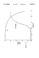
- FIG. 7 is a graphical illustration of the effect of learning iterations on ROC curves of a neural network according to the present invention, wherein the number of learning iterations ranges from 1 to 1,000 with the condition of 1 hidden layer and 6 hidden units;
- FIG. 8 is a graphical illustration comparing ROC curves of chest radiologists, senior radiology residents and the neural network according to the present invention indicating their decision performance and differential diagnosis of interstitial lung diseases;
- FIG. 9 is a block diagram of a second embodiment of the present invention illustrating three successive neural networks designed for differential diagnosis of interstitial lung diseases
- FIG. 10 is a graphical illustration of neural network output values at an output unit corresponding to silicosis for a range of dummy input values from 0 to 1 at an input corresponding to duration of symptoms;
- FIG. 11 is a graphical illustration of neural network output values at two output units corresponding to UIP and scleroderma for a range of dummy input values from 0 to 1 at an input unit corresponding to body temperature;
- FIG. 12 is a data sheet used in mammographic diagnosis including clinical parameters and mammographic descriptors.
- FIG. 13 is a graphical illustration of the ROC curve for a neural network according to a seventh embodiment of the present invention.
- the present invention relates to the differential diagnosis of diseases, and the following discussion directed to differential diagnosis of interstitial lung diseases is provided for example only, and it is understood that the present invention is not limited thereto.
- interstitial lung diseases for differential diagnosis were identified by chest radiologists, namely, sarcoidosis, miliary tuberculosis, lymphangitic metastatic tumor, interstitial pulmonary edema, silicosis, scleroderma, pneumocystis pneumonia, eosinophilic granuloma and usual interstitial pneumonitis (UIP).
- sarcoidosis miliary tuberculosis
- lymphangitic metastatic tumor interstitial pulmonary edema
- silicosis scleroderma
- pneumocystis pneumonia eosinophilic granuloma
- UIP interstitial pneumonitis
- radiographic descriptors which include 6 items regarding distribution of infiltrates in 6 divisions (upper, mid and lower zones of right and left lungs), 6 items relating to details of the infiltrate (homogeneity, fineness/coarseness, nodularity, septal lines and honeycombing) and 3 additional radiographic parameters (lymphadenopathy, pleural effusions and heart size).
- FIG. 3 shows the basic structure of the neural network according to a first embodiment of the present invention.
- the intermediate layer which is called the hidden layer, plays an important role in converting the input patterns to output patterns.
- This embodiment contains one hidden layer with two units. However, the number of hidden layers and the number of associated units can be changed in accordance with the desired structure and performance of the resulting network.
- a database was created for the training and testing processes. Two chest radiologists independently selected clinical data and radiographic findings for 10 hypothetical examples of each of the 9 diseases for a total of 90 cases. All input data for the neural network can be converted into numbers ranging from 0 to 1 using a linear or non-linear transformation. For example, the body temperature, which ranged from 37° C. to 41° C., was converted linearly into a number between 0 and 1. Radiographic descriptors were initially scored on a subjective scale from 0 to 10, and then normalized to the range from 0 to 1.
- fuzzy descriptive expressions such as "very large”, “large”, “probably”, “small” and “very small” can be converted into a number ranging from 0 to 1 using a membership function which can be defined appropriately based on the techniques used in fuzzy theory.
- Dubois et al “Fuzzy Set and Systems: Theory and Applications”, Academic Press (1980), and Zimmermann “Fuzzy Set Theory--and its Applications", Kluwer-Nijhoff Publishing (1985), whose disclosures are herein incorporated by reference.
- a neural network with a layered structure has the ability to learn the relationship between input data and output data from examples presented repeatedly.
- the discrimination criteria produced in the neural network depend on the number of variety of the training examples used. Therefore, the training database should be created ideally by selecting appropriate examples that represent a wide distribution of all potential cases.
- Two chest radiologists selected 10 cases for each of the 9 diseases on the basis of textbooks descriptions and their own experience.
- hypothetical data was used for training the neural network.
- the hypothetical database approach has an advantage in that varied distributions of all diseases can be represented to some extent with a relatively small number of total cases.
- a neural network trained with hypothetical data needs to be validated by testing with a large number of actual cases, prior to clinical application.
- the neural network according to the present invention was examined with 5 actual cases which fulfilled the criterion of having an interstitial infiltrate due to one of the nine selected diseases. All of these resulted in correct decisions. Further studies will determine the ultimate accuracy achievable in clinical situations, and also identify the need for modification of the database.
- the neural network according to the present invention can be retrained by adding new data to the existing training cases.
- these new data can be incorporated into the database along with the correct diagnosis so that very similar cases, which may be encountered subsequently, will be correctly identified.
- a jackknife method was employed for the evaluation of the neural network, that is, half of the cases, which were selected randomly from the database, were used for training, and the remaining half for testing. By random selection of cases, 10 different pairs of training and testing data sets were prepared. The outputs of the testing process were analyzed by determining true-positive and false-positive decisions, which were used for plotting the ordinate and abscissa, respectively, of ROC (receiver operating characteristics) curves.
- the ROC curve is known at present as the most reliable diagram which indicates the performance of detectors (or observers) in distinguishing between two possible states, such as normal and abnormal, for lungs.
- the ROC curve is generally a plot of the relationship between the true-positive fraction, i.e., the fraction of correct classifications (or detections) of the abnormal lung as abnormal and the false-positive fraction, i.e., the fraction of incorrect classifications (or detections) of the normal lung as abnormal. This relationship is expressed by a curve instead of a point because these fractions can change depending on the threshold level used, as disclosed in U.S. Pat. No. 4,839,807.
- the optimal parameters for the neural network according to the first embodiment were determined in terms of the number of hidden units, the number of hidden layers, and the number of learning iterations.
- the effect of the number of hidden units was examined by changing the number from 1 to 20 with the condition of one hidden layer and 200 learning iterations.
- the number of hidden layers was changed from 1 to 3 with 6 hidden units per layer and 200 learning iterations.
- the number of learning iterations was varied from 1 to 1000 with 1 hidden layer and 6 hidden units.
- the learning iterations were counted on every entry of a complete set of different training data.
- some other technical parameters such as learning rate, momentum factor of weight change and seed for random number generation were examined.
- FIG. 4 illustrates the ROC curves showing the effect of the number of hidden units on the performance of the neural network according to the first embodiment of the present invention.
- the ROC curve improves as the number of hidden units increases up to 8.
- the relationship between the area under the ROC curve (Az) and the number of hidden units, as shown in FIG. 5, indicates that more than 8 hidden units does not significantly improve the ROC curves, compared with the condition of 1 hidden layer and 200 learning iterations.
- the optimal number of hidden units the effect of a number of hidden units to distinguish completely all 90 cases of the database which were used for training first and then testing of the neural network was examined. Note that this was not the previously mentioned jack-knife test, but rather a verification test for consistency of the neural network according to the first embodiment of the present invention. Based on these curves, the minimum number of hidden units was determined as 6.
- FIG. 6 illustrates the ROC curves obtained with differing numbers of hidden layers. It is to be observed that the ROC curves deteriorated as the number of hidden layers increases.
- One hidden layer provides the highest ROC curve under the condition of 6 hidden units per layer and 200 learning iterations. As the learning iterations increase to 1000, virtually the same ROC curves are obtained as in the case of 200 learning iterations with no appreciable improvement. Accordingly, the neural network according to the first embodiment of the present invention includes one hidden layer.
- the ROC curves obtained with different numbers of learning iterations are shown in FIG. 7. Improvement in the ROC curves is observed as the number of learning iterations increases. The highest ROC curve is obtained for 200 or more learning iterations. Thus, 200 learning iterations is a sufficient number for training the neural network according to the first embodiment of the present invention.
- the neural network according to the first embodiment of the present invention includes one hidden layer having 6 units and was trained with 200 learning iterations.
- a discussion of the selection and learning processes is also described in Asada et al, "Neural Network Approach for Differential Diagnosis of Interstitial Lung Diseases" Proc. SPIE Medical Imaging IV (1990), which is herein incorporated by reference.
- Table 1 represents the average input data estimated from 10 cases for each of the 9 diseases in the database.
- Table 2 provides output data of the neural network according to the first embodiment of the present invention for a given set of the average input data given in Table 1.
- the 9 rows correspond to the 9 output units and the 9 columns correspond to the 9 columns of Table 1.
- the large output values such as 0.9 along the diagonal in Table 2, indicate that the neural network recognizes the input data as the disease shown on the same column with high probability.
- the small output values such as 0.01, 0.02 and 0.1 in other locations, suggests that the probability of false-positive decisions is very low. Therefore, Table 2 demonstrates the consistency of the neural network, which identified each of the 9 diseases with high probability.
- the decision performance of the neural network was compared with those of chest radiologists and of senior radiology residents. Three chest radiologists and three senior residents were tested using the same database as that used for the decision performance test of the neural network according to the first embodiment of the present invention. For each hypothetical case, each participant, as a decision maker, was allowed one minute to review the 20 clinical and radiographic descriptors and select the most likely diagnosis from the 9 possible options of interstitial lung diseases. The decision maker was required to indicate the level of confidence for each disease, using a rating scale from 1 to 5, 5 being the highest level of confidence for positive decisions. This rating method used is the same as that used for ROC analysis.
- the ROC curves of chest radiologists, senior radiology residents and the neural network for decision performance in differential diagnosis of interstitial lung diseases are shown in FIG. 8.
- the ROC curve of the neural network was comparable to the average ROC curve of the chest radiologist and superior to the average ROC curve of the senior radiology residents.
- the area under the ROC curves (Az), shown in FIG. 8, was 0.967 for both the chest radiologists and the neural network, and 0.905 for the senior residents.
- a neural network has multiple output units, it can be replaced with an equal multiple of parallel neural networks that have the same number of input units but have only one output unit.
- 9 different neural networks each having 20 input units and one output unit corresponding to the 9 chosen diseases, respectively, can be designed.
- the problem can be solved by using a series of neural networks.
- three successive neural networks are applied to the problem for differential diagnosis of interstitial lung diseases.
- the first neural network distinguishes between normal and abnormal patterns.
- the second neural network distinguishes between interstitial diseases and other diseases.
- the third neural network distinguishes among 9 types of interstitial lung diseases.
- another neural network can be added to this system to distinguish among other diseases such as primary lung cancer, pneumococcal pneumonia, pulmonary embolism, mediastinal tumor, etc.
- a method to obtain the output from the neural network even in the case of lack of some of the input information is to prepare several neural networks that have different numbers of input units corresponding to all possible combinations of missing data.
- 16 different neural networks can be produced: one neural network having 20 inputs, four having 19 inputs, six having 18 inputs, four having 17 inputs, and one having 16 inputs, which represent all possible permutations of the four missing clinical data inputs.
- a second method for handling an incomplete data set for testing is to run the neural network, which was trained with the incomplete data set, by entering "dummy" input values ranging from 0 to 1 with an appropriate increment such as 0.1 to an input unit in which the actual input data are missing.
- This method provides a range of output values at each unit with which the potential outcome for a case with the incomplete data set can be assessed by considering the possible variation of the missing input data. For example, if missing clinical data might not be essential for a certain disease, the output values at all output units would be basically unchanged for various dummy input values entered at the input unit which does not have the actual input data, as illustrated for silicosis in FIG. 10. The output value for the neural network in this case is not effected by the duration of symptoms.
- the potential outcome leading to a certain disease or other diseases can be identified with the corresponding potential input values as illustrated in FIG. 11.
- the output of the neural network for UIP and scleroderma are shown as a function of body temperature.
- the body temperature is low, the disease is very likely to be UIP.
- the probability of the UIP decreases and the disease is very likely to be scleroderma.
- physicians would be able to provide differential diagnosis of the various lung diseases depending on the potential value of missing clinical data.
- an incomplete set of input data which may be clinically important, may be desired or required to be entered into the training of the neural network.
- the incomplete set of input data can be used for training the neural network by creating "dummy" values for missing input data which are obtained from the range of input values belonging to the same disease in the complete set of input data.
- the range and the distribution of "dummy" input values at the missing input unit will be, therefore, comparable to those actual input values at the same input unit.
- different dummy input values sampled for the actual distribution described above are chosen for every entry of the training.
- the radiographic input data for the neural networks according to the invention are represented by numerical values which are estimated subjectively by a radiologist. This method may be impractical in many clinical situations. However, numerical values can be obtained from computerized automated analysis of lung texture, as disclosed in U.S. Pat. Nos. 4,839,807 and 4,851,484. Therefore, in a sixth embodiment of the present invention, a neural network is interfaced to a computerized scheme which performs automated lung texture analysis so that all radiographic input data to the neural network could be supplied in an objective and automated manner.
- the present invention is not limited to exclusively that of differential diagnosis of interstitial lung diseases.
- a neural network could be used in other areas of medical decision making, such as discrimination between benign and malignant patterns in mammography, which depend on simultaneous correlation of multiple clinical, laboratory and/or radiological findings.
- the neural network was applied to the mammographic diagnosis of breast cancer to distinguish between benign and malignant mammographic cases in a seventh embodiment.
- the network consists of a 3-layer feed-forward neural network with the back-propagating algorithm.
- the neural network has 45 input units, 1-25 hidden units and 1-5 output units.
- the neural network was trained by inputting ratings from mammographic features and clinical data from various cases, along with the corresponding correct benign/malignant diagnosis.
- FIG. 12 lists the various input data including clinical parameters and radiographic descriptors. In the training process, for each case in a database of 60 cases selected from a textbook on mammography (Laszlo Tabar and Peter B.
- an expert mammographer rated each feature shown in FIG. 12. For example, if an abnormal mass density was observed, then the mammographer provided a rating from 0 to 10 regarding the shape of the density, as shown in II.B.1 of FIG. 12, with 0 corresponding to a linear one-dimensional feature, 5 being an oblong lesion and 10 corresponding to a generally spherical lesion. In addition, as shown in FIG. 12, the mammographer checked the correct diagnosis (malignant or benign), as well as the course of action he would advise in an actual clinical situation, which corresponded to desired outputs.
- ROC analysis was used in evaluating the mammography network using round robin and jackknife methods. With the database of 60 cases representing a range of benign and malignant cases, ROC analysis yielded an A z of approximately 0.85 to 0.9, as shown in FIG. 13. The high value of A z indicates the high degree accuracy for mammographic diagnosis possible using neural networks.
Abstract
A method and system for computer-aided differential diagnosis of diseases, and in particular, computer-aided differential diagnosis using neural networks. A first embodiment of the neural network distinguishes between a plurality of interstitial lung diseases on the basis of inputted clinical parameters and radiographic information. A second embodiment distinguishes between malignant and benign mammographic cases based upon similar inputted clinical and radiographic information. The neural networks were first trained using a hypothetical data base made up of hypothetical cases for each of the interstitial lung diseases and for malignant and benign cases. The performance of the neural network was evaluated using receiver operating characteristics (ROC) analysis. The decision performance of the neural network was compared to experienced radiologists and achieved a high performance comparable to that of the experienced radiologists. The neural network according to the invention can be made up of a single network or a plurality of successive or parallel networks. The neural network according to the invention can also be interfaced to a computer which provides computerized automated lung texture analysis to supply radiographic input data in an objective and automated manner.
Description
The U.S. Government has a paid-up license in this invention and the right and limited circumstances to require the patent owner to license others on reasonable terms as provided for by the terms of USPHS Grant Nos. CA24804, CA44926 and CA47043 from the National Institute of Health.
This is a continuation of application Ser. No. 08/053,345 filed on Apr. 28, 1993, U.S. Pat. No. 5,463,548 which is a continuation of application Ser. No. 07/573,800 filed on Aug. 28, 1990 now abandoned.
The present invention is related to U.S. Pat. No. 4,839,807 granted to Doi et al and U.S. Pat. No. 4,851,984 granted to Doi et al, which are herein incorporated by reference.
1. Field of the Invention
The present invention relates to a method and system for computer-aided differential diagnosis of diseases, and in particular, computer-aided differential diagnosis using neural networks.
2. Discussion of the Background
Computer-aided differential diagnosis of diseases is an important subject in radiology. However, it is difficult to distinguish accurately between many diseases that can produce similar or even identical radiographic interstitial patterns. In such cases, a specific diagnosis can only be made by taking into consideration the multiple relevant clinical aspects of the case, i.e., age, sex, symptoms, etc., together with details of the radiographic findings. Thus, differential diagnosis of diseases lends itself to computer automation which can provide assistance to less expert observers of radiographic patterns, to bring the decision performance of these less expert observers to a level closer to that of experienced radiologists. However, computer-aided differential diagnosis of diseases has not gained wide acceptance due to the multiple clinical aspects of a case.
A powerful tool for use in solving problems involving pattern recognition and classification is an artificial neural network having a layered structure and applied with a supervised-learning procedure such as the back-propagating error correction algorithm as disclosed by Rumelhart et al "Parallel distributed processing" Explorations in the Microstructure of Cognition" Cambridge: MIT Press (1986), Grossberg "Neural Network and Neural Intelligence" Cambridge: MIT Press (1988), and Eckmiller et al "Neural Computers" Berlin: Springer-Verlag (1989), which are herein incorporated by reference. Artificial neural networks consist of a number of neuron-like elements (units) and connections between them, and can be implemented by hardware and/or software. The units of the neural network are categorized into three types of different groups (layers) according to their functions as shown in FIG. 1. A first layer (input layer) is assigned to accepting a set of data representing an input pattern, a second layer (output layer) is assigned to provide a set of data representing an output pattern, and an arbitrary number of intermediate layers (hidden layers) convert the input pattern to the output pattern. Since the number of units in each layer is determined arbitrarily, the input layer and the output layer include sufficient numbers of units to represent the input patterns and output patterns, respectively, of a problem to be solved. For example, a neural network which is designed to distinguish between 9 types of diseases on the basis of 20 items of clinical information, should have 20 input units and 9 output units. However, the optimum number of hidden layers and associated units needs to be determined empirically.
Briefly, the principle of neural network can be explained in the following manner. Input data, which are represented by numbers ranging from 0 to 1, are supplied to input units of the neural network. Next, the output data are provided from output units through two successive nonlinear calculations (in a case of one hidden layer) in the hidden and output layers. The calculation at each unit in the layer, which is illustrated schematically in FIG. 2, excluding the input units, includes a weighted summation of all entry numbers, an addition of certain offset terms and a conversion into a number ranging from 0 to 1 using a sigmoid-shape function such as a logistic function. In FIG. 2, units labelled O1 to On represent input or hidden units, W1 through Wn represent the weighting factors assigned to each respective output from these input or hidden units, and I represents the summation of the outputs multiplied by the respective weighting factors. An output O is calculated using the logistic-function equation given where θ represents an offset value for the input I. The weighting factors and offset values are internal parameters of the neural network which are determined for a given set of input and output data.
Two different basic processes are involved in the neural network, namely, a training process and a testing process. The neural network is trained by the back-propagation algorithm using pairs of training input data and desired output data, as given by Rumelhart et al, Ibid, pp. 318-362. The internal parameters of the neural network are adjusted in order to minimize the difference between the actual outputs of the neural network and the desired outputs. By iteration of this procedure in a random sequence for the same set of input and output data, the neural network learns a relationship between the training input data and the desired output data. Once trained sufficiently, the neural network can distinguish different input data according to its learning experience. To date, the neural network approach has not been applied to the computer-aided differential diagnosis of interstitial diseases.
Accordingly, it is an object of the present invention to provide a novel method and system for the computer-aided differential diagnosis of diseases including a neural network.
It is another object of the present invention to provide a new and improved method and system for the computer-aided differential diagnosis of diseases including a neural network which improves the diagnosis decision performance of radiologists.
These and other objects are achieved according to the invention by providing a new and improved method and system for diagnosis of diseases, based upon the input of clinical parameters and radiographic descriptors into a neural network. For the case of interstitial lung diseases, the clinical parameters selected are the patient's age, sex, duration of symptoms, severity of symptoms, temperature and immune status. The radiographic descriptors include information regarding the distribution of infiltrates in the lungs and information relating to details of the infiltrate. The clinical parameters and radiographic descriptors are converted into numerical expressions and then transformed into a number between 0 and 1. These transformed expressions are input into a neural network having a plurality of input units and a plurality of output units. The neural network diagnoses one of the plurality of interstitial lung diseases based upon the transformed numerical expressions inputted thereto. The system and method of the present invention allows less expert observers, i.e., radiology residents or clinicians, to improve their differential diagnosis decision performance to a level closer to that of experienced radiologists.
In another embodiment, the method of the present invention is applied to mammographic diagnosis of breast cancer to distinguish between benign and malignant cases. Clinical parameters and radiographic descriptors are transformed into numerical expressions and input into a neural network which outputs a diagnosis based upon the input expressions.
It is important to note that neural networks are not effective for solving problems in all applications of radiological diagnosis. Radiological diagnoses are often made based on findings of abnormal patterns which are commonly closely related to a disease, diseases or a state. For example, the detection of lung cancer, cardiomegaly, and pneumothorax is primarily due to the findings of lung nodules, enlargement of cardiac outline and subtle curved line structure, respectively. Radiological diagnosis on ultrasound images, which are well known to be of poor quality due to low resolution, high noise level and artifacts, is made by highly trained radiologists able to distinguish between normal and abnormal patterns. For this task, correct recognition of certain image patterns by radiologists is the most important element for achieving accurate diagnosis. This example indicates that the application of neural networks will not be helpful for decision making in many radiological diagnosis situations. Therefore, prior to the present invention, the utility and effectiveness of neural networks in differential diagnosis was not considered and unknown, and in particular with respect to differential diagnosis of interstitial lung diseases. The present invention is a novel use of neural networks in the field of differential diagnosis, which provides an effective and accurate differential diagnosis.
A more complete appreciation of the invention and many of the attendant advantages thereof will be readily obtained as the same becomes better understood by reference to the following detailed description when considered in connection with the accompanying drawings, wherein:
FIG. 1 is a diagram of the basic structure of an artificial neural network having a layered structure according to the prior art;
FIG. 2 is a schematic illustration of the calculation in a unit of an artificial neural network according to the prior art;
FIG. 3 is a diagram of a neural network according to the present invention, including 20 inputs units, 9 output units and 1 hidden layer with 2 units;
FIG. 4 is a graphical illustration of the effect of the number of hidden units on ROC curves of a neural network according to the present invention, wherein the number of hidden units ranges from 1 to 8 with the condition of 1 hidden layer and 200 learning iterations;
FIG. 5 is a graphical illustration of the effect of the number of hidden units on the area under the ROC curves of a neural network according to the present invention, wherein the number of hidden units ranges from 1 to 20 with a condition of 1 hidden layer and 200 learning iterations;
FIG. 6 is a graphical illustration of the effect of the number of hidden layers on ROC curves of a neural network according to the present invention, wherein the number of hidden layers ranges from 1 to 3 with a condition of 6 hidden units per layer and 200 learning iterations;
FIG. 7 is a graphical illustration of the effect of learning iterations on ROC curves of a neural network according to the present invention, wherein the number of learning iterations ranges from 1 to 1,000 with the condition of 1 hidden layer and 6 hidden units;
FIG. 8 is a graphical illustration comparing ROC curves of chest radiologists, senior radiology residents and the neural network according to the present invention indicating their decision performance and differential diagnosis of interstitial lung diseases;
FIG. 9 is a block diagram of a second embodiment of the present invention illustrating three successive neural networks designed for differential diagnosis of interstitial lung diseases;
FIG. 10 is a graphical illustration of neural network output values at an output unit corresponding to silicosis for a range of dummy input values from 0 to 1 at an input corresponding to duration of symptoms;
FIG. 11 is a graphical illustration of neural network output values at two output units corresponding to UIP and scleroderma for a range of dummy input values from 0 to 1 at an input unit corresponding to body temperature;
FIG. 12 is a data sheet used in mammographic diagnosis including clinical parameters and mammographic descriptors; and
FIG. 13 is a graphical illustration of the ROC curve for a neural network according to a seventh embodiment of the present invention.
The present invention relates to the differential diagnosis of diseases, and the following discussion directed to differential diagnosis of interstitial lung diseases is provided for example only, and it is understood that the present invention is not limited thereto.
Referring now to the drawings, and more particularly to FIG. 3 thereof, 9 types of interstitial lung diseases for differential diagnosis were identified by chest radiologists, namely, sarcoidosis, miliary tuberculosis, lymphangitic metastatic tumor, interstitial pulmonary edema, silicosis, scleroderma, pneumocystis pneumonia, eosinophilic granuloma and usual interstitial pneumonitis (UIP). Also identified were 6 key clinical parameters which are the patient's age, sex, duration of symptoms, severity of symptoms, temperature and immune status, and 14 radiographic descriptors which include 6 items regarding distribution of infiltrates in 6 divisions (upper, mid and lower zones of right and left lungs), 6 items relating to details of the infiltrate (homogeneity, fineness/coarseness, nodularity, septal lines and honeycombing) and 3 additional radiographic parameters (lymphadenopathy, pleural effusions and heart size).
Thus, the neural network was designed to include 20 input units (6 clinical parameters and 14 radiographic descriptors) and 9 output units (9 interstitial lung diseases). FIG. 3 shows the basic structure of the neural network according to a first embodiment of the present invention. The intermediate layer, which is called the hidden layer, plays an important role in converting the input patterns to output patterns. This embodiment contains one hidden layer with two units. However, the number of hidden layers and the number of associated units can be changed in accordance with the desired structure and performance of the resulting network.
Hypothetical Database
In order to evaluate the decision performance of the neural network, a database was created for the training and testing processes. Two chest radiologists independently selected clinical data and radiographic findings for 10 hypothetical examples of each of the 9 diseases for a total of 90 cases. All input data for the neural network can be converted into numbers ranging from 0 to 1 using a linear or non-linear transformation. For example, the body temperature, which ranged from 37° C. to 41° C., was converted linearly into a number between 0 and 1. Radiographic descriptors were initially scored on a subjective scale from 0 to 10, and then normalized to the range from 0 to 1. It should be noted that any kind of description even including fuzzy descriptive expressions such as "very large", "large", "probably", "small" and "very small" can be converted into a number ranging from 0 to 1 using a membership function which can be defined appropriately based on the techniques used in fuzzy theory. Dubois et al "Fuzzy Set and Systems: Theory and Applications", Academic Press (1980), and Zimmermann "Fuzzy Set Theory--and its Applications", Kluwer-Nijhoff Publishing (1985), whose disclosures are herein incorporated by reference.
As stated earlier, a neural network with a layered structure has the ability to learn the relationship between input data and output data from examples presented repeatedly. In other words, the discrimination criteria produced in the neural network depend on the number of variety of the training examples used. Therefore, the training database should be created ideally by selecting appropriate examples that represent a wide distribution of all potential cases. However, it is very time-consuming to create an appropriate database by collecting actual patient cases prospectively, and the relevant clinical information was not consistently available in teaching file cases.
Two chest radiologists selected 10 cases for each of the 9 diseases on the basis of textbooks descriptions and their own experience. Thus, hypothetical data was used for training the neural network. The hypothetical database approach has an advantage in that varied distributions of all diseases can be represented to some extent with a relatively small number of total cases. A neural network trained with hypothetical data needs to be validated by testing with a large number of actual cases, prior to clinical application.
The neural network according to the present invention was examined with 5 actual cases which fulfilled the criterion of having an interstitial infiltrate due to one of the nine selected diseases. All of these resulted in correct decisions. Further studies will determine the ultimate accuracy achievable in clinical situations, and also identify the need for modification of the database.
However, it is important to note that the neural network according to the present invention can be retrained by adding new data to the existing training cases. Thus, if the neural network fails to yield a correct diagnosis on some actual cases, these new data can be incorporated into the database along with the correct diagnosis so that very similar cases, which may be encountered subsequently, will be correctly identified.
Optimal Parameters
A jackknife method was employed for the evaluation of the neural network, that is, half of the cases, which were selected randomly from the database, were used for training, and the remaining half for testing. By random selection of cases, 10 different pairs of training and testing data sets were prepared. The outputs of the testing process were analyzed by determining true-positive and false-positive decisions, which were used for plotting the ordinate and abscissa, respectively, of ROC (receiver operating characteristics) curves.
The ROC curve is known at present as the most reliable diagram which indicates the performance of detectors (or observers) in distinguishing between two possible states, such as normal and abnormal, for lungs. The ROC curve is generally a plot of the relationship between the true-positive fraction, i.e., the fraction of correct classifications (or detections) of the abnormal lung as abnormal and the false-positive fraction, i.e., the fraction of incorrect classifications (or detections) of the normal lung as abnormal. This relationship is expressed by a curve instead of a point because these fractions can change depending on the threshold level used, as disclosed in U.S. Pat. No. 4,839,807.
Using average ROC curves obtained from 10 different data sets, the optimal parameters for the neural network according to the first embodiment were determined in terms of the number of hidden units, the number of hidden layers, and the number of learning iterations. The effect of the number of hidden units was examined by changing the number from 1 to 20 with the condition of one hidden layer and 200 learning iterations. In the same manner, the number of hidden layers was changed from 1 to 3 with 6 hidden units per layer and 200 learning iterations. The number of learning iterations was varied from 1 to 1000 with 1 hidden layer and 6 hidden units. The learning iterations were counted on every entry of a complete set of different training data. In addition, the effect of some other technical parameters such as learning rate, momentum factor of weight change and seed for random number generation were examined.
For training the neural network, 10 hypothetical cases for each disease were applied. However, as the actual cases are accumulated, the number of total cases collected for each disease may not be equal. In fact, it is likely that the number of training cases for rare diseases can be significantly smaller than those for common diseases. This insufficient number of training cases for a certain disease will cause a decrease in the sensitivity of detecting a specific disease. This problem is related to the prevalence (or frequency of occurrence) of diseases and can be solved to some extent by increasing the number of entries of the same training case for the rare disease. For example, if a common disease and a rare disease may have 100 cases and 10 cases respectively, then the 100 cases of the common disease may be entered for 200 iterations, but the 10 cases of the rare disease may be entered for 2000 iterations. Therefore, the total numbers of data entries for training the neural network for diseases having different prevalences, can be equalized.
A more complete discussion of the learning procedure used in training the neural network according to the first embodiment of the present invention, i.e., the back-propagating method, is described in the Rumelhart et al references and is omitted here for brevity.
FIG. 4 illustrates the ROC curves showing the effect of the number of hidden units on the performance of the neural network according to the first embodiment of the present invention. The ROC curve improves as the number of hidden units increases up to 8. However, the relationship between the area under the ROC curve (Az) and the number of hidden units, as shown in FIG. 5, indicates that more than 8 hidden units does not significantly improve the ROC curves, compared with the condition of 1 hidden layer and 200 learning iterations. In order to determine the optimal number of hidden units, the effect of a number of hidden units to distinguish completely all 90 cases of the database which were used for training first and then testing of the neural network was examined. Note that this was not the previously mentioned jack-knife test, but rather a verification test for consistency of the neural network according to the first embodiment of the present invention. Based on these curves, the minimum number of hidden units was determined as 6.
FIG. 6 illustrates the ROC curves obtained with differing numbers of hidden layers. It is to be observed that the ROC curves deteriorated as the number of hidden layers increases. One hidden layer provides the highest ROC curve under the condition of 6 hidden units per layer and 200 learning iterations. As the learning iterations increase to 1000, virtually the same ROC curves are obtained as in the case of 200 learning iterations with no appreciable improvement. Accordingly, the neural network according to the first embodiment of the present invention includes one hidden layer.
The ROC curves obtained with different numbers of learning iterations are shown in FIG. 7. Improvement in the ROC curves is observed as the number of learning iterations increases. The highest ROC curve is obtained for 200 or more learning iterations. Thus, 200 learning iterations is a sufficient number for training the neural network according to the first embodiment of the present invention.
Accordingly, the neural network according to the first embodiment of the present invention includes one hidden layer having 6 units and was trained with 200 learning iterations. A discussion of the selection and learning processes is also described in Asada et al, "Neural Network Approach for Differential Diagnosis of Interstitial Lung Diseases" Proc. SPIE Medical Imaging IV (1990), which is herein incorporated by reference.
Consistency Verification Test
Prior to performing the decision performance test, a consistency test of the neural network was performed. The neural network was trained in all cases in the database, and then tested by an average case of the 10 cases for each of the 9 diseases in the database. The average input values to the neural network and the corresponding output values from the neural network are shown in Tables 1 and 2, respectively. Table 1 represents the average input data estimated from 10 cases for each of the 9 diseases in the database. Table 2 provides output data of the neural network according to the first embodiment of the present invention for a given set of the average input data given in Table 1. In Table 2, the 9 rows correspond to the 9 output units and the 9 columns correspond to the 9 columns of Table 1. The large output values, such as 0.9 along the diagonal in Table 2, indicate that the neural network recognizes the input data as the disease shown on the same column with high probability. In addition, the small output values, such as 0.01, 0.02 and 0.1 in other locations, suggests that the probability of false-positive decisions is very low. Therefore, Table 2 demonstrates the consistency of the neural network, which identified each of the 9 diseases with high probability.
                                  TABLE 1
__________________________________________________________________________
            9 types of interstitial lung diseases
                  miliary
                        metastatic
                              pulmonary        pneumocystis
                                                      eosinophilic
Input unit name
            sarcoidosis
                  tuberculosis
                        tumor edema silicosis
                                        scleroderma
                                               pneumonia
                                                      granuloma
                                                            UIP
__________________________________________________________________________
age         32    53    59    60    67  40     37     34    54
sex*        0     0     1     1     1   0      1      1     1
duration of symptoms**
            2     1     3     1     7   5      1      3     5
severity of symptoms
            1     8     5     5     3   3      8      1     4
temperature 37.6  39.9  37.6  37.2  37.1
                                        37.4   39.7   37.4  37.1
immune status***
            0     0     0     0     0   0      1      0     0
RU          4     4     3     3     7   1      4      6     2
RM          3     4     4     5     5   3      4      4     3
RL          2     4     5     5     3   6      4      1     5
LU          4     4     3     3     7   1      4      6     2
LM          3     4     4     5     5   3      4      3     3
LL          2     5     6     6     3   6      4      1     5
homogeneity 6     8     6     7     6   7      9      8     8
fineness/coarseness
            3     2     6     4     5   3      2      5     4
nodularity  3     9     6     1     7   1      0      5     2
septal lines
            0     0     4     6     1   1      0      0     1
honeycombing
            1     0     0     0     3   2      0      7     5
lymphadenopathy
            6     0     4     0     3   0      1      0     0
pleural effusions
            0     2     4     4     0   0      0      0     0
heart size  2     1     2     4     2   1      1      1     1
__________________________________________________________________________
 *0 = female, 1 = male
 **month (unit)
 ***0 = normal, 1 = suppressed
                                  TABLE 2
__________________________________________________________________________
            9 types of interstitial lung diseases (correct diagnosis)
                  miliary
                        metastatic
                              pulmonary        pneumocystis
                                                      eosinophilic
Output unit name
            sarcoidosis
                  tuberculosis
                        tumor edema silicosis
                                        scleroderma
                                               pneumonia
                                                      granuloma
                                                            UIP
__________________________________________________________________________
sarcoidosis 0.99  0.00  0.00  0.00  0.02
                                        0.00   0.00   0.00  0.00
miliary tuberculosis
            0.00  0.99  0.01  0.00  0.01
                                        0.00   0.00   0.00  0.00
metastatic tumor
            0.00  0.00  0.98  0.01  0.01
                                        0.00   0.00   0.00  0.00
pulmonary edema
            0.00  0.00  0.01  0.98  0.00
                                        0.00   0.00   0.00  0.01
silicosis   0.00  0.00  0.01  0.00  0.98
                                        0.00   0.00   0.01  0.01
scleroderma 0.00  0.00  0.00  0.01  0.00
                                        0.97   0.01   0.00  0.00
pneumocystis pneumonia
            0.00  0.00  0.00  0.01  0.00
                                        0.01   0.99   0.00  0.00
eosinophilic granuloma
            0.00  0.00  0.00  0.00  0.01
                                        0.00   0.00   0.99  0.00
UIP         0.00  0.00  0.00  0.01  0.01
                                        0.10   0.00   0.00  0.99
__________________________________________________________________________
Decision Performance
The decision performance of the neural network was compared with those of chest radiologists and of senior radiology residents. Three chest radiologists and three senior residents were tested using the same database as that used for the decision performance test of the neural network according to the first embodiment of the present invention. For each hypothetical case, each participant, as a decision maker, was allowed one minute to review the 20 clinical and radiographic descriptors and select the most likely diagnosis from the 9 possible options of interstitial lung diseases. The decision maker was required to indicate the level of confidence for each disease, using a rating scale from 1 to 5, 5 being the highest level of confidence for positive decisions. This rating method used is the same as that used for ROC analysis.
Decision Performance Test
The ROC curves of chest radiologists, senior radiology residents and the neural network for decision performance in differential diagnosis of interstitial lung diseases are shown in FIG. 8. The ROC curve of the neural network was comparable to the average ROC curve of the chest radiologist and superior to the average ROC curve of the senior radiology residents. The area under the ROC curves (Az), shown in FIG. 8, was 0.967 for both the chest radiologists and the neural network, and 0.905 for the senior residents.
The above results indicate that a neural network can be helpful to less expert observers, such as the senior radiology residents or clinicians, to bring their decision performance to a level closer to that of chest radiologists in the differential diagnosis of interstitial lung diseases.
As stated previously, there is some flexibility in designing the structure and the combination of neural networks to solve a problem. If a neural network has multiple output units, it can be replaced with an equal multiple of parallel neural networks that have the same number of input units but have only one output unit. According to a second embodiment of the present invention, 9 different neural networks each having 20 input units and one output unit corresponding to the 9 chosen diseases, respectively, can be designed.
Alternatively, if a given problem can be divided into a series of smaller parts, the problem can be solved by using a series of neural networks. In a third embodiment of the present invention, as illustrated in FIG. 9, three successive neural networks are applied to the problem for differential diagnosis of interstitial lung diseases. The first neural network distinguishes between normal and abnormal patterns. The second neural network distinguishes between interstitial diseases and other diseases. Finally, the third neural network distinguishes among 9 types of interstitial lung diseases. It should be noted that another neural network can be added to this system to distinguish among other diseases such as primary lung cancer, pneumococcal pneumonia, pulmonary embolism, mediastinal tumor, etc. These parallel and serial approaches and combinations thereof, allow the solving of a large scale problem using multiple neural networks having rather small scale and simple structure.
In some cases, which occurs quite commonly in practical situations, it is not possible to acquire a complete set of input data. A method to obtain the output from the neural network even in the case of lack of some of the input information is to prepare several neural networks that have different numbers of input units corresponding to all possible combinations of missing data. According to a fourth embodiment of the present invention, in the differential diagnosis of interstitial lung diseases, for example, four of the 20 clinical data inputs (duration of symptoms, severity of symptoms, temperature and immune status) are sometimes unavailable. Thus, 16 different neural networks can be produced: one neural network having 20 inputs, four having 19 inputs, six having 18 inputs, four having 17 inputs, and one having 16 inputs, which represent all possible permutations of the four missing clinical data inputs. By training these 16 neural networks with a given complete set of initial input data, neural networks allowing the entry of an incomplete set of data are obtained.
A second method for handling an incomplete data set for testing is to run the neural network, which was trained with the incomplete data set, by entering "dummy" input values ranging from 0 to 1 with an appropriate increment such as 0.1 to an input unit in which the actual input data are missing. This method provides a range of output values at each unit with which the potential outcome for a case with the incomplete data set can be assessed by considering the possible variation of the missing input data. For example, if missing clinical data might not be essential for a certain disease, the output values at all output units would be basically unchanged for various dummy input values entered at the input unit which does not have the actual input data, as illustrated for silicosis in FIG. 10. The output value for the neural network in this case is not effected by the duration of symptoms.
However, if the output values at some output units would be significantly changed for various dummy input values entered, the potential outcome leading to a certain disease or other diseases can be identified with the corresponding potential input values as illustrated in FIG. 11. In FIG. 11, the output of the neural network for UIP and scleroderma are shown as a function of body temperature. Here, if the body temperature is low, the disease is very likely to be UIP. However, if the body temperature is high, then the probability of the UIP decreases and the disease is very likely to be scleroderma. Thus, physicians would be able to provide differential diagnosis of the various lung diseases depending on the potential value of missing clinical data.
In other situations, an incomplete set of input data, which may be clinically important, may be desired or required to be entered into the training of the neural network. Here, it is assumed that the majority of the input data are available for all input units, namely, most data are the complete sets of input data. However, a small fraction of input data is assumed to be incomplete sets of input data. In such cases, the incomplete set of input data can be used for training the neural network by creating "dummy" values for missing input data which are obtained from the range of input values belonging to the same disease in the complete set of input data. The range and the distribution of "dummy" input values at the missing input unit will be, therefore, comparable to those actual input values at the same input unit. In other words, for the training of the neural network which generally includes a large number of iterations, such as 200, different dummy input values sampled for the actual distribution described above are chosen for every entry of the training.
The radiographic input data for the neural networks according to the invention are represented by numerical values which are estimated subjectively by a radiologist. This method may be impractical in many clinical situations. However, numerical values can be obtained from computerized automated analysis of lung texture, as disclosed in U.S. Pat. Nos. 4,839,807 and 4,851,484. Therefore, in a sixth embodiment of the present invention, a neural network is interfaced to a computerized scheme which performs automated lung texture analysis so that all radiographic input data to the neural network could be supplied in an objective and automated manner.
The present invention is not limited to exclusively that of differential diagnosis of interstitial lung diseases. For example, a neural network could be used in other areas of medical decision making, such as discrimination between benign and malignant patterns in mammography, which depend on simultaneous correlation of multiple clinical, laboratory and/or radiological findings.
The neural network was applied to the mammographic diagnosis of breast cancer to distinguish between benign and malignant mammographic cases in a seventh embodiment. The network consists of a 3-layer feed-forward neural network with the back-propagating algorithm. The neural network has 45 input units, 1-25 hidden units and 1-5 output units. The neural network was trained by inputting ratings from mammographic features and clinical data from various cases, along with the corresponding correct benign/malignant diagnosis. FIG. 12 lists the various input data including clinical parameters and radiographic descriptors. In the training process, for each case in a database of 60 cases selected from a textbook on mammography (Laszlo Tabar and Peter B. Dean, Teaching Atlas of Mammography, Georg Yhieme Verlag, (1985)), an expert mammographer rated each feature shown in FIG. 12. For example, if an abnormal mass density was observed, then the mammographer provided a rating from 0 to 10 regarding the shape of the density, as shown in II.B.1 of FIG. 12, with 0 corresponding to a linear one-dimensional feature, 5 being an oblong lesion and 10 corresponding to a generally spherical lesion. In addition, as shown in FIG. 12, the mammographer checked the correct diagnosis (malignant or benign), as well as the course of action he would advise in an actual clinical situation, which corresponded to desired outputs. These ratings were converted to values between 0 and 1, and were used as the inputs to, and desired outputs from, the neural network. ROC analysis was used in evaluating the mammography network using round robin and jackknife methods. With the database of 60 cases representing a range of benign and malignant cases, ROC analysis yielded an Az of approximately 0.85 to 0.9, as shown in FIG. 13. The high value of Az indicates the high degree accuracy for mammographic diagnosis possible using neural networks.
Obviously, additional modifications and variations of the present invention are possible in light of the above teachings. It is therefore to be understood that within the scope of the appended claims, the invention may be practiced otherwise than as specifically described herein.
Claims (26)
1. A method for differential diagnosis of a plurality of breast diseases, the method comprising the steps of:
selecting a plurality of clinical parameters defining characteristics of a subject;
selecting radiographic descriptors comprising predetermined features obtained from a radiographic breast image defining characteristics of breast diseases, said radiographic descriptors consisting essentially of each of the following twelve radiographic descriptors:
shape of density, size of density, margin of density, margin spiculation and pattern of density, presence of, number of, shape of, uniformity of, and distribution of microcalcifications, parenchymal distortion and skin thickening;
converting said plurality of clinical parameters and said radiographic descriptors into numerical expressions;
transforming each of said numerical expressions into a number in a predetermined range;
inputting said transformed numerical expressions into a neural network; and
diagnosing at least one of said plurality of breast diseases using said neural network in accordance with said input expressions.
2. A method as recited in claim 1, wherein said neural network comprises 3 layers, 45 input units, at least one hidden unit and at least one output unit.
3. A method for differential diagnosis of a plurality of interstitial lung diseases, the method comprising the steps of:
selecting a plurality of clinical parameters defining characteristics of a subject;
selecting radiographic descriptors comprising predetermined features obtained from a radiographic chest image defining characteristics of interstitial lung diseases, said radiographic descriptors consisting essentially of each of the following fourteen radiographic descriptors:
distribution of infiltrates in 6 lung zones, homogeneity, fineness, nodularity, septal lines and honeycombing of said infiltrates, lymphadenopathy, pleural effusions and heart size;
converting said plurality of clinical parameters and said radiographic descriptors into numerical expressions;
transforming each of said numerical expressions into a number in a predetermined range;
inputting said transformed numerical expressions into a neural network; and
diagnosing at least one of said plurality of interstitial lung diseases using said neural network in accordance with said input expressions.
4. A method as recited in claim 3, further comprising the steps of:
training said neural network to identify each of said plurality of predetermined interstitial lung diseases using said clinical parameters and radiographic descriptors; and
determining an optimal number of hidden layers of said neural network by adjusting said neural network to minimize a difference between an output diagnosis and a desired diagnosis for a given set of said input expressions.
5. A method as recited in claim 4, further comprising the step of:
using receiver operating characteristic (ROC) analysis to determine optimal numbers of hidden layers, learning iterations and units per hidden layer of said neural network.
6. A method as recited in claim 4, wherein said step of training said neural network uses at least 200 learning iterations.
7. A method as recited in claim 3, wherein said neural network includes a plurality of parallel neural networks equal in number to said plurality of predetermined interstitial lung diseases, each of said neural networks having a plurality of inputs and one output, said outputs respectively corresponding to said plurality of predetermined interstitial lung diseases.
8. A method as recited in claim 3, further comprising the steps of:
dividing the diagnosis of said plurality of predetermined interstitial lung diseases into a plurality of stages using a plurality of neural networks.
9. A method as recited in claim 3, wherein said step of selecting a plurality of clinical parameters comprises:
selecting at least plural of the following clinical parameters:
age of a subject,
sex of said subject,
duration of symptoms,
severity of symptoms,
temperature of said subject, and
immune status of said subject.
10. A method as recited in claim 3, wherein said neural network comprises 20 input units, one hidden layer with 6 hidden units and 9 output units, said output units respectively corresponding to 9 interstitial lung diseases.
11. A method as recited in claim 3, further comprising the steps of:
selecting said radiographic descriptors using a computer by computerized automated lung texture analysis; and
interfacing said neural network to said computer.
12. A method for differential diagnosis of plurality of predetermined interstitial lung diseases using an incomplete set of input data, the method comprising the steps of:
selecting a plurality of clinical parameters defining characteristics of a subject;
selecting radiographic descriptors comprising predetermined features obtained from a radiographic chest image defining characteristics of interstitial lung diseases, said radiographic descriptors consisting essentially of each of the following fourteen radiographic descriptors:
distribution of infiltrates in 6 lung zones, homogeneity, fineness, nodularity, septal lines and honeycombing of said infiltrates, and lymphadenopathy, pleural effusions and heart size;
forming a complete set of input data comprising said plurality of clinical parameters and said radiographic descriptors;
converting said plurality of clinical parameters and said radiographic descriptors into numerical expressions;
transforming each of said numerical expressions into a number in a predetermined range;
training a neural network to identify each of said plurality of predetermined interstitial lung diseases using a database of said complete set of input data;
inputting said transformed numerical expressions into said neural network, said input expressions representing incomplete sets of input data; and
diagnosing at least one of said plurality of predetermined interstitial lung diseases in accordance with said input expressions.
13. A method as recited in claim 12, further comprising:
training said neural network to identify each of said plurality of predetermined, diseases using said clinical parameters and radiographic descriptors; and
determining an optimal number of hidden layers of said neural network by adjusting said neural network to minimize a difference between an output diagnosis and a desired diagnosis for a given set of said input expressions.
14. A method as recited in claim 13, further comprising the steps of:
using receiver operating characteristic (ROC) analysis to determine optimal numbers of hidden layers, learning iterations and units per hidden layer of said neural network.
15. A method as recited in claim 13, wherein the step of training said neural network uses at least 200 learning iterations.
16. A method as recited in claim 12, wherein said neural network comprises a plurality of parallel neural networks equal in number to said plurality of predetermined interstitial lung diseases, each of said neural networks having a plurality of inputs and one output, said outputs respectively corresponding to said plurality of predetermined interstitial lung diseases.
17. A method as recited in claim 12, further comprising the step of:
dividing the diagnosis of said plurality of predetermined interstitial lung diseases into a plurality of stages using a plurality of neural networks.
18. A method as recited in claim 12, further comprising the steps of:
completing an incomplete set of input data using a dummy value for each missing piece of input data, said dummy value being in said predetermined range;
inputting said transformed numerical expressions and said dummy values into said neural network, said input expressions representing incomplete sets of input data;
diagnosing at least one of said plurality of predetermined interstitial lung diseases using said neural network in accordance with said input expressions and input dummy values.
19. A method as recited in claim 18, further comprising the steps of:
training said neural network to identify each of said plurality of predetermined interstitial lung diseases using said clinical parameters and said radiographic descriptors; and
determining an optimal number of hidden layers of said neural network by adjusting said neural network to minimize a difference between an output diagnosis and a desired diagnosis for a given set of said input expressions.
20. A method as recited in claim 19, further comprising the step of:
using receiver operating characteristic (ROC) analysis to determine optimal numbers of hidden layers, learning iterations and units per hidden layer of said neural network.
21. A method as recited in claim 19, wherein the step of training said neural network uses at least 200 learning iterations.
22. A method as recited in claim 18, wherein said neural network comprises a plurality of parallel neural networks equal in number to said plurality of predetermined interstitial lung diseases, each of said neural networks having a plurality of inputs and one output, said outputs respectively corresponding to said plurality of predetermined interstitial lung diseases.
23. A method as recited in claim 18, further comprising the step of:
dividing the diagnosis of said plurality of predetermined interstitial lung diseases into a plurality of stages using a plurality of neural networks.
24. A method as recited in claim 18, wherein said neural network comprises 3 layers, 45 input units, at least one hidden unit and at least one output unit.
25. A method for differential diagnosis of a plurality of breast diseases using an incomplete set of input data, the method comprising the steps of:
selecting a plurality of clinical parameters defining characteristics of a subject;
selecting radiographic descriptors comprising predetermined features obtained from a radiographic breast image defining characteristics of breast diseases, said radiographic descriptors consisting essentially of each of the following twelve radiographic descriptors:
shape of density, size of density, margin of density, margin spiculation and pattern of density, presence of, number of, shape of, uniformity of, and distribution of microcalcifications, parenchymal distortion and skin thickening;
forming a complete set of input data comprising said clinical parameters and said plurality of radiographic descriptors;
converting said plurality of clinical parameters and said radiographic descriptors into numerical expressions;
transforming each of said numerical expressions into a number in a predetermined range;
training a neural network to identify each of said plurality of breast diseases using a database of said complete set of input data;
inputting said transformed numerical expressions into a neural network, said input expressions representing incomplete sets of input data; and
diagnosing at least one of said plurality of breast diseases using said neural network in accordance with said input expressions.
26. A method as recited in claim 25 wherein said neural network comprises 3 layers, 45 input units, at least one hidden unit and at least one output unit.
Priority Applications (1)
| Application Number | Priority Date | Filing Date | Title |
|---|---|---|---|
| US08/422,172 US5622171A (en) | 1990-08-28 | 1995-04-14 | Method and system for differential diagnosis based on clinical and radiological information using artificial neural networks |
Applications Claiming Priority (3)
| Application Number | Priority Date | Filing Date | Title |
|---|---|---|---|
| US57380090A | 1990-08-28 | 1990-08-28 | |
| US08/053,345 US5463548A (en) | 1990-08-28 | 1993-04-28 | Method and system for differential diagnosis based on clinical and radiological information using artificial neural networks |
| US08/422,172 US5622171A (en) | 1990-08-28 | 1995-04-14 | Method and system for differential diagnosis based on clinical and radiological information using artificial neural networks |
Related Parent Applications (1)
| Application Number | Title | Priority Date | Filing Date |
|---|---|---|---|
| US08/053,345 Continuation US5463548A (en) | 1990-08-28 | 1993-04-28 | Method and system for differential diagnosis based on clinical and radiological information using artificial neural networks |
Publications (1)
| Publication Number | Publication Date |
|---|---|
| US5622171A true US5622171A (en) | 1997-04-22 |
Family
ID=24293442
Family Applications (2)
| Application Number | Title | Priority Date | Filing Date |
|---|---|---|---|
| US08/053,345 Expired - Lifetime US5463548A (en) | 1990-08-28 | 1993-04-28 | Method and system for differential diagnosis based on clinical and radiological information using artificial neural networks |
| US08/422,172 Expired - Lifetime US5622171A (en) | 1990-08-28 | 1995-04-14 | Method and system for differential diagnosis based on clinical and radiological information using artificial neural networks |
Family Applications Before (1)
| Application Number | Title | Priority Date | Filing Date |
|---|---|---|---|
| US08/053,345 Expired - Lifetime US5463548A (en) | 1990-08-28 | 1993-04-28 | Method and system for differential diagnosis based on clinical and radiological information using artificial neural networks |
Country Status (2)
| Country | Link |
|---|---|
| US (2) | US5463548A (en) |
| JP (1) | JP3231810B2 (en) |
Cited By (52)
| Publication number | Priority date | Publication date | Assignee | Title |
|---|---|---|---|---|
| US5790761A (en) * | 1992-12-11 | 1998-08-04 | Heseltine; Gary L. | Method and apparatus for the diagnosis of colorectal cancer |
| US5873824A (en) * | 1996-11-29 | 1999-02-23 | Arch Development Corporation | Apparatus and method for computerized analysis of interstitial infiltrates in chest images using artificial neural networks |
| WO1999009887A1 (en) * | 1997-08-28 | 1999-03-04 | Qualia Computing, Inc. | Method and system for automated detection of clustered microcalcifications from digital mammograms |
| US6018727A (en) * | 1994-10-13 | 2000-01-25 | Thaler; Stephen L. | Device for the autonomous generation of useful information |
| US6021404A (en) * | 1997-08-18 | 2000-02-01 | Moukheibir; Nabil W. | Universal computer assisted diagnosis |
| WO2000013134A1 (en) * | 1998-08-27 | 2000-03-09 | R2 Technology, Inc. | Method and system for detection of suspicious lesions in digital mammograms |
| US6078680A (en) * | 1997-07-25 | 2000-06-20 | Arch Development Corporation | Method, apparatus, and storage medium for detection of nodules in biological tissue using wavelet snakes to characterize features in radiographic images |
| US6091841A (en) * | 1997-09-04 | 2000-07-18 | Qualia Computing, Inc. | Method and system for segmenting desired regions in digital mammograms |
| USD432244S (en) * | 1998-04-20 | 2000-10-17 | Adeza Biomedical Corporation | Device for encasing an assay test strip |
| US6137898A (en) * | 1997-08-28 | 2000-10-24 | Qualia Computing, Inc. | Gabor filtering for improved microcalcification detection in digital mammograms |
| USD434153S (en) * | 1998-04-20 | 2000-11-21 | Adeza Biomedical Corporation | Point of care analyte detector system |
| EP1082695A1 (en) * | 1998-06-05 | 2001-03-14 | Arch Development Corporation | Method and system for the computerized assessment of breast cancer risk |
| US6267722B1 (en) | 1998-02-03 | 2001-07-31 | Adeza Biomedical Corporation | Point of care diagnostic systems |
| US6282531B1 (en) | 1998-06-12 | 2001-08-28 | Cognimed, Llc | System for managing applied knowledge and workflow in multiple dimensions and contexts |
| US20020049612A1 (en) * | 2000-03-23 | 2002-04-25 | Jaeger Scott H. | Method and system for clinical knowledge management |
| US6394952B1 (en) | 1998-02-03 | 2002-05-28 | Adeza Biomedical Corporation | Point of care diagnostic systems |
| US20020068857A1 (en) * | 2000-02-14 | 2002-06-06 | Iliff Edwin C. | Automated diagnostic system and method including reuse of diagnostic objects |
| WO2001069513A3 (en) * | 2000-03-10 | 2002-10-03 | Zakim David S | A system and method for obtaining, processing and evaluating patient information for diagnosing disease and selecting treatment |
| US6556977B1 (en) | 1997-08-14 | 2003-04-29 | Adeza Biomedical Corporation | Methods for selecting, developing and improving diagnostic tests for pregnancy-related conditions |
| US20030215120A1 (en) * | 2002-05-15 | 2003-11-20 | Renuka Uppaluri | Computer aided diagnosis of an image set |
| US20040107210A1 (en) * | 2002-11-29 | 2004-06-03 | Agency For Science, Technology And Research | Method and apparatus for creating medical teaching files from image archives |
| US6757415B1 (en) | 1999-06-23 | 2004-06-29 | Qualia Computing, Inc. | Method for determining features from detections in a digital image using a bauer-fisher ratio |
| US20040267568A1 (en) * | 1999-09-15 | 2004-12-30 | Mark Chandler | Creation of a database of biochemical data and methods of use |
| WO2005001740A2 (en) * | 2003-06-25 | 2005-01-06 | Siemens Medical Solutions Usa, Inc. | Systems and methods for automated diagnosis and decision support for breast imaging |
| US20050197567A1 (en) * | 2004-01-21 | 2005-09-08 | Edda Technology, Inc. | Method and system for intelligent qualitative and quantitative analysis of digital radiography softcopy reading |
| US6970587B1 (en) | 1997-08-28 | 2005-11-29 | Icad, Inc. | Use of computer-aided detection system outputs in clinical practice |
| US20060018524A1 (en) * | 2004-07-15 | 2006-01-26 | Uc Tech | Computerized scheme for distinction between benign and malignant nodules in thoracic low-dose CT |
| US20060239544A1 (en) * | 2005-03-16 | 2006-10-26 | Yankelevitz David F | Method for expanding the domain of imaging software in a diagnostic work-up |
| US20070038474A1 (en) * | 2005-08-09 | 2007-02-15 | Halsted Mark J | Workflow and communications logging functions of an automated medical case management system |
| US20070118550A1 (en) * | 2003-11-27 | 2007-05-24 | Yang Guo L | Method and apparatus for building a multi-discipline and multi-media personal medical image library |
| US20070226008A1 (en) * | 2005-03-23 | 2007-09-27 | Halsted Mark J | Automated system and method for prioritization of waiting patients |
| US7308126B2 (en) | 1997-08-28 | 2007-12-11 | Icad, Inc. | Use of computer-aided detection system outputs in clinical practice |
| US20080045811A1 (en) * | 1997-03-13 | 2008-02-21 | Clinical Decision Support, Llc | Disease management system and method including significant symptom filtering |
| US7383237B2 (en) | 1998-05-01 | 2008-06-03 | Health Discovery Corporation | Computer-aided image analysis |
| US20090052768A1 (en) * | 2006-03-03 | 2009-02-26 | Koninklijke Philips Electronics, N.V. | Identifying a set of image characteristics for assessing similarity of images |
| WO2009031072A2 (en) | 2007-09-04 | 2009-03-12 | Koninklijke Philips Electronics, N.V. | Receiver operating characteristic-based training |
| US20090192957A1 (en) * | 2006-03-24 | 2009-07-30 | Revathi Subramanian | Computer-Implemented Data Storage Systems And Methods For Use With Predictive Model Systems |
| US20100063947A1 (en) * | 2008-05-16 | 2010-03-11 | Burnside Elizabeth S | System and Method for Dynamically Adaptable Learning Medical Diagnosis System |
| US7780595B2 (en) | 2003-05-15 | 2010-08-24 | Clinical Decision Support, Llc | Panel diagnostic method and system |
| US20110213213A1 (en) * | 2008-10-31 | 2011-09-01 | Fundacio Institut Guttmann | Method and system to safely guide interventions in procedures the substrate whereof is neuronal plasticity |
| US8015138B2 (en) | 1993-12-29 | 2011-09-06 | Clinical Decision Support, Llc | Computerized medical self-diagnostic and treatment advice system |
| USRE43433E1 (en) | 1993-12-29 | 2012-05-29 | Clinical Decision Support, Llc | Computerized medical diagnostic and treatment advice system |
| US8337409B2 (en) | 1993-12-29 | 2012-12-25 | Clinical Decision Support Llc | Computerized medical diagnostic system utilizing list-based processing |
| US20130117207A1 (en) * | 2011-11-08 | 2013-05-09 | Youn-Ho Kim | Method of classifying input pattern and pattern classification apparatus |
| US8498931B2 (en) | 2006-01-10 | 2013-07-30 | Sas Institute Inc. | Computer-implemented risk evaluation systems and methods |
| US8515862B2 (en) | 2008-05-29 | 2013-08-20 | Sas Institute Inc. | Computer-implemented systems and methods for integrated model validation for compliance and credit risk |
| US9005119B2 (en) | 1993-12-29 | 2015-04-14 | Clinical Decision Support, Llc | Computerized medical diagnostic and treatment advice system including network access |
| WO2015071815A1 (en) * | 2013-11-13 | 2015-05-21 | Koninklijke Philips N.V. | Hierarchical self-learning system for computerized clinical diagnostic support |
| US9081879B2 (en) | 2004-10-22 | 2015-07-14 | Clinical Decision Support, Llc | Matrix interface for medical diagnostic and treatment advice system and method |
| US9895121B2 (en) | 2013-08-20 | 2018-02-20 | Densitas Incorporated | Methods and systems for determining breast density |
| US11049606B2 (en) | 2018-04-25 | 2021-06-29 | Sota Precision Optics, Inc. | Dental imaging system utilizing artificial intelligence |
| US11334990B2 (en) | 2018-10-29 | 2022-05-17 | Fujifilm Corporation | Information processing apparatus, information processing method, and program |
Families Citing this family (83)
| Publication number | Priority date | Publication date | Assignee | Title |
|---|---|---|---|---|
| US5941832A (en) * | 1991-09-27 | 1999-08-24 | Tumey; David M. | Method and apparatus for detection of cancerous and precancerous conditions in a breast |
| JP2967450B2 (en) * | 1993-03-29 | 1999-10-25 | 株式会社学習情報通信システム研究所 | Computer-aided image diagnosis support apparatus and method |
| US5878746A (en) * | 1993-08-25 | 1999-03-09 | Lemelson; Jerome H. | Computerized medical diagnostic system |
| US6463438B1 (en) * | 1994-06-03 | 2002-10-08 | Urocor, Inc. | Neural network for cell image analysis for identification of abnormal cells |
| AU1837495A (en) | 1994-10-13 | 1996-05-06 | Horus Therapeutics, Inc. | Computer assisted methods for diagnosing diseases |
| US5782762A (en) * | 1994-10-27 | 1998-07-21 | Wake Forest University | Method and system for producing interactive, three-dimensional renderings of selected body organs having hollow lumens to enable simulated movement through the lumen |
| JP2705615B2 (en) * | 1995-02-03 | 1998-01-28 | 日本電気株式会社 | Autonomic nervous activity classifier |
| US5687716A (en) * | 1995-11-15 | 1997-11-18 | Kaufmann; Peter | Selective differentiating diagnostic process based on broad data bases |
| US5732697A (en) * | 1995-11-22 | 1998-03-31 | Arch Development Corporation | Shift-invariant artificial neural network for computerized detection of clustered microcalcifications in mammography |
| US6063026A (en) * | 1995-12-07 | 2000-05-16 | Carbon Based Corporation | Medical diagnostic analysis system |
| US5642936A (en) * | 1996-01-29 | 1997-07-01 | Oncormed | Methods for identifying human hereditary disease patterns |
| TW514513B (en) | 1996-02-06 | 2002-12-21 | Deus Technologies Inc | Method for the detection of lung nodule in radiological images using digital image processing and artificial neural network |
| US6125194A (en) * | 1996-02-06 | 2000-09-26 | Caelum Research Corporation | Method and system for re-screening nodules in radiological images using multi-resolution processing, neural network, and image processing |
| AU2316297A (en) * | 1996-02-09 | 1997-08-28 | Adeza Biomedical Corporation | Method for selecting medical and biochemical diagnostic tests using neural network-related applications |
| JP2000506642A (en) * | 1996-02-09 | 2000-05-30 | サーノフ コーポレイション | Method and apparatus for training a neural network to detect and classify objects using uncertain training data |
| EP1011419B1 (en) * | 1996-03-01 | 2002-05-02 | Medicomp Systems, Inc. | Method and apparatus to assist a user in creating a medical protocol |
| US5815591A (en) * | 1996-07-10 | 1998-09-29 | R2 Technology, Inc. | Method and apparatus for fast detection of spiculated lesions in digital mammograms |
| US5839438A (en) * | 1996-09-10 | 1998-11-24 | Neuralmed, Inc. | Computer-based neural network system and method for medical diagnosis and interpretation |
| US5987345A (en) * | 1996-11-29 | 1999-11-16 | Arch Development Corporation | Method and system for displaying medical images |
| WO1998029825A2 (en) * | 1996-12-16 | 1998-07-09 | Neopath, Inc. | Method and apparatus for efficacy improvement in management of cases with equivocal screening results |
| US5797839A (en) * | 1996-12-23 | 1998-08-25 | Apple Blossom, Llc | Homeopathic treatment selection system and method |
| US5860917A (en) * | 1997-01-15 | 1999-01-19 | Chiron Corporation | Method and apparatus for predicting therapeutic outcomes |
| US6109270A (en) * | 1997-02-04 | 2000-08-29 | The United States Of America As Represented By The Administrator Of The National Aeronautics And Space Administration | Multimodality instrument for tissue characterization |
| US6324532B1 (en) * | 1997-02-07 | 2001-11-27 | Sarnoff Corporation | Method and apparatus for training a neural network to detect objects in an image |
| US6466687B1 (en) * | 1997-02-12 | 2002-10-15 | The University Of Iowa Research Foundation | Method and apparatus for analyzing CT images to determine the presence of pulmonary tissue pathology |
| US8682045B2 (en) * | 1997-02-25 | 2014-03-25 | Wake Forest University Health Sciences | Virtual endoscopy with improved image segmentation and lesion detection |
| US6246782B1 (en) | 1997-06-06 | 2001-06-12 | Lockheed Martin Corporation | System for automated detection of cancerous masses in mammograms |
| US6317617B1 (en) * | 1997-07-25 | 2001-11-13 | Arch Development Corporation | Method, computer program product, and system for the automated analysis of lesions in magnetic resonance, mammogram and ultrasound images |
| US6418238B1 (en) | 1997-09-22 | 2002-07-09 | Olympus Optical Co., Ltd. | Image detection apparatus and image detection method capable of detecting roundish shape |
| US6385474B1 (en) * | 1999-03-19 | 2002-05-07 | Barbara Ann Karmanos Cancer Institute | Method and apparatus for high-resolution detection and characterization of medical pathologies |
| US6273854B1 (en) | 1998-05-05 | 2001-08-14 | Body Bio Corporation | Medical diagnostic analysis method and system |
| US6281503B1 (en) * | 1998-05-06 | 2001-08-28 | Quanta Vision, Inc. | Non-invasive composition analysis |
| WO1999063320A2 (en) * | 1998-05-29 | 1999-12-09 | The Brigham And Women's Hospital, Inc. | Computer system and computer-implemented process for analyzing results of cytology tests for performance evaluation of cytologists |
| US7747055B1 (en) | 1998-11-25 | 2010-06-29 | Wake Forest University Health Sciences | Virtual endoscopy with improved image segmentation and lesion detection |
| US6110109A (en) * | 1999-03-26 | 2000-08-29 | Biosignia, Inc. | System and method for predicting disease onset |
| US6785410B2 (en) * | 1999-08-09 | 2004-08-31 | Wake Forest University Health Sciences | Image reporting method and system |
| AU777440B2 (en) * | 1999-08-09 | 2004-10-14 | Wake Forest University | A method and computer-implemented procedure for creating electronic, multimedia reports |
| AU7749100A (en) * | 1999-10-04 | 2001-05-10 | University Of Florida | Local diagnostic and remote learning neural networks for medical diagnosis |
| US6549646B1 (en) | 2000-02-15 | 2003-04-15 | Deus Technologies, Llc | Divide-and-conquer method and system for the detection of lung nodule in radiological images |
| PT1301894E (en) | 2000-04-24 | 2009-08-21 | Iris Int Inc | Multi-neural net imaging apparatus and method |
| US7236623B2 (en) * | 2000-04-24 | 2007-06-26 | International Remote Imaging Systems, Inc. | Analyte recognition for urinalysis diagnostic system |
| WO2001082216A1 (en) * | 2000-04-24 | 2001-11-01 | International Remote Imaging Systems, Inc. | Multi-neural net imaging apparatus and method |
| US7485095B2 (en) | 2000-05-30 | 2009-02-03 | Vladimir Shusterman | Measurement and analysis of trends in physiological and/or health data |
| US8388530B2 (en) | 2000-05-30 | 2013-03-05 | Vladimir Shusterman | Personalized monitoring and healthcare information management using physiological basis functions |
| US9183351B2 (en) | 2000-05-30 | 2015-11-10 | Vladimir Shusterman | Mobile system with network-distributed data processing for biomedical applications |
| CA2432984C (en) | 2000-12-22 | 2008-08-26 | Surya Ramachandran | Multi-agent collaborative architecture for problem solving and tutoring |
| US6785570B2 (en) * | 2000-12-22 | 2004-08-31 | Md Medical Diagnostics Inc. | System and method for analysis of a tissue |
| US6954730B2 (en) * | 2001-06-18 | 2005-10-11 | Jkl Software Development Llc | System and method for assisting diagnosis and treatment of temporomandibular joint conditions |
| US20020198663A1 (en) * | 2001-06-18 | 2002-12-26 | Lau Kenneth U. | System and method for assisting diagnosis and treatment of temporomandibular joint conditions |
| AU2003218413A1 (en) * | 2002-03-29 | 2003-10-20 | Agilent Technologies, Inc. | Method and system for predicting multi-variable outcomes |
| AU2003261073A1 (en) | 2002-05-16 | 2003-12-02 | Barbara Ann Karmanos Cancer Institute | Combined diagnostic and therapeutic ultrasound system |
| US6984210B2 (en) * | 2002-12-18 | 2006-01-10 | Barbara Ann Karmanos Cancer Institute | Diagnostic analysis of ultrasound data |
| US6736776B2 (en) | 2002-10-11 | 2004-05-18 | Interactive Diagnostic Imaging, Llc | Method for diagnosing and interpreting dental conditions |
| US6926672B2 (en) * | 2002-12-18 | 2005-08-09 | Barbara Ann Karmanos Cancer Institute | Electret acoustic transducer array for computerized ultrasound risk evaluation system |
| US6837854B2 (en) | 2002-12-18 | 2005-01-04 | Barbara Ann Karmanos Cancer Institute | Methods and systems for using reference images in acoustic image processing |
| US7236938B2 (en) * | 2004-08-11 | 2007-06-26 | Hewlett-Packard Development Company, L.P. | System and method for refreshing metric values |
| JP2008036068A (en) * | 2006-08-04 | 2008-02-21 | Hiroshima Univ | Osteoporosis diagnosis support apparatus, method and program, computer-readable recording medium recorded with osteoporosis diagnosis support program and lsi for supporting osteoporosis diagnosis |
| US8870771B2 (en) * | 2007-05-04 | 2014-10-28 | Barbara Ann Karmanos Cancer Institute | Method and apparatus for categorizing breast density and assessing cancer risk utilizing acoustic parameters |
| US10201324B2 (en) | 2007-05-04 | 2019-02-12 | Delphinus Medical Technologies, Inc. | Patient interface system |
| CN101896942B (en) * | 2007-12-14 | 2014-09-10 | 皇家飞利浦电子股份有限公司 | Image analysis of brain image data |
| JP5517524B2 (en) * | 2009-08-10 | 2014-06-11 | キヤノン株式会社 | Medical diagnosis support apparatus, control method and program for medical diagnosis support apparatus |
| CN102843959B (en) | 2010-02-12 | 2014-11-12 | 戴尔菲纳斯医疗科技公司 | Method of characterizing the pathological response of tissue to a treatmant plan |
| JP2013519455A (en) | 2010-02-12 | 2013-05-30 | デルフィヌス メディカル テクノロジーズ,インコーポレイテッド | How to characterize a patient's tissue |
| RU2460116C1 (en) * | 2011-03-23 | 2012-08-27 | Федеральное государственное учреждение "Межотраслевой научно-технический комплекс "Микрохирургия глаза" им. акад. С.Н. Федорова Федерального агентства по высокотехнологичной медицинской помощи" | Automated ophthalmic microsurgeon workstation for pediatric surgery |
| US8577131B1 (en) * | 2011-07-12 | 2013-11-05 | Google Inc. | Systems and methods for visual object matching |
| US8788291B2 (en) | 2012-02-23 | 2014-07-22 | Robert Bosch Gmbh | System and method for estimation of missing data in a multivariate longitudinal setup |
| US9763641B2 (en) | 2012-08-30 | 2017-09-19 | Delphinus Medical Technologies, Inc. | Method and system for imaging a volume of tissue with tissue boundary detection |
| KR101993716B1 (en) * | 2012-09-28 | 2019-06-27 | 삼성전자주식회사 | Apparatus and method for diagnosing lesion using categorized diagnosis model |
| US10123770B2 (en) | 2013-03-13 | 2018-11-13 | Delphinus Medical Technologies, Inc. | Patient support system |
| US10143443B2 (en) | 2014-05-05 | 2018-12-04 | Delphinus Medical Technologies, Inc. | Method for representing tissue stiffness |
| US10743837B2 (en) | 2014-08-04 | 2020-08-18 | Delphinus Medical Technologies, Inc. | Ultrasound waveform tomography method and system |
| US10285667B2 (en) | 2014-08-05 | 2019-05-14 | Delphinus Medical Technologies, Inc. | Method for generating an enhanced image of a volume of tissue |
| KR101822404B1 (en) | 2015-11-30 | 2018-01-26 | 임욱빈 | diagnostics system for cell using Deep Neural Network learning |
| US10706533B2 (en) | 2016-05-13 | 2020-07-07 | National Jewish Health | Systems and methods for automatic detection and quantification of pathology using dynamic feature classification |
| CN106485054A (en) * | 2016-09-21 | 2017-03-08 | 广东工业大学 | Intelligent diagnostics data analysing method based on BP neural network algorithm and system |
| US11432778B2 (en) * | 2017-01-24 | 2022-09-06 | General Electric Company | Methods and systems for patient monitoring |
| GB2567900A (en) | 2017-10-31 | 2019-05-01 | Babylon Partners Ltd | A computer implemented determination method and system |
| EP3489861A1 (en) * | 2017-11-24 | 2019-05-29 | Siemens Healthcare GmbH | Computer-based diagnostic system |
| CN108877924A (en) * | 2018-03-30 | 2018-11-23 | 杭州依图医疗技术有限公司 | A kind of asthma method of determining probability and device |
| EP3564961A1 (en) | 2018-05-03 | 2019-11-06 | Koninklijke Philips N.V. | Interactive coronary labeling using interventional x-ray images and deep learning |
| CN109119156A (en) * | 2018-07-09 | 2019-01-01 | 河南艾玛医疗科技有限公司 | A kind of medical diagnosis system based on BP neural network |
| JP7442964B2 (en) * | 2018-09-26 | 2024-03-05 | キヤノンメディカルシステムズ株式会社 | Medical information collection system and medical information collection device |
| US10997475B2 (en) * | 2019-02-14 | 2021-05-04 | Siemens Healthcare Gmbh | COPD classification with machine-trained abnormality detection |
Citations (6)
| Publication number | Priority date | Publication date | Assignee | Title |
|---|---|---|---|---|
| US4730259A (en) * | 1985-03-01 | 1988-03-08 | Gallant Stephen I | Matrix controlled expert system producible from examples |
| US4839807A (en) * | 1987-08-03 | 1989-06-13 | University Of Chicago | Method and system for automated classification of distinction between normal lungs and abnormal lungs with interstitial disease in digital chest radiographs |
| US4912647A (en) * | 1988-12-14 | 1990-03-27 | Gte Laboratories Incorporated | Neural network training tool |
| US4941122A (en) * | 1989-01-12 | 1990-07-10 | Recognition Equipment Incorp. | Neural network image processing system |
| US5023045A (en) * | 1989-02-07 | 1991-06-11 | Doryokuro Kakunenryo Kaihatsu Jigyodan | Plant malfunction diagnostic method |
| US5046019A (en) * | 1989-10-13 | 1991-09-03 | Chip Supply, Inc. | Fuzzy data comparator with neural network postprocessor |
-
1991
- 1991-08-28 JP JP21751891A patent/JP3231810B2/en not_active Expired - Fee Related
-
1993
- 1993-04-28 US US08/053,345 patent/US5463548A/en not_active Expired - Lifetime
-
1995
- 1995-04-14 US US08/422,172 patent/US5622171A/en not_active Expired - Lifetime
Patent Citations (6)
| Publication number | Priority date | Publication date | Assignee | Title |
|---|---|---|---|---|
| US4730259A (en) * | 1985-03-01 | 1988-03-08 | Gallant Stephen I | Matrix controlled expert system producible from examples |
| US4839807A (en) * | 1987-08-03 | 1989-06-13 | University Of Chicago | Method and system for automated classification of distinction between normal lungs and abnormal lungs with interstitial disease in digital chest radiographs |
| US4912647A (en) * | 1988-12-14 | 1990-03-27 | Gte Laboratories Incorporated | Neural network training tool |
| US4941122A (en) * | 1989-01-12 | 1990-07-10 | Recognition Equipment Incorp. | Neural network image processing system |
| US5023045A (en) * | 1989-02-07 | 1991-06-11 | Doryokuro Kakunenryo Kaihatsu Jigyodan | Plant malfunction diagnostic method |
| US5046019A (en) * | 1989-10-13 | 1991-09-03 | Chip Supply, Inc. | Fuzzy data comparator with neural network postprocessor |
Non-Patent Citations (27)
| Title |
|---|
| AM J ClIN PATHOL, 1991, Dawson et al., "Nuclear Grading of Breast Carcinoma". . . (abstract only). |
| AM J ClIN PATHOL, 1991, Dawson et al., Nuclear Grading of Breast Carcinoma . . . (abstract only). * |
| Conference: Computer Assisted Radiology: Proceedings of the Int. Symposium CAR '91, Jul. 1991, Penedo, "A mutli-layered neural network for the recognition of lung nodules on digital chest radiographs" (abstract only). |
| Conference: Computer Assisted Radiology: Proceedings of the Int. Symposium CAR 91, Jul. 1991, Penedo, A mutli layered neural network for the recognition of lung nodules on digital chest radiographs (abstract only). * |
| Conference: Proceedings of the Annual Int. Conf. of the IEEE Eng. in Medicine and Biology Society, Cat. No. 89CH2770 6, 1989, Cohen et al., Combination of a neural network model and a rule based expert system to determine efficacy of medical testing procedures (abstract only). * |
| Conference: Proceedings of the Annual Int. Conf. of the IEEE Eng. in Medicine and Biology Society, Cat. No. 89CH2770-6, 1989, Cohen et al., "Combination of a neural network model and a rule-based expert system to determine efficacy of medical testing procedures" (abstract only). |
| Conference: scientific assembly annual meeting of the radiological society of North America, Chicago, 1987, Oldham et al., Neural Network Recognition of Mammographic Lesions (abstract only). * |
| Conference: The 13th Annual symposium on computer Application in Medical Care, Cat. No. 89tH0286 5, Nov. 1989, Meistrell et al., Evaluation of neural network performance by receiver operating characteristic analysis (abstract only). * |
| Conference: The 13th Annual symposium on computer Application in Medical Care, Cat. No. 89tH0286-5, Nov. 1989, Meistrell et al., "Evaluation of neural network performance by receiver operating characteristic analysis" (abstract only). |
| IEEE Int. Conf. on Neural Networks, Jul. 1988, Bounds et al., "A Multi-Layer Perceptron Network for the Diagnosis of Low Back Pain", pp. 481-489. |
| IEEE Int. Conf. on Neural Networks, Jul. 1988, Bounds et al., A Multi Layer Perceptron Network for the Diagnosis of Low Back Pain , pp. 481 489. * |
| IEEE Int. Conf. on Neural Networks, Jul. 1988, Cat. No. 88CH2632 8, Egbert et al. Preprocessing of Biomedical Images for Neurocomputer Analysis , pp. 561 568. * |
| IEEE Int. Conf. on Neural Networks, Jul. 1988, Cat. No. 88CH2632-8, Egbert et al. "Preprocessing of Biomedical Images for Neurocomputer Analysis", pp. 561-568. |
| IEEE Int. Conf. on Neural Networks, Jul. 1988, Saito et al., "Medical Diagnostic expert System Based on PDP Model", pp. 255-262. |
| IEEE Int. Conf. on Neural Networks, Jul. 1988, Saito et al., Medical Diagnostic expert System Based on PDP Model , pp. 255 262. * |
| INNC 90 Paris Conference, 9 13 Jul. 1990, Anthony et al., The use of neural networks in classifying lung scintigrams (abstract only). * |
| INNC 90 Paris Conference, 9-13 Jul. 1990, Anthony et al., "The use of neural networks in classifying lung scintigrams" (abstract only). |
| Journal of Clinical Engineering, vol. 16, No. 6, Dec. 1986, Miller et al. "A Case Study in the Use of an Advanced Expert System Tool or Diagnosis of Cardiovascular Disease", pp. 449-452. |
| Journal of Clinical Engineering, vol. 16, No. 6, Dec. 1986, Miller et al. A Case Study in the Use of an Advanced Expert System Tool or Diagnosis of Cardiovascular Disease , pp. 449 452. * |
| Neural Networks, vol. 3, No. 5, 1990, "A Comparison of Neural Networks and Other Pattern Recognition Approaches to the Diagnosis of Low Back Disorders", pp. 583-591. |
| Neural Networks, vol. 3, No. 5, 1990, A Comparison of Neural Networks and Other Pattern Recognition Approaches to the Diagnosis of Low Back Disorders , pp. 583 591. * |
| Neural Networks, vol. 3, No. 5, 1990, Silverman, Image Processing and Pattern Recognition in Ultrasonograms by Backpropagation (abstract only). * |
| Neurocomputing Applications: Sensor Processing, Control, and Data Analysis, Hecht Nelson, 1990 Addison Wesley Pub. Co., pp. 317 to 357. * |
| Neurocomputing Applications: Sensor Processing, Control, and Data Analysis, Hecht-Nelson, 1990 Addison-Wesley Pub. Co., pp. 317 to 357. |
| Radiology, vol. 177, No. 3, Dec. 1990, Asada et al., Potential Usefulness of an Artificial Network for Differential Diagnosis of Interstitial Lung diseases (abstract only). * |
| Wu et al., "Artificial Neural Networks in Mammography: Application to Decision Making in the Diagnosis of Breast Cancer," Radiology, vol. 187, No. 1, pp. 81-87, Apr. 1993. |
| Wu et al., Artificial Neural Networks in Mammography: Application to Decision Making in the Diagnosis of Breast Cancer, Radiology , vol. 187, No. 1, pp. 81 87, Apr. 1993. * |
Cited By (120)
| Publication number | Priority date | Publication date | Assignee | Title |
|---|---|---|---|---|
| US5790761A (en) * | 1992-12-11 | 1998-08-04 | Heseltine; Gary L. | Method and apparatus for the diagnosis of colorectal cancer |
| US9005119B2 (en) | 1993-12-29 | 2015-04-14 | Clinical Decision Support, Llc | Computerized medical diagnostic and treatment advice system including network access |
| US8015138B2 (en) | 1993-12-29 | 2011-09-06 | Clinical Decision Support, Llc | Computerized medical self-diagnostic and treatment advice system |
| USRE43433E1 (en) | 1993-12-29 | 2012-05-29 | Clinical Decision Support, Llc | Computerized medical diagnostic and treatment advice system |
| USRE43548E1 (en) | 1993-12-29 | 2012-07-24 | Clinical Decision Support, Llc | Computerized medical diagnostic and treatment advice system |
| US8337409B2 (en) | 1993-12-29 | 2012-12-25 | Clinical Decision Support Llc | Computerized medical diagnostic system utilizing list-based processing |
| US6018727A (en) * | 1994-10-13 | 2000-01-25 | Thaler; Stephen L. | Device for the autonomous generation of useful information |
| US6356884B1 (en) | 1994-10-13 | 2002-03-12 | Stephen L. Thaler | Device system for the autonomous generation of useful information |
| US6115701A (en) * | 1994-10-13 | 2000-09-05 | Thaler; Stephen L. | Neural network-based target seeking system |
| US6678669B2 (en) | 1996-02-09 | 2004-01-13 | Adeza Biomedical Corporation | Method for selecting medical and biochemical diagnostic tests using neural network-related applications |
| US6198838B1 (en) * | 1996-07-10 | 2001-03-06 | R2 Technology, Inc. | Method and system for detection of suspicious lesions in digital mammograms using a combination of spiculation and density signals |
| US5873824A (en) * | 1996-11-29 | 1999-02-23 | Arch Development Corporation | Apparatus and method for computerized analysis of interstitial infiltrates in chest images using artificial neural networks |
| US8727979B2 (en) | 1997-03-13 | 2014-05-20 | Clinical Decision Support, Llc | Disease management system and method including significant symptom filtering |
| US20080052116A1 (en) * | 1997-03-13 | 2008-02-28 | Clinical Decision Support, Llc | Disease management system and method |
| US8628470B1 (en) | 1997-03-13 | 2014-01-14 | Clinical Decision Support, Llc | Disease management system and method including medication therapy self-management |
| US8392217B2 (en) | 1997-03-13 | 2013-03-05 | Clinical Decision Support, Llc | Disease management system and method including preview mode |
| US8663104B2 (en) | 1997-03-13 | 2014-03-04 | Clinical Decision Support, Llc | Disease management system and method including therapeutic alterations permission level |
| US20080045811A1 (en) * | 1997-03-13 | 2008-02-21 | Clinical Decision Support, Llc | Disease management system and method including significant symptom filtering |
| US8682694B2 (en) | 1997-03-13 | 2014-03-25 | Clinical Decision Support, Llc | Disease management system and method including permission database |
| US20080051641A1 (en) * | 1997-03-13 | 2008-02-28 | Clinical Decision Support , Llc | Disease management system including a no response method |
| US8066636B2 (en) | 1997-03-13 | 2011-11-29 | Clinical Decision Support, Llc | Disease management system and method including pain code |
| US8727976B2 (en) | 1997-03-13 | 2014-05-20 | Clinical Decision Support, Llc | Disease management system operating on a network |
| US8060378B2 (en) | 1997-03-13 | 2011-11-15 | Clinical Decision Support, Llc | Disease management system and method including question version |
| US20080052120A1 (en) * | 1997-03-13 | 2008-02-28 | Clinical Decision Support, Llc | Disease management system and health assessment method |
| US7993267B2 (en) | 1997-03-13 | 2011-08-09 | Clinical Decision Support, Llc | Disease management system including a no response method |
| US7769600B2 (en) | 1997-03-13 | 2010-08-03 | Clinical Decision Support | Disease management system and method |
| US20080052118A1 (en) * | 1997-03-13 | 2008-02-28 | Clinical Decision Support, Llc | Disease management system and method including permission database |
| US20080052121A1 (en) * | 1997-03-13 | 2008-02-28 | Clinical Decision Support, Llc | Disease management system operating on a network |
| US8630875B2 (en) | 1997-03-13 | 2014-01-14 | Clinical Decision Support, Llc | Disease management system and health assessment method |
| US8740790B2 (en) | 1997-03-13 | 2014-06-03 | Clinical Decision Support, Llc | Disease management system and method |
| US6078680A (en) * | 1997-07-25 | 2000-06-20 | Arch Development Corporation | Method, apparatus, and storage medium for detection of nodules in biological tissue using wavelet snakes to characterize features in radiographic images |
| US6556977B1 (en) | 1997-08-14 | 2003-04-29 | Adeza Biomedical Corporation | Methods for selecting, developing and improving diagnostic tests for pregnancy-related conditions |
| US7228295B2 (en) | 1997-08-14 | 2007-06-05 | Adeza Biomedical Corporation | Methods for selecting, developing and improving diagnostic tests for pregnancy-related conditions |
| US6021404A (en) * | 1997-08-18 | 2000-02-01 | Moukheibir; Nabil W. | Universal computer assisted diagnosis |
| US6247004B1 (en) | 1997-08-18 | 2001-06-12 | Nabil W. Moukheibir | Universal computer assisted diagnosis |
| US6650766B1 (en) | 1997-08-28 | 2003-11-18 | Qualia Computing, Inc. | Method for combining automated detections from medical images with observed detections of a human interpreter |
| US6137898A (en) * | 1997-08-28 | 2000-10-24 | Qualia Computing, Inc. | Gabor filtering for improved microcalcification detection in digital mammograms |
| AU741337B2 (en) * | 1997-08-28 | 2001-11-29 | Qualia Computing, Inc. | Method and system for automated detection of clustered microcalcifications from digital mammograms |
| US6389157B2 (en) | 1997-08-28 | 2002-05-14 | Qualia Computing, Inc. | Joint optimization of parameters for the detection of clustered microcalcifications in digital mammograms |
| WO1999009887A1 (en) * | 1997-08-28 | 1999-03-04 | Qualia Computing, Inc. | Method and system for automated detection of clustered microcalcifications from digital mammograms |
| US6205236B1 (en) | 1997-08-28 | 2001-03-20 | Qualia Computing, Inc. | Method and system for automated detection of clustered microcalcifications from digital mammograms |
| US6115488A (en) * | 1997-08-28 | 2000-09-05 | Qualia Computing, Inc. | Method and system for combining automated detections from digital mammograms with observed detections of a human interpreter |
| US6970587B1 (en) | 1997-08-28 | 2005-11-29 | Icad, Inc. | Use of computer-aided detection system outputs in clinical practice |
| US7308126B2 (en) | 1997-08-28 | 2007-12-11 | Icad, Inc. | Use of computer-aided detection system outputs in clinical practice |
| US6556699B2 (en) | 1997-08-28 | 2003-04-29 | Qualia Computing, Inc. | Method for combining automated detections from medical images with observed detections of a human interpreter |
| US6167146A (en) * | 1997-08-28 | 2000-12-26 | Qualia Computing, Inc. | Method and system for segmentation and detection of microcalcifications from digital mammograms |
| US6091841A (en) * | 1997-09-04 | 2000-07-18 | Qualia Computing, Inc. | Method and system for segmenting desired regions in digital mammograms |
| US20060008923A1 (en) * | 1998-02-03 | 2006-01-12 | Anderson Emory V | Point of care diagnostic systems |
| US20060014302A1 (en) * | 1998-02-03 | 2006-01-19 | Martinez Ricardo R | Point of care diagnostic systems |
| US6394952B1 (en) | 1998-02-03 | 2002-05-28 | Adeza Biomedical Corporation | Point of care diagnostic systems |
| US6267722B1 (en) | 1998-02-03 | 2001-07-31 | Adeza Biomedical Corporation | Point of care diagnostic systems |
| US20040241752A1 (en) * | 1998-02-03 | 2004-12-02 | Anderson Emory V. | Point of care diagnostic systems |
| USD434153S (en) * | 1998-04-20 | 2000-11-21 | Adeza Biomedical Corporation | Point of care analyte detector system |
| USD432244S (en) * | 1998-04-20 | 2000-10-17 | Adeza Biomedical Corporation | Device for encasing an assay test strip |
| US7383237B2 (en) | 1998-05-01 | 2008-06-03 | Health Discovery Corporation | Computer-aided image analysis |
| EP1082695A1 (en) * | 1998-06-05 | 2001-03-14 | Arch Development Corporation | Method and system for the computerized assessment of breast cancer risk |
| EP1082695A4 (en) * | 1998-06-05 | 2003-02-12 | Arch Dev Corp | Method and system for the computerized assessment of breast cancer risk |
| US6282531B1 (en) | 1998-06-12 | 2001-08-28 | Cognimed, Llc | System for managing applied knowledge and workflow in multiple dimensions and contexts |
| WO2000013134A1 (en) * | 1998-08-27 | 2000-03-09 | R2 Technology, Inc. | Method and system for detection of suspicious lesions in digital mammograms |
| US6757415B1 (en) | 1999-06-23 | 2004-06-29 | Qualia Computing, Inc. | Method for determining features from detections in a digital image using a bauer-fisher ratio |
| US6763128B1 (en) | 1999-06-23 | 2004-07-13 | Qualia Computing, Inc. | Method for analyzing detections in a set of digital images using case based normalcy classification |
| US6801645B1 (en) | 1999-06-23 | 2004-10-05 | Icad, Inc. | Computer aided detection of masses and clustered microcalcifications with single and multiple input image context classification strategies |
| US20040267568A1 (en) * | 1999-09-15 | 2004-12-30 | Mark Chandler | Creation of a database of biochemical data and methods of use |
| US8019582B2 (en) | 2000-02-14 | 2011-09-13 | Clinical Decision Support, Llc | Automated diagnostic system and method |
| US20020068857A1 (en) * | 2000-02-14 | 2002-06-06 | Iliff Edwin C. | Automated diagnostic system and method including reuse of diagnostic objects |
| US7991485B2 (en) | 2000-03-10 | 2011-08-02 | Zakim David S | System and method for obtaining, processing and evaluating patient information for diagnosing disease and selecting treatment |
| WO2001069513A3 (en) * | 2000-03-10 | 2002-10-03 | Zakim David S | A system and method for obtaining, processing and evaluating patient information for diagnosing disease and selecting treatment |
| US7379885B1 (en) | 2000-03-10 | 2008-05-27 | David S. Zakim | System and method for obtaining, processing and evaluating patient information for diagnosing disease and selecting treatment |
| US20080177578A1 (en) * | 2000-03-10 | 2008-07-24 | Zakim David S | System and method for obtaining, processing and evaluating patient information for diagnosing disease and selecting treatment |
| US20020049612A1 (en) * | 2000-03-23 | 2002-04-25 | Jaeger Scott H. | Method and system for clinical knowledge management |
| US20030215120A1 (en) * | 2002-05-15 | 2003-11-20 | Renuka Uppaluri | Computer aided diagnosis of an image set |
| US7295691B2 (en) | 2002-05-15 | 2007-11-13 | Ge Medical Systems Global Technology Company, Llc | Computer aided diagnosis of an image set |
| US7796795B2 (en) | 2002-11-26 | 2010-09-14 | General Electric Co. | System and method for computer aided detection and diagnosis from multiple energy images |
| US20080031507A1 (en) * | 2002-11-26 | 2008-02-07 | General Electric Company | System and method for computer aided detection and diagnosis from multiple energy images |
| US7047235B2 (en) * | 2002-11-29 | 2006-05-16 | Agency For Science, Technology And Research | Method and apparatus for creating medical teaching files from image archives |
| US20040107210A1 (en) * | 2002-11-29 | 2004-06-03 | Agency For Science, Technology And Research | Method and apparatus for creating medical teaching files from image archives |
| US7780595B2 (en) | 2003-05-15 | 2010-08-24 | Clinical Decision Support, Llc | Panel diagnostic method and system |
| US8301467B2 (en) | 2003-05-15 | 2012-10-30 | Clinical Decision Support, Llc | Panel diagnostic method and system including active and passive strategies |
| US8055516B2 (en) | 2003-05-15 | 2011-11-08 | Clinical Decision Support, Llc | Panel diagnostic method and system |
| US20100318381A1 (en) * | 2003-05-15 | 2010-12-16 | Clinical Decision Support, Llc | Panel diagnostic method and system |
| US8731968B1 (en) | 2003-05-15 | 2014-05-20 | Clinical Decision Support, Llc | Panel diagnostic method and system including automated diagnostic analysis |
| US20050049497A1 (en) * | 2003-06-25 | 2005-03-03 | Sriram Krishnan | Systems and methods for automated diagnosis and decision support for breast imaging |
| WO2005001740A2 (en) * | 2003-06-25 | 2005-01-06 | Siemens Medical Solutions Usa, Inc. | Systems and methods for automated diagnosis and decision support for breast imaging |
| WO2005001740A3 (en) * | 2003-06-25 | 2005-06-09 | Siemens Medical Solutions | Systems and methods for automated diagnosis and decision support for breast imaging |
| US7640051B2 (en) | 2003-06-25 | 2009-12-29 | Siemens Medical Solutions Usa, Inc. | Systems and methods for automated diagnosis and decision support for breast imaging |
| US20070118550A1 (en) * | 2003-11-27 | 2007-05-24 | Yang Guo L | Method and apparatus for building a multi-discipline and multi-media personal medical image library |
| US8442280B2 (en) | 2004-01-21 | 2013-05-14 | Edda Technology, Inc. | Method and system for intelligent qualitative and quantitative analysis of digital radiography softcopy reading |
| US20050197567A1 (en) * | 2004-01-21 | 2005-09-08 | Edda Technology, Inc. | Method and system for intelligent qualitative and quantitative analysis of digital radiography softcopy reading |
| US8270688B2 (en) | 2004-01-21 | 2012-09-18 | Edda Technology, Inc. | Method for intelligent qualitative and quantitative analysis assisting digital or digitized radiography softcopy reading |
| US20060094954A1 (en) * | 2004-01-21 | 2006-05-04 | Edda Technology, Inc. | Method for intelligent qualitative and quantitative analysis assisting digital or digitized radiography softcopy reading |
| US8311299B2 (en) | 2004-01-21 | 2012-11-13 | Edda Technology, Inc. | Method and system for intelligent qualitative and quantitative analysis of digital radiography softcopy reading |
| US20060018524A1 (en) * | 2004-07-15 | 2006-01-26 | Uc Tech | Computerized scheme for distinction between benign and malignant nodules in thoracic low-dose CT |
| US9081879B2 (en) | 2004-10-22 | 2015-07-14 | Clinical Decision Support, Llc | Matrix interface for medical diagnostic and treatment advice system and method |
| US20060239544A1 (en) * | 2005-03-16 | 2006-10-26 | Yankelevitz David F | Method for expanding the domain of imaging software in a diagnostic work-up |
| US7596401B2 (en) * | 2005-03-16 | 2009-09-29 | Cornell Research Foundation, Inc. | Method for expanding the domain of imaging software in a diagnostic work-up |
| US8484048B2 (en) | 2005-03-23 | 2013-07-09 | Cincinnati Children's Hospital Medical Center | Automated system and method for prioritization of waiting patients |
| US20070226008A1 (en) * | 2005-03-23 | 2007-09-27 | Halsted Mark J | Automated system and method for prioritization of waiting patients |
| US20070038474A1 (en) * | 2005-08-09 | 2007-02-15 | Halsted Mark J | Workflow and communications logging functions of an automated medical case management system |
| US8498931B2 (en) | 2006-01-10 | 2013-07-30 | Sas Institute Inc. | Computer-implemented risk evaluation systems and methods |
| US20090052768A1 (en) * | 2006-03-03 | 2009-02-26 | Koninklijke Philips Electronics, N.V. | Identifying a set of image characteristics for assessing similarity of images |
| US20090192957A1 (en) * | 2006-03-24 | 2009-07-30 | Revathi Subramanian | Computer-Implemented Data Storage Systems And Methods For Use With Predictive Model Systems |
| US20090192855A1 (en) * | 2006-03-24 | 2009-07-30 | Revathi Subramanian | Computer-Implemented Data Storage Systems And Methods For Use With Predictive Model Systems |
| US9123260B2 (en) | 2007-09-04 | 2015-09-01 | Koninklijke Philips N.V. | Receiver operating characteristic-based training |
| US20100311034A1 (en) * | 2007-09-04 | 2010-12-09 | Koninklijke Philips Electronics N.V. | Receiver operating characteristic-based training |
| WO2009031072A2 (en) | 2007-09-04 | 2009-03-12 | Koninklijke Philips Electronics, N.V. | Receiver operating characteristic-based training |
| US20100063947A1 (en) * | 2008-05-16 | 2010-03-11 | Burnside Elizabeth S | System and Method for Dynamically Adaptable Learning Medical Diagnosis System |
| US8515862B2 (en) | 2008-05-29 | 2013-08-20 | Sas Institute Inc. | Computer-implemented systems and methods for integrated model validation for compliance and credit risk |
| US8521631B2 (en) | 2008-05-29 | 2013-08-27 | Sas Institute Inc. | Computer-implemented systems and methods for loan evaluation using a credit assessment framework |
| AU2008363525B2 (en) * | 2008-10-31 | 2016-01-28 | Fundacio Institut Guttmann | Method and system to safely guide interventions in procedures the substrate whereof is neuronal plasticity |
| US20110213213A1 (en) * | 2008-10-31 | 2011-09-01 | Fundacio Institut Guttmann | Method and system to safely guide interventions in procedures the substrate whereof is neuronal plasticity |
| US9547820B2 (en) * | 2011-11-08 | 2017-01-17 | Samsung Electronics Co., Ltd. | Method of classifying input pattern and pattern classification apparatus |
| US20130117207A1 (en) * | 2011-11-08 | 2013-05-09 | Youn-Ho Kim | Method of classifying input pattern and pattern classification apparatus |
| US9895121B2 (en) | 2013-08-20 | 2018-02-20 | Densitas Incorporated | Methods and systems for determining breast density |
| US10123758B2 (en) | 2013-08-20 | 2018-11-13 | Densitas Incorporated | Methods and systems for determining breast density |
| WO2015071815A1 (en) * | 2013-11-13 | 2015-05-21 | Koninklijke Philips N.V. | Hierarchical self-learning system for computerized clinical diagnostic support |
| CN106462655A (en) * | 2013-11-13 | 2017-02-22 | 皇家飞利浦有限公司 | Hierarchical self-learning system for computerized clinical diagnostic support |
| CN106462655B (en) * | 2013-11-13 | 2019-04-05 | 皇家飞利浦有限公司 | Network, system and method are supported in clinical diagnosis |
| US11361849B2 (en) | 2013-11-13 | 2022-06-14 | Koninklijke Philips N.V. | Hierarchical self-learning system for computerized clinical diagnostic support |
| US11049606B2 (en) | 2018-04-25 | 2021-06-29 | Sota Precision Optics, Inc. | Dental imaging system utilizing artificial intelligence |
| US11334990B2 (en) | 2018-10-29 | 2022-05-17 | Fujifilm Corporation | Information processing apparatus, information processing method, and program |
Also Published As
| Publication number | Publication date |
|---|---|
| JPH04332548A (en) | 1992-11-19 |
| US5463548A (en) | 1995-10-31 |
| JP3231810B2 (en) | 2001-11-26 |
Similar Documents
| Publication | Publication Date | Title |
|---|---|---|
| US5622171A (en) | Method and system for differential diagnosis based on clinical and radiological information using artificial neural networks | |
| Varela-Santos et al. | A new approach for classifying coronavirus COVID-19 based on its manifestation on chest X-rays using texture features and neural networks | |
| Asada et al. | Potential usefulness of an artificial neural network for differential diagnosis of interstitial lung diseases: pilot study. | |
| Ramezani et al. | A novel hybrid intelligent system with missing value imputation for diabetes diagnosis | |
| CN107180155B (en) | A kind of disease forecasting system based on Manufacturing resource model | |
| Tripathi et al. | Optimal pneumonia detection using convolutional neural networks from x-ray images | |
| Elalfi et al. | Artificial neural networks in medical images for diagnosis heart valve diseases | |
| El-Shafai et al. | Classification framework for COVID-19 diagnosis based on deep cnn models. | |
| Ma et al. | Diagnosis of thyroid nodules based on image enhancement and deep neural networks | |
| Chatchaiwatkul et al. | Lung disease detection and classification with deep learning approach | |
| Chowdhury et al. | Classification of diseases from CT images using LSTM-based CNN | |
| Chouat et al. | Lung disease detection in chest X-ray images using transfer learning | |
| Asada et al. | Neural network approach for differential diagnosis of interstitial lung diseases | |
| Rathod et al. | Covid-19 segmentation and classification from CT scan images | |
| de Oliveira Torres et al. | Texture analysis of lung nodules in computerized tomography images using functional diversity | |
| Wang et al. | Renal lesion classification in kidney CT images by seven-layer convolution neural network | |
| CN113476065A (en) | Multiclass pneumonia diagnostic system | |
| Torabipour et al. | A Deep Learning Approach for Diagnosis Chest Diseases | |
| Boyadzhiev et al. | Deep learning image classification for pneumonia detection | |
| Sharma et al. | Dual Tree Complex Wavelet Transform, Probabilistic Neural Network and Fuzzy Clustering based on Medical Images Classification–A Study | |
| Chaki et al. | An efficient and robust approach using inductive transfer-based ensemble deep neural networks for kidney stone detection | |
| Kumar | Automated Classification of chest X-ray images as normal or abnormal using Convolutional Neural Network | |
| Janghel et al. | Breast Cancer Diagnostic System using Hierarchical Learning Vector Quantization | |
| Hu et al. | CNN-based Classification Framework for Tissues of Lung with Additional Information | |
| Gumma et al. | A Binary Multi Class and Multi Level Classification with Dual Priority Labelling Model for COVID-19 and Other Thorax Disease Detection. |
Legal Events
| Date | Code | Title | Description |
|---|---|---|---|
| STCF | Information on status: patent grant |
Free format text: PATENTED CASE |
|
| FPAY | Fee payment |
Year of fee payment: 4 |
|
| FPAY | Fee payment |
Year of fee payment: 8 |
|
| FPAY | Fee payment |
Year of fee payment: 12 |