US5670151A - Method for controlling hyperproliferative diseases - Google Patents
Method for controlling hyperproliferative diseases Download PDFInfo
- Publication number
- US5670151A US5670151A US07/807,951 US80795191A US5670151A US 5670151 A US5670151 A US 5670151A US 80795191 A US80795191 A US 80795191A US 5670151 A US5670151 A US 5670151A
- Authority
- US
- United States
- Prior art keywords
- cells
- immunotoxin
- antibody
- transferrin
- cell
- Prior art date
- Legal status (The legal status is an assumption and is not a legal conclusion. Google has not performed a legal analysis and makes no representation as to the accuracy of the status listed.)
- Expired - Fee Related
Links
Images


Classifications
-
- A—HUMAN NECESSITIES
- A61—MEDICAL OR VETERINARY SCIENCE; HYGIENE
- A61K—PREPARATIONS FOR MEDICAL, DENTAL OR TOILETRY PURPOSES
- A61K47/00—Medicinal preparations characterised by the non-active ingredients used, e.g. carriers or inert additives; Targeting or modifying agents chemically bound to the active ingredient
- A61K47/50—Medicinal preparations characterised by the non-active ingredients used, e.g. carriers or inert additives; Targeting or modifying agents chemically bound to the active ingredient the non-active ingredient being chemically bound to the active ingredient, e.g. polymer-drug conjugates
- A61K47/51—Medicinal preparations characterised by the non-active ingredients used, e.g. carriers or inert additives; Targeting or modifying agents chemically bound to the active ingredient the non-active ingredient being chemically bound to the active ingredient, e.g. polymer-drug conjugates the non-active ingredient being a modifying agent
- A61K47/68—Medicinal preparations characterised by the non-active ingredients used, e.g. carriers or inert additives; Targeting or modifying agents chemically bound to the active ingredient the non-active ingredient being chemically bound to the active ingredient, e.g. polymer-drug conjugates the non-active ingredient being a modifying agent the modifying agent being an antibody, an immunoglobulin or a fragment thereof, e.g. an Fc-fragment
- A61K47/6835—Medicinal preparations characterised by the non-active ingredients used, e.g. carriers or inert additives; Targeting or modifying agents chemically bound to the active ingredient the non-active ingredient being chemically bound to the active ingredient, e.g. polymer-drug conjugates the non-active ingredient being a modifying agent the modifying agent being an antibody, an immunoglobulin or a fragment thereof, e.g. an Fc-fragment the modifying agent being an antibody or an immunoglobulin bearing at least one antigen-binding site
- A61K47/6851—Medicinal preparations characterised by the non-active ingredients used, e.g. carriers or inert additives; Targeting or modifying agents chemically bound to the active ingredient the non-active ingredient being chemically bound to the active ingredient, e.g. polymer-drug conjugates the non-active ingredient being a modifying agent the modifying agent being an antibody, an immunoglobulin or a fragment thereof, e.g. an Fc-fragment the modifying agent being an antibody or an immunoglobulin bearing at least one antigen-binding site the antibody targeting a determinant of a tumour cell
- A61K47/6855—Medicinal preparations characterised by the non-active ingredients used, e.g. carriers or inert additives; Targeting or modifying agents chemically bound to the active ingredient the non-active ingredient being chemically bound to the active ingredient, e.g. polymer-drug conjugates the non-active ingredient being a modifying agent the modifying agent being an antibody, an immunoglobulin or a fragment thereof, e.g. an Fc-fragment the modifying agent being an antibody or an immunoglobulin bearing at least one antigen-binding site the antibody targeting a determinant of a tumour cell the tumour determinant being from breast cancer cell
-
- A—HUMAN NECESSITIES
- A61—MEDICAL OR VETERINARY SCIENCE; HYGIENE
- A61K—PREPARATIONS FOR MEDICAL, DENTAL OR TOILETRY PURPOSES
- A61K47/00—Medicinal preparations characterised by the non-active ingredients used, e.g. carriers or inert additives; Targeting or modifying agents chemically bound to the active ingredient
- A61K47/50—Medicinal preparations characterised by the non-active ingredients used, e.g. carriers or inert additives; Targeting or modifying agents chemically bound to the active ingredient the non-active ingredient being chemically bound to the active ingredient, e.g. polymer-drug conjugates
- A61K47/51—Medicinal preparations characterised by the non-active ingredients used, e.g. carriers or inert additives; Targeting or modifying agents chemically bound to the active ingredient the non-active ingredient being chemically bound to the active ingredient, e.g. polymer-drug conjugates the non-active ingredient being a modifying agent
- A61K47/68—Medicinal preparations characterised by the non-active ingredients used, e.g. carriers or inert additives; Targeting or modifying agents chemically bound to the active ingredient the non-active ingredient being chemically bound to the active ingredient, e.g. polymer-drug conjugates the non-active ingredient being a modifying agent the modifying agent being an antibody, an immunoglobulin or a fragment thereof, e.g. an Fc-fragment
- A61K47/6801—Drug-antibody or immunoglobulin conjugates defined by the pharmacologically or therapeutically active agent
- A61K47/6803—Drugs conjugated to an antibody or immunoglobulin, e.g. cisplatin-antibody conjugates
- A61K47/6811—Drugs conjugated to an antibody or immunoglobulin, e.g. cisplatin-antibody conjugates the drug being a protein or peptide, e.g. transferrin or bleomycin
- A61K47/6817—Toxins
- A61K47/6819—Plant toxins
Definitions
- the disclosed invention relates to methods of reducing hyperproliferating cells and to the treatment of hyperproliferative diseases of the integument, eye and other body parts.
- Hyperproliferation of cells in an organism may lead to a variety of diseases in patients.
- the particular disease symptoms will vary depending on the cell type of the hyperproliferative cell and the tissue where the cell is located. These states may range from cancerous malignancies when the cell is a cancer cell to scarring when the cell type is a normal endothelial fibroblast or a skin disease when the hyperproliferating cell is an epithelial or dermal cell forming a part of the organism's integument or skin.
- Hyperproliferation of cells in various tissues of the eye can lead to impaired vision.
- various cells of the eye proliferate during the healing process.
- the cells frequently overgrow the healing regions resulting in impaired vision, and in extreme cases, blindness.
- Abnormal cellular proliferative disease of the eye are typified by down growth of the epithelium into the anterior chamber of the eye and a condition termed vitreoretinopathy.
- epithelial down growth the anterior chamber of the eye is primarily affected.
- Epithelial cells typically derived from the corneal or conjunctival cells of the eye, grow into the anterior chamber of the eye resulting in a number of conditions including a reactive inflation of the uvea, which is the colored vascular layer of the eye including the iris, ciliary body and choroid.
- Another condition, peripheral anterior synechia is characterized by adhesion of the base of the iris to the cornea.
- the second form of hyperproliferative disease of the eye proliferative vitreoretinopathy (PVR) is a frequent complication of traumatic injury to the eye, traumatic or spontaneous retinal detachment, and surgery to correct detachment of the retina.
- PVR proliferative vitreoretinopathy
- migrating retinal pigment epithelial (RPE) cells and glial cells from the retina become established at the junction of the retina and the vitreous chamber, a portion of the posterior chamber of the eye.
- RPE cells formed part of the blood retinal barrier. They lie between the Brach's membrane and the choroid, and the outer segments of the photoreceptors cells. Under normal circumstances, these cells do not proliferate in vivo. However, they hyperproliferate under various pathological conditions that include PVR.
- PVR the RPE of Several different lineages undergo a process of differentiation, often breaking free from the underlying tissues and growing and dividing in the vitreous humor.
- a secondary membrane may form.
- These membranes lead to changes in the retinal cells, impairment of visual acuity, retinal perforation, retinal detachment and blindness. This condition is the primary cause of failure of retinal reattachment surgery and at present the condition cannot be successfully treated in the vast majority of cases.
- Chemotherapy or surgery, including laser treatments, are the usual treatment for PVR but surgical removal of differentiated tissue often fails to stop further aberrant cellular proliferation while chemotherapy using, for example, 5-flurouracil is nonspecific and damages normal cells.
- proliferative disorder of the eye is diabetes associated proliferative retinopathy. This disorder is a complication of diabetes and is seen most frequently in insulin-dependent diabetics. The disorder is characterized by a proliferation of weakened abnormal blood vessels that grow on the surface of the retina and into the vitreous chamber of the eye. Hemorrhages into the vitreous chamber are associated with these blood vessels and retinal detachment may also occur.
- pterygium is another form of proliferative disorder of the eye that is caused by hyperproliferation of cells.
- Pterygium forms an abnormal structure on the surface of the globe of the eye.
- the pterygium extends from the conjunctiva to the cornea and is usually a triangular shaped piece of tissue. It grows over and destroys the corneal surface.
- the visual axis is threatened, resulting in vision problems or blindness. This condition is traditionally corrected by surgery.
- the main drawback of surgery is the regrowth of the pterygium and the need for repeated surgery.
- the regrowth occurs at a variable rate, but about 10% of the surgeries will fail because of immediate regrowth of the pterygium.
- the present treatments include irradiation done at the time of surgery or treatment with 5-fluorouracil (5-Fu), mitomycin c, or thiotepa (an alkylating reagent), which are severely toxic chemicals.
- Proliferative eye disorders are also a complication in a significant number of cataract or lens extraction surgeries.
- a drainage passage in the eye is surgically created to drain excess fluid from the eye.
- the passage must remain open to reduce the pressure within the eye.
- 5-FU is used to prevent closure of the drainage passage from the anterior chamber into the subconjunctival space.
- 5-FU is not effective for very long.
- Proliferating cells also result in scarring, for example, in the case of cicatricial penthagoid.
- Transferrin receptors are sites protein expressed on cell surface on the cell to which an iron-carrying protein called transferrin bind.
- Growing proliferating cells generally have an increased requirement for iron. This requirement is met by increasing the transport of iron via the transferrin-transferrin receptor complex.
- the transferrin receptor with bound transferrin is internalized by the cell and transferrin is eventually released from the internalized transferrin receptor at the endosomal level under acidic environment. Transferrin receptors are recycled appearing at the cell surface where transferrin is once again bound and eventually internalized and released.
- transferrin receptor activity of the growing cell increases. It has been suggested that the increased transferrin receptor activity may be due either to a faster turnover of the transferrin receptor as it cycles between external binding and internal release of transferrin or an increase in the number of transferrin receptors carried by the growing cell or both.
- the differences in the level of activity of transferrin receptor in growing cells is also reflected in the process of cell differentiation.
- Cell differentiation is the process by which cells mature and usually entails a series of cell divisions and cell proliferation in the course of the maturation process. Cells in their mature form are frequently non-proliferating and as a result, do not have increased transferrin receptor activity. Cells that have not completed differentiation from a progenitor cell type into a mature form, continue to proliferate and frequently have increased transferrin receptor activity. For example, red blood cells and leukocytes found in the circulating blood arise from progenitor stem cells in the bone marrow.
- Stem cells in the bone marrow are undifferentiated and frequently divide actively, whereas red blood cells and leukocytes are normally non-proliferating as mature cells in the blood stream, and do not have high transferrin receptor activity.
- One of the characteristics of dividing cancer tumor cells is an apparent loss of terminal differentiation and the comparatively rapidly dividing proliferating cancer cells frequently have high levels of transferrin receptor activity.
- Monoclonal antibodies to transferrin receptor are known and are able to bind to transferrin receptor.
- a murine monoclonal antibody to transferrin receptor that blocks the binding of transferrin to the receptor in CCRF-CEFM cells has been disclosed in U.S. Pat. No. 4,434,156.
- a number of uses of this transferrin blocking anti-transferrin receptor monoclonal antibody are suggested, including its use as a diagnostic material to indicate the presence of tumor cells.
- the complement mediated destruction of tumor cells by the anti-transferrin receptor monoclonal antibody is suggested as a therapy for killing tumor cells.
- the use of such antibodies carrying toxins such as ricin is also suggested as a method to destroy tumor cells.
- the use of the anti-transferrin receptor monoclonal antibody is effective in interfering with growth of known tumor cells by starving such tumor cells of iron, or arresting their growth in a manner making the cells sensitive to chemotherapy.
- the disclosure of U.S. Pat. No. 4,434,156 does not, however, address the use of toxic conjugates of antibodies to the translenin receptor as a therapeutic to treat proliferating non-tumorous cells.
- hyperproliferative disorders of non-malignant cells are known. These hyperproliferating cells grow in the presence of surrounding normal non-proliferating tissues, the function of which may be severely disrupted by the hyperproliferating tissue. It would be desirable to be able to control the growth of the hyperproliferating tissue without detrimentally affecting the surrounding non-proliferating tissue, and thus prevent or reduce the disruption of normal function.
- Injuries to the eye often result in proliferation of cells or overgrowth that disrupts normal tissue relationships and may result in blindness. It is possible to use regional treatment of proliferative eye disorders to prevent overgrowth that results from injury, retinal detachment, or other disease. Because confluent cells are more resistant than rapidly proliferating cells, most of the cells in the eye will not be affected by the immunotoxin. Only the rapidly proliferating cells that are the eventual cause of blindness will be affected. Because only small concentrations and very small absolute mounts of the immunotoxin are used, the danger of toxic effects to the patient will be very small. The relatively rapid rate of replacement of the material in the anterior and vitreous chambers of the eye serves to reduce the concentrations of the immunotoxin after application.
- Immunotoxins are known to require only a short time to bind to target cells, and then excess immunotoxin can be removed without impairing the efficiency of the eventual killing of the cell.
- the disclosed invention is a method for killing hyperproliferating cells by exposing such cells to a concentration of a toxin conjugate sufficient to kill such cells wherein the toxin conjugate comprises a toxic moiety and a binding region that binds to the proliferating cell and is internalized.
- the invention is for a method of controlling non-cancerous hyperproliferative cells by exposing such non-cancerous hyperproliferative cells to a concentration of a toxin conjugate sufficient to kill the hyperproliferative cells when the toxin conjugate comprises a toxic moiety and a binding region that binds to an internalizable element of the normal hyperproliferating cell.
- the hyperproliferating cell is a human cell of epithelial or endothelial origin and the toxin conjugate comprises an antibody that binds to the transferrin receptor of the hyperproliferative cell.
- the hyperproliferating cell is ocular epithelial or endothelial origin.
- the invention comprises a toxic conjugate comprising a toxin portion and binding portion capable of binding to an internalizable element of the cell in a pharmaceutically acceptable carrier suitable for parenteral topical use.
- FIG. 1 is a survival curve of corneal epithelial cells treated once for 7 days with a toxic conjugate of an anti-transferrin monoclonal antibody linked to recombinant ricin A chain after 3, 6, or 11 days of growth.
- FIG. 2A is a photomicrograph of a culture of human corneal endothelial cells grown for 6 days and then cultured for 7 days in the presence of 5 ⁇ 10-9M (0.9 ⁇ g/ml) of anti-transferrin monoclonal antibody linked to recombinant ricin toxin A chain.
- FIG. 2B is a photomicrograph of a culture of human corneal endothelial cells grown for 11 days and then treated for 7 days with 5 ⁇ 10-9M (0.9 ⁇ g/ml of anti-transferrin monoclonal antibody linked to recombinant ricin Toxin A chain).
- FIG. 2C is a photomicrograph of an untreated 11 day culture as a control.
- FIG. 3 is a protein synthesis assay curve of HSB2 T-lymphoblastic leukemia cells treated with varying concentrations of the anti-transferrin monoclonal antibody linked to recombinant ricin toxin A chain preincubated in medium or in a confluent culture medium supernatant for six hours at 37° C. prior to use.
- FIG. 4 shows that 454A12 MAB-rRA immunotoxin inhibits the incorporation of 35 S!methionine into proteins of sub-confluent RPE cells and the unconjugated free 454A12 MAB-rRA does not.
- FIG. 5 shows that unconjugated recombinant ricin A chain or unconjugated free 454A12 MAB does not inhibit incorporation of 35 S!methionine into proteins of sub-confluent RPE cells. Proliferating RPE cells were incubated with unconjugated ricin A chain (rRA) or free antibody and protein synthesis measured 18 hours later.
- rRA unconjugated ricin A chain
- FIG. 6 shows the effect of immunotoxin on the incorporation of 35 S!methionine into proteins of confluent RPE cells.
- Cells were grown until proliferation ceased. They were then incubated for 4 hours with immunotoxin or unconjugated antibody and incorporation of 35 S!methionine into protein measured. Confluent RPE cells were resistant to the immunotoxin and the free antibody.
- FIG. 7 shows the effect of immunotoxin on the incorporation of 35 S!methionine into proteins of sub-confluent and confluent RPE cells from a different donor.
- Sub-confluent and confluent cells from a different donor than those used in experiments shown in FIGS. 4-6 were incubated with various concentrations of immunotoxin for 4 hours and then incubated for 18 hours without immunotoxin. Incorporation of 35 S!methionine into protein was measured.
- FIG. 8 shows the effect of prolonged incubation of RPE cells with immunotoxin.
- Proliferating or confluent RPE cells were incubated from 0.001 nM immunotoxin (454A12 MAB-rRA) or free toxin (rRA) for various periods of time. The cells were then labelled with 35 S!methionine and incorporation of radiolabel into protein measured. Subconfluent cells were sensitive to the immunotoxin and not sensitive to free rRA. The confluent cells were not sensitive to the immunotoxin.
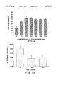
- FIG. 9 shows that free antibody inhibits the cytotoxic effect of immunotoxin.
- FIG. 10 shows the changes that transferrin receptor levels on RPE cells decrease with time in culture.
- FIG. 11 shows the effect of different doses of 454A12 MAB-rRA on confluent and proliferating human corneal endothelium (HCE) cells.
- Confluent HCE cells are not sensitive to the immunotoxin, but subconfluent cells are sensitive.
- FIG. 12 presents the percentage of protein synthesis for confluent and proliferating HCE, compared to unexposed controls, at different concentrations of 454A12 MAB-rRA.
- Confluent HCE cells are not sensitive to the immunotoxin, but subconfluent cells are sensitive.
- FIG. 13 presents the criteria for grading retinal damage in rabbit PVR model.
- FIG. 14 presents the form for evaluating growth of PVR in rabbit model.
- FIG. 15 presents inhibition of growth of human fibroblasts, in rabbit eyes by 454A12 MAB-rRA.
- the present invention is generally applicable to non-malignant hyperproliferative diseases.
- the present invention applies generally to any situation in which proliferating cells create pathological or undesirable conditions.
- diseases may affect the integument and are typified by hyperproliferative disease of the skin, such as psoriasis, keloid scarring, and various keratoses.
- benign papillomas occurring in various hollow organs of the body are intended to be within the scope of the invention.
- hyperproliferative diseases of the eye including epithelial down growth; vitreoretinopathy; diabetic retropathy; complications of eye surgeries, for example, in cataract, glaucoma, or lens extraction surgeries, as in the case of glaucoma surgery in which the cells regrow and block the drainage passage which has been surgically created to drain excess fluid from the eye; scarring, for example, in cicatricial penthagoid; and pterygium are also considered within the scope of the invention.
- the toxin conjugate according to the method of the invention comprises a toxic moiety and a binding region, which binds to an internalizable element of the proliferating cell or which is itself internalizable.
- the internalizable element of the cell will generally be located at the cell surface during some portion of the cell growth cycle.
- the internalizable element may be a specific receptor that is active at elevated levels during cell growth. Such receptors include transferrin receptor or the receptor for epidermal growth factor.
- the part of the receptor that is bound by the binding region may vary.
- the part of the receptor that is bound by the binding region may be an antigenic region of the receptor that is recognized by a specific antibody, preferably a monoclonal antibody.
- the part of the receptor bound may alternatively be the region of the receptor that actively binds to the material normally bound by the receptor or may be an antigen in that region.
- binding portion of the conjugate will specifically bind to the internalizable element.
- binding portions may be an antibody or the antigen binding portion thereof which binds to an antigen that is part of the internalizable element.
- the binding portion may be an antibody or antigen binding portion thereof that binds to an antigen which is part of the receptor.
- antigens may be located in an area of the receptor such that when the binding portion of the toxic conjugate is bound to the receptor, it does not prevent or significantly reduce the binding of the material for which the receptor is specific.
- the binding portion is an antibody, preferably a monoclonal antibody, that binds to an antigen of the transferrin receptor in such a manner that binding of transferrin to the receptor occurs.
- antibodies or antigen binding portions thereof that bind to antigens to the transferrin receptor that prevent or measurably inhibit the binding of transferrin receptor to the monoclonal antibody.
- antibody is intended to mean a polyclonal or monoclonal antibody.
- antibody is intended to mean a polyclonal or monoclonal antibody.
- the term "monoclonal antibody” means an antibody composition having a homogeneous antibody population. It is not intended to be limited as regards the source of the antibody or the manner in which it is made.
- antigen binding portion of an antibody means the portion of the antibody that binds an antigen to which the antibody is specific.
- antigen binding portions of the antibody encompass the Fab, Fab' and F(ab') 2 regions or fragments of the immunoglobin molecule.
- Fab, Fab' and F(ab') 2 regions of an immunoglobin may be generated by enzymatic digestion of the antibodies using techniques well known to those skilled in the art.
- Fab fragments may be generated by digesting the antibody with papain and contacting the digest with a reducing agent to reductively cleave disulfide bonds.
- Fab' fragments may be obtained by digesting the antibody with pepsin and reductive cleavage of the fragment so produced with a reducing agent. In the absence of reductive cleavage, enzymatic digestion of the antibody with pepsin produces F(ab') 2 fragments.
- Antibodies to the internalizable element of the proliferating cell are the preferred binding component according to the invention.
- the binding portion of the conjugate may be a material, other than an antibody to an antigen on the internalizable element, that ordinarily binds to the internalizable element and is itself ordinarily internalized.
- transferrin itself may be used as the binding portion of the conjugate.
- Epidermal growth factor could also be used in an analogous manner.
- the internalizable element of the hyperproliferating cell will usually be found at the surface of the cell during a portion of the cell growth cycle and is internalized into the cell daring another portion of the cell growth cycle. Internalization of the element may take place either periodically or in response to a stimulus such as the binding to the receptor of a material for which it is specific.
- the toxin conjugate according to methods of the invention are conjugates of the binding portion and a cytotoxic moiety.
- the cytotoxic moiety of the toxin conjugate may be a cytotoxic drug or an enzymatically active toxin of bacterial, fungal or plant origin, or an enzymatically active polypeptide chain or fragment ("A chain") of such a toxin.
- Enzymatically active toxins and fragments thereof are preferred and are exemplified by diphtheria toxin A fragment, and especially non-binding active fragments of diphtheria toxin, exotoxin A (from Pseudomonas aeruginosa), ricin A chain, abrin A chain, modecein A chain, alpha-sarcin, certain Aleurites fordii proteins, certain Dianthin proteins, Phytolacca americana proteins (PAP, PAPII and PAP-S), Momordica charantia inhibitor, curcin, crotin, Saponaria officinalis inhibitor, gelonin, mitogellin, restrictocin, phenomycin, and neomycin.
- Ricin A chain, Pseudomonas aeruginosa exotoxin A and PAP are preferred.
- Conjugates of the binding portion may be made using a variety of bi-functional protein coupling agents.
- bi-functional protein coupling agents include N-succinimidyl-3-(2-pyridyldithio) propionate (SPDP), iminothiolane (IT), bi-functional derivatives of imidoesters such as dimethyl adipimidate.
- HCl active esters such as disuccinimidyl suberate, aldehydes such as glutaraldehyde, bis-azido compounds such as bis(p-diazoniumbenzoyl)-ethylenediamine, diisocyanates such as tolylene 2,6-diisocyante, and bis-active fluorine compounds such as 1,5-fluoro-2,4-dinitrobenzene.
- active esters such as disuccinimidyl suberate
- aldehydes such as glutaraldehyde
- bis-azido compounds such as bis(p-diazoniumbenzoyl)-ethylenediamine
- diisocyanates such as tolylene 2,6-diisocyante
- bis-active fluorine compounds such as 1,5-fluoro-2,4-dinitrobenzene.
- the enzymatically active polypeptide of the toxin conjugate according to the invention may be recombinantly produced.
- the plasmid, designated pRA123, encoding the entire sequence for ricin A has been deposited with the American Type Culture Collection (ATCC), 12301 Parklawn Drive, Rockville, Md. 20852, U.S.A., under the Budapest Treaty on the International Recognition of the Deposit of Microorganisms for the Purposes of Patent Procedure and Regulations Thereunder (Budapest Treaty) and are thus made, maintained and made available according to the Budapest Treaty.
- the plasmid was deposited at the ATCC on Aug. 14, 1984, with the Accession No.
- plasmid pRA123 contains the entire sequence for ricin A, as confirmed by sequencing and comparison of the deduced amino acid sequence to that of native ricin A.
- another plasmid designated pRAP229, has been deposited with the ATCC on Mar. 8, 1985, with the Accession No. 53403, under the Budapest Treaty.
- Ricin A protein produced by E. coli transformed with the pRAP229 plasmid was used in the construction of the immunoconjugate in this patent application.
- Ricin A protein produced by E. coli transformed with the pRAP229 plasmid was in soluble form and associated with the intercellular environment.
- the ricin A derived from the pRAP229 transformants was shown to be cytotoxic both in vitro and in vivo.
- Recombinantly produced diphtheria toxin A chain and non-binding active fragments thereof are described in U.S. patent application Ser. No. 578,122 issued on May 16, 1989 as U.S. Pat. No. 4,830,962, and 648,259 issued on Jan. 16, 1990, as U.S. Pat. No. 4,894,443 which are herein incorporated by reference and are assigned to the assignee of the present invention.
- the toxin conjugates are, administered to the patient in therapeutically effective amounts i.e., amounts that eliminate or reduce or retard the increase of the hyperproliferating cells.
- the toxin conjugate will normally be administered parenterally, topically or intracavitarily. It is preferred that the administration will be local, for example, topically, ophthalmicaly or intracavitarily in the eye.
- the dose and dosage regimen will depend upon the name of the hyperproliferative disorder or the population of hyperproliferative cells, the characteristics of the particular toxin conjugate patient, and the patient's history.
- the amount of immunotoxin administered will typically be in the range of about 0.01 to about 100 mg/kg and preferably between 0.01 mg/kg and 10 mg/kg of patient weight.
- An anti-transferrin receptor monoclonal antibody 454A12, (454A12 MAB) was made in accordance with the description in U.S. patent application Serial No. 806,320, a continuation of which has now issued as U.S. Pat. No. 4,958,009 to Bjorn et al., which is herein incorporated by reference.
- the 454A12 hybridoma was deposited with the American Type Culture Collection (ATCC), Rockville, Md. U.S.A. under the terms of the Budapest Treaty and assigned Accession No. HB 10804. This antibody binds to a 95 K dalton antigen identified as transferrin receptor, but does not block binding of transferrin to the receptor.
- Soluble recombinant ricin Toxin A chain can be produced using those methods described above.
- SPDP (20 mM in ethanol) was added in a 20-fold molar excess to antibody and following a 30 min incubation at room temperature, the unreacted SPDP was removed by dialysis against PBS. The extent of derivatization was determined by measuring the release of pyridine-2-thione at 343 nm after reduction with dithiothreitol (DTT). Depending on the antibody, three to eight lysine amino acid groups (per antibody molecule) were converted to the pyridyl-disulfide derivative.
- DTT dithiothreitol
- the SPDP-treated antibodies were conjugated with RTA.
- the RTA was reduced with 50 mM DTT, then desalted on a column of chromatographic resin containing agarose, dextran and/or acrylamide to remove DTT from protein.
- Reduced RTA was added in a three- to five-fold molar excess over pyridyl-disulfide antibody.
- a typical reaction mixture (1 ml) consisted of 7 ⁇ M antibody and 30 ⁇ m RTA. The reaction was allowed to proceed overnight at 4° C.
- the extent of conjugation of RTA to antibody was determined spectrophotometrically by measuring the release of pyridine-2-thione. On the average, conjugates contained two to three RTA molecules per antibody molecule. This was confirmed by non-reducing SDS-PAGE gels (7.5%), which also revealed that the typical conjugate preparation contained 10%-30% free antibody.
- the conjugate mixture was chromatographed on a HPLC size exclusion column to separate conjugates from residual unreacted RTA.
- the column was equilibrated in 0.1 sodium sulfate/0.02M sodium phosphate pH 6.8. Conjugate mixture (0.7 ml) was injected, then chromatographed at a flow rate of 1 ml/min (room temperature). Fractions of 0.5 ml were collected and the peak conjugate fractions were pooled and filter sterilized prior to cytotoxicity testing.
- P-EDTA buffer Approximately 30 mg/ml antibody in 0.10M Na phosphate, 0.001M Na EDTA, pH 8.0 (hereafter referred to as P-EDTA buffer) is reacted with 1 mM 5,5'-dithiobis-(2-nitrobenzoic acid) (DTNB) at room temperature for about 15 min and then chilled to 0° C. in an ice bath. Enough IT is added to this solution to give an average of 2.5 IT molecules/antibody molecule, and the resulting solution is dialyzed at 0°-5° C. against three 100-fold excess volumes of P-EDTA buffer.
- DTNB 5,5'-dithiobis-(2-nitrobenzoic acid)
- RTA normally stored in P-EDTA containing 1 mM DTF, is ultra-filtered to a concentration between 10 and 15 mg/ml and dialyzed at 0°-5° C. against three 100-fold excess volumes of P-EDTA. Enough RTA is added to the derivatized antibody to give an average of 1.0-1.2 free thiols on RTA per blocked thiol on derivatized antibody. This mixture is incubated at room temperature for 2 hours.
- the coupling reaction mixture is applied to a column of a chromatographic resin based on a blue dye (trysacryl blue) covalently coupled to a solid support, which mixture is then eluted with P-EDTA at room temperature.
- the column is scaled to contain approximately 2 ml of bed volume per mg of starting antibody.
- the eluant is switched to P-EDTA containing 1M NaCl. Immunoconjugate and unreacted RTA are eluted in this buffer as a very sharp peak, which is pooled and dialyzed at 0°-5° C.
- p i buffer 0.15M Na phosphate, pH 7.1
- the dialyzed protein is applied to a column of a size-exclusion gel at 0°-5° C. and eluted with buffer at a flow rate of 6 cm/hr.
- the column is scaled to contain at least 25 ml of bed volume/ml of applied protein.
- Immunoconjugate is eluted as a single peak, slightly after the excluded volume, baseline-resolved from following peaks of dimerized and monomeric RTA.
- the pooled immunoconjugate peak is ultra-filtered at 35 psi to a final concentration of 5.0 mg/ml and filter-sterilized.
- Human corneal endothelial cell cultures were obtained from J. Polansky, Ph.D. of the University of California at San Francisco. The cells were grown in plastic tissue culture flasks (Falcon) in M199 medium (Irving Scientific, Santa Anna, Calif.), supplemented to the indicated final concentration with 20% fetal bovine serum (Flow Laboratories), penicillin 100 U/ml, streptomycin 10 ⁇ g/ml, fungizone 0.25 ⁇ g/ml (Irvine Scientific, Santa Anna, Calif.) and glutamine 2.0 mM. Cells were harvested by mild trypsinization with 0.2 ml saline containing trypsin and versine-EDTA STV for 20 minutes at 37° C.
- FIG. 2A is a photomicrograph of the 6 day non-confluent culture 7 days after treatment with the toxin conjugate. The surviving cells of the 11 day confluent culture were tightly packed and regular in morphology.
- FIG. 2B is a photomicrograph of the 11 day confluent culture 7 days after treatment with the toxin conjugate.
- FIG. 2C is a photomicrograph of an 18 day confluent culture that was not treated with the toxin conjugate. The cell morphology is the same as the treated 11 day confluent culture.
- a protein synthesis inhibition assay was carried out using HSB-2 cells as indicator cells and the immunoconjugate preincubated with the confluent culture supernatant or with fresh cell culture medium.
- the immunotoxin 454A12 MAB-rRA comprising 454A12 MAB conjugated to recombinant ricin A chain (rRA) could be used to treat pathological proliferation of fibroblasts including retinal pigment epithelia (RPE) cells.
- RPE retinal pigment epithelia
- RPE cells were isolated from human donor eyes and grown on laminin-coated tissue culture dishes by techniques known in the art. Cultures were inspected to determine the degree of confluence and the cell number was determined by trypsinization and counting on a hemocytometer. The rate of cell growth differed between the donors.
- RPE cells were grown to semi-confluence (approximately 1.4 ⁇ 10 5 cells per plate) in fresh medium and incubated with various concentrations of immunotoxin (454A12 MAB-rRA), antibody (454A12 MAB) or recombinant ricin A chain (rRA) that had been diluted into the medium.
- the cells were incubated for 4 hours at 37° C. Then the cells were washed free of immunotoxin, antibody or rRA, and incubated in supplemented F-10 medium for a further 18 hours. They were rinsed again in methionine-free minimal essential medium (Flow Labs, 7655 Old Springhouse Road, McClean, Va. 22102) and incubated for 1 hour in medium with 35 S!methionine (Amersham Corp., 2636 South Clearbrook Drive, Arlington Heights, Ill. 60005) to measure the rate of protein synthesis.
- FIG. 4 shows that there was a dose dependent inhibition of cell protein synthesis after pre-incubation with immunotoxin. No such effect was seen when anti-transferrin receptor antibody alone was incubated with the cells instead of the immunotoxin, suggesting that the antibody itself was not the cause of the inhibitory effect (FIG. 4). No inhibitory effect was observed in similar experiments in which unconjugated ricin A chain, instead of the immunotoxin was incubated with the cells (FIG. 5). These data, therefore, show that for inhibition to occur, rRA and antibody must be part of a covalent conjugate.
- FIGS. 4 and 5 are representative of three similar experiments.
- FIGS. 6 and 7 show the results of two of these experiments.
- no effect of the immunotoxin or free antibody was apparent after pre-incubation of either with non-proliferating cells.
- Confluent cells from another set of donors also showed inhibition by immunotoxin, but to a lesser degree; an example of this is shown in FIG. 7. Additionally, it was found that as with sub-confluent cells, free toxin had no effect on confluent cells (not shown).
- the immunotoxin consisted of recombinant ricin A chain (rRA) chemically linked by iminothiolane (IT) to a monoclonal antibody (MAB).
- human corneal endothelial cells were scraped from donor corneas within two hours of harvesting.
- Stock cultures were seeded on gelatinized 35 mm dishes in medium 199 containing 15% fetal calf serum, Earl's balanced salts, and 1% of 200 mM glutamine.
- the cells were maintained in 5% CO 2 at 37° C. with fibroblast growth factor added every other day. Once confluent, cells were trypsinized and seeded in 2 mL of medium on gelatinized 35 mm dishes at 2 ⁇ 10 4 cells/mL for individual experiments.
- FIG. 11 The results (FIG. 11) showed that immunotoxin 454A12 MAB-rRA significantly inhibited proliferating human corneal endothelium (HCE) in a dose dependent fashion. A concentration of 50 ng/ml of the immunotoxin caused an 89% decrease in the number of viable cells. Protein synthesis was inhibited by immunotoxin in both proliferating and non-proliferating cells but less effectively in the latter.
- FIG. 12 The concentration of immunotoxin which produced 50% inhibition of cell growth (IC 50 ) was calculated to be 100 ng/ml for proliferating cells but 1000 ng/ml for confluent cells.
- Amputated rat tails were soaked in 70% ethanol in water for 2 hours after which the skin was reflected.
- the tails were sectioned into 3 cm segments which were placed in 70% ethanol/H 2 O for 1 hour then washed three times with 70% ethanol.
- the ethanol solution was decanted and replaced with an equal volume of sterile, normal saline for 90 minutes.
- the tail segments were squeezed at their mid-point to induce extrusion of tendons from the cut ends.
- the tendons were washed twice with normal saline and placed into glacial acetic acid solution (1 part glacial acetic acid in 1000 parts distilled water) with a magnetic stirring bar.
- the solution was stirred in a sealed container at 4°-6° C. for 48 to 72 hours, after which the suspension was decanted into sterilized centrifuge tubes and spun at 25000 x g for 2 hours at 4° C.
- the sterile collagen solution (supernatant) was transferred to sterile polypropylene robes, and the protein concentration was adjusted to 1.4 mg/mL (Kjeldahl method). The collagen solution was then tested for its ability to form a mechanically stable gel within 20 seconds at 37° C. (only such solutions were used in the described experiments). The collagen solution was stored at -76° C. or in liquid nitrogen.
- the collagen solution was thawed and all reagents and robes were cooled to 4° C.
- To 1500 ⁇ L collagen solution was added 600 ⁇ L 5 x RPMI 1640, 300 ⁇ L FBS, 100 ⁇ L 0.4M NaOH, 107 ⁇ L 5.6% NaHCO 3 , 30 ⁇ L 200 mM L-glutamine, 30 ⁇ L penicillin (5000 U/mL)/streptomycin (5000 ⁇ g/mL) and 335 ⁇ L distilled H 2 O.
- the robe was capped, the contents rapidly mixed, placed in a 37° C. water bath, and gently tilted to 60° relative to the vertical at 15 second intervals. Gelling was noted. The gel was destroyed with a glass rod and the pH was measured. If the pH was not 7.4, it was adjusted by adding NaOH to the reaction mixture. If the collagen did not gel within 5 minutes, either the pH required further adjustment or the collagen solution was old and therefore not used.
- a solution of the following composition was prepared: 200 ⁇ L of 5 x RPMI 1640 without glutamine or NaHCO 3 , 36 ⁇ L 5.6% NaHCO 3 , 33 ⁇ L 0.4M sterile NaOH, 111 ⁇ L sterile H 2 O, 10 ⁇ L penicillin (5000 U/mL)/streptomycin (5000 ⁇ g/mL) and 10 ⁇ L L-glutamine (200 mM).
- the cell suspension and the solution were rapidly mixed and added to 500 gL of collagen solution yielding a solution of 5 ⁇ 10 4 cells/mL collagen gel containing 0.7 mg collagen/mL.
- a 150 ⁇ L aliquot of this gel solution was dispensed to 96-well plates.
- the gels were overlaid with 50 ⁇ L of RPMI growth medium with or without 3 H thymidine (0.1 ⁇ C 1 /well).
- the gel was overlaid with 50 ⁇ L of an aqueous solution of 15 mg collagenase and 0.625 mL Triton per 100 mL water, and the plate was incubated for 3 hours at 37° C.
- Cells were harvested with a Cambridge Technology PHD Model 200 A automated cell harvester using a distilled water rinse. Each filter section was placed into a counting vial to which 4 mL of Aquasol® was added, vortexed, allowed to stand for 30 minutes, and counted for 5 minutes in a scintillation counter.
- the immunotoxin 454A12 MAB-rRA was compared for activity with ricin A chain, the monoclonal antibody 454A12, and a non-specific immunoglobulin with ricin A chain attached by a similar chemical linkage, i.e., by SPDP-disulfide linkage. All compounds were tested in the simulated vitreous medium (Example VI) used to demonstrate the activity of the immunotoxin. The study was carried out as follow:
- MOPC21-IT-srRTA is a non-specific immunoglobulin (IgG 1 ) combined to ricin A chain which, because of its non-specificity, is frequently used as a negative control in immunotoxin studies.
- the compound sr-RA is soluble, recombinant ricin A chain.
- the following in vivo studies showed that the immunotoxin 454A12 MAB-rRA significantly reduced the proliferation of human fibroblasts injected into the vitreous cavities of rabbits.
- the human fibroblasts had been incubated in rabbit eyes to simulate human vitreoretinopathy.
- the result strongly suggested that the immunotoxin could be effective in the prevention of vitreoretinopathy.
- Human foreskin fibroblasts were grown at 37° C. in a humidified atmosphere in RPMI medium supplemented with 10% fetal calf serum in polyethylene flasks. After 8 to 16 passages, cells were harvested by incubation with 0.25% trypsin for 5 minutes. The digestion was stopped by addition of RPMI with fetal calf serum, the cells were centrifuged at 1000 rpm for 5 minutes, then resuspended in a serum free RPMI solution, and a sample was removed for viability testing by trypan blue exclusion and counting in a hemocytometer. Cells were re-centrifuged and diluted to a final concentration of 250,000 cells/0.1 mL.
- Pigmented rabbits (4.0-5.5 kg) were anesthetized with ketamine hydrochloride, acepromazine, and rumfin.
- a bloodless-injection area was prepared by the following sequence of steps: first by tattooing a 1.0 to 1.5 mm diameter region of the sclera located 5 to 6 mm posterior to the limbus in the anterior superior quadrant. One week later 5 or 6 applications of a Keeler Amoiles retinal cryoprobe cooled to -60° C. were made over the tattooed area. After 3 to 4 weeks, the area of cryopexy was examined with an indirect ophthalmoscope. Cryopexy was repeated if there was not a marked degree of choroidal pigment alteration.
- a paracentesis was performed to remove 0.2 to 0.3 mL of aqueous from the anterior chamber.
- 500,000 human fibroblasts (suspended in 0.2 mL RPMI) were injected through the area of diathermy using a 30 gauge needle under direct observation with the indirect ophthalmoscope. The needle was positioned to inject the cells approximately 2 mm above the optic nerve and medullary rays.
- a second paracentesis was performed 30 minutes later and 8000 ng of 454A12 MAB-rRA in 0.1 mL RPMI was injected into one eye while the contralateral eye was injected with 0.1 mL of RPMI. Randomization of the eyes to control or treated groups was determined by the flip of a coin.
- the extent of inhibition in the proliferation of the human fibroblasts was determined by comparing the growth between the two eyes of each animal using the unpaired two-tailed t test.
- the percent inhibition by the immunotoxin relative to the control was determined by calculating the difference between the control and treated groups and expressing this difference as percent of the control, i.e., (C-T)/C! ⁇ 100, for each animal at each time the eyes were examined (Table 2). Stimulation of growth was represented as a negative number. After the percent inhibition was calculated, the median inhibition was determined for each rabbit over the 12 weeks of the study. Using this method, at least 50% of the rabbits treated with the immunotoxin had inhibition of at least 62.5% whether inhibition was shown as early as the first two weeks or by a later time period.
- the toxin conjugate will be formulated in a unit dosage injectable form (solution, suspension, emulsion) in association with a pharmaceutically acceptable parenteral vehicle.
- a pharmaceutically acceptable parenteral vehicle Such vehicles are inherently nontoxic and non-therapeutic. Examples of such vehicles are water, saline, Ringer's solution, dextrose solution, and 5% human serum albumin. Nonaqueous vehicles such as fixed oils and ethyl oleate may also be used. Liposomes may be used as carriers.
- the vehicle may contain minor amounts of additives such as substances that enhance isotonicity and chemical stability, e.g., buffers and preservatives.
- the immunotoxin will typically be formulated in such vehicles at concentrations of about 0.01 mg/ml to 100 mg/ml.
- the above-described compounds may be formulated with any one of a number of well known pharmaceutically acceptable carriers, depending upon the optimal mute of administration, e.g., topical, ophthalmic, parenteral, including intravenous, intraperitoneal, intracavitarily or intraophthalmic.
- Such carriers include solutions compatible with the mode of administration and solubility of the compounds. Such solutions may be buffered or otherwise formulated to minimize undesirable localized effects of injection if necessary. Formations for administration are also well known to those skilled in this art and may be formulated for various effects, including timed, slow and delayed release. The compounds, formulated for these effects, may be administered in the form of suspensions, slurries and liquids. Such dosage forms may also include excipients, or other therapeutically inert ingredients in the formulation of the desired pharmaceutical preparation.
- the compounds of this invention can be formulated with sterile ingredients compounded and packaged aseptically. They may be administered intravenously or intracavitarily.
- Useful solvents for formulation in such use are the polyhydric aliphatic alcohols and mixtures thereof.
- the pharmaceutically acceptable glycols such as propylene glycol, and mixtures thereof.
- Glycerine is another example of a polyol which is a particularly convenient solvent system.
- Basicity may be controlled by the addition of a base as required, and a particularly convenient base is monoethanolamine.
- the toxin conjugate may be formulated in an ointment, augment or highly viscous oil or cream or lotion suitable for topical use.
- suitable topical formulations are known to those skilled in the art.
- Topical application of the immunotoxin may be used, for example, in the treatment and prevention of pterygium, or in the prevention of scarring due to cell proliferations.
- the immunotoxin can be topically applied; injected into the afflicted site after surgery; or injected into the growing end of a small pterygium. Similar methods of applications by injection may be applied in the case of post glaucoma surgery.
- the toxin conjugate may be in the form of drops or an ointment or topical slow release gel suitable for ophthalmic application.
- suitable ophthalmic formulations are known to those skilled in the art.
- the percentage of the compound to be used in the pharmaceutical carrier may be varied. It is necessary that the compound constitute a proportion such that a suitable dosage will be obtained.
- the dosage required to achieve the desired pharmacologic activity in the mammal will vary with various factors such as mute of administration, the species of mammal, general health and tolerances of the mammal, weight, sex and age of the mammal, the nature and severity of the disease being treated and the like. Additionally, it is to be noted that the exact dosage of each individual compound employed in similar situations will vary. Generally, a dosage would be in the range of from about 0.1 to about 100 nanogram or more per kilogram of body weight, and usually from 1 to about 20 nanogram per kilogram of body weight.
Abstract
Description
Claims (10)
Priority Applications (1)
| Application Number | Priority Date | Filing Date | Title |
|---|---|---|---|
| US07/807,951 US5670151A (en) | 1986-04-28 | 1991-12-10 | Method for controlling hyperproliferative diseases |
Applications Claiming Priority (3)
| Application Number | Priority Date | Filing Date | Title |
|---|---|---|---|
| US85673186A | 1986-04-28 | 1986-04-28 | |
| US43653889A | 1989-11-14 | 1989-11-14 | |
| US07/807,951 US5670151A (en) | 1986-04-28 | 1991-12-10 | Method for controlling hyperproliferative diseases |
Related Parent Applications (1)
| Application Number | Title | Priority Date | Filing Date |
|---|---|---|---|
| US43653889A Continuation | 1986-04-28 | 1989-11-14 |
Publications (1)
| Publication Number | Publication Date |
|---|---|
| US5670151A true US5670151A (en) | 1997-09-23 |
Family
ID=27031003
Family Applications (1)
| Application Number | Title | Priority Date | Filing Date |
|---|---|---|---|
| US07/807,951 Expired - Fee Related US5670151A (en) | 1986-04-28 | 1991-12-10 | Method for controlling hyperproliferative diseases |
Country Status (1)
| Country | Link |
|---|---|
| US (1) | US5670151A (en) |
Cited By (6)
| Publication number | Priority date | Publication date | Assignee | Title |
|---|---|---|---|---|
| US6207805B1 (en) | 1997-07-18 | 2001-03-27 | University Of Iowa Research Foundation | Prostate cell surface antigen-specific antibodies |
| US20060067885A1 (en) * | 1992-04-07 | 2006-03-30 | Immunomedics, Inc. | Method and kit for imaging and treating organs and tissues |
| US20060293599A1 (en) * | 2003-01-23 | 2006-12-28 | Dhiraj Sardar | Method and apparatus for diagnosing neovascularized tissues |
| US20070259020A1 (en) * | 2006-04-25 | 2007-11-08 | Robert Langer | Methods and compositions for the treatment of open and closed wound spinal cord injuries |
| US20090053284A1 (en) * | 2006-04-25 | 2009-02-26 | Children's Medical Center Corporation | Medical devices for use in the surgical treatment of hyperproliferative diseases affecting the spinal cord |
| US7973019B1 (en) * | 2007-10-03 | 2011-07-05 | Alcon Research, Ltd. | Transferrin/transferrin receptor-mediated siRNA delivery |
Citations (3)
| Publication number | Priority date | Publication date | Assignee | Title |
|---|---|---|---|---|
| US4434156A (en) * | 1981-10-26 | 1984-02-28 | The Salk Institute For Biological Studies | Monoclonal antibodies specific for the human transferrin receptor glycoprotein |
| US4830962A (en) * | 1984-02-09 | 1989-05-16 | Cetus Corporation | Recombinant diphtheria toxin fragments |
| US4894443A (en) * | 1984-02-08 | 1990-01-16 | Cetus Corporation | Toxin conjugates |
-
1991
- 1991-12-10 US US07/807,951 patent/US5670151A/en not_active Expired - Fee Related
Patent Citations (3)
| Publication number | Priority date | Publication date | Assignee | Title |
|---|---|---|---|---|
| US4434156A (en) * | 1981-10-26 | 1984-02-28 | The Salk Institute For Biological Studies | Monoclonal antibodies specific for the human transferrin receptor glycoprotein |
| US4894443A (en) * | 1984-02-08 | 1990-01-16 | Cetus Corporation | Toxin conjugates |
| US4830962A (en) * | 1984-02-09 | 1989-05-16 | Cetus Corporation | Recombinant diphtheria toxin fragments |
Non-Patent Citations (14)
| Title |
|---|
| Fastenberg, D.M, et al., 1982, Am. J. Ophthalmol ., 93: 565 572. * |
| Fastenberg, D.M, et al., 1982, Am. J. Ophthalmol., 93: 565-572. |
| Fastenberg, D.M., et al., 1982, Am. J. Ophthalmol ., 93:559 564. * |
| Fastenberg, D.M., et al., 1982, Am. J. Ophthalmol., 93:559-564. |
| Fulcher, S. et al. (II), "Use of Immunotoxin to Inhibit Proliferating Human Corneal Endothelium," Invest. Ophthalmol. Visual. Sci. 29(5): 755-759, May 1988. |
| Fulcher, S. et al. (II), Use of Immunotoxin to Inhibit Proliferating Human Corneal Endothelium, Invest. Ophthalmol. Visual. Sci . 29(5): 755 759, May 1988. * |
| Fulcher, S., et al., (I), "Effect of an Immunotoxin Containing Ricin A Chain and Monoclonal Antibody Against the Transferrin Receptor on the Proliferation of Human Corneal Endothelium", Annual Spring Meeting of the Association for Research in Vision and Ophthamology Incorporated, Sarasota, Fla, USA, Apr. 28-May 2, 1986, Invest Ophthalmology Visual Science,27 (3 Suppl.):69, 1986. |
| Fulcher, S., et al., (I), Effect of an Immunotoxin Containing Ricin A Chain and Monoclonal Antibody Against the Transferrin Receptor on the Proliferation of Human Corneal Endothelium , Annual Spring Meeting of the Association for Research in Vision and Ophthamology Incorporated, Sarasota, Fla, USA, Apr. 28 May 2, 1986, Invest Ophthalmology Visual Science ,27 (3 Suppl.):69, 1986. * |
| Ramakrishan, S., 1984, Science , 223:58 61. * |
| Ramakrishan, S., 1984, Science, 223:58-61. |
| Trowbridge, I.S. et al. "Anti-transferrin receptor monoclonal antibody and toxin--antibody conjugates affect growth of human tumor cells," Nature 294: 171-173, Nov. 12 ,1981. |
| Trowbridge, I.S. et al. Anti transferrin receptor monoclonal antibody and toxin antibody conjugates affect growth of human tumor cells, Nature 294: 171 173, Nov. 12 ,1981. * |
| Van Bockxmeer, F.M. et al., 1982, J. Tissue Culture Methods , 7:163 167. * |
| Van Bockxmeer, F.M. et al., 1982, J. Tissue Culture Methods, 7:163-167. |
Cited By (21)
| Publication number | Priority date | Publication date | Assignee | Title |
|---|---|---|---|---|
| US20110117105A1 (en) * | 1992-04-07 | 2011-05-19 | Immunomedics, Inc. | Method of treating immune disease using b-cell antibodies |
| US20060067885A1 (en) * | 1992-04-07 | 2006-03-30 | Immunomedics, Inc. | Method and kit for imaging and treating organs and tissues |
| US7811570B2 (en) * | 1992-04-07 | 2010-10-12 | Immunomedics, Inc. | Method of affecting a function of or ablating a non-malignant cell |
| US6512096B2 (en) | 1997-07-18 | 2003-01-28 | University Of Iowa Research Foundation | Prostate cell surface antigen-specific antibodies |
| US6207805B1 (en) | 1997-07-18 | 2001-03-27 | University Of Iowa Research Foundation | Prostate cell surface antigen-specific antibodies |
| US20060293599A1 (en) * | 2003-01-23 | 2006-12-28 | Dhiraj Sardar | Method and apparatus for diagnosing neovascularized tissues |
| US9173562B2 (en) * | 2003-01-23 | 2015-11-03 | The Board Of Regents Of The University Of Texas System | Method and apparatus for diagnosing neovascularized tissues |
| US8377463B2 (en) | 2006-04-25 | 2013-02-19 | Children's Medical Center Corporation | Methods and compositions for the treatment of open and closed wound spinal cord injuries |
| US9173732B2 (en) * | 2006-04-25 | 2015-11-03 | The Children's Medical Center Corporation | Medical devices for use in the surgical treatment of hyperproliferative diseases affecting the spinal cord |
| US9440008B2 (en) | 2006-04-25 | 2016-09-13 | Children's Medical Center Corporation | Methods and compositions for the treatment of open and closed wound spinal cord injuries |
| US20090053284A1 (en) * | 2006-04-25 | 2009-02-26 | Children's Medical Center Corporation | Medical devices for use in the surgical treatment of hyperproliferative diseases affecting the spinal cord |
| US20070259020A1 (en) * | 2006-04-25 | 2007-11-08 | Robert Langer | Methods and compositions for the treatment of open and closed wound spinal cord injuries |
| US8685434B2 (en) | 2006-04-25 | 2014-04-01 | Children's Medical Center Corporation | Methods and compositions for the treatment of open and closed wound spinal cord injuries |
| US8858966B2 (en) | 2006-04-25 | 2014-10-14 | Children's Medical Center Corporation | Methods and compositions for the treatment of open and closed wound spinal cord injuries |
| US9101695B2 (en) | 2006-04-25 | 2015-08-11 | Children's Medical Center Corporation | Methods and compositions for the treatment of open and closed wound spinal cord injuries |
| US9114152B2 (en) | 2007-10-03 | 2015-08-25 | Arrowhead Research Corporation | Transferrin/transferrin receptor-mediated siRNA delivery |
| US8399653B2 (en) * | 2007-10-03 | 2013-03-19 | Alcon Research, Ltd. | Transferrin/transferrin receptor-mediated siRNA delivery |
| US7973019B1 (en) * | 2007-10-03 | 2011-07-05 | Alcon Research, Ltd. | Transferrin/transferrin receptor-mediated siRNA delivery |
| US20110256156A1 (en) * | 2007-10-03 | 2011-10-20 | Alcon Research, Ltd. | TRANSFERRIN/TRANSFERRIN RECEPTOR-MEDIATED siRNA DELIVERY |
| US9562230B2 (en) | 2007-10-03 | 2017-02-07 | Arrowhead Pharmaceuticals, Inc. | Transferrin/transferrin receptor-mediated siRNA delivery |
| US10138483B2 (en) | 2007-10-03 | 2018-11-27 | Arrowhead Pharmaceuticals, Inc. | Transferrin/transferrin receptor-mediated siRNA delivery |
Similar Documents
| Publication | Publication Date | Title |
|---|---|---|
| US6013628A (en) | Method for treating conditions of the eye using polypeptides | |
| US5627162A (en) | Methods and means for control of proliferation of remnant cells following surgery | |
| Ambati et al. | Transscleral delivery of bioactive protein to the choroid and retina | |
| CA2120506C (en) | Treatment of ocular inflammation by blockage of cell adhesion molecules | |
| Hom et al. | The progression of the inflammation in established collagen‐induced arthritis can be altered by treatments with immunological or pharmacological agents which inhibit T cell activities | |
| JPH01100133A (en) | Mitotic inhibitor and method for preventing cloudiness of postorier lens capsule after extraction out of capsule | |
| US5055291A (en) | Compositions for preventing secondary cataracts | |
| Russ et al. | Partial characterization of the human retinal endothelial cell tight and adherens junction complexes. | |
| US5670151A (en) | Method for controlling hyperproliferative diseases | |
| WO2005117987A1 (en) | Antibody conjugates targeting to ocular proteins | |
| US5510329A (en) | Preparations for the treatment of eyes | |
| KR20120093540A (en) | Pharmaceutical composition for eye disease containing fk506 binding protein fusion protein | |
| Fulcher et al. | Use of immunotoxin to inhibit proliferating human corneal endothelium. | |
| Maurice et al. | Permanent destruction of the corneal endothelium in rabbits. | |
| Goins et al. | Inhibition of proliferating lens epithelium with antitransferrin receptor immunotoxin | |
| US5616122A (en) | Methods and compositions for preventing secondary cataracts | |
| JP2001521008A (en) | Immune composition for promoting nerve regeneration by transiently altering central nervous system myelin in mammals and method of using same | |
| Kuo et al. | Inhibition of corneal neovascularization with plasmid pigment epithelium-derived factor (p-PEDF) delivered by synthetic amphiphile INTeraction-18 (SAINT-18) vector in an experimental model of rat corneal angiogenesis | |
| Christiansen et al. | Long-term effects of ricin-mAb 35 on extraocular muscles of rabbits: potential treatment for strabismus | |
| Davis et al. | Selective inhibition of growing pigment epithelial cells by a receptor-directed immunotoxin. | |
| WO1995033492A1 (en) | Methods and compositions for modulation of wound healing | |
| US5972889A (en) | Method for inhibiting the proliferation of epithelial lens cells and implantable lens therefor | |
| FI95203B (en) | Analog method for preparing a cytotoxic composition | |
| WO1995033492A9 (en) | Methods and compositions for modulation of wound healing | |
| US5877154A (en) | Pharmaceutical compositions for the treatment or prevention of disorders in the eye |
Legal Events
| Date | Code | Title | Description |
|---|---|---|---|
| AS | Assignment |
Owner name: CETUS ONCOLOGY CORPORATION Free format text: CHANGE OF NAME;ASSIGNOR:CETUS CORPORATION;REEL/FRAME:006268/0881 Effective date: 19920304 |
|
| AS | Assignment |
Owner name: CETUS ONCOLOGY CORPORATION, CALIFORNIA Free format text: ASSIGNMENT OF ASSIGNORS INTEREST;ASSIGNORS:LARRICK, JAMES W.;HOUSTON, L.L.;GROVES, ERIC S.;REEL/FRAME:007294/0462;SIGNING DATES FROM 19940928 TO 19941028 |
|
| AS | Assignment |
Owner name: CHIRON CORPORATION, CALIFORNIA Free format text: MERGER;ASSIGNOR:CETUS ONCOLOGYCORPORATION (FORMERLY CETUS CORPORATION);REEL/FRAME:008209/0087 Effective date: 19960325 |
|
| FEPP | Fee payment procedure |
Free format text: PAT HLDR NO LONGER CLAIMS SMALL ENT STAT AS INDIV INVENTOR (ORIGINAL EVENT CODE: LSM1); ENTITY STATUS OF PATENT OWNER: LARGE ENTITY |
|
| FPAY | Fee payment |
Year of fee payment: 4 |
|
| FPAY | Fee payment |
Year of fee payment: 8 |
|
| REMI | Maintenance fee reminder mailed | ||
| LAPS | Lapse for failure to pay maintenance fees | ||
| STCH | Information on status: patent discontinuation |
Free format text: PATENT EXPIRED DUE TO NONPAYMENT OF MAINTENANCE FEES UNDER 37 CFR 1.362 |
|
| FP | Lapsed due to failure to pay maintenance fee |
Effective date: 20090923 |