US5733543A - Introduction of HIV-protective genes into cells by particle-mediated gene transfer - Google Patents
Introduction of HIV-protective genes into cells by particle-mediated gene transfer Download PDFInfo
- Publication number
- US5733543A US5733543A US08/235,277 US23527794A US5733543A US 5733543 A US5733543 A US 5733543A US 23527794 A US23527794 A US 23527794A US 5733543 A US5733543 A US 5733543A
- Authority
- US
- United States
- Prior art keywords
- cells
- gene
- hiv
- rev
- particles
- Prior art date
- Legal status (The legal status is an assumption and is not a legal conclusion. Google has not performed a legal analysis and makes no representation as to the accuracy of the status listed.)
- Expired - Fee Related
Links
Images


Classifications
-
- C—CHEMISTRY; METALLURGY
- C07—ORGANIC CHEMISTRY
- C07K—PEPTIDES
- C07K14/00—Peptides having more than 20 amino acids; Gastrins; Somatostatins; Melanotropins; Derivatives thereof
- C07K14/005—Peptides having more than 20 amino acids; Gastrins; Somatostatins; Melanotropins; Derivatives thereof from viruses
-
- A—HUMAN NECESSITIES
- A61—MEDICAL OR VETERINARY SCIENCE; HYGIENE
- A61K—PREPARATIONS FOR MEDICAL, DENTAL OR TOILETRY PURPOSES
- A61K48/00—Medicinal preparations containing genetic material which is inserted into cells of the living body to treat genetic diseases; Gene therapy
-
- C—CHEMISTRY; METALLURGY
- C12—BIOCHEMISTRY; BEER; SPIRITS; WINE; VINEGAR; MICROBIOLOGY; ENZYMOLOGY; MUTATION OR GENETIC ENGINEERING
- C12N—MICROORGANISMS OR ENZYMES; COMPOSITIONS THEREOF; PROPAGATING, PRESERVING, OR MAINTAINING MICROORGANISMS; MUTATION OR GENETIC ENGINEERING; CULTURE MEDIA
- C12N15/00—Mutation or genetic engineering; DNA or RNA concerning genetic engineering, vectors, e.g. plasmids, or their isolation, preparation or purification; Use of hosts therefor
- C12N15/09—Recombinant DNA-technology
- C12N15/87—Introduction of foreign genetic material using processes not otherwise provided for, e.g. co-transformation
-
- A—HUMAN NECESSITIES
- A61—MEDICAL OR VETERINARY SCIENCE; HYGIENE
- A61K—PREPARATIONS FOR MEDICAL, DENTAL OR TOILETRY PURPOSES
- A61K38/00—Medicinal preparations containing peptides
-
- C—CHEMISTRY; METALLURGY
- C12—BIOCHEMISTRY; BEER; SPIRITS; WINE; VINEGAR; MICROBIOLOGY; ENZYMOLOGY; MUTATION OR GENETIC ENGINEERING
- C12N—MICROORGANISMS OR ENZYMES; COMPOSITIONS THEREOF; PROPAGATING, PRESERVING, OR MAINTAINING MICROORGANISMS; MUTATION OR GENETIC ENGINEERING; CULTURE MEDIA
- C12N2740/00—Reverse transcribing RNA viruses
- C12N2740/00011—Details
- C12N2740/10011—Retroviridae
- C12N2740/16011—Human Immunodeficiency Virus, HIV
- C12N2740/16311—Human Immunodeficiency Virus, HIV concerning HIV regulatory proteins
- C12N2740/16322—New viral proteins or individual genes, new structural or functional aspects of known viral proteins or genes
Definitions
- the present invention relates to a method for introducing a gene into a patient's T cells, monocytes, macrophages, dendrites, or hematopoietic stem cells and to a method for introducing HIV protective genes into a patient's cells by particle-mediated gene transfer and reagents and kits useful in such methods.
- HIV human immunodeficiency Virus
- AIDS acquired immunodeficiency syndrome
- HIV infection has achieved epidemic proportions.
- Infection by the human immunodeficiency virus is typically characterized by an asymptomatic, or latent, phase of the disease.
- patients may not exhibit signs of immunodeficiency but nonetheless synthesize virus which progressively depletes CD4 + T cells and is infectious.
- seropositive cases with latent HIV infection in the United States today.
- NF- ⁇ B a cellular transcription factor that controls export of viral RNA from the nucleus to the cytoplasm of infected cells.
- Viral replication is critically dependent on the interaction of viral gene products with host cell factors. Because viruses are intimately associated with their host cells, it has been difficult to selectively interfere with replication in vivo. Successful antiviral approaches have selectively targeted viral gene products. For example, the treatment of herpes simplex virus (HSV) infection has taken advantage of the ability of a viral gene, thymidine kinase, to modify a drug which is toxic to the host cell. This approach led to the development of guanosine analogues, including acyclovir and ganciclovir (K. O. Smith, K. S. Galloway, W. L. Kennell, K. K. Ogilvie, B. K. Radatus, Antimicrob. Agents Chemother., vol.
- HSV herpes simplex virus
- AZT has relative selectivity for viral reverse transcriptase, its toxic effect on host cell function and its low therapeutic index has provided limited protection against the progression of AIDS.
- FIG. 1 provides schematic diagrams of Rev-responsive CAT reporter RSV-CAT-RRE, RSV-Rev and the different promoter protective plasmids.
- SD splice donor
- SA splice acceptor
- RRE Re-responsive element
- FIG. 2 illustrates the responsiveness of the RSV-CAT-RRE reporter to stimulation by Rev.
- the RSV-CAT-RRE reporter was transfected with the indicated amounts of RSV-Rev expression vector. Transfection and CAT assay procedures were performed as described in Materials and methods;
- FIG. 3 provides a comparison of relative strengths of different enhancers.
- the Rev-responsive CAT reporter RSV-CAT-RRE (2.5 ⁇ g) and the RSV-Rev (2.5 ⁇ g) were cotransfected with 2.0, 5.0 and 10.0 ⁇ g of expression vectors driven by different promoters as shown.
- the relative effectiveness of each enhancer as determined by its ability to inhibit transactivation of RSV-CAT-RRE by the RSV-Rev expression vector is shown as a percentage of conversion of chloramphenicol to its acetylated forms in the presence of different M10 expression vectors.
- the first two bars represent RSV-CAT-RRE alone and RSV-CAT-RRE with 2.5 ⁇ g RSV-Rev;
- FIG. 4 shows the effect of Tat on RSV/TAR Rev M10 protection.
- the RSV-CAT-RRE reporter (2.5 ⁇ g) and RSV-Rev (2.5 ⁇ g) were cotransfected with RSV Rev M10 (2 ⁇ g), RSV-TAR Rev M10 (2 ⁇ g), or RSV TAR FS vectors (2 ⁇ g) and two different doses of pHD101 Tat expression vector (0.2 and 0.5 ⁇ g) or no (0.0) Tat expression vector (control).
- the activation of RSV-CAT-RRE by RSV-Rev is similar to that shown in FIG. 2.
- the inhibition of CAT conversion by the indicated vectors relative to the control is shown for each plasmid. Total transfected plasmid was normalized to 8 ⁇ g using Bluescript plasmid;
- FIG. 5 shows the inhibition of Tat-mediated transactivation of HIV-CAT by RSV/TAR.
- HIV-CAT (1 ⁇ g) was cotransfected with pHD101Tat (0.2 ⁇ g) and 2.0 ⁇ g RSV expression vector without insert, RSV Rev M10, RSV Rev, or RSV/TAR Rev M10;
- FIG. 6 shows the effects of expression regulated by RSV, HIV LTR, or RSV/TAR; control untransfected CEM cells or CEM cells stably expressing Rev M10 from the indicated promoters were incubated with HIV Bru (1:500 ratio TCID 50 HIV:CEM cells) for 2 hours, washed twice and resuspended in fresh medium. Culture supernatants were then assayed for RT activity at various times postinfection. Cultures were split every 3 to 5 days dependent on growth rate;
- FIGS. 7a and 7b illustrate the protective effect of Rev M10 expressed in CEM cells after HIV infection.
- Control CEM cells (FIG. 7a) and CEM cells stably expressing Rev M10 (FIG. 7b) from the RSV/TAR construct were infected with HIV Bru as described in the Materials and Methods section below. Cells were then washed and monitored for cytopathic effects. The photographs are of cultures 5 days postinfection;
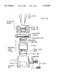
- FIG. 8 is an expanded view of the rupture disk microcarrier launch apparatus
- FIG. 9 illustrates the placement of the microcarrier in the microcarrier holder
- FIG. 10 is a schematic diagram of the plasmid pRSVtRevM10;
- FIG. 11A-11I show the sequence (SEQ ID NO: 1) of the RSV tar Rev M10 expression plasmid (pRSVtRevM10);
- FIGS. 12a and 12b show the results of the challenge of Rev M10/ ⁇ Rev M10 retrovirally transduced human PBLs with HIV BRU .
- PBLs were stimulated, transduced, and selected in G-418 for 1 week (see Examples).
- Cells were challenged with HIV at a MOI between 0.02 and 0.05.
- PBLs from different donors were stimulated via PHA/IL-2 (groups a-c) or anti-CD3/IL-2 (groups d and e) as in the Examples.
- PHA/IL-2 groups a-c
- anti-CD3/IL-2 groups d and e
- RT activity shown for each group is for 7-8 days post-HIV infection;
- FIGS. 13a and 13b show the results of the challenge of Rev M10/ ⁇ Rev M10 retrovirally transduced human PBLs with a clinical isolate of HIV, HIV CLIN .
- PBLs were stimulated, transduced, and selected in G-418 for 1 week as described in the Examples. Cells were then challenged with HIV at a MOI between 0.02 and 0.05.
- PBLs from different donors were stimulated via PHA/IL-2 (groups a-c) or anti-CD3/IL-2 (groups d and e) as described in the Examples.
- PHA/IL-2 groups a-c
- anti-CD3/IL-2 groups d and e
- RT activity shown for each group is for 7-8 days post-HIV infection;
- FIGS. 14a and 14b show the results of the challenge of particle-mediated Rev M10/ ⁇ Rev M10 transduced human PBLs with HIV BRU .
- Freshly isolated PBLs were stimulated with anti-CD3/IL-2 as described in the Examples.
- FIGS. 15a and 15b show the results of the challenge of particle-mediated Rev M10/ ⁇ Rev M10 transduced human PBLs with 2 clinical isolates of HIV. Freshly isolated PBLs were stimulated with anti-CD3/IL-2 as described in the Examples. After particle-mediated transduction and selection in G-418 for up to 8 days, cells were challenged with HIV CLIN at a MOI of between 0.02 and 0.05.
- FIG. 16 illustrates the results of a Southern blot analysis of T leukemia and human PBL transduced with Rev M10 by particle-mediated gene transfer.
- DNA samples were prepared and digested with XbaI and DraIII (XD) or XbaI, DraIII, and EcoRI(XDE) to detect linearized (l) or integrated (i) forms of the plasmid.
- CEM las 1-4
- PBL lanes 5-8
- the present invention provides a method for introducing a foreign gene into a patient's T cells, monocytes, macrophages, hematopoietic stem cells, or dendrites.
- the foreign gene is introduced into cells by particle-mediated gene transfer.
- Particle-mediated gene transfer is described in U.S. Pat. Nos. 5,015,580 and 5,120,657, which are incorporated herein by reference. Additional details of pareticle-mediated gene transfer are taught in J. K. Burkholder, et al, J. Immunological Methods, vol. 165, pp. 149-56 (1993); N. S. Yang, et al, Proc. Natl. Acad. Sci. USA, vol.
- the gene to be introduced into the cell is coated on a small inert particle which is then forced through the cell, thus introducing the gene into the cell.
- the inert particle may be made of any inert material such as an inert metal (gold, silver, platinum, tungsten etc.) or inert plastic (polystyrene, polypropylene, polycarbonate, etc.).
- the particle is made of gold, platinum, or tungsten.
- the particle is gold.
- the particle is spherical and has a diameter of 0.5 to 5 microns, preferably 1 to 3 microns.
- the foreign gene may be any gene which is desired to be introduced into a patient's T cells, monocytes, macrophages, hematopoietic stem cells, or dendrites.
- the gene is a HIV-protective gene.
- the HIV-protective gene may be any gene which will confer an HIV-protective effect on the cell. Examples of such materials include dominant-negative inhibition genes, ribozymes, antisense genes, etc.
- the HIV-protective gene is a dominant-negative inhibition gene, such as Rev M10, an inhibitory integrase or gag gene (Trono, D., et al, Cell, vol. 59, pp. 113-120 (1989)).
- the protective gene is Rev M10.
- the protective gene may be introduced in any suitable form including, a linearized plasmid, a circular plasmid, a plasmid capable of replication, an episome, RNA, etc.
- the gene is contained in a plasmid.
- the gene is contained in a linearized plasmid.
- the HIV-protective gene is contained in a plasmid and is downstream from a promoter.
- suitable promoters include the RSV promoter (Liu, J., et al, Gene Ther., vol. 1, pp. 32-37 (1994); and the CD4 enhancer/promoter (Sawada, S., et al, Mol. Cell. Biol., vol 11, pp. 5506-5515 (1991)).
- the promoter is the RSV promoter.
- the HIV-protective gene is downstream from the sequence of TAR from -18 to -72 of the HIV promoter.
- the HIV-protective gene is immediately downstream from the sequence TAR from -18 to -72 of the HIV promoter, which is in turn immediately downstream from the RSV promoter.
- the plasmid which contains the HIV-protective gene may also contain a selectable marker so that the cells which contain the plasmid can be selectively expanded.
- selectable markers include, neo, gpt, mdr, hygro or dhfr.
- the selectable marker is neo.
- the gene to be introduced into the cell may be coated on the particle by conventional methods.
- the DNA sequence containing the protective gene prepared in the form suitable for gene introduction can be simply dried onto naked gold or tungsten pellets.
- DNA molecules in such a form may have a relatively short period of stability and may tend to degrade rather rapidly due to chemical reactions with the metallic or oxide substrate of the particle itself.
- the carrier particles are first coated with an encapsulating agent, the DNA strands have greatly improved stability and do not degrade significantly even over a time period of several weeks.
- a suitable encapsulating agent is polylysine (molecular weight 200,000) which can be applied to the carrier particles before the DNA molecules are applied.
- encapsulating agents polymeric or otherwise, may also be useful as similar encapsulating agents, including spermidine.
- the polylysine is applied to the particles by rinsing the gold particles in a solution of 0.02% polylysine and then air drying or heat drying the particles thus coated. Once the metallic particles coated with polylysine were properly dried, DNA strands are then loaded onto the particles.
- the DNA is loaded onto the particles at a rate of between 3 and 30 micrograms of DNA per milligram of gold bead spheres.
- To 100 micrograms of DNA and 30 milligrams of 1-3 micron gold spheres are sequentially added 20 ⁇ l of 0.1M spermidine, and then 50 microliters of 2.5M CaCl 2 to provide a fine calcium precipitate which forms as the solution is dried.
- the precipitate carries the DNA with it onto the beads. Once the beads and the spermidine and calcium chloride solution are mixed with the DNA, the suspension is washed with ethanol and dried.
- the optimal ratio of DNA to gold is 5 ⁇ g DNA (5 ⁇ l) to 3 mg gold particles (50 ⁇ l). Once dried the precipitate is immediately resuspended in 100% ethanol for the process of placing the particles onto the carrier sheet.
- a preferred procedure begins with 3 milligrams of 1.0 to 3 microns gold beads (a microcrystalline gold) suspended in 50 microliters of water. This is then mixed with 5 micrograms (5 ⁇ l) of DNA in up to 100 microliters of water. Then 20 microliters of 0.1M spermidine (free base) is mixed and to this mixture is then added 50 microliters of 2.5M calcium chloride while mixing. The suspension is then gently spun down after which the supernatant is discarded. The solids are then resuspended in 140 ⁇ l of 70% ethanol and spundown. The supernatant is discarded and the solids are resuspended in 140 ⁇ l of pure ethanol and centrifuged. The liquid is discarded. The resulting solids are then placed on the microcarrier and desicated and dried.
- the particles coated with the DNA containing the HIV-protective gene may then be used to introduce the gene into a patient's cells using the method and apparatus described in U.S. Pat. Nos. 5,015,580 and 5,120,657, which are incorporated herein by reference.
- a preferred apparatus is Biolistics, PDS-1000/He System, Biorad Laboratories, Hercules, Calif.
- the cells into which the HIV-protective gene is introduced are those cells susceptable to HIV infection or cells which mature into cells susceptable to HIV infection.
- the HIV-protective gene is introduced into T cells, monocytes, macrophages, hematopoietic stem cells which give rise of T cells or macrophages, or dendrites.
- T cells monocytes, macrophages, hematopoietic stem cells which give rise of T cells or macrophages, or dendrites.
- the HIV-protective gene is introduced into 10 9 to 10 13 , preferably 10 10 to 10 11 T cells at a time.
- the efficiency of the introduction of the protective gene is 1 to 10%.
- the T cells are then reintroduced into the patient by conventional techniques such as described in Rosenberg, S. A., et al, N. Eng. J. Med., vol. 323, pp. 570-578 (1990) which is incorporated herein by reference.
- the HIV-protective gene be introduced into from 0.1 to 30%, preferably from 1 to 15%, of the patient's T cells.
- it will be typically necessary to repeat the procedure described above from 1 to 10, preferably from 2 to 5 times.
- it will be necessary to treat from 1 to 10, preferably 2 to 5 batches of T cells.
- These batch treatments usually take from 1 to 120 hours and typically are spaced apart by 2 to 24 hours, preferably 4 to 16 hours.
- booster treatments are usually performed 0.1 to 3 years, preferably 6 to 12 months, after the initial group of batch treatments.
- the cells containing the HIV-protective gene may be expanded, e.g., in the presence of IL-2, so that fewer batches need to be processed.
- this expansion is carried out in a manner which results in the selective expansion of those cells into which the HIV-protective gene has been introduced.
- the DNA introduced also contains a selectable marker such as neo
- expansion in the presence of G-418 will result in the selective expansion of those cells containing the HIV-protective gene.
- selection of genetically modified HIV-protected cells may occur within the patient, either by protection from viral lysis which would otherwise lyse those cells or by administration of cytokines, e.g. IL-2, which would allow their amplication in vivo.
- cytokines e.g. IL-2
- the HIV-protective gene is introduced into hematopoietic stem cells which mature into T cells. In this way, it is possible to obviate the need for booster treatments. This will also facilitate gene transfer into the monocyte/macrophage lineage and central nervous system.
- Hematopoietic stem cells can be isolated and expanded as described in U.S. Pat. No. 5,087,570 and S. Siena et al., Blood, vol. 65, pp. 655-662 (1985), which are incorporated herein by reference.
- the stem cells are isolated from a sample of bone marrow. Once the stem cells have been isolated and expanded, the HIV-protective gene may be introduced using the same methods and apparatus described above.
- the particle-mediated gene transfer is carried out at high cell densities.
- PBL or T cells which have been cultured for a few days or freshly isolated PBL or T cells are harvested, centrifuged at about 300 g for about 5 minutes and resuspended in growth medium (e.g., RPMI) with serum at a cell density of 1 to 5 million cells per 10 ⁇ l. Then about 5 ⁇ l to 20 ⁇ l of the cell suspension is evenly spread onto a surface area of about 3 cm 2 , and this suspension is used as the target.
- growth medium e.g., RPMI
- the stem cells are then reintroduced to the patient using conventional techniques. In this embodiment, it is possible to provide lifelong protection without the need for booster treatments.
- a particular advantage of the present method is the surprising discovery that particle-mediated gene transfer using a linearized plasmid results in stable incorporation of the foreign gene as evidenced by Southern blotting experiments.
- This stable incorporation of a HIV-protective gene provides a number of advantages. First, stable incorporation of the HIV-protective gene in stem cells means that the gene will be passed on to the progeny, conferring life-long protection. Second, stable incorporation in T cells means that upon expansion of and proliferation of the T cells the gene will be incorporated into memory cells.
- the particle-mediated gene transfer into T cells may be carried out without T cell stimulation, optimal gene transfer currently requires T cell stimulation.
- the T cells are stimulated with an agent such as PHA, OKT3 monoclonal antibody, IL-2, or CD28 antibody.
- the stimulation is carried out for a time of 1 min to 120 hours, preferably 4 to 72 hours, prior to gene transfer.
- the prestimulated T cells are maintained in the absence of a stimulating agent for a time of 4 to 96 hours, preferably 24 to 72 hours, immediately prior to gene transfer.
- the T cells are stimulated after gene transfer.
- the same types of stimulating agents described above may also be used for post-gene-transfer stimulation.
- the post-gene-transfer stimulation is usually carried out 0 to 120 hours, preferably 15 minutes to 4 hours, after gene transfer and for a time of 8 to 120 hours, preferably 24 to 72 hours.
- the amount of stimulating agent used will depend on the exact identity of the stimulating agent. However, the selection of the amount of stimulating agent is well within the abilities of the skilled artisan.
- good results are achieved using a concentration of 1 to 5 ⁇ g/ml
- OKT3 monoclonal antibody good results are achieved with 0.1 to 10 ⁇ g/ml
- IL-2 good results are achieved with 1 to 100 U/ml
- anti-CD28 e.g., ⁇ CD3+IL-2 or ⁇ CD3+ ⁇ CD28.
- T cell stimulation may be accompanied by activation of endogenous provirus.
- T cell stimulation is carried out in the presence of a HIV-1 antiviral inhibitor.
- Preferred inhibitors include zelaviridine (Richman, D., et al, Proc. Natl. Acad. Sci. USA, vol. 88, pp. 11241-11245 (1991)), nevirapine (Romero, D. L., et al, Proc. Natl. Acad. Sci. USA, VOL. 88, pp. 8806-8810 (1991)), and a CD4 domain Pseudomonas aeruginosa exotoxin A (CD4-PE40) (V. K.
- one of zelaviridine and nevirapine is used in conjunction with CD4-PE40.
- the concentration of zelaviridine or nevirapine is 0.01 to 20 ⁇ M, preferably 0.02 to 10 ⁇ M, while the concentration of CD4-PE40 is 0.1 to 100 nM, preferably 1 to 10 nM.
- the present invention provides reagents useful for carrying out the present process.
- reagents comprise a DNA fragment containing an HIV-protective gene, a coating solution and a small, inert, dense particle.
- the DNA fragment, coating solution, and the small, inert, dense particle are those described above.
- the DNA is frozen or lyophilized
- the coating solution is frozen or maintained at 4° C.
- the small, inert, dense particle is in a suspension in distilled water.
- the coating solution will contain saline.
- the dry ingredients for the coating solution may be premixed and premeasured and contained in a container such as a vial or sealed envelope.
- the present invention also provides kits which are useful for carrying out the present method.
- the present kits comprise a first container means containing the above-described frozen or lyophilized DNA.
- the kit also comprises a second container means which contains the coating solution or the premixed, premeasured dry components of the coating solution.
- the kit also comprises a third container means which contains the small, inert, dense particles in suspension.
- These container means can be made of glass, plastic or foil and can be a vial, bottle, pouch, tube, bag etc.
- the kit may also contain written information, such as procedures for carrying out the present method or analytical information, such as the amount of reagent (e.g., moles or mass of DNA) contained in the first container means.
- the written information may be on any of the first, second, and/or third container means, and/or a separate sheet included, along with the first, second, and third container means, in a fourth container means.
- the fourth container means may be, e.g., a box or a bag, and may contain the first, second, and third container means.
- Rev M10 a HIV-protective gene
- T cells T cells
- Tat is a 16 kDa protein derived from two exons (Goh, W. C., Rosen, C., Sodroski, J., Ho, D. D. & Haseltine, W. A., J. Virol., vol. 59, pp.
- Tat is essential for viral replication as defective mutants make virus very inefficiently unless Tat is present (Fisher, A. G., Feinberg, M. B., Josephs, S. F. et al., Nature, vol. 320, pp. 367-371 (1986); Dayton, A. I., Sodroski, J. G., Rosen, C. A., Goh, W. C. & Haseltine, W. A., Cell, vol. 44, pp. 941-947 (1986)).
- Rev is a nuclear protein with a molecular weight of 19 kDa (Cullen, B. R., et al., J. Virol., vol. 62, pp. 2498-2501 (1988)) which acts primarily post-transcriptionally to enhance the nuclear transport of unspliced message encoding structural proteins.
- This process requires interaction between Rev and a cis-acting RNA sequence designated the Rev-responsive element (RRE) located within the structural gene for the Env protein.
- the RRE consists of 234 nucleotides and can assume a highly complex secondary structure consisting of a putative central stem and five stem loop structures (Cochrane, A. W., Chen, C. H. & Rosen, C.
- Rev, M10 has been previously shown to inhibit HIV replication in a dominant negative fashion (Malim, M. H., Bohnlein, S., Hauber, J. & Cullen, B. R., Cell, vol. 58, pp. 205-214 (1989)).
- This gene product confers resistance to HIV infection without affecting T-cell function (Malim, M. H., Freimuth, W. W., Liu, J. et al, J. Exp. Med., vol. 176, pp. 1197-1201 (1992); Bevec, D., Dobrovnik, M., Hauber, J. & Bohnlein, E., Proc. Natl. Acad. Sci. USA, vol.
- an important step is to achieve the appropriate level of expression of the therapeutic gene in the relevant target cells.
- the introduced gene is expressed and up-regulated as a consequence of an HIV infection itself, thereby giving optimal protection.
- most of the present vector systems for gene therapy use constitutive expression vectors regulated by viral or cellular promoters.
- results are presented for a vector which takes advantage of three features of HIV gene expression to achieve antiviral effects, including the ability to respond to Tat, serve as a TAR decoy and inhibit Rev function through the synthesis of Rev M10.
- the regulated expression achieved by this vector improves the protection against productive HIV replication in susceptible cells.
- RNA decoys for both Tat and Rev
- a dominant mutant form of Rev, M10 (Malim, M. H., Bohnlein, S., Hauber, J.
- Rev M10 can bind to RRE with a similar affinity as the wild type Rev protein and inhibits normal Rev function, presumably by competing for the binding sites required for normal Rev function (Malim, M. H., Bohnlein, S., Hauber, J. & Cullen, B. R., Cell, vol. 58, pp. 205-214 (1989)).
- Rev M10 mutant represents a promising candidate for intracellular immunization in the control of HIV (Baltimore, D., Nature, vol. 335, pp. 395-396 (1988)).
- Other approaches have also been used to inhibit HIV replication, including other potential dominant negative mutant proteins (Trono, D., Feinberg, M. & Baltimore, D., Cell, vol. 59, pp.
- RNA decoys Sullenger, B. A., Gallardo, H. F., Ungers, G. E., & Gilboa, E., Cell, vol. 63, pp. 601-608 (1990), catalytic RNA (Sarver, N., Cantin, E. M., Chang, P. S. et al., Science, vol. 247, pp. 1222-1225 (1990); Ojwang, J. O., Hampel, A., Looney, D. J., Wong-Staal, F. & Rappaport, J., Proc. Natl. Acad. Sci. USA, vol. 89, pp.
- the protective effects of the RSV/TAR Rev M10 vector are probably related to at least three effects on early viral gene expression.
- the increased level of basal transcription from the RSV/TAR promoter provides a higher level of Rev M10 to block the function of wild type Rev.
- a high basal level of expression appears to correlate with inhibition of virus replication and may result from the formation of mixed Rev/Rev M10 multimers, which may form more readily if Rev M10 is synthesized prior to wild type Rev in an infected cell.
- Rev M10 expression is further induced by Tat produced from the early viral RNAs synthesized after HIV infection which binds to the TAR sequence of RSV TAR Rev M10 and produces more Rev M10 protein which inhibits Rev function.
- TAR may contribute directly to the high-level of basal expression (FIG. 3).
- the synthesis of TAR RNA from the RSV promoter also provides a source of TAR RNA which can compete with the binding of Tat to the native promoter and also inhibits Tat function. At low Tat concentrations, the TAR decoy could limit Tat-mediated transactivation, although the TAR region would allow for further protection should this protective effect be overwhelmed.
- the combination of the RSV promoter with the TAR sequence appears to provide the best protection against HIV replication in vivo through this combination of autoregulatory elements.
- This expression vector provides significant antiviral effects and could potentially be used with other HIV protective proteins to provide protective effects in patients infected with HIV.
- the present method can be applied also to other relevant target cells, including hematopoietic stem cells and dendritic cells, which bind virus in the lymph node. It is possible to generate expression vectors for these HIV protective genes whose inhibition of viral spread can be optimized by appropriate regulation of gene expression (Liu, J., et al Gene Ther., vol. 1, pp. 32-37 (1994)).
- the present method makes it possible to evaluate the efficacy of a variety of antiviral genes for the treatment of HIV infection in patients (Nabel, G. J., et al, Hum. Gene Ther., vol. 5, pp. 79-92 (1994)).
- the ability to introduce eukaryotic expression vectors into human primary peripheral blood lymphocytes by this method allows further analysis of the pathways of T cell activation responsible for the activation of specific transcription factors within these cells.
- the pRSV CAT-RRE plasmid was derived from a plasmid which utilized the SV40 enhancer, DM128 (Hope, T. J., Huang, X. J., McDonald, D. & Parslow, T. G., Proc. Natl. Acad. Sci. USA, vol. 87, pp. 7787-7791 (1990)). This plasmid was digested by BclI and HindII to remove the SV40 promoter and then incubated with Klenow polymerase.
- the resulting backbone was ligated with a Klenow enzyme-treated NdeI-HindIII fragment containing the RSV promoter isolated from PSPRSV (Williams, T., Admon, A., Luscher, B. & Tjian, R., Genes Dev., vol. 2, pp. 1557-1569 (1988)).
- the RSV-Rev expression vector is a plasmid containing the Rev open reading frame (ORF) under the control of RSV promoter.
- the NcoI-XhoI fragment containing the complete ORF for Rev, from the CMV-Rev expression vector (Malim, M. H., Hauber, J., Le., S. Y., Maizel, J. V. & Cullen, B. R., Nature, vol. 338, pp. 254-257 (1989)) was ligated into the NcoI-XhoI sites of RSV p105 expression vector (Perkins, N. D., Schmid, R. M., Duckett, C. S., Leung, K., Rice, N. R.
- RSV or Rev M10 is similar to RSV-Rev, except that a NcoI-XhoI fragment containing the Rev M10 ORF from CMV-M10 was used.
- the RSV-M10 expression vector used in the stable transfections was prepared from a parental RSV-ADH vector encoding the alcohol dehydrogenase (ADH) and neomycin resistance gene (Lin, W. -C. & Culp, L. A., BioTech, vol. 11, pp. 344-351 (1991)).
- a Klenow-treated NcoI-XhoI fragment containing the M10 ORF was ligated into Klenow-treated backbone after the ADH gene insert was removed by digestion with HindIII and XbaI.
- the HIV Rev M10 expression vector contains the HIV LTR from -660 to +74 to enhance expression of the Rev M10 ORF. It was prepared from a NcoI-XhoI fragment containing the Rev M10 ORF from cRev (M10) treated with Klenow enzyme, ligated into the HindIII site of HIV-CAT (Nabel, G. & Baltimore, D., Nature, vol. 326, pp. 711-713 (1987) to generate HIV Rev M10/CAT. The HindIII site is regenerated in the ligation. HIV Rev M10/CAT was digested with XhoII and HindIII restriction enzymes.
- This XhoI/HindIII fragment containing the HIV LTR and Rev M10 ORF was treated with Klenow inserted into RSV-ADH vector (Lin, W. -C. & Culp, L. A., BioTech, vol. 11, pp. 344-351 (1991)) after the RSV promoter and ADH structural genes were removed with BglII and XbaI and treated with Klenow.
- RSV/TAR Rev M10 contains the RSV promoter and sequence of TAR from -18 to -72 of HIV promoter to stimulate expression of the Rev M10 ORF. It was constructed by digesting HIV Rev M10/CAT described above with PvuII and HindIII. The 0.6 kb fragment containing the 80 base pair TAR sequence and Rev M10 ORF was isolated, treated with Klenow polymerase and ligated to the Klenow-treated HindIII and XbaI sites in the RSV-ADH vector (Lin, W. -C. & Culp, L. A., BioTech, vol. 11, pp. 344-351 (1991)) in which the ADH structural gene was excised. RSV/TAR ⁇ Rev M10 is identical to RSV/Tar Rev M10 except that the initiation codon ATG was deleted by site-directed mutagenesis and the deletion confirmed by sequence analysis.
- HIV-CAT plasmid (Rosen, C. A., Sodroski, J. G. & Haseltine, W. A., Cell, vol. 41, pp. 813-823 (1985)) have been described previously.
- CEM cells (1 ⁇ 10 6 ) were inoculated with 2000 TCID 50 of HIV Bru for 2 hours at 37° C. (1:500 ratio HIV:CEM cells). The cells were then washed twice in fresh medium (RPMI-1640/10% fetal calf serum), resuspended at the original cell density and incubated at 30° C. Cultures were subcultured every 3 to 5 days depending on growth rate and duplicate samples taken for reverse transcriptase assays.
- FIG. 1a Schematic diagrams of the regulatory elements utilized in these studies are illustrated (FIG. 1a).
- CAT chloramphenicol acetyl transferase
- RRE Rev responsive sequence element
- This reporter plasmid utilizes Rous sarcomia virus (RSV), a strong constitutive regulatory element unresponsive to mitogen stimulation, and CAT activity is responsive to Rev, which presumably facilitates the transport of its mRNA from the nucleus to cytoplasm.
- RSV Rous sarcomia virus
- CAT activity is responsive to Rev, which presumably facilitates the transport of its mRNA from the nucleus to cytoplasm.
- Co-expression of Rev with this reporter increased CAT activity linear with Rev concentration (FIG. 1b).
- This reporter was used to analyze the activity of the dominant Rev M10 protein by measuring the level of inhibition of transactivation by the Rev expression vector (FIGS. 1 and 3).
- Rev M10 Rev M10 gene
- Regulatory regions included the RSV enhancer/promoter, a chimeric RSV/TAR promoter containing the RSV regulatory region linked to the 80 bp HIV-1 TAR sequence, and another TAR containing vector, the HIV LTR, was used to regulate Rev M10 expression.
- the HIV LTR represented a potentially attractive regulatory sequence for this purpose because it contains the native regulatory elements required for HIV transcriptional activation, but was less effective than RSV/TAR. The relative level of inhibition by Rev M10 expressed by these different regulatory sequence was assessed (FIG. 3).
- the plasmid reporter was not tested in the presence of Tat, the presence of the TAR sequence conferred greater protection than the RSV enhancer without TAR, suggesting that this sequence may exert Tat-independent protective effects, and that this enhancer might show even greater efficacy in the presence of Tat which might be synthesized after HIV infection.
- RSV/TAR can function as an HIV-inducible regulatory region to stimulate the expression of Rev M10 gene following HIV infection (i.e., when Tat protein is synthesized).
- Rev M10 plasmids 2 ⁇ g
- Tat expression plasmids 2 ⁇ g
- RSV/TAR Rev M10 exerted a greater protective effect in the presence of the Tat expression plasmid.
- An identical plasmid lacking the TAR element showed minimal response to Tat (FIG. 4).
- the TAR sequence in combination with a strong constitutive enhancer, RSV could therefore facilitate the expression of Rev M10 in the presence of Tat.
- the RSV/TAR vector shows a high level of constitutive expression, it could also synthesize TAR RNA which could act as a decoy for Tat protein.
- TAR decoy effects have been previously described in specially designed retroviral vectors using a pol III promoter (Sullenger, B. A., Gallardo, H. F., Ungers, G. E., & Gilboa, E., Cell, vol. 63, pp. 601-608 (1990)), it was unknown whether this effect might be seen with a pol II promoter in a standard eukaryotic expression vector.
- TAR RNA could serve as a trans-acting competitor when the RSV/TAR Rev M10 vector is present in the cell by competing for binding to Tat protein required for transcription stimulated by the HIV LTR.
- HIV-CAT which contains the HIV LTR and TAR linked to the CAT gene, as the reporter plasmid. HIV-CAT was cotransfected with a constant amount of Tat expression vector in the presence of increasing amounts of the RSV promoter without an insert, RSV Rev M10, RSV Rev or RSV/TAR Rev M10. The Tat expression plasmid stimulated the expression from HIV-CAT 50-fold, and RSV/TAR Rev M10 reduced the level of transactivation by Tat ⁇ 80%, compared to RSV Rev M10 or RSV without an insert (FIG.
- RSV/TAR Rev M10 As the only difference between RSV/TAR Rev M10 and RSV Rev M10 is the presence of the additional 80 base pair TAR sequence present in RSV/TAR-M10, these results suggest that the TAR region competes for Tat binding to the HIV-CAT reporter plasmid, and that it acts as a decoy in the context of the RSV/TAR vector.
- plasmids Three plasmids, RSV Rev M10, HIV Rev M10 and RSV/TAR Rev M10, were inserted into a plasmid which contains the G418 resistance gene (Lin, W. -C. & Culp, L. A., BioTech, vol. 11, pp. 344-351 (1991)) and transfected into CEM human T-cell leukemia cells. Stable pools of resistant cells were selected in the presence of G418. Cells stably expressing Rev M10 were subsequently challenged with HIV Bru . The kinetics of cell survival from these lines containing different protective plasmids are shown in (FIG. 6). Control CEM cells began to show syncytia and cell death 5 days after HIV infection (FIG. 7).
- RSV Rev M10 provided protection for up to 15 days, whereupon the cells began to show cytopathic effects (CPE), although not at the same rate as control cells.
- CPE cytopathic effects
- HIV Rev M10 and RSV/TAR Rev M10 expressing cells showed protection from CPE up to 49 days, although the HIV Rev M10 cultures showed levels of reverse transcriptase (RT) above baseline indicative of a constant level of HIV RT production in the culture.
- RT reverse transcriptase
- the CEM cells transduced with HIV Rev M10 showed very few syncytia during the time-course, and RSV/TAR Rev M10 expressing cells showed no CPE throughout the entire 49 days of cell culture. Minimal RT activity was detected in these cultures but was above baseline levels, as previously noted (Malim, M. H., Freimuth, W.
- RSV-tar-- This consists of the RSV enhancer and TAR, a tat responsive element that is specific to the HIV system. This element proved to be most effective in experiments designed to test expression of Rev M10.
- the vector that was described in the previous section is linearized with restriction enzymes and precipitated onto gold beads.
- the beads are resuspended in 100% ethanol and layered onto mylar sheets at a predetermined concentration.
- the target cells are then separated from the mylar sheets by a screen. Expansion of a helium gas causes a rupture disk to release at a fixed pressure, forcing the mylar sheet against the screen.
- the mesh allows the beads to pass through, while acting as a barrier to the mylar.
- CD4 + human T cells are isolated as described above.
- the cells are stimulated as described above with anti-CD3 for 24 hours before undergoing particle-mediated gene delivery with DNA coated gold beads.
- the beads are coated with linearized plasmid (previously digested with the Aat II), precipitated onto the gold beads, resuspended in 100% ethanol, and layered onto mylar sheets.
- the cells are stimulated with anti-CD3 and IL-2 or anti-CD3 and anti-CD28. Cells will be re-infused only if 0.1% of the cells are transduced successfully. No penicillin or other beta-lactam antibiotics are used in the culture of these cells.
- the patients T cells are obtained by leukapheresis, and they are transferred to sterile 75 cm 2 vented cap tissue culture flasks (Corning). 60 mls of AIM V media containing 5% human AB sera (50 U/ml IL 2) is added to each flask. The final cell density is 5 ⁇ 10 5 /ml.
- CD4 + cells are enriched by eliminating CD8 + cells by selection with AIS CD8 CELLector culture flasks, following the manufacturers procedures.
- Cells are cultured in a 37° C./5% CO 2 incubator as indicated below.
- Cells are stimulated for 24 to 48 hours by either anti-CD3 plus IL2 (50 U/ml) or the simultaneous addition of anti-CD3 plus anti-CD28 (0.25 ⁇ g/ml) final concentration.
- anti-CD3 plus IL2 50 U/ml
- anti-CD28 anti-CD28
- final concentration 50 U/ml
- cells are concentrated by centrifugation and resuspended at 5 ⁇ 10 6 cells in 100 ⁇ l of AIM V medium, and transferred from the 75 cm 2 vented top tissue culture flask to 60 mm diameter petri dishes. The cells are spread out over a 4 cm 2 area in the center of the 60 mm dish using a small pipette tip. The cells are transduced at this concentration.
- the DNA's are linearized by cutting with AatII restriction enzyme.
- the DNA is then phenol/chloroform extracted and ethanol precipitated.
- DNA is resuspended at a concentration of 1 mg/ml in TE.
- Microcarrier Preparation 120 treatments using 500 ⁇ g gold beads per delivery
- the master lot is prepared as follows:
- Vortex microcarriers prepared in 50% glycerol (60 mg/ml) for 5 minutes to resuspend and disrupt agglomerated particles.
- the entire interior of the chamber is sprayed with 70% ethanol and left for 20 minutes, at which time any residual ethanol is removed by wiping with sterile 4 ⁇ 4 gauzes.
- Microcarrier and rupture discs are sterilized by immersion in 70% ethanol for four hours followed by drying in a sterile bio-safety cabinet. Batches of 50 discs will be sterilized at one time and tested for the presence of bacteria and fungi and then qualified as in the Certificate of Analysis. Microcarrier and rupture disks will be packaged individually in sterile autoclave bags. Stopping screens will be sterilized by autoclaving for 30 minutes at 121° C. individually in sterile autoclave bags.
- Operational Directions See FIG. 8 for diagramatic view of the PDS.
- Petri dish holder Put the Petri dish containing the lymphocytes in 100 ⁇ l of AIM V medium on the Petri dish holder. Remove the lid. Place the Petri dish holder at the desired level inside the sample chamber.
- a metering valve has been installed on the solenoid valve assembly to control the rate of fill of the gas acceleration tube. It should take about 12-15 seconds to fill to burst pressure.
- the rupture disk should burst within 10% of the indicated rupture pressure. Although the actual burst valves may be different from the indicated ones due to the accuracy of the pressure gauge, the variation in burst pressure form bombardment to bombardment is less than 5%.
- 0.1-10% recombinant gene expression has been achieved in activated CD4 + T lymphocytes, using this technique.
- unstimulated cells which may be used to transduce cells without concurrently inducing replication of virus in T cells which contain latent virus, 0.1% transduction efficiencies are observed.
- Experiments to test for the inhibition of HIV replication in cells in which Rev M10 was introduced using this method were performed. PCR analysis of HIV gag, and ELISA assays to detect p24 in the culture supernatants indicate that Rev M10 effectively inhibits productive replication of the virus, and proviral DNA.
- appropriate vectors to optimize expression of the gene in this system were developed. Plasmids that were constructed with different enhancers to determine which provided the most protection from HIV were evaluated.
- RSV/TAR Rev M10 contains RSV promoter and sequence of TAR from -18 to -72 of HIV promoter to stimulate expression of the Rev M10 open reading frame (Liu, J., et al, Gene Ther., vol. 1, pp. 32-37 (1994)).
- RSV/TAR ⁇ Rev M10 is identical to RSV/TAR Rev M10 except that the initiation codon ATG was deleted and a linker inserted which was confirmed by sequence analysis.
- the HIV-CAT plasmid Rosen, C. A., et al, Cell, vol. 41, pp. 813-823 (1985)
- pHD101 tat plasmid Markovitz, D. M., et al, Proc. Natl. Acad. Sci. USA, vol. 87, pp. 9098-9102 (1990) have been described previously.
- Peripheral blood mononuclear cells were isolated using Ficoll-Hypaque separation. The cells were then stimulated in flasks coated with immobilized OKT3 monoclonal antibody and soluble IL-2 (50 U/ml) for 48-72 hours. Cells were recovered and resuspended at 5 ⁇ 10 5 /ml in either AIM-V medium (BRL) or X-Vivo medium (MA Biowhittaker) containing 5% human AB serum+50 U/ml of IL-2. Cells were maintained at 5 ⁇ 10 5 -1.5 ⁇ 10 6 /ml throughout the experiments.
- Cells were incubated at a density of 1 ⁇ 10 6 per well in a 24 well plate containing 1 ml of Crip supernatant and 1 ml of AIM-V medium. After infection, cells were washed once by centrifugation and resuspended at 5 ⁇ 10 5 cells/ml in conditioned AIM-V medium + 50 U/ml IL-2. Cells were then selected with 300 ⁇ g/ml G-418 (active).
- DNA was linearized by digestion with AatII restriction enzyme, extracted using phenol/chloroform, precipitated with ethanol, and resuspended in Tris-EDTA buffer (pH 8.0) to a final concentration of 1 mg/ml.
- Gold microcarriers 60 mg, 1.6 ⁇ m microparticles were washed with 1 ml of 70% ethanol, vortexed for 3-5 minutes, incubated for 2 minutes, and pelleted by microcentrifugation. The particles were then washed 3 times in sterile water (by adding water, vortexing for 1 minute, and allowing the particles to settle) and resuspended in 1 ml of 50% glycerol. 5 ⁇ g of the linearized DNA were added to 3 mg (50 ⁇ l) of the gold microcarriers. CaCl 2 (final concentration of 1M) and spermidine (final concentration of 16 mM) were added to the mixture.
- the apparatus was set up as described by the manufacturer (Biolistics PDS-1000/He System, Biorad Laboratories, Hercules, Calif.). The following adjustments were made. Rupture disk microcarrier gap 3/4", microcarrier travel distance 6 mm, chamber vacuum 15 in Hg, Helium pressure 1800 psi and target distance 4 cm. Immediately prior to transduction, PBLs were centrifuged, resuspended to 5 ⁇ 10 6 cells/100 ⁇ l and the cell suspension spread over a 4 cm 2 area of a small petri dish (35 ⁇ 10 mm). After transduction, the cells are quickly removed from the petri dish and resuspended at 5 ⁇ 10 5 cells/ml in conditioned medium.
- Cells were challenged with either HIV BRU or freshly passaged HIV clinical isolates HIV CLIN .
- Cells (1 ⁇ 10 6 cells/ml) were incubated with HIV at specific MOI for 2-4 hours at 37° C. Following the incubation, cells were washed with a 10 ⁇ volume of fresh medium and resuspended at 5 ⁇ 10 5 cells/ml. Cells were maintained at a density of 0.5 ⁇ 1.5 ⁇ 10 6 cells/ml throughout the infection.
- Rev M10 To determine whether expression of Rev M10 could provide resistance to HIV infection in human lymphocytes, cells were transduced with a murine amphotropic retroviral vector pLJ Rev M10 (Malim, M. H., et al, J. Exp. Med., vol. 176, pp. 1197-1201 (1992)) or a frame-shifted Rev M10 negative control (pLJ ⁇ Rev M10). Stimulated lymphocyte populations were transduced by retroviral infection with supernatants derived from ⁇ -Crip amphotropic retroviral producer cells (0.5-5.0 ⁇ 10 6 G-418 resistant colonies/ml). After transduction, cells were selected in G-418 for 5-7 days prior to challenge with HIV.
- pLJ Rev M10 murine amphotropic retroviral vector
- pLJ ⁇ Rev M10 a murine amphotropic retroviral vector
- Stimulated lymphocyte populations were transduced by retroviral infection with supernatants derived from ⁇ -Crip
- Retroviral transduction frequencies for the lymphocyte population 3 days post-transduction in anti-CD3/IL-2 stimulated PBLs were estimated by limiting dilution PCR analysis. Seven days after selection with G-418, the percentage of Rev transduced cells in the populations had increased to greater than 10%.
- Rev M10 expression confers a significant level of protection from challenge with a cloned isolate of HIV.
- a nonspecific inhibitory effect was also seen when cells are grown in the presence of G-418, possibly because of its cytotoxic effect or through an effect on Rev (Zapp, M. L., et al, Cell, vol. 74, pp. 969-978 (1993)).
- This effect was also seen in HIV infection of untransduced lymphocytes, in which RT levels during infection ranged 2- to 3-fold higher than cells selected in G-418 (FIG. 12a).
- Rev M10 to affect viral replication in patients.
- human peripheral blood lymphocytes were challenged with freshly isolated clinical strains. Protection was observed when cells were exposed to these viruses.
- a representative time course of HIV infection showed a reduction in RT levels in Rev M10 cultures compared to ⁇ Rev M10 over the 14-day course of the experiment (FIG. 13a).
- supernatant RT levels at the peak of HIV infection were consistently reduced in multiple independent Rev M10 transduced cells with at least two independent fresh isolated clinical strains (FIG. 13b).
- particle-mediated gene transfer was used to introduce expression plasmids into human PBL.
- Plasmids encoding Rev M10 or the frameshift mutant, ⁇ Rev M10, under control of the RSV promoter and the HIV tat responsive element (Tar) were linearized by restriction enzyme digestion, precipitated onto gold microparticles and used to transduce PBLs by particle-mediated gene transfer.
- the frequency of gene transfer was determined by limiting dilution PCR.
- the percentage of cells initially transduced by this method was estimated to be at least 3% at 5 days post-transduction and increased to ⁇ 50% by 21 days of selection in G-418 (Table 1). Cells could be maintained in the presence of G-418 for ⁇ 2 months, suggesting that the transduced gene could be stably incorporated in the genome of these cells.
- Southern blot analysis was performed. As early as 8 days post gene transfer, a signal corresponding to an integrated, but not a linear, form of the plasmid was detected (FIG. 16).
- Transduction frequencies for cells transduced by particle-mediated gene transfer were calculated by limiting cell dilution PCR analysis using Rev specific primers in (A) normal PBLs or (B) PBLs obtained from asymptomatic HIV positive donor and transduced in the presence of zelavirine and CD4-PE40. PBLs were transduced with Rev M10 expression plasmids as described in the Examples.
- T cell stimulation needed to promote optimal gene transfer, also induces viral replication (Zagury, D., et al, Science, vol. 231, pp. 850-853 (1986)).
- activation of endogenous provirus must thus be avoided.
- This strategy employs a non-nucleoside reverse transcriptase inhibitor which specifically inhibits HIV-1 in vitro but does not affect murine retroviral reverse transcription. Two different agents were used including zelaviridine and nevirapine.
- the second agent was a chimeric toxin protein consisting of a CD4 domain fused to Pseudomonas aeruginosa exotoxin A (CD4-PE40) (Chaudhary, V. K., et al, Nature, vol. 335, pp. 369-372 (1988)).
- the human renal epithelial cell, 293 was infected with a murine amphotropic retroviral vector encoding ⁇ -galactosidase (BAG) in the presence of these agents alone or in combination (Table 1). Either zelaviridine and nevirapine, alone or in combination with CD4-PE40, caused minimal inhibition of murine retroviral ⁇ -galactosidase transduction.
- BAG murine amphotropic retroviral vector encoding ⁇ -galactosidase
- zelaviridine or nevirapine alone or in combination with CD4-PE40, also effectively suppressed HIV replication to undetectable RT levels in freshly infected lymphocyte cultures over a 3-week period.
- inhibition by azidothymidine (AZT) which is not HIV-1 selective, inhibited transduction by the murine retroviral vector (Table 2).
- lymphocytes from an asymptomatic HIV patient were activated in their presence.
- Proviral HIV DNA content was determined by limiting dilution PCR 1 to 8 days after stimulation. A significant reduction in lymphocyte proviral DNA content was observed in antiviral treated cultures compared to untreated cells.
- Before treatment between 1:250-1:2500 cells were HIV positive using gag specific primers. After treatment, ⁇ 1:25,000 were positive with nevirapine/CD4-PE40 and fewer than 1:50,000 with zelaviridine/CD4-PE40 treatment (the limit of the PCR detection system). No p24 antigen or reverse transcriptase activity was detected in culture supernatants.
- these agents were effective in reducing HIV activation in cell culture without interfering with the murine retroviral vector. Similarly, these agents had no effect on transduction efficiencies by particle-mediated gene transfer (Table 2).
- Murine amphotropic retroviral pLJ- ⁇ -gal supernatant 100 ⁇ l produced from ⁇ -Crip producer cells were inoculated onto 293 cells growing in 6 well plates. The wells contained 2 mls of medium with the indicated concentrations of antiviral compounds. After 3 days of incubation, cells were fixed and stained for ⁇ -gal activity by X-gal staining and the number of positive cells per duplicate well counted. Control untransduced cells showed no X-gal staining.
- a safety consideration that is unique to particle-mediated gene delivery is the potential for injection of gold particles into patients. Although this possibility is unlikely, since the particles are removed by low-speed centrifugation prior to reinfusion of transduced cells, to address this question, DNA-coated gold particles were tested in mice to determine their potential for toxicity.
- Gold particles coated with RSV/Rev M10 plasmid were injected intravenously (8 mice) and a negative control group received normal saline (8 mice). The test group was injected with 0.8 ⁇ g DNA and 500 ⁇ g of gold microspheres (1000 ⁇ greater than are likely to be inadvertently administered to patients). After ten days, the recipients were sacrificed, and the organs were removed and examined both grossly and histologically. Serum samples were obtained prior to injection and at the time recipient animals were sacrificed, and were analyzed for evidence of alterations is selected serum enzymes and chemistries. These studies demonstrated no signs of toxicity (see Tables 3 and 4 below).
- Another mouse has been observed at two months without ill effects after receiving 4 ⁇ 10 7 transduced cells intravenously.
Abstract
Foreign genes may be stably introduced into T cells, monocytes, macrophages, dendrites, and hematopoietic stem cells by particle-mediated gene transfer. Introduction of an HIV protective gene into the cells of a patient infected with HIV by particle-mediated gene transfer is effective for the treatment of HIV infection.
Description
The present invention was made with the assistance of United States Government funds. The United States Government may have some rights in the invention.
1. Field of the Invention
The present invention relates to a method for introducing a gene into a patient's T cells, monocytes, macrophages, dendrites, or hematopoietic stem cells and to a method for introducing HIV protective genes into a patient's cells by particle-mediated gene transfer and reagents and kits useful in such methods.
2. Discussion of the Background
Infection by human immunodeficiency Virus (HIV) leads to progressive depletion of CD4+ T cells which causes an acquired immunodeficiency syndrome (AIDS). Over the past decade, AIDS infection has achieved epidemic proportions. Despite considerable progress in understanding and diagnosing this disease, it has remained refractory to treatment. There are currently no effective means to vaccinate susceptible individuals or to treat infected patients. Infection by the human immunodeficiency virus is typically characterized by an asymptomatic, or latent, phase of the disease. During this time, patients may not exhibit signs of immunodeficiency but nonetheless synthesize virus which progressively depletes CD4+ T cells and is infectious. It is currently estimated that there are over one million seropositive cases with latent HIV infection in the United States today. These individuals, for whom there are no known effective treatments, are likely to progress and succumb to this disease.
For these individuals, a major therapeutic goal is to prolong the latent phase of HIV infection. A major interest has been to define the molecular basis of HIV gene activation in T lymphocytes. The activation of HIV gene expression is controlled by a cellular transcription factor, NF-κB (G. Nabel, D. Baltimore, Nature, vol. 326, p. 711 (1987)). This transcription factor is inactive in resting T cells but is stimulated following cell activation and induces viral transcription. In addition to NF-κB, there are essential viral genes which also appear to regulate the transition from latent to active infection. An important viral regulatory protein in this process is Rev, an 19 kDa nuclear protein which controls export of viral RNA from the nucleus to the cytoplasm of infected cells.
Viral replication is critically dependent on the interaction of viral gene products with host cell factors. Because viruses are intimately associated with their host cells, it has been difficult to selectively interfere with replication in vivo. Successful antiviral approaches have selectively targeted viral gene products. For example, the treatment of herpes simplex virus (HSV) infection has taken advantage of the ability of a viral gene, thymidine kinase, to modify a drug which is toxic to the host cell. This approach led to the development of guanosine analogues, including acyclovir and ganciclovir (K. O. Smith, K. S. Galloway, W. L. Kennell, K. K. Ogilvie, B. K. Radatus, Antimicrob. Agents Chemother., vol. 22, p. 55 (1982); A. K. Field, M. E. Davies, C. DeWitt, H. C. Perry, R. Liou, et al, Proc. Natl. Acad. Sci., USA, vol. 80, p. 4139 (1983); D. F. Smee, J. C. Martin, J. P. Verheyden, T. R. Matthews, Antimicrob. Agents Chemother., vol. 23, p. 676 (1983)), which are converted to DNA chain terminators only in HSV infected cells.
In HIV infection, traditional pharmaceutical targeting has thus far provided limited benefits. Although AZT has relative selectivity for viral reverse transcriptase, its toxic effect on host cell function and its low therapeutic index has provided limited protection against the progression of AIDS.
Although drug discovery has led to the identification of additional anti-viral drugs, the high rate of virus mutation has led to the generation of resistant viral mutants. More recently, as molecular biological studies have advanced, it has become possible to use recombinant genes to interfere with HIV gene expression. Several approaches have been used to exploit gene transfer to inhibit viral replication. These include anti-sense RNA (H. Weintraub J. G. Izant, R. M. Harland, Trends Genet., vol. 2, p. 22 (1985); J. P. Green, O. Pines, M. Inouye, Ann. Rev. Biochem, vol. 55, p. 569 (1986); A. R. van der Krol, J. N. M. Mol, A. R. Stuitjie, BioTech, vol. 6, p. 958 (1988)), catalytic RNA, or ribozymes (J. Hasseloff, W. L. Gerlach, Nature, vol. 334, p. 585 (1988); M. Cotton, M. L. Birnstiel, EMBO J, vol. 8, p. 3861 (1989); N. Sarver, E. M. Cantin, P. S. Chang, J. A. Zaia, P. A. Ladne, et al, Science, vol. 247, p. 1222 (1990); B. A. Sullenger, T. C. Lee, C. A. Smith, G. E. Ungers, E. Gilboa, Mol. Cell. Biol., vol. 10, p. 6512 (1990)), and RNA analogs or decoys (B. A. Sullenger, H. F. Gallardo, G. E. Ungers, E. Gilboa, Cell, vol. 63, p. 601 (1990)). The gene transfer approach to targeting viral nucleic acid is complicated by problems of delivering the antiviral gene to the appropriate compartment of the cell, and by the ability to obtain catalytic activity in vivo. These approaches have thus far have not been successful.
The concept of dominant negative-inhibition was initially described in yeast genetic systems (I. Herskowitz, Nature, vol. 329, p. 219 (1987)). It was subsequently demonstrated that anti-viral effects could be conferred on cells susceptible to infection by herpesvirus. Using the herpesvirus VP16 transactivator, mutant proteins lacking the transactivation domain of this protein were generated which could interfere with viral replication (A. D. Friedman, S. J. Triezenberg, S. L. McKnight, Nature, vol. 335, p. 452 (1988)). For HIV, several potential transdominant proteins have been defined which have been successful for inhibiting viral replication in transient transfection systems. Among these are the Rev transdominant protein (M. H. Malim, S. Bohnlein, J. Hauber, B. R. Cullen, Cell, vol. 58, p. 205 (1989)) and viral group-specific antigens (GAG). Recent success in protecting cells from HIV infection using TAR analogs has provided additional evidence that it is possible to render cells resistant to HIV infection (B. A. Sullenger, H. F. Gallardo, G. E. Ungers, E. Gilboa, Cell, vol. 63, p. 601 (1990)) through recombinant gene products.
Previous gene therapy attempts to protect cells from HIV infection have focused upon a viral vector delivery system, because, prior to the present invention, no method other than viral transfection was thought to be useful for introducing a gene into a patient's T cells. In other words, T cells are resistant to the uptake and stable expression of foreign DNA. However, a major concern regarding the utilization of viral vector delivery systems is the potential for small quantities of replication-competent virus to remain undetected in large production lots intended for clinical use. Since it is not possible to test the entire lot, quantities below the detection limits of the assay could be introduced into the patient. Given the underlying immunodeficiency of these patients, the potential to establish replication-competent virus in the host would be increased. Although proper testing of retroviral vector batches makes this possibility less likely, it remains a risk in this procedure. The use of a nonviral vector which is unable to replicate provides an alternative to this approach which might improve its safety. A variety of approaches have been attempted to introduce genes into T cells, but most are limited by low transduction efficiencies, transient expression, and the general resistance of T cells to the uptake of recombinant DNA.
Thus, the treatment of HIV infection by gene therapy remains elusive. Thus, there remains a need for a method of treating HIV infection. In particular, there remains a need for a method for introducing an HIV-protective gene into a patient's T cells, monocytes, macrophages, or hematopoietic stem cells. There also remains a need for reagents and kits for carrying out such a method.
Accordingly, it is one object of the present invention to provide novel methods for treating HIV infection.
It is another object of the present invention to provide a method for prolonging the latent phase of HIV infection in a patient infected with HIV.
It is another object of the present invention to provide a method for delaying the onset of AIDS in a patient infected with HIV.
It is another object of the present invention to provide a method for introducing a foreign gene into a patient's T cells, monocytes, macrophages, hematopoietic stem cells, or dendrites.
It is another object of the present invention to provide a method for introducing an HIV protective gene into a patient's T cells, monocytes, macrophages, hematopoietic stem cells, or dendrites.
It is another object of the present invention to provide a method for achieving stable expression of a foreign gene in a patient's T cells, monocytes, macrophages, hematopoietic stem cells, or dendrites.
It is another object of the present invention to provide a method for achieving stable expression of an HIV-protective gene in a patient's cells.
It is another object of the present invention to provide a method for introducing an HIV-protective gene into non-activated cells.
It is another object of the present invention to provide novel reagents useful in such a method.
It is another object of the present invention to provide novel kits for carrying out such a method.
These another objects, which will become apparent during the following detailed description, have been achieved by the inventors' discovery that foreign genes may be stably introduced into a patient's T cells, monocytes, macrophages, hematopoietic stem cells, and dendrites and that HIV protective genes may be successfully introduced into a patient's cells by particle-mediated gene transfer.
A more complete appreciation of the invention and many of the attendant advantages thereof will be readily obtained as the same becomes better understood by reference to the following detailed description when considered in connection with the accompanying drawings, wherein:
FIG. 1 provides schematic diagrams of Rev-responsive CAT reporter RSV-CAT-RRE, RSV-Rev and the different promoter protective plasmids. SD, splice donor; SA, splice acceptor; RRE, Re-responsive element;
FIG. 2 illustrates the responsiveness of the RSV-CAT-RRE reporter to stimulation by Rev. The RSV-CAT-RRE reporter was transfected with the indicated amounts of RSV-Rev expression vector. Transfection and CAT assay procedures were performed as described in Materials and methods;
FIG. 3 provides a comparison of relative strengths of different enhancers. The Rev-responsive CAT reporter RSV-CAT-RRE (2.5 μg) and the RSV-Rev (2.5 μg) were cotransfected with 2.0, 5.0 and 10.0 μg of expression vectors driven by different promoters as shown. The relative effectiveness of each enhancer as determined by its ability to inhibit transactivation of RSV-CAT-RRE by the RSV-Rev expression vector is shown as a percentage of conversion of chloramphenicol to its acetylated forms in the presence of different M10 expression vectors. The first two bars represent RSV-CAT-RRE alone and RSV-CAT-RRE with 2.5 μg RSV-Rev;
FIG. 4 shows the effect of Tat on RSV/TAR Rev M10 protection. The RSV-CAT-RRE reporter (2.5 μg) and RSV-Rev (2.5 μg) were cotransfected with RSV Rev M10 (2 μg), RSV-TAR Rev M10 (2 μg), or RSV TAR FS vectors (2 μg) and two different doses of pHD101 Tat expression vector (0.2 and 0.5 μg) or no (0.0) Tat expression vector (control). The activation of RSV-CAT-RRE by RSV-Rev is similar to that shown in FIG. 2. The inhibition of CAT conversion by the indicated vectors relative to the control is shown for each plasmid. Total transfected plasmid was normalized to 8 μg using Bluescript plasmid;
FIG. 5 shows the inhibition of Tat-mediated transactivation of HIV-CAT by RSV/TAR. HIV-CAT (1 μg) was cotransfected with pHD101Tat (0.2 μg) and 2.0 μg RSV expression vector without insert, RSV Rev M10, RSV Rev, or RSV/TAR Rev M10;
FIG. 6 shows the effects of expression regulated by RSV, HIV LTR, or RSV/TAR; control untransfected CEM cells or CEM cells stably expressing Rev M10 from the indicated promoters were incubated with HIVBru (1:500 ratio TCID50 HIV:CEM cells) for 2 hours, washed twice and resuspended in fresh medium. Culture supernatants were then assayed for RT activity at various times postinfection. Cultures were split every 3 to 5 days dependent on growth rate;
FIGS. 7a and 7b illustrate the protective effect of Rev M10 expressed in CEM cells after HIV infection. Control CEM cells (FIG. 7a) and CEM cells stably expressing Rev M10 (FIG. 7b) from the RSV/TAR construct were infected with HIVBru as described in the Materials and Methods section below. Cells were then washed and monitored for cytopathic effects. The photographs are of cultures 5 days postinfection;
FIG. 8 is an expanded view of the rupture disk microcarrier launch apparatus;
FIG. 9 illustrates the placement of the microcarrier in the microcarrier holder;
FIG. 10 is a schematic diagram of the plasmid pRSVtRevM10;
FIG. 11A-11I show the sequence (SEQ ID NO: 1) of the RSV tar Rev M10 expression plasmid (pRSVtRevM10);
FIGS. 12a and 12b show the results of the challenge of Rev M10/ΔRev M10 retrovirally transduced human PBLs with HIVBRU. PBLs were stimulated, transduced, and selected in G-418 for 1 week (see Examples). Cells were challenged with HIV at a MOI between 0.02 and 0.05.
(a) Time course of HIVBRU infection of an individual group of Rev M10 and ΔRev M10 transduced PBLs.
(b) PBLs from different donors were stimulated via PHA/IL-2 (groups a-c) or anti-CD3/IL-2 (groups d and e) as in the Examples. Following retroviral transduction with Rev M10 and ΔRev M10, and G-418 selection for 1 week, cells were challenged with HIV. RT activity shown for each group is for 7-8 days post-HIV infection;
FIGS. 13a and 13b show the results of the challenge of Rev M10/ΔRev M10 retrovirally transduced human PBLs with a clinical isolate of HIV, HIVCLIN. PBLs were stimulated, transduced, and selected in G-418 for 1 week as described in the Examples. Cells were then challenged with HIV at a MOI between 0.02 and 0.05.
(a) Time course of HIVCLIN infection of an individual group of Rev M10 and ΔRev M10 transduced PBLs.
(b) PBLs from different donors were stimulated via PHA/IL-2 (groups a-c) or anti-CD3/IL-2 (groups d and e) as described in the Examples. Following retroviral transduction with Rev M10 and ΔRev M10, and G-418 selection for 1 week, cells were challenged with HIV. RT activity shown for each group is for 7-8 days post-HIV infection;
FIGS. 14a and 14b show the results of the challenge of particle-mediated Rev M10/ΔRev M10 transduced human PBLs with HIVBRU. Freshly isolated PBLs were stimulated with anti-CD3/IL-2 as described in the Examples. After particle-mediated transduction and selection in G-418 for up to 8 days, cells were challenged with HIVBRU at a MOI of between 0.02 and 0.05.
(a) Time course of HIV infection of an individual group set of Rev M10 and ΔRev M10 transduced PBLs.
(b) Different experimental sets (a-h) of independently transduced PBLs from various donors were challenged with HIVBRU. RT activity is 7-8 days post-HIV infection;
FIGS. 15a and 15b show the results of the challenge of particle-mediated Rev M10/ΔRev M10 transduced human PBLs with 2 clinical isolates of HIV. Freshly isolated PBLs were stimulated with anti-CD3/IL-2 as described in the Examples. After particle-mediated transduction and selection in G-418 for up to 8 days, cells were challenged with HIVCLIN at a MOI of between 0.02 and 0.05.
(a) Time course of HIV infection of an individual group set of Rev M10 and ΔRev M10 transduced PBLs.
(b) Two groups (a and b) of independently transduced PBLs were challenged with 2 different clinical isolates of HIV-1. RT activity is 7-8 days post-HIV infection; and
FIG. 16 illustrates the results of a Southern blot analysis of T leukemia and human PBL transduced with Rev M10 by particle-mediated gene transfer. DNA samples were prepared and digested with XbaI and DraIII (XD) or XbaI, DraIII, and EcoRI(XDE) to detect linearized (l) or integrated (i) forms of the plasmid. CEM (lanes 1-4) or PBL (lanes 5-8), untransduced or transduced as indicated, were probed (see Examples).
Thus, in a first embodiment, the present invention provides a method for introducing a foreign gene into a patient's T cells, monocytes, macrophages, hematopoietic stem cells, or dendrites. In the present method, the foreign gene is introduced into cells by particle-mediated gene transfer. Particle-mediated gene transfer is described in U.S. Pat. Nos. 5,015,580 and 5,120,657, which are incorporated herein by reference. Additional details of pareticle-mediated gene transfer are taught in J. K. Burkholder, et al, J. Immunological Methods, vol. 165, pp. 149-56 (1993); N. S. Yang, et al, Proc. Natl. Acad. Sci. USA, vol. 87, pp. 9568-72 (1990); S. Jiao, et al, Bio/Technology, vol. 11, pp. 497-502 (1993); T. A. Thompson, et al, In Vitro Cell. Dev. Biol., vol. 29A, pp. 167-170 (1993); N. -S. Yang, et al, Critical Rev. in Biotechnology, vol. 12, pp. 335-356 (1992); L. Cheng, et al, Proc. Natl. Acad. Sci. USA, vol. 90, pp. 4455-4459 (1993); N. -S. Yang, et al, Proc. Natl. Acad. Sci. USA, vol. 87, pp. 9598-9572 (1990), all of which are incorporated herein by reference.
In particle mediated gene transfer, the gene to be introduced into the cell is coated on a small inert particle which is then forced through the cell, thus introducing the gene into the cell. The inert particle may be made of any inert material such as an inert metal (gold, silver, platinum, tungsten etc.) or inert plastic (polystyrene, polypropylene, polycarbonate, etc.). Preferably, the particle is made of gold, platinum, or tungsten. Most preferably, the particle is gold. Suitably, the particle is spherical and has a diameter of 0.5 to 5 microns, preferably 1 to 3 microns.
In the present invention, the foreign gene may be any gene which is desired to be introduced into a patient's T cells, monocytes, macrophages, hematopoietic stem cells, or dendrites. Preferably, the gene is a HIV-protective gene. The HIV-protective gene may be any gene which will confer an HIV-protective effect on the cell. Examples of such materials include dominant-negative inhibition genes, ribozymes, antisense genes, etc. Preferably, the HIV-protective gene is a dominant-negative inhibition gene, such as Rev M10, an inhibitory integrase or gag gene (Trono, D., et al, Cell, vol. 59, pp. 113-120 (1989)). In a particularly preferred embodiment, the protective gene is Rev M10.
The protective gene may be introduced in any suitable form including, a linearized plasmid, a circular plasmid, a plasmid capable of replication, an episome, RNA, etc. Preferably, the gene is contained in a plasmid. In a particularly preferred embodiment, the gene is contained in a linearized plasmid.
In a particularly preferred embodiment, the HIV-protective gene is contained in a plasmid and is downstream from a promoter. Suitable promoters include the RSV promoter (Liu, J., et al, Gene Ther., vol. 1, pp. 32-37 (1994); and the CD4 enhancer/promoter (Sawada, S., et al, Mol. Cell. Biol., vol 11, pp. 5506-5515 (1991)). Preferably the promoter is the RSV promoter. More preferably, the HIV-protective gene is downstream from the sequence of TAR from -18 to -72 of the HIV promoter. Most preferably, the HIV-protective gene is immediately downstream from the sequence TAR from -18 to -72 of the HIV promoter, which is in turn immediately downstream from the RSV promoter.
The plasmid which contains the HIV-protective gene may also contain a selectable marker so that the cells which contain the plasmid can be selectively expanded. Examples of suitable selectable markers include, neo, gpt, mdr, hygro or dhfr. Preferably, the selectable marker is neo.
The gene to be introduced into the cell may be coated on the particle by conventional methods. For example, the DNA sequence containing the protective gene prepared in the form suitable for gene introduction can be simply dried onto naked gold or tungsten pellets. However, DNA molecules in such a form may have a relatively short period of stability and may tend to degrade rather rapidly due to chemical reactions with the metallic or oxide substrate of the particle itself. Thus, if the carrier particles are first coated with an encapsulating agent, the DNA strands have greatly improved stability and do not degrade significantly even over a time period of several weeks. A suitable encapsulating agent is polylysine (molecular weight 200,000) which can be applied to the carrier particles before the DNA molecules are applied. Other encapsulating agents, polymeric or otherwise, may also be useful as similar encapsulating agents, including spermidine. The polylysine is applied to the particles by rinsing the gold particles in a solution of 0.02% polylysine and then air drying or heat drying the particles thus coated. Once the metallic particles coated with polylysine were properly dried, DNA strands are then loaded onto the particles.
The DNA is loaded onto the particles at a rate of between 3 and 30 micrograms of DNA per milligram of gold bead spheres. To 100 micrograms of DNA and 30 milligrams of 1-3 micron gold spheres are sequentially added 20 μl of 0.1M spermidine, and then 50 microliters of 2.5M CaCl2 to provide a fine calcium precipitate which forms as the solution is dried. The precipitate carries the DNA with it onto the beads. Once the beads and the spermidine and calcium chloride solution are mixed with the DNA, the suspension is washed with ethanol and dried. The optimal ratio of DNA to gold is 5 μg DNA (5 μl) to 3 mg gold particles (50 μl). Once dried the precipitate is immediately resuspended in 100% ethanol for the process of placing the particles onto the carrier sheet.
A preferred procedure begins with 3 milligrams of 1.0 to 3 microns gold beads (a microcrystalline gold) suspended in 50 microliters of water. This is then mixed with 5 micrograms (5 μl) of DNA in up to 100 microliters of water. Then 20 microliters of 0.1M spermidine (free base) is mixed and to this mixture is then added 50 microliters of 2.5M calcium chloride while mixing. The suspension is then gently spun down after which the supernatant is discarded. The solids are then resuspended in 140 μl of 70% ethanol and spundown. The supernatant is discarded and the solids are resuspended in 140 μl of pure ethanol and centrifuged. The liquid is discarded. The resulting solids are then placed on the microcarrier and desicated and dried.
Once the particles coated with the DNA containing the HIV-protective gene have been prepared, they may then be used to introduce the gene into a patient's cells using the method and apparatus described in U.S. Pat. Nos. 5,015,580 and 5,120,657, which are incorporated herein by reference. A preferred apparatus is Biolistics, PDS-1000/He System, Biorad Laboratories, Hercules, Calif.
The cells into which the HIV-protective gene is introduced are those cells susceptable to HIV infection or cells which mature into cells susceptable to HIV infection. Thus, the HIV-protective gene is introduced into T cells, monocytes, macrophages, hematopoietic stem cells which give rise of T cells or macrophages, or dendrites. When introducing the protective gene into T cells, a sample of a patient's T cells is first isolated by conventional methods, such as by Ficoll-Hypaque separation.
Typically, the HIV-protective gene is introduced into 109 to 1013, preferably 1010 to 1011 T cells at a time. Usually, the efficiency of the introduction of the protective gene is 1 to 10%. Thus, when a batch of 1010 to 1011 T cells is treated as described above, about 109 to 1010 T cells containing the HIV-protective gene will be obtained. After the introduction of the HIV-protective gene is complete, the T cells are then reintroduced into the patient by conventional techniques such as described in Rosenberg, S. A., et al, N. Eng. J. Med., vol. 323, pp. 570-578 (1990) which is incorporated herein by reference.
It is preferred that the HIV-protective gene be introduced into from 0.1 to 30%, preferably from 1 to 15%, of the patient's T cells. Thus, it will be typically necessary to repeat the procedure described above from 1 to 10, preferably from 2 to 5 times. In other words, it will be necessary to treat from 1 to 10, preferably 2 to 5 batches of T cells. These batch treatments usually take from 1 to 120 hours and typically are spaced apart by 2 to 24 hours, preferably 4 to 16 hours.
Moreover, since the lifetime of a T cell is typically about 5 days to 1 year, it may be necessary to carry out booster treatments to replenish the T cells carrying the HIV-protective gene as those prepared in the initial treatment die. Such booster treatments are usually performed 0.1 to 3 years, preferably 6 to 12 months, after the initial group of batch treatments.
Alternatively, the cells containing the HIV-protective gene may be expanded, e.g., in the presence of IL-2, so that fewer batches need to be processed. In a preferred embodiment, this expansion is carried out in a manner which results in the selective expansion of those cells into which the HIV-protective gene has been introduced. For example, when the DNA introduced also contains a selectable marker such as neo, expansion in the presence of G-418 will result in the selective expansion of those cells containing the HIV-protective gene.
Alternatively, selection of genetically modified HIV-protected cells may occur within the patient, either by protection from viral lysis which would otherwise lyse those cells or by administration of cytokines, e.g. IL-2, which would allow their amplication in vivo.
In an alternative embodiment, the HIV-protective gene is introduced into hematopoietic stem cells which mature into T cells. In this way, it is possible to obviate the need for booster treatments. This will also facilitate gene transfer into the monocyte/macrophage lineage and central nervous system. Hematopoietic stem cells can be isolated and expanded as described in U.S. Pat. No. 5,087,570 and S. Siena et al., Blood, vol. 65, pp. 655-662 (1985), which are incorporated herein by reference. Preferably, the stem cells are isolated from a sample of bone marrow. Once the stem cells have been isolated and expanded, the HIV-protective gene may be introduced using the same methods and apparatus described above.
In a preferred embodiment, the particle-mediated gene transfer is carried out at high cell densities. For example, PBL or T cells which have been cultured for a few days or freshly isolated PBL or T cells are harvested, centrifuged at about 300 g for about 5 minutes and resuspended in growth medium (e.g., RPMI) with serum at a cell density of 1 to 5 million cells per 10 μl. Then about 5 μl to 20 μl of the cell suspension is evenly spread onto a surface area of about 3 cm2, and this suspension is used as the target.
After the HIV-protective gene has been introduced into the stem cells, the stem cells are then reintroduced to the patient using conventional techniques. In this embodiment, it is possible to provide lifelong protection without the need for booster treatments.
A particular advantage of the present method is the surprising discovery that particle-mediated gene transfer using a linearized plasmid results in stable incorporation of the foreign gene as evidenced by Southern blotting experiments. This stable incorporation of a HIV-protective gene provides a number of advantages. First, stable incorporation of the HIV-protective gene in stem cells means that the gene will be passed on to the progeny, conferring life-long protection. Second, stable incorporation in T cells means that upon expansion of and proliferation of the T cells the gene will be incorporated into memory cells.
Although, the particle-mediated gene transfer into T cells may be carried out without T cell stimulation, optimal gene transfer currently requires T cell stimulation. Suitably, the T cells are stimulated with an agent such as PHA, OKT3 monoclonal antibody, IL-2, or CD28 antibody. Suitably, the stimulation is carried out for a time of 1 min to 120 hours, preferably 4 to 72 hours, prior to gene transfer. In a preferred embodiment, the prestimulated T cells are maintained in the absence of a stimulating agent for a time of 4 to 96 hours, preferably 24 to 72 hours, immediately prior to gene transfer. In a particularly preferred embodiment, the T cells are stimulated after gene transfer. The same types of stimulating agents described above may also be used for post-gene-transfer stimulation. The post-gene-transfer stimulation is usually carried out 0 to 120 hours, preferably 15 minutes to 4 hours, after gene transfer and for a time of 8 to 120 hours, preferably 24 to 72 hours. The amount of stimulating agent used will depend on the exact identity of the stimulating agent. However, the selection of the amount of stimulating agent is well within the abilities of the skilled artisan. For PHA, good results are achieved using a concentration of 1 to 5 μg/ml, for OKT3 monoclonal antibody, good results are achieved with 0.1 to 10 μg/ml, and with IL-2 good results are achieved with 1 to 100 U/ml, and with 0.1 to 10 μg/ml of anti-CD28. These agents can be used together or in various combinations, e.g., αCD3+IL-2 or αCD3+αCD28.
T cell stimulation may be accompanied by activation of endogenous provirus. Thus, in a preferred embodiment, T cell stimulation is carried out in the presence of a HIV-1 antiviral inhibitor. Preferred inhibitors include zelaviridine (Richman, D., et al, Proc. Natl. Acad. Sci. USA, vol. 88, pp. 11241-11245 (1991)), nevirapine (Romero, D. L., et al, Proc. Natl. Acad. Sci. USA, VOL. 88, pp. 8806-8810 (1991)), and a CD4 domain Pseudomonas aeruginosa exotoxin A (CD4-PE40) (V. K. Chaudhary, et al, Nature, vol. 335, pp. 369-372, (1988)). Preferably, one of zelaviridine and nevirapine is used in conjunction with CD4-PE40. Suitably, the concentration of zelaviridine or nevirapine is 0.01 to 20 μM, preferably 0.02 to 10 μM, while the concentration of CD4-PE40 is 0.1 to 100 nM, preferably 1 to 10 nM.
In another embodiment, the present invention provides reagents useful for carrying out the present process. Such reagents comprise a DNA fragment containing an HIV-protective gene, a coating solution and a small, inert, dense particle. The DNA fragment, coating solution, and the small, inert, dense particle are those described above.
Preferably, the DNA is frozen or lyophilized, the coating solution is frozen or maintained at 4° C., and the small, inert, dense particle is in a suspension in distilled water. Typically, the coating solution will contain saline. Alternatively, the dry ingredients for the coating solution may be premixed and premeasured and contained in a container such as a vial or sealed envelope.
The present invention also provides kits which are useful for carrying out the present method. The present kits comprise a first container means containing the above-described frozen or lyophilized DNA. The kit also comprises a second container means which contains the coating solution or the premixed, premeasured dry components of the coating solution. The kit also comprises a third container means which contains the small, inert, dense particles in suspension. These container means can be made of glass, plastic or foil and can be a vial, bottle, pouch, tube, bag etc. The kit may also contain written information, such as procedures for carrying out the present method or analytical information, such as the amount of reagent (e.g., moles or mass of DNA) contained in the first container means. The written information may be on any of the first, second, and/or third container means, and/or a separate sheet included, along with the first, second, and third container means, in a fourth container means. The fourth container means may be, e.g., a box or a bag, and may contain the first, second, and third container means.
The present invention will now be explained in further detail in the context of introducing Rev M10, a HIV-protective gene, into T cells. However, it is to be understood that similar results may be obtained using other foreign genes and other types of cells.
Replication of the human immunodeficiency virus (HIV) is controlled by complex interactions between its genome, virally encoded regulatory proteins and cellular factors (Vaishnav, Y. N., Wong-Staal, F., Annu. Rev. Biochem., vol. 60, pp. 577-630 (1991)). Of these viral proteins, Tat and Rev play critical roles during the early stages of HIV replication. Tat is a 16 kDa protein derived from two exons (Goh, W. C., Rosen, C., Sodroski, J., Ho, D. D. & Haseltine, W. A., J. Virol., vol. 59, pp. 181-184 (1986); Sodroski, J., Patarca, R., Rosen, C., Wong-Staal, F. & Haseltine, W., Science, vol. 229, pp. 74-77 (1985)) and primarily acts at the level of transcriptional elongation and/or initiation by interacting with the cis-acting Tat-responsive element (TAR) located in the RU5 region of the long terminal repeat (LTR) (Selby, M. J., Bain, E. S., Luciw, P. A. & Peterlin, B. M., Genes Dev., vol. 3, pp. 547-558 (1989)). Tat is essential for viral replication as defective mutants make virus very inefficiently unless Tat is present (Fisher, A. G., Feinberg, M. B., Josephs, S. F. et al., Nature, vol. 320, pp. 367-371 (1986); Dayton, A. I., Sodroski, J. G., Rosen, C. A., Goh, W. C. & Haseltine, W. A., Cell, vol. 44, pp. 941-947 (1986)).
Whereas Tat dramatically augments RNA levels, Rev is a nuclear protein with a molecular weight of 19 kDa (Cullen, B. R., et al., J. Virol., vol. 62, pp. 2498-2501 (1988)) which acts primarily post-transcriptionally to enhance the nuclear transport of unspliced message encoding structural proteins. This process requires interaction between Rev and a cis-acting RNA sequence designated the Rev-responsive element (RRE) located within the structural gene for the Env protein. The RRE consists of 234 nucleotides and can assume a highly complex secondary structure consisting of a putative central stem and five stem loop structures (Cochrane, A. W., Chen, C. H. & Rosen, C. A., Proc. Natl. Acad. Sci. USA, vol. 87, pp. 1198-1202 (1990); Heaphy, S., Dingwall, C., Ernberg, I. et al., Cell, vol. 60, pp. 685-693 (1990)). Mapping has shown that sequences within stem-loop 2 are the major binding sites for Rev. Mutations in Rev result in greatly decreased viral structural protein synthesis. Rev is thus essential for viral replication and infectious particle formation (Sodroski, J., Goh, W. C., Rosen, C., Dayton, A., Terwilliger, E. & Haseltine, W., Nature, vol. 321, pp. 412-417 (1986)). One mutant form of Rev, M10, has been previously shown to inhibit HIV replication in a dominant negative fashion (Malim, M. H., Bohnlein, S., Hauber, J. & Cullen, B. R., Cell, vol. 58, pp. 205-214 (1989)). This gene product confers resistance to HIV infection without affecting T-cell function (Malim, M. H., Freimuth, W. W., Liu, J. et al, J. Exp. Med., vol. 176, pp. 1197-1201 (1992); Bevec, D., Dobrovnik, M., Hauber, J. & Bohnlein, E., Proc. Natl. Acad. Sci. USA, vol. 89, pp. 9870-9874 (1992); Bahner, I., Zhou, C., Yu, X- J., Hao, Q- L., Guatelli, J. C. & Kohn, D. B., J. Virol., vol. 67, pp. 3199-3207 (1993)).
To adapt these molecular genetic strategies to clinically useful protocols for AIDS patients, an important step is to achieve the appropriate level of expression of the therapeutic gene in the relevant target cells. Preferably, the introduced gene is expressed and up-regulated as a consequence of an HIV infection itself, thereby giving optimal protection. Unfortunately, most of the present vector systems for gene therapy use constitutive expression vectors regulated by viral or cellular promoters. In the experiments described in the Examples section, results are presented for a vector which takes advantage of three features of HIV gene expression to achieve antiviral effects, including the ability to respond to Tat, serve as a TAR decoy and inhibit Rev function through the synthesis of Rev M10. The regulated expression achieved by this vector improves the protection against productive HIV replication in susceptible cells.
Several previous approaches have been attempted using RNA decoys for both Tat and Rev (Sullenger, B. A., Gallardo, H. F., Ungers, G. E., G. E. & Gilboa, E., Cell, vol. 63, pp. 601-608 (1990); Sullenger, B. A., Gallardo, H. F., Ungers, G. E. & Gilboa, E., J. Virol., vol. 65, pp. 6811-6816 (1991)); however, these RNA structures could potentially interact with normal cellular factors and adversely affect normal cell function. A dominant mutant form of Rev, M10 (Malim, M. H., Bohnlein, S., Hauber, J. & Cullen, B. R., Cell, vol. 58, pp. 205-214 (1989)) inhibits the action of Rev without affecting T-cell function (Malim, M. H., Freimuth, W. W., Liu, J. et al, J. Exp. Med., vol. 176, pp. 1197-1201 (1992)). Rev M10 can bind to RRE with a similar affinity as the wild type Rev protein and inhibits normal Rev function, presumably by competing for the binding sites required for normal Rev function (Malim, M. H., Bohnlein, S., Hauber, J. & Cullen, B. R., Cell, vol. 58, pp. 205-214 (1989)). Stable introduction and expression of the Rev M10 gene into CEM human T leukemia cells resulted in a marked reduction in HIV replication (Malim, M. H., Freimuth, W. W., Liu, J. et al, J. Exp. Med., vol. 176, pp. 1197-1201 (1992)), indicating that the Rev M10 mutant represents a promising candidate for intracellular immunization in the control of HIV (Baltimore, D., Nature, vol. 335, pp. 395-396 (1988)). Other approaches have also been used to inhibit HIV replication, including other potential dominant negative mutant proteins (Trono, D., Feinberg, M. & Baltimore, D., Cell, vol. 59, pp. 113-120 (1989)), RNA decoys (Sullenger, B. A., Gallardo, H. F., Ungers, G. E., & Gilboa, E., Cell, vol. 63, pp. 601-608 (1990), catalytic RNA (Sarver, N., Cantin, E. M., Chang, P. S. et al., Science, vol. 247, pp. 1222-1225 (1990); Ojwang, J. O., Hampel, A., Looney, D. J., Wong-Staal, F. & Rappaport, J., Proc. Natl. Acad. Sci. USA, vol. 89, pp. 10802-10806 (1992); Yu, M., Ojwang, J., Yamada, O. et al., Proc. Natl. Acad. Sci., USA, vol. 90, pp. 6340-6344 (1993)) and antisense oligonucleotides (Eck, S. L. & Nabel, G. J., Curr. Op. Biotechnol., vol. 2, pp. 897-904 (1991); van der Krol, A. R., Mol, J. N. & Stuitje, A. R., Biotechniques, vol. 6, pp. 958-976 (1988); Izant, J. G. & Weintraub, H., Science, vol. 229, pp. 345-352 (1985)).
The protective effects of the RSV/TAR Rev M10 vector are probably related to at least three effects on early viral gene expression. First, the increased level of basal transcription from the RSV/TAR promoter provides a higher level of Rev M10 to block the function of wild type Rev. A high basal level of expression appears to correlate with inhibition of virus replication and may result from the formation of mixed Rev/Rev M10 multimers, which may form more readily if Rev M10 is synthesized prior to wild type Rev in an infected cell. Second, Rev M10 expression is further induced by Tat produced from the early viral RNAs synthesized after HIV infection which binds to the TAR sequence of RSV TAR Rev M10 and produces more Rev M10 protein which inhibits Rev function. TAR, on the other hand, may contribute directly to the high-level of basal expression (FIG. 3). Finally, the synthesis of TAR RNA from the RSV promoter also provides a source of TAR RNA which can compete with the binding of Tat to the native promoter and also inhibits Tat function. At low Tat concentrations, the TAR decoy could limit Tat-mediated transactivation, although the TAR region would allow for further protection should this protective effect be overwhelmed. Thus, the combination of the RSV promoter with the TAR sequence appears to provide the best protection against HIV replication in vivo through this combination of autoregulatory elements. This expression vector provides significant antiviral effects and could potentially be used with other HIV protective proteins to provide protective effects in patients infected with HIV.
In the Examples described below, gold microparticles were used to achieve optimal transduction frequencies within T cells. Using this technique, ˜10% of cells were routinely transduced. In general, the rates of transduction were ˜10-fold higher than those achieved with retroviral vectors. Although expression was detected soon after gene transfer, cells could be maintained and selected for long periods of time (>2 months) in cell culture, indicating the stability of expression. This stability was confirmed by Southern blotting of cell lines which revealed a signal from integrated DNA, detectable as early as two weeks after transduction and selection in vivo.
The present method can be applied also to other relevant target cells, including hematopoietic stem cells and dendritic cells, which bind virus in the lymph node. It is possible to generate expression vectors for these HIV protective genes whose inhibition of viral spread can be optimized by appropriate regulation of gene expression (Liu, J., et al Gene Ther., vol. 1, pp. 32-37 (1994)). The present method makes it possible to evaluate the efficacy of a variety of antiviral genes for the treatment of HIV infection in patients (Nabel, G. J., et al, Hum. Gene Ther., vol. 5, pp. 79-92 (1994)). At the same time, the ability to introduce eukaryotic expression vectors into human primary peripheral blood lymphocytes by this method allows further analysis of the pathways of T cell activation responsible for the activation of specific transcription factors within these cells.
Other features of the invention will become apparent in the course of the following descriptions of exemplary embodiments which are given for illustration of the invention and are not intended to be limiting thereof.
I. Viral Transfection
Plasmids
The pRSV CAT-RRE plasmid was derived from a plasmid which utilized the SV40 enhancer, DM128 (Hope, T. J., Huang, X. J., McDonald, D. & Parslow, T. G., Proc. Natl. Acad. Sci. USA, vol. 87, pp. 7787-7791 (1990)). This plasmid was digested by BclI and HindII to remove the SV40 promoter and then incubated with Klenow polymerase. The resulting backbone was ligated with a Klenow enzyme-treated NdeI-HindIII fragment containing the RSV promoter isolated from PSPRSV (Williams, T., Admon, A., Luscher, B. & Tjian, R., Genes Dev., vol. 2, pp. 1557-1569 (1988)).
The RSV-Rev expression vector is a plasmid containing the Rev open reading frame (ORF) under the control of RSV promoter. The NcoI-XhoI fragment containing the complete ORF for Rev, from the CMV-Rev expression vector (Malim, M. H., Hauber, J., Le., S. Y., Maizel, J. V. & Cullen, B. R., Nature, vol. 338, pp. 254-257 (1989)) was ligated into the NcoI-XhoI sites of RSV p105 expression vector (Perkins, N. D., Schmid, R. M., Duckett, C. S., Leung, K., Rice, N. R. & Nabel, G. J., Proc. Natl. Acad. Sci. USA, vol. 89, pp. 1529-1533 (1992)) after removal of the original p105 insert with NcoI and XhoI. RSV or Rev M10 is similar to RSV-Rev, except that a NcoI-XhoI fragment containing the Rev M10 ORF from CMV-M10 was used.
The RSV-M10 expression vector used in the stable transfections was prepared from a parental RSV-ADH vector encoding the alcohol dehydrogenase (ADH) and neomycin resistance gene (Lin, W. -C. & Culp, L. A., BioTech, vol. 11, pp. 344-351 (1991)). A Klenow-treated NcoI-XhoI fragment containing the M10 ORF was ligated into Klenow-treated backbone after the ADH gene insert was removed by digestion with HindIII and XbaI.
The HIV Rev M10 expression vector contains the HIV LTR from -660 to +74 to enhance expression of the Rev M10 ORF. It was prepared from a NcoI-XhoI fragment containing the Rev M10 ORF from cRev (M10) treated with Klenow enzyme, ligated into the HindIII site of HIV-CAT (Nabel, G. & Baltimore, D., Nature, vol. 326, pp. 711-713 (1987) to generate HIV Rev M10/CAT. The HindIII site is regenerated in the ligation. HIV Rev M10/CAT was digested with XhoII and HindIII restriction enzymes. This XhoI/HindIII fragment containing the HIV LTR and Rev M10 ORF was treated with Klenow inserted into RSV-ADH vector (Lin, W. -C. & Culp, L. A., BioTech, vol. 11, pp. 344-351 (1991)) after the RSV promoter and ADH structural genes were removed with BglII and XbaI and treated with Klenow.
RSV/TAR Rev M10 contains the RSV promoter and sequence of TAR from -18 to -72 of HIV promoter to stimulate expression of the Rev M10 ORF. It was constructed by digesting HIV Rev M10/CAT described above with PvuII and HindIII. The 0.6 kb fragment containing the 80 base pair TAR sequence and Rev M10 ORF was isolated, treated with Klenow polymerase and ligated to the Klenow-treated HindIII and XbaI sites in the RSV-ADH vector (Lin, W. -C. & Culp, L. A., BioTech, vol. 11, pp. 344-351 (1991)) in which the ADH structural gene was excised. RSV/TAR ΔRev M10 is identical to RSV/Tar Rev M10 except that the initiation codon ATG was deleted by site-directed mutagenesis and the deletion confirmed by sequence analysis.
The HIV-CAT plasmid (Rosen, C. A., Sodroski, J. G. & Haseltine, W. A., Cell, vol. 41, pp. 813-823 (1985)) have been described previously.
Transfection and CAT assays
Transfection and CAT assays were performed as previously described (Nabel, G. & Baltimore, D., Nature, vol. 326, pp. 711-713 (1987)).
HIV infection
CEM cells (1×10⁶) were inoculated with 2000 TCID50 of HIVBru for 2 hours at 37° C. (1:500 ratio HIV:CEM cells). The cells were then washed twice in fresh medium (RPMI-1640/10% fetal calf serum), resuspended at the original cell density and incubated at 30° C. Cultures were subcultured every 3 to 5 days depending on growth rate and duplicate samples taken for reverse transcriptase assays.
RT assays
Culture supernatants were assayed for RT activity as described previously (Potts, B. J., in Techniques in HIV research. Aldovini, A. and Walker, B. D. (eds.) Stockton Press: New York, pp. 103-106 (1990)). Poly(A)/oligo(dT) was used as template primer and incorporation of 32 P-dTTP was measured after spotting 5 μl of the RT reaction mixture onto DE81 paper and washing with 2×sodium saline citrate (SSC) four times. Radioactivity was analyzed on a Betagene Betascope.
Results
Relative strength of different enhancers in Rev M10 expression vectors
Schematic diagrams of the regulatory elements utilized in these studies are illustrated (FIG. 1a). We prepared a reporter similar to others in which the 5' and 3' splice junctions of HIV are retained and the envelope gene is deleted, replaced by the chloramphenicol acetyl transferase (CAT) coding sequence with the Rev responsive sequence element (RRE) (Hope, T. J., Huang, X. J., McDonald, D. & Parslow, T. G., Proc. Natl. Acad. Sci. USA, vol. 87, pp. 7787-7791 (1990)). This reporter plasmid, RSV-CAT-RRE, utilizes Rous sarcomia virus (RSV), a strong constitutive regulatory element unresponsive to mitogen stimulation, and CAT activity is responsive to Rev, which presumably facilitates the transport of its mRNA from the nucleus to cytoplasm. Co-expression of Rev with this reporter increased CAT activity linear with Rev concentration (FIG. 1b). This reporter was used to analyze the activity of the dominant Rev M10 protein by measuring the level of inhibition of transactivation by the Rev expression vector (FIGS. 1 and 3).
To compare the effects of different promoters, we used alternative regulatory elements to stimulate expression of the Rev M10 gene. Regulatory regions included the RSV enhancer/promoter, a chimeric RSV/TAR promoter containing the RSV regulatory region linked to the 80 bp HIV-1 TAR sequence, and another TAR containing vector, the HIV LTR, was used to regulate Rev M10 expression. A frameshift mutant vector, RSV Tar FS, was used as a negative control. The HIV LTR represented a potentially attractive regulatory sequence for this purpose because it contains the native regulatory elements required for HIV transcriptional activation, but was less effective than RSV/TAR. The relative level of inhibition by Rev M10 expressed by these different regulatory sequence was assessed (FIG. 3). Although the plasmid reporter was not tested in the presence of Tat, the presence of the TAR sequence conferred greater protection than the RSV enhancer without TAR, suggesting that this sequence may exert Tat-independent protective effects, and that this enhancer might show even greater efficacy in the presence of Tat which might be synthesized after HIV infection.
Tat-stimulated expression of RSV/TAR Rev M10
To determine whether RSV/TAR can function as an HIV-inducible regulatory region to stimulate the expression of Rev M10 gene following HIV infection (i.e., when Tat protein is synthesized), we compared the protective effects of the RSV and RSV/TAR vectors in the presence of a Tat expression plasmid. Each of these Rev M10 plasmids (2 μg) was cotransfected with a Tat expression plasmid to determine whether co-expression of Tat provides additional protection. RSV/TAR Rev M10 exerted a greater protective effect in the presence of the Tat expression plasmid. An identical plasmid lacking the TAR element showed minimal response to Tat (FIG. 4). The TAR sequence in combination with a strong constitutive enhancer, RSV, could therefore facilitate the expression of Rev M10 in the presence of Tat.
TAR decoy effect
Because the RSV/TAR vector shows a high level of constitutive expression, it could also synthesize TAR RNA which could act as a decoy for Tat protein. Although TAR decoy effects have been previously described in specially designed retroviral vectors using a pol III promoter (Sullenger, B. A., Gallardo, H. F., Ungers, G. E., & Gilboa, E., Cell, vol. 63, pp. 601-608 (1990)), it was unknown whether this effect might be seen with a pol II promoter in a standard eukaryotic expression vector. In theory, TAR RNA could serve as a trans-acting competitor when the RSV/TAR Rev M10 vector is present in the cell by competing for binding to Tat protein required for transcription stimulated by the HIV LTR. To address this question, we used HIV-CAT, which contains the HIV LTR and TAR linked to the CAT gene, as the reporter plasmid. HIV-CAT was cotransfected with a constant amount of Tat expression vector in the presence of increasing amounts of the RSV promoter without an insert, RSV Rev M10, RSV Rev or RSV/TAR Rev M10. The Tat expression plasmid stimulated the expression from HIV-CAT 50-fold, and RSV/TAR Rev M10 reduced the level of transactivation by Tat≈80%, compared to RSV Rev M10 or RSV without an insert (FIG. 5). As the only difference between RSV/TAR Rev M10 and RSV Rev M10 is the presence of the additional 80 base pair TAR sequence present in RSV/TAR-M10, these results suggest that the TAR region competes for Tat binding to the HIV-CAT reporter plasmid, and that it acts as a decoy in the context of the RSV/TAR vector.
Efficacy of different expression vectors for protection against HIV replication
Three plasmids, RSV Rev M10, HIV Rev M10 and RSV/TAR Rev M10, were inserted into a plasmid which contains the G418 resistance gene (Lin, W. -C. & Culp, L. A., BioTech, vol. 11, pp. 344-351 (1991)) and transfected into CEM human T-cell leukemia cells. Stable pools of resistant cells were selected in the presence of G418. Cells stably expressing Rev M10 were subsequently challenged with HIVBru. The kinetics of cell survival from these lines containing different protective plasmids are shown in (FIG. 6). Control CEM cells began to show syncytia and cell death 5 days after HIV infection (FIG. 7). RSV Rev M10 provided protection for up to 15 days, whereupon the cells began to show cytopathic effects (CPE), although not at the same rate as control cells. HIV Rev M10 and RSV/TAR Rev M10 expressing cells showed protection from CPE up to 49 days, although the HIV Rev M10 cultures showed levels of reverse transcriptase (RT) above baseline indicative of a constant level of HIV RT production in the culture. The CEM cells transduced with HIV Rev M10 showed very few syncytia during the time-course, and RSV/TAR Rev M10 expressing cells showed no CPE throughout the entire 49 days of cell culture. Minimal RT activity was detected in these cultures but was above baseline levels, as previously noted (Malim, M. H., Freimuth, W. W., Liu, J. et al, J. Exp. Med., vol. 176, pp. 1197-1201 (1992)). At lower multiplicities of infection (e.g., 1:500 ratio HIV:CEM) all the Rev M10 expressing cells survived infection for at least 4-5 weeks (i.e., the total course of the experiment), whereas the CEM control cells were not protected and showed extensive CPE within 2 weeks. In general, similar relative degrees of protection were seen with these different regulatory regions when cells were challenged with increasing amounts of virus (data not shown).
II. Particle-Mediated Gene Delivery System
Plasmid
The plasmid that was observed to be most effective with the particle-mediated gene delivery system contains the components listed below:
a.) Rev M10 or ΔRev M10 gene--This gene was described in the previous section.
b.) pSV2neo gene--This gene functions to confer neomycin resistance, as described above.
c.) RSV-tar--This consists of the RSV enhancer and TAR, a tat responsive element that is specific to the HIV system. This element proved to be most effective in experiments designed to test expression of Rev M10.
Particle-mediated gene delivery system
In this system, the vector that was described in the previous section is linearized with restriction enzymes and precipitated onto gold beads. The beads are resuspended in 100% ethanol and layered onto mylar sheets at a predetermined concentration. The target cells are then separated from the mylar sheets by a screen. Expansion of a helium gas causes a rupture disk to release at a fixed pressure, forcing the mylar sheet against the screen. The mesh allows the beads to pass through, while acting as a barrier to the mylar.
Particle-Mediated Gene Delivery Vectors
CD4+ human T cells are isolated as described above. The cells are stimulated as described above with anti-CD3 for 24 hours before undergoing particle-mediated gene delivery with DNA coated gold beads. The beads are coated with linearized plasmid (previously digested with the Aat II), precipitated onto the gold beads, resuspended in 100% ethanol, and layered onto mylar sheets. The cells are stimulated with anti-CD3 and IL-2 or anti-CD3 and anti-CD28. Cells will be re-infused only if 0.1% of the cells are transduced successfully. No penicillin or other beta-lactam antibiotics are used in the culture of these cells.
Preparation of Human T-Cells
The patients T cells are obtained by leukapheresis, and they are transferred to sterile 75 cm2 vented cap tissue culture flasks (Corning). 60 mls of AIM V media containing 5% human AB sera (50 U/ml IL 2) is added to each flask. The final cell density is 5×10⁵ /ml.
Selection of CD4+ cells
CD4+ cells are enriched by eliminating CD8+ cells by selection with AIS CD8 CELLector culture flasks, following the manufacturers procedures.
Cells are cultured in a 37° C./5% CO2 incubator as indicated below.
Stimulation of Cells
Cells are stimulated for 24 to 48 hours by either anti-CD3 plus IL2 (50 U/ml) or the simultaneous addition of anti-CD3 plus anti-CD28 (0.25 μg/ml) final concentration. Immediately prior to particle delivery, cells are concentrated by centrifugation and resuspended at 5×10⁶ cells in 100 μl of AIM V medium, and transferred from the 75 cm2 vented top tissue culture flask to 60 mm diameter petri dishes. The cells are spread out over a 4 cm2 area in the center of the 60 mm dish using a small pipette tip. The cells are transduced at this concentration.
DNA Preparation
RevM10 containing vectors
The DNA's are linearized by cutting with AatII restriction enzyme. The DNA is then phenol/chloroform extracted and ethanol precipitated. DNA is resuspended at a concentration of 1 mg/ml in TE.
Microcarrier Preparation (120 treatments using 500 μg gold beads per delivery)
60 mg of gold 1.0 μm microparticles will be aliquoted in quantities of 3.0 mg per 1.5 ml microcentrifuge tube in each lot. One tube is used per treatment. The master lot is prepared as follows:
Add 1 ml of 70% ethanol freshly prepared.
Vortex for 3-5 minutes.
Incubate for 15 minutes.
Pellet the microparticles by spinning for 5 seconds in an Eppendorf microcentrifuge.
Remove the liquid and discard.
Repeat the following steps three times:
Add 1 ml of sterile water.
Vortex for 1 minutes at # setting.
Allow the particles to setter for 1 minute.
Pellet the microparticles by spinning for 2 seconds in a microcentrifuge.
Remove liquid and discard.
Add sterile 50% glycerol (Sigma, USP grade) to bring the microparticle concentration to 60 mg/ml (assume no loss during preparation).
Coating DNA onto microcarriers
Vortex microcarriers prepared in 50% glycerol (60 mg/ml) for 5 minutes to resuspend and disrupt agglomerated particles.
To 50 μl (3 mg) of microcarriers to a 1.5 ml microcentrifuge tube, add in order, vortexing vigorously: 5 μl spermidine (0.1M). (Sigma, USP)
Continue vortexing for 2-3 minutes.
Allow the microcarriers to settle for 1 minute.
Pellet the microcarriers by spinning for 2 seconds in a microcentrifuge.
Remove the liquid and discard.
Add 140 μl of 70% ethanol without disturbing the pellet.
Remove the liquid and discard.
Add 140 μl of 100% ethanol without disturbing the pellet.
Remove the liquid and discard.
Add 48 μl of 100% ethanol.
Gently resuspend the pellet by tapping the side of the tube several times and then by vortexing at low speed for 2-3 seconds.
Remove six 6 μl aliquots of microcarriers and transfer them to the center of a macrocarrier. 500 μg of microcarriers is removed each time and spread evenly over the central 1 cm2 of the macrocarrier using the pipette tip. Desiccate immediately and let dry for 25 minutes.
Operation of Particle Delivery System
The entire interior of the chamber is sprayed with 70% ethanol and left for 20 minutes, at which time any residual ethanol is removed by wiping with sterile 4×4 gauzes.
Microcarrier and rupture discs are sterilized by immersion in 70% ethanol for four hours followed by drying in a sterile bio-safety cabinet. Batches of 50 discs will be sterilized at one time and tested for the presence of bacteria and fungi and then qualified as in the Certificate of Analysis. Microcarrier and rupture disks will be packaged individually in sterile autoclave bags. Stopping screens will be sterilized by autoclaving for 30 minutes at 121° C. individually in sterile autoclave bags.
Operational Directions See FIG. 8 for diagramatic view of the PDS.
Adjusting the Gap between Rupture Disk and Microcarrier
Mount the rupture disk retaining cap on the gas acceleration tube. Place the fixed nest containing the stopping screen support and spacer rings, and the macrocarrier holder in the adjustable next and secure the cover lid.
Release the set screw on the front of the microcarrier launch assembly with the smaller of the two hex key wrenches provided.
Turn the adjustable next until the distance between the rupture disk retaining cap and the macrocarrier cover lid is as desired. Three (3) gas adjustment tools (1/8", 1/4" and 3/8") have been provided to reproducibly set the distance. A 1/4" distance has been found to be optimal for the proposed studies.
Loading the Rupture Disk
Unscrew the rupture disk retaining cap from the gas acceleration tube. Do not remove the acceleration tube.
Place a rupture disk of the desired burst pressure in the recess of the rupture disk retaining cap.
Screw the rupture disk retaining cap onto the gas acceleration tube using a counter-clockwise motion as viewed from the top, and hand-tighten the cap.
Loading the Microcarrier Launch Assembly See FIG. 9.
Remove the microcarrier launch assembly from the sample chamber.
Unscrew the macrocarrier cover lid from the assembly.
Place a sterile stopping screen on the stopping screen support.
Install the macrocarrier holder with macrocarrier on the top rim of the fixed nest. The microcarrier should be facing down towards the stopping screen.
Place the macrocarrier cover lid on the assembly and turn clockwise until snug; it doesn't have to be tight.
Place the microcarrier launch assembly in the second slot from the top in the sample chamber.
Positioning the Sample
Put the Petri dish containing the lymphocytes in 100 μl of AIM V medium on the Petri dish holder. Remove the lid. Place the Petri dish holder at the desired level inside the sample chamber.
Close the latch and sample chamber door.
Activating the System
Turn power on.
Confirm that the helium tank pressure regulator is set to 200 psi over the selected rupture pressure (i.e. using 1,800 psi rupture discs set 2,000 psi).
Turn vacuum pump on. Set the vacuum switch to the VAC position to evacuate the sample chamber to the desired level exceeding 5 inches of mercury.
Once the desired vacuum level is reached, put the vacuum switch in the HOLD position.
Press and hold the FIRE switch to allow pressure to build in the acceleration tube. A metering valve has been installed on the solenoid valve assembly to control the rate of fill of the gas acceleration tube. It should take about 12-15 seconds to fill to burst pressure.
The rupture disk should burst within 10% of the indicated rupture pressure. Although the actual burst valves may be different from the indicated ones due to the accuracy of the pressure gauge, the variation in burst pressure form bombardment to bombardment is less than 5%.
Release FIRE switch immediately after disk ruptures.
Release the vacuum in the sample chamber by setting the VACUUM switch to the VENT position.
After vacuum is released, open sample chamber door.
Remove Petri dish containing the cells, replace the top on the dish and remove to adjacent tissue culture hood. Immediately add 3 mls of fresh medium mix the cells gently and transfer to a 75 cm2 flask containing 15 ml of AIM V medium. Culture cells at 37° C. for 2 days to allow the cells to recover from the transduction procedures.
Remove the microcarrier launch assembly. Discard the macrocarrier and stopping screen from the microcarrier launch assembly.
Unscrew the rupture disk retaining cap from the gas acceleration tube. Remove the remains of the rupture disk. Repeat process for each firing, replace new discs.
If system is not to be continuously used, close main valve on the helium cylinder. Thoroughly clean sealing edges on the gas acceleration tube and rupture disk retaining cap.
Results
0.1-10% recombinant gene expression has been achieved in activated CD4+ T lymphocytes, using this technique. In unstimulated cells, which may be used to transduce cells without concurrently inducing replication of virus in T cells which contain latent virus, 0.1% transduction efficiencies are observed. Experiments to test for the inhibition of HIV replication in cells in which Rev M10 was introduced using this method were performed. PCR analysis of HIV gag, and ELISA assays to detect p24 in the culture supernatants indicate that Rev M10 effectively inhibits productive replication of the virus, and proviral DNA. As noted previously, appropriate vectors to optimize expression of the gene in this system were developed. Plasmids that were constructed with different enhancers to determine which provided the most protection from HIV were evaluated. These tests were conducted by inserting each of the test enhancers, individually, into the Rev M10 vector and the control (ΔRev M10) vector, transfecting the vector into CEM cells, then infecting the cells with HIV. The RSV-TAR enhancer provided to be the most effective in these cells.
III. Comparison of Viral Transfection with Particle-Mediated Gene Transfer
Plasmids
RSV/TAR Rev M10 contains RSV promoter and sequence of TAR from -18 to -72 of HIV promoter to stimulate expression of the Rev M10 open reading frame (Liu, J., et al, Gene Ther., vol. 1, pp. 32-37 (1994)). RSV/TAR ΔRev M10 is identical to RSV/TAR Rev M10 except that the initiation codon ATG was deleted and a linker inserted which was confirmed by sequence analysis. The HIV-CAT plasmid (Rosen, C. A., et al, Cell, vol. 41, pp. 813-823 (1985)) and the pHD101 tat plasmid (Markovitz, D. M., et al, Proc. Natl. Acad. Sci. USA, vol. 87, pp. 9098-9102 (1990)) have been described previously.
Isolation and passage of human peripheral blood lymphocytes
Blood for these studies was obtained from normal donors. Peripheral blood mononuclear cells were isolated using Ficoll-Hypaque separation. The cells were then stimulated in flasks coated with immobilized OKT3 monoclonal antibody and soluble IL-2 (50 U/ml) for 48-72 hours. Cells were recovered and resuspended at 5×10⁵ /ml in either AIM-V medium (BRL) or X-Vivo medium (MA Biowhittaker) containing 5% human AB serum+50 U/ml of IL-2. Cells were maintained at 5×10⁵ -1.5×10⁶ /ml throughout the experiments.
Retroviral transduction
Freshly isolated human PBMC's from different donors were purified by centrifugation on Ficoll gradients as described previously. Cells were then stimulated by either treatment with 5 μg/ml PHA and 50 units/ml IL-2 for 48 hours or treatment with immobilized anti-CD3 and 50 units/ml IL-2 for up to 72 hours. Following stimulation, cells were infected for 6-12 hours with Ψ-Crip supernatants (plus 5 μg/ml polybrene) containing the PLJ-Rev M10 neo or frameshift PLJ-ΔRev M10 neo retroviruses (Malim, M. H., et al, J. Exp. Med., vol. 176, pp. 1197-1201 (1992)). Cells were incubated at a density of 1×10⁶ per well in a 24 well plate containing 1 ml of Crip supernatant and 1 ml of AIM-V medium. After infection, cells were washed once by centrifugation and resuspended at 5×10⁵ cells/ml in conditioned AIM-V medium + 50 U/ml IL-2. Cells were then selected with 300 μg/ml G-418 (active).
Particle-mediated gene transfer
DNA was linearized by digestion with AatII restriction enzyme, extracted using phenol/chloroform, precipitated with ethanol, and resuspended in Tris-EDTA buffer (pH 8.0) to a final concentration of 1 mg/ml.
Gold microcarriers (60 mg, 1.6 μm microparticles) were washed with 1 ml of 70% ethanol, vortexed for 3-5 minutes, incubated for 2 minutes, and pelleted by microcentrifugation. The particles were then washed 3 times in sterile water (by adding water, vortexing for 1 minute, and allowing the particles to settle) and resuspended in 1 ml of 50% glycerol. 5 μg of the linearized DNA were added to 3 mg (50 μl) of the gold microcarriers. CaCl2 (final concentration of 1M) and spermidine (final concentration of 16 mM) were added to the mixture. Graded ethanol washes were performed, and the pellet resuspended in 50 μl of 100% ethanol. 500 μg (8 μl) of the microcarriers were removed and spread evenly over the central 1 cm2 of the mylar sheet. This was then desiccated and allowed to dry for 10-15 minutes before performing the bombardment procedure.
The apparatus was set up as described by the manufacturer (Biolistics PDS-1000/He System, Biorad Laboratories, Hercules, Calif.). The following adjustments were made. Rupture disk microcarrier gap 3/4", microcarrier travel distance 6 mm, chamber vacuum 15 in Hg, Helium pressure 1800 psi and target distance 4 cm. Immediately prior to transduction, PBLs were centrifuged, resuspended to 5×10⁶ cells/100 μl and the cell suspension spread over a 4 cm2 area of a small petri dish (35×10 mm). After transduction, the cells are quickly removed from the petri dish and resuspended at 5×10⁵ cells/ml in conditioned medium.
HIV infections
Cells were challenged with either HIVBRU or freshly passaged HIV clinical isolates HIVCLIN. Cells (1×10⁶ cells/ml) were incubated with HIV at specific MOI for 2-4 hours at 37° C. Following the incubation, cells were washed with a 10× volume of fresh medium and resuspended at 5×10⁵ cells/ml. Cells were maintained at a density of 0.5→1.5×10⁶ cells/ml throughout the infection.
Reverse Transcriptase assays
Culture supernatants were assayed for RT activity as described previously (Potts, B. J., Techniques in HIV Research, eds. Aldovini, A. & Walker, B. D. (Stockton Press, New York), pp. 103-106 (1990)). Poly A/oligo dT was used as the template primer and incorporation of 32 P-dTTP was measured after spotting 5 μl of the RT reaction mixture onto DE81 paper and washing with 2× sodium saline citrate (SSC) four times. Radioactivity was analyzed on a Betagene Betascope.
Southern blotting
Genomic DNA from PBLs transduced with Rev M10 by particle-mediated gene transfer and selected for 2 weeks in G-418 (300 μg/ml) was isolated (Nabel, E. G., et al, Hum. Gene. Ther., vol. 3, pp. 649-656 (1992)). DNA (5 μg) was resuspended in TE and digested with DraIII or DraIII/EcoRI for 4 hours at 37° C. The digested DNA was precipitated and electrophoresed on a 0.7% agarose gel. DNA was transferred to nitrocellulose and hybridized with a probe consisting of the entire Rev M10 gene labeled by oligonucleotide priming (Feinberg, A. P., et al, Anal. Biochem., vol. 132, pp. 6-13 (1983)). Conditions for hybridization and transfer were as described in (Nabel, E. G., et al, Hum. Gene. Ther., vol. 3, pp. 649-656 (1992)).
Protective effects of Rev M10 in human PBL
To determine whether expression of Rev M10 could provide resistance to HIV infection in human lymphocytes, cells were transduced with a murine amphotropic retroviral vector pLJ Rev M10 (Malim, M. H., et al, J. Exp. Med., vol. 176, pp. 1197-1201 (1992)) or a frame-shifted Rev M10 negative control (pLJ ΔRev M10). Stimulated lymphocyte populations were transduced by retroviral infection with supernatants derived from Ψ-Crip amphotropic retroviral producer cells (0.5-5.0×10⁶ G-418 resistant colonies/ml). After transduction, cells were selected in G-418 for 5-7 days prior to challenge with HIV. Retroviral transduction frequencies for the lymphocyte population 3 days post-transduction in anti-CD3/IL-2 stimulated PBLs were estimated by limiting dilution PCR analysis. Seven days after selection with G-418, the percentage of Rev transduced cells in the populations had increased to greater than 10%.
Following retroviral transduction and G-418 selection, cells were challenged with a cloned laboratory isolate, HIVBRU. A time course following HIV challenge revealed a reduction in RT levels in Rev M10-transduced cells compared to ΔRev M10 negative controls. In addition, five independent experiments using lymphocytes from different HIV seronegative donors revealed a consistent reduction in culture supernatant HIV RT levels in Rev M10 retrovirally transduced compared to the frameshift control, ΔRev M10, cultures (FIG. 12b). It is important to note that viral replication was suppressed in both cases but was not completely abrogated, presumably because a significant proportion of cells did not contain the Rev M10 gene. These findings suggested that Rev M10 expression confers a significant level of protection from challenge with a cloned isolate of HIV. A nonspecific inhibitory effect was also seen when cells are grown in the presence of G-418, possibly because of its cytotoxic effect or through an effect on Rev (Zapp, M. L., et al, Cell, vol. 74, pp. 969-978 (1993)). This effect was also seen in HIV infection of untransduced lymphocytes, in which RT levels during infection ranged 2- to 3-fold higher than cells selected in G-418 (FIG. 12a).
To assess the potential for Rev M10 to affect viral replication in patients, human peripheral blood lymphocytes were challenged with freshly isolated clinical strains. Protection was observed when cells were exposed to these viruses. A representative time course of HIV infection showed a reduction in RT levels in Rev M10 cultures compared to ΔRev M10 over the 14-day course of the experiment (FIG. 13a). In addition, supernatant RT levels at the peak of HIV infection were consistently reduced in multiple independent Rev M10 transduced cells with at least two independent fresh isolated clinical strains (FIG. 13b).
Particle-mediated gene transfer into PBL: transduction frequencies and integration status
To explore the potential efficacy of a nonviral vector delivery system, particle-mediated gene transfer was used to introduce expression plasmids into human PBL. Plasmids encoding Rev M10 or the frameshift mutant, ΔRev M10, under control of the RSV promoter and the HIV tat responsive element (Tar) (Liu, J., et al, Gene Ther., vol. 1, pp. 32-37 (1994)) were linearized by restriction enzyme digestion, precipitated onto gold microparticles and used to transduce PBLs by particle-mediated gene transfer. The frequency of gene transfer was determined by limiting dilution PCR. The percentage of cells initially transduced by this method was estimated to be at least 3% at 5 days post-transduction and increased to ≧50% by 21 days of selection in G-418 (Table 1). Cells could be maintained in the presence of G-418 for ≧2 months, suggesting that the transduced gene could be stably incorporated in the genome of these cells. To confirm this hypothesis, Southern blot analysis was performed. As early as 8 days post gene transfer, a signal corresponding to an integrated, but not a linear, form of the plasmid was detected (FIG. 16).
              TABLE 1
______________________________________
Transduction frequencies of particle-mediated
gene transfer into PBLs.
           Day   % Transduction
______________________________________
A.           5-8     3-20
             14-16   >20
             21      >50
B.            5      ≧10
             12      ≧15
             22      ≧25
______________________________________
Transduction frequencies for cells transduced by particle-mediated gene transfer were calculated by limiting cell dilution PCR analysis using Rev specific primers in (A) normal PBLs or (B) PBLs obtained from asymptomatic HIV positive donor and transduced in the presence of zelavirine and CD4-PE40. PBLs were transduced with Rev M10 expression plasmids as described in the Examples.
Resistance of cells transduced with a nonviral vector to HIV infection
Resistance of cells transduced with nonviral vectors was assessed after challenge with HIVBRU or fresh clinical isolates. A time course following HIVBRU challenge following transduction and G-418 selection revealed a reduction in RT levels in Rev M10-transduced cells compared to ΔRev M10 negative controls (FIG. 14a) similar to retrovirally transduced cells. Five independent experiments using lymphocytes from different HIV seronegative donors also revealed a consistent reduction in culture supernatant HIV RT levels in Rev M10 compared to the frameshift control, ΔRev M10, cultures (FIG. 14b). As with retroviral vectors, replication was suppressed but was not completely abrogated, because a significant proportion of cells did not contain the Rev M10 gene.
To assess the potential for Rev M10 to affect viral replication in fresh clinical isolates, human peripheral blood lymphocytes were challenged. Protection was observed when cells were exposed to these viruses over the 14-day course of the experiments (FIG. 15a). In addition, supernatant RT levels at the peak of HIV infection were consistently reduced in multiple independent Rev M10 transduced cells with at least two independent fresh isolated clinical strains (FIG. 15b).
Specific inhibition of endogenous HIV activation with HIV-1 antiviral agents
The process of T cell stimulation, needed to promote optimal gene transfer, also induces viral replication (Zagury, D., et al, Science, vol. 231, pp. 850-853 (1986)). In order to transduce and expand human PBLs derived from HIV-positive patients, activation of endogenous provirus must thus be avoided. To address this problem, conditions for activation and expansion of lymphocytes which selectively blocks productive HIV replication without affecting transduction efficiencies have been developed. This strategy employs a non-nucleoside reverse transcriptase inhibitor which specifically inhibits HIV-1 in vitro but does not affect murine retroviral reverse transcription. Two different agents were used including zelaviridine and nevirapine. Both inhibitors noncompetitively bind directly to HIV-1 reverse transcriptase (Richman, D., et al, Proc. Natl. Acad. Sci. USA, vol. 88, pp. 11241-11245 (1991); and Romero, D. L., et al, Proc. Natl. Acad. Sci. USA, vol. 88, pp. 8806-8810 (1991)). They were used alone or in combination with a second antiviral agent which exerts its effect on cells already infected with HIV. The second agent was a chimeric toxin protein consisting of a CD4 domain fused to Pseudomonas aeruginosa exotoxin A (CD4-PE40) (Chaudhary, V. K., et al, Nature, vol. 335, pp. 369-372 (1988)).
To determine whether a non-nucleoside RT inhibitor and CD4-PE40 could inhibit HIV replication in vitro and not interfere with transduction by the vector delivery system, the human renal epithelial cell, 293, was infected with a murine amphotropic retroviral vector encoding β-galactosidase (BAG) in the presence of these agents alone or in combination (Table 1). Either zelaviridine and nevirapine, alone or in combination with CD4-PE40, caused minimal inhibition of murine retroviral β-galactosidase transduction. At these concentrations, zelaviridine or nevirapine, alone or in combination with CD4-PE40, also effectively suppressed HIV replication to undetectable RT levels in freshly infected lymphocyte cultures over a 3-week period. In contrast, inhibition by azidothymidine (AZT), which is not HIV-1 selective, inhibited transduction by the murine retroviral vector (Table 2).
To evaluate the efficacy of these drugs in patient cells in vitro, lymphocytes from an asymptomatic HIV patient (CD4+ count ˜400) were activated in their presence. Proviral HIV DNA content was determined by limiting dilution PCR 1 to 8 days after stimulation. A significant reduction in lymphocyte proviral DNA content was observed in antiviral treated cultures compared to untreated cells. Before treatment, between 1:250-1:2500 cells were HIV positive using gag specific primers. After treatment, ˜1:25,000 were positive with nevirapine/CD4-PE40 and fewer than 1:50,000 with zelaviridine/CD4-PE40 treatment (the limit of the PCR detection system). No p24 antigen or reverse transcriptase activity was detected in culture supernatants. Thus, these agents were effective in reducing HIV activation in cell culture without interfering with the murine retroviral vector. Similarly, these agents had no effect on transduction efficiencies by particle-mediated gene transfer (Table 2).
              TABLE 2
______________________________________
Effect of zelavirine, nevirapine, and CD4-PE40 on
murine amphotropic retrovirus integration and expression.
Treatment      Concentration
                            Positive Cells (%)
______________________________________
None           --           85/82/93
Zelaviridine   1        μM   70/95
Zelaviridine   5        μM   73/81
Zelaviridine   20       μM   26/45
Nevirapine     40       nM      68/74
Nevirapine     100      nM      97/83
Nevirapine     200      nM      87/81
AZT            2        μM   0/1
AZT            10       μM   0/0
Nevirapine/CD4-PE40
               40 nM/4 nM   74/94
Zelaviridine/CD4-PE40
               1 μM/4 nM 65/82
Zelaviridine/CD4-PE40
               5 μM/12 nM
                            103/89
______________________________________
Murine amphotropic retroviral pLJ-β-gal supernatant (100 μl) produced from Ψ-Crip producer cells were inoculated onto 293 cells growing in 6 well plates. The wells contained 2 mls of medium with the indicated concentrations of antiviral compounds. After 3 days of incubation, cells were fixed and stained for β-gal activity by X-gal staining and the number of positive cells per duplicate well counted. Control untransduced cells showed no X-gal staining.
IV. Toxicity Studies on Particle-Mediated Gene Transfer System
Toxicity of DNA-coated Gold Particles
A safety consideration that is unique to particle-mediated gene delivery is the potential for injection of gold particles into patients. Although this possibility is unlikely, since the particles are removed by low-speed centrifugation prior to reinfusion of transduced cells, to address this question, DNA-coated gold particles were tested in mice to determine their potential for toxicity. Gold particles coated with RSV/Rev M10 plasmid were injected intravenously (8 mice) and a negative control group received normal saline (8 mice). The test group was injected with 0.8 μg DNA and 500 μg of gold microspheres (1000× greater than are likely to be inadvertently administered to patients). After ten days, the recipients were sacrificed, and the organs were removed and examined both grossly and histologically. Serum samples were obtained prior to injection and at the time recipient animals were sacrificed, and were analyzed for evidence of alterations is selected serum enzymes and chemistries. These studies demonstrated no signs of toxicity (see Tables 3 and 4 below).
              TABLE 3
______________________________________
Intravenous Injection of RSV/Rev M10
Plasmid-Coated Gold Beads
PCR Analysis
Mice Brain  Heart  Kidney
                         Liver
                              Lung Muscle
                                         Ovary Spleen
______________________________________
1    +      -      +     +    -    -     +     -
2    +      +      +     +    +    +     +     +
3    +      +      +     +    -    -     -     -
4    +      -      -     -    -    -     -     -
5    -      -      -     -    -    -     -     -
6    -      -      -     -    -    -     -     -
7    -      -      -     -    -    -     -     -
8    -      -      -     -    +    -     -     -
Total
     4/8    2/8    3/8   3/8  2/8  1/8   2/8   1/8
______________________________________
 *Note  PCR Analysis was performed on the 8 mice that were injected with
 the RSV/Rev M10 plasmidcoated gold beads
                                  TABLE 4
__________________________________________________________________________
Intravenous Injection of RSV/Rev M10 Plasmid-Coated Gold Beads
Organ Pathology
Mice
   Brain
       Heart
            Kidney
                 Liver
                      Lung Muscle
                               Ovary
                                   Spleen
__________________________________________________________________________
 1*
   Normal
       Normal.sup.2
            Normal
                 Normal.sup.1
                      Normal
                           Normal
                               Normal
                                   Normal
 2*
   Normal
       Normal
            Normal
                 Normal
                      Normal
                           Normal
                               No data
                                   Normal
 3*
   Normal
       Normal
            Normal
                 Normal
                      Normal
                           Normal
                               Normal
                                   Normal
 4*
   Normal
       Normal.sup.2
            Normal
                 Normal
                      Normal
                           Normal
                               Normal
                                   Normal
 5*
   Normal
       No data
            Normal
                 Normal
                      Normal
                           Normal
                               Normal
                                   Normal
 6**
   Normal
       Normal
            Normal
                 Normal
                      Normal.sup.3
                           Normal
                               Normal
                                   Normal
 7**
   Normal
       Normal
            Normal
                 Normal
                      Normal.sup.3
                           Normal
                               Normal
                                   Normal
 8**
   Normal
       Normal
            Normal
                 Normal
                      Normal.sup.3
                           Normal
                               No data
                                   Normal
 9**
   Normal
       Normal
            Normal
                 Normal
                      Normal
                           Normal
                               Normal
                                   Normal
10**
   Normal
       Normal
            Normal.sup.4
                 Normal.sup.1
                      Normal
                           Normal
                               Normal
                                   Normal
11*
   Normal
       Normal
            Normal
                 Normal
                      Normal.sup.3
                           Normal
                               Normal
                                   Normal
12*
   Normal
       Normal
            Normal
                 Normal
                      Normal.sup.3
                           Normal
                               Normal
                                   Normal
13*
   Normal
       Normal
            Normal
                 Normal.sup.1
                      Normal.sup.3
                           Normal
                               Normal
                                   Normal
14**
   Normal
       Normal
            Normal
                 Normal
                      Normal.sup.3
                           Normal
                               Normal
                                   Normal
15**
   Normal
       Normal
            Normal
                 Normal
                      Normal.sup.3
                           Normal
                               Normal
                                   Normal
16**
   Normal
       Normal
            Normal
                 Normal
                      Normal.sup.5
                           Normal
                               Normal
                                   Normal
__________________________________________________________________________
 *Mice injected with saline only
 **Mice injected with RSV/Rev M10 plasmidcoated gold beads
 The following are incidental findings of no pathological significance.
 .sup.1 Focal cluster of mononuclear inflammatory cells.
 .sup.2 Focal calcification of epicardium.
 .sup.3 Focal peribronchial lymphoid aggregates.
 .sup.4 Minimal focal very recent tubular necrosis.
 .sup.5 Focal endobronchial cluster of foamy macrophages. Focal
 peribronchial lymphoid aggregates. Focal interstitial macrophage clusters
 with particulate inclusions.
Additionally, studies have been performed with human CD4+ cells transduced with RSV/TARREV M10 by particle-mediated gene transfer. In three mice, 2×10⁷ cells were injected intravenously per animal. No transformation or toxicity has been observed in one animal at one week, one animal at two weeks, or one animal at three weeks. Pathologic analysis of these animals was normal (Table 5).
                                  TABLE 5
__________________________________________________________________________
Intravenous Injection of CD4+ cells Transduced with
the RSV/TAR Rev M10 Vector
                  Organ Pathology
             Time of
Mice
    Vector   Sacrifice
                  Heart
                      Kidney
                          Liver
                              Lung
                                  Ovary
                                      Spleen
__________________________________________________________________________
Control      3 weeks
                  Normal
                      Normal
                          Normal
                              Normal
                                  No data
                                      Normal*
1   RSV TAR Rev M10
             1 week
                  Normal
                      Normal
                          Normal
                              Normal
                                  Normal
                                      Normal*
2   RSV TAR Rev M10
             2 weeks
                  Normal
                      Normal
                          Normal
                              Normal
                                  No data
                                      Normal*
3   RSV TAR Rev M10
             3 weeks
                  Normal
                      Normal
                          Normal
                              Normal
                                  No data
                                      No data
__________________________________________________________________________
 *All of the specimens of spleen had an absence of T cell regions, due to
 the immunodeficient nature of the mice
Another mouse has been observed at two months without ill effects after receiving 4×10⁷ transduced cells intravenously.
Obviously, numerous modifications and variations of the present invention are possible in light of the above teachings. It is therefore to be understood that, within the scope of the appended claims, the invention may be practiced otherwise than as specifically described herein.
__________________________________________________________________________
SEQUENCE LISTING
(1) GENERAL INFORMATION:
(iii) NUMBER OF SEQUENCES: 1
(2) INFORMATION FOR SEQ ID NO:1:
(i) SEQUENCE CHARACTERISTICS:
(A) LENGTH: 5653 base pairs
(B) TYPE: nucleic acid
(C) STRANDEDNESS: single
(D) TOPOLOGY: unknown
(ii) MOLECULE TYPE: Other nucleic acid
(xi) SEQUENCE DESCRIPTION: SEQ ID NO:1:
GACGGATCGGGAGATCTCCCGATCCCCTATGGTCGACTCTCAGTACAATCTGCTCTGATG60
CCGCATAGTTAAGCCAGTATCTGCTCCCTGCTTGTGTGTTGGAGGTCGCTGAGTAGTGCG120
CGAGCAAAATTTAAGCTACAACAAGGCAAGGCTTGACCGACAATTGCATGAAGAATCTGC180
TTAGGGTTAGGCGTTTTGCGCTGCTTCGCGATGTACGGGCCAGATATACGCGTATCTGAG240
GGGACTAGGGTGTGTTTAGGCGAAAAGCGGGGCTTCGGTTGTACGCGGTTAGGAGTCCCC300
TCAGGATATAGTAGTTTCGCTTTTGCATAGGGAGGGGGAAATGTAGTCTTATGCAATACA360
CTTGTAGTCTTGCAACATGGTAACGATGAGTTAGCAACATGCCTTACAAGGAGAGAAAAA420
GCACCGTGCATGCCGATTGGTGGAAGTAAGGTGGTACGATCGTGCCTTATTAGGAAGGCA480
ACAGACAGGTCTGACATGGATTGGACGAACCACTGAATTCCGCATTGCAGAGATAATTGT540
ATTTAAGTGCCTAGCTCGATACAATAAACGCCATTTGACCATTCACCACATTGGTGTGCA600
CCTCCAAGCTCTGCTTTTTGCCTGTACTGGGTCTCTCTGGTTAGACCAGATCTGAGCCTG660
GGAGCTCTCTGGCTAGCTAGGGAACCCACTGCTTAAGCTCATGGCAGGAAGAAGCGGAGA720
CAGCGACGAAGACCTCCTCAAGGCAGTCAGACTCATCAAGTTTCTCTATCAAAGCAACCC780
ACCTCCCAATCCCGAGGGGACCCGACAGGCCCGAAGGAATAGAAGAAGAAGGTGGAGAGA840
GAGACAGAGACAGATCCATTCGATTAGTGAACGGATCCTTAGCACTTATCTGGGACGATC900
TGCGAGCCTGTGCCTCTTCAGCTACCACCAGATCTGAGACTTACTCTTGATTGTAACGAG960
GATTGTGGAACTTCTGGGACGCAGGGGGTGGGAAGCCCTCAAATATTGGTGGAATCTCCT1020
ACAGTATTGGAGTCAGGAACTAAAGAATAGTGCTGTTAGCTTGCTCAATGCCACAGCTAT1080
AGCAGTAGCTGAGGGGACAGATAGGGTTATAGAAGTAGTACAAGAAGCTCTAGAGCTCGC1140
TGATCAGCCTCGACTGTGCCTTCTAGTTGCCAGCCATCTGTTGTTTGCCCCTCCCCCGTG1200
CCTTCCTTGACCCTGGAAGGTGCCACTCCCACTGTCCTTTCCTAATAAAATGAGGAAATT1260
GCATCGCATTGTCTGAGTAGGTGTCATTCTATTCTGGGGGGTGGGGTGGGGCAGGACAGC1320
AAGGGGGAGGATTGGGAAGACAATAGCAGGCATGCTGGGGATGCGGTGGGCTCTATGGCT1380
TCTGAGGCGGAAAGAACCAGCTGGGGCTCGAGGGGGGATCCCCACGCGCCCTGTAGCGGC1440
GCATTAAGCGCGGCGGGTGTGGTGGTTACGCGCAGCGTGACCGCTACACTTGCCAGCGCC1500
CTAGCGCCCGCTCCTTTCGCTTTCTTCCCTTCCTTTCTCGCCACGTTCGCCGGCTTTCCC1560
CGTCAAGCTCTAAATCGGGGCATCCCTTTAGGGTTCCGATTTAGTGCTTTACGGCACCTC1620
GACCCCAAAAAACTTGATTAGGGTGATGGTTCACGTAGTGGGCCATCGCCCTGATAGACG1680
GTTTTTCGCCCTTTGACGTTGGAGTCCACGTTCTTTAATAGTGGACTCTTGTTCCAAACT1740
GGAACAACACTCAACCCTATCTCGGTCTATTCTTTTGATTTATAAGGGATTTTGGGGATT1800
TCGGCCTATTGGTTAAAAAATGAGCTGATTTAACAAAAATTTAACGCGAATTTTAACAAA1860
ATATTAACGTTTACAATTTAAATATTTGCTTATACAATCTTCCTGTTTTTGGGGCTTTTC1920
TGATTATCAACCGGGGTGGGTACCGAGCTCGAATTCTGTGGAATGTGTGTCAGTTAGGGT1980
GTGGAAAGTCCCCAGGCTCCCCAGGCAGGCAGAAGTATGCAAAGCATGCATCTCAATTAG2040
TCAGCAACCAGGTGTGGAAAGTCCCCAGGCTCCCCAGCAGGCAGAAGTATGCAAAGCATG2100
CATCTCAATTAGTCAGCAACCATAGTCCCGCCCCTAACTCCGCCCATCCCGCCCCTAACT2160
CCGCCCAGTTCCGCCCATTCTCCGCCCCATGGCTGACTAATTTTTTTTATTTATGCAGAG2220
GCCGAGGCCGCCTCGGCCTCTGAGCTATTCCAGAAGTAGTGAGGAGGCTTTTTTGGAGGC2280
CTAGGCTTTTGCAAAAAGCTCCCGGGAGCTTGGATATCCATTTTCGGATCTGATCAAGAG2340
ACAGGATGAGGATCGTTTCGCATGATTGAACAAGATGGATTGCACGCAGGTTCTCCGGCC2400
GCTTGGGTGGAGAGGCTATTCGGCTATGACTGGGCACAACAGACAATCGGCTGCTCTGAT2460
GCCGCCGTGTTCCGGCTGTCAGCGCAGGGGCGCCCGGTTCTTTTTGTCAAGACCGACCTG2520
TCCGGTGCCCTGAATGAACTGCAGGACGAGGCAGCGCGGCTATCGTGGCTGGCCACGACG2580
GGCGTTCCTTGCGCAGCTGTGCTCGACGTTGTCACTGAAGCGGGAAGGGACTGGCTGCTA2640
TTGGGCGAAGTGCCGGGGCAGGATCTCCTGTCATCTCACCTTGCTCCTGCCGAGAAAGTA2700
TCCATCATGGCTGATGCAATGCGGCGGCTGCATACGCTTGATCCGGCTACCTGCCCATTC2760
GACCACCAAGCGAAACATCGCATCGAGCGAGCACGTACTCGGATGGAAGCCGGTCTTGTC2820
GATCAGGATGATCTGGACGAAGAGCATCAGGGGCTCGCGCCAGCCGAACTGTTCGCCAGG2880
CTCAAGGCGCGCATGCCCGACGGCGAGGATCTCGTCGTGACCCATGGCGATGCCTGCTTG2940
CCGAATATCATGGTGGAAAATGGCCGCTTTTCTGGATTCATCGACTGTGGCCGGCTGGGT3000
GTGGCGGACCGCTATCAGGACATAGCGTTGGCTACCCGTGATATTGCTGAAGAGCTTGGC3060
GGCGAATGGGCTGACCGCTTCCTCGTGCTTTACGGTATCGCCGCTCCCGATTCGCAGCGC3120
ATCGCCTTCTATCGCCTTCTTGACGAGTTCTTCTGAGCGGGACTCTGGGGTTCGAAATGA3180
CCGACCAAGCGACGCCCAACCTGCCATCACGAGATTTCGATTCCACCGCCGCCTTCTATG3240
AAAGGTTGGGCTTCGGAATCGTTTTCCGGGACGCCGGCTGGATGATCCTCCAGCGCGGGG3300
ATCTCATGCTGGAGTTCTTCGCCCACCCCAACTTGTTTATTGCAGCTTATAATGGTTACA3360
AATAAAGCAATAGCATCACAAATTTCACAAATAAAGCATTTTTTTCACTGCATTCTAGTT3420
GTGGTTTGTCCAAACTCATCAATGTATCTTATCATGTCTGGATCCCGTCGACCTCGAGAG3480
CTTGGCGTAATCATGGTCATAGCTGTTTCCTGTGTGAAATTGTTATCCGCTCACAATTCC3540
ACACAACATACGAGCCGGAAGCATAAAGTGTAAAGCCTGGGGTGCCTAATGAGTGAGCTA3600
ACTCACATTAATTGCGTTGCGCTCACTGCCCGCTTTCCAGTCGGGAAACCTGTCGTGCCA3660
GCTGCATTAATGAATCGGCCAACGCGCGGGGAGAGGCGGTTTGCGTATTGGGCGCTCTTC3720
CGCTTCCTCGCTCACTGACTCGCTGCGCTCGGTCGTTCGGCTGCGGCGAGCGGTATCAGC3780
TCACTCAAAGGCGGTAATACGGTTATCCACAGAATCAGGGGATAACGCAGGAAAGAACAT3840
GTGAGCAAAAGGCCAGCAAAAGGCCAGGAACCGTAAAAAGGCCGCGTTGCTGGCGTTTTT3900
CCATAGGCTCCGCCCCCCTGACGAGCATCACAAAAATCGACGCTCAAGTCAGAGGTGGCG3960
AAACCCGACAGGACTATAAAGATACCAGGCGTTTCCCCCTGGAAGCTCCCTCGTGCGCTC4020
TCCTGTTCCGACCCTGCCGCTTACCGGATACCTGTCCGCCTTTCTCCCTTCGGGAAGCGT4080
GGCGCTTTCTCAATGCTCACGCTGTAGGTATCTCAGTTCGGTGTAGGTCGTTCGCTCCAA4140
GCTGGGCTGTGTGCACGAACCCCCCGTTCAGCCCGACCGCTGCGCCTTATCCGGTAACTA4200
TCGTCTTGAGTCCAACCCGGTAAGACACGACTTATCGCCACTGGCAGCAGCCACTGGTAA4260
CAGGATTAGCAGAGCGAGGTATGTAGGCGGTGCTACAGAGTTCTTGAAGTGGTGGCCTAA4320
CTACGGCTACACTAGAAGGACAGTATTTGGTATCTGCGCTCTGCTGAAGCCAGTTACCTT4380
CGGAAAAAGAGTTGGTAGCTCTTGATCCGGCAAACAAACCACCGCTGGTAGCGGTGGTTT4440
TTTTGTTTGCAAGCAGCAGATTACGCGCAGAAAAAAAGGATCTCAAGAAGATCCTTTGAT4500
CTTTTCTACGGGGTCTGACGCTCAGTGGAACGAAAACTCACGTTAAGGGATTTTGGTCAT4560
GAGATTATCAAAAAGGATCTTCACCTAGATCCTTTTAAATTAAAAATGAAGTTTTAAATC4620
AATCTAAAGTATATATGAGTAAACTTGGTCTGACAGTTACCAATGCTTAATCAGTGAGGC4680
ACCTATCTCAGCGATCTGTCTATTTCGTTCATCCATAGTTGCCTGACTCCCCGTCGTGTA4740
GATAACTACGATACGGGAGGGCTTACCATCTGGCCCCAGTGCTGCAATGATACCGCGAGA4800
CCCACGCTCACCGGCTCCAGATTTATCAGCAATAAACCAGCCAGCCGGAAGGGCCGAGCG4860
CAGAAGTGGTCCTGCAACTTTATCCGCCTCCATCCAGTCTATTAATTGTTGCCGGGAAGC4920
TAGAGTAAGTAGTTCGCCAGTTAATAGTTTGCGCAACGTTGTTGCCATTGCTACAGGCAT4980
CGTGGTGTCACGCTCGTCGTTTGGTATGGCTTCATTCAGCTCCGGTTCCCAACGATCAAG5040
GCGAGTTACATGATCCCCCATGTTGTGCAAAAAAGCGGTTAGCTCCTTCGGTCCTCCGAT5100
CGTTGTCAGAAGTAAGTTGGCCGCAGTGTTATCACTCATGGTTATGGCAGCACTGCATAA5160
TTCTCTTACTGTCATGCCATCCGTAAGATGCTTTTCTGTGACTGGTGAGTACTCAACCAA5220
GTCATTCTGAGAATAGTGTATGCGGCGACCGAGTTGCTCTTGCCCGGCGTCAATACGGGA5280
TAATACCGCGCCACATAGCAGAACTTTAAAAGTGCTCATCATTGGAAAACGTTCTTCGGG5340
GCGAAAACTCTCAAGGATCTTACCGCTGTTGAGATCCAGTTCGATGTAACCCACTCGTGC5400
ACCCAACTGATCTTCAGCATCTTTTACTTTCACCAGCGTTTCTGGGTGAGCAAAAACAGG5460
AAGGCAAAATGCCGCAAAAAAGGGAATAAGGGCGACACGGAAATGTTGAATACTCATACT5520
CTTCCTTTTTCAATATTATTGAAGCATTTATCAGGGTTATTGTCTCATGAGCGGATACAT5580
ATTTGAATGTATTTAGAAAAATAAACAAATAGGGGTTCCGCGCACATTTCCCCGAAAAGT5640
GCCACCTGACGTC5653
__________________________________________________________________________
Claims (20)
1. A method for prolonging T cell survival in a HIV infected patient, comprising:
(i) removing a plurality oft cells from a said patient;
(ii) introducing, by particle mediated gene transfer, a gene encoding a product which inhibits HIV replication into said plurality of T cells; and
(iii) reintroducing said plurality of T cells into said patient,
wherein said gene encoding a product which inhibits HIV replication is Rev M10.
2. The method of claim 1, wherein said gene is under the operational control of a sequence of DNA such that the expression of said gene is stimulated by the expression of HIV.
3. The method of claim 1, wherein said gene is contained in a plasmid and is downstream from the TAR sequence such that expression of the gene is activated by Tat.
4. The method of claim 3, wherein said plasmid is pRSVtRevM10.
5. The method of claim 1, wherein said introducing by particle mediated gene transfer is carried out by introducing particles on which said gene is coated into said plurality of T cells,
wherein said particles are made of a material selected from the group consisting of inert metals and inert plastics.
6. The method of claim 5, wherein said particles are made of a material selected from the group consisting of gold, silver, platinum, tungsten, polystyrene, polypropylene, and polycarbonate.
7. The method of claim 5, wherein said particles are gold particles.
8. The method of claim 5, wherein said particles have a diameter of 0.5 to 5 microns.
9. The method of claim 5, wherein said particles have a diameter of 1 to 3 microns.
10. The method of claim 5, wherein said gene is contained in a plasmid and said particles are gold particles, and said plasmid is coated on said particles in an amount of 3 to 30 micrograms of plasmid per milligram of particles.
11. The method of claim 10, wherein said particles are coated with an encapsulating agent before being coated with said plasmid.
12. The method of claim 11, wherein said encapsulating agent is polylysine.
13. The method of claim 1, wherein said gene is introduced into 109 to 1013 T cells.
14. The method of claim 1, wherein said gene is introduced into 1010 to 1011 T cells.
15. The method of claim 1, wherein said introducing results in said gene being introduced into 1 to 10% of said plurality of T cells.
16. The method of claim 1, wherein steps (i), (ii), and (iii) are repeated a number of times sufficient to result in the introduction of said gene into 0.1 to 30% of said patient's T cells.
17. The method of claim 1, wherein steps (i), (ii), and (iii) are repeated a number of times sufficient to result in the introduction of such gene into 1 to 15% of said patient's T cells.
18. The method of claim 1, wherein said steps (i), (ii), and (iii) are carried out 1 to 10 times.
19. The method of claim 1, wherein said steps (i), (ii), and (iii) are carried out 2 to 5 times.
20. The method of claim 1, wherein said steps (i), (ii), and (iii) are repeated after 2 to 24 hrs.
Priority Applications (3)
| Application Number | Priority Date | Filing Date | Title |
|---|---|---|---|
| US08/235,277 US5733543A (en) | 1994-04-29 | 1994-04-29 | Introduction of HIV-protective genes into cells by particle-mediated gene transfer |
| PCT/US1995/005024 WO1995029703A1 (en) | 1994-04-29 | 1995-05-01 | Introduction of hiv-protective genes into cells by particle-mediated gene transfer |
| AU23627/95A AU2362795A (en) | 1994-04-29 | 1995-05-01 | Introduction of HIV-protective genes into cells by particle-mediated gene transfer |
Applications Claiming Priority (1)
| Application Number | Priority Date | Filing Date | Title |
|---|---|---|---|
| US08/235,277 US5733543A (en) | 1994-04-29 | 1994-04-29 | Introduction of HIV-protective genes into cells by particle-mediated gene transfer |
Publications (1)
| Publication Number | Publication Date |
|---|---|
| US5733543A true US5733543A (en) | 1998-03-31 |
Family
ID=22884831
Family Applications (1)
| Application Number | Title | Priority Date | Filing Date |
|---|---|---|---|
| US08/235,277 Expired - Fee Related US5733543A (en) | 1994-04-29 | 1994-04-29 | Introduction of HIV-protective genes into cells by particle-mediated gene transfer |
Country Status (3)
| Country | Link |
|---|---|
| US (1) | US5733543A (en) |
| AU (1) | AU2362795A (en) |
| WO (1) | WO1995029703A1 (en) |
Cited By (7)
| Publication number | Priority date | Publication date | Assignee | Title |
|---|---|---|---|---|
| US20020061861A1 (en) * | 2000-08-17 | 2002-05-23 | Hans Herweijer | Nucleic acid expression from linear nucleic acids |
| US6537972B1 (en) * | 1997-06-02 | 2003-03-25 | Subsidiary No. 3., Inc. | Compositions and methods for inhibiting human immunodeficiency virus infection by down-regulating human cellular genes |
| US6613506B1 (en) | 2000-11-28 | 2003-09-02 | Subsidiary No. 3, Inc. | Compositions and methods for inhibiting human immunodeficiency virus infection by down-regulating human cellular genes |
| US6692964B1 (en) * | 1995-05-04 | 2004-02-17 | The United States Of America As Represented By The Secretary Of The Navy | Methods for transfecting T cells |
| US20040087025A1 (en) * | 1995-06-07 | 2004-05-06 | The United States Of America As Represented By The Secretary Of The Navy | Methods for transfecting T cells |
| US20060246587A1 (en) * | 1995-05-04 | 2006-11-02 | The United States Of America As Represented By The Secretary Of The Navy | Methods for transfecting T cells |
| US11129906B1 (en) | 2016-12-07 | 2021-09-28 | David Gordon Bermudes | Chimeric protein toxins for expression by therapeutic bacteria |
Families Citing this family (4)
| Publication number | Priority date | Publication date | Assignee | Title |
|---|---|---|---|---|
| US6242187B1 (en) | 1996-01-29 | 2001-06-05 | Virologic, Inc. | Compositions and methods for determining anti-viral drug susceptibility and resistance and anti-viral drug screening |
| ES2175355T3 (en) * | 1996-01-29 | 2002-11-16 | Virologic Inc | COMPOSITIONS AND METHODS TO DETERMINE SUSCEPTIBILITY AND RESISTANCE TO ANTIVIRAL DRUGS AND TRACKING OF ANTIVIRAL DRUGS. |
| US7504243B2 (en) | 2004-03-19 | 2009-03-17 | The United States Of America As Represented By The Department Of Health And Human Services | Methods for the production of biliverdin |
| US7691579B2 (en) | 2005-04-15 | 2010-04-06 | The United States Of America As Represented By The Secretary Of The Department Of Health And Human Services | Methods and compositions for producing an enhanced immune response to a human papillomavirus immunogen |
Citations (5)
| Publication number | Priority date | Publication date | Assignee | Title |
|---|---|---|---|---|
| US4945050A (en) * | 1984-11-13 | 1990-07-31 | Cornell Research Foundation, Inc. | Method for transporting substances into living cells and tissues and apparatus therefor |
| US5015580A (en) * | 1987-07-29 | 1991-05-14 | Agracetus | Particle-mediated transformation of soybean plants and lines |
| US5120657A (en) * | 1986-12-05 | 1992-06-09 | Agracetus, Inc. | Apparatus for genetic transformation |
| US5306631A (en) * | 1987-08-21 | 1994-04-26 | University Of Colorado Foundation, Inc. | Compositions and method for inhibition of HIV production |
| US5324643A (en) * | 1988-02-16 | 1994-06-28 | Greatbatch Gen-Aid, Ltd. | Method of conferring resistance to retroviral infection |
-
1994
- 1994-04-29 US US08/235,277 patent/US5733543A/en not_active Expired - Fee Related
-
1995
- 1995-05-01 WO PCT/US1995/005024 patent/WO1995029703A1/en active Application Filing
- 1995-05-01 AU AU23627/95A patent/AU2362795A/en not_active Abandoned
Patent Citations (5)
| Publication number | Priority date | Publication date | Assignee | Title |
|---|---|---|---|---|
| US4945050A (en) * | 1984-11-13 | 1990-07-31 | Cornell Research Foundation, Inc. | Method for transporting substances into living cells and tissues and apparatus therefor |
| US5120657A (en) * | 1986-12-05 | 1992-06-09 | Agracetus, Inc. | Apparatus for genetic transformation |
| US5015580A (en) * | 1987-07-29 | 1991-05-14 | Agracetus | Particle-mediated transformation of soybean plants and lines |
| US5306631A (en) * | 1987-08-21 | 1994-04-26 | University Of Colorado Foundation, Inc. | Compositions and method for inhibition of HIV production |
| US5324643A (en) * | 1988-02-16 | 1994-06-28 | Greatbatch Gen-Aid, Ltd. | Method of conferring resistance to retroviral infection |
Non-Patent Citations (63)
| Title |
|---|
| Annual Review of Immunology, vol. 1, pp. 297 329, 1993, Cournoyer, et al., Gene Therapy of the Immune System . * |
| Annual Review of Immunology, vol. 1, pp. 297-329, 1993, Cournoyer, et al., "Gene Therapy of the Immune System". |
| Bahner, I. et al. "Comparison of Trans-Dominant Inhibitory . . . " J. Virology 67(6): 3199-3207 (Jun. 1993). |
| Bahner, I. et al. Comparison of Trans Dominant Inhibitory . . . J. Virology 67(6): 3199 3207 (Jun. 1993). * |
| Bio/Technology, vol. 11, pp. 497 502, Apr. 1993, S. Jiao, et al., Particle Bombardment Mediated Gene Transfer and Expression in Rat Brain Tissues . * |
| Bio/Technology, vol. 11, pp. 497-502, Apr. 1993, S. Jiao, et al., "Particle Bombardment-Mediated Gene Transfer and Expression in Rat Brain Tissues". |
| Burkholder, JK et al. "Rapid Transgene Expression in Lymphocyte . . . " J. of Immunological Methods 165: 149-156 (1993). |
| Burkholder, JK et al. Rapid Transgene Expression in Lymphocyte . . . J. of Immunological Methods 165: 149 156 (1993). * |
| Cell, vol. 58, pp. 205 214, Jul. 1989, Malim, et al., Functional Dissection of the HIV 1 Rev Trans Activator Derivation of a Trans Dominant Repressor of Rev Function . * |
| Cell, vol. 58, pp. 205-214, Jul. 1989, Malim, et al., "Functional Dissection of the HIV-1 Rev Trans-Activator-Derivation of a Trans-Dominant Repressor of Rev Function". |
| Cell, vol. 59, pp. 113 120, 1989, Trono, et al., HIV 1 Gag Mutants Can Dominantly Interfere With the Replication of the Wild Type Virus . * |
| Cell, vol. 59, pp. 113-120, 1989, Trono, et al., "HIV-1 Gag Mutants Can Dominantly Interfere With the Replication of the Wild-Type Virus". |
| Critical Reviews in Biotechnology, vol. 12, No. 4, pp. 335 356, 1992, Ning Sun Yang, Ph.D., Gene Transfer Into Mammalian Somatic Cells In Vivo . * |
| Critical Reviews in Biotechnology, vol. 12, No. 4, pp. 335-356, 1992, Ning-Sun Yang, Ph.D., "Gene Transfer Into Mammalian Somatic Cells In Vivo". |
| Dropulic, B. et al. "Gene Therapy for Human Immunodeficiency Virus Infection . . . " Human Gene Therapy 5: 927-939 (Aug. 1994). |
| Dropulic, B. et al. Gene Therapy for Human Immunodeficiency Virus Infection . . . Human Gene Therapy 5: 927 939 (Aug. 1994). * |
| Gene Therapeutics Methods and Applications of Direct Gene Transfer, pp. 193 209, 1994, Yang, et al., Gene Transfer Via Particle Bombardment: Applications of the Accell Gene Gun . * |
| Gene Therapeutics Methods and Applications of Direct Gene Transfer, pp. 193-209, 1994, Yang, et al., "Gene Transfer Via Particle Bombardment: Applications of the Accell Gene Gun". |
| Gene Therapy, vol. 1, pp. 32 37, 1994, J. Liu, et al., Regulated Expression of a Dominant Negative Form of Rev Improves Resistance to HIV Replication in T Cells . * |
| Gene Therapy, vol. 1, pp. 32-37, 1994, J. Liu, et al., "Regulated Expression of a Dominant Negative Form of Rev Improves Resistance to HIV Replication in T Cells". |
| Gilboa, E. et al. "Gene Therapy for Infectious Diseases: The AIDS Model" Trends in Genetics 10(4): 139-144 (Apr. 1994). |
| Gilboa, E. et al. Gene Therapy for Infectious Diseases: The AIDS Model Trends in Genetics 10(4): 139 144 (Apr. 1994). * |
| H Collins (Mar. 6, 1993) Philadelphia Inquirer p. A01. * |
| Heiser, WC. "Gene Transfer Into Mammalian Cells by Particle Bombardment", Analytical Biochemistry 217: 185-196 (1994). |
| Heiser, WC. Gene Transfer Into Mammalian Cells by Particle Bombardment , Analytical Biochemistry 217: 185 196 (1994). * |
| Human Gene Therapy, vol. 5, pp. 149 150, 1994, Anderson, Gene Therapy for AIDS . * |
| Human Gene Therapy, vol. 5, pp. 149-150, 1994, Anderson, "Gene Therapy for AIDS". |
| Human Gene Therapy, vol. 5, pp. 79 96, 1994, G.J. Nabel, et al., A Molecular Genetic Intervention for AIDS Effects of a Transdominant Form of Rev . * |
| Human Gene Therapy, vol. 5, pp. 79-96, 1994, G.J. Nabel, et al., "A Molecular Genetic Intervention for AIDS-Effects of a Transdominant Form of Rev". |
| In Vitro Cell. Dev. Biol., vol. 29A, pp. 165 170, Feb. 1993, T.A. Thompson, et al., Transient Promoter Activity in Primary Rat Mammary Epithelial Cells Evaluated Using a Particle Bombardment Gene Transfer . * |
| In Vitro Cell. Dev. Biol., vol. 29A, pp. 165-170, Feb. 1993, T.A. Thompson, et al., "Transient Promoter Activity in Primary Rat Mammary Epithelial Cells Evaluated Using a Particle Bombardment Gene Transfer". |
| J. Exp. Med., vol. 176, pp. 1197 1201, Oct. 1992, M.H.Malim, et al., Stable Expression of Transdominant Rev Protein in Human T Cells Inhibits Human Immunodeficiency Virus Replication . * |
| J. Exp. Med., vol. 176, pp. 1197-1201, Oct. 1992, M.H.Malim, et al., "Stable Expression of Transdominant Rev Protein in Human T Cells Inhibits Human Immunodeficiency Virus Replication". |
| Journal of Immunological Methods, vol. 165, No. 2, Oct. 15, 1993, J.K. Burkholder, et al., "Rapid Transgene Expression in Lymphocyte and Macrophage Primary Cultures After Particle Bombardment-Mediated Gene Transfer", pp. 1-20. |
| Journal of Immunological Methods, vol. 165, No. 2, Oct. 15, 1993, J.K. Burkholder, et al., Rapid Transgene Expression in Lymphocyte and Macrophage Primary Cultures After Particle Bombardment Mediated Gene Transfer , pp. 1 20. * |
| Malim, MH et al. "Stable Expression of Transdominant Rev Protein . . . " J. Exp. Med. 176: 1197-1201 (1992). |
| Malim, MH et al. Stable Expression of Transdominant Rev Protein . . . J. Exp. Med. 176: 1197 1201 (1992). * |
| Marshall, E. "Gene Therapy's Growing Pains", Science 269: 1050-1055 (Aug. 1995). |
| Marshall, E. Gene Therapy s Growing Pains , Science 269: 1050 1055 (Aug. 1995). * |
| Medical Sciences, pp. 1 28, C. Woffendin, et al., Non Viral Delivery of an HIV Protective Gene Into Primary Human T Cells . * |
| Medical Sciences, pp. 1-28, C. Woffendin, et al., "Non-Viral Delivery of an HIV Protective Gene Into Primary Human T Cells". |
| Nature, vol. 356, pp. 152 154, Mar. 12, 1992, Tang, et al., Genetic Immunization is a Simple Method for Eliciting an Immune Response . * |
| Nature, vol. 356, pp. 152-154, Mar. 12, 1992, Tang, et al., "Genetic Immunization is a Simple Method for Eliciting an Immune Response". |
| PA Pizzo et al (1994) Clinical Infectious Diseases 19: 177 196. * |
| PA Pizzo et al (1994) Clinical Infectious Diseases 19: 177-196. |
| Proc. Natl. Acad. Sci. USA, vol. 87, No. 24, Dec. 1990, N.S. Yang, et al., "In Vivo and In Vitro Gene Transfer to Mammalian Somatic Cells by Particle Bombardment", pp. 9568-9572. |
| Proc. Natl. Acad. Sci. USA, vol. 87, No. 24, Dec. 1990, N.S. Yang, et al., In Vivo and In Vitro Gene Transfer to Mammalian Somatic Cells by Particle Bombardment , pp. 9568 9572. * |
| Proc. Natl. Acad. Sci. USA, vol. 88, pp. 7303 7307, Aug. 1991, Sioud, et al., Prevention of Human Immunodeficiency Virus Type 1 Integrase Expression in Escherichia coli by a Ribozyme . * |
| Proc. Natl. Acad. Sci. USA, vol. 88, pp. 7303-7307, Aug. 1991, Sioud, et al., "Prevention of Human Immunodeficiency Virus Type 1 Integrase Expression in Escherichia coli by a Ribozyme". |
| Proc. Natl. Acad. Sci. USA, vol. 89, pp. 10802 10806, Nov. 1992, Ojwang, et al., Inhibition of Human Immunodeficiency Virus Type 1 Expression by a Hairpin Ribozyme . * |
| Proc. Natl. Acad. Sci. USA, vol. 89, pp. 10802-10806, Nov. 1992, Ojwang, et al., "Inhibition of Human Immunodeficiency Virus Type 1 Expression by a Hairpin Ribozyme". |
| Proc. Natl. Acad. Sci. USA, vol. 89, pp. 9870 9874, Oct. 1992, Bevec, et al., Inhibition of Human Immunodeficiency Virus Type 1 Replication in Human T Cells by Retroviral Mediated Gene Transfer of a Dominant Negative Rev Trans Activator . * |
| Proc. Natl. Acad. Sci. USA, vol. 89, pp. 9870-9874, Oct. 1992, Bevec, et al., "Inhibition of Human Immunodeficiency Virus Type 1 Replication in Human T Cells by Retroviral-Mediated Gene Transfer of a Dominant-Negative Rev Trans-Activator". |
| Proc. Natl. Acad. Sci. USA, vol. 90, pp. 4455 4459, May 199S, L. Cheng et al., In Vivo Promoter Activity and Transgene Expression in Mammalian Somatic Tissues Evaluated by Using Particle Bombardment . * |
| Proc. Natl. Acad. Sci. USA, vol. 90, pp. 4455-4459, May 199S, L. Cheng et al., "In Vivo Promoter Activity and Transgene Expression in Mammalian Somatic Tissues Evaluated by Using Particle Bombardment". |
| Proc. Natl. Acad. Sci. USA, vol. 90, pp. 7889 7893, Aug. 1993, Marasco, et al., Design, Intracellular Expression, and Activity of a Human Anti Human Immunodeficiency Virus Type 1 gp120 Single Chain Antibody . * |
| Proc. Natl. Acad. Sci. USA, vol. 90, pp. 7889-7893, Aug. 1993, Marasco, et al., "Design, Intracellular Expression, and Activity of a Human Anti-Human Immunodeficiency Virus Type 1 gp120 Single-Chain Antibody". |
| Proc. Natl. Acad. Sci. USA, vol. 90, pp. 8000 8004, Sep. 1993, Lisziewicz, et al., Inhibition of Human Immunodeficiency Virus Type 1 Replication by Regulated Expression of a Polymeric Tat Activation Response RNA Decoy as a Strategy for Gene Therapy in AIDS . * |
| Proc. Natl. Acad. Sci. USA, vol. 90, pp. 8000-8004, Sep. 1993, Lisziewicz, et al., "Inhibition of Human Immunodeficiency Virus Type 1 Replication by Regulated Expression of a Polymeric Tat Activation Response RNA Decoy as a Strategy for Gene Therapy in AIDS". |
| Trends In Genetics, vol. 10, No. 4, pp. 139 144, Apr. 1994, Gilboa, et al., Gene Therapy for Infectious Diseases: The AIDS Model . * |
| Trends In Genetics, vol. 10, No. 4, pp. 139-144, Apr. 1994, Gilboa, et al., "Gene Therapy for Infectious Diseases: The AIDS Model". |
| Trends in Genetics, vol. 9, No. 12, pp. 433 437, Dec. 1993, Vink, et al., The Human Immunodeficiency Virus Integrase Protein . * |
| Trends in Genetics, vol. 9, No. 12, pp. 433-437, Dec. 1993, Vink, et al., "The Human Immunodeficiency Virus Integrase Protein". |
Cited By (11)
| Publication number | Priority date | Publication date | Assignee | Title |
|---|---|---|---|---|
| US6692964B1 (en) * | 1995-05-04 | 2004-02-17 | The United States Of America As Represented By The Secretary Of The Navy | Methods for transfecting T cells |
| US20040203155A1 (en) * | 1995-05-04 | 2004-10-14 | The United States Of America As Represented By The Secretary Of The Navy | Methods for transfecting T cells |
| US20060246587A1 (en) * | 1995-05-04 | 2006-11-02 | The United States Of America As Represented By The Secretary Of The Navy | Methods for transfecting T cells |
| US7172869B2 (en) * | 1995-05-04 | 2007-02-06 | The United States Of America As Represented By The Secretary Of The Navy | Methods for transfecting T cells |
| US20040087025A1 (en) * | 1995-06-07 | 2004-05-06 | The United States Of America As Represented By The Secretary Of The Navy | Methods for transfecting T cells |
| US7067318B2 (en) | 1995-06-07 | 2006-06-27 | The Regents Of The University Of Michigan | Methods for transfecting T cells |
| US6537972B1 (en) * | 1997-06-02 | 2003-03-25 | Subsidiary No. 3., Inc. | Compositions and methods for inhibiting human immunodeficiency virus infection by down-regulating human cellular genes |
| US20030229043A1 (en) * | 1997-06-02 | 2003-12-11 | Subsidiary No. 3, Inc. | Compositions and methods for inhibiting human immunodeficiency virus infection by down-regulating human cellular genes |
| US20020061861A1 (en) * | 2000-08-17 | 2002-05-23 | Hans Herweijer | Nucleic acid expression from linear nucleic acids |
| US6613506B1 (en) | 2000-11-28 | 2003-09-02 | Subsidiary No. 3, Inc. | Compositions and methods for inhibiting human immunodeficiency virus infection by down-regulating human cellular genes |
| US11129906B1 (en) | 2016-12-07 | 2021-09-28 | David Gordon Bermudes | Chimeric protein toxins for expression by therapeutic bacteria |
Also Published As
| Publication number | Publication date |
|---|---|
| AU2362795A (en) | 1995-11-29 |
| WO1995029703A1 (en) | 1995-11-09 |
Similar Documents
| Publication | Publication Date | Title |
|---|---|---|
| Woffendin et al. | Nonviral and viral delivery of a human immunodeficiency virus protective gene into primary human T cells. | |
| JP6968416B2 (en) | Methods and compositions for the treatment of RNA-induced, HIV infections | |
| US10279014B2 (en) | Gene editing methods and compositions for eliminating risk of JC virus activation and PML (progressive multifocal leukoencephalopathy) during immunosuppressive therapy | |
| US5650309A (en) | Viral vectors | |
| CN107949424B (en) | TAT-induced CRISPR/endonuclease-based gene editing | |
| JP2752788B2 (en) | Recombinant therapy for infection and hyperproliferative disorders | |
| JP2859252B2 (en) | Recombinant therapy for infection and hyperproliferative disorders | |
| Landau et al. | Pseudotyping with human T-cell leukemia virus type I broadens the human immunodeficiency virus host range | |
| Luciw et al. | Mutational analysis of the human immunodeficiency virus: the orf-B region down-regulates virus replication. | |
| US7226780B2 (en) | Lentivirus vector system | |
| Chen et al. | Inhibition of HIV-1 in human T-lymphocytes by retrovirally transduced anti-tat and rev hammerhead ribozymes | |
| Benkirane et al. | The cytoplasmic domain of CD4 plays a critical role during the early stages of HIV infection in T‐cells. | |
| JP2019517503A (en) | Negative feedback regulation of HIV-1 by gene editing strategies | |
| KR20040032852A (en) | Methods for genetic modification of hematopoietic progenitor cells and uses of the modified cells | |
| US5733543A (en) | Introduction of HIV-protective genes into cells by particle-mediated gene transfer | |
| Gupta et al. | Transcriptional control of the bovine leukemia virus genome: role and characterization of a non-immunoglobulin plasma protein from bovine leukemia virus-infected cattle | |
| US20140170709A1 (en) | Vector for gene therapy | |
| US6355790B1 (en) | Inhibition of HIV replication using a mutated transfer RNA primer | |
| Inouye et al. | Potent inhibition of human immunodeficiency virus type 1 in primary T cells and alveolar macrophages by a combination anti-Rev strategy delivered in an adeno-associated virus vector | |
| WO2000040741A9 (en) | Lentivirus vector system | |
| US6776986B1 (en) | Inhibition of HIV-1 replication by antisense RNA expression | |
| US20050124645A1 (en) | Methods and compositions for increasing CD4lymphocyte immune responsiveness | |
| JPH06153958A (en) | Expression vectors and use thereof in preparing hiv resisting human cell for therapeutic application | |
| WO1995031477A1 (en) | Vectors with multiple hiv-1 tar sequences inhibiting hiv-1 gene expression | |
| JP2001502884A (en) | Inhibition of HIV-1 replication by antisense RNA expression |
Legal Events
| Date | Code | Title | Description |
|---|---|---|---|
| AS | Assignment |
Owner name: POWDERJECT VACCINES, INC., WISCONSIN Free format text: ASSIGNMENT OF ASSIGNORS INTEREST;ASSIGNOR:YANG, NING-SUN;REEL/FRAME:009309/0578 Effective date: 19980228 |
|
| FPAY | Fee payment |
Year of fee payment: 4 |
|
| FPAY | Fee payment |
Year of fee payment: 8 |
|
| REMI | Maintenance fee reminder mailed | ||
| LAPS | Lapse for failure to pay maintenance fees | ||
| STCH | Information on status: patent discontinuation |
Free format text: PATENT EXPIRED DUE TO NONPAYMENT OF MAINTENANCE FEES UNDER 37 CFR 1.362 |
|
| FP | Lapsed due to failure to pay maintenance fee |
Effective date: 20100331 |