US5766901A - Apparatus and method for delivering a nucleotide into cell nuclei - Google Patents
Apparatus and method for delivering a nucleotide into cell nuclei Download PDFInfo
- Publication number
- US5766901A US5766901A US08/434,750 US43475095A US5766901A US 5766901 A US5766901 A US 5766901A US 43475095 A US43475095 A US 43475095A US 5766901 A US5766901 A US 5766901A
- Authority
- US
- United States
- Prior art keywords
- cell
- tissue
- nucleic acid
- delivering
- cells
- Prior art date
- Legal status (The legal status is an assumption and is not a legal conclusion. Google has not performed a legal analysis and makes no representation as to the accuracy of the status listed.)
- Expired - Fee Related
Links
Images


Classifications
-
- C—CHEMISTRY; METALLURGY
- C12—BIOCHEMISTRY; BEER; SPIRITS; WINE; VINEGAR; MICROBIOLOGY; ENZYMOLOGY; MUTATION OR GENETIC ENGINEERING
- C12N—MICROORGANISMS OR ENZYMES; COMPOSITIONS THEREOF; PROPAGATING, PRESERVING, OR MAINTAINING MICROORGANISMS; MUTATION OR GENETIC ENGINEERING; CULTURE MEDIA
- C12N15/00—Mutation or genetic engineering; DNA or RNA concerning genetic engineering, vectors, e.g. plasmids, or their isolation, preparation or purification; Use of hosts therefor
- C12N15/09—Recombinant DNA-technology
- C12N15/11—DNA or RNA fragments; Modified forms thereof; Non-coding nucleic acids having a biological activity
- C12N15/111—General methods applicable to biologically active non-coding nucleic acids
-
- A—HUMAN NECESSITIES
- A61—MEDICAL OR VETERINARY SCIENCE; HYGIENE
- A61M—DEVICES FOR INTRODUCING MEDIA INTO, OR ONTO, THE BODY; DEVICES FOR TRANSDUCING BODY MEDIA OR FOR TAKING MEDIA FROM THE BODY; DEVICES FOR PRODUCING OR ENDING SLEEP OR STUPOR
- A61M37/00—Other apparatus for introducing media into the body; Percutany, i.e. introducing medicines into the body by diffusion through the skin
-
- A—HUMAN NECESSITIES
- A61—MEDICAL OR VETERINARY SCIENCE; HYGIENE
- A61M—DEVICES FOR INTRODUCING MEDIA INTO, OR ONTO, THE BODY; DEVICES FOR TRANSDUCING BODY MEDIA OR FOR TAKING MEDIA FROM THE BODY; DEVICES FOR PRODUCING OR ENDING SLEEP OR STUPOR
- A61M5/00—Devices for bringing media into the body in a subcutaneous, intra-vascular or intramuscular way; Accessories therefor, e.g. filling or cleaning devices, arm-rests
- A61M5/14—Infusion devices, e.g. infusing by gravity; Blood infusion; Accessories therefor
- A61M5/142—Pressure infusion, e.g. using pumps
- A61M5/145—Pressure infusion, e.g. using pumps using pressurised reservoirs, e.g. pressurised by means of pistons
-
- C—CHEMISTRY; METALLURGY
- C12—BIOCHEMISTRY; BEER; SPIRITS; WINE; VINEGAR; MICROBIOLOGY; ENZYMOLOGY; MUTATION OR GENETIC ENGINEERING
- C12M—APPARATUS FOR ENZYMOLOGY OR MICROBIOLOGY; APPARATUS FOR CULTURING MICROORGANISMS FOR PRODUCING BIOMASS, FOR GROWING CELLS OR FOR OBTAINING FERMENTATION OR METABOLIC PRODUCTS, i.e. BIOREACTORS OR FERMENTERS
- C12M23/00—Constructional details, e.g. recesses, hinges
- C12M23/02—Form or structure of the vessel
- C12M23/14—Bags
-
- C—CHEMISTRY; METALLURGY
- C12—BIOCHEMISTRY; BEER; SPIRITS; WINE; VINEGAR; MICROBIOLOGY; ENZYMOLOGY; MUTATION OR GENETIC ENGINEERING
- C12M—APPARATUS FOR ENZYMOLOGY OR MICROBIOLOGY; APPARATUS FOR CULTURING MICROORGANISMS FOR PRODUCING BIOMASS, FOR GROWING CELLS OR FOR OBTAINING FERMENTATION OR METABOLIC PRODUCTS, i.e. BIOREACTORS OR FERMENTERS
- C12M35/00—Means for application of stress for stimulating the growth of microorganisms or the generation of fermentation or metabolic products; Means for electroporation or cell fusion
- C12M35/04—Mechanical means, e.g. sonic waves, stretching forces, pressure or shear stimuli
-
- C—CHEMISTRY; METALLURGY
- C12—BIOCHEMISTRY; BEER; SPIRITS; WINE; VINEGAR; MICROBIOLOGY; ENZYMOLOGY; MUTATION OR GENETIC ENGINEERING
- C12M—APPARATUS FOR ENZYMOLOGY OR MICROBIOLOGY; APPARATUS FOR CULTURING MICROORGANISMS FOR PRODUCING BIOMASS, FOR GROWING CELLS OR FOR OBTAINING FERMENTATION OR METABOLIC PRODUCTS, i.e. BIOREACTORS OR FERMENTERS
- C12M41/00—Means for regulation, monitoring, measurement or control, e.g. flow regulation
- C12M41/40—Means for regulation, monitoring, measurement or control, e.g. flow regulation of pressure
-
- C—CHEMISTRY; METALLURGY
- C12—BIOCHEMISTRY; BEER; SPIRITS; WINE; VINEGAR; MICROBIOLOGY; ENZYMOLOGY; MUTATION OR GENETIC ENGINEERING
- C12N—MICROORGANISMS OR ENZYMES; COMPOSITIONS THEREOF; PROPAGATING, PRESERVING, OR MAINTAINING MICROORGANISMS; MUTATION OR GENETIC ENGINEERING; CULTURE MEDIA
- C12N15/00—Mutation or genetic engineering; DNA or RNA concerning genetic engineering, vectors, e.g. plasmids, or their isolation, preparation or purification; Use of hosts therefor
- C12N15/09—Recombinant DNA-technology
- C12N15/11—DNA or RNA fragments; Modified forms thereof; Non-coding nucleic acids having a biological activity
- C12N15/113—Non-coding nucleic acids modulating the expression of genes, e.g. antisense oligonucleotides; Antisense DNA or RNA; Triplex- forming oligonucleotides; Catalytic nucleic acids, e.g. ribozymes; Nucleic acids used in co-suppression or gene silencing
- C12N15/1136—Non-coding nucleic acids modulating the expression of genes, e.g. antisense oligonucleotides; Antisense DNA or RNA; Triplex- forming oligonucleotides; Catalytic nucleic acids, e.g. ribozymes; Nucleic acids used in co-suppression or gene silencing against growth factors, growth regulators, cytokines, lymphokines or hormones
-
- C—CHEMISTRY; METALLURGY
- C12—BIOCHEMISTRY; BEER; SPIRITS; WINE; VINEGAR; MICROBIOLOGY; ENZYMOLOGY; MUTATION OR GENETIC ENGINEERING
- C12N—MICROORGANISMS OR ENZYMES; COMPOSITIONS THEREOF; PROPAGATING, PRESERVING, OR MAINTAINING MICROORGANISMS; MUTATION OR GENETIC ENGINEERING; CULTURE MEDIA
- C12N15/00—Mutation or genetic engineering; DNA or RNA concerning genetic engineering, vectors, e.g. plasmids, or their isolation, preparation or purification; Use of hosts therefor
- C12N15/09—Recombinant DNA-technology
- C12N15/87—Introduction of foreign genetic material using processes not otherwise provided for, e.g. co-transformation
- C12N15/89—Introduction of foreign genetic material using processes not otherwise provided for, e.g. co-transformation using microinjection
-
- A—HUMAN NECESSITIES
- A61—MEDICAL OR VETERINARY SCIENCE; HYGIENE
- A61M—DEVICES FOR INTRODUCING MEDIA INTO, OR ONTO, THE BODY; DEVICES FOR TRANSDUCING BODY MEDIA OR FOR TAKING MEDIA FROM THE BODY; DEVICES FOR PRODUCING OR ENDING SLEEP OR STUPOR
- A61M25/00—Catheters; Hollow probes
- A61M25/10—Balloon catheters
- A61M2025/1043—Balloon catheters with special features or adapted for special applications
- A61M2025/105—Balloon catheters with special features or adapted for special applications having a balloon suitable for drug delivery, e.g. by using holes for delivery, drug coating or membranes
-
- A—HUMAN NECESSITIES
- A61—MEDICAL OR VETERINARY SCIENCE; HYGIENE
- A61M—DEVICES FOR INTRODUCING MEDIA INTO, OR ONTO, THE BODY; DEVICES FOR TRANSDUCING BODY MEDIA OR FOR TAKING MEDIA FROM THE BODY; DEVICES FOR PRODUCING OR ENDING SLEEP OR STUPOR
- A61M25/00—Catheters; Hollow probes
- A61M25/10—Balloon catheters
- A61M2025/1043—Balloon catheters with special features or adapted for special applications
- A61M2025/1052—Balloon catheters with special features or adapted for special applications for temporarily occluding a vessel for isolating a sector
-
- A—HUMAN NECESSITIES
- A61—MEDICAL OR VETERINARY SCIENCE; HYGIENE
- A61M—DEVICES FOR INTRODUCING MEDIA INTO, OR ONTO, THE BODY; DEVICES FOR TRANSDUCING BODY MEDIA OR FOR TAKING MEDIA FROM THE BODY; DEVICES FOR PRODUCING OR ENDING SLEEP OR STUPOR
- A61M25/00—Catheters; Hollow probes
- A61M25/10—Balloon catheters
- A61M25/1011—Multiple balloon catheters
-
- C—CHEMISTRY; METALLURGY
- C12—BIOCHEMISTRY; BEER; SPIRITS; WINE; VINEGAR; MICROBIOLOGY; ENZYMOLOGY; MUTATION OR GENETIC ENGINEERING
- C12N—MICROORGANISMS OR ENZYMES; COMPOSITIONS THEREOF; PROPAGATING, PRESERVING, OR MAINTAINING MICROORGANISMS; MUTATION OR GENETIC ENGINEERING; CULTURE MEDIA
- C12N2310/00—Structure or type of the nucleic acid
- C12N2310/30—Chemical structure
- C12N2310/35—Nature of the modification
- C12N2310/351—Conjugate
- C12N2310/3517—Marker; Tag
-
- C—CHEMISTRY; METALLURGY
- C12—BIOCHEMISTRY; BEER; SPIRITS; WINE; VINEGAR; MICROBIOLOGY; ENZYMOLOGY; MUTATION OR GENETIC ENGINEERING
- C12N—MICROORGANISMS OR ENZYMES; COMPOSITIONS THEREOF; PROPAGATING, PRESERVING, OR MAINTAINING MICROORGANISMS; MUTATION OR GENETIC ENGINEERING; CULTURE MEDIA
- C12N2320/00—Applications; Uses
- C12N2320/30—Special therapeutic applications
- C12N2320/32—Special delivery means, e.g. tissue-specific
Definitions
- the present invention relates to the field of delivery of nucleotides into cells of tissues both in vivo and ex vivo, and in particular to utilizing controlled pressures to effect cell uptake and localization of exogenous nucleotides inside cell nuclei.
- nucleotide such as deoxyribonucleic acid (DNA), ribonucleic acid (RNA), etc.
- DNA deoxyribonucleic acid
- RNA ribonucleic acid
- Free nucleotides need to be delivered into cells, both in vivo and ex vivo, for the purpose of transfection or other genetic or pharmacological manipulation.
- Drugs containing nucleotides have to be incorporated into cells of living tissue to be effective. In fact, for many applications, the nucleotide needs not only to be admitted into the cells, but also localized in their nuclei.
- formulations such as cationic lipids, which form complexes with DNA to facilitate the entry of DNA into cells and subsequent nuclear localization.
- formulations such as cationic lipids, which form complexes with DNA to facilitate the entry of DNA into cells and subsequent nuclear localization.
- none of these formulations has been reported to achieve a transfection efficiency higher than 10% of an intact tissue either in vivo or ex vivo.
- exposure of living tissue to the formulations themselves is undesirable, since many of them are known to be toxic or harmful.
- Another delivery method is based on DNA encapsulation.
- Szoka, Jr. et al. describe a method of inserting DNA or fragments thereof into living cells.
- the DNA or fragment is first prepared in an aqueous mixture.
- This aqueous mixture is then placed in a mixture of a lipid vesicle wall-forming composition with an organic solvent.
- the DNA becomes encapsulated in the vesicle, and, once this has occurred, the vesicle is brought in contact with the target cell. Insertion takes place on contact.
- the transfection efficiency of this method and related variations is still very low.
- a method taught by Sanford et al. in U.S. Pat. Nos. 5,179,022 and 5,204,253 uses accelerated microprojectiles to deliver biological material into living cells.
- the microprojectiles are accelerated through a first vacuum chamber and deflected by a receiving plate into a second vacuum chamber through an aperture in the plate.
- the microprojectiles enter the biological material into which they are then incorporated.
- the microprojectiles are accelerated by a "cold" gas shock--a gas shock wave generated from an ambient temperature gas. The accelerated microprojectiles penetrate the surface of the target cells or tissues and become incorporated inside them.
- This biolistic method has several drawbacks. First, it is not suited for use with intact tissues either in vivo or ex vivo due to high impact energies. Second, the microprojectiles do not easily migrate within the tissue, since they lose most of their kinetic energy on initial impact. Meanwhile, a feasible method of generating particle acceleration within tissue has not been developed.
- the object of the present invention to provide a method and apparatus for efficient delivery of exogenous nucleotides into cells of tissues both in vivo and ex vivo.
- the method and apparatus of the invention enable high delivery efficiencies with nuclear localization in more than 90% of exposed cells.
- the invention eliminates the need for viral vectors and foreign particles or substances formerly required for the complexing of nucleotides. Also, the cells are not exposed to high-speed foreign materials, as in the case of biolistic techniques.
- Yet another object of the invention is to ensure that the apparatus for delivering the nucleotides is low-cost, simple in construction, and easy to operate.
- the nucleotide delivery system therefore subjects cells in a tissue to a high-pressure fluid environment containing nucleotides.
- the invention employs various different systems for delivering the solution and for pressurizing the tissue.
- the delivery system and pressurization system are the same system; this is accomplished by using a syringe-like tool to inject a nucleotide solution into a tissue.
- the pressurization system is a gas chamber or a mechanical vise
- the delivery system is a catheter with balloons mounted on it to occlude vessels and create watertight segments within a vessel.
- This nucleotide delivery system operates on any tissue which can be temporarily sealed, including entire organs or organisms.
- the tissue is protected from barotrauma by the use of an impermeable, inelastic sheath surrounding it.
- the tissue may not experience barotrauma, and it is sufficient to seal it from the passing of fluids. If the tissue is a vessel or an organ with a lumen which is already a contained space, then only the points of fluid ingress and egress need to be sealed, with balloons or temporary ligatures (such as tie-wraps).
- FIG. 1 depicts the preferred embodiment of the invention in the initial state.
- FIG. 2 depicts the nucleotide delivery system of FIG. 1 once the tissue has been mounted.
- FIG. 3 depicts the nucleotide delivery system of FIG. 1 after pressurization.
- FIG. 4 depicts an alternative embodiment which operates on an entire organ.
- FIG. 5 depicts an alternative embodiment which operates on a segment of tubular tissue still connected to a vessel.
- FIG. 6 depicts an alternative embodiment suited for pressurization using a mechanical pressurizing means.
- FIG. 7 depicts the embodiment of FIG. 6 after pressurization.
- FIG. 8 depicts an alternative embodiment which uses a catheter in combination with two balloons to deliver the nucleotide solution into the lumen of a vessel.
- FIG. 9 depicts the embodiment of FIG. 8 after the balloons have been inflated.
- FIG. 10 depicts an alternative embodiment of FIG. 8 in which the balloons have inner tubules for delivering nucleotide solution to the walls of a vessel.
- FIG. 11 depicts the embodiment of FIG. 10 with arrows showing the flow of the nucleotide solution as it is delivered.
- FIG. 12 depicts an alternative embodiment in which two catheters with balloons are used to pressurize an organ.
- FIG. 13 depicts an alternative embodiment which operates on isolated tissue or cell cultures.
- FIG. 14 depicts the embodiment of FIG. 13 after the nucleotide solution is introduced to the culture.
- FIG. 15 depicts the embodiment of FIG. 13 when the culture is placed in a pressurized chamber.
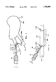
- FIG. 1 is a side view showing the initial state of the preferred embodiment of the nucleotide solution delivery system.
- the system comprises a syringe-like structure with a reservoir 10 to hold a nucleotide solution 40, and a plunger 12.
- a nucleotide solution can contain DNA, RNA or associated molecules.
- reservoir 10 opens into a tube 14. Attached around tube 14, listed in order of proximity to reservoir 10, are a stopcock 16, a pressure gauge 18, a retracted sheath 20, and a notch 22.
- Notch 22 is next to the distal open end 30 of tube 14.
- Sheath 20 is neither permeable nor elastic, in order to protect the tissue from experiencing barotrauma. Stopcock 16 is initially in the closed position, preventing solution 40 from passing from reservoir 10 to tube 14.
- FIG. 2 illustrates how a tubular tissue sample 24 is mounted. Open end 30 is placed into the proximal end of a living, tubular tissue 24. Notch 22 fits inside proximal end of tissue 24 and a tie or ligature 26A is placed to prevent tissue 24 from slipping off open end 30. Then, sheath 20 is pulled down to cover tissue 24. Ligature 26A is wrapped around sheath 20 and tissue 24 at the point where they are attached to tube 14. In this embodiment, the ligature is a tie wrap 26A.
- stopcock 16 is turned to the open position allowing nucleotide solution 40 to enter tube 14 and sheath 20, flushing out all gases and liquids present through open end 28 of sheath 20.
- a tie wrap 26B is placed over distal open end 28 of sheath 20 to form a watertight seal.
- tie wrap 26B is placed around sheath 20 and tissue 24.
- tie wrap 26B is also possible to place tie wrap 26B around sheath 20 only. One must ensure, however, that tie wrap 26B is wrapped in a tight manner.
- FIG. 3 illustrates how nucleotide solution 40 in tissue 24 is pressurized.
- Stopcock 16 is turned to the open position, and plunger 12 is pushed, such that nucleotide solution 40 is injected into sheath 20 under an injection pressure.
- the pressure inside sheath 20 is allowed to increase until a predetermined incubation pressure--generally between at least 300 mm Hg and 1,500 mm Hg--has been reached.
- stopcock 16 is closed (not illustrated).
- Tissue 24 is then allowed to incubate for a period of time, after which tie wrap 26B is untied to release the pressure (not illustrated).
- FIG. 4 illustrates an alternative embodiment of the nucleotide delivery system which operates on an organ.
- a protective sheath 120 is wrapped around an organ 124.
- Organ 124 has an artery 112 which carries blood into it and a vein 114 which carries blood away.
- tube 14 is inserted into the lumen of artery 112.
- sheath 120 is wrapped around artery 112 and vein 114.
- Tie wrap 126A is tightened around sheath 120 at artery 112
- tie wrap 126B is tightened around sheath 120 at vein 114, to prevent leakage of fluid out of organ 124.
- Tie wrap 126A allows tube 14 to enter artery 112, yet wraps tightly enough to seal artery 112 from leakage.
- nucleotide solution 40 is injected, and organ 124 is allowed to incubate. After the incubation period, tie wraps 126A and 126B are removed, and blood is allowed to flow through organ 124 once more.
- this embodiment is identical in structure and operation with the preferred embodiment.
- FIG. 5 illustrates an alternative embodiment which operates on a segment of tubular tissue, for example, a blood vessel.
- tube 14 is inserted into the lumen of a vessel 224 which is still connected to the body of a living animal.
- a sheathing sheet 220 wraps around vessel 224, and a fastener 228 attaches the two flaps of sheet 220 to form a tube.
- fastener 228 may be a heat seal.
- Two tie wraps 226A and 226B wrap around sheet 220. Tie wrap 226A wraps around sheet 220 where tube 14 enters, and tie wrap 226B wraps around the other end of sheet 220. Tie wraps 226A and 226B thus make a segment 230 of vessel 224 watertight.
- nucleotide solution 40 is injected, and segment 230 is allowed to incubate. After the incubation period, tie wraps 226A and 226B are removed, and segment 230 is perfused by blood once more.
- this embodiment is identical in structure and operation with the preferred embodiment.
- FIG. 6 is an alternative embodiment of FIG. 5 in which there are distinct delivery and pressurization systems.
- nucleotide solution 40 is delivered into the lumen of vessel 224 in the same way.
- tie wraps 226A and 226B are placed on sheet 220, a rigid tubular wrap 250 is placed around sheet 220, and a vise 252 is placed around wrap 250, as illustrated in FIG. 7.
- Wrap 250 is circumferentially flexible, so that the diameter of the tube it forms is variable, but it is rigid axially, so that even when its diameter changes, it still remains substantially tubular.
- a tightening screw 254 tightens vise 252, pulling wrap 250 tight, creating pressure within vessel 224. This pressure is maintained for an incubation period, after which screw 254 is unscrewed, releasing the pressure.
- this embodiment is identical in structure and operation with the embodiment of FIG. 5.
- FIG. 8 shows an alternative embodiment of the invention in which the nucleotide solution is delivered into the lumen of a vessel via a catheter.
- a catheter 314 is inserted into a vessel 324.
- Catheter 314 is closed at its end 316. Some distance from end 316, there is an open port 330.
- Mounted on catheter 314 before and after port 330 are two balloons 332A and 332B. Initially, balloons 332A and 332B are deflated.
- FIG. 9 shows the embodiment of FIG. 8 in action.
- Balloons 332A and 332B are inflated, and they occlude vessel 324 where they are inflated, creating a leak-proof segment 334 within vessel 324.
- a nucleotide solution 340 is delivered through catheter 314, exiting from port 330. Solution 340 is pressurized, so segment 334 also becomes pressurized.
- balloons 332A and 332B are deflated and segment 334 becomes depressurized again.
- FIG. 10 shows an alternative embodiment of FIG. 8 in which a balloon mounted to the catheter has miniature tubules for delivering the nucleotide solution to the walls of the vessel.
- FIG. 10 shows this embodiment in the state in which a balloon 432 has already been inflated, but the nucleotide solution has not yet been delivered. In this embodiment, there is no port 330 on catheter 314. Balloon 432 has tubules 450 which are directly connected to holes 452 in the segment of catheter 314 within balloon 432.
- FIG. 11 shows the embodiment of FIG. 10 in action.
- a pressurized nucleotide solution 440 is delivered through catheter 314, solution 440 exits holes 452, travels through tubules 450, and reaches the walls of vessel 324.
- this embodiment is identical to the embodiment in FIG. 8.
- FIG. 12 shows another alternative embodiment of FIG. 8 in which two catheters with balloons pressurize an organ.
- a catheter 550 with a balloon 552 is inserted into an artery 512 leading to an organ 524, and another catheter 560 with a balloon 562 is inserted into a vein 514 leading away from organ 524.
- balloons 552 and 562 are deflated (not illustrated). Once their respective catheters are inserted, they are inflated, thus occluding artery 512 and vein 514. At this point, a pressurized nucleotide solution is delivered into organ 524.
- this embodiment is identical in operation to the embodiment of FIG. 8.
- FIG. 13 illustrates another alternative embodiment of the invention, which utilizes a pressure chamber.
- a dish 610 contains a culture 624 of living tissue.
- FIG. 14 shows how an unpressurized nucleotide solution 640 is introduced into culture 624.
- dish 610 is placed into a pressure chamber 650, as shown in FIG. 15.
- Chamber 650 is as yet unpressurized.
- chamber 650 is closed and sealed, and a pressurized gas, such as CO 2 , is introduced into chamber 650 through a duct 660, thus pressurizing solution 640 and culture 624.
- duct 660 is open to release the pressure.
- This embodiment can also be used for introducing a nucleotide into the cells of an entire organism. To do this an organism is placed inside chamber 650 and pressurized gas is introduced through duct 660.
- nucleotide delivery system introduces exogenous nucleotides into cells in intact tissue in vivo or ex vivo without using foreign and potentially toxic substances. Specifically, this method allows cells to absorb pure DNA in solution at a 90% transfection efficiency, in some circumstances, without disrupting the normal functioning of the tissue. Furthermore, every embodiment described above uses tools and methods which are commonly available, and easy to operate.
- the tie wrap may be replaced by any sealing mechanism, for example, a suture ligature, a suture tie, or a clamp;
- the tissue need not be tubular, but only needs to be easily wrapped by a sheath; the sheath is not always necessary, for example if the tissue can maintain high pressure without a sheath.
- the sheath is useful for allowing exposure of the tissue to nucleotide at high pressure without over-distension of the tissue, and for preventing loss of pressure due to leakage of nucleotide solution through small holes or side branches of the tissue.
- Sheaths of various sizes, shapes, and materials can be used. Furthermore, a sheath may be elastic, so that it may be constricted in order to provide pressure to the tissue. An inelastic can be constricted as well, e.g., by twisting, to thus increase the pressure within. Furthermore, it is not necessary for the sheath to be in contact the tissue; a fluid solution can be introduced between the sheath and the tissue.
- the system is capable of delivering nucleotides into the cells within an intact organism, provided the organism can be placed in a pressurized environment.
- the nucleotide can be presented in either a solution or a suspension.
- the plunger can be automated by a servo-mechanism so as to deliver nucleotide solution at a safe but efficient rate.
- Another means of pressurizing tissue is to introduce an inflatable balloon into the lumen of an organ itself.
- the pressure can be as little as 300 mm Hg and as much as 1500 mm Hg. Lower or higher pressures are also possible, provided the nucleotide is localized in the cell nucleus and the necessary measures are taken to prevent barotrauma.
Abstract
Description
Claims (22)
Priority Applications (10)
| Application Number | Priority Date | Filing Date | Title |
|---|---|---|---|
| US08/434,750 US5766901A (en) | 1995-05-04 | 1995-05-04 | Apparatus and method for delivering a nucleotide into cell nuclei |
| DE69614846T DE69614846T2 (en) | 1995-05-04 | 1996-05-03 | APPARATUS AND METHOD FOR SLOWING IN A NUCLEOTID IN CELL NUCLES |
| PCT/US1996/006271 WO1996034967A1 (en) | 1995-05-04 | 1996-05-03 | Apparatus and method for delivering a nucleotide into cell nuclei |
| JP53354496A JP3865318B2 (en) | 1995-05-04 | 1996-05-03 | Apparatus and method for delivering nucleotides into cell nuclei |
| CA002219091A CA2219091A1 (en) | 1995-05-04 | 1996-05-03 | Apparatus and method for delivering a nucleotide into cell nuclei |
| ES96914572T ES2163018T3 (en) | 1995-05-04 | 1996-05-03 | APPLIANCE AND PROCEDURE TO SUPPLY A NUCLEOTIDE TO A CELL NUCLEUS. |
| EP96914572A EP0826059B1 (en) | 1995-05-04 | 1996-05-03 | Apparatus and method for delivering a nucleotide into cell nuclei |
| AT96914572T ATE204904T1 (en) | 1995-05-04 | 1996-05-03 | APPARATUS AND METHOD FOR INTRODUCING A NUCLEOTIDE INTO CELL NUCLEARS |
| DK96914572T DK0826059T3 (en) | 1995-05-04 | 1996-05-03 | Apparatus and method for administering a nucleotide in a cell nucleus |
| US08/745,023 US5922687A (en) | 1995-05-04 | 1996-11-07 | Intracellular delivery of nucleic acids using pressure |
Applications Claiming Priority (1)
| Application Number | Priority Date | Filing Date | Title |
|---|---|---|---|
| US08/434,750 US5766901A (en) | 1995-05-04 | 1995-05-04 | Apparatus and method for delivering a nucleotide into cell nuclei |
Related Child Applications (1)
| Application Number | Title | Priority Date | Filing Date |
|---|---|---|---|
| US08/745,023 Continuation-In-Part US5922687A (en) | 1995-05-04 | 1996-11-07 | Intracellular delivery of nucleic acids using pressure |
Publications (1)
| Publication Number | Publication Date |
|---|---|
| US5766901A true US5766901A (en) | 1998-06-16 |
Family
ID=23725522
Family Applications (1)
| Application Number | Title | Priority Date | Filing Date |
|---|---|---|---|
| US08/434,750 Expired - Fee Related US5766901A (en) | 1995-05-04 | 1995-05-04 | Apparatus and method for delivering a nucleotide into cell nuclei |
Country Status (9)
| Country | Link |
|---|---|
| US (1) | US5766901A (en) |
| EP (1) | EP0826059B1 (en) |
| JP (1) | JP3865318B2 (en) |
| AT (1) | ATE204904T1 (en) |
| CA (1) | CA2219091A1 (en) |
| DE (1) | DE69614846T2 (en) |
| DK (1) | DK0826059T3 (en) |
| ES (1) | ES2163018T3 (en) |
| WO (1) | WO1996034967A1 (en) |
Cited By (4)
| Publication number | Priority date | Publication date | Assignee | Title |
|---|---|---|---|---|
| US6077256A (en) * | 1998-10-06 | 2000-06-20 | Mann; Michael J. | Delivery of a composition to the lung |
| US6699231B1 (en) | 1997-12-31 | 2004-03-02 | Heartport, Inc. | Methods and apparatus for perfusion of isolated tissue structure |
| US6722370B1 (en) | 1998-07-17 | 2004-04-20 | Corgentech, Inc. | Delivery of a composition to the liver by utilizing the portal vein |
| US20130267929A1 (en) * | 2010-10-25 | 2013-10-10 | Kyoto University | Method for operating a device for delivering a substance to be introduced, and method for delivering a substance to be introduced |
Families Citing this family (6)
| Publication number | Priority date | Publication date | Assignee | Title |
|---|---|---|---|---|
| US5922687A (en) * | 1995-05-04 | 1999-07-13 | Board Of Trustees Of The Leland Stanford Junior University | Intracellular delivery of nucleic acids using pressure |
| WO1999031262A2 (en) * | 1997-12-16 | 1999-06-24 | Valentis, Inc. | Needle-free injection of formulated nucleic acid molecules |
| DE19834612A1 (en) * | 1998-07-31 | 2000-02-24 | Dornier Medtech Holding Int Gmbh | Method for intracellular transfer of oligonucleotides and device for carrying out the same |
| US20070173470A1 (en) * | 2006-01-23 | 2007-07-26 | Chi-Hung Lin | Methods for delivering extracellular target into cells |
| US9017991B2 (en) * | 2009-03-13 | 2015-04-28 | Tufts University | Methods tip assemblies and kits for introducing material into cells |
| CN111001077A (en) * | 2018-10-04 | 2020-04-14 | 口径疗法有限责任公司 | Balloon catheter system for infusing micelles at high pressure |
Citations (1)
| Publication number | Priority date | Publication date | Assignee | Title |
|---|---|---|---|---|
| US5328470A (en) * | 1989-03-31 | 1994-07-12 | The Regents Of The University Of Michigan | Treatment of diseases by site-specific instillation of cells or site-specific transformation of cells and kits therefor |
Family Cites Families (5)
| Publication number | Priority date | Publication date | Assignee | Title |
|---|---|---|---|---|
| JPH0376568A (en) * | 1989-08-16 | 1991-04-02 | Minoru Igari | Jetter for treating cell and piston of the same jetter |
| US5066587A (en) * | 1990-01-26 | 1991-11-19 | The Upjohn Company | Gas driven microprojectile accelerator and method of use |
| WO1992004439A1 (en) * | 1990-08-30 | 1992-03-19 | Brian John Bellhouse | Ballistic apparatus |
| TW404844B (en) * | 1993-04-08 | 2000-09-11 | Oxford Biosciences Ltd | Needleless syringe |
| DE4401076C2 (en) * | 1994-01-15 | 1998-12-03 | Eppendorf Geraetebau Netheler | Device for injecting liquids into biological cells |
-
1995
- 1995-05-04 US US08/434,750 patent/US5766901A/en not_active Expired - Fee Related
-
1996
- 1996-05-03 JP JP53354496A patent/JP3865318B2/en not_active Expired - Fee Related
- 1996-05-03 ES ES96914572T patent/ES2163018T3/en not_active Expired - Lifetime
- 1996-05-03 DK DK96914572T patent/DK0826059T3/en active
- 1996-05-03 AT AT96914572T patent/ATE204904T1/en not_active IP Right Cessation
- 1996-05-03 DE DE69614846T patent/DE69614846T2/en not_active Expired - Fee Related
- 1996-05-03 EP EP96914572A patent/EP0826059B1/en not_active Expired - Lifetime
- 1996-05-03 WO PCT/US1996/006271 patent/WO1996034967A1/en active IP Right Grant
- 1996-05-03 CA CA002219091A patent/CA2219091A1/en not_active Abandoned
Patent Citations (1)
| Publication number | Priority date | Publication date | Assignee | Title |
|---|---|---|---|---|
| US5328470A (en) * | 1989-03-31 | 1994-07-12 | The Regents Of The University Of Michigan | Treatment of diseases by site-specific instillation of cells or site-specific transformation of cells and kits therefor |
Non-Patent Citations (22)
| Title |
|---|
| Acsadi et al., "Direct gene transfer and expression into rat heart in vivo", New Biologist 3(1): 71-81, 1991. |
| Acsadi et al., Direct gene transfer and expression into rat heart in vivo , New Biologist 3(1): 71 81, 1991. * |
| Barinaga, M. Science, vol. 266, p. 1326, 1994. * |
| Crystal, R. G. Science, vol. 270, pp. 404 410, 1995. * |
| Crystal, R. G. Science, vol. 270, pp. 404-410, 1995. |
| Furth et al., "Gene transfer into somatic tissues", Anal. Biocehm. 205: 365-368, 1992. |
| Furth et al., Gene transfer into somatic tissues , Anal. Biocehm. 205: 365 368, 1992. * |
| Ledley, F.D. Current Opinion in Biotechnology, vol. 5 , pp. 626 636, 1994. * |
| Ledley, F.D. Current Opinion in Biotechnology, vol. 5 , pp. 626-636, 1994. |
| Marshall, E. Science, vol. 269, pp. 1050 1055, 1995. * |
| Marshall, E. Science, vol. 269, pp. 1050-1055, 1995. |
| Nabel et al., "Recombinant Gene Expression in Vivo Within Endothelial Cells of the Arterial Wall", Science 244:1342-1344 (1989). |
| Nabel et al., "Site-Specific Gene Expression in Vivio by Direct Gene Transfer into the Arterial Wall", Science 249:1285-1288 (1990). |
| Nabel et al., Recombinant Gene Expression in Vivo Within Endothelial Cells of the Arterial Wall , Science 244:1342 1344 (1989). * |
| Nabel et al., Site Specific Gene Expression in Vivio by Direct Gene Transfer into the Arterial Wall , Science 249:1285 1288 (1990). * |
| Orkin, S.H. et al. Report and Recommendations of the Panel to Assess the NIH Investment in Research on Gene Therapy, 1995. * |
| Schofield, J.P. et al. British Medical Bulletin, vol. 51 (1), pp. 56 71, 1995. * |
| Schofield, J.P. et al. British Medical Bulletin, vol. 51 (1), pp. 56-71, 1995. |
| Wolff et al., "Expression of maked plasmids by cultured myotubes and entry of plasmids into T tubules and caveolae of mammalian skeletal muscle", J. Cell Sci. 103: 1249-1259, 1992. |
| Wolff et al., Expression of maked plasmids by cultured myotubes and entry of plasmids into T tubules and caveolae of mammalian skeletal muscle , J. Cell Sci. 103: 1249 1259, 1992. * |
| Yee et al., "Cellular uptake of intracerebroventricularly administered biotin-or digoxigenin-labelled antisense oligonucleotides in the rat", Cell. Mol. Neurobiol. 14(5):475-486, 1994. |
| Yee et al., Cellular uptake of intracerebroventricularly administered biotin or digoxigenin labelled antisense oligonucleotides in the rat , Cell. Mol. Neurobiol. 14(5):475 486, 1994. * |
Cited By (4)
| Publication number | Priority date | Publication date | Assignee | Title |
|---|---|---|---|---|
| US6699231B1 (en) | 1997-12-31 | 2004-03-02 | Heartport, Inc. | Methods and apparatus for perfusion of isolated tissue structure |
| US6722370B1 (en) | 1998-07-17 | 2004-04-20 | Corgentech, Inc. | Delivery of a composition to the liver by utilizing the portal vein |
| US6077256A (en) * | 1998-10-06 | 2000-06-20 | Mann; Michael J. | Delivery of a composition to the lung |
| US20130267929A1 (en) * | 2010-10-25 | 2013-10-10 | Kyoto University | Method for operating a device for delivering a substance to be introduced, and method for delivering a substance to be introduced |
Also Published As
| Publication number | Publication date |
|---|---|
| ES2163018T3 (en) | 2002-01-16 |
| JP3865318B2 (en) | 2007-01-10 |
| EP0826059A4 (en) | 1998-07-01 |
| EP0826059A1 (en) | 1998-03-04 |
| WO1996034967A1 (en) | 1996-11-07 |
| ATE204904T1 (en) | 2001-09-15 |
| DK0826059T3 (en) | 2001-12-17 |
| EP0826059B1 (en) | 2001-08-29 |
| DE69614846D1 (en) | 2001-10-04 |
| CA2219091A1 (en) | 1996-11-07 |
| DE69614846T2 (en) | 2002-05-16 |
| JPH11505112A (en) | 1999-05-18 |
Similar Documents
| Publication | Publication Date | Title |
|---|---|---|
| KR100449330B1 (en) | Pressure-mediated intracellular delivery of molecules or microparticles | |
| US5766901A (en) | Apparatus and method for delivering a nucleotide into cell nuclei | |
| US5411475A (en) | Directly visualized method for deploying a detachable balloon at a target site in vivo | |
| JP2500161B2 (en) | Distribution catheter | |
| US20080125760A1 (en) | Directional Anchoring Mechanism, Method and Applications Thereof | |
| JPH0663143A (en) | Catheter | |
| US20040215143A1 (en) | Hollow stylet for infusion catheter systems, devices and methods | |
| CN103429214B (en) | O_2 transport body gives system, O_2 transport body oxygenation device and O_2 transport body host body | |
| US8157747B2 (en) | Single-use indicator for a surgical instrument and a surgical instrument incorporating same | |
| US5947928A (en) | Drug delivery system | |
| WO2007125327A1 (en) | Laparoscopic kidney cooling sheath | |
| WO1999022683A1 (en) | Intraorgan administration system (ias) | |
| US4113097A (en) | Ampule capable of being autoclaved | |
| CN208799661U (en) | Gallstone treatment system | |
| US6395550B1 (en) | Method and apparatus for tissue treatment | |
| WO1998057696A9 (en) | Drug delivery system including a drug transport enhancement mechanism | |
| JPWO2021167993A5 (en) | ||
| RU2045230C1 (en) | Method of laparoscopic mucoclasis |
Legal Events
| Date | Code | Title | Description |
|---|---|---|---|
| AS | Assignment |
Owner name: BOARD OF TRUSTEES OF THE LELAND STANFORD JUNIOR UN Free format text: ASSIGNMENT OF ASSIGNORS INTEREST;ASSIGNORS:MANN, MICHAEL J.;DIET, FRANK P.;DZAU, VICTOR J.;AND OTHERS;REEL/FRAME:007586/0368 Effective date: 19950623 |
|
| CC | Certificate of correction | ||
| FPAY | Fee payment |
Year of fee payment: 4 |
|
| REMI | Maintenance fee reminder mailed | ||
| AS | Assignment |
Owner name: ALTA CALIFORNIA PARTNERS III, L.P., CALIFORNIA Free format text: SECURITY AGREEMENT;ASSIGNOR:CORGENTECH INC., A DELAWARE CORPORATION;REEL/FRAME:014491/0102 Effective date: 20030902 Owner name: ALTA EMBARCADERO PARTNERS III, LLC, CALIFORNIA Free format text: SECURITY AGREEMENT;ASSIGNOR:CORGENTECH INC., A DELAWARE CORPORATION;REEL/FRAME:014491/0102 Effective date: 20030902 Owner name: BEAR STEARNS HEALTH INNOVENTURES EMPLOYEE FUND, L. Free format text: SECURITY AGREEMENT;ASSIGNOR:CORGENTECH INC., A DELAWARE CORPORATION;REEL/FRAME:014491/0102 Effective date: 20030902 Owner name: BEAR STEARNS HEALTH INNOVENTURES OFFSHORE, L.P., N Free format text: SECURITY AGREEMENT;ASSIGNOR:CORGENTECH INC., A DELAWARE CORPORATION;REEL/FRAME:014491/0102 Effective date: 20030902 Owner name: BEAR STEARNS HEALTH INNOVENTURES, L.P., NEW YORK Free format text: SECURITY AGREEMENT;ASSIGNOR:CORGENTECH INC., A DELAWARE CORPORATION;REEL/FRAME:014491/0102 Effective date: 20030902 Owner name: BSHI MEMBERS, L.L.C., NEW YORK Free format text: SECURITY AGREEMENT;ASSIGNOR:CORGENTECH INC., A DELAWARE CORPORATION;REEL/FRAME:014491/0102 Effective date: 20030902 Owner name: BX, L.P., NEW YORK Free format text: SECURITY AGREEMENT;ASSIGNOR:CORGENTECH INC., A DELAWARE CORPORATION;REEL/FRAME:014491/0102 Effective date: 20030902 Owner name: HBM BIOVENTURES (CAYMAN) LTD., CAYMAN ISLANDS Free format text: SECURITY AGREEMENT;ASSIGNOR:CORGENTECH INC., A DELAWARE CORPORATION;REEL/FRAME:014491/0102 Effective date: 20030902 Owner name: INTERWEST INVESTORS Q VIII, L.P., CALIFORNIA Free format text: SECURITY AGREEMENT;ASSIGNOR:CORGENTECH INC., A DELAWARE CORPORATION;REEL/FRAME:014491/0102 Effective date: 20030902 Owner name: INTERWEST INVESTORS VIII, L.P., CALIFORNIA Free format text: SECURITY AGREEMENT;ASSIGNOR:CORGENTECH INC., A DELAWARE CORPORATION;REEL/FRAME:014491/0102 Effective date: 20030902 Owner name: INTERWEST PARTNERS VIII, L.P., CALIFORNIA Free format text: SECURITY AGREEMENT;ASSIGNOR:CORGENTECH INC., A DELAWARE CORPORATION;REEL/FRAME:014491/0102 Effective date: 20030902 Owner name: J.P. MORGAN PARTNERS (SBIC), L.L.C., CALIFORNIA Free format text: SECURITY AGREEMENT;ASSIGNOR:CORGENTECH INC., A DELAWARE CORPORATION;REEL/FRAME:014491/0102 Effective date: 20030902 Owner name: JP MORGAN PARTNERS GLOBAL INVESTORS (CAYMAN) II, L Free format text: SECURITY AGREEMENT;ASSIGNOR:CORGENTECH INC., A DELAWARE CORPORATION;REEL/FRAME:014491/0102 Effective date: 20030902 Owner name: JP MORGAN PARTNERS GLOBAL INVESTORS (CAYMAN), L.P. Free format text: SECURITY AGREEMENT;ASSIGNOR:CORGENTECH INC., A DELAWARE CORPORATION;REEL/FRAME:014491/0102 Effective date: 20030902 Owner name: JP MORGAN PARTNERS GLOBAL INVESTORS A, L.P., CALIF Free format text: SECURITY AGREEMENT;ASSIGNOR:CORGENTECH INC., A DELAWARE CORPORATION;REEL/FRAME:014491/0102 Effective date: 20030902 Owner name: JP MORGAN PARTNERS GLOBAL INVESTORS, L.P., CALIFOR Free format text: SECURITY AGREEMENT;ASSIGNOR:CORGENTECH INC., A DELAWARE CORPORATION;REEL/FRAME:014491/0102 Effective date: 20030902 |
|
| FEPP | Fee payment procedure |
Free format text: PAYOR NUMBER ASSIGNED (ORIGINAL EVENT CODE: ASPN); ENTITY STATUS OF PATENT OWNER: LARGE ENTITY Free format text: PAT HOLDER NO LONGER CLAIMS SMALL ENTITY STATUS, ENTITY STATUS SET TO UNDISCOUNTED (ORIGINAL EVENT CODE: STOL); ENTITY STATUS OF PATENT OWNER: LARGE ENTITY |
|
| FPAY | Fee payment |
Year of fee payment: 8 |
|
| REMI | Maintenance fee reminder mailed | ||
| LAPS | Lapse for failure to pay maintenance fees | ||
| STCH | Information on status: patent discontinuation |
Free format text: PATENT EXPIRED DUE TO NONPAYMENT OF MAINTENANCE FEES UNDER 37 CFR 1.362 |
|
| FP | Lapsed due to failure to pay maintenance fee |
Effective date: 20100616 |