US5776488A - Liposome preparation - Google Patents
Liposome preparation Download PDFInfo
- Publication number
- US5776488A US5776488A US08/716,201 US71620196A US5776488A US 5776488 A US5776488 A US 5776488A US 71620196 A US71620196 A US 71620196A US 5776488 A US5776488 A US 5776488A
- Authority
- US
- United States
- Prior art keywords
- liposome
- liposome preparation
- deoxy
- preparation
- water
- Prior art date
- Legal status (The legal status is an assumption and is not a legal conclusion. Google has not performed a legal analysis and makes no representation as to the accuracy of the status listed.)
- Expired - Fee Related
Links
- 239000002502 liposome Substances 0.000 title claims abstract description 130
- 238000002360 preparation method Methods 0.000 title claims abstract description 68
- -1 2'-deoxy-2'methylidenecytidine compound Chemical class 0.000 claims abstract description 37
- CKTSBUTUHBMZGZ-UHFFFAOYSA-N Deoxycytidine Natural products O=C1N=C(N)C=CN1C1OC(CO)C(O)C1 CKTSBUTUHBMZGZ-UHFFFAOYSA-N 0.000 claims abstract description 35
- 239000012528 membrane Substances 0.000 claims abstract description 33
- 150000002632 lipids Chemical class 0.000 claims abstract description 21
- HVYWMOMLDIMFJA-DPAQBDIFSA-N cholesterol Chemical compound C1C=C2C[C@@H](O)CC[C@]2(C)[C@@H]2[C@@H]1[C@@H]1CC[C@H]([C@H](C)CCCC(C)C)[C@@]1(C)CC2 HVYWMOMLDIMFJA-DPAQBDIFSA-N 0.000 claims description 31
- 239000002245 particle Substances 0.000 claims description 21
- 150000003904 phospholipids Chemical class 0.000 claims description 19
- REYJJPSVUYRZGE-UHFFFAOYSA-N Octadecylamine Chemical compound CCCCCCCCCCCCCCCCCCN REYJJPSVUYRZGE-UHFFFAOYSA-N 0.000 claims description 15
- 235000012000 cholesterol Nutrition 0.000 claims description 15
- KILNVBDSWZSGLL-KXQOOQHDSA-N 1,2-dihexadecanoyl-sn-glycero-3-phosphocholine Chemical compound CCCCCCCCCCCCCCCC(=O)OC[C@H](COP([O-])(=O)OCC[N+](C)(C)C)OC(=O)CCCCCCCCCCCCCCC KILNVBDSWZSGLL-KXQOOQHDSA-N 0.000 claims description 9
- JLPULHDHAOZNQI-JLOPVYAASA-N [(2r)-3-hexadecanoyloxy-2-[(9e,12e)-octadeca-9,12-dienoyl]oxypropyl] 2-(trimethylazaniumyl)ethyl phosphate Chemical compound CCCCCCCCCCCCCCCC(=O)OC[C@H](COP([O-])(=O)OCC[N+](C)(C)C)OC(=O)CCCCCCC\C=C\C\C=C\CCCCC JLPULHDHAOZNQI-JLOPVYAASA-N 0.000 claims description 6
- WWUZIQQURGPMPG-UHFFFAOYSA-N (-)-D-erythro-Sphingosine Natural products CCCCCCCCCCCCCC=CC(O)C(N)CO WWUZIQQURGPMPG-UHFFFAOYSA-N 0.000 claims description 5
- CBFZRLQZSOMINR-BKPPORCPSA-N 4-amino-5-fluoro-1-[(2r,4s,5r)-4-hydroxy-5-(hydroxymethyl)-3-methylideneoxolan-2-yl]pyrimidin-2-one Chemical compound C1=C(F)C(N)=NC(=O)N1[C@H]1C(=C)[C@H](O)[C@@H](CO)O1 CBFZRLQZSOMINR-BKPPORCPSA-N 0.000 claims description 5
- WWUZIQQURGPMPG-KRWOKUGFSA-N sphingosine Chemical compound CCCCCCCCCCCCC\C=C\[C@@H](O)[C@@H](N)CO WWUZIQQURGPMPG-KRWOKUGFSA-N 0.000 claims description 5
- 239000003381 stabilizer Substances 0.000 claims description 5
- PULHLIOPJXPGJN-BWVDBABLSA-N 4-amino-1-[(2r,4s,5r)-4-hydroxy-5-(hydroxymethyl)-3-methylideneoxolan-2-yl]pyrimidin-2-one Chemical compound O=C1N=C(N)C=CN1[C@H]1C(=C)[C@H](O)[C@@H](CO)O1 PULHLIOPJXPGJN-BWVDBABLSA-N 0.000 claims description 4
- 150000004677 hydrates Chemical class 0.000 claims description 4
- 150000003839 salts Chemical class 0.000 claims description 4
- NRJAVPSFFCBXDT-HUESYALOSA-N 1,2-distearoyl-sn-glycero-3-phosphocholine Chemical compound CCCCCCCCCCCCCCCCCC(=O)OC[C@H](COP([O-])(=O)OCC[N+](C)(C)C)OC(=O)CCCCCCCCCCCCCCCCC NRJAVPSFFCBXDT-HUESYALOSA-N 0.000 claims description 3
- GFFXZLZWLOBBLO-BWVDBABLSA-N 4-amino-1-[(2r,4s,5r)-3-(fluoromethylidene)-4-hydroxy-5-(hydroxymethyl)oxolan-2-yl]pyrimidin-2-one Chemical compound O=C1N=C(N)C=CN1[C@H]1C(=CF)[C@H](O)[C@@H](CO)O1 GFFXZLZWLOBBLO-BWVDBABLSA-N 0.000 claims description 3
- XTYUGPDATYNDMJ-YPMAUOMFSA-N 4-amino-1-[(2r,4s,5r)-4-hydroxy-5-(hydroxymethyl)-3-methylideneoxolan-2-yl]pyrimidin-2-one;dihydrate Chemical compound O.O.O=C1N=C(N)C=CN1[C@H]1C(=C)[C@H](O)[C@@H](CO)O1 XTYUGPDATYNDMJ-YPMAUOMFSA-N 0.000 claims description 3
- SDUQYLNIPVEERB-QPPQHZFASA-N gemcitabine Chemical compound O=C1N=C(N)C=CN1[C@H]1C(F)(F)[C@H](O)[C@@H](CO)O1 SDUQYLNIPVEERB-QPPQHZFASA-N 0.000 claims description 3
- WTJKGGKOPKCXLL-RRHRGVEJSA-N phosphatidylcholine Chemical compound CCCCCCCCCCCCCCCC(=O)OC[C@H](COP([O-])(=O)OCC[N+](C)(C)C)OC(=O)CCCCCCCC=CCCCCCCCC WTJKGGKOPKCXLL-RRHRGVEJSA-N 0.000 claims description 3
- 102000002322 Egg Proteins Human genes 0.000 claims description 2
- 108010000912 Egg Proteins Proteins 0.000 claims description 2
- 229930182558 Sterol Natural products 0.000 claims description 2
- 235000013345 egg yolk Nutrition 0.000 claims description 2
- 210000002969 egg yolk Anatomy 0.000 claims description 2
- 150000003432 sterols Chemical class 0.000 claims description 2
- 235000003702 sterols Nutrition 0.000 claims description 2
- FIGGQGCOJFHEJY-CDNBRZBRSA-N 4-amino-1-[(2r,4s,5r)-4-hydroxy-5-(hydroxymethyl)oxolan-2-yl]pyrimidin-2-one;dihydrate Chemical compound O.O.O=C1N=C(N)C=CN1[C@@H]1O[C@H](CO)[C@@H](O)C1 FIGGQGCOJFHEJY-CDNBRZBRSA-N 0.000 claims 2
- 201000011510 cancer Diseases 0.000 abstract description 34
- 238000011282 treatment Methods 0.000 abstract description 22
- 230000000694 effects Effects 0.000 abstract description 17
- 230000009876 antimalignant effect Effects 0.000 abstract description 16
- 206010028980 Neoplasm Diseases 0.000 abstract description 13
- 150000001875 compounds Chemical class 0.000 abstract description 11
- 239000004480 active ingredient Substances 0.000 abstract description 5
- 239000008280 blood Substances 0.000 abstract description 5
- 210000004369 blood Anatomy 0.000 abstract description 5
- 230000000259 anti-tumor effect Effects 0.000 abstract description 3
- 230000001988 toxicity Effects 0.000 abstract description 2
- 231100000419 toxicity Toxicity 0.000 abstract description 2
- LFQSCWFLJHTTHZ-UHFFFAOYSA-N Ethanol Chemical compound CCO LFQSCWFLJHTTHZ-UHFFFAOYSA-N 0.000 description 45
- GZDFHIJNHHMENY-UHFFFAOYSA-N Dimethyl dicarbonate Chemical compound COC(=O)OC(=O)OC GZDFHIJNHHMENY-UHFFFAOYSA-N 0.000 description 38
- 235000010300 dimethyl dicarbonate Nutrition 0.000 description 38
- 239000000243 solution Substances 0.000 description 33
- 210000001519 tissue Anatomy 0.000 description 30
- 210000004379 membrane Anatomy 0.000 description 27
- 239000000725 suspension Substances 0.000 description 25
- 238000013508 migration Methods 0.000 description 15
- 230000005012 migration Effects 0.000 description 15
- 239000002904 solvent Substances 0.000 description 15
- 210000001185 bone marrow Anatomy 0.000 description 14
- 239000003795 chemical substances by application Substances 0.000 description 14
- 210000004185 liver Anatomy 0.000 description 14
- 239000000203 mixture Substances 0.000 description 12
- 230000007704 transition Effects 0.000 description 12
- XLYOFNOQVPJJNP-UHFFFAOYSA-N water Substances O XLYOFNOQVPJJNP-UHFFFAOYSA-N 0.000 description 12
- 238000000034 method Methods 0.000 description 11
- 238000000108 ultra-filtration Methods 0.000 description 10
- PZNPLUBHRSSFHT-RRHRGVEJSA-N 1-hexadecanoyl-2-octadecanoyl-sn-glycero-3-phosphocholine Chemical compound CCCCCCCCCCCCCCCCCC(=O)O[C@@H](COP([O-])(=O)OCC[N+](C)(C)C)COC(=O)CCCCCCCCCCCCCCC PZNPLUBHRSSFHT-RRHRGVEJSA-N 0.000 description 9
- 101001105486 Homo sapiens Proteasome subunit alpha type-7 Proteins 0.000 description 9
- 102100021201 Proteasome subunit alpha type-7 Human genes 0.000 description 9
- 230000000052 comparative effect Effects 0.000 description 9
- 239000012071 phase Substances 0.000 description 9
- 239000007791 liquid phase Substances 0.000 description 7
- 239000011148 porous material Substances 0.000 description 7
- 239000004094 surface-active agent Substances 0.000 description 7
- RTZKZFJDLAIYFH-UHFFFAOYSA-N Diethyl ether Chemical compound CCOCC RTZKZFJDLAIYFH-UHFFFAOYSA-N 0.000 description 6
- WQZGKKKJIJFFOK-GASJEMHNSA-N Glucose Natural products OC[C@H]1OC(O)[C@H](O)[C@@H](O)[C@@H]1O WQZGKKKJIJFFOK-GASJEMHNSA-N 0.000 description 6
- PEDCQBHIVMGVHV-UHFFFAOYSA-N Glycerine Chemical compound OCC(O)CO PEDCQBHIVMGVHV-UHFFFAOYSA-N 0.000 description 6
- 239000007864 aqueous solution Substances 0.000 description 6
- 239000000872 buffer Substances 0.000 description 6
- 230000003247 decreasing effect Effects 0.000 description 6
- 239000008103 glucose Substances 0.000 description 6
- 239000007924 injection Substances 0.000 description 6
- 238000002347 injection Methods 0.000 description 6
- 239000002504 physiological saline solution Substances 0.000 description 6
- 238000000527 sonication Methods 0.000 description 6
- 238000012360 testing method Methods 0.000 description 6
- 241000700159 Rattus Species 0.000 description 5
- 239000006185 dispersion Substances 0.000 description 5
- 238000000265 homogenisation Methods 0.000 description 5
- 239000003960 organic solvent Substances 0.000 description 5
- RYMZZMVNJRMUDD-HGQWONQESA-N simvastatin Chemical compound C([C@H]1[C@@H](C)C=CC2=C[C@H](C)C[C@@H]([C@H]12)OC(=O)C(C)(C)CC)C[C@@H]1C[C@@H](O)CC(=O)O1 RYMZZMVNJRMUDD-HGQWONQESA-N 0.000 description 5
- 238000004513 sizing Methods 0.000 description 5
- HEDRZPFGACZZDS-UHFFFAOYSA-N Chloroform Chemical compound ClC(Cl)Cl HEDRZPFGACZZDS-UHFFFAOYSA-N 0.000 description 4
- 238000001704 evaporation Methods 0.000 description 4
- 230000008020 evaporation Effects 0.000 description 4
- 238000010438 heat treatment Methods 0.000 description 4
- 230000036571 hydration Effects 0.000 description 4
- 238000006703 hydration reaction Methods 0.000 description 4
- 210000000056 organ Anatomy 0.000 description 4
- 238000003756 stirring Methods 0.000 description 4
- 238000010257 thawing Methods 0.000 description 4
- 239000000232 Lipid Bilayer Substances 0.000 description 3
- 206010058467 Lung neoplasm malignant Diseases 0.000 description 3
- DNIAPMSPPWPWGF-UHFFFAOYSA-N Propylene glycol Chemical compound CC(O)CO DNIAPMSPPWPWGF-UHFFFAOYSA-N 0.000 description 3
- 238000009825 accumulation Methods 0.000 description 3
- 238000013019 agitation Methods 0.000 description 3
- 238000007796 conventional method Methods 0.000 description 3
- 239000003814 drug Substances 0.000 description 3
- 229940079593 drug Drugs 0.000 description 3
- 201000005202 lung cancer Diseases 0.000 description 3
- 208000020816 lung neoplasm Diseases 0.000 description 3
- 235000000346 sugar Nutrition 0.000 description 3
- 230000001629 suppression Effects 0.000 description 3
- CIWBSHSKHKDKBQ-JLAZNSOCSA-N Ascorbic acid Chemical compound OC[C@H](O)[C@H]1OC(=O)C(O)=C1O CIWBSHSKHKDKBQ-JLAZNSOCSA-N 0.000 description 2
- IJGRMHOSHXDMSA-UHFFFAOYSA-N Atomic nitrogen Chemical compound N#N IJGRMHOSHXDMSA-UHFFFAOYSA-N 0.000 description 2
- NLZUEZXRPGMBCV-UHFFFAOYSA-N Butylhydroxytoluene Chemical compound CC1=CC(C(C)(C)C)=C(O)C(C(C)(C)C)=C1 NLZUEZXRPGMBCV-UHFFFAOYSA-N 0.000 description 2
- WQZGKKKJIJFFOK-VFUOTHLCSA-N beta-D-glucose Chemical compound OC[C@H]1O[C@@H](O)[C@H](O)[C@@H](O)[C@@H]1O WQZGKKKJIJFFOK-VFUOTHLCSA-N 0.000 description 2
- 235000010354 butylated hydroxytoluene Nutrition 0.000 description 2
- 210000004027 cell Anatomy 0.000 description 2
- KXGVEGMKQFWNSR-UHFFFAOYSA-N deoxycholic acid Natural products C1CC2CC(O)CCC2(C)C2C1C1CCC(C(CCC(O)=O)C)C1(C)C(O)C2 KXGVEGMKQFWNSR-UHFFFAOYSA-N 0.000 description 2
- 238000000502 dialysis Methods 0.000 description 2
- 239000012153 distilled water Substances 0.000 description 2
- 238000005538 encapsulation Methods 0.000 description 2
- 238000009472 formulation Methods 0.000 description 2
- 238000002523 gelfiltration Methods 0.000 description 2
- NOESYZHRGYRDHS-UHFFFAOYSA-N insulin Chemical compound N1C(=O)C(NC(=O)C(CCC(N)=O)NC(=O)C(CCC(O)=O)NC(=O)C(C(C)C)NC(=O)C(NC(=O)CN)C(C)CC)CSSCC(C(NC(CO)C(=O)NC(CC(C)C)C(=O)NC(CC=2C=CC(O)=CC=2)C(=O)NC(CCC(N)=O)C(=O)NC(CC(C)C)C(=O)NC(CCC(O)=O)C(=O)NC(CC(N)=O)C(=O)NC(CC=2C=CC(O)=CC=2)C(=O)NC(CSSCC(NC(=O)C(C(C)C)NC(=O)C(CC(C)C)NC(=O)C(CC=2C=CC(O)=CC=2)NC(=O)C(CC(C)C)NC(=O)C(C)NC(=O)C(CCC(O)=O)NC(=O)C(C(C)C)NC(=O)C(CC(C)C)NC(=O)C(CC=2NC=NC=2)NC(=O)C(CO)NC(=O)CNC2=O)C(=O)NCC(=O)NC(CCC(O)=O)C(=O)NC(CCCNC(N)=N)C(=O)NCC(=O)NC(CC=3C=CC=CC=3)C(=O)NC(CC=3C=CC=CC=3)C(=O)NC(CC=3C=CC(O)=CC=3)C(=O)NC(C(C)O)C(=O)N3C(CCC3)C(=O)NC(CCCCN)C(=O)NC(C)C(O)=O)C(=O)NC(CC(N)=O)C(O)=O)=O)NC(=O)C(C(C)CC)NC(=O)C(CO)NC(=O)C(C(C)O)NC(=O)C1CSSCC2NC(=O)C(CC(C)C)NC(=O)C(NC(=O)C(CCC(N)=O)NC(=O)C(CC(N)=O)NC(=O)C(NC(=O)C(N)CC=1C=CC=CC=1)C(C)C)CC1=CN=CN1 NOESYZHRGYRDHS-UHFFFAOYSA-N 0.000 description 2
- 238000010253 intravenous injection Methods 0.000 description 2
- JVTAAEKCZFNVCJ-UHFFFAOYSA-N lactic acid Chemical compound CC(O)C(O)=O JVTAAEKCZFNVCJ-UHFFFAOYSA-N 0.000 description 2
- 238000012986 modification Methods 0.000 description 2
- 230000004048 modification Effects 0.000 description 2
- 229910052757 nitrogen Inorganic materials 0.000 description 2
- 239000008213 purified water Substances 0.000 description 2
- 230000009467 reduction Effects 0.000 description 2
- 238000011160 research Methods 0.000 description 2
- 239000000126 substance Substances 0.000 description 2
- 150000008163 sugars Chemical class 0.000 description 2
- 239000002691 unilamellar liposome Substances 0.000 description 2
- GVJHHUAWPYXKBD-IEOSBIPESA-N α-tocopherol Chemical compound OC1=C(C)C(C)=C2O[C@@](CCC[C@H](C)CCC[C@H](C)CCCC(C)C)(C)CCC2=C1C GVJHHUAWPYXKBD-IEOSBIPESA-N 0.000 description 2
- HEGSGKPQLMEBJL-RQICVUQASA-N (2r,3s,4s,5r)-2-(hydroxymethyl)-6-octoxyoxane-3,4,5-triol Chemical compound CCCCCCCCOC1O[C@H](CO)[C@@H](O)[C@H](O)[C@H]1O HEGSGKPQLMEBJL-RQICVUQASA-N 0.000 description 1
- BHQCQFFYRZLCQQ-UHFFFAOYSA-N (3alpha,5alpha,7alpha,12alpha)-3,7,12-trihydroxy-cholan-24-oic acid Natural products OC1CC2CC(O)CCC2(C)C2C1C1CCC(C(CCC(O)=O)C)C1(C)C(O)C2 BHQCQFFYRZLCQQ-UHFFFAOYSA-N 0.000 description 1
- QYIXCDOBOSTCEI-QCYZZNICSA-N (5alpha)-cholestan-3beta-ol Chemical compound C([C@@H]1CC2)[C@@H](O)CC[C@]1(C)[C@@H]1[C@@H]2[C@@H]2CC[C@H]([C@H](C)CCCC(C)C)[C@@]2(C)CC1 QYIXCDOBOSTCEI-QCYZZNICSA-N 0.000 description 1
- QGLWBTPVKHMVHM-KTKRTIGZSA-N (z)-octadec-9-en-1-amine Chemical compound CCCCCCCC\C=C/CCCCCCCCN QGLWBTPVKHMVHM-KTKRTIGZSA-N 0.000 description 1
- IIZPXYDJLKNOIY-JXPKJXOSSA-N 1-palmitoyl-2-arachidonoyl-sn-glycero-3-phosphocholine Chemical compound CCCCCCCCCCCCCCCC(=O)OC[C@H](COP([O-])(=O)OCC[N+](C)(C)C)OC(=O)CCC\C=C/C\C=C/C\C=C/C\C=C/CCCCC IIZPXYDJLKNOIY-JXPKJXOSSA-N 0.000 description 1
- NHBKXEKEPDILRR-UHFFFAOYSA-N 2,3-bis(butanoylsulfanyl)propyl butanoate Chemical compound CCCC(=O)OCC(SC(=O)CCC)CSC(=O)CCC NHBKXEKEPDILRR-UHFFFAOYSA-N 0.000 description 1
- 208000004998 Abdominal Pain Diseases 0.000 description 1
- GUBGYTABKSRVRQ-XLOQQCSPSA-N Alpha-Lactose Chemical compound O[C@@H]1[C@@H](O)[C@@H](O)[C@@H](CO)O[C@H]1O[C@@H]1[C@@H](CO)O[C@H](O)[C@H](O)[C@H]1O GUBGYTABKSRVRQ-XLOQQCSPSA-N 0.000 description 1
- 206010005003 Bladder cancer Diseases 0.000 description 1
- 206010006187 Breast cancer Diseases 0.000 description 1
- 208000026310 Breast neoplasm Diseases 0.000 description 1
- FERIUCNNQQJTOY-UHFFFAOYSA-M Butyrate Chemical compound CCCC([O-])=O FERIUCNNQQJTOY-UHFFFAOYSA-M 0.000 description 1
- OYPRJOBELJOOCE-UHFFFAOYSA-N Calcium Chemical compound [Ca] OYPRJOBELJOOCE-UHFFFAOYSA-N 0.000 description 1
- 206010008342 Cervix carcinoma Diseases 0.000 description 1
- 239000004380 Cholic acid Substances 0.000 description 1
- 208000002881 Colic Diseases 0.000 description 1
- 206010009944 Colon cancer Diseases 0.000 description 1
- PMATZTZNYRCHOR-CGLBZJNRSA-N Cyclosporin A Chemical compound CC[C@@H]1NC(=O)[C@H]([C@H](O)[C@H](C)C\C=C\C)N(C)C(=O)[C@H](C(C)C)N(C)C(=O)[C@H](CC(C)C)N(C)C(=O)[C@H](CC(C)C)N(C)C(=O)[C@@H](C)NC(=O)[C@H](C)NC(=O)[C@H](CC(C)C)N(C)C(=O)[C@H](C(C)C)NC(=O)[C@H](CC(C)C)N(C)C(=O)CN(C)C1=O PMATZTZNYRCHOR-CGLBZJNRSA-N 0.000 description 1
- 229930105110 Cyclosporin A Natural products 0.000 description 1
- KCXVZYZYPLLWCC-UHFFFAOYSA-N EDTA Chemical compound OC(=O)CN(CC(O)=O)CCN(CC(O)=O)CC(O)=O KCXVZYZYPLLWCC-UHFFFAOYSA-N 0.000 description 1
- 208000000461 Esophageal Neoplasms Diseases 0.000 description 1
- 206010017993 Gastrointestinal neoplasms Diseases 0.000 description 1
- JZNWSCPGTDBMEW-UHFFFAOYSA-N Glycerophosphorylethanolamin Natural products NCCOP(O)(=O)OCC(O)CO JZNWSCPGTDBMEW-UHFFFAOYSA-N 0.000 description 1
- 229930186217 Glycolipid Natural products 0.000 description 1
- 102100036284 Hepcidin Human genes 0.000 description 1
- 101001021253 Homo sapiens Hepcidin Proteins 0.000 description 1
- 102000004877 Insulin Human genes 0.000 description 1
- 108090001061 Insulin Proteins 0.000 description 1
- 208000008839 Kidney Neoplasms Diseases 0.000 description 1
- GUBGYTABKSRVRQ-QKKXKWKRSA-N Lactose Natural products OC[C@H]1O[C@@H](O[C@H]2[C@H](O)[C@@H](O)C(O)O[C@@H]2CO)[C@H](O)[C@@H](O)[C@H]1O GUBGYTABKSRVRQ-QKKXKWKRSA-N 0.000 description 1
- FYYHWMGAXLPEAU-UHFFFAOYSA-N Magnesium Chemical compound [Mg] FYYHWMGAXLPEAU-UHFFFAOYSA-N 0.000 description 1
- 241000124008 Mammalia Species 0.000 description 1
- 241001465754 Metazoa Species 0.000 description 1
- 241000699666 Mus <mouse, genus> Species 0.000 description 1
- 241000699670 Mus sp. Species 0.000 description 1
- 238000011794 NU/NU nude mouse Methods 0.000 description 1
- 101710204212 Neocarzinostatin Proteins 0.000 description 1
- 206010030155 Oesophageal carcinoma Diseases 0.000 description 1
- 206010033128 Ovarian cancer Diseases 0.000 description 1
- 206010061535 Ovarian neoplasm Diseases 0.000 description 1
- 239000002202 Polyethylene glycol Substances 0.000 description 1
- 208000015634 Rectal Neoplasms Diseases 0.000 description 1
- 206010038389 Renal cancer Diseases 0.000 description 1
- 208000005718 Stomach Neoplasms Diseases 0.000 description 1
- CZMRCDWAGMRECN-UGDNZRGBSA-N Sucrose Chemical compound O[C@H]1[C@H](O)[C@@H](CO)O[C@@]1(CO)O[C@@H]1[C@H](O)[C@@H](O)[C@H](O)[C@@H](CO)O1 CZMRCDWAGMRECN-UGDNZRGBSA-N 0.000 description 1
- 229930006000 Sucrose Natural products 0.000 description 1
- 208000030886 Traumatic Brain injury Diseases 0.000 description 1
- 239000013504 Triton X-100 Substances 0.000 description 1
- 229920004890 Triton X-100 Polymers 0.000 description 1
- 208000007097 Urinary Bladder Neoplasms Diseases 0.000 description 1
- 208000006105 Uterine Cervical Neoplasms Diseases 0.000 description 1
- 208000002495 Uterine Neoplasms Diseases 0.000 description 1
- 230000002411 adverse Effects 0.000 description 1
- 229940087168 alpha tocopherol Drugs 0.000 description 1
- QYIXCDOBOSTCEI-UHFFFAOYSA-N alpha-cholestanol Natural products C1CC2CC(O)CCC2(C)C2C1C1CCC(C(C)CCCC(C)C)C1(C)CC2 QYIXCDOBOSTCEI-UHFFFAOYSA-N 0.000 description 1
- 239000008346 aqueous phase Substances 0.000 description 1
- 239000007900 aqueous suspension Substances 0.000 description 1
- 235000010323 ascorbic acid Nutrition 0.000 description 1
- 239000011668 ascorbic acid Substances 0.000 description 1
- 229960005070 ascorbic acid Drugs 0.000 description 1
- 238000009835 boiling Methods 0.000 description 1
- 229910052791 calcium Inorganic materials 0.000 description 1
- 239000011575 calcium Substances 0.000 description 1
- 125000003917 carbamoyl group Chemical group [H]N([H])C(*)=O 0.000 description 1
- 239000003093 cationic surfactant Substances 0.000 description 1
- 210000000170 cell membrane Anatomy 0.000 description 1
- 201000010881 cervical cancer Diseases 0.000 description 1
- WLNARFZDISHUGS-MIXBDBMTSA-N cholesteryl hemisuccinate Chemical compound C1C=C2C[C@@H](OC(=O)CCC(O)=O)CC[C@]2(C)[C@@H]2[C@@H]1[C@@H]1CC[C@H]([C@H](C)CCCC(C)C)[C@@]1(C)CC2 WLNARFZDISHUGS-MIXBDBMTSA-N 0.000 description 1
- BHQCQFFYRZLCQQ-OELDTZBJSA-N cholic acid Chemical compound C([C@H]1C[C@H]2O)[C@H](O)CC[C@]1(C)[C@@H]1[C@@H]2[C@@H]2CC[C@H]([C@@H](CCC(O)=O)C)[C@@]2(C)[C@@H](O)C1 BHQCQFFYRZLCQQ-OELDTZBJSA-N 0.000 description 1
- 235000019416 cholic acid Nutrition 0.000 description 1
- 229960002471 cholic acid Drugs 0.000 description 1
- 208000029742 colonic neoplasm Diseases 0.000 description 1
- 239000002872 contrast media Substances 0.000 description 1
- 239000002826 coolant Substances 0.000 description 1
- 229920001577 copolymer Polymers 0.000 description 1
- KXGVEGMKQFWNSR-LLQZFEROSA-N deoxycholic acid Chemical compound C([C@H]1CC2)[C@H](O)CC[C@]1(C)[C@@H]1[C@@H]2[C@@H]2CC[C@H]([C@@H](CCC(O)=O)C)[C@@]2(C)[C@@H](O)C1 KXGVEGMKQFWNSR-LLQZFEROSA-N 0.000 description 1
- 229960003964 deoxycholic acid Drugs 0.000 description 1
- UMNKXPULIDJLSU-UHFFFAOYSA-N dichlorofluoromethane Chemical compound FC(Cl)Cl UMNKXPULIDJLSU-UHFFFAOYSA-N 0.000 description 1
- 229940099364 dichlorofluoromethane Drugs 0.000 description 1
- 235000014113 dietary fatty acids Nutrition 0.000 description 1
- 238000007865 diluting Methods 0.000 description 1
- 150000002016 disaccharides Chemical class 0.000 description 1
- 201000010099 disease Diseases 0.000 description 1
- 208000037265 diseases, disorders, signs and symptoms Diseases 0.000 description 1
- 239000003937 drug carrier Substances 0.000 description 1
- 230000000482 effect on migration Effects 0.000 description 1
- 201000004101 esophageal cancer Diseases 0.000 description 1
- 229930195729 fatty acid Natural products 0.000 description 1
- 239000000194 fatty acid Substances 0.000 description 1
- 150000004665 fatty acids Chemical class 0.000 description 1
- 238000004108 freeze drying Methods 0.000 description 1
- 206010017758 gastric cancer Diseases 0.000 description 1
- 150000004676 glycans Chemical class 0.000 description 1
- 238000004128 high performance liquid chromatography Methods 0.000 description 1
- 238000010348 incorporation Methods 0.000 description 1
- 239000004615 ingredient Substances 0.000 description 1
- 239000007972 injectable composition Substances 0.000 description 1
- 239000008384 inner phase Substances 0.000 description 1
- 229940125396 insulin Drugs 0.000 description 1
- 238000007918 intramuscular administration Methods 0.000 description 1
- 238000001990 intravenous administration Methods 0.000 description 1
- 201000010982 kidney cancer Diseases 0.000 description 1
- 235000014655 lactic acid Nutrition 0.000 description 1
- 239000004310 lactic acid Substances 0.000 description 1
- 239000008101 lactose Substances 0.000 description 1
- 239000000787 lecithin Substances 0.000 description 1
- 235000010445 lecithin Nutrition 0.000 description 1
- 229940067606 lecithin Drugs 0.000 description 1
- 208000032839 leukemia Diseases 0.000 description 1
- 239000007788 liquid Substances 0.000 description 1
- 208000019423 liver disease Diseases 0.000 description 1
- 229910052749 magnesium Inorganic materials 0.000 description 1
- 239000011777 magnesium Substances 0.000 description 1
- 238000005259 measurement Methods 0.000 description 1
- 201000001441 melanoma Diseases 0.000 description 1
- 206010061289 metastatic neoplasm Diseases 0.000 description 1
- 239000011859 microparticle Substances 0.000 description 1
- 239000002808 molecular sieve Substances 0.000 description 1
- 150000002772 monosaccharides Chemical class 0.000 description 1
- 210000004400 mucous membrane Anatomy 0.000 description 1
- QZGIWPZCWHMVQL-UIYAJPBUSA-N neocarzinostatin chromophore Chemical compound O1[C@H](C)[C@H](O)[C@H](O)[C@@H](NC)[C@H]1O[C@@H]1C/2=C/C#C[C@H]3O[C@@]3([C@@H]3OC(=O)OC3)C#CC\2=C[C@H]1OC(=O)C1=C(O)C=CC2=C(C)C=C(OC)C=C12 QZGIWPZCWHMVQL-UIYAJPBUSA-N 0.000 description 1
- 239000002736 nonionic surfactant Substances 0.000 description 1
- 230000035699 permeability Effects 0.000 description 1
- 150000008104 phosphatidylethanolamines Chemical class 0.000 description 1
- 210000002381 plasma Anatomy 0.000 description 1
- 229920001223 polyethylene glycol Polymers 0.000 description 1
- 229920001282 polysaccharide Polymers 0.000 description 1
- 239000005017 polysaccharide Substances 0.000 description 1
- 238000004321 preservation Methods 0.000 description 1
- 230000008569 process Effects 0.000 description 1
- 125000001436 propyl group Chemical group [H]C([*])([H])C([H])([H])C([H])([H])[H] 0.000 description 1
- 150000003180 prostaglandins Chemical class 0.000 description 1
- 230000005855 radiation Effects 0.000 description 1
- 206010038038 rectal cancer Diseases 0.000 description 1
- 244000195895 saibo Species 0.000 description 1
- 229920006395 saturated elastomer Polymers 0.000 description 1
- 150000004671 saturated fatty acids Chemical class 0.000 description 1
- 210000002966 serum Anatomy 0.000 description 1
- URGAHOPLAPQHLN-UHFFFAOYSA-N sodium aluminosilicate Chemical compound [Na+].[Al+3].[O-][Si]([O-])=O.[O-][Si]([O-])=O URGAHOPLAPQHLN-UHFFFAOYSA-N 0.000 description 1
- 201000011549 stomach cancer Diseases 0.000 description 1
- 238000007920 subcutaneous administration Methods 0.000 description 1
- 239000005720 sucrose Substances 0.000 description 1
- 150000005846 sugar alcohols Polymers 0.000 description 1
- 238000001356 surgical procedure Methods 0.000 description 1
- 238000007910 systemic administration Methods 0.000 description 1
- 238000002560 therapeutic procedure Methods 0.000 description 1
- 229960000984 tocofersolan Drugs 0.000 description 1
- 208000018726 traumatic encephalopathy Diseases 0.000 description 1
- 238000005199 ultracentrifugation Methods 0.000 description 1
- 201000005112 urinary bladder cancer Diseases 0.000 description 1
- 230000002485 urinary effect Effects 0.000 description 1
- 206010046766 uterine cancer Diseases 0.000 description 1
- 210000003462 vein Anatomy 0.000 description 1
- 229920003169 water-soluble polymer Polymers 0.000 description 1
- 229950009268 zinostatin Drugs 0.000 description 1
- 235000004835 α-tocopherol Nutrition 0.000 description 1
- 239000002076 α-tocopherol Substances 0.000 description 1
Images


Classifications
-
- A—HUMAN NECESSITIES
- A61—MEDICAL OR VETERINARY SCIENCE; HYGIENE
- A61K—PREPARATIONS FOR MEDICAL, DENTAL OR TOILETRY PURPOSES
- A61K31/00—Medicinal preparations containing organic active ingredients
- A61K31/70—Carbohydrates; Sugars; Derivatives thereof
- A61K31/7042—Compounds having saccharide radicals and heterocyclic rings
- A61K31/7052—Compounds having saccharide radicals and heterocyclic rings having nitrogen as a ring hetero atom, e.g. nucleosides, nucleotides
- A61K31/706—Compounds having saccharide radicals and heterocyclic rings having nitrogen as a ring hetero atom, e.g. nucleosides, nucleotides containing six-membered rings with nitrogen as a ring hetero atom
- A61K31/7064—Compounds having saccharide radicals and heterocyclic rings having nitrogen as a ring hetero atom, e.g. nucleosides, nucleotides containing six-membered rings with nitrogen as a ring hetero atom containing condensed or non-condensed pyrimidines
- A61K31/7068—Compounds having saccharide radicals and heterocyclic rings having nitrogen as a ring hetero atom, e.g. nucleosides, nucleotides containing six-membered rings with nitrogen as a ring hetero atom containing condensed or non-condensed pyrimidines having oxo groups directly attached to the pyrimidine ring, e.g. cytidine, cytidylic acid
-
- A—HUMAN NECESSITIES
- A61—MEDICAL OR VETERINARY SCIENCE; HYGIENE
- A61K—PREPARATIONS FOR MEDICAL, DENTAL OR TOILETRY PURPOSES
- A61K9/00—Medicinal preparations characterised by special physical form
- A61K9/10—Dispersions; Emulsions
- A61K9/127—Liposomes
-
- A—HUMAN NECESSITIES
- A61—MEDICAL OR VETERINARY SCIENCE; HYGIENE
- A61K—PREPARATIONS FOR MEDICAL, DENTAL OR TOILETRY PURPOSES
- A61K9/00—Medicinal preparations characterised by special physical form
- A61K9/10—Dispersions; Emulsions
- A61K9/127—Liposomes
- A61K9/1271—Non-conventional liposomes, e.g. PEGylated liposomes, liposomes coated with polymers
- A61K9/1272—Non-conventional liposomes, e.g. PEGylated liposomes, liposomes coated with polymers with substantial amounts of non-phosphatidyl, i.e. non-acylglycerophosphate, surfactants as bilayer-forming substances, e.g. cationic lipids
-
- C—CHEMISTRY; METALLURGY
- C07—ORGANIC CHEMISTRY
- C07H—SUGARS; DERIVATIVES THEREOF; NUCLEOSIDES; NUCLEOTIDES; NUCLEIC ACIDS
- C07H19/00—Compounds containing a hetero ring sharing one ring hetero atom with a saccharide radical; Nucleosides; Mononucleotides; Anhydro-derivatives thereof
- C07H19/02—Compounds containing a hetero ring sharing one ring hetero atom with a saccharide radical; Nucleosides; Mononucleotides; Anhydro-derivatives thereof sharing nitrogen
- C07H19/04—Heterocyclic radicals containing only nitrogen atoms as ring hetero atom
- C07H19/06—Pyrimidine radicals
- C07H19/073—Pyrimidine radicals with 2-deoxyribosyl as the saccharide radical
Definitions
- the present invention relates to a liposome preparation which suppresses migration, to bone marrow, of a water-soluble 2'-deoxycytidine compound having an anti-malignant tumor activity, and which enhances migration thereof to tumor tissues.
- a method comprising encapsulating an anti-malignant tumor agent in liposomes which utilizes the tendency, when compared to normal tissues, of malignant tumor tissues toward easy uptake of microparticles, such as liposomes, which are fine and closed vesicles having a lipid bilayer structure formed upon dispersion of phospholipids or glycolipids in aqueous solutions.
- Encapsulation of an anti-malignant tumor agent in liposomes has revealed high anti-malignant tumor effects resulting from the suppression of adverse influences on normal cells and possible consecutive administration of the agent in large doses.
- liposomes do not suppress migration of an anti-malignant tumor agent to myeloid tissues, nor does it allow sufficient accumulation of the agent in malignant tumor tissues.
- an attempt has been made to prolong residence of the agent in blood by various modifications of liposomes, whereby to ultimately accumulate the anti-malignant tumor agent in malignant tumor tissues.
- Such attempt includes (1) decreasing the particle size of liposomes Cancer Research, vol 36, pp. 2949-2957 (1976)!, (2) using a liposome membrane structure-reinforcing factor such as phosphatidylcholine having long chain saturated fatty acid Biochimica et Biophysica Acta, vol. 839, pp.
- a liposome preparation containing a specific compound having excess positive charge which is suitable as a drug carrier preparation for treating liver diseases for its liver-specific behavior
- Japanese Patent Unexamined Publication No. 506661/1993 Japanese Patent Unexamined Publication No. 506661/1993
- U.S. Pat. No. 5,183,882 discloses an injectable composition having superior stability during preservation, which contains 2'-deoxy-2'-methylidenecytidine dihydrate and sugars.
- the present invention aims at providing a preparation which suppresses myelic migration of a water-soluble 2'-deoxycytidine compound having an anti-malignant tumor activity and prolongs residence thereof in blood, whereby to consequently improve migration to malignant tumor tissues.
- the present inventors have conducted intensive studies for the purpose of solving the above-mentioned problems, and found that a liposome preparation can be prepared, which suppresses myelic migration of an active ingredient and prolongs residence thereof in blood, whereby to consequently enhance migration to malignant tumor tissues, by encapsulating a 2'-deoxycytidine compound in liposomes and charging the membrane surface of the liposome preparation positive, which resulted in the completion of the invention.
- the present invention relates to a liposome preparation comprising a water-soluble 2'-deoxycytidine compound encapsulated therein and a compound which positively charges the surface of a lipid membrane.
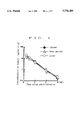
- FIG. 1 shows the results of Experimental Example 1.
- FIG. 2 shows the results of Experimental Example 2.
- FIG. 3 shows the results of Experimental Example 3.
- FIG. 4 shows the results of Experimental Example 4.
- FIG. 5 shows the results of Experimental Example 5.
- the liposome to be used for the liposome preparation of the present invention is a closed vesicle having internal aqueous phase portion enclosed by a lipid bilayer membrane formed by dispersing a phospholipid (lecithin) constituting the cell membrane in water, and grouped into three kinds of Multilamellar Vesicle (MLV), Large Unilamellar Vesicle (LUV) and Small Unilamellar Vesicle (SUV) according to the size and number of lipid bimolecules. Any kind of liposomes can be used in the present invention.
- the liposome preparation of the present invention should form a stable liposome structure in living organisms.
- the liposome needs to be prepared from a phospholipid having a gel-liquid phase transition temperature of not less than 37° C.
- Such phospholipid examples include those having a preferable gel-liquid phase transition temperature of 40°-65°, such as hydrogenated purified egg yolk phosphatidylcholine (phase transition temperature 50°-60° C., hereinafter to be referred to as HEPC), hydrogenated purified soy bean phosphatidylcholine (phase transition temperature 55° C., hereinafter to be referred to as HSPC), dipalmitoylphosphatidylcholine (phase transition temperature 42° C., hereinafter to be referred to as DPPC) and distearoylphosphatidylcholine (phase transition temperature 55° C., hereinafter to be referred to as DSPC), with preference given to HSPC and DPPC.
- HEPC hydrogenated purified egg yolk phosphatidylcholine
- HSPC hydrogenated soy bean phosphatidylcholine
- DPPC dipalmitoylphosphatidylcholine
- DSPC distearoylphosphatidyl
- the liposome usable in the present invention is prepared from these phospholipids, preferably combined with sterols which reportedly improve stability of liposome in the living body, such as cholesterol Biochemm. J., 186, pp. 591-598 (1980)! and cholestanol, or a stabilizer such as sphingomyelin.
- the 2'-deoxycytidine compound which is the active ingredient to be used in the present invention is water soluble and can be included in the inner phase of liposomes.
- the 2'-deoxycytidine compound is selected from 2'-deoxy-2'-methylidenecytidine (Japanese Patent Unexamined Publication No. 258818/1988), 2'-deoxy-2'-fluoromethylidenecytidine Journal of American Chemical Society, vol. 113, pp. 7439-7440 (1991)!, 2'-deoxy-2'-methylidene-5-fluorocytidine (Japanese Patent Unexamined Publication No.
- the liposome preparation of the present invention is characterized in that the surface of the lipid membrane is positively charged, and the compound to positively charge the lipid membrane, namely, the surface of liposome, is selected from saturated or unsaturated aliphatic amines (e.g., stearylamine and oleylamine), sphingosine, phosphatidylethanolamine, N- 1-(2,3-dioleyloxy)propyl!N,N,N-trimethylammonium chloride, cholesterylhemisuccinate, 3B- N-(N',N'-dimethylaminoethane)carbamoyl!cholesterol and cholesteryl(4'-trimethylammonio)butanoate, with preference given to stearylamine and sphingosine.
- saturated or unsaturated aliphatic amines e.g., stearylamine and oleylamine
- sphingosine e.g., stearyl
- the liposome preparation of the present invention contains a compound capable of positively charging the surface of the lipid membrane in a proportion of 0.001-0.4 mole, preferably 0.02-0.4 mole, and a stabilizer in a proportion of 0.6-1 mole per mole of phospholipid.
- the liposome preparation of the present invention can be prepared by a method known per se. For example, conventionally used hydration, reversed phase evaporation, removal of surfactant, solvent injection, freeze-thawing and dehydration-rehydration may be employed.
- a phospholipid and a compound which positively charges the surface of lipid membrane are dissolved in an organic solvent (e.g., chloroform and ether) which does not denature them, and the solvent is evaporated from the resulting solution to give a thin lipid membrane.
- an organic solvent e.g., chloroform and ether
- a solution containing a water-soluble 2'-deoxycytidine compound is added to the obtained thin membrane, and the mixture is subjected to agitation and sonication to give a liposome preparation encapsulating the water-soluble 2'-deoxycytidine compound.
- the solvent of the solution containing a water-soluble 2'-deoxycytidine compound may be any as long as it can be set for a temperature about 10° C. higher than the gel-liquid phase transition temperature, does not denature or decompose liposomes, and is physiologically acceptable, such as water, physiological saline and buffer.
- a phospholipid and a compound which positively charges the surface of lipid membrane are dissolved in an organic solvent (e.g., chloroform and ether) which does not denature them, and a solution containing a water-soluble 2'-deoxycytidine compound is added to the obtained solution.
- the obtained mixture is subjected to agitation, sonication and high pressure homogenation to uniformly disperse the water-soluble 2'-deoxycytidine compound.
- the solvent is evaporated from this dispersion to give a liposome preparation encapsulating the water-soluble 2'-deoxycytidine compound.
- the solvent of the solution containing a water-soluble 2'-deoxycytidine compound may be any as long as it does not denature or decompose liposomes and is physiologically acceptable, such as water, physiological saline and buffer.
- a phospholipid and a compound which positively charges the surface of lipid membrane are mixed with a surfactant (e.g., cationic surfactant such as cholic acid and deoxycholic acid, and non-ionic surfactant such as Triton X-100 and octyl-D-glucoside) and a solution containing a water-soluble 2'-deoxycytidine compound, which is followed by agitation, sonication and high pressure homogenation to uniformly disperse the water-soluble 2'-deoxycytidine compound.
- the surfactant is removed from this dispersion to give a liposome preparation encapsulating the water-soluble 2'-deoxycytidine compound.
- the solvent of the solution containing a water-soluble 2'-deoxycytidine compound may be any as long as it does not denature or decompose liposomes and is physiologically acceptable, such as water, physiological saline and buffer.
- the surfactant can be removed by dialysis, gel filtration and ultrafiltration, which are applied singly or in combination.
- a phospholipid and a compound which positively charges the surface of lipid membrane are dissolved in an organic solvent (e.g., ether and dichlorofluoromethane) which does not denature them, and the resulting solution is added to a solution containing a water-soluble 2'-deoxycytidine compound, which has been set for a temperature about 10° C. higher than the boiling point of the organic solvent. Then, the organic solvent is evaporated to give a liposome preparation encapsulating the water-soluble 2'-deoxycytidine compound.
- the solvent of the solution containing a water-soluble 2'-deoxycytidine compound may be any as long as it does not denature or decompose liposome and is physiologically acceptable, such as water, physiological saline and buffer.
- liposomes prepared by subjecting a phospholipid and a compound which positively charges the surface of lipid membrane to a method known per se, such as hydration, reversed phase evaporation, removal of surfactant, injection of solvent, freeze-thawing and dehydration-rehydration, are subjected to sonication and high pressure homogenation to give small unilamellar vesicles (SUV).
- the SUV and a solution containing a water-soluble 2'-deoxycytidine compound are frozen with a cooling medium such as liquid nitrogen, and thawed at a temperature about 10° C.
- the solvent of the solution containing a water-soluble 2'-deoxycytidine compound may be any as long as it does not denature or decompose liposomes and is physiologically acceptable, such as water, physiological saline and buffer.
- a phospholipid and a compound which positively charges the surface of lipid membrane are dissolved in a water-miscible solvent (e.g., ethanol) which does not denature them, and the resulting solution is added to an aqueous layer being stirred.
- a water-miscible solvent e.g., ethanol
- the resulting mixture is subjected to high pressure homogenation and sonication to give small unilamellar vesicles (SUV).
- An isotonizing agent such as sugars and polyhydric alcohols (e.g., monosaccharides such as glucose and lactose, disaccharides and glycerol and propylene glycol) is added to the aqueous layer, and the mixture is set to have a temperature about 10° C. higher than the gel-liquid phase transition temperature of the phospholipid.
- the SUV can be also obtained by subjecting the liposome prepared by a method known per se, such as hydration, reversed phase evaporation, removal of surfactant, freeze-thawing and dehydration-rehydration, to sonication and high pressure homogenation.
- a solution containing a water-soluble 2'-deoxycytidine compound is added to this SUV suspension, and the mixture is frozen and dried under reduced pressure (about 0.1-about 0.2 mmHg) to give a freeze-dried product.
- the solvent of the solution containing a water-soluble 2'-deoxycytidine compound may be any as long as it does not denature or decompose liposomes and is physiologically acceptable, such as water, physiological saline and buffer.
- a dispersion solvent is added to the freeze-dried liposomes and the mixture is vigorously stirred in a vortex mixer to give a liposome preparation encapsulating the water-soluble 2'-deoxycytidine compound.
- the dispersion solvent may be the same as the above-mentioned solvent for the solution containing a water-soluble 2'-deoxycytidine compound which is set for a temperature about 10° C. higher than the gel-liquid phase transition temperature.
- a sizing treatment is preferably applied to make the particle size of the drug-encapsulating liposomes more uniform.
- an apparatus Extruder, manufactured by Lipex Biomembranes Inc. set for a temperature about 10° C. higher than the gel-liquid phase transition temperature of the lipid is used and the liposomes are forcibly passed through the same membrane filter having a certain pore size several times.
- the same step is repeated using membrane filters having larger pore sizes which are successively changed to the filters having smaller pore sizes to adjust the average particle size to about 50 to 200 nm, preferably 100-180 nm, more preferably 120-160 nm and particularly preferably 150-160 nm.
- the liposome preparation of the present invention is prepared, which is subjected to one or more treatments as necessary from ultracentrifugation, molecular sieve treatment, gel filtration, ultrafiltration and dialysis, so that a water-soluble 2'-deoxycytidine compound which was not encapsulated in liposome is removed.
- the liposome preparation of the present invention can be prepared into an aqueous suspension together with a stabilizer e.g., ascorbic acid, ⁇ -tocopherol and 3,5-di-tert-butyl-4-hydroxytoluene (BHT)!, an isotonizing agent (e.g., glycerol, glucose and sucrose) and the like, or into a lyophilized preparation by freeze-drying, with preference given to a lyophilized preparation which is used as an injection (intravenous, intramuscular or subcutaneous preparation) after suspending in or diluting with a physiologically acceptable aqueous solution when in use.
- a stabilizer e.g., ascorbic acid, ⁇ -tocopherol and 3,5-di-tert-butyl-4-hydroxytoluene (BHT)!
- BHT 3,5-di-tert-butyl-4-hydroxytoluene
- an isotonizing agent e.g.,
- preparations may be designed such that an effective amount thereof can be used for the treatment of malignant tumors e.g., lung cancer, gastrointestinal cancer (e.g., esophageal cancer, stomach cancer, colic cancer, cancer of rectum and colon cancer), breast cancer, cancers in the neck, gynecological cancers (e.g., uterus cancer, cervical cancer and ovary cancer), urinary cancer (e.g., renal cancer and bladder cancer), leukemia, melanoma, and lymphogenous metastatic tumor! in mammals such as human.
- malignant tumors e.g., lung cancer, gastrointestinal cancer (e.g., esophageal cancer, stomach cancer, colic cancer, cancer of rectum and colon cancer), breast cancer, cancers in the neck, gynecological cancers (e.g., uterus cancer, cervical cancer and ovary cancer), urinary cancer (e.g., renal cancer and bladder cancer), leukemia, melanoma, and lymphogenous
- DMDC aqueous DMDC solution (10 ml) was added to this liposome suspension and the mixture was freeze-dried. Purified water (10 ml) heated to about 55° C. was added to this freeze-dried liposome product. After rehydration, a stirring treatment gave a liposome suspension. To unify the liposome particle size of this suspension, the suspension was subjected to a sizing treatment using an Extruder (manufactured by Lipex Biomembranes Inc.) equipped with a Nucleopore membrane (manufactured by Nomura Microscience) having a pore diameter of from 0.6 to 0.2 ⁇ m to give liposomes having a uniform average particle size of about 160 nm. Then, DMDC not encapsulated was removed by ultrafiltration to give DMDC liposomes.
- Extruder manufactured by Lipex Biomembranes Inc.
- Nucleopore membrane manufactured by Nomura Microscience
- DMDC aqueous DMDC solution (10 ml) was added to this liposome suspension and the mixture was freeze-dried by a conventional method. Purified water (10 ml) heated to about 70° C. was added to this freeze-dried liposome product. After rehydration, a stirring treatment gave a liposome suspension. To unify the liposome particle size of this suspension, the suspension was subjected to sizing treatment using an Extruder (manufactured by Lipex Biomembranes Inc.) equipped with a Nucleopore membrane (manufactured by Nomura Microscience) having a pore diameter of from 0.6 to 0.2 ⁇ m to give uniform liposomes having an average particle size of about 160 nm. Then, DMDC not encapsulated was removed by ultrafiltration to give DMDC liposomes.
- Extruder manufactured by Lipex Biomembranes Inc.
- Nucleopore membrane manufactured by Nomura Microscience
- HSPC 392.5 mg
- This liposome suspension was subjected to ultrafiltration to remove ethanol, and concentrated to about 30 ml.
- DMDC 150 mg was added to this liposome suspension and the mixture was freeze-dried by a conventional method.
- Extruder manufactured by Lipex Biomembranes Inc.
- Nucleopore membrane manufactured by Nomura Microscience
- DMDC in Example 1 is changed to 2'-deoxy-2'-fluoromethylidenecytidine, 2'-deoxy-2'-methylidene-5-fluorocytidine, 2'-deoxy-2',2'-difluorocytidine or 2'-C-cyano-2'-deoxy- ⁇ -arabinofuranosylcytosine and a liposome preparation is prepared in the same manner.
- DMDC was dissolved in a 2.5 wt % aqueous lactic acid solution to a DMDC concentration of 15 mg/ml.
- HSPC 235.5 mg
- cholesterol 116.0 mg
- DMDC liposomes were obtained in the same manner thereafter as in Example 2.
- the DMDC solution obtained in Control Example 1 was intravenously administered to a group of three to eight male SD rats at 30 mg/kg of DMDC. After administration, plasma, liver and bone marrow were taken at 0.25, 0.5, 1, 2, 4 and 6 hours with the passage of time, and DMDC concentration was determined. Migration to organ was evaluated from Kp value (concentration in tissue/concentration in plasma) which shows a ratio of concentration in tissues of liver and bone marrow to concentration in plasma. Quantitative determination was performed by HPLC wherein the limit of determination was 0.1 ⁇ g/ml (plasma), 0.5 ⁇ g/ml (liver) and about 0.25 ng/g (bone marrow).
- the level of concentration in bone marrow or liver of the rats which underwent intravenous injection of the aqueous solution was almost the same as that in plasma, as shown in FIG. 1.
- the Kp value was about 1.2 in liver and 0.9 in bone marrow, and almost the same at each determination.
- the DMDC liposome preparation obtained in Example 1 and the DMDC solution obtained in Control Example 1 were intravenously administered to the groups of three to eight male SD rats at 30 mg/kg of DMDC. After administration, plasma was taken at 0.25, 0.5, 1 and 2 hours with the passage of time, and liver and bone marrow were taken at the end of the test, which was followed by determination of DMDC concentration. Migration to organ was evaluated from Kp value (concentration in tissue/concentration in plasma) which shows a ratio of concentration in tissue to concentration in plasma. As a result, when compared to the control group, the concentration in plasma was noticeably maintained and AUC (area under curve) at 2 hours later was about 3.4 times greater, as shown in FIG. 2. As shown in Table 1, migration to liver or bone marrow decreased to about 1/2.
- the DMDC liposome preparation obtained in Example 3 and the DHDC solution obtained in Control Example 1 were intravenously administered to the groups of three to eight male SD rats at 30 mg/kg of DMDC. After administration, plasma was taken at 0.25, 0.5, 1, 2, 4, 6, 10 and 24 hours with the passage of time, and liver and bone marrow were taken at the end of the test, which was followed by determination of DMDC concentration. Migration to organ was evaluated from Kp value (concentration in tissue/concentration in plasma) which shows a ratio of concentration in tissue to concentration in plasma. As a result, the concentration in plasma was maintained, and AUC at 2, 10 and 24 hours later was about 3 times, 8 times and 13 times greater, respectively, than the control group, as shown in FIG. 3. As shown in Table 2, migration to bone marrow decreased to about 1/2-about 1/4 by formulation into liposomes, as evidenced by the comparison of the values at 2, 10 and 24 hours later.
- the DMDC liposome preparations obtained in Example 3 and Comparative Example 1 and the DMDC solution obtained in Control Example 1 were intravenously administered to the groups of three to eight male SD rats at 30 mg/kg of DMDC. After administration, plasma was taken at 0.25, 0.5, 1, 2, 4, 6, 10 and 24 hours with the passage of time, and liver and bone marrow were taken at the end of the test, which was followed by determination of DMDC concentration. Migration to organ was evaluated from Kp value (concentration in tissue/concentration in plasma) which shows a ratio of concentration in tissue to concentration in plasma. As a result, the residence in plasma of liposome preparation of Comparative Example 1 without stearylamine decreased and AUC value also decreased, as shown in FIG. 4. The liposome preparation of Example 3 containing stearylamine showed about 1.5 times greater suppressive effect on migration to myeloid tissue than did the liposome preparation of Comparative Example 1.
- DMDC liposome preparations (equivalent to DMDC 100 mg/kg) obtained in Example 3 and Comparative Example 1 and the DMDC aqueous solution (DMDC 100 mg/kg) obtained in Control Example 1 were intravenously administered to male BALB/c-nu/nu nude mice grafted with LX-1 human lung cancer, once a day for 5 consecutive days from the tail vein (5 or 6 mice/group). After administration, longer (L) and shorter (W) diameters of the tumor were measured with the passage of time, and the tumor volume (V) was calculated from the following formula:
- Vn/V 0 the relative tumor volume
- Vn the tumor volume on the measurement day (day n)
- V 0 the initial tumor volume at the time when the treatment was started (day 0).
- the mean value of Vn/V 0 for control group (Cn) and that for treated group (Tn) were calculated.
- T/C (%) was calculated from the following formula and used as an index of the treatment effect:
- the DMDC liposome preparation obtained in Example 3 significantly suppressed growth of LX-1 human lung cancer.
- the preparation showed more markedly enhanced effects than the DMDC aqueous solution obtained in Control Example 1, and the effects were stronger than those attained by the liposome preparation of Comparative Example 1 without stearylamine.
- the test results suggest that the DMDC liposome preparation of the present invention shows significant treatment effects on malignant tumors.
- the liposome preparation of the present invention is not only expected to decrease toxicity to myeloid tissue, which is caused by a water-soluble 2'-deoxycytidine compound having anti-malignant tumor activity, but also expected to accumulate the active ingredient in tumor tissues by virtue of an improved residence thereof in blood. Therefore, the preparation can maintain or enhance anti-tumor effects without causing side effects, and is useful for the treatment of malignant tumors.
Abstract
Description
              TABLE 1
______________________________________
Concentration in
                Concentration in
                            Concentration in
plasma (μg/ml)
                liver (μg/g)
                            bone marrow (μg/g)
______________________________________
Control
       5.1 ± 1.1 4.7 ± 1.0
                                4.0 ± 0.8
Example 1
       (1)          (0.9 ± 0.1)
                                (0.8 ± 0.2)
Example 1
       38.1 ± 5.3
                    22.5 ± 3.2
                                12.4 ± 1.2
       (1)          (0.6 ± 0.2)
                                (0.3 ± 0.1)
______________________________________
              TABLE 2
______________________________________
Concentration in
                Concentration in
                            Concentration in
plasma (μg/ml)
                liver (μg/g)
                            bone marrow (μg/g)
______________________________________
Control
       5.1 ± 1.1 4.7 ± 1.0
                                4.0 ± 0.8
Example 1
       (1)          (0.9 ± 0.1)
                                (0.8 ± 0.2)
(at 2 hr)
Example 3
       39.7 ± 12.0
                    22.0 ± 1.8
                                11.5 ± 3.7
(at 2 hr)
       (1)          (0.7 ± 0.1)
                                (0.4 ± 0.1)
Example 3
       15.0 ± 6.1
                    7.6 ± 2.2
                                3.9 ± 1.1
(at 10 hr)
       (1)          (0.5 ± 0.2)
                                (0.2 ± 0.1)
Example 3
       5.4 ± 2.3 6.0 ± 2.6
                                1.3 ± 0.4
(at 24 hr)
       (1)          (1.4 ± 1.0)
                                (0.3 ± 0.1)
______________________________________
              TABLE 3
______________________________________
Concentration in
                Concentration in
                            Concentration in
plasma (μg/ml)
                liver (μg/g)
                            bone marrow (μg/g)
______________________________________
Control  5.1 ± 1.1
                    4.7 ± 1.0
                                4.0 ± 0.8
Example 1
        (1)         (0.9 ± 0.1)
                                (0.8 ± 0.2)
(at 2 hr)
Example 3
        29.7 ± 1.7
                    22.0 ± 1.8
                                11.5 ± 3.7
(at 2 hr)
        (1)         (0.7 ± 0.1)
                                (0.4 ± 0.1)
Example 3
        17.1 ± 2.6
                    7.6 ± 2.2
                                3.9 ± 1.1
(at 10 hr)
        (1)         (0.5 ± 0.2)
                                (0.2 ± 0.1)
Example 3
         5.4 ± 2.3
                    6.0 ± 2.6
                                1.3 ± 0.4
(at 24 hr)
        (1)         (1.4 ± 1.0)
                                (0.3 ± 0.1)
Comparative
        14.3 ± 0.1
                    7.9 ± 1.1
                                8.0 ± 0.9
Example 1
        (1)         (0.6 ± 0.1)
                                (0.6 ± 0.1)
(at 2 hr)
Comparative
        10.0 ± 1.0
                    9.2 ± 1.6
                                3.1 ± 1.1
Example 1
        (1)         (0.9 ± 0.3)
                                (0.3 ± 0.1)
(at 10 hr)
Comparative
         3.3 ± 0.8
                    8.7 ± 2.9
                                1.2 ± 0.3
Example 3
        (1)         (2.9 ± 1.6)
                                (0.4 ± 0.1)
(at 24 hr)
______________________________________
V=1/2×L×W.sup.2
T/C (%)=(Tn/Cn)×100
Claims (13)
Applications Claiming Priority (3)
| Application Number | Priority Date | Filing Date | Title |
|---|---|---|---|
| JP6-041065 | 1994-03-11 | ||
| JP4106594 | 1994-03-11 | ||
| PCT/JP1995/000383 WO1995024201A1 (en) | 1994-03-11 | 1995-03-08 | Liposome preparation |
Publications (1)
| Publication Number | Publication Date |
|---|---|
| US5776488A true US5776488A (en) | 1998-07-07 |
Family
ID=12598041
Family Applications (1)
| Application Number | Title | Priority Date | Filing Date |
|---|---|---|---|
| US08/716,201 Expired - Fee Related US5776488A (en) | 1994-03-11 | 1995-03-08 | Liposome preparation |
Country Status (5)
| Country | Link |
|---|---|
| US (1) | US5776488A (en) |
| EP (1) | EP0750910A4 (en) |
| KR (1) | KR970701551A (en) |
| CA (1) | CA2184834A1 (en) |
| WO (1) | WO1995024201A1 (en) |
Cited By (25)
| Publication number | Priority date | Publication date | Assignee | Title |
|---|---|---|---|---|
| WO2000048611A1 (en) * | 1999-02-18 | 2000-08-24 | Sankyo Company, Limited | Liposome preparations containing antitumor drug |
| US20020150626A1 (en) * | 2000-10-16 | 2002-10-17 | Kohane Daniel S. | Lipid-protein-sugar particles for delivery of nucleic acids |
| US20040009126A1 (en) * | 2002-03-05 | 2004-01-15 | Transave, Inc. | Inhalation system for prevention and treatment of intracellular infections |
| US20040018243A1 (en) * | 1999-08-25 | 2004-01-29 | Advanced Inhalation Research, Inc. | Modulation of release from dry powder formulations |
| WO2005020935A2 (en) * | 2003-02-06 | 2005-03-10 | Henry M. Jackson Foundation For The Advancement Of Military Medicine, Inc. | Method and composition of administering radioprotectants |
| WO2006032136A1 (en) * | 2004-09-20 | 2006-03-30 | British Columbia Cancer Agency Branch | Free or liposomal gemcitabine alone or in combination with free or liposomal idarubicin |
| US20060078607A1 (en) * | 1999-10-22 | 2006-04-13 | Cell Therapeutics Europe S.R.L. | Liposome formulation of 6,9-bis[(2-aminoethyl)-amino]benzo[g]isoquinoline-5, 10-dione dimaleate |
| US20100063131A1 (en) * | 2007-03-26 | 2010-03-11 | Hirofumi Takeuchi | Prompt nucleic acid delivery carrier composition |
| US7732404B2 (en) | 1999-12-30 | 2010-06-08 | Dexcel Ltd | Pro-nanodispersion for the delivery of cyclosporin |
| US20100172964A1 (en) * | 2007-05-29 | 2010-07-08 | Pola Chemical Industries Inc. | Vesicle useful for external preparation for skin, and external preparation for skin comprising the vesicle |
| US20100330166A1 (en) * | 2008-01-30 | 2010-12-30 | The University Of Tokushima | Agent for enhancing anti-tumor effect comprising oxaliplatin liposome preparation, and anti-tumor agent comprising the liposome preparation |
| US20110250262A1 (en) * | 2008-12-24 | 2011-10-13 | Biomedcore, Inc. | Method for producing liposome and method for dissolving cholesterol |
| US20120128762A1 (en) * | 2005-07-20 | 2012-05-24 | University Of Pittsburgh - Of The Commonwealth System Of Higher Education | Sphingomyelin Liposomes for the Treatment of Hyperactive Bladder Disorders |
| US20120277727A1 (en) * | 2010-01-25 | 2012-11-01 | Envision Scientific Private Limited | Method and an insertable medical device for delivering one or more pro-healing agents to a target site within a blood vessel post-deployment of a stent |
| US20140056968A1 (en) * | 2011-03-03 | 2014-02-27 | Wroclawskie Centrum Badan Eit+ Sp Z O.O. | Liposome formulation comprising an anti-tumour active substance, method for its preparation and pharmaceutical compositions comprising it |
| US20140161876A1 (en) * | 2011-07-15 | 2014-06-12 | Konica Minolta, Inc. | Liposome-containing preparation utilizing dissolution aid, and method for producing same |
| US8951450B2 (en) | 2009-09-02 | 2015-02-10 | Biomedcore, Inc. | Apparatus and method for production of liposomes |
| US20150079159A1 (en) * | 2012-04-23 | 2015-03-19 | The Children's Medical Center Corporation | Formulations and Methods for Delaying Onset of Chronic Neuropathic Pain |
| US20150216801A1 (en) * | 2012-08-10 | 2015-08-06 | Taiho Pharmaceutical Co., Ltd. | Stable oxaliplatin-encapsulating liposome aqueous dispersion and method for stabilizing same |
| US9445975B2 (en) | 2008-10-03 | 2016-09-20 | Access Business Group International, Llc | Composition and method for preparing stable unilamellar liposomal suspension |
| US9694076B2 (en) | 2011-03-31 | 2017-07-04 | Kao Corporation | Vesicle composition |
| US20170312375A1 (en) * | 1998-01-14 | 2017-11-02 | Lantheus Medical Imaging, Inc. | Preparation of a lipid blend and a phospholipid suspension containing the lipid blend |
| US10022460B2 (en) | 2014-12-31 | 2018-07-17 | Lantheus Medical Imaging, Inc. | Lipid-encapsulated gas microsphere compositions and related methods |
| US10220104B2 (en) | 2016-07-06 | 2019-03-05 | Lantheus Medical Imaging, Inc. | Methods for making ultrasound contrast agents |
| US10588988B2 (en) | 2016-05-04 | 2020-03-17 | Lantheus Medical Imaging, Inc. | Methods and devices for preparation of ultrasound contrast agents |
Families Citing this family (9)
| Publication number | Priority date | Publication date | Assignee | Title |
|---|---|---|---|---|
| US7314637B1 (en) | 1999-06-29 | 2008-01-01 | Neopharm, Inc. | Method of administering liposomal encapsulated taxane |
| EP1044679B1 (en) * | 1998-11-02 | 2012-12-26 | Terumo Kabushiki Kaisha | Stable Liposomes for Targeted Drug Delivery |
| AU2003268087A1 (en) * | 2002-08-23 | 2004-03-11 | Ian Ma | Liposomal gemcitabine compositions for better drug delivery |
| EP1596825A2 (en) | 2003-02-03 | 2005-11-23 | Neopharm, Inc. | Stable sterile filterable liposomal encapsulated taxane and other antineoplastic drugs |
| EA200501285A1 (en) * | 2003-02-11 | 2006-02-24 | Неофарм, Инк. | METHOD OF OBTAINING LIPOSOMAL DRUGS |
| JP4617458B2 (en) * | 2004-02-10 | 2011-01-26 | 財団法人ヒューマンサイエンス振興財団 | Method for producing liposome using hollow fiber dialysis column |
| WO2008140081A1 (en) * | 2007-05-14 | 2008-11-20 | Konica Minolta Holdings, Inc. | Liposome and method for producing liposome |
| AU2011277183B2 (en) * | 2010-07-13 | 2014-11-13 | Clavis Pharma Asa | Parenteral formulations of elacytarabine derivatives |
| EP3388055B1 (en) * | 2015-12-08 | 2021-11-17 | Chia Tai Tianqing Pharmaceutical Group Co., Ltd. | Method for preparing liposome |
Citations (1)
| Publication number | Priority date | Publication date | Assignee | Title |
|---|---|---|---|---|
| US5246708A (en) * | 1987-10-28 | 1993-09-21 | Pro-Neuron, Inc. | Methods for promoting wound healing with deoxyribonucleosides |
-
1995
- 1995-03-08 KR KR1019960704935A patent/KR970701551A/en not_active Application Discontinuation
- 1995-03-08 US US08/716,201 patent/US5776488A/en not_active Expired - Fee Related
- 1995-03-08 CA CA002184834A patent/CA2184834A1/en not_active Abandoned
- 1995-03-08 EP EP95910773A patent/EP0750910A4/en not_active Withdrawn
- 1995-03-08 WO PCT/JP1995/000383 patent/WO1995024201A1/en not_active Application Discontinuation
Patent Citations (1)
| Publication number | Priority date | Publication date | Assignee | Title |
|---|---|---|---|---|
| US5246708A (en) * | 1987-10-28 | 1993-09-21 | Pro-Neuron, Inc. | Methods for promoting wound healing with deoxyribonucleosides |
Non-Patent Citations (9)
| Title |
|---|
| Allen et al., Biochemica et Biophysica Acta., vol. 643 (1981), pp. 346 362. * |
| Allen et al., Biochemica et Biophysica Acta., vol. 643 (1981), pp. 346-362. |
| Kataoka et al., Annals New York Academy of sciences (1978) vol. 308, pp. 387 394. * |
| Kataoka et al., Annals New York Academy of sciences (1978) vol. 308, pp. 387-394. |
| Kobayashi et al., Int. J. Cancer, vol. 20, pp. 581 587 (1977). * |
| Kobayashi et al., Int. J. Cancer, vol. 20, pp. 581-587 (1977). |
| Kobayashi, Kobunshi (1980) vol. 29 (2), p. 117. * |
| Rustum et al., Cancer Research, vol. 39(4), pp. 1390 1395 (1979). * |
| Rustum et al., Cancer Research, vol. 39(4), pp. 1390-1395 (1979). |
Cited By (44)
| Publication number | Priority date | Publication date | Assignee | Title |
|---|---|---|---|---|
| US20170312375A1 (en) * | 1998-01-14 | 2017-11-02 | Lantheus Medical Imaging, Inc. | Preparation of a lipid blend and a phospholipid suspension containing the lipid blend |
| WO2000048611A1 (en) * | 1999-02-18 | 2000-08-24 | Sankyo Company, Limited | Liposome preparations containing antitumor drug |
| US20040018243A1 (en) * | 1999-08-25 | 2004-01-29 | Advanced Inhalation Research, Inc. | Modulation of release from dry powder formulations |
| US7449197B2 (en) * | 1999-10-22 | 2008-11-11 | Cell Therapeutics Europe S.R.L. | Liposome formulation of 6,9-bis[(2-aminoethyl)-amino]benzo[g]isoquinoline-5, 10-dione dimaleate |
| US20060078607A1 (en) * | 1999-10-22 | 2006-04-13 | Cell Therapeutics Europe S.R.L. | Liposome formulation of 6,9-bis[(2-aminoethyl)-amino]benzo[g]isoquinoline-5, 10-dione dimaleate |
| US7732404B2 (en) | 1999-12-30 | 2010-06-08 | Dexcel Ltd | Pro-nanodispersion for the delivery of cyclosporin |
| US20020150626A1 (en) * | 2000-10-16 | 2002-10-17 | Kohane Daniel S. | Lipid-protein-sugar particles for delivery of nucleic acids |
| US20040009126A1 (en) * | 2002-03-05 | 2004-01-15 | Transave, Inc. | Inhalation system for prevention and treatment of intracellular infections |
| WO2005020935A2 (en) * | 2003-02-06 | 2005-03-10 | Henry M. Jackson Foundation For The Advancement Of Military Medicine, Inc. | Method and composition of administering radioprotectants |
| WO2005020935A3 (en) * | 2003-02-06 | 2005-07-21 | Jackson H M Found Military Med | Method and composition of administering radioprotectants |
| WO2006032136A1 (en) * | 2004-09-20 | 2006-03-30 | British Columbia Cancer Agency Branch | Free or liposomal gemcitabine alone or in combination with free or liposomal idarubicin |
| US20080213183A1 (en) * | 2004-09-20 | 2008-09-04 | Marcel Bally | Free or Liposomal Gemcitabine Alone or in Combination with Free or Liposomal Idarubicin |
| US20120128762A1 (en) * | 2005-07-20 | 2012-05-24 | University Of Pittsburgh - Of The Commonwealth System Of Higher Education | Sphingomyelin Liposomes for the Treatment of Hyperactive Bladder Disorders |
| US20100063131A1 (en) * | 2007-03-26 | 2010-03-11 | Hirofumi Takeuchi | Prompt nucleic acid delivery carrier composition |
| US9315828B2 (en) | 2007-03-26 | 2016-04-19 | Hirofumi Takeuchi | Prompt nucleic acid delivery carrier composition |
| US20100172964A1 (en) * | 2007-05-29 | 2010-07-08 | Pola Chemical Industries Inc. | Vesicle useful for external preparation for skin, and external preparation for skin comprising the vesicle |
| US8865208B2 (en) * | 2007-05-29 | 2014-10-21 | Pola Chemical Industries Inc. | Vesicle useful for external preparation for skin, and external preparation for skin comprising the vesicle |
| US8940327B2 (en) | 2008-01-30 | 2015-01-27 | The University Of Tokushima | Agent for enhancing anti-tumor effect comprising oxaliplatin liposome preparation, and anti-tumor agent comprising the liposome preparation |
| US20100330166A1 (en) * | 2008-01-30 | 2010-12-30 | The University Of Tokushima | Agent for enhancing anti-tumor effect comprising oxaliplatin liposome preparation, and anti-tumor agent comprising the liposome preparation |
| US9445975B2 (en) | 2008-10-03 | 2016-09-20 | Access Business Group International, Llc | Composition and method for preparing stable unilamellar liposomal suspension |
| US20110250262A1 (en) * | 2008-12-24 | 2011-10-13 | Biomedcore, Inc. | Method for producing liposome and method for dissolving cholesterol |
| US8951450B2 (en) | 2009-09-02 | 2015-02-10 | Biomedcore, Inc. | Apparatus and method for production of liposomes |
| US20120277727A1 (en) * | 2010-01-25 | 2012-11-01 | Envision Scientific Private Limited | Method and an insertable medical device for delivering one or more pro-healing agents to a target site within a blood vessel post-deployment of a stent |
| US9162014B2 (en) * | 2010-01-25 | 2015-10-20 | Concept Medical Research Private Limited | Method and an insertable medical device for delivering one or more pro-healing agents to a target site within a blood vessel post-deployment of a stent |
| US20140056968A1 (en) * | 2011-03-03 | 2014-02-27 | Wroclawskie Centrum Badan Eit+ Sp Z O.O. | Liposome formulation comprising an anti-tumour active substance, method for its preparation and pharmaceutical compositions comprising it |
| US9694076B2 (en) | 2011-03-31 | 2017-07-04 | Kao Corporation | Vesicle composition |
| US20140161876A1 (en) * | 2011-07-15 | 2014-06-12 | Konica Minolta, Inc. | Liposome-containing preparation utilizing dissolution aid, and method for producing same |
| US20150079159A1 (en) * | 2012-04-23 | 2015-03-19 | The Children's Medical Center Corporation | Formulations and Methods for Delaying Onset of Chronic Neuropathic Pain |
| US9408846B2 (en) * | 2012-04-23 | 2016-08-09 | The Children's Medical Center Corporation | Formulations and methods for delaying onset of chronic neuropathic pain |
| US10993913B2 (en) | 2012-08-10 | 2021-05-04 | Taiho Pharmaceutical Co., Ltd | Stable oxaliplatin-encapsulating liposome aqueous dispersion and method for stabilizing same |
| US20150216801A1 (en) * | 2012-08-10 | 2015-08-06 | Taiho Pharmaceutical Co., Ltd. | Stable oxaliplatin-encapsulating liposome aqueous dispersion and method for stabilizing same |
| US10383822B2 (en) * | 2012-08-10 | 2019-08-20 | Taiho Pharmaceutical Co., Ltd. | Stable oxaliplatin-encapsulating liposome aqueous dispersion and method for stabilizing same |
| US11395856B2 (en) | 2014-12-31 | 2022-07-26 | Lantheus Medical Imaging, Inc. | Lipid-encapsulated gas microsphere compositions and related methods |
| US10583207B2 (en) | 2014-12-31 | 2020-03-10 | Lantheus Medical Imaging, Inc. | Lipid-encapsulated gas microsphere compositions and related methods |
| US10022460B2 (en) | 2014-12-31 | 2018-07-17 | Lantheus Medical Imaging, Inc. | Lipid-encapsulated gas microsphere compositions and related methods |
| US10588988B2 (en) | 2016-05-04 | 2020-03-17 | Lantheus Medical Imaging, Inc. | Methods and devices for preparation of ultrasound contrast agents |
| US11266750B2 (en) | 2016-07-06 | 2022-03-08 | Lantheus Medical Imaging, Inc. | Methods for making ultrasound contrast agents |
| US10583208B2 (en) | 2016-07-06 | 2020-03-10 | Lantheus Medical Imaging, Inc. | Methods for making ultrasound contrast agents |
| US11266749B2 (en) | 2016-07-06 | 2022-03-08 | Lantheus Medical Imaging, Inc. | Methods for making ultrasound contrast agents |
| US11344636B2 (en) | 2016-07-06 | 2022-05-31 | Lantheus Medical Imaging, Inc. | Methods for making ultrasound contrast agents |
| US10220104B2 (en) | 2016-07-06 | 2019-03-05 | Lantheus Medical Imaging, Inc. | Methods for making ultrasound contrast agents |
| US11529431B2 (en) | 2016-07-06 | 2022-12-20 | Lantheus Medical Imaging, Inc. | Methods for making ultrasound contrast agents |
| US11857646B2 (en) | 2016-07-06 | 2024-01-02 | Lantheus Medical Imaging, Inc. | Methods for making ultrasound contrast agents |
| US11925695B2 (en) | 2016-07-06 | 2024-03-12 | Lantheus Medical Imaging, Inc. | Methods for making ultrasound contrast agents |
Also Published As
| Publication number | Publication date |
|---|---|
| CA2184834A1 (en) | 1995-09-14 |
| EP0750910A1 (en) | 1997-01-02 |
| KR970701551A (en) | 1997-04-12 |
| EP0750910A4 (en) | 1997-07-09 |
| WO1995024201A1 (en) | 1995-09-14 |
Similar Documents
| Publication | Publication Date | Title |
|---|---|---|
| US5776488A (en) | Liposome preparation | |
| KR100889139B1 (en) | Irinotecan preparation | |
| US4797285A (en) | Lipsome/anthraquinone drug composition and method | |
| Kozubek et al. | Liposomal drug delivery, a novel approach: PLARosomes. | |
| EP0219922B1 (en) | Anthracycline antineoplastic agents encapsulated in phospholipid micellular particles | |
| EP0280492B1 (en) | Liposome composition and its production | |
| US4898735A (en) | Liposome/doxorubicin composition and method | |
| JP3074733B2 (en) | Fat emulsion | |
| Mayhew et al. | Inhibition of liver metastases of M 5076 tumor by liposome-entrapped adriamycin | |
| US5043166A (en) | Liposome/anthraquinone drug composition and method | |
| CA2376849C (en) | Method of inhibiting leakage of drug encapsulated in liposomes | |
| JPS63112512A (en) | Liposome preparation and production thereof | |
| EP2107903A1 (en) | Pharmaceutical composition comprising a campothecin derivative | |
| Qi et al. | Comparative pharmacokinetics and antitumor efficacy of doxorubicin encapsulated in soybean-derived sterols and poly (ethylene glycol) liposomes in mice | |
| WO2006086992A2 (en) | Drug delivery systems containing phqspholipase a2 degradable lipid prodrug derivatives and the therapeutic uses thereof as. e.g. wound healing agents and peroxisome proliferator activated receptor ligands | |
| WO2005021012A1 (en) | Drug carrier having gemcitabine enclosed therein | |
| JP2009530318A (en) | Lipid-based drug delivery system containing phospholipase A2-degradable lipid that undergoes intramolecular cyclization during hydrolysis | |
| JP5355842B2 (en) | Non-pegylated long circulating liposomes | |
| WO2000009071A2 (en) | A novel liposomal formulation useful in treatment of cancer and other proliferation diseases | |
| JP2001524512A (en) | Combined chemoimmunotherapy with liposomal drugs and cytokines | |
| US6180137B1 (en) | Etherlipid-containing multiple lipid liposomes | |
| JP3693209B2 (en) | Method for producing closed vesicles | |
| JPH05194192A (en) | Temperature-sensitive mlv-type liposome with improved dispersibility | |
| JP2020521004A (en) | c(RGD-ACP-K) modified blood retention liposome | |
| WO2022153211A1 (en) | Liposomal composition of a camptothecin derivative |
Legal Events
| Date | Code | Title | Description |
|---|---|---|---|
| AS | Assignment |
Owner name: YOSHITOMI PHARMACEUTICAL INDUSTRIES, LTD., JAPAN Free format text: ASSIGNMENT OF ASSIGNORS INTEREST;ASSIGNORS:MORI, YOSHIYUKI;SAGARA, KAZUYOSHI;MIZUTA, HIROAKI;AND OTHERS;REEL/FRAME:008335/0705 Effective date: 19960819 |
|
| AS | Assignment |
Owner name: WELFIDE CORPORATION, JAPAN Free format text: CHANGE OF NAME;ASSIGNOR:YOSHITOMI PHARMACEUTICAL INDUSTRIES, LTD.;REEL/FRAME:011347/0303 Effective date: 20000401 |
|
| REMI | Maintenance fee reminder mailed | ||
| LAPS | Lapse for failure to pay maintenance fees | ||
| STCH | Information on status: patent discontinuation |
Free format text: PATENT EXPIRED DUE TO NONPAYMENT OF MAINTENANCE FEES UNDER 37 CFR 1.362 |
|
| FP | Lapsed due to failure to pay maintenance fee |
Effective date: 20020707 |