US5868728A - Medical linear actuator for surgical delivery, manipulation, and extraction - Google Patents
Medical linear actuator for surgical delivery, manipulation, and extraction Download PDFInfo
- Publication number
- US5868728A US5868728A US08/395,701 US39570195A US5868728A US 5868728 A US5868728 A US 5868728A US 39570195 A US39570195 A US 39570195A US 5868728 A US5868728 A US 5868728A
- Authority
- US
- United States
- Prior art keywords
- plunger
- cannula
- source
- members
- motive power
- Prior art date
- Legal status (The legal status is an assumption and is not a legal conclusion. Google has not performed a legal analysis and makes no representation as to the accuracy of the status listed.)
- Expired - Fee Related
Links
Images


Classifications
-
- A—HUMAN NECESSITIES
- A61—MEDICAL OR VETERINARY SCIENCE; HYGIENE
- A61F—FILTERS IMPLANTABLE INTO BLOOD VESSELS; PROSTHESES; DEVICES PROVIDING PATENCY TO, OR PREVENTING COLLAPSING OF, TUBULAR STRUCTURES OF THE BODY, e.g. STENTS; ORTHOPAEDIC, NURSING OR CONTRACEPTIVE DEVICES; FOMENTATION; TREATMENT OR PROTECTION OF EYES OR EARS; BANDAGES, DRESSINGS OR ABSORBENT PADS; FIRST-AID KITS
- A61F11/00—Methods or devices for treatment of the ears or hearing sense; Non-electric hearing aids; Methods or devices for enabling ear patients to achieve auditory perception through physiological senses other than hearing sense; Protective devices for the ears, carried on the body or in the hand
-
- A—HUMAN NECESSITIES
- A61—MEDICAL OR VETERINARY SCIENCE; HYGIENE
- A61F—FILTERS IMPLANTABLE INTO BLOOD VESSELS; PROSTHESES; DEVICES PROVIDING PATENCY TO, OR PREVENTING COLLAPSING OF, TUBULAR STRUCTURES OF THE BODY, e.g. STENTS; ORTHOPAEDIC, NURSING OR CONTRACEPTIVE DEVICES; FOMENTATION; TREATMENT OR PROTECTION OF EYES OR EARS; BANDAGES, DRESSINGS OR ABSORBENT PADS; FIRST-AID KITS
- A61F2/00—Filters implantable into blood vessels; Prostheses, i.e. artificial substitutes or replacements for parts of the body; Appliances for connecting them with the body; Devices providing patency to, or preventing collapsing of, tubular structures of the body, e.g. stents
- A61F2/95—Instruments specially adapted for placement or removal of stents or stent-grafts
-
- A—HUMAN NECESSITIES
- A61—MEDICAL OR VETERINARY SCIENCE; HYGIENE
- A61B—DIAGNOSIS; SURGERY; IDENTIFICATION
- A61B17/00—Surgical instruments, devices or methods, e.g. tourniquets
- A61B17/00234—Surgical instruments, devices or methods, e.g. tourniquets for minimally invasive surgery
-
- A—HUMAN NECESSITIES
- A61—MEDICAL OR VETERINARY SCIENCE; HYGIENE
- A61B—DIAGNOSIS; SURGERY; IDENTIFICATION
- A61B17/00—Surgical instruments, devices or methods, e.g. tourniquets
- A61B17/32—Surgical cutting instruments
- A61B17/320016—Endoscopic cutting instruments, e.g. arthroscopes, resectoscopes
- A61B17/32002—Endoscopic cutting instruments, e.g. arthroscopes, resectoscopes with continuously rotating, oscillating or reciprocating cutting instruments
-
- A—HUMAN NECESSITIES
- A61—MEDICAL OR VETERINARY SCIENCE; HYGIENE
- A61B—DIAGNOSIS; SURGERY; IDENTIFICATION
- A61B17/00—Surgical instruments, devices or methods, e.g. tourniquets
- A61B17/34—Trocars; Puncturing needles
- A61B17/3468—Trocars; Puncturing needles for implanting or removing devices, e.g. prostheses, implants, seeds, wires
-
- A—HUMAN NECESSITIES
- A61—MEDICAL OR VETERINARY SCIENCE; HYGIENE
- A61B—DIAGNOSIS; SURGERY; IDENTIFICATION
- A61B90/00—Instruments, implements or accessories specially adapted for surgery or diagnosis and not covered by any of the groups A61B1/00 - A61B50/00, e.g. for luxation treatment or for protecting wound edges
- A61B90/30—Devices for illuminating a surgical field, the devices having an interrelation with other surgical devices or with a surgical procedure
-
- A—HUMAN NECESSITIES
- A61—MEDICAL OR VETERINARY SCIENCE; HYGIENE
- A61B—DIAGNOSIS; SURGERY; IDENTIFICATION
- A61B17/00—Surgical instruments, devices or methods, e.g. tourniquets
- A61B17/30—Surgical pincettes without pivotal connections
-
- A—HUMAN NECESSITIES
- A61—MEDICAL OR VETERINARY SCIENCE; HYGIENE
- A61B—DIAGNOSIS; SURGERY; IDENTIFICATION
- A61B17/00—Surgical instruments, devices or methods, e.g. tourniquets
- A61B2017/00017—Electrical control of surgical instruments
-
- A—HUMAN NECESSITIES
- A61—MEDICAL OR VETERINARY SCIENCE; HYGIENE
- A61B—DIAGNOSIS; SURGERY; IDENTIFICATION
- A61B17/00—Surgical instruments, devices or methods, e.g. tourniquets
- A61B2017/00367—Details of actuation of instruments, e.g. relations between pushing buttons, or the like, and activation of the tool, working tip, or the like
- A61B2017/00398—Details of actuation of instruments, e.g. relations between pushing buttons, or the like, and activation of the tool, working tip, or the like using powered actuators, e.g. stepper motors, solenoids
-
- A—HUMAN NECESSITIES
- A61—MEDICAL OR VETERINARY SCIENCE; HYGIENE
- A61B—DIAGNOSIS; SURGERY; IDENTIFICATION
- A61B17/00—Surgical instruments, devices or methods, e.g. tourniquets
- A61B2017/0046—Surgical instruments, devices or methods, e.g. tourniquets with a releasable handle; with handle and operating part separable
- A61B2017/00464—Surgical instruments, devices or methods, e.g. tourniquets with a releasable handle; with handle and operating part separable for use with different instruments
-
- A—HUMAN NECESSITIES
- A61—MEDICAL OR VETERINARY SCIENCE; HYGIENE
- A61B—DIAGNOSIS; SURGERY; IDENTIFICATION
- A61B17/00—Surgical instruments, devices or methods, e.g. tourniquets
- A61B2017/00973—Surgical instruments, devices or methods, e.g. tourniquets pedal-operated
-
- A—HUMAN NECESSITIES
- A61—MEDICAL OR VETERINARY SCIENCE; HYGIENE
- A61B—DIAGNOSIS; SURGERY; IDENTIFICATION
- A61B17/00—Surgical instruments, devices or methods, e.g. tourniquets
- A61B17/28—Surgical forceps
- A61B17/29—Forceps for use in minimally invasive surgery
- A61B2017/2926—Details of heads or jaws
- A61B2017/2932—Transmission of forces to jaw members
- A61B2017/2933—Transmission of forces to jaw members camming or guiding means
- A61B2017/2937—Transmission of forces to jaw members camming or guiding means with flexible part
-
- A—HUMAN NECESSITIES
- A61—MEDICAL OR VETERINARY SCIENCE; HYGIENE
- A61B—DIAGNOSIS; SURGERY; IDENTIFICATION
- A61B2217/00—General characteristics of surgical instruments
- A61B2217/002—Auxiliary appliance
- A61B2217/005—Auxiliary appliance with suction drainage system
-
- A—HUMAN NECESSITIES
- A61—MEDICAL OR VETERINARY SCIENCE; HYGIENE
- A61F—FILTERS IMPLANTABLE INTO BLOOD VESSELS; PROSTHESES; DEVICES PROVIDING PATENCY TO, OR PREVENTING COLLAPSING OF, TUBULAR STRUCTURES OF THE BODY, e.g. STENTS; ORTHOPAEDIC, NURSING OR CONTRACEPTIVE DEVICES; FOMENTATION; TREATMENT OR PROTECTION OF EYES OR EARS; BANDAGES, DRESSINGS OR ABSORBENT PADS; FIRST-AID KITS
- A61F2/00—Filters implantable into blood vessels; Prostheses, i.e. artificial substitutes or replacements for parts of the body; Appliances for connecting them with the body; Devices providing patency to, or preventing collapsing of, tubular structures of the body, e.g. stents
- A61F2/95—Instruments specially adapted for placement or removal of stents or stent-grafts
- A61F2/9517—Instruments specially adapted for placement or removal of stents or stent-grafts handle assemblies therefor
-
- A—HUMAN NECESSITIES
- A61—MEDICAL OR VETERINARY SCIENCE; HYGIENE
- A61F—FILTERS IMPLANTABLE INTO BLOOD VESSELS; PROSTHESES; DEVICES PROVIDING PATENCY TO, OR PREVENTING COLLAPSING OF, TUBULAR STRUCTURES OF THE BODY, e.g. STENTS; ORTHOPAEDIC, NURSING OR CONTRACEPTIVE DEVICES; FOMENTATION; TREATMENT OR PROTECTION OF EYES OR EARS; BANDAGES, DRESSINGS OR ABSORBENT PADS; FIRST-AID KITS
- A61F9/00—Methods or devices for treatment of the eyes; Devices for putting-in contact lenses; Devices to correct squinting; Apparatus to guide the blind; Protective devices for the eyes, carried on the body or in the hand
- A61F9/007—Methods or devices for eye surgery
- A61F9/00736—Instruments for removal of intra-ocular material or intra-ocular injection, e.g. cataract instruments
- A61F9/00763—Instruments for removal of intra-ocular material or intra-ocular injection, e.g. cataract instruments with rotating or reciprocating cutting elements, e.g. concentric cutting needles
Definitions
- the present invention relates in general to surgical instruments and surgical techniques. More particularly, the present invention is directed to a linear actuator for a multifunctional surgical tool for delivery of grafts, drugs, devices, and for irrigation/aspiration of various parts of the body; as well as surgical manipulation and extraction.
- a device to deliver, extract, and implant medical devices, drugs, tissue, etc. to various parts of the body.
- Such a device must be capable of delivering an implant in a target site in the body in a controlled, calibrated fashion where necessary.
- implantation of neural and other living tissue an increasing number of surgical devices, and drugs.
- existing implants frequently require treatment involving manipulation and extraction procedures.
- vascular prosthetics e.g. stents
- vascular prosthetics and grafts be expressed from the catheter in a controlled and calibrated fashion.
- U.S. Pat. No. 4,837,857 issued to Scheller et al. discloses a foot pedal assembly which can be used for remotely controlling a variety of microsurgical instruments.
- the system employs pneumatic drive means and is thereby subject to the aforementioned drawbacks.
- a microprocessor controlled drive means adaptable for use with a plurality of functional attachments including, but not limited to, cutting tools, grasping tools, and tools useful for implantation of various devices, tissues, grafts, and drugs into the body.
- the microprocessor controlled drive means comprises a source of motive power, a linear actuator, and a microprocessor to selectively apply power to the linear actuator.
- the microprocessor can control the direction, speed, length of travel, and duration that power is applied to the linear actuator.
- the linear actuator of the present invention provides previously unobtainable levels of accuracy and precision in the controlled surgical delivery and manipulation of materials and devices, particularly in surgery beneath and around the retina, other parts of the eye, and in remote portions of blood vessels from the surgical delivery incision.
- the linear actuator may be a cable or thin tubular plunger disposed within a second tubular body and capable of relative axial movement therein.
- the plunger may be actuated manually, by a spring loaded foot pedal assembly, or by a foot pedal operated ratchet wheel assembly.
- FIG. 1 is a top elevation view of a housing for a source of motive power for the surgical instrument.
- FIG. 2 is a top plan view of the interior components of FIG. 1.
- FIG. 3 is a schematic of the control circuitry used to selectively supply power to the surgical instrument.
- FIG. 4 is a partial sectional view of a handpiece cable assembly including fittings for the linear actuator, infusion line, and the fiber optic cable.
- FIG. 5 is a perspective view of a functional attachment to the linear actuator of the present invention for containing an implant for implantation or for extraction of a material.
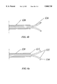
- FIG. 6(a) is a sectional view of a functional attachment to the linear actuator of the present invention including grasping members which are outwardly biased.
- FIG. 6(b) is a sectional view of a functional attachment to the linear actuator of the present invention including grasping forceps which can be inwardly biased.
- FIGS. 6(c) and 6(d) are views of a stent having an inner lip capable of engagement with the grasping membranes illustrated in FIG. 6(a).
- FIG. 7 is a sectional view of a handpiece cable assembly which can be operably connected to a source of motive power.
- FIG. 8 is a perspective view of an alternative hand actuated embodiment of the surgical instrument.
- FIG. 9 is a partially exploded perspective view of FIG. 8.
- FIGS. 1 and 2 a top view of a preferred embodiment of a microprocessor controlled electromechanical drive assembly 10 is shown.
- the assembly 10 includes stepper motors 12 and 14 (FIG. 2) which are contained within a housing 16.
- stepper motors 12 and 14 FIG. 2
- a mechanical drive mechanism obtains power from the energy stored in a spring or like mechanism may also be used.
- Attached to the housing 16 is a front panel 18 and a source of fluid pressure or suction in the form of a syringe 20, the fluid pressure or suction being controlled by stepper motor 14.
- the housing 16 can also contain a light source for illuminating a fiber optic filament as will be discussed later. A laser source could also be provided.
- Both of the stepper motors 12 and 14 are controlled by a microprocessor 22.
- a plurality of switches and terminals are disposed on the front panel 18 to allow the operator to select the various functions and modes of operation used with the various functional attachments.
- Power is selectively applied to the assembly 10 by power switch 24.
- DC power is supplied to the microprocessor by the transformer/rectifier assembly 26.
- the mode switch 28 allows the operator to alternate between actuation of the plunger 29 and the syringe 20.
- the plunger 29 is alternated between delivery and retraction by switch 30 which, depending on the position of the mode switch 28, also alternates the fluid pressure source, syringe 20, between infusion and aspiration.
- switch 30 which, depending on the position of the mode switch 28, also alternates the fluid pressure source, syringe 20, between infusion and aspiration.
- delivery or retraction of the plunger 29 is mutually exclusive of infusion or aspiration.
- infusion/aspiration can be performed simultaneously with delivery/retraction of the plunger 29.
- separate conduits and associated drive means can be used to apply infusion and aspiration simultaneously to a functional attachment.
- Pushbutton switch 32 controls the connecting and disconnecting of a functional attachment to device 10, with the connect status being indicated by LEDs 34.
- the delivery speed of the plunger 29 is infinitely variable over a range of speeds by speed control 36.
- the range of speeds is variable depending upon the exact procedure being performed and the associated functional attachment. The speed range can be altered if necessary to operate certain functional attachments.
- Actuation of the plunger 29 is controlled by footswitch 38 which may be a commercially available footswitch such as a model produced under the trademark Linemaster®.
- the footswitch 38 will operate the plunger 29 or the syringe 20 in the selected direction and speed as long as it is depressed.
- the assembly 10 includes an actuator 40 for advancing and retracting the plunger 29 which is connected to motor 12 via screw drive 42 and has solenoid controlled locking mechanisms 44 and 46.
- Locking mechanism 44 locks onto the connector 48 of sheath 50 and locking mechanism 46 locks onto the plunger 29.
- the connector 48 is adapted to be secured within terminal 52 when mechanism 44 locks onto annular recess 54 of the connector 48 (FIG. 4).
- the connector 48 has an axial bore 56 for slidably receiving the plunger 29.
- the sheath 50 is secured within the connector bore 56 by an adhesive, by frictional engagement, or is integrally attached. The opposite end of the sheath 50 is secured to a functional attachment.
- the actuator travel 40 is limited by limit switches 60 and 62.
- the limit switches are actuated by transversely extending arm 64 and serve to physically limit the travel of the actuator 40 as well as interrupt the supply of power to stepper motor 12.
- the syringe 20 is releasably mounted in syringe support 66.
- the support 66 includes a rectangular clamp 68 having a groove therein which is sized for holding the syringe 20.
- the clamp 68 has a threaded aperture extending therethrough, the aperture corresponding to an aperture in the support 66, both apertures aligned for receiving a screw 70 for tightening the clamp 68 onto the syringe 20.
- a recess 72 formed in the support is adapted to hold the annular flange 74 of the syringe 20 thereby preventing axial movement of the syringe.
- the syringe actuator 76 has a similar recess 78 for retaining the annular lip 80 at the tip of the syringe piston rod enabling the actuator to move the piston 82 to effect infusion or aspiration.
- the syringe 20 is filled with infusion fluid from a second syringe 84 via 3-way stopcock 86.
- the stopcock 86 is positioned to allow fluid flow from second syringe 84 to syringe 20 at startup.
- the stopcock 86 is then repositioned to allow fluid flow or suction through infusion/aspiration terminal 87 to the infusion line 88.
- actuator 76 The travel of actuator 76 is limited by actuator arm 77 and limit switches 90 and 92 in the same manner as actuator 40. It should be noted that the limit switches can be repositioned to adjust the travel of the actuators 40 and 76.
- a pump, fluid reservoir, and a fluid collection container may be used to apply fluid flow or aspiration to infusion line 88.
- a pump such as a peristaltic pump would provide suitable control of flow, and the infusion line 88 or other suitable conduit could be inserted into the pump drive mechanism.
- Such a pump could have the motor inside the housing of device 10, and have the pump and tubing connection external to the housing.
- Microcontroller U3 is the main control unit for the microprocessor 22.
- An Intel(R) 8031 may be used for U3.
- Microcontroller U3 is controlled in accordance with the following program.
- U1 is a data latch which is used to latch data coming from U3 and may be a 74HC245 integrated circuit.
- U2 is also a data latch and may be a 74HC373 IC.
- U4 are 74HC08 Quad two input AND gates which are used as temporary data stores.
- U5 is a 74ALS156 decoder which outputs control signals from the controller U3 for control of the other ICs.
- U7 is a RAM which stores data such as speed control for access as needed by the controller U3. An MCM6164-55C 64K RAM may be used for U7.
- U13 are 74HC32 Quad two input OR gates.
- U8 is programmed to perform as 3 eight bit ports. Port A and Port B are used as inputs. Port C is used as an output. The inputs are from the panel switches or the limit switches in the control box. The outputs are indicators such as LEDs 311.
- U8 may be a 8255 IC and is controlled by U3.
- U9 and U11 are UCN5804B microcontrollers and are used to drive the stepper motors 12 and 14.
- U9 and Ull respond to control signals from U3.
- U12 is a ADC0808CCJA 8 bit A/D converter used to convert the input voltage from the footswitch to a digital format that the controller recognizes.
- U10 is a 74HC74 flip-flop which divides the clock pulse by 2 since U12 cannot convert data as fast as U3.
- U14 is a 7407 hex buffer that responds to control signals from U3 to turn on the relays which activate the solenoids which operate locking mechanisms 44 and 46.
- a functional attachment capable of implantation of grafts, tissues, or drugs; as well as irrigation, aspiration or removal of tissue includes a handpiece cable assembly as is shown in FIG. 4.
- the handpiece cable assembly can be operatively connected to the microprocessor controlled drive assembly 10 and can be used for a retinal transplant procedure as described in our copending application 08/395,699 entitled “METHOD FOR TRANSPLANTATION OF PLANAR IMPLANTS AND SURGICAL INSTRUMENT THEREFOR” filed on even date herewith which is herein incorporated by reference.
- the handpiece cable assembly includes a handpiece 100 which is sectional and which has a delivery cannula 102 within which plunger 29 is axially movable to express the graft, drug, material, or device to be implanted.
- the handpiece 100 also includes an infusion lumen 104 which can be connected to infusion tube 88 for providing a source of infusion fluid or aspiration, as well as a fiber optic cable 106 which can be connected to a source of illumination for providing illumination at the site of implantation.
- a laser source can be connected to fiber optic cable 106 for, e.g., cauterizing blood vessels.
- the sheath 50 is connected to the delivery cannula 102 to allow for passage of the plunger 29 into the delivery cannula for expressing the desired implant from the tubular tip 114 of the cannula.
- the handpiece cable assembly is set up for the implantation of the graft by manually inserting the plunger 29 into and through the connector 48 and sheath 50 until the plunger enters a first section of the handpiece 100 and abuts the inner end wall 112 of the calibration cap 112 as shown in FIG. 4.
- the calibration cap 110 is adapted for detachable locking engagement with the first section of the handpiece 100 and is used to preset the initial position of the plunger 29 within the handpiece 100 when the actuator 40 is in the fully retracted position.
- the travel of the plunger 29 within the handpiece 100 extends from the preset position to a position of maximum extension within the delivery cannula 102 as determined by the spacing of the limit switches 60 and 62 as has been previously explained.
- the plunger 29 is made long enough so that an excess length of the plunger 29 protrudes from the connector 48 when the plunger abuts wall 112. The excess length of the plunger 29 is long enough to ensure proper engagement with the locking mechanism 46, but not so long as to prevent full insertion of the connector 48 into the terminal 52.
- the connect/disconnect switch 32 is depressed thereby deactivating locking mechanisms 44 and 46 and returning the actuator 40 to the fully retracted position as shown in FIG. 2.
- the plunger 29 and connector 48 can then be inserted into terminal 52. It should be noted that for most procedures the limit switches 60 and 62 do not allow the plunger 29 to travel beyond the opening 114 of the tubular tip of the delivery cannula 102.
- a switch (not shown) activates mechanisms 44 and 46 to lock onto annular recess 54 and plunger 29 respectively.
- Speed control 36 is then set to the desired speed.
- the foot pedal 38 can then be used to control extension or retraction of the plunger 29 as desired by setting the panel switches to the appropriate positions.
- the infusion/aspiration assembly is set up by attaching syringes 20 and 84 to the infusion/aspiration terminal 87 with the syringe clamp 68 being firmly attached to the syringe 20 so as to prevent axial movement of the syringe 20.
- the syringe 20 is then loaded with infusion fluid from syringe 84 via stopcock 86 by setting the panel switches in the aspiration mode and depressing the foot pedal 38.
- the stopcock 86 is then repositioned to allow infusion fluid to flow through infusion line 88. Fluid pressure or suction can then be applied to infusion line 88 via microprocessor controlled stepper motor 14 by operating foot pedal 38 with the panel switches in the appropriate positions.
- a multilumen cannula can be attached to infusion line 88 to aspirate tissue from the subretinal space or other locations, followed by the implant of drugs, grafts, or devices.
- the assembly can be adapted to provide accurately controlled motive power to a wide variety of functional attachments. Modification of the hardware or software may be required in order to operate certain functional attachments.
- FIG. 6(a) Another functional attachment capable of being operably connected to the drive assembly is shown in FIG. 6(a).
- This attachment requires the connection of a straight cannula 120 to the handpiece 100 which is sufficiently strong that it does not tear under the outward pressure exerted by retraction of outwardly biased members 122 into cannula 120. This may require reinforcement of tip 121, particularly in instances where cannula 120 is made sufficiently flexible to follow the contours of a blood vessel or other lumen into which it has been inserted.
- This attachment can be used as a stent retriever and has a pair of outwardly biased grasping members 122 having hook-like ends 124.
- the opposite ends of the grasping members 122 are attached to the plunger 29 by a connector (not shown) which allows for simultaneous delivery or retraction of the grasping members.
- a connector (not shown) which allows for simultaneous delivery or retraction of the grasping members.
- the grasping members 122, and ends 124 do not expand radially beyond the outer dimensions of the cannula 120 allowing for smooth progression of the device through blood vessels or other lumens.
- the grasping members 122 extend outwardly as shown so that the ends 124 can be used to grasp the stent or other device for removal.
- a stent 125 is illustrated in FIG. 6(c), and includes inner flexing lip 127 for engagement with ends 124.
- Inner flexing lip 127 is designed to lie against the inner surface of stent 125 when inserted so that blood flow biases lip 127 against the inner stent wall to create a smooth surface.
- the blood vessel may be dilated in from of stent 125, for example at position 129 with a balloon dilatation catheter 131 to permit use of the cannula and grasping elements of the device shown in 6(a) to be inserted through a lumen passing through the balloon dilatation catheter into the stent 125 as shown in FIG. 6(a) and permitting retraction of the stent 125 into the lumen of the balloon dilatation catheter.
- FIG. 6(b) A forceps-like grasping attachment is shown in FIG. 6(b).
- This attachment may be used for removal of tissues or devices, or for the manipulation of various devices within the body, and is attached to the drive assembly 10 in the same manner as the attachment of FIG. 6(a).
- the attachment has grasping members 126 which are biased outwardly to open upon advancement of the plunger 29 and to close upon retraction thus allowing the attachment to be used as a miniature forceps.
- the forceps can be inserted through a retinotomy to perform a choroidal biopsy; the members 126 (which can be two or more in number) may also be rotatably connected to plunger 29, and means for rotating members 126 in order to ensure a cleaner incision, or a better cutting and tearing action.
- a cauterization device e.g. electrocauterization probe
- the device can be used for insertion into a lumen of a balloon dilatation catheter for removal of a pre-shrunken (e.g., thermally cooled) stent, or may be used to both radially contract and extract a vascular stent.
- a pre-shrunken e.g., thermally cooled
- the cannula 120 in both attachments can be provided with an infusion/aspiration lumen which can be used for irrigation/aspiration at the surgical site.
- FIGS. 5 and 6 can be effectively employed by making only small surgical incisions.
- a handpiece cable assembly 250 having a handpiece 251 which can be functionally attached to a source of motive power is shown in FIG. 7.
- the handpiece cable assembly 250 includes a delivery cannula 252 which is secured to hollow frusto-conical connector 254 by an adhesive.
- the main body of the assembly 250 has an inner member 256 and an outer member 258.
- the inner member 256 has a frusto-conical portion 260 projecting from one end and a threaded cylindrical post 262 projecting from the opposite end.
- Connector 254 is adapted for frictional engagement with projecting portion 260 and may be further secured thereto by an adhesive.
- Outer member 258 has a contoured outer surface 264 to facilitate manipulation of the delivery cannula.
- a central aperture 266 in the outer member is threaded to enable threaded engagement with post 262 and is inwardly sloped at one end to form a camming surface 268.
- the post 262 has an aperture 270 adapted to receive a vice member 272 which has an exterior camming surface thereby forming a pin vice assembly comprised of threaded post 262, outer member 258, and vice member 272.
- infusion/aspiration can be manually effected by securing an infusion/aspiration lumen (not shown) to the exterior of the delivery cannula 252 by using an adhesive, the infusion lumen having an opening proximate the opening at the tip 274 of the delivery cannula, the infusion/aspiration lumen being connected to a syringe 276.
- plunger 29 may be actuated by a foot pedal operated ratchet assembly shown as a rectangular box in FIG. 2.
- the ratchet assembly includes two foot pedals, one for delivery, the other for retraction, or a single foot pedal having a directional switch, which reverses the direction of movement of the plunger in response to one up/down cycle of the foot pedal.
- the travel per foot pedal cycle can also be adjusted.
- the plunger 29 will move with each depression of the foot pedal until it reaches its limit of travel, the distance traveled by a single depression being adjustable by the spacings of gear teeth on the ratchet assembly as is well known.
- Such a ratchet drive assembly may be included with the device 10 as a backup source of motive power.
- the attachment 300 comprises a cylindrical aluminum housing 302 having a delivery cannula 304 attached thereto.
- a plunger 306 is actuated by thumbswitch 308 which slides in track 310.
- An actuator 312 is connected to thumbswitch 308 as can be seen in FIG. 12.
- Actuator 312 is connected to plunger 306 by rod 314 which projects into and through bore 316 in luer connector 317 and is inserted into an aperture 318 at one end of plunger 306 where it is secured therein by an adhesive.
- Luer connector 317 is connected to the housing 302 and therefore is held stationary relative to rod 314, actuator 312 and plunger 306.
- plunger 306 The travel of plunger 306 is limited by set screw 320 which screws into bore 322 and projects into the path of actuator 312.
- a plurality of bores such as bore 322 can be provided to adjustably limit the travel of actuator 312 and therefor the plunger 306.
- Grooves 324 and 326 are provided to hold infusion conduits 328 and 330 in place. Infusion fluid or aspiration can be provided to the infusion conduits 328 and 330 by a syringe.
- the delivery cannula 304 is connected to a hollow frusto-conical connector 332 which is secured to housing 302 and has a bore 334 into which the delivery cannula 304 is secured by an adhesive.
- Conduits 328 and 330 can also be secured to the exterior surfaces of connector 332 and delivery cannula 304 as shown to provide aspiration/infusion at the tip of the delivery cannula. Advancing or retracting the thumbswitch causes advancing or retraction of the plunger 306 within the delivery cannula 304.
Abstract
Description
__________________________________________________________________________
File name: PG21.C
© Drummond Sci. Col. 5/94 Nick Di Trolio
Initialize controller
Set time 0 (used for speed control)
Enable interrupts
Int0 used for clock pulses
Int1 used for analog inputs
Send output to P1 to initialize motor control function
Small delay for solenoids to reset
Output to I/O chip to initialize configuration (PortA=input; PortB=input;
PortC=output)
Read canula switch to determine in canula is present
If canula is present, turn on load LED
If canula is not present, turn on unload LED
Move cable drive motor to home position (retracted)
Load timer for set pulse width
Set direction
Enable motor drive
Enable timer interrupt
Enable timer
Send pulses to motor drive
Read cable home switch
loop until cable home switch is active
Disable timer
Disable timer interrupt
Disable motor drive
Beep once to indicate home
Move asp/infuse motor drive to home position (forward)
Load timer for set pulse width
Set direction
Enable motor drive
Enable timer interrupt
Enable timer
Send pulses to motor drive
Read cable home switch
loop until cable home switch is active
Disable timer
Disable timer interrupt
Disable motor drive
Beep twice to indicate home
Set infinite loop (main loop)
read PortA to look for user command
if load cable switch is pressed (on panel)
         Read canula switch
         If switch is on
            Turn off load LED
            Turn on unload LED (on panel)
            Turn on canula latch solenoid
            Delay 100ms.
            Turn on cable latch solenoid
            Read canula switch
               Loop until switch is off
            Delay 5 seconds
            Turn off cable latch solenoid
            Delay 100ms.
            Turn off cable latch solenoid
         If switch is not active
            Turn off unload LED
            Turn on load LED
            Move cable drive to home position
            Turn on canula latch solenoid
            Delay 100 ms.
            Turn on cable latch solenoid
            Read canula switch
               Loop until switch is on
            Turn off canula latch solenoid
            Delay 100ms.
            Turn off cable latch solenoid
         Return to main loop
If cable forward switch (foot pedal) is pressed
         Read analog voltage from potentiometer (on panel)
         Use lookup table in memory to determine speed
         Set motor direction
         Enable cable motor drive
         Enable timer
         Enable interrupt
            While cable forward switch is on:
               Read limit switch
               if limit switch is off:
                  Pulse motor drive
               if limit switch is on
                  Beep once
                  wait until user releases foot pedal
               Disable timer
               Disable interrupt
               Disable motor drive
               Return to main loop
If cable retract switch (on foot pedal) is on
Read analog voltage from potentiometer (on panel)
         Use lookup table in memory to determine speed
         Set motor direction
         Enable cable motor drive
         Enable timer
         Enable interrupt
            While cable forward switch is on:
               Read limit switch
               if limit switch is off:
                  Pulse motor drive
               if limit switch is on
                  Beep once
                  wait until user releases foot pedal
               Disable timer
               Disable interrupt
               Disable motor drive
               Return to main loop
If asp/infuse forward switch (on foot pedal) is on
Read analog voltage from potentiometer (on foot pedal)
         Use lookup table in memory to determine speed
         Set motor direction
         Enable asp/infuse motor drive
         Enable timer
         Enable interrupt
            While asp/infuse forward switch in on:
               Read limit switch
               if limit switch is off:
                  Pulse motor drive
                  Read analog voltage from potentiometer
                  set timer registers from lookup table based on
                  voltage
               if limit switch is on
                  Beep once
                  wait until user release foot pedal
               Disable timer
               Disable interrupt
               Disable motor drive
               Return to main loop
__________________________________________________________________________
Claims (13)
Priority Applications (11)
| Application Number | Priority Date | Filing Date | Title |
|---|---|---|---|
| US08/395,701 US5868728A (en) | 1995-02-28 | 1995-02-28 | Medical linear actuator for surgical delivery, manipulation, and extraction |
| JP8526306A JPH09509607A (en) | 1995-02-28 | 1996-02-28 | Medical linear actuators for surgical dosing, treatment and extraction |
| CA002189039A CA2189039C (en) | 1995-02-28 | 1996-02-28 | Medical linear actuator for surgical delivery, manipulation, and extraction |
| KR1019960706047A KR100220551B1 (en) | 1995-02-28 | 1996-02-28 | Medical linear actuator for surgical delivery, manipulation, and extraction |
| TW085102329A TW304873B (en) | 1995-02-28 | 1996-02-28 | |
| MX9605181A MX9605181A (en) | 1995-02-28 | 1996-02-28 | Medical linear actuator for surgical delivery, manipulation, and extraction. |
| ZA961598A ZA961598B (en) | 1995-02-28 | 1996-02-28 | Medical linear actuator for surgical delivery manipulation and extraction |
| PCT/US1996/002270 WO1996026696A1 (en) | 1995-02-28 | 1996-02-28 | Medical linear actuator for surgical delivery, manipulation, and extraction |
| EP96911200A EP0767641A4 (en) | 1995-02-28 | 1996-02-28 | Medical linear actuator for surgical delivery, manipulation, and extraction |
| IL11731096A IL117310A (en) | 1995-02-28 | 1996-02-28 | Medical linear actuator for surgical delivery, manipulation and extraction |
| AU54159/96A AU5415996A (en) | 1995-02-28 | 1996-02-28 | Medical linear actuator for surgical delivery, manipulation, and extraction |
Applications Claiming Priority (1)
| Application Number | Priority Date | Filing Date | Title |
|---|---|---|---|
| US08/395,701 US5868728A (en) | 1995-02-28 | 1995-02-28 | Medical linear actuator for surgical delivery, manipulation, and extraction |
Publications (1)
| Publication Number | Publication Date |
|---|---|
| US5868728A true US5868728A (en) | 1999-02-09 |
Family
ID=23564132
Family Applications (1)
| Application Number | Title | Priority Date | Filing Date |
|---|---|---|---|
| US08/395,701 Expired - Fee Related US5868728A (en) | 1995-02-28 | 1995-02-28 | Medical linear actuator for surgical delivery, manipulation, and extraction |
Country Status (11)
| Country | Link |
|---|---|
| US (1) | US5868728A (en) |
| EP (1) | EP0767641A4 (en) |
| JP (1) | JPH09509607A (en) |
| KR (1) | KR100220551B1 (en) |
| AU (1) | AU5415996A (en) |
| CA (1) | CA2189039C (en) |
| IL (1) | IL117310A (en) |
| MX (1) | MX9605181A (en) |
| TW (1) | TW304873B (en) |
| WO (1) | WO1996026696A1 (en) |
| ZA (1) | ZA961598B (en) |
Cited By (36)
| Publication number | Priority date | Publication date | Assignee | Title |
|---|---|---|---|---|
| WO2001034075A1 (en) * | 1998-11-05 | 2001-05-17 | Scieran Technologies, Inc. | A foot switch to proportionally control a medical cutting device |
| US6290690B1 (en) * | 1999-06-21 | 2001-09-18 | Alcon Manufacturing, Ltd. | Simultaneous injection and aspiration of viscous fluids in a surgical system |
| US6316465B1 (en) | 1998-06-27 | 2001-11-13 | Photogenesis, Inc. | Ophthalmic uses of PPARgamma agonists and PPARgamma antagonists |
| US6358260B1 (en) | 1998-04-20 | 2002-03-19 | Med-Logics, Inc. | Automatic corneal shaper with two separate drive mechanisms |
| US6402737B1 (en) * | 1998-03-19 | 2002-06-11 | Hitachi, Ltd. | Surgical apparatus |
| US6425905B1 (en) | 2000-11-29 | 2002-07-30 | Med-Logics, Inc. | Method and apparatus for facilitating removal of a corneal graft |
| US6428508B1 (en) | 2000-02-01 | 2002-08-06 | Enlighten Technologies, Inc. | Pulsed vacuum cataract removal system |
| US20030054023A1 (en) * | 1989-08-14 | 2003-03-20 | Photogenesis, Inc. | Volute subretinal implants |
| US6579256B2 (en) | 1989-08-14 | 2003-06-17 | Photogenesis, Inc. | Instrument for subretinal implantation |
| US20030216747A1 (en) * | 2002-02-14 | 2003-11-20 | Kaplan Henry J. | Subretinal implantation device and surgical cannulas for use therewith |
| US6652482B2 (en) * | 2000-08-17 | 2003-11-25 | Milestone Scientific Inc | Dental anesthetic and delivery injection unit with automated rate control |
| US6663644B1 (en) | 2000-06-02 | 2003-12-16 | Med-Logics, Inc. | Cutting blade assembly for a microkeratome |
| US20040039401A1 (en) * | 2000-03-31 | 2004-02-26 | Chow Alan Y. | Implant instrument |
| US6699285B2 (en) | 1999-09-24 | 2004-03-02 | Scieran Technologies, Inc. | Eye endoplant for the reattachment of a retina |
| US6702832B2 (en) | 1999-07-08 | 2004-03-09 | Med Logics, Inc. | Medical device for cutting a cornea that has a vacuum ring with a slitted vacuum opening |
| US20040082981A1 (en) * | 2000-03-31 | 2004-04-29 | Optobionics Corporation | Multi-phasic microphotodetector retinal implant with variable voltage and current capability and apparatus for insertion |
| US20040127443A1 (en) * | 2002-08-10 | 2004-07-01 | Pershadsingh Harrihar A. | Novel PPAR ligands that do not cause fluid retention, edema or congestive heart failure |
| US20040133165A1 (en) * | 1995-04-20 | 2004-07-08 | Doug Duchon | Angiographic injector and injection method |
| US20050010266A1 (en) * | 2003-03-24 | 2005-01-13 | Les Bogdanowicz | Device and methodology for ocular stimulation |
| WO2006043975A1 (en) | 2004-10-15 | 2006-04-27 | Glaser Bert M | Internal limiting membrane rake |
| US20060116404A1 (en) * | 2004-09-24 | 2006-06-01 | Gary Robinson | CAI-based systems and methods for the localized treatment of ocular and other diseases |
| US20060116703A1 (en) * | 2004-11-30 | 2006-06-01 | Glaser Bert M | Internal limiting membrane rake |
| US20070270744A1 (en) * | 2006-05-17 | 2007-11-22 | Bruno Dacquay | Limited Reuse Assembly For Ophthalmic Injection Device |
| US20080188881A1 (en) * | 2007-02-02 | 2008-08-07 | James Chon | Dual Coil Vitrectomy Probe |
| US20080247067A1 (en) * | 2007-04-06 | 2008-10-09 | Canon Kabushiki Kaisha | Catoptric reduction projection optical system, exposure apparatus, and method for manufacturing device |
| US20100152646A1 (en) * | 2008-02-29 | 2010-06-17 | Reshma Girijavallabhan | Intravitreal injection device and method |
| US20100241060A1 (en) * | 2009-03-18 | 2010-09-23 | Roizman Keith | Surgical devices and methods |
| US8057483B2 (en) | 2009-02-14 | 2011-11-15 | Ocular Transplantation Llc | Subretinal implantation instrument |
| US8535268B2 (en) * | 2010-12-22 | 2013-09-17 | Alcon Research, Ltd. | Device for at least one of injection or aspiration |
| WO2016007852A1 (en) * | 2014-07-11 | 2016-01-14 | The United States Of America, As Represented By The Secretary, Department Of Health & Human Services | Surgical tool and method for ocular tissue transplantation |
| US9486360B2 (en) | 2013-12-05 | 2016-11-08 | Novartis Ag | Dual electromagnetic coil vitrectomy probe |
| US9629826B2 (en) | 2004-09-24 | 2017-04-25 | Gen Pharma Holdings, Llc | CAI-based systems and methods for the localized treatment of uveitis |
| US20170165109A1 (en) * | 2015-12-14 | 2017-06-15 | Novartis Ag | Patch for sealing retinal breaks and associated devices, systems, and methods |
| EP3181080A1 (en) | 2015-12-15 | 2017-06-21 | Netvlieschirurg B.V. | Microsurgical fine gripping and diathermy forceps and scissors |
| US10251782B2 (en) | 2014-10-29 | 2019-04-09 | Novartis Ag | Vitrectomy probe with a counterbalanced electromagnetic drive |
| US11925580B2 (en) | 2019-06-14 | 2024-03-12 | Iantrek, Inc. | Implantable biologic stent and system for biologic material shaping and preparation in the treatment of glaucoma |
Families Citing this family (11)
| Publication number | Priority date | Publication date | Assignee | Title |
|---|---|---|---|---|
| US5817075A (en) * | 1989-08-14 | 1998-10-06 | Photogenesis, Inc. | Method for preparation and transplantation of planar implants and surgical instrument therefor |
| BE1009746A3 (en) * | 1995-11-07 | 1997-07-01 | Dereume Jean Pierre Georges Em | Capture device introduced in a cavity of a human or animal body. |
| WO1998014129A1 (en) * | 1996-09-30 | 1998-04-09 | Minnesota Mining And Manufacturing Company | Powered surgical instruments and control unit |
| DE19812101A1 (en) * | 1998-03-19 | 1999-09-30 | Braun Melsungen Ag | Device for endoscopic taking of tissue samples |
| WO2000002478A2 (en) * | 1998-07-10 | 2000-01-20 | Micro Medical Devices, Inc. | Hydraulic surgical system |
| EP3632385A1 (en) | 2006-01-17 | 2020-04-08 | Novartis AG | Glaucoma treatment device |
| JP5524983B2 (en) | 2009-01-28 | 2014-06-18 | トランセンド・メディカル・インコーポレイテッド | Implant system |
| US8852091B2 (en) * | 2012-04-04 | 2014-10-07 | Alcon Research, Ltd. | Devices, systems, and methods for pupil expansion |
| JP6339065B2 (en) * | 2012-04-19 | 2018-06-06 | ノバルティス アーゲー | Delivery system for intraocular implants |
| US9241832B2 (en) | 2012-04-24 | 2016-01-26 | Transcend Medical, Inc. | Delivery system for ocular implant |
| JP5452783B1 (en) * | 2012-05-10 | 2014-03-26 | オリンパスメディカルシステムズ株式会社 | Endoscopic treatment tool |
Citations (58)
| Publication number | Priority date | Publication date | Assignee | Title |
|---|---|---|---|---|
| US3934591A (en) * | 1974-03-20 | 1976-01-27 | Gleason Robert W | Dermatome |
| US4014342A (en) * | 1975-04-11 | 1977-03-29 | Concept, Inc. | Vitreous cutter |
| US4304866A (en) * | 1979-11-14 | 1981-12-08 | Massachusetts Institute Of Technology | Transplantable sheets of living keratinous tissue |
| US4418691A (en) * | 1981-10-26 | 1983-12-06 | Massachusetts Institute Of Technology | Method of promoting the regeneration of tissue at a wound |
| US4428748A (en) * | 1980-04-09 | 1984-01-31 | Peyman Gholam A | Combined ultrasonic emulsifier and mechanical cutter for surgery |
| US4495288A (en) * | 1981-03-13 | 1985-01-22 | Damon Biotech, Inc. | Method of culturing anchorage dependent cells |
| US4499899A (en) * | 1983-01-21 | 1985-02-19 | Brimfield Precision, Inc. | Fiber-optic illuminated microsurgical scissors |
| US4563779A (en) * | 1984-01-27 | 1986-01-14 | Kelman Charles D | Corneal implant and method of making the same |
| US4655774A (en) * | 1986-01-03 | 1987-04-07 | Choyce D Peter | Intra-corneal implant for correction of aniridia |
| US4662869A (en) * | 1984-11-19 | 1987-05-05 | Wright Kenneth W | Precision intraocular apparatus |
| US4689399A (en) * | 1984-12-24 | 1987-08-25 | Collagen Corporation | Collagen membranes for medical use |
| US4693686A (en) * | 1986-02-14 | 1987-09-15 | Sendax Victor I | Magnetic dental implant retention system |
| US4702697A (en) * | 1986-02-14 | 1987-10-27 | Linkow Leonard I | Prefabricated partial subperiosteal implant |
| US4727018A (en) * | 1984-05-18 | 1988-02-23 | Eichner Ronald D | Immunoregulation of transplantable tissue |
| DE3632786A1 (en) * | 1986-09-26 | 1988-03-31 | Wolfgang Griesat | Instrument for surgical interventions in body cavities |
| US4747836A (en) * | 1987-07-17 | 1988-05-31 | Luther Medical Products, Inc. | Needle guard, and assembly |
| US4837857A (en) * | 1986-11-06 | 1989-06-06 | Storz Instrument Company | Foot pedal assembly for ophthalmic surgical instrument |
| US4861339A (en) * | 1985-09-25 | 1989-08-29 | Henke-Sass, Wolf Gmbh | Injection spray gun with adjustable pressure limitation |
| US4868116A (en) * | 1985-07-05 | 1989-09-19 | Whitehead Institute For Biomedical Research | Introduction and expression of foreign genetic material in epithelial cells |
| US4871094A (en) * | 1986-12-31 | 1989-10-03 | Alcon Laboratories, Inc. | Means and method for dispensing substances |
| EP0340698A2 (en) * | 1988-05-03 | 1989-11-08 | Arcofil S.A. | Ophthalmological instrument |
| US4900300A (en) * | 1987-07-06 | 1990-02-13 | Lee David A | Surgical instrument |
| US4911161A (en) * | 1987-04-29 | 1990-03-27 | Noetix, Inc. | Capsulectomy cutting apparatus |
| US4927676A (en) * | 1988-07-01 | 1990-05-22 | Becton, Dickinson And Company | Method for rapid adherence of endothelial cells onto a surface and surfaces prepared thereby |
| US4940468A (en) * | 1988-01-13 | 1990-07-10 | Petillo Phillip J | Apparatus for microsurgery |
| US4963489A (en) * | 1987-04-14 | 1990-10-16 | Marrow-Tech, Inc. | Three-dimensional cell and tissue culture system |
| US4994028A (en) * | 1987-03-18 | 1991-02-19 | Endocon, Inc. | Injector for inplanting multiple pellet medicaments |
| WO1991002499A1 (en) * | 1989-08-14 | 1991-03-07 | Central Institute For The Deaf | Surgical instrument and cell isolation and transplantation |
| US5000963A (en) * | 1983-06-14 | 1991-03-19 | Hefton John M | Method of treating the skin using human epidermal sheets |
| US5019035A (en) * | 1989-06-07 | 1991-05-28 | Alcon Surgical, Inc. | Cutting assembly for surgical cutting instrument |
| EP0428998A1 (en) * | 1989-11-22 | 1991-05-29 | Angiomed Ag | Device for removing kidney and ureteral stones or the same |
| DE4004921A1 (en) * | 1990-02-16 | 1991-08-22 | Klaus Prof Dr Med Mees | Cosmetic surgical instrument with cannula - is used for introduction and positioning of flat implants in human tissue |
| WO1992008406A1 (en) * | 1990-11-14 | 1992-05-29 | The University Of Rochester | Intraretinal delivery and withdrawal instruments |
| US5184635A (en) * | 1990-12-28 | 1993-02-09 | Whirlpool Corporation | Fluid handling system for a dishwasher |
| US5184625A (en) * | 1992-04-16 | 1993-02-09 | Cordis Corporation | Biopsy forceps device having improved handle |
| US5220926A (en) * | 1992-07-13 | 1993-06-22 | Jones George T | Finger mounted core biopsy guide |
| US5275607A (en) * | 1991-09-23 | 1994-01-04 | Visionary Medical, Inc. | Intraocular surgical scissors |
| US5292802A (en) * | 1988-11-21 | 1994-03-08 | Collagen Corporation | Collagen-polymer tubes for use in vascular surgery |
| US5295967A (en) * | 1992-09-23 | 1994-03-22 | Becton, Dickinson And Company | Syringe pump having continuous pressure monitoring and display |
| US5306260A (en) * | 1992-10-30 | 1994-04-26 | Ryder International Corporation | Indexing cannula support mechanism |
| US5308343A (en) * | 1988-06-23 | 1994-05-03 | Gafner Paul Felix | Application device for depositing a liquid substance locally on a surface to be treated |
| US5322691A (en) * | 1986-10-02 | 1994-06-21 | Sohrab Darougar | Ocular insert with anchoring protrusions |
| US5322504A (en) * | 1992-05-07 | 1994-06-21 | United States Surgical Corporation | Method and apparatus for tissue excision and removal by fluid jet |
| US5323788A (en) * | 1992-09-21 | 1994-06-28 | Keravision | Overlapping split ring device for corneal curvature adjustment |
| US5324260A (en) * | 1992-04-27 | 1994-06-28 | Minnesota Mining And Manufacturing Company | Retrograde coronary sinus catheter |
| US5326584A (en) * | 1989-04-24 | 1994-07-05 | Drexel University | Biocompatible, surface modified materials and method of making the same |
| US5326346A (en) * | 1992-07-27 | 1994-07-05 | Board Of Regents, The University Of Texas System | Light-cured urethane dimethacrylate ocular prosthesis |
| US5328481A (en) * | 1988-06-21 | 1994-07-12 | Alcon Laboratories, Inc. | Method for injecting viscous fluid into the eye to lift retinal membrane |
| US5342370A (en) * | 1993-03-19 | 1994-08-30 | University Of Miami | Method and apparatus for implanting an artifical meshwork in glaucoma surgery |
| US5346464A (en) * | 1992-03-10 | 1994-09-13 | Camras Carl B | Method and apparatus for reducing intraocular pressure |
| US5370658A (en) * | 1992-11-05 | 1994-12-06 | Synergetics, Inc. | Microsurgical instrument having dexterous handle with interchangeable instrument heads |
| US5374515A (en) * | 1992-11-13 | 1994-12-20 | Organogenesis, Inc. | In vitro cornea equivalent model |
| US5409478A (en) * | 1991-10-03 | 1995-04-25 | United States Surgical Corporation | Handle for manipulating laparoscopic tool |
| WO1996002267A1 (en) * | 1994-06-29 | 1996-02-01 | Vladislav Isakovich Deigin | Peptide, a method of obtaining it and a pharmaceutical compound based on it |
| WO1996002270A1 (en) * | 1994-07-13 | 1996-02-01 | Gropep Pty. Ltd. | Use of insulin-like growth factor in combination with insulin |
| US5507807A (en) * | 1994-03-01 | 1996-04-16 | Shippert; Ronald D. | Apparatus for the release of a substance within a patient |
| EP0535506B1 (en) * | 1991-10-02 | 1996-04-17 | Wisap Gesellschaft Für Wissenschaftlichen Apparatebau Mbh | Instrument for applying an adhesion prophylaxis, especially a foil for endoscopic operations |
| US5533981A (en) * | 1994-10-06 | 1996-07-09 | Baxter International Inc. | Syringe infusion pump having a syringe plunger sensor |
Family Cites Families (9)
| Publication number | Priority date | Publication date | Assignee | Title |
|---|---|---|---|---|
| US3310593A (en) | 1965-06-23 | 1967-03-21 | Gulf Research Development Co | Method for improving the quality of dealkylated aromatic compounds |
| GB2094628A (en) * | 1981-03-10 | 1982-09-22 | Ealing The Corp | Infusion apparatus |
| US4580568A (en) | 1984-10-01 | 1986-04-08 | Cook, Incorporated | Percutaneous endovascular stent and method for insertion thereof |
| DE3526434C1 (en) * | 1985-07-24 | 1987-02-05 | Sinisa Dr Med Miketic | Device for controlling an encased probe of an endoscopic device |
| US4733665C2 (en) | 1985-11-07 | 2002-01-29 | Expandable Grafts Partnership | Expandable intraluminal graft and method and apparatus for implanting an expandable intraluminal graft |
| US5135536A (en) | 1991-02-05 | 1992-08-04 | Cordis Corporation | Endovascular stent and method |
| US5714493A (en) | 1991-05-10 | 1998-02-03 | Rhone-Poulenc Rorer Pharmaceuticals, Inc. | Aryl and heteroaryl quinazoline compounds which inhibit CSF-1R receptor tyrosine kinase |
| US5271379A (en) * | 1991-07-26 | 1993-12-21 | The Regents Of The University Of California | Endoscopic device actuator and method |
| US5350355A (en) * | 1992-02-14 | 1994-09-27 | Automated Medical Instruments, Inc. | Automated surgical instrument |
-
1995
- 1995-02-28 US US08/395,701 patent/US5868728A/en not_active Expired - Fee Related
-
1996
- 1996-02-28 TW TW085102329A patent/TW304873B/zh active
- 1996-02-28 JP JP8526306A patent/JPH09509607A/en active Pending
- 1996-02-28 WO PCT/US1996/002270 patent/WO1996026696A1/en not_active Application Discontinuation
- 1996-02-28 ZA ZA961598A patent/ZA961598B/en unknown
- 1996-02-28 IL IL11731096A patent/IL117310A/en not_active IP Right Cessation
- 1996-02-28 KR KR1019960706047A patent/KR100220551B1/en not_active IP Right Cessation
- 1996-02-28 CA CA002189039A patent/CA2189039C/en not_active Expired - Fee Related
- 1996-02-28 MX MX9605181A patent/MX9605181A/en not_active IP Right Cessation
- 1996-02-28 AU AU54159/96A patent/AU5415996A/en not_active Abandoned
- 1996-02-28 EP EP96911200A patent/EP0767641A4/en not_active Withdrawn
Patent Citations (60)
| Publication number | Priority date | Publication date | Assignee | Title |
|---|---|---|---|---|
| US3934591A (en) * | 1974-03-20 | 1976-01-27 | Gleason Robert W | Dermatome |
| US4014342A (en) * | 1975-04-11 | 1977-03-29 | Concept, Inc. | Vitreous cutter |
| US4304866A (en) * | 1979-11-14 | 1981-12-08 | Massachusetts Institute Of Technology | Transplantable sheets of living keratinous tissue |
| US4428748A (en) * | 1980-04-09 | 1984-01-31 | Peyman Gholam A | Combined ultrasonic emulsifier and mechanical cutter for surgery |
| US4495288A (en) * | 1981-03-13 | 1985-01-22 | Damon Biotech, Inc. | Method of culturing anchorage dependent cells |
| US4418691A (en) * | 1981-10-26 | 1983-12-06 | Massachusetts Institute Of Technology | Method of promoting the regeneration of tissue at a wound |
| US4499899A (en) * | 1983-01-21 | 1985-02-19 | Brimfield Precision, Inc. | Fiber-optic illuminated microsurgical scissors |
| US5000963A (en) * | 1983-06-14 | 1991-03-19 | Hefton John M | Method of treating the skin using human epidermal sheets |
| US4563779A (en) * | 1984-01-27 | 1986-01-14 | Kelman Charles D | Corneal implant and method of making the same |
| US4727018A (en) * | 1984-05-18 | 1988-02-23 | Eichner Ronald D | Immunoregulation of transplantable tissue |
| US4662869A (en) * | 1984-11-19 | 1987-05-05 | Wright Kenneth W | Precision intraocular apparatus |
| US4689399A (en) * | 1984-12-24 | 1987-08-25 | Collagen Corporation | Collagen membranes for medical use |
| US4868116A (en) * | 1985-07-05 | 1989-09-19 | Whitehead Institute For Biomedical Research | Introduction and expression of foreign genetic material in epithelial cells |
| US4861339A (en) * | 1985-09-25 | 1989-08-29 | Henke-Sass, Wolf Gmbh | Injection spray gun with adjustable pressure limitation |
| US4655774A (en) * | 1986-01-03 | 1987-04-07 | Choyce D Peter | Intra-corneal implant for correction of aniridia |
| US4702697A (en) * | 1986-02-14 | 1987-10-27 | Linkow Leonard I | Prefabricated partial subperiosteal implant |
| US4693686A (en) * | 1986-02-14 | 1987-09-15 | Sendax Victor I | Magnetic dental implant retention system |
| DE3632786A1 (en) * | 1986-09-26 | 1988-03-31 | Wolfgang Griesat | Instrument for surgical interventions in body cavities |
| US5322691A (en) * | 1986-10-02 | 1994-06-21 | Sohrab Darougar | Ocular insert with anchoring protrusions |
| US4837857A (en) * | 1986-11-06 | 1989-06-06 | Storz Instrument Company | Foot pedal assembly for ophthalmic surgical instrument |
| US4871094A (en) * | 1986-12-31 | 1989-10-03 | Alcon Laboratories, Inc. | Means and method for dispensing substances |
| US4994028A (en) * | 1987-03-18 | 1991-02-19 | Endocon, Inc. | Injector for inplanting multiple pellet medicaments |
| US4963489A (en) * | 1987-04-14 | 1990-10-16 | Marrow-Tech, Inc. | Three-dimensional cell and tissue culture system |
| US4911161A (en) * | 1987-04-29 | 1990-03-27 | Noetix, Inc. | Capsulectomy cutting apparatus |
| US4900300A (en) * | 1987-07-06 | 1990-02-13 | Lee David A | Surgical instrument |
| US4747836A (en) * | 1987-07-17 | 1988-05-31 | Luther Medical Products, Inc. | Needle guard, and assembly |
| US4940468A (en) * | 1988-01-13 | 1990-07-10 | Petillo Phillip J | Apparatus for microsurgery |
| EP0340698A2 (en) * | 1988-05-03 | 1989-11-08 | Arcofil S.A. | Ophthalmological instrument |
| US5328481A (en) * | 1988-06-21 | 1994-07-12 | Alcon Laboratories, Inc. | Method for injecting viscous fluid into the eye to lift retinal membrane |
| US5308343A (en) * | 1988-06-23 | 1994-05-03 | Gafner Paul Felix | Application device for depositing a liquid substance locally on a surface to be treated |
| US4927676A (en) * | 1988-07-01 | 1990-05-22 | Becton, Dickinson And Company | Method for rapid adherence of endothelial cells onto a surface and surfaces prepared thereby |
| US5292802A (en) * | 1988-11-21 | 1994-03-08 | Collagen Corporation | Collagen-polymer tubes for use in vascular surgery |
| US5308889A (en) * | 1988-11-21 | 1994-05-03 | Collagen Corporation | Dehydrated collagen-polymer strings |
| US5326584A (en) * | 1989-04-24 | 1994-07-05 | Drexel University | Biocompatible, surface modified materials and method of making the same |
| US5019035A (en) * | 1989-06-07 | 1991-05-28 | Alcon Surgical, Inc. | Cutting assembly for surgical cutting instrument |
| EP0486589B1 (en) * | 1989-08-14 | 1998-03-04 | PHOTOGENESIS Incorporated | Surgical instrument and cell isolation |
| WO1991002499A1 (en) * | 1989-08-14 | 1991-03-07 | Central Institute For The Deaf | Surgical instrument and cell isolation and transplantation |
| EP0428998A1 (en) * | 1989-11-22 | 1991-05-29 | Angiomed Ag | Device for removing kidney and ureteral stones or the same |
| DE4004921A1 (en) * | 1990-02-16 | 1991-08-22 | Klaus Prof Dr Med Mees | Cosmetic surgical instrument with cannula - is used for introduction and positioning of flat implants in human tissue |
| WO1992008406A1 (en) * | 1990-11-14 | 1992-05-29 | The University Of Rochester | Intraretinal delivery and withdrawal instruments |
| US5184635A (en) * | 1990-12-28 | 1993-02-09 | Whirlpool Corporation | Fluid handling system for a dishwasher |
| US5275607A (en) * | 1991-09-23 | 1994-01-04 | Visionary Medical, Inc. | Intraocular surgical scissors |
| EP0535506B1 (en) * | 1991-10-02 | 1996-04-17 | Wisap Gesellschaft Für Wissenschaftlichen Apparatebau Mbh | Instrument for applying an adhesion prophylaxis, especially a foil for endoscopic operations |
| US5409478A (en) * | 1991-10-03 | 1995-04-25 | United States Surgical Corporation | Handle for manipulating laparoscopic tool |
| US5346464A (en) * | 1992-03-10 | 1994-09-13 | Camras Carl B | Method and apparatus for reducing intraocular pressure |
| US5184625A (en) * | 1992-04-16 | 1993-02-09 | Cordis Corporation | Biopsy forceps device having improved handle |
| US5324260A (en) * | 1992-04-27 | 1994-06-28 | Minnesota Mining And Manufacturing Company | Retrograde coronary sinus catheter |
| US5322504A (en) * | 1992-05-07 | 1994-06-21 | United States Surgical Corporation | Method and apparatus for tissue excision and removal by fluid jet |
| US5220926A (en) * | 1992-07-13 | 1993-06-22 | Jones George T | Finger mounted core biopsy guide |
| US5326346A (en) * | 1992-07-27 | 1994-07-05 | Board Of Regents, The University Of Texas System | Light-cured urethane dimethacrylate ocular prosthesis |
| US5323788A (en) * | 1992-09-21 | 1994-06-28 | Keravision | Overlapping split ring device for corneal curvature adjustment |
| US5295967A (en) * | 1992-09-23 | 1994-03-22 | Becton, Dickinson And Company | Syringe pump having continuous pressure monitoring and display |
| US5306260A (en) * | 1992-10-30 | 1994-04-26 | Ryder International Corporation | Indexing cannula support mechanism |
| US5370658A (en) * | 1992-11-05 | 1994-12-06 | Synergetics, Inc. | Microsurgical instrument having dexterous handle with interchangeable instrument heads |
| US5374515A (en) * | 1992-11-13 | 1994-12-20 | Organogenesis, Inc. | In vitro cornea equivalent model |
| US5342370A (en) * | 1993-03-19 | 1994-08-30 | University Of Miami | Method and apparatus for implanting an artifical meshwork in glaucoma surgery |
| US5507807A (en) * | 1994-03-01 | 1996-04-16 | Shippert; Ronald D. | Apparatus for the release of a substance within a patient |
| WO1996002267A1 (en) * | 1994-06-29 | 1996-02-01 | Vladislav Isakovich Deigin | Peptide, a method of obtaining it and a pharmaceutical compound based on it |
| WO1996002270A1 (en) * | 1994-07-13 | 1996-02-01 | Gropep Pty. Ltd. | Use of insulin-like growth factor in combination with insulin |
| US5533981A (en) * | 1994-10-06 | 1996-07-09 | Baxter International Inc. | Syringe infusion pump having a syringe plunger sensor |
Non-Patent Citations (238)
| Title |
|---|
| "Biodegradable Polymers", Polysciences, Inc., Data Sheet #365, Jan. 1990. |
| Adolph; "Function and Structure in Isolated Subretinal Transplants", Invest. Ophthalmol. Vis. Sci. 34:1096, #4, abs. #1933-89, Mar. 15, 1993. |
| Adolph; Function and Structure in Isolated Subretinal Transplants , Invest. Ophthalmol. Vis. Sci. 34:1096, 4, abs. 1933 89, Mar. 15, 1993. * |
| Anderson; "Retinal Detachment in the Cat; The Pigment Epithelial-Photoreceptor Interface", Invest. Ophthalmol. Vis. Schi., vol. 24, pp. 906-926, Jul. 1983. |
| Anderson; Retinal Detachment in the Cat; The Pigment Epithelial Photoreceptor Interface , Invest. Ophthalmol. Vis. Schi., vol. 24, pp. 906 926, Jul. 1983. * |
| Aramant; "The Fate of Retinal Ganglion Cells, Retrogradely Labeled with Fluorogold and Transplanted to Rate Retina", Suppl. Invest. Ophthalmol. Vis. Sci., 32:983, abs. #1545, 1991. |
| Aramant; "Tracing of connections Between Retinal Transplants and Hosts Retina with . . . ", Invest. Ophthalmol. Vis. Sci., 34:1096, #4, abs. #1935-91, Mar. 15, 1993. |
| Aramant; "Xenografting Human Fetal Retina to Adult Rat Retina", Suppl. Invest. Ophthalmol. Vis. Sci., 31:594, abs. #2907-5, 1990. |
| Aramant; The Fate of Retinal Ganglion Cells, Retrogradely Labeled with Fluorogold and Transplanted to Rate Retina , Suppl. Invest. Ophthalmol. Vis. Sci., 32:983, abs. 1545, 1991. * |
| Aramant; Tracing of connections Between Retinal Transplants and Hosts Retina with . . . , Invest. Ophthalmol. Vis. Sci., 34:1096, 4, abs. 1935 91, Mar. 15, 1993. * |
| Aramant; Xenografting Human Fetal Retina to Adult Rat Retina , Suppl. Invest. Ophthalmol. Vis. Sci., 31:594, abs. 2907 5, 1990. * |
| Arvo; "Arvo Abstract Packet for Annual Meeting", Sarasota, Florida (May 2-May 7, 1993) Deadline for Abstract Receipt, Dec. 4, 1992. |
| Arvo; "Arvo Conference Brochure for Annual Meeting", Sarasota, Florida (May 2-May 7, 1993). |
| Arvo; Arvo Abstract Packet for Annual Meeting , Sarasota, Florida (May 2 May 7, 1993) Deadline for Abstract Receipt, Dec. 4, 1992. * |
| Arvo; Arvo Conference Brochure for Annual Meeting , Sarasota, Florida (May 2 May 7, 1993). * |
| Axen; "Chemical Coupling of Peptides and Proteins to Polysaccharides by Means of Cyanogen Halides", nature, 214:1302-1304, Jun. 24, 1967. |
| Axen; Chemical Coupling of Peptides and Proteins to Polysaccharides by Means of Cyanogen Halides , nature, 214:1302 1304, Jun. 24, 1967. * |
| Bhatt; "Transplantation of Human Retinal Pigment Epithelial Cells Into Rabbits", Invest. Ophthalmol. Vis., vol. 4, #4, abs. #1920-76, Mar. 15, 1993. |
| Bhatt; Transplantation of Human Retinal Pigment Epithelial Cells Into Rabbits , Invest. Ophthalmol. Vis., vol. 4, 4, abs. 1920 76, Mar. 15, 1993. * |
| Bignami; "The Radial Glial of Muller in the Rat Retina and Their Response to Injury. An Immunofluorescence Study with Antibodies to the Glial Fibrillary Acidic (GFA) Protein", Exp. Eye Res., 28:63-69, (1979). |
| Bignami; The Radial Glial of Muller in the Rat Retina and Their Response to Injury. An Immunofluorescence Study with Antibodies to the Glial Fibrillary Acidic (GFA) Protein , Exp. Eye Res., 28:63 69, (1979). * |
| Biodegradable Polymers , Polysciences, Inc., Data Sheet 365, Jan. 1990. * |
| Bjorklund; "Neural Grafting in the Mammalian CNS", Elsevier Science Publishing B.V., Netherlands, Ch. 38, pp. 431-436, 1985. |
| Bjorklund; Neural Grafting in the Mammalian CNS , Elsevier Science Publishing B.V., Netherlands, Ch. 38, pp. 431 436, 1985. * |
| Bonds; "Visually evoked potentials and deoxyglucose studies of monocularly deprived cats", Suppl., Invest. Ophthalmol. Visual Sci. 18:225, abs. #11, Apr. 1980. |
| Bonds; Visually evoked potentials and deoxyglucose studies of monocularly deprived cats , Suppl., Invest. Ophthalmol. Visual Sci. 18:225, abs. 11, Apr. 1980. * |
| Cameron; "The Cone Photoreceptor Mosaic of the Green Sunfish", Soc. Neuroscience, 18:838, abs. #352.6, Oct. 25-30, 1992. |
| Cameron; The Cone Photoreceptor Mosaic of the Green Sunfish , Soc. Neuroscience, 18:838, abs. 352.6, Oct. 25 30, 1992. * |
| Cuatrecasas; "Selective Enzyme Purification by Affinity Chromatography", Biochemistry Cuatrecasas et al., 61:636-643, Aug. 9, 1968. |
| Cuatrecasas; Selective Enzyme Purification by Affinity Chromatography , Biochemistry Cuatrecasas et al., 61:636 643, Aug. 9, 1968. * |
| Custits; "Clinical Angiographic and Histopathologic Correlations in Surgically removed Subfoveal Choroidal Neovascularization", Invest. Ophthalmol. Vis. Sci., 34:834, #4, abs #651, Mar. 15, 1993. |
| Custits; Clinical Angiographic and Histopathologic Correlations in Surgically removed Subfoveal Choroidal Neovascularization , Invest. Ophthalmol. Vis. Sci., 34:834, 4, abs 651, Mar. 15, 1993. * |
| del Cerro, "Retinal Transplants", Progress in Retinal Research vol. 9, chapter 6, pp. 229-269, 1990. |
| del Cerro, "Selective Transplantation of Enriched Cell Populations Derived from Immature Rat Retina", Supp. Invest. Ophthalmol. Visual Sci., 30:208, abs. #6, 1989. |
| del Cerro, Retinal Transplants , Progress in Retinal Research vol. 9, chapter 6, pp. 229 269, 1990. * |
| del Cerro, Selective Transplantation of Enriched Cell Populations Derived from Immature Rat Retina , Supp. Invest. Ophthalmol. Visual Sci., 30:208, abs. 6, 1989. * |
| del Cerro; "Intraocular Retinal Transplants", Invest. Ophthalmol, Vis. Sci., vol. 26, pp. 1182-1185, Aug. 1985. |
| del Cerro; "Intraretinal transplantation of fluorescently labeled retinal cell suspensions", Neurosci. Lt., 92 pp. 21-26, (1988). |
| del Cerro; Intraocular Retinal Transplants , Invest. Ophthalmol, Vis. Sci., vol. 26, pp. 1182 1185, Aug. 1985. * |
| del Cerro; Intraretinal transplantation of fluorescently labeled retinal cell suspensions , Neurosci. Lt., 92 pp. 21 26, (1988). * |
| Del Priore, "Differential ability of aged versus young human Bruch's Membrane to support repopulation by health RPE", Invest. Ophthalmol. Vis. Sci. 34:834, #4, abs #652, Mar. 15, 1993. |
| Del Priore, "Experimental and surgical aspects of retinal pigment epithelial cell transplantation", Eur. J. Implant Ref. Surg. 5:128-131, Jun. 1993. |
| Del Priore, "Transplantation of Retinal Pigment Epithelium (RPE) Onto Bruch's Memebrane in Organ Culture", Suppl., Invest. Ophthalmol. Vis. Sci. 33:1127, #4, abs. #2174, Mar. 15, 1992. |
| Del Priore, Differential ability of aged versus young human Bruch s Membrane to support repopulation by health RPE , Invest. Ophthalmol. Vis. Sci. 34:834, 4, abs 652, Mar. 15, 1993. * |
| Del Priore, Experimental and surgical aspects of retinal pigment epithelial cell transplantation , Eur. J. Implant Ref. Surg. 5:128 131, Jun. 1993. * |
| Del Priore, Transplantation of Retinal Pigment Epithelium (RPE) Onto Bruch s Memebrane in Organ Culture , Suppl., Invest. Ophthalmol. Vis. Sci. 33:1127, 4, abs. 2174, Mar. 15, 1992. * |
| Du, "Long Term Survival of Infant Versus Adult Photoreceptor Transplants Labeled by Tritiated Thymidine", Suppl. Invest. Ophthalmol. Vis. Sci. 32:983, abs #1546, 1991. |
| Du, "Neonatal Mouse Photoreceptor Transplants Replace the Photoreceptor Layer of the Host", Invest Ophthalmol.Vis. Sci. 34:1096, #4, abs. #1934-90, Mar. 15, 1992. |
| Du, Long Term Survival of Infant Versus Adult Photoreceptor Transplants Labeled by Tritiated Thymidine , Suppl. Invest. Ophthalmol. Vis. Sci. 32:983, abs 1546, 1991. * |
| Du, Neonatal Mouse Photoreceptor Transplants Replace the Photoreceptor Layer of the Host , Invest Ophthalmol.Vis. Sci. 34:1096, 4, abs. 1934 90, Mar. 15, 1992. * |
| Edwards, "Light-Regulated Protein Phosphatase Activity in Limulus Ventral Photoreceptors", Soc. Neurosci. 16:405, abs. #171.6, 1990. |
| Edwards, Light Regulated Protein Phosphatase Activity in Limulus Ventral Photoreceptors , Soc. Neurosci. 16:405, abs. 171.6, 1990. * |
| Faktorovich, "Basic Fibroblast Growth Factor and Local Injury Protect Photoreceptors from Light Damage in the Rat", vol. 12(9) Journal of Neuroscience pp. 3554-3567, Sep. 1992. |
| Faktorovich, "Photoreceptor Degeneration in Inherited Retinal Dystrophy Delayed by Basic Fibrolast Growth Factor", Nature, 347:83-86, Sep. 6, 1990. |
| Faktorovich, Basic Fibroblast Growth Factor and Local Injury Protect Photoreceptors from Light Damage in the Rat , vol. 12(9) Journal of Neuroscience pp. 3554 3567, Sep. 1992. * |
| Faktorovich, Photoreceptor Degeneration in Inherited Retinal Dystrophy Delayed by Basic Fibrolast Growth Factor , Nature, 347:83 86, Sep. 6, 1990. * |
| Fang, "Development of a surgical procedure and instrument for transplantation of extended gelatin sheets to the subretinal space", Invest. Ophthalmol. Vis. Sci. 34:1096, #4, abs. #1918-1974, Mar. 15, 1993. |
| Fang, Development of a surgical procedure and instrument for transplantation of extended gelatin sheets to the subretinal space , Invest. Ophthalmol. Vis. Sci. 34:1096, 4, abs. 1918 1974, Mar. 15, 1993. * |
| Ferguson, "Effect of genetic disparity on photoreceptor transplant survival", Invest. Ophthalmol. Vis. Sci. 32:983, #4, abs #1549, Mar. 15, 1991. |
| Ferguson, Effect of genetic disparity on photoreceptor transplant survival , Invest. Ophthalmol. Vis. Sci. 32:983, 4, abs 1549, Mar. 15, 1991. * |
| Fischer, "Photoreceptor Topography in the Retinae of Anubis Baboons", Soc. Neuroscience 18:838, abs. #352.7, Oct. 25-30, 1992. |
| Fischer, Photoreceptor Topography in the Retinae of Anubis Baboons , Soc. Neuroscience 18:838, abs. 352.7, Oct. 25 30, 1992. * |
| Gao, "Low immunogenicity of neonatal murine photoreceptor cells for cytotoxict lymphocytes in mice", Invest. Ophthalmol. Vis. Sci. 33:1285, #4, abs #2963, Mar. 15, 1992. |
| Gao, Low immunogenicity of neonatal murine photoreceptor cells for cytotoxict lymphocytes in mice , Invest. Ophthalmol. Vis. Sci. 33:1285, 4, abs 2963, Mar. 15, 1992. * |
| Garcia, "Comparison of Allogeneic and Syngeneic RPE Transplants in Renal Subcapsular Space", Invest Ophthalmol. Vis. 34:1112, abs. #2017-2049, 1993. |
| Garcia, Comparison of Allogeneic and Syngeneic RPE Transplants in Renal Subcapsular Space , Invest Ophthalmol. Vis. 34:1112, abs. 2017 2049, 1993. * |
| Gelanze, "Survival of Photoreceptors Transplanted to the Subretinal Space of Adult RCS Rats", Suppl. Invest. Ophthalmol. Visual Sci., 30:208, abs. #8, (1989). |
| Gelanze, Survival of Photoreceptors Transplanted to the Subretinal Space of Adult RCS Rats , Suppl. Invest. Ophthalmol. Visual Sci., 30:208, abs. 8, (1989). * |
| Gouras, "Anatomy and Physiology of Photoreceptor Transplants in Degenerate C3H Mouse Retina", Invest. Ophthalmol. Vis. Sci. 34:1096, #4, abs. #1938-94, Mar. 15, 1993. |
| Gouras, "Reconstruction of Degenerate rd Mouse Retina by Transplantation of Transgenic Photoreceptors", Invest. Ophthal. & Vis. Sci., vol. 33/9, pp. 2579-2586, Aug. 1992. |
| Gouras, "Transplanted Photoreceptors From Mature Outer Segments in Degenerate rd Mouse Retina", Invest. Ophthalmol. Vis. Sci. 33:1128, #4, abs #2180, Mar. 15, 1992. |
| Gouras, Anatomy and Physiology of Photoreceptor Transplants in Degenerate C3H Mouse Retina , Invest. Ophthalmol. Vis. Sci. 34:1096, 4, abs. 1938 94, Mar. 15, 1993. * |
| Gouras, Reconstruction of Degenerate rd Mouse Retina by Transplantation of Transgenic Photoreceptors , Invest. Ophthal. & Vis. Sci., vol. 33/9, pp. 2579 2586, Aug. 1992. * |
| Gouras, Transplanted Photoreceptors From Mature Outer Segments in Degenerate rd Mouse Retina , Invest. Ophthalmol. Vis. Sci. 33:1128, 4, abs 2180, Mar. 15, 1992. * |
| Hicks, "Different Rhodopsin Monoclonal Antibodies Reveal Different Binding patterns on Developing and Adult Rat Retina", Jour. of Histochemistry & Cytochemistry, vol. 35, No. 11, pp. 1317-1328, (1987). |
| Hicks, Different Rhodopsin Monoclonal Antibodies Reveal Different Binding patterns on Developing and Adult Rat Retina , Jour. of Histochemistry & Cytochemistry, vol. 35, No. 11, pp. 1317 1328, (1987). * |
| Honig, "Fluorescent Carbocyanine Dyes Allow Living Neurons of Identified Origin to be Studied in Long-term Cultures", Jour. of Cell Biology, 103:171-187, Jul. 1986. |
| Honig, Fluorescent Carbocyanine Dyes Allow Living Neurons of Identified Origin to be Studied in Long term Cultures , Jour. of Cell Biology, 103:171 187, Jul. 1986. * |
| Hughes, "Differential survival of sensory elements in intracranial otic transplants", Soc. Neurosci., 17:1138, abs. #452.12, Nov. 10-15, 1991. |
| Hughes, "Explorations of optic transplantation", Experimental Neurology, 115:37-43, 1992. |
| Hughes, "Quantification of synapses in light-damaged retina reconstructed by transplantation of photoreceptors", Invest. Ophthalmol. Vis. Sci., #4, 33:1058, abs. 1832-3, Mar. 15, 1992. |
| Hughes, "Transplantation of Retinal Photoreceptors to Dystrophic Retina", Society Sci. Abstr. 1277, abs. #511-16, Nov. 1988. |
| Hughes, "Transplanted Photoreceptors Form Synapses in Light-Damaged Retina", Suppl. Invest. Ophthalmol. Vis. Sci., 31:594, abs. #2908-6, 1990. |
| Hughes, "Whole Cell Recordings of Isolated Retinal Pigment Epithelial Cells of the Frog", Soc. Neurosci. Abstr. 17:1301, abs. #360.18, 1987. |
| Hughes, Differential survival of sensory elements in intracranial otic transplants , Soc. Neurosci., 17:1138, abs. 452.12, Nov. 10 15, 1991. * |
| Hughes, Explorations of optic transplantation , Experimental Neurology, 115:37 43, 1992. * |
| Hughes, Quantification of synapses in light damaged retina reconstructed by transplantation of photoreceptors , Invest. Ophthalmol. Vis. Sci., 4, 33:1058, abs. 1832 3, Mar. 15, 1992. * |
| Hughes, Transplantation of Retinal Photoreceptors to Dystrophic Retina , Society Sci. Abstr. 1277, abs. 511 16, Nov. 1988. * |
| Hughes, Transplanted Photoreceptors Form Synapses in Light Damaged Retina , Suppl. Invest. Ophthalmol. Vis. Sci., 31:594, abs. 2908 6, 1990. * |
| Hughes, Whole Cell Recordings of Isolated Retinal Pigment Epithelial Cells of the Frog , Soc. Neurosci. Abstr. 17:1301, abs. 360.18, 1987. * |
| Jacobs, "An Ultraviolet-Sensitive Cone in the Gerbil Retina", Soc. Neuroscience, 18:838, abs #352.10, Oct. 25-30, 1992. |
| Jacobs, An Ultraviolet Sensitive Cone in the Gerbil Retina , Soc. Neuroscience, 18:838, abs 352.10, Oct. 25 30, 1992. * |
| Jiang, "Intraocular Retinal Transplantation in Retinal Degeneration (rd/rd) Murine Strains", Suppl., Invest. Ophthalmol. Visual Sci., 30:208, abs. #5, (1989). |
| Jiang, Intraocular Retinal Transplantation in Retinal Degeneration (rd/rd) Murine Strains , Suppl., Invest. Ophthalmol. Visual Sci., 30:208, abs. 5, (1989). * |
| Kaplan, "Retinal pigment epithelium regeneration in the non-human primate", Suppl., Invest. Ophthalmol. Vis. Sci. #4, abs. #2173, Mar. 15, 1992. |
| Kaplan, Retinal pigment epithelium regeneration in the non human primate , Suppl., Invest. Ophthalmol. Vis. Sci. 4, abs. 2173, Mar. 15, 1992. * |
| Kitchell, "Poly(lactic/glycolic acid) biodegradable Drug-Polymer Matrix Systems", Methods in Enzymology, 112:436-448, Chapter 32, (1985). |
| Kitchell, Poly(lactic/glycolic acid) biodegradable Drug Polymer Matrix Systems , Methods in Enzymology, 112:436 448, Chapter 32, (1985). * |
| Klassen, "Anatomical and Behavioral Correlates of a Xenograft-Mediated Pupillary Reflex", Experimental Neurology 102, 102-108, (1988). |
| Klassen, "Retinal transplants can drive a pupillary reflex in host rat brains", Proc. Natl. Acad. Sci. U.S.A., vol. 84, pp. 6958-6960, Oct. 1987. |
| Klassen, Anatomical and Behavioral Correlates of a Xenograft Mediated Pupillary Reflex , Experimental Neurology 102, 102 108, (1988). * |
| Klassen, Retinal transplants can drive a pupillary reflex in host rat brains , Proc. Natl. Acad. Sci. U.S.A., vol. 84, pp. 6958 6960, Oct. 1987. * |
| Kordower, "Fetal Monkey Retina Transplanted into Adult Rat Eyes", Supp. Invest. Ophthalmol. Visual Sci., 30:208, abs. #7, (1989). |
| Kordower, Fetal Monkey Retina Transplanted into Adult Rat Eyes , Supp. Invest. Ophthalmol. Visual Sci., 30:208, abs. 7, (1989). * |
| Kruszewska, "Ultrastructure and Transduction in the Caudal Photoreceptor of Crayfish", Soc. Neurosci. 16:405, abs. #171.5, 1990. |
| Kruszewska, Ultrastructure and Transduction in the Caudal Photoreceptor of Crayfish , Soc. Neurosci. 16:405, abs. 171.5, 1990. * |
| Lane, Transplantation of Retinal Pigment Epithelium Using a Pars Plana Approach, Eye, 3:27 32, 1989. * |
| Lane, Transplantation of Retinal Pigment Epithelium Using a Pars Plana Approach, Eye, 3:27-32, 1989. |
| LaVail, "Histotypic Organization of the Rat Retina in Vitro", Z. Zellforsch, Springer Verlag, 114:557-579, 1971. |
| LaVail, "Multiple Growth factors, Cytokines, and Neurotrophins Rescue Photoreceptors from the Damaging Effects of Constant Light", Neurobiology, vol. 89, pp. 11249-11253, Dec. 1992. |
| LaVail, "RPE Cell Transplantation in RCS Rats: Normal Metabolism in Rescued Photoreceptors", Suppl., Invest. Ophthalmol. Vis. Sci. 33:1127, #4, abs. #2176, Mar. 15, 1992. |
| LaVail, Histotypic Organization of the Rat Retina in Vitro , Z. Zellforsch, Springer Verlag, 114:557 579, 1971. * |
| LaVail, Multiple Growth factors, Cytokines, and Neurotrophins Rescue Photoreceptors from the Damaging Effects of Constant Light , Neurobiology, vol. 89, pp. 11249 11253, Dec. 1992. * |
| LaVail, RPE Cell Transplantation in RCS Rats: Normal Metabolism in Rescued Photoreceptors , Suppl., Invest. Ophthalmol. Vis. Sci. 33:1127, 4, abs. 2176, Mar. 15, 1992. * |
| Lee, "Transplantation of Cultured Retinal Pigment Epithelium to Rabbit Retina Injured by Sodium Iodate", Suppl., Invest. Ophthalmol. Vis. Sci. 33:1127, #4, abs #2175, Mar. 15, 1992. |
| Lee, Transplantation of Cultured Retinal Pigment Epithelium to Rabbit Retina Injured by Sodium Iodate , Suppl., Invest. Ophthalmol. Vis. Sci. 33:1127, 4, abs 2175, Mar. 15, 1992. * |
| Li, "Inherited Retinal dystrophy in the RCS Rat: Prevention of Photoreceptor Degeneration by Pigment Epithelial Cell Transplantation", Exp. Eye Res. 47:911-917, (1988). |
| Li, "Optimal Coditions for Long-term Photoreceptor Cell Rescue in RCS Rats: The Necessity for Healthy RPE Transplants", Exp. Eye Res. 52:669-679, (191). |
| Li, "Transplantation of Retinal Pigment Epithelial Cells to Immature and Adult Rat Hosts: Short-and Long-term Survival Characteristics", Exp. Eye Res. 47:771-785 (1988). |
| Li, Inherited Retinal dystrophy in the RCS Rat: Prevention of Photoreceptor Degeneration by Pigment Epithelial Cell Transplantation , Exp. Eye Res. 47:911 917, (1988). * |
| Li, Optimal Coditions for Long term Photoreceptor Cell Rescue in RCS Rats: The Necessity for Healthy RPE Transplants , Exp. Eye Res. 52:669 679, (191). * |
| Li, Transplantation of Retinal Pigment Epithelial Cells to Immature and Adult Rat Hosts: Short and Long term Survival Characteristics , Exp. Eye Res. 47:771 785 (1988). * |
| Liu, "Photoreceptor inner and outer segments in transplanted retina", Soc. Neurosci., 16:405, abs. #171.1, 1990. |
| Liu, "Transplantation of confluent sheets of adult human and rat RPE on a thin substrate", Suppl., Invest. Ophthalmol. Vis. 34:1112, abs. #2018-50, 1993. |
| Liu, "Transplantation of confluent sheets of adult human RPE", Invest. Ophthalmol. Vis. Sci. 33:1128, #4, abs. #2180, Mar. 15, 1992. |
| Liu, Photoreceptor inner and outer segments in transplanted retina , Soc. Neurosci., 16:405, abs. 171.1, 1990. * |
| Liu, Transplantation of confluent sheets of adult human and rat RPE on a thin substrate , Suppl., Invest. Ophthalmol. Vis. 34:1112, abs. 2018 50, 1993. * |
| Liu, Transplantation of confluent sheets of adult human RPE , Invest. Ophthalmol. Vis. Sci. 33:1128, 4, abs. 2180, Mar. 15, 1992. * |
| Lopez, "Transplantation of Human RPE Cells into the Monkey", Suppl. Invest. Ophthalmol. Vis. Sci., 31:594, abs #2910-8, 1990. |
| Lopez, "Transplanted retinal Pigment Epithelium Modifies the Retinal Degeneration in the RCS Rat", Invest. Ophthalmol. & Vis. Sci., 30:586-589, #3, Mar. 1989. |
| Lopez, Transplantation of Human RPE Cells into the Monkey , Suppl. Invest. Ophthalmol. Vis. Sci., 31:594, abs 2910 8, 1990. * |
| Lopez, Transplanted retinal Pigment Epithelium Modifies the Retinal Degeneration in the RCS Rat , Invest. Ophthalmol. & Vis. Sci., 30:586 589, 3, Mar. 1989. * |
| Lund, "Axonal Outgrowth from Transplanted Retinae is stimulated by Appropriated Target Regions", Suppl., Invest. Opthalmol. Visc., 28:288, abs. #12(1987). |
| Lund, Axonal Outgrowth from Transplanted Retinae is stimulated by Appropriated Target Regions , Suppl., Invest. Opthalmol. Visc., 28:288, abs. 12(1987). * |
| MacLeish, "Growth and Synapse Formation Among Major Classes of Adult Salamander Retinal Neurons in Vitro", Neuron, Vo. 1, pp. 751-760, Oct. 1988. |
| MacLeish, Growth and Synapse Formation Among Major Classes of Adult Salamander Retinal Neurons in Vitro , Neuron, Vo. 1, pp. 751 760, Oct. 1988. * |
| Maurice, "Keratoplasty with Cultured Endothelium on Thin Membranes", Arvo Abstracts, Supp. Inv. Ophthalmol. and Vis. Sci., pp. 10, abs #9, Apr. 1979. |
| Maurice, Keratoplasty with Cultured Endothelium on Thin Membranes , Arvo Abstracts, Supp. Inv. Ophthalmol. and Vis. Sci., pp. 10, abs 9, Apr. 1979. * |
| Mayerson, "An Improved Method for Isolation and Culture of Rat Retinal Pigment Epithelial Cells", Invest. Ophthalmol. & Vis. Sci., 26:1599-1609, Nov. 1985. |
| Mayerson, An Improved Method for Isolation and Culture of Rat Retinal Pigment Epithelial Cells , Invest. Ophthalmol. & Vis. Sci., 26:1599 1609, Nov. 1985. * |
| McConnell, "Regeneration of ganglion cell axons in the adult mouse retina", Brain Research, 241:362-365 (1982). |
| McConnell, Regeneration of ganglion cell axons in the adult mouse retina , Brain Research, 241:362 365 (1982). * |
| McCulley, "A Gelatin Membrane Substrate for the Transplantation of Tissue Cultured Cells, Transplantation", vol. 29, No. 6, pp. 498-499, Jun. 1980. |
| McCulley, "Corneal Endothelial Transplantation", Ophthalmol., vol. 87, #3, pp. 194-201, Mar. 1980. |
| McCulley, A Gelatin Membrane Substrate for the Transplantation of Tissue Cultured Cells, Transplantation , vol. 29, No. 6, pp. 498 499, Jun. 1980. * |
| McCulley, Corneal Endothelial Transplantation , Ophthalmol., vol. 87, 3, pp. 194 201, Mar. 1980. * |
| Mollenhauer, "Plastic Embedding Mixtures for use in Electron Microscopy", Stain Tech., 39:111-114. |
| Mollenhauer, Plastic Embedding Mixtures for use in Electron Microscopy , Stain Tech., 39:111 114. * |
| Moritera, Transplants of monolayer retinal pigment epithelium grown on biodegradable membrane in rabbits. Invest. Ophthalmol. Vis. 34: #4, abs. 1919-75, Mar. 15, 1993. |
| Moritera, Transplants of monolayer retinal pigment epithelium grown on biodegradable membrane in rabbits. Invest. Ophthalmol. Vis. 34: 4, abs. 1919 75, Mar. 15, 1993. * |
| Mueller, "Autotransplantation of Retinal Pigment Epithelium in Intravitreal Diffusion Chamber", vol. 80, No. 3, part II Retinal Pigment Epithelium, pp. 530-537, 1993. |
| Mueller, Autotransplantation of Retinal Pigment Epithelium in Intravitreal Diffusion Chamber , vol. 80, No. 3, part II Retinal Pigment Epithelium, pp. 530 537, 1993. * |
| Muller, "Morphology and synaptic inputs to lucifer yellow injected bipolar cells in rat retinal slices", Soc. Neurosci., 17:1013, abs. #403.4, Nov. 10-15, 1991. |
| Muller, "Rod and cone inputs to bipolar cells in the rat retina", Inves. Ophthalmol. Vis. Sci. 34:984, #4, abs. #1387, Mar. 15, 1993. |
| Muller, Morphology and synaptic inputs to lucifer yellow injected bipolar cells in rat retinal slices , Soc. Neurosci., 17:1013, abs. 403.4, Nov. 10 15, 1991. * |
| Muller, Rod and cone inputs to bipolar cells in the rat retina , Inves. Ophthalmol. Vis. Sci. 34:984, 4, abs. 1387, Mar. 15, 1993. * |
| Nasir, "Choriocapillaris Atrophy as a Complication of Surgical Excision of Choroidal Neovascular Membranes", Invest. Ophthalmol. Vis. Sci. 34:834, #4, abs. #653, Mar. 15, 1993. |
| Nasir, Choriocapillaris Atrophy as a Complication of Surgical Excision of Choroidal Neovascular Membranes , Invest. Ophthalmol. Vis. Sci. 34:834, 4, abs. 653, Mar. 15, 1993. * |
| Newsome, "Transplantation of Human Retinal Pigment Epithelium Into Primate Model of Macular Degeneration", Retina Society Meeting, Toronto, Canada, Sep. 1991. |
| Newsome, Transplantation of Human Retinal Pigment Epithelium Into Primate Model of Macular Degeneration , Retina Society Meeting, Toronto, Canada, Sep. 1991. * |
| O Steen, Retinal and Optic Nerve Serotonin and Retinal Degeneration as Influenced by Photoperiod, Exp. Neurology, 27:194 205, 1970. * |
| O'Steen, Retinal and Optic Nerve Serotonin and Retinal Degeneration as Influenced by Photoperiod, Exp. Neurology, 27:194-205, 1970. |
| Petry, "Immunocytochemical Identification of Photoreceptor Populations in the Retinas of Normal and Red-Light-Reared Tree Shrews", Soc. Neuroscience, 18:838, abs #352.9, Oct. 25-30, 1992. |
| Petry, Immunocytochemical Identification of Photoreceptor Populations in the Retinas of Normal and Red Light Reared Tree Shrews , Soc. Neuroscience, 18:838, abs 352.9, Oct. 25 30, 1992. * |
| Pfeffer, Improved Methodology for Cell Culture of Human and Monkey Retinal Pigment Epithelium, Chapter 10, Progress in retinal research, vol. 10, pp. 251 291, 1991. * |
| Pfeffer, Improved Methodology for Cell Culture of Human and Monkey Retinal Pigment Epithelium, Chapter 10, Progress in retinal research, vol. 10, pp. 251-291, 1991. |
| Politi, Generation of Enriched Populations of Cultured Photoreceptor Cells, Invest. Ophthalmol. Vis. Sci., vol. 27, No. 5, pp. 656 665, May, 1986. * |
| Politi, Generation of Enriched Populations of Cultured Photoreceptor Cells, Invest. Ophthalmol. Vis. Sci., vol. 27, No. 5, pp. 656-665, May, 1986. |
| Powell, "Controlled release of nerve growth factor from a polymeric implant", Brain Res., 515:309-311, 1990. |
| Powell, Controlled release of nerve growth factor from a polymeric implant , Brain Res., 515:309 311, 1990. * |
| Pu, "Biochemical Interruption of Membrane Phospholipid Renewal in retinal Photoreceptor Cells", Jour. of Neurosci., vol. 4, No. 6, pp. 1559-1576, Jun. 1984. |
| Pu, Biochemical Interruption of Membrane Phospholipid Renewal in retinal Photoreceptor Cells , Jour. of Neurosci., vol. 4, No. 6, pp. 1559 1576, Jun. 1984. * |
| Radel, "Quantification of Light-Activated Pupilloconstriction in Rats Mediated by Intracranially Transplanted Retinae", Suppl. Invest. Ophthalmol. Vis. Sci. 32:983, abs #1550, 1991. |
| Radel, Quantification of Light Activated Pupilloconstriction in Rats Mediated by Intracranially Transplanted Retinae , Suppl. Invest. Ophthalmol. Vis. Sci. 32:983, abs 1550, 1991. * |
| Radtke, "Pharmacological Therapy for Proliferative Vitreoretinopathy", vol. 224 Graefe's Archive Ophthalmol. pp. 230-233, 1986. |
| Radtke, Pharmacological Therapy for Proliferative Vitreoretinopathy , vol. 224 Graefe s Archive Ophthalmol. pp. 230 233, 1986. * |
| Raymond, "Progenitor Cells in Outer Nuclear Layer of Goldfish Retina That Normally Produce Only Rods Produce other Neurons during Retinal Degeneration", Suppl., Invest. Ophthalmol. Vis. Sci. 28:288, abs #13, 1987. |
| Raymond, Progenitor Cells in Outer Nuclear Layer of Goldfish Retina That Normally Produce Only Rods Produce other Neurons during Retinal Degeneration , Suppl., Invest. Ophthalmol. Vis. Sci. 28:288, abs 13, 1987. * |
| Royo, "Retinal Transplantation from Fetal to Maternal Mammalian Eye", Growth, 23:313-336, 1959. |
| Royo, Retinal Transplantation from Fetal to Maternal Mammalian Eye , Growth, 23:313 336, 1959. * |
| Sarthy, Isolated Cells from a Mammalian Retina, Brain Research, 176:208 212, 1979. * |
| Sarthy, Isolated Cells from a Mammalian Retina, Brain Research, 176:208-212, 1979. |
| Schuschereba, "Retinal cell and photoreceptor transplantation between adult New Zealand Red Rabbit Retinas", Experimental Neurology, 115:95-99, 1992. |
| Schuschereba, Retinal cell and photoreceptor transplantation between adult New Zealand Red Rabbit Retinas , Experimental Neurology, 115:95 99, 1992. * |
| Seaton, "Inhibition of Neovascularization by the Transplantation of Healthy Retinal Pigment Epithelial Cells into the RCS Rat", Suppl., Invest. Ophthalmol. Vis. Sci. 32:983, abs #1547, 1991. |
| Seaton, Inhibition of Neovascularization by the Transplantation of Healthy Retinal Pigment Epithelial Cells into the RCS Rat , Suppl., Invest. Ophthalmol. Vis. Sci. 32:983, abs 1547, 1991. * |
| Sheedlo, "Photoreceptor Cell Rescue by RPE-Cell Grafts in RCS Rats at Early and Late Stages of Retinal Dystrophy", Suppl., Invest. Ophthalmol. Visual Sci., 30:208, abs #10, 1989. |
| Sheedlo, Functional and Structural Characteristics of Photoreceptor Cells Rescued in RPE cell Grafted Retinas of RCS Dystrophic Rats, 48:841 854, 1989. * |
| Sheedlo, Functional and Structural Characteristics of Photoreceptor Cells Rescued in RPE-cell Grafted Retinas of RCS Dystrophic Rats, 48:841-854, 1989. |
| Sheedlo, Photoreceptor Cell Rescue by RPE Cell Grafts in RCS Rats at Early and Late Stages of Retinal Dystrophy , Suppl., Invest. Ophthalmol. Visual Sci., 30:208, abs 10, 1989. * |
| Shiosaka, "A simple method for the separation of retinal sublayers from the entire retina with special reference to application for cell culture", Jour. Neurosci. Methods, 10:229-235, 1984. |
| Shiosaka, A simple method for the separation of retinal sublayers from the entire retina with special reference to application for cell culture , Jour. Neurosci. Methods, 10:229 235, 1984. * |
| Silverman, "A comparison of Ocular Dominance Patterns in Cat and Monkey", Suppl. Invest. Ophthalmol. Visual Sci. 22:12, #3, abs. #13, Mar. 1982. |
| Silverman, "Deoxyglucose mapping of Orientation and spatial frequency in cat visual cortex", Suppl., Invest. Ophthalmol. Visual Sci. 18:225, abs #10, 1980. |
| Silverman, "Deoxyglucose mapping of orientation in cat visual cortex", Recent Advances in Vision. Optical Society of America Technical Digest, SA13, 1980. |
| Silverman, "Department of Health and Human Services Grant Application, Transplantation of Mammalian Photoreceptors", Martin S. Silverman, pp. 1-61, submitted May, 1987, funded by NEI Feb. 16, 1988, Grant No. 1RO1 EY07547-01. |
| Silverman, "Department of Health and Human Services Grant Application, Transplantation of Mammalian Photoreceptors", Martin S. Sivlerman, pp. 1-13, submitted May, 1986, funded by NEI Sep. 11, 1986, Grant No. 1RO3 EY 06943-01. |
| Silverman, "Photoreceptor rescue in the RCS rat without pigment epithelium transplantation", Curr. Eye Res. 9:183-192, #2, 1990. |
| Silverman, "Photoreceptor rescue in the RCS rat without pigment epithelium transplantation", Soc. Neurosci., 15:115, abs #51.1, Oct. 29-Nov. 3, 1989. |
| Silverman, "Photoreceptor transplantation in inherited and environmentally induced retinal degeneration: Anatomay, Immunohistochemistry and Function. Inherited and Enviromentally Induced Retinal Degenration", (ed., MM LaVail, RE Anderson, and JG Hollyfield) Alan r. Liss publisher, pp. 687-704, 1989. |
| Silverman, "Photoreceptor transplantation to dystrophic retina. Retinal Degeneration", (ed. Anderson R.E., La Vail, MM, and Hollyfield J.G.). CRC Press, Inc., Boca Raton, Florida, pp. 321-335, Chapter 29, 1991. |
| Silverman, "Photoreceptor transplantation: Anatomic, electrophysiologic and behavioral evidence for the functional reconstruction of retinas lacking photoreceptors". Soc. Neurosci. 17:12, abs. #9.4, Nov. 10-15, 1991. |
| Silverman, "Photoreceptor transplantation: Anatomic, electrophysiologic and behavioral evidence for the functional reconstruction of retinas lacking photorecptors", Experimental Neurology 115:87-94, 1992. |
| Silverman, "Rescue of host cones by transplanted donor photoreceptors in the rd mouse", Invest. Ophthalmol. Vis. Sci. 34:1096, #4, abs. #1937-93, Mar. 15, 1993. |
| Silverman, "Restoration of the pupillary reflex by photoreceptor transplantation", Suppl., Invest. Ophthalmol. Vis. Sci. 32:983, Abs #1548, 1991. |
| Silverman, "The retinotopic organization of cat striate cortex", Suppl. Invest Ophthalmol. Visual Sci. 22:105, abs. #1, 1982. |
| Silverman, "Transplantation of Human and Non-Human Primate Photoreceptors to Damaged Primate Retina", Invest. Ophthalmol. Visual Sci., 31:594, abs #2909-7, 1990. |
| Silverman, "Transplantation of Photoreceptors to Light Damaged Retina", Invest. Ophthalmol. Vis. Sci., vol. 30, No. 8, 1684-1690, Aug. 1989. |
| Silverman, Deoxyglucose mapping of Orientation and spatial frequency in cat visual cortex , Suppl., Invest. Ophthalmol. Visual Sci. 18:225, abs 10, 1980. * |
| Silverman, Deoxyglucose mapping of orientation in cat visual cortex , Recent Advances in Vision. Optical Society of America Technical Digest, SA13, 1980. * |
| Silverman, Effect of Genetic Disparity on Photoreceptor Transplant Survival, Suppl., Invest. Ophthalmol. Vis. Sci. 32:983, abs #1549. |
| Silverman, Light Dependent Activation of Light Damaged Retina by Transplanted Photoreceptors, Suppl., Invest. Ophthalmol. Visual Sci., 30:208, abs. #9, 1989. |
| Silverman, Silverman Confidential letter from Central Institute for the Death at Washington University Medical Center, dated Oct. 7, 1991 to Gholam A. Peyman, M.D. and attachments. |
| Silverman, The retinotopic organization of cat striate cortex , Suppl. Invest Ophthalmol. Visual Sci. 22:105, abs. 1, 1982. * |
| Silverman, Transplantation of Human Photoreceptors to Light Damaged Retina, Soc. Neurosci. Abstr. 18:1278, abs. #511.17, 1988. |
| Silverman, Transplantation of retinal photoreceptors to light damaged retina, Suppl., Invest. Ophthalmol. Vis. Sci. 28:288, abs #11, 1987. |
| Silverman, Transplantation of retinal photoreceptors to light damaged retina: Survival and integration of receptors from a range of postnatal ages, Soc. Neurosci. Abstr. 17:1301, abs. #360.17, 1987. |
| Silverman, Transplantation of Retinal Photoreceptors to Light-Damaged Retina, 288 Arvo Abstracts, abs. #11. |
| Simmons, "Physiological Responses in Retinal Transplants and Host Tecta Evoked by Electrical or Photic Stimulation of Transplanted Embryonic Retinae", Soc. Neurosci. Abstr. 10:668, abs #196.5. |
| Sokoloff, "the C! Deoxyglucosel Method for the Measurement of Local Cerebral Glucose Utilization: Theory, Prodedure, and Normal Values in the Conscious and Anestherized Albino Rat", Jour. of Neurochem., 28:897-916, 1977. |
| Solomons, Special Topic M Photochemistry of Vision Organic Chemistry. 5th Ed., Univ. of Fl, Pub. Wiley & Sons, pp. 1168-1171, 1991. |
| Tien, In Search of A Receptor for Outer Segments in Rat Retinal Pigmented Epithelium, Soc. Neurosci. 16:405, abs #171.3, 1990. |
| Tootell, "2DG study of retinotopic organization in macaque striate cortex", Suppl., Invest. Ophthalmol. Visual Sci. 22:12, #3, abs. #14, Mar. 1982. |
| Tootell, "Color-Dependent Deoxyglucose Patterns Within Macaque Cortex". Arvo Abstracts 226, Suppl., Invest. Ophthalmol. Vis. Sci. pp. 226, abs. #12, Apr. 1980. |
| Tootell, "Deoxyglucose analysis of retinotopic organization in primate striate cortex", Sci. 218:902-904, Nov. 26, 1982. |
| Tootell, "Deoxyglucose mapping of color and spatial frequency organization in monkey and Cat Cortex", Recent Advances in Vision. Optical Society of America Techn. Digest. SA14, 1980. |
| Tootell, "Two methods for flat-mounting cortical tissue", Journal Neurosci. Methods, 15:177-190, 1985. |
| Townes, "Rod Photoreceptors Dissociated from the Adult Rabbit Retina", Jour. of Neuroscience, vol. 8, No. 1, pp. 320-331, Jan. 1988. |
| Tuliusson, Reversed Ratio of Color Specific Cones in Rabbit Retinal Transplants, Invest. Ophthalmol. Vis. Sci. 34:1096, abs #1936-92, Mar. 1993. |
| Turner, "Newborn Rat Retinal Cells Transplanted Into a Retinal Lesion Site In Adult Host Eyes", Develop. Brain Research, 26:91-104, (1986). |
| Valentino, "Photoreceptor rescue in RCS rat and rd mouse by heat shock", Suppl., Invest. Ophthalmol. Vis. Sci., 31:594, abs. #2911-9, 1990. |
| Valentino, "Photoreceptor sheets isolated form the neonatal rat retina lack synapses and other retinal cells", Soc. Neuroscience. 18:838, abs. #352-8, Oct. 25-30, 1992. |
| Valentino, Transplanted photoreceptors form synapses in reconstructed RCS rat retina. Soc. Neurosci., 16:405, abs #171.2, Oct. 28-Nov. 2, 1990. |
| Vinores, "Ultrastructural Localization of RPE Epitopes in In Situ and Clutrued RPE Cells and their Expression in Fibroblasts in Vitreous Culture", Soc. Neurosci. 16:405, abs. #171.4, 1990. |
| Weiss, Transplanting the Light Fantastic Cells from eye donors may someday restore vision in some blind individuals, Science News, vol. 136, No. 19, pp. 297-300, Nov. 4, 1989. |
| Wilcheck, "Immobilization of Enzymes and Affinity Ligands onto Agarose Via Stable and Uncharged Carbamate Linkages", Biochem. Int'l. vol. 4, No. 6, pp. 629-635, Jun. 1982. |
| Wise, Lactic/Glycolic Acid Polymer, Drug Carriers in Biology and Medicine (ed. Gregoriaris) 1979 Chapter 12, pp. 237-270. |
| Zucker, "Synaptic Microcircuitry of Rat Retinal Transplants Ultrastructural Observations", Suppl., Invest. Ophthalmol. Vis. Sci., 31:594, abs. #2906-4, 1990. |
Cited By (58)
| Publication number | Priority date | Publication date | Assignee | Title |
|---|---|---|---|---|
| US20030054023A1 (en) * | 1989-08-14 | 2003-03-20 | Photogenesis, Inc. | Volute subretinal implants |
| US6955809B2 (en) | 1989-08-14 | 2005-10-18 | Photogenesis, Inc. | Retinal cell grafts |
| US20060039993A1 (en) * | 1989-08-14 | 2006-02-23 | Hughes Stephen E | Retinal cell grafts and instrument for implanting |
| US6579256B2 (en) | 1989-08-14 | 2003-06-17 | Photogenesis, Inc. | Instrument for subretinal implantation |
| US7753885B2 (en) * | 1995-04-20 | 2010-07-13 | Acist Medical Systems, Inc. | Angiographic injector and injection method |
| US7959605B2 (en) | 1995-04-20 | 2011-06-14 | Acist Medical Systems, Inc. | Angiographic injector and injection method |
| US20100249587A1 (en) * | 1995-04-20 | 2010-09-30 | Acist Medical Systems, Inc. | Angiographic injector and injection method |
| US20040133165A1 (en) * | 1995-04-20 | 2004-07-08 | Doug Duchon | Angiographic injector and injection method |
| US6402737B1 (en) * | 1998-03-19 | 2002-06-11 | Hitachi, Ltd. | Surgical apparatus |
| US6358260B1 (en) | 1998-04-20 | 2002-03-19 | Med-Logics, Inc. | Automatic corneal shaper with two separate drive mechanisms |
| US6316465B1 (en) | 1998-06-27 | 2001-11-13 | Photogenesis, Inc. | Ophthalmic uses of PPARgamma agonists and PPARgamma antagonists |
| WO2001034075A1 (en) * | 1998-11-05 | 2001-05-17 | Scieran Technologies, Inc. | A foot switch to proportionally control a medical cutting device |
| US6290690B1 (en) * | 1999-06-21 | 2001-09-18 | Alcon Manufacturing, Ltd. | Simultaneous injection and aspiration of viscous fluids in a surgical system |
| US6702832B2 (en) | 1999-07-08 | 2004-03-09 | Med Logics, Inc. | Medical device for cutting a cornea that has a vacuum ring with a slitted vacuum opening |
| US6699285B2 (en) | 1999-09-24 | 2004-03-02 | Scieran Technologies, Inc. | Eye endoplant for the reattachment of a retina |
| US6428508B1 (en) | 2000-02-01 | 2002-08-06 | Enlighten Technologies, Inc. | Pulsed vacuum cataract removal system |
| US20040082981A1 (en) * | 2000-03-31 | 2004-04-29 | Optobionics Corporation | Multi-phasic microphotodetector retinal implant with variable voltage and current capability and apparatus for insertion |
| US20040039401A1 (en) * | 2000-03-31 | 2004-02-26 | Chow Alan Y. | Implant instrument |
| US7006873B2 (en) | 2000-03-31 | 2006-02-28 | Optobionics Corporation | Adjustment of electrical stimulus in a retinal implant |
| US6663644B1 (en) | 2000-06-02 | 2003-12-16 | Med-Logics, Inc. | Cutting blade assembly for a microkeratome |
| US6652482B2 (en) * | 2000-08-17 | 2003-11-25 | Milestone Scientific Inc | Dental anesthetic and delivery injection unit with automated rate control |
| US6425905B1 (en) | 2000-11-29 | 2002-07-30 | Med-Logics, Inc. | Method and apparatus for facilitating removal of a corneal graft |
| US20030216747A1 (en) * | 2002-02-14 | 2003-11-20 | Kaplan Henry J. | Subretinal implantation device and surgical cannulas for use therewith |
| US7189245B2 (en) | 2002-02-14 | 2007-03-13 | Photogenesis, Inc. | Subretinal implantation device and surgical cannulas for use therewith |
| EP1859799A1 (en) | 2002-08-10 | 2007-11-28 | Bethesda Pharmaceuticals, Inc. | Novel PPAR ligands that do not cause fluid retention, edema or congestive heart failure |
| US20110046188A1 (en) * | 2002-08-10 | 2011-02-24 | Bethesda Pharmaceuticals, Inc. | Compositions comprising novel ppar ligands and anti-hyperlipemic agents |
| US7232828B2 (en) | 2002-08-10 | 2007-06-19 | Bethesda Pharmaceuticals, Inc. | PPAR Ligands that do not cause fluid retention, edema or congestive heart failure |
| US20070185070A1 (en) * | 2002-08-10 | 2007-08-09 | Bethesda Pharmaceuticals, Inc. | Novel PPAR ligands that do not cause fluid retention, edema or congestive heart failure |
| US7867991B2 (en) | 2002-08-10 | 2011-01-11 | Bethesda Pharmaceuticals, Inc. | Compositions comprising novel PPAR ligands and anti-hyperlipemic agents |
| US7812046B2 (en) | 2002-08-10 | 2010-10-12 | Bethesda Pharmaceuticals, Inc. | PPAR ligands that do not cause fluid retention, edema or congestive heart failure |
| US20080009536A1 (en) * | 2002-08-10 | 2008-01-10 | Pershadsingh Harrihar A | Novel PPAR ligands that do not cause fluid retention, edema or congestive heart failure |
| EP1886683A1 (en) | 2002-08-10 | 2008-02-13 | Bethesda Pharmaceuticals, Inc. | Novel PPAR ligands that do not cause fluid retention edema or congestive heart failure |
| US20080242712A1 (en) * | 2002-08-10 | 2008-10-02 | Bethesda Pharmaceuticals | Identification and uses of novel PPAR ligands that do not cause fluid retention, edema or congestive heart failure |
| US20040127443A1 (en) * | 2002-08-10 | 2004-07-01 | Pershadsingh Harrihar A. | Novel PPAR ligands that do not cause fluid retention, edema or congestive heart failure |
| US7321795B2 (en) | 2003-03-24 | 2008-01-22 | Les Bogdanowicz | Compositions for electric stimulation of the eye |
| US20050010266A1 (en) * | 2003-03-24 | 2005-01-13 | Les Bogdanowicz | Device and methodology for ocular stimulation |
| US8614235B2 (en) | 2004-09-24 | 2013-12-24 | Rfe Pharma Llc | CAI-based systems and methods for the localized treatment of ocular and other diseases |
| US9629826B2 (en) | 2004-09-24 | 2017-04-25 | Gen Pharma Holdings, Llc | CAI-based systems and methods for the localized treatment of uveitis |
| US20060116404A1 (en) * | 2004-09-24 | 2006-06-01 | Gary Robinson | CAI-based systems and methods for the localized treatment of ocular and other diseases |
| WO2006043975A1 (en) | 2004-10-15 | 2006-04-27 | Glaser Bert M | Internal limiting membrane rake |
| US7731728B2 (en) | 2004-11-30 | 2010-06-08 | Glaser Bert M | Internal limiting membrane rake |
| US20060116703A1 (en) * | 2004-11-30 | 2006-06-01 | Glaser Bert M | Internal limiting membrane rake |
| US20070270744A1 (en) * | 2006-05-17 | 2007-11-22 | Bruno Dacquay | Limited Reuse Assembly For Ophthalmic Injection Device |
| US20080188881A1 (en) * | 2007-02-02 | 2008-08-07 | James Chon | Dual Coil Vitrectomy Probe |
| US20080247067A1 (en) * | 2007-04-06 | 2008-10-09 | Canon Kabushiki Kaisha | Catoptric reduction projection optical system, exposure apparatus, and method for manufacturing device |
| US20100152646A1 (en) * | 2008-02-29 | 2010-06-17 | Reshma Girijavallabhan | Intravitreal injection device and method |
| US8057483B2 (en) | 2009-02-14 | 2011-11-15 | Ocular Transplantation Llc | Subretinal implantation instrument |
| US20100241060A1 (en) * | 2009-03-18 | 2010-09-23 | Roizman Keith | Surgical devices and methods |
| US9592337B2 (en) | 2010-12-22 | 2017-03-14 | Alcon Research, Ltd. | Device for at least one of injection or aspiration |
| US8535268B2 (en) * | 2010-12-22 | 2013-09-17 | Alcon Research, Ltd. | Device for at least one of injection or aspiration |
| US9486360B2 (en) | 2013-12-05 | 2016-11-08 | Novartis Ag | Dual electromagnetic coil vitrectomy probe |
| WO2016007852A1 (en) * | 2014-07-11 | 2016-01-14 | The United States Of America, As Represented By The Secretary, Department Of Health & Human Services | Surgical tool and method for ocular tissue transplantation |
| US10729579B2 (en) | 2014-07-11 | 2020-08-04 | National Institutes Of Health | Surgical tool and method for ocular tissue transplantation |
| US11723799B2 (en) | 2014-07-11 | 2023-08-15 | The United States Of America, As Represented By The Secretary, Department Of Health And Human Services | Surgical tool and method for soft ocular tissue transplantation |
| US10251782B2 (en) | 2014-10-29 | 2019-04-09 | Novartis Ag | Vitrectomy probe with a counterbalanced electromagnetic drive |
| US20170165109A1 (en) * | 2015-12-14 | 2017-06-15 | Novartis Ag | Patch for sealing retinal breaks and associated devices, systems, and methods |
| EP3181080A1 (en) | 2015-12-15 | 2017-06-21 | Netvlieschirurg B.V. | Microsurgical fine gripping and diathermy forceps and scissors |
| US11925580B2 (en) | 2019-06-14 | 2024-03-12 | Iantrek, Inc. | Implantable biologic stent and system for biologic material shaping and preparation in the treatment of glaucoma |
Also Published As
| Publication number | Publication date |
|---|---|
| ZA961598B (en) | 1996-09-05 |
| WO1996026696A1 (en) | 1996-09-06 |
| TW304873B (en) | 1997-05-11 |
| EP0767641A1 (en) | 1997-04-16 |
| KR100220551B1 (en) | 1999-09-15 |
| KR970702705A (en) | 1997-06-10 |
| AU5415996A (en) | 1996-09-18 |
| IL117310A (en) | 2001-11-25 |
| CA2189039A1 (en) | 1996-09-06 |
| CA2189039C (en) | 2000-06-27 |
| EP0767641A4 (en) | 2000-03-01 |
| IL117310A0 (en) | 1996-06-18 |
| MX9605181A (en) | 1997-12-31 |
| JPH09509607A (en) | 1997-09-30 |
Similar Documents
| Publication | Publication Date | Title |
|---|---|---|
| US5868728A (en) | Medical linear actuator for surgical delivery, manipulation, and extraction | |
| CA2286908C (en) | Control handle for an endoscope | |
| DE69735270T2 (en) | FINGERPIECE INSTRUMENT FOR MINIMAL INVASIVE SURGERY | |
| JP5089976B2 (en) | Adjustable and separate stent deployment device | |
| US6080175A (en) | Surgical cutting instrument and method of use | |
| US20210121191A1 (en) | System and method for delivery and positioning of surgical implants | |
| US5817119A (en) | Surgical instrument for endoscopic and general surgery | |
| EP1065983B1 (en) | Device for implanting small-diameter capillary grafts | |
| US5873885A (en) | Surgical handpiece | |
| US5741278A (en) | Endoscopic suture placement tool | |
| US5797958A (en) | Endoscopic grasping instrument with scissors | |
| CN111031939A (en) | Endoscopic surgical clip applier and handle assembly for use therewith | |
| US4791913A (en) | Optical valvulotome | |
| US20030114838A1 (en) | Apparatus and method for performing surgery on a patient | |
| CA2133182A1 (en) | Articulable socket joint assembly for an endoscopic instrument and surgical fastener track therefor | |
| CA2148667A1 (en) | Self-contained powered surgical apparatus | |
| AU1511292A (en) | Surgical forceps | |
| WO2009061611A1 (en) | Suture cutter and method of cutting suture | |
| WO2014107630A2 (en) | Injection control device for proportional injection/extraction during the syringe's insertion/retraction | |
| CA2106865A1 (en) | Apparatus method for eye irrigation and aspiration | |
| GB2287196A (en) | Elongate surgical incision member | |
| GB2367751A (en) | Vessel dilator processing irrigation channel | |
| CA2497683A1 (en) | Endoscopic surgical instrument for aspiration and irrigation |
Legal Events
| Date | Code | Title | Description |
|---|---|---|---|
| AS | Assignment |
Owner name: PHOTOGENESIS, INCORPORATED, CALIFORNIA Free format text: ASSIGNMENT OF ASSIGNORS INTEREST;ASSIGNORS:GIUNGO, JOHN;SCHEIN, DANIEL B.;REEL/FRAME:007424/0930 Effective date: 19950403 |
|
| FEPP | Fee payment procedure |
Free format text: PAYOR NUMBER ASSIGNED (ORIGINAL EVENT CODE: ASPN); ENTITY STATUS OF PATENT OWNER: SMALL ENTITY |
|
| FPAY | Fee payment |
Year of fee payment: 4 |
|
| FEPP | Fee payment procedure |
Free format text: PAYOR NUMBER ASSIGNED (ORIGINAL EVENT CODE: ASPN); ENTITY STATUS OF PATENT OWNER: SMALL ENTITY Free format text: PAYER NUMBER DE-ASSIGNED (ORIGINAL EVENT CODE: RMPN); ENTITY STATUS OF PATENT OWNER: SMALL ENTITY |
|
| FPAY | Fee payment |
Year of fee payment: 8 |
|
| REMI | Maintenance fee reminder mailed | ||
| LAPS | Lapse for failure to pay maintenance fees | ||
| STCH | Information on status: patent discontinuation |
Free format text: PATENT EXPIRED DUE TO NONPAYMENT OF MAINTENANCE FEES UNDER 37 CFR 1.362 |
|
| FP | Lapsed due to failure to pay maintenance fee |
Effective date: 20110209 |