US5871933A - HTLV-I and HTLV-II peptide antigens and methods - Google Patents
HTLV-I and HTLV-II peptide antigens and methods Download PDFInfo
- Publication number
- US5871933A US5871933A US08/483,353 US48335395A US5871933A US 5871933 A US5871933 A US 5871933A US 48335395 A US48335395 A US 48335395A US 5871933 A US5871933 A US 5871933A
- Authority
- US
- United States
- Prior art keywords
- htlv
- peptide
- protein
- sera
- antibody
- Prior art date
- Legal status (The legal status is an assumption and is not a legal conclusion. Google has not performed a legal analysis and makes no representation as to the accuracy of the status listed.)
- Expired - Fee Related
Links
- 108090000765 processed proteins & peptides Proteins 0.000 title claims abstract description 172
- 241000714259 Human T-lymphotropic virus 2 Species 0.000 title claims abstract description 116
- 239000000427 antigen Substances 0.000 title claims abstract description 91
- 108091007433 antigens Proteins 0.000 title claims abstract description 91
- 102000036639 antigens Human genes 0.000 title claims abstract description 91
- 206010020460 Human T-cell lymphotropic virus type I infection Diseases 0.000 title claims abstract description 26
- 241000714260 Human T-lymphotropic virus 1 Species 0.000 title claims abstract 7
- 238000000034 method Methods 0.000 title description 50
- 101800000385 Transmembrane protein Proteins 0.000 claims abstract description 26
- 101800001271 Surface protein Proteins 0.000 claims abstract description 25
- 101710091045 Envelope protein Proteins 0.000 claims abstract description 14
- 101710188315 Protein X Proteins 0.000 claims abstract description 14
- 239000000203 mixture Substances 0.000 claims abstract description 12
- 229960005486 vaccine Drugs 0.000 claims abstract description 9
- 102100021696 Syncytin-1 Human genes 0.000 claims abstract 2
- 150000001413 amino acids Chemical class 0.000 claims description 33
- 125000003275 alpha amino acid group Chemical group 0.000 claims description 25
- 230000002163 immunogen Effects 0.000 claims description 13
- 208000015181 infectious disease Diseases 0.000 claims description 3
- FWMNVWWHGCHHJJ-SKKKGAJSSA-N 4-amino-1-[(2r)-6-amino-2-[[(2r)-2-[[(2r)-2-[[(2r)-2-amino-3-phenylpropanoyl]amino]-3-phenylpropanoyl]amino]-4-methylpentanoyl]amino]hexanoyl]piperidine-4-carboxylic acid Chemical compound C([C@H](C(=O)N[C@H](CC(C)C)C(=O)N[C@H](CCCCN)C(=O)N1CCC(N)(CC1)C(O)=O)NC(=O)[C@H](N)CC=1C=CC=CC=1)C1=CC=CC=C1 FWMNVWWHGCHHJJ-SKKKGAJSSA-N 0.000 claims 1
- 102000004196 processed proteins & peptides Human genes 0.000 abstract description 45
- 238000003556 assay Methods 0.000 abstract description 17
- 238000012216 screening Methods 0.000 abstract description 10
- 239000012634 fragment Substances 0.000 description 39
- 108090000623 proteins and genes Proteins 0.000 description 34
- 102000004169 proteins and genes Human genes 0.000 description 30
- 210000004027 cell Anatomy 0.000 description 29
- 101100129406 Neurospora africana MTA-1 gene Proteins 0.000 description 22
- 210000002966 serum Anatomy 0.000 description 21
- 108020004414 DNA Proteins 0.000 description 20
- 239000013598 vector Substances 0.000 description 18
- 230000003612 virological effect Effects 0.000 description 18
- FAPWRFPIFSIZLT-UHFFFAOYSA-M Sodium chloride Chemical compound [Na+].[Cl-] FAPWRFPIFSIZLT-UHFFFAOYSA-M 0.000 description 16
- 108020001507 fusion proteins Proteins 0.000 description 15
- 102000004190 Enzymes Human genes 0.000 description 14
- 108090000790 Enzymes Proteins 0.000 description 14
- 102000037865 fusion proteins Human genes 0.000 description 14
- 239000000463 material Substances 0.000 description 14
- 108010005774 beta-Galactosidase Proteins 0.000 description 13
- 239000000872 buffer Substances 0.000 description 13
- 238000003752 polymerase chain reaction Methods 0.000 description 13
- HEDRZPFGACZZDS-UHFFFAOYSA-N Chloroform Chemical compound ClC(Cl)Cl HEDRZPFGACZZDS-UHFFFAOYSA-N 0.000 description 12
- 102100027723 Endogenous retrovirus group K member 6 Rec protein Human genes 0.000 description 12
- 230000000692 anti-sense effect Effects 0.000 description 12
- 239000013615 primer Substances 0.000 description 12
- 238000001262 western blot Methods 0.000 description 12
- 239000000020 Nitrocellulose Substances 0.000 description 11
- 229920001220 nitrocellulos Polymers 0.000 description 11
- 238000012360 testing method Methods 0.000 description 11
- 239000007983 Tris buffer Substances 0.000 description 10
- 239000003153 chemical reaction reagent Substances 0.000 description 10
- 238000004519 manufacturing process Methods 0.000 description 10
- 239000007787 solid Substances 0.000 description 10
- LENZDBCJOHFCAS-UHFFFAOYSA-N tris Chemical compound OCC(N)(CO)CO LENZDBCJOHFCAS-UHFFFAOYSA-N 0.000 description 10
- 241000588724 Escherichia coli Species 0.000 description 9
- 238000007792 addition Methods 0.000 description 9
- 210000004899 c-terminal region Anatomy 0.000 description 9
- 238000006243 chemical reaction Methods 0.000 description 9
- 239000006166 lysate Substances 0.000 description 9
- 238000002965 ELISA Methods 0.000 description 8
- LFQSCWFLJHTTHZ-UHFFFAOYSA-N Ethanol Chemical compound CCO LFQSCWFLJHTTHZ-UHFFFAOYSA-N 0.000 description 8
- 208000005599 HTLV-I Infections Diseases 0.000 description 8
- AFWBWPCXSWUCLB-WDSKDSINSA-N Pro-Ser Chemical compound OC[C@@H](C([O-])=O)NC(=O)[C@@H]1CCC[NH2+]1 AFWBWPCXSWUCLB-WDSKDSINSA-N 0.000 description 8
- 102000007056 Recombinant Fusion Proteins Human genes 0.000 description 8
- 108010008281 Recombinant Fusion Proteins Proteins 0.000 description 8
- 241000700605 Viruses Species 0.000 description 8
- 230000006037 cell lysis Effects 0.000 description 8
- 239000011780 sodium chloride Substances 0.000 description 8
- 102000002260 Alkaline Phosphatase Human genes 0.000 description 7
- 108020004774 Alkaline Phosphatase Proteins 0.000 description 7
- 102100026189 Beta-galactosidase Human genes 0.000 description 7
- 108091026890 Coding region Proteins 0.000 description 7
- 239000013599 cloning vector Substances 0.000 description 7
- 230000000694 effects Effects 0.000 description 7
- 238000002474 experimental method Methods 0.000 description 7
- 239000013604 expression vector Substances 0.000 description 7
- QRXMUCSWCMTJGU-UHFFFAOYSA-L (5-bromo-4-chloro-1h-indol-3-yl) phosphate Chemical compound C1=C(Br)C(Cl)=C2C(OP([O-])(=O)[O-])=CNC2=C1 QRXMUCSWCMTJGU-UHFFFAOYSA-L 0.000 description 6
- 210000001744 T-lymphocyte Anatomy 0.000 description 6
- 238000010367 cloning Methods 0.000 description 6
- 238000003780 insertion Methods 0.000 description 6
- 230000037431 insertion Effects 0.000 description 6
- JPXMTWWFLBLUCD-UHFFFAOYSA-N nitro blue tetrazolium(2+) Chemical compound COC1=CC(C=2C=C(OC)C(=CC=2)[N+]=2N(N=C(N=2)C=2C=CC=CC=2)C=2C=CC(=CC=2)[N+]([O-])=O)=CC=C1[N+]1=NC(C=2C=CC=CC=2)=NN1C1=CC=C([N+]([O-])=O)C=C1 JPXMTWWFLBLUCD-UHFFFAOYSA-N 0.000 description 6
- 239000000243 solution Substances 0.000 description 6
- 239000000758 substrate Substances 0.000 description 6
- 108020004705 Codon Proteins 0.000 description 5
- 241000724791 Filamentous phage Species 0.000 description 5
- 108010010803 Gelatin Proteins 0.000 description 5
- ISWSIDIOOBJBQZ-UHFFFAOYSA-N Phenol Chemical compound OC1=CC=CC=C1 ISWSIDIOOBJBQZ-UHFFFAOYSA-N 0.000 description 5
- 239000006180 TBST buffer Substances 0.000 description 5
- 238000004458 analytical method Methods 0.000 description 5
- 210000004369 blood Anatomy 0.000 description 5
- 239000008280 blood Substances 0.000 description 5
- 230000000295 complement effect Effects 0.000 description 5
- 238000012790 confirmation Methods 0.000 description 5
- 239000008273 gelatin Substances 0.000 description 5
- 229920000159 gelatin Polymers 0.000 description 5
- 235000019322 gelatine Nutrition 0.000 description 5
- 235000011852 gelatine desserts Nutrition 0.000 description 5
- 229920001184 polypeptide Polymers 0.000 description 5
- 239000000523 sample Substances 0.000 description 5
- 230000014616 translation Effects 0.000 description 5
- QKNYBSVHEMOAJP-UHFFFAOYSA-N 2-amino-2-(hydroxymethyl)propane-1,3-diol;hydron;chloride Chemical compound Cl.OCC(N)(CO)CO QKNYBSVHEMOAJP-UHFFFAOYSA-N 0.000 description 4
- OPIFSICVWOWJMJ-AEOCFKNESA-N 5-bromo-4-chloro-3-indolyl beta-D-galactoside Chemical compound O[C@@H]1[C@@H](O)[C@@H](O)[C@@H](CO)O[C@H]1OC1=CNC2=CC=C(Br)C(Cl)=C12 OPIFSICVWOWJMJ-AEOCFKNESA-N 0.000 description 4
- 241000894006 Bacteria Species 0.000 description 4
- 241000283707 Capra Species 0.000 description 4
- 208000007687 HTLV-II Infections Diseases 0.000 description 4
- 101900057918 Human T-cell leukemia virus 1 Surface protein Proteins 0.000 description 4
- TWRXJAOTZQYOKJ-UHFFFAOYSA-L Magnesium chloride Chemical compound [Mg+2].[Cl-].[Cl-] TWRXJAOTZQYOKJ-UHFFFAOYSA-L 0.000 description 4
- 108091028043 Nucleic acid sequence Proteins 0.000 description 4
- 208000000389 T-cell leukemia Diseases 0.000 description 4
- 208000028530 T-cell lymphoblastic leukemia/lymphoma Diseases 0.000 description 4
- 230000001580 bacterial effect Effects 0.000 description 4
- 238000005119 centrifugation Methods 0.000 description 4
- 238000011161 development Methods 0.000 description 4
- 238000000338 in vitro Methods 0.000 description 4
- 238000011534 incubation Methods 0.000 description 4
- 230000005764 inhibitory process Effects 0.000 description 4
- BPHPUYQFMNQIOC-NXRLNHOXSA-N isopropyl beta-D-thiogalactopyranoside Chemical compound CC(C)S[C@@H]1O[C@H](CO)[C@H](O)[C@H](O)[C@H]1O BPHPUYQFMNQIOC-NXRLNHOXSA-N 0.000 description 4
- 239000008188 pellet Substances 0.000 description 4
- YBYRMVIVWMBXKQ-UHFFFAOYSA-N phenylmethanesulfonyl fluoride Chemical compound FS(=O)(=O)CC1=CC=CC=C1 YBYRMVIVWMBXKQ-UHFFFAOYSA-N 0.000 description 4
- 238000000746 purification Methods 0.000 description 4
- 238000012163 sequencing technique Methods 0.000 description 4
- 239000006228 supernatant Substances 0.000 description 4
- 238000005406 washing Methods 0.000 description 4
- XLYOFNOQVPJJNP-UHFFFAOYSA-N water Chemical compound O XLYOFNOQVPJJNP-UHFFFAOYSA-N 0.000 description 4
- 108010039627 Aprotinin Proteins 0.000 description 3
- 108091003079 Bovine Serum Albumin Proteins 0.000 description 3
- 125000001433 C-terminal amino-acid group Chemical group 0.000 description 3
- 102000007260 Deoxyribonuclease I Human genes 0.000 description 3
- 108010008532 Deoxyribonuclease I Proteins 0.000 description 3
- 238000012286 ELISA Assay Methods 0.000 description 3
- -1 N-protected amino Chemical class 0.000 description 3
- 238000012408 PCR amplification Methods 0.000 description 3
- 101000702488 Rattus norvegicus High affinity cationic amino acid transporter 1 Proteins 0.000 description 3
- HEMHJVSKTPXQMS-UHFFFAOYSA-M Sodium hydroxide Chemical compound [OH-].[Na+] HEMHJVSKTPXQMS-UHFFFAOYSA-M 0.000 description 3
- 229960004405 aprotinin Drugs 0.000 description 3
- 229940098773 bovine serum albumin Drugs 0.000 description 3
- 238000010276 construction Methods 0.000 description 3
- 230000009089 cytolysis Effects 0.000 description 3
- 230000029087 digestion Effects 0.000 description 3
- 230000036046 immunoreaction Effects 0.000 description 3
- ZPNFWUPYTFPOJU-LPYSRVMUSA-N iniprol Chemical compound C([C@H]1C(=O)NCC(=O)NCC(=O)N[C@H]2CSSC[C@H]3C(=O)N[C@@H](CCCCN)C(=O)N[C@@H](C)C(=O)N[C@@H](CCCNC(N)=N)C(=O)N[C@H](C(N[C@H](C(=O)N[C@@H](CCCNC(N)=N)C(=O)N[C@@H](CC=4C=CC(O)=CC=4)C(=O)N[C@@H](CC=4C=CC=CC=4)C(=O)N[C@@H](CC=4C=CC(O)=CC=4)C(=O)N[C@@H](CC(N)=O)C(=O)N[C@@H](C)C(=O)N[C@@H](CCCCN)C(=O)N[C@@H](C)C(=O)NCC(=O)N[C@@H](CC(C)C)C(=O)N[C@@H](CSSC[C@H](NC(=O)[C@H](CC(O)=O)NC(=O)[C@H](CCC(O)=O)NC(=O)[C@H](C)NC(=O)[C@H](CO)NC(=O)[C@H](CCCCN)NC(=O)[C@H](CC=4C=CC=CC=4)NC(=O)[C@H](CC(N)=O)NC(=O)[C@H](CC(N)=O)NC(=O)[C@H](CCCNC(N)=N)NC(=O)[C@H](CCCCN)NC(=O)[C@H](C)NC(=O)[C@H](CCCNC(N)=N)NC2=O)C(=O)N[C@@H](CCSC)C(=O)N[C@@H](CCCNC(N)=N)C(=O)N[C@@H]([C@@H](C)O)C(=O)N[C@@H](CSSC[C@H](NC(=O)[C@H](CC=2C=CC=CC=2)NC(=O)[C@H](CC(O)=O)NC(=O)[C@H]2N(CCC2)C(=O)[C@@H](N)CCCNC(N)=N)C(=O)N[C@@H](CC(C)C)C(=O)N[C@@H](CCC(O)=O)C(=O)N2[C@@H](CCC2)C(=O)N2[C@@H](CCC2)C(=O)N[C@@H](CC=2C=CC(O)=CC=2)C(=O)N[C@@H]([C@@H](C)O)C(=O)NCC(=O)N2[C@@H](CCC2)C(=O)N3)C(=O)NCC(=O)NCC(=O)N[C@@H](C)C(O)=O)C(=O)N[C@@H](CCC(N)=O)C(=O)N[C@H](C(=O)N[C@@H](CC=2C=CC=CC=2)C(=O)N[C@H](C(=O)N1)C(C)C)[C@@H](C)O)[C@@H](C)CC)=O)[C@@H](C)CC)C1=CC=C(O)C=C1 ZPNFWUPYTFPOJU-LPYSRVMUSA-N 0.000 description 3
- 238000002955 isolation Methods 0.000 description 3
- 230000001320 lysogenic effect Effects 0.000 description 3
- 108020004707 nucleic acids Proteins 0.000 description 3
- 102000039446 nucleic acids Human genes 0.000 description 3
- 150000007523 nucleic acids Chemical class 0.000 description 3
- 239000002773 nucleotide Substances 0.000 description 3
- 125000003729 nucleotide group Chemical group 0.000 description 3
- 239000013612 plasmid Substances 0.000 description 3
- 238000002360 preparation method Methods 0.000 description 3
- 239000000047 product Substances 0.000 description 3
- 230000002441 reversible effect Effects 0.000 description 3
- 238000002415 sodium dodecyl sulfate polyacrylamide gel electrophoresis Methods 0.000 description 3
- 238000006467 substitution reaction Methods 0.000 description 3
- 239000012134 supernatant fraction Substances 0.000 description 3
- 230000001131 transforming effect Effects 0.000 description 3
- DHZVWQPHNWDCFS-UHFFFAOYSA-N 2-hydroxy-3,5-diiodobenzoic acid Chemical compound OC(=O)C1=CC(I)=CC(I)=C1O DHZVWQPHNWDCFS-UHFFFAOYSA-N 0.000 description 2
- 208000030507 AIDS Diseases 0.000 description 2
- 108020001019 DNA Primers Proteins 0.000 description 2
- 108010066072 DNA modification methylase EcoRI Proteins 0.000 description 2
- 239000003155 DNA primer Substances 0.000 description 2
- KCXVZYZYPLLWCC-UHFFFAOYSA-N EDTA Chemical compound OC(=O)CN(CC(O)=O)CCN(CC(O)=O)CC(O)=O KCXVZYZYPLLWCC-UHFFFAOYSA-N 0.000 description 2
- 108010042407 Endonucleases Proteins 0.000 description 2
- 102000004533 Endonucleases Human genes 0.000 description 2
- 208000034454 F12-related hereditary angioedema with normal C1Inh Diseases 0.000 description 2
- KRHYYFGTRYWZRS-UHFFFAOYSA-N Fluorane Chemical compound F KRHYYFGTRYWZRS-UHFFFAOYSA-N 0.000 description 2
- 101710177291 Gag polyprotein Proteins 0.000 description 2
- 241000725303 Human immunodeficiency virus Species 0.000 description 2
- KFZMGEQAYNKOFK-UHFFFAOYSA-N Isopropanol Chemical compound CC(C)O KFZMGEQAYNKOFK-UHFFFAOYSA-N 0.000 description 2
- 101710125418 Major capsid protein Proteins 0.000 description 2
- 241000124008 Mammalia Species 0.000 description 2
- 108020005187 Oligonucleotide Probes Proteins 0.000 description 2
- 238000010222 PCR analysis Methods 0.000 description 2
- 239000002202 Polyethylene glycol Substances 0.000 description 2
- 102000055027 Protein Methyltransferases Human genes 0.000 description 2
- 102000006382 Ribonucleases Human genes 0.000 description 2
- 108010083644 Ribonucleases Proteins 0.000 description 2
- 108020005038 Terminator Codon Proteins 0.000 description 2
- 239000013504 Triton X-100 Substances 0.000 description 2
- 229920004890 Triton X-100 Polymers 0.000 description 2
- 239000002253 acid Substances 0.000 description 2
- 238000007259 addition reaction Methods 0.000 description 2
- 239000002671 adjuvant Substances 0.000 description 2
- 238000005273 aeration Methods 0.000 description 2
- 238000001042 affinity chromatography Methods 0.000 description 2
- 239000011543 agarose gel Substances 0.000 description 2
- 230000006229 amino acid addition Effects 0.000 description 2
- 230000003321 amplification Effects 0.000 description 2
- 239000012491 analyte Substances 0.000 description 2
- 238000002820 assay format Methods 0.000 description 2
- 230000037396 body weight Effects 0.000 description 2
- 125000003178 carboxy group Chemical group [H]OC(*)=O 0.000 description 2
- 239000002299 complementary DNA Substances 0.000 description 2
- 108700004025 env Genes Proteins 0.000 description 2
- 101150030339 env gene Proteins 0.000 description 2
- 238000012869 ethanol precipitation Methods 0.000 description 2
- 235000013305 food Nutrition 0.000 description 2
- 238000001502 gel electrophoresis Methods 0.000 description 2
- 230000036541 health Effects 0.000 description 2
- 208000016861 hereditary angioedema type 3 Diseases 0.000 description 2
- 230000001900 immune effect Effects 0.000 description 2
- 238000003018 immunoassay Methods 0.000 description 2
- 230000006698 induction Effects 0.000 description 2
- 238000011081 inoculation Methods 0.000 description 2
- PHTQWCKDNZKARW-UHFFFAOYSA-N isoamylol Chemical compound CC(C)CCO PHTQWCKDNZKARW-UHFFFAOYSA-N 0.000 description 2
- 108010045069 keyhole-limpet hemocyanin Proteins 0.000 description 2
- 239000007788 liquid Substances 0.000 description 2
- 239000012139 lysis buffer Substances 0.000 description 2
- 230000028744 lysogeny Effects 0.000 description 2
- 230000002101 lytic effect Effects 0.000 description 2
- 229910001629 magnesium chloride Inorganic materials 0.000 description 2
- 108020004999 messenger RNA Proteins 0.000 description 2
- 238000002156 mixing Methods 0.000 description 2
- 230000003472 neutralizing effect Effects 0.000 description 2
- 238000003199 nucleic acid amplification method Methods 0.000 description 2
- 239000002751 oligonucleotide probe Substances 0.000 description 2
- 239000012071 phase Substances 0.000 description 2
- 238000007747 plating Methods 0.000 description 2
- 229920001223 polyethylene glycol Polymers 0.000 description 2
- 238000011533 pre-incubation Methods 0.000 description 2
- 125000002924 primary amino group Chemical group [H]N([H])* 0.000 description 2
- 238000001742 protein purification Methods 0.000 description 2
- 238000010814 radioimmunoprecipitation assay Methods 0.000 description 2
- 238000010188 recombinant method Methods 0.000 description 2
- 239000011347 resin Substances 0.000 description 2
- 229920005989 resin Polymers 0.000 description 2
- 108091008146 restriction endonucleases Proteins 0.000 description 2
- 229920006395 saturated elastomer Polymers 0.000 description 2
- 238000013341 scale-up Methods 0.000 description 2
- 238000007423 screening assay Methods 0.000 description 2
- 239000007790 solid phase Substances 0.000 description 2
- 238000010561 standard procedure Methods 0.000 description 2
- 239000011550 stock solution Substances 0.000 description 2
- 238000010189 synthetic method Methods 0.000 description 2
- 238000001890 transfection Methods 0.000 description 2
- 238000012546 transfer Methods 0.000 description 2
- 238000013519 translation Methods 0.000 description 2
- 241001515965 unidentified phage Species 0.000 description 2
- 241001430294 unidentified retrovirus Species 0.000 description 2
- 238000011144 upstream manufacturing Methods 0.000 description 2
- OPIFSICVWOWJMJ-YGEXGZRRSA-N 5-bromo-4-chloro-3-indolyl alpha-D-galactoside Chemical compound O[C@@H]1[C@@H](O)[C@@H](O)[C@@H](CO)O[C@@H]1OC1=CNC2=CC=C(Br)C(Cl)=C12 OPIFSICVWOWJMJ-YGEXGZRRSA-N 0.000 description 1
- HRPVXLWXLXDGHG-UHFFFAOYSA-N Acrylamide Chemical compound NC(=O)C=C HRPVXLWXLXDGHG-UHFFFAOYSA-N 0.000 description 1
- 208000009746 Adult T-Cell Leukemia-Lymphoma Diseases 0.000 description 1
- 208000016683 Adult T-cell leukemia/lymphoma Diseases 0.000 description 1
- 235000010585 Ammi visnaga Nutrition 0.000 description 1
- 244000153158 Ammi visnaga Species 0.000 description 1
- 239000004475 Arginine Substances 0.000 description 1
- 101710174880 Capsid vertex protein Proteins 0.000 description 1
- CURLTUGMZLYLDI-UHFFFAOYSA-N Carbon dioxide Chemical compound O=C=O CURLTUGMZLYLDI-UHFFFAOYSA-N 0.000 description 1
- BVKZGUZCCUSVTD-UHFFFAOYSA-L Carbonate Chemical compound [O-]C([O-])=O BVKZGUZCCUSVTD-UHFFFAOYSA-L 0.000 description 1
- 206010057248 Cell death Diseases 0.000 description 1
- 101710094648 Coat protein Proteins 0.000 description 1
- 244000301850 Cupressus sempervirens Species 0.000 description 1
- KDXKERNSBIXSRK-RXMQYKEDSA-N D-lysine Chemical compound NCCCC[C@@H](N)C(O)=O KDXKERNSBIXSRK-RXMQYKEDSA-N 0.000 description 1
- 102000012410 DNA Ligases Human genes 0.000 description 1
- 108010061982 DNA Ligases Proteins 0.000 description 1
- 108010017826 DNA Polymerase I Proteins 0.000 description 1
- 102000004594 DNA Polymerase I Human genes 0.000 description 1
- 238000007400 DNA extraction Methods 0.000 description 1
- 102000016911 Deoxyribonucleases Human genes 0.000 description 1
- 108010053770 Deoxyribonucleases Proteins 0.000 description 1
- 108010067770 Endopeptidase K Proteins 0.000 description 1
- 108010070675 Glutathione transferase Proteins 0.000 description 1
- 101100356020 Haemophilus influenzae (strain ATCC 51907 / DSM 11121 / KW20 / Rd) recA gene Proteins 0.000 description 1
- 102100029100 Hematopoietic prostaglandin D synthase Human genes 0.000 description 1
- 108091006905 Human Serum Albumin Proteins 0.000 description 1
- 102000008100 Human Serum Albumin Human genes 0.000 description 1
- 101900058344 Human T-cell leukemia virus 1 Capsid protein p24 Proteins 0.000 description 1
- 101900157405 Human T-cell leukemia virus 1 Transmembrane protein Proteins 0.000 description 1
- 102000001706 Immunoglobulin Fab Fragments Human genes 0.000 description 1
- 108010054477 Immunoglobulin Fab Fragments Proteins 0.000 description 1
- 102000003960 Ligases Human genes 0.000 description 1
- 108090000364 Ligases Proteins 0.000 description 1
- 239000006142 Luria-Bertani Agar Substances 0.000 description 1
- 208000008771 Lymphadenopathy Diseases 0.000 description 1
- 229910021380 Manganese Chloride Inorganic materials 0.000 description 1
- GLFNIEUTAYBVOC-UHFFFAOYSA-L Manganese chloride Chemical compound Cl[Mn]Cl GLFNIEUTAYBVOC-UHFFFAOYSA-L 0.000 description 1
- 101100042680 Mus musculus Slc7a1 gene Proteins 0.000 description 1
- 206010028980 Neoplasm Diseases 0.000 description 1
- 108091034117 Oligonucleotide Proteins 0.000 description 1
- 241000283973 Oryctolagus cuniculus Species 0.000 description 1
- 108010058846 Ovalbumin Proteins 0.000 description 1
- 101150005926 Pc gene Proteins 0.000 description 1
- 108010046016 Peanut Agglutinin Proteins 0.000 description 1
- 102100022427 Plasmalemma vesicle-associated protein Human genes 0.000 description 1
- 101710193105 Plasmalemma vesicle-associated protein Proteins 0.000 description 1
- 239000004743 Polypropylene Substances 0.000 description 1
- 229920001213 Polysorbate 20 Polymers 0.000 description 1
- 241000242677 Schistosoma japonicum Species 0.000 description 1
- 229920002684 Sepharose Polymers 0.000 description 1
- 108010090804 Streptavidin Proteins 0.000 description 1
- 239000004098 Tetracycline Substances 0.000 description 1
- 239000007984 Tris EDTA buffer Substances 0.000 description 1
- 101710145727 Viral Fc-gamma receptor-like protein UL119 Proteins 0.000 description 1
- JLCPHMBAVCMARE-UHFFFAOYSA-N [3-[[3-[[3-[[3-[[3-[[3-[[3-[[3-[[3-[[3-[[3-[[5-(2-amino-6-oxo-1H-purin-9-yl)-3-[[3-[[3-[[3-[[3-[[3-[[5-(2-amino-6-oxo-1H-purin-9-yl)-3-[[5-(2-amino-6-oxo-1H-purin-9-yl)-3-hydroxyoxolan-2-yl]methoxy-hydroxyphosphoryl]oxyoxolan-2-yl]methoxy-hydroxyphosphoryl]oxy-5-(5-methyl-2,4-dioxopyrimidin-1-yl)oxolan-2-yl]methoxy-hydroxyphosphoryl]oxy-5-(6-aminopurin-9-yl)oxolan-2-yl]methoxy-hydroxyphosphoryl]oxy-5-(6-aminopurin-9-yl)oxolan-2-yl]methoxy-hydroxyphosphoryl]oxy-5-(6-aminopurin-9-yl)oxolan-2-yl]methoxy-hydroxyphosphoryl]oxy-5-(6-aminopurin-9-yl)oxolan-2-yl]methoxy-hydroxyphosphoryl]oxyoxolan-2-yl]methoxy-hydroxyphosphoryl]oxy-5-(5-methyl-2,4-dioxopyrimidin-1-yl)oxolan-2-yl]methoxy-hydroxyphosphoryl]oxy-5-(4-amino-2-oxopyrimidin-1-yl)oxolan-2-yl]methoxy-hydroxyphosphoryl]oxy-5-(5-methyl-2,4-dioxopyrimidin-1-yl)oxolan-2-yl]methoxy-hydroxyphosphoryl]oxy-5-(5-methyl-2,4-dioxopyrimidin-1-yl)oxolan-2-yl]methoxy-hydroxyphosphoryl]oxy-5-(6-aminopurin-9-yl)oxolan-2-yl]methoxy-hydroxyphosphoryl]oxy-5-(6-aminopurin-9-yl)oxolan-2-yl]methoxy-hydroxyphosphoryl]oxy-5-(4-amino-2-oxopyrimidin-1-yl)oxolan-2-yl]methoxy-hydroxyphosphoryl]oxy-5-(4-amino-2-oxopyrimidin-1-yl)oxolan-2-yl]methoxy-hydroxyphosphoryl]oxy-5-(4-amino-2-oxopyrimidin-1-yl)oxolan-2-yl]methoxy-hydroxyphosphoryl]oxy-5-(6-aminopurin-9-yl)oxolan-2-yl]methoxy-hydroxyphosphoryl]oxy-5-(4-amino-2-oxopyrimidin-1-yl)oxolan-2-yl]methyl [5-(6-aminopurin-9-yl)-2-(hydroxymethyl)oxolan-3-yl] hydrogen phosphate Polymers Cc1cn(C2CC(OP(O)(=O)OCC3OC(CC3OP(O)(=O)OCC3OC(CC3O)n3cnc4c3nc(N)[nH]c4=O)n3cnc4c3nc(N)[nH]c4=O)C(COP(O)(=O)OC3CC(OC3COP(O)(=O)OC3CC(OC3COP(O)(=O)OC3CC(OC3COP(O)(=O)OC3CC(OC3COP(O)(=O)OC3CC(OC3COP(O)(=O)OC3CC(OC3COP(O)(=O)OC3CC(OC3COP(O)(=O)OC3CC(OC3COP(O)(=O)OC3CC(OC3COP(O)(=O)OC3CC(OC3COP(O)(=O)OC3CC(OC3COP(O)(=O)OC3CC(OC3COP(O)(=O)OC3CC(OC3COP(O)(=O)OC3CC(OC3COP(O)(=O)OC3CC(OC3COP(O)(=O)OC3CC(OC3COP(O)(=O)OC3CC(OC3CO)n3cnc4c(N)ncnc34)n3ccc(N)nc3=O)n3cnc4c(N)ncnc34)n3ccc(N)nc3=O)n3ccc(N)nc3=O)n3ccc(N)nc3=O)n3cnc4c(N)ncnc34)n3cnc4c(N)ncnc34)n3cc(C)c(=O)[nH]c3=O)n3cc(C)c(=O)[nH]c3=O)n3ccc(N)nc3=O)n3cc(C)c(=O)[nH]c3=O)n3cnc4c3nc(N)[nH]c4=O)n3cnc4c(N)ncnc34)n3cnc4c(N)ncnc34)n3cnc4c(N)ncnc34)n3cnc4c(N)ncnc34)O2)c(=O)[nH]c1=O JLCPHMBAVCMARE-UHFFFAOYSA-N 0.000 description 1
- 239000003463 adsorbent Substances 0.000 description 1
- 201000006966 adult T-cell leukemia Diseases 0.000 description 1
- 238000013019 agitation Methods 0.000 description 1
- 125000003172 aldehyde group Chemical group 0.000 description 1
- 125000000539 amino acid group Chemical group 0.000 description 1
- 125000003277 amino group Chemical group 0.000 description 1
- 150000008064 anhydrides Chemical class 0.000 description 1
- 238000000137 annealing Methods 0.000 description 1
- 230000005875 antibody response Effects 0.000 description 1
- 230000030741 antigen processing and presentation Effects 0.000 description 1
- 238000013459 approach Methods 0.000 description 1
- 239000008346 aqueous phase Substances 0.000 description 1
- ODKSFYDXXFIFQN-UHFFFAOYSA-N arginine Natural products OC(=O)C(N)CCCNC(N)=N ODKSFYDXXFIFQN-UHFFFAOYSA-N 0.000 description 1
- 210000003719 b-lymphocyte Anatomy 0.000 description 1
- 239000011324 bead Substances 0.000 description 1
- 229960000074 biopharmaceutical Drugs 0.000 description 1
- 230000015572 biosynthetic process Effects 0.000 description 1
- 229940041514 candida albicans extract Drugs 0.000 description 1
- 239000002775 capsule Substances 0.000 description 1
- 235000011089 carbon dioxide Nutrition 0.000 description 1
- 239000000969 carrier Substances 0.000 description 1
- 230000010261 cell growth Effects 0.000 description 1
- 239000013592 cell lysate Substances 0.000 description 1
- 230000003833 cell viability Effects 0.000 description 1
- 108091092356 cellular DNA Proteins 0.000 description 1
- 238000004587 chromatography analysis Methods 0.000 description 1
- 238000003776 cleavage reaction Methods 0.000 description 1
- 238000002809 confirmatory assay Methods 0.000 description 1
- 230000021615 conjugation Effects 0.000 description 1
- 238000012258 culturing Methods 0.000 description 1
- 230000003247 decreasing effect Effects 0.000 description 1
- 239000008367 deionised water Substances 0.000 description 1
- 238000004925 denaturation Methods 0.000 description 1
- 230000036425 denaturation Effects 0.000 description 1
- 230000001419 dependent effect Effects 0.000 description 1
- 238000001212 derivatisation Methods 0.000 description 1
- 238000001514 detection method Methods 0.000 description 1
- 239000003599 detergent Substances 0.000 description 1
- 238000003745 diagnosis Methods 0.000 description 1
- 238000002405 diagnostic procedure Methods 0.000 description 1
- 238000010790 dilution Methods 0.000 description 1
- 239000012895 dilution Substances 0.000 description 1
- 239000013024 dilution buffer Substances 0.000 description 1
- 229960003983 diphtheria toxoid Drugs 0.000 description 1
- BNIILDVGGAEEIG-UHFFFAOYSA-L disodium hydrogen phosphate Chemical compound [Na+].[Na+].OP([O-])([O-])=O BNIILDVGGAEEIG-UHFFFAOYSA-L 0.000 description 1
- 229910000397 disodium phosphate Inorganic materials 0.000 description 1
- 235000019800 disodium phosphate Nutrition 0.000 description 1
- 239000012153 distilled water Substances 0.000 description 1
- 239000002552 dosage form Substances 0.000 description 1
- 229940079593 drug Drugs 0.000 description 1
- 239000003814 drug Substances 0.000 description 1
- 238000001962 electrophoresis Methods 0.000 description 1
- 238000000605 extraction Methods 0.000 description 1
- 235000013861 fat-free Nutrition 0.000 description 1
- 238000009472 formulation Methods 0.000 description 1
- 230000008014 freezing Effects 0.000 description 1
- 238000007710 freezing Methods 0.000 description 1
- 239000012520 frozen sample Substances 0.000 description 1
- 239000000499 gel Substances 0.000 description 1
- 238000010353 genetic engineering Methods 0.000 description 1
- 230000012010 growth Effects 0.000 description 1
- 201000009277 hairy cell leukemia Diseases 0.000 description 1
- 210000005260 human cell Anatomy 0.000 description 1
- 238000009396 hybridization Methods 0.000 description 1
- 229910052739 hydrogen Inorganic materials 0.000 description 1
- 239000001257 hydrogen Substances 0.000 description 1
- 125000002887 hydroxy group Chemical group [H]O* 0.000 description 1
- 238000007654 immersion Methods 0.000 description 1
- 230000036039 immunity Effects 0.000 description 1
- 230000003053 immunization Effects 0.000 description 1
- 238000002649 immunization Methods 0.000 description 1
- 230000000415 inactivating effect Effects 0.000 description 1
- 238000002347 injection Methods 0.000 description 1
- 239000007924 injection Substances 0.000 description 1
- 239000002054 inoculum Substances 0.000 description 1
- 238000001990 intravenous administration Methods 0.000 description 1
- 238000004255 ion exchange chromatography Methods 0.000 description 1
- 238000001155 isoelectric focusing Methods 0.000 description 1
- 238000009630 liquid culture Methods 0.000 description 1
- 238000011068 loading method Methods 0.000 description 1
- 230000007774 longterm Effects 0.000 description 1
- 238000000464 low-speed centrifugation Methods 0.000 description 1
- 208000018555 lymphatic system disease Diseases 0.000 description 1
- 239000011565 manganese chloride Substances 0.000 description 1
- 239000003550 marker Substances 0.000 description 1
- 230000001404 mediated effect Effects 0.000 description 1
- 239000002609 medium Substances 0.000 description 1
- 239000012528 membrane Substances 0.000 description 1
- IBIKHMZPHNKTHM-RDTXWAMCSA-N merck compound 25 Chemical compound C1C[C@@H](C(O)=O)[C@H](O)CN1C(C1=C(F)C=CC=C11)=NN1C(=O)C1=C(Cl)C=CC=C1C1CC1 IBIKHMZPHNKTHM-RDTXWAMCSA-N 0.000 description 1
- FIMHASWLGDDANN-UHFFFAOYSA-M methyl sulfate;tributyl(methyl)azanium Chemical compound COS([O-])(=O)=O.CCCC[N+](C)(CCCC)CCCC FIMHASWLGDDANN-UHFFFAOYSA-M 0.000 description 1
- 239000008267 milk Substances 0.000 description 1
- 210000004080 milk Anatomy 0.000 description 1
- 235000013336 milk Nutrition 0.000 description 1
- 239000002808 molecular sieve Substances 0.000 description 1
- 235000019799 monosodium phosphate Nutrition 0.000 description 1
- 239000007922 nasal spray Substances 0.000 description 1
- 229940097496 nasal spray Drugs 0.000 description 1
- 230000007935 neutral effect Effects 0.000 description 1
- FSVCQIDHPKZJSO-UHFFFAOYSA-L nitro blue tetrazolium dichloride Chemical compound [Cl-].[Cl-].COC1=CC(C=2C=C(OC)C(=CC=2)[N+]=2N(N=C(N=2)C=2C=CC=CC=2)C=2C=CC(=CC=2)[N+]([O-])=O)=CC=C1[N+]1=NC(C=2C=CC=CC=2)=NN1C1=CC=C([N+]([O-])=O)C=C1 FSVCQIDHPKZJSO-UHFFFAOYSA-L 0.000 description 1
- 229940092253 ovalbumin Drugs 0.000 description 1
- 238000004806 packaging method and process Methods 0.000 description 1
- 238000004091 panning Methods 0.000 description 1
- 244000045947 parasite Species 0.000 description 1
- 239000002245 particle Substances 0.000 description 1
- 108010083127 phage repressor proteins Proteins 0.000 description 1
- 239000013600 plasmid vector Substances 0.000 description 1
- 238000002264 polyacrylamide gel electrophoresis Methods 0.000 description 1
- 108091033319 polynucleotide Proteins 0.000 description 1
- 102000040430 polynucleotide Human genes 0.000 description 1
- 239000002157 polynucleotide Substances 0.000 description 1
- 239000000256 polyoxyethylene sorbitan monolaurate Substances 0.000 description 1
- 235000010486 polyoxyethylene sorbitan monolaurate Nutrition 0.000 description 1
- 229920001155 polypropylene Polymers 0.000 description 1
- 230000003389 potentiating effect Effects 0.000 description 1
- 239000002244 precipitate Substances 0.000 description 1
- 238000001556 precipitation Methods 0.000 description 1
- 230000001566 pro-viral effect Effects 0.000 description 1
- 108010031719 prolyl-serine Proteins 0.000 description 1
- 238000000164 protein isolation Methods 0.000 description 1
- 239000012429 reaction media Substances 0.000 description 1
- 230000009257 reactivity Effects 0.000 description 1
- 238000005215 recombination Methods 0.000 description 1
- 230000006798 recombination Effects 0.000 description 1
- 230000007017 scission Effects 0.000 description 1
- 238000010187 selection method Methods 0.000 description 1
- 238000000926 separation method Methods 0.000 description 1
- URGAHOPLAPQHLN-UHFFFAOYSA-N sodium aluminosilicate Chemical compound [Na+].[Al+3].[O-][Si]([O-])=O.[O-][Si]([O-])=O URGAHOPLAPQHLN-UHFFFAOYSA-N 0.000 description 1
- AJPJDKMHJJGVTQ-UHFFFAOYSA-M sodium dihydrogen phosphate Chemical compound [Na+].OP(O)([O-])=O AJPJDKMHJJGVTQ-UHFFFAOYSA-M 0.000 description 1
- 229910000162 sodium phosphate Inorganic materials 0.000 description 1
- 238000002764 solid phase assay Methods 0.000 description 1
- 238000010532 solid phase synthesis reaction Methods 0.000 description 1
- 238000001179 sorption measurement Methods 0.000 description 1
- 108010048090 soybean lectin Proteins 0.000 description 1
- 241000894007 species Species 0.000 description 1
- 238000011895 specific detection Methods 0.000 description 1
- 238000000856 sucrose gradient centrifugation Methods 0.000 description 1
- 239000000829 suppository Substances 0.000 description 1
- 238000003786 synthesis reaction Methods 0.000 description 1
- 238000012956 testing procedure Methods 0.000 description 1
- 229960000814 tetanus toxoid Drugs 0.000 description 1
- 229960002180 tetracycline Drugs 0.000 description 1
- 229930101283 tetracycline Natural products 0.000 description 1
- 235000019364 tetracycline Nutrition 0.000 description 1
- 150000003522 tetracyclines Chemical class 0.000 description 1
- 238000010257 thawing Methods 0.000 description 1
- 238000004448 titration Methods 0.000 description 1
- GPRLSGONYQIRFK-MNYXATJNSA-N triton Chemical compound [3H+] GPRLSGONYQIRFK-MNYXATJNSA-N 0.000 description 1
- 238000003211 trypan blue cell staining Methods 0.000 description 1
- 239000012137 tryptone Substances 0.000 description 1
- 210000004881 tumor cell Anatomy 0.000 description 1
- 230000035899 viability Effects 0.000 description 1
- 210000002845 virion Anatomy 0.000 description 1
- 239000011534 wash buffer Substances 0.000 description 1
- 239000012138 yeast extract Substances 0.000 description 1
Images


Classifications
-
- C—CHEMISTRY; METALLURGY
- C07—ORGANIC CHEMISTRY
- C07K—PEPTIDES
- C07K14/00—Peptides having more than 20 amino acids; Gastrins; Somatostatins; Melanotropins; Derivatives thereof
- C07K14/005—Peptides having more than 20 amino acids; Gastrins; Somatostatins; Melanotropins; Derivatives thereof from viruses
-
- A—HUMAN NECESSITIES
- A61—MEDICAL OR VETERINARY SCIENCE; HYGIENE
- A61P—SPECIFIC THERAPEUTIC ACTIVITY OF CHEMICAL COMPOUNDS OR MEDICINAL PREPARATIONS
- A61P31/00—Antiinfectives, i.e. antibiotics, antiseptics, chemotherapeutics
- A61P31/12—Antivirals
-
- C—CHEMISTRY; METALLURGY
- C07—ORGANIC CHEMISTRY
- C07K—PEPTIDES
- C07K16/00—Immunoglobulins [IGs], e.g. monoclonal or polyclonal antibodies
- C07K16/08—Immunoglobulins [IGs], e.g. monoclonal or polyclonal antibodies against material from viruses
- C07K16/10—Immunoglobulins [IGs], e.g. monoclonal or polyclonal antibodies against material from viruses from RNA viruses
- C07K16/1036—Retroviridae, e.g. leukemia viruses
-
- C—CHEMISTRY; METALLURGY
- C07—ORGANIC CHEMISTRY
- C07K—PEPTIDES
- C07K7/00—Peptides having 5 to 20 amino acids in a fully defined sequence; Derivatives thereof
- C07K7/04—Linear peptides containing only normal peptide links
- C07K7/06—Linear peptides containing only normal peptide links having 5 to 11 amino acids
-
- A—HUMAN NECESSITIES
- A61—MEDICAL OR VETERINARY SCIENCE; HYGIENE
- A61K—PREPARATIONS FOR MEDICAL, DENTAL OR TOILETRY PURPOSES
- A61K38/00—Medicinal preparations containing peptides
-
- A—HUMAN NECESSITIES
- A61—MEDICAL OR VETERINARY SCIENCE; HYGIENE
- A61K—PREPARATIONS FOR MEDICAL, DENTAL OR TOILETRY PURPOSES
- A61K39/00—Medicinal preparations containing antigens or antibodies
-
- C—CHEMISTRY; METALLURGY
- C07—ORGANIC CHEMISTRY
- C07K—PEPTIDES
- C07K2319/00—Fusion polypeptide
-
- C—CHEMISTRY; METALLURGY
- C07—ORGANIC CHEMISTRY
- C07K—PEPTIDES
- C07K2319/00—Fusion polypeptide
- C07K2319/40—Fusion polypeptide containing a tag for immunodetection, or an epitope for immunisation
-
- C—CHEMISTRY; METALLURGY
- C07—ORGANIC CHEMISTRY
- C07K—PEPTIDES
- C07K2319/00—Fusion polypeptide
- C07K2319/61—Fusion polypeptide containing an enzyme fusion for detection (lacZ, luciferase)
-
- C—CHEMISTRY; METALLURGY
- C12—BIOCHEMISTRY; BEER; SPIRITS; WINE; VINEGAR; MICROBIOLOGY; ENZYMOLOGY; MUTATION OR GENETIC ENGINEERING
- C12N—MICROORGANISMS OR ENZYMES; COMPOSITIONS THEREOF; PROPAGATING, PRESERVING, OR MAINTAINING MICROORGANISMS; MUTATION OR GENETIC ENGINEERING; CULTURE MEDIA
- C12N2740/00—Reverse transcribing RNA viruses
- C12N2740/00011—Details
- C12N2740/10011—Retroviridae
- C12N2740/14011—Deltaretrovirus, e.g. bovine leukeamia virus
- C12N2740/14022—New viral proteins or individual genes, new structural or functional aspects of known viral proteins or genes
-
- Y—GENERAL TAGGING OF NEW TECHNOLOGICAL DEVELOPMENTS; GENERAL TAGGING OF CROSS-SECTIONAL TECHNOLOGIES SPANNING OVER SEVERAL SECTIONS OF THE IPC; TECHNICAL SUBJECTS COVERED BY FORMER USPC CROSS-REFERENCE ART COLLECTIONS [XRACs] AND DIGESTS
- Y10—TECHNICAL SUBJECTS COVERED BY FORMER USPC
- Y10S—TECHNICAL SUBJECTS COVERED BY FORMER USPC CROSS-REFERENCE ART COLLECTIONS [XRACs] AND DIGESTS
- Y10S435/00—Chemistry: molecular biology and microbiology
- Y10S435/974—Aids related test
-
- Y—GENERAL TAGGING OF NEW TECHNOLOGICAL DEVELOPMENTS; GENERAL TAGGING OF CROSS-SECTIONAL TECHNOLOGIES SPANNING OVER SEVERAL SECTIONS OF THE IPC; TECHNICAL SUBJECTS COVERED BY FORMER USPC CROSS-REFERENCE ART COLLECTIONS [XRACs] AND DIGESTS
- Y10—TECHNICAL SUBJECTS COVERED BY FORMER USPC
- Y10S—TECHNICAL SUBJECTS COVERED BY FORMER USPC CROSS-REFERENCE ART COLLECTIONS [XRACs] AND DIGESTS
- Y10S435/00—Chemistry: molecular biology and microbiology
- Y10S435/975—Kit
Definitions
- the present invention relates to an HTLV-I specific antigen, and to methods of preparing and using the antigen.
- HTLV human T-cell leukemia viruses
- HTLV type I HTLV-I
- HTLV-III HTLV type I
- HTLV-III which has also been called lymphadenopathy-associated virus and is now known as the human immunodeficiency virus (HIV)
- HIV human immunodeficiency virus
- HTLV-III is not known to have in vitro transforming activity.
- the diagnosis of HTLV-I infection is usually based on serum antibody response to HTLV-I peptide antigens. This usually involves an initial screening assay to identify HTLV-I antibodies, based on an enzyme immunoassay assay (EIA) with HTLV-I virion peptides.
- EIA enzyme immunoassay assay
- the assays presently used for blood screening detect about 0.5 to 0.05% HTLV-I and HTLV-II positives; of these, about 4 out of 5 are false positives. Therefore, positive sera must be further tested in a confirmatory assay, using Western blot or radioimmunoprecipitation assays which detect antibody reaction to specific HTLV-I peptide antigens.
- the improved test should be capable of detecting all HTLV-I and HTLV-II positive sera, with a minimum number of false positives, and also be able to distinguish HTLV-I from HTLV-II positive sera.
- Another object of the invention is to provide such method and kit capable of distinguishing HTLV-I and HTLV-II positive sera.
- HTLV-I Peptide Antigen and Assay there is disclosed an HTLV-I peptide composed of a region of the HTLV-I gp46 envelope protein which is immunoreactive with the 0.5 ⁇ monoclonal antibody (Mab) produced by ATCC cell line HB8755 (Matsushita).
- the region is contained in a 41 amino acid sequence overlap of three gp46 peptide antigens, designated MTA-1, MTA-4, and MTA-5.
- the 44 amino acid sequence overlap region contains the sequence presented as SEQ ID NO:2 and may include the additional residues presented as SEQ ID NO:3 at the C-terminal Ser residue of the 41 amino acid sequence.
- a common amino acid sequence in recombinant and synthetic peptides which is immunoreactive with the 0.5 ⁇ Mab is the sequence presented as SEQ ID NO:4.
- the invention includes a kit for detecting the presence of HTLV-I infection in human serum.
- the kit includes a solid support on which the gp46 peptide antigen is carried, and a reporter system for detecting the presence of human antibodies bound to the peptide antigen.
- the kit is in an EIA format for screening human sera for HTLV-I antibodies.
- the peptide antigen is immobilized on a strip, along with one or more confirmatory HTLV-I antigens, in a Western blot format for confirming HTLV-I serum antibodies.
- the kit includes an HTLV-II specific antigen, defined below, capable of reacting specifically with antibodies from HTLV-II positive sera.
- the kit allows for specific detection of HTLV-I and HTLV-II positive sera.
- test sera is reacted with a peptide antigen which is immunoreactive with anti-HTLV-I monoclonal antibody (Mab) derived from ATCC cell line HB8755, designated 0.5 ⁇ Mab.
- Mob anti-HTLV-I monoclonal antibody
- the presence of anti-HTLV-I antibodies bound to the antigen is detected by a suitable reporter-labeled anti-human antibody.
- the 0.5 ⁇ Mab-reactive peptide may be produced by a random-sequence selection method in which a mixture of random-sequence polynucleotides, preferably encoding 5-10 amino acid residues, is introduced into a suitable expression vector, to form a library of random-sequence vectors.
- the expression products of the library vectors are screened for the presence of an amino acid sequence which is immunoreactive with the 0.5 ⁇ Mab.
- the library clone which expresses such an immunoreactive amino acid sequence is then isolated and used for producing the polypeptide encoded by the inserted sequence.
- an HTLV-II peptide antigen comprising less than about 50 amino acids derived from HTLV-II envelope protein gp46, and including the immunogenic region formed by the amino acid sequence presented as SEQ ID NO:5.
- a common amino acid sequence in recombinant and synthetic peptides which is immunoreactive with HTLV-II antisera has the sequence presented as SEQ ID NO:7 or the same sequence extended at the Ser C-terminus by the amino acid sequence presented as SEQ ID NO:8.
- the peptide antigen is used in a test kit for detecting the presence of HTLV-II infection in a human serum.
- the kit includes a solid support which carries the peptide antigen, and a reporter system for detecting the presence of human antibodies bound to the peptide antigen.
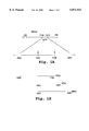
- FIG. 1A shows, in the upper line, a portion of the HTLV-I genome containing the gp46 envelope protein coding sequence, and in the lower line, a portion of the gp46 coding region containing the sequences which encode overlapping HTLV-I peptide antigens formed in accordance with the invention, and designated MTA-4, MTA-1, and MTA-5 in FIG. 1B;
- FIG. 2 shows the HTLV-I coding sequences and corresponding amino acid sequences for a portion of the HTLV-I envelope protein
- FIG. 3 shows amino acid sequences of homologous regions of HTLV-I and HTLV-II gp46 in the region of the peptide antigen of the invention, and peptide sequences of several HTLV-I gp46 peptide antigens (upper part of figure) and HTLV-II peptide antigens (lower part of figure) in accordance with the invention;
- FIGS. 4A and 4B show antigenicity plots for the MTA-1 peptide and corresponding HTLV-II gp46 peptide
- FIGS. 5A and 5B illustrate recombinant methods for producing and selecting random-sequence peptides, in accordance with the invention
- FIG. 6 shows the HTLV-II coding sequence, and corresponding amino acid sequence in the region of the gp46 envelope protein from which HTLV-II peptides of the invention are derived;
- FIG. 7 shows modified Western blots containing HTLV-I viral lysate and recombinant proteins p21E and MTA-4, where lanes A-F and G-R are HTLV-I and HTLV-II antisera, respectively.
- HTLV-I peptide antigens which are immunoreactive with anti-HTLV-I antibodies found in individuals with HTLV-I-related T-cell leukemia.
- the antigens are prepared using random HTLV-I gene sequences 100-300 base pairs in length cloned in a suitable expression vector, then selected with antibody for expression of immunoreactive peptides.
- Genomic libraries of HTLV-I are prepared conventionally from cellular DNA containing an HTLV-I proviral genome.
- Duplex DNA may be prepared from HTLV-I infected cells, including T-cells isolated from patients known to be infected with HTLV-I virus, or known cell lines, such as HUT 102-B2 (Poiesz), MT-2 (Miyoshi), and MJ-tumor (Popovic) cells, all of which have been shown to produce HTLV-I virus.
- the viral genome is integrated into host DNA in these cells. Methods for preparing cell lines containing the HTLV-I genome are detailed in the above references.
- the total host genomic DNA from the above cell line is partially digested with a frequent cutter, such as HaeIII or AluI, under conditions which produce partial digest fragments in the 15-20 kbase size range, and the digested material is fractionated, for example, by sucrose gradient centrifugation, to isolate the 15-20 kbase fragments.
- the fragments are then cloned into a suitable cloning vector, preferably a phage cloning vector which can efficiently incorporate a 15-20 kbase insert.
- the isolated fragments are treated with EcoRI methylase, and EcoRI linkers are ligated to their ends under standard conditions (Maniatis), and then cloned into a phage vector, such as ⁇ Charon 4a, having a unique EcoRI insertion site.
- HTLV-I sequences are known (Seiki), as are methods for producing radiolabeled synthetic oligonucleotide probes for selected sequences.
- synthetic oligonucleotides of specified sequences can be made by commercial services, such as provided by Synthetic Genetics, Inc. (San Diego, Calif.). Using such an oligonucleotide probe, molecular clones containing HTLV-I sequences are isolated from the library by standard hybridization procedures (Maniatis, p. 322).
- the clones can first be analyzed by restriction site analysis, to confirm that the full viral genomic sequence is present, as indicated by the presence of direct long terminal repeats which flank the integrated viral genome.
- the identified molecular clone is digested with a suitable endonuclease to release the full-copy viral genome.
- a preferred endonuclease for this purpose is SacI, which cuts the viral genome in the long terminal repeats (LTR) at either end of the viral coding sequences, but does not produce internal cleavage.
- LTR long terminal repeats
- an appropriate restriction enzyme will be chosen to isolate the full-length genome.
- the purified full-copy sequence is about a 9.5 kilobase fragment.
- a fragment of the genome representing the env gene sequences alone may be purified for production of the expression library.
- cloning vectors containing full-copy HTLV-I duplex DNA have been reported (Seiki) and may be obtained directly from the investigators, as indicated in Example 1.
- the full-copy HTLV-I insert is excised from the above cloning vector, such as by complete digestion with SacI, and isolated as a 9.5 kilobase fragment, as described in Example 1.
- the isolated full-copy fragment is digested to produce DNA fragments, and preferably random fragments with sizes predominantly between about 100-300 base pairs.
- Example 1 describes the preparation of such fragments by DNAase digestion. Because it is desired to obtain peptide antigens of between about 30-100 amino acids, the digest fragments are preferably size fractionated, for example by gel electrophoresis, to select those in the approximately 100-300 base pair size range.
- the genomic digest fragments are inserted into a suitable cloning vector, preferably an expression vector which permits expression of the coded-for peptide in a suitable host.
- a suitable expression vector is ⁇ gt11, which contains a unique EcoRI insertion site 53 base pairs upstream of the translation termination codon of the ⁇ -galactosidase gene.
- the inserted sequence will be expressed as a ⁇ -galactosidase gene.
- the inserted sequence will be expressed as a ⁇ -galactosidase fusion protein which contains most of the N-terminal portion of the ⁇ -galactosidase gene, the heterologous peptide, and at least a portion of the C-terminal region of the ⁇ -galactosidase gene.
- This vector also produces a temperature-sensitive repressor (cI857) which causes viral lysogeny at permissive temperatures, e.g., 32° C., and leads to viral lysis at elevated temperatures, e.g., 42° C.
- Advantages of this vector include: (1) highly efficient recombinant generation, (2) ability to select lysogenized host cells on the basis of host-cell growth at permissive, but not non-permissive temperatures, and (3) high levels of recombinant fusion protein production. Further, since phage containing a heterologous insert produce an inactive ⁇ -galactosidase enzyme, phage with inserts can be readily identified by a ⁇ -galactosidase colored-substrate reaction.
- the digest fragments inserted into the expression vector may be modified, if needed, to contain selected restriction-site linkers, such as EcoRI linkers, according to conventional procedures.
- Example I illustrates methods for cloning the digest fragments into ⁇ gt11, which includes the steps of blunt-ending the fragments, adding EcoRI linkers and ligating the fragments with EcoRI cut ⁇ gt11.
- the resulting viral genome library may be checked to confirm that a relatively large (representative) library has been produced. This can be done, in the case of the ⁇ gt11 vector, by infecting a suitable bacterial host, plating the bacteria, and examining the plaques for loss of ⁇ -galactosidase activity. Using the procedures described in Example 1, about 60% of the plaques showed loss of enzyme activity, when compared to the level of background phage showing loss of enzyme activity, as seen in Example 1.
- the genomic library formed above is screened for production of peptide antigen (expressed as a fusion protein) which is immunoreactive with the human anti-HTLV-I antibody of interest.
- peptide antigen expressed as a fusion protein
- One antibody of particular interest for diagnosing HTLV-I infection is the 0.5 ⁇ monoclonal antibody (Mab) which, as noted above, is has the same specificity as antibodies present in patients with T-cell leukemia related to HTLV-I infection.
- the antibody is produced by the EBV-transformed B-lymphocyte cell line having ATCC Deposit No. HC8755 (See Example 2).
- host cells infected with phage library vectors are plated, as above, and the plate is blotted with a nitrocellulose filter, to transfer recombinant antigens produced by the cells onto the filter.
- the filter is then reacted with the anti-HTLV-I antibody, washed to remove unbound antibody, and reacted with reporter-labeled, anti-human antibody, which becomes bound to the filter, in sandwich fashion, through the anti-HTLV-I antibody.
- phage plaques which are identified by virtue of their production of recombinant antigen of interest are re-examined at a relatively low density, for production of antibody-reactive fusion protein.
- the screening procedures described in Example 2 are illustrative. Several recombinant phage clones which produced immunoreactive recombinant antigen were identified in the procedure.
- the one or more library vectors identified as above are preferably analyzed by nucleic acid sequencing, to determine the positions of the peptide-coding regions within the HTLV-I genome.
- Methods for excising the heterologous insert (including adjacent coding sequences of the fusion protein, if desired) from the selected library vectors, and for purifying and sequencing the excised fragments generally follow known procedures, as outlined in Example 3.
- the coding sequences of three peptides which were found to be immunoreactive with the 0.5 ⁇ Mab are shown in the drawing.
- the three heterologous sequences were matched with the known sequence of HTLV-I (Seiki).
- the MTA-1 peptide includes the additional Ile-Pro-Trp-Lys-Ser-Lys residues at the Ser C terminus of the above sequence.
- the HTLV-I specific peptide contains the immunogenic region of the C-terminal 47 amino acid MTA-1 sequence which is immunoreactive with the 0.5 ⁇ Mab.
- the HTLV-I peptides of the invention include the immunogenic region of the above amino acid sequence which is immunoreactive with the 0.5 ⁇ Mab.
- the specified sequence includes minor, neutral amino substitutions which do not appreciably decrease the immunoreactivity of the peptide antigen for the 0.5 ⁇ Mab.
- Such amino substitutions may be selected on the basis of similarities in hydrophobicity, size, charge, hydrogen bonding ability, and effect on secondary structure according to known amino acid substitution principles.
- the selected clones are used for scale-up production, for purposes of recombinant protein purification.
- Scale-up production is carried out using one of a variety of reported methods for (a) lysogenizing a suitable host, such as E. coli, with a selected ⁇ gt11 recombinant, (b) culturing the transduced cells under conditions that yield high levels of the heterologous peptide, and (c) purifying the recombinant antigen from the lysed cells.
- a high-producer E. coli host, BNN103 is infected with the selected library phage, and replicaplated on two plates.
- One of the plates is grown at 32° C., at which viral lysogeny can occur, and the other at 42° C., at which the infecting phage is in a lytic stage and therefore prevents cell growth. Cells which grow at the lower, but not the higher temperature, are therefore assumed to be successfully lysogenized.
- the lysogenized host cells are then grown under liquid culture conditions which favor high production of the fused protein containing the viral insert, and lysed by rapid freezing to release the desired fusion protein. These methods are detailed in Example 4.
- HTLV-I coding sequences from the ⁇ gt11 clone expressing the peptide antigen MTA-1 have been prepared by PCR amplification, as described in Section II below, and cloned into the pGEX-1 expression vector (Pharmacia, Piscataway, N.J.). Inserts cloned into pGEX-1 were expressed as a fusion protein with the protein Sj26, which is a 26 Kdal Glutathione S-transferase from the parasite Schistosoma japonicum. Limited paneling of pGEX-MTA-1 against sera from HTLV-I or HTLV-II infected individuals has revealed no significant difference between the reactivity of pGEX-MTA-1 vs ⁇ -gal-MTA-1.
- the recombinant peptide is purified by standard protein purification procedures which may include differential precipitation, molecular sieve chromatography, ion-exchange chromatography, isoelectric focusing, gel electrophoresis and affinity chromatography.
- a fused protein such as the ⁇ -galactosidase fused protein prepared as above
- the protein isolation techniques which are used can be adapted from those used in isolation of the native protein.
- the protein can be isolated readily by simple affinity chromatography, by passing the cell lysis material over a solid support having surface-bound anti-galactosidase antibody. This approach is used in Example 4.
- the invention also includes, in another aspect, a method of detecting HTLV-I positive human sera, by reacting sera with a peptide antigen which is immunoreactive with the HTLV-I Mab produced by ATCC cell line HB8755, i.e., the 0.5 ⁇ Mab.
- a method of detecting HTLV-I positive human sera by reacting sera with a peptide antigen which is immunoreactive with the HTLV-I Mab produced by ATCC cell line HB8755, i.e., the 0.5 ⁇ Mab.
- the presence of HTLV-I specific antibodies in sera is detected by a reporter-labeled anti-human antibody, as described in Example 7.
- the peptides contain the immunogenic region from the 41-amino acid overlap region from above-described MTA-1, MTA-4, and MTA-5 HTLV-I peptides. These peptide antigens were further characterized to confirm the location of the immunoreactive region in the 41 amino acid sequence overlap region. The location of the immunoreactive region in the C-terminal portion of the overlap region was suggested by two lines of evidence. First, the 0.5 ⁇ Mab was reported to react specifically with the HTLV-I envelope protein, i.e., no reaction was observed with HTLV-II or HTLV-III (HIV-1) envelope proteins.
- FIG. 3 a comparison of the amino acid sequence of MTA-1 peptide with the corresponding region in the HTLV-II gp46 protein (FIG. 3) shows substantially greater homology in the N-terminal half of the peptide than in the C-terminal half (the center region of the HTLV-I and HTLV-II sequences seen in FIG. 3). This would indicate that the greatest differences in anti-genicity would be found in the C-terminal half of the peptide region.
- antigenicity plots of the two corresponding peptide regions shown in FIGS. 4A and 4B for HTLV-I and HTLV-II peptides, respectively.
- the antigenicity plots were generated by a standard hydrophobicity program "Antigen" in PC Gene from Intelligenetics (Palo Alto, Calif.). As seen, the two plots are substantially overlapping in residues 3-28, but diverge markedly in residues 28-40.
- the divergent residues include the HTLV-I sequence presented as SEQ ID NO:9.
- a number of peptide antigens which include the C-terminal region just indicated were prepared and tested for binding to 0.5 ⁇ Mab, and to HTLV-I and HTLV-II antisera.
- the sequences of several of these peptides are indicated in the upper portion of FIG. 3, along with the sequences of the above MTA-1, MTA-4, and MTA-5 peptide antigens.
- the peptides were prepared by solid-phase synthetic methods, according to standard procedures. Briefly, N-alpha-protected amino acid anhydrides were prepared in crystallized form and used for successive amino acid addition at the N-terminus.
- the growing peptide (on a solid support) was acid treated to remove the N-alpha-protective group, washed several times to remove residual acid and to promote accessibility of the peptide terminus to the reaction medium.
- the peptide is then reacted with an activated N-protected amino acid symmetrical anhydride, and the solid support is washed.
- the amino acid addition reaction may be repeated for a total of two or three separate addition reactions, to increase the percent of growing peptide molecules which are reacted. Typically, 1-2 reaction cycles are used for the first twelve residue additions, and 2-3 reaction cycles for remaining residues.
- the protected peptide resin is treated with liquid hydrofluoric acid to deblock and release the peptides from the support.
- the peptides were tested for specific immunoreactivity with 0.5 ⁇ Mab by binding competition studies, substantially as described in Example 6.
- the K163 peptide which contains the 18 C-terminal residues of MTA-4 or MTA-1, strongly inhibits binding of 0.5 ⁇ Mab to MTA-4. No binding interference, however, was observed with peptide K162, which contains only the 11 C-terminal residues of MTA-4.
- Peptide K164 which contains the 6 C-terminal residues of MTA-4 and an additional C-terminal 13 residues, weakly inhibited binding between 0.5 ⁇ Mab and MTA-4 or MTA-1.
- the most potent immunoreactive region in the gp46 peptide for the 0.5 ⁇ Mab is in a region which includes peptide K163, consistent with the divergence in sequence homology and anti-genicity plots between HTLV-I and HTLV-II sequences in this region.
- the weak binding of 0.5 ⁇ Mab to the K164 peptide may indicate that the epitope of interest in the presented as SEQ ID NO:10 region of overlap between K163 and K164, where adjacent N-terminal terminal or C-terminal sequences are required for antigen presentation, or may indicate that the K164 peptide contains an additional epitopic region which is weakly immunoreactive with the 0.5 ⁇ Mab.
- peptides were also examined for their ability to inhibit binding of antisera from HTLV-I infected patients to MTA-4.
- the ability of any particular peptide to inhibit binding of 0.5 ⁇ Mab to MTA-4 paralleled its ability to either bind to HTLV-I antisera in an ELISA binding protocol (Example 6B) or to inhibit binding of human HTLV-I antisera to MTA-4 or MTA-1 in a Western blot assay (Example 8C).
- peptide K162 did not react with any HTLV-I sera in the ELISA protocol and did not inhibit binding of J-254 sera to MTA-1 or MTA-4.
- the 0.5 ⁇ Mab-reactive peptide for use in the method is prepared by selection of random-sequence peptides. Recently, it has been demonstrated that antibodies directed against specific short (5-10 residues) peptides can be used to screen libraries of randomly generated peptides for immunoreactive species. (Scott; Cwirla et al). Such a strategy is exploited herein to identify novel sequences which are immunoreactive with the 0.5 ⁇ monoclonal antibody.
- approximately 10 8 novel heptapeptides are generated through construction of an epitope library using the filamentous phage fUSE5 as a vector.
- Other filamentous phage vectors are considered to be equally efficacious in developing such a library.
- FIGS. 5A and 5B show schematically the sequence of steps necessary to generate and screen a fUSE5 filamentous phage epitopic library.
- fUSE5 RF DNA is subjected to digestion with restriction endonuclease SfiI to create an insertion site for insertion of foreign DNA.
- a synthetic (15+3 m) base pair (bp) BglI DNA fragment is prepared which contains a degenerate sequence of the form (NNK)m, where N represents A, G, C, or T; K represents G or T; and m can vary from 2 to 15.
- m ranges from 5-10 and the bases are randomly added in single addition events to the template primer.
- An alternative method of achieving random addition of codons coding for the twenty amino acids is to randomly attach trinucleotide codons representing each amino acid to the template primer.
- amplification of the filamentous phage vector is achieved by transfection of E. coli cells. Successful transfection is measured by the presence of vector borne markers. In the preferred embodiment of the invention, this marker is tetracycline resistance.
- Recombinant phage are then isolated from bacterial cells. Phage bearing sequences of interest are isolated by an antibody panning method in which phage are incubated with the 0.5 ⁇ Mab or its Fab fragment.
- Biotinylated second antibody (goat anti-human IgG) is then added, and complexes containing biotinylated second antibody, the 0.5 ⁇ Mab and immunoreactive peptide bearing phage are separated from unreacted antibodies and phage by adhesion onto a streptavidin coated plate. Phage bearing immunoreactive sequences are then eluted, and their DNA sequences are determined.
- HTLV-II peptides which are specifically immunoreactive with HTLV-II antisera.
- the peptides are derived from the HTLV-II gp46 envelope protein region which is homologous to the above described MTA-1 peptide from the HTLV-I gp46 region.
- An HTLV-II peptide designated GH2-K15 (FIG. 3) corresponding to the HTLV-I peptide MTA-1 was prepared by cloning of an HTLV-II coding sequence corresponding to the desired peptide sequence.
- a 147 base pair (bp) HTLV-II DNA fragment corresponding to nucleotides 5648 to 5794 of the HTLV-II genome (FIG. ).
- HTLV-II clone pM04 which contains the majority of the HTLV-II genome cloned into the BamH I site of the plasmid pBR322
- PCR polymerase chain reaction
- the forward direction and reverse primers are indicated in FIG. 6.
- the amplified DNA was ligated into the EcoR I site of ⁇ gt11 phage vector, yielding the clone as3K15 which contains a 147 HTLV-II DNA insert into the -galactosidase gene of the ⁇ gt11.
- the recombinant phage was used to transfect E. coli strain BNN103. Details are given in Example 5.
- GH2-K15 amino acid sequence Several smaller peptides contained with the GH2-K15 amino acid sequence were prepared by recombinant methods, as outlined in Section I. Briefly, the peptides were prepared by PCR amplification of HTLV-II genomic DNA, using PCR primers designed to promote amplification of the sequences indicated, as detailed in Example 5. Five of these peptides, designated (GH2-) K14, K16, K24, K35, and K34 have the sequences shown in FIG. 3.
- HTLV-II peptides described above were immunoscreened against several HTLV-II and HTLV-I in an ELISA format, as described in Example 8. The results are shown in Table 1. All ⁇ gt11 HTLV-II clones except for GH2-K16 were recognized by at least 1 out of the 6 HTLV-II sera tested. GH2-K16, the sole non-reactive clone, is missing the carboxyl terminal 22 amino acids that are included in GH2-K15. All the other clones tested contain at least the 17 amino acids presented as SEQ ID NO:7 that are present in peptide K125.
- GH2-K15, GH2-K35, and GH2-K16 have been cloned into the pGEX-1 expression vector.
- Recombinant protein expressed by the 3 pGEX-1 HTLV-II clones GH2-K15, GH2-K25, and GH2-K35 have all been recognized by the J-317 HTLV-II serum.
- a number of peptide antigens which contain amino acid sequences within the K15 sequence were prepared by solid-phase methods, as outlined in Section III above. The sequences of five of these peptides, designated (GH2-) K169, K170, K125, K126, and K128 are shown in FIG. 3. The peptides were tested for immunoreactivity with several HTLV-I and HTLV-II positive sera, by an ELISA method, and some of the peptides were also examined for their ability to inhibit HTLV-II antibody binding to the K15 antigen.
- the K125 peptide was recognized by multiple HTLV-II sera when assayed by ELISA. In one experiment 6 out of 12 HTLV-II sera were able to bind efficiently to K125. In the same experiment 0 out of 7 HTLV-I sera bound peptide K125. The K125 peptide also inhibited the binding of a strongly reactive HTLV-II sera, J-317, to Western blotted GH2-K15. The ability of sera J-317 to bind GH2-K15 is not affected by incubation with the HTLV-I peptide K163 or the HTLV-II peptide K128.
- the HTLV-II peptide K170 is recognized by multiple HTLV-II sera in an ELISA based assay, and not recognized by HTLV-I sera in the same assay.
- the K169 peptide is not recognized by HTLV-II sera in an ELISA based assay.
- the first is based on inhibition of complement-mediated, antibody-dependent cytolysis by the peptide.
- serum from a test individual is reacted with HTLV-I or HTLV-II infected T-cell clones in the presence of complement.
- the presence of anti-HTLV-I or anti-HTLV-II antibody is evidenced by cell lysis, as judged, for example, by trypan blue dye exclusion.
- the specificity of the anti-HTLV-I antibody for the HTLV-I peptide is demonstrated by first reacting the serum with excess HTLV-I or HTLV-II peptide, then mixing the serum with cells in the presence of complement. The presence of HTLV-I or HTLV-II antibody is indicated by a substantial decrease in cell lysis. This method is described in Example 6A.
- the method can also be used to quantitate the antibody titer in the analyte serum, by titrating the serum with increasing amounts of peptide, and determining the peptide concentration where a noticeable effect on the extent of cell lysis is first observed.
- the second general assay type is an enzyme-immuno-assay for screening human sera for HTLV-I or HTLV-II infection.
- a solid phase reagent having surface-bound HTLV-I or HTLV-II gp46 peptide antigen is reacted with analyte serum, under conditions which allow antibody binding to the peptide on the reagent.
- the reagent is reacted with an enzyme-labeled anti-human antibody, to bind enzyme to the reagent in proportion to the amount of bound anti-HTLV-I antibody on the solid support.
- the reagent is again washed, to remove unbound antibody, and the amount of enzyme associated with the reagent is determined.
- the enzyme-labeled antibody, and reagents required for enzyme detection, are also referred to herein as reporter means for detecting the presence of human antibody bound to the peptide antigen on the solid support.
- the solid surface reagent in the above assay is prepared by known techniques for attaching protein material to solid support material, such as polymeric beads, dip sticks, or filter material. These attachment methods generally include non-specific adsorption of the protein to the support (as in the filter support described in Example 8) or the covalent attachment of the protein, typically through a free amine group, to a chemically reactive group on the solid support, such as an activated carboxyl, hydroxyl, or aldehyde group.
- the third general assay type is Western blot assay for use in confirming HTLV-I or HTLV-II antisera.
- This assay format includes, in addition to the gp46 peptide antigen of the invention, one or more confirmatory HTLV-I or HTLV-II antigens that are effective to detect HTLV-I or HTLV-II antisera.
- the confirmatory peptides include the p24 gag protein from HTLV-I viral lysate, and a p21E recombinant envelope protein containing a large portion of the HTLV-I gp21 envelope protein (Samuel, 1984, 1985).
- the p24 lysate proteins picks up most, but not all HTLV-I and HTLV-II positive sera.
- the p21E recombinant peptide picks up virtually all HTLV-I and HTLV-II, but also gives some false positives.
- This modified western blot assay has been reported by the applicants and co-workers (Lipka). Details of the blot assay procedure are given in Example 8.
- the modified Western blot format picked up all HTLV-I and HTLV-II positive sera tested (a panel of 95), as evidence by immunoreaction with viral lysate protein p24 and recombinant protein p21E.
- the MTA-4 peptide was immunoreactive with confirmed HTLV-I sera only.
- the modified blot assay thus can be used to confirm HTLV-I or HTLV-II antisera, and to distinguish the two types of HTLV virus by selective immunoreaction with the peptide of the invention.
- the HTLV-I peptide antigen is replaced by the HTLV-II gp peptide antigen described in Section III.
- the HTLV-I viral lysate proteins and p21E recombinant protein provide confirmation of HTLV-I or HTLV-II antisera, as above.
- the HTLV-II specific peptide will pick up HTLV-II, but not HTLV-I antisera, and thus provides a positive confirmation of HTLV-II antisera.
- the two formats can be combined to include both HTLV-I and HTLV-II specific peptide antigens, to give positive confirmation of either HTLV antisera.
- a vaccine composition containing an HTLV-I gp46 peptide and a antigen carrier, such as an immunogenic protein, to which the antigen peptide is bound.
- the peptide contains an immunogenic region formed by the above 41 or 47-amino acid overlap of MTA-1, MTA-4, and MTA-5 peptides described in Section I, which is immunoreactive with anti-HTLV-I 0.5 ⁇ Mab, i.e., the antibody derived from ATCC cell line HB8755. More specifically, the peptide contains the immunogenic region of the peptide sequence presented as SEQ ID NO:4. Since the 0.5 ⁇ Mab is a neutralizing antibody, the antibody raised by the peptide is expected to be a neutralizing antibody.
- the vaccine composition may alternatively include the HTLV-II gp46 peptide containing the HTLV-II specific immunogenic region formed by the amino acid sequence presented as SEQ ID NO:5 and preferably formed by the amino acid sequence presented as SEQ ID NO:7, or Ser-Pro-Pro-Leu-Val-His-Asp-Ser-Asp-Leu-Glu-His-Val-Leu-Thr-Pro-Ser.
- Particularly useful protein carriers for the peptide(s) include keyhole limpet hemocyanin (KLH), tetanus toxoid, poly-l-(Lys:Glu), peanut agglutinin, poly-D-lysine, diphtheria toxoid, ovalbumin, soybean agglutinin, bovine serum albumin (BSA), human serum albumin, and the like.
- KLH keyhole limpet hemocyanin
- tetanus toxoid poly-l-(Lys:Glu
- peanut agglutinin poly-D-lysine
- diphtheria toxoid ovalbumin
- soybean agglutinin bovine serum albumin
- BSA bovine serum albumin
- the immunogenic peptide(s) may be conjugated to the carrier by a variety of known methods, including chemical derivatization and by genetic engineering techniques. Such latter technique is disclosed in more detail by Gerald Quinnan, "Proceedings of a Work-shop," Nov. 13-14, 1984.
- Vaccines and inocula of the present invention may be administered by injection, usually intramuscularly or subcutaneously, orally by means of an enteric capsule or tablet, as a suppository, as a nasal spray, and by other suitable routes of administration.
- a suitable dose of the polypeptide depends, in part, upon the chosen route of administration and a number of other factors. Included among those factors are the body weight of the mammal to be immunized, the carrier when used, the adjuvant when used, and the number of inoculations desired to be used.
- Individual inoculations for a human patient typically contain unit doses of about 10 micrograms to about 100 milligrams of polypeptide, exclusive of any carrier to which the polypeptide may be linked. If desired, a series of doses may be administered over a period of time for optimum immunity.
- Unit dosage forms of the vaccine can also be provided, if desired, containing the aforementioned amounts of the polypeptide.
- the immunogen contained in a vaccine or an inoculum is present in an "effective amount," which amount depends upon a variety of factors as is well known in the immunological arts, e.g., the body weight of the mammal to be immunized, the carrier moiety used, the adjuvant used, the duration of protection sought, and the desired immunization protocol.
- Enzymes DNAase I and alkaline phosphatase were obtained by Boehringer Mannheim Biochemicals (BMB, Indianapolis, Ind.); EcoRI, EcoRI methylase, DNA ligase, and Polymerase I, from New England Biolabs (NEB, Beverly, Mass.); and RNase was obtained from Sigma (St. Louis, Mo.).
- EcoRI linkers were obtained from NEB; and nitro blue tetrazolium (NBT), 5-bromo-4-chloro-3-indolyl phosphate (BCIP), 5-bromo-4-chloro-3-indolyl- -D-galactopyranoside (X-gal) and isopropyl-D-thiogalactopyranoside (IPTG) were obtained from Sigma.
- NBT nitro blue tetrazolium
- BCIP 5-bromo-4-chloro-3-indolyl phosphate
- X-gal 5-bromo-4-chloro-3-indolyl- -D-galactopyranoside
- IPTG isopropyl-D-thiogalactopyranoside
- Bacteriophage containing a full-copy DNA insert derived from the HTLV-I genome was obtained from Drs. R. C. Gallo and F. Wong-Staal of the Laboratory of Tumor Cell Biology, National Institutes of Health (Bethesda, Md.).
- the bacteriophage was digested to completion with SacI, releasing the viral genome insert.
- the digested material was electrophoresed on standard 1% agarose gel, and the 9.5 kilobase fragment obtained by electroelution was extracted with phenol/chloroform before ethanol precipitation.
- the purified genomic DNA was suspended in a standard digest buffer (0.5M Tris HCl, pH 7.5; 1 mg/ml BSA; 10 mM MnCl 2 ) to a concentration of about 1 mg/ml, and digested with DNAase I at room temperature for about 5 minutes. These reaction conditions were determined from a prior calibration study, in which the incubation time required to produce predominantly 100-300 basepair fragments was determined. The material was extracted with phenol/chloroform before ethanol precipitation.
- a standard digest buffer 0.5M Tris HCl, pH 7.5; 1 mg/ml BSA; 10 mM MnCl 2
- the genomic fragments from above were blunt-ended with DNA Pol I under standard conditions (Huynh), then extracted with phenol/chloroform and precipitated with ethanol.
- the blunt-ended material was ligated with EcoRI linkers, under standard conditions (Maniatis, pp. 396-397), then digested with EcoRI to remove redundant linker ends.
- the material was then agarose-gel-fractionated to remove non-ligated linkers and to size-select (see below).
- the resultant fragments from the previous step were analyzed by electrophoresis (5-10V/cm) on 1.2% agarose gel, using X174/HaeIII and /HindIII size markers.
- the 100-300 bp fraction was eluted onto NA45 strips (Schleicher and Schuell), which were then placed into 1.5 ml microtubes with eluting solution (1M NaCl, 50 mM arginine, pH 9.0), and incubated at 67° C. for 30-60 minutes.
- the DNA, now in solution was extracted with phenol/chloroform and precipitated with ethanol.
- the pellet was resuspended in 20 ⁇ l TE (0.01M Tris HCl, pH 7.5, 0.001 M EDTA).
- gtll phage vector Human gtll phage vector (Huynh) was obtained from Promega Biotec (Madison, Wis.). This cloning vector has a unique EcoRI site 53 base pairs upstream from the ⁇ -galactosidase translation termination codon.
- the genomic fragments from above were introduced into the EcoRI site by mixing 0.5-1.0 ⁇ g EcoRI-cleaved gt11, 0.5-3 ⁇ l of the above HTLV--I genomic fragments, 0.5 ⁇ l 10 ⁇ ligation buffer (above), 0.5 ⁇ l ligase (200 units), and distilled water to 5 ⁇ l. The mixture was incubated overnight at 14° C., followed by in vitro packaging, according to standard methods (Maniatis, pp. 256-268).
- E. coli strain KM392 obtained from Dr. Kevin Moore, DNAX (Palo Alto, Calif.).
- E. coli strain Y1090 available from the American Type Culture Collection (ATCC #37197), could be used.
- the infected bacteria were plated and the resultant colonies were checked for loss of ⁇ -galactosidase activity (clear plaques) in the presence of X-gal using a standard X-gal substrate plaque assay method (Maniatis).
- Table 2 below shows the number of recombinant (clear) plaques obtained with insertion of the EcoRI-ended HTLV--I fragments (row 1). An EcoRI linker control (row 2) and vector alone (row 3) were also run.
- the phage libraries contained about 10 6 plaque-forming units (pfu)/ml.
- Purified 0.5 antibody derived from a human cell line was provided by Dr. Samuel Broder of the National Cancer Institute, National Institutes of Health (Bethesda, Md.).
- Mouse anti-human IgG antibody covalently derivatized with alkaline phosphatase was obtained from Promega Biotec (Madison, Wis.).
- a lawn of KM392 cells infected with about 10 4 pfu of the phage stock from Example 1 was prepared on a 150 mm plate, and incubated, inverted, for 5-8 hours at 37° C.
- the lawn was overlaid with a nitrocellulose sheet, causing transfer of HTLV-I recombinant protein from the plaques to the paper.
- the plate and filter were indexed for matching corresponding plate and filter positions.
- the filter was washed twice in TBST buffer (10 mM Tris, pH 8.0, 150 mM NaCl, 0.5% Tween 20), blocked with AIB (TBST buffer with 1% gelatin), washed again in TBST, and incubated overnight after addition of 0.5 monoclonal antibody (diluted to 1-2 ⁇ g/ml in AIB, 12-15 ml/plate).
- TBST buffer 10 mM Tris, pH 8.0, 150 mM NaCl, 0.5% Tween 20
- AIB TBST buffer with 1% gelatin
- the sheet was washed twice in TBST, then contacted with enzyme-labeled anti-human antibody, to attach the labeled antibody at filter sites containing antigen recognized by the 0.5 antibody.
- the filter was developed in a substrate medium containing 33 ⁇ l NBT (50 mg/ml stock solution maintained at 5° C.) in 5 ml of alkaline phosphatase buffer (100 mM Tris, 9.5, 100 mM NaCl, 5 mM MgCl 2 ). Reacted substrate appeared at points of antigen production, as recognized by the 0.5 ⁇ Mab.
- the areas of antigen production determined in the previous step were replated at about 100-200 pfu on an 82 mm plate.
- the identified plaques were picked and eluted in phage buffer (Maniatis, p. 443).
- Three of the recombinant phage plaques which secreted an antibody-reactive peptide were selected for sequencing analysis, according to the procedures in Example 3.
- the corresponding infected phage has been designated MTA-4, MTA-1, and MTA5.
- Phages MTA-4, MTA-1, and MTA-5 were isolated from the plate cultures of the infected E. coli Y1088 bacteria. These cells are available from the ATCC (ATCC #31195). The phage was collected by addition of phage-dilution buffer (maniatis) late material was purified from bacterial debris by low-speed centrifugation, and the supernatant was poured into SW 27 tubes. RNase and DNAse were each added to a concentration of 1 ⁇ g/ml each from stock solutions of 1 mg/ml.
- the sample was incubated for 30 minutes at 37° C., and an equal volume of a polyethylene glycol (PEG), 5.8 g NaCl, 2.0 g MgSO 4 .7H 2 O, 1M Tris Cl, pH 7.5, and 2% gelatin was added.
- PEG polyethylene glycol
- the sample was placed in an ice bath for 1 hour to allow the phage particles to form a precipitate, which was then isolated by centrifugation at 10 k for about 20 minutes at 4° C.
- the supernatant was decanted, and the pellet was resuspended in 0.6 ml PDB buffer (5.8 g NaCl, 2.0 g MgSO.7H 2 O, 50 ml 1M Tris Cl, pH 7.5, and 5 ml 2% gelatin) and transferred to 0.5 ml polypropylene microtubes. 5 ⁇ l 10% SDS, 5 ⁇ l 0.5M EDTA, and 2.5 ⁇ l proteinase K (20 mg/ml) were added, and the samples were incubated at 50° C. for 15 minutes.
- PDB buffer 5.8 g NaCl, 2.0 g MgSO.7H 2 O, 50 ml 1M Tris Cl, pH 7.5, and 5 ml 2% gelatin
- 5 ⁇ l 10% SDS, 5 ⁇ l 0.5M EDTA, and 2.5 ⁇ l proteinase K (20 mg/ml) were added, and the samples were incubated at 50° C. for 15 minutes.
- the detergent and enzyme-treated material was extracted with an equal volume of phenol/chloroform, and centrifuged to ensure separation of the phases.
- the aqueous phase was transferred to a new tube, and the extraction/centrifugation procedure was repeated with a mixture of chloroform and isoamyl alcohol.
- An equal volume of isopropanol was added, and the same was inverted several times to mix, and cooled to -70° C. for 20 minutes.
- the sample was centrifuged for 5 minutes and the supernatant was decanted.
- the pellet was washed in 70% ethanol, briefly dried in a 37° C. heat block, and resuspended in 100 ⁇ l TE buffer, pH 7.5.
- the isolated phage DNA was digested with KpnI and SacI and then combined with KpnI/SacI cut plasmid vector pGEM-3 (Promega Biotec) to isolate a plasmid recombinant with the insert of interest.
- the HTLV-I insert was then sequenced using the standard dideoxy sequencing procedure and forward and reverse primers for ⁇ gt1 sequences flanking the EcoRI insertion site.
- the figure shows the coding sequence and corresponding amino acid sequence of a portion of the fused protein formed by the above methods, for each of the three fused peptides examined.
- a terminal G base of the ⁇ -gal gene and the adjacent CC bases of the env gene contributed by each of the three insert sequences yield a GCC (Ala) codon, replacing the Ser codon which normally occurs at that codon position of all three env inserts.
- the insert in the MTA-4 includes a 225 base pair sequence extending from base 5564 to 5790 of the HTLV-I coding region.
- the insert of the MTA5 phage begins at base 5664, and extends to base 5895.
- the 231 basepair sequence covers amino acids 162 to 240 of the gp46 protein.
- the region of insert overlap includes the 42 amino acid sequence from amino acids 162 to 203 of the native gp46 protein.
- EcoRI strain C600
- Dr. R. Davis Stanford University (Stanford, Calif.).
- EcoRI Y1089 ATCC #37196
- a 1 ml saturated, overnight culture of the cells was infected with one of the three phages from Example 3 by adsorbing 10 ⁇ l of eluted plaque stock to 50 ⁇ l of overnight bacterial culture.
- the infected bacteria were spread only LB agar plates (Maniatis, p. 440) and incubated at 32° C.
- the individual colonies were picked with sterile toothpicks onto corresponding grids on two separate plates. One of the plates was incubated at 32° C., and the other at 42° C.
- This Example describes induction of a recombinant protein containing the HTLV-I epitope from the ⁇ gt11 lysogens prepared in Example 4 with the MTA-4 phage.
- the antigen is produced in the form of a-galactosidase fusion protein which also contains an N-terminal portion of the phage -gal protein.
- a superbroth was prepared containing 35 g bacto-tryptone, 2 g bacto-yeast extract, 5 g NaCl, and 5 ml 1N NaOH in 1 1 dHO. 500 ml of the superbroth were inoculated 1:100 with a saturated overnight culture of the EcoRI ⁇ gt11 lysogens prepared in the previous example. The culture was incubated to A 600 ⁇ 0.4-0.5 with vigorous aeration.
- the temperature of the culture was raised to 43°-44° C., thereby inactivating the temperature-sensitive -galactosidase repressor gene.
- the temperature was maintained at 43° C. with a 65° C. water bath for 15 minutes with aeration.
- IPTG which induces -galactosidase expression by competitively binding to the -galactosidase repressor, was added to the broth to 2 mM to further increase protein production.
- the culture was returned to the 38° C. shaker for about an hour.
- the cells were then pelleted at 6,000 ⁇ g for 15 minutes at 37° C., resuspended in lysis buffer (10 mM Tris, pH 7.4, 2% Triton X-100, 1% aprotinin, and 50 ⁇ g PMSF) and immediately plunged into liquid N 2 . Lysis was completed upon thawing of the frozen samples.
- the cell lysate obtained in the previous Example was thawed and warmed to 37° C. 10 ⁇ l DNAse (1 ⁇ g/ml) was added, and the mixture incubated until the viscosity decreased. The lysate was quickly chilled on ice, clarified t 4° C. for 5 minutes in a microfuge, and loaded onto a 6 ml column of anti- -galactosidase coupled to Sepharose 4B (Pharmacia).
- TX buffer (10 mM Tris, pH 8.0, 2% Triton X-110, 50 ⁇ g/ml PMSF
- TX buffer 10 mM Tris, pH 8.0, 2% Triton X-110, 50 ⁇ g/ml PMSF
- Fusion protein was then eluted from the column with 35 mM 3,5-diiodosalicylic acid in TX buffer. The majority of protein was eluted in the first 3-4 volumes, and removal was substantially complete after 7 volumes.
- the eluted samples were desalted and concentrated using Amicon filters (Danvers, Mass.).
- PCR polymerase chain reaction
- PCR was performed according to the manufacturers instructions (Perkin Elmer/Cetus), and all PCR reactions contained 2 ng of the above HTLV clone as template and 1 ⁇ M of the appropriate PCR primers. PCR amplification was carried out for 25 cycles. Each cycle involved template denaturation for 1 minute at 94 deg.C., annealing of primer to template for 2 minutes at 50 deg.C., followed by primer extension for 2 minutes at 72 deg.C. Afterwards the amplified DNA was purified and then digested to completion with EcoRI. The digested DNAs were then ligated into the EcoRI site of lambda gt11 .
- the recombinant phage DNAs were then packaged and the frequency of non-recombinant phage was determined by plating in the presence of 5-bromo-4-chloro-3-indolyl ⁇ -D-galactopyranoside.
- the ratio of recombinant to non-recombinant phage was about 50/1.
- Multiple isolated plaques from each of the 6 recombinant phage clones were picked and subsequently screened using PCR with lambda gt11 flanking primers 11F and 11R, and/or the HTLV-II plaques described above. Clones containing correctly sized and orientated inserts were then amplified and used in subsequent immunoscreening assays.
- the EcoR I fragment from 3 of the clones GH2-K15, GH2-K16 and GH2-K35 were subsequently subcloned into the pGEX-1 plasmid and DNA sequenced. The sequences obtained perfectly matched the reported sequence for the desired region of HTLV-II (Shimotokno).
- Recombinant phage was mixed 1/1 with wild type gt11 and used to infect E. coli strain KM-392. After allowing the phage to grow for ⁇ 5 hours expressed proteins were bound to nitrocellulose filters overnight. Filters were subsequently washed 3 ⁇ with TBS (0.5M NaCl, 20 mM Tris Ph 8.0), cut into sections, and blocked using TBS plus 1% Gelatin. Filter sections were then incubated overnight with 1st stage antibody, usually sera from HTLV-I or HTLV-II infected individuals diluted 1/100 in TBS plus gelatin. After washing with TBS, the filters were incubated with alkaline phosphatase conjugated goat anti human sera for at least 1 hour.
- TBS 0.5M NaCl, 20 mM Tris Ph 8.0
- the filters were washed with TBS and bound antibody was then detected by incubating the filters in a solution of nitroblue tetrazolium chloride and 5-bromo-4-chloro 3-indolylphosphate. A particular sera was scored as positive if plaques derived from the recombinant phage could clearly be distinguished from plaques of wild type gt11.
- the supernatant fraction was then combined with 2 mls of Protosorb LacZ adsorbent (Promega) and incubated for 2 hours at 25° C. with agitation.
- the column resin was then poured into a disposable column and washed with 2 ⁇ with 10 mls of TX buffer (1% Aprotinin, 10 mM Tris pH 7.4).
- Bound protein was eluted 14 mls of pH 10.8 carbonate buffer and 2 ml fractions were collected into tubes already containing 1 ml of 2M tris buffer (pH 7.5). Fractions were then concentrated using Centricon 30s (Amicon) following manufacturers instructions. The fractions were washed with 2 mls of MTBS buffer (150 mM NaCl, 4 mM NaH2PO4, 16 mM Na2HPO4, pH 7.3) and then concentrated again.
- the location, yield and purity of the purified fusion protein was determined using SDS-PAGE. After one pass through the immunoaffinity column GH2-K15 recombinant antigen was ⁇ 70% pure. Fractions containing fusion protein were pooled and aliquots of the pool were used in subsequent western blot experiments. Western blot analysis was performed essentially as described previously (Lipka). Titration experiments determined that the optimum loading of purified GH2-K15 antigen was 3 ⁇ g protein/cm nitrocellulose. Peptide competition experiments are described in Example 6C. Nitrocellulose strips containing blotted antigen were then added to the serum samples and incubated and developed as normal.
- HTLV-I HTLV-II
- HTLV-II HTLV-II
- ELISA reactive--HTLV negative individuals Sera samples were obtained from HTLV-I, HTLV-II, or ELISA reactive--HTLV negative individuals. These samples were from multiple different sources and geographic areas. Many of the seropositive sera samples were also typed for HTLV-I or HTLV-II infection using PCR with strain specific DNA primers. HTLV-I sera samples included 58 PCR proven samples consisting of 45 samples from Jamaican food handlers, 2 intravenous drug users (IVDU) from the New La area, and 11 northern California blood donors. In addition a total of 238 HTLV-I sera samples were obtained from Japan.
- IVDU intravenous drug users
- HTLV-II sera samples included 57 PCR proven samples consisting of 6 IVDU from the New La area, 24 IVDU from the northern California area, and 27 blood donors from the northern California area.
- HTLV negative sera included 1 Jamaican food handler, 15 California blood donors, and 29 samples from Japan. PCR analysis of serum samples was performed as described (Lipka).
- HUT 102-B2 cells were obtained from Dr. R. C. Gallo, LTCB, NIH. This is a long-term cultured T-cell line known to produce HTLV-I.
- 0.5 ⁇ antibody ( ⁇ 5 ⁇ g/ml IgG) or a control isotyped matched human IgG was preincubated with MTA-4 recombinant peptide or irrelevant recombinant for 30 minutes at room temperature. 50 ⁇ l of these mixtures was then added to 5 ⁇ 10 5 HUT 02B2 cells in 96-well micro titer plates, and incubated for 30 minutes at room temperature. 30 ⁇ l of rabbit complement per well was added, and incubated 1 hour at 37° C. Cell viability was determined by microscopic examination. Cell lysis was visibly inhibited by addition of the MTA-4 peptide antigen, but not by preincubation with irrelevant recombinant peptide antigen. Isotyped matched human IgG, after preincubation with either recombinant antigen or irrelevant recombinant peptide antigen, had no effect on HUT 102-B2 viability.
- HTLV-I and HTLV-II peptides were examined in an ELISA assay to determine the ability of sera from HTLV infected individuals to bind to the synthetic peptides described above.
- the ELISA assay involved binding a fixed amount of synthetic peptide to a microtiter plate, followed by the addition of sera from a HTLV infected individual. Unbound sera was then washed away and antibody bound to the peptide was detected by a 2nd antibody.
- the 2nd antibody is conjugated to an enzyme that converts a colorless substrate to a colored product.
- the amount of colored product produced indicates the amount of serum antibody which bound the peptide.
- the signal obtained from a particular sera against bound peptide was subtracted from the signal obtained by the sera from a well which did not contain any peptide.
- the values obtained after subtraction of the minus peptide background had to be 2.5 times the background value to be considered positive.
- the inhibition assay involves the incubation of a large excess of a synthetic peptide with sera from an HTLV infected individual prior to placing a strip of nitrocellulose which contains a HTLV-I or HTLV-II recombinant antigen blotted on to it. If the sera can bind to the peptide, the vast excess of peptide in solution with the antibody will prevent significant binding of the antibody to the relatively small amount of antigen present on the nitrocellulose strip. The amount of antibody bound to the recombinant antigen on the nitrocellulose strip is determined using an enzyme conjugated second antibody in a manner analogous to that described above for the ELISA assay.
- Control experiments involved incubating HTLV-I sera with the HTLV-II peptide K125 or HTLV-II sera with an HTLV-I peptide, and then determining the ability of the sera to recognize the appropriate recombinant antigen.
- Purified MTA-4 peptide antigen was prepared as in Example 4, and dot blotted on nitrocellulose filters, which were then used in a solid-phase assay for determination of serum antibodies in patients with T-cell leukemia (6 patients with HTLV-I infection).
- 0.1 ml of various serum dilutions, ranging from 1:100 to 1:50,000, from the test individual was added to the filter, and allowed to rest at room temperature for 30 minutes.
- the filter was then washed two times with TBST buffer (Example 2), and incubated with anti-human antibody conjugated with alkaline phosphatase, as in Example 2.
- the presence of antibody was determined by color development in NBT and BCIP, also as in Example 2.
- Recombinant MTA-1 was prepared as in Example 4.
- Recombinant p21E was prepared as described previously (Samuel, 1984, 1985).
- HTLV-II viral lysate was prepared from chronically infected cell line MT-2 (Hillcrest Biologicals, Cypress, Calif.). These HTLV-I antigens were combined and then separated under reducing conditions on a 11.5% acrylamide SDS/PAGE gel (Laemmli). The resolved proteins were electroblotted onto a nitrocellulose (onto a nitrocellulose membrane blocked with blotto (5% nonfat dry milk, 2.5% normal goat serum in 100 mM Tris-HCl, pH 7.4), air dried, and cut into 3 mm wide strips.
- test strips from above were first rehydrated in TBS buffer, and the strips were incubated overnight with human test sera, diluted 1:50 in blotto. The strips were washed several times with wash buffer, then incubated for one hour with goat anti-human IgG conjugated to alkaline phosphatase (Bio-Rad, Richmond, Calif.). After washing, color development was achieved by incubating the strips in a substrate solution containing 5-bromo-4-chloro-3-indolyl phosphate and nitroblue tetrazolium in 100 mM Tris-HCl buffer, pH 9.5, 50 mM MgCl 2 . Color development was continued until a uniform background developed on the strip and was halted by rinsing the strips two times with de-ionized water.
- a panel of HTLV-I or HTLV-II positive sera were tested. These had been previously confirmed as HTLV-I or HTLV-II positive by PCR analysis (Lipka). The results are shown in FIG. 6, where panels A-G are HTLV-I antisera, and panels H-S are HTLV-2 antisera. Viral lysate protein gp24 was immunoreactive with every serum sample, as was the recombinant peptide gp21E. MTA-4 was immunoreactive with HTLV-I serum samples only.
Abstract
Novel HTLV-I and HTLV-II peptide antigens are disclosed for use in diagnostics assays for screening and confirming HTLV-I and HTLV-II antisera. The peptides are derived from analogous regions of HTLV-I and HTLV-II gp46 envelope proteins, and are differentiated by their immunoreactivity with an HTLV-II specific monoclonal antibody and by HTLV-I and HTLV-II antisera. The peptides are also useful in vaccine compositions.
Description
This application is a division of copending U.S. patent application Ser. No. 07/653,091 filed Feb. 8, 1991, now U.S. Pat. No. 5,614,336 which is a continuation-in-part of U.S. patent application for "HTLV-I Peptide Antigen and Methods", Ser. No. 366,313, filed Jun. 13, 1989, now U.S. Pat. No. 5,066,579, which in turn is a continuation of U.S. patent application for "HTLV-I Peptide Antigen and Methods," Ser. No. 948,270, filed Dec. 31, 1986, now abandoned, all of which are hereby incorporated by reference.
The present invention relates to an HTLV-I specific antigen, and to methods of preparing and using the antigen.
Cwirla, S. E., et al., Proc Nat Acad. Sci, U.S.A., 87:6378 (1990).
Huynh, T. V., et al., in "DNA Cloning, Volume 1," ed. D. M. Glover, Washington, D.C.: IRL Press, 1985 (Chapter 2).
Laemmli, U. K., Nature, 227:680 (1970).
Lipka, J. J., et al., J Infect Dis, 162:353 (1990).
Lipka, J. J., et al., Proceedings of the 43 Meeting (1990).
Maniatis, T., et al., Molecular Cloning: A Laboratory Manual, Cold Spring Harbor Laboratory (1982).
Matsushita, S., et al., Proc Natl Acad Sci (U.S.A.), 83:2672 (1986).
Miyoshi, I., et al., Nature, 294:770 (1981).
Poiesz, B. J., et al., Proc Natl Acad Sci (U.S.A.), 77:7415 (1980).
Popovic, M., et al., Science, 29:856 (1983).
Samuel, K. P., et al., Science, 226:1094 (1984).
Samuel, K. P., Gene Anal Tech, 2:60 (1985)
Scott, J. T.<et al., Science, 249:386 (1990).
Seiki, M., et al., Proc Natl Acad Sci (U.S.A.), 80:3618 (1983).
Shimotokno, K., et al, Proc Nat Acad Sci, U.S.A., 82:3101 (1985).
The human T-cell leukemia viruses (HTLV) represent a family of T-cell retroviruses with three known members. HTLV type I (HTLV-I) has transforming activity in vitro and is etiologically linked to adult T-cell leukemia, which is known to be endemic in several parts of the world. HTLV-II is another retrovirus having transforming capacity in vitro, and has been isolated from a patient with a T-cell variant of hairy cell leukemia. HTLV-III, which has also been called lymphadenopathy-associated virus and is now known as the human immunodeficiency virus (HIV), is lytic for certain kinds of T cells and has been linked to the etiology of acquired immunodeficiency syndrome (AIDS). Unlike the HTLV-I and -II viruses, HTLV-III is not known to have in vitro transforming activity.
The diagnosis of HTLV-I infection is usually based on serum antibody response to HTLV-I peptide antigens. This usually involves an initial screening assay to identify HTLV-I antibodies, based on an enzyme immunoassay assay (EIA) with HTLV-I virion peptides. The assays presently used for blood screening detect about 0.5 to 0.05% HTLV-I and HTLV-II positives; of these, about 4 out of 5 are false positives. Therefore, positive sera must be further tested in a confirmatory assay, using Western blot or radioimmunoprecipitation assays which detect antibody reaction to specific HTLV-I peptide antigens.
Current blood testing procedures require confirmation tests based on immunoreaction with HTLV-I p24 gag protein and at least one of the envelope proteins gp46, gp21, or gp68. When the test antigens are prepared from virion proteins, only gp46 gives a high rate of antibody reaction with true HTLV-I seropositives. Even then, the reaction with gp46 may be detected only by additional antigen testing with a more sensitive radioimmunoprecipitation assay. The above screening and confirmation testing identifies HTLV-I and HTLV-II positives, but does not distinguish between the two HTLV viruses.
It would therefore be desirable to provide an improved method for detecting HTLV-I positive sera. In particular, the improved test should be capable of detecting all HTLV-I and HTLV-II positive sera, with a minimum number of false positives, and also be able to distinguish HTLV-I from HTLV-II positive sera.
It is therefore one object of the invention to provide an improved method and kit for detecting HTLV-I and HTLV-II positive human sera.
Another object of the invention is to provide such method and kit capable of distinguishing HTLV-I and HTLV-II positive sera.
In the above-cited patent application for "HTLV-I Peptide Antigen and Assay," there is disclosed an HTLV-I peptide composed of a region of the HTLV-I gp46 envelope protein which is immunoreactive with the 0.5α monoclonal antibody (Mab) produced by ATCC cell line HB8755 (Matsushita). The region is contained in a 41 amino acid sequence overlap of three gp46 peptide antigens, designated MTA-1, MTA-4, and MTA-5. The 44 amino acid sequence overlap region contains the sequence presented as SEQ ID NO:2 and may include the additional residues presented as SEQ ID NO:3 at the C-terminal Ser residue of the 41 amino acid sequence. A common amino acid sequence in recombinant and synthetic peptides which is immunoreactive with the 0.5α Mab is the sequence presented as SEQ ID NO:4.
In another aspect, the invention includes a kit for detecting the presence of HTLV-I infection in human serum. The kit includes a solid support on which the gp46 peptide antigen is carried, and a reporter system for detecting the presence of human antibodies bound to the peptide antigen.
In one embodiment, the kit is in an EIA format for screening human sera for HTLV-I antibodies. In another embodiment, the peptide antigen is immobilized on a strip, along with one or more confirmatory HTLV-I antigens, in a Western blot format for confirming HTLV-I serum antibodies.
In still another embodiment, the kit includes an HTLV-II specific antigen, defined below, capable of reacting specifically with antibodies from HTLV-II positive sera. The kit allows for specific detection of HTLV-I and HTLV-II positive sera.
Also included in the invention is a method of detecting HTLV-I positive human sera. In this method, test sera is reacted with a peptide antigen which is immunoreactive with anti-HTLV-I monoclonal antibody (Mab) derived from ATCC cell line HB8755, designated 0.5α Mab. The presence of anti-HTLV-I antibodies bound to the antigen is detected by a suitable reporter-labeled anti-human antibody.
The 0.5α Mab-reactive peptide may be produced by a random-sequence selection method in which a mixture of random-sequence polynucleotides, preferably encoding 5-10 amino acid residues, is introduced into a suitable expression vector, to form a library of random-sequence vectors. The expression products of the library vectors are screened for the presence of an amino acid sequence which is immunoreactive with the 0.5α Mab. The library clone which expresses such an immunoreactive amino acid sequence is then isolated and used for producing the polypeptide encoded by the inserted sequence.
Also disclosed herein is an HTLV-II peptide antigen comprising less than about 50 amino acids derived from HTLV-II envelope protein gp46, and including the immunogenic region formed by the amino acid sequence presented as SEQ ID NO:5. A common amino acid sequence in recombinant and synthetic peptides which is immunoreactive with HTLV-II antisera has the sequence presented as SEQ ID NO:7 or the same sequence extended at the Ser C-terminus by the amino acid sequence presented as SEQ ID NO:8.
The peptide antigen is used in a test kit for detecting the presence of HTLV-II infection in a human serum. The kit includes a solid support which carries the peptide antigen, and a reporter system for detecting the presence of human antibodies bound to the peptide antigen.
These and other objects and features of the present invention will become more fully apparent when the following detailed description of the invention is read in conjunction with the accompanying drawings.
FIG. 1A shows, in the upper line, a portion of the HTLV-I genome containing the gp46 envelope protein coding sequence, and in the lower line, a portion of the gp46 coding region containing the sequences which encode overlapping HTLV-I peptide antigens formed in accordance with the invention, and designated MTA-4, MTA-1, and MTA-5 in FIG. 1B;
FIG. 2 shows the HTLV-I coding sequences and corresponding amino acid sequences for a portion of the HTLV-I envelope protein;
FIG. 3 shows amino acid sequences of homologous regions of HTLV-I and HTLV-II gp46 in the region of the peptide antigen of the invention, and peptide sequences of several HTLV-I gp46 peptide antigens (upper part of figure) and HTLV-II peptide antigens (lower part of figure) in accordance with the invention;
FIGS. 4A and 4B show antigenicity plots for the MTA-1 peptide and corresponding HTLV-II gp46 peptide;
FIGS. 5A and 5B illustrate recombinant methods for producing and selecting random-sequence peptides, in accordance with the invention;
FIG. 6 shows the HTLV-II coding sequence, and corresponding amino acid sequence in the region of the gp46 envelope protein from which HTLV-II peptides of the invention are derived; and
FIG. 7 shows modified Western blots containing HTLV-I viral lysate and recombinant proteins p21E and MTA-4, where lanes A-F and G-R are HTLV-I and HTLV-II antisera, respectively.
I. Preparing HTLV-I Peptide Antigens
This section describes the preparation of HTLV-I peptide antigens which are immunoreactive with anti-HTLV-I antibodies found in individuals with HTLV-I-related T-cell leukemia. The antigens are prepared using random HTLV-I gene sequences 100-300 base pairs in length cloned in a suitable expression vector, then selected with antibody for expression of immunoreactive peptides.
A. HTLV-I Genomic Libraries
Genomic libraries of HTLV-I are prepared conventionally from cellular DNA containing an HTLV-I proviral genome. Duplex DNA may be prepared from HTLV-I infected cells, including T-cells isolated from patients known to be infected with HTLV-I virus, or known cell lines, such as HUT 102-B2 (Poiesz), MT-2 (Miyoshi), and MJ-tumor (Popovic) cells, all of which have been shown to produce HTLV-I virus. The viral genome is integrated into host DNA in these cells. Methods for preparing cell lines containing the HTLV-I genome are detailed in the above references.
The total host genomic DNA from the above cell line is partially digested with a frequent cutter, such as HaeIII or AluI, under conditions which produce partial digest fragments in the 15-20 kbase size range, and the digested material is fractionated, for example, by sucrose gradient centrifugation, to isolate the 15-20 kbase fragments. The fragments are then cloned into a suitable cloning vector, preferably a phage cloning vector which can efficiently incorporate a 15-20 kbase insert. In a preferred method, the isolated fragments are treated with EcoRI methylase, and EcoRI linkers are ligated to their ends under standard conditions (Maniatis), and then cloned into a phage vector, such as λ Charon 4a, having a unique EcoRI insertion site.
The cloned genomic fragments are screened with a probe which is complementary to a selected sequence of the full-copy HTLV-I genome. HTLV-I sequences are known (Seiki), as are methods for producing radiolabeled synthetic oligonucleotide probes for selected sequences. In addition, synthetic oligonucleotides of specified sequences can be made by commercial services, such as provided by Synthetic Genetics, Inc. (San Diego, Calif.). Using such an oligonucleotide probe, molecular clones containing HTLV-I sequences are isolated from the library by standard hybridization procedures (Maniatis, p. 322). The clones can first be analyzed by restriction site analysis, to confirm that the full viral genomic sequence is present, as indicated by the presence of direct long terminal repeats which flank the integrated viral genome. The identified molecular clone is digested with a suitable endonuclease to release the full-copy viral genome. A preferred endonuclease for this purpose is SacI, which cuts the viral genome in the long terminal repeats (LTR) at either end of the viral coding sequences, but does not produce internal cleavage. If the clonal HTLV-I genome is a variant with a third SacI site, an appropriate restriction enzyme will be chosen to isolate the full-length genome. The purified full-copy sequence is about a 9.5 kilobase fragment. Alternatively, a fragment of the genome representing the env gene sequences alone may be purified for production of the expression library.
Alternatively, cloning vectors containing full-copy HTLV-I duplex DNA have been reported (Seiki) and may be obtained directly from the investigators, as indicated in Example 1.
To produce the desired HTLV-I genomic library, the full-copy HTLV-I insert is excised from the above cloning vector, such as by complete digestion with SacI, and isolated as a 9.5 kilobase fragment, as described in Example 1. The isolated full-copy fragment is digested to produce DNA fragments, and preferably random fragments with sizes predominantly between about 100-300 base pairs. Example 1 describes the preparation of such fragments by DNAase digestion. Because it is desired to obtain peptide antigens of between about 30-100 amino acids, the digest fragments are preferably size fractionated, for example by gel electrophoresis, to select those in the approximately 100-300 base pair size range.
The genomic digest fragments are inserted into a suitable cloning vector, preferably an expression vector which permits expression of the coded-for peptide in a suitable host. One preferred expression vector is λgt11, which contains a unique EcoRI insertion site 53 base pairs upstream of the translation termination codon of the β-galactosidase gene. Thus, the inserted sequence will be expressed as a β-galactosidase gene. Thus, the inserted sequence will be expressed as a β-galactosidase fusion protein which contains most of the N-terminal portion of the β-galactosidase gene, the heterologous peptide, and at least a portion of the C-terminal region of the β-galactosidase gene. This vector also produces a temperature-sensitive repressor (cI857) which causes viral lysogeny at permissive temperatures, e.g., 32° C., and leads to viral lysis at elevated temperatures, e.g., 42° C. Advantages of this vector include: (1) highly efficient recombinant generation, (2) ability to select lysogenized host cells on the basis of host-cell growth at permissive, but not non-permissive temperatures, and (3) high levels of recombinant fusion protein production. Further, since phage containing a heterologous insert produce an inactive β-galactosidase enzyme, phage with inserts can be readily identified by a β-galactosidase colored-substrate reaction.
The digest fragments inserted into the expression vector may be modified, if needed, to contain selected restriction-site linkers, such as EcoRI linkers, according to conventional procedures. Example I illustrates methods for cloning the digest fragments into λgt11, which includes the steps of blunt-ending the fragments, adding EcoRI linkers and ligating the fragments with EcoRI cut λgt11. The resulting viral genome library may be checked to confirm that a relatively large (representative) library has been produced. This can be done, in the case of the λgt11 vector, by infecting a suitable bacterial host, plating the bacteria, and examining the plaques for loss of β-galactosidase activity. Using the procedures described in Example 1, about 60% of the plaques showed loss of enzyme activity, when compared to the level of background phage showing loss of enzyme activity, as seen in Example 1.
B. Peptide Antigen Expression
The genomic library formed above is screened for production of peptide antigen (expressed as a fusion protein) which is immunoreactive with the human anti-HTLV-I antibody of interest. One antibody of particular interest for diagnosing HTLV-I infection is the 0.5α monoclonal antibody (Mab) which, as noted above, is has the same specificity as antibodies present in patients with T-cell leukemia related to HTLV-I infection. The antibody is produced by the EBV-transformed B-lymphocyte cell line having ATCC Deposit No. HC8755 (See Example 2).
In a preferred screening method, host cells infected with phage library vectors are plated, as above, and the plate is blotted with a nitrocellulose filter, to transfer recombinant antigens produced by the cells onto the filter. The filter is then reacted with the anti-HTLV-I antibody, washed to remove unbound antibody, and reacted with reporter-labeled, anti-human antibody, which becomes bound to the filter, in sandwich fashion, through the anti-HTLV-I antibody.
Typically, phage plaques which are identified by virtue of their production of recombinant antigen of interest are re-examined at a relatively low density, for production of antibody-reactive fusion protein. The screening procedures described in Example 2 are illustrative. Several recombinant phage clones which produced immunoreactive recombinant antigen were identified in the procedure.
The one or more library vectors identified as above are preferably analyzed by nucleic acid sequencing, to determine the positions of the peptide-coding regions within the HTLV-I genome. Methods for excising the heterologous insert (including adjacent coding sequences of the fusion protein, if desired) from the selected library vectors, and for purifying and sequencing the excised fragments generally follow known procedures, as outlined in Example 3. The coding sequences of three peptides which were found to be immunoreactive with the 0.5α Mab are shown in the drawing. The three heterologous sequences were matched with the known sequence of HTLV-I (Seiki). As discussed more fully in Example 3, all of the sequences fall within base pairs 5565 and 5895 of the HTLV-I genome, within the gene coding for the HTLV-I envelope protein gp46 (drawings, part A), and have an overlapping coding sequence (defined by the two arrows in the drawing) between base pairs 5664 and 5790 (drawing, part B). As seen in the drawing, part C, the overlapping sequence codes for a 41-amino-acid peptide antigen having the following amino acid sequence: presented as SEQ ID NO:2. Screening studies conducted in support of the invention indicate that the MTA-1 peptide picks up the highest percentage of HTLV-I positive sera, particularly among subjects of Japanese ancestry. As seen in FIG. 3, the MTA-1 peptide includes the additional Ile-Pro-Trp-Lys-Ser-Lys residues at the Ser C terminus of the above sequence. In a preferred embodiment of the invention, the HTLV-I specific peptide contains the immunogenic region of the C-terminal 47 amino acid MTA-1 sequence which is immunoreactive with the 0.5α Mab.
More generally, the HTLV-I peptides of the invention include the immunogenic region of the above amino acid sequence which is immunoreactive with the 0.5α Mab. As defined herein, the specified sequence includes minor, neutral amino substitutions which do not appreciably decrease the immunoreactivity of the peptide antigen for the 0.5α Mab. Such amino substitutions may be selected on the basis of similarities in hydrophobicity, size, charge, hydrogen bonding ability, and effect on secondary structure according to known amino acid substitution principles.
The selected clones are used for scale-up production, for purposes of recombinant protein purification. Scale-up production is carried out using one of a variety of reported methods for (a) lysogenizing a suitable host, such as E. coli, with a selected λgt11 recombinant, (b) culturing the transduced cells under conditions that yield high levels of the heterologous peptide, and (c) purifying the recombinant antigen from the lysed cells.
In one preferred method involving the above λgt11 cloning vector, a high-producer E. coli host, BNN103, is infected with the selected library phage, and replicaplated on two plates. One of the plates is grown at 32° C., at which viral lysogeny can occur, and the other at 42° C., at which the infecting phage is in a lytic stage and therefore prevents cell growth. Cells which grow at the lower, but not the higher temperature, are therefore assumed to be successfully lysogenized.
The lysogenized host cells are then grown under liquid culture conditions which favor high production of the fused protein containing the viral insert, and lysed by rapid freezing to release the desired fusion protein. These methods are detailed in Example 4.
HTLV-I coding sequences from the λgt11 clone expressing the peptide antigen MTA-1 have been prepared by PCR amplification, as described in Section II below, and cloned into the pGEX-1 expression vector (Pharmacia, Piscataway, N.J.). Inserts cloned into pGEX-1 were expressed as a fusion protein with the protein Sj26, which is a 26 Kdal Glutathione S-transferase from the parasite Schistosoma japonicum. Limited paneling of pGEX-MTA-1 against sera from HTLV-I or HTLV-II infected individuals has revealed no significant difference between the reactivity of pGEX-MTA-1 vs β-gal-MTA-1.
C. Peptide Purification
The recombinant peptide is purified by standard protein purification procedures which may include differential precipitation, molecular sieve chromatography, ion-exchange chromatography, isoelectric focusing, gel electrophoresis and affinity chromatography. In the case of a fused protein, such as the β-galactosidase fused protein prepared as above, the protein isolation techniques which are used can be adapted from those used in isolation of the native protein. Thus, for isolation of a-galactosidase fusion protein, the protein can be isolated readily by simple affinity chromatography, by passing the cell lysis material over a solid support having surface-bound anti-galactosidase antibody. This approach is used in Example 4.
II. Peptide Immunoreactivity With 0.5α MAB
The invention also includes, in another aspect, a method of detecting HTLV-I positive human sera, by reacting sera with a peptide antigen which is immunoreactive with the HTLV-I Mab produced by ATCC cell line HB8755, i.e., the 0.5α Mab. The presence of HTLV-I specific antibodies in sera is detected by a reporter-labeled anti-human antibody, as described in Example 7.
A. HTLV-I Derived Peptides
In one embodiment, the peptides contain the immunogenic region from the 41-amino acid overlap region from above-described MTA-1, MTA-4, and MTA-5 HTLV-I peptides. These peptide antigens were further characterized to confirm the location of the immunoreactive region in the 41 amino acid sequence overlap region. The location of the immunoreactive region in the C-terminal portion of the overlap region was suggested by two lines of evidence. First, the 0.5α Mab was reported to react specifically with the HTLV-I envelope protein, i.e., no reaction was observed with HTLV-II or HTLV-III (HIV-1) envelope proteins. It has since been confirmed by the applicants and their co-workers that the gp46 peptide antigens MTA-1 and MTA-4 described above are reactive with HTLV-I, but not HTLV-II or HTLV-III antisera (Lipka).
Secondly, a comparison of the amino acid sequence of MTA-1 peptide with the corresponding region in the HTLV-II gp46 protein (FIG. 3) shows substantially greater homology in the N-terminal half of the peptide than in the C-terminal half (the center region of the HTLV-I and HTLV-II sequences seen in FIG. 3). This would indicate that the greatest differences in anti-genicity would be found in the C-terminal half of the peptide region.
This was further confirmed by antigenicity plots of the two corresponding peptide regions, shown in FIGS. 4A and 4B for HTLV-I and HTLV-II peptides, respectively. The antigenicity plots were generated by a standard hydrophobicity program "Antigen" in PC Gene from Intelligenetics (Palo Alto, Calif.). As seen, the two plots are substantially overlapping in residues 3-28, but diverge markedly in residues 28-40. The divergent residues include the HTLV-I sequence presented as SEQ ID NO:9.
A number of peptide antigens which include the C-terminal region just indicated were prepared and tested for binding to 0.5α Mab, and to HTLV-I and HTLV-II antisera. The sequences of several of these peptides are indicated in the upper portion of FIG. 3, along with the sequences of the above MTA-1, MTA-4, and MTA-5 peptide antigens. The peptides were prepared by solid-phase synthetic methods, according to standard procedures. Briefly, N-alpha-protected amino acid anhydrides were prepared in crystallized form and used for successive amino acid addition at the N-terminus. At each residue addition, the growing peptide (on a solid support) was acid treated to remove the N-alpha-protective group, washed several times to remove residual acid and to promote accessibility of the peptide terminus to the reaction medium. The peptide is then reacted with an activated N-protected amino acid symmetrical anhydride, and the solid support is washed.
At each residue-addition step, the amino acid addition reaction may be repeated for a total of two or three separate addition reactions, to increase the percent of growing peptide molecules which are reacted. Typically, 1-2 reaction cycles are used for the first twelve residue additions, and 2-3 reaction cycles for remaining residues. After completing the growing peptide chains, the protected peptide resin is treated with liquid hydrofluoric acid to deblock and release the peptides from the support.
The peptides were tested for specific immunoreactivity with 0.5α Mab by binding competition studies, substantially as described in Example 6. The K163 peptide, which contains the 18 C-terminal residues of MTA-4 or MTA-1, strongly inhibits binding of 0.5α Mab to MTA-4. No binding interference, however, was observed with peptide K162, which contains only the 11 C-terminal residues of MTA-4. Peptide K164, which contains the 6 C-terminal residues of MTA-4 and an additional C-terminal 13 residues, weakly inhibited binding between 0.5α Mab and MTA-4 or MTA-1.
These results indicate that the most potent immunoreactive region in the gp46 peptide for the 0.5α Mab is in a region which includes peptide K163, consistent with the divergence in sequence homology and anti-genicity plots between HTLV-I and HTLV-II sequences in this region. The weak binding of 0.5α Mab to the K164 peptide may indicate that the epitope of interest in the presented as SEQ ID NO:10 region of overlap between K163 and K164, where adjacent N-terminal terminal or C-terminal sequences are required for antigen presentation, or may indicate that the K164 peptide contains an additional epitopic region which is weakly immunoreactive with the 0.5α Mab.
The peptides were also examined for their ability to inhibit binding of antisera from HTLV-I infected patients to MTA-4. In general, it was found that the ability of any particular peptide to inhibit binding of 0.5α Mab to MTA-4 paralleled its ability to either bind to HTLV-I antisera in an ELISA binding protocol (Example 6B) or to inhibit binding of human HTLV-I antisera to MTA-4 or MTA-1 in a Western blot assay (Example 8C). Thus, peptide K162 did not react with any HTLV-I sera in the ELISA protocol and did not inhibit binding of J-254 sera to MTA-1 or MTA-4.
B. Random-Sequence Peptides
In another embodiment, the 0.5α Mab-reactive peptide for use in the method is prepared by selection of random-sequence peptides. Recently, it has been demonstrated that antibodies directed against specific short (5-10 residues) peptides can be used to screen libraries of randomly generated peptides for immunoreactive species. (Scott; Cwirla et al). Such a strategy is exploited herein to identify novel sequences which are immunoreactive with the 0.5α monoclonal antibody.
In the preferred method, approximately 108 novel heptapeptides are generated through construction of an epitope library using the filamentous phage fUSE5 as a vector. Other filamentous phage vectors are considered to be equally efficacious in developing such a library.
FIGS. 5A and 5B show schematically the sequence of steps necessary to generate and screen a fUSE5 filamentous phage epitopic library. Briefly, fUSE5 RF DNA is subjected to digestion with restriction endonuclease SfiI to create an insertion site for insertion of foreign DNA. A synthetic (15+3 m) base pair (bp) BglI DNA fragment is prepared which contains a degenerate sequence of the form (NNK)m, where N represents A, G, C, or T; K represents G or T; and m can vary from 2 to 15. In the preferred embodiment of the invention, m ranges from 5-10 and the bases are randomly added in single addition events to the template primer. An alternative method of achieving random addition of codons coding for the twenty amino acids is to randomly attach trinucleotide codons representing each amino acid to the template primer.
Following ligation of the insert to the cloning vector, amplification of the filamentous phage vector is achieved by transfection of E. coli cells. Successful transfection is measured by the presence of vector borne markers. In the preferred embodiment of the invention, this marker is tetracycline resistance. Recombinant phage are then isolated from bacterial cells. Phage bearing sequences of interest are isolated by an antibody panning method in which phage are incubated with the 0.5α Mab or its Fab fragment. Biotinylated second antibody (goat anti-human IgG) is then added, and complexes containing biotinylated second antibody, the 0.5α Mab and immunoreactive peptide bearing phage are separated from unreacted antibodies and phage by adhesion onto a streptavidin coated plate. Phage bearing immunoreactive sequences are then eluted, and their DNA sequences are determined.
Foreign DNA sequences present in the filamentous phage fusion protein pIII determine the sequence of the immunoreactive peptide. Peptides discovered to be immunoreactive through this procedure can then be synthesized by standard peptide synthetic methods and prepared as immunogens by conjugation to an appropriate peptide carrier.
III. HTLV-II Peptide Antigens
This section describes the identification and cloning of HTLV-II peptides which are specifically immunoreactive with HTLV-II antisera. The peptides are derived from the HTLV-II gp46 envelope protein region which is homologous to the above described MTA-1 peptide from the HTLV-I gp46 region.
An HTLV-II peptide designated GH2-K15 (FIG. 3) corresponding to the HTLV-I peptide MTA-1 was prepared by cloning of an HTLV-II coding sequence corresponding to the desired peptide sequence. A 147 base pair (bp) HTLV-II DNA fragment corresponding to nucleotides 5648 to 5794 of the HTLV-II genome (FIG. 6) was originally amplified from the HTLV-II clone pM04 (which contains the majority of the HTLV-II genome cloned into the BamH I site of the plasmid pBR322) by use of the polymerase chain reaction (PCR) procedure (Perkin-Elmer/Cetus GeneAmp kit).
The forward direction and reverse primers are indicated in FIG. 6. The amplified DNA was ligated into the EcoR I site of λgt11 phage vector, yielding the clone as3K15 which contains a 147 HTLV-II DNA insert into the -galactosidase gene of the λgt11. The recombinant phage was used to transfect E. coli strain BNN103. Details are given in Example 5.
In a preliminary experiment, sera from approximately 200 individuals with PCR-confirmed HTLV-I or HTLV-II infection, as well as sera from approximately 150 uninfected individuals were paneled against the GH2-K15 antigen. 98% of the sera from HTLV-II infected individuals reacted with GH2-K15. None of either the HTLV-I infected sera or the uninfected sera reacted with GH2-K15. The screening results demonstrate that the GH2-K15 peptide is specifically immunoreactive with HTLV-II positive sera.
Several smaller peptides contained with the GH2-K15 amino acid sequence were prepared by recombinant methods, as outlined in Section I. Briefly, the peptides were prepared by PCR amplification of HTLV-II genomic DNA, using PCR primers designed to promote amplification of the sequences indicated, as detailed in Example 5. Five of these peptides, designated (GH2-) K14, K16, K24, K35, and K34 have the sequences shown in FIG. 3.
The recombinant HTLV-II peptides described above were immunoscreened against several HTLV-II and HTLV-I in an ELISA format, as described in Example 8. The results are shown in Table 1. All λgt11 HTLV-II clones except for GH2-K16 were recognized by at least 1 out of the 6 HTLV-II sera tested. GH2-K16, the sole non-reactive clone, is missing the carboxyl terminal 22 amino acids that are included in GH2-K15. All the other clones tested contain at least the 17 amino acids presented as SEQ ID NO:7 that are present in peptide K125.
Also as seen in Table 1, none of the tested peptides reacted with any of the HTLV-I sera, nor with the 0.5α Mab.
Three of the original HTLV-II clones, GH2-K15, GH2-K35, and GH2-K16 have been cloned into the pGEX-1 expression vector. Recombinant protein expressed by the 3 pGEX-1 HTLV-II clones GH2-K15, GH2-K25, and GH2-K35 have all been recognized by the J-317 HTLV-II serum.
              TABLE 1
______________________________________
            HTLV-II ANTIGENS
SERUM  VIRUS   N      K15  K14  K16  K24  K34  K35
______________________________________
J-115  II      2      +/-  -    -    -    -    -
J-127  II      2      -    -    -    -    -    -
J-289  II      2      -    -    -    -    -    -
J-309  II      2      -    -    -    -    -    -
J-263  II      3      +/-  -    -    +    -    -
J-317  II      2      ++   +    -    ++   +    +
J-103  I       2      -    -    -    -    -    -
J-108  I       2      -    -    -    -    -    -
J-183  I       2      -    -    -    -    -    -
.5α Mab
       I       1      -    -    -    -    -    -
______________________________________
A number of peptide antigens which contain amino acid sequences within the K15 sequence were prepared by solid-phase methods, as outlined in Section III above. The sequences of five of these peptides, designated (GH2-) K169, K170, K125, K126, and K128 are shown in FIG. 3. The peptides were tested for immunoreactivity with several HTLV-I and HTLV-II positive sera, by an ELISA method, and some of the peptides were also examined for their ability to inhibit HTLV-II antibody binding to the K15 antigen.
The K125 peptide was recognized by multiple HTLV-II sera when assayed by ELISA. In one experiment 6 out of 12 HTLV-II sera were able to bind efficiently to K125. In the same experiment 0 out of 7 HTLV-I sera bound peptide K125. The K125 peptide also inhibited the binding of a strongly reactive HTLV-II sera, J-317, to Western blotted GH2-K15. The ability of sera J-317 to bind GH2-K15 is not affected by incubation with the HTLV-I peptide K163 or the HTLV-II peptide K128.
The HTLV-II peptide K170 is recognized by multiple HTLV-II sera in an ELISA based assay, and not recognized by HTLV-I sera in the same assay. The K169 peptide is not recognized by HTLV-II sera in an ELISA based assay.
Data from both the analysis of HTLV-II recombinant antigens and the synthetic HTLV-II peptides indicate that the HTLV-II specific epitope is contained in the 17 amino acid sequence presented as SEQ ID NO:7 in the GH2-K15 peptide. Data obtained by extensive paneling of the HTLV-I antigens MTA-1 and MTA-4, discussed above, would suggest that the 6 final amino acids of GH2-K15, presented as SEQ ID NO:8 may also contribute to the epitope recognized by HTLV-II antisera.
IV. HTLV-I and HTLV-II Diagnostic Methods
Three basic types of diagnostic applications of the HTLV-I and HTLV-II peptide antigens of the invention will be described. The first is based on inhibition of complement-mediated, antibody-dependent cytolysis by the peptide. In this method, serum from a test individual is reacted with HTLV-I or HTLV-II infected T-cell clones in the presence of complement. The presence of anti-HTLV-I or anti-HTLV-II antibody is evidenced by cell lysis, as judged, for example, by trypan blue dye exclusion.
Where cell lysis is observed, the specificity of the anti-HTLV-I antibody for the HTLV-I peptide is demonstrated by first reacting the serum with excess HTLV-I or HTLV-II peptide, then mixing the serum with cells in the presence of complement. The presence of HTLV-I or HTLV-II antibody is indicated by a substantial decrease in cell lysis. This method is described in Example 6A.
The method can also be used to quantitate the antibody titer in the analyte serum, by titrating the serum with increasing amounts of peptide, and determining the peptide concentration where a noticeable effect on the extent of cell lysis is first observed.
The second general assay type is an enzyme-immuno-assay for screening human sera for HTLV-I or HTLV-II infection. In this assay format, a solid phase reagent having surface-bound HTLV-I or HTLV-II gp46 peptide antigen is reacted with analyte serum, under conditions which allow antibody binding to the peptide on the reagent. After washing the reagent to remove unbound serum components, the reagent is reacted with an enzyme-labeled anti-human antibody, to bind enzyme to the reagent in proportion to the amount of bound anti-HTLV-I antibody on the solid support. The reagent is again washed, to remove unbound antibody, and the amount of enzyme associated with the reagent is determined. One exemplary method, employing an anti-human antibody labeled with alkaline phosphatase, is detailed in Example 7 for a direct HTLV-I screening assay. The enzyme-labeled antibody, and reagents required for enzyme detection, are also referred to herein as reporter means for detecting the presence of human antibody bound to the peptide antigen on the solid support.
The solid surface reagent in the above assay is prepared by known techniques for attaching protein material to solid support material, such as polymeric beads, dip sticks, or filter material. These attachment methods generally include non-specific adsorption of the protein to the support (as in the filter support described in Example 8) or the covalent attachment of the protein, typically through a free amine group, to a chemically reactive group on the solid support, such as an activated carboxyl, hydroxyl, or aldehyde group.
The third general assay type is Western blot assay for use in confirming HTLV-I or HTLV-II antisera. This assay format includes, in addition to the gp46 peptide antigen of the invention, one or more confirmatory HTLV-I or HTLV-II antigens that are effective to detect HTLV-I or HTLV-II antisera. In one preferred format, the confirmatory peptides include the p24 gag protein from HTLV-I viral lysate, and a p21E recombinant envelope protein containing a large portion of the HTLV-I gp21 envelope protein (Samuel, 1984, 1985). The p24 lysate proteins picks up most, but not all HTLV-I and HTLV-II positive sera. The p21E recombinant peptide picks up virtually all HTLV-I and HTLV-II, but also gives some false positives. This modified western blot assay has been reported by the applicants and co-workers (Lipka). Details of the blot assay procedure are given in Example 8.
As has been described, and as is detailed in Example 8, the modified Western blot format picked up all HTLV-I and HTLV-II positive sera tested (a panel of 95), as evidence by immunoreaction with viral lysate protein p24 and recombinant protein p21E. In addition, the MTA-4 peptide was immunoreactive with confirmed HTLV-I sera only. The modified blot assay thus can be used to confirm HTLV-I or HTLV-II antisera, and to distinguish the two types of HTLV virus by selective immunoreaction with the peptide of the invention.
In another embodiment of the Western blot assay, the HTLV-I peptide antigen is replaced by the HTLV-II gp peptide antigen described in Section III. In this format, the HTLV-I viral lysate proteins and p21E recombinant protein provide confirmation of HTLV-I or HTLV-II antisera, as above. The HTLV-II specific peptide will pick up HTLV-II, but not HTLV-I antisera, and thus provides a positive confirmation of HTLV-II antisera.
The two formats can be combined to include both HTLV-I and HTLV-II specific peptide antigens, to give positive confirmation of either HTLV antisera.
V. Vaccine Compositions
Also included in the invention is a vaccine composition containing an HTLV-I gp46 peptide and a antigen carrier, such as an immunogenic protein, to which the antigen peptide is bound. The peptide contains an immunogenic region formed by the above 41 or 47-amino acid overlap of MTA-1, MTA-4, and MTA-5 peptides described in Section I, which is immunoreactive with anti-HTLV-I 0.5α Mab, i.e., the antibody derived from ATCC cell line HB8755. More specifically, the peptide contains the immunogenic region of the peptide sequence presented as SEQ ID NO:4. Since the 0.5α Mab is a neutralizing antibody, the antibody raised by the peptide is expected to be a neutralizing antibody.
The vaccine composition may alternatively include the HTLV-II gp46 peptide containing the HTLV-II specific immunogenic region formed by the amino acid sequence presented as SEQ ID NO:5 and preferably formed by the amino acid sequence presented as SEQ ID NO:7, or Ser-Pro-Pro-Leu-Val-His-Asp-Ser-Asp-Leu-Glu-His-Val-Leu-Thr-Pro-Ser.
Particularly useful protein carriers for the peptide(s) include keyhole limpet hemocyanin (KLH), tetanus toxoid, poly-l-(Lys:Glu), peanut agglutinin, poly-D-lysine, diphtheria toxoid, ovalbumin, soybean agglutinin, bovine serum albumin (BSA), human serum albumin, and the like.
The immunogenic peptide(s) may be conjugated to the carrier by a variety of known methods, including chemical derivatization and by genetic engineering techniques. Such latter technique is disclosed in more detail by Gerald Quinnan, "Proceedings of a Work-shop," Nov. 13-14, 1984. Vaccines and inocula of the present invention may be administered by injection, usually intramuscularly or subcutaneously, orally by means of an enteric capsule or tablet, as a suppository, as a nasal spray, and by other suitable routes of administration. For a human patient, a suitable dose of the polypeptide depends, in part, upon the chosen route of administration and a number of other factors. Included among those factors are the body weight of the mammal to be immunized, the carrier when used, the adjuvant when used, and the number of inoculations desired to be used.
Individual inoculations for a human patient typically contain unit doses of about 10 micrograms to about 100 milligrams of polypeptide, exclusive of any carrier to which the polypeptide may be linked. If desired, a series of doses may be administered over a period of time for optimum immunity. Unit dosage forms of the vaccine can also be provided, if desired, containing the aforementioned amounts of the polypeptide.
In any event, the immunogen contained in a vaccine or an inoculum is present in an "effective amount," which amount depends upon a variety of factors as is well known in the immunological arts, e.g., the body weight of the mammal to be immunized, the carrier moiety used, the adjuvant used, the duration of protection sought, and the desired immunization protocol.
The following examples illustrate various aspects of the invention, but are in no way intended to limit the scope thereof.
The materials used in the following Examples were as follows:
Enzymes: DNAase I and alkaline phosphatase were obtained by Boehringer Mannheim Biochemicals (BMB, Indianapolis, Ind.); EcoRI, EcoRI methylase, DNA ligase, and Polymerase I, from New England Biolabs (NEB, Beverly, Mass.); and RNase was obtained from Sigma (St. Louis, Mo.).
Other reagents: EcoRI linkers were obtained from NEB; and nitro blue tetrazolium (NBT), 5-bromo-4-chloro-3-indolyl phosphate (BCIP), 5-bromo-4-chloro-3-indolyl- -D-galactopyranoside (X-gal) and isopropyl-D-thiogalactopyranoside (IPTG) were obtained from Sigma.
Bacteriophage containing a full-copy DNA insert derived from the HTLV-I genome was obtained from Drs. R. C. Gallo and F. Wong-Staal of the Laboratory of Tumor Cell Biology, National Institutes of Health (Bethesda, Md.). The bacteriophage was digested to completion with SacI, releasing the viral genome insert. The digested material was electrophoresed on standard 1% agarose gel, and the 9.5 kilobase fragment obtained by electroelution was extracted with phenol/chloroform before ethanol precipitation.
The purified genomic DNA was suspended in a standard digest buffer (0.5M Tris HCl, pH 7.5; 1 mg/ml BSA; 10 mM MnCl2) to a concentration of about 1 mg/ml, and digested with DNAase I at room temperature for about 5 minutes. These reaction conditions were determined from a prior calibration study, in which the incubation time required to produce predominantly 100-300 basepair fragments was determined. The material was extracted with phenol/chloroform before ethanol precipitation.
The genomic fragments from above were blunt-ended with DNA Pol I under standard conditions (Huynh), then extracted with phenol/chloroform and precipitated with ethanol. The blunt-ended material was ligated with EcoRI linkers, under standard conditions (Maniatis, pp. 396-397), then digested with EcoRI to remove redundant linker ends. The material was then agarose-gel-fractionated to remove non-ligated linkers and to size-select (see below).
The resultant fragments from the previous step were analyzed by electrophoresis (5-10V/cm) on 1.2% agarose gel, using X174/HaeIII and /HindIII size markers. The 100-300 bp fraction was eluted onto NA45 strips (Schleicher and Schuell), which were then placed into 1.5 ml microtubes with eluting solution (1M NaCl, 50 mM arginine, pH 9.0), and incubated at 67° C. for 30-60 minutes. The DNA, now in solution, was extracted with phenol/chloroform and precipitated with ethanol. The pellet was resuspended in 20 μl TE (0.01M Tris HCl, pH 7.5, 0.001 M EDTA).
gtll phage vector (Huynh) was obtained from Promega Biotec (Madison, Wis.). This cloning vector has a unique EcoRI site 53 base pairs upstream from the β-galactosidase translation termination codon. The genomic fragments from above were introduced into the EcoRI site by mixing 0.5-1.0 μg EcoRI-cleaved gt11, 0.5-3 μl of the above HTLV--I genomic fragments, 0.5 μl 10× ligation buffer (above), 0.5 μl ligase (200 units), and distilled water to 5 μl. The mixture was incubated overnight at 14° C., followed by in vitro packaging, according to standard methods (Maniatis, pp. 256-268).
The packaged phage were used to infect E. coli, strain KM392, obtained from Dr. Kevin Moore, DNAX (Palo Alto, Calif.). Alternatively, E. coli strain Y1090, available from the American Type Culture Collection (ATCC #37197), could be used. The infected bacteria were plated and the resultant colonies were checked for loss of β-galactosidase activity (clear plaques) in the presence of X-gal using a standard X-gal substrate plaque assay method (Maniatis). Table 2 below shows the number of recombinant (clear) plaques obtained with insertion of the EcoRI-ended HTLV--I fragments (row 1). An EcoRI linker control (row 2) and vector alone (row 3) were also run. As seen, about 50% of the phage plaques showed loss of enzyme (recombination). The background levels either in the presence or absence of EcoRI linkers were less than 15%, indicating the successful generation of an HTLV-I epitope library. The phage libraries contained about 106 plaque-forming units (pfu)/ml.
              TABLE 2
______________________________________
Insert      Vector      Clear/Total
                                  % Rec
______________________________________
1.    SacI i    1 μl     100/200 50
      3.25 μl
2.    EcoR1 linker
                1 μl      25/178 14
      3.25 μl
3.    Control   1 μl      50/400 13
______________________________________
Purified 0.5 antibody derived from a human cell line (ATCC #C8755) was provided by Dr. Samuel Broder of the National Cancer Institute, National Institutes of Health (Bethesda, Md.). Mouse anti-human IgG antibody covalently derivatized with alkaline phosphatase was obtained from Promega Biotec (Madison, Wis.).
A lawn of KM392 cells infected with about 104 pfu of the phage stock from Example 1 was prepared on a 150 mm plate, and incubated, inverted, for 5-8 hours at 37° C. The lawn was overlaid with a nitrocellulose sheet, causing transfer of HTLV-I recombinant protein from the plaques to the paper. The plate and filter were indexed for matching corresponding plate and filter positions.
The filter was washed twice in TBST buffer (10 mM Tris, pH 8.0, 150 mM NaCl, 0.5% Tween 20), blocked with AIB (TBST buffer with 1% gelatin), washed again in TBST, and incubated overnight after addition of 0.5 monoclonal antibody (diluted to 1-2 μg/ml in AIB, 12-15 ml/plate). The sheet was washed twice in TBST, then contacted with enzyme-labeled anti-human antibody, to attach the labeled antibody at filter sites containing antigen recognized by the 0.5 antibody. After a final washing, the filter was developed in a substrate medium containing 33 μl NBT (50 mg/ml stock solution maintained at 5° C.) in 5 ml of alkaline phosphatase buffer (100 mM Tris, 9.5, 100 mM NaCl, 5 mM MgCl2). Reacted substrate appeared at points of antigen production, as recognized by the 0.5α Mab.
The areas of antigen production determined in the previous step were replated at about 100-200 pfu on an 82 mm plate. The above steps, beginning with a 5-8 hour incubation, through NBT/BCIP development, were repeated in order to identify plaques which secreted an antigen capable of reacting with the 0.5 Mab. The identified plaques were picked and eluted in phage buffer (Maniatis, p. 443). Three of the recombinant phage plaques which secreted an antibody-reactive peptide were selected for sequencing analysis, according to the procedures in Example 3. The corresponding infected phage has been designated MTA-4, MTA-1, and MTA5.
Phages MTA-4, MTA-1, and MTA-5 were isolated from the plate cultures of the infected E. coli Y1088 bacteria. These cells are available from the ATCC (ATCC #31195). The phage was collected by addition of phage-dilution buffer (maniatis) late material was purified from bacterial debris by low-speed centrifugation, and the supernatant was poured into SW 27 tubes. RNase and DNAse were each added to a concentration of 1 μg/ml each from stock solutions of 1 mg/ml. The sample was incubated for 30 minutes at 37° C., and an equal volume of a polyethylene glycol (PEG), 5.8 g NaCl, 2.0 g MgSO4.7H2 O, 1M Tris Cl, pH 7.5, and 2% gelatin was added. The sample was placed in an ice bath for 1 hour to allow the phage particles to form a precipitate, which was then isolated by centrifugation at 10 k for about 20 minutes at 4° C.
The supernatant was decanted, and the pellet was resuspended in 0.6 ml PDB buffer (5.8 g NaCl, 2.0 g MgSO.7H2 O, 50 ml 1M Tris Cl, pH 7.5, and 5 ml 2% gelatin) and transferred to 0.5 ml polypropylene microtubes. 5 μl 10% SDS, 5 μl 0.5M EDTA, and 2.5 μl proteinase K (20 mg/ml) were added, and the samples were incubated at 50° C. for 15 minutes.
The detergent and enzyme-treated material was extracted with an equal volume of phenol/chloroform, and centrifuged to ensure separation of the phases. The aqueous phase was transferred to a new tube, and the extraction/centrifugation procedure was repeated with a mixture of chloroform and isoamyl alcohol. An equal volume of isopropanol was added, and the same was inverted several times to mix, and cooled to -70° C. for 20 minutes. The sample was centrifuged for 5 minutes and the supernatant was decanted. The pellet was washed in 70% ethanol, briefly dried in a 37° C. heat block, and resuspended in 100 μl TE buffer, pH 7.5.
The isolated phage DNA was digested with KpnI and SacI and then combined with KpnI/SacI cut plasmid vector pGEM-3 (Promega Biotec) to isolate a plasmid recombinant with the insert of interest. The HTLV-I insert was then sequenced using the standard dideoxy sequencing procedure and forward and reverse primers for λgt1 sequences flanking the EcoRI insertion site.
The figure shows the coding sequence and corresponding amino acid sequence of a portion of the fused protein formed by the above methods, for each of the three fused peptides examined. A terminal G base of the β-gal gene and the adjacent CC bases of the env gene contributed by each of the three insert sequences yield a GCC (Ala) codon, replacing the Ser codon which normally occurs at that codon position of all three env inserts. As shown, the insert in the MTA-4 includes a 225 base pair sequence extending from base 5564 to 5790 of the HTLV-I coding region. The insert of the MTA5 phage begins at base 5664, and extends to base 5895. The 231 basepair sequence covers amino acids 162 to 240 of the gp46 protein.
The region of insert overlap, from 5664 to 5790, includes the 42 amino acid sequence from amino acids 162 to 203 of the native gp46 protein.
A. Construction of Lysogens
EcoRI, strain C600, was obtained from Dr. R. Davis, Stanford University (Stanford, Calif.). Alternatively, EcoRI Y1089 (ATCC #37196) can be used. A 1 ml saturated, overnight culture of the cells was infected with one of the three phages from Example 3 by adsorbing 10 μl of eluted plaque stock to 50 μl of overnight bacterial culture. The infected bacteria were spread only LB agar plates (Maniatis, p. 440) and incubated at 32° C. The individual colonies were picked with sterile toothpicks onto corresponding grids on two separate plates. One of the plates was incubated at 32° C., and the other at 42° C. Cells that grew at the lower temperature (indicating a lysogenic state produced by the presence of the phage repressor protein) but not at the higher temperature (because of cell lysis) were assumed to be lysogenic. Many lysogenic colonies from each of the three phage types were found.
B. Recombinant Antigen Induction from Lysogens
This Example describes induction of a recombinant protein containing the HTLV-I epitope from the λgt11 lysogens prepared in Example 4 with the MTA-4 phage. As indicated above, the antigen is produced in the form of a-galactosidase fusion protein which also contains an N-terminal portion of the phage -gal protein.
A superbroth was prepared containing 35 g bacto-tryptone, 2 g bacto-yeast extract, 5 g NaCl, and 5 ml 1N NaOH in 1 1 dHO. 500 ml of the superbroth were inoculated 1:100 with a saturated overnight culture of the EcoRI λgt11 lysogens prepared in the previous example. The culture was incubated to A600 ≈0.4-0.5 with vigorous aeration.
In order to maximize protein production, the temperature of the culture was raised to 43°-44° C., thereby inactivating the temperature-sensitive -galactosidase repressor gene. The temperature was maintained at 43° C. with a 65° C. water bath for 15 minutes with aeration. IPTG, which induces -galactosidase expression by competitively binding to the -galactosidase repressor, was added to the broth to 2 mM to further increase protein production. The culture was returned to the 38° C. shaker for about an hour. The cells were then pelleted at 6,000×g for 15 minutes at 37° C., resuspended in lysis buffer (10 mM Tris, pH 7.4, 2% Triton X-100, 1% aprotinin, and 50 μg PMSF) and immediately plunged into liquid N2. Lysis was completed upon thawing of the frozen samples.
C. Purification of Fusion Protein
The cell lysate obtained in the previous Example was thawed and warmed to 37° C. 10 μl DNAse (1 μg/ml) was added, and the mixture incubated until the viscosity decreased. The lysate was quickly chilled on ice, clarified t 4° C. for 5 minutes in a microfuge, and loaded onto a 6 ml column of anti- -galactosidase coupled to Sepharose 4B (Pharmacia). The column was allowed to equilibrate 1-2 hour, and washed with 7 volumes (column volumes) of TX buffer (10 mM Tris, pH 8.0, 2% Triton X-110, 50 μg/ml PMSF), followed by 2 volumes of 5 mM 3,5-diiodosalicylic acid in TX buffer. Fusion protein was then eluted from the column with 35 mM 3,5-diiodosalicylic acid in TX buffer. The majority of protein was eluted in the first 3-4 volumes, and removal was substantially complete after 7 volumes.
The eluted samples were desalted and concentrated using Amicon filters (Danvers, Mass.).
A. Synthesis and Cloning of HTLV-II DNA Sequences
The polymerase chain reaction (PCR) procedure was used to generate HTLV-II DNA sequences for cloning. Six 30 bp DNA primers were synthesized. All 6 primers had 3 bp of gt11 sequence followed by an EcoR I site at their 5' ends. This was followed by 21 bp of HTLV-II sequences. The 3 forward direction primers contained HTLV-II sequences corresponding to nucleotides 5648-5668, 5687-5707, and 5726-5745. The 3 reverse direction primers contained HTLV-II sequences corresponding to nucleotides 5794-5774, 5776-5756, and 5728-5708.
PCR was performed according to the manufacturers instructions (Perkin Elmer/Cetus), and all PCR reactions contained 2 ng of the above HTLV clone as template and 1 μM of the appropriate PCR primers. PCR amplification was carried out for 25 cycles. Each cycle involved template denaturation for 1 minute at 94 deg.C., annealing of primer to template for 2 minutes at 50 deg.C., followed by primer extension for 2 minutes at 72 deg.C. Afterwards the amplified DNA was purified and then digested to completion with EcoRI. The digested DNAs were then ligated into the EcoRI site of lambda gt11 . The recombinant phage DNAs were then packaged and the frequency of non-recombinant phage was determined by plating in the presence of 5-bromo-4-chloro-3-indolyl β-D-galactopyranoside.
The ratio of recombinant to non-recombinant phage was about 50/1. Multiple isolated plaques from each of the 6 recombinant phage clones were picked and subsequently screened using PCR with lambda gt11 flanking primers 11F and 11R, and/or the HTLV-II plaques described above. Clones containing correctly sized and orientated inserts were then amplified and used in subsequent immunoscreening assays. The EcoR I fragment from 3 of the clones GH2-K15, GH2-K16 and GH2-K35 were subsequently subcloned into the pGEX-1 plasmid and DNA sequenced. The sequences obtained perfectly matched the reported sequence for the desired region of HTLV-II (Shimotokno).
B. Immunological Analysis of HTLV-II Clones
Recombinant phage was mixed 1/1 with wild type gt11 and used to infect E. coli strain KM-392. After allowing the phage to grow for ˜5 hours expressed proteins were bound to nitrocellulose filters overnight. Filters were subsequently washed 3× with TBS (0.5M NaCl, 20 mM Tris Ph 8.0), cut into sections, and blocked using TBS plus 1% Gelatin. Filter sections were then incubated overnight with 1st stage antibody, usually sera from HTLV-I or HTLV-II infected individuals diluted 1/100 in TBS plus gelatin. After washing with TBS, the filters were incubated with alkaline phosphatase conjugated goat anti human sera for at least 1 hour. The filters were washed with TBS and bound antibody was then detected by incubating the filters in a solution of nitroblue tetrazolium chloride and 5-bromo-4-chloro 3-indolylphosphate. A particular sera was scored as positive if plaques derived from the recombinant phage could clearly be distinguished from plaques of wild type gt11.
C. Expression and Purification of Recombinant Antigen β-galactosidase fusion proteins were expressed by first generating lysogens. Recombinant gt11 phage was used to infect E. coli strain BNN103, and lysogens were identified by growing duplicate plates at 32° C. and 42° C. The production of fusion protein was induced by raising the temperature of a log phase culture of lysogen to 42° C. for 15 minutes. Isopropyl thiogalactoside was then added to a final concentration of 1.6 Mm and the cultures were grown for an additional 1 hour at 37° C. Cells were then pelleted by centrifugation at 5000×g for 15 minutes and resuspended in 1/50th original culture volume of lysis buffer (2% Triton X-100, 1% Aprotinin, 10 mM Tris, pH 7.4). The solution was then frozen by immersion in a dry ice/ethanol bath and then thawed. DNase I was added to a final concentration of 1 μg/ml and the lysate was then incubated for 5 minutes at room temperature. Insoluble debris was then pelleted by centrifugation at 10,000×g for 10 minutes. The supernatant was then centrifuged as before. Sodium dodecyl sulfate-polyacrylamide gel electrophoresis (SDS-PAGE, Lammeli) analysis of aliquots of the pellet and supernatant fractions indicated that GH2-K15 was found primarily in the supernatant fraction.
The supernatant fraction was then combined with 2 mls of Protosorb LacZ adsorbent (Promega) and incubated for 2 hours at 25° C. with agitation. The column resin was then poured into a disposable column and washed with 2× with 10 mls of TX buffer (1% Aprotinin, 10 mM Tris pH 7.4). Bound protein was eluted 14 mls of pH 10.8 carbonate buffer and 2 ml fractions were collected into tubes already containing 1 ml of 2M tris buffer (pH 7.5). Fractions were then concentrated using Centricon 30s (Amicon) following manufacturers instructions. The fractions were washed with 2 mls of MTBS buffer (150 mM NaCl, 4 mM NaH2PO4, 16 mM Na2HPO4, pH 7.3) and then concentrated again.
The location, yield and purity of the purified fusion protein was determined using SDS-PAGE. After one pass through the immunoaffinity column GH2-K15 recombinant antigen was ˜70% pure. Fractions containing fusion protein were pooled and aliquots of the pool were used in subsequent western blot experiments. Western blot analysis was performed essentially as described previously (Lipka). Titration experiments determined that the optimum loading of purified GH2-K15 antigen was 3 μg protein/cm nitrocellulose. Peptide competition experiments are described in Example 6C. Nitrocellulose strips containing blotted antigen were then added to the serum samples and incubated and developed as normal.
D. HTLV Antisera
Sera samples were obtained from HTLV-I, HTLV-II, or ELISA reactive--HTLV negative individuals. These samples were from multiple different sources and geographic areas. Many of the seropositive sera samples were also typed for HTLV-I or HTLV-II infection using PCR with strain specific DNA primers. HTLV-I sera samples included 58 PCR proven samples consisting of 45 samples from Jamaican food handlers, 2 intravenous drug users (IVDU) from the New Orleans area, and 11 northern California blood donors. In addition a total of 238 HTLV-I sera samples were obtained from Japan. For the Japanese samples PCR data was unavailable and the infection was typed by western blot analysis of the sera samples against HTLV-I antigens using previously described criteria (Lipka et al. JID). HTLV-II sera samples included 57 PCR proven samples consisting of 6 IVDU from the New Orleans area, 24 IVDU from the northern California area, and 27 blood donors from the northern California area. HTLV negative sera included 1 Jamaican food handler, 15 California blood donors, and 29 samples from Japan. PCR analysis of serum samples was performed as described (Lipka).
A. Inhibition of Cell Lysis
HUT 102-B2 cells were obtained from Dr. R. C. Gallo, LTCB, NIH. This is a long-term cultured T-cell line known to produce HTLV-I.
0.5α antibody (≈5 μg/ml IgG) or a control isotyped matched human IgG was preincubated with MTA-4 recombinant peptide or irrelevant recombinant for 30 minutes at room temperature. 50 μl of these mixtures was then added to 5×10⁵ HUT 02B2 cells in 96-well micro titer plates, and incubated for 30 minutes at room temperature. 30 μl of rabbit complement per well was added, and incubated 1 hour at 37° C. Cell viability was determined by microscopic examination. Cell lysis was visibly inhibited by addition of the MTA-4 peptide antigen, but not by preincubation with irrelevant recombinant peptide antigen. Isotyped matched human IgG, after preincubation with either recombinant antigen or irrelevant recombinant peptide antigen, had no effect on HUT 102-B2 viability.
B. ELISA Assay
HTLV-I and HTLV-II peptides were examined in an ELISA assay to determine the ability of sera from HTLV infected individuals to bind to the synthetic peptides described above. Briefly, the ELISA assay involved binding a fixed amount of synthetic peptide to a microtiter plate, followed by the addition of sera from a HTLV infected individual. Unbound sera was then washed away and antibody bound to the peptide was detected by a 2nd antibody. The 2nd antibody is conjugated to an enzyme that converts a colorless substrate to a colored product. The amount of colored product produced indicates the amount of serum antibody which bound the peptide. The signal obtained from a particular sera against bound peptide was subtracted from the signal obtained by the sera from a well which did not contain any peptide. The values obtained after subtraction of the minus peptide background had to be 2.5 times the background value to be considered positive.
C. Antibody Binding Inhibition
The inhibition assay involves the incubation of a large excess of a synthetic peptide with sera from an HTLV infected individual prior to placing a strip of nitrocellulose which contains a HTLV-I or HTLV-II recombinant antigen blotted on to it. If the sera can bind to the peptide, the vast excess of peptide in solution with the antibody will prevent significant binding of the antibody to the relatively small amount of antigen present on the nitrocellulose strip. The amount of antibody bound to the recombinant antigen on the nitrocellulose strip is determined using an enzyme conjugated second antibody in a manner analogous to that described above for the ELISA assay. Control experiments involved incubating HTLV-I sera with the HTLV-II peptide K125 or HTLV-II sera with an HTLV-I peptide, and then determining the ability of the sera to recognize the appropriate recombinant antigen.
Purified MTA-4 peptide antigen was prepared as in Example 4, and dot blotted on nitrocellulose filters, which were then used in a solid-phase assay for determination of serum antibodies in patients with T-cell leukemia (6 patients with HTLV-I infection). In each case, 0.1 ml of various serum dilutions, ranging from 1:100 to 1:50,000, from the test individual was added to the filter, and allowed to rest at room temperature for 30 minutes. The filter was then washed two times with TBST buffer (Example 2), and incubated with anti-human antibody conjugated with alkaline phosphatase, as in Example 2. The presence of antibody was determined by color development in NBT and BCIP, also as in Example 2.
Recombinant MTA-1 was prepared as in Example 4. Recombinant p21E was prepared as described previously (Samuel, 1984, 1985). HTLV-II viral lysate was prepared from chronically infected cell line MT-2 (Hillcrest Biologicals, Cypress, Calif.). These HTLV-I antigens were combined and then separated under reducing conditions on a 11.5% acrylamide SDS/PAGE gel (Laemmli). The resolved proteins were electroblotted onto a nitrocellulose (onto a nitrocellulose membrane blocked with blotto (5% nonfat dry milk, 2.5% normal goat serum in 100 mM Tris-HCl, pH 7.4), air dried, and cut into 3 mm wide strips.
In the assay, the test strips from above were first rehydrated in TBS buffer, and the strips were incubated overnight with human test sera, diluted 1:50 in blotto. The strips were washed several times with wash buffer, then incubated for one hour with goat anti-human IgG conjugated to alkaline phosphatase (Bio-Rad, Richmond, Calif.). After washing, color development was achieved by incubating the strips in a substrate solution containing 5-bromo-4-chloro-3-indolyl phosphate and nitroblue tetrazolium in 100 mM Tris-HCl buffer, pH 9.5, 50 mM MgCl2. Color development was continued until a uniform background developed on the strip and was halted by rinsing the strips two times with de-ionized water.
A panel of HTLV-I or HTLV-II positive sera were tested. These had been previously confirmed as HTLV-I or HTLV-II positive by PCR analysis (Lipka). The results are shown in FIG. 6, where panels A-G are HTLV-I antisera, and panels H-S are HTLV-2 antisera. Viral lysate protein gp24 was immunoreactive with every serum sample, as was the recombinant peptide gp21E. MTA-4 was immunoreactive with HTLV-I serum samples only.
While the invention has been described with reference to particular embodiments, methods of construction, and uses, it will be clear to those in the are that various other uses, formulations, and methods of practice are within the contemplation of the present invention.
__________________________________________________________________________
SEQUENCE LISTING
(1) GENERAL INFORMATION:
(iii) NUMBER OF SEQUENCES: 12
(2) INFORMATION FOR SEQ ID NO:1:
(i) SEQUENCE CHARACTERISTICS:
(A) LENGTH: 47 amino acids
(B) TYPE: amino acid
(D) TOPOLOGY: linear
(ii) MOLECULE TYPE: protein
(iii) HYPOTHETICAL: NO
(iv) ANTI-SENSE: NO
(v) FRAGMENT TYPE: internal
(vi) ORIGINAL SOURCE:
(A) ORGANISM: HTLV-I
(xi) SEQUENCE DESCRIPTION: SEQ ID NO:1:
LeuLeuValAspAlaProGlyTyrAspProIleTrpPheLeuAsnThr
151015
GluProSerGlnLeuProProThrAlaProProLeuLeuProHisSer
202530
AsnLeuAspHisIleLeuGluProSerIleProTrpLysSerLys
354045
(2) INFORMATION FOR SEQ ID NO:2:
(i) SEQUENCE CHARACTERISTICS:
(A) LENGTH: 41 amino acids
(B) TYPE: amino acid
(D) TOPOLOGY: linear
(ii) MOLECULE TYPE: protein
(iii) HYPOTHETICAL: NO
(iv) ANTI-SENSE: NO
(v) FRAGMENT TYPE: internal
(vi) ORIGINAL SOURCE:
(A) ORGANISM: HTLV-I
(xi) SEQUENCE DESCRIPTION: SEQ ID NO:2:
LeuLeuValAspAlaProGlyTyrAspProIleTrpPheLeuAsnThr
151015
GluProSerGlnLeuProProThrAlaProProLeuLeuProHisSer
202530
AsnLeuAspHisIleLeuGluProSer
3540
(2) INFORMATION FOR SEQ ID NO:3:
(i) SEQUENCE CHARACTERISTICS:
(A) LENGTH: 6 amino acids
(B) TYPE: amino acid
(D) TOPOLOGY: linear
(ii) MOLECULE TYPE: protein
(iii) HYPOTHETICAL: NO
(iv) ANTI-SENSE: NO
(v) FRAGMENT TYPE: internal
(vi) ORIGINAL SOURCE:
(A) ORGANISM: HTLV-I
(xi) SEQUENCE DESCRIPTION: SEQ ID NO:3:
IleProTrpLysSerLys
15
(2) INFORMATION FOR SEQ ID NO:4:
(i) SEQUENCE CHARACTERISTICS:
(A) LENGTH: 18 amino acids
(B) TYPE: amino acid
(D) TOPOLOGY: linear
(ii) MOLECULE TYPE: protein
(iii) HYPOTHETICAL: NO
(iv) ANTI-SENSE: NO
(v) FRAGMENT TYPE: internal
(vi) ORIGINAL SOURCE:
(A) ORGANISM: HTLV-I
(xi) SEQUENCE DESCRIPTION: SEQ ID NO:4:
ThrAlaProProLeuLeuProHisSerAsnLeuAspHisIleLeuGlu
151015
ProSer
(2) INFORMATION FOR SEQ ID NO:5:
(i) SEQUENCE CHARACTERISTICS:
(A) LENGTH: 49 amino acids
(B) TYPE: amino acid
(D) TOPOLOGY: linear
(ii) MOLECULE TYPE: protein
(iii) HYPOTHETICAL: NO
(iv) ANTI-SENSE: NO
(v) FRAGMENT TYPE: internal
(vi) ORIGINAL SOURCE:
(A) ORGANISM: HTLV-II
(xi) SEQUENCE DESCRIPTION: SEQ ID NO:5:
MetThrLeuLeuValAspAlaProGlyTyrAspProLeuTrpPheIle
151015
ThrSerGluProThrGlnProProProThrSerProProLeuValHis
202530
AspSerAspLeuGluHisValLeuThrProSerThrSerTrpThrThr
354045
Lys
(2) INFORMATION FOR SEQ ID NO:6:
(i) SEQUENCE CHARACTERISTICS:
(A) LENGTH: 23 amino acids
(B) TYPE: amino acid
(D) TOPOLOGY: linear
(ii) MOLECULE TYPE: protein
(iii) HYPOTHETICAL: NO
(iv) ANTI-SENSE: NO
(v) FRAGMENT TYPE: internal
(vi) ORIGINAL SOURCE:
(A) ORGANISM: HTLV-II
(xi) SEQUENCE DESCRIPTION: SEQ ID NO:6:
SerProProLeuValHisAspSerAspLeuGluHisValLeuThrPro
151015
SerThrSerTrpThrThrLys
20
(2) INFORMATION FOR SEQ ID NO:7:
(i) SEQUENCE CHARACTERISTICS:
(A) LENGTH: 17 amino acids
(B) TYPE: amino acid
(D) TOPOLOGY: linear
(ii) MOLECULE TYPE: protein
(iii) HYPOTHETICAL: NO
(iv) ANTI-SENSE: NO
(v) FRAGMENT TYPE: internal
(vi) ORIGINAL SOURCE:
(A) ORGANISM: HTLV-II
(xi) SEQUENCE DESCRIPTION: SEQ ID NO:7:
SerProProLeuValHisAspSerAspLeuGluHisValLeuThrPro
151015
Ser
(2) INFORMATION FOR SEQ ID NO:8:
(i) SEQUENCE CHARACTERISTICS:
(A) LENGTH: 6 amino acids
(B) TYPE: amino acid
(D) TOPOLOGY: linear
(ii) MOLECULE TYPE: protein
(iii) HYPOTHETICAL: NO
(iv) ANTI-SENSE: NO
(v) FRAGMENT TYPE: internal
(vi) ORIGINAL SOURCE:
(A) ORGANISM: HTLV-II
(xi) SEQUENCE DESCRIPTION: SEQ ID NO:8:
ThrSerTrpTyrTyrLys
15
(2) INFORMATION FOR SEQ ID NO:9:
(i) SEQUENCE CHARACTERISTICS:
(A) LENGTH: 13 amino acids
(B) TYPE: amino acid
(D) TOPOLOGY: linear
(ii) MOLECULE TYPE: protein
(iii) HYPOTHETICAL: NO
(iv) ANTI-SENSE: NO
(v) FRAGMENT TYPE: internal
(vi) ORIGINAL SOURCE:
(A) ORGANISM: HTLV-I
(xi) SEQUENCE DESCRIPTION: SEQ ID NO:9:
LeuProHisSerAsnLeuAspHisIleLeuGluProSer
1510
(2) INFORMATION FOR SEQ ID NO:10:
(i) SEQUENCE CHARACTERISTICS:
(A) LENGTH: 6 amino acids
(B) TYPE: amino acid
(D) TOPOLOGY: linear
(ii) MOLECULE TYPE: protein
(iii) HYPOTHETICAL: NO
(iv) ANTI-SENSE: NO
(v) FRAGMENT TYPE: internal
(vi) ORIGINAL SOURCE:
(A) ORGANISM: HTLV-I
(xi) SEQUENCE DESCRIPTION: SEQ ID NO:10:
HisIleLeuGluProSer
15
(2) INFORMATION FOR SEQ ID NO:11:
(i) SEQUENCE CHARACTERISTICS:
(A) LENGTH: 2549 base pairs
(B) TYPE: nucleic acid
(C) STRANDEDNESS: double
(D) TOPOLOGY: linear
(ii) MOLECULE TYPE: cDNA to mRNA
(iii) HYPOTHETICAL: NO
(iv) ANTI-SENSE: NO
(vi) ORIGINAL SOURCE:
(A) ORGANISM: HTLV-I
(xi) SEQUENCE DESCRIPTION: SEQ ID NO:11:
ACAGGAGCCGTCTCCAGCCCCTACTGGAAATTTCAGCAAGATGTCAATTTTACTCAAGAA60
GTTTCACACCTCAATATTAATCTCCATTTTTCAAAATGCGGTTTTTCCTTCTCCCTTCTA120
GTCGACGCTCCAGGATATGACCCCATCTGGTTCCTTAATACCGAACCCAGCCAACTGCCT180
CCCACCGCCCCTCCTCTACTCTCCCACTCTAACCTAGACCATATCCTCGAGCCCTCTATA240
CCATGGAAATCAAAACTCCTGACTCTTGTCCAGTTAACCCTACAAAGCACTAATTATACT300
TGCATTGTCTGTATCGATCGTGCCAGCCTATCCACTTGGCACGTCCTATACTCTCCCAAC360
GTCTCTGTTCCATCCCCTTCTTCTACCCCCCTCCTTTACCCATCGTTAGCGCTTCCAGCC420
CCCCACCTGACGTTACCATTTAACTGGACTATGCTGCCCAGAACAGACGAGGCCTTGATC480
TCCTGTTCTGGGAGCAAGGAGGATTATGCAAAGCATTACAAGAACAGTGCTGTTTTCTAA540
ATATTACTAATTCCCATGTCTCAATACTACAAGAGAGACCCCCCCTTGAAAATCGAGTCC600
TGACTGGCTGGGGCCTTAACTGGGACCTTGGCCTCTCACAGTGGGCTCGAGAAGCCTTAC660
AAACTGGAATCACCCTTGTCGCGCTACTCCTTCTTGTTATCCTTGCAGGACCATGCATCC720
TCCGTCAGCTACGACACCTCCCCTCGCGCGTCAGATACCCCCATTACTCTCTTATAAACC780
CTGAGTCATCCCTGTAAACCAAGCACACAATTATTGCAACCACATCGCCTCCAGCCTCCC840
CTGCCAATAATTAACCTCTCCCATCAAATCCTCCTTCTCCTGCAGCAACCTCCTCCGTTC900
AGCCTCCAAGGACTCCACCTCGCCTTCCAACTGTCTAGTATAGCCATCAACCCCCAACTC960
CTGCATTTTTTCTTTCCTAGCACTATGCTGTTTCGCCTTCTCAGCCCCTTGTCTCCACTT1020
GCGCTCACGGCGCTCCTGCTCTTCCTGCTTTCTCCGGGCGAAGTCAGCGGCCTTCTCCTC1080
CGCCCGCTTCCTGCGCCGTGCCTTCTCCTCTTCCTTCCTTTTCAAATACTCAGCAATCTG1140
CTTTTCCTCCTCTTTCTCCCGCTCTTTTTTTCGCTTCCTCTTCTCCTCAGCCCGTCGCTG1200
CCGATCACGATGCGTTTCCCCGCGAGGTGGCGCTTTCCCCCCTGGAGGGCCCCGTCGCAG1260
CCGGCCGCGGCTTTCCTCTTCTAGAGATAGCAAACCGTCAAGCACAGTTTCCTCCTCCTC1320
CTTGTCCTTTAACTCTTCCTCCAAGGATAATAGCCCGTCCACCAATTCCTCCACCAGCAG1380
GTCCTCCGGGCATGGAACAGGCAAACATCGAAACAGCCCTACGGATACAAAGTTAACCAT1440
GCTTATTATCAGCCCACTTCCCAGGGTTTGGACAGAGTCTTCTTTTCGGATACCCAGTCT1500
ACGTGTTTGGAGACTGTGTACAAGGCGACTGGTGCCCCATCTCTGGGGGACTATGTTCGG1560
CCCGCCTACATCGTCACGCCCTACTGGCCACCTGTCCAGAGCATCAGATCACCTGGGACC1620
CCATCGATGGACGCGTTATCGGCTCAGCTCTACAGTTCCTTATCCCTCGACTCCCCTCCT1680
TCCCCACCCAGAGAACCTCTAAGACCCTTAAGGTCCTTACCCCGCCAATCACTCATACAA1740
CCCCCAACATTCCACCCTCCTTCCTCCAGGCCATGCGCAAATACTCCCCCTTCCGAAATG1800
GATACATGGAACCCACCCTTGGGCAGCACCTCCCAACCCTGTCTTTTCCAGACCCCGGAC1860
TCCGGCCCCAAAACCTGTACACCCTCTGGGGAGGCTCCGTTGTCTGCATGTACCTCTACC1920
AGCTTTCCCCCCCCATCACCTGGCCCCTCCTGCCCCATGTGATTTTTTGCCACCCCGGCC1980
AGCTCGGGGCCTTCCTCACCAATGTTCCCTACAAACGAATAGAAAAACTCCTCTATAAAA2040
TTTCCCTTACCACAGGGGCCCTAATAATTCTACCCGAGGACTGTTTGCCCACCACCCTTT2100
TCCAGCCTGCTAGGGCACCCGTCACGCTGACAGCCTGGCAAAACGGCCTCCTTCCGTTCC2160
ACTCAACCCTCACCACTCCAGGCCTTATTTGGACATTTACCGATGGCACGCCTATGATTT2220
CCGGGCCCTGCCCTAAAGATGGCCAGCCATCTTTAGTACTACAGTCCTCCTCCTTTATAT2280
TTCACAAATTTCAAACCAAGGCCTACCACCCCTCATTTCTACTCTCACACGGCCTCATAC2340
AGTACTCTTCCTTTCATAATTTGCATCTCCTATTTGAAGAATACACCAACATCCCCATTT2400
CTCTACTTTTTAACGAAAAAGAGGCAGATGACAATGACCATGAGCCCCAAATATCCCCCG2460
GGGGCTTAGAGCCTCTCAGTGAAAAACATTTCCGTGAAACAGAAGTCTGAGAAGGTCAGG2520
GCCCAGAATAAGGCTCTGACGTCTCCCCC2549
(2) INFORMATION FOR SEQ ID NO:12:
(i) SEQUENCE CHARACTERISTICS:
(A) LENGTH: 416 base pairs
(B) TYPE: nucleic acid
(C) STRANDEDNESS: double
(D) TOPOLOGY: linear
(ii) MOLECULE TYPE: cDNA to mRNA
(iii) HYPOTHETICAL: NO
(iv) ANTI-SENSE: NO
(vi) ORIGINAL SOURCE:
(A) ORGANISM: HTLV-II
(xi) SEQUENCE DESCRIPTION: SEQ ID NO:12:
TGGACATCCGCATACACGGGCCCCGTCTCCAGTCCATCCTGGAAGTTTCATTCAGATGTA60
AATTTCACCCAGGAAGTCAGCCAAGTGTCCCTTCGACTACACTTCTCTAAGTGCGGCTCC120
TCCATGACCCTCCTAGTAGATGCCCCTGGATATGATCCTTTATGGTTCATCACCTCAGAA180
CCCACTCAGCCTCCACCAACTTCTCCCCCATTGGTCCATGACTCCGACCTTGAACATGTC240
CTAACCCCCTCCACGTCCTGGACGACCAAAATACTCAAATTTATCCAGCTGACCTTACAG300
AGCACCAATTACTCCTGCATGGTTTGCGTGGATAGATCCAGCCTCTCATCCTGGCATGTA360
CTCTACACCCCCAACATCTCCATTCCCCAACAAACCTCCTCCCGAACCATCCTCTT416
__________________________________________________________________________
Claims (2)
1. A composition for use as a vaccine against infection by HTLV-I comprising
a peptide antigen consisting of less than about 77 amino acids derived from HTLV-I envelope protein gp46, said antigen consisting of the immunogenic region formed by the amino acid sequence presented as SEQ ID NO: 2 which is immunoreactive with anti-HTLV-I monoclonal antibody derived from ATCC cell line HB10571; and
(B) a carrier to which the peptide is attached.
2. The composition of claim 1, for use also as a vaccine against infection by HTLV-II, which further consists of, attached to the immunogenic carrier, a peptide antigen consisting of less than about 50 amino acids derived from HTLV-II envelopee protein gp46, said antigen consisting of the immunogenic region formed by the amino acid sequence presented as SEQ ID NO: 5.
Priority Applications (1)
| Application Number | Priority Date | Filing Date | Title |
|---|---|---|---|
| US08/483,353 US5871933A (en) | 1986-12-31 | 1995-06-07 | HTLV-I and HTLV-II peptide antigens and methods |
Applications Claiming Priority (4)
| Application Number | Priority Date | Filing Date | Title |
|---|---|---|---|
| US94827086A | 1986-12-31 | 1986-12-31 | |
| US07/366,313 US5066579A (en) | 1986-12-31 | 1989-06-13 | HTLV-I peptide antigen and assay |
| US07/653,091 US5614366A (en) | 1986-12-31 | 1991-02-08 | HTLV-I peptide antigens and kit |
| US08/483,353 US5871933A (en) | 1986-12-31 | 1995-06-07 | HTLV-I and HTLV-II peptide antigens and methods |
Related Parent Applications (2)
| Application Number | Title | Priority Date | Filing Date |
|---|---|---|---|
| US94827086A Continuation | 1986-12-31 | 1986-12-31 | |
| US07/653,091 Division US5614366A (en) | 1986-12-31 | 1991-02-08 | HTLV-I peptide antigens and kit |
Publications (1)
| Publication Number | Publication Date |
|---|---|
| US5871933A true US5871933A (en) | 1999-02-16 |
Family
ID=24619468
Family Applications (5)
| Application Number | Title | Priority Date | Filing Date |
|---|---|---|---|
| US07/653,091 Expired - Lifetime US5614366A (en) | 1986-12-31 | 1991-02-08 | HTLV-I peptide antigens and kit |
| US08/485,068 Expired - Fee Related US5763572A (en) | 1986-12-31 | 1995-06-07 | HTLV-II peptide antigens |
| US08/473,702 Expired - Fee Related US5928861A (en) | 1986-12-31 | 1995-06-07 | HTLV-I and HTLV-II peptide antigens and methods |
| US08/483,353 Expired - Fee Related US5871933A (en) | 1986-12-31 | 1995-06-07 | HTLV-I and HTLV-II peptide antigens and methods |
| US08/481,598 Expired - Fee Related US5814441A (en) | 1986-12-31 | 1995-06-07 | HTLV-I and HTLV-II peptide antigens and methods |
Family Applications Before (3)
| Application Number | Title | Priority Date | Filing Date |
|---|---|---|---|
| US07/653,091 Expired - Lifetime US5614366A (en) | 1986-12-31 | 1991-02-08 | HTLV-I peptide antigens and kit |
| US08/485,068 Expired - Fee Related US5763572A (en) | 1986-12-31 | 1995-06-07 | HTLV-II peptide antigens |
| US08/473,702 Expired - Fee Related US5928861A (en) | 1986-12-31 | 1995-06-07 | HTLV-I and HTLV-II peptide antigens and methods |
Family Applications After (1)
| Application Number | Title | Priority Date | Filing Date |
|---|---|---|---|
| US08/481,598 Expired - Fee Related US5814441A (en) | 1986-12-31 | 1995-06-07 | HTLV-I and HTLV-II peptide antigens and methods |
Country Status (7)
| Country | Link |
|---|---|
| US (5) | US5614366A (en) |
| EP (1) | EP0570509A1 (en) |
| JP (1) | JP3439208B2 (en) |
| AU (1) | AU667189B2 (en) |
| CA (1) | CA2100586C (en) |
| SG (1) | SG49843A1 (en) |
| WO (1) | WO1992013946A1 (en) |
Families Citing this family (10)
| Publication number | Priority date | Publication date | Assignee | Title |
|---|---|---|---|---|
| US5643714A (en) * | 1986-12-31 | 1997-07-01 | Genelabs Technologies, Inc. | Method and assay for HTLV |
| US5773211A (en) * | 1991-07-10 | 1998-06-30 | Abbott Laboratories | Differentiation of HTLV-I and HTLV-II using synthetic peptides |
| ATE180495T1 (en) * | 1991-07-10 | 1999-06-15 | Abbott Lab | DIFFERENTIATION OF HTLV-I AND HTLV-II USING SYNTHETIC PEPTIDES |
| WO1993017341A1 (en) * | 1992-02-24 | 1993-09-02 | Genelabs Technologies, Inc. | Htlv-i/htlv-ii assay and method |
| AU1443695A (en) * | 1993-12-20 | 1995-07-10 | Abbott Laboratories | Differentiation of htlv-i and htlv-ii using synthetic peptides |
| GB9924351D0 (en) | 1999-10-14 | 1999-12-15 | Brennan Frank | Immunomodulation methods and compositions |
| EP2610265B1 (en) * | 2003-05-02 | 2016-11-16 | Centre National de la Recherche Scientifique (C.N.R.S.) | Glut-1 as a receptor for HTLV envelopes and its uses |
| US9777044B2 (en) | 2003-05-02 | 2017-10-03 | Centre National De La Recherche Scientifique (Cnrs) | GLUT-1 as a receptor for HTLV envelopes and its uses |
| CN101784655A (en) * | 2007-04-27 | 2010-07-21 | 菲尼克斯股份有限公司 | Improved production and in vivo assembly of soluble recombinant icosahedral virus-like particles |
| JP5624994B2 (en) | 2009-01-09 | 2014-11-12 | サントル ナショナル ドゥ ラ ルシェルシュ シアンティフィク | New receptor binding ligands, their use in the detection of cells of biological interest |
Citations (22)
| Publication number | Priority date | Publication date | Assignee | Title |
|---|---|---|---|---|
| GB2122343A (en) * | 1982-04-26 | 1984-01-11 | Otsuka Pharma Co Ltd | Adult t-cell leukemia antigens, methods for their preparation and for assaying antibodies thereto |
| US4446122A (en) * | 1979-12-28 | 1984-05-01 | Research Corporation | Purified human prostate antigen |
| EP0166224A1 (en) * | 1984-05-25 | 1986-01-02 | Eisai Co., Ltd. | Process for assaying ATL virus antibody and reagent therefore |
| WO1986001834A1 (en) * | 1984-09-19 | 1986-03-27 | The Regents Of The University Of California | Retroviral polypeptides associated with human cellular transformation |
| US4588681A (en) * | 1982-09-30 | 1986-05-13 | Eisai Co., Ltd. | Process for producing adult T cell leukemia associated antigen |
| US4645738A (en) * | 1983-09-30 | 1987-02-24 | Memorial Sloan-Kettering Institute Cancer Center | Method for differential diagnosis of T cell leukemias using monoclonal antibodies |
| EP0214555A1 (en) * | 1985-08-28 | 1987-03-18 | Kyowa Hakko Kogyo Co., Ltd. | Fused antigen polypeptide |
| US4661445A (en) * | 1985-05-24 | 1987-04-28 | Saxinger W Carl | Competitive ELISA for the detection of HTLV-III antibodies |
| US4663436A (en) * | 1984-04-24 | 1987-05-05 | Scripps Clinic And Research Foundation | Leukemia-associated virus immunogen, vaccine and assay |
| US4689398A (en) * | 1984-06-20 | 1987-08-25 | Ortho Diagnostic Systems Inc. | HTLV test using synthetic peptides |
| EP0246101A2 (en) * | 1986-05-14 | 1987-11-19 | FUJIREBIO KABUSHIKI KAISHA also trading as FUJIREBIO INC. | Protein having HTLV-1 antigenic activity and preparation thereof |
| US4722888A (en) * | 1985-03-29 | 1988-02-02 | United States Of America As Represented By The Department Of Health And Human Services | Cell line producing human monoclonal antibody which binds to HTLV-I producing cells |
| US4724258A (en) * | 1984-02-03 | 1988-02-09 | Juridical Foundation, Japanese Foundation For Cancer Research | Adult T cell leukemia virus antigen polypeptide |
| US4731326A (en) * | 1984-06-04 | 1988-03-15 | Ortho Diagnostic Systems Inc. | Disease diagnosis by detection of shed normal tissue antigens |
| US4735896A (en) * | 1986-03-04 | 1988-04-05 | United Biomedical, Inc. | Synthetic peptide and process of using same for the detection and diagnosis of AIDS and pre-AIDS conditions |
| US4743678A (en) * | 1983-04-27 | 1988-05-10 | President And Fellows Of Harvard College | Method and products for detection of human T cell leukemia virus |
| EP0267622A2 (en) * | 1986-11-14 | 1988-05-18 | Kyowa Hakko Kogyo Co., Ltd. | Method for detecting anti-adult T cell leukemia virus antibody |
| EP0269445A2 (en) * | 1986-11-26 | 1988-06-01 | Cetus Oncology Corporation | Process for detecting HTLVI and HTLVII viruses by hybridization |
| US5003043A (en) * | 1988-05-25 | 1991-03-26 | Triton Biosciences Inc. | Peptides representing epitopic sites for the major HTLV-I envelope protein, antibodies thereto, and uses thereof |
| US5017687A (en) * | 1988-03-10 | 1991-05-21 | Virovahl, S.A. | Peptides for the detection of HTLV-1 infection |
| US5039604A (en) * | 1987-08-21 | 1991-08-13 | Cellular Products, Inc. | Test device and method of preparing same, assay kit and method for the simultaneous detection of two HTLV or HIV antibodies |
| US5066579A (en) * | 1986-12-31 | 1991-11-19 | Genelabs Incorporated | HTLV-I peptide antigen and assay |
Family Cites Families (7)
| Publication number | Priority date | Publication date | Assignee | Title |
|---|---|---|---|---|
| US4572800A (en) * | 1982-09-30 | 1986-02-25 | Otsuka Pharmaceutical Co., Ltd. | Human leukemia virus-related peptides, a process for production thereof, antibodies of the peptides and a process for production of the antibodies |
| CA1262014A (en) * | 1983-01-07 | 1989-09-26 | Mitsuaki Yoshida | Human leukemia virus-related peptides, antibodies of the peptides and a process for production of the antibodies |
| US4685779A (en) * | 1986-04-28 | 1987-08-11 | Kasos N.V. | Combined forward and rearward viewing mirror assembly for automotive vehicles |
| WO1989006543A1 (en) * | 1988-01-12 | 1989-07-27 | Genelabs Incorporated | Htlv-i peptide antigen and assay |
| ATE116660T1 (en) * | 1989-01-13 | 1995-01-15 | United Biomedical Inc | ARTIFICIAL PEPTIDE COMPOSITIONS WITH IMMUNOREACTIVE ANTIBODIES OF HTLV-1. |
| SE8900721D0 (en) * | 1989-03-02 | 1989-03-02 | Blomberg Jonas | METHODS FOR DETECTION OF ANTIBODIES TO |
| FI910245A (en) * | 1990-01-24 | 1991-07-25 | United Biomedical Inc | SYNTHETIC PEPTID COMPOSITION WITH MED IMMUNORE REACTIVES MOT HTLV ANTIKROPPAR. |
-
1991
- 1991-02-08 US US07/653,091 patent/US5614366A/en not_active Expired - Lifetime
-
1992
- 1992-02-03 JP JP50644092A patent/JP3439208B2/en not_active Expired - Fee Related
- 1992-02-03 EP EP19920906724 patent/EP0570509A1/en not_active Withdrawn
- 1992-02-03 SG SG1996007446A patent/SG49843A1/en unknown
- 1992-02-03 AU AU15644/92A patent/AU667189B2/en not_active Ceased
- 1992-02-03 CA CA002100586A patent/CA2100586C/en not_active Expired - Fee Related
- 1992-02-03 WO PCT/US1992/000823 patent/WO1992013946A1/en not_active Application Discontinuation
-
1995
- 1995-06-07 US US08/485,068 patent/US5763572A/en not_active Expired - Fee Related
- 1995-06-07 US US08/473,702 patent/US5928861A/en not_active Expired - Fee Related
- 1995-06-07 US US08/483,353 patent/US5871933A/en not_active Expired - Fee Related
- 1995-06-07 US US08/481,598 patent/US5814441A/en not_active Expired - Fee Related
Patent Citations (23)
| Publication number | Priority date | Publication date | Assignee | Title |
|---|---|---|---|---|
| US4446122A (en) * | 1979-12-28 | 1984-05-01 | Research Corporation | Purified human prostate antigen |
| GB2122343A (en) * | 1982-04-26 | 1984-01-11 | Otsuka Pharma Co Ltd | Adult t-cell leukemia antigens, methods for their preparation and for assaying antibodies thereto |
| US4588681A (en) * | 1982-09-30 | 1986-05-13 | Eisai Co., Ltd. | Process for producing adult T cell leukemia associated antigen |
| US4743678A (en) * | 1983-04-27 | 1988-05-10 | President And Fellows Of Harvard College | Method and products for detection of human T cell leukemia virus |
| US4645738A (en) * | 1983-09-30 | 1987-02-24 | Memorial Sloan-Kettering Institute Cancer Center | Method for differential diagnosis of T cell leukemias using monoclonal antibodies |
| US4724258A (en) * | 1984-02-03 | 1988-02-09 | Juridical Foundation, Japanese Foundation For Cancer Research | Adult T cell leukemia virus antigen polypeptide |
| US4663436A (en) * | 1984-04-24 | 1987-05-05 | Scripps Clinic And Research Foundation | Leukemia-associated virus immunogen, vaccine and assay |
| US4757000A (en) * | 1984-05-25 | 1988-07-12 | Eisai Co., Ltd. | Process for assaying ATL virus antibody and reagent therefor |
| EP0166224A1 (en) * | 1984-05-25 | 1986-01-02 | Eisai Co., Ltd. | Process for assaying ATL virus antibody and reagent therefore |
| US4731326A (en) * | 1984-06-04 | 1988-03-15 | Ortho Diagnostic Systems Inc. | Disease diagnosis by detection of shed normal tissue antigens |
| US4689398A (en) * | 1984-06-20 | 1987-08-25 | Ortho Diagnostic Systems Inc. | HTLV test using synthetic peptides |
| WO1986001834A1 (en) * | 1984-09-19 | 1986-03-27 | The Regents Of The University Of California | Retroviral polypeptides associated with human cellular transformation |
| US4722888A (en) * | 1985-03-29 | 1988-02-02 | United States Of America As Represented By The Department Of Health And Human Services | Cell line producing human monoclonal antibody which binds to HTLV-I producing cells |
| US4661445A (en) * | 1985-05-24 | 1987-04-28 | Saxinger W Carl | Competitive ELISA for the detection of HTLV-III antibodies |
| EP0214555A1 (en) * | 1985-08-28 | 1987-03-18 | Kyowa Hakko Kogyo Co., Ltd. | Fused antigen polypeptide |
| US4735896A (en) * | 1986-03-04 | 1988-04-05 | United Biomedical, Inc. | Synthetic peptide and process of using same for the detection and diagnosis of AIDS and pre-AIDS conditions |
| EP0246101A2 (en) * | 1986-05-14 | 1987-11-19 | FUJIREBIO KABUSHIKI KAISHA also trading as FUJIREBIO INC. | Protein having HTLV-1 antigenic activity and preparation thereof |
| EP0267622A2 (en) * | 1986-11-14 | 1988-05-18 | Kyowa Hakko Kogyo Co., Ltd. | Method for detecting anti-adult T cell leukemia virus antibody |
| EP0269445A2 (en) * | 1986-11-26 | 1988-06-01 | Cetus Oncology Corporation | Process for detecting HTLVI and HTLVII viruses by hybridization |
| US5066579A (en) * | 1986-12-31 | 1991-11-19 | Genelabs Incorporated | HTLV-I peptide antigen and assay |
| US5039604A (en) * | 1987-08-21 | 1991-08-13 | Cellular Products, Inc. | Test device and method of preparing same, assay kit and method for the simultaneous detection of two HTLV or HIV antibodies |
| US5017687A (en) * | 1988-03-10 | 1991-05-21 | Virovahl, S.A. | Peptides for the detection of HTLV-1 infection |
| US5003043A (en) * | 1988-05-25 | 1991-03-26 | Triton Biosciences Inc. | Peptides representing epitopic sites for the major HTLV-I envelope protein, antibodies thereto, and uses thereof |
Non-Patent Citations (112)
| Title |
|---|
| Anderson, D.W. et al., Blood 74,7:2585 2591 (1989). * |
| Anderson, D.W. et al., Blood 74,7:2585-2591 (1989). |
| Anderson, D.W. et al., MMWR 37,48:737 747 (1988). * |
| Anderson, D.W. et al., MMWR 37,48:737-747 (1988). |
| Blattner, W.A. et al., Int. J. Cancer 30:257 264 (1982). * |
| Blattner, W.A. et al., Int. J. Cancer 30:257-264 (1982). |
| Blattner, W.A. et al., JAMA 250,8:1074 1080 (1983). * |
| Blattner, W.A. et al., JAMA 250,8:1074-1080 (1983). |
| Blayney, D.W. et al., JAMA 250,8:1048 1052 (1983). * |
| Blayney, D.W. et al., JAMA 250,8:1048-1052 (1983). |
| Catovsky, D. et al., Lancet, 1,8273:639 643 (1982). * |
| Catovsky, D. et al., Lancet, 1,8273:639-643 (1982). |
| Chen, Y M. A. et al., J. Virology 63,11:4952 4957 (1989). * |
| Chen, Y M. A. et al., Lancet 336:1153 1155 (1990). * |
| Chen, Y-M. A. et al., J. Virology 63,11:4952-4957 (1989). |
| Chen, Y-M. A. et al., Lancet 336:1153-1155 (1990). |
| Chiba, J. et al., Proc. Natl. Acad. Sci. USA 88:4641 4645 (1991). * |
| Chiba, J. et al., Proc. Natl. Acad. Sci. USA 88:4641-4645 (1991). |
| Cianciolo, G.J. et al., Science 230:453 455 (1985). * |
| Cianciolo, G.J. et al., Science 230:453-455 (1985). |
| Clapham, P. et al., Proc Natl. Acad. Sci. USA 81:2886 2889 (1984). * |
| Clapham, P. et al., Proc Natl. Acad. Sci. USA 81:2886-2889 (1984). |
| Cohen, 1993, "Jitters Jeopardize AIDS Vaccine Trials" Science 262: 980-981. |
| Cohen, 1993, Jitters Jeopardize AIDS Vaccine Trials Science 262: 980 981. * |
| Essex, M. et al., Science 221:1061 1064 (1983). * |
| Essex, M. et al., Science 221:1061-1064 (1983). |
| Fox, 1994, No Winners Against AIDS, Biotechnology 12:128. * |
| Gallo, R.C. et al., Cancer Res. 43:3892 3899 (1983). * |
| Gallo, R.C. et al., Cancer Res. 43:3892-3899 (1983). |
| Gallo, R.C. et al., Proc. Natl. Acad. Sci. USA 79:5680 5683 (1982). * |
| Gallo, R.C. et al., Proc. Natl. Acad. Sci. USA 79:5680-5683 (1982). |
| Halbert, S.P. et al., J. Clin. Microbiol. 23,2:212 216 (1986). * |
| Halbert, S.P. et al., J. Clin. Microbiol. 23,2:212-216 (1986). |
| Hattori, S. et al., Virology 136:338 347 (1984). * |
| Hattori, S. et al., Virology 136:338-347 (1984). |
| Horal, P. et al., Proc. Natl. Acad. Sci. USA 88:5754 5788 (Jul. 1991). * |
| Horal, P. et al., Proc. Natl. Acad. Sci. USA 88:5754-5788 (Jul. 1991). |
| Itamura, S. et al., Gene 38:57 64 (1985). * |
| Itamura, S. et al., Gene 38:57-64 (1985). |
| Iwatsuki, K. et al., Jpn. J. Dermatol. 99:1059 1065 (1989). * |
| Iwatsuki, K. et al., Jpn. J. Dermatol. 99:1059-1065 (1989). |
| Kiyokawa, T. et al., Proc. Natl. Acad. Sci. USA 81:6202 6206 (1984). * |
| Kiyokawa, T. et al., Proc. Natl. Acad. Sci. USA 81:6202-6206 (1984). |
| Kline, R.L. et al., Lancet 337:30 33 (1991). * |
| Kline, R.L. et al., Lancet 337:30-33 (1991). |
| Lal, R.B. et al., J. Clin. Microbiol. 29,10:2253 2258 (1991). * |
| Lal, R.B. et al., J. Clin. Microbiol. 29,10:2253-2258 (1991). |
| Lal, R.B. et al., J. Infect. Dis. 163:41 46 (1991). * |
| Lal, R.B. et al., J. Infect. Dis. 163:41-46 (1991). |
| Lee, T.H. et al., Proc. Natl. Acad. Sci. USA 81:3856 3860 (1984). * |
| Lee, T.H. et al., Proc. Natl. Acad. Sci. USA 81:3856-3860 (1984). |
| Lillehoj, E.P. et al., J. Clin. Microbiol. 28,12:2653 2658 (1990). * |
| Lillehoj, E.P. et al., J. Clin. Microbiol. 28,12:2653-2658 (1990). |
| Matsushita, S. et al., Proc. Natl. Acad. Sci. USA 83:2672 2676 (1986). * |
| Matsushita, S. et al., Proc. Natl. Acad. Sci. USA 83:2672-2676 (1986). |
| Newman, M.J. et al., Virology 150:106 116 (1986). * |
| Newman, M.J. et al., Virology 150:106-116 (1986). |
| Nyunoya, H. et al., AIDS Res. H. Retroviruses 6,11:1311 1321 (1990). * |
| Nyunoya, H. et al., AIDS Res. H. Retroviruses 6,11:1311-1321 (1990). |
| Oroszlan, S. and Copeland, T.D., Current Topics in Microbiol. and Immunol. 115:221 233 (1985). * |
| Oroszlan, S. and Copeland, T.D., Current Topics in Microbiol. and Immunol. 115:221-233 (1985). |
| Palker, T.J. et al., J. Immunol. 142:971 978 (1989). * |
| Palker, T.J. et al., J. Immunol. 142:971-978 (1989). |
| Patarca, R. and Haseltine, W. A., Nature 309:728 (1984). * |
| Poiesz, B.J. et al., Nature 294:268 271 (1981). * |
| Poiesz, B.J. et al., Nature 294:268-271 (1981). |
| Poiesz, B.J. et al., Proc. Natl. Acad. Sci. USA 77:7415 7419 (1980). * |
| Poiesz, B.J. et al., Proc. Natl. Acad. Sci. USA 77:7415-7419 (1980). |
| Ralph, R.K., Nature 311:515 (1984). * |
| Ralston, S. et al., J. Biol. Chem. 264,28:16343 16346 (1989). * |
| Ralston, S. et al., J. Biol. Chem. 264,28:16343-16346 (1989). |
| Robert Guroff, M. and Shepard, E., J. Virology 53,1:214 220 (1985). * |
| Robert Guroff, M. et al., J. Exper. Med. 154:1957 1964 (1981). * |
| Robert Guroff, M. et al., J. Exper. Med. 157:248 258 (1983). * |
| Robert-Guroff, M. and Shepard, E., J. Virology 53,1:214-220 (1985). |
| Robert-Guroff, M. et al., J. Exper. Med. 154:1957-1964 (1981). |
| Robert-Guroff, M. et al., J. Exper. Med. 157:248-258 (1983). |
| Samuel, K.P. et al., Science 226:1094 1097 (1984). * |
| Samuel, K.P. et al., Science 226:1094-1097 (1984). |
| Saxinger, W. et al., Science (Sep. 28) 225:1473 1476 (1984). * |
| Saxinger, W. et al., Science (Sep. 28) 225:1473-1476 (1984). |
| Schupbach, J. et al., Science 224:607 610 (1984). * |
| Schupbach, J. et al., Science 224:607-610 (1984). |
| Seiki, M. et al., Proc. Natl. Acad. Sci. USA 80:3618 3622 (1983). * |
| Seiki, M. et al., Proc. Natl. Acad. Sci. USA 80:3618-3622 (1983). |
| Shimoyama, M. et al., Jpn. J. Clin. Oncol. 12,1:109 116 (1982). * |
| Shimoyama, M. et al., Jpn. J. Clin. Oncol. 12,1:109-116 (1982). |
| Slamon, D.J. et al., Science 226:61 65 (1984). * |
| Slamon, D.J. et al., Science 226:61-65 (1984). |
| Sodroski, J. et al., Science 225:421 424 (1984). * |
| Sodroski, J. et al., Science 225:421-424 (1984). |
| Sugamura, K. et al., J. Immun. 132,6:3180 3184 (1984). * |
| Sugamura, K. et al., J. Immun. 132,6:3180-3184 (1984). |
| Tanaka, Y. et al., Int. J. Cancer 36:549 555 (1985). * |
| Tanaka, Y. et al., Int. J. Cancer 36:549-555 (1985). |
| Tanaka, Y. et al., Int. J. Cancer 46:675 681 (1990). * |
| Tanaka, Y. et al., Int. J. Cancer 46:675-681 (1990). |
| Viscidi, R.P. et al., J. AIDS 4:1190 1198 (1991). * |
| Viscidi, R.P. et al., J. AIDS 4:1190-1198 (1991). |
| Wang, J. J. G. et al., Proc. Natl. Acad. Sci. USA 83:6159 6163 (1986). * |
| Wang, J. J. G. et al., Proc. Natl. Acad. Sci. USA 83:6159-6163 (1986). |
| Washitani et al., Int. J. Cancer 49:173 177 (1991). * |
| Washitani et al., Int. J. Cancer 49:173-177 (1991). |
| Wiktor S.Z. et al., Lancet 335:1533 (1990). * |
| Wiktor S.Z. et al., Lancet 338:512 513 (1991). * |
| Wiktor S.Z. et al., Lancet 338:512-513 (1991). |
| Williams, A. E. et al., MMWR 39,50:915 924 (1990). * |
| Williams, A. E. et al., MMWR 39,50:915-924 (1990). |
| Williams, A. E. et al., Science 240:643 646 (1988). * |
| Williams, A. E. et al., Science 240:643-646 (1988). |
| Yoshida, M. et al., Proc. Natl. Acad. Sci. USA 79:2031 2035 (1982). * |
| Yoshida, M. et al., Proc. Natl. Acad. Sci. USA 79:2031-2035 (1982). |
Also Published As
| Publication number | Publication date |
|---|---|
| CA2100586C (en) | 2000-05-09 |
| JP3439208B2 (en) | 2003-08-25 |
| US5614366A (en) | 1997-03-25 |
| US5928861A (en) | 1999-07-27 |
| JPH06508503A (en) | 1994-09-29 |
| WO1992013946A1 (en) | 1992-08-20 |
| AU667189B2 (en) | 1996-03-14 |
| AU1564492A (en) | 1992-09-07 |
| SG49843A1 (en) | 1998-06-15 |
| US5763572A (en) | 1998-06-09 |
| US5814441A (en) | 1998-09-29 |
| CA2100586A1 (en) | 1992-08-09 |
| EP0570509A1 (en) | 1993-11-24 |
Similar Documents
| Publication | Publication Date | Title |
|---|---|---|
| CA1341409C (en) | Cloning and expression of htlv-iii dna | |
| KR100217483B1 (en) | Hepatitis c virus epitopes | |
| US5830634A (en) | Peptides derived from a retrovirus of the HIV group and their use | |
| US5221610A (en) | Diagnostic method and composition for early detection of HIV infection | |
| US4784941A (en) | Expression and diagnostic use of pENV-3 encoded peptides which are immunologically reactive with antibodies to LAV | |
| IE64006B1 (en) | Antigens particulary envelope antigens of the virus of lymphadenopathies and of the acquired immune-deficiency syndrome and virus process for producing virus envelope antigens use of said antigens in the preparation of immunogenic compositions or for the diagnosis of the presence of antibodies against this virus | |
| JPH01501547A (en) | Peptides related to the pathogenesis of HIV infection | |
| US5871933A (en) | HTLV-I and HTLV-II peptide antigens and methods | |
| US5066579A (en) | HTLV-I peptide antigen and assay | |
| US6110662A (en) | HTLV-I/HTLV-II assay and method | |
| US7442525B1 (en) | Method for expressing HIV polypeptides | |
| Hadlock et al. | Cloning and analysis of a recombinant antigen containing an epitope specific for human T-cell lymphotropic virus type II | |
| WO1989006543A1 (en) | Htlv-i peptide antigen and assay | |
| US5643714A (en) | Method and assay for HTLV | |
| JPS6226300A (en) | Cloning and developping of htlv-iii dna | |
| NZ238855A (en) | Peptides, mixtures thereof and compositions useful for detecting htlv-i and htlv-ii infections | |
| US5919462A (en) | Nucleic acid fragments derived from the HIV-1 virus genome, corresponding peptides and their applications as reagents for evaluation of the risk of maternofoetal transmission of HIV-1 | |
| KR100242596B1 (en) | Htlv-i and htlv-ii peptide antigens and methods | |
| EP0395634B1 (en) | Htlv-i peptide antigen and assay | |
| JP2559482B2 (en) | HTLV-I peptide antigen and assay | |
| JPH09151199A (en) | Htlv-i peptide antigen and assay | |
| JPH02115199A (en) | Synthetic polypeptide immunochemically reacting with hiv antibody | |
| WO1991004038A1 (en) | Antigen and immunoassay for human immunodeficiency virus type 2 |
Legal Events
| Date | Code | Title | Description |
|---|---|---|---|
| CC | Certificate of correction | ||
| FPAY | Fee payment |
Year of fee payment: 4 |
|
| REMI | Maintenance fee reminder mailed | ||
| LAPS | Lapse for failure to pay maintenance fees | ||
| STCH | Information on status: patent discontinuation |
Free format text: PATENT EXPIRED DUE TO NONPAYMENT OF MAINTENANCE FEES UNDER 37 CFR 1.362 |
|
| FP | Lapsed due to failure to pay maintenance fee |
Effective date: 20070216 |