US6149434A - Method for autogenous transplantation of human and animal teeth that eliminates the risk of ankylosis and root resorption - Google Patents
Method for autogenous transplantation of human and animal teeth that eliminates the risk of ankylosis and root resorption Download PDFInfo
- Publication number
- US6149434A US6149434A US09/398,467 US39846799A US6149434A US 6149434 A US6149434 A US 6149434A US 39846799 A US39846799 A US 39846799A US 6149434 A US6149434 A US 6149434A
- Authority
- US
- United States
- Prior art keywords
- tooth
- alveolus
- human
- transplanted
- animal
- Prior art date
- Legal status (The legal status is an assumption and is not a legal conclusion. Google has not performed a legal analysis and makes no representation as to the accuracy of the status listed.)
- Expired - Fee Related
Links
Images


Classifications
-
- A—HUMAN NECESSITIES
- A61—MEDICAL OR VETERINARY SCIENCE; HYGIENE
- A61C—DENTISTRY; APPARATUS OR METHODS FOR ORAL OR DENTAL HYGIENE
- A61C8/00—Means to be fixed to the jaw-bone for consolidating natural teeth or for fixing dental prostheses thereon; Dental implants; Implanting tools
- A61C8/0093—Features of implants not otherwise provided for
-
- A—HUMAN NECESSITIES
- A61—MEDICAL OR VETERINARY SCIENCE; HYGIENE
- A61C—DENTISTRY; APPARATUS OR METHODS FOR ORAL OR DENTAL HYGIENE
- A61C5/00—Filling or capping teeth
-
- A—HUMAN NECESSITIES
- A61—MEDICAL OR VETERINARY SCIENCE; HYGIENE
- A61K—PREPARATIONS FOR MEDICAL, DENTAL OR TOILETRY PURPOSES
- A61K35/00—Medicinal preparations containing materials or reaction products thereof with undetermined constitution
- A61K35/12—Materials from mammals; Compositions comprising non-specified tissues or cells; Compositions comprising non-embryonic stem cells; Genetically modified cells
- A61K35/32—Bones; Osteocytes; Osteoblasts; Tendons; Tenocytes; Teeth; Odontoblasts; Cartilage; Chondrocytes; Synovial membrane
Definitions
- the present invention relates to a method for transplanting human or animal teeth, including mature or retained teeth, from one site to another in the mouth while eliminating the risk of ankylosis and root resorption. More specifically this method involves extracting human or animal teeth from their alveolar site of origin, reinserting said teeth into their original site and stabilizing the extracted teeth with sutures. After waiting a sufficient period of time to create a biological stimulation on the periodontal ligament due to the healing process, the extracted tooth is then transplanted in the receiving alveolus. This waiting period to achieve this biological stimulation is between about 5 and 30 days, preferably 15 days.
- Auto-transplantation is known in the art as a process wherein a tissue or organ is transferred by grafting the tissue or organ in the body of the same individual.
- a tooth located in one alveolus or tooth socket is transferred to a new alveolus.
- the tooth needs a live normal periodontal ligament (hereinafter referred to as PDL) around its entire surface when it is put in place into the new alveolus.
- PDL live normal periodontal ligament
- the second factor was that the alveolus in which the tooth is transplanted must be large enough so that there is no pressurized contact between the alveolus and the tooth to be transplanted.
- the PDL should not be compressed and should be fed by blood. Cells from the bone tissue should not contact the roots of the teeth that would facilitate ankylosis.
- PDL cells during transplant have an osseogenic potential capable of adapting to a too wide alveolus. See, for example, Oswald et al., J. Endod; 6:479-484 (1980); Trope M, et al., Endod Dent Traumatol. 4:171-5 (1997);
- the third factor known in the art was that the pulpal state of immature teeth, with an open apex, could be preserved and kept alive after transplantation.
- pulp of mature teeth, with a closed apex often get necrosed after transplantation.
- necrosed after transplantation Necrotic or infected pulpal tissue is the cause of inflammatory processes that in turn will provoke root resorption.
- endodontic treatment can be performed within 10 to 20 days after transplantation to avoid inflammatory resorption. Filling is done preferably with Ca(OH) 2 at this stage.
- the protocol for auto-transplanting a tooth involved the procedural steps of antibiotherapy treatment, local anesthesia of the tooth, extraction of the tooth to be replaced, preparation of the alveolus (curettage, enlargement), extraction of the dental transplant without tool impact on the root surface, placing the transplanted tooth into the alveolus at a level identical to the previous tooth and using a suture splint for immature teeth, while for mature teeth a wire stabilized by a composite was utilized.
- a further aspect of the present invention is to provide a method for regenerating the tooth alveolus ligament, particularly on a retained tooth.
- the present invention further provides a method for regenerating bone around a human or animal tooth and more particularly an alveolus bone or a collateral bone in any osseus site.
- the present invention provides a method for stimulating bone formation.
- the present invention provides a method for auto-transplanting a human or an animal tooth said method comprising the steps of:
- the present invention provides a method for regenerating bone, said method comprising the steps of:
- the present invention provides a method for suturing extracted teeth such that splinting of the transplanted tooth is avoided.
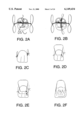
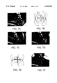
- FIG. 1 are drawings of a tooth illustrating the particular suturing procedures used in the method of the present invention.
- FIG. 2 are additional drawings of a tooth illustrating the particular suturing procedures used in the method of the present invention.
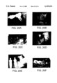
- FIG. 3 are photographs of teeth illustrating the method of the present invention displaying the presurgical X-ray, preliminary treatment and the beginning of the extraction-replantation; i.e., mobilization of the tooth to be transplanted. A picture illustrating the suture technique utilized in this part of the procedure is also shown.
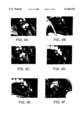
- FIG. 4. are photographs of teeth illustrating the method of the present invention displaying part of the extraction-replantation; i.e., mobilization of the tooth to be transplanted.
- FIG. 5 are photographs of teeth illustrating the method of the present invention displaying the completion of the extraction and mobilization of the tooth to be transplanted. Also shown in this Figure is the beginning of the tooth transplantation process.
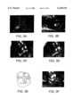
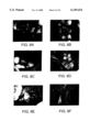
- FIG. 6 are photographs of teeth illustrating the method of the present invention displaying part of the tooth transplantation process.
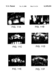
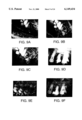
- FIG. 7 are photographs of teeth illustrating the method of the present invention displaying the completion of the tooth transplantation process. Pictures illustrating the suture technique utilized in this part of the procedure are also shown.
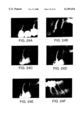
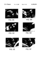
- FIG. 8 are photographs of the various sutures used in the method of the present invention, as well as photographs of the status of the transplanted tooth thirty days after transplantation.
- FIG. 9 are follow-up photographs and X-rays of teeth after completing the method of the present invention. Radiography showed a complete adaptation of the bone alveolus to the root having a normal periodontal ligament width.
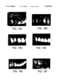
- FIG. 10 are photographs of teeth showing a specific embodiment using the method of the present invention wherein the patient had undergone a traumatic extraction of one tooth and further orthodontia treatment was needed and undertaken on the transplanted tooth.
- FIG. 11 are photographs and X-rays of teeth illustrating the orthodontia treatment after transplantation using the method of the present invention shown in FIG. 10.
- FIG. 12 are photographs and X-rays of teeth showing another specific embodiment using the method of the present invention wherein the patient had lost a tooth due to root fracture and the replacement tooth was non-functional.
- FIG. 13 are photographs of teeth after transplantation using the method shown in FIG. 12 in which coronal restitution was additionally performed.
- FIG. 14 are follow-up photographs and X-rays of teeth showing the results of the present invention using the method shown in FIGS. 12 and 13.
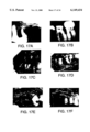
- FIG. 15 are photographs and X-rays of teeth showing another specific embodiment using the method of the present invention in which the tooth transplanted by the method of the present invention was later used as one of five abutments to anchor a bridge.
- FIG. 16 are follow-up X-rays of teeth after the bridge was anchored on the transplanted tooth using the method shown in FIG. 15.
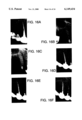
- FIG. 17 are photographs and X-rays of teeth showing another specific embodiment using the method of the present invention in which the alveolus was modified and two molar half's of a tooth were transplanted.
- FIG. 18 are photographs and X-rays of teeth illustrating the suturing procedure, coronal restoration and follow-up radiographs after the transplantation of two molar half's of a tooth using the method shown in FIG. 17.
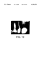
- FIG. 19 is an X-ray showing normal PDL space after the transplantation of two molar half's of a tooth using the method of the present invention shown in FIG. 17.
- FIG. 20 are photographs and X-rays of teeth showing yet another specific embodiment using the method of the present invention in which the tooth transplanted by the method had complete alveolar bone resorption following furcal invasion and follow-up radiographs.
- FIG. 21 are photographs and X-rays of teeth demonstrating the healing capacity of the stimulated PDL in a very old patient using the method as shown in FIG. 20.
- FIG. 22 are photographs and X-rays of teeth showing another specific embodiment using the method of the present invention in which the residual bone between the lesion and sinus was completely destroyed and two parts of the tooth is being transplanted. The suturing procedure for this type of procedure is also illustrated.
- FIG. 23 are photographs of teeth illustrating another embodiment using the method of the present invention on teeth in which two parts of a tooth are transplanted and coronal restoration is performed.
- FIG. 24 are follow-up X-rays of teeth illustrating that the method of the present invention shown in FIGS. 22 and 23 has the capacity to stimulate PDL and regenerate completely destroyed bone.
- tooth used in the singular also encompasses more than one tooth and encompasses natural mature teeth, retained teeth, part of one or more tooth (one root) and artificial teeth, including non osseo-integrated dental implants with or without a temporary crown. Any type of tooth can be used in the method of the present invention including molars, incisors, premolars and canines.
- Examples of the types of artificial teeth that can be used in the method of the present invention include, but are not limited to, those described in WO 97/45533, EP 0 734 712, Patent Abstracts of Japan, vol. 14 no. 086 (C-690 (1990) and JP 01 299563 by Moritsuga, Ootori et al (1989), as well as those described in Hanes et al, Journal of Periodontology, vol. 60, no. 4 pgs. 188-198 (1989).
- a large quantity of fibroblasts means the proliferative induction of PDL cells when a healing repair occurs after breaking in the middle of the periodontal ligament by mobilization (extraction and immediate replantation) of a tooth. Thus upon healing repair three to ten times the normal amount of fibroblasts are regenerated.
- the term "mechanical stimulation on the periodontal ligament” means that occlusal pressure under physiologic conditions (i.e., mastication, swallowing and the like) induce limited and episodic movements of the teeth in the alveolus. These movements are limited by the tightening of the PDL fibers and the sutures. The alveolus can be ectopic.
- the present invention was based on the discovery that reducing ankylosis and root resorption after transplantation of a tooth, can be achieved when the tooth of interest is transplanted with a stimulated periodontal ligament. This is accomplished by creating a trauma to the PDL of the tooth to be transplanted prior to transplantation and maintaining the tooth under a certain amount of non-rigidity, which creates a mechanical stimulation of the periodontal ligament.
- creating a trauma to the PDL is meant that the tooth is mobilized to disrupt the PDL via extraction-immediate replantation. It is the sole method to provoke trauma and the healing process on the entire surface of the PDL. If the tooth is an artificial tooth (implant) the stimulated PDL is obtained by an organotypic culture.
- organotypic culture is described, for instance, in WO97/45533.
- this waiting period should be of sufficient time to permit the stimulation of a large quantity of fibroblasts and is between about 5 to 30 days, preferably about 15 days.
- the fibroblasts generated in this manner can also be aspirated and used in cell culture, tests or grafts if so desired. It will be appreciated that in this instance, the tooth is not further transplanted.
- the tooth is then transplanted using known dentosurgical techniques.
- this method entails five phases, which are the preparation of the tooth to be transplanted and the preparation of the receiving alveolus, a transplantation phase, a post surgical check, a temporary crown restoration and the completion of a final crown.
- a patient Prior to beginning this procedure a patient is generally subjected to a general check up that includes an evaluation of the patient's general status, the buccal status of the patient and the type of tooth which needs to be replaced, which can be either a missing tooth or a tooth that cannot be preserved any longer.
- the mouth of the patient is searched to see whether there is a non-functional tooth or a root of a non-functional tooth that can be adapted to the tooth that will be replaced.
- the tooth that will be transplanted can also be a retained tooth or an artificial tooth. In some cases a functional tooth can be used depending on the therapeutic strategy for a better buccal rehabilitation.
- the endodontic treatment is done beforehand, for example, a week before.
- the tooth is filled with a final filling or alternatively with a Ca(OH) 2 filling. This last option has no influence on the protocol outcome. If the tooth is retained, endodontic treatment will be started approximately 3 weeks after transplantation and finalized when the tooth has achieved sufficient stability.
- an occlusal mesio-distal groove for stabilizing the sutures and a coronoplasty on the transplanted tooth is performed (FIG. 1 (1)).
- the tooth must be at a minimum of 1 mm under occlusal contact if any occlusal contact in fact exists. If only one root is used hemisection is performed before mobilization.
- the tooth to be transplanted or the dental section to be transplanted is then extracted.
- a supra-crestal incision is made with a surgical blade No. 12 to free up gingival attachment.
- a forceps adapted to the crown can be used to avoid damaging the root surface. Under no circumstances should syndesmotomes and elevators be used which damage cement and desmodontal fibers. Forceps should be used to break the alveolus-tooth ligament by small progressive rotations and rocking movements.
- the root is rapidly measured in length and at its maximal and minimal diameters are taken with a periodontal probe. The total length is also measured.
- the root must be kept moistened by blood to maintain the vitality of ligament cells and the cementoblasts present at the root surface.
- the tooth is then put back in the alveolar site as quickly as possible; i.e., immediately. A suture thread going through the groove (see below) will maintain the tooth to the gum (FIG. 1 (3)).
- the tooth to be replaced is still in place, it is extracted and the alveolus is carefully curetted.
- the alveolus is then modified with a bone drill and bur to match the volume of the root to be transplanted.
- the alveolus must be larger than the root to be transplanted.
- the "play" must be at least one-millimeter.
- the root should never be forced on the alveolus wall when it is put in place. It is useful to control the alveolus size by placing the transplant into the alveolus for testing and checking. When the bone crest is too thin, a bone flap can be mobilized for enlargement of the receiving alveolus.
- the alveolus must be entirely created with graduated implantology drills and shaped with a bone bur once the muco-periosteal flap has been elevated. The volume of the alveolus is controlled as discussed above and the flap is then stitched up.
- the alveolus is drilled down to the Schneiderian membrane then the flap is closed back.
- the transplantation surgery is performed from about 3 to 30 days, preferably about 15 days after the mobilization of the transplant and the adaptation or the creation of the receiving alveolus.
- the benefit of this time delay is to transplant the tooth with a stimulated desmodont that will contain a very large quantity of fibroblasts that are regenerating the tooth-alveolus ligament.
- the desmodont is entirely destroyed and replaced by granulation tissue before it starts to self-regenerate.
- the delay enables the extraction of the tooth for a second time with reduced trauma because 10 to 20% of the fibers will have regenerated and the root is covered with a new conjunctive tissue which is in a high growth stage (Mandel and Viidik, 1989).
- This stimulated tissue will regenerate the alveolus bone around the tooth in its transplanted site, whatever the bone status of this site.
- Retained teeth are accessed by opening up a flap and an osteotomy at the crown level is performed. They are then extracted without touching the root, measurements of the roots are taken and they are put back in place and the flap is closed. Pulpectomy is done about 3 weeks after transplantation.
- the bone alveolus is also prepared 15 days beforehand. During the transplantation surgery it is necessary to make sure that the alveolus does not contain any epithelial tissue or any necrotic tissue and that the gingival edge can be joined to the cervical surface of the transplanted tooth.
- the alveolus should be made such that the major axis of root section is in the direction of the mesio-distal line. This is often required when a tooth or a molar root is to be placed on a narrow bone crest. For example, an upper premolar to be transplanted on a distal wedge.
- a modified or non-modified alveolus is used in the transplant process, consequent to the extraction done 15 days before hand, the stitches joining the gingival edges are removed.
- the gingival opening has been narrowed during the 15 days of healing, thus facilitating the adaptation between the tooth and the gum.
- a superficial zone of the alveolus is curetted to remove epithelial tissue migrating inside the alveolar.
- a deeper zone is curetted to eliminate healing tissue in the center, while the inner walls are curetted softly.
- the stitches are removed, the flaps are opened with a periosteal elevator and the alveolus is curetted softly.
- the stitches are removed, the flaps are opened with a periosteal elevator and the alveolus is curetted softly.
- the healing process of the alveolus after 15 days creates a thickening of the sinus mucosal membrane and a plug of healing tissue in the bottom of the bone alveolus is generally found.
- This plug is gently pushed back in the sinus and the sinus mucous membrane is pulled away over 5 to 10 mm around the alveolus with the use of special sinus membrane elevators.
- the transplant must be able to push back the mucosal membrane without creating any tension on the membrane. It is essential that the mucous does not get punctured to avoid any risk of contamination.
- the space between the mucous membrane and bone cortical will be colonized by healing bone tissue.
- the transplant which as been mobilized 15 days before hand, is handled with forceps, without touching its roots, and extracted carefully. A small proportion of connective fibers is reformed and the tooth is extracted easily and with minor trauma.
- the tooth is immediately put in the prepared alveolus, keeping the orientation as planned previously, to best adapt its emerging profile to the root profile, and the root surface covered with a desmodont to the available gingival profile.
- the flap edge is held with a rongeur and the flap is adapted to the tooth contour with a surgical blade No. 12.
- the junction between the flap edge and the tooth must be as sealed as soon as possible. The tooth can be put back in its original location for a short time when the flap is cut.
- the transplanted tooth Prior to suturing the transplanted tooth, it must be ensured that the transplant is at least 1 mm below the occlusal contact. If there is contact, the occlusal groove can be deepened to make sure that the stitches will stay below the occlusion.
- the 1 mm gap prevents the transplant from making excessive occlusal contact.
- the transplant is always held in place by sutures and never with a splint connected to other teeth. This gives the tooth some mobility, which will favor ligament growth and inhibit bone tissue development.
- a rigid splint transmits strain to other teeth, blocks the functional stimulation of fibroblasts and favors the growth of the bone tissue resulting in tooth ankylosis. Thus, the use of a rigid splint should be avoided.
- Rivalry between the ligament and bone tissue during the healing process must be managed during all of the transplantation protocol because factors generating an ankylosis appear within less then one hour after the tooth has been transplanted and not months or years later.
- a first suture brings together the gingival papilla or flap edges on the distal side of the tooth. Both sutures are cut at a distance of approximately 40 to 50 mm from the suture knot (FIG. 1(2)).
- a second simple suture has the same role on the mesial side of the tooth. Suture threads should be left approximately 15 cm long for future use (FIG. 1(2)).
- One of the mesial suture threads is knotted with one of the distal threads going across the occlusal groove. The same is done for the remaining suture threads (FIG. 1(3)).
- the groove is not mesio-distal oriented but rather bucco-lingual. For example, when an upper premolar is moved to a narrow crest at the lower molar level, the tooth is then positioned with a rotation of 90°. Suture threads are set out in a configuration forming a figure "8", going around the cuspids and in the groove (FIG. 1(4)).
- FIG. 1(5) For molars, two grooves, one mesio-distal and one bucco-lingual, are made (FIG. 1(5)).
- the needle On the mesial suture, the needle is retained for the next step (FIG. 1(6)) after the occlusal knot is made in the mesio-distal groove with a thread of the distal suture (FIG. 2(1)).
- the needle passes through the lingual (or palatal) gum, and then by the buccal gum and a stitch in the occlusal groove is made with the second thread of the last knot (FIG. 2(2)).
- a suture thread is bonded in the middle of the buccal face of the tooth crown with a composite, then stitched in the buccal gum twice, mesially and distally, and finally knotted (FIG. 2(4)).
- a second thread is bonded on the buccal face (FIG. 2(5)), but goes along the palate side where it is stitched in the fibromucosal twice using mattress suture (FIG. 2(6)). The tooth is thus stabilized in bucco-lingual orientation.
- Occlusion is checked one more time and teeth are altered if necessary to allow a space of approximately 1 mm under occlusal contact. Because the sutures stay inside the occlusion groove, or on the buccal side of anterior teeth, there are no obstacles to occlusion adjustments.
- Suturing can be done on the area where the tooth has been removed.
- a circular thread on the cemento-enamel junction going above the other thread stitches prevents the tooth from sinking.
- Antibiotic therapy starts one day before the mobilization of the tooth, and is administered during a period of four weeks to prevent bacterial proliferation in surgical areas.
- methacycline 300 mg Lysocline®
- analgesics such as Diantalvic (dextropropoxyphene and acetominophen (Tylenol®)
- the mouth can be rinsed with Chlorhexidine at 0.2% for 1 minute twice a day for 45 days.
- Piascledine a mixture of avocado and soybean
- Post surgical controls are then performed 7 days after transplantation. These controls include an occlusion check, a suture stability check, a flap sealing check and supra-gingival cleaning with an ultrasound scaler and an antiseptic such as H 2 O 2 at about 0.02% volume. No curette nor air-polisher is used at this point in the procedure.
- the crown can be remolded with a composite to obtain an adequate morphology, in particular with occlusion points, a cosmetic buccal side and proximal contact points. Occlusal contact points during excursive movements have to be totally avoided. No heavy pressure should be applied to the tooth at this stage. A dam can be installed if the clamp is fixed on a more distal tooth.
- the alveolus was too wide or if the tooth is located on a sinus, it can be rebuilt 4 to 8 weeks after transplantation. If many teeth or roots are transplanted at the same time to a site with a major bone deficit, it is possible to join them together in order to increase their stability. This is accomplished always with a bonded composite (FIG. 24(3)).
- the transplanted tooth can be crowned or be used as a bridge pillar. It is preferable to wait a little longer with a composite temporary crown.
- Patient 1 was subjected to a general checkup and X-rays were taken of the teeth in the mouth. From the X-rays it was determined that the first molar in Patient 1 had a deep carious lesion and a half root length furcation lesion and thus had to be replaced. The third molar, which is nonfunctional and in healthy condition was chosen for transplantation (FIG. 3(1)). Preliminary treatment was started and scaling and root planning was completed, particularly on the tooth to be transplanted (FIG. 3(2)).
- the transplanted tooth needed additional endodontic treatment and this was performed by filling the 3rd molar with Ca(OH) 2 .
- the patient was then treated with antibiotics, analgesics and was requested to rinse the mouth twice a day with 0.2% Chlorohexydine for the next five weeks.
- Patient 1 returned for mobilization of the tooth to be transplanted.
- the cuspids were reduced to avoid occlusal lateral stress after the transplantation (FIG. 3(3)).
- a mesio-distal occlusal groove was then created on the third molar of the tooth to be transplanted.
- a second bucco-lingual groove was also made (FIG. 3(4)). When the groove was made it was at least 2 mm under occlusal contact after the transplantation so that the sutures will not cut if occlusal adjustments were needed (FIG. 3(5)).
- Supra-crestal incision with a surgical blade no. 12 was then made around the donor tooth to be replaced to cut supracrestal gingival fibers (FIG. 3(6)). The same incision separates the safe tissue from the pathologic tissue (granulation tissue of the periodontal lesion) around the tooth to be replaced (FIG. 4(1)).
- the roots of the first molar which was diseased were separated with a surgical bur and saline irrigation was performed throughout this procedure (FIG. 4(2)).
- the roots of the first molar were extracted carefully to preserve the osseous wall of the receiving alveolus (FIG. 4(3)).
- the receiving alveolus was carefully curetted to suppress all granulation tissue (FIG. 4(4)). Since the third molar had only one root, the receiving alveolus was modified with a surgical bur under saline irrigation to suppress interradicular septa (FIG. 4(5)).
- the third molar was then extracted with forceps (when possible the forceps were modified with small latex cushions to avoid fracture of the tooth).
- the tooth was excised by using small, progressive rotary and rocking movements (FIG. 4(6)).
- the transplant tooth was then placed in the receiving alveolus. Only the PDL surface was under the gingiva. If the alveolus is too small, it can be modified again with the bur (FIG. 5(1)).
- the tooth to be transplanted was then placed in the alveolar site of origin and to retain the tooth, a mesio-distal suture was placed in the groove of the tooth (FIG. 5(2)). Sutures were then placed that drew the gingival edges nearer around the receiving alveolus during the first healing period (FIG. 5(3).
- the tooth to be transplanted was then re-extracted from the alveolar of origin with minimal trauma (FIG. 6(3)).
- the forceps did not touch the vital ligament on the root transplant when re-extracted (FIG. 6(4)).
- the transplanted tooth was then immediately placed in the receiving alveolar site that was previously prepared keeping the orientation as previously planned (FIG. 6(5)).
- the flap edge was adapted to the transplanted tooth contour with a new incision being made.
- a mesial suture that brought together the gingival papilla on the mesial side of the tooth was then performed (FIG. 7(1)). Both suture stitches were cut at a minimal length of 40 mm on the distal side and at a length of approximately 120 mm on the mesial side (FIG. 7(2)). One of the mesial suture threads was then knotted with one of the distal threads going across the occlusal groove (FIG. 7(3)). A second knot was then made with the other threads (FIG. 7(4)). A bucco lingual suture was then realized from the central occlusal knot (FIG. 7(5)). The donor alveolus was then closed by suturing (FIG. 7(6)).
- FIG. 8(1) illustrates a buccal view of the sutures on Patient 1.
- FIG. 8(2) illustrates an additional palatal suture which stabilized the transplant in excellent alignment with the other teeth. An occlusal adjustment was made to avoid any occlusal contact on the transplant (FIG. 8(3)).
- Patient 1 returned to have a follow up 15 days after the tooth was transplanted. At this time the sutures were removed and the area surrounding the transplanted tooth was cleaned ultrasonically and checked for occlusal control (FIG. 8(4)). The buccal view of the transplanted tooth was at 30 days (FIG. 8(5)) illustrated very good healing of the gingival (FIG. 8(5)). An X-ray was then taken and the space between the root and alveolus was noted (FIG. 8(6)).
- Patient 2 (15 years old) had undergone a traumatic extraction of his tooth #8 (FIG. 10(1)). Reimplantation could not be performed since the incisive was not found within the required time to preserve the vital PDL.
- Tooth #13 was chosen for replacement of tooth #8 (FIG. 10(2)).
- the steps of the transplantation protocol were the same as in Example 1; i.e., endodontic treatment with a Ca(OH) 2 filling; the mobilization of tooth #13; the adaptation of the alveolus in site of tooth #8; a 15 day healing period; and the coronal reduction of tooth #13 to match the coronal volume of the central incisor.
- FIG. 10(3) shows the transplanted tooth #13 in which the interpapillary stitch pressed the gingival edge which provides the best contact with the cemento-enamel junction.
- a suture thread was bonded horizontally on the buccal side of the crown with a composite resin. This suture was anchored by two steps of mattress stitch in the buccal gum (FIG. 10(4)) and knotted. A second thread was bonded on the buccal side (FIG. 10(5)) and was anchored on the palatal mucosa in the same manner (FIG. 10(6)).
- FIG. 11(2) A radiographic comparison (FIG. 11(2)) showed alveolus adaptation, inter-radicular bone regeneration and the absence of root resorption during the first 12 months.
- FIG. 11(5) illustrates that the buccal gum outline found its past proximal design. A further correction should be performed before definitive coronal restoration.
- FIG. 13(2) 14 days after transplantation, the healing was very good and permitted coronal reconstitution immediately (FIG. 13(2)).
- a dental dam was placed with a clamp on tooth #4 (FIG. 13(3)) and a composite coronal reconstitution was performed as shown in FIG. 13(4) and FIG. 13(5).
- the mesial part of tooth #31 was left in place.
- FIG. 14(1) One month post-op (FIG. 14(1)), gingival healing was good and the transplanted tooth was functional.
- FIG. 14(2) shows a radiography performed just after the transplantation.
- FIG. 14(3) shows a radiography 24 months later. No ankylose-resorption was distinguishable.
- FIGS. 14(4) and 14(5) showed the tooth 24 months after transplantation.
- Tooth #6 had a vertical root fracture with deep bone resorption due to infection and abscesses (FIG. 15(1)+(2)). This tooth had to be replaced.
- FIG. 15(3) shows a radiography of tooth #11 retained in the palate. This tooth was chosen for replacement of tooth #6. It was mobilized and left 14 days in its site in the left palate. At the same time, tooth #6 was extracted and its alveolus was carefully curetted.
- tooth #11 was re-extracted and put in place of tooth #6 and retained only by sutures (FIG. 15(4)).
- FIG. 16 shows the radiographic follow up
- FIG. 16(1) tooth #6 before extraction
- FIG. 16(2): t +6 months
- FIG. 16(4): t +24 months
- FIG. 16(5): t +3.5 years
- Patient 5 was a 60 year old female. She had lost all of her upper left molars. The residual mesial root of tooth #14 had a deep periodontal lesion and had to be extracted (FIG. 17(1)).
- Tooth #18 was used for transplantation (FIG. 17(2)+(3)). Endodontic treatment, hemisection and mobilization of each tooth was performed as shown in FIG. 17(4). During the same appointment, the alveolus was created in the upper maxilla with bone burs. The form of the osseus crest was used to its best advantage during this procedure.
- the crest was narrower, and the distal alveolus was created with the largest axis in a mesio-distal direction for the mesial root of tooth #18.
- the size of each alveolus was controlled by placing its respective transplanted each half-tooth #18 until any forced contact would be suppressed between the root and the bone for the tooth's best position as illustrated in FIG. 17(5).
- Final orientation of the roots in their destined alveolus was recorded in the patient's file for the next appointment. The upper flap was then sutured as illustrated in FIG. 17(6).
- the two half molars were transplanted in the upper alveolus. Sutures were made similar to those illustrated in FIGS. 1(3) and 1(4). On the distal half tooth, the sutures were set out in a configuration forming a FIG. 8 as illustrated in FIG. 18(1+2).
- Coronal reconstitution was made with a composite resin placed jointly between the two parts as shown in FIG. 18(4). At this time occlusal contact was avoided. After 45 days, the new composite bonding had normal occlusal contacts.
- FIG. 18(5) shows a radiography of the transplanted tooth at day 15.
- FIG. 18(6) shows the buccal view at day 120.
- FIG. 19(1) shows the radiography at day 120, before definitive crown restoration. The normal PDL space should be noted.
- Patient 6 was a 59 year old male. His tooth #18 had a complete alveolar bone resorption following furcal invasion as shown in FIG. 20(1). This tooth had to be replaced. Tooth #1 (FIG. 20(2)) had been chosen for the transplantation.
- Double PDL stimulation protocol was again performed for transplantation. Since the alveolus was too large, the extraction of tooth #18 was performed 21 days before transplantation and the alveolus was carefully curetted to reduce infections sequelae.
- FIG. 20(3) shows the suturing technique of the transplanted tooth. An additional bucco-lingual stitch was required to adjust the gingival edge level.
- FIG. 20(4) shows a radiography at day 21.
- the canal was filled with Ca(OH) 2 .
- FIG. 20(5) shows complete bone repair at day 180. Two titanium implants were used to replace teeth #19 and #20.
- FIG. 20(6) shows the clinical condition at day 180. The periodontal state was excellent.
- transplantation can be performed even if the receiver site has a large bone defect, while still promoting bone regeneration.
- Teeth #3 and #5 which were bridge abutments showed deep periodontal lesions and had to be extracted (FIG. 21(1)). Tooth #32 (FIG. 21(2)) was used for transplantation into the site of the upper right premolars.
- FIG. 21(3+4) show a buccal and a lingual view at month 15. A vertical groove was performed on the buccal coronal side to look like two premolars.
- FIG. 21(5) shows a radiography at day 120.
- FIG. 21(6) shows a radiography at month 14. Bone regeneration was clearly seen.
- This example illustrates the healing capacity of stimulated PDL in a very old patient.
- Patient 8 was a 52 year old female.
- Her tooth #13 had a very large periodontal and apical lesion (FIG. 22(1)). Tooth #17, also a bridge abutment, had a deep periodontal pocket. The residual bone between the lesion and the sinus was less than a millimeter thick (FIG. 22(2)).
- FIG. 22(3) illustrates the clinical condition after the extraction of teeth #13 and #17 and a curettage.
- Tooth #18 (FIG. 22(4)) had very long roots and was used for the transplantation.
- FIG. 24 shows the follow-up with a radiographic comparison:
- FIG. 24(1) before extraction of teeth #13 and #15. Note the bone resorption around tooth #13 and the thickness of the bone in the site of tooth #14.
- FIG. 24(2) day 30.
- FIG. 24(3) day 72: bone regeneration around the roots was visible.
- the bone level was identical with its level on the roots before transplantations shown in FIG. 24(6).
- This example shows the capacity of stimulated PDL to regenerate bone even if it was completely destroyed.
- transplanted teeth has shown zero ankylosis and resorption phenomenon when using the method of the present invention.
- the method of transplantation described in the present invention can be used every time there is a non-functional tooth or a root and can be adapted to the replacement of a functional tooth. Furthermore, with osteo-integrated implants, transplantation has no harmful effect on side teeth.
- the method of the present invention gives a fully functional result after two to eight weeks and is particularly well adapted for the replacement of teeth suffering from deep periodontal lesion, severe furcation invasions and root fracture, even when the bone alveolus has been strongly damaged.
- the present invention also provides a method to regenerate a severely damaged bone because the stimulated desmodont has the potential to regenerate the alveolus bone around the transplanted tooth as well. Moreover, there is no attachment loss compared to the attachment of the tooth in its origin site.
- This technique enables the reimplantation of retained teeth, including adults with an atrophied desmodont.
- This study showed excellent global results; i.e., at about a 97% success rate.
- tooth-alveolus ligament transmits occlusal strains in a lot more physiological manner than osteo-integration implants.
- Trope M et al. "The role of the socket in the periodontal healing of replanted dogs' teeth stored in ViaSpan for extended periods.” Endod Dent Traumatol. 1997 Aug;13(4):171-5.
- Trope M et al. "The role of the socket in the periodontal healing of replanted dogs' teeth stored in ViaSpan for extended periods.” Endod Dent Traumatol. 1997 Aug;13(4):171-5.
Abstract
Description
Claims (24)
Priority Applications (1)
| Application Number | Priority Date | Filing Date | Title |
|---|---|---|---|
| US09/398,467 US6149434A (en) | 1999-09-17 | 1999-09-17 | Method for autogenous transplantation of human and animal teeth that eliminates the risk of ankylosis and root resorption |
Applications Claiming Priority (1)
| Application Number | Priority Date | Filing Date | Title |
|---|---|---|---|
| US09/398,467 US6149434A (en) | 1999-09-17 | 1999-09-17 | Method for autogenous transplantation of human and animal teeth that eliminates the risk of ankylosis and root resorption |
Publications (1)
| Publication Number | Publication Date |
|---|---|
| US6149434A true US6149434A (en) | 2000-11-21 |
Family
ID=23575480
Family Applications (1)
| Application Number | Title | Priority Date | Filing Date |
|---|---|---|---|
| US09/398,467 Expired - Fee Related US6149434A (en) | 1999-09-17 | 1999-09-17 | Method for autogenous transplantation of human and animal teeth that eliminates the risk of ankylosis and root resorption |
Country Status (1)
| Country | Link |
|---|---|
| US (1) | US6149434A (en) |
Cited By (12)
| Publication number | Priority date | Publication date | Assignee | Title |
|---|---|---|---|---|
| US20040102782A1 (en) * | 1999-12-21 | 2004-05-27 | Tomaso Vercellotti | Surgical device and method for bone surgery |
| US20060057542A1 (en) * | 2004-09-02 | 2006-03-16 | Odontis Ltd. | Bone regeneration |
| US20070015102A1 (en) * | 2005-06-21 | 2007-01-18 | Tomaso Vercellotti | Implant site preparation method and relative piezoelectric surgical device |
| US20080206709A1 (en) * | 2007-02-27 | 2008-08-28 | Lannan William G | Gingival support sleeve |
| WO2011163577A1 (en) * | 2010-06-24 | 2011-12-29 | The Board Of Trustees Of The University Of Illinois | Reagents and methods for preparing teeth for implantation |
| US8857442B1 (en) | 2012-03-05 | 2014-10-14 | Gloria A. Ospina | High lip-line smile corrective surgical method |
| RU2748959C1 (en) * | 2020-01-30 | 2021-06-02 | Федеральное государственное бюджетное образовательное учреждение высшего образования "Пензенский государственный университет" | Method for directed bone regeneration used in surgery of complex configuration defects |
| RU2750275C1 (en) * | 2020-09-07 | 2021-06-25 | Общество с ограниченной ответственностью "Профессорская клиника Едранова" | Method of forming a gingival cuff in the dental implant area from autogenous tissue of the patient attached on the bone |
| RU2760988C1 (en) * | 2021-03-23 | 2021-12-02 | Федеральное государственное бюджетное образовательное учреждение дополнительного профессионального образования "Российская медицинская академия непрерывного профессионального образования" Министерства здравоохранения Российской Федерации (ФГБОУ ДПО РМАНПО Минздрава России) | Method for bone augmentation for eliminating defects of upper jaw during dental implantation |
| RU2766977C1 (en) * | 2021-01-13 | 2022-03-16 | Общество с ограниченной ответственностью "Медлайн Компани" | Method for stacking and stabilising granular osteoplastic materials in the recipient bed when eliminating complex defects of the jaw bones |
| RU2771335C1 (en) * | 2021-06-07 | 2022-04-29 | Общество с ограниченной ответственностью "Профессорская клиника Едранова" | Method for vestibuloplasty with suprabony immobilization of a free gingival autograft |
| RU2816035C1 (en) * | 2023-09-25 | 2024-03-25 | федеральное государственное бюджетное образовательное учреждение высшего образования "Башкирский государственный медицинский университет" Министерства здравоохранения Российской Федерации | Method for integrated treatment of gum recession |
Citations (4)
| Publication number | Priority date | Publication date | Assignee | Title |
|---|---|---|---|---|
| US5292253A (en) * | 1992-06-22 | 1994-03-08 | Laser Medical Technology, Inc. | Method for repairing tooth and bone tissue |
| US5455041A (en) * | 1993-09-13 | 1995-10-03 | Research Foundation Of State University Of New York At Buffalo | Method for inducing periodontal tissue regeneration |
| US5674074A (en) * | 1996-06-13 | 1997-10-07 | Angelo, Jr.; Patrick J. | Periodontal procedure |
| US5695338A (en) * | 1992-09-15 | 1997-12-09 | Robert; Antoine | Expansion device for oral reconstruction and method for the installation of the expansion device under a patient's gum in order to carry out oral reconstruction in cases of bone loss |
-
1999
- 1999-09-17 US US09/398,467 patent/US6149434A/en not_active Expired - Fee Related
Patent Citations (4)
| Publication number | Priority date | Publication date | Assignee | Title |
|---|---|---|---|---|
| US5292253A (en) * | 1992-06-22 | 1994-03-08 | Laser Medical Technology, Inc. | Method for repairing tooth and bone tissue |
| US5695338A (en) * | 1992-09-15 | 1997-12-09 | Robert; Antoine | Expansion device for oral reconstruction and method for the installation of the expansion device under a patient's gum in order to carry out oral reconstruction in cases of bone loss |
| US5455041A (en) * | 1993-09-13 | 1995-10-03 | Research Foundation Of State University Of New York At Buffalo | Method for inducing periodontal tissue regeneration |
| US5674074A (en) * | 1996-06-13 | 1997-10-07 | Angelo, Jr.; Patrick J. | Periodontal procedure |
Cited By (18)
| Publication number | Priority date | Publication date | Assignee | Title |
|---|---|---|---|---|
| US20090024118A1 (en) * | 1999-12-21 | 2009-01-22 | Piezosurgery Inc. | Surgical Device For Bone Surgery |
| US8002783B2 (en) | 1999-12-21 | 2011-08-23 | Piezosurgery, Inc. | Surgical device for bone surgery |
| US20040102782A1 (en) * | 1999-12-21 | 2004-05-27 | Tomaso Vercellotti | Surgical device and method for bone surgery |
| US20060057542A1 (en) * | 2004-09-02 | 2006-03-16 | Odontis Ltd. | Bone regeneration |
| US7497686B2 (en) * | 2004-09-02 | 2009-03-03 | Odontis Ltd. | Bone regeneration |
| US8808295B2 (en) | 2005-06-21 | 2014-08-19 | Tomaso Vercellotti | Insert for a handheld ultrasound surgical device |
| US20070015102A1 (en) * | 2005-06-21 | 2007-01-18 | Tomaso Vercellotti | Implant site preparation method and relative piezoelectric surgical device |
| US20090136894A1 (en) * | 2005-06-21 | 2009-05-28 | Vercellotti Tomaso | Insert For A Handheld Ultrasound Surgical Device |
| US8109931B2 (en) | 2005-06-21 | 2012-02-07 | Piezosurgery, Inc. | Implant site preparation method and relative piezoelectric surgical device |
| US20080206709A1 (en) * | 2007-02-27 | 2008-08-28 | Lannan William G | Gingival support sleeve |
| WO2011163577A1 (en) * | 2010-06-24 | 2011-12-29 | The Board Of Trustees Of The University Of Illinois | Reagents and methods for preparing teeth for implantation |
| US8857442B1 (en) | 2012-03-05 | 2014-10-14 | Gloria A. Ospina | High lip-line smile corrective surgical method |
| RU2748959C1 (en) * | 2020-01-30 | 2021-06-02 | Федеральное государственное бюджетное образовательное учреждение высшего образования "Пензенский государственный университет" | Method for directed bone regeneration used in surgery of complex configuration defects |
| RU2750275C1 (en) * | 2020-09-07 | 2021-06-25 | Общество с ограниченной ответственностью "Профессорская клиника Едранова" | Method of forming a gingival cuff in the dental implant area from autogenous tissue of the patient attached on the bone |
| RU2766977C1 (en) * | 2021-01-13 | 2022-03-16 | Общество с ограниченной ответственностью "Медлайн Компани" | Method for stacking and stabilising granular osteoplastic materials in the recipient bed when eliminating complex defects of the jaw bones |
| RU2760988C1 (en) * | 2021-03-23 | 2021-12-02 | Федеральное государственное бюджетное образовательное учреждение дополнительного профессионального образования "Российская медицинская академия непрерывного профессионального образования" Министерства здравоохранения Российской Федерации (ФГБОУ ДПО РМАНПО Минздрава России) | Method for bone augmentation for eliminating defects of upper jaw during dental implantation |
| RU2771335C1 (en) * | 2021-06-07 | 2022-04-29 | Общество с ограниченной ответственностью "Профессорская клиника Едранова" | Method for vestibuloplasty with suprabony immobilization of a free gingival autograft |
| RU2816035C1 (en) * | 2023-09-25 | 2024-03-25 | федеральное государственное бюджетное образовательное учреждение высшего образования "Башкирский государственный медицинский университет" Министерства здравоохранения Российской Федерации | Method for integrated treatment of gum recession |
Similar Documents
| Publication | Publication Date | Title |
|---|---|---|
| Gluckman et al. | Partial Extraction Therapies (PET) Part 2: Procedures and Technical Aspects. | |
| Fugazzotto et al. | Sinus floor augmentation on at the time of maxillary molar extraction: Success and failure rates of 137 implants in function for up to 3 years | |
| Evian et al. | Autogenous gingival grafts as epithelial barriers for immediate implants | |
| Thalmair et al. | Coverage of multiple mandibular gingival recessions using tunnel technique with connective tissue graft: a prospective case series | |
| Gault et al. | Tooth auto‐transplantation with double periodontal ligament stimulation to replace periodontally compromised teeth | |
| US6149434A (en) | Method for autogenous transplantation of human and animal teeth that eliminates the risk of ankylosis and root resorption | |
| Kim et al. | A new approach using the surgical extrusion procedure as an alternative for the reestablishment of biologic width. | |
| Kang | Sinus elevation using a staged osteotome technique for site development prior to implant placement in sites with less than 5 mm of native bone: a case report. | |
| Vergara et al. | Immediate replacement of single upper posterior teeth: A report of cases | |
| Froum et al. | New surgical protocol to create interimplant papilla: the preliminary results of a case series | |
| Vergara et al. | Preservation of esthetics with implant dentistry: a clinical report | |
| Arora et al. | A comparative evaluation of immediate implant placement in fresh extraction socket with and without the use of platelet rich fibrin: A clinical and radiographic study | |
| El Zahwy et al. | Clinical and radiographic evaluation of dental implants penetrating the maxillary sinus | |
| Lazzara | Immediate placement of implants into extraction sites | |
| Tecimer et al. | The use of autogenous bone grafting to reconstruct a mandibular knife edge ridge before implant surgery: a case report | |
| Kasuga et al. | Journal of Dentistry And Oral Implants | |
| Taraphdar et al. | Efficacy of Socket Shield Technique for Immediate Implant Placement in Maxillary Anterior Region–A Case Series with Review. | |
| Alaa et al. | A Novel Approach for Maxillary Rehabilitation and Esthetic Problem Solving in Egyptian Patient-Case Report | |
| Hassanien et al. | A Novel Approach for full mouth rehabilitation and esthetic problem solving in Egyptian patient-case report | |
| Alaa et al. | MSA Dental Journal | |
| Turner et al. | Clinical guide to oral implantology: Step by step procedures | |
| Gabrić¹ et al. | Implant Rehabilitation of Internal Root Resorption after Dental Trauma | |
| Fien et al. | Guided Bone Regeneration: Novel Use of Fixation Screws as an Alternative to Using the Buccoapical Periosteum for Membrane Stabilization With Sutures—Two Case Reports | |
| Hütte | Tooth auto-transplantation–literature review: analysis of a method | |
| Tucker | Preprosthetic surgery |
Legal Events
| Date | Code | Title | Description |
|---|---|---|---|
| AS | Assignment |
Owner name: SOCIETE ANONYME NATURAL IMPLANT, FRANCE Free format text: ASSIGNMENT OF ASSIGNORS INTEREST;ASSIGNOR:GAULT, PHILIPPE;REEL/FRAME:010695/0914 Effective date: 19991017 |
|
| FPAY | Fee payment |
Year of fee payment: 4 |
|
| AS | Assignment |
Owner name: HENKEL KGAA, GERMANY Free format text: ASSIGNMENT OF ASSIGNORS INTEREST;ASSIGNOR:NATURAL IMPLANT SA;REEL/FRAME:016460/0431 Effective date: 20041118 |
|
| FEPP | Fee payment procedure |
Free format text: PAT HOLDER NO LONGER CLAIMS SMALL ENTITY STATUS, ENTITY STATUS SET TO UNDISCOUNTED (ORIGINAL EVENT CODE: STOL); ENTITY STATUS OF PATENT OWNER: LARGE ENTITY |
|
| REFU | Refund |
Free format text: REFUND - PAYMENT OF MAINTENANCE FEE, 8TH YR, SMALL ENTITY (ORIGINAL EVENT CODE: R2552); ENTITY STATUS OF PATENT OWNER: LARGE ENTITY |
|
| FPAY | Fee payment |
Year of fee payment: 8 |
|
| REMI | Maintenance fee reminder mailed | ||
| LAPS | Lapse for failure to pay maintenance fees | ||
| STCH | Information on status: patent discontinuation |
Free format text: PATENT EXPIRED DUE TO NONPAYMENT OF MAINTENANCE FEES UNDER 37 CFR 1.362 |
|
| FP | Lapsed due to failure to pay maintenance fee |
Effective date: 20121121 |