US6150174A - Method for measurement of whole blood coagulation parameters - Google Patents
Method for measurement of whole blood coagulation parameters Download PDFInfo
- Publication number
- US6150174A US6150174A US09/034,478 US3447898A US6150174A US 6150174 A US6150174 A US 6150174A US 3447898 A US3447898 A US 3447898A US 6150174 A US6150174 A US 6150174A
- Authority
- US
- United States
- Prior art keywords
- coagulation
- blood
- sample
- time
- electrodes
- Prior art date
- Legal status (The legal status is an assumption and is not a legal conclusion. Google has not performed a legal analysis and makes no representation as to the accuracy of the status listed.)
- Expired - Lifetime
Links
Images
Classifications
-
- G—PHYSICS
- G01—MEASURING; TESTING
- G01N—INVESTIGATING OR ANALYSING MATERIALS BY DETERMINING THEIR CHEMICAL OR PHYSICAL PROPERTIES
- G01N33/00—Investigating or analysing materials by specific methods not covered by groups G01N1/00 - G01N31/00
- G01N33/48—Biological material, e.g. blood, urine; Haemocytometers
- G01N33/50—Chemical analysis of biological material, e.g. blood, urine; Testing involving biospecific ligand binding methods; Immunological testing
- G01N33/86—Chemical analysis of biological material, e.g. blood, urine; Testing involving biospecific ligand binding methods; Immunological testing involving blood coagulating time or factors, or their receptors
-
- G—PHYSICS
- G01—MEASURING; TESTING
- G01N—INVESTIGATING OR ANALYSING MATERIALS BY DETERMINING THEIR CHEMICAL OR PHYSICAL PROPERTIES
- G01N33/00—Investigating or analysing materials by specific methods not covered by groups G01N1/00 - G01N31/00
- G01N33/48—Biological material, e.g. blood, urine; Haemocytometers
- G01N33/483—Physical analysis of biological material
- G01N33/487—Physical analysis of biological material of liquid biological material
- G01N33/49—Blood
- G01N33/4905—Determining clotting time of blood
Definitions
- the present invention relates to measuring and testing and, more particularly, to a method and apparatus for analyzing blood, particularly the coagulation characteristics of whole blood.
- Coagulation is the process of clotting, and the phrase "time of coagulation" generally means the time required for a small amount of blood to coagulate.
- the time of coagulation indicates the propensity of blood to coagulate. It can be determined, and was for many years, by collecting blood in a small container and merely observing elapsed time from the moment the sample was obtained to the time it coagulated. Clearly, this method was not very precise.
- Coagulation time may also be determined by collecting blood in a small capillary tube, then breaking off short pieces of the tube until threads of fibrin appear between the broken ends. Precision may still be questionable, but coagulation time measured by the latter method is normally six to seventeen minutes.
- U.S. Pat. No. 5,580,744 (Zweig) and U.S. Pat. No. 5,418,141 (Zweig et al.) disclose a test article and method for determining coagulation capability in a blood sample.
- the test article is a porous membrane having a coagulation initiator and substrate impregnated therein.
- blood is applied to one face of the membrane and plasma is absorbed into the interior of the membrane in the presence of the coagulation initiator and substrate.
- the membrane produces a detectable signal (i.e., a fluorescent signal) for reading by an automated detector or test system including a timer and means for calculating a coagulation value.
- the disclosed test article is said to be suitable for use in a home setting, as alluded to above, the Zweig test article requires that the plasma be separated from the sample.
- U.S. Pat. No. 4,547,735 discloses another example of known apparatus and methods for measuring and testing blood.
- the disclosed instrument includes two electrodes with which the conductivity of a sample may be measured.
- the sample contacting faces of the electrodes are placed in vertically separated horizontal planes in an accurately fixed spacing so a blood column of a given size is formed from a sample.
- Current acts on the sample and the instrument measures change in impedance to determine the hematocrit value of blood.
- the instrument is expressly designed to ignore or avoid the effect of sedimenting or aggregating.
- U.S. Pat. No. 5,601,995 discloses an apparatus and method for detecting coagulation.
- the method includes providing a porous sheet and applying a blood sample to the sheet so that the blood spreads through a part of the sheet.
- the spreading extent or spreading rate of the blood in the sheet is visualized, or measured by measuring electrical conductivity across the sheet, electrical potential across the sheet or an electrical resistance of the sheet.
- electrodes are provided on either side of the porous sheet.
- the conductivity or electrical impedance between the electrodes depends on the wetted area between them and, as a sample spreads through the porous sheet, impedance is reduced and conductivity increased, thereby indicating the extent of spread in the sheet. In theory, coagulation has occurred when the rate of change in conductivity/impedance approaches zero.
- the porous sheet may be affected by ambient conditions and handling, particularly severe problems if point of care use is attempted.
- U.S. Pat. No. 5,298,224 Patent Application Laidity
- U.S. Pat. No. 5,167,145 (Butler et al.) disclose apparatus for determining blood coagulation time using optical means.
- the Plum patent discloses the use of light and light detectors to measure blood transillumination and to determine coagulation time.
- the Butler et al. patent discloses the use of an infrared source and a photo detector to measure electromagnetic transmission and to determine coagulation time.
- the present invention provides a method of assessing the coagulation characteristics of blood.
- the method involves assessing a condition in a portion of a blood sample and relating the condition to the onset of coagulation.
- Apparatus for performing the method is encompassed.
- the apparatus includes an electrochemical cell wherein electrodes are positioned in close proximity at the bottom of the cell to make coagulation time measurements of a whole blood sample by measuring electrochemical resistance between the electrodes.
- the apparatus detects only resistance changes in the red blood cell fractions and, thus, measures resistance increases caused by the increase in number of red blood cells settling into the area of the electrical field as a function of time. By determining when resistance stops increasing, the onset of coagulation is determined and coagulation time can be determined and/or predicted.
- optically based measurements of the coagulation parameters are made by utilizing a wavelength that is absorbed by red blood cells.
- a light beam is positioned in an optical path into which red blood cells settle as a function of time.
- a clotting reagent may be introduced into a disposable electrochemical or optical cartridge or sample container by incorporating the reagent in the test fluid flow path where they are dissolved into the blood as the sample is introduced into the cartridge.
- the reagent may be contained in the sensor (and/or electrode) or light path chamber in a dried form and then dissolved into the sample with blood introduction.
- An advantage of such a portable analysis system is that it could be used in hospital settings to characterize patient condition before, during, and after operations where control of coagulation condition is vital to a successful outcome. Another advantage is that this system could be used in out patient settings, to evaluate patients on anti-coagulant therapies, such as treatment with Warfarin.
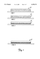
- FIG. 1 represents blood held in an exemplary sample chamber or container and illustrates the settling of red blood cells in that chamber or container at four different time intervals.
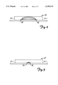
- FIG. 2 is a side view of a blood sample chamber that includes two spaced electrodes that create an electrical field that reaches the top of the sample chamber.
- FIG. 3 is a side view of a blood sample chamber that includes two closely spaced electrodes that create an electrical field limited to the bottom area of the sample chamber.
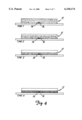
- FIG. 4 represents blood held in a sample chamber that includes two closely spaced electrodes and illustrates the relationship of the electrical field to the settling of red blood cells in the chamber at four different time intervals.
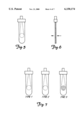
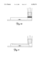
- FIG. 5 is a front view of an exemplary optical cell for use in performing an embodiment of the method of the present invention.
- FIG. 6 is a side view of the optical cell depicted in FIG. 5.
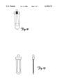
- FIG. 7 represents blood held in the optical cell and illustrates the settling of red blood cells in the chamber at three different time intervals.
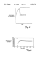
- FIG. 8 is a graph of the change in electrical resistance or light absorbance over time as blood coagulates.
- FIG. 9 depicts exemplary, experimentally derived results using the optical based coagulation detection method of the present invention.
- FIG. 10 depicts an exemplary mixing chamber for use with an electrochemical cell.
- FIG. 11 depicts an exemplary coated reagent sample container for use with an electrochemical cell.
- FIG. 12 depicts an exemplary mixing chamber for use with an optical cell.
- FIG. 13 depicts an exemplary coated reagent sample chamber for use with an optical cell.
- the present invention encompasses a method and apparatus to make coagulation time measurements of a whole blood sample, without separating out the plasma fraction, using an instrument with no moving mechanical parts.
- electrochemical cell is intended to mean a sample material on which electricity acts and at least two electrodes operably coupled to the sample material for communicating electricity to the sample material.
- Suitable circuit means wiring, integrated circuits, power source and the like
- control means a microprocessor, CPU or the like and related software
- sensor(s) is intended to encompass an electrode(s) or a combination of electrodes for analyte measurements, and is intended to be used interchangeably with electrode(s) in any electrochemical embodiment of the present invention.
- materials for making the components of the present invention may be selected from appropriate materials such as metal, metallic alloys, various plastics and the like.
- a component device suitable for use in practicing the electrochemical embodiment of the present invention is the IRMA® device manufactured and sold by Diametrics Medical, Inc. of Minneapolis, Minn.
- a sample cartridge suitable for use with the IRMA® device is provided for receiving a sample.
- the cartridge includes two closely spaced electrodes, approximately 0.003 inches apart, in the device's cartridge blood flow path. The two electrodes are electrically coupled to the IRMA® device which is programmed to provide a selected current across the electrodes and to measure, record and process a change in resistance.
- the device may be adapted to assess or monitor resistance and relate a change in resistance to the onset of coagulation, and to provide a time readout (i.e., a coagulation time readout) to an operator.
- a time readout i.e., a coagulation time readout
- other sensors may be situated in the blood flow channel or path for concurrent or separate measurement of other analytes, such as oxygen, carbon dioxide, pH, hematocrit, calcium, potassium, sodium chloride, urea, creatinine or glucose.
- the red blood cells in whole blood will settle as a function of time when held in a container 12, as illustrated in FIG. 1.
- the cells are dispersed, at times 2 and 3 settling and, at time 4, settled at or near the bottom of the container.
- the settling process naturally ceases upon the onset of coagulation.
- the present invention uses this phenomenon to measure the time it takes for the onset of coagulation.
- the present invention take advantage of the fact that resistance between two electrodes in a blood sample is related to the concentration of red blood cells in that sample.
- the electrodes 14, 16 are positioned close together, in one embodiment approximately 0.003 inches apart, at the bottom of an electrochemical cell or sample chamber 12.
- the electrical field is thus substantially limited to the bottom fraction of the blood sample and the apparatus detects only resistance changes in the red blood cell fractions and, thus, more accurately measures the resistance increase caused by the increase in number of red blood cells settling into the area of the electrical field as a function of time.
- reagent clotting reagent
- Red blood cell settling effects have been assessed or quantified using a planar electrode arrangement, utilizing gold or like, generally coplanar electrodes spaced approximately 0.003 inches apart in a cell of 0.025 inch height, using an impedance measurement mode on a typical electrochemical cell or analyzer. While a 0.003 space between electrodes is used in one embodiment, the space may range approximately between 0.0001 and 0.0500 of an inch apart, and may be optimized according to the selected size of the sample containing chamber.
- the method of the present invention may be adapted to use optically based measurements. For example, using a light source which generates a wavelength that is absorbed by red blood cells, and positioning the light beam from the source in an optical path into which red blood cells settle as a function of time, it can be determined when the settling of red blood cells ceases, i.e., when the change in absorption slows or stops, the lapsed time may be related to the onset of coagulation.
- FIGS. 5 and 6 depict an exemplary optical cell.
- FIG. 7 illustrates the optical path and changes that occur as the red blood cells of a whole blood sample settle.
- FIG. 8 graphically illustrates how changes in resistance or absorbance can be related to coagulation
- FIG. 9 presents exemplary, experimentally derived results using the optical based detection method.
- a suitable reagent e.g., prothrombin, thrombin, thromboplatin, calcium, fibrogen or the like
- a suitable reagent can be introduced into a disposable electrochemical or optical cartridge by incorporating them in the fluid flow path, where they are dissolved into the blood as the sample is introduced into the cartridge.
- Examples of reagent mixing methodologies are represented in FIGS. 10 and 11 for electrochemical cells, and in FIGS. 12 and 13 for optical cells.
- the reagent is introduced via a premixing chamber prior to blood passing over the electrodes or into the light path.
- FIGS. 10 and 12 the reagent is introduced via a premixing chamber prior to blood passing over the electrodes or into the light path.
- the reagent is contained in the electrode or light path chamber in a dried form, then dissolved into the sample with blood introduction.
- the reagents should be dissolved into the blood sample completely and quickly, so that reagent dissolution time will not adversely impact clotting time.
- freeze drying reagents directly in an optical cell of 100 micron thickness, 1.8 cm width, and 5 cm height adequately served to enable measurement of red blood cell settling and coagulation onset, using a conventional laboratory spectrophotometer.
- Reagents and their concentrations used are those typically associated with clotting measurements.
- a reagent deposit of thromboplastin and calcium can be used to determine the prothrombin time coagulation measurement.
- titration type measurements can be made using multiple cuvettes, or a single cuvette with multiple path chambers. For example, to characterize the heparin concentration of a blood sample, several cells of varying protamine concentration can be used to measure coagulation response to varying protamine doses. Thus, the protamine dosage to be administered to a patient can be quickly and easily determined.
Abstract
Description
Claims (4)
Priority Applications (1)
| Application Number | Priority Date | Filing Date | Title |
|---|---|---|---|
| US09/034,478 US6150174A (en) | 1997-03-05 | 1998-03-04 | Method for measurement of whole blood coagulation parameters |
Applications Claiming Priority (2)
| Application Number | Priority Date | Filing Date | Title |
|---|---|---|---|
| US3859397P | 1997-03-05 | 1997-03-05 | |
| US09/034,478 US6150174A (en) | 1997-03-05 | 1998-03-04 | Method for measurement of whole blood coagulation parameters |
Publications (1)
| Publication Number | Publication Date |
|---|---|
| US6150174A true US6150174A (en) | 2000-11-21 |
Family
ID=21900793
Family Applications (1)
| Application Number | Title | Priority Date | Filing Date |
|---|---|---|---|
| US09/034,478 Expired - Lifetime US6150174A (en) | 1997-03-05 | 1998-03-04 | Method for measurement of whole blood coagulation parameters |
Country Status (3)
| Country | Link |
|---|---|
| US (1) | US6150174A (en) |
| AU (1) | AU6681298A (en) |
| WO (1) | WO1998039643A1 (en) |
Cited By (7)
| Publication number | Priority date | Publication date | Assignee | Title |
|---|---|---|---|---|
| US6514766B2 (en) * | 2000-07-26 | 2003-02-04 | Charles R. Spillert | Modified erythrocyte sedimentation rate |
| US20040147032A1 (en) * | 2002-12-17 | 2004-07-29 | Martin Steven M | Microsystem for determining clotting time of blood and low-cost, single-use device for use therein |
| EP1443325A1 (en) * | 2003-02-01 | 2004-08-04 | Roche Diagnostics GmbH | System and method for determining a coagulation parameter |
| US20080114549A1 (en) * | 2006-11-09 | 2008-05-15 | Mark Evan Schafer | Rapid response blood analyzer |
| US20080297169A1 (en) * | 2007-05-31 | 2008-12-04 | Greenquist Alfred C | Particle Fraction Determination of A Sample |
| US20110112390A1 (en) * | 2008-05-08 | 2011-05-12 | Rolf Zander | Apparatus and method for blood clotting diagnostics |
| US20170160261A1 (en) * | 2014-07-24 | 2017-06-08 | Sony Corporation | Electrical measuring cartridge, electrical measuring apparatus for biological sample, electrical measuring system for biological sample, and electrical measuring method for biological sample |
Families Citing this family (3)
| Publication number | Priority date | Publication date | Assignee | Title |
|---|---|---|---|---|
| AU2001233785A1 (en) * | 2000-02-21 | 2001-09-03 | F. Hoffmann-La Roche Ag | Electrochemical sensor for determining blood clotting, corresponding system for measuring blood clotting and method for determining blood clotting |
| AU2003900285A0 (en) * | 2003-01-20 | 2003-02-06 | Universal Biosensors Pty Limited | Electrochemical detection method |
| CN107407650B (en) * | 2015-03-31 | 2020-09-11 | 索尼公司 | Electrical characteristic measuring device, electrical characteristic measuring method, blood condition analyzing system, and electrical characteristic measuring program for computerized method |
Citations (23)
| Publication number | Priority date | Publication date | Assignee | Title |
|---|---|---|---|---|
| US3699437A (en) * | 1968-09-27 | 1972-10-17 | Amiram Ur | Blood coagulation detection method and apparatus |
| US3840806A (en) * | 1973-08-20 | 1974-10-08 | G Stoner | Instrument for measuring blood clotting times |
| US4252536A (en) * | 1977-11-12 | 1981-02-24 | Kabushiki Kaisha Kyoto Daiichi Kagaku | Method and system for measuring blood coagulation time |
| US4319194A (en) * | 1978-10-02 | 1982-03-09 | Burroughs Wellcome Co. | Method of and apparatus for monitoring platelet aggregation and test cell for use in such method and apparatus |
| US4547735A (en) * | 1982-01-23 | 1985-10-15 | Holger Kiesewetter | Instrument for measuring the hematocrit value of blood |
| SU1239589A1 (en) * | 1984-11-27 | 1986-06-23 | Белорусский государственный институт усовершенствования врачей | Device for analyzing trombocytes aggregation |
| US4640896A (en) * | 1982-11-04 | 1987-02-03 | Unisearch Limited | Whole blood clotting timer |
| US4659550A (en) * | 1981-06-16 | 1987-04-21 | Hoffmann-La Roche Inc. | Method and apparatus for measuring blood coagulation time |
| US4756884A (en) * | 1985-08-05 | 1988-07-12 | Biotrack, Inc. | Capillary flow device |
| US4849340A (en) * | 1987-04-03 | 1989-07-18 | Cardiovascular Diagnostics, Inc. | Reaction system element and method for performing prothrombin time assay |
| SU1503014A1 (en) * | 1984-11-30 | 1989-08-23 | Г.Г.Федоров | Method of diagnosis of trombosis-hazardous state of organism |
| US4876069A (en) * | 1981-07-11 | 1989-10-24 | Siegfried Jochimsen | Blood clotting time measuring apparatus |
| US5167145A (en) * | 1990-09-19 | 1992-12-01 | Butler David M | Measurement of blood coagulation time using infrared electromagnetic energy |
| EP0582431A2 (en) * | 1992-08-04 | 1994-02-09 | Kowa Company Ltd. | Particle measurement apparatus |
| US5298224A (en) * | 1988-01-14 | 1994-03-29 | Novo Nordisk A/S | Apparatus for determination of the coagulation time of a blood sample |
| US5346604A (en) * | 1992-10-21 | 1994-09-13 | Diametrics Medical, Inc. | Self-activating chemical sensor system |
| US5385846A (en) * | 1993-06-03 | 1995-01-31 | Boehringer Mannheim Corporation | Biosensor and method for hematocrit determination |
| US5401663A (en) * | 1991-08-30 | 1995-03-28 | Toa Medical Electronics Co., Ltd. | Reagent for coagulating blood |
| US5418141A (en) * | 1994-05-06 | 1995-05-23 | Avocet Medical, Inc. | Test articles for performing dry reagent prothrombin time assays |
| US5447440A (en) * | 1993-10-28 | 1995-09-05 | I-Stat Corporation | Apparatus for assaying viscosity changes in fluid samples and method of conducting same |
| US5580744A (en) * | 1992-04-27 | 1996-12-03 | Avocet Medical, Inc. | Test article and method for performing blood coagulation assays |
| US5601995A (en) * | 1992-09-04 | 1997-02-11 | Gradipore Limited | Apparatus and method for detecting coagulation in blood samples |
| US5827746A (en) * | 1995-03-15 | 1998-10-27 | Sire Analytical Systems Srl | Method to determine the sedimentation of blood and relative device |
-
1998
- 1998-03-04 AU AU66812/98A patent/AU6681298A/en not_active Abandoned
- 1998-03-04 WO PCT/US1998/004146 patent/WO1998039643A1/en active Application Filing
- 1998-03-04 US US09/034,478 patent/US6150174A/en not_active Expired - Lifetime
Patent Citations (24)
| Publication number | Priority date | Publication date | Assignee | Title |
|---|---|---|---|---|
| US3699437A (en) * | 1968-09-27 | 1972-10-17 | Amiram Ur | Blood coagulation detection method and apparatus |
| US3840806A (en) * | 1973-08-20 | 1974-10-08 | G Stoner | Instrument for measuring blood clotting times |
| US4252536A (en) * | 1977-11-12 | 1981-02-24 | Kabushiki Kaisha Kyoto Daiichi Kagaku | Method and system for measuring blood coagulation time |
| US4319194A (en) * | 1978-10-02 | 1982-03-09 | Burroughs Wellcome Co. | Method of and apparatus for monitoring platelet aggregation and test cell for use in such method and apparatus |
| US4659550A (en) * | 1981-06-16 | 1987-04-21 | Hoffmann-La Roche Inc. | Method and apparatus for measuring blood coagulation time |
| US4876069A (en) * | 1981-07-11 | 1989-10-24 | Siegfried Jochimsen | Blood clotting time measuring apparatus |
| US4547735A (en) * | 1982-01-23 | 1985-10-15 | Holger Kiesewetter | Instrument for measuring the hematocrit value of blood |
| US4640896A (en) * | 1982-11-04 | 1987-02-03 | Unisearch Limited | Whole blood clotting timer |
| SU1239589A1 (en) * | 1984-11-27 | 1986-06-23 | Белорусский государственный институт усовершенствования врачей | Device for analyzing trombocytes aggregation |
| SU1503014A1 (en) * | 1984-11-30 | 1989-08-23 | Г.Г.Федоров | Method of diagnosis of trombosis-hazardous state of organism |
| US4756884A (en) * | 1985-08-05 | 1988-07-12 | Biotrack, Inc. | Capillary flow device |
| US4849340A (en) * | 1987-04-03 | 1989-07-18 | Cardiovascular Diagnostics, Inc. | Reaction system element and method for performing prothrombin time assay |
| US5298224A (en) * | 1988-01-14 | 1994-03-29 | Novo Nordisk A/S | Apparatus for determination of the coagulation time of a blood sample |
| US5167145A (en) * | 1990-09-19 | 1992-12-01 | Butler David M | Measurement of blood coagulation time using infrared electromagnetic energy |
| US5167145B1 (en) * | 1990-09-19 | 2000-05-23 | David M Butler | Measurement of blood coagulation time using infrared electromagnetic energy |
| US5401663A (en) * | 1991-08-30 | 1995-03-28 | Toa Medical Electronics Co., Ltd. | Reagent for coagulating blood |
| US5580744A (en) * | 1992-04-27 | 1996-12-03 | Avocet Medical, Inc. | Test article and method for performing blood coagulation assays |
| EP0582431A2 (en) * | 1992-08-04 | 1994-02-09 | Kowa Company Ltd. | Particle measurement apparatus |
| US5601995A (en) * | 1992-09-04 | 1997-02-11 | Gradipore Limited | Apparatus and method for detecting coagulation in blood samples |
| US5346604A (en) * | 1992-10-21 | 1994-09-13 | Diametrics Medical, Inc. | Self-activating chemical sensor system |
| US5385846A (en) * | 1993-06-03 | 1995-01-31 | Boehringer Mannheim Corporation | Biosensor and method for hematocrit determination |
| US5447440A (en) * | 1993-10-28 | 1995-09-05 | I-Stat Corporation | Apparatus for assaying viscosity changes in fluid samples and method of conducting same |
| US5418141A (en) * | 1994-05-06 | 1995-05-23 | Avocet Medical, Inc. | Test articles for performing dry reagent prothrombin time assays |
| US5827746A (en) * | 1995-03-15 | 1998-10-27 | Sire Analytical Systems Srl | Method to determine the sedimentation of blood and relative device |
Cited By (13)
| Publication number | Priority date | Publication date | Assignee | Title |
|---|---|---|---|---|
| US6514766B2 (en) * | 2000-07-26 | 2003-02-04 | Charles R. Spillert | Modified erythrocyte sedimentation rate |
| US20040147032A1 (en) * | 2002-12-17 | 2004-07-29 | Martin Steven M | Microsystem for determining clotting time of blood and low-cost, single-use device for use therein |
| US7291310B2 (en) * | 2002-12-17 | 2007-11-06 | The Regents Of The University Of Michigan | Microsystem for determining clotting time of blood and low-cost, single-use device for use therein |
| EP1443325A1 (en) * | 2003-02-01 | 2004-08-04 | Roche Diagnostics GmbH | System and method for determining a coagulation parameter |
| WO2004068138A1 (en) * | 2003-02-01 | 2004-08-12 | Roche Diagnostics Gmbh | System and method for measuring coagulation time without thermostatic control |
| US20060035298A1 (en) * | 2003-02-01 | 2006-02-16 | James Hill | System and method for determining a coagulation parameter |
| US20080114549A1 (en) * | 2006-11-09 | 2008-05-15 | Mark Evan Schafer | Rapid response blood analyzer |
| WO2008060950A2 (en) * | 2006-11-09 | 2008-05-22 | Sonomedix, Inc. | Rapid response blood analyzer |
| WO2008060950A3 (en) * | 2006-11-09 | 2008-12-18 | Sonomedix Inc | Rapid response blood analyzer |
| US20080297169A1 (en) * | 2007-05-31 | 2008-12-04 | Greenquist Alfred C | Particle Fraction Determination of A Sample |
| US20110112390A1 (en) * | 2008-05-08 | 2011-05-12 | Rolf Zander | Apparatus and method for blood clotting diagnostics |
| US20170160261A1 (en) * | 2014-07-24 | 2017-06-08 | Sony Corporation | Electrical measuring cartridge, electrical measuring apparatus for biological sample, electrical measuring system for biological sample, and electrical measuring method for biological sample |
| US10571456B2 (en) * | 2014-07-24 | 2020-02-25 | Sony Corporation | Electrical measuring cartridge, electrical measuring apparatus for biological sample, electrical measuring system for biological sample, and electrical measuring method for biological sample |
Also Published As
| Publication number | Publication date |
|---|---|
| WO1998039643A1 (en) | 1998-09-11 |
| AU6681298A (en) | 1998-09-22 |
Similar Documents
| Publication | Publication Date | Title |
|---|---|---|
| JP4814994B2 (en) | Method and system for quantitative determination of hemoglobin | |
| AU696428B2 (en) | A method and apparatus for detecting hemolysis in a fluid sample | |
| US8460938B2 (en) | Blood viscosity analysis | |
| US8759094B2 (en) | Hematocrit and analyte concentration determination | |
| US20080297169A1 (en) | Particle Fraction Determination of A Sample | |
| BRPI0207671B1 (en) | Method and system for the quantitative determination of hemoglobin in an undiluted whole blood | |
| WO1993012422A1 (en) | Method and apparatus for quantitation of relevant blood parameters | |
| JP4648905B2 (en) | Integrated apparatus and related methods for hematological analysis | |
| US6150174A (en) | Method for measurement of whole blood coagulation parameters | |
| JP2009109196A (en) | Dilution ratio deriving method, quantity determination method and analyzer | |
| RU2343456C1 (en) | Thrombocyte aggregation behavior and blood coagulability tester | |
| EP0487606A4 (en) | Methods and apparatus for quantifying tissue damage | |
| CA2528362A1 (en) | Method and device for analysing a biological liquid | |
| Nordström et al. | Quality assessment of two lactate test strip methods suitable for obstetric use | |
| JP4320324B2 (en) | System and method for measuring clotting time without thermostat control | |
| WO2021145461A1 (en) | Blood-clotting measurement device, blood-clotting time measurement method, method for determining completion of blood-clotting reaction, and automated centrifugal blood separator | |
| EP1636595B1 (en) | Coagulation tests at ambient temperature | |
| CN107407650B (en) | Electrical characteristic measuring device, electrical characteristic measuring method, blood condition analyzing system, and electrical characteristic measuring program for computerized method | |
| KR910002647B1 (en) | Method and apparatus for single determination blood analysis | |
| JPH0943242A (en) | Method for measuring concentration of glucose | |
| US20210260589A1 (en) | Method and Apparatus for Measuring Blood Coagulation | |
| Lin et al. | Comparing a piezoelectric quartz crystal with an optical coagulometer in monitoring high-dose heparin therapy by determining whole blood activated partial thromboplastin time and activated clotting time | |
| US20210247380A1 (en) | Contoured sample path for fluid analyzer | |
| US6212418B1 (en) | Methods, kits, electrodes and compositions for assessing the level of an analyte of interest in fluid samples | |
| CA2971389C (en) | Methods and systems for improving precision of measurements for reduced sample volumes |
Legal Events
| Date | Code | Title | Description |
|---|---|---|---|
| STCF | Information on status: patent grant |
Free format text: PATENTED CASE |
|
| AS | Assignment |
Owner name: INTERNATIONAL TECHNIDYNE CORPORATION, MINNESOTA Free format text: INTELLECTUAL PROPERTY ASSIGNMENT;ASSIGNOR:DIAMETRICS MEDICAL, INC.;REEL/FRAME:014261/0885 Effective date: 20030930 |
|
| REMI | Maintenance fee reminder mailed | ||
| FPAY | Fee payment |
Year of fee payment: 4 |
|
| SULP | Surcharge for late payment | ||
| FEPP | Fee payment procedure |
Free format text: PAYER NUMBER DE-ASSIGNED (ORIGINAL EVENT CODE: RMPN); ENTITY STATUS OF PATENT OWNER: LARGE ENTITY Free format text: PAYOR NUMBER ASSIGNED (ORIGINAL EVENT CODE: ASPN); ENTITY STATUS OF PATENT OWNER: LARGE ENTITY |
|
| FEPP | Fee payment procedure |
Free format text: PAT HOLDER NO LONGER CLAIMS SMALL ENTITY STATUS, ENTITY STATUS SET TO UNDISCOUNTED (ORIGINAL EVENT CODE: STOL); ENTITY STATUS OF PATENT OWNER: LARGE ENTITY |
|
| FPAY | Fee payment |
Year of fee payment: 8 |
|
| AS | Assignment |
Owner name: COMERICA BANK, CALIFORNIA Free format text: INTELLECTUAL PROPERTY SECURITY AGREEMENT;ASSIGNOR:INTERNATIONAL TECHNIDYNE CORPORATION;REEL/FRAME:026079/0301 Effective date: 20110322 |
|
| FPAY | Fee payment |
Year of fee payment: 12 |
|
| AS | Assignment |
Owner name: INTERNATIONAL TECHNIDYNE CORPORATION, NEW JERSEY Free format text: RELEASE BY SECURED PARTY;ASSIGNOR:COMERICA BANK;REEL/FRAME:032123/0787 Effective date: 20140123 |
|
| AS | Assignment |
Owner name: BLACKROCK KELSO CAPITAL CORPORATION, NEW YORK Free format text: SECURITY AGREEMENT;ASSIGNOR:INTERNATIONAL TECHNIDYNE CORPORATION;REEL/FRAME:032137/0798 Effective date: 20140117 |
|
| AS | Assignment |
Owner name: DIAMETRICS MEDICAL, INC., MINNESOTA Free format text: ASSIGNMENT OF ASSIGNORS INTEREST;ASSIGNOR:SIN, KEY VAN;REEL/FRAME:032248/0386 Effective date: 20001121 |
|
| AS | Assignment |
Owner name: DIAMETRICS MEDICAL INC., MINNESOTA Free format text: ASSIGNMENT OF ASSIGNORS INTEREST;ASSIGNOR:VAN SIN, KEY;REEL/FRAME:032289/0921 Effective date: 20140204 |
|
| AS | Assignment |
Owner name: LIFEHEALTH, LLC, MINNESOTA Free format text: ASSIGNMENT OF ASSIGNORS INTEREST;ASSIGNOR:ANDERSON, CARTER R.;REEL/FRAME:033396/0178 Effective date: 20140724 |
|
| AS | Assignment |
Owner name: LIFEHEALTH, LLC, MINNESOTA Free format text: ASSIGNMENT OF ASSIGNORS INTEREST;ASSIGNOR:INTERNATIONAL TECHNIDYNE CORPORATION;REEL/FRAME:033465/0629 Effective date: 20140630 |
|
| AS | Assignment |
Owner name: THE MICHAEL N. SINSHEIMER REVOCABLE TRUST U/A DATE Free format text: SECURITY INTEREST;ASSIGNOR:LIFEHEALTH, LLC;REEL/FRAME:040842/0303 Effective date: 20161207 Owner name: SIMCA PARTNERS, L.P., NORTH CAROLINA Free format text: SECURITY INTEREST;ASSIGNOR:LIFEHEALTH, LLC;REEL/FRAME:040842/0847 Effective date: 20161207 Owner name: PETERSON, MARK, MINNESOTA Free format text: SECURITY INTEREST;ASSIGNOR:LIFEHEALTH, LLC;REEL/FRAME:040842/0594 Effective date: 20161207 Owner name: SIMCA PARTNERS, L.P., NORTH CAROLINA Free format text: SECURITY INTEREST;ASSIGNOR:THE MICHAEL N. SINSHEIMER REVOCABLE TRUST U/A DATED 04/09/2015;REEL/FRAME:040843/0325 Effective date: 20161207 |
|
| AS | Assignment |
Owner name: SIMCAH PARTNERS, L.P., NORTH CAROLINA Free format text: CORRECTIVE ASSIGNMENT TO CORRECT THE RECEIVING PARTY NAME PREVIOUSLY RECORDED AT REEL: 040843 FRAME: 0325. ASSIGNOR(S) HEREBY CONFIRMS THE SECURITY INTEREST;ASSIGNOR:THE MICHAEL N. SINSHEIMER REVOCABLE TRUST U/A;REEL/FRAME:041328/0753 Effective date: 20161207 Owner name: SIMCAH PARTNERS, L.P., NORTH CAROLINA Free format text: CORRECTIVE ASSIGNMENT TO CORRECT THE RECEIVING PARTY NAME PREVIOUSLY RECORDED AT REEL: 040842 FRAME: 0847. ASSIGNOR(S) HEREBY CONFIRMS THE SECURITY INTEREST;ASSIGNOR:LIFEHEALTH, LLC;REEL/FRAME:041329/0164 Effective date: 20161207 |
|
| AS | Assignment |
Owner name: ACCUMETRICS, INC., CALIFORNIA Free format text: RELEASE BY SECURED PARTY;ASSIGNOR:BLACKROCK CAPITAL INVESTMENT CORPORATION;REEL/FRAME:041519/0240 Effective date: 20170126 Owner name: ACCRIVA DIANOGSTIC, INC., NEW JERSEY Free format text: RELEASE BY SECURED PARTY;ASSIGNOR:BLACKROCK CAPITAL INVESTMENT CORPORATION;REEL/FRAME:041519/0240 Effective date: 20170126 |
|
| AS | Assignment |
Owner name: PETERSON, MARK, MINNESOTA Free format text: CORRECTIVE ASSIGNMENT TO CORRECT THE INCORRECT APPL. NO. 13/843,452 PREVIOUSLY RECORDED AT REEL: 040842 FRAME: 0594. ASSIGNOR(S) HEREBY CONFIRMS THE SECURITY AGREEMENT;ASSIGNOR:LIFEHEALTH, LLC;REEL/FRAME:043050/0060 Effective date: 20161207 Owner name: SIMCAH PARTNERS, L.P., NORTH CAROLINA Free format text: CORRECTIVE ASSIGNMENT TO CORRECT THE INCORRECT APPL. NO. 13/843,452 PREVIOUSLY RECORDED AT REEL: 040842 FRAME: 0847. ASSIGNOR(S) HEREBY CONFIRMS THE ASSIGNMENT;ASSIGNOR:LIFEHEALTH, LLC;REEL/FRAME:043051/0413 Effective date: 20161207 Owner name: SIMCAH PARTNERS, L.P., NORTH CAROLINA Free format text: CORRECTIVE ASSIGNMENT TO CORRECT THE APPLICATION NUMBER 13843452. PREVIOUSLY RECORDED AT REEL: 040843 FRAME: 0325. ASSIGNOR(S) HEREBY CONFIRMS THE ASSIGNMENT;ASSIGNOR:THE MICHAEL N. SINSHEIMER REVOCABLE TRUST U/A DATED 04/09/15;REEL/FRAME:043042/0737 Effective date: 20161207 Owner name: THE MICHAEL N. SINSHEIMER REVOCABLE TRUST U/A DATE Free format text: CORRECTIVE ASSIGNMENT TO CORRECT THE INCORRECT APPL. NO. 13/843,452 PREVIOUSLY RECORDED AT REEL: 040842 FRAME: 0303. ASSIGNOR(S) HEREBY CONFIRMS THE SECURITY INTEREST;ASSIGNOR:LIFEHEALTH, LLC;REEL/FRAME:043042/0686 Effective date: 20161207 |
|
| AS | Assignment |
Owner name: EASYDX, INC., MINNESOTA Free format text: ASSIGNMENT OF ASSIGNORS INTEREST;ASSIGNOR:DSI ASSIGNMENTS LLC;REEL/FRAME:043595/0554 Effective date: 20170907 Owner name: LIFEHEALTH, LLC, MINNESOTA Free format text: RELEASE BY SECURED PARTY;ASSIGNOR:MICHAEL N. SINSHEIMER REVOCABLE TRUST U/A DATED 04/09/15;REEL/FRAME:043747/0483 Effective date: 20170907 Owner name: THE MICHAEL SINSHEIMER REVOCABLE TRUST U/A DATED 0 Free format text: RELEASE BY SECURED PARTY;ASSIGNOR:SIMCAH PARTNERS L.P.;REEL/FRAME:043747/0496 Effective date: 20170907 Owner name: LIFEHEALTH, LLC, MINNESOTA Free format text: RELEASE BY SECURED PARTY;ASSIGNOR:SIMCAH PARTNERS L.P.;REEL/FRAME:043747/0496 Effective date: 20170907 Owner name: LIFEHEALTH, LLC, MINNESOTA Free format text: RELEASE BY SECURED PARTY;ASSIGNOR:PETERSON, MARK;REEL/FRAME:043865/0861 Effective date: 20170907 |
|
| AS | Assignment |
Owner name: EASYDX, INC., MINNESOTA Free format text: CORRECTIVE ASSIGNMENT TO CORRECT THE POSTAL CODE OF RECEIVING PARTY AND PCT NUMBER US1999014696 PREVIOUSLY RECORDED ON REEL 043595 FRAME 0554. ASSIGNOR(S) HEREBY CONFIRMS THE ASSIGNMENT;ASSIGNOR:DSI ASSIGNMENTS LLC;REEL/FRAME:043929/0162 Effective date: 20170907 |
|
| AS | Assignment |
Owner name: DSI ASSIGNMENTS LLC, SOLELY IN ITS CAPACITY AS ASS Free format text: ASSIGNMENT OF ASSIGNORS INTEREST;ASSIGNOR:LIFEHEALTH, LLC;REEL/FRAME:043759/0368 Effective date: 20170711 |