US6348320B1 - Cartilage resorption assays measuring type II collagen fragments - Google Patents

Cartilage resorption assays measuring type II collagen fragments Download PDF

Info

Publication number
US6348320B1
US6348320B1 US09/385,740 US38574099A US6348320B1 US 6348320 B1 US6348320 B1 US 6348320B1 US 38574099 A US38574099 A US 38574099A US 6348320 B1 US6348320 B1 US 6348320B1
Authority
US
United States
Prior art keywords
antibody
collagen
type
sample
resorption
Prior art date
Legal status (The legal status is an assumption and is not a legal conclusion. Google has not performed a legal analysis and makes no representation as to the accuracy of the status listed.)
Expired - Lifetime
Application number
US09/385,740
Inventor
David R. Eyre
Current Assignee (The listed assignees may be inaccurate. Google has not performed a legal analysis and makes no representation or warranty as to the accuracy of the list.)
Washington Research Foundation
Original Assignee
Washington Research Foundation
Priority date (The priority date is an assumption and is not a legal conclusion. Google has not performed a legal analysis and makes no representation as to the accuracy of the date listed.)
Filing date
Publication date
Priority claimed from US09/335,098 external-priority patent/US6255056B1/en
Application filed by Washington Research Foundation filed Critical Washington Research Foundation
Assigned to UNIVERSITY OF WASHINGTON reassignment UNIVERSITY OF WASHINGTON ASSIGNMENT OF ASSIGNORS INTEREST (SEE DOCUMENT FOR DETAILS). Assignors: EYRE, DAVID RODNEY
Priority to US09/385,740 priority Critical patent/US6348320B1/en
Assigned to WASHINGTON RESEARCH FOUNDATION reassignment WASHINGTON RESEARCH FOUNDATION ASSIGNMENT OF ASSIGNORS INTEREST (SEE DOCUMENT FOR DETAILS). Assignors: WASHINGTON, UNIVERSITY OF
Priority to PCT/US1999/029357 priority patent/WO2000079284A1/en
Priority to JP2001505199A priority patent/JP2003502672A/en
Priority to AU31183/00A priority patent/AU3118300A/en
Priority to ES99965218T priority patent/ES2229805T3/en
Priority to DE69920981T priority patent/DE69920981T2/en
Priority to AT99965218T priority patent/ATE278965T1/en
Priority to DK99965218T priority patent/DK1210606T3/en
Priority to EP99965218A priority patent/EP1210606B1/en
Priority to US10/009,999 priority patent/US6602980B1/en
Priority to EP04104875A priority patent/EP1571450A3/en
Priority to US10/073,679 priority patent/US6566492B2/en
Publication of US6348320B1 publication Critical patent/US6348320B1/en
Application granted granted Critical
Priority to US10/408,178 priority patent/US20030166032A1/en
Priority to US10/615,959 priority patent/US6916903B2/en
Anticipated expiration legal-status Critical
Expired - Lifetime legal-status Critical Current

Links

Classifications

    • GPHYSICS
    • G01MEASURING; TESTING
    • G01NINVESTIGATING OR ANALYSING MATERIALS BY DETERMINING THEIR CHEMICAL OR PHYSICAL PROPERTIES
    • G01N33/00Investigating or analysing materials by specific methods not covered by groups G01N1/00 - G01N31/00
    • G01N33/48Biological material, e.g. blood, urine; Haemocytometers
    • G01N33/50Chemical analysis of biological material, e.g. blood, urine; Testing involving biospecific ligand binding methods; Immunological testing
    • G01N33/68Chemical analysis of biological material, e.g. blood, urine; Testing involving biospecific ligand binding methods; Immunological testing involving proteins, peptides or amino acids
    • CCHEMISTRY; METALLURGY
    • C07ORGANIC CHEMISTRY
    • C07KPEPTIDES
    • C07K14/00Peptides having more than 20 amino acids; Gastrins; Somatostatins; Melanotropins; Derivatives thereof
    • C07K14/435Peptides having more than 20 amino acids; Gastrins; Somatostatins; Melanotropins; Derivatives thereof from animals; from humans
    • C07K14/78Connective tissue peptides, e.g. collagen, elastin, laminin, fibronectin, vitronectin, cold insoluble globulin [CIG]
    • CCHEMISTRY; METALLURGY
    • C07ORGANIC CHEMISTRY
    • C07KPEPTIDES
    • C07K16/00Immunoglobulins [IGs], e.g. monoclonal or polyclonal antibodies
    • C07K16/18Immunoglobulins [IGs], e.g. monoclonal or polyclonal antibodies against material from animals or humans
    • GPHYSICS
    • G01MEASURING; TESTING
    • G01NINVESTIGATING OR ANALYSING MATERIALS BY DETERMINING THEIR CHEMICAL OR PHYSICAL PROPERTIES
    • G01N33/00Investigating or analysing materials by specific methods not covered by groups G01N1/00 - G01N31/00
    • G01N33/48Biological material, e.g. blood, urine; Haemocytometers
    • G01N33/50Chemical analysis of biological material, e.g. blood, urine; Testing involving biospecific ligand binding methods; Immunological testing
    • G01N33/68Chemical analysis of biological material, e.g. blood, urine; Testing involving biospecific ligand binding methods; Immunological testing involving proteins, peptides or amino acids
    • G01N33/6887Chemical analysis of biological material, e.g. blood, urine; Testing involving biospecific ligand binding methods; Immunological testing involving proteins, peptides or amino acids from muscle, cartilage or connective tissue
    • GPHYSICS
    • G01MEASURING; TESTING
    • G01NINVESTIGATING OR ANALYSING MATERIALS BY DETERMINING THEIR CHEMICAL OR PHYSICAL PROPERTIES
    • G01N33/00Investigating or analysing materials by specific methods not covered by groups G01N1/00 - G01N31/00
    • G01N33/48Biological material, e.g. blood, urine; Haemocytometers
    • G01N33/50Chemical analysis of biological material, e.g. blood, urine; Testing involving biospecific ligand binding methods; Immunological testing
    • G01N33/68Chemical analysis of biological material, e.g. blood, urine; Testing involving biospecific ligand binding methods; Immunological testing involving proteins, peptides or amino acids
    • G01N33/6893Chemical analysis of biological material, e.g. blood, urine; Testing involving biospecific ligand binding methods; Immunological testing involving proteins, peptides or amino acids related to diseases not provided for elsewhere
    • GPHYSICS
    • G01MEASURING; TESTING
    • G01NINVESTIGATING OR ANALYSING MATERIALS BY DETERMINING THEIR CHEMICAL OR PHYSICAL PROPERTIES
    • G01N2800/00Detection or diagnosis of diseases
    • G01N2800/10Musculoskeletal or connective tissue disorders
    • G01N2800/105Osteoarthritis, e.g. cartilage alteration, hypertrophy of bone
    • YGENERAL TAGGING OF NEW TECHNOLOGICAL DEVELOPMENTS; GENERAL TAGGING OF CROSS-SECTIONAL TECHNOLOGIES SPANNING OVER SEVERAL SECTIONS OF THE IPC; TECHNICAL SUBJECTS COVERED BY FORMER USPC CROSS-REFERENCE ART COLLECTIONS [XRACs] AND DIGESTS
    • Y10TECHNICAL SUBJECTS COVERED BY FORMER USPC
    • Y10STECHNICAL SUBJECTS COVERED BY FORMER USPC CROSS-REFERENCE ART COLLECTIONS [XRACs] AND DIGESTS
    • Y10S435/00Chemistry: molecular biology and microbiology
    • Y10S435/975Kit

Definitions

  • the invention relates to assays for detecting cross-linked telopeptide analytes indicative of type II collagen (cartilage) resorption in vivo, and in particular provides immunoassays for measuring and distinguishing between resorption of non-mineralized cartilage and mineralized cartilage, and for measuring total cartilage resorption in vivo.
  • Immunoassays are known for detecting telopeptide analytes indicative of collagen resorption in vivo.
  • type I collagen assays for measuring bone resorption include: WO 89/04491 (Eyre); WO 89/12824 (Robins); WO 91/08478 (Eyre); EP 0505210 A2 (Risteli & Risteli); WO 92/21698 (Eyre); WO 94/03813 (Naser et al.); WO 94/14844 (Baylink); WO 95/04282 (Naser et al.); WO 95/08115 (Qvist & Bonde); WO 96/12193 (Bonde & Qvist); EP 0718309 A1 (Naser et al.); and WO 96/36645 (Eyre et al.).
  • type II collagen telopeptide assays for measuring cartilage resorption include WO 91/08478 (Eyre); WO 95/08115 (Qvist & Bonde); and WO 96/12193 (Bonde & Qvist).
  • type III collagen telopeptide assays include WO 88/08980 (Risteli & Risteli) and WO 91/08478 (Eyre).
  • U.S. Pat. No. 4,628,027 discloses in vitro diagnostic methods using monoclonal antibodies against connective tissue proteins.
  • U.S. Pat. No. 5,316,914 discloses an enzyme sandwich immunoassay of human type III, IV, and VI collagens applicable to diagnosis of hepatic diseases.
  • WO 94/14070 discloses an immunoassay for the measurement of collagen cleavage in cartilage.
  • WO 94/18563 (Barrach et al.) discloses sandwich immunoassay methods for detecting type II collagen derived peptides associated with arthritis.
  • Hyl-Hyl-Hyl is hydroxylysyl pyridinoline (HP), a natural 3-hydroxypyridinium residue present in mature collagen fibrils of various tissues.
  • HP hydroxylysyl pyridinoline
  • EKGPD EKGPD
  • EKGPDP SEQ ID NO:2
  • EKGPDPL EKGPDPL
  • K represents either lysine, in the case of linear peptides, or a cross-linking 3-hydroxypyridinium residue selected from among hydroxylysyl pyridinoline (HP) and lysyl pyridinoline (LP).
  • RS-130830 a selective inhibitor of collagenase-3, blocks the release of hydroxyproline and a metalloproteinase (MMP) specific neoepitope, col2CTx, from bovine cartilage exposed to IL-1 ⁇ .
  • MMP metalloproteinase
  • mAb 2B4 recognizes the in vitro product of matrilysin digestion AFAGLGPREKGPDP (SEQ ID NO:4) of synthetic peptide AFAGLGPREKGPDPLQYMRA (SEQ ID NO:5), but not AFAGLGPREKGPDPLQ (SEQ ID NO:6), AFAGLGPREKGPDPLQY (SEQ ID NO:7), LQYMRA (SEQ ID NO:8), or YMRA (SEQ ID NO:9).
  • the authors also noted detection of mAb 2B4 immunoreactivity in synovial fluid, serum, and urine. They concluded that this 2B4 epitope has the potential to be a useful marker of type II collagen resorption in vivo.
  • the invention provides improved assays for measuring type II collagen (cartilage) resorption in vivo.
  • the subject immunoassays are useful for distinguishing between resorption of non-mineralized cartilage and mineralized cartilage, and for measuring total cartilage resorption in vivo.
  • the disclosed immunoassays employ either one or a combination of two antibodies.
  • a first antibody binds to DEKAGGA (SEQ ID NO:21) but not to GGFDEKAGGAQLG (SEQ ID NO:27), where the K symbols refer to lysine residues and/or preferably to cross-linking 3-hydroxypyridinium residues.
  • Measurement of analyte binding to the first antibody in serum provides an indication of non-mineralized cartilage resorption in vivo.
  • a second antibody binds to GGFDEKAGGAQLG but not to DEKAGGA, where the K symbols also preferably refer to cross-linking 3-hydroxypyridinium residues. Measurement of analyte binding to the second antibody in serum provides an indication of mineralized cartilage resorption in vivo.
  • An indication of total (non-mineralized and mineralized) cartilage resorption in vivo is provided by either measurement of analyte binding to the first antibody in urine, or by measurement of the total analyte binding to the first and second antibodies in serum.
  • the subject cartilage markers are preferably measured in conjunction with bone collagen resorption markers selected from among amino-terminal telopeptides of type I collagen, carboxy-terminal telopeptides of type I collagen, and free lysyl pyridinoline (deoxypyridinoline) cross-links.
  • the monoclonal antibody 2B4 recognizes a C-terminal epitope in the sequence EKGPDP.
  • the antibody requires the C-terminal proline to be free for any binding (i.e., Pro-COOH) and prefers K to be derivatized (e.g., cross-linked) through its side-chain (epsilon) amino group.
  • K e.g., cross-linked
  • side-chain e.g., epsilon
  • MMPs matrix metalloproteinases
  • collagenases MMP1 and MMP13
  • gelatinases MMP2 and 9
  • stromelysin MMP3
  • matrilysin MMP7
  • Matrilysin is the most potent based on in vitro studies with the recombinant enzymes.
  • Cathepsin K is unable to cleave the P-L bond required for 2B4 epitope release, but generates a longer fragment, EKGPDPLQ (SEQ ID NO:10).
  • EKGPDPLQ SEQ ID NO:10
  • cathepsin K is a protease expressed essentially only by osteoclasts and is the key enzyme responsible for the resorption of the calcified collagen when osteoclasts resorb bone.
  • Osteoclasts (or a very similar cell) are also responsible for degrading calcified cartilage.
  • Calcified cartilage is formed transiently in the growth plates of growing animals and is also present in the adult skeleton at the interfaces between bone and cartilage in all types of joints.
  • telopeptide analyte Since the urinary form of the type II collagen telopeptide analyte is EKGPDP or smaller, proteases in the kidney and/or liver remove the C-terminal -LQ before release into the urine. Therefore, two sources of immunoreactive col2CTx are can be defined: (1) direct generation by chondrocytes activated to degrade their matrix collagen by MMPs, and (2) secondary generation in the kidney and/or liver from the circulating products of osteoclastic resorption of calcified cartilage.
  • osteoarthritis MMPs are known to be key agents in the destruction of the collagenous matrices of joint cartilages.
  • RA rheumatoid arthritis
  • OA osteoarthritis
  • MMPs are known to be key agents in the destruction of the collagenous matrices of joint cartilages.
  • bone structure is also greatly affected.
  • osteoporosis is a common consequence both directly from the disease process itself through the effects of inflammatory cytokines and as a side effect of corticosteroid therapy.
  • the accelerated bone resorption will be most pronounced in involved joints and will include removal of the calcified cartilage underlying joint surfaces.
  • osteophytes bony outgrowths
  • calcified cartilage will be formed and then degraded (the latter by osteoclasts) as part of osteophyte growth. Therefore to monitor and manage both major forms of arthritis it would be desirable to differentially measure MMP-mediated and osteoclast-mediated (cathepsin K) collagen type II destruction.
  • measuring col2CTx EKGPDP
  • measuring the osteoclast product EKGPDPLQ
  • urinary col2CTx can provide an index of total resorption (both non-mineralized and mineralized cartilage).
  • Such urine assays preferably include the steps of determining the creatinine content of the urine sample and correlating the ratio of the detected binding of the antibody to the creatinine content in order to provide a urinary index of total type II collagen resorption independent of urine volume.
  • the subject differential measurements of EKGPDP and EKGPDPLQ immunoreactivities have diagnostic value in the assessment of disease activity in the individual patient.
  • an appropriate therapy can be prescribed and its desired effect monitored.
  • differential assays can be used to identify new drug candidates (in vitro and in animals) and to provide surrogate markers in clinical trials for demonstrating desired beneficial effects on target tissues and cellular processes.
  • telopeptides of type I collagen include amino-terminal telopeptides of type I collagen (Osteomark®, Ostex International, Seattle, Wash.), carboxy-terminal telopeptides of type I collagen (CrossLaps®-D, Osteometer Biotech, Herlev, Denmark), and free lysyl pyridinoline cross-links (Pyrilinks®, Metra Biosystems, Mountain View, Calif.).
  • Type III collagen resorption markers include amino-terminal telopeptides of type III collagen and carboxy-terminal telopeptides of type III collagen. Such assays are disclosed in U.S. Pat. Nos. 5,532,169, 5,641,687, and 6,010,862.
  • Type III collagen resorption markers can be used to assess vascular collagen degradation (atherosclerosis), lung tissue degradation (e.g., emphysema and other destructive lung diseases), muscle wastage, frailty syndromes of the elderly, liver disease, hyperthyroidism, secondary effects of diabetes on connective tissue, and other inflammatory and infectious conditions that accelerate total bone resorption activity.
  • the invention also provides antibodies directed against other cross-linked carboxy-telopeptide degradation products of type II collagen, as well as linear peptides (SEQ ID NOS 11-20) synthesized to match the telopeptide components of the degradation products, and assays for cartilage degradation using the antibodies and linear peptides.
  • the principal col2CTx degradation product is cross-linked EKGPDP.
  • the further degradation product cross-linked EKGPD is usually present in urine in lower amounts.
  • the cross-linked EKGPDPL degradation product is a minor component relative to cross-linked EKGPDP.
  • telopeptide sequences represented by SEQ ID NOS 11-13 result from metalloproteinase (MMP) cleavage of the G—L bond at the amino end of the ⁇ 1(II)C telopeptide (SEQ ID NO 5), in combination with MMP cuts of the P—L, Q—Y, or Y—M bonds at the carboxy end of the telopeptide.
  • SEQ ID NOS 14-16 represent related telopeptides from MMP cleavage between the A—F bond.
  • SEQ ID NOS 17-19 represent longer telopeptides from MMP cleavage between the G—I bond. The amino ends of these longer telopeptides are generally metabolized further in the lymph nodes, liver, and/or kidney.
  • the telopeptide sequence represented by SEQ ID NO 20 results from serine protease cleavage of the R—E bond amino terminal to the cross-linking K residue, in combination with serine protease cleavage of the R—A bond at the carboxy end of the telopeptide.
  • Such cross-linking degradation products of SEQ ID NOS 11-19 (wherein the two telopeptide components are independently selected from among SEQ ID NOS 11-19), and SEQ ID NO 20, generally result from degradation of non-mineralized cartilage and are primarily found in synovial fluid and blood, and to a lesser extent in urine. Higher than normal levels of these degradation products in body fluids indicate active cartilage degradation that may correlate with rheumatoid arthritis, osteoarthritis, and other arthritides.
  • the invention also provides antibodies directed against cross-linked amino-telopeptide degradation products of type II collagen, as well as linear peptides (SEQ ID NOS 21-31) synthesized to match the telopeptide components of the degradation products, and assays for cartilage degradation using the antibodies and linear peptides.
  • telopeptide sequences represented by SEQ ID NOS 21-23 result from metalloproteinase (MMP) cleavage of the F—D bond at the amino end of the ⁇ 1(II)N telopeptide, in combination with MMP cuts of the A—Q, Q—L, or L—G bonds at the carboxy end of the telopeptide.
  • SEQ ID NOS 24-26 represent related telopeptides from MMP cleavage between the G—F bond at the amino end.
  • the amino and carboxy ends of the longer telopeptides (SEQ ID NOS 22-26) are generally metabolized further in the lymph nodes, liver, and/or kidney. As a result, the principal col2NTx degradation products in urine are cross-linked DEKAGGA and FDEKAGGA.
  • telopeptide sequences represented by SEQ ID NOS 27-28 result from cathepsin K cleavage of the G—V bond at the carboxy end of the telopeptide, in combination with cathepsin K cleavage of the A—G bond at the amino end of the telopeptide in the case of SEQ ID NO 27.
  • SEQ ID NO 28 the amino terminal J residue is blocked.
  • Higher than normal levels of cross-linked GGFDEKAGGAQLG and QMAGGFDEKAGGAQLG in blood can indicate mineralized cartilage degradation associated with active osteophyte formation in osteoarthritis. Proteases in the kidney and/or liver remove the carboxy-terminal G residue before release into the urine.
  • telopeptides represented by SEQ ID NOS 29-31 result from metalloproteinase cleavage of the V—M bond at the carboxy end of the telopeptide, in combination with MMP cleavage of the G—F or F—D bond at the amino end of the telopeptide in the case of SEQ ID NOS 30-31.
  • Larger degradation products comprising SEQ ID NOS 29-31 plus some additional helical sequence(s) may also be present in synovial fluid and blood.
  • These cross-linked telopeptides are generally metabolized further in the lymph nodes, liver, and/or kidney, and consequently occur only in trace amounts in urine.
  • cross-linked ⁇ 1(II)N telopeptides represented by SEQ ID NOS 27-31 are produced at the site(s) of cartilage degradation but degraded further during passage through the lymph nodes, liver, and/or kidney.
  • the resulting smaller degradation products have two telopeptide components that may be independently selected from among SEQ ID NOS 21-26.
  • Higher than normal levels of these degradation products in urine indicate total cartilage degradation that may correlate with rheumatoid arthritis, osteoarthritis, and other arthritides.
  • the invention provides, in a first embodiment, an improved method of analyzing a body fluid sample for the presence of an analyte indicative of a physiological condition, including the steps of contacting the body fluid sample with an antibody which binds to the analyte, detecting binding of the antibody in the body fluid sample, and correlating any detected binding to the physiological condition, the improvement comprising contacting a serum sample with an antibody which binds to
  • K—K—K is hydroxylysyl pyridinoline or lysyl pyridinoline, and correlating any detected binding to resorption of non-mineralized type II collagen in vivo.
  • the invention provides an improved method of analyzing a body fluid sample for the presence of an analyte indicative of a physiological condition, including the steps of contacting the body fluid sample with an antibody which binds to the analyte, detecting binding of the antibody in the body fluid sample, and correlating any detected binding to the physiological condition, the improvement comprising contacting a serum sample with an antibody which binds to
  • K—K—K is hydroxylysyl pyridinoline or lysyl pyridinoline, and correlating any detected binding to resorption of mineralized type II collagen in vivo.
  • the invention provides an improved method of analyzing a body fluid sample for the presence of an analyte indicative of a physiological condition, including the steps of contacting the body fluid sample with an antibody which binds to the analyte, detecting binding of the antibody in the body fluid sample, and correlating any detected binding to the physiological condition, the improvement comprising contacting a urine sample with an antibody which binds to
  • K—K—K is hydroxylysyl pyridinoline or lysyl pyridinoline, and correlating any detected binding to resorption of both non-mineralized and mineralized type II collagen in vivo.
  • the invention provides an improved method of analyzing a body fluid sample for the presence of an analyte indicative of a physiological condition, including the steps of contacting the body fluid sample with an antibody which binds to the analyte, detecting binding of the antibody in the body fluid sample, and correlating any detected binding to the physiological condition, the improvement comprising contacting a serum sample with a first antibody which binds to
  • K—K—K is hydroxylysyl pyridinoline or lysyl pyridinoline, and correlating the total detected binding of the first and second antibodies to resorption of both mineralized and non-mineralized type II collagen in vivo.
  • the invention provides an improved method of analyzing a body fluid sample for the presence of an analyte indicative of a physiological condition, including the steps of contacting the body fluid sample with an antibody which binds to the analyte, detecting binding of the antibody in the body fluid sample, and correlating any detected binding to the physiological condition, the improvement comprising contacting a serum sample with a first antibody which binds to
  • K—K—K is hydroxylysyl pyridinoline or lysyl pyridinoline, and correlating any detected binding of the first antibody to resorption of mineralized type II collagen in vivo, and any detected binding of the second antibody to resorption of both mineralized and non-mineralized type II collagen in vivo.
  • antibody in this disclosure is meant to encompass polyclonal antibodies, monoclonal antibodies, and antigen-binding fragments thereof.
  • EKGPDPLQ-binding and EKGPDP-binding antibodies, or GGFDEKAGGAQLG-binding and DEKAGGA-binding antibodies can be produced by standard methods well known in the art. A representative protocol follows.
  • mice Female RFB/DnJ mice are injected intraperitoneal (ip) with an emulsion of complete Freund's adjuvant and EKGPDPLQ synthetic peptide cross-linked to the carrier protein keyhole limpet hemacyanin (KLH) via glutaraldehyde (KLH-EKOPDPLQ).
  • KLH carrier protein keyhole limpet hemacyanin
  • KLH-EKOPDPLQ glutaraldehyde
  • KLH-EKOPDPLQ glutaraldehyde
  • KLH-EKOPDPLQ glutaraldehyde
  • splenocytes fused, by polyethylene glycol, to the FOX-NY murine myeloma cell line (ATCC CRL-1732). Fusion cells are grown in 96-well plates in selection media containing adenine, aminopterin and thymidine (AAT).
  • hybridoma colony culture supernatants are tested in ELISA for reactivity to the EKGPDPLQ synthetic peptide cross-linked via glutaraldehyde to bovine serum albumin (BSA-EKGPDPLQ).
  • BSA-EKGPDPLQ bovine serum albumin
  • Hybridoma colonies producing antibody reactive with BSA-EKGPDPLQ are secondarily tested for specificity of reaction in ELISA against other synthetic peptide sequences.
  • EKGPDP synthetic peptide cross-linked to BSA via glutaraldehyde BSA-EKGPDP
  • Hybridoma colonies that react positively to BSA-EKGPDPLQ and negatively to BSA-EKGPDP are cryopreserved. Representative hybridomas are cloned to monoclonality by the limited dilution method.
  • a cathepsin K digest of type II collagen can also be prepared for immunization and screening purposes.
  • Proteoglycans are extracted from a tissue sample of human articular cartilage with a protein denaturant, such as 4 M guanidine HCl. The residue is washed with water and digested with recombinant cathepsin K (37° C., pH 5.8).
  • the digest is fractionated by high performance liquid chromatography, for example by reverse phase, and fractions containing pyridinoline cross-links are monitored by fluorescence emission at 390 nm (297 nm excitation).
  • the fluorescent fraction containing the cross-linked C-telopeptides of sequence EKGPDPLQ is identified by N-terminal sequence analysis. This fraction can be used for screening purposes and for validation of antibody specificity.
  • the subject antibodies can be employed in a variety of immunoassay formats that are well known in the art. For example, see: Harlow & Lane, Antibodies: A Laboratory Manual, Cold Spring Harbor Laboratory, pp. 553-612, 1988; Principles and Practice of Immunoassay, Price & Newman (Eds.), Stockton Press, 1991; and Immunoassay, Diamandis & Christopoulos (Eds.), Academic Press, 1996.
  • type II collagen ⁇ 400> SEQUENCE: 1 Glu Lys Gly Pro Asp 1 5 ⁇ 210> SEQ ID NO 2 ⁇ 211> LENGTH: 6 ⁇ 212> TYPE: PRT ⁇ 213>
  • type II collagen ⁇ 400> SEQUENCE: 2 Glu Lys Gly Pro Asp Pro 1 5 ⁇ 210> SEQ ID NO 3 ⁇ 211> LENGTH: 7 ⁇ 212> TYPE: PRT ⁇ 213>
  • type II collagen ⁇ 400> SEQUENCE: 3 Glu Lys Gly Pro Asp Pro Leu 1 5 ⁇ 210> SEQ ID NO 4 ⁇ 211> LENGTH: 14 ⁇ 212> TYPE: PRT ⁇ 213>
  • type II collagen ⁇ 400> SEQUENCE: 4 Ala Phe Ala Gly Leu Gly Pro Arg Glu Lys Gly Pro Asp Pro 1 5 10 ⁇ 210> SEQ ID NO 5 ⁇ 211> LENGTH: 20 ⁇ 212> TYPE: PRT ⁇ 213>
  • type II collagen ⁇ 400> SEQUENCE: 5 Ala Phe Ala Gly Leu Gly Pro Arg Glu Lys Gly Pro Asp Pro Leu Gln 1 5 10 15 Tyr Met Arg Ala 20 ⁇ 210> SEQ ID NO 6 ⁇ 211> LENGTH: 16 ⁇ 212> TYPE: PRT ⁇ 213>
  • type II collagen ⁇ 400> SEQUENCE: 6 Ala Phe Ala Gly Leu Gly Pro Arg Glu Lys Gly Pro Asp Pro Leu Gln 1 5 10 15 ⁇ 210> SEQ ID NO 7 ⁇ 211> LENGTH: 17 ⁇ 212> TYPE: PRT ⁇ 213>
  • type II collagen ⁇ 400> SEQUENCE: 7 Ala Phe Ala Gly Leu Gly Pro Arg Glu Lys Gly Pro Asp Pro Leu Gln 1 5 10 15 Tyr ⁇ 210> SEQ ID NO 8 ⁇ 211> LENGTH: 6 ⁇ 212> TYPE: PRT ⁇ 213>
  • type II collagen ⁇ 400> SEQUENCE: 8 Leu Gln Tyr Met Arg Ala 1 5 ⁇ 210> SEQ ID NO 9 ⁇ 211> LENGTH: 4 ⁇ 212> TYPE: PRT ⁇ 213>
  • type II collagen ⁇ 400> SEQUENCE: 9 Tyr Met Arg Ala 1 ⁇ 210> SEQ ID NO 10 ⁇ 211> LENGTH: 8 ⁇ 212> TYPE: PRT ⁇ 213>
  • type II collagen ⁇ 400> SEQUENCE: 10 Glu Lys Gly Pro Asp Pro Leu Gln 1 5 ⁇ 210> SEQ ID NO 11 ⁇ 211> LENGTH: 10 ⁇ 212> TYPE: PRT ⁇ 213> ORGANISM: Homo sapiens ⁇ 220> FEATURE: ⁇ 221> NAME/KEY: PEPTIDE ⁇ 222> LOCATION: (1)..(10) ⁇ 223> OTHER INFORMATION: syn- corr. to C-terminal telopeptide seq. of hu.
  • type II collagen ⁇ 400> SEQUENCE: 11 Leu Gly Pro Arg Glu Lys Gly Pro Asp Pro 1 5 10 ⁇ 210> SEQ ID NO 12 ⁇ 211> LENGTH: 12 ⁇ 212> TYPE: PRT ⁇ 213>
  • type II collagen ⁇ 400> SEQUENCE: 12 Leu Gly Pro Arg Glu Lys Gly Pro Asp Pro Leu Gln 1 5 10 ⁇ 210> SEQ ID NO 13 ⁇ 211> LENGTH: 13 ⁇ 212> TYPE: PRT ⁇ 213>
  • type II collagen ⁇ 400> SEQUENCE: 13 Leu Gly Pro Arg Glu Lys Gly Pro Asp Pro Leu Gln Tyr 1 5 10 ⁇ 210> SEQ ID NO 14 ⁇ 211> LENGTH: 13 ⁇ 212> TYPE: PRT ⁇ 213>
  • type II collagen ⁇ 400> SEQUENCE: 14 Phe Ala Gly Leu Gly Pro Arg Glu Lys Gly Pro Asp Pro 1 5 10 ⁇ 210> SEQ ID NO 15 ⁇ 211> LENGTH: 15 ⁇ 212> TYPE: PRT ⁇ 213>
  • type II collagen ⁇ 400> SEQUENCE: 15 Phe Ala Gly Leu Gly Pro Arg Glu Lys Gly Pro Asp Pro Leu Gln 1 5 10 15 ⁇ 210> SEQ ID NO 16 ⁇ 211> LENGTH: 16 ⁇ 212> TYPE: PRT ⁇ 213>
  • type II collagen ⁇ 400> SEQUENCE: 16 Phe Ala Gly Leu Gly Pro Arg Glu Lys Gly Pro Asp Pro Leu Gln Tyr 1 5 10 15 ⁇ 210> SEQ ID NO 17 ⁇ 211> LENGTH: 18 ⁇ 212> TYPE: PRT ⁇ 213>
  • type II collagen ⁇ 400> SEQUENCE: 17 Ile Asp Met Ser Ala Phe Ala Gly Leu Gly Pro Arg Glu Lys Gly Pro 1 5 10 15 Asp Pro ⁇ 210> SEQ ID NO 18 ⁇ 211> LENGTH: 20 ⁇ 212> TYPE: PRT ⁇ 213>
  • type II collagen ⁇ 400> SEQUENCE: 18 Ile Asp Met Ser Ala Phe Ala Gly Leu Gly Pro Arg Glu Lys Gly Pro 1 5 10 15 Asp Pro Leu Gln 20 ⁇ 210> SEQ ID NO 19 ⁇ 211> LENGTH: 21 ⁇ 212> TYPE: PRT ⁇ 213>
  • type II collagen ⁇ 400> SEQUENCE: 19 Ile Asp Met Ser Ala Phe Ala Gly Leu Gly Pro Arg Glu Lys Gly Pro 1 5 10 15 Asp Pro Leu Gln Tyr 20 ⁇ 210> SEQ ID NO 20 ⁇ 211> LENGTH: 11 ⁇ 212> TYPE: PRT ⁇ 213>
  • type II collagen ⁇ 400> SEQUENCE: 20 Glu Lys Gly Pro Asp Pro Leu Gln Tyr Met Arg 1 5 10 ⁇ 210> SEQ ID NO 21 ⁇ 211> LENGTH: 7 ⁇ 212> TYPE: PRT ⁇ 213>
  • type II collagen ⁇ 400> SEQUENCE: 21 Asp Glu Lys Ala Gly Gly Ala 1 5 ⁇ 210> SEQ ID NO 22 ⁇ 211> LENGTH: 8 ⁇ 212> TYPE: PRT ⁇ 213>
  • type II collagen ⁇ 400> SEQUENCE: 22 Asp Glu Lys Ala Gly Gly Ala Gln 1 5 ⁇ 210> SEQ ID NO 23 ⁇ 211> LENGTH: 9 ⁇ 212> TYPE: PRT ⁇ 213>
  • type II collagen ⁇ 400> SEQUENCE: 23 Asp Glu Lys Ala Gly Gly Ala Gln Leu 1 5 ⁇ 210> SEQ ID NO 24 ⁇ 211> LENGTH: 8 ⁇ 212> TYPE: PRT ⁇ 213>
  • type II collagen ⁇ 400> SEQUENCE: 24 Phe Asp Glu Lys Ala Gly Gly Ala 1 5 ⁇ 210> SEQ ID NO 25 ⁇ 211> LENGTH: 9 ⁇ 212> TYPE: PRT ⁇ 213>
  • type II collagen ⁇ 400> SEQUENCE: 25 Phe Asp Glu Lys Ala Gly Gly Ala Gln 1 5 ⁇ 210> SEQ ID NO 26 ⁇ 211> LENGTH: 10 ⁇ 212> TYPE: PRT ⁇ 213>
  • type II collagen ⁇ 400> SEQUENCE: 26 Phe Asp Glu Lys Ala Gly Gly Ala Gln Leu 1 5 10 ⁇ 210> SEQ ID NO 27 ⁇ 211> LENGTH: 13 ⁇ 212> TYPE: PRT ⁇ 213> ORGANISM: Homo sapiens ⁇ 220> FEATURE: ⁇ 221> NAME/KEY: PEPTIDE ⁇ 222> LOCATION: (1)..(13) ⁇ 223> OTHER INFORMATION: syn- corr. to N-terminal telopeptide seq. of hu.
  • type II collagen ⁇ 400> SEQUENCE: 27 Gly Gly Phe Asp Glu Lys Ala Gly Gly Ala Gln Leu Gly 1 5 10 ⁇ 210> SEQ ID NO 28 ⁇ 211> LENGTH: 16 ⁇ 212> TYPE: PRT ⁇ 213> ORGANISM: Homo sapiens ⁇ 220> FEATURE: ⁇ 221> NAME/KEY: PEPTIDE ⁇ 222> LOCATION: (1)..(16) ⁇ 223> OTHER INFORMATION: syn- corr. to N-terminal telopeptide seq. of hu.
  • Xaa is pyroglutamic acid ⁇ 400> SEQUENCE: 28 Xaa Met Ala Gly Gly Phe Asp Glu Lys Ala Gly Gly Ala Gln Leu Gly 1 5 10 15 ⁇ 210> SEQ ID NO 29 ⁇ 211> LENGTH: 17 ⁇ 212> TYPE: PRT ⁇ 213> ORGANISM: Homo sapiens ⁇ 220> FEATURE: ⁇ 221> NAME/KEY: PEPTIDE ⁇ 222> LOCATION: (1)..(17) ⁇ 223> OTHER INFORMATION: syn- corr.
  • Xaa is pyroglutamic acid ⁇ 400> SEQUENCE: 29 Xaa Met Ala Gly Gly Phe Asp Glu Lys Ala Gly Gly Ala Gln Leu Gly 1 5 10 15 Val ⁇ 210> SEQ ID NO 30 ⁇ 211> LENGTH: 12 ⁇ 212> TYPE: PRT ⁇ 213> ORGANISM: Homo sapiens ⁇ 220> FEATURE: ⁇ 221> NAME/KEY: PEPTIDE ⁇ 222> LOCATION: (1)..(12) ⁇ 223> OTHER INFORMATION: syn- corr.

Abstract

Immunoassays for measuring type II collagen (cartilage) resorption in vivo employ either one or two antibodies. A first antibody binds to DEKAGGA (SEQ ID NO:21) but not to GGFDEKAGGAQLG (SEQ ID NO:27), where the K symbols refer to lysine or preferably to cross-linking 3-hydroxypyridinium residues. Measurement of analyte binding to the first antibody in serum provides an indication of unmineralized cartilage resorption in vivo. A second antibody binds to GGFDEKAGGAQLG but not to DEKAGGA. Measurement of analyte binding to the second antibody in serum provides an indication of mineralized cartilage resorption in vivo. An indication of total (unmineralized and mineralized) cartilage resorption in vivo is provided by either measurement of analyte binding to the first antibody in urine, or by measurement of the total analyte binding to the first and second antibodies in serum.

Description

This is a continuation-in-part of provisional applications No. 60/142,274, filed Jul. 2, 1999, and No. 60/141,574, filed Jun. 29, 1999, and is a continuation-in-part of patent application Ser. No. 09/335,098, filed Jun. 17, 1999 now U.S. Pat. No. 6,255,056, which is a continuation-in-part of provisional application No. 60/089,823, filed Jun. 19, 1998.
This invention was made with government support under one or more of grants AR 37318 and AR 36794 awarded by the National Institutes of Health. The government has certain rights in the invention.
FIELD OF THE INVENTION
The invention relates to assays for detecting cross-linked telopeptide analytes indicative of type II collagen (cartilage) resorption in vivo, and in particular provides immunoassays for measuring and distinguishing between resorption of non-mineralized cartilage and mineralized cartilage, and for measuring total cartilage resorption in vivo.
BACKGROUND OF THE INVENTION
Reference is made to the applicant's prior U.S. Pat. Nos. 4,973,666, 5,140,103, 5,300,434, 5,320,970, 5,817,755, and 5,702,909, which are incorporated by reference herein.
Immunoassays are known for detecting telopeptide analytes indicative of collagen resorption in vivo. Examples of such type I collagen assays for measuring bone resorption include: WO 89/04491 (Eyre); WO 89/12824 (Robins); WO 91/08478 (Eyre); EP 0505210 A2 (Risteli & Risteli); WO 92/21698 (Eyre); WO 94/03813 (Naser et al.); WO 94/14844 (Baylink); WO 95/04282 (Naser et al.); WO 95/08115 (Qvist & Bonde); WO 96/12193 (Bonde & Qvist); EP 0718309 A1 (Naser et al.); and WO 96/36645 (Eyre et al.).
Examples of type II collagen telopeptide assays for measuring cartilage resorption include WO 91/08478 (Eyre); WO 95/08115 (Qvist & Bonde); and WO 96/12193 (Bonde & Qvist).
Examples of type III collagen telopeptide assays include WO 88/08980 (Risteli & Risteli) and WO 91/08478 (Eyre).
The following patent disclosures are also of interest: U.S. Pat. No. 4,628,027 (Gay) discloses in vitro diagnostic methods using monoclonal antibodies against connective tissue proteins. U.S. Pat. No. 5,316,914 (Oshima et al.) discloses an enzyme sandwich immunoassay of human type III, IV, and VI collagens applicable to diagnosis of hepatic diseases. WO 94/14070 (Poole & Hollander) discloses an immunoassay for the measurement of collagen cleavage in cartilage. WO 94/18563 (Barrach et al.) discloses sandwich immunoassay methods for detecting type II collagen derived peptides associated with arthritis.
Of particular interest is the applicant's prior international publication, WO 91/0478, which discloses the following urinary resorption products of type II collagen:
Figure US06348320-20020219-C00001
wherein the parentheses indicate optional amino acid residues, and the cross-linking residue depicted as Hyl-Hyl-Hyl is hydroxylysyl pyridinoline (HP), a natural 3-hydroxypyridinium residue present in mature collagen fibrils of various tissues. These analytes derive from the C-terminal cross-linked telopeptide domain of type II collagen and so are collectively referred to herein as “col2CTx”.
The present disclosure also adopts the one-letter symbol form of amino acid shorthand; thus, the Glu-Hyl-Gly-Pro-Asp-(Pro)-(Leu) telopeptide components of the analytes shown above are referred to hereinafter as EKGPD (SEQ ID NO:1), EKGPDP (SEQ ID NO:2), and EKGPDPL (SEQ ID NO:3). Furthermore, as used herein the symbol “K” represents either lysine, in the case of linear peptides, or a cross-linking 3-hydroxypyridinium residue selected from among hydroxylysyl pyridinoline (HP) and lysyl pyridinoline (LP).
The applicant and colleagues have also described col2CTx in recent abstracts:
Eyre et al., Bone Miner. Res. 11(S1): S413, 1996, describes cross-linked telopeptides from collagen types I, II, and III in human urine.
Atley et al., Arth. Rheum. 40(9S):584, 1997, reports that RS-130830, a selective inhibitor of collagenase-3, blocks the release of hydroxyproline and a metalloproteinase (MMP) specific neoepitope, col2CTx, from bovine cartilage exposed to IL-1α.
Atley et al., Trans. Orthop. Res. Soc., New Orleans, 1998, reports matrix metalloproteinase-mediated release of immunoreactive telopeptides from cartilage type II collagen. The aim of this study was to evaluate whether an immunoassay based on a monoclonal antibody (mAb) 2B4, which recognizes a domain of the α1(II) C-telopeptide EKGPDP, measures MMP cleavage products in cartilage. Referring to FIG. 1, mAb 2B4 recognizes the in vitro product of matrilysin digestion AFAGLGPREKGPDP (SEQ ID NO:4) of synthetic peptide AFAGLGPREKGPDPLQYMRA (SEQ ID NO:5), but not AFAGLGPREKGPDPLQ (SEQ ID NO:6), AFAGLGPREKGPDPLQY (SEQ ID NO:7), LQYMRA (SEQ ID NO:8), or YMRA (SEQ ID NO:9). The authors also noted detection of mAb 2B4 immunoreactivity in synovial fluid, serum, and urine. They concluded that this 2B4 epitope has the potential to be a useful marker of type II collagen resorption in vivo.
Moskowitz et al., Amer. Coll. Rheum., San Diego, Calif., Nov. 8-12, 1998, reports that the type II collagen C-telopeptide 2B4 epitope is a marker for cartilage resorption in familial osteoarthrosis.
Eyre et al., Combined Orthopaedic Research Societies Meeting, Vittel, France, Sept. 28-30, 1998, discusses biochemical markers of bone and collagen degradation, including a cross-linked telopeptide product of type II collagen degradation in urine (col2CTx).
Atley et al., Combined Orthopaedic Research Societies Meeting, Hamamatsu, Japan, 1998, discusses collagen type II cross-linked telopeptides, a promising marker for cartilage degradation in arthritis.
In addition, monoclonal antibodies to the C-telopeptide of type II collagen as diagnostic markers for rheumatoid arthritis are reportedly under development at Osteometer Biotech A/S. BioWorld International, page 3, Dec. 3, 1997.
The entire disclosures of the prior scientific and patent publications cited in this patent application are incorporated by reference herein.
SUMMARY OF THE INVENTION
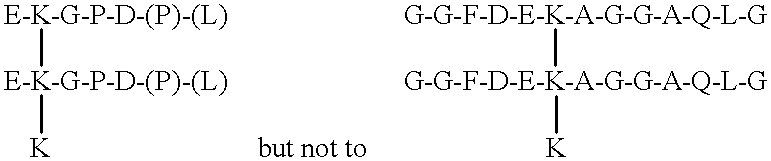
The invention provides improved assays for measuring type II collagen (cartilage) resorption in vivo. The subject immunoassays are useful for distinguishing between resorption of non-mineralized cartilage and mineralized cartilage, and for measuring total cartilage resorption in vivo. The disclosed immunoassays employ either one or a combination of two antibodies. A first antibody binds to DEKAGGA (SEQ ID NO:21) but not to GGFDEKAGGAQLG (SEQ ID NO:27), where the K symbols refer to lysine residues and/or preferably to cross-linking 3-hydroxypyridinium residues. Measurement of analyte binding to the first antibody in serum provides an indication of non-mineralized cartilage resorption in vivo.
A second antibody binds to GGFDEKAGGAQLG but not to DEKAGGA, where the K symbols also preferably refer to cross-linking 3-hydroxypyridinium residues. Measurement of analyte binding to the second antibody in serum provides an indication of mineralized cartilage resorption in vivo.
An indication of total (non-mineralized and mineralized) cartilage resorption in vivo is provided by either measurement of analyte binding to the first antibody in urine, or by measurement of the total analyte binding to the first and second antibodies in serum.
The subject cartilage markers are preferably measured in conjunction with bone collagen resorption markers selected from among amino-terminal telopeptides of type I collagen, carboxy-terminal telopeptides of type I collagen, and free lysyl pyridinoline (deoxypyridinoline) cross-links.
DETAILED DESCRIPTION OF THE PREFERRED EMBODIMENT
By way of introduction, the monoclonal antibody 2B4 recognizes a C-terminal epitope in the sequence EKGPDP. The antibody requires the C-terminal proline to be free for any binding (i.e., Pro-COOH) and prefers K to be derivatized (e.g., cross-linked) through its side-chain (epsilon) amino group. The presence of extra amino acids C-terminal to the proline prevents mAb 2B4 binding to this peptide domain.
Various matrix metalloproteinases (MMPs) including collagenases (MMP1 and MMP13), gelatinases (MMP2 and 9), stromelysin (MMP3), and matrilysin (MMP7) can generate the mAb 2B4 epitope from cartilage type II collagen or a synthetic C-telopeptide domain (AFAGLGPREKGPDPLQYMRA). Matrilysin (MMP7) is the most potent based on in vitro studies with the recombinant enzymes.
Cathepsin K, however, is unable to cleave the P-L bond required for 2B4 epitope release, but generates a longer fragment, EKGPDPLQ (SEQ ID NO:10). This is significant because cathepsin K is a protease expressed essentially only by osteoclasts and is the key enzyme responsible for the resorption of the calcified collagen when osteoclasts resorb bone. Osteoclasts (or a very similar cell) are also responsible for degrading calcified cartilage. Calcified cartilage is formed transiently in the growth plates of growing animals and is also present in the adult skeleton at the interfaces between bone and cartilage in all types of joints. In arthritis, accelerated resorption and bony remodeling will result in extensive resorption of calcified cartilage by osteoclasts. This process can release into the bloodstream collagen type II C-telopeptides (EKGPDPLQ) that are not recognized by 2B4.
Since the urinary form of the type II collagen telopeptide analyte is EKGPDP or smaller, proteases in the kidney and/or liver remove the C-terminal -LQ before release into the urine. Therefore, two sources of immunoreactive col2CTx are can be defined: (1) direct generation by chondrocytes activated to degrade their matrix collagen by MMPs, and (2) secondary generation in the kidney and/or liver from the circulating products of osteoclastic resorption of calcified cartilage.
In arthritis (rheumatoid arthritis (RA) and osteoarthritis (OA)) MMPs are known to be key agents in the destruction of the collagenous matrices of joint cartilages. In both forms of arthritis, bone structure is also greatly affected. In RA, osteoporosis is a common consequence both directly from the disease process itself through the effects of inflammatory cytokines and as a side effect of corticosteroid therapy. The accelerated bone resorption will be most pronounced in involved joints and will include removal of the calcified cartilage underlying joint surfaces. In OA, osteophytes (bony outgrowths) often feature prominently in the joint pathology. Here, too, calcified cartilage will be formed and then degraded (the latter by osteoclasts) as part of osteophyte growth. Therefore to monitor and manage both major forms of arthritis it would be desirable to differentially measure MMP-mediated and osteoclast-mediated (cathepsin K) collagen type II destruction. In blood or synovial fluid, measuring col2CTx (EKGPDP) can index the former activity, whereas measuring the osteoclast product (EKGPDPLQ) in blood can index the latter. In addition, urinary col2CTx can provide an index of total resorption (both non-mineralized and mineralized cartilage). Such urine assays preferably include the steps of determining the creatinine content of the urine sample and correlating the ratio of the detected binding of the antibody to the creatinine content in order to provide a urinary index of total type II collagen resorption independent of urine volume.
The subject differential measurements of EKGPDP and EKGPDPLQ immunoreactivities have diagnostic value in the assessment of disease activity in the individual patient. In combination with measurement of a type I collagen marker to assess total bone resorption activity, and/or measurement of a type III collagen marker to assess other sources of non-mineralized connective tissue breakdown, an appropriate therapy can be prescribed and its desired effect monitored. Similarly, such differential assays can be used to identify new drug candidates (in vitro and in animals) and to provide surrogate markers in clinical trials for demonstrating desired beneficial effects on target tissues and cellular processes.
Representative assays for type I collagen resorption markers for this purpose include amino-terminal telopeptides of type I collagen (Osteomark®, Ostex International, Seattle, Wash.), carboxy-terminal telopeptides of type I collagen (CrossLaps®-D, Osteometer Biotech, Herlev, Denmark), and free lysyl pyridinoline cross-links (Pyrilinks®, Metra Biosystems, Mountain View, Calif.).
Representative assays for type III collagen resorption markers include amino-terminal telopeptides of type III collagen and carboxy-terminal telopeptides of type III collagen. Such assays are disclosed in U.S. Pat. Nos. 5,532,169, 5,641,687, and 6,010,862. Type III collagen resorption markers can be used to assess vascular collagen degradation (atherosclerosis), lung tissue degradation (e.g., emphysema and other destructive lung diseases), muscle wastage, frailty syndromes of the elderly, liver disease, hyperthyroidism, secondary effects of diabetes on connective tissue, and other inflammatory and infectious conditions that accelerate total bone resorption activity.
The invention also provides antibodies directed against other cross-linked carboxy-telopeptide degradation products of type II collagen, as well as linear peptides (SEQ ID NOS 11-20) synthesized to match the telopeptide components of the degradation products, and assays for cartilage degradation using the antibodies and linear peptides.
SEQ Body Fluid1
ID Synovial
α1(II)C Telopeptide Sequence NO fluid Blood Urine
            EKGPD 1 N N + M
             EKGPDP 2 N N N + M
             EKGPDPL 3 N N N + M
             EKGPDPLQ 10 M
        LGPREKGPDP 11 N N
        LGPREKGPDPLQ 12 N N
        LGPREKGPDPLQY 13 N N
     FAGLGPREKGPDP 14 N N
     FAGLGPREKGPDPLQ 15 N N
     FAGLGPREKGPDPLQY 16 N N
IDMSAFAGLGPREKGPDP 17 N N
IDMSAFAGLGPREKGPDPLQ 18 N N
IDMSAFAGLGPREKGPDPLQY 19 N N
            EKGPDPLQYMR 20 N N
1Cartilage Source: N = non-mineralized cartilage; M = mineralized cartilage; N + M = total cartilage degradation; and — = absent or only trace amounts.
The principal col2CTx degradation product is cross-linked EKGPDP. The further degradation product cross-linked EKGPD is usually present in urine in lower amounts. The cross-linked EKGPDPL degradation product is a minor component relative to cross-linked EKGPDP.
Higher than normal levels of cross-linked EKGPDPLQ in blood can indicate mineralized cartilage degradation associated with active osteophyte formation in osteoarthritis.
The telopeptide sequences represented by SEQ ID NOS 11-13 result from metalloproteinase (MMP) cleavage of the G—L bond at the amino end of the α1(II)C telopeptide (SEQ ID NO 5), in combination with MMP cuts of the P—L, Q—Y, or Y—M bonds at the carboxy end of the telopeptide. SEQ ID NOS 14-16 represent related telopeptides from MMP cleavage between the A—F bond. SEQ ID NOS 17-19 represent longer telopeptides from MMP cleavage between the G—I bond. The amino ends of these longer telopeptides are generally metabolized further in the lymph nodes, liver, and/or kidney.
The telopeptide sequence represented by SEQ ID NO 20 results from serine protease cleavage of the R—E bond amino terminal to the cross-linking K residue, in combination with serine protease cleavage of the R—A bond at the carboxy end of the telopeptide.
Such cross-linking degradation products of SEQ ID NOS 11-19 (wherein the two telopeptide components are independently selected from among SEQ ID NOS 11-19), and SEQ ID NO 20, generally result from degradation of non-mineralized cartilage and are primarily found in synovial fluid and blood, and to a lesser extent in urine. Higher than normal levels of these degradation products in body fluids indicate active cartilage degradation that may correlate with rheumatoid arthritis, osteoarthritis, and other arthritides.
The invention also provides antibodies directed against cross-linked amino-telopeptide degradation products of type II collagen, as well as linear peptides (SEQ ID NOS 21-31) synthesized to match the telopeptide components of the degradation products, and assays for cartilage degradation using the antibodies and linear peptides.
SEQ Body Fluid1
ID Synovial
α1(II)C Telopeptide Sequence1 NO fluid Blood Urine
      DEKAGGA 21 N N N + M
      DEKAGGAQ 22 N N N + M
      DEKAGGAQL 23 N N N + M
     FDEKAGGA 24 N N N + M
     FDEKAGGAQ 25 N N N + M
     FDEKAGGAQL 26 N N N + M
   GGFDEKAGGAQLG 27 M
JMAGGFDEKAGGAQLG 28 M
JMAGGFDEKAGGAQLGV 29 N N
     FDEKAGGAQLGV 30 N N
      DEKAGGAQLGV 31 N N
1J = pyroglutamic acid, i.e., wholly cyclized pyrrolidone carboxylic acid (5-oxo-2-pyrrolidinecarboxylic acid).
2Cartilage Source: N = non-mineralized cartilage; M = mineralized cartilage; N + M = total cartilage degradation; and — = absent or only trace amounts.
The telopeptide sequences represented by SEQ ID NOS 21-23 result from metalloproteinase (MMP) cleavage of the F—D bond at the amino end of the α1(II)N telopeptide, in combination with MMP cuts of the A—Q, Q—L, or L—G bonds at the carboxy end of the telopeptide. SEQ ID NOS 24-26 represent related telopeptides from MMP cleavage between the G—F bond at the amino end. The amino and carboxy ends of the longer telopeptides (SEQ ID NOS 22-26) are generally metabolized further in the lymph nodes, liver, and/or kidney. As a result, the principal col2NTx degradation products in urine are cross-linked DEKAGGA and FDEKAGGA.
The telopeptide sequences represented by SEQ ID NOS 27-28 result from cathepsin K cleavage of the G—V bond at the carboxy end of the telopeptide, in combination with cathepsin K cleavage of the A—G bond at the amino end of the telopeptide in the case of SEQ ID NO 27. In the case of SEQ ID NO 28 the amino terminal J residue is blocked. Higher than normal levels of cross-linked GGFDEKAGGAQLG and QMAGGFDEKAGGAQLG in blood can indicate mineralized cartilage degradation associated with active osteophyte formation in osteoarthritis. Proteases in the kidney and/or liver remove the carboxy-terminal G residue before release into the urine.
The telopeptides represented by SEQ ID NOS 29-31 result from metalloproteinase cleavage of the V—M bond at the carboxy end of the telopeptide, in combination with MMP cleavage of the G—F or F—D bond at the amino end of the telopeptide in the case of SEQ ID NOS 30-31. Larger degradation products comprising SEQ ID NOS 29-31 plus some additional helical sequence(s) may also be present in synovial fluid and blood. These cross-linked telopeptides are generally metabolized further in the lymph nodes, liver, and/or kidney, and consequently occur only in trace amounts in urine.
As indicated above, cross-linked α1(II)N telopeptides represented by SEQ ID NOS 27-31 are produced at the site(s) of cartilage degradation but degraded further during passage through the lymph nodes, liver, and/or kidney. The resulting smaller degradation products have two telopeptide components that may be independently selected from among SEQ ID NOS 21-26. Higher than normal levels of these degradation products in urine indicate total cartilage degradation that may correlate with rheumatoid arthritis, osteoarthritis, and other arthritides.
Thus the invention provides, in a first embodiment, an improved method of analyzing a body fluid sample for the presence of an analyte indicative of a physiological condition, including the steps of contacting the body fluid sample with an antibody which binds to the analyte, detecting binding of the antibody in the body fluid sample, and correlating any detected binding to the physiological condition, the improvement comprising contacting a serum sample with an antibody which binds to
Figure US06348320-20020219-C00002
wherein K—K—K is hydroxylysyl pyridinoline or lysyl pyridinoline, and correlating any detected binding to resorption of non-mineralized type II collagen in vivo.
In a second embodiment, the invention provides an improved method of analyzing a body fluid sample for the presence of an analyte indicative of a physiological condition, including the steps of contacting the body fluid sample with an antibody which binds to the analyte, detecting binding of the antibody in the body fluid sample, and correlating any detected binding to the physiological condition, the improvement comprising contacting a serum sample with an antibody which binds to
Figure US06348320-20020219-C00003
wherein K—K—K is hydroxylysyl pyridinoline or lysyl pyridinoline, and correlating any detected binding to resorption of mineralized type II collagen in vivo.
In a third embodiment, the invention provides an improved method of analyzing a body fluid sample for the presence of an analyte indicative of a physiological condition, including the steps of contacting the body fluid sample with an antibody which binds to the analyte, detecting binding of the antibody in the body fluid sample, and correlating any detected binding to the physiological condition, the improvement comprising contacting a urine sample with an antibody which binds to
Figure US06348320-20020219-C00004
wherein K—K—K is hydroxylysyl pyridinoline or lysyl pyridinoline, and correlating any detected binding to resorption of both non-mineralized and mineralized type II collagen in vivo.
In a fourth embodiment, the invention provides an improved method of analyzing a body fluid sample for the presence of an analyte indicative of a physiological condition, including the steps of contacting the body fluid sample with an antibody which binds to the analyte, detecting binding of the antibody in the body fluid sample, and correlating any detected binding to the physiological condition, the improvement comprising contacting a serum sample with a first antibody which binds to
Figure US06348320-20020219-C00005
and contacting the serum sample with a second antibody which binds to
Figure US06348320-20020219-C00006
wherein K—K—K is hydroxylysyl pyridinoline or lysyl pyridinoline, and correlating the total detected binding of the first and second antibodies to resorption of both mineralized and non-mineralized type II collagen in vivo.
In a fifth embodiment, the invention provides an improved method of analyzing a body fluid sample for the presence of an analyte indicative of a physiological condition, including the steps of contacting the body fluid sample with an antibody which binds to the analyte, detecting binding of the antibody in the body fluid sample, and correlating any detected binding to the physiological condition, the improvement comprising contacting a serum sample with a first antibody which binds to
Figure US06348320-20020219-C00007
and contacting a urine sample with a second antibody which binds to
Figure US06348320-20020219-C00008
wherein K—K—K is hydroxylysyl pyridinoline or lysyl pyridinoline, and correlating any detected binding of the first antibody to resorption of mineralized type II collagen in vivo, and any detected binding of the second antibody to resorption of both mineralized and non-mineralized type II collagen in vivo.
The term “antibody” in this disclosure is meant to encompass polyclonal antibodies, monoclonal antibodies, and antigen-binding fragments thereof. Such EKGPDPLQ-binding and EKGPDP-binding antibodies, or GGFDEKAGGAQLG-binding and DEKAGGA-binding antibodies, can be produced by standard methods well known in the art. A representative protocol follows.
EKGPDPLQ hybridoma development: Female RFB/DnJ mice are injected intraperitoneal (ip) with an emulsion of complete Freund's adjuvant and EKGPDPLQ synthetic peptide cross-linked to the carrier protein keyhole limpet hemacyanin (KLH) via glutaraldehyde (KLH-EKOPDPLQ). One month later a booster injection of KLH-EKGPDPLQ is given ip in an emulsion with incomplete Freund's adjuvant. Approximately eight weeks later the mice are sacrificed and splenocytes fused, by polyethylene glycol, to the FOX-NY murine myeloma cell line (ATCC CRL-1732). Fusion cells are grown in 96-well plates in selection media containing adenine, aminopterin and thymidine (AAT).
Approximately one week after fusion hybridoma colony culture supernatants are tested in ELISA for reactivity to the EKGPDPLQ synthetic peptide cross-linked via glutaraldehyde to bovine serum albumin (BSA-EKGPDPLQ). Hybridoma colonies producing antibody reactive with BSA-EKGPDPLQ are secondarily tested for specificity of reaction in ELISA against other synthetic peptide sequences. Among those tested are EKGPDP synthetic peptide cross-linked to BSA via glutaraldehyde (BSA-EKGPDP). Hybridoma colonies that react positively to BSA-EKGPDPLQ and negatively to BSA-EKGPDP are cryopreserved. Representative hybridomas are cloned to monoclonality by the limited dilution method.
In competition ELISA, human serum specimens as well as soluble BSA-EKGPDPLQ are found to compete with solid phase BSA-EKGPDPLQ for candidate antibodies.
A cathepsin K digest of type II collagen can also be prepared for immunization and screening purposes. Proteoglycans are extracted from a tissue sample of human articular cartilage with a protein denaturant, such as 4 M guanidine HCl. The residue is washed with water and digested with recombinant cathepsin K (37° C., pH 5.8). The digest is fractionated by high performance liquid chromatography, for example by reverse phase, and fractions containing pyridinoline cross-links are monitored by fluorescence emission at 390 nm (297 nm excitation). The fluorescent fraction containing the cross-linked C-telopeptides of sequence EKGPDPLQ is identified by N-terminal sequence analysis. This fraction can be used for screening purposes and for validation of antibody specificity.
The subject antibodies can be employed in a variety of immunoassay formats that are well known in the art. For example, see: Harlow & Lane, Antibodies: A Laboratory Manual, Cold Spring Harbor Laboratory, pp. 553-612, 1988; Principles and Practice of Immunoassay, Price & Newman (Eds.), Stockton Press, 1991; and Immunoassay, Diamandis & Christopoulos (Eds.), Academic Press, 1996.
While the invention has been described in conjunction with preferred embodiments, one of ordinary skill after reading the foregoing specification will be able to effect various changes, substitutions of equivalents, and alterations to the subject matter set forth herein. Hence, the invention can be practiced in ways other than those specifically described herein.
SEQUENCE LISTING
<160> NUMBER OF SEQ ID NOS: 31
<210> SEQ ID NO 1
<211> LENGTH: 5
<212> TYPE: PRT
<213> ORGANISM: Homo sapiens
<220> FEATURE:
<221> NAME/KEY: PEPTIDE
<222> LOCATION: (1)..(5)
<223> OTHER INFORMATION: syn- corr. to C-terminal telopeptide seq. of
hu. type II collagen
<400> SEQUENCE: 1
Glu Lys Gly Pro Asp
1 5
<210> SEQ ID NO 2
<211> LENGTH: 6
<212> TYPE: PRT
<213> ORGANISM: Homo sapiens
<220> FEATURE:
<221> NAME/KEY: PEPTIDE
<222> LOCATION: (1)..(5)
<223> OTHER INFORMATION: syn- corr. to C-terminal telopeptide seq. of
hu. type II collagen
<400> SEQUENCE: 2
Glu Lys Gly Pro Asp Pro
1 5
<210> SEQ ID NO 3
<211> LENGTH: 7
<212> TYPE: PRT
<213> ORGANISM: Homo sapiens
<220> FEATURE:
<221> NAME/KEY: PEPTIDE
<222> LOCATION: (1)..(7)
<223> OTHER INFORMATION: syn- corr. to C-terminal telopeptide seq. of
hu. type II collagen
<400> SEQUENCE: 3
Glu Lys Gly Pro Asp Pro Leu
1 5
<210> SEQ ID NO 4
<211> LENGTH: 14
<212> TYPE: PRT
<213> ORGANISM: Homo sapiens
<220> FEATURE:
<221> NAME/KEY: PEPTIDE
<222> LOCATION: (1)..(14)
<223> OTHER INFORMATION: syn- corr. to C-terminal telopeptide seq. of
hu. type II collagen
<400> SEQUENCE: 4
Ala Phe Ala Gly Leu Gly Pro Arg Glu Lys Gly Pro Asp Pro
1 5 10
<210> SEQ ID NO 5
<211> LENGTH: 20
<212> TYPE: PRT
<213> ORGANISM: Homo sapiens
<220> FEATURE:
<221> NAME/KEY: PEPTIDE
<222> LOCATION: (1)..(20)
<223> OTHER INFORMATION: ssyn- corr. to C-terminal telopeptide seq. of
hu. type II collagen
<400> SEQUENCE: 5
Ala Phe Ala Gly Leu Gly Pro Arg Glu Lys Gly Pro Asp Pro Leu Gln
1 5 10 15
Tyr Met Arg Ala
20
<210> SEQ ID NO 6
<211> LENGTH: 16
<212> TYPE: PRT
<213> ORGANISM: Homo sapiens
<220> FEATURE:
<221> NAME/KEY: PEPTIDE
<222> LOCATION: (1)..(16)
<223> OTHER INFORMATION: syn- corr. to C-terminal telopeptide seq. of
hu. type II collagen
<400> SEQUENCE: 6
Ala Phe Ala Gly Leu Gly Pro Arg Glu Lys Gly Pro Asp Pro Leu Gln
1 5 10 15
<210> SEQ ID NO 7
<211> LENGTH: 17
<212> TYPE: PRT
<213> ORGANISM: Homo sapiens
<220> FEATURE:
<221> NAME/KEY: PEPTIDE
<222> LOCATION: (1)..(17)
<223> OTHER INFORMATION: syn- corr. to C-terminal telopeptide seq. of
hu. type II collagen
<400> SEQUENCE: 7
Ala Phe Ala Gly Leu Gly Pro Arg Glu Lys Gly Pro Asp Pro Leu Gln
1 5 10 15
Tyr
<210> SEQ ID NO 8
<211> LENGTH: 6
<212> TYPE: PRT
<213> ORGANISM: Homo sapiens
<220> FEATURE:
<221> NAME/KEY: PEPTIDE
<222> LOCATION: (1)..(6)
<223> OTHER INFORMATION: syn- corr. to C-terminal telopeptide seq. of
hu. type II collagen
<400> SEQUENCE: 8
Leu Gln Tyr Met Arg Ala
1 5
<210> SEQ ID NO 9
<211> LENGTH: 4
<212> TYPE: PRT
<213> ORGANISM: Homo sapiens
<220> FEATURE:
<221> NAME/KEY: PEPTIDE
<222> LOCATION: (1)..(4)
<223> OTHER INFORMATION: syn- corr. to C-terminal telopeptide seq. of
hu. type II collagen
<400> SEQUENCE: 9
Tyr Met Arg Ala
1
<210> SEQ ID NO 10
<211> LENGTH: 8
<212> TYPE: PRT
<213> ORGANISM: Homo sapiens
<220> FEATURE:
<221> NAME/KEY: PEPTIDE
<222> LOCATION: (1)..(8)
<223> OTHER INFORMATION: syn- corr. to C-terminal telopeptide seq. of
hu. type II collagen
<400> SEQUENCE: 10
Glu Lys Gly Pro Asp Pro Leu Gln
1 5
<210> SEQ ID NO 11
<211> LENGTH: 10
<212> TYPE: PRT
<213> ORGANISM: Homo sapiens
<220> FEATURE:
<221> NAME/KEY: PEPTIDE
<222> LOCATION: (1)..(10)
<223> OTHER INFORMATION: syn- corr. to C-terminal telopeptide seq. of
hu. type II collagen
<400> SEQUENCE: 11
Leu Gly Pro Arg Glu Lys Gly Pro Asp Pro
1 5 10
<210> SEQ ID NO 12
<211> LENGTH: 12
<212> TYPE: PRT
<213> ORGANISM: Homo sapiens
<220> FEATURE:
<221> NAME/KEY: PEPTIDE
<222> LOCATION: (1)..(12)
<223> OTHER INFORMATION: syn- corr. to C-terminal telopeptide seq. of
hu. type II collagen
<400> SEQUENCE: 12
Leu Gly Pro Arg Glu Lys Gly Pro Asp Pro Leu Gln
1 5 10
<210> SEQ ID NO 13
<211> LENGTH: 13
<212> TYPE: PRT
<213> ORGANISM: Homo sapiens
<220> FEATURE:
<221> NAME/KEY: PEPTIDE
<222> LOCATION: (1)..(13)
<223> OTHER INFORMATION: syn- corr. to C-terminal telopeptide seq. of
hu. type II collagen
<400> SEQUENCE: 13
Leu Gly Pro Arg Glu Lys Gly Pro Asp Pro Leu Gln Tyr
1 5 10
<210> SEQ ID NO 14
<211> LENGTH: 13
<212> TYPE: PRT
<213> ORGANISM: Homo sapiens
<220> FEATURE:
<221> NAME/KEY: PEPTIDE
<222> LOCATION: (1)..(13)
<223> OTHER INFORMATION: syn- corr. to C-terminal telopeptide seq. of
hu. type II collagen
<400> SEQUENCE: 14
Phe Ala Gly Leu Gly Pro Arg Glu Lys Gly Pro Asp Pro
1 5 10
<210> SEQ ID NO 15
<211> LENGTH: 15
<212> TYPE: PRT
<213> ORGANISM: Homo sapiens
<220> FEATURE:
<221> NAME/KEY: PEPTIDE
<222> LOCATION: (1)..(15)
<223> OTHER INFORMATION: syn- corr. to C-terminal telopeptide seq. of
hu. type II collagen
<400> SEQUENCE: 15
Phe Ala Gly Leu Gly Pro Arg Glu Lys Gly Pro Asp Pro Leu Gln
1 5 10 15
<210> SEQ ID NO 16
<211> LENGTH: 16
<212> TYPE: PRT
<213> ORGANISM: Homo sapiens
<220> FEATURE:
<221> NAME/KEY: PEPTIDE
<222> LOCATION: (1)..(16)
<223> OTHER INFORMATION: syn- corr. to C-terminal telopeptide seq. of
hu. type II collagen
<400> SEQUENCE: 16
Phe Ala Gly Leu Gly Pro Arg Glu Lys Gly Pro Asp Pro Leu Gln Tyr
1 5 10 15
<210> SEQ ID NO 17
<211> LENGTH: 18
<212> TYPE: PRT
<213> ORGANISM: Homo sapiens
<220> FEATURE:
<221> NAME/KEY: PEPTIDE
<222> LOCATION: (1)..(18)
<223> OTHER INFORMATION: syn- corr. to C-terminal telopeptide seq. of
hu. type II collagen
<400> SEQUENCE: 17
Ile Asp Met Ser Ala Phe Ala Gly Leu Gly Pro Arg Glu Lys Gly Pro
1 5 10 15
Asp Pro
<210> SEQ ID NO 18
<211> LENGTH: 20
<212> TYPE: PRT
<213> ORGANISM: Homo sapiens
<220> FEATURE:
<221> NAME/KEY: PEPTIDE
<222> LOCATION: (1)..(20)
<223> OTHER INFORMATION: syn- corr. to C-terminal telopeptide seq. of
hu. type II collagen
<400> SEQUENCE: 18
Ile Asp Met Ser Ala Phe Ala Gly Leu Gly Pro Arg Glu Lys Gly Pro
1 5 10 15
Asp Pro Leu Gln
20
<210> SEQ ID NO 19
<211> LENGTH: 21
<212> TYPE: PRT
<213> ORGANISM: Homo sapiens
<220> FEATURE:
<221> NAME/KEY: PEPTIDE
<222> LOCATION: (1)..(21)
<223> OTHER INFORMATION: syn- corr. to C-terminal telopeptide seq. of
hu. type II collagen
<400> SEQUENCE: 19
Ile Asp Met Ser Ala Phe Ala Gly Leu Gly Pro Arg Glu Lys Gly Pro
1 5 10 15
Asp Pro Leu Gln Tyr
20
<210> SEQ ID NO 20
<211> LENGTH: 11
<212> TYPE: PRT
<213> ORGANISM: Homo sapiens
<220> FEATURE:
<221> NAME/KEY: PEPTIDE
<222> LOCATION: (1)..(11)
<223> OTHER INFORMATION: syn- corr. to C-terminal telopeptide seq. of
hu. type II collagen
<400> SEQUENCE: 20
Glu Lys Gly Pro Asp Pro Leu Gln Tyr Met Arg
1 5 10
<210> SEQ ID NO 21
<211> LENGTH: 7
<212> TYPE: PRT
<213> ORGANISM: Homo sapiens
<220> FEATURE:
<221> NAME/KEY: PEPTIDE
<222> LOCATION: (1)..(7)
<223> OTHER INFORMATION: syn- corr. to N-terminal telopeptide seq. of
hu. type II collagen
<400> SEQUENCE: 21
Asp Glu Lys Ala Gly Gly Ala
1 5
<210> SEQ ID NO 22
<211> LENGTH: 8
<212> TYPE: PRT
<213> ORGANISM: Homo sapiens
<220> FEATURE:
<221> NAME/KEY: PEPTIDE
<222> LOCATION: (1)..(8)
<223> OTHER INFORMATION: syn- corr. to N-terminal telopeptide seq. of
hu. type II collagen
<400> SEQUENCE: 22
Asp Glu Lys Ala Gly Gly Ala Gln
1 5
<210> SEQ ID NO 23
<211> LENGTH: 9
<212> TYPE: PRT
<213> ORGANISM: Homo sapiens
<220> FEATURE:
<221> NAME/KEY: PEPTIDE
<222> LOCATION: (1)..(9)
<223> OTHER INFORMATION: syn- corr. to N-terminal telopeptide seq. of
hu. type II collagen
<400> SEQUENCE: 23
Asp Glu Lys Ala Gly Gly Ala Gln Leu
1 5
<210> SEQ ID NO 24
<211> LENGTH: 8
<212> TYPE: PRT
<213> ORGANISM: Homo sapiens
<220> FEATURE:
<221> NAME/KEY: PEPTIDE
<222> LOCATION: (1)..(8)
<223> OTHER INFORMATION: syn- corr. to N-terminal telopeptide seq. of
hu. type II collagen
<400> SEQUENCE: 24
Phe Asp Glu Lys Ala Gly Gly Ala
1 5
<210> SEQ ID NO 25
<211> LENGTH: 9
<212> TYPE: PRT
<213> ORGANISM: Homo sapiens
<220> FEATURE:
<221> NAME/KEY: PEPTIDE
<222> LOCATION: (1)..(9)
<223> OTHER INFORMATION: syn- corr. to N-terminal telopeptide seq. of
hu. type II collagen
<400> SEQUENCE: 25
Phe Asp Glu Lys Ala Gly Gly Ala Gln
1 5
<210> SEQ ID NO 26
<211> LENGTH: 10
<212> TYPE: PRT
<213> ORGANISM: Homo sapiens
<220> FEATURE:
<221> NAME/KEY: PEPTIDE
<222> LOCATION: (1)..(10)
<223> OTHER INFORMATION: syn- corr. to N-terminal telopeptide seq. of
hu. type II collagen
<400> SEQUENCE: 26
Phe Asp Glu Lys Ala Gly Gly Ala Gln Leu
1 5 10
<210> SEQ ID NO 27
<211> LENGTH: 13
<212> TYPE: PRT
<213> ORGANISM: Homo sapiens
<220> FEATURE:
<221> NAME/KEY: PEPTIDE
<222> LOCATION: (1)..(13)
<223> OTHER INFORMATION: syn- corr. to N-terminal telopeptide seq. of
hu. type II collagen
<400> SEQUENCE: 27
Gly Gly Phe Asp Glu Lys Ala Gly Gly Ala Gln Leu Gly
1 5 10
<210> SEQ ID NO 28
<211> LENGTH: 16
<212> TYPE: PRT
<213> ORGANISM: Homo sapiens
<220> FEATURE:
<221> NAME/KEY: PEPTIDE
<222> LOCATION: (1)..(16)
<223> OTHER INFORMATION: syn- corr. to N-terminal telopeptide seq. of
hu. type II collagen
<221> NAME/KEY: PEPTIDE
<222> LOCATION: (1)..(1)
<223> OTHER INFORMATION: Xaa is pyroglutamic acid
<400> SEQUENCE: 28
Xaa Met Ala Gly Gly Phe Asp Glu Lys Ala Gly Gly Ala Gln Leu Gly
1 5 10 15
<210> SEQ ID NO 29
<211> LENGTH: 17
<212> TYPE: PRT
<213> ORGANISM: Homo sapiens
<220> FEATURE:
<221> NAME/KEY: PEPTIDE
<222> LOCATION: (1)..(17)
<223> OTHER INFORMATION: syn- corr. to N-terminal telopeptide seq. of
hu. type II collagen
<221> NAME/KEY: PEPTIDE
<222> LOCATION: (1)..(1)
<223> OTHER INFORMATION: Xaa is pyroglutamic acid
<400> SEQUENCE: 29
Xaa Met Ala Gly Gly Phe Asp Glu Lys Ala Gly Gly Ala Gln Leu Gly
1 5 10 15
Val
<210> SEQ ID NO 30
<211> LENGTH: 12
<212> TYPE: PRT
<213> ORGANISM: Homo sapiens
<220> FEATURE:
<221> NAME/KEY: PEPTIDE
<222> LOCATION: (1)..(12)
<223> OTHER INFORMATION: syn- corr. to N-terminal telopeptide seq. of
hu. type II collagen
<400> SEQUENCE: 30
Phe Asp Glu Lys Ala Gly Gly Ala Gln Leu Gly Val
1 5 10
<210> SEQ ID NO 31
<211> LENGTH: 11
<212> TYPE: PRT
<213> ORGANISM: Homo sapiens
<220> FEATURE:
<221> NAME/KEY: PEPTIDE
<222> LOCATION: (1)..(11)
<223> OTHER INFORMATION: syn- corr. to N-terminal telopeptide seq. of
hu. type II collagen
<400> SEQUENCE: 31
Asp Glu Lys Ala Gly Gly Ala Gln Leu Gly Val
1 5 10

Claims (19)

The embodiments of the invention in which an exclusive property or privilege is claimed are defined as follows:
1. In a method of analyzing a body fluid sample for the presence of an analyte indicative of a physiological condition, comprising the steps of contacting the body fluid sample with an antibody which binds to the analyte, detecting binding of the antibody in the body fluid sample, and correlating any detected binding to the physiological condition, the improvement comprising contacting a serum sample with an antibody which binds to
Figure US06348320-20020219-C00009
wherein K—K—K is hydroxylysyl pyridinoline or lysyl pyridinoline, and correlating any detected binding to resorption of unmineralized type II collagen in vivo.
2. The method of claim 1, further comprising the step of contacting the serum sample with a second antibody which binds to a bone resorption marker selected from among amino-terminal telopeptides of type I collagen, carboxy-terminal telopeptides of type I collagen, and free lysyl pyridinoline cross-links, and correlating any detected binding of the second antibody in the sample to bone resorption in vivo.
3. The method of claim 1, further comprising the step of contacting the serum sample with a second antibody which binds to a type III collagen resorption marker selected from among amino-terminal telopeptides of type III collagen and carboxy-terminal telopeptides of type III collagen, and correlating any detected binding of the second antibody in the sample to type III collagen resorption in vivo.
4. In a method of analyzing a body fluid sample for the presence of an analyte indicative of a physiological condition, comprising the steps of contacting the body fluid sample with an antibody which binds to the analyte, detecting binding of the antibody in the body fluid sample, and correlating any detected binding to the physiological condition, the improvement comprising contacting a serum sample with an antibody which binds to
Figure US06348320-20020219-C00010
wherein K—K—K is hydroxylysyl pyridinoline or lysyl pyridinoline, and correlating any detected binding to resorption of mineralized type II collagen in vivo.
5. The method of claim 4, further comprising the step of contacting the serum sample with a second antibody which binds to a bone resorption marker selected from among amino-terminal telopeptides of type I collagen, carboxy-terminal telopeptides of type I collagen, and free lysly pyridinoline cross-links, and correlating any detected binding of the second antibody in the sample to bone resorption in vivo.
6. The method of claim 4, further comprising the step of contacting the serum sample with a second antibody which binds to a type III collagen resorption marker selected from among amino-terminal telopeptides of type III collagen and carboxy-terminal telopeptides of type III collagen, and correlating any detected binding of the second antibody in the sample to type III collagen resorption in vivo.
7. In a method of analyzing a body fluid sample for the presence of an analyte indicative of a physiological condition, comprising the steps of contacting the body fluid sample with an antibody which binds to the analyte, detecting binding of the antibody in the body fluid sample, and correlating any detected binding to the physiological condition, the improvement comprising contacting a urine sample with an antibody which binds to
Figure US06348320-20020219-C00011
wherein K—K—K is hydroxylysyl pyridinoline or lysyl pyridinoline, and correlating any detected binding to resorption of both unminemlized and mineralized type II collagen in vivo.
8. The method of claim 7, further comprising the steps of determining the creatinine content of the urine sample and correlating the ratio of the detected binding of the antibody to the creatinine content in order to provide a urinary index of total type II collagen resorption independent of urine volume.
9. The method of claim 7, further comprising the step of contacting the urine sample with a second antibody which binds to a bone resorption marker selected from among amino-terminal telopeptides of type I collagen, carboxy-terninal telopeptides of type I collagen, and free lysyl pyridinoline cross-links, and correlating any detected binding of the second antibody in the sample to bone resorption in vivo.
10. The method of claim 7, further comprising the step of contacting the serum sample with a second antibody which binds to a type III collagen resorption marker selected from among amino-terminal telopeptides of type III collagen and carboxy-terminal telopeptides of type III collagen, and correlating any detected binding of the second antibody in the sample to type III collagen resorption in vivo.
11. In a method of analyzing a body fluid sample for the presence of an analyte indicative of a physiological condition, comprising the steps of contacting the body fluid sample with an antibody which binds to the analyte, detecting binding of the antibody in the body fluid sample, and correlating any detected binding to the physiological condition, the improvement comprising contacting a serum sample with a first antibody which binds to
Figure US06348320-20020219-C00012
and contacting the serum sample with a second antibody which binds to
Figure US06348320-20020219-C00013
wherein K—K—K is hydroxylysyl pyridinoline or lysyl pyridinoline, and correlating the total detected binding of the first and second antibodies to resorption of both mineralized and unmineralized type II collagen in vivo.
12. The method of claim 11, further comprising the step of contacting the serum sample with a third antibody which binds to a bone resorption marker selected from among amino-terminal telopeptides of type I collagen, carboxy-terminal telopeptides of type I collagen, and free lysyl pyridinoline cross-links, and correlating any detected binding of the third antibody in the sample to bone resorption in vivo.
13. The method of claim 11, further comprising the step of contacting the serum sample with a third antibody which binds to a type III collagen resorption marker selected from among amino-terminal telopeptides of type III collagen and carboxy-terminal telopeptides of type III collagen, and correlating any detected binding of the third antibody in the sample to type III collagen resorption in vivo.
14. In a method of analyzing a body fluid sample for the presence of an analyte indicative of a physiological condition, comprising the steps of contacting the body fluid sample with an antibody which binds to the analyte, detecting binding of the antibody in the body fluid sample, and correlating any detected binding to the physiological condition, the improvement comprising contacting a serum sample with a first antibody which binds to
Figure US06348320-20020219-C00014
and contacting a urine sample with a second antibody which binds to
Figure US06348320-20020219-C00015
wherein K—K—K is hydroxylysyl pyridinoline or lysyl pyridinoline, and correlating any detected binding of the first antibody to resorption of mineralized type II collagen in vivo, and any detected binding of the second antibody to resorption of both mineralized and unmineralized type II collagen in vivo.
15. The method of claim 14, further comprising the steps of determining the creatinine content of the urine sample and correlating the ratio of the detected binding of the second antibody to the creatinine content in order to provide a urinary index of total type II collagen resorption independent of urine volume.
16. The method of claim 14, further comprising the step of contacting at least one of the serum and urine samples with a third antibody which binds to a bone resorption marker selected from among amino-terminal telopeptides of type I collagen, carboxy-terminal telopeptides of type I collagen, and free lysyl pyridinoline cross-links, and correlating any detected binding of the third antibody in the sample to bone resorption in vivo.
17. The method of claim 14, further comprising the step of contacting at least one of the serum and urine samples with a third antibody which binds to a type III collagen resorption marker selected from among amino-terminal telopeptides of type III collagen and carboxy-terminal telopeptides of type III collagen, and correlating any detected binding of the third antibody in the sample to type III collagen resorption in vivo.
18. Synthetic peptide DEKAGGA (SEQ ID NO:21).
19. Synthetic peptide GGFDEKAGGAQLG (SEQ ID NO:27).
US09/385,740 1998-06-19 1999-08-30 Cartilage resorption assays measuring type II collagen fragments Expired - Lifetime US6348320B1 (en)

Priority Applications (14)

Application Number Priority Date Filing Date Title
US09/385,740 US6348320B1 (en) 1998-06-19 1999-08-30 Cartilage resorption assays measuring type II collagen fragments
JP2001505199A JP2003502672A (en) 1999-06-17 1999-12-10 Cartilage resorption assay
EP04104875A EP1571450A3 (en) 1999-06-17 1999-12-10 Cartilage resorption assays
AT99965218T ATE278965T1 (en) 1999-06-17 1999-12-10 CARTILAGE RESORPTION ASSAY
US10/009,999 US6602980B1 (en) 1998-06-19 1999-12-10 Collagen type III synthetic peptides for collagen resorption assays
AU31183/00A AU3118300A (en) 1999-06-17 1999-12-10 Cartilage resorption assays
ES99965218T ES2229805T3 (en) 1999-06-17 1999-12-10 CARTILAGO RESORTION TESTS.
DE69920981T DE69920981T2 (en) 1999-06-17 1999-12-10 KNORPELRESORPTIONSASSAYS
PCT/US1999/029357 WO2000079284A1 (en) 1999-06-17 1999-12-10 Cartilage resorption assays
DK99965218T DK1210606T3 (en) 1999-06-17 1999-12-10 Cartilage resorption assays
EP99965218A EP1210606B1 (en) 1999-06-17 1999-12-10 Cartilage resorption assays
US10/073,679 US6566492B2 (en) 1998-06-19 2002-02-11 Synthetic peptides of type II collagen for cartilage resorption assays
US10/408,178 US20030166032A1 (en) 1998-06-19 2003-04-03 Cartilage resorption assays
US10/615,959 US6916903B2 (en) 1998-06-19 2003-07-08 Collagen type III synthetic peptides for collagen resorption assays

Applications Claiming Priority (5)

Application Number Priority Date Filing Date Title
US8982398P 1998-06-19 1998-06-19
US09/335,098 US6255056B1 (en) 1998-06-19 1999-06-17 Cartilage resorption assays
US14157499P 1999-06-29 1999-06-29
US14227499P 1999-07-02 1999-07-02
US09/385,740 US6348320B1 (en) 1998-06-19 1999-08-30 Cartilage resorption assays measuring type II collagen fragments

Related Parent Applications (2)

Application Number Title Priority Date Filing Date
US09/335,098 Continuation-In-Part US6255056B1 (en) 1998-06-19 1999-06-17 Cartilage resorption assays
US09/335,098 Continuation US6255056B1 (en) 1998-06-19 1999-06-17 Cartilage resorption assays

Related Child Applications (5)

Application Number Title Priority Date Filing Date
US10009999 Continuation-In-Part 1999-12-10
PCT/US1999/029357 Continuation-In-Part WO2000079284A1 (en) 1998-06-19 1999-12-10 Cartilage resorption assays
US10/009,999 Continuation-In-Part US6602980B1 (en) 1998-06-19 1999-12-10 Collagen type III synthetic peptides for collagen resorption assays
US10/009,999 Continuation US6602980B1 (en) 1998-06-19 1999-12-10 Collagen type III synthetic peptides for collagen resorption assays
US10/073,679 Continuation US6566492B2 (en) 1998-06-19 2002-02-11 Synthetic peptides of type II collagen for cartilage resorption assays

Publications (1)

Publication Number Publication Date
US6348320B1 true US6348320B1 (en) 2002-02-19

Family

ID=27492337

Family Applications (3)

Application Number Title Priority Date Filing Date
US09/385,740 Expired - Lifetime US6348320B1 (en) 1998-06-19 1999-08-30 Cartilage resorption assays measuring type II collagen fragments
US10/073,679 Expired - Lifetime US6566492B2 (en) 1998-06-19 2002-02-11 Synthetic peptides of type II collagen for cartilage resorption assays
US10/408,178 Abandoned US20030166032A1 (en) 1998-06-19 2003-04-03 Cartilage resorption assays

Family Applications After (2)

Application Number Title Priority Date Filing Date
US10/073,679 Expired - Lifetime US6566492B2 (en) 1998-06-19 2002-02-11 Synthetic peptides of type II collagen for cartilage resorption assays
US10/408,178 Abandoned US20030166032A1 (en) 1998-06-19 2003-04-03 Cartilage resorption assays

Country Status (1)

Country Link
US (3) US6348320B1 (en)

Cited By (2)

* Cited by examiner, † Cited by third party
Publication number Priority date Publication date Assignee Title
US20050124071A1 (en) * 2003-09-30 2005-06-09 Kraus Virginia B. Methods and compositions for diagnosing musculoskeletal, arthritic and joint disorders by biomarker dating
US20110178825A1 (en) * 2005-11-01 2011-07-21 Accenture Global Services Limited Collaborative intelligent task processor for insurance claims

Families Citing this family (1)

* Cited by examiner, † Cited by third party
Publication number Priority date Publication date Assignee Title
JP2004126050A (en) 2002-09-30 2004-04-22 Fuji Photo Film Co Ltd Lithographic printing original plate

Citations (19)

* Cited by examiner, † Cited by third party
Publication number Priority date Publication date Assignee Title
US4628027A (en) 1982-05-19 1986-12-09 Molecular Engineering Associates, Ltd. Vitro diagnostic methods using monoclonal antibodies against connective tissue proteins
WO1988008980A1 (en) 1987-05-08 1988-11-17 Farmos-Yhtymä Oy Type iii collagen degradation assay
WO1989004491A1 (en) 1987-11-06 1989-05-18 Washington Research Foundation Urinary assay for measuring bone resorption
WO1989012824A1 (en) 1988-06-25 1989-12-28 The Rowett Research Institute Method of monitoring collagen degradation
WO1991008478A2 (en) 1989-12-01 1991-06-13 Washington Research Foundation METHODS OF DETECTING COLLAGEN DEGRADATION $i(IN VIVO)
EP0505210A2 (en) 1991-03-20 1992-09-23 Orion-Yhtymà„ Oy Assay for measuring the degradation of type I collagen
WO1992021698A1 (en) 1991-05-31 1992-12-10 Washington Research Foundation DETECTION OF COLLAGEN DEGRADATION $i(IN VIVO)
WO1994003813A1 (en) 1992-07-29 1994-02-17 Boehringer Mannheim Gmbh Immunoassay for detecting collagen or collagen fragments
US5316914A (en) 1986-09-04 1994-05-31 Fuji Yakuhin Kogyo Kabushiki Kaisha Method for determining human collagen peptides by way of enzyme immunoassay
WO1994014070A1 (en) 1992-12-04 1994-06-23 Shriners Hospitals For Crippled Children Immunoassay for the measurement of collagen cleavage in cartilage
WO1994014844A1 (en) 1992-12-28 1994-07-07 Baylink David J Bone resorption assay
WO1994018563A1 (en) 1993-02-12 1994-08-18 Rhode Island Hospital Detection of type ii collagen and its peptides
WO1995004282A1 (en) 1993-07-28 1995-02-09 Boehringer Mannheim Gmbh Immunoassay for detecting collagen or collagen fragments
WO1995008115A1 (en) 1993-09-17 1995-03-23 Osteometer Biotech As A method of assaying collagen fragments in body fluids, a test kit and means for carrying out the method and use of the method to diagnose the presence of disorders associated with the metabolism of collagen
EP0699752A2 (en) 1994-07-22 1996-03-06 Collagen Corporation Mutated recombinant collagens
WO1996012193A1 (en) 1994-10-17 1996-04-25 Osteometer Bio Tech A/S Estimation of the fragmentation pattern of collagen in body fluids and the diagnosis of disorders associated with the metabolism of collagen
EP0718309A1 (en) 1994-12-23 1996-06-26 Roche Diagnostics GmbH Antigens and antibodies for the detection of collagen I
WO1996036645A1 (en) 1995-05-19 1996-11-21 Washington Research Foundation Synthetic peptide analogs of cross-linked n-telopeptides of type i collagen
US5702909A (en) 1987-11-06 1997-12-30 Washington Research Foundation Methods of detecting collagen type II degradation in vivo

Patent Citations (25)

* Cited by examiner, † Cited by third party
Publication number Priority date Publication date Assignee Title
US4628027A (en) 1982-05-19 1986-12-09 Molecular Engineering Associates, Ltd. Vitro diagnostic methods using monoclonal antibodies against connective tissue proteins
US5316914A (en) 1986-09-04 1994-05-31 Fuji Yakuhin Kogyo Kabushiki Kaisha Method for determining human collagen peptides by way of enzyme immunoassay
WO1988008980A1 (en) 1987-05-08 1988-11-17 Farmos-Yhtymä Oy Type iii collagen degradation assay
US5320970A (en) 1987-11-06 1994-06-14 Washington Research Foundation Detection of collagen degradation in vivo
US5702909A (en) 1987-11-06 1997-12-30 Washington Research Foundation Methods of detecting collagen type II degradation in vivo
US5140103A (en) 1987-11-06 1992-08-18 Washington Research Foundation Peptide fragments containing HP and LP cross-links
US5300434A (en) 1987-11-06 1994-04-05 Washington Research Foundation Hybridoma cell line producing an antibody to type-I collagen amino-terminal telopeptide
WO1989004491A1 (en) 1987-11-06 1989-05-18 Washington Research Foundation Urinary assay for measuring bone resorption
US4973666A (en) 1987-11-06 1990-11-27 Washington Research Foundation Peptide fragments containing HP and LP cross-links
US5919634A (en) * 1987-11-06 1999-07-06 Washington Research Foundation Methods of detecting collagen type II degradation in vivo
WO1989012824A1 (en) 1988-06-25 1989-12-28 The Rowett Research Institute Method of monitoring collagen degradation
WO1991008478A2 (en) 1989-12-01 1991-06-13 Washington Research Foundation METHODS OF DETECTING COLLAGEN DEGRADATION $i(IN VIVO)
EP0505210A2 (en) 1991-03-20 1992-09-23 Orion-Yhtymà„ Oy Assay for measuring the degradation of type I collagen
WO1992021698A1 (en) 1991-05-31 1992-12-10 Washington Research Foundation DETECTION OF COLLAGEN DEGRADATION $i(IN VIVO)
WO1994003813A1 (en) 1992-07-29 1994-02-17 Boehringer Mannheim Gmbh Immunoassay for detecting collagen or collagen fragments
WO1994014070A1 (en) 1992-12-04 1994-06-23 Shriners Hospitals For Crippled Children Immunoassay for the measurement of collagen cleavage in cartilage
WO1994014844A1 (en) 1992-12-28 1994-07-07 Baylink David J Bone resorption assay
WO1994018563A1 (en) 1993-02-12 1994-08-18 Rhode Island Hospital Detection of type ii collagen and its peptides
WO1995004282A1 (en) 1993-07-28 1995-02-09 Boehringer Mannheim Gmbh Immunoassay for detecting collagen or collagen fragments
WO1995008115A1 (en) 1993-09-17 1995-03-23 Osteometer Biotech As A method of assaying collagen fragments in body fluids, a test kit and means for carrying out the method and use of the method to diagnose the presence of disorders associated with the metabolism of collagen
EP0699752A2 (en) 1994-07-22 1996-03-06 Collagen Corporation Mutated recombinant collagens
WO1996012193A1 (en) 1994-10-17 1996-04-25 Osteometer Bio Tech A/S Estimation of the fragmentation pattern of collagen in body fluids and the diagnosis of disorders associated with the metabolism of collagen
EP0718309A1 (en) 1994-12-23 1996-06-26 Roche Diagnostics GmbH Antigens and antibodies for the detection of collagen I
WO1996036645A1 (en) 1995-05-19 1996-11-21 Washington Research Foundation Synthetic peptide analogs of cross-linked n-telopeptides of type i collagen
US5817755A (en) 1995-05-19 1998-10-06 Washington Research Foundation Synthetic peptide analogs of cross-linked N-telopeptides of type I collagen

Non-Patent Citations (9)

* Cited by examiner, † Cited by third party
Title
Atlely, L.M., et al., Collagen type II cross-linked telopeptides, a promising marker for cartilage degradation in arthritis, Combined Orthopaedic Research Societies Meeting, Hamamatsu, Japan, 1998.
Atley, L., et al., A selective inhibitor of collagenase-3 blocks the release of hydroxyproline and a metalloproteinase specific neoepitope, coll II CTx, from bovine collagen exposed to IL-1alpha, Arth. Rheum. 40(9S):584, 1997.
Atley, L., et al., A selective inhibitor of collagenase-3 blocks the release of hydroxyproline and a metalloproteinase specific neoepitope, coll II CTx, from bovine collagen exposed to IL-1α, Arth. Rheum. 40(9S):584, 1997.
Atley, L., et al., Matrix metalloproteinase-mediated release of immunoreactive telopeptides from cartilage type II collagen, Trans. Orthop. Res. Soc., New Orleans, 1998.
Eyre, D.R., et al., Biochemical markers of bone and cartilage collagen degradation, Combined Orthopaedic Research Societies Meeting, Vittel, France, Sep. 28-30, 1998.
Eyre, D.R., et al., Cross-linked telopeptides from collagen types I, II, and III in human urine, Bone Miner. Res. 11(S1): S413, 1996.
Moskowitz, R.W., et al., Type II collagen C-telopeptide is a marker for cartilage degradation in familial osteoarthritis, Amer. Coll. Rheum., San Diego, CA, Nov. 8-12, 1998.
Otter, A., et al., Biopolymers 33:1443-1459, 1993.
Sheridan, C., Osteometer targets collagen for osteoporosis, arthritis tests, Bio World International, p. 3, Dec. 3, 1997.

Cited By (2)

* Cited by examiner, † Cited by third party
Publication number Priority date Publication date Assignee Title
US20050124071A1 (en) * 2003-09-30 2005-06-09 Kraus Virginia B. Methods and compositions for diagnosing musculoskeletal, arthritic and joint disorders by biomarker dating
US20110178825A1 (en) * 2005-11-01 2011-07-21 Accenture Global Services Limited Collaborative intelligent task processor for insurance claims

Also Published As

Publication number Publication date
US20030166032A1 (en) 2003-09-04
US6566492B2 (en) 2003-05-20
US20020182639A1 (en) 2002-12-05

Similar Documents

Publication Publication Date Title
RU2139541C1 (en) Methods of assay of collagen destruction in vivo and sets for realization of methods, hybridoma, monoclonal antibody and its fragment, peptides of collagen and method of assay
EP0394296B2 (en) Assay of a body fluid for measuring bone resorption
EP0921395B1 (en) Assays for measurement of protein fragments in biological media
US5320970A (en) Detection of collagen degradation in vivo
JPH08505149A (en) Bone resorption assay
EP1130401A2 (en) Assays for measurement of type II collagen fragments in urine
JP2000516721A (en) Method for measuring D-amino acids in body fluids
JP4189033B2 (en) Quantitative arthritic condition assay
EP0960339A1 (en) Assay for collagen type ii fragments
US6348320B1 (en) Cartilage resorption assays measuring type II collagen fragments
EP1210606B1 (en) Cartilage resorption assays
US6916903B2 (en) Collagen type III synthetic peptides for collagen resorption assays
US6602980B1 (en) Collagen type III synthetic peptides for collagen resorption assays
US6255056B1 (en) Cartilage resorption assays
US7728112B2 (en) Method for monitoring collagen type II degradation in cartilage
CA2156935C (en) Methods of detecting collagen degradation in vivo

Legal Events

Date Code Title Description
AS Assignment

Owner name: WASHINGTON RESEARCH FOUNDATION, WASHINGTON

Free format text: ASSIGNMENT OF ASSIGNORS INTEREST;ASSIGNOR:WASHINGTON, UNIVERSITY OF;REEL/FRAME:010221/0058

Effective date: 19990826

Owner name: UNIVERSITY OF WASHINGTON, WASHINGTON

Free format text: ASSIGNMENT OF ASSIGNORS INTEREST;ASSIGNOR:EYRE, DAVID RODNEY;REEL/FRAME:010213/0664

Effective date: 19990817

STCF Information on status: patent grant

Free format text: PATENTED CASE

CC Certificate of correction
FEPP Fee payment procedure

Free format text: PAT HOLDER NO LONGER CLAIMS SMALL ENTITY STATUS, ENTITY STATUS SET TO UNDISCOUNTED (ORIGINAL EVENT CODE: STOL); ENTITY STATUS OF PATENT OWNER: LARGE ENTITY

FPAY Fee payment

Year of fee payment: 4

FPAY Fee payment

Year of fee payment: 8

FPAY Fee payment

Year of fee payment: 12