US6858409B1 - Nucleic acids encoding interleukin-1 inhibitors and processes for preparing interleukin-1 inhibitors - Google Patents
Nucleic acids encoding interleukin-1 inhibitors and processes for preparing interleukin-1 inhibitors Download PDFInfo
- Publication number
- US6858409B1 US6858409B1 US08/482,283 US48228395A US6858409B1 US 6858409 B1 US6858409 B1 US 6858409B1 US 48228395 A US48228395 A US 48228395A US 6858409 B1 US6858409 B1 US 6858409B1
- Authority
- US
- United States
- Prior art keywords
- host cell
- polypeptide
- acid sequence
- recombinant
- cell
- Prior art date
- Legal status (The legal status is an assumption and is not a legal conclusion. Google has not performed a legal analysis and makes no representation as to the accuracy of the status listed.)
- Expired - Lifetime
Links
- 0 C*(C)(*)N(C(c1c2cccc1)=O)C2=O Chemical compound C*(C)(*)N(C(c1c2cccc1)=O)C2=O 0.000 description 2
- VGJMDMCNVHEPRJ-UHFFFAOYSA-N CCCN1C(=O)c2ccccc2C1=O.CCCOS(=O)(=O)c1ccc(C)cc1.COCCCN.COCCCNC(=O)C1CCC(CN2C(=O)C=CC2=O)CC1 Chemical compound CCCN1C(=O)c2ccccc2C1=O.CCCOS(=O)(=O)c1ccc(C)cc1.COCCCN.COCCCNC(=O)C1CCC(CN2C(=O)C=CC2=O)CC1 VGJMDMCNVHEPRJ-UHFFFAOYSA-N 0.000 description 1
- KIFOCHPXAFAOQQ-UHFFFAOYSA-N CCN1C(=O)C=CC1=O.COCCCOC(=O)CCC(=O)NCCCCCCN.COCCCOC(=O)CCC(=O)NCCCCCCNC(=O)C1CCC(C)CC1.COCCCOC(=O)CCC(=O)O Chemical compound CCN1C(=O)C=CC1=O.COCCCOC(=O)CCC(=O)NCCCCCCN.COCCCOC(=O)CCC(=O)NCCCCCCNC(=O)C1CCC(C)CC1.COCCCOC(=O)CCC(=O)O KIFOCHPXAFAOQQ-UHFFFAOYSA-N 0.000 description 1
- XPMKXXLBQHKWKK-UHFFFAOYSA-N COCCCN1C(=O)C=CC1=O.COCCCNC(=O)C=CC(=O)O Chemical compound COCCCN1C(=O)C=CC1=O.COCCCNC(=O)C=CC(=O)O XPMKXXLBQHKWKK-UHFFFAOYSA-N 0.000 description 1
- WBWSUBCIFWHHDR-UHFFFAOYSA-N O=C(O)C=CC(=O)NCCCNC(=O)C=CC(=O)O.O=C1C=CC(=O)N1CCCN1C(=O)C=CC1=O Chemical compound O=C(O)C=CC(=O)NCCCNC(=O)C=CC(=O)O.O=C1C=CC(=O)N1CCCN1C(=O)C=CC1=O WBWSUBCIFWHHDR-UHFFFAOYSA-N 0.000 description 1
- IPPRCFUEIHNCOU-UHFFFAOYSA-N O=C1c2ccccc2C(=O)N1CCCN1C(=O)c2ccccc2C1=O Chemical compound O=C1c2ccccc2C(=O)N1CCCN1C(=O)c2ccccc2C1=O IPPRCFUEIHNCOU-UHFFFAOYSA-N 0.000 description 1
Images


Classifications
-
- C—CHEMISTRY; METALLURGY
- C07—ORGANIC CHEMISTRY
- C07K—PEPTIDES
- C07K14/00—Peptides having more than 20 amino acids; Gastrins; Somatostatins; Melanotropins; Derivatives thereof
- C07K14/435—Peptides having more than 20 amino acids; Gastrins; Somatostatins; Melanotropins; Derivatives thereof from animals; from humans
- C07K14/46—Peptides having more than 20 amino acids; Gastrins; Somatostatins; Melanotropins; Derivatives thereof from animals; from humans from vertebrates
- C07K14/47—Peptides having more than 20 amino acids; Gastrins; Somatostatins; Melanotropins; Derivatives thereof from animals; from humans from vertebrates from mammals
-
- A—HUMAN NECESSITIES
- A61—MEDICAL OR VETERINARY SCIENCE; HYGIENE
- A61K—PREPARATIONS FOR MEDICAL, DENTAL OR TOILETRY PURPOSES
- A61K47/00—Medicinal preparations characterised by the non-active ingredients used, e.g. carriers or inert additives; Targeting or modifying agents chemically bound to the active ingredient
- A61K47/50—Medicinal preparations characterised by the non-active ingredients used, e.g. carriers or inert additives; Targeting or modifying agents chemically bound to the active ingredient the non-active ingredient being chemically bound to the active ingredient, e.g. polymer-drug conjugates
- A61K47/51—Medicinal preparations characterised by the non-active ingredients used, e.g. carriers or inert additives; Targeting or modifying agents chemically bound to the active ingredient the non-active ingredient being chemically bound to the active ingredient, e.g. polymer-drug conjugates the non-active ingredient being a modifying agent
- A61K47/56—Medicinal preparations characterised by the non-active ingredients used, e.g. carriers or inert additives; Targeting or modifying agents chemically bound to the active ingredient the non-active ingredient being chemically bound to the active ingredient, e.g. polymer-drug conjugates the non-active ingredient being a modifying agent the modifying agent being an organic macromolecular compound, e.g. an oligomeric, polymeric or dendrimeric molecule
- A61K47/59—Medicinal preparations characterised by the non-active ingredients used, e.g. carriers or inert additives; Targeting or modifying agents chemically bound to the active ingredient the non-active ingredient being chemically bound to the active ingredient, e.g. polymer-drug conjugates the non-active ingredient being a modifying agent the modifying agent being an organic macromolecular compound, e.g. an oligomeric, polymeric or dendrimeric molecule obtained otherwise than by reactions only involving carbon-to-carbon unsaturated bonds, e.g. polyureas or polyurethanes
- A61K47/60—Medicinal preparations characterised by the non-active ingredients used, e.g. carriers or inert additives; Targeting or modifying agents chemically bound to the active ingredient the non-active ingredient being chemically bound to the active ingredient, e.g. polymer-drug conjugates the non-active ingredient being a modifying agent the modifying agent being an organic macromolecular compound, e.g. an oligomeric, polymeric or dendrimeric molecule obtained otherwise than by reactions only involving carbon-to-carbon unsaturated bonds, e.g. polyureas or polyurethanes the organic macromolecular compound being a polyoxyalkylene oligomer, polymer or dendrimer, e.g. PEG, PPG, PEO or polyglycerol
-
- C—CHEMISTRY; METALLURGY
- C07—ORGANIC CHEMISTRY
- C07K—PEPTIDES
- C07K14/00—Peptides having more than 20 amino acids; Gastrins; Somatostatins; Melanotropins; Derivatives thereof
- C07K14/435—Peptides having more than 20 amino acids; Gastrins; Somatostatins; Melanotropins; Derivatives thereof from animals; from humans
- C07K14/52—Cytokines; Lymphokines; Interferons
- C07K14/54—Interleukins [IL]
-
- C—CHEMISTRY; METALLURGY
- C07—ORGANIC CHEMISTRY
- C07K—PEPTIDES
- C07K14/00—Peptides having more than 20 amino acids; Gastrins; Somatostatins; Melanotropins; Derivatives thereof
- C07K14/435—Peptides having more than 20 amino acids; Gastrins; Somatostatins; Melanotropins; Derivatives thereof from animals; from humans
- C07K14/705—Receptors; Cell surface antigens; Cell surface determinants
- C07K14/715—Receptors; Cell surface antigens; Cell surface determinants for cytokines; for lymphokines; for interferons
- C07K14/7151—Receptors; Cell surface antigens; Cell surface determinants for cytokines; for lymphokines; for interferons for tumor necrosis factor [TNF], for lymphotoxin [LT]
-
- C—CHEMISTRY; METALLURGY
- C07—ORGANIC CHEMISTRY
- C07K—PEPTIDES
- C07K16/00—Immunoglobulins [IGs], e.g. monoclonal or polyclonal antibodies
- C07K16/18—Immunoglobulins [IGs], e.g. monoclonal or polyclonal antibodies against material from animals or humans
-
- A—HUMAN NECESSITIES
- A61—MEDICAL OR VETERINARY SCIENCE; HYGIENE
- A61K—PREPARATIONS FOR MEDICAL, DENTAL OR TOILETRY PURPOSES
- A61K38/00—Medicinal preparations containing peptides
Definitions
- This invention relates to polypeptides that have been covalently bonded to long chain polymers such as methoxy polyethylene glycol. This invention also describes methods and reagents for the reaction of activated polymer molecules with various biologically-important polypeptides.
- proteins have been found to have an extremely short half life in the blood serum. For the most part, proteins are cleared from the serum through the kidneys. The systematic introduction of relatively large quantities of proteins, particularly those foreign to the human system, can give rise to immunogenic reactions that, among other problems, may lead to rapid removal of the protein from the body through formation of immune complexes. For other proteins, solubility and aggregation problems have also hindered the optimal formulation of the protein.
- PEG polyethylene glycol
- mPEG monomethoxyl polyethylene glycol
- pegylation covalent modification of proteins with polyethylene glycols
- pegylation of IL-2 has been shown to decrease the clearance of IL-2 while not significantly affecting the activity of the cytokine.
- the decreased clearance leads to an increased efficiency over the non-pegylated material.
- SOD Superoxide Dismutase
- IgG Immunoglobulin G
- pegylation can lead to sterically hindered active sites.
- relatively small substrates may approach the protein, while the activity of proteins that react with larger substrates can be dramatically effected by random pegylation.
- Davis et al. supra. The site selective pegylation of such proteins could lead to modified materials that gain the desireable attributes of pegylation without the loss of activity.
- the pegylated protein is intended for therapeutic use, the multiple species mixture that results from the use of non-specific pegylation leads to difficulties in the preparation of a product with reproducible and characterizable properties. This makes it extremely difficult to evaluate therapeutics and to establish efficacy and dosing information.
- multimeric complexes that contain more than one biologically active polypeptide or drug can lead to synergistic benefits.
- a complex containing two identical binding polypeptides may have substantially increased affinity to the ligand or active site that it binds relative to the monomeric polypeptide.
- multimeric complexes of proteins can be desirable in order to increase affinity of the protein to its ligand in addition to increasing the molecular weight of the complex.
- Proteins frequently achieve their biological effects through interaction with other proteins. Where a simple complex of two proteins is sufficient to achieve the biological effect it has proved possible to mimic the physiological effects of endogenous proteins by administering exogenous proteins. However, where the biological effect requires the assembly of a complex containing more than two proteins it is more difficult to mimic the function of the endogenous proteins with recombinantly produced exogenous equivalents because the higher order complexes are frequently unstable. In such cases it may be advantageous to use crosslinked species containing two of the components of the complex to simulate the biologically-active complex.
- the proteins were expressed in animal cell expression systems, and were found to be substantially more effective at inhibiting TNF than the monomeric soluble receptor alone. Similar procedures have also been used for producing similar crosslinked proteins of the CD4 protein, (Byrn at al., Nature ( London ) vol. 344, pg. 667 (1990)) the CR1 protein, (Kalli et al., J. Exp. Med . vol. 174, pg. 1451 (1991); Hebell et al., WO 91/16437 (1991)) and the CR2 protein. (Hebell et al., Science , vol. 254, pg. 102 (1991)).
- crosslinked proteins constructed of two polypeptide units and a portion of the IgG antibody—have been shown to have promise as therapeutic agents.
- the crosslinked proteins have an increased molecular weight, which acts to decrease the rate of clearance of the complex from the body, in addition to the apparent enhancement of the affinity of the proteins to their ligand.
- the proteins crosslinked in this manner have so far only been prepared by expression in animal cell expression systems by the expression of fused genes. This has been required in order to have the IgG portion of the protein properly folded after expression.
- the fixed heavy chain portion of the IgG antibody that serves as the spacer or linker between the polypeptide units does not allow for the ability to vary the length, size or geometry of the spacer.
- the synergistic benefit may be optimized.
- the crosslinked proteins may be antigenic and/or have decreased solubility.
- the heavy chain of antibodies is not biologically inert.
- hirulogs Other dimeric or “bivalent” complexes have been described.
- One such group of dimeric compounds has been labeled hirulogs. These compounds are comprised of very short polypeptide units that are linked by a short poly-glycine spacer or linker.
- One of the polypeptide units is a thrombin inhibitor—a 5 amino acid sequence taken from the 65 amino acid protein Hirudin—and the other is an anion-binding exocite (ABE) recognition inhibitor.
- ABE anion-binding exocite
- CRP C-reactive protein
- CRP is an acute phase serum protein composed of five identical 23 kDa subunits.
- CRP can induce reactions of precipitation and agglutination and can also react with Clg to activate the classical complement pathway.
- Cross linked oligomers of CRP have been formed using bis (sulphosuccinimidyl) suberate or 3,3′-dithio (sulphosuccinimidylpropionate) as cross-linking agents.
- Non-peptidic ⁇ -naltrexamine or oxymorphamine pharmacophores have been connected by short ethylene oxide or glycine spacers.
- Tetrapeptide enkephalins linked by short methylene bridges have also been designed to target opoid receptors, and have been shown to have a greater selectivity and affinity for the delta receptor than the original delta ligand. Shimohigashi et al., Nature vol. 197, pg. 333 (1982).
- the cell surface glycoprotein CD4 has also been produced in multimeric forms through a sugar-based cross-linking strategy.
- the cross-linking agent utilized was bismaleimidohexane (BMH). Chen et al., J. Biol. Chem . vol 266, pg. 18237 (1991).
- Lymphocyte function-associated antigen-3 (LFA-3) is a widely distributed cell surface glycoprotein that is a ligand for the T lymphocyte CD2.
- LFA-3 with its associated lipids forms protein micelles of eight monomers which increased their ability to interact with cells with CD2 on their surface. Dustin et al., J. Exp. Med ., vol. 169, pg. 503 (1989).
- exogenous proteins must be administered systematically rather than being localized in the appropriate place. This can lead to lower efficacy and to increased side effects.
- Several groups have reported targeting bioactive proteins to the appropriate sites by linking them to other proteins that naturally home on those sites. Often such linkages are made through gene fusions between the active and the targeting proteins.
- Polyethylene glycol spacer or linker units have been used to create antibody targeted superantigens after the date of the instant invention.
- a monoclonal antibody reactive to colon carcinoma cells was attached to the bacterial superantigen staphylococcal enterotoxin.
- these complexes are designed to target superantigens to specific locations.
- the pegylation process described to form these targeted superantigens creates a complex containing a large mixture of materials.
- the coupling of the antibody and the superantigen was accomplished by the use of N-succin-imidyl 3-(2-pyridyldithio) proprionate and a 24-atom-long PEG-based hydrophilic space. According to this procedure 7 to 18 spacers were attached to each antibody unit and one or two lysines on each of the super antigens were reacted. Dohlsten et al., Proc. Natl. Acad. Sci. USA vol. 88, pg. 9287 (October, 1991). Using this procedure it would be impossible to isolate a single species in order to optimize the product or process.
- TNF Tumor Necrosis Factor
- Il-1ra Interleukin-1 receptor antagonists
- interleukin-2 inhibitors Two additional classes of materials that are potentially useful for the treatment of a variety of medical indications are interleukin-2 inhibitors and complement inhibitors.
- Potential inhibitors of interleukin-2 include interleukin-2 receptors, the extracellular portion of interleukin-2 receptors, interleukin-2 receptor antagonists, antibodies that recognize interleukin-2, and fragments of any of such species that contain the IL-2 binding function.
- Potential inhibitors of the complement system include the receptor CR1, the extracellular portion of CR1, and the fragment of CR1 that contains the complement binding function.
- Interleukin-2 receptor has been described and methods for its isolation have been disclosed in U.S. Pat. No. 4,578,335 of Urdal et al. and U.S. Pat. No. 4,816,565 of Honjo et al.
- the gene encoding Interleukin-2 receptor and methods for its recombinant production have also been disclosed.
- European Patent Application No. 89104023.0 of Taniguchi et al.; European Patent Application No. 90104246.6 of Taniguchi et al. See also, Honjo et al., Nature vol. 311, pg. 631 (1984); Taniguchi et al., Science vol. 244, pg. 551 (1989).
- Interleukin-2 is one of the best characterized cytokines, known to play a pivotal role in the antigen-specific clonal proliferation of T lymphocytes. Interleukin-2 has also been shown to act on a variety of other cells in the immune system.
- interleukin-2 receptor There are three discrete forms of the interleukin-2 receptor, comprised of two distinct receptor molecules designated either as IL-2r ⁇ and IL2r ⁇ .
- the highest affinity IL-2 receptor is composed of two distinct IL-2 receptors. Both of these receptors have been cloned and characterized.
- the low affinity IL-2 receptor (IL-2r ⁇ ) was cloned in 1984 and has been well characterized. Nikaido et al., Nature vol. 311, pg. 631 (1984).
- the extracellular domain of the molecule has a molecular weight of 24,825 and has two N-linked glycosylation sites.
- the molecule contains 11 cysteines, 10 of which are involved in intramolecular disulfide bonds.
- the putative IL-2 binding domains on the molecule have been mapped both by mutagenesis and epitope mapping.
- the intermediate affinity IL-2 receptor (Il-2r ⁇ ) was cloned in 1989 and has not been as completely characterized as IL-2r ⁇ . Hatakayama et al., Science vol. 244, pg. 551 (1989).
- the extracellular domain of IL-2r ⁇ has a molecular weight of 24,693.
- the molecule contains 8 cysteines and 4 N-linked glycosylation sites. The disulfide bonding in the molecule is unknown.
- IL-2r ⁇ has a cytoplasmic domain of 286 amino acids.
- Kd's The disassociation constants (Kd's) for the IL-2 receptors have been determined. They are 10 ⁇ 8 M for IL-2r ⁇ , 10 ⁇ 9 M for IL-2r ⁇ and 10 ⁇ 11 M for the high affinity receptor which consists of a complex of IL-2r ⁇ , IL-2r ⁇ and IL-2. Current models indicate that the formation of the high affinity complex is formed first by IL-2 binding to IL-2r ⁇ and then to IL-2r ⁇ . Ogura et al., Mol. Biol. Med , vol. 5, pg. 123 (1988).
- An inhibitor of IL-2 may be valuable in the prevention of transplant rejection as well as autoimmune disorders.
- a monoclonal antibody against IL-2r ⁇ that prevents IL-2 binding is being tested in human renal transplantation. Hiesse et al., La Presse Mediocle vol. 20, pg. 2036 (1991).
- the antibody, in combination with immunosuppressants has been shown to be as effective in preventing allograft rejection as a control group getting higher doses of immunosuppressants.
- High levels of circulating soluble IL-2r ⁇ have been detected in a number of diseases, some infections, as well as transplantation and rejection. This suggests involvement of IL-2 in these diseases.
- CR1 is a protein also referred to as the C3b/C4b receptor. CR1 is present on erythrocytes and a variety of other cell types, and specifically binds C3b, C4b, and iC3b. CR1 can also inhibit the classical and alternate pathway C3/ C5 convertases and act as a cofactor for the cleavage of C3b and C4b by factor 1. Fearon et al., Proc. Natl. Acad. Sci. USA vol. 75, pg. 5867 (1979). CR1 is a glycoprotein composed of a single polypeptide chain, and there are four allotypic forms. It is known that CR1 contains repetitive coding sequences, and this fact is used to explain the existence of multiple allotypes. Krickstein et al. Complement vol. 2, pg. 44 (Abst.) (1995).
- CR1 protein, the CR1 gene and methods for the production of CR1 are described in WO 91/05047 and WO 89/09220 of Fearon et al. As described above, dimeric species containing CR1 and portions of an antibody have also been disclosed. WO 91/16437 of Hebell et al.
- This invention relates to a method for modifying polypeptides and the resulting modified polypeptides.
- This invention includes substantially purified compounds comprised of the formula R 1 —X—R 2 wherein R 1 and R 2 are biologically active groups and X is a non-peptidic polymeric spacer.
- R 1 and R 2 may be the same or different groups, and at least one of the R 1 and R 2 is polypeptidic.
- R 1 and R 2 are selected from the group consisting of interleukin-1 receptor antagonist; 30 kDa tumor necrosis factor inhibitor; interleukin-2 receptor and CR1, and
- X is selected from the group consisting of polyethylene glycol, polyoxyethylated glycerol, dextran, colonic acids, poly ⁇ -amino acids and carbohydrate polymers.
- compositions comprised of such substantially purified compounds in a pharmaceutically acceptable carrier. Further included are methods of treating patients in need thereof with such pharmaceutical compositions.
- the compounds of the formula R 1 —X—R 2 as depicted in FIG. 19 , are referred to as “dumbbells”.
- This invention also includes a method for the preparation of substantially purified therapeutically valuable compounds comprised of the formula R 1 —X—R 2 comprising reacting a non-peptidic polymeric group having at least two reactive groups capable of forming covalent bonds with the biologically active group R; and isolating said compound.
- this invention includes a method for the preparation of substantially purified therapeutically valuable compounds, comprised of the formula R 1 —X—R 2 , wherein R 1 and R 2 are different, comprised of: reacting a non-peptidic polymeric group capable of forming covalent bonds when reacted with the biologically active group R 1 to form a complex R 1 —X; reacting complex R 1 —X with the biologically active group R 2 to form said compound; and isolating and purifying said compound.
- this invention relates to the site-specific pegylation of TNF inhibitor and IL-1 inhibitor proteins.
- pegylating reagents are selected that will react almost exclusively with the free —SH groups of cysteine residues of the polypeptides.
- An example of a pegylation reagent that covalently binds almost exclusively to the —SH groups of cysteine is 0-(2-maleimido ethyl)-0′ methlypolyethylene glycol.
- Site specific pegylation may be done at either naturally occurring “free” cysteine residues of a given polypeptide, or at free cysteines contained on muteins of the naturally-occurring polypeptides. Cysteines may either be added to or inserted into the amino acid sequence of the naturally occurring polypeptide, or substituted for other amino acid residues at selected locations.
- the polypeptides that are to be pegylated are produced via recombinant DNA technology from a bacterial host cell.
- the bacterially expressed polypeptide must be refolded to obtain biological activity prior to the pegylation step.
- the native polypeptide does not contain any free cysteine residues, but an altered polypeptide is produced to contain at least one free cysteine in the biologically active polypeptide. According to this method, the refolding of the bacterially expressed polypeptide is facilitated by the addition, in turn, of a sulfhydryl containing compound such as cysteine and a disulfide containing compound such as cystine.
- the polypeptide After refolding and purification, the polypeptide is treated with a limited amount of a mild reducing agent such as dithiothreitol (“DTT”) to regenerate the sulfhydryl group of the novel cysteine residue of the altered polypeptide.
- a mild reducing agent such as dithiothreitol (“DTT”)
- DTT dithiothreitol
- the polypeptide may be reacted with a cysteine specific pegylation agent to site specifically form a covalently modified polypeptide.
- Preferred pegylated polypeptides of the present invention are site-specifically pegylated TNF-inhibitors and IL-1 inhibitors. More specifically, this invention describes pegylated 30 kDa TNF inhibitor and pegylated IL-1 receptor antagonist. Most preferred pegylated TNF inhibitors include 30 kDa TNF inhibitor wherein the asparagine amino acid residue at position 105 of the native human protein is changed to cysteine using in vitro mutagenesis and pegylation has occurred at the free cysteine at position 105. Other pegylated derivatives of mutated 30 kDa TNF inhibitors include mutations where cysteine has been added at positions 1, 14, 111 and 161. In addition to the singly pegylated muteins, any and all combinations of the various mutations may be included within a single mutein to create altered 30 kDa TNF with more than one free cysteine residue capable of being pegylated.
- the most preferred pegylated IL-1ra includes native or naturally occurring IL-1ra, which includes four free cysteines. Mono pegylation of the native XL-1ra yields site-specific pegylation at cysteine position 116.
- Other pegylated derivatives of mutated IL-1ra include muteins having cysteine added at the amino terminus of the polypeptide, cysteine added at positions 6, 8, 9, 84, or 141, and the replacement of the cysteine at position 116 with serine. In addition to the singly pegylated muteins, any and all combinations of the various mutations may be included to create altered IL-1ra with more than one free cysteine capable of being pegylated.
- FIG. 1 depicts the amino acid sequence of native IL-1ra.
- FIG. 2 depicts the amino acid sequence of native 30 kDa TNF inhibitor.
- FIG. 3 shows the Coomassie SDS-PAGE of unpegylated and pegylated forms of IL-1ra and the mutein c84s116 IL-1ra. Lanes 2, 3, 5 and 6 contain pegylation reaction mixes. Lanes 1 and 4 are the unmodified proteins:.
- FIG. 4 depicts the mono S ion exchange chromatography of: FIG. 4A ; Chromatogram A, the pegylation reaction mixture of mPEG 5000 IL-1ra, peak 1 is the modified and peak 2 is the unmodified IL-1ra; and FIG. 14B ; Chromatogram B, shows the purified mPEG 5000 IL 1-ra.
- FIG. 5 depicts a size exclusion chromatogram showing the elution profile of several size standards, and mPEG 8500 *IL-1ra (fraction 7) and Il-1ra (fraction 13).
- FIG. 6 depicts the reverse phase HPLC fractionation of tryptic digest of alkylated mPEG 5000 *IL-1ra reacted with tritiated iodoacetic acid to label free cysteines. Separation was performed on a Brownlee C8 (2.1 ⁇ 220 mm) column at ambient temperature and a flow rate of 1000 ⁇ l/min with a linear gradient. Solvent A was 0.1% TFA in water and solvent B was 0.085% TFA in 80% acetonitrile and 20% H 2 O. FIG. 6 shows peaks 1 through 18.
- FIG. 7 depicts the reverse phase HPLC fractionation of chymotryptic digest of peptide 18 in FIG. 6 .
- Peptides 5 and 8 contained tritium counts and peptide 5 had the amino acid sequence LCTAMEADQPVSL.
- the cysteine was identified as the carboxymethylcysteine derivative. This cycle was the only one containing counts above background.
- the amino acid sequence of peptide 8 began with serine 103 of IL-1ra. Redigestion of this peptide with chymotrypsin permitted fractionation of all tritium counts from the peptide.
- FIG. 7 shows peaks 1 through 7.
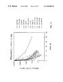
- FIG. 8 depicts the plasma IL-1ra concentration versus time profiles of mature IL-1ra, pegylated IL-1ra, and several pegylated muteins of IL-1ra.
- FIG. 9 shows the SDS-PAGE gel showing c105 30 kDa TNF inhibitor and mPEG, and the separation of unreacted 30 kDa TNF inhibitor from mPEG c105 30 kDa TNF inhibitor by size exclusion chromatography.
- FIG. 10 shows a plot containing intravenous plasma IL-1ra concentration versus time curves for a large number of singly PEGylated IL-1ra species, doubly PEGylated IL-1ra species, and IL-1ra PEG dumbbell species.
- FIG. 11 shows a plot containing subcutaneous plasma IL-1ra concentration versus time curves for a number of IL-1ra species as in FIG. 10 .
- FIG. 12 shows a plot of plasma IL-6 levels versus time after the injection of mice with hrTNF.
- FIG. 13 compares IL-6 levels induced in mice by five ratios of c105 30 kDa TNF inhibitor to TNF ( FIG. 13A ) and five ratios of c105 30 kDa TNF inhibitor to PEG 2000 db to TNF (FIG. 13 B).
- FIG. 14 depicts plasma IL-6 levels induced in mice by TNF alone and one to one ratios of TNF to c105 30 kDa TNF inhibitor PEG 3500 and PEG 10,000 dumbbells.
- FIG. 15 depicts percent neutrophils induced by varying ratios of TNF to c105 30 kDa TNF inhibitor (FIG. 15 A), c105 30 kDa TNF inhibitor PEG 3500 db (FIG. 15 B); c105 30 kDa TNF inhibitor PEG 10,000 db (FIG. 13 C); and c105 30 kDa TNF inhibitor PEG 20,000 db (FIG. 15 D).
- FIG. 16 shows a plot containing intravenous plasma 30 kDa TNF inhibitor concentration versus time curves for native 30 kDa TNF inhibitor, c105 30 kDa TNF inhibitor PEG 8500 , and PEG 10,000 and 30 kDa TNF inhibitor PEG 3500 , PEG 10,000 and PEG 20,000 dumbbells.
- FIG. 17 shows a plot containing subcutaneous plasma 30 kDa TNF inhibitor concentration versus time curves for a number of 30 kDa TNF inhibitor species as in FIG. 16 .
- FIG. 18 depicts the solubility of 3 solutions of native IL-1ra and c84 IL-1ra PEG 8500 by plotting O.D. 405 versus time.
- FIG. 19 depicts the basic structure of compounds of this invention having the general formula R 1 —X—R 2 that are referred to as dumbbell compounds.
- This invention involves the selective modification of pharmaceutically useful polypeptides, in particular, Tumor Necrosis Factor (“TNF”) inhibitors and interleukin-1 (“IL-1”) inhibitors. More specifically this invention describes the selective modification of 30 kDa TNF inhibitor and IL-1 receptor antagonist (“IL-1ra”). The selective modifications serve to both enhance the pharmacokinetic properties of the polypeptides as well as to provide homogenous compositions for human therapeutic use.
- TNF Tumor Necrosis Factor
- IL-1ra interleukin-1 receptor antagonist
- interleukin-2 receptors (“IL-2r”) and CR1. All references to interleukin-2 receptor shall be construed to include both ⁇ and ⁇ chains of IL-2r unless stated otherwise.
- the modified polypeptides and DNA sequences are human. However, to the extent that there is sufficient homology between animal DNA and peptide sequences to the human forms, they would be included within the scope of this invention.
- the method of modification of the present invention includes covalently bonding long chain polymers to the polypeptides of interest in a site specific manner.
- the selected polypeptides may be the native or naturally occurring polypeptides of interest, or they may be biologically active muteins of the polypeptides that have been produced to enhance the modification process described herein.
- the method of the invention includes the selection, production and screening of desired muteins that will meet the objectives of this invention.
- the method for modifying polypeptides requires merely that the modification be made so that the resulting product be available in substantially purified form as that term is defined herein.
- the modified polypeptides of the present invention will be bonded to long chain polymers at specific positions of the amino acid sequence.
- the modified polypeptides of the present invention will retain a substantial portion of their biological activity.
- the modified polypeptides will retain at least one tenth of the biological activity of the native polypeptide in a receptor binding assay.
- the modified polypeptide will retain at least one fifth of the biological activity of the native polypeptide, and in the most preferred embodiment at least one fourth of the activity will be retained.
- the modified polypeptide will serve to improve the pharmacokinetic performance of the native polypeptide in at least one of the following areas:
- the long chain polymer will be polyethylene glycol or monomethoxy polyethylene glycol.
- a polyethylene glycol unit will be referred to herein as PEG and a monomethoxy polyethylene glycol unit will be referred to as mPEG.
- the approximate molecular weight of the polymeric unit will be given in subscripts. For example, a monomethoxy polyethylene glycol unit of approximate molecular weight of 5,000 will be depicted as mPEG 5000 or PEG 5000 .
- PPG polypropylene glycol
- PEG polyoxyethylated glycerol
- dextran colonic acids or other carbohydrate-based polymers and polymers of ⁇ -amino acids and biotin derivatives.
- the long chain polymer unit is dihydroxy polyethylene glycol, or HO—(CH 2 CH 2 O) n —H.
- the dihydroxy material When activated to bind covalently with polypeptides or other biologically active compounds as described below, the dihydroxy material will contain two reactive sites.
- the long chain polymer units are bonded to the polypeptide via covalent attachment to the sulfhydryl group (—SH) of a cysteine residue.
- —SH sulfhydryl group
- functionalized polymer units that will react specifically with sulfhydryl groups.
- the functional or reactive group attached to the long chain polymer is referred to herein as the activating group.
- Activating groups include the maleimide group, sulfhydryl group, thiol, triflate, tresylate, aziridine, oxirane and 5-pyridyl.
- the preferred activating groups are maleimides.
- Activated dihydroxy polyethylene glycols because of the physical separation between the ends of the polymeric chain, are nearly equally reactive at each end of the molecule.
- the activated dihydroxy polyethylene glycols or any other multi-activated long chain polymer unit—will react with polypeptides to form “dumbbell” shaped complexes where two polypeptides are joined by a long chain polymeric unit.
- dumbbell complexes where substantially purified compounds can be formed comprising two different polypeptide groups, or comprising a single polypeptide group and a different biologically active group. Examples of such heterodumbbell compounds are given below.
- cysteines The extent and availability for reaction of cysteines varies dramatically from polypeptide to polypeptide. Therefore, in the biologically-active form many polypeptides do not have “free” cysteines, or cysteines not bound to another cysteine. In addition, the existence of “free” cysteines does not mean that cysteines are accessible for binding to reactive reagents. Since the modification usually occurs on the active or three dimensionally folded polypeptide, little or no reaction will occur when a free cysteine is found within the “interior” of the folded structure. A further constraint when modifying polypeptides is the potential effect the modification may have on the active site of the polypeptide.
- the modification of a cysteine having a certain proximal relationship to the active site may effectively deactivate the polypeptide. Even when a great deal is known about the selected polypeptide, it is difficult, if not impossible, to accurately predict which cysteine residues may be effectively modified.
- glycosylation sites may be a good site for a mutation to include a free cysteine.
- information can also be used to select potential muteins.
- the addition or substitution of a cysteine residue at the amino terminus or carboxyl terminus of the polypeptide is also a likely prospect because of its location.
- the mutation of lysine residues to cysteine may be considered based on the assumption that lysines will generally be found on the surface of the biologically active polypeptide.
- the preferred method for the production of the muteins is by recombinantly expressing the gene coding for the mutein.
- the altered gene may be created either by standard site specific mutagenesis procedures on the native gene, or by the construction of the altered gene by standard gene synthesis procedures. These techniques are well known to those of ordinary skill in the art.
- the gene coding for the target mutein may be expressed in a variety of expression systems, including animal, insect and bacterial systems. To the extent that expression systems have been perfected for the expression of the native polypeptides, the same systems may be used for the target muteins.
- the genes coding for the target muteins are produced by site specific mutagenesis of the native gene, and the gene encoding the mutein is expressed from a bacterial expression system.
- the gene encoding native IL-1ra and a method for expressing said gene in E. Coli is described in detail in U.S. Pat. No. 5,075,222 of Hannum et al., issued Dec. 24, 1991.
- the muteins and pegylated materials of the present invention include allelic variations in the protein sequence (sequence variations due to natural variability from individual to individual) and substantially equivalent proteins. “Substantially equivalent,” as used throughout the specification and claims is defined to mean possessing a very high degree of amino acid residue homology (See generally, M. Dayhoff, Atlas of Protein Sequence and Structure , vol. 5, p. 124 (1972), National Biochemical Research Foundation, Washington, D.C. specifically incorporated herein by references) as well as possessing comparable biological activity.
- the degree of homology is in excess of 70 percent, more perferably in excess of 80 percent and even more perferably in excess of 90 percent.
- a particularly preferred group of inhibitors are in excess of 95 percent homologous with the native inhibitor.
- the percentage homology as described is calculated as the percentage of amino acid residues found in the smaller of the two sequences that align with identical amino acid residues in the sequence being compared when four gaps in a length of 100 amino acids may be introduced to assist in that alignment as set forth by Dayhoff, M. D. in Atlas of Protein Sequence and Structure, Vol. 5, p.124 (1972), National Biochemical Research Foundation, Washington, D.C. Also included within the scope of this invention are muteins and pegylated polypeptides that are partially truncated versions of the native polypeptide.
- the mild reducing agent is dithiothreitol (“DTT”).
- DTT dithiothreitol
- the modification may occur prior to the refolding of the expressed protein or mutein.
- the pegylated muteins and pegylated native polypeptides may be purified and formulated into pharmaceutical compositions by conventional methods.
- the purified muteins may also be formulated into pharmaceutical compositions.
- the pegylated polypeptides of the present invention formed by the reaction of a deactivated long chain polymer unit have additional beneficial properties.
- These “dumbbell” shaped molecules can contain two of the polypeptides of interest attached by a single polymer unit. This structure imposes a certain amount of linearity to the polymeric molecule and reduces some of the steric hinderance inherent in the use of large hydrophilic polymers such as polyethylene glycol. The goal of obtaining molecules with increased apparent molecular weight is achieved while retaining high biological activity.
- bidentate molecules where two IL-1ra molecules or two TNF inhibitor molecules are covalently attached to a single polymeric chain, or where two different polypeptides are attached to a single polymeric chain, i.e., a single bidentate molecule containing both a TNF inhibitor and a IL-1ra moiety.
- Native IL-1ra ( FIG. 1 ) and various muteins of IL-1ra have been pegylated according to the present invention.
- Pegylation of wild type IL-1ra at free sulphydryl groups results in the addition of mPEG at the cysteine residue at position 116 of IL-1ra (c116).
- the other three cysteines are not accessible for pegylation in the fully native molecule.
- IL-1ra in which native amino acids in IL-1ra were replaced with a cysteine, or additional cysteines are added at the amino-terminus of the protein.
- residue 116 is not pegylated c116 has been changed to a serine in a number of the muteins.
- Native 30 kDa TNF inhibitor ( FIG. 2 ) does not contain any free cysteine residues.
- the following muteins of 30 kDa TNF inhibitor have been prepared (the residue numbering is based on the sequence given in FIG. 2 ; c referring to cysteine):
- R 1 —X—R 2 wherein R 1 and R 2 are biologically active groups and at least one of R 1 and R 2 is polypeptidic, and X is a non-peptidic polymeric spacer or linker group.
- R 1 and R 2 may be the same group or different. Where R 1 and R 2 are different groups, both R 1 and R 2 may be polypeptidic, or R 1 may be polypeptidic and R 2 may be any biologically active group.
- the compounds having this structure which have been referred to as “dumbbell” compounds, are characterized by being substantially purified. “Substantially purified” in this context is defined as being a homogenous composition.
- a homogenous composition consists of one molecule of the linker X and one molecule of R 1 and one molecule of R 2 .
- a homogenous composition includes, but does hot require, that the biologically active groups R 1 and R 2 be attached to the linker at the exact same location on the groups in each molecule of the compound.
- the biologically active groups are attached site specifically to the linker. For example, in the compound c105 30 kDa TNF inhibitor PEG 3000 db, two c105 30 kDa TNF inhibitor groups are attached at the 105 cysteine residue to the PEG 3000 linker.
- the dumbbell compound is also not necessarily homogenous with respect to the exact length of the spacer group. It is understood by those skilled in the art that any production process that utilizes a given weight range of PEG or other higher molecular weight polymer begins with a solution that contains an “average” molecular weight. Therefore, when a bis-reactive PEG unit is reacted with a polypeptidic group, the PEG unit is by definition polydisperse, and the resultant dumbbell compound is heterogenous to the extent that the length of the linker is subject to the variation known to exist by those skilled in the art.
- substantially purified in this context refers to materials that are substantially free from compounds: 1) that deviate in the composition of R 1 or R 2 ; or 2) that are linked together by more than one linker X.
- R 1 and R 2 are defined as being biologically active groups.
- Biologically active groups include any compound that can induce a biological effect on interact with a natural biological molecule.
- Biologically active groups include proteins, polypeptides, steroids, carbohydrates, organic species such as heparin, metal containing agents, vitamins, or any other biologically active species.
- At least one of the groups R 1 and R 2 is polypeptidic. In the preferred embodiment, both R 1 and R 2 are polypeptidic.
- Polypeptidic is defined as any compound that is substantially proteinaceous in nature. However, a polypeptidic group may contain some non-peptidic elements. For example, glycosylated polypeptides or synthetically modified proteins are included within the definition.
- the biologically active groups R 1 and R 2 include binding groups and targeting groups. Binding groups are defined by their affinity for a given biological ligand. Targeting groups are defined by their ability to direct the location of a complex within a biological system. R 1 and R 2 may have affinity for the same ligand, in which case the dumbbell may have enhanced affinity to that ligand. R 1 and R 2 may have an affinity for different ligands, wherein R 1 serves to target the complex into a location where the ligand for R 2 predominates.
- Preferred polypeptidic groups are receptors, the extracellular portions of receptors, cell surface molecules, and extracellular matrix molecules, binding proteins, and receptor antagonists. Included among the polypeptidic groups that may be used as R 1 or R 2 are the following polypeptides and any fragment thereof: IL-1 receptor antagonist, 30 kDa TNF inhibitor, 40 kDa TNF inhibitor, Il-2 receptor, CR1 (all references to CR1 include any single or combination of consensus repeat sequences of CR1), PDGF receptor, IL-2, MCSF receptor, EGF receptor, IL-5 receptor, IL-3 receptor, GMCSF receptor, T-cell receptor, HLA-I, HLA-II, NGF receptor, IgG (V H , V I ), CD40, CD27, IL-6 receptor, Integrins CR3, VLA 4 , ICAM, and VCAM, CR2, GMP140 Lec domain, Laminin binding protein, Laminin fragments, Mannose binding protein, exon 6 peptide of PDGF, and protea
- the groups R 1 and R 2 are selected from the group consisting of IL-1 receptor antagonist, 30 kDa TNF inhibitor, CR1, and IL-2 receptor (both the ⁇ and ⁇ chains).
- Non-peptidic is defined as a polymeric group that is substantially not peptidic in nature.
- the inclusion of less than 50% by weight of ⁇ -amino acid residue as part of Y 1 , Y 2 and Z would be considered substantially non-peptidic in nature and would be considered non-peptidic.
- the non-peptidic spacer X is non-immunogenic, and biologically inert and hydrophilic.
- the preferred linkers are capable of conveying desirable properties to the biologically active polypeptidic groups—such as reduced immunogencity, increased solubility, or reduced clearance rate from the body—without significantly reducing the affinity of a given R 1 or R 2 group to its ligand.
- substantially purified c105 30 kDa TNF inhibitor PEG 3400 db has an inhibitor activity for TNF that is greater than 20 times the inhibitor activity that c105 30 kDa TNF inhibitor has for TNF.
- the activating groups Y 1 and Y 2 that are part of the polymeric spacer X may be comprised of any of the activating groups as discussed above, including the maleimide group, sulfhydryl group, thiol, triflate, tresylate, aziridine, oxirane, and 5-pyridyl.
- the preferred activating groups are maleimides.
- the polymeric group (Z) n is preferably selected from the group consisting of polyethylene glycol, polypropylene glycol, polyoxyethylated glycerol, dextran, poly ⁇ -amino acids, colonic acids or other carbohydrate polymers and polymers of biotin derivatives.
- the polymeric group is polyethylene glycol. Any non-peptidic polymeric group that would serve the functions as described herein would also be included within the scope of this invention.
- One of the advantages of the present invention is the ability to vary the distance between the groups R 1 and R 2 by varying the length of the polymeric group linking the two binding groups.
- the increase in biological activity seen for the multimeric compounds of this invention may be attributed to the multimeric nature of the cell receptors and ligands in vivo.
- the optimal distance between the units R 1 and R 2 (which would be generally directly proportional to the length of the polymeric unit (Z) n ) may be easily determined by one skilled in the art by varying the size of the spacer X.
- the groups R 1 and R 2 are the same. However, in an alternate embodiment R 1 and R 2 are different species. Such compounds can be designed to create a heterodimer wherein both R 1 and R 2 act within the same general biological systems. For example, both IL-1 receptor antagonist and TNF inhibitors are believed to disrupt the inflammation cascade.
- the difunctional complexes may also be designed where R 1 or R 2 is a “targeting” species that “directs” the complex to a specific location by its binding affinity to a certain substrate, and the opposing binding, roup has a desired activity at the localized site.
- heterodimer that has great potential for being a successful IL-2 inhibitor is one where R 1 is IL-2r ⁇ and R 2 is IL-2r ⁇ . Such a heterodimer mimics the receptor complex that has the highest affinity for IL-2. See Example XVII.
- An additional heterodimer that can act as a complement inhibitor is the heterodimer where R 1 is the C3b binding domain from CR1 and R 2 is the C4b binding domain from CR1. See Example XVIII.
- R 1 is the exon 6 peptide of PDGF and R 2 is IL-1ra. See Example XIX.
- the procedures for producing the bifunctional R 1 —X—R 2 complexes are essentially the same as those used for the site-selective reaction of polypeptides as described above.
- the synthesis of c105 30 kDa TNF inhibitor PEG 3400 db is described below in Example 13.
- a bis-reactive polymeric group is reacted with a cysteine-containing polypeptide, wherein the activating group on the bis-reactive polymeric group forms a thio-ether bond with the selected free cysteine residue.
- the cysteine may be a free cysteine naturally-occurring on the polypeptidic group, or a non-native cysteine that has been added or substituted into the natural sequence.
- the preferred bis-reactive polymeric compound of the present invention is ⁇ -(2-maleimido) ⁇ -maleimido poly(oxyethylene) or bis-maleimido PEG.
- the synthesis of bis-maleimido PEG is described in Example 12. According to the preferred method, the bis-maleimido compound is prepared from bis-hydroxyl PEG via the bis-amino intermediate.
- the reactive intermediate in the conversion of the hydroxyl to the amine may be the halogenated derivative (e.g. the ⁇ -(bromoethyl) - ⁇ -bromopoly(oxyethylene) intermediate (Johannson, Biochim. et Biophy . vol. 222, pg. 381 (1970)) followed by direct substitution with ammonia, (Buckmann et al., Makromol. Chem . vol. 182, pg.
- the halogenated derivative e.g. the ⁇ -(bromoethyl) - ⁇ -bromopoly(oxyethylene) intermediate (Johannson, Biochim. et Biophy . vol. 222, pg. 381 (1970)
- direct substitution with ammonia (Buckmann et al., Makromol. Chem . vol. 182, pg.
- the bis-maleimide PEG is not the only sulfhydryl-specific reagent that may be used. Glass and coworkers have developed another method for the attachment of PEG to sulfhydryls. Glass et al., J. Biopolymers vol. 18, pg. 383 (1979). However, the reaction is reversible with thiols. Another method for attachment of PEG to cysteinyl sulhydryls is the bis-4-vinylpyridine PEG derivative.
- Harris also reviews the synthesis of a variety of electrophilic derivatives of PEG that can be used as reagents to modify proteins.
- the reagents include chlorocarbonates, isocyanate, epoxide, succinimidyl succinate, cyanuric chloride, mixed anhydride, carbodiimides and sulfonates.
- the latter group includes tresylate, tosylate, and mesylates.
- Some of the reagents react selectively with amines (e.g., cyanuric chloride and carbodiimides) while others react with both sulhydryls and amines (e.g., epoxide and tresylates).
- Some of these reagents have been used to modify proteins and may result in varying degrees in loss of activity.
- R 1 —X—R 2 complexes where R 1 and R 2 are different requires a two step process where the bis-reactive polymeric group is reacted in series with R 1 and then R 2 .
- the preparation of such heterodimers may be accomplished by those of ordinary skill in the art without undue experimentation.
- the intermediate R 1 —X must first be isolated and purified prior to reaction with R 2 , and in other circumstances an intermediate purification may not be necessary.
- the extracellular domains of both IL-2r ⁇ and IL-2r ⁇ may be cloned using PCR and cloned into a vector capable of directing expression in E. coli .
- the proteins may be refolded and purified from E. coli and their ability to inhibit IL-2 activity measured in bioassays.
- In vitro mutagenesis can be used to substitute native residues in the molecules with cysteine to allow for site directed attachment of PEG. Muteins of both IL-2r ⁇ and IL-2r ⁇ may then be identified that allow for efficient attachment of PEG which do not lose activity when PEGylated.
- a PEG-linked heterodimer may be formed by first PEGylating IL-2r ⁇ in the presence of an excess of bis-maleimido PEG.
- the singly PEGylated IL-2r ⁇ may be purified and IL-2r ⁇ added to react with the active maleimide group and form the heterodimer.
- This molecule may be purified and its activity assessed. This molecule should mimic the high affinity IL-2 receptor found on cell surfaces.
- a dumbbell complex where R 1 is IL-2 and R 2 is IL-2r ⁇ should also be useful as a receptor antagonist of IL-2.
- the succinate ester derivative of the mPEG x (intermediate 1) was prepared as described by Wie et al. Int. Archs. Allergy App. Immun ., vol. 64, pp. 84-99 (1981). The resulting product was weighed out and dissolved in a minimum of dry dioxane at 60° C. After the solution had cooled to ambient temperature, equimolar amounts of both tri-n-butylamine and isobutyl chloroformate were added. The reaction proceeded thirty minutes with stirring. During this time, a borate buffer, pH 8.8, was made by titrating a solution of 0.5 M boric acid with 1,6-hexanediamine.
- the solution containing the mixed anhydride was added dropwise to an aliquot of the borate buffer containing a 10-fold molar excess of 1,6-hexanediamine over the mixed anhydride.
- the reaction mixture was exhaustively dialyzed versus deionized water at 4° C. and lyophilized.
- This polymer intermediate (intermediate 2) was reacted with a 2.5:1 molar excess of sulfosuccinimidyl 4-(N-maleimiodethyl) cyclohexane-1-carboxylate (sulfo-SMCC, Pierce Chemical Co., Rockford Ill.) in 50 mM sodium phosphate or HEPES buffer, pH 7.0, for two hours at room temperature.
- the resulting polymer was purified by size exclusion chromatography of the reaction mixture on Sephadex G-25 using 50 mM sodium phosphate (or HEPES) pH 7.0 for elution at 4° C.
- the maleimido-polymer (reagent 1) eluted at the void volume of the column and was detected by monitoring its absorbance at 260 nanometers.
- the reagent was used to alkylate polypeptides within one hour of its purification. Since the mPEG from this reaction can be removed by base hydrolysis, this reagent is useful for identifying the site of mPEG attachment to the protein.
- the mPEG x -tosylate (intermediate 3) was prepared as described by Pillai et al. J. Org. Chem . vol. 45, pp. 5364-5370 (1980).
- the amount of sulfonated intermediate was estimated spectrophotometrically as described by Nilson and Mosbach, in Methods of Enzymology , vol. 104, pp. 56-69, Academic Pres. Inc., N.Y., N.Y. (1984).
- This intermediate was converted to the phthalimide derivative (intermediate 4) and subsequently reduced with hydrazine hydrate to the mPEG x —NH 2 intermediate (intermediate 5) by the procedure of Pillai et al., supra.
- the amino group capacity in equivalents per gram of product was quantified by microtitration with hydrochloric acid.
- the mPEG x —NH 2 was reacted with sulfo-SMCC in HEPES or phosphate buffer pH 7.2 at room temperature for two hours.
- the amount of the mPEG x -amine to sulfo-SMCC was tested at molar ratios of 5:1 to 1:5.
- reagent 2 was used in pegylation reactions and the quantity and quality of mPEG x *IL-1ra (we will use this designation for the pegylated product of IL-1ra reacted with reagent 2 and mPEG x IL-1ra for pegylated IL-1ra from a reaction with reagent 3 described below) obtained from these reactions was assessed by SDS-polyacrylamide gel electrophoresis (PAGE). The optimal result was seen with a 1:1 ratio of SMCC to mPEG x —NH 2 .
- Reagent 2 was purified by size exclusion chromatography using G25 sephadex resin.
- the mPEG x —NH 2 (intermediate 5) can be modified further to yield a different maleimido-derivative (reagent 3).
- the latter was accomplished by reacting the mPEG x —NH 2 with maleic anhydride via an adaptation of the procedure of Butler and Hartley, in Methods of Enzymology , vol. XXV pp. 191-199, Academic Press. Inc., N.Y., N.Y. (1972) and cyclizing this intermediate (intermediate 6) to the corresponding O-(2-maleimido ethyl)-O′-methylpolyethylene glycol using the method described by Wunsch et al., Biol. Chem. Hoppe - Seyler , vol. 366, pp. 53-61 (1985).
- Pegylation reactions at room temperature were analyzed from 0.5 to 24 hours. Conversion of the IL-1ra to the pegylated form is complete (80%-90%) in two to four hours and the total amount of mPEG*IL-1ra does not increase or decrease after longer periods of incubation. The quality of the mPEG*IL-1ra assayed by SDS-PAGE decreases at longer times due to the appearance of additional bands and smears at higher molecular weights on the stained gel.
- Pegylation reactions were incubated at temperatures of 4°, 25°, 37°, and 50° C. and then analyzed at time points of 0.5, 1, 2, 4 and 17 hours.
- the reactions at 25° and 37° generated a large amount (about 50%-80%) of pegylated protein within one to two hours but those at 4° C. and 50° C. resulted in a much lower yield (10%-20%) even at the later time points.
- the quality of the mPEG*IL-1ra does not seem to change significantly with temperature.
- Native IL-1ra was pegylated under the reaction conditions stated above between pH 5.5 and 7.5.
- the quality of the mPEG*IL-1ra is slightly better by SDS-PAGE and ion exchange at a lower pH (5.5) but the percent conversion is the same.
- Ratios higher than about 2:1 result in efficient conversion to the pegylated form of IL-1ra (50%-90%).
- Ratios greater than 5:1 generate lower quality mPEG*IL-1ra by increasing the amount of extra high molecular weight bands on reduced SDS-PAGE and multiple peaks on ion exchange chromatography.
- the optimal reaction conditions for both quantity of mPEG*IL-1ra obtained and quality of the material, within the parameters used, is a 2:1 mPEG-amido-maleimide/IL-1ra at 25° C. for 2-4 hours using mPEG-amido-maleimide generated with a 1:1 ratio of Sulfo-SMCC to mPEG-amine. With these conditions 80-90% of the IL-1ra is converted to the pegylated form using reagent synthesized with either mPEG 5000 or mPEG 8500 as the starting material (FIG. 3 ).
- PEG dumbbell complexes containing IL-1ra are made according to the same procedures as other PEGylated IL-1ra species. A 2-4 molar excess of bis-maleimido PEG to IL-1ra in HEPES buffer at 7.0 is used. With IL-1ra, the species used may be the wild type molecule, which has a free and available cysteine residue, or a mutein prepared as described herein. The IL-1ra is at a concentration of 2-5 mg/ml. The reaction is incubated at ambient temperatures for 4 to 6 hours.
- the IL-1ra PEG dumbbell compounds are purified from the unPEGylated and singly PEGylated species by MonoS cation exchange at pH5.5 in 20-50 mM MES buffer using a gradient from 0 to 1000 mM NaCl. Further purification may be achieved by size exclusion chromatography using a BioRad TSK 250 or Superdex 75 column, as described below.
- mPEG x *IL-1ra Purification of mPEG x *IL-1ra can be achieved by cation exchange or size exclusion chromatography. These procedures are applicable to pegylated IL-1ra derived from all three reagents described above.
- the mPEG x *IL-1ra can be purified using a MonoS (Pharmacia) column with 20 mM MES buffer at pH 5.5.
- the proteins were eluted from the column using a salt gradient from 0 to 500 mM NaCl in the same buffer.
- unmodified IL-1ra elutes at 220 mM NaCl, while the purity is assessed by various techniques including analytical ion exchange chromatography and SDS-PAGE.
- mPEG 5000 IL-ra elutes at 160 mM (FIGS. 4 A and 4 B).
- mPEG 5000 *IL-1ra which runs as about 52 kd
- mPEG 8500 *IL-1ra which runs as about 68 kd (based on column calibration with known size standards)
- mPEG 5000 *IL-1ra which runs as about 52 kd
- mPEG 8500 *IL-1ra which runs as about 68 kd (based on column calibration with known size standards)
- FIG. 5 The mPEG 5000 *IL-1ra, which runs as about 52 kd
- mPEG 8500 *IL-1ra which runs as about 68 kd
- Mutagenesis was performed on single stranded DNA from the IL-1ra gene cloned into the bacteriophage M13.
- BioRad's Mutagene kit was used which uses the procedure described by Kunkel et al. Methods in Enzymology vol. 154, pp. 367-382 (1987). Briefly, single stranded DNA template was generated using an E. coli strain that contains the dut and ung mutations, resulting in template that contains uracil instead of thymidine. Mutagenic oligonucleotides between 20 and 30 base pairs in length were annealed to the template and the second strand was resynthesized using DNA polymerase and DNA ligase.
- the reaction mixtures were used to transform a wild type .
- E. coli strain in which the uracil containing strand is degraded by the DNA repair mechanisms and the mutant strand is allowed to replicate.
- the mutant phage were screened and sequenced by standard techniques.
- the fragment containing the mutant gene was then subcloned into the expression vector pT5T (Eisenberg et al. Nature vol. 343, pp. 341-346, (1989)) and transformed into the T7 expression system strain ( E. coli B121DE3).
- Other E. coli expression systems may also be used.
- Expression clones were grown in Luria Broth supplemented with 15 ug/ml tetracycline at 37° C. When the cultures reached an optical density of 0.8 at 600 nm they were moved to 30° and IPTG was added to a final concentration of 1 mM to induce expression of the IL-1ra gene. Total accumulation of the IL-1ra protein was maximal after 4-6 hours and did not change significantly for up to 12 hours post induction.
- Cell cultures induced as described above were harvested by centrifugation at 10000 g for 10 min.
- the cells were resuspended in 30 mM sodium acetate buffer pH 5.2 in 20-50 mls. Lysis was achieved by two passes through the French Pressure cell at 18000 psi.
- the cell lysate was centrifuged at 10000 g for 10 minutes.
- the soluble portion was loaded onto a S-Sepharose column and washed with the same buffer containing 75mM NaCl.
- the IL-1ra mutein eluted with buffer containing 200 mM NaCl.
- the single pass over the ion exchange resin resulted in a product of sufficient purity (>95%) for pegylation studies.
- muteins c84s116, c84c116, c0s116 and c9s116 were pegylated.
- the pegylated forms of c84s116 and c84c116 were produced and purified. Since c84c116 contains two reactive cysteines, pegylation results in a higher molecular weight protein at about 40 kd on SDS-PAGE. This protein can be purified by cation exchange or size exclusion chromatography and runs at the expected molecular weight of about 68 kd on the latter when using PEG 5000 .
- the efficacy of the pegylated native IL-1ra molecules was tested by a standard competitive receptor binding assay using S 35 -IL-1ra as the ligand.
- Mouse cells (EL4) containing the mouse type 1 IL-1 receptor or hamster cells (CHO) expressing from a cloned gene the human type 1 receptor were used at 1 ⁇ 10 cells per well and 1 ⁇ 10 5 cells per well, respectively, in 96 well microliter dishes.
- S 35 -IL-1ra with a specific activity of 4000 Ci/mmol was added to a final concentration of 150 pM.
- Cold ligand was added in serial dilutions from 28 mM to 13 pM and allowed to incubate for 4 hours at 4° C.
- the cells were then filtered through a Milliliter filter plate (Millipore, 0.5 micron pore size Durapore filter), washed to remove nonspecifically bound counts, the filter removed and counted on an Ambis Radioanalytical Imaging System.
- Equilibrium dissociation constants were calculated and used to compare the pegylated and unmodified forms of IL-1ra.
- Unmodified wild type IL-1ra and c84s116 have equal kD's for the type 1 mouse receptor of 150-300 pM in our assay.
- the kD for the IL-1ra pegylated form is about 400-800 pM and for pegylated c84s116, 500-1000 pM which is 2.5 and 3.5 fold higher than that of the unmodified protein respectively.
- the kDs for all but one (c6s116) of the unpegylated muteins are within 65-150% of the native protein, within the standard error of the assay. See Table 1.
- the pharmacokinetic character of several pegylated native and mutein IL-1ra molecules was tested following intravenous injection of the molecules to rats.
- Native or pegylated IL-1ra was injected as an intravenous bolus dose (3 mg/kg).
- Serial blood samples were drawn from the tail vein and assayed for native or pegylated IL-1ra by enzyme-linked immunosorbent assay (ELISA).
- ELISA enzyme-linked immunosorbent assay
- the resulting plasma IL-1ra concentration vs. time profiles illustrate that pegylation has a pronounced influence on the disappearance of IL-1ra from the plasma after intravenous injection.
- the declines in plasma IL-1ra and pegylated derivatives of IL-1ra are best described by three exponential components.
- pegylation improves therapy with IL-1ra by increasing the extent to which the active molecules move from the systemic circulation into the extravascular compartment, a compartment in which IL-1 receptors are expected to be located. Because of the similarity between rats and humans in both clearance and distribution mechanisms for IL-1ra, it is apparent that pegylation will similarly improve the pharmacokinetic properties of IL-1ra in humans.
- the intravenous pharmacokinetics for eight additional pegylated IL-1ra muteins have been characterized using methods previously described.
- a plot containing intravenous plasma IL-1ra concentration vs. time curves for each of the molecules is attached (FIG. 10 ).
- Review of all of the intravenous pharmacokinetic data indicates that as the size of the PEG (single or double) is increased, the plasma clearance decreases and hence the intravenous mean residence time and plasma IL-1ra disappearance half-lives increase.
- the site of pegylation is important in determining the extent to which the pegylation decreases the plasma clearance and prolongs the means residence time.
- the addition of two PEGs to IL-1ra prolongs the intravenous mean residence time fourteen-fold compared to wild type IL-1ra.
- the mean residence time is generally increased. This increase is probably the result of molecule-size-related slower absorption through the lymphatic circulation (longer mean absorption times) as well as to delayed clearance after the pegylated molecule reaches the systemic circulation (plasma). This prolongation is profound and will improve the pharmacokinetic character of subcutaneous IL-1ra in humans.
- Cysteine has been substituted for the native residue at both the amino terminus and carboxyl terminus of the protein as well as all three glycosylation sites (residues 1, 14, 105, 111 and 161 as seen in FIG. 2 ).
- Mutagenesis was performed on single stranded DNA from the 30 kDa TNF inhibitor gene cloned into the bacteriophage M13. This gene is described in detail in U.S. patent application Ser. No. 07/555,274 filed Jul. 19, 1990. Mutagenesis was done as described by Kunkel et al. (1987) (see Example V). The mutagenized gene was isolated and subcloned into the expression vector pT5T (Eisenberg et al., Nature vol. 343, pg.
- Refolding includes the addition of cysteine to the solution containing the purified protein. The cysteine aids in the refolding and “bonds to” the free cysteine in the mutein.
- the c105 30 kDa TNF Inhibitor mutein was exposed to a 6-fold molar excess of DTT in 50 mM HEPES Ph 7.0 for 30 minutes at ambient temperature in order to remove an extra cysteine attached during the refolding process.
- the protein was then dialyzed against de-gassed 50 mM HEPES pH 7.0 for 2 hours to remove the DTT.
- the c105 30 kDa TNF inhibitor was then reacted with a 5 fold molar excess of pegylating reagent 1 (See Example 1A) for 2 hours at ambient temperature in 50 mM HEPES pH 7.0. Approximately 60% of the mutein was converted to the pegylated form.
- the c105 pegylation reaction mixture was loaded onto a superdex-75 FPLC column (Pharmacia) run at 0.25 ml/min in 50 mM Tris pH 7.0, 100 mM NaCl.
- Fractions containing c105-PEG 30 kDa TNF-inhibitor were pooled and loaded on a TSK-2000SW HPLC column (Bio-Rad) run at 0.2 ml/min in the same buffer.
- the activity was determined using the murine L929 cell TNF cytoxicity assay as described in U.S. patent application Ser. No. 07/555,274 filed Jul. 19, 1990.
- bisamino PEG The synthesis of the ⁇ -(2-aminoethyl) ⁇ -aminopoly(oxyethylene) derivative of the PEG (hereinafter bisamino PEG) consisted of three steps: 1) sulfonation of the hydroxyl group using tresyl chloride as described by Nilson and Mosback (Nilson et al., Methods in Enzyymology vol. 104, pg. 56, Academic Press, Inc., N.Y., N.Y. (1984)), 2) substitution of the tresylated intermediate by phthalimide (Pillai et al., J. Org. Chem . vol. 45, pg.
- the bisamino PEG was acylated using maleic anhydride (Butler et al., Methods in Enzymology vol. 25, pg. 191, Academic Press, Inc., N.Y., N.Y. (1972)) and the resulting intermediate was cyclized to produce ⁇ -(2-maleimidoethyl- ⁇ -maleimidopoly(oxyethylene) (Winsch et al., Biol. Chem. Hoppe - Seyler vol. 336, pg. 53 (1985)). This derivative reacts with sulfhydryls via a Michael addition to form a stable thioether.
- the inhibitory effects of four species of pegylated c105 30 kDa TNF inhibitor species were tested in vivo on two different TNF-stimulated physiological actions.
- One endpoint was the appearance of IL-6 in the plasma of nice that were injected intravenously with human recombinant TNF.
- the second endpoint was an increase in the migration of neutrophils into the peritoneal cavity after the intraperitoneal administration of human recombinant TNF.
- BALB/c female mice weighing 20 to 23 g were used to measure the induction of plasma IL-6 levels by human recombinant TNF.
- the time course was plotted for the appearance IL-6 in the plasma after the intravenous administration via the tail vein of two doses of human recombinant TNF (FIG. 12 ). Peak IL-6 levels occurred at two hours after stimulation with either 10 or 20 ug of human recombinant TNF per mouse. The lower dose was used in subsequent experiments.
- the potency of c105 30 kDa TNF inhibitor PEG 2000 dumbbell with that of the unpegylated c105 30 kDa TNF inhibitor was compared.
- Human recombinant TNF was injected intravenously at a dose of 10 ug per mouse either alone or simultaneously with the TNF inhibitors.
- Four different reactions of inhibitors to TNF were tested (FIGS. 13 A and 13 B). The ratios were calculated based on protein content. Three mice were tested at each dose. Blood was collected at two hours after the intravenous injections. IL-6 levels were measure by ELISA.
- Both the c105 30 kDa TNF inhibitor and c105 30 kDa TNF inhibitor PEG 2000 dumbbell caused nearly complete inhibition of IL-6 levels when administered at 10:1 and 5:1 ratios of inhibitor to TNF.
- the c105 30 kDa TNF inhibitor PEG 2000 dumbbell caused 95% reduction of IL-6 levels stimulated by TNF alone, whereas the unpegylated c105 30 kDa TNF inhibitor reduced IL-6 by only about 70%.
- the results of this experiment indicate that in the ratios tested, both the c105 30 kDa TNF inhibitor and c105 30 kDa TNF inhibitor PEG 2000 dumbbell were good inhibitors of this TNF-stimulated physiological parameter.
- the c105 30 kDa TNF inhibitor PEG 2000 dumbbell caused a greater percentage inhibition than the unpegylated inhibitor.
- c105 30 kDa TNF inhibitor Two other species of pegylated c105 30 kDa TNF inhibitor were tested.
- the inhibitory effects of c105 30 kDa TNF inhibitor PEG 3,500 dumbbell and c105 30 kDa TNF inhibitor PEG 10,000 dumbbell were tested on plasma IL-6 induction.
- the inhibitors were administered by intravenous injection simultaneously with human recombinant TNF at ratios of 1:1 (c105 30 kDa TNF inhibitor dumbbell: TNF) (FIG. 14 ).
- Three mice were tested in each of the two inhibitor-treated groups. Ten mice were injected with TNF alone.
- mice BALB/c female mice weighing 20 to 23 g were used to measure the migration of neutrophils into the peritoneal cavity after stimulation with human recombinant TNF.
- the technique used is that of Kim McIntyre et al. ( J. Exp. Med . vol. 173, pg. 931 (1991)) and is described in brief herein.
- Mice are injected with TNF in a volume of 0.1 ml directly into the peritoneal cavity. Four hours later the mice are killed and an immediate post mortem lavage of the peritoneal cavity is performed.
- Four ml of Hank's Balanced Salt Solution (HBS) (calcium and magnesium free) is injected into the peritoneal cavity. The abdomen is gently massaged.
- HBS Hank's Balanced Salt Solution
- the peritoneal fluid is recovered by aspiration with needle and syringe.
- the total number of peritoneal cells is counted on a Coulter counter.
- An aliquot of the cellular suspension is dried on a slide and stained with Diff-Kwik stain.
- a differential count of the cells is made by direct microscopic examination. One hundred cells are examined and classified as either neutrophils, lymphocytes, or macrophages.
- TNF caused an increase in the percentage of neutrophils and in the absolute number of neutrophils present in the peritoneal lavage fluid.
- saline-treated mice 9.4 ⁇ 10 4 neutrophils were recovered in the lavage fluid and made up only 2.3% of the total peritoneal cells.
- TNF (7.5 ng)-treated mice the total number of neutrophils was increased to 12.9 ⁇ 10 5 and the percentage of neutrophils was increased to 19.7%.
- the potency of unpegylated c105 30 kDa TNF inhibitor with three pegylated species of c105 30 kDA TNF inhibitor was also compared. Keeping the TNF stimulus constant at 7.5 ng per mouse, the inhibitors were tested at ratios of 100:1, 10:1, and 1:1 (c105 30 kDa TNF inhibitor species: TNF). The ratios were calculated based on protein content.
- the mice were injected subcutaneously with the c105 30 kDa TNF inhibitor simultaneous to the intraperitoneal administration of TNF. Six mice were tested in each dose group. Four hours later the peritoneal lavage fluid was collected and analyzed. Values shown in FIGS.
- 15A , 15 B, 15 C, and 15 D are the percentage neutrophils in the peritoneal lavage fluid.
- the lowest ratio at which the unpegylated c105 30 kDa TNF inhibitor and c105 30 kDa TNF inhibitor PEG 3,500 dumbbell significantly inhibited neutrophil migration is 100:1.
- c105 30 kDa TNF inhibitor PEG 3,500 PEG 10,000 and PEG 20,000 dumbbells are good inhibitors of the TNF-stimulated neutrophilic migration into the peritoneal cavity.
- the c105 30 kDa TNF inhibitors PEG 10,000 and PEG 20,000 dumbbells were more potent than the unpegylated c105 30 kDa TNF inhibitor and the c105 30 kDa TNF inhibitor PEG 3,500 .
- Recombinant c105 30 kDa TNF inhibitor 2-3 mg/ml is treated with a 4-fold molar excess of DTT for 2 hrs at ambient temperature.
- the TNF inhibitor is then dialyzed against de-gassed 50 mM HEPES, pH 7.0, for 3 hrs at 4° C.
- the TNF inhibitor is reacted with different molar ratios of the bis-maleimido PEG in 50 mM HEPES pH 7.0.
- TNF inhibitor is reacted with an equimolar ratio of bis-maleimido PEG. The reactions are incubated for 3-12 hrs at ambient temperature.
- the PEG-linked TNF inhibitor dumbbell is purified from un-PEGylated and singly-PEGylated TNF inhibitor using MONO-S FPLC in 50 mM HOAc, pH 4.0, using a 260 mM, 310 mM and 350 mM NaCl step-gradient.
- the PEG-linked TNF inhibitor dumbbell elutes at the 310 mM NaCl step. Any remaining unPEGylated TNF inhibitor is removed by chromatography on Superdex75.
- the TNF inhibitor dumbbells also have greatly increased activity in inhibiting the cytotoxicity of TNF ⁇ in the L929 bio-assay.
- the ED 50 values against TNF ⁇ are as follows:
- the pharmacokinetic character of several pegylated 30 kDa TNF inhibitor molecules was determined following intravenous administration of the molecules to rats.
- Native or pegylated TNF inhibitor was injected as an intravenous bolus dose.
- Serial blood samples were drawn from the tail vein and assayed for non-pegylated or pegylated TNF inhibitor by enzyme-linked immunosorbent assay (ELISA).
- ELISA enzyme-linked immunosorbent assay
- the resulting intravenous plasma TNF inhibitor concentration vs. time profiles illustrate that pegylation has a pronounced influence on the disappearance of TNF inhibitor from the plasma after intravenous injection.
- Statistical moment theory area under the curve [AUC] and area under the first moment curve [AUMC] was used to interpret the data of FIG. 16 .
- the intravenous mean residence time increases as the size of the attached PEG molecule increases (Table 4).
- the prolongation of mean residence times may be explained based on conventional pharmacokinetic theory which states that the intravenous mean residence time for a drug is inversely related to the plasma clearance for the drug and directly related to the apparent volume of distribution for the drug.
- Pharmacokinetic analysis of the disappearance of pegylated TNF inhibitor's from the plasma indicates that the prolongation of half-lives is inversely related to a decreased plasma clearance for the pegylated molecules, compared to non-pegylated TNF inhibitor (Table 4).
- the decrease in plasma clearance is consistent with an anticipated size-related decrease in glomerular filtration of the pegylated molecules by the kidneys. Because of the probable qualitative similarity between rats and humans in plasma clearance mechanisms for TNF inhibitor, it is apparent that pegylation will similarly improve the pharmacokinetic properties of TNF inhibitor in humans.
- this increase is likely the result of size-related slower absorption through the lymphatic circulation (longer mean absorption times) as well as delayed clearance once the pegylated molecule reaches the plasma. This prolongation is profound and will improve the pharmacokinetic character of subcutaneous TNF inhibitor in humans.
- Native 30 kDa TNF inhibitor cannot be concentrated to more than 5 mg/ml. Following PEGylation, the solubility was increased at least 5 fold.
- a PEG-linked heterodumbbell may be formed by first pegylating IL-2r ⁇ in the presence of an excess of bis-maleimido PEG.
- the singly pegylated IL-2r ⁇ may be purified and IL-2r ⁇ added to react with the remaining reactive maleimide group to form the heterodimer.
- Potential sites for PEGylation of Il-2r ⁇ include both the amino and carboxyl terminal residues, the two N-linked glycosylation sites, as well as the native free cysteine residue in the molecule.
- Cysteine residue 192 in the soluble extracellular domain of IL-2r ⁇ has been identified as being uninvolved in disulfide bonding. (Miedel et al. BBRC , vol. 154, pg. 372 (1988)).
- This cysteine residue lies in an epitope of an anti-IL-2r ⁇ monoclonal antibody that does not affect IL-2 binding to IL-2r ⁇ (Lorenzo et al. J. Immunology , vol. 147, pg. 2970 (1991)). This indicates this residue is a likely candidate for PEGylation without affecting the activity of IL-2r ⁇ .
- the potential sites include both the amino and carboxyl termini, the 4 N-linked glycosylation sites and a region (a.a. #108-118) that is similar to a region of biological significance in the murine erythropoietin receptor (Yoshimura, Longmore and Lodish, Nature , vol. 348, pg. 647 (1990)).
- Point mutational analysis of other residues in the receptors may also allow for identification of other sites of PEGylation that yield optimal properties in the heterodumbbell molecule.
- CR1 complement receptor 1
- the soluble form of CR1 has been examined in in vivo models of diseases.
- the complement inhibitor inhibits post-ischemic myocardial inflammation and necrosis (Weisman et al. Science , vol. 149, pg. 145-151, 1990), reversed passive arthus reaction (Yet et al. J. Immunology , vol. 146, pg. 250-256 (1991)), and allograft rejection (Pruitt et al. J. Surgical Research , vol. 50, pp. 350-355 (1991)).
- the soluble CR1 binds to C3b and C4b. It consists of 30 short consensus repeat sequences (SCR). Most of SCR contain one possible glycosylation site and four cysteines. All of the cysteines are likely be to involved in disulfide bonding. SCRs 1-4 are found to be involved in C4b binding. Two separate portions of CR1, SCRs 8-11 and SCRs 15-18, are involved in C3b binding (Klickstein et al. J. Exp. Med ., vol. 168, pp. 1699-1717 (1988): Kalli et al. J. Exp. Med . vol. 174, pp. 1451-1460 (1991)). According to this invention, it is possible to produce a heterodumbbell which contains the C4b binding domain and the C3b binding domain of CR1.
- the SCRs which contain C4b binding and C3b binding domains of CR1 may be cloned using PCR. These SCRs will be SCRs 1 through 5 (C4b binding) and SCRs 8 through 12 (C3b binding).
- the genes encoding these SCRs may be cloned in E. coli expression vector.
- the E. coli expressed-proteins may be refolded and purified. The success of refolding can be analyzed by the capacity to bind polyC3b or polyC4b. In vitro mutagenesis of these genes may be carried out to substitute native amino acid residues to cysteine. These cysteines may then be used to link the PEG molecule.
- Possible sites for PEGylation will be the glycosylation site or carboxyll terminal residue of SCR 5 and SCR 12.
- the C4b binding and C3b binding domains which contain an extra cysteine to the carboxyl terminal residue could be constructed and used for linking PEG molecule.
- the PEG linked heterodumbbell may be produced by the two step process of Example XIV. Purification may be carried out by ion-exchange chromatography.
- the platelet derived growth factor (PDGF) peptide YGRPRESGKKRKRLKPT is described in Khachigian, L. et al. J. Biol. Chem ., vol. 267, pg. 1660-1666 (1991). A terminal C was added to permit coupling to the maleimide.
- PDGF platelet derived growth factor
- the heterodumbbell was synthesized in two steps.
- first step 1.6 nanomoles of IL-1ra suspended in 3 ⁇ l of 0.05 M Hepes buffer, pH 7.5, was mixed with 6.4 nanomoles of bis-maleimido PEG 1900 dissolved in 11 ⁇ l of the same buffer. This reaction was carried out for 30 min at 20° C.
- second step 32 nanomoles of the PDGP peptide dissolved in 4 ⁇ l of 0.2 N sodium phosphate buffer, pH 7.0, was added to the products of the first reaction. The reaction was allowed to proceed for 1 hr at 20° C. The reaction was then terminated by the addition of an equal volume of SDS-PAGE sample buffer containing 30 ⁇ moles of 2-mercaptoethanol.
- the products of the first step of the reaction can be isolated by cation exchange chromatography on the resin S-Sepharose.
- the heterodimer may be isolated by cation exchange chromatography due to the abundance of basic amino acids in the peptide.
Abstract
Description
-
- Lane 1-IL-1ra
- Lane 2-mPEG* 5000 IL-1ra
- Lane 3-mPEG* 8500 IL-1ra
- Lane 4-c84s116 IL-1ra
- Lane 5-mPEG* 5000 c84s116 IL-1ra
- Lane 6-mPEG* 8500 c84s116 IL-1ra
-
- 1) increasing the apparent molecular weight of the native polypeptide and, hence, reducing the clearance rate following subcutaneous or systemic administration;
- 2) increasing the solubility of the native polypeptide in aqueous solutions; or
- 3) reducing the antigenicity of the native polypeptide.
-
- 1) The gene coding for the target mutein is created by site directed mutagenesis of the gene coding for the native polypeptide;
- 2) The gene coding for the target mutein is expressed in a bacterial expression system;
- 3) The target mutein is isolated from the bacteria and purified;
- 4) The target mutain is refolded in the presence of cysteine or another sulphydryl containing compound;
- 5) The refolded target mutein is isolated and purified;
- 6) The purified and refolded target mutein is treated with a mild reducing agent;
- 7) The reaction mixture is dialyzed in the absence of oxygen; and
- 8) The dialyzed reaction mixture is treated with a long chain polymer containing an activating group.
-
- c0s116 c0c116
- c84s116 C84c116
- c6s116 c6c116
- c8s116 c8c116
- c9s116 c9c116
- c141s116 c141c116
-
- c105 30 kDa TNF inhibitor
- c1 30 kDa TNF inhibitor
- c14 30 kDa TNF inhibitor
- c111 30 kDa TNF inhibitor
- c161 30 kDa TNF inhibitor
CH3O—(CH2CH2O)n—H
where x denotes the average molecular weight of the polymer in kilodaltons and n is the average number of
repeating oxythylene groups.

CH3O—(CH2CH2O)n—H
where x denotes the average molecular weight of the polymer in kilodaltons and n is the average number of repeating oxyethylene groups.

CH3O—(CH2CH2O)n−1—(CH2CH2)—NH2

| TABLE 1 |
| ANALYSIS OF PEGYLATED IL-1ra MOLECULES |
| SIZE | RECEPTOR | ||
| (kd) | ASSAY | ||
| MUTEIN | ||||
| WILD TYPE | 17.5 | 100 | ||
| C84S116 | 17.5 | 98 | ||
| C9S116 | 17.5 | 67 | ||
| C6S116 | 17.5 | 37 | ||
| C0S116 | 17.5 | 63 | ||
| C84C116 | 17.5 | 95 | ||
| PEG*IL-1ra | ||||
| SINGLE | ||||
| PEG5000c116 | 50-60 | 34 | ||
| PEG5000c84s116 | 50-60 | 28 | ||
| PEG8500c116 | 70-80 | 30 | ||
| PEG8500c84s116 | 70-80 | 30 | ||
| PEG8500c0s116 | ND | 22 | ||
| PEG8500c9s116 | ND | 12 | ||
| PEG12000c116 | 78 | 20 | ||
| DOUBLE | ||||
| PEG5000c84c116 | 70-80 | 11 | ||
| PEG8500c84C116 | 150-200 | 4 | ||
| PEG12000c84c116 | 175 | 5 | ||
| DUMBBELLS | ||||
| PEG3500c116 | 55-65 | 49 | ||
| PEG3500c84 | 60 | 49 | ||
| PEG10,000c116 | 175-200 | 49 | ||
| PEG10,000c84 | 200 | 60 | ||
| PEG20,000c84 | >200 | 24 | ||
| Data are presented as a percent of the activity exhibited by unmodified I1-1ra. Standard deviations are within 10%. | ||||
| TABLE 2 | ||||||
| C84 | C84 | |||||
| C116 | PEG(8500) | PEG(5000) | ||||
| wild type | PEG(8500) | PEG(5000) | S116 | S116 | ||
| n | 4 | 1 | 1 | 1 | 1 |
| Vd initial, ml/kg | 24 | 59 | 38 | 58 | 66 |
| Vd steady state, ml/kg | 110 | 160 | 150 | 240 | 290 |
| plasma clearance, ml/min/kg | 7.4 | 3.1 | 7.7 | 3.0 | 5.0 |
| t½ initial phase, min | 1.7 | 12 | 2.5 | 10 | 6.5 |
| t½ intermediate phase, min | 30 | 60 | 29 | 87 | 82 |
| t½ terminal phase, hr | 2.0 | 12 | 1.9 | 7.2 | 5.0 |
| mean residence time, hr | 0.25 | 0.86 | 0.32 | 1.4 | 0.96 |
HO—(CH2CH2O)n—H
where x denotes the average molecular weight of the polymer in kilodaltons and n is the average number of repeating oxyethylene groups.
Intermediate 1
F3—CH2—SO2—O—(CH2CH2O)n−1—(CH2CH2)—O—SO2—CH2—F3

Intermediate 3
H2N—(CH2CH2O)n−1—(CH2CH2)—NH2

| Wild Type rTNF inhibitor | 220 ng/ml | |||
| BMH-linked dumbbells | 220 ng/ml | |||
| 1900 MW PEG-dumbbells | 4.1 ng/ml | |||
| 3500 MW PEG-dumbbells | 4.8 ng/ml | |||
| 10,000 MW PEG-dumbbells | 4.6 ng/ml | |||
| 20,000 MW PEG-dumbbells | 4.2 ng/ml | |||
The TNF inhibitor dumbbells also have greatly increased activity in inhibiting the cytotoxicity of TNFβ in the L929 bio-assay. The ED50 values against TNFβ are as follows:
| Wild Type rTNF inhibitor | 70 μg/ml | ||
| 3400 MW PEG-dumbbells | 80 ng/ml | ||
| 20,000 MW PEG-dumbbells | 22 ng/ml | ||
| TABLE 3 |
| Pegylated Interleukin-1 Inhibitor Pharmacokinetics* in rats |
| type of pegylation = | none | single | single | single | single | single | single | double | dumbbell | dumbbell | dumbbell | dumbbell | dumbbell |
| PEG location = | [wild | 9 | 84 | 84 | 116 | 116 | 116 | 84,116 | 84 | 84 | 84 | 116 | 116 |
| PEG size = | type] | 8500 | 5000 | 8500 | 5000 | 8500 | 12000 | 8500 | **3400 | **10000 | **20000 | 3400 | **10000 |
| intravenous pharmacokinetics |
| Vd initial, mL/kg = | 24 | 51 | 72 | 90 | 52 | 63 | 61 | 50 | 45 | 48 | 58 | 47 | 40 |
| Vd steady-state, mL/kg = | 110 | 130 | 310 | 290 | 150 | 110 | 130 | 64 | 64 | 68 | 60 | 58 | 44 |
| plasma clearance, | 7.4 | 3.3 | 5.2 | 4.2 | 6.8 | 3.6 | 2.8 | 0.30 | 3.6 | 0.96 | 0.37 | 4.9 | 1.4 |
| mL/min/kg = | |||||||||||||
| plasma mean residence time | 0.25 | 0.65 | 1.0 | 1.2 | 0.37 | 0.52 | 0.76 | 3.5 | 0.30 | 1.2 | 2.7 | 0.19 | 0.52 |
| (i.v.), hr = | |||||||||||||
| t½ initial phase, min = | 1.7 | 8.8 | 5.3 | 11 | 4.6 | 11 | 13 | ||||||
| t½ intermediate phase, | 31 | 57 | 38 | 95 | 36 | 36 | 59 | 100 | 7.8 | 6.2 | 110 | 6.4 | 20 |
| min = | |||||||||||||
| t½ terminal phase, hr = | 2.8 | 3.0 | 2.3 | 6.9 | 2.3 | 3.1 | 7.0 | 7.4 | 0.74 | 0.87 | 5.7 | 1.0 | 2.6 |
| subcutaneous pharmacokinetics |
| systemic availability, % = | 68 | 37 | 74 | 99 | 28 | 30 | 30 | 25 | 20 | 7.9 | 3.3 | 7.6 | 4.7 |
| maximum plasma IL-1ra, | 1300 | 530 | 940 | 2100 | 360 | 540 | 630 | 1700 | 560 | 630 | 210 | 340 | 230 |
| ng/ml = | |||||||||||||
| time of maximum plasma | 0.82 | 1.8 | 2.5 | 4.0 | 1.5 | 1.3 | 1.8 | 12 | 3.0 | 3.1 | 5.4 | 0.75 | 0.51 |
| IL-1ra, hr = | |||||||||||||
| plasma mean residence time | 2.7 | 8.1 | 6.4 | 7.5 | 6.5 | 8.5 | 9.0 | 21 | 3.7 | 6.0 | 9.7 | 2.0 | 7.3 |
| (s.c.), hr = | |||||||||||||
| mean absorption time, hr = | 2.4 | 7.4 | 5.4 | 6.3 | 6.1 | 8.0 | 8.2 | 18 | 3.4 | 4.8 | 7.0 | 1.8 | 6.7 |
| *each pharmacokinetic parameter is expressed as the mean for two rats, except for C84-PEG(3400) dumbbell s.c. dose, for which n = 1 | |||||||||||||
| **pharmacokinetic information is caled from 1.5 mg/kg to 3 mg/kg dose | |||||||||||||
| TABLE 4 | |||||||
| type of pegylation = | none | none | single | single | dumbbell | dumbbell | dumbbell |
| PEG location = | [wild | C105 | C105 | C105 | C105 | C105 | C105 |
| PEG size = | type] | 0 | 8500 | 20000 | 3500 | 10000 | 20000 |
| intravenous pharmacokinetics | |||||||
| number of animals = | 2 | 2 | 2 | 2 | 2 | 2 | |
| Vd steady-state, mL/kg = | 230 | 240 | 140 | 340 | 93 | 130 | |
| plasma clearance, mL/min/kg = | 11.0 | 1.7 | 0.17 | 0.82 | 0.16 | 0.11 | |
| plasma mean residence time (i.v.), hr = | 0.37 | 2.3 | 14 | 6.8 | 10 | 19 | |
| subcutaneous pharmacokinetics | |||||||
| number of animals = | 2 | 2 | 1 | 1 | 2 | 2 | |
| systemic availability, % = | 99* | 25 | 65 | 29 | 39 | 34 | |
| plasma mean residence time (s.c.), hr = | 3.5 | 7.0 | 20 | 12 | 17 | 30 | |
| mean absorption time, hr = | 3.1* | 4.7 | 6.0 | 5.2 | 7.0 | 11 | |
| *referenced to C105 intravenous pharmacokinetics | |||||||
Claims (105)
Priority Applications (1)
| Application Number | Priority Date | Filing Date | Title |
|---|---|---|---|
| US08/482,283 US6858409B1 (en) | 1988-05-27 | 1995-06-07 | Nucleic acids encoding interleukin-1 inhibitors and processes for preparing interleukin-1 inhibitors |
Applications Claiming Priority (11)
| Application Number | Priority Date | Filing Date | Title |
|---|---|---|---|
| US19991588A | 1988-05-27 | 1988-05-27 | |
| US23871388A | 1988-08-31 | 1988-08-31 | |
| US24852188A | 1988-09-23 | 1988-09-23 | |
| US26653188A | 1988-11-03 | 1988-11-03 | |
| US07/506,522 US5075222A (en) | 1988-05-27 | 1990-04-06 | Interleukin-1 inhibitors |
| US55527490A | 1990-07-19 | 1990-07-19 | |
| US66986291A | 1991-03-15 | 1991-03-15 | |
| US82229692A | 1992-01-17 | 1992-01-17 | |
| US85067592A | 1992-03-13 | 1992-03-13 | |
| US31950694A | 1994-10-06 | 1994-10-06 | |
| US08/482,283 US6858409B1 (en) | 1988-05-27 | 1995-06-07 | Nucleic acids encoding interleukin-1 inhibitors and processes for preparing interleukin-1 inhibitors |
Related Parent Applications (1)
| Application Number | Title | Priority Date | Filing Date |
|---|---|---|---|
| US31950694A Division | 1988-05-27 | 1994-10-06 |
Publications (1)
| Publication Number | Publication Date |
|---|---|
| US6858409B1 true US6858409B1 (en) | 2005-02-22 |
Family
ID=34140017
Family Applications (1)
| Application Number | Title | Priority Date | Filing Date |
|---|---|---|---|
| US08/482,283 Expired - Lifetime US6858409B1 (en) | 1988-05-27 | 1995-06-07 | Nucleic acids encoding interleukin-1 inhibitors and processes for preparing interleukin-1 inhibitors |
Country Status (1)
| Country | Link |
|---|---|
| US (1) | US6858409B1 (en) |
Cited By (19)
| Publication number | Priority date | Publication date | Assignee | Title |
|---|---|---|---|---|
| US20070009480A1 (en) * | 2005-06-20 | 2007-01-11 | Villarete Lorelie H | Low-toxicity, long-circulating human interferon-alpha analogs |
| US20070237743A1 (en) * | 2005-06-20 | 2007-10-11 | Villarete Lorelie H | Combination therapies using low-toxicity, long-circulating human interferon-alpha analogs |
| US20080215088A1 (en) * | 1998-11-06 | 2008-09-04 | Neomend, Inc. | Systems, Methods, and compositions for achieving closure of suture sites |
| US20100016444A1 (en) * | 1998-11-12 | 2010-01-21 | Purdue Research Foundation | Methods and Compositions for Treating Mammalian Nerve Tissue Injuries |
| US20100099779A1 (en) * | 1998-11-06 | 2010-04-22 | Neomend, Inc. | Biocompatible material composition adaptable to diverse therapeutic indications |
| WO2010074935A1 (en) * | 2008-12-22 | 2010-07-01 | Ge Healthcare Limited | Synthesis of obtaining modified polyethylene glycol intermediates |
| US20100173843A1 (en) * | 1998-11-06 | 2010-07-08 | Neomend, Inc. | Tissue adhering compositions |
| US20100203103A1 (en) * | 2007-08-16 | 2010-08-12 | Schepens Eye Research Institute | Therapeutic compositions for treatment of inflammation of ocular and adnexal tissues |
| US8063182B1 (en) | 1989-09-12 | 2011-11-22 | Hoffman-Laroche Inc. | Human TNF receptor fusion protein |
| US20140011728A1 (en) * | 2011-02-15 | 2014-01-09 | Novo Nordisk A/S | Long-acting il-1 receptor antagonists |
| US8853150B2 (en) | 2010-07-29 | 2014-10-07 | Eleven Biotherapeutics, Inc. | Chimeric IL-1 receptor type I antagonists |
| US9198954B2 (en) | 2011-02-11 | 2015-12-01 | Swedish Orphan Biovitrum Ab (Publ) | Citrate free pharmaceutical compositions comprising anakinra |
| US9359405B2 (en) | 2011-03-14 | 2016-06-07 | Phlogo Aps | Antagonists of the interleukin-1 receptor |
| WO2016174082A1 (en) | 2015-04-28 | 2016-11-03 | Swedish Orphan Biovitrum Ab (Publ) | Compositions comprising anakinra |
| EP3178835A1 (en) | 2009-02-03 | 2017-06-14 | Amunix Operating Inc. | Extended recombinant polypeptides and compositions comprising same |
| KR20190039889A (en) * | 2016-06-20 | 2019-04-16 | 엘랑코 유에스 인코포레이티드 | PEGylated porcine interferon and its use |
| US10478394B2 (en) | 2015-03-11 | 2019-11-19 | Wayne State University | Compositions and methods to promote wound healing |
| US10799589B2 (en) | 2013-03-13 | 2020-10-13 | Buzzard Pharmaceuticals AB | Chimeric cytokine formulations for ocular delivery |
| WO2021097186A1 (en) | 2019-11-13 | 2021-05-20 | Amunix Pharmaceuticals, Inc. | Barcoded xten polypeptides and compositions thereof, and methods for making and using the same |
Citations (33)
| Publication number | Priority date | Publication date | Assignee | Title |
|---|---|---|---|---|
| WO1988010299A1 (en) | 1987-06-24 | 1988-12-29 | Novo-Nordisk A/S | A process for preparing a protein or polypeptide, a dna sequence coding for the polypeptide, a microorganism containing the dna sequence as well as the polypeptide and its use as a pharmaceutical preparation |
| WO1989001946A1 (en) | 1987-08-26 | 1989-03-09 | Biogen, Inc. | Biological materials, processes for producing biological materials and for using such materials in therapy |
| EP0343684A1 (en) * | 1988-05-27 | 1989-11-29 | Synergen, Inc. | Interleukin-1 inhibitors |
| WO1989011540A1 (en) | 1988-05-27 | 1989-11-30 | Synergen, Inc. | Interleukin-1 inhibitors |
| US4935343A (en) | 1986-08-08 | 1990-06-19 | Syntex (U.S.A.) Inc. | Monoclonal antibodies for interleukin-1β |
| US4956381A (en) | 1987-10-26 | 1990-09-11 | Sandoz Ltd. | Treating tissue calcium depletion or degenerative processes in bone or cartilage |
| US4968607A (en) | 1987-11-25 | 1990-11-06 | Immunex Corporation | Interleukin-1 receptors |
| EP0398817A1 (en) | 1989-05-19 | 1990-11-22 | Hadassah Medical Organization | Interleukin 1 inhibitor |
| WO1991000742A1 (en) | 1989-07-07 | 1991-01-24 | E.I. Du Pont De Nemours And Company | Soluble human b-cell interleukin-1 receptor |
| WO1991008285A1 (en) | 1989-11-29 | 1991-06-13 | Synergen, Inc. | Production of recombinant human interleukin-1 inhibitor |
| US5041554A (en) | 1989-01-31 | 1991-08-20 | Merrell Dow Pharmaceuticals Inc. | Novel aryl-or heteroaryl-1-alkyl-pyrrole-2-carboxylic acid compounds useful in treating interleukin-1 mediated conditions |
| WO1991017249A1 (en) | 1990-05-01 | 1991-11-14 | Cetus Corporation | Interleukin-1 antagonist and uses thereof |
| WO1991017184A1 (en) | 1990-04-27 | 1991-11-14 | The Upjohn Company | Modified interleukin-1 inhibitors |
| US5075222A (en) * | 1988-05-27 | 1991-12-24 | Synergen, Inc. | Interleukin-1 inhibitors |
| WO1992012724A1 (en) | 1991-01-17 | 1992-08-06 | The Upjohn Company | Method of preventing and treating insulin dependent diabetes mellitus |
| WO1992016221A1 (en) | 1991-03-15 | 1992-10-01 | Synergen, Inc. | Pegylation of polypeptides |
| WO1993002692A1 (en) | 1991-07-30 | 1993-02-18 | Duke University | Method of combatting hiv infections |
| WO1993008304A1 (en) | 1991-10-17 | 1993-04-29 | New England Medical Center Hospitals, Inc. | Inhibition of cytokine activity |
| WO1993007863A1 (en) | 1991-10-15 | 1993-04-29 | Mullarkey Michael F | Methods and compositions for treating allergic reactions |
| WO1993008819A1 (en) | 1991-10-31 | 1993-05-13 | The Victoria University Of Manchester | Treatment of neurological conditions by an interleukin-1 inhibiting compound |
| WO1993018783A1 (en) | 1992-03-20 | 1993-09-30 | Schering Corporation | Use of interleukin-10 to induce the production of interleukin-1 receptor antagonist |
| WO1993021946A1 (en) | 1992-04-30 | 1993-11-11 | Synergen, Inc. | Methods for treating interleukin-1 and tumor necrosis factor mediated diseases |
| WO1993024134A1 (en) | 1992-05-22 | 1993-12-09 | Genta Incorporated | Treatment of cellular hyperproliferation by inhibition of interleukin-1 |
| WO1994006457A1 (en) | 1992-09-17 | 1994-03-31 | Synergen, Inc. | Pharmaceutical formulations of interleukin-1 inhibitors |
| WO1994020517A1 (en) | 1993-03-08 | 1994-09-15 | University Of Pittsburgh Of The Commonwealth System Of Higher Education | Gene transfer for treating a connective tissue of a mammalian host |
| WO1994021235A1 (en) | 1993-03-23 | 1994-09-29 | Liposome Technology, Inc. | Enhanced circulation effector composition and method |
| WO1994021275A1 (en) | 1993-03-19 | 1994-09-29 | Vacsyn S.A. | Therapeutical compositions for use in humans, characterised by a combination of a muramyl peptide and a cytokine |
| WO1995010298A1 (en) | 1993-10-12 | 1995-04-20 | Mary Lake Polan | Method of contraception |
| WO1995016706A1 (en) | 1993-12-15 | 1995-06-22 | University Of South Florida | Interleukin-1 receptor antagonist decreases severity of acute pancreatitis |
| WO1995016353A1 (en) | 1993-12-14 | 1995-06-22 | University Of Pittsburgh Of The Commonwealth System Of Higher Education | Systemic gene treatment of connective tissue diseases |
| WO1996009323A1 (en) | 1994-09-21 | 1996-03-28 | Dompe' S.P.A | Il-1 receptor antagonists with enhanced inhibitory activity |
| US5747072A (en) | 1993-07-30 | 1998-05-05 | University Of Michigan | Adenoviral-mediated gene transfer to synovial cells in vivo |
| US5747444A (en) | 1990-10-09 | 1998-05-05 | Chiron Corporation | Method of treating graft-versus-host disease |
-
1995
- 1995-06-07 US US08/482,283 patent/US6858409B1/en not_active Expired - Lifetime
Patent Citations (38)
| Publication number | Priority date | Publication date | Assignee | Title |
|---|---|---|---|---|
| US4935343A (en) | 1986-08-08 | 1990-06-19 | Syntex (U.S.A.) Inc. | Monoclonal antibodies for interleukin-1β |
| WO1988010299A1 (en) | 1987-06-24 | 1988-12-29 | Novo-Nordisk A/S | A process for preparing a protein or polypeptide, a dna sequence coding for the polypeptide, a microorganism containing the dna sequence as well as the polypeptide and its use as a pharmaceutical preparation |
| WO1989001946A1 (en) | 1987-08-26 | 1989-03-09 | Biogen, Inc. | Biological materials, processes for producing biological materials and for using such materials in therapy |
| US4956381A (en) | 1987-10-26 | 1990-09-11 | Sandoz Ltd. | Treating tissue calcium depletion or degenerative processes in bone or cartilage |
| US4968607A (en) | 1987-11-25 | 1990-11-06 | Immunex Corporation | Interleukin-1 receptors |
| US5075222A (en) * | 1988-05-27 | 1991-12-24 | Synergen, Inc. | Interleukin-1 inhibitors |
| EP0343684A1 (en) * | 1988-05-27 | 1989-11-29 | Synergen, Inc. | Interleukin-1 inhibitors |
| WO1989011540A1 (en) | 1988-05-27 | 1989-11-30 | Synergen, Inc. | Interleukin-1 inhibitors |
| US5041554A (en) | 1989-01-31 | 1991-08-20 | Merrell Dow Pharmaceuticals Inc. | Novel aryl-or heteroaryl-1-alkyl-pyrrole-2-carboxylic acid compounds useful in treating interleukin-1 mediated conditions |
| EP0398817A1 (en) | 1989-05-19 | 1990-11-22 | Hadassah Medical Organization | Interleukin 1 inhibitor |
| WO1991000742A1 (en) | 1989-07-07 | 1991-01-24 | E.I. Du Pont De Nemours And Company | Soluble human b-cell interleukin-1 receptor |
| WO1991008285A1 (en) | 1989-11-29 | 1991-06-13 | Synergen, Inc. | Production of recombinant human interleukin-1 inhibitor |
| US5453490A (en) * | 1989-11-29 | 1995-09-26 | Synergen, Inc. | Production of recombinant human interleukin-1 inhibitor |
| WO1991017184A1 (en) | 1990-04-27 | 1991-11-14 | The Upjohn Company | Modified interleukin-1 inhibitors |
| WO1991017249A1 (en) | 1990-05-01 | 1991-11-14 | Cetus Corporation | Interleukin-1 antagonist and uses thereof |
| US5455330A (en) | 1990-05-01 | 1995-10-03 | Cetus Oncology Corporation | Interleukin-1 antagonist and uses thereof |
| US5747444A (en) | 1990-10-09 | 1998-05-05 | Chiron Corporation | Method of treating graft-versus-host disease |
| WO1992012724A1 (en) | 1991-01-17 | 1992-08-06 | The Upjohn Company | Method of preventing and treating insulin dependent diabetes mellitus |
| WO1992016221A1 (en) | 1991-03-15 | 1992-10-01 | Synergen, Inc. | Pegylation of polypeptides |
| WO1993002692A1 (en) | 1991-07-30 | 1993-02-18 | Duke University | Method of combatting hiv infections |
| WO1993007863A1 (en) | 1991-10-15 | 1993-04-29 | Mullarkey Michael F | Methods and compositions for treating allergic reactions |
| US5770401A (en) | 1991-10-15 | 1998-06-23 | Mullarkey; Michael F. | Methods and compositions for treating allergic reactions |
| WO1993008304A1 (en) | 1991-10-17 | 1993-04-29 | New England Medical Center Hospitals, Inc. | Inhibition of cytokine activity |
| WO1993008820A1 (en) | 1991-10-31 | 1993-05-13 | The Victoria University Of Manchester | Treatment of neurological conditions by an interleukin-1 inhibiting compound |
| WO1993008819A1 (en) | 1991-10-31 | 1993-05-13 | The Victoria University Of Manchester | Treatment of neurological conditions by an interleukin-1 inhibiting compound |
| WO1993018783A1 (en) | 1992-03-20 | 1993-09-30 | Schering Corporation | Use of interleukin-10 to induce the production of interleukin-1 receptor antagonist |
| WO1993021946A1 (en) | 1992-04-30 | 1993-11-11 | Synergen, Inc. | Methods for treating interleukin-1 and tumor necrosis factor mediated diseases |
| WO1993024134A1 (en) | 1992-05-22 | 1993-12-09 | Genta Incorporated | Treatment of cellular hyperproliferation by inhibition of interleukin-1 |
| WO1994006457A1 (en) | 1992-09-17 | 1994-03-31 | Synergen, Inc. | Pharmaceutical formulations of interleukin-1 inhibitors |
| WO1994020517A1 (en) | 1993-03-08 | 1994-09-15 | University Of Pittsburgh Of The Commonwealth System Of Higher Education | Gene transfer for treating a connective tissue of a mammalian host |
| WO1994021275A1 (en) | 1993-03-19 | 1994-09-29 | Vacsyn S.A. | Therapeutical compositions for use in humans, characterised by a combination of a muramyl peptide and a cytokine |
| WO1994021235A1 (en) | 1993-03-23 | 1994-09-29 | Liposome Technology, Inc. | Enhanced circulation effector composition and method |
| US5747072A (en) | 1993-07-30 | 1998-05-05 | University Of Michigan | Adenoviral-mediated gene transfer to synovial cells in vivo |
| WO1995010298A1 (en) | 1993-10-12 | 1995-04-20 | Mary Lake Polan | Method of contraception |
| WO1995016353A1 (en) | 1993-12-14 | 1995-06-22 | University Of Pittsburgh Of The Commonwealth System Of Higher Education | Systemic gene treatment of connective tissue diseases |
| WO1995016706A1 (en) | 1993-12-15 | 1995-06-22 | University Of South Florida | Interleukin-1 receptor antagonist decreases severity of acute pancreatitis |
| US5508262A (en) | 1993-12-15 | 1996-04-16 | University Of South Florida | Interleukin-1 receptor antagonist decreases severity of acute pancreatitis |
| WO1996009323A1 (en) | 1994-09-21 | 1996-03-28 | Dompe' S.P.A | Il-1 receptor antagonists with enhanced inhibitory activity |
Non-Patent Citations (104)
Cited By (35)
| Publication number | Priority date | Publication date | Assignee | Title |
|---|---|---|---|---|
| US8063182B1 (en) | 1989-09-12 | 2011-11-22 | Hoffman-Laroche Inc. | Human TNF receptor fusion protein |
| US8163522B1 (en) | 1989-09-12 | 2012-04-24 | Hoffman-Laroche Inc. | Human TNF receptor |
| US8383144B2 (en) * | 1998-11-06 | 2013-02-26 | Neomend, Inc. | Tissue adhering compositions |
| US20100173843A1 (en) * | 1998-11-06 | 2010-07-08 | Neomend, Inc. | Tissue adhering compositions |
| US8409249B2 (en) | 1998-11-06 | 2013-04-02 | Neomend, Inc. | Systems, methods, and compositions for achieving closure of suture sites |
| US8409605B2 (en) | 1998-11-06 | 2013-04-02 | Neomend, Inc. | Biocompatible material composition adaptable to diverse therapeutic indications |
| US20100099779A1 (en) * | 1998-11-06 | 2010-04-22 | Neomend, Inc. | Biocompatible material composition adaptable to diverse therapeutic indications |
| US20080215088A1 (en) * | 1998-11-06 | 2008-09-04 | Neomend, Inc. | Systems, Methods, and compositions for achieving closure of suture sites |
| US9101655B2 (en) | 1998-11-12 | 2015-08-11 | Purdue Research Foundation | Methods and compositions for treating mammalian nerve tissue injuries |
| US20100016444A1 (en) * | 1998-11-12 | 2010-01-21 | Purdue Research Foundation | Methods and Compositions for Treating Mammalian Nerve Tissue Injuries |
| US8460646B2 (en) * | 1998-11-12 | 2013-06-11 | Purdue Research Foundation | Methods and compositions for treating mammalian nerve tissue injuries |
| US20070237743A1 (en) * | 2005-06-20 | 2007-10-11 | Villarete Lorelie H | Combination therapies using low-toxicity, long-circulating human interferon-alpha analogs |
| US7695710B2 (en) | 2005-06-20 | 2010-04-13 | Pepgen Corporation | Antitumor and antiviral combination therapies using low-toxicity, long-circulating human interferon-alpha analogs |
| US7662370B2 (en) * | 2005-06-20 | 2010-02-16 | Pepgen Corporation | Low-toxicity, long-circulating human interferon-alpha PEGylated mutants |
| US20070009480A1 (en) * | 2005-06-20 | 2007-01-11 | Villarete Lorelie H | Low-toxicity, long-circulating human interferon-alpha analogs |
| US20100203103A1 (en) * | 2007-08-16 | 2010-08-12 | Schepens Eye Research Institute | Therapeutic compositions for treatment of inflammation of ocular and adnexal tissues |
| US10105441B2 (en) | 2007-08-16 | 2018-10-23 | The Schepens Eye Research Institute, Inc. | Method for inhibiting or reducing dry eye disease by IL-1Ra |
| US8536375B2 (en) | 2008-12-22 | 2013-09-17 | Ge Healthcare Limited | Synthesis of obtaining modified polyethylene glycol intermediates |
| WO2010074935A1 (en) * | 2008-12-22 | 2010-07-01 | Ge Healthcare Limited | Synthesis of obtaining modified polyethylene glycol intermediates |
| EP3581579A1 (en) | 2009-02-03 | 2019-12-18 | Amunix Pharmaceuticals, Inc. | Extended recombinant polypeptides and compositions comprising same |
| EP3178835A1 (en) | 2009-02-03 | 2017-06-14 | Amunix Operating Inc. | Extended recombinant polypeptides and compositions comprising same |
| US9458216B2 (en) | 2010-07-29 | 2016-10-04 | Eleven Biotherapeutics, Inc. | Nucleic acid encoding chimeric IL-1 receptor type I antagonists |
| US8853150B2 (en) | 2010-07-29 | 2014-10-07 | Eleven Biotherapeutics, Inc. | Chimeric IL-1 receptor type I antagonists |
| US10179162B2 (en) | 2011-02-11 | 2019-01-15 | Swedish Orphan Biovitrum Ab (Publ) | Citrate free pharmaceutical compositions comprising anakinra |
| US9198954B2 (en) | 2011-02-11 | 2015-12-01 | Swedish Orphan Biovitrum Ab (Publ) | Citrate free pharmaceutical compositions comprising anakinra |
| US20140011728A1 (en) * | 2011-02-15 | 2014-01-09 | Novo Nordisk A/S | Long-acting il-1 receptor antagonists |
| US9359405B2 (en) | 2011-03-14 | 2016-06-07 | Phlogo Aps | Antagonists of the interleukin-1 receptor |
| US10799589B2 (en) | 2013-03-13 | 2020-10-13 | Buzzard Pharmaceuticals AB | Chimeric cytokine formulations for ocular delivery |
| US10478394B2 (en) | 2015-03-11 | 2019-11-19 | Wayne State University | Compositions and methods to promote wound healing |
| WO2016174082A1 (en) | 2015-04-28 | 2016-11-03 | Swedish Orphan Biovitrum Ab (Publ) | Compositions comprising anakinra |
| RU2728795C2 (en) * | 2015-04-28 | 2020-07-31 | Свидиш Орфан Биовитрум Аб (Пабл) | Compositions containing anakinra |
| US20180110836A1 (en) * | 2015-04-28 | 2018-04-26 | Swedish Orphan Biovitrum Ab (Publ) | Compositions comprising anakinra |
| US11324807B2 (en) * | 2015-04-28 | 2022-05-10 | Swedish Orphan Biovitrum Ab (Publ) | Compositions comprising anakinra |
| KR20190039889A (en) * | 2016-06-20 | 2019-04-16 | 엘랑코 유에스 인코포레이티드 | PEGylated porcine interferon and its use |
| WO2021097186A1 (en) | 2019-11-13 | 2021-05-20 | Amunix Pharmaceuticals, Inc. | Barcoded xten polypeptides and compositions thereof, and methods for making and using the same |
Similar Documents
| Publication | Publication Date | Title |
|---|---|---|
| EP0575545B1 (en) | Pegylation of polypeptides | |
| US6858409B1 (en) | Nucleic acids encoding interleukin-1 inhibitors and processes for preparing interleukin-1 inhibitors | |
| CA1340297C (en) | Conjugation of polymer to colony stimulating factor-1 | |
| US7736639B2 (en) | Method of treating cancer by administering conjugates comprising human IL-18 and substitution mutants thereof | |
| JP5350330B2 (en) | N-terminal chemically modified protein compositions and methods | |
| US6451986B1 (en) | Site specific protein modification | |
| KR100961859B1 (en) | G-csf conjugate | |
| CA1283046C (en) | Tumor necrosis factor formulation | |
| CN102139114A (en) | Polymer factor VIII moiety conjugates | |
| CN107405384A (en) | The parts of IL 7 and the conjugate of polymer | |
| US20060247422A1 (en) | Chemically modified G-CSF | |
| RU2148586C1 (en) | Covalent modification of polypeptides with polyethylene glycols |
Legal Events
| Date | Code | Title | Description |
|---|---|---|---|
| AS | Assignment |
Owner name: AMGEN BOULDER INC., CALIFORNIA Free format text: CHANGE OF NAME;ASSIGNOR:SYNERGEN, INC.;REEL/FRAME:014958/0050 Effective date: 19950131 Owner name: AMGEN INC., CALIFORNIA Free format text: MERGER;ASSIGNOR:AMGEN BOULDER INC.;REEL/FRAME:014958/0047 Effective date: 19971231 Owner name: REGENTS OF THE UNIVERSITY OF COLORADO, THE, COLORA Free format text: ASSIGNMENT OF ASSIGNORS INTEREST;ASSIGNOR:AMGEN INC.;REEL/FRAME:014958/0029 Effective date: 20040204 Owner name: AMGEN INC., CALIFORNIA Free format text: ASSIGNMENT OF ASSIGNORS INTEREST;ASSIGNOR:AMGEN INC.;REEL/FRAME:014958/0029 Effective date: 20040204 |
|
| FEPP | Fee payment procedure |
Free format text: PAYER NUMBER DE-ASSIGNED (ORIGINAL EVENT CODE: RMPN); ENTITY STATUS OF PATENT OWNER: LARGE ENTITY Free format text: PAYOR NUMBER ASSIGNED (ORIGINAL EVENT CODE: ASPN); ENTITY STATUS OF PATENT OWNER: LARGE ENTITY |
|
| STCF | Information on status: patent grant |
Free format text: PATENTED CASE |
|
| FPAY | Fee payment |
Year of fee payment: 4 |
|
| FPAY | Fee payment |
Year of fee payment: 8 |
|
| FPAY | Fee payment |
Year of fee payment: 12 |