US6866992B2 - Synthetic platelet storage media formulation - Google Patents
Synthetic platelet storage media formulation Download PDFInfo
- Publication number
- US6866992B2 US6866992B2 US10/413,110 US41311003A US6866992B2 US 6866992 B2 US6866992 B2 US 6866992B2 US 41311003 A US41311003 A US 41311003A US 6866992 B2 US6866992 B2 US 6866992B2
- Authority
- US
- United States
- Prior art keywords
- blood
- present
- sodium
- bag
- storage media
- Prior art date
- Legal status (The legal status is an assumption and is not a legal conclusion. Google has not performed a legal analysis and makes no representation as to the accuracy of the status listed.)
- Expired - Fee Related
Links
Images


Classifications
-
- A—HUMAN NECESSITIES
- A61—MEDICAL OR VETERINARY SCIENCE; HYGIENE
- A61K—PREPARATIONS FOR MEDICAL, DENTAL OR TOILETRY PURPOSES
- A61K31/00—Medicinal preparations containing organic active ingredients
- A61K31/33—Heterocyclic compounds
- A61K31/335—Heterocyclic compounds having oxygen as the only ring hetero atom, e.g. fungichromin
- A61K31/365—Lactones
-
- A—HUMAN NECESSITIES
- A01—AGRICULTURE; FORESTRY; ANIMAL HUSBANDRY; HUNTING; TRAPPING; FISHING
- A01N—PRESERVATION OF BODIES OF HUMANS OR ANIMALS OR PLANTS OR PARTS THEREOF; BIOCIDES, e.g. AS DISINFECTANTS, AS PESTICIDES OR AS HERBICIDES; PEST REPELLANTS OR ATTRACTANTS; PLANT GROWTH REGULATORS
- A01N1/00—Preservation of bodies of humans or animals, or parts thereof
- A01N1/02—Preservation of living parts
- A01N1/0205—Chemical aspects
- A01N1/021—Preservation or perfusion media, liquids, solids or gases used in the preservation of cells, tissue, organs or bodily fluids
- A01N1/0215—Disinfecting agents, e.g. antimicrobials for preserving living parts
-
- A—HUMAN NECESSITIES
- A61—MEDICAL OR VETERINARY SCIENCE; HYGIENE
- A61K—PREPARATIONS FOR MEDICAL, DENTAL OR TOILETRY PURPOSES
- A61K31/00—Medicinal preparations containing organic active ingredients
- A61K31/33—Heterocyclic compounds
- A61K31/335—Heterocyclic compounds having oxygen as the only ring hetero atom, e.g. fungichromin
- A61K31/365—Lactones
- A61K31/366—Lactones having six-membered rings, e.g. delta-lactones
- A61K31/37—Coumarins, e.g. psoralen
-
- A—HUMAN NECESSITIES
- A61—MEDICAL OR VETERINARY SCIENCE; HYGIENE
- A61K—PREPARATIONS FOR MEDICAL, DENTAL OR TOILETRY PURPOSES
- A61K41/00—Medicinal preparations obtained by treating materials with wave energy or particle radiation ; Therapies using these preparations
- A61K41/10—Inactivation or decontamination of a medicinal preparation prior to administration to an animal or a person
- A61K41/17—Inactivation or decontamination of a medicinal preparation prior to administration to an animal or a person by ultraviolet [UV] or infrared [IR] light, X-rays or gamma rays
-
- A—HUMAN NECESSITIES
- A61—MEDICAL OR VETERINARY SCIENCE; HYGIENE
- A61L—METHODS OR APPARATUS FOR STERILISING MATERIALS OR OBJECTS IN GENERAL; DISINFECTION, STERILISATION OR DEODORISATION OF AIR; CHEMICAL ASPECTS OF BANDAGES, DRESSINGS, ABSORBENT PADS OR SURGICAL ARTICLES; MATERIALS FOR BANDAGES, DRESSINGS, ABSORBENT PADS OR SURGICAL ARTICLES
- A61L2/00—Methods or apparatus for disinfecting or sterilising materials or objects other than foodstuffs or contact lenses; Accessories therefor
- A61L2/0005—Methods or apparatus for disinfecting or sterilising materials or objects other than foodstuffs or contact lenses; Accessories therefor for pharmaceuticals, biologicals or living parts
- A61L2/0011—Methods or apparatus for disinfecting or sterilising materials or objects other than foodstuffs or contact lenses; Accessories therefor for pharmaceuticals, biologicals or living parts using physical methods
-
- A—HUMAN NECESSITIES
- A61—MEDICAL OR VETERINARY SCIENCE; HYGIENE
- A61M—DEVICES FOR INTRODUCING MEDIA INTO, OR ONTO, THE BODY; DEVICES FOR TRANSDUCING BODY MEDIA OR FOR TAKING MEDIA FROM THE BODY; DEVICES FOR PRODUCING OR ENDING SLEEP OR STUPOR
- A61M1/00—Suction or pumping devices for medical purposes; Devices for carrying-off, for treatment of, or for carrying-over, body-liquids; Drainage systems
- A61M1/36—Other treatment of blood in a by-pass of the natural circulatory system, e.g. temperature adaptation, irradiation ; Extra-corporeal blood circuits
- A61M1/3616—Batch-type treatment
-
- A—HUMAN NECESSITIES
- A61—MEDICAL OR VETERINARY SCIENCE; HYGIENE
- A61M—DEVICES FOR INTRODUCING MEDIA INTO, OR ONTO, THE BODY; DEVICES FOR TRANSDUCING BODY MEDIA OR FOR TAKING MEDIA FROM THE BODY; DEVICES FOR PRODUCING OR ENDING SLEEP OR STUPOR
- A61M1/00—Suction or pumping devices for medical purposes; Devices for carrying-off, for treatment of, or for carrying-over, body-liquids; Drainage systems
- A61M1/36—Other treatment of blood in a by-pass of the natural circulatory system, e.g. temperature adaptation, irradiation ; Extra-corporeal blood circuits
- A61M1/3621—Extra-corporeal blood circuits
- A61M1/3623—Means for actively controlling temperature of blood
-
- A—HUMAN NECESSITIES
- A61—MEDICAL OR VETERINARY SCIENCE; HYGIENE
- A61M—DEVICES FOR INTRODUCING MEDIA INTO, OR ONTO, THE BODY; DEVICES FOR TRANSDUCING BODY MEDIA OR FOR TAKING MEDIA FROM THE BODY; DEVICES FOR PRODUCING OR ENDING SLEEP OR STUPOR
- A61M1/00—Suction or pumping devices for medical purposes; Devices for carrying-off, for treatment of, or for carrying-over, body-liquids; Drainage systems
- A61M1/36—Other treatment of blood in a by-pass of the natural circulatory system, e.g. temperature adaptation, irradiation ; Extra-corporeal blood circuits
- A61M1/3681—Other treatment of blood in a by-pass of the natural circulatory system, e.g. temperature adaptation, irradiation ; Extra-corporeal blood circuits by irradiation
- A61M1/3683—Other treatment of blood in a by-pass of the natural circulatory system, e.g. temperature adaptation, irradiation ; Extra-corporeal blood circuits by irradiation using photoactive agents
- A61M1/3686—Other treatment of blood in a by-pass of the natural circulatory system, e.g. temperature adaptation, irradiation ; Extra-corporeal blood circuits by irradiation using photoactive agents by removing photoactive agents after irradiation
-
- A—HUMAN NECESSITIES
- A61—MEDICAL OR VETERINARY SCIENCE; HYGIENE
- A61N—ELECTROTHERAPY; MAGNETOTHERAPY; RADIATION THERAPY; ULTRASOUND THERAPY
- A61N5/00—Radiation therapy
- A61N5/06—Radiation therapy using light
-
- A—HUMAN NECESSITIES
- A61—MEDICAL OR VETERINARY SCIENCE; HYGIENE
- A61M—DEVICES FOR INTRODUCING MEDIA INTO, OR ONTO, THE BODY; DEVICES FOR TRANSDUCING BODY MEDIA OR FOR TAKING MEDIA FROM THE BODY; DEVICES FOR PRODUCING OR ENDING SLEEP OR STUPOR
- A61M2202/00—Special media to be introduced, removed or treated
- A61M2202/04—Liquids
- A61M2202/0413—Blood
- A61M2202/0415—Plasma
- A61M2202/0425—Thrombin
Definitions
- the invention generally relates to synthetic media for use with blood preparations intended for in vivo use, including synthetic media used in conjunction with the photodecontamination of platelets.
- red blood cells whole blood collected from volunteer donors for transfusion recipients is typically separated into its components: red blood cells, platelets, and plasma. Each of these fractions are individually stored and used to treat a multiplicity of specific conditions and disease states.
- the red blood cell component is used to treat anemia
- the concentrated platelet component is used to control bleeding
- the plasma component is used frequently as a source of Clotting Factor VIII for the treatment of hemophilia.
- all blood cell preparations should be from freshly drawn blood and then immediately transfused to the recipient.
- the logistics of operating a blood donor center preclude this possibility in the vast majority of cases. Transfusions are needed day and night and it is difficult, if not impossible, to arrange for donor recruiting at unusual hours. Consequently, modern blood donor centers must use stored blood products.
- a second, more pervasive source of contamination is the venepuncture.
- venepuncture Even when “sterile” methods of skin preparation are employed, it is extremely difficult to sterilize the crypts around the sweat glands and hair follicles.
- this contaminated skin is often cut out in a small “core” by a sharp needle. This core can serve to “seed” the blood bag with bacteria that may grow and become a risk to the recipient.
- Pre-culturing platelets is not a solution to the bacterial contamination problem.
- the culture assay takes 48 hours to detect growth. Holding platelet units for an additional two days to await the results of the assay would create, ironically, a smaller margin of safety. See Table 2 in J. F. Morrow et al., JAMA 266:555 (1991). While heavily contaminated units would be detected at the outset, lightly contaminated units would be allowed to grow for two days. Older and potentially more contaminated units would end up being transfused.
- washing the blood cells e.g., with saline
- filtering the bacteria are also not practical solutions. These techniques are time consuming and inefficient, as they can reduce the number of viable blood cells available for transfusion. Most importantly, they typically involve an “entry” into the storage system. Once an entry is made in a previously closed system, the system is considered “opened,” and transfusion must occur quickly, regardless of the manner in which the blood was collected and processed in the first place.
- antibiotics are not a reasonable solution. Contamination occurs from a wide spectrum of organisms. Antibiotics would be needed to cover this spectrum. Many recipients are allergic to antibiotics. In addition, there is an every increasing array of drug-resistant strains of bacteria that would not be inactivated.
- the invention generally relates to synthetic media for use with blood preparations intended for in vivo use, including synthetic media used in conjunction with the photodecontamination of platelets.
- synthetic media the present invention intends to indicate aqueous solutions (e.g., phosphate buffered, aqueous salt solutions) other than those found as natural fluids (e.g., plasma, serum, etc.).
- natural fluids e.g., plasma, serum, etc.
- the activating means comprises a photoactivation device capable of emitting a given intensity of a spectrum of electromagnetic radiation comprising wavelengths between 180 nm and 400 nm, and in particular, between 320 nm and 380 nm. It is preferred that the intensity is less than 25 mW/cm 2 (e.g. between 10 and 20 mW/cm 2 ) and that the mixture is exposed to this intensity for between one and twenty minutes (e.g. ten minutes).
- the present invention intends a concentration derived experimentally in an aqueous solution in the absence of organic solvents (e.g., DMSO, ethanol, etc.) at approximately room temperature. Concentrations exceeding this level are detected by the presence of precipitate, which is undesirable for intravenous infusion.
- organic solvents e.g., DMSO, ethanol, etc.
- a saturated solution of 8-methoxypsoralen can be made by simply dissolving the compound (over a number of hours at room temperature) in distilled water until precipitate is apparent. If the solution is simply centrifuged, the supernatant can have a concentration of over 50 ug ml. On the other hand, if the solution is filtered (e.g., glass wool), the concentration of 8-methoxypsoralen has been found to be under 50 ug/ml. If, instead of centrifuging or filtering, the saturated solution is dialyzed against distilled water (over a number of days at room temperature), the compound is found to have a maximum solubility of approximately 39 ug/ml. It has been found that, when placed in a container (not glass) to shield the compound from the light, a 0.9% NaCl solution of 8-methoxypsoralen at a concentration of 30 ug/ml is stable.
- the present invention employs the term “approximately” to reflect this variability. This variability is typically plus or minus 5% and usually less than 10%.
- the present invention contemplates a synthetic platelet storage media, comprising an aqueous solution of: 45-120 mM sodium chloride; 5-15 mM sodium citrate; 20-40 mM sodium acetate; and 20-30 mM sodium phosphate.
- the aqueous solution comprises: approximately 86 mM sodium chloride; approximately 10 mM sodium citrate; approximately 30 mM sodium acetate; and approximately 26 mM sodium phosphate.
- the solution has a pH of approximately pH 7.2 and an osmolarity of approximately 300 mOsm/Kg. By not containing glucose or magnesium, the media is readily autoclaved.
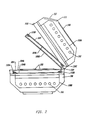
- FIG. 1 is a perspective view of one embodiment of the device of the present invention.
- FIG. 2 is a cross-sectional view of the device shown in FIG. 1 along the lines of 2—2.
- FIG. 3 is a cross-sectional view of the device shown in FIG. 1 along the lines of 3—3.
- FIG. 4 is a cross-sectional view of the device shown in FIG. 1 along the lines of 4—4.
- FIG. 5 schematically shows the decontamination approach of the present invention applied specifically to blood products.
- FIG. 6 is a graph showing the photoaddition of 8-methoxypsoralen to nucleic acid.
- FIG. 7 is a graph showing the degradation of 8-methoxypsoralen (8-MOP) compared to that of 4′-aminomethyl-4,5′,8-trimethylpsoralen (AMT), as measured by HPLC.
- FIG. 8 is a graph showing the inactivation of gram negative bacteria.
- FIG. 9A schematically shows the standard blood product separation approach used presently in blood banks.
- FIG. 9B schematically shows an embodiment of the present invention whereby synthetic media is introduced to platelet concentrate prepared as in FIG. 9 A.
- FIG. 9C schematically shows one embodiment of the decontamination approach of the present invention applied specifically to platelet concentrate diluted with synthetic media as in FIG. 9 B.
- the invention generally relates to synthetic media for use with blood preparations intended for in vivo use, including synthetic media used in conjunction with the photodecontamination of platelets.
- red blood cells As noted previously, whole blood is collected and typically separated into red blood cells, platelets, and plasma. Each of these fractions are individually stored under specific conditions prior to in vivo use. In many cases, the extent of contamination is related to the storage time because of growth. A process that inactivated microorganisms at the time of blood collection would be expected to prevent growth during storage.
- the present invention contemplates inactivating blood products after separation but before storage.
- a nucleic acid binding compound is selectively employed to treat contamination by microorganisms.
- the nucleic acid binding compound is selected from the group comprising furocoumarins.
- the furocoumarin in a psoralen that is activated by a photoactivation device.
- Psoralens are tricyclic compounds formed by the linear fusion of a furan ring with a coumarin. Psoralens can intercalate between the base pairs of double-stranded nucleic acids, forming covalent adducts to pyrimidine bases upon absorption of longwave ultraviolet light (UVA).
- UVA longwave ultraviolet light
- the inactivation method of the present invention provides a method of inactivating single cell and multicellular organisms, and in particular, bacteria, fungi, mycoplasma and protozoa.
- the present invention contemplates inactivation of viruses (e.g. HIV virus).
- viruses e.g. HIV virus
- the method of the present invention does not cause harm to the blood product. There is no significant damage to cells and, therefore, no need to limit the concentration of molecular oxygen.
- the present invention contemplates using much lower concentrations of nucleic acid binding compounds than previously employed.
- the present invention contemplates using 8-MOP at concentrations of 30 ug/ml or less.
- a preferred concentration of 8-MOP for bacterial decontamination in platelet concentrates is 3 ug/ml or less, i.e., a one hundred-fold lower concentration than employed by G. P. Wiesehahn et al., supra. Because lower concentrations are employed, solvents like DMSO (used to increase the solubility of 8-MOP) are unnecessary.
- the present invention contemplates using much lower doses of irradiation than previously described. This is accomplished with lower intensity irradiation sources, with wavelength cutoff filters (see below), and/or shorter irradiation times.
- the time of irradiation is variable and controlled from 1 second to 99 minutes, in one second increments.
- the device of the present invention is mounted on an agitator, giving horizontal unidirectional and sinusoidal motion of variable frequency and amplitude.
- heat from the lamps, ballasts and other sources is blocked from the bags.
- an inactivation method may or may not achieve complete inactivation, it is useful to consider a specific example.
- a bacterial culture is said to be sterilized if an aliquot of the culture, when transferred to a fresh culture plate and permitted to grow, is undetectable after a certain time period.
- the time period and the growth conditions e.g., temperature
- This amplification factor along with the limitations of the detection method (e.g., visual inspection of the culture plate for the appearance of a bacterial colony) define the sensitivity of the inactivation method.
- a minimal number of viable bacteria must be applied to the plate for a signal to be detectable. With the optimum detection method, this minimal number is 1 bacterial cell.
- the minimal number of bacterial cells applied so that a signal is observed may be much greater than 1.
- the detection method determines a “threshold” below which the method appears to be completely effective (and above which the method is, in fact, only partially effective).
- bacterial cells can be applied to a plate; the detection method is arbitrarily chosen to be visual inspection. Assume the growth conditions and time are such that an overall amplification of 10 4 has occurred. The detectable signal will be proportional to the number of bacterial cells actually present after amplification. For calculation purposes, the detection threshold is taken to be 10 6 cells; if fewer than 10 6 cells are present after amplification, no cell colonies are visually detectable and the inactivation method will appear effective.
- the sensitivity limit would be 100 bacterial cells; if less than 100 viable bacterial cells were present in the original aliquot of the bacterial culture after the sterilization method is performed, the culture would still appear to be sterilized.
- the present invention contemplates specific synthetic media formulations for use with blood preparations.
- the formulations of the present invention are particularly useful for platelet storage. These formulations are also useful when employed in conjunction with the photodecontamination of platelets.
- U.S. Pat. No. 4,447,415 to Rock describes a synthetic medium consisting essentially of: a balanced, physiologically compatible, saline solution; an anticoagulant; and one or more additives to enhance stability of the platelets selected from: (a) nutrients to improve the storage life of the platelets; (b) reversible inhibitors for platelet activation; (c) substances to raise cyclic adenosine monophosphate levels which have reversible effects on platelets; and (d) buffering agents which are physiologically compatible.
- the specification stresses the need to remove plasma. The specification indicates that this is achieved either by centrifugation and washing, or by “extraction” (see Examples 1 and 2 of the '415 patent). See also U.S. Pat. No. (Reissue) 32,874.
- U.S. Pat. No. 4,704,352 to Miripol discloses a medium which contains either magnesium L-ascorbate-2-phosphate or calcium L-ascorbate-2-phosphate.
- U.S. Pat. No. 4,695,460 to Holme describe media that contain sodium bicarbonate.
- U.S. Pat. No. 4,390,619 to Harmening-Pittiglio discloses a media capable of supporting platelet metabolism, containing a water-insoluble polymer containing releasable phosphate or bicarbonate ions.
- the formulations of the present invention i) utilize residual plasma; and ii) avoid bicarbonate.
- the present invention contemplates that a standard plasma expression method (e.g., such as currently used in most blood banks) will be used, such that platelets are suspended in a residual plasma concentration between 8 and 25% by volume, and more commonly 12 to 20%.
- Bicarbonate has been found to be extremely difficult to control in a system employing gas permeable blood bags.
- the present invention therefore contemplates a phosphate buffer to control the pH of the blood preparation.
- the present invention contemplates devices and methods for photoactivation and specifically, for activation of photoreactive nucleic acid-binding compounds.
- the present invention contemplates devices having an inexpensive source of electromagnetic radiation that is integrated into a unit.
- the present invention contemplates a photoactivation device for treating photoreactive compounds, comprising: a) means for providing appropriate wavelengths of electromagnetic radiation to cause activation of at least one photoreactive compound; b) means for supporting a plurality of blood products in a fixed relationship with the radiation providing means during activation; and c) means for maintaining the temperature of the blood products within a desired temperature range during activation.
- the present invention also contemplates methods, comprising: a) supporting a plurality of blood product containers, containing one or more photoreactive compounds, in a fixed relationship with a fluorescent source of electromagnetic radiation; b) irradiating the plurality of blood products simultaneously with said electromagnetic radiation to cause activation of at least one photoreactive compound; and c) maintaining the temperature of the blood products within a desired temperature range during activation.
- the major features of one embodiment of the device of the present invention involve: A) an inexpensive source of ultraviolet radiation in a fixed relationship with the means for supporting the sample vessels; B) rapid photoactivation; C) large sample processing; D) temperature control of the irradiated samples; and E) inherent safety.
- a preferred photoactivation device of the present invention has an inexpensive source of ultraviolet radiation in a fixed relationship with the means for supporting the sample vessels.
- Ultraviolet radiation is a form of energy that occupies a portion of the electromagnetic radiation spectrum (the electromagnetic radiation spectrum ranges from cosmic rays to radio waves).
- Ultraviolet radiation can come from many natural and artificial sources. Depending on the source of ultraviolet radiation, it may be accompanied by other (non-ultraviolet) types of electromagnetic radiation (e.g., visible light).
- Wavelength is herein described in terms of nanometers (“nm”; 10 ⁇ 9 meters).
- ultraviolet radiation extends from approximately 180 nm to 400 nm.
- a radiation source by virtue of filters or other means, does not allow radiation below a particular wavelength (e.g., 320 nm), it is said to have a low end “cutoff” at that wavelength (e.g., “a wavelength cutoff at 300 nanometers”).
- a radiation source allows only radiation below a particular wavelength (e.g., 360 nm), it is said to have a high end “cutoff” at that wavelength (e.g., “a wavelength cutoff at 360 nanometers”).
- any photochemical reaction it is desired to eliminate or least minimize any deleterious side reactions.
- Some of these side reactions can be caused by the excitation of endogenous chromophores that may be present during the photochemical activation procedure.
- the endogenous chromophores are the nucleic acid bases themselves. Restricting the activation process to wavelengths greater than 320 nm minimizes direct nucleic acid damage since there is very little absorption by nucleic acids above 313 nm.
- the nucleic acid is typically present together with additional biological chromophores. If the biological fluid is just protein, the 320 nm cutoff will be adequate for minimizing side reactions (aromatic amino acids do not absorb above 320 nm). If the biological fluid includes cells and/or cellular constituents, there will be many other chromophores, including hemes and flavins.
- Hemes are abundant in blood products where they arise from the lysis of red cells. Flavins, like hemes, are required for metabolic respiration. Both of these endogenous chromophores will cause damage to cells if excited by photoirradiation.
- Hemes have three principle absorption bands: two are in the red region of the visible spectrum; the other is centered about 400 nm. Flavins have two principle absorption peaks: one at 450 nm and the other at 370 nm.
- the device of the present invention be designed to allow for irradiation within a small range of specific and desirable wavelengths, and thus avoid damage to cells caused by energy transfer.
- the preferred range of desirable wavelengths is between 320 and 350 nm.
- BLB type fluorescent lamps are designed to remove wavelengths above 400 nm. This, however, only provides an upper end cutoff.
- the device of the present invention comprises an additional filtering means.
- the filtering means comprises a glass cut-off filter, such as a piece of Cobalt glass.
- the filtering means comprises a liquid filter solution that transmit only a specific region of the electromagnetic spectrum, such as an aqueous solution of Co(No 3 ) 2 . This salt solution yields a transmission window of 320-400 nm.
- the aqueous solution of Co(No 3 ) 2 is used in combination with NiSO 4 to remove the 365 nm component of the emission spectrum of the fluorescent or arc source employed.
- the Co—Ni solution preserves its initial transmission remarkably well even after tens of hours of exposure to the direct light of high energy sources.
- cupric sulfate is a most useful general filter for removing the infra-red, when only the ultraviolet is to be isolated. Its stability in intense sources is quite good.
- Other salts are known to one skilled in the art.
- Aperture or reflector lamps may also be used to achieve specific wavelengths and intensities.
- UV radiation When ultraviolet radiation is herein described in terms of irradiance, it is expressed in terms of intensity flux (milliwatts per square centimeter or “mW cm ⁇ 2 ”). “Output” is herein defined to encompass both the emission of radiation (yes or no; on or off) as well as the level of irradiance. In a preferred embodiment, intensity is monitored at 4 locations: 2 for each side of the plane of irradiation.
- a preferred source of ultraviolet radiation is a fluorescent source.
- Fluorescence is a special case of luminescence. Luminescence involves the absorption of electromagnetic radiation by a substance and the conversion of the energy into radiation of a different wavelength. With fluorescence, the substance that is excited by the electromagnetic radiation returns to its ground state by emitting a quantum of electromagnetic radiation. While fluorescent sources have heretofore been thought to be of too low intensity to be useful for photoactivation, in one embodiment the present invention employs fluorescent sources to achieve results thus far achievable on only expensive equipment.
- fixed relationship is defined as comprising a fixed distance and geometry between the sample and the light source during the sample irradiation.
- Distance relates to the distance between the source and the sample as it is supported. It is known that light intensity from a point source is inversely related to the square of the distance from the point source. Thus, small changes in the distance from the source can have a drastic impact on intensity. Since changes in intensity can impact photoactivation results, changes in distance are avoided in the devices of the present invention. This provides reproducibility and repeatability.
- Geometry relates to the positioning of the light source. For example, it can be imagined that light sources could be placed around the sample holder in many ways (on the sides, on the bottom, in a circle, etc.).
- the geometry used in a preferred embodiment of the present invention allows for uniform light exposure of appropriate intensity for rapid photoactivation.
- the geometry of a preferred device of the present invention involves multiple sources of linear lamps as opposed to single point sources. In addition, there are several reflective surfaces and several absorptive surfaces. Because of this complicated geometry, changes in the location or number of the lamps relative to the position of the samples to be irradiated are to be avoided in that such changes will result in intensity changes.
- the light source of the preferred embodiment of the present invention allows for rapid photoactivation.
- the intensity characteristics of the irradiation device have been selected to be convenient with the anticipation that many sets of multiple samples may need to be processed. With this anticipation, a fifteen minute exposure time or less is a practical goal.
- relative position of the elements of the preferred device have been optimized to allow for fifteen minutes of irradiation time, so that, when measured for the wavelengths between 320 and 350 nanometers, an intensity flux greater than approximately 1 mW cm ⁇ 2 is provided to the sample vessels.
- the device irradiates both sides of the bag.
- one element of the devices of the present invention is a means for supporting a plurality of blood products, and in particular, blood bags.
- the supporting means comprises glass plates between two banks of lights with a capacity of six 50 ml bags (equivalent to Dupont Stericell bag) plus connectors and tubing, at one time.
- Temperature control is important because the temperature of the sample in the sample at the time of exposure to light can dramatically impact the results.
- conditions that promote secondary structure in nucleic acids also enhance the affinity constants of many psoralen derivatives for nucleic acids. Hyde and Hearst, Biochemistry, 17, 1251 (1978). These conditions are a mix of both solvent composition and temperature.
- irradiation at low temperatures enhances the covalent addition of HMT to 5S rRNA by two fold at 4° C. compared to 20° C. Thompson et al., J. Mol. Biol. 147:417 (1981).
- Even further temperature induced enhancements of psoralen binding have been reported with synthetic polynucleotides. Thompson et al., Biochemistry 21:1363 (1982).
- Ultraviolet radiation can cause severe burns. Depending on the nature of the exposure, it may also be carcinogenic.
- the light source of a preferred embodiment of the present invention is shielded from the user. This is in contrast to the commercial hand-held ultraviolet sources as well as the large, high intensity sources.
- the irradiation source is contained within a housing made of material that obstructs the transmission of radiant energy (i.e., an opaque housing). No irradiation is allowed to pass to the user. This allows for inherent safety for the user.
- phosphate buffered synthetic media is formulated for platelet storage and treatment.
- One embodiment is made as follows:
- a photoactivation device for decontaminating blood products according to the method of the present invention.
- This device comprises: a) means for providing appropriate wavelengths of electromagnetic radiation to cause activation of at least one photoreactive compound; b) means for supporting a plurality of blood products in a fixed relationship with the radiation providing means during activation; and c) means for maintaining the temperature of the blood products within a desired temperature range during activation.
- FIG. 1 is a perspective view of one embodiment of the device integrating the above-named features.
- the figure shows an opaque housing ( 100 ) with a portion of it removed, containing an array of bulbs ( 101 ) above and below a plurality of representative blood product containing means ( 102 ) placed between plate assemblies ( 103 , 104 ).
- the plate assemblies ( 103 , 104 ) are described more fully, subsequently.
- the housing ( 101 ) can be opened via a latch ( 105 ) so that the blood product can be placed appropriately. As shown in FIG. 1 , the housing ( 100 ), when closed, completely contains the irradiation from the bulbs ( 101 ). During irradiation, the user can confirm that the device is operating by looking through a safety viewport ( 106 ) which does not allow transmission of ultraviolet light to the user.
- the housing ( 100 ) also serves as a mount for several electronic components on a control board ( 107 ), including, by way of example, a main power switch, a count down timer, and an hour meter.
- the power switch can be wired to the count down timer which in turn is wired in parallel to an hour meter and to the source of the electromagnetic radiation.
- the count down timer permits a user to preset the irradiation time to a desired level of exposure.
- the hour meter maintains a record of the total number of radiation hours that are provided by the source of electromagnetic radiation. This feature permits the bulbs ( 101 ) to be monitored and changed before their output diminishes below a minimum level necessary for rapid photoactivation.
- FIG. 2 is a cross-sectional view of the device shown in FIG. 1 along the lines of 2 — 2 .
- FIG. 2 shows the arrangement of the bulbs ( 101 ) with the housing ( 100 ) opened.
- a reflector ( 108 A, 108 B) completely surrounds each array of bulbs ( 101 ).
- Blood product containing means ( 102 ) are placed between upper ( 103 ) and lower ( 104 ) plate assemblies.
- Each plate assembly is comprised of an upper ( 103 A, 104 A) and lower ( 103 B, 104 B) plates.
- the plate assemblies ( 103 , 104 ) are connected via a hinge ( 109 ) which is designed to accommodate the space created by the blood product containing means ( 102 ).
- the upper plate assembly ( 103 ) is brought to rest gently on top of the blood product containing means ( 102 ) supported by the lower plate ( 104 B) of the lower plate assembly ( 104 ).
- Detectors may be conveniently placed between the plates ( 103 A, 103 B, 104 A, 104 B) of the plate assemblies ( 103 , 104 ). They can be wired to a printed circuit board ( 111 ) which in turn is wired to the control board ( 107 ).
- FIG. 3 is a cross-sectional view of the device shown in FIG. 1 along the lines of 3 — 3 .
- Six blood product containing means ( 102 ) e.g., TeflonTM platelet unit bags
- the temperature of the blood product can be controlled via a fan ( 112 ) alone or, more preferably, by employing a heat exchanger ( 113 ) having cooling inlet ( 114 ) and outlet ( 115 ) ports connected to a cooling source (not shown).
- FIG. 4 is a cross-sectional view of the device shown in FIG. 1 along the lines of 4 — 4 .
- FIG. 4 more clearly shows the temperature control approach of a preferred embodiment of the device.
- Upper plate assembly plates ( 103 A, 103 B) and lower plate assembly plates ( 104 A, 104 B) each create a temperature control chamber ( 103 C, 104 C), respectively.
- the fan ( 112 ) can circulate air within and between the chambers ( 103 C, 104 C).
- the heat exchanger ( 113 ) is employed, the circulating air is cooled and passed between the plates ( 103 A, 103 B, 104 A, 104 B).
- FIG. 5 shows an embodiment wherein platelets are treated by the method of the present invention.
- platelets are transferred to a bag containing a nucleic acid binding compound (shown in FIG. 5 as a shaded bag).
- This bag which has transmission properties and other characteristics suited for the present invention, is then placed in an irradiation device (such as that described in Example 1, above) and is irradiated.
- the free compound may be collected or “captured” as desired by a capture device.
- the bag would contain only compound that is contained in cells; the bag would have no free compound (this bag is indicated in FIG. 5 as unshaded).
- a bag comprised of a psoralen-binding polymer is employed to capture the compound.
- the Cutter CLX bag has been found to have this property.
- the decontamination methods of the present invention are applied to inactivate Klebsiella pneumoniae, which is known to be among the organisms associated with bacteremia. See generally, Infect. Control 5:343 (1984). The particular isolate in this case was obtained following a platelet transfusion where the recipient immediately went into shock and later died. The platelet bag was obtained and cultured, and the organism was identified and serotyped.
- the strain was kept at ambient temperature and inoculated onto either heart infusion agar (HIA) or heart infusion agar containing 5% (v/v) sheep blood (BAP) by swabbing each plate for confluency via a sterile applicator swab. Cultures were then incubated under static conditions for 16-18 h at 35° C. At the end of the incubation period, cultures were removed and suspended in phosphate buffered saline (PBS;pH 7.2-7.4) and spectrophotometrically standardized to 1.0 at an OD 610 using a Spectronic 501 or 601 spectrophotometer (Bausch and Lomb).
- suspensions were diluted 1:10 in PBS to achieve an ca. 10 8 CFU/ml concentration. This standardized suspension is then split to use an aliquot for the inactivation study, while another portion was plated in duplicate 10-fold serial dilutions onto HIA (or BAP) to ensure appropriate concentrations of the organism.
- ABO compatible freshly outdated human platelet concentrate units were obtained from the Blood Bank of Alameda—Contra Costa Medical Association. They were pooled and redivided into two bags. One bag was infused with the bacteria preparation. The platelets in the second unit were pelleted at 4000 ⁇ g for 6 minutes and then resuspended in a medium containing 85% saline and 15% plasma. Bacteria was added after platelets were well resuspended.
- F24T 12-BL-HO fluorescent lamps were used. These are high output “black light” tubes (engineered to emit specific wavelengths by means of an internal phosphor coating) 24 inches in length. Total intensity is less than 25 mw/cm 2 and typically between 15 and 20 mw/cm 2 .
- the binding of 8-MOP to Calf Thymus DNA is compared using plasma and a protein free media in order to validate the efficiency of psoralen-nucleic interactions under the decontamination methods of the present invention.
- this measurement used eukaryotic nucleic acid rather than bacterial nucleic acid, it is a useful indicator of the degree of adduct formation for bacteria.
- 3 H-8-MOP was prepared to a concentration of 115 ug/ml in ethanol at a specific activity of 4.7 ⁇ 10 6 CPM/microgram (hereinafter “8-MOP stock”). Thereafter 130.5 or 22 ul of 8-MOP stock (2 each) for samples containing DNA (“+DNA”) and 52.2 or 8.7 ul for samples not containing DNA (“ ⁇ DNA”) were dried down. To +DNA samples, 40 ul of DNA stock (7.7 mg/ml) was added as well as either 460 ul plasma (day old frozen) or 450 ul Tris-EDTA (“TE”) buffer. To the latter was also added 10 ul 5M NaCl. For ⁇ DNA samples (i.e., the controls), 184 ul plasma and 16 ul water was added.
- the samples were mildly vortexed for approximately one hour and the counts were checked to confirm that the 8-MOP dissolved.
- Each sample (100 ul) was irradiated on an HRI-100 (HRI Research Inc., Concord, Calif.) at 25° C. for 0, 2, 4, 8, and 16 minutes. Samples were kept at 4° C. overnight after irradiation. Thereafter, the samples were extracted. First, a phenol solution was prepared at pH 8 by equilibrating with 0.1 M Tris pH 8. Each sample was then extracted with 100 ul phenol. Each sample was centrifuged for 5 minutes to remove the aqueous phase to a new tube. A second extraction was performed with 100 ul 1:1 phenol:chloroform. A final extraction was performed with 100 ul chloroform.
- the final aqueous phase was precipitated by adding 50 ul NaCl adjusted to give a final concentration of NaCl of 0.2 M and then adding 250 ul ethanol. The samples were again centrifuged (10 minutes). The supernatant was removed and the pellets were dried. The pellets were resuspended in 100 ul TE and reprecipitated. This was repeated for a total of 3 precipitations. The final pellets were brought up in 600 ul water and 100 ul was counted. Each sample was assayed for DNA by measuring absorbance (260 nm). 8-MOP levels were plotted as adducts per 1000 base pairs (“8-MOP:kBP”).
- the frequency of 8-MOP-DNA adduct formation in protein free media predicts a high multiplicity of modification of the bacterial genome. Furthermore, this type of biochemical measurement has the potential to provide a means to monitor the efficiency of the photochemical inactivation method.
- Photoactivation of psoralens and isopsoralens may result in a variety of photoproducts.
- “Photoproduct” is best understood by considering the possible reactions of photoreactive compound when exposed to activating wavelengths of electromagnetic radiation. While not limited to any precise mechanism, it is believed that the reaction of photoreactive compound in its ground state (“C”) with activating wavelengths of electromagnetic radiation creates a short-lived excited species (“C*”): C ⁇ C* What happens next is largely a function of what potential reactants are available to the excited species. Since it is short-lived, a reaction of this species with nucleic acid (“NA”) is believed to only be possible if nucleic acid is present at the time the excited species is generated.
- C ground state
- NA nucleic acid
- the reaction must, in operational terms, be in the presence of activating wavelengths of electromagnetic radiation, i.e., it is “photobinding”; it is not dark binding.
- the reaction can be depicted as follows: C*+NA ⁇ NA:C
- the product of this reaction is hereinafter referred to as “Photoaddition Product” and is to be distinguished from “Photoproduct.”
- the excited species is not limited to reacting with itself. It may react with its environment, such as elements of the solvent (“E”) (e.g., ions, gases, etc.) to produce other products: C*+E ⁇ E:C It is this type of reaction that is believed to cause cellular damage (e.g., reaction with oxygen to create singlet oxygen species). Furthermore, it may simply internally rearrange (“isomerize”) to a ground state derivative (“[”): C* ⁇ [ Finally, the excited species may undergo other reactions than described here.
- E elements of the solvent
- ions e.g., ions, gases, etc.
- Photoproduct does not depend on which one (if any) of these reactions actually occurs. “Photoproduct”—whatever its nature—is deemed to exist if, following the reaction of a compound and activating wavelengths of electromagnetic radiation, there is a resultant product formed that can interact with other components of the reaction environment.
- HMT 4′-hydroxymethyl-4,5′,8-tri-methylpsoralen
- the major resultant products of HMT are two cyclobutyl photodimers.
- the two pyrone rings are linked in a cis-syn configuration, while in the other dimer, the linkage occurs between the furan end of one molecule and the pyrone end of the other, again with cis-syn configuration.
- a third resultant product of HMT is a monomeric HMT photoisomer. In this isomer, the central ring oxygens assume a 1, 4 instead of the normal 1, 3 orientation.
- the photoisomer While the two photodimers would not be expected to have an intercalating activity due to geometrical considerations, the photoisomer remains planer, and accordingly, it is contemplated that it has a positive intercalative association with double stranded nucleic acid and, thus, could be a mutagen.
- the photochemical breakdown of 8-MOP is compared with AMT.
- the samples were analyzed by reverse phase HPLC using a Rainen Dynamax 300A column. Gradient elution was performed with 0.1 M ammonium acetate/acetonitrile (0-70% acetonitrile over 42 minutes). AMT elutes as a single peak at approximately 24 minutes under these conditions. Detection was by absorption at either 260 or 330 nm. The latter wavelength was used for the plasma containing samples.
- FIG. 9 shows an embodiment wherein platelets are diluted with synthetic media by the method of the present invention.
- FIG. 9A schematically shows the standard blood product separation approach used presently in blood banks.
- Three bags are integrated by flexible tubing to create a blood transfer set ( 200 ) (e.g., commercially available from Baxter, Deerfield, Ill.). After blood is drawn into the first bag ( 201 ), the entire set is processed by centrifugation (e.g., SorvallTM swing bucket centrifuge, Dupont),resulting in packed red cells and platelet rich plasma in the first bag ( 201 ).
- centrifugation e.g., SorvallTM swing bucket centrifuge, Dupont
- the plasma is expressed off of the first bag ( 201 ) (e.g., using a FenwallTM device for plasma expression), through the tubing and into the second bag ( 202 ).
- the first bag ( 201 ) is then detached and the two bag set is centrifuged to create platelet concentrate and platelet-poor plasma; the latter is expressed off of the second bag ( 202 ) into the third bag ( 203 ).
- FIG. 9B schematically shows an embodiment of the present invention by which synthetic media is introduced to platelet concentrate prepared as in FIG. 9A.
- a two bag set ( 300 ) is sterile docked with the platelet concentrate bag ( 202 ) (indicated as “P.C.”).
- P.C. platelet concentrate bag
- Sterile docking is well-known to the art. See e.g., U.S. Pat. No. 4,412,835 to D. W. C. Spencer, hereby incorporated by reference. See also U.S. Pat. Nos. 4,157,723 and 4,265,280, hereby incorporated by reference.
- Sterile docking devices are commercially available (e.g., Terumo, Japan).
- One of the bags ( 301 ) of the two bag set ( 300 ) contains a synthetic media formulation of the present invention (indicated as “STERILYTE”).
- the platelet concentrate is mixed with the synthetic media by transferring the platelet concentrate to the synthetic media bag ( 301 ).
- FIG. 9C schematically shows one embodiment of the decontamination approach of the present invention applied specifically to platelet concentrate diluted with synthetic media as in FIG. 9 B.
- platelets have been transferred to a synthetic media bag ( 301 ) containing a nucleic acid binding compound.
- This bag ( 301 ) which has UV light transmission properties and other characteristics suited for the present invention, is then placed in a device (such as that described in Example 1, above) and illuminated.
- the platelets are transferred from the synthetic media bag ( 301 ) into the storage bag ( 302 ) of the two bag set ( 300 ).
- the storage bag can be a commercially available storage bag (e.g., CLX bag from Cutter).
- the present invention provides synthetic media formulations for use with blood preparations intended for storage and in vivo use.
- the formulations include synthetic media formulations to be employed in conjunction with the photodecontamination of platelets.
Abstract
Description
| Ingredients | Amounts (grams/liter) | ||
| Na Acetate.3H2O | 4.08 | ||
| Citrate.2H2O | 2.94 | ||
| NaH2PO4.H2O | 0.858 | ||
| Na2HPO4 | 2.81 | ||
| NaCl | 5.02 | ||
| pH adjustment | to pH 7.2 (with HCl) | ||
Distilled water is added to make 1 liter, the solution is mixed, sterile filtered (0.2 micron filter) and refrigerated.
C→C*
What happens next is largely a function of what potential reactants are available to the excited species. Since it is short-lived, a reaction of this species with nucleic acid (“NA”) is believed to only be possible if nucleic acid is present at the time the excited species is generated. Thus, the reaction must, in operational terms, be in the presence of activating wavelengths of electromagnetic radiation, i.e., it is “photobinding”; it is not dark binding. The reaction can be depicted as follows:
C*+NA→NA:C
The product of this reaction is hereinafter referred to as “Photoaddition Product” and is to be distinguished from “Photoproduct.”
C*→C
On the other hand, the excited species may react with itself (i.e., a ground state or excited species) to create a ground state complex (“C:C”). The product of these self-reactions where two compounds react is referred to as “photodimer” or simply “dimer.” The self-reactions, however, are not limited to two compounds; a variety of multimers may be formed (trimers, etc.).
C*+E→E:C
It is this type of reaction that is believed to cause cellular damage (e.g., reaction with oxygen to create singlet oxygen species). Furthermore, it may simply internally rearrange (“isomerize”) to a ground state derivative (“[”):
C*→[
Finally, the excited species may undergo other reactions than described here.
Claims (4)
Priority Applications (1)
| Application Number | Priority Date | Filing Date | Title |
|---|---|---|---|
| US10/413,110 US6866992B2 (en) | 1992-03-02 | 2003-04-14 | Synthetic platelet storage media formulation |
Applications Claiming Priority (8)
| Application Number | Priority Date | Filing Date | Title |
|---|---|---|---|
| US07/844,790 US5288605A (en) | 1992-03-02 | 1992-03-02 | Methods for inactivating bacteria in blood preparations with 8-methoxypsoralen |
| US92647792A | 1992-08-07 | 1992-08-07 | |
| US08/072,485 US5459030A (en) | 1992-03-02 | 1993-06-02 | Synthetic media compositions for inactivating bacteria and viruses in blood preparations with 8-methoxypsoralen |
| US33803994A | 1994-11-14 | 1994-11-14 | |
| US08/692,444 US5908742A (en) | 1992-03-02 | 1996-08-05 | Synthetic media for blood components |
| US09/240,067 US6251580B1 (en) | 1992-03-02 | 1999-01-29 | Synthetic media for blood components |
| US09/732,174 US6566046B2 (en) | 1992-03-02 | 2000-12-07 | Synthetic media for blood components |
| US10/413,110 US6866992B2 (en) | 1992-03-02 | 2003-04-14 | Synthetic platelet storage media formulation |
Related Parent Applications (1)
| Application Number | Title | Priority Date | Filing Date |
|---|---|---|---|
| US09/732,174 Continuation US6566046B2 (en) | 1992-03-02 | 2000-12-07 | Synthetic media for blood components |
Publications (2)
| Publication Number | Publication Date |
|---|---|
| US20030194806A1 US20030194806A1 (en) | 2003-10-16 |
| US6866992B2 true US6866992B2 (en) | 2005-03-15 |
Family
ID=27490986
Family Applications (4)
| Application Number | Title | Priority Date | Filing Date |
|---|---|---|---|
| US08/692,444 Expired - Lifetime US5908742A (en) | 1992-03-02 | 1996-08-05 | Synthetic media for blood components |
| US09/240,067 Expired - Fee Related US6251580B1 (en) | 1992-03-02 | 1999-01-29 | Synthetic media for blood components |
| US09/732,174 Expired - Fee Related US6566046B2 (en) | 1992-03-02 | 2000-12-07 | Synthetic media for blood components |
| US10/413,110 Expired - Fee Related US6866992B2 (en) | 1992-03-02 | 2003-04-14 | Synthetic platelet storage media formulation |
Family Applications Before (3)
| Application Number | Title | Priority Date | Filing Date |
|---|---|---|---|
| US08/692,444 Expired - Lifetime US5908742A (en) | 1992-03-02 | 1996-08-05 | Synthetic media for blood components |
| US09/240,067 Expired - Fee Related US6251580B1 (en) | 1992-03-02 | 1999-01-29 | Synthetic media for blood components |
| US09/732,174 Expired - Fee Related US6566046B2 (en) | 1992-03-02 | 2000-12-07 | Synthetic media for blood components |
Country Status (3)
| Country | Link |
|---|---|
| US (4) | US5908742A (en) |
| AU (1) | AU4107396A (en) |
| WO (1) | WO1996014740A1 (en) |
Cited By (14)
| Publication number | Priority date | Publication date | Assignee | Title |
|---|---|---|---|---|
| US20070099170A1 (en) * | 1998-07-21 | 2007-05-03 | Navigant Biotechnologies, Inc. | Method for treatment and storage of blood and blood products using endogenous alloxazines and acetate |
| US20090191537A1 (en) * | 2007-12-20 | 2009-07-30 | Veronique Mayaudon | Medium and methods for the storage of platelets |
| US20090239208A1 (en) * | 2008-03-21 | 2009-09-24 | Veronique Mayaudon | Red Blood Cell Storage Medium For Extended Storage |
| US20100081985A1 (en) * | 2008-10-01 | 2010-04-01 | Caridianbct, Inc. | Platelet Additive Solution For Leukoreducing White Blood Cells In Apheresed Platelets |
| US20110117647A1 (en) * | 2008-03-21 | 2011-05-19 | Fenwal, Inc. | Red Blood Cell Storage Medium For Extended Storage |
| WO2012151243A2 (en) | 2011-05-03 | 2012-11-08 | Fenwal, Inc. | Platelet resuspension method and apparatus |
| WO2012158983A2 (en) | 2011-05-17 | 2012-11-22 | Qiyong Peter Liu | Improved platelet storage using a sialidase inhibitor |
| WO2014055988A1 (en) | 2012-10-05 | 2014-04-10 | Velico Medical, Inc. | Platelet additive solution having a beta-galactosidase inhibitor |
| WO2014120886A1 (en) | 2013-01-30 | 2014-08-07 | Velico Medical, Inc. | Platelet additive solution having a self-assembling hydrogel-forming peptide |
| US9089479B2 (en) | 2011-05-03 | 2015-07-28 | Fenwal, Inc. | Medium, solutions and methods for the washing, culturing and storage of white blood cells |
| US9402866B2 (en) | 2011-04-07 | 2016-08-02 | Fenwal, Inc. | Automated methods and systems for providing platelet concentrates with reduced residual plasma volumes and storage media for such platelet concentrates |
| US9409128B2 (en) | 2009-10-23 | 2016-08-09 | Fenwal, Inc. | Methods for storing red blood cell products |
| US9788539B2 (en) | 2011-05-17 | 2017-10-17 | Velico Medical, Inc. | Platelet protection solution having beta-galactosidase and sialidase inhibitors |
| US10897891B2 (en) | 2015-04-10 | 2021-01-26 | Board Of Regents, The University Of Texas System | Compositions and methods for prolonged cell storage |
Families Citing this family (30)
| Publication number | Priority date | Publication date | Assignee | Title |
|---|---|---|---|---|
| AU4107396A (en) * | 1992-03-02 | 1996-06-06 | Cerus Corporation | Synthetic media for blood components |
| US20040053208A1 (en) * | 1995-08-29 | 2004-03-18 | V. I. TECHNOLOGIES, Inc. | Methods to selectively inactivate parasites in biological compositions |
| US6277337B1 (en) | 1998-07-21 | 2001-08-21 | Gambro, Inc. | Method and apparatus for inactivation of biological contaminants using photosensitizers |
| US20030215784A1 (en) * | 1998-07-21 | 2003-11-20 | Dumont Larry Joe | Method and apparatus for inactivation of biological contaminants using photosensitizers |
| US6565802B1 (en) | 1999-06-03 | 2003-05-20 | Baxter International Inc. | Apparatus, systems and methods for processing and treating a biological fluid with light |
| US7068361B2 (en) * | 1999-06-03 | 2006-06-27 | Baxter International | Apparatus, systems and methods for processing and treating a biological fluid with light |
| US6268120B1 (en) | 1999-10-19 | 2001-07-31 | Gambro, Inc. | Isoalloxazine derivatives to neutralize biological contaminants |
| JP4574799B2 (en) * | 2000-05-19 | 2010-11-04 | 株式会社トップ | UV sterilizer |
| TW590780B (en) | 2000-06-02 | 2004-06-11 | Gambro Inc | Additive solutions containing riboflavin |
| US7648699B2 (en) | 2000-06-02 | 2010-01-19 | Caridianbct Biotechnologies, Llc | Preventing transfusion related complications in a recipient of a blood transfusion |
| US7985588B2 (en) | 2000-06-02 | 2011-07-26 | Caridianbct Biotechnologies, Llc | Induction of and maintenance of nucleic acid damage in pathogens using riboflavin and light |
| US9044523B2 (en) | 2000-06-15 | 2015-06-02 | Terumo Bct, Inc. | Reduction of contaminants in blood and blood products using photosensitizers and peak wavelengths of light |
| US6548241B1 (en) * | 2000-11-28 | 2003-04-15 | Gambro, Inc. | Storage solution containing photosensitizer for inactivation of biological contaminants |
| US7252799B2 (en) * | 2001-08-31 | 2007-08-07 | Clearant, Inc. | Methods for sterilizing preparations containing albumin |
| US7264608B2 (en) * | 2001-12-05 | 2007-09-04 | Fenwal, Inc. | Manual processing systems and methods for providing blood components conditioned for pathogen inactivation |
| US6936413B1 (en) * | 2001-12-05 | 2005-08-30 | Baxter International Inc. | Methods and systems for preparing blood products |
| AU2003208903A1 (en) * | 2002-02-01 | 2003-09-02 | Gambro, Inc. | Pathogen reduction solution for platelets containing an endogenous photosensitizer and a glycolysis inhibitor. |
| US20040029097A1 (en) * | 2002-04-16 | 2004-02-12 | Gambro, Inc. | Addition of glycolysis inhibitor to a pathogen reduction and storage solution |
| JP2005523337A (en) * | 2002-04-24 | 2005-08-04 | ガンブロ インコーポレーテッド | Removal of adenine during the pathogen-depleted blood and blood component processes |
| WO2003090794A1 (en) * | 2002-04-26 | 2003-11-06 | Gambro, Inc. | Solution containing platelet activation inhibitors for pathogen reducing and storing blood platelets |
| US20080299538A1 (en) * | 2003-02-28 | 2008-12-04 | Caridianbct Biotechnologies, Llc | Pathogen Inactivation of Whole Blood |
| US20070031812A1 (en) * | 2003-06-20 | 2007-02-08 | Pall Corporation | Processing of platelet-containing biological fluids |
| CN100413542C (en) * | 2006-06-01 | 2008-08-27 | 上海市血液中心 | Pathogen inactivator composition for blood or blood component and its compounding process |
| WO2009026073A1 (en) * | 2007-08-22 | 2009-02-26 | Caridianbct Biotechnologies, Llc | Prevention of transfusion related acute lung injury using riboflavin and light |
| US10039877B2 (en) | 2013-08-23 | 2018-08-07 | Fenwal, Inc. | Apheresis platelets with fixed residual plasma volume |
| EA201890149A1 (en) | 2015-06-26 | 2018-07-31 | Сирус Корпорейшн | COMPOSITIONS OF CRYOPRECIPLES AND METHODS OF THEIR RECEPTION |
| CA3003097A1 (en) | 2015-10-23 | 2017-04-27 | Cerus Corporation | Plasma compositions and methods of use thereof |
| CA3055203A1 (en) | 2017-03-03 | 2018-09-07 | Cerus Corporation | Kits and methods for preparing pathogen-inactivated platelet compositions |
| KR20200115498A (en) | 2017-12-29 | 2020-10-07 | 세루스 코포레이션 | Systems and methods for treating biological fluids |
| EP3991179A1 (en) | 2019-06-28 | 2022-05-04 | Cerus Corporation | System and methods for implementing a biological fluid treatment device |
Citations (58)
| Publication number | Priority date | Publication date | Assignee | Title |
|---|---|---|---|---|
| US2786014A (en) | 1952-09-10 | 1957-03-19 | James L Tullis | Platelet preservation |
| US3629071A (en) | 1970-02-10 | 1971-12-21 | Upjohn Co | Storage-stable hemostatic transfusion suspensions of blood platelets, glucose, magnesium chloride and certain prostaglandins |
| US3729947A (en) | 1970-12-04 | 1973-05-01 | Alza Corp | Process for storing blood platelets |
| US3735005A (en) | 1970-08-19 | 1973-05-22 | Alza Corp | Method for preparing a viable platelet concentrate |
| US3874384A (en) | 1971-11-01 | 1975-04-01 | American Hospital Supply Corp | Improved blood storage unit and method of storing blood |
| US4124598A (en) | 1976-10-20 | 1978-11-07 | Hoffmann-La Roche Inc. | Psoralens |
| US4152208A (en) | 1977-03-29 | 1979-05-01 | Hoffmann-La Roche Inc. | Stabilized leucocytes |
| US4157723A (en) | 1977-10-19 | 1979-06-12 | Baxter Travenol Laboratories, Inc. | Method of forming a connection between two sealed conduits using radiant energy |
| US4169204A (en) | 1976-10-20 | 1979-09-25 | Regents Of The University Of California | Psoralens |
| US4196281A (en) | 1976-10-20 | 1980-04-01 | Regents Of The University Of California | Psoralens |
| US4265280A (en) | 1979-01-23 | 1981-05-05 | Baxter Travenol Laboratories, Inc. | Connector member for sealed conduits |
| US4344936A (en) | 1980-09-17 | 1982-08-17 | Gerald Soslau | Platelet composition having improved storage stability and method therefor |
| US4390619A (en) | 1981-09-28 | 1983-06-28 | James Clifford Haight | Leukocyte or platelet storage using ion-exchange resins |
| US4412835A (en) | 1982-07-06 | 1983-11-01 | E. I. Du Pont De Nemours & Company | Sterile docking process, apparatus and system |
| US4447415A (en) | 1982-11-01 | 1984-05-08 | Rock Gail A | Plasma-free medium for platelet storage |
| US4639373A (en) | 1984-11-29 | 1987-01-27 | New England Medical Center Hospitals, Inc. | Stabilization of granulocytes |
| US4695460A (en) | 1986-03-19 | 1987-09-22 | American Red Cross | Synthetic, plasma-free, transfusible platelet storage medium |
| US4704352A (en) | 1985-06-25 | 1987-11-03 | Baxter Travenol Laboratories, Inc. | L-ascorbate-2-phosphate salts in blood cell storage |
| US4727027A (en) | 1983-05-02 | 1988-02-23 | Diamond Scientific Co. | Photochemical decontamination treatment of whole blood or blood components |
| US4748120A (en) | 1983-05-02 | 1988-05-31 | Diamond Scientific Co. | Photochemical decontamination treatment of whole blood or blood components |
| US4828976A (en) | 1983-12-29 | 1989-05-09 | Thomas Jefferson University | Glucose free media for storing blood platelets |
| US4878891A (en) | 1987-06-25 | 1989-11-07 | Baylor Research Foundation | Method for eradicating infectious biological contaminants in body tissues |
| US4925665A (en) | 1989-06-22 | 1990-05-15 | Thomas Jefferson University | Glucose free primary anticoagulant for blood containing citrate ions |
| US4960408A (en) | 1989-01-10 | 1990-10-02 | Klainer Albert S | Treatment methods and vaccines for stimulating an immunological response against retroviruses |
| US4985153A (en) | 1988-06-23 | 1991-01-15 | Asahi Medical Co., Ltd. | Method for separating blood into blood components, and blood components separator unit |
| US4992363A (en) * | 1983-11-09 | 1991-02-12 | Thomas Jefferson University | Method for preparing glucose free media for storing blood platelets |
| US4994367A (en) * | 1988-10-07 | 1991-02-19 | East Carolina University | Extended shelf life platelet preparations and process for preparing the same |
| WO1991003933A1 (en) | 1989-09-13 | 1991-04-04 | Blutspendedienst Der Landesverbände Des Deutschen Roten Kreuzes Niedersachsen, Oldenburg Und Bremen G.G.M.B.H. | Process for inactivating viruses in blood and blood products |
| US5011695A (en) | 1988-02-22 | 1991-04-30 | Biotest Pharma Gmbh | Sterilization of blood and its derivatives with vitamins |
| US5055485A (en) | 1988-12-02 | 1991-10-08 | New York Blood Center, Inc. | Inactivation of viruses in cell- and protein-containing compositions using aryl diol epoxides |
| US5100564A (en) | 1990-11-06 | 1992-03-31 | Pall Corporation | Blood collection and processing system |
| US5112298A (en) | 1990-06-25 | 1992-05-12 | Baxter International Inc. | Apheresis method and device |
| US5120649A (en) | 1990-05-15 | 1992-06-09 | New York Blood Center, Inc. | Photodynamic inactivation of viruses in blood cell-containing compositions |
| US5234608A (en) | 1990-12-11 | 1993-08-10 | Baxter International Inc. | Systems and methods for processing cellular rich suspensions |
| US5234808A (en) | 1991-10-30 | 1993-08-10 | Thomas Jefferson University | Acetate addition to platelets stored in plasma |
| US5248506A (en) * | 1986-03-19 | 1993-09-28 | American National Red Cross | Synthetic, plasma-free, transfusible storage medium for red blood cells and platelets |
| US5288605A (en) * | 1992-03-02 | 1994-02-22 | Steritech, Inc. | Methods for inactivating bacteria in blood preparations with 8-methoxypsoralen |
| US5344752A (en) | 1991-10-30 | 1994-09-06 | Thomas Jefferson University | Plasma-based platelet concentrate preparations |
| US5372929A (en) | 1992-01-27 | 1994-12-13 | Cimino; George D. | Methods for measuring the inactivation of pathogens |
| US5376524A (en) | 1991-04-01 | 1994-12-27 | Thomas Jefferson University | Platelet storage medium containing acetate and phosphate |
| US5399719A (en) | 1993-06-28 | 1995-03-21 | Steritech, Inc. | Compounds for the photodecontamination of pathogens in blood |
| US5418130A (en) | 1990-04-16 | 1995-05-23 | Cryopharm Corporation | Method of inactivation of viral and bacterial blood contaminants |
| US5456845A (en) | 1993-02-22 | 1995-10-10 | Asahi Medical Co., Ltd. | Method for separating a blood material into blood component products |
| US5459030A (en) * | 1992-03-02 | 1995-10-17 | Steritech, Inc. | Synthetic media compositions for inactivating bacteria and viruses in blood preparations with 8-methoxypsoralen |
| US5474891A (en) | 1991-10-30 | 1995-12-12 | Thomas Jefferson University | Plasma-based platelet concentrate preparations with additive |
| US5516629A (en) | 1990-04-16 | 1996-05-14 | Cryopharm Corporation | Photoinactivation of viral and bacterial blood contaminants using halogenated coumarins |
| US5556958A (en) | 1989-10-26 | 1996-09-17 | Steritech, Inc. | Inactivation of pathogens in clinical samples |
| US5559250A (en) | 1994-11-14 | 1996-09-24 | Steritech, Inc. | Treating red blood cell solutions with anti-viral agents |
| US5582828A (en) | 1995-03-15 | 1996-12-10 | Ching-Yuang Lin | Active fractions of Cordyceps sinensis and method of isolation thereof |
| US5593823A (en) | 1993-06-28 | 1997-01-14 | Cerus Corporation | Method for inactivating pathogens in blood using photoactivation of 4'-primary amino-substituted psoralens |
| US5618662A (en) | 1992-03-02 | 1997-04-08 | Cerus Corporation | Intravenous administration of psoralen |
| US5709991A (en) | 1992-03-02 | 1998-01-20 | Cerus Corporation | Proralen inactivation of microorganisms and psoralen removal |
| US5712085A (en) | 1993-06-28 | 1998-01-27 | Cerus Corporation | 5'-(4-amino-2-oxa)butye-4,4', 8-trinethylpsoralen in synthetic medium |
| US5908742A (en) * | 1992-03-02 | 1999-06-01 | Cerus Corporation | Synthetic media for blood components |
| US6060233A (en) * | 1996-06-14 | 2000-05-09 | Biostore New Zealand, Ltd | Methods for the lyophilization of platelets, platelet membranes or erythrocytes |
| US20020009705A1 (en) * | 1998-10-30 | 2002-01-24 | David O. Lucas | Compositions, methods and apparatuses for preserving platelets |
| US20020034722A1 (en) * | 1999-12-21 | 2002-03-21 | Ericson Daniel G. | Compositions for the storage of platelets |
| US20040029097A1 (en) * | 2002-04-16 | 2004-02-12 | Gambro, Inc. | Addition of glycolysis inhibitor to a pathogen reduction and storage solution |
Family Cites Families (3)
| Publication number | Priority date | Publication date | Assignee | Title |
|---|---|---|---|---|
| US32874A (en) * | 1861-07-23 | Electro-magnet | ||
| US4925605A (en) * | 1986-10-02 | 1990-05-15 | Field Foam Incorporated | Method of forming a heat foam insulation jacket |
| ATE192898T1 (en) | 1992-08-07 | 2000-06-15 | Cerus Corp | METHOD FOR INACTIVATION OF BACTERIA IN BLOOD PREPARATIONS USING METHOXYPSORALEN |
-
1995
- 1995-11-08 AU AU41073/96A patent/AU4107396A/en not_active Abandoned
- 1995-11-08 WO PCT/US1995/014665 patent/WO1996014740A1/en active Application Filing
-
1996
- 1996-08-05 US US08/692,444 patent/US5908742A/en not_active Expired - Lifetime
-
1999
- 1999-01-29 US US09/240,067 patent/US6251580B1/en not_active Expired - Fee Related
-
2000
- 2000-12-07 US US09/732,174 patent/US6566046B2/en not_active Expired - Fee Related
-
2003
- 2003-04-14 US US10/413,110 patent/US6866992B2/en not_active Expired - Fee Related
Patent Citations (63)
| Publication number | Priority date | Publication date | Assignee | Title |
|---|---|---|---|---|
| US2786014A (en) | 1952-09-10 | 1957-03-19 | James L Tullis | Platelet preservation |
| US3629071A (en) | 1970-02-10 | 1971-12-21 | Upjohn Co | Storage-stable hemostatic transfusion suspensions of blood platelets, glucose, magnesium chloride and certain prostaglandins |
| US3735005A (en) | 1970-08-19 | 1973-05-22 | Alza Corp | Method for preparing a viable platelet concentrate |
| US3729947A (en) | 1970-12-04 | 1973-05-01 | Alza Corp | Process for storing blood platelets |
| US3874384A (en) | 1971-11-01 | 1975-04-01 | American Hospital Supply Corp | Improved blood storage unit and method of storing blood |
| US4196281A (en) | 1976-10-20 | 1980-04-01 | Regents Of The University Of California | Psoralens |
| US4169204A (en) | 1976-10-20 | 1979-09-25 | Regents Of The University Of California | Psoralens |
| US4124598A (en) | 1976-10-20 | 1978-11-07 | Hoffmann-La Roche Inc. | Psoralens |
| US4152208A (en) | 1977-03-29 | 1979-05-01 | Hoffmann-La Roche Inc. | Stabilized leucocytes |
| US4157723A (en) | 1977-10-19 | 1979-06-12 | Baxter Travenol Laboratories, Inc. | Method of forming a connection between two sealed conduits using radiant energy |
| US4265280A (en) | 1979-01-23 | 1981-05-05 | Baxter Travenol Laboratories, Inc. | Connector member for sealed conduits |
| US4344936A (en) | 1980-09-17 | 1982-08-17 | Gerald Soslau | Platelet composition having improved storage stability and method therefor |
| US4390619A (en) | 1981-09-28 | 1983-06-28 | James Clifford Haight | Leukocyte or platelet storage using ion-exchange resins |
| US4412835A (en) | 1982-07-06 | 1983-11-01 | E. I. Du Pont De Nemours & Company | Sterile docking process, apparatus and system |
| US4447415A (en) | 1982-11-01 | 1984-05-08 | Rock Gail A | Plasma-free medium for platelet storage |
| USRE32874E (en) * | 1982-11-01 | 1989-02-21 | Gail A. Rock | Plasma-free medium for platelet storage |
| US4748120A (en) | 1983-05-02 | 1988-05-31 | Diamond Scientific Co. | Photochemical decontamination treatment of whole blood or blood components |
| US4727027A (en) | 1983-05-02 | 1988-02-23 | Diamond Scientific Co. | Photochemical decontamination treatment of whole blood or blood components |
| US4992363A (en) * | 1983-11-09 | 1991-02-12 | Thomas Jefferson University | Method for preparing glucose free media for storing blood platelets |
| US4828976A (en) | 1983-12-29 | 1989-05-09 | Thomas Jefferson University | Glucose free media for storing blood platelets |
| US4639373A (en) | 1984-11-29 | 1987-01-27 | New England Medical Center Hospitals, Inc. | Stabilization of granulocytes |
| US4704352A (en) | 1985-06-25 | 1987-11-03 | Baxter Travenol Laboratories, Inc. | L-ascorbate-2-phosphate salts in blood cell storage |
| US5248506A (en) * | 1986-03-19 | 1993-09-28 | American National Red Cross | Synthetic, plasma-free, transfusible storage medium for red blood cells and platelets |
| US4695460A (en) | 1986-03-19 | 1987-09-22 | American Red Cross | Synthetic, plasma-free, transfusible platelet storage medium |
| US4878891A (en) | 1987-06-25 | 1989-11-07 | Baylor Research Foundation | Method for eradicating infectious biological contaminants in body tissues |
| US5011695A (en) | 1988-02-22 | 1991-04-30 | Biotest Pharma Gmbh | Sterilization of blood and its derivatives with vitamins |
| US4985153A (en) | 1988-06-23 | 1991-01-15 | Asahi Medical Co., Ltd. | Method for separating blood into blood components, and blood components separator unit |
| US4994367A (en) * | 1988-10-07 | 1991-02-19 | East Carolina University | Extended shelf life platelet preparations and process for preparing the same |
| US5055485A (en) | 1988-12-02 | 1991-10-08 | New York Blood Center, Inc. | Inactivation of viruses in cell- and protein-containing compositions using aryl diol epoxides |
| US4960408A (en) | 1989-01-10 | 1990-10-02 | Klainer Albert S | Treatment methods and vaccines for stimulating an immunological response against retroviruses |
| US4925665A (en) | 1989-06-22 | 1990-05-15 | Thomas Jefferson University | Glucose free primary anticoagulant for blood containing citrate ions |
| WO1991003933A1 (en) | 1989-09-13 | 1991-04-04 | Blutspendedienst Der Landesverbände Des Deutschen Roten Kreuzes Niedersachsen, Oldenburg Und Bremen G.G.M.B.H. | Process for inactivating viruses in blood and blood products |
| US5665762A (en) | 1989-10-26 | 1997-09-09 | Carus Corporation | Decontaminating clinical samples |
| US5556958A (en) | 1989-10-26 | 1996-09-17 | Steritech, Inc. | Inactivation of pathogens in clinical samples |
| US5418130A (en) | 1990-04-16 | 1995-05-23 | Cryopharm Corporation | Method of inactivation of viral and bacterial blood contaminants |
| US5516629A (en) | 1990-04-16 | 1996-05-14 | Cryopharm Corporation | Photoinactivation of viral and bacterial blood contaminants using halogenated coumarins |
| US5120649A (en) | 1990-05-15 | 1992-06-09 | New York Blood Center, Inc. | Photodynamic inactivation of viruses in blood cell-containing compositions |
| US5112298A (en) | 1990-06-25 | 1992-05-12 | Baxter International Inc. | Apheresis method and device |
| US5100564A (en) | 1990-11-06 | 1992-03-31 | Pall Corporation | Blood collection and processing system |
| US5234608A (en) | 1990-12-11 | 1993-08-10 | Baxter International Inc. | Systems and methods for processing cellular rich suspensions |
| US5466573A (en) * | 1991-04-01 | 1995-11-14 | Thomas Jefferson University | Platelet storage method in a medium containing acetate and phosphate |
| US5376524A (en) | 1991-04-01 | 1994-12-27 | Thomas Jefferson University | Platelet storage medium containing acetate and phosphate |
| US5474891A (en) | 1991-10-30 | 1995-12-12 | Thomas Jefferson University | Plasma-based platelet concentrate preparations with additive |
| US5234808A (en) | 1991-10-30 | 1993-08-10 | Thomas Jefferson University | Acetate addition to platelets stored in plasma |
| US5344752A (en) | 1991-10-30 | 1994-09-06 | Thomas Jefferson University | Plasma-based platelet concentrate preparations |
| US5372929A (en) | 1992-01-27 | 1994-12-13 | Cimino; George D. | Methods for measuring the inactivation of pathogens |
| US5908742A (en) * | 1992-03-02 | 1999-06-01 | Cerus Corporation | Synthetic media for blood components |
| US5709991A (en) | 1992-03-02 | 1998-01-20 | Cerus Corporation | Proralen inactivation of microorganisms and psoralen removal |
| US5288605A (en) * | 1992-03-02 | 1994-02-22 | Steritech, Inc. | Methods for inactivating bacteria in blood preparations with 8-methoxypsoralen |
| US6566046B2 (en) * | 1992-03-02 | 2003-05-20 | Baxter International Inc. | Synthetic media for blood components |
| US6251580B1 (en) * | 1992-03-02 | 2001-06-26 | Lily Lin | Synthetic media for blood components |
| US5459030A (en) * | 1992-03-02 | 1995-10-17 | Steritech, Inc. | Synthetic media compositions for inactivating bacteria and viruses in blood preparations with 8-methoxypsoralen |
| US5618662A (en) | 1992-03-02 | 1997-04-08 | Cerus Corporation | Intravenous administration of psoralen |
| US5456845A (en) | 1993-02-22 | 1995-10-10 | Asahi Medical Co., Ltd. | Method for separating a blood material into blood component products |
| US5712085A (en) | 1993-06-28 | 1998-01-27 | Cerus Corporation | 5'-(4-amino-2-oxa)butye-4,4', 8-trinethylpsoralen in synthetic medium |
| US5593823A (en) | 1993-06-28 | 1997-01-14 | Cerus Corporation | Method for inactivating pathogens in blood using photoactivation of 4'-primary amino-substituted psoralens |
| US5399719A (en) | 1993-06-28 | 1995-03-21 | Steritech, Inc. | Compounds for the photodecontamination of pathogens in blood |
| US5559250A (en) | 1994-11-14 | 1996-09-24 | Steritech, Inc. | Treating red blood cell solutions with anti-viral agents |
| US5582828A (en) | 1995-03-15 | 1996-12-10 | Ching-Yuang Lin | Active fractions of Cordyceps sinensis and method of isolation thereof |
| US6060233A (en) * | 1996-06-14 | 2000-05-09 | Biostore New Zealand, Ltd | Methods for the lyophilization of platelets, platelet membranes or erythrocytes |
| US20020009705A1 (en) * | 1998-10-30 | 2002-01-24 | David O. Lucas | Compositions, methods and apparatuses for preserving platelets |
| US20020034722A1 (en) * | 1999-12-21 | 2002-03-21 | Ericson Daniel G. | Compositions for the storage of platelets |
| US20040029097A1 (en) * | 2002-04-16 | 2004-02-12 | Gambro, Inc. | Addition of glycolysis inhibitor to a pathogen reduction and storage solution |
Non-Patent Citations (75)
Cited By (23)
| Publication number | Priority date | Publication date | Assignee | Title |
|---|---|---|---|---|
| US20070099170A1 (en) * | 1998-07-21 | 2007-05-03 | Navigant Biotechnologies, Inc. | Method for treatment and storage of blood and blood products using endogenous alloxazines and acetate |
| US8835104B2 (en) | 2007-12-20 | 2014-09-16 | Fenwal, Inc. | Medium and methods for the storage of platelets |
| US20090191537A1 (en) * | 2007-12-20 | 2009-07-30 | Veronique Mayaudon | Medium and methods for the storage of platelets |
| US10358627B2 (en) | 2007-12-20 | 2019-07-23 | Fenwal, Inc. | Medium and methods for the storage of platelets |
| US20090239208A1 (en) * | 2008-03-21 | 2009-09-24 | Veronique Mayaudon | Red Blood Cell Storage Medium For Extended Storage |
| US8968992B2 (en) | 2008-03-21 | 2015-03-03 | Fenwal, Inc. | Red blood cell storage medium for extended storage |
| US20110117647A1 (en) * | 2008-03-21 | 2011-05-19 | Fenwal, Inc. | Red Blood Cell Storage Medium For Extended Storage |
| US8871434B2 (en) | 2008-03-21 | 2014-10-28 | Fenwal, Inc. | Red blood cell storage medium for extended storage |
| US20100081985A1 (en) * | 2008-10-01 | 2010-04-01 | Caridianbct, Inc. | Platelet Additive Solution For Leukoreducing White Blood Cells In Apheresed Platelets |
| US11864553B2 (en) | 2009-10-23 | 2024-01-09 | Fenwal, Inc. | Methods and systems for providing red blood cell products with reduced plasma |
| US9409128B2 (en) | 2009-10-23 | 2016-08-09 | Fenwal, Inc. | Methods for storing red blood cell products |
| US9943077B2 (en) | 2009-10-23 | 2018-04-17 | Fenwal, Inc. | Methods for storing red blood cell products |
| US10273456B2 (en) | 2011-04-07 | 2019-04-30 | Fenwal, Inc. | Automated methods and systems for washing platelet concentrates |
| US9402866B2 (en) | 2011-04-07 | 2016-08-02 | Fenwal, Inc. | Automated methods and systems for providing platelet concentrates with reduced residual plasma volumes and storage media for such platelet concentrates |
| WO2012151243A2 (en) | 2011-05-03 | 2012-11-08 | Fenwal, Inc. | Platelet resuspension method and apparatus |
| US9089479B2 (en) | 2011-05-03 | 2015-07-28 | Fenwal, Inc. | Medium, solutions and methods for the washing, culturing and storage of white blood cells |
| US9609861B2 (en) | 2011-05-17 | 2017-04-04 | Velico Medical Inc. | Platelet additive solution having a β-galactosidase inhibitor |
| US9788539B2 (en) | 2011-05-17 | 2017-10-17 | Velico Medical, Inc. | Platelet protection solution having beta-galactosidase and sialidase inhibitors |
| US10271541B2 (en) | 2011-05-17 | 2019-04-30 | Velico Medical, Inc | Platelet additive solution having a beta-galactosidase inhibitor |
| WO2012158983A2 (en) | 2011-05-17 | 2012-11-22 | Qiyong Peter Liu | Improved platelet storage using a sialidase inhibitor |
| WO2014055988A1 (en) | 2012-10-05 | 2014-04-10 | Velico Medical, Inc. | Platelet additive solution having a beta-galactosidase inhibitor |
| WO2014120886A1 (en) | 2013-01-30 | 2014-08-07 | Velico Medical, Inc. | Platelet additive solution having a self-assembling hydrogel-forming peptide |
| US10897891B2 (en) | 2015-04-10 | 2021-01-26 | Board Of Regents, The University Of Texas System | Compositions and methods for prolonged cell storage |
Also Published As
| Publication number | Publication date |
|---|---|
| US5908742A (en) | 1999-06-01 |
| US20030194806A1 (en) | 2003-10-16 |
| US20010008757A1 (en) | 2001-07-19 |
| US6566046B2 (en) | 2003-05-20 |
| AU4107396A (en) | 1996-06-06 |
| WO1996014740A1 (en) | 1996-05-23 |
| US6251580B1 (en) | 2001-06-26 |
Similar Documents
| Publication | Publication Date | Title |
|---|---|---|
| US6866992B2 (en) | Synthetic platelet storage media formulation | |
| US5459030A (en) | Synthetic media compositions for inactivating bacteria and viruses in blood preparations with 8-methoxypsoralen | |
| US5709991A (en) | Proralen inactivation of microorganisms and psoralen removal | |
| US5482828A (en) | Synthetic media compositions and methods for inactivating bacteria and viruses in blood preparations with 8-methoxypsoralen | |
| US5965349A (en) | Methods of photodecontamination using synthetic media | |
| US5288605A (en) | Methods for inactivating bacteria in blood preparations with 8-methoxypsoralen | |
| US6586749B2 (en) | Device and method for photoactivation | |
| EP0727938B1 (en) | Device and method for photoactivation | |
| US20080107636A1 (en) | Induction of and Maintenance of Nucleic Acid Damage in Pathogens Using Riboflavin and Light | |
| AU772088B2 (en) | Device and method for photoactivation | |
| AU739806B2 (en) | Device and method for photoactivation |
Legal Events
| Date | Code | Title | Description |
|---|---|---|---|
| AS | Assignment |
Owner name: FENWAL, INC.,ILLINOIS Free format text: PATENT ASSIGNMENT;ASSIGNOR:BAXTER INTERNATIONAL INC.;REEL/FRAME:019129/0001 Effective date: 20070301 Owner name: FENWAL, INC., ILLINOIS Free format text: PATENT ASSIGNMENT;ASSIGNOR:BAXTER INTERNATIONAL INC.;REEL/FRAME:019129/0001 Effective date: 20070301 |
|
| AS | Assignment |
Owner name: MORGAN STANLEY & CO. INCORPORATED,NEW YORK Free format text: FIRST-LIEN INTELLECTUAL PROPERTY SECURITY AGREEMENT;ASSIGNORS:FENWAL, INC.;FENWAL HOLDINGS, INC.;REEL/FRAME:019280/0211 Effective date: 20070228 Owner name: MORGAN STANLEY & CO. INCORPORATED, NEW YORK Free format text: FIRST-LIEN INTELLECTUAL PROPERTY SECURITY AGREEMENT;ASSIGNORS:FENWAL, INC.;FENWAL HOLDINGS, INC.;REEL/FRAME:019280/0211 Effective date: 20070228 |
|
| AS | Assignment |
Owner name: MORGAN STANLEY & CO. INCORPORATED,NEW YORK Free format text: SECOND-LIEN INTELLECTUAL PROPERTY SECURITY AGREEMENT;ASSIGNORS:FENWAL, INC.;FENWAL HOLDINGS, INC.;REEL/FRAME:019297/0168 Effective date: 20070228 Owner name: MORGAN STANLEY & CO. INCORPORATED, NEW YORK Free format text: SECOND-LIEN INTELLECTUAL PROPERTY SECURITY AGREEMENT;ASSIGNORS:FENWAL, INC.;FENWAL HOLDINGS, INC.;REEL/FRAME:019297/0168 Effective date: 20070228 |
|
| FPAY | Fee payment |
Year of fee payment: 4 |
|
| REMI | Maintenance fee reminder mailed | ||
| AS | Assignment |
Owner name: FENWAL, INC., ILLINOIS Free format text: RELEASE BY SECURED PARTY;ASSIGNOR:MORGAN STANLEY & CO. LLC;REEL/FRAME:029480/0597 Effective date: 20121213 Owner name: FENWAL, INC., ILLINOIS Free format text: RELEASE BY SECURED PARTY;ASSIGNOR:MORGAN STANLEY & CO. LLC;REEL/FRAME:029480/0549 Effective date: 20121213 Owner name: FENWAL HOLDINGS, INC., ILLINOIS Free format text: RELEASE BY SECURED PARTY;ASSIGNOR:MORGAN STANLEY & CO. LLC;REEL/FRAME:029480/0549 Effective date: 20121213 Owner name: FENWAL HOLDINGS, INC., ILLINOIS Free format text: RELEASE BY SECURED PARTY;ASSIGNOR:MORGAN STANLEY & CO. LLC;REEL/FRAME:029480/0597 Effective date: 20121213 |
|
| LAPS | Lapse for failure to pay maintenance fees | ||
| STCH | Information on status: patent discontinuation |
Free format text: PATENT EXPIRED DUE TO NONPAYMENT OF MAINTENANCE FEES UNDER 37 CFR 1.362 |
|
| FP | Lapsed due to failure to pay maintenance fee |
Effective date: 20130315 |