US7632655B2 - Fluorescence polarization assays for determining clostridial toxin activity - Google Patents
Fluorescence polarization assays for determining clostridial toxin activity Download PDFInfo
- Publication number
- US7632655B2 US7632655B2 US12/125,723 US12572308A US7632655B2 US 7632655 B2 US7632655 B2 US 7632655B2 US 12572308 A US12572308 A US 12572308A US 7632655 B2 US7632655 B2 US 7632655B2
- Authority
- US
- United States
- Prior art keywords
- bont
- vamp
- clostridial toxin
- substrate
- rhodamine
- Prior art date
- Legal status (The legal status is an assumption and is not a legal conclusion. Google has not performed a legal analysis and makes no representation as to the accuracy of the status listed.)
- Active
Links
Images


Classifications
-
- G—PHYSICS
- G01—MEASURING; TESTING
- G01N—INVESTIGATING OR ANALYSING MATERIALS BY DETERMINING THEIR CHEMICAL OR PHYSICAL PROPERTIES
- G01N33/00—Investigating or analysing materials by specific methods not covered by groups G01N1/00 - G01N31/00
- G01N33/48—Biological material, e.g. blood, urine; Haemocytometers
- G01N33/50—Chemical analysis of biological material, e.g. blood, urine; Testing involving biospecific ligand binding methods; Immunological testing
- G01N33/53—Immunoassay; Biospecific binding assay; Materials therefor
- G01N33/536—Immunoassay; Biospecific binding assay; Materials therefor with immune complex formed in liquid phase
- G01N33/542—Immunoassay; Biospecific binding assay; Materials therefor with immune complex formed in liquid phase with steric inhibition or signal modification, e.g. fluorescent quenching
-
- G—PHYSICS
- G01—MEASURING; TESTING
- G01N—INVESTIGATING OR ANALYSING MATERIALS BY DETERMINING THEIR CHEMICAL OR PHYSICAL PROPERTIES
- G01N33/00—Investigating or analysing materials by specific methods not covered by groups G01N1/00 - G01N31/00
- G01N33/48—Biological material, e.g. blood, urine; Haemocytometers
- G01N33/50—Chemical analysis of biological material, e.g. blood, urine; Testing involving biospecific ligand binding methods; Immunological testing
- G01N33/52—Use of compounds or compositions for colorimetric, spectrophotometric or fluorometric investigation, e.g. use of reagent paper and including single- and multilayer analytical elements
-
- C—CHEMISTRY; METALLURGY
- C12—BIOCHEMISTRY; BEER; SPIRITS; WINE; VINEGAR; MICROBIOLOGY; ENZYMOLOGY; MUTATION OR GENETIC ENGINEERING
- C12N—MICROORGANISMS OR ENZYMES; COMPOSITIONS THEREOF; PROPAGATING, PRESERVING, OR MAINTAINING MICROORGANISMS; MUTATION OR GENETIC ENGINEERING; CULTURE MEDIA
- C12N9/00—Enzymes; Proenzymes; Compositions thereof; Processes for preparing, activating, inhibiting, separating or purifying enzymes
- C12N9/14—Hydrolases (3)
- C12N9/48—Hydrolases (3) acting on peptide bonds (3.4)
- C12N9/50—Proteinases, e.g. Endopeptidases (3.4.21-3.4.25)
- C12N9/52—Proteinases, e.g. Endopeptidases (3.4.21-3.4.25) derived from bacteria or Archaea
-
- C—CHEMISTRY; METALLURGY
- C12—BIOCHEMISTRY; BEER; SPIRITS; WINE; VINEGAR; MICROBIOLOGY; ENZYMOLOGY; MUTATION OR GENETIC ENGINEERING
- C12Q—MEASURING OR TESTING PROCESSES INVOLVING ENZYMES, NUCLEIC ACIDS OR MICROORGANISMS; COMPOSITIONS OR TEST PAPERS THEREFOR; PROCESSES OF PREPARING SUCH COMPOSITIONS; CONDITION-RESPONSIVE CONTROL IN MICROBIOLOGICAL OR ENZYMOLOGICAL PROCESSES
- C12Q1/00—Measuring or testing processes involving enzymes, nucleic acids or microorganisms; Compositions therefor; Processes of preparing such compositions
- C12Q1/34—Measuring or testing processes involving enzymes, nucleic acids or microorganisms; Compositions therefor; Processes of preparing such compositions involving hydrolase
- C12Q1/37—Measuring or testing processes involving enzymes, nucleic acids or microorganisms; Compositions therefor; Processes of preparing such compositions involving hydrolase involving peptidase or proteinase
-
- G—PHYSICS
- G01—MEASURING; TESTING
- G01N—INVESTIGATING OR ANALYSING MATERIALS BY DETERMINING THEIR CHEMICAL OR PHYSICAL PROPERTIES
- G01N2333/00—Assays involving biological materials from specific organisms or of a specific nature
- G01N2333/195—Assays involving biological materials from specific organisms or of a specific nature from bacteria
- G01N2333/33—Assays involving biological materials from specific organisms or of a specific nature from bacteria from Clostridium (G)
Definitions
- the present invention relates generally to protease assays, and more specifically, to methods for determining the presence or activity of clostridial toxins such as botulinum toxins and tetanus toxins using fluorescence polarization.
- the neuroparalytic syndrome of tetanus and the rare but potentially fatal disease, botulism, are caused by neurotoxins produced by bacteria of the genus Clostridium .
- These clostridial neurotoxins are highly potent and specific poisons of neural cells, with the human lethal dose of the botulinum toxins on the order of nanograms.
- the presence of even minute levels of botulinum toxins in foodstuffs represents a public health hazard that must be avoided through rigorous testing.
- botulinum neurotoxins have been successfully used as therapeutics and for some cosmetic applications.
- botulinum toxins have been used in the therapeutic management of a variety of focal and segmental dystonias, strabismus, and other conditions in which a reversible depression of cholinergic nerve terminal activity is desired.
- botulinum neurotoxins in humans include, without limitation, treatment of blepharospasm, hemifacial spasm, laryngeal dysphonia, focal hyperhidrosis, hypersalivation, oromandibular dystonia, cervical dystonia, torticollis, strabismus, limbs dystonia, occupational cramps and myokymia (Rossetto et al., Toxicon 39:27-41 (2001)).
- intramuscular injection of spastic tissue with small quantities of botulinum neurotoxin A has been used effectively to treat spasticity due to brain injury, spinal cord injury, stroke, multiple sclerosis and cerebral palsy. Additional possible clinical uses of clostridial neurotoxins are currently being investigated.
- assays for botulinum neurotoxins are employed in the food and pharmaceutical industries.
- the food industry requires assays for the botulinum neurotoxins to validate new food packaging methods and to ensure food safety.
- the growing clinical use of the botulinum toxins necessitates accurate assays for botulinum neurotoxin activity for product formulation as well as quality control.
- a mouse lethality test currently is the only acceptable assay for botulinum neurotoxin potency.
- the mouse lethality assay suffers from several drawbacks: cost due to the large numbers of laboratory animals required; lack of specificity; potential for inaccuracy unless large animal groups are used; and sacrifice of animal life.
- the present invention satisfies this need by providing novel assays for determining the presence or activity of a clostridial toxin and provides related advantages as well.
- the present invention provides a method of determining the presence or activity of a clostridial toxin by (a) treating with a sample, under conditions suitable for clostridial toxin protease activity, a clostridial toxin substrate which includes a fluorophore; a bulking group; and a clostridial toxin recognition sequence containing a cleavage site that intervenes between the fluorophore and the bulking group; (b) exciting the fluorophore with plane polarized light; and (c) determining fluorescence polarization of the treated substrate relative to a control substrate, where a change in fluorescence polarization of the treated substrate as compared to fluorescence polarization of the control substrate is indicative of the presence or activity of the clostridial toxin.
- a method of determining the presence or activity of a clostridial toxin by (a) treating with a sample, under conditions suitable for clostridial toxin protease activity, a clostridial toxin substrate containing (i) a donor fluorophore; (ii) an acceptor having an absorbance spectrum overlapping the emission spectrum of the donor fluorophore; and (iii) a clostridial toxin recognition sequence containing a cleavage site, where the cleavage site intervenes between the donor fluorophore and the acceptor and where, under the appropriate conditions, resonance energy transfer is exhibited between the donor fluorophore and the acceptor; (b) exciting the donor fluorophore with plane polarized light; and (c) determining fluorescence polarization of the treated substrate relative to a control substrate, where a change in fluorescence polarization of the treated substrate as compared to fluorescence polarization of the control substrate is indicative of
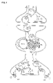
- FIG. 1 shows a schematic of the four steps required for tetanus and botulinum toxin activity in central and peripheral neurons.
- FIG. 2 shows the subcellular localization and sites of cleavage of SNAP-25, VAMP and syntaxin.
- VAMP is bound to synaptic vesicle membrane
- SNAP-25 and syntaxin are bound to the target plasma membrane.
- BoNT/A and /E cleave SNAP-25 close to the carboxy-terminus, releasing nine or 26 residues, respectively.
- BoNT/B, /D, /F, /G and TeNT act on the conserved central portion of VAMP (dotted) and release the amino-terminal portion of VAMP into the cytosol.
- BoNT/C1 cleaves SNAP-25 close to the carboxy-terminus as well as cleaving syntaxin at a single site near the cytosolic membrane surface.
- BoNT/B, /C1, /D, /F, /G and TeNT results in release of a large portion of the cytosolic domain of VAMP or syntaxin, while only a small portion of SNAP-25 is released by selective proteolysis of BoNT/A, /C1 or /E.
- FIG. 3 shows an alignment of various SNAP-25 proteins.
- Human SNAP-25 (SEQ ID NO: 1; GenBank accession g4507099; see, also, related human SNAP-25 sequence g2135800); mouse SNAP-25 (SEQ ID NO: 2; GenBank accession G6755588); Drosophila SNAP-25 (SEQ ID NO: 3; GenBank accession g548941); goldfish SNAP-25 (SEQ ID NO: 4; GenBank accession g2133923); sea urchin SNAP-25 (SEQ ID NO: 5; GenBank accession g2707818) and chicken SNAP-25 (SEQ ID NO: 6; GenBank accession g481202) are depicted.
- FIG. 4 shows an alignment of various VAMP proteins.
- Human VAMP-1 (SEQ ID NO: 7; GenBank accession g135093); human VAMP-2 (SEQ ID NO: 8; GenBank accession g135094); mouse VAMP-2 (SEQ ID NO: 9; GenBank accession g2501081); bovine VAMP (SEQ ID NO: 10; GenBank accession g89782); frog VAMP (SEQ ID NO: 11; GenBank accession g6094391); and sea urchin VAMP (SEQ ID NO: 12; GenBank accession g5031415) are depicted.
- FIG. 5 shows an alignment of various syntaxin proteins.
- Human syntaxin 1A (SEQ ID NO: 13; GenBank accession g15079184), human syntaxin 1B2 (SEQ ID NO: 14; GenBank accession g15072437), mouse syntaxin 1A (SEQ ID NO: 15; GenBank accession g15011853), Drosophila syntaxin 1A (SEQ ID NO: 16; GenBank accession g2501095); C. elegans syntaxin A (SEQ ID NO: 17; GenBank accession g7511662) and sea urchin syntaxin (SEQ ID NO: 18; GenBank accession g13310402) are depicted.
- FIG. 6 shows (A) a schematic of plasmid pQBI GFP-SNAP25 (134-206)- 6 ⁇ HIS-C and (B) the nucleic acid and amino acid sequences (SEQ ID NOS: 19 and 20) of pQBI GFP-SNAP25 (134-206)- 6 ⁇ HIS-C.
- FIG. 7 shows (A) the absorption spectrum and (B) the excitation (dotted) and emission (bold) spectra of GFP-SNAP25 (134-206) -His6C.
- FIG. 8 shows (A) the UV-VIS absorption spectrum and (B) the excitation (bold) and emission (dotted) spectra of GFP-SNAP25 (134-206) -His6C-Alexa Fluor® 594.
- FIG. 9 shows turnover of the GFP-SNAP25 (134-206) -His6C-Alexa Fluor® 594 substrate using reduced BoNT/A at various concentrations. The arrow indicates when the reduced toxin complex was added.
- FIG. 10 shows turnover of the GFP-SNAP25 (134-206) -His6C-Alexa Fluor® 546 substrate using recombinant BoNT/A light chain.
- the arrow indicates addition of the BoNT/A light chain.
- the invention provides novel methods for determining the presence or activity of clostridial toxins including botulinum toxins of all serotypes as well as tetanus toxins.
- the novel methods of the invention which rely on a clostridial toxin substrate useful for fluorescence polarization analysis, reduce the need for animal toxicity studies and can be used to analyze crude and bulk samples as well as highly purified dichain or single chain toxins or formulated toxin products.
- the fluorescence polarization-based methods of the invention are advantageous in that they are sensitive assays which are robust in terms of interference from background fluorescence present in samples.
- the novel methods of the invention can be performed as homogeneous solution-phase assays and are amenable to automated high-throughput formats.
- a clostridial toxin substrate was prepared with Alexa Fluor® 594 as a fluorophore, green fluorescent protein (GFP) as a bulking group, and a portion of SNAP-25 (residues 134-206) as a clostridial toxin recognition sequence for BoNT/A.
- the absorption spectrum of the GFP-SNAP25 (134-206) -His6-Cys protein labeled with Alexa Fluor® 594 is shown herein in FIG. 8A
- the excitation and emission spectra of GFP-SNAP25 (134-206) -His6-C-Alexa Fluor® 594 are shown herein in FIG. 8B .
- the GFP-SNAP25 (134-206) -His6-C-Alexa Fluor® 594 substrate was tested for its utility as a suitable substrate by assaying for the activity of BoNT/A reduced bulk toxin by recording the change in fluorescence polarization over time.
- FIG. 9 there was a reduction in fluorescence polarization at or shortly after the time the diluted bulk BoNT/A toxin was added, and toxin activity was detected at a concentration of as little as about 50 ng/ml (see panel 9D).
- fluorescence polarization can be combined with fluorescence resonance energy transfer to sensitively assay for the presence or activity of a clostridial toxin.
- a GFP-SNAP25 (134-206) -His6-C protein was site-specifically labeled at the carboxy-terminal cysteine residue with Alexa Fluor® 546; the photoselection properties of GFP and Alexa Fluor® 546 provide for fluorescence resonance energy transfer (FRET) between the donor fluorophore GFP and the acceptor Alexa Fluor® 546.
- FRET fluorescence resonance energy transfer
- fluorescence polarization increased upon addition of recombinant BoNT/A light chain.
- FRET in the intact substrate leads to an apparent depolarization of Alexa Fluor® 546 emission due to the significant angle between the initially selected dipole (GFP) and the dipole which would be selected by direct excitation of Alexa Fluor® 546.
- GFP initially selected dipole
- polarization consequently increases even though rotation of the Alexa Fluor® dye is increased.
- the combination of fluorescence resonance energy transfer with fluorescence polarization enhanced the polarization change upon turnover, increasing the sensitivity of the assay (see FIG. 10 ).
- the present invention provides a method of determining the presence or activity of a clostridial toxin by (a) treating with a sample, under conditions suitable for clostridial toxin protease activity, a clostridial toxin substrate which includes a fluorophore; a bulking group; and a clostridial toxin recognition sequence containing a cleavage site that intervenes between the fluorophore and the bulking group; (b) exciting the fluorophore with plane polarized light; and (c) determining fluorescence polarization of the treated substrate relative to a control substrate, where a change in fluorescence polarization of the treated substrate as compared to fluorescence polarization of the control substrate is indicative of the presence or activity of the clostridial toxin.
- the change in fluorescence polarization is a decrease in fluorescence polarization.
- step (c) includes determining the change in fluorescence polarization
- a fluorophore can have, without limitation, a fluorescence lifetime of at least 0.5 nanoseconds, or at least 5 nanoseconds, or at least 10 nanoseconds.
- fluorophores can be useful in the methods of the invention including, but not limited to, Alexa Fluor® dyes; fluorescein and fluorescein derivatives such as diaminotriazinylamino-fluorescein (DTAF); biarsenic derivatives of fluorescein such as fluorescein arsenical hairpin binding dye (FlAsHTM) and red biarsenical dye (ReAsHTM); carboxyfluorescein (FAM); Texas RedTM; tetramethylcarboxyrhodamine (TMR); carboxy-x-rhodamine (ROX); rhodamine green; Oregon Green 488; BODIPY®-TR; BODIPY®-TMR; BODIPY®-FL; Cy3; Cy3; Cy3; Cy3; Cy3
- a variety of bulking groups are useful in the methods of the invention, including, without limitation, fluorescent proteins such as green fluorescent protein.
- a method of the invention is practiced such that the change in molecular mass upon cleavage of the clostridial toxin substrate is at least 1000 Da.
- a method of the invention is practiced such that the decrease in fluorescence polarization is at least 5 millipolarization units (mP).
- mP millipolarization units
- a method of the invention is practiced such that the decrease in fluorescence polarization is at least 15 mP.
- a variety of recognition sequences can be included in a clostridial toxin substrate useful in a method of the invention.
- the recognition sequence is a BoNT/A recognition sequence such as, without limitation, a BoNT/A recognition sequence containing at least six consecutive residues of SNAP-25, where the six consecutive residues include Gln-Arg, or a peptidomimetic thereof.
- a BoNT/A recognition sequence can include, for example, residues 134 to 206 of SEQ ID NO: 2.
- a recognition sequence included in a clostridial toxin substrate useful in a method of the invention also can be, without limitation, a BoNT/B recognition sequence.
- BoNT/B recognition sequence can contain, for example, at least six consecutive residues of VAMP, where the six consecutive residues include Gln-Phe, or a peptidomimetic thereof.
- a recognition sequence included in a clostridial toxin substrate useful in a method of the invention is a BoNT/C1 recognition sequence.
- Such a BoNT/C1 recognition sequence can contain, without limitation, at least six consecutive residues of syntaxin, where the six consecutive residues include Lys-Ala, or a peptidomimetic thereof.
- a BoNT/C1 recognition sequence useful in the invention also can contain at least six consecutive residues of SNAP-25, where the six consecutive residues include Arg-Ala, or a peptidomimetic thereof.
- a recognition sequence included in a clostridial toxin substrate useful in a method of the invention is a BoNT/D recognition sequence.
- a BoNT/D recognition sequence can contain, for example, at least six consecutive residues of VAMP, where the six consecutive residues include Lys-Leu, or a peptidomimetic thereof.
- a recognition sequence useful in the invention also can be, for example, a BoNT/E recognition sequence.
- Such a BoNT/E recognition sequence can include, without limitation, residues 134 to 206 of SEQ ID NO: 2, or can contain at least six consecutive residues of SNAP-25, where the six consecutive residues include Arg-Ile, or a peptidomimetic thereof.
- a recognition sequence included in a clostridial toxin substrate useful in a method of the invention is a BoNT/F recognition sequence.
- BoNT/F recognition sequences useful in the invention encompass, without limitation, those having at least six consecutive residues of VAMP, where the six consecutive residues include Gln-Lys, or a peptidomimetic thereof.
- a recognition sequence included in a clostridial toxin substrate useful in a method of the invention also can be a BoNT/G recognition sequence.
- Such BoNT/G recognition sequences encompass, without limitation, those having at least six consecutive residues of VAMP, where the six consecutive residues include Ala-Ala, or a peptidomimetic thereof.
- a recognition sequence included in a clostridial toxin substrate useful in a method of the invention is a TeNT recognition sequence.
- a TeNT recognition sequence can be, without limitation, a sequence containing at least six consecutive residues of VAMP, where the six consecutive residues include Gln-Phe, or a peptidomimetic thereof.
- clostridial toxin substrates are useful for determining the presence or activity of a clostridial toxin according to a method of the invention.
- a clostridial toxin substrate is a peptide or peptidomimetic having at least 100 residues.
- a clostridial toxin substrate is a peptide or peptidomimetic having at least 200 residues.
- any of a variety of samples can be assayed according to a method of the invention including, but not limited to, crude cell lysates, isolated clostridial toxins including isolated clostridial toxin light chains; and formulated clostridial toxin products such as, without limitation, formulated BoNT/A, BoNT/B or BoNT/E toxin products.
- the tetanus and botulinum neurotoxins which can be assayed according to a method of the invention are produced by Clostridia. These toxins cause the neuroparalytic syndromes of tetanus and botulism, with tetanus toxin acting mainly within the central nervous system and botulinum toxin acting on the peripheral nervous system. Clostridial neurotoxins share a similar mechanism of cell intoxication in which the release of neurotransmitters is blocked. In these toxins, which are composed of two disulfide-linked polypeptide chains, the larger subunit is responsible for neurospecific binding and translocation of the smaller subunit into the cytoplasm.
- the smaller chain Upon translocation and reduction in neurons, the smaller chain displays peptidase activity specific for protein components involved in neuroexocytosis.
- the “SNARE” protein targets of clostridial toxins are common to exocytosis in a variety of non-neuronal types; in these cells, as in neurons, light chain peptidase activity inhibits exocytosis.
- Tetanus neurotoxin and botulinum neurotoxins B, D, F, and G specifically recognize VAMP (also known as synaptobrevin), an integral protein of the synaptic vesicle membrane. VAMP is cleaved at distinct bonds depending on the neurotoxin.
- Botulinum A and E neurotoxins recognize and cleave specifically SNAP-25, a protein of the presynaptic membrane, at two different sites in the carboxy-terminal portion of the protein.
- Botulinum neurotoxin C cleaves syntaxin, a protein of the nerve plasmalemma, in addition to SNAP-25.
- Clostridial neurotoxins are conserved from yeast to humans although cleavage sites and toxin susceptibility are not necessarily conserved (see below; see, also, Humeau et al., Biochimie 82:427-446 (2000); Niemann et al., Trends in Cell Biol. 4:179-185 (1994); and Pellizzari et al., Phil. Trans. R. Soc. London 354:259-268 (1999)).
- Naturally occurring tetanus and botulinum neurotoxins are produced as polypeptide chains of 150 kDa without a leader sequence. These toxins may be cleaved by bacterial or tissue proteinases at an exposed protease-sensitive loop, generating active dichain toxin. Selective proteolytic cleavage activates the toxins by generating two disulfide-linked chains: an L chain of 50 kDa and an H chain of 100 kDa, which is composed of two domains denoted H N and H C . This dichain toxin is more active than unnicked toxin.
- Naturally occurring clostridial toxins contain a single interchain disulfide bond bridging the heavy chain and light chain; such a bridge is important for neurotoxicity of toxin added extracellularly (Montecucco and Schiavo, Quarterly Rev. Biophysics 28:423-472 (1995)).
- the clostridial toxins appear to be folded into three distinct domains of about 50 kDa which are connected by loops, with each domain having a distinct functional role.
- the cell intoxication mechanism of the clostridial toxins consists of four distinct steps: (1) binding; (2) internalization; (3) membrane translocation; and (4) enzymatic target modification.
- the carboxy-terminal domain of the heavy chain (H C ) functions in neurospecific binding, while the amino-terminal domain of the H chain (H N ) functions in membrane translocation from endosome to cell cytoplasm. Following reduction of the disulfide linkage inside the cell, the zinc-endopeptidase activity of the L chain is liberated (Montecucco and Schiavo, supra, 1995).
- the amino acid sequences of eight human clostridial neurotoxin serotypes have been derived from the corresponding genes (Niemann, “Molecular Biology of Clostridial Neurotoxins” in Sourcebook of Bacterial Protein Toxins Alouf and Freer (Eds.) pp. 303-348 London: Academic Press 1991).
- the L chain and H chain are composed of roughly 439 and 843 residues, respectively. Homologous segments are separated by regions of little or no similarity.
- the most well conserved regions of the L chain are the amino-terminal portion (100 residues) and central region (corresponding to residues 216 to 244 of TeNT), as well as the two cysteines forming the interchain disulfide bond.
- the 216 to 244 region contains a His-Glu-X-X-His binding motif characteristic of zinc-endopeptidases.
- the clostridial toxin heavy chains are less well conserved than the light chains, with the carboxy-terminal portion of H C corresponding to residues 1140 to 1315 of TeNT the most variable. This is consistent with the involvement of the H C domain in binding to nerve terminals and the fact that different neurotoxins appear to bind different receptors.
- nucleotide and amino acid sequences of the clostridial toxins indicates that they derive from a common ancestral gene. Spreading of these genes may have been facilitated by the fact that the clostridial neurotoxin genes are located on mobile genetic elements. As discussed further below, sequence variants of the seven botulinum toxins are known in the art. See, for example, Humeau et al., supra, 2000.
- VAMP is associated with the synaptic vesicle membrane
- SNAP-25 and syntaxin are associated with the target membrane (see FIG. 2 ).
- BoNT/A and BoNT/E cleave SNAP-25 in the carboxy-terminal region, releasing nine or twenty-six amino acid residues, respectively, and BoNT/C1 also cleaves SNAP-25 near the carboxy-terminus.
- BoNT/B The botulinum serotypes BoNT/B, BoNT/D, BoNT/F and BoNT/G, and tetanus toxin, act on the conserved central portion of VAMP, and release the amino-terminal portion of VAMP into the cytosol.
- BoNT/C1 cleaves syntaxin at a single site near the cytosolic membrane surface.
- BoNT/B, BoNT/C1, BoNT/D, BoNT/F, BoNT/G or TeNT proteolysis results in release of a large portion of the cytosolic domain of VAMP or syntaxin, while only a small portion of SNAP-25 is released by BoNT/A, BoNT/C1 or BoNT/E cleavage (Montecucco and Schiavo, supra, 1995).
- Naturally occurring SNAP-25 a protein of about 206 residues lacking a transmembrane segment, is associated with the cytosolic surface of the nerve plasmalemma ( FIG. 2 ; see, also, Hodel et al., Int. J. Biochemistry and Cell Biology 30:1069-1073 (1998)).
- SNAP-25-related proteins also have been cloned from yeast. SNAP-25 is required for axonal growth during development and may be required for nerve terminal plasticity in the mature nervous system.
- isoform a is constitutively expressed during fetal development
- isoform b appears at birth and predominates in adult life.
- SNAP-25 analogues such as SNAP-23 also are expressed outside the nervous system, for example, in pancreatic cells.
- Naturally occurring VAMP is a protein of about 120 residues, with the exact length depending on the species and isotype. As shown in FIG. 2 , VAMP contains a short carboxy-terminal segment inside the vesicle lumen while most of the molecule is exposed to the cytosol. The proline-rich amino-terminal thirty residues are divergent among species and isoforms while the central portion of VAMP (residues 30 to 96), which is rich in charged and hydrophilic residues and includes known cleavage sites, is highly conserved. VAMP colocalizes with synaptophysin on synaptic vesicle membranes.
- VAMP A variety of species homologs of VAMP are known in the art including human, rat, bovine, Torpedo, Drosophila , yeast, squid and Aplysia homologs.
- multiple isoforms of VAMP have been identified including VAMP-1, VAMP-2 and cellubrevin, and forms insensitive to toxin cleavage have been identified in non-neuronal cells.
- VAMP appears to be present in all vertebrate tissues although the distribution of VAMP-1 and VAMP-2 varies in different cell types.
- Chicken and rat VAMP-1 are not cleaved by TeNT or BoNT/B.
- VAMP-1 homologs have a valine in place of the glutamine present in human and mouse VAMP-1 at the TeNT or BoNT/B cleavage site.
- the substitution does not affect BoNT/D, /F or /G, which cleave both VAMP-1 and VAMP-2 with similar rates.
- Syntaxin is located on the cytosolic surface of the nerve plasmalemma and is membrane-anchored via a carboxy-terminal segment, with most of the protein exposed to the cytosol. Syntaxin colocalizes with calcium channels at the active zones of the presynaptic membrane, where neurotransmitter release takes place.
- syntaxin interacts with synaptotagmin, a protein of the SSV membrane, that forms a functional bridge between the plasmalemma and the vesicles.
- a variety of syntaxin isoforms have been identified. Two isoforms of slightly different length (285 and 288 residues) have been identified in nerve cells (isoforms 1A and 1B), with isoforms 2, 3, 4 and 5 expressed in other tissues. The different isoforms have varying sensitivities to BoNT/C1, with the 1A, 1B, 2 and 3 syntaxin isoforms cleaved by this toxin.
- the methods of the invention rely, in part, on the use of fluorescence polarization.
- fluorescence polarization when a fluorescently labeled molecule is excited with plane polarized light, it emits lights that has a degree of polarization which is inversely proportional to its molecular rotation.
- polarization As a consequence, for large fluorescently labeled molecules, which remain relatively stationary during their excited state (about 4 ns for fluorescein), polarization remains relatively constant between excitation and emission.
- small fluorescently labeled molecules rotate rapidly during the excited state, such that polarization of the light changes significantly between excitation and emission. Therefore, as a generalization, small molecules have low polarization values, and large molecules have high polarization values.
- Fluorescence polarization assays are homogeneous in that they do not require a separation step and do not require attachment of substrate to an immobilized phase. Furthermore, polarization values can be measured repeatedly. In addition, fluorescence polarization is a sensitive technique which can be used to measure polarization values of fluorophores from low picomolar and micromolar levels. Polarization is also independent of fluorescence intensity.
- Fluorescence anisotropy (commonly denoted as “r” or sometimes “A”) is an alternative definition of how a plane of polarized light changes between excitation and emission with a rotating fluorophore. Fluorescence polarization and anisotropy are well known in the art as described in Lundblad et al., Mol. Endocrin. 10:607-612 (1996); Nasir et al., Comb. Chem. High Throughput Screen. 2:177-190 (1999); Sittampalam et al., Curr. Opin. Chem. Biol. 1:384-391 (1997); Thompson et al., Biotechniques 32:34-40 (1997); Lakowicz et al., J. Biomol. Screen. 5:123-132 (2000); and Fernandes, Curr. Opin. Chem. Biol. 2:597-603 (1998).
- fluorescence polarization (P) and anisotropy (r) are defined as follows:
- I Vertical is the intensity of the emission light parallel to the excitation light plane and I Horizontal is the intensity of the emission light perpendicular to the excitation light plane.
- P and r being ratios of light intensities, are dimensionless.
- polarization is a relationship of fluorescence lifetime and how fast a fluorophore rotates in the time between excitation and emission.
- the principal factors controlling rotation are molar volume (V), absolute temperature (T), and viscosity ( ⁇ ).
- the rotational correlation time ( ⁇ ) and the rotational relaxation time ( ⁇ o ) are taken from the work of Perrin and Weber. In particular, the rotational correlation time ( ⁇ ) is taken from the Perrin equation as follows:
- a method of the invention relies on a clostridial toxin substrate which includes, in part, a fluorophore.
- fluorophore means a molecule that, when irradiated with light of a certain wavelength, emits light, also denoted fluorescence, of a different wavelength.
- fluorophore is synonymous in the art with the term “fluorochrome.”
- Fluorophores useful in the invention include those having fluorescence lifetimes suitable for fluorescence polarization analysis.
- Useful fluorophores include, without limitation, Alexa Fluor® dyes; fluorescein and fluorescein derivatives such as diaminotriazinylamino-fluorescein (DTAF); biarsenic derivatives of fluorescein such as fluorescein arsenical hairpin binding dye (FlAsHTM) and red biarsenical dye (ReAsHTM); carboxyfluorescein (FAM); Texas RedTM; tetramethylcarboxyrhodamine (TMR); carboxy-x-rhodamine (ROX); rhodamine green; Oregon Green 488; BODIPY®-TR; BODIPY®-TMR; BODIPY®-FL; Cy3, CyTM3B and Dansyl.
- Alexa Fluor® dyes fluorescein and fluorescein derivatives such as diaminotriazinyla
- fluorophores suitable for fluorescence polarization are known in the art, including, but not limited to, long-wavelength fluorophores such as BODIPY®-TMR and BODIPY®-TR (Molecular Probes), which tend to minimize assay interference, and pH insensitive fluorophores such as BODIPY®-FL. See, for example, Owicki, J. Biomol. Screening 5:297-306 (2000); Burke et al., Comb. Chem . & High Throughput Screen. 6:183-194 (2003); and Jameson and Croney, Comb. Chem . & High Throughput Screen. 6:167-176 (2003).
- fluorophores and donor fluorophores useful for fluorescence polarization are commercially available from various sources such as Molecular Probes (Eugene, Oreg.) and Amersham Pharmacia Biotech (Piscataway, N.J.).
- Molecular Probes Eugene, Oreg.
- Amersham Pharmacia Biotech Piscataway, N.J.
- fluorophores suitable for fluorescence polarization are known in the art and can be useful in the methods of the invention.
- the term “bulking group” means a moiety having sufficient hydrodynamic volume such that, upon cleavage of a clostridial toxin substrate into which the bulking group is incorporated, there is a change in polarization of at least 3 millipolarization units (mP).
- any of a variety of moieties can be useful as a bulking group in a method of the invention including physical, chemical and biological moieties which can be covalently or non-covalently incorporated into a clostridial toxin substrate.
- the bulking group is expressed as a fusion protein with another component of the clostridial toxin substrate.
- Bulking groups useful in the invention encompass natural and man-made moieties and further encompass, without limitation, inert moieties as well as those with biological or other activity.
- a bulking group useful in the invention can be, without limitation, a moiety having a size of greater than 1000 Da.
- a bulking group useful in the invention also can be, without limitation, a moiety having a size of greater than 2 kDa, 3 kDa, 4 kDa, 5 kDa, 10 kDa, 15 kDa, 20 kDa, 25 kDa, 30 kDa, 35 kDa or 40 kDa. See, also, Mattison et al., Application Note for Protein Solutions Inc . February 2001. One skilled in the art understands that a fluorophore with a suitable lifetime will be selected depending, in part, on the size of the bulking group.
- a bulking group useful in the invention can be an inert or active protein, peptide or peptidomimetic; an antibody; organic chemical; latex or other bead; or moiety such as streptavidin.
- Additional bulking groups useful in the invention encompass, without limitation, phage and other viruses; cells; liposomes; polymeric and non-polymeric matrices; gold and other particles; and microdevices and nanodevices.
- a bulking group useful in the invention can be a fluorescent protein such as GFP or BFP, or a fragment thereof; a protein useful for affinity purification such as glutathione-S-transferase (GST) or maltose-binding protein (MBP); or an antibody such as, without limitation, an anti-FLAG, anti-hemagglutinin (HA) or anti-myc antibody.
- Streptavidin also can be a bulking group useful in the invention.
- a biotinylation sequence can be covalently included in a clostridial toxin substrate, providing for association with streptavidin; enzymatic cleavage can be detected by following the fluorescence polarization change upon addition of streptavidin as described in Levine et al., “Measurement of specific protease activity utilizing fluorescence polarization,” Anal. Biochem. 247:83-88 (1997).
- a clostridial toxin substrate useful in the invention contains a cleavage site that “intervenes” between a fluorophore and a bulking group.
- the cleavage site is positioned in between the fluorophore and the bulking group such that proteolysis at the cleavage site results in a first cleavage product containing the fluorophore and a second cleavage product containing the bulking group. It is understood that all or only a portion of the clostridial toxin recognition sequence may intervene between the fluorophore and the bulking group.
- a clostridial toxin substrate useful in the invention contains, in part, a clostridial toxin recognition sequence which includes a cleavage site.
- a clostridial toxin substrate is susceptible to cleavage by at least one clostridial toxin under conditions suitable for clostridial toxin protease activity.
- a clostridial toxin substrate useful in the invention is a peptide or peptidomimetic having a defined length.
- a clostridial toxin substrate can be, for example, a peptide or peptidomimetic having at least 50, at least 100, at least 150, at least 200, at least 250, at least 300, at least 350, at least 500, at least 600, at least 700, at least 800 or at least 900 residues.
- a clostridial toxin substrate has at most 20 residues, at most 30 residues, at most 40 residues, at most 50 residues, at most 60 residues, at most 70 residues, at most 80 residues, at most 90 residues, at most 100 residues, at most 150 residues, at most 200 residues, at most 250 residues, at most 300 residues, at most 350 residues or at most 400 residues.
- a clostridial toxin substrate useful in the invention optionally can include one or more additional components.
- a flexible spacer sequence such as GGGGS (SEQ ID NO: 21) can be included in a clostridial toxin substrate useful in the invention.
- a useful clostridial toxin substrate further can include, without limitation, one or more of the following: an affinity tag such as HIS6; biotin or a biotinylation sequence; or an epitope such as FLAG, hemagglutinin (HA), c-myc, or AU1; an immunoglobulin hinge region; an N-hydroxysuccinimide linker; a peptide or peptidomimetic hairpin turn; or a hydrophilic sequence or another component or sequence that, for example, facilitates purification or promotes the solubility or stability of the clostridial toxin substrate.
- an affinity tag such as HIS6
- biotin or a biotinylation sequence such as an epitope such as FLAG, hemagglutinin (HA), c-myc, or AU1
- an immunoglobulin hinge region such as FLAG, hemagglutinin (HA), c-myc, or AU1
- an immunoglobulin hinge region
- a method of the invention can be useful to determine the presence or activity of a clostridial toxin in a food or beverage sample; to assay a sample from a human or animal, for example, exposed to a clostridial toxin or having one or more symptoms of a clostridial toxin; to follow activity during production and purification of clostridial toxin; or to assay formulated clostridial toxin products such as pharmaceuticals or cosmetics.
- sample means any biological matter that contains or potentially contains an active clostridial toxin.
- sample encompasses, but is not limited to, purified or partially purified clostridial toxin; recombinant single chain or dichain toxin with a naturally or non-naturally occurring sequence; recombinant clostridial toxin with a modified protease specificity; recombinant clostridial toxin with an altered cell specificity; chimeric toxin containing structural elements from multiple clostridial toxin species or subtypes; bulk toxin; formulated toxin product; cells or crude, fractionated or partially purified cell lysates, for example, engineered to include a recombinant nucleic acid encoding a clostridial toxin; bacterial, baculoviral and yeast lysates; raw, cooked, partially cooked or processed foods
- sample encompasses tissue samples, including, without limitation, mammalian tissue samples, livestock tissue samples such as sheep, cow and pig tissue samples; primate tissue samples; and human tissue samples.
- tissue samples encompass, without limitation, intestinal samples such as infant intestinal samples, and tissue samples obtained from a wound.
- conditions suitable for clostridial toxin protease activity are useful in the methods of the invention.
- conditions suitable for clostridial toxin protease activity can be provided such that at least 10% of the substrate is cleaved.
- conditions suitable for clostridial toxin protease activity can be provided such that at least 20%, 30%, 40%, 50%, 60%, 70%, 80%, 90% or 95% of the clostridial toxin substrate is cleaved, or such that 100% of the clostridial toxin substrate is cleaved.
- the conditions suitable for clostridial toxin protease activity are selected such that the assay is linear.
- conditions suitable for clostridial toxin protease activity are provided such that at least 90% of the clostridial toxin substrate is cleaved.
- conditions suitable for clostridial toxin protease activity are provided such that at most 25% of the clostridial toxin substrate is cleaved.
- conditions suitable for clostridial toxin protease activity are provided such that at most 5%, 10%, 15% or 20% of the clostridial toxin substrate is cleaved.
- the clostridial toxin substrate can be treated with a sample in solution phase.
- in solution phase means that the substrate is soluble and, during proteolysis, is not constrained or immobilized on a solid support such as a column or dish.
- a sample is treated with a clostridial toxin substrate under conditions suitable for clostridial toxin protease activity.
- exemplary conditions suitable for clostridial toxin protease activity are well known in the art, and further can be determined by routine methods. See, for example, Hallis et al., J. Clin. Microbiol. 34:1934-1938 (1996); Ekong et al., Microbiol. 143:3337-3347 (1997); Shone et al., WO 95/33850; Schmidt and Bostian, supra, 1995; Schmidt and Bostian, supra, 1997; Schmidt et al., supra, 1998; and Schmidt and Bostian, U.S. Pat. No.
- conditions suitable for clostridial toxin protease activity can depend, in part, on the specific clostridial toxin type or subtype being assayed and the purity of the toxin preparation.
- Conditions suitable for clostridial toxin protease activity generally include a buffer, such as HEPES, Tris or sodium phosphate, typically in the range of pH 5.5 to 9.5, for example, in the range of pH 6.0 to 9.0, pH 6.5 to 8.5 or pH 7.0 to 8.0.
- Conditions suitable for clostridial toxin protease activity also can include, if desired, dithiothreitol, ⁇ -mercaptoethanol or another reducing agent, for example, where a dichain toxin is being assayed (Ekong et al., supra, 1997).
- the conditions include DTT in the range of 0.01 mM to 50 mM; in other embodiments, the conditions include DTT in the range of 0.1 mM to 20 mM, 1 to 20 mM, or 5 to 10 mM.
- an isolated clostridial toxin or sample can be pre-incubated with a reducing agent, for example, with 10 mM dithiothreitol (DTT) for about 30 minutes prior to addition of clostridial toxin substrate.
- a reducing agent for example, with 10 mM dithiothreitol (DTT) for about 30 minutes prior to addition of clostridial toxin substrate.
- Clostridial toxins are zinc metalloproteases, and a source of zinc, such as zinc chloride or zinc acetate, typically in the range of 1 to 500 ⁇ M, for example, 5 to 10 ⁇ M can be included, if desired, as part of the conditions suitable for clostridial toxin protease activity.
- a source of zinc such as zinc chloride or zinc acetate
- zinc chelators such as EDTA generally are excluded from a buffer for determining the activity of a clostridial toxin.
- Conditions suitable for clostridial toxin protease activity can optionally include a detergent such as TWEEN-20, which can be used, for example, in place of bovine serum albumin.
- TWEEN-20 can be provided, for example, in the range of 0.001% to 10% (v/v), or in the range of 0.01% to 1.0% (v/v).
- TWEEN-20 can be included at a concentration of 0.1% (v/v).
- Conditions suitable for clostridial toxin protease activity also can include, if desired, bovine serum albumin (BSA) or another agent which acts as a protein stabilizer, solubilizing agent or blocker of surface loss.
- BSA bovine serum albumin
- BSA typically is provided in the range of 0.1 mg/ml to 10 mg/ml. In one embodiment, BSA is included at a concentration of 1 mg/ml. See, for example, Schmidt and Bostian, supra, 1997. In another embodiment, BSA is included at a concentration of 0.1% (w/v).
- the amount of clostridial toxin substrate can be varied in a method of the invention.
- a clostridial toxin substrate can be supplied, for example, at a concentration of 1 ⁇ M to 500 ⁇ M, 1 ⁇ M to 50 ⁇ M, 1 ⁇ M to 30 ⁇ M, 5 ⁇ M to 20 ⁇ M, 50 ⁇ M to 3.0 mM, 0.5 mM to 3.0 mM, 0.5 mM to 2.0 mM, or 0.5 mM to 1.0 mM.
- concentration of clostridial toxin substrate or the amount of sample can be limited, if desired, such that the assay is linear.
- a method of the invention relies on a clostridial toxin substrate concentration of less than 100 ⁇ M. In further embodiments, a method of the invention relies on a clostridial toxin substrate concentration of less than 50 ⁇ M or less than 25 ⁇ M. In a further embodiment, a method of the invention relies on a clostridial toxin substrate concentration of 10 ⁇ M to 20 ⁇ M. If desired, a linear assay also can be performed by mixing clostridial toxin substrate with corresponding, “unlabeled” substrate which lacks a fluorophore. The appropriate dilution can be determined, for example, by preparing serial dilutions of clostridial toxin substrate in the corresponding unlabeled substrate.
- the concentration of purified or partially purified clostridial toxin to be assayed in a method of the invention generally is in the range of about 0.0001 ng/ml to 500 ⁇ g/ml toxin, for example, about 0.0001 ng/ml to 50 ⁇ g/ml toxin, 0.001 ng/ml to 500 ⁇ g/ml toxin, 0.001 ng/ml to 50 ⁇ g/ml toxin, 0.0001 to 5000 ng/ml toxin, for example, about 0.001 ng/ml to 5000 ng/ml, 0.01 ng/ml to 5000 ng/ml, 0.1 ng/ml to 5000 ng/ml, 1 ng/ml to 5000 ng/ml, 10 ng/ml to 5000 ng/ml, 50 ng/ml to 5000 ng/ml, 50 ng/ml to 500 ng/ml or 100 ng/ml
- the concentration of purified or partially purified clostridial toxin assayed in a method of the invention can be, for example, in the range of about 0.1 pM to 100 ⁇ M, 0.1 pM to 10 ⁇ M, 0.1 pM to 1 ⁇ M, 0.1 pM to 500 nM, 0.1 pM to 100 nM, for example, 1 pM to 2000 pM, 1 pM to 200 pM, 1 pM to 50 pM, 1 nM to 1 ⁇ M, 1 nM to 500 nM, 1 nM to 200 nM, 1 nM to 100 nM or 3 nM to 100 nM toxin, which can be, for example, purified native or recombinant light chain or dichain toxin or formulated clostridial toxin product containing human serum albumin and excipients.
- concentration of purified or partially purified clostridial toxin will depend on the serotype of the toxin assayed, as well as the purity or recombinant sequence of the toxin, the presence of inhibitory components, and the assay conditions. It is additionally understood that purified, partially purified or crude samples can be diluted to within a convenient range for assaying for clostridial toxin protease activity against a standard curve. Similarly, it is understood that a sample can be diluted, if desired, such that the assay is linear.
- Conditions suitable for clostridial toxin protease activity also generally include, for example, temperatures in the range of about 20° C. to about 45° C., for example, in the range of 25° C. to 40° C., or the range of 35° C. to 39° C.
- Assay volumes often are in the range of about 5 to about 200 ⁇ l, for example, in the range of about 10 ⁇ l to 100 ⁇ l or about 0.5 ⁇ l to 100 ⁇ l, although nanoliter reaction volumes also can be used with the methods of the invention.
- Assay volumes also can be, for example, in the range of 100 ⁇ l to 2.0 ml or in the range of 0.5 ml to 1.0 ml.
- fluorescence polarization reactions may or may not be terminated and that assay times can be varied as appropriate by the skilled artisan.
- Assay times generally depend, in part, on the concentration, purity and activity of the clostridial toxin and generally vary, without limitation, in the range of about 15 minutes to about 5 hours.
- exemplary assay times include incubation, for example, at 37° C. for 30 minutes, 45 minutes, 60 minutes, 75 minutes or 90 minutes. In particular embodiments, at least 10%, 20%, 30%, 40%, 50%, 60%, 70%, 80%, 90%, 95% or 100% of the clostridial toxin substrate is cleaved.
- the protease reaction is stopped before more than 5%, 10%, 15%, 20%, 25% or 50% of the clostridial toxin substrate is cleaved.
- protease reactions can be terminated by the appropriate reagent, which generally depends on the fluorophore and other components of the substrate.
- a protease reaction based on a substrate containing GFP as the donor fluorophore can be terminated by the addition of guanidinium chloride, for example, to a final concentration of 1 to 2 M.
- Protease reactions also can be terminated by addition of H 2 SO 4 ; addition of about 0.5 to 1.0 sodium borate, pH 9.0 to 9.5; or addition of zinc chelators.
- protease reactions can be terminated, if desired, prior to exciting the fluorophore or donor fluorophore with plane polarized light.
- conditions suitable for clostridial toxin protease activity such as BoNT/A protease activity can be incubation at 37° C. for 90 minutes in a buffer containing 50 mM HEPES (pH 7.2), 10 ⁇ M ZnCl 2 , 10 mM DTT, and 0.1% (v/v) TWEEN-20 with 10-16 ⁇ M substrate.
- samples containing BoNT/A, particularly dichain BoNT/A can be preincubated with dithiothreitol, for example, for 20 or 30 minutes before addition of substrate.
- conditions suitable for BoNT/A protease activity can be incubation at 37° C.
- BSA in the range of 0.1 mg/ml to 10 mg/ml, for example, 1 mg/ml BSA, also can be included when a sample is treated with a clostridial toxin substrate (Schmidt and Bostian, supra, 1997).
- conditions suitable for clostridial toxin protease activity can be incubation in 50 mM HEPES, pH 7.4, with 10 ⁇ M zinc chloride, 1% fetal bovine serum and 10 mM dithiothreitol, with incubation for 90 minutes at 37° C. (Shone and Roberts, Eur. J. Biochem.
- Conditions suitable for tetanus toxin protease activity or other clostridial toxin protease activity can be, for example, incubation in 20 mM HEPES, pH 7.2, and 100 mM NaCl for 2 hours at 37° C. with 25 ⁇ M peptide substrate (Cornille et al., supra, 1994).
- conditions suitable for clostridial toxin protease activity include cationic polyamino acids such as polyarginine in a buffer of suitable ionic strength. Where there is a charge difference in the clostridial toxin substrate as compared to the cleavage product, fluorescence polarization can be observed in the presence of polyarginine or another cationic polyamino acid (Simeonov et al., Analytical Biochemistry 304:193-199 (2002)).
- polyarginine will selectively bind to the fluorescently labeled cleavage product, thereby generating a measurable increase in polarization.
- control substrate can be, for example, a clostridial toxin substrate which is not treated with active, toxin-containing sample; a polarization value determined before addition of the sample; or a similar, but different substrate which does not contain a toxin cleavage site or functional recognition sequence.
- the methods of the invention can be automated and can be configured in a high-throughput or ultra high-throughput format using, without limitation, 96-well, 384-well or 1536-well plates.
- Any of a variety of spectrofluorometers equipped with an appropriate polarizer can be used to assay the change in fluorescence polarization over time including, without limitation a Cary Eclipse spectrofluorometer; the Beckmann AffinityTM Multi-Mode plate reader; TECAN GeniusPro; and other systems from, for example, Perkin Elmer.
- a clostridial toxin substrate containing (i) a donor fluorophore; (ii) an acceptor having an absorbance spectrum overlapping the emission spectrum of the donor fluorophore; and (iii) a clostridial toxin recognition sequence containing a cleavage site, where the cleavage site intervenes between the donor fluorophore and the acceptor and where, under the appropriate conditions, resonance energy transfer is exhibited between the donor fluorophore and the acceptor; (b) exciting the donor fluorophore with plane polarized light; and (c) determining fluorescence polarization of the treated substrate relative to a control substrate, where a change in fluorescence polarization of the treated substrate as compared to fluorescence polarization of the control substrate is indicative of the presence
- the change in fluorescence polarization can be an increase or decrease in fluorescence polarization.
- the donor fluorophore has a fluorescence lifetime of at least 0.5 nanoseconds. In another embodiment, the donor fluorophore has a fluorescence lifetime of at least 5 nanoseconds.
- a donor fluorophore useful in the invention can be, without limitation, a green fluorescent protein (GFP); blue fluorescent protein (BFP); cyan fluorescent protein (CFP); yellow fluorescent protein (YFP); red fluorescent protein (RFP); Alexa Fluor® dye; fluorescein; a fluorescein derivative; diaminotriazinylamino-fluorescein (DTAF); a biarsenic derivative of fluorescein; fluorescein arsenical hairpin binding dye (FlAsHTM); red biarsenical dye (ReAsHTM); carboxyfluorescein (FAM); Texas RedTM; tetramethylcarboxy-rhodamine (TMR); carboxy-x-rhodamine (ROX); rhodamine green; Oregon Green 488; BODIPY®-TR; BODIPY®-TMR; BODIPY®-FL; Cy3, CyTM3B or Dansyl.
- GFP green fluorescent protein
- BFP blue fluorescent protein
- the donor fluorophore is a green fluorescent protein; blue fluorescent protein; cyan fluorescent protein; yellow fluorescent protein or red fluorescent protein.
- the donor fluorophore is a green fluorescent protein (GFP).
- the acceptor fluorophore is Alexa Fluor® 546.
- the recognition sequence is a BoNT/A recognition sequence such as, without limitation, a BoNT/A recognition sequence containing at least six consecutive residues of SNAP-25, where the six consecutive residues include Gln-Arg, or a peptidomimetic thereof.
- a BoNT/A recognition sequence can include, for example, residues 134 to 206 of SEQ ID NO: 2.
- a recognition sequence included in a clostridial toxin substrate useful in a method of the invention also can be, without limitation, a BoNT/B recognition sequence.
- BoNT/B recognition sequence can contain, for example, at least six consecutive residues of VAMP, where the six consecutive residues include Gln-Phe, or a peptidomimetic thereof.
- a recognition sequence included in a clostridial toxin substrate useful in a method of the invention is a BoNT/C1 recognition sequence.
- Such a BoNT/C1 recognition sequence can contain, without limitation, at least six consecutive residues of syntaxin, where the six consecutive residues include Lys-Ala, or a peptidomimetic thereof.
- a BoNT/C1 recognition sequence useful in the invention also can contain at least six consecutive residues of SNAP-25, where the six consecutive residues include Arg-Ala, or a peptidomimetic thereof.
- a recognition sequence included in a clostridial toxin substrate useful in a method of the invention is a BoNT/D recognition sequence.
- a BoNT/D recognition sequence can contain, for example, at least six consecutive residues of VAMP, where the six consecutive residues include Lys-Leu, or a peptidomimetic thereof.
- a recognition sequence useful in the invention also can be, for example, a BoNT/E recognition sequence.
- Such a BoNT/E recognition sequence can contain, without limitation, at least six consecutive residues of SNAP-25, where the six consecutive residues include Arg-Ile, or a peptidomimetic thereof.
- a recognition sequence included in a clostridial toxin substrate useful in a method of the invention is a BoNT/F recognition sequence.
- BoNT/F recognition sequences useful in the invention encompass, without limitation, those having at least six consecutive residues of VAMP, where the six consecutive residues include Gln-Lys, or a peptidomimetic thereof.
- a recognition sequence included in a clostridial toxin substrate useful in a method of the invention also can be a BoNT/G recognition sequence.
- Such BoNT/G recognition sequences encompass, without limitation, those having at least six consecutive residues of VAMP, where the six consecutive residues include Ala-Ala, or a peptidomimetic thereof.
- a recognition sequence included in a clostridial toxin substrate useful in a method of the invention is a TeNT recognition sequence.
- a TeNT recognition sequence can be, without limitation, a sequence containing at least six consecutive residues of VAMP, where the six consecutive residues include Gln-Phe, or a peptidomimetic thereof.
- any of a variety of clostridial toxin substrates can be useful in the methods of the invention, including peptides and peptidomimetics having at least 100 residues, or having at least 200 residues.
- any of a variety of samples can be assayed according to a method of the invention including, without limitation, crude cell lysates, isolated clostridial toxins including isolated clostridial toxin light chains; and formulated clostridial toxin products such as formulated BoNT/A, BoNT/B or BoNT/E toxin products.
- a method of the invention involves fluorescence resonance energy transfer
- the method relies on a clostridial toxin substrate which includes, in part, a donor fluorophore.
- a “donor fluorophore” is a molecule that, when irradiated with light of a certain wavelength, emits light, also denoted fluorescence, of a different wavelength.
- a donor fluorophore is a fluorophore which, when paired with a suitable acceptor, transfers energy to the acceptor.
- acceptor means a molecule that can absorb energy from, and upon excitation of, a donor fluorophore.
- An acceptor useful in a clostridial toxin substrate has an absorbance spectrum which overlaps the emission spectrum of a donor fluorophore included in the substrate.
- An acceptor useful in the invention generally has rather low absorption at a wavelength suitable for excitation of the donor fluorophore.
- an acceptor has an absorbance spectrum that overlaps the emission spectrum of the donor fluorophore.
- overlapping means an absorbance spectrum and emission spectrum that are partly or entirely shared.
- the high end of the range of the donor fluorophore's emission spectrum is higher than the low end of the range of the acceptor's absorbance spectrum.
- any of a variety of donor fluorophores can be useful in the invention, including, without limitation, green fluorescent protein; blue fluorescent protein; cyan fluorescent protein; yellow fluorescent protein; red fluorescent protein; an Alexa Fluor® dye; fluorescein; a fluorescein derivative; diaminotriazinylamino-fluorescein (DTAF); a biarsenic derivative of fluorescein; fluorescein arsenical hairpin binding dye (FlAsHTM); red biarsenical dye (ReAsHTM); carboxyfluorescein (FAM); Texas RedTM; tetramethylcarboxy-rhodamine (TMR); carboxy-x-rhodamine (ROX); rhodamine green; Oregon Green 488; BODIPY®-TR; BODIPY®-TMR; BODIPY®-FL; Cy3, CyTM3B or Dansyl.
- green fluorescent protein blue fluorescent protein
- cyan fluorescent protein yellow fluorescent protein
- red fluorescent protein an Alexa Flu
- Alexa Fluor® dyes such as Alexa Fluor® 546, Alexa Fluor® 568, Alexa Fluor® 610, Alexa Fluor® 660 and Alexa Fluor® 750; QSY® 7; tetramethylrhodamine; octadecylrhodamine; flavodoxin, cytochrome c peroxidase; and rubredoxin.
- Exemplary donor fluorophore-acceptor pairs which exhibit FRET and are useful in the methods of the invention encompass, without limitation, GFP and Alexa Fluor® 546; fluorescein and QSY® 7; fluorescein and tetramethylrhodamine; and dansyl and octadecylrhodamine.
- Further exemplary donor fluorophore-acceptor pairs which are useful in the methods of the invention encompass, without limitation, Alexa Fluor® 633 and Alexa Fluor® 660; Alexa Fluor® 594 and Alexa Fluor® 610; Alexa Fluor® 700 and Alexa Fluor® 750; and Alexa Fluor® 555 and Alexa Fluor® 568.
- acceptors useful in the invention include those in which the acceptor is a protein with a visible chromophore such as, without limitation, flavodoxin, cytochrome c peroxidase or rubredoxin; such a protein can have, for example, a molecular weight in the range of 6 to 34 kDa and a chromophore which absorbs strongly in the region between 400-500 nm.
- a visible chromophore such as, without limitation, flavodoxin, cytochrome c peroxidase or rubredoxin
- a protein can have, for example, a molecular weight in the range of 6 to 34 kDa and a chromophore which absorbs strongly in the region between 400-500 nm.
- Exemplary donor fluorophore-acceptor pairs based on such proteins include, but are not limited to, 5-(((2-iodoaacetyl)amino)ethyl)amino)naphthalene-1-sulfonic acid (1,5 IAEDANS) and flavodoxin; 4-acetamido-4′ maleimidylstilbene 2,2′ disulfonic acid and cytochrom c peroxidase; and Alexa Fluor®488 and rubredoxin.
- donor fluorophores suitable for fluorescence polarization can be paired with any of a variety of acceptors having an absorbance spectrum which overlaps the emission spectrum of the donor fluorophore.
- the methods of the invention based on FRET-assisted fluorescence polarization optionally utilize a substrate which includes a bulking group in addition to the donor fluorophore and acceptor.
- a bulking group depends on the molecular weight and bulking characteristics of the selected donor fluorophore and acceptor.
- a variety of bulking groups are optionally useful in the invention, including those described herein above.
- Substrates useful in the invention can be prepared by recombinant methods or using synthetic chemical methods, or a combination thereof.
- a fusion protein containing a bulking group fused to a BoNT/A clostridial toxin recognition sequence and a carboxy-terminal cysteine was prepared by recombinant methods.
- the carboxy-terminal cysteine was used for attachment of a fluorophore to produce the complete clostridial toxin substrate.
- Recombinant methods for preparation of clostridial toxin substrates which are fusion proteins are well known in the art as described, for example, in Ausubel, Current Protocols in Molecular Biology John Wiley & Sons, Inc., New York 2000.
- a variety of groups can be used to couple a fluorophore, bulking group, donor fluorophore or acceptor, for example, to a peptide or peptidomimetic containing a clostridial toxin recognition sequence.
- a thiol group for example, can be used to couple a fluorophore, bulking group, donor fluorophore or acceptor to the desired position in a peptide or peptidomimetic to produce a clostridial toxin substrate useful in the invention (see Example I).
- Haloacetyl and maleimide labeling reagents also can be used to couple a fluorophore, bulking group, donor fluorophore or acceptor in preparing a clostridial toxin substrate useful in the invention. See, for example, Wu and Brand, supra, 1994.
- Cross-linker moieties also can be useful for preparing a clostridial toxin substrate.
- Cross-linkers are well known in the art and include homo- and hetero-bifunctional cross-linkers such as BMH and SPDP.
- a fluorophore, bulking group, donor fluorophore or acceptor is a protein
- well known chemical methods for specifically linking molecules to the amino- or carboxy-terminus of a protein can be employed. See, for example, “Chemical Approaches to Protein Engineering” in Protein Engineering: A Practical Approach Rees et al. (Eds) Oxford University Press, 1992.
- a clostridial toxin substrate contains a fluorophore and bulking group
- the clostridial toxin cleavage site is positioned between the fluorophore and bulking group.
- the fluorophore is positioned carboxy-terminal of the cleavage site while the bulking group is positioned amino-terminal of the cleavage site.
- the fluorophore is positioned amino-terminal of the cleavage site while the bulking group is positioned carboxy-terminal of the cleavage site.
- a clostridial toxin substrate contains a donor fluorophore and an acceptor
- the clostridial toxin cleavage site is positioned between the donor fluorophore and the acceptor.
- the donor fluorophore is positioned amino-terminal of the cleavage site while the acceptor is positioned carboxy-terminal of the cleavage site.
- the donor fluorophore is positioned carboxy-terminal of the cleavage site while the acceptor is positioned amino-terminal of the cleavage site.
- a fluorophore and a bulking group, or a donor fluorophore and an acceptor generally are positioned to minimize interference with substrate binding to, or proteolysis by, the clostridial toxin.
- a fluorophore and bulking group, or donor fluorophore and acceptor can be selected and positioned, for example, so as to minimize the disruption of bonded and non-bonded interactions that are important for binding, and to minimize steric hindrance.
- the spatial distance between an acceptor and donor fluorophore generally is limited to achieve efficient energy transfer from the donor fluorophore to the acceptor.
- efficiency of energy transfer from a donor fluorophore to an acceptor is dependent, in part, on the spatial separation of the donor fluorophore and acceptor molecules. As the distance between the donor fluorophore and acceptor increases, there is less energy transfer to the acceptor, and the donor fluorescence signal therefore increases.
- the overall energy transfer between the donor fluorophore and acceptor is dependent upon many factors, including the separation distance between the donor fluorophore and acceptor in the substrate, the spectral overlap between donor fluorophore and acceptor, and the substrate concentration.
- concentration of substrate can be controlled as described above.
- the Förster distance which is the separation between a donor fluorophore and an acceptor for 50% energy transfer, represents a spatial separation between donor fluorophore and acceptor that provides a good sensitivity.
- adjacent residues are separated by a distance of approximately 3.6 ⁇ in the most extended conformation.
- the calculated Förster distance for a fluorescein/tetramethylrhodamine pair is 55 ⁇ , which would represent a spatial separation between fluorescein and tetramethylrhodamine of about 15 residues in the most extended conformation.
- donor fluorophores and acceptors can be more widely separated than expected based on a calculation performed using 3.6 ⁇ per residue and still remain within the Förster distance as shown, for example, by the occurrence of FRET between donor-acceptor pairs separated by about 50 amino acids (Graham et al., Analyt. Biochem. 296: 208-217 (2001)).
- Förster theory is based on very weak interactions between a donor fluorophore and an acceptor; spectroscopic properties such as absorption of one fluorophore should not be altered in the presence of the other, defining the shortest distance range over which the theory is valid. It is understood that, for many donor fluorophore-acceptor pairs, Förster theory is valid when donor fluorophores and acceptors are separated by about 10 ⁇ to 100 ⁇ . However, for particular donor fluorophore-acceptor pairs, Förster theory is valid below 10 ⁇ as determined by subpicosecond techniques (Kaschke and Ernsting, Ultrafast Phenomenon in Spectroscopy (Klose and Wilhelmi (Eds.)) Springer-Verlag, Berlin 1990).
- the invention provides a method that relies on a clostridial toxin substrate in which the donor fluorophore is spatially separated from the acceptor by a distance of at most 100 ⁇ . In other embodiments, the invention provides a method that relies on a clostridial toxin substrate in which the donor fluorophore is spatially separated from the acceptor by a distance of at most 90 ⁇ , 80 ⁇ , 70 ⁇ , 60 ⁇ , 50 ⁇ , 40 ⁇ , 30 ⁇ or 20 ⁇ .
- the invention provides a method that relies on a clostridial toxin substrate in which the donor fluorophore is spatially separated from the acceptor by a distance of 10 ⁇ to 100 ⁇ , 10 ⁇ to 80 ⁇ , 10 ⁇ to 60 ⁇ , 1A to 40 ⁇ , 10 ⁇ to 20 ⁇ , 20 ⁇ to 100 ⁇ , 20 ⁇ to 80 ⁇ , 20 ⁇ to 60 ⁇ , 20 ⁇ to 40 ⁇ , 40 ⁇ to 100 ⁇ , 40 ⁇ to 80 ⁇ or 40 ⁇ to 60 ⁇ .
- the invention provides a method that relies on a clostridial toxin substrate in which the donor fluorophore and the acceptor are separated in the primary amino acid sequence by at most six residues, at most eight residues, at most ten residues, at most twelve residues, at most fifteen residues, at most twenty residues, at most twenty-five residues, at most thirty residues, at most thirty-five residues, at most forty residues, at most forty-five residues, at most fifty residues, at most sixty residues, at most seventy residues, at most eighty residues, at most ninety residues, at most 100 residues, at most 150 residues, at most 200 residues or up to the full-length of a naturally occurring clostridial toxin target protein.
- a clostridial toxin substrate useful in the invention can be designed, if desired, to optimize the efficiency of FRET.
- a donor fluorophore can be selected, if desired, with a high quantum yield, and acceptor can be selected, if desired, with a high extinction coefficient to maximize the Förster distance.
- fluorescence arising from direct excitation of an acceptor can be difficult to distinguish from fluorescence resulting from resonance energy transfer.
- a donor fluorophore and acceptor can be selected which have relatively little overlap of their excitation spectra such that the donor can be excited at a wavelength that does not result in direct excitation of the acceptor.
- a clostridial toxin substrate useful in the invention can be designed so that the emission spectra of the donor fluorophore and acceptor overlap relatively little such that the two emissions can be readily distinguished.
- BoNT/A cleaves a Gln-Arg bond
- BoNT/C1 cleaves a Lys-Ala or Arg-Ala bond
- BoNT/D cleaves a Lys-Leu bond
- BoNT/E cleaves an Arg-Ile bond
- BoNT/F cleaves a Gln-Lys bond
- BoNT/G cleaves an Ala-Ala bond (see Table A).
- P 5 -P 4 -P 3 -P 2 -P 1 -P 1 ′-P 2 ′-P 3 ′-P 4 ′-P 5 ′ with P 1 -P 1 ′ representing the scissile bond. It is understood that a P 1 or P 1 ′ site, or both, can be substituted with another amino acid or amino acid mimetic in place of the naturally occurring residue.
- BoNT/A substrates have been prepared in which the P 1 position (Gln) is modified to be an alanine, 2-aminobutyric acid or asparagine residue; these substrates were hydrolyzed by BoNT/A at the P 1 Arg bond (Schmidt and Bostian, J. Protein Chem. 16:19-26 (1997)). While it is recognized that substitutions can be introduced at the P 1 position of the scissile bond, for example, a BoNT/A scissile bond, it is further recognized that conservation of the P 1 ′ residue can be advantageous (Vaidyanathan et al., J. Neurochem. 72:327-337 (1999)).
- the invention provides a method which relies on a clostridial toxin substrate having a clostridial toxin recognition sequence in which the P 1 ′ residue is not modified or substituted relative to the naturally occurring residue in a target protein cleaved by the clostridial toxin.
- the invention provides a method which relies on a clostridial toxin substrate having a recognition sequence in which the P 1 residue is modified or substituted relative to the naturally occurring residue in a target protein cleaved by the clostridial toxin; such a clostridial toxin substrate retains susceptibility to peptide bond cleavage between the P 1 and P 1 ′ residues.
- SNAP-25, VAMP and syntaxin share a short motif located within regions predicted to adopt an ⁇ -helical conformation. This motif is present in SNAP-25, VAMP and syntaxin isoforms expressed in animals sensitive to the neurotoxins. In contrast, Drosophila and yeast homologs that are resistant to these neurotoxins and syntaxin isoforms not involved in exocytosis contain sequence variations in the ⁇ -helical motif regions of these VAMP and syntaxin proteins.
- ⁇ -helical motif Multiple repetitions of the ⁇ -helical motif are present in proteins sensitive to cleavage by clostridial toxins: Four copies are naturally present in SNAP-25; two copies are naturally present in VAMP; and two copies are naturally present in syntaxin.
- peptides corresponding to the specific sequence of the ⁇ -helical motifs can inhibit neurotoxin activity in vitro and in vivo, and such peptides can cross-inhibit different neurotoxins.
- antibodies raised against such peptides can cross-react among the three target proteins, indicating that this ⁇ -helical motif is exposed on the protein surface and adopts a similar configuration in each of the three target proteins.
- a clostridial toxin recognition sequence can include, if desired, at least one ⁇ -helical motif. It is recognized that an ⁇ -helical motif is not required for cleavage by a clostridial toxin, as evidenced by 16-mer and 17-mer substrates for BoNT/A known in the art.
- a clostridial toxin recognition sequence useful in a clostridial toxin substrate can have a single ⁇ -helical motif.
- a method of the invention relies on a clostridial toxin recognition sequence including two or more ⁇ -helical motifs.
- a BoNT/A or BoNT/E recognition sequence can include, for example, the S4 ⁇ -helical motif, alone or combined with one or more additional ⁇ -helical motifs;
- a BoNT/B, BoNT/G or TeNT recognition sequence can include, for example, the V2 ⁇ -helical motif, alone or combined with one or more additional ⁇ -helical motifs;
- a BoNT/C1 recognition sequence can include, for example, the S4 ⁇ -helical motif, alone or combined with one or more additional ⁇ -helical motifs, or the X2 ⁇ -helical motif, alone or combined with one or more additional ⁇ -helical motifs;
- a BoNT/D or BoNT/F recognition sequence can include, for example, the V1 ⁇ -helical motif, alone or combined with one or more additional ⁇ -helical motifs.
- BoNT/A recognition sequence means a scissile bond together with adjacent or non-adjacent recognition elements, or both, sufficient for detectable proteolysis at the scissile bond by a BoNT/A under conditions suitable for clostridial toxin protease activity.
- a scissile bond cleaved by BoNT/A can be, for example, Gln-Arg.
- BoNT/A recognition sequences are well known in the art and are useful in the invention.
- a BoNT/A recognition sequence can have, for example, residues 134 to 206 or residues 137 to 206 of human SNAP-25 (Ekong et al., supra, 1997; U.S. Pat. No. 5,962,637).
- a BoNT/A recognition sequence also can include, without limitation, the sequence Thr-Arg-Ile-Asp-Glu-Ala-Asn-Gln-Arg-Ala-Thr-Lys-Met (SEQ ID NO: 30) or a peptidomimetic thereof, which corresponds to residues 190 to 202 of human SNAP-25; Ser-Asn-Lys-Thr-Arg-Ile-Asp-Glu-Ala-Asn-Gln-Arg-Ala-Thr-Lys (SEQ ID NO: 31) or a peptidomimetic thereof, which corresponds to residues 187 to 201 of human SNAP-25; Ser-Asn-Lys-Thr-Arg-Ile-Asp-Glu-Ala-Asn-Gln-Arg-Ala-Thr-Lys-Met (SEQ ID NO: 32) or a peptidomimetic thereof, which corresponds to residues 187 to 202 of human SNAP-25;
- BoNT/A recognition sequence can be prepared from a corresponding (homologous) segment of another BoNT/A-sensitive SNAP-25 isoform or homolog such as, for example, murine, rat, goldfish or zebrafish SNAP-25 or can be any of the peptides described herein or known in the art, for example, in U.S. Pat. No. 5,965,699.
- a BoNT/A recognition sequence useful in the invention can correspond to a segment of a protein that is sensitive to cleavage by botulinum toxin serotype A, or can be substantially similar to a segment of a BoNT/A-sensitive protein.
- Table B a variety of naturally occurring proteins sensitive to cleavage by BoNT/A are known in the art and include, for example, human, mouse and rat SNAP-25; and goldfish SNAP-25A and SNAP-25B.
- a BoNT/A recognition sequence useful in the invention can correspond, for example, to a segment of human SNAP-25, mouse SNAP-25, rat SNAP-25, goldfish SNAP-25A or 25B, or another naturally occurring protein sensitive to cleavage by BoNT/A.
- comparison of native SNAP-25 amino acid sequences cleaved by BoNT/A reveals that such sequences are not absolutely conserved (see Table B and FIG. 3 ), indicating that a variety of amino acid substitutions and modifications relative to a naturally occurring BoNT/A-sensitive SNAP-25 sequence can be tolerated in a BoNT/A recognition sequence useful in the invention.
- a clostridial toxin substrate such as a substrate containing a BoNT/A recognition sequence
- substitution of Asp193 with Asn in the BoNT/A substrate resulted in a relative rate of proteolysis of 0.23
- substitution of Glu194 with Gln resulted in a relative rate of 2.08
- substitution of Ala195 with 2-aminobutyric acid resulted in a relative rate of 0.38
- substitution of Gln197 with Asn, 2-aminobutyric acid or Ala resulted in a relative rate of 0.66, 0.25, or 0.19, respectively (see Table C).
- BoNT/A BoNT/A substrate useful in the invention.
- a BoNT/A substrate is detectably proteolyzed at the scissile bond by BoNT/A under conditions suitable for clostridial toxin protease activity.
- a BoNT/A substrate can include, if desired, one or several amino acid substitutions, additions or deletions relative to a naturally occurring SNAP-25 sequence.
- BoNT/B recognition sequence means a scissile bond together with adjacent or non-adjacent recognition elements, or both, sufficient for detectable proteolysis at the scissile bond by a BoNT/B under appropriate conditions.
- a scissile bond cleaved by BoNT/B can be, for example, Gln-Phe.
- BoNT/B recognition sequences are well known in the art or can be defined by routine methods. Such BoNT/B recognition sequences can include, for example, a sequence corresponding to some or all of the hydrophilic core of a VAMP protein such as human VAMP-1 or human VAMP-2.
- a BoNT/B recognition sequence can include, without limitation, residues 33 to 94, residues 45 to 94, residues 55 to 94, residues 60 to 94, residues 65 to 94, residues 60 to 88 or residues 65 to 88 of human VAMP-2 (SEQ ID NO: 8), or residues 60 to 94 of human VAMP-1 (SEQ ID NO: 7). See, for example, Shone et al., Eur. J. Biochem.
- BoNT/B recognition sequence can be prepared from a corresponding (homologous) segment of another BoNT/B-sensitive VAMP isoform or homolog such as human VAMP-1 or rat or chicken VAMP-2.
- a BoNT/B recognition sequence can correspond to a segment of a protein that is sensitive to cleavage by botulinum toxin serotype B, or can be substantially similar to such a segment of a BoNT/B-sensitive protein.
- Table D a variety of naturally occurring proteins sensitive to cleavage by BoNT/B are known in the art and include, for example, human, mouse and bovine VAMP-1 and VAMP-2; rat VAMP-2; rat cellubrevin; chicken VAMP-2 ; Torpedo VAMP-1; sea urchin VAMP; Aplysia VAMP; squid VAMP; C. elegans VAMP; Drosophila n-syb; and leech VAMP.
- a BoNT/B recognition sequence included in a BoNT/B substrate can correspond, for example, to a segment of human VAMP-1 or VAMP-2, mouse VAMP-1 or VAMP-2, bovine VAMP-1 or VAMP-2, rat VAMP-2, rat cellubrevin, chicken VAMP-2 , Torpedo VAMP-1, sea urchin VAMP, Aplysia VAMP, squid VAMP, C. elegans VAMP, Drosophila n-syb, leech VAMP, or another naturally occurring protein sensitive to cleavage by BoNT/B.
- BoNT/C1 recognition sequence is synonymous with “BoNT/C1 recognition sequence” and means a scissile bond together with adjacent or non-adjacent recognition elements, or both, sufficient for detectable proteolysis at the scissile bond by a BoNT/C1 under appropriate conditions.
- a scissile bond cleaved by BoNT/C1 can be, for example, Lys-Ala or Arg-Ala.
- BoNT/C1 recognition sequence can correspond to a segment of a protein that is sensitive to cleavage by botulinum toxin serotype C1, or can be substantially similar to a segment of a BoNT/C1-sensitive protein.
- Table E a variety of naturally occurring proteins sensitive to cleavage by BoNT/C1 are known in the art and include, for example, human, rat, mouse and bovine syntaxin 1A and 1B; rat syntaxins 2 and 3; sea urchin syntaxin; Aplysia syntaxin 1; squid syntaxin; Drosophila Dsynt1; and leech syntaxin 1.
- a BoNT/C1 recognition sequence useful in a BoNT/C1 substrate can correspond, for example, to a segment of human, rat, mouse or bovine syntaxin 1A or 1B, rat syntaxin 2, rat syntaxin 3, sea urchin syntaxin, Aplysia syntaxin 1, squid syntaxin, Drosophila Dsynt1, leech syntaxin 1, or another naturally occurring protein sensitive to cleavage by BoNT/C1.
- comparison of native syntaxin amino acid sequences cleaved by BoNT/C1 reveals that such sequences are not absolutely conserved (see Table E and FIG. 5 ), indicating that a variety of amino acid substitutions and modifications relative to a naturally occurring BoNT/C1-sensitive syntaxin sequence can be tolerated in a BoNT/C1 substrate useful in the invention.
- BoNT/C1 recognition sequence useful in a BoNT/C1 substrate can correspond, for example, to a segment of human, mouse or rat SNAP-25, goldfish SNAP-25A or 25B, Torpedo SNAP-25, zebrafish SNAP-25, Drosophila SNAP-25, leech SNAP-25, or another naturally occurring protein sensitive to cleavage by BoNT/C1.
- BoNT/D recognition sequence is synonymous with “BoNT/D recognition sequence” and means a scissile bond together with adjacent or non-adjacent recognition elements, or both, sufficient for detectable proteolysis at the scissile bond by a BoNT/D under appropriate conditions.
- a scissile bond cleaved by BoNT/D can be, for example, Lys-Leu.
- BoNT/D recognition sequences are well known in the art or can be defined by routine methods.
- a BoNT/D recognition sequence can include, for example, residues 27 to 116; residues 37 to 116; residues 1 to 86; residues 1 to 76; or residues 1 to 69 of rat VAMP-2 (SEQ ID NO: 90; Yamasaki et al., J. Biol. Chem. 269:12764-12772 (1994)).
- a BoNT/D recognition sequence can include, for example, residues 27 to 69 or residues 37 to 69 of rat VAMP-2 (SEQ ID NO: 90).
- a similar BoNT/D recognition sequence can be prepared from a corresponding (homologous) segment of another BoNT/D-sensitive VAMP isoform or homolog such as human VAMP-1 or human VAMP-2.
- a BoNT/D recognition sequence can correspond to a segment of a protein that is sensitive to cleavage by botulinum toxin serotype D, or can be substantially similar to a segment of a BoNT/D-sensitive protein.
- Table D a variety of naturally occurring proteins sensitive to cleavage by BoNT/D are known in the art and include, for example, human, mouse and bovine VAMP-1 and VAMP-2; rat VAMP-1 and VAMP-2; rat cellubrevin; chicken VAMP-1 and VAMP-2 ; Torpedo VAMP-1 ; Aplysia VAMP; squid VAMP; Drosophila syb and n-syb; and leech VAMP.
- a BoNT/D recognition sequence can correspond, for example, to a segment of human VAMP-1 or VAMP-2, mouse VAMP-1 or VAMP-2, bovine VAMP-1 or VAMP-2, rat VAMP-1 or VAMP-2, rat cellubrevin, chicken VAMP-1 or VAMP-2 , Torpedo VAMP-1 , Aplysia VAMP, squid VAMP, Drosophila syb or n-syb, leech VAMP, or another naturally occurring protein sensitive to cleavage by BoNT/D.
- Table D comparison of native VAMP amino acid sequences cleaved by BoNT/D reveals significant sequence variability (see, also, FIG. 4 ), indicating that a variety of amino acid substitutions and modifications relative to a naturally occurring BoNT/D-sensitive VAMP sequence can be tolerated in a BoNT/D substrate useful in the invention.
- BoNT/E recognition sequence means a scissile bond together with adjacent or non-adjacent recognition elements, or both, sufficient for detectable proteolysis at the scissile bond by a BoNT/E under appropriate conditions.
- a scissile bond cleaved by BoNT/E can be, for example, Arg-Ile.
- BoNT/E recognition sequence can correspond to a segment of a protein that is sensitive to cleavage by botulinum toxin serotype E, or can be substantially similar to a segment of a BoNT/E-sensitive protein.
- a variety of naturally occurring proteins sensitive to cleavage by BoNT/E are known in the art and include, for example, human, mouse and rat SNAP-25; mouse SNAP-23; chicken SNAP-25; goldfish SNAP-25A and SNAP-25B; zebrafish SNAP-25; C. elegans SNAP-25; and leech SNAP-25 (see Table B).
- a BoNT/E recognition sequence can correspond, for example, to a segment of human SNAP-25, mouse SNAP-25, rat SNAP-25, mouse SNAP-23, chicken SNAP-25, goldfish SNAP-25A or 25B, C. elegans SNAP-25, leech SNAP-25, or another naturally occurring protein sensitive to cleavage by BoNT/E. Furthermore, as shown in Table B and FIG.
- BoNT/F recognition sequence is synonymous with “BoNT/F recognition sequence” and means a scissile bond together with adjacent or non-adjacent recognition elements, or both, sufficient for detectable proteolysis at the scissile bond by a BoNT/F under appropriate conditions.
- a scissile bond cleaved by BoNT/F can be, for example, Gln-Lys.
- BoNT/F recognition sequences are well known in the art or can be defined by routine methods.
- a BoNT/F recognition sequence can include, for example, residues 27 to 116; residues 37 to 116; residues 1 to 86; residues 1 to 76; or residues 1 to 69 of rat VAMP-2 (SEQ ID NO: 90; Yamasaki et al., supra, 1994).
- a BoNT/F recognition sequence also can include, for example, residues 27 to 69 or residues 37 to 69 of rat VAMP-2 (SEQ ID NO: 90). It is understood that a similar BoNT/F recognition sequence can be prepared, if desired, from a corresponding (homologous) segment of another BoNT/F-sensitive VAMP isoform or homolog such as human VAMP-1 or human VAMP-2.
- a BoNT/F recognition sequence can correspond to a segment of a protein that is sensitive to cleavage by botulinum toxin serotype F, or can be substantially similar to a segment of a BoNT/F-sensitive protein.
- a variety of naturally occurring proteins sensitive to cleavage by BoNT/F are known in the art and include, for example, human, mouse and bovine VAMP-1 and VAMP-2; rat VAMP-1 and VAMP-2; rat cellubrevin; chicken VAMP-1 and VAMP-2 ; Torpedo VAMP-1 ; Aplysia VAMP; Drosophila syb; and leech VAMP (see Table D).
- a BoNT/F recognition sequence can correspond, for example, to a segment of human VAMP-1 or VAMP-2, mouse VAMP-1 or VAMP-2, bovine VAMP-1 or VAMP-2, rat VAMP-1 or VAMP-2, rat cellubrevin, chicken VAMP-1 or VAMP-2 , Torpedo VAMP-1 , Aplysia VAMP, Drosophila syb, leech VAMP, or another naturally occurring protein sensitive to cleavage by BoNT/F.
- Table D above comparison of native VAMP amino acid sequences cleaved by BoNT/F reveals that such sequences are not absolutely conserved (see, also, FIG. 4 ), indicating that a variety of amino acid substitutions and modifications relative to a naturally occurring BoNT/F-sensitive VAMP sequence can be tolerated in a BoNT/F substrate useful in the invention.
- BoNT/G recognition sequence means a scissile bond together with adjacent or non-adjacent recognition elements, or both, sufficient for detectable proteolysis at the scissile bond by a BoNT/G under appropriate conditions.
- a scissile bond cleaved by BoNT/G can be, for example, Ala-Ala.
- a BoNT/G recognition sequence can correspond to a segment of a protein that is sensitive to cleavage by botulinum toxin serotype G, or can be substantially similar to such a BoNT/G-sensitive segment.
- BoNT/G-sensitive segment As illustrated in Table D above, a variety of naturally occurring proteins sensitive to cleavage by BoNT/G are known in the art and include, for example, human, mouse and bovine VAMP-1 and VAMP-2; rat VAMP-1 and VAMP-2; rat cellubrevin; chicken VAMP-1 and VAMP-2; and Torpedo VAMP-1.
- a BoNT/G recognition sequence can correspond, for example, to a segment of human VAMP-1 or VAMP-2, mouse VAMP-1 or VAMP-2, bovine VAMP-1 or VAMP-2, rat VAMP-1 or VAMP-2, rat cellubrevin, chicken VAMP-1 or VAMP-2 , Torpedo VAMP-1, or another naturally occurring protein sensitive to cleavage by BoNT/G.
- Table D comparison of native VAMP amino acid sequences cleaved by BoNT/G reveals that such sequences are not absolutely conserved (see, also, FIG. 4 ), indicating that a variety of amino acid substitutions and modifications relative to a naturally occurring BoNT/G-sensitive VAMP sequence can be tolerated in a BoNT/G substrate useful in the invention.
- tetanus toxin recognition sequence means a scissile bond together with adjacent or non-adjacent recognition elements, or both, sufficient for detectable proteolysis at the scissile bond by a tetanus toxin under appropriate conditions.
- a scissile bond cleaved by TeNT can be, for example, Gln-Phe.
- TeNT recognition sequences are well known in the art or can be defined by routine methods and include sequences corresponding to some or all of the hydrophilic core of a VAMP protein such as human VAMP-1 or human VAMP-2.
- a TeNT recognition sequence can include, for example, residues 25 to 93 or residues 33 to 94 of human VAMP-2 (SEQ ID NO: 8; Cornille et al., Eur. J. Biochem. 222:173-181 (1994); Foran et al., Biochem.
- a TeNT recognition sequence also can include, for example, residues 25 to 86, residues 33 to 86 or residues 51 to 86 of human VAMP-2 (SEQ ID NO: 8) or rat VAMP-2 (SEQ ID NO: 90).
- TeNT recognition sequence can be prepared, if desired, from a corresponding (homologous) segment of another TeNT-sensitive VAMP isoform or species homolog such as human VAMP-1 or sea urchin or Aplysia VAMP.
- a TeNT recognition sequence can correspond to a segment of a protein that is sensitive to cleavage by tetanus toxin, or can be substantially similar to a segment of a TeNT-sensitive protein.
- a variety of naturally occurring proteins sensitive to cleavage by TeNT are known in the art and include, for example, human, mouse and bovine VAMP-1 and VAMP-2; rat VAMP-2; rat cellubrevin; chicken VAMP-2 ; Torpedo VAMP-1; sea urchin VAMP; Aplysia VAMP; squid VAMP; C. elegans VAMP; Drosophila n-syb; and leech VAMP.
- a TeNT recognition sequence can correspond, for example, to a segment of human VAMP-1 or VAMP-2, mouse VAMP-1 or VAMP-2, bovine VAMP-1 or VAMP-2, rat VAMP-2, rat cellubrevin, chicken VAMP-2 , Torpedo VAMP-1, sea urchin VAMP, Aplysia VAMP, squid VAMP, C. elegans VAMP, Drosophila n-syb, leech VAMP, or another naturally occurring protein sensitive to cleavage by TeNT.
- comparison of native VAMP amino acid sequences cleaved by TeNT reveals that such sequences are not absolutely conserved (Table D and FIG. 4 ). This finding indicates that a variety of amino acid substitutions and modifications relative to a naturally occurring TeNT-sensitive VAMP sequence can be tolerated in a TeNT substrate useful in the invention.
- peptidomimetic is used broadly to mean a peptide-like molecule that is cleaved by the same clostridial toxin as the peptide substrate upon which it is structurally based.
- peptidomimetics include chemically modified peptides, peptide-like molecules containing non-naturally occurring amino acids, and peptoids, which are peptide-like molecules resulting from oligomeric assembly of N-substituted glycines, and are cleaved by the same clostridial toxin as the peptide substrate upon which the peptidomimetic is derived (see, for example, Goodman and Ro, Peptidomimetics for Drug Design , in “Burger's Medicinal Chemistry and Drug Discovery” Vol. 1 (ed. M. E. Wolff; John Wiley & Sons 1995), pages 803-861).
- a variety of peptidomimetics are known in the art including, for example, peptide-like molecules which contain a constrained amino acid, a non-peptide component that mimics peptide secondary structure, or an amide bond isostere.
- a peptidomimetic that contains a constrained, non-naturally occurring amino acid can include, for example, an ⁇ -methylated amino acid; an ⁇ , ⁇ -dialkyl-glycine or ⁇ -aminocycloalkane carboxylic acid; an N ⁇ -C ⁇ cyclized amino acid; an N-methylated amino acid; ⁇ - or ⁇ -amino cycloalkane carboxylic acid; an ⁇ , ⁇ -unsaturated amino acid; a ⁇ , ⁇ -dimethyl or ⁇ -methyl amino acid; a ⁇ -substituted-2,3-methano amino acid; an NC ⁇ or C ⁇ -C ⁇ cyclized amino acid; or a substituted proline or another amino acid mi
- a peptidomimetic which mimics peptide secondary structure can contain, for example, a nonpeptidic ⁇ -turn mimic; ⁇ -turn mimic; mimic of ⁇ -sheet structure; or mimic of helical structure, each of which is well known in the art.
- a peptidomimetic also can be a peptide-like molecule which contains, for example, an amide bond isostere such as a retro-inverso modification; reduced amide bond; methylenethioether or methylenesulfoxide bond; methylene ether bond; ethylene bond; thioamide bond; trans-olefin or fluoroolefin bond; 1,5-disubstituted tetrazole ring; ketomethylene or fluoroketomethylene bond or another amide isostere.
- an amide bond isostere such as a retro-inverso modification
- reduced amide bond such as a retro-inverso modification
- methylenethioether or methylenesulfoxide bond methylene ether bond
- ethylene bond thioamide bond
- trans-olefin or fluoroolefin bond 1,5-disubstituted tetrazole ring
- a clostridial toxin substrate can include one or multiple clostridial toxin cleavage sites for the same or different clostridial toxins.
- the invention provides methods that rely on a clostridial toxin substrate which contains a single clostridial toxin cleavage site.
- the invention provides methods which rely on a clostridial toxin substrate which contains multiple cleavage sites for the same clostridial toxin. These cleavage sites can be incorporated within the same or different clostridial toxin recognition sequences.
- a clostridial toxin substrate can have multiple cleavage sites for the same clostridial toxin intervening between the same fluorophore and bulking group or the same donor fluorophore and acceptor.
- a clostridial toxin substrate useful in the invention can contain, for example, two or more, three or more, five or more, or ten or more cleavage sites for the same clostridial toxin.
- a clostridial toxin substrate useful in the invention also can have, for example, two, three, four, five, six, seven, eight, nine or ten cleavage sites for the same clostridial toxin; the multiple cleavage sites can intervene between the same or different fluorophores and bulking groups, or between the same or different donor fluorophores and acceptors.
- a clostridial toxin substrate useful in the invention also can include cleavage sites for different clostridial toxins.
- the invention provides a method that relies on a clostridial toxin substrate which includes multiple cleavage sites for different clostridial toxins all intervening between the same fluorophore and bulking group, or between the same donor fluorophore and acceptor.
- a clostridial toxin substrate can include, for example, cleavage sites for two or more, three or more, or five or more different clostridial toxins all intervening between the same fluorophore and bulking group.
- a clostridial toxin substrate also an include, for example, cleavage sites for two or more, three or more, or five or more different clostridial toxins all intervening between the same donor fluorophore and acceptor.
- a clostridial toxin substrate also can incorporate, for example, cleavage sites for two or more, three or more, or five or more different clostridial toxins intervening between at least two fluorophore-bulking group pairs or between at least two donor fluorophore-acceptor pairs.
- the invention provides a clostridial toxin substrate having cleavage sites for two, three, four, five, six, seven or eight different clostridial toxins, where the cleavage sites intervene between the same or different fluorophores and bulking groups, or between the same or different donor fluorophores and acceptors.
- the invention provides a clostridial toxin substrate which has any combination of two, three, four, five, six, seven or eight cleavage sites for any combination of the following clostridial toxins: BoNT/A, BoNT/B, BoNT/C1, BoNT/D, BoNT/E, BoNT/F, BoNT/G and TeNT.
- a method of the invention optionally can be performed with multiple substrates.
- a clostridial toxin substrate is treated with a sample, the substrate including a first donor fluorophore, a first acceptor having an absorbance spectrum which overlaps the emission spectrum of the first donor fluorophore, and a first clostridial toxin recognition sequence containing a cleavage site, where the cleavage site intervenes between the donor fluorophore and the acceptor and where, under the appropriate conditions, resonance energy transfer is exhibited between the first donor fluorophore and the first acceptor.
- a second clostridial toxin substrate can be included in the same assay; this second substrate contains a second donor fluorophore and second acceptor having an absorbance spectrum which overlaps the emission spectrum of the second donor fluorophore, and a second clostridial toxin recognition sequence that is cleaved by a different clostridial toxin than the toxin that cleaves the first clostridial toxin recognition sequence.
- the donor fluorophore-acceptor pair in the second substrate can be the same or different from the donor fluorophore-acceptor pair in the first substrate. In this way, a single sample can be simultaneously assayed for the presence of more than one clostridial toxin.
- clostridial toxin substrate containing a donor fluorophore-acceptor pair it is understood that one can assay for any combination of clostridial toxins, for example, two, three, four, five, six, seven, eight, or more clostridial toxins.
- an assay can be performed with seven substrates, each of which includes fluorescein and tetramethylrhodamine flanking a BoNT/A, BoNT/B, BoNT/C1, BoNT/D, BoNT/E, BoNT/F or BoNT/G recognition sequence and cleavage site.
- These substrates can be treated with a sample under conditions suitable for botulinum toxin activity before exciting the donor fluorescein with plane polarized light at an absorption wavelength of about 488 nm and determining fluorescence polarization.
- a change in the fluorescence polarization is indicative of the presence or activity of at least one clostridial toxin.
- Such an assay can be useful, for example, for assaying food samples or tissue samples for the presence of any botulinum or other clostridial toxin and can be combined, if desired, with one or more subsequent assays for individual clostridial toxins or specific combinations of clostridial toxins.
- a single sample is assayed for two or more different clostridial toxins using two or more different clostridial toxin substrates, with each substrate containing a different donor fluorophore-acceptor pair.
- the use of multiple substrates can be useful for extending the dynamic range of an assay, as described, for example, in U.S. Pat. No. 6,180,340.
- a single sample can be assayed for the presence or activity of BoNT/A and BoNT/B using first and second clostridial toxin substrates: the first clostridial toxin substrate contains the donor fluorophore Alexa Fluor® 555 and the acceptor Alexa Fluor® 568 with an intervening BoNT/A recognition sequence, and a second clostridial toxin substrate contains the donor fluorophore Alexa Fluor® 700 and the acceptor Alexa Fluor® 750 with an intervening BoNT/B recognition sequence.
- first donor fluorophore can be excited before or after excitation of the second donor fluorophore, and that the change in fluorescence polarization of the first substrate can be determined before, at the same time, or after determining energy transfer of the second substrate.
- a method of the invention is useful for assaying two or more different purified or isolated clostridial toxins using two or more different clostridial toxin substrates, with each substrate containing the same donor fluorophore-acceptor pair.
- the presence or activity of different serotypes is assayed by adding the serotypes sequentially and waiting between additions for the response to stabilize.
- This example describes construction of substrates suitable for assaying for the presence or activity of a clostridial toxin using fluorescence polarization.
- a substrate was prepared as a fusion protein containing green fluorescent protein (GFP), murine SNAP-25 residues 134-206, a polyhistidine affinity tag (6 ⁇ His), and a carboxy-terminal cysteine, with several components separated by peptide linkers. As described further below, the substrate was designed such that the GFP and terminal cysteine were present at opposite ends of SNAP-25 (134-206) .
- GFP green fluorescent protein
- 6 ⁇ His polyhistidine affinity tag
- the SNAP-25 sequence was obtained from pT25FL, a plasmid which contains the full-length mouse SNAP-25 gene inserted in frame with the 3′ terminus of the glutathione-S-transferase (GST) gene (GST-SNAP25 (1-206) ), provided by Professor Dolly (O'Sullivan et al., J. Biol. Chem. 274:36897-36904 (1999)).
- the SNAP-25 sequence from pT25FL was incorporated into a second expression vector, which was designed to have a BirAsp signal sequence for biotinylation and a polyhistidine affinity tag fused to the amino-terminus of residues 134 to 206 of SNAP-25 (BirAsp-polyHis-SNAP25 (134-206) , denoted “BA-SNAP”).
- the DNA sequence encoding SNAP25 was generated by PCR amplification of the appropriate region of the pT25FL plasmid with PCR primers 5′-GCT AGA TCT CGA GTT AAC CAC TTC CCA GCA TCT TTG-3′ (SEQ ID NO: 91; antisense) and 5′-ATC CGG AGG GTA ACA AAC GAT GCC-3′ (SEQ ID NO: 92; sense) to produce a SNAP25 (134-206) PCR product containing a Bgl II restriction site (PCR product A).
- the BirAsp sequence a natural substrate for biotinylation, as well as a polyhistidine affinity tag, were engineered for fusion upstream and in frame with the SNAP25 (134-206) sequence using synthetic oligonucleotides SEQ ID NOS: 93 and 94, which contained a 20 bp complementary region.
- oligonucleotides 5′-CGA ATT CCG CGG GCC ACC ATG GGA GGA GGA CTG AAC GAC ATC TTC GAG GCT CAA AAG ATC-3′ (SEQ ID NO: 93; sense; Sac II site underlined) and 5′-TCG TTT GTT ACC CTC CGG ATA TGA TGA TGA TGA TGA TGA TGA TGG GAT CCA TGC CAC TCG ATC TTT TGA GCC TCG AAG A-3′ (SEQ ID NO: 94; antisense), were annealed, and the single strand overhangs filled by PCR amplification to yield PCR product B.
- PCR product A The two double stranded PCR products containing the coding sequences for SNAP25 (134-206) , denoted PCR product A, and BirAsp and polyhistidine, denoted PCR product B, were denatured and annealed.
- the 20 bp complementary sequence in the two gene fragments is shown in italics in PCR primers SEQ ID NO: 92 and SEQ ID NO: 94). After filling in the overhangs by PCR, the product was amplified with primers SEQ ID NO: 93 and SEQ ID NO: 91.
- the resulting PCR product which encoded BirAsp-polyHis-SNAP25 (134-206) (designated “BA-SNAP”), was digested with SacII and BglII, and the isolated gene insert ligated into pQBI25fA2 vector digested with SacII and BamHI, to yield plasmid pNTP12 (pQBI25fA2 containing BA-SNAP).
- the BA-SNAP gene was transferred into a pTrc99A plasmid (Amersham Pharmacia Biotech).
- the BA-SNAP gene was isolated from pNTP12 by digestion with NcoI and XhoI followed by gel purification. Separately, the pTrc99A plasmid was digested with NcoI and SalI, and the isolated vector ligated to the BA-SNAP gene to yield plasmid pNTP14 (pTrc99A containing BA-SNAP).
- the BA-SNAP fragment was PCR amplified from pNTP14 with primer SEQ ID NO: 91 and primer SEQ ID NO: 95 (5′-CGA AGA TCT GGA GGA CTG AAC GAC ATC TTC-3′ (sense; Bgl II site underlined)). After digestion with BglII and XhoI, the amplified PCR product was ligated into vector pQE-50, which had been digested with BamH I and Sal I. The resulting plasmid, which represents pQE50 containing BA-SNAP, was designated pNTP26.
- a plasmid encoding the green fluorescent protein (GFP) fusion protein substrate was prepared by modifying vector pQBI T7-GFP (Quantum Biotechnologies; Carlsbad, Calif.) in three phases as described below. First, vector pQBI T7-GFP was PCR-modified to remove the stop codon at the 3′ terminus of the GFP-coding sequence and to insert the coding sequence for a portion of the peptide linker separating GFP from the SNAP-25 fragment.
- vector pQBI T7-GFP was PCR-modified to remove the stop codon at the 3′ terminus of the GFP-coding sequence and to insert the coding sequence for a portion of the peptide linker separating GFP from the SNAP-25 fragment.
- a DNA fragment coding for SNAP-25 was PCR amplified from pNTP26 using PCR primers designed to incorporate the coding sequence for the remainder of the peptide linker fused 5′ to the SNAP-25 (134-206) gene and a 6 ⁇ His affinity tag fused 3′ of the gene.
- the resultant PCR product was cloned into the modified pQBI vector described above to yield pQBI GFP-SNAP25 (134-206) .
- Plasmid pQBI GFP-SNAP25 (134-206) was then modified by site-directed mutagenesis to add a cysteine codon at the carboxy-terminus using primer SEQ ID NO: 96 (5′-GATGGTGATGGTGATG ACA GCCGCCACC GCCACC-3′ (antisense primer, with the added nucleotides underlined) and its reverse complement (sense primer).
- the resulting plasmid designated pQBI GFP-SNAP25 (Cys-Stop) is shown in FIG. 6A and was used for expression of GFP-SNAP25 (134-206) -6 ⁇ His-Cys.
- the nucleic acid and predicted amino acid sequence for the GFP-SNAP25 (134-206) -6 ⁇ His-cysteine construct is shown herein in FIG. 6B .
- the pQBI GFP-SNAP25 (Cys-Stop) expression vector was transformed into E. coli BL21(DE3) cells (Novagen; Madison, Wis.; or Invitrogen; Carlsbad, Calif.) or into E. coli BL21-CodonPlus®(DE3)-RIL cells (Stratagene) containing the T7 RNA polymerase gene. Transformed cells were selected on LB-ampicillin plates overnight at 37° C. Single colonies were used to inoculate 1-3 mL starter cultures, which were in turn used to inoculate 0.5 to 1.0 L cultures. The large cultures were grown at 37° C. with shaking until A 595 reached 0.5-0.6, at which time they were removed from the incubator and were allowed to cool briefly.
- GFP-SNAP25 (134-206) -His6-C substrate was expressed from the pQBI GFP-SNAP25 (Cys-Stop) plasmid overnight with shaking at 16° C. in order to facilitate formation of the GFP fluorophore.
- Cells from 250 mL aliquots of the expression cultures were collected by centrifugation (30 minutes, 6,000 ⁇ g, 4° C.) and stored at ⁇ 80° C. until needed.
- Substrates were purified at 4° C. by a two-step procedure involving IMAC purification, followed by a de-salting step to remove NaCl and imidazole, typically yielding greater than 150 mg/L of purified substrate as follows.
- Cell pellets from 250 mL cultures were each resuspended in 7-12 mL Column Binding Buffer (25 mM HEPES, pH 8.0; 500 mM NaCl; 1 mM ⁇ -mercaptoethanol; 10 mM imidazole), lysed by sonication (1 minute 40 seconds in 10-second pulses at 38% amplitude), and clarified by centrifugation (16000 rpm, 4° C., 1 hour).
- Affinity resin (3-5 mL Talon SuperFlow Co 2+ per cell pellet) was equilibrated in a glass or disposable column support (Bio-Rad) by rinsing with 4 column volumes of sterile ddH 2 O and 4 column volumes of Column Binding Buffer. Clarified lysate was applied to the column in one of two ways: (1) Lysate was added to the resin and batch bound by horizontal incubation for 1 hour with gentle rocking or (2) Lysate was applied to the vertical column and allowed to enter the column slowly by gravity flow. Following batch binding only, the column was righted and the solution drained, collected, and passed over the resin again.
- the column was washed with 4-5 column volumes of Column Binding Buffer. In some cases, the column was further washed with 1-2 column volumes of Column Wash Buffer (25 mM HEPES, pH 8.0; 500 mM NaCl; 1 mM ⁇ -mercaptoethanol; 20 mM imidazole). Protein was eluted with 1.5 to 2.0 column volumes of Column Elution Buffer (25 mM HEPES, pH 8.0; 500 mM NaCl; 1 mM ⁇ -mercaptoethanol; 250 mM imidazole), which was collected in fractions of ⁇ 1.4 mL.
- Column Wash Buffer 25 mM HEPES, pH 8.0; 500 mM NaCl; 1 mM ⁇ -mercaptoethanol; 250 mM imidazole
- the green fractions were combined, concentrated with a centrifugal filter (10,000 or 30,000 molecular weight cut-off) and desalted by FPLC (BioRad Biologic DuoLogic, QuadTec UV-Vis detector) with a HiPrep 26/10 size exclusion column (Pharmacia) and an isocratic mobile phase of chilled Fusion Protein Desalting Buffer (50 mM HEPES, pH 7.4, 4° C.) at a flow rate of 10 mL/minute. Desalted protein was collected as a single fraction, and the concentration determined using a BioRad Protein Assay.
- the GFP-SNAP25 (134-206) -His6-C substrate was analyzed by reducing SDS-PAGE.
- the protein solution was subsequently divided into 500 ⁇ L aliquots, flash-frozen with liquid nitrogen and stored at ⁇ 80° C. Once defrosted, a working aliquot was stored at 4° C., protected from light.
- the GFP-SNAP25 (134-206) -His6-C construct contains a single cysteine residue which is solvent exposed although there are three buried cysteine residues within GFP which are not available for chemical modification (Selvin, supra, 2000; Heyduk, Curr. Opin. Biotech. 13:292-296 (2002)).
- the carboxy-terminal cysteine residue can therefore be selectively labeled using a fluorophore-maleimide at neutral pH.
- FIGS. 7A and 7B are the absorption and emission/excitation spectra of purified GFP-SNAP25 (134-206) -His6-C protein.
- the concentration of the protein solution was determined to be 2.74 mg/ml based on the theoretical molar extinction coefficient of 20250 M ⁇ 1 cm ⁇ 1 as calculated from the primary sequence of the construct.
- the molecular weight of the purified GFP-SNAP25 (134-206) -His6-C protein was confirmed to be about 37,000 using Matrix Assisted Laser Desorption Time of Flight mass spectrometry (MALDI-TOF).
- Labeling with Alexa Fluor® 594 was performed essentially as follows.
- the C-terminal cysteine residue of the GFP-SNAP25 (134-206) -His6-C protein was labeled by adding a concentrated solution of Alexa Fluor® 594 (Molecular Probes, Inc.) in dry dimethyl formamide (DMF) to a final concentration of 20:1 molar excess of fluorophore to protein.
- DMF dry dimethyl formamide
- the protein/fluorophore solution was kept at 4° C. in the refrigerator overnight and subsequently dialyzed against 20 mM HEPES pH 6.9.
- the absorption spectrum of the GFP-SNAP25 (134-206) -His6-C protein labeled with Alexa Fluor® 594 is shown in FIG. 8A following dialysis against 20 mM HEPES pH 6.9, which is the pH used for assaying enzymatic activity of reduced bulk toxin or purified BoNT-A light chain.
- the labeling ratio as calculated from the absorption spectrum using the theoretical extinction coefficient of the GFP-SNAP25 (134-206) -His6-C construct, was approximately 3:1 (protein: Alexa probe).
- Shown in FIG. 8B are the excitation and emission spectra of labeled GFP-SNAP25 (134-206) -His6-C-Alexa Fluor® 594 after extensive dialysis for 20 hours with three changes of buffer to remove free probe.
- Labeling with Alexa Fluor® 546 was performed essentially as follows with all procedures carried out on ice or at 4° C. Four microliters of a 10 mM aqueous solution of Alexa Fluor® 546 C 5 maleimide (MW 1,034.37; Molecular Probes) were added to 200 ⁇ L of GFP-SNAP25 (134-206) -His6-C (135 ⁇ M in 25 mM HEPES buffer, pH 7.2), mixed well, and incubated at 4° C. overnight. The reactions were transferred to Biomax Ultrafree centrifugal filters (30 KDa NMWL; Millipore), concentrated, and then reconcentrated two times from 25 mM HEPES, pH 7.2, to remove most of the excess Alexa Fluor® 546.
- the concentrated solutions were transferred to Spin Microdialyzers (Harvard Apparatus) and each was dialyzed against 500 mL 20 mM HEPES, pH 6.9, for 1 hour, and against 3 ⁇ 250 mL of that buffer for about 1.5 hours each. A small aliquot was removed for fluorescence measurements, and the balance of the reaction was flash-frozen in liquid nitrogen and stored at ⁇ 80° C.
- This example demonstrates that a fluorescence polarization assay can be used to determine the presence or activity of a clostridial toxin.
- the GFP-SNAP25 (134-206) -His6-C protein labeled with Alexa Fluor® 594 was tested for its utility as a suitable substrate for BoNT/A reduced bulk toxin by recording the change in polarization over time.
- a Cary Eclipse spectrofluorometer (Varian, Inc.; Palo Alto, Calif.) equipped with motorized thin-film polarizers was used to monitor the reaction. The excitation wavelength was set at 590 nm, and polarized emission was recorded at 620 nm.
- FIG. 9 shows the data for bulk BoNT/A toxin proteolysis of GFP-SNAP25 (134-206) -His6-C-Alexa Fluor® 594. As shown in panel 9D, toxin was detected at a concentration of as little as about 50 ng/ml.
- This example demonstrates that fluorescence polarization can be assayed to determine the presence or activity of a clostridial toxin using a substrate which exhibits fluorescence resonance energy transfer.
- the GFP-SNAP25 (134-206) -His6-C protein labeled with Alexa Fluor® 546 as described above was utilized as a substrate for BoNT/A.
- the photoselection properties of GFP and Alexa Fluor® 546 provide for fluorescence resonance energy transfer (FRET) between the donor fluorophore GFP and the acceptor Alexa Fluor® 546.
- FRET fluorescence resonance energy transfer
- Steady-state polarization measurements were carried out in a Cary Eclipse spectrophotometer (Varian). Excitation was at 474 nm, the excitation maximum of the GFP component. Emission was measured at the Alexa Fluor® 546 fluorescence maximum of 570 nm.
- a dual path length cuvette (10 mm by 2 mm) was utilized, and the emission viewed through the 2 mm path.
- a solution of 390 ⁇ L Toxin Reaction Buffer 50 mM HEPES, pH 7.2; 0.1% v/v TWEEN-20; 10 ⁇ M Zn Cl 2 , 10 mM DTT
- 10 ⁇ L of GFP-SNAP25 10 mM DTT
- 10 ⁇ L of GFP-SNAP25 (134-206) -His6-C-Alexa Fluor® 546 was placed in the cuvette and allowed to equilibrate to 30° C.
- fluorescence polarization increased upon addition of recombinant BoNT/A light chain which results in substrate cleavage.
- the fluorescence resonance energy transfer enhanced the polarization change upon turnover, thereby increasing the sensitivity of the assay.
- the overall change in polarization using the GFP-SNAP25 (134-206) -His6-C-Alexa Fluor® 546 substrate was about 40 mP, twice the magnitude of the depolarization of approximately 20 mP observed during proteolysis of GFP-SNAP25 (134-206) -His6-C-Alexa Fluor® 594.
Abstract
The present invention provides a method of determining the presence or activity of a clostridial toxin by (a) treating with a sample, under conditions suitable for clostridial toxin protease activity, a clostridial toxin substrate which includes a fluorophore; a bulking group; and a clostridial toxin recognition sequence containing a cleavage site that intervenes between the fluorophore and the bulking group; (b) exciting the fluorophore with plane polarized light; and (c) determining fluorescence polarization of the treated substrate relative to a control substrate, where a change in fluorescence polarization of the treated substrate as compared to fluorescence polarization of the control substrate is indicative of the presence or activity of the clostridial toxin.
Description
This application is a divisional and claims priority pursuant to 35 U.S.C. §120 to U.S. patent application Ser. No. 10/948,097, filed Sep. 22, 2004, now U.S. Pat. No. 7,399,607 which is hereby incorporated by reference in its entirety.
The present invention relates generally to protease assays, and more specifically, to methods for determining the presence or activity of clostridial toxins such as botulinum toxins and tetanus toxins using fluorescence polarization.
The neuroparalytic syndrome of tetanus and the rare but potentially fatal disease, botulism, are caused by neurotoxins produced by bacteria of the genus Clostridium. These clostridial neurotoxins are highly potent and specific poisons of neural cells, with the human lethal dose of the botulinum toxins on the order of nanograms. Thus, the presence of even minute levels of botulinum toxins in foodstuffs represents a public health hazard that must be avoided through rigorous testing.
However, in spite of their potentially deleterious effects, low controlled doses of botulinum neurotoxins have been successfully used as therapeutics and for some cosmetic applications. In particular, botulinum toxins have been used in the therapeutic management of a variety of focal and segmental dystonias, strabismus, and other conditions in which a reversible depression of cholinergic nerve terminal activity is desired. Established therapeutic uses of botulinum neurotoxins in humans include, without limitation, treatment of blepharospasm, hemifacial spasm, laryngeal dysphonia, focal hyperhidrosis, hypersalivation, oromandibular dystonia, cervical dystonia, torticollis, strabismus, limbs dystonia, occupational cramps and myokymia (Rossetto et al., Toxicon 39:27-41 (2001)). As an example, intramuscular injection of spastic tissue with small quantities of botulinum neurotoxin A has been used effectively to treat spasticity due to brain injury, spinal cord injury, stroke, multiple sclerosis and cerebral palsy. Additional possible clinical uses of clostridial neurotoxins are currently being investigated.
Given the potential danger associated with small quantities of botulinum toxins in foodstuffs and the need to prepare accurate pharmaceutical formulations, assays for botulinum neurotoxins presently are employed in the food and pharmaceutical industries. The food industry requires assays for the botulinum neurotoxins to validate new food packaging methods and to ensure food safety. The growing clinical use of the botulinum toxins necessitates accurate assays for botulinum neurotoxin activity for product formulation as well as quality control. In both industries, a mouse lethality test currently is the only acceptable assay for botulinum neurotoxin potency.
Unfortunately, the mouse lethality assay suffers from several drawbacks: cost due to the large numbers of laboratory animals required; lack of specificity; potential for inaccuracy unless large animal groups are used; and sacrifice of animal life. Thus, there is a need for new methods based on convenient synthetic substrates that can complement and reduce the need for the mouse lethality assay. The present invention satisfies this need by providing novel assays for determining the presence or activity of a clostridial toxin and provides related advantages as well.
The present invention provides a method of determining the presence or activity of a clostridial toxin by (a) treating with a sample, under conditions suitable for clostridial toxin protease activity, a clostridial toxin substrate which includes a fluorophore; a bulking group; and a clostridial toxin recognition sequence containing a cleavage site that intervenes between the fluorophore and the bulking group; (b) exciting the fluorophore with plane polarized light; and (c) determining fluorescence polarization of the treated substrate relative to a control substrate, where a change in fluorescence polarization of the treated substrate as compared to fluorescence polarization of the control substrate is indicative of the presence or activity of the clostridial toxin.
Further provided herein is a method of determining the presence or activity of a clostridial toxin by (a) treating with a sample, under conditions suitable for clostridial toxin protease activity, a clostridial toxin substrate containing (i) a donor fluorophore; (ii) an acceptor having an absorbance spectrum overlapping the emission spectrum of the donor fluorophore; and (iii) a clostridial toxin recognition sequence containing a cleavage site, where the cleavage site intervenes between the donor fluorophore and the acceptor and where, under the appropriate conditions, resonance energy transfer is exhibited between the donor fluorophore and the acceptor; (b) exciting the donor fluorophore with plane polarized light; and (c) determining fluorescence polarization of the treated substrate relative to a control substrate, where a change in fluorescence polarization of the treated substrate as compared to fluorescence polarization of the control substrate is indicative of the presence or activity of the clostridial toxin.
The invention provides novel methods for determining the presence or activity of clostridial toxins including botulinum toxins of all serotypes as well as tetanus toxins. The novel methods of the invention, which rely on a clostridial toxin substrate useful for fluorescence polarization analysis, reduce the need for animal toxicity studies and can be used to analyze crude and bulk samples as well as highly purified dichain or single chain toxins or formulated toxin products. The fluorescence polarization-based methods of the invention are advantageous in that they are sensitive assays which are robust in terms of interference from background fluorescence present in samples. Furthermore, the novel methods of the invention can be performed as homogeneous solution-phase assays and are amenable to automated high-throughput formats.
As disclosed herein in Example I, a clostridial toxin substrate was prepared with Alexa Fluor® 594 as a fluorophore, green fluorescent protein (GFP) as a bulking group, and a portion of SNAP-25 (residues 134-206) as a clostridial toxin recognition sequence for BoNT/A. The absorption spectrum of the GFP-SNAP25(134-206)-His6-Cys protein labeled with Alexa Fluor® 594 is shown herein in FIG. 8A , and the excitation and emission spectra of GFP-SNAP25(134-206)-His6-C-Alexa Fluor® 594 are shown herein in FIG. 8B . As further disclosed herein in Example II, the GFP-SNAP25(134-206)-His6-C-Alexa Fluor® 594 substrate was tested for its utility as a suitable substrate by assaying for the activity of BoNT/A reduced bulk toxin by recording the change in fluorescence polarization over time. As shown in FIG. 9 , there was a reduction in fluorescence polarization at or shortly after the time the diluted bulk BoNT/A toxin was added, and toxin activity was detected at a concentration of as little as about 50 ng/ml (see panel 9D). These results demonstrate that the presence or activity of clostridial toxins can be determined using synthetic substrates assayed by fluorescence polarization.
As further disclosed herein, fluorescence polarization can be combined with fluorescence resonance energy transfer to sensitively assay for the presence or activity of a clostridial toxin. As disclosed in Example I, a GFP-SNAP25(134-206)-His6-C protein was site-specifically labeled at the carboxy-terminal cysteine residue with Alexa Fluor® 546; the photoselection properties of GFP and Alexa Fluor® 546 provide for fluorescence resonance energy transfer (FRET) between the donor fluorophore GFP and the acceptor Alexa Fluor® 546. As disclosed in Example III and shown in FIG. 10 , fluorescence polarization increased upon addition of recombinant BoNT/A light chain. Without wishing to be bound by the following, FRET in the intact substrate leads to an apparent depolarization of Alexa Fluor® 546 emission due to the significant angle between the initially selected dipole (GFP) and the dipole which would be selected by direct excitation of Alexa Fluor® 546. Upon proteolysis, the FRET effect is abolished, and polarization consequently increases even though rotation of the Alexa Fluor® dye is increased. The combination of fluorescence resonance energy transfer with fluorescence polarization enhanced the polarization change upon turnover, increasing the sensitivity of the assay (see FIG. 10 ). These results indicate that fluorescence polarization can be combined with fluorescence resonance energy transfer for enhanced sensitivity in assaying for the presence or activity of a clostridial toxin.
Based on these findings, the present invention provides a method of determining the presence or activity of a clostridial toxin by (a) treating with a sample, under conditions suitable for clostridial toxin protease activity, a clostridial toxin substrate which includes a fluorophore; a bulking group; and a clostridial toxin recognition sequence containing a cleavage site that intervenes between the fluorophore and the bulking group; (b) exciting the fluorophore with plane polarized light; and (c) determining fluorescence polarization of the treated substrate relative to a control substrate, where a change in fluorescence polarization of the treated substrate as compared to fluorescence polarization of the control substrate is indicative of the presence or activity of the clostridial toxin. In one embodiment, the change in fluorescence polarization is a decrease in fluorescence polarization. In another embodiment, step (c) includes determining the change in fluorescence polarization of the treated substrate over time.
In a method of the invention, a fluorophore can have, without limitation, a fluorescence lifetime of at least 0.5 nanoseconds, or at least 5 nanoseconds, or at least 10 nanoseconds. Any of a variety of fluorophores can be useful in the methods of the invention including, but not limited to, Alexa Fluor® dyes; fluorescein and fluorescein derivatives such as diaminotriazinylamino-fluorescein (DTAF); biarsenic derivatives of fluorescein such as fluorescein arsenical hairpin binding dye (FlAsH™) and red biarsenical dye (ReAsH™); carboxyfluorescein (FAM); Texas Red™; tetramethylcarboxyrhodamine (TMR); carboxy-x-rhodamine (ROX); rhodamine green; Oregon Green 488; BODIPY®-TR; BODIPY®-TMR; BODIPY®-FL; Cy3; Cy™3B and Dansyl. In one embodiment, the fluorophore is an Alexa Fluor® dye such as, without limitation, Alexa Fluor® 594. In other embodiments, the fluorophore is FlAsH™ or ReAsH™.
A variety of bulking groups are useful in the methods of the invention, including, without limitation, fluorescent proteins such as green fluorescent protein. In one embodiment, a method of the invention is practiced such that the change in molecular mass upon cleavage of the clostridial toxin substrate is at least 1000 Da. In a further embodiment, a method of the invention is practiced such that the decrease in fluorescence polarization is at least 5 millipolarization units (mP). In still a further embodiment, a method of the invention is practiced such that the decrease in fluorescence polarization is at least 15 mP.
A variety of recognition sequences can be included in a clostridial toxin substrate useful in a method of the invention. In one embodiment, the recognition sequence is a BoNT/A recognition sequence such as, without limitation, a BoNT/A recognition sequence containing at least six consecutive residues of SNAP-25, where the six consecutive residues include Gln-Arg, or a peptidomimetic thereof. Such a BoNT/A recognition sequence can include, for example, residues 134 to 206 of SEQ ID NO: 2. A recognition sequence included in a clostridial toxin substrate useful in a method of the invention also can be, without limitation, a BoNT/B recognition sequence. Such a BoNT/B recognition sequence can contain, for example, at least six consecutive residues of VAMP, where the six consecutive residues include Gln-Phe, or a peptidomimetic thereof. In a further embodiment, a recognition sequence included in a clostridial toxin substrate useful in a method of the invention is a BoNT/C1 recognition sequence. Such a BoNT/C1 recognition sequence can contain, without limitation, at least six consecutive residues of syntaxin, where the six consecutive residues include Lys-Ala, or a peptidomimetic thereof. A BoNT/C1 recognition sequence useful in the invention also can contain at least six consecutive residues of SNAP-25, where the six consecutive residues include Arg-Ala, or a peptidomimetic thereof.
In a further embodiment, a recognition sequence included in a clostridial toxin substrate useful in a method of the invention is a BoNT/D recognition sequence. Such a BoNT/D recognition sequence can contain, for example, at least six consecutive residues of VAMP, where the six consecutive residues include Lys-Leu, or a peptidomimetic thereof. A recognition sequence useful in the invention also can be, for example, a BoNT/E recognition sequence. Such a BoNT/E recognition sequence can include, without limitation, residues 134 to 206 of SEQ ID NO: 2, or can contain at least six consecutive residues of SNAP-25, where the six consecutive residues include Arg-Ile, or a peptidomimetic thereof. In yet another embodiment, a recognition sequence included in a clostridial toxin substrate useful in a method of the invention is a BoNT/F recognition sequence. BoNT/F recognition sequences useful in the invention encompass, without limitation, those having at least six consecutive residues of VAMP, where the six consecutive residues include Gln-Lys, or a peptidomimetic thereof. A recognition sequence included in a clostridial toxin substrate useful in a method of the invention also can be a BoNT/G recognition sequence. Such BoNT/G recognition sequences encompass, without limitation, those having at least six consecutive residues of VAMP, where the six consecutive residues include Ala-Ala, or a peptidomimetic thereof. In still a further embodiment, a recognition sequence included in a clostridial toxin substrate useful in a method of the invention is a TeNT recognition sequence. Such a TeNT recognition sequence can be, without limitation, a sequence containing at least six consecutive residues of VAMP, where the six consecutive residues include Gln-Phe, or a peptidomimetic thereof.
Any of a variety of clostridial toxin substrates are useful for determining the presence or activity of a clostridial toxin according to a method of the invention. In one embodiment, a clostridial toxin substrate is a peptide or peptidomimetic having at least 100 residues. In another embodiment, a clostridial toxin substrate is a peptide or peptidomimetic having at least 200 residues. Furthermore, any of a variety of samples can be assayed according to a method of the invention including, but not limited to, crude cell lysates, isolated clostridial toxins including isolated clostridial toxin light chains; and formulated clostridial toxin products such as, without limitation, formulated BoNT/A, BoNT/B or BoNT/E toxin products.
The tetanus and botulinum neurotoxins which can be assayed according to a method of the invention are produced by Clostridia. These toxins cause the neuroparalytic syndromes of tetanus and botulism, with tetanus toxin acting mainly within the central nervous system and botulinum toxin acting on the peripheral nervous system. Clostridial neurotoxins share a similar mechanism of cell intoxication in which the release of neurotransmitters is blocked. In these toxins, which are composed of two disulfide-linked polypeptide chains, the larger subunit is responsible for neurospecific binding and translocation of the smaller subunit into the cytoplasm. Upon translocation and reduction in neurons, the smaller chain displays peptidase activity specific for protein components involved in neuroexocytosis. The “SNARE” protein targets of clostridial toxins are common to exocytosis in a variety of non-neuronal types; in these cells, as in neurons, light chain peptidase activity inhibits exocytosis.
Tetanus neurotoxin and botulinum neurotoxins B, D, F, and G specifically recognize VAMP (also known as synaptobrevin), an integral protein of the synaptic vesicle membrane. VAMP is cleaved at distinct bonds depending on the neurotoxin. Botulinum A and E neurotoxins recognize and cleave specifically SNAP-25, a protein of the presynaptic membrane, at two different sites in the carboxy-terminal portion of the protein. Botulinum neurotoxin C cleaves syntaxin, a protein of the nerve plasmalemma, in addition to SNAP-25. The three protein targets of the Clostridial neurotoxins are conserved from yeast to humans although cleavage sites and toxin susceptibility are not necessarily conserved (see below; see, also, Humeau et al., Biochimie 82:427-446 (2000); Niemann et al., Trends in Cell Biol. 4:179-185 (1994); and Pellizzari et al., Phil. Trans. R. Soc. London 354:259-268 (1999)).
Naturally occurring tetanus and botulinum neurotoxins are produced as polypeptide chains of 150 kDa without a leader sequence. These toxins may be cleaved by bacterial or tissue proteinases at an exposed protease-sensitive loop, generating active dichain toxin. Selective proteolytic cleavage activates the toxins by generating two disulfide-linked chains: an L chain of 50 kDa and an H chain of 100 kDa, which is composed of two domains denoted HN and HC. This dichain toxin is more active than unnicked toxin. Naturally occurring clostridial toxins contain a single interchain disulfide bond bridging the heavy chain and light chain; such a bridge is important for neurotoxicity of toxin added extracellularly (Montecucco and Schiavo, Quarterly Rev. Biophysics 28:423-472 (1995)).
The clostridial toxins appear to be folded into three distinct domains of about 50 kDa which are connected by loops, with each domain having a distinct functional role. As illustrated in FIG. 1 , the cell intoxication mechanism of the clostridial toxins consists of four distinct steps: (1) binding; (2) internalization; (3) membrane translocation; and (4) enzymatic target modification. The carboxy-terminal domain of the heavy chain (HC) functions in neurospecific binding, while the amino-terminal domain of the H chain (HN) functions in membrane translocation from endosome to cell cytoplasm. Following reduction of the disulfide linkage inside the cell, the zinc-endopeptidase activity of the L chain is liberated (Montecucco and Schiavo, supra, 1995).
The amino acid sequences of eight human clostridial neurotoxin serotypes have been derived from the corresponding genes (Niemann, “Molecular Biology of Clostridial Neurotoxins” in Sourcebook of Bacterial Protein Toxins Alouf and Freer (Eds.) pp. 303-348 London: Academic Press 1991). The L chain and H chain are composed of roughly 439 and 843 residues, respectively. Homologous segments are separated by regions of little or no similarity. The most well conserved regions of the L chain are the amino-terminal portion (100 residues) and central region (corresponding to residues 216 to 244 of TeNT), as well as the two cysteines forming the interchain disulfide bond. The 216 to 244 region contains a His-Glu-X-X-His binding motif characteristic of zinc-endopeptidases.
The clostridial toxin heavy chains are less well conserved than the light chains, with the carboxy-terminal portion of HC corresponding to residues 1140 to 1315 of TeNT the most variable. This is consistent with the involvement of the HC domain in binding to nerve terminals and the fact that different neurotoxins appear to bind different receptors.
Comparison of the nucleotide and amino acid sequences of the clostridial toxins indicates that they derive from a common ancestral gene. Spreading of these genes may have been facilitated by the fact that the clostridial neurotoxin genes are located on mobile genetic elements. As discussed further below, sequence variants of the seven botulinum toxins are known in the art. See, for example, Humeau et al., supra, 2000.
As discussed above, natural targets of the clostridial neurotoxins include VAMP, SNAP-25, and syntaxin. VAMP is associated with the synaptic vesicle membrane, whereas SNAP-25 and syntaxin are associated with the target membrane (see FIG. 2 ). BoNT/A and BoNT/E cleave SNAP-25 in the carboxy-terminal region, releasing nine or twenty-six amino acid residues, respectively, and BoNT/C1 also cleaves SNAP-25 near the carboxy-terminus. The botulinum serotypes BoNT/B, BoNT/D, BoNT/F and BoNT/G, and tetanus toxin, act on the conserved central portion of VAMP, and release the amino-terminal portion of VAMP into the cytosol. BoNT/C1 cleaves syntaxin at a single site near the cytosolic membrane surface. Thus, BoNT/B, BoNT/C1, BoNT/D, BoNT/F, BoNT/G or TeNT proteolysis results in release of a large portion of the cytosolic domain of VAMP or syntaxin, while only a small portion of SNAP-25 is released by BoNT/A, BoNT/C1 or BoNT/E cleavage (Montecucco and Schiavo, supra, 1995).
Naturally occurring SNAP-25, a protein of about 206 residues lacking a transmembrane segment, is associated with the cytosolic surface of the nerve plasmalemma (FIG. 2 ; see, also, Hodel et al., Int. J. Biochemistry and Cell Biology 30:1069-1073 (1998)). In addition to homologs highly conserved from Drosophila to mammals, SNAP-25-related proteins also have been cloned from yeast. SNAP-25 is required for axonal growth during development and may be required for nerve terminal plasticity in the mature nervous system. In humans, two isoforms are differentially expressed during development; isoform a is constitutively expressed during fetal development, while isoform b appears at birth and predominates in adult life. SNAP-25 analogues such as SNAP-23 also are expressed outside the nervous system, for example, in pancreatic cells.
Naturally occurring VAMP is a protein of about 120 residues, with the exact length depending on the species and isotype. As shown in FIG. 2 , VAMP contains a short carboxy-terminal segment inside the vesicle lumen while most of the molecule is exposed to the cytosol. The proline-rich amino-terminal thirty residues are divergent among species and isoforms while the central portion of VAMP (residues 30 to 96), which is rich in charged and hydrophilic residues and includes known cleavage sites, is highly conserved. VAMP colocalizes with synaptophysin on synaptic vesicle membranes.
A variety of species homologs of VAMP are known in the art including human, rat, bovine, Torpedo, Drosophila, yeast, squid and Aplysia homologs. In addition, multiple isoforms of VAMP have been identified including VAMP-1, VAMP-2 and cellubrevin, and forms insensitive to toxin cleavage have been identified in non-neuronal cells. VAMP appears to be present in all vertebrate tissues although the distribution of VAMP-1 and VAMP-2 varies in different cell types. Chicken and rat VAMP-1 are not cleaved by TeNT or BoNT/B. These VAMP-1 homologs have a valine in place of the glutamine present in human and mouse VAMP-1 at the TeNT or BoNT/B cleavage site. The substitution does not affect BoNT/D, /F or /G, which cleave both VAMP-1 and VAMP-2 with similar rates.
Syntaxin is located on the cytosolic surface of the nerve plasmalemma and is membrane-anchored via a carboxy-terminal segment, with most of the protein exposed to the cytosol. Syntaxin colocalizes with calcium channels at the active zones of the presynaptic membrane, where neurotransmitter release takes place. In addition, syntaxin interacts with synaptotagmin, a protein of the SSV membrane, that forms a functional bridge between the plasmalemma and the vesicles. A variety of syntaxin isoforms have been identified. Two isoforms of slightly different length (285 and 288 residues) have been identified in nerve cells (isoforms 1A and 1B), with isoforms 2, 3, 4 and 5 expressed in other tissues. The different isoforms have varying sensitivities to BoNT/C1, with the 1A, 1B, 2 and 3 syntaxin isoforms cleaved by this toxin.
The methods of the invention rely, in part, on the use of fluorescence polarization. According to the theory of fluorescence polarization, when a fluorescently labeled molecule is excited with plane polarized light, it emits lights that has a degree of polarization which is inversely proportional to its molecular rotation. As a consequence, for large fluorescently labeled molecules, which remain relatively stationary during their excited state (about 4 ns for fluorescein), polarization remains relatively constant between excitation and emission. In contrast, small fluorescently labeled molecules rotate rapidly during the excited state, such that polarization of the light changes significantly between excitation and emission. Therefore, as a generalization, small molecules have low polarization values, and large molecules have high polarization values. See, for example, Weber, “Polarization of the Fluorescence of Solutions” in Fluorescence & Phosphorescence Analysis pages 217-241 Wiley Interscience (1996), and Jameson and Seifried, Methods Enzym. 19:222-233 (1999).
Fluorescence polarization assays are homogeneous in that they do not require a separation step and do not require attachment of substrate to an immobilized phase. Furthermore, polarization values can be measured repeatedly. In addition, fluorescence polarization is a sensitive technique which can be used to measure polarization values of fluorophores from low picomolar and micromolar levels. Polarization is also independent of fluorescence intensity.
Fluorescence anisotropy (commonly denoted as “r” or sometimes “A”) is an alternative definition of how a plane of polarized light changes between excitation and emission with a rotating fluorophore. Fluorescence polarization and anisotropy are well known in the art as described in Lundblad et al., Mol. Endocrin. 10:607-612 (1996); Nasir et al., Comb. Chem. High Throughput Screen. 2:177-190 (1999); Sittampalam et al., Curr. Opin. Chem. Biol. 1:384-391 (1997); Thompson et al., Biotechniques 32:34-40 (1997); Lakowicz et al., J. Biomol. Screen. 5:123-132 (2000); and Fernandes, Curr. Opin. Chem. Biol. 2:597-603 (1998).
In particular, fluorescence polarization (P) and anisotropy (r) are defined as follows:
where IVertical is the intensity of the emission light parallel to the excitation light plane and IHorizontal is the intensity of the emission light perpendicular to the excitation light plane. P and r, being ratios of light intensities, are dimensionless. Experimental data can be expressed in millipolarization units, where 1 polarization unit=1000 mP units, or in millianisotropy units, where 1 anisotropy unit=1000 mA units.
The formulae to interconvert polarization and anisotropy are as follows:
Fundamentally, polarization is a relationship of fluorescence lifetime and how fast a fluorophore rotates in the time between excitation and emission. The principal factors controlling rotation are molar volume (V), absolute temperature (T), and viscosity (η). The rotational correlation time (Θ) and the rotational relaxation time (ρo) are taken from the work of Perrin and Weber. In particular, the rotational correlation time (Θ) is taken from the Perrin equation as follows:
and is defined as:
Rotational Correlation Time
Furthermore, the rotational relaxation time (ρo) is taken from the Perrin/Weber equation (Perrin, J. Phys. Rad. 7:390-401 (1926)), as follows:
and is defined as:
Rotational Relaxation Time
-
- where R is the gas constant, τ is the fluorescence lifetime, P is the polarization, and P0 is the limiting polarization.
From the above, it can be seen that, where lifetime, viscosity, and temperature are held constant, the molecular volume (and thus the polarization or anisotropy) determines the rotation. The larger the molecular volume, the slower the molecule rotates and the higher the polarization and anisotropy values. Furthermore, as is evident from the equations above, the rotational relaxation time will be exactly three times longer than the rotational correlation time.
A method of the invention relies on a clostridial toxin substrate which includes, in part, a fluorophore. As used herein, the term “fluorophore” means a molecule that, when irradiated with light of a certain wavelength, emits light, also denoted fluorescence, of a different wavelength. The term fluorophore is synonymous in the art with the term “fluorochrome.”
Fluorophores useful in the invention, as well as donor fluorophores which are discussed further below, include those having fluorescence lifetimes suitable for fluorescence polarization analysis. Useful fluorophores include, without limitation, Alexa Fluor® dyes; fluorescein and fluorescein derivatives such as diaminotriazinylamino-fluorescein (DTAF); biarsenic derivatives of fluorescein such as fluorescein arsenical hairpin binding dye (FlAsH™) and red biarsenical dye (ReAsH™); carboxyfluorescein (FAM); Texas Red™; tetramethylcarboxyrhodamine (TMR); carboxy-x-rhodamine (ROX); rhodamine green; Oregon Green 488; BODIPY®-TR; BODIPY®-TMR; BODIPY®-FL; Cy3, Cy™3B and Dansyl. Additional fluorophores suitable for fluorescence polarization are known in the art, including, but not limited to, long-wavelength fluorophores such as BODIPY®-TMR and BODIPY®-TR (Molecular Probes), which tend to minimize assay interference, and pH insensitive fluorophores such as BODIPY®-FL. See, for example, Owicki, J. Biomol. Screening 5:297-306 (2000); Burke et al., Comb. Chem. & High Throughput Screen. 6:183-194 (2003); and Jameson and Croney, Comb. Chem. & High Throughput Screen. 6:167-176 (2003). A variety of fluorophores and donor fluorophores useful for fluorescence polarization are commercially available from various sources such as Molecular Probes (Eugene, Oreg.) and Amersham Pharmacia Biotech (Piscataway, N.J.). One skilled in the art understands that these as well as other fluorophores suitable for fluorescence polarization are known in the art and can be useful in the methods of the invention.
As used herein, the term “bulking group” means a moiety having sufficient hydrodynamic volume such that, upon cleavage of a clostridial toxin substrate into which the bulking group is incorporated, there is a change in polarization of at least 3 millipolarization units (mP).
Any of a variety of moieties can be useful as a bulking group in a method of the invention including physical, chemical and biological moieties which can be covalently or non-covalently incorporated into a clostridial toxin substrate. In one embodiment, the bulking group is expressed as a fusion protein with another component of the clostridial toxin substrate. Bulking groups useful in the invention encompass natural and man-made moieties and further encompass, without limitation, inert moieties as well as those with biological or other activity. A bulking group useful in the invention can be, without limitation, a moiety having a size of greater than 1000 Da. A bulking group useful in the invention also can be, without limitation, a moiety having a size of greater than 2 kDa, 3 kDa, 4 kDa, 5 kDa, 10 kDa, 15 kDa, 20 kDa, 25 kDa, 30 kDa, 35 kDa or 40 kDa. See, also, Mattison et al., Application Note for Protein Solutions Inc. February 2001. One skilled in the art understands that a fluorophore with a suitable lifetime will be selected depending, in part, on the size of the bulking group.
A variety of bulking groups can be useful in the invention. As non-limiting examples, a bulking group useful in the invention can be an inert or active protein, peptide or peptidomimetic; an antibody; organic chemical; latex or other bead; or moiety such as streptavidin. Additional bulking groups useful in the invention encompass, without limitation, phage and other viruses; cells; liposomes; polymeric and non-polymeric matrices; gold and other particles; and microdevices and nanodevices. As non-limiting examples, a bulking group useful in the invention can be a fluorescent protein such as GFP or BFP, or a fragment thereof; a protein useful for affinity purification such as glutathione-S-transferase (GST) or maltose-binding protein (MBP); or an antibody such as, without limitation, an anti-FLAG, anti-hemagglutinin (HA) or anti-myc antibody. Streptavidin also can be a bulking group useful in the invention. As a non-limiting example, a biotinylation sequence can be covalently included in a clostridial toxin substrate, providing for association with streptavidin; enzymatic cleavage can be detected by following the fluorescence polarization change upon addition of streptavidin as described in Levine et al., “Measurement of specific protease activity utilizing fluorescence polarization,” Anal. Biochem. 247:83-88 (1997).
A clostridial toxin substrate useful in the invention contains a cleavage site that “intervenes” between a fluorophore and a bulking group. Thus, the cleavage site is positioned in between the fluorophore and the bulking group such that proteolysis at the cleavage site results in a first cleavage product containing the fluorophore and a second cleavage product containing the bulking group. It is understood that all or only a portion of the clostridial toxin recognition sequence may intervene between the fluorophore and the bulking group.
A clostridial toxin substrate useful in the invention contains, in part, a clostridial toxin recognition sequence which includes a cleavage site. By definition, a clostridial toxin substrate is susceptible to cleavage by at least one clostridial toxin under conditions suitable for clostridial toxin protease activity.
As used herein, the term “clostridial toxin recognition sequence” means a scissile bond together with adjacent or non-adjacent recognition elements, or both, sufficient for detectable proteolysis at the scissile bond by a clostridial toxin under conditions suitable for clostridial toxin protease activity. A variety of clostridial toxin recognition sequences are discussed herein below.
In particular embodiments, a clostridial toxin substrate useful in the invention is a peptide or peptidomimetic having a defined length. A clostridial toxin substrate can be, for example, a peptide or peptidomimetic having at least 50, at least 100, at least 150, at least 200, at least 250, at least 300, at least 350, at least 500, at least 600, at least 700, at least 800 or at least 900 residues. In other embodiments, a clostridial toxin substrate has at most 20 residues, at most 30 residues, at most 40 residues, at most 50 residues, at most 60 residues, at most 70 residues, at most 80 residues, at most 90 residues, at most 100 residues, at most 150 residues, at most 200 residues, at most 250 residues, at most 300 residues, at most 350 residues or at most 400 residues.
It is understood that a clostridial toxin substrate useful in the invention optionally can include one or more additional components. As a non-limiting example, a flexible spacer sequence such as GGGGS (SEQ ID NO: 21) can be included in a clostridial toxin substrate useful in the invention. A useful clostridial toxin substrate further can include, without limitation, one or more of the following: an affinity tag such as HIS6; biotin or a biotinylation sequence; or an epitope such as FLAG, hemagglutinin (HA), c-myc, or AU1; an immunoglobulin hinge region; an N-hydroxysuccinimide linker; a peptide or peptidomimetic hairpin turn; or a hydrophilic sequence or another component or sequence that, for example, facilitates purification or promotes the solubility or stability of the clostridial toxin substrate.
As discussed further below, it is understood that the methods of the invention are applicable to crude samples as well as highly purified dichain and single chain toxins. As non-limiting examples, a method of the invention can be useful to determine the presence or activity of a clostridial toxin in a food or beverage sample; to assay a sample from a human or animal, for example, exposed to a clostridial toxin or having one or more symptoms of a clostridial toxin; to follow activity during production and purification of clostridial toxin; or to assay formulated clostridial toxin products such as pharmaceuticals or cosmetics.
A variety of samples are useful in the methods of the invention. As used herein, the term “sample” means any biological matter that contains or potentially contains an active clostridial toxin. Thus, the term sample encompasses, but is not limited to, purified or partially purified clostridial toxin; recombinant single chain or dichain toxin with a naturally or non-naturally occurring sequence; recombinant clostridial toxin with a modified protease specificity; recombinant clostridial toxin with an altered cell specificity; chimeric toxin containing structural elements from multiple clostridial toxin species or subtypes; bulk toxin; formulated toxin product; cells or crude, fractionated or partially purified cell lysates, for example, engineered to include a recombinant nucleic acid encoding a clostridial toxin; bacterial, baculoviral and yeast lysates; raw, cooked, partially cooked or processed foods; beverages; animal feed; soil samples; water samples; pond sediments; lotions; cosmetics; and clinical formulations. It further is understood that the term sample encompasses tissue samples, including, without limitation, mammalian tissue samples, livestock tissue samples such as sheep, cow and pig tissue samples; primate tissue samples; and human tissue samples. Such samples encompass, without limitation, intestinal samples such as infant intestinal samples, and tissue samples obtained from a wound.
As discussed further below, a variety of conditions suitable for clostridial toxin protease activity are useful in the methods of the invention. For example, conditions suitable for clostridial toxin protease activity can be provided such that at least 10% of the substrate is cleaved. Similarly, conditions suitable for clostridial toxin protease activity can be provided such that at least 20%, 30%, 40%, 50%, 60%, 70%, 80%, 90% or 95% of the clostridial toxin substrate is cleaved, or such that 100% of the clostridial toxin substrate is cleaved. In one embodiment, the conditions suitable for clostridial toxin protease activity are selected such that the assay is linear. In another embodiment, conditions suitable for clostridial toxin protease activity are provided such that at least 90% of the clostridial toxin substrate is cleaved. In a further embodiment, conditions suitable for clostridial toxin protease activity are provided such that at most 25% of the clostridial toxin substrate is cleaved. In yet further embodiments, conditions suitable for clostridial toxin protease activity are provided such that at most 5%, 10%, 15% or 20% of the clostridial toxin substrate is cleaved.
In the methods of the invention, the clostridial toxin substrate can be treated with a sample in solution phase. As used herein in reference to a clostridial toxin substrate, the term “in solution phase” means that the substrate is soluble and, during proteolysis, is not constrained or immobilized on a solid support such as a column or dish.
In the methods of the invention, a sample is treated with a clostridial toxin substrate under conditions suitable for clostridial toxin protease activity. Exemplary conditions suitable for clostridial toxin protease activity are well known in the art, and further can be determined by routine methods. See, for example, Hallis et al., J. Clin. Microbiol. 34:1934-1938 (1996); Ekong et al., Microbiol. 143:3337-3347 (1997); Shone et al., WO 95/33850; Schmidt and Bostian, supra, 1995; Schmidt and Bostian, supra, 1997; Schmidt et al., supra, 1998; and Schmidt and Bostian, U.S. Pat. No. 5,965,699. It is understood that conditions suitable for clostridial toxin protease activity can depend, in part, on the specific clostridial toxin type or subtype being assayed and the purity of the toxin preparation. Conditions suitable for clostridial toxin protease activity generally include a buffer, such as HEPES, Tris or sodium phosphate, typically in the range of pH 5.5 to 9.5, for example, in the range of pH 6.0 to 9.0, pH 6.5 to 8.5 or pH 7.0 to 8.0. Conditions suitable for clostridial toxin protease activity also can include, if desired, dithiothreitol, β-mercaptoethanol or another reducing agent, for example, where a dichain toxin is being assayed (Ekong et al., supra, 1997). In one embodiment, the conditions include DTT in the range of 0.01 mM to 50 mM; in other embodiments, the conditions include DTT in the range of 0.1 mM to 20 mM, 1 to 20 mM, or 5 to 10 mM. If desired, an isolated clostridial toxin or sample can be pre-incubated with a reducing agent, for example, with 10 mM dithiothreitol (DTT) for about 30 minutes prior to addition of clostridial toxin substrate.
Clostridial toxins are zinc metalloproteases, and a source of zinc, such as zinc chloride or zinc acetate, typically in the range of 1 to 500 μM, for example, 5 to 10 μM can be included, if desired, as part of the conditions suitable for clostridial toxin protease activity. One skilled in the art understands that zinc chelators such as EDTA generally are excluded from a buffer for determining the activity of a clostridial toxin.
Conditions suitable for clostridial toxin protease activity can optionally include a detergent such as TWEEN-20, which can be used, for example, in place of bovine serum albumin. TWEEN-20 can be provided, for example, in the range of 0.001% to 10% (v/v), or in the range of 0.01% to 1.0% (v/v). As a non-limiting example, TWEEN-20 can be included at a concentration of 0.1% (v/v).
Conditions suitable for clostridial toxin protease activity also can include, if desired, bovine serum albumin (BSA) or another agent which acts as a protein stabilizer, solubilizing agent or blocker of surface loss. As an example, when included, BSA typically is provided in the range of 0.1 mg/ml to 10 mg/ml. In one embodiment, BSA is included at a concentration of 1 mg/ml. See, for example, Schmidt and Bostian, supra, 1997. In another embodiment, BSA is included at a concentration of 0.1% (w/v).
The amount of clostridial toxin substrate can be varied in a method of the invention. A clostridial toxin substrate can be supplied, for example, at a concentration of 1 μM to 500 μM, 1 μM to 50 μM, 1 μM to 30 μM, 5 μM to 20 μM, 50 μM to 3.0 mM, 0.5 mM to 3.0 mM, 0.5 mM to 2.0 mM, or 0.5 mM to 1.0 mM. The skilled artisan understands that the concentration of clostridial toxin substrate or the amount of sample can be limited, if desired, such that the assay is linear. In one embodiment, a method of the invention relies on a clostridial toxin substrate concentration of less than 100 μM. In further embodiments, a method of the invention relies on a clostridial toxin substrate concentration of less than 50 μM or less than 25 μM. In a further embodiment, a method of the invention relies on a clostridial toxin substrate concentration of 10 μM to 20 μM. If desired, a linear assay also can be performed by mixing clostridial toxin substrate with corresponding, “unlabeled” substrate which lacks a fluorophore. The appropriate dilution can be determined, for example, by preparing serial dilutions of clostridial toxin substrate in the corresponding unlabeled substrate.
The concentration of purified or partially purified clostridial toxin to be assayed in a method of the invention generally is in the range of about 0.0001 ng/ml to 500 μg/ml toxin, for example, about 0.0001 ng/ml to 50 μg/ml toxin, 0.001 ng/ml to 500 μg/ml toxin, 0.001 ng/ml to 50 μg/ml toxin, 0.0001 to 5000 ng/ml toxin, for example, about 0.001 ng/ml to 5000 ng/ml, 0.01 ng/ml to 5000 ng/ml, 0.1 ng/ml to 5000 ng/ml, 1 ng/ml to 5000 ng/ml, 10 ng/ml to 5000 ng/ml, 50 ng/ml to 5000 ng/ml, 50 ng/ml to 500 ng/ml or 100 ng/ml to 5000 ng/ml toxin, which can be, for example, purified recombinant dichain toxin or formulated clostridial toxin product containing human serum albumin and excipients. Generally, the amount of purified toxin assayed in a method of the invention is in the range of 0.1 pg to 100 μg, for example, 0.1 pg to 50 μg or 0.1 pg to 10 μg.
The concentration of purified or partially purified clostridial toxin assayed in a method of the invention can be, for example, in the range of about 0.1 pM to 100 μM, 0.1 pM to 10 μM, 0.1 pM to 1 μM, 0.1 pM to 500 nM, 0.1 pM to 100 nM, for example, 1 pM to 2000 pM, 1 pM to 200 pM, 1 pM to 50 pM, 1 nM to 1 μM, 1 nM to 500 nM, 1 nM to 200 nM, 1 nM to 100 nM or 3 nM to 100 nM toxin, which can be, for example, purified native or recombinant light chain or dichain toxin or formulated clostridial toxin product containing human serum albumin and excipients. In particular embodiments, the concentration of purified or partially purified recombinant BoNT/A or BoNT/E light chain or dichain or formulated toxin product is in the range of 1 pM to 2000 pM, 10 pM to 2000 pM, 20 pM to 2000 pM, 40 pM to 2000 pM, or 1 pM to 200 pM. In further embodiments, the concentration of purified or partially purified recombinant BoNT/C light chain or dichain or formulated toxin product is in the range of 1 to 200 nM, 4 to 100 nM, 10 to 100 nM or 4 to 60 nM. One skilled in the art understands that the concentration of purified or partially purified clostridial toxin will depend on the serotype of the toxin assayed, as well as the purity or recombinant sequence of the toxin, the presence of inhibitory components, and the assay conditions. It is additionally understood that purified, partially purified or crude samples can be diluted to within a convenient range for assaying for clostridial toxin protease activity against a standard curve. Similarly, it is understood that a sample can be diluted, if desired, such that the assay is linear.
Conditions suitable for clostridial toxin protease activity also generally include, for example, temperatures in the range of about 20° C. to about 45° C., for example, in the range of 25° C. to 40° C., or the range of 35° C. to 39° C. Assay volumes often are in the range of about 5 to about 200 μl, for example, in the range of about 10 μl to 100 μl or about 0.5 μl to 100 μl, although nanoliter reaction volumes also can be used with the methods of the invention. Assay volumes also can be, for example, in the range of 100 μl to 2.0 ml or in the range of 0.5 ml to 1.0 ml.
One skilled in the art understands that fluorescence polarization reactions may or may not be terminated and that assay times can be varied as appropriate by the skilled artisan. Assay times generally depend, in part, on the concentration, purity and activity of the clostridial toxin and generally vary, without limitation, in the range of about 15 minutes to about 5 hours. As non-limiting examples, exemplary assay times include incubation, for example, at 37° C. for 30 minutes, 45 minutes, 60 minutes, 75 minutes or 90 minutes. In particular embodiments, at least 10%, 20%, 30%, 40%, 50%, 60%, 70%, 80%, 90%, 95% or 100% of the clostridial toxin substrate is cleaved. In further embodiments, the protease reaction is stopped before more than 5%, 10%, 15%, 20%, 25% or 50% of the clostridial toxin substrate is cleaved. It is understood that protease reactions can be terminated by the appropriate reagent, which generally depends on the fluorophore and other components of the substrate. As a non-limiting example, a protease reaction based on a substrate containing GFP as the donor fluorophore can be terminated by the addition of guanidinium chloride, for example, to a final concentration of 1 to 2 M. Protease reactions also can be terminated by addition of H2SO4; addition of about 0.5 to 1.0 sodium borate, pH 9.0 to 9.5; or addition of zinc chelators. One skilled in the art understands that protease reactions can be terminated, if desired, prior to exciting the fluorophore or donor fluorophore with plane polarized light.
As a non-limiting example, conditions suitable for clostridial toxin protease activity such as BoNT/A protease activity can be incubation at 37° C. for 90 minutes in a buffer containing 50 mM HEPES (pH 7.2), 10 μM ZnCl2, 10 mM DTT, and 0.1% (v/v) TWEEN-20 with 10-16 μM substrate. If desired, samples containing BoNT/A, particularly dichain BoNT/A, can be preincubated with dithiothreitol, for example, for 20 or 30 minutes before addition of substrate. As a further non-limiting example, conditions suitable for BoNT/A protease activity can be incubation at 37° C. in a buffer such as 30 mM HEPES (pH 7.3) containing a reducing agent such as 5 mM dithiothreitol; and a source of zinc such as 25 μM zinc chloride (approximately 7 nM; Schmidt and Bostian, supra, 1997). BSA in the range of 0.1 mg/ml to 10 mg/ml, for example, 1 mg/ml BSA, also can be included when a sample is treated with a clostridial toxin substrate (Schmidt and Bostian, supra, 1997). As another non-limiting example, conditions suitable for clostridial toxin protease activity, for example BoNT/B activity, can be incubation in 50 mM HEPES, pH 7.4, with 10 μM zinc chloride, 1% fetal bovine serum and 10 mM dithiothreitol, with incubation for 90 minutes at 37° C. (Shone and Roberts, Eur. J. Biochem. 225:263-270 (1994); Hallis et al., supra, 1996); or can be, for example, incubation in 40 mM sodium phosphate, pH 7.4, with 10 mM dithiothreitol, optionally including 0.2% (v/v) Triton X-100, with incubation for 2 hours at 37° C. (Shone et al., supra, 1993). Conditions suitable for tetanus toxin protease activity or other clostridial toxin protease activity can be, for example, incubation in 20 mM HEPES, pH 7.2, and 100 mM NaCl for 2 hours at 37° C. with 25 μM peptide substrate (Cornille et al., supra, 1994).
In one embodiment, conditions suitable for clostridial toxin protease activity include cationic polyamino acids such as polyarginine in a buffer of suitable ionic strength. Where there is a charge difference in the clostridial toxin substrate as compared to the cleavage product, fluorescence polarization can be observed in the presence of polyarginine or another cationic polyamino acid (Simeonov et al., Analytical Biochemistry 304:193-199 (2002)). As a non-limiting example, if the net ionic charge of a fluorescently labeled cleavage product becomes negative following treatment of clostridial toxin substrate with a toxin sample, polyarginine will selectively bind to the fluorescently labeled cleavage product, thereby generating a measurable increase in polarization.
It is understood that any of a variety of control substrates are useful in the methods of the invention. A control substrate can be, for example, a clostridial toxin substrate which is not treated with active, toxin-containing sample; a polarization value determined before addition of the sample; or a similar, but different substrate which does not contain a toxin cleavage site or functional recognition sequence.
It is understood that the methods of the invention can be automated and can be configured in a high-throughput or ultra high-throughput format using, without limitation, 96-well, 384-well or 1536-well plates. Any of a variety of spectrofluorometers equipped with an appropriate polarizer can be used to assay the change in fluorescence polarization over time including, without limitation a Cary Eclipse spectrofluorometer; the Beckmann Affinity™ Multi-Mode plate reader; TECAN GeniusPro; and other systems from, for example, Perkin Elmer.
Further provided herein are methods of determining the presence or activity of a clostridial toxin by (a) treating with a sample, under conditions suitable for clostridial toxin protease activity, a clostridial toxin substrate containing (i) a donor fluorophore; (ii) an acceptor having an absorbance spectrum overlapping the emission spectrum of the donor fluorophore; and (iii) a clostridial toxin recognition sequence containing a cleavage site, where the cleavage site intervenes between the donor fluorophore and the acceptor and where, under the appropriate conditions, resonance energy transfer is exhibited between the donor fluorophore and the acceptor; (b) exciting the donor fluorophore with plane polarized light; and (c) determining fluorescence polarization of the treated substrate relative to a control substrate, where a change in fluorescence polarization of the treated substrate as compared to fluorescence polarization of the control substrate is indicative of the presence or activity of the clostridial toxin. In one embodiment, step (c) includes determining the change in fluorescence polarization of the treated substrate over time.
In the methods of the invention based on FRET-assisted fluorescence polarization, the change in fluorescence polarization can be an increase or decrease in fluorescence polarization. In one embodiment, the donor fluorophore has a fluorescence lifetime of at least 0.5 nanoseconds. In another embodiment, the donor fluorophore has a fluorescence lifetime of at least 5 nanoseconds. A donor fluorophore useful in the invention can be, without limitation, a green fluorescent protein (GFP); blue fluorescent protein (BFP); cyan fluorescent protein (CFP); yellow fluorescent protein (YFP); red fluorescent protein (RFP); Alexa Fluor® dye; fluorescein; a fluorescein derivative; diaminotriazinylamino-fluorescein (DTAF); a biarsenic derivative of fluorescein; fluorescein arsenical hairpin binding dye (FlAsH™); red biarsenical dye (ReAsH™); carboxyfluorescein (FAM); Texas Red™; tetramethylcarboxy-rhodamine (TMR); carboxy-x-rhodamine (ROX); rhodamine green; Oregon Green 488; BODIPY®-TR; BODIPY®-TMR; BODIPY®-FL; Cy3, Cy™3B or Dansyl. In particular embodiments, the donor fluorophore is a green fluorescent protein; blue fluorescent protein; cyan fluorescent protein; yellow fluorescent protein or red fluorescent protein. In one embodiment, the donor fluorophore is a green fluorescent protein (GFP). In another embodiment, the acceptor fluorophore is Alexa Fluor® 546.
Any of a variety of recognition sequences can be included in a clostridial toxin substrate useful in a method of the invention. In one embodiment, the recognition sequence is a BoNT/A recognition sequence such as, without limitation, a BoNT/A recognition sequence containing at least six consecutive residues of SNAP-25, where the six consecutive residues include Gln-Arg, or a peptidomimetic thereof. Such a BoNT/A recognition sequence can include, for example, residues 134 to 206 of SEQ ID NO: 2. A recognition sequence included in a clostridial toxin substrate useful in a method of the invention also can be, without limitation, a BoNT/B recognition sequence. Such a BoNT/B recognition sequence can contain, for example, at least six consecutive residues of VAMP, where the six consecutive residues include Gln-Phe, or a peptidomimetic thereof. In a further embodiment, a recognition sequence included in a clostridial toxin substrate useful in a method of the invention is a BoNT/C1 recognition sequence. Such a BoNT/C1 recognition sequence can contain, without limitation, at least six consecutive residues of syntaxin, where the six consecutive residues include Lys-Ala, or a peptidomimetic thereof. A BoNT/C1 recognition sequence useful in the invention also can contain at least six consecutive residues of SNAP-25, where the six consecutive residues include Arg-Ala, or a peptidomimetic thereof.
In a further embodiment, a recognition sequence included in a clostridial toxin substrate useful in a method of the invention is a BoNT/D recognition sequence. Such a BoNT/D recognition sequence can contain, for example, at least six consecutive residues of VAMP, where the six consecutive residues include Lys-Leu, or a peptidomimetic thereof. A recognition sequence useful in the invention also can be, for example, a BoNT/E recognition sequence. Such a BoNT/E recognition sequence can contain, without limitation, at least six consecutive residues of SNAP-25, where the six consecutive residues include Arg-Ile, or a peptidomimetic thereof. In yet another embodiment, a recognition sequence included in a clostridial toxin substrate useful in a method of the invention is a BoNT/F recognition sequence. BoNT/F recognition sequences useful in the invention encompass, without limitation, those having at least six consecutive residues of VAMP, where the six consecutive residues include Gln-Lys, or a peptidomimetic thereof. A recognition sequence included in a clostridial toxin substrate useful in a method of the invention also can be a BoNT/G recognition sequence. Such BoNT/G recognition sequences encompass, without limitation, those having at least six consecutive residues of VAMP, where the six consecutive residues include Ala-Ala, or a peptidomimetic thereof. In still a further embodiment, a recognition sequence included in a clostridial toxin substrate useful in a method of the invention is a TeNT recognition sequence. Such a TeNT recognition sequence can be, without limitation, a sequence containing at least six consecutive residues of VAMP, where the six consecutive residues include Gln-Phe, or a peptidomimetic thereof.
Any of a variety of clostridial toxin substrates can be useful in the methods of the invention, including peptides and peptidomimetics having at least 100 residues, or having at least 200 residues. Furthermore, any of a variety of samples can be assayed according to a method of the invention including, without limitation, crude cell lysates, isolated clostridial toxins including isolated clostridial toxin light chains; and formulated clostridial toxin products such as formulated BoNT/A, BoNT/B or BoNT/E toxin products.
Where a method of the invention involves fluorescence resonance energy transfer, the method relies on a clostridial toxin substrate which includes, in part, a donor fluorophore. Like a “fluorophore,” a “donor fluorophore” is a molecule that, when irradiated with light of a certain wavelength, emits light, also denoted fluorescence, of a different wavelength. A donor fluorophore is a fluorophore which, when paired with a suitable acceptor, transfers energy to the acceptor.
As used herein, the term “acceptor” means a molecule that can absorb energy from, and upon excitation of, a donor fluorophore. An acceptor useful in a clostridial toxin substrate has an absorbance spectrum which overlaps the emission spectrum of a donor fluorophore included in the substrate. An acceptor useful in the invention generally has rather low absorption at a wavelength suitable for excitation of the donor fluorophore.
As set forth above, an acceptor has an absorbance spectrum that overlaps the emission spectrum of the donor fluorophore. The term “overlapping,” as used herein in reference to the absorbance spectrum of an acceptor and the emission spectrum of a donor fluorophore, means an absorbance spectrum and emission spectrum that are partly or entirely shared. Thus, in such overlapping spectra, the high end of the range of the donor fluorophore's emission spectrum is higher than the low end of the range of the acceptor's absorbance spectrum.
As set forth above, any of a variety of donor fluorophores can be useful in the invention, including, without limitation, green fluorescent protein; blue fluorescent protein; cyan fluorescent protein; yellow fluorescent protein; red fluorescent protein; an Alexa Fluor® dye; fluorescein; a fluorescein derivative; diaminotriazinylamino-fluorescein (DTAF); a biarsenic derivative of fluorescein; fluorescein arsenical hairpin binding dye (FlAsH™); red biarsenical dye (ReAsH™); carboxyfluorescein (FAM); Texas Red™; tetramethylcarboxy-rhodamine (TMR); carboxy-x-rhodamine (ROX); rhodamine green; Oregon Green 488; BODIPY®-TR; BODIPY®-TMR; BODIPY®-FL; Cy3, Cy™3B or Dansyl. A variety of acceptors also can be useful in the invention including, but not limited to, Alexa Fluor® dyes such as Alexa Fluor® 546, Alexa Fluor® 568, Alexa Fluor® 610, Alexa Fluor® 660 and Alexa Fluor® 750; QSY® 7; tetramethylrhodamine; octadecylrhodamine; flavodoxin, cytochrome c peroxidase; and rubredoxin.
Exemplary donor fluorophore-acceptor pairs which exhibit FRET and are useful in the methods of the invention encompass, without limitation, GFP and Alexa Fluor® 546; fluorescein and QSY® 7; fluorescein and tetramethylrhodamine; and dansyl and octadecylrhodamine. Further exemplary donor fluorophore-acceptor pairs which are useful in the methods of the invention encompass, without limitation, Alexa Fluor® 633 and Alexa Fluor® 660; Alexa Fluor® 594 and Alexa Fluor® 610; Alexa Fluor® 700 and Alexa Fluor® 750; and Alexa Fluor® 555 and Alexa Fluor® 568. Additional acceptors useful in the invention include those in which the acceptor is a protein with a visible chromophore such as, without limitation, flavodoxin, cytochrome c peroxidase or rubredoxin; such a protein can have, for example, a molecular weight in the range of 6 to 34 kDa and a chromophore which absorbs strongly in the region between 400-500 nm. Exemplary donor fluorophore-acceptor pairs based on such proteins include, but are not limited to, 5-(((2-iodoaacetyl)amino)ethyl)amino)naphthalene-1-sulfonic acid (1,5 IAEDANS) and flavodoxin; 4-acetamido-4 ′ maleimidylstilbene 2,2′ disulfonic acid and cytochrom c peroxidase; and Alexa Fluor®488 and rubredoxin. These and other donor fluorophores suitable for fluorescence polarization can be paired with any of a variety of acceptors having an absorbance spectrum which overlaps the emission spectrum of the donor fluorophore.
One skilled in the art understands that the methods of the invention based on FRET-assisted fluorescence polarization optionally utilize a substrate which includes a bulking group in addition to the donor fluorophore and acceptor. One skilled in the art understands that the optional inclusion of a bulking group depends on the molecular weight and bulking characteristics of the selected donor fluorophore and acceptor. A variety of bulking groups are optionally useful in the invention, including those described herein above.
Substrates useful in the invention can be prepared by recombinant methods or using synthetic chemical methods, or a combination thereof. As described herein in Example I, a fusion protein containing a bulking group fused to a BoNT/A clostridial toxin recognition sequence and a carboxy-terminal cysteine was prepared by recombinant methods. The carboxy-terminal cysteine was used for attachment of a fluorophore to produce the complete clostridial toxin substrate. Recombinant methods for preparation of clostridial toxin substrates which are fusion proteins are well known in the art as described, for example, in Ausubel, Current Protocols in Molecular Biology John Wiley & Sons, Inc., New York 2000.
Chemical methods for modifying a protein, peptide or peptidomimetic to contain a fluorophore and bulking group, or a donor fluorophore and acceptor, are well known in the art (Fairclough and Cantor, Methods Enzymol. 48:347-379 (1978); Glaser et al., Chemical Modification of Proteins Elsevier Biochemical Press, Amsterdam (1975); Haugland, Excited States of Biopolymers (Steiner Ed.) pp. 29-58, Plenum Press, New York (1983); Means and Feeney, Bioconjugate Chem. 1:2-12 (1990); Matthews et al., Methods Enzymol. 208:468-496 (1991); Lundblad, Chemical Reagents for Protein Modification 2nd Ed., CRC Press, Boca Ratan, Fla. (1991); Haugland, supra, 1996). A variety of groups can be used to couple a fluorophore, bulking group, donor fluorophore or acceptor, for example, to a peptide or peptidomimetic containing a clostridial toxin recognition sequence. A thiol group, for example, can be used to couple a fluorophore, bulking group, donor fluorophore or acceptor to the desired position in a peptide or peptidomimetic to produce a clostridial toxin substrate useful in the invention (see Example I). Haloacetyl and maleimide labeling reagents also can be used to couple a fluorophore, bulking group, donor fluorophore or acceptor in preparing a clostridial toxin substrate useful in the invention. See, for example, Wu and Brand, supra, 1994.
Cross-linker moieties also can be useful for preparing a clostridial toxin substrate. Cross-linkers are well known in the art and include homo- and hetero-bifunctional cross-linkers such as BMH and SPDP. Where a fluorophore, bulking group, donor fluorophore or acceptor is a protein, well known chemical methods for specifically linking molecules to the amino- or carboxy-terminus of a protein can be employed. See, for example, “Chemical Approaches to Protein Engineering” in Protein Engineering: A Practical Approach Rees et al. (Eds) Oxford University Press, 1992.
Where a clostridial toxin substrate contains a fluorophore and bulking group, the clostridial toxin cleavage site is positioned between the fluorophore and bulking group. In one embodiment, the fluorophore is positioned carboxy-terminal of the cleavage site while the bulking group is positioned amino-terminal of the cleavage site. In another embodiment, the fluorophore is positioned amino-terminal of the cleavage site while the bulking group is positioned carboxy-terminal of the cleavage site.
Where a clostridial toxin substrate contains a donor fluorophore and an acceptor, the clostridial toxin cleavage site is positioned between the donor fluorophore and the acceptor. In one embodiment, the donor fluorophore is positioned amino-terminal of the cleavage site while the acceptor is positioned carboxy-terminal of the cleavage site. In another embodiment, the donor fluorophore is positioned carboxy-terminal of the cleavage site while the acceptor is positioned amino-terminal of the cleavage site.
One skilled in the art understands that there are several considerations in selecting and positioning a fluorophore and a bulking group, or a donor fluorophore and an acceptor, in a clostridial toxin substrate useful in the invention. The fluorophore and bulking group, or donor fluorophore and acceptor, generally are positioned to minimize interference with substrate binding to, or proteolysis by, the clostridial toxin. Thus, a fluorophore and bulking group, or donor fluorophore and acceptor, can be selected and positioned, for example, so as to minimize the disruption of bonded and non-bonded interactions that are important for binding, and to minimize steric hindrance. In addition, as discussed further below, the spatial distance between an acceptor and donor fluorophore generally is limited to achieve efficient energy transfer from the donor fluorophore to the acceptor.
As discussed above, efficiency of energy transfer from a donor fluorophore to an acceptor is dependent, in part, on the spatial separation of the donor fluorophore and acceptor molecules. As the distance between the donor fluorophore and acceptor increases, there is less energy transfer to the acceptor, and the donor fluorescence signal therefore increases. The overall energy transfer between the donor fluorophore and acceptor is dependent upon many factors, including the separation distance between the donor fluorophore and acceptor in the substrate, the spectral overlap between donor fluorophore and acceptor, and the substrate concentration. One skilled in the art understands that, as the concentration of substrate increases, intermolecular quenching of the donor, even after proteolytic cleavage, can become a factor. This phenomenon is denoted the “inner filter effect.” One skilled in the art further understands that the concentration of substrate can be controlled as described above.
The Förster distance, which is the separation between a donor fluorophore and an acceptor for 50% energy transfer, represents a spatial separation between donor fluorophore and acceptor that provides a good sensitivity. For peptide substrates, adjacent residues are separated by a distance of approximately 3.6 Å in the most extended conformation. For example, the calculated Förster distance for a fluorescein/tetramethylrhodamine pair is 55 Å, which would represent a spatial separation between fluorescein and tetramethylrhodamine of about 15 residues in the most extended conformation. Because peptides and peptidomimetics in solution rarely have a fully extended conformation, donor fluorophores and acceptors can be more widely separated than expected based on a calculation performed using 3.6 Å per residue and still remain within the Förster distance as shown, for example, by the occurrence of FRET between donor-acceptor pairs separated by about 50 amino acids (Graham et al., Analyt. Biochem. 296: 208-217 (2001)).
Förster theory is based on very weak interactions between a donor fluorophore and an acceptor; spectroscopic properties such as absorption of one fluorophore should not be altered in the presence of the other, defining the shortest distance range over which the theory is valid. It is understood that, for many donor fluorophore-acceptor pairs, Förster theory is valid when donor fluorophores and acceptors are separated by about 10 Å to 100 Å. However, for particular donor fluorophore-acceptor pairs, Förster theory is valid below 10 Å as determined by subpicosecond techniques (Kaschke and Ernsting, Ultrafast Phenomenon in Spectroscopy (Klose and Wilhelmi (Eds.)) Springer-Verlag, Berlin 1990).
In particular embodiments, the invention provides a method that relies on a clostridial toxin substrate in which the donor fluorophore is spatially separated from the acceptor by a distance of at most 100 Å. In other embodiments, the invention provides a method that relies on a clostridial toxin substrate in which the donor fluorophore is spatially separated from the acceptor by a distance of at most 90 Å, 80 Å, 70 Å, 60 Å, 50 Å, 40 Å, 30 Å or 20 Å. In further embodiments, the invention provides a method that relies on a clostridial toxin substrate in which the donor fluorophore is spatially separated from the acceptor by a distance of 10 Å to 100 Å, 10 Å to 80 Å, 10 Å to 60 Å, 1A to 40 Å, 10 Å to 20 Å, 20 Å to 100 Å, 20 Å to 80 Å, 20 Å to 60 Å, 20 Å to 40 Å, 40 Å to 100 Å, 40 Å to 80 Å or 40 Å to 60 Å. In still further embodiments, the invention provides a method that relies on a clostridial toxin substrate in which the donor fluorophore and the acceptor are separated in the primary amino acid sequence by at most six residues, at most eight residues, at most ten residues, at most twelve residues, at most fifteen residues, at most twenty residues, at most twenty-five residues, at most thirty residues, at most thirty-five residues, at most forty residues, at most forty-five residues, at most fifty residues, at most sixty residues, at most seventy residues, at most eighty residues, at most ninety residues, at most 100 residues, at most 150 residues, at most 200 residues or up to the full-length of a naturally occurring clostridial toxin target protein.
One skilled in the art understands that a clostridial toxin substrate useful in the invention can be designed, if desired, to optimize the efficiency of FRET. One skilled in the art understands that a donor fluorophore can be selected, if desired, with a high quantum yield, and acceptor can be selected, if desired, with a high extinction coefficient to maximize the Förster distance. One skilled in the art further understands that fluorescence arising from direct excitation of an acceptor can be difficult to distinguish from fluorescence resulting from resonance energy transfer. Thus, it is recognized that a donor fluorophore and acceptor can be selected which have relatively little overlap of their excitation spectra such that the donor can be excited at a wavelength that does not result in direct excitation of the acceptor. It further is recognized that a clostridial toxin substrate useful in the invention can be designed so that the emission spectra of the donor fluorophore and acceptor overlap relatively little such that the two emissions can be readily distinguished.
Specific and distinct cleavage sites for different clostridial toxins are well known in the art. BoNT/A cleaves a Gln-Arg bond; BoNT/B and TeNT cleave a Gln-Phe bond; BoNT/C1 cleaves a Lys-Ala or Arg-Ala bond; BoNT/D cleaves a Lys-Leu bond; BoNT/E cleaves an Arg-Ile bond; BoNT/F cleaves a Gln-Lys bond; and BoNT/G cleaves an Ala-Ala bond (see Table A). In standard nomenclature, the sequence surrounding a clostridial toxin cleavage site is denoted P5-P4-P3-P2-P1-P1′-P2′-P3′-P4′-P5′ with P1-P1′ representing the scissile bond. It is understood that a P1 or P1′ site, or both, can be substituted with another amino acid or amino acid mimetic in place of the naturally occurring residue. As an example, BoNT/A substrates have been prepared in which the P1 position (Gln) is modified to be an alanine, 2-aminobutyric acid or asparagine residue; these substrates were hydrolyzed by BoNT/A at the P1Arg bond (Schmidt and Bostian, J. Protein Chem. 16:19-26 (1997)). While it is recognized that substitutions can be introduced at the P1 position of the scissile bond, for example, a BoNT/A scissile bond, it is further recognized that conservation of the P1′ residue can be advantageous (Vaidyanathan et al., J. Neurochem. 72:327-337 (1999)). Thus, in particular embodiments, the invention provides a method which relies on a clostridial toxin substrate having a clostridial toxin recognition sequence in which the P1′ residue is not modified or substituted relative to the naturally occurring residue in a target protein cleaved by the clostridial toxin. In other embodiments, the invention provides a method which relies on a clostridial toxin substrate having a recognition sequence in which the P1 residue is modified or substituted relative to the naturally occurring residue in a target protein cleaved by the clostridial toxin; such a clostridial toxin substrate retains susceptibility to peptide bond cleavage between the P1 and P1′ residues.
| TABLE A | |
| BONDS CLEAVED IN HUMAN VAMP-2, SNAP-25 | |
| OR SYNTAXIN | |
| SEQ | ||||
| ID | ||||
| Toxin | Target | P4-P3-P2-P1—P1′-P2′-P3′-P4′ | NO: | |
| BoNT/A | SNAP-25 | Glu-Ala-Asn-Gln—Arg*-Ala-Thr-Lys | 22 | |
| BoNT/B | VAMP-2 | Gly-Ala-Ser-Gln—Phe*-Glu-Thr-Ser | 23 | |
| BoNT/ | syn- | Asp-Thr-Lys-Lys—Ala*-Val-Lys-Tyr | 24 | |
| C1 | taxin | |||
| BoNT/D | VAMP-2 | Arg-Asp-Gln-Lys—Leu*-Ser-Glu- |
25 | |
| BoNT/E | SNAP-25 | Gln-Ile-Asp-Arg—Ile*-Met-Glu-Lys | 26 | |
| BoNT/F | VAMP-2 | Glu-Arg-Asp-Gln—Lys*-Leu-Ser-Glu | 27 | |
| BoNT/G | VAMP-2 | Glu-Thr-Ser-Ala—Ala*-Lys-Leu-Lys | 28 | |
| TeNT | VAMP-2 | Gly-Ala-Ser-Gln—Phe*-Glu-Thr-Ser | 29 | |
| *Scissile bond shown in bold | ||||
SNAP-25, VAMP and syntaxin share a short motif located within regions predicted to adopt an α-helical conformation. This motif is present in SNAP-25, VAMP and syntaxin isoforms expressed in animals sensitive to the neurotoxins. In contrast, Drosophila and yeast homologs that are resistant to these neurotoxins and syntaxin isoforms not involved in exocytosis contain sequence variations in the α-helical motif regions of these VAMP and syntaxin proteins.
Multiple repetitions of the α-helical motif are present in proteins sensitive to cleavage by clostridial toxins: Four copies are naturally present in SNAP-25; two copies are naturally present in VAMP; and two copies are naturally present in syntaxin. Furthermore, peptides corresponding to the specific sequence of the α-helical motifs can inhibit neurotoxin activity in vitro and in vivo, and such peptides can cross-inhibit different neurotoxins. In addition, antibodies raised against such peptides can cross-react among the three target proteins, indicating that this α-helical motif is exposed on the protein surface and adopts a similar configuration in each of the three target proteins. Consistent with these findings, SNAP-25-specific, VAMP-specific and syntaxin-specific neurotoxins cross-inhibit each other by competing for the same binding site, although they do not cleave targets non-specifically. These results indicate that a clostridial toxin recognition sequence can include, if desired, at least one α-helical motif. It is recognized that an α-helical motif is not required for cleavage by a clostridial toxin, as evidenced by 16-mer and 17-mer substrates for BoNT/A known in the art.
Although multiple α-helical motifs are found in the naturally occurring SNAP-25, VAMP and syntaxin target proteins, a clostridial toxin recognition sequence useful in a clostridial toxin substrate can have a single α-helical motif. In particular embodiments, a method of the invention relies on a clostridial toxin recognition sequence including two or more α-helical motifs. A BoNT/A or BoNT/E recognition sequence can include, for example, the S4 α-helical motif, alone or combined with one or more additional α-helical motifs; a BoNT/B, BoNT/G or TeNT recognition sequence can include, for example, the V2 α-helical motif, alone or combined with one or more additional α-helical motifs; a BoNT/C1 recognition sequence can include, for example, the S4 α-helical motif, alone or combined with one or more additional α-helical motifs, or the X2 α-helical motif, alone or combined with one or more additional α-helical motifs; and a BoNT/D or BoNT/F recognition sequence can include, for example, the V1 α-helical motif, alone or combined with one or more additional α-helical motifs.
As used herein, the term “botulinum toxin serotype A recognition sequence” is synonymous with “BoNT/A recognition sequence” and means a scissile bond together with adjacent or non-adjacent recognition elements, or both, sufficient for detectable proteolysis at the scissile bond by a BoNT/A under conditions suitable for clostridial toxin protease activity. A scissile bond cleaved by BoNT/A can be, for example, Gln-Arg.
A variety of BoNT/A recognition sequences are well known in the art and are useful in the invention. A BoNT/A recognition sequence can have, for example, residues 134 to 206 or residues 137 to 206 of human SNAP-25 (Ekong et al., supra, 1997; U.S. Pat. No. 5,962,637). A BoNT/A recognition sequence also can include, without limitation, the sequence Thr-Arg-Ile-Asp-Glu-Ala-Asn-Gln-Arg-Ala-Thr-Lys-Met (SEQ ID NO: 30) or a peptidomimetic thereof, which corresponds to residues 190 to 202 of human SNAP-25; Ser-Asn-Lys-Thr-Arg-Ile-Asp-Glu-Ala-Asn-Gln-Arg-Ala-Thr-Lys (SEQ ID NO: 31) or a peptidomimetic thereof, which corresponds to residues 187 to 201 of human SNAP-25; Ser-Asn-Lys-Thr-Arg-Ile-Asp-Glu-Ala-Asn-Gln-Arg-Ala-Thr-Lys-Met (SEQ ID NO: 32) or a peptidomimetic thereof, which corresponds to residues 187 to 202 of human SNAP-25; Ser-Asn-Lys-Thr-Arg-Ile-Asp-Glu-Ala-Asn-Gln-Arg-Ala-Thr-Lys-Met-Leu (SEQ ID NO: 33) or a peptidomimetic thereof, which corresponds to residues 187 to 203 of human SNAP-25; Asp-Ser-Asn-Lys-Thr-Arg-Ile-Asp-Glu-Ala-Asn-Gln-Arg-Ala-Thr-Lys-Met (SEQ ID NO: 34) or a peptidomimetic thereof, which corresponds to residues 186 to 202 of human SNAP-25; or Asp-Ser-Asn-Lys-Thr-Arg-Ile-Asp-Glu-Ala-Asn-Gln-Arg-Ala-Thr-Lys-Met-Leu (SEQ ID NO: 35) or a peptidomimetic thereof, which corresponds to residues 186 to 203 of human SNAP-25. See, for example, Schmidt and Bostian, J. Protein Chem. 14:703-708 (1995); Schmidt and Bostian, supra, 1997; Schmidt et al., FEBS Letters 435:61-64 (1998); and Schmidt and Bostian, U.S. Pat. No. 5,965,699). If desired, a similar BoNT/A recognition sequence can be prepared from a corresponding (homologous) segment of another BoNT/A-sensitive SNAP-25 isoform or homolog such as, for example, murine, rat, goldfish or zebrafish SNAP-25 or can be any of the peptides described herein or known in the art, for example, in U.S. Pat. No. 5,965,699.
A BoNT/A recognition sequence useful in the invention can correspond to a segment of a protein that is sensitive to cleavage by botulinum toxin serotype A, or can be substantially similar to a segment of a BoNT/A-sensitive protein. As illustrated in Table B, a variety of naturally occurring proteins sensitive to cleavage by BoNT/A are known in the art and include, for example, human, mouse and rat SNAP-25; and goldfish SNAP-25A and SNAP-25B. Thus, a BoNT/A recognition sequence useful in the invention can correspond, for example, to a segment of human SNAP-25, mouse SNAP-25, rat SNAP-25, goldfish SNAP-25A or 25B, or another naturally occurring protein sensitive to cleavage by BoNT/A. Furthermore, comparison of native SNAP-25 amino acid sequences cleaved by BoNT/A reveals that such sequences are not absolutely conserved (see Table B and FIG. 3 ), indicating that a variety of amino acid substitutions and modifications relative to a naturally occurring BoNT/A-sensitive SNAP-25 sequence can be tolerated in a BoNT/A recognition sequence useful in the invention.
| TABLE B |
| Cleavage of SNAP-25 and related proteinsa,b,c,d |
| SEQ | ||||
| ID | Resistance to | |||
| Species-Isoform | Cleavage Sites | NO: | Cleavage by | |
| BoNT/E BoNT/A BoNT/C | ||||
| |
||||
| human | 174 206 | |||
| mouse-SNAP-25 | qnrqid ri mekadsnktridean qra tkmlgsg | nonea | ||
| rat | ||||
| 180 end | ||||
| human-SNAP-23 | qnpqik ri tdkadtnrdridian ara kklids | allb | ||
| 179 end | ||||
| mouse-SNAP-23 | qnqqiq ki tekadtnknridian tra kklids | BoNT/A & C | ||
| 174 end | ||||
| chicken-SNAP-25 | qnrqid ri meklipikpglmkpt svq qrcsavvk | BoNT/A & C | ||
| 171 end | ||||
| goldfish-SNAP-25A | qnrqid ri mdmadsnktridean qra tkmlgsg | none | ||
| 172 end | ||||
| goldfish-SNAP-25B | qnrqid ri mekadsnktridean qra tkmlgsg | none | ||
| 180 end | ||||
| Torpedo-SNAP-25 | qnaqvd ri vvkgdmnkaridean kha tkml | BoNT/Ec & A | ||
| 180 end | ||||
| sea urchin-SNAP-25 | qnsqvg ri tskaesnegrinsad kra knilrnk | (?)e | ||
| 203 end | ||||
| C-elegans-SNAP-25 | qnrqld ri hdkqsnevrvesank rak nlitk | BoNT/A & |
||
| 182 end | ||||
| Drosophila-SNAP-25 | qnrqid ri nrkgesneariavan qra hqllk | BoNT/E & Ae | ||
| 181 end | ||||
| leech-SNAP-25 | qnrqvd ri nnkmtsnqlrisdan kra skllke | BoNT/Ae | ||
| a= In vitro cleavage of SNAP-25 requires 1000-fold higher BoNT/C concentration than BoNT/A or /E. | ||||
| b= Substitution of p182r, or k185dd (boxes) induces susceptibility toward BoNT/E. | ||||
| c= Resistance to BoNT/A possibly due to d189 or e189 substitution by v189, see box. | ||||
| d= Note that Torpedo is susceptible to BoNT/A. | ||||
| e= Note the presence of several non-conservative mutations around putative cleavage sites. | ||||
A clostridial toxin substrate, such as a substrate containing a BoNT/A recognition sequence, can have one or multiple modifications as compared to a naturally occurring sequence that is cleaved by the corresponding clostridial toxin. As an example, as compared to a 17-mer corresponding to residues 187 to 203 of human SNAP-25, substitution of Asp193 with Asn in the BoNT/A substrate resulted in a relative rate of proteolysis of 0.23; substitution of Glu194 with Gln resulted in a relative rate of 2.08; substitution of Ala195 with 2-aminobutyric acid resulted in a relative rate of 0.38; and substitution of Gln197 with Asn, 2-aminobutyric acid or Ala resulted in a relative rate of 0.66, 0.25, or 0.19, respectively (see Table C). Furthermore, substitution of Ala199 with 2-aminobutyric acid resulted in a relative rate of 0.79; substitution of Thr200 with Ser or 2-aminobutyric acid resulted in a relative rate of 0.26 or 1.20, respectively; substitution of Lys201 with Ala resulted in a relative rate of 0.12; and substitution of Met202 with Ala or norleucine resulted in a relative rate of 0.38 or 1.20, respectively. See Schmidt and Bostian, supra, 1997. These results indicate that a variety of residues can be substituted in a clostridial toxin substrate as compared to a naturally occurring toxin-sensitive sequence. In the case of BoNT/A, these results indicate that residues including but not limited to Glu194, Ala195, Gln197, Ala199, Thr200 and Met202, Leu203, Gly204, Ser205, and Gly206, as well as residues more distal from the Gln-Arg scissile bond, can be substituted or conjugated to a fluorophore, bulking group, donor fluorophore or acceptor in a BoNT/A substrate useful in the invention. Such a BoNT/A substrate is detectably proteolyzed at the scissile bond by BoNT/A under conditions suitable for clostridial toxin protease activity. Thus, a BoNT/A substrate can include, if desired, one or several amino acid substitutions, additions or deletions relative to a naturally occurring SNAP-25 sequence.
| TABLE C | |
| KINETIC PARAMETERS OF BONT/A SYNTHETIC | |
| PEPTIDE SUBSTRATES | |
| SEQ | ||||||
| ID | Relative | |||||
| Peptide | Sequencea | NO: | Rateb | |||
| [1-15] | SNKTRIDEANQRATK | 31 | 0.03 | |||
| [1-16] | SNKTRIDEANQRATKM | 32 | 1.17 | |||
| [1-17] | SNKTRIDEANQRATKML | 33 | 1.00 | |||
| M16A | SNKTRIDEANQRATK A L | 50 | 0.38 | |||
| M16X | SNKTRIDEANQRATK X L | 51 | 1.20 | |||
| K15A | SNKTRIDEANQRAT A ML | 52 | 0.12 | |||
| T14S | SNKTRIDEANQRA S KML | 53 | 0.26 | |||
| T14B | SNKTRIDEANQRA B KML | 54 | 1.20 | |||
| A13B | SNKTRIDEANQR B TKML | 55 | 0.79 | |||
| Q11A | SNKTRIDEAN A RATKML | 56 | 0.19 | |||
| Q11B | |
57 | 0.25 | |||
| Q11N | SNKTRIDEAN N RATKML | 58 | 0.66 | |||
| N10A | SNKTRIDEA A QRATKML | 59 | 0.06 | |||
| A9B | SNKTRIDE B NQRATKML | 60 | 0.38 | |||
| E8Q | SNKTRID Q ANQRATKML | 61 | 2.08 | |||
| D7N | SNKTRI N EANQRATKML | 62 | 0.23 | |||
| aNonstandard amino acid abbreviations are: B, 2-aminobutyric acid; X, 2-aminohexanoic acid (norleucine) | ||||||
| bInitial hydrolysis rates relative to peptide [1-17]. Peptide concentrations were 1.0 mM. | ||||||
As used herein, the term “botulinum toxin serotype B recognition sequence” is synonymous with “BoNT/B recognition sequence” and means a scissile bond together with adjacent or non-adjacent recognition elements, or both, sufficient for detectable proteolysis at the scissile bond by a BoNT/B under appropriate conditions. A scissile bond cleaved by BoNT/B can be, for example, Gln-Phe.
A variety of BoNT/B recognition sequences are well known in the art or can be defined by routine methods. Such BoNT/B recognition sequences can include, for example, a sequence corresponding to some or all of the hydrophilic core of a VAMP protein such as human VAMP-1 or human VAMP-2. A BoNT/B recognition sequence can include, without limitation, residues 33 to 94, residues 45 to 94, residues 55 to 94, residues 60 to 94, residues 65 to 94, residues 60 to 88 or residues 65 to 88 of human VAMP-2 (SEQ ID NO: 8), or residues 60 to 94 of human VAMP-1 (SEQ ID NO: 7). See, for example, Shone et al., Eur. J. Biochem. 217: 965-971 (1993). and U.S. Pat. No. 5,962,637. If desired, a similar BoNT/B recognition sequence can be prepared from a corresponding (homologous) segment of another BoNT/B-sensitive VAMP isoform or homolog such as human VAMP-1 or rat or chicken VAMP-2.
Thus, it is understood that a BoNT/B recognition sequence can correspond to a segment of a protein that is sensitive to cleavage by botulinum toxin serotype B, or can be substantially similar to such a segment of a BoNT/B-sensitive protein. As shown in Table D, a variety of naturally occurring proteins sensitive to cleavage by BoNT/B are known in the art and include, for example, human, mouse and bovine VAMP-1 and VAMP-2; rat VAMP-2; rat cellubrevin; chicken VAMP-2; Torpedo VAMP-1; sea urchin VAMP; Aplysia VAMP; squid VAMP; C. elegans VAMP; Drosophila n-syb; and leech VAMP. Thus, a BoNT/B recognition sequence included in a BoNT/B substrate can correspond, for example, to a segment of human VAMP-1 or VAMP-2, mouse VAMP-1 or VAMP-2, bovine VAMP-1 or VAMP-2, rat VAMP-2, rat cellubrevin, chicken VAMP-2, Torpedo VAMP-1, sea urchin VAMP, Aplysia VAMP, squid VAMP, C. elegans VAMP, Drosophila n-syb, leech VAMP, or another naturally occurring protein sensitive to cleavage by BoNT/B. Furthermore, as shown in Table D, comparison of native VAMP amino acid sequences cleaved by BoNT/B reveals that such sequences are not absolutely conserved (see, also, FIG. 4 ), indicating that a variety of amino acid substitutions and modifications relative to a naturally occurring VAMP sequence can be tolerated in a BoNT/B substrate of the invention.
| TABLE D |
| Cleavage of VAMPa,b |
| SEQ |
| ID | Resistance to | |||
| Species-Isoform | Cleavage Sites | NO: | Cleavage by | |
| BoNT/B | ||||
| BoNT/F BoNT/D TeNT BoNT/G | ||||
| |
||||
| human | 53 92 | |||
| mouse-VAMP-1 | dkvlerd qkl selddradalqagas qf ess aa klkrkyww | none | ||
| bovine | ||||
| human | 51 90 | |||
| mouse-VAMP-2 | dkvlerd qkl selddradalqagas qf ets aa klkrkyww | none | ||
| bovine | ||||
| 53 92 | ||||
| rat-VAMP-2 | dkvlerd qkl selddradalqagas vf ess aa klkrkyww | TeNT & BoNT/B | ||
| 51 90 | ||||
| rat-VAMP-2 | dkvlerd qkl selddradalqagas qf ets aa klkrkyww | none | ||
| 38 77 | ||||
| rat-Cellubrevin | dkvlerd qkl selddradalqagas qf ets aa klkrkyww | none | ||
| 146 175 | ||||
| rat-TI-VAMP | dlvaqrg erl ellidktenlvdssv tf ktt sr nlaramcm | all | ||
| - - | ||||
| chicken-VAMP-1 | ----erd qkl selddradalqagas vf ess aa klkr---- | TeNT & BoNT/B | ||
| - - | ||||
| chicken-VAMP-2 | ----erd qkl selddradalqagas qf ets aa klkr---- | none | ||
| 55 94 | ||||
| Torpedo-VAMP-1 | dkvlerd qkl selddradalqagas qf ess aa klkrkyww | none | ||
| 35 74 | ||||
| sea urchin-VAMP | dkvldrd gal svlddradalqqgas qf etn ag klkrkyww | BoNT/F, D & G | ||
| 41 80 | ||||
| Aplysia-VAMP | ekvldrd qki sqlddraealqagas qf eas ag klkrkyww | BoNT/G | ||
| 60 99 | ||||
| squid-VAMP | dkvlerd ski selddradalqagas qf eas ag klkrkfww | BoNT/F & G | ||
| 86 115 | ||||
| C. elegans-VAMP | nkvmerd vql nsldhraevlqngas qf qqs sr elkrqyww | BoNT/F, D & G | ||
| 67 106 | ||||
| Drosphila-syba | ekvlerd qkl selgeradqleqgas qs eqq ag klkrkqww | TeNT & BoNT/B & G | ||
| 61 100 | ||||
| Drosphila-n-sybb | ekvlerd skl selddradalqqgas qf eqq ag klkrkfwl | BoNT/F & G | ||
| 49 88 | ||||
| leech-VAMP | dkvlekd qkl aeldgradalqagas qf eas ag klkrkfww | BoNT/G | ||
| a= Sequence corrected in position 93 (f>s). | ||||
| b= Sequence corrected in position 68 (t>s). | ||||
As used herein, the term “botulinum toxin serotype C1 recognition sequence” is synonymous with “BoNT/C1 recognition sequence” and means a scissile bond together with adjacent or non-adjacent recognition elements, or both, sufficient for detectable proteolysis at the scissile bond by a BoNT/C1 under appropriate conditions. A scissile bond cleaved by BoNT/C1 can be, for example, Lys-Ala or Arg-Ala.
It is understood that a BoNT/C1 recognition sequence can correspond to a segment of a protein that is sensitive to cleavage by botulinum toxin serotype C1, or can be substantially similar to a segment of a BoNT/C1-sensitive protein. As shown in Table E, a variety of naturally occurring proteins sensitive to cleavage by BoNT/C1 are known in the art and include, for example, human, rat, mouse and bovine syntaxin 1A and 1B; rat syntaxins 2 and 3; sea urchin syntaxin; Aplysia syntaxin 1; squid syntaxin; Drosophila Dsynt1; and leech syntaxin 1. Thus, a BoNT/C1 recognition sequence useful in a BoNT/C1 substrate can correspond, for example, to a segment of human, rat, mouse or bovine syntaxin 1A or 1B, rat syntaxin 2, rat syntaxin 3, sea urchin syntaxin, Aplysia syntaxin 1, squid syntaxin, Drosophila Dsynt1, leech syntaxin 1, or another naturally occurring protein sensitive to cleavage by BoNT/C1. Furthermore, comparison of native syntaxin amino acid sequences cleaved by BoNT/C1 reveals that such sequences are not absolutely conserved (see Table E and FIG. 5 ), indicating that a variety of amino acid substitutions and modifications relative to a naturally occurring BoNT/C1-sensitive syntaxin sequence can be tolerated in a BoNT/C1 substrate useful in the invention.
| TABLE E |
| Cleavage of syntaxin |
| SEQ | ||||
| ID | Resistance to | |||
| Species-Isoform | Cleavage Sites | NO: | Cleavage by | |
| BoNT/C | ||||
| |
||||
| human | 245 262 | |||
| rat- |
eravsdtk ka vkyqskar | no | ||
| mouse | ||||
| bovine | ||||
| human | 244 261 | |||
| rat-syntaxin 1B | eravsdtk ka vkyqskar | no | ||
| mouse | ||||
| bovine | ||||
| 245 262 | ||||
| rat- |
ehakeetk ka ikyqskar | no | ||
| 244 261 | ||||
| rat- |
ekardetr ka mkyqgqar | no | ||
| 244 261 | ||||
| rat- |
ergqehvk ia lenqkkar | yes | ||
| 239 259 | ||||
| chicken-syntaxin 1B | vpevfvtk sa vmyqcksr | expected | ||
| 243 260 | ||||
| sea urchin-syntaxin | vrrqndtk ka vkyqskar | no | ||
| 247 264 | ||||
| Aplysia- |
etakmdtk ka vkyqskar | no | ||
| 248 265 | ||||
| squid-syntaxin | etakvdtk ka vkyqskar | no | ||
| 248 265 | ||||
| Drosophila- |
qtatqdtk ka lkyqskar | no | ||
| 251 268 | ||||
| leech- |
etaaadtk ka mkyqsaar | no | ||
A variety of naturally occurring SNAP-25 proteins also are sensitive to cleavage by BoNT/C1, including human, mouse and rat SNAP-25; goldfish SNAP-25A and 25B; and Drosophila and leech SNAP-25. Thus, a BoNT/C1 recognition sequence useful in a BoNT/C1 substrate can correspond, for example, to a segment of human, mouse or rat SNAP-25, goldfish SNAP-25A or 25B, Torpedo SNAP-25, zebrafish SNAP-25, Drosophila SNAP-25, leech SNAP-25, or another naturally occurring protein sensitive to cleavage by BoNT/C1. As discussed above in regard to variants of naturally occurring syntaxin sequences, comparison of native SNAP-25 amino acid sequences cleaved by BoNT/C1 reveals significant sequence variability (see FIG. 3 and Table B above), indicating that a variety of amino acid substitutions and modifications relative to a naturally occurring BoNT/C1-sensitive SNAP-25 sequence can be tolerated in a BoNT/C1 substrate useful in the invention.
The term “botulinum toxin serotype D recognition sequence” is synonymous with “BoNT/D recognition sequence” and means a scissile bond together with adjacent or non-adjacent recognition elements, or both, sufficient for detectable proteolysis at the scissile bond by a BoNT/D under appropriate conditions. A scissile bond cleaved by BoNT/D can be, for example, Lys-Leu.
A variety of BoNT/D recognition sequences are well known in the art or can be defined by routine methods. A BoNT/D recognition sequence can include, for example, residues 27 to 116; residues 37 to 116; residues 1 to 86; residues 1 to 76; or residues 1 to 69 of rat VAMP-2 (SEQ ID NO: 90; Yamasaki et al., J. Biol. Chem. 269:12764-12772 (1994)). Thus, a BoNT/D recognition sequence can include, for example, residues 27 to 69 or residues 37 to 69 of rat VAMP-2 (SEQ ID NO: 90). If desired, a similar BoNT/D recognition sequence can be prepared from a corresponding (homologous) segment of another BoNT/D-sensitive VAMP isoform or homolog such as human VAMP-1 or human VAMP-2.
A BoNT/D recognition sequence can correspond to a segment of a protein that is sensitive to cleavage by botulinum toxin serotype D, or can be substantially similar to a segment of a BoNT/D-sensitive protein. As shown in Table D, a variety of naturally occurring proteins sensitive to cleavage by BoNT/D are known in the art and include, for example, human, mouse and bovine VAMP-1 and VAMP-2; rat VAMP-1 and VAMP-2; rat cellubrevin; chicken VAMP-1 and VAMP-2; Torpedo VAMP-1; Aplysia VAMP; squid VAMP; Drosophila syb and n-syb; and leech VAMP. Thus, a BoNT/D recognition sequence can correspond, for example, to a segment of human VAMP-1 or VAMP-2, mouse VAMP-1 or VAMP-2, bovine VAMP-1 or VAMP-2, rat VAMP-1 or VAMP-2, rat cellubrevin, chicken VAMP-1 or VAMP-2, Torpedo VAMP-1, Aplysia VAMP, squid VAMP, Drosophila syb or n-syb, leech VAMP, or another naturally occurring protein sensitive to cleavage by BoNT/D. Furthermore, as shown in Table D above, comparison of native VAMP amino acid sequences cleaved by BoNT/D reveals significant sequence variability (see, also, FIG. 4 ), indicating that a variety of amino acid substitutions and modifications relative to a naturally occurring BoNT/D-sensitive VAMP sequence can be tolerated in a BoNT/D substrate useful in the invention.
As used herein, the term “botulinum toxin serotype E recognition sequence” is synonymous with “BoNT/E recognition sequence” and means a scissile bond together with adjacent or non-adjacent recognition elements, or both, sufficient for detectable proteolysis at the scissile bond by a BoNT/E under appropriate conditions. A scissile bond cleaved by BoNT/E can be, for example, Arg-Ile.
One skilled in the art appreciates that a BoNT/E recognition sequence can correspond to a segment of a protein that is sensitive to cleavage by botulinum toxin serotype E, or can be substantially similar to a segment of a BoNT/E-sensitive protein. A variety of naturally occurring proteins sensitive to cleavage by BoNT/E are known in the art and include, for example, human, mouse and rat SNAP-25; mouse SNAP-23; chicken SNAP-25; goldfish SNAP-25A and SNAP-25B; zebrafish SNAP-25; C. elegans SNAP-25; and leech SNAP-25 (see Table B). Thus, a BoNT/E recognition sequence can correspond, for example, to a segment of human SNAP-25, mouse SNAP-25, rat SNAP-25, mouse SNAP-23, chicken SNAP-25, goldfish SNAP-25A or 25B, C. elegans SNAP-25, leech SNAP-25, or another naturally occurring protein sensitive to cleavage by BoNT/E. Furthermore, as shown in Table B and FIG. 3 above, comparison of native SNAP-23 and SNAP-25 amino acid sequences cleaved by BoNT/E reveals that such sequences are not absolutely conserved, indicating that a variety of amino acid substitutions and modifications relative to a naturally occurring BoNT/E-sensitive SNAP-23 or SNAP-25 sequence can be tolerated in a BoNT/E substrate useful in the invention.
The term “botulinum toxin serotype F recognition sequence,” as used herein, is synonymous with “BoNT/F recognition sequence” and means a scissile bond together with adjacent or non-adjacent recognition elements, or both, sufficient for detectable proteolysis at the scissile bond by a BoNT/F under appropriate conditions. A scissile bond cleaved by BoNT/F can be, for example, Gln-Lys.
A variety of BoNT/F recognition sequences are well known in the art or can be defined by routine methods. A BoNT/F recognition sequence can include, for example, residues 27 to 116; residues 37 to 116; residues 1 to 86; residues 1 to 76; or residues 1 to 69 of rat VAMP-2 (SEQ ID NO: 90; Yamasaki et al., supra, 1994). A BoNT/F recognition sequence also can include, for example, residues 27 to 69 or residues 37 to 69 of rat VAMP-2 (SEQ ID NO: 90). It is understood that a similar BoNT/F recognition sequence can be prepared, if desired, from a corresponding (homologous) segment of another BoNT/F-sensitive VAMP isoform or homolog such as human VAMP-1 or human VAMP-2.
A BoNT/F recognition sequence can correspond to a segment of a protein that is sensitive to cleavage by botulinum toxin serotype F, or can be substantially similar to a segment of a BoNT/F-sensitive protein. A variety of naturally occurring proteins sensitive to cleavage by BoNT/F are known in the art and include, for example, human, mouse and bovine VAMP-1 and VAMP-2; rat VAMP-1 and VAMP-2; rat cellubrevin; chicken VAMP-1 and VAMP-2; Torpedo VAMP-1; Aplysia VAMP; Drosophila syb; and leech VAMP (see Table D). Thus, a BoNT/F recognition sequence can correspond, for example, to a segment of human VAMP-1 or VAMP-2, mouse VAMP-1 or VAMP-2, bovine VAMP-1 or VAMP-2, rat VAMP-1 or VAMP-2, rat cellubrevin, chicken VAMP-1 or VAMP-2, Torpedo VAMP-1, Aplysia VAMP, Drosophila syb, leech VAMP, or another naturally occurring protein sensitive to cleavage by BoNT/F. Furthermore, as shown in Table D above, comparison of native VAMP amino acid sequences cleaved by BoNT/F reveals that such sequences are not absolutely conserved (see, also, FIG. 4 ), indicating that a variety of amino acid substitutions and modifications relative to a naturally occurring BoNT/F-sensitive VAMP sequence can be tolerated in a BoNT/F substrate useful in the invention.
As used herein, the term “botulinum toxin serotype G recognition sequence” is synonymous with “BoNT/G recognition sequence” and means a scissile bond together with adjacent or non-adjacent recognition elements, or both, sufficient for detectable proteolysis at the scissile bond by a BoNT/G under appropriate conditions. A scissile bond cleaved by BoNT/G can be, for example, Ala-Ala.
A BoNT/G recognition sequence can correspond to a segment of a protein that is sensitive to cleavage by botulinum toxin serotype G, or can be substantially similar to such a BoNT/G-sensitive segment. As illustrated in Table D above, a variety of naturally occurring proteins sensitive to cleavage by BoNT/G are known in the art and include, for example, human, mouse and bovine VAMP-1 and VAMP-2; rat VAMP-1 and VAMP-2; rat cellubrevin; chicken VAMP-1 and VAMP-2; and Torpedo VAMP-1. Thus, a BoNT/G recognition sequence can correspond, for example, to a segment of human VAMP-1 or VAMP-2, mouse VAMP-1 or VAMP-2, bovine VAMP-1 or VAMP-2, rat VAMP-1 or VAMP-2, rat cellubrevin, chicken VAMP-1 or VAMP-2, Torpedo VAMP-1, or another naturally occurring protein sensitive to cleavage by BoNT/G. Furthermore, as shown in Table D above, comparison of native VAMP amino acid sequences cleaved by BoNT/G reveals that such sequences are not absolutely conserved (see, also, FIG. 4 ), indicating that a variety of amino acid substitutions and modifications relative to a naturally occurring BoNT/G-sensitive VAMP sequence can be tolerated in a BoNT/G substrate useful in the invention.
As used herein, the term “tetanus toxin recognition sequence” means a scissile bond together with adjacent or non-adjacent recognition elements, or both, sufficient for detectable proteolysis at the scissile bond by a tetanus toxin under appropriate conditions. A scissile bond cleaved by TeNT can be, for example, Gln-Phe.
A variety of TeNT recognition sequences are well known in the art or can be defined by routine methods and include sequences corresponding to some or all of the hydrophilic core of a VAMP protein such as human VAMP-1 or human VAMP-2. A TeNT recognition sequence can include, for example, residues 25 to 93 or residues 33 to 94 of human VAMP-2 (SEQ ID NO: 8; Cornille et al., Eur. J. Biochem. 222:173-181 (1994); Foran et al., Biochem. 33: 15365-15374 (1994)); residues 51 to 93 or residues 1 to 86 of rat VAMP-2 (SEQ ID NO: 90; Yamasaki et al., supra, 1994); or residues 33 to 94 of human VAMP-1 (SEQ ID NO: 7). A TeNT recognition sequence also can include, for example, residues 25 to 86, residues 33 to 86 or residues 51 to 86 of human VAMP-2 (SEQ ID NO: 8) or rat VAMP-2 (SEQ ID NO: 90). It is understood that a similar TeNT recognition sequence can be prepared, if desired, from a corresponding (homologous) segment of another TeNT-sensitive VAMP isoform or species homolog such as human VAMP-1 or sea urchin or Aplysia VAMP.
Thus, a TeNT recognition sequence can correspond to a segment of a protein that is sensitive to cleavage by tetanus toxin, or can be substantially similar to a segment of a TeNT-sensitive protein. As shown in Table D above, a variety of naturally occurring proteins sensitive to cleavage by TeNT are known in the art and include, for example, human, mouse and bovine VAMP-1 and VAMP-2; rat VAMP-2; rat cellubrevin; chicken VAMP-2; Torpedo VAMP-1; sea urchin VAMP; Aplysia VAMP; squid VAMP; C. elegans VAMP; Drosophila n-syb; and leech VAMP. Thus, a TeNT recognition sequence can correspond, for example, to a segment of human VAMP-1 or VAMP-2, mouse VAMP-1 or VAMP-2, bovine VAMP-1 or VAMP-2, rat VAMP-2, rat cellubrevin, chicken VAMP-2, Torpedo VAMP-1, sea urchin VAMP, Aplysia VAMP, squid VAMP, C. elegans VAMP, Drosophila n-syb, leech VAMP, or another naturally occurring protein sensitive to cleavage by TeNT. Furthermore, comparison of native VAMP amino acid sequences cleaved by TeNT reveals that such sequences are not absolutely conserved (Table D and FIG. 4 ). This finding indicates that a variety of amino acid substitutions and modifications relative to a naturally occurring TeNT-sensitive VAMP sequence can be tolerated in a TeNT substrate useful in the invention.
As used herein, the term “peptidomimetic” is used broadly to mean a peptide-like molecule that is cleaved by the same clostridial toxin as the peptide substrate upon which it is structurally based. Such peptidomimetics include chemically modified peptides, peptide-like molecules containing non-naturally occurring amino acids, and peptoids, which are peptide-like molecules resulting from oligomeric assembly of N-substituted glycines, and are cleaved by the same clostridial toxin as the peptide substrate upon which the peptidomimetic is derived (see, for example, Goodman and Ro, Peptidomimetics for Drug Design, in “Burger's Medicinal Chemistry and Drug Discovery” Vol. 1 (ed. M. E. Wolff; John Wiley & Sons 1995), pages 803-861).
A variety of peptidomimetics are known in the art including, for example, peptide-like molecules which contain a constrained amino acid, a non-peptide component that mimics peptide secondary structure, or an amide bond isostere. A peptidomimetic that contains a constrained, non-naturally occurring amino acid can include, for example, an α-methylated amino acid; an α,α-dialkyl-glycine or α-aminocycloalkane carboxylic acid; an Nα-Cα cyclized amino acid; an N-methylated amino acid; β- or γ-amino cycloalkane carboxylic acid; an α,β-unsaturated amino acid; a β,β-dimethyl or β-methyl amino acid; a β-substituted-2,3-methano amino acid; an NCδ or Cα-Cδ cyclized amino acid; or a substituted proline or another amino acid mimetic. In addition, a peptidomimetic which mimics peptide secondary structure can contain, for example, a nonpeptidic β-turn mimic; γ-turn mimic; mimic of β-sheet structure; or mimic of helical structure, each of which is well known in the art. A peptidomimetic also can be a peptide-like molecule which contains, for example, an amide bond isostere such as a retro-inverso modification; reduced amide bond; methylenethioether or methylenesulfoxide bond; methylene ether bond; ethylene bond; thioamide bond; trans-olefin or fluoroolefin bond; 1,5-disubstituted tetrazole ring; ketomethylene or fluoroketomethylene bond or another amide isostere. One skilled in the art understands that these and other peptidomimetics are encompassed within the meaning of the term “peptidomimetic” as used herein.
In any of the methods of the invention, a clostridial toxin substrate can include one or multiple clostridial toxin cleavage sites for the same or different clostridial toxins. In particular embodiments, the invention provides methods that rely on a clostridial toxin substrate which contains a single clostridial toxin cleavage site. In other embodiments, the invention provides methods which rely on a clostridial toxin substrate which contains multiple cleavage sites for the same clostridial toxin. These cleavage sites can be incorporated within the same or different clostridial toxin recognition sequences. As non-limiting examples, a clostridial toxin substrate can have multiple cleavage sites for the same clostridial toxin intervening between the same fluorophore and bulking group or the same donor fluorophore and acceptor. A clostridial toxin substrate useful in the invention can contain, for example, two or more, three or more, five or more, or ten or more cleavage sites for the same clostridial toxin. A clostridial toxin substrate useful in the invention also can have, for example, two, three, four, five, six, seven, eight, nine or ten cleavage sites for the same clostridial toxin; the multiple cleavage sites can intervene between the same or different fluorophores and bulking groups, or between the same or different donor fluorophores and acceptors.
A clostridial toxin substrate useful in the invention also can include cleavage sites for different clostridial toxins. In particular embodiments, the invention provides a method that relies on a clostridial toxin substrate which includes multiple cleavage sites for different clostridial toxins all intervening between the same fluorophore and bulking group, or between the same donor fluorophore and acceptor. A clostridial toxin substrate can include, for example, cleavage sites for two or more, three or more, or five or more different clostridial toxins all intervening between the same fluorophore and bulking group. A clostridial toxin substrate also an include, for example, cleavage sites for two or more, three or more, or five or more different clostridial toxins all intervening between the same donor fluorophore and acceptor. A clostridial toxin substrate also can incorporate, for example, cleavage sites for two or more, three or more, or five or more different clostridial toxins intervening between at least two fluorophore-bulking group pairs or between at least two donor fluorophore-acceptor pairs. In particular embodiments, the invention provides a clostridial toxin substrate having cleavage sites for two, three, four, five, six, seven or eight different clostridial toxins, where the cleavage sites intervene between the same or different fluorophores and bulking groups, or between the same or different donor fluorophores and acceptors. In further embodiments, the invention provides a clostridial toxin substrate which has any combination of two, three, four, five, six, seven or eight cleavage sites for any combination of the following clostridial toxins: BoNT/A, BoNT/B, BoNT/C1, BoNT/D, BoNT/E, BoNT/F, BoNT/G and TeNT.
A method of the invention optionally can be performed with multiple substrates. In a method of the invention which relies on a clostridial toxin substrate containing a donor fluorophore-acceptor pair, a clostridial toxin substrate is treated with a sample, the substrate including a first donor fluorophore, a first acceptor having an absorbance spectrum which overlaps the emission spectrum of the first donor fluorophore, and a first clostridial toxin recognition sequence containing a cleavage site, where the cleavage site intervenes between the donor fluorophore and the acceptor and where, under the appropriate conditions, resonance energy transfer is exhibited between the first donor fluorophore and the first acceptor. If desired, a second clostridial toxin substrate can be included in the same assay; this second substrate contains a second donor fluorophore and second acceptor having an absorbance spectrum which overlaps the emission spectrum of the second donor fluorophore, and a second clostridial toxin recognition sequence that is cleaved by a different clostridial toxin than the toxin that cleaves the first clostridial toxin recognition sequence. The donor fluorophore-acceptor pair in the second substrate can be the same or different from the donor fluorophore-acceptor pair in the first substrate. In this way, a single sample can be simultaneously assayed for the presence of more than one clostridial toxin.
In a method of the invention which relies on a clostridial toxin substrate containing a donor fluorophore-acceptor pair, it is understood that one can assay for any combination of clostridial toxins, for example, two, three, four, five, six, seven, eight, or more clostridial toxins. One can assay, for example, any combination of two, three, four, five, six, seven or eight of BoNT/A, BoNT/B, BoNT/C1, BoNT/D, BoNT/E, BoNT/F, BoNT/G and TeNT. As an example, an assay can be performed with seven substrates, each of which includes fluorescein and tetramethylrhodamine flanking a BoNT/A, BoNT/B, BoNT/C1, BoNT/D, BoNT/E, BoNT/F or BoNT/G recognition sequence and cleavage site. These substrates can be treated with a sample under conditions suitable for botulinum toxin activity before exciting the donor fluorescein with plane polarized light at an absorption wavelength of about 488 nm and determining fluorescence polarization. A change in the fluorescence polarization is indicative of the presence or activity of at least one clostridial toxin. Such an assay can be useful, for example, for assaying food samples or tissue samples for the presence of any botulinum or other clostridial toxin and can be combined, if desired, with one or more subsequent assays for individual clostridial toxins or specific combinations of clostridial toxins.
In another embodiment, a single sample is assayed for two or more different clostridial toxins using two or more different clostridial toxin substrates, with each substrate containing a different donor fluorophore-acceptor pair. The use of multiple substrates can be useful for extending the dynamic range of an assay, as described, for example, in U.S. Pat. No. 6,180,340. As an example of the use of multiple clostridial toxin substrates, a single sample can be assayed for the presence or activity of BoNT/A and BoNT/B using first and second clostridial toxin substrates: the first clostridial toxin substrate contains the donor fluorophore Alexa Fluor® 555 and the acceptor Alexa Fluor® 568 with an intervening BoNT/A recognition sequence, and a second clostridial toxin substrate contains the donor fluorophore Alexa Fluor® 700 and the acceptor Alexa Fluor® 750 with an intervening BoNT/B recognition sequence. Those skilled in the art understand that the first donor fluorophore can be excited before or after excitation of the second donor fluorophore, and that the change in fluorescence polarization of the first substrate can be determined before, at the same time, or after determining energy transfer of the second substrate.
In a further embodiment, a method of the invention is useful for assaying two or more different purified or isolated clostridial toxins using two or more different clostridial toxin substrates, with each substrate containing the same donor fluorophore-acceptor pair. In the endpoint format, the presence or activity of different serotypes is assayed by adding the serotypes sequentially and waiting between additions for the response to stabilize.
The following examples are intended to illustrate but not limit the present invention.
This example describes construction of substrates suitable for assaying for the presence or activity of a clostridial toxin using fluorescence polarization.
A. Construction of GFP-SNAP25(134-206)-His6-C
A substrate was prepared as a fusion protein containing green fluorescent protein (GFP), murine SNAP-25 residues 134-206, a polyhistidine affinity tag (6×His), and a carboxy-terminal cysteine, with several components separated by peptide linkers. As described further below, the substrate was designed such that the GFP and terminal cysteine were present at opposite ends of SNAP-25(134-206).
The SNAP-25 sequence was obtained from pT25FL, a plasmid which contains the full-length mouse SNAP-25 gene inserted in frame with the 3′ terminus of the glutathione-S-transferase (GST) gene (GST-SNAP25(1-206)), provided by Professor Dolly (O'Sullivan et al., J. Biol. Chem. 274:36897-36904 (1999)). The SNAP-25 sequence from pT25FL was incorporated into a second expression vector, which was designed to have a BirAsp signal sequence for biotinylation and a polyhistidine affinity tag fused to the amino-terminus of residues 134 to 206 of SNAP-25 (BirAsp-polyHis-SNAP25(134-206), denoted “BA-SNAP”). The DNA sequence encoding SNAP25(134-206) was generated by PCR amplification of the appropriate region of the pT25FL plasmid with PCR primers 5′-GCT AGA TCT CGA GTT AAC CAC TTC CCA GCA TCT TTG-3′ (SEQ ID NO: 91; antisense) and 5′-ATC CGG AGG GTA ACA AAC GAT GCC-3′ (SEQ ID NO: 92; sense) to produce a SNAP25(134-206) PCR product containing a Bgl II restriction site (PCR product A).
The BirAsp sequence, a natural substrate for biotinylation, as well as a polyhistidine affinity tag, were engineered for fusion upstream and in frame with the SNAP25(134-206) sequence using synthetic oligonucleotides SEQ ID NOS: 93 and 94, which contained a 20 bp complementary region. These oligonucleotides, 5′-CGA ATT CCG CGG GCC ACC ATG GGA GGA GGA CTG AAC GAC ATC TTC GAG GCT CAA AAG ATC-3′ (SEQ ID NO: 93; sense; Sac II site underlined) and 5′-TCG TTT GTT ACC CTC CGG ATA TGA TGA TGA TGA TGA TGA TGA TGG GAT CCA TGC CAC TCG ATC TTT TGA GCC TCG AAG A-3′ (SEQ ID NO: 94; antisense), were annealed, and the single strand overhangs filled by PCR amplification to yield PCR product B.
The two double stranded PCR products containing the coding sequences for SNAP25(134-206), denoted PCR product A, and BirAsp and polyhistidine, denoted PCR product B, were denatured and annealed. The 20 bp complementary sequence in the two gene fragments is shown in italics in PCR primers SEQ ID NO: 92 and SEQ ID NO: 94). After filling in the overhangs by PCR, the product was amplified with primers SEQ ID NO: 93 and SEQ ID NO: 91. The resulting PCR product, which encoded BirAsp-polyHis-SNAP25(134-206) (designated “BA-SNAP”), was digested with SacII and BglII, and the isolated gene insert ligated into pQBI25fA2 vector digested with SacII and BamHI, to yield plasmid pNTP12 (pQBI25fA2 containing BA-SNAP).
For expression and purification from E. coli, the BA-SNAP gene was transferred into a pTrc99A plasmid (Amersham Pharmacia Biotech). The BA-SNAP gene was isolated from pNTP12 by digestion with NcoI and XhoI followed by gel purification. Separately, the pTrc99A plasmid was digested with NcoI and SalI, and the isolated vector ligated to the BA-SNAP gene to yield plasmid pNTP14 (pTrc99A containing BA-SNAP).
For cloning of the BA-SNAP gene into plasmid pQE-50, the BA-SNAP fragment was PCR amplified from pNTP14 with primer SEQ ID NO: 91 and primer SEQ ID NO: 95 (5′-CGA AGA TCT GGA GGA CTG AAC GAC ATC TTC-3′ (sense; Bgl II site underlined)). After digestion with BglII and XhoI, the amplified PCR product was ligated into vector pQE-50, which had been digested with BamH I and Sal I. The resulting plasmid, which represents pQE50 containing BA-SNAP, was designated pNTP26.
A plasmid encoding the green fluorescent protein (GFP) fusion protein substrate was prepared by modifying vector pQBI T7-GFP (Quantum Biotechnologies; Carlsbad, Calif.) in three phases as described below. First, vector pQBI T7-GFP was PCR-modified to remove the stop codon at the 3′ terminus of the GFP-coding sequence and to insert the coding sequence for a portion of the peptide linker separating GFP from the SNAP-25 fragment. Second, a DNA fragment coding for SNAP-25(134-206) was PCR amplified from pNTP26 using PCR primers designed to incorporate the coding sequence for the remainder of the peptide linker fused 5′ to the SNAP-25(134-206) gene and a 6×His affinity tag fused 3′ of the gene. The resultant PCR product was cloned into the modified pQBI vector described above to yield pQBI GFP-SNAP25(134-206).
Plasmid pQBI GFP-SNAP25(134-206) was then modified by site-directed mutagenesis to add a cysteine codon at the carboxy-terminus using primer SEQ ID NO: 96 (5′-GATGGTGATGGTGATGACAGCCGCCACC GCCACC-3′ (antisense primer, with the added nucleotides underlined) and its reverse complement (sense primer). The resulting plasmid, designated pQBI GFP-SNAP25 (Cys-Stop), is shown in FIG. 6A and was used for expression of GFP-SNAP25(134-206)-6×His-Cys. The nucleic acid and predicted amino acid sequence for the GFP-SNAP25(134-206)-6×His-cysteine construct is shown herein in FIG. 6B .
B. Expression and Characterization of GFP-SNAP25(134-206)-His6-C
The pQBI GFP-SNAP25 (Cys-Stop) expression vector was transformed into E. coli BL21(DE3) cells (Novagen; Madison, Wis.; or Invitrogen; Carlsbad, Calif.) or into E. coli BL21-CodonPlus®(DE3)-RIL cells (Stratagene) containing the T7 RNA polymerase gene. Transformed cells were selected on LB-ampicillin plates overnight at 37° C. Single colonies were used to inoculate 1-3 mL starter cultures, which were in turn used to inoculate 0.5 to 1.0 L cultures. The large cultures were grown at 37° C. with shaking until A595 reached 0.5-0.6, at which time they were removed from the incubator and were allowed to cool briefly. After induction of protein expression with 1 mM IPTG, GFP-SNAP25(134-206)-His6-C substrate was expressed from the pQBI GFP-SNAP25 (Cys-Stop) plasmid overnight with shaking at 16° C. in order to facilitate formation of the GFP fluorophore. Cells from 250 mL aliquots of the expression cultures were collected by centrifugation (30 minutes, 6,000×g, 4° C.) and stored at −80° C. until needed.
Substrates were purified at 4° C. by a two-step procedure involving IMAC purification, followed by a de-salting step to remove NaCl and imidazole, typically yielding greater than 150 mg/L of purified substrate as follows. Cell pellets from 250 mL cultures were each resuspended in 7-12 mL Column Binding Buffer (25 mM HEPES, pH 8.0; 500 mM NaCl; 1 mM β-mercaptoethanol; 10 mM imidazole), lysed by sonication (1 minute 40 seconds in 10-second pulses at 38% amplitude), and clarified by centrifugation (16000 rpm, 4° C., 1 hour). Affinity resin (3-5 mL Talon SuperFlow Co2+ per cell pellet) was equilibrated in a glass or disposable column support (Bio-Rad) by rinsing with 4 column volumes of sterile ddH2O and 4 column volumes of Column Binding Buffer. Clarified lysate was applied to the column in one of two ways: (1) Lysate was added to the resin and batch bound by horizontal incubation for 1 hour with gentle rocking or (2) Lysate was applied to the vertical column and allowed to enter the column slowly by gravity flow. Following batch binding only, the column was righted and the solution drained, collected, and passed over the resin again. In both cases, after the lysate had been applied, the column was washed with 4-5 column volumes of Column Binding Buffer. In some cases, the column was further washed with 1-2 column volumes of Column Wash Buffer (25 mM HEPES, pH 8.0; 500 mM NaCl; 1 mM β-mercaptoethanol; 20 mM imidazole). Protein was eluted with 1.5 to 2.0 column volumes of Column Elution Buffer (25 mM HEPES, pH 8.0; 500 mM NaCl; 1 mM β-mercaptoethanol; 250 mM imidazole), which was collected in fractions of ˜1.4 mL. The green fractions were combined, concentrated with a centrifugal filter (10,000 or 30,000 molecular weight cut-off) and desalted by FPLC (BioRad Biologic DuoLogic, QuadTec UV-Vis detector) with a HiPrep 26/10 size exclusion column (Pharmacia) and an isocratic mobile phase of chilled Fusion Protein Desalting Buffer (50 mM HEPES, pH 7.4, 4° C.) at a flow rate of 10 mL/minute. Desalted protein was collected as a single fraction, and the concentration determined using a BioRad Protein Assay. The GFP-SNAP25(134-206)-His6-C substrate was analyzed by reducing SDS-PAGE. The protein solution was subsequently divided into 500 μL aliquots, flash-frozen with liquid nitrogen and stored at −80° C. Once defrosted, a working aliquot was stored at 4° C., protected from light.
C. Labeling with Alexa Fluor® 594 and Alexa Fluor® 546
The GFP-SNAP25(134-206)-His6-C construct contains a single cysteine residue which is solvent exposed although there are three buried cysteine residues within GFP which are not available for chemical modification (Selvin, supra, 2000; Heyduk, Curr. Opin. Biotech. 13:292-296 (2002)). The carboxy-terminal cysteine residue can therefore be selectively labeled using a fluorophore-maleimide at neutral pH. Shown in FIGS. 7A and 7B , respectively, are the absorption and emission/excitation spectra of purified GFP-SNAP25(134-206)-His6-C protein. The concentration of the protein solution was determined to be 2.74 mg/ml based on the theoretical molar extinction coefficient of 20250 M−1cm−1 as calculated from the primary sequence of the construct. The molecular weight of the purified GFP-SNAP25(134-206)-His6-C protein was confirmed to be about 37,000 using Matrix Assisted Laser Desorption Time of Flight mass spectrometry (MALDI-TOF).
Labeling with Alexa Fluor® 594 was performed essentially as follows. The C-terminal cysteine residue of the GFP-SNAP25(134-206)-His6-C protein was labeled by adding a concentrated solution of Alexa Fluor® 594 (Molecular Probes, Inc.) in dry dimethyl formamide (DMF) to a final concentration of 20:1 molar excess of fluorophore to protein. The protein/fluorophore solution was kept at 4° C. in the refrigerator overnight and subsequently dialyzed against 20 mM HEPES pH 6.9.
The absorption spectrum of the GFP-SNAP25(134-206)-His6-C protein labeled with Alexa Fluor® 594 is shown in FIG. 8A following dialysis against 20 mM HEPES pH 6.9, which is the pH used for assaying enzymatic activity of reduced bulk toxin or purified BoNT-A light chain. The labeling ratio, as calculated from the absorption spectrum using the theoretical extinction coefficient of the GFP-SNAP25(134-206)-His6-C construct, was approximately 3:1 (protein: Alexa probe). Shown in FIG. 8B are the excitation and emission spectra of labeled GFP-SNAP25(134-206)-His6-C-Alexa Fluor® 594 after extensive dialysis for 20 hours with three changes of buffer to remove free probe.
Labeling with Alexa Fluor® 546 was performed essentially as follows with all procedures carried out on ice or at 4° C. Four microliters of a 10 mM aqueous solution of Alexa Fluor® 546 C5 maleimide (MW 1,034.37; Molecular Probes) were added to 200 μL of GFP-SNAP25(134-206)-His6-C (135 μM in 25 mM HEPES buffer, pH 7.2), mixed well, and incubated at 4° C. overnight. The reactions were transferred to Biomax Ultrafree centrifugal filters (30 KDa NMWL; Millipore), concentrated, and then reconcentrated two times from 25 mM HEPES, pH 7.2, to remove most of the excess Alexa Fluor® 546. To remove the remaining unreacted Alexa Fluor® 546, the concentrated solutions were transferred to Spin Microdialyzers (Harvard Apparatus) and each was dialyzed against 500 mL 20 mM HEPES, pH 6.9, for 1 hour, and against 3×250 mL of that buffer for about 1.5 hours each. A small aliquot was removed for fluorescence measurements, and the balance of the reaction was flash-frozen in liquid nitrogen and stored at −80° C.
This example demonstrates that a fluorescence polarization assay can be used to determine the presence or activity of a clostridial toxin.
The GFP-SNAP25(134-206)-His6-C protein labeled with Alexa Fluor® 594 was tested for its utility as a suitable substrate for BoNT/A reduced bulk toxin by recording the change in polarization over time. A Cary Eclipse spectrofluorometer (Varian, Inc.; Palo Alto, Calif.) equipped with motorized thin-film polarizers was used to monitor the reaction. The excitation wavelength was set at 590 nm, and polarized emission was recorded at 620 nm.
Several dilutions of BoNT/A bulk toxin or BoNT/A light chain were prepared, and fluorescence polarization monitored over time. For both bulk toxin and light chain, fluorescence polarization was reduced at or shortly after the time the diluted toxin was added. FIG. 9 shows the data for bulk BoNT/A toxin proteolysis of GFP-SNAP25(134-206)-His6-C-Alexa Fluor® 594. As shown in panel 9D, toxin was detected at a concentration of as little as about 50 ng/ml.
These results demonstrate that the presence or activity of a clostridial toxin can be sensitively determined using synthetic substrates assayed by fluorescence polarization.
This example demonstrates that fluorescence polarization can be assayed to determine the presence or activity of a clostridial toxin using a substrate which exhibits fluorescence resonance energy transfer.
The GFP-SNAP25(134-206)-His6-C protein labeled with Alexa Fluor® 546 as described above was utilized as a substrate for BoNT/A. As indicated above, the photoselection properties of GFP and Alexa Fluor® 546 provide for fluorescence resonance energy transfer (FRET) between the donor fluorophore GFP and the acceptor Alexa Fluor® 546. Steady-state polarization measurements were carried out in a Cary Eclipse spectrophotometer (Varian). Excitation was at 474 nm, the excitation maximum of the GFP component. Emission was measured at the Alexa Fluor® 546 fluorescence maximum of 570 nm. In all cases, a dual path length cuvette (10 mm by 2 mm) was utilized, and the emission viewed through the 2 mm path. A solution of 390 μL Toxin Reaction Buffer (50 mM HEPES, pH 7.2; 0.1% v/v TWEEN-20; 10 μM Zn Cl2, 10 mM DTT) and 10 μL of GFP-SNAP25(134-206)-His6-C-Alexa Fluor® 546 was placed in the cuvette and allowed to equilibrate to 30° C. When the polarization measurements, which were taken at 30 second intervals, were stabilized, 10 μL of recombinant BoNT/A light chain (rLC/A) at a concentration of 1.0 μg/μL, 0.5 μg/μL, 0.25 μg/μL, or 0.1 μg/μL was added to the cuvette. Measurements continued to be taken until the polarization again stabilized.
As shown in FIG. 10 , fluorescence polarization increased upon addition of recombinant BoNT/A light chain which results in substrate cleavage. As compared to the substrate having GFP and Alexa Fluor® 594, the fluorescence resonance energy transfer enhanced the polarization change upon turnover, thereby increasing the sensitivity of the assay. The overall change in polarization using the GFP-SNAP25(134-206)-His6-C-Alexa Fluor® 546 substrate was about 40 mP, twice the magnitude of the depolarization of approximately 20 mP observed during proteolysis of GFP-SNAP25(134-206)-His6-C-Alexa Fluor® 594.
These results indicate that fluorescence polarization can be combined with fluorescence resonance energy transfer for enhanced sensitivity in assaying for the presence or activity of a clostridial toxin.
All journal article, reference and patent citations provided above, in parentheses or otherwise, whether previously stated or not, are incorporated herein by reference in their entirety.
Although the invention has been described with reference to the examples provided above, it should be understood that various modifications can be made without departing from the spirit of the invention. Accordingly, the invention is limited only by the following claims.
Claims (28)
1. A method of determining the presence or activity of a botulinum toxin type A, comprising the steps of:
(a) measuring fluorescence polarization of a botulinum toxin type A substrate by exciting said botulinum toxin type A substrate with plane polarized light, said botulinum toxin type A substrate comprising
(i) a donor fluorophore;
(ii) an acceptor having an absorbance spectrum overlapping the emission spectrum of said donor fluorophore; and
(iii) a botulinum toxin type A recognition sequence comprising a cleavage site, wherein said cleavage site intervenes between said donor fluorophore and said acceptor;
(b) treating said botulinum toxin type A substrate with a sample, under conditions suitable for botulinum toxin type A protease activity; and
(c) measuring fluorescence polarization of said treated botulinum toxin type A substrate by exciting said treated botulinum toxin type A substrate with plane polarized light;
wherein an increase in said fluorescence polarization measured in step (c) relative to said fluorescence polarization measured in step (a) is indicative of the activity of said botulinum toxin type A in said sample due to cleavage of said treated botulinum toxin type A substrate by said botulinum toxin type A;
wherein a decrease in said fluorescence polarization measured in step (c) relative to said fluorescence polarization measured in step (a) is indicative of the presence of said botulinum toxin type A in said sample due to complex formation of said clostridial toxin with said treated botulinum toxin type A substrate; and
wherein no substantial change in said fluorescence polarization measured in step (c) relative to said fluorescence polarization measured in step (a) is indicative of the lack of presence or activity of said botulinum toxin type A in said sample.
2. The method of claim 1 , wherein said donor fluorophore is a fluorescent protein.
3. The method of claim 2 , wherein said fluorescent protein is a green fluorescent protein, a blue fluorescent protein, a cyan fluorescent protein, a yellow fluorescent protein, or a red fluorescent protein.
4. The method of claim 1 , wherein said donor fluorophore is a fluorescein arsenical hairpin binding dye or a resorufin arsenical hairpin binding dye.
5. The method of claim 1 , wherein said donor fluorophore is selected from a fluorescin or a fluorescin derivative, a rhodamine or a rhodamine derivative, and a cyanine or a cyanine derivative.
6. The method of claim 5 , wherein said fluorescin or said fluorescin derivative is selected from diaminotriazinylamino-fluorescein (DTAF), carboxyfluorescein (FAM), a biarsenic-based fluorescein or fluorescein derivative, and a dipyrromethene boron d fluoride-based fluorescein or fluorescein derivative.
7. The method of claim 5 , wherein said rhodamine or said rhodamine derivative is selected from tetramethylcarboxyrhodamine (TMR), carboxy-x-rhodamine (ROX), rhodamine green, octadecylrhodamine, a biarsenic-based rhodamine or rhodamine derivative, and a dipyrromethene boron difluoride-based rhodamine or rhodamine derivative.
8. The method of claim 5 , wherein said cyanine or said cyanine derivative is indocarbocyanine.
9. The method of claim 1 , wherein said donor fluorophore has a fluorescence lifetime of at least 0.5 nanoseconds, at least 5 nanoseconds, or at least 10 nanoseconds.
10. The method of claim 1 , wherein said acceptor is a fluorescent protein.
11. The method of claim 10 , wherein said fluorescent protein is a green fluorescent protein, a blue fluorescent protein, a cyan fluorescent protein, a yellow fluorescent protein, or a red fluorescent protein.
12. The method of claim 1 , wherein said acceptor is a fluorescein arsenical hairpin binding dye or a resorufin arsenical hairpin binding dye.
13. The method of claim 1 , wherein said acceptor is selected from a fluorescin or a fluorescin derivative, a rhodamine or a rhodamine derivative, and a cyanine or a cyanine derivative.
14. The method of claim 13 , wherein said fluorescin or said fluorescin derivative is selected from diaminotriazinylamino-fluorescein (DTAF), carboxyfluorescein (FAM), a biarsenic-based fluorescein or fluorescein derivative, and a dipyrromethene boron difluoride-based fluorescein or fluorescein derivative.
15. The method of claim 13 , wherein said rhodamine or said rhodamine derivative is selected from tetramethylcarboxyrhodamine (TMR), carboxy-x-rhodamine (ROX), rhodamine green, octadecylrhodamine, a biarsenic-based rhodamine or rhodamine derivative, and a dipyrromethene boron difluoride-based rhodamine or rhodamine derivative.
16. The method of claim 13 , wherein said cyanine or said cyanine derivative is indocarbocyanine.
17. The method of claim 1 , wherein said acceptor has a fluorescence lifetime of at least 0.5 nanoseconds, at least 5 nanoseconds, or at least 10 nanoseconds.
18. The method of claim 1 , wherein said botulinum toxin type A recognition sequence comprises at least 100 residues or at least 200 residues.
19. The method of claim 1 , wherein said botulinum toxin type A recognition sequences comprises at least six consecutive residues of SNAP-25, or a peptidomimetic thereof, said six consecutive residues comprising Gln-Arg, or a peptidomimetic thereof.
20. The method of claim 19 , wherein said botulinum toxin type A recognition sequence comprises SEQ ID NO: 30, SEQ ID NO: 31, SEQ ID NO: 32, SEQ ID NO: 33, SEQ ID NO: 34, SEQ ID NO: 35, residues 134 to 206 of SEQ ID NO: 2, or a peptidomimetic thereof.
21. The method of claim 1 , wherein said clostridial toxin substrate further comprises a bulking group.
22. The method of claim 1 , wherein said bulking group is greater than 1 KDa, greater than 5 kDa, or greater than 10 kDa.
23. The method of claim 1 , wherein in step (c) said increased fluorescence polarization or said decreased fluorescence polarization at least 5 mP.
24. The method of claim 1 , wherein in step (c) said increased fluorescence polarization or said decreased fluorescence polarization at least 15 mP.
25. The method of claim 1 , wherein said sample is a cell, a crude cell lysate, or a fractionated or partially purified cell lysate.
26. The method of claim 1 , wherein said sample is a soil sample, a water sample, a pond sediment, an intestinal sample, or a tissue sample.
27. The method of claim 1 , wherein said sample is raw food, cooked food, partially cooked food, processed food, beverage, or animal feed.
28. The method of claim 1 , wherein said botulinum toxin type A recognition sequences comprises at least six consecutive residues of SNAP-25, said six consecutive residues comprising Gln-Arg.
Priority Applications (1)
| Application Number | Priority Date | Filing Date | Title |
|---|---|---|---|
| US12/125,723 US7632655B2 (en) | 2004-09-22 | 2008-05-22 | Fluorescence polarization assays for determining clostridial toxin activity |
Applications Claiming Priority (2)
| Application Number | Priority Date | Filing Date | Title |
|---|---|---|---|
| US10/948,097 US7399607B2 (en) | 2004-09-22 | 2004-09-22 | Fluorescence polarization assays for determining clostridial toxin activity |
| US12/125,723 US7632655B2 (en) | 2004-09-22 | 2008-05-22 | Fluorescence polarization assays for determining clostridial toxin activity |
Related Parent Applications (1)
| Application Number | Title | Priority Date | Filing Date |
|---|---|---|---|
| US10/948,097 Continuation US7399607B2 (en) | 2004-09-22 | 2004-09-22 | Fluorescence polarization assays for determining clostridial toxin activity |
Publications (2)
| Publication Number | Publication Date |
|---|---|
| US20080305510A1 US20080305510A1 (en) | 2008-12-11 |
| US7632655B2 true US7632655B2 (en) | 2009-12-15 |
Family
ID=36074524
Family Applications (7)
| Application Number | Title | Priority Date | Filing Date |
|---|---|---|---|
| US10/948,097 Active 2025-02-09 US7399607B2 (en) | 2004-09-22 | 2004-09-22 | Fluorescence polarization assays for determining clostridial toxin activity |
| US12/124,412 Active US7638294B2 (en) | 2004-09-22 | 2008-05-21 | Fluorescence polarization assays for determining clostridial toxin activity |
| US12/125,723 Active US7632655B2 (en) | 2004-09-22 | 2008-05-22 | Fluorescence polarization assays for determining clostridial toxin activity |
| US12/125,235 Active US7635574B2 (en) | 2004-09-22 | 2008-05-22 | Fluorescence polarization assays for determining clostridial toxin activity |
| US12/125,327 Active US7674601B2 (en) | 2004-09-22 | 2008-05-22 | Fluorescence polarization assays for determining clostridial toxin activity |
| US12/604,059 Abandoned US20100081155A1 (en) | 2004-09-22 | 2009-10-22 | Fluorescence polarization assays for determining clostridial toxin activity |
| US12/604,138 Abandoned US20100081156A1 (en) | 2004-09-22 | 2009-10-22 | Fluorescence polarization assays for determining clostridial toxin activity |
Family Applications Before (2)
| Application Number | Title | Priority Date | Filing Date |
|---|---|---|---|
| US10/948,097 Active 2025-02-09 US7399607B2 (en) | 2004-09-22 | 2004-09-22 | Fluorescence polarization assays for determining clostridial toxin activity |
| US12/124,412 Active US7638294B2 (en) | 2004-09-22 | 2008-05-21 | Fluorescence polarization assays for determining clostridial toxin activity |
Family Applications After (4)
| Application Number | Title | Priority Date | Filing Date |
|---|---|---|---|
| US12/125,235 Active US7635574B2 (en) | 2004-09-22 | 2008-05-22 | Fluorescence polarization assays for determining clostridial toxin activity |
| US12/125,327 Active US7674601B2 (en) | 2004-09-22 | 2008-05-22 | Fluorescence polarization assays for determining clostridial toxin activity |
| US12/604,059 Abandoned US20100081155A1 (en) | 2004-09-22 | 2009-10-22 | Fluorescence polarization assays for determining clostridial toxin activity |
| US12/604,138 Abandoned US20100081156A1 (en) | 2004-09-22 | 2009-10-22 | Fluorescence polarization assays for determining clostridial toxin activity |
Country Status (11)
| Country | Link |
|---|---|
| US (7) | US7399607B2 (en) |
| EP (2) | EP2107112A1 (en) |
| JP (2) | JP2008513038A (en) |
| KR (1) | KR101442246B1 (en) |
| AT (1) | ATE472101T1 (en) |
| AU (1) | AU2005333632B2 (en) |
| CA (1) | CA2581231A1 (en) |
| DE (1) | DE602005021981D1 (en) |
| DK (1) | DK1807698T3 (en) |
| ES (2) | ES2345780T3 (en) |
| WO (1) | WO2007001358A2 (en) |
Cited By (8)
| Publication number | Priority date | Publication date | Assignee | Title |
|---|---|---|---|---|
| US20070243565A1 (en) * | 2001-08-28 | 2007-10-18 | Williams Dudley J | Luminescence resonance energy transfer (lret) assays for clostridial toxin activity |
| US20080166739A1 (en) * | 2001-08-28 | 2008-07-10 | Allergan, Inc. | GFP-SNAP25 Fluorescence Release Assay for Botulinum Toxin Protease Activity |
| US20090042231A1 (en) * | 2001-08-28 | 2009-02-12 | Allergan, Inc. | Fret protease assays for clostridial toxins |
| US20100203559A1 (en) * | 2008-03-14 | 2010-08-12 | Fernandez-Salas Ester | Immuno-Based Botulinum Toxin Serotype A Activity Assays |
| US8198034B2 (en) | 2008-03-14 | 2012-06-12 | Allergan, Inc. | Immuno-based botulinum toxin serotype A activity assays |
| US10451621B2 (en) | 2011-12-31 | 2019-10-22 | Allergan, Inc. | Highly sensitive cell-based assay to detect the presence of active botulinum neurotoxin serotype-A |
| US10527620B2 (en) | 2014-07-07 | 2020-01-07 | Allergan, Inc. | Method of detecting cleaved SNAP25 in tissue samples |
| US11129906B1 (en) | 2016-12-07 | 2021-09-28 | David Gordon Bermudes | Chimeric protein toxins for expression by therapeutic bacteria |
Families Citing this family (21)
| Publication number | Priority date | Publication date | Assignee | Title |
|---|---|---|---|---|
| US7208285B2 (en) * | 2001-08-28 | 2007-04-24 | Allergan, Inc. | Fret protease assays for botulinum serotype A/E toxins |
| US20080064054A1 (en) * | 2001-08-28 | 2008-03-13 | Ester Fernandez-Salas | Fluorescence resonance energy transfer (fret) assays for clostridial toxin activity |
| US7183066B2 (en) * | 2002-09-27 | 2007-02-27 | Allergan, Inc. | Cell-based fluorescence resonance energy transfer (FRET) assays for clostridial toxins |
| US7399607B2 (en) * | 2004-09-22 | 2008-07-15 | Allergan, Inc. | Fluorescence polarization assays for determining clostridial toxin activity |
| WO2006107921A2 (en) * | 2005-04-05 | 2006-10-12 | Allergan, Inc. | Lipophilic dye-based fret assays for clostridial toxin activity |
| EP2264458A1 (en) * | 2005-04-05 | 2010-12-22 | Allergan, Inc. | Clostridial toxin activity assays |
| DK1926744T4 (en) * | 2005-09-19 | 2019-01-28 | Allergan Inc | CLOSTRIDIUM TOXIN-ACTIVABLE CLOSTRIDIAL TOXINES |
| EP2131195B1 (en) * | 2005-10-12 | 2012-04-25 | Allergan, Inc. | Assays of molecular or subcellular proximity using depolarization after resonance energy transfer (DARET) |
| US8003314B2 (en) | 2007-04-16 | 2011-08-23 | Diagnostic Hybrids, Inc. | Methods for direct fluorescent antibody virus detection in liquids |
| US8753831B2 (en) * | 2007-06-05 | 2014-06-17 | City Of Hope | Methods for detection of botulinum neurotoxin |
| US8617568B2 (en) * | 2007-07-10 | 2013-12-31 | Medy-Tox, Inc. | Pharmaceutical liquid composition of botulinum toxin with improved stability |
| UA104456C2 (en) | 2009-03-13 | 2014-02-10 | Аллерган, Інк. | Immunological tests of activity of endopeptidases with altered focusing |
| RU2011139364A (en) | 2009-03-13 | 2013-04-20 | Аллерган, Инк. | CELLS FOR APPLICATION IN IMMUNOLOGICAL TESTS ON THE ACTIVITY OF BOTULINIC TOXIN SEROTYPE A |
| US8129139B2 (en) | 2009-07-13 | 2012-03-06 | Allergan, Inc. | Process for obtaining botulinum neurotoxin |
| WO2011041779A1 (en) | 2009-10-02 | 2011-04-07 | Allergan, Inc. | In vitro method of determining changes in a protein environment |
| KR20210107179A (en) | 2012-01-04 | 2021-08-31 | 바이오메디슨, 인코퍼레이티드 | Methods and compounds for increasing sensitivity of botulinum assays |
| US8962340B2 (en) | 2012-12-03 | 2015-02-24 | Src, Inc. | Real-time assay for the detection of botulinum toxin |
| KR102349256B1 (en) | 2013-08-09 | 2022-01-07 | 바이오메디슨, 인코퍼레이티드 | Botulinum toxin assay with improved sensitivity |
| FR3053689B1 (en) * | 2016-07-06 | 2021-01-08 | Univ Claude Bernard Lyon | POLYPEPTIDES PROTEASE SUBSTRATES OF THE ASTACIN FAMILY AND THEIR USES |
| PT3436054T (en) | 2016-09-13 | 2019-11-19 | Allergan Inc | Stabilized non-protein clostridial toxin compositions |
| EP3529371B1 (en) * | 2016-10-21 | 2022-06-22 | Toxogen GmbH | A functional detection assay devoid of antibodies for serotyping of botulinum neurotoxins |
Citations (43)
| Publication number | Priority date | Publication date | Assignee | Title |
|---|---|---|---|---|
| WO1995033850A1 (en) | 1994-06-03 | 1995-12-14 | Microbiological Research Authority | Toxin assay |
| WO1997034620A1 (en) | 1996-03-18 | 1997-09-25 | The Regents Of The University Of California | Peptide inhibitors of neurotransmitter secretion by neuronal cells |
| US5693476A (en) | 1995-02-24 | 1997-12-02 | The Board Of Trustees Of The Leland Stanford Junior University | Methods of screening for compounds capable of modulating vesicular release |
| US5804604A (en) | 1989-12-21 | 1998-09-08 | Biogen, Inc. | Tat-derived transport polypeptides and fusion proteins |
| US5804395A (en) | 1995-12-01 | 1998-09-08 | The United States Of America As Represented By The Secretary Of The Navy | Fluorescence polarization assays of enzymes and substrates therefore |
| WO1999029721A1 (en) | 1997-12-10 | 1999-06-17 | Washington University | Anti-pathogen system and methods of use thereof |
| US5962637A (en) | 1994-06-03 | 1999-10-05 | Microbiological Research Authority | Toxin assay |
| US5965699A (en) | 1996-11-06 | 1999-10-12 | The United States Of America As Represented By The Secretary Of The Army | Assay for the proteolytic activity of serotype a from clostridium botulinum |
| WO1999055899A1 (en) | 1998-04-28 | 1999-11-04 | Washington University | Methods for transducing fusion molecules |
| US5981200A (en) | 1996-01-31 | 1999-11-09 | The Regents Of The University Of California | Tandem fluorescent protein constructs |
| US5989545A (en) | 1995-04-21 | 1999-11-23 | The Speywood Laboratory Ltd. | Clostridial toxin derivatives able to modify peripheral sensory afferent functions |
| WO2000034308A2 (en) | 1998-12-10 | 2000-06-15 | Washington University | Protein transduction system and methods of use thereof |
| US6197928B1 (en) | 1997-03-14 | 2001-03-06 | The Regents Of The University Of California | Fluorescent protein sensors for detection of analytes |
| US6197534B1 (en) | 1998-07-17 | 2001-03-06 | Joseph R. Lakowicz | Engineered proteins for analyte sensing |
| WO2001018038A2 (en) | 1999-08-20 | 2001-03-15 | Imperial College Innovations Limited | Isoforms of snare molecules and the uses thereof in modulation of cellular exocytosis |
| US6280981B1 (en) * | 1999-06-22 | 2001-08-28 | Mitokor | Compositions and methods for assaying subcellular conditions and processes using energy transfer |
| US20010046668A1 (en) | 1994-10-21 | 2001-11-29 | G.D. Searle & Co. | Fluorescence polarization method for determining protease activity |
| WO2002025284A2 (en) | 2000-09-25 | 2002-03-28 | U.S. Medical Research Institute Of Infectious Diseases | High-throughput assays for the proteolytic activities of clostridial neurotoxins |
| US20020127623A1 (en) | 2000-07-31 | 2002-09-12 | Maxygen, Inc. | Biosensors, reagents and diagnostic applications of directed evolution |
| US6469154B1 (en) | 1999-05-21 | 2002-10-22 | The Regents Of The University Of California | Fluorescent protein indicators |
| US6504006B1 (en) | 2001-10-12 | 2003-01-07 | Nancy Rose Shine | Substrate peptides and assays for detecting and measuring proteolytic activity of serotype A neurotoxin from clostridium botulinum |
| US6511815B1 (en) | 1996-08-02 | 2003-01-28 | Panvera Llc | Method for quantitating competitive binding of molecules to proteins utilizing fluorescence polarization |
| US20030027752A1 (en) | 2000-07-21 | 2003-02-06 | Allergan Sales, Inc. | Leucine-based motif and clostridial neurotoxins |
| WO2003020948A2 (en) | 2001-08-28 | 2003-03-13 | Allergan, Inc. | Fret protease assays for botulinum serotype a/e toxins |
| US20030143651A1 (en) | 2001-08-28 | 2003-07-31 | Steward Lance E. | Fret protease assays for clostridial toxins |
| US20030170770A1 (en) | 2002-01-29 | 2003-09-11 | Pyare Khanna | Enzyme activation protease assay |
| US20030219462A1 (en) | 2000-07-21 | 2003-11-27 | Allergan Sales, Inc | Clostridial neurotoxin compositions and modified clostridial neurotoxins |
| US20040038307A1 (en) * | 2002-05-10 | 2004-02-26 | Engeneos, Inc. | Unique recognition sequences and methods of use thereof in protein analysis |
| WO2004031355A2 (en) | 2002-10-01 | 2004-04-15 | University Of Maryland | Methods for identifying inhibitors of botulinum neurotoxins |
| US20040072270A1 (en) * | 2002-09-27 | 2004-04-15 | Ester Fernandez-Salas | Cell-based fluorescence resonance energy transfer (FRET) assays for clostridial toxins |
| US20040115727A1 (en) | 2002-12-11 | 2004-06-17 | Allergan, Inc., A Corporation | Evolved clostridial toxins with altered protease specificity |
| US20040146956A1 (en) | 2003-01-28 | 2004-07-29 | Pyare Khanna | Protease efc cell surface fusion protein assay |
| US20040241782A1 (en) | 2001-01-26 | 2004-12-02 | Eloisa Lopez-Calle | Methods and means for detecting enzymatic cleavage and linkage reactions |
| US20050095601A1 (en) | 2003-11-05 | 2005-05-05 | Cullum Malford E. | Fluorescence polarization instruments and methods for detection of exposure to biological materials by fluorescence polarization immunoassay of saliva, oral or bodily fluids |
| US20050100973A1 (en) | 2001-08-28 | 2005-05-12 | Allergan Sales, Inc. | GFP-SNAP25 fluorescence release assay for botulinum neurotoxin protease activity |
| US20050106655A1 (en) | 2003-06-16 | 2005-05-19 | Savage M. D. | Homogenous assay for enzymatic activity |
| WO2005076785A2 (en) | 2003-12-19 | 2005-08-25 | Wisconsin Alumni Research Foundation | Method and compositions for detecting botulinum neurotoxin |
| US20050227306A1 (en) | 2003-11-25 | 2005-10-13 | Fox Brian G | Fluorescence polarization assay to detect protease cleavage |
| US20050233429A1 (en) | 2003-09-15 | 2005-10-20 | Yanbin Liang | Human COX-1 alternatively spliced variants and methods of using same |
| US20060024794A1 (en) | 2004-07-30 | 2006-02-02 | Shengwen Li | Novel methods for production of di-chain botulinum toxin |
| US20060063222A1 (en) | 2004-09-22 | 2006-03-23 | Allergan, Inc., A Corporation | Fluorescence polarization assays for determining clostridial toxin activity |
| US20060063221A1 (en) | 2004-09-21 | 2006-03-23 | Williams Dudley J | Lanthanide-based substrates and methods for determining clostridial toxin activity |
| US20060110778A1 (en) | 2001-11-20 | 2006-05-25 | Allergan, Inc. | High-throughput screen for identifying selective persistent sodium channels channel blockers |
Family Cites Families (8)
| Publication number | Priority date | Publication date | Assignee | Title |
|---|---|---|---|---|
| FI102922B1 (en) * | 1996-06-28 | 1999-03-15 | Valtion Teknillinen | Fluorescence based immunoassay |
| US6180340B1 (en) | 1997-10-31 | 2001-01-30 | Gen-Probe Incorporated | Extended dynamic range assays |
| ATE388224T1 (en) * | 1998-03-27 | 2008-03-15 | Prolume Ltd | LUCIFERASE, GFP FLUORESCENT PROTEINS, CODING NUCLEIC ACID AND THEIR USE IN DIAGNOSTICS |
| US6251583B1 (en) * | 1998-04-27 | 2001-06-26 | Schering Corporation | Peptide substrates for HCV NS3 protease assays |
| US7105310B1 (en) * | 2000-07-19 | 2006-09-12 | California Institute Of Technology | Detection of biomolecules by sensitizer-linked substrates |
| US7504202B2 (en) * | 2003-03-28 | 2009-03-17 | The United States Of America As Represented By The Secretary Of The Navy | Rapid immunoassay of anthrax protective antigen in vaccine cultures and bodily fluids by fluorescence polarization |
| EP1848812A4 (en) * | 2004-10-01 | 2011-11-09 | Life Technologies Corp | Feeding buffers, systems, and methods for in vitro synthesis of biomolecules |
| EP2131195B1 (en) * | 2005-10-12 | 2012-04-25 | Allergan, Inc. | Assays of molecular or subcellular proximity using depolarization after resonance energy transfer (DARET) |
-
2004
- 2004-09-22 US US10/948,097 patent/US7399607B2/en active Active
-
2005
- 2005-09-09 AT AT05858107T patent/ATE472101T1/en not_active IP Right Cessation
- 2005-09-09 JP JP2007533521A patent/JP2008513038A/en active Pending
- 2005-09-09 AU AU2005333632A patent/AU2005333632B2/en not_active Ceased
- 2005-09-09 DK DK05858107.5T patent/DK1807698T3/en active
- 2005-09-09 ES ES05858107T patent/ES2345780T3/en active Active
- 2005-09-09 DE DE602005021981T patent/DE602005021981D1/en active Active
- 2005-09-09 EP EP09160607A patent/EP2107112A1/en not_active Withdrawn
- 2005-09-09 EP EP05858107A patent/EP1807698B1/en not_active Revoked
- 2005-09-09 CA CA002581231A patent/CA2581231A1/en not_active Abandoned
- 2005-09-09 WO PCT/US2005/032328 patent/WO2007001358A2/en active Application Filing
- 2005-09-09 KR KR1020077009002A patent/KR101442246B1/en active IP Right Grant
-
2008
- 2008-05-21 US US12/124,412 patent/US7638294B2/en active Active
- 2008-05-22 US US12/125,723 patent/US7632655B2/en active Active
- 2008-05-22 US US12/125,235 patent/US7635574B2/en active Active
- 2008-05-22 US US12/125,327 patent/US7674601B2/en active Active
-
2009
- 2009-04-23 ES ES21203994T patent/ES2928863T3/en active Active
- 2009-10-22 US US12/604,059 patent/US20100081155A1/en not_active Abandoned
- 2009-10-22 US US12/604,138 patent/US20100081156A1/en not_active Abandoned
-
2012
- 2012-05-31 JP JP2012125033A patent/JP2012210215A/en active Pending
Patent Citations (57)
| Publication number | Priority date | Publication date | Assignee | Title |
|---|---|---|---|---|
| US5804604A (en) | 1989-12-21 | 1998-09-08 | Biogen, Inc. | Tat-derived transport polypeptides and fusion proteins |
| US5962637A (en) | 1994-06-03 | 1999-10-05 | Microbiological Research Authority | Toxin assay |
| WO1995033850A1 (en) | 1994-06-03 | 1995-12-14 | Microbiological Research Authority | Toxin assay |
| US6043042A (en) | 1994-06-03 | 2000-03-28 | Microbiobiological Research Authority | Antibody for a toxin assay, and method of obtaining same |
| US6337386B1 (en) | 1994-06-03 | 2002-01-08 | Microbiological Research Authority | Toxin Assay |
| US20010046668A1 (en) | 1994-10-21 | 2001-11-29 | G.D. Searle & Co. | Fluorescence polarization method for determining protease activity |
| US5693476A (en) | 1995-02-24 | 1997-12-02 | The Board Of Trustees Of The Leland Stanford Junior University | Methods of screening for compounds capable of modulating vesicular release |
| US5989545A (en) | 1995-04-21 | 1999-11-23 | The Speywood Laboratory Ltd. | Clostridial toxin derivatives able to modify peripheral sensory afferent functions |
| US5804395A (en) | 1995-12-01 | 1998-09-08 | The United States Of America As Represented By The Secretary Of The Navy | Fluorescence polarization assays of enzymes and substrates therefore |
| US5981200A (en) | 1996-01-31 | 1999-11-09 | The Regents Of The University Of California | Tandem fluorescent protein constructs |
| WO1997034620A1 (en) | 1996-03-18 | 1997-09-25 | The Regents Of The University Of California | Peptide inhibitors of neurotransmitter secretion by neuronal cells |
| US6169074B1 (en) | 1996-03-18 | 2001-01-02 | The Regents Of The University Of California | Peptide inhibitors of neurotransmitter secretion by neuronal cells |
| US6511815B1 (en) | 1996-08-02 | 2003-01-28 | Panvera Llc | Method for quantitating competitive binding of molecules to proteins utilizing fluorescence polarization |
| US5965699A (en) | 1996-11-06 | 1999-10-12 | The United States Of America As Represented By The Secretary Of The Army | Assay for the proteolytic activity of serotype a from clostridium botulinum |
| US6197928B1 (en) | 1997-03-14 | 2001-03-06 | The Regents Of The University Of California | Fluorescent protein sensors for detection of analytes |
| WO1999029721A1 (en) | 1997-12-10 | 1999-06-17 | Washington University | Anti-pathogen system and methods of use thereof |
| US6221355B1 (en) | 1997-12-10 | 2001-04-24 | Washington University | Anti-pathogen system and methods of use thereof |
| WO1999055899A1 (en) | 1998-04-28 | 1999-11-04 | Washington University | Methods for transducing fusion molecules |
| US6197534B1 (en) | 1998-07-17 | 2001-03-06 | Joseph R. Lakowicz | Engineered proteins for analyte sensing |
| WO2000034308A2 (en) | 1998-12-10 | 2000-06-15 | Washington University | Protein transduction system and methods of use thereof |
| US6469154B1 (en) | 1999-05-21 | 2002-10-22 | The Regents Of The University Of California | Fluorescent protein indicators |
| US6280981B1 (en) * | 1999-06-22 | 2001-08-28 | Mitokor | Compositions and methods for assaying subcellular conditions and processes using energy transfer |
| WO2001018038A2 (en) | 1999-08-20 | 2001-03-15 | Imperial College Innovations Limited | Isoforms of snare molecules and the uses thereof in modulation of cellular exocytosis |
| US20030219462A1 (en) | 2000-07-21 | 2003-11-27 | Allergan Sales, Inc | Clostridial neurotoxin compositions and modified clostridial neurotoxins |
| US20030027752A1 (en) | 2000-07-21 | 2003-02-06 | Allergan Sales, Inc. | Leucine-based motif and clostridial neurotoxins |
| US20020127623A1 (en) | 2000-07-31 | 2002-09-12 | Maxygen, Inc. | Biosensors, reagents and diagnostic applications of directed evolution |
| WO2002025284A2 (en) | 2000-09-25 | 2002-03-28 | U.S. Medical Research Institute Of Infectious Diseases | High-throughput assays for the proteolytic activities of clostridial neurotoxins |
| US20030077685A1 (en) | 2000-09-25 | 2003-04-24 | Schmidt James J. | High throughput assays for the proteotytic activities of clostridial neurotoxins |
| US20040146963A1 (en) | 2000-09-25 | 2004-07-29 | Schmidt James J. | High throughput assays for the proteotytic activities of clostridial neurotoxins |
| US6762280B2 (en) * | 2000-09-25 | 2004-07-13 | The United States Of America As Represented By The Secretary Of The Army | High throughput assays for the proteotytic activities of clostridial neurotoxins |
| US20040241782A1 (en) | 2001-01-26 | 2004-12-02 | Eloisa Lopez-Calle | Methods and means for detecting enzymatic cleavage and linkage reactions |
| WO2003020948A2 (en) | 2001-08-28 | 2003-03-13 | Allergan, Inc. | Fret protease assays for botulinum serotype a/e toxins |
| US20060154314A9 (en) | 2001-08-28 | 2006-07-13 | Allergan Sales, Inc. | GFP-SNAP25 fluorescence release assay for botulinum neurotoxin protease activity |
| US7208285B2 (en) | 2001-08-28 | 2007-04-24 | Allergan, Inc. | Fret protease assays for botulinum serotype A/E toxins |
| US20050100973A1 (en) | 2001-08-28 | 2005-05-12 | Allergan Sales, Inc. | GFP-SNAP25 fluorescence release assay for botulinum neurotoxin protease activity |
| US7495069B2 (en) * | 2001-08-28 | 2009-02-24 | Allergan, Inc. | FRET protease assays for botulinum serotype A/E toxins |
| US20030143650A1 (en) * | 2001-08-28 | 2003-07-31 | Steward Lance E. | Fret protease assays for botulinum serotype A/E toxins |
| US20030143651A1 (en) | 2001-08-28 | 2003-07-31 | Steward Lance E. | Fret protease assays for clostridial toxins |
| US6504006B1 (en) | 2001-10-12 | 2003-01-07 | Nancy Rose Shine | Substrate peptides and assays for detecting and measuring proteolytic activity of serotype A neurotoxin from clostridium botulinum |
| US20060110778A1 (en) | 2001-11-20 | 2006-05-25 | Allergan, Inc. | High-throughput screen for identifying selective persistent sodium channels channel blockers |
| US20030170770A1 (en) | 2002-01-29 | 2003-09-11 | Pyare Khanna | Enzyme activation protease assay |
| US20040038307A1 (en) * | 2002-05-10 | 2004-02-26 | Engeneos, Inc. | Unique recognition sequences and methods of use thereof in protein analysis |
| US20040072270A1 (en) * | 2002-09-27 | 2004-04-15 | Ester Fernandez-Salas | Cell-based fluorescence resonance energy transfer (FRET) assays for clostridial toxins |
| US20070122858A1 (en) | 2002-09-27 | 2007-05-31 | Ester Fernandez-Salas | Cell-based fluorescence resonance energy transfer (fret) assays for clostridial toxins |
| US7183066B2 (en) | 2002-09-27 | 2007-02-27 | Allergan, Inc. | Cell-based fluorescence resonance energy transfer (FRET) assays for clostridial toxins |
| WO2004031355A2 (en) | 2002-10-01 | 2004-04-15 | University Of Maryland | Methods for identifying inhibitors of botulinum neurotoxins |
| US20040115727A1 (en) | 2002-12-11 | 2004-06-17 | Allergan, Inc., A Corporation | Evolved clostridial toxins with altered protease specificity |
| US20040146956A1 (en) | 2003-01-28 | 2004-07-29 | Pyare Khanna | Protease efc cell surface fusion protein assay |
| US20050106655A1 (en) | 2003-06-16 | 2005-05-19 | Savage M. D. | Homogenous assay for enzymatic activity |
| US20050233429A1 (en) | 2003-09-15 | 2005-10-20 | Yanbin Liang | Human COX-1 alternatively spliced variants and methods of using same |
| US7198928B2 (en) | 2003-09-15 | 2007-04-03 | Allergan, Inc. | Human COX-1 alternatively spliced variants and methods of using same |
| US20050095601A1 (en) | 2003-11-05 | 2005-05-05 | Cullum Malford E. | Fluorescence polarization instruments and methods for detection of exposure to biological materials by fluorescence polarization immunoassay of saliva, oral or bodily fluids |
| US20050227306A1 (en) | 2003-11-25 | 2005-10-13 | Fox Brian G | Fluorescence polarization assay to detect protease cleavage |
| WO2005076785A2 (en) | 2003-12-19 | 2005-08-25 | Wisconsin Alumni Research Foundation | Method and compositions for detecting botulinum neurotoxin |
| US20060024794A1 (en) | 2004-07-30 | 2006-02-02 | Shengwen Li | Novel methods for production of di-chain botulinum toxin |
| US20060063221A1 (en) | 2004-09-21 | 2006-03-23 | Williams Dudley J | Lanthanide-based substrates and methods for determining clostridial toxin activity |
| US20060063222A1 (en) | 2004-09-22 | 2006-03-23 | Allergan, Inc., A Corporation | Fluorescence polarization assays for determining clostridial toxin activity |
Non-Patent Citations (100)
Cited By (27)
| Publication number | Priority date | Publication date | Assignee | Title |
|---|---|---|---|---|
| US7838260B2 (en) * | 2001-08-28 | 2010-11-23 | Allergan, Inc. | GFP-SNAP25 fluorescence release assay for botulinum toxin protease activity |
| US20100081158A1 (en) * | 2001-08-28 | 2010-04-01 | Allergan, Inc. | Fret protease assays for clostridial toxins |
| US20070243565A1 (en) * | 2001-08-28 | 2007-10-18 | Williams Dudley J | Luminescence resonance energy transfer (lret) assays for clostridial toxin activity |
| US20080176249A1 (en) * | 2001-08-28 | 2008-07-24 | Allergan, Inc. | GFP-SNAP25 Fluorescence Release Assay for Botulinum Toxin Protease Activity |
| US20080213796A1 (en) * | 2001-08-28 | 2008-09-04 | Allergan, Inc. | GFP-SNAP25 Fluorescence Release Assay for Botulinum Toxin Protease Activity |
| US20080220456A1 (en) * | 2001-08-28 | 2008-09-11 | Williams Dudley J | Luminescence resonance energy transfer (lret) assays for clostridial toxin activity |
| US20090042231A1 (en) * | 2001-08-28 | 2009-02-12 | Allergan, Inc. | Fret protease assays for clostridial toxins |
| US20100075358A1 (en) * | 2001-08-28 | 2010-03-25 | Steward Lance E | Fret protease assays for clostridial toxins |
| US20100075346A1 (en) * | 2001-08-28 | 2010-03-25 | Allergan, Inc. | Fret protease assays for clostridial toxins |
| US8053209B2 (en) * | 2001-08-28 | 2011-11-08 | Allergan, Inc. | FRET protease assays for clostridial toxins |
| US20100081157A1 (en) * | 2001-08-28 | 2010-04-01 | Allergan, Inc. | Fret protease assays for clostridial toxins |
| US7846722B2 (en) * | 2001-08-28 | 2010-12-07 | Allergan, Inc. | Luminescence resonance energy transfer (LRET) assays for clostridial toxin activity |
| US20080171348A1 (en) * | 2001-08-28 | 2008-07-17 | Allergan, Inc. | GFP-SNAP25 Fluorescence Release Assay for Botulinum Toxin Protease Activity |
| US20080166739A1 (en) * | 2001-08-28 | 2008-07-10 | Allergan, Inc. | GFP-SNAP25 Fluorescence Release Assay for Botulinum Toxin Protease Activity |
| US8003753B2 (en) * | 2001-08-28 | 2011-08-23 | Allergan, Inc. | Fret protease assays for clostridial toxins |
| US8013113B2 (en) * | 2001-08-28 | 2011-09-06 | Allergan, Inc. | FRET protease assays for clostridial toxins |
| US8022172B2 (en) * | 2001-08-28 | 2011-09-20 | Allergan, Inc. | Luminescence resonance energy transfer (LRET) assays for clostridial toxin activity |
| US11332518B2 (en) | 2008-03-14 | 2022-05-17 | Allergan, Inc. | Immuno-based botulinum toxin serotype A activity assays |
| US8198034B2 (en) | 2008-03-14 | 2012-06-12 | Allergan, Inc. | Immuno-based botulinum toxin serotype A activity assays |
| US8618261B2 (en) | 2008-03-14 | 2013-12-31 | Allergan, Inc. | Immuno-based botulinum toxin serotype A activity assays |
| US9249216B2 (en) | 2008-03-14 | 2016-02-02 | Allergan, Inc. | Immuno-based botulinum toxin serotype A activity assays |
| US10703806B2 (en) | 2008-03-14 | 2020-07-07 | Allergan, Inc. | Immuno-based botulinum toxin serotype A activity assays |
| US11261240B2 (en) | 2008-03-14 | 2022-03-01 | Allergan, Inc. | Immuno-based botulinum toxin serotype A activity assays |
| US20100203559A1 (en) * | 2008-03-14 | 2010-08-12 | Fernandez-Salas Ester | Immuno-Based Botulinum Toxin Serotype A Activity Assays |
| US10451621B2 (en) | 2011-12-31 | 2019-10-22 | Allergan, Inc. | Highly sensitive cell-based assay to detect the presence of active botulinum neurotoxin serotype-A |
| US10527620B2 (en) | 2014-07-07 | 2020-01-07 | Allergan, Inc. | Method of detecting cleaved SNAP25 in tissue samples |
| US11129906B1 (en) | 2016-12-07 | 2021-09-28 | David Gordon Bermudes | Chimeric protein toxins for expression by therapeutic bacteria |
Also Published As
| Publication number | Publication date |
|---|---|
| US20080293084A1 (en) | 2008-11-27 |
| JP2008513038A (en) | 2008-05-01 |
| ES2345780T3 (en) | 2010-10-01 |
| US20100081156A1 (en) | 2010-04-01 |
| CA2581231A1 (en) | 2007-01-04 |
| ATE472101T1 (en) | 2010-07-15 |
| KR20070055609A (en) | 2007-05-30 |
| DE602005021981D1 (en) | 2010-08-05 |
| US7635574B2 (en) | 2009-12-22 |
| US7674601B2 (en) | 2010-03-09 |
| US20060063222A1 (en) | 2006-03-23 |
| WO2007001358A3 (en) | 2007-06-28 |
| US20080305510A1 (en) | 2008-12-11 |
| KR101442246B1 (en) | 2014-10-01 |
| US20100081155A1 (en) | 2010-04-01 |
| AU2005333632B2 (en) | 2011-12-22 |
| US7638294B2 (en) | 2009-12-29 |
| US7399607B2 (en) | 2008-07-15 |
| DK1807698T3 (en) | 2010-09-20 |
| EP1807698B1 (en) | 2010-06-23 |
| ES2928863T3 (en) | 2022-11-23 |
| US20080293085A1 (en) | 2008-11-27 |
| AU2005333632A1 (en) | 2007-01-04 |
| EP2107112A1 (en) | 2009-10-07 |
| EP1807698A2 (en) | 2007-07-18 |
| JP2012210215A (en) | 2012-11-01 |
| US20080305509A1 (en) | 2008-12-11 |
| WO2007001358A2 (en) | 2007-01-04 |
Similar Documents
| Publication | Publication Date | Title |
|---|---|---|
| US7632655B2 (en) | Fluorescence polarization assays for determining clostridial toxin activity | |
| US7838260B2 (en) | GFP-SNAP25 fluorescence release assay for botulinum toxin protease activity | |
| US7208285B2 (en) | Fret protease assays for botulinum serotype A/E toxins | |
| US20090042231A1 (en) | Fret protease assays for clostridial toxins | |
| EP1792185A2 (en) | Lanthanide-based substrates and methods for determining clostridial toxin activity | |
| AU2012201501A1 (en) | Fluorescence polarization assays for determining clostridial toxin activity |
Legal Events
| Date | Code | Title | Description |
|---|---|---|---|
| STCF | Information on status: patent grant |
Free format text: PATENTED CASE |
|
| CC | Certificate of correction | ||
| FPAY | Fee payment |
Year of fee payment: 4 |
|
| FPAY | Fee payment |
Year of fee payment: 8 |
|
| MAFP | Maintenance fee payment |
Free format text: PAYMENT OF MAINTENANCE FEE, 12TH YEAR, LARGE ENTITY (ORIGINAL EVENT CODE: M1553); ENTITY STATUS OF PATENT OWNER: LARGE ENTITY Year of fee payment: 12 |