US7795027B2 - Extracellular matrix composite materials, and manufacture and use thereof - Google Patents
Extracellular matrix composite materials, and manufacture and use thereof Download PDFInfo
- Publication number
- US7795027B2 US7795027B2 US11/368,077 US36807706A US7795027B2 US 7795027 B2 US7795027 B2 US 7795027B2 US 36807706 A US36807706 A US 36807706A US 7795027 B2 US7795027 B2 US 7795027B2
- Authority
- US
- United States
- Prior art keywords
- cells
- extracellular matrix
- sis
- matrix material
- ecm
- Prior art date
- Legal status (The legal status is an assumption and is not a legal conclusion. Google has not performed a legal analysis and makes no representation as to the accuracy of the status listed.)
- Active, expires
Links
- 102000010834 Extracellular Matrix Proteins Human genes 0.000 title claims abstract description 199
- 108010037362 Extracellular Matrix Proteins Proteins 0.000 title claims abstract description 199
- 210000002744 extracellular matrix Anatomy 0.000 title claims abstract description 192
- 239000002131 composite material Substances 0.000 title claims abstract description 76
- 238000004519 manufacturing process Methods 0.000 title claims description 20
- 210000004027 cell Anatomy 0.000 claims abstract description 206
- 239000000463 material Substances 0.000 claims abstract description 96
- 239000011159 matrix material Substances 0.000 claims abstract description 49
- 238000012258 culturing Methods 0.000 claims abstract description 11
- 238000000034 method Methods 0.000 claims description 75
- 210000001519 tissue Anatomy 0.000 claims description 55
- 210000004876 tela submucosa Anatomy 0.000 claims description 52
- 230000001143 conditioned effect Effects 0.000 claims description 34
- 210000002889 endothelial cell Anatomy 0.000 claims description 32
- 108090000623 proteins and genes Proteins 0.000 claims description 25
- 102000004169 proteins and genes Human genes 0.000 claims description 25
- 108010035532 Collagen Proteins 0.000 claims description 14
- 102000008186 Collagen Human genes 0.000 claims description 14
- 229920001436 collagen Polymers 0.000 claims description 14
- 108010067306 Fibronectins Proteins 0.000 claims description 11
- 102000016359 Fibronectins Human genes 0.000 claims description 11
- 239000012528 membrane Substances 0.000 claims description 11
- 230000000968 intestinal effect Effects 0.000 claims description 10
- 108010085895 Laminin Proteins 0.000 claims description 9
- 102000007547 Laminin Human genes 0.000 claims description 9
- 210000002950 fibroblast Anatomy 0.000 claims description 9
- 239000000853 adhesive Substances 0.000 claims description 8
- 230000001070 adhesive effect Effects 0.000 claims description 8
- 238000000338 in vitro Methods 0.000 claims description 8
- 210000003850 cellular structure Anatomy 0.000 claims description 7
- 239000000126 substance Substances 0.000 claims description 7
- 229920002683 Glycosaminoglycan Polymers 0.000 claims description 6
- 210000003932 urinary bladder Anatomy 0.000 claims description 6
- 102000016942 Elastin Human genes 0.000 claims description 4
- 108010014258 Elastin Proteins 0.000 claims description 4
- 102000004887 Transforming Growth Factor beta Human genes 0.000 claims description 4
- 108090001012 Transforming Growth Factor beta Proteins 0.000 claims description 4
- 210000001789 adipocyte Anatomy 0.000 claims description 4
- 229920002549 elastin Polymers 0.000 claims description 4
- 210000002540 macrophage Anatomy 0.000 claims description 4
- 210000001616 monocyte Anatomy 0.000 claims description 4
- 210000000130 stem cell Anatomy 0.000 claims description 4
- 210000002784 stomach Anatomy 0.000 claims description 4
- ZRKFYGHZFMAOKI-QMGMOQQFSA-N tgfbeta Chemical compound C([C@H](NC(=O)[C@H](C(C)C)NC(=O)CNC(=O)[C@H](CCC(O)=O)NC(=O)[C@H](CCCNC(N)=N)NC(=O)[C@H](CC(N)=O)NC(=O)[C@H](CC(C)C)NC(=O)[C@H]([C@@H](C)O)NC(=O)[C@H](CCC(O)=O)NC(=O)[C@H]([C@@H](C)O)NC(=O)[C@H](CC(C)C)NC(=O)CNC(=O)[C@H](C)NC(=O)[C@H](CO)NC(=O)[C@H](CCC(N)=O)NC(=O)[C@@H](NC(=O)[C@H](C)NC(=O)[C@H](C)NC(=O)[C@@H](NC(=O)[C@H](CC(C)C)NC(=O)[C@@H](N)CCSC)C(C)C)[C@@H](C)CC)C(=O)N[C@@H]([C@@H](C)O)C(=O)N[C@@H](C(C)C)C(=O)N[C@@H](CC=1C=CC=CC=1)C(=O)N[C@@H](C)C(=O)N1[C@@H](CCC1)C(=O)N[C@@H]([C@@H](C)O)C(=O)N[C@@H](CC(N)=O)C(=O)N[C@@H](CCC(O)=O)C(=O)N[C@@H](C)C(=O)N[C@@H](CC=1C=CC=CC=1)C(=O)N[C@@H](CCCNC(N)=N)C(=O)N[C@@H](C)C(=O)N[C@@H](CC(C)C)C(=O)N1[C@@H](CCC1)C(=O)N1[C@@H](CCC1)C(=O)N[C@@H](CCCNC(N)=N)C(=O)N[C@@H](CCC(O)=O)C(=O)N[C@@H](CCCNC(N)=N)C(=O)N[C@@H](CO)C(=O)N[C@@H](CCCNC(N)=N)C(=O)N[C@@H](CC(C)C)C(=O)N[C@@H](CC(C)C)C(O)=O)C1=CC=C(O)C=C1 ZRKFYGHZFMAOKI-QMGMOQQFSA-N 0.000 claims description 4
- 210000001612 chondrocyte Anatomy 0.000 claims description 3
- 239000000835 fiber Substances 0.000 claims description 3
- 210000005033 mesothelial cell Anatomy 0.000 claims description 3
- 210000003668 pericyte Anatomy 0.000 claims description 3
- 102000004237 Decorin Human genes 0.000 claims description 2
- 108090000738 Decorin Proteins 0.000 claims description 2
- 108090000379 Fibroblast growth factor 2 Proteins 0.000 claims description 2
- 108090000385 Fibroblast growth factor 7 Proteins 0.000 claims description 2
- 102000007000 Tenascin Human genes 0.000 claims description 2
- 108010008125 Tenascin Proteins 0.000 claims description 2
- 230000002378 acidificating effect Effects 0.000 claims description 2
- 230000001434 glomerular Effects 0.000 claims description 2
- 238000012545 processing Methods 0.000 claims description 2
- 238000009877 rendering Methods 0.000 claims description 2
- 210000003556 vascular endothelial cell Anatomy 0.000 claims description 2
- 210000002919 epithelial cell Anatomy 0.000 claims 4
- 210000000663 muscle cell Anatomy 0.000 claims 2
- 102000003971 Fibroblast Growth Factor 1 Human genes 0.000 claims 1
- 108090000386 Fibroblast Growth Factor 1 Proteins 0.000 claims 1
- 102000003974 Fibroblast growth factor 2 Human genes 0.000 claims 1
- 102000003972 Fibroblast growth factor 7 Human genes 0.000 claims 1
- 239000010410 layer Substances 0.000 description 66
- 238000011282 treatment Methods 0.000 description 19
- 238000000151 deposition Methods 0.000 description 16
- 238000010899 nucleation Methods 0.000 description 16
- 230000000366 juvenile effect Effects 0.000 description 15
- 239000000243 solution Substances 0.000 description 14
- 108090000190 Thrombin Proteins 0.000 description 13
- 210000000988 bone and bone Anatomy 0.000 description 13
- 239000000203 mixture Substances 0.000 description 13
- KAQKFAOMNZTLHT-OZUDYXHBSA-N prostaglandin I2 Chemical compound O1\C(=C/CCCC(O)=O)C[C@@H]2[C@@H](/C=C/[C@@H](O)CCCCC)[C@H](O)C[C@@H]21 KAQKFAOMNZTLHT-OZUDYXHBSA-N 0.000 description 13
- 230000008439 repair process Effects 0.000 description 13
- 229960004072 thrombin Drugs 0.000 description 13
- 241001465754 Metazoa Species 0.000 description 12
- VHUUQVKOLVNVRT-UHFFFAOYSA-N Ammonium hydroxide Chemical compound [NH4+].[OH-] VHUUQVKOLVNVRT-UHFFFAOYSA-N 0.000 description 11
- 101001027128 Homo sapiens Fibronectin Proteins 0.000 description 11
- 102000004190 Enzymes Human genes 0.000 description 10
- 108090000790 Enzymes Proteins 0.000 description 10
- XJLXINKUBYWONI-DQQFMEOOSA-N [[(2r,3r,4r,5r)-5-(6-aminopurin-9-yl)-3-hydroxy-4-phosphonooxyoxolan-2-yl]methoxy-hydroxyphosphoryl] [(2s,3r,4s,5s)-5-(3-carbamoylpyridin-1-ium-1-yl)-3,4-dihydroxyoxolan-2-yl]methyl phosphate Chemical compound NC(=O)C1=CC=C[N+]([C@@H]2[C@H]([C@@H](O)[C@H](COP([O-])(=O)OP(O)(=O)OC[C@@H]3[C@H]([C@@H](OP(O)(O)=O)[C@@H](O3)N3C4=NC=NC(N)=C4N=C3)O)O2)O)=C1 XJLXINKUBYWONI-DQQFMEOOSA-N 0.000 description 10
- 210000002469 basement membrane Anatomy 0.000 description 10
- 230000008021 deposition Effects 0.000 description 10
- 229940088598 enzyme Drugs 0.000 description 10
- 238000002474 experimental method Methods 0.000 description 10
- -1 for example Substances 0.000 description 10
- 239000001963 growth medium Substances 0.000 description 10
- 210000004379 membrane Anatomy 0.000 description 10
- 229930027945 nicotinamide-adenine dinucleotide Natural products 0.000 description 10
- 238000003556 assay Methods 0.000 description 9
- 239000007767 bonding agent Substances 0.000 description 9
- 230000010261 cell growth Effects 0.000 description 9
- 230000003750 conditioning effect Effects 0.000 description 9
- 239000002609 medium Substances 0.000 description 9
- 239000011521 glass Substances 0.000 description 8
- 238000003119 immunoblot Methods 0.000 description 8
- 230000002792 vascular Effects 0.000 description 8
- 108010049003 Fibrinogen Proteins 0.000 description 7
- 102000008946 Fibrinogen Human genes 0.000 description 7
- 238000004458 analytical method Methods 0.000 description 7
- 238000004113 cell culture Methods 0.000 description 7
- 230000001413 cellular effect Effects 0.000 description 7
- LOKCTEFSRHRXRJ-UHFFFAOYSA-I dipotassium trisodium dihydrogen phosphate hydrogen phosphate dichloride Chemical compound P(=O)(O)(O)[O-].[K+].P(=O)(O)([O-])[O-].[Na+].[Na+].[Cl-].[K+].[Cl-].[Na+] LOKCTEFSRHRXRJ-UHFFFAOYSA-I 0.000 description 7
- 230000002500 effect on skin Effects 0.000 description 7
- 229940012952 fibrinogen Drugs 0.000 description 7
- 210000003041 ligament Anatomy 0.000 description 7
- 230000002503 metabolic effect Effects 0.000 description 7
- 230000000399 orthopedic effect Effects 0.000 description 7
- 239000002953 phosphate buffered saline Substances 0.000 description 7
- 238000002360 preparation method Methods 0.000 description 7
- LFQSCWFLJHTTHZ-UHFFFAOYSA-N Ethanol Chemical compound CCO LFQSCWFLJHTTHZ-UHFFFAOYSA-N 0.000 description 6
- 108060003393 Granulin Proteins 0.000 description 6
- 239000012981 Hank's balanced salt solution Substances 0.000 description 6
- FAPWRFPIFSIZLT-UHFFFAOYSA-M Sodium chloride Chemical compound [Na+].[Cl-] FAPWRFPIFSIZLT-UHFFFAOYSA-M 0.000 description 6
- DBMJMQXJHONAFJ-UHFFFAOYSA-M Sodium laurylsulphate Chemical compound [Na+].CCCCCCCCCCCCOS([O-])(=O)=O DBMJMQXJHONAFJ-UHFFFAOYSA-M 0.000 description 6
- 108090000631 Trypsin Proteins 0.000 description 6
- 102000004142 Trypsin Human genes 0.000 description 6
- 235000011114 ammonium hydroxide Nutrition 0.000 description 6
- 239000000872 buffer Substances 0.000 description 6
- 238000005119 centrifugation Methods 0.000 description 6
- 239000003795 chemical substances by application Substances 0.000 description 6
- 239000011248 coating agent Substances 0.000 description 6
- 238000000576 coating method Methods 0.000 description 6
- 238000004624 confocal microscopy Methods 0.000 description 6
- 210000002808 connective tissue Anatomy 0.000 description 6
- 229960001123 epoprostenol Drugs 0.000 description 6
- 238000001727 in vivo Methods 0.000 description 6
- 239000000843 powder Substances 0.000 description 6
- 230000002829 reductive effect Effects 0.000 description 6
- 210000000329 smooth muscle myocyte Anatomy 0.000 description 6
- 239000000725 suspension Substances 0.000 description 6
- 239000012588 trypsin Substances 0.000 description 6
- 229960001322 trypsin Drugs 0.000 description 6
- 210000001635 urinary tract Anatomy 0.000 description 6
- 102000015735 Beta-catenin Human genes 0.000 description 5
- 108060000903 Beta-catenin Proteins 0.000 description 5
- 241000283690 Bos taurus Species 0.000 description 5
- 108010080379 Fibrin Tissue Adhesive Proteins 0.000 description 5
- 241000251539 Vertebrata <Metazoa> Species 0.000 description 5
- 239000000908 ammonium hydroxide Substances 0.000 description 5
- 239000003599 detergent Substances 0.000 description 5
- MHMNJMPURVTYEJ-UHFFFAOYSA-N fluorescein-5-isothiocyanate Chemical compound O1C(=O)C2=CC(N=C=S)=CC=C2C21C1=CC=C(O)C=C1OC1=CC(O)=CC=C21 MHMNJMPURVTYEJ-UHFFFAOYSA-N 0.000 description 5
- 230000006870 function Effects 0.000 description 5
- 239000000499 gel Substances 0.000 description 5
- 230000012010 growth Effects 0.000 description 5
- 239000003102 growth factor Substances 0.000 description 5
- 230000008569 process Effects 0.000 description 5
- 239000000047 product Substances 0.000 description 5
- 238000004626 scanning electron microscopy Methods 0.000 description 5
- 238000000926 separation method Methods 0.000 description 5
- 239000002356 single layer Substances 0.000 description 5
- 238000010186 staining Methods 0.000 description 5
- JKMHFZQWWAIEOD-UHFFFAOYSA-N 2-[4-(2-hydroxyethyl)piperazin-1-yl]ethanesulfonic acid Chemical compound OCC[NH+]1CCN(CCS([O-])(=O)=O)CC1 JKMHFZQWWAIEOD-UHFFFAOYSA-N 0.000 description 4
- IJGRMHOSHXDMSA-UHFFFAOYSA-N Atomic nitrogen Chemical compound N#N IJGRMHOSHXDMSA-UHFFFAOYSA-N 0.000 description 4
- 102000029816 Collagenase Human genes 0.000 description 4
- 108060005980 Collagenase Proteins 0.000 description 4
- HTTJABKRGRZYRN-UHFFFAOYSA-N Heparin Chemical compound OC1C(NC(=O)C)C(O)OC(COS(O)(=O)=O)C1OC1C(OS(O)(=O)=O)C(O)C(OC2C(C(OS(O)(=O)=O)C(OC3C(C(O)C(O)C(O3)C(O)=O)OS(O)(=O)=O)C(CO)O2)NS(O)(=O)=O)C(C(O)=O)O1 HTTJABKRGRZYRN-UHFFFAOYSA-N 0.000 description 4
- 229930040373 Paraformaldehyde Natural products 0.000 description 4
- KPKZJLCSROULON-QKGLWVMZSA-N Phalloidin Chemical compound N1C(=O)[C@@H]([C@@H](O)C)NC(=O)[C@H](C)NC(=O)[C@H](C[C@@](C)(O)CO)NC(=O)[C@H](C2)NC(=O)[C@H](C)NC(=O)[C@@H]3C[C@H](O)CN3C(=O)[C@@H]1CSC1=C2C2=CC=CC=C2N1 KPKZJLCSROULON-QKGLWVMZSA-N 0.000 description 4
- UIIMBOGNXHQVGW-UHFFFAOYSA-M Sodium bicarbonate Chemical compound [Na+].OC([O-])=O UIIMBOGNXHQVGW-UHFFFAOYSA-M 0.000 description 4
- 230000008901 benefit Effects 0.000 description 4
- 239000002775 capsule Substances 0.000 description 4
- 239000003153 chemical reaction reagent Substances 0.000 description 4
- 229960002424 collagenase Drugs 0.000 description 4
- 210000004185 liver Anatomy 0.000 description 4
- 230000008520 organization Effects 0.000 description 4
- 229920002866 paraformaldehyde Polymers 0.000 description 4
- 239000008188 pellet Substances 0.000 description 4
- 238000004659 sterilization and disinfection Methods 0.000 description 4
- 210000002536 stromal cell Anatomy 0.000 description 4
- 239000013589 supplement Substances 0.000 description 4
- 102000007469 Actins Human genes 0.000 description 3
- 108010085238 Actins Proteins 0.000 description 3
- 108091003079 Bovine Serum Albumin Proteins 0.000 description 3
- CURLTUGMZLYLDI-UHFFFAOYSA-N Carbon dioxide Chemical compound O=C=O CURLTUGMZLYLDI-UHFFFAOYSA-N 0.000 description 3
- IAZDPXIOMUYVGZ-UHFFFAOYSA-N Dimethylsulphoxide Chemical compound CS(C)=O IAZDPXIOMUYVGZ-UHFFFAOYSA-N 0.000 description 3
- 239000006144 Dulbecco’s modified Eagle's medium Substances 0.000 description 3
- 229930182566 Gentamicin Natural products 0.000 description 3
- CEAZRRDELHUEMR-URQXQFDESA-N Gentamicin Chemical compound O1[C@H](C(C)NC)CC[C@@H](N)[C@H]1O[C@H]1[C@H](O)[C@@H](O[C@@H]2[C@@H]([C@@H](NC)[C@@](C)(O)CO2)O)[C@H](N)C[C@@H]1N CEAZRRDELHUEMR-URQXQFDESA-N 0.000 description 3
- WQZGKKKJIJFFOK-GASJEMHNSA-N Glucose Natural products OC[C@H]1OC(O)[C@H](O)[C@@H](O)[C@@H]1O WQZGKKKJIJFFOK-GASJEMHNSA-N 0.000 description 3
- 102000003886 Glycoproteins Human genes 0.000 description 3
- 108090000288 Glycoproteins Proteins 0.000 description 3
- 241000282412 Homo Species 0.000 description 3
- 108010001336 Horseradish Peroxidase Proteins 0.000 description 3
- OKKJLVBELUTLKV-UHFFFAOYSA-N Methanol Chemical compound OC OKKJLVBELUTLKV-UHFFFAOYSA-N 0.000 description 3
- 239000000020 Nitrocellulose Substances 0.000 description 3
- DNIAPMSPPWPWGF-UHFFFAOYSA-N Propylene glycol Chemical compound CC(O)CO DNIAPMSPPWPWGF-UHFFFAOYSA-N 0.000 description 3
- FJWGYAHXMCUOOM-QHOUIDNNSA-N [(2s,3r,4s,5r,6r)-2-[(2r,3r,4s,5r,6s)-4,5-dinitrooxy-2-(nitrooxymethyl)-6-[(2r,3r,4s,5r,6s)-4,5,6-trinitrooxy-2-(nitrooxymethyl)oxan-3-yl]oxyoxan-3-yl]oxy-3,5-dinitrooxy-6-(nitrooxymethyl)oxan-4-yl] nitrate Chemical compound O([C@@H]1O[C@@H]([C@H]([C@H](O[N+]([O-])=O)[C@H]1O[N+]([O-])=O)O[C@H]1[C@@H]([C@@H](O[N+]([O-])=O)[C@H](O[N+]([O-])=O)[C@@H](CO[N+]([O-])=O)O1)O[N+]([O-])=O)CO[N+](=O)[O-])[C@@H]1[C@@H](CO[N+]([O-])=O)O[C@@H](O[N+]([O-])=O)[C@H](O[N+]([O-])=O)[C@H]1O[N+]([O-])=O FJWGYAHXMCUOOM-QHOUIDNNSA-N 0.000 description 3
- 238000007605 air drying Methods 0.000 description 3
- 230000003416 augmentation Effects 0.000 description 3
- 230000000975 bioactive effect Effects 0.000 description 3
- 229920002988 biodegradable polymer Polymers 0.000 description 3
- 239000004621 biodegradable polymer Substances 0.000 description 3
- 230000015572 biosynthetic process Effects 0.000 description 3
- 210000003855 cell nucleus Anatomy 0.000 description 3
- KRKNYBCHXYNGOX-UHFFFAOYSA-N citric acid Chemical compound OC(=O)CC(O)(C(O)=O)CC(O)=O KRKNYBCHXYNGOX-UHFFFAOYSA-N 0.000 description 3
- 238000004132 cross linking Methods 0.000 description 3
- 230000006378 damage Effects 0.000 description 3
- 230000003511 endothelial effect Effects 0.000 description 3
- 239000012091 fetal bovine serum Substances 0.000 description 3
- 239000007789 gas Substances 0.000 description 3
- 229960002518 gentamicin Drugs 0.000 description 3
- 239000008103 glucose Substances 0.000 description 3
- 229920000669 heparin Polymers 0.000 description 3
- 229960002897 heparin Drugs 0.000 description 3
- 239000007943 implant Substances 0.000 description 3
- 230000001976 improved effect Effects 0.000 description 3
- 230000001965 increasing effect Effects 0.000 description 3
- 238000002347 injection Methods 0.000 description 3
- 239000007924 injection Substances 0.000 description 3
- 210000002510 keratinocyte Anatomy 0.000 description 3
- 239000007788 liquid Substances 0.000 description 3
- 230000000877 morphologic effect Effects 0.000 description 3
- 229940126619 mouse monoclonal antibody Drugs 0.000 description 3
- 229920001220 nitrocellulos Polymers 0.000 description 3
- 210000000056 organ Anatomy 0.000 description 3
- DCWXELXMIBXGTH-UHFFFAOYSA-N phosphotyrosine Chemical compound OC(=O)C(N)CC1=CC=C(OP(O)(O)=O)C=C1 DCWXELXMIBXGTH-UHFFFAOYSA-N 0.000 description 3
- 230000001737 promoting effect Effects 0.000 description 3
- 150000003180 prostaglandins Chemical class 0.000 description 3
- 230000000284 resting effect Effects 0.000 description 3
- 230000000717 retained effect Effects 0.000 description 3
- 210000003491 skin Anatomy 0.000 description 3
- 239000011780 sodium chloride Substances 0.000 description 3
- 235000019333 sodium laurylsulphate Nutrition 0.000 description 3
- 230000001954 sterilising effect Effects 0.000 description 3
- 239000000758 substrate Substances 0.000 description 3
- 230000008093 supporting effect Effects 0.000 description 3
- 210000002435 tendon Anatomy 0.000 description 3
- 210000004509 vascular smooth muscle cell Anatomy 0.000 description 3
- 239000013598 vector Substances 0.000 description 3
- 238000001262 western blot Methods 0.000 description 3
- DVLFYONBTKHTER-UHFFFAOYSA-N 3-(N-morpholino)propanesulfonic acid Chemical compound OS(=O)(=O)CCCN1CCOCC1 DVLFYONBTKHTER-UHFFFAOYSA-N 0.000 description 2
- HEDRZPFGACZZDS-UHFFFAOYSA-N Chloroform Chemical compound ClC(Cl)Cl HEDRZPFGACZZDS-UHFFFAOYSA-N 0.000 description 2
- 102000016911 Deoxyribonucleases Human genes 0.000 description 2
- 108010053770 Deoxyribonucleases Proteins 0.000 description 2
- KCXVZYZYPLLWCC-UHFFFAOYSA-N EDTA Chemical compound OC(=O)CN(CC(O)=O)CCN(CC(O)=O)CC(O)=O KCXVZYZYPLLWCC-UHFFFAOYSA-N 0.000 description 2
- SXRSQZLOMIGNAQ-UHFFFAOYSA-N Glutaraldehyde Chemical compound O=CCCCC=O SXRSQZLOMIGNAQ-UHFFFAOYSA-N 0.000 description 2
- DHMQDGOQFOQNFH-UHFFFAOYSA-N Glycine Chemical compound NCC(O)=O DHMQDGOQFOQNFH-UHFFFAOYSA-N 0.000 description 2
- TWRXJAOTZQYOKJ-UHFFFAOYSA-L Magnesium chloride Chemical compound [Mg+2].[Cl-].[Cl-] TWRXJAOTZQYOKJ-UHFFFAOYSA-L 0.000 description 2
- CSNNHWWHGAXBCP-UHFFFAOYSA-L Magnesium sulfate Chemical compound [Mg+2].[O-][S+2]([O-])([O-])[O-] CSNNHWWHGAXBCP-UHFFFAOYSA-L 0.000 description 2
- 108010052285 Membrane Proteins Proteins 0.000 description 2
- KFSLWBXXFJQRDL-UHFFFAOYSA-N Peracetic acid Chemical compound CC(=O)OO KFSLWBXXFJQRDL-UHFFFAOYSA-N 0.000 description 2
- 108010009711 Phalloidine Proteins 0.000 description 2
- 229920002732 Polyanhydride Polymers 0.000 description 2
- 102000016611 Proteoglycans Human genes 0.000 description 2
- 108010067787 Proteoglycans Proteins 0.000 description 2
- 108010073929 Vascular Endothelial Growth Factor A Proteins 0.000 description 2
- 102000005789 Vascular Endothelial Growth Factors Human genes 0.000 description 2
- 108010019530 Vascular Endothelial Growth Factors Proteins 0.000 description 2
- 210000002867 adherens junction Anatomy 0.000 description 2
- 230000001464 adherent effect Effects 0.000 description 2
- 210000001691 amnion Anatomy 0.000 description 2
- 238000000540 analysis of variance Methods 0.000 description 2
- 210000001367 artery Anatomy 0.000 description 2
- 210000004369 blood Anatomy 0.000 description 2
- 239000008280 blood Substances 0.000 description 2
- 210000001185 bone marrow Anatomy 0.000 description 2
- 210000004556 brain Anatomy 0.000 description 2
- 229910002092 carbon dioxide Inorganic materials 0.000 description 2
- 230000021164 cell adhesion Effects 0.000 description 2
- 230000010307 cell transformation Effects 0.000 description 2
- 238000007796 conventional method Methods 0.000 description 2
- 229920001577 copolymer Polymers 0.000 description 2
- 210000000399 corneal endothelial cell Anatomy 0.000 description 2
- 230000003247 decreasing effect Effects 0.000 description 2
- 230000007547 defect Effects 0.000 description 2
- 230000004069 differentiation Effects 0.000 description 2
- 102000038379 digestive enzymes Human genes 0.000 description 2
- 108091007734 digestive enzymes Proteins 0.000 description 2
- 108010007093 dispase Proteins 0.000 description 2
- 210000001951 dura mater Anatomy 0.000 description 2
- 230000002526 effect on cardiovascular system Effects 0.000 description 2
- 230000006862 enzymatic digestion Effects 0.000 description 2
- 230000002255 enzymatic effect Effects 0.000 description 2
- 238000001976 enzyme digestion Methods 0.000 description 2
- 239000000284 extract Substances 0.000 description 2
- 230000002349 favourable effect Effects 0.000 description 2
- 238000000799 fluorescence microscopy Methods 0.000 description 2
- 238000011010 flushing procedure Methods 0.000 description 2
- 238000007710 freezing Methods 0.000 description 2
- 239000012737 fresh medium Substances 0.000 description 2
- 238000001476 gene delivery Methods 0.000 description 2
- ZDXPYRJPNDTMRX-UHFFFAOYSA-N glutamine Natural products OC(=O)C(N)CCC(N)=O ZDXPYRJPNDTMRX-UHFFFAOYSA-N 0.000 description 2
- 230000005484 gravity Effects 0.000 description 2
- 238000000227 grinding Methods 0.000 description 2
- 230000003394 haemopoietic effect Effects 0.000 description 2
- 238000003306 harvesting Methods 0.000 description 2
- JYGXADMDTFJGBT-VWUMJDOOSA-N hydrocortisone Chemical compound O=C1CC[C@]2(C)[C@H]3[C@@H](O)C[C@](C)([C@@](CC4)(O)C(=O)CO)[C@@H]4[C@@H]3CCC2=C1 JYGXADMDTFJGBT-VWUMJDOOSA-N 0.000 description 2
- 238000010166 immunofluorescence Methods 0.000 description 2
- 238000011534 incubation Methods 0.000 description 2
- 239000003112 inhibitor Substances 0.000 description 2
- 208000014674 injury Diseases 0.000 description 2
- 229910052500 inorganic mineral Inorganic materials 0.000 description 2
- 210000004692 intercellular junction Anatomy 0.000 description 2
- 238000002955 isolation Methods 0.000 description 2
- 230000033001 locomotion Effects 0.000 description 2
- 238000005259 measurement Methods 0.000 description 2
- 238000001000 micrograph Methods 0.000 description 2
- 238000000386 microscopy Methods 0.000 description 2
- 230000003278 mimic effect Effects 0.000 description 2
- 239000011707 mineral Substances 0.000 description 2
- 235000010755 mineral Nutrition 0.000 description 2
- 230000004048 modification Effects 0.000 description 2
- 238000012986 modification Methods 0.000 description 2
- 239000003068 molecular probe Substances 0.000 description 2
- 210000004498 neuroglial cell Anatomy 0.000 description 2
- 238000006386 neutralization reaction Methods 0.000 description 2
- 229910052757 nitrogen Inorganic materials 0.000 description 2
- 235000015097 nutrients Nutrition 0.000 description 2
- 210000003516 pericardium Anatomy 0.000 description 2
- 210000004303 peritoneum Anatomy 0.000 description 2
- 210000004623 platelet-rich plasma Anatomy 0.000 description 2
- 230000035755 proliferation Effects 0.000 description 2
- 230000005855 radiation Effects 0.000 description 2
- 239000000523 sample Substances 0.000 description 2
- 239000012723 sample buffer Substances 0.000 description 2
- 210000000813 small intestine Anatomy 0.000 description 2
- 229910000030 sodium bicarbonate Inorganic materials 0.000 description 2
- 239000008223 sterile water Substances 0.000 description 2
- 238000003860 storage Methods 0.000 description 2
- 239000006228 supernatant Substances 0.000 description 2
- 238000001356 surgical procedure Methods 0.000 description 2
- 229920002994 synthetic fiber Polymers 0.000 description 2
- MPLHNVLQVRSVEE-UHFFFAOYSA-N texas red Chemical compound [O-]S(=O)(=O)C1=CC(S(Cl)(=O)=O)=CC=C1C(C1=CC=2CCCN3CCCC(C=23)=C1O1)=C2C1=C(CCC1)C3=[N+]1CCCC3=C2 MPLHNVLQVRSVEE-UHFFFAOYSA-N 0.000 description 2
- 230000008467 tissue growth Effects 0.000 description 2
- 210000003606 umbilical vein Anatomy 0.000 description 2
- 238000012800 visualization Methods 0.000 description 2
- XLYOFNOQVPJJNP-UHFFFAOYSA-N water Chemical compound O XLYOFNOQVPJJNP-UHFFFAOYSA-N 0.000 description 2
- KIUKXJAPPMFGSW-DNGZLQJQSA-N (2S,3S,4S,5R,6R)-6-[(2S,3R,4R,5S,6R)-3-Acetamido-2-[(2S,3S,4R,5R,6R)-6-[(2R,3R,4R,5S,6R)-3-acetamido-2,5-dihydroxy-6-(hydroxymethyl)oxan-4-yl]oxy-2-carboxy-4,5-dihydroxyoxan-3-yl]oxy-5-hydroxy-6-(hydroxymethyl)oxan-4-yl]oxy-3,4,5-trihydroxyoxane-2-carboxylic acid Chemical compound CC(=O)N[C@H]1[C@H](O)O[C@H](CO)[C@@H](O)[C@@H]1O[C@H]1[C@H](O)[C@@H](O)[C@H](O[C@H]2[C@@H]([C@@H](O[C@H]3[C@@H]([C@@H](O)[C@H](O)[C@H](O3)C(O)=O)O)[C@H](O)[C@@H](CO)O2)NC(C)=O)[C@@H](C(O)=O)O1 KIUKXJAPPMFGSW-DNGZLQJQSA-N 0.000 description 1
- DNIAPMSPPWPWGF-GSVOUGTGSA-N (R)-(-)-Propylene glycol Chemical compound C[C@@H](O)CO DNIAPMSPPWPWGF-GSVOUGTGSA-N 0.000 description 1
- CMCBDXRRFKYBDG-UHFFFAOYSA-N 1-dodecoxydodecane Chemical compound CCCCCCCCCCCCOCCCCCCCCCCCC CMCBDXRRFKYBDG-UHFFFAOYSA-N 0.000 description 1
- SXGZJKUKBWWHRA-UHFFFAOYSA-N 2-(N-morpholiniumyl)ethanesulfonate Chemical compound [O-]S(=O)(=O)CC[NH+]1CCOCC1 SXGZJKUKBWWHRA-UHFFFAOYSA-N 0.000 description 1
- IEQAICDLOKRSRL-UHFFFAOYSA-N 2-[2-[2-[2-[2-[2-[2-[2-[2-[2-[2-[2-[2-[2-[2-[2-[2-[2-[2-[2-[2-[2-(2-dodecoxyethoxy)ethoxy]ethoxy]ethoxy]ethoxy]ethoxy]ethoxy]ethoxy]ethoxy]ethoxy]ethoxy]ethoxy]ethoxy]ethoxy]ethoxy]ethoxy]ethoxy]ethoxy]ethoxy]ethoxy]ethoxy]ethoxy]ethanol Chemical compound CCCCCCCCCCCCOCCOCCOCCOCCOCCOCCOCCOCCOCCOCCOCCOCCOCCOCCOCCOCCOCCOCCOCCOCCOCCOCCOCCO IEQAICDLOKRSRL-UHFFFAOYSA-N 0.000 description 1
- UMCMPZBLKLEWAF-BCTGSCMUSA-N 3-[(3-cholamidopropyl)dimethylammonio]propane-1-sulfonate Chemical compound C([C@H]1C[C@H]2O)[C@H](O)CC[C@]1(C)[C@@H]1[C@@H]2[C@@H]2CC[C@H]([C@@H](CCC(=O)NCCC[N+](C)(C)CCCS([O-])(=O)=O)C)[C@@]2(C)[C@@H](O)C1 UMCMPZBLKLEWAF-BCTGSCMUSA-N 0.000 description 1
- HKJKONMZMPUGHJ-UHFFFAOYSA-N 4-amino-5-hydroxy-3-[(4-nitrophenyl)diazenyl]-6-phenyldiazenylnaphthalene-2,7-disulfonic acid Chemical compound OS(=O)(=O)C1=CC2=CC(S(O)(=O)=O)=C(N=NC=3C=CC=CC=3)C(O)=C2C(N)=C1N=NC1=CC=C([N+]([O-])=O)C=C1 HKJKONMZMPUGHJ-UHFFFAOYSA-N 0.000 description 1
- 208000021970 Abdominal wall defect Diseases 0.000 description 1
- QTBSBXVTEAMEQO-UHFFFAOYSA-M Acetate Chemical compound CC([O-])=O QTBSBXVTEAMEQO-UHFFFAOYSA-M 0.000 description 1
- APKFDSVGJQXUKY-KKGHZKTASA-N Amphotericin-B Natural products O[C@H]1[C@@H](N)[C@H](O)[C@@H](C)O[C@H]1O[C@H]1C=CC=CC=CC=CC=CC=CC=C[C@H](C)[C@@H](O)[C@@H](C)[C@H](C)OC(=O)C[C@H](O)C[C@H](O)CC[C@@H](O)[C@H](O)C[C@H](O)C[C@](O)(C[C@H](O)[C@H]2C(O)=O)O[C@H]2C1 APKFDSVGJQXUKY-KKGHZKTASA-N 0.000 description 1
- 102000007347 Apyrase Human genes 0.000 description 1
- 108010007730 Apyrase Proteins 0.000 description 1
- IYMAXBFPHPZYIK-BQBZGAKWSA-N Arg-Gly-Asp Chemical compound NC(N)=NCCC[C@H](N)C(=O)NCC(=O)N[C@@H](CC(O)=O)C(O)=O IYMAXBFPHPZYIK-BQBZGAKWSA-N 0.000 description 1
- UXVMQQNJUSDDNG-UHFFFAOYSA-L Calcium chloride Chemical compound [Cl-].[Cl-].[Ca+2] UXVMQQNJUSDDNG-UHFFFAOYSA-L 0.000 description 1
- 241000283707 Capra Species 0.000 description 1
- 102000019034 Chemokines Human genes 0.000 description 1
- 108010012236 Chemokines Proteins 0.000 description 1
- 229920001287 Chondroitin sulfate Polymers 0.000 description 1
- 108090000317 Chymotrypsin Proteins 0.000 description 1
- 101000862089 Clarkia lewisii Glucose-6-phosphate isomerase, cytosolic 1A Proteins 0.000 description 1
- 102000004266 Collagen Type IV Human genes 0.000 description 1
- 108010042086 Collagen Type IV Proteins 0.000 description 1
- 239000004971 Cross linker Substances 0.000 description 1
- 108020001738 DNA Glycosylase Proteins 0.000 description 1
- 102000028381 DNA glycosylase Human genes 0.000 description 1
- 101710088194 Dehydrogenase Proteins 0.000 description 1
- 229920000045 Dermatan sulfate Polymers 0.000 description 1
- 229920002307 Dextran Polymers 0.000 description 1
- 238000011891 EIA kit Methods 0.000 description 1
- 238000002965 ELISA Methods 0.000 description 1
- 241000283074 Equus asinus Species 0.000 description 1
- IAYPIBMASNFSPL-UHFFFAOYSA-N Ethylene oxide Chemical compound C1CO1 IAYPIBMASNFSPL-UHFFFAOYSA-N 0.000 description 1
- 101150021185 FGF gene Proteins 0.000 description 1
- 108010054218 Factor VIII Proteins 0.000 description 1
- 102000001690 Factor VIII Human genes 0.000 description 1
- 102100024785 Fibroblast growth factor 2 Human genes 0.000 description 1
- 102100028071 Fibroblast growth factor 7 Human genes 0.000 description 1
- 108010010803 Gelatin Proteins 0.000 description 1
- 239000004471 Glycine Substances 0.000 description 1
- 239000007995 HEPES buffer Substances 0.000 description 1
- 229920002971 Heparan sulfate Polymers 0.000 description 1
- 206010019909 Hernia Diseases 0.000 description 1
- 239000004705 High-molecular-weight polyethylene Substances 0.000 description 1
- 101500025419 Homo sapiens Epidermal growth factor Proteins 0.000 description 1
- 101000803709 Homo sapiens Vitronectin Proteins 0.000 description 1
- 108010003272 Hyaluronate lyase Proteins 0.000 description 1
- 102000001974 Hyaluronidases Human genes 0.000 description 1
- 239000007836 KH2PO4 Substances 0.000 description 1
- 229920000288 Keratan sulfate Polymers 0.000 description 1
- 102000004882 Lipase Human genes 0.000 description 1
- 108090001060 Lipase Proteins 0.000 description 1
- 239000004367 Lipase Substances 0.000 description 1
- 239000007993 MOPS buffer Substances 0.000 description 1
- 241000124008 Mammalia Species 0.000 description 1
- 102000018697 Membrane Proteins Human genes 0.000 description 1
- 206010028980 Neoplasm Diseases 0.000 description 1
- 101710163270 Nuclease Proteins 0.000 description 1
- 108091028043 Nucleic acid sequence Proteins 0.000 description 1
- 241000283973 Oryctolagus cuniculus Species 0.000 description 1
- 229910019142 PO4 Inorganic materials 0.000 description 1
- 108010067372 Pancreatic elastase Proteins 0.000 description 1
- 102000016387 Pancreatic elastase Human genes 0.000 description 1
- 108090000284 Pepsin A Proteins 0.000 description 1
- 102000057297 Pepsin A Human genes 0.000 description 1
- 102000035195 Peptidases Human genes 0.000 description 1
- 108091005804 Peptidases Proteins 0.000 description 1
- 102000015439 Phospholipases Human genes 0.000 description 1
- 108010064785 Phospholipases Proteins 0.000 description 1
- 239000004952 Polyamide Substances 0.000 description 1
- 239000004695 Polyether sulfone Substances 0.000 description 1
- 239000004698 Polyethylene Substances 0.000 description 1
- 229920000954 Polyglycolide Polymers 0.000 description 1
- 229920001710 Polyorthoester Polymers 0.000 description 1
- 239000004743 Polypropylene Substances 0.000 description 1
- 229920001213 Polysorbate 20 Polymers 0.000 description 1
- 108010059712 Pronase Proteins 0.000 description 1
- 239000004365 Protease Substances 0.000 description 1
- 239000012980 RPMI-1640 medium Substances 0.000 description 1
- MUPFEKGTMRGPLJ-RMMQSMQOSA-N Raffinose Natural products O(C[C@H]1[C@@H](O)[C@H](O)[C@@H](O)[C@@H](O[C@@]2(CO)[C@H](O)[C@@H](O)[C@@H](CO)O2)O1)[C@@H]1[C@H](O)[C@@H](O)[C@@H](O)[C@@H](CO)O1 MUPFEKGTMRGPLJ-RMMQSMQOSA-N 0.000 description 1
- 108010081750 Reticulin Proteins 0.000 description 1
- 102000006382 Ribonucleases Human genes 0.000 description 1
- 108010083644 Ribonucleases Proteins 0.000 description 1
- 239000012722 SDS sample buffer Substances 0.000 description 1
- 229930006000 Sucrose Natural products 0.000 description 1
- CZMRCDWAGMRECN-UGDNZRGBSA-N Sucrose Chemical compound O[C@H]1[C@H](O)[C@@H](CO)O[C@@]1(CO)O[C@@H]1[C@H](O)[C@@H](O)[C@H](O)[C@@H](CO)O1 CZMRCDWAGMRECN-UGDNZRGBSA-N 0.000 description 1
- 206010043248 Tendon rupture Diseases 0.000 description 1
- MUPFEKGTMRGPLJ-UHFFFAOYSA-N UNPD196149 Natural products OC1C(O)C(CO)OC1(CO)OC1C(O)C(O)C(O)C(COC2C(C(O)C(O)C(CO)O2)O)O1 MUPFEKGTMRGPLJ-UHFFFAOYSA-N 0.000 description 1
- 102000013127 Vimentin Human genes 0.000 description 1
- 108010065472 Vimentin Proteins 0.000 description 1
- 206010052428 Wound Diseases 0.000 description 1
- 208000027418 Wounds and injury Diseases 0.000 description 1
- 108010022164 acetyl-LDL Proteins 0.000 description 1
- 239000002253 acid Substances 0.000 description 1
- 210000004504 adult stem cell Anatomy 0.000 description 1
- 150000001299 aldehydes Chemical class 0.000 description 1
- 150000001335 aliphatic alkanes Chemical class 0.000 description 1
- 230000004075 alteration Effects 0.000 description 1
- 125000003368 amide group Chemical group 0.000 description 1
- 125000003277 amino group Chemical group 0.000 description 1
- APKFDSVGJQXUKY-INPOYWNPSA-N amphotericin B Chemical compound O[C@H]1[C@@H](N)[C@H](O)[C@@H](C)O[C@H]1O[C@H]1/C=C/C=C/C=C/C=C/C=C/C=C/C=C/[C@H](C)[C@@H](O)[C@@H](C)[C@H](C)OC(=O)C[C@H](O)C[C@H](O)CC[C@@H](O)[C@H](O)C[C@H](O)C[C@](O)(C[C@H](O)[C@H]2C(O)=O)O[C@H]2C1 APKFDSVGJQXUKY-INPOYWNPSA-N 0.000 description 1
- 229960003942 amphotericin b Drugs 0.000 description 1
- 239000003242 anti bacterial agent Substances 0.000 description 1
- 230000003466 anti-cipated effect Effects 0.000 description 1
- 230000002529 anti-mitochondrial effect Effects 0.000 description 1
- 229940088710 antibiotic agent Drugs 0.000 description 1
- 239000000427 antigen Substances 0.000 description 1
- 102000036639 antigens Human genes 0.000 description 1
- 108091007433 antigens Proteins 0.000 description 1
- 239000002246 antineoplastic agent Substances 0.000 description 1
- 230000006907 apoptotic process Effects 0.000 description 1
- 238000013459 approach Methods 0.000 description 1
- 108010072041 arginyl-glycyl-aspartic acid Proteins 0.000 description 1
- 238000003491 array Methods 0.000 description 1
- 239000012298 atmosphere Substances 0.000 description 1
- QVGXLLKOCUKJST-UHFFFAOYSA-N atomic oxygen Chemical compound [O] QVGXLLKOCUKJST-UHFFFAOYSA-N 0.000 description 1
- 230000009286 beneficial effect Effects 0.000 description 1
- 239000012867 bioactive agent Substances 0.000 description 1
- 230000003115 biocidal effect Effects 0.000 description 1
- 229920000249 biocompatible polymer Polymers 0.000 description 1
- 230000004071 biological effect Effects 0.000 description 1
- 230000002201 biotropic effect Effects 0.000 description 1
- 210000005068 bladder tissue Anatomy 0.000 description 1
- 239000007853 buffer solution Substances 0.000 description 1
- 239000007975 buffered saline Substances 0.000 description 1
- CDQSJQSWAWPGKG-UHFFFAOYSA-N butane-1,1-diol Chemical compound CCCC(O)O CDQSJQSWAWPGKG-UHFFFAOYSA-N 0.000 description 1
- 239000001110 calcium chloride Substances 0.000 description 1
- 229910001628 calcium chloride Inorganic materials 0.000 description 1
- 125000004432 carbon atom Chemical group C* 0.000 description 1
- 239000001569 carbon dioxide Substances 0.000 description 1
- 210000001715 carotid artery Anatomy 0.000 description 1
- 210000000845 cartilage Anatomy 0.000 description 1
- 238000003352 cell adhesion assay Methods 0.000 description 1
- 230000032823 cell division Effects 0.000 description 1
- 210000000170 cell membrane Anatomy 0.000 description 1
- 238000001516 cell proliferation assay Methods 0.000 description 1
- 239000006285 cell suspension Substances 0.000 description 1
- 229920002301 cellulose acetate Polymers 0.000 description 1
- 238000012512 characterization method Methods 0.000 description 1
- 239000002738 chelating agent Substances 0.000 description 1
- 238000010382 chemical cross-linking Methods 0.000 description 1
- 239000007795 chemical reaction product Substances 0.000 description 1
- 230000003399 chemotactic effect Effects 0.000 description 1
- 229940094517 chondroitin 4-sulfate Drugs 0.000 description 1
- KXKPYJOVDUMHGS-OSRGNVMNSA-N chondroitin sulfate Chemical compound CC(=O)N[C@H]1[C@H](O)O[C@H](OS(O)(=O)=O)[C@H](O)[C@@H]1O[C@H]1[C@H](O)[C@@H](O)[C@H](O)[C@@H](C(O)=O)O1 KXKPYJOVDUMHGS-OSRGNVMNSA-N 0.000 description 1
- 229960002376 chymotrypsin Drugs 0.000 description 1
- 238000010367 cloning Methods 0.000 description 1
- 238000003501 co-culture Methods 0.000 description 1
- 238000004737 colorimetric analysis Methods 0.000 description 1
- 230000006835 compression Effects 0.000 description 1
- 238000007906 compression Methods 0.000 description 1
- 238000012790 confirmation Methods 0.000 description 1
- 238000010276 construction Methods 0.000 description 1
- 210000004351 coronary vessel Anatomy 0.000 description 1
- 239000002537 cosmetic Substances 0.000 description 1
- 238000002316 cosmetic surgery Methods 0.000 description 1
- 239000003431 cross linking reagent Substances 0.000 description 1
- 230000009260 cross reactivity Effects 0.000 description 1
- 239000002577 cryoprotective agent Substances 0.000 description 1
- 238000005520 cutting process Methods 0.000 description 1
- 230000009089 cytolysis Effects 0.000 description 1
- 229940127089 cytotoxic agent Drugs 0.000 description 1
- 230000000991 decompressive effect Effects 0.000 description 1
- 230000007812 deficiency Effects 0.000 description 1
- 230000018044 dehydration Effects 0.000 description 1
- 238000006297 dehydration reaction Methods 0.000 description 1
- AVJBPWGFOQAPRH-FWMKGIEWSA-L dermatan sulfate Chemical compound CC(=O)N[C@H]1[C@H](O)O[C@H](CO)[C@H](OS([O-])(=O)=O)[C@@H]1O[C@H]1[C@H](O)[C@@H](O)[C@H](O)[C@H](C([O-])=O)O1 AVJBPWGFOQAPRH-FWMKGIEWSA-L 0.000 description 1
- 229940051593 dermatan sulfate Drugs 0.000 description 1
- 239000000645 desinfectant Substances 0.000 description 1
- 238000011161 development Methods 0.000 description 1
- 230000018109 developmental process Effects 0.000 description 1
- 230000029087 digestion Effects 0.000 description 1
- 238000010494 dissociation reaction Methods 0.000 description 1
- 230000005593 dissociations Effects 0.000 description 1
- 238000009826 distribution Methods 0.000 description 1
- 230000000694 effects Effects 0.000 description 1
- 210000004177 elastic tissue Anatomy 0.000 description 1
- 238000001962 electrophoresis Methods 0.000 description 1
- 210000001671 embryonic stem cell Anatomy 0.000 description 1
- 210000000871 endothelium corneal Anatomy 0.000 description 1
- 230000007613 environmental effect Effects 0.000 description 1
- 238000006911 enzymatic reaction Methods 0.000 description 1
- 210000000981 epithelium Anatomy 0.000 description 1
- 210000003560 epithelium corneal Anatomy 0.000 description 1
- 238000011156 evaluation Methods 0.000 description 1
- 210000001723 extracellular space Anatomy 0.000 description 1
- 229960000301 factor viii Drugs 0.000 description 1
- 210000001105 femoral artery Anatomy 0.000 description 1
- 108060002894 fibrillar collagen Proteins 0.000 description 1
- 102000013373 fibrillar collagen Human genes 0.000 description 1
- 239000000945 filler Substances 0.000 description 1
- 238000001914 filtration Methods 0.000 description 1
- 239000000834 fixative Substances 0.000 description 1
- 239000012530 fluid Substances 0.000 description 1
- 238000001943 fluorescence-activated cell sorting Methods 0.000 description 1
- 230000008014 freezing Effects 0.000 description 1
- 230000004927 fusion Effects 0.000 description 1
- 229920000159 gelatin Polymers 0.000 description 1
- 239000008273 gelatin Substances 0.000 description 1
- 235000019322 gelatine Nutrition 0.000 description 1
- 235000011852 gelatine desserts Nutrition 0.000 description 1
- 150000004676 glycans Polymers 0.000 description 1
- PCHJSUWPFVWCPO-UHFFFAOYSA-N gold Chemical compound [Au] PCHJSUWPFVWCPO-UHFFFAOYSA-N 0.000 description 1
- 239000010931 gold Substances 0.000 description 1
- 229910052737 gold Inorganic materials 0.000 description 1
- 210000005003 heart tissue Anatomy 0.000 description 1
- 210000003630 histaminocyte Anatomy 0.000 description 1
- 239000012456 homogeneous solution Substances 0.000 description 1
- 229940088597 hormone Drugs 0.000 description 1
- 239000005556 hormone Substances 0.000 description 1
- 229940116978 human epidermal growth factor Drugs 0.000 description 1
- 229920002674 hyaluronan Polymers 0.000 description 1
- 229960003160 hyaluronic acid Drugs 0.000 description 1
- 229960002773 hyaluronidase Drugs 0.000 description 1
- 229960000890 hydrocortisone Drugs 0.000 description 1
- 230000028993 immune response Effects 0.000 description 1
- 238000003365 immunocytochemistry Methods 0.000 description 1
- 230000002163 immunogen Effects 0.000 description 1
- 230000006872 improvement Effects 0.000 description 1
- 238000011065 in-situ storage Methods 0.000 description 1
- 238000010348 incorporation Methods 0.000 description 1
- 230000001939 inductive effect Effects 0.000 description 1
- 230000002757 inflammatory effect Effects 0.000 description 1
- 230000028709 inflammatory response Effects 0.000 description 1
- 238000011081 inoculation Methods 0.000 description 1
- 230000002452 interceptive effect Effects 0.000 description 1
- FZWBNHMXJMCXLU-BLAUPYHCSA-N isomaltotriose Chemical compound O[C@@H]1[C@@H](O)[C@H](O)[C@@H](CO)O[C@@H]1OC[C@@H]1[C@@H](O)[C@H](O)[C@@H](O)[C@@H](OC[C@@H](O)[C@@H](O)[C@H](O)[C@@H](O)C=O)O1 FZWBNHMXJMCXLU-BLAUPYHCSA-N 0.000 description 1
- KXCLCNHUUKTANI-RBIYJLQWSA-N keratan Chemical compound CC(=O)N[C@@H]1[C@@H](O)C[C@@H](COS(O)(=O)=O)O[C@H]1O[C@@H]1[C@@H](O)[C@H](O[C@@H]2[C@H](O[C@@H](O[C@H]3[C@H]([C@@H](COS(O)(=O)=O)O[C@@H](O)[C@@H]3O)O)[C@H](NC(C)=O)[C@H]2O)COS(O)(=O)=O)O[C@H](COS(O)(=O)=O)[C@@H]1O KXCLCNHUUKTANI-RBIYJLQWSA-N 0.000 description 1
- 210000002414 leg Anatomy 0.000 description 1
- 230000000670 limiting effect Effects 0.000 description 1
- 235000019421 lipase Nutrition 0.000 description 1
- 239000006166 lysate Substances 0.000 description 1
- 229910001629 magnesium chloride Inorganic materials 0.000 description 1
- 229910052943 magnesium sulfate Inorganic materials 0.000 description 1
- 230000035800 maturation Effects 0.000 description 1
- 239000012567 medical material Substances 0.000 description 1
- 229940127554 medical product Drugs 0.000 description 1
- 230000037323 metabolic rate Effects 0.000 description 1
- 229910052751 metal Inorganic materials 0.000 description 1
- 239000002184 metal Substances 0.000 description 1
- 210000003632 microfilament Anatomy 0.000 description 1
- 210000004925 microvascular endothelial cell Anatomy 0.000 description 1
- 230000002438 mitochondrial effect Effects 0.000 description 1
- 230000011278 mitosis Effects 0.000 description 1
- 229910000402 monopotassium phosphate Inorganic materials 0.000 description 1
- 210000003205 muscle Anatomy 0.000 description 1
- VMGAPWLDMVPYIA-HIDZBRGKSA-N n'-amino-n-iminomethanimidamide Chemical compound N\N=C\N=N VMGAPWLDMVPYIA-HIDZBRGKSA-N 0.000 description 1
- GVUGOAYIVIDWIO-UFWWTJHBSA-N nepidermin Chemical compound C([C@@H](C(=O)N[C@@H]([C@@H](C)CC)C(=O)NCC(=O)N[C@@H](CCC(O)=O)C(=O)N[C@@H](CCCNC(N)=N)C(=O)N[C@@H](CS)C(=O)N[C@@H](CCC(N)=O)C(=O)N[C@@H](CC=1C=CC(O)=CC=1)C(=O)N[C@@H](CCCNC(N)=N)C(=O)N[C@@H](CC(O)=O)C(=O)N[C@@H](CC(C)C)C(=O)N[C@@H](CCCCN)C(=O)N[C@@H](CC=1C2=CC=CC=C2NC=1)C(=O)N[C@@H](CC=1C2=CC=CC=C2NC=1)C(=O)N[C@@H](CCC(O)=O)C(=O)N[C@@H](CC(C)C)C(=O)N[C@@H](CCCNC(N)=N)C(O)=O)NC(=O)CNC(=O)[C@@H](NC(=O)[C@@H](NC(=O)[C@H](CS)NC(=O)[C@H](CC(N)=O)NC(=O)[C@H](CS)NC(=O)[C@H](C)NC(=O)[C@H](CC=1C=CC(O)=CC=1)NC(=O)[C@H](CCCCN)NC(=O)[C@H](CC(O)=O)NC(=O)[C@H](CC(C)C)NC(=O)[C@H](C)NC(=O)[C@H](CCC(O)=O)NC(=O)[C@@H](NC(=O)[C@H](CC=1C=CC(O)=CC=1)NC(=O)[C@H](CCSC)NC(=O)[C@H](CS)NC(=O)[C@@H](NC(=O)CNC(=O)[C@H](CC(O)=O)NC(=O)[C@H](CC=1NC=NC=1)NC(=O)[C@H](CC(C)C)NC(=O)[C@H](CS)NC(=O)[C@H](CC=1C=CC(O)=CC=1)NC(=O)CNC(=O)[C@H](CC(O)=O)NC(=O)[C@H](CC=1NC=NC=1)NC(=O)[C@H](CO)NC(=O)[C@H](CC(C)C)NC(=O)[C@H]1N(CCC1)C(=O)[C@H](CS)NC(=O)[C@H](CCC(O)=O)NC(=O)[C@H](CO)NC(=O)[C@H](CC(O)=O)NC(=O)[C@H](CO)NC(=O)[C@@H](N)CC(N)=O)C(C)C)[C@@H](C)CC)C(C)C)C(C)C)C1=CC=C(O)C=C1 GVUGOAYIVIDWIO-UFWWTJHBSA-N 0.000 description 1
- 210000000653 nervous system Anatomy 0.000 description 1
- 210000004788 neurological cell Anatomy 0.000 description 1
- 230000000926 neurological effect Effects 0.000 description 1
- 238000012758 nuclear staining Methods 0.000 description 1
- 150000007523 nucleic acids Chemical group 0.000 description 1
- UYDLBVPAAFVANX-UHFFFAOYSA-N octylphenoxy polyethoxyethanol Chemical compound CC(C)(C)CC(C)(C)C1=CC=C(OCCOCCOCCOCCO)C=C1 UYDLBVPAAFVANX-UHFFFAOYSA-N 0.000 description 1
- 230000003204 osmotic effect Effects 0.000 description 1
- 230000001590 oxidative effect Effects 0.000 description 1
- 239000001301 oxygen Substances 0.000 description 1
- 229910052760 oxygen Inorganic materials 0.000 description 1
- 238000004806 packaging method and process Methods 0.000 description 1
- 230000036961 partial effect Effects 0.000 description 1
- 239000006072 paste Substances 0.000 description 1
- 244000052769 pathogen Species 0.000 description 1
- 229940111202 pepsin Drugs 0.000 description 1
- 230000003239 periodontal effect Effects 0.000 description 1
- 230000002093 peripheral effect Effects 0.000 description 1
- 239000012071 phase Substances 0.000 description 1
- NBIIXXVUZAFLBC-UHFFFAOYSA-K phosphate Chemical compound [O-]P([O-])([O-])=O NBIIXXVUZAFLBC-UHFFFAOYSA-K 0.000 description 1
- 239000010452 phosphate Substances 0.000 description 1
- 230000001699 photocatalysis Effects 0.000 description 1
- INAAIJLSXJJHOZ-UHFFFAOYSA-N pibenzimol Chemical compound C1CN(C)CCN1C1=CC=C(N=C(N2)C=3C=C4NC(=NC4=CC=3)C=3C=CC(O)=CC=3)C2=C1 INAAIJLSXJJHOZ-UHFFFAOYSA-N 0.000 description 1
- 210000002381 plasma Anatomy 0.000 description 1
- 239000013612 plasmid Substances 0.000 description 1
- 210000004180 plasmocyte Anatomy 0.000 description 1
- 229920003023 plastic Polymers 0.000 description 1
- 239000004033 plastic Substances 0.000 description 1
- 230000010118 platelet activation Effects 0.000 description 1
- 238000007747 plating Methods 0.000 description 1
- 239000005014 poly(hydroxyalkanoate) Substances 0.000 description 1
- 239000005015 poly(hydroxybutyrate) Substances 0.000 description 1
- 229920000747 poly(lactic acid) Polymers 0.000 description 1
- 239000002745 poly(ortho ester) Substances 0.000 description 1
- 229920002401 polyacrylamide Polymers 0.000 description 1
- 229920002647 polyamide Polymers 0.000 description 1
- 229920001610 polycaprolactone Polymers 0.000 description 1
- 239000004632 polycaprolactone Substances 0.000 description 1
- 239000004417 polycarbonate Substances 0.000 description 1
- 229920000515 polycarbonate Polymers 0.000 description 1
- 229920000728 polyester Polymers 0.000 description 1
- 229920006393 polyether sulfone Polymers 0.000 description 1
- 229920000573 polyethylene Polymers 0.000 description 1
- 229920000139 polyethylene terephthalate Polymers 0.000 description 1
- 239000005020 polyethylene terephthalate Substances 0.000 description 1
- 239000004633 polyglycolic acid Substances 0.000 description 1
- 229920000903 polyhydroxyalkanoate Polymers 0.000 description 1
- 239000004626 polylactic acid Substances 0.000 description 1
- 229920000642 polymer Polymers 0.000 description 1
- 239000002861 polymer material Substances 0.000 description 1
- 239000000256 polyoxyethylene sorbitan monolaurate Substances 0.000 description 1
- 235000010486 polyoxyethylene sorbitan monolaurate Nutrition 0.000 description 1
- 229920001155 polypropylene Polymers 0.000 description 1
- 150000004804 polysaccharides Polymers 0.000 description 1
- 229920001296 polysiloxane Polymers 0.000 description 1
- 239000004810 polytetrafluoroethylene Substances 0.000 description 1
- 229920001343 polytetrafluoroethylene Polymers 0.000 description 1
- 229920002635 polyurethane Polymers 0.000 description 1
- 239000004814 polyurethane Substances 0.000 description 1
- 229920000036 polyvinylpyrrolidone Polymers 0.000 description 1
- 239000001267 polyvinylpyrrolidone Substances 0.000 description 1
- 235000013855 polyvinylpyrrolidone Nutrition 0.000 description 1
- GNSKLFRGEWLPPA-UHFFFAOYSA-M potassium dihydrogen phosphate Chemical compound [K+].OP(O)([O-])=O GNSKLFRGEWLPPA-UHFFFAOYSA-M 0.000 description 1
- 230000000770 proinflammatory effect Effects 0.000 description 1
- 230000002062 proliferating effect Effects 0.000 description 1
- 230000000541 pulsatile effect Effects 0.000 description 1
- 230000002285 radioactive effect Effects 0.000 description 1
- MUPFEKGTMRGPLJ-ZQSKZDJDSA-N raffinose Chemical compound O[C@H]1[C@H](O)[C@@H](CO)O[C@@]1(CO)O[C@@H]1[C@H](O)[C@@H](O)[C@H](O)[C@@H](CO[C@@H]2[C@@H]([C@@H](O)[C@@H](O)[C@@H](CO)O2)O)O1 MUPFEKGTMRGPLJ-ZQSKZDJDSA-N 0.000 description 1
- 238000011084 recovery Methods 0.000 description 1
- 238000010992 reflux Methods 0.000 description 1
- 230000002787 reinforcement Effects 0.000 description 1
- 238000007634 remodeling Methods 0.000 description 1
- BOLDJAUMGUJJKM-LSDHHAIUSA-N renifolin D Natural products CC(=C)[C@@H]1Cc2c(O)c(O)ccc2[C@H]1CC(=O)c3ccc(O)cc3O BOLDJAUMGUJJKM-LSDHHAIUSA-N 0.000 description 1
- 238000002271 resection Methods 0.000 description 1
- 230000004044 response Effects 0.000 description 1
- 150000003839 salts Chemical class 0.000 description 1
- 238000007790 scraping Methods 0.000 description 1
- 230000003248 secreting effect Effects 0.000 description 1
- 230000028327 secretion Effects 0.000 description 1
- 238000005204 segregation Methods 0.000 description 1
- 210000002966 serum Anatomy 0.000 description 1
- 238000010008 shearing Methods 0.000 description 1
- 230000019491 signal transduction Effects 0.000 description 1
- 230000008477 smooth muscle tissue growth Effects 0.000 description 1
- 235000017557 sodium bicarbonate Nutrition 0.000 description 1
- 239000001509 sodium citrate Substances 0.000 description 1
- NLJMYIDDQXHKNR-UHFFFAOYSA-K sodium citrate Chemical compound O.O.[Na+].[Na+].[Na+].[O-]C(=O)CC(O)(CC([O-])=O)C([O-])=O NLJMYIDDQXHKNR-UHFFFAOYSA-K 0.000 description 1
- 238000002415 sodium dodecyl sulfate polyacrylamide gel electrophoresis Methods 0.000 description 1
- 210000004872 soft tissue Anatomy 0.000 description 1
- 238000010532 solid phase synthesis reaction Methods 0.000 description 1
- 230000007928 solubilization Effects 0.000 description 1
- 238000005063 solubilization Methods 0.000 description 1
- 239000001590 sorbitan monolaureate Substances 0.000 description 1
- 235000011067 sorbitan monolaureate Nutrition 0.000 description 1
- 239000007921 spray Substances 0.000 description 1
- 230000007480 spreading Effects 0.000 description 1
- 238000003892 spreading Methods 0.000 description 1
- 238000010561 standard procedure Methods 0.000 description 1
- 238000007619 statistical method Methods 0.000 description 1
- 239000005720 sucrose Substances 0.000 description 1
- 230000000153 supplemental effect Effects 0.000 description 1
- 108010070228 surgisis Proteins 0.000 description 1
- 229920001059 synthetic polymer Polymers 0.000 description 1
- 230000002885 thrombogenetic effect Effects 0.000 description 1
- 210000002303 tibia Anatomy 0.000 description 1
- 230000017423 tissue regeneration Effects 0.000 description 1
- 238000010361 transduction Methods 0.000 description 1
- 230000026683 transduction Effects 0.000 description 1
- 230000008733 trauma Effects 0.000 description 1
- LENZDBCJOHFCAS-UHFFFAOYSA-N tris Chemical compound OCC(N)(CO)CO LENZDBCJOHFCAS-UHFFFAOYSA-N 0.000 description 1
- GPRLSGONYQIRFK-MNYXATJNSA-N triton Chemical compound [3H+] GPRLSGONYQIRFK-MNYXATJNSA-N 0.000 description 1
- 210000004231 tunica media Anatomy 0.000 description 1
- 108091005990 tyrosine-phosphorylated proteins Proteins 0.000 description 1
- 210000000689 upper leg Anatomy 0.000 description 1
- 229940070710 valerate Drugs 0.000 description 1
- NQPDZGIKBAWPEJ-UHFFFAOYSA-N valeric acid Chemical compound CCCCC(O)=O NQPDZGIKBAWPEJ-UHFFFAOYSA-N 0.000 description 1
- 201000002282 venous insufficiency Diseases 0.000 description 1
- 238000012795 verification Methods 0.000 description 1
- 210000005048 vimentin Anatomy 0.000 description 1
- 230000000007 visual effect Effects 0.000 description 1
- 239000002699 waste material Substances 0.000 description 1
- 238000004804 winding Methods 0.000 description 1
Images
Classifications
-
- A—HUMAN NECESSITIES
- A61—MEDICAL OR VETERINARY SCIENCE; HYGIENE
- A61L—METHODS OR APPARATUS FOR STERILISING MATERIALS OR OBJECTS IN GENERAL; DISINFECTION, STERILISATION OR DEODORISATION OF AIR; CHEMICAL ASPECTS OF BANDAGES, DRESSINGS, ABSORBENT PADS OR SURGICAL ARTICLES; MATERIALS FOR BANDAGES, DRESSINGS, ABSORBENT PADS OR SURGICAL ARTICLES
- A61L27/00—Materials for grafts or prostheses or for coating grafts or prostheses
- A61L27/36—Materials for grafts or prostheses or for coating grafts or prostheses containing ingredients of undetermined constitution or reaction products thereof, e.g. transplant tissue, natural bone, extracellular matrix
- A61L27/3641—Materials for grafts or prostheses or for coating grafts or prostheses containing ingredients of undetermined constitution or reaction products thereof, e.g. transplant tissue, natural bone, extracellular matrix characterised by the site of application in the body
-
- A—HUMAN NECESSITIES
- A61—MEDICAL OR VETERINARY SCIENCE; HYGIENE
- A61F—FILTERS IMPLANTABLE INTO BLOOD VESSELS; PROSTHESES; DEVICES PROVIDING PATENCY TO, OR PREVENTING COLLAPSING OF, TUBULAR STRUCTURES OF THE BODY, e.g. STENTS; ORTHOPAEDIC, NURSING OR CONTRACEPTIVE DEVICES; FOMENTATION; TREATMENT OR PROTECTION OF EYES OR EARS; BANDAGES, DRESSINGS OR ABSORBENT PADS; FIRST-AID KITS
- A61F2/00—Filters implantable into blood vessels; Prostheses, i.e. artificial substitutes or replacements for parts of the body; Appliances for connecting them with the body; Devices providing patency to, or preventing collapsing of, tubular structures of the body, e.g. stents
- A61F2/02—Prostheses implantable into the body
-
- A—HUMAN NECESSITIES
- A61—MEDICAL OR VETERINARY SCIENCE; HYGIENE
- A61K—PREPARATIONS FOR MEDICAL, DENTAL OR TOILETRY PURPOSES
- A61K35/00—Medicinal preparations containing materials or reaction products thereof with undetermined constitution
- A61K35/12—Materials from mammals; Compositions comprising non-specified tissues or cells; Compositions comprising non-embryonic stem cells; Genetically modified cells
- A61K35/22—Urine; Urinary tract, e.g. kidney or bladder; Intraglomerular mesangial cells; Renal mesenchymal cells; Adrenal gland
-
- A—HUMAN NECESSITIES
- A61—MEDICAL OR VETERINARY SCIENCE; HYGIENE
- A61K—PREPARATIONS FOR MEDICAL, DENTAL OR TOILETRY PURPOSES
- A61K35/00—Medicinal preparations containing materials or reaction products thereof with undetermined constitution
- A61K35/12—Materials from mammals; Compositions comprising non-specified tissues or cells; Compositions comprising non-embryonic stem cells; Genetically modified cells
- A61K35/37—Digestive system
-
- A—HUMAN NECESSITIES
- A61—MEDICAL OR VETERINARY SCIENCE; HYGIENE
- A61K—PREPARATIONS FOR MEDICAL, DENTAL OR TOILETRY PURPOSES
- A61K35/00—Medicinal preparations containing materials or reaction products thereof with undetermined constitution
- A61K35/12—Materials from mammals; Compositions comprising non-specified tissues or cells; Compositions comprising non-embryonic stem cells; Genetically modified cells
- A61K35/44—Vessels; Vascular smooth muscle cells; Endothelial cells; Endothelial progenitor cells
-
- A—HUMAN NECESSITIES
- A61—MEDICAL OR VETERINARY SCIENCE; HYGIENE
- A61L—METHODS OR APPARATUS FOR STERILISING MATERIALS OR OBJECTS IN GENERAL; DISINFECTION, STERILISATION OR DEODORISATION OF AIR; CHEMICAL ASPECTS OF BANDAGES, DRESSINGS, ABSORBENT PADS OR SURGICAL ARTICLES; MATERIALS FOR BANDAGES, DRESSINGS, ABSORBENT PADS OR SURGICAL ARTICLES
- A61L27/00—Materials for grafts or prostheses or for coating grafts or prostheses
- A61L27/36—Materials for grafts or prostheses or for coating grafts or prostheses containing ingredients of undetermined constitution or reaction products thereof, e.g. transplant tissue, natural bone, extracellular matrix
- A61L27/3604—Materials for grafts or prostheses or for coating grafts or prostheses containing ingredients of undetermined constitution or reaction products thereof, e.g. transplant tissue, natural bone, extracellular matrix characterised by the human or animal origin of the biological material, e.g. hair, fascia, fish scales, silk, shellac, pericardium, pleura, renal tissue, amniotic membrane, parenchymal tissue, fetal tissue, muscle tissue, fat tissue, enamel
- A61L27/3629—Intestinal tissue, e.g. small intestinal submucosa
-
- A—HUMAN NECESSITIES
- A61—MEDICAL OR VETERINARY SCIENCE; HYGIENE
- A61L—METHODS OR APPARATUS FOR STERILISING MATERIALS OR OBJECTS IN GENERAL; DISINFECTION, STERILISATION OR DEODORISATION OF AIR; CHEMICAL ASPECTS OF BANDAGES, DRESSINGS, ABSORBENT PADS OR SURGICAL ARTICLES; MATERIALS FOR BANDAGES, DRESSINGS, ABSORBENT PADS OR SURGICAL ARTICLES
- A61L27/00—Materials for grafts or prostheses or for coating grafts or prostheses
- A61L27/36—Materials for grafts or prostheses or for coating grafts or prostheses containing ingredients of undetermined constitution or reaction products thereof, e.g. transplant tissue, natural bone, extracellular matrix
- A61L27/3604—Materials for grafts or prostheses or for coating grafts or prostheses containing ingredients of undetermined constitution or reaction products thereof, e.g. transplant tissue, natural bone, extracellular matrix characterised by the human or animal origin of the biological material, e.g. hair, fascia, fish scales, silk, shellac, pericardium, pleura, renal tissue, amniotic membrane, parenchymal tissue, fetal tissue, muscle tissue, fat tissue, enamel
- A61L27/3633—Extracellular matrix [ECM]
-
- A—HUMAN NECESSITIES
- A61—MEDICAL OR VETERINARY SCIENCE; HYGIENE
- A61L—METHODS OR APPARATUS FOR STERILISING MATERIALS OR OBJECTS IN GENERAL; DISINFECTION, STERILISATION OR DEODORISATION OF AIR; CHEMICAL ASPECTS OF BANDAGES, DRESSINGS, ABSORBENT PADS OR SURGICAL ARTICLES; MATERIALS FOR BANDAGES, DRESSINGS, ABSORBENT PADS OR SURGICAL ARTICLES
- A61L27/00—Materials for grafts or prostheses or for coating grafts or prostheses
- A61L27/36—Materials for grafts or prostheses or for coating grafts or prostheses containing ingredients of undetermined constitution or reaction products thereof, e.g. transplant tissue, natural bone, extracellular matrix
- A61L27/3641—Materials for grafts or prostheses or for coating grafts or prostheses containing ingredients of undetermined constitution or reaction products thereof, e.g. transplant tissue, natural bone, extracellular matrix characterised by the site of application in the body
- A61L27/3645—Connective tissue
-
- A—HUMAN NECESSITIES
- A61—MEDICAL OR VETERINARY SCIENCE; HYGIENE
- A61L—METHODS OR APPARATUS FOR STERILISING MATERIALS OR OBJECTS IN GENERAL; DISINFECTION, STERILISATION OR DEODORISATION OF AIR; CHEMICAL ASPECTS OF BANDAGES, DRESSINGS, ABSORBENT PADS OR SURGICAL ARTICLES; MATERIALS FOR BANDAGES, DRESSINGS, ABSORBENT PADS OR SURGICAL ARTICLES
- A61L27/00—Materials for grafts or prostheses or for coating grafts or prostheses
- A61L27/36—Materials for grafts or prostheses or for coating grafts or prostheses containing ingredients of undetermined constitution or reaction products thereof, e.g. transplant tissue, natural bone, extracellular matrix
- A61L27/38—Materials for grafts or prostheses or for coating grafts or prostheses containing ingredients of undetermined constitution or reaction products thereof, e.g. transplant tissue, natural bone, extracellular matrix containing added animal cells
- A61L27/3804—Materials for grafts or prostheses or for coating grafts or prostheses containing ingredients of undetermined constitution or reaction products thereof, e.g. transplant tissue, natural bone, extracellular matrix containing added animal cells characterised by specific cells or progenitors thereof, e.g. fibroblasts, connective tissue cells, kidney cells
-
- A—HUMAN NECESSITIES
- A61—MEDICAL OR VETERINARY SCIENCE; HYGIENE
- A61L—METHODS OR APPARATUS FOR STERILISING MATERIALS OR OBJECTS IN GENERAL; DISINFECTION, STERILISATION OR DEODORISATION OF AIR; CHEMICAL ASPECTS OF BANDAGES, DRESSINGS, ABSORBENT PADS OR SURGICAL ARTICLES; MATERIALS FOR BANDAGES, DRESSINGS, ABSORBENT PADS OR SURGICAL ARTICLES
- A61L27/00—Materials for grafts or prostheses or for coating grafts or prostheses
- A61L27/40—Composite materials, i.e. containing one material dispersed in a matrix of the same or different material
- A61L27/44—Composite materials, i.e. containing one material dispersed in a matrix of the same or different material having a macromolecular matrix
- A61L27/48—Composite materials, i.e. containing one material dispersed in a matrix of the same or different material having a macromolecular matrix with macromolecular fillers
Definitions
- the present invention relates generally to medical graft materials, and in particular to medical graft materials having extracellular matrix materials deposited thereon.
- extracellular matrix materials have been proposed for use in medical grafting, cell culture, and other related applications.
- medical grafts and cell culture materials containing submucosa derived from small intestine, stomach or urinary bladder tissues have been proposed. See, e.g., U.S. Pat. Nos. 4,902,508, 4,956,178, 5,281,422, 5,554,389, 6,099,567 and 6,206,931.
- Cook Biotech Incorporated West Lafayette, Ind., currently manufactures a variety of medical products based upon small intestinal submucosa under the trademarks SURGISIS®, STRATASIS® and OASIS®.
- the present invention provides a material, desirably a medical graft material, comprising a substantially acellular extracellular matrix composite including a first extracellular matrix material having a surface, and a second, biosynthetically-deposited extracellular matrix material on said surface.
- the second, biosynthetically-deposited extracellular matrix material is non-native to the surface of the first extracellular matrix material, i.e., it is added to the surface of the first extracellular matrix material as opposed to being retained from the source of the first extracellular matrix material.
- the inventive composite material is substantially devoid of both cells and cell components.
- the present invention provides a method for inducing endogenous tissue growth in an animal, including the step of implanting in the animal a medical graft material as described above.
- the present invention also provides a method for culturing cells, which involves culturing the cells on an extracellular matrix composite material as described above.
- the present invention also provides a method for making an extracellular matrix composite, comprising the steps of (a) providing a first extracellular matrix material having a surface; (b) culturing cells in vitro under conditions to secrete extracellular matrix components wherein said components are transferred to said surface to form an extracellular matrix composite comprising said first extracellular matrix material with said extracellular matrix components on said surface; and (c) decellularizing the extracellular matrix composite.
- the present invention provides novel materials useful for medical grafting and cell culture, and methods for making and using them. Additional embodiments as well as features and advantages of the invention will be apparent from the descriptions herein.
- FIG. 1 provides a perspective view of a tubular graft prosthesis device of the present invention.
- FIG. 2 provides a cross-sectional view of the tubular graft prosthesis device depicted in FIG. 1 taken along line 2 - 2 and viewed in the direction of the arrows.
- FIGS. 3A-3N depict steps used in the manufacture of a 5-layer tubular prosthesis device of the invention.
- FIG. 5 HUVEC are efficiently removed from SIS following “conditioning.” Native SIS without cells, SIS with HUVEC grown for 14 days, and SIS with HUVEC grown for 14 days and then removed (conditioned) were stained with Hoechst dye to label cell nuclei and viewed in the fluorescence microscope (A) or analyzed by immunoblotting using antibodies against either of two cellular proteins, ⁇ -catenin or mitochondrial glycosylase (B). Both visual (microscopy) and biochemical (immunoblot) analysis demonstrate that no cells are detected on the SIS following the conditioning treatment.
- A fluorescence microscope
- B mitochondrial glycosylase
- FIG. 6 HUVEC deposit human fibronectin onto the surface of conditioned SIS. Immunoblot analysis of native SIS without cells, SIS with HUVEC grown for 14 days, and SIS with HUVEC grown for 14 days and then removed (conditioned) using an antibody that is specific for human fibronectin and has little or no cross reactivity with porcine fibronectin. Human fibronectin deposited by HUVEC is retained on the conditioned SIS following removal of the cells.
- FIG. 7 Scanning electron microcopy of the surface of SIS. SEM of untreated SIS, native SIS (subjected to the NH 4 OH cell removal treatment) and conditioned SIS indicted there was no obvious structural damage to the SIS as a result of the cell removal treatment.
- FIG. 8 Cell attachment assays on native and conditioned SIS. HUVEC were labeled with 35S-methionine overnight and then seeded onto native and conditioned SIS to determine if there was a difference in the relative number of cells that were able to adhere to the two surfaces. There was no statistically significant difference in the relative numbers of HUVEC adhered to native or conditioned SIS at times ranging from 1 to 48 hr after seeding.
- FIG. 9 Formation of cell-cell adherens junctions is improved in HUVEC grown on conditioned SIS compared to cells grown on native SIS.
- HUVEC were seeded onto native or conditioned SIS at a concentration of 100,000 cells/cm2 and analyzed by immunofluorescence confocal microscopy 48 h after seeding.
- Cells were labeled with a mouse monoclonal antibody against ⁇ -catenin or with mouse monoclonal antibody against phosphotyrosine containing proteins followed by an anti-mouse Ig antibody conjugated to FITC to visualize cell-cell junctions.
- Cells grown on conditioned SIS form well organized cell-cell cell junctions as indicated by the appearance of ⁇ -catenin staining at the membrane whereas cells grown on native SIS have patchy, discontinuous membrane staining.
- Phosphotyrosine staining which can be an indicator of active cell-cell and cell-matrix signal transduction activity, is also more abundant in cells grown on conditioned SIS compared to native SIS.
- FIG. 10 NADPH production is greater at 24 and 96 h after plating in HUVEC grown on conditioned SIS compared to native SIS.
- Metabolic activity assays that measure the production of NADPH by cells were carried out with native and conditioned SIS onto which 100,000 or 200,000 cells/cm2 were seeded for 1-240 h.
- NADPH production by HUVEC grown on conditioned SIS was significantly higher than by cells grown on native SIS.
- Note that error bars have been omitted to simplify the appearance of the graph.
- FIG. 11 Prostacyclin (PGI 2 ) release from HUVEC is lower when cells are grown on conditioned SIS compared to cells grown on native SIS. HUVEC were seeded at 100,000 cells/cm2 on native and conditioned SIS and grown for 24 or 48 h. After 24 or 48 h the media was replaced and HUVEC were cultured for an additional 1 hand PGI 2 in the media was measured. HUVEC grown on conditioned SIS released significantly less prostacyclin (p ⁇ 0:0001) compared to cells grown on native SIS at both 24 and 48 h time points.
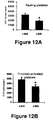
- PGI 2 Prostacyclin
- FIG. 12 Adhesion of human platelets is lower on conditioned SIS compared to native SIS.
- Adhesion of resting (A) or thrombin activated (B) human platelets labeled with 51Cr was measured on native SIS and conditioned SIS. Platelet adhesion was significantly reduced by 40.4% when resting platelets were used, and by 29.6% when thrombin-activated activated platelets were used. *p ⁇ 0:05 vs native SIS.
- the present invention provides medical grafting materials including extracellular matrix composites, and methods for their manufacture and use.
- acellular means free or essentially free from living cells.
- substantially devoid of cells and cell components means free or essentially free from cells (living or dead) and of cell membranes and other cell remnants.
- An ECM material substantially devoid of cells or cell components is intended to include the ECM material carrying cells or cell components at a level sufficiently low to be non-immunogenic when the material is implanted in a recipient, especially a recipient to which the cells or cell components are xenogenic or allogenic.
- decellularizing in respect of a cell-containing ECM material means that the material is treated to as to remove at least about 70% of the original cells (living or dead). More preferably, at least 90% of the cells will be removed, and most preferably at least 99% of the cells will be removed, in decellularization processes involved in the instant invention.
- Medical grafting devices and methods of the invention employ a first extracellular matrix (ECM) base material.
- ECM extracellular matrix
- Suitable extracellular matrix materials include, for instance, submucosa (including for example small intestinal submucosa, stomach submucosa, urinary bladder submucosa, or uterine submucosa, each of these isolated from juvenile or adult animals), renal capsule membrane, amnion, dura mater, pericardium, serosa, peritoneum or basement membrane materials, including liver basement membrane or epithelial basement membrane materials.
- ECM base materials contain residual bioactive proteins or other ECM components derived from the tissue source of the materials.
- they may contain Fibroblast Growth Factor 2 (basic FGF), vascular endothelial growth factor (VEGF), and Transforming Growth Factor-beta (TFG-beta).
- ECM base materials of the invention may contain additional bioactive components including, for example, one or more of glycosaminoglycans, glycoproteins, proteoglycans, and/or growth factors.
- cells will be cultured in vitro on the ECM material under conditions and for a duration wherein secreted extracellular matrix proteins are deposited upon a portion of or the entire surface of the ECM material.
- the resulting ECM composite material can be isolated by decellularizing the material.
- the deposited ECM proteins may, for example, enhance the functionality of the ECM base material, e.g. by potentially affecting remodeling of the material by cells and/or the thrombogenicity of the material.
- the ECM base material along with seeded cells can be subjected to mechanical, chemical or physical stresses to influence the cell growth and deposition products.
- Such forces could include but are not limited to mechanically stretching the ECM base material, preferably without tearing it, subjecting the ECM base material to pulsatile forces (e.g. by flowing fluid such as culture medium through a tube of ECM base material), altering the culture atmosphere, e.g. to a higher or lower carbon dioxide content, or adding specific growth factors or chemokines that affect the cell growth rates, phenotypes, secretory functions or apoptosis events, thereby affecting the molecules deposited by the cells.
- pulsatile forces e.g. by flowing fluid such as culture medium through a tube of ECM base material
- altering the culture atmosphere e.g. to a higher or lower carbon dioxide content
- adding specific growth factors or chemokines that affect the cell growth rates, phenotypes, secretory functions or apoptosis events, thereby affecting the molecules deposited by the cells.
- Cells to be used to secrete ECM proteins can be applied to the surface of the base ECM supporting structure in any suitable fashion.
- the cells can be applied to the base ECM material by allowing gravity to settle the cells onto the base ECM.
- Positive pressure may also be used to force media through the ECM material, thereby depositing cells onto the ECM surface.
- Other suitable means for applying the cells to the ECM may include, but not be limited to using negative pressure to draw the cells onto the ECM material; and using chemotactic agents, for example.
- cell types to produce the ECM proteins for deposition include vascular and other endothelial cells (including microvascular endothelial cells), vascular and other smooth muscle cells, fibroblasts, corneal endothelium or epithelium, glomerular epithelium, and mesothelial cells, pericytes, macrophages, monocytes, plasma cells, mast cells, adipocytes, chondrocytes, etc.
- vascular smooth muscle cells can be isolated from segments of carotid or femoral arteries obtained from humans (e.g. the patient to be treated or cadaver) or animals. Such isolation procedures are known and may for example involve storing the tissue segments in a suitable sterile medium, such as sterile Medium 199 (Gibco BRL) potentially also containing antibiotic agents such as gentamicin.
- a suitable sterile medium such as sterile Medium 199 (Gibco BRL) potentially also containing antibiotic agents such as gentamicin.
- the artery segment can be slit longitudinally and the endothelial cells removed by rubbing and/or scraping (e.g. with a scalpel blade). Thin strips of arterial media peeled up with forceps are pooled into HBSS in a sterile Petri dish.
- the strips can then be cultured in tissue culture flasks containing a suitable culture medium, e.g. Smooth Muscle Cell Growth Medium (SMCGM) (43% Dulbecco's Modified Eagle Medium (DMEM); 43% Medium 199; 13% fetal bovine serum; 2 mM glutamine; 15 units/ml heparin; 23 ⁇ g/ml gentamicin; and 12.5 ⁇ g/ml endothelial cell growth supplement (Collaborative Biomedical Products, Bedford, Mass.)).
- SMCGM Smooth Muscle Cell Growth Medium
- DMEM Dulbecco's Modified Eagle Medium
- the culture medium can be replaced when significant outgrowth of cells from the tissue pieces is observed.
- the cells can then be fed the SMCGM periodically as needed, and conventionally passaged and split. Smooth muscle cell type can be confirmed, for instance by morphological criteria, positive staining for alpha smooth muscle cell actin, and/or other suitable known techniques.
- subconfluent VSMC cultures can be rinsed with calcium-magnesium-free-HBSS (CMF-HBSS) and washed in CMF-HBSS.
- CMF-HBSS calcium-magnesium-free-HBSS
- EDTA trypsin-ethylenediamine tetraacetic acid
- SMCGM trypsin neutralization with SMCGM.
- Cells can be pelleted by centrifugation and the pellet re-suspended in SMCGM for cell counting. After centrifugation, the cell pellet can be re-suspended in SMCGM.
- This cell suspension can then be contacted with the ECM base material to seed the material with the cells.
- the seeded ECM can then be placed into a culture container filled with SMCGM.
- the culture container can be capped and incubated at about 37° C. on a roller apparatus.
- the medium in the culture tubes can be periodically replaced and the seeded ECMs can be cultured for a period sufficient to deposit ECM components, for example, from one to twenty days.
- endothelial cells are used to deposit ECM proteins on an ECM base material.
- endothelial cells can be caused to attach and spread on the ECM surface, and cultured to deposit the ECM proteins.
- small patches of endothelial cells may be directly harvested from a donor vessel (e.g. a vessel of the patient to be treated, or a cadaver) and seeded onto an ECM surface whereby they will attach and proliferate to cover the ECM surface, and deposit ECM components.
- enzymatic methods can be used to release endothelial cells (ECs) from arterial or venous vessels obtained from humans or animals.
- the vessel lumina are cannulated, rinsed with HBSS, and filled with an endothelial cell harvesting enzyme solution in a suitable medium such as CMF-HBSS.
- Suitable enzymes include, but are not limited to, collagenase, dispase, and trypsin.
- Endothelial cells are flushed into a sterile centrifuge tube and the ECs pelleted. The cells are then plated onto tissue culture flasks, grown at about 37° C. until nearly confluent and then passaged. Endothelial cell type can be confirmed by morphological criteria, by positive staining for Factor VIII, and by uptake of acetylated low density lipoprotein.
- Subconfluent endothelial cells can be rinsed with CMF-HBSS and washed in CMF-HBSS.
- the cells can be harvested by using trypsin-EDTA to release cells from the flasks followed by trypsin neutralization with complete Endothelial Cell Growth Medium (ECGM; 80% Medium 199, 16% fetalbovine serum, 2 mM glutamine, 15 units/ml heparin, 25 ⁇ g/ml gentamicin, 12.5 ⁇ g/ml Endothelial Cell Growth Supplement (Collaborative Biomedical Products, Bedford, Mass.)).
- the cells can be pelleted and the pellet re-suspended in ECGM. This suspension can then be used to seed the ECM base material, and then cultured in association with the ECM base material to secrete and deposit the desired amount of ECM components, for example, one to twenty days.
- the endothelial cell layer can be removed.
- the endothelial cells can be removed by rinsing the graft several times (e.g. three times) with HBSS, and treating with an ammonium hydroxide solution, e.g. about 0.025M ammonium hydroxide, to remove the endothelial cells, and rinsed again several times in HBSS.
- ammonium hydroxide solution e.g. about 0.025M ammonium hydroxide
- Other suitable treatments may include, for example, 0.01-0.5M ammonium hydroxide for about 30 seconds to about 60 minutes.
- candidate methods of removing the endothelial cells may include air drying, or treatment with other stripping solutions, for example, chloroform, methanol, ammonium hydroxide, or sodium chloride, either alone or in combination.
- Other treatments known to those skilled in the art may also be suitable.
- a secreted matrix deposit through a variety of other methods.
- One suitable method involves using mixed culture seeding in which both ECs and SMCs are combined and both cell types are seeded onto the ECM base material simultaneously. After extended co-culture, the secreted matrix will be produced.
- Fibroblasts for use in depositing extracellular matrix proteins may be readily isolated from an appropriate source organ or tissue. This can be readily accomplished using techniques known to those skilled in the art.
- the tissue or organ can be disaggregated mechanically and/or treated with digestive enzymes and/or chelating agents that weaken the connections between neighboring cells, making it possible to disperse the tissue into a suspension of individual cells without appreciable cell breakage.
- Enzymatic dissociation can be accomplished by mincing the tissue and treating the minced tissue with any of a number of digestive enzymes either alone or in combination.
- Such enzymes include, but are not limited to, trypsin, chymotrypsin, collagenase, elastase, hyaluronidase, DNase, pronase, and/or dispase etc.
- Mechanical disruption can also be accomplished by a number of methods including, but not limited to the use of grinders, blenders, sieves, homogenizers, pressure cells, insonators, and the like.
- the suspension can be fractionated into subpopulations from which the fibroblasts and/or other stromal cells and/or elements can be obtained.
- This also may be accomplished using standard techniques for cell separation including, but not limited to, cloning and selection of specific cell types, selective destruction of unwanted cells (negative selection), separation based upon differential cell agglutinability in the mixed population, freeze-thaw procedures, differential adherence properties of the cells in the mixed population, filtration, conventional and zonal centrifugation, centrifugal elutriation (counter-streaming centrifugation), unit gravity separation, countercurrent distribution, electrophoresis and fluorescence-activated cell sorting.
- cells which are specialized for the particular tissue implant site, can be cultured on the base ECM material for the production of a tissue type specific extracellular matrix composite.
- the first ECM material can conditioned to create the ECM composite using cells of a given type, and the resulting ECM composite can be configured for and grafted on or in a type of tissue of the patient having cells of that given type.
- dermal fibroblasts can be used to form the three-dimensional subconfluent stroma for the production of skin-specific extracellular composite matrix in vitro.
- stromal cells of hematopoietic tissue including, but not limited to, fibroblast endothelial cells, macrophages/monocytes, adipocytes and reticular cells, can be used to form the three-dimensional subconfluent stroma for the production of a bone marrow-specific extracellular matrix in vitro.
- Hematopoietic stromal cells can be readily obtained from the “buffy coat” formed in bone marrow suspensions by centrifugation at low forces, e. g., 3000 ⁇ g.
- glial cells can be used as the stroma to support the proliferation of neurological cells and tissues.
- Glial cells for this purpose can be obtained by trypsinization or collagenase digestion of embryonic or adult brain.
- the growth of cells in the presence of the ECM base material can be further enhanced by adding to the framework, or coating the framework support with natural or recombinant molecules, including but not limited to, proteins, such as collagens, elastic fibers, reticular fibers, and glycoproteins; glycosaminoglycans, such as heparin sulfate, chondroitin-4-sulfate, chondroitin-6-sulfate, dermatan sulfate, keratan sulfate, etc.; a cellular matrix, and/or other materials, such as whole blood, serum, growth factors, fibronectin, Pronectin F, RGD peptide, or cell or tissue extracts.
- proteins such as collagens, elastic fibers, reticular fibers, and glycoproteins
- glycosaminoglycans such as heparin sulfate, chondroitin-4-sulfate, chondroitin-6-sulfate, dermatan sulfate,
- Stem cells may also be used and cultured on the ECM base material to deposit extracellular matrix components.
- adult or embryonic stem cells may be cultured and treated with appropriate differentiation factors to mature and secrete extracellular matrix components.
- the differentiated cell population can then be removed using suitable techniques as described herein.
- the ECM base material is incubated in an appropriate nutrient medium under physiologic conditions favorable for cell growth, i. e., promoting mitosis (cell division).
- an appropriate nutrient medium i. e., promoting mitosis (cell division).
- Many commercially available media such as RPMI 1640, Fisher's, Iscove's, McCoy's, and the like, may be suitable for use.
- the three-dimensional culture can be suspended or floated in the medium during the incubation period in order to maximize proliferative activity.
- the culture can be “fed” periodically to remove the spent media, depopulate released cells, and to add fresh media.
- the extracellular matrix components are secreted locally by cells and not only bind cells and tissues together but also influence the development and behavior of the cells they contact.
- the typical extracellular matrix contains various fiber-forming proteins interwoven in a hydrated gel composed of a network of glycosaminoglycan chains.
- the glycosaminoglycans are a heterogeneous group of long, negatively charged polysaccharide chains, which (except for hyaluronic acid) are covalently linked to protein to form proteoglycan molecules.
- the fiber-forming proteins are of two functional types: (a) mainly structural (collagens and elastin), and (b) mainly adhesive (such as fibronectin and laminin).
- the fibrillar collagens (types I, II, and III) are rope-like, triple-stranded helical molecules that aggregate into long cable-like fibrils in the extracellular space; these in turn can assemble into a variety of highly ordered arrays.
- Type IV collagen molecules assemble into a sheetlike meshwork that forms the core of all basal laminae.
- Elastin molecules form an extensive cross-linked network of fibers and sheets that can stretch and recoil, imparting elasticity to the matrix.
- Fibronectin and laminin are examples of large adhesive glycoproteins in the matrix; fibronectin is widely distributed in connective tissues, whereas laminin is found mainly in basal laminae. By means of their multiple binding domains, such proteins help cells adhere to and become organized by the extracellular matrix.
- a naturally secreted human dermal extracellular matrix contains type I and type III collagens, fibronectin, tenascin, glycosaminoglycans, acidic and basic FGF, TGF-beta, KGF, decorin and various other secreted human dermal matrix proteins.
- the various extracellular matrix proteins are produced in the quantities and ratios similar to that existing in vivo.
- growth of the stromal cells in three dimensions will sustain active proliferation of cells in culture for much longer time periods than will monolayer systems.
- the three-dimensional system supports the maturation, differentiation, and segregation of cells in culture in vitro to form components of adult tissues analogous to counterparts found in vivo.
- the extracellular matrix created by the cells in culture is more analogous to native tissues.
- the ECM composite material will be decellularized after deposition of the desired level of non-native ECM components on the ECM base material.
- the tissue may be treated with a solution that releases component cells from the associated extracellular membrane matrix.
- agents and methods that will remove the cells.
- the cell-containing composite material can be treated with a mild chemical stripping solution, such as ammonium hydroxide (NH 4 OH).
- NH 4 OH ammonium hydroxide
- One such treatment may involve incubating the material in an aqueous NH 4 OH solution at a concentration of about 0.01M to about 0.5M, for a period of about 30 seconds to about 60 minutes followed by flushing the vessel lumen or other ECM construct with a buffer solution.
- the treatment may involve treating the cell-containing material with a 0.25M NH 4 OH solution for about 1 to 10 minutes.
- Decellularization may also involve air drying. Following air drying, the material can be flushed with buffer to remove cells and cell components and rehydrate the ECM composite for further processing, if desired.
- the cells can also be removed by exposing the cell-containing material to one or more freeze-thaw cycles, typically followed by removal of dead cells and cell debris.
- removal may be accomplished by flushing the material after freezing with a suitable solution such as HBSS (1.3 mM CaCl 2 , 5 mM KCl, 0.3 mM KH 2 PO 4 , 0.5 mM MgCl 2 , 0.4 mM MgSO 4 , 138 mM NaCl, 4 mM NaHCO 3 , 0.3 mM NaHPO 4 , 5.6 mM glucose).
- Freeze-kill of the cells may be accomplished, for instance, by flash-freezing the living cells in liquid nitrogen.
- the cells may also be killed by irrigating the inoculated three-dimensional framework with sterile water, such that the cells burst in response to osmotic pressure.
- a mild detergent rinse such as EDTA, CHAPS or a zwitterionic detergent
- a cryoprotectant such as DMSO, propylene glycol, butanediol, raffinose, polyvinyl pyrrolidone, dextran or sucrose and vitrified in liquid nitrogen.
- the cell-containing material can be subjected to enzymatic digestion and/or extracting with reagents that break down the cellular membranes and allow removal of cell contents.
- detergents include non-ionic detergents (for example, TRITON X-100, octylphenoxy polyethoxyethanol, (Rohm and Haas); BRIJ-35, a polyethoxyethanol lauryl ether (Atlas Chemical Co.), TWEEN 20, a polyethoxyethanol sorbitan monolaureate (Rohm and Haas), LUBROL-PX, or polyethylene lauryl ether (Rohm and Haas)); and ionic detergents (for example, sodium dodecyl sulphate, sulfated higher aliphatic alcohol, sulfonated alkane and sulfonated alkylarene containing 7 to 22 carbon atoms in a branched or unbranched chain).
- Enzymes can be used
- the ECM composite material can be treated with a fixative, if desired.
- This fixation can be accomplished by placing the graft into a fixing solution, such as, for example, glutaraldehyde in a suitable buffer.
- Suitable buffers may include, but are not limited to, N-2-hydroxyethylpiperazine-N′-2-ethane sulfonic acid (HEPES), acetate, 2-(N-morpholino) ethanesulfonic acid (MES), 3-[N-morpholino]propanesulfonic acid (MOPS), tris hydroxymethyl aminomethane, phosphate, and others. Any remaining aldehyde reactive sites can be blocked with an amino group containing solution.
- Suitable reagents for this procedure include, but are not limited to, 0.1M glycine, Medium 199, Dulbecco's Modified Eagle Medium, and other physiological culture media, for example.
- ECM composite graft materials of the invention can be manufactured in a variety of physical forms, to suit a variety of medical applications.
- the graft materials may be formed as sheets, tubes, or other three-dimensional shapes.
- the configuration of the ECM composite material may be attained before or after deposition of the non-native ECM components on the ECM base material.
- an ECM composite material can be manufactured in larger, bulk dimensions, and then divided into smaller products.
- the ECM base material may provided in a naturally-derived layer form, or may itself be a manufactured article, such as a sponge or cast sheet, prepared from a naturally-derived ECM material.
- ECM composite grafts of the invention may be used in a wide variety of medical (including veterinary) applications.
- Examples of specific tissues which can be repaired and/or reconstructed using the inventive ECM composite materials included nervous tissue, skin, cardiovascular tissue (including vascular tissue and cardiac tissue), pericardial tissue, muscle tissue, ocular-tissue, periodontal tissue, bone, connective tissue such as tendons or ligaments, and others.
- the ECM composites of the present invention can be formed into grafts taking the forms of powders, fluidized compositions, sheets, tubes, pouches, multi-ply constructs, single-ply constructs, and constructs in combination with other medical devices or implements such as stents, valves, catheters, sutures, staples, balloons, metal coils, synthetic meshes, biodegradable polymers, inert polymers, non-biodegradable polymers, other collagenous matrices, stem cells, vectors for gene delivery, other naturally-occurring, synthetic, or recombinant bioactive molecules, or in combination with other combinations of these medical devices or implements.
- other medical devices or implements such as stents, valves, catheters, sutures, staples, balloons, metal coils, synthetic meshes, biodegradable polymers, inert polymers, non-biodegradable polymers, other collagenous matrices, stem cells, vectors for gene delivery, other naturally-occurring, synthetic, or
- the ECM composites of the present invention can be formed into a powder tissue graft composition.
- the ECM composite material can be comminuted by tearing, cutting, grinding, shearing or the like, frozen or not, in a manner similar to that described for other collagen-based ECMs in U.S. Pat. No. 5,275,826, International Publications Nos. WO 96/32146, WO 98/22158 and WO 98/25636; the disclosures of which are expressly incorporated herein by reference.
- ECM composite powders of the invention can be used to repair bone tissue, for instance using the general techniques described in U.S. Pat. No. 5,641,518.
- a powder form of the ECM composite can be implanted into a damaged or diseased bone region for repair.
- the ECM composite powder can be used alone, or in combination with one or more additional bioactive agents such as physiologically compatible minerals, growth factors, antibiotics, chemotherapeutic agents, antigen, antibodies, enzymes, vectors for gene delivery and hormones.
- the powder-form implant will be compressed into a predetermined, three-dimensional shape, which will be implanted into the bone region and will substantially retain its shape during replacement of the graft with endogenous tissues.
- the ECM composites of the invention may be fluidized following techniques as described in U.S. Pat. No. 5,275,826, the disclosure of which is incorporated herein by reference.
- Fluidized form of the composite ECMs are generally prepared by solubilization by enzymatic digestion, including the use of protease, such as trypsin or pepsin, or other appropriate enzymes such as a collagenase or a glycosaminoglycanase, or the use of a mixture of enzymes, for a period of time sufficient to solubilize the tissue and form a substantially homogeneous solution. While a fluidized composition may be formed from enzyme digestion of comminuted tissues, a procedure involving only enzyme digestion with no mechanical commination can also be used.
- the ECM composites can be provided as sheet constructs.
- the ECM composites can be spread out into a native sheet form, trimmed to size if desired, and lyophilized.
- Multi-layer sheet constructs can be made including two or more such sheets stacked directly atop one another. In this regard, these constructs can be stabilized by bonding the collagen sheets together, for example by compressing the sheets under dehydrating conditions, use of crosslinkers, or other suitable means.
- Larger area sheet constructs can be prepared by overlapping and fusing together of several smaller area ECM sheets.
- the construction of a large area sheet from smaller area sheets has been described in U.S. Pat. No. 5,127,903, International Publication No. WO 96/32146, and ASAIO Journal, 2000, 46:268-272.
- the small area sheets are fused together by compressing the overlapped edges of these sheets under dehydrating conditions.
- the sheets are joined by photocrosslinking in the presence of a photo-catalytic dye.
- Other standard tissue bonding techniques known to those skilled in the art, including the use of sutures, crosslinking agents, adhesives and pastes can also be used.
- the smaller area sheets can be bonded or fused together to form a larger area sheet before or after the deposition of the non-native ECM components and decellularization steps described above.
- the composite ECM matrices can be configured into tubes of various dimension and thickness to provide tubular tissue grafts useful in, for example, vascular, urinary tract or nervous system repair or replacement.
- tubular grafts may be used in coronary artery and peripheral artery replacement, or the like, e.g. with the graft having an internal diameter of about three to six millimeters.
- Techniques for making tubular grafts from collagenous sheets have been generally described in U.S. Pat. Nos. 2,127,903 and 4,902,508 and International Publication No. WO 98/22158, the disclosures of which are expressly incorporated herein by reference.
- a tubular graft may be constructed by manipulating sheets of the ECMs to define a tube and suturing, bonding or otherwise securing the longitudinal seam to form an appropriately-dimensioned tubular graft having a lumen.
- the tube may be formed so as to present a lumenal and/or outer surface comprising the deposited ECM material.
- the tubular construct is formed over a sterile rod or mandrel having an outer diameter approximating that of the recipient vessel to be grafted.
- at least one sheet layer of the inventive ECM composite is wrapped about the rod to form an overlapping seam.
- the overlapped seam may be joined together by any of the conventional methods such as suturing, photocrosslinking, radiation crosslinking, fusion under compression and dehydration, or by adhesive and the like.
- the tubular grafts may be constructed from multiple layers of ECMs, with the number of layers used generally depending on the strength requirement of the particular application.
- vascular grafts can be constructed with multiple layers of ECM sheets to ensure that they can withstanding the constant pulsating pressure of their implanting environment.
- composite ECMs of the invention can be used as exterior, interior or other coverings for supporting structures such as stents, grafts, etc.
- the ECM composite material can be attached to a stent in a fashion whereby it forms one, two, or more leaflets, cusps, pockets or similar structures that resist flow in one direction relative to another.
- such devices constructed as vascular valves are implanted to treat venous insufficiencies in humans, for example occurring in the legs.
- valves for treating these or other valve deficiencies may be surgically created without the use of a stent or other supporting structure.
- Tubular grafts containing the ECM composite material of the invention may also be prepared as disclosed in International Publication No. WO 2004/022107 published Mar. 18, 2004, which claims the benefit of U.S. Patent Application Ser. No. 60/408,914 filed Sep. 6, 2002 and entitled TISSUE GRAFT PROSTHESIS DEVICES CONTAINING JUVENILE OR SMALL DIAMETER SUBMUCOSA, each of which is incorporated herein by reference in its entirety. All or some of the layers of the disclosed tubular grafts may contain an ECM composite material of the present invention, and in particular embodiments at least the lumenal surface of the tubular constructs will contain the second ECM material.
- a pre-manufactured ECM composite material may be fashioned into such disclosed constructs, or the overall construct may be prepared including the first ECM material, and then the second ECM material deposited onto the first ECM material followed by decellularization as disclosed herein.
- Tubular graft prosthesis 10 defines an inner lumen 11 and has a length L and diameter D rendering the construct suitable for the intended use, for example a vascular use.
- FIG. 2 shown in FIG. 2 is a cross-sectional view of the prosthesis 10 of FIG. 1 taken along line 2 - 2 and viewed in the direction of the arrows.
- Prosthesis 10 has walls defining inner lumen 11 , preferably including several layers of material as illustrated.
- shown in prosthesis 10 is a first tubular layer 12 , a second layer tubular layer 13 , a third tubular layer 14 , a fourth tubular layer 15 , and a fifth tubular layer 16 .
- At least one of these layers may include juvenile submucosa from a warm-blooded vertebrate animal, or otherwise include an intact tubular submucosa segment having a small native internal diameter (12 mm or less).
- the animal is preferably a mammal, such as a porcine, ovine, bovine, or other mammalian animal. Human donor tissues may also be used in the present invention. In the case of juvenile porcine submucosa, the animal at harvest will typically not exceed about 10 kilograms (kg).
- the juvenile or other small diameter submucosa tissue will retain its intact, tubular form as harvested from the animal. More preferably, at least the innermost layer 12 will be formed from intact, tubular juvenile submucosa tissue. In this fashion, the surface 17 of the lumen 11 will be defined by the intact juvenile submucosa tissue, and will be free of any seams that would otherwise be created when configuring sheet-form tissue into a tube.
- Preferred devices will include at least one additional layer, for example, layers 13 , 14 , 15 and 16 as illustrated in FIG. 2 . These additional layers can be made of any suitable material and desirably provide reinforcement and strength to the device supplemental to that provided by innermost layer 12 .
- one or more of layers 13 , 14 , and 15 may, for example, be formed of synthetic materials such as synthetic polymer materials. Suitable synthetic materials may be biodegradable or non-biodegradable materials.
- biocompatible polymers such as cellulose acetate, cellulose nitrate, silicone, polyethylene teraphthalate, polyurethane, polyamide, polyester, polyorthoester, polyanhydride, polyether sulfone, polycarbonate, polypropylene, high molecular weight polyethylene, polytetrafluoroethylene, or mixtures or copolymers thereof; polylactic acid, polyglycolic acid or copolymers thereof, a polyanhydride, polycaprolactone, polyhydroxy-butyrate valerate, polyhydroxyalkanoate, or another biodegradable polymer.
- synthetic biocompatible polymers such as cellulose acetate, cellulose nitrate, silicone, polyethylene teraphthalate, polyurethane, polyamide, polyester, polyorthoester, polyanhydride, polyether sulfone, polycarbonate, polypropylene, high molecular weight polyethylene, polytetrafluoroethylene, or mixtures or copolymers thereof; polylactic acid, poly
- one or more of, and potentially all of layers 13 , 14 , 15 and/or 16 are formed from additional collagenous materials.
- suitable collagenous materials include extracellular matrix layers including, for instance, submucosa, renal capsule membrane, dura mater, pericardium, serosa, peritoneum or basement membrane layers, including liver basement membrane. These layers may be isolated and used as intact membranes, or reconstituted collagen layers including collagen derived from these materials or other collagenous materials may be used.
- layers 13 , 14 , 15 and 16 are made from additional submucosa tissue layers.
- Suitable submucosa tissues for these purposes include, for instance, intestinal submucosa including small intestinal submucosa, stomach submucosa, urinary bladder submucosa, and uterine submucosa.
- Small intestinal submucosa when employed, can be used in an intact, native tubular form or can be a tubular form shaped from flat sheets including one or more seams along all or a portion of its length.
- At least one of layers 13 , 14 , 15 and 16 will include adult submucosa tissue, as such tissue in its native condition is generally superior in mechanical properties to juvenile submucosa tissue.
- adult submucosa tissue can be used to provide strength to the overall graft construct 10 .
- Porcine small intestinal submucosa is particularly preferred for these purposes.
- intermediate layers 14 and 15 can be made from adult small intestinal submucosa
- intermediate layer 12 and outermost layer 16 can be made from juvenile small intestinal submucosa, preferably again in its native, intact tubular form.
- seamless inner layer 12 and seamless outer layer 16 can be provided.
- Layers 12 , 13 , 14 , 15 and 16 can be adhered to one another so as to generally form a unitary construct. This adherence may be achieved, for example, by crosslinking, including for example dehydrothermal crosslinking or chemical crosslinking, and/or by the use of a bonding agent. As bonding agents for these purposes, one may use fibrin glue, or gelatin or collagenous pastes in sufficient amount to bond adjacent layers to one another.
- Tubular devices of the invention may be prepared, for example, by positioning the appropriate tissue layers over a mandrel, and subsequently bonding or adhering the tissue layers together to form a generally unitary tubular construct. This may be accomplished, for instance, using intact tubes, and/or by wrapping or winding sheet- or strip-form adult submucosa tissue around the mandrel to form overlapped sections which are subsequently bonded or adhered. In some embodiments, an outermost covering layer may be provided by an intact juvenile submucosa segment positioned over the underlying tissue layers. If a bonding agent is to be used in forming the construct, the agent or its components can be applied at appropriate points intermediate the application of layers to the mandrel. The entire construct can then be dried, e.g., lyophilized and/or dried under vacuum, to form the overall tubular graft construct.
- tubular prosthesis devices are prepared using a two component bonding agent such as fibrin glue (e.g., having thrombin and fibrinogen as separate components).
- a two component bonding agent such as fibrin glue
- subsequent layers are added after coating the previously-applied layer with a first component of the bonding agent (e.g., thrombin) and coating a layer to be applied with a second component of the bonding agent (e.g., fibrinogen).
- the layer to be applied is positioned over the previously-applied layer so as to bring the two bonding components into contact, thus causing the curing process to begin. This process can be repeated for any and all additional layers to be applied to the tubular construct.
- FIGS. 3A through 3G an illustrative manufacture of a 5-layer (5L) tubular device of the invention will now be described.
- An intact tubular submucosa segment 21 from a juvenile animal may first be positioned over a mandrel 20 as depicted in FIG. 3A , to provide a one-layer (1L) construct.
- Thrombin (light shading, FIG. 3B ) is then applied to the intact segment 21 .
- a second intact tube of juvenile submucosa 22 is provided either on an extension of the same mandrel as illustrated, or on a second mandrel connectable to the first mandrel.
- the second intact segment 22 is coated with fibrinogen (dark shading), and the segment 22 is positioned immediately adjacent the first segment 21 .
- Segment 22 is then and pulled over the first submucosa tube in a fashion causing inversion of the tube 22 ( FIGS. 3C-3E ).
- leading end 25 of segment 22 remains substantially in place, but inverted, in contact with the trailing end 23 of segment 21 .
- Trailing end 26 of segment 22 finally inverts and comes into contact with leading end 24 of the first segment 21 .
- portions of the two submucosa tubes coming together will remain substantially together, i.e., one submucosa layer will not pulled along another submucosa layer.
- the inversion of the second tube is continued until it is completely inverted and lying atop the first submucosa tube, creating a two-layer (2L) construct as illustrated in FIG. 3E .
- the fibrin glue of the 2L construct is then allowed to cure (typically 1 to 5 minutes).
- the outer surface of the 2L construct is coated with thrombin (light shading, FIG. 3F ).
- Fibrinogen (dark shading, FIG. 3F ) is then applied to one surface of a sheet 27 of adult submucosa of a dimension sufficient to encircle the prior-applied layers two times. As illustrated in FIGS.
- the adult submucosa is then wrapped around the 2L construct for a single turn (clockwise rotation in FIGS. 3F-3G ), resulting in a completed three-layer (3L) construct on the mandrel 20 .
- thrombin is applied to the outer surface of the 3L construct (light shading, FIG. 3I ).
- a second turn of the adult submucosa sheet 27 is then completed ( FIG. 3I ), bringing the applied thrombin and fibrinogen components into contact with one another, and forming the completed four-layer device (4L, FIG. 3J ).
- a third intact tubular juvenile submucosa segment 28 ( FIG. 3K ) is positioned adjacent to the applied layers, thrombin (light shading) is coated onto the 4L construct and fibrinogen (dark shading) is coated onto the third tubular segment 28 .
- the third tubular segment 28 is then pulled and inverted over onto the applied layers ( FIGS. 3K-3M ) as before to complete the 5L construct ( FIG. 3N ), with the leading end 31 of the third segment 28 lying atop the trailing end 29 of segment the 4L construct, and the trailing end 32 of segment 28 lying atop the leading end 30 of the 4L construct.
- the device is then allowed to cure and is trimmed as necessary.
- the components of the bonding agents can be applied by any suitable method, including spray or brush application methods, and intermediate constructs can be hydrated at appropriate points in the manufacture. The entire construct may then be freeze-dried or otherwise processed (e.g. to deposit the second ECM material on lumenal or other surfaces) if desired.
- ECM composite matrices of the invention can optionally be preconditioned for orthopedic or other applications, again before or after deposition of ECM components and removal of the depositing cells.
- Preparation of orthopedic grafts have been generally described in U.S. Pat. Nos. 2,127,903; 5,281,422; 5,275,826; and 5,352,463, the disclosures of which are expressly incorporated herein by reference.
- Conditioning alters the viscoelastic property and reduces the strain inherent in the native ECMs sheets.
- the ECMs may be conditioned by stretching, chemical treatment, enzymatic treatment or exposing the tissue to other environmental factors, or any other art known methods.
- the ECMs can be conditioned by stretching under a load to a predetermined percentage of the pre-stretched length.
- the ECMs may be conditioned by suspending a weight from a segment of an ECM sheet for a period of time sufficient to allow about 10% to about 20% elongation of the segment.
- the ECM segment can then be configured, alone or in combination with other segments, to a variety of shapes to serve as orthopedic grafts.
- ECM composites of the invention can be used for the repair and/or replacement of connective tissues.
- connective tissues include bone, cartilage, tendons and ligaments.
- the composite ECMs may serve as ligament or tendon replacement or a patch for a broken or severed tendon or ligament.
- the end portions of the graft construct may be formed, manipulate or shape the end portions of the graft construct to be attached, for example, to a bone structure, in a manner that will reduce the possibility of graft tearing at the point of attachment.
- the conditioned ECM composite graft material can be folded or partially everted to provide multiple layers for gripping, for example, with spiked washers or staples.
- a ECM material can be folded back on itself to join the end portions to provide a first connective portion to be attached, for example, to a first bone and a bend in the intermediate portion to provide a second connective portion to be attached to a second bone articulated with respect to the first bone.
- one of the end portions of the ECM graft can be adapted to be pulled through a tunnel in, for example, the femur and attached thereto, while the other of the end portions may be adapted to be pulled through a tunnel in the tibia and attached thereto to provide a substitute for the natural cruciate ligament, the segment being adapted to be placed under tension between the tunnels to provide a ligament function, i.e., a tensioning and position function provided by a normal ligament.
- a ligament function i.e., a tensioning and position function provided by a normal ligament.
- ECM composite sheets of the present invention can also be used to provide an orthopedic graft for use as connective tissue to hold fractured bone pieces together and in proper orientation in the body, the tissue segment being formed to serve as a fracture wrap about segments of fractured bone and to be attached to the bone.
- the first ECM (base) material can be disinfected and/or sterilized by contact with a suitable agent such as peracetic acid or another oxidizing disinfectant prior to seeding with the cells to be used to deposit the second ECM material, during removal of such cells, after removal of such cells, or any combination of these.
- a suitable agent such as peracetic acid or another oxidizing disinfectant
- Such disinfection/sterilization techniques may for example be conducted using agents and general conditions as disclosed in U.S. Pat. No. 6,206,931, which is incorporated herein by reference in its entirety.
- the ECM composite graft constructs and compositions of the invention can be packaged to protect the purity of the constructs/compositions for storage and shipment to the consumers.
- dried graft constructs are packaged in pre-sterilized bags and then vacuum sealed. These packages may be stored and shipped at room temperature.
- graft constructs that are in a hydrated state are packaged in pre-sterilized bags and then vacuum sealed. The hydrated samples may be stored and shipped frozen.
- the pre-sterilized bags are formed of materials that are impervious to moisture and oxygen, e.g., PETE.
- the sealed packages may be irradiated to decimate pathogens that were inadvertently collected on the surface of the constructs during manufacturing.
- dried graft constructs can be packaged in non-sterile gas permeable bags and then vacuum-sealed. The packaged constructs are then sterilized along with the gas permeable packaging using ethylene oxide gas. It is contemplated that still other conventional techniques for sterilization which are within the knowledge of those of ordinary skill in the arts may also be used, including for example radiation and plasma sterilization techniques.
- Tissue graft compositions including ECM composites of the invention can be administered to a vertebrate host in an amount effective to induce endogenous tissue growth at a site in the host in need of same due to the presence of damaged or diseased tissue.
- the vertebrate host may be autogenic, allogenic or xenogenic to the animals from which the base ECMs were harvested, and/or to the cells used to deposit the ECM components on the ECM base material.
- the vertebrate host is a human
- the ECM base material is allogenic or xenogenic
- the ECM-depositing cells are autogenic, allogenic or xenogenic to the human host.
- the base ECM is xenogenic and the ECM-depositing cells are autogenic to the human recipient of the ECM composite graft
- the base ECM is xenogenic and the ECM-depositing cells are human and allogenic to the recipient of the ECM composite graft.
- the isolated extracellular matrix composite material can be used to prepare a cell-seeded graft, if desired.
- cells allogenic or autogenic to the recipient of a graft of the composite material can be seeded and optionally proliferated on the composite material, which can then be implanted, along with the cells, into the recipient.
- Any of the cell types disclosed above for deposit of the second extracellular matrix material can also be used in such cell-seeded grafts, including combinations of such cell types.
- the first extracellular matrix material will be xenogenic to a human recipient (e.g.
- the second extracellular matrix material will be human but allogenic to the recipient, and the cells of the cell-seeded graft will be autogenic or allogenic to the recipient, and when allogenic optionally either immunologically matched or modulated to minimize potential immune response.
- Fluidized composite ECMs of this invention may be used in orthopedic applications, e.g. tissue replacement, augmentation, and/or repair. It is contemplated that fluidized ECMs of the present invention may be used as an injectable heterograft. By injecting an effective amount of a fluidized ECMs composition into the location of a tissue defect, for example, in bone or soft tissues in need of repair or augmentation, one can readily take advantage of the biotropic properties of the ECMs. It is further contemplated that the fluidized composition can be used advantageously as a filler for implant constructs, e.g., in cosmetic or trauma-treating surgical procedures.
- Composite ECMs of the invention can also be used in body wall repair, including for example in the repair of abdominal wall defects such as hernias, using techniques analogous to those described in Ann. Plast. Surg., 1995, 35:374-380; and J. Surg. Res., 1996, 60:107-114.
- preferred ECM tissue grafts of the invention promote favorable organization, vascularity and consistency in the remodeled tissue.
- composite ECMs of the invention can be used in the repair of partial or full thickness wounds and in dermal augmentation using general grafting techniques which are known to the art and literature (see, e.g. Annals of Plastic Surgery 1995, 35:381-388).
- general grafting techniques which are known to the art and literature (see, e.g. Annals of Plastic Surgery 1995, 35:381-388).
- cultured epidermal grafts preferably cultured epidermal autografts, or CEA's
- Such cultured grafts have typically involved transplanting keratinocytes and/or fibroblasts onto the dermal substitute.
- the composite ECMs can be used as the dermal substitute, for example in sheet form, and the CEA provided on the composite ECMs.
- keratinocytes can be transplanted, for example by seeding or transferring a keratinocyte sheet, onto the denser side of the composite ECM sheet and fibroblasts can be transplanted also on the more areolar side of the composite ECM sheet.
- Composite ECMs of the invention can also be used in tissue grafts for urogenital applications.
- a large area composite ECM sheet can be used for the repair of urinary bladder, in which the composite ECM sheet provides a scaffold for bladder tissue regeneration.
- the techniques for urinary bladder repair have been generally described in U.S. Pat. No. 5,645,860; Urology, 1995, 46:396-400; and J. Urology, 1996, 155:2098; the disclosures of which are incorporated herein by reference.
- Tubular grafts including composite ECMs may be used for the repair and replacement of the urinary tract.
- a tubular graft of a specific diameter and strength, the making of which has been previously described, can be surgically substituted for a damaged or diseased urinary tract by standard surgical techniques. Technique for urinary tract replacement has been described generally in “Use of Reconstructed Small Intestine Submucosa for Urinary Tract Replacement”, by Xie et. al, in ASAIO Journal 2000, p. 268, and the disclosure of which is incorporated herein by reference.
- the inventive composite ECMs can also find use in an endoscopic injection procedure to correct vesicureteral reflux.
- a submucosal injection can be made, for instance in the area under the ureteral orifice of a patient, to induce smooth muscle growth and collagen formation at the injection site.
- tissue graft constructs formed with composite ECMs of the present invention can be used in neurological applications. For example, in techniques requiring a dural substitute to repair defects due to trauma, tumor resection, or decompressive procedures.
- ECM composites of the invention can be utilized as cell growth substrates for in vitro cultivation of eukaryotic or other cells. These applications employ techniques in the art which have been applied to other naturally-derived ECMs or synthetic graft materials and can be similarly undertaken using the composite ECMs of the present invention. Typically, the composite ECM composition will be combined with various nutrients, growth factors, minerals and salts known in the art to be important for the particular cell types.
- inventive composite ECMs can also serve as a collagenous matrix in compositions for producing transformed cells.
- the techniques for cell transformation have been described in International Publication Nos. WO 96/25179 and WO 95/22611; the disclosures of which are expressly incorporated herein by reference.
- purified composite ECMs of the present invention for example in fluidized or paste form, is included in the cell transformation compositions, in combination with a recombinant vector (e.g. a plasmid) containing a nucleic acid sequence with which in vitro or in vivo target cells are to be genetically transformed.
- a recombinant vector e.g. a plasmid
- Human umbilical vein endothelial cells were grown for two weeks on [ptvomr SIS and then removed to leave behind intact human basement membrane materials.
- the resulting composite ECM material was termed “conditioned” SIS (c-SIS).
- c-SIS conditioned SIS
- HUVEC When re-seeded on c-SIS, HUVEC exhibited enhanced organization of cell junctions and had increased metabolic activity compared to cells on native SIS (n-SIS). HUVEC grown on c-SIS also released lower amounts of the pro-inflammatory prostaglandin PGI 2 into the media compared to cells grown on n-SIS. Additionally, adhesion of resting or activated human platelets to c-SIS was significantly decreased as compared to n-SIS. Conditioning of porcine SIS by human endothelial cells was thus demonstrated to improve key biological properties of the material. Statistical analysis for this Experimental was made by ANOVA and p ⁇ 0.05 was considered significant.
- EGM Human umbilical vein endothelial cells
- Clonetics East Rutherford, N.J.
- EGM Endothelial growth media
- EGM contains 2% fetal bovine serum, 12 .tg/ml bovine brain extract, I ⁇ g/ml human epidermal growth factor, and I ⁇ g/ml hydrocortisone, getamicin, and amphotericin B.
- HUVEC were maintained at 5% CO 2 at 37° C., and passages 1-4 were used for experiments.
- SIS was obtained from Cook Biotech, Inc. (West Lafayette, Ind.) in a dehydrated form and was rehydrated in endothelial basal media (EBM; Clonetics) at 37° for at least 10 min. Once hydrated, the SIS was fastened to wells with an approximate area of 0.44 cm 2 available for cell seeding.
- EBM endothelial basal media
- Samples of SIS for confocal microscopy were rinsed twice in phosphate-buffered saline (PBS) and fixed in 4% paraformaldehyde for 15 minutes, rinsed extensively in PBS, permeabilized using 0.2% TritonX-100 and labeled with appropriate antibodies.
- PBS phosphate-buffered saline
- TritonX-100 0.2% TritonX-100 and labeled with appropriate antibodies.
- samples were mounted on glass slides with Fluoromount-GTM (Southern Biotechnology Associates Inc., Birmingham, Ala.).
- HUVECs at passage 2 were seeded onto the glass slides at 75,000 cells/mm 2 and cultured for 72 hours prior to fixation with 4% paraformaldehyde.
- Visualization of cell-cell adherens was made using mouse anti- ⁇ catenin (Zymed Laboratories Inc., San Francisco, Calif.); tyrosine phosphorylated proteins were visualized using a mouse anti-phosphotyrosine antibody (Transduction Laboratories, Lexington, Ky.).
- Actin filaments were visualized using Texas-Red Phalloidin (Molecular Probes, Eugene, Oreg.).
- SDS sodium dodecyl sulfate
- Protein concentrations were determined using the amido black method Sheffield J B, Graff D, Li H P. A solid-phase method for the quantitation of protein in the presence of sodium dodecyl sulfate and other interfering substances. Anal Biochem 1987; 166(1):49-54. Equal protein (30 ⁇ g) was loaded onto a 10% SDS-polyacrylamide gel for separation and transferred to nitrocellulose for immunoblot analysis.
- SIS basement membrane protein deposition
- SDS gel sample buffer For analysis of basement membrane protein deposition, SIS was minced into tiny sections and extracted in SDS gel sample buffer. These samples were then boiled and centrifuged at 14,000 ⁇ g for 10 minutes. The supernatant was collected and the remaining pieces of SIS were discarded. Protein levels were equalized by amido back assays and loaded onto a 5% SDS-PAGE gel for separation and transferred to nitrocellulose for immunoblot analysis. Human fibronectin was detected using rabbit anti-human fibronectin (Cederlane Laboratories, Ltd., Ontario, Canada) with the appropriate horseradish peroxidase-labeled secondary antibodies (Jackson Immunoresearch, Inc.).
- HUVEC Removal of all HUVEC was verified when preparing c-SIS visually using fluorescence microscopy and scanning electron microscopy (SEM), and biochemically by immunoblot analysis.
- fluorescence microscopy samples were fixed in 4% paraformaldehyde for 15 min and then stained with Hoechst 33258 nuclear staining dye obtained from Molecular Probes (Eugene, Oreg.). Images of stained cell nuclei were viewed and recorded on an RT Color Spotlights camera (Diagnostic Instruments, Sterling heights, Mich.) using an Optiphot-2 Nikon epifluorescent microscope.
- samples were prepared by fixation in 4% gluteraldehyde, dehydrated through increasing ethanol concentrations to 100% ethanol, sputter coated with gold and viewed using a JOEL scanning electron microscope.
- the surface of SIS was scraped directly in SDS sample buffer and an antibody specific for the cellular enzyme anti-mitochondrial-8-oxoguanine DNA-glycosylase (ab6491), Abcam Limited (Cambridge, UK) or for ( ⁇ -catenin along with the appropriate horseradish peroxidase-labeled secondary antibodies (Jackson) was used to detect the presence of cells on SIS.
- Cell adhesion to SIS was measured using metabolically labeled HUVEC.
- Cells were grown in 150 mm 2 tissue culture dishes and labeled for 16 hours in 25 ⁇ Ci of 35 S-methionine, rinsed in PBS and then removed from the dish using trypsin.
- 35 S-methionine labeled HUVEC were resuspended in growth media and equal numbers of cells (100,000/cm 2 ) were added to n-SIS or c-SIS and allowed to adhere for varying lengths of time as indicated. After the adhesion period, SIS was washed with PBS to remove unbound cells and the amount of radioactivity remaining on the SIS was measured using a liquid scintillation counter.
- n-SIS and c-SIS To estimate the relative metabolic rates of cells grown on n-SIS and c-SIS, cells were seeded at 100,000 or 200,000 cells/cm 2 and cultured for one hour to two weeks. Metabolic activity was assessed using CellTiter 96 Aqueos Once Solution Cell Proliferation Assay obtained from Promega (Madison, Wis.). The assay is a colorimetric method, which measures the amount of NADPH produced by dehydrogenase enzymes in metabolically active cells by determining the amount of colored reaction product (Formazan) that can be produced from MTS tetrazolium compound (Owen's reagent) in media samples.
- Formulamazan colored reaction product
- Prostacyclin release was measured by incubating SIS wells in 200 ⁇ L of EGM media for 1 hour. The media was collected and centrifuged at 14,000 ⁇ g for 2 min to pellet any particulates, and the supernatant was retained for prostacyclin measurement. 6-keto Prostaglandin Fi a EIA kit was obtained from Cayman Chemical (Ann Arbor, Mich.) and used to determine the amount of PGI 2 present in each sample.
- Platelets Whole blood was drawn by venipuncture from healthy aspirin-free human donors and collected in ACD (83 mM sodium citrate, 111 mM glucose, 71.4 mM citric acid, ph 4.5; 1:7, v/v). Platelet-rich plasma was isolated by centrifugation at 250 g for 20 min, and prostaglandin El (0.1 mM) or prostaglandin 12 (50 ng/ml) was added.
- ACD 83 mM sodium citrate, 111 mM glucose, 71.4 mM citric acid, ph 4.5; 1:7, v/v.
- ACD 83 mM sodium citrate, 111 mM glucose, 71.4 mM citric acid, ph 4.5; 1:7, v/v. Platelet-rich plasma was isolated by centrifugation at 250 g for 20 min, and prostaglandin El (0.1 mM) or prostaglandin 12 (50 ng/ml) was added.
- the platelet-rich plasma was centrifuged for 15 min at 850 ⁇ g, and the platelets were resuspended in Hepes-buffered saline ((Buffer A) 10 MM Hepes, pH 7.4, 138 mM NaCl, 12 mM NaHCO3, 10 mM KCI, 5.5 mM glucose, 0.35% BSA, 2 units/ml heparin and I unit/ml apyrase). Resuspended platelets were washed three times in Buffer A without BSA or inhibitors and the concentration was adjusted to (0.5-1.0) ⁇ 10 9 platelets/ml. The platelets were then labeled with 51 Cr (0.5 mCi) for 60 min.
- Buffer A 10 MM Hepes, pH 7.4, 138 mM NaCl, 12 mM NaHCO3, 10 mM KCI, 5.5 mM glucose, 0.35% BSA, 2 units/ml heparin and I unit/ml apyras
- FIG. 4 shows confocal microscope images illustrating the appearance of HUVEC on SIS and glass at various times after seeding using an antibody against the cell-cell junction protein ( ⁇ -catenin, followed by FITC-labeled secondary antibody and with Texas-Red-phalloidin to visualize actin.
- HUVEC grown on glass slides were confluent by 2 days after seeding and exhibited a typical cobblestone appearance characteristic of endothelial cells in culture and similar to the morphology of endothelial cells in vivo (Fun-YC, Biomechanics: Motion, Flow, Stress, and Growth.
- HUVEC grown on SIS at the same initial seeding density of 100,000 cells/mm 2 for 2 days did not form a confluent monolayer with well-developed cell-cell junctions.
- HUVEC were seeded on SIS and grown for 14 days to allow sufficient time for the cells to deposit human matrix proteins onto the surface of the porcine SIS. Thus, HUVEC were cultured for approximately 9 days after reaching confluency. HUVEC were then removed from the SIS using NH4OH as has been described previously (Gonzales M, Weksler B, Tsuruta D, Goldman R D, Yoon K J, Hopkinson S B, Flitney F W, and Jones J C. Structure and function of a vimentin-associated matrix adhesion in endothelial cells.
- FIG. 5A shows microscopy images of untreated SIS, SIS with cells grown for 14 days and left intact, and SIS with cells removed (conditioned) that were stained with Hoechst dye to label cell nuclei to verify that all cells were removed from the SIS. Confirmation that all cells were removed was also made by immunoblotting for the cellular proteins (3-catenin and mitochondrial-DNA8-oxanine-glycosylase ( FIG. 5B ).
- FIG. 6 shows a western blot to illustrate the presence of hFN on the surface of SIS following the conditioning treatment.
- An antibody specific for human laminin that did not cross-react with porcine laminin was also used. In this work, no evidence of secretion of laminin onto conditioned SIS was found (not shown).
- SEM scanning electron microscopy
- HUVEC growing on tissue culture plastic were labeled with 35 -methionine for 18 hours during log phase growth to allow incorporation of the radioactive label into newly synthesized proteins.
- the cells were washed free of unincorporated radioactivity in the media and the labeled cells were collected by brief trypsinization. Equal numbers of cells were seeded onto n-SIS or c-SIS and allowed to adhere.
- FIG. 8 shows there were no statistically significant differences in the number of cells present on n-SIS vs. c-SIS at any of the time points examined between 1 and 48 hours.
- FIG. 9 illustrates the dramatic improvement in the organization of cell-cell junctional complexes (adherens junctions) when cells are grown on c-SIS compared to cells grown on n-SIS at 48 hours after seeding.
- Cells were labeled for (3-catenin and for phosphotyrosine containing proteins and imaged using confocal microscopy.
- n-SIS Cells grown on n-SIS appeared to be visible in multiple different focal planes indicating the surface of n-SIS was relatively rough. In contrast, cells grown on c-SIS were visible across the surface of the SIS primarily within a single focal plane suggesting that conditioning of the SIS might modify the surface of the SIS by deposition of a layer of matrix proteins that creates a smooth surface that allows cells to spread more uniformly. This result suggests that SIS conditioned with human basement membrane proteins provides an improved substrate for the establishment of endothelial monolayers with well-organized cell-cell junctions.
- HUVEC grown on n-SIS and c-SIS
- metabolic activity assays which measured the production and release of NADPH into the culture media, were conducted.
- HUVEC were seeded onto n-SIS and c-SIS (100,000 cells/cm 2 and 200,000 cells/cm 2 ) and allowed to grow over a period of 1 hour to 240 hours.
- n-SIS and c-SIS 100,000 cells/cm 2 and 200,000 cells/cm 2
- HUVEC grown on n-SIS exhibited a significant decrease in NADPH production after 24 and 96 hours in culture, despite the fact equal numbers of cells adhered to n-SIS and c-SIS (see FIG. 8 ), when compared to their initial (1 hour) levels ( FIG. 10 ).
- HUVEC grown on c-SIS exhibited a significant increase in NADPH production after 24 hours in culture.
- NADPH production by HUVEC remained significantly higher in cells grown on c-SIS compared to n-SIS through the first 96 hours.
- NADPH levels were not different between groups; after 240 hours in culture NADPH production had dropped significantly below initial levels for both substrates and were not significantly different from each other.
- HUVEC inflammatory prostaglandin, prostacyclin
- EIA enzyme-linked immunosorbant assay
- HUVEC were seeded at an initial density of 100,000 cells/cm 2 and grown on n-SIS or c-SIS for 24 or 48 hours. After 24 or 48 hours, fresh media was incubated with the wells for 1 hour and the media was collected. Significantly higher levels of PG1 2 were released into the media by cells grown on n-SIS compared to cells grown on c-SIS ( FIG. 11 ). This difference (approximately 3-fold more PGI2 (pg/mL) than HUVEC on c-SIS, increased to approximately 4-fold at the 48 hour time point.
- platelet adhesion assays were performed. Using freshly isolated human platelets, we found that conditioning the SIS resulted in a significant decrease in platelet adhesion of approximately 40% when compared to n-SIS ( FIG. 1 12 A). This value of a 40% decrease was typical of four independent experiments using platelets from multiple donors in which the decrease in platelet adhesion on c-SIS ranged from 30-43%. In each independent experiment this decrease was statistically significant (p ⁇ 0.05 by ANOVA). Interestingly, platelet adhesion was also decreased (by approximately 21-26%) on SIS with HUVEC still present, compared to n-SIS, but this difference was not statistically significant in any of four independent experiments (data not shown).
- n-SIS significantly reduced adhesion of activated platelets by approximately 30%.
- adhesion of thrombin-activated platelets to SIS with HUVEC still present was consistently lower by 15-20%, but this difference was not statistically significant.
- n-SIS was also coated with commercially available human fibronectin to determine if coating with this single ECM protein known to be present in c-SIS could mimic the decrease in platelet adhesion seen on c-SIS.
Abstract
Description
Claims (27)
Priority Applications (2)
Application Number | Priority Date | Filing Date | Title |
---|---|---|---|
US11/368,077 US7795027B2 (en) | 2003-09-04 | 2006-03-03 | Extracellular matrix composite materials, and manufacture and use thereof |
US12/399,635 US9186435B2 (en) | 2003-09-04 | 2009-03-06 | Extracellular matrix composite materials, and manufacture and use thereof |
Applications Claiming Priority (3)
Application Number | Priority Date | Filing Date | Title |
---|---|---|---|
US50002603P | 2003-09-04 | 2003-09-04 | |
PCT/US2004/029016 WO2005023321A2 (en) | 2003-09-04 | 2004-09-07 | Extracellular matrix composite materials, and manufacture and use thereof |
US11/368,077 US7795027B2 (en) | 2003-09-04 | 2006-03-03 | Extracellular matrix composite materials, and manufacture and use thereof |
Related Parent Applications (1)
Application Number | Title | Priority Date | Filing Date |
---|---|---|---|
PCT/US2004/029016 Continuation WO2005023321A2 (en) | 2003-09-04 | 2004-09-07 | Extracellular matrix composite materials, and manufacture and use thereof |
Related Child Applications (1)
Application Number | Title | Priority Date | Filing Date |
---|---|---|---|
US12/399,635 Division US9186435B2 (en) | 2003-09-04 | 2009-03-06 | Extracellular matrix composite materials, and manufacture and use thereof |
Publications (2)
Publication Number | Publication Date |
---|---|
US20060147433A1 US20060147433A1 (en) | 2006-07-06 |
US7795027B2 true US7795027B2 (en) | 2010-09-14 |
Family
ID=34272902
Family Applications (2)
Application Number | Title | Priority Date | Filing Date |
---|---|---|---|
US11/368,077 Active 2027-07-21 US7795027B2 (en) | 2003-09-04 | 2006-03-03 | Extracellular matrix composite materials, and manufacture and use thereof |
US12/399,635 Active 2026-08-10 US9186435B2 (en) | 2003-09-04 | 2009-03-06 | Extracellular matrix composite materials, and manufacture and use thereof |
Family Applications After (1)
Application Number | Title | Priority Date | Filing Date |
---|---|---|---|
US12/399,635 Active 2026-08-10 US9186435B2 (en) | 2003-09-04 | 2009-03-06 | Extracellular matrix composite materials, and manufacture and use thereof |
Country Status (5)
Country | Link |
---|---|
US (2) | US7795027B2 (en) |
AU (1) | AU2004270239C1 (en) |
CA (1) | CA2536923C (en) |
GB (1) | GB2423934B (en) |
WO (1) | WO2005023321A2 (en) |
Cited By (23)
Publication number | Priority date | Publication date | Assignee | Title |
---|---|---|---|---|
US20090204228A1 (en) * | 2003-09-04 | 2009-08-13 | Hiles Michael C | Extracellular matrix composite materials, and manufacture and use thereof |
US20100136050A1 (en) * | 2005-10-27 | 2010-06-03 | University Of Notre Dame Du Lac | Extracellular Matrix Materials as Vaccine Adjuvants for Diseases Associated with Infectious Pathogens or Toxins |
US20100249519A1 (en) * | 2007-04-09 | 2010-09-30 | Adrian Edward Park | Frame Device |
US20100256778A1 (en) * | 2003-08-11 | 2010-10-07 | Wilson-Cook Medical Inc. | Surgical Implant |
US8257434B2 (en) | 2007-12-18 | 2012-09-04 | Cormatrix Cardiovascular, Inc. | Prosthetic tissue valve |
US8679176B2 (en) | 2007-12-18 | 2014-03-25 | Cormatrix Cardiovascular, Inc | Prosthetic tissue valve |
US8696744B2 (en) | 2011-05-27 | 2014-04-15 | Cormatrix Cardiovascular, Inc. | Extracellular matrix material valve conduit and methods of making thereof |
US8735155B1 (en) * | 2005-07-15 | 2014-05-27 | Cormatrix Cardiovascular, Inc. | Compositions for regenerating defective or absent myocardium |
US8778362B2 (en) | 2005-10-27 | 2014-07-15 | University Of Notre Dame | Anti-tumor/cancer heterologous acellular collagenous preparations and uses thereof |
US8883210B1 (en) | 2010-05-14 | 2014-11-11 | Musculoskeletal Transplant Foundation | Tissue-derived tissuegenic implants, and methods of fabricating and using same |
US20150057751A1 (en) * | 2012-03-15 | 2015-02-26 | L&C Bio Co., Ltd. | Acellular dermal graft |
US8980296B2 (en) | 2009-02-18 | 2015-03-17 | Cormatrix Cardiovascular, Inc. | Compositions and methods for preventing cardiac arrhythmia |
US9072818B2 (en) | 2010-01-05 | 2015-07-07 | Cell Constructs I, Llc | Biomaterials made from human hair |
US9238090B1 (en) | 2014-12-24 | 2016-01-19 | Fettech, Llc | Tissue-based compositions |
US9352003B1 (en) | 2010-05-14 | 2016-05-31 | Musculoskeletal Transplant Foundation | Tissue-derived tissuegenic implants, and methods of fabricating and using same |
US9801910B2 (en) | 2014-03-17 | 2017-10-31 | Ethicon, Inc. | Decellularized pleural matrix |
US10130736B1 (en) | 2010-05-14 | 2018-11-20 | Musculoskeletal Transplant Foundation | Tissue-derived tissuegenic implants, and methods of fabricating and using same |
US10531957B2 (en) | 2015-05-21 | 2020-01-14 | Musculoskeletal Transplant Foundation | Modified demineralized cortical bone fibers |
US10702260B2 (en) | 2016-02-01 | 2020-07-07 | Medos International Sàrl | Soft tissue fixation repair methods using tissue augmentation scaffolds |
WO2020206228A1 (en) | 2019-04-03 | 2020-10-08 | The Secant Group, Llc | Human cell-deposited extracellular matrix coatings for textiles and fibers |
US11376347B1 (en) * | 2021-06-21 | 2022-07-05 | The Florida International University Board Of Trustees | Materials and methods for accelerating cardiovascular tissue regeneration |
US11484401B2 (en) | 2016-02-01 | 2022-11-01 | Medos International Sarl | Tissue augmentation scaffolds for use in soft tissue fixation repair |
US11826490B1 (en) | 2020-12-29 | 2023-11-28 | Acell, Inc. | Extracellular matrix sheet devices with improved mechanical properties and method of making |
Families Citing this family (68)
Publication number | Priority date | Publication date | Assignee | Title |
---|---|---|---|---|
CA2547088C (en) | 2003-11-28 | 2011-10-18 | Cook Biotech Incorporated | Vascular occlusion methods, systems and devices |
WO2005094694A2 (en) * | 2004-03-29 | 2005-10-13 | Cook Biotech Incorporated | Medical graft products with differing regions and methods and systems for producing the same |
WO2006102063A2 (en) * | 2005-03-19 | 2006-09-28 | Cook Biotech Incorporated | Prosthetic implants including ecm composite material |
US9072816B2 (en) * | 2006-01-18 | 2015-07-07 | Cormatrix Cardiovascular, Inc. | Composition for modulating inflammation of cardiovascular tissue |
US8921109B2 (en) | 2005-09-19 | 2014-12-30 | Histogenics Corporation | Cell-support matrix having narrowly defined uniformly vertically and non-randomly organized porosity and pore density and a method for preparation thereof |
US8802113B2 (en) * | 2005-10-27 | 2014-08-12 | University Of Notre Dame | Extracellular matrix cancer vaccine adjuvant |
US8778360B2 (en) * | 2005-10-27 | 2014-07-15 | University Of Notre Dame | Extracellular matrix cancer vaccine adjuvant |
US8974542B2 (en) | 2006-06-27 | 2015-03-10 | University of Pittsburgh—of the Commonwealth System of Higher Education | Biodegradable elastomeric patch for treating cardiac or cardiovascular conditions |
CN101616698A (en) | 2006-10-23 | 2009-12-30 | 库克生物科技公司 | The ECM material of the enhanced processing of component characteristic |
WO2008060973A1 (en) * | 2006-11-10 | 2008-05-22 | Vance Products Incorporated D/B/A Cook Urological Incorporated | Graft for hysterotomy closure |
WO2008109407A2 (en) | 2007-03-02 | 2008-09-12 | University Of Pittsburgh-Of The Commonwealth System Of Higher Education | Extracellular matrix-derived gels and related methods |
EP2121062B1 (en) * | 2007-03-07 | 2020-05-06 | Coloplast A/S | Fistula plug comprising ecm |
US20080281419A1 (en) * | 2007-05-10 | 2008-11-13 | Matheny Robert G | Breast implants and compositions of extracellular matrix |
US9034367B2 (en) * | 2007-05-10 | 2015-05-19 | Cormatrix Cardiovascular, Inc. | Articles for tissue regeneration with biodegradable polymer |
US9066993B2 (en) * | 2007-05-10 | 2015-06-30 | Cormatrix Cardiovascular, Inc. | Extracellular matrix encasement structures and methods |
US9283302B2 (en) * | 2011-12-16 | 2016-03-15 | Cormatrix Cardiovascular, Inc. | Extracellular matrix encasement structures and methods |
US20080279833A1 (en) * | 2007-05-10 | 2008-11-13 | Matheny Robert G | Laminate sheet articles for tissue regeneration |
US20080306610A1 (en) * | 2007-06-07 | 2008-12-11 | Zimmer Orthobiologics, Inc. | Tissue processing for nonimmunogenic implants |
US20090054984A1 (en) | 2007-08-20 | 2009-02-26 | Histogenics Corporation | Method For Use Of A Double-Structured Tissue Implant For Treatment Of Tissue Defects |
US8070827B2 (en) | 2007-07-03 | 2011-12-06 | Histogenics Corporation | Method for use of a double-structured tissue implant for treatment of tissue defects |
US8685107B2 (en) | 2007-07-03 | 2014-04-01 | Histogenics Corporation | Double-structured tissue implant and a method for preparation and use thereof |
WO2009026392A1 (en) | 2007-08-20 | 2009-02-26 | Histogenics Corporation | A method for improvement of differentiation of mesenchymal stem cells using a double-structured tissue implant |
WO2009089110A2 (en) * | 2008-01-04 | 2009-07-16 | Boston Scientific Scimed, Inc. | Methods for repair and regeneration of bone marrow |
US8801725B2 (en) | 2008-03-10 | 2014-08-12 | Zimmer Orthobiologics, Inc. | Instruments and methods used when repairing a defect on a tissue surface |
US8709096B2 (en) * | 2008-04-29 | 2014-04-29 | Proxy Biomedical Limited | Tissue repair implant |
DE102008022319A1 (en) * | 2008-04-30 | 2009-11-05 | Aesculap Ag | Implant, in particular for restoring and / or regenerating human and / or animal tissue |
US8858698B2 (en) * | 2008-09-05 | 2014-10-14 | Mentor Worldwide Llc | Acellular matrix glue |
US7927414B2 (en) * | 2008-09-05 | 2011-04-19 | Ethicon, Inc. | Method of manufacturing acellular matrix glue |
WO2010085449A1 (en) | 2009-01-23 | 2010-07-29 | Cook Incorporated | Vessel puncture closure device |
JP5693475B2 (en) * | 2009-03-04 | 2015-04-01 | ペイタント・ソリューションズ・インコーポレイテッドPeytant Solutions, Inc. | Stent modified with a material comprising amnion tissue and corresponding method |
WO2010111278A1 (en) * | 2009-03-23 | 2010-09-30 | The Texas A&M University System | Compositions of mesenchymal stem cells to regenerate bone |
CN102481390B (en) * | 2009-09-02 | 2017-03-08 | 生命细胞公司 | Blood vessel graft from acellular tissue matrix |
US9115336B2 (en) | 2010-05-25 | 2015-08-25 | Muffin Incorporated | Methods, substrates, and systems useful for cell seeding of medical grafts |
US8840665B2 (en) | 2010-06-11 | 2014-09-23 | Liventa Bioscience, Inc. | Method of tendon repair with amnion and chorion constructs |
US20120010708A1 (en) * | 2010-07-08 | 2012-01-12 | AFcell Medical | Amnion and chorion replacement cover and uses thereof in surgical repair of muscles |
WO2012024390A2 (en) | 2010-08-17 | 2012-02-23 | University Of Pittsburgh - Of The Commonwealth System Of Higher Education | Biohybrid composite scaffold |
DK2608777T3 (en) | 2010-08-24 | 2019-10-28 | Univ California | Compositions and Methods for Cardiac Therapy |
US8435305B2 (en) | 2010-08-31 | 2013-05-07 | Zimmer, Inc. | Osteochondral graft delivery device and uses thereof |
US9113916B2 (en) | 2010-08-31 | 2015-08-25 | Zimmer, Inc. | Drill bit for osteochondral drilling with guiding element and uses thereof |
US10201636B2 (en) * | 2010-12-20 | 2019-02-12 | Cormatrix Cardiovascular, Inc. | Cardiovascular prostheses |
US10143778B2 (en) * | 2010-12-21 | 2018-12-04 | Cormatrix Cardiovascular, Inc | Cardiovascular prostheses |
US20170100514A1 (en) * | 2011-12-16 | 2017-04-13 | Cormatrix Cardiovascular, Inc. | Cardiovascular Prostheses |
US8961617B2 (en) | 2012-03-08 | 2015-02-24 | Liventa Bioscience, Inc. | Amnion and chorion constructs and uses thereof in abdominal surgery |
US9811613B2 (en) | 2012-05-01 | 2017-11-07 | University Of Washington Through Its Center For Commercialization | Fenestration template for endovascular repair of aortic aneurysms |
US9220586B2 (en) * | 2012-09-28 | 2015-12-29 | Covidien Lp | Surgical implant and applicator |
US20150094799A1 (en) * | 2012-10-08 | 2015-04-02 | Cormatrix Cardiovascular, Inc. | Extracellular Matrix Prostheses for Treating Damaged Biological Tissue |
US20150100115A1 (en) * | 2012-10-08 | 2015-04-09 | Cormatrix Cardiovascular, Inc. | Extracellular Matrix Prostheses for Treating Damaged Biological Tissue |
US9533072B2 (en) * | 2012-10-08 | 2017-01-03 | Cormatrix Cardiovascular, Inc. | Reinforced vascular prostheses |
AU2014251110A1 (en) * | 2013-04-08 | 2015-11-26 | Regentys Corporation | Method and composition for treating inflammatory bowel disease without colectomy |
WO2014169031A1 (en) * | 2013-04-10 | 2014-10-16 | Tufts University | Two and three dimensional decellularized ecm constructs and uses therefor |
CN103263694B (en) * | 2013-05-14 | 2014-12-03 | 北京华信佳音医疗科技发展有限责任公司 | Collagen-based dura and preparation method thereof |
CN103394126B (en) * | 2013-07-11 | 2014-12-31 | 陕西佰傲再生医学有限公司 | Cartilage repair material and preparation method thereof |
JP6681872B2 (en) | 2014-03-21 | 2020-04-15 | ユニバーシティ オブ ピッツバーグ −オブ ザ コモンウェルス システム オブ ハイヤー エデュケイション | Method for preparing final sterile hydrogel derived from extracellular matrix |
US10765705B2 (en) | 2014-11-24 | 2020-09-08 | Prime Merger Sub, Llc | Visco-supplement compositions, and methods of use thereof |
US10077420B2 (en) | 2014-12-02 | 2018-09-18 | Histogenics Corporation | Cell and tissue culture container |
WO2016094106A1 (en) * | 2014-12-10 | 2016-06-16 | Cormatrix Cardiovascular, Inc. | Biocompatable annular prostheses and methods for forming same |
WO2016172694A1 (en) | 2015-04-23 | 2016-10-27 | Richard Van Bibber | Devices and methods for anatomic mapping for prosthetic implants |
CN104888273B (en) * | 2015-05-14 | 2017-08-22 | 四川大学 | A kind of double-layer composite endocranium and preparation method thereof |
CN107949346B (en) | 2015-07-08 | 2020-09-04 | 主动脉公司 | Apparatus and method for anatomical mapping of a prosthetic implant |
US9855146B2 (en) * | 2015-08-24 | 2018-01-02 | Arthrex, Inc. | Arthroscopic resurfacing techniques |
CA3017747A1 (en) | 2016-03-14 | 2017-09-21 | Regentys Corporation | Method and composition for treating inflammatory bowel disease |
EP3493766B1 (en) | 2016-08-02 | 2024-03-06 | Bolton Medical, Inc. | Assembly for coupling a prosthetic implant to a fenestrated body |
CN107913436A (en) * | 2016-10-09 | 2018-04-17 | 刘英芹 | Act on the bone and soft tissue synchronizing regeneration derivant of disability tissue in situ |
AU2018226871B2 (en) | 2017-03-02 | 2023-08-10 | University Of Pittsburgh - Of The Commonwealth System Of Higher Education | Extracellular matrix (ECM) hydrogel and soluble fraction thereof for the treatment of cancer |
CN111148484B (en) | 2017-09-25 | 2022-12-30 | 波尔顿医疗公司 | Systems, devices, and methods for coupling a prosthetic implant to an open window |
CN108795867A (en) * | 2018-06-05 | 2018-11-13 | 华东理工大学 | The method for shifting external threedimensional model for building colon cancer cell peritonaeum |
CN109758261A (en) * | 2019-03-07 | 2019-05-17 | 上海白衣缘生物工程有限公司 | A kind of solid tendon biological sticking patch and its preparation method and application |
CN115337458B (en) * | 2022-08-16 | 2023-08-22 | 尧舜泽生物医药(南京)有限公司 | Cell matrix nerve graft for repairing peripheral nerve injury and preparation method thereof |
Citations (58)
Publication number | Priority date | Publication date | Assignee | Title |
---|---|---|---|---|
US2127903A (en) | 1936-05-05 | 1938-08-23 | Davis & Geck Inc | Tube for surgical purposes and method of preparing and using the same |
US4361552A (en) | 1980-09-26 | 1982-11-30 | Board Of Regents, The University Of Texas System | Wound dressing |
US4902508A (en) | 1988-07-11 | 1990-02-20 | Purdue Research Foundation | Tissue graft composition |
US4956178A (en) | 1988-07-11 | 1990-09-11 | Purdue Research Foundation | Tissue graft composition |
US5127903A (en) | 1990-05-22 | 1992-07-07 | Mailot Kevin G | Device for dispensing medicaments to infants |
US5275826A (en) | 1992-11-13 | 1994-01-04 | Purdue Research Foundation | Fluidized intestinal submucosa and its use as an injectable tissue graft |
US5281422A (en) | 1991-09-24 | 1994-01-25 | Purdue Research Foundation | Graft for promoting autogenous tissue growth |
US5352463A (en) | 1992-11-13 | 1994-10-04 | Badylak Steven F | Tissue graft for surgical reconstruction of a collagenous meniscus and method therefor |
WO1995022611A2 (en) | 1994-02-18 | 1995-08-24 | The Regents Of The University Of Michigan | Methods and compositions for stimulating bone cells |
WO1996008213A1 (en) | 1994-09-12 | 1996-03-21 | Advanced Tissue Sciences, Inc. | Three-dimensional human cell cultures on cardiac valve frameworks and their uses |
US5554389A (en) | 1995-04-07 | 1996-09-10 | Purdue Research Foundation | Urinary bladder submucosa derived tissue graft |
WO1996032146A1 (en) | 1995-04-13 | 1996-10-17 | Advanced Cytometrix, Inc. | Aspiration needle and method |
US5641518A (en) | 1992-11-13 | 1997-06-24 | Purdue Research Foundation | Method of repairing bone tissue |
US5645860A (en) | 1995-04-07 | 1997-07-08 | Purdue Research Foundation | Tissue graft and method for urinary urothelium reconstruction replacement |
EP0809691A1 (en) | 1995-02-10 | 1997-12-03 | Purdue Research Foundation | Submucosa as a growth substrate for cells |
US5733337A (en) | 1995-04-07 | 1998-03-31 | Organogenesis, Inc. | Tissue repair fabric |
WO1998022158A2 (en) | 1996-08-23 | 1998-05-28 | Cook Biotech, Incorporated | Graft prosthesis, materials and methods |
WO1998025636A1 (en) | 1996-12-10 | 1998-06-18 | Purdue Research Foundation | Stomach submucosa derived tissue graft |
US5770417A (en) | 1986-11-20 | 1998-06-23 | Massachusetts Institute Of Technology Children's Medical Center Corporation | Three-dimensional fibrous scaffold containing attached cells for producing vascularized tissue in vivo |
US5879383A (en) | 1994-04-29 | 1999-03-09 | W. L. Gore & Associates, Inc. | Blood contact surfaces using endothelium on a subendothelial matrix |
US5916265A (en) | 1994-03-30 | 1999-06-29 | Hu; Jie | Method of producing a biological extracellular matrix for use as a cell seeding scaffold and implant |
WO1999062431A1 (en) | 1998-06-02 | 1999-12-09 | Cook Incorporated | Multiple-sided intraluminal medical device |
WO2001019285A1 (en) | 1999-09-10 | 2001-03-22 | Patricia Ellen Thorpe | Endovascular treatment for chronic venous insufficiency |
US20010039450A1 (en) | 1999-06-02 | 2001-11-08 | Dusan Pavcnik | Implantable vascular device |
US6331319B1 (en) | 1997-09-11 | 2001-12-18 | Purdue Research Foundation | Galactosidase modified submucosal tissue |
US6379710B1 (en) | 1996-12-10 | 2002-04-30 | Purdue Research Foundation | Biomaterial derived from vertebrate liver tissue |
WO2002040630A2 (en) | 2000-11-15 | 2002-05-23 | Amiel Gilad E | Process of decellularizing biological matrices and acellular biological matrices useful in tissue engineering |
US20020123800A1 (en) | 2001-03-01 | 2002-09-05 | Taheri Syde A. | Prosthetic vein valve and method |
US6485723B1 (en) | 1995-02-10 | 2002-11-26 | Purdue Research Foundation | Enhanced submucosal tissue graft constructs |
US20020188348A1 (en) | 1999-10-21 | 2002-12-12 | Scimed Life Systems, Inc. | Implantable prosthetic valve |
WO2003002165A1 (en) | 2001-06-28 | 2003-01-09 | Cook Biotech Incorporated | Graft prosthesis devices containing renal capsule collagen |
US20030036794A1 (en) | 1995-06-07 | 2003-02-20 | Cook Incorporated | Coated implantable medical device |
US20030072741A1 (en) | 2001-05-10 | 2003-04-17 | Berglund Joseph D. | Soft tissue devices and methods of use |
US6576618B1 (en) | 1999-06-22 | 2003-06-10 | Research Development Foundation | Methods to enhance wound healing and enhanced wound coverage material |
US6582462B1 (en) | 1990-05-18 | 2003-06-24 | Heartport, Inc. | Valve prosthesis for implantation in the body and a catheter for implanting such valve prosthesis |
WO2003070124A2 (en) | 2002-02-20 | 2003-08-28 | Osse Francisco J | Venous bi-valve |
US20030191525A1 (en) | 2002-04-03 | 2003-10-09 | Thornton Sally C. | Artifical valve |
US6638312B2 (en) | 2000-08-04 | 2003-10-28 | Depuy Orthopaedics, Inc. | Reinforced small intestinal submucosa (SIS) |
US20030208261A1 (en) | 2000-03-03 | 2003-11-06 | Thorpe Patricia E. | Bulbous valve and stent for treating vascular reflux |
US20030216811A1 (en) | 2002-05-02 | 2003-11-20 | Badylak Stephen F. | Vascularization enhanced graft constructs |
US20040015230A1 (en) | 1996-12-18 | 2004-01-22 | Moll Franciscus Laurens | Methods for regulating the flow of blood through the blood system |
US20040049262A1 (en) | 2000-01-31 | 2004-03-11 | Obermiller Joseph F. | Stent valves and uses of same |
WO2004022107A2 (en) | 2002-09-06 | 2004-03-18 | Cook Biotech Incorporated | Tissue graft prosthesis devices containing juvenile or small diameter submucosa |
US6716241B2 (en) | 2002-03-05 | 2004-04-06 | John G. Wilder | Venous valve and graft combination |
WO2004029016A1 (en) | 2002-09-20 | 2004-04-08 | Oelmühle Leer Connemann Gmbh & Co. | Method and device for producing biodiesel |
US20040082989A1 (en) | 2002-08-20 | 2004-04-29 | Cook Incorporated | Stent graft with improved proximal end |
US20040098084A1 (en) | 2002-09-02 | 2004-05-20 | Cook Incorporated | Branch grafting device and method |
US20040106978A1 (en) | 2002-06-28 | 2004-06-03 | Cook Incorporated | Thoracic aortic aneurysm stent graft |
US6783776B2 (en) * | 1999-12-22 | 2004-08-31 | Acell, Inc. | Tissue regenerative composition, method of making, and method of use thereof |
US20040176832A1 (en) | 2002-12-04 | 2004-09-09 | Cook Incorporated | Method and device for treating aortic dissection |
US20040176833A1 (en) | 2002-11-22 | 2004-09-09 | Cook Incorporated | Stent tissue graft prosthesis |
US20040186558A1 (en) | 2001-02-05 | 2004-09-23 | Cook Incorporated | Implantable vascular device |
US20040260389A1 (en) | 2003-04-24 | 2004-12-23 | Cook Incorporated | Artificial valve prosthesis with improved flow dynamics |
US20050025838A1 (en) | 2003-06-25 | 2005-02-03 | Badylak Stephen F. | Conditioned compositions for tissue restoration |
WO2005023321A2 (en) | 2003-09-04 | 2005-03-17 | Cook Biotech Incorporated | Extracellular matrix composite materials, and manufacture and use thereof |
US6893653B2 (en) | 1997-05-08 | 2005-05-17 | Organogenesis Inc. | Chemical cleaning of biological material |
US6962814B2 (en) | 2000-08-16 | 2005-11-08 | Duke University | Decellularized tissue engineered constructs and tissues |
US20050256588A1 (en) | 2002-06-28 | 2005-11-17 | Cardio, Inc. | Decellularized tissue |
Family Cites Families (7)
Publication number | Priority date | Publication date | Assignee | Title |
---|---|---|---|---|
WO1995002611A1 (en) | 1993-07-16 | 1995-01-26 | Schering Corporation | Cell surface protein present on nk (natural killer cells) named dx1 |
US20050026838A1 (en) * | 1995-06-05 | 2005-02-03 | Human Genome Sciences, Inc. | Fibroblast Growth Factor-13 |
CA2334228C (en) * | 1998-06-05 | 2010-09-28 | Organogenesis Inc. | Bioengineered vascular graft support prostheses |
JP2004071560A (en) * | 2002-08-07 | 2004-03-04 | Samsung Sdi Co Ltd | Polymer electrolyte for lithium-sulfur battery and lithium-sulfur battery comprising same |
US6763776B1 (en) * | 2003-06-30 | 2004-07-20 | Industrial Sales Corporation | Cleat device for a flexible line and method of using same |
WO2006102063A2 (en) * | 2005-03-19 | 2006-09-28 | Cook Biotech Incorporated | Prosthetic implants including ecm composite material |
US7939481B2 (en) * | 2007-10-31 | 2011-05-10 | Nazzaro John | Color changing bar soap |
-
2004
- 2004-09-07 AU AU2004270239A patent/AU2004270239C1/en not_active Ceased
- 2004-09-07 CA CA2536923A patent/CA2536923C/en not_active Expired - Fee Related
- 2004-09-07 GB GB0605898A patent/GB2423934B/en not_active Expired - Fee Related
- 2004-09-07 WO PCT/US2004/029016 patent/WO2005023321A2/en active Search and Examination
-
2006
- 2006-03-03 US US11/368,077 patent/US7795027B2/en active Active
-
2009
- 2009-03-06 US US12/399,635 patent/US9186435B2/en active Active
Patent Citations (67)
Publication number | Priority date | Publication date | Assignee | Title |
---|---|---|---|---|
US2127903A (en) | 1936-05-05 | 1938-08-23 | Davis & Geck Inc | Tube for surgical purposes and method of preparing and using the same |
US4361552A (en) | 1980-09-26 | 1982-11-30 | Board Of Regents, The University Of Texas System | Wound dressing |
US5770417A (en) | 1986-11-20 | 1998-06-23 | Massachusetts Institute Of Technology Children's Medical Center Corporation | Three-dimensional fibrous scaffold containing attached cells for producing vascularized tissue in vivo |
US4902508A (en) | 1988-07-11 | 1990-02-20 | Purdue Research Foundation | Tissue graft composition |
US4956178A (en) | 1988-07-11 | 1990-09-11 | Purdue Research Foundation | Tissue graft composition |
US6582462B1 (en) | 1990-05-18 | 2003-06-24 | Heartport, Inc. | Valve prosthesis for implantation in the body and a catheter for implanting such valve prosthesis |
US5127903A (en) | 1990-05-22 | 1992-07-07 | Mailot Kevin G | Device for dispensing medicaments to infants |
US5281422A (en) | 1991-09-24 | 1994-01-25 | Purdue Research Foundation | Graft for promoting autogenous tissue growth |
US5275826A (en) | 1992-11-13 | 1994-01-04 | Purdue Research Foundation | Fluidized intestinal submucosa and its use as an injectable tissue graft |
US5352463A (en) | 1992-11-13 | 1994-10-04 | Badylak Steven F | Tissue graft for surgical reconstruction of a collagenous meniscus and method therefor |
US5641518A (en) | 1992-11-13 | 1997-06-24 | Purdue Research Foundation | Method of repairing bone tissue |
WO1995022611A2 (en) | 1994-02-18 | 1995-08-24 | The Regents Of The University Of Michigan | Methods and compositions for stimulating bone cells |
US5916265A (en) | 1994-03-30 | 1999-06-29 | Hu; Jie | Method of producing a biological extracellular matrix for use as a cell seeding scaffold and implant |
US5879383A (en) | 1994-04-29 | 1999-03-09 | W. L. Gore & Associates, Inc. | Blood contact surfaces using endothelium on a subendothelial matrix |
WO1996008213A1 (en) | 1994-09-12 | 1996-03-21 | Advanced Tissue Sciences, Inc. | Three-dimensional human cell cultures on cardiac valve frameworks and their uses |
US6485723B1 (en) | 1995-02-10 | 2002-11-26 | Purdue Research Foundation | Enhanced submucosal tissue graft constructs |
EP0809691A1 (en) | 1995-02-10 | 1997-12-03 | Purdue Research Foundation | Submucosa as a growth substrate for cells |
US20030064056A1 (en) | 1995-02-10 | 2003-04-03 | Badylak Stephen F. | Enhanced submucosal tissue graft constructs |
US5554389A (en) | 1995-04-07 | 1996-09-10 | Purdue Research Foundation | Urinary bladder submucosa derived tissue graft |
US5645860A (en) | 1995-04-07 | 1997-07-08 | Purdue Research Foundation | Tissue graft and method for urinary urothelium reconstruction replacement |
US5733337A (en) | 1995-04-07 | 1998-03-31 | Organogenesis, Inc. | Tissue repair fabric |
WO1996032146A1 (en) | 1995-04-13 | 1996-10-17 | Advanced Cytometrix, Inc. | Aspiration needle and method |
US20030036794A1 (en) | 1995-06-07 | 2003-02-20 | Cook Incorporated | Coated implantable medical device |
WO1998022158A2 (en) | 1996-08-23 | 1998-05-28 | Cook Biotech, Incorporated | Graft prosthesis, materials and methods |
US6206931B1 (en) | 1996-08-23 | 2001-03-27 | Cook Incorporated | Graft prosthesis materials |
WO1998025636A1 (en) | 1996-12-10 | 1998-06-18 | Purdue Research Foundation | Stomach submucosa derived tissue graft |
US6099567A (en) | 1996-12-10 | 2000-08-08 | Purdue Research Foundation | Stomach submucosa derived tissue graft |
US6379710B1 (en) | 1996-12-10 | 2002-04-30 | Purdue Research Foundation | Biomaterial derived from vertebrate liver tissue |
US20040015230A1 (en) | 1996-12-18 | 2004-01-22 | Moll Franciscus Laurens | Methods for regulating the flow of blood through the blood system |
US6893653B2 (en) | 1997-05-08 | 2005-05-17 | Organogenesis Inc. | Chemical cleaning of biological material |
US6331319B1 (en) | 1997-09-11 | 2001-12-18 | Purdue Research Foundation | Galactosidase modified submucosal tissue |
US6508833B2 (en) | 1998-06-02 | 2003-01-21 | Cook Incorporated | Multiple-sided intraluminal medical device |
WO1999062431A1 (en) | 1998-06-02 | 1999-12-09 | Cook Incorporated | Multiple-sided intraluminal medical device |
US20010011187A1 (en) | 1998-06-02 | 2001-08-02 | Dusan Pavcnik | Multiple-sided intraluminal medical device |
US20030125795A1 (en) | 1998-06-02 | 2003-07-03 | Cook Incorporated | Multiple-sided intraluminal medical device |
US20010039450A1 (en) | 1999-06-02 | 2001-11-08 | Dusan Pavcnik | Implantable vascular device |
US6576618B1 (en) | 1999-06-22 | 2003-06-10 | Research Development Foundation | Methods to enhance wound healing and enhanced wound coverage material |
WO2001019285A1 (en) | 1999-09-10 | 2001-03-22 | Patricia Ellen Thorpe | Endovascular treatment for chronic venous insufficiency |
US20030130726A1 (en) | 1999-09-10 | 2003-07-10 | Thorpe Patricia E. | Combination valve and stent for treating vascular reflux |
US20020188348A1 (en) | 1999-10-21 | 2002-12-12 | Scimed Life Systems, Inc. | Implantable prosthetic valve |
US6783776B2 (en) * | 1999-12-22 | 2004-08-31 | Acell, Inc. | Tissue regenerative composition, method of making, and method of use thereof |
US20040049262A1 (en) | 2000-01-31 | 2004-03-11 | Obermiller Joseph F. | Stent valves and uses of same |
US20030208261A1 (en) | 2000-03-03 | 2003-11-06 | Thorpe Patricia E. | Bulbous valve and stent for treating vascular reflux |
US6638312B2 (en) | 2000-08-04 | 2003-10-28 | Depuy Orthopaedics, Inc. | Reinforced small intestinal submucosa (SIS) |
US6962814B2 (en) | 2000-08-16 | 2005-11-08 | Duke University | Decellularized tissue engineered constructs and tissues |
WO2002040630A2 (en) | 2000-11-15 | 2002-05-23 | Amiel Gilad E | Process of decellularizing biological matrices and acellular biological matrices useful in tissue engineering |
US20040186558A1 (en) | 2001-02-05 | 2004-09-23 | Cook Incorporated | Implantable vascular device |
US20020123800A1 (en) | 2001-03-01 | 2002-09-05 | Taheri Syde A. | Prosthetic vein valve and method |
US20030072741A1 (en) | 2001-05-10 | 2003-04-17 | Berglund Joseph D. | Soft tissue devices and methods of use |
WO2003002165A1 (en) | 2001-06-28 | 2003-01-09 | Cook Biotech Incorporated | Graft prosthesis devices containing renal capsule collagen |
WO2003070124A2 (en) | 2002-02-20 | 2003-08-28 | Osse Francisco J | Venous bi-valve |
US6716241B2 (en) | 2002-03-05 | 2004-04-06 | John G. Wilder | Venous valve and graft combination |
US20030191525A1 (en) | 2002-04-03 | 2003-10-09 | Thornton Sally C. | Artifical valve |
US6752828B2 (en) | 2002-04-03 | 2004-06-22 | Scimed Life Systems, Inc. | Artificial valve |
US20030216811A1 (en) | 2002-05-02 | 2003-11-20 | Badylak Stephen F. | Vascularization enhanced graft constructs |
US20050256588A1 (en) | 2002-06-28 | 2005-11-17 | Cardio, Inc. | Decellularized tissue |
US20040106978A1 (en) | 2002-06-28 | 2004-06-03 | Cook Incorporated | Thoracic aortic aneurysm stent graft |
US20040082989A1 (en) | 2002-08-20 | 2004-04-29 | Cook Incorporated | Stent graft with improved proximal end |
US20040098084A1 (en) | 2002-09-02 | 2004-05-20 | Cook Incorporated | Branch grafting device and method |
WO2004022107A2 (en) | 2002-09-06 | 2004-03-18 | Cook Biotech Incorporated | Tissue graft prosthesis devices containing juvenile or small diameter submucosa |
WO2004029016A1 (en) | 2002-09-20 | 2004-04-08 | Oelmühle Leer Connemann Gmbh & Co. | Method and device for producing biodiesel |
US20040176833A1 (en) | 2002-11-22 | 2004-09-09 | Cook Incorporated | Stent tissue graft prosthesis |
US20040176832A1 (en) | 2002-12-04 | 2004-09-09 | Cook Incorporated | Method and device for treating aortic dissection |
US20040260389A1 (en) | 2003-04-24 | 2004-12-23 | Cook Incorporated | Artificial valve prosthesis with improved flow dynamics |
US20050025838A1 (en) | 2003-06-25 | 2005-02-03 | Badylak Stephen F. | Conditioned compositions for tissue restoration |
US20090118166A1 (en) | 2003-06-25 | 2009-05-07 | Badylak Stephen F | Conditioned Decellularized Native Tissues for Tissue Restoration |
WO2005023321A2 (en) | 2003-09-04 | 2005-03-17 | Cook Biotech Incorporated | Extracellular matrix composite materials, and manufacture and use thereof |
Non-Patent Citations (18)
Title |
---|
Badylak, et al. "Small Intestinal Submucosa: A Substrate for in vitro Cell Growth" Journal of Biomaterials Sci. Polymer Edn 1998; 8(9): 863-878. |
Clarke, K., et al "Intestine Submucosa and Polypropylene Mesh for Abdominal Wall Repair in Dogs" Journal of Surgical Research 1996 vol. 60, pp. 107-114 Academic Press, Inc. |
Gospodarowicz et al. J Cell Biol 1984;947-61. * |
Heeschen, C., et al. "Nicotine Stimulates Angiogenesis and Promotes Tumor Growith and Atherosclerosis" Nature Medicine (2001), vol. 7 833-839. Nature Publishing Group. |
Johnson, C., et al. "Matrix Matalloproteinase-9 is Required for Adequate Angiogenic Revascularization of Ischemic Tissues-Potential Role in Capillary Branching", Circulation Research (2004) 94; 262-268. American Heart Association, Dallas, TX. |
Kropp, B., et al. "Experimental Assessment of Small Intestinal Submucosa as a Bladder Wall Substitute" Urology 1995 vol. 46(3), pp. 396-400. |
Kropp, et al., "Regenerative Urinary Bladder Augmentation Using Small Intestinal Submucosa: Urodynamic and Histopathologic Assessment in Long-Term Canine Bladder Augmentations", The Journal of Urology, 1996, 155:2098-2104. |
Ku et al., Collagen Synthesis By Mesenchymal Stem Cells and Aortic Valve Interstitial Cells in Response to Mechanical Stretch, Cardiovascular Research, 2006, vol. 71, pp. 548-556. |
Lindberg et al. Burns 2001;27:254-66. * |
Lindberg, K., et al. "Porcine Small Intestinal Submucosa (SIS): A Bioscaffold Supporting in Vitro Primary Human Epidermal Cell Differentiation and Synthesis of Basement Membrane Proteins". Burns 27 (2001) 254-266. Elsevier Science Ltd. and ISBI. |
Prevel, et al. "Small Intestine Submucosa: Utilization for Repair of Rodent Abdominal Wall Defects", Annals of Plastic Surgery, 1995, 35:374-380. |
Prevel, et al., "Small Intestinal Submucosa: Utilization as a Wound Dressing in Full-Thickness Rodent Wounds", Annals of Plastic Surgery, 1995, 35:381-388. |
Schnoor et al., Production of Type VI Collagen by Human Macrophages: A New Dimension In Macrophage Functional Heterogeneity, U.S. National Library of Medicine National Institutes of Health, Apr. 15, 2008, vol. 180(8), pp. 5707-5719. |
Tancred et al., Altered Expression of Fibronectin and Collagens I and IV in Multiple Myeloma and Monoclonal Gammopathy of Undetermined Significance, Journal of Histochemistry & Cytochemistry, 2009, pp. 239-247, vol. 57(3). |
Thompson et al., Murine Mast Cells Synthesize Basement Membrane Components. A Potential Role In Early Fibrosis, U.S. National Library of Medicine National Institutes of Health, Feb. 1991, vol. 87(2), pp. 619-623. |
Voytik-Harbin et al. Tissue Engineer 1998;4:157-74. * |
Woods, A.M., et al. "Improved Biocompatibility of Small Intestinal Submucosa (SIS) Following Conditioning by Human Endothelial Cells". Biornaterials 24 (2004) 515-525. Elsevier, Ltd. |
Xie, et al, "Use of Reconstructed Small Intestine Submucosa for Urinary Tract Replacement", ASAIO Journal, 2000 46:268-272. |
Cited By (37)
Publication number | Priority date | Publication date | Assignee | Title |
---|---|---|---|---|
US8226730B2 (en) * | 2003-08-11 | 2012-07-24 | Cook Medical Technologies Llc | Surgical implant |
US20100256778A1 (en) * | 2003-08-11 | 2010-10-07 | Wilson-Cook Medical Inc. | Surgical Implant |
US9186435B2 (en) | 2003-09-04 | 2015-11-17 | Cook Biotech, Incorporated | Extracellular matrix composite materials, and manufacture and use thereof |
US20090204228A1 (en) * | 2003-09-04 | 2009-08-13 | Hiles Michael C | Extracellular matrix composite materials, and manufacture and use thereof |
US8735155B1 (en) * | 2005-07-15 | 2014-05-27 | Cormatrix Cardiovascular, Inc. | Compositions for regenerating defective or absent myocardium |
US8753886B1 (en) * | 2005-07-15 | 2014-06-17 | Cormatrix Cardiovascular, Inc. | Compositions for regenerating defective or absent myocardium |
US8778362B2 (en) | 2005-10-27 | 2014-07-15 | University Of Notre Dame | Anti-tumor/cancer heterologous acellular collagenous preparations and uses thereof |
US20100136050A1 (en) * | 2005-10-27 | 2010-06-03 | University Of Notre Dame Du Lac | Extracellular Matrix Materials as Vaccine Adjuvants for Diseases Associated with Infectious Pathogens or Toxins |
US20100249519A1 (en) * | 2007-04-09 | 2010-09-30 | Adrian Edward Park | Frame Device |
US9427223B2 (en) * | 2007-04-09 | 2016-08-30 | Creative Surgical, Llc | Frame device |
US8257434B2 (en) | 2007-12-18 | 2012-09-04 | Cormatrix Cardiovascular, Inc. | Prosthetic tissue valve |
US8449607B2 (en) | 2007-12-18 | 2013-05-28 | Cormatrix Cardiovascular, Inc. | Prosthetic tissue valve |
US8679176B2 (en) | 2007-12-18 | 2014-03-25 | Cormatrix Cardiovascular, Inc | Prosthetic tissue valve |
US8980296B2 (en) | 2009-02-18 | 2015-03-17 | Cormatrix Cardiovascular, Inc. | Compositions and methods for preventing cardiac arrhythmia |
US9072818B2 (en) | 2010-01-05 | 2015-07-07 | Cell Constructs I, Llc | Biomaterials made from human hair |
US11305035B2 (en) | 2010-05-14 | 2022-04-19 | Musculoskeletal Transplant Foundatiaon | Tissue-derived tissuegenic implants, and methods of fabricating and using same |
US8883210B1 (en) | 2010-05-14 | 2014-11-11 | Musculoskeletal Transplant Foundation | Tissue-derived tissuegenic implants, and methods of fabricating and using same |
US9352003B1 (en) | 2010-05-14 | 2016-05-31 | Musculoskeletal Transplant Foundation | Tissue-derived tissuegenic implants, and methods of fabricating and using same |
US10130736B1 (en) | 2010-05-14 | 2018-11-20 | Musculoskeletal Transplant Foundation | Tissue-derived tissuegenic implants, and methods of fabricating and using same |
US8845719B2 (en) | 2011-05-27 | 2014-09-30 | Cormatrix Cardiovascular, Inc | Extracellular matrix material conduits and methods of making and using same |
US8696744B2 (en) | 2011-05-27 | 2014-04-15 | Cormatrix Cardiovascular, Inc. | Extracellular matrix material valve conduit and methods of making thereof |
US20150057751A1 (en) * | 2012-03-15 | 2015-02-26 | L&C Bio Co., Ltd. | Acellular dermal graft |
US9532866B2 (en) * | 2012-03-15 | 2017-01-03 | L&C Bio Co., Ltd. | Acellular dermal graft |
US9801910B2 (en) | 2014-03-17 | 2017-10-31 | Ethicon, Inc. | Decellularized pleural matrix |
US9238090B1 (en) | 2014-12-24 | 2016-01-19 | Fettech, Llc | Tissue-based compositions |
US11938246B2 (en) | 2014-12-24 | 2024-03-26 | Fettech, Llc | Tissue-based compositions and methods of use thereof |
US10531957B2 (en) | 2015-05-21 | 2020-01-14 | Musculoskeletal Transplant Foundation | Modified demineralized cortical bone fibers |
US11596517B2 (en) | 2015-05-21 | 2023-03-07 | Musculoskeletal Transplant Foundation | Modified demineralized cortical bone fibers |
US11523812B2 (en) | 2016-02-01 | 2022-12-13 | Medos International Sarl | Soft tissue fixation repair methods using tissue augmentation constructs |
US11484401B2 (en) | 2016-02-01 | 2022-11-01 | Medos International Sarl | Tissue augmentation scaffolds for use in soft tissue fixation repair |
US11357495B2 (en) | 2016-02-01 | 2022-06-14 | Medos International Sarl | Tissue augmentation scaffolds for use with soft tissue fixation repair systems and methods |
US11938017B2 (en) | 2016-02-01 | 2024-03-26 | Medos International Srl | Tissue augmentation scaffolds for use in soft tissue fixation repair |
US10702260B2 (en) | 2016-02-01 | 2020-07-07 | Medos International Sàrl | Soft tissue fixation repair methods using tissue augmentation scaffolds |
US11937803B2 (en) | 2016-02-01 | 2024-03-26 | Medos International Sarl | Soft tissue fixation repair methods using tissue augmentation constructs |
WO2020206228A1 (en) | 2019-04-03 | 2020-10-08 | The Secant Group, Llc | Human cell-deposited extracellular matrix coatings for textiles and fibers |
US11826490B1 (en) | 2020-12-29 | 2023-11-28 | Acell, Inc. | Extracellular matrix sheet devices with improved mechanical properties and method of making |
US11376347B1 (en) * | 2021-06-21 | 2022-07-05 | The Florida International University Board Of Trustees | Materials and methods for accelerating cardiovascular tissue regeneration |
Also Published As
Publication number | Publication date |
---|---|
AU2004270239B2 (en) | 2010-12-09 |
WO2005023321A3 (en) | 2005-12-22 |
AU2004270239C1 (en) | 2011-07-07 |
US9186435B2 (en) | 2015-11-17 |
AU2004270239A1 (en) | 2005-03-17 |
GB2423934A (en) | 2006-09-13 |
GB0605898D0 (en) | 2006-05-03 |
WO2005023321A2 (en) | 2005-03-17 |
CA2536923A1 (en) | 2005-03-17 |
US20090204228A1 (en) | 2009-08-13 |
GB2423934B (en) | 2007-11-28 |
US20060147433A1 (en) | 2006-07-06 |
CA2536923C (en) | 2012-10-09 |
Similar Documents
Publication | Publication Date | Title |
---|---|---|
US7795027B2 (en) | Extracellular matrix composite materials, and manufacture and use thereof | |
US8454678B2 (en) | Prosthetic implants including ECM composite material | |
CA2452033C (en) | Graft prosthesis devices containing renal capsule collagen | |
US6652583B2 (en) | Cardiac valve replacement | |
EP1503625B1 (en) | Vascularization enhanced graft constructs | |
US7771717B2 (en) | Enhanced submucosal tissue graft constructs | |
AU2002320189A1 (en) | Graft prosthesis devices containing renal capsule collagen | |
AU766719B2 (en) | Enhanced submucosal tissue graft constructs | |
AU2007203460B2 (en) | Graft prosthesis devices containing renal capsule collagen | |
Pingchuan Zhang et al. | Collagen biomaterials for cardiac tissue engineering applications |
Legal Events
Date | Code | Title | Description |
---|---|---|---|
AS | Assignment |
Owner name: COOK BIOTECH INCORPORATED, INDIANA Free format text: ASSIGNMENT OF ASSIGNORS INTEREST;ASSIGNOR:HILES, MICHAEL C.;REEL/FRAME:017670/0387 Effective date: 20051103 |
|
STCF | Information on status: patent grant |
Free format text: PATENTED CASE |
|
FPAY | Fee payment |
Year of fee payment: 4 |
|
MAFP | Maintenance fee payment |
Free format text: PAYMENT OF MAINTENANCE FEE, 8TH YEAR, LARGE ENTITY (ORIGINAL EVENT CODE: M1552) Year of fee payment: 8 |
|
MAFP | Maintenance fee payment |
Free format text: PAYMENT OF MAINTENANCE FEE, 12TH YEAR, LARGE ENTITY (ORIGINAL EVENT CODE: M1553); ENTITY STATUS OF PATENT OWNER: LARGE ENTITY Year of fee payment: 12 |
|
AS | Assignment |
Owner name: ARES CAPITAL CORPORATION, AS SECURITY AGENT, NEW YORK Free format text: SECURITY INTEREST;ASSIGNOR:COOK BIOTECH INCORPORATED;REEL/FRAME:067001/0541 Effective date: 20240403 |