US7931641B2 - Visceral pleura ring connector - Google Patents
Visceral pleura ring connector Download PDFInfo
- Publication number
- US7931641B2 US7931641B2 US12/034,774 US3477408A US7931641B2 US 7931641 B2 US7931641 B2 US 7931641B2 US 3477408 A US3477408 A US 3477408A US 7931641 B2 US7931641 B2 US 7931641B2
- Authority
- US
- United States
- Prior art keywords
- lung
- securing device
- ring
- visceral
- visceral membrane
- Prior art date
- Legal status (The legal status is an assumption and is not a legal conclusion. Google has not performed a legal analysis and makes no representation as to the accuracy of the status listed.)
- Expired - Fee Related, expires
Links
- 210000004224 pleura Anatomy 0.000 title claims abstract description 75
- 210000004072 lung Anatomy 0.000 claims abstract description 198
- 238000003780 insertion Methods 0.000 claims abstract description 8
- 230000037431 insertion Effects 0.000 claims abstract description 8
- 239000003795 chemical substances by application Substances 0.000 claims description 34
- 230000009278 visceral effect Effects 0.000 claims description 31
- 210000004379 membrane Anatomy 0.000 claims description 30
- 239000012528 membrane Substances 0.000 claims description 30
- 239000000463 material Substances 0.000 claims description 26
- 210000001519 tissue Anatomy 0.000 claims description 20
- 230000007246 mechanism Effects 0.000 claims description 6
- 238000002513 implantation Methods 0.000 claims description 5
- 238000006243 chemical reaction Methods 0.000 claims description 4
- 229920000954 Polyglycolide Polymers 0.000 claims description 3
- 239000004633 polyglycolic acid Substances 0.000 claims description 3
- 239000012781 shape memory material Substances 0.000 claims 3
- 239000013013 elastic material Substances 0.000 claims 1
- 230000002685 pulmonary effect Effects 0.000 abstract description 5
- 238000001356 surgical procedure Methods 0.000 abstract description 5
- 239000003381 stabilizer Substances 0.000 abstract description 3
- 239000003570 air Substances 0.000 description 69
- QVGXLLKOCUKJST-UHFFFAOYSA-N atomic oxygen Chemical compound [O] QVGXLLKOCUKJST-UHFFFAOYSA-N 0.000 description 60
- 239000001301 oxygen Substances 0.000 description 60
- 229910052760 oxygen Inorganic materials 0.000 description 60
- 238000000034 method Methods 0.000 description 54
- 239000000126 substance Substances 0.000 description 43
- 238000009423 ventilation Methods 0.000 description 33
- 238000002640 oxygen therapy Methods 0.000 description 26
- 150000001875 compounds Chemical class 0.000 description 25
- 230000007774 longterm Effects 0.000 description 21
- 238000007789 sealing Methods 0.000 description 19
- 210000000115 thoracic cavity Anatomy 0.000 description 18
- 210000000779 thoracic wall Anatomy 0.000 description 17
- 239000000853 adhesive Substances 0.000 description 16
- 230000001070 adhesive effect Effects 0.000 description 16
- 206010006451 bronchitis Diseases 0.000 description 13
- 210000003281 pleural cavity Anatomy 0.000 description 12
- 208000006545 Chronic Obstructive Pulmonary Disease Diseases 0.000 description 11
- 210000003097 mucus Anatomy 0.000 description 11
- 206010014561 Emphysema Diseases 0.000 description 10
- 210000003491 skin Anatomy 0.000 description 10
- 206010006458 Bronchitis chronic Diseases 0.000 description 9
- 208000007451 chronic bronchitis Diseases 0.000 description 9
- 239000012530 fluid Substances 0.000 description 9
- 210000003437 trachea Anatomy 0.000 description 9
- 230000001684 chronic effect Effects 0.000 description 8
- 210000003800 pharynx Anatomy 0.000 description 7
- 239000000565 sealant Substances 0.000 description 7
- 239000007789 gas Substances 0.000 description 6
- 239000010410 layer Substances 0.000 description 6
- 201000003144 pneumothorax Diseases 0.000 description 6
- SGKRLCUYIXIAHR-AKNGSSGZSA-N (4s,4ar,5s,5ar,6r,12ar)-4-(dimethylamino)-1,5,10,11,12a-pentahydroxy-6-methyl-3,12-dioxo-4a,5,5a,6-tetrahydro-4h-tetracene-2-carboxamide Chemical compound C1=CC=C2[C@H](C)[C@@H]([C@H](O)[C@@H]3[C@](C(O)=C(C(N)=O)C(=O)[C@H]3N(C)C)(O)C3=O)C3=C(O)C2=C1O SGKRLCUYIXIAHR-AKNGSSGZSA-N 0.000 description 5
- 108010006654 Bleomycin Proteins 0.000 description 5
- 229960001561 bleomycin Drugs 0.000 description 5
- OYVAGSVQBOHSSS-UAPAGMARSA-O bleomycin A2 Chemical compound N([C@H](C(=O)N[C@H](C)[C@@H](O)[C@H](C)C(=O)N[C@@H]([C@H](O)C)C(=O)NCCC=1SC=C(N=1)C=1SC=C(N=1)C(=O)NCCC[S+](C)C)[C@@H](O[C@H]1[C@H]([C@@H](O)[C@H](O)[C@H](CO)O1)O[C@@H]1[C@H]([C@@H](OC(N)=O)[C@H](O)[C@@H](CO)O1)O)C=1N=CNC=1)C(=O)C1=NC([C@H](CC(N)=O)NC[C@H](N)C(N)=O)=NC(N)=C1C OYVAGSVQBOHSSS-UAPAGMARSA-O 0.000 description 5
- 210000003123 bronchiole Anatomy 0.000 description 5
- 210000004081 cilia Anatomy 0.000 description 5
- 230000006378 damage Effects 0.000 description 5
- 230000007423 decrease Effects 0.000 description 5
- 229960003722 doxycycline Drugs 0.000 description 5
- 238000012377 drug delivery Methods 0.000 description 5
- 230000006870 function Effects 0.000 description 5
- 239000007769 metal material Substances 0.000 description 5
- 229910001000 nickel titanium Inorganic materials 0.000 description 5
- 230000001936 parietal effect Effects 0.000 description 5
- 239000000454 talc Substances 0.000 description 5
- 229910052623 talc Inorganic materials 0.000 description 5
- CURLTUGMZLYLDI-UHFFFAOYSA-N Carbon dioxide Chemical compound O=C=O CURLTUGMZLYLDI-UHFFFAOYSA-N 0.000 description 4
- 241000124008 Mammalia Species 0.000 description 4
- 230000001154 acute effect Effects 0.000 description 4
- 239000000560 biocompatible material Substances 0.000 description 4
- 210000000621 bronchi Anatomy 0.000 description 4
- 238000002591 computed tomography Methods 0.000 description 4
- 238000009792 diffusion process Methods 0.000 description 4
- 201000010099 disease Diseases 0.000 description 4
- 208000037265 diseases, disorders, signs and symptoms Diseases 0.000 description 4
- 239000002085 irritant Substances 0.000 description 4
- 231100000021 irritant Toxicity 0.000 description 4
- 210000000867 larynx Anatomy 0.000 description 4
- 238000002595 magnetic resonance imaging Methods 0.000 description 4
- 210000003928 nasal cavity Anatomy 0.000 description 4
- 230000001105 regulatory effect Effects 0.000 description 4
- 206010021143 Hypoxia Diseases 0.000 description 3
- 230000003872 anastomosis Effects 0.000 description 3
- 230000008901 benefit Effects 0.000 description 3
- 239000008280 blood Substances 0.000 description 3
- 210000004369 blood Anatomy 0.000 description 3
- 239000013043 chemical agent Substances 0.000 description 3
- 210000000254 ciliated cell Anatomy 0.000 description 3
- 230000006835 compression Effects 0.000 description 3
- 238000007906 compression Methods 0.000 description 3
- 230000008878 coupling Effects 0.000 description 3
- 238000010168 coupling process Methods 0.000 description 3
- 238000005859 coupling reaction Methods 0.000 description 3
- 238000011038 discontinuous diafiltration by volume reduction Methods 0.000 description 3
- 238000002594 fluoroscopy Methods 0.000 description 3
- PCHJSUWPFVWCPO-UHFFFAOYSA-N gold Chemical compound [Au] PCHJSUWPFVWCPO-UHFFFAOYSA-N 0.000 description 3
- 239000010931 gold Substances 0.000 description 3
- 229910052737 gold Inorganic materials 0.000 description 3
- 230000007954 hypoxia Effects 0.000 description 3
- 238000012423 maintenance Methods 0.000 description 3
- HLXZNVUGXRDIFK-UHFFFAOYSA-N nickel titanium Chemical compound [Ti].[Ti].[Ti].[Ti].[Ti].[Ti].[Ti].[Ti].[Ti].[Ti].[Ti].[Ni].[Ni].[Ni].[Ni].[Ni].[Ni].[Ni].[Ni].[Ni].[Ni].[Ni].[Ni].[Ni].[Ni] HLXZNVUGXRDIFK-UHFFFAOYSA-N 0.000 description 3
- 230000008569 process Effects 0.000 description 3
- 230000001737 promoting effect Effects 0.000 description 3
- 230000005855 radiation Effects 0.000 description 3
- 230000002285 radioactive effect Effects 0.000 description 3
- 230000029058 respiratory gaseous exchange Effects 0.000 description 3
- 239000007787 solid Substances 0.000 description 3
- 230000008467 tissue growth Effects 0.000 description 3
- 206010013975 Dyspnoeas Diseases 0.000 description 2
- 239000004098 Tetracycline Substances 0.000 description 2
- 210000004712 air sac Anatomy 0.000 description 2
- 239000012080 ambient air Substances 0.000 description 2
- 230000004888 barrier function Effects 0.000 description 2
- 230000003182 bronchodilatating effect Effects 0.000 description 2
- 229910002092 carbon dioxide Inorganic materials 0.000 description 2
- 239000001569 carbon dioxide Substances 0.000 description 2
- 239000011248 coating agent Substances 0.000 description 2
- 238000000576 coating method Methods 0.000 description 2
- 230000001010 compromised effect Effects 0.000 description 2
- 238000010276 construction Methods 0.000 description 2
- 238000013461 design Methods 0.000 description 2
- 239000003814 drug Substances 0.000 description 2
- 210000002919 epithelial cell Anatomy 0.000 description 2
- 239000000835 fiber Substances 0.000 description 2
- 239000003292 glue Substances 0.000 description 2
- 238000003384 imaging method Methods 0.000 description 2
- 239000007943 implant Substances 0.000 description 2
- 239000003550 marker Substances 0.000 description 2
- 210000001331 nose Anatomy 0.000 description 2
- 239000013618 particulate matter Substances 0.000 description 2
- 230000002085 persistent effect Effects 0.000 description 2
- 210000003456 pulmonary alveoli Anatomy 0.000 description 2
- 238000001959 radiotherapy Methods 0.000 description 2
- 230000002829 reductive effect Effects 0.000 description 2
- 230000000241 respiratory effect Effects 0.000 description 2
- 239000002356 single layer Substances 0.000 description 2
- 239000003894 surgical glue Substances 0.000 description 2
- 229960002180 tetracycline Drugs 0.000 description 2
- 229930101283 tetracycline Natural products 0.000 description 2
- 235000019364 tetracycline Nutrition 0.000 description 2
- 150000003522 tetracyclines Chemical class 0.000 description 2
- 238000012546 transfer Methods 0.000 description 2
- FFTVPQUHLQBXQZ-KVUCHLLUSA-N (4s,4as,5ar,12ar)-4,7-bis(dimethylamino)-1,10,11,12a-tetrahydroxy-3,12-dioxo-4a,5,5a,6-tetrahydro-4h-tetracene-2-carboxamide Chemical compound C1C2=C(N(C)C)C=CC(O)=C2C(O)=C2[C@@H]1C[C@H]1[C@H](N(C)C)C(=O)C(C(N)=O)=C(O)[C@@]1(O)C2=O FFTVPQUHLQBXQZ-KVUCHLLUSA-N 0.000 description 1
- 208000035143 Bacterial infection Diseases 0.000 description 1
- 206010011224 Cough Diseases 0.000 description 1
- 229920001651 Cyanoacrylate Polymers 0.000 description 1
- MYMOFIZGZYHOMD-UHFFFAOYSA-N Dioxygen Chemical compound O=O MYMOFIZGZYHOMD-UHFFFAOYSA-N 0.000 description 1
- 208000000059 Dyspnea Diseases 0.000 description 1
- 102000004190 Enzymes Human genes 0.000 description 1
- 108090000790 Enzymes Proteins 0.000 description 1
- 102000009123 Fibrin Human genes 0.000 description 1
- 108010073385 Fibrin Proteins 0.000 description 1
- BWGVNKXGVNDBDI-UHFFFAOYSA-N Fibrin monomer Chemical compound CNC(=O)CNC(=O)CN BWGVNKXGVNDBDI-UHFFFAOYSA-N 0.000 description 1
- 206010019909 Hernia Diseases 0.000 description 1
- 206010061218 Inflammation Diseases 0.000 description 1
- 208000019693 Lung disease Diseases 0.000 description 1
- 241000238413 Octopus Species 0.000 description 1
- 206010035588 Pleural adhesion Diseases 0.000 description 1
- 208000036142 Viral infection Diseases 0.000 description 1
- HZEWFHLRYVTOIW-UHFFFAOYSA-N [Ti].[Ni] Chemical compound [Ti].[Ni] HZEWFHLRYVTOIW-UHFFFAOYSA-N 0.000 description 1
- 230000009471 action Effects 0.000 description 1
- 238000003915 air pollution Methods 0.000 description 1
- 229910045601 alloy Inorganic materials 0.000 description 1
- 239000000956 alloy Substances 0.000 description 1
- 238000004873 anchoring Methods 0.000 description 1
- 238000013459 approach Methods 0.000 description 1
- 208000022362 bacterial infectious disease Diseases 0.000 description 1
- 229920000249 biocompatible polymer Polymers 0.000 description 1
- 230000015572 biosynthetic process Effects 0.000 description 1
- 230000036760 body temperature Effects 0.000 description 1
- 239000013590 bulk material Substances 0.000 description 1
- 230000015556 catabolic process Effects 0.000 description 1
- 229910010293 ceramic material Inorganic materials 0.000 description 1
- 210000000038 chest Anatomy 0.000 description 1
- 208000013116 chronic cough Diseases 0.000 description 1
- 238000004140 cleaning Methods 0.000 description 1
- 238000004891 communication Methods 0.000 description 1
- 238000012790 confirmation Methods 0.000 description 1
- 239000000470 constituent Substances 0.000 description 1
- 239000000356 contaminant Substances 0.000 description 1
- 230000008602 contraction Effects 0.000 description 1
- 238000013270 controlled release Methods 0.000 description 1
- 230000001276 controlling effect Effects 0.000 description 1
- 230000006837 decompression Effects 0.000 description 1
- 230000003247 decreasing effect Effects 0.000 description 1
- 230000007812 deficiency Effects 0.000 description 1
- 230000000916 dilatatory effect Effects 0.000 description 1
- 230000003292 diminished effect Effects 0.000 description 1
- 238000007598 dipping method Methods 0.000 description 1
- 238000002224 dissection Methods 0.000 description 1
- 230000009189 diving Effects 0.000 description 1
- 229940079593 drug Drugs 0.000 description 1
- 238000002651 drug therapy Methods 0.000 description 1
- 230000000694 effects Effects 0.000 description 1
- 210000002615 epidermis Anatomy 0.000 description 1
- 210000002409 epiglottis Anatomy 0.000 description 1
- 210000003238 esophagus Anatomy 0.000 description 1
- 229950003499 fibrin Drugs 0.000 description 1
- 230000001771 impaired effect Effects 0.000 description 1
- 230000004054 inflammatory process Effects 0.000 description 1
- 230000002427 irreversible effect Effects 0.000 description 1
- 239000007788 liquid Substances 0.000 description 1
- 239000000314 lubricant Substances 0.000 description 1
- 239000011159 matrix material Substances 0.000 description 1
- 238000010297 mechanical methods and process Methods 0.000 description 1
- 230000005226 mechanical processes and functions Effects 0.000 description 1
- HAWPXGHAZFHHAD-UHFFFAOYSA-N mechlorethamine Chemical class ClCCN(C)CCCl HAWPXGHAZFHHAD-UHFFFAOYSA-N 0.000 description 1
- 229960004961 mechlorethamine Drugs 0.000 description 1
- 229960004023 minocycline Drugs 0.000 description 1
- 238000012986 modification Methods 0.000 description 1
- 230000004048 modification Effects 0.000 description 1
- 230000009972 noncorrosive effect Effects 0.000 description 1
- 230000000414 obstructive effect Effects 0.000 description 1
- 210000000056 organ Anatomy 0.000 description 1
- 230000001575 pathological effect Effects 0.000 description 1
- 230000000737 periodic effect Effects 0.000 description 1
- 239000013047 polymeric layer Substances 0.000 description 1
- 238000011084 recovery Methods 0.000 description 1
- 230000009467 reduction Effects 0.000 description 1
- 230000008439 repair process Effects 0.000 description 1
- 238000002271 resection Methods 0.000 description 1
- 238000004528 spin coating Methods 0.000 description 1
- 238000005507 spraying Methods 0.000 description 1
- 230000006641 stabilisation Effects 0.000 description 1
- 238000011105 stabilization Methods 0.000 description 1
- 229910001220 stainless steel Inorganic materials 0.000 description 1
- 239000010935 stainless steel Substances 0.000 description 1
- 238000011272 standard treatment Methods 0.000 description 1
- 230000009747 swallowing Effects 0.000 description 1
- 238000010408 sweeping Methods 0.000 description 1
- 229940033134 talc Drugs 0.000 description 1
- 229940124597 therapeutic agent Drugs 0.000 description 1
- 239000012780 transparent material Substances 0.000 description 1
- 230000000472 traumatic effect Effects 0.000 description 1
- 238000011282 treatment Methods 0.000 description 1
- 230000003612 virological effect Effects 0.000 description 1
Images


Classifications
-
- A—HUMAN NECESSITIES
- A61—MEDICAL OR VETERINARY SCIENCE; HYGIENE
- A61B—DIAGNOSIS; SURGERY; IDENTIFICATION
- A61B17/00—Surgical instruments, devices or methods, e.g. tourniquets
- A61B17/34—Trocars; Puncturing needles
- A61B17/3415—Trocars; Puncturing needles for introducing tubes or catheters, e.g. gastrostomy tubes, drain catheters
-
- A—HUMAN NECESSITIES
- A61—MEDICAL OR VETERINARY SCIENCE; HYGIENE
- A61B—DIAGNOSIS; SURGERY; IDENTIFICATION
- A61B17/00—Surgical instruments, devices or methods, e.g. tourniquets
- A61B17/34—Trocars; Puncturing needles
- A61B17/3417—Details of tips or shafts, e.g. grooves, expandable, bendable; Multiple coaxial sliding cannulas, e.g. for dilating
- A61B17/3421—Cannulas
-
- A—HUMAN NECESSITIES
- A61—MEDICAL OR VETERINARY SCIENCE; HYGIENE
- A61M—DEVICES FOR INTRODUCING MEDIA INTO, OR ONTO, THE BODY; DEVICES FOR TRANSDUCING BODY MEDIA OR FOR TAKING MEDIA FROM THE BODY; DEVICES FOR PRODUCING OR ENDING SLEEP OR STUPOR
- A61M16/00—Devices for influencing the respiratory system of patients by gas treatment, e.g. mouth-to-mouth respiration; Tracheal tubes
- A61M16/04—Tracheal tubes
- A61M16/0402—Special features for tracheal tubes not otherwise provided for
- A61M16/0404—Special features for tracheal tubes not otherwise provided for with means for selective or partial lung respiration
- A61M16/0406—Special features for tracheal tubes not otherwise provided for with means for selective or partial lung respiration implanted flow modifiers
-
- A—HUMAN NECESSITIES
- A61—MEDICAL OR VETERINARY SCIENCE; HYGIENE
- A61B—DIAGNOSIS; SURGERY; IDENTIFICATION
- A61B17/00—Surgical instruments, devices or methods, e.g. tourniquets
- A61B2017/00743—Type of operation; Specification of treatment sites
- A61B2017/00809—Lung operations
-
- A—HUMAN NECESSITIES
- A61—MEDICAL OR VETERINARY SCIENCE; HYGIENE
- A61B—DIAGNOSIS; SURGERY; IDENTIFICATION
- A61B17/00—Surgical instruments, devices or methods, e.g. tourniquets
- A61B2017/00831—Material properties
- A61B2017/00867—Material properties shape memory effect
-
- A—HUMAN NECESSITIES
- A61—MEDICAL OR VETERINARY SCIENCE; HYGIENE
- A61B—DIAGNOSIS; SURGERY; IDENTIFICATION
- A61B17/00—Surgical instruments, devices or methods, e.g. tourniquets
- A61B17/34—Trocars; Puncturing needles
- A61B17/3417—Details of tips or shafts, e.g. grooves, expandable, bendable; Multiple coaxial sliding cannulas, e.g. for dilating
- A61B17/3421—Cannulas
- A61B17/3423—Access ports, e.g. toroid shape introducers for instruments or hands
- A61B2017/3425—Access ports, e.g. toroid shape introducers for instruments or hands for internal organs, e.g. heart ports
-
- A—HUMAN NECESSITIES
- A61—MEDICAL OR VETERINARY SCIENCE; HYGIENE
- A61B—DIAGNOSIS; SURGERY; IDENTIFICATION
- A61B17/00—Surgical instruments, devices or methods, e.g. tourniquets
- A61B17/34—Trocars; Puncturing needles
- A61B2017/348—Means for supporting the trocar against the body or retaining the trocar inside the body
- A61B2017/3482—Means for supporting the trocar against the body or retaining the trocar inside the body inside
- A61B2017/3484—Anchoring means, e.g. spreading-out umbrella-like structure
- A61B2017/3488—Fixation to inner organ or inner body tissue
-
- A—HUMAN NECESSITIES
- A61—MEDICAL OR VETERINARY SCIENCE; HYGIENE
- A61M—DEVICES FOR INTRODUCING MEDIA INTO, OR ONTO, THE BODY; DEVICES FOR TRANSDUCING BODY MEDIA OR FOR TAKING MEDIA FROM THE BODY; DEVICES FOR PRODUCING OR ENDING SLEEP OR STUPOR
- A61M16/00—Devices for influencing the respiratory system of patients by gas treatment, e.g. mouth-to-mouth respiration; Tracheal tubes
- A61M16/10—Preparation of respiratory gases or vapours
- A61M16/1005—Preparation of respiratory gases or vapours with O2 features or with parameter measurement
- A61M16/101—Preparation of respiratory gases or vapours with O2 features or with parameter measurement using an oxygen concentrator
-
- A—HUMAN NECESSITIES
- A61—MEDICAL OR VETERINARY SCIENCE; HYGIENE
- A61M—DEVICES FOR INTRODUCING MEDIA INTO, OR ONTO, THE BODY; DEVICES FOR TRANSDUCING BODY MEDIA OR FOR TAKING MEDIA FROM THE BODY; DEVICES FOR PRODUCING OR ENDING SLEEP OR STUPOR
- A61M16/00—Devices for influencing the respiratory system of patients by gas treatment, e.g. mouth-to-mouth respiration; Tracheal tubes
- A61M16/10—Preparation of respiratory gases or vapours
- A61M16/105—Filters
- A61M16/106—Filters in a path
-
- A—HUMAN NECESSITIES
- A61—MEDICAL OR VETERINARY SCIENCE; HYGIENE
- A61M—DEVICES FOR INTRODUCING MEDIA INTO, OR ONTO, THE BODY; DEVICES FOR TRANSDUCING BODY MEDIA OR FOR TAKING MEDIA FROM THE BODY; DEVICES FOR PRODUCING OR ENDING SLEEP OR STUPOR
- A61M2202/00—Special media to be introduced, removed or treated
- A61M2202/02—Gases
- A61M2202/0208—Oxygen
-
- A—HUMAN NECESSITIES
- A61—MEDICAL OR VETERINARY SCIENCE; HYGIENE
- A61M—DEVICES FOR INTRODUCING MEDIA INTO, OR ONTO, THE BODY; DEVICES FOR TRANSDUCING BODY MEDIA OR FOR TAKING MEDIA FROM THE BODY; DEVICES FOR PRODUCING OR ENDING SLEEP OR STUPOR
- A61M2202/00—Special media to be introduced, removed or treated
- A61M2202/03—Gases in liquid phase, e.g. cryogenic liquids
-
- A—HUMAN NECESSITIES
- A61—MEDICAL OR VETERINARY SCIENCE; HYGIENE
- A61M—DEVICES FOR INTRODUCING MEDIA INTO, OR ONTO, THE BODY; DEVICES FOR TRANSDUCING BODY MEDIA OR FOR TAKING MEDIA FROM THE BODY; DEVICES FOR PRODUCING OR ENDING SLEEP OR STUPOR
- A61M2210/00—Anatomical parts of the body
- A61M2210/10—Trunk
- A61M2210/1025—Respiratory system
- A61M2210/1039—Lungs
Definitions
- the present invention relates to methods and devices for treating diseased lungs including lungs damaged by chronic obstructive pulmonary disease and emphysema.
- Chronic obstructive pulmonary disease is a persistent obstruction of the airways caused by chronic bronchitis and pulmonary emphysema. In the United States alone, approximately fourteen million people suffer from some form of chronic obstructive pulmonary disease and it is in the top ten leading causes of death.
- the epiglottis automatically closes off the larynx during swallowing so that solids and/or liquids enter the esophagus rather than the lower air passageways or airways.
- the air passes into the trachea, which divides into two branches, referred to as the bronchi.
- the bronchi are connected to the lungs.
- the lungs are large, paired, spongy, elastic organs, which are positioned in the thoracic cavity.
- the lungs are in contact with the walls of the thoracic cavity.
- the right lung comprises three lobes and the left lung comprises two lobes.
- Lungs are paired in all mammals, but the number of lobes or sections of lungs varies from mammal to mammal. Healthy lungs, as discussed below, have a tremendous surface area for gas/air exchange.
- Both the left and right lung is covered with a pleural membrane. Essentially, the pleural membrane around each lung forms a continuous sac that encloses the lung. A pleural membrane also forms a lining for the thoracic cavity.
- the space between the pleural membrane forming the lining of the thoracic cavity and the pleural membranes enclosing the lungs is referred to as the pleural cavity.
- the pleural cavity comprises a film of fluid that serves as a lubricant between the lungs and the chest wall.
- An extremely thin, single layer of epithelial cells lining each alveolus wall and an extremely thin, single layer of epithelial cells lining the capillary walls separate the air/gas in the alveolus from the blood.
- Oxygen molecules in higher concentration pass by simple diffusion through the two thin layers from the alveoli into the blood in the pulmonary capillaries.
- carbon dioxide molecules in higher concentration pass by simple diffusion through the two thin layers from the blood in the pulmonary capillaries into the alveoli.
- Breathing is a mechanical process involving inspiration and expiration.
- the thoracic cavity is normally a closed system and air cannot enter or leave the lungs except through the trachea. If the chest wall is somehow compromised and air/gas enters the pleural cavity, the lungs will typically collapse.
- the volume of the thoracic cavity is increased by the contraction of the diaphragm, the volume of the lungs is also increased.
- the pressure of the air in the lungs falls slightly below the pressure of the air external to the body (ambient air pressure). Accordingly, as a result of this slight pressure differential, external or ambient air flows through the respiratory passageways described above and fills the lungs until the pressure equalizes. This process is inspiration.
- the volume of the thoracic cavity decreases, which in turn decreases the volume of the lungs.
- the pressure of the air in the lungs rises slightly above the pressure of the air external to the body. Accordingly, as a result of this slight pressure differential, the air in the alveoli is expelled through the respiratory passageways until the pressure equalizes. This process is expiration.
- Chronic obstructive pulmonary disease is a persistent obstruction of the airways caused by chronic bronchitis and pulmonary emphysema.
- Chronic bronchitis and acute bronchitis share certain similar characteristics; however, they are distinct diseases. Both chronic and acute bronchitis involve inflammation and constriction of the bronchial tubes and the bronchioles; however, acute bronchitis is generally associated with a viral and/or bacterial infection and its duration is typically much shorter than chronic bronchitis.
- Mucus membranes comprising ciliated cells (hair like structures) line the trachea and bronchi.
- the ciliated cells or cilia continuously push or sweep the mucus secreted from the mucus membranes in a direction away from the lungs and into the pharynx, where it is periodically swallowed. This sweeping action of the cilia functions to keep foreign matter from reaching the lungs.
- the ciliated cells When too much mucus is secreted, the ciliated cells may become damaged, leading to a decrease in the efficiency of the cilia to sweep the bronchial tubes and trachea of the mucus containing the foreign matter. This in turn causes the bronchioles to become constricted and inflamed and the individual becomes short of breath. In addition, the individual will develop a chronic cough as a means of attempting to clear the airways of excess mucus.
- Pulmonary emphysema may be caused by a number of factors, including chronic bronchitis, long term exposure to inhaled irritants, e.g. air pollution, which damage the cilia, enzyme deficiencies and other pathological conditions. Pulmonary emphysema is a disease in which the alveoli walls, which are normally fairly rigid structures, are destroyed. The destruction of the alveoli walls is irreversible. In pulmonary emphysema, the alveoli of the lungs lose their elasticity, and eventually the walls between adjacent alveoli are destroyed. Accordingly, as more and more alveoli walls are lost, the air exchange (oxygen and carbon dioxide) surface area of the lungs is reduced until air exchange becomes seriously impaired.
- Mucus hyper-secretion and dynamic airway compression are mechanisms of airflow limitation in chronic obstructive pulmonary disease. Mucus hyper-secretion is described above with respect to bronchitis. Dynamic airway compression results from the loss of tethering forces exerted on the airway due to the reduction in lung tissue elasticity. In other words, the breakdown of lung tissue leads to the reduced ability of the lungs to recoil and the loss of radial support of the airways. Consequently, the loss of elastic recoil of the lung tissue contributes to the inability of individuals to exhale completely. The loss of radial support of the airways also allows a collapsing phenomenon to occur during the expiratory phase of breathing.
- This collapsing phenomenon also intensifies the inability for individuals to exhale completely. As the inability to exhale completely increases, residual volume in the lungs also increases. This then causes the lung to establish in a hyperinflated state. The individual develops dyspnea in which the individual can only take short shallow breaths. Essentially, air is not effectively expelled and stale air accumulates in the lungs. Once the stale air accumulates in the lungs, the individual is deprived of oxygen.
- Another aspect of an emphysematous lung is that the communicating flow of air between neighboring air sacs is much more prevalent as compared to healthy lungs. This phenomenon is known as collateral ventilation. However, since air cannot be expelled from the native airways due to the loss of tissue elastic recoil and radial support of the airways (dynamic collapse during exhalation), the increase in collateral ventilation does not significantly assist an individual in breathing.
- the present invention relates to a device for treating diseased lungs, and more particularly, to a visceral pleura ring connector for sealing the visceral pleura to a conduit for removing the trapped air from the lung.
- the present invention overcomes the limitations in treating diseases associated with chronic obstructive pulmonary disorders, such as emphysema and chronic bronchitis, as briefly described above.
- a long term oxygen therapy system may be utilized to effectively treat hypoxia caused by chronic obstructive pulmonary disease.
- a collateral ventilation bypass trap system may be utilized to take advantage of the above-described collateral ventilation phenomenon to increase the expiratory flow from a diseased lung or lungs, thereby treating another aspect of chronic obstructive pulmonary disease.
- Various methods may be utilized to determine the location or locations of the diseased tissue, for example, computerized axial tomography or CAT scans, magnetic resonance imaging or MRI, positron emission tomograph or PET, and/or standard X-ray imaging.
- the most collaterally ventilated area of the lung or lungs is determined utilizing the scanning techniques described above. Once this area or areas are located, a conduit or conduits are positioned in a passage or passages that access the outer pleural layer of the diseased lung or lungs. The conduit or conduits utilize the collateral ventilation of the lung or lungs and allow the entrapped air to bypass the native airways and be expelled to a containment system outside of the body.
- the components of the system are preferably sealed to the lung. Accordingly, methods and devices to create a chemically and/or mechanically localized pleurodesis of the present invention may be utilized to provide the seals required for effective sealing of the components of the long term oxygen therapy system and the collateral ventilation bypass trap system as well as other devices requiring pleurodesis.
- the present invention is directed to a visceral pleura ring connector that may be utilized to anchor and seal the visceral pleura to a conduit or other device entering the lung from a non-native airway.
- the visceral pleura ring connector may be positioned around the pleura and inserted device and seal them together without having to make any holes in the pleura.
- the present invention comprises an anchoring device for a collateral ventilation bypass system comprising at least one device connected to at least one lung for removing trapped gases in the lung and a removable ring assembly positionable around the at least one device for securing the at least one device to the visceral pleura.
- a pulmonary pleural stabilizer may be utilized to hold the visceral pleura of the lung during surgical procedures involving accessing the lung or lungs of a patient directly through the lung and not the native airways.
- the device allows the lung to be opened while another device is inserted and sealed to the lung.
- a visceral pleura ring connector may be positioned around the pleura and the device to receive and secure the device in place.
- FIG. 1 is a diagrammatic representation of a first exemplary embodiment of the long term oxygen therapy system in accordance with the present invention.
- FIG. 2 is a diagrammatic representation of a first exemplary embodiment of a sealing device utilized in conjunction with the long term oxygen therapy system of the present invention.
- FIG. 3 is a diagrammatic representation of a second exemplary embodiment of a sealing device utilized in conjunction with the long term oxygen therapy system of the present invention.
- FIG. 4 is a diagrammatic representation of a third exemplary embodiment of a sealing device utilized in conjunction with the long term oxygen therapy system of the present invention.
- FIG. 5 is a diagrammatic representation of a fourth exemplary embodiment of a sealing device utilized in conjunction with the long term oxygen therapy system of the present invention.
- FIG. 6 is a diagrammatic representation of a second exemplary embodiment of the long term oxygen therapy system in accordance with the present invention.
- FIG. 7 is a diagrammatic representation of a first exemplary embodiment of a collateral ventilation bypass trap system in accordance with the present invention.
- FIG. 8 is a diagrammatic representation of a first exemplary embodiment of a localized pleurodesis chemical delivery system.
- FIG. 9 is a diagrammatic representation of a second exemplary embodiment of a localized pleurodesis chemical delivery system.
- FIGS. 10A-10G are diagrammatic representations of an exemplary mechanical device for producing a chronic local adhesion in accordance with the present invention.
- FIGS. 11A and 11B are diagrammatic representations of an exemplary pulmonary pleural stabilizer in accordance with the present invention.
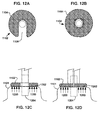
- FIGS. 12A , 12 B, 12 C and 12 D are diagrammatic representations of two exemplary holding devices in accordance with the present invention.
- FIGS. 13A and 13B are diagrammatic representations of an exemplary visceral pleura ring connector in accordance with the present invention.
- a long term oxygen therapy system and method may be utilized to deliver oxygen directly into the lung tissue in order to optimize oxygen transfer efficiency in the lungs.
- improved efficiency may be achieved if oxygen were to be delivered directly into the alveolar tissue in the lungs.
- alveoli walls are destroyed, thereby causing a decrease in air exchange surface area.
- collateral ventilation resistance is lowered. Accordingly, if it can be determined where collateral ventilation is occurring, then the diseased lung tissue may be isolated and the oxygen delivered to this precise location or locations.
- Various methods may be utilized to determine the diseased tissue locations, for example, computerized axial tomography or CAT scans, magnetic resonance imaging or MRI, positron emission tomograph or PET, and/or standard X-ray imaging.
- pressurized oxygen may be directly delivered to these diseased areas and more effectively and efficiently forced into the lung tissue for air exchange.
- anastomotic openings are made in the thoracic cavity and lung or lungs and one or more oxygen carrying conduits are positioned and sealed therein.
- the one or more oxygen carrying conduits are connected to an oxygen source which supplies oxygen under elevated pressure directly to the diseased portion or portions of the lung or lungs.
- the pressurized oxygen essentially displaces the accumulated air and is thus more easily absorbed by the alveoli tissue.
- the long term oxygen therapy system may be configured in such a way as to provide collateral ventilation bypass in addition to direct oxygen therapy.
- an additional conduit may be connected between the main conduit and the individual's trachea with the appropriate valve arrangement.
- stale air may be removed through the trachea when the individual exhales since the trachea is directly linked with the diseased site or sites in the lung via the conduits.
- the long term oxygen therapy system improves oxygen transfer efficiency in the lungs thereby reducing oxygen supply requirements, which in turn reduces the patient's medical costs.
- the system also allows for improved self-image, improved mobility, and greater exercise capability and is easily maintained.
- FIG. 1 illustrates a first exemplary long term oxygen therapy system 100 .
- the system 100 comprises an oxygen source 102 , an oxygen carrying conduit 104 and a one-way valve 106 .
- the oxygen source 102 may comprise any suitable device for supplying filtered oxygen under adjustably regulated pressures and flow rates, including pressurized oxygen tanks, liquid oxygen reservoirs, oxygen concentrators and the associated devices for controlling pressure and flow rate e.g. regulators.
- the oxygen carrying conduit 104 may comprise any suitable biocompatible tubing having a high resistance to damage caused by continuous oxygen exposure.
- the oxygen carrying conduit 104 comprises tubing having an inside diameter in the range from about 1/16 inch to about 1 ⁇ 2 inch and more preferably from about 1 ⁇ 8 inch to about 1 ⁇ 4 inch.
- the one-way valve 106 may comprise any suitable, in-line mechanical valve which allows oxygen to flow into the lungs 108 through the oxygen carrying conduit 104 , but not from the lungs 108 back into the oxygen source 102 .
- a simple check valve may be utilized.
- the oxygen carrying conduit 104 passes through the lung 108 at the site determined to have the highest degree of collateral ventilation.
- the exemplary system 100 described above may be modified in a number of ways, including the use of an in-line filter.
- both oxygen and air may flow through the system.
- oxygen is delivered to the lungs through the oxygen carrying conduit 104 and during exhalation, air from the lungs flow through the oxygen carrying conduit 104 .
- the in-line filter would trap mucus and other contaminants, thereby preventing a blockage in the oxygen source 102 .
- no valve 106 would be utilized.
- the flow of oxygen into the lungs and the flow of air from the lungs is based on pressure differentials.
- an air-tight seal is preferably maintained where the oxygen carrying conduit 104 passes through the thoracic cavity and lung. This seal is maintained in order to sustain the inflation/functionality of the lungs. If the seal is breached, air can enter the cavity and cause the lungs to collapse as described above.
- a method to create this seal comprises forming adhesions between the visceral pleura of the lung and the inner wall of the thoracic cavity. This may be achieved using either chemical methods, including irritants such as Doxycycline and/or Bleomycin, surgical methods, including pleurectomy or horoscope talc pleurodesis, or radiotherapy methods, including radioactive gold or external radiation. All of these methods are known in the relevant art for creating pleurodesis. With a seal created at the site for the ventilation bypass, an intervention may be safely performed without the danger of creating a pneumothorax of the lung.
- the oxygen carrying conduit 104 may be sealed to the skin at the site of the ventilation bypass.
- the oxygen carrying conduit 104 may be sealed to the skin of the thoracic wall 202 utilizing an adhesive 204 .
- the oxygen carrying conduit 104 comprises a flange 200 having a biocompatible adhesive coating 204 on the skin contacting surface.
- the biocompatible adhesive 204 would provide a fluid tight seal between the flange 200 and the skin or epidermis of the thoracic wall 202 .
- the biocompatible adhesive 204 provides a temporary fluid tight seal such that the oxygen carrying conduit 104 may be disconnected from the ventilation bypass site. This would allow for the site to be cleaned and for the long term oxygen therapy system 100 to undergo periodic maintenance.
- FIG. 3 illustrates another exemplary embodiment for sealing the oxygen carrying conduit 104 to the skin of the thoracic wall 202 at the site of the ventilation bypass.
- a coupling plate 300 is sealed to the skin at the site of the ventilation bypass by a biocompatible adhesive coating 204 or any other suitable means.
- the oxygen carrying conduit 104 is then connected to the coupling plate 300 by any suitable means, including threaded couplings and locking rings.
- the exemplary embodiment also allows for clearing of the site and maintenance of the system 100 .
- FIG. 4 illustrates yet another exemplary embodiment for sealing the oxygen carrying conduit 104 to the skin of the thoracic wall 202 at the site of the ventilation bypass.
- balloon flanges 400 may be utilized to create the seal.
- the balloon flanges 400 may be attached to the oxygen carrying conduit 104 such that in the deflated state, the oxygen carrying conduit 104 and one of the balloon flanges passes through the ventilation bypass anastomosis.
- the balloon flanges 400 are spaced apart a sufficient distance such that the balloon flanges remain on opposite sides of the thoracic wall 202 . When inflated, the balloons expand and form a fluid tight seal by sandwiching the thoracic wall.
- this exemplary embodiment allows for easy removal of the oxygen carrying conduit 104 .
- FIG. 5 illustrates yet another exemplary embodiment for sealing the oxygen carrying conduit 104 to the skin of the thoracic wall 202 at the site of the ventilation bypass.
- a single balloon flange 500 is utilized in combination with a fixed flange 502 .
- the balloon flange 500 is connected to the oxygen carrying conduit 104 in the same manner as described above.
- the balloon flange 500 when inflated, forms the fluid tight seal.
- the fixed flange 502 which is maintained against the skin of the thoracic wall 202 , provides the structural support against which the balloon exerts pressure to form the seal.
- a collateral ventilation bypass trap system utilizes the above-described collateral ventilation phenomenon to increase the expiratory flow from a diseased lung or lungs, thereby treating another aspect of chronic obstructive pulmonary disease.
- the most collaterally ventilated area of the lung or lungs is determined utilizing the scanning techniques described above. Once this area or areas are located, a conduit or conduits are positioned in a passage or passages that access the outer pleural layer of the diseased lung or lungs. The conduit or conduits utilize the collateral ventilation of the lung or lungs and allows the entrapped air to bypass the native airways and be expelled to a containment system outside of the body.
- FIG. 6 illustrates an exemplary embodiment of a collateral ventilation bypass/direct oxygen therapy system 600 .
- the system 600 comprises an oxygen source 602 (with potential filter), an oxygen carrying conduit 604 having two branches 606 and 608 , and a control valve 610 .
- the oxygen source 602 and oxygen carrying conduit 604 may comprise components similar to the above-described exemplary embodiment illustrated in FIG. 1 .
- the valve 610 when the individual inhales, the valve 610 is open and oxygen flows into the lung 612 and into the bronchial tube 614 .
- the branch 608 may be connected to the trachea 616 . Accordingly, during inhalation oxygen flows to the diseased site in the lung or lungs and to other parts of the lung through the normal bronchial passages.
- the valve 610 is closed so that no oxygen is delivered and air in the diseased portion of the lung may flow from the lung 612 , through one branch 606 and into the second branch 608 and finally into the bronchial tube 614 . In this manner, stale air is removed and oxygen is directly delivered.
- a sealed joint 607 is provided at the end of branch 606
- a sealed joint 609 is provided at the end of branch 608 .
- the connection and sealing of the oxygen carrying conduit 604 and branches 606 , 608 to the lung 612 and bronchial tube 614 may be made in a manner similar to that described above.
- FIG. 7 illustrates a first exemplary collateral ventilation bypass trap system 700 .
- the system 700 comprises a trap 702 , an air carrying conduit 704 and a filter/one-way valve 706 .
- the air carrying conduit 704 creates a fluid communication between an individual's lung 708 and the trap 702 through the filter/one-way valve 706 . It is important to note that although a single conduit 704 is illustrated, multiple conduits may be utilized in each lung 708 if it is determined that there are more than one area of high collateral ventilation.
- the trap 702 may comprise any suitable device for collecting discharge from the individual's lung or lungs 708 .
- the trap 702 is simply a containment vessel for temporarily storing discharge from the lungs, for example, mucous and other fluids that may accumulate in the lungs.
- the trap 702 may comprise any suitable shape and may be formed from any suitable metallic or non-metallic materials.
- the trap 702 should be formed from a lightweight, non-corrosive material.
- the trap 702 should be designed in such a manner as to allow for effective and efficient cleaning.
- the trap 702 may comprise disposable liners that may be removed when the trap 702 is full.
- the trap 702 may be formed from a transparent material or comprise an indicator window so that it may be easily determined when the trap 702 should be emptied or cleaned.
- a lightweight trap 702 increases the patient's mobility.
- the filter/one-way valve 706 may be attached to the trap 702 by any suitable means, including threaded fittings or compression type fittings commonly utilized in compressor connections.
- the filter/one-way valve 706 serves a number of functions.
- the filter/one-way valve 706 allows the air from the individual's lung or lungs 708 to exit the trap 702 while maintaining the fluid discharge and solid particulate matter in the trap 702 .
- This filter/one-way valve 706 would essentially maintain the pressure in the trap 702 below that of the pressure inside the individual's lung or lungs 708 so that the flow of air from the lungs 708 to the trap 702 is maintained in this one direction.
- the filter portion of the filter/one-way valve 706 may be designed to capture particulate matter of a particular size which is suspended in the air, but allows the clean air to pass therethrough and be vented to the ambient environment.
- the filter portion may also be designed in such a manner as to reduce the moisture content of the exhaled air.
- the air carrying conduit 704 connects the trap 702 to the lung or lungs 708 of the patient through the filter/one-way valve 706 .
- the air carrying conduit 704 may comprise any suitable biocompatible tubing having a resistance to the gases contained in air.
- the air carrying conduit 704 comprises tubing having an inside diameter in the range from about 1/16 inch to about 1 ⁇ 2 inch, and more preferably from about 1 ⁇ 8 inch to about 1 ⁇ 4 inch.
- the filter/one-way valve 706 may comprise any suitable valve which allows air to flow from the lung or lungs 708 through the air carrying conduit 704 , but not from the trap 702 back to the lungs 708 .
- a simple check valve may be utilized.
- the air carrying conduit 704 may be connected to the filter/one-way valve 706 by any suitable means. Preferably, a quick release mechanism is utilized so that the trap may be easily removed for maintenance.
- the air carrying conduit 704 passes through the lung 708 at the site determined to have the highest degree of collateral ventilation. If more than one site is determined, multiple air carrying conduits 704 may be utilized.
- the connection of multiple air carrying conduits 704 to the filter/one-way valve 706 may be accomplished by any suitable means, including an octopus device similar to that utilized in scuba diving regulators.
- the air carrying conduit 704 is preferably able to withstand and resist collapsing once in place. Since air will travel through the conduit 704 , if the conduit is crushed and unable to recover, the effectiveness of the system is diminished. Accordingly, a crush recoverable material may be incorporated into the air carrying conduit 704 in order to make it crush recoverable. Any number of suitable materials may be utilized. For example, Nitinol incorporated into the conduit 704 will give the conduit collapse resistance and collapse recovery properties.
- Expandable features at the end of the conduit 704 may be used to aid in maintaining contact and sealing the conduit 704 to the lung pleura. Nitinol incorporated into the conduit 704 will provide the ability to deliver the conduit 704 in a compressed state and then deployed in an expanded state to secure it in place. Shoulders at the end of the conduit may also provide a mechanical stop for insertion and an area for an adhesive/sealant to join as described in detail subsequently.
- an air-tight seal is preferably maintained where the air carrying conduit 704 passes through the thoracic cavity and lungs 708 .
- a sealed joint 705 is provided at the end of conduit 704 . This seal is maintained in order to sustain the inflation/functionality of the lungs. If the seal is breached, air can enter the cavity and cause the lungs to collapse.
- One exemplary method for creating the seal comprises forming adhesions between the visceral pleura of the lung and the inner wall of the thoracic cavity.
- a sealed joint between the air carrying conduit 704 and the outer pleural layer includes using various glues to help with the adhesion/sealing of the air carrying conduit 704 .
- Focal Inc. markets a sealant available under the trade name FOCAL/SEAL-L which is indicated for use on a lung for sealing purposes. Focal/Seal-L is activated by light in order to cure the sealant.
- Another seal available under the trade name THOREX which is manufactured by Surgical Sealants Inc., is currently conducting a clinical trial for lung sealing indications. Thorex is a two-part sealant that has a set curing time after the two parts are mixed.
- the creation of the opening in the chest cavity may be accomplished in a number of ways.
- the procedure may be accomplished using an open chest procedure, sternotomy or thoracotomy.
- the procedure may be accomplished using a laparoscopic technique, which is less invasive.
- the seal should be established while the lung is at least partially inflated in order to maintain a solid adhesive surface.
- the opening may then be made after the joint has been adequately created between the conduit component and the lung pleural surface.
- the opening should be adequate in cross-sectional area in order to provide sufficient decompression of the hyperinflated lung.
- This opening may be created using a number of different techniques such as cutting, piercing, dilating, blunt dissection, radio frequency energy, ultrasonic energy, microwave energy, or cryoblative energy.
- the air carrying conduit 704 may be sealed to the skin at the site by any of the means and methods described above with respect to the oxygen carrying conduit 704 and illustrated in FIGS. 2 through 5 .
- a pneumothorax (collapsed lung) may occur. Essentially, in any circumstance where the lung is punctured and a device inserted, an air-tight seal should preferably be maintained.
- pleurodesis i.e. an obliteration of the pleural space.
- pleurodesis methods including chemical, surgical and radiological.
- chemical pleurodesis an agent such as tetracycline, doxycycline, bleomycin or nitrogen mustard may be utilized.
- surgical pleurodesis a pleurectomy or a thorascopic talc procedure may be performed.
- radiological procedures radioactive gold or external radiation may be utilized.
- chemical pleurodesis is utilized.
- Exemplary methods for creating the seal comprises forming adhesions between the visceral pleura of the lung and the inner wall of the thoracic cavity using chemical methods, including irritants such as Doxycycline and/or Bleomycin, surgical methods, including pleurectomy or thorascopic talc pleurodesis.
- a sealed joint between the air carrying conduit 704 and the outer pleural layer includes using various glues to help with the adhesion/sealing of the air carrying conduit 704 .
- Focal Inc. markets a sealant available under the trade name FOCAL/SEAL-L which is indicated for use on a lung for sealing purposes. Focal/Seal-L is activated by light in order to cure the sealant.
- Another seal available under the trade name THOREX which is manufactured by Surgical Sealants Inc., is currently conducting a clinical trial for lung sealing indications. Thorex is a two-part sealant that has a set curing time after the two parts are mixed.
- Exemplary devices and methods for delivering a chemical(s) or agent(s) in a localized manner for ensuring a proper air-tight seal of the above-described apparatus is described below.
- the chemical(s), agent(s) and/or compound(s) are used to create a pleurodesis between the parietal and visceral pleura so that a component of the apparatus may penetrate through the particular area and not result in a pneumothorax.
- the chemical(s), agent(s) and/or compound(s) include talc, tetracycline, doxycycline, bleomycin and minocycline.
- a modified drug delivery catheter may be utilized to deliver chemical(s), agent(s) and/or compound(s) to a localized area for creating a pleurodesis in that area.
- the pleurodesis is formed and then the conduit 704 , as illustrated in FIG. 7 , is positioned in the lung 708 through the area of the pleurodesis.
- the drug delivery catheter provides a minimally invasive means for creating a localized pleurodesis.
- FIG. 8 there is illustrated an exemplary embodiment of a drug delivery catheter that may be utilized in accordance with the present invention. Any number of drug delivery catheters may be utilized.
- the distal tip of the catheter may comprise any suitable size, shape or configuration thereby enabling the formation of a pleurodesis having any size, shape or configuration.
- the catheter 800 is inserted into the patient such that the distal end 802 is positioned in the pleural space 804 between the thoracic wall 808 and the lung 806 .
- the distal end 802 of the catheter 800 comprises a substantially circular shape that would allow the chemical(s), agent(s) and/or compound(s) to be released towards the inner diameter of the substantially circular shape as indicated by arrows 810 .
- the distal end 802 of the catheter 800 comprising a plurality of holes or openings 812 through which the chemical(s), agent(s) and/or compound(s) are released.
- the distal end 802 may comprise any suitable size, shape or configuration.
- the distal end or tip 802 of the catheter 800 should preferably maintain its desired size, shape and/or configuration once deployed in the pleural space. This may be accomplished in a number of ways.
- the material forming the distal end 802 of the catheter 800 may be selected such that it has a certain degree of flexibility for insertion of the catheter 800 and a certain degree of shape memory such that it resumes its original or programmed shape once deployed. Any number of biocompatible polymers with these properties may be utilized.
- another material may be utilized.
- a metallic material having shape memory characteristics may be integrated into the distal end 802 of the catheter 800 . This metallic material may include Nitinol or stainless steel.
- the metallic material may be radiopaque or comprise radiopaque markers. By having a radiopaque material or radiopaque markers, the catheter 800 may be viewed under x-ray fluoroscopy and aid in determining when the catheter 800 is at the location of the highest collateral ventilation.
- a local drug delivery device may be utilized to deliver the pleurodesis chemical(s), agent(s) and/or compound(s).
- the pleurodesis is formed and then the conduit 704 , as illustrated in FIG. 7 , is positioned in the lung 708 through the pleurodesis.
- chemical(s), agent(s) and/or compound(s) may be affixed to an implantable medical device. The medical device is then implanted in the pleural cavity at a particular site and the chemical(s), agent(s) and/or compound(s) are released therefrom to form or create the pleurodesis.
- any of the above-described chemical(s), agent(s) and/or compound(s) may be affixed to the medical device.
- the chemical(s), agent(s) and/or compound(s) may be affixed to the medical device in any suitable manner.
- the chemical(s), agent(s) and/or compound(s) may be coated on the device utilizing any number of well known techniques including, spin coating, spraying or dipping, they may be incorporated into a polymeric matrix that is affixed to the surface of the medical device, they may be impregnated into the outer surface of the medical device, they may be incorporated into holes or chambers in the medical device, they may be coated onto the surface of the medical device and then coated with a polymeric layer that acts as a diffusion barrier for controlled release of the chemical(s), agent(s) and/or compound(s), they may be incorporated directly into the material forming the medical device, or any combination of the above-described techniques.
- the medical device may be formed from a biodegradable material which elutes the chemical(s), agent(s) and/or compound(s) as the device degrades.
- the implantable medical device may comprise any suitable size, shape and/or configuration, and may be formed using any suitable biocompatible material.
- FIG. 9 illustrates one exemplary embodiment of an implantable medical device 900 .
- the implantable medical device 900 comprises a substantially cylindrical disk 900 .
- the disk 900 is positioned in the pleural space 902 between the thoracic wall 904 and the lung 906 . Once in position, the disk 900 elutes or otherwise releases the chemical(s), agent(s) and/or compound(s) that form the pleurodesis.
- the release rate may be precisely controlled by using any of the various techniques described above, for example, a polymeric diffusion barrier.
- the disk 900 may be formed from a biodegradable material that elutes the chemical(s), agent(s) and/or compound(s) as the disk 900 itself disintegrates or dissolves.
- a non-biodegradable disk 900 may or may not require removal from the pleural cavity 902 once the pleurodesis is formed.
- the disk 900 may comprise a radiopaque marker or be formed from a radiopaque material.
- the radiopaque marker or material allows the disk 900 to be seen under fluoroscopy and then positioned accurately.
- the fluid characteristics of the chemical(s), agent(s) and/or compound(s) may be altered.
- the chemical(s), agent(s) and/or compound(s) may be made more viscous. With a more viscous chemical agent and/or compound, there would be less chance of the chemical, agent and/or compound moving from the desired location in the pleural space.
- the chemical(s), agent(s) and/or compound(s) may also comprise radiopaque constituents. Making the chemical(s), agent(s) and/or compounds radiopaque would allow the confirmation of the location of the chemical(s), agent(s) and/or compound(s) with regard to the optimal location of collateral ventilation.
- the chemical(s), agent(s) and/or compound(s) as modified above may be utilized in conjunction with standard chemical pleurodesis devices and processes or in conjunction with the exemplary embodiments set forth above.
- an implantable structure in combination with a chemical agent and/or a therapeutic agent may be utilized to create a localized area where the visceral and parietal pleura of the lung are fused together.
- a localized pleurodesis may be created utilizing either or both a mechanical component and a chemical component.
- the purpose of the chemical component is to provide an acute adhesion between the parietal and visceral pleura, while the mechanical component is utilized to provide a chronic adhesion.
- the acute adhesion provided by the chemical adhesive would provide enough stability at the implant location on the lung to allow for the mechanical component to create a chronic adhesion.
- the combination of a chemical adhesive with a tissue growth promoting material in a specific area of the lung would promote a well-controlled localized pleurodesis reaction.
- FIGS. 10A , 10 B and 10 C illustrate a first exemplary mechanical device 1000 for providing a chronic adhesion.
- FIG. 10A shows a close up view of the sectional view of mechanical device 1000 shown in FIG. 10B .
- FIG. 10C shows a cutaway view of the mechanical device 1000 shown in FIG. 10B on the surface of lung 1022 .
- the mechanical device 1000 comprises a mesh 1002 that may be formed out of any suitable biocompatible material.
- the mesh 1002 may comprise a metallic material, a polymeric material and/or a ceramic material. Primary variations of this material may be bio-resorbable or non-resorbable materials that promote tissue growth. Any type of mesh may be utilized including hernia repair meshes, laparoscopic meshes and surgical meshes.
- the mesh 1002 may be inserted between the parietal 1005 and visceral 1007 pleura at the desired location by any suitable means as set forth below.
- the mesh 1002 may be simply positioned or secured in place by any number of suitable means.
- the mechanical device is secured in such a manner that ensures the apposition of the device to either and/or both the visceral pleura 1007 and parietal pleura 1005 . As shown in FIG. 10G , this may be accomplished by a percutaneous application of a chemical adhesive 1010 after the lung is inflated to allow for a chemical agent to form an acute adhesion between the visceral pleura 1007 and parietal pleura 1005 .
- the chemical adhesive 1010 may include fibrin backed adhesive, cyanoacrylate bond adhesive or aldehyde bond adhesive. Alternately, as shown in FIG. 10D , a suture 1004 may be threaded into the device and pulled along with the visceral pleura against the parietal pleura of the thoracic wall.
- Radiological markers may be incorporated into the device 1000 thereby increasing its radiopacity under fluoroscopy. Essentially, this would ensure that in follow-up examinations, the exact location of where the localized pleurodesis has grown would be easy to find.
- These markers may be incorporated into the device 1000 in any number of suitable ways. For example, as shown in FIG. 10F , a wire ring 1006 may be woven into the spot of the tissue growth promoting material of the mesh. Alternately, as shown in FIG. 10E , radiological fibers 1008 may be incorporated into the tissue promoting fibers of the mesh 1002 . In yet another alternate exemplary embodiment, a radiological chemical adhesive may be utilized as shown in FIG. 10G .
- the delivery of the device 1000 may be approached utilizing any number of acceptable procedures.
- a thoracotomy procedure to open the thoracic cavity may be performed, and the device 1000 placed directly in the location.
- a minimally invasive approach using a cannula or such like device may be utilized to percutaneously access the thoracic cavity.
- the device 1000 could then be entirely delivered via a delivery system through the cannula or sheath.
- the device of the present invention allows for a small controlled local pleurodesis to form, thereby reducing potentially painful side effects and minimize pleural adhesions for subsequent thoracic interventions. Additionally, due to the dynamic nature between the lung and thoracic wall, it may be difficult to create a chronic local pleurodesis without the help of a clinical adhesive to provide acute stability to the location of intent.
- the visceral pleura must be properly attached to the conduit in order to properly seal around the conduit.
- a technique that may be utilized is to gather and attach the visceral pleura around the conduit using a purse-string suture or similar technique. This technique, however, requires the handling of the pleura in order to provide a counterforce on the pleura as the conduit is being positioned in the lung. In addition, what makes this technique more difficult is as soon as an access is made through the pleura for the conduit, the lung will immediately leak air and collapse to a smaller size. Therefore, providing a counterforce to insert a conduit or other device described herein through the access in the lung becomes even more vital.
- the visceral pleura of the lung are thin and somewhat fragile. Manipulation of the pleura using surgical instruments such as forceps or hemostats may create a break in the pleura. It is often difficult to seal the leak that will follow and the leak will typically result in a pneumothorax or a collapsed lung. In an emphysematous lung where the patient is already compromised with the inability to breath, a pneumothorax may potentially lead to serious complications, including death.
- the present invention is directed to a device that would provide the ability to insert a conduit or other device in the lung with a significantly decreased chance of injuring the lung if conventional surgical tools are utilized. Essentially, if a device could stabilize the visceral pleura and provide the counterforce without damaging the pleura, the procedure of inserting the device in the lung could become easier, faster and less conducive to injuring the pleura.
- a vacuum assist device 1102 may be utilized to hold the pleura while a conduit or other device is being positioned in the lung.
- FIG. 11A there is illustrated an inflated lung 1100 A and a deflated lung 1100 B and an access point 1100 C. Illustrated in FIG. 11B is a vacuum assist device 1102 which comprises a substantially disc-like structure or removable holding device 1104 illustrated in FIGS. 12A , 12 B, 12 C and 12 D, that exerts a vacuum force 1200 on the visceral pleura 1101 in contact therewith and an insertion envelope 1106 through which a conduit or other device may be inserted.
- any shape device may be utilized, for ease of explanation a substantially disc like structure is illustrated.
- the disc-like structure 1104 preferably has one substantially flat surface 1202 that makes contact with the visceral pleura 1101 .
- This flat surface has one or more openings through which a vacuum force that is created by an external source (not illustrated) is transmitted to the visceral pleura.
- This gentle vacuum force in the range from about 10 mm Hg to about 450 mm Hg is preferably evenly distributed over the substantially flat surface and gently pulls the visceral pleura 1101 into contact with the substantially flat surface 1202 .
- the disc like structure 1104 comprises a slit like opening 1108 that forms the envelope 1106 .
- the disc like structure 1104 comprises a two piece structure that when connected together forms the envelope 1106 .
- the disc like structure 1104 may be formed from any suitable biocompatible material that will not damage the visceral pleura and is easily removed from the pleural space when the vacuum is cut off.
- the vacuum assist device 1102 is inserted and placed into contact with the visceral pleura 1101 , the vacuum is started and draws and holds the visceral pleura 1101 in place while the conduit 1204 or other device is inserted through the envelope 1106 .
- the vacuum assist device 1102 maintains the counter-pressure for insertion and sealing without damaging the lung tissue. When the seal is created, the vacuum is cut off and the device 1102 is removed.
- the vacuum pressure or negative pressure may be created in a variety of ways. For example, surgical sites are typically equipped with vacuum devices that may be regulated to draw a negative pressure in the desired range.
- a simple pressure regulator or vacuum regulator may be connected between two vacuum sources and the device 1102 by any suitable means.
- the device 1102 may comprise a vacuum pump and regulator.
- the vacuum pump may use hospital power or be a self-contained battery power unit.
- the device As described above, once a device such as a conduit 1204 is inserted into the lung, the device must be sealed to the lung tissue. Also as described above is the purse-string suture that may be utilized to gather and attach the visceral pleura 1101 around the conduit 1204 or other device to create the seal. While this technique and other similar techniques may be utilized to create a seal, when the suture is pushed through the visceral pleura 1101 and around the conduit 1204 , it will inevitably leave small holes or tears through the pleura which may eventually lead to leaks. Accordingly, it would be advantageous to seal the visceral pleura 1101 around the conduit 1204 without having to make any holes or tears through or in the visceral pleura 1101 . If the visceral pleura 1101 were to be gathered around the conduit or other device, it would provide the accessibility to use a ring-type device to secure the gathered pleura around the conduit or other medical device.
- FIGS. 13A and 13B there is illustrated an exemplary visceral pleural ring connector 1300 in accordance with the present invention.
- the visceral pleural ring connector 1300 is simply placed around the gathered pleura 1302 which is gathered around the conduit 1304 .
- Any suitable biocompatible material may be utilized in constructing the visceral pleural ring connector 1300 .
- the visceral pleural ring connector 1300 may be constructed from any number of suitable materials, including superelastic materials such as nickel titanium alloys and bioabsorbable materials such as polyglycolic acid.
- a superelastic material such as a nickel titanium alloy
- the material may be programmed to be delivered at a first expanded diameter and, when released from a delivery device, allowed to contract to a second smaller diameter that snuggly holds the gathered visceral pleura 1302 to the conduit 1304 . It is important that the ring 1300 not fit too tight so as to avoid potential damage to the visceral pleura 1302 .
- the ring 1300 may be delivered in its contracted form, expanded and positioned over the gathered visceral pleura 1302 and then allowed to contract to its programmed size.
- various means may be incorporated into the ring structure 1300 for delivery and securing.
- the ring 1300 may comprise a split ring design wherein the ring 1300 may be opened like a chain link, placed around the gathered visceral pleura and then manually closed to create a snug fit.
- various self-locking structures may be incorporated into the ring structure 1300 .
- a ratchet mechanism may be utilized to tighten the ring 1300 around the gathered visceral pleura 1302 . It is important to note that any type of locking or tightening mechanisms may be utilized.
- one or more agents may be affixed to the ring 1300 .
- the one or more agents may be directly affixed to the surface of the ring 1300 , incorporated into a polymeric vehicle and then affixed to the surface of the ring 1300 , incorporated into channels or holes in the ring 1300 or incorporated into the bulk material forming the ring 1300 .
- the one or more agents may include chemicals to promote the pleurodesis reaction between the parietal pleura (inner thoracic wall) and the visceral pleura (lung).
- the pleurodesis is a key component to the chronic success of the procedure.
- the pleurodesis reaction will allow for the anastomosis to chronically exist without the danger of pneumothorax.
Abstract
Description
Claims (23)
Priority Applications (1)
| Application Number | Priority Date | Filing Date | Title |
|---|---|---|---|
| US12/034,774 US7931641B2 (en) | 2007-05-11 | 2008-02-21 | Visceral pleura ring connector |
Applications Claiming Priority (2)
| Application Number | Priority Date | Filing Date | Title |
|---|---|---|---|
| US99975807P | 2007-05-11 | 2007-05-11 | |
| US12/034,774 US7931641B2 (en) | 2007-05-11 | 2008-02-21 | Visceral pleura ring connector |
Publications (2)
| Publication Number | Publication Date |
|---|---|
| US20080281295A1 US20080281295A1 (en) | 2008-11-13 |
| US7931641B2 true US7931641B2 (en) | 2011-04-26 |
Family
ID=39970197
Family Applications (1)
| Application Number | Title | Priority Date | Filing Date |
|---|---|---|---|
| US12/034,774 Expired - Fee Related US7931641B2 (en) | 2007-05-11 | 2008-02-21 | Visceral pleura ring connector |
Country Status (1)
| Country | Link |
|---|---|
| US (1) | US7931641B2 (en) |
Cited By (12)
| Publication number | Priority date | Publication date | Assignee | Title |
|---|---|---|---|---|
| US20160007998A1 (en) * | 2014-07-11 | 2016-01-14 | Cardio Medical Solutions, Inc. | Device and method for assisting end-to-side anastomosis |
| US10512458B2 (en) | 2013-12-06 | 2019-12-24 | Med-Venture Investments, Llc | Suturing methods and apparatuses |
| US10610216B2 (en) | 2011-04-15 | 2020-04-07 | Heartstitch, Inc. | Suturing devices and methods for suturing an anatomic valve |
| US10687801B2 (en) | 2016-04-11 | 2020-06-23 | Nobles Medical Technologies Ii, Inc. | Suture spools for tissue suturing device |
| US10758223B2 (en) | 2005-06-20 | 2020-09-01 | Scarab Technology Services, Llc | Method and apparatus for applying a knot to a suture |
| US10828022B2 (en) | 2013-07-02 | 2020-11-10 | Med-Venture Investments, Llc | Suturing devices and methods for suturing an anatomic structure |
| US11051802B2 (en) | 2012-05-11 | 2021-07-06 | Heartstitch, Inc. | Suturing devices and methods for suturing an anatomic structure |
| US11166712B2 (en) | 2008-05-09 | 2021-11-09 | Scarab Technology Services, Llc | Suturing devices and methods for suturing an anatomic valve |
| US11197661B2 (en) | 2007-03-29 | 2021-12-14 | Scarab Technology Services, Llc | Device for applying a knot to a suture |
| US11202624B2 (en) | 2017-08-18 | 2021-12-21 | Nobles Medical Technologies Ii, Inc. | Apparatus for applying a knot to a suture |
| US11839370B2 (en) | 2017-06-19 | 2023-12-12 | Heartstitch, Inc. | Suturing devices and methods for suturing an opening in the apex of the heart |
| US11903865B2 (en) | 2015-09-15 | 2024-02-20 | Savage Medical, Inc. | Devices and methods for anchoring a sheath in a tissue cavity |
Families Citing this family (8)
| Publication number | Priority date | Publication date | Assignee | Title |
|---|---|---|---|---|
| US7682332B2 (en) | 2003-07-15 | 2010-03-23 | Portaero, Inc. | Methods to accelerate wound healing in thoracic anastomosis applications |
| US8475389B2 (en) | 2008-02-19 | 2013-07-02 | Portaero, Inc. | Methods and devices for assessment of pneumostoma function |
| US8336540B2 (en) | 2008-02-19 | 2012-12-25 | Portaero, Inc. | Pneumostoma management device and method for treatment of chronic obstructive pulmonary disease |
| EP2242527A4 (en) | 2008-02-19 | 2011-07-13 | Portaero Inc | Devices and methods for delivery of a therapeutic agent through a pneumostoma |
| US8347881B2 (en) | 2009-01-08 | 2013-01-08 | Portaero, Inc. | Pneumostoma management device with integrated patency sensor and method |
| US8518053B2 (en) | 2009-02-11 | 2013-08-27 | Portaero, Inc. | Surgical instruments for creating a pneumostoma and treating chronic obstructive pulmonary disease |
| EP2811939B8 (en) * | 2012-02-10 | 2017-11-15 | CVDevices, LLC | Products made of biological tissues for stents and methods of manufacturing |
| US10058332B2 (en) | 2012-08-01 | 2018-08-28 | Terumo Kabushiki Kaisha | Method for treatment of chronic obstructive pulmonary disease |
Citations (255)
| Publication number | Priority date | Publication date | Assignee | Title |
|---|---|---|---|---|
| US733152A (en) | 1902-08-30 | 1903-07-07 | Murdoch Chisholm | Empyema drainage device. |
| US953922A (en) | 1907-06-17 | 1910-04-05 | John B Rogers | Tracheal cannula or tube. |
| US2206687A (en) | 1937-11-29 | 1940-07-02 | Martha F Mckesson | Pleural diagnosis and treatment |
| US2867213A (en) | 1957-06-12 | 1959-01-06 | Jr Paul A Thomas | Flutter valve for drainage of the pleural cavity |
| US2873742A (en) | 1954-07-14 | 1959-02-17 | Research Corp | Surgical instruments |
| US2991787A (en) | 1957-04-11 | 1961-07-11 | Sierra Eng Co | Tracheotomy instrument |
| US3253594A (en) | 1963-07-30 | 1966-05-31 | Frank E Matthews | Peritoneal cannula |
| US3384087A (en) | 1962-11-12 | 1968-05-21 | Brummelkamp Reindert | Tracheotome |
| US3463159A (en) | 1965-02-16 | 1969-08-26 | Henry J Heimlich | Instrument for drainage of the chest |
| US3511243A (en) | 1967-07-18 | 1970-05-12 | Frederic J Toy | Apparatus for providing a breathing conduit communicating with the trachea at the base of the neck |
| US3556103A (en) | 1968-03-15 | 1971-01-19 | Edward J Calhoun | Tracheotomy instrument |
| US3638649A (en) | 1969-07-07 | 1972-02-01 | Univ Minnesota | Implantable prosthetic pass-through device |
| US3682166A (en) | 1970-07-29 | 1972-08-08 | Harvey Barry Jacobs | Emergency percutaneous trans-tracheal high flow oxygen catheter-type resuscitator for restoration of breathing in non-breathing patients |
| US3688773A (en) | 1970-04-16 | 1972-09-05 | Sol Weiss | Device for performing a tracheostomy and other surgical procedures |
| US3777757A (en) | 1971-01-08 | 1973-12-11 | R Gray | Sucking wound plug and chest aspirator |
| US3788326A (en) | 1970-07-29 | 1974-01-29 | H Jacobs | Distally perforated catheter for use in ventilating system |
| US3817250A (en) | 1972-10-24 | 1974-06-18 | Int Medical Devices Inc | Instrument for performing a tracheostomy and other surgical procedures |
| US3908704A (en) | 1958-06-12 | 1975-09-30 | Penntube Plastics Company | Corrugated tubing |
| US3916903A (en) | 1973-07-20 | 1975-11-04 | Reta M H Pozzi | Cricothyroid puncture apparatus |
| US4153058A (en) | 1977-07-05 | 1979-05-08 | Nehme Alexander E | Pleural decompression catheter |
| US4291694A (en) | 1979-10-29 | 1981-09-29 | Chai S | Apparatus and method for performing a thoracic operation |
| US4439189A (en) | 1981-06-18 | 1984-03-27 | Bentley Laboratories, Inc. | Pleural drainage system |
| US4465062A (en) | 1982-05-14 | 1984-08-14 | Gina Versaggi | Noninvasive seal for a sucking chest wound |
| US4502482A (en) | 1983-05-23 | 1985-03-05 | Deluccia Victor C | Endotracheal tube complex |
| US4583977A (en) | 1984-08-15 | 1986-04-22 | Vsesojuzny Nauchno-Issledovatelsky Institut Meditsiuskikh Polimerov | Device for lengthy fixation of a tube introduced into the patient's body |
| US4664660A (en) | 1985-04-01 | 1987-05-12 | Becton, Dickinson And Company | Chest drainage apparatus with ambient air sealing |
| EP0260543A1 (en) | 1986-09-16 | 1988-03-23 | Becton, Dickinson and Company | Chest drainage apparatus |
| US4799494A (en) | 1986-10-22 | 1989-01-24 | Wang Ko P | Percutaneous aspiration lung biopsy needle assembly |
| US4813929A (en) | 1987-02-19 | 1989-03-21 | Neal Semrad | Chest tube device and method of inserting device |
| US4826495A (en) | 1986-09-04 | 1989-05-02 | Coloplast A/S | Coupling means for fastening disposable ostomy bags and bag system therefore |
| US4828553A (en) | 1984-12-07 | 1989-05-09 | Coloplast A/S | Annular coupling system for ostomy equipment |
| US4869717A (en) | 1988-04-25 | 1989-09-26 | Adair Edwin Lloyd | Gas insufflation needle with instrument port |
| US4872869A (en) | 1982-06-09 | 1989-10-10 | Smith & Nephew (Latin America), Inc. | Low profile ostomy device |
| US4889534A (en) | 1986-12-04 | 1989-12-26 | Hollister Incorporated | Ostomy appliance with three-lement coupling ring assembly |
| US4931045A (en) | 1986-12-09 | 1990-06-05 | E. R. Squibb And Sons, Inc. | Low profile ostomy bag coupling |
| US4944724A (en) | 1987-06-05 | 1990-07-31 | Uresil Corporation | Apparatus for locating body cavities having signaling indicator |
| US4959054A (en) | 1988-11-23 | 1990-09-25 | Clemson University | Pharmaceutically protected percutaneous devices |
| US4976688A (en) | 1989-02-03 | 1990-12-11 | Rosenblum Jeffrey L | Position-adjustable thoracic catheter |
| US5004456A (en) | 1989-03-10 | 1991-04-02 | Arrow International Investment Corporation | In-dwelling catheter |
| US5060645A (en) | 1990-08-14 | 1991-10-29 | Russell David N | Tracheostomy tube assembly |
| US5078689A (en) | 1990-05-14 | 1992-01-07 | Keller Alan M | Device for removing body fluids |
| US5137509A (en) | 1991-04-17 | 1992-08-11 | Dexide, Inc. | Surgical insufflation instrument |
| US5139485A (en) | 1991-05-03 | 1992-08-18 | Ethicon, Inc. | Verress needle with enhanced acoustical means |
| US5218957A (en) | 1990-10-19 | 1993-06-15 | Ballard Medical Products | Multi-layered transtracheal catheter |
| US5230350A (en) | 1991-05-29 | 1993-07-27 | Tabex Industries, Inc. | Moisture barrier for indwelling catheters and the like |
| US5230332A (en) | 1990-10-22 | 1993-07-27 | Ballard Medical Products | Methods and apparatus for a micro-tracheal catheter hub assembly |
| US5261708A (en) | 1989-11-17 | 1993-11-16 | E. R. Squibb & Sons, Inc. | Ostomy coupling |
| US5263939A (en) | 1992-10-09 | 1993-11-23 | Surgin Surgical Instrumentation, Inc. | Retainer for laparoscopic cannula |
| US5312331A (en) | 1993-04-15 | 1994-05-17 | Knoepfler Dennis J | Method and apparatus for talc pleurodesis |
| US5315992A (en) | 1993-03-10 | 1994-05-31 | Dalton William J | Triple cuff endobronchial tube with selective multiple outlets served by a single airflow passage |
| US5336206A (en) | 1989-08-15 | 1994-08-09 | United States Surgical Corporation | Trocar penetration depth indicator and guide tube positioning device |
| EP0609950A1 (en) | 1993-02-04 | 1994-08-10 | Cordis Europa N.V. | Angiography catheter |
| US5354283A (en) | 1994-01-07 | 1994-10-11 | Little Rapids Corporation | Trocar retention apparatus |
| US5356386A (en) | 1987-06-05 | 1994-10-18 | Uresil Corporation | Apparatus for locating body cavities |
| US5366478A (en) | 1993-07-27 | 1994-11-22 | Ethicon, Inc. | Endoscopic surgical sealing device |
| US5370625A (en) | 1989-08-15 | 1994-12-06 | United States Surgical Corporation | Trocar guide tube positioning device |
| US5376376A (en) | 1992-01-13 | 1994-12-27 | Li; Shu-Tung | Resorbable vascular wound dressings |
| US5389077A (en) | 1993-03-03 | 1995-02-14 | Uresil Corporation | Minimally invasive body cavity penetrating instruments |
| US5401262A (en) | 1990-07-20 | 1995-03-28 | Atrium Medical Corporation | Fluid recovery system |
| US5431633A (en) | 1994-05-06 | 1995-07-11 | Global Fury, Inc. | Noninvasive device for preventing pressure build-up in pleural space and reducing possibility of development of a tension pneumothorax from an open pneumothorax |
| US5478333A (en) | 1994-03-04 | 1995-12-26 | Asherman, Jr.; Richard E. | Medical dressing for treating open chest injuries |
| US5484401A (en) | 1992-11-04 | 1996-01-16 | Denver Biomaterials, Inc. | Treatment method for pleural effusion |
| US5489264A (en) * | 1993-03-05 | 1996-02-06 | Salo; Gaspar E. | Pleural drainage probe |
| US5496297A (en) | 1993-02-22 | 1996-03-05 | Coloplast A/S | Ostomy coupling |
| US5501677A (en) | 1993-06-25 | 1996-03-26 | Jensen; Ole R. | Two-piece ostomy appliance and low-profile coupling ring assembly |
| US5501678A (en) | 1991-08-30 | 1996-03-26 | Coloplast A/S | Adapter for use in connection with ostomy equipment |
| US5588424A (en) | 1995-06-28 | 1996-12-31 | The Cleveland Clinic Foundation | Bronchial blocker endotracheal apparatus |
| US5616131A (en) | 1992-09-23 | 1997-04-01 | Lasersurge, Inc. | Apparatus and method for anchoring surgical instrumentation |
| US5660175A (en) | 1995-08-21 | 1997-08-26 | Dayal; Bimal | Endotracheal device |
| US5662629A (en) | 1995-04-13 | 1997-09-02 | E.R. Squibb & Sons, Inc. | Ostomy coupling |
| US5728066A (en) | 1995-12-13 | 1998-03-17 | Daneshvar; Yousef | Injection systems and methods |
| US5730735A (en) | 1996-03-12 | 1998-03-24 | Hollister Incorporated | Convex ostomy faceplate with floating flange and finger recess |
| US5738661A (en) | 1995-06-16 | 1998-04-14 | Larice; Gennaro | Medical device for holding a feeding tube and use thereof |
| US5807341A (en) | 1996-12-11 | 1998-09-15 | Team Medical Llc | Medical catheter dressing device |
| US5843053A (en) | 1996-08-20 | 1998-12-01 | Bristol-Myers Squibb Company | Ostomy coupling |
| US5897531A (en) | 1994-01-07 | 1999-04-27 | Amirana; Omar | Adhesive surgical retaining device |
| US5931821A (en) | 1996-03-05 | 1999-08-03 | Tyco Group S.A.R.L. | Chest drainage unit with controlled automatic excess negativity relief feature |
| US5954636A (en) | 1997-07-15 | 1999-09-21 | Schwartz; Roy E. | Pediatric endotracheal tube with bronchial blocker and method for selectively blocking respiratory airflow to a pediatric patient's lung |
| US5971962A (en) | 1997-03-11 | 1999-10-26 | Sun Medical Technology Research | Skin button |
| US5972026A (en) | 1997-04-07 | 1999-10-26 | Broncus Technologies, Inc. | Bronchial stenter having diametrically adjustable electrodes |
| US6059816A (en) | 1996-02-29 | 2000-05-09 | Moenning; Stephen P. | Apparatus and method for protecting a port site opening in the wall of a body cavity utilizing a sealing member and a biologically active compound |
| US6083255A (en) | 1997-04-07 | 2000-07-04 | Broncus Technologies, Inc. | Bronchial stenter |
| WO2000076577A1 (en) | 1999-06-15 | 2000-12-21 | Bard Access Systems, Inc., A Subsidiary Of C.R. Bard, Inc. | Graft-catheter vascular access system |
| WO2001002042A1 (en) | 1999-07-02 | 2001-01-11 | Pulmonx | Methods, systems, and kits for lung volume reduction |
| US6174323B1 (en) | 1998-06-05 | 2001-01-16 | Broncus Technologies, Inc. | Method and assembly for lung volume reduction |
| US6197010B1 (en) | 1998-06-11 | 2001-03-06 | Hollister Incorporated | Ostomy appliance faceplate with concealed coupling ring flange |
| US6200333B1 (en) | 1997-04-07 | 2001-03-13 | Broncus Technologies, Inc. | Bronchial stenter |
| WO2001045568A1 (en) | 1999-12-20 | 2001-06-28 | Dexterity Surgical, Inc. | Adjustable surgical wound protector and retractor |
| US6258100B1 (en) | 1999-08-24 | 2001-07-10 | Spiration, Inc. | Method of reducing lung size |
| US6273907B1 (en) | 1997-04-07 | 2001-08-14 | Broncus Technologies, Inc. | Bronchial stenter |
| US6283988B1 (en) | 1997-04-07 | 2001-09-04 | Broncus Technologies, Inc. | Bronchial stenter having expandable electrodes |
| US6293930B1 (en) | 1998-06-11 | 2001-09-25 | Hollister Incorporated | Low-profile ostomy faceplate with recessed coupling ring |
| US20010025132A1 (en) | 2000-03-23 | 2001-09-27 | Spiration, Inc. | Tissue resection device, system and method |
| US20010041906A1 (en) | 1999-10-20 | 2001-11-15 | Spiration, Inc. | Apparatus and method for expanding the thorax |
| US20010041932A1 (en) | 1998-12-08 | 2001-11-15 | Hans Scholz | Flanged graft for end-to-side anastomosis |
| US6322536B1 (en) | 1998-03-06 | 2001-11-27 | Cornell Research Foundation, Inc. | Minimally invasive gene therapy delivery and method |
| US6330882B1 (en) | 1998-03-06 | 2001-12-18 | Ronald French | Emergency apparatus for evacuating air from the body cavity |
| US6334441B1 (en) | 1998-11-23 | 2002-01-01 | Mallinckrodt Medical, Inc. | Phonation valve for breathing tube |
| US6358269B1 (en) | 1998-11-02 | 2002-03-19 | Ralph Aye | Method of treating peripheral bronchopleural fistulas |
| US20020042564A1 (en) | 1999-08-05 | 2002-04-11 | Cooper Joel D. | Devices for creating collateral channels in the lungs |
| US6398775B1 (en) | 1999-10-21 | 2002-06-04 | Pulmonx | Apparatus and method for isolated lung access |
| US6411852B1 (en) | 1997-04-07 | 2002-06-25 | Broncus Technologies, Inc. | Modification of airways by application of energy |
| US20020087153A1 (en) | 1999-08-05 | 2002-07-04 | Broncus Technologies, Inc. | Devices for creating collateral channels |
| US6416554B1 (en) | 1999-08-24 | 2002-07-09 | Spiration, Inc. | Lung reduction apparatus and method |
| US6432100B1 (en) | 1998-11-11 | 2002-08-13 | Ing Klaus Affeld | Apparatus and method for generation of a protective sleeve against infections for an artificial lead |
| US20020111619A1 (en) | 1999-08-05 | 2002-08-15 | Broncus Technologies, Inc. | Devices for creating collateral channels |
| US20020111620A1 (en) | 2001-02-14 | 2002-08-15 | Broncus Technologies, Inc. | Devices and methods for maintaining collateral channels in tissue |
| US20020112729A1 (en) | 2001-02-21 | 2002-08-22 | Spiration, Inc. | Intra-bronchial obstructing device that controls biological interaction with the patient |
| US6443156B1 (en) | 2000-08-02 | 2002-09-03 | Laura E. Niklason | Separable double lumen endotracheal tube |
| WO2002076279A2 (en) | 2001-03-23 | 2002-10-03 | Hospitec Inc. | Method and system for intubation |
| US6468292B1 (en) | 1995-05-19 | 2002-10-22 | General Surgical Innovations, Inc. | Skin seal with inflatable membrane |
| US20020165618A1 (en) | 2001-01-18 | 2002-11-07 | The Brigham And Women's Hospital, Inc. | Prosthetic device for respiratory patients |
| RU2192185C1 (en) * | 2001-05-04 | 2002-11-10 | Фатихов Рашит Габдуллович | Pleural cavity drainage apparatus |
| US6488673B1 (en) | 1997-04-07 | 2002-12-03 | Broncus Technologies, Inc. | Method of increasing gas exchange of a lung |
| WO2002096325A1 (en) | 2001-05-27 | 2002-12-05 | Schurr Marc O | Medical implant |
| US6491706B1 (en) | 2001-07-10 | 2002-12-10 | Spiration, Inc. | Constriction device including fixation structure |
| US20030013935A1 (en) | 2001-07-10 | 2003-01-16 | Spiration, Inc. | Constriction device viewable under X ray fluoroscopy |
| US20030018344A1 (en) | 2001-07-19 | 2003-01-23 | Olympus Optical Co., Ltd. | Medical device and method of embolizing bronchus or bronchiole |
| US20030018309A1 (en) * | 2001-07-17 | 2003-01-23 | Breznock Eugene Michael | Method and apparatus for chest drainage |
| WO2003007821A1 (en) | 2001-07-18 | 2003-01-30 | Sumitomo Bakelite Company Limited | Medical treating instrument |
| US6514290B1 (en) | 2000-03-31 | 2003-02-04 | Broncus Technologies, Inc. | Lung elastic recoil restoring or tissue compressing device and method |
| US6517519B1 (en) | 1999-08-13 | 2003-02-11 | The Johns Hopkins University | Device and method for rapid chest tube insertion |
| US6520183B2 (en) | 2001-06-11 | 2003-02-18 | Memorial Sloan-Kettering Cancer Center | Double endobronchial catheter for one lung isolation anesthesia and surgery |
| US6527761B1 (en) | 2000-10-27 | 2003-03-04 | Pulmonx, Inc. | Methods and devices for obstructing and aspirating lung tissue segments |
| US20030050648A1 (en) | 2001-09-11 | 2003-03-13 | Spiration, Inc. | Removable lung reduction devices, systems, and methods |
| WO2003020338A2 (en) | 2001-09-04 | 2003-03-13 | Broncus Technologies, Inc. | Conduits having distal cage structure for maintaining collateral channels in tissue and related methods |
| US20030055331A1 (en) | 2001-09-11 | 2003-03-20 | Pulmonx | Methods of endobronchial diagnosis using imaging |
| US20030051733A1 (en) | 2001-09-10 | 2003-03-20 | Pulmonx | Method and apparatus for endobronchial diagnosis |
| US20030065339A1 (en) | 2001-10-02 | 2003-04-03 | Spiration, Inc. | Constriction device including reinforced suture holes |
| US6550475B1 (en) | 1998-03-11 | 2003-04-22 | Oldfield Family Holdings Pty. Limited | Endotracheal tube for selective bronchial occlusion |
| US20030078469A1 (en) | 2001-10-18 | 2003-04-24 | Spiration, Inc. | Constriction device including tear resistant structures |
| US20030083671A1 (en) | 2001-10-25 | 2003-05-01 | Spiration, Inc. | Bronchial obstruction device deployment system and method |
| US6569121B1 (en) | 2000-10-12 | 2003-05-27 | Benjamin Warren Purow | Sheath device with dressing for prevention of pneumothorax |
| US6585639B1 (en) | 2000-10-27 | 2003-07-01 | Pulmonx | Sheath and method for reconfiguring lung viewing scope |
| US20030130593A1 (en) | 2002-01-04 | 2003-07-10 | Spiration, Inc. | System and method for capturing body tissue samples |
| US20030127090A1 (en) | 2001-11-14 | 2003-07-10 | Emphasys Medical, Inc. | Active pump bronchial implant devices and methods of use thereof |
| US6599311B1 (en) | 1998-06-05 | 2003-07-29 | Broncus Technologies, Inc. | Method and assembly for lung volume reduction |
| WO2003061480A1 (en) | 2001-10-20 | 2003-07-31 | Applied Medical Resources Corporation | Wound retraction apparatus and method |
| US20030149446A1 (en) | 2002-02-01 | 2003-08-07 | Spiration, Inc. | Device and method of isolating deleterious body tissue located within healthy body tissue |
| US20030158515A1 (en) | 2002-02-21 | 2003-08-21 | Spiration, Inc. | Device and method for intra-bronchial provision of a therapeutic agent |
| US20030154988A1 (en) | 2002-02-21 | 2003-08-21 | Spiration, Inc. | Intra-bronchial device that provides a medicant intra-bronchially to the patient |
| US6609521B1 (en) | 2001-04-09 | 2003-08-26 | Regents Of The University Of Minnesota | Endotracheal tube |
| US20030181922A1 (en) | 2002-03-20 | 2003-09-25 | Spiration, Inc. | Removable anchored lung volume reduction devices and methods |
| US20030181356A1 (en) | 2002-03-11 | 2003-09-25 | Edward Ingenito | Compositions and methods for treating emphysema |
| US20030186904A1 (en) | 2001-01-08 | 2003-10-02 | Human Genome Sciences, Inc. | Keratinocyte growth factor-2 |
| US6632243B1 (en) | 1997-09-16 | 2003-10-14 | Emphasys Medical Inc. | Body fluid flow control device |
| US20030195511A1 (en) | 2002-04-16 | 2003-10-16 | Spiration, Inc. | Device for and method of removing deleterious body tissue from a site within a patient |
| US20030195385A1 (en) | 2002-04-16 | 2003-10-16 | Spiration, Inc. | Removable anchored lung volume reduction devices and methods |
| US6634360B1 (en) | 1998-09-02 | 2003-10-21 | Flodin Bjoern | Device for supplying inhalation gas to and removing exhalation gas from a patient |
| US6634363B1 (en) | 1997-04-07 | 2003-10-21 | Broncus Technologies, Inc. | Methods of treating lungs having reversible obstructive pulmonary disease |
| US20030212337A1 (en) | 2002-05-09 | 2003-11-13 | Spiration, Inc. | Automated provision of information related to air evacuation from a chest cavity |
| US20030212412A1 (en) | 2002-05-09 | 2003-11-13 | Spiration, Inc. | Intra-bronchial obstructing device that permits mucus transport |
| US20030216769A1 (en) | 2002-05-17 | 2003-11-20 | Dillard David H. | Removable anchored lung volume reduction devices and methods |
| US20030216730A1 (en) | 2002-05-14 | 2003-11-20 | Spiration, Inc. | Apparatus and method for resecting and removing selected body tissue from a site inside a patient |
| US6659961B2 (en) | 1996-10-25 | 2003-12-09 | Gavin J. B. Robinson | Method and apparatus for measuring pulmonary blood flow by pulmonary exchange of oxygen and an inert gas with the blood |
| US20030228344A1 (en) | 2002-03-08 | 2003-12-11 | Fields Antony J. | Methods and devices for inducing collapse in lung regions fed by collateral pathways |
| US20030233099A1 (en) | 2000-10-17 | 2003-12-18 | Broncus Technologies, Inc. | Modification of airways by application of energy |
| US20040010209A1 (en) | 2002-07-15 | 2004-01-15 | Spiration, Inc. | Device and method for measuring the diameter of an air passageway |
| US20040010289A1 (en) | 2000-10-17 | 2004-01-15 | Broncus Technologies, Inc. | Control system and process for application of energy to airway walls and other mediums |
| US6679264B1 (en) | 2000-03-04 | 2004-01-20 | Emphasys Medical, Inc. | Methods and devices for use in performing pulmonary procedures |
| US6682506B1 (en) | 1998-12-22 | 2004-01-27 | Francis Navarro | Device for maintaining at least a tube |
| US20040024356A1 (en) | 2002-07-31 | 2004-02-05 | Don Tanaka | Long term oxygen therapy system |
| US20040040555A1 (en) | 2002-08-28 | 2004-03-04 | Don Tanaka | Collateral ventilation bypass trap system |
| US20040047855A1 (en) | 2002-06-17 | 2004-03-11 | Bistech, Inc., A Delaware Corporation | Compositions and methods for reducing lung volume |
| US20040059263A1 (en) | 2002-09-24 | 2004-03-25 | Spiration, Inc. | Device and method for measuring the diameter of an air passageway |
| US20040055606A1 (en) | 2001-03-02 | 2004-03-25 | Emphasys Medical, Inc. | Bronchial flow control devices with membrane seal |
| US20040073155A1 (en) | 2000-01-14 | 2004-04-15 | Broncus Technologies, Inc. | Methods and devices for maintaining patency of surgically created channels in tissue |
| US20040073241A1 (en) | 2002-10-11 | 2004-04-15 | Spiration, Inc. | Implantable tissue constriction device and method for suppressing leakage of fluid from resectioned body tissue |
| US20040078054A1 (en) | 1998-06-05 | 2004-04-22 | Broncus Technologies, Inc. | Method for lung volume reduction |
| US20040078026A1 (en) | 2002-10-21 | 2004-04-22 | Wagner Robert B. | Method and apparatus for pleural drainage |
| US6736797B1 (en) | 1998-06-19 | 2004-05-18 | Unomedical A/S | Subcutaneous infusion set |
| US20040144387A1 (en) | 2003-01-28 | 2004-07-29 | David Amar | Double-balloon endobronchial catheter for one lung isolation anesthesia and surgery |
| US6770063B2 (en) | 2002-04-23 | 2004-08-03 | Uresil, L.P. | Thoracic vent kit |
| US6770070B1 (en) | 2000-03-17 | 2004-08-03 | Rita Medical Systems, Inc. | Lung treatment apparatus and method |
| US20040200484A1 (en) | 2003-04-08 | 2004-10-14 | Springmeyer Steven C. | Bronchoscopic lung volume reduction method |
| US20040210248A1 (en) | 2003-03-12 | 2004-10-21 | Spiration, Inc. | Apparatus, method and assembly for delivery of intra-bronchial devices |
| US20040211434A1 (en) | 2002-07-05 | 2004-10-28 | Broncus Technologies, Inc. | Extrapleural airway device and method |
| US20040225254A1 (en) | 2003-05-07 | 2004-11-11 | Don Tanaka | Localized pleurodesis chemical delivery |
| US20040231674A1 (en) | 2003-05-20 | 2004-11-25 | Don Tanaka | Intra/extra-thoracic collateral ventilation bypass system |
| US20040237966A1 (en) | 2003-05-29 | 2004-12-02 | Don Tanaka | Methods and devices to assist pulmonary decompression |
| US20040244802A1 (en) | 2003-06-03 | 2004-12-09 | Don Tanaka | Lung reduction system |
| US20040244803A1 (en) | 2003-06-05 | 2004-12-09 | Don Tanaka | Intra-thoracic collateral ventilation bypass system |
| US6837906B2 (en) | 2001-08-03 | 2005-01-04 | Ensure Medical, Inc. | Lung assist apparatus and methods for use |
| US20050005936A1 (en) | 2003-06-18 | 2005-01-13 | Wondka Anthony David | Methods, systems and devices for improving ventilation in a lung area |
| US6846292B2 (en) | 2002-02-19 | 2005-01-25 | Mohamed Bakry | Pleural biopsy and brushing needle |
| US20050022809A1 (en) | 2003-04-25 | 2005-02-03 | Wondka Anthony David | Methods, systems and devices for desufflating a lung area |
| US20050025816A1 (en) | 2003-07-15 | 2005-02-03 | Don Tanaka | Methods and devices to accelerate wound healing in thoracic anastomosis applications |
| US20050043751A1 (en) | 2001-09-04 | 2005-02-24 | Broncus Technologies, Inc. | Methods and devices for maintaining patency of surgically created channels in a body organ |
| US20050043752A1 (en) | 2001-09-04 | 2005-02-24 | Broncus Technologies, Inc. | Methods and devices for maintaining patency of surgically created channels in a body organ |
| US20050060042A1 (en) | 2001-09-04 | 2005-03-17 | Broncus Technologies, Inc. | Methods and devices for maintaining surgically created channels in a body organ |
| US20050056292A1 (en) | 1999-08-05 | 2005-03-17 | Cooper Joel D. | Devices for maintaining patency of surgically created channels in tissue |
| US20050060044A1 (en) | 1999-08-05 | 2005-03-17 | Ed Roschak | Methods and devices for maintaining patency of surgically created channels in a body organ |
| US20050060041A1 (en) | 2001-09-04 | 2005-03-17 | Broncus Technologies, Inc. | Methods and devices for maintaining surgically created channels in a body organ |
| US20050061322A1 (en) | 2003-01-20 | 2005-03-24 | Pulmonx | Method and arrangement for reducing the volume of a lung |
| US20050066976A1 (en) | 2003-08-18 | 2005-03-31 | Wondka Anthony D. | Method and device for non-invasive ventilation with nasal interface |
| US20050107783A1 (en) | 1999-08-05 | 2005-05-19 | Broncus Technologies, Inc. | Devices for applying energy to tissue |
| US20050103340A1 (en) | 2003-08-20 | 2005-05-19 | Wondka Anthony D. | Methods, systems & devices for endobronchial ventilation and drug delivery |
| US6901927B2 (en) | 2000-03-04 | 2005-06-07 | Emphasys Medical, Inc. | Methods and devices for use in performing pulmonary procedures |
| US20050137715A1 (en) | 1999-08-05 | 2005-06-23 | Broncus Technologies, Inc. | Methods and devices for maintaining patency of surgically created channels in a body organ |
| US20050137518A1 (en) | 2002-04-19 | 2005-06-23 | Broncus Technologies, Inc. | Devices for maintaining surgically created openings |
| US20050137611A1 (en) | 2001-09-04 | 2005-06-23 | Broncus Technologies, Inc. | Methods and devices for maintaining surgically created channels in a body organ |
| US20050145253A1 (en) | 2001-10-11 | 2005-07-07 | Emphasys Medical, Inc., A Delaware Corporation | Bronchial flow control devices and methods of use |
| US6916310B2 (en) | 2003-05-30 | 2005-07-12 | Codman & Shurtleff, Inc. | Percutaneous access device |
| US20050161040A1 (en) | 2003-07-03 | 2005-07-28 | Don Tanaka | Collateral ventilation bypass trap system |
| US20050171396A1 (en) | 2003-10-20 | 2005-08-04 | Cyberheart, Inc. | Method for non-invasive lung treatment |
| US20050177144A1 (en) | 1999-08-05 | 2005-08-11 | Broncus Technologies, Inc. | Methods and devices for maintaining patency of surgically created channels in a body organ |
| US20050178389A1 (en) | 2004-01-27 | 2005-08-18 | Shaw David P. | Disease indications for selective endobronchial lung region isolation |
| US20050178385A1 (en) | 2002-06-11 | 2005-08-18 | Raffaele Dellaca' | System and method for the automatic detection of the expiratory flow limitation |
| US20050205097A1 (en) | 2004-03-17 | 2005-09-22 | Cook Critical Care Incorporated | Medical devices and methods of selectively and alternately isolating bronchi or lungs |
| US20050244401A1 (en) | 2002-06-17 | 2005-11-03 | Ingenito Edward P | Compositions and methods for reducing lung volume |
| US20050281801A1 (en) | 2004-06-16 | 2005-12-22 | Glen Gong | Glue composition for lung volume reduction |
| US20050281797A1 (en) | 2004-06-16 | 2005-12-22 | Glen Gong | Lung volume reduction using glue compositions |
| US20050282748A1 (en) | 2004-06-16 | 2005-12-22 | Glen Gong | Glue compositions for lung volume reduction |
| US20050281802A1 (en) | 2004-06-16 | 2005-12-22 | Glen Gong | Lung volume reduction using glue composition |
| US20050288550A1 (en) | 2004-06-14 | 2005-12-29 | Pneumrx, Inc. | Lung access device |
| US20050288684A1 (en) | 2004-06-16 | 2005-12-29 | Aronson Nathan A | Method of reducing collateral flow in a portion of a lung |
| US20060025815A1 (en) | 2004-07-08 | 2006-02-02 | Mcgurk Erin | Lung device with sealing features |
| US20060047291A1 (en) | 2004-08-20 | 2006-03-02 | Uptake Medical Corporation | Non-foreign occlusion of an airway and lung collapse |
| US7011094B2 (en) | 2001-03-02 | 2006-03-14 | Emphasys Medical, Inc. | Bronchial flow control devices and methods of use |
| US7014628B2 (en) | 1995-07-07 | 2006-03-21 | Bousquet Gerald G | Transcutaneous access device |
| US7022088B2 (en) | 1999-08-05 | 2006-04-04 | Broncus Technologies, Inc. | Devices for applying energy to tissue |
| US20060079838A1 (en) | 2004-10-08 | 2006-04-13 | Walker Steven C | Movable Balloon anchor for medical devices |
| US7036509B2 (en) | 2003-12-04 | 2006-05-02 | Emphasys Medical, Inc. | Multiple seal port anesthesia adapter |
| US20060107961A1 (en) | 2004-11-19 | 2006-05-25 | Don Tanaka | Localized pleurodesis evacuation device |
| US20060116749A1 (en) | 2003-07-18 | 2006-06-01 | Broncus Technologies, Inc. | Devices for maintaining patency of surgically created channels in tissue |
| US20060118125A1 (en) | 2004-11-19 | 2006-06-08 | Don Tanaka | Pulmonary drug delivery |
| US20060118126A1 (en) | 2004-11-19 | 2006-06-08 | Don Tanaka | Methods and devices for controlling collateral ventilation |
| US20060124126A1 (en) | 2004-12-10 | 2006-06-15 | Don Tanaka | Collateral ventilation device with chest tube/evacuation features |
| US20060130830A1 (en) | 2004-09-07 | 2006-06-22 | Uptake Medical Corporation | Intra-bronchial implants for improved attachment |
| US20060135947A1 (en) | 2000-10-27 | 2006-06-22 | Pulmonx | Occlusal stent and methods for its use |
| US20060135984A1 (en) | 2003-07-18 | 2006-06-22 | Broncus Technologies, Inc. | Devices for maintaining patency of surgically created channels in tissue |
| US20060161233A1 (en) | 2004-11-16 | 2006-07-20 | Uptake Medical Corp. | Device and method for lung treatment |
| US20060162731A1 (en) | 2004-11-16 | 2006-07-27 | Pulmonx | Pulmonary occlusal stent delivery catheter, loading system and methods of use |
| US20060212046A1 (en) | 2005-03-17 | 2006-09-21 | Pearce Joseph R | Device to deploy a resilient sleeve to constrict on body tissue |
| US20060235432A1 (en) | 2002-02-21 | 2006-10-19 | Devore Lauri J | Intra-bronchial obstructing device that controls biological interaction with the patient |
| US7135010B2 (en) | 2003-09-30 | 2006-11-14 | Damage Control Surgical Technologies, Inc. | Method and apparatus for rapid deployment chest drainage |
| US20060264772A1 (en) | 2001-09-10 | 2006-11-23 | Pulmonx | Minimally invasive determination of collateral ventilation in lungs |
| US20060280773A1 (en) | 1999-08-05 | 2006-12-14 | Broncus Technologies, Inc. | Methods and devices for maintaining patency of surgically created channels in a body organ |
| US20060280772A1 (en) | 2001-09-04 | 2006-12-14 | Broncus Technologies, Inc. | Methods and devices for maintaining surgically created channels in a body organ |
| US20070005083A1 (en) | 1997-04-30 | 2007-01-04 | Sabaratham Sabanathan | Occlusion device |
| US20070027434A1 (en) | 2003-06-02 | 2007-02-01 | Ole Pedersen | Adhesive face plate for an ostomy appliance, an ostomy appliance and a method for cleaning a face plate |
| US7172581B2 (en) | 2001-09-24 | 2007-02-06 | Coloplast A/S | Ostomy appliance with a removable, washable and reusable sealing member |
| US20070038177A1 (en) * | 2005-08-12 | 2007-02-15 | Sinha Anil K | Self securing chest tubes and methods |
| US20070043350A1 (en) | 2005-08-17 | 2007-02-22 | Pulmonx | Selective lung tissue ablation |
| US20070055175A1 (en) | 2005-05-25 | 2007-03-08 | Pulmosonix Pty Ltd | Devices and methods for tissue analysis |
| US20070051372A1 (en) | 2005-08-23 | 2007-03-08 | Don Tanaka | Collateral ventilation bypass system with retention features |
| US7192420B2 (en) | 2003-08-08 | 2007-03-20 | Whiteford Bruce W | Ostomy adapter with multiple adhesives for reliable sealing |
| US7195016B2 (en) | 2004-01-07 | 2007-03-27 | E. Benson Hood Laboratories | Transtracheal oxygen stent |
| US20070088300A1 (en) | 2005-10-11 | 2007-04-19 | Bristol-Myers Squibb Company | Ostomy coupling |
| US20070128174A1 (en) | 2005-09-21 | 2007-06-07 | Kleinsek Donald A | Methods and compositions for organ and tissue functionality |
| US20070142742A1 (en) | 2005-07-13 | 2007-06-21 | Pulmonx | Methods and systems for segmental lung diagnostics |
| US20070163598A1 (en) | 2006-01-17 | 2007-07-19 | Asia Chang | Variable resistance pulmonary ventilation bypass valve |
| US20070179426A1 (en) * | 2004-05-11 | 2007-08-02 | Selden Nathan R | Interfacial stent and method of maintaining patency of surgical fenestrations |
| US20070186932A1 (en) | 2006-01-06 | 2007-08-16 | Pulmonx | Collateral pathway treatment using agent entrained by aspiration flow current |
| US20070186933A1 (en) | 2006-01-17 | 2007-08-16 | Pulmonx | Systems and methods for delivering flow restrictive element to airway in lungs |
Family Cites Families (2)
| Publication number | Priority date | Publication date | Assignee | Title |
|---|---|---|---|---|
| EP0074784B1 (en) * | 1981-09-09 | 1985-11-13 | Industrial Property Rights Limited | Improvements relating to latch security devices |
| US5312332A (en) * | 1992-10-09 | 1994-05-17 | Symbiosis Corporation | Endoscopic surgical methods utilizing a suction-irrigation instrument with a port for endoscopic manipulating instruments |
-
2008
- 2008-02-21 US US12/034,774 patent/US7931641B2/en not_active Expired - Fee Related
Patent Citations (351)
| Publication number | Priority date | Publication date | Assignee | Title |
|---|---|---|---|---|
| US733152A (en) | 1902-08-30 | 1903-07-07 | Murdoch Chisholm | Empyema drainage device. |
| US953922A (en) | 1907-06-17 | 1910-04-05 | John B Rogers | Tracheal cannula or tube. |
| US2206687A (en) | 1937-11-29 | 1940-07-02 | Martha F Mckesson | Pleural diagnosis and treatment |
| US2873742A (en) | 1954-07-14 | 1959-02-17 | Research Corp | Surgical instruments |
| US2991787A (en) | 1957-04-11 | 1961-07-11 | Sierra Eng Co | Tracheotomy instrument |
| US2867213A (en) | 1957-06-12 | 1959-01-06 | Jr Paul A Thomas | Flutter valve for drainage of the pleural cavity |
| US3908704A (en) | 1958-06-12 | 1975-09-30 | Penntube Plastics Company | Corrugated tubing |
| US3384087A (en) | 1962-11-12 | 1968-05-21 | Brummelkamp Reindert | Tracheotome |
| US3253594A (en) | 1963-07-30 | 1966-05-31 | Frank E Matthews | Peritoneal cannula |
| US3463159A (en) | 1965-02-16 | 1969-08-26 | Henry J Heimlich | Instrument for drainage of the chest |
| US3511243A (en) | 1967-07-18 | 1970-05-12 | Frederic J Toy | Apparatus for providing a breathing conduit communicating with the trachea at the base of the neck |
| US3556103A (en) | 1968-03-15 | 1971-01-19 | Edward J Calhoun | Tracheotomy instrument |
| US3638649A (en) | 1969-07-07 | 1972-02-01 | Univ Minnesota | Implantable prosthetic pass-through device |
| US3688773A (en) | 1970-04-16 | 1972-09-05 | Sol Weiss | Device for performing a tracheostomy and other surgical procedures |
| US3682166A (en) | 1970-07-29 | 1972-08-08 | Harvey Barry Jacobs | Emergency percutaneous trans-tracheal high flow oxygen catheter-type resuscitator for restoration of breathing in non-breathing patients |
| US3788326A (en) | 1970-07-29 | 1974-01-29 | H Jacobs | Distally perforated catheter for use in ventilating system |
| US3777757A (en) | 1971-01-08 | 1973-12-11 | R Gray | Sucking wound plug and chest aspirator |
| US3817250A (en) | 1972-10-24 | 1974-06-18 | Int Medical Devices Inc | Instrument for performing a tracheostomy and other surgical procedures |
| US3916903A (en) | 1973-07-20 | 1975-11-04 | Reta M H Pozzi | Cricothyroid puncture apparatus |
| US4153058A (en) | 1977-07-05 | 1979-05-08 | Nehme Alexander E | Pleural decompression catheter |
| US4291694A (en) | 1979-10-29 | 1981-09-29 | Chai S | Apparatus and method for performing a thoracic operation |
| US4439189A (en) | 1981-06-18 | 1984-03-27 | Bentley Laboratories, Inc. | Pleural drainage system |
| US4465062A (en) | 1982-05-14 | 1984-08-14 | Gina Versaggi | Noninvasive seal for a sucking chest wound |
| US4872869A (en) | 1982-06-09 | 1989-10-10 | Smith & Nephew (Latin America), Inc. | Low profile ostomy device |
| US4502482A (en) | 1983-05-23 | 1985-03-05 | Deluccia Victor C | Endotracheal tube complex |
| US4583977A (en) | 1984-08-15 | 1986-04-22 | Vsesojuzny Nauchno-Issledovatelsky Institut Meditsiuskikh Polimerov | Device for lengthy fixation of a tube introduced into the patient's body |
| US4828553A (en) | 1984-12-07 | 1989-05-09 | Coloplast A/S | Annular coupling system for ostomy equipment |
| US4664660A (en) | 1985-04-01 | 1987-05-12 | Becton, Dickinson And Company | Chest drainage apparatus with ambient air sealing |
| US4826495A (en) | 1986-09-04 | 1989-05-02 | Coloplast A/S | Coupling means for fastening disposable ostomy bags and bag system therefore |
| EP0260543A1 (en) | 1986-09-16 | 1988-03-23 | Becton, Dickinson and Company | Chest drainage apparatus |
| US4799494A (en) | 1986-10-22 | 1989-01-24 | Wang Ko P | Percutaneous aspiration lung biopsy needle assembly |
| US4889534A (en) | 1986-12-04 | 1989-12-26 | Hollister Incorporated | Ostomy appliance with three-lement coupling ring assembly |
| US4931045A (en) | 1986-12-09 | 1990-06-05 | E. R. Squibb And Sons, Inc. | Low profile ostomy bag coupling |
| US4813929A (en) | 1987-02-19 | 1989-03-21 | Neal Semrad | Chest tube device and method of inserting device |
| US5356386A (en) | 1987-06-05 | 1994-10-18 | Uresil Corporation | Apparatus for locating body cavities |
| US4944724A (en) | 1987-06-05 | 1990-07-31 | Uresil Corporation | Apparatus for locating body cavities having signaling indicator |
| US4869717A (en) | 1988-04-25 | 1989-09-26 | Adair Edwin Lloyd | Gas insufflation needle with instrument port |
| US4959054A (en) | 1988-11-23 | 1990-09-25 | Clemson University | Pharmaceutically protected percutaneous devices |
| US4976688A (en) | 1989-02-03 | 1990-12-11 | Rosenblum Jeffrey L | Position-adjustable thoracic catheter |
| US5004456A (en) | 1989-03-10 | 1991-04-02 | Arrow International Investment Corporation | In-dwelling catheter |
| US5370625A (en) | 1989-08-15 | 1994-12-06 | United States Surgical Corporation | Trocar guide tube positioning device |
| US5336206A (en) | 1989-08-15 | 1994-08-09 | United States Surgical Corporation | Trocar penetration depth indicator and guide tube positioning device |
| US5261708A (en) | 1989-11-17 | 1993-11-16 | E. R. Squibb & Sons, Inc. | Ostomy coupling |
| US5078689A (en) | 1990-05-14 | 1992-01-07 | Keller Alan M | Device for removing body fluids |
| US5401262A (en) | 1990-07-20 | 1995-03-28 | Atrium Medical Corporation | Fluid recovery system |
| US5060645A (en) | 1990-08-14 | 1991-10-29 | Russell David N | Tracheostomy tube assembly |
| US5218957A (en) | 1990-10-19 | 1993-06-15 | Ballard Medical Products | Multi-layered transtracheal catheter |
| US5230332A (en) | 1990-10-22 | 1993-07-27 | Ballard Medical Products | Methods and apparatus for a micro-tracheal catheter hub assembly |
| US5137509A (en) | 1991-04-17 | 1992-08-11 | Dexide, Inc. | Surgical insufflation instrument |
| US5139485A (en) | 1991-05-03 | 1992-08-18 | Ethicon, Inc. | Verress needle with enhanced acoustical means |
| US5230350A (en) | 1991-05-29 | 1993-07-27 | Tabex Industries, Inc. | Moisture barrier for indwelling catheters and the like |
| US5501678A (en) | 1991-08-30 | 1996-03-26 | Coloplast A/S | Adapter for use in connection with ostomy equipment |
| US5376376A (en) | 1992-01-13 | 1994-12-27 | Li; Shu-Tung | Resorbable vascular wound dressings |
| US5616131A (en) | 1992-09-23 | 1997-04-01 | Lasersurge, Inc. | Apparatus and method for anchoring surgical instrumentation |
| US5263939A (en) | 1992-10-09 | 1993-11-23 | Surgin Surgical Instrumentation, Inc. | Retainer for laparoscopic cannula |
| US5484401A (en) | 1992-11-04 | 1996-01-16 | Denver Biomaterials, Inc. | Treatment method for pleural effusion |
| EP0609950A1 (en) | 1993-02-04 | 1994-08-10 | Cordis Europa N.V. | Angiography catheter |
| US5496297A (en) | 1993-02-22 | 1996-03-05 | Coloplast A/S | Ostomy coupling |
| US5389077A (en) | 1993-03-03 | 1995-02-14 | Uresil Corporation | Minimally invasive body cavity penetrating instruments |
| US5489264A (en) * | 1993-03-05 | 1996-02-06 | Salo; Gaspar E. | Pleural drainage probe |
| US5315992A (en) | 1993-03-10 | 1994-05-31 | Dalton William J | Triple cuff endobronchial tube with selective multiple outlets served by a single airflow passage |
| US5312331A (en) | 1993-04-15 | 1994-05-17 | Knoepfler Dennis J | Method and apparatus for talc pleurodesis |
| US5501677A (en) | 1993-06-25 | 1996-03-26 | Jensen; Ole R. | Two-piece ostomy appliance and low-profile coupling ring assembly |
| US5366478A (en) | 1993-07-27 | 1994-11-22 | Ethicon, Inc. | Endoscopic surgical sealing device |
| US5897531A (en) | 1994-01-07 | 1999-04-27 | Amirana; Omar | Adhesive surgical retaining device |
| US5354283A (en) | 1994-01-07 | 1994-10-11 | Little Rapids Corporation | Trocar retention apparatus |
| US5478333A (en) | 1994-03-04 | 1995-12-26 | Asherman, Jr.; Richard E. | Medical dressing for treating open chest injuries |
| US5431633A (en) | 1994-05-06 | 1995-07-11 | Global Fury, Inc. | Noninvasive device for preventing pressure build-up in pleural space and reducing possibility of development of a tension pneumothorax from an open pneumothorax |
| US5662629A (en) | 1995-04-13 | 1997-09-02 | E.R. Squibb & Sons, Inc. | Ostomy coupling |
| US5830200A (en) | 1995-04-13 | 1998-11-03 | E. R. Squibb & Sons, Inc. | Ostomy coupling |
| US6468292B1 (en) | 1995-05-19 | 2002-10-22 | General Surgical Innovations, Inc. | Skin seal with inflatable membrane |
| US5738661A (en) | 1995-06-16 | 1998-04-14 | Larice; Gennaro | Medical device for holding a feeding tube and use thereof |
| US5588424A (en) | 1995-06-28 | 1996-12-31 | The Cleveland Clinic Foundation | Bronchial blocker endotracheal apparatus |
| US7014628B2 (en) | 1995-07-07 | 2006-03-21 | Bousquet Gerald G | Transcutaneous access device |
| US5660175A (en) | 1995-08-21 | 1997-08-26 | Dayal; Bimal | Endotracheal device |
| US5728066A (en) | 1995-12-13 | 1998-03-17 | Daneshvar; Yousef | Injection systems and methods |
| US6059816A (en) | 1996-02-29 | 2000-05-09 | Moenning; Stephen P. | Apparatus and method for protecting a port site opening in the wall of a body cavity utilizing a sealing member and a biologically active compound |
| US5931821A (en) | 1996-03-05 | 1999-08-03 | Tyco Group S.A.R.L. | Chest drainage unit with controlled automatic excess negativity relief feature |
| US5730735A (en) | 1996-03-12 | 1998-03-24 | Hollister Incorporated | Convex ostomy faceplate with floating flange and finger recess |
| US5843053A (en) | 1996-08-20 | 1998-12-01 | Bristol-Myers Squibb Company | Ostomy coupling |
| US6659961B2 (en) | 1996-10-25 | 2003-12-09 | Gavin J. B. Robinson | Method and apparatus for measuring pulmonary blood flow by pulmonary exchange of oxygen and an inert gas with the blood |
| US5807341A (en) | 1996-12-11 | 1998-09-15 | Team Medical Llc | Medical catheter dressing device |
| US5971962A (en) | 1997-03-11 | 1999-10-26 | Sun Medical Technology Research | Skin button |
| US6283988B1 (en) | 1997-04-07 | 2001-09-04 | Broncus Technologies, Inc. | Bronchial stenter having expandable electrodes |
| US5972026A (en) | 1997-04-07 | 1999-10-26 | Broncus Technologies, Inc. | Bronchial stenter having diametrically adjustable electrodes |
| US6411852B1 (en) | 1997-04-07 | 2002-06-25 | Broncus Technologies, Inc. | Modification of airways by application of energy |
| US6634363B1 (en) | 1997-04-07 | 2003-10-21 | Broncus Technologies, Inc. | Methods of treating lungs having reversible obstructive pulmonary disease |
| US6200333B1 (en) | 1997-04-07 | 2001-03-13 | Broncus Technologies, Inc. | Bronchial stenter |
| US6299633B1 (en) | 1997-04-07 | 2001-10-09 | Broncus Technologies, Inc. | Bronchial stenter |
| US6488673B1 (en) | 1997-04-07 | 2002-12-03 | Broncus Technologies, Inc. | Method of increasing gas exchange of a lung |
| US6273907B1 (en) | 1997-04-07 | 2001-08-14 | Broncus Technologies, Inc. | Bronchial stenter |
| US6083255A (en) | 1997-04-07 | 2000-07-04 | Broncus Technologies, Inc. | Bronchial stenter |
| US6283989B1 (en) | 1997-04-07 | 2001-09-04 | Broncus Technolgies, Inc. | Method of treating a bronchial tube with a bronchial stenter having diametrically adjustable electrodes |
| US20070005083A1 (en) | 1997-04-30 | 2007-01-04 | Sabaratham Sabanathan | Occlusion device |
| US5954636A (en) | 1997-07-15 | 1999-09-21 | Schwartz; Roy E. | Pediatric endotracheal tube with bronchial blocker and method for selectively blocking respiratory airflow to a pediatric patient's lung |
| US7033387B2 (en) | 1997-09-16 | 2006-04-25 | Emphasys Medical, Inc. | Body fluid flow control device |
| US6632243B1 (en) | 1997-09-16 | 2003-10-14 | Emphasys Medical Inc. | Body fluid flow control device |
| US6322536B1 (en) | 1998-03-06 | 2001-11-27 | Cornell Research Foundation, Inc. | Minimally invasive gene therapy delivery and method |
| US6330882B1 (en) | 1998-03-06 | 2001-12-18 | Ronald French | Emergency apparatus for evacuating air from the body cavity |
| US6550475B1 (en) | 1998-03-11 | 2003-04-22 | Oldfield Family Holdings Pty. Limited | Endotracheal tube for selective bronchial occlusion |
| US6997189B2 (en) | 1998-06-05 | 2006-02-14 | Broncus Technologies, Inc. | Method for lung volume reduction |
| US20040078054A1 (en) | 1998-06-05 | 2004-04-22 | Broncus Technologies, Inc. | Method for lung volume reduction |
| US6599311B1 (en) | 1998-06-05 | 2003-07-29 | Broncus Technologies, Inc. | Method and assembly for lung volume reduction |
| US6174323B1 (en) | 1998-06-05 | 2001-01-16 | Broncus Technologies, Inc. | Method and assembly for lung volume reduction |
| US20040031494A1 (en) | 1998-06-10 | 2004-02-19 | Broncus Technologies, Inc. | Methods of treating asthma |
| US6197010B1 (en) | 1998-06-11 | 2001-03-06 | Hollister Incorporated | Ostomy appliance faceplate with concealed coupling ring flange |
| US6293930B1 (en) | 1998-06-11 | 2001-09-25 | Hollister Incorporated | Low-profile ostomy faceplate with recessed coupling ring |
| US6736797B1 (en) | 1998-06-19 | 2004-05-18 | Unomedical A/S | Subcutaneous infusion set |
| US6634360B1 (en) | 1998-09-02 | 2003-10-21 | Flodin Bjoern | Device for supplying inhalation gas to and removing exhalation gas from a patient |
| US6358269B1 (en) | 1998-11-02 | 2002-03-19 | Ralph Aye | Method of treating peripheral bronchopleural fistulas |
| US6432100B1 (en) | 1998-11-11 | 2002-08-13 | Ing Klaus Affeld | Apparatus and method for generation of a protective sleeve against infections for an artificial lead |
| US6334441B1 (en) | 1998-11-23 | 2002-01-01 | Mallinckrodt Medical, Inc. | Phonation valve for breathing tube |
| US20010041932A1 (en) | 1998-12-08 | 2001-11-15 | Hans Scholz | Flanged graft for end-to-side anastomosis |
| US6682506B1 (en) | 1998-12-22 | 2004-01-27 | Francis Navarro | Device for maintaining at least a tube |
| WO2000076577A1 (en) | 1999-06-15 | 2000-12-21 | Bard Access Systems, Inc., A Subsidiary Of C.R. Bard, Inc. | Graft-catheter vascular access system |
| US20050203483A1 (en) | 1999-07-02 | 2005-09-15 | Pulmonx | Methods, systems, and kits for lung volume reduction |
| US6709401B2 (en) | 1999-07-02 | 2004-03-23 | Pulmonx | Methods, systems, and kits for lung volume reduction |
| US7186259B2 (en) | 1999-07-02 | 2007-03-06 | Pulmonx | Methods, systems, and kits for lung volume reduction |
| US20040158228A1 (en) | 1999-07-02 | 2004-08-12 | Pulmonx | Methods, systems, and kits for lung volume reduction |
| US7141046B2 (en) | 1999-07-02 | 2006-11-28 | Pulmonx | Methods, systems, and kits for lung volume reduction |
| US20020062120A1 (en) | 1999-07-02 | 2002-05-23 | Pulmonx | Methods, systems, and kits for lung volume reduction |
| US20050015106A1 (en) | 1999-07-02 | 2005-01-20 | Pulmonx | Methods, systems, and kits for lung volume reduction |
| WO2001002042A1 (en) | 1999-07-02 | 2001-01-11 | Pulmonx | Methods, systems, and kits for lung volume reduction |
| US6287290B1 (en) | 1999-07-02 | 2001-09-11 | Pulmonx | Methods, systems, and kits for lung volume reduction |
| US6878141B1 (en) | 1999-07-02 | 2005-04-12 | Pulmonx | Methods systems and kits for lung volume reduction |
| US20040073201A1 (en) | 1999-08-05 | 2004-04-15 | Broncus Technologies, Inc. | Methods for treating chronic obstructive pulmonary disease |
| US20040220556A1 (en) | 1999-08-05 | 2004-11-04 | Broncus Technologies, Inc. | Devices and methods for maintaining collateral channels in tissue |
| US6692494B1 (en) | 1999-08-05 | 2004-02-17 | Broncus Technologies, Inc. | Methods and devices for creating collateral channels in the lungs |
| US20050137715A1 (en) | 1999-08-05 | 2005-06-23 | Broncus Technologies, Inc. | Methods and devices for maintaining patency of surgically created channels in a body organ |
| US6712812B2 (en) | 1999-08-05 | 2004-03-30 | Broncus Technologies, Inc. | Devices for creating collateral channels |
| US20020087153A1 (en) | 1999-08-05 | 2002-07-04 | Broncus Technologies, Inc. | Devices for creating collateral channels |
| US6749606B2 (en) | 1999-08-05 | 2004-06-15 | Thomas Keast | Devices for creating collateral channels |
| US20060280773A1 (en) | 1999-08-05 | 2006-12-14 | Broncus Technologies, Inc. | Methods and devices for maintaining patency of surgically created channels in a body organ |
| US20050177144A1 (en) | 1999-08-05 | 2005-08-11 | Broncus Technologies, Inc. | Methods and devices for maintaining patency of surgically created channels in a body organ |
| US20060276807A1 (en) | 1999-08-05 | 2006-12-07 | Broncus Technologies, Inc. | Methods for treating chronic obstructive pulmonary disease |
| US20050107783A1 (en) | 1999-08-05 | 2005-05-19 | Broncus Technologies, Inc. | Devices for applying energy to tissue |
| US20050096529A1 (en) | 1999-08-05 | 2005-05-05 | Broncus Technologies, Inc. | Methods for treating chronic obstructive pulmonary disease |
| US20050085801A1 (en) | 1999-08-05 | 2005-04-21 | Broncus Technologies, Inc. | Methods of treating chronic obstructive pulmonary disease |
| US6629951B2 (en) | 1999-08-05 | 2003-10-07 | Broncus Technologies, Inc. | Devices for creating collateral in the lungs |
| US20020111619A1 (en) | 1999-08-05 | 2002-08-15 | Broncus Technologies, Inc. | Devices for creating collateral channels |
| US20050060044A1 (en) | 1999-08-05 | 2005-03-17 | Ed Roschak | Methods and devices for maintaining patency of surgically created channels in a body organ |
| US20050056292A1 (en) | 1999-08-05 | 2005-03-17 | Cooper Joel D. | Devices for maintaining patency of surgically created channels in tissue |
| US20050049615A1 (en) | 1999-08-05 | 2005-03-03 | Broncus Technologies, Inc. | Methods for treating chronic obstructive pulmonary disease |
| US20060142672A1 (en) | 1999-08-05 | 2006-06-29 | Broncus Technologies, Inc. | Devices for applying energy to tissue |
| US20020042564A1 (en) | 1999-08-05 | 2002-04-11 | Cooper Joel D. | Devices for creating collateral channels in the lungs |
| US7022088B2 (en) | 1999-08-05 | 2006-04-04 | Broncus Technologies, Inc. | Devices for applying energy to tissue |
| US6517519B1 (en) | 1999-08-13 | 2003-02-11 | The Johns Hopkins University | Device and method for rapid chest tube insertion |
| US6416554B1 (en) | 1999-08-24 | 2002-07-09 | Spiration, Inc. | Lung reduction apparatus and method |
| US6258100B1 (en) | 1999-08-24 | 2001-07-10 | Spiration, Inc. | Method of reducing lung size |
| US6293951B1 (en) | 1999-08-24 | 2001-09-25 | Spiration, Inc. | Lung reduction device, system, and method |
| US20020188171A1 (en) | 1999-08-24 | 2002-12-12 | Spiration, Inc. | Lung reduction apparatus and method |
| US6402754B1 (en) | 1999-10-20 | 2002-06-11 | Spiration, Inc. | Apparatus for expanding the thorax |
| US6569166B2 (en) | 1999-10-20 | 2003-05-27 | Spiration, Inc. | Method for expanding the thorax |
| US20010041906A1 (en) | 1999-10-20 | 2001-11-15 | Spiration, Inc. | Apparatus and method for expanding the thorax |
| US20020077593A1 (en) | 1999-10-21 | 2002-06-20 | Pulmonx | Apparatus and method for isolated lung access |
| US6398775B1 (en) | 1999-10-21 | 2002-06-04 | Pulmonx | Apparatus and method for isolated lung access |
| WO2001045568A1 (en) | 1999-12-20 | 2001-06-28 | Dexterity Surgical, Inc. | Adjustable surgical wound protector and retractor |
| US20040073155A1 (en) | 2000-01-14 | 2004-04-15 | Broncus Technologies, Inc. | Methods and devices for maintaining patency of surgically created channels in tissue |
| US6901927B2 (en) | 2000-03-04 | 2005-06-07 | Emphasys Medical, Inc. | Methods and devices for use in performing pulmonary procedures |
| US6679264B1 (en) | 2000-03-04 | 2004-01-20 | Emphasys Medical, Inc. | Methods and devices for use in performing pulmonary procedures |
| US6840243B2 (en) | 2000-03-04 | 2005-01-11 | Emphasys Medical, Inc. | Methods and devices for use in performing pulmonary procedures |
| US20040016435A1 (en) | 2000-03-04 | 2004-01-29 | Deem Mark E. | Methods and devices for use in performing pulmonary procedures |
| US6904909B2 (en) | 2000-03-04 | 2005-06-14 | Emphasys Medical, Inc. | Methods and devices for use in performing pulmonary procedures |
| US6694979B2 (en) | 2000-03-04 | 2004-02-24 | Emphasys Medical, Inc. | Methods and devices for use in performing pulmonary procedures |
| US7165548B2 (en) | 2000-03-04 | 2007-01-23 | Emphasys Medical, Inc. | Methods and devices for use in performing pulmonary procedures |
| US6770070B1 (en) | 2000-03-17 | 2004-08-03 | Rita Medical Systems, Inc. | Lung treatment apparatus and method |
| US6328689B1 (en) | 2000-03-23 | 2001-12-11 | Spiration, Inc., | Lung constriction apparatus and method |
| US6485407B2 (en) | 2000-03-23 | 2002-11-26 | Spiration, Inc. | Tissue resection device, system and method |
| US6790172B2 (en) | 2000-03-23 | 2004-09-14 | Spiration, Inc. | Tissue resection device, system and method |
| US20030083542A1 (en) | 2000-03-23 | 2003-05-01 | Spiration, Inc. | Tissue resection device, system and method |
| US20050043745A1 (en) | 2000-03-23 | 2005-02-24 | Alferness Clifton A. | Tissue resection device, system, and method |
| US20010025132A1 (en) | 2000-03-23 | 2001-09-27 | Spiration, Inc. | Tissue resection device, system and method |
| US6514290B1 (en) | 2000-03-31 | 2003-02-04 | Broncus Technologies, Inc. | Lung elastic recoil restoring or tissue compressing device and method |
| US6443156B1 (en) | 2000-08-02 | 2002-09-03 | Laura E. Niklason | Separable double lumen endotracheal tube |
| US6569121B1 (en) | 2000-10-12 | 2003-05-27 | Benjamin Warren Purow | Sheath device with dressing for prevention of pneumothorax |
| US7244245B2 (en) | 2000-10-12 | 2007-07-17 | Benjamin Purow | Sheath device with dressing for prevention of pneumothorax. |
| US20030233099A1 (en) | 2000-10-17 | 2003-12-18 | Broncus Technologies, Inc. | Modification of airways by application of energy |
| US20040010289A1 (en) | 2000-10-17 | 2004-01-15 | Broncus Technologies, Inc. | Control system and process for application of energy to airway walls and other mediums |
| US20040073191A1 (en) | 2000-10-27 | 2004-04-15 | Pulmonx | Methods and devices for obstructing and aspirating lung tissue segments |
| US6527761B1 (en) | 2000-10-27 | 2003-03-04 | Pulmonx, Inc. | Methods and devices for obstructing and aspirating lung tissue segments |
| US20060135947A1 (en) | 2000-10-27 | 2006-06-22 | Pulmonx | Occlusal stent and methods for its use |
| US20060095002A1 (en) | 2000-10-27 | 2006-05-04 | Pulmonx | Methods and devices for obstructing and aspirating lung tissue segments |
| US6997918B2 (en) | 2000-10-27 | 2006-02-14 | Pulmonx | Methods and devices for obstructing and aspirating lung tissue segments |
| US6585639B1 (en) | 2000-10-27 | 2003-07-01 | Pulmonx | Sheath and method for reconfiguring lung viewing scope |
| US20030186904A1 (en) | 2001-01-08 | 2003-10-02 | Human Genome Sciences, Inc. | Keratinocyte growth factor-2 |
| US20020165618A1 (en) | 2001-01-18 | 2002-11-07 | The Brigham And Women's Hospital, Inc. | Prosthetic device for respiratory patients |
| US6653525B2 (en) | 2001-01-18 | 2003-11-25 | The Brigham And Women's Hospital, Inc. | Prosthetic device for respiratory patients |
| US20070123922A1 (en) | 2001-02-14 | 2007-05-31 | Broncus Technologies, Inc. | Devices and methods for maintaining collateral channels in tissue |
| US20020111620A1 (en) | 2001-02-14 | 2002-08-15 | Broncus Technologies, Inc. | Devices and methods for maintaining collateral channels in tissue |
| US7175644B2 (en) | 2001-02-14 | 2007-02-13 | Broncus Technologies, Inc. | Devices and methods for maintaining collateral channels in tissue |
| US20020112729A1 (en) | 2001-02-21 | 2002-08-22 | Spiration, Inc. | Intra-bronchial obstructing device that controls biological interaction with the patient |
| US20040055606A1 (en) | 2001-03-02 | 2004-03-25 | Emphasys Medical, Inc. | Bronchial flow control devices with membrane seal |
| US7011094B2 (en) | 2001-03-02 | 2006-03-14 | Emphasys Medical, Inc. | Bronchial flow control devices and methods of use |
| WO2002076279A2 (en) | 2001-03-23 | 2002-10-03 | Hospitec Inc. | Method and system for intubation |
| US6609521B1 (en) | 2001-04-09 | 2003-08-26 | Regents Of The University Of Minnesota | Endotracheal tube |
| RU2192185C1 (en) * | 2001-05-04 | 2002-11-10 | Фатихов Рашит Габдуллович | Pleural cavity drainage apparatus |
| WO2002096325A1 (en) | 2001-05-27 | 2002-12-05 | Schurr Marc O | Medical implant |
| US6520183B2 (en) | 2001-06-11 | 2003-02-18 | Memorial Sloan-Kettering Cancer Center | Double endobronchial catheter for one lung isolation anesthesia and surgery |
| US6491706B1 (en) | 2001-07-10 | 2002-12-10 | Spiration, Inc. | Constriction device including fixation structure |
| US20050131276A1 (en) | 2001-07-10 | 2005-06-16 | Alferness Clifton A. | Constriction device viewable under X ray fluoroscopy |
| US20030069488A1 (en) | 2001-07-10 | 2003-04-10 | Spiration, Inc. | Constriction device including fixation structure |
| US20030013935A1 (en) | 2001-07-10 | 2003-01-16 | Spiration, Inc. | Constriction device viewable under X ray fluoroscopy |
| US7182772B2 (en) | 2001-07-10 | 2007-02-27 | Spiration, Inc. | Constriction device including fixation structure |
| US6860847B2 (en) | 2001-07-10 | 2005-03-01 | Spiration, Inc. | Constriction device viewable under X ray fluoroscopy |
| US6638253B2 (en) | 2001-07-17 | 2003-10-28 | Eugene Michael Breznock | Method and apparatus for chest drainage |
| US20030018309A1 (en) * | 2001-07-17 | 2003-01-23 | Breznock Eugene Michael | Method and apparatus for chest drainage |
| WO2003007821A1 (en) | 2001-07-18 | 2003-01-30 | Sumitomo Bakelite Company Limited | Medical treating instrument |
| US20040173218A1 (en) | 2001-07-18 | 2004-09-09 | Hideo Yamada | Medical treating instrument |
| US20030018344A1 (en) | 2001-07-19 | 2003-01-23 | Olympus Optical Co., Ltd. | Medical device and method of embolizing bronchus or bronchiole |
| US6905518B2 (en) | 2001-08-03 | 2005-06-14 | Gateway Medical, Inc. | Lung assist apparatus and methods for use |
| US6837906B2 (en) | 2001-08-03 | 2005-01-04 | Ensure Medical, Inc. | Lung assist apparatus and methods for use |
| WO2003020338A2 (en) | 2001-09-04 | 2003-03-13 | Broncus Technologies, Inc. | Conduits having distal cage structure for maintaining collateral channels in tissue and related methods |
| US20060280772A1 (en) | 2001-09-04 | 2006-12-14 | Broncus Technologies, Inc. | Methods and devices for maintaining surgically created channels in a body organ |
| US20050060042A1 (en) | 2001-09-04 | 2005-03-17 | Broncus Technologies, Inc. | Methods and devices for maintaining surgically created channels in a body organ |
| US20050060041A1 (en) | 2001-09-04 | 2005-03-17 | Broncus Technologies, Inc. | Methods and devices for maintaining surgically created channels in a body organ |
| US20050043752A1 (en) | 2001-09-04 | 2005-02-24 | Broncus Technologies, Inc. | Methods and devices for maintaining patency of surgically created channels in a body organ |
| US20050043751A1 (en) | 2001-09-04 | 2005-02-24 | Broncus Technologies, Inc. | Methods and devices for maintaining patency of surgically created channels in a body organ |
| US20050137611A1 (en) | 2001-09-04 | 2005-06-23 | Broncus Technologies, Inc. | Methods and devices for maintaining surgically created channels in a body organ |
| US20030051733A1 (en) | 2001-09-10 | 2003-03-20 | Pulmonx | Method and apparatus for endobronchial diagnosis |
| US20060264772A1 (en) | 2001-09-10 | 2006-11-23 | Pulmonx | Minimally invasive determination of collateral ventilation in lungs |
| US20040211412A1 (en) | 2001-09-11 | 2004-10-28 | Alferness Clifton A. | Removable lung reduction devices, systems, and method |
| US20040243140A1 (en) | 2001-09-11 | 2004-12-02 | Alferness Clifton A. | Collapsible intra-bronchial valve devices |
| US20030055331A1 (en) | 2001-09-11 | 2003-03-20 | Pulmonx | Methods of endobronchial diagnosis using imaging |
| US20050033310A1 (en) | 2001-09-11 | 2005-02-10 | Alferness Clifton A. | Intra-bronchial valve devices |
| US20040206349A1 (en) | 2001-09-11 | 2004-10-21 | Alferness Clifton A. | Removable lung reduction devices, systems, and methods |
| US20030050648A1 (en) | 2001-09-11 | 2003-03-13 | Spiration, Inc. | Removable lung reduction devices, systems, and methods |
| US7172581B2 (en) | 2001-09-24 | 2007-02-06 | Coloplast A/S | Ostomy appliance with a removable, washable and reusable sealing member |
| US20060212051A1 (en) | 2001-10-02 | 2006-09-21 | Leslie Snyder | Constriction device including reinforced suture holes |
| US6632239B2 (en) | 2001-10-02 | 2003-10-14 | Spiration, Inc. | Constriction device including reinforced suture holes |
| US20040097983A1 (en) | 2001-10-02 | 2004-05-20 | Leslie Snyder | Constriction device including reinforced suture holes |
| US20030065339A1 (en) | 2001-10-02 | 2003-04-03 | Spiration, Inc. | Constriction device including reinforced suture holes |
| US20050166925A1 (en) | 2001-10-11 | 2005-08-04 | Emphasys Medical, Inc., A California Corporation | Bronchial flow control devices and methods of use |
| US20050145253A1 (en) | 2001-10-11 | 2005-07-07 | Emphasys Medical, Inc., A Delaware Corporation | Bronchial flow control devices and methods of use |
| US6941950B2 (en) | 2001-10-11 | 2005-09-13 | Emphasys Medical, Inc. | Bronchial flow control devices and methods of use |
| US6843767B2 (en) | 2001-10-18 | 2005-01-18 | Spiration, Inc. | Constriction device including tear resistant structures |
| US20030078469A1 (en) | 2001-10-18 | 2003-04-24 | Spiration, Inc. | Constriction device including tear resistant structures |
| US6589161B2 (en) | 2001-10-18 | 2003-07-08 | Spiration, Inc. | Constriction device including tear resistant structures |
| US20040220446A1 (en) | 2001-10-18 | 2004-11-04 | Corcoran Dean T | Constriction device including tear resistant structures |
| US20030163024A1 (en) | 2001-10-18 | 2003-08-28 | Spiration, Inc. | Constriction device including tear resistant structures |
| WO2003061480A1 (en) | 2001-10-20 | 2003-07-31 | Applied Medical Resources Corporation | Wound retraction apparatus and method |
| US20030183235A1 (en) | 2001-10-25 | 2003-10-02 | Spiration, Inc. | Bronchial obstruction device deployment system and method |
| US20030083671A1 (en) | 2001-10-25 | 2003-05-01 | Spiration, Inc. | Bronchial obstruction device deployment system and method |
| US6592594B2 (en) | 2001-10-25 | 2003-07-15 | Spiration, Inc. | Bronchial obstruction device deployment system and method |
| US20070185531A1 (en) | 2001-10-25 | 2007-08-09 | Spiration, Inc. | Apparatus and method for deployment of a bronchial obstruction device |
| US20030127090A1 (en) | 2001-11-14 | 2003-07-10 | Emphasys Medical, Inc. | Active pump bronchial implant devices and methods of use thereof |
| US20030130593A1 (en) | 2002-01-04 | 2003-07-10 | Spiration, Inc. | System and method for capturing body tissue samples |
| US6695791B2 (en) | 2002-01-04 | 2004-02-24 | Spiration, Inc. | System and method for capturing body tissue samples |
| US7232414B2 (en) | 2002-01-04 | 2007-06-19 | Spiration, Inc. | System and method for capturing body tissue samples |
| US20030149446A1 (en) | 2002-02-01 | 2003-08-07 | Spiration, Inc. | Device and method of isolating deleterious body tissue located within healthy body tissue |
| US6827086B2 (en) | 2002-02-01 | 2004-12-07 | Spiration, Inc. | Device and method of isolating deleterious body tissue located within healthy body tissue |
| US6846292B2 (en) | 2002-02-19 | 2005-01-25 | Mohamed Bakry | Pleural biopsy and brushing needle |
| US6929637B2 (en) | 2002-02-21 | 2005-08-16 | Spiration, Inc. | Device and method for intra-bronchial provision of a therapeutic agent |
| US20060235432A1 (en) | 2002-02-21 | 2006-10-19 | Devore Lauri J | Intra-bronchial obstructing device that controls biological interaction with the patient |
| US20060206147A1 (en) | 2002-02-21 | 2006-09-14 | Devore Lauri J | Intra-bronchial obstruction device that provides a medicant intra-bronchially to the patient |
| US20030154988A1 (en) | 2002-02-21 | 2003-08-21 | Spiration, Inc. | Intra-bronchial device that provides a medicant intra-bronchially to the patient |
| US20030158515A1 (en) | 2002-02-21 | 2003-08-21 | Spiration, Inc. | Device and method for intra-bronchial provision of a therapeutic agent |
| US20060283462A1 (en) | 2002-03-08 | 2006-12-21 | Fields Antony J | Methods and devices for inducing collapse in lung regions fed by collateral pathways |
| US20030228344A1 (en) | 2002-03-08 | 2003-12-11 | Fields Antony J. | Methods and devices for inducing collapse in lung regions fed by collateral pathways |
| US20030181356A1 (en) | 2002-03-11 | 2003-09-25 | Edward Ingenito | Compositions and methods for treating emphysema |
| US20030181922A1 (en) | 2002-03-20 | 2003-09-25 | Spiration, Inc. | Removable anchored lung volume reduction devices and methods |
| US20060235467A1 (en) | 2002-04-16 | 2006-10-19 | Devore Lauri J | Removable anchored lung volume reduction device and methods |
| US20030195511A1 (en) | 2002-04-16 | 2003-10-16 | Spiration, Inc. | Device for and method of removing deleterious body tissue from a site within a patient |
| US20030195385A1 (en) | 2002-04-16 | 2003-10-16 | Spiration, Inc. | Removable anchored lung volume reduction devices and methods |
| US20050137518A1 (en) | 2002-04-19 | 2005-06-23 | Broncus Technologies, Inc. | Devices for maintaining surgically created openings |
| US20050192526A1 (en) | 2002-04-19 | 2005-09-01 | Michael Biggs | Devices for maintaining surgically created openings |
| US20050137712A1 (en) | 2002-04-19 | 2005-06-23 | Michael Biggs | Devices for maintaining surgically created openings |
| US6770063B2 (en) | 2002-04-23 | 2004-08-03 | Uresil, L.P. | Thoracic vent kit |
| US20030212337A1 (en) | 2002-05-09 | 2003-11-13 | Spiration, Inc. | Automated provision of information related to air evacuation from a chest cavity |
| US20030212412A1 (en) | 2002-05-09 | 2003-11-13 | Spiration, Inc. | Intra-bronchial obstructing device that permits mucus transport |
| US7207946B2 (en) | 2002-05-09 | 2007-04-24 | Spiration, Inc. | Automated provision of information related to air evacuation from a chest cavity |
| US6852108B2 (en) | 2002-05-14 | 2005-02-08 | Spiration, Inc. | Apparatus and method for resecting and removing selected body tissue from a site inside a patient |
| US20030216730A1 (en) | 2002-05-14 | 2003-11-20 | Spiration, Inc. | Apparatus and method for resecting and removing selected body tissue from a site inside a patient |
| US20030216769A1 (en) | 2002-05-17 | 2003-11-20 | Dillard David H. | Removable anchored lung volume reduction devices and methods |
| US20040167636A1 (en) | 2002-05-17 | 2004-08-26 | Dillard David H. | Methods of achieving lung volume reduction with removable anchored devices |
| US20050033344A1 (en) | 2002-05-17 | 2005-02-10 | Dillard David H. | One-way valve devices for anchored implantation in a lung |
| US20040143282A1 (en) | 2002-05-17 | 2004-07-22 | Dillard David H. | Methods of achieving lung volume reduction with removable anchored devices |
| US20050178385A1 (en) | 2002-06-11 | 2005-08-18 | Raffaele Dellaca' | System and method for the automatic detection of the expiratory flow limitation |
| US20050244401A1 (en) | 2002-06-17 | 2005-11-03 | Ingenito Edward P | Compositions and methods for reducing lung volume |
| US20040047855A1 (en) | 2002-06-17 | 2004-03-11 | Bistech, Inc., A Delaware Corporation | Compositions and methods for reducing lung volume |
| US20040211434A1 (en) | 2002-07-05 | 2004-10-28 | Broncus Technologies, Inc. | Extrapleural airway device and method |
| US20040010209A1 (en) | 2002-07-15 | 2004-01-15 | Spiration, Inc. | Device and method for measuring the diameter of an air passageway |
| US20060076023A1 (en) | 2002-07-26 | 2006-04-13 | Emphasys Medical, Inc., A Delaware Corporation | Bronchial flow control devices and methods of use |
| US7086398B2 (en) | 2002-07-31 | 2006-08-08 | Cordis Corporation | Long term oxygen therapy system |
| US20040024356A1 (en) | 2002-07-31 | 2004-02-05 | Don Tanaka | Long term oxygen therapy system |
| US20040040555A1 (en) | 2002-08-28 | 2004-03-04 | Don Tanaka | Collateral ventilation bypass trap system |
| US6886558B2 (en) | 2002-08-28 | 2005-05-03 | Cordis Corporation | Collateral ventilation bypass trap system |
| US20040059263A1 (en) | 2002-09-24 | 2004-03-25 | Spiration, Inc. | Device and method for measuring the diameter of an air passageway |
| US20040073241A1 (en) | 2002-10-11 | 2004-04-15 | Spiration, Inc. | Implantable tissue constriction device and method for suppressing leakage of fluid from resectioned body tissue |
| US20040078026A1 (en) | 2002-10-21 | 2004-04-22 | Wagner Robert B. | Method and apparatus for pleural drainage |
| US6849061B2 (en) | 2002-10-21 | 2005-02-01 | Robert B. Wagner | Method and apparatus for pleural drainage |
| US20050061322A1 (en) | 2003-01-20 | 2005-03-24 | Pulmonx | Method and arrangement for reducing the volume of a lung |
| US20040144387A1 (en) | 2003-01-28 | 2004-07-29 | David Amar | Double-balloon endobronchial catheter for one lung isolation anesthesia and surgery |
| US20040210248A1 (en) | 2003-03-12 | 2004-10-21 | Spiration, Inc. | Apparatus, method and assembly for delivery of intra-bronchial devices |
| US20040200484A1 (en) | 2003-04-08 | 2004-10-14 | Springmeyer Steven C. | Bronchoscopic lung volume reduction method |
| US7100616B2 (en) | 2003-04-08 | 2006-09-05 | Spiration, Inc. | Bronchoscopic lung volume reduction method |
| US20050022809A1 (en) | 2003-04-25 | 2005-02-03 | Wondka Anthony David | Methods, systems and devices for desufflating a lung area |
| US20040225254A1 (en) | 2003-05-07 | 2004-11-11 | Don Tanaka | Localized pleurodesis chemical delivery |
| US20040231674A1 (en) | 2003-05-20 | 2004-11-25 | Don Tanaka | Intra/extra-thoracic collateral ventilation bypass system |
| US20040237966A1 (en) | 2003-05-29 | 2004-12-02 | Don Tanaka | Methods and devices to assist pulmonary decompression |
| US6916310B2 (en) | 2003-05-30 | 2005-07-12 | Codman & Shurtleff, Inc. | Percutaneous access device |
| US20070027434A1 (en) | 2003-06-02 | 2007-02-01 | Ole Pedersen | Adhesive face plate for an ostomy appliance, an ostomy appliance and a method for cleaning a face plate |
| US20040244802A1 (en) | 2003-06-03 | 2004-12-09 | Don Tanaka | Lung reduction system |
| US7252086B2 (en) | 2003-06-03 | 2007-08-07 | Cordis Corporation | Lung reduction system |
| US20040244803A1 (en) | 2003-06-05 | 2004-12-09 | Don Tanaka | Intra-thoracic collateral ventilation bypass system |
| US20050005936A1 (en) | 2003-06-18 | 2005-01-13 | Wondka Anthony David | Methods, systems and devices for improving ventilation in a lung area |
| US20050161040A1 (en) | 2003-07-03 | 2005-07-28 | Don Tanaka | Collateral ventilation bypass trap system |
| US7195017B2 (en) | 2003-07-03 | 2007-03-27 | Cordis Corporation | Collateral ventilation bypass trap system |
| US20050025816A1 (en) | 2003-07-15 | 2005-02-03 | Don Tanaka | Methods and devices to accelerate wound healing in thoracic anastomosis applications |
| US20060135984A1 (en) | 2003-07-18 | 2006-06-22 | Broncus Technologies, Inc. | Devices for maintaining patency of surgically created channels in tissue |
| US20060116749A1 (en) | 2003-07-18 | 2006-06-01 | Broncus Technologies, Inc. | Devices for maintaining patency of surgically created channels in tissue |
| US7192420B2 (en) | 2003-08-08 | 2007-03-20 | Whiteford Bruce W | Ostomy adapter with multiple adhesives for reliable sealing |
| US20050066976A1 (en) | 2003-08-18 | 2005-03-31 | Wondka Anthony D. | Method and device for non-invasive ventilation with nasal interface |
| US20050103340A1 (en) | 2003-08-20 | 2005-05-19 | Wondka Anthony D. | Methods, systems & devices for endobronchial ventilation and drug delivery |
| US7135010B2 (en) | 2003-09-30 | 2006-11-14 | Damage Control Surgical Technologies, Inc. | Method and apparatus for rapid deployment chest drainage |
| US20050171396A1 (en) | 2003-10-20 | 2005-08-04 | Cyberheart, Inc. | Method for non-invasive lung treatment |
| US7036509B2 (en) | 2003-12-04 | 2006-05-02 | Emphasys Medical, Inc. | Multiple seal port anesthesia adapter |
| US7195016B2 (en) | 2004-01-07 | 2007-03-27 | E. Benson Hood Laboratories | Transtracheal oxygen stent |
| US20050178389A1 (en) | 2004-01-27 | 2005-08-18 | Shaw David P. | Disease indications for selective endobronchial lung region isolation |
| US20050205097A1 (en) | 2004-03-17 | 2005-09-22 | Cook Critical Care Incorporated | Medical devices and methods of selectively and alternately isolating bronchi or lungs |
| US20070179426A1 (en) * | 2004-05-11 | 2007-08-02 | Selden Nathan R | Interfacial stent and method of maintaining patency of surgical fenestrations |
| US20050288549A1 (en) | 2004-06-14 | 2005-12-29 | Pneumrx, Inc. | Guided access to lung tissues |
| US20050288550A1 (en) | 2004-06-14 | 2005-12-29 | Pneumrx, Inc. | Lung access device |
| US20050281802A1 (en) | 2004-06-16 | 2005-12-22 | Glen Gong | Lung volume reduction using glue composition |
| US20050288684A1 (en) | 2004-06-16 | 2005-12-29 | Aronson Nathan A | Method of reducing collateral flow in a portion of a lung |
| US20060004400A1 (en) | 2004-06-16 | 2006-01-05 | Mcgurk Erin | Method of treating a lung |
| US20050282748A1 (en) | 2004-06-16 | 2005-12-22 | Glen Gong | Glue compositions for lung volume reduction |
| US20050281797A1 (en) | 2004-06-16 | 2005-12-22 | Glen Gong | Lung volume reduction using glue compositions |
| US20050288702A1 (en) | 2004-06-16 | 2005-12-29 | Mcgurk Erin | Intra-bronchial lung volume reduction system |
| US20050281801A1 (en) | 2004-06-16 | 2005-12-22 | Glen Gong | Glue composition for lung volume reduction |
| US20060009748A1 (en) | 2004-06-16 | 2006-01-12 | Mathis Mark L | Method of compressing a portion of a lung |
| US20060025815A1 (en) | 2004-07-08 | 2006-02-02 | Mcgurk Erin | Lung device with sealing features |
| US20060047291A1 (en) | 2004-08-20 | 2006-03-02 | Uptake Medical Corporation | Non-foreign occlusion of an airway and lung collapse |
| US20060130830A1 (en) | 2004-09-07 | 2006-06-22 | Uptake Medical Corporation | Intra-bronchial implants for improved attachment |
| US20060079838A1 (en) | 2004-10-08 | 2006-04-13 | Walker Steven C | Movable Balloon anchor for medical devices |
| US20060161233A1 (en) | 2004-11-16 | 2006-07-20 | Uptake Medical Corp. | Device and method for lung treatment |
| US20060162731A1 (en) | 2004-11-16 | 2006-07-27 | Pulmonx | Pulmonary occlusal stent delivery catheter, loading system and methods of use |
| US20060107961A1 (en) | 2004-11-19 | 2006-05-25 | Don Tanaka | Localized pleurodesis evacuation device |
| US20060118126A1 (en) | 2004-11-19 | 2006-06-08 | Don Tanaka | Methods and devices for controlling collateral ventilation |
| US20060118125A1 (en) | 2004-11-19 | 2006-06-08 | Don Tanaka | Pulmonary drug delivery |
| US20060124126A1 (en) | 2004-12-10 | 2006-06-15 | Don Tanaka | Collateral ventilation device with chest tube/evacuation features |
| US20060212046A1 (en) | 2005-03-17 | 2006-09-21 | Pearce Joseph R | Device to deploy a resilient sleeve to constrict on body tissue |
| US20070055175A1 (en) | 2005-05-25 | 2007-03-08 | Pulmosonix Pty Ltd | Devices and methods for tissue analysis |
| US20070142742A1 (en) | 2005-07-13 | 2007-06-21 | Pulmonx | Methods and systems for segmental lung diagnostics |
| US20070038177A1 (en) * | 2005-08-12 | 2007-02-15 | Sinha Anil K | Self securing chest tubes and methods |
| US20070043350A1 (en) | 2005-08-17 | 2007-02-22 | Pulmonx | Selective lung tissue ablation |
| US20070051372A1 (en) | 2005-08-23 | 2007-03-08 | Don Tanaka | Collateral ventilation bypass system with retention features |
| US20070128174A1 (en) | 2005-09-21 | 2007-06-07 | Kleinsek Donald A | Methods and compositions for organ and tissue functionality |
| US20070088300A1 (en) | 2005-10-11 | 2007-04-19 | Bristol-Myers Squibb Company | Ostomy coupling |
| US20070186932A1 (en) | 2006-01-06 | 2007-08-16 | Pulmonx | Collateral pathway treatment using agent entrained by aspiration flow current |
| US20070163598A1 (en) | 2006-01-17 | 2007-07-19 | Asia Chang | Variable resistance pulmonary ventilation bypass valve |
| US20070186933A1 (en) | 2006-01-17 | 2007-08-16 | Pulmonx | Systems and methods for delivering flow restrictive element to airway in lungs |
Non-Patent Citations (76)
| Title |
|---|
| Al-Salem et al., "Computed tomography-guided percutaneous needle aspiration of lung abscesses in neonates and children", Pediatr Surg Int (1997) 12: 417-419, copyright Springer-Verlag 1997. |
| Ball, Jr et al., "Percutaneous Drainage of Chest Abscesses in Children", Radiology 1989; 171: 431-434. |
| Becker et al., "Lung Volumes before and after Lung Volume Reduction Surgery: Quantitative CT Analysis", Am J Respir Crit Care Med 1998; 157: 1593-1599. |
| Brenner et al., "Innovative Approaches to Lung Volume Reduction for Emphysema", Chest 2004; 126: 238-248. |
| Brutinel et al., "A two-year experience with the neodymium-YAG laser in endobronchial obstruction", Chest 1987; 91: 159-165. |
| Celli et al. "Standards for the diagnosis and treatment of patients with COPD: a summary of the ATS/ERS position paper", European Respiratory Journal 2004; 23; 932-946. |
| Cetti et al., "Collateral ventilation", Thorax 2006; 61: 371-373. |
| Chino et al., "Ventilation of Excised Human Lungs Via Spiracles through the Pleura", Thematic Poster Session (Abstract p. A546) Session: 12:45 pm-4:15 pm, Mechanics of the Lung and Respiratory System. |
| Choong et al., "Feasibility and safety of airway bypass stent placement and influence of topical mitomycin C on stent patency", The Journal of Thoracic and Cardiovascular Surgery 2005; 129: 632-638. |
| Choong et al., "Transpleural ventilation of explanted human lungs", Thorax 2007; 62: 623-630; originally published online Apr. 5, 2007. |
| Cope, J. Hallam, "Monaldi Procedure", Presented at the annual meeting of the California Tuberculosis and Health Association and the California Trudeau Society, Mar. 30-Apr. 1, 1950, San Diego; retrieved from California Medicine Dec. 1950; vol. 73, No. 6: 563-564. |
| Dumon, J. F., "A Dedicated Tracheobronchial Stent", Chest 1990; 97: 328-332. |
| Eloesser, "An Operation for Tuberculous Empyema", Chest 1935: 1: 8-23. |
| Fein, Alan M, "Lung Volume Reduction Surgery: Answering the Crucial Questions", Chest 1998; 113: 277-282. |
| Fernandes et al., "Airway Hyperresponsiveness: From Molecules to Bedside Invited Review: Do inflammatory mediators influence the contribution of airway smooth muscle contraction to airway hyperresponsiveness in asthma?", Journal Appl Physiol 2003; 95; 844-853. |
| Fessler, Henry E., "Collateral Ventilation, the Bane of Bronchoscopic Volume Reduction", Am J Respir Crit Care Med 2005; 171: 423-425. |
| Frawley et al., "Airway Pressure Release Ventilation: Theory and Practice", AACN Clinical Issues 2001; vol. 12, No. 2: 234-246. |
| Freitag et al., "Theoretical and experimental basis for the development of a dynamic airway stent", European Respiratory Journal 1994; 7: 2038-2045. |
| Ghaye et al., "Imaging guided thoracic interventions", European Respiratory Journal 2001; 17: 507-528. |
| Golding et al., "External drainage of large bullae in severe generalized emphysema", Journal of Thoracic and Cardiovascular Surgery Jun. 1968; vol. 55, No. 6: 891-894. |
| Goldstraw et al., "The Surgical Treatment of Emphysema: The Brompton Approach", Chest Surgery Clinics of North America Nov. 1995; vol. 5, No. 4: 777-797. |
| Habashi, Nader M., "Other approaches to open-lung ventilation: Airway pressure release ventilation", Crit Care Med 2005, vol. 33, No. 3 (Suppl): S228-S240. |
| Harada et al., "Re-expansion of Refractory Atelectasis Using a Bronchofiberscope with a Balloon Cuff", Chest 1983; 84: 725-728. |
| Head et al., "Intracavitary Suction (Monaldi) in the Treatment of Emphysematous Bullae and Blebs", Journal of Thoracic Surgery Dec. 1949; vol. 18, No. 6: 761-776. |
| Heimlich, Henry J., "Respiratory Rehabilitation with Transtracheal Oxygen System", Ann Otol Rhinol Laryngol Nov./Dec. 1982; 91: 643-647. |
| Hogg et al., "Chronic obstructive pulmonary disease c2: Pathology and biochemistry of emphysema", Thorax 2002; 57: 830-834. |
| Hogg et al., "The Resistance of Collateral Channels in Excised Human Lungs", Journal of Clinical Investigation 1969; 48: 421-431. |
| Joannette, Albert, "Drainage of Tuberculous Cavities by Aspiration (Monaldi Method)", The Canadian Medical Association Journal Jan. 1941; 46-48. |
| Korpela et al., "Bioabsorbable Self-reinforced Poly-L-Lactide, Metallic, and Silicone Stents in the Management of Experimental Tracheal Stenosis", Chest 1999; 115: 490-495. |
| Lausberg et al., "Bronchial Fenestration Improves Expiratory Flow in Emphysematous Human Lungs", Annals of Thoracic Surgery 2003; 75: 393-398. |
| Lorenzo et al., "Lung Abscesses in Children: Diagnostic and Therapeutic Needle Aspiration", Radiology Oct. 1985; 157: 79-80. |
| MacArthur et al., "Intracavity suction and drainage in the treatment of emphysematous bullae", Thorax 1977; 32: 668-672. |
| MacKlem, Peter T., "Collateral Ventilation", The New England Journal of Medicine Jan. 5, 1978; 298(1): 49-50. |
| Matson et al., "Evaluation of Various Surgical Procedures in the Treatment of Pulmonary Tuberculosis", Chest 1946; 12: 40-47. |
| McCoy, Robert, "Oxygen-Conserving Techniques and Devices", Respiratory Care Jan. 2000, vol. 45, No. 1: 95-104. |
| Meyers et al., "Chronic obstructive pulmonary disease 10: Bullectomy, lung volume reduction surgery, and transplantation for patients with chronic obstructive pulmonary disease", Thorax 2003; 58: 634-638. |
| Mineo et al., "Awake Nonresectional Lung Volume Reduction Surgery", Annals of Surgery 2006; 243: 131-136. |
| Monaldi, V., "Endocavitary Aspiration in the Treatment of Lung Abscess", Chest 1956; 29: 193-201. |
| Monaldi, V., "Endocavitary Aspiration in the Treatment of Pathological Cavities of the Lung", Proceedings of the International Conference on Tuberculosis, Scandinavian Journal of Respiratory Diseases Supplementum 1968; 65: 113-121. |
| Monaldi, V., "Endocavitary Aspiration: Its Practical Application", Tubercle 1947: 223-228. |
| Parker et al., "Percutaneous small bore catheter drainage in the management of lung abscesses", Chest 1987; 92: 213-218. |
| Petty, Thomas L., "The history of COPD", International Journal of COPD 2006; 1(1): 3-14. |
| Polkey, M. J., "Bronchoscopic lung volume reduction", European Respiratory Review 2006; 15(100): 99-103. |
| Polkey, M. J., "Surgical procedures in emphysema: any impact on dynamic hyperinflation?" European Respiratory Review 2006; 15(100): 96-98. |
| Rendina et al., "Feasibility and safety of the airway bypass procedure for patients with emphysema", The Journal of Thoracic and Cardiovascular Surgery 2003; 125: 1294-1299. |
| Rockey, Edward E., "Tube Pneumonostomy for Thoracotomy Reject Crippling Bulbous Emphysema", New York State Journal of Medicine Mar. 1, 1973: 664-671. |
| Rousseau et al., "Self-expandable Prostheses in the Tracheobronchial Tree", Thoracic Radiology 1993; 188: 199-203. |
| Russi et al., "Lung volume reduction surgery: what can we learn from the National Emphysema Treatment Trial?" European Respiratory Journal 2003; 22: 571-573. |
| Saad et al., "Surgical treatment of bullae for Bulbous emphysema: a simple drainage", J. Pneumologia 2000; 26(3): 1-11, retrieved from <http://www.scielo.br/scielo.php?script=arttext&pid=S0102-35862000000300003&Ing=e . . .> May 2, 2007. |
| Saad et al., "Surgical treatment of bullae for Bulbous emphysema: a simple drainage", J. Pneumologia 2000; 26(3): 1-11, retrieved from May 2, 2007. |
| Shah et al., "Surgical Treatment of Bulbous Emphysema: Experience with the Brompton Technique", Annals of Thoracic Surgery 1994; 58: 1452-1456. |
| Shah, Pallav, "Surgical and Non-surgical Volume Reduction for COPD", Presented at the Clinical Consensus on COPD, Mar. 2-3, 2007, Novotel London West, 56 pages; see p. 55 of 56. |
| Shim et al., "Percutaneous Drainage of Lung Abscess", Lung 1990; 168: 201-207. |
| Snell et al., "The Potential for Bronchoscopic Lung Volume Reduction Using Bronchial Prosteses: A Pilot Study", Chest 2003; 124: 1073-1080. |
| Snell, Gregory I., "Airway Bypass Stenting for Severe Emphysema", retrieved from , Aug. 6, 2007, 4 pages. |
| Snell, Gregory I., "Airway Bypass Stenting for Severe Emphysema", retrieved from <http://www.ctsnet.org/sections/thoracic/newtechnology/article-4.html>, Aug. 6, 2007, 4 pages. |
| Springmeyer, Steven C., "Development of a Bronchial Valve for Treatment of Severe Emphysema", retrieved from , Jul. 16, 2007, 6 pages. |
| Springmeyer, Steven C., "Development of a Bronchial Valve for Treatment of Severe Emphysema", retrieved from <http://www.ctsnet.org/sections/thoracic/newtechnology/article-10.html>, Jul. 16, 2007, 6 pages. |
| Stewart et al.., "Decompression of Giant Bulla in Acute Pneumonia: Surgical Palliation Prior to Definitive Management", Ann Thoracic Surg 20006; 82: 2308-2309. |
| Sugarmann et al., "Mesh insertion as an aid for pleurodesis", Journal of Cardiovascular Surgery 1996; 37 (Suppl. 1 to No. 6):173-5. |
| Swallow et al., "Quadriceps strength predicts mortality in patients with moderate to severe chronic obstructive pulmonary disease", Thorax 2007; 62: 115-120. |
| Symbas et al., "Nontuberculous Pleural Empyema in Adults, The Role of a Modified Eloesser Procedure in Its Management", The Annals of Thoracic Surgery 1971; 12: 69-78. |
| Takizawa et al., "Computed tomography-guided drainage for large pulmonary bullae", Interactive Cardiovascular and Thoracic Surgery 2004; 3: 283-285. |
| Terry et al., "Collateral Ventilation in Man", The New England Journal of Medicine 1978; 298(1): 10-15. |
| Thourani et al., "Twenty-six Years of Experience With the Modified Eloesser Flap", Ann Thorac Surg 2003; 76: 401-406. |
| Toma et al., "Brave new world for interventional bronchoscopy", Thorax 2005; 60: 180-181. |
| U.S. Department of Health and Human Services; National Institutes of Health National Heart, Lung, and Blood Institute; "Chronic Obstructive Pulmonary Disease", NIH Publication No. 03-5229 Mar. 2003: 1-6. |
| Ugama et al., "Drainage of Giant Bulla with Balloon Catheter Using Chemical Irritant and Fibrin Glue", Chest 1988; 94(6): 1289-1290. |
| Vainrub et al. "Percutaneous Drainage of Lung Abscess", American Review of Respiratory Disease 1978; 117: 153-160. |
| Venn et al., "Intracavity drainage for Bulbous, emphysematous lung disease: experience with the Brompton technique", Thorax 1988; 43: 998-1002. |
| Wood et al., "A multicenter trial of an intrabronchial valve for treatment of severe emphysema", The Journal of Thoracic and Cardiovascular Surgery 2007; 133: 65-73.e2. |
| Woodring et al., "Pneumothorax ex vacuo", Chest 1996, 110: 1102-1124. |
| Woolcock et al., "Mechanical factors influencing collateral ventilation in human, dog, and pig lungs", Journal of Applied Physiology 1971, 30: 99-115. |
| Yellin et al., "Percutaneous Tube Drainage: The Treatment of Choice for Refractory Lung Abscess", The Annals of Thoracic Surgery 1985; 39: 266-270. |
| Yim et al., "Early results of endoscopic lung volume reduction for emphysema", The Journal of Thoracic and Cardiovascular Surgery 2004; 127: 1564-1573. |
| Yim et al., "Minimally invasive thoracic surgery: where do we stand now?" Hong Kong Medical Journal 1995; 1: 115-122. |
Cited By (17)
| Publication number | Priority date | Publication date | Assignee | Title |
|---|---|---|---|---|
| US10758223B2 (en) | 2005-06-20 | 2020-09-01 | Scarab Technology Services, Llc | Method and apparatus for applying a knot to a suture |
| US11744576B2 (en) | 2005-06-20 | 2023-09-05 | Scarab Technology Services, Llc | Method and apparatus for applying a knot to a suture |
| US11197661B2 (en) | 2007-03-29 | 2021-12-14 | Scarab Technology Services, Llc | Device for applying a knot to a suture |
| US11166712B2 (en) | 2008-05-09 | 2021-11-09 | Scarab Technology Services, Llc | Suturing devices and methods for suturing an anatomic valve |
| US10610216B2 (en) | 2011-04-15 | 2020-04-07 | Heartstitch, Inc. | Suturing devices and methods for suturing an anatomic valve |
| US10624629B2 (en) | 2011-04-15 | 2020-04-21 | Heartstitch, Inc. | Suturing devices and methods for suturing an anatomic valve |
| US11051802B2 (en) | 2012-05-11 | 2021-07-06 | Heartstitch, Inc. | Suturing devices and methods for suturing an anatomic structure |
| US10828022B2 (en) | 2013-07-02 | 2020-11-10 | Med-Venture Investments, Llc | Suturing devices and methods for suturing an anatomic structure |
| US10512458B2 (en) | 2013-12-06 | 2019-12-24 | Med-Venture Investments, Llc | Suturing methods and apparatuses |
| US11779324B2 (en) | 2013-12-06 | 2023-10-10 | Med-Venture Investments, Llc | Suturing methods and apparatuses |
| US20160007998A1 (en) * | 2014-07-11 | 2016-01-14 | Cardio Medical Solutions, Inc. | Device and method for assisting end-to-side anastomosis |
| US11395658B2 (en) | 2014-07-11 | 2022-07-26 | Cardio Medical Solutions, Inc. | Device and method for assisting end-to-side anastomosis |
| US10178993B2 (en) * | 2014-07-11 | 2019-01-15 | Cardio Medical Solutions, Inc. | Device and method for assisting end-to-side anastomosis |
| US11903865B2 (en) | 2015-09-15 | 2024-02-20 | Savage Medical, Inc. | Devices and methods for anchoring a sheath in a tissue cavity |
| US10687801B2 (en) | 2016-04-11 | 2020-06-23 | Nobles Medical Technologies Ii, Inc. | Suture spools for tissue suturing device |
| US11839370B2 (en) | 2017-06-19 | 2023-12-12 | Heartstitch, Inc. | Suturing devices and methods for suturing an opening in the apex of the heart |
| US11202624B2 (en) | 2017-08-18 | 2021-12-21 | Nobles Medical Technologies Ii, Inc. | Apparatus for applying a knot to a suture |
Also Published As
| Publication number | Publication date |
|---|---|
| US20080281295A1 (en) | 2008-11-13 |
Similar Documents
| Publication | Publication Date | Title |
|---|---|---|
| US7931641B2 (en) | Visceral pleura ring connector | |
| US8163034B2 (en) | Methods and devices to create a chemically and/or mechanically localized pleurodesis | |
| US7753052B2 (en) | Intra-thoracic collateral ventilation bypass system | |
| US7789083B2 (en) | Intra/extra thoracic system for ameliorating a symptom of chronic obstructive pulmonary disease | |
| EP1475117B1 (en) | Delivery devices for localized pleurodesis agent delivery | |
| US8062315B2 (en) | Variable parietal/visceral pleural coupling | |
| US7896008B2 (en) | Lung reduction system | |
| EP1658867B1 (en) | Localized pleurodesis evacuation device | |
| US7682332B2 (en) | Methods to accelerate wound healing in thoracic anastomosis applications | |
| US7824366B2 (en) | Collateral ventilation device with chest tube/evacuation features and method | |
| US7533667B2 (en) | Methods and devices to assist pulmonary decompression | |
| US20080287878A1 (en) | Pulmonary visceral pleura anastomosis reinforcement | |
| US20100147294A1 (en) | Devices and methods to maintain the patency of an opening relative to parenchymal tissue of the lung | |
| US20080281151A1 (en) | Pulmonary pleural stabilizer | |
| US20080295829A1 (en) | Bridge element for lung implant | |
| CA2466636A1 (en) | Localized pleurodesis chemical delivery | |
| CA2469114A1 (en) | Methods and devices to assist pulmonary decompression |
Legal Events
| Date | Code | Title | Description |
|---|---|---|---|
| AS | Assignment |
Owner name: PORTAERO, INC., CALIFORNIA Free format text: ASSIGNMENT OF ASSIGNORS INTEREST;ASSIGNORS:CHANG, ASIA;MERETEI, ATTILA;NEDVETSKY, ALEX;AND OTHERS;REEL/FRAME:020868/0593;SIGNING DATES FROM 20080314 TO 20080411 Owner name: PORTAERO, INC., CALIFORNIA Free format text: ASSIGNMENT OF ASSIGNORS INTEREST;ASSIGNORS:CHANG, ASIA;MERETEI, ATTILA;NEDVETSKY, ALEX;AND OTHERS;SIGNING DATES FROM 20080314 TO 20080411;REEL/FRAME:020868/0593 |
|
| REMI | Maintenance fee reminder mailed | ||
| LAPS | Lapse for failure to pay maintenance fees | ||
| STCH | Information on status: patent discontinuation |
Free format text: PATENT EXPIRED DUE TO NONPAYMENT OF MAINTENANCE FEES UNDER 37 CFR 1.362 |
|
| FP | Lapsed due to failure to pay maintenance fee |
Effective date: 20150426 |