US8187638B2 - Preferential killing of cancer cells and activated human T cells using ZnO nanoparticles - Google Patents
Preferential killing of cancer cells and activated human T cells using ZnO nanoparticles Download PDFInfo
- Publication number
- US8187638B2 US8187638B2 US12/235,415 US23541508A US8187638B2 US 8187638 B2 US8187638 B2 US 8187638B2 US 23541508 A US23541508 A US 23541508A US 8187638 B2 US8187638 B2 US 8187638B2
- Authority
- US
- United States
- Prior art keywords
- cells
- cell
- zno
- zno nanoparticle
- activated
- Prior art date
- Legal status (The legal status is an assumption and is not a legal conclusion. Google has not performed a legal analysis and makes no representation as to the accuracy of the status listed.)
- Expired - Fee Related, expires
Links
Images


Classifications
-
- A—HUMAN NECESSITIES
- A61—MEDICAL OR VETERINARY SCIENCE; HYGIENE
- A61K—PREPARATIONS FOR MEDICAL, DENTAL OR TOILETRY PURPOSES
- A61K33/00—Medicinal preparations containing inorganic active ingredients
- A61K33/24—Heavy metals; Compounds thereof
- A61K33/30—Zinc; Compounds thereof
-
- A—HUMAN NECESSITIES
- A61—MEDICAL OR VETERINARY SCIENCE; HYGIENE
- A61K—PREPARATIONS FOR MEDICAL, DENTAL OR TOILETRY PURPOSES
- A61K9/00—Medicinal preparations characterised by special physical form
- A61K9/0012—Galenical forms characterised by the site of application
- A61K9/0019—Injectable compositions; Intramuscular, intravenous, arterial, subcutaneous administration; Compositions to be administered through the skin in an invasive manner
-
- A—HUMAN NECESSITIES
- A61—MEDICAL OR VETERINARY SCIENCE; HYGIENE
- A61K—PREPARATIONS FOR MEDICAL, DENTAL OR TOILETRY PURPOSES
- A61K9/00—Medicinal preparations characterised by special physical form
- A61K9/14—Particulate form, e.g. powders, Processes for size reducing of pure drugs or the resulting products, Pure drug nanoparticles
-
- A—HUMAN NECESSITIES
- A61—MEDICAL OR VETERINARY SCIENCE; HYGIENE
- A61P—SPECIFIC THERAPEUTIC ACTIVITY OF CHEMICAL COMPOUNDS OR MEDICINAL PREPARATIONS
- A61P35/00—Antineoplastic agents
-
- A—HUMAN NECESSITIES
- A61—MEDICAL OR VETERINARY SCIENCE; HYGIENE
- A61P—SPECIFIC THERAPEUTIC ACTIVITY OF CHEMICAL COMPOUNDS OR MEDICINAL PREPARATIONS
- A61P37/00—Drugs for immunological or allergic disorders
Definitions
- This invention relates generally to a new method for preferentially killing cancer and activated T cells with zinc oxide (ZnO) nanoparticles. More specifically, this invention relates to autoimmune disease and cancer treatment in humans and animals by preferentially killing cancer and activated T cells with relatively little damage to normal cells.
- ZnO zinc oxide
- nanotecluology and biology provides the opportunity for the development of new materials in the nanometer size range that may be applied to many potential applications in biological science and clinical medicine.
- unique size-dependent properties of nanomaterials including nanopaiticles (NP) are manifested.
- the principal factors believed to cause properties of nanomaterials to differ from their bulk counterparts include an increase in relative surface area and quantum effects, which can affect chemical reactivity and other physical and chemical properties.
- a particle of 30 nm size has 5% of it atoms on its surface compared to 50% of the atoms on the surface of a 3 nm particle.
- NP neuropeptides
- nanomedicine which is the application of nanotechnology to medical problems, can offer new approaches.
- cancer treatment most current anticancer regimes do not effectively differentiate between cancerous and normal cells. This indiscriminate action frequently leads to systemic toxicity and debilitating adverse effects in normal body tissues including bone marrow suppression, neurotoxicity, and cardiomyopathy.
- Nanotechnology and nanomedicine can offer a more targeted approach which promises significant improvements in the treatment of cancer.
- Nanoparticles are increasingly being recognized for their potential utility in biological applications including nanomedicine.
- ZnO nanoparticles exhibit a strong preferential ability to kill cancerous T cells ( ⁇ 28-35X) compared to normal cells.
- the activation state of the cell contributes toward nanoparticle toxicity as resting T cells display a relative resistance while cells stimulated through the T cell receptor and CD28 costimulatory pathway show greater toxicity in direct relation to the level of activation.
- Mechanisms of toxicity appear to involve the generation of reactive oxygen species (ROS) with cancerous T cells producing higher inducible levels than normal T cells.
- ROS reactive oxygen species
- nanoparticles were found to induce apoptosis and the inhibition of ROS was found to be protective against nanoparticle induced cell death.
- the novel findings of cell selective toxicity towards potential disease causing cells indicate a potential utility of ZnO nanoparticle in the treatment of cancer and/or autoimmunity.
- FIG. 1 is two charts about ZnO NP toxicity on unactivated verses activated primary human CD4 + T cells.
- FIG. 2 is three charts about the activation of T cells promoting NP association.
- FIG. 3 is two charts about differential cytotoxic effects of ZnO NP on cancerous T cell lines and primary T cells.
- FIG. 4 is a chart about viability effects of ZnO NP on co-cultures of cancerous and normal T cells.
- FIG. 5 is a chart about kinetics of ZnO NP toxicity on immortalized and primary human T cells.
- FIG. 6 is three charts about cellular production of ROS following ZnO NP exposure.
- FIG. 7 is a chart about quenching of ROS protecting against NP-mediated cytotoxicity.
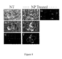
- FIG. 8 is seven copies of photos about ZnO NP inducing apoptosis in Jurkat T cells.
- FIG. 9 is three copies of photos about detection of apoptotic morphological changes in Jurkat T cells treated with ZnO NP.
- ZnO nanoparticles were synthesized in diethylene glycol (DEG) by forced hydrolysis of zinc acetate at 160° C. as previously described and size control achieved by optimizing the hydrolysis ratio.
- the ZnO NPs were separated from DEG via centrifugation (15,000 rpm), washed with ethanol several times and dried to obtain a nanoscale powder sample.
- the ZnO chemical phase, crystallite size (8-13 nm) and shape were confirmed using x-ray diffraction (XRD), transmission electron microscopy (TEM) and spectrophotometry.
- the nanoparticles were then reconstituted in phosphate buffered saline (PBS) solution. After reconstitution, NPs were sonicated for 10 min and immediately vortexed before addition to cell cultures.
- PBS phosphate buffered saline
- FITC-ZnO FITC encapsulated ZnO particles
- PBMC Peripheral blood mononuclear cells
- CD4 + cells were obtained by negative immunonmagnetic selection per manufacturer's instructions using a cocktail of antibodies against CD45RO, CD8, CD19, CD14, CD16, CD56, CD8, and glycophorin A (StemCell Technologies, Vancouver, B.C.) with collection of unlabeled T cells (typically >96% CD4 + and >90% viable as assessed by flow cytometry).
- Purified CD4 + cells were cultured in RPMI/10% FBS at 1 ⁇ 10 6 cells/ml in 200 ⁇ L total volume in 96-well microtiter plates.
- the Jurkat and Hut-78 T cell lines were cultured in RPMI 1640 supplemented with 10% FBS (Jurkat) or 20% FBS (Hut-78) and 2 mM L-glutamine, 1.5 g/L sodium bicarbonate, 4.5 g/L glucose, 10 mM HEPES, and 1.0 mM sodium pyruvate.
- Cells were maintained in log phase at 37° C. and 5% CO 2 and seeded at 1 ⁇ 10 5 cells/well in 96-well plates for individual experiments.
- Jurkat cells were seeded at 5 ⁇ 10 4 cells/well and primary T cells were seeded at 1 ⁇ 10 5 cells into the same well.
- T cells were dually stained with fluoroscein isothiocyanate labeled antibodies (anti-CD4 for primary T cells and anti-HLA ABC for T cancer cell lines) followed by treatment with 50 ⁇ g/mL propidium iodide (PI) to monitor losses in membrane integrity.
- PI propidium iodide
- fluorescent CountBright counting beads Invitrogen, Carlsbad, Calif. were added to samples to enable determinations of absolute cell numbers, and changes in PI staining used to quantify cell death. Nanoparticles were excluded from analysis based on absence of fluorescence signal and light forward scatter (FS) and side scatter (SSC) characteristics.
- a second viability assay the LIVE/DEAD viability assay for mammalian cells (Invitrogen, Eugene, Oreg.) was used to verify results.
- Per manufacturer's protocol for flow cytometry cells were dually stained with two fluorescently labeled probes that enable the simultaneous determination of live and dead cells in a sample.
- Calcein AM was used to stain live cells as it fluoresces only when cleaved by intracellular esterases and EthD-1 was used to identify dead/dying cells as it exclusively enters cells with disrupted cell membranes.
- FS and SSC of Jurkat cells were distinguished from each other using differential gating based on their differing and non-overlapping light scattering properties indicative of size (FS) and granularity (SSC) between the two cell types.
- FS and SSC of Jurkat cells was ⁇ 2.2 and 3.2 times greater than for primary T cells, respectively.
- DCFH-DA reactive oxygen species
- the white blood cells were then resuspended in phenol red-free RPMI a 1 ⁇ 10 6 cells/mL and treated with 13 nm ZnO NP. After 18 h of treatment, cultures were loaded with 5 ⁇ M of DCFH-DA for 20 min and ROS production evaluated using flow cytometry as previously described. To ensure cells were capable of ROS production, control samples were activated with 25 ng/mL of PMA for 1 h after loading with DCFH-DA.
- White blood cell populations i.e. T lymphocytes and monocytes
- Jurkat leukemia cells were seeded in a 96-well plate at 0.2 mL per well at a concentration of 5 ⁇ 10 5 cells/mL.
- a stock solution of N-Acetyl Cysteine (NAC, Sigma Aldrich) was made in sterile nanopure water and added to cells at 5 mM or 10 mM for 1 h.
- NAC pretreatment cells were cultured with 0.3-0.5 mM ZnO NP for 24 h. Viability was determined by PI exclusion and flow cytometry with fluorescent CountBright counting beads (Invitrogen, Carlsbad, Calif.) added to samples to enable determination of absolute cell numbers.
- ZnO NP induced cytotoxicity was evaluated by confocal microscopy using two different staining techniques; acridine orange and Vybrant® Apoptosis Assay Kit #2—AlexaFluor® annexin V/propidium iodide by Invitrogen (Eugene, Oreg.). Acridine orange stains double stranded DNA and allows for visualization of nuclear morphology. Invitrogen's Vybrant annexin V assay makes use of two different fluorescently labeled probes, annexin V and PI, to differentiate between live, necrotic and apoptotic cells.
- Jurkat T cells were suspended in complete RPMI-1640 medium and plated at 5 ⁇ 10 5 on poly-d lysine coated glass bottom culture dishes (P35GC-1.5 mm-14 mm-C) supplied by MatTek Corporation (Ashland, Mass.). Samples were left untreated, treated with 0.3 mM ZnO NP, or with 100 nM okadaic acid as a positive control for apoptosis. Following a 20 h incubation at 37° C.
- FIGS. 1 , 3 A, and 4 All data was analyzed using SAS, Inc. software (Cary, N.C.). Data for FIGS. 1 , 3 A, and 4 were analyzed using repeated measures of valiance with post hoc comparisons and significance levels defined as p ⁇ 0.05. Repeated measures of variance analyses were used when two or more measurements of the same type were made on the same subject to determine statistical differences between the means and allow within-subject variation to be separated from between-subject variation. Data for FIGS. 3B and 7 were analyzed using a two-way analysis of variance (ANOVA) to test for statistical significance of the model and post hoc comparisons were used to test for statistically significant effects of treatment on cell viability (p ⁇ 0.05).
- ANOVA analysis of variance
- Activated T Cells are Preferentially Killed by ZnO NP
- toxicity of ZnO NP occurs in a cell dependent manner. For example, both gram negative and grain positive bacteria are killed at substantially lower ZnO NP concentrations than human T cells.
- toxicity effects were determined in resting primary human T cells and compared to cells activated through the T cell receptor (TCR).
- TCR T cell receptor
- Normal peripheral blood CD4 + T cells were isolated using negative immunomagnetic selection and either activated with stimulatory TCR antibodies (anti-CD3), costimulated with anti-CD3/anti-CD28, or left unactivated.
- PI propidium iodide
- FIG. 1 ZnO NP toxicity on unactivated versus activated primary human CD4 + T cells.
- the partial activation status of these cells was verified by 38% ⁇ 6.2% of the cells expressing membrane CD40L.
- FITC-encapsulated ZnO NP (FITC-ZnO-NP) were prepared as described and their fluorescence properties used to monitor cell uptake/association.
- Primary CD4 + T cells were either left unactivated or activated with CD3/CD28 antibodies and treated with 5 mM FITC-ZnO-NP for 4 h. Dual color flow cytometry was used to analyze changes in the FITC-NP signal on gated CD4 + T cells.
- T cells the activation of T cells promotes NP association.
- Primary CD4 + T cells (>96% purity) were left untreated or activated with immobilized CD3/CD28 antibodies and cultures concurrently treated with 5 mM FITC encapsulated ZnO NP for 4 h. Cells were stained using an ECD-labeled CD4 Ab. Using flow cytometry, 10,000 events gated on CD4 + cells were analyzed for changes in FITC fluorescence. Data from a representative histogram is presented with panel A) showing resting T cells cultured with FITC encapsulated NP and panel B) showing activated T cells cultured with FITC encapsulated NP. Inset numbers depict the percentage of FITC positive cells.
- resting T cells had a low NP associated fluorescence signal (12.5% FITC positive) while a substantially greater FITC signal (48.4% positive) was observed for activated T cells.
- a dose-dependent uptake of FITC-ZnO-NP was noted with greater NP association at 5 mM compared to 1 mM concentrations ( ⁇ 3.3 greater attachment at 5 mM; FIG. 2C ).
- NP labeling did not appear to reflect a generalized increase in membrane permeability as no PI uptake indicative of cytotoxicity was observed following 4 h of exposure with 5 mM FITC-ZnO-NP.
- our recent confocal microscopic studies have demonstrated FITC-ZnO-NP uptake and intracellular localization by human T cells.
- FIG. 3 differential cytotoxic effects of ZnO NP on cancerous T cell lines and primary T cells.
- A) Jurkat, Hut-78 T cell lines, or normal primary T cells were treated with varying concentrations of ZnO nanopaiticles for 22-24 h and viability determined by monitoring PI uptake using flow cytometry as described for FIG. 1 .
- Data from seven (Jurkat), three (Hut-78), and four (normal CD4 + T cells) independent experiments is presented and error bars depict standard error. Data was analyzed using a repeated measures ANOVA and model based means post test.
- NP concentrations e.g. 0.5 mM
- Jurkat T cells were co-cultured with primary CD4 + T cells, treated with various concentrations of ZnO NP for 24 h, and cell viability assessed by PI uptake.
- FIG. 4 viability effects of ZnO NP on co-cultures of cancerous and normal T cells.
- Individual wells in a 96-well plate were seeded with Jurkat and primary T cells and treated with various concentrations of ZnO nanopaiticles for 22-24 h. Viability was determined by monitoring PI uptake using flow cytometry. Data from three independent experiments is presented and error bars depict standard error. Statistical analysis was performed using a repeated measures ANOVA and model based means post hoc test. Significance levels were defined as p ⁇ 0.05 and are indicated by an asterisk.
- FIG. 4 viability effects of ZnO NP on co-cultures of cancerous and normal T cells.
- FIG. 5 kinetics of ZnO NP toxicity on immortalized and primary human T cells.
- both primary T cells and Jurkat cells displayed similar kinetics with appreciable lose of cell viability beginning as early as 8 h post treatment and full toxicity effects requiring a longer treatment period of 24 h.
- FIG. 6 cellular production of ROS following ZnO NP exposure.
- ROS generation was evaluated in primary T cells and monocytes and in the transformed Hut-78 T cell line following 18-24 h of ZnO NP exposure using the oxidation sensitive dye DCFH-DA and flow cytometry.
- a & C Representative histograms depicting ROS production in primary T cells and monocytes. Assays were performed using freshly obtained whole blood in which red blood cells were removed following NH 4 Cl lysis. T lymphocytes and monocytes were gated based on staining with fluorescently labeled CD3 and CD14 antibodies and the oxidation product of DCFH-DA detected using the FL1 detector.
- FIG. 7 quenching of ROS protects against NP-mediated cytotoxicity.
- Jurkat cells were pretreated with 5-10 mM N-acetyl cysteine for 60 min and then treated with 0.3-0.5 mM ZnO NP. After 24 h, cell viability was determined using propidium iodide exclusion and flow cytometry. Data from a representative experiment performed in triplicate is presented with error bars depicting standard error. A two way analysis of variance was performed followed by a model based means test to show significant differences in means of cell viability (%) between treatments (asterisk denotes p ⁇ 0.0001).
- FIG. 7 shows that NAC has significant effects to prevent NP-induced cytotoxicity with rescue being observed at both NP concentrations tested.
- ZnO NP induce apoptosis in Jurkat T cells.
- Cells were left untreated, treated with 0.3 mM ZnO NP for 20 h, or treated with 100 nM okadaic acid for 20 h (positive control) and stained with a green fluorescent annexin V antibody to detect apoptotic membranes and the red fluorescent dye PI to detect permeable membranes using the Vybrant apoptosis assay kit #2 (Molecular Probes). Cells were visualized by confocal microscopy and representative images are shown.
- cells treated with ZnO NP stain positive with the apoptotic marker eg. two out of the four cells in panel F and four out of five cells in panel G show green fluorescence only).
- NP-induced apoptosis Similar cell cultures were stained with the DNA dye, acridine orange, which is used to detect apoptotic morphology characterized by nuclear fragmentation, cellular shrinkage, and chromatin condensation.
- FIG. 9 detection of apoptotic morphological changes in Jurkat cells treated with ZnO NP.
- Cells were left untreated (A), or treated with 100 nM okadaic acid for 20 h as a positive control for apoptosis (B), or treated with 0.3 mM ZnO NP for 20 h (C) and stained with acridine orange and visualized by fluorescent microscopy.
- Arrows indicate typical apoptotic cells characterized by a shrunken appearance and condensed or fragmented nuclei.
- FIG. 9 these morphological changes were observed in NP treated cultures and cells treated with the apoptosis inducer okadaic acid, but not in control samples. Collectively, these results indicate that ZnO NP induce apoptosis in Jurkat T cells.
- Therapeutic indices of ⁇ 10 have been reported for both doxorubicin and carboplatin against a variety of tumors including acute myelogenous leukemia, non-Hodgkin's lymphoma, ovarian, and other solid tumors.
- the inherent differential toxicity of ZnO NP against rapidly dividing cancer cells raises exciting opportunities for their potential use as anticancer agents, and the selectivity of these nanomaterials can be expected to be even further enhanced by design by linking tumor targeting ligands such as monoclonal antibodies, peptides, and small molecules to tumor-associated proteins, or by using NP for drug delivery.
- self-reactive T cells are a pathogenic subset underlying disease processes and exist in a predominately activated state as they are continually exposed to specific antigen present in normal body tissue. Because only a very small percentage of the total T cell repertoire are self-reactive and pathogenic in autoimmunity, the ability of identical concentrations of ZnO NP to preferentially induce cytotoxicity in self-reactive activated T cells while leaving the unactivated T cell repertoire largely intact and immunity uncompromised against future infections is an incredibly attractive approach which may ultimately become feasible.
- ZnO NP hold promise in this novel biomedical application, especially if their selectivity against self-reactive pathogenic cells can be improved by the covalent attachment of antibodies specific to proteins expressed predominantly on activated T cells such as CD40L and OX40.
Abstract
Description
Claims (21)
Priority Applications (1)
| Application Number | Priority Date | Filing Date | Title |
|---|---|---|---|
| US12/235,415 US8187638B2 (en) | 2007-09-22 | 2008-09-22 | Preferential killing of cancer cells and activated human T cells using ZnO nanoparticles |
Applications Claiming Priority (2)
| Application Number | Priority Date | Filing Date | Title |
|---|---|---|---|
| US97446007P | 2007-09-22 | 2007-09-22 | |
| US12/235,415 US8187638B2 (en) | 2007-09-22 | 2008-09-22 | Preferential killing of cancer cells and activated human T cells using ZnO nanoparticles |
Publications (2)
| Publication Number | Publication Date |
|---|---|
| US20090136580A1 US20090136580A1 (en) | 2009-05-28 |
| US8187638B2 true US8187638B2 (en) | 2012-05-29 |
Family
ID=40468815
Family Applications (1)
| Application Number | Title | Priority Date | Filing Date |
|---|---|---|---|
| US12/235,415 Expired - Fee Related US8187638B2 (en) | 2007-09-22 | 2008-09-22 | Preferential killing of cancer cells and activated human T cells using ZnO nanoparticles |
Country Status (2)
| Country | Link |
|---|---|
| US (1) | US8187638B2 (en) |
| WO (1) | WO2009039508A2 (en) |
Families Citing this family (7)
| Publication number | Priority date | Publication date | Assignee | Title |
|---|---|---|---|---|
| US7939560B2 (en) | 2007-09-22 | 2011-05-10 | Boise State University | Fluorescent particles comprising nanoscale ZnO layer and exhibiting cell-specific toxicity |
| WO2009039508A2 (en) | 2007-09-22 | 2009-03-26 | Boise State University | Preferential killing of cancer cells and activated human t cells using zno nanoparticles |
| US8507556B2 (en) | 2007-09-22 | 2013-08-13 | Boise State University | Fluorescent particles comprising nanoscale ZnO layer and exhibiting cell-specific toxicity |
| CN102759796B (en) * | 2012-05-09 | 2014-07-23 | 中国科学院上海技术物理研究所 | Optical calibration technology of multi-degree-of-freedom imaging optical system for computed-generated holographic multi-point instantaneous positioning |
| CN107022571A (en) * | 2017-05-18 | 2017-08-08 | 山西大学 | A kind of method of transfected Jurkat cells |
| US20200330616A1 (en) * | 2017-11-13 | 2020-10-22 | POLITECNICO Dl TORINO | Biomimetic non-immunogenic nanoassembly for the antitumor therapy |
| CN113616787A (en) * | 2021-08-12 | 2021-11-09 | 国药集团动物保健股份有限公司 | Litchi branch-shaped nano zinc oxide cluster adjuvant, preparation method thereof and application of litchi branch-shaped nano zinc oxide cluster adjuvant as vaccine adjuvant |
Citations (6)
| Publication number | Priority date | Publication date | Assignee | Title |
|---|---|---|---|---|
| US6165440A (en) * | 1997-07-09 | 2000-12-26 | Board Of Regents, The University Of Texas System | Radiation and nanoparticles for enhancement of drug delivery in solid tumors |
| US20040062817A1 (en) * | 2000-10-12 | 2004-04-01 | Peshoff Mickey L. | Wound healing compound |
| US20070015226A1 (en) * | 2005-07-12 | 2007-01-18 | Fuji Photo Film Co., Ltd. | Method for detecting cancer using metal-oxide or metal-sulfide nanoparticle fluorescent material |
| US20080317768A1 (en) * | 2007-06-21 | 2008-12-25 | Boeing Company | Bioconjugated nanoparticles |
| WO2009039508A2 (en) | 2007-09-22 | 2009-03-26 | Boise State University | Preferential killing of cancer cells and activated human t cells using zno nanoparticles |
| US20090137666A1 (en) * | 2007-09-22 | 2009-05-28 | Boise State University | FLUORESCENT PARTICLES COMPRISING NANOSCALE ZnO LAYER AND EXHIBITING CELL-SPECIFIC TOXICITY |
-
2008
- 2008-09-22 WO PCT/US2008/077252 patent/WO2009039508A2/en active Application Filing
- 2008-09-22 US US12/235,415 patent/US8187638B2/en not_active Expired - Fee Related
Patent Citations (7)
| Publication number | Priority date | Publication date | Assignee | Title |
|---|---|---|---|---|
| US6165440A (en) * | 1997-07-09 | 2000-12-26 | Board Of Regents, The University Of Texas System | Radiation and nanoparticles for enhancement of drug delivery in solid tumors |
| US20040062817A1 (en) * | 2000-10-12 | 2004-04-01 | Peshoff Mickey L. | Wound healing compound |
| US20070015226A1 (en) * | 2005-07-12 | 2007-01-18 | Fuji Photo Film Co., Ltd. | Method for detecting cancer using metal-oxide or metal-sulfide nanoparticle fluorescent material |
| US20080317768A1 (en) * | 2007-06-21 | 2008-12-25 | Boeing Company | Bioconjugated nanoparticles |
| WO2009039508A2 (en) | 2007-09-22 | 2009-03-26 | Boise State University | Preferential killing of cancer cells and activated human t cells using zno nanoparticles |
| US20090137666A1 (en) * | 2007-09-22 | 2009-05-28 | Boise State University | FLUORESCENT PARTICLES COMPRISING NANOSCALE ZnO LAYER AND EXHIBITING CELL-SPECIFIC TOXICITY |
| WO2009079056A2 (en) | 2007-09-22 | 2009-06-25 | Boise State University | Fluorescent particles comprising nanoscale zno layer and exhibiting cell-specific toxicity |
Non-Patent Citations (7)
| Title |
|---|
| Ciardiello et al, EGFR antagonists in cancer treatment, The new england journal of medicine, pp. 1160-1174, Mar. 13, 2008. * |
| Hagadone, Zach, "Nano cells on the attack: BSU researchers find cancer-killing particles", Sep. 8, 2008. |
| Hays, Reddy, Graces, Engelhard, Shutthanandan, Luo, Xu, Giles, Wang, Thevuthasa, Punnoose, "Effect of Co doping on the structural, optical and magnetic properties of ZnO nanoparticles", Apr. 27, 2007, Journal of Physics: Condensed Matter (2007) 266203 (24pp), UK, published Jun. 7, 2007. |
| International Preliminary Report on Patentability, PCT/US2008/077252, Appilicant: Boise State University. |
| International Search Report, PCT/US2008/077284, Sep. 1, 2009 Appilicant: Boise State University. |
| Reddy, Feris, Wingett, Hanley, Punnoose, "Selective toxicity of zinc oxide nanoparticles to prokaryotic and eukaryotic systems", Applied Physics Letters, May 24, 2007, 90, 213902, U.S. |
| Wang, Wingett, Engelard, Feris, Reddy, Turner, Layne, Hanley, Bell, Tenne, Wang, Punnoose, "Fluorescent dye encapsulated ZnO particles with cell-specific toxicity for potential use in biomedical applications", Journal of Material Science: Materials in Medicine (2009) 20:11-22, U.S., published online Jul. 24, 2008. |
Also Published As
| Publication number | Publication date |
|---|---|
| WO2009039508A2 (en) | 2009-03-26 |
| US20090136580A1 (en) | 2009-05-28 |
| WO2009039508A9 (en) | 2009-10-22 |
Similar Documents
| Publication | Publication Date | Title |
|---|---|---|
| Hanley et al. | Preferential killing of cancer cells and activated human T cells using ZnO nanoparticles | |
| US8187638B2 (en) | Preferential killing of cancer cells and activated human T cells using ZnO nanoparticles | |
| Hanley et al. | The influences of cell type and ZnO nanoparticle size on immune cell cytotoxicity and cytokine induction | |
| Junqueira et al. | γδ T cells suppress Plasmodium falciparum blood-stage infection by direct killing and phagocytosis | |
| Gelderman et al. | Adverse effects of fullerenes on endothelial cells: Fullerenol C60 (OH) 24 induced tissue factor and ICAM-1 membrane expression and apoptosis in vitro | |
| Ma et al. | Monitoring of the enzymatic degradation of protein corona and evaluating the accompanying cytotoxicity of nanoparticles | |
| Liz et al. | Silver nanoparticles rapidly induce atypical human neutrophil cell death by a process involving inflammatory caspases and reactive oxygen species and induce neutrophil extracellular traps release upon cell adhesion | |
| Pinton et al. | Targeting of immunosuppressive myeloid cells from glioblastoma patients by modulation of size and surface charge of lipid nanocapsules | |
| Chattopadhyay et al. | Chitosan-modified cobalt oxide nanoparticles stimulate TNF-α-mediated apoptosis in human leukemic cells | |
| Laakko et al. | Versatility of merocyanine 540 for the flow cytometric detection of apoptosis in human and murine cells | |
| Liang et al. | Au@ Pt nanoparticles as catalase mimics to attenuate tumor hypoxia and enhance immune cell-mediated cytotoxicity | |
| Li et al. | Red blood cell-mimic nanocatalyst triggering radical storm to augment cancer immunotherapy | |
| Tamkovich et al. | Blood circulating exosomes contain distinguishable fractions of free and cell-surface-associated vesicles | |
| Lin et al. | Chitosan nanoparticles strengthen Vγ9Vδ2 T-cell cytotoxicity through upregulation of killing molecules and cytoskeleton polarization | |
| Deng et al. | Inhibition of caveolae contributes to propofol preconditioning-suppressed microvesicles release and cell injury by hypoxia-reoxygenation | |
| Wurzer et al. | Intrinsic resistance of chronic lymphocytic leukemia cells to NK cell-mediated lysis can be overcome in vitro by pharmacological inhibition of cdc42-induced actin cytoskeleton remodeling | |
| Comparetti et al. | Cancer cell membrane-derived nanoparticles improve the activity of gemcitabine and paclitaxel on pancreatic cancer cells and coordinate immunoregulatory properties on professional antigen-presenting cells | |
| He et al. | Metabolic reprogramming of NK cells by black phosphorus quantum dots potentiates cancer immunotherapy | |
| CN116004636A (en) | Differential CD47 aptamer not binding red blood cells and application thereof | |
| Feng et al. | An acid-responsive MOF nanomedicine for augmented anti-tumor immunotherapy via a metal ion interference-mediated pyroptotic pathway | |
| Wang et al. | Multi-walled carbon nanotubes do not impair immune functions of dendritic cells | |
| Christiansen et al. | The random co-polymer glatiramer acetate rapidly kills primary human leukocytes through sialic-acid-dependent cell membrane damage | |
| Lim et al. | Activation of mouse protease‐activated receptor‐2 induces lymphocyte adhesion and generation of reactive oxygen species | |
| Huang et al. | Pegylated liposomal mitoxantrone modulates tumor immune landscape to boost PD-L1 blockade therapy | |
| Le Roux et al. | Fourier Transform Infrared spectroscopy discloses different types of cell death in flow cytometrically sorted cells |
Legal Events
| Date | Code | Title | Description |
|---|---|---|---|
| AS | Assignment |
Owner name: BOISE STATE UNIVERSITY, IDAHO Free format text: ASSIGNMENT OF ASSIGNORS INTEREST;ASSIGNORS:PUNNOOSE, ALEX;KONGARA, MADHUSAN R.;WINGETT, DENISE;REEL/FRAME:021982/0577;SIGNING DATES FROM 20080716 TO 20081201 Owner name: BOISE STATE UNIVERSITY, IDAHO Free format text: ASSIGNMENT OF ASSIGNORS INTEREST;ASSIGNORS:PUNNOOSE, ALEX;KONGARA, MADHUSAN R.;WINGETT, DENISE;REEL/FRAME:021982/0584;SIGNING DATES FROM 20080716 TO 20081201 Owner name: BOISE STATE UNIVERSITY, IDAHO Free format text: ASSIGNMENT OF ASSIGNORS INTEREST;ASSIGNORS:PUNNOOSE, ALEX;KONGARA, MADHUSAN R.;WINGETT, DENISE;SIGNING DATES FROM 20080716 TO 20081201;REEL/FRAME:021982/0577 Owner name: BOISE STATE UNIVERSITY, IDAHO Free format text: ASSIGNMENT OF ASSIGNORS INTEREST;ASSIGNORS:PUNNOOSE, ALEX;KONGARA, MADHUSAN R.;WINGETT, DENISE;SIGNING DATES FROM 20080716 TO 20081201;REEL/FRAME:021982/0584 |
|
| AS | Assignment |
Owner name: KABUSHIKI KAISHA TOSHIBA, JAPAN Free format text: ASSIGNMENT OF ASSIGNORS INTEREST;ASSIGNORS:MOTOE, HIRONORI;KOIDE, SHINGO;REEL/FRAME:021989/0248 Effective date: 20081031 |
|
| REMI | Maintenance fee reminder mailed | ||
| LAPS | Lapse for failure to pay maintenance fees | ||
| STCH | Information on status: patent discontinuation |
Free format text: PATENT EXPIRED DUE TO NONPAYMENT OF MAINTENANCE FEES UNDER 37 CFR 1.362 |
|
| FP | Expired due to failure to pay maintenance fee |
Effective date: 20160529 |
|
| CC | Certificate of correction |