US8426571B2 - Nucleic acids and corresponding proteins entitled 202P5A5 useful in treatment and detection of cancer - Google Patents
Nucleic acids and corresponding proteins entitled 202P5A5 useful in treatment and detection of cancer Download PDFInfo
- Publication number
- US8426571B2 US8426571B2 US13/021,202 US201113021202A US8426571B2 US 8426571 B2 US8426571 B2 US 8426571B2 US 201113021202 A US201113021202 A US 201113021202A US 8426571 B2 US8426571 B2 US 8426571B2
- Authority
- US
- United States
- Prior art keywords
- protein
- cancer
- nucleotide
- peptide
- amino acid
- Prior art date
- Legal status (The legal status is an assumption and is not a legal conclusion. Google has not performed a legal analysis and makes no representation as to the accuracy of the status listed.)
- Expired - Fee Related, expires
Links
- 0 *.*.*.*.*.*.*.*.* Chemical compound *.*.*.*.*.*.*.*.* 0.000 description 25
Images


Classifications
-
- C—CHEMISTRY; METALLURGY
- C12—BIOCHEMISTRY; BEER; SPIRITS; WINE; VINEGAR; MICROBIOLOGY; ENZYMOLOGY; MUTATION OR GENETIC ENGINEERING
- C12N—MICROORGANISMS OR ENZYMES; COMPOSITIONS THEREOF; PROPAGATING, PRESERVING, OR MAINTAINING MICROORGANISMS; MUTATION OR GENETIC ENGINEERING; CULTURE MEDIA
- C12N5/00—Undifferentiated human, animal or plant cells, e.g. cell lines; Tissues; Cultivation or maintenance thereof; Culture media therefor
- C12N5/06—Animal cells or tissues; Human cells or tissues
- C12N5/0602—Vertebrate cells
- C12N5/0693—Tumour cells; Cancer cells
-
- A—HUMAN NECESSITIES
- A61—MEDICAL OR VETERINARY SCIENCE; HYGIENE
- A61K—PREPARATIONS FOR MEDICAL, DENTAL OR TOILETRY PURPOSES
- A61K47/00—Medicinal preparations characterised by the non-active ingredients used, e.g. carriers or inert additives; Targeting or modifying agents chemically bound to the active ingredient
- A61K47/50—Medicinal preparations characterised by the non-active ingredients used, e.g. carriers or inert additives; Targeting or modifying agents chemically bound to the active ingredient the non-active ingredient being chemically bound to the active ingredient, e.g. polymer-drug conjugates
- A61K47/51—Medicinal preparations characterised by the non-active ingredients used, e.g. carriers or inert additives; Targeting or modifying agents chemically bound to the active ingredient the non-active ingredient being chemically bound to the active ingredient, e.g. polymer-drug conjugates the non-active ingredient being a modifying agent
- A61K47/68—Medicinal preparations characterised by the non-active ingredients used, e.g. carriers or inert additives; Targeting or modifying agents chemically bound to the active ingredient the non-active ingredient being chemically bound to the active ingredient, e.g. polymer-drug conjugates the non-active ingredient being a modifying agent the modifying agent being an antibody, an immunoglobulin or a fragment thereof, e.g. an Fc-fragment
- A61K47/6835—Medicinal preparations characterised by the non-active ingredients used, e.g. carriers or inert additives; Targeting or modifying agents chemically bound to the active ingredient the non-active ingredient being chemically bound to the active ingredient, e.g. polymer-drug conjugates the non-active ingredient being a modifying agent the modifying agent being an antibody, an immunoglobulin or a fragment thereof, e.g. an Fc-fragment the modifying agent being an antibody or an immunoglobulin bearing at least one antigen-binding site
- A61K47/6851—Medicinal preparations characterised by the non-active ingredients used, e.g. carriers or inert additives; Targeting or modifying agents chemically bound to the active ingredient the non-active ingredient being chemically bound to the active ingredient, e.g. polymer-drug conjugates the non-active ingredient being a modifying agent the modifying agent being an antibody, an immunoglobulin or a fragment thereof, e.g. an Fc-fragment the modifying agent being an antibody or an immunoglobulin bearing at least one antigen-binding site the antibody targeting a determinant of a tumour cell
-
- A—HUMAN NECESSITIES
- A61—MEDICAL OR VETERINARY SCIENCE; HYGIENE
- A61P—SPECIFIC THERAPEUTIC ACTIVITY OF CHEMICAL COMPOUNDS OR MEDICINAL PREPARATIONS
- A61P35/00—Antineoplastic agents
-
- A—HUMAN NECESSITIES
- A61—MEDICAL OR VETERINARY SCIENCE; HYGIENE
- A61P—SPECIFIC THERAPEUTIC ACTIVITY OF CHEMICAL COMPOUNDS OR MEDICINAL PREPARATIONS
- A61P37/00—Drugs for immunological or allergic disorders
- A61P37/02—Immunomodulators
- A61P37/04—Immunostimulants
-
- A—HUMAN NECESSITIES
- A61—MEDICAL OR VETERINARY SCIENCE; HYGIENE
- A61P—SPECIFIC THERAPEUTIC ACTIVITY OF CHEMICAL COMPOUNDS OR MEDICINAL PREPARATIONS
- A61P43/00—Drugs for specific purposes, not provided for in groups A61P1/00-A61P41/00
-
- C—CHEMISTRY; METALLURGY
- C07—ORGANIC CHEMISTRY
- C07K—PEPTIDES
- C07K14/00—Peptides having more than 20 amino acids; Gastrins; Somatostatins; Melanotropins; Derivatives thereof
- C07K14/435—Peptides having more than 20 amino acids; Gastrins; Somatostatins; Melanotropins; Derivatives thereof from animals; from humans
- C07K14/46—Peptides having more than 20 amino acids; Gastrins; Somatostatins; Melanotropins; Derivatives thereof from animals; from humans from vertebrates
- C07K14/47—Peptides having more than 20 amino acids; Gastrins; Somatostatins; Melanotropins; Derivatives thereof from animals; from humans from vertebrates from mammals
-
- G—PHYSICS
- G01—MEASURING; TESTING
- G01N—INVESTIGATING OR ANALYSING MATERIALS BY DETERMINING THEIR CHEMICAL OR PHYSICAL PROPERTIES
- G01N33/00—Investigating or analysing materials by specific methods not covered by groups G01N1/00 - G01N31/00
- G01N33/48—Biological material, e.g. blood, urine; Haemocytometers
- G01N33/50—Chemical analysis of biological material, e.g. blood, urine; Testing involving biospecific ligand binding methods; Immunological testing
- G01N33/53—Immunoassay; Biospecific binding assay; Materials therefor
- G01N33/574—Immunoassay; Biospecific binding assay; Materials therefor for cancer
- G01N33/57484—Immunoassay; Biospecific binding assay; Materials therefor for cancer involving compounds serving as markers for tumor, cancer, neoplasia, e.g. cellular determinants, receptors, heat shock/stress proteins, A-protein, oligosaccharides, metabolites
-
- A—HUMAN NECESSITIES
- A01—AGRICULTURE; FORESTRY; ANIMAL HUSBANDRY; HUNTING; TRAPPING; FISHING
- A01K—ANIMAL HUSBANDRY; CARE OF BIRDS, FISHES, INSECTS; FISHING; REARING OR BREEDING ANIMALS, NOT OTHERWISE PROVIDED FOR; NEW BREEDS OF ANIMALS
- A01K2217/00—Genetically modified animals
- A01K2217/05—Animals comprising random inserted nucleic acids (transgenic)
-
- A—HUMAN NECESSITIES
- A01—AGRICULTURE; FORESTRY; ANIMAL HUSBANDRY; HUNTING; TRAPPING; FISHING
- A01K—ANIMAL HUSBANDRY; CARE OF BIRDS, FISHES, INSECTS; FISHING; REARING OR BREEDING ANIMALS, NOT OTHERWISE PROVIDED FOR; NEW BREEDS OF ANIMALS
- A01K2217/00—Genetically modified animals
- A01K2217/07—Animals genetically altered by homologous recombination
- A01K2217/075—Animals genetically altered by homologous recombination inducing loss of function, i.e. knock out
-
- A—HUMAN NECESSITIES
- A61—MEDICAL OR VETERINARY SCIENCE; HYGIENE
- A61K—PREPARATIONS FOR MEDICAL, DENTAL OR TOILETRY PURPOSES
- A61K39/00—Medicinal preparations containing antigens or antibodies
- A61K2039/505—Medicinal preparations containing antigens or antibodies comprising antibodies
-
- A—HUMAN NECESSITIES
- A61—MEDICAL OR VETERINARY SCIENCE; HYGIENE
- A61K—PREPARATIONS FOR MEDICAL, DENTAL OR TOILETRY PURPOSES
- A61K38/00—Medicinal preparations containing peptides
-
- C—CHEMISTRY; METALLURGY
- C07—ORGANIC CHEMISTRY
- C07K—PEPTIDES
- C07K2319/00—Fusion polypeptide
-
- Y—GENERAL TAGGING OF NEW TECHNOLOGICAL DEVELOPMENTS; GENERAL TAGGING OF CROSS-SECTIONAL TECHNOLOGIES SPANNING OVER SEVERAL SECTIONS OF THE IPC; TECHNICAL SUBJECTS COVERED BY FORMER USPC CROSS-REFERENCE ART COLLECTIONS [XRACs] AND DIGESTS
- Y02—TECHNOLOGIES OR APPLICATIONS FOR MITIGATION OR ADAPTATION AGAINST CLIMATE CHANGE
- Y02A—TECHNOLOGIES FOR ADAPTATION TO CLIMATE CHANGE
- Y02A50/00—TECHNOLOGIES FOR ADAPTATION TO CLIMATE CHANGE in human health protection, e.g. against extreme weather
- Y02A50/30—Against vector-borne diseases, e.g. mosquito-borne, fly-borne, tick-borne or waterborne diseases whose impact is exacerbated by climate change
Definitions
- the invention described herein relates to genes and their encoded proteins, termed 202P5A5 and variants thereof, expressed in certain cancers, and to diagnostic and therapeutic methods and compositions useful in the management of cancers that express 202P5A5.
- Cancer is the second leading cause of human death next to coronary disease. Worldwide, millions of people die from cancer every year. In the United States alone, as reported by the American Cancer Society, cancer causes the death of well over a half-million people annually, with over 1.2 million new cases diagnosed per year. While deaths from heart disease have been declining significantly, those resulting from cancer generally are on the rise. In the early part of the next century, cancer is predicted to become the leading cause of death.
- carcinomas of the lung, prostate, breast, colon, pancreas, and ovary represent the primary causes of cancer death. These and virtually all other carcinomas share a common lethal feature. With very few exceptions, metastatic disease from a carcinoma is fatal. Moreover, even for those cancer patients who initially survive their primary cancers, common experience has shown that their lives are dramatically altered. Many cancer patients experience strong anxieties driven by the awareness of the potential for recurrence or treatment failure. Many cancer patients experience physical debilitations following treatment. Furthermore, many cancer patients experience a recurrence.
- prostate cancer is the fourth most prevalent cancer in men. In North America and Northern Europe, it is by far the most common cancer in males and is the second leading cause of cancer death in men. In the United States alone, well over 30,000 men die annually of this disease—second only to lung cancer. Despite the magnitude of these figures, there is still no effective treatment for metastatic prostate cancer. Surgical prostatectomy, radiation therapy, hormone ablation therapy, surgical castration and chemotherapy continue to be the main treatment modalities. Unfortunately, these treatments are ineffective for many and are often associated with undesirable consequences.
- PSA serum prostate specific antigen
- the LAPC Los Angeles Prostate Cancer
- SCID severe combined immune deficient mice
- More recently identified prostate cancer markers include PCTA-1 (Su et al., 1996, Proc. Natl. Acad. Sci. USA 93: 7252), prostate-specific membrane (PSM) antigen (Pinto et al., Clin Cancer Res 1996 Sep.
- Renal cell carcinoma accounts for approximately 3 percent of adult malignancies. Once adenomas reach a diameter of 2 to 3 cm, malignant potential exists. In the adult, the two principal malignant renal tumors are renal cell adenocarcinoma and transitional cell carcinoma of the renal pelvis or ureter. The incidence of renal cell adenocarcinoma is estimated at more than 29,000 cases in the United States, and more than 11,600 patients died of this disease in 1998. Transitional cell carcinoma is less frequent, with an incidence of approximately 500 cases per year in the United States.
- bladder cancer represents approximately 5 percent in men (fifth most common neoplasm) and 3 percent in women (eighth most common neoplasm). The incidence is increasing slowly, concurrent with an increasing older population. In 1998, there was an estimated 54,500 cases, including 39,500 in men and 15,000 in women. The age-adjusted incidence in the United States is 32 per 100,000 for men and eight per 100,000 in women. The historic male/female ratio of 3:1 may be decreasing related to smoking patterns in women. There were an estimated 11,000 deaths from bladder cancer in 1998 (7,800 in men and 3,900 in women). Bladder cancer incidence and mortality strongly increase with age and will be an increasing problem as the population becomes more elderly.
- bladder cancers recur in the bladder.
- Bladder cancer is managed with a combination of transurethral resection of the bladder (TUR) and intravesical chemotherapy or immunotherapy.
- TUR transurethral resection of the bladder
- the multifocal and recurrent nature of bladder cancer points out the limitations of TUR.
- Most muscle-invasive cancers are not cured by TUR alone. Radical cystectomy and urinary diversion is the most effective means to eliminate the cancer but carry an undeniable impact on urinary and sexual function. There continues to be a significant need for treatment modalities that are beneficial for bladder cancer patients.
- Treatment options for lung and bronchial cancer are determined by the type and stage of the cancer and include surgery, radiation therapy, and chemotherapy. For many localized cancers, surgery is usually the treatment of choice. Because the disease has usually spread by the time it is discovered, radiation therapy and chemotherapy are often needed in combination with surgery. Chemotherapy alone or combined with radiation is the treatment of choice for small cell lung cancer; on this regimen, a large percentage of patients experience remission, which in some cases is long lasting. There is however, an ongoing need for effective treatment and diagnostic approaches for lung and bronchial cancers.
- treatment of breast cancer may involve lumpectomy (local removal of the tumor) and removal of the lymph nodes under the arm; mastectomy (surgical removal of the breast) and removal of the lymph nodes under the arm; radiation therapy; chemotherapy; or hormone therapy.
- lumpectomy local removal of the tumor
- mastectomy surgical removal of the breast
- radiation therapy chemotherapy
- hormone therapy chemotherapy
- two or more methods are used in combination.
- Numerous studies have shown that, for early stage disease, long-term survival rates after lumpectomy plus radiotherapy are similar to survival rates after modified radical mastectomy.
- Significant advances in reconstruction techniques provide several options for breast reconstruction after mastectomy. Recently, such reconstruction has been done at the same time as the mastectomy.
- DCIS ductal carcinoma in situ
- Surgery, radiation therapy, and chemotherapy are treatment options for ovarian cancer.
- Surgery usually includes the removal of one or both ovaries, the fallopian tubes (salpingo-oophorectomy), and the uterus (hysterectomy).
- the fallopian tubes salivary-oophorectomy
- the uterus hematoma-oophorectomy
- pancreatic cancer There were an estimated 28,300 new cases of pancreatic cancer in the United States in 2000. Over the past 20 years, rates of pancreatic cancer have declined in men. Rates among women have remained approximately constant but may be beginning to decline. Pancreatic cancer caused an estimated 28,200 deaths in 2000 in the United States. Over the past 20 years, there has been a slight but significant decrease in mortality rates among men (about ⁇ 0.9% per year) while rates have increased slightly among women.
- pancreatic cancer Surgery, radiation therapy, and chemotherapy are treatment options for pancreatic cancer. These treatment options can extend survival and/or relieve symptoms in many patients but are not likely to produce a cure for most. There is a significant need for additional therapeutic and diagnostic options for pancreatic cancer.
- the present invention relates to a gene, designated 202P5A5, that has now been found to be over-expressed in the cancer(s) listed in Table I.
- Northern blot expression analysis of 202P5A5 gene expression in normal tissues shows a restricted expression pattern in adult tissues.
- the nucleotide ( FIG. 2 ) and amino acid ( FIG. 2 , and FIG. 3 ) sequences of 202P5A5 are provided.
- tissue-related profile of 202P5A5 in normal adult tissues combined with the over-expression observed in the tissues listed in Table I, shows that 202P5A5 is aberrantly over-expressed in at least some cancers, and thus serves as a useful diagnostic, prophylactic, prognostic, and/or therapeutic target for cancers of the tissue(s) such as those listed in Table I.
- the invention provides polynucleotides corresponding or complementary to all or part of the 202P5A5 genes, mRNAs, and/or coding sequences, preferably in isolated form, including polynucleotides encoding 202P5A5-related proteins and fragments of 4, 5, 6, 7, 8, 9, 10, 11, 12, 13, 14, 15, 16, 17, 18, 19, 20, 21, 22, 23, 24, 25, or more than 25 contiguous amino acids; at least 30, 35, 40, 45, 50, 55, 60, 65, 70, 80, 85, 90, 95, 100 or more than 100 contiguous amino acids of a 202P5A5-related protein, as well as the peptides/proteins themselves; DNA, RNA, DNA/RNA hybrids, and related molecules, polynucleotides or oligonucleotides complementary or having at least a 90% homology to the 202P5A5 genes or mRNA sequences or parts thereof, and polynucleotides or oligonucleotides that hybridize to the 202P5A5
- 202P5A5 Recombinant DNA molecules containing 202P5A5 polynucleotides, cells transformed or transduced with such molecules, and host-vector systems for the expression of 202P5A5 gene products are also provided.
- the invention further provides antibodies that bind to 202P5A5 proteins and polypeptide fragments thereof, including polyclonal and monoclonal antibodies, murine and other mammalian antibodies, chimeric antibodies, humanized and fully human antibodies, and antibodies labeled with a detectable marker or therapeutic agent.
- the entire nucleic acid sequence of FIG. 2 is encoded and/or the entire amino acid sequence of FIG. 2 is prepared, either of which are in respective human unit dose forms.
- the invention further provides methods for detecting the presence and status of 202P5A5 polynucleotides and proteins in various biological samples, as well as methods for identifying cells that express 202P5A5.
- a typical embodiment of this invention provides methods for monitoring 202P5A5 gene products in a tissue or hematology sample having or suspected of having some form of growth dysregulation such as cancer.
- the invention further provides various immunogenic or therapeutic compositions and strategies for treating cancers that express 202P5A5 such as cancers of tissues listed in Table I, including therapies aimed at inhibiting the transcription, translation, processing or function of 202P5A5 as well as cancer vaccines.
- the invention provides compositions, and methods comprising them, for treating a cancer that expresses 202P5A5 in a human subject wherein the composition comprises a carrier suitable for human use and a human unit dose of one or more than one agent that inhibits the production or function of 202P5A5.
- the carrier is a uniquely human carrier.
- the agent is a moiety that is immunoreactive with 202P5A5 protein.
- Non-limiting examples of such moieties include, but are not limited to, antibodies (such as single chain, monoclonal, polyclonal, humanized, chimeric, or human antibodies), functional equivalents thereof (whether naturally occurring or synthetic), and combinations thereof.
- the antibodies can be conjugated to a diagnostic or therapeutic moiety.
- the agent is a small molecule as defined herein.
- the agent comprises one or more than one peptide which comprises a cytotoxic T lymphocyte (CTL) epitope that binds an HLA class I molecule in a human to elicit a CTL response to 202P5A5 and/or one or more than one peptide which comprises a helper T lymphocyte (HTL) epitope which binds an HLA class II molecule in a human to elicit an HTL response.
- CTL cytotoxic T lymphocyte
- HTL helper T lymphocyte
- the peptides of the invention may be on the same or on one or more separate polypeptide molecules.
- the agent comprises one or more than one nucleic acid molecule that expresses one or more than one of the CTL or HTL response stimulating peptides as described above.
- the one or more than one nucleic acid molecule may express a moiety that is immunologically reactive with 202P5A5 as described above.
- the one or more than one nucleic acid molecule may also be, or encodes, a molecule that inhibits production of 202P5A5.
- Non-limiting examples of such molecules include, but are not limited to, those complementary to a nucleotide sequence essential for production of 202P5A5 (e.g. antisense sequences or molecules that form a triple helix with a nucleotide double helix essential for 202P5A5 production) or a ribozyme effective to lyse 202P5A5 mRNA.
- HLA Peptide Tables respective to its parental protein, e.g., variant 1, variant 2, etc.
- HLA Peptide Tables respective to its parental protein, e.g., variant 1, variant 2, etc.
- search Peptides in Table VII Generally, a unique Search Peptide is used to obtain HLA peptides of a particular for a particular variant. The position of each Search Peptide relative to its respective parent molecule is listed in Table VII.
- a Search Peptide begins at position “X”, one must add the value “X ⁇ 1” to each position in Tables VIII-XXI and XXII to XLIX to obtain the actual position of the HLA peptides in their parental molecule. For example, if a particular Search Peptide begins at position 150 of its parental molecule, one must add 150-1, i.e., 149 to each HLA peptide amino acid position to calculate the position of that amino acid in the parent molecule.
- One embodiment of the invention comprises an HLA peptide, that occurs at least twice in Tables VIII-XXI and XXII to XLIX collectively, or an oligonucleotide that encodes the HLA peptide.
- Another embodiment of the invention comprises an HLA peptide that occurs at least once in Tables VIII-XXI and at least once in tables XXII to XLIX, or an oligonucleotide that encodes the HLA peptide.
- antibody epitopes which comprise a peptide regions, or an oligonucleotide encoding the peptide region, that has one two, three, four, or five of the following characteristics:
- FIG. 1 The 202P5A5 SSH sequence of 186 nucleotides.
- FIG. 2 A) The cDNA and amino acid sequence of 202P5A5 variant 1 (also called “202P5A5 v.1” or “202P5A5 variant 1”) is shown in FIG. 2A .
- the start methionine is underlined.
- the open reading frame extends from nucleic acid 29-1858 including the stop codon.
- 202P5A5 variant 2 also called “202P5A5 v.2”
- FIG. 2B The cDNA and amino acid sequence of 202P5A5 variant 2 (also called “202P5A5 v.2”) is shown in FIG. 2B .
- the codon for the start methionine is underlined.
- the open reading frame extends from nucleic acid 13-1890 including the stop codon.
- 202P5A5 variant 3 (also called “202P5A5 v.3”) is shown in FIG. 2C .
- the codon for the start methionine is underlined.
- the open reading frame extends from nucleic acid 121-1950 including the stop codon.
- 202P5A5 variant 14 (also called “202P5A5 v.14”) is shown in FIG. 2D .
- the codon for the start methionine is underlined.
- the open reading frame extends from nucleic acid 29-1858 including the stop codon.
- 202P5A5 variant 22 also called “202P5A5 v.22”
- the codon for the start methionine is underlined.
- the open reading frame extends from nucleic acid 29-1858 including the stop codon.
- 202P5A5 v.4 through v.26 SNP variants of 202P5A5 v.1.
- the 202P5A5 v.4 through v.26 are variants with single nucleotide difference from 202P5A5 v.1.
- 202P5A5 v.4, v.5, v.6 and v.8 differ from 202P5A5 v.1 by one amino acid.
- 202P5A5 v.7, and v.9 through v.26 code for the same protein as v.1. Though these SNP variants are shown separately, they can also occur in any combinations and in any of the transcript variants listed above in FIGS. 2A through 2C .
- FIG. 3 A) The amino acid sequence of 202P5A5 v.1 is shown in FIG. 3A ; it has 609 amino acids.
- a reference to 202P5A5 includes all variants thereof, including those shown in FIGS. 2 , 3 , 10 , and 11 , unless the context clearly indicates otherwise.
- FIG. 4 Alignment of 202P5A5 with known homologs.
- FIG. 4A Alignment of 202P5A5 with human hypothetical protein FLJ13782 (gi 13376382).
- FIG. 4B Alignment of 202P5A5 with mouse BOM (gi 20502771).
- FIG. 4C Alignment of 202P5A5 with mouse grainyhead-like protein (gi 21312674).
- FIG. 5 Hydrophilicity amino acid profile of 202P5A5 v.1 determined by computer algorithm sequence analysis using the method of Hopp and Woods (Hopp T. P., Woods K. R., 1981. Proc. Natl. Acad. Sci. U.S.A. 78:3824-3828) accessed on the Protscale website located on the World Wide Web at (expasy.ch/cgi-bin/protscale.pl) through the ExPasy molecular biology server.
- FIG. 6 Hydropathicity amino acid profile of 202P5A5 v.1 determined by computer algorithm sequence analysis using the method of Kyte and Doolittle (Kyte J., Doolittle R. F., 1982. J. Mol. Biol. 157:105-132) accessed on the ProtScale website located on the World Wide Web at (.expasy.ch/cgi-bin/protscale.pl) through the ExPasy molecular biology server.
- FIG. 7 Percent accessible residues amino acid profile of 202P5A5 v.1 determined by computer algorithm sequence analysis using the method of Janin (Janin J., 1979 Nature 277:491-492) accessed on the ProtScale website located on the World Wide Web at (.expasy.ch/cgi-bin/protscale.pl) through the ExPasy molecular biology server.
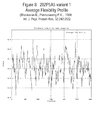
- FIG. 8 Average flexibility amino acid profile of 202P5A5 v.1 determined by computer algorithm sequence analysis using the method of Bhaskaran and Ponnuswamy (Bhaskaran R., and Ponnuswamy P. K., 1988. Int. J. Pept. Protein Res. 32:242-255) accessed on the ProtScale website located on the World Wide Web at (.expasy.ch/cgi-bin/protscale.pl) through the ExPasy molecular biology server.
- FIG. 9 Beta-turn amino acid profile of 202P5A5 v.1 determined by computer algorithm sequence analysis using the method of Deleage and Roux (Deleage, G., Roux B. 1987 Protein Engineering 1:289-294) accessed on the ProtScale website located on the World Wide Web at (.expasy.ch/cgi-bin/protscale.pl) through the ExPasy molecular biology server.
- FIG. 10 Structures of transcript variants of 202P5A5.
- Variants 202P5A5 v.2 and v.3 are transcript variants of 202P5A05 v.1.
- Variant 202P5A05 v.2 added an exon to the 5′ end of variant v.1.
- Variant v.3 further extended exon 1 of v.2 into intron 1.
- Poly A tails are not shown in this figure. Numbers in “( )” underneath the boxes correspond to those of 202P5A05 v.1. Lengths of introns and exons are not proportional.
- FIG. 11 Schematic alignment of protein variants of 202P5A5. Protein variants correspond to nucleotide variants. Nucleotide variants 202P5A5 v.3, v.7, and v.9 through v.26 coded the same protein as v.1. Variant v.2 coded a protein that was 16 amino acids longer and contained the whole protein of v.1. Nucleotide variants 202P5A5 v.2 and v.3 were transcript variants of v.1, as shown in FIG. 10 . SNP in v.1 also existed in v.2 and v.3. Single amino acid differences were indicated above the boxes. Black boxes represent the same sequence as 202P5A5 v.1. Numbers underneath the box correspond to 202P5A5 v.1.
- FIG. 12 Schematic alignment of SNP variants of 202P5A5.
- Variants 202P5A5 v.4 through v.26 are variants with single nucleotide differences as compared to variant v.1 (ORF:29-1858).
- Variant v.14 inserted two base pairs at 2269-2270 while variant v.22 deleted one base pair at 3427. Though these SNP variants were shown separately, they could also occur in any combinations and in any transcript variants, such as v.3 shown in FIG. 10 , that contained the base pairs. Numbers correspond to those of 202P5A5 v.1.
- the black box shows the same sequence as 202P5A5 v.1. SNPs are indicated above the box.
- FIG. 13 Secondary structure and transmembrane domains prediction for 202P5A05 protein variant 1.
- NPS@ Network Protein Sequence Analysis TIBS 2000 March Vol. 25, No 3 [291]:147-150 Combe
- FIG. 13B Schematic representation of the probability of existence of transmembrane regions of 202P5A5 variant 1 based on the TMpred algorithm of Hofmann and Stoffel which utilizes TMBASE (K. Hofmann, W. Stoffel. TMBASE—A database of membrane spanning protein segments Biol. Chem. Hoppe-Seyler 374:166, 1993).
- C Schematic representation of the probability of the existence of transmembrane regions of 202P5A05 variant 1, based on the TMHMM algorithm of Sonnhammer, von Heijne, and Krogh (Erik L. L.
- FIG. 14 Expression of 202P5A5 by RT-PCR.
- FIG. 14A First strand cDNA was prepared from vital pool 1 (liver, lung and kidney), vital pool 2 (pancreas, colon and stomach), prostate cancer metastasis to lymph node, prostate cancer pool, bladder cancer pool, colon cancer pool, lung cancer pool, breast cancer pool, and cancer metastasis pool. Normalization was performed by PCR using primers to actin and GAPDH. Semi-quantitative PCR, using primers to 202P5A5, was performed at 26 and 30 cycles of amplification. Expression was detected in prostate cancer metastasis to lymph node, prostate cancer pool, bladder cancer pool, colon cancer pool, lung cancer pool, breast cancer pool, and cancer metastasis pool. Low expression was also detected in vital pool 1 but not in vital pool 2.
- FIG. 14B Semi-quantitative PCR, using primers to 202P5A5, was performed on a panel of 13 normal tissues and 13 cancer pools. Samples were run on an agarose gel, and PCR products were quantitated using the AlphaImager software. Results show strong expression of 202P5A5 in prostate cancer, bladder cancer, colon cancer, lung cancer, ovary cancer, breast cancer, metastasis cancer, xenograft pool, prostate metastasis to lymph node (PMLN), bone cancer/melanoma pool, cervical cancer, lymphoma and stomach cancer compared to all normal tissues tested.
- PMLN lymph node
- FIG. 15 Expression of 202P5A5 variants by RT-PCR. Primers were designed to differentiate between 202P5A5 v.2 and 202P5A5 v.3. 202P5A5 leads to a PCR product of 173 bp, whereas 202P5A5 v.3 leads to a PCR product of 233 by in size.
- First strand cDNA was prepared from vital pool 1 (liver, lung and kidney), vital pool 2 (pancreas, colon and stomach), LAPC prostate xenograft pool (LAPC-4AD.
- LAPC-4AI, LAPC-9AD and LAPC-9AI prostate cancer pool, bladder cancer pool, lung cancer pool, ovary cancer pool, breast cancer pool, cancer metastasis pool, cervical cancer pool, stomach cancer pool, uterus cancer pool, and master xenograft pool (LAPC xenograft pool, bladder cancer xenograft, kidney cancer xenograft).
- Normalization was performed by PCR using primers to actin and GAPDH. Semi-quantitative PCR, using the variant specific primers was performed at 26 and 30 cycles of amplification. Stronger expression of the 173 bp product was detected in all cancer pools tested and weakly in vital pools. The larger 233 bp product was mostly detected in the cancer pools and not in the vital tissues, and at a frequency of 20-30% compared to the smaller product.
- FIG. 16 Expression of 202P5A5 in normal tissues. Two multiple tissue northern blots (Clontech) both with 2 ug of mRNA/lane were probed with the 202P5A5 sequence. Size standards in kilobases (kb) are indicated on the side. Results show expression of an approximately 7 kb 202P5A5 transcript in normal prostate and normal placenta but not in any other normal tissue tested.
- FIG. 17 Expression of 202P5A5 in Prostate Cancer Patient Specimens.
- RNA was extracted from prostate cancer xenografts (LAPC-4AD, LAPC-4AI, LAPC-9AD, and LAPC-9AI), prostate cancer cell lines (LNCaP and PC3), normal prostate (N), and prostate cancer patient tumors (T).
- Northern blots with 10 ug of total RNA were probed with the 202P5A5 SSH fragment. Size standards in kilobases are on the side. Results show expression of 202P5A5 in all prostate cancer specimens tested as well as in the normal prostate, prostate cancer xenografts and LNCaP, but not in the PC3 cell line.
- FIG. 18 Expression of 202P5A5 in Bladder Cancer Patient Specimens. RNA was extracted from bladder cancer cell lines (CL), normal bladder (N), bladder cancer patient tumors (T) as well as their adjacent normal tissues (Nat). Northern blots with 10 ug of total RNA were probed with the 202P5A5 sequence. Size standards in kilobases are on the side. Results show expression of 202P5A5 in all bladder cancer patient tumor specimens tested but not in normal bladder. Expression was also detected in SCABER but not in the other cancer cell lines tested.
- FIG. 19 Expression of 202P5A5 in Breast Cancer Patient Specimens.
- RNA was extracted from breast cancer cell lines (CL), normal breast (N), breast cancer patient tumors (T), and breast cancer metastasis specimens (M).
- Northern blots with 10 ug of total RNA were probed with the 202P5A5 sequence. Size standards in kilobases are on the side.
- Results show expression of 202P5A5 in the breast cancer patient tumors and metastasis specimens. Expression was also detected in MCF-7 and CAMA-1 but not in the DU4475 cell line. Lower level expression was also detected in normal breast.
- FIG. 20 Expression of 202P5A5 in Colon and Cervical Cancer Patient Specimens.
- First strand cDNA was prepared from a panel of patient cancer specimens. Normalization was performed by PCR using primers to actin. Semi-quantitative PCR, using primers to 202P5A5, was performed at 26 and 30 cycles of amplification. Samples were run on an agarose gel, and PCR products were quantitated using the AlphaImager software. Expression was recorded as absent, low, medium or strong. Results show expression of 202P5A5 in the majority of patient cancer specimens tested.
- FIG. 21 Expression of 202P5A5.pcDNA3.1/MycHis following transfection into 293T cells.
- 293T cells were transfected with either 202P5A5.pcDNA3.1/MycHis or pcDNA3.1/MycHis vector control. Forty hours later, cell lysates were collected. Samples were run on an SDS-PAGE acrylamide gel, blotted and stained with anti-his antibody. The blot was developed using the ECL chemiluminescence kit and visualized by autoradiography. Results show expression of 202P5A5 from the 202P5A5.pcDNA3.1/MycHis construct in the lysates of transfected cells but not in the control pcDNA3.1/MycHis transfected cells.
- prostate cancer locally advanced prostate cancer
- advanced disease and “locally advanced disease” mean prostate cancers that have extended through the prostate capsule, and are meant to include stage C disease under the American Urological Association (AUA) system, stage C1-C2 disease under the Whitmore-Jewett system, and stage T3-T4 and N+ disease under the TNM (tumor, node, metastasis) system.
- AUA American Urological Association
- stage C1-C2 disease under the Whitmore-Jewett system
- T3-T4 and N+ disease under the TNM (tumor, node, metastasis) system.
- surgery is not recommended for patients with locally advanced disease, and these patients have substantially less favorable outcomes compared to patients having clinically localized (organ-confined) prostate cancer.
- Locally advanced disease is clinically identified by palpable evidence of induration beyond the lateral border of the prostate, or asymmetry or induration above the prostate base.
- Locally advanced prostate cancer is presently diagnosed pathologically following radical prostatectomy
- “Altering the native glycosylation pattern” is intended for purposes herein to mean deleting one or more carbohydrate moieties found in native sequence 202P5A5 (either by removing the underlying glycosylation site or by deleting the glycosylation by chemical and/or enzymatic means), and/or adding one or more glycosylation sites that are not present in the native sequence 202P5A5.
- the phrase includes qualitative changes in the glycosylation of the native proteins, involving a change in the nature and proportions of the various carbohydrate moieties present.
- an analog refers to a molecule which is structurally similar or shares similar or corresponding attributes with another molecule (e.g. a 202P5A5-related protein).
- an analog of a 202P5A5 protein can be specifically bound by an antibody or T cell that specifically binds to 202P5A5.
- Antibody is used in the broadest sense. Therefore, an “antibody” can be naturally occurring or man-made such as monoclonal antibodies produced by conventional hybridoma technology.
- Anti-202P5A5 antibodies comprise monoclonal and polyclonal antibodies as well as fragments containing the antigen-binding domain and/or one or more complementarity determining regions of these antibodies.
- an “antibody fragment” is defined as at least a portion of the variable region of the immunoglobulin molecule that binds to its target, i.e., the antigen-binding region. In one embodiment it specifically covers single anti-202P5A5 antibodies and clones thereof (including agonist, antagonist and neutralizing antibodies) and anti-202P5A5 antibody compositions with polyepitopic specificity.
- codon optimized sequences refers to nucleotide sequences that have been optimized for a particular host species by replacing any codons having a usage frequency of less than about 20%. Nucleotide sequences that have been optimized for expression in a given host species by elimination of spurious polyadenylation sequences, elimination of exon/intron splicing signals, elimination of transposon-like repeats and/or optimization of GC content in addition to codon optimization are referred to herein as an “expression enhanced sequences.”
- a “combinatorial library” is a collection of diverse chemical compounds generated by either chemical synthesis or biological synthesis by combining a number of chemical “building blocks” such as reagents.
- a linear combinatorial chemical library such as a polypeptide (e.g., mutein) library, is formed by combining a set of chemical building blocks called amino acids in every possible way for a given compound length (i.e., the number of amino acids in a polypeptide compound).
- Numerous chemical compounds are synthesized through such combinatorial mixing of chemical building blocks (Gallop et al., J. Med. Chem. 37(9): 1233-1251 (1994)).
- combinatorial chemical libraries include, but are not limited to, peptide libraries (see, e.g., U.S. Pat. No. 5,010,175, Furka, Pept. Prot. Res. 37:487-493 (1991), Houghton et al., Nature, 354:84-88 (1991)), peptoids (PCT Publication No WO 91/19735), encoded peptides (PCT Publication WO 93/20242), random bio-oligomers (PCT Publication WO 92/00091), benzodiazepines (U.S. Pat. No.
- cytotoxic agent refers to a substance that inhibits or prevents the expression activity of cells, function of cells and/or causes destruction of cells.
- the term is intended to include radioactive isotopes chemotherapeutic agents, and toxins such as small molecule toxins or enzymatically active toxins of bacterial, fungal, plant or animal origin, including fragments and/or variants thereof.
- cytotoxic agents include, but are not limited to auristatins, auromycins, maytansinoids, yttrium, bismuth, ricin, ricin A-chain, combrestatin, duocarmycins, dolostatins, doxorubicin, daunorubicin, taxol, cisplatin, cc1065, ethidium bromide, mitomycin, etoposide, tenoposide, vincristine, vinblastine, colchicine, dihydroxy anthracin dione, actinomycin, diphtheria toxin, Pseudomonas exotoxin (PE) A, PE40, abrin, abrin A chain, modeccin A chain, alpha-sarcin, gelonin, mitogellin, retstrictocin, phenomycin, enomycin, curicin, crotin, calicheamicin, Sapaonaria officinalis inhibitor
- the “gene product” is sometimes referred to herein as a protein or mRNA.
- a “gene product of the invention” is sometimes referred to herein as a “cancer amino acid sequence”, “cancer protein”, “protein of a cancer listed in Table I”, a “cancer mRNA”, “mRNA of a cancer listed in Table I”, etc.
- the cancer protein is encoded by a nucleic acid of FIG. 2 .
- the cancer protein can be a fragment, or alternatively, be the full-length protein to the fragment encoded by the nucleic acids of FIG. 2 .
- a cancer amino acid sequence is used to determine sequence identity or similarity.
- the sequences are naturally occurring allelic variants of a protein encoded by a nucleic acid of FIG. 2 .
- the sequences are sequence variants as further described herein.
- “High throughput screening” assays for the presence, absence, quantification, or other properties of particular nucleic acids or protein products are well known to those of skill in the art.
- binding assays and reporter gene assays are similarly well known.
- U.S. Pat. No. 5,559,410 discloses high throughput screening methods for proteins
- U.S. Pat. No. 5,585,639 discloses high throughput screening methods for nucleic acid binding (i.e., in arrays)
- U.S. Pat. Nos. 5,576,220 and 5,541,061 disclose high throughput methods of screening for ligand/antibody binding.
- high throughput screening systems are commercially available (see, e.g., Amersham Biosciences, Piscataway, N.J.; Zymark Corp., Hopkinton, Mass.; Air Technical Industries, Mentor, Ohio; Beckman Instruments, Inc. Fullerton, Calif.; Precision Systems, Inc., Natick, Mass.; etc.). These systems typically automate entire procedures, including all sample and reagent pipetting, liquid dispensing, timed incubations, and final readings of the microplate in detector(s) appropriate for the assay. These configurable systems provide high throughput and rapid start up as well as a high degree of flexibility and customization. The manufacturers of such systems provide detailed protocols for various high throughput systems. Thus, e.g., Zymark Corp. provides technical bulletins describing screening systems for detecting the modulation of gene transcription, ligand binding, and the like.
- homolog refers to a molecule which exhibits homology to another molecule, by for example, having sequences of chemical residues that are the same or similar at corresponding positions.
- HLA Human Leukocyte Antigen
- MHC Major Histocompatibility Complex
- hybridize used in the context of polynucleotides, are meant to refer to conventional hybridization conditions, preferably such as hybridization in 50% formamide/6 ⁇ SSC/0.1% SDS/100 ⁇ g/ml ssDNA, in which temperatures for hybridization are above 37 degrees C. and temperatures for washing in 0.1 ⁇ SSC/0.1% SDS are above 55 degrees C.
- isolated or “biologically pure” refer to material which is substantially or essentially free from components which normally accompany the material as it is found in its native state.
- isolated peptides in accordance with the invention preferably do not contain materials normally associated with the peptides in their in situ environment.
- a polynucleotide is said to be “isolated” when it is substantially separated from contaminant polynucleotides that correspond or are complementary to genes other than the 202P5A5 genes or that encode polypeptides other than 202P5A5 gene product or fragments thereof.
- a skilled artisan can readily employ nucleic acid isolation procedures to obtain an isolated 202P5A5 polynucleotide.
- a protein is said to be “isolated,” for example, when physical, mechanical or chemical methods are employed to remove the 202P5A5 proteins from cellular constituents that are normally associated with the protein.
- a skilled artisan can readily employ standard purification methods to obtain an isolated 202P5A5 protein.
- an isolated protein can be prepared by chemical means.
- mammal refers to any organism classified as a mammal, including mice, rats, rabbits, dogs, cats, cows, horses and humans. In one embodiment of the invention, the mammal is a mouse. In another embodiment of the invention, the mammal is a human.
- metastatic prostate cancer and “metastatic disease” mean prostate cancers that have spread to regional lymph nodes or to distant sites, and are meant to include stage D disease under the AUA system and stage T ⁇ N ⁇ M+under the TNM system.
- surgery is generally not indicated for patients with metastatic disease, and hormonal (androgen ablation) therapy is a preferred treatment modality.
- Patients with metastatic prostate cancer eventually develop an androgen-refractory state within 12 to 18 months of treatment initiation. Approximately half of these androgen-refractory patients die within 6 months after developing that status.
- the most common site for prostate cancer metastasis is bone. Prostate cancer bone metastases are often osteoblastic rather than osteolytic (i.e., resulting in net bone formation).
- Bone metastases are found most frequently in the spine, followed by the femur, pelvis, rib cage, skull and humerus. Other common sites for metastasis include lymph nodes, lung, liver and brain. Metastatic prostate cancer is typically diagnosed by open or laparoscopic pelvic lymphadenectomy, whole body radionuclide scans, skeletal radiography, and/or bone lesion biopsy.
- modulator or “test compound” or “drug candidate” or grammatical equivalents as used herein describe any molecule, e.g., protein, oligopeptide, small organic molecule, polysaccharide, polynucleotide, etc., to be tested for the capacity to directly or indirectly alter the cancer phenotype or the expression of a cancer sequence, e.g., a nucleic acid or protein sequences, or effects of cancer sequences (e.g., signaling, gene expression, protein interaction, etc.)
- a modulator will neutralize the effect of a cancer protein of the invention.
- neutralize is meant that an activity of a protein is inhibited or blocked, along with the consequent effect on the cell.
- a modulator will neutralize the effect of a gene, and its corresponding protein, of the invention by normalizing levels of said protein.
- modulators alter expression profiles, or expression profile nucleic acids or proteins provided herein, or downstream effector pathways.
- the modulator suppresses a cancer phenotype, e.g. to a normal tissue fingerprint.
- a modulator induced a cancer phenotype.
- a plurality of assay mixtures is run in parallel with different agent concentrations to obtain a differential response to the various concentrations. Typically, one of these concentrations serves as a negative control, i.e., at zero concentration or below the level of detection.
- Modulators, drug candidates or test compounds encompass numerous chemical classes, though typically they are organic molecules, preferably small organic compounds having a molecular weight of more than 100 and less than about 2,500 Daltons. Preferred small molecules are less than 2000, or less than 1500 or less than 1000 or less than 500 D.
- Candidate agents comprise functional groups necessary for structural interaction with proteins, particularly hydrogen bonding, and typically include at least an amine, carbonyl, hydroxyl or carboxyl group, preferably at least two of the functional chemical groups.
- the candidate agents often comprise cyclical carbon or heterocyclic structures and/or aromatic or polyaromatic structures substituted with one or more of the above functional groups.
- Modulators also comprise biomolecules such as peptides, saccharides, fatty acids, steroids, purines, pyrimidines, derivatives, structural analogs or combinations thereof. Particularly preferred are peptides.
- One class of modulators are peptides, for example of from about five to about 35 amino acids, with from about five to about 20 amino acids being preferred, and from about 7 to about 15 being particularly preferred.
- the cancer modulatory protein is soluble, includes a non-transmembrane region, and/or, has an N-terminal Cys to aid in solubility.
- the C-terminus of the fragment is kept as a free acid and the N-terminus is a free amine to aid in coupling, i.e., to cysteine.
- a cancer protein of the invention is conjugated to an immunogenic agent as discussed herein.
- the cancer protein is conjugated to BSA.
- the peptides of the invention e.g., of preferred lengths, can be linked to each other or to other amino acids to create a longer peptide/protein.
- the modulatory peptides can be digests of naturally occurring proteins as is outlined above, random peptides, or “biased” random peptides.
- peptide/protein-based modulators are antibodies, and fragments thereof, as defined herein.
- Modulators of cancer can also be nucleic acids.
- Nucleic acid modulating agents can be naturally occurring nucleic acids, random nucleic acids, or “biased” random nucleic acids. For example, digests of prokaryotic or eukaryotic genomes can be used in an approach analogous to that outlined above for proteins.
- the term “monoclonal antibody” refers to an antibody obtained from a population of substantially homogeneous antibodies, i.e., the antibodies comprising the population are identical except for possible naturally occurring mutations that are present in minor amounts.
- a “motif”, as in biological motif of a 202P5A5-related protein, refers to any pattern of amino acids forming part of the primary sequence of a protein, that is associated with a particular function (e.g. protein-protein interaction, protein-DNA interaction, etc) or modification (e.g. that is phosphorylated, glycosylated or amidated), or localization (e.g. secretory sequence, nuclear localization sequence, etc.) or a sequence that is correlated with being immunogenic, either humorally or cellularly.
- a motif can be either contiguous or capable of being aligned to certain positions that are generally correlated with a certain function or property.
- motif refers to the pattern of residues in a peptide of defined length, usually a peptide of from about 8 to about 13 amino acids for a class I HLA motif and from about 6 to about 25 amino acids for a class II HLA motif, which is recognized by a particular HLA molecule.
- Peptide motifs for HLA binding are typically different for each protein encoded by each human HLA allele and differ in the pattern of the primary and secondary anchor residues.
- a “pharmaceutical excipient” comprises a material such as an adjuvant, a carrier, pH-adjusting and buffering agents, tonicity adjusting agents, wetting agents, preservative, and the like.
- “Pharmaceutically acceptable” refers to a non-toxic, inert, and/or composition that is physiologically compatible with humans or other mammals.
- polynucleotide means a polymeric form of nucleotides of at least 10 bases or base pairs in length, either ribonucleotides or deoxynucleotides or a modified form of either type of nucleotide, and is meant to include single and double stranded forms of DNA and/or RNA. In the art, this term if often used interchangeably with “oligonucleotide”.
- a polynucleotide can comprise a nucleotide sequence disclosed herein wherein thymidine (T), as shown for example in FIG. 2 , can also be uracil (U); this definition pertains to the differences between the chemical structures of DNA and RNA, in particular the observation that one of the four major bases in RNA is uracil (U) instead of thymidine (T).
- polypeptide means a polymer of at least about 4, 5, 6, 7, or 8 amino acids. Throughout the specification, standard three letter or single letter designations for amino acids are used. In the art, this term is often used interchangeably with “peptide” or “protein”.
- HLA “primary anchor residue” is an amino acid at a specific position along a peptide sequence which is understood to provide a contact point between the immunogenic peptide and the HLA molecule.
- One to three, usually two, primary anchor residues within a peptide of defined length generally defines a “motif” for an immunogenic peptide. These residues are understood to fit in close contact with peptide binding groove of an HLA molecule, with their side chains buried in specific pockets of the binding groove.
- the primary anchor residues for an HLA class I molecule are located at position 2 (from the amino terminal position) and at the carboxyl terminal position of a 8, 9, 10, 11, or 12 residue peptide epitope in accordance with the invention.
- the primary anchor residues of a peptide binds an HLA class II molecule are spaced relative to each other, rather than to the termini of a peptide, where the peptide is generally of at least 9 amino acids in length.
- the primary anchor positions for each motif and supermotif are set forth in Table IV.
- analog peptides can be created by altering the presence or absence of particular residues in the primary and/or secondary anchor positions shown in Table IV. Such analogs are used to modulate the binding affinity and/or population coverage of a peptide comprising a particular HLA motif or supermotif.
- Radioisotopes include, but are not limited to the following (non-limiting exemplary uses are also set forth):
- Actinium-225 See Thorium-229 (Th-229) (AC-225) Actinium-227 Parent of Radium-223 (Ra-223) which is an alpha emitter used to treat (AC-227) metastases in the skeleton resulting from cancer (i.e., breast and prostate cancers), and cancer radioimmunotherapy Bismuth-212 See Thorium-228 (Th-228) (Bi-212) Bismuth-213 See Thorium-229 (Th-229) (Bi-213) Cadmium-109 Cancer detection (Cd-109) Cobalt-60 Radiation source for radiotherapy of cancer, for food irradiators, and for (Co-60) sterilization of medical supplies Copper-64 A positron emitter used for cancer therapy and SPECT imaging (Cu-64) Copper-67 Beta/gamma emitter used in cancer radioimmunotherapy and diagnostic (Cu-67) studies (i.e., breast and colon cancers, and lymphoma) Dysprosium-166 Cancer radioimmunotherapy (Dy-16)
- Tc-99m is the most widely used radioisotope used for diagnostic imaging of various cancers and diseases involving the brain, heart, liver, lungs; also used in detection of deep vein thrombosis of the legs Osmium-194 Cancer radioimmunotherapy (Os-194) Palladium-103 Prostate cancer treatment (Pd-103) Platinum-195m
- a chemotherapeutic (Pt-195m) drug Phosphorus-32 Polycythemia rubra vera (blood cell disease) and leukemia treatment, bone (P-32) cancer diagnosis/treatment; colon, pancreatic, and liver cancer treatment; radiolabeling nucleic acids for in vitro research, diagnosis of superficial tumors, treatment of blocked arteries (i.e., arteriosclerosis and restenosis), and intracavity therapy Phosphorus-33 Leukemia treatment, bone disease diagnosis/treatment, radiolabeling, and (P-33) treatment of blocked arteries (i.e.,
- each nucleic acid and peptide consists of essentially random nucleotides and amino acids, respectively. These random peptides (or nucleic acids, discussed herein) can incorporate any nucleotide or amino acid at any position.
- the synthetic process can be designed to generate randomized proteins or nucleic acids, to allow the formation of all or most of the possible combinations over the length of the sequence, thus forming a library of randomized candidate bioactive proteinaceous agents.
- a library is “fully randomized,” with no sequence preferences or constants at any position.
- the library is a “biased random” library. That is, some positions within the sequence either are held constant, or are selected from a limited number of possibilities.
- the nucleotides or amino acid residues are randomized within a defined class, e.g., of hydrophobic amino acids, hydrophilic residues, sterically biased (either small or large) residues, towards the creation of nucleic acid binding domains, the creation of cysteines, for cross-linking, prolines for SH-3 domains, serines, threonines, tyrosines or histidines for phosphorylation sites, etc., or to purines, etc.
- a “recombinant” DNA or RNA molecule is a DNA or RNA molecule that has been subjected to molecular manipulation in vitro.
- Non-limiting examples of small molecules include compounds that bind or interact with 202P5A5, ligands including hormones, neuropeptides, chemokines, odorants, phospholipids, and functional equivalents thereof that bind and preferably inhibit 202P5A5 protein function.
- Such non-limiting small molecules preferably have a molecular weight of less than about 10 kDa, more preferably below about 9, about 8, about 7, about 6, about 5 or about 4 kDa.
- small molecules physically associate with, or bind, 202P5A5 protein; are not found in naturally occurring metabolic pathways; and/or are more soluble in aqueous than non-aqueous solutions.
- “Stringency” of hybridization reactions is readily determinable by one of ordinary skill in the art, and generally is an empirical calculation dependent upon probe length, washing temperature, and salt concentration. In general, longer probes require higher temperatures for proper annealing, while shorter probes need lower temperatures. Hybridization generally depends on the ability of denatured nucleic acid sequences to reanneal when complementary strands are present in an environment below their melting temperature. The higher the degree of desired homology between the probe and hybridizable sequence, the higher the relative temperature that can be used. As a result, it follows that higher relative temperatures would tend to make the reaction conditions more stringent, while lower temperatures less so. For additional details and explanation of stringency of hybridization reactions, see Ausubel et al., Current Protocols in Molecular Biology, Wiley Interscience Publishers, (1995).
- “Stringent conditions” or “high stringency conditions”, as defined herein, are identified by, but not limited to, those that: (1) employ low ionic strength and high temperature for washing, for example 0.015 M sodium chloride/0.0015 M sodium citrate/0.1% sodium dodecyl sulfate at 50° C.; (2) employ during hybridization a denaturing agent, such as formamide, for example, 50% (v/v) formamide with 0.1% bovine serum albumin/0.1% Ficoll/0.1% polyvinylpyrrolidone/50 mM sodium phosphate buffer at pH 6.5 with 750 mM sodium chloride, 75 mM sodium citrate at 42° C.; or (3) employ 50% formamide, 5 ⁇ SSC (0.75 M NaCl, 0.075 M sodium citrate), 50 mM sodium phosphate (pH 6.8), 0.1% sodium pyrophosphate, 5 ⁇ Denhardt's solution, sonicated salmon sperm DNA (50 ⁇ g/ml), 0.1% SDS, and
- Modely stringent conditions are described by, but not limited to, those in Sambrook et al., Molecular Cloning: A Laboratory Manual, New York: Cold Spring Harbor Press, 1989, and include the use of washing solution and hybridization conditions (e.g., temperature, ionic strength and % SDS) less stringent than those described above.
- washing solution and hybridization conditions e.g., temperature, ionic strength and % SDS
- An example of moderately stringent conditions is overnight incubation at 37° C.
- HLA “supermotif” is a peptide binding specificity shared by HLA molecules encoded by two or more HLA alleles. Overall phenotypic frequencies of HLA-supertypes in different ethnic populations are set forth in Table IV (F). The non-limiting constituents of various supetypes are as follows:
- to treat or “therapeutic” and grammatically related terms, refer to any improvement of any consequence of disease, such as prolonged survival, less morbidity, and/or a lessening of side effects which are the byproducts of an alternative therapeutic modality; full eradication of disease is not required.
- a “transgenic animal” (e.g., a mouse or rat) is an animal having cells that contain a transgene, which transgene was introduced into the animal or an ancestor of the animal at a prenatal, e.g., an embryonic stage.
- a “transgene” is a DNA that is integrated into the genome of a cell from which a transgenic animal develops.
- an HLA or cellular immune response “vaccine” is a composition that contains or encodes one or more peptides of the invention.
- vaccines such as a cocktail of one or more individual peptides; one or more peptides of the invention comprised by a polyepitopic peptide; or nucleic acids that encode such individual peptides or polypeptides, e.g., a minigene that encodes a polyepitopic peptide.
- the “one or more peptides” can include any whole unit integer from 1-150 or more, e.g., at least 2, 3, 4, 5, 6, 7, 8, 9, 10, 11, 12, 13, 14, 15, 16, 17, 18, 19, 20, 21, 22, 23, 24, 25, 26, 27, 28, 29, 30, 31, 32, 33, 34, 35, 36, 37, 38, 39, 40, 41, 42, 43, 44, 45, 46, 47, 48, 49, 50, 55, 60, 65, 70, 75, 80, 85, 90, 95, 100, 105, 110, 115, 120, 125, 130, 135, 140, 145, or 150 or more peptides of the invention.
- the peptides or polypeptides can optionally be modified, such as by lipidation, addition of targeting or other sequences.
- HLA class I peptides of the invention can be admixed with, or linked to, HLA class II peptides, to facilitate activation of both cytotoxic T lymphocytes and helper T lymphocytes.
- HLA vaccines can also comprise peptide-pulsed antigen presenting cells, e.g., dendritic cells.
- variant refers to a molecule that exhibits a variation from a described type or norm, such as a protein that has one or more different amino acid residues in the corresponding position(s) of a specifically described protein (e.g. the 202P5A5 protein shown in FIG. 2 or FIG. 3 .
- An analog is an example of a variant protein.
- Splice isoforms and single nucleotides polymorphisms (SNPs) are further examples of variants.
- the “202P5A5-related proteins” of the invention include those specifically identified herein, as well as allelic variants, conservative substitution variants, analogs and homologs that can be isolated/generated and characterized without undue experimentation following the methods outlined herein or readily available in the art. Fusion proteins that combine parts of different 202P5A5 proteins or fragments thereof, as well as fusion proteins of a 202P5A5 protein and a heterologous polypeptide are also included. Such 202P5A5 proteins are collectively referred to as the 202P5A5-related proteins, the proteins of the invention, or 202P5A5.
- 202P5A5-related protein refers to a polypeptide fragment or a 202P5A5 protein sequence of 4, 5, 6, 7, 8, 9, 10, 11, 12, 13, 14, 15, 16, 17, 18, 19, 20, 21, 22, 23, 24, 25, or more than 25 amino acids; or, at least 30, 35, 40, 45, 50, 55, 60, 65, 70, 80, 85, 90, 95, 100, 105, 110, 115, 120, 125, 130, 135, 140, 145, 150, 155, 160, 165, 170, 175, 180, 185, 190, 195, 200, 225, 250, 275, 300, 325, 350, 375, 400, 425, 450, 475, 500, 525, 550, 575, or 576 or more amino acids.
- One aspect of the invention provides polynucleotides corresponding or complementary to all or part of a 202P5A5 gene, mRNA, and/or coding sequence, preferably in isolated form, including polynucleotides encoding a 202P5A5-related protein and fragments thereof, DNA, RNA, DNA/RNA hybrid, and related molecules, polynucleotides or oligonucleotides complementary to a 202P5A5 gene or mRNA sequence or a part thereof, and polynucleotides or oligonucleotides that hybridize to a 202P5A5 gene, mRNA, or to a 202P5A5 encoding polynucleotide (collectively, “202P5A5 polynucleotides”).
- T can also be U in FIG. 2 .
- Embodiments of a 202P5A5 polynucleotide include: a 202P5A5 polynucleotide having the sequence shown in FIG. 2 , the nucleotide sequence of 202P5A5 as shown in FIG. 2 wherein T is U; at least 10 contiguous nucleotides of a polynucleotide having the sequence as shown in FIG. 2 ; or, at least 10 contiguous nucleotides of a polynucleotide having the sequence as shown in FIG. 2 where T is U.
- embodiments of 202P5A5 nucleotides comprise, without limitation:
- Typical embodiments of the invention disclosed herein include 202P5A5 polynucleotides that encode specific portions of 202P5A5 mRNA sequences (and those which are complementary to such sequences) such as those that encode the proteins and/or fragments thereof, for example:
- representative embodiments of the invention disclosed herein include: polynucleotides and their encoded peptides themselves encoding about amino acid 1 to about amino acid 10 of the 202P5A5 protein shown in FIG. 2 or FIG. 3 , polynucleotides encoding about amino acid 10 to about amino acid 20 of the 202P5A5 protein shown in FIG. 2 or FIG. 3 , polynucleotides encoding about amino acid 20 to about amino acid 30 of the 202P5A5 protein shown in FIG. 2 or FIG. 3 , polynucleotides encoding about amino acid 30 to about amino acid 40 of the 202P5A5 protein shown in FIG. 2 or FIG.
- polynucleotides encoding about amino acid 40 to about amino acid 50 of the 202P5A5 protein shown in FIG. 2 or FIG. 3 polynucleotides encoding about amino acid 50 to about amino acid 60 of the 202P5A5 protein shown in FIG. 2 or FIG. 3
- polynucleotides encoding about amino acid 60 to about amino acid 70 of the 202P5A5 protein shown in FIG. 2 or FIG. 3 polynucleotides encoding about amino acid 70 to about amino acid 80 of the 202P5A5 protein shown in FIG. 2 or FIG. 3
- polynucleotides encoding portions of the amino acid sequence (of about 10 amino acids), of amino acids, 100 through the carboxyl terminal amino acid of the 202P5A5 protein are embodiments of the invention. Wherein it is understood that each particular amino acid position discloses that position plus or minus five amino acid residues.
- Polynucleotides encoding relatively long portions of a 202P5A5 protein are also within the scope of the invention.
- polynucleotides encoding from about amino acid 1 (or 20 or 30 or 40 etc.) to about amino acid 20, (or 30, or 40 or 50 etc.) of the 202P5A5 protein “or variant” shown in FIG. 2 or FIG. 3 can be generated by a variety of techniques well known in the art. These polynucleotide fragments can include any portion of the 202P5A5 sequence as shown in FIG. 2 .
- Additional illustrative embodiments of the invention disclosed herein include 202P5A5 polynucleotide fragments encoding one or more of the biological motifs contained within a 202P5A5 protein “or variant” sequence, including one or more of the motif-bearing subsequences of a 202P5A5 protein “or variant” set forth in Tables VIII-XXI and XXII-XLIX.
- typical polynucleotide fragments of the invention encode one or more of the regions of 202P5A5 protein or variant that exhibit homology to a known molecule.
- typical polynucleotide fragments can encode one or more of the 202P5A5 protein or variant N-glycosylation sites, cAMP and cGMP-dependent protein kinase phosphorylation sites, casein kinase II phosphorylation sites or N-myristoylation site and amidation sites.
- HLA Peptide Tables e.g., variant 1, variant 2, etc.
- search Peptides listed in Table VII.
- a unique Search Peptide is used to obtain HLA peptides for a particular variant. The position of each Search Peptide relative to its respective parent molecule is listed in Table VII.
- a Search Peptide begins at position “X”, one must add the value “X minus 1” to each position in Tables VIII-XXI and Tables XXII-IL to obtain the actual position of the HLA peptides in their parental molecule. For example if a particular Search Peptide begins at position 150 of its parental molecule, one must add 150 ⁇ 1, i.e., 149 to each HLA peptide amino acid position to calculate the position of that amino acid in the parent molecule.
- the polynucleotides of the preceding paragraphs have a number of different specific uses.
- the human 202P5A5 gene maps to the chromosomal location set forth in the Example entitled “Chromosomal Mapping of 202P5A5.”
- polynucleotides that encode different regions of the 202P5A5 proteins are used to characterize cytogenetic abnormalities of this chromosomal locale, such as abnormalities that are identified as being associated with various cancers.
- cytogenetic abnormalities of this chromosomal locale such as abnormalities that are identified as being associated with various cancers.
- a variety of chromosomal abnormalities including rearrangements have been identified as frequent cytogenetic abnormalities in a number of different cancers (see e.g.
- polynucleotides encoding specific regions of the 202P5A5 proteins provide new tools that can be used to delineate, with greater precision than previously possible, cytogenetic abnormalities in the chromosomal region that encodes 202P5A5 that may contribute to the malignant phenotype.
- these polynucleotides satisfy a need in the art for expanding the sensitivity of chromosomal screening in order to identify more subtle and less common chromosomal abnormalities (see e.g. Evans et al., Am. J. Obstet. Gynecol 171(4): 1055-1057 (1994)).
- 202P5A5 was shown to be highly expressed in prostate and other cancers
- 202P5A5 polynucleotides are used in methods assessing the status of 202P5A5 gene products in normal versus cancerous tissues.
- polynucleotides that encode specific regions of the 202P5A5 proteins are used to assess the presence of perturbations (such as deletions, insertions, point mutations, or alterations resulting in a loss of an antigen etc.) in specific regions of the 202P5A5 gene, such as regions containing one or more motifs.
- Exemplary assays include both RT-PCR assays as well as single-strand conformation polymorphism (SSCP) analysis (see, e.g., Marrogi et al., J. Cutan. Pathol. 26(8): 369-378 (1999), both of which utilize polynucleotides encoding specific regions of a protein to examine these regions within the protein.
- SSCP single-strand conformation polymorphism
- nucleic acid related embodiments of the invention disclosed herein are genomic DNA, cDNAs, ribozymes, and antisense molecules, as well as nucleic acid molecules based on an alternative backbone, or including alternative bases, whether derived from natural sources or synthesized, and include molecules capable of inhibiting the RNA or protein expression of 202P5A5.
- antisense molecules can be RNAs or other molecules, including peptide nucleic acids (PNAs) or non-nucleic acid molecules such as phosphorothioate derivatives that specifically bind DNA or RNA in a base pair-dependent manner.
- PNAs peptide nucleic acids
- non-nucleic acid molecules such as phosphorothioate derivatives that specifically bind DNA or RNA in a base pair-dependent manner.
- a skilled artisan can readily obtain these classes of nucleic acid molecules using the 202P5A5 polynucleotides and polynucleotide sequences disclosed herein.
- Antisense technology entails the administration of exogenous oligonucleotides that bind to a target polynucleotide located within the cells.
- the term “antisense” refers to the fact that such oligonucleotides are complementary to their intracellular targets, e.g., 202P5A5. See for example, Jack Cohen, Oligodeoxynucleotides, Antisense Inhibitors of Gene Expression, CRC Press, 1989; and Synthesis 1:1-5 (1988).
- the 202P5A5 antisense oligonucleotides of the present invention include derivatives such as S-oligonucleotides (phosphorothioate derivatives or S-oligos, see, Jack Cohen, supra), which exhibit enhanced cancer cell growth inhibitory action.
- S-oligos are isoelectronic analogs of an oligonucleotide (O-oligo) in which a nonbridging oxygen atom of the phosphate group is replaced by a sulfur atom.
- the S-oligos of the present invention can be prepared by treatment of the corresponding O-oligos with 3H-1,2-benzodithiol-3-one-1,1-dioxide, which is a sulfur transfer reagent. See, e.g., Iyer, R. P. et al., J. Org. Chem. 55:4693-4698 (1990); and Iyer, R. P. et al., J. Am. Chem. Soc. 112:1253-1254 (1990).
- Additional 202P5A5 antisense oligonucleotides of the present invention include morpholino antisense oligonucleotides known in the art (see, e.g., Partridge et al., 1996, Antisense & Nucleic Acid Drug Development 6: 169-175).
- the 202P5A5 antisense oligonucleotides of the present invention typically can be RNA or DNA that is complementary to and stably hybridizes with the first 100 5′ codons or last 100 3′ codons of a 202P5A5 genomic sequence or the corresponding mRNA. Absolute complementarity is not required, although high degrees of complementarity are preferred. Use of an oligonucleotide complementary to this region allows for the selective hybridization to 202P5A5 mRNA and not to mRNA specifying other regulatory subunits of protein kinase.
- 202P5A5 antisense oligonucleotides of the present invention are 15 to 30-mer fragments of the antisense DNA molecule that have a sequence that hybridizes to 202P5A5 mRNA.
- 202P5A5 antisense oligonucleotide is a 30-mer oligonucleotide that is complementary to a region in the first 10 5′ codons or last 10 3′ codons of 202P5A5.
- the antisense molecules are modified to employ ribozymes in the inhibition of 202P5A5 expression, see, e.g., L. A. Couture & D. T. Stinchcomb; Trends Genet. 12:510-515 (1996).
- nucleotides of the invention include primers and primer pairs, which allow the specific amplification of polynucleotides of the invention or of any specific parts thereof, and probes that selectively or specifically hybridize to nucleic acid molecules of the invention or to any part thereof.
- Probes can be labeled with a detectable marker, such as, for example, a radioisotope, fluorescent compound, bioluminescent compound, a chemiluminescent compound, metal chelator or enzyme.
- a detectable marker such as, for example, a radioisotope, fluorescent compound, bioluminescent compound, a chemiluminescent compound, metal chelator or enzyme.
- Such probes and primers are used to detect the presence of a 202P5A5 polynucleotide in a sample and as a means for detecting a cell expressing a 202P5A5 protein.
- probes include polypeptides comprising all or part of the human 202P5A5 cDNA sequence shown in FIG. 2 .
- primer pairs capable of specifically amplifying 202P5A5 mRNAs are also described in the Examples.
- a great many different primers and probes can be prepared based on the sequences provided herein and used effectively to amplify and/or detect a 202P5A5 mRNA.
- the 202P5A5 polynucleotides of the invention are useful for a variety of purposes, including but not limited to their use as probes and primers for the amplification and/or detection of the 202P5A5 gene(s), mRNA(s), or fragments thereof; as reagents for the diagnosis and/or prognosis of prostate cancer and other cancers; as coding sequences capable of directing the expression of 202P5A5 polypeptides; as tools for modulating or inhibiting the expression of the 202P5A5 gene(s) and/or translation of the 202P5A5 transcript(s); and as therapeutic agents.
- the present invention includes the use of any probe as described herein to identify and isolate a 202P5A5 or 202P5A5 related nucleic acid sequence from a naturally occurring source, such as humans or other mammals, as well as the isolated nucleic acid sequence per se, which would comprise all or most of the sequences found in the probe used.
- the 202P5A5 cDNA sequences described herein enable the isolation of other polynucleotides encoding 202P5A5 gene product(s), as well as the isolation of polynucleotides encoding 202P5A5 gene product homologs, alternatively spliced isoforms, allelic variants, and mutant forms of a 202P5A5 gene product as well as polynucleotides that encode analogs of 202P5A5-related proteins.
- Various molecular cloning methods that can be employed to isolate full length cDNAs encoding a 202P5A5 gene are well known (see, for example, Sambrook, J.
- 202P5A5 gene cDNAs can be identified by probing with a labeled 202P5A5 cDNA or a fragment thereof.
- a 202P5A5 cDNA e.g., FIG.
- a 202P5A5 gene itself can be isolated by screening genomic DNA libraries, bacterial artificial chromosome libraries (BACs), yeast artificial chromosome libraries (YACs), and the like, with 202P5A5 DNA probes or primers.
- BACs bacterial artificial chromosome libraries
- YACs yeast artificial chromosome libraries
- the invention also provides recombinant DNA or RNA molecules containing a 202P5A5 polynucleotide, a fragment, analog or homologue thereof, including but not limited to phages, plasmids, phagemids, cosmids, YACs, BACs, as well as various viral and non-viral vectors well known in the art, and cells transformed or transfected with such recombinant DNA or RNA molecules. Methods for generating such molecules are well known (see, for example, Sambrook et al., 1989, supra).
- the invention further provides a host-vector system comprising a recombinant DNA molecule containing a 202P5A5 polynucleotide, fragment, analog or homologue thereof within a suitable prokaryotic or eukaryotic host cell.
- suitable eukaryotic host cells include a yeast cell, a plant cell, or an animal cell, such as a mammalian cell or an insect cell (e.g., a baculovirus-infectible cell such as an Sf9 or HighFive cell).
- suitable mammalian cells include various prostate cancer cell lines such as DU145 and TsuPr1, other transfectable or transducible prostate cancer cell lines, primary cells (PrEC), as well as a number of mammalian cells routinely used for the expression of recombinant proteins (e.g., COS, CHO, 293, 293T cells). More particularly, a polynucleotide comprising the coding sequence of 202P5A5 or a fragment, analog or homolog thereof can be used to generate 202P5A5 proteins or fragments thereof using any number of host-vector systems routinely used and widely known in the art.
- 202P5A5 A wide range of host-vector systems suitable for the expression of 202P5A5 proteins or fragments thereof are available, see for example, Sambrook et al., 1989, supra; Current Protocols in Molecular Biology, 1995, supra).
- Preferred vectors for mammalian expression include but are not limited to pcDNA 3.1 myc-His-tag (Invitrogen) and the retroviral vector pSRatkneo (Muller et al., 1991, MCB 11:1785).
- 202P5A5 can be expressed in several prostate cancer and non-prostate cell lines, including for example 293, 293T, rat-1, NIH 3T3 and TsuPr1.
- the host-vector systems of the invention are useful for the production of a 202P5A5 protein or fragment thereof. Such host-vector systems can be employed to study the functional properties of 202P5A5 and 202P5A5 mutations or analogs.
- Recombinant human 202P5A5 protein or an analog or homolog or fragment thereof can be produced by mammalian cells transfected with a construct encoding a 202P5A5-related nucleotide.
- 293T cells can be transfected with an expression plasmid encoding 202P5A5 or fragment, analog or homolog thereof, a 202P5A5-related protein is expressed in the 293T cells, and the recombinant 202P5A5 protein is isolated using standard purification methods (e.g., affinity purification using anti-202P5A5 antibodies).
- a 202P5A5 coding sequence is subcloned into the retroviral vector pSR ⁇ MSVtkneo and used to infect various mammalian cell lines, such as NIH 3T3, TsuPr1, 293 and rat-1 in order to establish 202P5A5 expressing cell lines.
- mammalian cell lines such as NIH 3T3, TsuPr1, 293 and rat-1
- Various other expression systems well known in the art can also be employed.
- Expression constructs encoding a leader peptide joined in frame to a 202P5A5 coding sequence can be used for the generation of a secreted form of recombinant 202P5A5 protein.
- redundancy in the genetic code permits variation in 202P5A5 gene sequences.
- specific host species often have specific codon preferences, and thus one can adapt the disclosed sequence as preferred for a desired host.
- preferred analog codon sequences typically have rare codons (i.e., codons having a usage frequency of less than about 20% in known sequences of the desired host) replaced with higher frequency codons.
- Codon preferences for a specific species are calculated, for example, by utilizing codon usage tables available on the INTERNET such as at URL dna.affrc.go.jp/ ⁇ nakamura/codon.html.
- Additional sequence modifications are known to enhance protein expression in a cellular host. These include elimination of sequences encoding spurious polyadenylation signals, exon/intron splice site signals, transposon-like repeats, and/or other such well-characterized sequences that are deleterious to gene expression.
- the GC content of the sequence is adjusted to levels average for a given cellular host, as calculated by reference to known genes expressed in the host cell. Where possible, the sequence is modified to avoid predicted hairpin secondary mRNA structures.
- Other useful modifications include the addition of a translational initiation consensus sequence at the start of the open reading frame, as described in Kozak, Mol. Cell. Biol., 9:5073-5080 (1989).
- 202P5A5-related proteins Another aspect of the present invention provides 202P5A5-related proteins.
- 202P5A5 proteins comprise a polypeptide having all or part of the amino acid sequence of human 202P5A5 as shown in FIG. 2 or FIG. 3 .
- embodiments of 202P5A5 proteins comprise variant, homolog or analog polypeptides that have alterations in the amino acid sequence of 202P5A5 shown in FIG. 2 or FIG. 3 .
- Embodiments of a 202P5A5 polypeptide include: a 202P5A5 polypeptide having a sequence shown in FIG. 2 , a peptide sequence of a 202P5A5 as shown in FIG. 2 wherein T is U; at least 10 contiguous nucleotides of a polypeptide having the sequence as shown in FIG. 2 ; or, at least 10 contiguous peptides of a polypeptide having the sequence as shown in FIG. 2 where T is U.
- embodiments of 202P5A5 peptides comprise, without limitation:
- Typical embodiments of the invention disclosed herein include 202P5A5 polynucleotides that encode specific portions of 202P5A5 mRNA sequences (and those which are complementary to such sequences) such as those that encode the proteins and/or fragments thereof, for example:
- allelic variants of human 202P5A5 share a high degree of structural identity and homology (e.g., 90% or more homology).
- allelic variants of a 202P5A5 protein contain conservative amino acid substitutions within the 202P5A5 sequences described herein or contain a substitution of an amino acid from a corresponding position in a homologue of 202P5A5.
- One class of 202P5A5 allelic variants are proteins that share a high degree of homology with at least a small region of a particular 202P5A5 amino acid sequence, but further contain a radical departure from the sequence, such as a non-conservative substitution, truncation, insertion or frame shift.
- Proteins of the invention can comprise 1, 2, 3, 4, 5, 6, 7, 8, 9, 10, 11, 12, 13, 14, 15 conservative substitutions. Such changes include substituting any of isoleucine (I), valine (V), and leucine (L) for any other of these hydrophobic amino acids; aspartic acid (D) for glutamic acid (E) and vice versa; glutamine (Q) for asparagine (N) and vice versa; and serine (S) for threonine (T) and vice versa.
- isoleucine I
- V valine
- L leucine
- Such changes include substituting any of isoleucine (I), valine (V), and leucine (L) for any other of these hydrophobic amino acids; aspartic acid (D) for glutamic acid (E) and vice versa; glutamine (Q) for asparagine (N) and vice versa; and serine (S) for threonine (T) and vice versa.
- substitutions can also be considered conservative, depending on the environment of the particular amino acid and its role in the three-dimensional structure of the protein.
- G glycine
- A alanine
- V valine
- M Methionine
- L Lysine
- K arginine
- R arginine
- Embodiments of the invention disclosed herein include a wide variety of art-accepted variants or analogs of 202P5A5 proteins such as polypeptides having amino acid insertions, deletions and substitutions.
- 202P5A5 variants can be made using methods known in the art such as site-directed mutagenesis, alanine scanning, and PCR mutagenesis. Site-directed mutagenesis (Carter et al., Nucl. Acids Res., 13:4331 (1986); Zoller et al., Nucl.
- Scanning amino acid analysis can also be employed to identify one or more amino acids along a contiguous sequence that is involved in a specific biological activity such as a protein-protein interaction.
- preferred scanning amino acids are relatively small, neutral amino acids.
- amino acids include alanine, glycine, serine, and cysteine
- Alanine is typically a preferred scanning amino acid among this group because it eliminates the side-chain beyond the beta-carbon and is less likely to alter the main-chain conformation of the variant.
- Alanine is also typically preferred because it is the most common amino acid. Further, it is frequently found in both buried and exposed positions (Creighton, The Proteins , (W.H. Freeman & Co., N.Y.); Chothia, J. Mol. Biol., 150:1 (1976)). If alanine substitution does not yield adequate amounts of variant, an isosteric amino acid can be used.
- 202P5A5 variants, analogs or homologs have the distinguishing attribute of having at least one epitope that is “cross reactive” with a 202P5A5 protein having an amino acid sequence of FIG. 3 .
- cross reactive means that an antibody or T cell that specifically binds to a 202P5A5 variant also specifically binds to a 202P5A5 protein having an amino acid sequence set forth in FIG. 3 .
- a polypeptide ceases to be a variant of a protein shown in FIG. 3 , when it no longer contains any epitope capable of being recognized by an antibody or T cell that specifically binds to the starting 202P5A5 protein.
- 202P5A5-related protein variants share 70%, 75%, 80%, 85% or 90% or more similarity with an amino acid sequence of FIG. 3 , or a fragment thereof.
- Another specific class of 202P5A5 protein variants or analogs comprises one or more of the 202P5A5 biological motifs described herein or presently known in the art.
- analogs of 202P5A5 fragments that have altered functional (e.g. immunogenic) properties relative to the starting fragment. It is to be appreciated that motifs now or which become part of the art are to be applied to the nucleic or amino acid sequences of FIG. 2 or FIG. 3 .
- embodiments of the claimed invention include polypeptides containing less than the full amino acid sequence of a 202P5A5 protein shown in FIG. 2 or FIG. 3 .
- representative embodiments of the invention comprise peptides/proteins having any 4, 5, 6, 7, 8, 9, 10, 11, 12, 13, 14, 15 or more contiguous amino acids of a 202P5A5 protein shown in FIG. 2 or FIG. 3 .
- representative embodiments of the invention disclosed herein include polypeptides consisting of about amino acid 1 to about amino acid 10 of a 202P5A5 protein shown in FIG. 2 or FIG. 3 , polypeptides consisting of about amino acid 10 to about amino acid 20 of a 202P5A5 protein shown in FIG. 2 or FIG. 3 , polypeptides consisting of about amino acid 20 to about amino acid 30 of a 202P5A5 protein shown in FIG. 2 or FIG. 3 , polypeptides consisting of about amino acid 30 to about amino acid 40 of a 202P5A5 protein shown in FIG. 2 or FIG. 3 , polypeptides consisting of about amino acid 40 to about amino acid 50 of a 202P5A5 protein shown in FIG.
- polypeptides consisting of about amino acid 50 to about amino acid 60 of a 202P5A5 protein shown in FIG. 2 or FIG. 3 polypeptides consisting of about amino acid 60 to about amino acid 70 of a 202P5A5 protein shown in FIG. 2 or FIG. 3
- polypeptides consisting of about amino acid 70 to about amino acid 80 of a 202P5A5 protein shown in FIG. 2 or FIG. 3 polypeptides consisting of about amino acid 80 to about amino acid 90 of a 202P5A5 protein shown in FIG. 2 or FIG. 3
- polypeptides consisting of about amino acid 1 (or 20 or 30 or 40 etc.) to about amino acid 20, (or 130, or 140 or 150 etc.) of a 202P5A5 protein shown in FIG. 2 or FIG. 3 are embodiments of the invention. It is to be appreciated that the starting and stopping positions in this paragraph refer to the specified position as well as that position plus or minus 5 residues.
- 202P5A5-related proteins are generated using standard peptide synthesis technology or using chemical cleavage methods well known in the art. Alternatively, recombinant methods can be used to generate nucleic acid molecules that encode a 202P5A5-related protein. In one embodiment, nucleic acid molecules provide a means to generate defined fragments of a 202P5A5 protein (or variants, homologs or analogs thereof).
- Additional illustrative embodiments of the invention disclosed herein include 202P5A5 polypeptides comprising the amino acid residues of one or more of the biological motifs contained within a 202P5A5 polypeptide sequence set forth in FIG. 2 or FIG. 3 .
- motifs are known in the art, and a protein can be evaluated for the presence of such motifs by a number of publicly available Internet sites (see, e.g., URL addresses: pfam.wustl.edu/; searchlauncher.bcm.tmc.edu/seq-search/struc-predict.html; psort.ims.u-tokyo.ac.jp/; cbs.dtu.dk/; ebi.ac.uk/interpro/scan.html; expasy.ch/tools/scnpsitl.html; EpimatrixTM and EpimerTM, Brown University, brown.edu/Research/TB-HIV_Lab/epimatrix/epimatrix.html; and BIMAS, bimas.dcrt.nih.gov/.).
- Motif bearing subsequences of all 202P5A5 variant proteins are set forth and identified in Tables VIII-XXI and XXII-XLIX.
- Table V sets forth several frequently occurring motifs based on pfam searches (see URL address pfam.wustl.edu/). The columns of Table V list (1) motif name abbreviation, (2) percent identity found amongst the different member of the motif family, (3) motif name or description and (4) most common function; location information is included if the motif is relevant for location.
- Polypeptides comprising one or more of the 202P5A5 motifs discussed above are useful in elucidating the specific characteristics of a malignant phenotype in view of the observation that the 202P5A5 motifs discussed above are associated with growth dysregulation and because 202P5A5 is overexpressed in certain cancers (See, e.g., Table I).
- Casein kinase II, cAMP and camp-dependent protein kinase, and Protein Kinase C are enzymes known to be associated with the development of the malignant phenotype (see e.g.
- Amidation is another protein modification also associated with cancer and cancer progression (see e.g. Treston et al., J. Natl. Cancer Inst. Monogr. (13): 169-175 (1992)).
- proteins of the invention comprise one or more of the immunoreactive epitopes identified in accordance with art-accepted methods, such as the peptides set forth in Tables VIII-XXI and XXII-XLIX.
- CTL epitopes can be determined using specific algorithms to identify peptides within a 202P5A5 protein that are capable of optimally binding to specified HLA alleles (e.g., Table IV; EpimatrixTM and EpimerTM, Brown University, URL brown.edu/Research/TB-HIV_Lab/epimatrix/epimatrix.html; and BIMAS, URL bimas.dcrt.nih.gov/.)
- processes for identifying peptides that have sufficient binding affinity for HLA molecules and which are correlated with being immunogenic epitopes are well known in the art, and are carried out without undue experimentation.
- processes for identifying peptides that are immunogenic epitopes are well known in
- epitopes in order to modulate immunogenicity. For example, one begins with an epitope that bears a CTL or HTL motif (see, e.g., the HLA Class I and HLA Class II motifs/supermotifs of Table IV).
- the epitope is analoged by substituting out an amino acid at one of the specified positions, and replacing it with another amino acid specified for that position.
- residues defined in Table IV one can substitute out a deleterious residue in favor of any other residue, such as a preferred residue; substitute a less-preferred residue with a preferred residue; or substitute an originally-occurring preferred residue with another preferred residue. Substitutions can occur at primary anchor positions or at other positions in a peptide; see, e.g., Table IV.
- polypeptides comprising combinations of the different motifs set forth in Table VI, and/or, one or more of the predicted CTL epitopes of Tables VIII-XXI and XXII-XLIX, and/or, one or more of the predicted HTL epitopes of Tables XLVI-XLIX, and/or, one or more of the T cell binding motifs known in the art.
- Preferred embodiments contain no insertions, deletions or substitutions either within the motifs or within the intervening sequences of the polypeptides.
- embodiments which include a number of either N-terminal and/or C-terminal amino acid residues on either side of these motifs may be desirable (to, for example, include a greater portion of the polypeptide architecture in which the motif is located).
- the number of N-terminal and/or C-terminal amino acid residues on either side of a motif is between about 1 to about 100 amino acid residues, preferably 5 to about 50 amino acid residues.
- 202P5A5-related proteins are embodied in many forms, preferably in isolated form.
- a purified 202P5A5 protein molecule will be substantially free of other proteins or molecules that impair the binding of 202P5A5 to antibody, T cell or other ligand.
- the nature and degree of isolation and purification will depend on the intended use.
- Embodiments of a 202P5A5-related proteins include purified 202P5A5-related proteins and functional, soluble 202P5A5-related proteins.
- a functional, soluble 202P5A5 protein or fragment thereof retains the ability to be bound by antibody, T cell or other ligand.
- the invention also provides 202P5A5 proteins comprising biologically active fragments of a 202P5A5 amino acid sequence shown in FIG. 2 or FIG. 3 .
- Such proteins exhibit properties of the starting 202P5A5 protein, such as the ability to elicit the generation of antibodies that specifically bind an epitope associated with the starting 202P5A5 protein; to be bound by such antibodies; to elicit the activation of HTL or CTL; and/or, to be recognized by HTL or CTL that also specifically bind to the starting protein.
- 202P5A5-related polypeptides that contain particularly interesting structures can be predicted and/or identified using various analytical techniques well known in the art, including, for example, the methods of Chou-Fasman, Garnier-Robson, Kyte-Doolittle, Eisenberg, Karplus-Schultz or Jameson-Wolf analysis, or based on immunogenicity. Fragments that contain such structures are particularly useful in generating subunit-specific anti-202P5A5 antibodies or T cells or in identifying cellular factors that bind to 202P5A5. For example, hydrophilicity profiles can be generated, and immunogenic peptide fragments identified, using the method of Hopp, T. P. and Woods, K. R., 1981, Proc. Natl. Acad.
- Hydropathicity profiles can be generated, and immunogenic peptide fragments identified, using the method of Kyte, J. and Doolittle, R. F., 1982, J. Mol. Biol. 157:105-132. Percent (%) Accessible Residues profiles can be generated, and immunogenic peptide fragments identified, using the method of Janin J., 1979, Nature 277:491-492. Average Flexibility profiles can be generated, and immunogenic peptide fragments identified, using the method of Bhaskaran R., Ponnuswamy P. K., 1988, Int. J. Pept. Protein Res. 32:242-255. Beta-turn profiles can be generated, and immunogenic peptide fragments identified, using the method of Deleage, G., Roux B., 1987, Protein Engineering 1:289-294.
- CTL epitopes can be determined using specific algorithms to identify peptides within a 202P5A5 protein that are capable of optimally binding to specified HLA alleles (e.g., by using the SYFPEITHI site at World Wide Web URL syfpeithi.bmi-heidelberg.com/; the listings in Table IV(A)-(E); EpimatrixTM and EpimerTM, Brown University, URL (brown.edu/Research/TB-HIV_Lab/epimatrix/epimatrix.html); and BIMAS, URL bimas.dcrt.nih.gov/).
- peptide epitopes from 202P5A5 that are presented in the context of human MHC Class I molecules, e.g., HLA-A1, A2, A3, A11, A24, B7 and B35 were predicted (see, e.g., Tables VIII-XXI, XXII-XLIX).
- the complete amino acid sequence of the 202P5A5 protein and relevant portions of other variants i.e., for HLA Class I predictions 9 flanking residues on either side of a point mutation or exon juction, and for HLA Class II predictions 14 flanking residues on either side of a point mutation or exon junction corresponding to that variant, were entered into the HLA Peptide Motif Search algorithm found in the Bioinformatics and Molecular Analysis Section (BIMAS) web site listed above; in addition to the site SYFPEITHI, at URL syfpeithi.bmi-heidelberg.com/.
- BIMAS Bioinformatics and Molecular Analysis Section
- HLA peptide motif search algorithm was developed by Dr. Ken Parker based on binding of specific peptide sequences in the groove of HLA Class I molecules, in particular HLA-A2 (see, e.g., Falk et al., Nature 351: 290-6 (1991); Hunt et al., Science 255:1261-3 (1992); Parker et al., J. Immunol. 149:3580-7 (1992); Parker et al., J. Immunol. 152:163-75 (1994)).
- This algorithm allows location and ranking of 8-mer, 9-mer, and 10-mer peptides from a complete protein sequence for predicted binding to HLA-A2 as well as numerous other HLA Class I molecules.
- HLA class I binding peptides are 8-, 9-, 10 or 11-mers.
- the epitopes preferably contain a leucine (L) or methionine (M) at position 2 and a valine (V) or leucine (L) at the C-terminus (see, e.g., Parker et al., J. Immunol. 149:3580-7 (1992)).
- Selected results of 202P5A5 predicted binding peptides are shown in Tables VIII-XXI and XXII-XLIX herein.
- Tables VIII-XXI and XXII-XLVII selected candidates, 9-mers and 10-mers, for each family member are shown along with their location, the amino acid sequence of each specific peptide, and an estimated binding score.
- Tables XLVI-XLIX selected candidates, 15-mers, for each family member are shown along with their location, the amino acid sequence of each specific peptide, and an estimated binding score.
- the binding score corresponds to the estimated half time of dissociation of complexes containing the peptide at 37° C. at pH 6.5. Peptides with the highest binding score are predicted to be the most tightly bound to HLA Class I on the cell surface for the greatest period of time and thus represent the best immunogenic targets for T-cell recognition.
- every epitope predicted by the BIMAS site, EpimerTM and EpimatrixTM sites, or specified by the HLA class I or class II motifs available in the art or which become part of the art such as set forth in Table IV (or determined using World Wide Web site URL syfpeithi.bmi-heidelberg.com/, or BIMAS, bimas.dcrt nih gov/) are to be “applied” to a 202P5A5 protein in accordance with the invention.
- “applied” means that a 202P5A5 protein is evaluated, e.g., visually or by computer-based patterns finding methods, as appreciated by those of skill in the relevant art.
- Every subsequence of a 202P5A5 protein of 8, 9, 10, or 11 amino acid residues that bears an HLA Class I motif, or a subsequence of 9 or more amino acid residues that bear an HLA Class II motif are within the scope of the invention.
- 202P5A5 can be conveniently expressed in cells (such as 293T cells) transfected with a commercially available expression vector such as a CMV-driven expression vector encoding 202P5A5 with a C-terminal 6 ⁇ His and MYC tag (pcDNA3.1/mycHIS, Invitrogen or Tag5, GenHunter Corporation, Nashville Tenn.).
- the Tag5 vector provides an IgGK secretion signal that can be used to facilitate the production of a secreted 202P5A5 protein in transfected cells.
- the secreted HIS-tagged 202P5A5 in the culture media can be purified, e.g., using a nickel column using standard techniques.
- Modifications of 202P5A5-related proteins such as covalent modifications are included within the scope of this invention.
- One type of covalent modification includes reacting targeted amino acid residues of a 202P5A5 polypeptide with an organic derivatizing agent that is capable of reacting with selected side chains or the N- or C-terminal residues of a 202P5A5 protein.
- Another type of covalent modification of a 202P5A5 polypeptide included within the scope of this invention comprises altering the native glycosylation pattern of a protein of the invention.
- Another type of covalent modification of 202P5A5 comprises linking a 202P5A5 polypeptide to one of a variety of nonproteinaceous polymers, e.g., polyethylene glycol (PEG), polypropylene glycol, or polyoxyalkylenes, in the manner set forth in U.S. Pat. No. 4,640,835; 4,496,689; 4,301,144; 4,670,417; 4,791,192 or 4,179,337.
- PEG polyethylene glycol
- polypropylene glycol polypropylene glycol
- polyoxyalkylenes polyoxyalkylenes
- the 202P5A5-related proteins of the present invention can also be modified to form a chimeric molecule comprising 202P5A5 fused to another, heterologous polypeptide or amino acid sequence.
- a chimeric molecule can be synthesized chemically or recombinantly.
- a chimeric molecule can have a protein of the invention fused to another tumor-associated antigen or fragment thereof.
- a protein in accordance with the invention can comprise a fusion of fragments of a 202P5A5 sequence (amino or nucleic acid) such that a molecule is created that is not, through its length, directly homologous to the amino or nucleic acid sequences shown in FIG. 2 or FIG. 3 .
- Such a chimeric molecule can comprise multiples of the same subsequence of 202P5A5.
- a chimeric molecule can comprise a fusion of a 202P5A5-related protein with a polyhistidine epitope tag, which provides an epitope to which immobilized nickel can selectively bind, with cytokines or with growth factors.
- the epitope tag is generally placed at the amino- or carboxyl-terminus of a 202P5A5 protein.
- the chimeric molecule can comprise a fusion of a 202P5A5-related protein with an immunoglobulin or a particular region of an immunoglobulin.
- the chimeric molecule also referred to as an “immunoadhesin”
- a fusion could be to the Fc region of an IgG molecule.
- the Ig fusions preferably include the substitution of a soluble (transmembrane domain deleted or inactivated) form of a 202P5A5 polypeptide in place of at least one variable region within an Ig molecule.
- the immunoglobulin fusion includes the hinge, CH2 and CH3, or the hinge, CHI, CH2 and CH3 regions of an IgGI molecule.
- the proteins of the invention have a number of different specific uses. As 202P5A5 is highly expressed in prostate and other cancers, 202P5A5-related proteins are used in methods that assess the status of 202P5A5 gene products in normal versus cancerous tissues, thereby elucidating the malignant phenotype. Typically, polypeptides from specific regions of a 202P5A5 protein are used to assess the presence of perturbations (such as deletions, insertions, point mutations etc.) in those regions (such as regions containing one or more motifs).
- perturbations such as deletions, insertions, point mutations etc.
- Exemplary assays utilize antibodies or T cells targeting 202P5A5-related proteins comprising the amino acid residues of one or more of the biological motifs contained within a 202P5A5 polypeptide sequence in order to evaluate the characteristics of this region in normal versus cancerous tissues or to elicit an immune response to the epitope.
- 202P5A5-related proteins that contain the amino acid residues of one or more of the biological motifs in a 202P5A5 protein are used to screen for factors that interact with that region of 202P5A5.
- 202P5A5 protein fragments/subsequences are particularly useful in generating and characterizing domain-specific antibodies (e.g., antibodies recognizing an extracellular or intracellular epitope of a 202P5A5 protein), for identifying agents or cellular factors that bind to 202P5A5 or a particular structural domain thereof, and in various therapeutic and diagnostic contexts, including but not limited to diagnostic assays, cancer vaccines and methods of preparing such vaccines.
- domain-specific antibodies e.g., antibodies recognizing an extracellular or intracellular epitope of a 202P5A5 protein
- Proteins encoded by the 202P5A5 genes, or by analogs, homologs or fragments thereof, have a variety of uses, including but not limited to generating antibodies and in methods for identifying ligands and other agents and cellular constituents that bind to a 202P5A5 gene product.
- Antibodies raised against a 202P5A5 protein or fragment thereof are useful in diagnostic and prognostic assays, and imaging methodologies in the management of human cancers characterized by expression of 202P5A5 protein, such as those listed in Table I. Such antibodies can be expressed intracellularly and used in methods of treating patients with such cancers.
- 202P5A5-related nucleic acids or proteins are also used in generating HTL or CTL responses.
- 202P5A5 proteins are used, including but not limited to various types of radioimmunoassays, enzyme-linked immunosorbent assays (ELISA), enzyme-linked immunofluorescent assays (ELIFA), immunocytochemical methods, and the like.
- Antibodies can be labeled and used as immunological imaging reagents capable of detecting 202P5A5-expressing cells (e.g., in radioscintigraphic imaging methods).
- 202P5A5 proteins are also particularly useful in generating cancer vaccines, as further described herein.
- Another aspect of the invention provides antibodies that bind to 202P5A5-related proteins.
- Preferred antibodies specifically bind to a 202P5A5-related protein and do not bind (or bind weakly) to peptides or proteins that are not 202P5A5-related proteins under physiological conditions.
- physiological conditions include: 1) phosphate buffered saline; 2) Tris-buffered saline containing 25 mM Tris and 150 mM NaCl; or normal saline (0.9% NaCl); 4) animal serum such as human serum; or, 5) a combination of any of 1) through 4); these reactions preferably taking place at pH 7.5, alternatively in a range of pH 7.0 to 8.0, or alternatively in a range of pH 6.5 to 8.5; also, these reactions taking place at a temperature between 4° C. to 37° C.
- antibodies that bind 202P5A5 can bind 202P5A5-related proteins such as the homologs or analogs thereof.
- 202P5A5 antibodies of the invention are particularly useful in cancer (see, e.g., Table I) diagnostic and prognostic assays, and imaging methodologies. Similarly, such antibodies are useful in the treatment, diagnosis, and/or prognosis of other cancers, to the extent 202P5A5 is also expressed or overexpressed in these other cancers. Moreover, intracellularly expressed antibodies (e.g., single chain antibodies) are therapeutically useful in treating cancers in which the expression of 202P5A5 is involved, such as advanced or metastatic prostate cancers.
- the invention also provides various immunological assays useful for the detection and quantification of 202P5A5 and mutant 202P5A5-related proteins.
- Such assays can comprise one or more 202P5A5 antibodies capable of recognizing and binding a 202P5A5-related protein, as appropriate.
- These assays are performed within various immunological assay formats well known in the art, including but not limited to various types of radioimmunoassays, enzyme-linked immunosorbent assays (ELISA), enzyme-linked immunofluorescent assays (ELIFA), and the like.
- Immunological non-antibody assays of the invention also comprise T cell immunogenicity assays (inhibitory or stimulatory) as well as major histocompatibility complex (MHC) binding assays.
- T cell immunogenicity assays inhibitory or stimulatory
- MHC major histocompatibility complex
- immunological imaging methods capable of detecting prostate cancer and other cancers expressing 202P5A5 are also provided by the invention, including but not limited to radioscintigraphic imaging methods using labeled 202P5A5 antibodies.
- assays are clinically useful in the detection, monitoring, and prognosis of 202P5A5 expressing cancers such as prostate cancer.
- 202P5A5 antibodies are also used in methods for purifying a 202P5A5-related protein and for isolating 202P5A5 homologues and related molecules.
- a method of purifying a 202P5A5-related protein comprises incubating a 202P5A5 antibody, which has been coupled to a solid matrix, with a lysate or other solution containing a 202P5A5-related protein under conditions that permit the 202P5A5 antibody to bind to the 202P5A5-related protein; washing the solid matrix to eliminate impurities; and eluting the 202P5A5-related protein from the coupled antibody.
- Other uses of 202P5A5 antibodies in accordance with the invention include generating anti-idiotypic antibodies that mimic a 202P5A5 protein.
- antibodies can be prepared by immunizing a suitable mammalian host using a 202P5A5-related protein, peptide, or fragment, in isolated or immunoconjugated form (Antibodies: A Laboratory Manual, CSH Press, Eds., Harlow, and Lane (1988); Harlow, Antibodies, Cold Spring Harbor Press, NY (1989)).
- fusion proteins of 202P5A5 can also be used, such as a 202P5A5 GST-fusion protein.
- a GST fusion protein comprising all or most of the amino acid sequence of FIG. 2 or FIG. 3 is produced, then used as an immunogen to generate appropriate antibodies.
- a 202P5A5-related protein is synthesized and used as an immunogen.
- naked DNA immunization techniques known in the art are used (with or without purified 202P5A5-related protein or 202P5A5 expressing cells) to generate an immune response to the encoded immunogen (for review, see Donnelly et al., 1997, Ann. Rev. Immunol. 15: 617-648).
- the amino acid sequence of a 202P5A5 protein as shown in FIG. 2 or FIG. 3 can be analyzed to select specific regions of the 202P5A5 protein for generating antibodies.
- hydrophobicity and hydrophilicity analyses of a 202P5A5 amino acid sequence are used to identify hydrophilic regions in the 202P5A5 structure. Regions of a 202P5A5 protein that show immunogenic structure, as well as other regions and domains, can readily be identified using various other methods known in the art, such as Chou-Fasman, Garnier-Robson, Kyte-Doolittle, Eisenberg, Karplus-Schultz or Jameson-Wolf analysis. Hydrophilicity profiles can be generated using the method of Hopp, T. P.
- Hydropathicity profiles can be generated using the method of Kyte, J. and Doolittle, R. F., 1982, J. Mol. Biol. 157:105-132. Percent (%) Accessible Residues profiles can be generated using the method of Janin J., 1979, Nature 277:491-492. Average Flexibility profiles can be generated using the method of Bhaskaran R., Ponnuswamy P. K., 1988, Int. J. Pept. Protein Res. 32:242-255.
- Beta-turn profiles can be generated using the method of Deleage, G., Roux B., 1987, Protein Engineering 1:289-294. Thus, each region identified by any of these programs or methods is within the scope of the present invention. Methods for the generation of 202P5A5 antibodies are further illustrated by way of the examples provided herein. Methods for preparing a protein or polypeptide for use as an immunogen are well known in the art. Also well known in the art are methods for preparing immunogenic conjugates of a protein with a carrier, such as BSA, KLH or other carrier protein. In some circumstances, direct conjugation using, for example, carbodiimide reagents are used; in other instances linking reagents such as those supplied by Pierce Chemical Co., Rockford, Ill., are effective.
- a 202P5A5 immunogen is often conducted by injection over a suitable time period and with use of a suitable adjuvant, as is understood in the art.
- titers of antibodies can be taken to determine adequacy of antibody formation.
- 202P5A5 monoclonal antibodies can be produced by various means well known in the art.
- immortalized cell lines that secrete a desired monoclonal antibody are prepared using the standard hybridoma technology of Kohler and Milstein or modifications that immortalize antibody-producing B cells, as is generally known
- Immortalized cell lines that secrete the desired antibodies are screened by immunoassay in which the antigen is a 202P5A5-related protein.
- the appropriate immortalized cell culture is identified, the cells can be expanded and antibodies produced either from in vitro cultures or from ascites fluid.
- the antibodies or fragments of the invention can also be produced, by recombinant means. Regions that bind specifically to the desired regions of a 202P5A5 protein can also be produced in the context of chimeric or complementarity-determining region (CDR) grafted antibodies of multiple species origin. Humanized or human 202P5A5 antibodies can also be produced, and are preferred for use in therapeutic contexts.
- CDR complementarity-determining region
- Fully human 202P5A5 monoclonal antibodies can be generated using cloning technologies employing large human Ig gene combinatorial libraries (i.e., phage display) (Griffiths and Hoogenboom, Building an in vitro immune system: human antibodies from phage display libraries. In: Protein Engineering of Antibody Molecules for Prophylactic and Therapeutic Applications in Man, Clark, M. (Ed.), Nottingham Academic, pp 45-64 (1993); Burton and Barbas, Human Antibodies from combinatorial libraries. Id., pp 65-82).
- Fully human 202P5A5 monoclonal antibodies can also be produced using transgenic mice engineered to contain human immunoglobulin gene loci as described in PCT Patent Application WO98/24893, Kucherlapati and Jakobovits et al., published Dec. 3, 1997 (see also, Jakobovits, 1998, Exp. Opin. Invest. Drugs 7(4): 607-614; U.S. Pat. Nos. 6,162,963 issued 19 Dec. 2000; 6,150,584 issued 12 Nov. 2000; and, 6,114,598 issued 5 Sep. 2000). This method avoids the in vitro manipulation required with phage display technology and efficiently produces high affinity authentic human antibodies.
- Reactivity of 202P5A5 antibodies with a 202P5A5-related protein can be established by a number of well known means, including Western blot, immunoprecipitation, ELISA, and FACS analyses using, as appropriate, 202P5A5-related proteins, 202P5A5-expressing cells or extracts thereof.
- a 202P5A5 antibody or fragment thereof can be labeled with a detectable marker or conjugated to a second molecule. Suitable detectable markers include, but are not limited to, a radioisotope, a fluorescent compound, a bioluminescent compound, chemiluminescent compound, a metal chelator or an enzyme.
- bi-specific antibodies specific for two or more 202P5A5 epitopes are generated using methods generally known in the art.
- Homodimeric antibodies can also be generated by cross-linking techniques known in the art (e.g., Wolff et al., Cancer Res. 53: 2560-2565).
- compositions of the invention induce a therapeutic or prophylactic immune responses in very broad segments of the world-wide population.
- immunology-related technology For an understanding of the value and efficacy of compositions of the invention that induce cellular immune responses, a brief review of immunology-related technology is provided.
- a complex of an HLA molecule and a peptidic antigen acts as the ligand recognized by HLA-restricted T cells (Buus, S. et al., Cell 47:1071, 1986; Babbitt, B. P. et al., Nature 317:359, 1985; Townsend, A. and Bodmer, H., Annu. Rev. Immunol. 7:601, 1989; Germain, R. N., Annu. Rev. Immunol. 11:403, 1993).
- class I and class II allele-specific HLA binding motifs allows identification of regions within a protein that are correlated with binding to particular HLA antigen(s).
- candidates for epitope-based vaccines have been identified; such candidates can be further evaluated by HLA-peptide binding assays to determine binding affinity and/or the time period of association of the epitope and its corresponding HLA molecule. Additional confirmatory work can be performed to select, amongst these vaccine candidates, epitopes with preferred characteristics in terms of population coverage, and/or immunogenicity.
- Nucleic acids that encode a 202P5A5-related protein can also be used to generate either transgenic animals or “knock out” animals that, in turn, are useful in the development and screening of therapeutically useful reagents.
- cDNA encoding 202P5A5 can be used to clone genomic DNA that encodes 202P5A5.
- the cloned genomic sequences can then be used to generate transgenic animals containing cells that express DNA that encode 202P5A5.
- Methods for generating transgenic animals, particularly animals such as mice or rats have become conventional in the art and are described, for example, in U.S. Pat. Nos. 4,736,866 issued 12 Apr. 1988, and 4,870,009 issued 26 Sep. 1989.
- particular cells would be targeted for 202P5A5 transgene incorporation with tissue-specific enhancers.
- Transgenic animals that include a copy of a transgene encoding 202P5A5 can be used to examine the effect of increased expression of DNA that encodes 202P5A5. Such animals can be used as tester animals for reagents thought to confer protection from, for example, pathological conditions associated with its overexpression.
- an animal is treated with a reagent and a reduced incidence of a pathological condition, compared to untreated animals that bear the transgene, would indicate a potential therapeutic intervention for the pathological condition.
- non-human homologues of 202P5A5 can be used to construct a 202P5A5 “knock out” animal that has a defective or altered gene encoding 202P5A5 as a result of homologous recombination between the endogenous gene encoding 202P5A5 and altered genomic DNA encoding 202P5A5 introduced into an embryonic cell of the animal.
- cDNA that encodes 202P5A5 can be used to clone genomic DNA encoding 202P5A5 in accordance with established techniques.
- a portion of the genomic DNA encoding 202P5A5 can be deleted or replaced with another gene, such as a gene encoding a selectable marker that can be used to monitor integration.
- flanking DNA typically, several kilobases of unaltered flanking DNA (both at the 5′ and 3′ ends) are included in the vector (see, e.g., Thomas and Capecchi, Cell, 51:503 (1987) for a description of homologous recombination vectors).
- the vector is introduced into an embryonic stem cell line (e.g., by electroporation) and cells in which the introduced DNA has homologously recombined with the endogenous DNA are selected (see, e.g., Li et al., Cell, 69:915 (1992)).
- the selected cells are then injected into a blastocyst of an animal (e.g., a mouse or rat) to form aggregation chimeras (see, e.g., Bradley, in Teratocarcinomas and Embryonic Stem Cells: A Practical Approach , E. J. Robertson, ed. (IRL, Oxford, 1987), pp. 113-152).
- a chimeric embryo can then be implanted into a suitable pseudopregnant female foster animal, and the embryo brought to term to create a “knock out” animal.
- Progeny harboring the homologously recombined DNA in their germ cells can be identified by standard techniques and used to breed animals in which all cells of the animal contain the homologously recombined DNA. Knock out animals can be characterized, for example, for their ability to defend against certain pathological conditions or for their development of pathological conditions due to absence of a 202P5A5 polypeptide.
- Another aspect of the present invention relates to methods for detecting 202P5A5 polynucleotides and 202P5A5-related proteins, as well as methods for identifying a cell that expresses 202P5A5.
- the expression profile of 202P5A5 makes it a diagnostic marker for metastasized disease. Accordingly, the status of 202P5A5 gene products provides information useful for predicting a variety of factors including susceptibility to advanced stage disease, rate of progression, and/or tumor aggressiveness.
- the status of 202P5A5 gene products in patient samples can be analyzed by a variety protocols that are well known in the art including immunohistochemical analysis, the variety of Northern blotting techniques including in situ hybridization, RT-PCR analysis (for example on laser capture micro-dissected samples), Western blot analysis and tissue array analysis.
- the invention provides assays for the detection of 202P5A5 polynucleotides in a biological sample, such as serum, bone, prostate, and other tissues, urine, semen, cell preparations, and the like.
- Detectable 202P5A5 polynucleotides include, for example, a 202P5A5 gene or fragment thereof, 202P5A5 mRNA, alternative splice variant 202P5A5 mRNAs, and recombinant DNA or RNA molecules that contain a 202P5A5 polynucleotide.
- a number of methods for amplifying and/or detecting the presence of 202P5A5 polynucleotides are well known in the art and can be employed in the practice of this aspect of the invention.
- a method for detecting a 202P5A5 mRNA in a biological sample comprises producing cDNA from the sample by reverse transcription using at least one primer; amplifying the cDNA so produced using a 202P5A5 polynucleotides as sense and antisense primers to amplify 202P5A5 cDNAs therein; and detecting the presence of the amplified 202P5A5 cDNA.
- the sequence of the amplified 202P5A5 cDNA can be determined
- a method of detecting a 202P5A5 gene in a biological sample comprises first isolating genomic DNA from the sample; amplifying the isolated genomic DNA using 202P5A5 polynucleotides as sense and antisense primers; and detecting the presence of the amplified 202P5A5 gene.
- Any number of appropriate sense and antisense probe combinations can be designed from a 202P5A5 nucleotide sequence (see, e.g., FIG. 2 ) and used for this purpose.
- the invention also provides assays for detecting the presence of a 202P5A5 protein in a tissue or other biological sample such as serum, semen, bone, prostate, urine, cell preparations, and the like.
- Methods for detecting a 202P5A5-related protein are also well known and include, for example, immunoprecipitation, immunohistochemical analysis, Western blot analysis, molecular binding assays, ELISA, ELIFA and the like.
- a method of detecting the presence of a 202P5A5-related protein in a biological sample comprises first contacting the sample with a 202P5A5 antibody, a 202P5A5-reactive fragment thereof, or a recombinant protein containing an antigen-binding region of a 202P5A5 antibody; and then detecting the binding of 202P5A5-related protein in the sample.
- an assay for identifying a cell that expresses a 202P5A5 gene comprises detecting the presence of 202P5A5 mRNA in the cell.
- Methods for the detection of particular mRNAs in cells include, for example, hybridization assays using complementary DNA probes (such as in situ hybridization using labeled 202P5A5 riboprobes, Northern blot and related techniques) and various nucleic acid amplification assays (such as RT-PCR using complementary primers specific for 202P5A5, and other amplification type detection methods, such as, for example, branched DNA, SISBA, TMA and the like).
- an assay for identifying a cell that expresses a 202P5A5 gene comprises detecting the presence of 202P5A5-related protein in the cell or secreted by the cell.
- Various methods for the detection of proteins are well known in the art and are employed for the detection of 202P5A5-related proteins and cells that express 202P5A5-related proteins.
- 202P5A5 expression analysis is also useful as a tool for identifying and evaluating agents that modulate 202P5A5 gene expression.
- 202P5A5 expression is significantly upregulated in prostate cancer, and is expressed in cancers of the tissues listed in Table I.
- Identification of a molecule or biological agent that inhibits 202P5A5 expression or over-expression in cancer cells is of therapeutic value.
- such an agent can be identified by using a screen that quantifies 202P5A5 expression by RT-PCR, nucleic acid hybridization or antibody binding.
- Oncogenesis is known to be a multistep process where cellular growth becomes progressively dysregulated and cells progress from a normal physiological state to precancerous and then cancerous states (see, e.g., Alers et al., Lab Invest. 77(5): 437-438 (1997) and Isaacs et al., Cancer Surv. 23: 19-32 (1995)).
- examining a biological sample for evidence of dysregulated cell growth allows for early detection of such aberrant physiology, before a pathologic state such as cancer has progressed to a stage that therapeutic options are more limited and or the prognosis is worse.
- the status of 202P5A5 in a biological sample of interest can be compared, for example, to the status of 202P5A5 in a corresponding normal sample (e.g. a sample from that individual or alternatively another individual that is not affected by a pathology).
- a corresponding normal sample e.g. a sample from that individual or alternatively another individual that is not affected by a pathology.
- An alteration in the status of 202P5A5 in the biological sample provides evidence of dysregulated cellular growth.
- a predetermined normative value such as a predetermined normal level of mRNA expression (see, e.g., Greyer et al., J. Comp. Neurol. 1996 Dec. 9; 376(2): 306-14 and U.S. Pat. No. 5,837,501) to compare 202P5A5 status in a sample.
- status in this context is used according to its art accepted meaning and refers to the condition or state of a gene and its products.
- skilled artisans use a number of parameters to evaluate the condition or state of a gene and its products. These include, but are not limited to the location of expressed gene products (including the location of 202P5A5 expressing cells) as well as the level, and biological activity of expressed gene products (such as 202P5A5 mRNA, polynucleotides and polypeptides).
- an alteration in the status of 202P5A5 comprises a change in the location of 202P5A5 and/or 202P5A5 expressing cells and/or an increase in 202P5A5 mRNA and/or protein expression.
- 202P5A5 status in a sample can be analyzed by a number of means well known in the art, including without limitation, immunohistochemical analysis, in situ hybridization, RT-PCR analysis on laser capture micro-dissected samples, Western blot analysis, and tissue array analysis.
- Typical protocols for evaluating the status of a 202P5A5 gene and gene products are found, for example in Ausubel et al. eds., 1995, Current Protocols In Molecular Biology, Units 2 (Northern Blotting), 4 (Southern Blotting), 15 (Immunoblotting) and 18 (PCR Analysis).
- the status of 202P5A5 in a biological sample is evaluated by various methods utilized by skilled artisans including, but not limited to genomic Southern analysis (to examine, for example perturbations in a 202P5A5 gene), Northern analysis and/or PCR analysis of 202P5A5 mRNA (to examine, for example alterations in the polynucleotide sequences or expression levels of 202P5A5 mRNAs), and, Western and/or immunohistochemical analysis (to examine, for example alterations in polypeptide sequences, alterations in polypeptide localization within a sample, alterations in expression levels of 202P5A5 proteins and/or associations of 202P5A5 proteins with polypeptide binding partners).
- genomic Southern analysis to examine, for example perturbations in a 202P5A5 gene
- Northern analysis and/or PCR analysis of 202P5A5 mRNA to examine, for example alterations in the polynucleotide sequences or expression levels of 202P5A5 mRNAs
- Detectable 202P5A5 polynucleotides include, for example, a 202P5A5 gene or fragment thereof, 202P5A5 mRNA, alternative splice variants, 202P5A5 mRNAs, and recombinant DNA or RNA molecules containing a 202P5A5 polynucleotide.
- the expression profile of 202P5A5 makes it a diagnostic marker for local and/or metastasized disease, and provides information on the growth or oncogenic potential of a biological sample.
- the status of 202P5A5 provides information useful for predicting susceptibility to particular disease stages, progression, and/or tumor aggressiveness.
- the invention provides methods and assays for determining 202P5A5 status and diagnosing cancers that express 202P5A5, such as cancers of the tissues listed in Table I.
- assays that evaluate the levels of 202P5A5 mRNA transcripts or proteins in a biological sample can be used to diagnose a disease associated with 202P5A5 dysregulation, and can provide prognostic information useful in defining appropriate therapeutic options.
- the expression status of 202P5A5 provides information including the presence, stage and location of dysplastic, precancerous and cancerous cells, predicting susceptibility to various stages of disease, and/or for gauging tumor aggressiveness. Moreover, the expression profile makes it useful as an imaging reagent for metastasized disease. Consequently, an aspect of the invention is directed to the various molecular prognostic and diagnostic methods for examining the status of 202P5A5 in biological samples such as those from individuals suffering from, or suspected of suffering from a pathology characterized by dysregulated cellular growth, such as cancer.
- the status of 202P5A5 in a biological sample can be examined by a number of well-known procedures in the art.
- the status of 202P5A5 in a biological sample taken from a specific location in the body can be examined by evaluating the sample for the presence or absence of 202P5A5 expressing cells (e.g. those that express 202P5A5 mRNAs or proteins).
- This examination can provide evidence of dysregulated cellular growth, for example, when 202P5A5-expressing cells are found in a biological sample that does not normally contain such cells (such as a lymph node), because such alterations in the status of 202P5A5 in a biological sample are often associated with dysregulated cellular growth.
- one indicator of dysregulated cellular growth is the metastases of cancer cells from an organ of origin (such as the prostate) to a different area of the body (such as a lymph node).
- evidence of dysregulated cellular growth is important for example because occult lymph node metastases can be detected in a substantial proportion of patients with prostate cancer, and such metastases are associated with known predictors of disease progression (see, e.g., Murphy et al., Prostate 42(4): 315-317 (2000); Su et al., Semin Surg. Oncol. 18(1): 17-28 (2000) and Freeman et al., J Urol 1995 August 154(2 Pt 1):474-8).
- the invention provides methods for monitoring 202P5A5 gene products by determining the status of 202P5A5 gene products expressed by cells from an individual suspected of having a disease associated with dysregulated cell growth (such as hyperplasia or cancer) and then comparing the status so determined to the status of 202P5A5 gene products in a corresponding normal sample.
- the presence of aberrant 202P5A5 gene products in the test sample relative to the normal sample provides an indication of the presence of dysregulated cell growth within the cells of the individual.
- the invention provides assays useful in determining the presence of cancer in an individual, comprising detecting a significant increase in 202P5A5 mRNA or protein expression in a test cell or tissue sample relative to expression levels in the corresponding normal cell or tissue.
- the presence of 202P5A5 mRNA can, for example, be evaluated in tissues including but not limited to those listed in Table I.
- the presence of significant 202P5A5 expression in any of these tissues is useful to indicate the emergence, presence and/or severity of a cancer, since the corresponding normal tissues do not express 202P5A5 mRNA or express it at lower levels.
- 202P5A5 status is determined at the protein level rather than at the nucleic acid level.
- a method comprises determining the level of 202P5A5 protein expressed by cells in a test tissue sample and comparing the level so determined to the level of 202P5A5 expressed in a corresponding normal sample.
- the presence of 202P5A5 protein is evaluated, for example, using immunohistochemical methods.
- 202P5A5 antibodies or binding partners capable of detecting 202P5A5 protein expression are used in a variety of assay formats well known in the art for this purpose.
- These perturbations can include insertions, deletions, substitutions and the like.
- Such evaluations are useful because perturbations in the nucleotide and amino acid sequences are observed in a large number of proteins associated with a growth dysregulated phenotype (see, e.g., Marrogi et al., 1999, J. Cutan. Pathol. 26(8):369-378).
- a mutation in the sequence of 202P5A5 may be indicative of the presence or promotion of a tumor.
- Such assays therefore have diagnostic and predictive value where a mutation in 202P5A5 indicates a potential loss of function or increase in tumor growth.
- a wide variety of assays for observing perturbations in nucleotide and amino acid sequences are well known in the art. For example, the size and structure of nucleic acid or amino acid sequences of 202P5A5 gene products are observed by the Northern, Southern, Western, PCR and DNA sequencing protocols discussed herein.
- other methods for observing perturbations in nucleotide and amino acid sequences such as single strand conformation polymorphism analysis are well known in the art (see, e.g., U.S. Pat. Nos. 5,382,510 issued 7 Sep. 1999, and 5,952,170 issued 17 Jan. 1995).
- a 202P5A5 gene Aberrant demethylation and/or hypermethylation of CpG islands in gene 5′ regulatory regions frequently occurs in immortalized and transformed cells, and can result in altered expression of various genes.
- promoter hypermethylation of the pi-class glutathione S-transferase (a protein expressed in normal prostate but not expressed in >90% of prostate carcinomas) appears to permanently silence transcription of this gene and is the most frequently detected genomic alteration in prostate carcinomas (De Marzo et al., Am. J. Pathol. 155(6): 1985-1992 (1999)).
- methylation-sensitive restriction enzymes that cannot cleave sequences that contain methylated CpG sites to assess the methylation status of CpG islands.
- MSP methylation specific PCR
- MSP methylation specific PCR
- This procedure involves initial modification of DNA by sodium bisulfite (which will convert all unmethylated cytosines to uracil) followed by amplification using primers specific for methylated versus unmethylated DNA. Protocols involving methylation interference can also be found for example in Current Protocols In Molecular Biology, Unit 12, Frederick M. Ausubel et al. eds., 1995.
- Gene amplification is an additional method for assessing the status of 202P5A5.
- Gene amplification is measured in a sample directly, for example, by conventional Southern blotting or Northern blotting to quantitate the transcription of mRNA (Thomas, 1980, Proc. Natl. Acad. Sci. USA, 77:5201-5205), dot blotting (DNA analysis), or in situ hybridization, using an appropriately labeled probe, based on the sequences provided herein.
- antibodies are employed that recognize specific duplexes, including DNA duplexes, RNA duplexes, and DNA-RNA hybrid duplexes or DNA-protein duplexes. The antibodies in turn are labeled and the assay carried out where the duplex is bound to a surface, so that upon the formation of duplex on the surface, the presence of antibody bound to the duplex can be detected.
- Biopsied tissue or peripheral blood can be conveniently assayed for the presence of cancer cells using for example, Northern, dot blot or RT-PCR analysis to detect 202P5A5 expression.
- the presence of RT-PCR amplifiable 202P5A5 mRNA provides an indication of the presence of cancer.
- RT-PCR assays are well known in the art.
- RT-PCR detection assays for tumor cells in peripheral blood are currently being evaluated for use in the diagnosis and management of a number of human solid tumors. In the prostate cancer field, these include RT-PCR assays for the detection of cells expressing PSA and PSM (Verkaik et al., 1997, Urol. Res. 25:373-384; Ghossein et al., 1995, J. Clin. Oncol. 13:1195-2000; Heston et al., 1995, Clin. Chem. 41:1687-1688).
- a further aspect of the invention is an assessment of the susceptibility that an individual has for developing cancer.
- a method for predicting susceptibility to cancer comprises detecting 202P5A5 mRNA or 202P5A5 protein in a tissue sample, its presence indicating susceptibility to cancer, wherein the degree of 202P5A5 mRNA expression correlates to the degree of susceptibility.
- the presence of 202P5A5 in prostate or other tissue is examined, with the presence of 202P5A5 in the sample providing an indication of prostate cancer susceptibility (or the emergence or existence of a prostate tumor).
- 202P5A5 nucleotide and amino acid sequences in a biological sample, in order to identify perturbations in the structure of these molecules such as insertions, deletions, substitutions and the like.
- the presence of one or more perturbations in 202P5A5 gene products in the sample is an indication of cancer susceptibility (or the emergence or existence of a tumor).
- the invention also comprises methods for gauging tumor aggressiveness.
- a method for gauging aggressiveness of a tumor comprises determining the level of 202P5A5 mRNA or 202P5A5 protein expressed by tumor cells, comparing the level so determined to the level of 202P5A5 mRNA or 202P5A5 protein expressed in a corresponding normal tissue taken from the same individual or a normal tissue reference sample, wherein the degree of 202P5A5 mRNA or 202P5A5 protein expression in the tumor sample relative to the normal sample indicates the degree of aggressiveness.
- aggressiveness of a tumor is evaluated by determining the extent to which 202P5A5 is expressed in the tumor cells, with higher expression levels indicating more aggressive tumors.
- Another embodiment is the evaluation of the integrity of 202P5A5 nucleotide and amino acid sequences in a biological sample, in order to identify perturbations in the structure of these molecules such as insertions, deletions, substitutions and the like. The presence of one or more perturbations indicates more aggressive tumors.
- methods for observing the progression of a malignancy in an individual over time comprise determining the level of 202P5A5 mRNA or 202P5A5 protein expressed by cells in a sample of the tumor, comparing the level so determined to the level of 202P5A5 mRNA or 202P5A5 protein expressed in an equivalent tissue sample taken from the same individual at a different time, wherein the degree of 202P5A5 mRNA or 202P5A5 protein expression in the tumor sample over time provides information on the progression of the cancer.
- the progression of a cancer is evaluated by determining 202P5A5 expression in the tumor cells over time, where increased expression over time indicates a progression of the cancer. Also, one can evaluate the integrity 202P5A5 nucleotide and amino acid sequences in a biological sample in order to identify perturbations in the structure of these molecules such as insertions, deletions, substitutions and the like, where the presence of one or more perturbations indicates a progression of the cancer.
- Another embodiment of the invention is directed to methods for observing a coincidence between the expression of 202P5A5 gene and 202P5A5 gene products (or perturbations in 202P5A5 gene and 202P5A5 gene products) and a factor that is associated with malignancy, as a means for diagnosing and prognosticating the status of a tissue sample.
- factors associated with malignancy can be utilized, such as the expression of genes associated with malignancy (e.g.
- Methods for observing a coincidence between the expression of 202P5A5 gene and 202P5A5 gene products (or perturbations in 202P5A5 gene and 202P5A5 gene products) and another factor that is associated with malignancy are useful, for example, because the presence of a set of specific factors that coincide with disease provides information crucial for diagnosing and prognosticating the status of a tissue sample.
- methods for observing a coincidence between the expression of 202P5A5 gene and 202P5A5 gene products (or perturbations in 202P5A5 gene and 202P5A5 gene products) and another factor associated with malignancy entails detecting the overexpression of 202P5A5 mRNA or protein in a tissue sample, detecting the overexpression of PSA mRNA or protein in a tissue sample (or PSCA or PSM expression), and observing a coincidence of 202P5A5 mRNA or protein and PSA mRNA or protein overexpression (or PSCA or PSM expression).
- the expression of 202P5A5 and PSA mRNA in prostate tissue is examined, where the coincidence of 202P5A5 and PSA mRNA overexpression in the sample indicates the existence of prostate cancer, prostate cancer susceptibility or the emergence or status of a prostate tumor.
- Standard methods for the detection and quantification of 202P5A5 mRNA include in situ hybridization using labeled 202P5A5 riboprobes, Northern blot and related techniques using 202P5A5 polynucleotide probes, RT-PCR analysis using primers specific for 202P5A5, and other amplification type detection methods, such as, for example, branched DNA, SISBA, TMA and the like.
- semi-quantitative RT-PCR is used to detect and quantify 202P5A5 mRNA expression.
- primers capable of amplifying 202P5A5 can be used for this purpose, including but not limited to the various primer sets specifically described herein.
- polyclonal or monoclonal antibodies specifically reactive with the wild-type 202P5A5 protein can be used in an immunohistochemical assay of biopsied tissue.
- the 202P5A5 protein and nucleic acid sequences disclosed herein allow a skilled artisan to identify proteins, small molecules and other agents that interact with 202P5A5, as well as pathways activated by 202P5A5 via any one of a variety of art accepted protocols.
- one can utilize one of the so-called interaction trap systems also referred to as the “two-hybrid assay”.
- molecules interact and reconstitute a transcription factor which directs expression of a reporter gene, whereupon the expression of the reporter gene is assayed.
- Other systems identify protein-protein interactions in vivo through reconstitution of a eukaryotic transcriptional activator, see, e.g., U.S. Pat. Nos. 5,955,280 issued 21 Sep.
- peptide libraries can be screen peptide libraries to identify molecules that interact with 202P5A5 protein sequences.
- peptides that bind to 202P5A5 are identified by screening libraries that encode a random or controlled collection of amino acids.
- Peptides encoded by the libraries are expressed as fusion proteins of bacteriophage coat proteins, the bacteriophage particles are then screened against the 202P5A5 protein(s).
- peptides having a wide variety of uses are thus identified without any prior information on the structure of the expected ligand or receptor molecule.
- Typical peptide libraries and screening methods that can be used to identify molecules that interact with 202P5A5 protein sequences are disclosed for example in U.S. Pat. Nos. 5,723,286 issued 3 Mar. 1998 and 5,733,731 issued 31 Mar. 1998.
- cell lines that express 202P5A5 are used to identify protein-protein interactions mediated by 202P5A5. Such interactions can be examined using immunoprecipitation techniques (see, e.g., Hamilton B. J., et al. Biochem. Biophys. Res. Commun. 1999, 261:646-51).
- 202P5A5 protein can be immunoprecipitated from 202P5A5-expressing cell lines using anti-202P5A5 antibodies.
- antibodies against His-tag can be used in a cell line engineered to express fusions of 202P5A5 and a His-tag (vectors mentioned above).
- the immunoprecipitated complex can be examined for protein association by procedures such as Western blotting, 35 S-methionine labeling of proteins, protein microsequencing, silver staining and two-dimensional gel electrophoresis.
- Small molecules and ligands that interact with 202P5A5 can be identified through related embodiments of such screening assays. For example, small molecules can be identified that interfere with protein function, including molecules that interfere with 202P5A5's ability to mediate phosphorylation and de-phosphorylation, interaction with DNA or RNA molecules as an indication of regulation of cell cycles, second messenger signaling or tumorigenesis.
- small molecules that modulate 202P5A5-related ion channel, protein pump, or cell communication functions are identified and used to treat patients that have a cancer that expresses 202P5A5 (see, e.g., Hille, B., Ionic Channels of Excitable Membranes 2 nd Ed., Sinauer Assoc., Sunderland, Mass., 1992).
- ligands that regulate 202P5A5 function can be identified based on their ability to bind 202P5A5 and activate a reporter construct. Typical methods are discussed for example in U.S. Pat. No. 5,928,868 issued 27 Jul. 1999, and include methods for forming hybrid ligands in which at least one ligand is a small molecule.
- cells engineered to express a fusion protein of 202P5A5 and a DNA-binding protein are used to co-express a fusion protein of a hybrid ligand/small molecule and a cDNA library transcriptional activator protein.
- the cells further contain a reporter gene, the expression of which is conditioned on the proximity of the first and second fusion proteins to each other, an event that occurs only if the hybrid ligand binds to target sites on both hybrid proteins.
- Those cells that express the reporter gene are selected and the unknown small molecule or the unknown ligand is identified. This method provides a means of identifying modulators, which activate or inhibit 202P5A5.
- An embodiment of this invention comprises a method of screening for a molecule that interacts with a 202P5A5 amino acid sequence shown in FIG. 2 or FIG. 3 , comprising the steps of contacting a population of molecules with a 202P5A5 amino acid sequence, allowing the population of molecules and the 202P5A5 amino acid sequence to interact under conditions that facilitate an interaction, determining the presence of a molecule that interacts with the 202P5A5 amino acid sequence, and then separating molecules that do not interact with the 202P5A5 amino acid sequence from molecules that do.
- the method further comprises purifying, characterizing and identifying a molecule that interacts with the 202P5A5 amino acid sequence. The identified molecule can be used to modulate a function performed by 202P5A5.
- the 202P5A5 amino acid sequence is contacted with a library of peptides.
- 202P5A5 as a protein that is normally expressed in a restricted set of tissues, but which is also expressed in cancers such as those listed in Table I, opens a number of therapeutic approaches to the treatment of such cancers.
- targeted antitumor therapies have been useful even when the targeted protein is expressed on normal tissues, even vital normal organ tissues.
- a vital organ is one that is necessary to sustain life, such as the heart or colon.
- a non-vital organ is one that can be removed whereupon the individual is still able to survive. Examples of non-vital organs are ovary, breast, and prostate.
- Herceptin® is an FDA approved pharmaceutical that has as its active ingredient an antibody which is immunoreactive with the protein variously known as HER2, HER2/neu, and erb-b-2. It is marketed by Genentech and has been a commercially successful antitumor agent. Herceptin sales reached almost $400 million in 2002. Herceptin is a treatment for HER2 positive metastatic breast cancer. However, the expression of HER2 is not limited to such tumors. The same protein is expressed in a number of normal tissues. In particular, it is known that HER2/neu is present in normal kidney and heart, thus these tissues are present in all human recipients of Herceptin.
- HER2/neu in normal kidney is also confirmed by Latif, Z., et al., B.J.U. International (2002) 89:5-9.
- this article which evaluated whether renal cell carcinoma should be a preferred indication for anti-HER2 antibodies such as Herceptin
- both protein and mRNA are produced in benign renal tissues.
- HER2/neu protein was strongly overexpressed in benign renal tissue.
- Herceptin is a very useful, FDA approved, and commercially successful drug.
- the effect of Herceptin on cardiac tissue, i.e., “cardiotoxicity,” has merely been a side effect to treatment. When patients were treated with Herceptin alone, significant cardiotoxicity occurred in a very low percentage of patients.
- kidney tissue is indicated to exhibit normal expression, possibly even higher expression than cardiac tissue, kidney has no appreciable Herceptin side effect whatsoever.
- kidney tissue is indicated to exhibit normal expression, possibly even higher expression than cardiac tissue, kidney has no appreciable Herceptin side effect whatsoever.
- cardiac tissue has manifested any appreciable side effect at all.
- EGFR epidermal growth factor receptor
- therapeutic approaches that inhibit the activity of a 202P5A5 protein are useful for patients suffering from a cancer that expresses 202P5A5.
- These therapeutic approaches generally fall into two classes.
- One class comprises various methods for inhibiting the binding or association of a 202P5A5 protein with its binding partner or with other proteins.
- Another class comprises a variety of methods for inhibiting the transcription of a 202P5A5 gene or translation of 202P5A5 mRNA.
- the invention provides cancer vaccines comprising a 202P5A5-related protein or 202P5A5-related nucleic acid.
- cancer vaccines prevent and/or treat 202P5A5-expressing cancers with minimal or no effects on non-target tissues.
- the use of a tumor antigen in a vaccine that generates humoral and/or cell-mediated immune responses as anti-cancer therapy is well known in the art and has been employed in prostate cancer using human PSMA and rodent PAP immunogens (Hodge et al., 1995, Int. J. Cancer 63:231-237; Fong et al., 1997, J. Immunol. 159:3113-3117).
- Such methods can be readily practiced by employing a 202P5A5-related protein, or a 202P5A5-encoding nucleic acid molecule and recombinant vectors capable of expressing and presenting the 202P5A5 immunogen (which typically comprises a number of antibody or T cell epitopes).
- Skilled artisans understand that a wide variety of vaccine systems for delivery of immunoreactive epitopes are known in the art (see, e.g., Heryln et al., Ann Med 1999 Feb. 31(1):66-78; Maruyama et al., Cancer Immunol Immunother 2000 June 49(3):123-32) Briefly, such methods of generating an immune response (e.g.
- a 202P5A5 immunogen contains a biological motif, see e.g., Tables VIII-XXI and XXII-XLIX, or a peptide of a size range from 202P5A5 indicated in FIG. 5 , FIG. 6 , FIG. 7 , FIG. 8 , and FIG. 9 .
- Such vaccine compositions can include, for example, lipopeptides (e.g., Vitiello, A. et al., J. Clin. Invest. 95:341, 1995), peptide compositions encapsulated in poly(DL-lactide-co-glycolide) (“PLG”) microspheres (see, e.g., Eldridge, et al., Molec. Immunol.
- lipopeptides e.g., Vitiello, A. et al., J. Clin. Invest. 95:341, 1995
- PLG poly(DL-lactide-co-glycolide)
- Toxin-targeted delivery technologies also known as receptor mediated targeting, such as those of Avant Immunotherapeutics, Inc. (Needham, Mass.) may also be used.
- the vaccine compositions of the invention can also be used in conjunction with other treatments used for cancer, e.g., surgery, chemotherapy, drug therapies, radiation therapies, etc. including use in combination with immune adjuvants such as IL-2, IL-12, GM-CSF, and the like.
- CTL epitopes can be determined using specific algorithms to identify peptides within 202P5A5 protein that bind corresponding HLA alleles (see e.g., Table IV; EpimerTM and EpimatrixTM, Brown University (URL brown.edu/Research/TB-HIV_Lab/epimatrix/epimatrix.html); and, BIMAS, (URL bimas.dcrt.nih.gov/; SYFPEITHI at URL syfpeithi.bmi-heidelberg.com/).
- a 202P5A5 immunogen contains one or more amino acid sequences identified using techniques well known in the art, such as the sequences shown in Tables VIII-XXI and XXII-XLIX or a peptide of 8, 9, 10 or 11 amino acids specified by an HLA Class I motif/supermotif (e.g., Table IV (A), Table IV (D), or Table IV (E)) and/or a peptide of at least 9 amino acids that comprises an HLA Class II motif/supermotif (e.g., Table IV (B) or Table IV (C)).
- HLA Class I motif/supermotif e.g., Table IV (A), Table IV (D), or Table IV (E)
- HLA Class II motif/supermotif e.g., Table IV (B) or Table IV (C)
- the HLA Class I binding groove is essentially closed ended so that peptides of only a particular size range can fit into the groove and be bound, generally HLA Class I epitopes are 8, 9, 10, or 11 amino acids long.
- the HLA Class II binding groove is essentially open ended; therefore a peptide of about 9 or more amino acids can be bound by an HLA Class II molecule. Due to the binding groove differences between HLA Class I and II, HLA Class I motifs are length specific, i.e., position two of a Class I motif is the second amino acid in an amino to carboxyl direction of the peptide.
- HLA Class II epitopes are often 9, 10, 11, 12, 13, 14, 15, 16, 17, 18, 19, 20, 21, 22, 23, 24, or 25 amino acids long, or longer than 25 amino acids.
- Methods of generating an immune response in a mammal comprise exposing the mammal's immune system to an immunogenic epitope on a protein (e.g. a 202P5A5 protein) so that an immune response is generated.
- a protein e.g. a 202P5A5 protein
- a typical embodiment consists of a method for generating an immune response to 202P5A5 in a host, by contacting the host with a sufficient amount of at least one 202P5A5 B cell or cytotoxic T-cell epitope or analog thereof; and at least one periodic interval thereafter re-contacting the host with the 202P5A5 B cell or cytotoxic T-cell epitope or analog thereof.
- a specific embodiment consists of a method of generating an immune response against a 202P5A5-related protein or a man-made multiepitopic peptide comprising: administering 202P5A5 immunogen (e.g.
- a 202P5A5 protein or a peptide fragment thereof, a 202P5A5 fusion protein or analog etc. in a vaccine preparation to a human or another mammal.
- vaccine preparations further contain a suitable adjuvant (see, e.g., U.S. Pat. No. 6,146,635) or a universal helper epitope such as a PADRE peptide (Epimmune Inc., San Diego, Calif.; see, e.g., Alexander et al., J. Immunol. 2000 164(3); 164(3): 1625-1633; Alexander et al., Immunity 1994 1(9): 751-761 and Alexander et al., Immunol. Res.
- An alternative method comprises generating an immune response in an individual against a 202P5A5 immunogen by: administering in vivo to muscle or skin of the individual's body a DNA molecule that comprises a DNA sequence that encodes a 202P5A5 immunogen, the DNA sequence operatively linked to regulatory sequences which control the expression of the DNA sequence; wherein the DNA molecule is taken up by cells, the DNA sequence is expressed in the cells and an immune response is generated against the immunogen (see, e.g., U.S. Pat. No. 5,962,428).
- a genetic vaccine facilitator such as anionic lipids; saponins; lectins; estrogenic compounds; hydroxylated lower alkyls; dimethyl sulfoxide; and urea is also administered.
- an antiidiotypic antibody can be administered that mimics 202P5A5, in order to generate a response to the target antigen.
- Vaccine compositions of the invention include nucleic acid-mediated modalities.
- DNA or RNA that encode protein(s) of the invention can be administered to a patient.
- Genetic immunization methods can be employed to generate prophylactic or therapeutic humoral and cellular immune responses directed against cancer cells expressing 202P5A5.
- Constructs comprising DNA encoding a 202P5A5-related protein/immunogen and appropriate regulatory sequences can be injected directly into muscle or skin of an individual, such that the cells of the muscle or skin take-up the construct and express the encoded 202P5A5 protein/immunogen.
- a vaccine comprises a 202P5A5-related protein.
- 202P5A5-related protein immunogen results in the generation of prophylactic or therapeutic humoral and cellular immunity against cells that bear a 202P5A5 protein.
- Various prophylactic and therapeutic genetic immunization techniques known in the art can be used (for review, see information and references published at Internet address genweb.com). Nucleic acid-based delivery is described, for instance, in Wolff et. al., Science 247:1465 (1990) as well as U.S. Pat. Nos. 5,580,859; 5,589,466; 5,804,566; 5,739,118; 5,736,524; 5,679,647; WO 98/04720.
- DNA-based delivery technologies include “naked DNA”, facilitated (bupivicaine, polymers, peptide-mediated) delivery, cationic lipid complexes, and particle-mediated (“gene gun”) or pressure-mediated delivery (see, e.g., U.S. Pat. No. 5,922,687).
- proteins of the invention can be expressed via viral or bacterial vectors.
- viral gene delivery systems that can be used in the practice of the invention include, but are not limited to, vaccinia, fowlpox, canarypox, adenovirus, influenza, poliovirus, adeno-associated virus, lentivirus, and Sindbis virus (see, e.g., Restifo, 1996, Curr. Opin. Immunol. 8:658-663; Tsang et al. J. Natl. Cancer Inst. 87:982-990 (1995)).
- Non-viral delivery systems can also be employed by introducing naked DNA encoding a 202P5A5-related protein into the patient (e.g., intramuscularly or intradermally) to induce an anti-tumor response.
- Vaccinia virus is used, for example, as a vector to express nucleotide sequences that encode the peptides of the invention. Upon introduction into a host, the recombinant vaccinia virus expresses the protein immunogenic peptide, and thereby elicits a host immune response.
- Vaccinia vectors and methods useful in immunization protocols are described in, e.g., U.S. Pat. No. 4,722,848.
- Another vector is BCG (Bacille Calmette Guerin). BCG vectors are described in Stover et al., Nature 351:456-460 (1991).
- BCG vectors are described in Stover et al., Nature 351:456-460 (1991).
- a wide variety of other vectors useful for therapeutic administration or immunization of the peptides of the invention e.g. adeno and adeno-associated virus vectors, retroviral vectors, Salmonella typhi vectors, detoxified an
- gene delivery systems are used to deliver a 202P5A5-related nucleic acid molecule.
- the full-length human 202P5A5 cDNA is employed.
- 202P5A5 nucleic acid molecules encoding specific cytotoxic T lymphocyte (CTL) and/or antibody epitopes are employed.
- APCs antigen presenting cells
- DC dendritic cells
- DRCs antigen presenting cells
- DRCs dendritic cells
- MHC class I and II molecules MHC class I and II molecules
- B7 co-stimulator B7 co-stimulator
- IL-12 IL-12
- PSMA prostate-specific membrane antigen
- dendritic cells can be used to present 202P5A5 peptides to T cells in the context of MHC class I or II molecules.
- autologous dendritic cells are pulsed with 202P5A5 peptides capable of binding to MHC class I and/or class II molecules.
- dendritic cells are pulsed with the complete 202P5A5 protein.
- Yet another embodiment involves engineering the overexpression of a 202P5A5 gene in dendritic cells using various implementing vectors known in the art, such as adenovirus (Arthur et al., 1997, Cancer Gene Ther. 4:17-25), retrovirus (Henderson et al., 1996, Cancer Res.
- Cells that express 202P5A5 can also be engineered to express immune modulators, such as GM-CSF, and used as immunizing agents.
- 202P5A5 is an attractive target for antibody-based therapeutic strategies.
- a number of antibody strategies are known in the art for targeting both extracellular and intracellular molecules (see, e.g., complement and ADCC mediated killing as well as the use of intrabodies).
- 202P5A5 is expressed by cancer cells of various lineages relative to corresponding normal cells, systemic administration of 202P5A5-immunoreactive compositions are prepared that exhibit excellent sensitivity without toxic, non-specific and/or non-target effects caused by binding of the immunoreactive composition to non-target organs and tissues.
- Antibodies specifically reactive with domains of 202P5A5 are useful to treat 202P5A5-expressing cancers systemically, either as conjugates with a toxin or therapeutic agent, or as naked antibodies capable of inhibiting cell proliferation or function.
- 202P5A5 antibodies can be introduced into a patient such that the antibody binds to 202P5A5 and modulates a function, such as an interaction with a binding partner, and consequently mediates destruction of the tumor cells and/or inhibits the growth of the tumor cells.
- Mechanisms by which such antibodies exert a therapeutic effect can include complement-mediated cytolysis, antibody-dependent cellular cytotoxicity, modulation of the physiological function of 202P5A5, inhibition of ligand binding or signal transduction pathways, modulation of tumor cell differentiation, alteration of tumor angiogenesis factor profiles, and/or apoptosis.
- antibodies can be used to specifically target and bind immunogenic molecules such as an immunogenic region of a 202P5A5 sequence shown in FIG. 2 or FIG. 3 .
- skilled artisans understand that it is routine to conjugate antibodies to cytotoxic agents (see, e.g., Slevers et al. Blood 93:11 3678-3684 (Jun. 1, 1999)).
- cytotoxic agents see, e.g., Slevers et al. Blood 93:11 3678-3684 (Jun. 1, 1999)
- the cytotoxic agent When cytotoxic and/or therapeutic agents are delivered directly to cells, such as by conjugating them to antibodies specific for a molecule expressed by that cell (e.g. 202P5A5), the cytotoxic agent will exert its known biological effect (i.e. cytotoxicity) on those cells.
- compositions and methods for using antibody-cytotoxic agent conjugates to kill cells are known in the art.
- typical methods entail administering to an animal having a tumor a biologically effective amount of a conjugate comprising a selected cytotoxic and/or therapeutic agent linked to a targeting agent (e.g. an anti-202P5A5 antibody) that binds to a marker (e.g. 202P5A5) expressed, accessible to binding or localized on the cell surfaces.
- a targeting agent e.g. an anti-202P5A5 antibody
- a marker e.g. 202P5A5 expressed, accessible to binding or localized on the cell surfaces.
- a typical embodiment is a method of delivering a cytotoxic and/or therapeutic agent to a cell expressing 202P5A5, comprising conjugating the cytotoxic agent to an antibody that immunospecifically binds to a 202P5A5 epitope, and, exposing the cell to the antibody-agent conjugate.
- Another illustrative embodiment is a method of treating an individual suspected of suffering from metastasized cancer, comprising a step of administering parenterally to said individual a pharmaceutical composition comprising a therapeutically effective amount of an antibody conjugated to a cytotoxic and/or therapeutic agent.
- Cancer immunotherapy using anti-202P5A5 antibodies can be done in accordance with various approaches that have been successfully employed in the treatment of other types of cancer, including but not limited to colon cancer (Arlen et al., 1998, Crit. Rev. Immunol. 18:133-138), multiple myeloma (Ozaki et al., 1997, Blood 90:3179-3186, Tsunenari et al., 1997, Blood 90:2437-2444), gastric cancer (Kasprzyk et al., 1992, Cancer Res. 52:2771-2776), B-cell lymphoma (Funakoshi et al., 1996, J. Immunother. Emphasis Tumor Immunol.
- leukemia Zhong et al., 1996, Leuk. Res. 20:581-589
- colorectal cancer Moun et al., 1994, Cancer Res. 54:6160-6166; Velders et al., 1995, Cancer Res. 55:4398-4403)
- breast cancer Shepard et al., 1991, J. Clin. Immunol. 11:117-127.
- Some therapeutic approaches involve conjugation of naked antibody to a toxin or radioisotope, such as the conjugation of Y 91 or I 131 to anti-CD20 antibodies (e.g., ZevalinTM, IDEC Pharmaceuticals Corp.
- paclitaxel Genetech, Inc.
- the antibodies can be conjugated to a therapeutic agent.
- 202P5A5 antibodies can be administered in conjunction with radiation, chemotherapy or hormone ablation.
- antibodies can be conjugated to a toxin such as calicheamicin (e.g., MylotargTM, Wyeth-Ayerst, Madison, N.J., a recombinant humanized IgG 4 kappa antibody conjugated to antitumor antibiotic calicheamicin) or a maytansinoid (e.g., taxane-based Tumor-Activated Prodrug, TAP, platform, ImmunoGen, Cambridge, Mass., also see e.g., U.S. Pat. No. 5,416,064).
- a toxin such as calicheamicin (e.g., MylotargTM, Wyeth-Ayerst, Madison, N.J., a recombinant humanized IgG 4 kappa antibody conjugated to antitumor antibiotic calicheamicin) or a maytansinoid (e.g., taxane-based Tumor-Activated Prodrug, TAP, platform, ImmunoGen, Cambridge
- antibody therapy can be particularly appropriate in advanced or metastatic cancers.
- Treatment with the antibody therapy of the invention is indicated for patients who have received one or more rounds of chemotherapy.
- antibody therapy of the invention is combined with a chemotherapeutic or radiation regimen for patients who have not received chemotherapeutic treatment.
- antibody therapy can enable the use of reduced dosages of concomitant chemotherapy, particularly for patients who do not tolerate the toxicity of the chemotherapeutic agent very well.
- Fan et al. (Cancer Res. 53:4637-4642, 1993), Prewett et al. (International J. of Onco. 9:217-224, 1996), and Hancock et al. (Cancer Res. 51:4575-4580, 1991) describe the use of various antibodies together with chemotherapeutic agents.
- antibody therapy can be particularly appropriate in advanced or metastatic cancers. Treatment with the antibody therapy of the invention is indicated for patients who have received one or more rounds of chemotherapy. Alternatively, antibody therapy of the invention is combined with a chemotherapeutic or radiation regimen for patients who have not received chemotherapeutic treatment. Additionally, antibody therapy can enable the use of reduced dosages of concomitant chemotherapy, particularly for patients who do not tolerate the toxicity of the chemotherapeutic agent very well.
- Cancer patients can be evaluated for the presence and level of 202P5A5 expression, preferably using immunohistochemical assessments of tumor tissue, quantitative 202P5A5 imaging, or other techniques that reliably indicate the presence and degree of 202P5A5 expression Immunohistochemical analysis of tumor biopsies or surgical specimens is preferred for this purpose. Methods for immunohistochemical analysis of tumor tissues are well known in the art.
- Anti-202P5A5 monoclonal antibodies that treat prostate and other cancers include those that initiate a potent immune response against the tumor or those that are directly cytotoxic.
- anti-202P5A5 monoclonal antibodies mAbs
- ADCC antibody-dependent cell cytotoxicity
- anti-202P5A5 mAbs that exert a direct biological effect on tumor growth are useful to treat cancers that express 202P5A5.
- Mechanisms by which directly cytotoxic mAbs act include: inhibition of cell growth, modulation of cellular differentiation, modulation of tumor angiogenesis factor profiles, and the induction of apoptosis.
- the mechanism(s) by which a particular anti-202P5A5 mAb exerts an anti-tumor effect is evaluated using any number of in vitro assays that evaluate cell death such as ADCC, ADMMC, complement-mediated cell lysis, and so forth, as is generally known in the art.
- preferred monoclonal antibodies used in the therapeutic methods of the invention are those that are either fully human or humanized and that bind specifically to the target 202P5A5 antigen with high affinity but exhibit low or no antigenicity in the patient.
- Therapeutic methods of the invention contemplate the administration of single anti-202P5A5 mAbs as well as combinations, or cocktails, of different mAbs.
- Such mAb cocktails can have certain advantages inasmuch as they contain mAbs that target different epitopes, exploit different effector mechanisms or combine directly cytotoxic mAbs with mAbs that rely on immune effector functionality. Such mAbs in combination can exhibit synergistic therapeutic effects.
- anti-202P5A5 mAbs can be administered concomitantly with other therapeutic modalities, including but not limited to various chemotherapeutic agents, androgen-blockers, immune modulators (e.g., IL-2, GM-CSF), surgery or radiation.
- the anti-202P5A5 mAbs are administered in their “naked” or unconjugated form, or can have a therapeutic agent(s) conjugated to them.
- Anti-202P5A5 antibody formulations are administered via any route capable of delivering the antibodies to a tumor cell.
- Routes of administration include, but are not limited to, intravenous, intraperitoneal, intramuscular, intratumor, intradermal, and the like.
- Treatment generally involves repeated administration of the anti-202P5A5 antibody preparation, via an acceptable route of administration such as intravenous injection (IV), typically at a dose in the range of about 0.1, 0.2, 0.3, 0.4, 0.5, 0.6, 0.7, 0.8, 0.9., 1, 2, 3, 4, 5, 6, 7, 8, 9, 10, 15, 20, or 25 mg/kg body weight.
- IV intravenous injection
- doses in the range of 10-1000 mg mAb per week are effective and well tolerated.
- an initial loading dose of approximately 4 mg/kg patient body weight IV, followed by weekly doses of about 2 mg/kg IV of the anti-202P5A5 mAb preparation represents an acceptable dosing regimen.
- the initial loading dose is administered as a 90-minute or longer infusion.
- the periodic maintenance dose is administered as a 30 minute or longer infusion, provided the initial dose was well tolerated.
- various factors can influence the ideal dose regimen in a particular case.
- Such factors include, for example, the binding affinity and half life of the Ab or mAbs used, the degree of 202P5A5 expression in the patient, the extent of circulating shed 202P5A5 antigen, the desired steady-state antibody concentration level, frequency of treatment, and the influence of chemotherapeutic or other agents used in combination with the treatment method of the invention, as well as the health status of a particular patient.
- patients should be evaluated for the levels of 202P5A5 in a given sample (e.g. the levels of circulating 202P5A5 antigen and/or 202P5A5 expressing cells) in order to assist in the determination of the most effective dosing regimen, etc.
- levels of 202P5A5 in a given sample e.g. the levels of circulating 202P5A5 antigen and/or 202P5A5 expressing cells
- Such evaluations are also used for monitoring purposes throughout therapy, and are useful to gauge therapeutic success in combination with the evaluation of other parameters (for example, urine cytology and/or ImmunoCyt levels in bladder cancer therapy, or by analogy, serum PSA levels in prostate cancer therapy).
- Anti-idiotypic anti-202P5A5 antibodies can also be used in anti-cancer therapy as a vaccine for inducing an immune response to cells expressing a 202P5A5-related protein.
- the generation of anti-idiotypic antibodies is well known in the art; this methodology can readily be adapted to generate anti-idiotypic anti-202P5A5 antibodies that mimic an epitope on a 202P5A5-related protein (see, for example, Wagner et al., 1997, Hybridoma 16: 33-40; Foon et al., 1995, J. Clin. Invest. 96:334-342; Herlyn et al., 1996, Cancer Immunol. Immunother. 43:65-76).
- Such an anti-idiotypic antibody can be used in cancer vaccine strategies.
- Vaccines and methods of preparing vaccines that contain an immunogenic ally effective amount of one or more HLA-binding peptides as described herein are further embodiments of the invention.
- vaccines in accordance with the invention encompass compositions of one or more of the claimed peptides.
- a peptide can be present in a vaccine individually.
- the peptide can exist as a homopolymer comprising multiple copies of the same peptide, or as a heteropolymer of various peptides.
- Polymers have the advantage of increased immunological reaction and, where different peptide epitopes are used to make up the polymer, the additional ability to induce antibodies and/or CTLs that react with different antigenic determinants of the pathogenic organism or tumor-related peptide targeted for an immune response.
- the composition can be a naturally occurring region of an antigen or can be prepared, e.g., recombinantly or by chemical synthesis.
- Carriers that can be used with vaccines of the invention are well known in the art, and include, e.g., thyroglobulin, albumins such as human serum albumin, tetanus toxoid, polyamino acids such as poly L-lysine, poly L-glutamic acid, influenza, hepatitis B virus core protein, and the like.
- the vaccines can contain a physiologically tolerable (i.e., acceptable) diluent such as water, or saline, preferably phosphate buffered saline.
- the vaccines also typically include an adjuvant.
- Adjuvants such as incomplete Freund's adjuvant, aluminum phosphate, aluminum hydroxide, or alum are examples of materials well known in the art. Additionally, as disclosed herein, CTL responses can be primed by conjugating peptides of the invention to lipids, such as tripalmitoyl-S-glycerylcysteinlyseryl-serine (P 3 CSS). Moreover, an adjuvant such as a synthetic cytosine-phosphorothiolated-guanine-containing (CpG) oligonucleotides has been found to increase CTL responses 10- to 100-fold. (see, e.g. Davila and Celis, J. Immunol. 165:539-547 (2000))
- the immune system of the host Upon immunization with a peptide composition in accordance with the invention, via injection, aerosol, oral, transdermal, transmucosal, intrapleural, intrathecal, or other suitable routes, the immune system of the host responds to the vaccine by producing large amounts of CTLs and/or HTLs specific for the desired antigen. Consequently, the host becomes at least partially immune to later development of cells that express or overexpress 202P5A5 antigen, or derives at least some therapeutic benefit when the antigen was tumor-associated.
- class I peptide components may be desirable to combine with components that induce or facilitate neutralizing antibody and or helper T cell responses directed to the target antigen.
- a preferred embodiment of such a composition comprises class I and class II epitopes in accordance with the invention.
- An alternative embodiment of such a composition comprises a class I and/or class II epitope in accordance with the invention, along with a cross reactive HTL epitope such as PADRETM (Epimmune, San Diego, Calif.) molecule (described e.g., in U.S. Pat. No. 5,736,142).
- a vaccine of the invention can also include antigen-presenting cells (APC), such as dendritic cells (DC), as a vehicle to present peptides of the invention.
- APC antigen-presenting cells
- DC dendritic cells
- Vaccine compositions can be created in vitro, following dendritic cell mobilization and harvesting, whereby loading of dendritic cells occurs in vitro.
- dendritic cells are transfected, e.g., with a minigene in accordance with the invention, or are pulsed with peptides.
- the dendritic cell can then be administered to a patient to elicit immune responses in vivo.
- Vaccine compositions either DNA- or peptide-based, can also be administered in vivo in combination with dendritic cell mobilization whereby loading of dendritic cells occurs in vivo.
- the following principles are utilized when selecting an array of epitopes for inclusion in a polyepitopic composition for use in a vaccine, or for selecting discrete epitopes to be included in a vaccine and/or to be encoded by nucleic acids such as a minigene. It is preferred that each of the following principles be balanced in order to make the selection.
- the multiple epitopes to be incorporated in a given vaccine composition may be, but need not be, contiguous in sequence in the native antigen from which the epitopes are derived.
- Epitopes are selected which, upon administration, mimic immune responses that have been observed to be correlated with tumor clearance.
- this includes 3-4 epitopes that come from at least one tumor associated antigen (TAA).
- TAA tumor associated antigen
- HLA Class II a similar rationale is employed; again 3-4 epitopes are selected from at least one TAA (see, e.g., Rosenberg et al., Science 278:1447-1450).
- Epitopes from one TAA may be used in combination with epitopes from one or more additional TAAs to produce a vaccine that targets tumors with varying expression patterns of frequently-expressed TAAs.
- Epitopes are selected that have the requisite binding affinity established to be correlated with immunogenicity: for HLA Class I an IC 50 of 500 nM or less, often 200 nM or less; and for Class II an IC 50 of 1000 nM or less.
- Sufficient supermotif bearing-peptides, or a sufficient array of allele-specific motif-bearing peptides, are selected to give broad population coverage. For example, it is preferable to have at least 80% population coverage.
- a Monte Carlo analysis a statistical evaluation known in the art, can be employed to assess the breadth, or redundancy of, population coverage.
- Nested epitopes occur where at least two epitopes overlap in a given peptide sequence.
- a nested peptide sequence can comprise B cell, HLA class I and/or HLA class II epitopes.
- a general objective is to provide the greatest number of epitopes per sequence.
- an aspect is to avoid providing a peptide that is any longer than the amino terminus of the amino terminal epitope and the carboxyl terminus of the carboxyl terminal epitope in the peptide.
- a multi-epitopic sequence such as a sequence comprising nested epitopes, it is generally important to screen the sequence in order to insure that it does not have pathological or other deleterious biological properties.
- a polyepitopic protein is created, or when creating a minigene, an objective is to generate the smallest peptide that encompasses the epitopes of interest. This principle is similar, if not the same as that employed when selecting a peptide comprising nested epitopes. However, with an artificial polyepitopic peptide, the size minimization objective is balanced against the need to integrate any spacer sequences between epitopes in the polyepitopic protein.
- Spacer amino acid residues can, for example, be introduced to avoid junctional epitopes (an epitope recognized by the immune system, not present in the target antigen, and only created by the man-made juxtaposition of epitopes), or to facilitate cleavage between epitopes and thereby enhance epitope presentation.
- Junctional epitopes are generally to be avoided because the recipient may generate an immune response to that non-native epitope. Of particular concern is a junctional epitope that is a “dominant epitope.” A dominant epitope may lead to such a zealous response that immune responses to other epitopes are diminished or suppressed.
- potential peptide epitopes can also be selected on the basis of their conservancy.
- a criterion for conservancy may define that the entire sequence of an HLA class I binding peptide or the entire 9-mer core of a class II binding peptide be conserved in a designated percentage of the sequences evaluated for a specific protein antigen.
- Nucleic acids encoding the peptides of the invention are a particularly useful embodiment of the invention. Epitopes for inclusion in a minigene are preferably selected according to the guidelines set forth in the previous section.
- a preferred means of administering nucleic acids encoding the peptides of the invention uses minigene constructs encoding a peptide comprising one or multiple epitopes of the invention.
- a multi-epitope DNA plasmid encoding supermotif- and/or motif-bearing epitopes derived 202P5A5, the PADRE® universal helper T cell epitope or multiple HTL epitopes from 202P5A5 (see e.g., Tables VIII-XXI and XXII to XLIX), and an endoplasmic reticulum-translocating signal sequence can be engineered.
- a vaccine may also comprise epitopes that are derived from other TAAs.
- the immunogenicity of a multi-epitopic minigene can be confirmed in transgenic mice to evaluate the magnitude of CTL induction responses against the epitopes tested. Further, the immunogenicity of DNA-encoded epitopes in vivo can be correlated with the in vitro responses of specific CTL lines against target cells transfected with the DNA plasmid. Thus, these experiments can show that the minigene serves to both: 1.) generate a CTL response and 2.) that the induced CTLs recognized cells expressing the encoded epitopes.
- the amino acid sequences of the epitopes may be reverse translated.
- a human codon usage table can be used to guide the codon choice for each amino acid.
- These epitope-encoding DNA sequences may be directly adjoined, so that when translated, a continuous polypeptide sequence is created.
- additional elements can be incorporated into the minigene design.
- amino acid sequences that can be reverse translated and included in the minigene sequence include: HLA class I epitopes, HLA class II epitopes, antibody epitopes, a ubiquitination signal sequence, and/or an endoplasmic reticulum targeting signal.
- HLA presentation of CTL and HTL epitopes may be improved by including synthetic (e.g. poly-alanine) or naturally-occurring flanking sequences adjacent to the CTL or HTL epitopes; these larger peptides comprising the epitope(s) are within the scope of the invention.
- the minigene sequence may be converted to DNA by assembling oligonucleotides that encode the plus and minus strands of the minigene. Overlapping oligonucleotides (30-100 bases long) may be synthesized, phosphorylated, purified and annealed under appropriate conditions using well known techniques. The ends of the oligonucleotides can be joined, for example, using T4 DNA ligase. This synthetic minigene, encoding the epitope polypeptide, can then be cloned into a desired expression vector.
- Standard regulatory sequences well known to those of skill in the art are preferably included in the vector to ensure expression in the target cells.
- a promoter with a down-stream cloning site for minigene insertion a polyadenylation signal for efficient transcription termination; an E. coli origin of replication; and an E. coli selectable marker (e.g. ampicillin or kanamycin resistance).
- Numerous promoters can be used for this purpose, e.g., the human cytomegalovirus (hCMV) promoter. See, e.g., U.S. Pat. Nos. 5,580,859 and 5,589,466 for other suitable promoter sequences.
- introns are required for efficient gene expression, and one or more synthetic or naturally-occurring introns could be incorporated into the transcribed region of the minigene.
- mRNA stabilization sequences and sequences for replication in mammalian cells may also be considered for increasing minigene expression.
- the minigene is cloned into the polylinker region downstream of the promoter.
- This plasmid is transformed into an appropriate E. coli strain, and DNA is prepared using standard techniques. The orientation and DNA sequence of the minigene, as well as all other elements included in the vector, are confirmed using restriction mapping and DNA sequence analysis. Bacterial cells harboring the correct plasmid can be stored as a master cell bank and a working cell bank.
- immunostimulatory sequences appear to play a role in the immunogenicity of DNA vaccines. These sequences may be included in the vector, outside the minigene coding sequence, if desired to enhance immunogenicity.
- a bi-cistronic expression vector which allows production of both the minigene-encoded epitopes and a second protein (included to enhance or decrease immunogenicity) can be used.
- proteins or polypeptides that could beneficially enhance the immune response if co-expressed include cytokines (e.g., IL-2, IL-12, GM-CSF), cytokine-inducing molecules (e.g., LeIF), costimulatory molecules, or for HTL responses, pan-DR binding proteins (PADRETM, Epimmune, San Diego, Calif.).
- Helper (HTL) epitopes can be joined to intracellular targeting signals and expressed separately from expressed CTL epitopes; this allows direction of the HTL epitopes to a cell compartment different than that of the CTL epitopes. If required, this could facilitate more efficient entry of HTL epitopes into the HLA class II pathway, thereby improving HTL induction.
- immunosuppressive molecules e.g. TGF- ⁇
- TGF- ⁇ immunosuppressive molecules
- Therapeutic quantities of plasmid DNA can be produced for example, by fermentation in E. coli , followed by purification. Aliquots from the working cell bank are used to inoculate growth medium, and grown to saturation in shaker flasks or a bioreactor according to well-known techniques. Plasmid DNA can be purified using standard bioseparation technologies such as solid phase anion-exchange resins supplied by QIAGEN, Inc. (Valencia, Calif.). If required, supercoiled DNA can be isolated from the open circular and linear forms using gel electrophoresis or other methods.
- Purified plasmid DNA can be prepared for injection using a variety of formulations. The simplest of these is reconstitution of lyophilized DNA in sterile phosphate-buffer saline (PBS). This approach, known as “naked DNA,” is currently being used for intramuscular (IM) administration in clinical trials. To maximize the immunotherapeutic effects of minigene DNA vaccines, an alternative method for formulating purified plasmid DNA may be desirable. A variety of methods have been described, and new techniques may become available.
- Cationic lipids, glycolipids, and fusogenic liposomes can also be used in the formulation (see, e.g., as described by WO 93/24640; Mannino & Gould-Fogerite, BioTechniques 6(7): 682 (1988); U.S. Pat. No. 5,279,833; WO 91/06309; and Felgner, et al., Proc. Nat'l Acad. Sci. USA 84:7413 (1987).
- peptides and compounds referred to collectively as protective, interactive, non-condensing compounds could also be complexed to purified plasmid DNA to influence variables such as stability, intramuscular dispersion, or trafficking to specific organs or cell types.
- Target cell sensitization can be used as a functional assay for expression and HLA class I presentation of minigene-encoded CTL epitopes.
- the plasmid DNA is introduced into a mammalian cell line that is suitable as a target for standard CTL chromium release assays.
- the transfection method used will be dependent on the final formulation. Electroporation can be used for “naked” DNA, whereas cationic lipids allow direct in vitro transfection.
- a plasmid expressing green fluorescent protein (GFP) can be co-transfected to allow enrichment of transfected cells using fluorescence activated cell sorting (FACS).
- FACS fluorescence activated cell sorting
- HTL epitopes are then chromium-51 ( 51 Cr) labeled and used as target cells for epitope-specific CTL lines; cytolysis, detected by 51 Cr release, indicates both production of, and HLA presentation of, minigene-encoded CTL epitopes. Expression of HTL epitopes may be evaluated in an analogous manner using assays to assess HTL activity.
- In vivo immunogenicity is a second approach for functional testing of minigene DNA formulations.
- Transgenic mice expressing appropriate human HLA proteins are immunized with the DNA product.
- the dose and route of administration are formulation dependent (e.g., IM for DNA in PBS, intraperitoneal (i.p.) for lipid-complexed DNA).
- Twenty-one days after immunization splenocytes are harvested and restimulated for one week in the presence of peptides encoding each epitope being tested. Thereafter, for CTL effector cells, assays are conducted for cytolysis of peptide-loaded, 51 Cr-labeled target cells using standard techniques.
- nucleic acids can be administered using ballistic delivery as described, for instance, in U.S. Pat. No. 5,204,253.
- particles comprised solely of DNA are administered.
- DNA can be adhered to particles, such as gold particles.
- Minigenes can also be delivered using other bacterial or viral delivery systems well known in the art, e.g., an expression construct encoding epitopes of the invention can be incorporated into a viral vector such as vaccinia.
- Vaccine compositions comprising CTL peptides of the invention can be modified, e.g., analoged, to provide desired attributes, such as improved serum half life, broadened population coverage or enhanced immunogenicity.
- the ability of a peptide to induce CTL activity can be enhanced by linking the peptide to a sequence which contains at least one epitope that is capable of inducing a T helper cell response.
- a CTL peptide can be directly linked to a T helper peptide, often CTL epitope/HTL epitope conjugates are linked by a spacer molecule.
- the spacer is typically comprised of relatively small, neutral molecules, such as amino acids or amino acid mimetics, which are substantially uncharged under physiological conditions.
- the spacers are typically selected from, e.g., Ala, Gly, or other neutral spacers of nonpolar amino acids or neutral polar amino acids.
- the optionally present spacer need not be comprised of the same residues and thus may be a hetero- or homo-oligomer. When present, the spacer will usually be at least one or two residues, more usually three to six residues and sometimes 10 or more residues.
- the CTL peptide epitope can be linked to the T helper peptide epitope either directly or via a spacer either at the amino or carboxy terminus of the CTL peptide.
- the amino terminus of either the immunogenic peptide or the T helper peptide may be acylated.
- the T helper peptide is one that is recognized by T helper cells present in a majority of a genetically diverse population. This can be accomplished by selecting peptides that bind to many, most, or all of the HLA class II molecules.
- Examples of such amino acid bind many HLA Class II molecules include sequences from antigens such as tetanus toxoid at positions 830-843 (QYIKANSKFIGITE; SEQ ID NO:24), Plasmodium falciparum circumsporozoite (CS) protein at positions 378-398 (DIEKKIAKMEKASSVFNVVNS; SEQ ID NO:25), and Streptococcus 18 kD protein at positions 116-131 (GAVDSILGGVATYGAA; SEQ ID NO:26).
- Other examples include peptides bearing a DR 1-4-7 supermotif, or either of the DR3 motifs.
- An alternative of a pan-DR binding epitope comprises all “L” natural amino acids and can be provided in the form of nucleic acids that encode the epitope.
- HTL peptide epitopes can also be modified to alter their biological properties. For example, they can be modified to include D-amino acids to increase their resistance to proteases and thus extend their serum half life, or they can be conjugated to other molecules such as lipids, proteins, carbohydrates, and the like to increase their biological activity.
- a T helper peptide can be conjugated to one or more palmitic acid chains at either the amino or carboxyl termini.
- compositions of the invention at least one component which primes B lymphocytes or T lymphocytes.
- Lipids have been identified as agents capable of priming CTL in vivo.
- palmitic acid residues can be attached to the ⁇ - and ⁇ -amino groups of a lysine residue and then linked, e.g., via one or more linking residues such as Gly, Gly-Gly-, Ser, Ser-Ser, or the like, to an immunogenic peptide.
- the lipidated peptide can then be administered either directly in a micelle or particle, incorporated into a liposome, or emulsified in an adjuvant, e.g., incomplete Freund's adjuvant.
- a particularly effective immunogenic composition comprises palmitic acid attached to ⁇ - and ⁇ -amino groups of Lys, which is attached via linkage, e.g., Ser-Ser, to the amino terminus of the immunogenic peptide.
- E. coli lipoproteins such as tripalmitoyl-S-glycerylcysteinlyseryl-serine (P 3 CSS) can be used to prime virus specific CTL when covalently attached to an appropriate peptide (see, e.g., Deres, et al., Nature 342:561, 1989).
- Peptides of the invention can be coupled to P 3 CSS, for example, and the lipopeptide administered to an individual to prime specifically an immune response to the target antigen.
- P 3 CSS-conjugated epitopes two such compositions can be combined to more effectively elicit both humoral and cell-mediated responses.
- Vaccine Compositions Comprising DC Pulsed with CTL and/or HTL Peptides
- An embodiment of a vaccine composition in accordance with the invention comprises ex vivo administration of a cocktail of epitope-bearing peptides to PBMC, or isolated DC therefrom, from the patient's blood.
- a pharmaceutical to facilitate harvesting of DC can be used, such as ProgenipoietinTM (Pharmacia-Monsanto, St. Louis, Mo.) or GM-CSF/IL-4. After pulsing the DC with peptides and prior to reinfusion into patients, the DC are washed to remove unbound peptides.
- a vaccine comprises peptide-pulsed DCs which present the pulsed peptide epitopes complexed with HLA molecules on their surfaces.
- the DC can be pulsed ex vivo with a cocktail of peptides, some of which stimulate CTL responses to 202P5A5.
- a helper T cell (HTL) peptide such as a natural or artificial loosely restricted HLA Class II peptide, can be included to facilitate the CTL response.
- HTL helper T cell
- a vaccine in accordance with the invention is used to treat a cancer which expresses or overexpresses 202P5A5.
- Antigenic 202P5A5-related peptides are used to elicit a CTL and/or HTL response ex vivo, as well.
- the resulting CTL or HTL cells can be used to treat tumors in patients that do not respond to other conventional forms of therapy, or will not respond to a therapeutic vaccine peptide or nucleic acid in accordance with the invention.
- Ex vivo CTL or HTL responses to a particular antigen are induced by incubating in tissue culture the patient's, or genetically compatible, CTL or HTL precursor cells together with a source of antigen-presenting cells (APC), such as dendritic cells, and the appropriate immunogenic peptide.
- APC antigen-presenting cells
- the cells After an appropriate incubation time (typically about 7-28 days), in which the precursor cells are activated and expanded into effector cells, the cells are infused back into the patient, where they will destroy (CTL) or facilitate destruction (HTL) of their specific target cell (e.g., a tumor cell).
- CTL destroy
- HTL facilitate destruction
- Transfected dendritic cells may also be used as antigen presenting cells.
- compositions of the invention are typically used to treat and/or prevent a cancer that expresses or overexpresses 202P5A5.
- peptide and/or nucleic acid compositions are administered to a patient in an amount sufficient to elicit an effective B cell, CTL and/or HTL response to the antigen and to cure or at least partially arrest or slow symptoms and/or complications.
- An amount adequate to accomplish this is defined as “therapeutically effective dose.” Amounts effective for this use will depend on, e.g., the particular composition administered, the manner of administration, the stage and severity of the disease being treated, the weight and general state of health of the patient, and the judgment of the prescribing physician.
- the immunogenic peptides of the invention are generally administered to an individual already bearing a tumor that expresses 202P5A5.
- the peptides or DNA encoding them can be administered individually or as fusions of one or more peptide sequences.
- Patients can be treated with the immunogenic peptides separately or in conjunction with other treatments, such as surgery, as appropriate.
- administration should generally begin at the first diagnosis of 202P5A5-associated cancer. This is followed by boosting doses until at least symptoms are substantially abated and for a period thereafter.
- the embodiment of the vaccine composition i.e., including, but not limited to embodiments such as peptide cocktails, polyepitopic polypeptides, minigenes, or TAA-specific CTLs or pulsed dendritic cells
- delivered to the patient may vary according to the stage of the disease or the patient's health status. For example, in a patient with a tumor that expresses 202P5A5, a vaccine comprising 202P5A5-specific CTL may be more efficacious in killing tumor cells in patient with advanced disease than alternative embodiments.
- compositions which stimulate helper T cell responses can also be given in accordance with this embodiment of the invention.
- the dosage for an initial therapeutic immunization generally occurs in a unit dosage range where the lower value is about 1, 5, 50, 500, or 1,000 ⁇ g and the higher value is about 10,000; 20,000; 30,000; or 50,000 ⁇ g.
- Dosage values for a human typically range from about 500 ⁇ g to about 50,000 ⁇ g per 70 kilogram patient.
- Boosting dosages of between about 1.0 ⁇ g to about 50,000 ⁇ g of peptide pursuant to a boosting regimen over weeks to months may be administered depending upon the patient's response and condition as determined by measuring the specific activity of CTL and HTL obtained from the patient's blood. Administration should continue until at least clinical symptoms or laboratory tests indicate that the neoplasia, has been eliminated or reduced and for a period thereafter.
- the dosages, routes of administration, and dose schedules are adjusted in accordance with methodologies known in the art.
- the peptides and compositions of the present invention are employed in serious disease states, that is, life-threatening or potentially life threatening situations.
- life-threatening or potentially life threatening situations in certain embodiments, it is possible and may be felt desirable by the treating physician to administer substantial excesses of these peptide compositions relative to these stated dosage amounts.
- the vaccine compositions of the invention can also be used purely as prophylactic agents.
- the dosage for an initial prophylactic immunization generally occurs in a unit dosage range where the lower value is about 1, 5, 50, 500, or 1000 ⁇ g and the higher value is about 10,000; 20,000; 30,000; or 50,000 ⁇ g.
- Dosage values for a human typically range from about 500 ⁇ g to about 50,000 ⁇ g per 70 kilogram patient. This is followed by boosting dosages of between about 1.0 ⁇ g to about 50,000 ⁇ g of peptide administered at defined intervals from about four weeks to six months after the initial administration of vaccine.
- the immunogenicity of the vaccine can be assessed by measuring the specific activity of CTL and HTL obtained from a sample of the patient's blood.
- compositions for therapeutic treatment are intended for parenteral, topical, oral, nasal, intrathecal, or local (e.g. as a cream or topical ointment) administration.
- the pharmaceutical compositions are administered parentally, e.g., intravenously, subcutaneously, intradermally, or intramuscularly.
- compositions for parenteral administration which comprise a solution of the immunogenic peptides dissolved or suspended in an acceptable carrier, preferably an aqueous carrier.
- aqueous carriers may be used, e.g., water, buffered water, 0.8% saline, 0.3% glycine, hyaluronic acid and the like. These compositions may be sterilized by conventional, well-known sterilization techniques, or may be sterile filtered. The resulting aqueous solutions may be packaged for use as is, or lyophilized, the lyophilized preparation being combined with a sterile solution prior to administration.
- compositions may contain pharmaceutically acceptable auxiliary substances as required to approximate physiological conditions, such as pH-adjusting and buffering agents, tonicity adjusting agents, wetting agents, preservatives, and the like, for example, sodium acetate, sodium lactate, sodium chloride, potassium chloride, calcium chloride, sorbitan monolaurate, triethanolamine oleate, etc.
- auxiliary substances such as pH-adjusting and buffering agents, tonicity adjusting agents, wetting agents, preservatives, and the like, for example, sodium acetate, sodium lactate, sodium chloride, potassium chloride, calcium chloride, sorbitan monolaurate, triethanolamine oleate, etc.
- concentration of peptides of the invention in the pharmaceutical formulations can vary widely, i.e., from less than about 0.1%, usually at or at least about 2% to as much as 20% to 50% or more by weight, and will be selected primarily by fluid volumes, viscosities, etc., in accordance with the particular mode of administration selected.
- a human unit dose form of a composition is typically included in a pharmaceutical composition that comprises a human unit dose of an acceptable carrier, in one embodiment an aqueous carrier, and is administered in a volume/quantity that is known by those of skill in the art to be used for administration of such compositions to humans (see, e.g., Remington's Pharmaceutical Sciences, 17 th Edition, A. Gennaro, Editor, Mack Publishing Co., Easton, Pa., 1985).
- a peptide dose for initial immunization can be from about 1 to about 50,000 ⁇ g, generally 100-5,000 ⁇ g, for a 70 kg patient.
- an initial immunization may be performed using an expression vector in the form of naked nucleic acid administered IM (or SC or ID) in the amounts of 0.5-5 mg at multiple sites.
- the nucleic acid (0.1 to 1000 ⁇ g) can also be administered using a gene gun.
- a booster dose is then administered.
- the booster can be recombinant fowlpox virus administered at a dose of 5-10 7 to 5 ⁇ 10 9 pfu.
- a treatment generally involves repeated administration of the anti-202P5A5 antibody preparation, via an acceptable route of administration such as intravenous injection (IV), typically at a dose in the range of about 0.1 to about 10 mg/kg body weight.
- IV intravenous injection
- doses in the range of 10-500 mg mAb per week are effective and well tolerated.
- an initial loading dose of approximately 4 mg/kg patient body weight IV, followed by weekly doses of about 2 mg/kg IV of the anti-202P5A5 mAb preparation represents an acceptable dosing regimen.
- various factors can influence the ideal dose in a particular case.
- Such factors include, for example, half life of a composition, the binding affinity of an Ab, the immunogenicity of a substance, the degree of 202P5A5 expression in the patient, the extent of circulating shed 202P5A5 antigen, the desired steady-state concentration level, frequency of treatment, and the influence of chemotherapeutic or other agents used in combination with the treatment method of the invention, as well as the health status of a particular patient.
- Non-limiting preferred human unit doses are, for example, 500 ⁇ g-1 mg, 1 mg-50 mg, 50 mg-100 mg, 100 mg-200 mg, 200 mg-300 mg, 400 mg-500 mg, 500 mg-600 mg, 600 mg-700 mg, 700 mg-800 mg, 800 mg-900 mg, 900 mg-1 g, or 1 mg-700 mg.
- the dose is in a range of 2-5 mg/kg body weight, e.g., with follow on weekly doses of 1-3 mg/kg; 0.5 mg, 1, 2, 3, 4, 5, 6, 7, 8, 9, 10 mg/kg body weight followed, e.g., in two, three or four weeks by weekly doses; 0.5-10 mg/kg body weight, e.g., followed in two, three or four weeks by weekly doses; 225, 250, 275, 300, 325, 350, 375, 400 mg m 2 of body area weekly; 1-600 mg m 2 of body area weekly; 225-400 mg m 2 of body area weekly; these does can be followed by weekly doses for 2, 3, 4, 5, 6, 7, 8, 9, 19, 11, 12 or more weeks.
- human unit dose forms of polynucleotides comprise a suitable dosage range or effective amount that provides any therapeutic effect.
- a therapeutic effect depends on a number of factors, including the sequence of the polynucleotide, molecular weight of the polynucleotide and route of administration. Dosages are generally selected by the physician or other health care professional in accordance with a variety of parameters known in the art, such as severity of symptoms, history of the patient and the like.
- a dosage range may be selected from, for example, an independently selected lower limit such as about 0.1, 0.25, 0.5, 1, 2, 5, 10, 20, 30, 40, 50, 60, 70, 80, 90, 100, 200, 300, 400 or 500 mg/kg up to an independently selected upper limit, greater than the lower limit, of about 60, 80, 100, 200, 300, 400, 500, 750, 1000, 1500, 2000, 3000, 4000, 5000, 6000, 7000, 8000, 9000 or 10,000 mg/kg.
- an independently selected lower limit such as about 0.1, 0.25, 0.5, 1, 2, 5, 10, 20, 30, 40, 50, 60, 70, 80, 90, 100, 200, 300, 400 or 500 mg/kg up to an independently selected upper limit, greater than the lower limit, of about 60, 80, 100, 200, 300, 400, 500, 750, 1000, 1500, 2000, 3000, 4000, 5000, 6000, 7000, 8000, 9000 or 10,000 mg/kg.
- a dose may be about any of the following: 0.1 to 100 mg/kg, 0.1 to 50 mg/kg, 0.1 to 25 mg/kg, 0.1 to 10 mg/kg, 1 to 500 mg/kg, 100 to 400 mg/kg, 200 to 300 mg/kg, 1 to 100 mg/kg, 100 to 200 mg/kg, 300 to 400 mg/kg, 400 to 500 mg/kg, 500 to 1000 mg/kg, 500 to 5000 mg/kg, or 500 to 10,000 mg/kg.
- parenteral routes of administration may require higher doses of polynucleotide compared to more direct application to the nucleotide to diseased tissue, as do polynucleotides of increasing length.
- human unit dose forms of T-cells comprise a suitable dosage range or effective amount that provides any therapeutic effect.
- a therapeutic effect depends on a number of factors. Dosages are generally selected by the physician or other health care professional in accordance with a variety of parameters known in the art, such as severity of symptoms, history of the patient and the like.
- a dose may be about 10 4 cells to about 10 6 cells, about 10 6 cells to about 10 8 cells, about 10 8 to about 10 11 cells, or about 10 8 to about 5 ⁇ 10 10 cells.
- a dose may also about 10 6 cells/m 2 to about 10 10 cells/m 2 , or about 10 6 cells/m 2 to about 10 8 cells/m 2 .
- Proteins(s) of the invention, and/or nucleic acids encoding the protein(s), can also be administered via liposomes, which may also serve to: 1) target the proteins(s) to a particular tissue, such as lymphoid tissue; 2) to target selectively to diseases cells; or, 3) to increase the half-life of the peptide composition.
- liposomes include emulsions, foams, micelles, insoluble monolayers, liquid crystals, phospholipid dispersions, lamellar layers and the like.
- the peptide to be delivered is incorporated as part of a liposome, alone or in conjunction with a molecule which binds to a receptor prevalent among lymphoid cells, such as monoclonal antibodies which bind to the CD45 antigen, or with other therapeutic or immunogenic compositions.
- liposomes either filled or decorated with a desired peptide of the invention can be directed to the site of lymphoid cells, where the liposomes then deliver the peptide compositions.
- Liposomes for use in accordance with the invention are formed from standard vesicle-forming lipids, which generally include neutral and negatively charged phospholipids and a sterol, such as cholesterol.
- lipids are generally guided by consideration of, e.g., liposome size, acid lability and stability of the liposomes in the blood stream.
- a variety of methods are available for preparing liposomes, as described in, e.g., Szoka, et al., Ann. Rev. Biophys. Bioeng. 9:467 (1980), and U.S. Pat. Nos. 4,235,871, 4,501,728, 4,837,028, and 5,019,369.
- a ligand to be incorporated into the liposome can include, e.g., antibodies or fragments thereof specific for cell surface determinants of the desired immune system cells.
- a liposome suspension containing a peptide may be administered intravenously, locally, topically, etc. in a dose which varies according to, inter alia, the manner of administration, the peptide being delivered, and the stage of the disease being treated.
- nontoxic solid carriers include, for example, pharmaceutical grades of mannitol, lactose, starch, magnesium stearate, sodium saccharin, talcum, cellulose, glucose, sucrose, magnesium carbonate, and the like.
- a pharmaceutically acceptable nontoxic composition is formed by incorporating any of the normally employed excipients, such as those carriers previously listed, and generally 10-95% of active ingredient, that is, one or more peptides of the invention, and more preferably at a concentration of 25%-75%.
- immunogenic peptides are preferably supplied in finely divided form along with a surfactant and propellant. Typical percentages of peptides are about 0.01%-20% by weight, preferably about 1%-10%.
- the surfactant must, of course, be nontoxic, and preferably soluble in the propellant.
- Representative of such agents are the esters or partial esters of fatty acids containing from about 6 to 22 carbon atoms, such as caproic, octanoic, lauric, palmitic, stearic, linoleic, linolenic, olesteric and oleic acids with an aliphatic polyhydric alcohol or its cyclic anhydride.
- the surfactant may constitute about 0.1%-20% by weight of the composition, preferably about 0.25-5%.
- the balance of the composition is ordinarily propellant.
- a carrier can also be included, as desired, as with, e.g., lecithin for intranasal delivery.
- 202P5A5 polynucleotides, polypeptides, reactive cytotoxic T cells (CTL), reactive helper T cells (HTL) and anti-polypeptide antibodies are used in well known diagnostic, prognostic and therapeutic assays that examine conditions associated with dysregulated cell growth such as cancer, in particular the cancers listed in Table I (see, e.g., both its specific pattern of tissue expression as well as its overexpression in certain cancers as described for example in the Example entitled “Expression analysis of 202P5A5 in normal tissues, and patient specimens”).
- 202P5A5 can be analogized to a prostate associated antigen PSA, the archetypal marker that has been used by medical practitioners for years to identify and monitor the presence of prostate cancer (see, e.g., Merrill et al., J. Urol. 163(2): 503-5120 (2000); Polascik et al., J. Urol. August; 162(2):293-306 (1999) and Fortier et al., J. Nat. Cancer Inst. 91(19): 1635-1640 (1999)).
- a variety of other diagnostic markers are also used in similar contexts including p53 and K-ras (see, e.g., Tulchinsky et al., Int J Mol Med 1999 Jul.
- Typical embodiments of diagnostic methods which utilize the 202P5A5 polynucleotides, polypeptides, reactive T cells and antibodies are analogous to those methods from well-established diagnostic assays, which employ, e.g., PSA polynucleotides, polypeptides, reactive T cells and antibodies.
- PSA polynucleotides are used as probes (for example in Northern analysis, see, e.g., Sharief et al., Biochem. Mol. Biol. Int. 33(3):567-74 (1994)) and primers (for example in PCR analysis, see, e.g., Okegawa et al., J. Urol.
- the 202P5A5 polynucleotides described herein can be utilized in the same way to detect 202P5A5 overexpression or the metastasis of prostate and other cancers expressing this gene.
- PSA polypeptides are used to generate antibodies specific for PSA which can then be used to observe the presence and/or the level of PSA proteins in methods to monitor PSA protein overexpression (see, e.g., Stephan et al., Urology 55(4):560-3 (2000)) or the metastasis of prostate cells (see, e.g., Alanen et al., Pathol. Res. Pract. 192(3):233-7 (1996)), the 202P5A5 polypeptides described herein can be utilized to generate antibodies for use in detecting 202P5A5 overexpression or the metastasis of prostate cells and cells of other cancers expressing this gene.
- metastases involves the movement of cancer cells from an organ of origin (such as the lung or prostate gland etc.) to a different area of the body (such as a lymph node)
- assays which examine a biological sample for the presence of cells expressing 202P5A5 polynucleotides and/or polypeptides can be used to provide evidence of metastasis.
- a biological sample from tissue that does not normally contain 202P5A5-expressing cells lymph node
- lymph node a biological sample from tissue that does not normally contain 202P5A5-expressing cells
- xenografts isolated from lymph node and bone metastasis are indicative of metastasis.
- 202P5A5 polynucleotides and/or polypeptides can be used to provide evidence of cancer, for example, when cells in a biological sample that do not normally express 202P5A5 or express 202P5A5 at a different level are found to express 202P5A5 or have an increased expression of 202P5A5 (see, e.g., the 202P5A5 expression in the cancers listed in Table I and in patient samples etc. shown in the accompanying Figures).
- artisans may further wish to generate supplementary evidence of metastasis by testing the biological sample for the presence of a second tissue restricted marker (in addition to 202P5A5) such as PSA, PSCA etc. (see, e.g., Alanen et al., Pathol. Res. Pract. 192(3): 233-237 (1996)).
- immunohistochemistry to identify the presence of a 202P5A5 polypeptide within a tissue section can indicate an altered state of certain cells within that tissue. It is well understood in the art that the ability of an antibody to localize to a polypeptide that is expressed in cancer cells is a way of diagnosing presence of disease, disease stage, progression and/or tumor aggressiveness. Such an antibody can also detect an altered distribution of the polypeptide within the cancer cells, as compared to corresponding non-malignant tissue.
- the 202P5A5 polypeptide and immunogenic compositions are also useful in view of the phenomena of altered subcellular protein localization in disease states. Alteration of cells from normal to diseased state causes changes in cellular morphology and is often associated with changes in subcellular protein localization/distribution. For example, cell membrane proteins that are expressed in a polarized manner in normal cells can be altered in disease, resulting in distribution of the protein in a non-polar manner over the whole cell surface.
- normal breast epithelium is either negative for Her2 protein or exhibits only a basolateral distribution whereas malignant cells can express the protein over the whole cell surface (De Potter, et al, International Journal of Cancer, 44; 969-974 (1989): McCormick, et al, 117; 935-943 (2002)).
- distribution of the protein may be altered from a surface only localization to include diffuse cytoplasmic expression in the diseased state. Such an example can be seen with MUC1 (Diaz, et al, The Breast Journal, 7: 40-45 (2001)).
- a protein may be intracellular in normal tissue, but cell surface in malignant cells; the cell surface location makes the cells favorably amenable to antibody-based diagnostic and treatment regimens.
- the 202P5A5 protein and immune responses related thereto are very useful. Accordingly, the ability to determine whether alteration of subcellular protein localization occurred for 24P4C12 make the 202P5A5 protein and immune responses related thereto very useful.
- Use of the 202P5A5 compositions allows those skilled in the art to make important diagnostic and therapeutic decisions.
- Immunohistochemical reagents specific to 202P5A5 are also useful to detect metastases of tumors expressing 202P5A5 when the polypeptide appears in tissues where 202P5A5 is not normally produced.
- 202P5A5 polypeptides and antibodies resulting from immune responses thereto are useful in a variety of important contexts such as diagnostic, prognostic, preventative and/or therapeutic purposes known to those skilled in the art.
- PSA polynucleotide fragments and polynucleotide variants are employed by skilled artisans for use in methods of monitoring PSA
- 202P5A5 polynucleotide fragments and polynucleotide variants are used in an analogous manner.
- typical PSA polynucleotides used in methods of monitoring PSA are probes or primers which consist of fragments of the PSA cDNA sequence.
- primers used to PCR amplify a PSA polynucleotide must include less than the whole PSA sequence to function in the polymerase chain reaction.
- PCR reactions In the context of such PCR reactions, skilled artisans generally create a variety of different polynucleotide fragments that can be used as primers in order to amplify different portions of a polynucleotide of interest or to optimize amplification reactions (see, e.g., Caetano-Anolles, G. Biotechniques 25(3): 472-476, 478-480 (1998); Robertson et al., Methods Mol. Biol. 98:121-154 (1998)).
- variant polynucleotide sequences are typically used as primers and probes for the corresponding mRNAs in PCR and Northern analyses (see, e.g., Sawai et al., Fetal Diagn. Ther. 1996 November-December 11(6):407-13 and Current Protocols In Molecular Biology, Volume 2, Unit 2, Frederick M. Ausubel et al. eds., 1995)).
- Polynucleotide fragments and variants are useful in this context where they are capable of binding to a target polynucleotide sequence (e.g., a 202P5A5 polynucleotide shown in FIG. 2 or variant thereof) under conditions of high stringency.
- a target polynucleotide sequence e.g., a 202P5A5 polynucleotide shown in FIG. 2 or variant thereof
- PSA polypeptides which contain an epitope that can be recognized by an antibody or T cell that specifically binds to that epitope are used in methods of monitoring PSA.
- 202P5A5 polypeptide fragments and polypeptide analogs or variants can also be used in an analogous manner.
- This practice of using polypeptide fragments or polypeptide variants to generate antibodies is typical in the art with a wide variety of systems such as fusion proteins being used by practitioners (see, e.g., Current Protocols In Molecular Biology, Volume 2, Unit 16, Frederick M. Ausubel et al. eds., 1995).
- each epitope(s) functions to provide the architecture with which an antibody or T cell is reactive.
- polypeptide fragments that can be used in order to generate immune responses specific for different portions of a polypeptide of interest (see, e.g., U.S. Pat. Nos. 5,840,501 and 5,939,533).
- a polypeptide comprising one of the 202P5A5 biological motifs discussed herein or a motif-bearing subsequence which is readily identified by one of skill in the art based on motifs available in the art.
- Polypeptide fragments, variants or analogs are typically useful in this context as long as they comprise an epitope capable of generating an antibody or T cell specific for a target polypeptide sequence (e.g. a 202P5A5 polypeptide shown in FIG. 3 ).
- the 202P5A5 polynucleotides and polypeptides exhibit specific properties that make them useful in diagnosing cancers such as those listed in Table I.
- Diagnostic assays that measure the presence of 202P5A5 gene products in order to evaluate the presence or onset of a disease condition described herein, such as prostate cancer, are used to identify patients for preventive measures or further monitoring, as has been done so successfully with PSA.
- these materials satisfy a need in the art for molecules having similar or complementary characteristics to PSA in situations where, for example, a definite diagnosis of metastasis of prostatic origin cannot be made on the basis of a test for PSA alone (see, e.g., Alanen et al., Pathol. Res. Pract. 192(3): 233-237 (1996)), and consequently, materials such as 202P5A5 polynucleotides and polypeptides (as well as the 202P5A5 polynucleotide probes and anti-202P5A5 antibodies used to identify the presence of these molecules) need to be employed to confirm a metastases of prostatic origin.
- the 202P5A5 polynucleotides disclosed herein have a number of other utilities such as their use in the identification of oncogenetic associated chromosomal abnormalities in the chromosomal region to which the 202P5A5 gene maps (see the Example entitled “Chromosomal Mapping of 202P5A5” below).
- the 202P5A5-related proteins and polynucleotides disclosed herein have other utilities such as their use in the forensic analysis of tissues of unknown origin (see, e.g., Takahama K Forensic Sci Int 1996 Jun. 28; 80(1-2): 63-9).
- 202P5A5-related proteins or polynucleotides of the invention can be used to treat a pathologic condition characterized by the over-expression of 202P5A5.
- the amino acid or nucleic acid sequence of FIG. 2 or FIG. 3 , or fragments of either can be used to generate an immune response to a 202P5A5 antigen.
- Antibodies or other molecules that react with 202P5A5 can be used to modulate the function of this molecule, and thereby provide a therapeutic benefit.
- the invention includes various methods and compositions for inhibiting the binding of 202P5A5 to its binding partner or its association with other protein(s) as well as methods for inhibiting 202P5A5 function.
- a recombinant vector that encodes single chain antibodies that specifically bind to 202P5A5 are introduced into 202P5A5 expressing cells via gene transfer technologies. Accordingly, the encoded single chain anti-202P5A5 antibody is expressed intracellularly, binds to 202P5A5 protein, and thereby inhibits its function.
- Methods for engineering such intracellular single chain antibodies are well known.
- intracellular antibodies also known as “intrabodies”, are specifically targeted to a particular compartment within the cell, providing control over where the inhibitory activity of the treatment is focused. This technology has been successfully applied in the art (for review, see Richardson and Marasco, 1995, TIBTECH vol. 13).
- Intrabodies have been shown to virtually eliminate the expression of otherwise abundant cell surface receptors (see, e.g., Richardson et al., 1995, Proc. Natl. Acad. Sci. USA 92: 3137-3141; Beerli et al., 1994, J. Biol. Chem. 289: 23931-23936; Deshane et al., 1994, Gene Ther. 1: 332-337).
- Single chain antibodies comprise the variable domains of the heavy and light chain joined by a flexible linker polypeptide, and are expressed as a single polypeptide.
- single chain antibodies are expressed as a single chain variable region fragment joined to the light chain constant region.
- Well-known intracellular trafficking signals are engineered into recombinant polynucleotide vectors encoding such single chain antibodies in order to target precisely the intrabody to the desired intracellular compartment.
- intrabodies targeted to the endoplasmic reticulum (ER) are engineered to incorporate a leader peptide and, optionally, a C-terminal ER retention signal, such as the KDEL amino acid motif.
- Intrabodies intended to exert activity in the nucleus are engineered to include a nuclear localization signal.
- Lipid moieties are joined to intrabodies in order to tether the intrabody to the cytosolic side of the plasma membrane.
- Intrabodies can also be targeted to exert function in the cytosol.
- cytosolic intrabodies are used to sequester factors within the cytosol, thereby preventing them from being transported to their natural cellular destination.
- intrabodies are used to capture 202P5A5 in the nucleus, thereby preventing its activity within the nucleus.
- Nuclear targeting signals are engineered into such 202P5A5 intrabodies in order to achieve the desired targeting.
- Such 202P5A5 intrabodies are designed to bind specifically to a particular 202P5A5 domain.
- cytosolic intrabodies that specifically bind to a 202P5A5 protein are used to prevent 202P5A5 from gaining access to the nucleus, thereby preventing it from exerting any biological activity within the nucleus (e.g., preventing 202P5A5 from forming transcription complexes with other factors).
- the transcription of the intrabody is placed under the regulatory control of an appropriate tumor-specific promoter and/or enhancer.
- an appropriate tumor-specific promoter and/or enhancer In order to target intrabody expression specifically to prostate, for example, the PSA promoter and/or promoter/enhancer can be utilized (See, for example, U.S. Pat. No. 5,919,652 issued 6 Jul. 1999).
- recombinant molecules bind to 202P5A5 and thereby inhibit 202P5A5 function.
- these recombinant molecules prevent or inhibit 202P5A5 from accessing/binding to its binding partner(s) or associating with other protein(s).
- Such recombinant molecules can, for example, contain the reactive part(s) of a 202P5A5 specific antibody molecule.
- the 202P5A5 binding domain of a 202P5A5 binding partner is engineered into a dimeric fusion protein, whereby the fusion protein comprises two 202P5A5 ligand binding domains linked to the Fc portion of a human IgG, such as human IgG1.
- Such IgG portion can contain, for example, the C H 2 and C H 3 domains and the hinge region, but not the C H 1 domain.
- Such dimeric fusion proteins are administered in soluble form to patients suffering from a cancer associated with the expression of 202P5A5, whereby the dimeric fusion protein specifically binds to 202P5A5 and blocks 202P5A5 interaction with a binding partner.
- Such dimeric fusion proteins are further combined into multimeric proteins using known antibody linking technologies.
- the present invention also comprises various methods and compositions for inhibiting the transcription of the 202P5A5 gene. Similarly, the invention also provides methods and compositions for inhibiting the translation of 202P5A5 mRNA into protein.
- a method of inhibiting the transcription of the 202P5A5 gene comprises contacting the 202P5A5 gene with a 202P5A5 antisense polynucleotide.
- a method of inhibiting 202P5A5 mRNA translation comprises contacting a 202P5A5 mRNA with an antisense polynucleotide.
- a 202P5A5 specific ribozyme is used to cleave a 202P5A5 message, thereby inhibiting translation.
- Such antisense and ribozyme based methods can also be directed to the regulatory regions of the 202P5A5 gene, such as 202P5A5 promoter and/or enhancer elements.
- proteins capable of inhibiting a 202P5A5 gene transcription factor are used to inhibit 202P5A5 mRNA transcription.
- the various polynucleotides and compositions useful in the aforementioned methods have been described above.
- the use of antisense and ribozyme molecules to inhibit transcription and translation is well known in the art.
- Gene transfer and gene therapy technologies can be used to deliver therapeutic polynucleotide molecules to tumor cells synthesizing 202P5A5 (i.e., antisense, ribozyme, polynucleotides encoding intrabodies and other 202P5A5 inhibitory molecules).
- 202P5A5 i.e., antisense, ribozyme, polynucleotides encoding intrabodies and other 202P5A5 inhibitory molecules.
- a number of gene therapy approaches are known in the art.
- Recombinant vectors encoding 202P5A5 antisense polynucleotides, ribozymes, factors capable of interfering with 202P5A5 transcription, and so forth, can be delivered to target tumor cells using such gene therapy approaches.
- the above therapeutic approaches can be combined with any one of a wide variety of surgical, chemotherapy or radiation therapy regimens.
- the therapeutic approaches of the invention can enable the use of reduced dosages of chemotherapy (or other therapies) and/or less frequent administration, an advantage for all patients and particularly for those that do not tolerate the toxicity of the chemotherapeutic agent well.
- the anti-tumor activity of a particular composition can be evaluated using various in vitro and in vivo assay systems.
- In vitro assays that evaluate therapeutic activity include cell growth assays, soft agar assays and other assays indicative of tumor promoting activity, binding assays capable of determining the extent to which a therapeutic composition will inhibit the binding of 202P5A5 to a binding partner, etc.
- a 202P5A5 therapeutic composition can be evaluated in a suitable animal model.
- xenogenic prostate cancer models can be used, wherein human prostate cancer explants or passaged xenograft tissues are introduced into immune compromised animals, such as nude or SCID mice (Klein et al., 1997, Nature Medicine 3: 402-408).
- PCT Patent Application WO98/16628 and U.S. Pat. No. 6,107,540 describe various xenograft models of human prostate cancer capable of recapitulating the development of primary tumors, micrometastasis, and the formation of osteoblastic metastases characteristic of late stage disease. Efficacy can be predicted using assays that measure inhibition of tumor formation, tumor regression or metastasis, and the like.
- xenografts from tumor bearing mice treated with the therapeutic composition can be examined for the presence of apoptotic foci and compared to untreated control xenograft-bearing mice. The extent to which apoptotic foci are found in the tumors of the treated mice provides an indication of the therapeutic efficacy of the composition.
- Suitable carriers include any material that when combined with the therapeutic composition retains the anti-tumor function of the therapeutic composition and is generally non-reactive with the patient's immune system. Examples include, but are not limited to, any of a number of standard pharmaceutical carriers such as sterile phosphate buffered saline solutions, bacteriostatic water, and the like (see, generally, Remington's Pharmaceutical Sciences 16 th Edition, A. Osal., Ed., 1980).
- Therapeutic formulations can be solubilized and administered via any route capable of delivering the therapeutic composition to the tumor site.
- Potentially effective routes of administration include, but are not limited to, intravenous, parenteral, intraperitoneal, intramuscular, intratumor, intradermal, intraorgan, orthotopic, and the like.
- a preferred formulation for intravenous injection comprises the therapeutic composition in a solution of preserved bacteriostatic water, sterile unpreserved water, and/or diluted in polyvinylchloride or polyethylene bags containing 0.9% sterile Sodium Chloride for Injection, USP.
- Therapeutic protein preparations can be lyophilized and stored as sterile powders, preferably under vacuum, and then reconstituted in bacteriostatic water (containing for example, benzyl alcohol preservative) or in sterile water prior to injection.
- Dosages and administration protocols for the treatment of cancers using the foregoing methods will vary with the method and the target cancer, and will generally depend on a number of other factors appreciated in the art.
- screening is performed to identify modulators that induce or suppress a particular expression profile, suppress or induce specific pathways, preferably generating the associated phenotype thereby.
- having identified differentially expressed genes important in a particular state screens are performed to identify modulators that alter expression of individual genes, either increase or decrease.
- screening is performed to identify modulators that alter a biological function of the expression product of a differentially expressed gene. Again, having identified the importance of a gene in a particular state, screens are performed to identify agents that bind and/or modulate the biological activity of the gene product.
- screens are done for genes that are induced in response to a candidate agent.
- identifying a modulator one that suppresses a cancer expression pattern leading to a normal expression pattern, or a modulator of a cancer gene that leads to expression of the gene as in normal tissue
- a screen is performed to identify genes that are specifically modulated in response to the agent. Comparing expression profiles between normal tissue and agent-treated cancer tissue reveals genes that are not expressed in normal tissue or cancer tissue, but are expressed in agent treated tissue, and vice versa.
- agent-specific sequences are identified and used by methods described herein for cancer genes or proteins. In particular these sequences and the proteins they encode are used in marking or identifying agent-treated cells.
- antibodies are raised against the agent-induced proteins and used to target novel therapeutics to the treated cancer tissue sample.
- Proteins, nucleic acids, and antibodies of the invention are used in screening assays.
- the cancer-associated proteins, antibodies, nucleic acids, modified proteins and cells containing these sequences are used in screening assays, such as evaluating the effect of drug candidates on a “gene expression profile,” expression profile of polypeptides or alteration of biological function.
- the expression profiles are used, preferably in conjunction with high throughput screening techniques to allow monitoring for expression profile genes after treatment with a candidate agent (e.g., Davis, GF, et al, J Biol Screen 7:69 (2002); Zlokarnik, et al., Science 279:84-8 (1998); Heid, Genome Res 6:986-94, 1996).
- the cancer proteins, antibodies, nucleic acids, modified proteins and cells containing the native or modified cancer proteins or genes are used in screening assays. That is, the present invention comprises methods for screening for compositions which modulate the cancer phenotype or a physiological function of a cancer protein of the invention. This is done on a gene itself or by evaluating the effect of drug candidates on a “gene expression profile” or biological function. In one embodiment, expression profiles are used, preferably in conjunction with high throughput screening techniques to allow monitoring after treatment with a candidate agent, see Zlokarnik, supra.
- assays are executed directed to the genes and proteins of the invention. Assays are run on an individual nucleic acid or protein level. That is, having identified a particular gene as up regulated in cancer, test compounds are screened for the ability to modulate gene expression or for binding to the cancer protein of the invention. “Modulation” in this context includes an increase or a decrease in gene expression. The preferred amount of modulation will depend on the original change of the gene expression in normal versus tissue undergoing cancer, with changes of at least 10%, preferably 50%, more preferably 100-300%, and in some embodiments 300-1000% or greater.
- a gene exhibits a 4-fold increase in cancer tissue compared to normal tissue, a decrease of about four-fold is often desired; similarly, a 10-fold decrease in cancer tissue compared to normal tissue a target value of a 10-fold increase in expression by the test compound is often desired.
- Modulators that exacerbate the type of gene expression seen in cancer are also useful, e.g., as an upregulated target in further analyses.
- the amount of gene expression is monitored using nucleic acid probes and the quantification of gene expression levels, or, alternatively, a gene product itself is monitored, e.g., through the use of antibodies to the cancer protein and standard immunoassays. Proteomics and separation techniques also allow for quantification of expression.
- gene expression monitoring i.e., an expression profile
- Such profiles will typically involve one or more of the genes of FIG. 2 .
- cancer nucleic acid probes are attached to biochips to detect and quantify cancer sequences in a particular cell.
- PCR can be used.
- a series e.g., wells of a microtiter plate, can be used with dispensed primers in desired wells. A PCR reaction can then be performed and analyzed for each well.
- Expression monitoring is performed to identify compounds that modify the expression of one or more cancer-associated sequences, e.g., a polynucleotide sequence set out in FIG. 2 .
- a test modulator is added to the cells prior to analysis.
- screens are also provided to identify agents that modulate cancer, modulate cancer proteins of the invention, bind to a cancer protein of the invention, or interfere with the binding of a cancer protein of the invention and an antibody or other binding partner.
- high throughput screening methods involve providing a library containing a large number of potential therapeutic compounds (candidate compounds). Such “combinatorial chemical libraries” are then screened in one or more assays to identify those library members (particular chemical species or subclasses) that display a desired characteristic activity. The compounds thus identified can serve as conventional “lead compounds,” as compounds for screening, or as therapeutics.
- combinatorial libraries of potential modulators are screened for an ability to bind to a cancer polypeptide or to modulate activity.
- new chemical entities with useful properties are generated by identifying a chemical compound (called a “lead compound”) with some desirable property or activity, e.g., inhibiting activity, creating variants of the lead compound, and evaluating the property and activity of those variant compounds.
- HTS high throughput screening
- gene expression monitoring is conveniently used to test candidate modulators (e.g., protein, nucleic acid or small molecule).
- candidate modulators e.g., protein, nucleic acid or small molecule.
- the target sequence is prepared using known techniques. For example, a sample is treated to lyse the cells, using known lysis buffers, electroporation, etc., with purification and/or amplification such as PCR performed as appropriate. For example, an in vitro transcription with labels covalently attached to the nucleotides is performed. Generally, the nucleic acids are labeled with biotin-FITC or PE, or with cy3 or cy5.
- the target sequence can be labeled with, e.g., a fluorescent, a chemiluminescent, a chemical, or a radioactive signal, to provide a means of detecting the target sequence's specific binding to a probe.
- the label also can be an enzyme, such as alkaline phosphatase or horseradish peroxidase, which when provided with an appropriate substrate produces a product that is detected.
- the label is a labeled compound or small molecule, such as an enzyme inhibitor, that binds but is not catalyzed or altered by the enzyme.
- the label also can be a moiety or compound, such as, an epitope tag or biotin which specifically binds to streptavidin.
- the streptavidin is labeled as described above, thereby, providing a detectable signal for the bound target sequence. Unbound labeled streptavidin is typically removed prior to analysis.
- these assays can be direct hybridization assays or can comprise “sandwich assays”, which include the use of multiple probes, as is generally outlined in U.S. Pat. Nos. 5,681,702; 5,597,909; 5,545,730; 5,594,117; 5,591,584; 5,571,670; 5,580,731; 5,571,670; 5,591,584; 5,624,802; 5,635,352; 5,594,118; 5,359,100; 5,124,246; and 5,681,697.
- the target nucleic acid is prepared as outlined above, and then added to the biochip comprising a plurality of nucleic acid probes, under conditions that allow the formation of a hybridization complex.
- hybridization conditions are used in the present invention, including high, moderate and low stringency conditions as outlined above.
- the assays are generally run under stringency conditions which allow formation of the label probe hybridization complex only in the presence of target.
- Stringency can be controlled by altering a step parameter that is a thermodynamic variable, including, but not limited to, temperature, formamide concentration, salt concentration, chaotropic salt concentration pH, organic solvent concentration, etc. These parameters may also be used to control non-specific binding, as is generally outlined in U.S. Pat. No. 5,681,697. Thus, it can be desirable to perform certain steps at higher stringency conditions to reduce non-specific binding.
- the reactions outlined herein can be accomplished in a variety of ways. Components of the reaction can be added simultaneously, or sequentially, in different orders, with preferred embodiments outlined below.
- the reaction may include a variety of other reagents. These include salts, buffers, neutral proteins, e.g. albumin, detergents, etc. which can be used to facilitate optimal hybridization and detection, and/or reduce nonspecific or background interactions. Reagents that otherwise improve the efficiency of the assay, such as protease inhibitors, nuclease inhibitors, anti-microbial agents, etc., may also be used as appropriate, depending on the sample preparation methods and purity of the target.
- the assay data are analyzed to determine the expression levels of individual genes, and changes in expression levels as between states, forming a gene expression profile.
- the invention provides methods identify or screen for a compound that modulates the activity of a cancer-related gene or protein of the invention.
- the methods comprise adding a test compound, as defined above, to a cell comprising a cancer protein of the invention.
- the cells contain a recombinant nucleic acid that encodes a cancer protein of the invention.
- a library of candidate agents is tested on a plurality of cells.
- the assays are evaluated in the presence or absence or previous or subsequent exposure of physiological signals, e.g. hormones, antibodies, peptides, antigens, cytokines, growth factors, action potentials, pharmacological agents including chemotherapeutics, radiation, carcinogenics, or other cells (i.e., cell-cell contacts).
- physiological signals e.g. hormones, antibodies, peptides, antigens, cytokines, growth factors, action potentials, pharmacological agents including chemotherapeutics, radiation, carcinogenics, or other cells (i.e., cell-cell contacts).
- the determinations are made at different stages of the cell cycle process. In this way, compounds that modulate genes or proteins of the invention are identified. Compounds with pharmacological activity are able to enhance or interfere with the activity of the cancer protein of the invention. Once identified, similar structures are evaluated to identify critical structural features of the compound.
- a method of modulating (e.g., inhibiting) cancer cell division comprises administration of a cancer modulator.
- a method of modulating (e.g., inhibiting) cancer is provided; the method comprises administration of a cancer modulator.
- methods of treating cells or individuals with cancer are provided; the method comprises administration of a cancer modulator.
- a method for modulating the status of a cell that expresses a gene of the invention comprises such art-accepted parameters such as growth, proliferation, survival, function, apoptosis, senescence, location, enzymatic activity, signal transduction, etc. of a cell.
- a cancer inhibitor is an antibody as discussed above.
- the cancer inhibitor is an antisense molecule.
- a variety of cell growth, proliferation, and metastasis assays are known to those of skill in the art, as described herein.
- the assays to identify suitable modulators are amenable to high throughput screening. Preferred assays thus detect enhancement or inhibition of cancer gene transcription, inhibition or enhancement of polypeptide expression, and inhibition or enhancement of polypeptide activity.
- modulators evaluated in high throughput screening methods are proteins, often naturally occurring proteins or fragments of naturally occurring proteins.
- proteins e.g., cellular extracts containing proteins, or random or directed digests of proteinaceous cellular extracts, are used.
- libraries of proteins are made for screening in the methods of the invention.
- Particularly preferred in this embodiment are libraries of bacterial, fungal, viral, and mammalian proteins, with the latter being preferred, and human proteins being especially preferred.
- Particularly useful test compound will be directed to the class of proteins to which the target belongs, e.g., substrates for enzymes, or ligands and receptors.
- Normal cells require a solid substrate to attach and grow. When cells are transformed, they lose this phenotype and grow detached from the substrate. For example, transformed cells can grow in stirred suspension culture or suspended in semi-solid media, such as semi-solid or soft agar. The transformed cells, when transfected with tumor suppressor genes, can regenerate normal phenotype and once again require a solid substrate to attach to and grow. Soft agar growth or colony formation in assays are used to identify modulators of cancer sequences, which when expressed in host cells, inhibit abnormal cellular proliferation and transformation. A modulator reduces or eliminates the host cells' ability to grow suspended in solid or semisolid media, such as agar.
- Normal cells typically grow in a flat and organized pattern in cell culture until they touch other cells. When the cells touch one another, they are contact inhibited and stop growing. Transformed cells, however, are not contact inhibited and continue to grow to high densities in disorganized foci. Thus, transformed cells grow to a higher saturation density than corresponding normal cells. This is detected morphologically by the formation of a disoriented monolayer of cells or cells in foci. Alternatively, labeling index with ( 3 H)-thymidine at saturation density is used to measure density limitation of growth, similarly an MTT or Alamar blue assay will reveal proliferation capacity of cells and the ability of modulators to affect same. See Freshney (1994), supra. Transformed cells, when transfected with tumor suppressor genes, can regenerate a normal phenotype and become contact inhibited and would grow to a lower density.
- labeling index with 3 H)-thymidine at saturation density is a preferred method of measuring density limitation of growth.
- Transformed host cells are transfected with a cancer-associated sequence and are grown for 24 hours at saturation density in non-limiting medium conditions.
- the percentage of cells labeling with ( 3 H)-thymidine is determined by incorporated cpm.
- a modulator reduces or eliminates contact independent growth, and returns the cells to a normal phenotype.
- Transformed cells have lower serum dependence than their normal counterparts (see, e.g., Temin, J. Natl. Cancer Inst. 37:167-175 (1966); Eagle et al., J. Exp. Med. 131:836-879 (1970)); Freshney, supra. This is in part due to release of various growth factors by the transformed cells.
- the degree of growth factor or serum dependence of transformed host cells can be compared with that of control. For example, growth factor or serum dependence of a cell is monitored in methods to identify and characterize compounds that modulate cancer-associated sequences of the invention.
- Tumor cells release an increased amount of certain factors (hereinafter “tumor specific markers”) than their normal counterparts.
- plasminogen activator PA
- PA plasminogen activator
- Tumor Angiogenesis Factor TAF
- TAF Tumor Angiogenesis Factor
- the degree of invasiveness into Matrigel or an extracellular matrix constituent can be used as an assay to identify and characterize compounds that modulate cancer associated sequences.
- Tumor cells exhibit a positive correlation between malignancy and invasiveness of cells into Matrigel or some other extracellular matrix constituent.
- tumorigenic cells are typically used as host cells. Expression of a tumor suppressor gene in these host cells would decrease invasiveness of the host cells. Techniques described in Cancer Res. 1999; 59:6010; Freshney (1994), supra, can be used. Briefly, the level of invasion of host cells is measured by using filters coated with Matrigel or some other extracellular matrix constituent.
- Penetration into the gel, or through to the distal side of the filter, is rated as invasiveness, and rated histologically by number of cells and distance moved, or by prelabeling the cells with 125 I and counting the radioactivity on the distal side of the filter or bottom of the dish. See, e.g., Freshney (1984), supra.
- Transgenic organisms are prepared in a variety of art-accepted ways. For example, knock-out transgenic organisms, e.g., mammals such as mice, are made, in which a cancer gene is disrupted or in which a cancer gene is inserted. Knock-out transgenic mice are made by insertion of a marker gene or other heterologous gene into the endogenous cancer gene site in the mouse genome via homologous recombination. Such mice can also be made by substituting the endogenous cancer gene with a mutated version of the cancer gene, or by mutating the endogenous cancer gene, e.g., by exposure to carcinogens.
- transgenic chimeric animals e.g., mice
- a DNA construct is introduced into the nuclei of embryonic stem cells.
- Cells containing the newly engineered genetic lesion are injected into a host mouse embryo, which is re-implanted into a recipient female. Some of these embryos develop into chimeric mice that possess germ cells some of which are derived from the mutant cell line. Therefore, by breeding the chimeric mice it is possible to obtain a new line of mice containing the introduced genetic lesion (see, e.g., Capecchi et al., Science 244:1288 (1989)).
- Chimeric mice can be derived according to U.S. Pat. No. 6,365,797, issued 2 Apr. 2002; U.S. Pat. No.
- various immune-suppressed or immune-deficient host animals can be used.
- a genetically athymic “nude” mouse see, e.g., Giovanella et al., J. Natl. Cancer Inst. 52:921 (1974)
- SCID mouse see, e.g., Giovanella et al., J. Natl. Cancer Inst. 52:921 (1974)
- SCID mouse e.g., a thymectornized mouse
- an irradiated mouse see, e.g., Bradley et al., Br. J. Cancer 38:263 (1978); Selby et al., Br. J. Cancer 41:52 (1980)
- Transplantable tumor cells typically about 10 6 cells injected into isogenic hosts produce invasive tumors in a high proportion of cases, while normal cells of similar origin will not.
- cells expressing cancer-associated sequences are injected subcutaneously or orthotopically. Mice are then separated into groups, including control groups and treated experimental groups) e.g. treated with a modulator). After a suitable length of time, preferably 4-8 weeks, tumor growth is measured (e.g., by volume or by its two largest dimensions, or weight) and compared to the control. Tumors that have statistically significant reduction (using, e.g., Student's T test) are said to have inhibited growth.
- Assays to identify compounds with modulating activity can be performed in vitro.
- a cancer polypeptide is first contacted with a potential modulator and incubated for a suitable amount of time, e.g., from 0.5 to 48 hours.
- the cancer polypeptide levels are determined in vitro by measuring the level of protein or mRNA.
- the level of protein is measured using immunoassays such as Western blotting, ELISA and the like with an antibody that selectively binds to the cancer polypeptide or a fragment thereof.
- amplification e.g., using PCR, LCR, or hybridization assays, e.g., Northern hybridization, RNAse protection, dot blotting, are preferred.
- the level of protein or mRNA is detected using directly or indirectly labeled detection agents, e.g., fluorescently or radioactively labeled nucleic acids, radioactively or enzymatically labeled antibodies, and the like, as described herein.
- a reporter gene system can be devised using a cancer protein promoter operably linked to a reporter gene such as luciferase, green fluorescent protein, CAT, or P-gal.
- the reporter construct is typically transfected into a cell. After treatment with a potential modulator, the amount of reporter gene transcription, translation, or activity is measured according to standard techniques known to those of skill in the art (Davis GF, supra; Gonzalez, J. & Negulescu, P. Curr. Opin. Biotechnol. 1998: 9:624).
- in vitro screens are done on individual genes and gene products. That is, having identified a particular differentially expressed gene as important in a particular state, screening of modulators of the expression of the gene or the gene product itself is performed.
- screening for modulators of expression of specific gene(s) is performed. Typically, the expression of only one or a few genes is evaluated. In another embodiment, screens are designed to first find compounds that bind to differentially expressed proteins. These compounds are then evaluated for the ability to modulate differentially expressed activity. Moreover, once initial candidate compounds are identified, variants can be further screened to better evaluate structure activity relationships.
- a purified or isolated gene product of the invention is generally used.
- antibodies are generated to a protein of the invention, and immunoassays are run to determine the amount and/or location of protein.
- cells comprising the cancer proteins are used in the assays.
- the methods comprise combining a cancer protein of the invention and a candidate compound such as a ligand, and determining the binding of the compound to the cancer protein of the invention.
- a cancer protein of the invention utilizes the human cancer protein; animal models of human disease of can also be developed and used.
- other analogous mammalian proteins also can be used as appreciated by those of skill in the art.
- variant or derivative cancer proteins are used.
- the cancer protein of the invention, or the ligand is non-diffusibly bound to an insoluble support.
- the support can, e.g., be one having isolated sample receiving areas (a microtiter plate, an array, etc.).
- the insoluble supports can be made of any composition to which the compositions can be bound, is readily separated from soluble material, and is otherwise compatible with the overall method of screening.
- the surface of such supports can be solid or porous and of any convenient shape.
- suitable insoluble supports include microtiter plates, arrays, membranes and beads. These are typically made of glass, plastic (e.g., polystyrene), polysaccharide, nylon, nitrocellulose, or TeflonTM, etc. Microtiter plates and arrays are especially convenient because a large number of assays can be carried out simultaneously, using small amounts of reagents and samples. The particular manner of binding of the composition to the support is not crucial so long as it is compatible with the reagents and overall methods of the invention, maintains the activity of the composition and is nondiffusable.
- Preferred methods of binding include the use of antibodies which do not sterically block either the ligand binding site or activation sequence when attaching the protein to the support, direct binding to “sticky” or ionic supports, chemical crosslinking, the synthesis of the protein or agent on the surface, etc. Following binding of the protein or ligand/binding agent to the support, excess unbound material is removed by washing. The sample receiving areas may then be blocked through incubation with bovine serum albumin (BSA), casein or other innocuous protein or other moiety.
- BSA bovine serum albumin
- Binding agents include specific antibodies, non-natural binding agents identified in screens of chemical libraries, peptide analogs, etc.
- assays to identify agents that have a low toxicity for human cells.
- a wide variety of assays can be used for this purpose, including proliferation assays, cAMP assays, labeled in vitro protein-protein binding assays, electrophoretic mobility shift assays, immunoassays for protein binding, functional assays (phosphorylation assays, etc.) and the like.
- a determination of binding of the test compound (ligand, binding agent, modulator, etc.) to a cancer protein of the invention can be done in a number of ways.
- the test compound can be labeled, and binding determined directly, e.g., by attaching all or a portion of the cancer protein of the invention to a solid support, adding a labeled candidate compound (e.g., a fluorescent label), washing off excess reagent, and determining whether the label is present on the solid support.
- a labeled candidate compound e.g., a fluorescent label
- only one of the components is labeled, e.g., a protein of the invention or ligands labeled.
- more than one component is labeled with different labels, e.g., I 125 , for the proteins and a fluorophor for the compound.
- Proximity reagents e.g., quenching or energy transfer reagents are also useful.
- the binding of the “test compound” is determined by competitive binding assay with a “competitor.”
- the competitor is a binding moiety that binds to the target molecule (e.g., a cancer protein of the invention). Competitors include compounds such as antibodies, peptides, binding partners, ligands, etc. Under certain circumstances, the competitive binding between the test compound and the competitor displaces the test compound.
- the test compound is labeled. Either the test compound, the competitor, or both, is added to the protein for a time sufficient to allow binding. Incubations are performed at a temperature that facilitates optimal activity, typically between four and 40° C.
- Incubation periods are typically optimized, e.g., to facilitate rapid high throughput screening; typically between zero and one hour will be sufficient. Excess reagent is generally removed or washed away. The second component is then added, and the presence or absence of the labeled component is followed, to indicate binding.
- the competitor is added first, followed by the test compound.
- Displacement of the competitor is an indication that the test compound is binding to the cancer protein and thus is capable of binding to, and potentially modulating, the activity of the cancer protein.
- either component can be labeled.
- the presence of label in the post-test compound wash solution indicates displacement by the test compound.
- the presence of the label on the support indicates displacement.
- the test compound is added first, with incubation and washing, followed by the competitor.
- the absence of binding by the competitor indicates that the test compound binds to the cancer protein with higher affinity than the competitor.
- the presence of the label on the support, coupled with a lack of competitor binding indicates that the test compound binds to and thus potentially modulates the cancer protein of the invention.
- the competitive binding methods comprise differential screening to identity agents that are capable of modulating the activity of the cancer proteins of the invention.
- the methods comprise combining a cancer protein and a competitor in a first sample.
- a second sample comprises a test compound, the cancer protein, and a competitor.
- the binding of the competitor is determined for both samples, and a change, or difference in binding between the two samples indicates the presence of an agent capable of binding to the cancer protein and potentially modulating its activity. That is, if the binding of the competitor is different in the second sample relative to the first sample, the agent is capable of binding to the cancer protein.
- differential screening is used to identify drug candidates that bind to the native cancer protein, but cannot bind to modified cancer proteins.
- the structure of the cancer protein is modeled and used in rational drug design to synthesize agents that interact with that site, agents which generally do not bind to site-modified proteins.
- drug candidates that affect the activity of a native cancer protein are also identified by screening drugs for the ability to either enhance or reduce the activity of such proteins.
- Positive controls and negative controls can be used in the assays.
- control and test samples are performed in at least triplicate to obtain statistically significant results. Incubation of all samples occurs for a time sufficient to allow for the binding of the agent to the protein. Following incubation, samples are washed free of non-specifically bound material and the amount of bound, generally labeled agent determined. For example, where a radiolabel is employed, the samples can be counted in a scintillation counter to determine the amount of bound compound.
- reagents can be included in the screening assays. These include reagents like salts, neutral proteins, e.g. albumin, detergents, etc. which are used to facilitate optimal protein-protein binding and/or reduce non-specific or background interactions. Also reagents that otherwise improve the efficiency of the assay, such as protease inhibitors, nuclease inhibitors, anti-microbial agents, etc., can be used. The mixture of components is added in an order that provides for the requisite binding.
- Polynucleotide modulators of cancer can be introduced into a cell containing the target nucleotide sequence by formation of a conjugate with a ligand-binding molecule, as described in WO 91/04753.
- Suitable ligand-binding molecules include, but are not limited to, cell surface receptors, growth factors, other cytokines, or other ligands that bind to cell surface receptors.
- conjugation of the ligand binding molecule does not substantially interfere with the ability of the ligand binding molecule to bind to its corresponding molecule or receptor, or block entry of the sense or antisense oligonucleotide or its conjugated version into the cell.
- a polynucleotide modulator of cancer can be introduced into a cell containing the target nucleic acid sequence, e.g., by formation of a polynucleotide-lipid complex, as described in WO 90/10448. It is understood that the use of antisense molecules or knock out and knock in models may also be used in screening assays as discussed above, in addition to methods of treatment.
- the activity of a cancer-associated protein is down-regulated, or entirely inhibited, by the use of antisense polynucleotide or inhibitory small nuclear RNA (snRNA), i.e., a nucleic acid complementary to, and which can preferably hybridize specifically to, a coding mRNA nucleic acid sequence, e.g., a cancer protein of the invention, mRNA, or a subsequence thereof. Binding of the antisense polynucleotide to the mRNA reduces the translation and/or stability of the mRNA.
- snRNA inhibitory small nuclear RNA
- antisense polynucleotides can comprise naturally occurring nucleotides, or synthetic species formed from naturally occurring subunits or their close homologs. Antisense polynucleotides may also have altered sugar moieties or inter-sugar linkages. Exemplary among these are the phosphorothioate and other sulfur containing species which are known for use in the art. Analogs are comprised by this invention so long as they function effectively to hybridize with nucleotides of the invention. See, e.g., Isis Pharmaceuticals, Carlsbad, Calif.; Sequitor, Inc., Natick, Mass.
- antisense polynucleotides can readily be synthesized using recombinant means, or can be synthesized in vitro. Equipment for such synthesis is sold by several vendors, including Applied Biosystems. The preparation of other oligonucleotides such as phosphorothioates and alkylated derivatives is also well known to those of skill in the art.
- Antisense molecules as used herein include antisense or sense oligonucleotides.
- Sense oligonucleotides can, e.g., be employed to block transcription by binding to the anti-sense strand.
- the antisense and sense oligonucleotide comprise a single stranded nucleic acid sequence (either RNA or DNA) capable of binding to target mRNA (sense) or DNA (antisense) sequences for cancer molecules.
- Antisense or sense oligonucleotides, according to the present invention comprise a fragment generally at least about 12 nucleotides, preferably from about 12 to 30 nucleotides.
- ribozymes can be used to target and inhibit transcription of cancer-associated nucleotide sequences.
- a ribozyme is an RNA molecule that catalytically cleaves other RNA molecules.
- Different kinds of ribozymes have been described, including group I ribozymes, hammerhead ribozymes, hairpin ribozymes, RNase P, and axhead ribozymes (see, e.g., Castanotto et al., Adv. in Pharmacology 25: 289-317 (1994) for a general review of the properties of different ribozymes).
- hairpin ribozymes are described, e.g., in Hampel et al., Nucl. Acids Res. 18:299-304 (1990); European Patent Publication No. 0360257; U.S. Pat. No. 5,254,678.
- Methods of preparing are well known to those of skill in the art (see, e.g., WO 94/26877; Ojwang et al., Proc. Natl. Acad. Sci. USA 90:6340-6344 (1993); Yamada et al., Human Gene Therapy 1:39-45 (1994); Leavitt et al., Proc. Natl. Acad. Sci. USA 92:699-703 (1995); Leavitt et al., Human Gene Therapy 5: 1151-120 (1994); and Yamada et al., Virology 205: 121-126 (1994)).
- a test compound is administered to a population of cancer cells, which have an associated cancer expression profile.
- administration or “contacting” herein is meant that the modulator is added to the cells in such a manner as to allow the modulator to act upon the cell, whether by uptake and intracellular action, or by action at the cell surface.
- a nucleic acid encoding a proteinaceous agent i.e., a peptide
- a viral construct such as an adenoviral or retroviral construct
- expression of the peptide agent is accomplished, e.g., PCT US97/01019.
- Regulatable gene therapy systems can also be used.
- the cells are washed if desired and are allowed to incubate under preferably physiological conditions for some period.
- the cells are then harvested and a new gene expression profile is generated.
- cancer tissue is screened for agents that modulate, e.g., induce or suppress, the cancer phenotype.
- a change in at least one gene, preferably many, of the expression profile indicates that the agent has an effect on cancer activity.
- altering a biological function or a signaling pathway is indicative of modulator activity.
- screens are done to assess genes or gene products. That is, having identified a particular differentially expressed gene as important in a particular state, screening of modulators of either the expression of the gene or the gene product itself is performed.
- Measurements of cancer polypeptide activity, or of the cancer phenotype are performed using a variety of assays. For example, the effects of modulators upon the function of a cancer polypeptide(s) are measured by examining parameters described above. A physiological change that affects activity is used to assess the influence of a test compound on the polypeptides of this invention.
- a variety of effects can be assesses such as, in the case of a cancer associated with solid tumors, tumor growth, tumor metastasis, neovascularization, hormone release, transcriptional changes to both known and uncharacterized genetic markers (e.g., by Northern blots), changes in cell metabolism such as cell growth or pH changes, and changes in intracellular second messengers such as cGNIP.
- the invention provides methods for identifying cells containing variant cancer genes, e.g., determining the presence of, all or part, the sequence of at least one endogenous cancer gene in a cell. This is accomplished using any number of sequencing techniques.
- the invention comprises methods of identifying the cancer genotype of an individual, e.g., determining all or part of the sequence of at least one gene of the invention in the individual. This is generally done in at least one tissue of the individual, e.g., a tissue set forth in Table I, and may include the evaluation of a number of tissues or different samples of the same tissue.
- the method may include comparing the sequence of the sequenced gene to a known cancer gene, i.e., a wild-type gene to determine the presence of family members, homologies, mutations or variants.
- the sequence of all or part of the gene can then be compared to the sequence of a known cancer gene to determine if any differences exist. This is done using any number of known homology programs, such as BLAST, Bestfit, etc.
- the presence of a difference in the sequence between the cancer gene of the patient and the known cancer gene correlates with a disease state or a propensity for a disease state, as outlined herein.
- the cancer genes are used as probes to determine the number of copies of the cancer gene in the genome.
- the cancer genes are used as probes to determine the chromosomal localization of the cancer genes.
- Information such as chromosomal localization finds use in providing a diagnosis or prognosis in particular when chromosomal abnormalities such as translocations, and the like are identified in the cancer gene locus.
- kits are within the scope of the invention.
- kits can comprise a carrier, package, or container that is compartmentalized to receive one or more containers such as vials, tubes, and the like, each of the container(s) comprising one of the separate elements to be used in the method, along with a label or insert comprising instructions for use, such as a use described herein.
- the container(s) can comprise a probe that is or can be detectably labeled.
- probe can be an antibody or polynucleotide specific for a protein or a gene or message of the invention, respectively.
- kits can also have containers containing nucleotide(s) for amplification of the target nucleic acid sequence.
- Kits can comprise a container comprising a reporter, such as a biotin-binding protein, such as avidin or streptavidin, bound to a reporter molecule, such as an enzymatic, fluorescent, or radioisotope label; such a reporter can be used with, e.g., a nucleic acid or antibody.
- the kit can include all or part of the amino acid sequences in FIG. 2 or FIG. 3 or analogs thereof, or a nucleic acid molecule that encodes such amino acid sequences.
- the kit of the invention will typically comprise the container described above and one or more other containers associated therewith that comprise materials desirable from a commercial and user standpoint, including buffers, diluents, filters, needles, syringes; carrier, package, container, vial and/or tube labels listing contents and/or instructions for use, and package inserts with instructions for use.
- a label can be present on or with the container to indicate that the composition is used for a specific therapy or non-therapeutic application, such as a prognostic, prophylactic, diagnostic or laboratory application, and can also indicate directions for either in vivo or in vitro use, such as those described herein. Directions and or other information can also be included on an insert(s) or label(s) which is included with or on the kit.
- the label can be on or associated with the container.
- a label a can be on a container when letters, numbers or other characters forming the label are molded or etched into the container itself; a label can be associated with a container when it is present within a receptacle or carrier that also holds the container, e.g., as a package insert.
- the label can indicate that the composition is used for diagnosing, treating, prophylaxing or prognosing a condition, such as a neoplasia of a tissue set forth in Table I.
- an article(s) of manufacture containing compositions such as amino acid sequence(s), small molecule(s), nucleic acid sequence(s), and/or antibody(s), e.g., materials useful for the diagnosis, prognosis, prophylaxis and/or treatment of neoplasias of tissues such as those set forth in Table I.
- the article of manufacture typically comprises at least one container and at least one label. Suitable containers include, for example, bottles, vials, syringes, and test tubes.
- the containers can be formed from a variety of materials such as glass, metal or plastic.
- the container can hold amino acid sequence(s), small molecule(s), nucleic acid sequence(s), cell population(s) and/or antibody(s).
- the container holds a polynucleotide for use in examining the mRNA expression profile of a cell, together with reagents used for this purpose.
- a container comprises an antibody, binding fragment thereof or specific binding protein for use in evaluating protein expression of 282P1G3 in cells and tissues, or for relevant laboratory, prognostic, diagnostic, prophylactic and therapeutic purposes; indications and/or directions for such uses can be included on or with such container, as can reagents and other compositions or tools used for these purposes.
- a container comprises materials for eliciting a cellular or humoral immune response, together with associated indications and/or directions.
- a container comprises materials for adoptive immunotherapy, such as cytotoxic T cells (CTL) or helper T cells (HTL), together with associated indications and/or directions; reagents and other compositions or tools used for such purpose can also be included.
- CTL cytotoxic T cells
- HTL helper T cells
- the container can alternatively hold a composition that is effective for treating, diagnosis, prognosing or prophylaxing a condition and can have a sterile access port (for example the container can be an intravenous solution bag or a vial having a stopper pierceable by a hypodermic injection needle).
- the active agents in the composition can be an antibody capable of specifically binding 282P1G3 and modulating the function of 282P1G3.
- the article of manufacture can further comprise a second container comprising a pharmaceutically-acceptable buffer, such as phosphate-buffered saline, Ringer's solution and/or dextrose solution. It can further include other materials desirable from a commercial and user standpoint, including other buffers, diluents, filters, stirrers, needles, syringes, and/or package inserts with indications and/or instructions for use.
- a pharmaceutically-acceptable buffer such as phosphate-buffered saline, Ringer's solution and/or dextrose solution.
- It can further include other materials desirable from a commercial and user standpoint, including other buffers, diluents, filters, stirrers, needles, syringes, and/or package inserts with indications and/or instructions for use.
- SSH Suppression Subtractive Hybridization
- the patient cancer and normal tissues were purchased from different sources such as the NDRI (Philadelphia, Pa.). mRNA for some normal tissues was purchased from Clontech, Palo Alto, Calif.
- Tissues were homogenized in Trizol reagent (Life Technologies, Gibco BRL) using 10 ml/g tissue to isolate total RNA.
- Poly A RNA was purified from total RNA using Qiagen's Oligotex mRNA Mini and Midi kits. Total and mRNA were quantified by spectrophotometric analysis (O.D. 260/280 nm) and analyzed by gel electrophoresis.
- DPNCDN (cDNA synthesis primer): (SEQ ID NO: 28) 5′TTTT GATC AAGCTT 30 3′ Adaptor 1: (SEQ ID NO: 29) 5′CTAATACGACTCACTATAGGGCTCGAGCGGCCGCCCGGGCAG3′ (SEQ ID NO: 30) 3′GGCCCGTC CTAG 5′ Adaptor 2: (SEQ ID NO: 31) 5′GTAATACGACTCACTATAGGGCAGCGTGGTCGCGCGGCCGAG3′ (SEQ ID NO: 32) 3′CGGCTC CTAG 5′ PCR primer 1: (SEQ ID NO: 33) 5′CTAATACGACTCACTATAGGGC3′ Nested primer (NP)1: (SEQ ID NO: 34) 5′TCGAGCGGCCGCCCGGGCAG GA 3′ Nested primer (NP)2: (SEQ ID NO: 35) 5′AGCGTGGTCGCGGCCGAG GA 3′
- SSH Suppression Subtractive Hybridization
- the gene 202P5A5 sequence was derived from prostate cancer minus normal prostate cDNA subtraction.
- the SSH DNA sequence ( FIG. 1 ) was identified.
- the cDNA derived from normal prostate mixed with a pool of 9 normal tissues was used as the source of the “driver” cDNA, while the cDNA from prostate cancer was used as the source of the “tester” cDNA.
- Double stranded cDNAs corresponding to tester and driver cDNAs were synthesized from 2 ⁇ g of poly(A) + RNA isolated from the relevant xenograft tissue, as described above, using CLONTECH's PCR-Select cDNA Subtraction Kit and 1 ⁇ g of oligonucleotide DPNCDN as primer. First- and second-strand synthesis were carried out as described in the Kit's user manual protocol (CLONTECH Protocol No. PT1117-1, Catalog No. K1804-1). The resulting cDNA was digested with Dpn II for 3 hrs at 37° C. Digested cDNA was extracted with phenol/chloroform (1:1) and ethanol precipitated.
- Driver cDNA was generated by combining in a 1:1 ratio Dpn II digested cDNA from normal prostate with a mix of digested cDNAs derived from the nine normal tissues: stomach, skeletal muscle, lung, brain, liver, kidney, pancreas, small intestine, and heart.
- Tester cDNA was generated by diluting 1 ⁇ l of Dpn II digested cDNA from prostate cancer (400 ng) in 5 ⁇ l of water. The diluted cDNA (2 ⁇ l, 160 ng) was then ligated to 2 ⁇ A of Adaptor 1 and Adaptor 2 (10 ⁇ M), in separate ligation reactions, in a total volume of 10 ⁇ l at 16° C. overnight, using 400 u of T4 DNA ligase (CLONTECH). Ligation was terminated with 1 ⁇ l of 0.2 M EDTA and heating at 72° C. for 5 min.
- the first hybridization was performed by adding 1.5 ⁇ l (600 ng) of driver cDNA to each of two tubes containing 1.5 ⁇ l (20 ng) Adaptor 1- and Adaptor 2-ligated tester cDNA. In a final volume of 4 ⁇ l, the samples were overlaid with mineral oil, denatured in an MJ Research thermal cycler at 98° C. for 1.5 minutes, and then were allowed to hybridize for 8 hrs at 68° C. The two hybridizations were then mixed together with an additional 1 ⁇ l of fresh denatured driver cDNA and were allowed to hybridize overnight at 68° C. The second hybridization was then diluted in 200 ⁇ l of 20 mM Hepes, pH 8.3, 50 mM NaCl, 0.2 mM EDTA, heated at 70° C. for 7 min and stored at ⁇ 20° C.
- PCR 1 was conducted using the following conditions: 75° C. for 5 min., 94° C. for 25 sec., then 27 cycles of 94° C. for 10 sec, 66° C. for 30 sec, 72° C. for 1.5 min. Five separate primary PCR reactions were performed for each experiment.
- PCR 2 was performed using 10-12 cycles of 94° C. for 10 sec, 68° C. for 30 sec, and 72° C. for 1.5 minutes.
- the PCR products were analyzed using 2% agarose gel electrophoresis.
- PCR products were inserted into pCR2.1 using the T/A vector cloning kit (Invitrogen). Transformed E. coli were subjected to blue/white and ampicillin selection. White colonies were picked and arrayed into 96 well plates and were grown in liquid culture overnight. To identify inserts, PCR amplification was performed on 1 ⁇ l of bacterial culture using the conditions of PCR1 and NP1 and NP2 as primers. PCR products were analyzed using 2% agarose gel electrophoresis.
- Bacterial clones were stored in 20% glycerol in a 96 well format. Plasmid DNA was prepared, sequenced, and subjected to nucleic acid homology searches of the GenBank, dBest, and NCI-CGAP databases.
- First strand cDNAs can be generated from 1 ⁇ g of mRNA with oligo (dT)12-18 priming using the Gibco-BRL Superscript Preamplification system. The manufacturer's protocol was used which included an incubation for 50 mM at 42° C. with reverse transcriptase followed by RNAse H treatment at 37° C. for 20 mM After completing the reaction, the volume can be increased to 200 ⁇ l with water prior to normalization. First strand cDNAs from 16 different normal human tissues can be obtained from Clontech.
- Normalization of the first strand cDNAs from multiple tissues was performed by using the primers 5′ atatcgccgcgctcgtcgtcgacaa3′ (SEQ ID NO:36) and 5′ agccacacgcagctcattgtagaagg 3′ (SEQ ID NO:37) to amplify ⁇ -actin.
- First strand cDNA (5 ⁇ l) were amplified in a total volume of 50 ⁇ l containing 0.4 ⁇ M primers, 0.2 ⁇ M each dNTPs, 1 ⁇ PCR buffer (Clontech, 10 mM Tris-HCL, 1.5 mM MgCl 2 , 50 mM KCl, pH8.3) and 1 ⁇ Klentaq DNA polymerase (Clontech).
- Five ⁇ l of the PCR reaction can be removed at 18, 20, and 22 cycles and used for agarose gel electrophoresis.
- PCR was performed using an MJ Research thermal cycler under the following conditions: Initial denaturation can be at 94° C. for 15 sec, followed by a 18, 20, and 22 cycles of 94° C. for 15, 65° C.
- First strand cDNA was prepared from vital pool 1 (liver, lung and kidney), vital pool 2 (pancreas, colon and stomach), prostate cancer metastasis to lymph node, prostate cancer pool, bladder cancer pool, colon cancer pool, lung cancer pool, breast cancer pool, and cancer metastasis pool. Normalization was performed by PCR using primers to actin and GAPDH. Semi-quantitative PCR, using primers to 202P5A5, was performed at 26 and 30 cycles of amplification. Expression was detected in prostate cancer metastasis to lymph node, prostate cancer pool, bladder cancer pool, colon cancer pool, lung cancer pool, breast cancer pool, and cancer metastasis pool. Low expression was also detected in vital pool 1 but not in vital pool 2.
- the 202P5A5 SSH cDNA sequence was derived from a subtraction consisting of prostate cancer minus normal prostate.
- the SSH cDNA sequence of 186 by ( FIG. 1 ) was designated 202P5A5.
- Other variants of 202P5A5 were also identified and these are listed in FIG. 2 and FIG. 3 .
- 202P5A5 v.1, v.4, v.5, v.6, and v.8 proteins are 609 amino acids in length and differ from each other by one amino acid as shown in FIG. 11 .
- 202P5A5 v.7 and v.9 through v.26 are SNP variants and code for the same protein as 202P5A5 v.1.
- 202P5A5 v.2 is a splice variant adding extra 16 amino acids to the amino terminus of v.1 and thereby codes for a 625 amino acids protein.
- 202P5A5 v.1 shows 99% identity over 4760 nucleotides, and 99% identity over 609 amino acids, to cDNA FLJ13782, a gene similar to gene coding for Grainy Head protein.
- 202P5A5 v.2 shows 99% identity over 4792 nucleotides, and 99% identity over 625 amino acids, to cDNA FLJ13782.
- Chromosomal localization can implicate genes in disease pathogenesis.
- chromosome mapping approaches include fluorescent in situ hybridization (FISH), human/hamster radiation hybrid (RH) panels (Walter et al., 1994; Nature Genetics 7:22; Research Genetics, Huntsville Ala.), human-rodent somatic cell hybrid panels available from the Cornell Institute (Camden, N.J.), and genomic viewers utilizing BLAST homologies to sequenced and mapped genomic clones (NCBI, Bethesda, Md.).
- first strand cDNA was prepared from vital pool 1 (liver, lung and kidney), vital pool 2 (pancreas, colon and stomach), prostate cancer metastasis to lymph node, prostate cancer pool, bladder cancer pool, colon cancer pool, lung cancer pool, breast cancer pool, and cancer metastasis pool. Normalization was performed by PCR using primers to actin and GAPDH. Semi-quantitative PCR, using primers to 202P5A5, was performed at 26 and 30 cycles of amplification. Expression was detected in prostate cancer metastasis to lymph node, prostate cancer pool, bladder cancer pool, colon cancer pool, lung cancer pool, breast cancer pool, and cancer metastasis pool. Low expression was detected in vital pool 1 but not in vital pool 2.
- FIG. 14B semi-quantitative PCR, using primers to 202P5A5, was performed on a panel of 13 normal tissues and 13 cancer pools. Samples were run on an agarose gel, and PCR products were quantitated using the AlphaImager software. Results show strong expression of 202P5A5 in prostate cancer, bladder cancer, colon cancer, lung cancer, ovary cancer, breast cancer, metastasis cancer, xenograft pool, prostate metastasis to lymph node (PMLN), bone cancer/melanoma pool, cervical cancer, lymphoma and stomach cancer compared to all normal tissues tested.
- PMLN lymph node
- 202P5A5 v.2 In order to assay relative expression of 202P5A5 v.2 compared to the other variants, primers were designed spanning the 60 bp insertion at position 32-92 of 202P5A5 v.3 ( FIG. 15 ).
- 202P5A5 v.2 leads to a PCR product of 173 base pairs in size
- other 202P5A5 variants lead to a PCR product of 233 base pairs in size.
- First strand cDNA was prepared from vital pool 1 (liver, lung and kidney), vital pool 2 (pancreas, colon and stomach), LAPC prostate xenograft pool (LAPC-4AD.
- LAPC-4AI, LAPC-9AD and LAPC-9AI prostate cancer pool, bladder cancer pool, lung cancer pool, ovary cancer pool, breast cancer pool, cancer metastasis pool, cervical cancer pool, stomach cancer pool, uterus cancer pool, and master xenograft pool (LAPC xenograft pool, bladder cancer xenograft, kidney cancer xenograft).
- Normalization was performed by PCR using primers to actin and GAPDH. Semi-quantitative PCR, using the variant specific primers was performed at 26 and 30 cycles of amplification. Stronger expression of the 173 bp product was detected in all cancer pools tested and weakly in vital pools. The larger 233 bp product was mostly detected in the cancer pools and not in the vital tissues, and at a frequency of 20-50% compared to the smaller product
- FIG. 16 Extensive expression of 202P5A5 in normal tissues is shown in FIG. 16 .
- FIG. 19 shows expression of 202P5A5 in breast cancer patient specimens.
- RNA was extracted from breast cancer cell lines (CL), normal breast (N), breast cancer patient tumors (T), and breast cancer metastasis specimens (M).
- Northern blots with 10 ⁇ g of total RNA were probed with the 202P5A5 sequence. Size standards in kilobases are on the side.
- Results show expression of 202P5A5 in the breast cancer patient tumors and metastasis specimens. Expression was also detected in MCF-7 and CAMA-1 but not in the DU4475 cell line. Weaker expression was detected in normal breast.
- FIG. 20 shows expression of 202P5A5 in colon and cervical cancer patient specimens.
- First strand cDNA was prepared from a panel of patient cancer specimens. Normalization was performed by PCR using primers to actin. Semi-quantitative PCR, using primers to 202P5A5, was performed at 26 and 30 cycles of amplification. Samples were run on an agarose gel, and PCR products were quantitated using the AlphaImager software. Expression was recorded as absent, low, medium or strong. Results show expression of 202P5A5 in the majority of the colon and cervical cancer patient specimens tested.
- 202P5A5 is a potential therapeutic target and a diagnostic prognostic, and/or preventative marker for human cancers.
- Transcript variants are variants of mature mRNA from the same gene which arise by alternative transcription or alternative splicing.
- Alternative transcripts are transcripts from the same gene but start transcription at different points.
- Splice variants are mRNA variants spliced differently from the same transcript.
- a given gene can have zero to many alternative transcripts and each transcript can have zero to many splice variants.
- Each transcript variant has a unique exon makeup, and can have different coding and/or non-coding (5′ or 3′ end) portions, from the original transcript.
- Transcript variants can code for similar or different proteins with the same or a similar function or can encode proteins with different functions, and can be expressed in the same tissue at the same time, or in different tissues at the same time, or in the same tissue at different times, or in different tissues at different times. Proteins encoded by transcript variants can have similar or different cellular or extracellular localizations, e.g., secreted versus intracellular.
- Transcript variants are identified by a variety of art-accepted methods. For example, alternative transcripts and splice variants are identified by full-length cloning experiments, or by use of full-length transcript and EST sequences. First, all human ESTs were grouped into clusters which show direct or indirect identity with each other. Second, ESTs in the same cluster were further grouped into sub-clusters and assembled into a consensus sequence. The original gene sequence is compared to the consensus sequence(s) or other full-length sequences. Each consensus sequence is a potential splice variant for that gene. Even when a variant is identified that is not a full-length clone, that portion of the variant is very useful for antigen generation and for further cloning of the full-length splice variant, using techniques known to those skilled in the art.
- Genomic-based transcript variant identification programs include FgenesH (A. Salamov and V. Solovyev, “Ab initio gene finding in Drosophila genomic DNA,” Genome Research. 2000 April; 10(4):516-22); Grail (URL compbio.ornl.gov/Grail-bin/EmptyGrailForm) and GenScan (URL genes.mit.edu/GENSCAN.html).
- FgenesH A. Salamov and V. Solovyev, “Ab initio gene finding in Drosophila genomic DNA,” Genome Research. 2000 April; 10(4):516-22
- Grail URL compbio.ornl.gov/Grail-bin/EmptyGrailForm
- GenScan URL genes.mit.edu/GENSCAN.html.
- splice variant identification protocols see., e.g., Southan, C., A genomic perspective on human proteases, FEBS Lett. 2001 Jun. 8; 498(2-3):214-8; de Souza, S.
- PCR-based Validation Wellmann S, et al., Specific reverse transcription-PCR quantification of vascular endothelial growth factor (VEGF) splice variants by LightCycler technology, Clin Chem. 2001 April; 47(4):654-60; Jia, H. P., et al., Discovery of new human beta-defensins using a genomics-based approach, Gene. 2001 Jan. 24; 263(1-2):211-8.
- VEGF vascular endothelial growth factor
- genomic regions are modulated in cancers.
- the alternative transcripts or splice variants of the gene are modulated as well.
- 202P5A5 has a particular expression profile related to cancer (See, Table I).
- Alternative transcripts and splice variants of 202P5A5 may also be involved in cancers in the same or different tissues, thus serving as tumor-associated markers/antigens.
- transcript variants Two additional transcript variants were identified, designated as 202P5A05 v.2 and v.3.
- the boundaries of exons in the original transcript, 202P5A05 v.1 are shown in Table LI.
- the structures of the transcript variants are shown in FIG. 10 .
- Variant 202P5A05 v.2 added an exon to the 5′ end of variant v.1.
- Variants v.3 further extended exon 1 of v.2 into intron 1.
- Tables LII(a)-(b) through LV(a)-(b) are set forth on a variant-by-variant bases.
- LII(a)-(b) shows nucleotide sequence of the transcript variant.
- Table LIII(a)-(b) shows the alignment of the transcript variant with nucleic acid sequence of 202P5A05 v.1.
- Table LIV(a)-(b) lays out amino acid translation of the transcript variant for the identified reading frame orientation.
- Table LV(a)-(b) displays alignments of the amino acid sequence encoded by the splice variant with that of 202P5A05 v.1.
- a Single Nucleotide Polymorphism is a single base pair variation in a nucleotide sequence at a specific location.
- SNP Single Nucleotide Polymorphism
- A/T refers to the specific base pair sequence of one or more locations in the genome of an individual.
- Haplotype refers to the base pair sequence of more than one location on the same DNA molecule (or the same chromosome in higher organisms), often in the context of one gene or in the context of several tightly linked genes.
- SNPs that occur on a cDNA are called cSNP. This cSNPs may change amino acids of the protein encoded by the gene and thus change the functions of the protein.
- SNP and/or combinations of alleles have many applications, including diagnosis of inherited diseases, determination of drug reactions and dosage, identification of genes responsible for diseases, and analysis of the genetic relationship between individuals (P. Nowotny, J. M. Kwon and A. M. Goate, “SNP analysis to dissect human traits,” Curr. Opin. Neurobiol. 2001 October; 11(5):637-641; M. Pirmohamed and B. K. Park, “Genetic susceptibility to adverse drug reactions,” Trends Pharmacol. Sci. 2001 June; 22(6):298-305; J. H. Riley, C. J.
- SNPs are identified by a variety of art-accepted methods (P. Bean, “The promising voyage of SNP target discovery,” Am. Clin. Lab. 2001 October-November; 20(9):18-20; K. M. Weiss, “In search of human variation,” Genome Res. 1998 July; 8(7):691-697; M. M. She, “Enabling large-scale pharmacogenetic studies by high-throughput mutation detection and genotyping technologies,” Clin. Chem. 2001 February; 47(2):164-172).
- SNPs can be identified by sequencing DNA fragments that show polymorphism by gel-based methods such as restriction fragment length polymorphism (RFLP) and denaturing gradient gel electrophoresis (DGGE).
- RFLP restriction fragment length polymorphism
- DGGE denaturing gradient gel electrophoresis
- SNPs can also be discovered by direct sequencing of DNA samples pooled from different individuals or by comparing sequences from different DNA samples. With the rapid accumulation of sequence data in public and private databases, one can discover SNPs by comparing sequences using computer programs (Z. Gu, L. Hillier and P. Y. Kwok, “Single nucleotide polymorphism hunting in cyberspace,” Hum. Mutat. 1998; 12(4):221-225). SNPs can be verified and genotype or haplotype of an individual can be determined by a variety of methods including direct sequencing and high throughput microarrays (P. Y. Kwok, “Methods for genotyping single nucleotide polymorphisms,” Annu. Rev. Genomics Hum. Genet.
- SNPs were identified in the transcript, 202P5A5 v.1, as shown in Table LVI.
- the transcripts or proteins with alternative alleles were designated as variant 202P5A5 v.4 through v.26, as shown in Table LVI and FIG. 12 .
- Table LVI also lists the amino acid changes of protein sequence in the corresponding transcript variants v.2 and v.3. These alleles of the SNP, though shown separately here, can occur in different combinations (haplotypes) and in any one of the transcript variants (such as 202P5A5 v.2 and v.3, as listed in table LVI) that contains the site of the SNP, as set forth in FIGS. 11 and 12 .
- 202P5A5 and 202P5A5 variants are cloned into any one of a variety of expression vectors known in the art.
- One or more of the following regions of 202P5A5 variants are expressed: the full length sequence presented in FIGS. 2 and 3 , or any 8, 9, 10, 11, 12, 13, 14, 15, 16, 17, 18, 19, 20, 21, 22, 23, 24, 25, 26, 27, 28, 29, 30 or more contiguous amino acids from 202P5A5, variants, or analogs thereof.
- pCRII To generate 202P5A05 sense and anti-sense RNA probes for RNA in situ investigations, pCRII constructs (Invitrogen, Carlsbad Calif.) are generated encoding either all or fragments of the 202P5A05 cDNA.
- the pCRII vector has Sp6 and T7 promoters flanking the insert to drive the transcription of 202P5A05 RNA for use as probes in RNA in situ hybridization experiments. These probes are used to analyze the cell and tissue expression of 202P5A05 at the RNA level.
- Transcribed 202P5A05 RNA representing the cDNA amino acid coding region of the 202P5A05 gene is used in in vitro translation systems such as the TnTTM Coupled Reticulolysate System (Promega, Corp., Madison, Wis.) to synthesize 202P5A05 protein.
- TnTTM Coupled Reticulolysate System Promega, Corp., Madison, Wis.
- pGEX Constructs To generate recombinant 202P5A5 proteins in bacteria that are fused to the Glutathione S-transferase (GST) protein, all or parts of the 202P5A5 cDNA protein coding sequence are cloned into the pGEX family of GST-fusion vectors (Amersham Pharmacia Biotech, Piscataway, N.J.). These constructs allow controlled expression of recombinant 202P5A5 protein sequences with GST fused at the amino-terminus and a six histidine epitope (6 ⁇ His) at the carboxyl-terminus.
- GST Glutathione S-transferase
- the GST and 6 ⁇ His tags permit purification of the recombinant fusion protein from induced bacteria with the appropriate affinity matrix and allow recognition of the fusion protein with anti-GST and anti-His antibodies.
- the 6 ⁇ His tag is generated by adding 6 histidine codons to the cloning primer at the 3′ end, e.g., of the open reading frame (ORF).
- a proteolytic cleavage site such as the PreScissionTM recognition site in pGEX-6P-1, may be employed such that it permits cleavage of the GST tag from 202P5A05-related protein.
- the ampicillin resistance gene and pBR322 origin permits selection and maintenance of the pGEX plasmids in E. coli.
- pMAL Constructs To generate, in bacteria, recombinant 202P5A5 proteins that are fused to maltose-binding protein (MBP), all or parts of the 202P5A5 cDNA protein coding sequence are fused to the MBP gene by cloning into the pMAL-c2X and pMAL-p2X vectors (New England Biolabs, Beverly, Mass.).
- MBP maltose-binding protein
- constructs allow controlled expression of recombinant 202P5A5 protein sequences with MBP fused at the amino-terminus and a 6 ⁇ His epitope tag at the carboxyl-terminus
- the MBP and 6 ⁇ His tags permit purification of the recombinant protein from induced bacteria with the appropriate affinity matrix and allow recognition of the fusion protein with anti-MBP and anti-His antibodies.
- the 6 ⁇ His epitope tag is generated by adding 6 histidine codons to the 3′ cloning primer.
- a Factor Xa recognition site permits cleavage of the pMAL tag from 202P5A5.
- the pMAL-c2X and pMAL-p2X vectors are optimized to express the recombinant protein in the cytoplasm or periplasm respectively. Periplasm expression enhances folding of proteins with disulfide bonds.
- pET Constructs To express 202P5A05 in bacterial cells, all or parts of the 202P5A05 cDNA protein coding sequence are cloned into the pET family of vectors (Novagen, Madison, Wis.). These vectors allow tightly controlled expression of recombinant 202P5A05 protein in bacteria with and without fusion to proteins that enhance solubility, such as NusA and thioredoxin (Trx), and epitope tags, such as 6 ⁇ His and S-TagTM that aid purification and detection of the recombinant protein. For example, constructs are made utilizing pET NusA fusion system 43.1 such that regions of the 202P5A05 protein are expressed as amino-terminal fusions to NusA.
- pESC Constructs To express 202P5A5 in the yeast species Saccharomyces cerevisiae for generation of recombinant protein and functional studies, all or parts of the 202P5A05 cDNA protein coding sequence are cloned into the pESC family of vectors each of which contain 1 of 4 selectable markers, HISS, TRP1, LEU2, and URA3 (Stratagene, La Jolla, Calif.). These vectors allow controlled expression from the same plasmid of up to 2 different genes or cloned sequences containing either FlagTM or Myc epitope tags in the same yeast cell. This system is useful to confirm protein-protein interactions of 202P5A5. In addition, expression in yeast yields similar post-translational modifications, such as glycosylations and phosphorylations that are found when expressed in eukaryotic cells.
- pESP Constructs To express 202P5A5 in the yeast species Saccharomyces pombe, all or parts of the 202P5A5 cDNA protein coding sequence are cloned into the pESP family of vectors. These vectors allow controlled high level of expression of a 202P5A5 protein sequence that is fused at either the amino terminus or at the carboxyl terminus to GST which aids purification of the recombinant protein. A FlagTM epitope tag allows detection of the recombinant protein with anti-FlagTM antibody.
- 202P5A5 To express recombinant 202P5A5 in eukaryotic cells, the full or partial length 202P5A5 cDNA sequences were cloned into any one of a variety of expression vectors known in the art. One or more of the following regions of 202P5A5 were expressed in these constructs, amino acids 1 to 609, or any 8, 9, 10, 11, 12, 13, 14, 15, 16, 17, 18, 19, 20, 21, 22, 23, 24, 25, 26, 27, 28, 29, 30 or more contiguous amino acids from 202P5A5 v.1, v.4, v.5, v.6 and v.8; amino acids 1 to 625, or any 8, 9, 10, 11, 12, 13, 14, 15, 16, 17, 18, 19, 20, 21, 22, 23, 24, 25, 26, 27, 28, 29, 30 or more contiguous amino acids from 202P5A5 v.2 variants, or analogs thereof.
- constructs can be transfected into any one of a wide variety of mammalian cells such as 293T cells.
- Transfected 293T cell lysates can be probed with the anti-202P5A5 polyclonal serum, described herein.
- pcDNA4/HisMax Constructs To express 202P5A5 in mammalian cells, a 202P5A5 ORF, or portions thereof, of 202P5A5 are cloned into pcDNA4/HisMax Version A (Invitrogen, Carlsbad, Calif.). Protein expression is driven from the cytomegalovirus (CMV) promoter and the SP16 translational enhancer.
- CMV cytomegalovirus
- the recombinant protein has XpressTM and six histidine (6 ⁇ His) epitopes fused to the amino-terminus
- the pcDNA4/HisMax vector also contains the bovine growth hormone (BGH) polyadenylation signal and transcription termination sequence to enhance mRNA stability along with the SV40 origin for episomal replication and simple vector rescue in cell lines expressing the large T antigen.
- BGH bovine growth hormone
- the Zeocin resistance gene allows for selection of mammalian cells expressing the protein and the ampicillin resistance gene and ColE1 origin permits selection and maintenance of the plasmid in E. coli.
- pcDNA3.1/MycHis Constructs To express 202P5A5 in mammalian cells, a 202P5A5 ORF, or portions thereof, of 202P5A5 with a consensus Kozak translation initiation site is cloned into pcDNA3.1/MycHis Version A (Invitrogen, Carlsbad, Calif.). Protein expression is driven from the cytomegalovirus (CMV) promoter. The recombinant proteins have the myc epitope and 6 ⁇ His epitope fused to the carboxyl-terminus.
- CMV cytomegalovirus
- the pcDNA3.1/MycHis vector also contains the bovine growth hormone (BGH) polyadenylation signal and transcription termination sequence to enhance mRNA stability, along with the SV40 origin for episomal replication and simple vector rescue in cell lines expressing the large T antigen.
- BGH bovine growth hormone
- the Neomycin resistance gene can be used, as it allows for selection of mammalian cells expressing the protein and the ampicillin resistance gene and ColE1 origin permits selection and maintenance of the plasmid in E. coli.
- FIG. 21 shows expression of 202P5A5.pcDNA3.1/MycHis.
- 293T cells were transfected with either 202P5A5.pcDNA3.1/MycHis or pcDNA3.1/MycHis vector control. Forty hours later, cell lysates were collected. Samples were run on an SDS-PAGE acrylamide gel, blotted and stained with anti-his antibody. The blot was developed using the ECL chemiluminescence kit and visualized by autoradiography. Results show expression of 202P5A5 from the 202P5A5.pcDNA3.1/MycHis construct in the lysates of transfected cells.
- pcDNA3.1/CT-GFP-TOPO Construct To express 202P5A5 in mammalian cells and to allow detection of the recombinant proteins using fluorescence, a 202P5A5 ORF, or portions thereof, with a consensus Kozak translation initiation site are cloned into pcDNA3.1/CT-GFP-TOPO (Invitrogen, CA). Protein expression is driven from the cytomegalovirus (CMV) promoter. The recombinant proteins have the Green Fluorescent Protein (GFP) fused to the carboxyl-terminus facilitating non-invasive, in vivo detection and cell biology studies.
- CMV cytomegalovirus
- the pcDNA3.1CT-GFP-TOPO vector also contains the bovine growth hormone (BGH) polyadenylation signal and transcription termination sequence to enhance mRNA stability along with the SV40 origin for episomal replication and simple vector rescue in cell lines expressing the large T antigen.
- BGH bovine growth hormone
- the Neomycin resistance gene allows for selection of mammalian cells that express the protein, and the ampicillin resistance gene and ColE1 origin permits selection and maintenance of the plasmid in E. coli . Additional constructs with an amino-terminal GFP fusion are made in pcDNA3.1/NT-GFP-TOPO spanning the entire length of a 202P5A5 protein.
- PAPtag A 202P5A5 ORF, or portions thereof, is cloned into pAPtag-5 (GenHunter Corp. Nashville, Tenn.). This construct generates an alkaline phosphatase fusion at the carboxyl-terminus of a 202P5A5 protein while fusing the IgG ⁇ signal sequence to the amino-terminus Constructs are also generated in which alkaline phosphatase with an amino-terminal IgG ⁇ signal sequence is fused to the amino-terminus of a 202P5A5 protein.
- the resulting recombinant 202P5A5 proteins are optimized for secretion into the media of transfected mammalian cells and can be used to identify proteins such as ligands or receptors that interact with 202P5A5 proteins.
- Protein expression is driven from the CMV promoter and the recombinant proteins also contain myc and 6 ⁇ His epitopes fused at the carboxyl-terminus that facilitates detection and purification.
- the Zeocin resistance gene present in the vector allows for selection of mammalian cells expressing the recombinant protein and the ampicillin resistance gene permits selection of the plasmid in E. coli.
- pTag5 A 202P5A5 ORF, or portions thereof, is cloned into pTag-5.
- This vector is similar to pAPtag but without the alkaline phosphatase fusion.
- This construct generates 202P5A5 protein with an amino-terminal IgG ⁇ signal sequence and myc and 6 ⁇ His epitope tags at the carboxyl-terminus that facilitate detection and affinity purification.
- the resulting recombinant 202P5A5 protein is optimized for secretion into the media of transfected mammalian cells, and is used as immunogen or ligand to identify proteins such as ligands or receptors that interact with the 202P5A5 proteins. Protein expression is driven from the CMV promoter.
- the Zeocin resistance gene present in the vector allows for selection of mammalian cells expressing the protein, and the ampicillin resistance gene permits selection of the plasmid in E. coli.
- PsecFc A 202P5A5 ORF, or portions thereof, is also cloned into psecFc.
- the psecFc vector was assembled by cloning the human immunoglobulin G1 (IgG) Fc (hinge, CH 2 , CH3 regions) into pSecTag2 (Invitrogen, California). This construct generates an IgG1 Fc fusion at the carboxyl-terminus of the 202P5A5 proteins, while fusing the IgGK signal sequence to N-terminus 202P5A5 fusions utilizing the murine IgG1 Fc region are also used.
- IgG human immunoglobulin G1
- the resulting recombinant 202P5A5 proteins are optimized for secretion into the media of transfected mammalian cells, and can be used as immunogens or to identify proteins such as ligands or receptors that interact with 202P5A5 protein. Protein expression is driven from the CMV promoter.
- the hygromycin resistance gene present in the vector allows for selection of mammalian cells that express the recombinant protein, and the ampicillin resistance gene permits selection of the plasmid in E. coli.
- Retroviral Constructs To generate mammalian cell lines that express 202P5A5 constitutively, 202P5A5 ORF, or portions thereof, of 202P5A5 were cloned into pQCXIN (Clontech) constructs. Amphotropic and ecotropic retroviruses were generated by transfection of pQCXIN constructs into the 293T-10A1 packaging line or co-transfection of pQCXIN and a helper plasmid (containing deleted packaging sequences) into the 293 cells, respectively. The retrovirus is used to infect a variety of mammalian cell lines, resulting in the integration of the cloned gene, 202P5A5, into the host cell-lines.
- Neomycin resistance gene present in the vector allows for selection of mammalian cells that express the protein, and the ampicillin resistance gene and ColE1 origin permit selection and maintenance of the plasmid in E. coli .
- the retroviral vectors can thereafter be used for infection and generation of various cell lines using, for example, PC3, NIH 3T3, TsuPr1, 293 or rat-1 cells.
- Additional pQCXIN constructs are made that fuse an epitope tag such as the FLAGTM tag to the carboxyl-terminus of 202P5A5 sequences to allow detection using anti-Flag antibodies.
- the FLAGTM sequence 5′ gat tac aag gat gac gac gat aag 3′ (SEQ ID NO:40) is added to cloning primer at the 3′ end of the ORF.
- Additional retroviral constructs are made to produce both amino-terminal and carboxyl-terminal GFP and myc/6 ⁇ His fusion proteins of the full-length 202P5A5 proteins and under various selection methods.
- Additional Viral Vectors Additional constructs are made for viral-mediated delivery and expression of 202P5A5.
- High virus titer leading to high level expression of 202P5A5 is achieved in viral delivery systems such as adenoviral vectors and herpes amplicon vectors.
- a 202P5A5 coding sequences or fragments thereof are amplified by PCR and subcloned into the AdEasy shuttle vector (Stratagene). Recombination and virus packaging are performed according to the manufacturer's instructions to generate adenoviral vectors.
- 202P5A5 coding sequences or fragments thereof are cloned into the HSV-1 vector (Imgenex) to generate herpes viral vectors.
- the viral vectors are thereafter used for infection of various cell lines such as PC3, NIH 3T3, 293 or rat-1 cells.
- Regulated Expression Systems To control expression of 202P5A5 in mammalian cells, coding sequences of 202P5A5, or portions thereof, are cloned into regulated mammalian expression systems such as the T-Rex System (Invitrogen), the GeneSwitch System (Invitrogen) and the tightly-regulated Ecdysone System (Sratagene). These systems allow the study of the temporal and concentration dependent effects of recombinant 202P5A5. These vectors are thereafter used to control expression of 202P5A5 in various cell lines such as PC3, NIH 3T3, 293 or rat-1 cells.
- T-Rex System Invitrogen
- GeneSwitch System Invitrogen
- Sratagene Ecdysone System
- 202P5A5 ORF To generate recombinant 202P5A5 proteins in a baculovirus expression system, 202P5A5 ORF, or portions thereof, are cloned into the baculovirus transfer vector pBlueBac 4.5 (Invitrogen), which provides a His-tag at the N-terminus.
- pBlueBac-202P5A5 is co-transfected with helper plasmid pBac-N-Blue (Invitrogen) into SF9 ( Spodoptera frugiperda ) insect cells to generate recombinant baculovirus (see Invitrogen instruction manual for details). Baculovirus is then collected from cell supernatant and purified by plaque assay.
- Recombinant 202P5A5 protein is then generated by infection of HighFive insect cells (Invitrogen) with purified baculovirus. Recombinant 202P5A5 protein can be detected using anti-202P5A5 or anti-His-tag antibody. 202P5A5 protein can be purified and used in various cell-based assays or as immunogen to generate polyclonal and monoclonal antibodies specific for 202P5A5.
- FIG. 5 , FIG. 6 , FIG. 7 , FIG. 8 , and FIG. 9 depict graphically five amino acid profiles of 202P5A5 variant 1, each assessment available by accessing the ProtScale website located on the World Wide Web at (.expasy.ch/cgi-bin/protscale.pl) on the ExPasy molecular biology server.
- FIG. 5 Hydrophilicity, (Hopp T. P., Woods K. R., 1981. Proc. Natl. Acad. Sci. U.S.A. 78:3824-3828);
- FIG. 6 Hydropathicity, (Kyte J., Doolittle R. F., 1982. J. Mol. Biol. 157:105-132);
- FIG. 7 Percentage Accessible Residues (Janin J., 1979 Nature 277:491-492);
- FIG. 8 Average Flexibility, (Bhaskaran R., and Ponnuswamy P. K., 1988. Int. J. Pept. Protein Res. 32:242-255);
- FIG. 5 Hydrophilicity, (Hopp T. P., Woods K. R., 1981. Proc. Natl. Acad. Sci. U.S.A. 78:3824-3828);
- FIG. 6 Hydropathicity, (Kyte J., Doolittle R. F., 1982. J
- Beta-turn (Deleage, G., Roux B. 1987 Protein Engineering 1:289-294); and optionally others available in the art, such as on the ProtScale website, were used to identify antigenic regions of each of the 202P5A5 variant proteins.
- Each of the above amino acid profiles of 202P5A5 variants were generated using the following ProtScale parameters for analysis: 1) A window size of 9; 2) 100% weight of the window edges compared to the window center; and, 3) amino acid profile values normalized to lie between 0 and 1.
- Hydrophilicity ( FIG. 5 ), Hydropathicity ( FIG. 6 ) and Percentage Accessible Residues ( FIG. 7 ) profiles were used to determine stretches of hydrophilic amino acids (i.e., values greater than 0.5 on the Hydrophilicity and Percentage Accessible Residues profile, and values less than 0.5 on the Hydropathicity profile). Such regions are likely to be exposed to the aqueous environment, be present on the surface of the protein, and thus available for immune recognition, such as by antibodies.
- Average Flexibility ( FIG. 8 ) and Beta-turn ( FIG. 9 ) profiles determine stretches of amino acids (i.e., values greater than 0.5 on the Beta-turn profile and the Average Flexibility profile) that are not constrained in secondary structures such as beta sheets and alpha helices. Such regions are also more likely to be exposed on the protein and thus accessible to immune recognition, such as by antibodies.
- Antigenic sequences of the 202P5A5 variant proteins indicated, e.g., by the profiles set forth in FIG. 5 , FIG. 6 , FIG. 7 , FIG. 8 , and/or FIG. 9 are used to prepare immunogens, either peptides or nucleic acids that encode them, to generate therapeutic and diagnostic anti-202P5A5 antibodies.
- the immunogen can be any 5, 6, 7, 8, 9, 10, 11, 12, 13, 14, 15, 16, 17, 18, 19, 20, 21, 22, 23, 24, 25, 30, 35, 40, 45, 50 or more than 50 contiguous amino acids, or the corresponding nucleic acids that encode them, from the 202P5A5 protein variants listed in FIGS. 2 and 3 .
- peptide immunogens of the invention can comprise, a peptide region of at least 5 amino acids of FIGS. 2 and 3 in any whole number increment that includes an amino acid position having a value greater than 0.5 in the Hydrophilicity profiles of FIG. 5 ; a peptide region of at least 5 amino acids of FIGS. 2 and 3 in any whole number increment that includes an amino acid position having a value less than 0.5 in the Hydropathicity profile of FIG. 6 ; a peptide region of at least 5 amino acids of FIGS. 2 and 3 in any whole number increment that includes an amino acid position having a value greater than 0.5 in the Percent Accessible Residues profiles of FIG. 7 ; a peptide region of at least 5 amino acids of FIGS.
- Peptide immunogens of the invention can also comprise nucleic acids that encode any of the forgoing.
- All immunogens of the invention, peptide or nucleic acid can be embodied in human unit dose form, or comprised by a composition that includes a pharmaceutical excipient compatible with human physiology.
- the analysis indicates that 202P5A5 variant 1 is composed of 31.69% alpha helix, 19.87% extended strand, and 48.44% random coil ( FIG. 13A ).
- Polyclonal antibodies can be raised in a mammal, for example, by one or more injections of an immunizing agent and, if desired, an adjuvant.
- the immunizing agent and/or adjuvant will be injected in the mammal by multiple subcutaneous or intraperitoneal injections.
- computer algorithms are employed in design of immunogens that, based on amino acid sequence analysis contain characteristics of being antigenic and available for recognition by the immune system of the immunized host (see the Example entitled “Antigenicity Profiles and Secondary Structures”). Such regions would be predicted to be hydrophilic, flexible, in beta-turn conformations, and be exposed on the surface of the protein (see, e.g., FIG. 5 , FIG. 6 , FIG. 7 , FIG. 8 , or FIG. 9 for amino acid profiles that indicate such regions of 202P5A5 protein variant 1).
- recombinant bacterial fusion proteins or peptides containing hydrophilic, flexible, beta-turn regions of 202P5A5 protein variants are used as antigens to generate polyclonal antibodies in New Zealand White rabbits or monoclonal antibodies as described in the Example entitled “Generation of 202P5A5 Monoclonal Antibodies (mAbs)”.
- mAbs Monoclonal Antibodies
- such regions include, but are not limited to, amino acids 1-22, amino acids 55-84, amino acids 181-225, amino acids 399-450, and amino acids 496-536. It is useful to conjugate the immunizing agent to a protein known to be immunogenic in the mammal being immunized.
- immunogenic proteins examples include, but are not limited to, keyhole limpet hemocyanin (KLH), serum albumin, bovine thyroglobulin, and soybean trypsin inhibitor.
- KLH keyhole limpet hemocyanin
- serum albumin serum albumin
- bovine thyroglobulin bovine thyroglobulin
- soybean trypsin inhibitor soybean trypsin inhibitor.
- a peptide encoding amino acids 1-22 of 202P5A5 variant 1 was conjugated to KLH and used to immunize a rabbit.
- the immunizing agent may include all or portions of the 202P5A5 variant proteins, analogs or fusion proteins thereof.
- the 202P5A5 variant 1 amino acid sequence can be fused using recombinant DNA techniques to any one of a variety of fusion protein partners that are well known in the art, such as glutathione-S-transferase (GST) and HIS tagged fusion proteins.
- GST glutathione-S-transferase
- HIS HIS tagged fusion proteins.
- the complete cDNA of 202P5A5 variant 1 is fused to GST using recombinant techniques and the pGEX expression vector, expressed, purified and used to immunize a rabbit.
- Such fusion proteins are purified from induced bacteria using the appropriate affinity matrix.
- recombinant bacterial fusion proteins that may be employed include maltose binding protein, LacZ, thioredoxin, NusA, or an immunoglobulin constant region (see the section entitled “Production of 202P5A5 in Prokaryotic Systems” and Current Protocols In Molecular Biology, Volume 2, Unit 16, Frederick M. Ausubul et al. eds., 1995; Linsley, P.S., Brady, W., Urnes, M., Grosmaire, L., Damle, N., and Ledbetter, L.(1991) J. Exp. Med. 174, 561-566).
- mammalian expressed protein antigens are also used. These antigens are expressed from mammalian expression vectors such as the Tag5 and Fc-fusion vectors (see the section entitled “Production of Recombinant 202P5A5 in Eukaryotic Systems”), and retain post-translational modifications such as glycosylations found in native protein.
- the complete cDNA of 202P5A5 variant 1 is cloned into the Tag5 mammalian secretion vector, and expressed in 293T cells.
- the recombinant protein is purified by metal chelate chromatography from tissue culture supernatants of 293T cells stably expressing the recombinant vector.
- the purified Tag5 202P5A5 protein is then used as immunogen.
- adjuvants include, but are not limited to, complete Freund's adjuvant (CFA) and MPL-TDM adjuvant (monophosphoryl Lipid A, synthetic trehalose dicorynomycolate).
- CFA complete Freund's adjuvant
- MPL-TDM adjuvant monophosphoryl Lipid A, synthetic trehalose dicorynomycolate
- rabbits are initially immunized subcutaneously with up to 200 ⁇ g, typically 100-200 ⁇ g, of fusion protein or peptide conjugated to KLH mixed in complete Freund's adjuvant (CFA). Rabbits are then injected subcutaneously every two weeks with up to 200 ⁇ g, typically 100-200 ⁇ g, of the immunogen in incomplete Freund's adjuvant (IFA). Test bleeds are taken approximately 7-10 days following each immunization and used to monitor the titer of the antiserum by ELISA.
- CFA complete Freund's adjuvant
- 202P5A5 variant 1 cDNA is cloned into pcDNA 3.1 myc-his expression vector (Invitrogen, see the Example entitled “Production of Recombinant 202P5A05 in Eukaryotic Systems”). After transfection of the constructs into 293T cells, cell lysates are probed with the anti-202P5A5 serum and with anti-His antibody ( FIG. 21 ); Santa Cruz Biotechnologies, Santa Cruz, Calif.) to determine specific reactivity to denatured 202P5A5 protein using the Western blot technique.
- the immune serum is tested by fluorescence microscopy, flow cytometry and immunoprecipitation against 293T and other recombinant 202P5A5-expressing cells to determine specific recognition of native protein.
- Western blot, immunoprecipitation, fluorescent microscopy, and flow cytometric techniques using cells that endogenously express 202P5A5 are also carried out to test reactivity and specificity.
- Anti-serum from rabbits immunized with 202P5A5 variant fusion proteins are purified by depletion of antibodies reactive to the fusion partner sequence by passage over an affinity column containing the fusion partner either alone or in the context of an irrelevant fusion protein.
- antiserum derived from a GST-202P5A5 variant 1 fusion protein is first purified by passage over a column of GST protein covalently coupled to AffiGel matrix (BioRad, Hercules, Calif.). The antiserum is then affinity purified by passage over a column composed of a MBP-202P5A5 fusion protein covalently coupled to Affigel matrix.
- the serum is then further purified by protein G affinity chromatography to isolate the IgG fraction.
- Sera from other His-tagged antigens and peptide immunized rabbits as well as fusion partner depleted sera are affinity purified by passage over a column matrix composed of the original protein immunogen or free peptide.
- therapeutic mAbs to 202P5A5 variants comprise those that react with epitopes specific for each variant protein or specific to sequences in common between the variants that would disrupt or modulate the biological function of the 202P5A5 variants, for example those that would disrupt the interaction with ligands and binding partners.
- Immunogens for generation of such mAbs include those designed to encode or contain the entire 202P5A5 protein variant sequence, regions of the 202P5A5 protein variants predicted to be antigenic from computer analysis of the amino acid sequence (see, e.g., FIG. 5 , FIG. 6 , FIG. 7 , FIG. 8 , or FIG.
- Immunogens include peptides, recombinant bacterial proteins, and mammalian expressed Tag 5 proteins and human and murine IgG FC fusion proteins.
- cells engineered to express high levels of a respective 202P5A5 variant such as 293T-202P5A05 variant 1 or 300.19-202P5A5 variant 1 murine Pre-B cells, are used to immunize mice.
- mice are first immunized intraperitoneally (IP) with, typically, 10-50 ⁇ g of protein immunogen or 10 7 202P5A5-expressing cells mixed in complete Freund's adjuvant. Mice are then subsequently immunized IP every 2-4 weeks with, typically, 10-50 ⁇ g of protein immunogen or 10 7 cells mixed in incomplete Freund's adjuvant.
- IP intraperitoneally
- MPL-TDM adjuvant is used in immunizations.
- a DNA-based immunization protocol is employed in which a mammalian expression vector encoding a 202P5A5 variant sequence is used to immunize mice by direct injection of the plasmid DNA.
- a mammalian expression vector encoding a 202P5A5 variant sequence is used to immunize mice by direct injection of the plasmid DNA.
- the complete cDNA of 202P5A5 of variant 1 (amino acids 1-609) is cloned into the Tag5 mammalian secretion vector and the recombinant vector will then be used as immunogen.
- the same amino acids are cloned into an Fc-fusion secretion vector in which the 202P5A5 variant 2 sequence is fused at the amino-terminus to an IgK leader sequence and at the carboxyl-terminus to the coding sequence of the human or murine IgG Fc region.
- This recombinant vector is then used as immunogen.
- the plasmid immunization protocols are used in combination with purified proteins expressed from the same vector and with cells expressing the respective 202P5A5 variant.
- test bleeds are taken 7-10 days following an injection to monitor titer and specificity of the immune response. Once appropriate reactivity and specificity is obtained as determined by ELISA, Western blotting, immunoprecipitation, fluorescence microscopy, and flow cytometric analyses, fusion and hybridoma generation is then carried out with established procedures well known in the art (see, e.g., Harlow and Lane, 1988).
- a GST-fusion of variant 1 antigen encoding amino acids 1-609 is expressed and then purified from stably transfected 293T cells.
- Balb C mice are initially immunized intraperitoneally with 25 ⁇ g of the Tag5-202P5A05 variant 1 protein mixed in complete Freund's adjuvant. Mice are subsequently immunized every two weeks with 25 ⁇ g of the antigen mixed in incomplete Freund's adjuvant for a total of three immunizations.
- ELISA using the GST-fusion antigen and a cleavage product from which the GST portion is removed determines the titer of serum from immunized mice.
- Reactivity and specificity of serum to full length 202P5A5 variant 1 protein is monitored by Western blotting, immunoprecipitation and flow cytometry using 293T cells transfected with an expression vector encoding the 202P5A5 variant 1 cDNA (see e.g., the Example entitled “Production of Recombinant 202P5A05 in Eukaryotic Systems” and FIG. 21 ).
- Other recombinant 202P5A5 variant 1-expressing cells or cells endogenously expressing 202P5A5 variant 1 are also used. Mice showing the strongest reactivity are rested and given a final injection of Tag5 antigen in PBS and then sacrificed four days later.
- mice The spleens of the sacrificed mice are harvested and fused to SPO/2 myeloma cells using standard procedures (Harlow and Lane, 1988). Supernatants from HAT selected growth wells are screened by ELISA, Western blot, immunoprecipitation, fluorescent microscopy, and flow cytometry to identify 202P5A5 specific antibody-producing clones.
- immunogens are designed to encode the sequence unique to that variant. For example, a peptide encoding amino acids 1-16 of 202P5A5 variant 2 is synthesized, conjugated to KLH and used as immunogen. Hybridoma supernatants are then screened on the peptide antigen and then further screened on cells expressing the 202P5A5 variant 2 and cross-screened on cells expressing 202P5A5 variant 1 to derive variant 2-specific monoclonal antibodies.
- the binding affinity of a 202P5A5 variant monoclonal antibody is determined using standard technologies. Affinity measurements quantify the strength of antibody to epitope binding and are used to help define which 202P5A5 variant monoclonal antibodies preferred for diagnostic or therapeutic use, as appreciated by one of skill in the art.
- the BIAcore system (Uppsala, Sweden) is a preferred method for determining binding affinity.
- the BIAcore system uses surface plasmon resonance (SPR, Welford K. 1991, Opt. Quant. Elect. 23:1; Morton and Myszka, 1998, Methods in Enzymology 295: 268) to monitor biomolecular interactions in real time. BIAcore analysis conveniently generates association rate constants, dissociation rate constants, equilibrium dissociation constants, and affinity constants.
- HLA class I and class II binding assays using purified HLA molecules are performed in accordance with disclosed protocols (e.g., PCT publications WO 94/20127 and WO 94/03205; Sidney et al., Current Protocols in Immunology 18.3.1 (1998); Sidney, et al., J. Immunol. 154:247 (1995); Sette, et al., Mol. Immunol. 31:813 (1994)). Briefly, purified MHC molecules (5 to 500 nM) are incubated with various unlabeled peptide inhibitors and 1-10 nM 125 I-radiolabeled probe peptides as described.
- MHC-peptide complexes are separated from free peptide by gel filtration and the fraction of peptide bound is determined.
- each MHC preparation is titered in the presence of fixed amounts of radiolabeled peptides to determine the concentration of HLA molecules necessary to bind 10-20% of the total radioactivity. All subsequent inhibition and direct binding assays are performed using these HLA concentrations.
- Binding assays as outlined above may be used to analyze HLA supermotif and/or HLA motif-bearing peptides (see Table IV).
- HLA vaccine compositions of the invention can include multiple epitopes.
- the multiple epitopes can comprise multiple HLA supermotifs or motifs to achieve broad population coverage. This example illustrates the identification and confirmation of supermotif- and motif-bearing epitopes for the inclusion in such a vaccine composition. Calculation of population coverage is performed using the strategy described below.
- the ARB values corresponding to the sequence of the peptide are multiplied. If this product exceeds a chosen threshold, the peptide is predicted to bind. Appropriate thresholds are chosen as a function of the degree of stringency of prediction desired.
- Protein sequences from 202P5A5 are scanned utilizing motif identification software, to identify 8-, 9-10- and 11-mer sequences containing the HLA-A2-supermotif main anchor specificity. Typically, these sequences are then scored using the protocol described above and the peptides corresponding to the positive-scoring sequences are synthesized and tested for their capacity to bind purified HLA-A*0201 molecules in vitro (HLA-A*0201 is considered a prototype A2 supertype molecule).
- A2-supertype molecules A*0202, A*0203, A*0206, and A*6802.
- Peptides that bind to at least three of the five A2-supertype alleles tested are typically deemed A2-supertype cross-reactive binders.
- Preferred peptides bind at an affinity equal to or less than 500 nM to three or more HLA-A2 supertype molecules.
- the 202P5A5 protein sequence(s) scanned above is also examined for the presence of peptides with the HLA-A3-supermotif primary anchors. Peptides corresponding to the HLA A3 supermotif-bearing sequences are then synthesized and tested for binding to HLA-A*0301 and HLA-A*1101 molecules, the molecules encoded by the two most prevalent A3-supertype alleles.
- the peptides that bind at least one of the two alleles with binding affinities of ⁇ 500 nM, often ⁇ 200 nM, are then tested for binding cross-reactivity to the other common A3-supertype alleles (e.g., A*3101, A*3301, and A*6801) to identify those that can bind at least three of the five HLA-A3-supertype molecules tested.
- A3-supertype alleles e.g., A*3101, A*3301, and A*6801
- the 202P5A5 protein(s) scanned above is also analyzed for the presence of 8-, 9-10-, or 11-mer peptides with the HLA-B7-supermotif.
- Corresponding peptides are synthesized and tested for binding to HLA-B*0702, the molecule encoded by the most common B7-supertype allele (i.e., the prototype B7 supertype allele).
- Peptides binding B*0702 with IC 50 of ⁇ 500 nM are identified using standard methods. These peptides are then tested for binding to other common B7-supertype molecules (e.g., B*3501, B*5101, B*5301, and B*5401). Peptides capable of binding to three or more of the five B7-supertype alleles tested are thereby identified.
- HLA-A1 and -A24 epitopes can also be incorporated into vaccine compositions.
- An analysis of the 202P5A5 protein can also be performed to identify HLA-A1- and A24-motif-containing sequences.
- Cross-reactive candidate CTL A2-supermotif-bearing peptides that are identified as described herein are selected to confirm in vitro immunogenicity. Confirmation is performed using the following methodology:
- the 0.221A2.1 cell line produced by transferring the HLA-A2.1 gene into the HLA-A, -B, -C null mutant human B-lymphoblastoid cell line 721.221, is used as the peptide-loaded target to measure activity of HLA-A2.1-restricted CTL.
- This cell line is grown in RPMI-1640 medium supplemented with antibiotics, sodium pyruvate, nonessential amino acids and 10% (v/v) heat inactivated FCS.
- Cells that express an antigen of interest, or transfectants comprising the gene encoding the antigen of interest can be used as target cells to confirm the ability of peptide-specific CTLs to recognize endogenous antigen.
- DC Dendritic Cells
- the wells are washed a total of three times with 3 ml RPMI to remove most of the non-adherent and loosely adherent cells.
- Three ml of complete medium containing 50 ng/ml of GM-CSF and 1,000 U/ml of IL-4 are then added to each well.
- TNF ⁇ is added to the DCs on day 6 at 75 ng/ml and the cells are used for CTL induction cultures on day 7.
- CD8+ T-cells are isolated by positive selection with Dynal immunomagnetic beads (Dynabeads® M-450) and the Detacha-bead® reagent. Typically about 200-250 ⁇ 10 6 PBMC are processed to obtain 24 ⁇ 10 6 CD8 + T-cells (enough for a 48-well plate culture). Briefly, the PBMCs are thawed in RPMI with 30 ⁇ g/ml DNAse, washed once with PBS containing 1% human AB serum and resuspended in PBS/1% AB serum at a concentration of 20 ⁇ 10 6 cells/ml.
- the magnetic beads are washed 3 times with PBS/AB serum, added to the cells (140 ⁇ l beads/20 ⁇ 10 6 cells) and incubated for 1 hour at 4° C. with continuous mixing.
- the beads and cells are washed 4 ⁇ with PBS/AB serum to remove the nonadherent cells and resuspended at 100 ⁇ 10 6 cells/ml (based on the original cell number) in PBS/AB serum containing 100 ⁇ l/ml Detacha-bead® reagent and 30 ⁇ g/ml DNAse.
- the mixture is incubated for 1 hour at room temperature with continuous mixing.
- the beads are washed again with PBS/AB/DNAse to collect the CD8+ T-cells.
- the DC are collected and centrifuged at 1300 rpm for 5-7 minutes, washed once with PBS with 1% BSA, counted and pulsed with 40 ⁇ g/ml of peptide at a cell concentration of 1-2 ⁇ 10 6 /ml in the presence of 3 ⁇ g/ml ⁇ 2 -microglobulin for 4 hours at 20° C.
- the DC are then irradiated (4,200 rads), washed 1 time with medium and counted again.
- cytokine-generated DC at 1 ⁇ 10 5 cells/ml
- CD8+ T-cells at 2 ⁇ 10 6 cell/ml
- Recombinant human IL-10 is added the next day at a final concentration of 10 ng/ml and rhuman IL-2 is added 48 hours later at 10 IU/ml.
- PBMCs are thawed and washed twice with RPMI and DNAse. The cells are resuspended at 5 ⁇ 10 6 cells/ml and irradiated at ⁇ 4200 rads. The PBMCs are plated at 2 ⁇ 10 6 in 0.5 ml complete medium per well and incubated for 2 hours at 37° C.
- the plates are washed twice with RPMI by tapping the plate gently to remove the nonadherent cells and the adherent cells pulsed with 10 ⁇ g/ml of peptide in the presence of 3 ⁇ g/ml ⁇ 2 microglobulin in 0.25 ml RPMI/5% AB per well for 2 hours at 37° C.
- Peptide solution from each well is aspirated and the wells are washed once with RPMI. Most of the media is aspirated from the induction cultures (CD8+ cells) and brought to 0.5 ml with fresh media. The cells are then transferred to the wells containing the peptide-pulsed adherent cells.
- recombinant human IL-10 is added at a final concentration of 10 ng/ml and recombinant human IL2 is added the next day and again 2-3 days later at 50 IU/ml (Tsai et al., Critical Reviews in Immunology 18(1-2):65-75, 1998). Seven days later, the cultures are assayed for CTL activity in a 51 Cr release assay. In some experiments the cultures are assayed for peptide-specific recognition in the in situ IFN ⁇ ELISA at the time of the second restimulation followed by assay of endogenous recognition 7 days later. After expansion, activity is measured in both assays for a side-by-side comparison.
- cytotoxicity is determined in a standard (5 hr) 51 Cr release assay by assaying individual wells at a single E:T.
- Peptide-pulsed targets are prepared by incubating the cells with 10 ⁇ g/ml peptide overnight at 37° C.
- Adherent target cells are removed from culture flasks with trypsin-EDTA.
- Target cells are labeled with 200 ⁇ Ci of 51 Cr sodium chromate (Dupont, Wilmington, Del.) for 1 hour at 37° C.
- Labeled target cells are resuspended at 10 6 per ml and diluted 1:10 with K562 cells at a concentration of 3.3 ⁇ 10 6 /ml (an NK-sensitive erythroblastoma cell line used to reduce non-specific lysis).
- Target cells (100 ⁇ l) and effectors (100 ⁇ l) are plated in 96 well round-bottom plates and incubated for 5 hours at 37° C.
- a positive culture is defined as one in which the specific lysis (sample-background) is 10% or higher in the case of individual wells and is 15% or more at the two highest E:T ratios when expanded cultures are assayed.
- Immulon 2 plates are coated with mouse anti-human IFN ⁇ monoclonal antibody (4 ⁇ g/ml 0.1 M NaHCO 3 , pH8.2) overnight at 4° C.
- the plates are washed with Ca 2+ , Mg 2+ -free PBS/0.05% Tween 20 and blocked with PBS/10% FCS for two hours, after which the CTLs (100 ⁇ l/well) and targets (100 ⁇ l/well) are added to each well, leaving empty wells for the standards and blanks (which received media only).
- the target cells either peptide-pulsed or endogenous targets, are used at a concentration of 1 ⁇ 10 6 cells/ml.
- the plates are incubated for 48 hours at 37° C. with 5% CO 2 .
- Recombinant human IFN-gamma is added to the standard wells starting at 400 pg or 1200 pg/100 microliter/well and the plate incubated for two hours at 37° C.
- the plates are washed and 100 ⁇ A of biotinylated mouse anti-human IFN-gamma monoclonal antibody (2 microgram/ml in PBS/3% FCS/0.05% Tween 20) are added and incubated for 2 hours at room temperature. After washing again, 100 microliter HRP-streptavidin (1:4000) are added and the plates incubated for one hour at room temperature.
- the plates are then washed 6 ⁇ with wash buffer, 100 microliter/well developing solution (TMB 1:1) are added, and the plates allowed to develop for 5-15 minutes.
- the reaction is stopped with 50 microliter/well 1M H 3 PO 4 and read at OD450.
- a culture is considered positive if it measured at least 50 pg of IFN-gamma/well above background and is twice the background
- Recombinant human IL2 is added 24 hours later at a final concentration of 200 IU/ml and every three days thereafter with fresh media at 501 U/ml.
- the cells are split if the cell concentration exceeds 1 ⁇ 10 6 /ml and the cultures are assayed between days 13 and 15 at E:T ratios of 30, 10, 3 and 1:1 in the 51 Cr release assay or at 1 ⁇ 10 6 /ml in the in situ IFN ⁇ assay using the same targets as before the expansion.
- Cultures are expanded in the absence of anti-CD3 + as follows. Those cultures that demonstrate specific lytic activity against peptide and endogenous targets are selected and 5 ⁇ 10 4 CD8 + cells are added to a T25 flask containing the following: 1 ⁇ 10 6 autologous PBMC per ml which have been peptide-pulsed with 10 ⁇ g/ml peptide for two hours at 37° C. and irradiated (4,200 rad); 2 ⁇ 10 5 irradiated (8,000 rad) EBV-transformed cells per ml RPMI-1640 containing 10% (v/v) human AB serum, non-essential AA, sodium pyruvate, 25 mM 2-ME, L-glutamine and gentamicin.
- A2-supermotif cross-reactive binding peptides are tested in the cellular assay for the ability to induce peptide-specific CTL in normal individuals.
- a peptide is typically considered to be an epitope if it induces peptide-specific CTLs in at least individuals, and preferably, also recognizes the endogenously expressed peptide.
- PBMCs isolated from patients bearing a tumor that expresses 202P5A5. Briefly, PBMCs are isolated from patients, re-stimulated with peptide-pulsed monocytes and assayed for the ability to recognize peptide-pulsed target cells as well as transfected cells endogenously expressing the antigen.
- HLA-A3 supermotif-bearing cross-reactive binding peptides are also evaluated for immunogenicity using methodology analogous for that used to evaluate the immunogenicity of the HLA-A2 supermotif peptides.
- HLA motifs and supermotifs are useful in the identification and preparation of highly cross-reactive native peptides, as demonstrated herein.
- the definition of HLA motifs and supermotifs also allows one to engineer highly cross-reactive epitopes by identifying residues within a native peptide sequence which can be analoged to confer upon the peptide certain characteristics, e.g. greater cross-reactivity within the group of HLA molecules that comprise a supertype, and/or greater binding affinity for some or all of those HLA molecules. Examples of analoging peptides to exhibit modulated binding affinity are set forth in this example.
- Peptide engineering strategies are implemented to further increase the cross-reactivity of the epitopes.
- the main anchors of A2-supermotif-bearing peptides are altered, for example, to introduce a preferred L, I, V, or M at position 2, and I or V at the C-terminus
- each engineered analog is initially tested for binding to the prototype A2 supertype allele A*0201, then, if A*0201 binding capacity is maintained, for A2-supertype cross-reactivity.
- a peptide is confirmed as binding one or all supertype members and then analoged to modulate binding affinity to any one (or more) of the supertype members to add population coverage.
- the selection of analogs for immunogenicity in a cellular screening analysis is typically further restricted by the capacity of the parent wild type (WT) peptide to bind at least weakly, i.e., bind at an IC 50 of 5000 nM or less, to three of more A2 supertype alleles.
- WT wild type
- the rationale for this requirement is that the WT peptides must be present endogenously in sufficient quantity to be biologically relevant.
- Analoged peptides have been shown to have increased immunogenicity and cross-reactivity by T cells specific for the parent epitope (see, e.g., Parkhurst et al., J. Immunol. 157:2539, 1996; and Pogue et al., Proc. Natl. Acad. Sci. USA 92:8166, 1995).
- analog-specific CTLs are also able to recognize the wild-type peptide and, when possible, target cells that endogenously express the epitope.
- Analogs of HLA-A3 supermotif-bearing epitopes are generated using strategies similar to those employed in analoging HLA-A2 supermotif-bearing peptides. For example, peptides binding to 3/5 of the A3-supertype molecules are engineered at primary anchor residues to possess a preferred residue (V, S, M, or A) at position 2.
- analog peptides are then tested for the ability to bind A*03 and A*11 (prototype A3 supertype alleles). Those peptides that demonstrate ⁇ 500 nM binding capacity are then confirmed as having A3-supertype cross-reactivity.
- B7 supermotif-bearing peptides are, for example, engineered to possess a preferred residue (V, I, L, or F) at the C-terminal primary anchor position, as demonstrated by Sidney et al. ( J. Immunol. 157:3480-3490, 1996).
- analog-specific CTLs are also able to recognize the wild-type peptide and, when possible, targets that endogenously express the epitope.
- HLA supermotifs are of value in engineering highly cross-reactive peptides and/or peptides that bind HLA molecules with increased affinity by identifying particular residues at secondary anchor positions that are associated with such properties. For example, the binding capacity of a B7 supermotif-bearing peptide with an F residue at position 1 is analyzed. The peptide is then analoged to, for example, substitute L for F at position 1. The analoged peptide is evaluated for increased binding affinity, binding half life and/or increased cross-reactivity. Such a procedure identifies analoged peptides with enhanced properties.
- Engineered analogs with sufficiently improved binding capacity or cross-reactivity can also be tested for immunogenicity in HLA-B7-transgenic mice, following for example, IFA immunization or lipopeptide immunization.
- Analoged peptides are additionally tested for the ability to stimulate a recall response using PBMC from patients with 202P5A5-expressing tumors.
- cysteine Another form of peptide analoging, unrelated to anchor positions, involves the substitution of a cysteine with ⁇ -amino butyric acid. Due to its chemical nature, cysteine has the propensity to form disulfide bridges and sufficiently alter the peptide structurally so as to reduce binding capacity. Substitution of ⁇ -amino butyric acid for cysteine not only alleviates this problem, but has been shown to improve binding and crossbinding capabilities in some instances (see, e.g., the review by Sette et al., In: Persistent Viral Infections, Eds R Ahmed and I. Chen, John Wiley & Sons, England, 1999).
- the binding properties and/or cross-reactivity of peptide ligands for HLA supertype molecules can be modulated.
- Peptide epitopes bearing an HLA class II supermotif or motif are identified and confirmed as outlined below using methodology similar to that described for HLA Class I peptides.
- a 202P5A5 antigen is analyzed for the presence of sequences bearing an HLA-DR-motif or supermotif. Specifically, 15-mer sequences are selected comprising a DR-supermotif, comprising a 9-mer core, and three-residue N- and C-terminal flanking regions (15 amino acids total).
- Protocols for predicting peptide binding to DR molecules have been developed (Southwood et al., J. Immunol. 160:3363-3373, 1998). These protocols, specific for individual DR molecules, allow the scoring, and ranking, of 9-mer core regions. Each protocol not only scores peptide sequences for the presence of DR-supermotif primary anchors (i.e., at position 1 and position 6) within a 9-mer core, but additionally evaluates sequences for the presence of secondary anchors. Using allele-specific selection tables (see, e.g., Southwood et al., ibid.), it has been found that these protocols efficiently select peptide sequences with a high probability of binding a particular DR molecule. Additionally, it has been found that performing these protocols in tandem, specifically those for DR1, DR4w4, and DR7, can efficiently select DR cross-reactive peptides.
- the 202P5A5-derived peptides identified above are tested for their binding capacity for various common HLA-DR molecules. All peptides are initially tested for binding to the DR molecules in the primary panel: DR1, DR4w4, and DR7. Peptides binding at least two of these three DR molecules are then tested for binding to DR2w2 ⁇ 1, DR2w2 ⁇ 2, DR6w19, and DR9 molecules in secondary assays. Finally, peptides binding at least two of the four secondary panel DR molecules, and thus cumulatively at least four of seven different DR molecules, are screened for binding to DR4w15, DR5w11, and DR8w2 molecules in tertiary assays.
- Peptides binding at least seven of the ten DR molecules comprising the primary, secondary, and tertiary screening assays are considered cross-reactive DR binders.
- 202P5A5-derived peptides found to bind common HLA-DR alleles are of particular interest.
- DR3 binding capacity is a relevant criterion in the selection of HTL epitopes.
- peptides shown to be candidates may also be assayed for their DR3 binding capacity.
- peptides binding only to DR3 can also be considered as candidates for inclusion in a vaccine formulation.
- target 202P5A5 antigens are analyzed for sequences carrying one of the two DR3-specific binding motifs reported by Geluk et al. ( J. Immunol. 152:5742-5748, 1994).
- the corresponding peptides are then synthesized and confirmed as having the ability to bind DR3 with an affinity of 1 ⁇ M or better, i.e., less than 1 ⁇ M.
- Peptides are found that meet this binding criterion and qualify as HLA class II high affinity binders.
- DR3 binding epitopes identified in this manner are included in vaccine compositions with DR supermotif-bearing peptide epitopes.
- the class II motif-bearing peptides are analoged to improve affinity or cross-reactivity.
- aspartic acid at position 4 of the 9-mer core sequence is an optimal residue for DR3 binding, and substitution for that residue often improves DR 3 binding.
- This example determines immunogenic DR supermotif- and DR3 motif-bearing epitopes among those identified using the methodology set forth herein.
- Immunogenicity of HTL epitopes are confirmed in a manner analogous to the determination of immunogenicity of CTL epitopes, by assessing the ability to stimulate HTL responses and/or by using appropriate transgenic mouse models Immunogenicity is determined by screening for: 1.) in vitro primary induction using normal PBMC or 2.) recall responses from patients who have 202P5A5-expressing tumors.
- This example illustrates the assessment of the breadth of population coverage of a vaccine composition comprised of multiple epitopes comprising multiple supermotifs and/or motifs.
- the A3-like supertype may also include A34, A66, and A*7401, these alleles were not included in overall frequency calculations.
- confirmed members of the A2-like supertype family are A*0201, A*0202, A*0203, A*0204, A*0205, A*0206, A*0207, A*6802, and A*6901.
- the B7-like supertype-confirmed alleles are: B7, B*3501-03, B51, B*5301, B*5401, B*5501-2, B*5601, B*6701, and B*7801 (potentially also B*1401, B*3504-06, B*4201, and B*5602).
- Population coverage achieved by combining the A2-, A3- and B7-supertypes is approximately 86% in five major ethnic groups. Coverage may be extended by including peptides bearing the A1 and A24 motifs. On average, A1 is present in 12% and A24 in 29% of the population across five different major ethnic groups (Caucasian, North American Black, Chinese, Japanese, and Hispanic). Together, these alleles are represented with an average frequency of 39% in these same ethnic populations. The total coverage across the major ethnicities when A1 and A24 are combined with the coverage of the A2-, A3- and B7-supertype alleles is >95%, see, e.g., Table IV (G). An analogous approach can be used to estimate population coverage achieved with combinations of class II motif-bearing epitopes.
- an average population coverage is predicted to be greater than 95% in each of five major ethnic populations.
- the game theory Monte Carlo simulation analysis which is known in the art (see e.g., Osborne, M. J. and Rubinstein, A. “A course in game theory” MIT Press, 1994), can be used to estimate what percentage of the individuals in a population comprised of the Caucasian, North American Black, Japanese, Chinese, and Hispanic ethnic groups would recognize the vaccine epitopes described herein. A preferred percentage is 90%. A more preferred percentage is 95%.
- Effector cells isolated from transgenic mice that are immunized with peptide epitopes are re-stimulated in vitro using peptide-coated stimulator cells. Six days later, effector cells are assayed for cytotoxicity and the cell lines that contain peptide-specific cytotoxic activity are further re-stimulated. An additional six days later, these cell lines are tested for cytotoxic activity on 51 Cr labeled Jurkat-A2.1/K b target cells in the absence or presence of peptide, and also tested on 51 Cr labeled target cells bearing the endogenously synthesized antigen, i.e. cells that are stably transfected with 202P5A5 expression vectors.
- transgenic mouse model The results demonstrate that CTL lines obtained from animals primed with peptide epitope recognize endogenously synthesized 202P5A5 antigen.
- the choice of transgenic mouse model to be used for such an analysis depends upon the epitope(s) that are being evaluated.
- HLA-A*0201/K b transgenic mice several other transgenic mouse models including mice with human A11, which may also be used to evaluate A3 epitopes, and B7 alleles have been characterized and others (e.g., transgenic mice for HLA-A1 and A24) are being developed.
- HLA-DR1 and HLA-DR3 mouse models have also been developed, which may be used to evaluate HTL epitopes.
- the vaccine composition used herein comprise peptides to be administered to a patient with a 202P5A5-expressing tumor.
- the peptide composition can comprise multiple CTL and/or HTL epitopes.
- the epitopes are identified using methodology as described herein.
- This example also illustrates that enhanced immunogenicity can be achieved by inclusion of one or more HTL epitopes in a CTL vaccine composition; such a peptide composition can comprise an HTL epitope conjugated to a CTL epitope.
- the CTL epitope can be one that binds to multiple HLA family members at an affinity of 500 nM or less, or analogs of that epitope.
- the peptides may be lipidated, if desired.
- mice which are transgenic for the human HLA A2.1 allele and are used to confirm the immunogenicity of HLA-A*0201 motif- or HLA-A2 supermotif-bearing epitopes, and are primed subcutaneously (base of the tail) with a 0.1 ml of peptide in Incomplete Freund's Adjuvant, or if the peptide composition is a lipidated CTL/HTL conjugate, in DMSO/saline, or if the peptide composition is a polypeptide, in PBS or Incomplete Freund's Adjuvant. Seven days after priming, splenocytes obtained from these animals are restimulated with syngenic irradiated LPS-activated lymphoblasts coated with peptide.
- Target cells for peptide-specific cytotoxicity assays are Jurkat cells transfected with the HLA-A2.1/K 6 chimeric gene (e.g., Vitiello et al., J. Exp. Med. 173:1007, 1991)
- spleen cells (30 ⁇ 10 6 cells/flask) are co-cultured at 37° C. with syngeneic, irradiated (3000 rads), peptide coated lymphoblasts (10 ⁇ 10 6 cells/flask) in 10 ml of culture medium/T25 flask. After six days, effector cells are harvested and assayed for cytotoxic activity.
- Target cells 1.0 to 1.5 ⁇ 10 6 ) are incubated at 37° C. in the presence of 200 ⁇ l of 51 Cr. After 60 minutes, cells are washed three times and resuspended in R10 medium. Peptide is added where required at a concentration of 1 ⁇ g/ml.
- 10 4 51 Cr-labeled target cells are added to different concentrations of effector cells (final volume of 200 ⁇ l) in U-bottom 96-well plates. After a six hour incubation period at 37° C., a 0.1 ml aliquot of supernatant is removed from each well and radioactivity is determined in a Micromedic automatic gamma counter.
- % 51 Cr release data is expressed as lytic units/10 6 cells.
- One lytic unit is arbitrarily defined as the number of effector cells required to achieve 30% lysis of 10,000 target cells in a six hour 51 Cr release assay.
- the lytic units/10 6 obtained in the absence of peptide is subtracted from the lytic units/10 6 obtained in the presence of peptide.
- results are analyzed to assess the magnitude of the CTL responses of animals injected with the immunogenic CTL/HTL conjugate vaccine preparation and are compared to the magnitude of the CTL response achieved using, for example, CTL epitopes as outlined above in the Example entitled “Confirmation of Immunogenicity.” Analyses similar to this may be performed to confirm the immunogenicity of peptide conjugates containing multiple CTL epitopes and/or multiple HTL epitopes. In accordance with these procedures, it is found that a CTL response is induced, and concomitantly that an HTL response is induced upon administration of such compositions.
- the peptides in the composition can be in the form of a nucleic acid sequence, either single or one or more sequences (i.e., minigene) that encodes peptide(s), or can be single and/or polyepitopic peptides.
- the following principles are utilized when selecting a plurality of epitopes for inclusion in a vaccine composition. Each of the following principles is balanced in order to make the selection.
- Epitopes are selected which, upon administration, mimic immune responses that are correlated with 202P5A5 clearance.
- the number of epitopes used depends on observations of patients who spontaneously clear 202P5A5. For example, if it has been observed that patients who spontaneously clear 202P5A5-expressing cells generate an immune response to at least three (3) epitopes from 202P5A5 antigen, then at least three epitopes should be included for HLA class I. A similar rationale is used to determine HLA class II epitopes.
- Epitopes are often selected that have a binding affinity of an IC 50 of 500 nM or less for an HLA class I molecule, or for class II, an IC 50 of 1000 nM or less; or HLA Class I peptides with high binding scores from the BIMAS web site, at URL bimas.dcrt.nih.gov/.
- sufficient supermotif bearing peptides, or a sufficient array of allele-specific motif bearing peptides are selected to give broad population coverage.
- epitopes are selected to provide at least 80% population coverage.
- Monte Carlo analysis a statistical evaluation known in the art, can be employed to assess breadth, or redundancy, of population coverage.
- a protein sequence for the vaccine composition is selected because it has maximal number of epitopes contained within the sequence, i.e., it has a high concentration of epitopes.
- Epitopes may be nested or overlapping (i.e., frame shifted relative to one another). For example, with overlapping epitopes, two 9-mer epitopes and one 10-mer epitope can be present in a 10 amino acid peptide.
- Each epitope can be exposed and bound by an HLA molecule upon administration of such a peptide.
- a multi-epitopic, peptide can be generated synthetically, recombinantly, or via cleavage from the native source.
- an analog can be made of this native sequence, whereby one or more of the epitopes comprise substitutions that alter the cross-reactivity and/or binding affinity properties of the polyepitopic peptide.
- Such a vaccine composition is administered for therapeutic or prophylactic purposes.
- This embodiment provides for the possibility that an as yet undiscovered aspect of immune system processing will apply to the native nested sequence and thereby facilitate the production of therapeutic or prophylactic immune response-inducing vaccine compositions.
- a vaccine composition comprised of selected peptides, when administered, is safe, efficacious, and elicits an immune response similar in magnitude to an immune response that controls or clears cells that bear or overexpress 202P5A5.
- Minigene plasmids may, of course, contain various configurations of B cell, CTL and/or HTL epitopes or epitope analogs as described herein.
- a minigene expression plasmid typically includes multiple CTL and HTL peptide epitopes.
- HLA-A2, -A3, -B7 supermotif-bearing peptide epitopes and HLA-A1 and -A24 motif-bearing peptide epitopes are used in conjunction with DR supermotif-bearing epitopes and/or DR3 epitopes.
- HLA class I supermotif or motif-bearing peptide epitopes derived 202P5A5 are selected such that multiple supermotifs/motifs are represented to ensure broad population coverage.
- HLA class II epitopes are selected from 202P5A5 to provide broad population coverage, i.e.
- both HLA DR-1-4-7 supermotif-bearing epitopes and HLA DR-3 motif-bearing epitopes are selected for inclusion in the minigene construct.
- the selected CTL and HTL epitopes are then incorporated into a minigene for expression in an expression vector.
- Such a construct may additionally include sequences that direct the HTL epitopes to the endoplasmic reticulum.
- the Ii protein may be fused to one or more HTL epitopes as described in the art, wherein the CLIP sequence of the Ii protein is removed and replaced with an HLA class II epitope sequence so that HLA class II epitope is directed to the endoplasmic reticulum, where the epitope binds to an HLA class II molecules.
- This example illustrates the methods to be used for construction of a minigene-bearing expression plasmid.
- Other expression vectors that may be used for minigene compositions are available and known to those of skill in the art.
- the minigene DNA plasmid of this example contains a consensus Kozak sequence and a consensus murine kappa Ig-light chain signal sequence followed by CTL and/or HTL epitopes selected in accordance with principles disclosed herein.
- the sequence encodes an open reading frame fused to the Myc and His antibody epitope tag coded for by the pcDNA 3.1 Myc-His vector.
- Overlapping oligonucleotides that can, for example, average about 70 nucleotides in length with 15 nucleotide overlaps, are synthesized and HPLC-purified.
- the oligonucleotides encode the selected peptide epitopes as well as appropriate linker nucleotides, Kozak sequence, and signal sequence.
- the final multiepitope minigene is assembled by extending the overlapping oligonucleotides in three sets of reactions using PCR.
- a Perkin/Elmer 9600 PCR machine is used and a total of 30 cycles are performed using the following conditions: 95° C. for 15 sec, annealing temperature (5° below the lowest calculated Tm of each primer pair) for 30 sec, and 72° C. for 1 min
- the full-length dimer products are gel-purified, and two reactions containing the product of 1+2 and 3+4, and the product of 5+6 and 7+8 are mixed, annealed, and extended for 10 cycles. Half of the two reactions are then mixed, and 5 cycles of annealing and extension carried out before flanking primers are added to amplify the full length product.
- the full-length product is gel-purified and cloned into pCR-blunt (Invitrogen) and individual clones are screened by sequencing.
- a plasmid construct for example a plasmid constructed in accordance with the previous Example, is able to induce immunogenicity is confirmed in vitro by determining epitope presentation by APC following transduction or transfection of the APC with an epitope-expressing nucleic acid construct. Such a study determines “antigenicity” and allows the use of human APC.
- the assay determines the ability of the epitope to be presented by the APC in a context that is recognized by a T cell by quantifying the density of epitope-HLA class I complexes on the cell surface. Quantitation can be performed by directly measuring the amount of peptide eluted from the APC (see, e.g., Sijts et al., J.
- immunogenicity is confirmed through in vivo injections into mice and subsequent in vitro assessment of CTL and HTL activity, which are analyzed using cytotoxicity and proliferation assays, respectively, as detailed e.g., in Alexander et al., Immunity 1:751-761, 1994.
- HLA-A2.1/K b transgenic mice for example, are immunized intramuscularly with 100 ⁇ g of naked cDNA.
- a control group of animals is also immunized with an actual peptide composition that comprises multiple epitopes synthesized as a single polypeptide as they would be encoded by the minigene.
- Splenocytes from immunized animals are stimulated twice with each of the respective compositions (peptide epitopes encoded in the minigene or the polyepitopic peptide), then assayed for peptide-specific cytotoxic activity in a 51 Cr release assay.
- the results indicate the magnitude of the CTL response directed against the A2-restricted epitope, thus indicating the in vivo immunogenicity of the minigene vaccine and polyepitopic vaccine.
- the minigene elicits immune responses directed toward the HLA-A2 supermotif peptide epitopes as does the polyepitopic peptide vaccine.
- a similar analysis is also performed using other HLA-A3 and HLA-B7 transgenic mouse models to assess CTL induction by HLA-A3 and HLA-B7 motif or supermotif epitopes, whereby it is also found that the minigene elicits appropriate immune responses directed toward the provided epitopes.
- I-A b -restricted mice are immunized intramuscularly with 100 ⁇ g of plasmid DNA.
- a group of control animals is also immunized with an actual peptide composition emulsified in complete Freund's adjuvant.
- CD4+ T cells i.e.
- HTLs are purified from splenocytes of immunized animals and stimulated with each of the respective compositions (peptides encoded in the minigene).
- the HTL response is measured using a 3 H-thymidine incorporation proliferation assay, (see, e.g., Alexander et al. Immunity 1:751-761, 1994). The results indicate the magnitude of the HTL response, thus demonstrating the in vivo immunogenicity of the minigene.
- DNA minigenes constructed as described in the previous Example, can also be confirmed as a vaccine in combination with a boosting agent using a prime boost protocol.
- the boosting agent can consist of recombinant protein (e.g., Barnett et al., Aids Res. and Human Retroviruses 14, Supplement 3:S299-S309, 1998) or recombinant vaccinia, for example, expressing a minigene or DNA encoding the complete protein of interest (see, e.g., Hanke et al., Vaccine 16:439-445, 1998; Sedegah et al., Proc. Natl. Acad. Sci. USA 95:7648-53, 1998; Hanke and McMichael, Immunol. Letters 66:177-181, 1999; and Robinson et al., Nature Med. 5:526-34, 1999).
- recombinant protein e.g., Barnett et al., Aids Res. and Human Retroviruse
- the efficacy of the DNA minigene used in a prime boost protocol is initially evaluated in transgenic mice.
- A2.1/K b transgenic mice are immunized IM with 100 ⁇ g of a DNA minigene encoding the immunogenic peptides including at least one HLA-A2 supermotif-bearing peptide.
- the mice are boosted IP with 10 7 pfu/mouse of a recombinant vaccinia virus expressing the same sequence encoded by the DNA minigene.
- mice are immunized with 100 ⁇ g of DNA or recombinant vaccinia without the minigene sequence, or with DNA encoding the minigene, but without the vaccinia boost. After an additional incubation period of two weeks, splenocytes from the mice are immediately assayed for peptide-specific activity in an ELISPOT assay. Additionally, splenocytes are stimulated in vitro with the A2-restricted peptide epitopes encoded in the minigene and recombinant vaccinia, then assayed for peptide-specific activity in an alpha, beta and/or gamma IFN ELISA.
- Vaccine compositions of the present invention can be used to prevent 202P5A5 expression in persons who are at risk for tumors that bear this antigen.
- a polyepitopic peptide epitope composition (or a nucleic acid comprising the same) containing multiple CTL and HTL epitopes such as those selected in the above Examples, which are also selected to target greater than 80% of the population, is administered to individuals at risk for a 202P5A5-associated tumor.
- a peptide-based composition is provided as a single polypeptide that encompasses multiple epitopes.
- the vaccine is typically administered in a physiological solution that comprises an adjuvant, such as Incomplete Freunds Adjuvant.
- the dose of peptide for the initial immunization is from about 1 to about 50,000 ⁇ g, generally 100-5,000 ⁇ g, for a 70 kg patient.
- the initial administration of vaccine is followed by booster dosages at 4 weeks followed by evaluation of the magnitude of the immune response in the patient, by techniques that determine the presence of epitope-specific CTL populations in a PBMC sample. Additional booster doses are administered as required.
- the composition is found to be both safe and efficacious as a prophylaxis against 202P5A5-associated disease.
- composition typically comprising transfecting agents is used for the administration of a nucleic acid-based vaccine in accordance with methodologies known in the art and disclosed herein.
- a native 202P5A5 polyprotein sequence is analyzed, preferably using computer algorithms defined for each class I and/or class II supermotif or motif, to identify “relatively short” regions of the polyprotein that comprise multiple epitopes.
- the “relatively short” regions are preferably less in length than an entire native antigen.
- This relatively short sequence that contains multiple distinct or overlapping, “nested” epitopes can be used to generate a minigene construct.
- the construct is engineered to express the peptide, which corresponds to the native protein sequence.
- the “relatively short” peptide is generally less than 250 amino acids in length, often less than 100 amino acids in length, preferably less than 75 amino acids in length, and more preferably less than 50 amino acids in length.
- the protein sequence of the vaccine composition is selected because it has maximal number of epitopes contained within the sequence, i.e., it has a high concentration of epitopes.
- epitope motifs may be nested or overlapping (i.e., frame shifted relative to one another). For example, with overlapping epitopes, two 9-mer epitopes and one 10-mer epitope can be present in a 10 amino acid peptide. Such a vaccine composition is administered for therapeutic or prophylactic purposes.
- the vaccine composition will include, for example, multiple CTL epitopes from 202P5A5 antigen and at least one HTL epitope.
- This polyepitopic native sequence is administered either as a peptide or as a nucleic acid sequence which encodes the peptide.
- an analog can be made of this native sequence, whereby one or more of the epitopes comprise substitutions that alter the cross-reactivity and/or binding affinity properties of the polyepitopic peptide.
- the embodiment of this example provides for the possibility that an as yet undiscovered aspect of immune system processing will apply to the native nested sequence and thereby facilitate the production of therapeutic or prophylactic immune response-inducing vaccine compositions. Additionally, such an embodiment provides for the possibility of motif-bearing epitopes for an HLA makeup(s) that is presently unknown. Furthermore, this embodiment (excluding an analoged embodiment) directs the immune response to multiple peptide sequences that are actually present in native 202P5A5, thus avoiding the need to evaluate any junctional epitopes. Lastly, the embodiment provides an economy of scale when producing peptide or nucleic acid vaccine compositions.
- the 202P5A5 peptide epitopes of the present invention are used in conjunction with epitopes from other target tumor-associated antigens, to create a vaccine composition that is useful for the prevention or treatment of cancer that expresses 202P5A5 and such other antigens.
- a vaccine composition can be provided as a single polypeptide that incorporates multiple epitopes from 202P5A5 as well as tumor-associated antigens that are often expressed with a target cancer associated with 202P5A5 expression, or can be administered as a composition comprising a cocktail of one or more discrete epitopes.
- the vaccine can be administered as a minigene construct or as dendritic cells which have been loaded with the peptide epitopes in vitro.
- Peptides of the invention may be used to analyze an immune response for the presence of specific antibodies, CTL or HTL directed to 202P5A5. Such an analysis can be performed in a manner described by Ogg et al., Science 279:2103-2106, 1998.
- peptides in accordance with the invention are used as a reagent for diagnostic or prognostic purposes, not as an immunogen.
- tetramers highly sensitive human leukocyte antigen tetrameric complexes
- tetramers highly sensitive human leukocyte antigen tetrameric complexes
- tetramers are used for a cross-sectional analysis of, for example, 202P5A5 HLA-A*0201-specific CTL frequencies from HLA A*0201-positive individuals at different stages of disease or following immunization comprising a 202P5A5 peptide containing an A*0201 motif.
- Tetrameric complexes are synthesized as described (Musey et al., N. Engl. J. Med. 337:1267, 1997). Briefly, purified HLA heavy chain (A*0201 in this example) and ⁇ 2-microglobulin are synthesized by means of a prokaryotic expression system.
- the heavy chain is modified by deletion of the transmembrane-cytosolic tail and COOH-terminal addition of a sequence containing a BirA enzymatic biotinylation site.
- the heavy chain, ⁇ 2-microglobulin, and peptide are refolded by dilution.
- the 45-kD refolded product is isolated by fast protein liquid chromatography and then biotinylated by BirA in the presence of biotin (Sigma, St. Louis, Mo.), adenosine 5′ triphosphate and magnesium.
- Streptavidin-phycoerythrin conjugate is added in a 1:4 molar ratio, and the tetrameric product is concentrated to 1 mg/ml. The resulting product is referred to as tetramer-phycoerythrin.
- PBMCs For the analysis of patient blood samples, approximately one million PBMCs are centrifuged at 300 g for 5 minutes and resuspended in 50 ⁇ l of cold phosphate-buffered saline. Tri-color analysis is performed with the tetramer-phycoerythrin, along with anti-CD8-Tricolor, and anti-CD38. The PBMCs are incubated with tetramer and antibodies on ice for 30 to 60 min and then washed twice before formaldehyde fixation. Gates are applied to contain >99.98% of control samples. Controls for the tetramers include both A*0201-negative individuals and A*0201-positive non-diseased donors.
- the percentage of cells stained with the tetramer is then determined by flow cytometry.
- the results indicate the number of cells in the PBMC sample that contain epitope-restricted CTLs, thereby readily indicating the extent of immune response to the 202P5A5 epitope, and thus the status of exposure to 202P5A5, or exposure to a vaccine that elicits a protective or therapeutic response.
- the peptide epitopes of the invention are used as reagents to evaluate T cell responses, such as acute or recall responses, in patients. Such an analysis may be performed on patients who have recovered from 202P5A5-associated disease or who have been vaccinated with a 202P5A5 vaccine.
- the class I restricted CTL response of persons who have been vaccinated may be analyzed.
- the vaccine may be any 202P5A5 vaccine.
- PBMC are collected from vaccinated individuals and HLA typed.
- Appropriate peptide epitopes of the invention that, optimally, bear supermotifs to provide cross-reactivity with multiple HLA supertype family members, are then used for analysis of samples derived from individuals who bear that HLA type.
- PBMC from vaccinated individuals are separated on Ficoll-Histopaque density gradients (Sigma Chemical Co., St. Louis, Mo.), washed three times in HBSS (GIBCO Laboratories), resuspended in RPMI-1640 (GIBCO Laboratories) supplemented with L-glutamine (2 mM), penicillin (50 U/ml), streptomycin (50 ⁇ g/ml), and Hepes (10 mM) containing 10% heat-inactivated human AB serum (complete RPMI) and plated using microculture formats.
- a synthetic peptide comprising an epitope of the invention is added at 10 ⁇ g/ml to each well and HBV core 128-140 epitope is added at 1 ⁇ g/ml to each well as a source of T cell help during the first week of stimulation.
- PBMC peripheral blood mononuclear cells
- PBMC peripheral blood mononuclear cells
- 100 ⁇ l of complete RPMI and 20 U/ml final concentration of rIL-2 are added to each well.
- the cultures are transferred into a 96-well flat-bottom plate and restimulated with peptide, rIL-2 and 10 5 irradiated (3,000 rad) autologous feeder cells. The cultures are tested for cytotoxic activity on day 14.
- a positive CTL response requires two or more of the eight replicate cultures to display greater than 10% specific 51 Cr release, based on comparison with non-diseased control subjects as previously described (Rehermann, et al., Nature Med. 2:1104,1108, 1996; Rehermann et al., J. Clin. Invest. 97:1655-1665, 1996; and Rehermann et al. J. Clin. Invest. 98:1432-1440, 1996).
- Target cell lines are autologous and allogeneic EBV-transformed B-LCL that are either purchased from the American Society for Histocompatibility and Immunogenetics (ASHI, Boston, Mass.) or established from the pool of patients as described (Guilhot, et al. J. Virol. 66:2670-2678, 1992).
- Target cells consist of either allogeneic HLA-matched or autologous EBV-transformed B lymphoblastoid cell line that are incubated overnight with the synthetic peptide epitope of the invention at 10 ⁇ M, and labeled with 100 ⁇ Ci of 51 Cr (Amersham Corp., Arlington Heights, Ill.) for 1 hour after which they are washed four times with HBSS.
- Cytolytic activity is determined in a standard 4-h, split well 51 Cr release assay using U-bottomed 96 well plates containing 3,000 targets/well. Stimulated PBMC are tested at effector/target (E/T) ratios of 20-50:1 on day 14. Percent cytotoxicity is determined from the formula: 100 ⁇ [(experimental release ⁇ spontaneous release)/maximum release ⁇ spontaneous release)]. Maximum release is determined by lysis of targets by detergent (2% Triton X-100; Sigma Chemical Co., St. Louis, Mo.). Spontaneous release is ⁇ 25% of maximum release for all experiments.
- results of such an analysis indicate the extent to which HLA-restricted CTL populations have been stimulated by previous exposure to 202P5A5 or a 202P5A5 vaccine.
- Class II restricted HTL responses may also be analyzed.
- Purified PBMC are cultured in a 96-well flat bottom plate at a density of 1.5 ⁇ 10 5 cells/well and are stimulated with 10 ⁇ g/ml synthetic peptide of the invention, whole 202P5A5 antigen, or PHA. Cells are routinely plated in replicates of 4-6 wells for each condition. After seven days of culture, the medium is removed and replaced with fresh medium containing 10 U/ml IL-2. Two days later, 1 ⁇ Ci 3 H-thymidine is added to each well and incubation is continued for an additional 18 hours. Cellular DNA is then harvested on glass fiber mats and analyzed for 3 H-thymidine incorporation. Antigen-specific T cell proliferation is calculated as the ratio of 3 H-thymidine incorporation in the presence of antigen divided by the 3 H-thymidine incorporation in the absence of antigen.
- a human clinical trial for an immunogenic composition comprising CTL and HTL epitopes of the invention is set up as an IND Phase I, dose escalation study and carried out as a randomized, double-blind, placebo-controlled trial.
- Such a trial is designed, for example, as follows:
- a total of about 27 individuals are enrolled and divided into 3 groups:
- the endpoints measured in this study relate to the safety and tolerability of the peptide composition as well as its immunogenicity.
- Cellular immune responses to the peptide composition are an index of the intrinsic activity of this the peptide composition, and can therefore be viewed as a measure of biological efficacy.
- Peripheral blood mononuclear cells are isolated from fresh heparinized blood by Ficoll-Hypaque density gradient centrifugation, aliquoted in freezing media and stored frozen. Samples are assayed for CTL and HTL activity.
- the vaccine is found to be both safe and efficacious.
- Phase II trials are performed to study the effect of administering the CTL-HTL peptide compositions to patients having cancer that expresses 202P5A5.
- the main objectives of the trial are to determine an effective dose and regimen for inducing CTLs in cancer patients that express 202P5A5, to establish the safety of inducing a CTL and HTL response in these patients, and to see to what extent activation of CTLs improves the clinical picture of these patients, as manifested, e.g., by the reduction and/or shrinking of lesions.
- Such a study is designed, for example, as follows:
- the studies are performed in multiple centers.
- the trial design is an open-label, uncontrolled, dose escalation protocol wherein the peptide composition is administered as a single dose followed six weeks later by a single booster shot of the same dose.
- the dosages are 50, 500 and 5,000 micrograms per injection. Drug-associated adverse effects (severity and reversibility) are recorded.
- the first group is injected with 50 micrograms of the peptide composition and the second and third groups with 500 and 5,000 micrograms of peptide composition, respectively.
- the patients within each group range in age from 21-65 and represent diverse ethnic backgrounds. All of them have a tumor that expresses 202P5A5.
- the vaccine composition is found to be both safe and efficacious in the treatment of 202P5A5-associated disease.
- a prime boost protocol similar in its underlying principle to that used to confirm the efficacy of a DNA vaccine in transgenic mice, such as described above in the Example entitled “The Plasmid Construct and the Degree to Which It Induces Immunogenicity,” can also be used for the administration of the vaccine to humans.
- Such a vaccine regimen can include an initial administration of, for example, naked DNA followed by a boost using recombinant virus encoding the vaccine, or recombinant protein/polypeptide or a peptide mixture administered in an adjuvant.
- the initial immunization may be performed using an expression vector, such as that constructed in the Example entitled “Construction of “Minigene” Multi-Epitope DNA Plasmids” in the form of naked nucleic acid administered IM (or SC or ID) in the amounts of 0.5-5 mg at multiple sites.
- the nucleic acid (0.1 to 1000 ⁇ g) can also be administered using a gene gun.
- a booster dose is then administered.
- the booster can be recombinant fowlpox virus administered at a dose of 5-10 7 to 5 ⁇ 10 9 pfu.
- An alternative recombinant virus such as an MVA, canarypox, adenovirus, or adeno-associated virus, can also be used for the booster, or the polyepitopic protein or a mixture of the peptides can be administered.
- an MVA, canarypox, adenovirus, or adeno-associated virus can also be used for the booster, or the polyepitopic protein or a mixture of the peptides can be administered.
- patient blood samples are obtained before immunization as well as at intervals following administration of the initial vaccine and booster doses of the vaccine.
- Peripheral blood mononuclear cells are isolated from fresh heparinized blood by Ficoll-Hypaque density gradient centrifugation, aliquoted in freezing media and stored frozen. Samples are assayed for CTL and HTL activity.
Landscapes
- Health & Medical Sciences (AREA)
- Life Sciences & Earth Sciences (AREA)
- Chemical & Material Sciences (AREA)
- Engineering & Computer Science (AREA)
- Organic Chemistry (AREA)
- General Health & Medical Sciences (AREA)
- Immunology (AREA)
- Medicinal Chemistry (AREA)
- Biomedical Technology (AREA)
- Cell Biology (AREA)
- Molecular Biology (AREA)
- Bioinformatics & Cheminformatics (AREA)
- Biochemistry (AREA)
- Zoology (AREA)
- Genetics & Genomics (AREA)
- Animal Behavior & Ethology (AREA)
- Public Health (AREA)
- Veterinary Medicine (AREA)
- Pharmacology & Pharmacy (AREA)
- Biotechnology (AREA)
- Urology & Nephrology (AREA)
- Hematology (AREA)
- Microbiology (AREA)
- Oncology (AREA)
- Proteomics, Peptides & Aminoacids (AREA)
- Biophysics (AREA)
- Gastroenterology & Hepatology (AREA)
- Toxicology (AREA)
- General Chemical & Material Sciences (AREA)
- Chemical Kinetics & Catalysis (AREA)
- Nuclear Medicine, Radiotherapy & Molecular Imaging (AREA)
- Wood Science & Technology (AREA)
- Analytical Chemistry (AREA)
- Epidemiology (AREA)
- General Physics & Mathematics (AREA)
- Physics & Mathematics (AREA)
- Food Science & Technology (AREA)
- Hospice & Palliative Care (AREA)
- Pathology (AREA)
- General Engineering & Computer Science (AREA)
Abstract
A novel gene 202P5A5 and its encoded protein, and variants thereof, are described wherein 202P5A5 exhibits tissue specific expression in normal adult tissue, and is aberrantly expressed in the cancers listed in Table I. Consequently, 202P5A5 provides a diagnostic, prognostic, prophylactic and/or therapeutic target for cancer. The 202P5A5 gene or fragment thereof, or its encoded protein, or variants thereof, or a fragment thereof, can be used to elicit a humoral or cellular immune response; antibodies or T cells reactive with 202P5A5 can be used in active or passive immunization.
Description
This application is a divisional of U.S. application Ser. No. 11/548,626 filed 11 Oct. 2006, now U.S. Pat. No. 8,057,996 which is a continuation of U.S. application Ser. No. 10/463,782 filed 16 Jun. 2003, now abandoned, that claims priority from U.S. Provisional Application No. 60/404,306, filed 16 Aug. 2002 and this application claims priority from U.S. Provisional Application No. 60/423,290, filed 1 Nov. 2002. The contents of the applications listed in this paragraph are fully incorporated by reference herein.
Not applicable.
The entire content of the following electronic submission of the sequence listing via the USPTO EFS-WEB server, as authorized and set forth in MPEP §1730 II.B.2(a)(A), is incorporated herein by reference in its entirety for all purposes. The sequence listing is identified on the electronically filed text file as follows:
| File Name | Date of Creation | Size (bytes) | ||
| 511582008602Seqlist.txt | Sep. 8, 2010 | 229,665 bytes | ||
The invention described herein relates to genes and their encoded proteins, termed 202P5A5 and variants thereof, expressed in certain cancers, and to diagnostic and therapeutic methods and compositions useful in the management of cancers that express 202P5A5.
Cancer is the second leading cause of human death next to coronary disease. Worldwide, millions of people die from cancer every year. In the United States alone, as reported by the American Cancer Society, cancer causes the death of well over a half-million people annually, with over 1.2 million new cases diagnosed per year. While deaths from heart disease have been declining significantly, those resulting from cancer generally are on the rise. In the early part of the next century, cancer is predicted to become the leading cause of death.
Worldwide, several cancers stand out as the leading killers. In particular, carcinomas of the lung, prostate, breast, colon, pancreas, and ovary represent the primary causes of cancer death. These and virtually all other carcinomas share a common lethal feature. With very few exceptions, metastatic disease from a carcinoma is fatal. Moreover, even for those cancer patients who initially survive their primary cancers, common experience has shown that their lives are dramatically altered. Many cancer patients experience strong anxieties driven by the awareness of the potential for recurrence or treatment failure. Many cancer patients experience physical debilitations following treatment. Furthermore, many cancer patients experience a recurrence.
Worldwide, prostate cancer is the fourth most prevalent cancer in men. In North America and Northern Europe, it is by far the most common cancer in males and is the second leading cause of cancer death in men. In the United States alone, well over 30,000 men die annually of this disease—second only to lung cancer. Despite the magnitude of these figures, there is still no effective treatment for metastatic prostate cancer. Surgical prostatectomy, radiation therapy, hormone ablation therapy, surgical castration and chemotherapy continue to be the main treatment modalities. Unfortunately, these treatments are ineffective for many and are often associated with undesirable consequences.
On the diagnostic front, the lack of a prostate tumor marker that can accurately detect early-stage, localized tumors remains a significant limitation in the diagnosis and management of this disease. Although the serum prostate specific antigen (PSA) assay has been a very useful tool, however its specificity and general utility is widely regarded as lacking in several important respects.
Progress in identifying additional specific markers for prostate cancer has been improved by the generation of prostate cancer xenografts that can recapitulate different stages of the disease in mice. The LAPC (Los Angeles Prostate Cancer) xenografts are prostate cancer xenografts that have survived passage in severe combined immune deficient (SCID) mice and have exhibited the capacity to mimic the transition from androgen dependence to androgen independence (Klein et al., 1997, Nat. Med. 3:402). More recently identified prostate cancer markers include PCTA-1 (Su et al., 1996, Proc. Natl. Acad. Sci. USA 93: 7252), prostate-specific membrane (PSM) antigen (Pinto et al., Clin Cancer Res 1996 Sep. 2 (9): 1445-51), STEAP (Hubert, et al., Proc Natl Acad Sci USA. 1999 Dec. 7; 96(25): 14523-8) and prostate stem cell antigen (PSCA) (Reiter et al., 1998, Proc. Natl. Acad. Sci. USA 95: 1735).
While previously identified markers such as PSA, PSM, PCTA and PSCA have facilitated efforts to diagnose and treat prostate cancer, there is need for the identification of additional markers and therapeutic targets for prostate and related cancers in order to further improve diagnosis and therapy.
Renal cell carcinoma (RCC) accounts for approximately 3 percent of adult malignancies. Once adenomas reach a diameter of 2 to 3 cm, malignant potential exists. In the adult, the two principal malignant renal tumors are renal cell adenocarcinoma and transitional cell carcinoma of the renal pelvis or ureter. The incidence of renal cell adenocarcinoma is estimated at more than 29,000 cases in the United States, and more than 11,600 patients died of this disease in 1998. Transitional cell carcinoma is less frequent, with an incidence of approximately 500 cases per year in the United States.
Surgery has been the primary therapy for renal cell adenocarcinoma for many decades. Until recently, metastatic disease has been refractory to any systemic therapy. With recent developments in systemic therapies, particularly immunotherapies, metastatic renal cell carcinoma may be approached aggressively in appropriate patients with a possibility of durable responses. Nevertheless, there is a remaining need for effective therapies for these patients.
Of all new cases of cancer in the United States, bladder cancer represents approximately 5 percent in men (fifth most common neoplasm) and 3 percent in women (eighth most common neoplasm). The incidence is increasing slowly, concurrent with an increasing older population. In 1998, there was an estimated 54,500 cases, including 39,500 in men and 15,000 in women. The age-adjusted incidence in the United States is 32 per 100,000 for men and eight per 100,000 in women. The historic male/female ratio of 3:1 may be decreasing related to smoking patterns in women. There were an estimated 11,000 deaths from bladder cancer in 1998 (7,800 in men and 3,900 in women). Bladder cancer incidence and mortality strongly increase with age and will be an increasing problem as the population becomes more elderly.
Most bladder cancers recur in the bladder. Bladder cancer is managed with a combination of transurethral resection of the bladder (TUR) and intravesical chemotherapy or immunotherapy. The multifocal and recurrent nature of bladder cancer points out the limitations of TUR. Most muscle-invasive cancers are not cured by TUR alone. Radical cystectomy and urinary diversion is the most effective means to eliminate the cancer but carry an undeniable impact on urinary and sexual function. There continues to be a significant need for treatment modalities that are beneficial for bladder cancer patients.
An estimated 130,200 cases of colorectal cancer occurred in 2000 in the United States, including 93,800 cases of colon cancer and 36,400 of rectal cancer. Colorectal cancers are the third most common cancers in men and women. Incidence rates declined significantly during 1992-1996 (−2.1% per year). Research suggests that these declines have been due to increased screening and polyp removal, preventing progression of polyps to invasive cancers. There were an estimated 56,300 deaths (47,700 from colon cancer, 8,600 from rectal cancer) in 2000, accounting for about 11% of all U.S. cancer deaths.
At present, surgery is the most common form of therapy for colorectal cancer, and for cancers that have not spread, it is frequently curative. Chemotherapy, or chemotherapy plus radiation, is given before or after surgery to most patients whose cancer has deeply perforated the bowel wall or has spread to the lymph nodes. A permanent colostomy (creation of an abdominal opening for elimination of body wastes) is occasionally needed for colon cancer and is infrequently required for rectal cancer. There continues to be a need for effective diagnostic and treatment modalities for colorectal cancer.
There were an estimated 164,100 new cases of lung and bronchial cancer in 2000, accounting for 14% of all U.S. cancer diagnoses. The incidence rate of lung and bronchial cancer is declining significantly in men, from a high of 86.5 per 100,000 in 1984 to 70.0 in 1996. In the 1990s, the rate of increase among women began to slow. In 1996, the incidence rate in women was 42.3 per 100,000.
Lung and bronchial cancer caused an estimated 156,900 deaths in 2000, accounting for 28% of all cancer deaths. During 1992-1996, mortality from lung cancer declined significantly among men (−1.7% per year) while rates for women were still significantly increasing (0.9% per year). Since 1987, more women have died each year of lung cancer than breast cancer, which, for over 40 years, was the major cause of cancer death in women. Decreasing lung cancer incidence and mortality rates most likely resulted from decreased smoking rates over the previous 30 years; however, decreasing smoking patterns among women lag behind those of men. Of concern, although the declines in adult tobacco use have slowed, tobacco use in youth is increasing again.
Treatment options for lung and bronchial cancer are determined by the type and stage of the cancer and include surgery, radiation therapy, and chemotherapy. For many localized cancers, surgery is usually the treatment of choice. Because the disease has usually spread by the time it is discovered, radiation therapy and chemotherapy are often needed in combination with surgery. Chemotherapy alone or combined with radiation is the treatment of choice for small cell lung cancer; on this regimen, a large percentage of patients experience remission, which in some cases is long lasting. There is however, an ongoing need for effective treatment and diagnostic approaches for lung and bronchial cancers.
An estimated 182,800 new invasive cases of breast cancer were expected to occur among women in the United States during 2000. Additionally, about 1,400 new cases of breast cancer were expected to be diagnosed in men in 2000. After increasing about 4% per year in the 1980s, breast cancer incidence rates in women have leveled off in the 1990s to about 110.6 cases per 100,000.
In the U.S. alone, there were an estimated 41,200 deaths (40,800 women, 400 men) in 2000 due to breast cancer. Breast cancer ranks second among cancer deaths in women. According to the most recent data, mortality rates declined significantly during 1992-1996 with the largest decreases in younger women, both white and black. These decreases were probably the result of earlier detection and improved treatment.
Taking into account the medical circumstances and the patient's preferences, treatment of breast cancer may involve lumpectomy (local removal of the tumor) and removal of the lymph nodes under the arm; mastectomy (surgical removal of the breast) and removal of the lymph nodes under the arm; radiation therapy; chemotherapy; or hormone therapy. Often, two or more methods are used in combination. Numerous studies have shown that, for early stage disease, long-term survival rates after lumpectomy plus radiotherapy are similar to survival rates after modified radical mastectomy. Significant advances in reconstruction techniques provide several options for breast reconstruction after mastectomy. Recently, such reconstruction has been done at the same time as the mastectomy.
Local excision of ductal carcinoma in situ (DCIS) with adequate amounts of surrounding normal breast tissue may prevent the local recurrence of the DCIS. Radiation to the breast and/or tamoxifen may reduce the chance of DCIS occurring in the remaining breast tissue. This is important because DCIS, if left untreated, may develop into invasive breast cancer. Nevertheless, there are serious side effects or sequelae to these treatments. There is, therefore, a need for efficacious breast cancer treatments.
There were an estimated 23,100 new cases of ovarian cancer in the United States in 2000. It accounts for 4% of all cancers among women and ranks second among gynecologic cancers. During 1992-1996, ovarian cancer incidence rates were significantly declining. Consequent to ovarian cancer, there were an estimated 14,000 deaths in 2000. Ovarian cancer causes more deaths than any other cancer of the female reproductive system.
Surgery, radiation therapy, and chemotherapy are treatment options for ovarian cancer. Surgery usually includes the removal of one or both ovaries, the fallopian tubes (salpingo-oophorectomy), and the uterus (hysterectomy). In some very early tumors, only the involved ovary will be removed, especially in young women who wish to have children. In advanced disease, an attempt is made to remove all intra-abdominal disease to enhance the effect of chemotherapy. There continues to be an important need for effective treatment options for ovarian cancer.
There were an estimated 28,300 new cases of pancreatic cancer in the United States in 2000. Over the past 20 years, rates of pancreatic cancer have declined in men. Rates among women have remained approximately constant but may be beginning to decline. Pancreatic cancer caused an estimated 28,200 deaths in 2000 in the United States. Over the past 20 years, there has been a slight but significant decrease in mortality rates among men (about −0.9% per year) while rates have increased slightly among women.
Surgery, radiation therapy, and chemotherapy are treatment options for pancreatic cancer. These treatment options can extend survival and/or relieve symptoms in many patients but are not likely to produce a cure for most. There is a significant need for additional therapeutic and diagnostic options for pancreatic cancer.
The present invention relates to a gene, designated 202P5A5, that has now been found to be over-expressed in the cancer(s) listed in Table I. Northern blot expression analysis of 202P5A5 gene expression in normal tissues shows a restricted expression pattern in adult tissues. The nucleotide (FIG. 2 ) and amino acid (FIG. 2 , and FIG. 3 ) sequences of 202P5A5 are provided. The tissue-related profile of 202P5A5 in normal adult tissues, combined with the over-expression observed in the tissues listed in Table I, shows that 202P5A5 is aberrantly over-expressed in at least some cancers, and thus serves as a useful diagnostic, prophylactic, prognostic, and/or therapeutic target for cancers of the tissue(s) such as those listed in Table I.
The invention provides polynucleotides corresponding or complementary to all or part of the 202P5A5 genes, mRNAs, and/or coding sequences, preferably in isolated form, including polynucleotides encoding 202P5A5-related proteins and fragments of 4, 5, 6, 7, 8, 9, 10, 11, 12, 13, 14, 15, 16, 17, 18, 19, 20, 21, 22, 23, 24, 25, or more than 25 contiguous amino acids; at least 30, 35, 40, 45, 50, 55, 60, 65, 70, 80, 85, 90, 95, 100 or more than 100 contiguous amino acids of a 202P5A5-related protein, as well as the peptides/proteins themselves; DNA, RNA, DNA/RNA hybrids, and related molecules, polynucleotides or oligonucleotides complementary or having at least a 90% homology to the 202P5A5 genes or mRNA sequences or parts thereof, and polynucleotides or oligonucleotides that hybridize to the 202P5A5 genes, mRNAs, or to 202P5A5-encoding polynucleotides. Also provided are means for isolating cDNAs and the genes encoding 202P5A5. Recombinant DNA molecules containing 202P5A5 polynucleotides, cells transformed or transduced with such molecules, and host-vector systems for the expression of 202P5A5 gene products are also provided. The invention further provides antibodies that bind to 202P5A5 proteins and polypeptide fragments thereof, including polyclonal and monoclonal antibodies, murine and other mammalian antibodies, chimeric antibodies, humanized and fully human antibodies, and antibodies labeled with a detectable marker or therapeutic agent. In certain embodiments, there is a proviso that the entire nucleic acid sequence of FIG. 2 is not encoded and/or the entire amino acid sequence of FIG. 2 is not prepared. In certain embodiments, the entire nucleic acid sequence of FIG. 2 is encoded and/or the entire amino acid sequence of FIG. 2 is prepared, either of which are in respective human unit dose forms.
The invention further provides methods for detecting the presence and status of 202P5A5 polynucleotides and proteins in various biological samples, as well as methods for identifying cells that express 202P5A5. A typical embodiment of this invention provides methods for monitoring 202P5A5 gene products in a tissue or hematology sample having or suspected of having some form of growth dysregulation such as cancer.
The invention further provides various immunogenic or therapeutic compositions and strategies for treating cancers that express 202P5A5 such as cancers of tissues listed in Table I, including therapies aimed at inhibiting the transcription, translation, processing or function of 202P5A5 as well as cancer vaccines. In one aspect, the invention provides compositions, and methods comprising them, for treating a cancer that expresses 202P5A5 in a human subject wherein the composition comprises a carrier suitable for human use and a human unit dose of one or more than one agent that inhibits the production or function of 202P5A5. Preferably, the carrier is a uniquely human carrier. In another aspect of the invention, the agent is a moiety that is immunoreactive with 202P5A5 protein. Non-limiting examples of such moieties include, but are not limited to, antibodies (such as single chain, monoclonal, polyclonal, humanized, chimeric, or human antibodies), functional equivalents thereof (whether naturally occurring or synthetic), and combinations thereof. The antibodies can be conjugated to a diagnostic or therapeutic moiety. In another aspect, the agent is a small molecule as defined herein.
In another aspect, the agent comprises one or more than one peptide which comprises a cytotoxic T lymphocyte (CTL) epitope that binds an HLA class I molecule in a human to elicit a CTL response to 202P5A5 and/or one or more than one peptide which comprises a helper T lymphocyte (HTL) epitope which binds an HLA class II molecule in a human to elicit an HTL response. The peptides of the invention may be on the same or on one or more separate polypeptide molecules. In a further aspect of the invention, the agent comprises one or more than one nucleic acid molecule that expresses one or more than one of the CTL or HTL response stimulating peptides as described above. In yet another aspect of the invention, the one or more than one nucleic acid molecule may express a moiety that is immunologically reactive with 202P5A5 as described above. The one or more than one nucleic acid molecule may also be, or encodes, a molecule that inhibits production of 202P5A5. Non-limiting examples of such molecules include, but are not limited to, those complementary to a nucleotide sequence essential for production of 202P5A5 (e.g. antisense sequences or molecules that form a triple helix with a nucleotide double helix essential for 202P5A5 production) or a ribozyme effective to lyse 202P5A5 mRNA.
Note that to determine the starting position of any peptide set forth in Tables VIII-XXI and XXII to XLIX (collectively HLA Peptide Tables) respective to its parental protein, e.g., variant 1, variant 2, etc., reference is made to three factors: the particular variant, the length of the peptide in an HLA Peptide Table, and the Search Peptides in Table VII. Generally, a unique Search Peptide is used to obtain HLA peptides of a particular for a particular variant. The position of each Search Peptide relative to its respective parent molecule is listed in Table VII. Accordingly, if a Search Peptide begins at position “X”, one must add the value “X−1” to each position in Tables VIII-XXI and XXII to XLIX to obtain the actual position of the HLA peptides in their parental molecule. For example, if a particular Search Peptide begins at position 150 of its parental molecule, one must add 150-1, i.e., 149 to each HLA peptide amino acid position to calculate the position of that amino acid in the parent molecule.
One embodiment of the invention comprises an HLA peptide, that occurs at least twice in Tables VIII-XXI and XXII to XLIX collectively, or an oligonucleotide that encodes the HLA peptide. Another embodiment of the invention comprises an HLA peptide that occurs at least once in Tables VIII-XXI and at least once in tables XXII to XLIX, or an oligonucleotide that encodes the HLA peptide.
Another embodiment of the invention is antibody epitopes, which comprise a peptide regions, or an oligonucleotide encoding the peptide region, that has one two, three, four, or five of the following characteristics:
-
- i) a peptide region of at least 5 amino acids of a particular peptide of
FIG. 3 , in any whole number increment up to the full length of that protein inFIG. 3 , that includes an amino acid position having a value equal to or greater than 0.5, 0.6, 0.7, 0.8, 0.9, or having a value equal to 1.0, in the Hydrophilicity profile ofFIG. 5 ; - ii) a peptide region of at least 5 amino acids of a particular peptide of
FIG. 3 , in any whole number increment up to the full length of that protein inFIG. 3 , that includes an amino acid position having a value equal to or less than 0.5, 0.4, 0.3, 0.2, 0.1, or having a value equal to 0.0, in the Hydropathicity profile ofFIG. 6 ; - iii) a peptide region of at least 5 amino acids of a particular peptide of
FIG. 3 , in any whole number increment up to the full length of that protein inFIG. 3 , that includes an amino acid position having a value equal to or greater than 0.5, 0.6, 0.7, 0.8, 0.9, or having a value equal to 1.0, in the Percent Accessible Residues profile ofFIG. 7 ; - iv) a peptide region of at least 5 amino acids of a particular peptide of
FIG. 3 , in any whole number increment up to the full length of that protein inFIG. 3 , that includes an amino acid position having a value equal to or greater than 0.5, 0.6, 0.7, 0.8, 0.9, or having a value equal to 1.0, in the Average Flexibility profile ofFIG. 8 ; or - v) a peptide region of at least 5 amino acids of a particular peptide of
FIG. 3 , in any whole number increment up to the full length of that protein inFIG. 3 , that includes an amino acid position having a value equal to or greater than 0.5, 0.6, 0.7, 0.8, 0.9, or having a value equal to 1.0, in the Beta-turn profile ofFIG. 9 .
- i) a peptide region of at least 5 amino acids of a particular peptide of
B) The cDNA and amino acid sequence of 202P5A5 variant 2 (also called “202P5A5 v.2”) is shown in FIG. 2B . The codon for the start methionine is underlined. The open reading frame extends from nucleic acid 13-1890 including the stop codon.
C) The cDNA and amino acid sequence of 202P5A5 variant 3 (also called “202P5A5 v.3”) is shown in FIG. 2C . The codon for the start methionine is underlined. The open reading frame extends from nucleic acid 121-1950 including the stop codon.
D) The cDNA and amino acid sequence of 202P5A5 variant 14 (also called “202P5A5 v.14”) is shown in FIG. 2D . The codon for the start methionine is underlined. The open reading frame extends from nucleic acid 29-1858 including the stop codon.
E) The cDNA and amino acid sequence of 202P5A5 variant 22 (also called “202P5A5 v.22”) is shown in FIG. 2E . The codon for the start methionine is underlined. The open reading frame extends from nucleic acid 29-1858 including the stop codon.
F) 202P5A5 v.4 through v.26, SNP variants of 202P5A5 v.1. The 202P5A5 v.4 through v.26 are variants with single nucleotide difference from 202P5A5 v.1. 202P5A5 v.4, v.5, v.6 and v.8 differ from 202P5A5 v.1 by one amino acid. 202P5A5 v.7, and v.9 through v.26 code for the same protein as v.1. Though these SNP variants are shown separately, they can also occur in any combinations and in any of the transcript variants listed above in FIGS. 2A through 2C .
B) The amino acid sequence of 202P5A5 v.2 is shown in FIG. 3B ; it has 625 amino acids.
C) The amino acid sequence of 202P5A5 v.4 is shown in FIG. 3C ; it has 609 amino acids.
D) The amino acid sequence of 202P5A5 v.5 is shown in FIG. 3D ; it has 609 amino acids.
E) The amino acid sequence of 202P5A5 v.6 is shown in FIG. 3E ; it has 609 amino acids.
F) The amino acid sequence of 202P5A5 v.8 is shown in FIG. 3F ; it has 609 amino acids.
As used herein, a reference to 202P5A5 includes all variants thereof, including those shown in FIGS. 2 , 3, 10, and 11, unless the context clearly indicates otherwise.
Outline of Sections
-
- I.) Definitions
- II.) 202P5A5 Polynucleotides
- II.A.) Uses of 202P5A5 Polynucleotides
- II.A.1.) Monitoring of Genetic Abnormalities
- II.A.2.) Antisense Embodiments
- II.A.3.) Primers and Primer Pairs
- II.A.4.) Isolation of 202P5A5-Encoding Nucleic Acid Molecules
- II.A.5.) Recombinant Nucleic Acid Molecules and Host-Vector Systems
- III.) 202P5A5-related Proteins
- III.A.) Motif-bearing Protein Embodiments
- III.B.) Expression of 202P5A5-related Proteins
- III.C.) Modifications of 202P5A5-related Proteins
- III.D.) Uses of 202P5A5-related Proteins
- IV.) 202P5A5 Antibodies
- V.) 202P5A5 Cellular Immune Responses
- VI.) 202P5A5 Transgenic Animals
- VII.) Methods for the Detection of 202P5A5
- VIII.) Methods for Monitoring the Status of 202P5A5-related Genes and Their Products
- IX.) Identification of Molecules That Interact With 202P5A5
- X.) Therapeutic Methods and Compositions
- X.A.) Anti-Cancer Vaccines
- X.B.) 202P5A5 as a Target for Antibody-Based Therapy
- X.C.) 202P5A5 as a Target for Cellular Immune Responses
- X.C.1. Minigene Vaccines
- X.C.2. Combinations of CTL Peptides with Helper Peptides
- X.C.3. Combinations of CTL Peptides with T Cell Priming Agents
- X.C.4. Vaccine Compositions Comprising DC Pulsed with CTL and/or HTL Peptides
- X.D.) Adoptive Immunotherapy
- X.E.) Administration of Vaccines for Therapeutic or Prophylactic Purposes
- XI.) Diagnostic and Prognostic Embodiments of 202P5A5.
- XII.) Inhibition of 202P5A5 Protein Function
- XII.A.) Inhibition of 202P5A5 With Intracellular Antibodies
- XII.B.) Inhibition of 202P5A5 with Recombinant Proteins
- XII.C.) Inhibition of 202P5A5 Transcription or Translation
- XII.D.) General Considerations for Therapeutic Strategies
- XIII.) Identification, Characterization and Use of Modulators of 202P5A5
- XIV.) KITS/Articles of Manufacture
- I.) Definitions:
Unless otherwise defined, all terms of art, notations and other scientific terms or terminology used herein are intended to have the meanings commonly understood by those of skill in the art to which this invention pertains. In some cases, terms with commonly understood meanings are defined herein for clarity and/or for ready reference, and the inclusion of such definitions herein should not necessarily be construed to represent a substantial difference over what is generally understood in the art. Many of the techniques and procedures described or referenced herein are well understood and commonly employed using conventional methodology by those skilled in the art, such as, for example, the widely utilized molecular cloning methodologies described in Sambrook et al., Molecular Cloning: A Laboratory Manual 2nd. edition (1989) Cold Spring Harbor Laboratory Press, Cold Spring Harbor, N.Y. As appropriate, procedures involving the use of commercially available kits and reagents are generally carried out in accordance with manufacturer defined protocols and/or parameters unless otherwise noted.
The terms “advanced prostate cancer”, “locally advanced prostate cancer”, “advanced disease” and “locally advanced disease” mean prostate cancers that have extended through the prostate capsule, and are meant to include stage C disease under the American Urological Association (AUA) system, stage C1-C2 disease under the Whitmore-Jewett system, and stage T3-T4 and N+ disease under the TNM (tumor, node, metastasis) system. In general, surgery is not recommended for patients with locally advanced disease, and these patients have substantially less favorable outcomes compared to patients having clinically localized (organ-confined) prostate cancer. Locally advanced disease is clinically identified by palpable evidence of induration beyond the lateral border of the prostate, or asymmetry or induration above the prostate base. Locally advanced prostate cancer is presently diagnosed pathologically following radical prostatectomy if the tumor invades or penetrates the prostatic capsule, extends into the surgical margin, or invades the seminal vesicles.
“Altering the native glycosylation pattern” is intended for purposes herein to mean deleting one or more carbohydrate moieties found in native sequence 202P5A5 (either by removing the underlying glycosylation site or by deleting the glycosylation by chemical and/or enzymatic means), and/or adding one or more glycosylation sites that are not present in the native sequence 202P5A5. In addition, the phrase includes qualitative changes in the glycosylation of the native proteins, involving a change in the nature and proportions of the various carbohydrate moieties present.
The term “analog” refers to a molecule which is structurally similar or shares similar or corresponding attributes with another molecule (e.g. a 202P5A5-related protein). For example, an analog of a 202P5A5 protein can be specifically bound by an antibody or T cell that specifically binds to 202P5A5.
The term “antibody” is used in the broadest sense. Therefore, an “antibody” can be naturally occurring or man-made such as monoclonal antibodies produced by conventional hybridoma technology. Anti-202P5A5 antibodies comprise monoclonal and polyclonal antibodies as well as fragments containing the antigen-binding domain and/or one or more complementarity determining regions of these antibodies.
An “antibody fragment” is defined as at least a portion of the variable region of the immunoglobulin molecule that binds to its target, i.e., the antigen-binding region. In one embodiment it specifically covers single anti-202P5A5 antibodies and clones thereof (including agonist, antagonist and neutralizing antibodies) and anti-202P5A5 antibody compositions with polyepitopic specificity.
The term “codon optimized sequences” refers to nucleotide sequences that have been optimized for a particular host species by replacing any codons having a usage frequency of less than about 20%. Nucleotide sequences that have been optimized for expression in a given host species by elimination of spurious polyadenylation sequences, elimination of exon/intron splicing signals, elimination of transposon-like repeats and/or optimization of GC content in addition to codon optimization are referred to herein as an “expression enhanced sequences.”
A “combinatorial library” is a collection of diverse chemical compounds generated by either chemical synthesis or biological synthesis by combining a number of chemical “building blocks” such as reagents. For example, a linear combinatorial chemical library, such as a polypeptide (e.g., mutein) library, is formed by combining a set of chemical building blocks called amino acids in every possible way for a given compound length (i.e., the number of amino acids in a polypeptide compound). Numerous chemical compounds are synthesized through such combinatorial mixing of chemical building blocks (Gallop et al., J. Med. Chem. 37(9): 1233-1251 (1994)).
Preparation and screening of combinatorial libraries is well known to those of skill in the art. Such combinatorial chemical libraries include, but are not limited to, peptide libraries (see, e.g., U.S. Pat. No. 5,010,175, Furka, Pept. Prot. Res. 37:487-493 (1991), Houghton et al., Nature, 354:84-88 (1991)), peptoids (PCT Publication No WO 91/19735), encoded peptides (PCT Publication WO 93/20242), random bio-oligomers (PCT Publication WO 92/00091), benzodiazepines (U.S. Pat. No. 5,288,514), diversomers such as hydantoins, benzodiazepines and dipeptides (Hobbs et al., Proc. Nat. Acad. Sci. USA 90:6909-6913 (1993)), vinylogous polypeptides (Hagihara et al., J. Amer. Chem. Soc. 114:6568 (1992)), nonpeptidal peptidomimetics with a Beta-D-Glucose scaffolding (Hirschmann et al., J. Amer. Chem. Soc. 114:9217-9218 (1992)), analogous organic syntheses of small compound libraries (Chen et al., J. Amer. Chem. Soc. 116:2661 (1994)), oligocarbamates (Cho, et al., Science 261:1303 (1993)), and/or peptidyl phosphonates (Campbell et al., J. Org. Chem. 59:658 (1994)). See, generally, Gordon et al., J. Med. Chem. 37:1385 (1994), nucleic acid libraries (see, e.g., Stratagene, Corp.), peptide nucleic acid libraries (see, e.g., U.S. Pat. No. 5,539,083), antibody libraries (see, e.g., Vaughn et al., Nature Biotechnology 14(3): 309-314 (1996), and PCT/US96/10287), carbohydrate libraries (see, e.g., Liang et al., Science 274:1520-1522 (1996), and U.S. Pat. No. 5,593,853), and small organic molecule libraries (see, e.g., benzodiazepines, Baum, C&EN, January 18, page 33 (1993); isoprenoids, U.S. Pat. No. 5,569,588; thiazolidinones and metathiazanones, U.S. Pat. No. 5,549,974; pyrrolidines, U.S. Pat. Nos. 5,525,735 and 5,519,134; morpholino compounds, U.S. Pat. No. 5,506,337; benzodiazepines, U.S. Pat. No. 5,288,514; and the like).
Devices for the preparation of combinatorial libraries are commercially available (see, e.g., 357 NIPS, 390 NIPS, Advanced Chem Tech, Louisville Ky.; Symphony, Rainin, Woburn, Mass.; 433A, Applied Biosystems, Foster City, Calif.; 9050, Plus, Millipore, Bedford, NIA). A number of well-known robotic systems have also been developed for solution phase chemistries. These systems include automated workstations such as the automated synthesis apparatus developed by Takeda Chemical Industries, LTD. (Osaka, Japan) and many robotic systems utilizing robotic arms (Zymate H, Zymark Corporation, Hopkinton, Mass.; Orca, Hewlett-Packard, Palo Alto, Calif.), which mimic the manual synthetic operations performed by a chemist. Any of the above devices are suitable for use with the present invention. The nature and implementation of modifications to these devices (if any) so that they can operate as discussed herein will be apparent to persons skilled in the relevant art. In addition, numerous combinatorial libraries are themselves commercially available (see, e.g., ComGenex, Princeton, N.J.; Asinex, Moscow, R U; Tripos, Inc., St. Louis, Mo.; ChemStar, Ltd, Moscow, R U; 3D Pharmaceuticals, Exton, Pa.; Martek Biosciences, Columbia, Md.; etc.).
The term “cytotoxic agent” refers to a substance that inhibits or prevents the expression activity of cells, function of cells and/or causes destruction of cells. The term is intended to include radioactive isotopes chemotherapeutic agents, and toxins such as small molecule toxins or enzymatically active toxins of bacterial, fungal, plant or animal origin, including fragments and/or variants thereof. Examples of cytotoxic agents include, but are not limited to auristatins, auromycins, maytansinoids, yttrium, bismuth, ricin, ricin A-chain, combrestatin, duocarmycins, dolostatins, doxorubicin, daunorubicin, taxol, cisplatin, cc1065, ethidium bromide, mitomycin, etoposide, tenoposide, vincristine, vinblastine, colchicine, dihydroxy anthracin dione, actinomycin, diphtheria toxin, Pseudomonas exotoxin (PE) A, PE40, abrin, abrin A chain, modeccin A chain, alpha-sarcin, gelonin, mitogellin, retstrictocin, phenomycin, enomycin, curicin, crotin, calicheamicin, Sapaonaria officinalis inhibitor, and glucocorticoid and other chemotherapeutic agents, as well as radioisotopes such as At211, I131, I125, Y90, Re186, Re188, Sm153, Bi212 or 213, P32 and radioactive isotopes of Lu including Lu177. Antibodies may also be conjugated to an anti-cancer pro-drug activating enzyme capable of converting the pro-drug to its active form.
The “gene product” is sometimes referred to herein as a protein or mRNA. For example, a “gene product of the invention” is sometimes referred to herein as a “cancer amino acid sequence”, “cancer protein”, “protein of a cancer listed in Table I”, a “cancer mRNA”, “mRNA of a cancer listed in Table I”, etc. In one embodiment, the cancer protein is encoded by a nucleic acid of FIG. 2 . The cancer protein can be a fragment, or alternatively, be the full-length protein to the fragment encoded by the nucleic acids of FIG. 2 . In one embodiment, a cancer amino acid sequence is used to determine sequence identity or similarity. In another embodiment, the sequences are naturally occurring allelic variants of a protein encoded by a nucleic acid of FIG. 2 . In another embodiment, the sequences are sequence variants as further described herein.
“High throughput screening” assays for the presence, absence, quantification, or other properties of particular nucleic acids or protein products are well known to those of skill in the art. Similarly, binding assays and reporter gene assays are similarly well known. Thus, e.g., U.S. Pat. No. 5,559,410 discloses high throughput screening methods for proteins; U.S. Pat. No. 5,585,639 discloses high throughput screening methods for nucleic acid binding (i.e., in arrays); while U.S. Pat. Nos. 5,576,220 and 5,541,061 disclose high throughput methods of screening for ligand/antibody binding.
In addition, high throughput screening systems are commercially available (see, e.g., Amersham Biosciences, Piscataway, N.J.; Zymark Corp., Hopkinton, Mass.; Air Technical Industries, Mentor, Ohio; Beckman Instruments, Inc. Fullerton, Calif.; Precision Systems, Inc., Natick, Mass.; etc.). These systems typically automate entire procedures, including all sample and reagent pipetting, liquid dispensing, timed incubations, and final readings of the microplate in detector(s) appropriate for the assay. These configurable systems provide high throughput and rapid start up as well as a high degree of flexibility and customization. The manufacturers of such systems provide detailed protocols for various high throughput systems. Thus, e.g., Zymark Corp. provides technical bulletins describing screening systems for detecting the modulation of gene transcription, ligand binding, and the like.
The term “homolog” refers to a molecule which exhibits homology to another molecule, by for example, having sequences of chemical residues that are the same or similar at corresponding positions.
“Human Leukocyte Antigen” or “HLA” is a human class I or class II Major Histocompatibility Complex (MHC) protein (see, e.g., Stites, et al., IMMUNOLOGY, 8TH ED ., Lange Publishing, Los Altos, Calif. (1994).
The terms “hybridize”, “hybridizing”, “hybridizes” and the like, used in the context of polynucleotides, are meant to refer to conventional hybridization conditions, preferably such as hybridization in 50% formamide/6×SSC/0.1% SDS/100 μg/ml ssDNA, in which temperatures for hybridization are above 37 degrees C. and temperatures for washing in 0.1×SSC/0.1% SDS are above 55 degrees C.
The phrases “isolated” or “biologically pure” refer to material which is substantially or essentially free from components which normally accompany the material as it is found in its native state. Thus, isolated peptides in accordance with the invention preferably do not contain materials normally associated with the peptides in their in situ environment. For example, a polynucleotide is said to be “isolated” when it is substantially separated from contaminant polynucleotides that correspond or are complementary to genes other than the 202P5A5 genes or that encode polypeptides other than 202P5A5 gene product or fragments thereof. A skilled artisan can readily employ nucleic acid isolation procedures to obtain an isolated 202P5A5 polynucleotide. A protein is said to be “isolated,” for example, when physical, mechanical or chemical methods are employed to remove the 202P5A5 proteins from cellular constituents that are normally associated with the protein. A skilled artisan can readily employ standard purification methods to obtain an isolated 202P5A5 protein. Alternatively, an isolated protein can be prepared by chemical means.
The term “mammal” refers to any organism classified as a mammal, including mice, rats, rabbits, dogs, cats, cows, horses and humans. In one embodiment of the invention, the mammal is a mouse. In another embodiment of the invention, the mammal is a human.
The terms “metastatic prostate cancer” and “metastatic disease” mean prostate cancers that have spread to regional lymph nodes or to distant sites, and are meant to include stage D disease under the AUA system and stage T×N×M+under the TNM system. As is the case with locally advanced prostate cancer, surgery is generally not indicated for patients with metastatic disease, and hormonal (androgen ablation) therapy is a preferred treatment modality. Patients with metastatic prostate cancer eventually develop an androgen-refractory state within 12 to 18 months of treatment initiation. Approximately half of these androgen-refractory patients die within 6 months after developing that status. The most common site for prostate cancer metastasis is bone. Prostate cancer bone metastases are often osteoblastic rather than osteolytic (i.e., resulting in net bone formation). Bone metastases are found most frequently in the spine, followed by the femur, pelvis, rib cage, skull and humerus. Other common sites for metastasis include lymph nodes, lung, liver and brain. Metastatic prostate cancer is typically diagnosed by open or laparoscopic pelvic lymphadenectomy, whole body radionuclide scans, skeletal radiography, and/or bone lesion biopsy.
The term “modulator” or “test compound” or “drug candidate” or grammatical equivalents as used herein describe any molecule, e.g., protein, oligopeptide, small organic molecule, polysaccharide, polynucleotide, etc., to be tested for the capacity to directly or indirectly alter the cancer phenotype or the expression of a cancer sequence, e.g., a nucleic acid or protein sequences, or effects of cancer sequences (e.g., signaling, gene expression, protein interaction, etc.) In one aspect, a modulator will neutralize the effect of a cancer protein of the invention. By “neutralize” is meant that an activity of a protein is inhibited or blocked, along with the consequent effect on the cell. In another aspect, a modulator will neutralize the effect of a gene, and its corresponding protein, of the invention by normalizing levels of said protein. In preferred embodiments, modulators alter expression profiles, or expression profile nucleic acids or proteins provided herein, or downstream effector pathways. In one embodiment, the modulator suppresses a cancer phenotype, e.g. to a normal tissue fingerprint. In another embodiment, a modulator induced a cancer phenotype. Generally, a plurality of assay mixtures is run in parallel with different agent concentrations to obtain a differential response to the various concentrations. Typically, one of these concentrations serves as a negative control, i.e., at zero concentration or below the level of detection.
Modulators, drug candidates or test compounds encompass numerous chemical classes, though typically they are organic molecules, preferably small organic compounds having a molecular weight of more than 100 and less than about 2,500 Daltons. Preferred small molecules are less than 2000, or less than 1500 or less than 1000 or less than 500 D. Candidate agents comprise functional groups necessary for structural interaction with proteins, particularly hydrogen bonding, and typically include at least an amine, carbonyl, hydroxyl or carboxyl group, preferably at least two of the functional chemical groups. The candidate agents often comprise cyclical carbon or heterocyclic structures and/or aromatic or polyaromatic structures substituted with one or more of the above functional groups. Modulators also comprise biomolecules such as peptides, saccharides, fatty acids, steroids, purines, pyrimidines, derivatives, structural analogs or combinations thereof. Particularly preferred are peptides. One class of modulators are peptides, for example of from about five to about 35 amino acids, with from about five to about 20 amino acids being preferred, and from about 7 to about 15 being particularly preferred. Preferably, the cancer modulatory protein is soluble, includes a non-transmembrane region, and/or, has an N-terminal Cys to aid in solubility. In one embodiment, the C-terminus of the fragment is kept as a free acid and the N-terminus is a free amine to aid in coupling, i.e., to cysteine. In one embodiment, a cancer protein of the invention is conjugated to an immunogenic agent as discussed herein. In one embodiment, the cancer protein is conjugated to BSA. The peptides of the invention, e.g., of preferred lengths, can be linked to each other or to other amino acids to create a longer peptide/protein. The modulatory peptides can be digests of naturally occurring proteins as is outlined above, random peptides, or “biased” random peptides. In a preferred embodiment, peptide/protein-based modulators are antibodies, and fragments thereof, as defined herein.
Modulators of cancer can also be nucleic acids. Nucleic acid modulating agents can be naturally occurring nucleic acids, random nucleic acids, or “biased” random nucleic acids. For example, digests of prokaryotic or eukaryotic genomes can be used in an approach analogous to that outlined above for proteins.
The term “monoclonal antibody” refers to an antibody obtained from a population of substantially homogeneous antibodies, i.e., the antibodies comprising the population are identical except for possible naturally occurring mutations that are present in minor amounts.
A “motif”, as in biological motif of a 202P5A5-related protein, refers to any pattern of amino acids forming part of the primary sequence of a protein, that is associated with a particular function (e.g. protein-protein interaction, protein-DNA interaction, etc) or modification (e.g. that is phosphorylated, glycosylated or amidated), or localization (e.g. secretory sequence, nuclear localization sequence, etc.) or a sequence that is correlated with being immunogenic, either humorally or cellularly. A motif can be either contiguous or capable of being aligned to certain positions that are generally correlated with a certain function or property. In the context of HLA motifs, “motif” refers to the pattern of residues in a peptide of defined length, usually a peptide of from about 8 to about 13 amino acids for a class I HLA motif and from about 6 to about 25 amino acids for a class II HLA motif, which is recognized by a particular HLA molecule. Peptide motifs for HLA binding are typically different for each protein encoded by each human HLA allele and differ in the pattern of the primary and secondary anchor residues.
A “pharmaceutical excipient” comprises a material such as an adjuvant, a carrier, pH-adjusting and buffering agents, tonicity adjusting agents, wetting agents, preservative, and the like.
“Pharmaceutically acceptable” refers to a non-toxic, inert, and/or composition that is physiologically compatible with humans or other mammals.
The term “polynucleotide” means a polymeric form of nucleotides of at least 10 bases or base pairs in length, either ribonucleotides or deoxynucleotides or a modified form of either type of nucleotide, and is meant to include single and double stranded forms of DNA and/or RNA. In the art, this term if often used interchangeably with “oligonucleotide”. A polynucleotide can comprise a nucleotide sequence disclosed herein wherein thymidine (T), as shown for example in FIG. 2 , can also be uracil (U); this definition pertains to the differences between the chemical structures of DNA and RNA, in particular the observation that one of the four major bases in RNA is uracil (U) instead of thymidine (T).
The term “polypeptide” means a polymer of at least about 4, 5, 6, 7, or 8 amino acids. Throughout the specification, standard three letter or single letter designations for amino acids are used. In the art, this term is often used interchangeably with “peptide” or “protein”.
An HLA “primary anchor residue” is an amino acid at a specific position along a peptide sequence which is understood to provide a contact point between the immunogenic peptide and the HLA molecule. One to three, usually two, primary anchor residues within a peptide of defined length generally defines a “motif” for an immunogenic peptide. These residues are understood to fit in close contact with peptide binding groove of an HLA molecule, with their side chains buried in specific pockets of the binding groove. In one embodiment, for example, the primary anchor residues for an HLA class I molecule are located at position 2 (from the amino terminal position) and at the carboxyl terminal position of a 8, 9, 10, 11, or 12 residue peptide epitope in accordance with the invention. Alternatively, in another embodiment, the primary anchor residues of a peptide binds an HLA class II molecule are spaced relative to each other, rather than to the termini of a peptide, where the peptide is generally of at least 9 amino acids in length. The primary anchor positions for each motif and supermotif are set forth in Table IV. For example, analog peptides can be created by altering the presence or absence of particular residues in the primary and/or secondary anchor positions shown in Table IV. Such analogs are used to modulate the binding affinity and/or population coverage of a peptide comprising a particular HLA motif or supermotif.
“Radioisotopes” include, but are not limited to the following (non-limiting exemplary uses are also set forth):
Examples of Medical Isotopes:
| Isotope | Description of use |
| Actinium-225 | See Thorium-229 (Th-229) |
| (AC-225) | |
| Actinium-227 | Parent of Radium-223 (Ra-223) which is an alpha emitter used to treat |
| (AC-227) | metastases in the skeleton resulting from cancer (i.e., breast and prostate |
| cancers), and cancer radioimmunotherapy | |
| Bismuth-212 | See Thorium-228 (Th-228) |
| (Bi-212) | |
| Bismuth-213 | See Thorium-229 (Th-229) |
| (Bi-213) | |
| Cadmium-109 | Cancer detection |
| (Cd-109) | |
| Cobalt-60 | Radiation source for radiotherapy of cancer, for food irradiators, and for |
| (Co-60) | sterilization of medical supplies |
| Copper-64 | A positron emitter used for cancer therapy and SPECT imaging |
| (Cu-64) | |
| Copper-67 | Beta/gamma emitter used in cancer radioimmunotherapy and diagnostic |
| (Cu-67) | studies (i.e., breast and colon cancers, and lymphoma) |
| Dysprosium-166 | Cancer radioimmunotherapy |
| (Dy-166) | |
| Erbium-169 | Rheumatoid arthritis treatment, particularly for the small joints associated |
| (Er-169) | with fingers and toes |
| Europium-152 | Radiation source for food irradiation and for sterilization of medical supplies |
| (Eu-152) | |
| Europium-154 | Radiation source for food irradiation and for sterilization of medical supplies |
| (Eu-154) | |
| Gadolinium-153 | Osteoporosis detection and nuclear medical quality assurance devices |
| (Gd-153) | |
| Gold-198 | Implant and intracavity therapy of ovarian, prostate, and brain cancers |
| (Au-198) | |
| Holmium-166 | Multiple myeloma treatment in targeted skeletal therapy, cancer |
| (Ho-166) | radioimmunotherapy, bone marrow ablation, and rheumatoid arthritis |
| treatment | |
| Iodine-125 | Osteoporosis detection, diagnostic imaging, tracer drugs, brain cancer |
| (I-125) | treatment, radiolabeling, tumor imaging, mapping of receptors in the brain, |
| interstitial radiation therapy, brachytherapy for treatment of prostate cancer, | |
| determination of glomerular filtration rate (GFR), determination of plasma | |
| volume, detection of deep vein thrombosis of the legs | |
| Iodine-131 | Thyroid function evaluation, thyroid disease detection, treatment of thyroid |
| (I-131) | cancer as well as other non-malignant thyroid diseases (i.e., Graves disease, |
| goiters, and hyperthyroidism), treatment of leukemia, lymphoma, and other | |
| forms of cancer (e.g., breast cancer) using radioimmunotherapy | |
| Iridium-192 | Brachytherapy, brain and spinal cord tumor treatment, treatment of blocked |
| (Ir-192) | arteries (i.e., arteriosclerosis and restenosis), and implants for breast and |
| prostate tumors | |
| Lutetium-177 | Cancer radioimmunotherapy and treatment of blocked arteries (i.e., |
| (Lu-177) | arteriosclerosis and restenosis) |
| Molybdenum-99 | Parent of Technetium-99m (Tc-99m) which is used for imaging the brain, |
| (Mo-99) | liver, lungs, heart, and other organs. Currently, Tc-99m is the most widely |
| used radioisotope used for diagnostic imaging of various cancers and | |
| diseases involving the brain, heart, liver, lungs; also used in detection of deep | |
| vein thrombosis of the legs | |
| Osmium-194 | Cancer radioimmunotherapy |
| (Os-194) | |
| Palladium-103 | Prostate cancer treatment |
| (Pd-103) | |
| Platinum-195m | Studies on biodistribution and metabolism of cisplatin, a chemotherapeutic |
| (Pt-195m) | drug |
| Phosphorus-32 | Polycythemia rubra vera (blood cell disease) and leukemia treatment, bone |
| (P-32) | cancer diagnosis/treatment; colon, pancreatic, and liver cancer treatment; |
| radiolabeling nucleic acids for in vitro research, diagnosis of superficial | |
| tumors, treatment of blocked arteries (i.e., arteriosclerosis and restenosis), | |
| and intracavity therapy | |
| Phosphorus-33 | Leukemia treatment, bone disease diagnosis/treatment, radiolabeling, and |
| (P-33) | treatment of blocked arteries (i.e., arteriosclerosis and restenosis) |
| Radium-223 | See Actinium-227 (Ac-227) |
| (Ra-223) | |
| Rhenium-186 | Bone cancer pain relief, rheumatoid arthritis treatment, and diagnosis and |
| (Re-186) | treatment of lymphoma and bone, breast, colon, and liver cancers using |
| radioimmunotherapy | |
| Rhenium-188 | Cancer diagnosis and treatment using radioimmunotherapy, bone cancer pain |
| (Re-188) | relief, treatment of rheumatoid arthritis, and treatment of prostate cancer |
| Rhodium-105 | Cancer radioimmunotherapy |
| (Rh-105) | |
| Samarium-145 | Ocular cancer treatment |
| (Sm-145) | |
| Samarium-153 | Cancer radioimmunotherapy and bone cancer pain relief |
| (Sm-153) | |
| Scandium-47 | Cancer radioimmunotherapy and bone cancer pain relief |
| (Sc-47) | |
| Selenium-75 | Radiotracer used in brain studies, imaging of adrenal cortex by gamma- |
| (Se-75) | scintigraphy, lateral locations of steroid secreting tumors, pancreatic |
| scanning, detection of hyperactive parathyroid glands, measure rate of bile | |
| acid loss from the endogenous pool | |
| Strontium-85 | Bone cancer detection and brain scans |
| (Sr-85) | |
| Strontium-89 | Bone cancer pain relief, multiple myeloma treatment, and osteoblastic |
| (Sr-89) | therapy |
| Technetium-99m | See Molybdenum-99 (Mo-99) |
| (Tc-99m) | |
| Thorium-228 | Parent of Bismuth-212 (Bi-212) which is an alpha emitter used in cancer |
| (Th-228) | radioimmunotherapy |
| Thorium-229 | Parent of Actinium-225 (Ac-225) and grandparent of Bismuth-213 (Bi-213) |
| (Th-229) | which are alpha emitters used in cancer radioimmunotherapy |
| Thulium-170 | Gamma source for blood irradiators, energy source for implanted medical |
| (Tm-170) | devices |
| Tin-117m | Cancer immunotherapy and bone cancer pain relief |
| (Sn-117m) | |
| Tungsten-188 | Parent for Rhenium-188 (Re-188) which is used for cancer |
| (W-188) | diagnostics/treatment, bone cancer pain relief, rheumatoid arthritis treatment, |
| and treatment of blocked arteries (i.e., arteriosclerosis and restenosis) | |
| Xenon-127 | Neuroimaging of brain disorders, high resolution SPECT studies, pulmonary |
| (Xe-127) | function tests, and cerebral blood flow studies |
| Ytterbium-175 | Cancer radioimmunotherapy |
| (Yb-175) | |
| Yttrium-90 | Microseeds obtained from irradiating Yttrium-89 (Y-89) for liver cancer |
| (Y-90) | treatment |
| Yttrium-91 | A gamma-emitting label for Yttrium-90 (Y-90) which is used for cancer |
| (Y-91) | radioimmunotherapy (i.e., lymphoma, breast, colon, kidney, lung, ovarian, |
| prostate, pancreatic, and inoperable liver cancers) | |
By “randomized” or grammatical equivalents as herein applied to nucleic acids and proteins is meant that each nucleic acid and peptide consists of essentially random nucleotides and amino acids, respectively. These random peptides (or nucleic acids, discussed herein) can incorporate any nucleotide or amino acid at any position. The synthetic process can be designed to generate randomized proteins or nucleic acids, to allow the formation of all or most of the possible combinations over the length of the sequence, thus forming a library of randomized candidate bioactive proteinaceous agents.
In one embodiment, a library is “fully randomized,” with no sequence preferences or constants at any position. In another embodiment, the library is a “biased random” library. That is, some positions within the sequence either are held constant, or are selected from a limited number of possibilities. For example, the nucleotides or amino acid residues are randomized within a defined class, e.g., of hydrophobic amino acids, hydrophilic residues, sterically biased (either small or large) residues, towards the creation of nucleic acid binding domains, the creation of cysteines, for cross-linking, prolines for SH-3 domains, serines, threonines, tyrosines or histidines for phosphorylation sites, etc., or to purines, etc.
A “recombinant” DNA or RNA molecule is a DNA or RNA molecule that has been subjected to molecular manipulation in vitro.
Non-limiting examples of small molecules include compounds that bind or interact with 202P5A5, ligands including hormones, neuropeptides, chemokines, odorants, phospholipids, and functional equivalents thereof that bind and preferably inhibit 202P5A5 protein function. Such non-limiting small molecules preferably have a molecular weight of less than about 10 kDa, more preferably below about 9, about 8, about 7, about 6, about 5 or about 4 kDa. In certain embodiments, small molecules physically associate with, or bind, 202P5A5 protein; are not found in naturally occurring metabolic pathways; and/or are more soluble in aqueous than non-aqueous solutions.
“Stringency” of hybridization reactions is readily determinable by one of ordinary skill in the art, and generally is an empirical calculation dependent upon probe length, washing temperature, and salt concentration. In general, longer probes require higher temperatures for proper annealing, while shorter probes need lower temperatures. Hybridization generally depends on the ability of denatured nucleic acid sequences to reanneal when complementary strands are present in an environment below their melting temperature. The higher the degree of desired homology between the probe and hybridizable sequence, the higher the relative temperature that can be used. As a result, it follows that higher relative temperatures would tend to make the reaction conditions more stringent, while lower temperatures less so. For additional details and explanation of stringency of hybridization reactions, see Ausubel et al., Current Protocols in Molecular Biology, Wiley Interscience Publishers, (1995).
“Stringent conditions” or “high stringency conditions”, as defined herein, are identified by, but not limited to, those that: (1) employ low ionic strength and high temperature for washing, for example 0.015 M sodium chloride/0.0015 M sodium citrate/0.1% sodium dodecyl sulfate at 50° C.; (2) employ during hybridization a denaturing agent, such as formamide, for example, 50% (v/v) formamide with 0.1% bovine serum albumin/0.1% Ficoll/0.1% polyvinylpyrrolidone/50 mM sodium phosphate buffer at pH 6.5 with 750 mM sodium chloride, 75 mM sodium citrate at 42° C.; or (3) employ 50% formamide, 5×SSC (0.75 M NaCl, 0.075 M sodium citrate), 50 mM sodium phosphate (pH 6.8), 0.1% sodium pyrophosphate, 5×Denhardt's solution, sonicated salmon sperm DNA (50 μg/ml), 0.1% SDS, and 10% dextran sulfate at 42° C., with washes at 42° C. in 0.2×SSC (sodium chloride/sodium. citrate) and 50% formamide at 55° C., followed by a high-stringency wash consisting of 0.1×SSC containing EDTA at 55° C. “Moderately stringent conditions” are described by, but not limited to, those in Sambrook et al., Molecular Cloning: A Laboratory Manual, New York: Cold Spring Harbor Press, 1989, and include the use of washing solution and hybridization conditions (e.g., temperature, ionic strength and % SDS) less stringent than those described above. An example of moderately stringent conditions is overnight incubation at 37° C. in a solution comprising: 20% formamide, 5×SSC (150 mM NaCl, 15 mM trisodium citrate), 50 mM sodium phosphate (pH 7.6), 5×Denhardt's solution, 10% dextran sulfate, and 20 mg/mL denatured sheared salmon sperm DNA, followed by washing the filters in 1×SSC at about 37-50° C. The skilled artisan will recognize how to adjust the temperature, ionic strength, etc. as necessary to accommodate factors such as probe length and the like.
An HLA “supermotif” is a peptide binding specificity shared by HLA molecules encoded by two or more HLA alleles. Overall phenotypic frequencies of HLA-supertypes in different ethnic populations are set forth in Table IV (F). The non-limiting constituents of various supetypes are as follows:
- A2: A*0201, A*0202, A*0203, A*0204, A*0205, A*0206, A*6802, A*6901, A*0207
- A3: A3, All, A31, A*3301, A*6801, A*0301, A*1101, A*3101
- B7: B7, B*3501-03, B*51, B*5301, B*5401, B*5501, B*5502, B*5601, B*6701, B*7801, B*0702, B*5101, B*5602
- B44: B*3701, B*4402, B*4403, B*60 (B*4001), B61 (B*4006)
- A1: A*0102, A*2604, A*3601, A*4301, A*8001
- A24: A*24, A*30, A*2403, A*2404, A*3002, A*3003
- B27: B*1401-02, B*1503, B*1509, B*1510, B*1518, B*3801-02, B*3901, B*3902, B*3903-04, B*4801-02, B*7301, B*2701-08
- B58: B*1516, B*1517, B*5701, B*5702, B58
- B62: B*4601, B52, B*1501 (B62), B*1502 (B75), B*1513 (B77)
Calculated population coverage afforded by different HLA-supertype combinations are set forth in Table IV (G).
As used herein “to treat” or “therapeutic” and grammatically related terms, refer to any improvement of any consequence of disease, such as prolonged survival, less morbidity, and/or a lessening of side effects which are the byproducts of an alternative therapeutic modality; full eradication of disease is not required.
A “transgenic animal” (e.g., a mouse or rat) is an animal having cells that contain a transgene, which transgene was introduced into the animal or an ancestor of the animal at a prenatal, e.g., an embryonic stage. A “transgene” is a DNA that is integrated into the genome of a cell from which a transgenic animal develops.
As used herein, an HLA or cellular immune response “vaccine” is a composition that contains or encodes one or more peptides of the invention. There are numerous embodiments of such vaccines, such as a cocktail of one or more individual peptides; one or more peptides of the invention comprised by a polyepitopic peptide; or nucleic acids that encode such individual peptides or polypeptides, e.g., a minigene that encodes a polyepitopic peptide. The “one or more peptides” can include any whole unit integer from 1-150 or more, e.g., at least 2, 3, 4, 5, 6, 7, 8, 9, 10, 11, 12, 13, 14, 15, 16, 17, 18, 19, 20, 21, 22, 23, 24, 25, 26, 27, 28, 29, 30, 31, 32, 33, 34, 35, 36, 37, 38, 39, 40, 41, 42, 43, 44, 45, 46, 47, 48, 49, 50, 55, 60, 65, 70, 75, 80, 85, 90, 95, 100, 105, 110, 115, 120, 125, 130, 135, 140, 145, or 150 or more peptides of the invention. The peptides or polypeptides can optionally be modified, such as by lipidation, addition of targeting or other sequences. HLA class I peptides of the invention can be admixed with, or linked to, HLA class II peptides, to facilitate activation of both cytotoxic T lymphocytes and helper T lymphocytes. HLA vaccines can also comprise peptide-pulsed antigen presenting cells, e.g., dendritic cells.
The term “variant” refers to a molecule that exhibits a variation from a described type or norm, such as a protein that has one or more different amino acid residues in the corresponding position(s) of a specifically described protein (e.g. the 202P5A5 protein shown in FIG. 2 or FIG. 3 . An analog is an example of a variant protein. Splice isoforms and single nucleotides polymorphisms (SNPs) are further examples of variants.
The “202P5A5-related proteins” of the invention include those specifically identified herein, as well as allelic variants, conservative substitution variants, analogs and homologs that can be isolated/generated and characterized without undue experimentation following the methods outlined herein or readily available in the art. Fusion proteins that combine parts of different 202P5A5 proteins or fragments thereof, as well as fusion proteins of a 202P5A5 protein and a heterologous polypeptide are also included. Such 202P5A5 proteins are collectively referred to as the 202P5A5-related proteins, the proteins of the invention, or 202P5A5. The term “202P5A5-related protein” refers to a polypeptide fragment or a 202P5A5 protein sequence of 4, 5, 6, 7, 8, 9, 10, 11, 12, 13, 14, 15, 16, 17, 18, 19, 20, 21, 22, 23, 24, 25, or more than 25 amino acids; or, at least 30, 35, 40, 45, 50, 55, 60, 65, 70, 80, 85, 90, 95, 100, 105, 110, 115, 120, 125, 130, 135, 140, 145, 150, 155, 160, 165, 170, 175, 180, 185, 190, 195, 200, 225, 250, 275, 300, 325, 350, 375, 400, 425, 450, 475, 500, 525, 550, 575, or 576 or more amino acids.
II.) 202P5A5 Polynucleotides
One aspect of the invention provides polynucleotides corresponding or complementary to all or part of a 202P5A5 gene, mRNA, and/or coding sequence, preferably in isolated form, including polynucleotides encoding a 202P5A5-related protein and fragments thereof, DNA, RNA, DNA/RNA hybrid, and related molecules, polynucleotides or oligonucleotides complementary to a 202P5A5 gene or mRNA sequence or a part thereof, and polynucleotides or oligonucleotides that hybridize to a 202P5A5 gene, mRNA, or to a 202P5A5 encoding polynucleotide (collectively, “202P5A5 polynucleotides”). In all instances when referred to in this section, T can also be U in FIG. 2 .
Embodiments of a 202P5A5 polynucleotide include: a 202P5A5 polynucleotide having the sequence shown in FIG. 2 , the nucleotide sequence of 202P5A5 as shown in FIG. 2 wherein T is U; at least 10 contiguous nucleotides of a polynucleotide having the sequence as shown in FIG. 2 ; or, at least 10 contiguous nucleotides of a polynucleotide having the sequence as shown in FIG. 2 where T is U. For example, embodiments of 202P5A5 nucleotides comprise, without limitation:
-
- (I) a polynucleotide comprising, consisting essentially of, or consisting of a sequence as shown in
FIG. 2 , wherein T can also be U; - (II) a polynucleotide comprising, consisting essentially of, or consisting of the sequence as shown in
FIG. 2A , from nucleotide residue number 29 through nucleotide residue number 1858, including the stop codon, wherein T can also be U; - (III) a polynucleotide comprising, consisting essentially of, or consisting of the sequence as shown in
FIG. 2B , fromnucleotide residue number 13 through nucleotide residue number 1890, including the stop codon, wherein T can also be U; - (IV) a polynucleotide comprising, consisting essentially of, or consisting of the sequence as shown in
FIG. 2C , fromnucleotide residue number 121 through nucleotide residue number 1950, including the a stop codon, wherein T can also be U; - (V) a polynucleotide comprising, consisting essentially of, or consisting of the sequence as shown in
FIG. 2D , from nucleotide residue number 29 through nucleotide residue number 1858, including the stop codon, wherein T can also be U; - (VI) a polynucleotide comprising, consisting essentially of, or consisting of the sequence as shown in
FIG. 2E , from nucleotide residue number 29 through nucleotide residue number 1858, including the stop codon, wherein T can also be U; - (VII) a polynucleotide comprising, consisting essentially of, or consisting of the sequence as shown in
FIGS. 2F and 2A , from nucleotide residue number 29 through nucleotide residue number 1858, including the stop codon, wherein T can also be U; - (VIII) a polynucleotide that encodes a 202P5A5-related protein that is at least 90, 91, 92, 93, 94, 95, 96, 97, 98, 99 or 100% homologous to an entire amino acid sequence shown in
FIG. 2A-F ; - (IX) a polynucleotide that encodes a 202P5A5-related protein that is at least 90, 91, 92, 93, 94, 95, 96, 97, 98, 99 or 100% identical to an entire amino acid sequence shown in
FIG. 2A-F ; - (X) a polynucleotide that encodes at least one peptide set forth in Tables VIII-XXI and XXII-XLIX;
- (XI) a polynucleotide that encodes a peptide region of at least 5, 6, 7, 8, 9, 10, 11, 12, 13, 14, 15, 16, 17, 18, 19, 20, 21, 22, 23, 24, 25, 26, 27, 28, 29, 30, 31, 32, 33, 34, 35 amino acids of a peptide of FIGS. 3A and 3C-3F in any whole number increment up to 609 that includes at least 1, 2, 3, 4, 5, 6, 7, 8, 9, 10, 11, 12, 13, 14, 15, 16, 17, 18, 19, 20, 21, 22, 23, 24, 25, 26, 27, 28, 29, 30, 31, 32, 33, 34, 35 amino acid position(s) having a value greater than 0.5 in the Hydrophilicity profile of
FIG. 5 ; - (XII) a polynucleotide that encodes a peptide region of at least 5, 6, 7, 8, 9, 10, 11, 12, 13, 14, 15, 16, 17, 18, 19, 20, 21, 22, 23, 24, 25, 26, 27, 28, 29, 30, 31, 32, 33, 34, 35 amino acids of a peptide of FIGS. 3A and 3C-3F in any whole number increment up to 609 that includes 1, 2, 3, 4, 5, 6, 7, 8, 9, 10, 11, 12, 13, 14, 15, 16, 17, 18, 19, 20, 21, 22, 23, 24, 25, 26, 27, 28, 29, 30, 31, 32, 33, 34, 35 amino acid position(s) having a value less than 0.5 in the Hydropathicity profile of
FIG. 6 ; - (XIII) a polynucleotide that encodes a peptide region of at least 5, 6, 7, 8, 9, 10, 11, 12, 13, 14, 15, 16, 17, 18, 19, 20, 21, 22, 23, 24, 25, 26, 27, 28, 29, 30, 31, 32, 33, 34, 35 amino acids of a peptide of FIGS. 3A and 3C-3F in any whole number increment up to 609 that includes 1, 2, 3, 4, 5, 6, 7, 8, 9, 10, 11, 12, 13, 14, 15, 16, 17, 18, 19, 20, 21, 22, 23, 24, 25, 26, 27, 28, 29, 30, 31, 32, 33, 34, 35 amino acid position(s) having a value greater than 0.5 in the Percent Accessible Residues profile of
FIG. 7 ; - (XIV) a polynucleotide that encodes a peptide region of at least 5, 6, 7, 8, 9, 10, 11, 12, 13, 14, 15, 16, 17, 18, 19, 20, 21, 22, 23, 24, 25, 26, 27, 28, 29, 30, 31, 32, 33, 34, 35 amino acids of a peptide of FIGS. 3A and 3C-3F in any whole number increment up to 609 that includes 1, 2, 3, 4, 5, 6, 7, 8, 9, 10, 11, 12, 13, 14, 15, 16, 17, 18, 19, 20, 21, 22, 23, 24, 25, 26, 27, 28, 29, 30, 31, 32, 33, 34, 35 amino acid position(s) having a value greater than 0.5 in the Average Flexibility profile of
FIG. 8 ; - (XV) a polynucleotide that encodes a peptide region of at least 5, 6, 7, 8, 9, 10, 11, 12, 13, 14, 15, 16, 17, 18, 19, 20, 21, 22, 23, 24, 25, 26, 27, 28, 29, 30, 31, 32, 33, 34, 35 amino acids of a peptide of FIGS. 3A and 3C-3F in any whole number increment up to 609 that includes 1, 2, 3, 4, 5, 6, 7, 8, 9, 10, 11, 12, 13, 14, 15, 16, 17, 18, 19, 20, 21, 22, 23, 24, 25, 26, 27, 28, 29, 30, 31, 32, 33, 34, 35 amino acid position(s) having a value greater than 0.5 in the Beta-turn profile of
FIG. 9 ; - (XVI) a polynucleotide that encodes a peptide region of at least 5, 6, 7, 8, 9, 10, 11, 12, 13, 14, 15, 16, 17, 18, 19, 20, 21, 22, 23, 24, 25, 26, 27, 28, 29, 30, 31, 32, 33, 34, 35 amino acids of a peptide of
FIG. 3B in any whole number increment up to 625 that includes 1, 2, 3, 4, 5, 6, 7, 8, 9, 10, 11, 12, 13, 14, 15, 16, 17, 18, 19, 20, 21, 22, 23, 24, 25, 26, 27, 28, 29, 30, 31, 32, 33, 34, 35 amino acid position(s) having a value greater than 0.5 in the Hydrophilicity profile ofFIG. 5 ; - (XVII) a polynucleotide that encodes a peptide region of at least 5, 6, 7, 8, 9, 10, 11, 12, 13, 14, 15, 16, 17, 18, 19, 20, 21, 22, 23, 24, 25, 26, 27, 28, 29, 30, 31, 32, 33, 34, 35 amino acids of a peptide of
FIG. 3B in any whole number increment up to 625 that includes 1, 2, 3, 4, 5, 6, 7, 8, 9, 10, 11, 12, 13, 14, 15, 16, 17, 18, 19, 20, 21, 22, 23, 24, 25, 26, 27, 28, 29, 30, 31, 32, 33, 34, 35 amino acid position(s) having a value less than 0.5 in the Hydropathicity profile ofFIG. 6 ; - (XVIII) a polynucleotide that encodes a peptide region of at least 5, 6, 7, 8, 9, 10, 11, 12, 13, 14, 15, 16, 17, 18, 19, 20, 21, 22, 23, 24, 25, 26, 27, 28, 29, 30, 31, 32, 33, 34, 35 amino acids of a peptide of
FIG. 3B in any whole number increment up to 625 that includes 1, 2, 3, 4, 5, 6, 7, 8, 9, 10, 11, 12, 13, 14, 15, 16, 17, 18, 19, 20, 21, 22, 23, 24, 25, 26, 27, 28, 29, 30, 31, 32, 33, 34, 35 amino acid position(s) having a value greater than 0.5 in the Percent Accessible Residues profile ofFIG. 7 ; - (XIX) a polynucleotide that encodes a peptide region of at least 5, 6, 7, 8, 9, 10, 11, 12, 13, 14, 15, 16, 17, 18, 19, 20, 21, 22, 23, 24, 25, 26, 27, 28, 29, 30, 31, 32, 33, 34, 35 amino acids of a peptide of
FIG. 3B in any whole number increment up to 625 that includes 1, 2, 3, 4, 5, 6, 7, 8, 9, 10, 11, 12, 13, 14, 15, 16, 17, 18, 19, 20, 21, 22, 23, 24, 25, 26, 27, 28, 29, 30, 31, 32, 33, 34, 35 amino acid position(s) having a value greater than 0.5 in the Average Flexibility profile ofFIG. 8 ; - (XX) a polynucleotide that encodes a peptide region of at least 5, 6, 7, 8, 9, 10, 11, 12, 13, 14, 15, 16, 17, 18, 19, 20, 21, 22, 23, 24, 25, 26, 27, 28, 29, 30, 31, 32, 33, 34, 35 amino acids of a peptide of
FIG. 3B in any whole number increment up to 625 that includes 1, 2, 3, 4, 5, 6, 7, 8, 9, 10, 11, 12, 13, 14, 15, 16, 17, 18, 19, 20, 21, 22, 23, 24, 25, 26, 27, 28, 29, 30, 31, 32, 33, 34, 35 amino acid position(s) having a value greater than 0.5 in the Beta-turn profile ofFIG. 9 ; - (XXI) a polynucleotide that is fully complementary to a polynucleotide of any one of (I)-(XX);
- (XXII) a polynucleotide that is fully complementary to a polynucleotide of any one of (I)-(XXI);
- (XXIII) a peptide that is encoded by any of (I) to (XXII); and;
- (XXIV) a composition comprising a polynucleotide of any of (I)-(XXII) or peptide of (XXIII) together with a pharmaceutical excipient and/or in a human unit dose form;
- (XXV) a method of using a polynucleotide of any (I)-(XXII) or peptide of (XXIII) or a composition of (XXIV) in a method to modulate a cell expressing 202P5A5;
- (XXVI) a method of using a polynucleotide of any (I)-(XXII) or peptide of (XXIII) or a composition of (XXIV) in a method to diagnose, prophylax, prognose, or treat an individual who bears a cell expressing 202P5A5;
- (XXVII) a method of using a polynucleotide of any (I)-(XXII) or peptide of (XXIII) or a composition of (XXIV) in a method to diagnose, prophylax, prognose, or treat an individual who bears a cell expressing 202P5A5, said cell from a cancer of a tissue listed in Table I;
- (XXVIII) a method of using a polynucleotide of any (I)-(XXII) or peptide of (XXIII) or a composition of (XXIV) in a method to diagnose, prophylax, prognose, or treat a a cancer;
- (XXIX) a method of using a polynucleotide of any (I)-(XXII) or peptide of (XXIII) or a composition of (XXIV) in a method to diagnose, prophylax, prognose, or treat a a cancer of a tissue listed in Table I; and;
- (XXX) a method of using a polynucleotide of any (I)-(XXII) or peptide of (XXIII) or a composition of (XXIV) in a method to identify or characterize a modulator of a cell expressing 202P5A5.
- (I) a polynucleotide comprising, consisting essentially of, or consisting of a sequence as shown in
As used herein, a range is understood to disclose specifically all whole unit positions thereof.
Typical embodiments of the invention disclosed herein include 202P5A5 polynucleotides that encode specific portions of 202P5A5 mRNA sequences (and those which are complementary to such sequences) such as those that encode the proteins and/or fragments thereof, for example:
-
- (a) 4, 5, 6, 7, 8, 9, 10, 11, 12, 13, 14, 15, 16, 17, 18, 19, 20, 21, 22, 23, 24, 25, 30, 35, 40, 45, 50, 55, 60, 65, 70, 75, 80, 85, 90, 95, 100, 105, 110, 115, 120, 125, 130, 135, 140, 145, 150, 155, 160, 165, 170, 175, 180, 185, 190, 195, 200, 225, 250, 275, 300, 325, 350, 375, 400, 425, 450, 475, 500, 525, 550, 575, 600, 605, 609 or more contiguous amino acids of
202P5A5 variant 1; the maximal lengths relevant for other variants are: variant 2, 625 amino acids;variant variant variant variant
- (a) 4, 5, 6, 7, 8, 9, 10, 11, 12, 13, 14, 15, 16, 17, 18, 19, 20, 21, 22, 23, 24, 25, 30, 35, 40, 45, 50, 55, 60, 65, 70, 75, 80, 85, 90, 95, 100, 105, 110, 115, 120, 125, 130, 135, 140, 145, 150, 155, 160, 165, 170, 175, 180, 185, 190, 195, 200, 225, 250, 275, 300, 325, 350, 375, 400, 425, 450, 475, 500, 525, 550, 575, 600, 605, 609 or more contiguous amino acids of
For example, representative embodiments of the invention disclosed herein include: polynucleotides and their encoded peptides themselves encoding about amino acid 1 to about amino acid 10 of the 202P5A5 protein shown in FIG. 2 or FIG. 3 , polynucleotides encoding about amino acid 10 to about amino acid 20 of the 202P5A5 protein shown in FIG. 2 or FIG. 3 , polynucleotides encoding about amino acid 20 to about amino acid 30 of the 202P5A5 protein shown in FIG. 2 or FIG. 3 , polynucleotides encoding about amino acid 30 to about amino acid 40 of the 202P5A5 protein shown in FIG. 2 or FIG. 3 , polynucleotides encoding about amino acid 40 to about amino acid 50 of the 202P5A5 protein shown in FIG. 2 or FIG. 3 , polynucleotides encoding about amino acid 50 to about amino acid 60 of the 202P5A5 protein shown in FIG. 2 or FIG. 3 , polynucleotides encoding about amino acid 60 to about amino acid 70 of the 202P5A5 protein shown in FIG. 2 or FIG. 3 , polynucleotides encoding about amino acid 70 to about amino acid 80 of the 202P5A5 protein shown in FIG. 2 or FIG. 3 , polynucleotides encoding about amino acid 80 to about amino acid 90 of the 202P5A5 protein shown in FIG. 2 or FIG. 3 , polynucleotides encoding about amino acid 90 to about amino acid 100 of the 202P5A5 protein shown in FIG. 2 or FIG. 3 , in increments of about 10 amino acids, ending at the carboxyl terminal amino acid set forth in FIG. 2 or FIG. 3 . Accordingly, polynucleotides encoding portions of the amino acid sequence (of about 10 amino acids), of amino acids, 100 through the carboxyl terminal amino acid of the 202P5A5 protein are embodiments of the invention. Wherein it is understood that each particular amino acid position discloses that position plus or minus five amino acid residues.
Polynucleotides encoding relatively long portions of a 202P5A5 protein are also within the scope of the invention. For example, polynucleotides encoding from about amino acid 1 (or 20 or 30 or 40 etc.) to about amino acid 20, (or 30, or 40 or 50 etc.) of the 202P5A5 protein “or variant” shown in FIG. 2 or FIG. 3 can be generated by a variety of techniques well known in the art. These polynucleotide fragments can include any portion of the 202P5A5 sequence as shown in FIG. 2 .
Additional illustrative embodiments of the invention disclosed herein include 202P5A5 polynucleotide fragments encoding one or more of the biological motifs contained within a 202P5A5 protein “or variant” sequence, including one or more of the motif-bearing subsequences of a 202P5A5 protein “or variant” set forth in Tables VIII-XXI and XXII-XLIX. In another embodiment, typical polynucleotide fragments of the invention encode one or more of the regions of 202P5A5 protein or variant that exhibit homology to a known molecule. In another embodiment of the invention, typical polynucleotide fragments can encode one or more of the 202P5A5 protein or variant N-glycosylation sites, cAMP and cGMP-dependent protein kinase phosphorylation sites, casein kinase II phosphorylation sites or N-myristoylation site and amidation sites.
Note that to determine the starting position of any peptide set forth in Tables VIII-XXI and Tables XXII to XLIX (collectively HLA Peptide Tables) respective to its parental protein, e.g., variant 1, variant 2, etc., reference is made to three factors: the particular variant, the length of the peptide in an HLA Peptide Table, and the Search Peptides listed in Table VII. Generally, a unique Search Peptide is used to obtain HLA peptides for a particular variant. The position of each Search Peptide relative to its respective parent molecule is listed in Table VII. Accordingly, if a Search Peptide begins at position “X”, one must add the value “X minus 1” to each position in Tables VIII-XXI and Tables XXII-IL to obtain the actual position of the HLA peptides in their parental molecule. For example if a particular Search Peptide begins at position 150 of its parental molecule, one must add 150−1, i.e., 149 to each HLA peptide amino acid position to calculate the position of that amino acid in the parent molecule.
II.A.) Uses of 202P5A5 Polynucleotides
II.A.1.) Monitoring of Genetic Abnormalities
The polynucleotides of the preceding paragraphs have a number of different specific uses. The human 202P5A5 gene maps to the chromosomal location set forth in the Example entitled “Chromosomal Mapping of 202P5A5.” For example, because the 202P5A5 gene maps to this chromosome, polynucleotides that encode different regions of the 202P5A5 proteins are used to characterize cytogenetic abnormalities of this chromosomal locale, such as abnormalities that are identified as being associated with various cancers. In certain genes, a variety of chromosomal abnormalities including rearrangements have been identified as frequent cytogenetic abnormalities in a number of different cancers (see e.g. Krajinovic et al., Mutat. Res. 382(3-4): 81-83 (1998); Johansson et al., Blood 86(10): 3905-3914 (1995) and Finger et al., P.N.A.S. 85(23): 9158-9162 (1988)). Thus, polynucleotides encoding specific regions of the 202P5A5 proteins provide new tools that can be used to delineate, with greater precision than previously possible, cytogenetic abnormalities in the chromosomal region that encodes 202P5A5 that may contribute to the malignant phenotype. In this context, these polynucleotides satisfy a need in the art for expanding the sensitivity of chromosomal screening in order to identify more subtle and less common chromosomal abnormalities (see e.g. Evans et al., Am. J. Obstet. Gynecol 171(4): 1055-1057 (1994)).
Furthermore, as 202P5A5 was shown to be highly expressed in prostate and other cancers, 202P5A5 polynucleotides are used in methods assessing the status of 202P5A5 gene products in normal versus cancerous tissues. Typically, polynucleotides that encode specific regions of the 202P5A5 proteins are used to assess the presence of perturbations (such as deletions, insertions, point mutations, or alterations resulting in a loss of an antigen etc.) in specific regions of the 202P5A5 gene, such as regions containing one or more motifs. Exemplary assays include both RT-PCR assays as well as single-strand conformation polymorphism (SSCP) analysis (see, e.g., Marrogi et al., J. Cutan. Pathol. 26(8): 369-378 (1999), both of which utilize polynucleotides encoding specific regions of a protein to examine these regions within the protein.
II.A.2.) Antisense Embodiments
Other specifically contemplated nucleic acid related embodiments of the invention disclosed herein are genomic DNA, cDNAs, ribozymes, and antisense molecules, as well as nucleic acid molecules based on an alternative backbone, or including alternative bases, whether derived from natural sources or synthesized, and include molecules capable of inhibiting the RNA or protein expression of 202P5A5. For example, antisense molecules can be RNAs or other molecules, including peptide nucleic acids (PNAs) or non-nucleic acid molecules such as phosphorothioate derivatives that specifically bind DNA or RNA in a base pair-dependent manner. A skilled artisan can readily obtain these classes of nucleic acid molecules using the 202P5A5 polynucleotides and polynucleotide sequences disclosed herein.
Antisense technology entails the administration of exogenous oligonucleotides that bind to a target polynucleotide located within the cells. The term “antisense” refers to the fact that such oligonucleotides are complementary to their intracellular targets, e.g., 202P5A5. See for example, Jack Cohen, Oligodeoxynucleotides, Antisense Inhibitors of Gene Expression, CRC Press, 1989; and Synthesis 1:1-5 (1988). The 202P5A5 antisense oligonucleotides of the present invention include derivatives such as S-oligonucleotides (phosphorothioate derivatives or S-oligos, see, Jack Cohen, supra), which exhibit enhanced cancer cell growth inhibitory action. S-oligos (nucleoside phosphorothioates) are isoelectronic analogs of an oligonucleotide (O-oligo) in which a nonbridging oxygen atom of the phosphate group is replaced by a sulfur atom. The S-oligos of the present invention can be prepared by treatment of the corresponding O-oligos with 3H-1,2-benzodithiol-3-one-1,1-dioxide, which is a sulfur transfer reagent. See, e.g., Iyer, R. P. et al., J. Org. Chem. 55:4693-4698 (1990); and Iyer, R. P. et al., J. Am. Chem. Soc. 112:1253-1254 (1990). Additional 202P5A5 antisense oligonucleotides of the present invention include morpholino antisense oligonucleotides known in the art (see, e.g., Partridge et al., 1996, Antisense & Nucleic Acid Drug Development 6: 169-175).
The 202P5A5 antisense oligonucleotides of the present invention typically can be RNA or DNA that is complementary to and stably hybridizes with the first 100 5′ codons or last 100 3′ codons of a 202P5A5 genomic sequence or the corresponding mRNA. Absolute complementarity is not required, although high degrees of complementarity are preferred. Use of an oligonucleotide complementary to this region allows for the selective hybridization to 202P5A5 mRNA and not to mRNA specifying other regulatory subunits of protein kinase. In one embodiment, 202P5A5 antisense oligonucleotides of the present invention are 15 to 30-mer fragments of the antisense DNA molecule that have a sequence that hybridizes to 202P5A5 mRNA. Optionally, 202P5A5 antisense oligonucleotide is a 30-mer oligonucleotide that is complementary to a region in the first 10 5′ codons or last 10 3′ codons of 202P5A5. Alternatively, the antisense molecules are modified to employ ribozymes in the inhibition of 202P5A5 expression, see, e.g., L. A. Couture & D. T. Stinchcomb; Trends Genet. 12:510-515 (1996).
II.A.3.) Primers and Primer Pairs
Further specific embodiments of these nucleotides of the invention include primers and primer pairs, which allow the specific amplification of polynucleotides of the invention or of any specific parts thereof, and probes that selectively or specifically hybridize to nucleic acid molecules of the invention or to any part thereof. Probes can be labeled with a detectable marker, such as, for example, a radioisotope, fluorescent compound, bioluminescent compound, a chemiluminescent compound, metal chelator or enzyme. Such probes and primers are used to detect the presence of a 202P5A5 polynucleotide in a sample and as a means for detecting a cell expressing a 202P5A5 protein.
Examples of such probes include polypeptides comprising all or part of the human 202P5A5 cDNA sequence shown in FIG. 2 . Examples of primer pairs capable of specifically amplifying 202P5A5 mRNAs are also described in the Examples. As will be understood by the skilled artisan, a great many different primers and probes can be prepared based on the sequences provided herein and used effectively to amplify and/or detect a 202P5A5 mRNA.
The 202P5A5 polynucleotides of the invention are useful for a variety of purposes, including but not limited to their use as probes and primers for the amplification and/or detection of the 202P5A5 gene(s), mRNA(s), or fragments thereof; as reagents for the diagnosis and/or prognosis of prostate cancer and other cancers; as coding sequences capable of directing the expression of 202P5A5 polypeptides; as tools for modulating or inhibiting the expression of the 202P5A5 gene(s) and/or translation of the 202P5A5 transcript(s); and as therapeutic agents.
The present invention includes the use of any probe as described herein to identify and isolate a 202P5A5 or 202P5A5 related nucleic acid sequence from a naturally occurring source, such as humans or other mammals, as well as the isolated nucleic acid sequence per se, which would comprise all or most of the sequences found in the probe used.
II.A.4.) Isolation of 202P5A5-Encoding Nucleic Acid Molecules
The 202P5A5 cDNA sequences described herein enable the isolation of other polynucleotides encoding 202P5A5 gene product(s), as well as the isolation of polynucleotides encoding 202P5A5 gene product homologs, alternatively spliced isoforms, allelic variants, and mutant forms of a 202P5A5 gene product as well as polynucleotides that encode analogs of 202P5A5-related proteins. Various molecular cloning methods that can be employed to isolate full length cDNAs encoding a 202P5A5 gene are well known (see, for example, Sambrook, J. et al., Molecular Cloning: A Laboratory Manual, 2d edition, Cold Spring Harbor Press, New York, 1989; Current Protocols in Molecular Biology. Ausubel et al., Eds., Wiley and Sons, 1995). For example, lambda phage cloning methodologies can be conveniently employed, using commercially available cloning systems (e.g., Lambda ZAP Express, Stratagene). Phage clones containing 202P5A5 gene cDNAs can be identified by probing with a labeled 202P5A5 cDNA or a fragment thereof. For example, in one embodiment, a 202P5A5 cDNA (e.g., FIG. 2 ) or a portion thereof can be synthesized and used as a probe to retrieve overlapping and full-length cDNAs corresponding to a 202P5A5 gene. A 202P5A5 gene itself can be isolated by screening genomic DNA libraries, bacterial artificial chromosome libraries (BACs), yeast artificial chromosome libraries (YACs), and the like, with 202P5A5 DNA probes or primers.
II.A.5.) Recombinant Nucleic Acid Molecules and Host-Vector Systems
The invention also provides recombinant DNA or RNA molecules containing a 202P5A5 polynucleotide, a fragment, analog or homologue thereof, including but not limited to phages, plasmids, phagemids, cosmids, YACs, BACs, as well as various viral and non-viral vectors well known in the art, and cells transformed or transfected with such recombinant DNA or RNA molecules. Methods for generating such molecules are well known (see, for example, Sambrook et al., 1989, supra).
The invention further provides a host-vector system comprising a recombinant DNA molecule containing a 202P5A5 polynucleotide, fragment, analog or homologue thereof within a suitable prokaryotic or eukaryotic host cell. Examples of suitable eukaryotic host cells include a yeast cell, a plant cell, or an animal cell, such as a mammalian cell or an insect cell (e.g., a baculovirus-infectible cell such as an Sf9 or HighFive cell). Examples of suitable mammalian cells include various prostate cancer cell lines such as DU145 and TsuPr1, other transfectable or transducible prostate cancer cell lines, primary cells (PrEC), as well as a number of mammalian cells routinely used for the expression of recombinant proteins (e.g., COS, CHO, 293, 293T cells). More particularly, a polynucleotide comprising the coding sequence of 202P5A5 or a fragment, analog or homolog thereof can be used to generate 202P5A5 proteins or fragments thereof using any number of host-vector systems routinely used and widely known in the art.
A wide range of host-vector systems suitable for the expression of 202P5A5 proteins or fragments thereof are available, see for example, Sambrook et al., 1989, supra; Current Protocols in Molecular Biology, 1995, supra). Preferred vectors for mammalian expression include but are not limited to pcDNA 3.1 myc-His-tag (Invitrogen) and the retroviral vector pSRatkneo (Muller et al., 1991, MCB 11:1785). Using these expression vectors, 202P5A5 can be expressed in several prostate cancer and non-prostate cell lines, including for example 293, 293T, rat-1, NIH 3T3 and TsuPr1. The host-vector systems of the invention are useful for the production of a 202P5A5 protein or fragment thereof. Such host-vector systems can be employed to study the functional properties of 202P5A5 and 202P5A5 mutations or analogs.
Recombinant human 202P5A5 protein or an analog or homolog or fragment thereof can be produced by mammalian cells transfected with a construct encoding a 202P5A5-related nucleotide. For example, 293T cells can be transfected with an expression plasmid encoding 202P5A5 or fragment, analog or homolog thereof, a 202P5A5-related protein is expressed in the 293T cells, and the recombinant 202P5A5 protein is isolated using standard purification methods (e.g., affinity purification using anti-202P5A5 antibodies). In another embodiment, a 202P5A5 coding sequence is subcloned into the retroviral vector pSRαMSVtkneo and used to infect various mammalian cell lines, such as NIH 3T3, TsuPr1, 293 and rat-1 in order to establish 202P5A5 expressing cell lines. Various other expression systems well known in the art can also be employed. Expression constructs encoding a leader peptide joined in frame to a 202P5A5 coding sequence can be used for the generation of a secreted form of recombinant 202P5A5 protein.
As discussed herein, redundancy in the genetic code permits variation in 202P5A5 gene sequences. In particular, it is known in the art that specific host species often have specific codon preferences, and thus one can adapt the disclosed sequence as preferred for a desired host. For example, preferred analog codon sequences typically have rare codons (i.e., codons having a usage frequency of less than about 20% in known sequences of the desired host) replaced with higher frequency codons. Codon preferences for a specific species are calculated, for example, by utilizing codon usage tables available on the INTERNET such as at URL dna.affrc.go.jp/˜nakamura/codon.html.
Additional sequence modifications are known to enhance protein expression in a cellular host. These include elimination of sequences encoding spurious polyadenylation signals, exon/intron splice site signals, transposon-like repeats, and/or other such well-characterized sequences that are deleterious to gene expression. The GC content of the sequence is adjusted to levels average for a given cellular host, as calculated by reference to known genes expressed in the host cell. Where possible, the sequence is modified to avoid predicted hairpin secondary mRNA structures. Other useful modifications include the addition of a translational initiation consensus sequence at the start of the open reading frame, as described in Kozak, Mol. Cell. Biol., 9:5073-5080 (1989). Skilled artisans understand that the general rule that eukaryotic ribosomes initiate translation exclusively at the 5′ proximal AUG codon is abrogated only under rare conditions (see, e.g., Kozak PNAS 92(7): 2662-2666, (1995) and Kozak NAR 15(20): 8125-8148 (1987)).
III.) 202P5A5-RELATED PROTEINS
Another aspect of the present invention provides 202P5A5-related proteins. Specific embodiments of 202P5A5 proteins comprise a polypeptide having all or part of the amino acid sequence of human 202P5A5 as shown in FIG. 2 or FIG. 3 . Alternatively, embodiments of 202P5A5 proteins comprise variant, homolog or analog polypeptides that have alterations in the amino acid sequence of 202P5A5 shown in FIG. 2 or FIG. 3 .
Embodiments of a 202P5A5 polypeptide include: a 202P5A5 polypeptide having a sequence shown in FIG. 2 , a peptide sequence of a 202P5A5 as shown in FIG. 2 wherein T is U; at least 10 contiguous nucleotides of a polypeptide having the sequence as shown in FIG. 2 ; or, at least 10 contiguous peptides of a polypeptide having the sequence as shown in FIG. 2 where T is U. For example, embodiments of 202P5A5 peptides comprise, without limitation:
-
- (I) a protein comprising, consisting essentially of, or consisting of an amino acid sequence as shown in
FIG. 2A-F orFIG. 3A-F ; - (II) a 202P5A5-related protein that is at least 90, 91, 92, 93, 94, 95, 96, 97, 98, 99 or 100% homologous to an entire amino acid sequence shown in
FIG. 2A-F or 3A-F; - (III) a 202P5A5-related protein that is at least 90, 91, 92, 93, 94, 95, 96, 97, 98, 99 or 100% identical to an entire amino acid sequence shown in
FIG. 2A-F or 3A-F; - (IV) a protein that comprises at least one peptide set forth in Tables VIII to XLIX, optionally with a proviso that it is not an entire protein of
FIG. 2 ; - (V) a protein that comprises at least one peptide set forth in Tables VIII-XXI, collectively, which peptide is also set forth in Tables XXII to XLIX, collectively, optionally with a proviso that it is not an entire protein of
FIG. 2 ; - (VI) a protein that comprises at least two peptides selected from the peptides set forth in Tables VIII-XLIX, optionally with a proviso that it is not an entire protein of
FIG. 2 ; - (VII) a protein that comprises at least two peptides selected from the peptides set forth in Tables VIII to XLIX collectively, with a proviso that the protein is not a contiguous sequence from an amino acid sequence of
FIG. 2 ; - (VIII) a protein that comprises at least one peptide selected from the peptides set forth in Tables VIII-XXI; and at least one peptide selected from the peptides set forth in Tables XXII to XLIX, with a proviso that the protein is not a contiguous sequence from an amino acid sequence of
FIG. 2 ; - (IX) a polypeptide comprising at least 5, 6, 7, 8, 9, 10, 11, 12, 13, 14, 15, 16, 17, 18, 19, 20, 21, 22, 23, 24, 25, 26, 27, 28, 29, 30, 31, 32, 33, 34, 35 amino acids of a protein of
FIG. 3A , 3C-3F in any whole number increment up to 609 respectively that includes at least 1, 2, 3, 4, 5, 6, 7, 8, 9, 10, 11, 12, 13, 14, 15, 16, 17, 18, 19, 20, 21, 22, 23, 24, 25, 26, 27, 28, 29, 30, 31, 32, 33, 34, 35 amino acid position(s) having a value greater than 0.5 in the Hydrophilicity profile ofFIG. 5 ; - (X) a polypeptide comprising at least 5, 6, 7, 8, 9, 10, 11, 12, 13, 14, 15, 16, 17, 18, 19, 20, 21, 22, 23, 24, 25, 26, 27, 28, 29, 30, 31, 32, 33, 34, 35 amino acids of a protein of
FIG. 3A , 3C-3F, in any whole number increment up to 609 respectively that includes at least at least 1, 2, 3, 4, 5, 6, 7, 8, 9, 10, 11, 12, 13, 14, 15, 16, 17, 18, 19, 20, 21, 22, 23, 24, 25, 26, 27, 28, 29, 30, 31, 32, 33, 34, 35 amino acid position(s) having a value less than 0.5 in the Hydropathicity profile ofFIG. 6 ; - (XI) a polypeptide comprising at least 5, 6, 7, 8, 9, 10, 11, 12, 13, 14, 15, 16, 17, 18, 19, 20, 21, 22, 23, 24, 25, 26, 27, 28, 29, 30, 31, 32, 33, 34, 35 amino acids of a protein of
FIG. 3A , 3C-3F, in any whole number increment up to 609 respectively that includes at least at least 1, 2, 3, 4, 5, 6, 7, 8, 9, 10, 11, 12, 13, 14, 15, 16, 17, 18, 19, 20, 21, 22, 23, 24, 25, 26, 27, 28, 29, 30, 31, 32, 33, 34, 35 amino acid position(s) having a value greater than 0.5 in the Percent Accessible Residues profile ofFIG. 7 ; - (XII) a polypeptide comprising at least 5, 6, 7, 8, 9, 10, 11, 12, 13, 14, 15, 16, 17, 18, 19, 20, 21, 22, 23, 24, 25, 26, 27, 28, 29, 30, 31, 32, 33, 34, 35 amino acids of a protein of
FIG. 3A , 3C-3F, in any whole number increment up to 609 respectively that includes at least at least 1, 2, 3, 4, 5, 6, 7, 8, 9, 10, 11, 12, 13, 14, 15, 16, 17, 18, 19, 20, 21, 22, 23, 24, 25, 26, 27, 28, 29, 30, 31, 32, 33, 34, 35 amino acid position(s) having a value greater than 0.5 in the Average Flexibility profile ofFIG. 8 ; - (XIII) a polypeptide comprising at least 5, 6, 7, 8, 9, 10, 11, 12, 13, 14, 15, 16, 17, 18, 19, 20, 21, 22, 23, 24, 25, 26, 27, 28, 29, 30, 31, 32, 33, 34, amino acids of a protein of
FIG. 3A , 3C-3F in any whole number increment up to 609 respectively that includes at least at least 1, 2, 3, 4, 5, 6, 7, 8, 9, 10, 11, 12, 13, 14, 15, 16, 17, 18, 19, 20, 21, 22, 23, 24, 25, 26, 27, 28, 29, 30, 31, 32, 33, 34, 35 amino acid position(s) having a value greater than 0.5 in the Beta-turn profile ofFIG. 9 ; - (XIV) a polypeptide comprising at least 5, 6, 7, 8, 9, 10, 11, 12, 13, 14, 15, 16, 17, 18, 19, 20, 21, 22, 23, 24, 25, 26, 27, 28, 29, 30, 31, 32, 33, 34, 35 amino acids of a protein of
FIG. 3B , in any whole number increment up to 625 respectively that includes at least 1, 2, 3, 4, 5, 6, 7, 8, 9, 10, 11, 12, 13, 14, 15, 16, 17, 18, 19, 20, 21, 22, 23, 24, 25, 26, 27, 28, 29, 30, 31, 32, 33, 34, 35 amino acid position(s) having a value greater than 0.5 in the Hydrophilicity profile ofFIG. 5 ; - (XV) a polypeptide comprising at least 5, 6, 7, 8, 9, 10, 11, 12, 13, 14, 15, 16, 17, 18, 19, 20, 21, 22, 23, 24, 25, 26, 27, 28, 29, 30, 31, 32, 33, 34, 35 amino acids of a protein of
FIG. 3B , in any whole number increment up to 625 respectively that includes at least at least 1, 2, 3, 4, 5, 6, 7, 8, 9, 10, 11, 12, 13, 14, 15, 16, 17, 18, 19, 20, 21, 22, 23, 24, 25, 26, 27, 28, 29, 30, 31, 32, 33, 34, 35 amino acid position(s) having a value less than 0.5 in the Hydropathicity profile ofFIG. 6 ; - (XVI) a polypeptide comprising at least 5, 6, 7, 8, 9, 10, 11, 12, 13, 14, 15, 16, 17, 18, 19, 20, 21, 22, 23, 24, 25, 26, 27, 28, 29, 30, 31, 32, 33, 34, 35 amino acids of a protein of
FIG. 3B , in any whole number increment up to 625 respectively that includes at least at least 1, 2, 3, 4, 5, 6, 7, 8, 9, 10, 11, 12, 13, 14, 15, 16, 17, 18, 19, 20, 21, 22, 23, 24, 25, 26, 27, 28, 29, 30, 31, 32, 33, 34, 35 amino acid position(s) having a value greater than 0.5 in the Percent Accessible Residues profile ofFIG. 7 ; - (XVII) a polypeptide comprising at least 5, 6, 7, 8, 9, 10, 11, 12, 13, 14, 15, 16, 17, 18, 19, 20, 21, 22, 23, 24, 25, 26, 27, 28, 29, 30, 31, 32, 33, 34, 35 amino acids of a protein of
FIG. 3B , in any whole number increment up to 625 respectively that includes at least at least 1, 2, 3, 4, 5, 6, 7, 8, 9, 10, 11, 12, 13, 14, 15, 16, 17, 18, 19, 20, 21, 22, 23, 24, 25, 26, 27, 28, 29, 30, 31, 32, 33, 34, 35 amino acid position(s) having a value greater than 0.5 in the Average Flexibility profile ofFIG. 8 ; - (XVIII) a polypeptide comprising at least 5, 6, 7, 8, 9, 10, 11, 12, 13, 14, 15, 16, 17, 18, 19, 20, 21, 22, 23, 24, 25, 26, 27, 28, 29, 30, 31, 32, 33, 34, amino acids of a protein of
FIG. 3B in any whole number increment up to 625 respectively that includes at least at least 1, 2, 3, 4, 5, 6, 7, 8, 9, 10, 11, 12, 13, 14, 15, 16, 17, 18, 19, 20, 21, 22, 23, 24, 25, 26, 27, 28, 29, 30, 31, 32, 33, 34, 35 amino acid position(s) having a value greater than 0.5 in the Beta-turn profile ofFIG. 9 ; - (XIX) a peptide that occurs at least twice in Tables VIII-XXI and XXII to XLIX, collectively;
- (XX) a peptide that occurs at least three times in Tables VIII-XXI and XXII to XLIX, collectively;
- (XXI) a peptide that occurs at least four times in Tables VIII-XXI and XXII to XLIX, collectively;
- (XXII) a peptide that occurs at least five times in Tables VIII-XXI and XXII to XLIX, collectively;
- (XXIII) a peptide that occurs at least once in Tables VIII-XXI, and at least once in tables XXII to XLIX;
- (XXIV) a peptide that occurs at least once in Tables VIII-XXI, and at least twice in tables XXII to XLIX;
- (XXV) a peptide that occurs at least twice in Tables VIII-XXI, and at least once in tables XXII to XLIX;
- (XXVI) a peptide that occurs at least twice in Tables VIII-XXI, and at least twice in tables XXII to XLIX;
- (XXVII) a peptide which comprises one two, three, four, or five of the following characteristics, or an oligonucleotide encoding such peptide:
- i) a region of at least 5 amino acids of a particular peptide of
FIG. 3 , in any whole number increment up to the full length of that protein inFIG. 3 , that includes an amino acid position having a value equal to or greater than 0.5, 0.6, 0.7, 0.8, 0.9, or having a value equal to 1.0, in the Hydrophilicity profile ofFIG. 5 ; - ii) a region of at least 5 amino acids of a particular peptide of
FIG. 3 , in any whole number increment up to the full length of that protein inFIG. 3 , that includes an amino acid position having a value equal to or less than 0.5, 0.4, 0.3, 0.2, 0.1, or having a value equal to 0.0, in the Hydropathicity profile ofFIG. 6 ; - iii) a region of at least 5 amino acids of a particular peptide of
FIG. 3 , in any whole number increment up to the full length of that protein inFIG. 3 , that includes an amino acid position having a value equal to or greater than 0.5, 0.6, 0.7, 0.8, 0.9, or having a value equal to 1.0, in the Percent Accessible Residues profile ofFIG. 7 ; - iv) a region of at least 5 amino acids of a particular peptide of
FIG. 3 , in any whole number increment up to the full length of that protein inFIG. 3 , that includes an amino acid position having a value equal to or greater than 0.5, 0.6, 0.7, 0.8, 0.9, or having a value equal to 1.0, in the Average Flexibility profile ofFIG. 8 ; or, - v) a region of at least 5 amino acids of a particular peptide of
FIG. 3 , in any whole number increment up to the full length of that protein inFIG. 3 , that includes an amino acid position having a value equal to or greater than 0.5, 0.6, 0.7, 0.8, 0.9, or having a value equal to 1.0, in the Beta-turn profile ofFIG. 9 ; - (XXVIII) a composition comprising a peptide of (I)-(XXVII) or an antibody or binding region thereof together with a pharmaceutical excipient and/or in a human unit dose form;
- (XXIX) a method of using a peptide of (I)-(XXVII), or an antibody or binding region thereof or a composition of (XXVIII) in a method to modulate a cell expressing 202P5A5;
- (XXX) a method of using a peptide of (I)-(XXVII) or an antibody or binding region thereof or a composition of (XXVIII) in a method to diagnose, prophylax, prognose, or treat an individual who bears a cell expressing 202P5A5;
- (XXXI) a method of using a peptide of (I)-(XXVII) or an antibody or binding region thereof or a composition (XXVIII) in a method to diagnose, prophylax, prognose, or treat an individual who bears a cell expressing 202P5A5, said cell from a cancer of a tissue listed in Table I;
- (XXXII) a method of using a peptide of (I)-(XXVII) or an antibody or binding region thereof or a composition of (XXVIII) in a method to diagnose, prophylax, prognose, or treat a a cancer;
- (XXXIII) a method of using a peptide of (I)-(XXVII) or an antibody or binding region thereof or a composition of (XXVIII) in a method to diagnose, prophylax, prognose, or treat a cancer of a tissue listed in Table I; and;
- (XXXIV) a method of using a peptide of (I)-(XXVII) or an antibody or binding region thereof or a composition (XXVIII) in a method to identify or characterize a modulator of a cell expressing 202P5A5.
- (I) a protein comprising, consisting essentially of, or consisting of an amino acid sequence as shown in
As used herein, a range is understood to specifically disclose all whole unit positions thereof.
Typical embodiments of the invention disclosed herein include 202P5A5 polynucleotides that encode specific portions of 202P5A5 mRNA sequences (and those which are complementary to such sequences) such as those that encode the proteins and/or fragments thereof, for example:
-
- (a) 4, 5, 6, 7, 8, 9, 10, 11, 12, 13, 14, 15, 16, 17, 18, 19, 20, 21, 22, 23, 24, 25, 30, 35, 40, 45, 50, 55, 60, 65, 70, 75, 80, 85, 90, 95, 100, 105, 110, 115, 120, 125, 130, 135, 140, 145, 150, 155, 160, 165, 170, 175, 180, 185, 190, 195, 200, 225, 250, 275, 300, 325, 350, 375, 400, 425, 450, 475, 500, 525, 550, 575, 600, 605, and 609 or more contiguous amino acids of
202P5A5 variant 1; the maximal lengths relevant for other variants are: variant 2, 625 amino acids;variant variant variant variant
- (a) 4, 5, 6, 7, 8, 9, 10, 11, 12, 13, 14, 15, 16, 17, 18, 19, 20, 21, 22, 23, 24, 25, 30, 35, 40, 45, 50, 55, 60, 65, 70, 75, 80, 85, 90, 95, 100, 105, 110, 115, 120, 125, 130, 135, 140, 145, 150, 155, 160, 165, 170, 175, 180, 185, 190, 195, 200, 225, 250, 275, 300, 325, 350, 375, 400, 425, 450, 475, 500, 525, 550, 575, 600, 605, and 609 or more contiguous amino acids of
In general, naturally occurring allelic variants of human 202P5A5 share a high degree of structural identity and homology (e.g., 90% or more homology). Typically, allelic variants of a 202P5A5 protein contain conservative amino acid substitutions within the 202P5A5 sequences described herein or contain a substitution of an amino acid from a corresponding position in a homologue of 202P5A5. One class of 202P5A5 allelic variants are proteins that share a high degree of homology with at least a small region of a particular 202P5A5 amino acid sequence, but further contain a radical departure from the sequence, such as a non-conservative substitution, truncation, insertion or frame shift. In comparisons of protein sequences, the terms, similarity, identity, and homology each have a distinct meaning as appreciated in the field of genetics. Moreover, orthology and paralogy can be important concepts describing the relationship of members of a given protein family in one organism to the members of the same family in other organisms.
Amino acid abbreviations are provided in Table II. Conservative amino acid substitutions can frequently be made in a protein without altering either the conformation or the function of the protein. Proteins of the invention can comprise 1, 2, 3, 4, 5, 6, 7, 8, 9, 10, 11, 12, 13, 14, 15 conservative substitutions. Such changes include substituting any of isoleucine (I), valine (V), and leucine (L) for any other of these hydrophobic amino acids; aspartic acid (D) for glutamic acid (E) and vice versa; glutamine (Q) for asparagine (N) and vice versa; and serine (S) for threonine (T) and vice versa. Other substitutions can also be considered conservative, depending on the environment of the particular amino acid and its role in the three-dimensional structure of the protein. For example, glycine (G) and alanine (A) can frequently be interchangeable, as can alanine (A) and valine (V). Methionine (M), which is relatively hydrophobic, can frequently be interchanged with leucine and isoleucine, and sometimes with valine. Lysine (K) and arginine (R) are frequently interchangeable in locations in which the significant feature of the amino acid residue is its charge and the differing pK's of these two amino acid residues are not significant. Still other changes can be considered “conservative” in particular environments (see, e.g. Table III herein; pages 13-15 “Biochemistry” 2nd ED. Lubert Stryer ed (Stanford University); Henikoff et al., PNAS 1992 Vol 89 10915-10919; Lei et al., J Biol Chem 1995 May 19; 270(20):11882-6).
Embodiments of the invention disclosed herein include a wide variety of art-accepted variants or analogs of 202P5A5 proteins such as polypeptides having amino acid insertions, deletions and substitutions. 202P5A5 variants can be made using methods known in the art such as site-directed mutagenesis, alanine scanning, and PCR mutagenesis. Site-directed mutagenesis (Carter et al., Nucl. Acids Res., 13:4331 (1986); Zoller et al., Nucl. Acids Res., 10:6487 (1987)), cassette mutagenesis (Wells et al., Gene, 34:315 (1985)), restriction selection mutagenesis (Wells et al., Philos. Trans. R. Soc. London SerA, 317:415 (1986)) or other known techniques can be performed on the cloned DNA to produce the 202P5A5 variant DNA.
Scanning amino acid analysis can also be employed to identify one or more amino acids along a contiguous sequence that is involved in a specific biological activity such as a protein-protein interaction. Among the preferred scanning amino acids are relatively small, neutral amino acids. Such amino acids include alanine, glycine, serine, and cysteine Alanine is typically a preferred scanning amino acid among this group because it eliminates the side-chain beyond the beta-carbon and is less likely to alter the main-chain conformation of the variant. Alanine is also typically preferred because it is the most common amino acid. Further, it is frequently found in both buried and exposed positions (Creighton, The Proteins, (W.H. Freeman & Co., N.Y.); Chothia, J. Mol. Biol., 150:1 (1976)). If alanine substitution does not yield adequate amounts of variant, an isosteric amino acid can be used.
As defined herein, 202P5A5 variants, analogs or homologs, have the distinguishing attribute of having at least one epitope that is “cross reactive” with a 202P5A5 protein having an amino acid sequence of FIG. 3 . As used in this sentence, “cross reactive” means that an antibody or T cell that specifically binds to a 202P5A5 variant also specifically binds to a 202P5A5 protein having an amino acid sequence set forth in FIG. 3 . A polypeptide ceases to be a variant of a protein shown in FIG. 3 , when it no longer contains any epitope capable of being recognized by an antibody or T cell that specifically binds to the starting 202P5A5 protein. Those skilled in the art understand that antibodies that recognize proteins bind to epitopes of varying size, and a grouping of the order of about four or five amino acids, contiguous or not, is regarded as a typical number of amino acids in a minimal epitope. See, e.g., Nair et al., J. Immunol. 2000 165(12): 6949-6955; Hebbes et al., Mol Immunol (1989) 26(9):865-73; Schwartz et al., J Immunol (1985) 135(4):2598-608.
Other classes of 202P5A5-related protein variants share 70%, 75%, 80%, 85% or 90% or more similarity with an amino acid sequence of FIG. 3 , or a fragment thereof. Another specific class of 202P5A5 protein variants or analogs comprises one or more of the 202P5A5 biological motifs described herein or presently known in the art. Thus, encompassed by the present invention are analogs of 202P5A5 fragments (nucleic or amino acid) that have altered functional (e.g. immunogenic) properties relative to the starting fragment. It is to be appreciated that motifs now or which become part of the art are to be applied to the nucleic or amino acid sequences of FIG. 2 or FIG. 3 .
As discussed herein, embodiments of the claimed invention include polypeptides containing less than the full amino acid sequence of a 202P5A5 protein shown in FIG. 2 or FIG. 3 . For example, representative embodiments of the invention comprise peptides/proteins having any 4, 5, 6, 7, 8, 9, 10, 11, 12, 13, 14, 15 or more contiguous amino acids of a 202P5A5 protein shown in FIG. 2 or FIG. 3 .
Moreover, representative embodiments of the invention disclosed herein include polypeptides consisting of about amino acid 1 to about amino acid 10 of a 202P5A5 protein shown in FIG. 2 or FIG. 3 , polypeptides consisting of about amino acid 10 to about amino acid 20 of a 202P5A5 protein shown in FIG. 2 or FIG. 3 , polypeptides consisting of about amino acid 20 to about amino acid 30 of a 202P5A5 protein shown in FIG. 2 or FIG. 3 , polypeptides consisting of about amino acid 30 to about amino acid 40 of a 202P5A5 protein shown in FIG. 2 or FIG. 3 , polypeptides consisting of about amino acid 40 to about amino acid 50 of a 202P5A5 protein shown in FIG. 2 or FIG. 3 , polypeptides consisting of about amino acid 50 to about amino acid 60 of a 202P5A5 protein shown in FIG. 2 or FIG. 3 , polypeptides consisting of about amino acid 60 to about amino acid 70 of a 202P5A5 protein shown in FIG. 2 or FIG. 3 , polypeptides consisting of about amino acid 70 to about amino acid 80 of a 202P5A5 protein shown in FIG. 2 or FIG. 3 , polypeptides consisting of about amino acid 80 to about amino acid 90 of a 202P5A5 protein shown in FIG. 2 or FIG. 3 , polypeptides consisting of about amino acid 90 to about amino acid 100 of a 202P5A5 protein shown in FIG. 2 or FIG. 3 , etc. throughout the entirety of a 202P5A5 amino acid sequence. Moreover, polypeptides consisting of about amino acid 1 (or 20 or 30 or 40 etc.) to about amino acid 20, (or 130, or 140 or 150 etc.) of a 202P5A5 protein shown in FIG. 2 or FIG. 3 are embodiments of the invention. It is to be appreciated that the starting and stopping positions in this paragraph refer to the specified position as well as that position plus or minus 5 residues.
202P5A5-related proteins are generated using standard peptide synthesis technology or using chemical cleavage methods well known in the art. Alternatively, recombinant methods can be used to generate nucleic acid molecules that encode a 202P5A5-related protein. In one embodiment, nucleic acid molecules provide a means to generate defined fragments of a 202P5A5 protein (or variants, homologs or analogs thereof).
III.A.) Motif-Bearing Protein Embodiments
Additional illustrative embodiments of the invention disclosed herein include 202P5A5 polypeptides comprising the amino acid residues of one or more of the biological motifs contained within a 202P5A5 polypeptide sequence set forth in FIG. 2 or FIG. 3 . Various motifs are known in the art, and a protein can be evaluated for the presence of such motifs by a number of publicly available Internet sites (see, e.g., URL addresses: pfam.wustl.edu/; searchlauncher.bcm.tmc.edu/seq-search/struc-predict.html; psort.ims.u-tokyo.ac.jp/; cbs.dtu.dk/; ebi.ac.uk/interpro/scan.html; expasy.ch/tools/scnpsitl.html; Epimatrix™ and Epimer™, Brown University, brown.edu/Research/TB-HIV_Lab/epimatrix/epimatrix.html; and BIMAS, bimas.dcrt.nih.gov/.).
Motif bearing subsequences of all 202P5A5 variant proteins are set forth and identified in Tables VIII-XXI and XXII-XLIX.
Table V sets forth several frequently occurring motifs based on pfam searches (see URL address pfam.wustl.edu/). The columns of Table V list (1) motif name abbreviation, (2) percent identity found amongst the different member of the motif family, (3) motif name or description and (4) most common function; location information is included if the motif is relevant for location.
Polypeptides comprising one or more of the 202P5A5 motifs discussed above are useful in elucidating the specific characteristics of a malignant phenotype in view of the observation that the 202P5A5 motifs discussed above are associated with growth dysregulation and because 202P5A5 is overexpressed in certain cancers (See, e.g., Table I). Casein kinase II, cAMP and camp-dependent protein kinase, and Protein Kinase C, for example, are enzymes known to be associated with the development of the malignant phenotype (see e.g. Chen et al., Lab Invest., 78(2): 165-174 (1998); Gaiddon et al., Endocrinology 136(10): 4331-4338 (1995); Hall et al., Nucleic Acids Research 24(6): 1119-1126 (1996); Peterziel et al., Oncogene 18(46): 6322-6329 (1999) and O'Brian, Oncol. Rep. 5(2): 305-309 (1998)). Moreover, both glycosylation and myristoylation are protein modifications also associated with cancer and cancer progression (see e.g. Dennis et al., Biochem. Biophys. Acta 1473(1):21-34 (1999); Raju et al., Exp. Cell Res. 235(1): 145-154 (1997)). Amidation is another protein modification also associated with cancer and cancer progression (see e.g. Treston et al., J. Natl. Cancer Inst. Monogr. (13): 169-175 (1992)).
In another embodiment, proteins of the invention comprise one or more of the immunoreactive epitopes identified in accordance with art-accepted methods, such as the peptides set forth in Tables VIII-XXI and XXII-XLIX. CTL epitopes can be determined using specific algorithms to identify peptides within a 202P5A5 protein that are capable of optimally binding to specified HLA alleles (e.g., Table IV; Epimatrix™ and Epimer™, Brown University, URL brown.edu/Research/TB-HIV_Lab/epimatrix/epimatrix.html; and BIMAS, URL bimas.dcrt.nih.gov/.) Moreover, processes for identifying peptides that have sufficient binding affinity for HLA molecules and which are correlated with being immunogenic epitopes, are well known in the art, and are carried out without undue experimentation. In addition, processes for identifying peptides that are immunogenic epitopes, are well known in the art, and are carried out without undue experimentation either in vitro or in vivo.
Also known in the art are principles for creating analogs of such epitopes in order to modulate immunogenicity. For example, one begins with an epitope that bears a CTL or HTL motif (see, e.g., the HLA Class I and HLA Class II motifs/supermotifs of Table IV). The epitope is analoged by substituting out an amino acid at one of the specified positions, and replacing it with another amino acid specified for that position. For example, on the basis of residues defined in Table IV, one can substitute out a deleterious residue in favor of any other residue, such as a preferred residue; substitute a less-preferred residue with a preferred residue; or substitute an originally-occurring preferred residue with another preferred residue. Substitutions can occur at primary anchor positions or at other positions in a peptide; see, e.g., Table IV.
A variety of references reflect the art regarding the identification and generation of epitopes in a protein of interest as well as analogs thereof. See, for example, WO 97/33602 to Chesnut et al.; Sette, Immunogenetics 1999 50(3-4): 201-212; Sette et al., J. Immunol. 2001 166(2): 1389-1397; Sidney et al., Hum. Immunol. 1997 58(1): 12-20; Kondo et al., Immunogenetics 1997 45(4): 249-258; Sidney et al., J. Immunol. 1996 157(8): 3480-90; and Falk et al., Nature 351: 290-6 (1991); Hunt et al., Science 255:1261-3 (1992); Parker et al., J. Immunol. 149:3580-7 (1992); Parker et al., J. Immunol. 152:163-75 (1994)); Kast et al., 1994 152(8): 3904-12; Borras-Cuesta et al., Hum. Immunol. 2000 61(3): 266-278; Alexander et al., J. Immunol. 2000 164(3); 164(3): 1625-1633; Alexander et al., PMID: 7895164, UI: 95202582; O'Sullivan et al., J. Immunol. 1991 147(8): 2663-2669; Alexander et al., Immunity 1994 1(9): 751-761 and Alexander et al., Immunol. Res. 1998 18(2): 79-92.
Related embodiments of the invention include polypeptides comprising combinations of the different motifs set forth in Table VI, and/or, one or more of the predicted CTL epitopes of Tables VIII-XXI and XXII-XLIX, and/or, one or more of the predicted HTL epitopes of Tables XLVI-XLIX, and/or, one or more of the T cell binding motifs known in the art. Preferred embodiments contain no insertions, deletions or substitutions either within the motifs or within the intervening sequences of the polypeptides. In addition, embodiments which include a number of either N-terminal and/or C-terminal amino acid residues on either side of these motifs may be desirable (to, for example, include a greater portion of the polypeptide architecture in which the motif is located). Typically, the number of N-terminal and/or C-terminal amino acid residues on either side of a motif is between about 1 to about 100 amino acid residues, preferably 5 to about 50 amino acid residues.
202P5A5-related proteins are embodied in many forms, preferably in isolated form. A purified 202P5A5 protein molecule will be substantially free of other proteins or molecules that impair the binding of 202P5A5 to antibody, T cell or other ligand. The nature and degree of isolation and purification will depend on the intended use. Embodiments of a 202P5A5-related proteins include purified 202P5A5-related proteins and functional, soluble 202P5A5-related proteins. In one embodiment, a functional, soluble 202P5A5 protein or fragment thereof retains the ability to be bound by antibody, T cell or other ligand.
The invention also provides 202P5A5 proteins comprising biologically active fragments of a 202P5A5 amino acid sequence shown in FIG. 2 or FIG. 3 . Such proteins exhibit properties of the starting 202P5A5 protein, such as the ability to elicit the generation of antibodies that specifically bind an epitope associated with the starting 202P5A5 protein; to be bound by such antibodies; to elicit the activation of HTL or CTL; and/or, to be recognized by HTL or CTL that also specifically bind to the starting protein.
202P5A5-related polypeptides that contain particularly interesting structures can be predicted and/or identified using various analytical techniques well known in the art, including, for example, the methods of Chou-Fasman, Garnier-Robson, Kyte-Doolittle, Eisenberg, Karplus-Schultz or Jameson-Wolf analysis, or based on immunogenicity. Fragments that contain such structures are particularly useful in generating subunit-specific anti-202P5A5 antibodies or T cells or in identifying cellular factors that bind to 202P5A5. For example, hydrophilicity profiles can be generated, and immunogenic peptide fragments identified, using the method of Hopp, T. P. and Woods, K. R., 1981, Proc. Natl. Acad. Sci. U.S.A. 78:3824-3828. Hydropathicity profiles can be generated, and immunogenic peptide fragments identified, using the method of Kyte, J. and Doolittle, R. F., 1982, J. Mol. Biol. 157:105-132. Percent (%) Accessible Residues profiles can be generated, and immunogenic peptide fragments identified, using the method of Janin J., 1979, Nature 277:491-492. Average Flexibility profiles can be generated, and immunogenic peptide fragments identified, using the method of Bhaskaran R., Ponnuswamy P. K., 1988, Int. J. Pept. Protein Res. 32:242-255. Beta-turn profiles can be generated, and immunogenic peptide fragments identified, using the method of Deleage, G., Roux B., 1987, Protein Engineering 1:289-294.
CTL epitopes can be determined using specific algorithms to identify peptides within a 202P5A5 protein that are capable of optimally binding to specified HLA alleles (e.g., by using the SYFPEITHI site at World Wide Web URL syfpeithi.bmi-heidelberg.com/; the listings in Table IV(A)-(E); Epimatrix™ and Epimer™, Brown University, URL (brown.edu/Research/TB-HIV_Lab/epimatrix/epimatrix.html); and BIMAS, URL bimas.dcrt.nih.gov/). Illustrating this, peptide epitopes from 202P5A5 that are presented in the context of human MHC Class I molecules, e.g., HLA-A1, A2, A3, A11, A24, B7 and B35 were predicted (see, e.g., Tables VIII-XXI, XXII-XLIX). Specifically, the complete amino acid sequence of the 202P5A5 protein and relevant portions of other variants, i.e., for HLA Class I predictions 9 flanking residues on either side of a point mutation or exon juction, and for HLA Class II predictions 14 flanking residues on either side of a point mutation or exon junction corresponding to that variant, were entered into the HLA Peptide Motif Search algorithm found in the Bioinformatics and Molecular Analysis Section (BIMAS) web site listed above; in addition to the site SYFPEITHI, at URL syfpeithi.bmi-heidelberg.com/.
The HLA peptide motif search algorithm was developed by Dr. Ken Parker based on binding of specific peptide sequences in the groove of HLA Class I molecules, in particular HLA-A2 (see, e.g., Falk et al., Nature 351: 290-6 (1991); Hunt et al., Science 255:1261-3 (1992); Parker et al., J. Immunol. 149:3580-7 (1992); Parker et al., J. Immunol. 152:163-75 (1994)). This algorithm allows location and ranking of 8-mer, 9-mer, and 10-mer peptides from a complete protein sequence for predicted binding to HLA-A2 as well as numerous other HLA Class I molecules. Many HLA class I binding peptides are 8-, 9-, 10 or 11-mers. For example, for Class I HLA-A2, the epitopes preferably contain a leucine (L) or methionine (M) at position 2 and a valine (V) or leucine (L) at the C-terminus (see, e.g., Parker et al., J. Immunol. 149:3580-7 (1992)). Selected results of 202P5A5 predicted binding peptides are shown in Tables VIII-XXI and XXII-XLIX herein. In Tables VIII-XXI and XXII-XLVII, selected candidates, 9-mers and 10-mers, for each family member are shown along with their location, the amino acid sequence of each specific peptide, and an estimated binding score. In Tables XLVI-XLIX, selected candidates, 15-mers, for each family member are shown along with their location, the amino acid sequence of each specific peptide, and an estimated binding score. The binding score corresponds to the estimated half time of dissociation of complexes containing the peptide at 37° C. at pH 6.5. Peptides with the highest binding score are predicted to be the most tightly bound to HLA Class I on the cell surface for the greatest period of time and thus represent the best immunogenic targets for T-cell recognition.
Actual binding of peptides to an HLA allele can be evaluated by stabilization of HLA expression on the antigen-processing defective cell line T2 (see, e.g., Xue et al., Prostate 30:73-8 (1997) and Peshwa et al., Prostate 36:129-38 (1998)) Immunogenicity of specific peptides can be evaluated in vitro by stimulation of CD8+ cytotoxic T lymphocytes (CTL) in the presence of antigen presenting cells such as dendritic cells.
It is to be appreciated that every epitope predicted by the BIMAS site, Epimer™ and Epimatrix™ sites, or specified by the HLA class I or class II motifs available in the art or which become part of the art such as set forth in Table IV (or determined using World Wide Web site URL syfpeithi.bmi-heidelberg.com/, or BIMAS, bimas.dcrt nih gov/) are to be “applied” to a 202P5A5 protein in accordance with the invention. As used in this context “applied” means that a 202P5A5 protein is evaluated, e.g., visually or by computer-based patterns finding methods, as appreciated by those of skill in the relevant art. Every subsequence of a 202P5A5 protein of 8, 9, 10, or 11 amino acid residues that bears an HLA Class I motif, or a subsequence of 9 or more amino acid residues that bear an HLA Class II motif are within the scope of the invention.
III.B.) Expression of 202P5A5-related Proteins
In an embodiment described in the examples that follow, 202P5A5 can be conveniently expressed in cells (such as 293T cells) transfected with a commercially available expression vector such as a CMV-driven expression vector encoding 202P5A5 with a C-terminal 6×His and MYC tag (pcDNA3.1/mycHIS, Invitrogen or Tag5, GenHunter Corporation, Nashville Tenn.). The Tag5 vector provides an IgGK secretion signal that can be used to facilitate the production of a secreted 202P5A5 protein in transfected cells. The secreted HIS-tagged 202P5A5 in the culture media can be purified, e.g., using a nickel column using standard techniques.
III.C.) Modifications of 202P5A5-related Proteins
Modifications of 202P5A5-related proteins such as covalent modifications are included within the scope of this invention. One type of covalent modification includes reacting targeted amino acid residues of a 202P5A5 polypeptide with an organic derivatizing agent that is capable of reacting with selected side chains or the N- or C-terminal residues of a 202P5A5 protein. Another type of covalent modification of a 202P5A5 polypeptide included within the scope of this invention comprises altering the native glycosylation pattern of a protein of the invention. Another type of covalent modification of 202P5A5 comprises linking a 202P5A5 polypeptide to one of a variety of nonproteinaceous polymers, e.g., polyethylene glycol (PEG), polypropylene glycol, or polyoxyalkylenes, in the manner set forth in U.S. Pat. No. 4,640,835; 4,496,689; 4,301,144; 4,670,417; 4,791,192 or 4,179,337.
The 202P5A5-related proteins of the present invention can also be modified to form a chimeric molecule comprising 202P5A5 fused to another, heterologous polypeptide or amino acid sequence. Such a chimeric molecule can be synthesized chemically or recombinantly. A chimeric molecule can have a protein of the invention fused to another tumor-associated antigen or fragment thereof. Alternatively, a protein in accordance with the invention can comprise a fusion of fragments of a 202P5A5 sequence (amino or nucleic acid) such that a molecule is created that is not, through its length, directly homologous to the amino or nucleic acid sequences shown in FIG. 2 or FIG. 3 . Such a chimeric molecule can comprise multiples of the same subsequence of 202P5A5. A chimeric molecule can comprise a fusion of a 202P5A5-related protein with a polyhistidine epitope tag, which provides an epitope to which immobilized nickel can selectively bind, with cytokines or with growth factors. The epitope tag is generally placed at the amino- or carboxyl-terminus of a 202P5A5 protein. In an alternative embodiment, the chimeric molecule can comprise a fusion of a 202P5A5-related protein with an immunoglobulin or a particular region of an immunoglobulin. For a bivalent form of the chimeric molecule (also referred to as an “immunoadhesin”), such a fusion could be to the Fc region of an IgG molecule. The Ig fusions preferably include the substitution of a soluble (transmembrane domain deleted or inactivated) form of a 202P5A5 polypeptide in place of at least one variable region within an Ig molecule. In a preferred embodiment, the immunoglobulin fusion includes the hinge, CH2 and CH3, or the hinge, CHI, CH2 and CH3 regions of an IgGI molecule. For the production of immunoglobulin fusions see, e.g., U.S. Pat. No. 5,428,130 issued Jun. 27, 1995.
III.D.) Uses of 202P5A5-related Proteins
The proteins of the invention have a number of different specific uses. As 202P5A5 is highly expressed in prostate and other cancers, 202P5A5-related proteins are used in methods that assess the status of 202P5A5 gene products in normal versus cancerous tissues, thereby elucidating the malignant phenotype. Typically, polypeptides from specific regions of a 202P5A5 protein are used to assess the presence of perturbations (such as deletions, insertions, point mutations etc.) in those regions (such as regions containing one or more motifs). Exemplary assays utilize antibodies or T cells targeting 202P5A5-related proteins comprising the amino acid residues of one or more of the biological motifs contained within a 202P5A5 polypeptide sequence in order to evaluate the characteristics of this region in normal versus cancerous tissues or to elicit an immune response to the epitope. Alternatively, 202P5A5-related proteins that contain the amino acid residues of one or more of the biological motifs in a 202P5A5 protein are used to screen for factors that interact with that region of 202P5A5.
202P5A5 protein fragments/subsequences are particularly useful in generating and characterizing domain-specific antibodies (e.g., antibodies recognizing an extracellular or intracellular epitope of a 202P5A5 protein), for identifying agents or cellular factors that bind to 202P5A5 or a particular structural domain thereof, and in various therapeutic and diagnostic contexts, including but not limited to diagnostic assays, cancer vaccines and methods of preparing such vaccines.
Proteins encoded by the 202P5A5 genes, or by analogs, homologs or fragments thereof, have a variety of uses, including but not limited to generating antibodies and in methods for identifying ligands and other agents and cellular constituents that bind to a 202P5A5 gene product. Antibodies raised against a 202P5A5 protein or fragment thereof are useful in diagnostic and prognostic assays, and imaging methodologies in the management of human cancers characterized by expression of 202P5A5 protein, such as those listed in Table I. Such antibodies can be expressed intracellularly and used in methods of treating patients with such cancers. 202P5A5-related nucleic acids or proteins are also used in generating HTL or CTL responses.
Various immunological assays useful for the detection of 202P5A5 proteins are used, including but not limited to various types of radioimmunoassays, enzyme-linked immunosorbent assays (ELISA), enzyme-linked immunofluorescent assays (ELIFA), immunocytochemical methods, and the like. Antibodies can be labeled and used as immunological imaging reagents capable of detecting 202P5A5-expressing cells (e.g., in radioscintigraphic imaging methods). 202P5A5 proteins are also particularly useful in generating cancer vaccines, as further described herein.
IV.) 202P5A5 Antibodies
Another aspect of the invention provides antibodies that bind to 202P5A5-related proteins. Preferred antibodies specifically bind to a 202P5A5-related protein and do not bind (or bind weakly) to peptides or proteins that are not 202P5A5-related proteins under physiological conditions. In this context, examples of physiological conditions include: 1) phosphate buffered saline; 2) Tris-buffered saline containing 25 mM Tris and 150 mM NaCl; or normal saline (0.9% NaCl); 4) animal serum such as human serum; or, 5) a combination of any of 1) through 4); these reactions preferably taking place at pH 7.5, alternatively in a range of pH 7.0 to 8.0, or alternatively in a range of pH 6.5 to 8.5; also, these reactions taking place at a temperature between 4° C. to 37° C. For example, antibodies that bind 202P5A5 can bind 202P5A5-related proteins such as the homologs or analogs thereof.
202P5A5 antibodies of the invention are particularly useful in cancer (see, e.g., Table I) diagnostic and prognostic assays, and imaging methodologies. Similarly, such antibodies are useful in the treatment, diagnosis, and/or prognosis of other cancers, to the extent 202P5A5 is also expressed or overexpressed in these other cancers. Moreover, intracellularly expressed antibodies (e.g., single chain antibodies) are therapeutically useful in treating cancers in which the expression of 202P5A5 is involved, such as advanced or metastatic prostate cancers.
The invention also provides various immunological assays useful for the detection and quantification of 202P5A5 and mutant 202P5A5-related proteins. Such assays can comprise one or more 202P5A5 antibodies capable of recognizing and binding a 202P5A5-related protein, as appropriate. These assays are performed within various immunological assay formats well known in the art, including but not limited to various types of radioimmunoassays, enzyme-linked immunosorbent assays (ELISA), enzyme-linked immunofluorescent assays (ELIFA), and the like.
Immunological non-antibody assays of the invention also comprise T cell immunogenicity assays (inhibitory or stimulatory) as well as major histocompatibility complex (MHC) binding assays.
In addition, immunological imaging methods capable of detecting prostate cancer and other cancers expressing 202P5A5 are also provided by the invention, including but not limited to radioscintigraphic imaging methods using labeled 202P5A5 antibodies. Such assays are clinically useful in the detection, monitoring, and prognosis of 202P5A5 expressing cancers such as prostate cancer.
202P5A5 antibodies are also used in methods for purifying a 202P5A5-related protein and for isolating 202P5A5 homologues and related molecules. For example, a method of purifying a 202P5A5-related protein comprises incubating a 202P5A5 antibody, which has been coupled to a solid matrix, with a lysate or other solution containing a 202P5A5-related protein under conditions that permit the 202P5A5 antibody to bind to the 202P5A5-related protein; washing the solid matrix to eliminate impurities; and eluting the 202P5A5-related protein from the coupled antibody. Other uses of 202P5A5 antibodies in accordance with the invention include generating anti-idiotypic antibodies that mimic a 202P5A5 protein.
Various methods for the preparation of antibodies are well known in the art. For example, antibodies can be prepared by immunizing a suitable mammalian host using a 202P5A5-related protein, peptide, or fragment, in isolated or immunoconjugated form (Antibodies: A Laboratory Manual, CSH Press, Eds., Harlow, and Lane (1988); Harlow, Antibodies, Cold Spring Harbor Press, NY (1989)). In addition, fusion proteins of 202P5A5 can also be used, such as a 202P5A5 GST-fusion protein. In a particular embodiment, a GST fusion protein comprising all or most of the amino acid sequence of FIG. 2 or FIG. 3 is produced, then used as an immunogen to generate appropriate antibodies. In another embodiment, a 202P5A5-related protein is synthesized and used as an immunogen.
In addition, naked DNA immunization techniques known in the art are used (with or without purified 202P5A5-related protein or 202P5A5 expressing cells) to generate an immune response to the encoded immunogen (for review, see Donnelly et al., 1997, Ann. Rev. Immunol. 15: 617-648).
The amino acid sequence of a 202P5A5 protein as shown in FIG. 2 or FIG. 3 can be analyzed to select specific regions of the 202P5A5 protein for generating antibodies. For example, hydrophobicity and hydrophilicity analyses of a 202P5A5 amino acid sequence are used to identify hydrophilic regions in the 202P5A5 structure. Regions of a 202P5A5 protein that show immunogenic structure, as well as other regions and domains, can readily be identified using various other methods known in the art, such as Chou-Fasman, Garnier-Robson, Kyte-Doolittle, Eisenberg, Karplus-Schultz or Jameson-Wolf analysis. Hydrophilicity profiles can be generated using the method of Hopp, T. P. and Woods, K. R., 1981, Proc. Natl. Acad. Sci. U.S.A. 78:3824-3828. Hydropathicity profiles can be generated using the method of Kyte, J. and Doolittle, R. F., 1982, J. Mol. Biol. 157:105-132. Percent (%) Accessible Residues profiles can be generated using the method of Janin J., 1979, Nature 277:491-492. Average Flexibility profiles can be generated using the method of Bhaskaran R., Ponnuswamy P. K., 1988, Int. J. Pept. Protein Res. 32:242-255. Beta-turn profiles can be generated using the method of Deleage, G., Roux B., 1987, Protein Engineering 1:289-294. Thus, each region identified by any of these programs or methods is within the scope of the present invention. Methods for the generation of 202P5A5 antibodies are further illustrated by way of the examples provided herein. Methods for preparing a protein or polypeptide for use as an immunogen are well known in the art. Also well known in the art are methods for preparing immunogenic conjugates of a protein with a carrier, such as BSA, KLH or other carrier protein. In some circumstances, direct conjugation using, for example, carbodiimide reagents are used; in other instances linking reagents such as those supplied by Pierce Chemical Co., Rockford, Ill., are effective. Administration of a 202P5A5 immunogen is often conducted by injection over a suitable time period and with use of a suitable adjuvant, as is understood in the art. During the immunization schedule, titers of antibodies can be taken to determine adequacy of antibody formation.
202P5A5 monoclonal antibodies can be produced by various means well known in the art. For example, immortalized cell lines that secrete a desired monoclonal antibody are prepared using the standard hybridoma technology of Kohler and Milstein or modifications that immortalize antibody-producing B cells, as is generally known Immortalized cell lines that secrete the desired antibodies are screened by immunoassay in which the antigen is a 202P5A5-related protein. When the appropriate immortalized cell culture is identified, the cells can be expanded and antibodies produced either from in vitro cultures or from ascites fluid.
The antibodies or fragments of the invention can also be produced, by recombinant means. Regions that bind specifically to the desired regions of a 202P5A5 protein can also be produced in the context of chimeric or complementarity-determining region (CDR) grafted antibodies of multiple species origin. Humanized or human 202P5A5 antibodies can also be produced, and are preferred for use in therapeutic contexts. Methods for humanizing murine and other non-human antibodies, by substituting one or more of the non-human antibody CDRs for corresponding human antibody sequences, are well known (see for example, Jones et al., 1986, Nature 321: 522-525; Riechmann et al., 1988, Nature 332: 323-327; Verhoeyen et al., 1988, Science 239: 1534-1536). See also, Carter et al., 1993, Proc. Natl. Acad. Sci. USA 89: 4285 and Sims et al., 1993, J. Immunol. 151: 2296.
Methods for producing fully human monoclonal antibodies include phage display and transgenic methods (for review, see Vaughan et al., 1998, Nature Biotechnology 16: 535-539). Fully human 202P5A5 monoclonal antibodies can be generated using cloning technologies employing large human Ig gene combinatorial libraries (i.e., phage display) (Griffiths and Hoogenboom, Building an in vitro immune system: human antibodies from phage display libraries. In: Protein Engineering of Antibody Molecules for Prophylactic and Therapeutic Applications in Man, Clark, M. (Ed.), Nottingham Academic, pp 45-64 (1993); Burton and Barbas, Human Antibodies from combinatorial libraries. Id., pp 65-82). Fully human 202P5A5 monoclonal antibodies can also be produced using transgenic mice engineered to contain human immunoglobulin gene loci as described in PCT Patent Application WO98/24893, Kucherlapati and Jakobovits et al., published Dec. 3, 1997 (see also, Jakobovits, 1998, Exp. Opin. Invest. Drugs 7(4): 607-614; U.S. Pat. Nos. 6,162,963 issued 19 Dec. 2000; 6,150,584 issued 12 Nov. 2000; and, 6,114,598 issued 5 Sep. 2000). This method avoids the in vitro manipulation required with phage display technology and efficiently produces high affinity authentic human antibodies.
Reactivity of 202P5A5 antibodies with a 202P5A5-related protein can be established by a number of well known means, including Western blot, immunoprecipitation, ELISA, and FACS analyses using, as appropriate, 202P5A5-related proteins, 202P5A5-expressing cells or extracts thereof. A 202P5A5 antibody or fragment thereof can be labeled with a detectable marker or conjugated to a second molecule. Suitable detectable markers include, but are not limited to, a radioisotope, a fluorescent compound, a bioluminescent compound, chemiluminescent compound, a metal chelator or an enzyme. Further, bi-specific antibodies specific for two or more 202P5A5 epitopes are generated using methods generally known in the art. Homodimeric antibodies can also be generated by cross-linking techniques known in the art (e.g., Wolff et al., Cancer Res. 53: 2560-2565).
V.) 202P5A5 Cellular Immune Responses
The mechanism by which T cells recognize antigens has been delineated. Efficacious peptide epitope vaccine compositions of the invention induce a therapeutic or prophylactic immune responses in very broad segments of the world-wide population. For an understanding of the value and efficacy of compositions of the invention that induce cellular immune responses, a brief review of immunology-related technology is provided.
A complex of an HLA molecule and a peptidic antigen acts as the ligand recognized by HLA-restricted T cells (Buus, S. et al., Cell 47:1071, 1986; Babbitt, B. P. et al., Nature 317:359, 1985; Townsend, A. and Bodmer, H., Annu. Rev. Immunol. 7:601, 1989; Germain, R. N., Annu. Rev. Immunol. 11:403, 1993). Through the study of single amino acid substituted antigen analogs and the sequencing of endogenously bound, naturally processed peptides, critical residues that correspond to motifs required for specific binding to HLA antigen molecules have been identified and are set forth in Table IV (see also, e.g., Southwood, et al., J. Immunol. 160:3363, 1998; Rammensee, et al., Immunogenetics 41:178, 1995; Rammensee et al., SYFPEITHI, access via World Wide Web at URL (134.2.96.221/scripts.hlaserver.dll/home.htm); Sette, A. and Sidney, J. Curr. Opin. Immunol. 10:478, 1998; Engelhard, V. H., Curr. Opin. Immunol. 6:13, 1994; Sette, A. and Grey, H. M., Curr. Opin. Immunol. 4:79, 1992; Sinigaglia, F. and Hammer, J. Curr. Biol. 6:52, 1994; Ruppert et al., Cell 74:929-937, 1993; Kondo et al., J. Immunol. 155:4307-4312, 1995; Sidney et al., J. Immunol. 157:3480-3490, 1996; Sidney et al., Human Immunol. 45:79-93, 1996; Sette, A. and Sidney, J. Immunogenetics 1999 November; 50(3-4):201-12, Review).
Furthermore, x-ray crystallographic analyses of HLA-peptide complexes have revealed pockets within the peptide binding cleft/groove of HLA molecules which accommodate, in an allele-specific mode, residues borne by peptide ligands; these residues in turn determine the HLA binding capacity of the peptides in which they are present. (See, e.g., Madden, D. R. Annu. Rev. Immunol. 13:587, 1995; Smith, et al., Immunity 4:203, 1996; Fremont et al., Immunity 8:305, 1998; Stern et al., Structure 2:245, 1994; Jones, E. Y. Curr. Opin. Immunol. 9:75, 1997; Brown, J. H. et al., Nature 364:33, 1993; Guo, H. C. et al., Proc. Natl. Acad. Sci. USA 90:8053, 1993; Guo, H. C. et al., Nature 360:364, 1992; Silver, M. L. et al., Nature 360:367, 1992; Matsumura, M. et al., Science 257:927, 1992; Madden et al., Cell 70:1035, 1992; Fremont, D. H. et al., Science 257:919, 1992; Saper, M. A., Bjorkman, P. J. and Wiley, D. C., J. Mol. Biol. 219:277, 1991.)
Accordingly, the definition of class I and class II allele-specific HLA binding motifs, or class I or class II supermotifs allows identification of regions within a protein that are correlated with binding to particular HLA antigen(s).
Thus, by a process of HLA motif identification, candidates for epitope-based vaccines have been identified; such candidates can be further evaluated by HLA-peptide binding assays to determine binding affinity and/or the time period of association of the epitope and its corresponding HLA molecule. Additional confirmatory work can be performed to select, amongst these vaccine candidates, epitopes with preferred characteristics in terms of population coverage, and/or immunogenicity.
Various strategies can be utilized to evaluate cellular immunogenicity, including:
-
- 1) Evaluation of primary T cell cultures from normal individuals (see, e.g., Wentworth, P. A. et al., Mol. Immunol. 32:603, 1995; Celis, E. et al., Proc. Natl. Acad. Sci. USA 91:2105, 1994; Tsai, V. et al., J. Immunol. 158:1796, 1997; Kawashima, I. et al., Human Immunol. 59:1, 1998). This procedure involves the stimulation of peripheral blood lymphocytes (PBL) from normal subjects with a test peptide in the presence of antigen presenting cells in vitro over a period of several weeks. T cells specific for the peptide become activated during this time and are detected using, e.g., a lymphokine- or 51Cr-release assay involving peptide sensitized target cells.
- 2) Immunization of HLA transgenic mice (see, e.g., Wentworth, P. A. et al., J. Immunol. 26:97, 1996; Wentworth, P. A. et al., Int. Immunol. 8:651, 1996; Alexander, J. et al., J. Immunol. 159:4753, 1997). For example, in such methods peptides in incomplete Freund's adjuvant are administered subcutaneously to HLA transgenic mice. Several weeks following immunization, splenocytes are removed and cultured in vitro in the presence of test peptide for approximately one week. Peptide-specific T cells are detected using, e.g., a 51Cr-release assay involving peptide sensitized target cells and target cells expressing endogenously generated antigen.
- 3) Demonstration of recall T cell responses from immune individuals who have been either effectively vaccinated and/or from chronically ill patients (see, e.g., Rehermann, B. et al., J. Exp. Med. 181:1047, 1995; Doolan, D. L. et al., Immunity 7:97, 1997; Bertoni, R. et al., J. Clin. Invest. 100:503, 1997; Threlkeld, S. C. et al., J. Immunol. 159:1648, 1997; Diepolder, H. M. et al., J. Virol. 71:6011, 1997). Accordingly, recall responses are detected by culturing PBL from subjects that have been exposed to the antigen due to disease and thus have generated an immune response “naturally”, or from patients who were vaccinated against the antigen. PBL from subjects are cultured in vitro for 1-2 weeks in the presence of test peptide plus antigen presenting cells (APC) to allow activation of “memory” T cells, as compared to “naive” T cells. At the end of the culture period, T cell activity is detected using assays including 51Cr release involving peptide-sensitized targets, T cell proliferation, or lymphokine release.
VI.) 202P5A5 Transgenic Animals
Nucleic acids that encode a 202P5A5-related protein can also be used to generate either transgenic animals or “knock out” animals that, in turn, are useful in the development and screening of therapeutically useful reagents. In accordance with established techniques, cDNA encoding 202P5A5 can be used to clone genomic DNA that encodes 202P5A5. The cloned genomic sequences can then be used to generate transgenic animals containing cells that express DNA that encode 202P5A5. Methods for generating transgenic animals, particularly animals such as mice or rats, have become conventional in the art and are described, for example, in U.S. Pat. Nos. 4,736,866 issued 12 Apr. 1988, and 4,870,009 issued 26 Sep. 1989. Typically, particular cells would be targeted for 202P5A5 transgene incorporation with tissue-specific enhancers.
Transgenic animals that include a copy of a transgene encoding 202P5A5 can be used to examine the effect of increased expression of DNA that encodes 202P5A5. Such animals can be used as tester animals for reagents thought to confer protection from, for example, pathological conditions associated with its overexpression. In accordance with this aspect of the invention, an animal is treated with a reagent and a reduced incidence of a pathological condition, compared to untreated animals that bear the transgene, would indicate a potential therapeutic intervention for the pathological condition.
Alternatively, non-human homologues of 202P5A5 can be used to construct a 202P5A5 “knock out” animal that has a defective or altered gene encoding 202P5A5 as a result of homologous recombination between the endogenous gene encoding 202P5A5 and altered genomic DNA encoding 202P5A5 introduced into an embryonic cell of the animal. For example, cDNA that encodes 202P5A5 can be used to clone genomic DNA encoding 202P5A5 in accordance with established techniques. A portion of the genomic DNA encoding 202P5A5 can be deleted or replaced with another gene, such as a gene encoding a selectable marker that can be used to monitor integration. Typically, several kilobases of unaltered flanking DNA (both at the 5′ and 3′ ends) are included in the vector (see, e.g., Thomas and Capecchi, Cell, 51:503 (1987) for a description of homologous recombination vectors). The vector is introduced into an embryonic stem cell line (e.g., by electroporation) and cells in which the introduced DNA has homologously recombined with the endogenous DNA are selected (see, e.g., Li et al., Cell, 69:915 (1992)). The selected cells are then injected into a blastocyst of an animal (e.g., a mouse or rat) to form aggregation chimeras (see, e.g., Bradley, in Teratocarcinomas and Embryonic Stem Cells: A Practical Approach, E. J. Robertson, ed. (IRL, Oxford, 1987), pp. 113-152). A chimeric embryo can then be implanted into a suitable pseudopregnant female foster animal, and the embryo brought to term to create a “knock out” animal. Progeny harboring the homologously recombined DNA in their germ cells can be identified by standard techniques and used to breed animals in which all cells of the animal contain the homologously recombined DNA. Knock out animals can be characterized, for example, for their ability to defend against certain pathological conditions or for their development of pathological conditions due to absence of a 202P5A5 polypeptide.
VII.) Methods For The Detection Of 202P5A5
Another aspect of the present invention relates to methods for detecting 202P5A5 polynucleotides and 202P5A5-related proteins, as well as methods for identifying a cell that expresses 202P5A5. The expression profile of 202P5A5 makes it a diagnostic marker for metastasized disease. Accordingly, the status of 202P5A5 gene products provides information useful for predicting a variety of factors including susceptibility to advanced stage disease, rate of progression, and/or tumor aggressiveness. As discussed in detail herein, the status of 202P5A5 gene products in patient samples can be analyzed by a variety protocols that are well known in the art including immunohistochemical analysis, the variety of Northern blotting techniques including in situ hybridization, RT-PCR analysis (for example on laser capture micro-dissected samples), Western blot analysis and tissue array analysis.
More particularly, the invention provides assays for the detection of 202P5A5 polynucleotides in a biological sample, such as serum, bone, prostate, and other tissues, urine, semen, cell preparations, and the like. Detectable 202P5A5 polynucleotides include, for example, a 202P5A5 gene or fragment thereof, 202P5A5 mRNA, alternative splice variant 202P5A5 mRNAs, and recombinant DNA or RNA molecules that contain a 202P5A5 polynucleotide. A number of methods for amplifying and/or detecting the presence of 202P5A5 polynucleotides are well known in the art and can be employed in the practice of this aspect of the invention.
In one embodiment, a method for detecting a 202P5A5 mRNA in a biological sample comprises producing cDNA from the sample by reverse transcription using at least one primer; amplifying the cDNA so produced using a 202P5A5 polynucleotides as sense and antisense primers to amplify 202P5A5 cDNAs therein; and detecting the presence of the amplified 202P5A5 cDNA. Optionally, the sequence of the amplified 202P5A5 cDNA can be determined
In another embodiment, a method of detecting a 202P5A5 gene in a biological sample comprises first isolating genomic DNA from the sample; amplifying the isolated genomic DNA using 202P5A5 polynucleotides as sense and antisense primers; and detecting the presence of the amplified 202P5A5 gene. Any number of appropriate sense and antisense probe combinations can be designed from a 202P5A5 nucleotide sequence (see, e.g., FIG. 2 ) and used for this purpose.
The invention also provides assays for detecting the presence of a 202P5A5 protein in a tissue or other biological sample such as serum, semen, bone, prostate, urine, cell preparations, and the like. Methods for detecting a 202P5A5-related protein are also well known and include, for example, immunoprecipitation, immunohistochemical analysis, Western blot analysis, molecular binding assays, ELISA, ELIFA and the like. For example, a method of detecting the presence of a 202P5A5-related protein in a biological sample comprises first contacting the sample with a 202P5A5 antibody, a 202P5A5-reactive fragment thereof, or a recombinant protein containing an antigen-binding region of a 202P5A5 antibody; and then detecting the binding of 202P5A5-related protein in the sample.
Methods for identifying a cell that expresses 202P5A5 are also within the scope of the invention. In one embodiment, an assay for identifying a cell that expresses a 202P5A5 gene comprises detecting the presence of 202P5A5 mRNA in the cell. Methods for the detection of particular mRNAs in cells are well known and include, for example, hybridization assays using complementary DNA probes (such as in situ hybridization using labeled 202P5A5 riboprobes, Northern blot and related techniques) and various nucleic acid amplification assays (such as RT-PCR using complementary primers specific for 202P5A5, and other amplification type detection methods, such as, for example, branched DNA, SISBA, TMA and the like). Alternatively, an assay for identifying a cell that expresses a 202P5A5 gene comprises detecting the presence of 202P5A5-related protein in the cell or secreted by the cell. Various methods for the detection of proteins are well known in the art and are employed for the detection of 202P5A5-related proteins and cells that express 202P5A5-related proteins.
202P5A5 expression analysis is also useful as a tool for identifying and evaluating agents that modulate 202P5A5 gene expression. For example, 202P5A5 expression is significantly upregulated in prostate cancer, and is expressed in cancers of the tissues listed in Table I. Identification of a molecule or biological agent that inhibits 202P5A5 expression or over-expression in cancer cells is of therapeutic value. For example, such an agent can be identified by using a screen that quantifies 202P5A5 expression by RT-PCR, nucleic acid hybridization or antibody binding.
VIII.) Methods for Monitoring the Status of 202p5a5-related Genes and Their Products
Oncogenesis is known to be a multistep process where cellular growth becomes progressively dysregulated and cells progress from a normal physiological state to precancerous and then cancerous states (see, e.g., Alers et al., Lab Invest. 77(5): 437-438 (1997) and Isaacs et al., Cancer Surv. 23: 19-32 (1995)). In this context, examining a biological sample for evidence of dysregulated cell growth (such as aberrant 202P5A5 expression in cancers) allows for early detection of such aberrant physiology, before a pathologic state such as cancer has progressed to a stage that therapeutic options are more limited and or the prognosis is worse. In such examinations, the status of 202P5A5 in a biological sample of interest can be compared, for example, to the status of 202P5A5 in a corresponding normal sample (e.g. a sample from that individual or alternatively another individual that is not affected by a pathology). An alteration in the status of 202P5A5 in the biological sample (as compared to the normal sample) provides evidence of dysregulated cellular growth. In addition to using a biological sample that is not affected by a pathology as a normal sample, one can also use a predetermined normative value such as a predetermined normal level of mRNA expression (see, e.g., Greyer et al., J. Comp. Neurol. 1996 Dec. 9; 376(2): 306-14 and U.S. Pat. No. 5,837,501) to compare 202P5A5 status in a sample.
The term “status” in this context is used according to its art accepted meaning and refers to the condition or state of a gene and its products. Typically, skilled artisans use a number of parameters to evaluate the condition or state of a gene and its products. These include, but are not limited to the location of expressed gene products (including the location of 202P5A5 expressing cells) as well as the level, and biological activity of expressed gene products (such as 202P5A5 mRNA, polynucleotides and polypeptides). Typically, an alteration in the status of 202P5A5 comprises a change in the location of 202P5A5 and/or 202P5A5 expressing cells and/or an increase in 202P5A5 mRNA and/or protein expression.
202P5A5 status in a sample can be analyzed by a number of means well known in the art, including without limitation, immunohistochemical analysis, in situ hybridization, RT-PCR analysis on laser capture micro-dissected samples, Western blot analysis, and tissue array analysis. Typical protocols for evaluating the status of a 202P5A5 gene and gene products are found, for example in Ausubel et al. eds., 1995, Current Protocols In Molecular Biology, Units 2 (Northern Blotting), 4 (Southern Blotting), 15 (Immunoblotting) and 18 (PCR Analysis). Thus, the status of 202P5A5 in a biological sample is evaluated by various methods utilized by skilled artisans including, but not limited to genomic Southern analysis (to examine, for example perturbations in a 202P5A5 gene), Northern analysis and/or PCR analysis of 202P5A5 mRNA (to examine, for example alterations in the polynucleotide sequences or expression levels of 202P5A5 mRNAs), and, Western and/or immunohistochemical analysis (to examine, for example alterations in polypeptide sequences, alterations in polypeptide localization within a sample, alterations in expression levels of 202P5A5 proteins and/or associations of 202P5A5 proteins with polypeptide binding partners). Detectable 202P5A5 polynucleotides include, for example, a 202P5A5 gene or fragment thereof, 202P5A5 mRNA, alternative splice variants, 202P5A5 mRNAs, and recombinant DNA or RNA molecules containing a 202P5A5 polynucleotide.
The expression profile of 202P5A5 makes it a diagnostic marker for local and/or metastasized disease, and provides information on the growth or oncogenic potential of a biological sample. In particular, the status of 202P5A5 provides information useful for predicting susceptibility to particular disease stages, progression, and/or tumor aggressiveness. The invention provides methods and assays for determining 202P5A5 status and diagnosing cancers that express 202P5A5, such as cancers of the tissues listed in Table I. For example, because 202P5A5 mRNA is so highly expressed in prostate and other cancers relative to normal prostate tissue, assays that evaluate the levels of 202P5A5 mRNA transcripts or proteins in a biological sample can be used to diagnose a disease associated with 202P5A5 dysregulation, and can provide prognostic information useful in defining appropriate therapeutic options.
The expression status of 202P5A5 provides information including the presence, stage and location of dysplastic, precancerous and cancerous cells, predicting susceptibility to various stages of disease, and/or for gauging tumor aggressiveness. Moreover, the expression profile makes it useful as an imaging reagent for metastasized disease. Consequently, an aspect of the invention is directed to the various molecular prognostic and diagnostic methods for examining the status of 202P5A5 in biological samples such as those from individuals suffering from, or suspected of suffering from a pathology characterized by dysregulated cellular growth, such as cancer.
As described above, the status of 202P5A5 in a biological sample can be examined by a number of well-known procedures in the art. For example, the status of 202P5A5 in a biological sample taken from a specific location in the body can be examined by evaluating the sample for the presence or absence of 202P5A5 expressing cells (e.g. those that express 202P5A5 mRNAs or proteins). This examination can provide evidence of dysregulated cellular growth, for example, when 202P5A5-expressing cells are found in a biological sample that does not normally contain such cells (such as a lymph node), because such alterations in the status of 202P5A5 in a biological sample are often associated with dysregulated cellular growth. Specifically, one indicator of dysregulated cellular growth is the metastases of cancer cells from an organ of origin (such as the prostate) to a different area of the body (such as a lymph node). In this context, evidence of dysregulated cellular growth is important for example because occult lymph node metastases can be detected in a substantial proportion of patients with prostate cancer, and such metastases are associated with known predictors of disease progression (see, e.g., Murphy et al., Prostate 42(4): 315-317 (2000); Su et al., Semin Surg. Oncol. 18(1): 17-28 (2000) and Freeman et al., J Urol 1995 August 154(2 Pt 1):474-8).
In one aspect, the invention provides methods for monitoring 202P5A5 gene products by determining the status of 202P5A5 gene products expressed by cells from an individual suspected of having a disease associated with dysregulated cell growth (such as hyperplasia or cancer) and then comparing the status so determined to the status of 202P5A5 gene products in a corresponding normal sample. The presence of aberrant 202P5A5 gene products in the test sample relative to the normal sample provides an indication of the presence of dysregulated cell growth within the cells of the individual.
In another aspect, the invention provides assays useful in determining the presence of cancer in an individual, comprising detecting a significant increase in 202P5A5 mRNA or protein expression in a test cell or tissue sample relative to expression levels in the corresponding normal cell or tissue. The presence of 202P5A5 mRNA can, for example, be evaluated in tissues including but not limited to those listed in Table I. The presence of significant 202P5A5 expression in any of these tissues is useful to indicate the emergence, presence and/or severity of a cancer, since the corresponding normal tissues do not express 202P5A5 mRNA or express it at lower levels.
In a related embodiment, 202P5A5 status is determined at the protein level rather than at the nucleic acid level. For example, such a method comprises determining the level of 202P5A5 protein expressed by cells in a test tissue sample and comparing the level so determined to the level of 202P5A5 expressed in a corresponding normal sample. In one embodiment, the presence of 202P5A5 protein is evaluated, for example, using immunohistochemical methods. 202P5A5 antibodies or binding partners capable of detecting 202P5A5 protein expression are used in a variety of assay formats well known in the art for this purpose.
In a further embodiment, one can evaluate the status of 202P5A5 nucleotide and amino acid sequences in a biological sample in order to identify perturbations in the structure of these molecules. These perturbations can include insertions, deletions, substitutions and the like. Such evaluations are useful because perturbations in the nucleotide and amino acid sequences are observed in a large number of proteins associated with a growth dysregulated phenotype (see, e.g., Marrogi et al., 1999, J. Cutan. Pathol. 26(8):369-378). For example, a mutation in the sequence of 202P5A5 may be indicative of the presence or promotion of a tumor. Such assays therefore have diagnostic and predictive value where a mutation in 202P5A5 indicates a potential loss of function or increase in tumor growth.
A wide variety of assays for observing perturbations in nucleotide and amino acid sequences are well known in the art. For example, the size and structure of nucleic acid or amino acid sequences of 202P5A5 gene products are observed by the Northern, Southern, Western, PCR and DNA sequencing protocols discussed herein. In addition, other methods for observing perturbations in nucleotide and amino acid sequences such as single strand conformation polymorphism analysis are well known in the art (see, e.g., U.S. Pat. Nos. 5,382,510 issued 7 Sep. 1999, and 5,952,170 issued 17 Jan. 1995).
Additionally, one can examine the methylation status of a 202P5A5 gene in a biological sample. Aberrant demethylation and/or hypermethylation of CpG islands in gene 5′ regulatory regions frequently occurs in immortalized and transformed cells, and can result in altered expression of various genes. For example, promoter hypermethylation of the pi-class glutathione S-transferase (a protein expressed in normal prostate but not expressed in >90% of prostate carcinomas) appears to permanently silence transcription of this gene and is the most frequently detected genomic alteration in prostate carcinomas (De Marzo et al., Am. J. Pathol. 155(6): 1985-1992 (1999)). In addition, this alteration is present in at least 70% of cases of high-grade prostatic intraepithelial neoplasia (PIN) (Brooks et al., Cancer Epidemiol. Biomarkers Prey., 1998, 7:531-536). In another example, expression of the LAGE-I tumor specific gene (which is not expressed in normal prostate but is expressed in 25-50% of prostate cancers) is induced by deoxy-azacytidine in lymphoblastoid cells, suggesting that tumoral expression is due to demethylation (Lethe et al., Int. J. Cancer 76(6): 903-908 (1998)). A variety of assays for examining methylation status of a gene are well known in the art. For example, one can utilize, in Southern hybridization approaches, methylation-sensitive restriction enzymes that cannot cleave sequences that contain methylated CpG sites to assess the methylation status of CpG islands. In addition, MSP (methylation specific PCR) can rapidly profile the methylation status of all the CpG sites present in a CpG island of a given gene. This procedure involves initial modification of DNA by sodium bisulfite (which will convert all unmethylated cytosines to uracil) followed by amplification using primers specific for methylated versus unmethylated DNA. Protocols involving methylation interference can also be found for example in Current Protocols In Molecular Biology, Unit 12, Frederick M. Ausubel et al. eds., 1995.
Gene amplification is an additional method for assessing the status of 202P5A5. Gene amplification is measured in a sample directly, for example, by conventional Southern blotting or Northern blotting to quantitate the transcription of mRNA (Thomas, 1980, Proc. Natl. Acad. Sci. USA, 77:5201-5205), dot blotting (DNA analysis), or in situ hybridization, using an appropriately labeled probe, based on the sequences provided herein. Alternatively, antibodies are employed that recognize specific duplexes, including DNA duplexes, RNA duplexes, and DNA-RNA hybrid duplexes or DNA-protein duplexes. The antibodies in turn are labeled and the assay carried out where the duplex is bound to a surface, so that upon the formation of duplex on the surface, the presence of antibody bound to the duplex can be detected.
Biopsied tissue or peripheral blood can be conveniently assayed for the presence of cancer cells using for example, Northern, dot blot or RT-PCR analysis to detect 202P5A5 expression. The presence of RT-PCR amplifiable 202P5A5 mRNA provides an indication of the presence of cancer. RT-PCR assays are well known in the art. RT-PCR detection assays for tumor cells in peripheral blood are currently being evaluated for use in the diagnosis and management of a number of human solid tumors. In the prostate cancer field, these include RT-PCR assays for the detection of cells expressing PSA and PSM (Verkaik et al., 1997, Urol. Res. 25:373-384; Ghossein et al., 1995, J. Clin. Oncol. 13:1195-2000; Heston et al., 1995, Clin. Chem. 41:1687-1688).
A further aspect of the invention is an assessment of the susceptibility that an individual has for developing cancer. In one embodiment, a method for predicting susceptibility to cancer comprises detecting 202P5A5 mRNA or 202P5A5 protein in a tissue sample, its presence indicating susceptibility to cancer, wherein the degree of 202P5A5 mRNA expression correlates to the degree of susceptibility. In a specific embodiment, the presence of 202P5A5 in prostate or other tissue is examined, with the presence of 202P5A5 in the sample providing an indication of prostate cancer susceptibility (or the emergence or existence of a prostate tumor). Similarly, one can evaluate the integrity 202P5A5 nucleotide and amino acid sequences in a biological sample, in order to identify perturbations in the structure of these molecules such as insertions, deletions, substitutions and the like. The presence of one or more perturbations in 202P5A5 gene products in the sample is an indication of cancer susceptibility (or the emergence or existence of a tumor).
The invention also comprises methods for gauging tumor aggressiveness. In one embodiment, a method for gauging aggressiveness of a tumor comprises determining the level of 202P5A5 mRNA or 202P5A5 protein expressed by tumor cells, comparing the level so determined to the level of 202P5A5 mRNA or 202P5A5 protein expressed in a corresponding normal tissue taken from the same individual or a normal tissue reference sample, wherein the degree of 202P5A5 mRNA or 202P5A5 protein expression in the tumor sample relative to the normal sample indicates the degree of aggressiveness. In a specific embodiment, aggressiveness of a tumor is evaluated by determining the extent to which 202P5A5 is expressed in the tumor cells, with higher expression levels indicating more aggressive tumors. Another embodiment is the evaluation of the integrity of 202P5A5 nucleotide and amino acid sequences in a biological sample, in order to identify perturbations in the structure of these molecules such as insertions, deletions, substitutions and the like. The presence of one or more perturbations indicates more aggressive tumors.
Another embodiment of the invention is directed to methods for observing the progression of a malignancy in an individual over time. In one embodiment, methods for observing the progression of a malignancy in an individual over time comprise determining the level of 202P5A5 mRNA or 202P5A5 protein expressed by cells in a sample of the tumor, comparing the level so determined to the level of 202P5A5 mRNA or 202P5A5 protein expressed in an equivalent tissue sample taken from the same individual at a different time, wherein the degree of 202P5A5 mRNA or 202P5A5 protein expression in the tumor sample over time provides information on the progression of the cancer. In a specific embodiment, the progression of a cancer is evaluated by determining 202P5A5 expression in the tumor cells over time, where increased expression over time indicates a progression of the cancer. Also, one can evaluate the integrity 202P5A5 nucleotide and amino acid sequences in a biological sample in order to identify perturbations in the structure of these molecules such as insertions, deletions, substitutions and the like, where the presence of one or more perturbations indicates a progression of the cancer.
The above diagnostic approaches can be combined with any one of a wide variety of prognostic and diagnostic protocols known in the art. For example, another embodiment of the invention is directed to methods for observing a coincidence between the expression of 202P5A5 gene and 202P5A5 gene products (or perturbations in 202P5A5 gene and 202P5A5 gene products) and a factor that is associated with malignancy, as a means for diagnosing and prognosticating the status of a tissue sample. A wide variety of factors associated with malignancy can be utilized, such as the expression of genes associated with malignancy (e.g. PSA, PSCA and PSM expression for prostate cancer etc.) as well as gross cytological observations (see, e.g., Bocking et al., 1984, Anal. Quant. Cytol. 6(2):74-88; Epstein, 1995, Hum. Pathol. 26(2):223-9; Thorson et al., 1998, Mod. Pathol. 11(6):543-51; Baisden et al., 1999, Am. J. Surg. Pathol. 23(8):918-24). Methods for observing a coincidence between the expression of 202P5A5 gene and 202P5A5 gene products (or perturbations in 202P5A5 gene and 202P5A5 gene products) and another factor that is associated with malignancy are useful, for example, because the presence of a set of specific factors that coincide with disease provides information crucial for diagnosing and prognosticating the status of a tissue sample.
In one embodiment, methods for observing a coincidence between the expression of 202P5A5 gene and 202P5A5 gene products (or perturbations in 202P5A5 gene and 202P5A5 gene products) and another factor associated with malignancy entails detecting the overexpression of 202P5A5 mRNA or protein in a tissue sample, detecting the overexpression of PSA mRNA or protein in a tissue sample (or PSCA or PSM expression), and observing a coincidence of 202P5A5 mRNA or protein and PSA mRNA or protein overexpression (or PSCA or PSM expression). In a specific embodiment, the expression of 202P5A5 and PSA mRNA in prostate tissue is examined, where the coincidence of 202P5A5 and PSA mRNA overexpression in the sample indicates the existence of prostate cancer, prostate cancer susceptibility or the emergence or status of a prostate tumor.
Methods for detecting and quantifying the expression of 202P5A5 mRNA or protein are described herein, and standard nucleic acid and protein detection and quantification technologies are well known in the art. Standard methods for the detection and quantification of 202P5A5 mRNA include in situ hybridization using labeled 202P5A5 riboprobes, Northern blot and related techniques using 202P5A5 polynucleotide probes, RT-PCR analysis using primers specific for 202P5A5, and other amplification type detection methods, such as, for example, branched DNA, SISBA, TMA and the like. In a specific embodiment, semi-quantitative RT-PCR is used to detect and quantify 202P5A5 mRNA expression. Any number of primers capable of amplifying 202P5A5 can be used for this purpose, including but not limited to the various primer sets specifically described herein. In a specific embodiment, polyclonal or monoclonal antibodies specifically reactive with the wild-type 202P5A5 protein can be used in an immunohistochemical assay of biopsied tissue.
IX.) Identification Of Molecules That Interact With 202P5A5
The 202P5A5 protein and nucleic acid sequences disclosed herein allow a skilled artisan to identify proteins, small molecules and other agents that interact with 202P5A5, as well as pathways activated by 202P5A5 via any one of a variety of art accepted protocols. For example, one can utilize one of the so-called interaction trap systems (also referred to as the “two-hybrid assay”). In such systems, molecules interact and reconstitute a transcription factor which directs expression of a reporter gene, whereupon the expression of the reporter gene is assayed. Other systems identify protein-protein interactions in vivo through reconstitution of a eukaryotic transcriptional activator, see, e.g., U.S. Pat. Nos. 5,955,280 issued 21 Sep. 1999, 5,925,523 issued 20 Jul. 1999, 5,846,722 issued 8 Dec. 1998 and 6,004,746 issued 21 Dec. 1999. Algorithms are also available in the art for genome-based predictions of protein function (see, e.g., Marcotte, et al., Nature 402: 4 Nov. 1999, 83-86).
Alternatively one can screen peptide libraries to identify molecules that interact with 202P5A5 protein sequences. In such methods, peptides that bind to 202P5A5 are identified by screening libraries that encode a random or controlled collection of amino acids. Peptides encoded by the libraries are expressed as fusion proteins of bacteriophage coat proteins, the bacteriophage particles are then screened against the 202P5A5 protein(s).
Accordingly, peptides having a wide variety of uses, such as therapeutic, prognostic or diagnostic reagents, are thus identified without any prior information on the structure of the expected ligand or receptor molecule. Typical peptide libraries and screening methods that can be used to identify molecules that interact with 202P5A5 protein sequences are disclosed for example in U.S. Pat. Nos. 5,723,286 issued 3 Mar. 1998 and 5,733,731 issued 31 Mar. 1998.
Alternatively, cell lines that express 202P5A5 are used to identify protein-protein interactions mediated by 202P5A5. Such interactions can be examined using immunoprecipitation techniques (see, e.g., Hamilton B. J., et al. Biochem. Biophys. Res. Commun. 1999, 261:646-51). 202P5A5 protein can be immunoprecipitated from 202P5A5-expressing cell lines using anti-202P5A5 antibodies. Alternatively, antibodies against His-tag can be used in a cell line engineered to express fusions of 202P5A5 and a His-tag (vectors mentioned above). The immunoprecipitated complex can be examined for protein association by procedures such as Western blotting, 35S-methionine labeling of proteins, protein microsequencing, silver staining and two-dimensional gel electrophoresis.
Small molecules and ligands that interact with 202P5A5 can be identified through related embodiments of such screening assays. For example, small molecules can be identified that interfere with protein function, including molecules that interfere with 202P5A5's ability to mediate phosphorylation and de-phosphorylation, interaction with DNA or RNA molecules as an indication of regulation of cell cycles, second messenger signaling or tumorigenesis. Similarly, small molecules that modulate 202P5A5-related ion channel, protein pump, or cell communication functions are identified and used to treat patients that have a cancer that expresses 202P5A5 (see, e.g., Hille, B., Ionic Channels of Excitable Membranes 2nd Ed., Sinauer Assoc., Sunderland, Mass., 1992). Moreover, ligands that regulate 202P5A5 function can be identified based on their ability to bind 202P5A5 and activate a reporter construct. Typical methods are discussed for example in U.S. Pat. No. 5,928,868 issued 27 Jul. 1999, and include methods for forming hybrid ligands in which at least one ligand is a small molecule. In an illustrative embodiment, cells engineered to express a fusion protein of 202P5A5 and a DNA-binding protein are used to co-express a fusion protein of a hybrid ligand/small molecule and a cDNA library transcriptional activator protein. The cells further contain a reporter gene, the expression of which is conditioned on the proximity of the first and second fusion proteins to each other, an event that occurs only if the hybrid ligand binds to target sites on both hybrid proteins. Those cells that express the reporter gene are selected and the unknown small molecule or the unknown ligand is identified. This method provides a means of identifying modulators, which activate or inhibit 202P5A5.
An embodiment of this invention comprises a method of screening for a molecule that interacts with a 202P5A5 amino acid sequence shown in FIG. 2 or FIG. 3 , comprising the steps of contacting a population of molecules with a 202P5A5 amino acid sequence, allowing the population of molecules and the 202P5A5 amino acid sequence to interact under conditions that facilitate an interaction, determining the presence of a molecule that interacts with the 202P5A5 amino acid sequence, and then separating molecules that do not interact with the 202P5A5 amino acid sequence from molecules that do. In a specific embodiment, the method further comprises purifying, characterizing and identifying a molecule that interacts with the 202P5A5 amino acid sequence. The identified molecule can be used to modulate a function performed by 202P5A5. In a preferred embodiment, the 202P5A5 amino acid sequence is contacted with a library of peptides.
X.) Therapeutic Methods And Compositions
The identification of 202P5A5 as a protein that is normally expressed in a restricted set of tissues, but which is also expressed in cancers such as those listed in Table I, opens a number of therapeutic approaches to the treatment of such cancers.
Of note, targeted antitumor therapies have been useful even when the targeted protein is expressed on normal tissues, even vital normal organ tissues. A vital organ is one that is necessary to sustain life, such as the heart or colon. A non-vital organ is one that can be removed whereupon the individual is still able to survive. Examples of non-vital organs are ovary, breast, and prostate.
For example, Herceptin® is an FDA approved pharmaceutical that has as its active ingredient an antibody which is immunoreactive with the protein variously known as HER2, HER2/neu, and erb-b-2. It is marketed by Genentech and has been a commercially successful antitumor agent. Herceptin sales reached almost $400 million in 2002. Herceptin is a treatment for HER2 positive metastatic breast cancer. However, the expression of HER2 is not limited to such tumors. The same protein is expressed in a number of normal tissues. In particular, it is known that HER2/neu is present in normal kidney and heart, thus these tissues are present in all human recipients of Herceptin. The presence of HER2/neu in normal kidney is also confirmed by Latif, Z., et al., B.J.U. International (2002) 89:5-9. As shown in this article (which evaluated whether renal cell carcinoma should be a preferred indication for anti-HER2 antibodies such as Herceptin) both protein and mRNA are produced in benign renal tissues. Notably, HER2/neu protein was strongly overexpressed in benign renal tissue.
Despite the fact that HER2/neu is expressed in such vital tissues as heart and kidney, Herceptin is a very useful, FDA approved, and commercially successful drug. The effect of Herceptin on cardiac tissue, i.e., “cardiotoxicity,” has merely been a side effect to treatment. When patients were treated with Herceptin alone, significant cardiotoxicity occurred in a very low percentage of patients.
Of particular note, although kidney tissue is indicated to exhibit normal expression, possibly even higher expression than cardiac tissue, kidney has no appreciable Herceptin side effect whatsoever. Moreover, of the diverse array of normal tissues in which HER2 is expressed, there is very little occurrence of any side effect. Only cardiac tissue has manifested any appreciable side effect at all. A tissue such as kidney, where HER2/neu expression is especially notable, has not been the basis for any side effect.
Furthermore, favorable therapeutic effects have been found for antitumor therapies that target epidermal growth factor receptor (EGFR). EGFR is also expressed in numerous normal tissues. There have been very limited side effects in normal tissues following use of anti-EGFR therapeutics.
Thus, expression of a target protein in normal tissue, even vital normal tissue, does not defeat the utility of a targeting agent for the protein as a therapeutic for certain tumors in which the protein is also overexpressed.
Accordingly, therapeutic approaches that inhibit the activity of a 202P5A5 protein are useful for patients suffering from a cancer that expresses 202P5A5. These therapeutic approaches generally fall into two classes. One class comprises various methods for inhibiting the binding or association of a 202P5A5 protein with its binding partner or with other proteins. Another class comprises a variety of methods for inhibiting the transcription of a 202P5A5 gene or translation of 202P5A5 mRNA.
X.A.) Anti-Cancer Vaccines
The invention provides cancer vaccines comprising a 202P5A5-related protein or 202P5A5-related nucleic acid. In view of the expression of 202P5A5, cancer vaccines prevent and/or treat 202P5A5-expressing cancers with minimal or no effects on non-target tissues. The use of a tumor antigen in a vaccine that generates humoral and/or cell-mediated immune responses as anti-cancer therapy is well known in the art and has been employed in prostate cancer using human PSMA and rodent PAP immunogens (Hodge et al., 1995, Int. J. Cancer 63:231-237; Fong et al., 1997, J. Immunol. 159:3113-3117).
Such methods can be readily practiced by employing a 202P5A5-related protein, or a 202P5A5-encoding nucleic acid molecule and recombinant vectors capable of expressing and presenting the 202P5A5 immunogen (which typically comprises a number of antibody or T cell epitopes). Skilled artisans understand that a wide variety of vaccine systems for delivery of immunoreactive epitopes are known in the art (see, e.g., Heryln et al., Ann Med 1999 Feb. 31(1):66-78; Maruyama et al., Cancer Immunol Immunother 2000 June 49(3):123-32) Briefly, such methods of generating an immune response (e.g. humoral and/or cell-mediated) in a mammal, comprise the steps of: exposing the mammal's immune system to an immunoreactive epitope (e.g. an epitope present in a 202P5A5 protein shown in FIG. 3 or analog or homolog thereof) so that the mammal generates an immune response that is specific for that epitope (e.g. generates antibodies that specifically recognize that epitope). In a preferred method, a 202P5A5 immunogen contains a biological motif, see e.g., Tables VIII-XXI and XXII-XLIX, or a peptide of a size range from 202P5A5 indicated in FIG. 5 , FIG. 6 , FIG. 7 , FIG. 8 , and FIG. 9 .
The entire 202P5A5 protein, immunogenic regions or epitopes thereof can be combined and delivered by various means. Such vaccine compositions can include, for example, lipopeptides (e.g., Vitiello, A. et al., J. Clin. Invest. 95:341, 1995), peptide compositions encapsulated in poly(DL-lactide-co-glycolide) (“PLG”) microspheres (see, e.g., Eldridge, et al., Molec. Immunol. 28:287-294, 1991: Alonso et al., Vaccine 12:299-306, 1994; Jones et al., Vaccine 13:675-681, 1995), peptide compositions contained in immune stimulating complexes (ISCOMS) (see, e.g., Takahashi et al., Nature 344:873-875, 1990; Hu et al., Clin Exp Immunol. 113:235-243, 1998), multiple antigen peptide systems (MAPs) (see e.g., Tam, J. P., Proc. Natl. Acad. Sci. U.S.A. 85:5409-5413, 1988; Tam, J. P., J. Immunol. Methods 196:17-32, 1996), peptides formulated as multivalent peptides; peptides for use in ballistic delivery systems, typically crystallized peptides, viral delivery vectors (Perkus, M. E. et al., In: Concepts in vaccine development, Kaufmann, S. H. E., ed., p. 379, 1996; Chakrabarti, S. et al., Nature 320:535, 1986; Hu, S. L. et al., Nature 320:537, 1986; Kieny, M.-P. et al., AIDS Bio/Technology 4:790, 1986; Top, F. H. et al., J. Infect. Dis. 124:148, 1971; Chanda, P. K. et al., Virology 175:535, 1990), particles of viral or synthetic origin (e.g., Kofler, N. et al., J. Immunol. Methods. 192:25, 1996; Eldridge, J. H. et al., Sem. Hematol. 30:16, 1993; Falo, L. D., Jr. et al., Nature Med. 7:649, 1995), adjuvants (Warren, H. S., Vogel, F. R., and Chedid, L. A. Annu. Rev. Immunol. 4:369, 1986; Gupta, R. K. et al., Vaccine 11:293, 1993), liposomes (Reddy, R. et al., J. Immunol. 148:1585, 1992; Rock, K. L., Immunol. Today 17:131, 1996), or, naked or particle absorbed cDNA (Ulmer, J. B. et al., Science 259:1745, 1993; Robinson, H. L., Hunt, L. A., and Webster, R. G., Vaccine 11:957, 1993; Shiver, J. W. et al., In: Concepts in vaccine development, Kaufmann, S. H. E., ed., p. 423, 1996; Cease, K. B., and Berzofsky, J. A., Annu. Rev. Immunol. 12:923, 1994 and Eldridge, J. H. et al., Sem. Hematol. 30:16, 1993). Toxin-targeted delivery technologies, also known as receptor mediated targeting, such as those of Avant Immunotherapeutics, Inc. (Needham, Mass.) may also be used.
In patients with 202P5A5-associated cancer, the vaccine compositions of the invention can also be used in conjunction with other treatments used for cancer, e.g., surgery, chemotherapy, drug therapies, radiation therapies, etc. including use in combination with immune adjuvants such as IL-2, IL-12, GM-CSF, and the like.
Cellular Vaccines:
CTL epitopes can be determined using specific algorithms to identify peptides within 202P5A5 protein that bind corresponding HLA alleles (see e.g., Table IV; Epimer™ and Epimatrix™, Brown University (URL brown.edu/Research/TB-HIV_Lab/epimatrix/epimatrix.html); and, BIMAS, (URL bimas.dcrt.nih.gov/; SYFPEITHI at URL syfpeithi.bmi-heidelberg.com/). In a preferred embodiment, a 202P5A5 immunogen contains one or more amino acid sequences identified using techniques well known in the art, such as the sequences shown in Tables VIII-XXI and XXII-XLIX or a peptide of 8, 9, 10 or 11 amino acids specified by an HLA Class I motif/supermotif (e.g., Table IV (A), Table IV (D), or Table IV (E)) and/or a peptide of at least 9 amino acids that comprises an HLA Class II motif/supermotif (e.g., Table IV (B) or Table IV (C)). As is appreciated in the art, the HLA Class I binding groove is essentially closed ended so that peptides of only a particular size range can fit into the groove and be bound, generally HLA Class I epitopes are 8, 9, 10, or 11 amino acids long. In contrast, the HLA Class II binding groove is essentially open ended; therefore a peptide of about 9 or more amino acids can be bound by an HLA Class II molecule. Due to the binding groove differences between HLA Class I and II, HLA Class I motifs are length specific, i.e., position two of a Class I motif is the second amino acid in an amino to carboxyl direction of the peptide. The amino acid positions in a Class II motif are relative only to each other, not the overall peptide, i.e., additional amino acids can be attached to the amino and/or carboxyl termini of a motif-bearing sequence. HLA Class II epitopes are often 9, 10, 11, 12, 13, 14, 15, 16, 17, 18, 19, 20, 21, 22, 23, 24, or 25 amino acids long, or longer than 25 amino acids.
Antibody-Based Vaccines
A wide variety of methods for generating an immune response in a mammal are known in the art (for example as the first step in the generation of hybridomas). Methods of generating an immune response in a mammal comprise exposing the mammal's immune system to an immunogenic epitope on a protein (e.g. a 202P5A5 protein) so that an immune response is generated. A typical embodiment consists of a method for generating an immune response to 202P5A5 in a host, by contacting the host with a sufficient amount of at least one 202P5A5 B cell or cytotoxic T-cell epitope or analog thereof; and at least one periodic interval thereafter re-contacting the host with the 202P5A5 B cell or cytotoxic T-cell epitope or analog thereof. A specific embodiment consists of a method of generating an immune response against a 202P5A5-related protein or a man-made multiepitopic peptide comprising: administering 202P5A5 immunogen (e.g. a 202P5A5 protein or a peptide fragment thereof, a 202P5A5 fusion protein or analog etc.) in a vaccine preparation to a human or another mammal. Typically, such vaccine preparations further contain a suitable adjuvant (see, e.g., U.S. Pat. No. 6,146,635) or a universal helper epitope such as a PADRE peptide (Epimmune Inc., San Diego, Calif.; see, e.g., Alexander et al., J. Immunol. 2000 164(3); 164(3): 1625-1633; Alexander et al., Immunity 1994 1(9): 751-761 and Alexander et al., Immunol. Res. 1998 18(2): 79-92). An alternative method comprises generating an immune response in an individual against a 202P5A5 immunogen by: administering in vivo to muscle or skin of the individual's body a DNA molecule that comprises a DNA sequence that encodes a 202P5A5 immunogen, the DNA sequence operatively linked to regulatory sequences which control the expression of the DNA sequence; wherein the DNA molecule is taken up by cells, the DNA sequence is expressed in the cells and an immune response is generated against the immunogen (see, e.g., U.S. Pat. No. 5,962,428). Optionally a genetic vaccine facilitator such as anionic lipids; saponins; lectins; estrogenic compounds; hydroxylated lower alkyls; dimethyl sulfoxide; and urea is also administered. In addition, an antiidiotypic antibody can be administered that mimics 202P5A5, in order to generate a response to the target antigen.
Nucleic Acid Vaccines:
Vaccine compositions of the invention include nucleic acid-mediated modalities. DNA or RNA that encode protein(s) of the invention can be administered to a patient. Genetic immunization methods can be employed to generate prophylactic or therapeutic humoral and cellular immune responses directed against cancer cells expressing 202P5A5. Constructs comprising DNA encoding a 202P5A5-related protein/immunogen and appropriate regulatory sequences can be injected directly into muscle or skin of an individual, such that the cells of the muscle or skin take-up the construct and express the encoded 202P5A5 protein/immunogen. Alternatively, a vaccine comprises a 202P5A5-related protein. Expression of the 202P5A5-related protein immunogen results in the generation of prophylactic or therapeutic humoral and cellular immunity against cells that bear a 202P5A5 protein. Various prophylactic and therapeutic genetic immunization techniques known in the art can be used (for review, see information and references published at Internet address genweb.com). Nucleic acid-based delivery is described, for instance, in Wolff et. al., Science 247:1465 (1990) as well as U.S. Pat. Nos. 5,580,859; 5,589,466; 5,804,566; 5,739,118; 5,736,524; 5,679,647; WO 98/04720. Examples of DNA-based delivery technologies include “naked DNA”, facilitated (bupivicaine, polymers, peptide-mediated) delivery, cationic lipid complexes, and particle-mediated (“gene gun”) or pressure-mediated delivery (see, e.g., U.S. Pat. No. 5,922,687).
For therapeutic or prophylactic immunization purposes, proteins of the invention can be expressed via viral or bacterial vectors. Various viral gene delivery systems that can be used in the practice of the invention include, but are not limited to, vaccinia, fowlpox, canarypox, adenovirus, influenza, poliovirus, adeno-associated virus, lentivirus, and sindbis virus (see, e.g., Restifo, 1996, Curr. Opin. Immunol. 8:658-663; Tsang et al. J. Natl. Cancer Inst. 87:982-990 (1995)). Non-viral delivery systems can also be employed by introducing naked DNA encoding a 202P5A5-related protein into the patient (e.g., intramuscularly or intradermally) to induce an anti-tumor response.
Vaccinia virus is used, for example, as a vector to express nucleotide sequences that encode the peptides of the invention. Upon introduction into a host, the recombinant vaccinia virus expresses the protein immunogenic peptide, and thereby elicits a host immune response. Vaccinia vectors and methods useful in immunization protocols are described in, e.g., U.S. Pat. No. 4,722,848. Another vector is BCG (Bacille Calmette Guerin). BCG vectors are described in Stover et al., Nature 351:456-460 (1991). A wide variety of other vectors useful for therapeutic administration or immunization of the peptides of the invention, e.g. adeno and adeno-associated virus vectors, retroviral vectors, Salmonella typhi vectors, detoxified anthrax toxin vectors, and the like, will be apparent to those skilled in the art from the description herein.
Thus, gene delivery systems are used to deliver a 202P5A5-related nucleic acid molecule. In one embodiment, the full-length human 202P5A5 cDNA is employed. In another embodiment, 202P5A5 nucleic acid molecules encoding specific cytotoxic T lymphocyte (CTL) and/or antibody epitopes are employed.
Ex Vivo Vaccines
Various ex vivo strategies can also be employed to generate an immune response. One approach involves the use of antigen presenting cells (APCs) such as dendritic cells (DC) to present 202P5A5 antigen to a patient's immune system. Dendritic cells express MHC class I and II molecules, B7 co-stimulator, and IL-12, and are thus highly specialized antigen presenting cells. In prostate cancer, autologous dendritic cells pulsed with peptides of the prostate-specific membrane antigen (PSMA) are being used in a Phase I clinical trial to stimulate prostate cancer patients' immune systems (Tjoa et al., 1996, Prostate 28:65-69; Murphy et al., 1996, Prostate 29:371-380). Thus, dendritic cells can be used to present 202P5A5 peptides to T cells in the context of MHC class I or II molecules. In one embodiment, autologous dendritic cells are pulsed with 202P5A5 peptides capable of binding to MHC class I and/or class II molecules. In another embodiment, dendritic cells are pulsed with the complete 202P5A5 protein. Yet another embodiment involves engineering the overexpression of a 202P5A5 gene in dendritic cells using various implementing vectors known in the art, such as adenovirus (Arthur et al., 1997, Cancer Gene Ther. 4:17-25), retrovirus (Henderson et al., 1996, Cancer Res. 56:3763-3770), lentivirus, adeno-associated virus, DNA transfection (Ribas et al., 1997, Cancer Res. 57:2865-2869), or tumor-derived RNA transfection (Ashley et al., 1997, J. Exp. Med. 186:1177-1182). Cells that express 202P5A5 can also be engineered to express immune modulators, such as GM-CSF, and used as immunizing agents.
X.B.) 202P5A5 as a Target for Antibody-Based Therapy
202P5A5 is an attractive target for antibody-based therapeutic strategies. A number of antibody strategies are known in the art for targeting both extracellular and intracellular molecules (see, e.g., complement and ADCC mediated killing as well as the use of intrabodies). Because 202P5A5 is expressed by cancer cells of various lineages relative to corresponding normal cells, systemic administration of 202P5A5-immunoreactive compositions are prepared that exhibit excellent sensitivity without toxic, non-specific and/or non-target effects caused by binding of the immunoreactive composition to non-target organs and tissues. Antibodies specifically reactive with domains of 202P5A5 are useful to treat 202P5A5-expressing cancers systemically, either as conjugates with a toxin or therapeutic agent, or as naked antibodies capable of inhibiting cell proliferation or function.
202P5A5 antibodies can be introduced into a patient such that the antibody binds to 202P5A5 and modulates a function, such as an interaction with a binding partner, and consequently mediates destruction of the tumor cells and/or inhibits the growth of the tumor cells. Mechanisms by which such antibodies exert a therapeutic effect can include complement-mediated cytolysis, antibody-dependent cellular cytotoxicity, modulation of the physiological function of 202P5A5, inhibition of ligand binding or signal transduction pathways, modulation of tumor cell differentiation, alteration of tumor angiogenesis factor profiles, and/or apoptosis.
Those skilled in the art understand that antibodies can be used to specifically target and bind immunogenic molecules such as an immunogenic region of a 202P5A5 sequence shown in FIG. 2 or FIG. 3 . In addition, skilled artisans understand that it is routine to conjugate antibodies to cytotoxic agents (see, e.g., Slevers et al. Blood 93:11 3678-3684 (Jun. 1, 1999)). When cytotoxic and/or therapeutic agents are delivered directly to cells, such as by conjugating them to antibodies specific for a molecule expressed by that cell (e.g. 202P5A5), the cytotoxic agent will exert its known biological effect (i.e. cytotoxicity) on those cells.
A wide variety of compositions and methods for using antibody-cytotoxic agent conjugates to kill cells are known in the art. In the context of cancers, typical methods entail administering to an animal having a tumor a biologically effective amount of a conjugate comprising a selected cytotoxic and/or therapeutic agent linked to a targeting agent (e.g. an anti-202P5A5 antibody) that binds to a marker (e.g. 202P5A5) expressed, accessible to binding or localized on the cell surfaces. A typical embodiment is a method of delivering a cytotoxic and/or therapeutic agent to a cell expressing 202P5A5, comprising conjugating the cytotoxic agent to an antibody that immunospecifically binds to a 202P5A5 epitope, and, exposing the cell to the antibody-agent conjugate. Another illustrative embodiment is a method of treating an individual suspected of suffering from metastasized cancer, comprising a step of administering parenterally to said individual a pharmaceutical composition comprising a therapeutically effective amount of an antibody conjugated to a cytotoxic and/or therapeutic agent.
Cancer immunotherapy using anti-202P5A5 antibodies can be done in accordance with various approaches that have been successfully employed in the treatment of other types of cancer, including but not limited to colon cancer (Arlen et al., 1998, Crit. Rev. Immunol. 18:133-138), multiple myeloma (Ozaki et al., 1997, Blood 90:3179-3186, Tsunenari et al., 1997, Blood 90:2437-2444), gastric cancer (Kasprzyk et al., 1992, Cancer Res. 52:2771-2776), B-cell lymphoma (Funakoshi et al., 1996, J. Immunother. Emphasis Tumor Immunol. 19:93-101), leukemia (Zhong et al., 1996, Leuk. Res. 20:581-589), colorectal cancer (Moun et al., 1994, Cancer Res. 54:6160-6166; Velders et al., 1995, Cancer Res. 55:4398-4403), and breast cancer (Shepard et al., 1991, J. Clin. Immunol. 11:117-127). Some therapeutic approaches involve conjugation of naked antibody to a toxin or radioisotope, such as the conjugation of Y91 or I131 to anti-CD20 antibodies (e.g., Zevalin™, IDEC Pharmaceuticals Corp. or Bexxar™ Coulter Pharmaceuticals), while others involve co-administration of antibodies and other therapeutic agents, such as Herceptin™ (trastuzumab) with paclitaxel (Genentech, Inc.). The antibodies can be conjugated to a therapeutic agent. To treat prostate cancer, for example, 202P5A5 antibodies can be administered in conjunction with radiation, chemotherapy or hormone ablation. Also, antibodies can be conjugated to a toxin such as calicheamicin (e.g., Mylotarg™, Wyeth-Ayerst, Madison, N.J., a recombinant humanized IgG4 kappa antibody conjugated to antitumor antibiotic calicheamicin) or a maytansinoid (e.g., taxane-based Tumor-Activated Prodrug, TAP, platform, ImmunoGen, Cambridge, Mass., also see e.g., U.S. Pat. No. 5,416,064).
Although 202P5A5 antibody therapy is useful for all stages of cancer, antibody therapy can be particularly appropriate in advanced or metastatic cancers. Treatment with the antibody therapy of the invention is indicated for patients who have received one or more rounds of chemotherapy. Alternatively, antibody therapy of the invention is combined with a chemotherapeutic or radiation regimen for patients who have not received chemotherapeutic treatment. Additionally, antibody therapy can enable the use of reduced dosages of concomitant chemotherapy, particularly for patients who do not tolerate the toxicity of the chemotherapeutic agent very well. Fan et al. (Cancer Res. 53:4637-4642, 1993), Prewett et al. (International J. of Onco. 9:217-224, 1996), and Hancock et al. (Cancer Res. 51:4575-4580, 1991) describe the use of various antibodies together with chemotherapeutic agents.
Although 202P5A5 antibody therapy is useful for all stages of cancer, antibody therapy can be particularly appropriate in advanced or metastatic cancers. Treatment with the antibody therapy of the invention is indicated for patients who have received one or more rounds of chemotherapy. Alternatively, antibody therapy of the invention is combined with a chemotherapeutic or radiation regimen for patients who have not received chemotherapeutic treatment. Additionally, antibody therapy can enable the use of reduced dosages of concomitant chemotherapy, particularly for patients who do not tolerate the toxicity of the chemotherapeutic agent very well.
Cancer patients can be evaluated for the presence and level of 202P5A5 expression, preferably using immunohistochemical assessments of tumor tissue, quantitative 202P5A5 imaging, or other techniques that reliably indicate the presence and degree of 202P5A5 expression Immunohistochemical analysis of tumor biopsies or surgical specimens is preferred for this purpose. Methods for immunohistochemical analysis of tumor tissues are well known in the art.
Anti-202P5A5 monoclonal antibodies that treat prostate and other cancers include those that initiate a potent immune response against the tumor or those that are directly cytotoxic. In this regard, anti-202P5A5 monoclonal antibodies (mAbs) can elicit tumor cell lysis by either complement-mediated or antibody-dependent cell cytotoxicity (ADCC) mechanisms, both of which require an intact Fc portion of the immunoglobulin molecule for interaction with effector cell Fc receptor sites on complement proteins. In addition, anti-202P5A5 mAbs that exert a direct biological effect on tumor growth are useful to treat cancers that express 202P5A5. Mechanisms by which directly cytotoxic mAbs act include: inhibition of cell growth, modulation of cellular differentiation, modulation of tumor angiogenesis factor profiles, and the induction of apoptosis. The mechanism(s) by which a particular anti-202P5A5 mAb exerts an anti-tumor effect is evaluated using any number of in vitro assays that evaluate cell death such as ADCC, ADMMC, complement-mediated cell lysis, and so forth, as is generally known in the art.
In some patients, the use of murine or other non-human monoclonal antibodies, or human/mouse chimeric mAbs can induce moderate to strong immune responses against the non-human antibody. This can result in clearance of the antibody from circulation and reduced efficacy. In the most severe cases, such an immune response can lead to the extensive formation of immune complexes which, potentially, can cause renal failure. Accordingly, preferred monoclonal antibodies used in the therapeutic methods of the invention are those that are either fully human or humanized and that bind specifically to the target 202P5A5 antigen with high affinity but exhibit low or no antigenicity in the patient.
Therapeutic methods of the invention contemplate the administration of single anti-202P5A5 mAbs as well as combinations, or cocktails, of different mAbs. Such mAb cocktails can have certain advantages inasmuch as they contain mAbs that target different epitopes, exploit different effector mechanisms or combine directly cytotoxic mAbs with mAbs that rely on immune effector functionality. Such mAbs in combination can exhibit synergistic therapeutic effects. In addition, anti-202P5A5 mAbs can be administered concomitantly with other therapeutic modalities, including but not limited to various chemotherapeutic agents, androgen-blockers, immune modulators (e.g., IL-2, GM-CSF), surgery or radiation. The anti-202P5A5 mAbs are administered in their “naked” or unconjugated form, or can have a therapeutic agent(s) conjugated to them.
Anti-202P5A5 antibody formulations are administered via any route capable of delivering the antibodies to a tumor cell. Routes of administration include, but are not limited to, intravenous, intraperitoneal, intramuscular, intratumor, intradermal, and the like. Treatment generally involves repeated administration of the anti-202P5A5 antibody preparation, via an acceptable route of administration such as intravenous injection (IV), typically at a dose in the range of about 0.1, 0.2, 0.3, 0.4, 0.5, 0.6, 0.7, 0.8, 0.9., 1, 2, 3, 4, 5, 6, 7, 8, 9, 10, 15, 20, or 25 mg/kg body weight. In general, doses in the range of 10-1000 mg mAb per week are effective and well tolerated.
Based on clinical experience with the Herceptin™ mAb in the treatment of metastatic breast cancer, an initial loading dose of approximately 4 mg/kg patient body weight IV, followed by weekly doses of about 2 mg/kg IV of the anti-202P5A5 mAb preparation represents an acceptable dosing regimen. Preferably, the initial loading dose is administered as a 90-minute or longer infusion. The periodic maintenance dose is administered as a 30 minute or longer infusion, provided the initial dose was well tolerated. As appreciated by those of skill in the art, various factors can influence the ideal dose regimen in a particular case. Such factors include, for example, the binding affinity and half life of the Ab or mAbs used, the degree of 202P5A5 expression in the patient, the extent of circulating shed 202P5A5 antigen, the desired steady-state antibody concentration level, frequency of treatment, and the influence of chemotherapeutic or other agents used in combination with the treatment method of the invention, as well as the health status of a particular patient.
Optionally, patients should be evaluated for the levels of 202P5A5 in a given sample (e.g. the levels of circulating 202P5A5 antigen and/or 202P5A5 expressing cells) in order to assist in the determination of the most effective dosing regimen, etc. Such evaluations are also used for monitoring purposes throughout therapy, and are useful to gauge therapeutic success in combination with the evaluation of other parameters (for example, urine cytology and/or ImmunoCyt levels in bladder cancer therapy, or by analogy, serum PSA levels in prostate cancer therapy).
Anti-idiotypic anti-202P5A5 antibodies can also be used in anti-cancer therapy as a vaccine for inducing an immune response to cells expressing a 202P5A5-related protein. In particular, the generation of anti-idiotypic antibodies is well known in the art; this methodology can readily be adapted to generate anti-idiotypic anti-202P5A5 antibodies that mimic an epitope on a 202P5A5-related protein (see, for example, Wagner et al., 1997, Hybridoma 16: 33-40; Foon et al., 1995, J. Clin. Invest. 96:334-342; Herlyn et al., 1996, Cancer Immunol. Immunother. 43:65-76). Such an anti-idiotypic antibody can be used in cancer vaccine strategies.
X.C.) 202P5A5 as a Target for Cellular Immune Responses
Vaccines and methods of preparing vaccines that contain an immunogenic ally effective amount of one or more HLA-binding peptides as described herein are further embodiments of the invention. Furthermore, vaccines in accordance with the invention encompass compositions of one or more of the claimed peptides. A peptide can be present in a vaccine individually. Alternatively, the peptide can exist as a homopolymer comprising multiple copies of the same peptide, or as a heteropolymer of various peptides. Polymers have the advantage of increased immunological reaction and, where different peptide epitopes are used to make up the polymer, the additional ability to induce antibodies and/or CTLs that react with different antigenic determinants of the pathogenic organism or tumor-related peptide targeted for an immune response. The composition can be a naturally occurring region of an antigen or can be prepared, e.g., recombinantly or by chemical synthesis.
Carriers that can be used with vaccines of the invention are well known in the art, and include, e.g., thyroglobulin, albumins such as human serum albumin, tetanus toxoid, polyamino acids such as poly L-lysine, poly L-glutamic acid, influenza, hepatitis B virus core protein, and the like. The vaccines can contain a physiologically tolerable (i.e., acceptable) diluent such as water, or saline, preferably phosphate buffered saline. The vaccines also typically include an adjuvant. Adjuvants such as incomplete Freund's adjuvant, aluminum phosphate, aluminum hydroxide, or alum are examples of materials well known in the art. Additionally, as disclosed herein, CTL responses can be primed by conjugating peptides of the invention to lipids, such as tripalmitoyl-S-glycerylcysteinlyseryl-serine (P3CSS). Moreover, an adjuvant such as a synthetic cytosine-phosphorothiolated-guanine-containing (CpG) oligonucleotides has been found to increase CTL responses 10- to 100-fold. (see, e.g. Davila and Celis, J. Immunol. 165:539-547 (2000))
Upon immunization with a peptide composition in accordance with the invention, via injection, aerosol, oral, transdermal, transmucosal, intrapleural, intrathecal, or other suitable routes, the immune system of the host responds to the vaccine by producing large amounts of CTLs and/or HTLs specific for the desired antigen. Consequently, the host becomes at least partially immune to later development of cells that express or overexpress 202P5A5 antigen, or derives at least some therapeutic benefit when the antigen was tumor-associated.
In some embodiments, it may be desirable to combine the class I peptide components with components that induce or facilitate neutralizing antibody and or helper T cell responses directed to the target antigen. A preferred embodiment of such a composition comprises class I and class II epitopes in accordance with the invention. An alternative embodiment of such a composition comprises a class I and/or class II epitope in accordance with the invention, along with a cross reactive HTL epitope such as PADRE™ (Epimmune, San Diego, Calif.) molecule (described e.g., in U.S. Pat. No. 5,736,142).
A vaccine of the invention can also include antigen-presenting cells (APC), such as dendritic cells (DC), as a vehicle to present peptides of the invention. Vaccine compositions can be created in vitro, following dendritic cell mobilization and harvesting, whereby loading of dendritic cells occurs in vitro. For example, dendritic cells are transfected, e.g., with a minigene in accordance with the invention, or are pulsed with peptides. The dendritic cell can then be administered to a patient to elicit immune responses in vivo. Vaccine compositions, either DNA- or peptide-based, can also be administered in vivo in combination with dendritic cell mobilization whereby loading of dendritic cells occurs in vivo.
Preferably, the following principles are utilized when selecting an array of epitopes for inclusion in a polyepitopic composition for use in a vaccine, or for selecting discrete epitopes to be included in a vaccine and/or to be encoded by nucleic acids such as a minigene. It is preferred that each of the following principles be balanced in order to make the selection. The multiple epitopes to be incorporated in a given vaccine composition may be, but need not be, contiguous in sequence in the native antigen from which the epitopes are derived.
1.) Epitopes are selected which, upon administration, mimic immune responses that have been observed to be correlated with tumor clearance. For HLA Class I this includes 3-4 epitopes that come from at least one tumor associated antigen (TAA). For HLA Class II a similar rationale is employed; again 3-4 epitopes are selected from at least one TAA (see, e.g., Rosenberg et al., Science 278:1447-1450). Epitopes from one TAA may be used in combination with epitopes from one or more additional TAAs to produce a vaccine that targets tumors with varying expression patterns of frequently-expressed TAAs.
2.) Epitopes are selected that have the requisite binding affinity established to be correlated with immunogenicity: for HLA Class I an IC50 of 500 nM or less, often 200 nM or less; and for Class II an IC50 of 1000 nM or less.
3.) Sufficient supermotif bearing-peptides, or a sufficient array of allele-specific motif-bearing peptides, are selected to give broad population coverage. For example, it is preferable to have at least 80% population coverage. A Monte Carlo analysis, a statistical evaluation known in the art, can be employed to assess the breadth, or redundancy of, population coverage.
4.) When selecting epitopes from cancer-related antigens it is often useful to select analogs because the patient may have developed tolerance to the native epitope.
5.) Of particular relevance are epitopes referred to as “nested epitopes.” Nested epitopes occur where at least two epitopes overlap in a given peptide sequence. A nested peptide sequence can comprise B cell, HLA class I and/or HLA class II epitopes. When providing nested epitopes, a general objective is to provide the greatest number of epitopes per sequence. Thus, an aspect is to avoid providing a peptide that is any longer than the amino terminus of the amino terminal epitope and the carboxyl terminus of the carboxyl terminal epitope in the peptide. When providing a multi-epitopic sequence, such as a sequence comprising nested epitopes, it is generally important to screen the sequence in order to insure that it does not have pathological or other deleterious biological properties.
6.) If a polyepitopic protein is created, or when creating a minigene, an objective is to generate the smallest peptide that encompasses the epitopes of interest. This principle is similar, if not the same as that employed when selecting a peptide comprising nested epitopes. However, with an artificial polyepitopic peptide, the size minimization objective is balanced against the need to integrate any spacer sequences between epitopes in the polyepitopic protein. Spacer amino acid residues can, for example, be introduced to avoid junctional epitopes (an epitope recognized by the immune system, not present in the target antigen, and only created by the man-made juxtaposition of epitopes), or to facilitate cleavage between epitopes and thereby enhance epitope presentation. Junctional epitopes are generally to be avoided because the recipient may generate an immune response to that non-native epitope. Of particular concern is a junctional epitope that is a “dominant epitope.” A dominant epitope may lead to such a zealous response that immune responses to other epitopes are diminished or suppressed.
7.) Where the sequences of multiple variants of the same target protein are present, potential peptide epitopes can also be selected on the basis of their conservancy. For example, a criterion for conservancy may define that the entire sequence of an HLA class I binding peptide or the entire 9-mer core of a class II binding peptide be conserved in a designated percentage of the sequences evaluated for a specific protein antigen.
X.C.1. Minigene Vaccines
A number of different approaches are available which allow simultaneous delivery of multiple epitopes. Nucleic acids encoding the peptides of the invention are a particularly useful embodiment of the invention. Epitopes for inclusion in a minigene are preferably selected according to the guidelines set forth in the previous section. A preferred means of administering nucleic acids encoding the peptides of the invention uses minigene constructs encoding a peptide comprising one or multiple epitopes of the invention.
The use of multi-epitope minigenes is described below and in, Ishioka et al., J. Immunol. 162:3915-3925, 1999; An, L. and Whitton, J. L., J. Virol. 71:2292, 1997; Thomson, S. A. et al., J. Immunol. 157:822, 1996; Whitton, J. L. et al., J. Virol. 67:348, 1993; Hanke, R. et al., Vaccine 16:426, 1998. For example, a multi-epitope DNA plasmid encoding supermotif- and/or motif-bearing epitopes derived 202P5A5, the PADRE® universal helper T cell epitope or multiple HTL epitopes from 202P5A5 (see e.g., Tables VIII-XXI and XXII to XLIX), and an endoplasmic reticulum-translocating signal sequence can be engineered. A vaccine may also comprise epitopes that are derived from other TAAs.
The immunogenicity of a multi-epitopic minigene can be confirmed in transgenic mice to evaluate the magnitude of CTL induction responses against the epitopes tested. Further, the immunogenicity of DNA-encoded epitopes in vivo can be correlated with the in vitro responses of specific CTL lines against target cells transfected with the DNA plasmid. Thus, these experiments can show that the minigene serves to both: 1.) generate a CTL response and 2.) that the induced CTLs recognized cells expressing the encoded epitopes.
For example, to create a DNA sequence encoding the selected epitopes (minigene) for expression in human cells, the amino acid sequences of the epitopes may be reverse translated. A human codon usage table can be used to guide the codon choice for each amino acid. These epitope-encoding DNA sequences may be directly adjoined, so that when translated, a continuous polypeptide sequence is created. To optimize expression and/or immunogenicity, additional elements can be incorporated into the minigene design. Examples of amino acid sequences that can be reverse translated and included in the minigene sequence include: HLA class I epitopes, HLA class II epitopes, antibody epitopes, a ubiquitination signal sequence, and/or an endoplasmic reticulum targeting signal. In addition, HLA presentation of CTL and HTL epitopes may be improved by including synthetic (e.g. poly-alanine) or naturally-occurring flanking sequences adjacent to the CTL or HTL epitopes; these larger peptides comprising the epitope(s) are within the scope of the invention.
The minigene sequence may be converted to DNA by assembling oligonucleotides that encode the plus and minus strands of the minigene. Overlapping oligonucleotides (30-100 bases long) may be synthesized, phosphorylated, purified and annealed under appropriate conditions using well known techniques. The ends of the oligonucleotides can be joined, for example, using T4 DNA ligase. This synthetic minigene, encoding the epitope polypeptide, can then be cloned into a desired expression vector.
Standard regulatory sequences well known to those of skill in the art are preferably included in the vector to ensure expression in the target cells. Several vector elements are desirable: a promoter with a down-stream cloning site for minigene insertion; a polyadenylation signal for efficient transcription termination; an E. coli origin of replication; and an E. coli selectable marker (e.g. ampicillin or kanamycin resistance). Numerous promoters can be used for this purpose, e.g., the human cytomegalovirus (hCMV) promoter. See, e.g., U.S. Pat. Nos. 5,580,859 and 5,589,466 for other suitable promoter sequences.
Additional vector modifications may be desired to optimize minigene expression and immunogenicity. In some cases, introns are required for efficient gene expression, and one or more synthetic or naturally-occurring introns could be incorporated into the transcribed region of the minigene. The inclusion of mRNA stabilization sequences and sequences for replication in mammalian cells may also be considered for increasing minigene expression.
Once an expression vector is selected, the minigene is cloned into the polylinker region downstream of the promoter. This plasmid is transformed into an appropriate E. coli strain, and DNA is prepared using standard techniques. The orientation and DNA sequence of the minigene, as well as all other elements included in the vector, are confirmed using restriction mapping and DNA sequence analysis. Bacterial cells harboring the correct plasmid can be stored as a master cell bank and a working cell bank.
In addition, immunostimulatory sequences (ISSs or CpGs) appear to play a role in the immunogenicity of DNA vaccines. These sequences may be included in the vector, outside the minigene coding sequence, if desired to enhance immunogenicity.
In some embodiments, a bi-cistronic expression vector which allows production of both the minigene-encoded epitopes and a second protein (included to enhance or decrease immunogenicity) can be used. Examples of proteins or polypeptides that could beneficially enhance the immune response if co-expressed include cytokines (e.g., IL-2, IL-12, GM-CSF), cytokine-inducing molecules (e.g., LeIF), costimulatory molecules, or for HTL responses, pan-DR binding proteins (PADRE™, Epimmune, San Diego, Calif.). Helper (HTL) epitopes can be joined to intracellular targeting signals and expressed separately from expressed CTL epitopes; this allows direction of the HTL epitopes to a cell compartment different than that of the CTL epitopes. If required, this could facilitate more efficient entry of HTL epitopes into the HLA class II pathway, thereby improving HTL induction. In contrast to HTL or CTL induction, specifically decreasing the immune response by co-expression of immunosuppressive molecules (e.g. TGF-β) may be beneficial in certain diseases.
Therapeutic quantities of plasmid DNA can be produced for example, by fermentation in E. coli, followed by purification. Aliquots from the working cell bank are used to inoculate growth medium, and grown to saturation in shaker flasks or a bioreactor according to well-known techniques. Plasmid DNA can be purified using standard bioseparation technologies such as solid phase anion-exchange resins supplied by QIAGEN, Inc. (Valencia, Calif.). If required, supercoiled DNA can be isolated from the open circular and linear forms using gel electrophoresis or other methods.
Purified plasmid DNA can be prepared for injection using a variety of formulations. The simplest of these is reconstitution of lyophilized DNA in sterile phosphate-buffer saline (PBS). This approach, known as “naked DNA,” is currently being used for intramuscular (IM) administration in clinical trials. To maximize the immunotherapeutic effects of minigene DNA vaccines, an alternative method for formulating purified plasmid DNA may be desirable. A variety of methods have been described, and new techniques may become available. Cationic lipids, glycolipids, and fusogenic liposomes can also be used in the formulation (see, e.g., as described by WO 93/24640; Mannino & Gould-Fogerite, BioTechniques 6(7): 682 (1988); U.S. Pat. No. 5,279,833; WO 91/06309; and Felgner, et al., Proc. Nat'l Acad. Sci. USA 84:7413 (1987). In addition, peptides and compounds referred to collectively as protective, interactive, non-condensing compounds (PINC) could also be complexed to purified plasmid DNA to influence variables such as stability, intramuscular dispersion, or trafficking to specific organs or cell types.
Target cell sensitization can be used as a functional assay for expression and HLA class I presentation of minigene-encoded CTL epitopes. For example, the plasmid DNA is introduced into a mammalian cell line that is suitable as a target for standard CTL chromium release assays. The transfection method used will be dependent on the final formulation. Electroporation can be used for “naked” DNA, whereas cationic lipids allow direct in vitro transfection. A plasmid expressing green fluorescent protein (GFP) can be co-transfected to allow enrichment of transfected cells using fluorescence activated cell sorting (FACS). These cells are then chromium-51 (51Cr) labeled and used as target cells for epitope-specific CTL lines; cytolysis, detected by 51Cr release, indicates both production of, and HLA presentation of, minigene-encoded CTL epitopes. Expression of HTL epitopes may be evaluated in an analogous manner using assays to assess HTL activity.
In vivo immunogenicity is a second approach for functional testing of minigene DNA formulations. Transgenic mice expressing appropriate human HLA proteins are immunized with the DNA product. The dose and route of administration are formulation dependent (e.g., IM for DNA in PBS, intraperitoneal (i.p.) for lipid-complexed DNA). Twenty-one days after immunization, splenocytes are harvested and restimulated for one week in the presence of peptides encoding each epitope being tested. Thereafter, for CTL effector cells, assays are conducted for cytolysis of peptide-loaded, 51Cr-labeled target cells using standard techniques. Lysis of target cells that were sensitized by HLA loaded with peptide epitopes, corresponding to minigene-encoded epitopes, demonstrates DNA vaccine function for in vivo induction of CTLs Immunogenicity of HTL epitopes is confirmed in transgenic mice in an analogous manner.
Alternatively, the nucleic acids can be administered using ballistic delivery as described, for instance, in U.S. Pat. No. 5,204,253. Using this technique, particles comprised solely of DNA are administered. In a further alternative embodiment, DNA can be adhered to particles, such as gold particles.
Minigenes can also be delivered using other bacterial or viral delivery systems well known in the art, e.g., an expression construct encoding epitopes of the invention can be incorporated into a viral vector such as vaccinia.
X.C.2. Combinations of CTL Peptides with Helper Peptides
Vaccine compositions comprising CTL peptides of the invention can be modified, e.g., analoged, to provide desired attributes, such as improved serum half life, broadened population coverage or enhanced immunogenicity.
For instance, the ability of a peptide to induce CTL activity can be enhanced by linking the peptide to a sequence which contains at least one epitope that is capable of inducing a T helper cell response. Although a CTL peptide can be directly linked to a T helper peptide, often CTL epitope/HTL epitope conjugates are linked by a spacer molecule. The spacer is typically comprised of relatively small, neutral molecules, such as amino acids or amino acid mimetics, which are substantially uncharged under physiological conditions. The spacers are typically selected from, e.g., Ala, Gly, or other neutral spacers of nonpolar amino acids or neutral polar amino acids. It will be understood that the optionally present spacer need not be comprised of the same residues and thus may be a hetero- or homo-oligomer. When present, the spacer will usually be at least one or two residues, more usually three to six residues and sometimes 10 or more residues. The CTL peptide epitope can be linked to the T helper peptide epitope either directly or via a spacer either at the amino or carboxy terminus of the CTL peptide. The amino terminus of either the immunogenic peptide or the T helper peptide may be acylated.
In certain embodiments, the T helper peptide is one that is recognized by T helper cells present in a majority of a genetically diverse population. This can be accomplished by selecting peptides that bind to many, most, or all of the HLA class II molecules. Examples of such amino acid bind many HLA Class II molecules include sequences from antigens such as tetanus toxoid at positions 830-843 (QYIKANSKFIGITE; SEQ ID NO:24), Plasmodium falciparum circumsporozoite (CS) protein at positions 378-398 (DIEKKIAKMEKASSVFNVVNS; SEQ ID NO:25), and Streptococcus 18 kD protein at positions 116-131 (GAVDSILGGVATYGAA; SEQ ID NO:26). Other examples include peptides bearing a DR 1-4-7 supermotif, or either of the DR3 motifs.
Alternatively, it is possible to prepare synthetic peptides capable of stimulating T helper lymphocytes, in a loosely HLA-restricted fashion, using amino acid sequences not found in nature (see, e.g., PCT publication WO 95/07707). These synthetic compounds called Pan-DR-binding epitopes (e.g., PADRE™, Epimmune, Inc., San Diego, Calif.) are designed, most preferably, to bind most HLA-DR (human HLA class II) molecules. For instance, a pan-DR-binding epitope peptide having the formula: aKXVAAWTLKAa (SEQ ID NO:27), where “X” is either cyclohexylalanine, phenylalanine, or tyrosine, and a is either D-alanine or L-alanine, has been found to bind to most HLA-DR alleles, and to stimulate the response of T helper lymphocytes from most individuals, regardless of their HLA type. An alternative of a pan-DR binding epitope comprises all “L” natural amino acids and can be provided in the form of nucleic acids that encode the epitope.
HTL peptide epitopes can also be modified to alter their biological properties. For example, they can be modified to include D-amino acids to increase their resistance to proteases and thus extend their serum half life, or they can be conjugated to other molecules such as lipids, proteins, carbohydrates, and the like to increase their biological activity. For example, a T helper peptide can be conjugated to one or more palmitic acid chains at either the amino or carboxyl termini.
X.C.3. Combinations of CTL Peptides with T Cell Priming Agents
In some embodiments it may be desirable to include in the pharmaceutical compositions of the invention at least one component which primes B lymphocytes or T lymphocytes. Lipids have been identified as agents capable of priming CTL in vivo. For example, palmitic acid residues can be attached to the ε- and α-amino groups of a lysine residue and then linked, e.g., via one or more linking residues such as Gly, Gly-Gly-, Ser, Ser-Ser, or the like, to an immunogenic peptide. The lipidated peptide can then be administered either directly in a micelle or particle, incorporated into a liposome, or emulsified in an adjuvant, e.g., incomplete Freund's adjuvant. In a preferred embodiment, a particularly effective immunogenic composition comprises palmitic acid attached to ε- and α-amino groups of Lys, which is attached via linkage, e.g., Ser-Ser, to the amino terminus of the immunogenic peptide.
As another example of lipid priming of CTL responses, E. coli lipoproteins, such as tripalmitoyl-S-glycerylcysteinlyseryl-serine (P3CSS) can be used to prime virus specific CTL when covalently attached to an appropriate peptide (see, e.g., Deres, et al., Nature 342:561, 1989). Peptides of the invention can be coupled to P3CSS, for example, and the lipopeptide administered to an individual to prime specifically an immune response to the target antigen. Moreover, because the induction of neutralizing antibodies can also be primed with P3CSS-conjugated epitopes, two such compositions can be combined to more effectively elicit both humoral and cell-mediated responses.
X.C.4. Vaccine Compositions Comprising DC Pulsed with CTL and/or HTL Peptides
An embodiment of a vaccine composition in accordance with the invention comprises ex vivo administration of a cocktail of epitope-bearing peptides to PBMC, or isolated DC therefrom, from the patient's blood. A pharmaceutical to facilitate harvesting of DC can be used, such as Progenipoietin™ (Pharmacia-Monsanto, St. Louis, Mo.) or GM-CSF/IL-4. After pulsing the DC with peptides and prior to reinfusion into patients, the DC are washed to remove unbound peptides. In this embodiment, a vaccine comprises peptide-pulsed DCs which present the pulsed peptide epitopes complexed with HLA molecules on their surfaces.
The DC can be pulsed ex vivo with a cocktail of peptides, some of which stimulate CTL responses to 202P5A5. Optionally, a helper T cell (HTL) peptide, such as a natural or artificial loosely restricted HLA Class II peptide, can be included to facilitate the CTL response. Thus, a vaccine in accordance with the invention is used to treat a cancer which expresses or overexpresses 202P5A5.
X.D. Adoptive Immunotherapy
Antigenic 202P5A5-related peptides are used to elicit a CTL and/or HTL response ex vivo, as well. The resulting CTL or HTL cells, can be used to treat tumors in patients that do not respond to other conventional forms of therapy, or will not respond to a therapeutic vaccine peptide or nucleic acid in accordance with the invention. Ex vivo CTL or HTL responses to a particular antigen are induced by incubating in tissue culture the patient's, or genetically compatible, CTL or HTL precursor cells together with a source of antigen-presenting cells (APC), such as dendritic cells, and the appropriate immunogenic peptide. After an appropriate incubation time (typically about 7-28 days), in which the precursor cells are activated and expanded into effector cells, the cells are infused back into the patient, where they will destroy (CTL) or facilitate destruction (HTL) of their specific target cell (e.g., a tumor cell). Transfected dendritic cells may also be used as antigen presenting cells.
X.E. Administration of Vaccines for Therapeutic or Prophylactic Purposes
Pharmaceutical and vaccine compositions of the invention are typically used to treat and/or prevent a cancer that expresses or overexpresses 202P5A5. In therapeutic applications, peptide and/or nucleic acid compositions are administered to a patient in an amount sufficient to elicit an effective B cell, CTL and/or HTL response to the antigen and to cure or at least partially arrest or slow symptoms and/or complications. An amount adequate to accomplish this is defined as “therapeutically effective dose.” Amounts effective for this use will depend on, e.g., the particular composition administered, the manner of administration, the stage and severity of the disease being treated, the weight and general state of health of the patient, and the judgment of the prescribing physician.
For pharmaceutical compositions, the immunogenic peptides of the invention, or DNA encoding them, are generally administered to an individual already bearing a tumor that expresses 202P5A5. The peptides or DNA encoding them can be administered individually or as fusions of one or more peptide sequences. Patients can be treated with the immunogenic peptides separately or in conjunction with other treatments, such as surgery, as appropriate.
For therapeutic use, administration should generally begin at the first diagnosis of 202P5A5-associated cancer. This is followed by boosting doses until at least symptoms are substantially abated and for a period thereafter. The embodiment of the vaccine composition (i.e., including, but not limited to embodiments such as peptide cocktails, polyepitopic polypeptides, minigenes, or TAA-specific CTLs or pulsed dendritic cells) delivered to the patient may vary according to the stage of the disease or the patient's health status. For example, in a patient with a tumor that expresses 202P5A5, a vaccine comprising 202P5A5-specific CTL may be more efficacious in killing tumor cells in patient with advanced disease than alternative embodiments.
It is generally important to provide an amount of the peptide epitope delivered by a mode of administration sufficient to stimulate effectively a cytotoxic T cell response; compositions which stimulate helper T cell responses can also be given in accordance with this embodiment of the invention.
The dosage for an initial therapeutic immunization generally occurs in a unit dosage range where the lower value is about 1, 5, 50, 500, or 1,000 μg and the higher value is about 10,000; 20,000; 30,000; or 50,000 μg. Dosage values for a human typically range from about 500 μg to about 50,000 μg per 70 kilogram patient. Boosting dosages of between about 1.0 μg to about 50,000 μg of peptide pursuant to a boosting regimen over weeks to months may be administered depending upon the patient's response and condition as determined by measuring the specific activity of CTL and HTL obtained from the patient's blood. Administration should continue until at least clinical symptoms or laboratory tests indicate that the neoplasia, has been eliminated or reduced and for a period thereafter. The dosages, routes of administration, and dose schedules are adjusted in accordance with methodologies known in the art.
In certain embodiments, the peptides and compositions of the present invention are employed in serious disease states, that is, life-threatening or potentially life threatening situations. In such cases, as a result of the minimal amounts of extraneous substances and the relative nontoxic nature of the peptides in preferred compositions of the invention, it is possible and may be felt desirable by the treating physician to administer substantial excesses of these peptide compositions relative to these stated dosage amounts.
The vaccine compositions of the invention can also be used purely as prophylactic agents. Generally the dosage for an initial prophylactic immunization generally occurs in a unit dosage range where the lower value is about 1, 5, 50, 500, or 1000 μg and the higher value is about 10,000; 20,000; 30,000; or 50,000 μg. Dosage values for a human typically range from about 500 μg to about 50,000 μg per 70 kilogram patient. This is followed by boosting dosages of between about 1.0 μg to about 50,000 μg of peptide administered at defined intervals from about four weeks to six months after the initial administration of vaccine. The immunogenicity of the vaccine can be assessed by measuring the specific activity of CTL and HTL obtained from a sample of the patient's blood.
The pharmaceutical compositions for therapeutic treatment are intended for parenteral, topical, oral, nasal, intrathecal, or local (e.g. as a cream or topical ointment) administration. Preferably, the pharmaceutical compositions are administered parentally, e.g., intravenously, subcutaneously, intradermally, or intramuscularly. Thus, the invention provides compositions for parenteral administration which comprise a solution of the immunogenic peptides dissolved or suspended in an acceptable carrier, preferably an aqueous carrier.
A variety of aqueous carriers may be used, e.g., water, buffered water, 0.8% saline, 0.3% glycine, hyaluronic acid and the like. These compositions may be sterilized by conventional, well-known sterilization techniques, or may be sterile filtered. The resulting aqueous solutions may be packaged for use as is, or lyophilized, the lyophilized preparation being combined with a sterile solution prior to administration.
The compositions may contain pharmaceutically acceptable auxiliary substances as required to approximate physiological conditions, such as pH-adjusting and buffering agents, tonicity adjusting agents, wetting agents, preservatives, and the like, for example, sodium acetate, sodium lactate, sodium chloride, potassium chloride, calcium chloride, sorbitan monolaurate, triethanolamine oleate, etc.
The concentration of peptides of the invention in the pharmaceutical formulations can vary widely, i.e., from less than about 0.1%, usually at or at least about 2% to as much as 20% to 50% or more by weight, and will be selected primarily by fluid volumes, viscosities, etc., in accordance with the particular mode of administration selected.
A human unit dose form of a composition is typically included in a pharmaceutical composition that comprises a human unit dose of an acceptable carrier, in one embodiment an aqueous carrier, and is administered in a volume/quantity that is known by those of skill in the art to be used for administration of such compositions to humans (see, e.g., Remington's Pharmaceutical Sciences, 17th Edition, A. Gennaro, Editor, Mack Publishing Co., Easton, Pa., 1985). For example a peptide dose for initial immunization can be from about 1 to about 50,000 μg, generally 100-5,000 μg, for a 70 kg patient. For example, for nucleic acids an initial immunization may be performed using an expression vector in the form of naked nucleic acid administered IM (or SC or ID) in the amounts of 0.5-5 mg at multiple sites. The nucleic acid (0.1 to 1000 μg) can also be administered using a gene gun. Following an incubation period of 3-4 weeks, a booster dose is then administered. The booster can be recombinant fowlpox virus administered at a dose of 5-107 to 5×10⁹ pfu.
For antibodies, a treatment generally involves repeated administration of the anti-202P5A5 antibody preparation, via an acceptable route of administration such as intravenous injection (IV), typically at a dose in the range of about 0.1 to about 10 mg/kg body weight. In general, doses in the range of 10-500 mg mAb per week are effective and well tolerated. Moreover, an initial loading dose of approximately 4 mg/kg patient body weight IV, followed by weekly doses of about 2 mg/kg IV of the anti-202P5A5 mAb preparation represents an acceptable dosing regimen. As appreciated by those of skill in the art, various factors can influence the ideal dose in a particular case. Such factors include, for example, half life of a composition, the binding affinity of an Ab, the immunogenicity of a substance, the degree of 202P5A5 expression in the patient, the extent of circulating shed 202P5A5 antigen, the desired steady-state concentration level, frequency of treatment, and the influence of chemotherapeutic or other agents used in combination with the treatment method of the invention, as well as the health status of a particular patient. Non-limiting preferred human unit doses are, for example, 500 μg-1 mg, 1 mg-50 mg, 50 mg-100 mg, 100 mg-200 mg, 200 mg-300 mg, 400 mg-500 mg, 500 mg-600 mg, 600 mg-700 mg, 700 mg-800 mg, 800 mg-900 mg, 900 mg-1 g, or 1 mg-700 mg. In certain embodiments, the dose is in a range of 2-5 mg/kg body weight, e.g., with follow on weekly doses of 1-3 mg/kg; 0.5 mg, 1, 2, 3, 4, 5, 6, 7, 8, 9, 10 mg/kg body weight followed, e.g., in two, three or four weeks by weekly doses; 0.5-10 mg/kg body weight, e.g., followed in two, three or four weeks by weekly doses; 225, 250, 275, 300, 325, 350, 375, 400 mg m2 of body area weekly; 1-600 mg m2 of body area weekly; 225-400 mg m2 of body area weekly; these does can be followed by weekly doses for 2, 3, 4, 5, 6, 7, 8, 9, 19, 11, 12 or more weeks.
In one embodiment, human unit dose forms of polynucleotides comprise a suitable dosage range or effective amount that provides any therapeutic effect. As appreciated by one of ordinary skill in the art a therapeutic effect depends on a number of factors, including the sequence of the polynucleotide, molecular weight of the polynucleotide and route of administration. Dosages are generally selected by the physician or other health care professional in accordance with a variety of parameters known in the art, such as severity of symptoms, history of the patient and the like. Generally, for a polynucleotide of about 20 bases, a dosage range may be selected from, for example, an independently selected lower limit such as about 0.1, 0.25, 0.5, 1, 2, 5, 10, 20, 30, 40, 50, 60, 70, 80, 90, 100, 200, 300, 400 or 500 mg/kg up to an independently selected upper limit, greater than the lower limit, of about 60, 80, 100, 200, 300, 400, 500, 750, 1000, 1500, 2000, 3000, 4000, 5000, 6000, 7000, 8000, 9000 or 10,000 mg/kg. For example, a dose may be about any of the following: 0.1 to 100 mg/kg, 0.1 to 50 mg/kg, 0.1 to 25 mg/kg, 0.1 to 10 mg/kg, 1 to 500 mg/kg, 100 to 400 mg/kg, 200 to 300 mg/kg, 1 to 100 mg/kg, 100 to 200 mg/kg, 300 to 400 mg/kg, 400 to 500 mg/kg, 500 to 1000 mg/kg, 500 to 5000 mg/kg, or 500 to 10,000 mg/kg. Generally, parenteral routes of administration may require higher doses of polynucleotide compared to more direct application to the nucleotide to diseased tissue, as do polynucleotides of increasing length.
In one embodiment, human unit dose forms of T-cells comprise a suitable dosage range or effective amount that provides any therapeutic effect. As appreciated by one of ordinary skill in the art, a therapeutic effect depends on a number of factors. Dosages are generally selected by the physician or other health care professional in accordance with a variety of parameters known in the art, such as severity of symptoms, history of the patient and the like. A dose may be about 104 cells to about 106 cells, about 106 cells to about 108 cells, about 108 to about 1011 cells, or about 108 to about 5×10¹⁰ cells. A dose may also about 106 cells/m2 to about 1010 cells/m2, or about 106 cells/m2 to about 108 cells/m2.
Proteins(s) of the invention, and/or nucleic acids encoding the protein(s), can also be administered via liposomes, which may also serve to: 1) target the proteins(s) to a particular tissue, such as lymphoid tissue; 2) to target selectively to diseases cells; or, 3) to increase the half-life of the peptide composition. Liposomes include emulsions, foams, micelles, insoluble monolayers, liquid crystals, phospholipid dispersions, lamellar layers and the like. In these preparations, the peptide to be delivered is incorporated as part of a liposome, alone or in conjunction with a molecule which binds to a receptor prevalent among lymphoid cells, such as monoclonal antibodies which bind to the CD45 antigen, or with other therapeutic or immunogenic compositions. Thus, liposomes either filled or decorated with a desired peptide of the invention can be directed to the site of lymphoid cells, where the liposomes then deliver the peptide compositions. Liposomes for use in accordance with the invention are formed from standard vesicle-forming lipids, which generally include neutral and negatively charged phospholipids and a sterol, such as cholesterol. The selection of lipids is generally guided by consideration of, e.g., liposome size, acid lability and stability of the liposomes in the blood stream. A variety of methods are available for preparing liposomes, as described in, e.g., Szoka, et al., Ann. Rev. Biophys. Bioeng. 9:467 (1980), and U.S. Pat. Nos. 4,235,871, 4,501,728, 4,837,028, and 5,019,369.
For targeting cells of the immune system, a ligand to be incorporated into the liposome can include, e.g., antibodies or fragments thereof specific for cell surface determinants of the desired immune system cells. A liposome suspension containing a peptide may be administered intravenously, locally, topically, etc. in a dose which varies according to, inter alia, the manner of administration, the peptide being delivered, and the stage of the disease being treated.
For solid compositions, conventional nontoxic solid carriers may be used which include, for example, pharmaceutical grades of mannitol, lactose, starch, magnesium stearate, sodium saccharin, talcum, cellulose, glucose, sucrose, magnesium carbonate, and the like. For oral administration, a pharmaceutically acceptable nontoxic composition is formed by incorporating any of the normally employed excipients, such as those carriers previously listed, and generally 10-95% of active ingredient, that is, one or more peptides of the invention, and more preferably at a concentration of 25%-75%.
For aerosol administration, immunogenic peptides are preferably supplied in finely divided form along with a surfactant and propellant. Typical percentages of peptides are about 0.01%-20% by weight, preferably about 1%-10%. The surfactant must, of course, be nontoxic, and preferably soluble in the propellant. Representative of such agents are the esters or partial esters of fatty acids containing from about 6 to 22 carbon atoms, such as caproic, octanoic, lauric, palmitic, stearic, linoleic, linolenic, olesteric and oleic acids with an aliphatic polyhydric alcohol or its cyclic anhydride. Mixed esters, such as mixed or natural glycerides may be employed. The surfactant may constitute about 0.1%-20% by weight of the composition, preferably about 0.25-5%. The balance of the composition is ordinarily propellant. A carrier can also be included, as desired, as with, e.g., lecithin for intranasal delivery.
XI.) Diagnostic and Prognostic Embodiments of 202P5A5.
As disclosed herein, 202P5A5 polynucleotides, polypeptides, reactive cytotoxic T cells (CTL), reactive helper T cells (HTL) and anti-polypeptide antibodies are used in well known diagnostic, prognostic and therapeutic assays that examine conditions associated with dysregulated cell growth such as cancer, in particular the cancers listed in Table I (see, e.g., both its specific pattern of tissue expression as well as its overexpression in certain cancers as described for example in the Example entitled “Expression analysis of 202P5A5 in normal tissues, and patient specimens”).
202P5A5 can be analogized to a prostate associated antigen PSA, the archetypal marker that has been used by medical practitioners for years to identify and monitor the presence of prostate cancer (see, e.g., Merrill et al., J. Urol. 163(2): 503-5120 (2000); Polascik et al., J. Urol. August; 162(2):293-306 (1999) and Fortier et al., J. Nat. Cancer Inst. 91(19): 1635-1640 (1999)). A variety of other diagnostic markers are also used in similar contexts including p53 and K-ras (see, e.g., Tulchinsky et al., Int J Mol Med 1999 Jul. 4(1):99-102 and Minimoto et al., Cancer Detect Prey 2000; 24(1):1-12). Therefore, this disclosure of 202P5A5 polynucleotides and polypeptides (as well as 202P5A5 polynucleotide probes and anti-202P5A5 antibodies used to identify the presence of these molecules) and their properties allows skilled artisans to utilize these molecules in methods that are analogous to those used, for example, in a variety of diagnostic assays directed to examining conditions associated with cancer.
Typical embodiments of diagnostic methods which utilize the 202P5A5 polynucleotides, polypeptides, reactive T cells and antibodies are analogous to those methods from well-established diagnostic assays, which employ, e.g., PSA polynucleotides, polypeptides, reactive T cells and antibodies. For example, just as PSA polynucleotides are used as probes (for example in Northern analysis, see, e.g., Sharief et al., Biochem. Mol. Biol. Int. 33(3):567-74 (1994)) and primers (for example in PCR analysis, see, e.g., Okegawa et al., J. Urol. 163(4): 1189-1190 (2000)) to observe the presence and/or the level of PSA mRNAs in methods of monitoring PSA overexpression or the metastasis of prostate cancers, the 202P5A5 polynucleotides described herein can be utilized in the same way to detect 202P5A5 overexpression or the metastasis of prostate and other cancers expressing this gene. Alternatively, just as PSA polypeptides are used to generate antibodies specific for PSA which can then be used to observe the presence and/or the level of PSA proteins in methods to monitor PSA protein overexpression (see, e.g., Stephan et al., Urology 55(4):560-3 (2000)) or the metastasis of prostate cells (see, e.g., Alanen et al., Pathol. Res. Pract. 192(3):233-7 (1996)), the 202P5A5 polypeptides described herein can be utilized to generate antibodies for use in detecting 202P5A5 overexpression or the metastasis of prostate cells and cells of other cancers expressing this gene.
Specifically, because metastases involves the movement of cancer cells from an organ of origin (such as the lung or prostate gland etc.) to a different area of the body (such as a lymph node), assays which examine a biological sample for the presence of cells expressing 202P5A5 polynucleotides and/or polypeptides can be used to provide evidence of metastasis. For example, when a biological sample from tissue that does not normally contain 202P5A5-expressing cells (lymph node) is found to contain 202P5A5-expressing cells such as the 202P5A5 expression seen in LAPC4 and LAPC9, xenografts isolated from lymph node and bone metastasis, respectively, this finding is indicative of metastasis.
Alternatively 202P5A5 polynucleotides and/or polypeptides can be used to provide evidence of cancer, for example, when cells in a biological sample that do not normally express 202P5A5 or express 202P5A5 at a different level are found to express 202P5A5 or have an increased expression of 202P5A5 (see, e.g., the 202P5A5 expression in the cancers listed in Table I and in patient samples etc. shown in the accompanying Figures). In such assays, artisans may further wish to generate supplementary evidence of metastasis by testing the biological sample for the presence of a second tissue restricted marker (in addition to 202P5A5) such as PSA, PSCA etc. (see, e.g., Alanen et al., Pathol. Res. Pract. 192(3): 233-237 (1996)).
The use of immunohistochemistry to identify the presence of a 202P5A5 polypeptide within a tissue section can indicate an altered state of certain cells within that tissue. It is well understood in the art that the ability of an antibody to localize to a polypeptide that is expressed in cancer cells is a way of diagnosing presence of disease, disease stage, progression and/or tumor aggressiveness. Such an antibody can also detect an altered distribution of the polypeptide within the cancer cells, as compared to corresponding non-malignant tissue.
The 202P5A5 polypeptide and immunogenic compositions are also useful in view of the phenomena of altered subcellular protein localization in disease states. Alteration of cells from normal to diseased state causes changes in cellular morphology and is often associated with changes in subcellular protein localization/distribution. For example, cell membrane proteins that are expressed in a polarized manner in normal cells can be altered in disease, resulting in distribution of the protein in a non-polar manner over the whole cell surface.
The phenomenon of altered subcellular protein localization in a disease state has been demonstrated with MUC1 and Her2 protein expression by use of immunohistochemical means. Normal epithelial cells have a typical apical distribution of MUC1, in addition to some supranuclear localization of the glycoprotein, whereas malignant lesions often demonstrate an apolar staining pattern (Diaz et al, The Breast Journal, 7; 40-45 (2001); Zhang et al, Clinical Cancer Research, 4; 2669-2676 (1998): Cao, et al, The Journal of Histochemistry and Cytochemistry, 45: 1547-1557 (1997)). In addition, normal breast epithelium is either negative for Her2 protein or exhibits only a basolateral distribution whereas malignant cells can express the protein over the whole cell surface (De Potter, et al, International Journal of Cancer, 44; 969-974 (1989): McCormick, et al, 117; 935-943 (2002)). Alternatively, distribution of the protein may be altered from a surface only localization to include diffuse cytoplasmic expression in the diseased state. Such an example can be seen with MUC1 (Diaz, et al, The Breast Journal, 7: 40-45 (2001)).
Alteration in the localization/distribution of a protein in the cell, as detected by immunohistochemical methods, can also provide valuable information concerning the favorability of certain treatment modalities. This last point is illustrated by a situation where a protein may be intracellular in normal tissue, but cell surface in malignant cells; the cell surface location makes the cells favorably amenable to antibody-based diagnostic and treatment regimens. When such an alteration of protein localization occurs for 202P5A5, the 202P5A5 protein and immune responses related thereto are very useful. Accordingly, the ability to determine whether alteration of subcellular protein localization occurred for 24P4C12 make the 202P5A5 protein and immune responses related thereto very useful. Use of the 202P5A5 compositions allows those skilled in the art to make important diagnostic and therapeutic decisions.
Immunohistochemical reagents specific to 202P5A5 are also useful to detect metastases of tumors expressing 202P5A5 when the polypeptide appears in tissues where 202P5A5 is not normally produced.
Thus, 202P5A5 polypeptides and antibodies resulting from immune responses thereto are useful in a variety of important contexts such as diagnostic, prognostic, preventative and/or therapeutic purposes known to those skilled in the art.
Just as PSA polynucleotide fragments and polynucleotide variants are employed by skilled artisans for use in methods of monitoring PSA, 202P5A5 polynucleotide fragments and polynucleotide variants are used in an analogous manner. In particular, typical PSA polynucleotides used in methods of monitoring PSA are probes or primers which consist of fragments of the PSA cDNA sequence. Illustrating this, primers used to PCR amplify a PSA polynucleotide must include less than the whole PSA sequence to function in the polymerase chain reaction. In the context of such PCR reactions, skilled artisans generally create a variety of different polynucleotide fragments that can be used as primers in order to amplify different portions of a polynucleotide of interest or to optimize amplification reactions (see, e.g., Caetano-Anolles, G. Biotechniques 25(3): 472-476, 478-480 (1998); Robertson et al., Methods Mol. Biol. 98:121-154 (1998)). An additional illustration of the use of such fragments is provided in the Example entitled “Expression analysis of 202P5A5 in normal tissues, and patient specimens,” where a 202P5A5 polynucleotide fragment is used as a probe to show the expression of 202P5A5 RNAs in cancer cells. In addition, variant polynucleotide sequences are typically used as primers and probes for the corresponding mRNAs in PCR and Northern analyses (see, e.g., Sawai et al., Fetal Diagn. Ther. 1996 November-December 11(6):407-13 and Current Protocols In Molecular Biology, Volume 2, Unit 2, Frederick M. Ausubel et al. eds., 1995)). Polynucleotide fragments and variants are useful in this context where they are capable of binding to a target polynucleotide sequence (e.g., a 202P5A5 polynucleotide shown in FIG. 2 or variant thereof) under conditions of high stringency.
Furthermore, PSA polypeptides which contain an epitope that can be recognized by an antibody or T cell that specifically binds to that epitope are used in methods of monitoring PSA. 202P5A5 polypeptide fragments and polypeptide analogs or variants can also be used in an analogous manner. This practice of using polypeptide fragments or polypeptide variants to generate antibodies (such as anti-PSA antibodies or T cells) is typical in the art with a wide variety of systems such as fusion proteins being used by practitioners (see, e.g., Current Protocols In Molecular Biology, Volume 2, Unit 16, Frederick M. Ausubel et al. eds., 1995). In this context, each epitope(s) functions to provide the architecture with which an antibody or T cell is reactive. Typically, skilled artisans create a variety of different polypeptide fragments that can be used in order to generate immune responses specific for different portions of a polypeptide of interest (see, e.g., U.S. Pat. Nos. 5,840,501 and 5,939,533). For example it may be preferable to utilize a polypeptide comprising one of the 202P5A5 biological motifs discussed herein or a motif-bearing subsequence which is readily identified by one of skill in the art based on motifs available in the art. Polypeptide fragments, variants or analogs are typically useful in this context as long as they comprise an epitope capable of generating an antibody or T cell specific for a target polypeptide sequence (e.g. a 202P5A5 polypeptide shown in FIG. 3 ).
As shown herein, the 202P5A5 polynucleotides and polypeptides (as well as the 202P5A5 polynucleotide probes and anti-202P5A5 antibodies or T cells used to identify the presence of these molecules) exhibit specific properties that make them useful in diagnosing cancers such as those listed in Table I. Diagnostic assays that measure the presence of 202P5A5 gene products, in order to evaluate the presence or onset of a disease condition described herein, such as prostate cancer, are used to identify patients for preventive measures or further monitoring, as has been done so successfully with PSA. Moreover, these materials satisfy a need in the art for molecules having similar or complementary characteristics to PSA in situations where, for example, a definite diagnosis of metastasis of prostatic origin cannot be made on the basis of a test for PSA alone (see, e.g., Alanen et al., Pathol. Res. Pract. 192(3): 233-237 (1996)), and consequently, materials such as 202P5A5 polynucleotides and polypeptides (as well as the 202P5A5 polynucleotide probes and anti-202P5A5 antibodies used to identify the presence of these molecules) need to be employed to confirm a metastases of prostatic origin.
Finally, in addition to their use in diagnostic assays, the 202P5A5 polynucleotides disclosed herein have a number of other utilities such as their use in the identification of oncogenetic associated chromosomal abnormalities in the chromosomal region to which the 202P5A5 gene maps (see the Example entitled “Chromosomal Mapping of 202P5A5” below). Moreover, in addition to their use in diagnostic assays, the 202P5A5-related proteins and polynucleotides disclosed herein have other utilities such as their use in the forensic analysis of tissues of unknown origin (see, e.g., Takahama K Forensic Sci Int 1996 Jun. 28; 80(1-2): 63-9).
Additionally, 202P5A5-related proteins or polynucleotides of the invention can be used to treat a pathologic condition characterized by the over-expression of 202P5A5. For example, the amino acid or nucleic acid sequence of FIG. 2 or FIG. 3 , or fragments of either, can be used to generate an immune response to a 202P5A5 antigen. Antibodies or other molecules that react with 202P5A5 can be used to modulate the function of this molecule, and thereby provide a therapeutic benefit.
XII.) Inhibition of 202P5A5 Protein Function
The invention includes various methods and compositions for inhibiting the binding of 202P5A5 to its binding partner or its association with other protein(s) as well as methods for inhibiting 202P5A5 function.
XII.A.) Inhibition of 202P5A5 with Intracellular Antibodies
In one approach, a recombinant vector that encodes single chain antibodies that specifically bind to 202P5A5 are introduced into 202P5A5 expressing cells via gene transfer technologies. Accordingly, the encoded single chain anti-202P5A5 antibody is expressed intracellularly, binds to 202P5A5 protein, and thereby inhibits its function. Methods for engineering such intracellular single chain antibodies are well known. Such intracellular antibodies, also known as “intrabodies”, are specifically targeted to a particular compartment within the cell, providing control over where the inhibitory activity of the treatment is focused. This technology has been successfully applied in the art (for review, see Richardson and Marasco, 1995, TIBTECH vol. 13). Intrabodies have been shown to virtually eliminate the expression of otherwise abundant cell surface receptors (see, e.g., Richardson et al., 1995, Proc. Natl. Acad. Sci. USA 92: 3137-3141; Beerli et al., 1994, J. Biol. Chem. 289: 23931-23936; Deshane et al., 1994, Gene Ther. 1: 332-337).
Single chain antibodies comprise the variable domains of the heavy and light chain joined by a flexible linker polypeptide, and are expressed as a single polypeptide. Optionally, single chain antibodies are expressed as a single chain variable region fragment joined to the light chain constant region. Well-known intracellular trafficking signals are engineered into recombinant polynucleotide vectors encoding such single chain antibodies in order to target precisely the intrabody to the desired intracellular compartment. For example, intrabodies targeted to the endoplasmic reticulum (ER) are engineered to incorporate a leader peptide and, optionally, a C-terminal ER retention signal, such as the KDEL amino acid motif. Intrabodies intended to exert activity in the nucleus are engineered to include a nuclear localization signal. Lipid moieties are joined to intrabodies in order to tether the intrabody to the cytosolic side of the plasma membrane. Intrabodies can also be targeted to exert function in the cytosol. For example, cytosolic intrabodies are used to sequester factors within the cytosol, thereby preventing them from being transported to their natural cellular destination.
In one embodiment, intrabodies are used to capture 202P5A5 in the nucleus, thereby preventing its activity within the nucleus. Nuclear targeting signals are engineered into such 202P5A5 intrabodies in order to achieve the desired targeting. Such 202P5A5 intrabodies are designed to bind specifically to a particular 202P5A5 domain. In another embodiment, cytosolic intrabodies that specifically bind to a 202P5A5 protein are used to prevent 202P5A5 from gaining access to the nucleus, thereby preventing it from exerting any biological activity within the nucleus (e.g., preventing 202P5A5 from forming transcription complexes with other factors).
In order to specifically direct the expression of such intrabodies to particular cells, the transcription of the intrabody is placed under the regulatory control of an appropriate tumor-specific promoter and/or enhancer. In order to target intrabody expression specifically to prostate, for example, the PSA promoter and/or promoter/enhancer can be utilized (See, for example, U.S. Pat. No. 5,919,652 issued 6 Jul. 1999).
XII.B.) Inhibition of 202P5A5 with Recombinant Proteins
In another approach, recombinant molecules bind to 202P5A5 and thereby inhibit 202P5A5 function. For example, these recombinant molecules prevent or inhibit 202P5A5 from accessing/binding to its binding partner(s) or associating with other protein(s). Such recombinant molecules can, for example, contain the reactive part(s) of a 202P5A5 specific antibody molecule. In a particular embodiment, the 202P5A5 binding domain of a 202P5A5 binding partner is engineered into a dimeric fusion protein, whereby the fusion protein comprises two 202P5A5 ligand binding domains linked to the Fc portion of a human IgG, such as human IgG1. Such IgG portion can contain, for example, the C H2 and C H3 domains and the hinge region, but not the C H1 domain. Such dimeric fusion proteins are administered in soluble form to patients suffering from a cancer associated with the expression of 202P5A5, whereby the dimeric fusion protein specifically binds to 202P5A5 and blocks 202P5A5 interaction with a binding partner. Such dimeric fusion proteins are further combined into multimeric proteins using known antibody linking technologies.
XII.C.) Inhibition of 202P5A5 Transcription or Translation
The present invention also comprises various methods and compositions for inhibiting the transcription of the 202P5A5 gene. Similarly, the invention also provides methods and compositions for inhibiting the translation of 202P5A5 mRNA into protein.
In one approach, a method of inhibiting the transcription of the 202P5A5 gene comprises contacting the 202P5A5 gene with a 202P5A5 antisense polynucleotide. In another approach, a method of inhibiting 202P5A5 mRNA translation comprises contacting a 202P5A5 mRNA with an antisense polynucleotide. In another approach, a 202P5A5 specific ribozyme is used to cleave a 202P5A5 message, thereby inhibiting translation. Such antisense and ribozyme based methods can also be directed to the regulatory regions of the 202P5A5 gene, such as 202P5A5 promoter and/or enhancer elements. Similarly, proteins capable of inhibiting a 202P5A5 gene transcription factor are used to inhibit 202P5A5 mRNA transcription. The various polynucleotides and compositions useful in the aforementioned methods have been described above. The use of antisense and ribozyme molecules to inhibit transcription and translation is well known in the art.
Other factors that inhibit the transcription of 202P5A5 by interfering with 202P5A5 transcriptional activation are also useful to treat cancers expressing 202P5A5. Similarly, factors that interfere with 202P5A5 processing are useful to treat cancers that express 202P5A5. Cancer treatment methods utilizing such factors are also within the scope of the invention.
XII.D.) General Considerations for Therapeutic Strategies
Gene transfer and gene therapy technologies can be used to deliver therapeutic polynucleotide molecules to tumor cells synthesizing 202P5A5 (i.e., antisense, ribozyme, polynucleotides encoding intrabodies and other 202P5A5 inhibitory molecules). A number of gene therapy approaches are known in the art. Recombinant vectors encoding 202P5A5 antisense polynucleotides, ribozymes, factors capable of interfering with 202P5A5 transcription, and so forth, can be delivered to target tumor cells using such gene therapy approaches.
The above therapeutic approaches can be combined with any one of a wide variety of surgical, chemotherapy or radiation therapy regimens. The therapeutic approaches of the invention can enable the use of reduced dosages of chemotherapy (or other therapies) and/or less frequent administration, an advantage for all patients and particularly for those that do not tolerate the toxicity of the chemotherapeutic agent well.
The anti-tumor activity of a particular composition (e.g., antisense, ribozyme, intrabody), or a combination of such compositions, can be evaluated using various in vitro and in vivo assay systems. In vitro assays that evaluate therapeutic activity include cell growth assays, soft agar assays and other assays indicative of tumor promoting activity, binding assays capable of determining the extent to which a therapeutic composition will inhibit the binding of 202P5A5 to a binding partner, etc.
In vivo, the effect of a 202P5A5 therapeutic composition can be evaluated in a suitable animal model. For example, xenogenic prostate cancer models can be used, wherein human prostate cancer explants or passaged xenograft tissues are introduced into immune compromised animals, such as nude or SCID mice (Klein et al., 1997, Nature Medicine 3: 402-408). For example, PCT Patent Application WO98/16628 and U.S. Pat. No. 6,107,540 describe various xenograft models of human prostate cancer capable of recapitulating the development of primary tumors, micrometastasis, and the formation of osteoblastic metastases characteristic of late stage disease. Efficacy can be predicted using assays that measure inhibition of tumor formation, tumor regression or metastasis, and the like.
In vivo assays that evaluate the promotion of apoptosis are useful in evaluating therapeutic compositions. In one embodiment, xenografts from tumor bearing mice treated with the therapeutic composition can be examined for the presence of apoptotic foci and compared to untreated control xenograft-bearing mice. The extent to which apoptotic foci are found in the tumors of the treated mice provides an indication of the therapeutic efficacy of the composition.
The therapeutic compositions used in the practice of the foregoing methods can be formulated into pharmaceutical compositions comprising a carrier suitable for the desired delivery method. Suitable carriers include any material that when combined with the therapeutic composition retains the anti-tumor function of the therapeutic composition and is generally non-reactive with the patient's immune system. Examples include, but are not limited to, any of a number of standard pharmaceutical carriers such as sterile phosphate buffered saline solutions, bacteriostatic water, and the like (see, generally, Remington's Pharmaceutical Sciences 16th Edition, A. Osal., Ed., 1980).
Therapeutic formulations can be solubilized and administered via any route capable of delivering the therapeutic composition to the tumor site. Potentially effective routes of administration include, but are not limited to, intravenous, parenteral, intraperitoneal, intramuscular, intratumor, intradermal, intraorgan, orthotopic, and the like. A preferred formulation for intravenous injection comprises the therapeutic composition in a solution of preserved bacteriostatic water, sterile unpreserved water, and/or diluted in polyvinylchloride or polyethylene bags containing 0.9% sterile Sodium Chloride for Injection, USP. Therapeutic protein preparations can be lyophilized and stored as sterile powders, preferably under vacuum, and then reconstituted in bacteriostatic water (containing for example, benzyl alcohol preservative) or in sterile water prior to injection.
Dosages and administration protocols for the treatment of cancers using the foregoing methods will vary with the method and the target cancer, and will generally depend on a number of other factors appreciated in the art.
XIII.) Identification, Characterization and Use of Modulators of 202P5A5
Methods to Identify and Use Modulators
In one embodiment, screening is performed to identify modulators that induce or suppress a particular expression profile, suppress or induce specific pathways, preferably generating the associated phenotype thereby. In another embodiment, having identified differentially expressed genes important in a particular state; screens are performed to identify modulators that alter expression of individual genes, either increase or decrease. In another embodiment, screening is performed to identify modulators that alter a biological function of the expression product of a differentially expressed gene. Again, having identified the importance of a gene in a particular state, screens are performed to identify agents that bind and/or modulate the biological activity of the gene product.
In addition, screens are done for genes that are induced in response to a candidate agent. After identifying a modulator (one that suppresses a cancer expression pattern leading to a normal expression pattern, or a modulator of a cancer gene that leads to expression of the gene as in normal tissue) a screen is performed to identify genes that are specifically modulated in response to the agent. Comparing expression profiles between normal tissue and agent-treated cancer tissue reveals genes that are not expressed in normal tissue or cancer tissue, but are expressed in agent treated tissue, and vice versa. These agent-specific sequences are identified and used by methods described herein for cancer genes or proteins. In particular these sequences and the proteins they encode are used in marking or identifying agent-treated cells. In addition, antibodies are raised against the agent-induced proteins and used to target novel therapeutics to the treated cancer tissue sample.
Modulator-related Identification and Screening Assays:
Gene Expression-related Assays
Proteins, nucleic acids, and antibodies of the invention are used in screening assays. The cancer-associated proteins, antibodies, nucleic acids, modified proteins and cells containing these sequences are used in screening assays, such as evaluating the effect of drug candidates on a “gene expression profile,” expression profile of polypeptides or alteration of biological function. In one embodiment, the expression profiles are used, preferably in conjunction with high throughput screening techniques to allow monitoring for expression profile genes after treatment with a candidate agent (e.g., Davis, GF, et al, J Biol Screen 7:69 (2002); Zlokarnik, et al., Science 279:84-8 (1998); Heid, Genome Res 6:986-94, 1996).
The cancer proteins, antibodies, nucleic acids, modified proteins and cells containing the native or modified cancer proteins or genes are used in screening assays. That is, the present invention comprises methods for screening for compositions which modulate the cancer phenotype or a physiological function of a cancer protein of the invention. This is done on a gene itself or by evaluating the effect of drug candidates on a “gene expression profile” or biological function. In one embodiment, expression profiles are used, preferably in conjunction with high throughput screening techniques to allow monitoring after treatment with a candidate agent, see Zlokarnik, supra.
A variety of assays are executed directed to the genes and proteins of the invention. Assays are run on an individual nucleic acid or protein level. That is, having identified a particular gene as up regulated in cancer, test compounds are screened for the ability to modulate gene expression or for binding to the cancer protein of the invention. “Modulation” in this context includes an increase or a decrease in gene expression. The preferred amount of modulation will depend on the original change of the gene expression in normal versus tissue undergoing cancer, with changes of at least 10%, preferably 50%, more preferably 100-300%, and in some embodiments 300-1000% or greater. Thus, if a gene exhibits a 4-fold increase in cancer tissue compared to normal tissue, a decrease of about four-fold is often desired; similarly, a 10-fold decrease in cancer tissue compared to normal tissue a target value of a 10-fold increase in expression by the test compound is often desired. Modulators that exacerbate the type of gene expression seen in cancer are also useful, e.g., as an upregulated target in further analyses.
The amount of gene expression is monitored using nucleic acid probes and the quantification of gene expression levels, or, alternatively, a gene product itself is monitored, e.g., through the use of antibodies to the cancer protein and standard immunoassays. Proteomics and separation techniques also allow for quantification of expression.
Expression Monitoring to Identify Compounds that Modify Gene Expression
In one embodiment, gene expression monitoring, i.e., an expression profile, is monitored simultaneously for a number of entities. Such profiles will typically involve one or more of the genes of FIG. 2 . In this embodiment, e.g., cancer nucleic acid probes are attached to biochips to detect and quantify cancer sequences in a particular cell. Alternatively, PCR can be used. Thus, a series, e.g., wells of a microtiter plate, can be used with dispensed primers in desired wells. A PCR reaction can then be performed and analyzed for each well.
Expression monitoring is performed to identify compounds that modify the expression of one or more cancer-associated sequences, e.g., a polynucleotide sequence set out in FIG. 2 . Generally, a test modulator is added to the cells prior to analysis. Moreover, screens are also provided to identify agents that modulate cancer, modulate cancer proteins of the invention, bind to a cancer protein of the invention, or interfere with the binding of a cancer protein of the invention and an antibody or other binding partner.
In one embodiment, high throughput screening methods involve providing a library containing a large number of potential therapeutic compounds (candidate compounds). Such “combinatorial chemical libraries” are then screened in one or more assays to identify those library members (particular chemical species or subclasses) that display a desired characteristic activity. The compounds thus identified can serve as conventional “lead compounds,” as compounds for screening, or as therapeutics.
In certain embodiments, combinatorial libraries of potential modulators are screened for an ability to bind to a cancer polypeptide or to modulate activity. Conventionally, new chemical entities with useful properties are generated by identifying a chemical compound (called a “lead compound”) with some desirable property or activity, e.g., inhibiting activity, creating variants of the lead compound, and evaluating the property and activity of those variant compounds. Often, high throughput screening (HTS) methods are employed for such an analysis.
As noted above, gene expression monitoring is conveniently used to test candidate modulators (e.g., protein, nucleic acid or small molecule). After the candidate agent has been added and the cells allowed to incubate for a period, the sample containing a target sequence to be analyzed is, e.g., added to a biochip.
If required, the target sequence is prepared using known techniques. For example, a sample is treated to lyse the cells, using known lysis buffers, electroporation, etc., with purification and/or amplification such as PCR performed as appropriate. For example, an in vitro transcription with labels covalently attached to the nucleotides is performed. Generally, the nucleic acids are labeled with biotin-FITC or PE, or with cy3 or cy5.
The target sequence can be labeled with, e.g., a fluorescent, a chemiluminescent, a chemical, or a radioactive signal, to provide a means of detecting the target sequence's specific binding to a probe. The label also can be an enzyme, such as alkaline phosphatase or horseradish peroxidase, which when provided with an appropriate substrate produces a product that is detected. Alternatively, the label is a labeled compound or small molecule, such as an enzyme inhibitor, that binds but is not catalyzed or altered by the enzyme. The label also can be a moiety or compound, such as, an epitope tag or biotin which specifically binds to streptavidin. For the example of biotin, the streptavidin is labeled as described above, thereby, providing a detectable signal for the bound target sequence. Unbound labeled streptavidin is typically removed prior to analysis.
As will be appreciated by those in the art, these assays can be direct hybridization assays or can comprise “sandwich assays”, which include the use of multiple probes, as is generally outlined in U.S. Pat. Nos. 5,681,702; 5,597,909; 5,545,730; 5,594,117; 5,591,584; 5,571,670; 5,580,731; 5,571,670; 5,591,584; 5,624,802; 5,635,352; 5,594,118; 5,359,100; 5,124,246; and 5,681,697. In this embodiment, in general, the target nucleic acid is prepared as outlined above, and then added to the biochip comprising a plurality of nucleic acid probes, under conditions that allow the formation of a hybridization complex.
A variety of hybridization conditions are used in the present invention, including high, moderate and low stringency conditions as outlined above. The assays are generally run under stringency conditions which allow formation of the label probe hybridization complex only in the presence of target. Stringency can be controlled by altering a step parameter that is a thermodynamic variable, including, but not limited to, temperature, formamide concentration, salt concentration, chaotropic salt concentration pH, organic solvent concentration, etc. These parameters may also be used to control non-specific binding, as is generally outlined in U.S. Pat. No. 5,681,697. Thus, it can be desirable to perform certain steps at higher stringency conditions to reduce non-specific binding.
The reactions outlined herein can be accomplished in a variety of ways. Components of the reaction can be added simultaneously, or sequentially, in different orders, with preferred embodiments outlined below. In addition, the reaction may include a variety of other reagents. These include salts, buffers, neutral proteins, e.g. albumin, detergents, etc. which can be used to facilitate optimal hybridization and detection, and/or reduce nonspecific or background interactions. Reagents that otherwise improve the efficiency of the assay, such as protease inhibitors, nuclease inhibitors, anti-microbial agents, etc., may also be used as appropriate, depending on the sample preparation methods and purity of the target. The assay data are analyzed to determine the expression levels of individual genes, and changes in expression levels as between states, forming a gene expression profile.
Biological Activity-related Assays
The invention provides methods identify or screen for a compound that modulates the activity of a cancer-related gene or protein of the invention. The methods comprise adding a test compound, as defined above, to a cell comprising a cancer protein of the invention. The cells contain a recombinant nucleic acid that encodes a cancer protein of the invention. In another embodiment, a library of candidate agents is tested on a plurality of cells.
In one aspect, the assays are evaluated in the presence or absence or previous or subsequent exposure of physiological signals, e.g. hormones, antibodies, peptides, antigens, cytokines, growth factors, action potentials, pharmacological agents including chemotherapeutics, radiation, carcinogenics, or other cells (i.e., cell-cell contacts). In another example, the determinations are made at different stages of the cell cycle process. In this way, compounds that modulate genes or proteins of the invention are identified. Compounds with pharmacological activity are able to enhance or interfere with the activity of the cancer protein of the invention. Once identified, similar structures are evaluated to identify critical structural features of the compound.
In one embodiment, a method of modulating (e.g., inhibiting) cancer cell division is provided; the method comprises administration of a cancer modulator. In another embodiment, a method of modulating (e.g., inhibiting) cancer is provided; the method comprises administration of a cancer modulator. In a further embodiment, methods of treating cells or individuals with cancer are provided; the method comprises administration of a cancer modulator.
In one embodiment, a method for modulating the status of a cell that expresses a gene of the invention is provided. As used herein status comprises such art-accepted parameters such as growth, proliferation, survival, function, apoptosis, senescence, location, enzymatic activity, signal transduction, etc. of a cell. In one embodiment, a cancer inhibitor is an antibody as discussed above. In another embodiment, the cancer inhibitor is an antisense molecule. A variety of cell growth, proliferation, and metastasis assays are known to those of skill in the art, as described herein.
High Throughput Screening to Identify Modulators
The assays to identify suitable modulators are amenable to high throughput screening. Preferred assays thus detect enhancement or inhibition of cancer gene transcription, inhibition or enhancement of polypeptide expression, and inhibition or enhancement of polypeptide activity.
In one embodiment, modulators evaluated in high throughput screening methods are proteins, often naturally occurring proteins or fragments of naturally occurring proteins. Thus, e.g., cellular extracts containing proteins, or random or directed digests of proteinaceous cellular extracts, are used. In this way, libraries of proteins are made for screening in the methods of the invention. Particularly preferred in this embodiment are libraries of bacterial, fungal, viral, and mammalian proteins, with the latter being preferred, and human proteins being especially preferred. Particularly useful test compound will be directed to the class of proteins to which the target belongs, e.g., substrates for enzymes, or ligands and receptors.
Use of Soft Agar Growth and Colony Formation to Identify and Characterize Modulators
Normal cells require a solid substrate to attach and grow. When cells are transformed, they lose this phenotype and grow detached from the substrate. For example, transformed cells can grow in stirred suspension culture or suspended in semi-solid media, such as semi-solid or soft agar. The transformed cells, when transfected with tumor suppressor genes, can regenerate normal phenotype and once again require a solid substrate to attach to and grow. Soft agar growth or colony formation in assays are used to identify modulators of cancer sequences, which when expressed in host cells, inhibit abnormal cellular proliferation and transformation. A modulator reduces or eliminates the host cells' ability to grow suspended in solid or semisolid media, such as agar.
Techniques for soft agar growth or colony formation in suspension assays are described in Freshney, Culture of Animal Cells a Manual of Basic Technique (3rd ed., 1994). See also, the methods section of Garkavtsev et al. (1996), supra.
Evaluation of Contact Inhibition and Growth Density Limitation to Identify and Characterize Modulators
Normal cells typically grow in a flat and organized pattern in cell culture until they touch other cells. When the cells touch one another, they are contact inhibited and stop growing. Transformed cells, however, are not contact inhibited and continue to grow to high densities in disorganized foci. Thus, transformed cells grow to a higher saturation density than corresponding normal cells. This is detected morphologically by the formation of a disoriented monolayer of cells or cells in foci. Alternatively, labeling index with (3H)-thymidine at saturation density is used to measure density limitation of growth, similarly an MTT or Alamar blue assay will reveal proliferation capacity of cells and the ability of modulators to affect same. See Freshney (1994), supra. Transformed cells, when transfected with tumor suppressor genes, can regenerate a normal phenotype and become contact inhibited and would grow to a lower density.
In this assay, labeling index with 3H)-thymidine at saturation density is a preferred method of measuring density limitation of growth. Transformed host cells are transfected with a cancer-associated sequence and are grown for 24 hours at saturation density in non-limiting medium conditions. The percentage of cells labeling with (3H)-thymidine is determined by incorporated cpm.
Contact independent growth is used to identify modulators of cancer sequences, which had led to abnormal cellular proliferation and transformation. A modulator reduces or eliminates contact independent growth, and returns the cells to a normal phenotype.
Evaluation of Growth Factor or Serum Dependence to Identify and Characterize Modulators
Transformed cells have lower serum dependence than their normal counterparts (see, e.g., Temin, J. Natl. Cancer Inst. 37:167-175 (1966); Eagle et al., J. Exp. Med. 131:836-879 (1970)); Freshney, supra. This is in part due to release of various growth factors by the transformed cells. The degree of growth factor or serum dependence of transformed host cells can be compared with that of control. For example, growth factor or serum dependence of a cell is monitored in methods to identify and characterize compounds that modulate cancer-associated sequences of the invention.
Use of Tumor-specific Marker Levels to Identify and Characterize Modulators
Tumor cells release an increased amount of certain factors (hereinafter “tumor specific markers”) than their normal counterparts. For example, plasminogen activator (PA) is released from human glioma at a higher level than from normal brain cells (see, e.g., Gullino, Angiogenesis, Tumor Vascularization, and Potential Interference with Tumor Growth, in Biological Responses in Cancer, pp. 178-184 (Mihich (ed.) 1985)). Similarly, Tumor Angiogenesis Factor (TAF) is released at a higher level in tumor cells than their normal counterparts. See, e.g., Folkman, Angiogenesis and Cancer, Sem Cancer Biol. (1992)), while bFGF is released from endothelial tumors (Ensoli, B et al).
Various techniques which measure the release of these factors are described in Freshney (1994), supra. Also, see, Unkless et al., J. Biol. Chem. 249:4295-4305 (1974); Strickland & Beers, J. Biol. Chem. 251:5694-5702 (1976); Whur et al., Br. J. Cancer 42:305 312 (1980); Gullino, Angiogenesis, Tumor Vascularization, and Potential Interference with Tumor Growth, in Biological Responses in Cancer, pp. 178-184 (Mihich (ed.) 1985); Freshney, Anticancer Res. 5:111-130 (1985). For example, tumor specific marker levels are monitored in methods to identify and characterize compounds that modulate cancer-associated sequences of the invention.
Invasiveness into Matrigel to Identify and Characterize Modulators
The degree of invasiveness into Matrigel or an extracellular matrix constituent can be used as an assay to identify and characterize compounds that modulate cancer associated sequences. Tumor cells exhibit a positive correlation between malignancy and invasiveness of cells into Matrigel or some other extracellular matrix constituent. In this assay, tumorigenic cells are typically used as host cells. Expression of a tumor suppressor gene in these host cells would decrease invasiveness of the host cells. Techniques described in Cancer Res. 1999; 59:6010; Freshney (1994), supra, can be used. Briefly, the level of invasion of host cells is measured by using filters coated with Matrigel or some other extracellular matrix constituent. Penetration into the gel, or through to the distal side of the filter, is rated as invasiveness, and rated histologically by number of cells and distance moved, or by prelabeling the cells with 125I and counting the radioactivity on the distal side of the filter or bottom of the dish. See, e.g., Freshney (1984), supra.
Evaluation of Tumor Growth In Vivo to Identify and Characterize Modulators
Effects of cancer-associated sequences on cell growth are tested in transgenic or immune-suppressed organisms. Transgenic organisms are prepared in a variety of art-accepted ways. For example, knock-out transgenic organisms, e.g., mammals such as mice, are made, in which a cancer gene is disrupted or in which a cancer gene is inserted. Knock-out transgenic mice are made by insertion of a marker gene or other heterologous gene into the endogenous cancer gene site in the mouse genome via homologous recombination. Such mice can also be made by substituting the endogenous cancer gene with a mutated version of the cancer gene, or by mutating the endogenous cancer gene, e.g., by exposure to carcinogens.
To prepare transgenic chimeric animals, e.g., mice, a DNA construct is introduced into the nuclei of embryonic stem cells. Cells containing the newly engineered genetic lesion are injected into a host mouse embryo, which is re-implanted into a recipient female. Some of these embryos develop into chimeric mice that possess germ cells some of which are derived from the mutant cell line. Therefore, by breeding the chimeric mice it is possible to obtain a new line of mice containing the introduced genetic lesion (see, e.g., Capecchi et al., Science 244:1288 (1989)). Chimeric mice can be derived according to U.S. Pat. No. 6,365,797, issued 2 Apr. 2002; U.S. Pat. No. 6,107,540 issued 22 Aug. 2000; Hogan et al., Manipulating the Mouse Embryo: A laboratory Manual, Cold Spring Harbor Laboratory (1988) and Teratocarcinomas and Embryonic Stem Cells: A Practical Approach, Robertson, ed., IRL Press, Washington, D.C., (1987).
Alternatively, various immune-suppressed or immune-deficient host animals can be used. For example, a genetically athymic “nude” mouse (see, e.g., Giovanella et al., J. Natl. Cancer Inst. 52:921 (1974)), a SCID mouse, a thymectornized mouse, or an irradiated mouse (see, e.g., Bradley et al., Br. J. Cancer 38:263 (1978); Selby et al., Br. J. Cancer 41:52 (1980)) can be used as a host. Transplantable tumor cells (typically about 106 cells) injected into isogenic hosts produce invasive tumors in a high proportion of cases, while normal cells of similar origin will not. In hosts which developed invasive tumors, cells expressing cancer-associated sequences are injected subcutaneously or orthotopically. Mice are then separated into groups, including control groups and treated experimental groups) e.g. treated with a modulator). After a suitable length of time, preferably 4-8 weeks, tumor growth is measured (e.g., by volume or by its two largest dimensions, or weight) and compared to the control. Tumors that have statistically significant reduction (using, e.g., Student's T test) are said to have inhibited growth.
In Vitro Assays to Identify and Characterize Modulators
Assays to identify compounds with modulating activity can be performed in vitro. For example, a cancer polypeptide is first contacted with a potential modulator and incubated for a suitable amount of time, e.g., from 0.5 to 48 hours. In one embodiment, the cancer polypeptide levels are determined in vitro by measuring the level of protein or mRNA. The level of protein is measured using immunoassays such as Western blotting, ELISA and the like with an antibody that selectively binds to the cancer polypeptide or a fragment thereof. For measurement of mRNA, amplification, e.g., using PCR, LCR, or hybridization assays, e.g., Northern hybridization, RNAse protection, dot blotting, are preferred. The level of protein or mRNA is detected using directly or indirectly labeled detection agents, e.g., fluorescently or radioactively labeled nucleic acids, radioactively or enzymatically labeled antibodies, and the like, as described herein.
Alternatively, a reporter gene system can be devised using a cancer protein promoter operably linked to a reporter gene such as luciferase, green fluorescent protein, CAT, or P-gal. The reporter construct is typically transfected into a cell. After treatment with a potential modulator, the amount of reporter gene transcription, translation, or activity is measured according to standard techniques known to those of skill in the art (Davis GF, supra; Gonzalez, J. & Negulescu, P. Curr. Opin. Biotechnol. 1998: 9:624).
As outlined above, in vitro screens are done on individual genes and gene products. That is, having identified a particular differentially expressed gene as important in a particular state, screening of modulators of the expression of the gene or the gene product itself is performed.
In one embodiment, screening for modulators of expression of specific gene(s) is performed. Typically, the expression of only one or a few genes is evaluated. In another embodiment, screens are designed to first find compounds that bind to differentially expressed proteins. These compounds are then evaluated for the ability to modulate differentially expressed activity. Moreover, once initial candidate compounds are identified, variants can be further screened to better evaluate structure activity relationships.
Binding Assays to Identify and Characterize Modulators
In binding assays in accordance with the invention, a purified or isolated gene product of the invention is generally used. For example, antibodies are generated to a protein of the invention, and immunoassays are run to determine the amount and/or location of protein. Alternatively, cells comprising the cancer proteins are used in the assays.
Thus, the methods comprise combining a cancer protein of the invention and a candidate compound such as a ligand, and determining the binding of the compound to the cancer protein of the invention. Preferred embodiments utilize the human cancer protein; animal models of human disease of can also be developed and used. Also, other analogous mammalian proteins also can be used as appreciated by those of skill in the art. Moreover, in some embodiments variant or derivative cancer proteins are used.
Generally, the cancer protein of the invention, or the ligand, is non-diffusibly bound to an insoluble support. The support can, e.g., be one having isolated sample receiving areas (a microtiter plate, an array, etc.). The insoluble supports can be made of any composition to which the compositions can be bound, is readily separated from soluble material, and is otherwise compatible with the overall method of screening. The surface of such supports can be solid or porous and of any convenient shape.
Examples of suitable insoluble supports include microtiter plates, arrays, membranes and beads. These are typically made of glass, plastic (e.g., polystyrene), polysaccharide, nylon, nitrocellulose, or Teflon™, etc. Microtiter plates and arrays are especially convenient because a large number of assays can be carried out simultaneously, using small amounts of reagents and samples. The particular manner of binding of the composition to the support is not crucial so long as it is compatible with the reagents and overall methods of the invention, maintains the activity of the composition and is nondiffusable. Preferred methods of binding include the use of antibodies which do not sterically block either the ligand binding site or activation sequence when attaching the protein to the support, direct binding to “sticky” or ionic supports, chemical crosslinking, the synthesis of the protein or agent on the surface, etc. Following binding of the protein or ligand/binding agent to the support, excess unbound material is removed by washing. The sample receiving areas may then be blocked through incubation with bovine serum albumin (BSA), casein or other innocuous protein or other moiety.
Once a cancer protein of the invention is bound to the support, and a test compound is added to the assay. Alternatively, the candidate binding agent is bound to the support and the cancer protein of the invention is then added. Binding agents include specific antibodies, non-natural binding agents identified in screens of chemical libraries, peptide analogs, etc.
Of particular interest are assays to identify agents that have a low toxicity for human cells. A wide variety of assays can be used for this purpose, including proliferation assays, cAMP assays, labeled in vitro protein-protein binding assays, electrophoretic mobility shift assays, immunoassays for protein binding, functional assays (phosphorylation assays, etc.) and the like.
A determination of binding of the test compound (ligand, binding agent, modulator, etc.) to a cancer protein of the invention can be done in a number of ways. The test compound can be labeled, and binding determined directly, e.g., by attaching all or a portion of the cancer protein of the invention to a solid support, adding a labeled candidate compound (e.g., a fluorescent label), washing off excess reagent, and determining whether the label is present on the solid support. Various blocking and washing steps can be utilized as appropriate.
In certain embodiments, only one of the components is labeled, e.g., a protein of the invention or ligands labeled. Alternatively, more than one component is labeled with different labels, e.g., I125, for the proteins and a fluorophor for the compound. Proximity reagents, e.g., quenching or energy transfer reagents are also useful.
Competitive Binding to Identify and Characterize Modulators
In one embodiment, the binding of the “test compound” is determined by competitive binding assay with a “competitor.” The competitor is a binding moiety that binds to the target molecule (e.g., a cancer protein of the invention). Competitors include compounds such as antibodies, peptides, binding partners, ligands, etc. Under certain circumstances, the competitive binding between the test compound and the competitor displaces the test compound. In one embodiment, the test compound is labeled. Either the test compound, the competitor, or both, is added to the protein for a time sufficient to allow binding. Incubations are performed at a temperature that facilitates optimal activity, typically between four and 40° C. Incubation periods are typically optimized, e.g., to facilitate rapid high throughput screening; typically between zero and one hour will be sufficient. Excess reagent is generally removed or washed away. The second component is then added, and the presence or absence of the labeled component is followed, to indicate binding.
In one embodiment, the competitor is added first, followed by the test compound. Displacement of the competitor is an indication that the test compound is binding to the cancer protein and thus is capable of binding to, and potentially modulating, the activity of the cancer protein. In this embodiment, either component can be labeled. Thus, e.g., if the competitor is labeled, the presence of label in the post-test compound wash solution indicates displacement by the test compound. Alternatively, if the test compound is labeled, the presence of the label on the support indicates displacement.
In an alternative embodiment, the test compound is added first, with incubation and washing, followed by the competitor. The absence of binding by the competitor indicates that the test compound binds to the cancer protein with higher affinity than the competitor. Thus, if the test compound is labeled, the presence of the label on the support, coupled with a lack of competitor binding, indicates that the test compound binds to and thus potentially modulates the cancer protein of the invention.
Accordingly, the competitive binding methods comprise differential screening to identity agents that are capable of modulating the activity of the cancer proteins of the invention. In this embodiment, the methods comprise combining a cancer protein and a competitor in a first sample. A second sample comprises a test compound, the cancer protein, and a competitor. The binding of the competitor is determined for both samples, and a change, or difference in binding between the two samples indicates the presence of an agent capable of binding to the cancer protein and potentially modulating its activity. That is, if the binding of the competitor is different in the second sample relative to the first sample, the agent is capable of binding to the cancer protein.
Alternatively, differential screening is used to identify drug candidates that bind to the native cancer protein, but cannot bind to modified cancer proteins. For example the structure of the cancer protein is modeled and used in rational drug design to synthesize agents that interact with that site, agents which generally do not bind to site-modified proteins. Moreover, such drug candidates that affect the activity of a native cancer protein are also identified by screening drugs for the ability to either enhance or reduce the activity of such proteins.
Positive controls and negative controls can be used in the assays. Preferably control and test samples are performed in at least triplicate to obtain statistically significant results. Incubation of all samples occurs for a time sufficient to allow for the binding of the agent to the protein. Following incubation, samples are washed free of non-specifically bound material and the amount of bound, generally labeled agent determined. For example, where a radiolabel is employed, the samples can be counted in a scintillation counter to determine the amount of bound compound.
A variety of other reagents can be included in the screening assays. These include reagents like salts, neutral proteins, e.g. albumin, detergents, etc. which are used to facilitate optimal protein-protein binding and/or reduce non-specific or background interactions. Also reagents that otherwise improve the efficiency of the assay, such as protease inhibitors, nuclease inhibitors, anti-microbial agents, etc., can be used. The mixture of components is added in an order that provides for the requisite binding.
Use of Polynucleotides to Down-Regulate or Inhibit a Protein of the Invention.
Polynucleotide modulators of cancer can be introduced into a cell containing the target nucleotide sequence by formation of a conjugate with a ligand-binding molecule, as described in WO 91/04753. Suitable ligand-binding molecules include, but are not limited to, cell surface receptors, growth factors, other cytokines, or other ligands that bind to cell surface receptors. Preferably, conjugation of the ligand binding molecule does not substantially interfere with the ability of the ligand binding molecule to bind to its corresponding molecule or receptor, or block entry of the sense or antisense oligonucleotide or its conjugated version into the cell. Alternatively, a polynucleotide modulator of cancer can be introduced into a cell containing the target nucleic acid sequence, e.g., by formation of a polynucleotide-lipid complex, as described in WO 90/10448. It is understood that the use of antisense molecules or knock out and knock in models may also be used in screening assays as discussed above, in addition to methods of treatment.
Inhibitory and Antisense Nucleotides
In certain embodiments, the activity of a cancer-associated protein is down-regulated, or entirely inhibited, by the use of antisense polynucleotide or inhibitory small nuclear RNA (snRNA), i.e., a nucleic acid complementary to, and which can preferably hybridize specifically to, a coding mRNA nucleic acid sequence, e.g., a cancer protein of the invention, mRNA, or a subsequence thereof. Binding of the antisense polynucleotide to the mRNA reduces the translation and/or stability of the mRNA.
In the context of this invention, antisense polynucleotides can comprise naturally occurring nucleotides, or synthetic species formed from naturally occurring subunits or their close homologs. Antisense polynucleotides may also have altered sugar moieties or inter-sugar linkages. Exemplary among these are the phosphorothioate and other sulfur containing species which are known for use in the art. Analogs are comprised by this invention so long as they function effectively to hybridize with nucleotides of the invention. See, e.g., Isis Pharmaceuticals, Carlsbad, Calif.; Sequitor, Inc., Natick, Mass.
Such antisense polynucleotides can readily be synthesized using recombinant means, or can be synthesized in vitro. Equipment for such synthesis is sold by several vendors, including Applied Biosystems. The preparation of other oligonucleotides such as phosphorothioates and alkylated derivatives is also well known to those of skill in the art.
Antisense molecules as used herein include antisense or sense oligonucleotides. Sense oligonucleotides can, e.g., be employed to block transcription by binding to the anti-sense strand. The antisense and sense oligonucleotide comprise a single stranded nucleic acid sequence (either RNA or DNA) capable of binding to target mRNA (sense) or DNA (antisense) sequences for cancer molecules. Antisense or sense oligonucleotides, according to the present invention, comprise a fragment generally at least about 12 nucleotides, preferably from about 12 to 30 nucleotides. The ability to derive an antisense or a sense oligonucleotide, based upon a cDNA sequence encoding a given protein is described in, e.g., Stein &Cohen (Cancer Res. 48:2659 (1988 and van der Krol et al. (BioTechniques 6:958 (1988)).
Ribozymes
In addition to antisense polynucleotides, ribozymes can be used to target and inhibit transcription of cancer-associated nucleotide sequences. A ribozyme is an RNA molecule that catalytically cleaves other RNA molecules. Different kinds of ribozymes have been described, including group I ribozymes, hammerhead ribozymes, hairpin ribozymes, RNase P, and axhead ribozymes (see, e.g., Castanotto et al., Adv. in Pharmacology 25: 289-317 (1994) for a general review of the properties of different ribozymes).
The general features of hairpin ribozymes are described, e.g., in Hampel et al., Nucl. Acids Res. 18:299-304 (1990); European Patent Publication No. 0360257; U.S. Pat. No. 5,254,678. Methods of preparing are well known to those of skill in the art (see, e.g., WO 94/26877; Ojwang et al., Proc. Natl. Acad. Sci. USA 90:6340-6344 (1993); Yamada et al., Human Gene Therapy 1:39-45 (1994); Leavitt et al., Proc. Natl. Acad. Sci. USA 92:699-703 (1995); Leavitt et al., Human Gene Therapy 5: 1151-120 (1994); and Yamada et al., Virology 205: 121-126 (1994)).
Use of Modulators in Phenotypic Screening
In one embodiment, a test compound is administered to a population of cancer cells, which have an associated cancer expression profile. By “administration” or “contacting” herein is meant that the modulator is added to the cells in such a manner as to allow the modulator to act upon the cell, whether by uptake and intracellular action, or by action at the cell surface. In some embodiments, a nucleic acid encoding a proteinaceous agent (i.e., a peptide) is put into a viral construct such as an adenoviral or retroviral construct, and added to the cell, such that expression of the peptide agent is accomplished, e.g., PCT US97/01019. Regulatable gene therapy systems can also be used. Once the modulator has been administered to the cells, the cells are washed if desired and are allowed to incubate under preferably physiological conditions for some period. The cells are then harvested and a new gene expression profile is generated. Thus, e.g., cancer tissue is screened for agents that modulate, e.g., induce or suppress, the cancer phenotype. A change in at least one gene, preferably many, of the expression profile indicates that the agent has an effect on cancer activity. Similarly, altering a biological function or a signaling pathway is indicative of modulator activity. By defining such a signature for the cancer phenotype, screens for new drugs that alter the phenotype are devised. With this approach, the drug target need not be known and need not be represented in the original gene/protein expression screening platform, nor does the level of transcript for the target protein need to change. The modulator inhibiting function will serve as a surrogate marker
As outlined above, screens are done to assess genes or gene products. That is, having identified a particular differentially expressed gene as important in a particular state, screening of modulators of either the expression of the gene or the gene product itself is performed.
Use of Modulators to Affect Peptides of the Invention
Measurements of cancer polypeptide activity, or of the cancer phenotype are performed using a variety of assays. For example, the effects of modulators upon the function of a cancer polypeptide(s) are measured by examining parameters described above. A physiological change that affects activity is used to assess the influence of a test compound on the polypeptides of this invention. When the functional outcomes are determined using intact cells or animals, a variety of effects can be assesses such as, in the case of a cancer associated with solid tumors, tumor growth, tumor metastasis, neovascularization, hormone release, transcriptional changes to both known and uncharacterized genetic markers (e.g., by Northern blots), changes in cell metabolism such as cell growth or pH changes, and changes in intracellular second messengers such as cGNIP.
Methods of Identifying Characterizing Cancer-Associated Sequences
Expression of various gene sequences is correlated with cancer. Accordingly, disorders based on mutant or variant cancer genes are determined. In one embodiment, the invention provides methods for identifying cells containing variant cancer genes, e.g., determining the presence of, all or part, the sequence of at least one endogenous cancer gene in a cell. This is accomplished using any number of sequencing techniques. The invention comprises methods of identifying the cancer genotype of an individual, e.g., determining all or part of the sequence of at least one gene of the invention in the individual. This is generally done in at least one tissue of the individual, e.g., a tissue set forth in Table I, and may include the evaluation of a number of tissues or different samples of the same tissue. The method may include comparing the sequence of the sequenced gene to a known cancer gene, i.e., a wild-type gene to determine the presence of family members, homologies, mutations or variants. The sequence of all or part of the gene can then be compared to the sequence of a known cancer gene to determine if any differences exist. This is done using any number of known homology programs, such as BLAST, Bestfit, etc. The presence of a difference in the sequence between the cancer gene of the patient and the known cancer gene correlates with a disease state or a propensity for a disease state, as outlined herein.
In a preferred embodiment, the cancer genes are used as probes to determine the number of copies of the cancer gene in the genome. The cancer genes are used as probes to determine the chromosomal localization of the cancer genes. Information such as chromosomal localization finds use in providing a diagnosis or prognosis in particular when chromosomal abnormalities such as translocations, and the like are identified in the cancer gene locus.
XIV.) Kits/Articles of Manufacture
For use in the laboratory, prognostic, prophylactic, diagnostic and therapeutic applications described herein, kits are within the scope of the invention. Such kits can comprise a carrier, package, or container that is compartmentalized to receive one or more containers such as vials, tubes, and the like, each of the container(s) comprising one of the separate elements to be used in the method, along with a label or insert comprising instructions for use, such as a use described herein. For example, the container(s) can comprise a probe that is or can be detectably labeled. Such probe can be an antibody or polynucleotide specific for a protein or a gene or message of the invention, respectively. Where the method utilizes nucleic acid hybridization to detect the target nucleic acid, the kit can also have containers containing nucleotide(s) for amplification of the target nucleic acid sequence. Kits can comprise a container comprising a reporter, such as a biotin-binding protein, such as avidin or streptavidin, bound to a reporter molecule, such as an enzymatic, fluorescent, or radioisotope label; such a reporter can be used with, e.g., a nucleic acid or antibody. The kit can include all or part of the amino acid sequences in FIG. 2 or FIG. 3 or analogs thereof, or a nucleic acid molecule that encodes such amino acid sequences.
The kit of the invention will typically comprise the container described above and one or more other containers associated therewith that comprise materials desirable from a commercial and user standpoint, including buffers, diluents, filters, needles, syringes; carrier, package, container, vial and/or tube labels listing contents and/or instructions for use, and package inserts with instructions for use.
A label can be present on or with the container to indicate that the composition is used for a specific therapy or non-therapeutic application, such as a prognostic, prophylactic, diagnostic or laboratory application, and can also indicate directions for either in vivo or in vitro use, such as those described herein. Directions and or other information can also be included on an insert(s) or label(s) which is included with or on the kit. The label can be on or associated with the container. A label a can be on a container when letters, numbers or other characters forming the label are molded or etched into the container itself; a label can be associated with a container when it is present within a receptacle or carrier that also holds the container, e.g., as a package insert. The label can indicate that the composition is used for diagnosing, treating, prophylaxing or prognosing a condition, such as a neoplasia of a tissue set forth in Table I.
The terms “kit” and “article of manufacture” can be used as synonyms.
In another embodiment of the invention, an article(s) of manufacture containing compositions, such as amino acid sequence(s), small molecule(s), nucleic acid sequence(s), and/or antibody(s), e.g., materials useful for the diagnosis, prognosis, prophylaxis and/or treatment of neoplasias of tissues such as those set forth in Table I is provided. The article of manufacture typically comprises at least one container and at least one label. Suitable containers include, for example, bottles, vials, syringes, and test tubes. The containers can be formed from a variety of materials such as glass, metal or plastic. The container can hold amino acid sequence(s), small molecule(s), nucleic acid sequence(s), cell population(s) and/or antibody(s). In one embodiment, the container holds a polynucleotide for use in examining the mRNA expression profile of a cell, together with reagents used for this purpose. In another embodiment a container comprises an antibody, binding fragment thereof or specific binding protein for use in evaluating protein expression of 282P1G3 in cells and tissues, or for relevant laboratory, prognostic, diagnostic, prophylactic and therapeutic purposes; indications and/or directions for such uses can be included on or with such container, as can reagents and other compositions or tools used for these purposes. In another embodiment, a container comprises materials for eliciting a cellular or humoral immune response, together with associated indications and/or directions. In another embodiment, a container comprises materials for adoptive immunotherapy, such as cytotoxic T cells (CTL) or helper T cells (HTL), together with associated indications and/or directions; reagents and other compositions or tools used for such purpose can also be included.
The container can alternatively hold a composition that is effective for treating, diagnosis, prognosing or prophylaxing a condition and can have a sterile access port (for example the container can be an intravenous solution bag or a vial having a stopper pierceable by a hypodermic injection needle). The active agents in the composition can be an antibody capable of specifically binding 282P1G3 and modulating the function of 282P1G3.
The article of manufacture can further comprise a second container comprising a pharmaceutically-acceptable buffer, such as phosphate-buffered saline, Ringer's solution and/or dextrose solution. It can further include other materials desirable from a commercial and user standpoint, including other buffers, diluents, filters, stirrers, needles, syringes, and/or package inserts with indications and/or instructions for use.
Various aspects of the invention are further described and illustrated by way of the several examples that follow, none of which is intended to limit the scope of the invention.
To isolate genes that are over-expressed in prostate cancer the Suppression Subtractive Hybridization (SSH) procedure was performed using cDNA derived from prostate cancer tissues. The 202P5A5 SSH cDNA sequence was derived from prostate tumor minus cDNAs derived from normal prostate. The 202P5A5 cDNA was identified as highly expressed in prostate cancer as well as in other cancers listed in Table I.
Materials and Methods
Human Tissues:
The patient cancer and normal tissues were purchased from different sources such as the NDRI (Philadelphia, Pa.). mRNA for some normal tissues was purchased from Clontech, Palo Alto, Calif.
RNA Isolation:
Tissues were homogenized in Trizol reagent (Life Technologies, Gibco BRL) using 10 ml/g tissue to isolate total RNA. Poly A RNA was purified from total RNA using Qiagen's Oligotex mRNA Mini and Midi kits. Total and mRNA were quantified by spectrophotometric analysis (O.D. 260/280 nm) and analyzed by gel electrophoresis.
Oligonucleotides:
The following HPLC purified oligonucleotides were used.
| DPNCDN (cDNA synthesis primer): |
| (SEQ ID NO: 28) |
| 5′ |
| Adaptor 1: |
| (SEQ ID NO: 29) |
| 5′CTAATACGACTCACTATAGGGCTCGAGCGGCCGCCCGGGCAG3′ |
| (SEQ ID NO: 30) |
| 3′GGCCCGTCCTAG5′ |
| Adaptor 2: |
| (SEQ ID NO: 31) |
| 5′GTAATACGACTCACTATAGGGCAGCGTGGTCGCGGCCGAG3′ |
| (SEQ ID NO: 32) |
| 3′CGGCTCCTAG5′ |
| PCR primer 1: |
| (SEQ ID NO: 33) |
| 5′CTAATACGACTCACTATAGGGC3′ |
| Nested primer (NP)1: |
| (SEQ ID NO: 34) |
| 5′TCGAGCGGCCGCCCGGGCAGGA3′ |
| Nested primer (NP)2: |
| (SEQ ID NO: 35) |
| 5′AGCGTGGTCGCGGCCGAGGA3′ |
Suppression Subtractive Hybridization:
Suppression Subtractive Hybridization (SSH) was used to identify cDNAs corresponding to genes that may be differentially expressed in prostate cancer. The SSH reaction utilized cDNA from prostate cancer and normal tissues.
The gene 202P5A5 sequence was derived from prostate cancer minus normal prostate cDNA subtraction. The SSH DNA sequence (FIG. 1 ) was identified.
The cDNA derived from normal prostate mixed with a pool of 9 normal tissues was used as the source of the “driver” cDNA, while the cDNA from prostate cancer was used as the source of the “tester” cDNA. Double stranded cDNAs corresponding to tester and driver cDNAs were synthesized from 2 μg of poly(A)+ RNA isolated from the relevant xenograft tissue, as described above, using CLONTECH's PCR-Select cDNA Subtraction Kit and 1 μg of oligonucleotide DPNCDN as primer. First- and second-strand synthesis were carried out as described in the Kit's user manual protocol (CLONTECH Protocol No. PT1117-1, Catalog No. K1804-1). The resulting cDNA was digested with Dpn II for 3 hrs at 37° C. Digested cDNA was extracted with phenol/chloroform (1:1) and ethanol precipitated.
Driver cDNA was generated by combining in a 1:1 ratio Dpn II digested cDNA from normal prostate with a mix of digested cDNAs derived from the nine normal tissues: stomach, skeletal muscle, lung, brain, liver, kidney, pancreas, small intestine, and heart.
Tester cDNA was generated by diluting 1 μl of Dpn II digested cDNA from prostate cancer (400 ng) in 5 μl of water. The diluted cDNA (2 μl, 160 ng) was then ligated to 2 μA of Adaptor 1 and Adaptor 2 (10 μM), in separate ligation reactions, in a total volume of 10 μl at 16° C. overnight, using 400 u of T4 DNA ligase (CLONTECH). Ligation was terminated with 1 μl of 0.2 M EDTA and heating at 72° C. for 5 min.
The first hybridization was performed by adding 1.5 μl (600 ng) of driver cDNA to each of two tubes containing 1.5 μl (20 ng) Adaptor 1- and Adaptor 2-ligated tester cDNA. In a final volume of 4 μl, the samples were overlaid with mineral oil, denatured in an MJ Research thermal cycler at 98° C. for 1.5 minutes, and then were allowed to hybridize for 8 hrs at 68° C. The two hybridizations were then mixed together with an additional 1 μl of fresh denatured driver cDNA and were allowed to hybridize overnight at 68° C. The second hybridization was then diluted in 200 μl of 20 mM Hepes, pH 8.3, 50 mM NaCl, 0.2 mM EDTA, heated at 70° C. for 7 min and stored at −20° C.
PCR Amplification, Cloning and Sequencing of Gene Fragments Generated from SSH:
To amplify gene fragments resulting from SSH reactions, two PCR amplifications were performed. In the primary PCR reaction 1 μl of the diluted final hybridization mix was added to 1 μl of PCR primer 1 (10 μM), 0.5 μl dNTP mix (10 μM), 2.5 μl 10× reaction buffer (CLONTECH) and 0.5 μl 50× Advantage cDNA polymerase Mix (CLONTECH) in a final volume of 25 μl. PCR 1 was conducted using the following conditions: 75° C. for 5 min., 94° C. for 25 sec., then 27 cycles of 94° C. for 10 sec, 66° C. for 30 sec, 72° C. for 1.5 min. Five separate primary PCR reactions were performed for each experiment. The products were pooled and diluted 1:10 with water. For the secondary PCR reaction, 1 μl from the pooled and diluted primary PCR reaction was added to the same reaction mix as used for PCR 1, except that primers NP1 and NP2 (10 μM) were used instead of PCR primer 1. PCR 2 was performed using 10-12 cycles of 94° C. for 10 sec, 68° C. for 30 sec, and 72° C. for 1.5 minutes. The PCR products were analyzed using 2% agarose gel electrophoresis.
The PCR products were inserted into pCR2.1 using the T/A vector cloning kit (Invitrogen). Transformed E. coli were subjected to blue/white and ampicillin selection. White colonies were picked and arrayed into 96 well plates and were grown in liquid culture overnight. To identify inserts, PCR amplification was performed on 1 μl of bacterial culture using the conditions of PCR1 and NP1 and NP2 as primers. PCR products were analyzed using 2% agarose gel electrophoresis.
Bacterial clones were stored in 20% glycerol in a 96 well format. Plasmid DNA was prepared, sequenced, and subjected to nucleic acid homology searches of the GenBank, dBest, and NCI-CGAP databases.
RT-PCR Expression Analysis:
First strand cDNAs can be generated from 1 μg of mRNA with oligo (dT)12-18 priming using the Gibco-BRL Superscript Preamplification system. The manufacturer's protocol was used which included an incubation for 50 mM at 42° C. with reverse transcriptase followed by RNAse H treatment at 37° C. for 20 mM After completing the reaction, the volume can be increased to 200 μl with water prior to normalization. First strand cDNAs from 16 different normal human tissues can be obtained from Clontech.
Normalization of the first strand cDNAs from multiple tissues was performed by using the primers 5′ atatcgccgcgctcgtcgtcgacaa3′ (SEQ ID NO:36) and 5′ agccacacgcagctcattgtagaagg 3′ (SEQ ID NO:37) to amplify β-actin. First strand cDNA (5 μl) were amplified in a total volume of 50 μl containing 0.4 μM primers, 0.2 μM each dNTPs, 1×PCR buffer (Clontech, 10 mM Tris-HCL, 1.5 mM MgCl2, 50 mM KCl, pH8.3) and 1× Klentaq DNA polymerase (Clontech). Five μl of the PCR reaction can be removed at 18, 20, and 22 cycles and used for agarose gel electrophoresis. PCR was performed using an MJ Research thermal cycler under the following conditions: Initial denaturation can be at 94° C. for 15 sec, followed by a 18, 20, and 22 cycles of 94° C. for 15, 65° C. for 2 min, 72° C. for 5 sec. A final extension at 72° C. was carried out for 2 min. After agarose gel electrophoresis, the band intensities of the 283 b.p. β-actin bands from multiple tissues were compared by visual inspection. Dilution factors for the first strand cDNAs were calculated to result in equal β-actin band intensities in all tissues after 22 cycles of PCR. Three rounds of normalization can be required to achieve equal band intensities in all tissues after 22 cycles of PCR.
To determine expression levels of the 202P5A5 gene, 5 μl of normalized first strand cDNA were analyzed by PCR using 26, and 30 cycles of amplification. Semi-quantitative expression analysis can be achieved by comparing the PCR products at cycle numbers that give light band intensities. The primers used for RT-PCR were designed using the 202P5A5 SSH sequence and are listed below:
| 202P5A5.1 | |
| 5′-CATTTCACATGTCCATGATCTTCC-3′ | (SEQ ID NO: 38) |
| 202P5A5.2 | |
| 5′-CTTTGATGTGTCCGCTGTGTATGT-3′ | (SEQ ID NO: 39) |
A typical RT-PCR expression analysis is shown in FIG. 14A . First strand cDNA was prepared from vital pool 1 (liver, lung and kidney), vital pool 2 (pancreas, colon and stomach), prostate cancer metastasis to lymph node, prostate cancer pool, bladder cancer pool, colon cancer pool, lung cancer pool, breast cancer pool, and cancer metastasis pool. Normalization was performed by PCR using primers to actin and GAPDH. Semi-quantitative PCR, using primers to 202P5A5, was performed at 26 and 30 cycles of amplification. Expression was detected in prostate cancer metastasis to lymph node, prostate cancer pool, bladder cancer pool, colon cancer pool, lung cancer pool, breast cancer pool, and cancer metastasis pool. Low expression was also detected in vital pool 1 but not in vital pool 2.
The 202P5A5 SSH cDNA sequence was derived from a subtraction consisting of prostate cancer minus normal prostate. The SSH cDNA sequence of 186 by (FIG. 1 ) was designated 202P5A5.
202P5A5 v.3 of 4973 by was cloned from a pool of bladder cancer cDNA library, revealing an ORF of 609 amino acids (FIG. 2 and FIG. 3 ). Other variants of 202P5A5 were also identified and these are listed in FIG. 2 and FIG. 3 .
202P5A5 v.1, v.4, v.5, v.6, and v.8 proteins are 609 amino acids in length and differ from each other by one amino acid as shown in FIG. 11 . 202P5A5 v.7 and v.9 through v.26, are SNP variants and code for the same protein as 202P5A5 v.1. 202P5A5 v.2 is a splice variant adding extra 16 amino acids to the amino terminus of v.1 and thereby codes for a 625 amino acids protein.
202P5A5 v.1 shows 99% identity over 4760 nucleotides, and 99% identity over 609 amino acids, to cDNA FLJ13782, a gene similar to gene coding for Grainy Head protein. 202P5A5 v.2 shows 99% identity over 4792 nucleotides, and 99% identity over 625 amino acids, to cDNA FLJ13782.
Chromosomal localization can implicate genes in disease pathogenesis. Several chromosome mapping approaches are known in the art including fluorescent in situ hybridization (FISH), human/hamster radiation hybrid (RH) panels (Walter et al., 1994; Nature Genetics 7:22; Research Genetics, Huntsville Ala.), human-rodent somatic cell hybrid panels available from the Cornell Institute (Camden, N.J.), and genomic viewers utilizing BLAST homologies to sequenced and mapped genomic clones (NCBI, Bethesda, Md.).
Accordingly, 202P5A5 maps to chromosome 8q22.3 using 202P5A5 sequence and the NCBI BLAST tool located on the World Wide Web at (.ncbi.nlm.nih.gov/genome/seq/page.cgi?F=HsBlast.html&&ORG=Hs).
Expression analysis by RT-PCR demonstrated that 202P5A5 is strongly expressed in patient cancer specimens (FIG. 14 ). In FIG. 14A , first strand cDNA was prepared from vital pool 1 (liver, lung and kidney), vital pool 2 (pancreas, colon and stomach), prostate cancer metastasis to lymph node, prostate cancer pool, bladder cancer pool, colon cancer pool, lung cancer pool, breast cancer pool, and cancer metastasis pool. Normalization was performed by PCR using primers to actin and GAPDH. Semi-quantitative PCR, using primers to 202P5A5, was performed at 26 and 30 cycles of amplification. Expression was detected in prostate cancer metastasis to lymph node, prostate cancer pool, bladder cancer pool, colon cancer pool, lung cancer pool, breast cancer pool, and cancer metastasis pool. Low expression was detected in vital pool 1 but not in vital pool 2.
In FIG. 14B , semi-quantitative PCR, using primers to 202P5A5, was performed on a panel of 13 normal tissues and 13 cancer pools. Samples were run on an agarose gel, and PCR products were quantitated using the AlphaImager software. Results show strong expression of 202P5A5 in prostate cancer, bladder cancer, colon cancer, lung cancer, ovary cancer, breast cancer, metastasis cancer, xenograft pool, prostate metastasis to lymph node (PMLN), bone cancer/melanoma pool, cervical cancer, lymphoma and stomach cancer compared to all normal tissues tested.
In order to assay relative expression of 202P5A5 v.2 compared to the other variants, primers were designed spanning the 60 bp insertion at position 32-92 of 202P5A5 v.3 (FIG. 15 ). 202P5A5 v.2 leads to a PCR product of 173 base pairs in size, whereas other 202P5A5 variants lead to a PCR product of 233 base pairs in size. First strand cDNA was prepared from vital pool 1 (liver, lung and kidney), vital pool 2 (pancreas, colon and stomach), LAPC prostate xenograft pool (LAPC-4AD. LAPC-4AI, LAPC-9AD and LAPC-9AI), prostate cancer pool, bladder cancer pool, lung cancer pool, ovary cancer pool, breast cancer pool, cancer metastasis pool, cervical cancer pool, stomach cancer pool, uterus cancer pool, and master xenograft pool (LAPC xenograft pool, bladder cancer xenograft, kidney cancer xenograft). Normalization was performed by PCR using primers to actin and GAPDH. Semi-quantitative PCR, using the variant specific primers was performed at 26 and 30 cycles of amplification. Stronger expression of the 173 bp product was detected in all cancer pools tested and weakly in vital pools. The larger 233 bp product was mostly detected in the cancer pools and not in the vital tissues, and at a frequency of 20-50% compared to the smaller product
Extensive expression of 202P5A5 in normal tissues is shown in FIG. 16 . Two multiple tissue northern blots (Clontech) both with 2 μg of mRNA/lane were probed with the 202P5A5 sequence. Size standards in kilobases (kb) are indicated on the side. Results show expression of an approximately 7 kb 202P5A5 transcript in normal prostate and normal placenta but not in any other normal tissue tested.
Expression of 202P5A5 in prostate cancer patient specimens is shown in FIG. 17 . RNA was extracted from prostate cancer xenografts (LAPC-4AD, LAPC-4AI, LAPC-9AD, and LAPC-9AI), prostate cancer cell lines (LNCaP and PC3), normal prostate (N), and prostate cancer patient tumors (T). Northern blots with 10 μg of total RNA were probed with the 202P5A5 SSH fragment. Size standards in kilobases are on the side. Results show expression of 202P5A5 in all prostate cancer specimens tested as well as in the normal prostate, prostate cancer xenografts and LNCaP, but not in the PC3 cell line.
Expression of 202P5A5 was also detected in bladder cancer patient specimens (FIG. 18 ). RNA was extracted from bladder cancer cell lines (CL), normal bladder (N), bladder cancer patient tumors (T) as well as their adjacent normal tissues (Nat). Northern blots with 10 μg of total RNA were probed with the 202P5A5 sequence. Size standards in kilobases are on the side. Results show expression of 202P5A5 in all bladder cancer patient tumor specimens tested but not in normal bladder. Expression was also detected in SCABER but not in the other cancer cell lines tested.
The restricted expression of 202P5A5 in normal tissues and the expression detected in cancer patient specimens suggest that 202P5A5 is a potential therapeutic target and a diagnostic prognostic, and/or preventative marker for human cancers.
Transcript variants are variants of mature mRNA from the same gene which arise by alternative transcription or alternative splicing. Alternative transcripts are transcripts from the same gene but start transcription at different points. Splice variants are mRNA variants spliced differently from the same transcript. In eukaryotes, when a multi-exon gene is transcribed from genomic DNA, the initial RNA is spliced to produce functional mRNA, which has only exons and is used for translation into an amino acid sequence. Accordingly, a given gene can have zero to many alternative transcripts and each transcript can have zero to many splice variants. Each transcript variant has a unique exon makeup, and can have different coding and/or non-coding (5′ or 3′ end) portions, from the original transcript. Transcript variants can code for similar or different proteins with the same or a similar function or can encode proteins with different functions, and can be expressed in the same tissue at the same time, or in different tissues at the same time, or in the same tissue at different times, or in different tissues at different times. Proteins encoded by transcript variants can have similar or different cellular or extracellular localizations, e.g., secreted versus intracellular.
Transcript variants are identified by a variety of art-accepted methods. For example, alternative transcripts and splice variants are identified by full-length cloning experiments, or by use of full-length transcript and EST sequences. First, all human ESTs were grouped into clusters which show direct or indirect identity with each other. Second, ESTs in the same cluster were further grouped into sub-clusters and assembled into a consensus sequence. The original gene sequence is compared to the consensus sequence(s) or other full-length sequences. Each consensus sequence is a potential splice variant for that gene. Even when a variant is identified that is not a full-length clone, that portion of the variant is very useful for antigen generation and for further cloning of the full-length splice variant, using techniques known to those skilled in the art.
Moreover, computer programs are available to those skilled in the art that identify transcript variants based on genomic sequences. Genomic-based transcript variant identification programs include FgenesH (A. Salamov and V. Solovyev, “Ab initio gene finding in Drosophila genomic DNA,” Genome Research. 2000 April; 10(4):516-22); Grail (URL compbio.ornl.gov/Grail-bin/EmptyGrailForm) and GenScan (URL genes.mit.edu/GENSCAN.html). For a general discussion of splice variant identification protocols see., e.g., Southan, C., A genomic perspective on human proteases, FEBS Lett. 2001 Jun. 8; 498(2-3):214-8; de Souza, S. J., et al., Identification of human chromosome 22 transcribed sequences with ORF expressed sequence tags, Proc. Natl. Acad Sci USA. 2000 Nov. 7; 97(23):12690-3.
To further confirm the parameters of a transcript variant, a variety of techniques are available in the art, such as full-length cloning, proteomic validation, PCR-based validation, and 5′ RACE validation, etc. (see e.g., Proteomic Validation: Brennan, S. O., et al., Albumin banks peninsula: a new termination variant characterized by electrospray mass spectrometry, Biochem Biophys Acta. 1999 Aug. 17; 1433(1-2):321-6; Ferranti P, et al., Differential splicing of pre-messenger RNA produces multiple forms of mature caprine alpha(s1)-casein, Eur J Biochem. 1997 Oct. 1; 249(1):1-7. For PCR-based Validation: Wellmann S, et al., Specific reverse transcription-PCR quantification of vascular endothelial growth factor (VEGF) splice variants by LightCycler technology, Clin Chem. 2001 April; 47(4):654-60; Jia, H. P., et al., Discovery of new human beta-defensins using a genomics-based approach, Gene. 2001 Jan. 24; 263(1-2):211-8. For PCR-based and 5′ RACE Validation: Brigle, K. E., et al., Organization of the murine reduced folate carrier gene and identification of variant splice forms, Biochem Biophys Acta. 1997 Aug. 7; 1353(2): 191-8).
It is known in the art that genomic regions are modulated in cancers. When the genomic region to which a gene maps is modulated in a particular cancer, the alternative transcripts or splice variants of the gene are modulated as well. Disclosed herein is that 202P5A5 has a particular expression profile related to cancer (See, Table I). Alternative transcripts and splice variants of 202P5A5 may also be involved in cancers in the same or different tissues, thus serving as tumor-associated markers/antigens.
Using the full-length gene and EST sequences, two additional transcript variants were identified, designated as 202P5A05 v.2 and v.3. The boundaries of exons in the original transcript, 202P5A05 v.1 are shown in Table LI. The structures of the transcript variants are shown in FIG. 10 . Variant 202P5A05 v.2 added an exon to the 5′ end of variant v.1. Variants v.3 further extended exon 1 of v.2 into intron 1.
Tables LII(a)-(b) through LV(a)-(b) are set forth on a variant-by-variant bases. LII(a)-(b) shows nucleotide sequence of the transcript variant. Table LIII(a)-(b) shows the alignment of the transcript variant with nucleic acid sequence of 202P5A05 v.1. Table LIV(a)-(b) lays out amino acid translation of the transcript variant for the identified reading frame orientation. Table LV(a)-(b) displays alignments of the amino acid sequence encoded by the splice variant with that of 202P5A05 v.1.
A Single Nucleotide Polymorphism (SNP) is a single base pair variation in a nucleotide sequence at a specific location. At any given point of the genome, there are four possible nucleotide base pairs: A/T, C/G, G/C and T/A. Genotype refers to the specific base pair sequence of one or more locations in the genome of an individual. Haplotype refers to the base pair sequence of more than one location on the same DNA molecule (or the same chromosome in higher organisms), often in the context of one gene or in the context of several tightly linked genes. SNPs that occur on a cDNA are called cSNP. This cSNPs may change amino acids of the protein encoded by the gene and thus change the functions of the protein. Some SNP cause inherited diseases; others contribute to quantitative variations in phenotype and reactions to environmental factors including diet and drugs among individuals. Therefore, SNP and/or combinations of alleles (called haplotypes) have many applications, including diagnosis of inherited diseases, determination of drug reactions and dosage, identification of genes responsible for diseases, and analysis of the genetic relationship between individuals (P. Nowotny, J. M. Kwon and A. M. Goate, “SNP analysis to dissect human traits,” Curr. Opin. Neurobiol. 2001 October; 11(5):637-641; M. Pirmohamed and B. K. Park, “Genetic susceptibility to adverse drug reactions,” Trends Pharmacol. Sci. 2001 June; 22(6):298-305; J. H. Riley, C. J. Allan, E. Lai and A. Roses, “The use of single nucleotide polymorphisms in the isolation of common disease genes,” Pharmacogenomics. 2000 February; 1(1):39-47; R. Judson, J. C. Stephens and A. Windemuth, “The predictive power of haplotypes in clinical response,” Pharmacogenomics. 2000 February; 1(1):15-26).
SNPs are identified by a variety of art-accepted methods (P. Bean, “The promising voyage of SNP target discovery,” Am. Clin. Lab. 2001 October-November; 20(9):18-20; K. M. Weiss, “In search of human variation,” Genome Res. 1998 July; 8(7):691-697; M. M. She, “Enabling large-scale pharmacogenetic studies by high-throughput mutation detection and genotyping technologies,” Clin. Chem. 2001 February; 47(2):164-172). For example, SNPs can be identified by sequencing DNA fragments that show polymorphism by gel-based methods such as restriction fragment length polymorphism (RFLP) and denaturing gradient gel electrophoresis (DGGE). They can also be discovered by direct sequencing of DNA samples pooled from different individuals or by comparing sequences from different DNA samples. With the rapid accumulation of sequence data in public and private databases, one can discover SNPs by comparing sequences using computer programs (Z. Gu, L. Hillier and P. Y. Kwok, “Single nucleotide polymorphism hunting in cyberspace,” Hum. Mutat. 1998; 12(4):221-225). SNPs can be verified and genotype or haplotype of an individual can be determined by a variety of methods including direct sequencing and high throughput microarrays (P. Y. Kwok, “Methods for genotyping single nucleotide polymorphisms,” Annu. Rev. Genomics Hum. Genet. 2001; 2:235-258; M. Kokoris, K. Dix, K. Moynihan, J. Mathis, B. Erwin, P. Grass, B. Hines and A. Duesterhoeft, “High-throughput SNP genotyping with the Masscode system,” Mol. Diagn. 2000 December; 5(4):329-340).
Using the methods described above, twenty-four SNPs were identified in the transcript, 202P5A5 v.1, as shown in Table LVI. The transcripts or proteins with alternative alleles were designated as variant 202P5A5 v.4 through v.26, as shown in Table LVI and FIG. 12 . Table LVI also lists the amino acid changes of protein sequence in the corresponding transcript variants v.2 and v.3. These alleles of the SNP, though shown separately here, can occur in different combinations (haplotypes) and in any one of the transcript variants (such as 202P5A5 v.2 and v.3, as listed in table LVI) that contains the site of the SNP, as set forth in FIGS. 11 and 12 .
To express recombinant 202P5A5 and 202P5A5 variants in prokaryotic cells, the full or partial length 202P5A5 and 202P5A5 variant cDNA sequences are cloned into any one of a variety of expression vectors known in the art. One or more of the following regions of 202P5A5 variants are expressed: the full length sequence presented in FIGS. 2 and 3 , or any 8, 9, 10, 11, 12, 13, 14, 15, 16, 17, 18, 19, 20, 21, 22, 23, 24, 25, 26, 27, 28, 29, 30 or more contiguous amino acids from 202P5A5, variants, or analogs thereof.
A. In Vitro Transcription and Translation Constructs:
pCRII: To generate 202P5A05 sense and anti-sense RNA probes for RNA in situ investigations, pCRII constructs (Invitrogen, Carlsbad Calif.) are generated encoding either all or fragments of the 202P5A05 cDNA. The pCRII vector has Sp6 and T7 promoters flanking the insert to drive the transcription of 202P5A05 RNA for use as probes in RNA in situ hybridization experiments. These probes are used to analyze the cell and tissue expression of 202P5A05 at the RNA level. Transcribed 202P5A05 RNA representing the cDNA amino acid coding region of the 202P5A05 gene is used in in vitro translation systems such as the TnT™ Coupled Reticulolysate System (Promega, Corp., Madison, Wis.) to synthesize 202P5A05 protein.
B. Bacterial Constructs:
pGEX Constructs: To generate recombinant 202P5A5 proteins in bacteria that are fused to the Glutathione S-transferase (GST) protein, all or parts of the 202P5A5 cDNA protein coding sequence are cloned into the pGEX family of GST-fusion vectors (Amersham Pharmacia Biotech, Piscataway, N.J.). These constructs allow controlled expression of recombinant 202P5A5 protein sequences with GST fused at the amino-terminus and a six histidine epitope (6×His) at the carboxyl-terminus. The GST and 6×His tags permit purification of the recombinant fusion protein from induced bacteria with the appropriate affinity matrix and allow recognition of the fusion protein with anti-GST and anti-His antibodies. The 6×His tag is generated by adding 6 histidine codons to the cloning primer at the 3′ end, e.g., of the open reading frame (ORF). A proteolytic cleavage site, such as the PreScission™ recognition site in pGEX-6P-1, may be employed such that it permits cleavage of the GST tag from 202P5A05-related protein. The ampicillin resistance gene and pBR322 origin permits selection and maintenance of the pGEX plasmids in E. coli.
pMAL Constructs: To generate, in bacteria, recombinant 202P5A5 proteins that are fused to maltose-binding protein (MBP), all or parts of the 202P5A5 cDNA protein coding sequence are fused to the MBP gene by cloning into the pMAL-c2X and pMAL-p2X vectors (New England Biolabs, Beverly, Mass.). These constructs allow controlled expression of recombinant 202P5A5 protein sequences with MBP fused at the amino-terminus and a 6×His epitope tag at the carboxyl-terminus The MBP and 6×His tags permit purification of the recombinant protein from induced bacteria with the appropriate affinity matrix and allow recognition of the fusion protein with anti-MBP and anti-His antibodies. The 6×His epitope tag is generated by adding 6 histidine codons to the 3′ cloning primer. A Factor Xa recognition site permits cleavage of the pMAL tag from 202P5A5. The pMAL-c2X and pMAL-p2X vectors are optimized to express the recombinant protein in the cytoplasm or periplasm respectively. Periplasm expression enhances folding of proteins with disulfide bonds.
pET Constructs: To express 202P5A05 in bacterial cells, all or parts of the 202P5A05 cDNA protein coding sequence are cloned into the pET family of vectors (Novagen, Madison, Wis.). These vectors allow tightly controlled expression of recombinant 202P5A05 protein in bacteria with and without fusion to proteins that enhance solubility, such as NusA and thioredoxin (Trx), and epitope tags, such as 6×His and S-Tag™ that aid purification and detection of the recombinant protein. For example, constructs are made utilizing pET NusA fusion system 43.1 such that regions of the 202P5A05 protein are expressed as amino-terminal fusions to NusA.
C. Yeast Constructs:
pESC Constructs: To express 202P5A5 in the yeast species Saccharomyces cerevisiae for generation of recombinant protein and functional studies, all or parts of the 202P5A05 cDNA protein coding sequence are cloned into the pESC family of vectors each of which contain 1 of 4 selectable markers, HISS, TRP1, LEU2, and URA3 (Stratagene, La Jolla, Calif.). These vectors allow controlled expression from the same plasmid of up to 2 different genes or cloned sequences containing either Flag™ or Myc epitope tags in the same yeast cell. This system is useful to confirm protein-protein interactions of 202P5A5. In addition, expression in yeast yields similar post-translational modifications, such as glycosylations and phosphorylations that are found when expressed in eukaryotic cells.
pESP Constructs: To express 202P5A5 in the yeast species Saccharomyces pombe, all or parts of the 202P5A5 cDNA protein coding sequence are cloned into the pESP family of vectors. These vectors allow controlled high level of expression of a 202P5A5 protein sequence that is fused at either the amino terminus or at the carboxyl terminus to GST which aids purification of the recombinant protein. A Flag™ epitope tag allows detection of the recombinant protein with anti-Flag™ antibody.
A. Mammalian Constructs:
To express recombinant 202P5A5 in eukaryotic cells, the full or partial length 202P5A5 cDNA sequences were cloned into any one of a variety of expression vectors known in the art. One or more of the following regions of 202P5A5 were expressed in these constructs, amino acids 1 to 609, or any 8, 9, 10, 11, 12, 13, 14, 15, 16, 17, 18, 19, 20, 21, 22, 23, 24, 25, 26, 27, 28, 29, 30 or more contiguous amino acids from 202P5A5 v.1, v.4, v.5, v.6 and v.8; amino acids 1 to 625, or any 8, 9, 10, 11, 12, 13, 14, 15, 16, 17, 18, 19, 20, 21, 22, 23, 24, 25, 26, 27, 28, 29, 30 or more contiguous amino acids from 202P5A5 v.2 variants, or analogs thereof.
The constructs can be transfected into any one of a wide variety of mammalian cells such as 293T cells. Transfected 293T cell lysates can be probed with the anti-202P5A5 polyclonal serum, described herein.
pcDNA4/HisMax Constructs: To express 202P5A5 in mammalian cells, a 202P5A5 ORF, or portions thereof, of 202P5A5 are cloned into pcDNA4/HisMax Version A (Invitrogen, Carlsbad, Calif.). Protein expression is driven from the cytomegalovirus (CMV) promoter and the SP16 translational enhancer. The recombinant protein has Xpress™ and six histidine (6×His) epitopes fused to the amino-terminus The pcDNA4/HisMax vector also contains the bovine growth hormone (BGH) polyadenylation signal and transcription termination sequence to enhance mRNA stability along with the SV40 origin for episomal replication and simple vector rescue in cell lines expressing the large T antigen. The Zeocin resistance gene allows for selection of mammalian cells expressing the protein and the ampicillin resistance gene and ColE1 origin permits selection and maintenance of the plasmid in E. coli.
pcDNA3.1/MycHis Constructs: To express 202P5A5 in mammalian cells, a 202P5A5 ORF, or portions thereof, of 202P5A5 with a consensus Kozak translation initiation site is cloned into pcDNA3.1/MycHis Version A (Invitrogen, Carlsbad, Calif.). Protein expression is driven from the cytomegalovirus (CMV) promoter. The recombinant proteins have the myc epitope and 6×His epitope fused to the carboxyl-terminus. The pcDNA3.1/MycHis vector also contains the bovine growth hormone (BGH) polyadenylation signal and transcription termination sequence to enhance mRNA stability, along with the SV40 origin for episomal replication and simple vector rescue in cell lines expressing the large T antigen. The Neomycin resistance gene can be used, as it allows for selection of mammalian cells expressing the protein and the ampicillin resistance gene and ColE1 origin permits selection and maintenance of the plasmid in E. coli.
The complete ORF of 202P5A5 v.1 was cloned into the pcDNA3.1/MycHis construct to generate 202P5A5.pcDNA3.1/MycHis. FIG. 21 shows expression of 202P5A5.pcDNA3.1/MycHis. 293T cells were transfected with either 202P5A5.pcDNA3.1/MycHis or pcDNA3.1/MycHis vector control. Forty hours later, cell lysates were collected. Samples were run on an SDS-PAGE acrylamide gel, blotted and stained with anti-his antibody. The blot was developed using the ECL chemiluminescence kit and visualized by autoradiography. Results show expression of 202P5A5 from the 202P5A5.pcDNA3.1/MycHis construct in the lysates of transfected cells.
pcDNA3.1/CT-GFP-TOPO Construct: To express 202P5A5 in mammalian cells and to allow detection of the recombinant proteins using fluorescence, a 202P5A5 ORF, or portions thereof, with a consensus Kozak translation initiation site are cloned into pcDNA3.1/CT-GFP-TOPO (Invitrogen, CA). Protein expression is driven from the cytomegalovirus (CMV) promoter. The recombinant proteins have the Green Fluorescent Protein (GFP) fused to the carboxyl-terminus facilitating non-invasive, in vivo detection and cell biology studies. The pcDNA3.1CT-GFP-TOPO vector also contains the bovine growth hormone (BGH) polyadenylation signal and transcription termination sequence to enhance mRNA stability along with the SV40 origin for episomal replication and simple vector rescue in cell lines expressing the large T antigen. The Neomycin resistance gene allows for selection of mammalian cells that express the protein, and the ampicillin resistance gene and ColE1 origin permits selection and maintenance of the plasmid in E. coli. Additional constructs with an amino-terminal GFP fusion are made in pcDNA3.1/NT-GFP-TOPO spanning the entire length of a 202P5A5 protein.
PAPtag: A 202P5A5 ORF, or portions thereof, is cloned into pAPtag-5 (GenHunter Corp. Nashville, Tenn.). This construct generates an alkaline phosphatase fusion at the carboxyl-terminus of a 202P5A5 protein while fusing the IgGκ signal sequence to the amino-terminus Constructs are also generated in which alkaline phosphatase with an amino-terminal IgGκ signal sequence is fused to the amino-terminus of a 202P5A5 protein. The resulting recombinant 202P5A5 proteins are optimized for secretion into the media of transfected mammalian cells and can be used to identify proteins such as ligands or receptors that interact with 202P5A5 proteins. Protein expression is driven from the CMV promoter and the recombinant proteins also contain myc and 6×His epitopes fused at the carboxyl-terminus that facilitates detection and purification. The Zeocin resistance gene present in the vector allows for selection of mammalian cells expressing the recombinant protein and the ampicillin resistance gene permits selection of the plasmid in E. coli.
pTag5: A 202P5A5 ORF, or portions thereof, is cloned into pTag-5. This vector is similar to pAPtag but without the alkaline phosphatase fusion. This construct generates 202P5A5 protein with an amino-terminal IgGκ signal sequence and myc and 6×His epitope tags at the carboxyl-terminus that facilitate detection and affinity purification. The resulting recombinant 202P5A5 protein is optimized for secretion into the media of transfected mammalian cells, and is used as immunogen or ligand to identify proteins such as ligands or receptors that interact with the 202P5A5 proteins. Protein expression is driven from the CMV promoter. The Zeocin resistance gene present in the vector allows for selection of mammalian cells expressing the protein, and the ampicillin resistance gene permits selection of the plasmid in E. coli.
PsecFc: A 202P5A5 ORF, or portions thereof, is also cloned into psecFc. The psecFc vector was assembled by cloning the human immunoglobulin G1 (IgG) Fc (hinge, CH2, CH3 regions) into pSecTag2 (Invitrogen, California). This construct generates an IgG1 Fc fusion at the carboxyl-terminus of the 202P5A5 proteins, while fusing the IgGK signal sequence to N-terminus 202P5A5 fusions utilizing the murine IgG1 Fc region are also used. The resulting recombinant 202P5A5 proteins are optimized for secretion into the media of transfected mammalian cells, and can be used as immunogens or to identify proteins such as ligands or receptors that interact with 202P5A5 protein. Protein expression is driven from the CMV promoter. The hygromycin resistance gene present in the vector allows for selection of mammalian cells that express the recombinant protein, and the ampicillin resistance gene permits selection of the plasmid in E. coli.
Retroviral Constructs: To generate mammalian cell lines that express 202P5A5 constitutively, 202P5A5 ORF, or portions thereof, of 202P5A5 were cloned into pQCXIN (Clontech) constructs. Amphotropic and ecotropic retroviruses were generated by transfection of pQCXIN constructs into the 293T-10A1 packaging line or co-transfection of pQCXIN and a helper plasmid (containing deleted packaging sequences) into the 293 cells, respectively. The retrovirus is used to infect a variety of mammalian cell lines, resulting in the integration of the cloned gene, 202P5A5, into the host cell-lines. Protein expression is driven from the CMV promoter. The Neomycin resistance gene present in the vector allows for selection of mammalian cells that express the protein, and the ampicillin resistance gene and ColE1 origin permit selection and maintenance of the plasmid in E. coli. The retroviral vectors can thereafter be used for infection and generation of various cell lines using, for example, PC3, NIH 3T3, TsuPr1, 293 or rat-1 cells.
Additional pQCXIN constructs are made that fuse an epitope tag such as the FLAG™ tag to the carboxyl-terminus of 202P5A5 sequences to allow detection using anti-Flag antibodies. For example, the FLAG™ sequence 5′ gat tac aag gat gac gac gat aag 3′ (SEQ ID NO:40) is added to cloning primer at the 3′ end of the ORF. Additional retroviral constructs are made to produce both amino-terminal and carboxyl-terminal GFP and myc/6×His fusion proteins of the full-length 202P5A5 proteins and under various selection methods.
Additional Viral Vectors: Additional constructs are made for viral-mediated delivery and expression of 202P5A5. High virus titer leading to high level expression of 202P5A5 is achieved in viral delivery systems such as adenoviral vectors and herpes amplicon vectors. A 202P5A5 coding sequences or fragments thereof are amplified by PCR and subcloned into the AdEasy shuttle vector (Stratagene). Recombination and virus packaging are performed according to the manufacturer's instructions to generate adenoviral vectors. Alternatively, 202P5A5 coding sequences or fragments thereof are cloned into the HSV-1 vector (Imgenex) to generate herpes viral vectors. The viral vectors are thereafter used for infection of various cell lines such as PC3, NIH 3T3, 293 or rat-1 cells.
Regulated Expression Systems: To control expression of 202P5A5 in mammalian cells, coding sequences of 202P5A5, or portions thereof, are cloned into regulated mammalian expression systems such as the T-Rex System (Invitrogen), the GeneSwitch System (Invitrogen) and the tightly-regulated Ecdysone System (Sratagene). These systems allow the study of the temporal and concentration dependent effects of recombinant 202P5A5. These vectors are thereafter used to control expression of 202P5A5 in various cell lines such as PC3, NIH 3T3, 293 or rat-1 cells.
B. Baculovirus Expression Systems
To generate recombinant 202P5A5 proteins in a baculovirus expression system, 202P5A5 ORF, or portions thereof, are cloned into the baculovirus transfer vector pBlueBac 4.5 (Invitrogen), which provides a His-tag at the N-terminus. Specifically, pBlueBac-202P5A5 is co-transfected with helper plasmid pBac-N-Blue (Invitrogen) into SF9 (Spodoptera frugiperda) insect cells to generate recombinant baculovirus (see Invitrogen instruction manual for details). Baculovirus is then collected from cell supernatant and purified by plaque assay.
Recombinant 202P5A5 protein is then generated by infection of HighFive insect cells (Invitrogen) with purified baculovirus. Recombinant 202P5A5 protein can be detected using anti-202P5A5 or anti-His-tag antibody. 202P5A5 protein can be purified and used in various cell-based assays or as immunogen to generate polyclonal and monoclonal antibodies specific for 202P5A5.
These profiles: FIG. 5 , Hydrophilicity, (Hopp T. P., Woods K. R., 1981. Proc. Natl. Acad. Sci. U.S.A. 78:3824-3828); FIG. 6 , Hydropathicity, (Kyte J., Doolittle R. F., 1982. J. Mol. Biol. 157:105-132); FIG. 7 , Percentage Accessible Residues (Janin J., 1979 Nature 277:491-492); FIG. 8 , Average Flexibility, (Bhaskaran R., and Ponnuswamy P. K., 1988. Int. J. Pept. Protein Res. 32:242-255); FIG. 9 , Beta-turn (Deleage, G., Roux B. 1987 Protein Engineering 1:289-294); and optionally others available in the art, such as on the ProtScale website, were used to identify antigenic regions of each of the 202P5A5 variant proteins. Each of the above amino acid profiles of 202P5A5 variants were generated using the following ProtScale parameters for analysis: 1) A window size of 9; 2) 100% weight of the window edges compared to the window center; and, 3) amino acid profile values normalized to lie between 0 and 1.
Hydrophilicity (FIG. 5 ), Hydropathicity (FIG. 6 ) and Percentage Accessible Residues (FIG. 7 ) profiles were used to determine stretches of hydrophilic amino acids (i.e., values greater than 0.5 on the Hydrophilicity and Percentage Accessible Residues profile, and values less than 0.5 on the Hydropathicity profile). Such regions are likely to be exposed to the aqueous environment, be present on the surface of the protein, and thus available for immune recognition, such as by antibodies.
Average Flexibility (FIG. 8 ) and Beta-turn (FIG. 9 ) profiles determine stretches of amino acids (i.e., values greater than 0.5 on the Beta-turn profile and the Average Flexibility profile) that are not constrained in secondary structures such as beta sheets and alpha helices. Such regions are also more likely to be exposed on the protein and thus accessible to immune recognition, such as by antibodies.
Antigenic sequences of the 202P5A5 variant proteins indicated, e.g., by the profiles set forth in FIG. 5 , FIG. 6 , FIG. 7 , FIG. 8 , and/or FIG. 9 are used to prepare immunogens, either peptides or nucleic acids that encode them, to generate therapeutic and diagnostic anti-202P5A5 antibodies. The immunogen can be any 5, 6, 7, 8, 9, 10, 11, 12, 13, 14, 15, 16, 17, 18, 19, 20, 21, 22, 23, 24, 25, 30, 35, 40, 45, 50 or more than 50 contiguous amino acids, or the corresponding nucleic acids that encode them, from the 202P5A5 protein variants listed in FIGS. 2 and 3 . In particular, peptide immunogens of the invention can comprise, a peptide region of at least 5 amino acids of FIGS. 2 and 3 in any whole number increment that includes an amino acid position having a value greater than 0.5 in the Hydrophilicity profiles of FIG. 5 ; a peptide region of at least 5 amino acids of FIGS. 2 and 3 in any whole number increment that includes an amino acid position having a value less than 0.5 in the Hydropathicity profile of FIG. 6 ; a peptide region of at least 5 amino acids of FIGS. 2 and 3 in any whole number increment that includes an amino acid position having a value greater than 0.5 in the Percent Accessible Residues profiles of FIG. 7 ; a peptide region of at least 5 amino acids of FIGS. 2 and 3 in any whole number increment that includes an amino acid position having a value greater than 0.5 in the Average Flexibility profiles on FIG. 8 ; and, a peptide region of at least 5 amino acids of FIGS. 2 and 3 in any whole number increment that includes an amino acid position having a value greater than 0.5 in the Beta-turn profile of FIG. 9 . Peptide immunogens of the invention can also comprise nucleic acids that encode any of the forgoing.
All immunogens of the invention, peptide or nucleic acid, can be embodied in human unit dose form, or comprised by a composition that includes a pharmaceutical excipient compatible with human physiology.
The secondary structure of 202P5A5 protein variant 1, namely the predicted presence and location of alpha helices, extended strands, and random coils, is predicted from the primary amino acid sequence using the HNN—Hierarchical Neural Network method (NPS @: Network Protein Sequence Analysis TIBS 2000 March Vol. 25, No 3 [291]:147-150 Combet C., Blanchet C., Geourjon C. and Deleage G., located on the World Wide Web at pbil.ibcp.fr/cgi-bin/npsa_automat.pl?page=npsa_nn.html), accessed from the ExPasy molecular biology server located on the World Wide Web at (.expasy.ch/tools/). The analysis indicates that 202P5A5 variant 1 is composed of 31.69% alpha helix, 19.87% extended strand, and 48.44% random coil (FIG. 13A ).
Analysis for the potential presence of transmembrane domains in the 202P5A5 variant proteins was carried out using a variety of transmembrane prediction algorithms accessed from the ExPasy molecular biology server located on the World Wide Web at (.expasy.ch/tools/). Shown graphically in FIGS. 13B and 13C are the results of analysis of 202P5A5 variant 1 using the TMpred program (FIG. 13B ) and TMHMM program (FIG. 13C ). Neither of the programs predicted the presence of transmembrane domains, suggesting that 202P5A5 is a soluble protein. The results of structural analysis programs are summarized in Table VI.
Polyclonal antibodies can be raised in a mammal, for example, by one or more injections of an immunizing agent and, if desired, an adjuvant. Typically, the immunizing agent and/or adjuvant will be injected in the mammal by multiple subcutaneous or intraperitoneal injections. In addition to immunizing with a full length 202P5A5 protein variant, computer algorithms are employed in design of immunogens that, based on amino acid sequence analysis contain characteristics of being antigenic and available for recognition by the immune system of the immunized host (see the Example entitled “Antigenicity Profiles and Secondary Structures”). Such regions would be predicted to be hydrophilic, flexible, in beta-turn conformations, and be exposed on the surface of the protein (see, e.g., FIG. 5 , FIG. 6 , FIG. 7 , FIG. 8 , or FIG. 9 for amino acid profiles that indicate such regions of 202P5A5 protein variant 1).
For example, recombinant bacterial fusion proteins or peptides containing hydrophilic, flexible, beta-turn regions of 202P5A5 protein variants are used as antigens to generate polyclonal antibodies in New Zealand White rabbits or monoclonal antibodies as described in the Example entitled “Generation of 202P5A5 Monoclonal Antibodies (mAbs)”. For example, in 202P5A5 variant 1, such regions include, but are not limited to, amino acids 1-22, amino acids 55-84, amino acids 181-225, amino acids 399-450, and amino acids 496-536. It is useful to conjugate the immunizing agent to a protein known to be immunogenic in the mammal being immunized. Examples of such immunogenic proteins include, but are not limited to, keyhole limpet hemocyanin (KLH), serum albumin, bovine thyroglobulin, and soybean trypsin inhibitor. In one embodiment, a peptide encoding amino acids 1-22 of 202P5A5 variant 1 was conjugated to KLH and used to immunize a rabbit. Alternatively the immunizing agent may include all or portions of the 202P5A5 variant proteins, analogs or fusion proteins thereof. For example, the 202P5A5 variant 1 amino acid sequence can be fused using recombinant DNA techniques to any one of a variety of fusion protein partners that are well known in the art, such as glutathione-S-transferase (GST) and HIS tagged fusion proteins. In another embodiment, the complete cDNA of 202P5A5 variant 1 is fused to GST using recombinant techniques and the pGEX expression vector, expressed, purified and used to immunize a rabbit. Such fusion proteins are purified from induced bacteria using the appropriate affinity matrix.
Other recombinant bacterial fusion proteins that may be employed include maltose binding protein, LacZ, thioredoxin, NusA, or an immunoglobulin constant region (see the section entitled “Production of 202P5A5 in Prokaryotic Systems” and Current Protocols In Molecular Biology, Volume 2, Unit 16, Frederick M. Ausubul et al. eds., 1995; Linsley, P.S., Brady, W., Urnes, M., Grosmaire, L., Damle, N., and Ledbetter, L.(1991) J. Exp. Med. 174, 561-566).
In addition to bacterial derived fusion proteins, mammalian expressed protein antigens are also used. These antigens are expressed from mammalian expression vectors such as the Tag5 and Fc-fusion vectors (see the section entitled “Production of Recombinant 202P5A5 in Eukaryotic Systems”), and retain post-translational modifications such as glycosylations found in native protein. In one embodiment, the complete cDNA of 202P5A5 variant 1 is cloned into the Tag5 mammalian secretion vector, and expressed in 293T cells. The recombinant protein is purified by metal chelate chromatography from tissue culture supernatants of 293T cells stably expressing the recombinant vector. The purified Tag5 202P5A5 protein is then used as immunogen.
During the immunization protocol, it is useful to mix or emulsify the antigen in adjuvants that enhance the immune response of the host animal. Examples of adjuvants include, but are not limited to, complete Freund's adjuvant (CFA) and MPL-TDM adjuvant (monophosphoryl Lipid A, synthetic trehalose dicorynomycolate).
In a typical protocol, rabbits are initially immunized subcutaneously with up to 200 μg, typically 100-200 μg, of fusion protein or peptide conjugated to KLH mixed in complete Freund's adjuvant (CFA). Rabbits are then injected subcutaneously every two weeks with up to 200 μg, typically 100-200 μg, of the immunogen in incomplete Freund's adjuvant (IFA). Test bleeds are taken approximately 7-10 days following each immunization and used to monitor the titer of the antiserum by ELISA.
To test reactivity and specificity of immune serum, such as the rabbit serum derived from immunization with the GSTfusion of 202P5A5 variant 1 protein, the full-length 202P5A5 variant 1 cDNA is cloned into pcDNA 3.1 myc-his expression vector (Invitrogen, see the Example entitled “Production of Recombinant 202P5A05 in Eukaryotic Systems”). After transfection of the constructs into 293T cells, cell lysates are probed with the anti-202P5A5 serum and with anti-His antibody (FIG. 21 ); Santa Cruz Biotechnologies, Santa Cruz, Calif.) to determine specific reactivity to denatured 202P5A5 protein using the Western blot technique. In addition, the immune serum is tested by fluorescence microscopy, flow cytometry and immunoprecipitation against 293T and other recombinant 202P5A5-expressing cells to determine specific recognition of native protein. Western blot, immunoprecipitation, fluorescent microscopy, and flow cytometric techniques using cells that endogenously express 202P5A5 are also carried out to test reactivity and specificity.
Anti-serum from rabbits immunized with 202P5A5 variant fusion proteins, such as GST and MBP fusion proteins, are purified by depletion of antibodies reactive to the fusion partner sequence by passage over an affinity column containing the fusion partner either alone or in the context of an irrelevant fusion protein. For example, antiserum derived from a GST-202P5A5 variant 1 fusion protein is first purified by passage over a column of GST protein covalently coupled to AffiGel matrix (BioRad, Hercules, Calif.). The antiserum is then affinity purified by passage over a column composed of a MBP-202P5A5 fusion protein covalently coupled to Affigel matrix. The serum is then further purified by protein G affinity chromatography to isolate the IgG fraction. Sera from other His-tagged antigens and peptide immunized rabbits as well as fusion partner depleted sera are affinity purified by passage over a column matrix composed of the original protein immunogen or free peptide.
In one embodiment, therapeutic mAbs to 202P5A5 variants comprise those that react with epitopes specific for each variant protein or specific to sequences in common between the variants that would disrupt or modulate the biological function of the 202P5A5 variants, for example those that would disrupt the interaction with ligands and binding partners. Immunogens for generation of such mAbs include those designed to encode or contain the entire 202P5A5 protein variant sequence, regions of the 202P5A5 protein variants predicted to be antigenic from computer analysis of the amino acid sequence (see, e.g., FIG. 5 , FIG. 6 , FIG. 7 , FIG. 8 , or FIG. 9 , and the Example entitled “Antigenicity Profiles and Secondary Structures”) Immunogens include peptides, recombinant bacterial proteins, and mammalian expressed Tag 5 proteins and human and murine IgG FC fusion proteins. In addition, cells engineered to express high levels of a respective 202P5A5 variant, such as 293T-202P5A05 variant 1 or 300.19-202P5A5 variant 1 murine Pre-B cells, are used to immunize mice.
To generate mAbs to a 202P5A5 variant, mice are first immunized intraperitoneally (IP) with, typically, 10-50 μg of protein immunogen or 107 202P5A5-expressing cells mixed in complete Freund's adjuvant. Mice are then subsequently immunized IP every 2-4 weeks with, typically, 10-50 μg of protein immunogen or 107 cells mixed in incomplete Freund's adjuvant. Alternatively, MPL-TDM adjuvant is used in immunizations. In addition to the above protein and cell-based immunization strategies, a DNA-based immunization protocol is employed in which a mammalian expression vector encoding a 202P5A5 variant sequence is used to immunize mice by direct injection of the plasmid DNA. For example, the complete cDNA of 202P5A5 of variant 1 (amino acids 1-609) is cloned into the Tag5 mammalian secretion vector and the recombinant vector will then be used as immunogen. In another example, the same amino acids are cloned into an Fc-fusion secretion vector in which the 202P5A5 variant 2 sequence is fused at the amino-terminus to an IgK leader sequence and at the carboxyl-terminus to the coding sequence of the human or murine IgG Fc region. This recombinant vector is then used as immunogen. The plasmid immunization protocols are used in combination with purified proteins expressed from the same vector and with cells expressing the respective 202P5A5 variant.
During the immunization protocol, test bleeds are taken 7-10 days following an injection to monitor titer and specificity of the immune response. Once appropriate reactivity and specificity is obtained as determined by ELISA, Western blotting, immunoprecipitation, fluorescence microscopy, and flow cytometric analyses, fusion and hybridoma generation is then carried out with established procedures well known in the art (see, e.g., Harlow and Lane, 1988).
In one embodiment for generating 202P5A5 monoclonal antibodies, a GST-fusion of variant 1 antigen encoding amino acids 1-609, is expressed and then purified from stably transfected 293T cells. Balb C mice are initially immunized intraperitoneally with 25 μg of the Tag5-202P5A05 variant 1 protein mixed in complete Freund's adjuvant. Mice are subsequently immunized every two weeks with 25 μg of the antigen mixed in incomplete Freund's adjuvant for a total of three immunizations. ELISA using the GST-fusion antigen and a cleavage product from which the GST portion is removed determines the titer of serum from immunized mice. Reactivity and specificity of serum to full length 202P5A5 variant 1 protein is monitored by Western blotting, immunoprecipitation and flow cytometry using 293T cells transfected with an expression vector encoding the 202P5A5 variant 1 cDNA (see e.g., the Example entitled “Production of Recombinant 202P5A05 in Eukaryotic Systems” and FIG. 21 ). Other recombinant 202P5A5 variant 1-expressing cells or cells endogenously expressing 202P5A5 variant 1 are also used. Mice showing the strongest reactivity are rested and given a final injection of Tag5 antigen in PBS and then sacrificed four days later. The spleens of the sacrificed mice are harvested and fused to SPO/2 myeloma cells using standard procedures (Harlow and Lane, 1988). Supernatants from HAT selected growth wells are screened by ELISA, Western blot, immunoprecipitation, fluorescent microscopy, and flow cytometry to identify 202P5A5 specific antibody-producing clones.
To generate monoclonal antibodies that are specific for 202P5A5 variant 2 protein, immunogens are designed to encode the sequence unique to that variant. For example, a peptide encoding amino acids 1-16 of 202P5A5 variant 2 is synthesized, conjugated to KLH and used as immunogen. Hybridoma supernatants are then screened on the peptide antigen and then further screened on cells expressing the 202P5A5 variant 2 and cross-screened on cells expressing 202P5A5 variant 1 to derive variant 2-specific monoclonal antibodies.
The binding affinity of a 202P5A5 variant monoclonal antibody is determined using standard technologies. Affinity measurements quantify the strength of antibody to epitope binding and are used to help define which 202P5A5 variant monoclonal antibodies preferred for diagnostic or therapeutic use, as appreciated by one of skill in the art. The BIAcore system (Uppsala, Sweden) is a preferred method for determining binding affinity. The BIAcore system uses surface plasmon resonance (SPR, Welford K. 1991, Opt. Quant. Elect. 23:1; Morton and Myszka, 1998, Methods in Enzymology 295: 268) to monitor biomolecular interactions in real time. BIAcore analysis conveniently generates association rate constants, dissociation rate constants, equilibrium dissociation constants, and affinity constants.
HLA class I and class II binding assays using purified HLA molecules are performed in accordance with disclosed protocols (e.g., PCT publications WO 94/20127 and WO 94/03205; Sidney et al., Current Protocols in Immunology 18.3.1 (1998); Sidney, et al., J. Immunol. 154:247 (1995); Sette, et al., Mol. Immunol. 31:813 (1994)). Briefly, purified MHC molecules (5 to 500 nM) are incubated with various unlabeled peptide inhibitors and 1-10 nM 125I-radiolabeled probe peptides as described. Following incubation, MHC-peptide complexes are separated from free peptide by gel filtration and the fraction of peptide bound is determined. Typically, in preliminary experiments, each MHC preparation is titered in the presence of fixed amounts of radiolabeled peptides to determine the concentration of HLA molecules necessary to bind 10-20% of the total radioactivity. All subsequent inhibition and direct binding assays are performed using these HLA concentrations.
Since under these conditions [label]<[HLA] and IC50≧[HLA], the measured IC50 values are reasonable approximations of the true KD values. Peptide inhibitors are typically tested at concentrations ranging from 120 μg/ml to 1.2 ng/ml, and are tested in two to four completely independent experiments. To allow comparison of the data obtained in different experiments, a relative binding Figure is calculated for each peptide by dividing the IC50 of a positive control for inhibition by the IC50 for each tested peptide (typically unlabeled versions of the radiolabeled probe peptide). For database purposes, and inter-experiment comparisons, relative binding values are compiled. These values can subsequently be converted back into IC50 nM values by dividing the IC50 nM of the positive controls for inhibition by the relative binding of the peptide of interest. This method of data compilation is accurate and consistent for comparing peptides that have been tested on different days, or with different lots of purified MHC.
Binding assays as outlined above may be used to analyze HLA supermotif and/or HLA motif-bearing peptides (see Table IV).
HLA vaccine compositions of the invention can include multiple epitopes. The multiple epitopes can comprise multiple HLA supermotifs or motifs to achieve broad population coverage. This example illustrates the identification and confirmation of supermotif- and motif-bearing epitopes for the inclusion in such a vaccine composition. Calculation of population coverage is performed using the strategy described below.
Computer Searches and Algorithms for Identification of Supermotif and/or Motif-bearing Epitopes
The searches performed to identify the motif-bearing peptide sequences in the Example entitled “Antigenicity Profiles” and Tables VIII-XXI and XXII-XLIX employ the protein sequence data from the gene product of 202P5A5 set forth in FIGS. 2 and 3 , the specific search peptides used to generate the tables are listed in Table VII.
Computer searches for epitopes bearing HLA Class I or Class II supermotifs or motifs are performed as follows. All translated 202P5A5 protein sequences are analyzed using a text string search software program to identify potential peptide sequences containing appropriate HLA binding motifs; such programs are readily produced in accordance with information in the art in view of known motif/supermotif disclosures. Furthermore, such calculations can be made mentally.
Identified A2-, A3-, and DR-supermotif sequences are scored using polynomial algorithms to predict their capacity to bind to specific HLA-Class I or Class II molecules. These polynomial algorithms account for the impact of different amino acids at different positions, and are essentially based on the premise that the overall affinity (or AG) of peptide-HLA molecule interactions can be approximated as a linear polynomial function of the type:
“ΔG”=a 1i ×a 2i ×a 3i . . . ×a ni
“ΔG”=a 1i ×a 2i ×a 3i . . . ×a ni
-
- where is a coefficient which represents the effect of the presence of a given amino acid (j) at a given position (i) along the sequence of a peptide of n amino acids. The crucial assumption of this method is that the effects at each position are essentially independent of each other (i.e., independent binding of individual side-chains). When residue j occurs at position i in the peptide, it is assumed to contribute a constant amount ji to the free energy of binding of the peptide irrespective of the sequence of the rest of the peptide.
The method of derivation of specific algorithm coefficients has been described in Gulukota et al., J. Mol. Biol. 267:1258-126, 1997; (see also Sidney et al., Human Immunol. 45:79-93, 1996; and Southwood et al., J. Immunol. 160:3363-3373, 1998). Briefly, for all i positions, anchor and non-anchor alike, the geometric mean of the average relative binding (ARB) of all peptides carrying j is calculated relative to the remainder of the group, and used as the estimate of ji. For Class II peptides, if multiple alignments are possible, only the highest scoring alignment is utilized, following an iterative procedure. To calculate an algorithm score of a given peptide in a test set, the ARB values corresponding to the sequence of the peptide are multiplied. If this product exceeds a chosen threshold, the peptide is predicted to bind. Appropriate thresholds are chosen as a function of the degree of stringency of prediction desired.
Selection of HLA-A2 Supertype Cross-reactive Peptides
Protein sequences from 202P5A5 are scanned utilizing motif identification software, to identify 8-, 9-10- and 11-mer sequences containing the HLA-A2-supermotif main anchor specificity. Typically, these sequences are then scored using the protocol described above and the peptides corresponding to the positive-scoring sequences are synthesized and tested for their capacity to bind purified HLA-A*0201 molecules in vitro (HLA-A*0201 is considered a prototype A2 supertype molecule).
These peptides are then tested for the capacity to bind to additional A2-supertype molecules (A*0202, A*0203, A*0206, and A*6802). Peptides that bind to at least three of the five A2-supertype alleles tested are typically deemed A2-supertype cross-reactive binders. Preferred peptides bind at an affinity equal to or less than 500 nM to three or more HLA-A2 supertype molecules.
Selection of HLA-A3 Supermotif-bearing Epitopes
The 202P5A5 protein sequence(s) scanned above is also examined for the presence of peptides with the HLA-A3-supermotif primary anchors. Peptides corresponding to the HLA A3 supermotif-bearing sequences are then synthesized and tested for binding to HLA-A*0301 and HLA-A*1101 molecules, the molecules encoded by the two most prevalent A3-supertype alleles. The peptides that bind at least one of the two alleles with binding affinities of ≦500 nM, often ≦200 nM, are then tested for binding cross-reactivity to the other common A3-supertype alleles (e.g., A*3101, A*3301, and A*6801) to identify those that can bind at least three of the five HLA-A3-supertype molecules tested.
Selection of HLA-B7 Supermotif Bearing Epitopes
The 202P5A5 protein(s) scanned above is also analyzed for the presence of 8-, 9-10-, or 11-mer peptides with the HLA-B7-supermotif. Corresponding peptides are synthesized and tested for binding to HLA-B*0702, the molecule encoded by the most common B7-supertype allele (i.e., the prototype B7 supertype allele). Peptides binding B*0702 with IC50 of ≦500 nM are identified using standard methods. These peptides are then tested for binding to other common B7-supertype molecules (e.g., B*3501, B*5101, B*5301, and B*5401). Peptides capable of binding to three or more of the five B7-supertype alleles tested are thereby identified.
Selection of A1 and A24 Motif-bearing Epitopes
To further increase population coverage, HLA-A1 and -A24 epitopes can also be incorporated into vaccine compositions. An analysis of the 202P5A5 protein can also be performed to identify HLA-A1- and A24-motif-containing sequences.
High affinity and/or cross-reactive binding epitopes that bear other motif and/or supermotifs are identified using analogous methodology.
Cross-reactive candidate CTL A2-supermotif-bearing peptides that are identified as described herein are selected to confirm in vitro immunogenicity. Confirmation is performed using the following methodology:
Target Cell Lines for Cellular Screening:
The 0.221A2.1 cell line, produced by transferring the HLA-A2.1 gene into the HLA-A, -B, -C null mutant human B-lymphoblastoid cell line 721.221, is used as the peptide-loaded target to measure activity of HLA-A2.1-restricted CTL. This cell line is grown in RPMI-1640 medium supplemented with antibiotics, sodium pyruvate, nonessential amino acids and 10% (v/v) heat inactivated FCS. Cells that express an antigen of interest, or transfectants comprising the gene encoding the antigen of interest, can be used as target cells to confirm the ability of peptide-specific CTLs to recognize endogenous antigen.
Primary CTL Induction Cultures:
Generation of Dendritic Cells (DC): PBMCs are thawed in RPMI with 30 μg/ml DNAse, washed twice and resuspended in complete medium (RPMI-1640 plus 5% AB human serum, non-essential amino acids, sodium pyruvate, L-glutamine and penicillin/streptomycin). The monocytes are purified by plating 10×106 PBMC/well in a 6-well plate. After 2 hours at 37° C., the non-adherent cells are removed by gently shaking the plates and aspirating the supernatants. The wells are washed a total of three times with 3 ml RPMI to remove most of the non-adherent and loosely adherent cells. Three ml of complete medium containing 50 ng/ml of GM-CSF and 1,000 U/ml of IL-4 are then added to each well. TNFα is added to the DCs on day 6 at 75 ng/ml and the cells are used for CTL induction cultures on day 7.
Induction of CTL with DC and Peptide: CD8+ T-cells are isolated by positive selection with Dynal immunomagnetic beads (Dynabeads® M-450) and the Detacha-bead® reagent. Typically about 200-250×106 PBMC are processed to obtain 24×106 CD8+ T-cells (enough for a 48-well plate culture). Briefly, the PBMCs are thawed in RPMI with 30 μg/ml DNAse, washed once with PBS containing 1% human AB serum and resuspended in PBS/1% AB serum at a concentration of 20×106 cells/ml. The magnetic beads are washed 3 times with PBS/AB serum, added to the cells (140 μl beads/20×106 cells) and incubated for 1 hour at 4° C. with continuous mixing. The beads and cells are washed 4× with PBS/AB serum to remove the nonadherent cells and resuspended at 100×106 cells/ml (based on the original cell number) in PBS/AB serum containing 100 μl/ml Detacha-bead® reagent and 30 μg/ml DNAse. The mixture is incubated for 1 hour at room temperature with continuous mixing. The beads are washed again with PBS/AB/DNAse to collect the CD8+ T-cells. The DC are collected and centrifuged at 1300 rpm for 5-7 minutes, washed once with PBS with 1% BSA, counted and pulsed with 40 μg/ml of peptide at a cell concentration of 1-2×10⁶/ml in the presence of 3 μg/ml β2-microglobulin for 4 hours at 20° C. The DC are then irradiated (4,200 rads), washed 1 time with medium and counted again.
Setting up induction cultures: 0.25 ml cytokine-generated DC (at 1×10⁵ cells/ml) are co-cultured with 0.25 ml of CD8+ T-cells (at 2×10⁶ cell/ml) in each well of a 48-well plate in the presence of 10 ng/ml of IL-7. Recombinant human IL-10 is added the next day at a final concentration of 10 ng/ml and rhuman IL-2 is added 48 hours later at 10 IU/ml.
Restimulation of the induction cultures with peptide pulsed adherent cells: Seven and fourteen days after the primary induction, the cells are restimulated with peptide-pulsed adherent cells. The PBMCs are thawed and washed twice with RPMI and DNAse. The cells are resuspended at 5×10⁶ cells/ml and irradiated at ˜4200 rads. The PBMCs are plated at 2×10⁶ in 0.5 ml complete medium per well and incubated for 2 hours at 37° C. The plates are washed twice with RPMI by tapping the plate gently to remove the nonadherent cells and the adherent cells pulsed with 10 μg/ml of peptide in the presence of 3 μg/ml β2 microglobulin in 0.25 ml RPMI/5% AB per well for 2 hours at 37° C. Peptide solution from each well is aspirated and the wells are washed once with RPMI. Most of the media is aspirated from the induction cultures (CD8+ cells) and brought to 0.5 ml with fresh media. The cells are then transferred to the wells containing the peptide-pulsed adherent cells. Twenty four hours later recombinant human IL-10 is added at a final concentration of 10 ng/ml and recombinant human IL2 is added the next day and again 2-3 days later at 50 IU/ml (Tsai et al., Critical Reviews in Immunology 18(1-2):65-75, 1998). Seven days later, the cultures are assayed for CTL activity in a 51Cr release assay. In some experiments the cultures are assayed for peptide-specific recognition in the in situ IFNγ ELISA at the time of the second restimulation followed by assay of endogenous recognition 7 days later. After expansion, activity is measured in both assays for a side-by-side comparison.
Measurement of CTL Lytic Activity by 51Cr Release.
Seven days after the second restimulation, cytotoxicity is determined in a standard (5 hr) 51Cr release assay by assaying individual wells at a single E:T. Peptide-pulsed targets are prepared by incubating the cells with 10 μg/ml peptide overnight at 37° C.
Adherent target cells are removed from culture flasks with trypsin-EDTA. Target cells are labeled with 200 μCi of 51Cr sodium chromate (Dupont, Wilmington, Del.) for 1 hour at 37° C. Labeled target cells are resuspended at 106 per ml and diluted 1:10 with K562 cells at a concentration of 3.3×10⁶/ml (an NK-sensitive erythroblastoma cell line used to reduce non-specific lysis). Target cells (100 μl) and effectors (100 μl) are plated in 96 well round-bottom plates and incubated for 5 hours at 37° C. At that time, 100 μl of supernatant are collected from each well and percent lysis is determined according to the formula:
[(cpm of the test sample−cpm of the spontaneous 51Cr release sample)/(cpm of the maximal 51Cr release sample−cpm of the spontaneous 51Cr release sample)]×100.
[(cpm of the test sample−cpm of the spontaneous 51Cr release sample)/(cpm of the maximal 51Cr release sample−cpm of the spontaneous 51Cr release sample)]×100.
Maximum and spontaneous release are determined by incubating the labeled targets with 1% Triton X-100 and media alone, respectively. A positive culture is defined as one in which the specific lysis (sample-background) is 10% or higher in the case of individual wells and is 15% or more at the two highest E:T ratios when expanded cultures are assayed.
In Situ Measurement of Human IFNγ Production as an Indicator of Peptide-specific and Endogenous Recognition
Recombinant human IFN-gamma is added to the standard wells starting at 400 pg or 1200 pg/100 microliter/well and the plate incubated for two hours at 37° C. The plates are washed and 100 μA of biotinylated mouse anti-human IFN-gamma monoclonal antibody (2 microgram/ml in PBS/3% FCS/0.05% Tween 20) are added and incubated for 2 hours at room temperature. After washing again, 100 microliter HRP-streptavidin (1:4000) are added and the plates incubated for one hour at room temperature. The plates are then washed 6× with wash buffer, 100 microliter/well developing solution (TMB 1:1) are added, and the plates allowed to develop for 5-15 minutes. The reaction is stopped with 50 microliter/well 1M H3PO4 and read at OD450. A culture is considered positive if it measured at least 50 pg of IFN-gamma/well above background and is twice the background level of expression.
CTL Expansion.
Those cultures that demonstrate specific lytic activity against peptide-pulsed targets and/or tumor targets are expanded over a two week period with anti-CD3. Briefly, 5×10⁴ CD8+ cells are added to a T25 flask containing the following: 1×10⁶ irradiated (4,200 rad) PBMC (autologous or allogeneic) per ml, 2×10⁵ irradiated (8,000 rad) EBV-transformed cells per ml, and OKT3 (anti-CD3) at 30 ng per ml in RPMI-1640 containing 10% (v/v) human AB serum, non-essential amino acids, sodium pyruvate, 25 μM 2-mercaptoethanol, L-glutamine and penicillin/streptomycin. Recombinant human IL2 is added 24 hours later at a final concentration of 200 IU/ml and every three days thereafter with fresh media at 501 U/ml. The cells are split if the cell concentration exceeds 1×10⁶/ml and the cultures are assayed between days 13 and 15 at E:T ratios of 30, 10, 3 and 1:1 in the 51Cr release assay or at 1×10⁶/ml in the in situ IFNγ assay using the same targets as before the expansion.
Cultures are expanded in the absence of anti-CD3+ as follows. Those cultures that demonstrate specific lytic activity against peptide and endogenous targets are selected and 5×10⁴ CD8+ cells are added to a T25 flask containing the following: 1×10⁶ autologous PBMC per ml which have been peptide-pulsed with 10 μg/ml peptide for two hours at 37° C. and irradiated (4,200 rad); 2×10⁵ irradiated (8,000 rad) EBV-transformed cells per ml RPMI-1640 containing 10% (v/v) human AB serum, non-essential AA, sodium pyruvate, 25 mM 2-ME, L-glutamine and gentamicin.
Immunogenicity of A2 Supermotif-bearing Peptides
A2-supermotif cross-reactive binding peptides are tested in the cellular assay for the ability to induce peptide-specific CTL in normal individuals. In this analysis, a peptide is typically considered to be an epitope if it induces peptide-specific CTLs in at least individuals, and preferably, also recognizes the endogenously expressed peptide.
Immunogenicity can also be confirmed using PBMCs isolated from patients bearing a tumor that expresses 202P5A5. Briefly, PBMCs are isolated from patients, re-stimulated with peptide-pulsed monocytes and assayed for the ability to recognize peptide-pulsed target cells as well as transfected cells endogenously expressing the antigen.
Evaluation of A*03/A11 Immunogenicity
HLA-A3 supermotif-bearing cross-reactive binding peptides are also evaluated for immunogenicity using methodology analogous for that used to evaluate the immunogenicity of the HLA-A2 supermotif peptides.
Evaluation of B7 immunogenicity
Immunogenicity screening of the B7-supertype cross-reactive binding peptides identified as set forth herein are confirmed in a manner analogous to the confirmation of A2- and A3-supermotif-bearing peptides.
Peptides bearing other supermotifs/motifs, e.g., HLA-A1, HLA-A24 etc. are also confirmed using similar methodology
HLA motifs and supermotifs (comprising primary and/or secondary residues) are useful in the identification and preparation of highly cross-reactive native peptides, as demonstrated herein. Moreover, the definition of HLA motifs and supermotifs also allows one to engineer highly cross-reactive epitopes by identifying residues within a native peptide sequence which can be analoged to confer upon the peptide certain characteristics, e.g. greater cross-reactivity within the group of HLA molecules that comprise a supertype, and/or greater binding affinity for some or all of those HLA molecules. Examples of analoging peptides to exhibit modulated binding affinity are set forth in this example.
Analoging at Primary Anchor Residues
Peptide engineering strategies are implemented to further increase the cross-reactivity of the epitopes. For example, the main anchors of A2-supermotif-bearing peptides are altered, for example, to introduce a preferred L, I, V, or M at position 2, and I or V at the C-terminus
To analyze the cross-reactivity of the analog peptides, each engineered analog is initially tested for binding to the prototype A2 supertype allele A*0201, then, if A*0201 binding capacity is maintained, for A2-supertype cross-reactivity.
Alternatively, a peptide is confirmed as binding one or all supertype members and then analoged to modulate binding affinity to any one (or more) of the supertype members to add population coverage.
The selection of analogs for immunogenicity in a cellular screening analysis is typically further restricted by the capacity of the parent wild type (WT) peptide to bind at least weakly, i.e., bind at an IC50 of 5000 nM or less, to three of more A2 supertype alleles. The rationale for this requirement is that the WT peptides must be present endogenously in sufficient quantity to be biologically relevant. Analoged peptides have been shown to have increased immunogenicity and cross-reactivity by T cells specific for the parent epitope (see, e.g., Parkhurst et al., J. Immunol. 157:2539, 1996; and Pogue et al., Proc. Natl. Acad. Sci. USA 92:8166, 1995).
In the cellular screening of these peptide analogs, it is important to confirm that analog-specific CTLs are also able to recognize the wild-type peptide and, when possible, target cells that endogenously express the epitope.
Analoging of HLA-A3 and B7-Supermotif-bearing Peptides
Analogs of HLA-A3 supermotif-bearing epitopes are generated using strategies similar to those employed in analoging HLA-A2 supermotif-bearing peptides. For example, peptides binding to 3/5 of the A3-supertype molecules are engineered at primary anchor residues to possess a preferred residue (V, S, M, or A) at position 2.
The analog peptides are then tested for the ability to bind A*03 and A*11 (prototype A3 supertype alleles). Those peptides that demonstrate ≦500 nM binding capacity are then confirmed as having A3-supertype cross-reactivity.
Similarly to the A2- and A3-motif bearing peptides, peptides binding 3 or more B7-supertype alleles can be improved, where possible, to achieve increased cross-reactive binding or greater binding affinity or binding half life. B7 supermotif-bearing peptides are, for example, engineered to possess a preferred residue (V, I, L, or F) at the C-terminal primary anchor position, as demonstrated by Sidney et al. (J. Immunol. 157:3480-3490, 1996).
Analoging at primary anchor residues of other motif and/or supermotif-bearing epitopes is performed in a like manner.
The analog peptides are then be confirmed for immunogenicity, typically in a cellular screening assay. Again, it is generally important to demonstrate that analog-specific CTLs are also able to recognize the wild-type peptide and, when possible, targets that endogenously express the epitope.
Analoging at Secondary Anchor Residues
Moreover, HLA supermotifs are of value in engineering highly cross-reactive peptides and/or peptides that bind HLA molecules with increased affinity by identifying particular residues at secondary anchor positions that are associated with such properties. For example, the binding capacity of a B7 supermotif-bearing peptide with an F residue at position 1 is analyzed. The peptide is then analoged to, for example, substitute L for F at position 1. The analoged peptide is evaluated for increased binding affinity, binding half life and/or increased cross-reactivity. Such a procedure identifies analoged peptides with enhanced properties.
Engineered analogs with sufficiently improved binding capacity or cross-reactivity can also be tested for immunogenicity in HLA-B7-transgenic mice, following for example, IFA immunization or lipopeptide immunization. Analoged peptides are additionally tested for the ability to stimulate a recall response using PBMC from patients with 202P5A5-expressing tumors.
Other Analoging Strategies
Another form of peptide analoging, unrelated to anchor positions, involves the substitution of a cysteine with α-amino butyric acid. Due to its chemical nature, cysteine has the propensity to form disulfide bridges and sufficiently alter the peptide structurally so as to reduce binding capacity. Substitution of α-amino butyric acid for cysteine not only alleviates this problem, but has been shown to improve binding and crossbinding capabilities in some instances (see, e.g., the review by Sette et al., In: Persistent Viral Infections, Eds R Ahmed and I. Chen, John Wiley & Sons, England, 1999).
Thus, by the use of single amino acid substitutions, the binding properties and/or cross-reactivity of peptide ligands for HLA supertype molecules can be modulated.
Peptide epitopes bearing an HLA class II supermotif or motif are identified and confirmed as outlined below using methodology similar to that described for HLA Class I peptides.
Selection of HLA-DR-Supermotif-bearing Epitopes.
To identify 202P5A5-derived, HLA class II HTL epitopes, a 202P5A5 antigen is analyzed for the presence of sequences bearing an HLA-DR-motif or supermotif. Specifically, 15-mer sequences are selected comprising a DR-supermotif, comprising a 9-mer core, and three-residue N- and C-terminal flanking regions (15 amino acids total).
Protocols for predicting peptide binding to DR molecules have been developed (Southwood et al., J. Immunol. 160:3363-3373, 1998). These protocols, specific for individual DR molecules, allow the scoring, and ranking, of 9-mer core regions. Each protocol not only scores peptide sequences for the presence of DR-supermotif primary anchors (i.e., at position 1 and position 6) within a 9-mer core, but additionally evaluates sequences for the presence of secondary anchors. Using allele-specific selection tables (see, e.g., Southwood et al., ibid.), it has been found that these protocols efficiently select peptide sequences with a high probability of binding a particular DR molecule. Additionally, it has been found that performing these protocols in tandem, specifically those for DR1, DR4w4, and DR7, can efficiently select DR cross-reactive peptides.
The 202P5A5-derived peptides identified above are tested for their binding capacity for various common HLA-DR molecules. All peptides are initially tested for binding to the DR molecules in the primary panel: DR1, DR4w4, and DR7. Peptides binding at least two of these three DR molecules are then tested for binding to DR2w2 β1, DR2w2 β2, DR6w19, and DR9 molecules in secondary assays. Finally, peptides binding at least two of the four secondary panel DR molecules, and thus cumulatively at least four of seven different DR molecules, are screened for binding to DR4w15, DR5w11, and DR8w2 molecules in tertiary assays. Peptides binding at least seven of the ten DR molecules comprising the primary, secondary, and tertiary screening assays are considered cross-reactive DR binders. 202P5A5-derived peptides found to bind common HLA-DR alleles are of particular interest.
Selection of DR3 Motif Peptides
Because HLA-DR3 is an allele that is prevalent in Caucasian, Black, and Hispanic populations, DR3 binding capacity is a relevant criterion in the selection of HTL epitopes. Thus, peptides shown to be candidates may also be assayed for their DR3 binding capacity. However, in view of the binding specificity of the DR3 motif, peptides binding only to DR3 can also be considered as candidates for inclusion in a vaccine formulation.
To efficiently identify peptides that bind DR3, target 202P5A5 antigens are analyzed for sequences carrying one of the two DR3-specific binding motifs reported by Geluk et al. (J. Immunol. 152:5742-5748, 1994). The corresponding peptides are then synthesized and confirmed as having the ability to bind DR3 with an affinity of 1 μM or better, i.e., less than 1 μM. Peptides are found that meet this binding criterion and qualify as HLA class II high affinity binders.
DR3 binding epitopes identified in this manner are included in vaccine compositions with DR supermotif-bearing peptide epitopes.
Similarly to the case of HLA class I motif-bearing peptides, the class II motif-bearing peptides are analoged to improve affinity or cross-reactivity. For example, aspartic acid at position 4 of the 9-mer core sequence is an optimal residue for DR3 binding, and substitution for that residue often improves DR 3 binding.
This example determines immunogenic DR supermotif- and DR3 motif-bearing epitopes among those identified using the methodology set forth herein.
Immunogenicity of HTL epitopes are confirmed in a manner analogous to the determination of immunogenicity of CTL epitopes, by assessing the ability to stimulate HTL responses and/or by using appropriate transgenic mouse models Immunogenicity is determined by screening for: 1.) in vitro primary induction using normal PBMC or 2.) recall responses from patients who have 202P5A5-expressing tumors.
This example illustrates the assessment of the breadth of population coverage of a vaccine composition comprised of multiple epitopes comprising multiple supermotifs and/or motifs.
In order to analyze population coverage, gene frequencies of HLA alleles are determined Gene frequencies for each HLA allele are calculated from antigen or allele frequencies utilizing the binomial distribution formulae gf=1−(SQRT(1−af)) (see, e.g., Sidney et al., Human Immunol. 45:79-93, 1996). To obtain overall phenotypic frequencies, cumulative gene frequencies are calculated, and the cumulative antigen frequencies derived by the use of the inverse formula [af=1−(1−Cgf)2].
Where frequency data is not available at the level of DNA typing, correspondence to the serologically defined antigen frequencies is assumed. To obtain total potential supertype population coverage no linkage disequilibrium is assumed, and only alleles confirmed to belong to each of the supertypes are included (minimal estimates). Estimates of total potential coverage achieved by inter-loci combinations are made by adding to the A coverage the proportion of the non-A covered population that could be expected to be covered by the B alleles considered (e.g., total=A+B*(1−A)). Confirmed members of the A3-like supertype are A3, A11, A31, A*3301, and A*6801. Although the A3-like supertype may also include A34, A66, and A*7401, these alleles were not included in overall frequency calculations. Likewise, confirmed members of the A2-like supertype family are A*0201, A*0202, A*0203, A*0204, A*0205, A*0206, A*0207, A*6802, and A*6901. Finally, the B7-like supertype-confirmed alleles are: B7, B*3501-03, B51, B*5301, B*5401, B*5501-2, B*5601, B*6701, and B*7801 (potentially also B*1401, B*3504-06, B*4201, and B*5602).
Population coverage achieved by combining the A2-, A3- and B7-supertypes is approximately 86% in five major ethnic groups. Coverage may be extended by including peptides bearing the A1 and A24 motifs. On average, A1 is present in 12% and A24 in 29% of the population across five different major ethnic groups (Caucasian, North American Black, Chinese, Japanese, and Hispanic). Together, these alleles are represented with an average frequency of 39% in these same ethnic populations. The total coverage across the major ethnicities when A1 and A24 are combined with the coverage of the A2-, A3- and B7-supertype alleles is >95%, see, e.g., Table IV (G). An analogous approach can be used to estimate population coverage achieved with combinations of class II motif-bearing epitopes.
Immunogenicity studies in humans (e.g., Bertoni et al., J. Clin. Invest. 100:503, 1997; Doolan et al., Immunity 7:97, 1997; and Threlkeld et al., J. Immunol. 159:1648, 1997) have shown that highly cross-reactive binding peptides are almost always recognized as epitopes. The use of highly cross-reactive binding peptides is an important selection criterion in identifying candidate epitopes for inclusion in a vaccine that is immunogenic in a diverse population.
With a sufficient number of epitopes (as disclosed herein and from the art), an average population coverage is predicted to be greater than 95% in each of five major ethnic populations. The game theory Monte Carlo simulation analysis, which is known in the art (see e.g., Osborne, M. J. and Rubinstein, A. “A course in game theory” MIT Press, 1994), can be used to estimate what percentage of the individuals in a population comprised of the Caucasian, North American Black, Japanese, Chinese, and Hispanic ethnic groups would recognize the vaccine epitopes described herein. A preferred percentage is 90%. A more preferred percentage is 95%.
This example confirms that CTL induced by native or analoged peptide epitopes identified and selected as described herein recognize endogenously synthesized, i.e., native antigens.
Effector cells isolated from transgenic mice that are immunized with peptide epitopes, for example HLA-A2 supermotif-bearing epitopes, are re-stimulated in vitro using peptide-coated stimulator cells. Six days later, effector cells are assayed for cytotoxicity and the cell lines that contain peptide-specific cytotoxic activity are further re-stimulated. An additional six days later, these cell lines are tested for cytotoxic activity on 51Cr labeled Jurkat-A2.1/Kb target cells in the absence or presence of peptide, and also tested on 51Cr labeled target cells bearing the endogenously synthesized antigen, i.e. cells that are stably transfected with 202P5A5 expression vectors.
The results demonstrate that CTL lines obtained from animals primed with peptide epitope recognize endogenously synthesized 202P5A5 antigen. The choice of transgenic mouse model to be used for such an analysis depends upon the epitope(s) that are being evaluated. In addition to HLA-A*0201/Kb transgenic mice, several other transgenic mouse models including mice with human A11, which may also be used to evaluate A3 epitopes, and B7 alleles have been characterized and others (e.g., transgenic mice for HLA-A1 and A24) are being developed. HLA-DR1 and HLA-DR3 mouse models have also been developed, which may be used to evaluate HTL epitopes.
This example illustrates the induction of CTLs and HTLs in transgenic mice, by use of a 202P5A5-derived CTL and HTL peptide vaccine compositions. The vaccine composition used herein comprise peptides to be administered to a patient with a 202P5A5-expressing tumor. The peptide composition can comprise multiple CTL and/or HTL epitopes. The epitopes are identified using methodology as described herein. This example also illustrates that enhanced immunogenicity can be achieved by inclusion of one or more HTL epitopes in a CTL vaccine composition; such a peptide composition can comprise an HTL epitope conjugated to a CTL epitope. The CTL epitope can be one that binds to multiple HLA family members at an affinity of 500 nM or less, or analogs of that epitope. The peptides may be lipidated, if desired.
Immunization procedures: Immunization of transgenic mice is performed as described (Alexander et al., J. Immunol. 159:4753-4761, 1997). For example, A2/K6 mice, which are transgenic for the human HLA A2.1 allele and are used to confirm the immunogenicity of HLA-A*0201 motif- or HLA-A2 supermotif-bearing epitopes, and are primed subcutaneously (base of the tail) with a 0.1 ml of peptide in Incomplete Freund's Adjuvant, or if the peptide composition is a lipidated CTL/HTL conjugate, in DMSO/saline, or if the peptide composition is a polypeptide, in PBS or Incomplete Freund's Adjuvant. Seven days after priming, splenocytes obtained from these animals are restimulated with syngenic irradiated LPS-activated lymphoblasts coated with peptide.
Cell lines: Target cells for peptide-specific cytotoxicity assays are Jurkat cells transfected with the HLA-A2.1/K6 chimeric gene (e.g., Vitiello et al., J. Exp. Med. 173:1007, 1991)
In vitro CTL activation: One week after priming, spleen cells (30×106 cells/flask) are co-cultured at 37° C. with syngeneic, irradiated (3000 rads), peptide coated lymphoblasts (10×106 cells/flask) in 10 ml of culture medium/T25 flask. After six days, effector cells are harvested and assayed for cytotoxic activity.
Assay for cytotoxic activity: Target cells (1.0 to 1.5×10⁶) are incubated at 37° C. in the presence of 200 μl of 51Cr. After 60 minutes, cells are washed three times and resuspended in R10 medium. Peptide is added where required at a concentration of 1 μg/ml. For the assay, 104 51Cr-labeled target cells are added to different concentrations of effector cells (final volume of 200 μl) in U-bottom 96-well plates. After a six hour incubation period at 37° C., a 0.1 ml aliquot of supernatant is removed from each well and radioactivity is determined in a Micromedic automatic gamma counter. The percent specific lysis is determined by the formula: percent specific release=100×(experimental release−spontaneous release)/(maximum release−spontaneous release). To facilitate comparison between separate CTL assays run under the same conditions, % 51Cr release data is expressed as lytic units/106 cells. One lytic unit is arbitrarily defined as the number of effector cells required to achieve 30% lysis of 10,000 target cells in a six hour 51Cr release assay. To obtain specific lytic units/106, the lytic units/106 obtained in the absence of peptide is subtracted from the lytic units/106 obtained in the presence of peptide. For example, if 30% 51Cr release is obtained at the effector (E): target (T) ratio of 50:1 (i.e., 5×10⁵ effector cells for 10,000 targets) in the absence of peptide and 5:1 (i.e., 5×10⁴ effector cells for 10,000 targets) in the presence of peptide, the specific lytic units would be: [(1/50,000)−(1/500,000)]×106=18 LU.
The results are analyzed to assess the magnitude of the CTL responses of animals injected with the immunogenic CTL/HTL conjugate vaccine preparation and are compared to the magnitude of the CTL response achieved using, for example, CTL epitopes as outlined above in the Example entitled “Confirmation of Immunogenicity.” Analyses similar to this may be performed to confirm the immunogenicity of peptide conjugates containing multiple CTL epitopes and/or multiple HTL epitopes. In accordance with these procedures, it is found that a CTL response is induced, and concomitantly that an HTL response is induced upon administration of such compositions.
This example illustrates a procedure for selecting peptide epitopes for vaccine compositions of the invention. The peptides in the composition can be in the form of a nucleic acid sequence, either single or one or more sequences (i.e., minigene) that encodes peptide(s), or can be single and/or polyepitopic peptides.
The following principles are utilized when selecting a plurality of epitopes for inclusion in a vaccine composition. Each of the following principles is balanced in order to make the selection.
Epitopes are selected which, upon administration, mimic immune responses that are correlated with 202P5A5 clearance. The number of epitopes used depends on observations of patients who spontaneously clear 202P5A5. For example, if it has been observed that patients who spontaneously clear 202P5A5-expressing cells generate an immune response to at least three (3) epitopes from 202P5A5 antigen, then at least three epitopes should be included for HLA class I. A similar rationale is used to determine HLA class II epitopes.
Epitopes are often selected that have a binding affinity of an IC50 of 500 nM or less for an HLA class I molecule, or for class II, an IC50 of 1000 nM or less; or HLA Class I peptides with high binding scores from the BIMAS web site, at URL bimas.dcrt.nih.gov/.
In order to achieve broad coverage of the vaccine through out a diverse population, sufficient supermotif bearing peptides, or a sufficient array of allele-specific motif bearing peptides, are selected to give broad population coverage. In one embodiment, epitopes are selected to provide at least 80% population coverage. A Monte Carlo analysis, a statistical evaluation known in the art, can be employed to assess breadth, or redundancy, of population coverage.
When creating polyepitopic compositions, or a minigene that encodes same, it is typically desirable to generate the smallest peptide possible that encompasses the epitopes of interest. The principles employed are similar, if not the same, as those employed when selecting a peptide comprising nested epitopes. For example, a protein sequence for the vaccine composition is selected because it has maximal number of epitopes contained within the sequence, i.e., it has a high concentration of epitopes. Epitopes may be nested or overlapping (i.e., frame shifted relative to one another). For example, with overlapping epitopes, two 9-mer epitopes and one 10-mer epitope can be present in a 10 amino acid peptide. Each epitope can be exposed and bound by an HLA molecule upon administration of such a peptide. A multi-epitopic, peptide can be generated synthetically, recombinantly, or via cleavage from the native source. Alternatively, an analog can be made of this native sequence, whereby one or more of the epitopes comprise substitutions that alter the cross-reactivity and/or binding affinity properties of the polyepitopic peptide. Such a vaccine composition is administered for therapeutic or prophylactic purposes. This embodiment provides for the possibility that an as yet undiscovered aspect of immune system processing will apply to the native nested sequence and thereby facilitate the production of therapeutic or prophylactic immune response-inducing vaccine compositions. Additionally such an embodiment provides for the possibility of motif-bearing epitopes for an HLA makeup that is presently unknown. Furthermore, this embodiment (absent the creating of any analogs) directs the immune response to multiple peptide sequences that are actually present in 202P5A5, thus avoiding the need to evaluate any junctional epitopes. Lastly, the embodiment provides an economy of scale when producing nucleic acid vaccine compositions. Related to this embodiment, computer programs can be derived in accordance with principles in the art, which identify in a target sequence, the greatest number of epitopes per sequence length.
A vaccine composition comprised of selected peptides, when administered, is safe, efficacious, and elicits an immune response similar in magnitude to an immune response that controls or clears cells that bear or overexpress 202P5A5.
This example discusses the construction of a minigene expression plasmid. Minigene plasmids may, of course, contain various configurations of B cell, CTL and/or HTL epitopes or epitope analogs as described herein.
A minigene expression plasmid typically includes multiple CTL and HTL peptide epitopes. In the present example, HLA-A2, -A3, -B7 supermotif-bearing peptide epitopes and HLA-A1 and -A24 motif-bearing peptide epitopes are used in conjunction with DR supermotif-bearing epitopes and/or DR3 epitopes. HLA class I supermotif or motif-bearing peptide epitopes derived 202P5A5, are selected such that multiple supermotifs/motifs are represented to ensure broad population coverage. Similarly, HLA class II epitopes are selected from 202P5A5 to provide broad population coverage, i.e. both HLA DR-1-4-7 supermotif-bearing epitopes and HLA DR-3 motif-bearing epitopes are selected for inclusion in the minigene construct. The selected CTL and HTL epitopes are then incorporated into a minigene for expression in an expression vector.
Such a construct may additionally include sequences that direct the HTL epitopes to the endoplasmic reticulum. For example, the Ii protein may be fused to one or more HTL epitopes as described in the art, wherein the CLIP sequence of the Ii protein is removed and replaced with an HLA class II epitope sequence so that HLA class II epitope is directed to the endoplasmic reticulum, where the epitope binds to an HLA class II molecules.
This example illustrates the methods to be used for construction of a minigene-bearing expression plasmid. Other expression vectors that may be used for minigene compositions are available and known to those of skill in the art.
The minigene DNA plasmid of this example contains a consensus Kozak sequence and a consensus murine kappa Ig-light chain signal sequence followed by CTL and/or HTL epitopes selected in accordance with principles disclosed herein. The sequence encodes an open reading frame fused to the Myc and His antibody epitope tag coded for by the pcDNA 3.1 Myc-His vector.
Overlapping oligonucleotides that can, for example, average about 70 nucleotides in length with 15 nucleotide overlaps, are synthesized and HPLC-purified. The oligonucleotides encode the selected peptide epitopes as well as appropriate linker nucleotides, Kozak sequence, and signal sequence. The final multiepitope minigene is assembled by extending the overlapping oligonucleotides in three sets of reactions using PCR. A Perkin/Elmer 9600 PCR machine is used and a total of 30 cycles are performed using the following conditions: 95° C. for 15 sec, annealing temperature (5° below the lowest calculated Tm of each primer pair) for 30 sec, and 72° C. for 1 min
For example, a minigene is prepared as follows. For a first PCR reaction, 5 μg of each of two oligonucleotides are annealed and extended: In an example using eight oligonucleotides, i.e., four pairs of primers, oligonucleotides 1+2, 3+4, 5+6, and 7+8 are combined in 100 ml reactions containing Pfu polymerase buffer (1×=10 mM KCL, 10 mM (NH4)2SO4, 20 mM Tris-chloride, pH 8.75, 2 mM MgSO4, 0.1% Triton X-100, 100 μg/ml BSA), 0.25 mM each dNTP, and 2.5 U of Pfu polymerase. The full-length dimer products are gel-purified, and two reactions containing the product of 1+2 and 3+4, and the product of 5+6 and 7+8 are mixed, annealed, and extended for 10 cycles. Half of the two reactions are then mixed, and 5 cycles of annealing and extension carried out before flanking primers are added to amplify the full length product. The full-length product is gel-purified and cloned into pCR-blunt (Invitrogen) and individual clones are screened by sequencing.
The degree to which a plasmid construct, for example a plasmid constructed in accordance with the previous Example, is able to induce immunogenicity is confirmed in vitro by determining epitope presentation by APC following transduction or transfection of the APC with an epitope-expressing nucleic acid construct. Such a study determines “antigenicity” and allows the use of human APC. The assay determines the ability of the epitope to be presented by the APC in a context that is recognized by a T cell by quantifying the density of epitope-HLA class I complexes on the cell surface. Quantitation can be performed by directly measuring the amount of peptide eluted from the APC (see, e.g., Sijts et al., J. Immunol. 156:683-692, 1996; Demotz et al., Nature 342:682-684, 1989); or the number of peptide-HLA class I complexes can be estimated by measuring the amount of lysis or lymphokine release induced by diseased or transfected target cells, and then determining the concentration of peptide necessary to obtain equivalent levels of lysis or lymphokine release (see, e.g., Kageyama et al., J. Immunol. 154:567-576, 1995).
Alternatively, immunogenicity is confirmed through in vivo injections into mice and subsequent in vitro assessment of CTL and HTL activity, which are analyzed using cytotoxicity and proliferation assays, respectively, as detailed e.g., in Alexander et al., Immunity 1:751-761, 1994.
For example, to confirm the capacity of a DNA minigene construct containing at least one HLA-A2 supermotif peptide to induce CTLs in vivo, HLA-A2.1/Kb transgenic mice, for example, are immunized intramuscularly with 100 μg of naked cDNA. As a means of comparing the level of CTLs induced by cDNA immunization, a control group of animals is also immunized with an actual peptide composition that comprises multiple epitopes synthesized as a single polypeptide as they would be encoded by the minigene.
Splenocytes from immunized animals are stimulated twice with each of the respective compositions (peptide epitopes encoded in the minigene or the polyepitopic peptide), then assayed for peptide-specific cytotoxic activity in a 51Cr release assay. The results indicate the magnitude of the CTL response directed against the A2-restricted epitope, thus indicating the in vivo immunogenicity of the minigene vaccine and polyepitopic vaccine.
It is, therefore, found that the minigene elicits immune responses directed toward the HLA-A2 supermotif peptide epitopes as does the polyepitopic peptide vaccine. A similar analysis is also performed using other HLA-A3 and HLA-B7 transgenic mouse models to assess CTL induction by HLA-A3 and HLA-B7 motif or supermotif epitopes, whereby it is also found that the minigene elicits appropriate immune responses directed toward the provided epitopes.
To confirm the capacity of a class II epitope-encoding minigene to induce HTLs in vivo, DR transgenic mice, or for those epitopes that cross react with the appropriate mouse MHC molecule, I-Ab-restricted mice, for example, are immunized intramuscularly with 100 μg of plasmid DNA. As a means of comparing the level of HTLs induced by DNA immunization, a group of control animals is also immunized with an actual peptide composition emulsified in complete Freund's adjuvant. CD4+ T cells, i.e. HTLs, are purified from splenocytes of immunized animals and stimulated with each of the respective compositions (peptides encoded in the minigene). The HTL response is measured using a 3H-thymidine incorporation proliferation assay, (see, e.g., Alexander et al. Immunity 1:751-761, 1994). The results indicate the magnitude of the HTL response, thus demonstrating the in vivo immunogenicity of the minigene.
DNA minigenes, constructed as described in the previous Example, can also be confirmed as a vaccine in combination with a boosting agent using a prime boost protocol. The boosting agent can consist of recombinant protein (e.g., Barnett et al., Aids Res. and Human Retroviruses 14, Supplement 3:S299-S309, 1998) or recombinant vaccinia, for example, expressing a minigene or DNA encoding the complete protein of interest (see, e.g., Hanke et al., Vaccine 16:439-445, 1998; Sedegah et al., Proc. Natl. Acad. Sci. USA 95:7648-53, 1998; Hanke and McMichael, Immunol. Letters 66:177-181, 1999; and Robinson et al., Nature Med. 5:526-34, 1999).
For example, the efficacy of the DNA minigene used in a prime boost protocol is initially evaluated in transgenic mice. In this example, A2.1/Kb transgenic mice are immunized IM with 100 μg of a DNA minigene encoding the immunogenic peptides including at least one HLA-A2 supermotif-bearing peptide. After an incubation period (ranging from 3-9 weeks), the mice are boosted IP with 107 pfu/mouse of a recombinant vaccinia virus expressing the same sequence encoded by the DNA minigene. Control mice are immunized with 100 μg of DNA or recombinant vaccinia without the minigene sequence, or with DNA encoding the minigene, but without the vaccinia boost. After an additional incubation period of two weeks, splenocytes from the mice are immediately assayed for peptide-specific activity in an ELISPOT assay. Additionally, splenocytes are stimulated in vitro with the A2-restricted peptide epitopes encoded in the minigene and recombinant vaccinia, then assayed for peptide-specific activity in an alpha, beta and/or gamma IFN ELISA.
It is found that the minigene utilized in a prime-boost protocol elicits greater immune responses toward the HLA-A2 supermotif peptides than with DNA alone. Such an analysis can also be performed using HLA-A11 or HLA-B7 transgenic mouse models to assess CTL induction by HLA-A3 or HLA-B7 motif or supermotif epitopes. The use of prime boost protocols in humans is described below in the Example entitled “Induction of CTL Responses Using a Prime Boost Protocol.”
Vaccine compositions of the present invention can be used to prevent 202P5A5 expression in persons who are at risk for tumors that bear this antigen. For example, a polyepitopic peptide epitope composition (or a nucleic acid comprising the same) containing multiple CTL and HTL epitopes such as those selected in the above Examples, which are also selected to target greater than 80% of the population, is administered to individuals at risk for a 202P5A5-associated tumor.
For example, a peptide-based composition is provided as a single polypeptide that encompasses multiple epitopes. The vaccine is typically administered in a physiological solution that comprises an adjuvant, such as Incomplete Freunds Adjuvant. The dose of peptide for the initial immunization is from about 1 to about 50,000 μg, generally 100-5,000 μg, for a 70 kg patient. The initial administration of vaccine is followed by booster dosages at 4 weeks followed by evaluation of the magnitude of the immune response in the patient, by techniques that determine the presence of epitope-specific CTL populations in a PBMC sample. Additional booster doses are administered as required. The composition is found to be both safe and efficacious as a prophylaxis against 202P5A5-associated disease.
Alternatively, a composition typically comprising transfecting agents is used for the administration of a nucleic acid-based vaccine in accordance with methodologies known in the art and disclosed herein.
A native 202P5A5 polyprotein sequence is analyzed, preferably using computer algorithms defined for each class I and/or class II supermotif or motif, to identify “relatively short” regions of the polyprotein that comprise multiple epitopes. The “relatively short” regions are preferably less in length than an entire native antigen. This relatively short sequence that contains multiple distinct or overlapping, “nested” epitopes can be used to generate a minigene construct. The construct is engineered to express the peptide, which corresponds to the native protein sequence. The “relatively short” peptide is generally less than 250 amino acids in length, often less than 100 amino acids in length, preferably less than 75 amino acids in length, and more preferably less than 50 amino acids in length. The protein sequence of the vaccine composition is selected because it has maximal number of epitopes contained within the sequence, i.e., it has a high concentration of epitopes. As noted herein, epitope motifs may be nested or overlapping (i.e., frame shifted relative to one another). For example, with overlapping epitopes, two 9-mer epitopes and one 10-mer epitope can be present in a 10 amino acid peptide. Such a vaccine composition is administered for therapeutic or prophylactic purposes.
The vaccine composition will include, for example, multiple CTL epitopes from 202P5A5 antigen and at least one HTL epitope. This polyepitopic native sequence is administered either as a peptide or as a nucleic acid sequence which encodes the peptide. Alternatively, an analog can be made of this native sequence, whereby one or more of the epitopes comprise substitutions that alter the cross-reactivity and/or binding affinity properties of the polyepitopic peptide.
The embodiment of this example provides for the possibility that an as yet undiscovered aspect of immune system processing will apply to the native nested sequence and thereby facilitate the production of therapeutic or prophylactic immune response-inducing vaccine compositions. Additionally, such an embodiment provides for the possibility of motif-bearing epitopes for an HLA makeup(s) that is presently unknown. Furthermore, this embodiment (excluding an analoged embodiment) directs the immune response to multiple peptide sequences that are actually present in native 202P5A5, thus avoiding the need to evaluate any junctional epitopes. Lastly, the embodiment provides an economy of scale when producing peptide or nucleic acid vaccine compositions.
Related to this embodiment, computer programs are available in the art which can be used to identify in a target sequence, the greatest number of epitopes per sequence length.
The 202P5A5 peptide epitopes of the present invention are used in conjunction with epitopes from other target tumor-associated antigens, to create a vaccine composition that is useful for the prevention or treatment of cancer that expresses 202P5A5 and such other antigens. For example, a vaccine composition can be provided as a single polypeptide that incorporates multiple epitopes from 202P5A5 as well as tumor-associated antigens that are often expressed with a target cancer associated with 202P5A5 expression, or can be administered as a composition comprising a cocktail of one or more discrete epitopes. Alternatively, the vaccine can be administered as a minigene construct or as dendritic cells which have been loaded with the peptide epitopes in vitro.
Peptides of the invention may be used to analyze an immune response for the presence of specific antibodies, CTL or HTL directed to 202P5A5. Such an analysis can be performed in a manner described by Ogg et al., Science 279:2103-2106, 1998. In this Example, peptides in accordance with the invention are used as a reagent for diagnostic or prognostic purposes, not as an immunogen.
In this example highly sensitive human leukocyte antigen tetrameric complexes (“tetramers”) are used for a cross-sectional analysis of, for example, 202P5A5 HLA-A*0201-specific CTL frequencies from HLA A*0201-positive individuals at different stages of disease or following immunization comprising a 202P5A5 peptide containing an A*0201 motif. Tetrameric complexes are synthesized as described (Musey et al., N. Engl. J. Med. 337:1267, 1997). Briefly, purified HLA heavy chain (A*0201 in this example) and β2-microglobulin are synthesized by means of a prokaryotic expression system. The heavy chain is modified by deletion of the transmembrane-cytosolic tail and COOH-terminal addition of a sequence containing a BirA enzymatic biotinylation site. The heavy chain, β2-microglobulin, and peptide are refolded by dilution. The 45-kD refolded product is isolated by fast protein liquid chromatography and then biotinylated by BirA in the presence of biotin (Sigma, St. Louis, Mo.), adenosine 5′ triphosphate and magnesium. Streptavidin-phycoerythrin conjugate is added in a 1:4 molar ratio, and the tetrameric product is concentrated to 1 mg/ml. The resulting product is referred to as tetramer-phycoerythrin.
For the analysis of patient blood samples, approximately one million PBMCs are centrifuged at 300 g for 5 minutes and resuspended in 50 μl of cold phosphate-buffered saline. Tri-color analysis is performed with the tetramer-phycoerythrin, along with anti-CD8-Tricolor, and anti-CD38. The PBMCs are incubated with tetramer and antibodies on ice for 30 to 60 min and then washed twice before formaldehyde fixation. Gates are applied to contain >99.98% of control samples. Controls for the tetramers include both A*0201-negative individuals and A*0201-positive non-diseased donors. The percentage of cells stained with the tetramer is then determined by flow cytometry. The results indicate the number of cells in the PBMC sample that contain epitope-restricted CTLs, thereby readily indicating the extent of immune response to the 202P5A5 epitope, and thus the status of exposure to 202P5A5, or exposure to a vaccine that elicits a protective or therapeutic response.
The peptide epitopes of the invention are used as reagents to evaluate T cell responses, such as acute or recall responses, in patients. Such an analysis may be performed on patients who have recovered from 202P5A5-associated disease or who have been vaccinated with a 202P5A5 vaccine.
For example, the class I restricted CTL response of persons who have been vaccinated may be analyzed. The vaccine may be any 202P5A5 vaccine. PBMC are collected from vaccinated individuals and HLA typed. Appropriate peptide epitopes of the invention that, optimally, bear supermotifs to provide cross-reactivity with multiple HLA supertype family members, are then used for analysis of samples derived from individuals who bear that HLA type.
PBMC from vaccinated individuals are separated on Ficoll-Histopaque density gradients (Sigma Chemical Co., St. Louis, Mo.), washed three times in HBSS (GIBCO Laboratories), resuspended in RPMI-1640 (GIBCO Laboratories) supplemented with L-glutamine (2 mM), penicillin (50 U/ml), streptomycin (50 μg/ml), and Hepes (10 mM) containing 10% heat-inactivated human AB serum (complete RPMI) and plated using microculture formats. A synthetic peptide comprising an epitope of the invention is added at 10 μg/ml to each well and HBV core 128-140 epitope is added at 1 μg/ml to each well as a source of T cell help during the first week of stimulation.
In the microculture format, 4×10⁵ PBMC are stimulated with peptide in 8 replicate cultures in 96-well round bottom plate in 100 μl/well of complete RPMI. On days 3 and 10, 100 μl of complete RPMI and 20 U/ml final concentration of rIL-2 are added to each well. On day 7 the cultures are transferred into a 96-well flat-bottom plate and restimulated with peptide, rIL-2 and 105 irradiated (3,000 rad) autologous feeder cells. The cultures are tested for cytotoxic activity on day 14. A positive CTL response requires two or more of the eight replicate cultures to display greater than 10% specific 51Cr release, based on comparison with non-diseased control subjects as previously described (Rehermann, et al., Nature Med. 2:1104,1108, 1996; Rehermann et al., J. Clin. Invest. 97:1655-1665, 1996; and Rehermann et al. J. Clin. Invest. 98:1432-1440, 1996).
Target cell lines are autologous and allogeneic EBV-transformed B-LCL that are either purchased from the American Society for Histocompatibility and Immunogenetics (ASHI, Boston, Mass.) or established from the pool of patients as described (Guilhot, et al. J. Virol. 66:2670-2678, 1992).
Cytotoxicity assays are performed in the following manner. Target cells consist of either allogeneic HLA-matched or autologous EBV-transformed B lymphoblastoid cell line that are incubated overnight with the synthetic peptide epitope of the invention at 10 μM, and labeled with 100 μCi of 51Cr (Amersham Corp., Arlington Heights, Ill.) for 1 hour after which they are washed four times with HBSS.
Cytolytic activity is determined in a standard 4-h, split well 51Cr release assay using U-bottomed 96 well plates containing 3,000 targets/well. Stimulated PBMC are tested at effector/target (E/T) ratios of 20-50:1 on day 14. Percent cytotoxicity is determined from the formula: 100×[(experimental release−spontaneous release)/maximum release−spontaneous release)]. Maximum release is determined by lysis of targets by detergent (2% Triton X-100; Sigma Chemical Co., St. Louis, Mo.). Spontaneous release is <25% of maximum release for all experiments.
The results of such an analysis indicate the extent to which HLA-restricted CTL populations have been stimulated by previous exposure to 202P5A5 or a 202P5A5 vaccine.
Similarly, Class II restricted HTL responses may also be analyzed. Purified PBMC are cultured in a 96-well flat bottom plate at a density of 1.5×10⁵ cells/well and are stimulated with 10 μg/ml synthetic peptide of the invention, whole 202P5A5 antigen, or PHA. Cells are routinely plated in replicates of 4-6 wells for each condition. After seven days of culture, the medium is removed and replaced with fresh medium containing 10 U/ml IL-2. Two days later, 1 μCi 3H-thymidine is added to each well and incubation is continued for an additional 18 hours. Cellular DNA is then harvested on glass fiber mats and analyzed for 3H-thymidine incorporation. Antigen-specific T cell proliferation is calculated as the ratio of 3H-thymidine incorporation in the presence of antigen divided by the 3H-thymidine incorporation in the absence of antigen.
A human clinical trial for an immunogenic composition comprising CTL and HTL epitopes of the invention is set up as an IND Phase I, dose escalation study and carried out as a randomized, double-blind, placebo-controlled trial. Such a trial is designed, for example, as follows:
A total of about 27 individuals are enrolled and divided into 3 groups:
-
- Group I: 3 subjects are injected with placebo and 6 subjects are injected with 5 μg of peptide composition;
- Group II: 3 subjects are injected with placebo and 6 subjects are injected with 50 μg peptide composition;
- Group III: 3 subjects are injected with placebo and 6 subjects are injected with 500 μg of peptide composition.
After 4 weeks following the first injection, all subjects receive a booster inoculation at the same dosage.
The endpoints measured in this study relate to the safety and tolerability of the peptide composition as well as its immunogenicity. Cellular immune responses to the peptide composition are an index of the intrinsic activity of this the peptide composition, and can therefore be viewed as a measure of biological efficacy. The following summarize the clinical and laboratory data that relate to safety and efficacy endpoints.
Safety: The incidence of adverse events is monitored in the placebo and drug treatment group and assessed in terms of degree and reversibility.
Evaluation of Vaccine Efficacy: For evaluation of vaccine efficacy, subjects are bled before and after injection. Peripheral blood mononuclear cells are isolated from fresh heparinized blood by Ficoll-Hypaque density gradient centrifugation, aliquoted in freezing media and stored frozen. Samples are assayed for CTL and HTL activity.
The vaccine is found to be both safe and efficacious.
Phase II trials are performed to study the effect of administering the CTL-HTL peptide compositions to patients having cancer that expresses 202P5A5. The main objectives of the trial are to determine an effective dose and regimen for inducing CTLs in cancer patients that express 202P5A5, to establish the safety of inducing a CTL and HTL response in these patients, and to see to what extent activation of CTLs improves the clinical picture of these patients, as manifested, e.g., by the reduction and/or shrinking of lesions. Such a study is designed, for example, as follows:
The studies are performed in multiple centers. The trial design is an open-label, uncontrolled, dose escalation protocol wherein the peptide composition is administered as a single dose followed six weeks later by a single booster shot of the same dose. The dosages are 50, 500 and 5,000 micrograms per injection. Drug-associated adverse effects (severity and reversibility) are recorded.
There are three patient groupings. The first group is injected with 50 micrograms of the peptide composition and the second and third groups with 500 and 5,000 micrograms of peptide composition, respectively. The patients within each group range in age from 21-65 and represent diverse ethnic backgrounds. All of them have a tumor that expresses 202P5A5.
Clinical manifestations or antigen-specific T-cell responses are monitored to assess the effects of administering the peptide compositions. The vaccine composition is found to be both safe and efficacious in the treatment of 202P5A5-associated disease.
A prime boost protocol similar in its underlying principle to that used to confirm the efficacy of a DNA vaccine in transgenic mice, such as described above in the Example entitled “The Plasmid Construct and the Degree to Which It Induces Immunogenicity,” can also be used for the administration of the vaccine to humans. Such a vaccine regimen can include an initial administration of, for example, naked DNA followed by a boost using recombinant virus encoding the vaccine, or recombinant protein/polypeptide or a peptide mixture administered in an adjuvant.
For example, the initial immunization may be performed using an expression vector, such as that constructed in the Example entitled “Construction of “Minigene” Multi-Epitope DNA Plasmids” in the form of naked nucleic acid administered IM (or SC or ID) in the amounts of 0.5-5 mg at multiple sites. The nucleic acid (0.1 to 1000 μg) can also be administered using a gene gun. Following an incubation period of 3-4 weeks, a booster dose is then administered. The booster can be recombinant fowlpox virus administered at a dose of 5-107 to 5×10⁹ pfu. An alternative recombinant virus, such as an MVA, canarypox, adenovirus, or adeno-associated virus, can also be used for the booster, or the polyepitopic protein or a mixture of the peptides can be administered. For evaluation of vaccine efficacy, patient blood samples are obtained before immunization as well as at intervals following administration of the initial vaccine and booster doses of the vaccine. Peripheral blood mononuclear cells are isolated from fresh heparinized blood by Ficoll-Hypaque density gradient centrifugation, aliquoted in freezing media and stored frozen. Samples are assayed for CTL and HTL activity.
Analysis of the results indicates that a magnitude of response sufficient to achieve a therapeutic or protective immunity against 202P5A5 is generated.
Vaccines comprising peptide epitopes of the invention can be administered using APCs, or “professional” APCs such as DC. In this example, peptide-pulsed DC are administered to a patient to stimulate a CTL response in vivo. In this method, dendritic cells are isolated, expanded, and pulsed with a vaccine comprising peptide CTL and HTL epitopes of the invention. The dendritic cells are infused back into the patient to elicit CTL and HTL responses in vivo. The induced CTL and HTL then destroy or facilitate destruction, respectively, of the target cells that bear the 202P5A5 protein from which the epitopes in the vaccine are derived.
For example, a cocktail of epitope-comprising peptides is administered ex vivo to PBMC, or isolated DC therefrom. A pharmaceutical to facilitate harvesting of DC can be used, such as Progenipoietin™ (Monsanto, St. Louis, Mo.) or GM-CSF/IL-4. After pulsing the DC with peptides, and prior to reinfusion into patients, the DC are washed to remove unbound peptides.
As appreciated clinically, and readily determined by one of skill based on clinical outcomes, the number of DC reinfused into the patient can vary (see, e.g., Nature Med. 4:328, 1998; Nature Med. 2:52, 1996 and Prostate 32:272, 1997). Although 2-50×106 DC per patient are typically administered, larger number of DC, such as 107 or 108 can also be provided. Such cell populations typically contain between 50-90% DC.
In some embodiments, peptide-loaded PBMC are injected into patients without purification of the DC. For example, PBMC generated after treatment with an agent such as Progenipoietin™ are injected into patients without purification of the DC. The total number of PBMC that are administered often ranges from 108 to 1010. Generally, the cell doses injected into patients is based on the percentage of DC in the blood of each patient, as determined, for example, by immunofluorescence analysis with specific anti-DC antibodies. Thus, for example, if Progenipoietin™ mobilizes 2% DC in the peripheral blood of a given patient, and that patient is to receive 5×10⁶ DC, then the patient will be injected with a total of 2.5×10⁸ peptide-loaded PBMC. The percent DC mobilized by an agent such as Progenipoietin™ is typically estimated to be between 2-10%, but can vary as appreciated by one of skill in the art.
Ex Vivo Activation of CTL/HTL Responses
Alternatively, ex vivo CTL or HTL responses to 202P5A5 antigens can be induced by incubating, in tissue culture, the patient's, or genetically compatible, CTL or HTL precursor cells together with a source of APC, such as DC, and immunogenic peptides. After an appropriate incubation time (typically about 7-28 days), in which the precursor cells are activated and expanded into effector cells, the cells are infused into the patient, where they will destroy (CTL) or facilitate destruction (HTL) of their specific target cells, i.e., tumor cells.
Another method of identifying and confirming motif-bearing peptides is to elute them from cells bearing defined MHC molecules. For example, EBV transformed B cell lines used for tissue typing have been extensively characterized to determine which HLA molecules they express. In certain cases these cells express only a single type of HLA molecule. These cells can be transfected with nucleic acids that express the antigen of interest, e.g. 202P5A5. Peptides produced by endogenous antigen processing of peptides produced as a result of transfection will then bind to HLA molecules within the cell and be transported and displayed on the cell's surface. Peptides are then eluted from the HLA molecules by exposure to mild acid conditions and their amino acid sequence determined, e.g., by mass spectral analysis (e.g., Kubo et al., J. Immunol. 152:3913, 1994). Because the majority of peptides that bind a particular HLA molecule are motif-bearing, this is an alternative modality for obtaining the motif-bearing peptides correlated with the particular HLA molecule expressed on the cell.
Alternatively, cell lines that do not express endogenous HLA molecules can be transfected with an expression construct encoding a single HLA allele. These cells can then be used as described, i.e., they can then be transfected with nucleic acids that encode 202P5A5 to isolate peptides corresponding to 202P5A5 that have been presented on the cell surface. Peptides obtained from such an analysis will bear motif(s) that correspond to binding to the single HLA allele that is expressed in the cell.
As appreciated by one in the art, one can perform a similar analysis on a cell bearing more than one HLA allele and subsequently determine peptides specific for each HLA allele expressed. Moreover, one of skill would also recognize that means other than transfection, such as loading with a protein antigen, can be used to provide a source of antigen to the cell.
Sequences complementary to the 202P5A5-encoding sequences, or any parts thereof, are used to detect, decrease, or inhibit expression of naturally occurring 202P5A5. Although use of oligonucleotides comprising from about 15 to 30 base pairs is described, essentially the same procedure is used with smaller or with larger sequence fragments. Appropriate oligonucleotides are designed using, e.g., OLIGO 4.06 software (National Biosciences) and the coding sequence of 202P5A5. To inhibit transcription, a complementary oligonucleotide is designed from the most unique 5′ sequence and used to prevent promoter binding to the coding sequence. To inhibit translation, a complementary oligonucleotide is designed to prevent ribosomal binding to a 202P5A5-encoding transcript.
Naturally occurring or recombinant 202P5A5 is substantially purified by immunoaffinity chromatography using antibodies specific for 202P5A5. An immunoaffinity column is constructed by covalently coupling anti-202P5A5 antibody to an activated chromatographic resin, such as CNBr-activated SEPHAROSE (Amersham Pharmacia Biotech). After the coupling, the resin is blocked and washed according to the manufacturer's instructions.
Media containing 202P5A5 are passed over the immunoaffinity column, and the column is washed under conditions that allow the preferential absorbance of 202P5A5 (e.g., high ionic strength buffers in the presence of detergent). The column is eluted under conditions that disrupt antibody/202P5A5 binding (e.g., a buffer of pH 2 to pH 3, or a high concentration of a chaotrope, such as urea or thiocyanate ion), and GCR.P is collected.
202P5A5, or biologically active fragments thereof, are labeled with 121 1 Bolton-Hunter reagent. (See, e.g., Bolton et al. (1973) Biochem. J. 133:529.) Candidate molecules previously arrayed in the wells of a multi-well plate are incubated with the labeled 202P5A5, washed, and any wells with labeled 202P5A5 complex are assayed. Data obtained using different concentrations of 202P5A5 are used to calculate values for the number, affinity, and association of 202P5A5 with the candidate molecules.
The effect of the 202P5A5 protein on tumor cell growth is evaluated in vivo by evaluating tumor development and growth of cells expressing or lacking 202P5A5. For example, SCID mice are injected subcutaneously on each flank with 1×10⁶ of either 3T3, prostate (e.g. PC3 cells), bladder (e.g. UM-UC3 cells) or breast (e.g. DU4475 cells) cancer cell lines containing tkNeo empty vector or 202P5A5. At least two strategies may be used: (1) Constitutive 202P5A5 expression under regulation of a promoter such as a constitutive promoter obtained from the genomes of viruses such as polyoma virus, fowlpox virus (UK 2,211,504 published 5 Jul. 1989), adenovirus (such as Adenovirus 2), bovine papilloma virus, avian sarcoma virus, cytomegalovirus, a retrovirus, hepatitis-B virus and Simian Virus 40 (SV40), or from heterologous mammalian promoters, e.g., the actin promoter or an immunoglobulin promoter, provided such promoters are compatible with the host cell systems, and (2) Regulated expression under control of an inducible vector system, such as ecdysone, tetracycline, etc., provided such promoters are compatible with the host cell systems. Tumor volume is then monitored by caliper measurement at the appearance of palpable tumors and followed over time to determine if 202P5A5-expressing cells grow at a faster rate and whether tumors produced by 202P5A5-expressing cells demonstrate characteristics of altered aggressiveness (e.g. enhanced metastasis, vascularization, reduced responsiveness to chemotherapeutic drugs).
Additionally, mice can be implanted with 1×10⁵ of the same cells orthotopically to determine if 202P5A5 has an effect on local growth in the pancreas, and whether 202P5A5 affects the ability of the cells to metastasize, specifically to lymph nodes, and bone (Mild T et al, Oncol Res. 2001; 12:209; Fu X et al, Int J. Cancer. 1991, 49:938). The effect of 202P5A5 on bone tumor formation and growth may be assessed by injecting tumor cells intratibially.
The assay is also useful to determine the 202P5A5 inhibitory effect of candidate therapeutic compositions, such as for example, 202P5A5 intrabodies, 202P5A5 antisense molecules and ribozymes.
The significant expression of 202P5A5 in cancer tissues, together with its restrictive expression in normal tissues makes 202P5A5 a good target for antibody therapy. Similarly, 202P5A5 is a target for T cell-based immunotherapy. Thus, the therapeutic efficacy of anti-202P5A5 mAbs in human cancer xenograft mouse models, including prostate, bladder and breast (e.g. DU4475 cells) and other 202P5A5 cancers listed in table I, is evaluated by using recombinant cell lines such as PC3-202P5A5, UM-UC3-202P5A5, DU4475-202P5A5, and 3T3-202P5A5 (see, e.g., Kaighn, M. E., et al., Invest Urol. 1979. 17(1): 16-23), as well as human xenograft models (Saffran et al PNAS 1999, 10:1073-1078).
Antibody efficacy on tumor growth and metastasis formation is studied, e.g., in a mouse orthotopic ovary, pancreas, or blood cancer xenograft models. The antibodies can be unconjugated, as discussed in this Example, or can be conjugated to a therapeutic modality, as appreciated in the art. Anti-202P5A5 mAbs inhibit formation of tumors in mouse xenografts. Anti-202P5A5 mAbs also retard the growth of established orthotopic tumors and prolonged survival of tumor-bearing mice. These results indicate the utility of anti-202P5A5 mAbs in the treatment of local and advanced stages several solid tumors. (See, e.g., Saffran, D., et al., PNAS 10:1073-1078 or world wide web URL pnas.org/cgi/doi/10.1073/pnas.051624698).
Administration of the anti-202P5A5 mAbs led to retardation of established orthotopic tumor growth and inhibition of metastasis to distant sites, resulting in a significant prolongation in the survival of tumor-bearing mice. These studies indicate that 202P5A5 as an attractive target for immunotherapy and demonstrate the therapeutic potential of anti-202P5A5 mAbs for the treatment of local and metastatic cancer. This example indicates that unconjugated 202P5A5 monoclonal antibodies are effective to inhibit the growth of human pancreatic, ovarian, and lymphomas tumor xenografts grown in SCID mice; accordingly a combination of such efficacious monoclonal antibodies is also effective.
Tumor Inhibition Using Multiple Unconjugated 202P5A5 Mabs
Materials and Methods
202P5A5 Monoclonal Antibodies:
Monoclonal antibodies are raised against 202P5A5 as described in the Example entitled “Generation of 202P5A5 Monoclonal Antibodies (mAbs).” The antibodies are characterized by ELISA, Western blot, FACS, and immunoprecipitation for their capacity to bind 202P5A5. Epitope mapping data for the anti-202P5A5 mAbs, as determined by ELISA and Western analysis, recognize epitopes on the 202P5A5 protein Immunohistochemical analysis of cancer tissues and cells with these antibodies is performed.
The monoclonal antibodies are purified from ascites or hybridoma tissue culture supernatants by Protein-G Sepharose chromatography, dialyzed against PBS, filter sterilized, and stored at −20° C. Protein determinations are performed by a Bradford assay (Bio-Rad, Hercules, Calif.). A therapeutic monoclonal antibody or a cocktail comprising a mixture of individual monoclonal antibodies is prepared and used for the treatment of mice receiving subcutaneous or orthotopic injections of PC3, UM-UC3, CaKi and A427 tumor xenografts.
Cell Lines and Xenografts
The LAPC-9 xenograft, which expresses a wild-type androgen receptor and produces prostate-specific antigen (PSA), is passaged in 6- to 8-week-old male ICR-severe combined immunodeficient (SCID) mice (Taconic Farms) by s.c. trocar implant (Craft, N., et al., 1999, Cancer Res. 59:5030-5036). The AGS-K3 and AGS-K6 kidney xenografts are also passaged by subcutaneous implants in 6- to 8-week old SCID mice. Single-cell suspensions of tumor cells are prepared as described in Craft, et al.
The cancer cell lines PC3, UM-UC3 and DU4475 cell lines, as well as the fibroblast line NIH 3T3 (American Type Culture Collection). The prostate carcinoma cell line PC3 is maintained in RPMI supplemented with L-glutamine and 10% FBS, and the bladder and breast carcinoma lines, UM-UC3 and DU4475 respectively, are maintained in DMEM supplemented with L-glutamine and 10% FBS. PC3-202P5A5, UM-UC3-202P5A5, DU4475-202P5A5 and 3T3-202P5A5 cell populations are generated by retroviral gene transfer as described in Hubert, R. S., et al., Proc Natl Acad Sci USA, 1999. 96(25): 14523.
Xenograft Mouse Models.
Subcutaneous (s.c.) tumors are generated by injection of 2×10⁶ cancer cells mixed at a 1:1 dilution with Matrigel (Collaborative Research) in the right flank of male SCID mice. To test antibody efficacy on tumor formation, antibody injections are started on the same day as tumor-cell injections. As a control, mice are injected with either purified mouse IgG (ICN) or PBS; or a purified monoclonal antibody that recognizes an irrelevant antigen not expressed in human cells. In preliminary studies, no difference is found between mouse IgG or PBS on tumor growth. Tumor sizes are determined by caliper measurements, and the tumor volume is calculated as length×width×height. Mice with Subcutaneous tumors greater than 1.5 cm in diameter are sacrificed.
Orthotopic injections are performed under anesthesia by using ketamine/xylazine. For prostate orthotopic studies, an incision is made through the abdominal muscles to expose the bladder and seminal vesicles, which then are delivered through the incision to expose the dorsal prostate. LAPC-9 cells (5×10⁵) mixed with Matrigel are injected into each dorsal lobe in a 10 μl volume. To monitor tumor growth, mice are bled on a weekly basis for determination of PSA levels. For the breast orthopotic model, an incision is made through the abdominal muscles to expose the mammary tissues and a single cell suspension of breast cancer cells is injected into the mammary pad. For the bladder orthotopic model, AGS-B1 bladder cancer tissue is adhered onto the bladder wall. Following tumor implantation, the mice are segregated into groups for the appropriate treatments, with anti-202P5A5 or control mAbs being injected i.p. To monitor tumor growth, mice are palpated and blood is collected on a weekly basis to measure hCG levels.
Anti-202P5A5 mAbs Inhibit Growth of 202P5A5-Expressing Xenograft-Cancer Tumors
The effect of anti-202P5A5 mAbs on tumor formation is tested by using cell line (e.g. PC3, UM-UC3, DU4475 and 3T3) and patient-derived tumor orthotopic models. As compared with the s.c. tumor model, the orthotopic model, which requires injection of tumor cells directly in the mouse organ that results in a local tumor growth, development of metastasis in distal sites, deterioration of mouse health, and subsequent death (Saffran, D., et al., PNAS supra). The features make the orthotopic model more representative of human disease progression and allowed the therapeutic effect of mAbs on clinically relevant end points to be followed more easily.
A major advantage of the orthotopic cancer models is the ability to study the development of metastases. Formation of metastasis in mice bearing established orthotopic tumors is studies by IHC analysis on lung sections using an antibody against a tumor-specific cell-surface protein such as anti-CK20 for prostate cancer (Lin S et al, Cancer Detect Prey. 2001; 25:202).
Another advantage of xenograft cancer models is the ability to study neovascularization and angiogenesis. Tumor growth is partly dependent on new blood vessel development. Although the capillary system and developing blood network is of host origin, the initiation and architecture of the neovasculature is regulated by the xenograft tumor (Davidoff A M et al, Clin Cancer Res. 2001; 7:2870; Solesvik O et al, Eur J Cancer Clin Oncol. 1984, 20:1295). The effect of antibody and small molecule on neovascularization is studied in accordance with procedures known in the art, such as by IHC analysis of tumor tissues and their surrounding microenvironment.
Mice bearing established orthotopic tumors are administered 1000 μg injections of either anti-202P5A5 mAb or PBS over a 4-week period. Mice in both groups are allowed to establish a high tumor burden, to ensure a high frequency of metastasis formation in mouse lungs. Mice then are killed and their bladders, livers, bone, and lungs are analyzed for the presence of tumor cells by IHC analysis. These studies demonstrate a broad anti-tumor efficacy of anti-202P5A5 antibodies on initiation and progression of prostate cancer in xenograft mouse models. Anti-202P5A5 antibodies inhibit tumor formation of tumors as well as retarding the growth of already established tumors and prolong the survival of treated mice. Moreover, anti-202P5A5 mAbs demonstrate a dramatic inhibitory effect on the spread of local prostate tumor to distal sites, even in the presence of a large tumor burden.
Thus, anti-202P5A5 mAbs are efficacious on major clinically relevant end points (tumor growth), prolongation of survival, and health.
Anti-202P5A5 monoclonal antibodies are safely and effectively used for diagnostic, prophylactic, prognostic and/or therapeutic purposes in humans. Western blot and immunohistochemical analysis of cancer tissues and cancer xenografts with anti-202P5A5 mAb show strong extensive staining in carcinoma but significantly lower or undetectable levels in normal tissues. Detection of 202P5A5 in carcinoma and in metastatic disease demonstrates the usefulness of the mAb as a diagnostic and/or prognostic indicator. Anti-202P5A5 antibodies are therefore used in diagnostic applications such as immunohistochemistry of kidney biopsy specimens to detect cancer from suspect patients.
As determined by flow cytometry, anti-202P5A5 mAb specifically binds to carcinoma cells. Thus, anti-202P5A5 antibodies are used in diagnostic whole body imaging applications, such as radioimmunoscintigraphy and radioimmunotherapy, (see, e.g., Potamianos S., et. al. Anticancer Res 20(2A):925-948 (2000)) for the detection of localized and metastatic cancers that exhibit expression of 202P5A5. Shedding or release of an extracellular domain of 202P5A5 into the extracellular milieu, such as that seen for alkaline phosphodiesterase B10 (Meerson, N. R., Hepatology 27:563-568 (1998)), allows diagnostic detection of 202P5A5 by anti-202P5A5 antibodies in serum and/or urine samples from suspect patients.
Anti-202P5A5 antibodies that specifically bind 202P5A5 are used in therapeutic applications for the treatment of cancers that express 202P5A5. Anti-202P5A5 antibodies are used as an unconjugated modality and as conjugated form in which the antibodies are attached to one of various therapeutic or imaging modalities well known in the art, such as a prodrugs, enzymes or radioisotopes. In preclinical studies, unconjugated and conjugated anti-202P5A5 antibodies are tested for efficacy of tumor prevention and growth inhibition in the SCID mouse cancer xenograft models, e.g., kidney cancer models AGS-K3 and AGS-K6, (see, e.g., the Example entitled “202P5A5 Monoclonal Antibody-mediated Inhibition of Bladder and Lung Tumors In Vivo”). Either conjugated and unconjugated anti-202P5A5 antibodies are used as a therapeutic modality in human clinical trials either alone or in combination with other treatments as described in following Examples.
Antibodies are used in accordance with the present invention which recognize an epitope on 202P5A5, and are used in the treatment of certain tumors such as those listed in Table I. Based upon a number of factors, including 202P5A5 expression levels, tumors such as those listed in Table I are presently preferred indications. In connection with each of these indications, three clinical approaches are successfully pursued.
I.) Adjunctive therapy: In adjunctive therapy, patients are treated with anti-202P5A5 antibodies in combination with a chemotherapeutic or antineoplastic agent and/or radiation therapy. Primary cancer targets, such as those listed in Table I, are treated under standard protocols by the addition anti-202P5A5 antibodies to standard first and second line therapy. Protocol designs address effectiveness as assessed by reduction in tumor mass as well as the ability to reduce usual doses of standard chemotherapy. These dosage reductions allow additional and/or prolonged therapy by reducing dose-related toxicity of the chemotherapeutic agent. Anti-202P5A5 antibodies are utilized in several adjunctive clinical trials in combination with the chemotherapeutic or antineoplastic agents adriamycin (advanced prostrate carcinoma), cisplatin (advanced head and neck and lung carcinomas), taxol (breast cancer), and doxorubicin (preclinical).
II.) Monotherapy: In connection with the use of the anti-202P5A5 antibodies in monotherapy of tumors, the antibodies are administered to patients without a chemotherapeutic or antineoplastic agent. In one embodiment, monotherapy is conducted clinically in end stage cancer patients with extensive metastatic disease. Patients show some disease stabilization. Trials demonstrate an effect in refractory patients with cancerous tumors.
III.) Imaging Agent: Through binding a radionuclide (e.g., iodine or yttrium (I131, Y90) to anti-202P5A5 antibodies, the radiolabeled antibodies are utilized as a diagnostic and/or imaging agent. In such a role, the labeled antibodies localize to both solid tumors, as well as, metastatic lesions of cells expressing 202P5A5. In connection with the use of the anti-202P5A5 antibodies as imaging agents, the antibodies are used as an adjunct to surgical treatment of solid tumors, as both a pre-surgical screen as well as a post-operative follow-up to determine what tumor remains and/or returns. In one embodiment, a (111In)-202P5A5 antibody is used as an imaging agent in a Phase I human clinical trial in patients having a carcinoma that expresses 202P5A5 (by analogy see, e.g., Divgi et al. J. Natl. Cancer Inst. 83:97-104 (1991)). Patients are followed with standard anterior and posterior gamma camera. The results indicate that primary lesions and metastatic lesions are identified.
Dose and Route of Administration
As appreciated by those of ordinary skill in the art, dosing considerations can be determined through comparison with the analogous products that are in the clinic. Thus, anti-202P5A5 antibodies can be administered with doses in the range of 5 to 400 mg/m2, with the lower doses used, e.g., in connection with safety studies. The affinity of anti-202P5A5 antibodies relative to the affinity of a known antibody for its target is one parameter used by those of skill in the art for determining analogous dose regimens. Further, anti-202P5A5 antibodies that are fully human antibodies, as compared to the chimeric antibody, have slower clearance; accordingly, dosing in patients with such fully human anti-202P5A5 antibodies can be lower, perhaps in the range of 50 to 300 mg/m2, and still remain efficacious. Dosing in mg/m2, as opposed to the conventional measurement of dose in mg/kg, is a measurement based on surface area and is a convenient dosing measurement that is designed to include patients of all sizes from infants to adults.
Three distinct delivery approaches are useful for delivery of anti-202P5A5 antibodies. Conventional intravenous delivery is one standard delivery technique for many tumors. However, in connection with tumors in the peritoneal cavity, such as tumors of the ovaries, biliary duct, other ducts, and the like, intraperitoneal administration may prove favorable for obtaining high dose of antibody at the tumor and to also minimize antibody clearance. In a similar manner, certain solid tumors possess vasculature that is appropriate for regional perfusion. Regional perfusion allows for a high dose of antibody at the site of a tumor and minimizes short term clearance of the antibody.
Clinical Development Plan (CDP)
Overview: The CDP follows and develops treatments of anti-202P5A5 antibodies in connection with adjunctive therapy, monotherapy, and as an imaging agent. Trials initially demonstrate safety and thereafter confirm efficacy in repeat doses. Trails are open label comparing standard chemotherapy with standard therapy plus anti-202P5A5 antibodies. As will be appreciated, one criteria that can be utilized in connection with enrollment of patients is 202P5A5 expression levels in their tumors as determined by biopsy.
As with any protein or antibody infusion-based therapeutic, safety concerns are related primarily to (i) cytokine release syndrome, i.e., hypotension, fever, shaking, chills; (ii) the development of an immunogenic response to the material (i.e., development of human antibodies by the patient to the antibody therapeutic, or HAHA response); and, (iii) toxicity to normal cells that express 202P5A5. Standard tests and follow-up are utilized to monitor each of these safety concerns. Anti-202P5A5 antibodies are found to be safe upon human administration.
A phase I human clinical trial is initiated to assess the safety of six intravenous doses of a human anti-202P5A5 antibody in connection with the treatment of a solid tumor, e.g., a cancer of a tissue listed in Table I. In the study, the safety of single doses of anti-202P5A5 antibodies when utilized as an adjunctive therapy to an antineoplastic or chemotherapeutic agent as defined herein, such as, without limitation: cisplatin, topotecan, doxorubicin, adriamycin, taxol, or the like, is assessed. The trial design includes delivery of six single doses of an anti-202P5A5 antibody with dosage of antibody escalating from approximately about 25 mg/m2 to about 275 mg/m2 over the course of the treatment in accordance with the following schedule:
| |
|
|
|
Day 28 | Day 35 | |
| |
25 | 75 | 125 | 175 | 225 | 275 |
| mg/m2 | mg/m2 | mg/m2 | mg/m2 | mg/m2 | mg/m2 | |
| Chemotherapy | + | + | + | + | + | + |
| (standard dose) | ||||||
Patients are closely followed for one-week following each administration of antibody and chemotherapy. In particular, patients are assessed for the safety concerns mentioned above: (i) cytokine release syndrome, i.e., hypotension, fever, shaking, chills; (ii) the development of an immunogenic response to the material (i.e., development of human antibodies by the patient to the human antibody therapeutic, or HAHA response); and, (iii) toxicity to normal cells that express 202P5A5. Standard tests and follow-up are utilized to monitor each of these safety concerns. Patients are also assessed for clinical outcome, and particularly reduction in tumor mass as evidenced by MRI or other imaging.
The anti-202P5A5 antibodies are demonstrated to be safe and efficacious, Phase II trials confirm the efficacy and refine optimum dosing.
Anti-202P5A5 antibodies are safe in connection with the above-discussed adjunctive trial, a Phase II human clinical trial confirms the efficacy and optimum dosing for monotherapy. Such trial is accomplished, and entails the same safety and outcome analyses, to the above-described adjunctive trial with the exception being that patients do not receive chemotherapy concurrently with the receipt of doses of anti-202P5A5 antibodies.
Once again, as the adjunctive therapy discussed above is safe within the safety criteria discussed above, a human clinical trial is conducted concerning the use of anti-202P5A5 antibodies as a diagnostic imaging agent. The protocol is designed in a substantially similar manner to those described in the art, such as in Divgi et al. J. Natl. Cancer Inst. 83:97-104 (1991). The antibodies are found to be both safe and efficacious when used as a diagnostic modality.
The 202P5A5 gene encodes a 609 amino acid protein. The human 202P5A5 protein exhibits a high degree of homology to a human protein of unknown function, namely hypothetical protein FLJ13782 (gi 13376382), exhibiting 99% identity to 202P5A5 at the protein level (FIG. 4A ). The mouse homolog of 202P5A5 has been identified as murine BOM (gi 20502771), and shows 94% identity and 97% homology to 202P5A5 (FIG. 4B ). Mouse BOM and human 202P5A5 show significant homology to a slightly smaller protein named grainyhead protein or NTF1 (gi 21312674; Shirra M K, Hansen U. J. Biol. Chem. 1998, 273:19260) (FIG. 4C ).
Grainyhead proteins were first identified in Drosophila melanogaster, where they were found to function as transcription factors regulating embryo development (Uv A E, Thompson C R, Bray S J. Mol Cell Biol. 1994, 24:4020; Uv A E, Harrison E J, Bray S J. Mol Cell Biol. 1997, 17:6727). Similarly, mammalian grainyhead-like proteins have been identified in mammalian cells and found to function as transcription factors in these cells. For example, CP2 (LBP-1c) and LBP-1a regulate transcription of diverse genes involved in hematopoietic differentiation, T-cell activation, metabolism and cell growth (Ramamurthy L et al, J Biol. Chem. 2001, 276:7836; Volker J L., et. al., Genes Dev. 1997, 11: 1435). Grainyhead proteins have recently been shown to participate in the Notch pathways as they participate in the regulation of Notch-mediated gene expression (Fusse B, Hoch M. Curr Biol. 2002, 12:171).
The 202P5A5 protein has several variants (FIG. 11 ). These include five SNPs, namely 202P5A5 v.1, v.4, v.5, v.6 and v.8, in addition to splice variants, namely 202P5A5 v.2 and v.3. The 202P5A5 v.2 protein encompasses 16 additional aa at the N-terminus of the protein relative to 202P5A5v.1. 202P5A5 v.3 further extended exon 1 of v.2 into intron 1 (FIG. 10 ). Bioinformatic analysis using topology prediction programs indicate that 202P5A5 is a soluble protein with no transmembrane domains (Table L).
Motif analysis revealed the presence of several protein functional motifs in the 202P5A5 protein (Table L). A fibronectin type III repeat has been identified in addition to a CP2 transcription factor motif.
Fibronectin type III repeats are 100 amino acid domains with binding sites for various molecules, including DNA, heparin, basement membrane, and cell surface proteins (Kimizuka et al, J Biol. Chem. 1991, 266:3045; Yokosaki et al, J Biol. Chem. 1998, 273:11423). Proteins containing fibronectin III motifs participate in cell surface binding, binding to specific substrates including heparin, collagen, DNA, actin, and fibrin, are involved in binding to fibronectin receptors. Fibronectins have been reported to function in wound healing; cell adhesion, cell differentiation, cell migration, and tumor metastasis (Bloom et al, Mol Biol Cell. 1999, 10:1521; Brodt P. Cancer Met Rev 1991, 10:23).
CP2-related proteins are DNA-binding transcription factors. They regulate transcription by homo-oligomerizing and hetero-oligomerizing with transcription factors, thereby forming a stable DNA-protein complex (Shirra, J. Biol. Chem. 1998, 273:19260). In addition, transcriptional activation of LBP-1, a member of the CP2 family, is regulated by phosphorylation (Volker J, et al. Genes Dev 1997, 11:1435). As indicated above, CP2 proteins regulate transcription of diverse genes, including those regulating hematopoietic differentiation, immune response, and cell growth (Ramamurthy L et al, J Biol. Chem. 2001, 276:7836; Volker J L., Rameh L E. et al, Genes Dev. 1997, 11: 1435). Recent studies have implicated CP2 in Alzheimer's disease (Taylor et al, J Med Genet. 2001, 38:232).
The motifs found in 202P5A5 indicate that 202P5A5 participates in tumor growth, and progression by transcriptionally regulating the expression of tumor-related genes, thereby regulating tumor establishment, tumor growth, adhesion, migration, metastasis, differentiation, immune response, and cell growth.
Accordingly, when 202P5A5 functions as a transcription factor regulating embryo development, a regulator of tumor establishment, tumor growth, tumor invasion, cell survival, cell signaling, differentiation, immune response, and cell growth, 202P5A5 is used for therapeutic, diagnostic, prognostic, and/or preventative purposes. In addition, when a molecule, such as a splice variant or SNP of 202P5A5 is expressed in cancerous tissues, such as those listed in Table I, they are used for therapeutic, diagnostic, prognostic and/or preventative purposes.
The nuclear localization of 202P5A5 coupled to the presence of CP2 domains within its sequence indicate that 202P5A5 modulates the transcriptional regulation of eukaryotic genes. Regulation of gene expression is confirmed, e.g., by studying gene expression in cells expressing or lacking 202P5A5. For this purpose, two types of experiments are performed.
In the first set of experiments, RNA from parental and 202P5A5-expressing cells are extracted and hybridized to commercially available gene arrays (Clontech) (Smid-Koopman E et al. Br J. Cancer. 2000. 83:246). Resting cells as well as cells treated with FBS, androgen or growth factors are compared. Differentially expressed genes are identified in accordance with procedures known in the art. The differentially expressed genes are then mapped to biological pathways (Chen K et al. Thyroid. 2001. 11:41.).
In the second set of experiments, specific transcriptional pathway activation is evaluated using commercially available (Stratagene) luciferase reporter constructs including: NFkB-luc, SRE-luc, ELK1-luc, ARE-luc, p53-luc, and CRE-luc. These transcriptional reporters contain consensus binding sites for known transcription factors that lie downstream of well-characterized signal transduction pathways, and represent a good tool to ascertain pathway activation and screen for positive and negative modulators of pathway activation.
Thus, 202P5A5 plays a role in gene regulation, and it is used as a target for diagnostic, prognostic, preventative and/or therapeutic purposes.
Many mammalian proteins have been reported to interact with signaling molecules and to participate in regulating signaling pathways. (J. Neurochem. 2001; 76:217-223). Using immunoprecipitation and Western blotting techniques, proteins are identified that associate with 202P5A5 and mediate signaling events. Several pathways known to play a role in cancer biology can be regulated by 202P5A5, including phospholipid pathways such as PI3K, AKT, etc, adhesion and migration pathways, including FAK, Rho, Rac-1, catenin, etc, as well as mitogenic/survival cascades such as ERK, p38, etc (Cell Growth Differ. 2000, 11:279; J Biol. Chem. 1999, 274:801; Oncogene. 2000, 19:3003, J. Cell Biol. 1997, 138:913.). In order to determine whether expression of 202P5A5 is sufficient to regulate specific signaling pathways not otherwise active in resting cancer cells, the effect of 202P5A5 on the activation of the signaling cascade is investigated in the cancer cell lines PA-1, Pancl and Daudi. Cancer cells stably expressing 202P5A5 or neo are stimulated with growth factor, FBS or other activating molecules. Whole cell lysates are analyzed by western blotting.
To confirm that 202P5A5 directly or indirectly activates known signal transduction pathways in cells, luciferase (luc) based transcriptional reporter assays are carried out in cells expressing individual genes. These transcriptional reporters contain consensus-binding sites
for known transcription factors that lie downstream of well-characterized signal transduction pathways. The reporters and examples of these associated transcription factors, signal transduction pathways, and activation stimuli are listed below.
-
- 1. NFkB-luc, NFkB/Rel; Ik-kinase/SAPK; growth/apoptosis/stress
- 2. SRE-luc, SRF/TCF/ELK1; MAPK/SAPK; growth/differentiation
- 3. AP-1-luc, FOS/JUN; MAPK/SAPK/PKC; growth/apoptosis/stress
- 4. ARE-luc, androgen receptor; steroids/MAPK; growth/differentiation/apoptosis
- 5. p53-luc, p53; SAPK; growth/differentiation/apoptosis
- 6. CRE-luc, CREB/ATF2; PKA/p38; growth/apoptosis/stress
- 7. TCF-luc, TCF/Lef; β-catenin, Adhesion/invasion
Gene-mediated effects can be assayed in cells showing mRNA expression. Luciferase reporter plasmids can be introduced by lipid-mediated transfection (TFX-50, Promega). Luciferase activity, an indicator of relative transcriptional activity, is measured by incubation of cell extracts with luciferin substrate and luminescence of the reaction is monitored in a luminometer.
Signaling pathways activated by 202P5A5 are mapped and used for the identification and validation of therapeutic targets. When 202P5A5 is involved in cell signaling, it is used as target for diagnostic, prognostic, preventative and/or therapeutic purposes.
Based on the role of CP2 domains and fibronectin motifs in cell growth and protein interactions, the 202P5A5 gene can contribute to the growth, invasion, and transformation of cancer cells. The role of 202P5A5 in tumor growth is confirmed in a variety of primary and transfected cell lines including prostate cell lines, as well as NIH 3T3 cells engineered to stably express 202P5A5. Parental cells lacking 202P5A5 and cells expressing 202P5A5 are evaluated for cell growth using a well-documented proliferation assay (Fraser S P, Grimes J A, Djamgoz M B. Prostate. 2000; 44:61, Johnson D E, Ochieng J, Evans SL. Anticancer Drugs. 1996, 7:288).
To confirm the role of 202P5A5 in the transformation process, its effect in colony forming assays is investigated. Parental NIH-3T3 cells lacking 202P5A5 are compared to NIH-3T3 cells expressing 202P5A5, using a soft agar assay under stringent and more permissive conditions (Song Z. et al. Cancer Res. 2000; 60:6730).
To confirm the role of 202P5A5 in invasion and metastasis of cancer cells, a well-established assay is used, e.g., a Transwell Insert System assay (Becton Dickinson) (Cancer Res. 1999; 59:6010). Control cells, including prostate, breast, and kidney cell lines lacking 202P5A5 are compared to cells expressing 202P5A5. Cells are loaded with the fluorescent dye, calcein, and plated in the top well of the Transwell insert coated with a basement membrane analog. Invasion is determined by fluorescence of cells in the lower chamber relative to the fluorescence of the entire cell population.
202P5A5 can also play a role in cell cycle and apoptosis. Parental cells and cells expressing 202P5A5 are compared for differences in cell cycle regulation using a well-established BrdU assay (Abdel-Malek Z A. J Cell Physiol. 1988, 136:247). In short, cells are grown under both optimal (full serum) and limiting (low serum) conditions are labeled with BrdU and stained with anti-BrdU Ab and propidium iodide. Cells are analyzed for entry into the G1, S, and G2M phases of the cell cycle. Alternatively, the effect of stress on apoptosis is evaluated in control parental cells and cells expressing 202P5A5, including normal and tumor prostate cells. Engineered and parental cells are treated with various chemotherapeutic agents, such as etoposide, taxol, etc, and protein synthesis inhibitors, such as cycloheximide Cells are stained with annexin V-FITC and cell death is measured by FACS analysis. The modulation of cell death by 202P5A5 can play a critical role in regulating tumor progression and tumor load.
When 202P5A5 plays a role in cell growth, transformation, invasion or apoptosis, it is used as a target for diagnostic, prognostic, preventative and/or therapeutic purposes.
Angiogenesis or new capillary blood vessel formation is necessary for tumor growth (Hanahan D, Folkman J. Cell. 1996, 86:353; Folkman J. Endocrinology. 1998 139:441). Based on the effect of fibronectins on tumor cell adhesion and their interaction with endothelial cells, 202P5A5 plays a role in angiogenesis (Mareel and Leroy: Physiol Rev, 83:337; DeFouw L et al, Microvasc Res 2001, 62:263). Several assays have been developed to measure angiogenesis in vitro and in vivo, such as the tissue culture assays endothelial cell tube formation and endothelial cell proliferation. Using these assays as well as in vitro neo-vascularization, the role of 202P5A5 in angiogenesis, enhancement or inhibition, is confirmed.
For example, endothelial cells engineered to express 202P5A5 are evaluated using tube formation and proliferation assays. The effect of 202P5A5 is also confirmed in animal models in vivo. For example, cells either expressing or lacking 202P5A5 are implanted subcutaneously in immunocompromised mice. Endothelial cell migration and angiogenesis are evaluated 5-15 days later using immunohistochemistry techniques. Thus, 202P5A5 affects angiogenesis, and it is used as a target for diagnostic, prognostic, preventative and/or therapeutic purposes.
CP2 domains and fibronectin motifs have been shown to mediate interaction with other proteins. Using immunoprecipitation techniques as well as two yeast hybrid systems, proteins are identified that associate with 202P5A5 Immunoprecipitates from cells expressing 202P5A5 and cells lacking 202P5A5 are compared for specific protein-protein associations.
Studies are performed to confirm the extent of association of 202P5A5 with effector molecules, such as nuclear proteins, transcription factors, kinases, phosphates etc. Studies comparing 202P5A5 positive and 202P5A5 negative cells as well as studies comparing unstimulated/resting cells and cells treated with epithelial cell activators, such as cytokines, growth factors, androgen and anti-integrin Ab reveal unique interactions.
In addition, protein-protein interactions are confirmed using two yeast hybrid methodology (Curr Opin Chem. Biol. 1999, 3:64). A vector carrying a library of proteins fused to the activation domain of a transcription factor is introduced into yeast expressing a 202P5A5-DNA-binding domain fusion protein and a reporter construct. Protein-protein interaction is detected by colorimetric reporter activity. Specific association with effector molecules and transcription factors directs one of skill to the mode of action of 202P5A5, and thus identifies therapeutic, prognostic, preventative and/or diagnostic targets for cancer. This and similar assays are also used to identify and screen for small molecules that interact with 202P5A5.
Thus, it is found that 202P5A5 associates with proteins and small molecules. Accordingly, 202P5A5 and these proteins and small molecules are used for diagnostic, prognostic, preventative and/or therapeutic purposes.
Cell-cell communication is essential in maintaining organ integrity and homeostasis, both of which become deregulated during tumor formation and progression. Based on the presence of a fibronectin motif in 202P5A5, a motif known to be involved in cell interaction and cell-cell adhesion, as well as the role of CP2 in gene expression, 202P5A5 can regulate cell communication. Intercellular communications can be measured using two types of assays (J. Biol. Chem. 2000, 275:25207). In the first assay, cells loaded with a fluorescent dye are incubated in the presence of unlabeled recipient cells and the cell populations are examined under fluorescent microscopy. This qualitative assay measures the exchange of dye between adjacent cells. In the second assay system, donor and recipient cell populations are treated as above and quantitative measurements of the recipient cell population are performed by FACS analysis. Using these two assay systems, cells expressing 202P5A5 are compared to controls that do not express 202P5A5, and it is found that 202P5A5 enhances cell communications. Small molecules and/or antibodies that modulate cell-cell communication mediated by 202P5A5 are used as therapeutics for cancers that express 202P5A5.
Thus, 202P5A5 functions in cell-cell communication and small molecule transport, it is used as a target or marker for diagnostic, prognostic, preventative and/or therapeutic purposes.
Throughout this application, various website data content, publications, patent applications and patents are referenced. (Websites are referenced by their Uniform Resource Locator, or URL, addresses on the World Wide Web.) The disclosures of each of these references are hereby incorporated by reference herein in their entireties.
The present invention is not to be limited in scope by the embodiments disclosed herein, which are intended as single illustrations of individual aspects of the invention, and any that are functionally equivalent are within the scope of the invention. Various modifications to the models and methods of the invention, in addition to those described herein, will become apparent to those skilled in the art from the foregoing description and teachings, and are similarly intended to fall within the scope of the invention. Such modifications or other embodiments can be practiced without departing from the true scope and spirit of the invention.
Tables:
| TABLE I |
| Tissues that Express 202P5A5: |
| a. Malignant Tissues |
| Prostate | ||
| Bladder | ||
| Colon | ||
| Lung | ||
| Ovary | ||
| Breast | ||
| Stomach | ||
| Cervix | ||
| Lymphoma | ||
| Bone | ||
| Skin | ||
| TABLE II |
| Amino Acid Abbreviations |
| SINGLE | THREE | |||
| LETTER | LETTER | FULL NAME | ||
| F | Phe | phenylalanine | ||
| L | Leu | leucine | ||
| S | Ser | serine | ||
| Y | Tyr | tyrosine | ||
| C | Cys | cysteine | ||
| W | Trp | tryptophan | ||
| P | Pro | proline | ||
| H | His | histidine | ||
| Q | Gln | glutamine | ||
| R | Arg | arginine | ||
| I | Ile | isoleucine | ||
| M | Met | methionine | ||
| T | Thr | threonine | ||
| N | Asn | asparagine | ||
| K | Lys | lysine | ||
| V | Val | valine | ||
| A | Ala | alanine | ||
| D | Asp | aspartic acid | ||
| E | Glu | glutamic acid | ||
| G | Gly | glycine | ||
| TABLE III |
| Amino Acid Substitution Matrix |
| A | C | D | E | F | G | H | I | K | L | M | N | P | Q | R | S | T | V | W | Y | . |
| 4 | 0 | −2 | −1 | −2 | 0 | −2 | −1 | −1 | −1 | −1 | −2 | −1 | −1 | −1 | 1 | 0 | 0 | −3 | −2 | A |
| 9 | −3 | −4 | −2 | −3 | −3 | −1 | −3 | −1 | −1 | −3 | −3 | −3 | −3 | −1 | −1 | −1 | −2 | −2 | C | |
| 6 | 2 | −3 | −1 | −1 | −3 | −1 | −4 | −3 | 1 | −1 | 0 | −2 | 0 | −1 | −3 | −4 | −3 | D | ||
| 5 | −3 | −2 | 0 | −3 | 1 | −3 | −2 | 0 | −1 | 2 | 0 | 0 | −1 | −2 | −3 | −2 | E | |||
| 6 | −3 | −1 | 0 | −3 | 0 | 0 | −3 | −4 | −3 | −3 | −2 | −2 | −1 | 1 | 3 | F | ||||
| 6 | −2 | −4 | −2 | −4 | −3 | 0 | −2 | −2 | −2 | 0 | −2 | −3 | −2 | −3 | G | |||||
| 8 | −3 | −1 | −3 | −2 | 1 | −2 | 0 | 0 | −1 | −2 | −3 | −2 | 2 | H | ||||||
| 4 | −3 | 2 | 1 | −3 | −3 | −3 | −3 | −2 | −1 | 3 | −3 | −1 | I | |||||||
| 5 | −2 | −1 | 0 | −1 | 1 | 2 | 0 | −1 | −2 | −3 | −2 | K | ||||||||
| 4 | 2 | −3 | −3 | −2 | −2 | −2 | −1 | 1 | −2 | −1 | L | |||||||||
| 5 | −2 | −2 | 0 | −1 | −1 | −1 | 1 | −1 | −1 | M | ||||||||||
| 6 | −2 | 0 | 0 | 1 | 0 | −3 | −4 | −2 | N | |||||||||||
| 7 | −1 | −2 | −1 | −1 | −2 | −4 | −3 | P | ||||||||||||
| 5 | 1 | 0 | −1 | −2 | −2 | −1 | Q | |||||||||||||
| 5 | −1 | −1 | −3 | −3 | −2 | R | ||||||||||||||
| 4 | 1 | −2 | −3 | −2 | S | |||||||||||||||
| 5 | 0 | −2 | −2 | T | ||||||||||||||||
| 4 | −3 | −1 | V | |||||||||||||||||
| 11 | 2 | W | ||||||||||||||||||
| 7 | Y | |||||||||||||||||||
| Adapted from the GCG Software 9.0 BLOSUM62 amino acid substitution matrix (block substitution matrix). The higher the value, the more likely a substitution is found in related, natural proteins. (See world wide web URL ikp.unibe.ch/manual/blosum62.html) | ||||||||||||||||||||
Table IV:
HLA Class I/II Motifs/Supermotifs
| TABLE IV (A) |
| HLA Class I Supermotifs/Motifs |
| POSITION | |||
| POSITION | POSITION | C Terminus | |
| 2 (Primary | 3 (Primary | (Primary | |
| Anchor) | Anchor) | Anchor) | |
| SUPERMOTIF | |||
| A1 | TI LVMS | FWY | |
| A2 | LIVM ATQ | IV MATL | |
| A3 | VSMA TLI | RK | |
| A24 | YF WIVLMT | FI YWLM | |
| B7 | P | VILF MWYA | |
| B27 | RHK | FYL WMIVA | |
| B44 | E D | FWYLIMVA | |
| B58 | ATS | FWY LIVMA | |
| B62 | QL IVMP | FWY MIVLA | |
| MOTIFS | |||
| A1 | TSM | Y | |
| A1 | DE AS | Y | |
| A2.1 | LM VQIAT | V LIMAT | |
| A3 | LMVISATF CGD | KYR HFA | |
| A11 | VTMLISAGN CDF | K RYH | |
| A24 | YF WM | FLIW | |
| A*3101 | MVT ALIS | R K | |
| A*3301 | MVALF IST | RK | |
| A*6801 | AVT MSLI | RK | |
| B*0702 | P | LMF WYAIV | |
| B*3501 | P | LMFWY IVA | |
| B51 | P | LIVF WYAM | |
| B*5301 | P | IMFWY ALV | |
| B*5401 | P | ATIV LMFWY | |
| Bolded residues are preferred, italicized residues are less preferred: A peptide is considered motif-bearing if it has primary anchors at each primary anchor position for a motif or supermotif as specified in the above table. | |||
| TABLE IV (B) |
| HLA |
| 1 | 6 | 9 |
| W, F, Y, V, .I, L | A, V, I, L, P, C, S, T | A, V, I, L, C, S, T, M, Y |
| TABLE IV (C) |
| HLA Class |
| MOTIFS |
| 1° |
2 | 3 | 4 | 5 | 1° |
7 | 8 | 9 | ||
| DR4 | preferred | FMYLIVW | M | T | I | VSTCPALIM | MH | MH | ||
| deleterious | W | R | WDE | |||||||
| DR1 | preferred | MFLIVWY | PAMQ | VMATSPLIC | M | AVM | ||||
| deleterious | C | CH | FD | CWD | GDE | D | ||||
| DR7 | preferred | MFLIVWY | M | W | A | IVMSACTPL | M | IV | ||
| deleterious | C | G | GRD | N | | |||||
| DR3 | MOTIFS |
| 1° |
2 | 3 | 1° |
5 | 1° |
||
| Motif a | LIVMFY | D | |||||
| preferred | |||||||
| Motif b | LIVMFAY | DNQEST | KRH | ||||
| preferred | |||||||
| DR | MFLIVWY | VMSTACPLI | |||||
| Supermotif | |||||||
| Italicized residues indicate less preferred or “tolerated” residues |
| TABLE IV (D) |
| HLA Class I Supermotifs |
| SUPER- | POSITION: |
| |
1 | 2 | 3 | 4 | 5 | 6 | 7 | 8 | C- |
| A1 |
|
1° |
1° Anchor | ||||||||||
| | FWY | ||||||||||
| A2 | |||||||||||
|
1° |
1° Anchor | ||||||||||
| LIVMATQ | | ||||||||||
| A3 | Preferred | ||||||||||
| 1° Anchor | YFW | | YFW | P | 1° Anchor | ||||||
| VSMATLI | (4/5) | (3/5) | (4/5) | (4/5) | RK | ||||||
| deleterious | DE (3/5); | DE | |||||||||
| P (5/5) | (4/5) | ||||||||||
| |
1° |
1° Anchor | |||||||||
| YFWIVLMT | FIYWLM | ||||||||||
| B7 | Preferred | FWY (5/5) |
1° | FWY | FWY | 1° Anchor | |||||
| LIVM (3/5) | P | (4/5) | (3/5) | VILFMWYA | |||||||
| deleterious | DE (3/5); | DE | G | QN | DE | ||||||
| P (5/5); | (3/5) | (4/5) | (4/5) | (4/5) | |||||||
| G (4/5); | |||||||||||
| A (3/5); | |||||||||||
| QN (3/5) | |||||||||||
| |
1° |
1° Anchor | |||||||||
| | FYLWMIVA | ||||||||||
| B44 | |||||||||||
|
1° |
1° Anchor | ||||||||||
| | FWYLIMVA | ||||||||||
| B58 | |||||||||||
|
1° |
1° Anchor | ||||||||||
| | FWYLIVMA | ||||||||||
| B62 | |||||||||||
|
1° |
1° Anchor | ||||||||||
| QLIVMP | FWYMIVLA | ||||||||||
| Italicized residues indicate less preferred or “tolerated” residues |
| TABLE IV (E) |
| HLA Class I Motifs |
| position |
| 9 or C- | C- | |||||||||
| 1 | 2 | 3 | 4 | 5 | 6 | 7 | 8 | terminus | terminus | |
| A1 | preferred | GFYW | 1° Anchor | DEA | YFW | P | DEQN | YFW | 1° Anchor | ||
| 9-mer | STM | Y | |||||||||
| deleterious | DE | RHKLIV | A | G | A | ||||||
| MP | |||||||||||
| A1 | preferred | GRHK | ASTCLIV | 1° Anchor | GSTC | ASTC | LIVM | DE | 1° Anchor | ||
| 9-mer | M | DEAS | Y | ||||||||
| deleterious | A | RHKDEPY | DE | PQN | RHK | PG | GP | ||||
| FW | |||||||||||
| A1 | preferred | YFW | 1° Anchor | DEAQN | A | YFWQ | PASTC | GDE | P | 1° Anchor | |
| 10-mer | STM | N | Y | ||||||||
| deleterious | GP | RHKGLI | DE | RHK | QNA | RHKYFW | RHK | A | |||
| VM | |||||||||||
| A1 | preferred | YFW | STCLIVM | 1° Anchor | A | YFW | PG | G | YFW | 1° Anchor | |
| 10-mer | DEAS | Y | |||||||||
| deleterious | RHK | RHKDEPY | P | G | PRH | QN | |||||
| FW | K | ||||||||||
| A2.1 | preferred | YFW | 1° Anchor | YFW | STC | YFW | A | P | 1° Anchor | ||
| 9-mer | LMIVQAT | VLIMAT | |||||||||
| deleterious | DEP | DERKH | RKH | DERKH | |||||||
| A2.1 | preferred | AYFW | 1° Anchor | LVIM | G | G | FYW | 1° Anchor | |||
| 10-mer | LMIVQAT | LVIM | VLIMAT | ||||||||
| deleterious | DEP | DE | RKHA | P | RKH | DER | RKH | ||||
| KH | |||||||||||
| A3 | preferred | RHK | 1° Anchor | YFW | PRHKY | A | YFW | P | 1° Anchor | ||
| LMVISATF | FW | KYRHF | |||||||||
| CGD | A | ||||||||||
| deleterious | DEP | DE | |||||||||
| A11 | preferred | A | 1° Anchor | YFW | YFW | A | YFW | YFW | P | 1° Anchor | |
| VTLMISA | KRYH | ||||||||||
| GNCDF | |||||||||||
| deleterious | DEP | A | G | ||||||||
| A24 | preferred | YFWRH | 1° Anchor | STC | YFW | YFW | 1° Anchor | ||||
| 9-mer | K | YFWM | FLIW | ||||||||
| deleterious | DEG | DE | G | QNP | DER | G | AQN | ||||
| HK | |||||||||||
| A24 | preferred | 1° Anchor | P | YFWP | P | 1° Anchor | |||||
| 10-mer | YFWM | FLIW | |||||||||
| deleterious | GDE | QN | RHK | DE | A | QN | DEA | ||||
| A3101 | preferred | RHK | 1° Anchor | YFW | P | YFW | YFW | AP | 1° Anchor | ||
| MVTALIS | RK | ||||||||||
| deleterious | DEP | DE | ADE | DE | DE | DE | |||||
| A3301 | preferred | 1° Anchor | YFW | AYFW | 1° Anchor | ||||||
| MVALFIST | RK | ||||||||||
| deleterious | GP | DE | |||||||||
| A6801 | preferred | YFWST | 1° Anchor | YFWLI | YFW | P | 1° Anchor | ||||
| C | AVTMSLI | VM | RK | ||||||||
| deleterious | GP | DEG | RHK | A | |||||||
| B0702 | preferred | RHKFWY | 1° Anchor | RHK | RHK | RHK | RHK | PA | 1° Anchor | ||
| P | LMFWY | ||||||||||
| AIV | |||||||||||
| deleterious | DEQNP | DEP | DE | DE | GDE | QN | DE | ||||
| B3501 | preferred | FWYLI | 1° Anchor | FWY | FWY | 1° Anchor | |||||
| VM | P | LMFWY | |||||||||
| IVA | |||||||||||
| deleterious | AGP | G | G | ||||||||
| B51 | preferred | LIVMF | 1° Anchor | FWY | STC | FWY | G | FWY | 1° Anchor | ||
| WY | P | LIVFWY | |||||||||
| AM | |||||||||||
| deleterious | AGPDE | DE | G | DEQN | GDE | ||||||
| RHKST | |||||||||||
| C | |||||||||||
| B5301 | preferred | LIVMF | 1° Anchor | FWY | STC | FWY | LIVMF | FWY | 1° Anchor | ||
| WY | P | WY | IMFWY | ||||||||
| ALV | |||||||||||
| deleterious | AGPQN | G | RHKQ | DE | |||||||
| N | |||||||||||
| B5401 | preferred | FWY | 1° Anchor | FWYLIV | LIVM | ALIVM | FWY | 1° Anchor | |||
| P | M | AP | ATIVLM | ||||||||
| FWY | |||||||||||
| deleterious | GPQND | GDESTC | RHKD | DE | QNDG | DE | |||||
| E | E | E | |||||||||
| TABLE IV (F) |
| Summary of HLA-supertypes |
| Overall phenotypic frequencies of HLA-supertypes in different ethnic populations |
| Specificity | Phenotypic frequency |
| Supertype | Position 2 | C-Terminus | Caucasian | N.A. Black | Japanese | Chinese | Hispanic | Average |
| B7 | P | AILMVFWY | 43.2 | 55.1 | 57.1 | 43.0 | 49.3 | 49.5 |
| A3 | AILMVST | RK | 37.5 | 42.1 | 45.8 | 52.7 | 43.1 | 44.2 |
| A2 | AILMVT | AILMVT | 45.8 | 39.0 | 42.4 | 45.9 | 43.0 | 42.2 |
| A24 | YF (WIVLMT) | FI (YWLM) | 23.9 | 38.9 | 58.6 | 40.1 | 38.3 | 40.0 |
| B44 | E (D) | FWYLIMVA | 43.0 | 21.2 | 42.9 | 39.1 | 39.0 | 37.0 |
| A1 | TI (LVMS) | FWY | 47.1 | 16.1 | 21.8 | 14.7 | 26.3 | 25.2 |
| B27 | RHK | FYL (WMI) | 28.4 | 26.1 | 13.3 | 13.9 | 35.3 | 23.4 |
| B62 | QL (IVMP) | FWY (MIV) | 12.6 | 4.8 | 36.5 | 25.4 | 11.1 | 18.1 |
| B58 | ATS | FWY (LIV) | 10.0 | 25.1 | 1.6 | 9.0 | 5.9 | 10.3 |
| TABLE IV (G) |
| Calculated population coverage afforded by different HLA-supertype combinations |
| Phenotypic frequency |
| HLA-supertypes | Caucasian | N.A Blacks | Japanese | Chinese | Hispanic | Average |
| A2, A3 and B7 | 83.0 | 86.1 | 87.5 | 88.4 | 86.3 | 86.2 |
| A2, A3, B7, A24, | 99.5 | 98.1 | 100.0 | 99.5 | 99.4 | 99.3 |
| B44 and A1 | 99.9 | 99.6 | 100.0 | 99.8 | 99.9 | 99.8 |
| A2, A3, B7, A24, | ||||||
| B44, A1, B27, B62, | ||||||
| and B58 | ||||||
| Motifs indicate the residues defining supertype specificites. The motifs incorporate residues determined on the basis of published data to be recognized by multiple alleles within the supertype. Residues within brackets are additional residues also predicted to be tolerated by multiple alleles within the supertype. | ||||||
| TABLE V |
| Frequently Occurring Motifs |
| avrg. % | |||
| Name | identity | Description | Potential Function |
| zf-C2H2 | 34% | Zinc finger, C2H2 | Nucleic acid-binding protein functions as |
| type | transcription factor, nuclear location | ||
| probable | |||
| cytochrome_b_N | 68% | Cytochrome b(N- | membrane bound oxidase, generate |
| terminal)/b6/ | superoxide | ||
| Ig | |||
| 19% | Immunoglobulin | domains are one hundred amino acids long | |
| domain | and include a conserved intradomain | ||
| disulfide bond. | |||
| |
18% | WD domain, G-beta | tandem repeats of about 40 residues, each |
| repeat | containing a Trp-Asp motif. Function in | ||
| signal transduction and | |||
| PDZ | |||
| 23% | PDZ domain | may function in targeting signaling | |
| molecules to sub-membranous sites | |||
| LRR | 28% | Leucine Rich Repeat | short sequence motifs involved in protein- |
| protein interactions | |||
| Pkinase | 23% | Protein kinase domain | conserved catalytic core common to both |
| serine/threonine and tyrosine protein kinases | |||
| containing an ATP binding site and a | |||
| | |||
| PH | |||
| 16% | PH domain | pleckstrin homology involved in | |
| intracellular signaling or as constituents of | |||
| the cytoskeleton | |||
| EGF | 34% | EGF-like domain | 30-40 amino-acid long found in the |
| extracellular domain of membrane-bound | |||
| proteins or in secreted proteins | |||
| Rvt | 49% | Reverse transcriptase | |
| (RNA-dependent | |||
| DNA polymerase) | |||
| |
25% | Ank repeat | Cytoplasmic protein, associates integral |
| membrane proteins to the | |||
| Oxidored_q1 | |||
| 32% | NADH- | membrane associated. Involved in proton | |
| Ubiquinone/plastoqui | translocation across the membrane | ||
| none (complex I), | |||
| various chains | |||
| Efhand | 24% | EF hand | calcium-binding domain, consists of a12 |
| residue loop flanked on both sides by a 12 | |||
| residue alpha-helical domain | |||
| Rvp | 79% | Retroviral aspartyl | Aspartyl or acid proteases, centered on a |
| protease | catalytic aspartyl residue | ||
| Collagen | 42% | Collagen triple helix | extracellular structural proteins involved in |
| repeat (20 copies) | formation of connective tissue. The sequence | ||
| consists of the G-X-Y and the polypeptide | |||
| chains forms a triple helix. | |||
| Fn3 | 20% | Fibronectin type III | Located in the extracellular ligand-binding |
| domain | region of receptors and is about 200 amino | ||
| acid residues long with two pairs of | |||
| cysteines involved in disulfide bonds | |||
| 7tm_1 | 19% | 7 transmembrane | seven hydrophobic transmembrane regions, |
| receptor (rhodopsin | with the N-terminus located extracellularly | ||
| family) | while the C-terminus is cytoplasmic. Signal | ||
| through G proteins | |||
| TABLE VI |
| Post-translational modifications of 202P5A5 |
| N-glycosylation site |
| 90-93 | NLSG (SEQID NO: 41) |
| 107-110 | NLSL (SEQ ID NO: 42) |
| 384-387 | NRSN (SEQ ID NO: 43) |
| 431-434 | NSSS (SEQ ID NO: 44) |
| Tyrosine sulfation site |
| 215-229 | ASVGAEEYMYDQTSS (SEQ ID NO: 45) |
| 217-231 | VGAEEYMYDQTSSGT (SEQ ID NO: 46) |
| 314-328 | RVLDIADYKESFNTI (SEQ ID NO: 47) |
| 578-592 | DDNIIEHYSNEDTFI (SEQ ID NO: 48) |
| cAMP- and cGMP-dependent protein |
| kinase phosphorylation site |
| 527-530 | RKET (SEQ ID NO: 49) |
| Protein kinase C phosphorylation site |
| 9-11 | TRR |
| 118-120 | SKR |
| 203-205 | SFK |
| 209-211 | TEK |
| 241-243 | SLR |
| 310-312 | TAK |
| 364-366 | SQK |
| 386-388 | SNK |
| 519-521 | TKR |
| 543-545 | TVK |
| 552-554 | SEK |
| 569-571 | SKK |
| Casein kinase II phosphorylation site |
| 14-17 | TSED (SEQ ID NO: 50) |
| 15-18 | SEDE (SEQ ID NO: 51) |
| 22-25 | SYLE (SEQ ID NO: 52) |
| 72-75 | SQED (SEQ ID NO: 53) |
| 92-95 | SGGE (SEQ ID NO: 54) |
| 118-121 | SKRE (SEQ ID NO: 55) |
| 126-129 | SFPE (SEQ ID NO: 56) |
| 174-177 | TQYD (SEQ ID NO: 57) |
| 194-197 | STPD (SEQ ID NO: 58) |
| 199-202 | TYSE (SEQ ID NO: 59) |
| 203-206 | SFKD (SEQ ID NO: 60) |
| 263-266 | TLSE (SEQ ID NO: 61) |
| 432-435 | SSSD (SEQ ID NO: 62) |
| 454-457 | TMPD (SEQ ID NO: 63) |
| 484-487 | TDDE (SEQ ID NO: 64) |
| 586-589 | SNED (SEQ ID NO: 65) |
| 597-600 | SMVE (SEQ ID NO: 66) |
| 605-608 | TLME (SEQ ID NO: 67) |
| Tyrosine kinase phosphorylation site |
| 193-200 | RSTPDSTY (SEQ ID NO: 68) |
| 292-300 | KNRDEQLKY (SEQ ID NO: 69) |
| 314-321 | RVLDIADY (SEQ ID NO: 70) |
| 445-451 | KKSDITY (SEQ ID NO: 71) |
| N-myristoylation site |
| 83-88 | GTSEAQ (SEQ ID NO: 72) |
| 257-262 | GQFYAI (SEQ ID NO: 73) |
| 546-551 | GLMEAI (SEQ ID NO: 74) |
| 572-577 | GILVNM (SEQ ID NO: 75) |
| Bipartite nuclear targeting sequence |
| 407-423 | RKIRDEERKQNRKKGKG (SEQ ID NO: 76) |
| Cell attachment sequence |
| 160-162 | RGD |
| TABLE VII |
| Search Peptides |
| 202P5A05 v.1 |
| 9-mers, 10-mers and 15-mers (SEQ ID NO: 77) |
| MPSDPPFNTR RAYTSEDEAW KSYLENPLTA ATKAMMSING DEDSAAALGL LYDYYKVPRD | 60 |
| KRLLSVSKAS DSQEDQEKRN CLGTSEAQSN LSGGENRVQV LKTVPVNLSL NQDHLENSKR | 120 |
| EQYSISFPES SAIIPVSGIT VVKAEDFTPV FMAPPVHYPR GDGEEQRVVI FEQTQYDVPS | 180 |
| LATHSAYLKD DQRSTPDSTY SESFKDAATE KFRSASVGAE EYMYDQTSSG TFQYTLEATK | 240 |
| SLRQKQGEGP MTYLNKGQFY AITLSETGDN KCFRHPISKV RSVVMVVFSE DKNRDEQLKY | 300 |
| WKYWHSRQHT AKQRVLDIAD YKESFNTIGN IEEIAYNAVS FTWDVNEEAK IFITVNCLST | 360 |
| DFSSQKGVKG LPLMIQIDTY SYNNRSNKPI HRAYCQIKVF CDKGAERKIR DEERKQNRKK | 420 |
| GKGQASQTQC NSSSDGKLAA IPLQKKSDIT YFKTMPDLHS QPVLFIPDVH FANLQRTGQV | 480 |
| YYNTDDEREG GSVLVKRMFR PMEEEFGPVP SKQMKEEGTK RVLLYVRKET DDVFDALMLK | 540 |
| SPTVKGLMEA ISEKYGLPVE KIAKLYKKSK KGILVNMDDN IIEHYSNEDT FILNMESMVE | 600 |
| GFKVTLMEI | 609 |
| 202P5A5v.2 ORF: 13-1890 |
| 9-mers, aa1-24 |
| MSQESDNNKR LVALVPMPSD PPFN (SEQ ID NO: 78) |
| 10-mers, aa 1-25 |
| MSQESDNNKR LVALVPMPSD PPFNT (SEQ ID NO: 79) |
| 25-mers, aa 1-30 |
| MSQESDNNKR LVALVPMPSD PPFNTRRAYT (SEQ ID NO: 80) |
| 202P5A5v.4 ORF: 121-1950 |
| 9-mers, aa 29-45 |
| TAATKAMMIINGDEDSA (SEQ ID NO: 81) |
| 10-mers, aa 28-46 |
| LTAATKAMMIINGDEDSAA (SEQ ID NO: 82) |
| 14-mers, aa 22-51 |
| YLENPLTAATKAMMIINGDEDSAAALGLL (SEQ ID NO: 83) |
| 202P5A5v.5 |
| 9-mers, aa 406-422 |
| ERKIRDEEQKQNRKKGK (SEQ ID NO: 84) |
| 10-mers, aa 405-423 |
| AERKIRDEEQKQNRKKGKG (SEQ ID NO: 85) |
| 15-mers, aa400-428 |
| FCDKGAERKIRDEEQKQNRKKGKGQASQT (SEQ ID NO: 86) |
| 202P5A5v.6 |
| 9-mers, aa 412-428 |
| EERKQNRKNGKGQASQT (SEQ ID NO: 87) |
| 10-mers, aa 411-429 |
| DEERKQNRKNGKGQASQTQ (SEQ ID NO: 88) |
| 15-mers, aa 406-434 |
| ERKIRDEERKQNRKNGKGQASQTQCNSSS (SEQ ID NO: 89) |
| 202P5A5V5/6 |
| 9-mers, aa412-422 |
| EEQKQNRKNGK (SEQ ID NO: 90) |
| 10-mers, aa411-423 |
| DEEQKQNRKNGKG (SEQ ID NO: 91) |
| 15-mers, aa406-428 |
| ERKIRDEEQKQNRKNGKGQASQT (SEQ ID NO: 92) |
| 202P5A5v.8 |
| 9-mers, aa 537-553 |
| LMLKSPTVMGLMEAISE (SEQ ID NO: 93) |
| 10-mers, aa 536-554 |
| ALMLKSPTVMGLMEAISEK (SEQ ID NO: 94) |
| 15-mers, aa 531-559 |
| DDVFDALMLKSPTVMGLMEAISEKYGLPV (SEQ ID NO: 95) |
Tables VIII-XXI:
| TABLES VIII | ||
| Start | Subsequence | Score |
| V1-HLA-A1-9 mers-202P5A5 |
| Each peptide is a portion of SEQ ID NO: 3; |
| each start position is specified, the length of |
| peptide is 9 amino acids, and the end position for |
| each peptide is the start position plus eight. |
| 547 | LMEAISEKY | 22.500 |
| 143 | KAEDFTPVF | 18.000 |
| 235 | TLEATKSLR | 9.000 |
| 2 | PSDPPFNTR | 7.500 |
| 226 | QTSSGTFQY | 6.250 |
| 594 | NMESMVEGF | 4.500 |
| 446 | KSDITYFKT | 3.750 |
| 44 | SAAALGLLY | 2.500 |
| 119 | KREQYSISF | 2.250 |
| 152 | MAPPVHYPR | 2.000 |
| 598 | MVEGFKVTL | 1.800 |
| 294 | RDEQLKYWK | 1.800 |
| 218 | GAEEYMYDQ | 1.800 |
| 330 | NIEEIAYNA | 1.800 |
| 433 | SSDGKLAAI | 1.500 |
| 111 | NQDHLENSK | 1.500 |
| 200 | YSESFKDAA | 1.350 |
| 264 | LSETGDNKC | 1.350 |
| 14 | TSEDEAWKS | 1.350 |
| 84 | TSEAQSNLS | 1.350 |
| 529 | ETDDVFDAL | 1.250 |
| 102 | KTVPVNLSL | 1.250 |
| 483 | NTDDEREGG | 1.250 |
| 576 | NMDDNIIEH | 1.250 |
| 15 | SEDEAWKSY | 1.250 |
| 577 | MDDNIIEHY | 1.250 |
| 359 | STDFSSQKG | 1.250 |
| 345 | VNEEAKIFI | 1.125 |
| 586 | SNEDTFILN | 1.125 |
| 46 | AALGLLYDY | 1.000 |
| 532 | DVFDALMLK | 1.000 |
| 400 | FCDKGAERK | 1.000 |
| 216 | SVGAEEYMY | 1.000 |
| 376 | QIDTYSYNN | 1.000 |
| 556 | GLPVEKIAK | 1.000 |
| 437 | KLAAIPLQK | 1.000 |
| 374 | MIQIDTYSY | 1.000 |
| 13 | YTSEDEAWK | 1.000 |
| 163 | GEEQRVVIF | 0.900 |
| 114 | HLENSKREQ | 0.900 |
| 23 | YLENPLTAA | 0.900 |
| 551 | ISEKYGLPV | 0.675 |
| 328 | IGNIEEIAY | 0.625 |
| 267 | TGDNKCFRH | 0.625 |
| 468 | DVHFANLQR | 0.500 |
| 168 | VVIFEQTQY | 0.500 |
| 342 | TWDVNEEAK | 0.500 |
| 214 | SASVGAEEY | 0.500 |
| 318 | IADYKESFN | 0.500 |
| 492 | SVLVKRMFR | 0.500 |
| 47 | ALGLLYDYY | 0.500 |
| 51 | LYDYYKVPR | 0.500 |
| 507 | GPVPSKQMK | 0.500 |
| 558 | PVEKIAKLY | 0.450 |
| 162 | DGEEQRVVI | 0.450 |
| 410 | RDEERKQNR | 0.450 |
| 181 | LATHSAYLK | 0.400 |
| 245 | KQGEGPMTY | 0.375 |
| 380 | YSYNNRSNK | 0.300 |
| 358 | LSTDFSSQK | 0.300 |
| 197 | DSTYSESFK | 0.300 |
| 453 | KTMPDLHSQ | 0.250 |
| 39 | NGDEDSAAA | 0.250 |
| 266 | ETGDNKCFR | 0.250 |
| 248 | EGPMTYLNK | 0.250 |
| 204 | FKDAATEKF | 0.250 |
| 466 | IPDVHFANL | 0.250 |
| 251 | MTYLNKGQF | 0.250 |
| 139 | ITVVKAEDF | 0.250 |
| 346 | NEEAKIFIT | 0.225 |
| 246 | QGEGPMTYL | 0.225 |
| 93 | GGENRVQVL | 0.225 |
| 208 | ATEKFRSAS | 0.225 |
| 263 | TLSETGDNK | 0.200 |
| 314 | RVLDIADYK | 0.200 |
| 546 | GLMEAISEK | 0.200 |
| 438 | LAAIPLQKK | 0.200 |
| 457 | DLHSQPVLF | 0.200 |
| 463 | VLFIPDVHF | 0.200 |
| 333 | EIAYNAVSF | 0.200 |
| 429 | QCNSSSDGK | 0.200 |
| 71 | DSQEDQEKR | 0.150 |
| 125 | ISFPESSAI | 0.150 |
| 491 | GSVLVKRMF | 0.150 |
| 193 | RSTPDSTYS | 0.150 |
| 288 | FSEDKNRDE | 0.135 |
| 72 | SQEDQEKRN | 0.135 |
| 589 | DTFILNMES | 0.125 |
| 533 | VFDALMLKS | 0.125 |
| 175 | QYDVPSLAT | 0.125 |
| 386 | SNKPIHRAY | 0.125 |
| 230 | GTFQYTLEA | 0.125 |
| 587 | NEDTFILNM | 0.125 |
| 369 | KGLPLMIQI | 0.125 |
| 455 | MPDLHSQPV | 0.125 |
| 195 | TPDSTYSES | 0.125 |
| 160 | RGDGEEQRV | 0.125 |
| 127 | FPESSAIIP | 0.113 |
| 315 | VLDIADYKE | 0.100 |
| 354 | TVNCLSTDF | 0.100 |
| V2-HLA-A1-9 mers-202P5A5 |
| Each peptide is a portion of SEQ ID NO: 5; |
| each start position is specified, the length of |
| peptide is 9 amino acids, and the end position for |
| each peptide is the start position plus eight. |
| 2 | SQESDNNKR | 1.350 |
| 4 | ESDNNKRLV | 0.750 |
| 15 | VPMPSDPPF | 0.500 |
| 1 | MSQESDNNK | 0.300 |
| 11 | LVALVPMPS | 0.050 |
| 13 | ALVPMPSDP | 0.010 |
| 5 | SDNNKRLVA | 0.003 |
| 12 | VALVPMPSD | 0.002 |
| 10 | RLVALVPMP | 0.002 |
| 9 | KRLVALVPM | 0.001 |
| 16 | PMPSDPPFN | 0.001 |
| 14 | LVPMPSDPP | 0.001 |
| 3 | QESDNNKRL | 0.001 |
| 6 | DNNKRLVAL | 0.001 |
| 7 | NNKRLVALV | 0.000 |
| 8 | NKRLVALVP | 0.000 |
| V4-HLA-A1-9 mers-202P5A5 |
| Each peptide is a portion of SEQ ID NO: 3; |
| each start position is specified, the length of |
| peptide is 9 amino acids, and the end position for |
| each peptide is the start position plus eight. |
| 1 | TAATKAMMI | 0.050 |
| 3 | ATKAMMIIN | 0.013 |
| 8 | MIINGDEDS | 0.010 |
| 9 | IINGDEDSA | 0.010 |
| 2 | AATKAMMII | 0.005 |
| 4 | TKAMMIING | 0.003 |
| 5 | KAMMIINGD | 0.001 |
| 6 | AMMIINGDE | 0.001 |
| 7 | MMIINGDED | 0.001 |
| V5-HLA-A1-9 mers-202P5A5 |
| Each peptide is a portion of SEQ ID NO: 3; |
| each start position is specified, the length of |
| peptide is 9 amino acids, and the end position for |
| each peptide is the start position plus eight. |
| 6 | DEEQKQNRK | 0.900 |
| 5 | RDEEQKQNR | 0.450 |
| 4 | IRDEEQKQN | 0.025 |
| 9 | QKQNRKKGK | 0.010 |
| 7 | EEQKQNRKK | 0.001 |
| 2 | RKIRDEEQK | 0.001 |
| 3 | KIRDEEQKQ | 0.001 |
| 8 | EQKQNRKKG | 0.000 |
| 1 | ERKIRDEEQ | 0.000 |
| V5&6-HLA-A1-9 mers-202P5A5 |
| Each peptide is a portion of SEQ ID NO: 3; |
| each start position is specified, the length of |
| peptide is 9 amino acids, and the end position for |
| each peptide is the start position plus eight. |
| 3 | QKQNRKNGK | 0.010 |
| 2 | EQKQNRKNG | 0.000 |
| 1 | EEQKQNRKN | 0.000 |
| V6-HLA-A1-9 mers-202P5A5 |
| Each peptide is a portion of SEQ ID NO: 3; |
| each start position is specified, the length of |
| peptide is 9 amino acids, and the end position for |
| each peptide is the start position plus eight. |
| 3 | RKQNRKNGK | 0.010 |
| 8 | KNGKGQASQ | 0.001 |
| 7 | RKNGKGQAS | 0.001 |
| 9 | NGKGQASQT | 0.000 |
| 4 | KQNRKNGKG | 0.000 |
| 2 | ERKQNRKNG | 0.000 |
| 6 | NRKNGKGQA | 0.000 |
| 5 | QNRKNGKGQ | 0.000 |
| 1 | EERKQNRKN | 0.000 |
| V8-HLA-A1-9 mers-202P5A5 |
| Each peptide is a portion of SEQ ID NO: 3; |
| each start position is specified, the length of |
| peptide is 9 amino acids, and the end position for |
| each peptide is the start position plus eight. |
| 3 | LKSPTVMGL | 0.025 |
| 4 | KSPTVMGLM | 0.015 |
| 6 | PTVMGLMEA | 0.013 |
| 7 | TVMGLMEAI | 0.010 |
| 8 | VMGLMEAIS | 0.010 |
| 2 | MLKSPTVMG | 0.002 |
| 9 | MGLMEAISE | 0.001 |
| 5 | SPTVMGLME | 0.001 |
| 1 | LMLKSPTVM | 0.001 |
| TABLE IX | ||
| Start | Subsequence | Score |
| V1-HLA-A1-10 mers-202P5A5 |
| Each peptide is a portion of SEQ ID NO: 3; |
| each start position is specified, the length of |
| peptide is 10 amino acids, and the end position for |
| each peptide is the start position plus nine. |
| 14 | TSEDEAWKSY | 67.500 |
| 114 | HLENSKREQY | 45.000 |
| 69 | ASDSQEDQEK | 15.000 |
| 264 | LSETGDNKCF | 13.500 |
| 576 | NMDDNIIEHY | 12.500 |
| 93 | GGENRVQVLK | 9.000 |
| 594 | NMESMVEGFK | 9.000 |
| 2 | PSDPPFNTRR | 7.500 |
| 84 | TSEAQSNLSG | 6.750 |
| 376 | QIDTYSYNNR | 5.000 |
| 235 | TLEATKSLRQ | 4.500 |
| 487 | EREGGSVLVK | 4.500 |
| 162 | DGEEQRVVIF | 4.500 |
| 43 | DSAAALGLLY | 3.750 |
| 200 | YSESFKDAAT | 2.700 |
| 327 | TIGNIEEIAY | 2.500 |
| 529 | ETDDVFDALM | 2.500 |
| 555 | YGLPVEKIAK | 2.500 |
| 318 | IADYKESFNT | 2.500 |
| 294 | RDEQLKYWKY | 2.250 |
| 503 | EEEFGPVPSK | 1.800 |
| 143 | KAEDFTPVFM | 1.800 |
| 23 | YLENPLTAAT | 1.800 |
| 215 | ASVGAEEYMY | 1.500 |
| 288 | FSEDKNRDEQ | 1.350 |
| 251 | MTYLNKGQFY | 1.250 |
| 359 | STDFSSQKGV | 1.250 |
| 195 | TPDSTYSESF | 1.250 |
| 483 | NTDDEREGGS | 1.250 |
| 345 | VNEEAKIFIT | 1.125 |
| 127 | FPESSAIIPV | 1.125 |
| 586 | SNEDTFILNM | 1.125 |
| 151 | FMAPPVHYPR | 1.000 |
| 293 | NRDEQLKYWK | 1.000 |
| 45 | AAALGLLYDY | 1.000 |
| 598 | MVEGFKVTLM | 0.900 |
| 410 | RDEERKQNRK | 0.900 |
| 218 | GAEEYMYDQT | 0.900 |
| 330 | NIEEIAYNAV | 0.900 |
| 558 | PVEKIAKLYK | 0.900 |
| 581 | IIEHYSNEDT | 0.900 |
| 213 | RSASVGAEEY | 0.750 |
| 446 | KSDITYFKTM | 0.750 |
| 491 | GSVLVKRMFR | 0.750 |
| 111 | NQDHLENSKR | 0.750 |
| 132 | AIIPVSGITV | 0.500 |
| 46 | AALGLLYDYY | 0.500 |
| 546 | GLMEAISEKY | 0.500 |
| 167 | RVVIFEQTQY | 0.500 |
| 315 | VLDIADYKES | 0.500 |
| 373 | LMIQIDTYSY | 0.500 |
| 262 | ITLSETGDNK | 0.500 |
| 247 | GEGPMTYLNK | 0.500 |
| 208 | ATEKFRSASV | 0.450 |
| 547 | LMEAISEKYG | 0.450 |
| 246 | QGEGPMTYLN | 0.450 |
| 180 | SLATHSAYLK | 0.400 |
| 47 | ALGLLYDYYK | 0.400 |
| 225 | DQTSSGTFQY | 0.375 |
| 125 | ISFPESSAII | 0.300 |
| 223 | MYDQTSSGTF | 0.250 |
| 39 | NGDEDSAAAL | 0.250 |
| 353 | ITVNCLSTDF | 0.250 |
| 234 | YTLEATKSLR | 0.250 |
| 204 | FKDAATEKFR | 0.250 |
| 102 | KTVPVNLSLN | 0.250 |
| 409 | IRDEERKQNR | 0.250 |
| 455 | MPDLHSQPVL | 0.250 |
| 346 | NEEAKIFITV | 0.225 |
| 527 | RKETDDVFDA | 0.225 |
| 321 | YKESFNTIGN | 0.225 |
| 536 | ALMLKSPTVK | 0.200 |
| 152 | MAPPVHYPRG | 0.200 |
| 24 | LENPLTAATK | 0.200 |
| 50 | LLYDYYKVPR | 0.200 |
| 283 | VVMVVFSEDK | 0.200 |
| 357 | CLSTDFSSQK | 0.200 |
| 394 | YCQIKVFCDK | 0.200 |
| 398 | KVFCDKGAER | 0.200 |
| 437 | KLAAIPLQKK | 0.200 |
| 88 | QSNLSGGENR | 0.150 |
| 184 | HSAYLKDDQR | 0.150 |
| 433 | SSDGKLAAIP | 0.150 |
| 72 | SQEDQEKRNC | 0.135 |
| 41 | DEDSAAALGL | 0.125 |
| 466 | IPDVHFANLQ | 0.125 |
| 244 | QKQGEGPMTY | 0.125 |
| 371 | LPLMIQIDTY | 0.125 |
| 557 | LPVEKIAKLY | 0.125 |
| 160 | RGDGEEQRVV | 0.125 |
| 28 | LTAATKAMMS | 0.125 |
| 144 | AEDFTPVFMA | 0.125 |
| 472 | ANLQRTGQVY | 0.125 |
| 530 | TDDVFDALML | 0.125 |
| 4 | DPPFNTRRAY | 0.125 |
| 516 | EEGTKRVLLY | 0.125 |
| 178 | VPSLATHSAY | 0.125 |
| 266 | ETGDNKCFRH | 0.125 |
| 134 | IPVSGITVVK | 0.100 |
| 149 | PVFMAPPVHY | 0.100 |
| V2-HLA-A1-10 mers-202P5A5 |
| Each peptide is a portion of SEQ ID NO: 5; |
| each start position is specified, the length of |
| peptide is 10 amino acids, and the end position for |
| each peptide is the start position plus nine. |
| 4 | ESDNNKRLVA | 3.750 |
| 14 | LVPMPSDPPF | 0.200 |
| 1 | MSQESDNNKR | 0.150 |
| 2 | SQESDNNKRL | 0.135 |
| 15 | VPMPSDPPFN | 0.050 |
| 10 | RLVALVPMPS | 0.050 |
| 13 | ALVPMPSDPP | 0.010 |
| 16 | PMPSDPPFNT | 0.003 |
| 11 | LVALVPMPSD | 0.002 |
| 5 | SDNNKRLVAL | 0.001 |
| 12 | VALVPMPSDP | 0.001 |
| 3 | QESDNNKRLV | 0.001 |
| 6 | DNNKRLVALV | 0.001 |
| 8 | NKRLVALVPM | 0.000 |
| 9 | KRLVALVPMP | 0.000 |
| 7 | NNKRLVALVP | 0.000 |
| V4-HLA-A1-10 mers-202P5A5 |
| Each peptide is a portion of SEQ ID NO: 3; |
| each start position is specified, the length of |
| peptide is 10 amino acids, and the end position for |
| each peptide is the start position plus nine. |
| 1 | LTAATKAMMI | 0.125 |
| 2 | TAATKAMMII | 0.050 |
| 4 | ATKAMMIING | 0.013 |
| 9 | MIINGDEDSA | 0.010 |
| 10 | IINGDEDSAA | 0.010 |
| 8 | MMIINGDEDS | 0.005 |
| 3 | AATKAMMIIN | 0.005 |
| 6 | KAMMIINGDE | 0.001 |
| 7 | AMMIINGDED | 0.001 |
| 5 | TKAMMIINGD | 0.000 |
| V5-HLA-A1-10 mers-202P5A5 |
| Each peptide is a portion of SEQ ID NO: 3; |
| each start position is specified, the length of |
| peptide is 10 amino acids, and the end position for |
| each peptide is the start position plus nine. |
| 6 | RDEEQKQNRK | 0.900 |
| 7 | DEEQKQNRKK | 0.900 |
| 5 | IRDEEQKQNR | 0.250 |
| 9 | EQKQNRKKGK | 0.003 |
| 2 | ERKIRDEEQK | 0.001 |
| 4 | KIRDEEQKQN | 0.001 |
| 10 | QKQNRKKGKG | 0.001 |
| 3 | RKIRDEEQKQ | 0.000 |
| 8 | EEQKQNRKKG | 0.000 |
| 1 | AERKIRDEEQ | 0.000 |
| V5&6-HLA-A1-10 mers-202P5A5 |
| Each peptide is a portion of SEQ ID NO: 3; |
| each start position is specified, the length of |
| peptide is 10 amino acids, and the end position for |
| each peptide is the start position plus nine. |
| 1 | DEEQKQNRKN | 0.045 |
| 3 | EQKQNRKNGK | 0.003 |
| 4 | QKQNRKNGKG | 0.001 |
| 2 | EEQKQNRKNG | 0.000 |
| V6-HLA-A1-10 mers-202P5A5 |
| Each peptide is a portion of SEQ ID NO: 3; |
| each start position is specified, the length of |
| peptide is 10 amino acids, and the end position for |
| each peptide is the start position plus nine. |
| 2 | ERKQNRKNGK | 0.001 |
| 7 | RKNGKGQASQ | 0.001 |
| 3 | RKQNRKNGKG | 0.001 |
| 8 | KNGKGQASQT | 0.000 |
| 4 | KQNRKNGKGQ | 0.000 |
| 6 | NRKNGKGQAS | 0.000 |
| 5 | QNRKNGKGQA | 0.000 |
| 1 | EERKQNRKNG | 0.000 |
| V8-HLA-A1-10 mers-202P5A5 |
| Each peptide is a portion of SEQ ID NO: 3; |
| each start position is specified, the length of |
| peptide is 10 amino acids, and the end position for |
| each peptide is the start position plus nine. |
| 10 | MGLMEAISEK | 0.050 |
| 8 | TVMGLMEAIS | 0.020 |
| 6 | SPTVMGLMEA | 0.013 |
| 1 | ALMLKSPTVM | 0.010 |
| 5 | KSPTVMGLME | 0.007 |
| 3 | MLKSPTVMGL | 0.005 |
| 4 | LKSPTVMGLM | 0.005 |
| 7 | PTVMGLMEAI | 0.003 |
| 9 | VMGLMEAISE | 0.003 |
| 2 | LMLKSPTVMG | 0.001 |
| TABLE X | ||
| Start | Subsequence | Score |
| V1-HLA-A0201-9 mers-202P5A5 |
| Each peptide is a portion of SEQ ID NO: 3; |
| each start position is specified, the length of |
| peptide is 9 amino acids, and the end position for |
| each peptide is the start position plus eight. |
| 253 | YLNKGQFYA | 729.004 |
| 49 | GLLYDYYKV | 386.014 |
| 536 | ALMLKSPTV | 257.342 |
| 591 | FILNMESMV | 162.769 |
| 180 | SLATHSAYL | 117.493 |
| 350 | KIFITVNCL | 87.488 |
| 90 | NLSGGENRV | 69.552 |
| 522 | VLLYVRKET | 46.873 |
| 337 | NAVSFTWDV | 27.531 |
| 234 | YTLEATKSL | 20.704 |
| 528 | KETDDVFDA | 18.492 |
| 597 | SMVEGFKVT | 17.190 |
| 133 | IIPVSGITV | 16.258 |
| 98 | VQVLKTVPV | 11.988 |
| 107 | NLSLNQDHL | 10.468 |
| 573 | ILVNMDDNI | 8.691 |
| 334 | IAYNAVSFT | 7.122 |
| 307 | RQHTAKQRV | 7.052 |
| 174 | TQYDVPSLA | 6.609 |
| 585 | YSNEDTFIL | 6.254 |
| 341 | FTWDVNEEA | 5.293 |
| 352 | FITVNCLST | 4.713 |
| 461 | QPVLFIPDV | 3.968 |
| 23 | YLENPLTAA | 3.364 |
| 222 | YMYDQTSSG | 3.248 |
| 557 | LPVEKIAKL | 2.236 |
| 369 | KGLPLMIQI | 2.028 |
| 1 | MPSDPPFNT | 1.967 |
| 92 | SGGENRVQV | 1.861 |
| 140 | TVVKAEDFT | 1.757 |
| 279 | KVRSVVMVV | 1.527 |
| 56 | KVPRDKRLL | 1.308 |
| 142 | VKAEDFTPV | 1.279 |
| 100 | VLKTVPVNL | 1.271 |
| 83 | GTSEAQSNL | 1.216 |
| 465 | FIPDVHFAN | 1.121 |
| 525 | YVRKETDDV | 1.043 |
| 102 | KTVPVNLSL | 1.038 |
| 187 | YLKDDQRST | 0.984 |
| 564 | KLYKKSKKG | 0.835 |
| 598 | MVEGFKVTL | 0.773 |
| 596 | ESMVEGFKV | 0.731 |
| 134 | IPVSGITVV | 0.728 |
| 124 | SISFPESSA | 0.683 |
| 173 | QTQYDVPSL | 0.682 |
| 574 | LVNMDDNII | 0.636 |
| 151 | FMAPPVHYP | 0.626 |
| 446 | KSDITYFKT | 0.592 |
| 21 | KSYLENPLT | 0.545 |
| 228 | SSGTFQYTL | 0.530 |
| 546 | GLMEAISEK | 0.520 |
| 227 | TSSGTFQYT | 0.508 |
| 373 | LMIQIDTYS | 0.503 |
| 256 | KGQFYAITL | 0.488 |
| 463 | VLFIPDVHF | 0.469 |
| 148 | TPVFMAPPV | 0.454 |
| 375 | IQIDTYSYN | 0.434 |
| 441 | IPLQKKSDI | 0.428 |
| 125 | ISFPESSAI | 0.428 |
| 518 | GTKRVLLYV | 0.428 |
| 599 | VEGFKVTLM | 0.378 |
| 360 | TDFSSQKGV | 0.357 |
| 567 | KKSKKGILV | 0.338 |
| 493 | VLVKRMFRP | 0.338 |
| 587 | NEDTFILNM | 0.338 |
| 37 | SINGDEDSA | 0.335 |
| 131 | SAIIPVSGI | 0.333 |
| 30 | AATKAMMSI | 0.333 |
| 363 | SSQKGVKGL | 0.321 |
| 382 | YNNRSNKPI | 0.313 |
| 455 | MPDLHSQPV | 0.309 |
| 371 | LPLMIQIDT | 0.306 |
| 47 | ALGLLYDYY | 0.301 |
| 169 | VIFEQTQYD | 0.291 |
| 62 | RLLSVSKAS | 0.276 |
| 366 | KGVKGLPLM | 0.261 |
| 230 | GTFQYTLEA | 0.255 |
| 24 | LENPLTAAT | 0.246 |
| 555 | YGLPVEKIA | 0.226 |
| 38 | INGDEDSAA | 0.226 |
| 278 | SKVRSVVMV | 0.222 |
| 331 | IEEIAYNAV | 0.221 |
| 472 | ANLQRTGQV | 0.218 |
| 144 | AEDFTPVFM | 0.213 |
| 50 | LLYDYYKVP | 0.204 |
| 576 | NMDDNIIEH | 0.203 |
| 347 | EEAKIFITV | 0.193 |
| 319 | ADYKESFNT | 0.192 |
| 160 | RGDGEEQRV | 0.182 |
| 466 | IPDVHFANL | 0.180 |
| 330 | NIEEIAYNA | 0.179 |
| 345 | VNEEAKIFI | 0.167 |
| 326 | NTIGNIEEI | 0.163 |
| 458 | LHSQPVLFI | 0.156 |
| 476 | RTGQVYYNT | 0.155 |
| 506 | FGPVPSKQM | 0.149 |
| 201 | SESFKDAAT | 0.145 |
| 327 | TIGNIEEIA | 0.137 |
| 55 | YKVPRDKRL | 0.136 |
| 20 | WKSYLENPL | 0.136 |
| V2-HLA-A0201-9 mers-202P5A5 |
| Each peptide is a portion of SEQ ID NO: 5; |
| each start position is specified, the length of |
| peptide is 9 amino acids, and the end position for |
| each peptide is the start position plus eight. |
| 3 | QESDNNKRL | 1.703 |
| 7 | NNKRLVALV | 0.037 |
| 10 | RLVALVPMP | 0.034 |
| 6 | DNNKRLVAL | 0.024 |
| 9 | KRLVALVPM | 0.021 |
| 13 | ALVPMPSDP | 0.015 |
| 11 | LVALVPMPS | 0.011 |
| 16 | PMPSDPPFN | 0.007 |
| 15 | VPMPSDPPF | 0.003 |
| 12 | VALVPMPSD | 0.003 |
| 5 | SDNNKRLVA | 0.003 |
| 4 | ESDNNKRLV | 0.003 |
| 14 | LVPMPSDPP | 0.001 |
| 1 | MSQESDNNK | 0.001 |
| 2 | SQESDNNKR | 0.000 |
| 8 | NKRLVALVP | 0.000 |
| V4-HLA-A0201-9 mers-202P5A5 |
| Each peptide is a portion of SEQ ID NO: 3; |
| each start position is specified, the length of |
| peptide is 9 amino acids, and the end position for |
| each peptide is the start position plus eight. |
| 9 | IINGDEDSA | 0.569 |
| 1 | TAATKAMMI | 0.145 |
| 2 | AATKAMMII | 0.137 |
| 7 | MMIINGDED | 0.009 |
| 8 | MIINGDEDS | 0.009 |
| 5 | KAMMIINGD | 0.006 |
| 6 | AMMIINGDE | 0.005 |
| 4 | TKAMMIING | 0.000 |
| 3 | ATKAMMIIN | 0.000 |
| V5-HLA-A0201-9 mers-202P5A5 |
| Each peptide is a portion of SEQ ID NO: 3; |
| each start position is specified, the length of |
| peptide is 9 amino acids, and the end position for |
| each peptide is the start position plus eight. |
| 3 | KIRDEEQKQ | 0.002 |
| 4 | IRDEEQKQN | 0.000 |
| 9 | QKQNRKKGK | 0.000 |
| 8 | EQKQNRKKG | 0.000 |
| 2 | RKIRDEEQK | 0.000 |
| 5 | RDEEQKQNR | 0.000 |
| 7 | EEQKQNRKK | 0.000 |
| 6 | DEEQKQNRK | 0.000 |
| 1 | ERKIRDEEQ | 0.000 |
| V5&6-HLA-A0201-9 mers-202P5A5 |
| Each peptide is a portion of SEQ ID NO: 3; |
| each start position is specified, the length of |
| peptide is 9 amino acids, and the end position for |
| each peptide is the start position plus eight. |
| 1 | EEQKQNRKN | 0.000 |
| 3 | QKQNRKNGK | 0.000 |
| 2 | EQKQNRKNG | 0.000 |
| V6-HLA-A0201-9 mers-202P5A5 |
| Each peptide is a portion of SEQ ID NO: 3; |
| each start position is specified, the length of |
| peptide is 9 amino acids, and the end position for |
| each peptide is the start position plus eight. |
| 4 | KQNRKNGKG | 0.003 |
| 9 | NGKGQASQT | 0.002 |
| 8 | KNGKGQASQ | 0.000 |
| 7 | RKNGKGQAS | 0.000 |
| 3 | RKQNRKNGK | 0.000 |
| 5 | QNRKNGKGQ | 0.000 |
| 6 | NRKNGKGQA | 0.000 |
| 1 | EERKQNRKN | 0.000 |
| 2 | ERKQNRKNG | 0.000 |
| V8-HLA-A0201-9 mers-202P5A5 |
| Each peptide is a portion of SEQ ID NO: 3; |
| each start position is specified, the length of |
| peptide is 9 amino acids, and the end position for |
| each peptide is the start position plus eight. |
| 1 | LMLKSPTVM | 9.253 |
| 7 | TVMGLMEAI | 3.807 |
| 3 | LKSPTVMGL | 0.116 |
| 8 | VMGLMEAIS | 0.038 |
| 4 | KSPTVMGLM | 0.034 |
| 2 | MLKSPTVMG | 0.004 |
| 6 | PTVMGLMEA | 0.003 |
| 9 | MGLMEAISE | 0.001 |
| 5 | SPTVMGLME | 0.000 |
| TABLE XI | ||
| Start | Subsequence | Score |
| V1-HLA-A0201-10 mers-202P5A5 |
| Each peptide is a portion of SEQ ID NO: 3; |
| each start position is specified, the length of |
| peptide is 10 amino acids, and the end position for |
| each peptide is the start position plus nine. |
| 463 | VLFIPDVHFA | 395.296 |
| 245 | KQGEGPMTYL | 393.172 |
| 222 | YMYDQTSSGT | 324.814 |
| 169 | VIFEQTQYDV | 246.631 |
| 597 | SMVEGFKVTL | 240.374 |
| 454 | TMPDLHSQPV | 205.951 |
| 564 | KLYKKSKKGI | 116.847 |
| 465 | FIPDVHFANL | 105.256 |
| 253 | YLNKGQFYAI | 91.183 |
| 460 | SQPVLFIPDV | 61.633 |
| 336 | YNAVSFTWDV | 59.522 |
| 556 | GLPVEKIAKL | 49.134 |
| 550 | AISEKYGLPV | 39.210 |
| 263 | TLSETGDNKC | 20.369 |
| 133 | IIPVSGITVV | 15.331 |
| 271 | KCFRHPISKV | 13.523 |
| 595 | MESMVEGFKV | 13.335 |
| 174 | TQYDVPSLAT | 9.913 |
| 457 | DLHSQPVLFI | 9.898 |
| 132 | AIIPVSGITV | 9.563 |
| 370 | GLPLMIQIDT | 7.452 |
| 573 | ILVNMDDNII | 7.272 |
| 513 | QMKEEGTKRV | 7.208 |
| 99 | QVLKTVPVNL | 6.916 |
| 97 | RVQVLKTVPV | 6.086 |
| 528 | KETDDVFDAL | 5.549 |
| 147 | FTPVFMAPPV | 4.444 |
| 48 | LGLLYDYYKV | 4.284 |
| 330 | NIEEIAYNAV | 3.764 |
| 15 | SEDEAWKSYL | 3.747 |
| 449 | ITYFKTMPDL | 3.712 |
| 493 | VLVKRMFRPM | 3.209 |
| 538 | MLKSPTVKGL | 2.923 |
| 572 | GILVNMDDNI | 2.601 |
| 521 | RVLLYVRKET | 2.413 |
| 23 | YLENPLTAAT | 2.194 |
| 500 | RPMEEEFGPV | 1.701 |
| 89 | SNLSGGENRV | 1.680 |
| 344 | DVNEEAKIFI | 1.544 |
| 362 | FSSQKGVKGL | 1.475 |
| 440 | AIPLQKKSDI | 1.435 |
| 124 | SISFPESSAI | 1.435 |
| 445 | KKSDITYFKT | 1.292 |
| 392 | RAYCQIKVFC | 1.214 |
| 226 | QTSSGTFQYT | 1.082 |
| 333 | EIAYNAVSFT | 0.972 |
| 91 | LSGGENRVQV | 0.772 |
| 548 | MEAISEKYGL | 0.706 |
| 141 | VVKAEDFTPV | 0.688 |
| 37 | SINGDEDSAA | 0.683 |
| 432 | SSSDGKLAAI | 0.642 |
| 471 | FANLQRTGQV | 0.578 |
| 546 | GLMEAISEKY | 0.554 |
| 57 | VPRDKRLLSV | 0.553 |
| 92 | SGGENRVQVL | 0.539 |
| 227 | TSSGTFQYTL | 0.530 |
| 144 | AEDFTPVFMA | 0.515 |
| 139 | ITVVKAEDFT | 0.474 |
| 230 | GTFQYTLEAT | 0.432 |
| 517 | EGTKRVLLYV | 0.431 |
| 106 | VNLSLNQDHL | 0.430 |
| 198 | STYSESFKDA | 0.420 |
| 473 | NLQRTGQVYY | 0.410 |
| 318 | IADYKESFNT | 0.408 |
| 359 | STDFSSQKGV | 0.386 |
| 172 | EQTQYDVPSL | 0.374 |
| 21 | KSYLENPLTA | 0.363 |
| 388 | KPIHRAYCQI | 0.358 |
| 325 | FNTIGNIEEI | 0.353 |
| 143 | KAEDFTPVFM | 0.349 |
| 537 | LMLKSPTVKG | 0.339 |
| 29 | TAATKAMMSI | 0.333 |
| 94 | GENRVQVLKT | 0.333 |
| 50 | LLYDYYKVPR | 0.332 |
| 127 | FPESSAIIPV | 0.307 |
| 592 | ILNMESMVEG | 0.291 |
| 535 | DALMLKSPTV | 0.268 |
| 366 | KGVKGLPLMI | 0.238 |
| 82 | LGTSEAQSNL | 0.237 |
| 39 | NGDEDSAAAL | 0.229 |
| 38 | INGDEDSAAA | 0.226 |
| 322 | KESFNTIGNI | 0.212 |
| 214 | SASVGAEEYM | 0.186 |
| 307 | RQHTAKQRVL | 0.178 |
| 125 | ISFPESSAII | 0.176 |
| 73 | QEDQEKRNCL | 0.166 |
| 346 | NEEAKIFITV | 0.164 |
| 130 | SSAIIPVSGI | 0.157 |
| 301 | WKYWHSRQHT | 0.152 |
| 123 | YSISFPESSA | 0.149 |
| 13 | YTSEDEAWKS | 0.146 |
| 374 | MIQIDTYSYN | 0.144 |
| 329 | GNIEEIAYNA | 0.133 |
| 350 | KIFITVNCLS | 0.133 |
| 63 | LLSVSKASDS | 0.127 |
| 26 | NPLTAATKAM | 0.120 |
| 437 | KLAAIPLQKK | 0.120 |
| 345 | VNEEAKIFIT | 0.119 |
| 468 | DVHFANLQRT | 0.112 |
| 232 | FQYTLEATKS | 0.111 |
| V2-HLA-A0201-10 mers-202P5A5 |
| Each peptide is a portion of SEQ ID NO: 5; |
| each start position is specified, the length of |
| peptide is 10 amino acids, and the end position for |
| each peptide is the start position plus nine. |
| 3 | QESDNNKRLV | 1.109 |
| 16 | PMPSDPPFNT | 0.687 |
| 2 | SQESDNNKRL | 0.139 |
| 6 | DNNKRLVALV | 0.078 |
| 10 | RLVALVPMPS | 0.075 |
| 5 | SDNNKRLVAL | 0.068 |
| 15 | VPMPSDPPFN | 0.017 |
| 14 | LVPMPSDPPF | 0.011 |
| 13 | ALVPMPSDPP | 0.007 |
| 11 | LVALVPMPSD | 0.005 |
| 12 | VALVPMPSDP | 0.001 |
| 8 | NKRLVALVPM | 0.001 |
| 1 | MSQESDNNKR | 0.001 |
| 4 | ESDNNKRLVA | 0.000 |
| 9 | KRLVALVPMP | 0.000 |
| 7 | NNKRLVALVP | 0.000 |
| V4-HLA-A0201-10 mers-202P5A5 |
| Each peptide is a portion of SEQ ID NO: 3; |
| each start position is specified, the length of |
| peptide is 10 amino acids, and the end position for |
| each peptide is the start position plus nine. |
| 10 | IINGDEDSAA | 1.161 |
| 9 | MIINGDEDSA | 0.569 |
| 1 | LTAATKAMMI | 0.246 |
| 2 | TAATKAMMII | 0.137 |
| 8 | MMIINGDEDS | 0.045 |
| 7 | AMMIINGDED | 0.020 |
| 3 | AATKAMMIIN | 0.001 |
| 6 | KAMMIINGDE | 0.000 |
| 4 | ATKAMMIING | 0.000 |
| 5 | TKAMMIINGD | 0.000 |
| V5-HLA-A0201-10 mers-202P5A5 |
| Each peptide is a portion of SEQ ID NO: 3; |
| each start position is specified, the length of |
| peptide is 10 amino acids, and the end position for |
| each peptide is the start position plus nine. |
| 4 | KIRDEEQKQN | 0.011 |
| 8 | EEQKQNRKKG | 0.000 |
| 3 | RKIRDEEQKQ | 0.000 |
| 10 | QKQNRKKGKG | 0.000 |
| 1 | AERKIRDEEQ | 0.000 |
| 5 | IRDEEQKQNR | 0.000 |
| 6 | RDEEQKQNRK | 0.000 |
| 9 | EQKQNRKKGK | 0.000 |
| 7 | DEEQKQNRKK | 0.000 |
| 2 | ERKIRDEEQK | 0.000 |
| V5&6-HLA-A0201-10 mers-202P5A5 |
| Each peptide is a portion of SEQ ID NO: 3; |
| each start position is specified, the length of |
| peptide is 10 amino acids, and the end position for |
| each peptide is the start position plus nine. |
| 2 | EEQKQNRKNG | 0.000 |
| 4 | QKQNRKNGKG | 0.000 |
| 3 | EQKQNRKNGK | 0.000 |
| 1 | DEEQKQNRKN | 0.000 |
| V6-HLA-A0201-10 mers-202P5A5 |
| Each peptide is a portion of SEQ ID NO: 3; |
| each start position is specified, the length of |
| peptide is 10 amino acids, and the end position for |
| each peptide is the start position plus nine. |
| 8 | KNGKGQASQT | 0.170 |
| 4 | KQNRKNGKGQ | 0.005 |
| 5 | QNRKNGKGQA | 0.000 |
| 7 | RKNGKGQASQ | 0.000 |
| 3 | RKQNRKNGKG | 0.000 |
| 1 | EERKQNRKNG | 0.000 |
| 6 | NRKNGKGQAS | 0.000 |
| 2 | ERKQNRKNGK | 0.000 |
| V8-HLA-A0201-10 mers-202P5A5 |
| Each peptide is a portion of SEQ ID NO: 3; |
| each start position is specified, the length of |
| peptide is 10 amino acids, and the end position for |
| each peptide is the start position plus nine. |
| 1 | ALMLKSPTVM | 7.536 |
| 3 | MLKSPTVMGL | 2.923 |
| 2 | LMLKSPTVMG | 0.339 |
| 6 | SPTVMGLMEA | 0.075 |
| 9 | VMGLMEAISE | 0.018 |
| 8 | TVMGLMEAIS | 0.010 |
| 7 | PTVMGLMEAI | 0.004 |
| 4 | LKSPTVMGLM | 0.004 |
| 10 | MGLMEAISEK | 0.001 |
| 5 | KSPTVMGLME | 0.000 |
| TABLE XII | ||
| Start | Subsequence | Score |
| V1-HLA-A3-9 mers-202P5A5 |
| Each peptide is a portion of SEQ ID NO: 3; |
| each start position is specified, the length of |
| peptide is 9 amino acids, and the end position for |
| each peptide is the start position plus eight. |
| 546 | GLMEAISEK | 202.500 |
| 437 | KLAAIPLQK | 180.000 |
| 556 | GLPVEKIAK | 120.000 |
| 284 | VMVVFSEDK | 90.000 |
| 263 | TLSETGDNK | 30.000 |
| 537 | LMLKSPTVK | 30.000 |
| 463 | VLFIPDVHF | 15.000 |
| 532 | DVFDALMLK | 9.000 |
| 594 | NMESMVEGF | 6.000 |
| 547 | LMEAISEKY | 6.000 |
| 513 | QMKEEGTKR | 6.000 |
| 271 | KCFRHPISK | 6.000 |
| 314 | RVLDIADYK | 4.500 |
| 395 | CQIKVFCDK | 4.050 |
| 235 | TLEATKSLR | 4.000 |
| 473 | NLQRTGQVY | 4.000 |
| 47 | ALGLLYDYY | 4.000 |
| 232 | FQYTLEATK | 3.000 |
| 350 | KIFITVNCL | 2.700 |
| 49 | GLLYDYYKV | 2.700 |
| 297 | QLKYWKYWH | 1.800 |
| 13 | YTSEDEAWK | 1.500 |
| 507 | GPVPSKQMK | 1.350 |
| 216 | SVGAEEYMY | 1.200 |
| 492 | SVLVKRMFR | 1.200 |
| 374 | MIQIDTYSY | 1.200 |
| 245 | KQGEGPMTY | 1.080 |
| 286 | VVFSEDKNR | 1.000 |
| 230 | GTFQYTLEA | 0.900 |
| 372 | PLMIQIDTY | 0.900 |
| 100 | VLKTVPVNL | 0.900 |
| 573 | ILVNMDDNI | 0.900 |
| 512 | KQMKEEGTK | 0.810 |
| 102 | KTVPVNLSL | 0.608 |
| 504 | EEFGPVPSK | 0.607 |
| 180 | SLATHSAYL | 0.600 |
| 457 | DLHSQPVLF | 0.600 |
| 226 | QTSSGTFQY | 0.600 |
| 107 | NLSLNQDHL | 0.600 |
| 168 | VVIFEQTQY | 0.600 |
| 111 | NQDHLENSK | 0.600 |
| 576 | NMDDNIIEH | 0.600 |
| 181 | LATHSAYLK | 0.600 |
| 253 | YLNKGQFYA | 0.600 |
| 279 | KVRSVVMVV | 0.540 |
| 367 | GVKGLPLMI | 0.540 |
| 488 | REGGSVLVK | 0.540 |
| 380 | YSYNNRSNK | 0.500 |
| 438 | LAAIPLQKK | 0.450 |
| 135 | PVSGITVVK | 0.450 |
| 23 | YLENPLTAA | 0.450 |
| 46 | AALGLLYDY | 0.405 |
| 597 | SMVEGFKVT | 0.338 |
| 358 | LSTDFSSQK | 0.300 |
| 562 | IAKLYKKSK | 0.300 |
| 536 | ALMLKSPTV | 0.300 |
| 90 | NLSGGENRV | 0.300 |
| 48 | LGLLYDYYK | 0.270 |
| 94 | GENRVQVLK | 0.270 |
| 445 | KKSDITYFK | 0.270 |
| 152 | MAPPVHYPR | 0.270 |
| 598 | MVEGFKVTL | 0.270 |
| 468 | DVHFANLQR | 0.240 |
| 443 | LQKKSDITY | 0.240 |
| 497 | RMFRPMEEE | 0.225 |
| 354 | TVNCLSTDF | 0.200 |
| 400 | FCDKGAERK | 0.200 |
| 185 | SAYLKDDQR | 0.200 |
| 429 | QCNSSSDGK | 0.200 |
| 299 | KYWKYWHSR | 0.180 |
| 143 | KAEDFTPVF | 0.180 |
| 317 | DIADYKESF | 0.180 |
| 564 | KLYKKSKKG | 0.150 |
| 50 | LLYDYYKVP | 0.150 |
| 251 | MTYLNKGQF | 0.150 |
| 139 | ITVVKAEDF | 0.150 |
| 543 | TVKGLMEAI | 0.135 |
| 151 | FMAPPVHYP | 0.135 |
| 83 | GTSEAQSNL | 0.135 |
| 518 | GTKRVLLYV | 0.135 |
| 330 | NIEEIAYNA | 0.135 |
| 493 | VLVKRMFRP | 0.135 |
| 559 | VEKIAKLYK | 0.120 |
| 474 | LQRTGQVYY | 0.120 |
| 522 | VLLYVRKET | 0.113 |
| 326 | NTIGNIEEI | 0.101 |
| 523 | LLYVRKETD | 0.100 |
| 222 | YMYDQTSSG | 0.100 |
| 520 | KRVLLYVRK | 0.090 |
| 373 | LMIQIDTYS | 0.090 |
| 370 | GLPLMIQID | 0.090 |
| 173 | QTQYDVPSL | 0.090 |
| 44 | SAAALGLLY | 0.080 |
| 341 | FTWDVNEEA | 0.075 |
| 392 | RAYCQIKVF | 0.075 |
| 294 | RDEQLKYWK | 0.060 |
| 390 | IHRAYCQIK | 0.060 |
| 333 | EIAYNAVSF | 0.060 |
| 574 | LVNMDDNII | 0.060 |
| 315 | VLDIADYKE | 0.060 |
| V2-HLA-A3-9 mers-202P5A5 |
| Each peptide is a portion of SEQ ID NO: 5; |
| each start position is specified, the length of |
| peptide is 9 amino acids, and the end position for |
| each peptide is the start position plus eight. |
| 1 | MSQESDNNK | 0.150 |
| 2 | SQESDNNKR | 0.120 |
| 10 | RLVALVPMP | 0.090 |
| 13 | ALVPMPSDP | 0.045 |
| 15 | VPMPSDPPF | 0.045 |
| 11 | LVALVPMPS | 0.012 |
| 14 | LVPMPSDPP | 0.003 |
| 16 | PMPSDPPFN | 0.002 |
| 9 | KRLVALVPM | 0.001 |
| 7 | NNKRLVALV | 0.001 |
| 3 | QESDNNKRL | 0.001 |
| 12 | VALVPMPSD | 0.001 |
| 6 | DNNKRLVAL | 0.001 |
| 5 | SDNNKRLVA | 0.000 |
| 8 | NKRLVALVP | 0.000 |
| 4 | ESDNNKRLV | 0.000 |
| V4-HLA-A3-9 mers-202P5A5 |
| Each peptide is a portion of SEQ ID NO: 3; |
| each start position is specified, the length of |
| peptide is 9 amino acids, and the end position for |
| each peptide is the start position plus eight. |
| 9 | IINGDEDSA | 0.030 |
| 7 | MMIINGDED | 0.030 |
| 2 | AATKAMMII | 0.018 |
| 6 | AMMIINGDE | 0.013 |
| 1 | TAATKAMMI | 0.012 |
| 8 | MIINGDEDS | 0.006 |
| 3 | ATKAMMIIN | 0.003 |
| 5 | KAMMIINGD | 0.002 |
| 4 | TKAMMIING | 0.000 |
| V5-HLA-A3-9 mers-202P5A5 |
| Each peptide is a portion of SEQ ID NO: 3; |
| each start position is specified, the length of |
| peptide is 9 amino acids, and the end position for |
| each peptide is the start position plus eight. |
| 2 | RKIRDEEQK | 0.030 |
| 6 | DEEQKQNRK | 0.018 |
| 9 | QKQNRKKGK | 0.010 |
| 5 | RDEEQKQNR | 0.006 |
| 3 | KIRDEEQKQ | 0.006 |
| 7 | EEQKQNRKK | 0.002 |
| 8 | EQKQNRKKG | 0.000 |
| 4 | IRDEEQKQN | 0.000 |
| 1 | ERKIRDEEQ | 0.000 |
| V5&6-HLA-A3-9 mers-202P5A5 |
| Each peptide is a portion of SEQ ID NO: 3; |
| each start position is specified, the length of |
| peptide is 9 amino acids, and the end position for |
| each peptide is the start position plus eight. |
| 3 | QKQNRKNGK | 0.020 |
| 2 | EQKQNRKNG | 0.000 |
| 1 | EEQKQNRKN | 0.000 |
| V6-HLA-A3-9 mers-202P5A5 |
| Each peptide is a portion of SEQ ID NO: 3; |
| each start position is specified, the length of |
| peptide is 9 amino acids, and the end position for |
| each peptide is the start position plus eight. |
| 3 | RKQNRKNGK | 0.020 |
| 4 | KQNRKNGKG | 0.001 |
| 9 | NGKGQASQT | 0.000 |
| 8 | KNGKGQASQ | 0.000 |
| 7 | RKNGKGQAS | 0.000 |
| 6 | NRKNGKGQA | 0.000 |
| 5 | QNRKNGKGQ | 0.000 |
| 2 | ERKQNRKNG | 0.000 |
| 1 | EERKQNRKN | 0.000 |
| V8-HLA-A3-9 mers-202P5A5 |
| Each peptide is a portion of SEQ ID NO: 3; |
| each start position is specified, the length of |
| peptide is 9 amino acids, and the end position for |
| each peptide is the start position plus eight. |
| 1 | LMLKSPTVM | 0.300 |
| 7 | TVMGLMEAI | 0.203 |
| 8 | VMGLMEAIS | 0.040 |
| 2 | MLKSPTVMG | 0.030 |
| 3 | LKSPTVMGL | 0.005 |
| 6 | PTVMGLMEA | 0.005 |
| 4 | KSPTVMGLM | 0.002 |
| 5 | SPTVMGLME | 0.000 |
| 9 | MGLMEAISE | 0.000 |
| TABLE XIII | ||
| Start | Subsequence | Score |
| V1-HLA-A3-10 mers-202P5A5 |
| Each peptide is a portion of SEQ ID NO: 3; |
| each start position is specified, the length of |
| peptide is 10 amino acids, and the end position for |
| each peptide is the start position plus nine. |
| 47 | ALGLLYDYYK | 180.000 |
| 437 | KLAAIPLQKK | 135.000 |
| 50 | LLYDYYKVPR | 60.000 |
| 180 | SLATHSAYLK | 60.000 |
| 357 | CLSTDFSSQK | 60.000 |
| 546 | GLMEAISEKY | 40.500 |
| 536 | ALMLKSPTVK | 30.000 |
| 151 | FMAPPVHYPR | 27.000 |
| 373 | LMIQIDTYSY | 18.000 |
| 497 | RMFRPMEEEF | 10.000 |
| 561 | KIAKLYKKSK | 9.000 |
| 283 | VVMVVFSEDK | 9.000 |
| 594 | NMESMVEGFK | 9.000 |
| 576 | NMDDNIIEHY | 9.000 |
| 398 | KVFCDKGAER | 6.000 |
| 253 | YLNKGQFYAI | 5.400 |
| 341 | FTWDVNEEAK | 5.000 |
| 564 | KLYKKSKKGI | 4.500 |
| 597 | SMVEGFKVTL | 4.050 |
| 473 | NLQRTGQVYY | 4.000 |
| 114 | HLENSKREQY | 4.000 |
| 463 | VLFIPDVHFA | 3.000 |
| 518 | GTKRVLLYVR | 2.700 |
| 556 | GLPVEKIAKL | 2.700 |
| 479 | QVYYNTDDER | 2.000 |
| 457 | DLHSQPVLFI | 1.620 |
| 262 | ITLSETGDNK | 1.500 |
| 279 | KVRSVVMVVF | 1.350 |
| 251 | MTYLNKGQFY | 1.000 |
| 573 | ILVNMDDNII | 0.900 |
| 394 | YCQIKVFCDK | 0.900 |
| 538 | MLKSPTVKGL | 0.900 |
| 370 | GLPLMIQIDT | 0.900 |
| 327 | TIGNIEEIAY | 0.800 |
| 442 | PLQKKSDITY | 0.800 |
| 428 | TQCNSSSDGK | 0.600 |
| 167 | RVVIFEQTQY | 0.600 |
| 389 | PIHRAYCQIK | 0.600 |
| 138 | GITVVKAEDF | 0.600 |
| 312 | KQRVLDIADY | 0.540 |
| 512 | KQMKEEGTKR | 0.540 |
| 247 | GEGPMTYLNK | 0.540 |
| 222 | YMYDQTSSGT | 0.500 |
| 234 | YTLEATKSLR | 0.450 |
| 134 | IPVSGITVVK | 0.450 |
| 169 | VIFEQTQYDV | 0.450 |
| 376 | QIDTYSYNNR | 0.400 |
| 558 | PVEKIAKLYK | 0.400 |
| 149 | PVFMAPPVHY | 0.300 |
| 285 | MVVFSEDKNR | 0.300 |
| 263 | TLSETGDNKC | 0.300 |
| 572 | GILVNMDDNI | 0.270 |
| 559 | VEKIAKLYKK | 0.270 |
| 436 | GKLAAIPLQK | 0.270 |
| 449 | ITYFKTMPDL | 0.225 |
| 562 | IAKLYKKSKK | 0.200 |
| 525 | YVRKETDDVF | 0.200 |
| 454 | TMPDLHSQPV | 0.200 |
| 245 | KQGEGPMTYL | 0.182 |
| 1 | MPSDPPFNTR | 0.180 |
| 491 | GSVLVKRMFR | 0.180 |
| 465 | FIPDVHFANL | 0.180 |
| 45 | AAALGLLYDY | 0.180 |
| 309 | HTAKQRVLDI | 0.180 |
| 550 | AISEKYGLPV | 0.180 |
| 100 | VLKTVPVNLS | 0.180 |
| 202 | ESFKDAATEK | 0.150 |
| 353 | ITVNCLSTDF | 0.150 |
| 513 | QMKEEGTKRV | 0.150 |
| 49 | GLLYDYYKVP | 0.135 |
| 215 | ASVGAEEYMY | 0.135 |
| 545 | KGLMEAISEK | 0.135 |
| 99 | QVLKTVPVNL | 0.135 |
| 297 | QLKYWKYWHS | 0.120 |
| 111 | NQDHLENSKR | 0.120 |
| 230 | GTFQYTLEAT | 0.113 |
| 225 | DQTSSGTFQY | 0.108 |
| 23 | YLENPLTAAT | 0.100 |
| 69 | ASDSQEDQEK | 0.100 |
| 523 | LLYVRKETDD | 0.100 |
| 293 | NRDEQLKYWK | 0.090 |
| 444 | QKKSDITYFK | 0.090 |
| 93 | GGENRVQVLK | 0.090 |
| 555 | YGLPVEKIAK | 0.090 |
| 46 | AALGLLYDYY | 0.090 |
| 598 | MVEGFKVTLM | 0.090 |
| 443 | LQKKSDITYF | 0.090 |
| 493 | VLVKRMFRPM | 0.090 |
| 350 | KIFITVNCLS | 0.090 |
| 132 | AIIPVSGITV | 0.090 |
| 371 | LPLMIQIDTY | 0.090 |
| 24 | LENPLTAATK | 0.090 |
| 503 | EEEFGPVPSK | 0.081 |
| 198 | STYSESFKDA | 0.075 |
| 557 | LPVEKIAKLY | 0.068 |
| 271 | KCFRHPISKV | 0.068 |
| 124 | SISFPESSAI | 0.060 |
| 552 | SEKYGLPVEK | 0.060 |
| 109 | SLNQDHLENS | 0.060 |
| 195 | TPDSTYSESF | 0.060 |
| V2-HLA-A3 10 mers-202P5A5 |
| Each peptide is a portion of SEQ ID NO: 5; |
| each start position is specified, the length of |
| peptide is 10 amino acids, and the end position for |
| each peptide is the start position plus nine. |
| 14 | LVPMPSDPPF | 0.200 |
| 10 | RLVALVPMPS | 0.180 |
| 13 | ALVPMPSDPP | 0.068 |
| 16 | PMPSDPPFNT | 0.045 |
| 1 | MSQESDNNKR | 0.030 |
| 2 | SQESDNNKRL | 0.009 |
| 11 | LVALVPMPSD | 0.005 |
| 5 | SDNNKRLVAL | 0.001 |
| 4 | ESDNNKRLVA | 0.001 |
| 15 | VPMPSDPPFN | 0.000 |
| 6 | DNNKRLVALV | 0.000 |
| 8 | NKRLVALVPM | 0.000 |
| 12 | VALVPMPSDP | 0.000 |
| 9 | KRLVALVPMP | 0.000 |
| 7 | NNKRLVALVP | 0.000 |
| 3 | QESDNNKRLV | 0.000 |
| V4-HLA-A3-10 mers-202P5A5 |
| Each peptide is a portion of SEQ ID NO: 3; |
| each start position is specified, the length of |
| peptide is 10 amino acids, and the end position for |
| each peptide is the start position plus nine. |
| 1 | LTAATKAMMI | 0.060 |
| 8 | MMIINGDEDS | 0.060 |
| 7 | AMMIINGDED | 0.030 |
| 9 | MIINGDEDSA | 0.030 |
| 10 | IINGDEDSAA | 0.030 |
| 2 | TAATKAMMII | 0.018 |
| 4 | ATKAMMIING | 0.003 |
| 3 | AATKAMMIIN | 0.001 |
| 6 | KAMMIINGDE | 0.000 |
| 5 | TKAMMIINGD | 0.000 |
| V5-HLA-A3-10 mers-202P5A5 |
| Each peptide is a portion of SEQ ID NO: 3; |
| each start position is specified, the length of |
| peptide is 10 amino acids, and the end position for |
| each peptide is the start position plus nine. |
| 9 | EQKQNRKKGK | 0.090 |
| 6 | RDEEQKQNRK | 0.030 |
| 5 | IRDEEQKQNR | 0.006 |
| 2 | ERKIRDEEQK | 0.006 |
| 4 | KIRDEEQKQN | 0.003 |
| 7 | DEEQKQNRKK | 0.002 |
| 1 | AERKIRDEEQ | 0.000 |
| 3 | RKIRDEEQKQ | 0.000 |
| 8 | EEQKQNRKKG | 0.000 |
| 10 | QKQNRKKGKG | 0.000 |
| V5&6-HLA-A3-10 mers-202P5A5 |
| Each peptide is a portion of SEQ ID NO: 3; |
| each start position is specified, the length of |
| peptide is 10 amino acids, and the end position for |
| each peptide is the start position plus nine. |
| 3 | EQKQNRKNGK | 0.180 |
| 2 | EEQKQNRKNG | 0.000 |
| 4 | QKQNRKNGKG | 0.000 |
| 1 | DEEQKQNRKN | 0.000 |
| V6-HLA-A3-10 mers-202P5A5 |
| Each peptide is a portion of SEQ ID NO: 3; |
| each start position is specified, the length of |
| peptide is 10 amino acids, and the end position for |
| each peptide is the start position plus nine. |
| 2 | ERKQNRKNGK | 0.006 |
| 4 | KQNRKNGKGQ | 0.001 |
| 8 | KNGKGQASQT | 0.001 |
| 5 | QNRKNGKGQA | 0.000 |
| 6 | NRKNGKGQAS | 0.000 |
| 7 | RKNGKGQASQ | 0.000 |
| 1 | EERKQNRKNG | 0.000 |
| 3 | RKQNRKNGKG | 0.000 |
| V8-HLA-A3-10 mers-202P5A5 |
| Each peptide is a portion of SEQ ID NO: 3; |
| each start position is specified, the length of |
| peptide is 10 amino acids, and the end position for |
| each peptide is the start position plus nine. |
| 3 | MLKSPTVMGL | 3.600 |
| 1 | ALMLKSPTVM | 0.300 |
| 10 | MGLMEAISEK | 0.045 |
| 2 | LMLKSPTVMG | 0.045 |
| 9 | VMGLMEAISE | 0.040 |
| 8 | TVMGLMEAIS | 0.009 |
| 7 | PTVMGLMEAI | 0.007 |
| 6 | SPTVMGLMEA | 0.006 |
| 5 | KSPTVMGLME | 0.001 |
| 4 | LKSPTVMGLM | 0.000 |
| TABLE XIV | ||
| Start | Subsequence | Score |
| V1-HLA-A1101-9 mers-202P5A5 |
| Each peptide is a portion of SEQ ID NO: 3; |
| each start position is specified, the length of |
| peptide is 9 amino acids, and the end position for |
| each peptide is the start position plus eight. |
| 314 | RVLDIADYK | 9.000 |
| 512 | KQMKEEGTK | 3.600 |
| 556 | GLPVEKIAK | 2.400 |
| 271 | KCFRHPISK | 2.400 |
| 546 | GLMEAISEK | 2.400 |
| 532 | DVFDALMLK | 2.400 |
| 437 | KLAAIPLQK | 2.400 |
| 492 | SVLVKRMFR | 1.200 |
| 232 | FQYTLEATK | 1.200 |
| 13 | YTSEDEAWK | 1.000 |
| 395 | CQIKVFCDK | 0.900 |
| 507 | GPVPSKQMK | 0.900 |
| 111 | NQDHLENSK | 0.600 |
| 284 | VMVVFSEDK | 0.600 |
| 537 | LMLKSPTVK | 0.600 |
| 299 | KYWKYWHSR | 0.480 |
| 263 | TLSETGDNK | 0.400 |
| 286 | VVFSEDKNR | 0.400 |
| 181 | LATHSAYLK | 0.400 |
| 488 | REGGSVLVK | 0.360 |
| 468 | DVHFANLQR | 0.240 |
| 429 | QCNSSSDGK | 0.200 |
| 400 | FCDKGAERK | 0.200 |
| 135 | PVSGITVVK | 0.200 |
| 203 | SFKDAATEK | 0.200 |
| 438 | LAAIPLQKK | 0.200 |
| 94 | GENRVQVLK | 0.180 |
| 480 | VYYNTDDER | 0.160 |
| 445 | KKSDITYFK | 0.120 |
| 294 | RDEQLKYWK | 0.120 |
| 279 | KVRSVVMVV | 0.120 |
| 559 | VEKIAKLYK | 0.120 |
| 367 | GVKGLPLMI | 0.120 |
| 230 | GTFQYTLEA | 0.120 |
| 562 | IAKLYKKSK | 0.100 |
| 407 | RKIRDEERK | 0.090 |
| 520 | KRVLLYVRK | 0.090 |
| 102 | KTVPVNLSL | 0.090 |
| 185 | SAYLKDDQR | 0.080 |
| 54 | YYKVPRDKR | 0.080 |
| 152 | MAPPVHYPR | 0.080 |
| 51 | LYDYYKVPR | 0.080 |
| 513 | QMKEEGTKR | 0.080 |
| 235 | TLEATKSLR | 0.080 |
| 48 | LGLLYDYYK | 0.060 |
| 266 | ETGDNKCFR | 0.060 |
| 361 | DFSSQKGVK | 0.060 |
| 595 | MESMVEGFK | 0.060 |
| 518 | GTKRVLLYV | 0.060 |
| 399 | VFCDKGAER | 0.040 |
| 216 | SVGAEEYMY | 0.040 |
| 380 | YSYNNRSNK | 0.040 |
| 504 | EEFGPVPSK | 0.036 |
| 245 | KQGEGPMTY | 0.036 |
| 49 | GLLYDYYKV | 0.036 |
| 563 | AKLYKKSKK | 0.030 |
| 414 | RKQNRKKGK | 0.030 |
| 168 | VVIFEQTQY | 0.030 |
| 83 | GTSEAQSNL | 0.030 |
| 226 | QTSSGTFQY | 0.030 |
| 53 | DYYKVPRDK | 0.024 |
| 350 | KIFITVNCL | 0.024 |
| 341 | FTWDVNEEA | 0.020 |
| 390 | IHRAYCQIK | 0.020 |
| 304 | WHSRQHTAK | 0.020 |
| 543 | TVKGLMEAI | 0.020 |
| 354 | TVNCLSTDF | 0.020 |
| 342 | TWDVNEEAK | 0.020 |
| 70 | SDSQEDQEK | 0.020 |
| 251 | MTYLNKGQF | 0.020 |
| 574 | LVNMDDNII | 0.020 |
| 358 | LSTDFSSQK | 0.020 |
| 598 | MVEGFKVTL | 0.020 |
| 525 | YVRKETDDV | 0.020 |
| 243 | RQKQGEGPM | 0.018 |
| 411 | DEERKQNRK | 0.018 |
| 307 | RQHTAKQRV | 0.018 |
| 560 | EKIAKLYKK | 0.018 |
| 139 | ITVVKAEDF | 0.015 |
| 326 | NTIGNIEEI | 0.015 |
| 410 | RDEERKQNR | 0.012 |
| 89 | SNLSGGENR | 0.012 |
| 253 | YLNKGQFYA | 0.012 |
| 11 | RAYTSEDEA | 0.012 |
| 335 | AYNAVSFTW | 0.012 |
| 398 | KVFCDKGAE | 0.012 |
| 601 | GFKVTLMEI | 0.012 |
| 553 | EKYGLPVEK | 0.012 |
| 554 | KYGLPVEKI | 0.012 |
| 248 | EGPMTYLNK | 0.012 |
| 25 | ENPLTAATK | 0.012 |
| 174 | TQYDVPSLA | 0.012 |
| 22 | SYLENPLTA | 0.012 |
| 443 | LQKKSDITY | 0.012 |
| 28 | LTAATKAMM | 0.010 |
| 173 | QTQYDVPSL | 0.010 |
| 98 | VQVLKTVPV | 0.009 |
| 282 | SVVMVVFSE | 0.009 |
| 167 | RVVIFEQTQ | 0.009 |
| 536 | ALMLKSPTV | 0.008 |
| V2-HLA-A1101-9 mers-202P5A5 |
| Each peptide is a portion of SEQ ID NO: 5; |
| each start position is specified, the length of |
| peptide is 9 amino acids, and the end position for |
| each peptide is the start position plus eight. |
| 2 | SQESDNNKR | 0.120 |
| 1 | MSQESDNNK | 0.020 |
| 15 | VPMPSDPPF | 0.004 |
| 11 | LVALVPMPS | 0.004 |
| 14 | LVPMPSDPP | 0.002 |
| 10 | RLVALVPMP | 0.002 |
| 9 | KRLVALVPM | 0.001 |
| 13 | ALVPMPSDP | 0.001 |
| 7 | NNKRLVALV | 0.000 |
| 5 | SDNNKRLVA | 0.000 |
| 12 | VALVPMPSD | 0.000 |
| 3 | QESDNNKRL | 0.000 |
| 6 | DNNKRLVAL | 0.000 |
| 16 | PMPSDPPFN | 0.000 |
| 8 | NKRLVALVP | 0.000 |
| 4 | ESDNNKRLV | 0.000 |
| V4-HLA-A1101-9 mers-202P5A5 |
| Each peptide is a portion of SEQ ID NO: 3; |
| each start position is specified, the length of |
| peptide is 9 amino acids, and the end position for |
| each peptide is the start position plus eight. |
| 9 | IINGDEDSA | 0.004 |
| 1 | TAATKAMMI | 0.004 |
| 2 | AATKAMMII | 0.004 |
| 3 | ATKAMMIIN | 0.002 |
| 5 | KAMMIINGD | 0.001 |
| 6 | AMMIINGDE | 0.001 |
| 8 | MIINGDEDS | 0.001 |
| 7 | MMIINGDED | 0.001 |
| 4 | TKAMMIING | 0.000 |
| V5-HLA-A1101-9 mers-202P5A5 |
| Each peptide is a portion of SEQ ID NO: 3; |
| each start position is specified, the length of |
| peptide is 9 amino acids, and the end position for |
| each peptide is the start position plus eight. |
| 2 | RKIRDEEQK | 0.090 |
| 6 | DEEQKQNRK | 0.018 |
| 5 | RDEEQKQNR | 0.012 |
| 9 | QKQNRKKGK | 0.010 |
| 7 | EEQKQNRKK | 0.002 |
| 3 | KIRDEEQKQ | 0.001 |
| 8 | EQKQNRKKG | 0.000 |
| 4 | IRDEEQKQN | 0.000 |
| 1 | ERKIRDEEQ | 0.000 |
| V5&6-HLA-A1101-9 mers-202P5A5 |
| Each peptide is a portion of SEQ ID NO: 3; |
| each start position is specified, the length of |
| peptide is 9 amino acids, and the end position for |
| each peptide is the start position plus eight. |
| 3 | QKQNRKNGK | 0.020 |
| 2 | EQKQNRKNG | 0.000 |
| 1 | EEQKQNRKN | 0.000 |
| V6-HLA-A1101-9 mers-202P5A5 |
| Each peptide is a portion of SEQ ID NO: 3; |
| each start position is specified, the length of |
| peptide is 9 amino acids, and the end position for |
| each peptide is the start position plus eight. |
| 3 | RKQNRKNGK | 0.060 |
| 4 | KQNRKNGKG | 0.002 |
| 6 | NRKNGKGQA | 0.000 |
| 8 | KNGKGQASQ | 0.000 |
| 7 | RKNGKGQAS | 0.000 |
| 9 | NGKGQASQT | 0.000 |
| 5 | QNRKNGKGQ | 0.000 |
| 2 | ERKQNRKNG | 0.000 |
| 1 | EERKQNRKN | 0.000 |
| V8-HLA-A1101-9 mers-202P5A5 |
| Each peptide is a portion of SEQ ID NO: 3; |
| each start position is specified, the length of |
| peptide is 9 amino acids, and the end position for |
| each peptide is the start position plus eight. |
| 7 | TVMGLMEAI | 0.040 |
| 1 | LMLKSPTVM | 0.006 |
| 6 | PTVMGLMEA | 0.003 |
| 4 | KSPTVMGLM | 0.001 |
| 8 | VMGLMEAIS | 0.000 |
| 5 | SPTVMGLME | 0.000 |
| 3 | LKSPTVMGL | 0.000 |
| 2 | MLKSPTVMG | 0.000 |
| 9 | MGLMEAISE | 0.000 |
| TABLE XV | ||
| Start | Subsequence | Score |
| V1-HLA-A1101-10 mers-202P5A5 |
| Each peptide is a portion of SEQ ID NO: 3; |
| each start position is specified, the length of |
| peptide is 10 amino acids, and the end position for |
| each peptide is the start position plus nine. |
| 283 | VVMVVFSEDK | 4.000 |
| 398 | KVFCDKGAER | 2.400 |
| 341 | FTWDVNEEAK | 2.000 |
| 262 | ITLSETGDNK | 1.500 |
| 518 | GTKRVLLYVR | 1.200 |
| 437 | KLAAIPLQKK | 1.200 |
| 536 | ALMLKSPTVK | 0.800 |
| 180 | SLATHSAYLK | 0.800 |
| 479 | QVYYNTDDER | 0.800 |
| 47 | ALGLLYDYYK | 0.800 |
| 512 | KQMKEEGTKR | 0.720 |
| 428 | TQCNSSSDGK | 0.600 |
| 561 | KIAKLYKKSK | 0.600 |
| 379 | TYSYNNRSNK | 0.400 |
| 12 | AYTSEDEAWK | 0.400 |
| 357 | CLSTDFSSQK | 0.400 |
| 594 | NMESMVEGFK | 0.400 |
| 558 | PVEKIAKLYK | 0.400 |
| 247 | GEGPMTYLNK | 0.360 |
| 234 | YTLEATKSLR | 0.300 |
| 134 | IPVSGITVVK | 0.300 |
| 285 | MVVFSEDKNR | 0.300 |
| 231 | TFQYTLEATK | 0.200 |
| 399 | VFCDKGAERK | 0.200 |
| 562 | IAKLYKKSKK | 0.200 |
| 394 | YCQIKVFCDK | 0.200 |
| 436 | GKLAAIPLQK | 0.180 |
| 151 | FMAPPVHYPR | 0.160 |
| 50 | LLYDYYKVPR | 0.160 |
| 559 | VEKIAKLYKK | 0.120 |
| 111 | NQDHLENSKR | 0.120 |
| 545 | KGLMEAISEK | 0.090 |
| 167 | RVVIFEQTQY | 0.090 |
| 376 | QIDTYSYNNR | 0.080 |
| 59 | RDKRLLSVSK | 0.060 |
| 555 | YGLPVEKIAK | 0.060 |
| 410 | RDEERKQNRK | 0.060 |
| 93 | GGENRVQVLK | 0.060 |
| 279 | KVRSVVMVVF | 0.060 |
| 24 | LENPLTAATK | 0.060 |
| 97 | RVQVLKTVPV | 0.060 |
| 552 | SEKYGLPVEK | 0.060 |
| 53 | DYYKVPRDKR | 0.048 |
| 270 | NKCFRHPISK | 0.040 |
| 389 | PIHRAYCQIK | 0.040 |
| 444 | QKKSDITYFK | 0.040 |
| 110 | LNQDHLENSK | 0.040 |
| 360 | TDFSSQKGVK | 0.040 |
| 293 | NRDEQLKYWK | 0.040 |
| 158 | YPRGDGEEQR | 0.040 |
| 1 | MPSDPPFNTA | 0.040 |
| 491 | GSVLVKRMFR | 0.036 |
| 488 | REGGSVLVKR | 0.036 |
| 313 | QRVLDIADYK | 0.030 |
| 99 | QVLKTVPVNL | 0.030 |
| 302 | KYWHSRQHTA | 0.024 |
| 497 | RMFRPMEEEF | 0.024 |
| 546 | GLMEAISEKY | 0.024 |
| 303 | YWHSRQHTAK | 0.020 |
| 449 | ITYFKTMPDL | 0.020 |
| 519 | TKRVLLYVRK | 0.020 |
| 272 | CFRHPISKVR | 0.020 |
| 251 | MTYLNKGQFY | 0.020 |
| 69 | ASDSQEDQEK | 0.020 |
| 506 | FGPVPSKQMK | 0.020 |
| 598 | MVEGFKVTLM | 0.020 |
| 511 | SKQMKEEGTK | 0.020 |
| 141 | VVKAEDFTPV | 0.020 |
| 309 | HTAKQRVLDI | 0.020 |
| 525 | YVRKETDDVF | 0.020 |
| 314 | RVLDIADYKE | 0.018 |
| 252 | TYLNKGQFYA | 0.018 |
| 572 | GILVNMDDNI | 0.018 |
| 312 | KQRVLDIADY | 0.018 |
| 531 | DDVFDALMLK | 0.018 |
| 245 | KQGEGPMTYL | 0.018 |
| 503 | EEEFGPVPSK | 0.018 |
| 169 | VIFEQTQYDV | 0.016 |
| 383 | NNRSNKPIHR | 0.016 |
| 326 | NTIGNIEEIA | 0.015 |
| 353 | ITVNCLSTDF | 0.015 |
| 556 | GLPVEKIAKL | 0.012 |
| 265 | SETGDNKCFR | 0.012 |
| 202 | ESFKDAATEK | 0.012 |
| 500 | RPMEEEFGPV | 0.012 |
| 487 | EREGGSVLVK | 0.012 |
| 344 | DVNEEAKIFI | 0.012 |
| 138 | GITVVKAEDF | 0.012 |
| 271 | KCFRHPISKV | 0.012 |
| 11 | RAYTSEDEAW | 0.012 |
| 564 | KLYKKSKKGI | 0.012 |
| 364 | SQKGVKGLPL | 0.012 |
| 405 | AERKIRDEER | 0.012 |
| 367 | GVKGLPLMIQ | 0.012 |
| 132 | AIIPVSGITV | 0.012 |
| 373 | LMIQIDTYSY | 0.012 |
| 56 | KVPRDKRLLS | 0.012 |
| 334 | IAYNAVSFTW | 0.012 |
| 584 | HYSNEDTFIL | 0.012 |
| 208 | ATEKFRSASV | 0.010 |
| V2-HLA-A1101-10 mers-202P5A5 |
| Each peptide is a portion of SEQ ID NO: 5; |
| each start position is specified, the length of |
| peptide is 10 amino acids, and the end position for |
| each peptide is the start position plus nine. |
| 14 | LVPMPSDPPF | 0.020 |
| 1 | MSQESDNNKR | 0.004 |
| 10 | RLVALVPMPS | 0.004 |
| 2 | SQESDNNKRL | 0.003 |
| 11 | LVALVPMPSD | 0.002 |
| 13 | ALVPMPSDPP | 0.001 |
| 15 | VPMPSDPPFN | 0.000 |
| 12 | VALVPMPSDP | 0.000 |
| 5 | SDNNKRLVAL | 0.000 |
| 8 | NKRLVALVPM | 0.000 |
| 16 | PMPSDPPFNT | 0.000 |
| 6 | DNNKRLVALV | 0.000 |
| 4 | ESDNNKRLVA | 0.000 |
| 9 | KRLVALVPMP | 0.000 |
| 7 | NNKRLVALVP | 0.000 |
| 3 | QESDNNKRLV | 0.000 |
| V4-HLA-A1101-10 mers-202P5A5 |
| Each peptide is a portion of SEQ ID NO: 3; |
| each start position is specified, the length of |
| peptide is 10 amino acids, and the end position for |
| each peptide is the start position plus nine. |
| 1 | LTAATKAMMI | 0.020 |
| 9 | MIINGDEDSA | 0.006 |
| 10 | IINGDEDSAA | 0.004 |
| 2 | TAATKAMMII | 0.004 |
| 4 | ATKAMMIING | 0.002 |
| 6 | KAMMIINGDE | 0.001 |
| 7 | AMMIINGDED | 0.001 |
| 8 | MMIINGDEDS | 0.001 |
| 3 | AATKAMMIIN | 0.000 |
| 5 | TKAMMIINGD | 0.000 |
| V5-HLA-A1101-10 mers-202P5A5 |
| Each peptide is a portion of SEQ ID NO: 3; |
| each start position is specified, the length of |
| peptide is 10 amino acids, and the end position for |
| each peptide is the start position plus nine. |
| 9 | EQKQNRKKGK | 0.090 |
| 6 | RDEEQKQNRK | 0.060 |
| 2 | ERKIRDEEQK | 0.006 |
| 5 | IRDEEQKQNR | 0.004 |
| 7 | DEEQKQNRKK | 0.002 |
| 4 | KIRDEEQKQN | 0.001 |
| 3 | RKIRDEEQKQ | 0.000 |
| 1 | AERKIRDEEQ | 0.000 |
| 10 | QKQNRKKGKG | 0.000 |
| 8 | EEQKQNRKKG | 0.000 |
| V5&6-HLA-A1101-10 mers-202P5A5 |
| Each peptide is a portion of SEQ ID NO: 3; |
| each start position is specified, the length of |
| peptide is 10 amino acids, and the end position for |
| each peptide is the start position plus nine. |
| 3 | EQKQNRKNGK | 0.180 |
| 4 | QKQNRKNGKG | 0.000 |
| 2 | EEQKQNRKNG | 0.000 |
| 1 | DEEQKQNRKN | 0.000 |
| V6-HLA-A1101-10 mers-202P5A5 |
| Each peptide is a portion of SEQ ID NO: 3; |
| each start position is specified, the length of |
| peptide is 10 amino acids, and the end position for |
| each peptide is the start position plus nine. |
| 3 | RKQNRKNGK | 0.060 |
| 4 | KQNRKNGKG | 0.002 |
| 6 | NRKNGKGQA | 0.000 |
| 8 | KNGKGQASQ | 0.000 |
| 7 | RKNGKGQAS | 0.000 |
| 9 | NGKGQASQT | 0.000 |
| 5 | QNRKNGKGQ | 0.000 |
| 2 | ERKQNRKNG | 0.000 |
| 1 | EERKQNRKN | 0.000 |
| V8-HLA-A1101-10 mers-202P5A5 |
| Each peptide is a portion of SEQ ID NO: 3; |
| each start position is specified, the length of |
| peptide is 10 amino acids, and the end position for |
| each peptide is the start position plus nine. |
| 10 | MGLMEAISEK | 0.030 |
| 1 | ALMLKSPTVM | 0.008 |
| 3 | MLKSPTVMGL | 0.008 |
| 8 | TVMGLMEAIS | 0.004 |
| 6 | SPTVMGLMEA | 0.004 |
| 7 | PTVMGLMEAI | 0.002 |
| 9 | VMGLMEAISE | 0.001 |
| 2 | LMLKSPTVMG | 0.001 |
| 4 | LKSPTVMGLM | 0.000 |
| 5 | KSPTVMGLME | 0.000 |
| TABLE XVI | ||
| Start | Subsequence | Score |
| V1-HLA-A24-9 mers-202P5A5 |
| Each peptide is a portion of SEQ ID NO: 3; |
| each start position is specified, the length of |
| peptide is 9 amino acids, and the end position for |
| each peptide is the start position plus eight. |
| 450 | TYFKTMPDL | 200.000 |
| 554 | KYGLPVEKI | 110.000 |
| 320 | DYKESFNTI | 86.400 |
| 565 | LYKKSKKGI | 50.000 |
| 584 | HYSNEDTFI | 50.000 |
| 102 | KTVPVNLSL | 20.160 |
| 498 | MFRPMEEEF | 13.200 |
| 302 | KYWHSRQHT | 12.000 |
| 56 | KVPRDKRLL | 12.000 |
| 256 | KGQFYAITL | 12.000 |
| 335 | AYNAVSFTW | 10.500 |
| 350 | KIFITVNCL | 9.600 |
| 22 | SYLENPLTA | 9.000 |
| 186 | AYLKDDQRS | 9.000 |
| 126 | SFPESSAII | 9.000 |
| 252 | TYLNKGQFY | 9.000 |
| 529 | ETDDVFDAL | 8.064 |
| 557 | LPVEKIAKL | 7.920 |
| 221 | EYMYDQTSS | 7.500 |
| 234 | YTLEATKSL | 7.200 |
| 199 | TYSESFKDA | 7.200 |
| 93 | GGENRVQVL | 7.200 |
| 585 | YSNEDTFIL | 7.200 |
| 143 | KAEDFTPVF | 7.200 |
| 393 | AYCQIKVFC | 7.000 |
| 122 | QYSISFPES | 6.600 |
| 363 | SSQKGVKGL | 6.000 |
| 598 | MVEGFKVTL | 6.000 |
| 549 | EAISEKYGL | 6.000 |
| 246 | QGEGPMTYL | 6.000 |
| 173 | QTQYDVPSL | 6.000 |
| 100 | VLKTVPVNL | 5.600 |
| 259 | FYAITLSET | 5.500 |
| 233 | QYTLEATKS | 5.500 |
| 601 | GFKVTLMEI | 5.500 |
| 12 | AYTSEDEAW | 5.000 |
| 175 | QYDVPSLAT | 5.000 |
| 223 | MYDQTSSGT | 5.000 |
| 379 | TYSYNNRSN | 5.000 |
| 83 | GTSEAQSNL | 4.800 |
| 466 | IPDVHFANL | 4.800 |
| 430 | CNSSSDGKL | 4.400 |
| 369 | KGLPLMIQI | 4.320 |
| 491 | GSVLVKRMF | 4.200 |
| 180 | SLATHSAYL | 4.000 |
| 435 | DGKLAAIPL | 4.000 |
| 228 | SSGTFQYTL | 4.000 |
| 392 | RAYCQIKVF | 4.000 |
| 107 | NLSLNQDHL | 4.000 |
| 43 | DSAAALGLL | 4.000 |
| 590 | TFILNMESM | 3.750 |
| 344 | DVNEEAKIF | 3.600 |
| 354 | TVNCLSTDF | 3.600 |
| 139 | ITVVKAEDF | 3.000 |
| 594 | NMESMVEGF | 3.000 |
| 317 | DIADYKESF | 2.400 |
| 540 | KSPTVKGLM | 2.100 |
| 463 | VLFIPDVHF | 2.000 |
| 333 | EIAYNAVSF | 2.000 |
| 457 | DLHSQPVLF | 2.000 |
| 251 | MTYLNKGQF | 2.000 |
| 345 | VNEEAKIFI | 1.800 |
| 574 | LVNMDDNII | 1.800 |
| 162 | DGEEQRVVI | 1.800 |
| 326 | NTIGNIEEI | 1.650 |
| 382 | YNNRSNKPI | 1.500 |
| 573 | ILVNMDDNI | 1.500 |
| 131 | SAIIPVSGI | 1.500 |
| 366 | KGVKGLPLM | 1.500 |
| 441 | IPLQKKSDI | 1.500 |
| 515 | KEEGTKRVL | 1.200 |
| 543 | TVKGLMEAI | 1.200 |
| 125 | ISFPESSAI | 1.200 |
| 367 | GVKGLPLMI | 1.200 |
| 254 | LNKGQFYAI | 1.200 |
| 351 | IFITVNCLS | 1.050 |
| 30 | AATKAMMSI | 1.000 |
| 310 | TAKQRVLDI | 1.000 |
| 323 | ESFNTIGNI | 1.000 |
| 211 | KFRSASVGA | 1.000 |
| 117 | NSKREQYSI | 1.000 |
| 243 | RQKQGEGPM | 1.000 |
| 299 | KYWKYWHSR | 1.000 |
| 269 | DNKCFRHPI | 1.000 |
| 433 | SSDGKLAAI | 1.000 |
| 381 | SYNNRSNKP | 0.990 |
| 464 | LFIPDVHFA | 0.900 |
| 74 | EDQEKRNCL | 0.864 |
| 157 | HYPRGDGEE | 0.825 |
| 150 | VFMAPPVHY | 0.750 |
| 170 | IFEQTQYDV | 0.750 |
| 524 | LYVRKETDD | 0.750 |
| 215 | ASVGAEEYM | 0.750 |
| 481 | YYNTDDERE | 0.750 |
| 231 | TFQYTLEAT | 0.750 |
| 506 | FGPVPSKQM | 0.750 |
| 40 | GDEDSAAAL | 0.720 |
| 55 | YKVPRDKRL | 0.720 |
| 53 | DYYKVPRDK | 0.700 |
| 533 | VFDALMLKS | 0.660 |
| V2-HLA-A24-9 mers-202P5A5 |
| Each peptide is a portion of SEQ ID NO: 5; |
| each start position is specified, the length of |
| peptide is 9 amino acids, and the end position for |
| each peptide is the start position plus eight. |
| 6 | DNNKRLVAL | 6.000 |
| 15 | VPMPSDPPF | 3.600 |
| 3 | QESDNNKRL | 0.480 |
| 9 | KRLVALVPM | 0.150 |
| 11 | LVALVPMPS | 0.140 |
| 7 | NNKRLVALV | 0.120 |
| 4 | ESDNNKRLV | 0.100 |
| 10 | RLVALVPMP | 0.036 |
| 1 | MSQESDNNK | 0.022 |
| 2 | SQESDNNKR | 0.020 |
| 13 | ALVPMPSDP | 0.018 |
| 5 | SDNNKRLVA | 0.015 |
| 12 | VALVPMPSD | 0.015 |
| 16 | PMPSDPPFN | 0.015 |
| 14 | LVPMPSDPP | 0.015 |
| 8 | NKRLVALVP | 0.001 |
| V4-HLA-A24-9 mers-202P5A5 |
| Each peptide is a portion of SEQ ID NO: 3; |
| each start position is specified, the length of |
| peptide is 9 amino acids, and the end position for |
| each peptide is the start position plus eight. |
| 1 | TAATKAMMI | 1.000 |
| 2 | AATKAMMII | 1.000 |
| 8 | MIINGDEDS | 0.150 |
| 9 | IINGDEDSA | 0.150 |
| 3 | ATKAMMIIN | 0.100 |
| 5 | KAMMIINGD | 0.050 |
| 6 | AMMIINGDE | 0.021 |
| 7 | MMIINGDED | 0.017 |
| 4 | TKAMMIING | 0.001 |
| V5-HLA-A24-9 mers-202P5A5 |
| Each peptide is a portion of SEQ ID NO: 3; |
| each start position is specified, the length of |
| peptide is 9 amino acids, and the end position for |
| each peptide is the start position plus eight. |
| 3 | KIRDEEQKQ | 0.032 |
| 4 | IRDEEQKQN | 0.012 |
| 8 | EQKQNRKKG | 0.011 |
| 5 | RDEEQKQNR | 0.004 |
| 2 | RKIRDEEQK | 0.003 |
| 6 | DEEQKQNRK | 0.002 |
| 7 | EEQKQNRKK | 0.002 |
| 9 | QKQNRKKGK | 0.002 |
| 1 | ERKIRDEEQ | 0.001 |
| V5&6-HLA-A24-9 mers-202P5A5 |
| Each peptide is a portion of SEQ ID NO: 3; |
| each start position is specified, the length of |
| peptide is 9 amino acids, and the end position for |
| each peptide is the start position plus eight. |
| 1 | EEQKQNRKN | 0.017 |
| 2 | EQKQNRKNG | 0.010 |
| 3 | QKQNRKNGK | 0.002 |
| V6-HLA-A24-9 mers-202P5A5 |
| Each peptide is a portion of SEQ ID NO: 3; |
| each start position is specified, the length of |
| peptide is 9 amino acids, and the end position for |
| each peptide is the start position plus eight. |
| 9 | NGKGQASQT | 0.100 |
| 7 | RKNGKGQAS | 0.036 |
| 4 | KQNRKNGKG | 0.033 |
| 8 | KNGKGQASQ | 0.020 |
| 1 | EERKQNRKN | 0.011 |
| 6 | NRKNGKGQA | 0.010 |
| 5 | QNRKNGKGQ | 0.010 |
| 3 | RKQNRKNGK | 0.004 |
| 2 | ERKQNRKNG | 0.001 |
| V8-HLA-A24-9mers-202P5A5 |
| Each peptide is a portion of SEQ ID NO: 3; |
| each start position is specified, the length of |
| peptide is 9 amino acids, and the end position for |
| each peptide is the start position plus eight. |
| 4 | KSPTVMGLM | 2.100 |
| 7 | TVMGLMEAI | 1.800 |
| 1 | LMLKSPTVM | 0.750 |
| 3 | LKSPTVMGL | 0.480 |
| 8 | VMGLMEAIS | 0.100 |
| 6 | PTVMGLMEA | 0.017 |
| 9 | MGLMEAISE | 0.015 |
| 2 | MLKSPTVMG | 0.010 |
| 5 | SPTVMGLME | 0.010 |
| TABLE XVII | ||
| Start | Subsequence | Score |
| V1-HLA-A24-10 mers-202P5A5 |
| Each peptide is a portion of SEQ ID NO: 3; |
| each start position is specified, the length of |
| peptide is 10 amino acids, and the end position for |
| each peptide is the start position plus nine. |
| 565 | LYKKSKKGIL | 200.000 |
| 584 | HYSNEDTFIL | 200.000 |
| 54 | YYKVPRDKRL | 200.000 |
| 233 | QYTLEATKSL | 200.000 |
| 223 | MYDQTSSGTF | 100.000 |
| 381 | SYNNRSNKPI | 75.000 |
| 554 | KYGLPVEKIA | 14.000 |
| 203 | SFKDAATEKF | 13.200 |
| 302 | KYWHSRQHTA | 10.000 |
| 245 | KQGEGPMTYL | 9.600 |
| 22 | SYLENPLTAA | 9.000 |
| 465 | FIPDVHFANL | 8.640 |
| 99 | QVLKTVPVNL | 8.400 |
| 307 | RQHTAKQRVL | 8.000 |
| 252 | TYLNKGQFYA | 7.500 |
| 524 | LYVRKETDDV | 7.500 |
| 186 | AYLKDDQRST | 7.500 |
| 597 | SMVEGFKVTL | 7.200 |
| 556 | GLPVEKIAKL | 6.600 |
| 429 | QCNSSSDGKL | 6.600 |
| 106 | VNLSLNQDHL | 6.000 |
| 199 | TYSESFKDAA | 6.000 |
| 19 | AWKSYLENPL | 5.760 |
| 92 | SGGENRVQVL | 5.760 |
| 279 | KVRSVVMVVF | 5.600 |
| 122 | QYSISFPESS | 5.000 |
| 39 | NGDEDSAAAL | 4.800 |
| 497 | RMFRPMEEEF | 4.400 |
| 449 | ITYFKTMPDL | 4.000 |
| 227 | TSSGTFQYTL | 4.000 |
| 538 | MLKSPTVKGL | 4.000 |
| 455 | MPDLHSQPVL | 4.000 |
| 364 | SQKGVKGLPL | 4.000 |
| 172 | EQTQYDVPSL | 4.000 |
| 82 | LGTSEAQSNL | 4.000 |
| 362 | FSSQKGVKGL | 4.000 |
| 353 | ITVNCLSTDF | 3.600 |
| 162 | DGEEQRVVIF | 3.600 |
| 593 | LNMESMVEGF | 3.600 |
| 366 | KGVKGLPLMI | 3.600 |
| 388 | KPIHRAYCQI | 3.000 |
| 264 | LSETGDNKCF | 3.000 |
| 505 | EFGPVPSKQM | 3.000 |
| 490 | GGSVLVKRMF | 2.800 |
| 138 | GITVVKAEDF | 2.000 |
| 195 | TPDSTYSESF | 2.000 |
| 564 | KLYKKSKKGI | 2.000 |
| 443 | LQKKSDITYF | 2.000 |
| 525 | YVRKETDDVF | 2.000 |
| 253 | YLNKGQFYAI | 1.800 |
| 344 | DVNEEAKIFI | 1.800 |
| 573 | ILVNMDDNII | 1.800 |
| 143 | KAEDFTPVFM | 1.800 |
| 528 | KETDDVFDAL | 1.613 |
| 440 | AIPLQKKSDI | 1.500 |
| 572 | GILVNMDDNI | 1.500 |
| 568 | KSKKGILVNM | 1.400 |
| 464 | LFIPDVHFAN | 1.260 |
| 446 | KSDITYFKTM | 1.200 |
| 125 | ISFPESSAII | 1.200 |
| 515 | KEEGTKRVLL | 1.200 |
| 432 | SSSDGKLAAI | 1.200 |
| 325 | FNTIGNIEEI | 1.100 |
| 342 | TWDVNEEAKI | 1.100 |
| 400 | FCDKGAERKI | 1.100 |
| 600 | EGFKVTLMEI | 1.100 |
| 116 | ENSKREQYSI | 1.000 |
| 299 | KYWKYWHSRQ | 1.000 |
| 457 | DLHSQPVLFI | 1.000 |
| 309 | HTAKQRVLDI | 1.000 |
| 124 | SISFPESSAI | 1.000 |
| 29 | TAATKAMMSI | 1.000 |
| 130 | SSAIIPVSGI | 1.000 |
| 493 | VLVKRMFRPM | 0.900 |
| 586 | SNEDTFILNM | 0.900 |
| 335 | AYNAVSFTWD | 0.900 |
| 157 | HYPRGDGEEQ | 0.825 |
| 481 | YYNTDDEREG | 0.825 |
| 340 | SFTWDVNEEA | 0.770 |
| 26 | NPLTAATKAM | 0.750 |
| 221 | EYMYDQTSSG | 0.750 |
| 598 | MVEGFKVTLM | 0.750 |
| 351 | IFITVNCLST | 0.750 |
| 590 | TFILNMESMV | 0.750 |
| 55 | YKVPRDKRLL | 0.720 |
| 514 | MKEEGTKRVL | 0.720 |
| 349 | AKIFITVNCL | 0.720 |
| 529 | ETDDVFDALM | 0.600 |
| 485 | DDEREGGSVL | 0.600 |
| 259 | FYAITLSETG | 0.600 |
| 320 | DYKESFNTIG | 0.600 |
| 489 | EGGSVLVKRM | 0.600 |
| 179 | PSLATHSAYL | 0.600 |
| 101 | LKTVPVNLSL | 0.560 |
| 53 | DYYKVPRDKR | 0.550 |
| 258 | QFYAITLSET | 0.550 |
| 451 | YFKTMPDLHS | 0.500 |
| 175 | QYDVPSLATH | 0.500 |
| 379 | TYSYNNRSNK | 0.500 |
| 589 | DTFILNMESM | 0.500 |
| V2-HLA-A24-10 mers-202P5A5 |
| Each peptide is a portion of SEQ ID NO: 5; |
| each start position is specified, the length of |
| peptide is 10 amino acids, and the end position for |
| each peptide is the start position plus nine. |
| 2 | SQESDNNKRL | 6.000 |
| 14 | LVPMPSDPPF | 3.000 |
| 5 | SDNNKRLVAL | 0.600 |
| 10 | RLVALVPMPS | 0.420 |
| 6 | DNNKRLVALV | 0.180 |
| 15 | VPMPSDPPFN | 0.180 |
| 4 | ESDNNKRLVA | 0.100 |
| 8 | NKRLVALVPM | 0.050 |
| 1 | MSQESDNNKR | 0.024 |
| 13 | ALVPMPSDPP | 0.018 |
| 16 | PMPSDPPFNT | 0.015 |
| 12 | VALVPMPSDP | 0.015 |
| 7 | NNKRLVALVP | 0.014 |
| 3 | QESDNNKRLV | 0.012 |
| 11 | LVALVPMPSD | 0.010 |
| 9 | KRLVALVPMP | 0.004 |
| V4-HLA-A24-10 mers-202P5A5 |
| Each peptide is a portion of SEQ ID NO: 3; |
| each start position is specified, the length of |
| peptide is 10 amino acids, and the end position for |
| each peptide is the start position plus nine. |
| 1 | LTAATKAMMI | 1.000 |
| 2 | TAATKAMMII | 1.000 |
| 8 | MMIINGDEDS | 0.150 |
| 9 | MIINGDEDSA | 0.150 |
| 10 | IINGDEDSAA | 0.150 |
| 3 | AATKAMMIIN | 0.100 |
| 6 | KAMMIINGDE | 0.042 |
| 7 | AMMIINGDED | 0.017 |
| 4 | ATKAMMIING | 0.010 |
| 5 | TKAMMIINGD | 0.002 |
| V5-HLA-A24-10 mers-202P5A5 |
| Each peptide is a portion of SEQ ID NO: 3; |
| each start position is specified, the length of |
| peptide is 10 amino acids, and the end position for |
| each peptide is the start position plus nine. |
| 4 | KIRDEEQKQN | 0.240 |
| 9 | EQKQNRKKGK | 0.010 |
| 6 | RDEEQKQNRK | 0.004 |
| 3 | RKIRDEEQKQ | 0.004 |
| 8 | EEQKQNRKKG | 0.002 |
| 10 | QKQNRKKGKG | 0.002 |
| 7 | DEEQKQNRKK | 0.002 |
| 5 | IRDEEQKQNR | 0.001 |
| 1 | AERKIRDEEQ | 0.001 |
| 2 | ERKIRDEEQK | 0.001 |
| V5&6-HLA-A24-10 mers-202P5A5 |
| Each peptide is a portion of SEQ ID NO: 3; |
| each start position is specified, the length of |
| peptide is 10 amino acids, and the end position for |
| each peptide is the start position plus nine. |
| 1 | DEEQKQNRKN | 0.017 |
| 3 | EQKQNRKNGK | 0.012 |
| 4 | QKQNRKNGKG | 0.002 |
| 2 | EEQKQNRKNG | 0.002 |
| V6-HLA-A24-10 mers-202P5A5 |
| Each peptide is a portion of SEQ ID NO: 3; |
| each start position is specified, the length of |
| peptide is 10 amino acids, and the end position for |
| each peptide is the start position plus nine. |
| 8 | KNGKGQASQT | 0.200 |
| 5 | QNRKNGKGQA | 0.100 |
| 4 | KQNRKNGKGQ | 0.030 |
| 6 | NRKNGKGQAS | 0.012 |
| 3 | RKQNRKNGKG | 0.003 |
| 7 | RKNGKGQASQ | 0.003 |
| 2 | ERKQNRKNGK | 0.001 |
| 1 | EERKQNRKNG | 0.001 |
| V8-HLA-A24-10 mers-202P5A5 |
| Each peptide is a portion of SEQ ID NO: 3; |
| each start position is specified, the length of |
| peptide is 10 amino acids, and the end position for |
| each peptide is the start position plus nine. |
| 3 | MLKSPTVMGL | 4.000 |
| 1 | ALMLKSPTVM | 0.750 |
| 7 | PTVMGLMEAI | 0.180 |
| 8 | TVMGLMEAIS | 0.150 |
| 6 | SPTVMGLMEA | 0.110 |
| 4 | LKSPTVMGLM | 0.084 |
| 5 | KSPTVMGLME | 0.030 |
| 10 | MGLMEAISEK | 0.017 |
| 2 | LMLKSPTVMG | 0.015 |
| 9 | VMGLMEAISE | 0.010 |
| TABLE XVIII | ||
| Start | Subsequence | Score |
| V1-HLA-B7-9 mers-202P5A5 |
| Each peptide is a portion of SEQ ID NO: 3; |
| each start position is specified, the length of |
| peptide is 9 amino acids, and the end position for |
| each peptide is the start position plus eight. |
| 557 | LPVEKIAKL | 80.000 |
| 466 | IPDVHFANL | 24.000 |
| 56 | KVPRDKRLL | 20.000 |
| 549 | EAISEKYGL | 12.000 |
| 525 | YVRKETDDV | 10.000 |
| 279 | KVRSVVMVV | 10.000 |
| 441 | IPLQKKSDI | 8.000 |
| 598 | MVEGFKVTL | 6.000 |
| 57 | VPRDKRLLS | 6.000 |
| 494 | LVKRMFRPM | 5.000 |
| 461 | QPVLFIPDV | 4.000 |
| 256 | KGQFYAITL | 4.000 |
| 43 | DSAAALGLL | 4.000 |
| 100 | VLKTVPVNL | 4.000 |
| 275 | HPISKVRSV | 4.000 |
| 148 | TPVFMAPPV | 4.000 |
| 107 | NLSLNQDHL | 4.000 |
| 435 | DGKLAAIPL | 4.000 |
| 363 | SSQKGVKGL | 4.000 |
| 486 | DEREGGSVL | 4.000 |
| 83 | GTSEAQSNL | 4.000 |
| 585 | YSNEDTFIL | 4.000 |
| 234 | YTLEATKSL | 4.000 |
| 180 | SLATHSAYL | 4.000 |
| 173 | QTQYDVPSL | 4.000 |
| 228 | SSGTFQYTL | 4.000 |
| 134 | IPVSGITVV | 4.000 |
| 102 | KTVPVNLSL | 4.000 |
| 350 | KIFITVNCL | 4.000 |
| 430 | CNSSSDGKL | 4.000 |
| 30 | AATKAMMSI | 3.600 |
| 215 | ASVGAEEYM | 3.000 |
| 1 | MPSDPPFNT | 3.000 |
| 26 | NPLTAATKA | 2.000 |
| 371 | LPLMIQIDT | 2.000 |
| 158 | YPRGDGEEQ | 2.000 |
| 543 | TVKGLMEAI | 2.000 |
| 4 | DPPFNTRRA | 2.000 |
| 178 | VPSLATHSA | 2.000 |
| 574 | LVNMDDNII | 2.000 |
| 367 | GVKGLPLMI | 2.000 |
| 246 | QGEGPMTYL | 1.800 |
| 131 | SAIIPVSGI | 1.800 |
| 536 | ALMLKSPTV | 1.800 |
| 506 | FGPVPSKQM | 1.500 |
| 529 | ETDDVFDAL | 1.200 |
| 310 | TAKQRVLDI | 1.200 |
| 93 | GGENRVQVL | 1.200 |
| 455 | MPDLHSQPV | 1.200 |
| 490 | GGSVLVKRM | 1.000 |
| 277 | ISKVRSVVM | 1.000 |
| 366 | KGVKGLPLM | 1.000 |
| 95 | ENRVQVLKT | 1.000 |
| 540 | KSPTVKGLM | 1.000 |
| 191 | DQRSTPDST | 1.000 |
| 28 | LTAATKAMM | 1.000 |
| 243 | RQKQGEGPM | 1.000 |
| 207 | AATEKFRSA | 0.900 |
| 472 | ANLQRTGQV | 0.600 |
| 74 | EDQEKRNCL | 0.600 |
| 269 | DNKCFRHPI | 0.600 |
| 153 | APPVHYPRG | 0.600 |
| 337 | NAVSFTWDV | 0.600 |
| 516 | EEGTKRVLL | 0.600 |
| 596 | ESMVEGFKV | 0.600 |
| 500 | RPMEEEFGP | 0.600 |
| 249 | GPMTYLNKG | 0.600 |
| 140 | TVVKAEDFT | 0.500 |
| 125 | ISFPESSAI | 0.400 |
| 531 | DDVFDALML | 0.400 |
| 539 | LKSPTVKGL | 0.400 |
| 573 | ILVNMDDNI | 0.400 |
| 290 | EDKNRDEQL | 0.400 |
| 566 | YKKSKKGIL | 0.400 |
| 254 | LNKGQFYAI | 0.400 |
| 382 | YNNRSNKPI | 0.400 |
| 326 | NTIGNIEEI | 0.400 |
| 369 | KGLPLMIQI | 0.400 |
| 55 | YKVPRDKRL | 0.400 |
| 308 | QHTAKQRVL | 0.400 |
| 20 | WKSYLENPL | 0.400 |
| 365 | QKGVKGLPL | 0.400 |
| 42 | EDSAAALGL | 0.400 |
| 450 | TYFKTMPDL | 0.400 |
| 323 | ESFNTIGNI | 0.400 |
| 117 | NSKREQYSI | 0.400 |
| 334 | IAYNAVSFT | 0.300 |
| 338 | AVSFTWDVN | 0.300 |
| 11 | RAYTSEDEA | 0.300 |
| 92 | SGGENRVQV | 0.300 |
| 272 | CFRHPISKV | 0.300 |
| 535 | DALMLKSPT | 0.300 |
| 132 | AIIPVSGIT | 0.300 |
| 541 | SPTVKGLME | 0.200 |
| 591 | FILNMESMV | 0.200 |
| 518 | GTKRVLLYV | 0.200 |
| 90 | NLSGGENRV | 0.200 |
| 104 | VPVNLSLNQ | 0.200 |
| 307 | RQHTAKQRV | 0.200 |
| 509 | VPSKQMKEE | 0.200 |
| V2-HLA-B7-9 mers-202P5A5 |
| Each peptide is a portion of SEQ ID NO: 5; |
| each start position is specified, the length of |
| peptide is 9 amino acids, and the end position for |
| each peptide is the start position plus eight. |
| 6 | DNNKRLVAL | 4.000 |
| 15 | VPMPSDPPF | 1.200 |
| 3 | QESDNNKRL | 0.400 |
| 7 | NNKRLVALV | 0.200 |
| 11 | LVALVPMPS | 0.100 |
| 9 | KRLVALVPM | 0.100 |
| 14 | LVPMPSDPP | 0.075 |
| 4 | ESDNNKRLV | 0.060 |
| 12 | VALVPMPSD | 0.045 |
| 13 | ALVPMPSDP | 0.030 |
| 5 | SDNNKRLVA | 0.015 |
| 10 | RLVALVPMP | 0.010 |
| 8 | NKRLVALVP | 0.010 |
| 1 | MSQESDNNK | 0.010 |
| 2 | SQESDNNKR | 0.003 |
| 16 | PMPSDPPFN | 0.002 |
| V4-HLA-B7-9 mers-202P5A5 |
| Each peptide is a portion of SEQ ID NO: 3; |
| each start position is specified, the length of |
| peptide is 9 amino acids, and the end position for |
| each peptide is the start position plus eight. |
| 2 | AATKAMMII | 3.600 |
| 1 | TAATKAMMI | 1.200 |
| 9 | IINGDEDSA | 0.100 |
| 6 | AMMIINGDE | 0.090 |
| 5 | KAMMIINGD | 0.090 |
| 3 | ATKAMMIIN | 0.060 |
| 8 | MIINGDEDS | 0.020 |
| 7 | MMIINGDED | 0.010 |
| 4 | TKAMMIING | 0.001 |
| V5-HLA-B7-9 mers-202P5A5 |
| Each peptide is a portion of SEQ ID NO: 3; |
| each start position is specified, the length of |
| peptide is 9 amino acids, and the end position for |
| each peptide is the start position plus eight. |
| 3 | KIRDEEQKQ | 0.100 |
| 8 | EQKQNRKKG | 0.015 |
| 7 | EEQKQNRKK | 0.001 |
| 9 | QKQNRKKGK | 0.001 |
| 1 | ERKIRDEEQ | 0.001 |
| 2 | RKIRDEEQK | 0.001 |
| 4 | IRDEEQKQN | 0.001 |
| 6 | DEEQKQNRK | 0.000 |
| 5 | RDEEQKQNR | 0.000 |
| V5&6-HLA-B7-9 mers-202P5A5 |
| Each peptide is a portion of SEQ ID NO: 3; |
| each start position is specified, the length of |
| peptide is 9 amino acids, and the end position for |
| each peptide is the start position plus eight. |
| 2 | EQKQNRKNG | 0.015 |
| 1 | EEQKQNRKN | 0.002 |
| 3 | QKQNRKNGK | 0.001 |
| V6-HLA-B7-9 mers-202P5A5 |
| Each peptide is a portion of SEQ ID NO: 3; |
| each start position is specified, the length of |
| peptide is 9 amino acids, and the end position for |
| each peptide is the start position plus eight. |
| 9 | NGKGQASQT | 0.100 |
| 5 | QNRKNGKGQ | 0.100 |
| 1 | EERKQNRKN | 0.020 |
| 6 | NRKNGKGQA | 0.010 |
| 8 | KNGKGQASQ | 0.010 |
| 4 | KQNRKNGKG | 0.010 |
| 7 | RKNGKGQAS | 0.002 |
| 2 | ERKQNRKNG | 0.002 |
| 3 | RKQNRKNGK | 0.001 |
| V8-HLA-B7-9 mers-202P5A5 |
| Each peptide is a portion of SEQ ID NO: 3; |
| each start position is specified, the length of |
| peptide is 9 amino acids, and the end position for |
| each peptide is the start position plus eight. |
| 7 | TVMGLMEAI | 6.000 |
| 1 | LMLKSPTVM | 1.000 |
| 4 | KSPTVMGLM | 1.000 |
| 3 | LKSPTVMGL | 0.400 |
| 5 | SPTVMGLME | 0.200 |
| 8 | VMGLMEAIS | 0.020 |
| 2 | MLKSPTVMG | 0.015 |
| 6 | PTVMGLMEA | 0.010 |
| 9 | MGLMEAISE | 0.010 |
| TABLE XIX | ||
| Start | Subsequence | Score |
| V1-HLA-B7-10 mers-202P5A5 |
| Each peptide is a portion of SEQ ID NO: 3; |
| each start position is specified, the length of |
| peptide is 10 amino acids, and the end position for |
| each peptide is the start position plus nine. |
| 57 | VPRDKRLLSV | 40.000 |
| 455 | MPDLHSQPVL | 24.000 |
| 99 | QVLKTVPVNL | 20.000 |
| 26 | NPLTAATKAM | 20.000 |
| 500 | RPMEEEFGPV | 12.000 |
| 388 | KPIHRAYCQI | 8.000 |
| 245 | KQGEGPMTYL | 6.000 |
| 275 | HPISKVRSVV | 6.000 |
| 364 | SQKGVKGLPL | 4.000 |
| 92 | SGGENRVQVL | 4.000 |
| 429 | QCNSSSDGKL | 4.000 |
| 449 | ITYFKTMPDL | 4.000 |
| 556 | GLPVEKIAKL | 4.000 |
| 172 | EQTQYDVPSL | 4.000 |
| 538 | MLKSPTVKGL | 4.000 |
| 106 | VNLSLNQDHL | 4.000 |
| 227 | TSSGTFQYTL | 4.000 |
| 362 | FSSQKGVKGL | 4.000 |
| 465 | FIPDVHFANL | 4.000 |
| 597 | SMVEGFKVTL | 4.000 |
| 82 | LGTSEAQSNL | 4.000 |
| 307 | RQHTAKQRVL | 4.000 |
| 214 | SASVGAEEYM | 3.000 |
| 95 | ENRVQVLKTV | 2.000 |
| 158 | YPRGDGEEQR | 2.000 |
| 441 | IPLQKKSDIT | 2.000 |
| 541 | SPTVKGLMEA | 2.000 |
| 344 | DVNEEAKIFI | 2.000 |
| 598 | MVEGFKVTLM | 1.500 |
| 29 | TAATKAMMSI | 1.200 |
| 39 | NGDEDSAAAL | 1.200 |
| 440 | AIPLQKKSDI | 1.200 |
| 19 | AWKSYLENPL | 1.200 |
| 127 | FPESSAIIPV | 1.200 |
| 349 | AKIFITVNCL | 1.200 |
| 416 | QNRKKGKGQA | 1.000 |
| 279 | KVRSVVMVVF | 1.000 |
| 525 | YVRKETDDVF | 1.000 |
| 493 | VLVKRMFRPM | 1.000 |
| 165 | EQRVVIFEQT | 1.000 |
| 97 | RVQVLKTVPV | 1.000 |
| 568 | KSKKGILVNM | 1.000 |
| 589 | DTFILNMESM | 1.000 |
| 489 | EGGSVLVKRM | 1.000 |
| 141 | VVKAEDFTPV | 1.000 |
| 143 | KAEDFTPVFM | 0.900 |
| 521 | RVLLYVRKET | 0.750 |
| 153 | APPVHYPRGD | 0.600 |
| 249 | GPMTYLNKGQ | 0.600 |
| 457 | DLHSQPVLFI | 0.600 |
| 535 | DALMLKSPTV | 0.600 |
| 130 | SSAIIPVSGI | 0.600 |
| 4 | DPPFNTRRAY | 0.600 |
| 132 | AIIPVSGITV | 0.600 |
| 471 | FANLQRTGQV | 0.600 |
| 550 | AISEKYGLPV | 0.600 |
| 177 | DVPSLATHSA | 0.500 |
| 468 | DVHFANLQRT | 0.500 |
| 600 | EGFKVTLMEI | 0.400 |
| 564 | KLYKKSKKGI | 0.400 |
| 309 | HTAKQRVLDI | 0.400 |
| 54 | YYKVPRDKRL | 0.400 |
| 584 | HYSNEDTFIL | 0.400 |
| 255 | NKGQFYAITL | 0.400 |
| 366 | KGVKGLPLMI | 0.400 |
| 434 | SDGKLAAIPL | 0.400 |
| 565 | LYKKSKKGIL | 0.400 |
| 371 | LPLMIQIDTY | 0.400 |
| 528 | KETDDVFDAL | 0.400 |
| 325 | FNTIGNIEEI | 0.400 |
| 233 | QYTLEATKSL | 0.400 |
| 557 | LPVEKIAKLY | 0.400 |
| 178 | VPSLATHSAY | 0.400 |
| 42 | EDSAAALGLL | 0.400 |
| 179 | PSLATHSAYL | 0.400 |
| 432 | SSSDGKLAAI | 0.400 |
| 572 | GILVNMDDNI | 0.400 |
| 573 | ILVNMDDNII | 0.400 |
| 124 | SISFPESSAI | 0.400 |
| 55 | YKVPRDKRLL | 0.400 |
| 253 | YLNKGQFYAI | 0.400 |
| 548 | MEAISEKYGL | 0.400 |
| 116 | ENSKREQYSI | 0.400 |
| 125 | ISFPESSAII | 0.400 |
| 101 | LKTVPVNLSL | 0.400 |
| 271 | KCFRHPISKV | 0.300 |
| 408 | KIRDEERKQN | 0.300 |
| 5 | PPFNTRRAYT | 0.300 |
| 586 | SNEDTFILNM | 0.300 |
| 191 | DQRSTPDSTY | 0.300 |
| 392 | RAYCQIKVFC | 0.300 |
| 596 | ESMVEGFKVT | 0.300 |
| 1 | MPSDPPFNTR | 0.300 |
| 310 | TAKQRVLDIA | 0.300 |
| 131 | SAIIPVSGIT | 0.300 |
| 91 | LSGGENRVQV | 0.300 |
| 206 | DAATEKFRSA | 0.300 |
| 348 | EAKIFITVNC | 0.300 |
| 446 | KSDITYFKTM | 0.300 |
| 529 | ETDDVFDALM | 0.300 |
| V2-HLA-B7-10 mers-202P5A5 |
| Each peptide is a portion of SEQ ID NO: 5; |
| each start position is specified, the length of |
| peptide is 10 amino acids, and the end position for |
| each peptide is the start position plus nine. |
| 2 | SQESDNNKRL | 1.200 |
| 15 | VPMPSDPPFN | 1.200 |
| 8 | NKRLVALVPM | 1.000 |
| 5 | SDNNKRLVAL | 0.400 |
| 6 | DNNKRLVALV | 0.200 |
| 14 | LVPMPSDPPF | 0.100 |
| 11 | LVALVPMPSD | 0.075 |
| 13 | ALVPMPSDPP | 0.045 |
| 4 | ESDNNKRLVA | 0.045 |
| 12 | VALVPMPSDP | 0.030 |
| 10 | RLVALVPMPS | 0.020 |
| 3 | QESDNNKRLV | 0.020 |
| 16 | PMPSDPPFNT | 0.015 |
| 7 | NNKRLVALVP | 0.010 |
| 1 | MSQESDNNKR | 0.010 |
| 9 | KRLVALVPMP | 0.001 |
| V4-HLA-B7-10 mers-202P5A5 |
| Each peptide is a portion of SEQ ID NO: 3; |
| each start position is specified, the length of |
| peptide is 10 amino acids, and the end position for |
| each peptide is the start position plus nine. |
| 2 | TAATKAMMII | 1.200 |
| 1 | LTAATKAMMI | 0.400 |
| 3 | AATKAMMIIN | 0.180 |
| 9 | MIINGDEDSA | 0.100 |
| 10 | IINGDEDSAA | 0.100 |
| 6 | KAMMIINGDE | 0.090 |
| 7 | AMMIINGDED | 0.090 |
| 4 | ATKAMMIING | 0.030 |
| 8 | MMIINGDEDS | 0.020 |
| 5 | TKAMMIINGD | 0.001 |
| V5-HLA-B7-10 mers-202P5A5 |
| Each peptide is a portion of SEQ ID NO: 3; |
| each start position is specified, the length of |
| peptide is 10 amino acids, and the end position for |
| each peptide is the start position plus nine. |
| 4 | KIRDEEQKQN | 0.200 |
| 1 | AERKIRDEEQ | 0.030 |
| 9 | EQKQNRKKGK | 0.010 |
| 8 | EEQKQNRKKG | 0.002 |
| 2 | ERKIRDEEQK | 0.001 |
| 10 | QKQNRKKGKG | 0.001 |
| 3 | RKIRDEEQKQ | 0.001 |
| 7 | DEEQKQNRKK | 0.000 |
| 6 | RDEEQKQNRK | 0.000 |
| 5 | IRDEEQKQNR | 0.000 |
| V5&6-HLA-B7-10 mers-202P5A5 |
| Each peptide is a portion of SEQ ID NO: 3; |
| each start position is specified, the length of |
| peptide is 10 amino acids, and the end position for |
| each peptide is the start position plus nine. |
| 3 | EQKQNRKNGK | 0.010 |
| 2 | EEQKQNRKNG | 0.002 |
| 4 | QKQNRKNGKG | 0.001 |
| 1 | DEEQKQNRKN | 0.001 |
| V6-HLA-B7-10 mers-202P5A5 |
| Each peptide is a portion of SEQ ID NO: 3; |
| each start position is specified, the length of |
| peptide is 10 amino acids, and the end position for |
| each peptide is the start position plus nine. |
| 5 | QNRKNGKGQA | 1.000 |
| 8 | KNGKGQASQT | 0.100 |
| 1 | EERKQNRKNG | 0.015 |
| 4 | KQNRKNGKGQ | 0.010 |
| 6 | NRKNGKGQAS | 0.002 |
| 7 | RKNGKGQASQ | 0.001 |
| 2 | ERKQNRKNGK | 0.001 |
| 3 | RKQNRKNGKG | 0.001 |
| V8-HLA-B7-10 mers-202P5A5 |
| Each peptide is a portion of SEQ ID NO: 3; |
| each start position is specified, the length of |
| peptide is 10 amino acids, and the end position for |
| each peptide is the start position plus nine. |
| 1 | ALMLKSPTVM | 9.000 |
| 3 | MLKSPTVMGL | 4.000 |
| 6 | SPTVMGLMEA | 2.000 |
| 8 | TVMGLMEAIS | 0.300 |
| 4 | LKSPTVMGLM | 0.100 |
| 7 | PTVMGLMEAI | 0.040 |
| 2 | LMLKSPTVMG | 0.015 |
| 10 | MGLMEAISEK | 0.010 |
| 9 | VMGLMEAISE | 0.010 |
| 5 | KSPTVMGLME | 0.010 |
| TABLE XX | ||
| Start | Subsequence | Score |
| V1-HLA-B3501-9 mers-202P5A5 |
| Each peptide is a portion of SEQ ID NO: 3; |
| each start position is specified, the length of |
| peptide is 9 amino acids, and the end position for |
| each peptide is the start position plus eight. |
| 557 | LPVEKIAKL | 40.000 |
| 292 | KNRDEQLKY | 36.000 |
| 277 | ISKVRSVVM | 30.000 |
| 540 | KSPTVKGLM | 20.000 |
| 585 | YSNEDTFIL | 15.000 |
| 243 | RQKQGEGPM | 12.000 |
| 57 | VPRDKRLLS | 12.000 |
| 215 | ASVGAEEYM | 10.000 |
| 117 | NSKREQYSI | 9.000 |
| 245 | KQGEGPMTY | 8.000 |
| 441 | IPLQKKSDI | 8.000 |
| 214 | SASVGAEEY | 6.000 |
| 46 | AALGLLYDY | 6.000 |
| 443 | LQKKSDITY | 6.000 |
| 466 | IPDVHFANL | 6.000 |
| 386 | SNKPIHRAY | 6.000 |
| 392 | RAYCQIKVF | 6.000 |
| 474 | LQRTGQVYY | 6.000 |
| 494 | LVKRMFRPM | 6.000 |
| 44 | SAAALGLLY | 6.000 |
| 43 | DSAAALGLL | 5.000 |
| 491 | GSVLVKRMF | 5.000 |
| 228 | SSGTFQYTL | 5.000 |
| 363 | SSQKGVKGL | 5.000 |
| 549 | EAISEKYGL | 4.500 |
| 148 | TPVFMAPPV | 4.000 |
| 134 | IPVSGITVV | 4.000 |
| 5 | PPFNTRRAY | 4.000 |
| 1 | MPSDPPFNT | 4.000 |
| 366 | KGVKGLPLM | 4.000 |
| 275 | HPISKVRSV | 4.000 |
| 461 | QPVLFIPDV | 4.000 |
| 143 | KAEDFTPVF | 3.600 |
| 310 | TAKQRVLDI | 3.600 |
| 216 | SVGAEEYMY | 3.000 |
| 56 | KVPRDKRLL | 3.000 |
| 374 | MIQIDTYSY | 3.000 |
| 344 | DVNEEAKIF | 3.000 |
| 100 | VLKTVPVNL | 3.000 |
| 125 | ISFPESSAI | 3.000 |
| 328 | IGNIEEIAY | 3.000 |
| 435 | DGKLAAIPL | 3.000 |
| 568 | KSKKGILVN | 3.000 |
| 168 | VVIFEQTQY | 3.000 |
| 226 | QTSSGTFQY | 2.000 |
| 490 | GGSVLVKRM | 2.000 |
| 350 | KIFITVNCL | 2.000 |
| 371 | LPLMIQIDT | 2.000 |
| 4 | DPPFNTRRA | 2.000 |
| 26 | NPLTAATKA | 2.000 |
| 234 | YTLEATKSL | 2.000 |
| 506 | FGPVPSKQM | 2.000 |
| 323 | ESFNTIGNI | 2.000 |
| 178 | VPSLATHSA | 2.000 |
| 28 | LTAATKAMM | 2.000 |
| 83 | GTSEAQSNL | 2.000 |
| 256 | KGQFYAITL | 2.000 |
| 517 | EGTKRVLLY | 2.000 |
| 47 | ALGLLYDYY | 2.000 |
| 317 | DIADYKESF | 2.000 |
| 473 | NLQRTGQVY | 2.000 |
| 102 | KTVPVNLSL | 2.000 |
| 596 | ESMVEGFKV | 1.500 |
| 193 | RSTPDSTYS | 1.500 |
| 173 | QTQYDVPSL | 1.500 |
| 21 | KSYLENPLT | 1.500 |
| 254 | LNKGQFYAI | 1.200 |
| 30 | AATKAMMSI | 1.200 |
| 269 | DNKCFRHPI | 1.200 |
| 500 | RPMEEEFGP | 1.200 |
| 367 | GVKGLPLMI | 1.200 |
| 455 | MPDLHSQPV | 1.200 |
| 279 | KVRSVVMVV | 1.200 |
| 543 | TVKGLMEAI | 1.200 |
| 131 | SAIIPVSGI | 1.200 |
| 333 | EIAYNAVSF | 1.000 |
| 385 | RSNKPIHRA | 1.000 |
| 107 | NLSLNQDHL | 1.000 |
| 354 | TVNCLSTDF | 1.000 |
| 430 | CNSSSDGKL | 1.000 |
| 251 | MTYLNKGQF | 1.000 |
| 281 | RSVVMVVFS | 1.000 |
| 179 | PSLATHSAY | 1.000 |
| 457 | DLHSQPVLF | 1.000 |
| 139 | ITVVKAEDF | 1.000 |
| 180 | SLATHSAYL | 1.000 |
| 432 | SSSDGKLAA | 1.000 |
| 463 | VLFIPDVHF | 1.000 |
| 187 | YLKDDQRST | 0.900 |
| 348 | EAKIFITVN | 0.900 |
| 525 | YVRKETDDV | 0.900 |
| 158 | YPRGDGEEQ | 0.900 |
| 369 | KGLPLMIQI | 0.800 |
| 431 | NSSSDGKLA | 0.750 |
| 486 | DEREGGSVL | 0.600 |
| 547 | LMEAISEKY | 0.600 |
| 207 | AATEKFRSA | 0.600 |
| 529 | ETDDVFDAL | 0.600 |
| 195 | TPDSTYSES | 0.600 |
| 526 | VRKETDDVF | 0.600 |
| V2-HLA-B3501-9 mers-202P5A5 |
| Each peptide is a portion of SEQ ID NO: 5; |
| each start position is specified, the length of |
| peptide is 9 amino acids, and the end position for |
| each peptide is the start position plus eight. |
| 15 | VPMPSDPPF | 20.000 |
| 6 | DNNKRLVAL | 1.000 |
| 7 | NNKRLVALV | 0.600 |
| 9 | KRLVALVPM | 0.400 |
| 4 | ESDNNKRLV | 0.300 |
| 3 | QESDNNKRL | 0.200 |
| 11 | LVALVPMPS | 0.100 |
| 1 | MSQESDNNK | 0.100 |
| 12 | VALVPMPSD | 0.030 |
| 10 | RLVALVPMP | 0.020 |
| 16 | PMPSDPPFN | 0.015 |
| 5 | SDNNKRLVA | 0.010 |
| 14 | LVPMPSDPP | 0.010 |
| 13 | ALVPMPSDP | 0.010 |
| 2 | SQESDNNKR | 0.004 |
| 8 | NKRLVALVP | 0.003 |
| V4-HLA-B3501-9 mers-202P5A5 |
| Each peptide is a portion of SEQ ID NO: 3; |
| each start position is specified, the length of |
| peptide is 9 amino acids, and the end position for |
| each peptide is the start position plus eight. |
| 1 | TAATKAMMI | 1.200 |
| 2 | AATKAMMII | 1.200 |
| 3 | ATKAMMIIN | 0.300 |
| 9 | IINGDEDSA | 0.150 |
| 8 | MIINGDEDS | 0.100 |
| 5 | KAMMIINGD | 0.060 |
| 6 | AMMIINGDE | 0.010 |
| 7 | MMIINGDED | 0.010 |
| 4 | TKAMMIING | 0.001 |
| V5-HLA-B3501-9 mers-202P5A5 |
| Each peptide is a portion of SEQ ID NO: 3; |
| each start position is specified, the length of |
| peptide is 9 amino acids, and the end position for |
| each peptide is the start position plus eight. |
| 3 | KIRDEEQKQ | 0.180 |
| 8 | EQKQNRKKG | 0.030 |
| 4 | IRDEEQKQN | 0.009 |
| 1 | ERKIRDEEQ | 0.003 |
| 2 | RKIRDEEQK | 0.003 |
| 5 | RDEEQKQNR | 0.001 |
| 9 | QKQNRKKGK | 0.001 |
| 7 | EEQKQNRKK | 0.001 |
| 6 | DEEQKQNRK | 0.000 |
| V5&6-HLA-B3501-9 mers-202P5A5 |
| Each peptide is a portion of SEQ ID NO: 3; |
| each start position is specified, the length of |
| peptide is 9 amino acids, and the end position for |
| each peptide is the start position plus eight. |
| 2 | EQKQNRKNG | 0.030 |
| 1 | EEQKQNRKN | 0.010 |
| 3 | QKQNRKNGK | 0.001 |
| V6-HLA-B3501-9 mers-202P5A5 |
| Each peptide is a portion of SEQ ID NO: 3; |
| each start position is specified, the length of |
| peptide is 9 amino acids, and the end position for |
| each peptide is the start position plus eight. |
| 9 | NGKGQASQT | 0.300 |
| 5 | QNRKNGKGQ | 0.030 |
| 6 | NRKNGKGQA | 0.030 |
| 1 | EERKQNRKN | 0.030 |
| 7 | RKNGKGQAS | 0.020 |
| 8 | KNGKGQASQ | 0.020 |
| 4 | KQNRKNGKG | 0.020 |
| 2 | ERKQNRKNG | 0.003 |
| 3 | RKQNRKNGK | 0.002 |
| V8-HLA-B3501-9 mers-202P5A5 |
| Each peptide is a portion of SEQ ID NO: 3; |
| each start position is specified, the length of |
| peptide is 9 amino acids, and the end position for |
| each peptide is the start position plus eight. |
| 4 | KSPTVMGLM | 20.000 |
| 1 | LMLKSPTVM | 2.000 |
| 7 | TVMGLMEAI | 0.400 |
| 5 | SPTVMGLME | 0.200 |
| 3 | LKSPTVMGL | 0.100 |
| 8 | VMGLMEAIS | 0.100 |
| 2 | MLKSPTVMG | 0.030 |
| 9 | MGLMEAISE | 0.015 |
| 6 | PTVMGLMEA | 0.010 |
| TABLE XXI | ||
| Start | Subsequence | Score |
| V1-HLA-B3501-10 mers-202P5A5 |
| Each peptide is a portion of SEQ ID NO: 3; |
| each start position is specified, the length of |
| peptide is 10 amino acids, and the end position for |
| each peptide is the start position plus nine. |
| 557 | LPVEKIAKLY | 80.000 |
| 568 | KSKKGILVNM | 60.000 |
| 178 | VPSLATHSAY | 40.000 |
| 4 | DPPFNTRRAY | 40.000 |
| 371 | LPLMIQIDTY | 40.000 |
| 26 | NPLTAATKAM | 40.000 |
| 57 | VPRDKRLLSV | 24.000 |
| 500 | RPMEEEFGPV | 24.000 |
| 213 | RSASVGAEEY | 20.000 |
| 385 | RSNKPIHRAY | 20.000 |
| 312 | KQRVLDIADY | 18.000 |
| 388 | KPIHRAYCQI | 16.000 |
| 215 | ASVGAEEYMY | 15.000 |
| 43 | DSAAALGLLY | 10.000 |
| 143 | KAEDFTPVFM | 7.200 |
| 45 | AAALGLLYDY | 6.000 |
| 292 | KNRDEQLKYW | 6.000 |
| 455 | MPDLHSQPVL | 6.000 |
| 46 | AALGLLYDYY | 6.000 |
| 214 | SASVGAEEYM | 6.000 |
| 279 | KVRSVVMVVF | 6.000 |
| 14 | TSEDEAWKSY | 6.000 |
| 195 | TPDSTYSESF | 6.000 |
| 167 | RVVIFEQTQY | 6.000 |
| 446 | KSDITYFKTM | 6.000 |
| 191 | DQRSTPDSTY | 6.000 |
| 227 | TSSGTFQYTL | 5.000 |
| 362 | FSSQKGVKGL | 5.000 |
| 11 | RAYTSEDEAW | 4.500 |
| 443 | LQKKSDITYF | 4.500 |
| 432 | SSSDGKLAAI | 4.000 |
| 275 | HPISKVRSVV | 4.000 |
| 245 | KQGEGPMTYL | 4.000 |
| 546 | GLMEAISEKY | 4.000 |
| 538 | MLKSPTVKGL | 3.000 |
| 373 | LMIQIDTYSY | 3.000 |
| 364 | SQKGVKGLPL | 3.000 |
| 277 | ISKVRSVVMV | 3.000 |
| 525 | YVRKETDDVF | 3.000 |
| 327 | TIGNIEEIAY | 3.000 |
| 264 | LSETGDNKCF | 2.250 |
| 497 | RMFRPMEEEF | 2.000 |
| 307 | RQHTAKQRVL | 2.000 |
| 593 | LNMESMVEGF | 2.000 |
| 541 | SPTVKGLMEA | 2.000 |
| 473 | NLQRTGQVYY | 2.000 |
| 465 | FIPDVHFANL | 2.000 |
| 441 | IPLQKKSDIT | 2.000 |
| 589 | DTFILNMESM | 2.000 |
| 472 | ANLQRTGQVY | 2.000 |
| 597 | SMVEGFKVTL | 2.000 |
| 251 | MTYLNKGQFY | 2.000 |
| 92 | SGGENRVQVL | 2.000 |
| 489 | EGGSVLVKRM | 2.000 |
| 225 | DQTSSGTFQY | 2.000 |
| 130 | SSAIIPVSGI | 2.000 |
| 125 | ISFPESSAII | 2.000 |
| 493 | VLVKRMFRPM | 2.000 |
| 408 | KIRDEERKQN | 1.800 |
| 172 | EQTQYDVPSL | 1.500 |
| 117 | NSKREQYSIS | 1.500 |
| 334 | IAYNAVSFTW | 1.500 |
| 529 | ETDDVFDALM | 1.200 |
| 29 | TAATKAMMSI | 1.200 |
| 513 | QMKEEGTKRV | 1.200 |
| 586 | SNEDTFILNM | 1.200 |
| 576 | NMDDNIIEHY | 1.200 |
| 127 | FPESSAIIPV | 1.200 |
| 82 | LGTSEAQSNL | 1.000 |
| 106 | VNLSLNQDHL | 1.000 |
| 490 | GGSVLVKRMF | 1.000 |
| 429 | QCNSSSDGKL | 1.000 |
| 353 | ITVNCLSTDF | 1.000 |
| 21 | KSYLENPLTA | 1.000 |
| 91 | LSGGENRVQV | 1.000 |
| 449 | ITYFKTMPDL | 1.000 |
| 556 | GLPVEKIAKL | 1.000 |
| 585 | YSNEDTFILN | 1.000 |
| 138 | GITVVKAEDF | 1.000 |
| 71 | DSQEDQEKRN | 1.000 |
| 99 | QVLKTVPVNL | 1.000 |
| 141 | VVKAEDFTPV | 0.900 |
| 243 | RQKQGEGPMT | 0.900 |
| 310 | TAKQRVLDIA | 0.900 |
| 348 | EAKIFITVNC | 0.900 |
| 366 | KGVKGLPLMI | 0.800 |
| 564 | KLYKKSKKGI | 0.800 |
| 344 | DVNEEAKIFI | 0.800 |
| 108 | LSLNQDHLEN | 0.750 |
| 36 | MSINGDEDSA | 0.750 |
| 598 | MVEGFKVTLM | 0.600 |
| 116 | ENSKREQYSI | 0.600 |
| 124 | SISFPESSAI | 0.600 |
| 392 | RAYCQIKVFC | 0.600 |
| 114 | HLENSKREQY | 0.600 |
| 95 | ENRVQVLKTV | 0.600 |
| 203 | SFKDAATEKF | 0.600 |
| 207 | AATEKFRSAS | 0.600 |
| 158 | YPRGDGEEQR | 0.600 |
| 118 | SKREQYSISF | 0.600 |
| V2-HLA-B3501-10 mers-202P5A5 |
| Each peptide is a portion of SEQ ID NO: 3; |
| each start position is specified, the length of |
| peptide is 10 amino acids, and the end position for |
| each peptide is the start position plus nine. |
| 15 | VPMPSDPPFN | 3.000 |
| 14 | LVPMPSDPPF | 1.000 |
| 8 | NKRLVALVPM | 0.600 |
| 2 | SQESDNNKRL | 0.300 |
| 6 | DNNKRLVALV | 0.200 |
| 10 | RLVALVPMPS | 0.200 |
| 4 | ESDNNKRLVA | 0.150 |
| 1 | MSQESDNNKR | 0.150 |
| 5 | SDNNKRLVAL | 0.100 |
| 3 | QESDNNKRLV | 0.040 |
| 12 | VALVPMPSDP | 0.030 |
| 7 | NNKRLVALVP | 0.030 |
| 13 | ALVPMPSDPP | 0.010 |
| 11 | LVALVPMPSD | 0.010 |
| 16 | PMPSDPPFNT | 0.010 |
| 9 | KRLVALVPMP | 0.002 |
| V4-HLA-B3501-10 mers-202P5A5 |
| Each peptide is a portion of SEQ ID NO: 3; |
| each start position is specified, the length of |
| peptide is 10 amino acids, and the end position for |
| each peptide is the start position plus nine. |
| 2 | TAATKAMMII | 1.200 |
| 1 | LTAATKAMMI | 0.400 |
| 3 | AATKAMMIIN | 0.300 |
| 9 | MIINGDEDSA | 0.150 |
| 10 | IINGDEDSAA | 0.150 |
| 8 | MMIINGDEDS | 0.100 |
| 6 | KAMMIINGDE | 0.060 |
| 4 | ATKAMMIING | 0.030 |
| 7 | AMMIINGDED | 0.010 |
| 5 | TKAMMIINGD | 0.001 |
| V5-HLA-B3501-10 mers-202P5A5 |
| Each peptide is a portion of SEQ ID NO: 3; |
| each start position is specified, the length of |
| peptide is 10 amino acids, and the end position for |
| each peptide is the start position plus nine. |
| 4 | KIRDEEQKQN | 1.800 |
| 9 | EQKQNRKKGK | 0.030 |
| 2 | ERKIRDEEQK | 0.005 |
| 1 | AERKIRDEEQ | 0.003 |
| 3 | RKIRDEEQKQ | 0.003 |
| 6 | RDEEQKQNRK | 0.001 |
| 8 | EEQKQNRKKG | 0.001 |
| 10 | QKQNRKKGKG | 0.001 |
| 5 | IRDEEQKQNR | 0.001 |
| 7 | DEEQKQNRKK | 0.000 |
| V5&6-HLA-B3501-10 mers-202P5A5 |
| Each peptide is a portion of SEQ ID NO: 3; |
| each start position is specified, the length of |
| peptide is 10 amino acids, and the end position for |
| each peptide is the start position plus nine. |
| 3 | EQKQNRKNGK | 0.030 |
| 1 | DEEQKQNRKN | 0.003 |
| 4 | QKQNRKNGKG | 0.001 |
| 2 | EEQKQNRKNG | 0.001 |
| V6-HLA-B3501-10 mers-202P5A5 |
| Each peptide is a portion of SEQ ID NO: 3; |
| each start position is specified, the length of |
| peptide is 10 amino acids, and the end position for |
| each peptide is the start position plus nine. |
| 5 | QNRKNGKGQA | 0.300 |
| 8 | KNGKGQASQT | 0.200 |
| 6 | NRKNGKGQAS | 0.030 |
| 4 | KQNRKNGKGQ | 0.020 |
| 1 | EERKQNRKNG | 0.003 |
| 2 | ERKQNRKNGK | 0.003 |
| 7 | RKNGKGQASQ | 0.002 |
| 3 | RKQNRKNGKG | 0.002 |
| V8-HLA-B3501-10 mers-202P5A5 |
| Each peptide is a portion of SEQ ID NO: 3; |
| each start position is specified, the length of |
| peptide is 10 amino acids, and the end position for |
| each peptide is the start position plus nine. |
| 3 | MLKSPTVMGL | 3.000 |
| 1 | ALMLKSPTVM | 2.000 |
| 6 | SPTVMGLMEA | 2.000 |
| 4 | LKSPTVMGLM | 0.200 |
| 8 | TVMGLMEAIS | 0.100 |
| 5 | KSPTVMGLME | 0.100 |
| 7 | PTVMGLMEAI | 0.040 |
| 9 | VMGLMEAISE | 0.015 |
| 10 | MGLMEAISEK | 0.010 |
| 2 | LMLKSPTVMG | 0.010 |
Tables XXII-XLIX:
| TABLE XXII | ||||||||||
| |
1 | 2 | 3 | 4 | 5 | 6 | 7 | 8 | 9 | score |
| V1-HLA-A1-9 mers-202P5A5 |
| Each peptide is a portion of SEQ ID NO: 3; |
| each start position is specified, the length of |
| peptide is 9 amino acids, and the end position for |
| each peptide is the start position plus eight. |
| 15 | S | E | D | E | A | W | K | S | Y | 27 | |
| 547 | L | M | E | A | I | S | E | K | Y | 27 | |
| 558 | P | V | E | K | I | A | K | L | Y | 27 | |
| 44 | S | A | A | A | L | G | L | L | Y | 26 | |
| 577 | M | D | D | N | I | I |
| H | Y | 25 | |
| 517 | E | G | T | K | R | V |
| L | Y | 24 | |
| 226 | Q | T | S | S | G | T | F | Q | Y | 23 | |
| 292 | K | N | R | D | E | Q |
| K | Y | 23 | |
| 2 | P | S | D | P | P | F | N | T | R | 22 | |
| 551 | I | S | E | K | Y | G |
| P | V | 22 | |
| 386 | S | N | K | P | I | H |
| A | Y | 21 | |
| 529 | E | T | D | D | V | F |
| A | L | 21 | |
| 586 | S | N | E | D | T | F | I | | N | 21 | |
| 359 | S | T | D | F | S | S |
| K | G | 20 | |
| 102 | K | T | V | P | V | N |
| S | L | 19 | |
| 179 | P | S | L | A | T | H | S | A | |
19 | |
| 214 | S | A | S | V | G | A |
| E | Y | 19 | |
| 328 | I | G | N | I | E | E | I | | Y | 19 | |
| 443 | L | Q | K | K | S | D | I | | Y | 19 | |
| 533 | V | F | D | A | L | M |
| K | S | 19 | |
| 84 | T | S | E | A | Q | S |
| L | S | 18 | |
| 208 | A | T | E | K | F | R | S | A | |
18 | |
| 216 | S | V | G | A | E | E | Y | M | Y | 18 | |
| 264 | L | S | E | T | G | D | N | K | |
18 | |
| 372 | P | L | M | I | Q | I | D | T | Y | 18 | |
| 473 | N | L | Q | R | T | G | Q | V | Y | 18 | |
| 46 | A | A | L | G | L | L | Y | D | Y | 17 | |
| 47 | A | L | G | L | L | Y | D | Y | |
17 | |
| 69 | A | S | D | S | Q | E |
| Q | E | 17 | |
| 150 | V | F | M | A | P | P |
| H | Y | 17 | |
| 175 | Q | Y | D | V | P | S |
| A | T | 17 | |
| 245 | K | Q | G | E | G | P |
| T | Y | 17 | |
| 295 | D | E | Q | L | K | Y |
| K | Y | 17 | |
| 433 | S | S | D | G | K | L | A | A | I | 17 | |
| 474 | L | Q | R | T | G | Q |
| Y | Y | 17 | |
| 483 | N | T | D | D | E | R |
| G | G | 17 | |
| 5 | P | P | F | N | T | R | R | A | |
16 | |
| 14 | T | S | E | D | E | A |
| K | S | 16 | |
| 58 | P | R | D | K | R | L |
| S | V | 16 | |
| 115 | L | E | N | S | K | R | E | Q | Y | 16 | |
| 168 | V | V | I | F | E | Q | T | Q | Y | 16 | |
| 192 | Q | R | S | T | P | D | S | T | Y | 16 | |
| 200 | Y | S | E | S | F | K | D | A | A | 16 | |
| 252 | T | Y | L | N | K | G | Q | F | Y | 16 | |
| 288 | F | S | E | D | K | N | R | | E | 16 | |
| 446 | K | S | D | I | T | Y |
| K | T | 16 | |
| 587 | N | E | D | T | F | I |
| N | M | 16 | |
| 119 | K | R | E | Q | Y | S | I | S | F | 15 | |
| 127 | F | P | E | S | S | A | I | I | |
15 | |
| 182 | A | T | H | S | A | Y |
| K | D | 15 | |
| 313 | Q | R | V | L | D | I |
| D | Y | 15 | |
| 345 | V | N | E | E | A | K | I | F | I | 15 | |
| 374 | M | I | Q | I | D | T |
| S | Y | 15 | |
| 487 | E | R | E | G | G | S |
| L | V | 15 | |
| 23 | Y | L | E | N | P | L | T | A | A | 14 | |
| 72 | S | Q | E | D | Q | E |
| R | N | 14 | |
| 162 | D | G | E | E | Q | R | V | V | I | 14 | |
| 315 | V | L | D | I | A | D |
| K | E | 14 | |
| 432 | S | S | S | D | G | K |
| A | A | 14 | |
| 518 | G | T | K | R | V | L |
| Y | V | 14 | |
| 576 | N | M | D | D | N | I | I | | H | 14 | |
| 75 | D | Q | E | K | R | N |
| L | G | 13 | |
| 188 | L | K | D | D | Q | R |
| T | P | 13 | |
| 204 | F | K | D | A | A | T |
| K | F | 13 | |
| 230 | G | T | F | Q | Y | T | L | E | A | 13 | |
| 235 | T | L | E | A | T | K | S | L | R | 13 | |
| 289 | S | E | D | K | N | R |
| E | Q | 13 | |
| 321 | Y | K | E | S | F | N | T | I | |
13 | |
| 400 | F | C | D | K | G | A | E | R | |
13 | |
| 466 | I | P | D | V | H | F | A | | L | 13 | |
| 501 | P | M | E | E | E | F |
| P | V | 13 | |
| 568 | K | S | K | K | G | I |
| V | N | 13 |
| V2-HLA-A1-9 mers-202P5A5 |
| Each peptide is a portion of SEQ ID NO: 5; |
| each start position is specified, the length of |
| peptide is 9 amino acids, and the end position for |
| each peptide is the start position plus eight. |
| 4 | E | S | D | N | N | K |
| L | V | 16 | |
| 2 | S | Q | E | S | D | N | N | K | R | 15 | |
| 5 | S | D | N | N | K | R |
| V | A | 10 | |
| 8 | N | K | R | L | V | A |
| V | P | 9 |
| V4-HLA-A1-9 mers-202P5A5 |
| Each peptide is a portion of SEQ ID NO: 3; |
| each start position is specified, the length of |
| peptide is 9 amino acids, and the end position for |
| each peptide is the start position plus eight. |
| 3 | A | T | K | A | M | M |
| I | N | 11 |
| V5-HLA-A1-9 mers-202P5A5 |
| Each peptide is a portion of SEQ ID NO: 3; |
| each start position is specified, the length of |
| peptide is 9 amino acids, and the end position for |
| each peptide is the start position plus eight. |
| 1 | Y | L | E | N | P | L |
| A | A | 14 | |
| 19 | D | E | D | S | A | A |
| L | G | 12 | |
| 9 | A | T | K | A | M | M | I | I | |
11 | |
| 17 | N | G | D | E | D | S | A | A | |
10 | |
| 18 | G | D | E | D | S | A |
| A | L | 10 | |
| 6 | L | T | A | A | T | K |
| M | M | 7 | |
| 20 | E | D | S | A | A | A |
| G | L | 6 | |
| 21 | D | S | A | A | A | L |
| L | L | 6 |
| V5&6-HLA-A1-9 mers-202P5A5 |
| Each peptide is a portion of SEQ ID NO: 3; |
| each start position is specified, the length of |
| peptide is 9 amino acids, and the end position for |
| each peptide is the start position plus eight. |
| 1 | E | E | Q | K | Q | N |
| K | N | 5 |
| V6-HLA-A1-9 mers-202P5A5 |
| Each peptide is a portion of SEQ ID NO: 3; |
| each start position is specified, the length of |
| peptide is 9 amino acids, and the end position for |
| each peptide is the start position plus eight. |
| 1 | E | E | R | K | Q | N |
| K | N | 5 | |
| 4 | K | Q | N | R | K | N |
| K | G | 3 | |
| 9 | N | G | K | G | Q | A |
| Q | T | 3 | |
| 6 | N | R | K | N | G | K |
| Q | A | 2 | |
| 7 | R | K | N | G | K | G | Q | A | |
2 | |
| 8 | K | N | G | K | G | Q | A | | Q | 2 |
| V8-HLA-A1-9 mers-202P5A5 |
| Each peptide is a portion of SEQ ID NO: 3; |
| each start position is specified, the length of |
| peptide is 9 amino acids, and the end position for |
| each peptide is the start position plus eight. |
| 5 | S | P | T | V | M | G |
| M | E | 10 | |
| 4 | K | S | P | T | V | M |
| L | M | 8 | |
| 6 | P | T | V | M | G | L | M | E | A | 8 | |
| 3 | L | K | S | P | T | V |
| G | L | 5 | |
| 9 | M | G | L | M | E | A |
| S | E | 4 |
| V1-HLA-A0201-9 mers-202P5A5 |
| Each peptide is a portion of SEQ ID NO: 3; |
| each start position is specified, the length of |
| peptide is 9 amino acids, and the end position for |
| each peptide is the start position plus eight. |
| 536 | A | L | M | L | K | S | P | T | V | 27 | |
| 180 | S | L | A | T | H | S | | Y | L | 26 | |
| 49 | G | L | L | Y | D | Y | | K | V | 25 | |
| 90 | N | L | S | G | G | E | | R | V | 25 | |
| 350 | K | I | F | I | T | V | | C | L | 25 | |
| 234 | Y | T | L | E | A | T | | S | L | 23 | |
| 557 | L | P | V | E | K | I | | K | L | 23 | |
| 100 | V | L | K | T | V | P | | N | L | 22 | |
| 131 | S | A | I | I | P | V | S | G | I | 22 | |
| 133 | I | I | P | V | S | G | I | | V | 22 | |
| 326 | N | T | I | G | N | I | E | E | I | 22 | |
| 23 | Y | L | E | N | P | L | | A | A | 21 | |
| 591 | F | I | L | N | M | E | | M | V | 21 | |
| 102 | K | T | V | P | V | N | | S | L | 20 | |
| 107 | N | L | S | L | N | Q | | H | L | 20 | |
| 173 | Q | T | Q | Y | D | V | | S | L | 20 | |
| 433 | S | S | D | G | K | L | A | A | I | 20 | |
| 546 | G | L | M | E | A | I | | E | K | 20 | |
| 573 | I | L | V | N | M | D | D | N | I | 20 | |
| 598 | M | V | E | G | F | K | | T | L | 20 | |
| 37 | S | I | N | G | D | E | | S | A | 19 | |
| 151 | F | M | A | P | P | V | | Y | P | 19 | |
| 253 | Y | L | N | K | G | Q | | Y | A | 19 | |
| 275 | H | P | I | S | K | V | | S | V | 19 | |
| 279 | K | V | R | S | V | V | | V | V | 19 | |
| 518 | G | T | K | R | V | L | | Y | V | 19 | |
| 132 | A | I | I | P | V | S | G | I | |
18 | |
| 134 | I | P | V | S | G | I | T | V | |
18 | |
| 187 | Y | L | K | D | D | Q | | S | T | 18 | |
| 278 | S | K | V | R | S | V | | M | V | 18 | |
| 363 | S | S | Q | K | G | V | | G | L | 18 | |
| 525 | Y | V | R | K | E | T | | D | V | 18 | |
| 539 | L | K | S | P | T | V | | G | L | 18 | |
| 83 | G | I | S | E | A | Q | | N | L | 17 | |
| 272 | C | F | R | H | P | I | | K | V | 17 | |
| 337 | N | A | V | S | F | T | | D | V | 17 | |
| 522 | V | L | L | Y | V | R | | E | T | 17 | |
| 585 | Y | S | N | E | D | T | F | I | |
17 | |
| 597 | S | M | V | E | G | F | | V | T | 17 | |
| 46 | A | A | L | G | L | L | | D | Y | 16 | |
| 63 | L | L | S | V | S | K | | S | D | 16 | |
| 92 | S | G | G | E | N | R | | Q | V | 16 | |
| 98 | V | Q | V | L | K | T | | P | V | 16 | |
| 109 | S | L | N | Q | D | H | | E | N | 16 | |
| 124 | S | I | S | F | P | E | S | S | A | 16 | |
| 222 | Y | M | Y | D | Q | T | | S | G | 16 | |
| 228 | S | S | G | T | F | Q | | T | L | 16 | |
| 230 | G | T | F | Q | Y | T | L | E | A | 16 | |
| 310 | T | A | K | Q | R | V | L | D | I | 16 | |
| 367 | G | V | K | G | L | P | L | M | I | 16 | |
| 458 | L | H | S | Q | P | V | L | F | I | 16 | |
| 461 | Q | P | V | L | F | I | | D | V | 16 | |
| 472 | A | N | L | Q | R | T | | Q | V | 16 | |
| 501 | P | M | E | E | E | F | | P | V | 16 | |
| 538 | M | L | K | S | P | T | | K | G | 16 | |
| 554 | K | Y | G | L | P | V | E | K | I | 16 | |
| 574 | L | V | N | M | D | D | N | I | I | 16 | |
| 24 | L | E | N | P | L | T | | A | T | 15 | |
| 30 | A | A | T | K | A | M | M | S | I | 15 | |
| 43 | D | S | A | A | A | L | | L | L | 15 | |
| 45 | A | A | A | L | G | L | | Y | D | 15 | |
| 56 | K | V | P | R | D | K | | L | L | 15 | |
| 96 | N | R | V | Q | V | L | | T | V | 15 | |
| 142 | V | K | A | E | D | F | | P | V | 15 | |
| 241 | S | L | R | Q | K | Q | | E | G | 15 | |
| 276 | P | I | S | K | V | R | | V | V | 15 | |
| 334 | I | A | Y | N | A | V | | F | T | 15 | |
| 352 | F | I | T | V | N | C | | S | T | 15 | |
| 370 | G | L | P | L | M | I | Q | I | |
15 | |
| 438 | L | A | A | I | P | L | | K | K | 15 | |
| 453 | K | T | M | P | D | L | | S | Q | 15 | |
| 463 | V | L | F | I | P | D | | H | F | 15 | |
| 464 | L | F | I | P | D | V | H | F | A | 15 | |
| 523 | L | L | Y | V | R | K | | T | D | 15 | |
| 543 | T | V | K | G | L | M | E | A | I | 15 | |
| 549 | E | A | I | S | E | K | | G | L | 15 | |
| 576 | N | M | D | D | N | I | I | | H | 15 | |
| 28 | L | T | A | A | T | K | | M | M | 14 | |
| 40 | G | D | E | D | S | A | | A | L | 14 | |
| 50 | L | L | Y | D | Y | Y | | V | P | 14 | |
| 58 | P | R | D | K | R | L | | S | V | 14 | |
| 93 | G | G | E | N | R | V | Q | V | |
14 | |
| 125 | I | S | F | P | E | S | S | A | I | 14 | |
| 126 | S | F | P | E | S | S | A | I | I | 14 | |
| 169 | V | I | F | E | Q | T | Q | Y | |
14 | |
| 207 | A | A | T | E | K | F | R | S | A | 14 | |
| 315 | V | L | D | I | A | D | | K | E | 14 | |
| 330 | N | I | E | E | I | A | | N | A | 14 | |
| 369 | K | G | L | P | L | M | I | Q | I | 14 | |
| 372 | P | L | M | I | Q | I | | T | Y | 14 | |
| 437 | K | L | A | A | I | P | | Q | K | 14 | |
| 466 | I | P | D | V | H | F | | N | L | 14 | |
| 514 | M | K | E | E | G | T | | R | V | 14 | |
| 564 | K | L | Y | K | K | S | | K | G | 14 | |
| 601 | G | F | K | V | T | L | M | E | I | 14 | |
| 22 | S | Y | L | E | N | P | | T | A | 13 | |
| 47 | A | L | G | L | L | Y | | Y | Y | 13 | |
| 55 | Y | K | V | P | R | D | | R | L | 13 | |
| 61 | K | R | L | L | S | V | | K | A | 13 | |
| 136 | V | S | G | I | T | V | | K | A | 13 | |
| 161 | G | D | G | E | E | Q | | V | V | 13 | |
| 254 | L | N | K | G | Q | F | Y | A | I | 13 | |
| 256 | K | G | Q | F | Y | A | I | | L | 13 | |
| 259 | F | Y | A | I | T | L | | E | T | 13 | |
| 317 | D | I | A | D | Y | K | E | S | F | 13 | |
| 331 | I | E | E | I | A | Y | N | A | |
13 | |
| 341 | F | T | W | D | V | N | E | E | A | 13 | |
| 374 | M | I | Q | I | D | T | | S | Y | 13 | |
| 389 | P | I | H | R | A | Y | C | Q | I | 13 | |
| 391 | H | R | A | Y | C | Q | I | | V | 13 | |
| 441 | I | P | L | Q | K | K | S | D | I | 13 | |
| 486 | D | E | R | E | G | G | | V | L | 13 | |
| 521 | R | V | L | L | Y | V | | K | E | 13 | |
| 529 | E | T | D | D | V | F | | A | L | 13 | |
| 542 | P | T | V | K | G | L | M | E | A | 13 | |
| 550 | A | I | S | E | K | Y | | L | P | 13 | |
| 551 | I | S | E | K | Y | G | | P | V | 13 | |
| 561 | K | I | A | K | L | Y | | K | S | 13 | |
| 569 | S | K | K | G | I | L | | N | M | 13 | |
| 581 | I | I | E | H | Y | S | | E | D | 13 | |
| 592 | I | L | N | M | E | S | M | V | |
13 | |
| TABLE XXIII | ||||||||||
| |
1 | 2 | 3 | 4 | 5 | 6 | 7 | 8 | 9 | score |
| V2-HLA-A0201-9 mers-202P5A5 |
| Each peptide is a portion of SEQ ID NO: 5; |
| each start position is specified, the length of |
| peptide is 9 amino acids, and the end position for |
| each peptide is the start position plus eight. |
| 6 | D | N | N | K | R | L | | A | L | 17 | |
| 7 | N | N | K | R | L | V | | L | V | 16 | |
| 10 | R | L | V | A | L | V | | M | P | 16 | |
| 13 | A | L | V | P | M | P | | D | P | 14 | |
| 9 | K | R | L | V | A | L | | P | M | 13 | |
| 12 | V | A | L | V | P | M | | S | D | 11 | |
| 3 | Q | E | S | D | N | N | | R | L | 10 | |
| 5 | S | D | N | N | K | R | | V | A | 9 | |
| 11 | L | V | A | L | V | P | | P | S | 8 |
| V4-HLA-A0201-9 mers-202P5A5 |
| Each peptide is a portion of SEQ ID NO: 3; |
| each start position is specified, the length of |
| peptide is 9 amino acids, and the end position for |
| each peptide is the start position plus eight. |
| 9 | I | I | N | G | D | E | | S | A | 19 | |
| 1 | T | A | A | T | K | A | M | M | I | 15 | |
| 2 | A | A | T | K | A | M | M | I | I | 13 | |
| 5 | K | A | M | M | I | I | | G | D | 12 | |
| 7 | M | M | I | I | N | G | | E | D | 12 | |
| 8 | M | I | I | N | G | D | | D | S | 12 | |
| 6 | A | M | M | I | I | N | | D | E | 11 |
| V5-HLA-A0201-9 mers-202P5A5 |
| Each peptide is a portion of SEQ ID NO: 3; |
| each start position is specified, the length of |
| peptide is 9 amino acids, and the end position for |
| each peptide is the start position plus eight. |
| 3 | K | I | R | D | E | E | | K | Q | 12 |
| V5&6-HLA-A0201-9 mers-202P5A5 |
| Each peptide is a portion of SEQ ID NO: 3; |
| each start position is specified, the length of |
| peptide is 9 amino acids, and the end position for |
| each peptide is the start position plus eight. |
| 3 | Q | K | Q | N | R | K | | G | K | 2 |
| V6-HLA-A0201-9 mers-202P5A5 |
| Each peptide is a portion of SEQ ID NO: 3; |
| each start position is specified, the length of |
| peptide is 9 amino acids, and the end position for |
| each peptide is the start position plus eight. |
| 8 | K | N | G | K | G | Q | A | | Q | 7 | |
| 4 | K | Q | N | R | K | N | | K | G | 5 | |
| 6 | N | R | K | N | G | K | G | Q | A | 5 | |
| 7 | R | K | N | G | K | G | Q | A | |
5 | |
| 9 | N | G | K | G | Q | A | | Q | T | 5 |
| V8-HLA-A0201-9 mers-202P5A5 |
| Each peptide is a portion of SEQ ID NO: 3; |
| each start position is specified, the length of |
| peptide is 9 amino acids, and the end position for |
| each peptide is the start position plus eight. |
| 3 | L | K | S | P | T | V | | G | L | 19 | |
| 7 | T | V | M | G | L | M | E | A | I | 17 | |
| 1 | L | M | L | K | S | P | | V | M | 16 | |
| 2 | M | L | K | S | P | T | | M | G | 14 | |
| 6 | P | T | V | M | G | L | | E | A | 13 | |
| 8 | V | M | G | L | M | E | | I | S | 11 | |
| | ||||||||||
| Pos | ||||||||||
| 1 | 2 | 3 | 4 | 5 | 6 | 7 | 8 | 9 | score | |
| V1-HLA- |
| A0203-9 mers- |
| 202P5A5 |
| No Results Found. |
| V2-HLA- |
| A0203-9 mers- |
| 202P5A5 |
| No Results Found. |
| V3-HLA- |
| A0203-9 mers- |
| 202P5A5 |
| No Results Found. |
| V4-HLA- |
| A0203-9 mers- |
| 202P5A5 |
| No Results Found. |
| V5-HLA- |
| A0203-9 mers- |
| 202P5A5 |
| No Results Found. |
| V5&6- |
| HLA-A0203-9 mers- |
| 202P5A5 |
| No Results Found. |
| V6-HLA- |
| A0203-9 mers- |
| 202P5A5 |
| No Results Found. |
| V8-HLA- |
| A0203-9 mers- |
| 202P5A5 |
| No Results Found. |
| | ||||||||||
| Pos | ||||||||||
| 1 | 2 | 3 | 4 | 5 | 6 | 7 | 8 | 9 | score | |
| V1-HLA-A3-9 mers-202P5A5 |
| Each peptide is a portion of SEQ ID NO: 3; |
| each start position is specified, the length of |
| peptide is 9 amino acids, and the end position for |
| each peptide is the start position plus eight. |
| 437 | K | L | A | A | I | P | L | Q | K | 33 |
| 135 | P | V | S | G | I | T | V | V | K | 30 |
| 314 | R | V | L | D | I | A | D | Y | K | 30 |
| 473 | N | L | Q | R | T | G | Q | V | Y | 26 |
| 263 | T | L | S | E | T | G | D | N | K | 24 |
| 546 | G | L | M | E | A | I | S | E | K | 24 |
| 168 | V | V | I | F | E | Q | T | Q | Y | 23 |
| 488 | R | E | G | G | S | V | L | V | K | 23 |
| 279 | K | V | R | S | V | V | M | V | V | 22 |
| 333 | E | I | A | Y | N | A | V | S | F | 22 |
| 532 | D | V | F | D | A | L | M | L | K | 22 |
| 25 | E | N | P | L | T | A | A | T | K | 21 |
| 232 | F | Q | Y | T | L | E | A | T | K | 21 |
| 372 | P | L | M | I | Q | I | D | T | Y | 21 |
| 462 | P | V | L | F | I | P | D | V | H | 21 |
| 553 | E | K | Y | G | L | P | V | E | K | 21 |
| 556 | G | L | P | V | E | K | I | A | K | 21 |
| 47 | A | L | G | L | L | Y | D | Y | Y | 20 |
| 60 | D | K | R | L | L | S | V | S | K | 20 |
| 216 | S | V | G | A | E | E | Y | M | Y | 20 |
| 407 | R | K | I | R | D | E | E | R | K | 20 |
| 468 | D | V | H | F | A | N | L | Q | R | 20 |
| 492 | S | V | L | V | K | R | M | F | R | 20 |
| 523 | L | L | Y | V | R | K | E | T | D | 20 |
| 598 | M | V | E | G | F | K | V | T | L | 20 |
| 50 | L | L | Y | D | Y | Y | K | V | P | 19 |
| 97 | R | V | Q | V | L | K | T | V | P | 19 |
| 132 | A | I | I | P | V | S | G | I | T | 19 |
| 149 | P | V | F | M | A | P | P | V | H | 19 |
| 167 | R | V | V | I | F | E | Q | T | Q | 19 |
| 235 | T | L | E | A | T | K | S | L | R | 19 |
| 271 | K | C | F | R | H | P | I | S | K | 19 |
| 380 | Y | S | Y | N | N | R | S | N | K | 19 |
| 457 | D | L | H | S | Q | P | V | L | F | 19 |
| 463 | V | L | F | I | P | D | V | H | F | 19 |
| 536 | A | L | M | L | K | S | P | T | V | 19 |
| 558 | P | V | E | K | I | A | K | L | Y | 19 |
| 563 | A | K | L | Y | K | K | S | K | K | 19 |
| 62 | R | L | L | S | V | S | K | A | S | 18 |
| 94 | G | E | N | R | V | Q | V | L | K | 18 |
| 99 | Q | V | L | K | T | V | P | V | N | 18 |
| 245 | K | Q | G | E | G | P | M | T | Y | 18 |
| 297 | Q | L | K | Y | W | K | Y | W | H | 18 |
| 344 | D | V | N | E | E | A | K | I | F | 18 |
| 367 | G | V | K | G | L | P | L | M | I | 18 |
| 512 | K | Q | M | K | E | E | G | T | K | 18 |
| 520 | K | R | V | L | L | Y | V | R | K | 18 |
| 537 | L | M | L | K | S | P | T | V | K | 18 |
| 564 | K | L | Y | K | K | S | K | K | G | 18 |
| 100 | V | L | K | T | V | P | V | N | L | 17 |
| 133 | I | I | P | V | S | G | I | T | V | 17 |
| 141 | V | V | K | A | E | D | F | T | P | 17 |
| 203 | S | F | K | D | A | A | T | E | K | 17 |
| 276 | P | I | S | K | V | R | S | V | V | 17 |
| 338 | A | V | S | F | T | W | D | V | N | 17 |
| 358 | L | S | T | D | F | S | S | Q | K | 17 |
| 374 | M | I | Q | I | D | T | Y | S | Y | 17 |
| 390 | I | H | R | A | Y | C | Q | I | K | 17 |
| 414 | R | K | Q | N | R | K | K | G | K | 17 |
| 521 | R | V | L | L | Y | V | R | K | E | 17 |
| 592 | I | L | N | M | E | S | M | V | E | 17 |
| 44 | S | A | A | A | L | G | L | L | Y | 16 |
| 53 | D | Y | Y | K | V | P | R | D | K | 16 |
| 56 | K | V | P | R | D | K | R | L | L | 16 |
| 81 | C | L | G | T | S | E | A | Q | S | 16 |
| 241 | S | L | R | Q | K | Q | G | E | G | 16 |
| 292 | K | N | R | D | E | Q | L | K | Y | 16 |
| 317 | D | I | A | D | Y | K | E | S | F | 16 |
| 392 | R | A | Y | C | Q | I | K | V | F | 16 |
| 479 | Q | V | Y | Y | N | T | D | D | E | 16 |
| 486 | D | E | R | E | G | G | S | V | L | 16 |
| 560 | E | K | I | A | K | L | Y | K | K | 16 |
| 562 | I | A | K | L | Y | K | K | S | K | 16 |
| V2-HLA-A3-9 mers-202P5A5 |
| Each peptide is a portion of SEQ ID NO: 5; |
| each start position is specified, the length of |
| peptide is 9 amino acids, and the end position for |
| each peptide is the start position plus eight. |
| 13 | A | L | V | P | M | P |
| D | P | 20 | |
| 10 | R | L | V | A | L | V |
| M | P | 17 | |
| 8 | N | K | R | L | V | A |
| V | P | 15 | |
| 11 | L | V | A | L | V | P |
| P | S | 15 | |
| 1 | M | S | Q | E | S | D |
| N | K | 11 | |
| 5 | S | D | N | N | K | R |
| V | A | 11 | |
| 14 | L | V | P | M | P | S |
| P | P | 10 | |
| 9 | K | R | L | V | A | L |
| P | M | 9 |
| V4-HLA-A3-9 mers-202P5A5 |
| Each peptide is a portion of SEQ ID NO: 3; |
| each start position is specified, the length of |
| peptide is 9 amino acids, and the end position for |
| each peptide is the start position plus eight. |
| 8 | M | I | I | N | G | D |
| D | S | 14 | |
| 9 | I | I | N | G | D | E |
| S | A | 14 | |
| 1 | T | A | A | T | K | A |
| M | I | 7 | |
| 7 | M | M | I | I | N | G |
| E | D | 7 | |
| 3 | A | T | K | A | M | M |
| I | N | 6 |
| V5-HLA-A3-9 mers-202P5A5 |
| Each peptide is a portion of SEQ ID NO: 3; |
| each start position is specified, the length of |
| peptide is 9 amino acids, and the end position for |
| each peptide is the start position plus eight. |
| 3 | E | N | P | L | T | A |
| T | K | 21 | |
| 14 | M | I | I | N | G | D |
| D | S | 14 | |
| 15 | I | I | N | G | D | E |
| S | A | 14 | |
| 1 | Y | L | E | N | P | L |
| A | A | 13 | |
| 5 | P | L | T | A | A | T |
| A | M | 13 |
| V5&6-HLA-A3-9 mers-202P5A5 |
| Each peptide is a portion of SEQ ID NO: 3; |
| each start position is specified, the length of |
| peptide is 9 amino acids, and the end position for |
| each peptide is the start position plus eight. |
| 3 | Q | K | Q | N | R | K |
| G | K | 15 |
| V6-HLA-A3-9 mers-202P5A5 |
| Each peptide is a portion of SEQ ID NO: 3; |
| each start position is specified, the length of |
| peptide is 9 amino acids, and the end position for |
| each peptide is the start position plus eight. |
| 3 | R | K | Q | N | R | K |
| G | K | 17 | |
| 9 | N | G | K | G | Q | A |
| Q | T | 12 | |
| 7 | R | K | N | G | K | G | Q | A | S | 11 | |
| 8 | K | N | G | K | G | Q | A | | Q | 10 | |
| 4 | K | Q | N | R | K | N |
| K | G | 8 | |
| 5 | Q | N | R | K | N | G |
| G | Q | 8 | |
| 6 | N | R | K | N | G | K | G | Q | A | 8 |
| V8-HLA-A3-9 mers-202P5A5 |
| Each peptide is a portion of SEQ ID NO: 3; |
| each start position is specified, the length of |
| peptide is 9 amino acids, and the end position for |
| each peptide is the start position plus eight. |
| 2 | M | L | K | S | P | T |
| M | G | 16 | |
| 7 | T | V | M | G | L | M | E | A | I | 13 | |
| 1 | L | M | L | K | S | P |
| V | M | 8 | |
| 5 | S | P | T | V | M | G |
| M | E | 8 | |
| 9 | M | G | L | M | E | A |
| S | E | 7 | |
| | ||||||||||
| Pos | ||||||||||
| 1 | 2 | 3 | 4 | 5 | 6 | 7 | 8 | 9 | score | |
| V1-HLA-A26-9 mers-202P5A5 |
| Each peptide is a portion of SEQ ID NO: 3; |
| each start position is specified, the length of |
| peptide is 9 amino acids, and the end position for |
| each peptide is the start position plus eight. |
| 344 | D | V | N | E | E | A | K | I | F | 28 |
| 529 | E | T | D | D | V | F | D | A | L | 28 |
| 532 | D | V | F | D | A | L | M | L | K | 27 |
| 517 | E | G | T | K | R | V | L | L | Y | 26 |
| 168 | V | V | I | F | E | Q | T | Q | Y | 25 |
| 333 | E | I | A | Y | N | A | V | S | F | 24 |
| 102 | K | T | V | P | V | N | L | S | L | 23 |
| 549 | E | A | I | S | E | K | Y | G | L | 23 |
| 216 | S | V | G | A | E | E | Y | M | Y | 22 |
| 317 | D | I | A | D | Y | K | E | S | F | 22 |
| 531 | D | D | V | F | D | A | L | M | L | 22 |
| 558 | P | V | E | K | I | A | K | L | Y | 22 |
| 43 | D | S | A | A | A | L | G | L | L | 21 |
| 74 | E | D | Q | E | K | R | N | C | L | 21 |
| 139 | I | T | V | V | K | A | E | D | F | 21 |
| 173 | Q | T | Q | Y | D | V | P | S | L | 21 |
| 583 | E | H | Y | S | N | E | D | T | F | 21 |
| 589 | D | T | F | I | L | N | M | E | S | 21 |
| 177 | D | V | P | S | L | A | T | H | S | 20 |
| 226 | Q | T | S | S | G | T | F | Q | Y | 20 |
| 266 | E | T | G | D | N | K | C | F | R | 20 |
| 282 | S | V | V | M | V | V | F | S | E | 20 |
| 295 | D | E | Q | L | K | Y | W | K | Y | 20 |
| 350 | K | I | F | I | T | V | N | C | L | 20 |
| 354 | T | V | N | C | L | S | T | D | F | 20 |
| 468 | D | V | H | F | A | N | L | Q | R | 20 |
| 486 | D | E | R | E | G | G | S | V | L | 20 |
| 557 | L | P | V | E | K | I | A | K | L | 20 |
| 560 | E | K | I | A | K | L | Y | K | K | 20 |
| 16 | E | D | E | A | W | K | S | Y | L | 19 |
| 42 | E | D | S | A | A | A | L | G | L | 19 |
| 83 | G | T | S | E | A | Q | S | N | L | 19 |
| 145 | E | D | F | T | P | V | F | M | A | 19 |
| 234 | Y | T | L | E | A | T | K | S | L | 19 |
| 251 | M | T | Y | L | N | K | G | Q | F | 19 |
| 290 | E | D | K | N | R | D | E | Q | L | 19 |
| 323 | E | S | F | N | T | I | G | N | I | 19 |
| 504 | E | E | F | G | P | V | P | S | K | 19 |
| 516 | E | E | G | T | K | R | V | L | L | 19 |
| 598 | M | V | E | G | F | K | V | T | L | 19 |
| 56 | K | V | P | R | D | K | R | L | L | 18 |
| 313 | Q | R | V | L | D | I | A | D | Y | 18 |
| 353 | I | T | V | N | C | L | S | T | D | 18 |
| 588 | E | D | T | F | I | L | N | M | E | 18 |
| 86 | E | A | Q | S | N | L | S | G | G | 17 |
| 103 | T | V | P | V | N | L | S | L | N | 17 |
| 326 | N | T | I | G | N | I | E | E | I | 17 |
| 378 | D | T | Y | S | Y | N | N | R | S | 17 |
| 435 | D | G | K | L | A | A | I | P | L | 17 |
| 457 | D | L | H | S | Q | P | V | L | F | 17 |
| 77 | E | K | R | N | C | L | G | T | S | 16 |
| 95 | E | N | R | V | Q | V | L | K | T | 16 |
| 129 | E | S | S | A | I | I | P | V | S | 16 |
| 237 | E | A | T | K | S | L | R | Q | K | 16 |
| 398 | K | V | F | C | D | K | G | A | E | 16 |
| 489 | E | G | G | S | V | L | V | K | R | 16 |
| 542 | P | T | V | K | G | L | M | E | A | 16 |
| 577 | M | D | D | N | I | I | E | H | Y | 16 |
| 600 | E | G | F | K | V | T | L | M | E | 16 |
| 5 | P | P | F | N | T | R | R | A | Y | 15 |
| 46 | A | A | L | G | L | L | Y | D | Y | 15 |
| 105 | P | V | N | L | S | L | N | Q | D | 15 |
| 140 | T | V | V | K | A | E | D | F | T | 15 |
| 164 | E | E | Q | R | V | V | I | F | E | 15 |
| 167 | R | V | V | I | F | E | Q | T | Q | 15 |
| 202 | E | S | F | K | D | A | A | T | E | 15 |
| 210 | E | K | F | R | S | A | S | V | G | 15 |
| 279 | K | V | R | S | V | V | M | V | V | 15 |
| 285 | M | V | V | F | S | E | D | K | N | 15 |
| 286 | V | V | F | S | E | D | K | N | R | 15 |
| 372 | P | L | M | I | Q | I | D | T | Y | 15 |
| 374 | M | I | Q | I | D | T | Y | S | Y | 15 |
| 386 | S | N | K | P | I | H | R | A | Y | 15 |
| 521 | R | V | L | L | Y | V | R | K | E | 15 |
| 15 | S | E | D | E | A | W | K | S | Y | 14 |
| 44 | S | A | A | A | L | G | L | L | Y | 14 |
| 99 | Q | V | L | K | T | V | P | V | N | 14 |
| 149 | P | V | F | M | A | P | P | V | H | 14 |
| 165 | E | Q | R | V | V | I | F | E | Q | 14 |
| 332 | E | E | I | A | Y | N | A | V | S | 14 |
| 347 | E | E | A | K | I | F | I | T | V | 14 |
| 348 | E | A | K | I | F | I | T | V | N | 14 |
| 448 | D | I | T | Y | F | K | T | M | P | 14 |
| 453 | K | T | M | P | D | L | H | S | Q | 14 |
| 55 | Y | K | V | P | R | D | K | R | L | 13 |
| 115 | L | E | N | S | K | R | E | Q | Y | 13 |
| 121 | E | Q | Y | S | I | S | F | P | E | 13 |
| 230 | G | T | F | Q | Y | T | L | E | A | 13 |
| 245 | K | Q | G | E | G | P | M | T | Y | 13 |
| 252 | T | Y | L | N | K | G | Q | F | Y | 13 |
| 292 | K | N | R | D | E | Q | L | K | Y | 13 |
| 314 | R | V | L | D | I | A | D | Y | K | 13 |
| 363 | S | S | Q | K | G | V | K | G | L | 13 |
| 367 | G | V | K | G | L | P | L | M | I | 13 |
| 444 | Q | K | K | S | D | I | T | Y | F | 13 |
| 450 | T | Y | F | K | T | M | P | D | L | 13 |
| 491 | G | S | V | L | V | K | R | M | F | 13 |
| 505 | E | F | G | P | V | P | S | K | Q | 13 |
| 508 | P | V | P | S | K | Q | M | K | E | 13 |
| 518 | G | T | K | R | V | L | L | Y | V | 13 |
| 539 | L | K | S | P | T | V | K | G | L | 13 |
| 543 | T | V | K | G | L | M | E | A | I | 13 |
| 547 | L | M | E | A | I | S | E | K | Y | 13 |
| 579 | D | N | I | I | E | H | Y | S | N | 13 |
| 594 | N | M | E | S | M | V | E | G | F | 13 |
| V2-HLA-A26-9 mers-202P5A5 |
| Each peptide is a portion of SEQ ID NO: 5; |
| each start position is specified, the length of |
| peptide is 9 amino acids, and the end position for |
| each peptide is the start position plus eight. |
| 6 | D | N | N | K | R | L | | A | L | 21 | |
| 4 | E | S | D | N | N | K | | L | V | 11 | |
| 3 | Q | E | S | D | N | N | | R | L | 10 | |
| 10 | R | L | V | A | L | V | | M | P | 10 | |
| 11 | L | V | A | L | V | P | | P | S | 10 | |
| 14 | L | V | P | M | P | S | | P | P | 10 |
| V4-HLA-A26-9 mers-202P5A5 |
| Each peptide is a portion of SEQ ID NO: 3; |
| each start position is specified, the length of |
| peptide is 9 amino acids, and the end position for |
| each peptide is the start position plus eight. |
| 3 | A | T | K | A | M | M | | I | N | 9 | |
| 8 | M | I | I | N | G | D | | D | S | 8 | |
| 4 | T | K | A | M | M | I | | N | G | 6 | |
| 5 | K | A | M | M | I | I | | G | D | 6 | |
| 7 | M | M | I | I | N | G | | E | D | 6 | |
| 9 | I | I | N | G | D | E | | S | A | 5 |
| V5-HLA-A26-9 mers-202P5A5 |
| Each peptide is a portion of SEQ ID NO: 3; |
| each start position is specified, the length of |
| peptide is 9 amino acids, and the end position for |
| each peptide is the start position plus eight. |
| 21 | D | S | A | A | A | L | | L | L | 21 | |
| 20 | E | D | S | A | A | A | | G | L | 19 | |
| 3 | E | N | P | L | T | A | | T | K | 11 | |
| 18 | G | D | E | D | S | A | | A | L | 10 |
| V5&6-HLA-A26-9 mers-202P5A5 |
| Each peptide is a portion of SEQ ID NO: 3; |
| each start position is specified, the length of |
| peptide is 9 amino acids, and the end position for |
| each peptide is the start position plus eight. |
| 1 | E | E | Q | K | Q | N | | K | N | 11 | |
| 2 | E | Q | K | Q | N | R | | N | G | 11 |
| V6-HLA-A26-9 mers-202P5A5 |
| Each peptide is a portion of SEQ ID NO: 3; |
| each start position is specified, the length of |
| peptide is 9 amino acids, and the end position for |
| each peptide is the start position plus eight. |
| 1 | E | E | R | K | Q | N | | K | N | 11 | |
| 2 | E | R | K | Q | N | R | | N | G | 11 |
| V8-HLA-A26-9 mers-202P5A5 |
| Each peptide is a portion of SEQ ID NO: 3; |
| each start position is specified, the length of |
| peptide is 9 amino acids, and the end position for |
| each peptide is the start position plus eight. |
| 6 | P | T | V | M | G | L | | E | A | 16 | |
| 3 | L | K | S | P | T | V | | G | L | 13 | |
| 7 | T | V | M | G | L | M | | A | I | 13 | |
| | ||||||||||
| Pos | ||||||||||
| 1 | 2 | 3 | 4 | 5 | 6 | 7 | 8 | 9 | score | |
| V1-HLA-B0702-9 mers-202P5A5 |
| Each peptide is a portion of SEQ ID NO: 3; |
| each start position is specified, the length of |
| peptide is 9 amino acids, and the end position for |
| each peptide is the start position plus eight. |
| 466 | I | P | D | V | H | F | A | N | L | 24 | |
| 1 | M | P | S | D | P | P | F | N | T | 21 | |
| 557 | L | P | V | E | K | I | A | K | L | 21 | |
| 134 | I | P | V | S | G | I | T | V | V | 19 | |
| 178 | V | P | S | L | A | T | H | S | A | 19 | |
| 148 | T | P | V | F | M | A | P | P | V | 18 | |
| 455 | M | P | D | L | H | S | Q | P | V | 18 | |
| 4 | D | P | P | F | N | T | R | R | A | 17 | |
| 26 | N | P | L | T | A | A | T | K | A | 17 | |
| 275 | H | P | I | S | K | V | R | S | V | 17 | |
| 441 | I | P | L | Q | K | K | S | D | I | 17 | |
| 461 | Q | P | V | L | F | I | P | D | V | 17 | |
| 42 | E | D | S | A | A | A | L | G | L | 16 | |
| 246 | Q | G | E | G | P | M | T | Y | L | 16 | |
| 371 | L | P | L | M | I | Q | I | D | T | 16 | |
| 57 | V | P | R | D | K | R | L | L | S | 15 | |
| 158 | Y | P | R | G | D | G | E | E | Q | 15 | |
| 365 | Q | K | G | V | K | G | L | P | L | 15 | |
| 458 | L | H | S | Q | P | V | L | F | I | 15 | |
| 516 | E | E | G | T | K | R | V | L | L | 15 | |
| 102 | K | T | V | P | V | N | L | S | L | 14 | |
| 486 | D | E | R | E | G | G | S | V | L | 14 | |
| 515 | K | E | E | G | T | K | R | V | L | 14 | |
| 529 | E | T | D | D | V | F | D | A | L | 14 | |
| 539 | L | K | S | P | T | V | K | G | L | 14 | |
| 598 | M | V | E | G | F | K | V | T | L | 14 | |
| 20 | W | K | S | Y | L | E | N | P | L | 13 | |
| 153 | A | P | P | V | H | Y | P | R | G | 13 | |
| 175 | Q | Y | D | V | P | S | L | A | T | 13 | |
| 509 | V | P | S | K | Q | M | K | E | E | 13 | |
| 541 | S | P | T | V | K | G | L | M | E | 13 | |
| 5 | P | P | F | N | T | R | R | A | Y | 12 | |
| 16 | E | D | E | A | W | K | S | Y | L | 12 | |
| 40 | G | D | E | D | S | A | A | A | L | 12 | |
| 43 | D | S | A | A | A | L | G | L | L | 12 | |
| 74 | E | D | Q | E | K | R | N | C | L | 12 | |
| 83 | G | T | S | E | A | Q | S | N | L | 12 | |
| 93 | G | G | E | N | R | V | Q | V | L | 12 | |
| 95 | E | N | R | V | Q | V | L | K | T | 12 | |
| 100 | V | L | K | T | V | P | V | N | L | 12 | |
| 104 | V | P | V | N | L | S | L | N | Q | 12 | |
| 107 | N | L | S | L | N | Q | | H | L | 12 | |
| 144 | A | E | D | F | T | P | | F | M | 12 | |
| 173 | Q | T | Q | Y | D | V | | S | L | 12 | |
| 180 | S | L | A | T | H | S | A | | L | 12 | |
| 308 | Q | H | T | A | K | Q | | V | L | 12 | |
| 350 | K | I | F | I | T | V | | C | L | 12 | |
| 363 | S | S | Q | K | G | V | | G | L | 12 | |
| 430 | C | N | S | S | S | D | | K | L | 12 | |
| 450 | T | Y | F | K | T | M | | D | L | 12 | |
| 456 | P | D | L | H | S | Q | P | V | |
12 | |
| 500 | R | P | M | E | E | E | | G | P | 12 | |
| 531 | D | D | V | F | D | A | | M | L | 12 | |
| 551 | I | S | E | K | Y | G | | P | V | 12 | |
| 554 | K | Y | G | L | P | V | E | K | I | 12 | |
| 23 | Y | L | E | N | P | L | T | A | A | 11 | |
| 55 | Y | K | V | P | R | D | | R | L | 11 | |
| 56 | K | V | P | R | D | K | | L | L | 11 | |
| 124 | S | I | S | F | P | E | S | S | A | 11 | |
| 145 | E | D | F | T | P | V | F | M | A | 11 | |
| 195 | T | P | D | S | T | Y | | E | S | 11 | |
| 211 | K | F | R | S | A | S | | G | A | 11 | |
| 227 | T | S | S | G | T | F | | Y | T | 11 | |
| 228 | S | S | G | T | F | Q | | T | L | 11 | |
| 234 | Y | T | L | E | A | T | | S | L | 11 | |
| 249 | G | P | M | T | Y | L | | K | G | 11 | |
| 256 | K | G | Q | F | Y | A | I | | L | 11 | |
| 272 | C | F | R | H | P | I | | K | V | 11 | |
| 276 | P | I | S | K | V | R | | V | V | 11 | |
| 279 | K | V | R | S | V | V | | V | V | 11 | |
| 280 | V | R | S | V | V | M | | V | F | 11 | |
| 290 | E | D | K | N | R | D | | Q | L | 11 | |
| 385 | R | S | N | K | P | I | | R | A | 11 | |
| 388 | K | P | I | H | R | A | | C | Q | 11 | |
| 432 | S | S | S | D | G | K | | A | A | 11 | |
| 433 | S | S | D | G | K | L | A | A | I | 11 | |
| 435 | D | G | K | L | A | A | I | | L | 11 | |
| 463 | V | L | F | I | P | D | | H | F | 11 | |
| 487 | E | R | E | G | G | S | | L | V | 11 | |
| 507 | G | P | V | P | S | K | | M | K | 11 | |
| 549 | E | A | I | S | E | K | | G | L | 11 | |
| 567 | K | K | S | K | K | G | | L | V | 11 | |
| 585 | Y | S | N | E | D | T | F | I | |
11 |
| V2-HLA-B0702-9 mers-202P5A5 |
| Each peptide is a portion of SEQ ID NO: 5; |
| each start position is specified, the length of |
| peptide is 9 amino acids, and the end position for |
| each peptide is the start position plus eight. |
| 15 | V | P | M | P | S | D | | P | F | 20 | |
| 6 | D | N | N | K | R | L | V | A | |
14 | |
| 3 | Q | E | S | D | N | N | | R | L | 12 | |
| 5 | S | D | N | N | K | R | | V | A | 10 | |
| 9 | K | R | L | V | A | L | | P | M | 10 |
| V4-HLA-B0702-9 mers-202P5A5 |
| Each peptide is a portion of SEQ ID NO: 3; |
| each start position is specified, the length of |
| peptide is 9 amino acids, and the end position for |
| each peptide is the start position plus eight. |
| 2 | A | A | T | K | A | M | M | I | |
9 | |
| 9 | I | I | N | G | D | E | | S | A | 9 | |
| 1 | T | A | A | T | K | A | | M | I | 7 |
| V5-HLA-B0702-9 mers-202P5A5 |
| Each peptide is a portion of SEQ ID NO: 3; |
| each start position is specified, the length of |
| peptide is 9 amino acids, and the end position for |
| each peptide is the start position plus eight. |
| 4 | N | P | L | T | A | A | | K | A | 17 | |
| 20 | E | D | S | A | A | A | | G | L | 16 | |
| 18 | G | D | E | D | S | A | | A | L | 12 | |
| 21 | D | S | A | A | A | L | | L | L | 12 | |
| 1 | Y | L | E | N | P | L | T | A | A | 11 | |
| 2 | L | E | N | P | L | T | A | A | |
10 | |
| 5 | P | L | T | A | A | T | | A | M | 9 | |
| 6 | L | T | A | A | T | K | | M | M | 9 | |
| 8 | A | A | T | K | A | M | M | I | I | 9 | |
| 15 | I | I | N | G | D | E | D | S | A | 9 | |
| 16 | I | N | G | D | E | D | S | A | |
9 | |
| 17 | N | G | D | E | D | S | A | | A | 9 |
| V5&6-HLA-B0702-9 mers-202P5A5 |
| Each peptide is a portion of SEQ ID NO: 3; |
| each start position is specified, the length of |
| peptide is 9 amino acids, and the end position for |
| each peptide is the start position plus eight. |
| 1 | E | E | Q | K | Q | N | | K | N | 2 | |
| 2 | E | Q | K | Q | N | R | | N | G | 2 |
| V6-HLA-B0702-9 mers-202P5A5 |
| Each peptide is a portion of SEQ ID NO: 3; |
| each start position is specified, the length of |
| peptide is 9 amino acids, and the end position for |
| each peptide is the start position plus eight. |
| 9 | N | G | K | G | Q | A | | Q | T | 8 | |
| 6 | N | R | K | N | G | K | G | Q | A | 6 | |
| 7 | R | K | N | G | K | G | Q | A | |
5 | |
| 1 | E | E | R | K | Q | N | | K | N | 4 | |
| 5 | Q | N | R | K | N | G | | G | Q | 4 | |
| 8 | K | N | G | K | G | Q | A | | Q | 4 |
| V8-HLA-B0702-9 mers-202P5A |
| Each peptide is a portion of SEQ ID NO: 3; |
| each start position is specified, the length of |
| peptide is 9 amino acids, and the end position for |
| each peptide is the start position plus eight. |
| 3 | L | K | S | P | T | V | | G | L | 14 | |
| 5 | S | P | T | V | M | G | | M | E | 13 | |
| 7 | T | V | M | G | L | M | E | A | I | 10 | |
| 1 | L | M | L | K | S | P | | V | M | 9 | |
| 4 | K | S | P | T | V | M | | L | M | 7 | |
| 2 | M | L | K | S | P | T | | M | G | 6 | |
| 6 | P | T | V | M | G | L | | E | A | 6 |
| V1-HLA-B08-9 mers-202P5A5 |
| Each peptide is a portion of SEQ ID NO: 3; |
| each start position is specified, the length of |
| peptide is 9 amino acids, and the end position for |
| each peptide is the start position plus eight. |
| 290 | E | D | K | N | R | D | | Q | L | 30 | |
| 566 | Y | K | K | S | K | K | G | I | |
30 | |
| 310 | T | A | K | Q | R | V | L | D | I | 29 | |
| 557 | L | P | V | E | K | I | A | K | L | 29 | |
| 100 | V | L | K | T | V | P | V | N | L | 28 | |
| 241 | S | L | R | Q | K | Q | | E | G | 25 | |
| 74 | E | D | Q | E | K | R | | C | L | 24 | |
| 57 | V | P | R | D | K | R | | L | S | 23 | |
| 441 | I | P | L | Q | K | K | S | D | I | 23 | |
| 516 | E | E | G | T | K | R | | L | L | 23 | |
| 277 | I | S | K | V | R | S | | V | M | 21 | |
| 365 | Q | K | G | V | K | G | | P | L | 21 | |
| 406 | E | R | K | I | R | D | | E | R | 21 | |
| 418 | R | K | K | G | K | G | | A | S | 21 | |
| 435 | D | G | K | L | A | A | I | | L | 21 | |
| 93 | G | G | E | N | R | V | | V | L | 20 | |
| 308 | Q | H | T | A | K | Q | | V | L | 20 | |
| 55 | Y | K | V | P | R | D | | R | L | 19 | |
| 433 | S | S | D | G | K | L | A | A | I | 19 | |
| 526 | V | R | K | E | T | D | | V | F | 19 | |
| 541 | S | P | T | V | K | G | | M | E | 19 | |
| 601 | G | F | K | V | T | L | M | E | I | 19 | |
| 139 | I | T | V | V | K | A | | D | F | 18 | |
| 180 | S | L | A | T | H | S | | Y | L | 18 | |
| 209 | T | E | K | F | R | S | | S | V | 18 | |
| 275 | H | P | I | S | K | V | | S | V | 18 | |
| 320 | D | Y | K | E | S | F | N | T | I | 18 | |
| 466 | I | P | D | V | H | F | | N | L | 18 | |
| 486 | D | E | R | E | G | G | | V | L | 18 | |
| 107 | N | L | S | L | N | Q | | H | L | 17 | |
| 163 | G | E | E | Q | R | V | V | I | |
17 | |
| 185 | S | A | Y | L | K | D | | Q | R | 17 | |
| 254 | L | N | K | G | Q | F | Y | A | I | 17 | |
| 297 | Q | L | K | Y | W | K | | W | H | 17 | |
| 348 | E | A | K | I | F | I | | V | N | 17 | |
| 367 | G | V | K | G | L | P | L | M | I | 17 | |
| 416 | Q | N | R | K | K | G | | G | Q | 17 | |
| 444 | Q | K | K | S | D | I | | Y | F | 17 | |
| 536 | A | L | M | L | K | S | | T | V | 17 | |
| 538 | M | L | K | S | P | T | | K | G | 17 | |
| 543 | T | V | K | G | L | M | E | A | I | 17 | |
| 562 | I | A | K | L | Y | K | | S | K | 17 | |
| 564 | K | L | Y | K | K | S | | K | G | 17 | |
| 565 | L | Y | K | K | S | K | K | G | I | 17 | |
| 117 | N | S | K | R | E | Q | Y | S | I | 16 | |
| 187 | Y | L | K | D | D | Q | | S | T | 16 | |
| 207 | A | A | T | E | K | F | | S | A | 16 | |
| 269 | D | N | K | C | F | R | H | P | I | 16 | |
| 318 | I | A | D | Y | K | E | | F | N | 16 | |
| 401 | C | D | K | G | A | E | R | K | I | 16 | |
| 404 | G | A | E | R | K | I | | D | E | 16 | |
| 442 | P | L | Q | K | K | S | D | I | |
16 | |
| 549 | E | A | I | S | E | K | | G | L | 16 | |
| 550 | A | I | S | E | K | Y | | L | P | 16 | |
| 350 | K | I | F | I | T | V | | C | L | 15 | |
| 396 | Q | I | K | V | F | C | | K | G | 15 | |
| 493 | V | L | V | K | R | M | F | R | |
15 | |
| 523 | L | L | Y | V | R | K | | T | D | 15 | |
| 29 | T | A | A | T | K | A | | M | S | 14 | |
| 333 | E | I | A | Y | N | A | | S | F | 14 | |
| 363 | S | S | Q | K | G | V | | G | L | 14 | |
| 388 | K | P | I | H | R | A | | C | Q | 14 | |
| 463 | V | L | F | I | P | D | | H | F | 14 | |
| TABLE XXVIII | ||||||||||
| |
1 | 2 | 3 | 4 | 5 | 6 | 7 | 8 | 9 | score |
| V2-HLA-B08-9 mers-202P5A5 |
| Each peptide is a portion of SEQ ID NO: 5; |
| each start position is specified, the length of |
| peptide is 9 amino acids, and the end position for |
| each peptide is the start position plus eight. |
| 6 | D | N | N | K | R | L | | A | L | 20 | |
| 15 | V | P | M | P | S | D | P | P | F | 13 | |
| 3 | Q | E | S | D | N | N | | R | L | 12 | |
| 5 | S | D | N | N | K | R | | V | A | 12 | |
| 7 | N | N | K | R | L | V | | L | V | 11 |
| V4-HLA-B08-9 mers-202P5A5 |
| Each peptide is a portion of SEQ ID NO: 3; |
| each start position is specified, the length of |
| peptide is 9 amino acids, and the end position for |
| each peptide is the start position plus eight. |
| 1 | T | A | A | T | K | A | M | M | I | 20 | |
| 2 | A | A | T | K | A | M | M | I | |
10 | |
| 3 | A | T | K | A | M | M | | I | N | 10 |
| V5-HLA-B08-9 mers-202P5A5 |
| Each peptide is a portion of SEQ ID NO: 3; |
| each start position is specified, the length of |
| peptide is 9 amino acids, and the end position for |
| each peptide is the start position plus eight. |
| 7 | T | A | A | T | K | A | M | M | I | 20 | |
| 18 | G | D | E | D | S | A | | A | L | 12 | |
| 20 | E | D | S | A | A | A | | G | L | 12 | |
| 8 | A | A | T | K | A | M | M | I | |
10 | |
| 9 | A | T | K | A | M | M | I | | N | 10 | |
| 21 | D | S | A | A | A | L | | L | L | 10 |
| V5&6-HLA-B08-9 mers-202P5A5 |
| Each peptide is a portion of SEQ ID NO: 3; |
| each start position is specified, the length of |
| peptide is 9 amino acids, and the end position for |
| each peptide is the start position plus eight. |
| 2 | E | Q | K | Q | N | R | | N | G | 13 | |
| 3 | Q | K | Q | N | R | K | | G | K | 8 |
| V6-HLA-B08-9 mers-202P5A5 |
| Each peptide is a portion of SEQ ID NO: 3; |
| each start position is specified, the length of |
| peptide is 9 amino acids, and the end position for |
| each peptide is the start position plus eight. |
| 2 | E | R | K | Q | N | R | | N | G | 13 | |
| 4 | K | Q | N | R | K | N | | K | G | 11 | |
| 7 | R | K | N | G | K | G | Q | A | |
11 | |
| 6 | N | R | K | N | G | K | G | Q | A | 10 | |
| 9 | N | G | K | G | Q | A | | Q | T | 10 | |
| 1 | E | E | R | K | Q | N | | K | N | 9 | |
| 3 | R | K | Q | N | R | K | | G | K | 8 | |
| 5 | Q | N | R | K | N | G | | G | Q | 7 |
| V8-HLA-B08-9 mers-202P5A5 |
| Each peptide is a portion of SEQ ID NO: 3; |
| each start position is specified, the length of |
| peptide is 9 amino acids, and the end position for |
| each peptide is the start position plus eight. |
| 2 | M | L | K | S | P | T | | M | G | 17 | |
| 3 | L | K | S | P | T | V | | G | L | 11 | |
| 5 | S | P | T | V | M | G | | M | E | 9 | |
| TABLE XXIX | ||||||||||
| |
1 | 2 | 3 | 4 | 5 | 6 | 7 | 8 | 9 | score |
| V1-HLA-B1510-9 mers-202P5A5 |
| Each peptide is a portion of SEQ ID NO: 3; |
| each start position is specified, the length of |
| peptide is 9 amino acids, and the end position for |
| each peptide is the start position plus eight. |
| 308 | Q | H | T | A | K | Q | | V | L | 24 | |
| 583 | E | H | Y | S | N | E | D | T | F | 19 | |
| 515 | K | E | E | G | T | K | | V | L | 17 | |
| 598 | M | V | E | G | F | K | | T | L | 16 | |
| 55 | Y | K | V | P | R | D | | R | L | 15 | |
| 93 | G | G | E | N | R | V | Q | V | |
15 | |
| 274 | R | H | P | I | S | K | | R | S | 15 | |
| 40 | G | D | E | D | S | A | | A | L | 14 | |
| 113 | D | H | L | E | N | S | | R | E | 14 | |
| 246 | Q | G | E | G | P | M | | Y | L | 14 | |
| 450 | T | Y | F | K | T | M | | D | L | 14 | |
| 458 | L | H | S | Q | P | V | L | F | I | 14 | |
| 486 | D | E | R | E | G | G | | V | L | 14 | |
| 516 | E | E | G | T | K | R | | L | L | 14 | |
| 539 | L | K | S | P | T | V | | G | L | 14 | |
| 16 | E | D | E | A | W | K | | Y | L | 13 | |
| 56 | K | V | P | R | D | K | | L | L | 13 | |
| 74 | E | D | Q | E | K | R | | C | L | 13 | |
| 83 | G | T | S | E | A | Q | | N | L | 13 | |
| 100 | V | L | K | T | V | P | | N | L | 13 | |
| 277 | I | S | K | V | R | S | | V | M | 13 | |
| 280 | V | R | S | V | V | M | | V | F | 13 | |
| 363 | S | S | Q | K | G | V | | G | L | 13 | |
| 456 | P | D | L | H | S | Q | | V | L | 13 | |
| 466 | I | P | D | V | H | F | | N | L | 13 | |
| 469 | V | H | F | A | N | L | | R | T | 13 | |
| 529 | E | T | D | D | V | F | | A | L | 13 | |
| 42 | E | D | S | A | A | A | | G | L | 12 | |
| 102 | K | T | V | P | V | N | | S | L | 12 | |
| 107 | N | L | S | L | N | Q | | H | L | 12 | |
| 156 | V | H | Y | P | R | G | | G | E | 12 | |
| 163 | G | E | E | Q | R | V | V | I | |
12 | |
| 173 | Q | T | Q | Y | D | V | | S | L | 12 | |
| 183 | T | H | S | A | Y | L | | D | D | 12 | |
| 234 | Y | T | L | E | A | T | | S | L | 12 | |
| 304 | W | H | S | R | Q | H | T | A | |
12 | |
| 390 | I | H | R | A | Y | C | Q | I | |
12 | |
| 490 | G | G | S | V | L | V | | R | M | 12 | |
| 549 | E | A | I | S | E | K | | G | L | 12 | |
| 557 | L | P | V | E | K | I | | K | L | 12 | |
| 566 | Y | K | K | S | K | K | G | I | |
12 | |
| 585 | Y | S | N | E | D | T | F | I | |
12 | |
| 20 | W | K | S | Y | L | E | | P | L | 11 | |
| 43 | D | S | A | A | A | L | | L | L | 11 | |
| 143 | K | A | E | D | F | T | P | V | |
11 | |
| 180 | S | L | A | T | H | S | | Y | L | 11 | |
| 228 | S | S | G | T | F | Q | | T | L | 11 | |
| 256 | K | G | Q | F | Y | A | I | | L | 11 | |
| 290 | E | D | K | N | R | D | | Q | L | 11 | |
| 333 | E | I | A | Y | N | A | V | S | F | 11 | |
| 350 | K | I | F | I | T | V | | C | L | 11 | |
| 365 | Q | K | G | V | K | G | | P | L | 11 | |
| 430 | C | N | S | S | S | D | | K | L | 11 |
| V2-HLA-B1510-9 mers-202P5A5 |
| Each peptide is a portion of SEQ ID NO: 5; |
| each start position is specified, the length of |
| peptide is 9 amino acids, and the end position for |
| each peptide is the start position plus eight. |
| 3 | Q | E | S | D | N | N | | R | L | 15 | |
| 6 | D | N | N | K | R | L | | A | L | 14 | |
| 9 | K | R | L | V | A | L | | P | M | 9 | |
| 15 | V | P | M | P | S | D | P | P | F | 9 |
| V4-HLA-B1510-9 mers-202P5A5 |
| Each peptide is a portion of SEQ ID NO: 5; |
| each start position is specified, the length of |
| peptide is 9 amino acids, and the end position for |
| each peptide is the start position plus eight. |
| 4 | T | K | A | M | M | I | | N | G | 3 | |
| 9 | I | I | N | G | D | E | | S | A | 3 | |
| 1 | T | A | A | T | K | A | M | M | I | 2 | |
| 2 | A | A | T | K | A | M | M | I | |
2 | |
| 7 | M | M | I | I | N | G | | E | D | 2 | |
| 3 | A | T | K | A | M | M | I | I | |
1 | |
| 5 | K | A | M | M | I | I | | G | D | 1 | |
| 8 | M | I | I | N | G | D | | D | S | 1 |
| V5-HLA-B1510-9 mers-202P5A5 |
| Each peptide is a portion of SEQ ID NO: 3; |
| each start position is specified, the length of |
| peptide is 9 amino acids, and the end position for |
| each peptide is the start position plus eight. |
| 6 | D | E | E | Q | K | Q | | R | K | 4 | |
| 7 | E | E | Q | K | Q | N | | K | K | 4 | |
| 8 | E | Q | K | Q | N | R | | K | G | 4 | |
| 4 | I | R | D | E | E | Q | | Q | N | 3 | |
| 5 | R | D | E | E | Q | K | Q | N | R | 3 | |
| 1 | E | R | K | I | R | D | | E | Q | 2 | |
| 3 | K | I | R | D | E | E | | K | Q | 1 | |
| 9 | Q | K | Q | N | R | K | | G | K | 1 |
| V5&6-HLA-B1510-9 mers-202P5A5 |
| Each peptide is a portion of SEQ ID NO: 3; |
| each start position is specified, the length of |
| peptide is 9 amino acids, and the end position for |
| each peptide is the start position plus eight. |
| 1 | E | E | Q | K | Q | N | | K | N | 4 | |
| 2 | E | Q | K | Q | N | R | | N | G | 4 |
| V6-HLA-B1510-9 mers-202P5A5 |
| Each peptide is a portion of SEQ ID NO: 3; |
| each start position is specified, the length of |
| peptide is 9 amino acids, and the end position for |
| each peptide is the start position plus eight. |
| 1 | E | E | R | K | Q | N | | K | N | 4 | |
| 2 | E | R | K | Q | N | R | | N | G | 4 | |
| 5 | Q | N | R | K | N | G | | G | Q | 2 | |
| 6 | N | R | K | N | G | K | G | Q | A | 2 | |
| 7 | R | K | N | G | K | G | Q | A | |
2 | |
| 8 | K | N | G | K | G | Q | A | | Q | 2 | |
| 3 | R | K | Q | N | R | K | | G | K | 1 | |
| 9 | N | G | K | G | Q | A | | Q | T | 1 |
| V8-HLA-B1510-9 mers-202P5A5 |
| Each peptide is a portion of SEQ ID NO: 3; |
| each start position is specified, the length of |
| peptide is 9 amino acids, and the end position for |
| each peptide is the start position plus eight. |
| 3 | L | K | S | P | T | V | | G | L | 14 | |
| 1 | L | M | L | K | S | P | | V | M | 10 | |
| 4 | K | S | P | T | V | M | | L | M | 7 | |
| TABLE XXX | ||||||||||
| |
1 | 2 | 3 | 4 | 5 | 6 | 7 | 8 | 9 | score |
| V1-HLA-B2705-9 mers-202P5A5 |
| Each peptide is a portion of SEQ ID NO: 3; |
| each start position is specified, the length of |
| peptide is 9 amino acids, and the end position for |
| each peptide is the start position plus eight. |
| 520 | K | R | V | L | L | Y | V | R | K | 28 | |
| 119 | K | R | E | Q | Y | S | I | S | F | 27 | |
| 273 | F | R | H | P | I | S | | V | R | 24 | |
| 313 | Q | R | V | L | D | I | | D | Y | 24 | |
| 159 | P | R | G | D | G | E | | Q | R | 23 | |
| 280 | V | R | S | V | V | M | | V | F | 23 | |
| 526 | V | R | K | E | T | D | | V | F | 23 | |
| 192 | Q | R | S | T | P | D | | T | Y | 22 | |
| 306 | S | R | Q | H | T | A | | Q | R | 22 | |
| 384 | N | R | S | N | K | P | I | | R | 22 | |
| 406 | E | R | K | I | R | D | | E | R | 22 | |
| 61 | K | R | L | L | S | V | | K | A | 20 | |
| 392 | R | A | Y | C | Q | I | | V | F | 20 | |
| 488 | R | E | G | G | S | V | | V | K | 20 | |
| 83 | G | T | S | E | A | Q | | N | L | 19 | |
| 294 | R | D | E | Q | L | K | | W | K | 19 | |
| 504 | E | E | F | G | P | V | | S | K | 19 | |
| 546 | G | L | M | E | A | I | | E | K | 19 | |
| 557 | L | P | V | E | K | I | | K | L | 19 | |
| 102 | K | T | V | P | V | N | | S | L | 18 | |
| 314 | R | V | L | D | I | A | | Y | K | 18 | |
| 407 | R | K | I | R | D | E | | R | K | 18 | |
| 410 | R | D | E | E | R | K | | N | R | 18 | |
| 537 | L | M | L | K | S | P | | V | K | 18 | |
| 563 | A | K | L | Y | K | K | | K | K | 18 | |
| 94 | G | E | N | R | V | Q | | L | K | 17 | |
| 163 | G | E | E | Q | R | V | V | I | |
17 | |
| 271 | K | C | F | R | H | P | I | | K | 17 | |
| 286 | V | V | F | S | E | D | | N | R | 17 | |
| 350 | K | I | F | I | T | V | | C | L | 17 | |
| 366 | K | G | V | K | G | L | | L | M | 17 | |
| 400 | F | C | D | K | G | A | | R | K | 17 | |
| 456 | P | D | L | H | S | Q | | V | L | 17 | |
| 491 | G | S | V | L | V | K | | M | F | 17 | |
| 507 | G | P | V | P | S | K | | M | K | 17 | |
| 553 | E | K | Y | G | L | P | V | | K | 17 | |
| 560 | E | K | I | A | K | L | | K | K | 17 | |
| 3 | S | D | P | P | F | N | T | R | |
16 | |
| 40 | G | D | E | D | S | A | | A | L | 16 | |
| 46 | A | A | L | G | L | L | | D | Y | 16 | |
| 78 | K | R | N | C | L | G | | S | E | 16 | |
| 89 | S | N | L | S | G | G | | N | R | 16 | |
| 96 | N | R | V | Q | V | L | | T | V | 16 | |
| 112 | Q | D | H | L | E | N | | K | R | 16 | |
| 168 | V | V | I | F | E | Q | | Q | Y | 16 | |
| 185 | S | A | Y | L | K | D | | Q | R | 16 | |
| 243 | R | Q | K | Q | G | E | | P | M | 16 | |
| 292 | K | N | R | D | E | Q | | K | Y | 16 | |
| 411 | D | E | E | R | K | Q | | R | K | 16 | |
| 417 | N | R | K | K | G | K | | Q | A | 16 | |
| 450 | T | Y | F | K | T | M | | D | L | 16 | |
| 463 | V | L | F | I | P | D | | H | F | 16 | |
| 487 | E | R | E | G | G | S | | L | V | 16 | |
| 512 | K | Q | M | K | E | E | | T | K | 16 | |
| 513 | Q | M | K | E | E | G | | K | R | 16 | |
| 515 | K | E | E | G | T | K | | V | L | 16 | |
| 556 | G | L | P | V | E | K | I | | K | 16 | |
| 10 | R | R | A | Y | T | S | | D | E | 15 | |
| 48 | L | G | L | L | Y | D | | Y | K | 15 | |
| 53 | D | Y | Y | K | V | P | | D | K | 15 | |
| 55 | Y | K | V | P | R | D | | R | L | 15 | |
| 58 | P | R | D | K | R | L | | S | V | 15 | |
| 60 | D | K | R | L | L | S | | S | K | 15 | |
| 70 | S | D | S | Q | E | D | | E | K | 15 | |
| 71 | D | S | Q | E | D | Q | | K | R | 15 | |
| 93 | G | G | E | N | R | V | Q | V | |
15 | |
| 135 | P | V | S | G | I | T | | V | K | 15 | |
| 232 | F | Q | Y | T | L | E | A | | K | 15 | |
| 237 | E | A | T | K | S | L | | Q | K | 15 | |
| 245 | K | Q | G | E | G | P | | T | Y | 15 | |
| 246 | Q | G | E | G | P | M | | Y | L | 15 | |
| 251 | M | T | Y | L | N | K | | Q | F | 15 | |
| 256 | K | G | Q | F | Y | A | I | | L | 15 | |
| 299 | K | Y | W | K | Y | W | H | S | R | 15 | |
| 363 | S | S | Q | K | G | V | | G | L | 15 | |
| 377 | I | D | T | Y | S | Y | | N | R | 15 | |
| 380 | Y | S | Y | N | N | R | | N | K | 15 | |
| 391 | H | R | A | Y | C | Q | I | | V | 15 | |
| 395 | C | Q | I | K | V | F | | D | K | 15 | |
| 412 | E | E | R | K | Q | N | | K | K | 15 | |
| 414 | R | K | Q | N | R | K | | G | K | 15 | |
| 437 | K | L | A | A | I | P | | Q | K | 15 | |
| 444 | Q | K | K | S | D | I | | Y | F | 15 | |
| 486 | D | E | R | E | G | G | S | V | |
15 | |
| 489 | E | G | G | S | V | L | | K | R | 15 | |
| 490 | G | G | S | V | L | V | | R | M | 15 | |
| 532 | D | V | F | D | A | L | | L | K | 15 | |
| 549 | E | A | I | S | E | K | | G | L | 15 | |
| 569 | S | K | K | G | I | L | | N | M | 15 | |
| 598 | M | V | E | G | F | K | | T | L | 15 | |
| 2 | P | S | D | P | P | F | | T | R | 14 | |
| 25 | E | N | P | L | T | A | | T | K | 14 | |
| 100 | V | L | K | T | V | P | | N | L | 14 | |
| 125 | I | S | F | P | E | S | S | A | I | 14 | |
| 139 | I | T | V | V | K | A | | D | F | 14 | |
| 143 | K | A | E | D | F | T | | V | F | 14 | |
| 176 | Y | D | V | P | S | L | | T | H | 14 | |
| 203 | S | F | K | D | A | A | | E | K | 14 | |
| 228 | S | S | G | T | F | Q | | T | L | 14 | |
| 234 | Y | T | L | E | A | T | | S | L | 14 | |
| 308 | Q | H | T | A | K | Q | R | V | |
14 | |
| 323 | E | S | F | N | T | I | G | N | I | 14 | |
| 326 | N | T | I | G | N | I | E | E | I | 14 | |
| 354 | T | V | N | C | L | S | | D | F | 14 | |
| 369 | K | G | L | P | L | M | I | Q | I | 14 | |
| 409 | I | R | D | E | E | R | | Q | N | 14 | |
| 413 | E | R | K | Q | N | R | | K | G | 14 | |
| 430 | C | N | S | S | S | D | | K | L | 14 | |
| 438 | L | A | A | I | P | L | | K | K | 14 | |
| 441 | I | P | L | Q | K | K | S | D | I | 14 | |
| 443 | L | Q | K | K | S | D | I | | Y | 14 | |
| 445 | K | K | S | D | I | T | | F | K | 14 | |
| 466 | I | P | D | V | H | F | A | | L | 14 | |
| 475 | Q | R | T | G | Q | V | | Y | N | 14 | |
| 480 | V | Y | Y | N | T | D | | E | R | 14 | |
| 492 | S | V | L | V | K | R | | F | R | 14 | |
| 496 | K | R | M | F | R | P | | E | E | 14 | |
| 519 | T | K | R | V | L | L | | V | R | 14 | |
| 562 | I | A | K | L | Y | K | | S | K | 14 | |
| 587 | N | E | D | T | F | I | | N | M | 14 | |
| 54 | Y | Y | K | V | P | R | | K | R | 13 | |
| 56 | K | V | P | R | D | K | | L | L | 13 | |
| 74 | E | D | Q | E | K | R | | C | L | 13 | |
| 107 | N | L | S | L | N | Q | | H | L | 13 | |
| 111 | N | Q | D | H | L | E | | S | K | 13 | |
| 149 | P | V | F | M | A | P | | V | H | 13 | |
| 152 | M | A | P | P | V | H | Y | P | R | 13 | |
| 166 | Q | R | V | V | I | F | | Q | T | 13 | |
| 173 | Q | T | Q | Y | D | V | | S | L | 13 | |
| 181 | L | A | T | H | S | A | | L | K | 13 | |
| 224 | Y | D | Q | T | S | S | | T | F | 13 | |
| 248 | E | G | P | M | T | Y | | N | K | 13 | |
| 263 | T | L | S | E | T | G | | N | K | 13 | |
| 265 | S | E | T | G | D | N | | C | F | 13 | |
| 295 | D | E | Q | L | K | Y | | K | Y | 13 | |
| 333 | E | I | A | Y | N | A | | S | F | 13 | |
| 344 | D | V | N | E | E | A | K | I | |
13 | |
| 358 | L | S | T | D | F | S | | Q | K | 13 | |
| 367 | G | V | K | G | L | P | L | M | I | 13 | |
| 399 | V | F | C | D | K | G | | E | R | 13 | |
| 435 | D | G | K | L | A | A | I | | L | 13 | |
| 468 | D | V | H | F | A | N | | Q | R | 13 | |
| 474 | L | Q | R | T | G | Q | | Y | Y | 13 | |
| 531 | D | D | V | F | D | A | | M | L | 13 | |
| 554 | K | Y | G | L | P | V | E | K | I | 13 | |
| 576 | N | M | D | D | N | I | | E | H | 13 | |
| 583 | E | H | Y | S | N | E | D | T | F | 13 |
| V2-HLA-B2705-9 mers-202P5A5 |
| Each peptide is a portion of SEQ ID NO: 5; |
| each start position is specified, the length of |
| peptide is 9 amino acids, and the end position for |
| each peptide is the start position plus eight. |
| 9 | K | R | L | V | A | L | | P | M | 24 | |
| 1 | M | S | Q | E | S | D | | N | K | 16 | |
| 2 | S | Q | E | S | D | N | | K | R | 14 | |
| 6 | D | N | N | K | R | L | | A | L | 14 | |
| 15 | V | P | M | P | S | D | P | P | F | 14 | |
| 3 | Q | E | S | D | N | N | | R | L | 13 |
| V4-HLA-B2705-9 mers-202P5A5 |
| Each peptide is a portion of SEQ ID NO: 3; |
| each start position is specified, the length of |
| peptide is 9 amino acids, and the end position for |
| each peptide is the start position plus eight. |
| 2 | A | A | T | K | A | M | M | I | |
10 | |
| 1 | T | A | A | T | K | A | M | M | I | 9 | |
| 5 | K | A | M | M | I | I | | G | D | 7 | |
| 4 | T | K | A | M | M | I | | N | G | 6 | |
| 8 | M | I | I | N | G | D | | D | S | 5 | |
| 9 | I | I | N | G | D | E | | S | A | 4 |
| V5-HLA-B2705-9 mers-202P5A5 |
| Each peptide is a portion of SEQ ID NO: 3; |
| each start position is specified, the length of |
| peptide is 9 amino acids, and the end position for |
| each peptide is the start position plus eight. |
| 5 | R | D | E | E | Q | K | | N | R | 18 | |
| 6 | D | E | E | Q | K | Q | | R | K | 18 | |
| 2 | R | K | I | R | D | E | | Q | K | 17 | |
| 4 | I | R | D | E | E | Q | | Q | N | 16 | |
| 7 | E | E | Q | K | Q | N | | K | K | 15 | |
| 1 | E | R | K | I | R | D | | E | Q | 12 | |
| 9 | Q | K | Q | N | R | K | | G | K | 11 |
| V5&6-HLA-B2705-9 mers-202P5A5 |
| Each peptide is a portion of SEQ ID NO: 3; |
| each start position is specified, the length of |
| peptide is 9 amino acids, and the end position for |
| each peptide is the start position plus eight. |
| 3 | Q | K | Q | N | R | K | | G | K | 12 | |
| 1 | E | E | Q | K | Q | N | | K | N | 5 |
| V6-HLA-B2705-9 mers-202P5A5 |
| Each peptide is a portion of SEQ ID NO: 3; |
| each start position is specified, the length of |
| peptide is 9 amino acids, and the end position for |
| each peptide is the start position plus eight. |
| 3 | R | K | Q | N | R | K | | G | K | 16 | |
| 2 | E | R | K | Q | N | R | | N | G | 14 | |
| 6 | N | R | K | N | G | K | G | Q | A | 14 | |
| 8 | K | N | G | K | G | Q | A | | Q | 10 | |
| 7 | R | K | N | G | K | G | | A | S | 7 |
| V8-HLA-B2705-9 mers-202P5A5 |
| Each peptide is a portion of SEQ ID NO: 3; |
| each start position is specified, the length of |
| peptide is 9 amino acids, and the end position for |
| each peptide is the start position plus eight. |
| 1 | L | M | L | K | S | P | | V | M | 16 | |
| 3 | L | K | S | P | T | V | | G | L | 13 | |
| 4 | K | S | P | T | V | M | | L | M | 11 | |
| 7 | T | V | M | G | L | M | E | A | I | 9 | |
| TABLE XXXI | ||||||||||
| |
1 | 2 | 3 | 4 | 5 | 6 | 7 | 8 | 9 | score |
| V1-HLA-B2709-9 mers-202P5A5 |
| Each peptide is a portion of SEQ ID NO: 3; |
| each start position is specified, the length of |
| peptide is 9 amino acids, and the end position for |
| each peptide is the start position plus eight. |
| 119 | K | R | E | Q | Y | S | I | | F | 20 | |
| 58 | P | R | D | K | R | L | | S | V | 19 | |
| 96 | N | R | V | Q | V | L | | T | V | 19 | |
| 280 | V | R | S | V | V | M | | V | F | 19 | |
| 487 | E | R | E | G | G | S | | L | V | 19 | |
| 391 | H | R | A | Y | C | Q | I | | V | 18 | |
| 526 | V | R | K | E | T | D | | V | F | 18 | |
| 49 | G | L | L | Y | D | Y | | K | V | 15 | |
| 61 | K | R | L | L | S | V | | K | A | 15 | |
| 102 | K | T | V | P | V | N | | S | L | 15 | |
| 350 | K | I | F | I | T | V | | C | L | 15 | |
| 520 | K | R | V | L | L | Y | | R | K | 15 | |
| 10 | R | R | A | Y | T | S | | D | E | 14 | |
| 40 | G | D | E | D | S | A | | A | L | 14 | |
| 83 | G | T | S | E | A | Q | | N | L | 14 | |
| 93 | G | G | E | N | R | V | Q | V | |
14 | |
| 369 | K | G | L | P | L | M | I | Q | I | 14 | |
| 392 | R | A | Y | C | Q | I | | V | F | 14 | |
| 531 | D | D | V | F | D | A | | M | L | 14 | |
| 55 | Y | K | V | P | R | D | | R | L | 13 | |
| 56 | K | V | P | R | D | K | | L | L | 13 | |
| 160 | R | G | D | G | E | E | | R | V | 13 | |
| 163 | G | E | E | Q | R | V | V | I | |
13 | |
| 166 | Q | R | V | V | I | F | | Q | T | 13 | |
| 173 | Q | T | Q | Y | D | V | | S | L | 13 | |
| 256 | K | G | Q | F | Y | A | I | | L | 13 | |
| 307 | R | Q | H | T | A | K | | R | V | 13 | |
| 450 | T | Y | F | K | T | M | | D | L | 13 | |
| 456 | P | D | L | H | S | Q | | V | L | 13 | |
| 475 | Q | R | T | G | Q | V | | Y | N | 13 | |
| 490 | G | G | S | V | L | V | | R | M | 13 | |
| 549 | E | A | I | S | E | K | | G | L | 13 | |
| 42 | E | D | S | A | A | A | | G | L | 12 | |
| 78 | K | R | N | C | L | G | | S | E | 12 | |
| 100 | V | L | K | T | V | P | | N | L | 12 | |
| 125 | I | S | F | P | E | S | S | A | I | 12 | |
| 234 | Y | T | L | E | A | T | | S | L | 12 | |
| 243 | R | Q | K | Q | G | E | | P | M | 12 | |
| 273 | F | R | H | P | I | S | | V | R | 12 | |
| 279 | K | V | R | S | V | V | | V | V | 12 | |
| 306 | S | R | Q | H | T | A | | Q | R | 12 | |
| 313 | Q | R | V | L | D | I | | D | Y | 12 | |
| 366 | K | G | V | K | G | L | | L | M | 12 | |
| 367 | G | V | K | G | L | P | L | M | I | 12 | |
| 409 | I | R | D | E | E | R | | Q | N | 12 | |
| 466 | I | P | D | V | H | F | | N | L | 12 | |
| 472 | A | N | L | Q | R | T | | Q | V | 12 | |
| 491 | G | S | V | L | V | K | | M | F | 12 | |
| 496 | K | R | M | F | R | P | | E | E | 12 | |
| 515 | K | E | E | G | T | K | | V | L | 12 | |
| 518 | G | T | K | R | V | L | | Y | V | 12 | |
| 539 | L | K | S | P | T | V | | G | L | 12 | |
| 134 | I | P | V | S | G | I | | V | V | 11 | |
| 139 | I | T | V | V | K | A | | D | F | 11 | |
| 143 | K | A | E | D | F | T | P | V | |
11 | |
| 148 | T | P | V | F | M | A | | P | V | 11 | |
| 159 | P | R | G | D | G | E | | Q | R | 11 | |
| 161 | G | D | G | E | E | Q | | V | V | 11 | |
| 212 | F | R | S | A | S | V | | A | E | 11 | |
| 228 | S | S | G | T | F | Q | | T | L | 11 | |
| 251 | M | T | Y | L | N | K | | Q | F | 11 | |
| 275 | H | P | I | S | K | V | | S | V | 11 | |
| 278 | S | K | V | R | S | V | | M | V | 11 | |
| 290 | E | D | K | N | R | D | | Q | L | 11 | |
| 308 | Q | H | T | A | K | Q | | V | L | 11 | |
| 323 | E | S | F | N | T | I | G | N | I | 11 | |
| 360 | T | D | F | S | S | Q | | G | V | 11 | |
| 363 | S | S | Q | K | G | V | | G | L | 11 | |
| 365 | Q | K | G | V | K | G | | P | L | 11 | |
| 417 | N | R | K | K | G | K | | Q | A | 11 | |
| 430 | C | N | S | S | S | D | | K | L | 11 | |
| 435 | D | G | K | L | A | A | I | | L | 11 | |
| 441 | I | P | L | Q | K | K | S | D | I | 11 | |
| 463 | V | L | F | I | P | D | | H | F | 11 | |
| 516 | E | E | G | T | K | R | | L | L | 11 | |
| 554 | K | Y | G | L | P | V | E | K | I | 11 | |
| 557 | L | P | V | E | K | I | | K | L | 11 | |
| 566 | Y | K | K | S | K | K | G | I | |
11 | |
| 569 | S | K | K | G | I | L | | N | M | 11 | |
| 573 | I | L | V | N | M | D | D | N | I | 11 | |
| 583 | E | H | Y | S | N | E | | T | F | 11 | |
| 585 | Y | S | N | E | D | T | F | I | |
11 | |
| 598 | M | V | E | G | F | K | | T | L | 11 | |
| 601 | G | F | K | V | T | L | M | E | I | 11 | |
| 9 | T | R | R | A | Y | T | | E | D | 10 | |
| 16 | E | D | E | A | W | K | | Y | L | 10 | |
| 20 | W | K | S | Y | L | E | | P | L | 10 | |
| 30 | A | A | T | K | A | M | M | S | I | 10 | |
| 43 | D | S | A | A | A | L | | L | L | 10 | |
| 74 | E | D | Q | E | K | R | | C | L | 10 | |
| 92 | S | G | G | E | N | R | | Q | V | 10 | |
| 98 | V | Q | V | L | K | T | V | | V | 10 | |
| 107 | N | L | S | L | N | Q | | H | L | 10 | |
| 131 | S | A | I | I | P | V | S | G | I | 10 | |
| 144 | A | E | D | F | T | P | | F | M | 10 | |
| 170 | I | F | E | Q | T | Q | Y | | V | 10 | |
| 180 | S | L | A | T | H | S | | Y | L | 10 | |
| 192 | Q | R | S | T | P | D | | T | Y | 10 | |
| 215 | A | S | V | G | A | E | | Y | M | 10 | |
| 242 | L | R | Q | K | Q | G | | G | P | 10 | |
| 246 | Q | G | E | G | P | M | | Y | L | 10 | |
| 265 | S | E | T | G | D | N | | C | F | 10 | |
| 277 | I | S | K | V | R | S | V | V | |
10 | |
| 293 | N | R | D | E | Q | L | | Y | W | 10 | |
| 310 | T | A | K | Q | R | V | L | D | I | 10 | |
| 333 | E | I | A | Y | N | A | | S | F | 10 | |
| 337 | N | A | V | S | F | T | | D | V | 10 | |
| 384 | N | R | S | N | K | P | I | | R | 10 | |
| 406 | E | R | K | I | R | D | E | E | R | 10 | |
| 413 | E | R | K | Q | N | R | | K | G | 10 | |
| 444 | Q | K | K | S | D | I | | Y | F | 10 | |
| 457 | D | L | H | S | Q | P | | L | F | 10 | |
| 458 | L | H | S | Q | P | V | L | F | I | 10 | |
| 461 | Q | P | V | L | F | I | | D | V | 10 | |
| 486 | D | E | R | E | G | G | | V | L | 10 | |
| 499 | F | R | P | M | E | E | E | F | |
10 | |
| 529 | E | T | D | D | V | F | | A | L | 10 | |
| 536 | A | L | M | L | K | S | | T | V | 10 | |
| 540 | K | S | P | T | V | K | | L | M | 10 | |
| 551 | I | S | E | K | Y | G | | P | V | 10 | |
| 567 | K | K | S | K | K | G | I | | V | 10 | |
| 587 | N | E | D | T | F | I | | N | M | 10 | |
| 591 | F | I | L | N | M | E | | M | V | 10 | |
| 594 | N | M | E | S | M | V | | G | F | 10 | |
| 27 | P | L | T | A | A | T | | A | M | 9 | |
| 90 | N | L | S | G | G | E | | R | V | 9 | |
| 117 | N | S | K | R | E | Q | Y | S | I | 9 | |
| 128 | P | E | S | S | A | I | I | | V | 9 | |
| 133 | I | I | P | V | S | G | I | | V | 9 | |
| 142 | V | K | A | E | D | F | | P | V | 9 | |
| 162 | D | G | E | E | Q | R | V | V | I | 9 | |
| 254 | L | N | K | G | Q | F | Y | A | I | 9 | |
| 326 | N | T | I | G | N | I | E | E | I | 9 | |
| 331 | I | E | E | I | A | Y | N | A | |
9 | |
| 343 | W | D | V | N | E | E | A | K | I | 9 | |
| 344 | D | V | N | E | E | A | K | I | |
9 | |
| 347 | E | E | A | K | I | F | I | | V | 9 | |
| 389 | P | I | H | R | A | Y | C | Q | I | 9 | |
| 447 | S | D | I | T | Y | F | | T | M | 9 | |
| 498 | M | F | R | P | M | E | | E | F | 9 | |
| 506 | F | G | P | V | P | S | | Q | M | 9 | |
| 514 | M | K | E | E | G | T | | R | V | 9 | |
| 574 | L | V | N | M | D | D | N | I | I | 9 | |
| 590 | T | F | I | L | N | M | | S | M | 9 | |
| 599 | V | E | G | F | K | V | | L | M | 9 |
| V2-HLA-B2709-9 mers-202P5A5 |
| Each peptide is a portion of SEQ ID NO: 5; |
| each start position is specified, the length of |
| peptide is 9 amino acids, and the end position for |
| each peptide is the start position plus eight. |
| 9 | K | R | L | V | A | L | | P | M | 23 | |
| 3 | Q | E | S | D | N | N | | R | L | 11 | |
| 6 | D | N | N | K | R | L | V | A | |
11 |
| V4-HLA-B2709-9 mers-202P5A5 |
| Each peptide is a portion of SEQ ID NO: 3; |
| each start position is specified, the length of |
| peptide is 9 amino acids, and the end position for |
| each peptide is the start position plus eight. |
| 2 | A | A | T | K | A | M | M | I | |
10 | |
| 1 | T | A | A | T | K | A | | M | I | 9 |
| V5-HLA-B2709-9 mers-202P5A5 |
| Each peptide is a portion of SEQ ID NO: 3; |
| each start position is specified, the length of |
| peptide is 9 amino acids, and the end position for |
| each peptide is the start position plus eight. |
| 4 | I | R | D | E | E | Q | | Q | N | 12 | |
| 1 | E | R | K | I | R | D | E | E | |
10 | |
| 2 | R | K | I | R | D | E | | Q | K | 7 | |
| 5 | R | D | E | E | Q | K | | N | R | 5 |
| V5&6-HLA-B2709-9 mers-202P5A5 |
| Each peptide is a portion of SEQ ID NO: 3; |
| each start position is specified, the length of |
| peptide is 9 amino acids, and the end position for |
| each peptide is the start position plus eight. |
| 2 | E | Q | K | Q | N | R | | N | G | 1 |
| V6-HLA-B2709-9 mers-202P5A5 |
| Each peptide is a portion of SEQ ID NO: 3; |
| each start position is specified, the length of |
| peptide is 9 amino acids, and the end position for |
| each peptide is the start position plus eight. |
| 2 | E | R | K | Q | N | R | | N | G | 11 | |
| 6 | N | R | K | N | G | K | G | Q | A | 11 | |
| 3 | R | K | Q | N | R | K | | G | K | 4 | |
| 7 | R | K | N | G | K | G | Q | A | |
4 |
| V8-HLA-B2709-9 mers-202P5A5 |
| Each peptide is a portion of SEQ ID NO: 3; |
| each start position is specified, the length of |
| peptide is 9 amino acids, and the end position for |
| each peptide is the start position plus eight. |
| 3 | L | K | S | P | T | V | | G | L | 12 | |
| 1 | L | M | L | K | S | P | | V | M | 10 | |
| 4 | K | S | P | T | V | M | | L | M | 10 | |
| 7 | T | V | M | G | L | M | E | A | I | 8 | |
| TABLE XXXII | ||||||||||
| |
1 | 2 | 3 | 4 | 5 | 6 | 7 | 8 | 9 | score |
| V1-HLA-B4402-9 mers-202P5A5 |
| Each peptide is a portion of SEQ ID NO: 3; |
| each start position is specified, the length of |
| peptide is 9 amino acids, and the end position for |
| each peptide is the start position plus eight. |
| 516 | E | E | G | T | K | R | | L | L | 26 | |
| 15 | S | E | D | E | A | W | | S | Y | 24 | |
| 265 | S | E | T | G | D | N | K | C | F | 24 | |
| 515 | K | E | E | G | T | K | | V | L | 24 | |
| 163 | G | E | E | Q | R | V | V | I | |
23 | |
| 115 | L | E | N | S | K | R | E | Q | Y | 22 | |
| 295 | D | E | Q | L | K | Y | | K | Y | 22 | |
| 486 | D | E | R | E | G | G | S | V | |
21 | |
| 386 | S | N | K | P | I | H | R | A | |
18 | |
| 5 | P | P | F | N | T | R | R | A | |
17 | |
| 326 | N | T | I | G | N | I | E | E | I | 17 | |
| 529 | E | T | D | D | V | F | | A | L | 17 | |
| 539 | L | K | S | P | T | V | | G | L | 17 | |
| 587 | N | E | D | T | F | I | | N | M | 17 | |
| 24 | L | E | N | P | L | T | | A | T | 16 | |
| 46 | A | A | L | G | L | L | | D | Y | 16 | |
| 55 | Y | K | V | P | R | D | | R | L | 16 | |
| 144 | A | E | D | F | T | P | V | | M | 16 | |
| 323 | E | S | F | N | T | I | G | N | I | 16 | |
| 332 | E | E | I | A | Y | N | A | V | |
16 | |
| 347 | E | E | A | K | I | F | I | | V | 16 | |
| 392 | R | A | Y | C | Q | I | | V | F | 16 | |
| 504 | E | E | F | G | P | V | | S | K | 16 | |
| 517 | E | G | T | K | R | V | | L | Y | 16 | |
| 577 | M | D | D | N | I | I | | H | Y | 16 | |
| 12 | A | Y | T | S | E | D | E | A | |
15 | |
| 42 | E | D | S | A | A | A | | G | L | 15 | |
| 56 | K | V | P | R | D | K | | L | L | 15 | |
| 102 | K | T | V | P | V | N | | S | L | 15 | |
| 125 | I | S | F | P | E | S | S | A | I | 15 | |
| 128 | P | E | S | S | A | I | I | | V | 15 | |
| 164 | E | E | Q | R | V | V | I | F | E | 15 | |
| 247 | G | E | G | P | M | T | | L | N | 15 | |
| 290 | E | D | K | N | R | D | | Q | L | 15 | |
| 293 | N | R | D | E | Q | L | | Y | W | 15 | |
| 296 | E | Q | L | K | Y | W | | Y | W | 15 | |
| 322 | K | E | S | F | N | T | I | | N | 15 | |
| 333 | E | I | A | Y | N | A | V | S | F | 15 | |
| 350 | K | I | F | I | T | V | | C | L | 15 | |
| 369 | K | G | L | P | L | M | I | Q | I | 15 | |
| 372 | P | L | M | I | Q | I | D | T | Y | 15 | |
| 405 | A | E | R | K | I | R | D | E | E | 15 | |
| 549 | E | A | I | S | E | K | | G | L | 15 | |
| 558 | P | V | E | K | I | A | | L | Y | 15 | |
| 40 | G | D | E | D | S | A | A | A | |
14 | |
| 47 | A | L | G | L | L | Y | D | Y | |
14 | |
| 74 | E | D | Q | E | K | R | | C | L | 14 | |
| 93 | G | G | E | N | R | V | Q | V | |
14 | |
| 107 | N | L | S | L | N | Q | | H | L | 14 | |
| 143 | K | A | E | D | F | T | P | V | |
14 | |
| 168 | V | V | I | F | E | Q | T | Q | Y | 14 | |
| 201 | S | E | S | F | K | D | A | A | |
14 | |
| 219 | A | E | E | Y | M | Y | | Q | T | 14 | |
| 234 | Y | T | L | E | A | T | | S | L | 14 | |
| 256 | K | G | Q | F | Y | A | I | | L | 14 | |
| 289 | S | E | D | K | N | R | | E | Q | 14 | |
| 292 | K | N | R | D | E | Q | | K | Y | 14 | |
| 313 | Q | R | V | L | D | I | A | D | Y | 14 | |
| 335 | A | Y | N | A | V | S | | T | W | 14 | |
| 346 | N | E | E | A | K | I | F | I | |
14 | |
| 363 | S | S | Q | K | G | V | | G | L | 14 | |
| 412 | E | E | R | K | Q | N | | K | K | 14 | |
| 433 | S | S | D | G | K | L | A | A | I | 14 | |
| 488 | R | E | G | G | S | V | L | V | |
14 | |
| 503 | E | E | E | F | G | P | V | | S | 14 | |
| 557 | L | P | V | E | K | I | | K | L | 14 | |
| 583 | E | H | Y | S | N | E | D | T | F | 14 | |
| 41 | D | E | D | S | A | A | | L | G | 13 | |
| 44 | S | A | A | A | L | G | | L | Y | 13 | |
| 76 | Q | E | K | R | N | C | | G | T | 13 | |
| 94 | G | E | N | R | V | Q | V | | K | 13 | |
| 100 | V | L | K | T | V | P | | N | L | 13 | |
| 120 | R | E | Q | Y | S | I | | F | P | 13 | |
| 131 | S | A | I | I | P | V | S | G | I | 13 | |
| 150 | V | F | M | A | P | P | | H | Y | 13 | |
| 179 | P | S | L | A | T | H | | A | Y | 13 | |
| 192 | Q | R | S | T | P | D | | T | Y | 13 | |
| 204 | F | K | D | A | A | T | | K | F | 13 | |
| 214 | S | A | S | V | G | A | | E | Y | 13 | |
| 220 | E | E | Y | M | Y | D | | T | S | 13 | |
| 226 | Q | T | S | S | G | T | | Q | Y | 13 | |
| 228 | S | S | G | T | F | Q | | T | L | 13 | |
| 245 | K | Q | G | E | G | P | | T | Y | 13 | |
| 251 | M | T | Y | L | N | K | | Q | F | 13 | |
| 280 | V | R | S | V | V | M | V | V | |
13 | |
| 328 | I | G | N | I | E | E | I | A | |
13 | |
| 331 | I | E | E | I | A | Y | N | A | |
13 | |
| 344 | D | V | N | E | E | A | K | I | |
13 | |
| 435 | D | G | K | L | A | A | I | | L | 13 | |
| 444 | Q | K | K | S | D | I | | Y | F | 13 | |
| 457 | D | L | H | S | Q | P | | L | F | 13 | |
| 463 | V | L | F | I | P | D | | H | F | 13 | |
| 473 | N | L | Q | R | T | G | Q | V | |
13 | |
| 498 | M | F | R | P | M | E | | E | F | 13 | |
| 528 | K | E | T | D | D | V | | D | A | 13 | |
| 552 | S | E | K | Y | G | L | P | V | |
13 | |
| 598 | M | V | E | G | F | K | | T | L | 13 | |
| 16 | E | D | E | A | W | K | | Y | L | 12 | |
| 20 | W | K | S | Y | L | E | | P | L | 12 | |
| 43 | D | S | A | A | A | L | | L | L | 12 | |
| 73 | Q | E | D | Q | E | K | | N | C | 12 | |
| 85 | S | E | A | Q | S | N | | S | G | 12 | |
| 119 | K | R | E | Q | Y | S | I | S | F | 12 | |
| 171 | F | E | Q | T | Q | Y | D | V | |
12 | |
| 180 | S | L | A | T | H | S | | Y | L | 12 | |
| 196 | P | D | S | T | Y | S | E | S | F | 12 | |
| 224 | Y | D | Q | T | S | S | | T | F | 12 | |
| 236 | L | E | A | T | K | S | | R | Q | 12 | |
| 252 | T | Y | L | N | K | G | | F | Y | 12 | |
| 254 | L | N | K | G | Q | F | Y | A | I | 12 | |
| 308 | Q | H | T | A | K | Q | R | V | |
12 | |
| 411 | D | E | E | R | K | Q | | R | K | 12 | |
| 430 | C | N | S | S | S | D | | K | L | 12 | |
| 439 | A | A | I | P | L | Q | | K | S | 12 | |
| 443 | L | Q | K | K | S | D | I | | Y | 12 | |
| 450 | T | Y | F | K | T | M | | D | L | 12 | |
| 456 | P | D | L | H | S | Q | | V | L | 12 | |
| 466 | I | P | D | V | H | F | A | | L | 12 | |
| 474 | L | Q | R | T | G | Q | | Y | Y | 12 | |
| 491 | G | S | V | L | V | K | | M | F | 12 | |
| 531 | D | D | V | F | D | A | | M | L | 12 | |
| 547 | L | M | E | A | I | S | | K | Y | 12 | |
| 548 | M | E | A | I | S | E | | Y | G | 12 | |
| 554 | K | Y | G | L | P | V | E | K | I | 12 | |
| 594 | N | M | E | S | M | V | | G | F | 12 | |
| 599 | V | E | G | F | K | V | | L | M | 12 |
| V2-HLA-B4402-9 mers-202P5A5 |
| Each peptide is a portion of SEQ ID NO: 5; |
| each start position is specified, the length of |
| peptide is 9 amino acids, and the end position for |
| each peptide is the start position plus eight. |
| 3 | Q | E | S | D | N | N | K | R | |
25 | |
| 6 | D | N | N | K | R | L | | A | L | 15 | |
| 15 | V | P | M | P | S | D | P | P | F | 14 |
| V4-HLA-B4402-9 mers-202P5A5 |
| Each peptide is a portion of SEQ ID NO: 3; |
| each start position is specified, the length of |
| peptide is 9 amino acids, and the end position for |
| each peptide is the start position plus eight. |
| 2 | A | A | T | K | A | M | M | I | |
11 | |
| 1 | T | A | A | T | K | A | M | M | I | 10 | |
| 5 | K | A | M | M | I | I | | G | D | 7 | |
| 6 | A | M | M | I | I | N | | D | E | 5 |
| V5-HLA-B4402-9 mers-202P5A5 |
| Each peptide is a portion of SEQ ID NO: 3; |
| each start position is specified, the length of |
| peptide is 9 amino acids, and the end position for |
| each peptide is the start position plus eight. |
| 7 | E | E | Q | K | Q | N | R | | K | 14 | |
| 6 | D | E | E | Q | K | Q | N | R | |
11 | |
| 8 | E | Q | K | Q | N | R | | K | G | 7 |
| V5&6HLA-B4402-9 mers-202P5A5 |
| Each peptide is a portion of SEQ ID NO: 3; |
| each start position is specified, the length of |
| peptide is 9 amino acids, and the end position for |
| each peptide is the start position plus eight. |
| 1 | E | E | Q | K | Q | N | | K | N | 14 | |
| 2 | E | Q | K | Q | N | R | | N | G | 6 |
| V6HLA-B4402-9 mers-202P5A5 |
| Each peptide is a portion of SEQ ID NO: 3; |
| each start position is specified, the length of |
| peptide is 9 amino acids, and the end position for |
| each peptide is the start position plus eight. |
| 1 | E | E | R | K | Q | N | | K | N | 14 | |
| 2 | E | R | K | Q | N | R | | N | G | 6 |
| V8HLA-B4402-9 mers-202P5A5 |
| Each peptide is a portion of SEQ ID NO: 3; |
| each start position is specified, the length of |
| peptide is 9 amino acids, and the end position for |
| each peptide is the start position plus eight. |
| 3 | L | K | S | P | T | V | | G | L | 15 | |
| 7 | T | V | M | G | L | M | E | A | I | 12 | |
| TABLE | ||||||||||
| Pos | ||||||||||
| 1 | 2 | 3 | 4 | 5 | 6 | 7 | 8 | 9 | score | |
| V1-HLA-B5101-9 mers-202P5A5 |
| Each peptide is a portion of SEQ ID NO: 3; |
| each start position is specified, the length of |
| peptide is 9 amino acids, and the end position for |
| each peptide is the start position plus eight. |
| 162 | D | G | E | E | Q | R | V | V | I | 26 | |
| 134 | I | P | V | S | G | I | T | V | |
25 | |
| 441 | I | P | L | Q | K | K | S | D | I | 25 | |
| 310 | T | A | K | Q | R | V | L | D | I | 24 | |
| 557 | L | P | V | E | K | I | | K | L | 24 | |
| 131 | S | A | I | I | P | V | S | G | I | 22 | |
| 275 | H | P | I | S | K | V | | S | V | 22 | |
| 30 | A | A | T | K | A | M | M | S | I | 21 | |
| 148 | T | P | V | F | M | A | | P | V | 21 | |
| 320 | D | Y | K | E | S | F | N | T | I | 21 | |
| 337 | N | A | V | S | F | T | | D | V | 19 | |
| 369 | K | G | L | P | L | M | I | Q | I | 19 | |
| 392 | R | A | Y | C | Q | I | | V | F | 19 | |
| 455 | M | P | D | L | H | S | | P | V | 19 | |
| 461 | Q | P | V | L | F | I | | D | V | 19 | |
| 466 | I | P | D | V | H | F | | N | L | 19 | |
| 26 | N | P | L | T | A | A | | K | A | 18 | |
| 334 | I | A | Y | N | A | V | | F | T | 18 | |
| 435 | D | G | K | L | A | A | I | | L | 18 | |
| 4 | D | P | P | F | N | T | | R | A | 17 | |
| 92 | S | G | G | E | N | R | | Q | V | 17 | |
| 93 | G | G | E | N | R | V | Q | V | |
17 | |
| 256 | K | G | Q | F | Y | A | I | | L | 17 | |
| 11 | R | A | Y | T | S | E | D | E | A | 16 | |
| 160 | R | G | D | G | E | E | | R | V | 16 | |
| 206 | D | A | A | T | E | K | F | R | |
16 | |
| 249 | G | P | M | T | Y | L | | K | G | 16 | |
| 535 | D | A | L | M | L | K | | P | T | 16 | |
| 549 | E | A | I | S | E | K | | G | L | 16 | |
| 96 | N | R | V | Q | V | L | | T | V | 15 | |
| 133 | I | I | P | V | S | G | I | | V | 15 | |
| 185 | S | A | Y | L | K | D | | Q | R | 15 | |
| 207 | A | A | T | E | K | F | R | S | A | 15 | |
| 260 | Y | A | I | T | L | S | | T | G | 15 | |
| 279 | K | V | R | S | V | V | M | V | |
15 | |
| 348 | E | A | K | I | F | I | | V | N | 15 | |
| 371 | L | P | L | M | I | Q | I | | T | 15 | |
| 382 | Y | N | N | R | S | N | K | P | I | 15 | |
| 438 | L | A | A | I | P | L | | K | K | 15 | |
| 458 | L | H | S | Q | P | V | L | F | I | 15 | |
| 486 | D | E | R | E | G | G | S | V | |
15 | |
| 565 | L | Y | K | K | S | K | K | G | I | 15 | |
| 18 | E | A | W | K | S | Y | | E | N | 14 | |
| 29 | T | A | A | T | K | A | | M | S | 14 | |
| 46 | A | A | L | G | L | L | | D | Y | 14 | |
| 57 | V | P | R | D | K | R | | L | S | 14 | |
| 126 | S | F | P | E | S | S | A | I | I | 14 | |
| 127 | F | P | E | S | S | A | I | I | |
14 | |
| 143 | K | A | E | D | F | T | | V | F | 14 | |
| 152 | M | A | P | P | V | H | Y | P | R | 14 | |
| 161 | G | D | G | E | E | Q | R | V | |
14 | |
| 181 | L | A | T | H | S | A | | L | K | 14 | |
| 237 | E | A | T | K | S | L | | Q | K | 14 | |
| 246 | Q | G | E | G | P | M | | Y | L | 14 | |
| 254 | L | N | K | G | Q | F | Y | A | I | 14 | |
| 269 | D | N | K | C | F | R | H | P | I | 14 | |
| 276 | P | I | S | K | V | R | S | V | |
14 | |
| 345 | V | N | E | E | A | K | I | F | I | 14 | |
| 401 | C | D | K | G | A | E | R | K | I | 14 | |
| 554 | K | Y | G | L | P | V | E | K | I | 14 | |
| 562 | I | A | K | L | Y | K | | S | K | 14 | |
| 1 | M | P | S | D | P | P | | N | T | 13 | |
| 5 | P | P | F | N | T | R | R | A | |
13 | |
| 33 | K | A | M | M | S | I | | G | D | 13 | |
| 53 | D | Y | Y | K | V | P | | D | K | 13 | |
| 125 | I | S | F | P | E | S | S | A | I | 13 | |
| 142 | V | K | A | E | D | F | | P | V | 13 | |
| 154 | P | P | V | H | Y | P | R | | D | 13 | |
| 158 | Y | P | R | G | D | G | | E | Q | 13 | |
| 218 | G | A | E | E | Y | M | Y | | Q | 13 | |
| 234 | Y | T | L | E | A | T | K | | L | 13 | |
| 318 | I | A | D | Y | K | E | S | F | |
13 | |
| 323 | E | S | F | N | T | I | G | N | I | 13 | |
| 326 | N | T | I | G | N | I | E | E | I | 13 | |
| 347 | E | E | A | K | I | F | I | | V | 13 | |
| 367 | G | V | K | G | L | P | L | M | I | 13 | |
| 404 | G | A | E | R | K | I | | D | E | 13 | |
| 433 | S | S | D | G | K | L | A | A | I | 13 | |
| 439 | A | A | I | P | L | Q | | K | S | 13 | |
| 509 | V | P | S | K | Q | M | | E | E | 13 | |
| 514 | M | K | E | E | G | T | | R | V | 13 | |
| 551 | I | S | E | K | Y | G | | P | V | 13 | |
| 555 | Y | G | L | P | V | E | K | I | A | 13 | |
| 573 | I | L | V | N | M | D | D | N | I | 13 | |
| 574 | L | V | N | M | D | D | N | I | I | 13 | |
| 43 | D | S | A | A | A | L | | L | L | 12 | |
| 44 | S | A | A | A | L | G | | L | Y | 12 | |
| 45 | A | A | A | L | G | L | | Y | D | 12 | |
| 49 | G | L | L | Y | D | Y | | K | V | 12 | |
| 50 | L | L | Y | D | Y | Y | | V | P | 12 | |
| 68 | K | A | S | D | S | Q | | D | Q | 12 | |
| 90 | N | L | S | G | G | E | | R | V | 12 | |
| 100 | V | L | K | T | V | P | | N | L | 12 | |
| 104 | V | P | V | N | L | S | | N | Q | 12 | |
| 137 | S | G | I | T | V | V | | A | E | 12 | |
| 153 | A | P | P | V | H | Y | | R | G | 12 | |
| 195 | T | P | D | S | T | Y | | E | S | 12 | |
| 278 | S | K | V | R | S | V | | M | V | 12 | |
| 308 | Q | H | T | A | K | Q | | V | L | 12 | |
| 343 | W | D | V | N | E | E | A | K | I | 12 | |
| 360 | T | D | F | S | S | Q | | G | V | 12 | |
| 378 | D | T | Y | S | Y | N | | R | S | 12 | |
| 456 | P | D | L | H | S | Q | P | V | |
12 | |
| 471 | F | A | N | L | Q | R | | G | Q | 12 | |
| 485 | D | D | E | R | E | G | | S | V | 12 | |
| 489 | E | G | G | S | V | L | | K | R | 12 | |
| 500 | R | P | M | E | E | E | | G | P | 12 | |
| 515 | K | E | E | G | T | K | | V | L | 12 | |
| 518 | G | T | K | R | V | L | | Y | V | 12 | |
| 531 | D | D | V | F | D | A | | M | L | 12 | |
| 539 | L | K | S | P | T | V | | G | L | 12 | |
| 541 | S | P | T | V | K | G | | M | E | 12 | |
| 543 | T | V | K | G | L | M | E | A | I | 12 | |
| 591 | F | I | L | N | M | E | | M | V | 12 | |
| 601 | G | F | K | V | T | L | | E | I | 12 |
| V2-HLA-B5101-9 mers-202P5A5 |
| Each peptide is a portion of SEQ ID NO: 5; |
| each start position is specified, the length of |
| peptide is 9 amino acids, and the end position for |
| each peptide is the start position plus eight. |
| 6 | D | N | N | K | R | L | | A | L | 13 | |
| 12 | V | A | L | V | P | M | | S | D | 13 | |
| 15 | V | P | M | P | S | D | | P | F | 12 | |
| 7 | N | N | K | R | L | V | | L | V | 11 | |
| 3 | Q | E | S | D | N | N | | R | L | 10 | |
| 4 | E | S | D | N | N | K | | L | V | 9 | |
| 8 | N | K | R | L | V | A | | V | P | 8 |
| V4-HLA-B5101-9 mers-202P5A5 |
| Each peptide is a portion of SEQ ID NO: 3; |
| each start position is specified, the length of |
| peptide is 9 amino acids, and the end position for |
| each peptide is the start position plus eight. |
| 1 | T | A | A | T | K | A | | M | I | 24 | |
| 2 | A | A | T | K | A | M | M | I | |
21 | |
| 5 | K | A | M | M | I | I | | G | D | 12 |
| V5-HLA-B5101-9 mers-202P5A5 |
| Each peptide is a portion of SEQ ID NO: 3; |
| each start position is specified, the length of |
| peptide is 9 amino acids, and the end position for |
| each peptide is the start position plus eight. |
| 6 | D | E | E | Q | K | Q | N | R | |
7 | |
| 4 | I | R | D | E | E | Q | | Q | N | 5 | |
| 7 | E | E | Q | K | Q | N | | K | K | 5 | |
| 3 | K | I | R | D | E | E | | K | Q | 3 | |
| 8 | E | Q | K | Q | N | R | | K | G | 3 |
| V5&6-HLA-B5101-9 mers-202P5A5 |
| Each peptide is a portion of SEQ ID NO: 3; |
| each start position is specified, the length of |
| peptide is 9 amino acids, and the end position for |
| each peptide is the start position plus eight. |
| 1 | E | E | Q | K | Q | N | | K | N | 5 |
| V6-HLA-B5101-9 mers-202P5A5 |
| Each peptide is a portion of SEQ ID NO: 3; |
| each start position is specified, the length of |
| peptide is 9 amino acids, and the end position for |
| each peptide is the start position plus eight. |
| 9 | N | G | K | G | Q | A | | Q | T | 9 | |
| 1 | E | E | R | K | Q | N | | K | N | 5 | |
| 4 | K | Q | N | R | K | N | | K | G | 4 |
| V8-HLA-B5101-9 mers-202P5A5 |
| Each peptide is a portion of SEQ ID NO: 3; |
| each start position is specified, the length of |
| peptide is 9 amino acids, and the end position for |
| each peptide is the start position plus eight. |
| 5 | S | P | T | V | M | G | | M | E | 12 | |
| 7 | T | V | M | G | L | M | E | A | I | 12 | |
| 1 | L | M | L | K | S | P | | V | M | 11 | |
| 3 | L | K | S | P | T | V | | G | L | 11 | |
| 9 | M | G | L | M | E | A | I | | E | 11 | |
| TABLE XXXIV | ||||||||||||
| |
1 | 2 | 3 | 4 | 5 | 6 | 7 | 8 | 9 | 0 | score | |
| V1-HLA-A1-10 mers-202P5A5 |
| Each peptide is a portion of SEQ ID NO: 3; |
| each start position is specified, the length of |
| peptide is 10 amino acids, and the end position for |
| each peptide is the start position plus nine. |
| 14 | T | S | E | D | E | A | W | K | S | Y | 29 | ||
| 43 | D | S | A | A | A | L | G | L | L | Y | 28 | ||
| 114 | H | L | E | N | S | K | R | E | Q | Y | 27 | ||
| 294 | R | D | E | Q | L | K | Y | W | K | Y | 27 | ||
| 576 | N | M | D | D | N | I | I | | H | Y | 25 | ||
| 516 | E | E | G | T | K | R | V | L | L | Y | 24 | ||
| 291 | D | K | N | R | D | E | Q | | K | Y | 23 | ||
| 215 | A | S | V | G | A | E | E | Y | M | Y | 22 | ||
| 251 | M | T | Y | L | N | K | G | Q | F | Y | 22 | ||
| 84 | T | S | E | A | Q | S | N | | S | G | 21 | ||
| 442 | P | L | Q | K | K | S | D | I | | Y | 20 | ||
| 2 | P | S | D | P | P | F | N | T | R | R | 19 | ||
| 213 | R | S | A | S | V | G | A | | E | Y | 19 | ||
| 327 | T | I | G | N | I | E | E | I | | Y | 19 | ||
| 385 | R | S | N | K | P | I | H | R | A | |
19 | ||
| 487 | E | R | E | G | G | S | V | | V | K | 19 | ||
| 359 | S | T | D | F | S | S | Q | | G | V | 18 | ||
| 472 | A | N | L | Q | R | T | G | | V | Y | 18 | ||
| 529 | E | T | D | D | V | F | D | | L | M | 18 | ||
| 546 | G | L | M | E | A | I | S | | K | Y | 18 | ||
| 586 | S | N | E | D | T | F | I | | N | M | 18 | ||
| 46 | A | A | L | G | L | L | Y | D | Y | |
17 | ||
| 208 | A | T | E | K | F | R | S | | S | V | 17 | ||
| 235 | T | L | E | A | T | K | S | | R | Q | 17 | ||
| 346 | N | E | E | A | K | I | F | I | | V | 17 | ||
| 433 | S | S | D | G | K | L | A | A | I | |
17 | ||
| 446 | K | S | D | I | T | Y | F | | T | M | 17 | ||
| 530 | T | D | D | V | F | D | A | | M | L | 17 | ||
| 557 | L | P | V | E | K | I | A | | L | Y | 17 | ||
| 4 | D | P | P | F | N | T | R | R | A | |
16 | ||
| 41 | D | E | D | S | A | A | A | | G | L | 16 | ||
| 45 | A | A | A | L | G | L | L | Y | D | Y | 16 | ||
| 75 | D | Q | E | K | R | N | C | | G | T | 16 | ||
| 178 | V | P | S | L | A | T | H | S | A | Y | 16 | ||
| 200 | Y | S | E | S | F | K | D | A | A | |
16 | ||
| 244 | Q | K | Q | G | E | G | P | M | T | Y | 16 | ||
| 288 | F | S | E | D | K | N | R | | E | Q | 16 | ||
| 321 | Y | K | E | S | F | N | T | I | | N | 16 | ||
| 473 | N | L | Q | R | T | G | Q | V | | Y | 16 | ||
| 483 | N | T | D | D | E | R | E | | G | S | 16 | ||
| 558 | P | V | E | K | I | A | K | | Y | K | 16 | ||
| 69 | A | S | D | S | Q | E | D | | E | K | 15 | ||
| 127 | F | P | E | S | S | A | I | I | | V | 15 | ||
| 149 | P | V | F | M | A | P | P | | H | Y | 15 | ||
| 163 | G | E | E | Q | R | V | V | I | | E | 15 | ||
| 167 | R | V | V | I | F | E | Q | T | Q | Y | 15 | ||
| 191 | D | Q | R | S | T | P | D | S | T | Y | 15 | ||
| 225 | D | Q | T | S | S | G | T | F | Q | Y | 15 | ||
| 264 | L | S | E | T | G | D | N | | C | F | 15 | ||
| 312 | K | Q | R | V | L | D | I | A | D | Y | 15 | ||
| 371 | L | P | L | M | I | Q | I | D | T | Y | 15 | ||
| 373 | L | M | I | Q | I | D | T | Y | S | Y | 15 | ||
| 459 | H | S | Q | P | V | L | F | I | | D | 15 | ||
| 551 | I | S | E | K | Y | G | L | P | V | |
15 | ||
| 585 | Y | S | N | E | D | T | F | | L | N | 15 | ||
| 515 | K | E | E | G | T | K | R | V | | L | 14 | ||
| 598 | M | V | E | G | F | K | V | | L | M | 14 |
| V2-HLA-A1-10 mers-202P5A5 |
| Each peptide is a portion of SEQ ID NO: 5; |
| each start position is specified, the length of |
| peptide is 10 amino acids, and the end position for |
| each peptide is the start position plus nine. |
| 4 | E | S | D | N | N | K | R | | V | A | 22 | ||
| 2 | S | Q | E | S | D | N | N | K | R | |
13 |
| V4-HLA-A1-10 mers-202P5A5 |
| Each peptide is a portion of SEQ ID NO: 3; |
| each start position is specified, the length of |
| peptide is 10 amino acids, and the end position for |
| each peptide is the start position plus nine. |
| 4 | A | T | K | A | M | M | I | | N | G | 11 | ||
| 1 | L | T | A | A | T | K | A | | M | I | 7 | ||
| 3 | A | A | T | K | A | M | M | | I | N | 5 |
| V5-HLA-A1-10 mers-202P5A5 |
| Each peptide is a portion of SEQ ID NO: 3; |
| each start position is specified, the length of |
| peptide is 10 amino acids, and the end position for |
| each peptide is the start position plus nine. |
| 7 | D | E | E | Q | K | Q | N | R | K | |
13 | ||
| 5 | I | R | D | E | E | Q | K | Q | N | R | 10 | ||
| 6 | R | D | E | E | Q | K | Q | | R | K | 10 |
| V5&6-HLA-A1-10 mers-202P5A5 |
| Each peptide is a portion of SEQ ID NO: 3; |
| each start position is specified, the length of |
| peptide is 10 amino acids, and the end position for |
| each peptide is the start position plus nine. |
| 1 | D | E | E | Q | K | Q | N | | K | N | 13 |
| V6-HLA-A1-10 mers-202P5A5 |
| Each peptide is a portion of SEQ ID NO: 3; |
| each start position is specified, the length of |
| peptide is 10 amino acids, and the end position for |
| each peptide is the start position plus nine. |
| 1 | D | E | E | R | K | Q | N | | K | N | 13 |
| V8-HLA-A1-10 mers-202P5A5 |
| Each peptide is a portion of SEQ ID NO: 3; |
| each start position is specified, the length of |
| peptide is 10 amino acids, and the end position for |
| each peptide is the start position plus nine. |
| 5 | K | S | P | T | V | M | G | | M | E | 12 | ||
| 4 | L | K | S | P | T | V | M | | L | M | 6 | ||
| 7 | P | T | V | M | G | L | M | E | A | I | 6 | ||
| 9 | V | M | G | L | M | E | A | I | | E | 5 | ||
| TABLE XXXV | ||||||||||||
| |
1 | 2 | 3 | 4 | 5 | 6 | 7 | 8 | 9 | 0 | score | |
| V1-HLA-A0201-10 mers-202P5A5 |
| Each peptide is a portion of SEQ ID NO: 3; |
| each start position is specified, the length of |
| peptide is 10 amino acids, and the end position for |
| each peptide is the start position plus nine. |
| 556 | G | L | P | V | E | K | I | A | K | L | 29 | ||
| 132 | A | I | I | P | V | S | G | I | | V | 26 | ||
| 597 | S | M | V | E | G | F | K | | T | L | 26 | ||
| 133 | I | I | P | V | S | G | I | T | V | |
24 | ||
| 169 | V | I | F | E | Q | T | Q | | D | V | 23 | ||
| 253 | Y | L | N | K | G | Q | F | Y | A | I | 23 | ||
| 538 | M | L | K | S | P | T | V | | G | L | 23 | ||
| 550 | A | I | S | E | K | Y | G | | P | V | 23 | ||
| 457 | D | L | H | S | Q | P | V | L | F | I | 22 | ||
| 465 | F | I | P | D | V | H | F | | N | L | 22 | ||
| 330 | N | I | E | E | I | A | Y | | A | V | 21 | ||
| 463 | V | L | F | I | P | D | V | | F | A | 21 | ||
| 573 | I | L | V | N | M | D | D | N | I | I | 21 | ||
| 564 | K | L | Y | K | K | S | K | K | G | I | 20 | ||
| 57 | V | P | R | D | K | R | L | | S | V | 19 | ||
| 208 | A | T | E | K | F | R | S | | S | V | 19 | ||
| 309 | H | T | A | K | Q | R | V | L | D | I | 19 | ||
| 437 | K | L | A | A | I | P | L | | K | K | 19 | ||
| 454 | T | M | P | D | L | H | S | | P | V | 19 | ||
| 572 | G | I | L | V | N | M | D | D | N | I | 19 | ||
| 23 | Y | L | E | N | P | L | T | | A | T | 18 | ||
| 37 | S | I | N | G | D | E | D | | A | A | 18 | ||
| 92 | S | G | G | E | N | R | V | Q | V | |
18 | ||
| 99 | Q | V | L | K | T | V | P | | N | L | 18 | ||
| 124 | S | I | S | F | P | E | S | S | A | I | 18 | ||
| 271 | K | C | F | R | H | P | I | | K | V | 18 | ||
| 349 | A | K | I | F | I | T | V | | C | L | 18 | ||
| 432 | S | S | S | D | G | K | L | A | A | I | 18 | ||
| 440 | A | I | P | L | Q | K | K | S | D | I | 18 | ||
| 513 | Q | M | K | E | E | G | T | | R | V | 18 | ||
| 535 | D | A | L | M | L | K | S | | T | V | 18 | ||
| 48 | L | G | L | L | Y | D | Y | | K | V | 17 | ||
| 109 | S | L | N | Q | D | H | L | | N | S | 17 | ||
| 130 | S | S | A | I | I | P | V | S | G | I | 17 | ||
| 222 | Y | M | Y | D | Q | T | S | | G | T | 17 | ||
| 277 | I | S | K | V | R | S | V | | M | V | 17 | ||
| 449 | I | T | Y | F | K | T | M | | D | L | 17 | ||
| 460 | S | Q | P | V | L | F | I | | D | V | 17 | ||
| 592 | I | L | N | M | E | S | M | | E | G | 17 | ||
| 29 | T | A | A | T | K | A | M | M | S | I | 16 | ||
| 50 | L | L | Y | D | Y | Y | K | | P | R | 16 | ||
| 62 | R | L | L | S | V | S | K | | S | D | 16 | ||
| 91 | L | S | G | G | E | N | R | | Q | V | 16 | ||
| 97 | R | V | Q | V | L | K | T | | P | V | 16 | ||
| 147 | F | T | P | V | F | M | A | | P | V | 16 | ||
| 274 | R | H | P | I | S | K | V | | S | V | 16 | ||
| 278 | S | K | V | R | S | V | V | | V | V | 16 | ||
| 471 | F | A | N | L | Q | R | T | | Q | V | 16 | ||
| 537 | L | M | L | K | S | P | T | | K | G | 16 | ||
| 546 | G | L | M | E | A | I | S | | K | Y | 16 | ||
| 22 | S | Y | L | E | N | P | L | | A | A | 15 | ||
| 39 | N | G | D | E | D | S | A | | A | L | 15 | ||
| 89 | S | N | L | S | G | G | E | | R | V | 15 | ||
| 100 | V | L | K | T | V | P | V | | L | S | 15 | ||
| 125 | I | S | F | P | E | S | S | A | I | I | 15 | ||
| 135 | P | V | S | G | I | T | V | | K | A | 15 | ||
| 180 | S | L | A | T | H | S | A | | L | K | 15 | ||
| 227 | T | S | S | G | T | F | Q | | T | L | 15 | ||
| 230 | G | T | F | Q | Y | T | L | E | A | |
15 | ||
| 263 | T | L | S | E | T | G | D | | K | C | 15 | ||
| 359 | S | T | D | F | S | S | Q | | G | V | 15 | ||
| 362 | F | S | S | Q | K | G | V | | G | L | 15 | ||
| 370 | G | L | P | L | M | I | Q | I | | T | 15 | ||
| 373 | L | M | I | Q | I | D | T | | S | Y | 15 | ||
| 493 | V | L | V | K | R | M | F | | P | M | 15 | ||
| 580 | N | I | I | E | H | Y | S | | E | D | 15 | ||
| 15 | S | E | D | E | A | W | K | | Y | L | 14 | ||
| 55 | Y | K | V | P | R | D | K | | L | L | 14 | ||
| 101 | L | K | T | V | P | V | N | | S | L | 14 | ||
| 127 | F | P | E | S | S | A | I | I | | V | 14 | ||
| 141 | V | V | K | A | E | D | F | | P | V | 14 | ||
| 151 | F | M | A | P | P | V | H | | P | R | 14 | ||
| 187 | Y | L | K | D | D | Q | R | | T | P | 14 | ||
| 319 | A | D | Y | K | E | S | F | N | T | I | 14 | ||
| 325 | F | N | T | I | G | N | I | E | E | I | 14 | ||
| 333 | E | I | A | Y | N | A | V | | F | T | 14 | ||
| 336 | Y | N | A | V | S | F | T | | D | V | 14 | ||
| 344 | D | V | N | E | E | A | K | I | F | I | 14 | ||
| 352 | F | I | T | V | N | C | L | | T | D | 14 | ||
| 364 | S | Q | K | G | V | K | G | | P | L | 14 | ||
| 390 | I | H | R | A | Y | C | Q | | K | V | 14 | ||
| 484 | T | D | D | E | R | E | G | | S | V | 14 | ||
| 515 | K | E | E | G | T | K | R | V | | L | 14 | ||
| 522 | V | L | L | Y | V | R | K | | T | D | 14 | ||
| 548 | M | E | A | I | S | E | K | | G | L | 14 | ||
| 553 | E | K | Y | G | L | P | V | E | K | I | 14 |
| V2-HLA-A0201-10 mers-202P5A5 |
| Each peptide is a portion of SEQ ID NO: 5; |
| each start position is specified, the length of |
| peptide is 10 amino acids, and the end position for |
| each peptide is the start position plus nine. |
| 5 | S | D | N | N | K | R | L | | A | L | 20 | ||
| 6 | D | N | N | K | R | L | V | | L | V | 16 | ||
| 13 | A | L | V | P | M | P | S | | P | P | 14 | ||
| 3 | Q | E | S | D | N | N | K | | L | V | 11 | ||
| 11 | L | V | A | L | V | P | M | | S | D | 11 | ||
| 2 | S | Q | E | S | D | N | N | | R | L | 10 | ||
| 8 | N | K | R | L | V | A | L | | P | M | 10 | ||
| 10 | R | L | V | A | L | V | P | | P | S | 10 | ||
| 16 | P | M | P | S | D | P | P | | N | T | 9 |
| V4-HLA-A0201-10 mers-202P5A5 |
| Each peptide is a portion of SEQ ID NO: 3; |
| each start position is specified, the length of |
| peptide is 10 amino acids, and the end position for |
| each peptide is the start position plus nine. |
| 10 | I | I | N | G | D | E | D | | A | A | 18 | ||
| 1 | L | T | A | A | T | K | A | M | M | I | 16 | ||
| 9 | M | I | I | N | G | D | E | D | S | A | 16 | ||
| 2 | T | A | A | T | K | A | M | M | I | I | 14 | ||
| 7 | A | M | M | I | I | N | G | | E | D | 13 | ||
| 8 | M | M | I | I | N | G | D | | D | S | 10 | ||
| 4 | A | T | K | A | M | M | I | | N | G | 9 | ||
| 5 | T | K | A | M | M | I | I | | G | D | 8 |
| V5-HLA-A0201-10 mers-202P5A5 |
| Each peptide is a portion of SEQ ID NO: 3; |
| each start position is specified, the length of |
| peptide is 10 amino acids, and the end position for |
| each peptide is the start position plus nine. |
| 4 | K | I | R | D | E | E | Q | | Q | N | 9 | ||
| 1 | A | E | R | K | I | R | D | E | | Q | 4 | ||
| 3 | R | K | I | R | D | E | E | | K | Q | 4 | ||
| 5 | I | R | D | E | E | Q | K | | N | R | 4 |
| V5&6-HLA-A0201-10 mers-202P5A5 |
| Each peptide is a portion of SEQ ID NO: 3; |
| each start position is specified, the length of |
| peptide is 10 amino acids, and the end position for |
| each peptide is the start position plus nine. |
| 4 | Q | K | Q | N | R | K | N | | K | G | 2 |
| V6-HLA-A0201-10 mers-202P5A5 |
| Each peptide is a portion of SEQ ID NO: 3; |
| each start position is specified, the length of |
| peptide is 10 amino acids, and the end position for |
| each peptide is the start position plus nine. |
| 8 | R | K | N | G | K | G | Q | A | | Q | 7 | ||
| 9 | K | N | G | K | G | Q | A | | Q | T | 5 | ||
| 6 | Q | N | R | K | N | G | K | G | Q | A | 4 | ||
| 10 | N | G | K | G | Q | A | S | | T | Q | 3 |
| V8-HLA-A0201-10 mers-202P5A5 |
| Each peptide is a portion of SEQ ID NO: 3; |
| each start position is specified, the length of |
| peptide is 10 amino acids, and the end position for |
| each peptide is the start position plus nine. |
| 3 | M | L | K | S | P | T | V | | G | L | 24 | ||
| 1 | A | L | M | L | K | S | P | | V | M | 17 | ||
| 2 | L | M | L | K | S | P | T | | M | G | 14 | ||
| 6 | S | P | T | V | M | G | L | | E | A | 11 | ||
| 9 | V | M | G | L | M | E | A | I | | E | 11 | ||
| TABLE XXXVI | ||||||||||||
| |
1 | 2 | 3 | 4 | 5 | 6 | 7 | 8 | 9 | 0 | score | |
| V1-HLA-A0203-10 mers-202P5A5 |
| Each peptide is a portion of SEQ ID NO: 3; |
| each start position is specified, the length of |
| peptide is 10 amino acids, and the end position for |
| each peptide is the start position plus nine. |
| 38 | I | N | G | D | E | D | S | A | A | A | 27 | ||
| 22 | S | Y | L | E | N | P | L | T | A | A | 19 | ||
| 37 | S | I | N | G | D | E | D | S | A | A | 19 | ||
| 199 | T | Y | S | E | S | F | K | D | A | A | 19 | ||
| 431 | N | S | S | S | D | G | K | L | A | A | 19 | ||
| 23 | Y | L | E | N | P | L | T | A | A | |
17 | ||
| 39 | N | G | D | E | D | S | A | | A | L | 17 | ||
| 200 | Y | S | E | S | F | K | D | A | A | |
17 | ||
| 432 | S | S | S | D | G | K | L | A | A | I | 17 |
| V2-HLA-A0203-10 mers-202P5A5 |
| Each peptide is a portion of SEQ ID NO: 5; |
| each start position is specified, the length of |
| peptide is 10 amino acids, and the end position for |
| each peptide is the start position plus nine. |
| 4 | E | S | D | N | N | K | R | | V | A | 10 | ||
| 5 | S | D | N | N | K | R | L | V | A | |
9 | ||
| 6 | D | N | N | K | R | L | V | A | | V | 8 |
| V4-HLA-A0203-10 mers-202P5A5 |
| Each peptide is a portion of SEQ ID NO: 3; |
| each start position is specified, the length of |
| peptide is 10 amino acids, and the end position for |
| each peptide is the start position plus nine. |
| 10 | I | I | N | G | D | E | D | S | A | A | 19 | |
| 9 | M | I | I | N | G | D | E | D | S | A | 10 |
| V5-HLA-A0203-10 mers-202P5A5 |
| NoResultsFound. |
| V5&6-HLA-A0203-10 mers-202P5A5 |
| NoResultsFound. |
| V6-HLA-A0203-10 mers-202P5A5 |
| Each peptide is a portion of SEQ ID NO: 3; |
| each start position is specified, the length of |
| peptide is 10 amino acids, and the end position for |
| each peptide is the start position plus nine. |
| 6 | Q | N | R | K | N | G | K | G | Q | A | 10 | ||
| 7 | N | R | K | N | G | K | G | Q | A | |
9 | ||
| 8 | R | K | N | G | K | G | Q | A | | Q | 8 |
| V8-HLA-A0203-10 mers-202P5A5 |
| Each peptide is a portion of SEQ ID NO: 3; |
| each start position is specified, the length of |
| peptide is 10 amino acids, and the end position for |
| each peptide is the start position plus nine. |
| 6 | S | P | T | V | M | G | L | M | E | A | 10 | |
| 7 | P | T | V | M | G | L | M | E | A | I | 9 | |
| 8 | T | V | M | G | L | M | E | A | I | |
8 | |
| TABLE XXXVII | ||||||||||||
| |
1 | 2 | 3 | 4 | 5 | 6 | 7 | 8 | 9 | 0 | score | |
| V1-HLA-A3-10 mers-202P5A5 |
| Each peptide is a portion of SEQ ID NO: 3; |
| each start position is specified, the length of |
| peptide is 10 amino acids, and the end position |
| for each peptide is the start position plus nine. |
| 536 | A | L | M | L | K | S | P | | V | K | 30 | ||
| 279 | K | V | R | S | V | V | M | V | V | F | 28 | ||
| 357 | C | L | S | T | D | F | S | | Q | K | 26 | ||
| 437 | K | L | A | A | I | P | L | | K | K | 26 | ||
| 167 | R | V | V | I | F | E | Q | | Q | Y | 25 | ||
| 180 | S | L | A | T | H | S | A | | L | K | 25 | ||
| 47 | A | L | G | L | L | Y | D | | Y | K | 24 | ||
| 59 | R | D | K | R | L | L | S | | S | K | 24 | ||
| 132 | A | I | I | P | V | S | G | I | | V | 24 | ||
| 561 | K | I | A | K | L | Y | K | | S | K | 24 | ||
| 24 | L | E | N | P | L | T | A | | T | K | 23 | ||
| 50 | L | L | Y | D | Y | Y | K | | P | R | 23 | ||
| 398 | K | V | F | C | D | K | G | | E | R | 23 | ||
| 473 | N | L | Q | R | T | G | Q | | Y | Y | 23 | ||
| 558 | P | V | E | K | I | A | K | | Y | K | 23 | ||
| 62 | R | L | L | S | V | S | K | | S | D | 21 | ||
| 114 | H | L | E | N | S | K | R | | Q | Y | 21 | ||
| 149 | P | V | F | M | A | P | P | | H | Y | 21 | ||
| 389 | P | I | H | R | A | Y | C | Q | I | |
21 | ||
| 436 | G | K | L | A | A | I | P | | Q | K | 21 | ||
| 525 | Y | V | R | K | E | T | D | | V | F | 21 | ||
| 56 | K | V | P | R | D | K | R | | L | S | 20 | ||
| 134 | I | P | V | S | G | I | T | | V | K | 20 | ||
| 187 | Y | L | K | D | D | Q | R | | T | P | 20 | ||
| 283 | V | V | M | V | V | F | S | | D | K | 20 | ||
| 442 | P | L | Q | K | K | S | D | I | | Y | 20 | ||
| 462 | P | V | L | F | I | P | D | | H | F | 20 | ||
| 479 | Q | V | Y | Y | N | T | D | | E | R | 20 | ||
| 99 | Q | V | L | K | T | V | P | | N | L | 19 | ||
| 408 | K | I | R | D | E | E | R | | Q | N | 19 | ||
| 487 | E | R | E | G | G | S | V | | V | K | 19 | ||
| 97 | R | V | Q | V | L | K | T | | P | V | 18 | ||
| 332 | E | E | I | A | Y | N | A | | S | F | 18 | ||
| 472 | A | N | L | Q | R | T | G | Q | V | |
18 | ||
| 521 | R | V | L | L | Y | V | R | | E | T | 18 | ||
| 545 | K | G | L | M | E | A | I | | E | K | 18 | ||
| 550 | A | I | S | E | K | Y | G | | P | V | 18 | ||
| 12 | A | Y | T | S | E | D | E | | W | K | 17 | ||
| 65 | S | V | S | K | A | S | D | | Q | E | 17 | ||
| 231 | T | F | Q | Y | T | L | E | | T | K | 17 | ||
| 241 | S | L | R | Q | K | Q | G | | G | P | 17 | ||
| 262 | I | T | L | S | E | T | G | | N | K | 17 | ||
| 314 | R | V | L | D | I | A | D | | K | E | 17 | ||
| 338 | A | V | S | F | T | W | D | | N | E | 17 | ||
| 519 | T | K | R | V | L | L | Y | | R | K | 17 | ||
| 546 | G | L | M | E | A | I | S | | K | Y | 17 | ||
| 552 | S | E | K | Y | G | L | P | V | | K | 17 | ||
| 562 | I | A | K | L | Y | K | K | | K | K | 17 | ||
| 564 | K | L | Y | K | K | S | K | K | G | I | 17 | ||
| 21 | K | S | Y | L | E | N | P | | T | A | 16 | ||
| 27 | P | L | T | A | A | T | K | | M | M | 16 | ||
| 49 | G | L | L | Y | D | Y | Y | | V | P | 16 | ||
| 63 | L | L | S | V | S | K | A | | D | S | 16 | ||
| 90 | N | L | S | G | G | E | N | | V | Q | 16 | ||
| 135 | P | V | S | G | I | T | V | | K | A | 16 | ||
| 191 | D | Q | R | S | T | P | D | | T | Y | 16 | ||
| 244 | Q | K | Q | G | E | G | P | | T | Y | 16 | ||
| 247 | G | E | G | P | M | T | Y | | N | K | 16 | ||
| 276 | P | I | S | K | V | R | S | | V | M | 16 | ||
| 313 | Q | R | V | L | D | I | A | | Y | K | 16 | ||
| 327 | T | I | G | N | I | E | E | I | | Y | 16 | ||
| 494 | L | V | K | R | M | F | R | | M | E | 16 | ||
| 511 | S | K | Q | M | K | E | E | | T | K | 16 | ||
| 522 | V | L | L | Y | V | R | K | | T | D | 16 | ||
| 543 | T | V | K | G | L | M | E | A | I | |
16 | ||
| 555 | Y | G | L | P | V | E | K | I | | K | 16 | ||
| 591 | F | I | L | N | M | E | S | M | V | |
16 | ||
| 592 | I | L | N | M | E | S | M | V | | G | 16 | ||
| 23 | Y | L | E | N | P | L | T | | A | T | 15 | ||
| 37 | S | I | N | G | D | E | D | S | A | A | 15 | ||
| 43 | D | S | A | A | A | L | G | | L | Y | 15 | ||
| 52 | Y | D | Y | Y | K | V | P | | D | K | 15 | ||
| 81 | C | L | G | T | S | E | A | | S | N | 15 | ||
| 93 | G | G | E | N | R | V | Q | V | | K | 15 | ||
| 105 | P | V | N | L | S | L | N | | D | H | 15 | ||
| 133 | I | I | P | V | S | G | I | T | V | |
15 | ||
| 138 | G | I | T | V | V | K | A | | D | F | 15 | ||
| 235 | T | L | E | A | T | K | S | | R | Q | 15 | ||
| 312 | K | Q | R | V | L | D | I | | D | Y | 15 | ||
| 352 | F | I | T | V | N | C | L | | T | D | 15 | ||
| 360 | T | D | F | S | S | Q | K | | V | K | 15 | ||
| 410 | R | D | E | E | R | K | Q | | R | K | 15 | ||
| 440 | A | I | P | L | Q | K | K | S | D | I | 15 | ||
| 523 | L | L | Y | V | R | K | E | | D | D | 15 | ||
| 140 | T | V | V | K | A | E | D | | T | P | 14 | ||
| 141 | V | V | K | A | E | D | F | | P | V | 14 | ||
| 202 | E | S | F | K | D | A | A | | E | K | 14 | ||
| 213 | R | S | A | S | V | G | A | | E | Y | 14 | ||
| 216 | S | V | G | A | E | E | Y | | Y | D | 14 | ||
| 236 | L | E | A | T | K | S | L | | Q | K | 14 | ||
| 251 | M | T | Y | L | N | K | G | | F | Y | 14 | ||
| 282 | S | V | V | M | V | V | F | | E | D | 14 | ||
| 285 | M | V | V | F | S | E | D | | N | R | 14 | ||
| 367 | G | V | K | G | L | P | L | M | I | |
14 | ||
| 376 | Q | I | D | T | Y | S | Y | | N | R | 14 | ||
| 379 | T | Y | S | Y | N | N | R | | N | K | 14 | ||
| 399 | V | F | C | D | K | G | A | | R | K | 14 | ||
| 488 | R | E | G | G | S | V | L | | K | R | 14 | ||
| 532 | D | V | F | D | A | L | M | | K | S | 14 |
| V2-HLA-A3-10 mers-202P5A5 |
| Each peptide is a portion of SEQ ID NO: 5; |
| each start position is specified, the length of |
| peptide is 10 amino acids, and the end position |
| for each peptide is the start position plus nine. |
| 10 | R | L | V | A | L | V | P | | P | S | 18 | ||
| 13 | A | L | V | P | M | P | S | | P | P | 16 | ||
| 7 | N | N | K | R | L | V | A | | V | P | 14 | ||
| 11 | L | V | A | L | V | P | M | | S | D | 14 | ||
| 14 | L | V | P | M | P | S | D | | P | F | 14 | ||
| 4 | E | S | D | N | N | K | R | | V | A | 9 | ||
| 8 | N | K | R | L | V | A | L | | P | M | 8 |
| V4-HLA-A3-10 mers-202P5A5 |
| Each peptide is a portion of SEQ ID NO: 3; |
| each start position is specified, the length of |
| peptide is 10 amino acids, and the end position |
| for each peptide is the start position plus nine. |
| 10 | I | I | N | G | D | E | D | | A | A | 16 | ||
| 9 | M | I | I | N | G | D | E | | S | A | 14 | ||
| 1 | L | T | A | A | T | K | A | | M | I | 8 | ||
| 7 | A | M | M | I | I | N | G | | E | D | 7 |
| V5-HLA-A3-10 mers-202P5A5 |
| Each peptide is a portion of SEQ ID NO: 3; |
| each start position is specified, the length of |
| peptide is 10 amino acids, and the end position |
| for each peptide is the start position plus nine. |
| 4 | K | I | R | D | E | E | Q | | Q | N | 18 | ||
| 2 | E | R | K | I | R | D | E | | Q | K | 15 | ||
| 6 | R | D | E | E | Q | K | Q | N | R | K | 15 | ||
| 9 | E | Q | K | Q | N | R | K | | G | K | 11 | ||
| 3 | R | K | I | R | D | E | E | | K | Q | 10 | ||
| 7 | D | E | E | Q | K | Q | N | | K | K | 10 |
| V5&6-HLA-A3-10 mers-202P5A5 |
| Each peptide is a portion of SEQ ID NO: 3; |
| each start position is specified, the length of |
| peptide is 10 amino acids, and the end position |
| for each peptide is the start position plus nine. |
| 3 | E | Q | K | Q | N | R | K | | G | K | 11 |
| V6-HLA-A3-10 mers-202P5A5 |
| Each peptide is a portion of SEQ ID NO: 3; |
| each start position is specified, the length of |
| peptide is 10 amino acids, and the end position |
| for each peptide is the start position plus nine. |
| 9 | K | N | G | K | G | Q | A | | Q | T | 12 | ||
| 3 | E | R | K | Q | N | R | K | | G | K | 11 | ||
| 8 | R | K | N | G | K | G | Q | A | | Q | 11 | ||
| 5 | K | Q | N | R | K | N | G | | G | Q | 9 | ||
| 6 | Q | N | R | K | N | G | K | G | Q | A | 9 | ||
| 10 | N | G | K | G | Q | A | S | | T | Q | 7 | ||
| 4 | R | K | Q | N | R | K | N | | K | G | 6 | ||
| 7 | N | R | K | N | G | K | G | Q | A | |
6 |
| V8-HLA-A3-10 mers-202P5A5 |
| Each peptide is a portion of SEQ ID NO: 3; |
| each start position is specified, the length of |
| peptide is 10 amino acids, and the end position |
| for each peptide is the start position plus nine. |
| 1 | A | L | M | L | K | S | P | | V | M | 20 | ||
| 8 | T | V | M | G | L | M | E | A | I | |
15 | ||
| 10 | M | G | L | M | E | A | I | | E | K | 15 | ||
| 3 | M | L | K | S | P | T | V | | G | L | 12 | ||
| 5 | K | S | P | T | V | M | G | | M | E | 10 | ||
| TABLE | ||||||||||||
| Pos | ||||||||||||
| 1 | 2 | 3 | 4 | 5 | 6 | 7 | 8 | 9 | 0 | score | ||
| V1-HLA-A26-10 mers-202P5A5 |
| Each peptide is a portion of SEQ ID NO: 3; |
| each start position is specified, the length of |
| peptide is 10 amino acids, and the end position |
| for each peptide is the start position plus nine. |
| 167 | R | V | V | I | F | E | Q | | Q | Y | 25 | ||
| 516 | E | E | G | T | K | R | V | | L | Y | 25 | ||
| 532 | D | V | F | D | A | L | M | | K | S | 25 | ||
| 149 | P | V | F | M | A | P | P | | H | Y | 24 | ||
| 172 | E | Q | T | Q | Y | D | V | | S | L | 24 | ||
| 42 | E | D | S | A | A | A | L | | L | L | 23 | ||
| 332 | E | E | I | A | Y | N | A | | S | F | 23 | ||
| 162 | D | G | E | E | Q | R | V | V | I | |
22 | ||
| 468 | D | V | H | F | A | N | L | | R | T | 22 | ||
| 191 | D | Q | R | S | T | P | D | | T | Y | 21 | ||
| 225 | D | Q | T | S | S | G | T | F | Q | Y | 21 | ||
| 344 | D | V | N | E | E | A | K | I | F | I | 21 | ||
| 589 | D | T | F | I | L | N | M | | S | M | 21 | ||
| 43 | D | S | A | A | A | L | G | | L | Y | 20 | ||
| 177 | D | V | P | S | L | A | T | H | S | A | 20 | ||
| 291 | D | K | N | R | D | E | Q | | K | Y | 20 | ||
| 353 | I | T | V | N | C | L | S | | D | F | 20 | ||
| 4 | D | P | P | F | N | T | R | R | A | |
19 | ||
| 251 | M | T | Y | L | N | K | G | | F | Y | 19 | ||
| 266 | E | T | G | D | N | K | C | | R | H | 19 | ||
| 279 | K | V | R | S | V | V | M | | V | F | 19 | ||
| 333 | E | I | A | Y | N | A | V | | F | T | 19 | ||
| 529 | E | T | D | D | V | F | D | | L | M | 19 | ||
| 41 | D | E | D | S | A | A | A | | G | L | 18 | ||
| 99 | Q | V | L | K | T | V | P | | N | L | 18 | ||
| 102 | K | T | V | P | V | N | L | | L | N | 18 | ||
| 378 | D | T | Y | S | Y | N | N | | S | N | 18 | ||
| 462 | P | V | L | F | I | P | D | | H | F | 18 | ||
| 485 | D | D | E | R | E | G | G | | V | L | 18 | ||
| 525 | Y | V | R | K | E | T | D | | V | F | 18 | ||
| 600 | E | G | F | K | V | T | L | M | E | I | 18 | ||
| 230 | G | T | F | Q | Y | T | L | | A | T | 17 | ||
| 327 | T | I | G | N | I | E | E | I | | Y | 17 | ||
| 140 | T | V | V | K | A | E | D | | T | P | 16 | ||
| 282 | S | V | V | M | V | V | F | | E | D | 16 | ||
| 349 | A | K | I | F | I | T | V | | C | L | 16 | ||
| 449 | I | T | Y | F | K | T | M | | D | L | 16 | ||
| 504 | E | E | F | G | P | V | P | | K | Q | 16 | ||
| 517 | E | G | T | K | R | V | L | | Y | V | 16 | ||
| 531 | D | D | V | F | D | A | L | | L | K | 16 | ||
| 549 | E | A | I | S | E | K | Y | | L | P | 16 | ||
| 557 | L | P | V | E | K | I | A | | L | Y | 16 | ||
| 597 | S | M | V | E | G | F | K | | T | L | 16 | ||
| 92 | S | G | G | E | N | R | V | | V | L | 15 | ||
| 95 | E | N | R | V | Q | V | L | | T | V | 15 | ||
| 135 | P | V | S | G | I | T | V | | K | A | 15 | ||
| 145 | E | D | F | T | P | V | F | | A | P | 15 | ||
| 215 | A | S | V | G | A | E | E | | M | Y | 15 | ||
| 248 | E | G | P | M | T | Y | L | | K | G | 15 | ||
| 286 | V | V | F | S | E | D | K | N | R | |
15 | ||
| 323 | E | S | F | N | T | I | G | N | I | |
15 | ||
| 371 | L | P | L | M | I | Q | I | | T | Y | 15 | ||
| 373 | L | M | I | Q | I | D | T | | S | Y | 15 | ||
| 398 | K | V | F | C | D | K | G | | E | R | 15 | ||
| 487 | E | R | E | G | G | S | V | | V | K | 15 | ||
| 489 | E | G | G | S | V | L | V | K | R | |
15 | ||
| 503 | E | E | E | F | G | P | V | | S | K | 15 | ||
| 553 | E | K | Y | G | L | P | V | E | K | I | 15 | ||
| 560 | E | K | I | A | K | L | Y | | K | S | 15 | ||
| 576 | N | M | D | D | N | I | I | | H | Y | 15 | ||
| 18 | E | A | W | K | S | Y | L | | N | P | 14 | ||
| 45 | A | A | A | L | G | L | L | | D | Y | 14 | ||
| 60 | D | K | R | L | L | S | V | | K | A | 14 | ||
| 164 | E | E | Q | R | V | V | I | | E | Q | 14 | ||
| 168 | V | V | I | F | E | Q | T | | Y | D | 14 | ||
| 202 | E | S | F | K | D | A | A | | E | K | 14 | ||
| 210 | E | K | F | R | S | A | S | V | G | A | 14 | ||
| 285 | M | V | V | F | S | E | D | K | N | R | 14 | ||
| 312 | K | Q | R | V | L | D | I | | D | Y | 14 | ||
| 317 | D | I | A | D | Y | K | E | | F | N | 14 | ||
| 347 | E | E | A | K | I | F | I | | V | N | 14 | ||
| 367 | G | V | K | G | L | P | L | M | I | |
14 | ||
| 457 | D | L | H | S | Q | P | V | L | F | I | 14 | ||
| 593 | L | N | M | E | S | M | V | | G | F | 14 | ||
| 598 | M | V | E | G | F | K | V | | L | M | 14 | ||
| 14 | T | S | E | D | E | A | W | | S | Y | 13 | ||
| 55 | Y | K | V | P | R | D | K | | L | L | 13 | ||
| 65 | S | V | S | K | A | S | D | | Q | E | 13 | ||
| 118 | S | K | R | E | Q | Y | S | I | | F | 13 | ||
| 138 | G | I | T | V | V | K | A | | D | F | 13 | ||
| 182 | A | T | H | S | A | Y | L | | D | D | 13 | ||
| 309 | H | T | A | K | Q | R | V | L | D | I | 13 | ||
| 316 | L | D | I | A | D | Y | K | | S | F | 13 | ||
| 448 | D | I | T | Y | F | K | T | | P | D | 13 | ||
| 465 | F | I | P | D | V | H | F | | N | L | 13 | ||
| 538 | M | L | K | S | P | T | V | | G | L | 13 | ||
| 542 | P | T | V | K | G | L | M | E | A | I | 13 | ||
| 546 | G | L | M | E | A | I | S | | K | Y | 13 | ||
| 556 | G | L | P | V | E | K | I | | K | L | 13 | ||
| 579 | D | N | I | I | E | H | Y | | N | E | 13 | ||
| 15 | S | E | D | E | A | W | K | | Y | L | 12 | ||
| 74 | E | D | Q | E | K | R | N | | L | G | 12 | ||
| 77 | E | K | R | N | C | L | G | | S | E | 12 | ||
| 86 | E | A | Q | S | N | L | S | | G | E | 12 | ||
| 103 | T | V | P | V | N | L | S | | N | Q | 12 | ||
| 132 | A | I | I | P | V | S | G | I | | V | 12 | ||
| 139 | I | T | V | V | K | A | E | | F | T | 12 | ||
| 178 | V | P | S | L | A | T | H | | A | Y | 12 | ||
| 221 | E | Y | M | Y | D | Q | T | | S | G | 12 | ||
| 237 | E | A | T | K | S | L | R | | K | Q | 12 | ||
| 314 | R | V | L | D | I | A | D | | K | E | 12 | ||
| 326 | N | T | I | G | N | I | E | E | I | |
12 | ||
| 343 | W | D | V | N | E | E | A | K | I | |
12 | ||
| 362 | F | S | S | Q | K | G | V | | G | L | 12 | ||
| 391 | H | R | A | Y | C | Q | I | | V | F | 12 | ||
| 435 | D | G | K | L | A | A | I | | L | Q | 12 | ||
| 442 | P | L | Q | K | K | S | D | I | | Y | 12 | ||
| 443 | L | Q | K | K | S | D | I | | Y | F | 12 | ||
| 497 | R | M | F | R | P | M | E | E | E | F | 12 | ||
| 505 | E | F | G | P | V | P | S | | Q | M | 12 | ||
| 518 | G | T | K | R | V | L | L | | V | R | 12 |
| V2-HLA-A26-10 mers-202P5A5 |
| Each peptide is a portion of SEQ ID NO: 5; |
| each start position is specified, the length of |
| peptide is 10 amino acids, and the end position |
| for each peptide is the start position plus nine. |
| 14 | L | V | P | M | P | S | D | | P | F | 18 | ||
| 5 | S | D | N | N | K | R | L | V | A | |
14 | ||
| 6 | D | N | N | K | R | L | V | A | | V | 12 | ||
| 4 | E | S | D | N | N | K | R | | V | A | 11 | ||
| 11 | L | V | A | L | V | P | M | | S | D | 11 | ||
| 2 | S | Q | E | S | D | N | N | | R | L | 10 |
| V4-HLA-A26-10 mers-202P5A5 |
| Each peptide is a portion of SEQ ID NO: 3; |
| each start position is specified, the length of |
| peptide is 10 amino acids, and the end position |
| for each peptide is the start position plus nine. |
| 4 | A | T | K | A | M | M | I | | N | G | 13 | ||
| 1 | L | T | A | A | T | K | A | | M | I | 9 | ||
| 9 | M | I | I | N | G | D | E | | S | A | 9 | ||
| 10 | I | I | N | G | D | E | D | | A | A | 6 |
| V5-HLA-A26-10 mers-202P5A5 |
| Each peptide is a portion of SEQ ID NO: 3; |
| each start position is specified, the length of |
| peptide is 10 amino acids, and the end position |
| for each peptide is the start position plus nine. |
| 2 | E | R | K | I | R | D | E | | Q | K | 11 | ||
| 8 | E | E | Q | K | Q | N | R | | K | G | 11 | ||
| 9 | E | Q | K | Q | N | R | K | | G | K | 10 | ||
| 7 | D | E | E | Q | K | Q | N | | K | K | 9 | ||
| 4 | K | I | R | D | E | E | Q | | Q | N | 6 | ||
| 3 | R | K | I | R | D | E | E | | K | Q | 5 |
| V5&6-HLA-A26-10 mers-202P5A5 |
| Each peptide is a portion of SEQ ID NO: 3; |
| each start position is specified, the length of |
| peptide is 10 amino acids, and the end position |
| for each peptide is the start position plus nine. |
| 2 | E | E | Q | K | Q | N | R | | N | G | 10 | ||
| 3 | E | Q | K | Q | N | R | K | | G | K | 10 | ||
| 1 | D | E | E | Q | K | Q | N | | K | N | 9 |
| V6-HLA-A26-10 mers-202P5A5 |
| Each peptide is a portion of SEQ ID NO: 3; |
| each start position is specified, the length of |
| peptide is 10 amino acids, and the end position |
| for each peptide is the start position plus nine. |
| 2 | E | E | R | K | Q | N | R | | N | G | 10 | ||
| 3 | E | R | K | Q | N | R | K | | G | K | 10 | ||
| 1 | D | E | E | R | K | Q | N | | K | N | 9 |
| V8HLA-A26-10 mers-202P5A5 |
| Each peptide is a portion of SEQ ID NO: 3; |
| each start position is specified, the length of |
| peptide is 10 amino acids, and the end position |
| for each peptide is the start position plus nine. |
| 3 | M | L | K | S | P | T | V | | G | L | 13 | ||
| 7 | P | T | V | M | G | L | M | E | A | I | 13 | ||
| 8 | T | V | M | G | L | M | E | A | I | |
11 | ||
| 6 | S | P | T | V | M | G | L | | E | A | 6 | ||
| TABLE XXXIX | ||||||||||||
| |
1 | 2 | 3 | 4 | 5 | 6 | 7 | 8 | 9 | 0 | score | |
| V1-HLA-B0702-10 mers-202P5A5 |
| Each peptide is a portion of SEQ ID NO: 3; |
| each start position is specified, the length of |
| peptide is 10 amino acids, and the end position |
| for each peptide is the start position plus nine. |
| 455 | M | P | D | L | H | S | Q | P | V | |
23 | ||
| 57 | V | P | R | D | K | R | L | | S | V | 20 | ||
| 500 | R | P | M | E | E | E | F | | P | V | 20 | ||
| 5 | P | P | F | N | T | R | R | | Y | T | 18 | ||
| 26 | N | P | L | T | A | A | T | | A | M | 18 | ||
| 275 | H | P | I | S | K | V | R | | V | V | 18 | ||
| 127 | F | P | E | S | S | A | I | I | | V | 17 | ||
| 195 | T | P | D | S | T | Y | S | | S | F | 17 | ||
| 388 | K | P | I | H | R | A | Y | C | Q | I | 17 | ||
| 441 | I | P | L | Q | K | K | S | D | I | |
17 | ||
| 515 | K | E | E | G | T | K | R | | L | L | 16 | ||
| 541 | S | P | T | V | K | G | L | M | E | A | 16 | ||
| 134 | I | P | V | S | G | I | T | | V | K | 15 | ||
| 364 | S | Q | K | G | V | K | G | | P | L | 15 | ||
| 42 | E | D | S | A | A | A | L | | L | L | 14 | ||
| 158 | Y | P | R | G | D | G | E | | Q | R | 14 | ||
| 227 | T | S | S | G | T | F | Q | | T | L | 14 | ||
| 1 | M | P | S | D | P | P | F | | T | R | 13 | ||
| 19 | A | W | K | S | Y | L | E | | P | L | 13 | ||
| 39 | N | G | D | E | D | S | A | | A | L | 13 | ||
| 41 | D | E | D | S | A | A | A | | G | L | 13 | ||
| 92 | S | G | G | E | N | R | V | | V | L | 13 | ||
| 99 | Q | V | L | K | T | V | P | | N | L | 13 | ||
| 135 | P | V | S | G | I | T | V | | K | A | 13 | ||
| 172 | E | Q | T | Q | Y | D | V | | S | L | 13 | ||
| 178 | V | P | S | L | A | T | H | | A | Y | 13 | ||
| 245 | K | Q | G | E | G | P | M | | Y | L | 13 | ||
| 349 | A | K | I | F | I | T | V | | C | L | 13 | ||
| 362 | F | S | S | Q | K | G | V | | G | L | 13 | ||
| 434 | S | D | G | K | L | A | A | I | | L | 13 | ||
| 509 | V | P | S | K | Q | M | K | | E | G | 13 | ||
| 528 | K | E | T | D | D | V | F | D | A | |
13 | ||
| 530 | T | D | D | V | F | D | A | | M | L | 13 | ||
| 550 | A | I | S | E | K | Y | G | | P | V | 13 | ||
| 584 | H | Y | S | N | E | D | T | F | I | |
13 | ||
| 4 | D | P | P | F | N | T | R | | A | Y | 12 | ||
| 73 | Q | E | D | Q | E | K | R | | C | L | 12 | ||
| 91 | L | S | G | G | E | N | R | | Q | V | 12 | ||
| 101 | L | K | T | V | P | V | N | | S | L | 12 | ||
| 148 | T | P | V | F | M | A | P | | V | H | 12 | ||
| 153 | A | P | P | V | H | Y | P | | G | D | 12 | ||
| 179 | P | S | L | A | T | H | S | | Y | L | 12 | ||
| 276 | P | I | S | K | V | R | S | | V | M | 12 | ||
| 279 | K | V | R | S | V | V | M | | V | F | 12 | ||
| 289 | S | E | D | K | N | R | D | | Q | L | 12 | ||
| 307 | R | Q | H | T | A | K | Q | R | V | |
12 | ||
| 449 | I | T | Y | F | K | T | M | | D | L | 12 | ||
| 465 | F | I | P | D | V | H | F | A | | L | 12 | ||
| 466 | I | P | D | V | H | F | A | | L | Q | 12 | ||
| 597 | S | M | V | E | G | F | K | | T | L | 12 | ||
| 15 | S | E | D | E | A | W | K | | Y | L | 11 | ||
| 55 | Y | K | V | P | R | D | K | | L | L | 11 | ||
| 97 | R | V | Q | V | L | K | T | | P | V | 11 | ||
| 154 | P | P | V | H | Y | P | R | | D | G | 11 | ||
| 160 | R | G | D | G | E | E | Q | | V | V | 11 | ||
| 161 | G | D | G | E | E | Q | R | V | V | I | 11 | ||
| 233 | Q | Y | T | L | E | A | T | | S | L | 11 | ||
| 249 | G | P | M | T | Y | L | N | | G | Q | 11 | ||
| 255 | N | K | G | Q | F | Y | A | I | | L | 11 | ||
| 309 | H | T | A | K | Q | R | V | L | D | I | 11 | ||
| 431 | N | S | S | S | D | G | K | | A | A | 11 | ||
| 461 | Q | P | V | L | F | I | P | | V | H | 11 | ||
| 485 | D | D | E | R | E | G | G | | V | L | 11 | ||
| 507 | G | P | V | P | S | K | Q | | K | E | 11 | ||
| 514 | M | K | E | E | G | T | K | | V | L | 11 | ||
| 538 | M | L | K | S | P | T | V | | G | L | 11 | ||
| 548 | M | E | A | I | S | E | K | | G | L | 11 | ||
| 556 | G | L | P | V | E | K | I | | K | L | 11 | ||
| 565 | L | Y | K | K | S | K | K | | I | L | 11 |
| V2-HLA-B0702-10 mers-202P5A5 |
| Each peptide is a portion of SEQ ID NO: 5; |
| each start position is specified, the length of |
| peptide is 10 amino acids, and the end position |
| for each peptide is the start position plus nine. |
| 15 | V | P | M | P | S | D | P | | F | N | 14 | ||
| 5 | S | D | N | N | K | R | L | | A | L | 13 | ||
| 4 | E | S | D | N | N | K | R | | V | A | 12 | ||
| 8 | N | K | R | L | V | A | L | | P | M | 11 | ||
| 2 | S | Q | E | S | D | N | N | | R | L | 10 | ||
| 6 | D | N | N | K | R | L | V | | L | V | 9 | ||
| 3 | Q | E | S | D | N | N | K | | L | V | 8 | ||
| 14 | L | V | P | M | P | S | D | | P | F | 8 | ||
| 16 | P | M | P | S | D | P | P | | N | T | 6 |
| V4-HLA-B0702-10 mers-202P5A5 |
| Each peptide is a portion of SEQ ID NO: 3; |
| each start position is specified, the length of |
| peptide is 10 amino acids, and the end position |
| for each peptide is the start position plus nine. |
| 10 | I | I | N | G | D | E | D | | A | A | 10 | ||
| 1 | L | T | A | A | T | K | A | | M | I | 8 | ||
| 2 | T | A | A | T | K | A | M | M | I | |
7 | ||
| 9 | M | I | I | N | G | D | E | | S | A | 6 |
| V5-HLA-B0702-10 mers-202P5A5 |
| Each peptide is a portion of SEQ ID NO: 3; |
| each start position is specified, the length of |
| peptide is 10 amino acids, and the end position |
| for each peptide is the start position plus nine. |
| 1 | A | E | R | K | I | R | D | | E | Q | 5 | ||
| 4 | K | I | R | D | E | E | Q | | Q | N | 3 | ||
| 8 | E | E | Q | K | Q | N | R | K | K | |
3 | ||
| 5 | I | R | D | E | E | Q | K | | N | R | 2 |
| V5&6-HLA-B0702-10 mers-202P5A5 |
| Each peptide is a portion of SEQ ID NO: 3; |
| each start position is specified, the length of |
| peptide is 10 amino acids, and the end position |
| for each peptide is the start position plus nine. |
| 2 | E | E | Q | K | Q | N | R | | N | G | 3 | ||
| 3 | E | Q | K | Q | N | R | K | | G | K | 1 |
| V6-HLA-B0702-10 mers-202P5A5 |
| Each peptide is a portion of SEQ ID NO: 3; |
| each start position is specified, the length of |
| peptide is 10 amino acids, and the end position |
| for each peptide is the start position plus nine. |
| 6 | Q | N | R | K | N | G | K | G | Q | A | 9 | ||
| 9 | K | N | G | K | G | Q | A | | Q | T | 9 | ||
| 2 | E | E | R | K | Q | N | R | | N | G | 5 | ||
| 8 | R | K | N | G | K | G | Q | A | | Q | 4 |
| V8-HLA-B0702-10 mers-202P5A5 |
| Each peptide is a portion of SEQ ID NO: 3; |
| each start position is specified, the length of |
| peptide is 10 amino acids, and the end position |
| for each peptide is the start position plus nine. |
| 6 | S | P | T | V | M | G | L | | E | A | 16 | ||
| 1 | A | L | M | L | K | S | P | | V | M | 11 | ||
| 3 | M | L | K | S | P | T | V | | G | L | 11 | ||
| 4 | L | K | S | P | T | V | M | | L | M | 9 | ||
| 7 | P | T | V | M | G | L | M | E | A | I | 7 | ||
| TABLE XL | |||||||||||||
| |
1 | 2 | 3 | 4 | 5 | 6 | 7 | 8 | 9 | 0 | score | ||
| V1-HLA-B08- |
| 10 mers-202P5A5 |
| No Results Found. |
| V2-HLA-B08- |
| 10 mers-202P5A5 |
| No Results Found. |
| V4-HLA-B08- |
| 10 mers-202P5A5 |
| No Results Found. |
| V5-HLA-B08- |
| 10 mers-202P5A5 |
| No Results Found. |
| V5&6-HLA- |
| B08-10 mers-202P5A5 |
| No Results Found. |
| V6-HLA-B08- |
| 10 mers-202P5A5 |
| No Results Found. |
| V8-HLA-B08- |
| 10 mers-202P5A5 |
| No Results Found. |
| TABLE XLI | |||||||||||||
| |
1 | 2 | 3 | 4 | 5 | 6 | 7 | 8 | 9 | 0 | score | ||
| V1-HLA- |
| B1510-10 mers- |
| 202P5A5 |
| No Results Found. |
| V2-HLA- |
| B1510-10 mers- |
| 202P5A5 |
| No Results Found. |
| V4-HLA- |
| B1510-10 mers- |
| 202P5A5 |
| No Results Found. |
| V5-HLA- |
| B1510-10 mers- |
| 202P5A5 |
| No Results Found. |
| V5&6-HLA- |
| B1510-10 mers- |
| 202P5A5 |
| No Results Found. |
| V6-HLA- |
| B1510-10 mers- |
| 202P5A5 |
| No Results Found. |
| V8-HLA- |
| B1510-10 mers- |
| 202P5A5 |
| No Results Found. |
| TABLE XLII | |||||||||||||
| |
1 | 2 | 3 | 4 | 5 | 6 | 7 | 8 | 9 | 0 | score | ||
| V1-HLA- |
| B2705-10 mers- |
| 202P5A5 |
| No Results Found. |
| V2-HLA- |
| B2705-10 mers- |
| 202P5A5 |
| No Results Found. |
| V4-HLA- |
| B2705-10 mers- |
| 202P5A5 |
| No Results Found. |
| V5-HLA- |
| B2705-10 mers- |
| 202P5A5 |
| No Results Found. |
| V5&6-HLA- |
| B2705-10 mers- |
| 202P5A5 |
| No Results Found. |
| V6-HLA- |
| B2705-10 mers- |
| 202P5A5 |
| No Results Found. |
| V8-HLA- |
| B2705-10 mers- |
| 202P5A5 |
| No Results Found. |
| | |||||||||||||
| Pos | |||||||||||||
| 1 | 2 | 3 | 4 | 5 | 6 | 7 | 8 | 9 | 0 | score | |||
| V1-HLA- |
| B2709-10 mers- |
| 202P5A5 |
| No Results Found. |
| V2-HLA- |
| B2709-10 mers- |
| 202P5A5 |
| No Results Found. |
| | ||||||||||||
| Pos | ||||||||||||
| 1 | 2 | 3 | 4 | 5 | 6 | 7 | 8 | 9 | 0 | score | ||
| V1-HLA-B4402-10 mers-202P5A5 |
| Each peptide is a portion of SEQ ID NO: 3; |
| each start position is specified, the length of |
| peptide is 10 amino acids, and the end position |
| for each peptide is the start position plus nine. |
| 332 | E | E | I | A | Y | N | A | V | S | F | 27 | |
| 516 | E | E | G | T | K | R | V | L | L | Y | 26 | |
| 515 | K | E | E | G | T | K | R | V | L | L | 25 | |
| 528 | K | E | T | D | D | V | F | D | A | L | 25 | |
| 41 | D | E | D | S | A | A | A | L | G | L | 23 | |
| 73 | Q | E | D | Q | E | K | R | N | C | L | 23 | |
| 289 | S | E | D | K | N | R | D | E | Q | L | 23 | |
| 295 | D | E | Q | L | K | Y | W | K | Y | W | 23 | |
| 322 | K | E | S | F | N | T | I | G | N | I | 23 | |
| 15 | S | E | D | E | A | W | K | S | Y | L | 22 | |
| 582 | I | E | H | Y | S | N | E | D | T | F | 21 | |
| 548 | M | E | A | I | S | E | K | Y | G | L | 20 | |
| 349 | A | K | I | F | I | T | V | N | C | L | 18 | |
| 504 | E | E | F | G | P | V | P | S | K | Q | 17 | |
| 164 | E | E | Q | R | V | V | I | F | E | Q | 16 | |
| 347 | E | E | A | K | I | F | I | T | V | N | 16 | |
| 391 | H | R | A | Y | C | Q | I | K | V | F | 16 | |
| 412 | E | E | R | K | Q | N | R | K | K | G | 16 | |
| 576 | N | M | D | D | N | I | I | E | H | Y | 16 | |
| 4 | D | P | P | F | N | T | R | R | A | Y | 15 | |
| 24 | L | E | N | P | L | T | A | A | T | K | 15 | |
| 39 | N | G | D | E | D | S | A | A | A | L | 15 | |
| 42 | E | D | S | A | A | A | L | G | L | L | 15 | |
| 45 | A | A | A | L | G | L | L | Y | D | Y | 15 | |
| 46 | A | A | L | G | L | L | Y | D | Y | Y | 15 | |
| 55 | Y | K | V | P | R | D | K | R | L | L | 15 | |
| 128 | P | E | S | S | A | I | I | P | V | S | 15 | |
| 144 | A | E | D | F | T | P | V | F | M | A | 15 | |
| 292 | K | N | R | D | E | Q | L | K | Y | W | 15 | |
| 371 | L | P | L | M | I | Q | I | D | T | Y | 15 | |
| 472 | A | N | L | Q | R | T | G | Q | V | Y | 15 | |
| 556 | G | L | P | V | E | K | I | A | K | L | 15 | |
| 587 | N | E | D | T | F | I | L | N | M | E | 15 | |
| 19 | A | W | K | S | Y | L | E | N | P | L | 14 | |
| 94 | G | E | N | R | V | Q | V | L | K | T | 14 | |
| 149 | P | V | F | M | A | P | P | V | H | Y | 14 | |
| 178 | V | P | S | L | A | T | H | S | A | Y | 14 | |
| 201 | S | E | S | F | K | D | A | A | T | E | 14 | |
| 203 | S | F | K | D | A | A | T | E | K | F | 14 | |
| 215 | A | S | V | G | A | E | E | Y | M | Y | 14 | |
| 219 | A | E | E | Y | M | Y | D | Q | T | S | 14 | |
| 233 | Q | Y | T | L | E | A | T | K | S | L | 14 | |
| 236 | L | E | A | T | K | S | L | R | Q | K | 14 | |
| 247 | G | E | G | P | M | T | Y | L | N | K | 14 | |
| 255 | N | K | G | Q | F | Y | A | I | T | L | 14 | |
| 312 | K | Q | R | V | L | D | I | A | D | Y | 14 | |
| 319 | A | D | Y | K | E | S | F | N | T | I | 14 | |
| 327 | T | I | G | N | I | E | E | I | A | Y | 14 | |
| 343 | W | D | V | N | E | E | A | K | I | F | 14 | |
| 346 | N | E | E | A | K | I | F | I | T | V | 14 | |
| 362 | F | S | S | Q | K | G | V | K | G | L | 14 | |
| 373 | L | M | I | Q | I | D | T | Y | S | Y | 14 | |
| 432 | S | S | S | D | G | K | L | A | A | I | 14 | |
| 456 | P | D | L | H | S | Q | P | V | L | F | 14 | |
| 488 | R | E | G | G | S | V | L | V | K | R | 14 | |
| 557 | L | P | V | E | K | I | A | K | L | Y | 14 | |
| 11 | R | A | Y | T | S | E | D | E | A | W | 13 | |
| 14 | T | S | E | D | E | A | W | K | S | Y | 13 | |
| 76 | Q | E | K | R | N | C | L | G | T | S | 13 | |
| 92 | S | G | G | E | N | R | V | Q | V | L | 13 | |
| 99 | Q | V | L | K | T | V | P | V | N | L | 13 | |
| 114 | H | L | E | N | S | K | R | E | Q | Y | 13 | |
| 125 | I | S | F | P | E | S | S | A | I | I | 13 | |
| 172 | E | Q | T | Q | Y | D | V | P | S | L | 13 | |
| 227 | T | S | S | G | T | F | Q | Y | T | L | 13 | |
| 244 | Q | K | Q | G | E | G | P | M | T | Y | 13 | |
| 264 | L | S | E | T | G | D | N | K | C | F | 13 | |
| 279 | K | V | R | S | V | V | M | V | V | F | 13 | |
| 291 | D | K | N | R | D | E | Q | L | K | Y | 13 | |
| 307 | R | Q | H | T | A | K | Q | R | V | L | 13 | |
| 316 | L | D | I | A | D | Y | K | E | S | F | 13 | |
| 364 | S | Q | K | G | V | K | G | L | P | L | 13 | |
| 385 | R | S | N | K | P | I | H | R | A | Y | 13 | |
| 388 | K | P | I | H | R | A | Y | C | Q | I | 13 | |
| 405 | A | E | R | K | I | R | D | E | E | R | 13 | |
| 411 | D | E | E | R | K | Q | N | R | K | K | 13 | |
| 434 | S | D | G | K | L | A | A | I | P | L | 13 | |
| 440 | A | I | P | L | Q | K | K | S | D | I | 13 | |
| 443 | L | Q | K | K | S | D | I | T | Y | F | 13 | |
| 455 | M | P | D | L | H | S | Q | P | V | L | 13 | |
| 465 | F | I | P | D | V | H | F | A | N | L | 13 | |
| 473 | N | L | Q | R | T | G | Q | V | Y | Y | 13 | |
| 497 | R | M | F | R | P | M | E | E | E | F | 13 | |
| 503 | E | E | E | F | G | P | V | P | S | K | 13 | |
| 530 | T | D | D | V | F | D | A | L | M | L | 13 | |
| 538 | M | L | K | S | P | T | V | K | G | L | 13 | |
| 552 | S | E | K | Y | G | L | P | V | E | K | 13 | |
| 553 | E | K | Y | G | L | P | V | E | K | I | 13 | |
| 584 | H | Y | S | N | E | D | T | F | I | L | 13 | |
| 597 | S | M | V | E | G | F | K | V | T | L | 13 | |
| 600 | E | G | F | K | V | T | L | M | E | I | 13 |
| V2-HLA-B4402-10 mers-202P5A5 |
| Each peptide is a portion of SEQ ID NO: 5; |
| each start position is specified, the length of |
| peptide is 10 amino acids, and the end position |
| for each peptide is the start position plus nine. |
| 5 | S | D | N | N | K | R | L | | A | L | 16 | ||
| 3 | Q | E | S | D | N | N | K | | L | V | 14 | ||
| 2 | S | Q | E | S | D | N | N | | R | L | 13 | ||
| 14 | L | V | P | M | P | S | D | | P | F | 11 | ||
| 4 | E | S | D | N | N | K | R | | V | A | 7 |
| V4-HLA-B4402-10 mers-202P5A5 |
| Each peptide is a portion of SEQ ID NO: 3; |
| each start position is specified, the length of |
| peptide is 10 amino acids, and the end position |
| for each peptide is the start position plus nine. |
| 1 | L | T | A | A | T | K | A | | M | I | 9 | ||
| 2 | T | A | A | T | K | A | M | | I | I | 9 | ||
| 4 | A | T | K | A | M | M | I | | N | G | 7 | ||
| 3 | A | A | T | K | A | M | M | | I | N | 4 | ||
| 5 | T | K | A | M | M | I | I | | G | D | 4 | ||
| 7 | A | M | M | I | I | N | G | | E | D | 4 | ||
| 9 | M | I | I | N | G | D | E | | S | A | 4 |
| V5-HLA-B4402-10 mers-202P5A5 |
| Each peptide is a portion of SEQ ID NO: 3; |
| each start position is specified, the length of |
| peptide is 10 amino acids, and the end position |
| for each peptide is the start position plus nine. |
| 8 | E | E | Q | K | Q | N | R | | K | G | 16 | ||
| 1 | A | E | R | K | I | R | D | | E | Q | 13 | ||
| 7 | D | E | E | Q | K | Q | N | | K | K | 12 |
| V5&6-HLA-B4402-10 mers-202P5A5 |
| Each peptide is a portion of SEQ ID NO: 3; |
| each start position is specified, the length of |
| peptide is 10 amino acids, and the end position |
| for each peptide is the start position plus nine. |
| 2 | E | E | Q | K | Q | N | R | | N | G | 15 | ||
| 1 | D | E | E | Q | K | Q | N | | K | N | 12 |
| V6-HLA-B4402-10 mers-202P5A5 |
| Each peptide is a portion of SEQ ID NO: 3; |
| each start position is specified, the length of |
| peptide is 10 amino acids, and the end position |
| for each peptide is the start position plus nine. |
| 2 | E | E | R | K | Q | N | R | | N | G | 15 | ||
| 1 | D | E | E | R | K | Q | N | | K | N | 13 |
| V8-HLA-B4402-10 mers-202P5A5 |
| Each peptide is a portion of SEQ ID NO: 3; |
| each start position is specified, the length of |
| peptide is 10 amino acids, and the end position |
| for each peptide is the start position plus nine. |
| 3 | M | L | K | S | P | T | V | | G | L | 11 | ||
| 7 | P | T | V | M | G | L | M | E | A | I | 10 | ||
| 1 | A | L | M | L | K | S | P | | V | M | 7 | ||
| 4 | L | K | S | P | T | V | M | | L | M | 5 | ||
| | |||||||||||||
| Pos | |||||||||||||
| 1 | 2 | 3 | 4 | 5 | 6 | 7 | 8 | 9 | 0 | score | |||
| V4-HLA- |
| B1510-10 mers- |
| 202P5A5 |
| No Results Found. |
| V5-HLA- |
| B1510-10 mers- |
| 202P5A5 |
| No Results Found. |
| V5&6-HLA- |
| B1510-10 mers- |
| 202P5A5 |
| No Results Found. |
| V6-HLA- |
| B1510-10 mers- |
| 202P5A5 |
| No Results Found. |
| V8-HLA- |
| B1510-10 mers- |
| 202P5A5 |
| No Results Found. |
| | |||||||||||||
| Pos | |||||||||||||
| 1 | 2 | 3 | 4 | 5 | 6 | 7 | 8 | 9 | 0 | score | |||
| V1-HLA- |
| B5101-10 mers- |
| 202P5A5 |
| No Results Found. |
| V2-HLA- |
| B5101-10 mers- |
| 202P5A5 |
| No Results Found. |
| V4-HLA- |
| B5101-10 mers- |
| 202P5A5 |
| No Results Found. |
| V5HLA- |
| B5101-10 mers- |
| 202P5A5 |
| No Results Found. |
| V5&6HLA- |
| B5101-10 mers- |
| 202P5A5 |
| No Results Found. |
| V6HLA- |
| B5101-10 mers- |
| 202P5A5 |
| No Results Found. |
| V8HLA- |
| B5101-10 mers- |
| 202P5A5 |
| No Results Found. |
| | ||||||||||||||||
| Pos | ||||||||||||||||
| 1 | 2 | 3 | 4 | 5 | 6 | 7 | 8 | 9 | 0 | 1 | 2 | 3 | 4 | 5 | score | |
| V1-HLA-DRB1-0101-15 mers-202P5A5 |
| Each peptide is a portion of SEQ ID NO: 3; |
| each start position is specified, the length of |
| peptide is 14 amino acids, and the end position for |
| each peptide is the start position plus fourteen. |
| 136 | V | S | G | I | T | V | V | K | A | E | D | F | T | P | V | 31 | |
| 352 | F | I | T | V | N | C | L | S | T | D | F | S | S | Q | K | 31 | |
| 503 | E | E | E | F | G | P | V | P | S | K | Q | M | K | E | E | 31 | |
| 95 | E | N | R | V | Q | V | L | K | T | V | P | V | N | L | S | 30 | |
| 130 | S | S | A | I | I | P | V | S | G | I | T | V | V | K | A | 30 | |
| 175 | Q | Y | D | V | P | S | L | A | T | H | S | A | Y | L | K | 30 | |
| 274 | R | H | P | I | S | K | V | R | S | V | V | M | V | V | F | 30 | |
| 452 | F | K | T | M | P | D | L | H | S | Q | P | V | L | F | I | 30 | |
| 239 | T | K | S | L | R | Q | K | Q | G | E | G | P | M | T | Y | 28 | |
| 4 | D | P | P | F | N | T | R | R | A | Y | T | S | E | D | E | 27 | |
| 120 | R | E | Q | Y | S | I | S | F | P | E | S | S | A | I | I | 27 | |
| 230 | G | T | F | Q | Y | T | L | E | A | T | K | S | L | R | Q | 27 | |
| 231 | T | F | Q | Y | T | L | E | A | T | K | S | L | R | Q | K | 27 | |
| 531 | D | D | V | F | D | A | L | M | L | K | S | P | T | V | K | 27 | |
| 124 | S | I | S | F | P | E | S | S | A | I | I | P | V | S | G | 26 | |
| 201 | S | E | S | F | K | D | A | A | T | E | K | F | R | S | A | 26 | |
| 370 | G | L | P | L | M | I | Q | I | D | T | Y | S | Y | N | N | 26 | |
| 432 | S | S | S | D | G | K | L | A | A | I | P | L | Q | K | K | 26 | |
| 522 | V | L | L | Y | V | R | K | E | T | D | D | V | F | D | A | 26 | |
| 144 | A | E | D | F | T | P | V | F | M | A | P | P | V | H | Y | 25 | |
| 248 | E | G | P | M | T | Y | L | N | K | G | Q | F | Y | A | I | 25 | |
| 359 | S | T | D | F | S | S | Q | K | G | V | K | G | L | P | L | 25 | |
| 449 | I | T | Y | F | K | T | M | P | D | L | H | S | Q | P | V | 25 | |
| 511 | S | K | Q | M | K | E | E | G | T | K | R | V | L | L | Y | 25 | |
| 541 | S | P | T | V | K | G | L | M | E | A | I | S | E | K | Y | 25 | |
| 593 | L | N | M | E | S | M | V | E | G | F | K | V | T | L | M | 25 | |
| 61 | K | R | L | L | S | V | S | K | A | S | D | S | Q | E | D | 24 | |
| 76 | Q | E | K | R | N | C | L | G | T | S | E | A | Q | S | N | 24 | |
| 97 | R | V | Q | V | L | K | T | V | P | V | N | L | S | L | N | 24 | |
| 122 | Q | Y | S | I | S | F | P | E | S | S | A | I | I | P | V | 24 | |
| 221 | E | Y | M | Y | D | Q | T | S | S | G | T | F | Q | Y | T | 24 | |
| 277 | I | S | K | V | R | S | V | V | M | V | V | F | S | E | D | 24 | |
| 499 | F | R | P | M | E | E | E | F | G | P | V | P | S | K | Q | 24 | |
| 548 | M | E | A | I | S | E | K | Y | G | L | P | V | E | K | I | 24 | |
| 582 | I | E | H | Y | S | N | E | D | T | F | I | L | N | M | E | 24 | |
| 98 | V | Q | V | L | K | T | V | P | V | N | L | S | L | N | Q | 23 | |
| 133 | I | I | P | V | S | G | I | T | V | V | K | A | E | D | F | 23 | |
| 362 | F | S | S | Q | K | G | V | K | G | L | P | L | M | I | Q | 23 | |
| 438 | L | A | A | I | P | L | Q | K | K | S | D | I | T | Y | F | 23 | |
| 460 | S | Q | P | V | L | F | I | P | D | V | H | F | A | N | L | 23 | |
| 533 | V | F | D | A | L | M | L | K | S | P | T | V | K | G | L | 23 | |
| 571 | K | G | I | L | V | N | M | D | D | N | I | I | E | H | Y | 23 | |
| 32 | T | K | A | M | M | S | I | N | G | D | E | D | S | A | A | 22 | |
| 116 | E | N | S | K | R | E | Q | Y | S | I | S | F | P | E | S | 22 | |
| 167 | R | V | V | I | F | E | Q | T | Q | Y | D | V | P | S | L | 22 | |
| 220 | E | E | Y | M | Y | D | Q | T | S | S | G | T | F | Q | Y | 22 | |
| 227 | T | S | S | G | T | F | Q | Y | T | L | E | A | T | K | S | 22 | |
| 328 | I | G | N | I | E | E | I | A | Y | N | A | V | S | F | T | 22 | |
| 455 | M | P | D | L | H | S | Q | P | V | L | F | I | P | D | V | 22 | |
| 463 | V | L | F | I | P | D | V | H | F | A | N | L | Q | R | T | 22 | |
| 469 | V | H | F | A | N | L | Q | R | T | G | Q | V | Y | Y | N | 22 | |
| 468 | D | V | H | F | A | N | L | Q | R | T | G | Q | V | Y | Y | 21 | |
| 562 | I | A | K | L | Y | K | K | S | K | K | G | I | L | V | N | 21 | |
| 563 | A | K | L | Y | K | K | S | K | K | G | I | L | V | N | M | 21 | |
| 589 | D | T | F | I | L | N | M | E | S | M | V | E | G | F | K | 21 | |
| 33 | K | A | M | M | S | I | N | G | D | E | D | S | A | A | A | 20 | |
| 52 | Y | D | Y | Y | K | V | P | R | D | K | R | L | L | S | V | 20 | |
| 58 | P | R | D | K | R | L | L | S | V | S | K | A | S | D | S | 20 | |
| 138 | G | I | T | V | V | K | A | E | D | F | T | P | V | F | M | 20 | |
| 281 | R | S | V | V | M | V | V | F | S | E | D | K | N | R | D | 20 | |
| 313 | Q | R | V | L | D | I | A | D | Y | K | E | S | F | N | T | 20 | |
| 340 | S | F | T | W | D | V | N | E | E | A | K | I | F | I | T | 20 | |
| 371 | L | P | L | M | I | Q | I | D | T | Y | S | Y | N | N | R | 20 | |
| 379 | T | Y | S | Y | N | N | R | S | N | K | P | I | H | R | A | 20 | |
| 394 | Y | C | Q | I | K | V | F | C | D | K | G | A | E | R | K | 20 | |
| 491 | G | S | V | L | V | K | R | M | F | R | P | M | E | E | E | 20 | |
| 523 | L | L | Y | V | R | K | E | T | D | D | V | F | D | A | L | 20 | |
| 536 | A | L | M | L | K | S | P | T | V | K | G | L | M | E | A | 20 | |
| 552 | S | E | K | Y | G | L | P | V | E | K | I | A | K | L | Y | 20 | |
| 17 | D | E | A | W | K | S | Y | L | E | N | P | L | T | A | A | 19 | |
| 20 | W | K | S | Y | L | E | N | P | L | T | A | A | T | K | A | 19 | |
| 23 | Y | L | E | N | P | L | T | A | A | T | K | A | M | M | S | 19 | |
| 51 | L | Y | D | Y | Y | K | V | P | R | D | K | R | L | L | S | 19 | |
| 128 | P | E | S | S | A | I | I | P | V | S | G | I | T | V | V | 19 | |
| 250 | P | M | T | Y | L | N | K | G | Q | F | Y | A | I | T | L | 19 | |
| 251 | M | T | Y | L | N | K | G | Q | F | Y | A | I | T | L | S | 19 | |
| 257 | G | Q | F | Y | A | I | T | L | S | E | T | G | D | N | K | 19 | |
| 270 | N | K | C | F | R | H | P | I | S | K | V | R | S | V | V | 19 | |
| 338 | A | V | S | F | T | W | D | V | N | E | E | A | K | I | F | 19 | |
| 341 | F | T | W | D | V | N | E | E | A | K | I | F | I | T | V | 19 | |
| 348 | E | A | K | I | F | I | T | V | N | C | L | S | T | D | F | 19 | |
| 397 | I | K | V | F | C | D | K | G | A | E | R | K | I | R | D | 19 | |
| 415 | K | Q | N | R | K | K | G | K | G | Q | A | S | Q | T | Q | 19 | |
| 461 | Q | P | V | L | F | I | P | D | V | H | F | A | N | L | Q | 19 | |
| 496 | K | R | M | F | R | P | M | E | E | E | F | G | P | V | P | 19 | |
| 559 | V | E | K | I | A | K | L | Y | K | K | S | K | K | G | I | 19 | |
| 587 | N | E | D | T | F | I | L | N | M | E | S | M | | E | G | 19 | |
| 588 | E | D | T | F | I | L | N | M | E | S | M | V | | G | F | 19 | |
| 11 | R | A | Y | T | S | E | D | E | A | W | K | S | | L | E | 18 | |
| 35 | M | M | S | I | N | G | D | E | D | S | A | A | | L | G | 18 | |
| 39 | N | G | D | E | D | S | A | A | A | L | G | L | | Y | D | 18 | |
| 131 | S | A | I | I | P | V | S | G | I | T | V | V | | A | E | 18 | |
| 139 | I | T | V | V | K | A | E | D | F | T | P | V | | M | A | 18 | |
| 148 | T | P | V | F | M | A | P | P | V | H | Y | P | | G | D | 18 | |
| 153 | A | P | P | V | H | Y | P | R | G | D | G | E | E | Q | R | 18 | |
| 155 | P | V | H | Y | P | R | G | D | G | E | E | Q | R | V | |
18 | |
| 168 | V | V | I | F | E | Q | T | Q | Y | D | V | P | S | L | A | 18 | |
| 174 | T | Q | Y | D | V | P | S | L | A | T | H | S | | Y | L | 18 | |
| 197 | D | S | T | Y | S | E | S | F | K | D | A | A | | E | K | 18 | |
| 298 | L | K | Y | W | K | Y | W | H | S | R | Q | H | | A | K | 18 | |
| 300 | Y | W | K | Y | W | H | S | R | Q | H | T | A | | Q | R | 18 | |
| 322 | K | E | S | F | N | T | I | G | N | I | E | E | I | | Y | 18 | |
| 333 | E | I | A | Y | N | A | V | S | F | T | W | D | | N | E | 18 | |
| 448 | D | I | T | Y | F | K | T | M | P | D | L | H | | Q | P | 18 | |
| 22 | S | Y | L | E | N | P | L | T | A | A | T | K | | M | M | 17 | |
| 25 | E | N | P | L | T | A | A | T | K | A | M | M | S | I | |
17 | |
| 45 | A | A | A | L | G | L | L | Y | D | Y | Y | K | | P | R | 17 | |
| 47 | A | L | G | L | L | Y | D | Y | Y | K | V | P | | D | K | 17 | |
| 79 | R | N | C | L | G | T | S | E | A | Q | S | N | | S | G | 17 | |
| 93 | G | G | E | N | R | V | Q | V | L | K | T | V | P | V | |
17 | |
| 107 | N | L | S | L | N | Q | D | H | L | E | N | S | | R | E | 17 | |
| 185 | S | A | Y | L | K | D | D | Q | R | S | T | P | | S | T | 17 | |
| 240 | K | S | L | R | Q | K | Q | G | E | G | P | M | | Y | L | 17 | |
| 259 | F | Y | A | I | T | L | S | E | T | G | D | N | | C | F | 17 | |
| 295 | D | E | Q | L | K | Y | W | K | Y | W | H | S | | Q | H | 17 | |
| 342 | T | W | D | V | N | E | E | A | K | I | F | I | T | V | |
17 | |
| 364 | S | Q | K | G | V | K | G | L | P | L | M | I | Q | I | |
17 | |
| 365 | Q | K | G | V | K | G | L | P | L | M | I | Q | I | | T | 17 | |
| 428 | T | Q | C | N | S | S | S | D | G | K | L | A | A | I | |
17 | |
| 482 | Y | N | T | D | D | E | R | E | G | G | S | V | | V | K | 17 | |
| 483 | N | T | D | D | E | R | E | G | G | S | V | L | | K | R | 17 | |
| 484 | T | D | D | E | R | E | G | G | S | V | L | V | | R | M | 17 | |
| 488 | R | E | G | G | S | V | L | V | K | R | M | F | | P | M | 17 | |
| 530 | T | D | D | V | F | D | A | L | M | L | K | S | | T | V | 17 | |
| 535 | D | A | L | M | L | K | S | P | T | V | K | G | | M | E | 17 | |
| 551 | I | S | E | K | Y | G | L | P | V | E | K | I | | K | L | 17 | |
| 554 | K | Y | G | L | P | V | E | K | I | A | K | L | | K | K | 17 | |
| 564 | K | L | Y | K | K | S | K | K | G | I | L | V | | M | D | 17 | |
| 595 | M | E | S | M | V | E | G | F | K | V | T | L | M | E | I | 17 | |
| 26 | N | P | L | T | A | A | T | K | A | M | M | S | I | | G | 16 | |
| 37 | S | I | N | G | D | E | D | S | A | A | A | L | | L | L | 16 | |
| 54 | Y | Y | K | V | P | R | D | K | R | L | L | S | | S | K | 16 | |
| 59 | R | D | K | R | L | L | S | V | S | K | A | S | | S | Q | 16 | |
| 85 | S | E | A | Q | S | N | L | S | G | G | E | N | R | V | |
16 | |
| 94 | G | E | N | R | V | Q | V | L | K | T | V | P | | N | L | 16 | |
| 104 | V | P | V | N | L | S | L | N | Q | D | H | L | | N | S | 16 | |
| 145 | E | D | F | T | P | V | F | M | A | P | P | V | | Y | P | 16 | |
| 178 | V | P | S | L | A | T | H | S | A | Y | L | K | | D | Q | 16 | |
| 206 | D | A | A | T | E | K | F | R | S | A | S | V | | A | E | 16 | |
| 208 | A | T | E | K | F | R | S | A | S | V | G | A | | E | Y | 16 | |
| 219 | A | E | E | Y | M | Y | D | Q | T | S | S | G | | F | Q | 16 | |
| 253 | Y | L | N | K | G | Q | F | Y | A | I | T | L | | E | T | 16 | |
| 256 | K | G | Q | F | Y | A | I | T | L | S | E | T | | D | N | 16 | |
| 280 | V | R | S | V | V | M | V | V | F | S | E | D | | N | R | 16 | |
| 307 | R | Q | H | T | A | K | Q | R | V | L | D | I | | D | Y | 16 | |
| 327 | T | I | G | N | I | E | E | I | A | Y | N | A | | S | F | 16 | |
| 330 | N | I | E | E | I | A | Y | N | A | V | S | F | | W | D | 16 | |
| 349 | A | K | I | F | I | T | V | N | C | L | S | T | | F | S | 16 | |
| 351 | I | F | I | T | V | N | C | L | S | T | D | F | | S | Q | 16 | |
| 391 | H | R | A | Y | C | Q | I | K | V | F | C | D | | G | A | 16 | |
| 396 | Q | I | K | V | F | C | D | K | G | A | E | R | K | I | |
16 | |
| 431 | N | S | S | S | D | G | K | L | A | A | I | P | | Q | K | 16 | |
| 458 | L | H | S | Q | P | V | L | F | I | P | D | V | H | F | A | 16 | |
| 490 | G | G | S | V | L | V | K | R | M | F | R | P | | E | E | 16 | |
| 495 | V | K | R | M | F | R | P | M | E | E | E | F | | P | V | 16 | |
| 528 | K | E | T | D | D | V | F | D | A | L | M | L | | S | P | 16 | |
| 534 | F | D | A | L | M | L | K | S | P | T | V | K | | L | M | 16 | |
| 542 | P | T | V | K | G | L | M | E | A | I | S | E | | Y | G | 16 | |
| 556 | G | L | P | V | E | K | I | A | K | L | Y | K | | S | K | 16 | |
| 570 | K | K | G | I | L | V | N | M | D | D | N | I | I | | H | 16 | |
| 586 | S | N | E | D | T | F | I | L | N | M | E | S | M | V | |
16 | |
| 1 | M | P | S | D | P | P | F | N | T | R | R | A | | T | S | 15 | |
| 19 | A | W | K | S | Y | L | E | N | P | L | T | A | | T | K | 15 | |
| 24 | L | E | N | P | L | T | A | A | T | K | A | M | M | S | I | 15 | |
| 57 | V | P | R | D | K | R | L | L | S | V | S | K | | S | D | 15 | |
| 147 | F | T | P | V | F | M | A | P | P | V | H | Y | | R | G | 15 | |
| 166 | Q | R | V | V | I | F | E | Q | T | Q | Y | D | V | | S | 15 | |
| 190 | D | D | Q | R | S | T | P | D | S | T | Y | S | | S | F | 15 | |
| 211 | K | F | R | S | A | S | V | G | A | E | E | Y | | Y | D | 15 | |
| 312 | K | Q | R | V | L | D | I | A | D | Y | K | E | | F | N | 15 | |
| 325 | F | N | T | I | G | N | I | E | E | I | A | Y | N | A | |
15 | |
| 347 | E | E | A | K | I | F | I | T | V | N | C | L | | T | D | 15 | |
| 368 | V | K | G | L | P | L | M | I | Q | I | D | T | | S | Y | 15 | |
| 378 | D | T | Y | S | Y | N | N | R | S | N | K | P | I | | R | 15 | |
| 435 | D | G | K | L | A | A | I | P | L | Q | K | K | S | D | I | 15 | |
| 446 | K | S | D | I | T | Y | F | K | T | M | P | D | | H | S | 15 | |
| 487 | E | R | E | G | G | S | V | L | V | K | R | M | | R | P | 15 | |
| 492 | S | V | L | V | K | R | M | F | R | P | M | E | | E | F | 15 | |
| 500 | R | P | M | E | E | E | F | G | P | V | P | S | | Q | M | 15 | |
| 544 | V | K | G | L | M | E | A | I | S | E | K | Y | | L | P | 15 | |
| 545 | K | G | L | M | E | A | I | S | E | K | Y | G | | P | V | 15 |
| V2-HLA-DRB1-0101-15 mers-202P5A5 |
| Each peptide is a portion of SEQ ID NO: 5; |
| each start position is specified, the length of |
| peptide is 14 amino acids, and the end position for |
| each peptide is the start position plus fourteen. |
| 8 | N | K | R | L | V | A | L | V | P | M | P | S | D | P | P | 31 | |
| 11 | L | V | A | L | V | P | M | P | S | D | P | P | F | N | T | 29 | |
| 5 | S | D | N | N | K | R | L | V | A | L | V | P | | P | S | 25 | |
| 6 | D | N | N | K | R | L | V | A | L | V | P | M | | S | D | 17 | |
| 12 | V | A | L | V | P | M | P | S | D | P | P | F | | T | R | 17 | |
| 9 | K | R | L | V | A | L | V | P | M | P | S | D | | P | F | 15 |
| V4-HLA-DRB1-0101-15 mers-202P5A5 |
| Each peptide is a portion of SEQ ID NO: 3; |
| each start position is specified, the length of |
| peptide is 14 amino acids, and the end position for |
| each peptide is the start position plus fourteen. |
| 10 | T | K | A | M | M | I | I | N | G | D | E | D | | A | A | 25 | |
| 11 | K | A | M | M | I | I | N | G | D | E | D | S | | A | A | 20 | |
| 1 | Y | L | E | N | P | L | T | A | A | T | K | A | M | M | I | 19 | |
| 13 | M | M | I | I | N | G | D | E | D | S | A | A | | L | G | 18 | |
| 3 | E | N | P | L | T | A | A | T | K | A | M | M | I | I | |
17 | |
| 4 | N | P | L | T | A | A | T | K | A | M | M | I | I | | G | 16 | |
| 12 | A | M | M | I | I | N | G | D | E | D | S | A | | A | L | 16 | |
| 15 | I | I | N | G | D | E | D | S | A | A | A | L | | L | L | 16 | |
| 2 | L | E | N | P | L | T | A | A | T | K | A | M | M | I | I | 15 | |
| 14 | M | I | I | N | G | D | E | D | S | A | A | A | | G | L | 14 |
| V5-HLA-DRB1-0101-15 mers-202P5A5 |
| Each peptide is a portion of SEQ ID NO: 3; |
| each start position is specified, the length of |
| peptide is 14 amino acids, and the end position for |
| each peptide is the start position plus fourteen. |
| 15 | Q | K | Q | N | R | K | K | G | K | G | Q | A | | Q | T | 13 | |
| 7 | E | R | K | I | R | D | E | E | Q | K | Q | N | | K | K | 12 | |
| 14 | E | Q | K | Q | N | R | K | K | G | K | G | Q | A | | Q | 12 | |
| 4 | K | G | A | E | R | K | I | R | D | E | E | Q | | Q | N | 10 | |
| 11 | R | D | E | E | Q | K | Q | N | R | K | K | G | | G | Q | 10 | |
| 9 | K | I | R | D | E | E | Q | K | Q | N | R | K | | G | K | 6 |
| V5&6-HLA-DRB1-0101-15 mers-202P5A5 |
| Each peptide is a portion of SEQ ID NO: 3; |
| each start position is specified, the length of |
| peptide is 14 amino acids, and the end position for |
| each peptide is the start position plus fourteen. |
| 9 | Q | K | Q | N | R | K | N | G | K | G | Q | A | | Q | T | 13 | |
| 1 | E | R | K | I | R | D | E | E | Q | K | Q | N | | K | N | 12 | |
| 8 | E | Q | K | Q | N | R | K | N | G | K | G | Q | A | | Q | 12 | |
| 5 | R | D | E | E | Q | K | Q | N | R | K | N | G | | G | Q | 9 | |
| 3 | K | I | R | D | E | E | Q | K | Q | N | R | K | | G | K | 6 |
| V6-HLA-DRB1-0101-15 mers-202P5A5 |
| Each peptide is a portion of SEQ ID NO: 3; |
| each start position is specified, the length of |
| peptide is 14 amino acids, and the end position for |
| each peptide is the start position plus fourteen. |
| 10 | K | Q | N | R | K | N | G | K | G | Q | A | S | | T | Q | 17 | |
| 9 | R | K | Q | N | R | K | N | G | K | G | Q | A | | Q | T | 13 | |
| 13 | R | K | N | G | K | G | Q | A | S | Q | T | Q | | N | S | 13 | |
| 1 | E | R | K | I | R | D | E | E | R | K | Q | N | | K | N | 12 | |
| 8 | E | R | K | Q | N | R | K | N | G | K | G | Q | A | | Q | 12 | |
| 5 | R | D | E | E | R | K | Q | N | R | K | N | G | | G | Q | 11 | |
| 12 | N | R | K | N | G | K | G | Q | A | S | Q | T | | C | N | 10 |
| V8-HLA-DRB1-0101-15 mers-202P5A5 |
| Each peptide is a portion of SEQ ID NO: 3; |
| each start position is specified, the length of |
| peptide is 14 amino acids, and the end position for |
| each peptide is the start position plus fourteen. |
| 1 | D | D | V | F | D | A | L | M | L | K | S | P | T | V | M | 27 | |
| 4 | F | D | A | L | M | L | K | S | P | T | V | M | | L | M | 24 | |
| 11 | S | P | T | V | M | G | L | M | E | A | I | S | | K | Y | 24 | |
| 12 | P | T | V | M | G | L | M | E | A | I | S | E | | Y | G | 24 | |
| 3 | V | F | D | A | L | M | L | K | S | P | T | V | | G | L | 23 | |
| 6 | A | L | M | L | K | S | P | T | V | M | G | L | | E | A | 19 | |
| 5 | D | A | L | M | L | K | S | P | T | V | M | G | | M | E | 16 | |
| 9 | L | K | S | P | T | V | M | G | L | M | E | A | I | | E | 16 | |
| 14 | V | M | G | L | M | E | A | I | S | E | K | Y | | L | P | 15 | |
| 15 | M | G | L | M | E | A | I | S | E | K | Y | G | | P | V | 15 | |
| 8 | M | L | K | S | P | T | V | M | G | L | M | E | A | I | |
14 | |
| TABLE XLVII |
| V1-HLA-DRB1-0301-15 mers-202P5A5 |
| Each peptide is a portion of SEQ ID NO: 3; |
| each start position is specified, the length of |
| peptide is 14 amino acids, and the end position for |
| each peptide is the start position plus fourteen. |
| |
1 | 2 | 3 | 4 | 5 | 6 | 7 | 8 | 9 | 0 | 1 | 2 | 3 | 4 | 5 | score |
| 54 | Y | Y | K | V | P | R | D | K | R | L | L | S | V | S | K | 29 | |
| 185 | S | A | Y | L | K | D | D | Q | R | S | T | P | D | S | T | 27 | |
| 289 | S | E | D | K | N | R | D | E | Q | L | K | Y | W | K | Y | 27 | |
| 572 | G | I | L | V | N | M | D | D | N | I | I | E | H | Y | S | 27 | |
| 595 | M | E | S | M | V | E | G | F | K | V | T | L | M | E | I | 27 | |
| 165 | E | Q | R | V | V | I | F | E | Q | T | Q | Y | D | V | |
26 | |
| 285 | M | V | V | F | S | E | D | K | N | R | D | E | | L | K | 26 | |
| 325 | F | N | T | I | G | N | I | E | E | I | A | Y | | A | V | 26 | |
| 471 | F | A | N | L | Q | R | T | G | Q | V | Y | Y | | T | D | 26 | |
| 112 | Q | D | H | L | E | N | S | K | R | E | Q | Y | S | I | |
25 | |
| 248 | E | G | P | M | T | Y | L | N | K | G | Q | F | Y | A | I | 25 | |
| 314 | R | V | L | D | I | A | D | Y | K | E | S | F | N | T | I | 25 | |
| 338 | A | V | S | F | T | W | D | V | N | E | E | A | K | I | |
25 | |
| 495 | V | K | R | M | F | R | P | M | E | E | E | F | | P | V | 24 | |
| 35 | M | M | S | I | N | G | D | E | D | S | A | A | | L | G | 22 | |
| 37 | S | I | N | G | D | E | D | S | A | A | A | L | | L | L | 22 | |
| 47 | A | L | G | L | L | Y | D | Y | Y | K | V | P | | D | K | 22 | |
| 97 | R | V | Q | V | L | K | T | V | P | V | N | L | | L | N | 22 | |
| 396 | Q | I | K | V | F | C | D | K | G | A | E | R | K | I | |
22 | |
| 460 | S | Q | P | V | L | F | I | P | D | V | H | F | | N | L | 22 | |
| 60 | D | K | R | L | L | S | V | S | K | A | S | D | | Q | E | 21 | |
| 107 | N | L | S | L | N | Q | D | H | L | E | N | S | | R | E | 21 | |
| 147 | F | T | P | V | F | M | A | P | P | V | H | Y | | R | G | 21 | |
| 277 | I | S | K | V | R | S | V | V | M | V | V | F | | E | D | 21 | |
| 462 | P | V | L | F | I | P | D | V | H | F | A | N | | Q | R | 21 | |
| 554 | K | Y | G | L | P | V | E | K | I | A | K | L | | K | K | 21 | |
| 136 | V | S | G | I | T | V | V | K | A | E | D | F | | P | V | 20 | |
| 355 | V | N | C | L | S | T | D | F | S | S | Q | K | | V | K | 20 | |
| 372 | P | L | M | I | Q | I | D | T | Y | S | Y | N | | R | S | 20 | |
| 429 | Q | C | N | S | S | S | D | G | K | L | A | A | I | | L | 20 | |
| 463 | V | L | F | I | P | D | V | H | F | A | N | L | | R | T | 20 | |
| 526 | V | R | K | E | T | D | D | V | F | D | A | L | | L | K | 20 | |
| 536 | A | L | M | L | K | S | P | T | V | K | G | L | | E | A | 20 | |
| 544 | V | K | G | L | M | E | A | I | S | E | K | Y | | L | P | 20 | |
| 103 | T | V | P | V | N | L | S | L | N | Q | D | H | | E | N | 19 | |
| 140 | T | V | V | K | A | E | D | F | T | P | V | F | | A | P | 19 | |
| 282 | S | V | V | M | V | V | F | S | E | D | K | N | | D | E | 19 | |
| 371 | L | P | L | M | I | Q | I | D | T | Y | S | Y | | N | R | 19 | |
| 405 | A | E | R | K | I | R | D | E | E | R | K | Q | | R | K | 19 | |
| 440 | A | I | P | L | Q | K | K | S | D | I | T | Y | | K | T | 19 | |
| 519 | T | K | R | V | L | L | Y | V | R | K | E | T | | D | V | 19 | |
| 523 | L | L | Y | V | R | K | E | T | D | D | V | F | | A | L | 19 | |
| 17 | D | E | A | W | K | S | Y | L | E | N | P | L | | A | A | 18 | |
| 25 | E | N | P | L | T | A | A | T | K | A | M | M | S | I | |
18 | |
| 65 | S | V | S | K | A | S | D | S | Q | E | D | Q | | K | R | 18 | |
| 156 | V | H | Y | P | R | G | D | G | E | E | Q | R | V | V | I | 18 | |
| 184 | H | S | A | Y | L | K | D | D | Q | R | S | T | | D | S | 18 | |
| 201 | S | E | S | F | K | D | A | A | T | E | K | F | | S | A | 18 | |
| 284 | V | M | V | V | F | S | E | D | K | N | R | D | | Q | L | 18 | |
| 406 | E | R | K | I | R | D | E | E | R | K | Q | N | | K | K | 18 | |
| 438 | L | A | A | I | P | L | Q | K | K | S | D | I | | Y | F | 18 | |
| 480 | V | Y | Y | N | T | D | D | E | R | E | G | G | S | V | |
18 | |
| 520 | K | R | V | L | L | Y | V | R | K | E | T | D | D | V | |
18 | |
| 541 | S | P | T | V | K | G | L | M | E | A | I | S | | K | Y | 18 | |
| 574 | L | V | N | M | D | D | N | I | I | E | H | Y | | N | E | 18 | |
| 48 | L | G | L | L | Y | D | Y | Y | K | V | P | R | | K | R | 17 | |
| 53 | D | Y | Y | K | V | P | R | D | K | R | L | L | S | V | |
17 | |
| 69 | A | S | D | S | Q | E | D | Q | E | K | R | N | | L | G | 17 | |
| 80 | N | C | L | G | T | S | E | A | Q | S | N | L | | G | G | 17 | |
| 88 | Q | S | N | L | S | G | G | E | N | R | V | Q | | L | K | 17 | |
| 104 | V | P | V | N | L | S | L | N | Q | D | H | L | | N | S | 17 | |
| 167 | R | V | V | I | F | E | Q | T | Q | Y | D | V | | S | L | 17 | |
| 263 | T | L | S | E | T | G | D | N | K | C | F | R | H | P | I | 17 | |
| 315 | V | L | D | I | A | D | Y | K | E | S | F | N | T | I | |
17 | |
| 342 | T | W | D | V | N | E | E | A | K | I | F | I | T | V | |
17 | |
| 348 | E | A | K | I | F | I | T | V | N | C | L | S | | D | F | 17 | |
| 368 | V | K | G | L | P | L | M | I | Q | I | D | T | | S | Y | 17 | |
| 492 | S | V | L | V | K | R | M | F | R | P | M | E | | E | F | 17 | |
| 496 | K | R | M | F | R | P | M | E | E | E | F | G | P | V | |
17 | |
| 513 | Q | M | K | E | E | G | T | K | R | V | L | L | | V | R | 17 | |
| 521 | R | V | L | L | Y | V | R | K | E | T | D | D | | F | D | 17 | |
| 545 | K | G | L | M | E | A | I | S | E | K | Y | G | | P | V | 17 | |
| 556 | G | L | P | V | E | K | I | A | K | L | Y | K | | S | K | 17 | |
| 562 | I | A | K | L | Y | K | K | S | K | K | G | I | | V | N | 17 | |
| 579 | D | N | I | I | E | H | Y | S | N | E | D | T | F | I | |
17 | |
| 592 | I | L | N | M | E | S | M | V | E | G | F | K | | T | L | 17 | |
| 71 | D | S | Q | E | D | Q | E | K | R | N | C | L | | T | S | 16 | |
| 99 | Q | V | L | K | T | V | P | V | N | L | S | L | | Q | D | 16 | |
| 213 | R | S | A | S | V | G | A | E | E | Y | M | Y | | Q | T | 16 | |
| 229 | S | G | T | F | Q | Y | T | L | E | A | T | K | | L | R | 16 | |
| 453 | K | T | M | P | D | L | H | S | Q | P | V | L | F | I | |
16 | |
| 479 | Q | V | Y | Y | N | T | D | D | E | R | E | G | | S | V | 16 | |
| 559 | V | E | K | I | A | K | L | Y | K | K | S | K | K | G | I | 16 | |
| 563 | A | K | L | Y | K | K | S | K | K | G | I | L | | N | M | 16 | |
| 588 | E | D | T | F | I | L | N | M | E | S | M | V | | G | F | 16 | |
| 2 | P | S | D | P | P | F | N | T | R | R | A | Y | | S | E | 15 | |
| 13 | Y | T | S | E | D | E | A | W | K | S | Y | L | | N | P | 15 | |
| 305 | H | S | R | Q | H | T | A | K | Q | R | V | L | D | I | A | 15 | |
| 322 | K | E | S | F | N | T | I | G | N | I | E | E | I | | Y | 15 | |
| 512 | K | Q | M | K | E | E | G | T | K | R | V | L | | Y | V | 15 | |
| 535 | D | A | L | M | L | K | S | P | T | V | K | G | | M | E | 15 | |
| 546 | G | L | M | E | A | I | S | E | K | Y | G | L | P | V | |
15 | |
| 98 | V | Q | V | L | K | T | V | P | V | N | L | S | | N | Q | 14 | |
| 225 | D | Q | T | S | S | G | T | F | Q | Y | T | L | E | A | |
14 | |
| 261 | A | I | T | L | S | E | T | G | D | N | K | C | | R | H | 14 | |
| 262 | I | T | L | S | E | T | G | D | N | K | C | F | | H | P | 14 | |
| 468 | D | V | H | F | A | N | L | Q | R | T | G | Q | V | | Y | 14 | |
| 488 | R | E | G | G | S | V | L | V | K | R | M | F | | P | M | 14 | |
| 580 | N | I | I | E | H | Y | S | N | E | D | T | F | I | | N | 14 | |
| TABLE XLVI | ||||||||||||||||
| |
1 | 2 | 3 | 4 | 5 | 6 | 7 | 8 | 9 | 0 | 1 | 2 | 3 | 4 | 5 | score |
| V2-HLA-DRB1-0301-15 mers-202P5A5 |
| Each peptide is a portion of SEQ ID NO: 5; |
| each start position is specified, the length of |
| peptide is 14 amino acids, and the end position for |
| each peptide is the start position plus fourteen. |
| 14 | L | V | P | M | P | S | D | P | P | F | N | T | | R | A | 23 | |
| 12 | V | A | L | V | P | M | P | S | D | P | P | F | | T | R | 20 | |
| 11 | L | V | A | L | V | P | M | P | S | D | P | P | | N | T | 13 | |
| 9 | K | R | L | V | A | L | V | P | M | P | S | D | | P | F | 12 | |
| 8 | N | K | R | L | V | A | L | V | P | M | P | S | | P | P | 11 |
| V4-HLA-DRB1-0301-15 mers-202P5A5 |
| Each peptide is a portion of SEQ ID NO: 3; |
| each start position is specified, the length of |
| peptide is 14 amino acids, and the end position for |
| each peptide is the start position plus fourteen. |
| 13 | M | M | I | I | N | G | D | E | D | S | A | A | | L | G | 22 | |
| 15 | I | I | N | G | D | E | D | S | A | A | A | L | | L | L | 22 | |
| 3 | E | N | P | L | T | A | A | T | K | A | M | M | I | I | |
18 | |
| 12 | A | M | M | I | I | N | G | D | E | D | S | A | A | | L | 17 | |
| 11 | K | A | M | M | I | I | N | G | D | E | D | S | | A | A | 12 | |
| 10 | T | K | A | M | M | I | I | N | G | D | E | D | S | A | |
10 |
| V5-HLA-DRB1-0301-15 mers-202P5A5 |
| Each peptide is a portion of SEQ ID NO: 3; |
| each start position is specified, the length of |
| peptide is 14 amino acids, and the end position for |
| each peptide is the start position plus fourteen. |
| 6 | A | E | R | K | I | R | D | E | E | Q | K | Q | | R | K | 20 | |
| 7 | E | R | K | I | R | D | E | E | Q | K | Q | N | | K | K | 18 | |
| 8 | R | K | I | R | D | E | E | Q | K | Q | N | R | | K | G | 9 | |
| 9 | K | I | R | D | E | E | Q | K | Q | N | R | K | | G | K | 9 |
| V5&6-HLA-DRB1-0301-15 mers-202P5A5 |
| Each peptide is a portion of SEQ ID NO: 3; |
| each start position is specified, the length of |
| peptide is 14 amino acids, and the end position for |
| each peptide is the start position plus fourteen. |
| 1 | E | R | K | I | R | D | E | E | Q | K | Q | N | | K | N | 18 | |
| 2 | R | K | I | R | D | E | E | Q | K | Q | N | R | | N | G | 9 | |
| 3 | K | I | R | D | E | E | Q | K | Q | N | R | K | | G | K | 9 | |
| 6 | D | E | E | Q | K | Q | N | R | K | N | G | K | G | Q | A | 8 | |
| 7 | E | E | Q | K | Q | N | R | K | N | G | K | G | Q | A | |
8 | |
| 9 | Q | K | Q | N | R | K | N | G | K | G | Q | A | | Q | T | 8 |
| V6-HLA-DRB1-0301-15 mers-202P5A5 |
| Each peptide is a portion of SEQ ID NO: 3; |
| each start position is specified, the length of |
| peptide is 14 amino acids, and the end position for |
| each peptide is the start position plus fourteen. |
| 1 | E | R | K | I | R | D | E | E | R | K | Q | N | | K | N | 18 | |
| 2 | R | K | I | R | D | E | E | R | K | Q | N | R | | N | G | 9 | |
| 3 | K | I | R | D | E | E | R | K | Q | N | R | K | | G | K | 9 | |
| 5 | R | D | E | E | R | K | Q | N | R | K | N | G | | G | Q | 8 | |
| 6 | D | E | E | R | K | Q | N | R | K | N | G | K | G | Q | A | 8 | |
| 7 | E | E | R | K | Q | N | R | K | N | G | K | G | Q | A | |
8 | |
| 9 | R | K | Q | N | R | K | N | G | K | G | Q | A | | Q | T | 8 | |
| 11 | Q | N | R | K | N | G | K | G | Q | A | S | Q | | Q | C | 8 |
| V8-HLA-DRB1-0301-15 mers-202P5A5 |
| Each peptide is a portion of SEQ ID NO: 3; |
| each start position is specified, the length of |
| peptide is 14 amino acids, and the end position for |
| each peptide is the start position plus fourteen. |
| 6 | A | L | M | L | K | S | P | T | V | M | G | L | | E | A | 20 | |
| 14 | V | M | G | L | M | E | A | I | S | E | K | Y | | L | P | 20 | |
| 11 | S | P | T | V | M | G | L | M | E | A | I | S | | K | Y | 17 | |
| 15 | M | G | L | M | E | A | I | S | E | K | Y | G | | P | V | 17 | |
| 5 | D | A | L | M | L | K | S | P | T | V | M | G | | M | E | 14 | |
| 4 | F | D | A | L | M | L | K | S | P | T | V | M | | L | M | 12 | |
| 12 | P | T | V | M | G | L | M | E | A | I | S | E | | Y | G | 12 | |
| TABLE XLVIII | ||||||||||||||||
| |
1 | 2 | 3 | 4 | 5 | 6 | 7 | 8 | 9 | 0 | 1 | 2 | 3 | 4 | 5 | score |
| V1-HLA-DR1-0401-15 mers-202P5A5 |
| Each peptide is a portion of SEQ ID NO: 3; |
| each start position is specified, the length of |
| peptide is 14 amino acids, and the end position for |
| each peptide is the start position plus fourteen. |
| 184 | H | S | A | Y | L | K | D | D | Q | R | S | T | P | D | S | 28 | |
| 231 | T | F | Q | Y | T | L | E | A | T | K | S | L | R | Q | K | 28 | |
| 338 | A | V | S | F | T | W | D | V | N | E | E | A | K | I | F | 28 | |
| 462 | P | V | L | F | I | P | D | V | H | F | A | N | L | Q | R | 28 | |
| 468 | D | V | H | F | A | N | L | Q | R | T | G | Q | V | Y | Y | 28 | |
| 503 | E | E | E | F | G | P | V | P | S | K | Q | M | K | E | E | 28 | |
| 582 | I | E | H | Y | S | N | E | D | T | F | I | L | N | M | E | 28 | |
| 54 | Y | Y | K | V | P | R | D | K | R | L | L | S | | S | K | 26 | |
| 95 | E | N | R | V | Q | V | L | K | T | V | P | V | | L | S | 26 | |
| 175 | Q | Y | D | V | P | S | L | A | T | H | S | A | | L | K | 26 | |
| 274 | R | H | P | I | S | K | V | R | S | V | V | M | | V | F | 26 | |
| 281 | R | S | V | V | M | V | V | F | S | E | D | K | | R | D | 26 | |
| 352 | F | I | T | V | N | C | L | S | T | D | F | S | | Q | K | 26 | |
| 355 | V | N | C | L | S | T | D | F | S | S | Q | K | | V | K | 26 | |
| 406 | E | R | K | I | R | D | E | E | R | K | Q | N | | K | K | 26 | |
| 452 | F | K | T | M | P | D | L | H | S | Q | P | V | L | F | I | 26 | |
| 511 | S | K | Q | M | K | E | E | G | T | K | R | V | | L | Y | 26 | |
| 10 | R | R | A | Y | T | S | E | D | E | A | W | K | | Y | L | 22 | |
| 51 | L | Y | D | Y | Y | K | V | P | R | D | K | R | | L | S | 22 | |
| 201 | S | E | S | F | K | D | A | A | T | E | K | F | R | S | A | 22 | |
| 219 | A | E | E | Y | M | Y | D | Q | T | S | S | G | T | F | |
22 | |
| 256 | K | G | Q | F | Y | A | I | T | L | S | E | T | | D | N | 22 | |
| 285 | M | V | V | F | S | E | D | K | N | R | D | E | | L | K | 22 | |
| 297 | Q | L | K | Y | W | K | Y | W | H | S | R | Q | H | T | A | 22 | |
| 300 | Y | W | K | Y | W | H | S | R | Q | H | T | A | K | Q | R | 22 | |
| 322 | K | E | S | F | N | T | I | G | N | I | E | E | I | | Y | 22 | |
| 349 | A | K | I | F | I | T | V | N | C | L | S | T | | F | S | 22 | |
| 377 | I | D | T | Y | S | Y | N | N | R | S | N | K | P | I | |
22 | |
| 21 | K | S | Y | L | E | N | P | L | T | A | A | T | | A | M | 20 | |
| 25 | E | N | P | L | T | A | A | T | K | A | M | M | S | I | |
20 | |
| 35 | M | M | S | I | N | G | D | E | D | S | A | A | | L | G | 20 | |
| 45 | A | A | A | L | G | L | L | Y | D | Y | Y | K | | P | R | 20 | |
| 88 | Q | S | N | L | S | G | G | E | N | R | V | Q | V | | K | 20 | |
| 98 | V | Q | V | L | K | T | V | P | V | N | L | S | | N | Q | 20 | |
| 101 | L | K | T | V | P | V | N | L | S | L | N | Q | | H | L | 20 | |
| 107 | N | L | S | L | N | Q | D | H | L | E | N | S | | R | E | 20 | |
| 122 | Q | Y | S | I | S | F | P | E | S | S | A | I | I | | V | 20 | |
| 130 | S | S | A | I | I | P | V | S | G | I | T | V | | K | A | 20 | |
| 133 | I | I | P | V | S | G | I | T | V | V | K | A | | D | F | 20 | |
| 139 | I | T | V | V | K | A | E | D | F | T | P | V | | M | A | 20 | |
| 165 | E | Q | R | V | V | I | F | E | Q | T | Q | Y | D | V | |
20 | |
| 166 | Q | R | V | V | I | F | E | Q | T | Q | Y | D | V | | S | 20 | |
| 167 | R | V | V | I | F | E | Q | T | Q | Y | D | V | | S | L | 20 | |
| 185 | S | A | Y | L | K | D | D | Q | R | S | T | P | | S | T | 20 | |
| 220 | E | E | Y | M | Y | D | Q | T | S | S | G | T | | Q | Y | 20 | |
| 233 | Q | Y | T | L | E | A | T | K | S | L | R | Q | | Q | G | 20 | |
| 259 | F | Y | A | I | T | L | S | E | T | G | D | N | | C | F | 20 | |
| 282 | S | V | V | M | V | V | F | S | E | D | K | N | | D | E | 20 | |
| 313 | Q | R | V | L | D | I | A | D | Y | K | E | S | | N | T | 20 | |
| 328 | I | G | N | I | E | E | I | A | Y | N | A | V | | F | T | 20 | |
| 342 | T | W | D | V | N | E | E | A | K | I | F | I | T | V | |
20 | |
| 348 | E | A | K | I | F | I | T | V | N | C | L | S | | D | F | 20 | |
| 365 | Q | K | G | V | K | G | L | P | L | M | I | Q | I | | T | 20 | |
| 368 | V | K | G | L | P | L | M | I | Q | I | D | T | Y | | Y | 20 | |
| 371 | L | P | L | M | I | Q | I | D | T | Y | S | Y | | N | R | 20 | |
| 372 | P | L | M | I | Q | I | D | T | Y | S | Y | N | | R | S | 20 | |
| 387 | N | K | P | I | H | R | A | Y | C | Q | I | K | | F | C | 20 | |
| 394 | Y | C | Q | I | K | V | F | C | D | K | G | A | | R | K | 20 | |
| 435 | D | G | K | L | A | A | I | P | L | Q | K | K | S | D | I | 20 | |
| 446 | K | S | D | I | T | Y | F | K | T | M | P | D | | H | S | 20 | |
| 463 | V | L | F | I | P | D | V | H | F | A | N | L | | R | T | 20 | |
| 466 | I | P | D | V | H | F | A | N | L | Q | R | T | | Q | V | 20 | |
| 519 | T | K | R | V | L | L | Y | V | R | K | E | T | | D | V | 20 | |
| 530 | T | D | D | V | F | D | A | L | M | L | K | S | | T | V | 20 | |
| 541 | S | P | T | V | K | G | L | M | E | A | I | S | | K | Y | 20 | |
| 544 | V | K | G | L | M | E | A | I | S | E | K | Y | | L | P | 20 | |
| 554 | K | Y | G | L | P | V | E | K | I | A | K | L | | K | K | 20 | |
| 556 | G | L | P | V | E | K | I | A | K | L | Y | K | | S | K | 20 | |
| 559 | V | E | K | I | A | K | L | Y | K | K | S | K | K | G | I | 20 | |
| 572 | G | I | L | V | N | M | D | D | N | I | I | E | | Y | S | 20 | |
| 579 | D | N | I | I | E | H | Y | S | N | E | D | T | F | I | |
20 | |
| 1 | M | P | S | D | P | P | F | N | T | R | R | A | | T | S | 18 | |
| 18 | E | A | W | K | S | Y | L | E | N | P | L | T | | A | T | 18 | |
| 24 | L | E | N | P | L | T | A | A | T | K | A | M | M | S | I | 18 | |
| 36 | M | S | I | N | G | D | E | D | S | A | A | A | | G | L | 18 | |
| 57 | V | P | R | D | K | R | L | L | S | V | S | K | | S | D | 18 | |
| 65 | S | V | S | K | A | S | D | S | Q | E | D | Q | | K | R | 18 | |
| 71 | D | S | Q | E | D | Q | E | K | R | N | C | L | | T | S | 18 | |
| 76 | Q | E | K | R | N | C | L | G | T | S | E | A | | S | N | 18 | |
| 80 | N | C | L | G | T | S | E | A | Q | S | N | L | | G | G | 18 | |
| 81 | C | L | G | T | S | E | A | Q | S | N | L | S | | G | E | 18 | |
| 89 | S | N | L | S | G | G | E | N | R | V | Q | V | | K | T | 18 | |
| 104 | V | P | V | N | L | S | L | N | Q | D | H | L | | N | S | 18 | |
| 123 | Y | S | I | S | F | P | E | S | S | A | I | I | P | V | |
18 | |
| 158 | Y | P | R | G | D | G | E | E | Q | R | V | V | I | | E | 18 | |
| 159 | P | R | G | D | G | E | E | Q | R | V | V | I | | E | Q | 18 | |
| 172 | E | Q | T | Q | Y | D | V | P | S | L | A | T | | S | A | 18 | |
| 206 | D | A | A | T | E | K | F | R | S | A | S | V | | A | E | 18 | |
| 267 | T | G | D | N | K | C | F | R | H | P | I | S | K | V | |
18 | |
| 305 | H | S | R | Q | H | T | A | K | Q | R | V | L | D | I | |
18 | |
| 319 | A | D | Y | K | E | S | F | N | T | I | G | N | I | | E | 18 | |
| 329 | G | N | I | E | E | I | A | Y | N | A | V | S | | T | W | 18 | |
| 442 | P | L | Q | K | K | S | D | I | T | Y | F | K | | M | P | 18 | |
| 533 | V | F | D | A | L | M | L | K | S | P | T | V | | G | L | 18 | |
| 576 | N | M | D | D | N | I | I | E | H | Y | S | N | | D | T | 18 | |
| 586 | S | N | E | D | T | F | I | L | N | M | E | S | M | V | |
18 | |
| 250 | P | M | T | Y | L | N | K | G | Q | F | Y | A | I | | L | 17 | |
| 301 | W | K | Y | W | H | S | R | Q | H | T | A | K | | R | V | 17 | |
| 379 | T | Y | S | Y | N | N | R | S | N | K | P | I | | R | A | 17 | |
| 522 | V | L | L | Y | V | R | K | E | T | D | D | V | | D | A | 17 | |
| 17 | D | E | A | W | K | S | Y | L | E | N | P | L | T | A | A | 16 | |
| 20 | W | K | S | Y | L | E | N | P | L | T | A | A | T | K | A | 16 | |
| 52 | Y | D | Y | Y | K | V | P | R | D | K | R | L | | S | V | 16 | |
| 120 | R | E | Q | Y | S | I | S | F | P | E | S | S | A | I | I | 16 | |
| 144 | A | E | D | F | T | P | V | F | M | A | P | P | | H | Y | 16 | |
| 155 | P | V | H | Y | P | R | G | D | G | E | E | Q | | V | V | 16 | |
| 197 | D | S | T | Y | S | E | S | F | K | D | A | A | | E | K | 16 | |
| 209 | T | E | K | F | R | S | A | S | V | G | A | E | | Y | M | 16 | |
| 221 | E | Y | M | Y | D | Q | T | S | S | G | T | F | | Y | T | 16 | |
| 229 | S | G | T | F | Q | Y | T | L | E | A | T | K | | L | R | 16 | |
| 257 | G | Q | F | Y | A | I | T | L | S | E | T | G | | N | K | 16 | |
| 270 | N | K | C | F | R | H | P | I | S | K | V | R | S | V | |
16 | |
| 298 | L | K | Y | W | K | Y | W | H | S | R | Q | H | | A | K | 16 | |
| 318 | I | A | D | Y | K | E | S | F | N | T | I | G | N | I | |
16 | |
| 333 | E | I | A | Y | N | A | V | S | F | T | W | D | | N | E | 16 | |
| 340 | S | F | T | W | D | V | N | E | E | A | K | I | F | I | |
16 | |
| 391 | H | R | A | Y | C | Q | I | K | V | F | C | D | | G | A | 16 | |
| 449 | I | T | Y | F | K | T | M | P | D | L | H | S | Q | P | V | 16 | |
| 479 | Q | V | Y | Y | N | T | D | D | E | R | E | G | | S | V | 16 | |
| 531 | D | D | V | F | D | A | L | M | L | K | S | P | T | V | |
16 | |
| 552 | S | E | K | Y | G | L | P | V | E | K | I | A | | L | Y | 16 | |
| 563 | A | K | L | Y | K | K | S | K | K | G | I | L | | N | M | 16 | |
| 588 | E | D | T | F | I | L | N | M | E | S | M | V | | G | F | 16 | |
| 32 | T | K | A | M | M | S | I | N | G | D | E | D | | A | A | 14 | |
| 33 | K | A | M | M | S | I | N | G | D | E | D | S | | A | A | 14 | |
| 47 | A | L | G | L | L | Y | D | Y | Y | K | V | P | | D | K | 14 | |
| 60 | D | K | R | L | L | S | V | S | K | A | S | D | | Q | E | 14 | |
| 61 | K | R | L | L | S | V | S | K | A | S | D | S | | E | D | 14 | |
| 63 | L | L | S | V | S | K | A | S | D | S | Q | E | | Q | E | 14 | |
| 79 | R | N | C | L | G | T | S | E | A | Q | S | N | | S | G | 14 | |
| 97 | R | V | Q | V | L | K | T | V | P | V | N | L | | L | N | 14 | |
| 103 | T | V | P | V | N | L | S | L | N | Q | D | H | | E | N | 14 | |
| 112 | Q | D | H | L | E | N | S | K | R | E | Q | Y | S | I | |
14 | |
| 131 | S | A | I | I | P | V | S | G | I | T | V | V | | A | E | 14 | |
| 136 | V | S | G | I | T | V | V | K | A | E | D | F | | P | V | 14 | |
| 138 | G | I | T | V | V | K | A | E | D | F | T | P | | F | M | 14 | |
| 147 | F | T | P | V | F | M | A | P | P | V | H | Y | | R | G | 14 | |
| 149 | P | V | F | M | A | P | P | V | H | Y | P | R | | D | G | 14 | |
| 178 | V | P | S | L | A | T | H | S | A | Y | L | K | | D | Q | 14 | |
| 214 | S | A | S | V | G | A | E | E | Y | M | Y | D | | T | S | 14 | |
| 248 | E | G | P | M | T | Y | L | N | K | G | Q | F | Y | A | I | 14 | |
| 251 | M | T | Y | L | N | K | G | Q | F | Y | A | I | | L | S | 14 | |
| 277 | I | S | K | V | R | S | V | V | M | V | V | F | | E | D | 14 | |
| 280 | V | R | S | V | V | M | V | V | F | S | E | D | | N | R | 14 | |
| 284 | V | M | V | V | F | S | E | D | K | N | R | D | | Q | L | 14 | |
| 295 | D | E | Q | L | K | Y | W | K | Y | W | H | S | | Q | H | 14 | |
| 312 | K | Q | R | V | L | D | I | A | D | Y | K | E | | F | N | 14 | |
| 315 | V | L | D | I | A | D | Y | K | E | S | F | N | T | I | |
14 | |
| 325 | F | N | T | I | G | N | I | E | E | I | A | Y | N | A | V | 14 | |
| 331 | I | E | E | I | A | Y | N | A | V | S | F | T | | D | V | 14 | |
| 370 | G | L | P | L | M | I | Q | I | D | T | Y | S | | N | N | 14 | |
| 396 | Q | I | K | V | F | G | D | K | G | A | E | R | K | I | |
14 | |
| 438 | L | A | A | I | P | L | Q | K | K | S | D | I | | Y | F | 14 | |
| 455 | M | P | D | L | H | S | Q | P | V | L | F | I | | D | V | 14 | |
| 460 | S | Q | P | V | L | F | I | P | D | V | H | F | A | | L | 14 | |
| 461 | Q | P | V | L | F | I | P | D | V | H | F | A | | L | Q | 14 | |
| 471 | F | A | N | L | Q | R | T | G | Q | V | Y | Y | | T | D | 14 | |
| 492 | S | V | L | V | K | R | M | F | R | P | M | E | | E | F | 14 | |
| 499 | F | R | P | M | E | E | E | F | G | P | V | P | | K | Q | 14 | |
| 520 | K | R | V | L | L | Y | V | R | K | E | T | D | | V | F | 14 | |
| 523 | L | L | Y | V | R | K | E | T | D | D | V | F | | A | L | 14 | |
| 535 | D | A | L | M | L | K | S | P | T | V | K | G | | M | E | 14 | |
| 536 | A | L | M | L | K | S | P | T | V | K | G | L | | E | A | 14 | |
| 545 | K | G | L | M | E | A | I | S | E | K | Y | G | | P | V | 14 | |
| 570 | K | K | G | I | L | V | N | M | D | D | N | I | I | | H | 14 | |
| 571 | K | G | I | L | V | N | M | D | D | N | I | I | | H | Y | 14 | |
| 578 | D | D | N | I | I | E | H | Y | S | N | E | D | T | F | I | 14 | |
| 589 | D | T | F | I | L | N | M | E | S | M | V | E | | F | K | 14 | |
| 590 | T | F | I | L | N | M | E | S | M | V | E | G | | K | V | 14 | |
| 592 | I | L | N | M | E | S | M | V | E | G | F | K | | T | L | 14 | |
| 595 | M | E | S | M | V | E | G | F | K | V | T | L | | E | I | 14 |
| V2-HLA-DR1-0401-15 mers-202P5A5 |
| Each peptide is a portion of SEQ ID NO: 5; |
| each start position is specified, the length of |
| peptide is 14 amino acids, and the end position for |
| each peptide is the start position plus fourteen. |
| 8 | N | K | R | L | V | A | L | V | P | M | P | S | | P | P | 20 | |
| 14 | L | V | P | M | P | S | D | P | P | F | N | T | | R | A | 20 | |
| 9 | K | R | L | V | A | L | V | P | M | P | S | D | | P | F | 14 | |
| 11 | L | V | A | L | V | P | M | P | S | D | P | P | | N | T | 14 | |
| 2 | S | Q | E | S | D | N | N | K | R | L | V | A | | V | P | 12 | |
| 6 | D | N | N | K | R | L | V | A | L | V | P | M | | S | D | 12 |
| V4-HLA-DR1-0401-15 mers-202P5A5 |
| Each peptide is a portion of SEQ ID NO: 3; |
| each start position is specified, the length of |
| peptide is 14 amino acids, and the end position for |
| each peptide is the start position plus fourteen. |
| 3 | E | N | P | L | T | A | A | T | K | A | M | M | I | I | |
20 | |
| 13 | M | M | I | I | N | G | D | E | D | S | A | A | | L | G | 20 | |
| 2 | L | E | N | P | L | T | A | A | T | K | A | M | M | I | I | 18 | |
| 14 | M | I | I | N | G | D | E | D | S | A | A | A | | G | L | 18 | |
| 10 | T | K | A | M | M | I | I | N | G | D | E | D | | A | A | 14 | |
| 11 | K | A | M | M | I | I | N | G | D | E | D | S | | A | A | 14 | |
| 12 | A | M | M | I | I | N | G | D | E | D | S | A | | A | L | 14 | |
| 6 | L | T | A | A | T | K | A | M | M | I | I | N | | D | E | 12 | |
| 9 | A | T | K | A | M | M | I | I | N | G | D | E | | S | A | 12 | |
| 15 | I | I | N | G | D | E | D | S | A | A | A | L | | L | L | 12 |
| V5-HLA-DR1-0401-15 mers-202P5A5 |
| Each peptide is a portion of SEQ ID NO: 3; |
| each start position is specified, the length of |
| peptide is 14 amino acids, and the end position for |
| each peptide is the start position plus fourteen. |
| 7 | E | R | K | I | R | D | E | E | Q | K | Q | N | | K | K | 26 | |
| 4 | K | G | A | E | R | K | I | R | D | E | E | Q | | Q | N | 12 | |
| 6 | A | E | R | K | I | R | D | E | E | Q | K | Q | N | R | |
12 | |
| 9 | K | I | R | D | E | E | Q | K | Q | N | R | K | | G | K | 12 |
| V5&6-HLA-DR1-0401-15 mers-202P5A5 |
| Each peptide is a portion of SEQ ID NO: 3; |
| each start position is specified, the length of |
| peptide is 14 amino acids, and the end position for |
| each peptide is the start position plus fourteen. |
| 1 | E | R | K | I | R | D | E | E | Q | K | Q | N | | K | N | 26 | |
| 3 | K | I | R | D | E | E | Q | K | Q | N | R | K | | G | K | 12 |
| V6-HLA-DR1-0401-15 mers-202P5A5 |
| Each peptide is a portion of SEQ ID NO: 3; |
| each start position is specified, the length of |
| peptide is 14 amino acids, and the end position for |
| each peptide is the start position plus fourteen. |
| 1 | E | R | K | I | R | D | E | E | R | K | Q | N | | K | N | 26 | |
| 13 | R | K | N | G | K | G | Q | A | S | Q | T | Q | | N | S | 12 | |
| 14 | K | N | G | K | G | Q | A | S | Q | T | Q | C | | S | S | 12 |
| V8-HLA-DR1-0401-15 mers-202P5A5 |
| Each peptide is a portion of SEQ ID NO: 3; |
| each start position is specified, the length of |
| peptide is 14 amino acids, and the end position for |
| each peptide is the start position plus fourteen. |
| 11 | S | P | T | V | M | G | L | M | E | A | I | S | | K | Y | 20 | |
| 14 | V | M | G | L | M | E | A | I | S | E | K | Y | | L | P | 20 | |
| 3 | V | F | D | A | L | M | L | K | S | P | T | V | | G | L | 18 | |
| 1 | D | D | V | F | D | A | L | M | L | K | S | P | T | V | |
16 | |
| 5 | D | A | L | M | L | K | S | P | T | V | M | G | | M | E | 14 | |
| 6 | A | L | M | L | K | S | P | T | V | M | G | L | | E | A | 14 | |
| 15 | M | G | L | M | E | A | I | S | E | K | Y | G | | P | V | 14 | |
| 13 | T | V | M | G | L | M | E | A | I | S | E | K | | G | L | 12 | |
| 4 | F | D | A | L | M | L | K | S | P | T | V | M | | L | M | 9 | |
| TABLE XLIX | ||||||||||||||||
| |
1 | 2 | 3 | 4 | 5 | 6 | 7 | 8 | 9 | 0 | 1 | 2 | 3 | 4 | 5 | score |
| V1-HLA-DRB1-1101-15 mers-202P5A5 |
| Each peptide is a portion of SEQ ID NO: 3; |
| each start position is specified, the length of |
| peptide is 14 amino acids, and the end position for |
| each peptide is the start position plus fourteen. |
| 51 | L | Y | D | Y | Y | K | V | P | R | D | K | R | | L | S | 25 | |
| 144 | A | E | D | F | T | P | V | F | M | A | P | P | | H | Y | 24 | |
| 197 | D | S | T | Y | S | E | S | F | K | D | A | A | | E | K | 24 | |
| 468 | D | V | H | F | A | N | L | Q | R | T | G | Q | | Y | Y | 24 | |
| 248 | E | G | P | M | T | Y | L | N | K | G | Q | F | Y | A | I | 22 | |
| 271 | K | C | F | R | H | P | I | S | K | V | R | S | | V | M | 22 | |
| 449 | I | T | Y | F | K | T | M | P | D | L | H | S | | P | V | 22 | |
| 496 | K | R | M | F | R | P | M | E | E | E | F | G | P | V | |
22 | |
| 531 | D | D | V | F | D | A | L | M | L | K | S | P | | V | K | 22 | |
| 54 | Y | Y | K | V | P | R | D | K | R | L | L | S | | S | K | 21 | |
| 94 | G | E | N | R | V | Q | V | L | K | T | V | P | | N | L | 21 | |
| 60 | D | K | R | L | L | S | V | S | K | A | S | D | | Q | E | 20 | |
| 185 | S | A | Y | L | K | D | D | Q | R | S | T | P | | S | T | 20 | |
| 297 | Q | L | K | Y | W | K | Y | W | H | S | R | Q | | T | A | 20 | |
| 338 | A | V | S | F | T | W | D | V | N | E | E | A | K | I | |
20 | |
| 462 | P | V | L | F | I | P | D | V | H | F | A | N | | Q | R | 20 | |
| 492 | S | V | L | V | K | R | M | F | R | P | M | E | | E | F | 20 | |
| 520 | K | R | V | L | L | Y | V | R | K | E | T | D | | V | F | 20 | |
| 556 | G | L | P | V | E | K | I | A | K | L | Y | K | | S | K | 20 | |
| 559 | V | E | K | I | A | K | L | Y | K | K | S | K | K | G | I | 20 | |
| 98 | V | Q | V | L | K | T | V | P | V | N | L | S | | N | Q | 19 | |
| 133 | I | I | P | V | S | G | I | T | V | V | K | A | | D | F | 19 | |
| 541 | S | P | T | V | K | G | L | M | E | A | I | S | | K | Y | 19 | |
| 552 | S | E | K | Y | G | L | P | V | E | K | I | A | | L | Y | 19 | |
| 563 | A | K | L | Y | K | K | S | K | K | G | I | L | | N | M | 19 | |
| 175 | Q | Y | D | V | P | S | L | A | T | H | S | A | | L | K | 18 | |
| 301 | W | K | Y | W | H | S | R | Q | H | T | A | K | | R | V | 18 | |
| 352 | F | I | T | V | N | C | L | S | T | D | F | S | | Q | K | 18 | |
| 377 | I | D | T | Y | S | Y | N | N | R | S | N | K | P | I | |
18 | |
| 488 | R | E | G | G | S | V | L | V | K | R | M | F | | P | M | 18 | |
| 545 | K | G | L | M | E | A | I | S | E | K | Y | G | | P | V | 18 | |
| 124 | S | I | S | F | P | E | S | S | A | I | I | P | V | | G | 17 | |
| 168 | V | V | I | F | E | Q | T | Q | Y | D | V | P | | L | A | 17 | |
| 284 | V | M | V | V | F | S | E | D | K | N | R | D | | Q | L | 17 | |
| 318 | I | A | D | Y | K | E | S | F | N | T | I | G | N | I | |
17 | |
| 359 | S | T | D | F | S | S | Q | K | G | V | K | G | | P | L | 17 | |
| 361 | D | F | S | S | Q | K | G | V | K | G | L | P | L | M | I | 17 | |
| 479 | Q | V | Y | Y | N | T | D | D | E | R | E | G | | S | V | 17 | |
| 4 | D | P | P | F | N | T | R | R | A | Y | T | S | | D | E | 16 | |
| 20 | W | K | S | Y | L | E | N | P | L | T | A | A | | K | A | 16 | |
| 25 | E | N | P | L | T | A | A | T | K | A | M | M | S | I | |
16 | |
| 120 | R | E | Q | Y | S | I | S | F | P | E | S | S | A | I | I | 16 | |
| 135 | P | V | S | G | I | T | V | V | K | A | E | D | | T | P | 16 | |
| 148 | T | P | V | F | M | A | P | P | V | H | Y | P | R | | D | 16 | |
| 149 | P | V | F | M | A | P | P | V | H | Y | P | R | | D | G | 16 | |
| 203 | S | F | K | D | A | A | T | E | K | F | R | S | A | | V | 16 | |
| 205 | K | D | A | A | T | E | K | F | R | S | A | S | V | G | A | 16 | |
| 219 | A | E | E | Y | M | Y | D | Q | T | S | S | G | T | F | |
16 | |
| 250 | P | M | T | Y | L | N | K | G | Q | F | Y | A | I | | L | 16 | |
| 256 | K | G | Q | F | Y | A | I | T | L | S | E | T | | D | N | 16 | |
| 257 | G | Q | F | Y | A | I | T | L | S | E | T | G | | N | K | 16 | |
| 300 | Y | W | K | Y | W | H | S | R | Q | H | T | A | K | Q | R | 16 | |
| 322 | K | E | S | F | N | T | I | G | N | I | E | E | I | A | |
16 | |
| 333 | E | I | A | Y | N | A | V | S | F | T | W | D | | N | E | 16 | |
| 348 | E | A | K | I | F | I | T | V | N | C | L | S | T | D | F | 16 | |
| 349 | A | K | I | F | I | T | V | N | C | L | S | T | | F | S | 16 | |
| 391 | H | R | A | Y | C | Q | I | K | V | F | C | D | | G | A | 16 | |
| 480 | V | Y | Y | N | T | D | D | E | R | E | G | G | | V | L | 16 | |
| 503 | E | E | E | F | G | P | V | P | S | K | Q | M | | E | E | 16 | |
| 519 | T | K | R | V | L | L | Y | V | R | K | E | T | | D | V | 16 | |
| 136 | V | S | G | I | T | V | V | K | A | E | D | F | | P | V | 15 | |
| 267 | T | G | D | N | K | C | F | R | H | P | I | S | | V | R | 15 | |
| 277 | I | S | K | V | R | S | V | V | M | V | V | F | | E | D | 15 | |
| 410 | R | D | E | E | R | K | Q | N | R | K | K | G | | G | Q | 15 | |
| 412 | E | E | R | K | Q | N | R | K | K | G | K | G | | A | S | 15 | |
| 490 | G | G | S | V | L | V | K | R | M | F | R | P | | E | E | 15 | |
| 48 | L | G | L | L | Y | D | Y | Y | K | V | P | R | | K | R | 14 | |
| 112 | Q | D | H | L | E | N | S | K | R | E | Q | Y | S | I | |
14 | |
| 150 | V | F | M | A | P | P | V | H | Y | P | R | G | | G | E | 14 | |
| 152 | M | A | P | P | V | H | Y | P | R | G | D | G | | E | Q | 14 | |
| 280 | V | R | S | V | V | M | V | V | F | S | E | D | | N | R | 14 | |
| 342 | T | W | D | V | N | E | E | A | K | I | F | I | | V | N | 14 | |
| 384 | N | R | S | N | K | P | I | H | R | A | Y | C | Q | I | |
14 | |
| 406 | E | R | K | I | R | D | E | E | R | K | Q | N | | K | K | 14 | |
| 414 | R | K | Q | N | R | K | K | G | K | G | Q | A | | Q | T | 14 | |
| 429 | Q | C | N | S | S | S | D | G | K | L | A | A | I | | L | 14 | |
| 437 | K | L | A | A | I | P | L | Q | K | K | S | D | I | | Y | 14 | |
| 438 | L | A | A | I | P | L | Q | K | K | S | D | I | | Y | F | 14 | |
| 445 | K | K | S | D | I | T | Y | F | K | T | M | P | | L | H | 14 | |
| 451 | Y | F | K | T | M | P | D | L | H | S | Q | P | V | | F | 14 | |
| 489 | E | G | G | S | V | L | V | K | R | M | F | R | | M | E | 14 | |
| 507 | G | P | V | P | S | K | Q | M | K | E | E | G | | K | R | 14 | |
| 530 | T | D | D | V | F | D | A | L | M | L | K | S | | T | V | 14 | |
| 532 | D | V | F | D | A | L | M | L | K | S | P | T | | K | G | 14 | |
| 553 | E | K | Y | G | L | P | V | E | K | I | A | K | | Y | K | 14 | |
| 562 | I | A | K | L | Y | K | K | S | K | K | G | I | | V | N | 14 | |
| 576 | N | M | D | D | N | I | I | E | H | Y | S | N | | D | T | 14 | |
| 586 | S | N | E | D | T | F | I | L | N | M | E | S | | V | E | 14 | |
| 592 | I | L | N | M | E | S | M | V | E | G | F | K | | T | L | 14 | |
| 595 | M | E | S | M | V | E | G | F | K | V | T | L | M | E | I | 14 | |
| 47 | A | L | G | L | L | Y | D | Y | Y | K | V | P | | D | K | 13 | |
| 52 | Y | D | Y | Y | K | V | P | R | D | K | R | L | | S | V | 13 | |
| 58 | P | R | D | K | R | L | L | S | V | S | K | A | | D | S | 13 | |
| 61 | K | R | L | L | S | V | S | K | A | S | D | S | | E | D | 13 | |
| 95 | E | N | R | V | Q | V | L | K | T | V | P | V | | L | S | 13 | |
| 230 | G | T | F | Q | Y | T | L | E | A | T | K | S | | R | Q | 13 | |
| 274 | R | H | P | I | S | K | V | R | S | V | V | M | | V | F | 13 | |
| 278 | S | K | V | R | S | V | V | M | V | V | F | S | | D | K | 13 | |
| 281 | R | S | V | V | M | V | V | F | S | E | D | K | | R | D | 13 | |
| 312 | K | Q | R | V | L | D | I | A | D | Y | K | E | | F | N | 13 | |
| 365 | Q | K | G | V | K | G | L | P | L | M | I | Q | I | | T | 13 | |
| 393 | A | Y | C | Q | I | K | V | F | C | D | K | G | | E | R | 13 | |
| 394 | Y | C | Q | I | K | V | F | C | D | K | G | A | | R | K | 13 | |
| 435 | D | G | K | L | A | A | I | P | L | Q | K | K | S | D | I | 13 | |
| 461 | Q | P | V | L | F | I | P | D | V | H | F | A | | L | Q | 13 | |
| 499 | F | R | P | M | E | E | E | F | G | P | V | P | | K | Q | 13 | |
| 535 | D | A | L | M | L | K | S | P | T | V | K | G | | M | E | 13 | |
| 571 | K | G | I | L | V | N | M | D | D | N | I | I | | H | Y | 13 | |
| 589 | D | T | F | I | L | N | M | E | S | M | V | E | | F | K | 13 | |
| 10 | R | R | A | Y | T | S | E | D | E | A | W | K | | Y | L | 12 | |
| 29 | T | A | A | T | K | A | M | M | S | I | N | G | | E | D | 12 | |
| 32 | T | K | A | M | M | S | I | N | G | D | E | D | | A | A | 12 | |
| 33 | K | A | M | M | S | I | N | G | D | E | D | S | | A | A | 12 | |
| 35 | M | M | S | I | N | G | D | E | D | S | A | A | | L | G | 12 | |
| 45 | A | A | A | L | G | L | L | Y | D | Y | Y | K | V | P | R | 12 | |
| 49 | G | L | L | Y | D | Y | Y | K | V | P | R | D | | R | L | 12 | |
| 76 | Q | E | K | R | N | C | L | G | T | S | E | A | | S | N | 12 | |
| 107 | N | L | S | L | N | Q | D | H | L | E | N | S | | R | E | 12 | |
| 119 | K | R | E | Q | Y | S | I | S | F | P | E | S | S | A | I | 12 | |
| 127 | F | P | E | S | S | A | I | I | P | V | S | G | I | | V | 12 | |
| 130 | S | S | A | I | I | P | V | S | G | I | T | V | V | K | A | 12 | |
| 138 | G | I | T | V | V | K | A | E | D | F | T | P | | F | M | 12 | |
| 217 | V | G | A | E | E | Y | M | Y | D | Q | T | S | | G | T | 12 | |
| 231 | T | F | Q | Y | T | L | E | A | T | K | S | L | | Q | K | 12 | |
| 236 | L | E | A | T | K | S | L | R | Q | K | Q | G | | G | P | 12 | |
| 239 | T | K | S | L | R | Q | K | Q | G | E | G | P | | T | Y | 12 | |
| 295 | D | E | Q | L | K | Y | W | K | Y | W | H | S | | Q | H | 12 | |
| 313 | Q | R | V | L | D | I | A | D | Y | K | E | S | | N | T | 12 | |
| 325 | F | N | T | I | G | N | I | E | E | I | A | Y | N | A | |
12 | |
| 328 | I | G | N | I | E | E | I | A | Y | N | A | V | | F | T | 12 | |
| 340 | S | F | T | W | D | V | N | E | E | A | K | I | F | I | |
12 | |
| 368 | V | K | G | L | P | L | M | I | Q | I | D | T | | S | Y | 12 | |
| 370 | G | L | P | L | M | I | Q | I | D | T | Y | S | | N | N | 12 | |
| 371 | L | P | L | M | I | Q | I | D | T | Y | S | Y | | N | R | 12 | |
| 397 | I | K | V | F | C | D | K | G | A | E | R | K | I | | D | 12 | |
| 448 | D | I | T | Y | F | K | T | M | P | D | L | H | | Q | P | 12 | |
| 452 | F | K | T | M | P | D | L | H | S | Q | P | V | L | F | I | 12 | |
| 460 | S | Q | P | V | L | F | I | P | D | V | H | F | A | | L | 12 | |
| 463 | V | L | F | I | P | D | V | H | F | A | N | L | | R | T | 12 | |
| 478 | G | Q | V | Y | Y | N | T | D | D | E | R | E | | G | S | 12 | |
| 575 | V | N | M | D | D | N | I | I | E | H | Y | S | | E | D | 12 | |
| 590 | T | F | I | L | N | M | E | S | M | V | E | G | | K | V | 12 |
| V2-HLA-DRB1-1101-15 mers-202P5A5 |
| Each peptide is a portion of SEQ ID NO: 5; |
| each start position is specified, the length of |
| peptide is 14 amino acids, and the end position for |
| each peptide is the start position plus fourteen. |
| 8 | N | K | R | L | V | A | L | V | P | M | P | S | | P | P | 20 | |
| 11 | L | V | A | L | V | P | M | P | S | D | P | P | | N | T | 18 | |
| 2 | S | Q | E | S | D | N | N | K | R | L | V | A | | V | P | 15 | |
| 5 | S | D | N | N | K | R | L | V | A | L | V | P | | P | S | 15 | |
| 9 | K | R | L | V | A | L | V | P | M | P | S | D | | P | F | 12 | |
| 1 | M | S | Q | E | S | D | N | N | K | R | L | V | | L | V | 10 |
| V4-HLA-DRB1-1101-15 mers-202P5A5 |
| Each peptide is a portion of SEQ ID NO: 3; |
| each start position is specified, the length of |
| peptide is 14 amino acids, and the end position for |
| each peptide is the start position plus fourteen. |
| 3 | E | N | P | L | T | A | A | T | K | A | M | M | I | I | |
16 | |
| 7 | T | A | A | T | K | A | M | M | I | I | N | G | | E | D | 12 | |
| 10 | T | K | A | M | M | I | I | N | G | D | E | D | S | A | A | 12 | |
| 11 | K | A | M | M | I | I | N | G | D | E | D | S | | A | A | 12 | |
| 12 | A | M | M | I | I | N | G | D | E | D | S | A | | A | L | 12 | |
| 13 | M | M | I | I | N | G | D | E | D | S | A | A | | L | G | 12 | |
| 1 | Y | L | E | N | P | L | T | A | A | T | K | A | | M | I | 7 |
| V5-HLA-DRB1-1101-15mers-202P5A5 |
| Each peptide is a portion of SEQ ID NO: 3; |
| each start position is specified, the length of |
| peptide is 14 amino acids, and the end position for |
| each peptide is the start position plus fourteen. |
| 11 | R | D | E | E | Q | K | Q | N | R | K | K | G | | G | Q | 15 | |
| 13 | E | E | Q | K | Q | N | R | K | K | G | K | G | Q | A | |
15 | |
| 15 | Q | K | Q | N | R | K | K | G | K | G | Q | A | | Q | T | 14 | |
| 1 | F | C | D | K | G | A | E | R | K | I | R | D | | E | Q | 10 | |
| 3 | D | K | G | A | E | R | K | I | R | D | E | E | | K | Q | 8 | |
| 8 | R | K | I | R | D | E | E | Q | K | Q | N | R | | K | G | 8 | |
| 12 | D | E | E | Q | K | Q | N | R | K | K | G | K | | Q | A | 8 |
| V5&6-HLA-DRB1-1101-15 mers-202P5A5 |
| Each peptide is a portion of SEQ ID NO: 3; |
| each start position is specified, the length of |
| peptide is 14 amino acids, and the end position for |
| each peptide is the start position plus fourteen. |
| 5 | R | D | E | E | Q | K | Q | N | R | K | N | G | | G | Q | 14 | |
| 9 | Q | K | Q | N | R | K | N | G | K | G | Q | A | | Q | T | 14 | |
| 2 | R | K | I | R | D | E | E | Q | K | Q | N | R | | N | G | 8 | |
| 6 | D | E | E | Q | K | Q | N | R | K | N | G | K | G | Q | A | 8 | |
| 7 | E | E | Q | K | Q | N | R | K | N | G | K | G | Q | A | |
7 | |
| 1 | E | R | K | I | R | D | E | E | Q | K | Q | N | | K | N | 6 |
| V6-HLA-DRB1-1101-15 mers-202P5A5 |
| Each peptide is a portion of SEQ ID NO: 3; |
| each start position is specified, the length of |
| peptide is 14 amino acids, and the end position for |
| each peptide is the start position plus fourteen. |
| 1 | E | R | K | I | R | D | E | E | R | K | Q | N | | K | N | 14 | |
| 5 | R | D | E | E | R | K | Q | N | R | K | N | G | | G | Q | 14 | |
| 9 | R | K | Q | N | R | K | N | G | K | G | Q | A | | Q | T | 14 | |
| 2 | R | K | I | R | D | E | E | R | K | Q | N | R | | N | G | 8 | |
| 6 | D | E | E | R | K | Q | N | R | K | N | G | K | G | Q | A | 8 | |
| 7 | E | E | R | K | Q | N | R | K | N | G | K | G | Q | A | |
7 | |
| 10 | K | Q | N | R | K | N | G | K | G | Q | A | S | | T | Q | 6 |
| V8-HLA-DRB1-1101-15 mers-202P5A5 |
| Each peptide is a portion of SEQ ID NO: 3; |
| each start position is specified, the length of |
| peptide is 14 amino acids, and the end position for |
| each peptide is the start position plus fourteen. |
| 11 | S | P | T | V | M | G | L | M | E | A | I | S | | K | Y | 20 | |
| 14 | V | M | G | L | M | E | A | I | S | E | K | Y | | L | P | 20 | |
| 3 | V | F | D | A | L | M | L | K | S | P | T | V | | G | L | 18 | |
| 1 | D | D | V | F | D | A | L | M | L | K | S | P | T | V | |
16 | |
| 5 | D | A | L | M | L | K | S | P | T | V | M | G | | M | E | 14 | |
| 6 | A | L | M | L | K | S | P | T | V | M | G | L | | E | A | 14 | |
| 15 | M | G | L | M | E | A | I | S | E | K | Y | G | | P | V | 14 | |
| 13 | T | V | M | G | L | M | E | A | I | S | E | K | | G | L | 12 | |
| 4 | F | D | A | L | M | L | K | S | P | T | V | M | | L | M | 9 | |
| TABLE L |
| Protein Characteristics of 202P5A5 |
| Bio- | Located on | ||
| informatic | the World Wide | ||
| Program | Web at: | Outcome | |
| ORF | ORF finder | 1829 bp | |
| Protein length | 609aa | ||
| Trans- | TM Pred | ch.embnet.org/ | no TM |
| membrane | |||
| region | |||
| HMMTop | enzim.hu/hmmtop/ | no TM | |
| Sosui | genome.ad.jp/ | soluble protein | |
| SOSui/ | |||
| TMHMM | cbs.dtu.dk/services/ | no TM, extracellular | |
| TMHMM | |||
| Signal Peptide | Signal P | cbs.dtu.dk/services/ | no signal peptide |
| SignalP/ | |||
| pI | pI/MW tool | expasy.ch/tools/ | pI 6.05 |
| Molecular | pI/MW tool | expasy.ch/tools/ | 63.9 kD |
| weight | |||
| Localization | PSORT | psort.nibb.ac.jp/ | 76% nuclear, 30% |
| microbody | |||
| PSORT II | psort.nibb.ac.jp/ | 61% nuclear, 22% | |
| mitochondrial | |||
| Motifs | Pfam | sanger.ac.uk/Pfam/ | CP2 transcription |
| factor | |||
| Prints | biochem.ucl.ac.uk/ | Fibronetin type III | |
| repeat | |||
| Blocks | blocks.fhcrc.org/ | M protein repeat | |
| TABLE LI |
| Exon boundaries of transcript 202P5A05 v.1 |
| Exon Number | Start | | Length | ||
| 1 | 1 | 196 | 196 | ||
| 2 | 197 | 264 | 68 | ||
| 3 | 265 | 658 | 394 | ||
| 4 | 659 | 714 | 56 | ||
| 5 | 715 | 871 | 157 | ||
| 6 | 872 | 983 | 112 | ||
| 7 | 984 | 1078 | 95 | ||
| 8 | 1079 | 1237 | 159 | ||
| 9 | 1238 | 1325 | 88 | ||
| 10 | 1326 | 1465 | 140 | ||
| 11 | 1466 | 1497 | 32 | ||
| 12 | 1498 | 1592 | 95 | ||
| 13 | 1593 | 1678 | 86 | ||
| 14 | 1679 | 1743 | 65 | ||
| 15 | 1744 | 4746 | 3002 | ||
| TABLE LII(a) |
| Nucleotide sequence of transcript variant 202P5A05 v.2 (SEQ ID NO: 96) |
| attggatcaa acatgtcaca agagtcggac |
60 |
| atgcccagtg accctccatt caatacccga agagcctaca ccagtgagga tgaagcctgg | 120 |
| aagtcatact tggagaatcc cctgacagca gccaccaagg ccatgatgag cattaatggt | 180 |
| gatgaggaca gtgctgctgc cctcggcctg ctctatgact actacaaggt tcctcgagac | 240 |
| aagaggctgc tgtctgtaag caaagcaagt gacagccaag aagaccagga |
300 |
| tgccttggca ccagtgaagc ccagagtaat ttgagtggag gagaaaaccg agtgcaagtc | 360 |
| ctaaagactg ttccagtgaa cctttcccta aatcaagatc acctggagaa ttccaagcgg | 420 |
| gaacagtaca gcatcagctt ccccgagagc tctgccatca tcccggtgtc gggaatcacg | 480 |
| gtggtgaaag ctgaagattt cacaccagtt ttcatggccc cacctgtgca ctatccccgg | 540 |
| ggagatgggg aagagcaacg agtggttatc tttgaacaga ctcagtatga cgtgccctcg | 600 |
| ctggccaccc acagcgccta tctcaaagac gaccagcgca gcactccgga cagcacatac | 660 |
| agcgagagct tcaaggacgc agccacagag aaatttcgga gtgcttcagt tggggctgag | 720 |
| gagtacatgt atgatcagac atcaagtggc acatttcagt acaccctgga agccaccaaa | 780 |
| tctctccgtc agaagcaggg ggagggcccc atgacctacc tcaacaaagg acagttctat | 840 |
| gccataacac tcagcgagac cggagacaac aaatgcttcc gacaccccat cagcaaagtc | 900 |
| aggagtgtgg tgatggtggt cttcagtgaa gacaaaaaca gagatgaaca gctcaaatac | 960 |
| tggaaatact ggcactctcg gcagcatacg gcgaagcaga gggtccttga cattgccgat | 1020 |
| tacaaggaga gctttaatac gattggaaac attgaagaga ttgcatataa tgctgtttcc | 1080 |
| tttacctggg acgtgaatga agaggcgaag attttcatca ccgtgaattg cttgagcaca | 1140 |
| gatttctcct cccaaaaagg ggtgaaagga cttcctttga tgattcagat tgacacatac | 1200 |
| agttataaca atcgtagcaa taaacccatt catagagctt attgccagat caaggtcttc | 1260 |
| tgtgacaaag gagcagaaag aaaaatccga gatgaagagc ggaagcagaa caggaagaaa | 1320 |
| gggaaaggcc aggcctccca aactcaatgc aacagctcct ctgatgggaa gttggctgcc | 1380 |
| atacctttac agaagaagag tgacatcacc tacttcaaaa ccatgcctga tctccactca | 1440 |
| cagccagttc tcttcatacc tgatgttcac tttgcaaacc tgcagaggac cggacaggtg | 1500 |
| tattacaaca cggatgatga acgagaaggt ggcagtgtcc ttgttaaacg gatgttccgg | 1560 |
| cccatggaag aggagtttgg tccagtgcct tcaaagcaga tgaaagaaga agggacaaag | 1620 |
| cgagtgctct tgtacgtgag gaaggagact gacgatgtgt tcgatgcatt gatgttgaag | 1680 |
| tctcccacag tgaagggcct gatggaagcg atatctgaga aatatgggct gcccgtggag | 1740 |
| aagatagcaa agctttacaa gaaaagcaaa aaaggcatct tggtgaacat ggatgacaac | 1800 |
| atcatcgagc actactcgaa cgaggacacc ttcatcctca acatggagag catggtggag | 1860 |
| ggcttcaagg tcacgctcat ggaaatctag ccctgggttt ggcatccgct ttggctggag | 1920 |
| ctctcagtgc gttcctccct gagagagaca gaagccccag ccccagaacc tggagaccca | 1980 |
| tctcccccat ctcacaactg ctgttacaag accgtgctgg ggagtggggc aagggacagg | 2040 |
| ccccactgtc ggtgtgcttg gcccatccac tggcacctac cacggagctg aagcctgagc | 2100 |
| ccctcaggaa ggtgccttag gcctgttgga ttcctattta ttgcccacct tttcctggag | 2160 |
| cccaggtcca ggcccgccag gactctgcag gtcactgcta gctccagatg agaccgtcca | 2220 |
| gcgttccccc ttcaagagaa acactcatcc cgaacagcct aaaaaattcc catcccttct | 2280 |
| ctctcacccc tccatatcta tctcccgagt ggctggacaa aatgagctac gtctgggtgc | 2340 |
| agtagttata ggtggggcaa gaggtggatg cccactttct ggtcagacac ctttaggttg | 2400 |
| ctctggggaa ggctgtcttg ctaaatacct ccagggttcc cagcaagtgg ccaccaggcc | 2460 |
| ttgtacagga agacattcag tcaccgtgta attagtaaca cagaaagtct gcctgtctgc | 2520 |
| attgtacata gtgtttataa tattgtaata atatatttta cctgtggtat gtgggcatgt | 2580 |
| ttactgccac tggcctagag gagacacaga cctggagacc gttttaatgg gggtttttgc | 2640 |
| ctctgtgcct gttcaagaga cttgcagggc taggtagagg gcctttggga tgttaaggtg | 2700 |
| actgcagctg atgccaagat ggactctgca atgggcatac ctgggggctc gttccctgtc | 2760 |
| cccagaggaa gccccctctc cttctccatg ggcatgactc tccttcgagg ccaccacgtt | 2820 |
| tatctcacaa tgatgtgttt tgcttgactt tccctttgcg ctgtctcgtg ggaaaggtca | 2880 |
| ttctgtctga gaccccagct ccttctccag ctttggctgc gggcatggcc tgagctttct | 2940 |
| ggagagcctc tgcagggggt ttgccatcag ggccctgtgg ctgggtctgc tgcagagctc | 3000 |
| cttggctatc aggagaatcc tggacactgt actgtgcctc ccagtttaca aacacgccct | 3060 |
| tcatctcaag tggcccttta aaaggcctgc tgccatgtga gagctgtgaa cagctcagct | 3120 |
| ctgagtcggc aggctggggc ttcctcctgg gccaccagat ggaaaggggg tattgtttgc | 3180 |
| ctcactcctg gatgctgcgt tttaaggaag tgagtgagaa agaatgtgcc aagatacctg | 3240 |
| gctcctgtga aaccagcctc aggagggaaa ctgggagaga gaagctgtgg tctcctgcta | 3300 |
| catgccctgg gagctggaag agaaaaacac tcccctaaac aatcgcaaaa tgatgaacca | 3360 |
| tcatgggcca ctgttctctt tgaggggaca ggtttagggg tttgcgttcg cccttgtggg | 3420 |
| ctgaagcact agctttttgg tagctagaca catcctgcac ccaaaggttc tctacaaagg | 3480 |
| cccagatttg tttgtaaagc actttgactc ttacctggag gcccgctctc taagggcttc | 3540 |
| ctgcgctccc acctcatctg tccctgagat gcagagcagg atggagggtc tgcttctagc | 3600 |
| tcagctgttt ctccttgagg ttgcggagga attgaattga atgggacaga gggcaggtgc | 3660 |
| tgtggccaag aagatctccg agcagcagtg acggggcacc ttgctgtgtg tcctctgggc | 3720 |
| atgttaaccc ttctgtgggg ccaaaggttt gcatcgtgga tccagctgtg ctccagtctg | 3780 |
| tcccctcctc ctccactctg actgccacgc cccggaccag cagcttgggg accctccagg | 3840 |
| gtactaatgg ggctctgttc tgagatggac aaattcagtg ttggaaatac atgttgtact | 3900 |
| atgcacttcc catgctccta gggttaggaa tagtttcaaa catgattggc agacataaca | 3960 |
| acggcaaata ctcggactgg ggcataggac tccagagtag gaaaaagaca aaagatttgg | 4020 |
| cagcctgaca caggcaacct acccctctct ctccagcctc tttatgaaac tgtttgtttg | 4080 |
| ccagtcctgc cctaaggcag aagatgaatt gaagatgctg tgcatgtttc ctaagtcctt | 4140 |
| gagcaatcat ggtggtgaca attgccacaa gggatatgag gccagtgcca ccagagggtg | 4200 |
| gtgccaagtg ccacatccct tccgatccat tcccctctgc atcctcggag caccccagtt | 4260 |
| tgcctttgat gtgtccgctg tgtatgttag ctgaactttg atgagcaaaa tttcctgagc | 4320 |
| gaaacactcc aaagagatag gaaaacttgc cgcctcttct tttttgtccc ttaatcaaac | 4380 |
| tcaaataagc ttaaaaaaaa tccatggaag atcatggaca tgtgaaatga gcattttttt | 4440 |
| cttttttttt tttaacaaag tctgaactga acagaacaag actttttcct catacatctc | 4500 |
| caaattgttt aaacttactt tatgagtgtt tgtttagaag ttcggaccaa cagaaaaatg | 4560 |
| cagtcagatg tcatcttgga attggtttct aaaagagtaa ggcatgtccc tgcccagaaa | 4620 |
| cttaggaagc atgaaataaa tcaaatgttt attttccttc ttatttaaaa tcatgcaaat | 4680 |
| gcaacagaaa tagagggttt gtgccaaatg ctatgaacgg ccctttctta aagacaagca | 4740 |
| agggagattg atatatgtac aatttgctct catgttttaa aaaaaaaagg taaatgtaac | 4800 |
| ttaatagttt tgtaaatggg agagggggaa tctataaact ataaatacag ttattttatt | 4860 |
| ttttgtacat ttttaaggag aaaaaaataa atattcataa cataagagga aaa | 4913 |
| TABLE LIII(a) |
| Nucleotide sequence alignment of 202P5A05 v.2 (SEQ ID NO: 97) and |
| 202P5A05 v.1 (SEQ ID NO: 98) |
 |
 |
 |
 |
 |
 |
 |
 |
 |
 |
| TABLE LIV(a) |
| Peptide sequences of protein coded by 202P5A05 v.2 (SEQ ID NO: 99) |
| MSQESDNNKR LVALVPMPSD PPFNTRRAYT |
60 |
| AAALGLLYDY YKVPRDKRLL SVSKASDSQE |
120 |
| PVNLSLNQDH LENSKREQYS ISFPESSAII PVSGITVVKA EDFTPVFMAP PVHYPRGDGE | 180 |
| EQRVVIFEQT QYDVPSLATH SAYLKDDQRS TPDSTYSESF KDAATEKFRS ASVGAEEYMY | 240 |
| DQTSSGTFQY TLEATKSLRQ KQGEGPMTYL NKGQFYAITL SETGDNKCFR HPISKVRSVV | 300 |
| MVVFSEDKNR DEQLKYWKYW HSRQHTAKQR VLDIADYKES FNTIGNIEEI AYNAVSFTWD | 360 |
| VNEEAKIFIT VNCLSTDFSS QKGVKGLPLM IQIDTYSYNN RSNKPIHRAY CQIKVFCDKG | 420 |
| AERKIRDEER KQNRKKGKGQ ASQTQCNSSS DGKLAAIPLQ KKSDITYFKT MPDLHSQPVL | 480 |
| FIPDVHFANL QRTGQVYYNT DDEREGGSVL |
540 |
| YVRKETDDVF DALMLKSPTV KGLMEAISEK |
600 |
| YSNEDTFILN |
625 |
| TABLE LV(a) |
| Amino acid sequence alignment of 202P5A05 v.2 (SEQ ID NO: 100) |
| and 202P5A05 v.1 (SEQ ID NO: 101) |
 |
 |
| TABLE LII(b) |
| Nucleotide sequence of transcript variant 202P5A05 v.3 (SEQ ID NO: 102) |
| attggatcaa acatgtcaca agagtcggac aagtaagtgg atcacacgcg ccggctgctg | 60 |
| ctactactac cactttgggc tgatggcaac |
120 |
| atgcccagtg accctccatt caatacccga agagcctaca ccagtgagga tgaagcctgg | 180 |
| aagtcatact tggagaatcc cctgacagca gccaccaagg ccatgatgag cattaatggt | 240 |
| gatgaggaca gtgctgctgc cctcggcctg ctctatgact actacaaggt tcctcgagac | 300 |
| aagaggctgc tgtctgtaag caaagcaagt gacagccaag aagaccagga gaaaagaaac | 360 |
| tgccttggca ccagtgaagc ccagagtaat ttgagtggag gagaaaaccg agtgcaagtc | 420 |
| ctaaagactg ttccagtgaa cctttcccta aatcaagatc acctggagaa ttccaagcgg | 480 |
| gaacagtaca gcatcagctt ccccgagagc tctgccatca tcccggtgtc |
540 |
| gtggtgaaag ctgaagattt cacaccagtt ttcatggccc cacctgtgca ctatccccgg | 600 |
| ggagatgggg aagagcaacg agtggttatc tttgaacaga ctcagtatga cgtgccctcg | 660 |
| ctggccaccc acagcgccta tctcaaagac gaccagcgca gcactccgga cagcacatac | 720 |
| agcgagagct tcaaggacgc agccacagag aaatttcgga gtgcttcagt tggggctgag | 780 |
| gagtacatgt atgatcagac atcaagtggc acatttcagt acaccctgga agccaccaaa | 840 |
| tctctccgtc agaagcaggg ggagggcccc atgacctacc tcaacaaagg acagttctat | 900 |
| gccataacac tcagcgagac cggagacaac aaatgcttcc gacaccccat cagcaaagtc | 960 |
| aggagtgtgg tgatggtggt cttcagtgaa gacaaaaaca gagatgaaca gctcaaatac | 1020 |
| tggaaatact ggcactctcg gcagcatacg gcgaagcaga gggtccttga cattgccgat | 1080 |
| tacaaggaga gctttaatac gattggaaac attgaagaga ttgcatataa tgctgtttcc | 1140 |
| tttacctggg acgtgaatga agaggcgaag attttcatca ccgtgaattg cttgagcaca | 1200 |
| gatttctcct cccaaaaagg ggtgaaagga cttcctttga tgattcagat tgacacatac | 1260 |
| agttataaca atcgtagcaa taaacccatt catagagctt attgccagat caaggtcttc | 1320 |
| tgtgacaaag gagcagaaag aaaaatccga gatgaagagc ggaagcagaa caggaagaaa | 1380 |
| gggaaaggcc aggcctccca aactcaatgc aacagctcct ctgatgggaa gttggctgcc | 1440 |
| atacctttac agaagaagag tgacatcacc tacttcaaaa ccatgcctga tctccactca | 1500 |
| cagccagttc tcttcatacc tgatgttcac tttgcaaacc tgcagaggac cggacaggtg | 1560 |
| tattacaaca cggatgatga acgagaaggt ggcagtgtcc ttgttaaacg gatgttccgg | 1620 |
| cccatggaag aggagtttgg tccagtgcct tcaaagcaga tgaaagaaga agggacaaag | 1680 |
| cgagtgctct tgtacgtgag gaaggagact gacgatgtgt tcgatgcatt gatgttgaag | 1740 |
| tctcccacag tgaagggcct gatggaagcg atatctgaga aatatgggct gcccgtggag | 1800 |
| aagatagcaa agctttacaa gaaaagcaaa aaaggcatct tggtgaacat ggatgacaac | 1860 |
| atcatcgagc actactcgaa cgaggacacc ttcatcctca acatggagag catggtggag | 1920 |
| ggcttcaagg tcacgctcat ggaaatctag ccctgggttt ggcatccgct ttggctggag | 1980 |
| ctctcagtgc gttcctccct gagagagaca gaagccccag ccccagaacc tggagaccca | 2040 |
| tctcccccat ctcacaactg ctgttacaag accgtgctgg ggagtggggc aagggacagg | 2100 |
| ccccactgtc ggtgtgcttg gcccatccac tggcacctac cacggagctg aagcctgagc | 2160 |
| ccctcaggaa ggtgccttag gcctgttgga ttcctattta ttgcccacct tttcctggag | 2220 |
| cccaggtcca ggcccgccag gactctgcag gtcactgcta gctccagatg agaccgtcca | 2280 |
| gcgttccccc ttcaagagaa acactcatcc cgaacagcct aaaaaattcc catcccttct | 2340 |
| ctctcacccc tccatatcta tctcccgagt ggctggacaa aatgagctac gtctgggtgc | 2400 |
| agtagttata ggtggggcaa gaggtggatg cccactttct ggtcagacac ctttaggttg | 2460 |
| ctctggggaa ggctgtcttg ctaaatacct ccagggttcc cagcaagtgg ccaccaggcc | 2520 |
| ttgtacagga agacattcag tcaccgtgta attagtaaca cagaaagtct gcctgtctgc | 2580 |
| attgtacata gtgtttataa tattgtaata atatatttta cctgtggtat gtgggcatgt | 2640 |
| ttactgccac tggcctagag gagacacaga cctggagacc gttttaatgg gggtttttgc | 2700 |
| ctctgtgcct gttcaagaga cttgcagggc taggtagagg gcctttggga tgttaaggtg | 2760 |
| actgcagctg atgccaagat ggactctgca atgggcatac ctgggggctc gttccctgtc | 2820 |
| cccagaggaa gccccctctc cttctccatg ggcatgactc tccttcgagg ccaccacgtt | 2880 |
| tatctcacaa tgatgtgttt tgcttgactt tccctttgcg ctgtctcgtg ggaaaggtca | 2940 |
| ttctgtctga gaccccagct ccttctccag ctttggctgc gggcatggcc tgagctttct | 3000 |
| ggagagcctc tgcagggggt ttgccatcag ggccctgtgg ctgggtctgc tgcagagctc | 3060 |
| cttggctatc aggagaatcc tggacactgt actgtgcctc ccagtttaca aacacgccct | 3120 |
| tcatctcaag tggcccttta aaaggcctgc tgccatgtga gagctgtgaa cagctcagct | 3180 |
| ctgagtcggc aggctggggc ttcctcctgg gccaccagat ggaaaggggg tattgtttgc | 3240 |
| ctcactcctg gatgctgcgt tttaaggaag tgagtgagaa agaatgtgcc aagatacctg | 3300 |
| gctcctgtga aaccagcctc aggagggaaa ctgggagaga gaagctgtgg tctcctgcta | 3360 |
| catgccctgg gagctggaag agaaaaacac tcccctaaac aatcgcaaaa tgatgaacca | 3420 |
| tcatgggcca ctgttctctt tgaggggaca ggtttagggg tttgcgttcg cccttgtggg | 3480 |
| ctgaagcact agctttttgg tagctagaca catcctgcac ccaaaggttc tctacaaagg | 3540 |
| cccagatttg tttgtaaagc actttgactc ttacctggag gcccgctctc taagggcttc | 3600 |
| ctgcgctccc acctcatctg tccctgagat gcagagcagg atggagggtc tgcttctagc | 3660 |
| tcagctgttt ctccttgagg ttgcggagga attgaattga atgggacaga gggcaggtgc | 3720 |
| tgtggccaag aagatctccg agcagcagtg acggggcacc ttgctgtgtg tcctctgggc | 3780 |
| atgttaaccc ttctgtgggg ccaaaggttt gcatcgtgga tccagctgtg ctccagtctg | 3840 |
| tcccctcctc ctccactctg actgccacgc cccggaccag cagcttgggg accctccagg | 3900 |
| gtactaatgg ggctctgttc tgagatggac aaattcagtg ttggaaatac atgttgtact | 3960 |
| atgcacttcc catgctccta gggttaggaa tagtttcaaa catgattggc agacataaca | 4020 |
| acggcaaata ctcggactgg ggcataggac tccagagtag gaaaaagaca aaagatttgg | 4080 |
| cagcctgaca caggcaacct acccctctct ctccagcctc tttatgaaac tgtttgtttg | 4140 |
| ccagtcctgc cctaaggcag aagatgaatt gaagatgctg tgcatgtttc ctaagtcctt | 4200 |
| gagcaatcat ggtggtgaca attgccacaa gggatatgag gccagtgcca ccagagggtg | 4260 |
| gtgccaagtg ccacatccct tccgatccat tcccctctgc atcctcggag caccccagtt | 4320 |
| tgcctttgat gtgtccgctg tgtatgttag ctgaactttg atgagcaaaa tttcctgagc | 4380 |
| gaaacactcc aaagagatag gaaaacttgc cgcctcttct tttttgtccc ttaatcaaac | 4440 |
| tcaaataagc ttaaaaaaaa tccatggaag atcatggaca tgtgaaatga gcattttttt | 4500 |
| cttttttttt tttaacaaag tctgaactga acagaacaag actttttcct catacatctc | 4560 |
| caaattgttt aaacttactt tatgagtgtt tgtttagaag ttcggaccaa cagaaaaatg | 4620 |
| cagtcagatg tcatcttgga attggtttct aaaagagtaa ggcatgtccc tgcccagaaa | 4680 |
| cttaggaagc atgaaataaa tcaaatgttt attttccttc ttatttaaaa tcatgcaaat | 4740 |
| gcaacagaaa tagagggttt gtgccaaatg ctatgaacgg ccctttctta aagacaagca | 4800 |
| agggagattg atatatgtac aatttgctct catgttttaa aaaaaaaagg taaatgtaac | 4860 |
| ttaatagttt tgtaaatggg agagggggaa tctataaact ataaatacag ttattttatt | 4920 |
| ttttgtacat ttttaaggag aaaaaaataa atattcataa cataagagga aaa | 4973 |
| TABLE LIII(b) |
| Nucleotide sequence alignment of 202P5A05 v.3 (SEQ ID NO: 103) and |
| 202P5A05 v.1 (SEQ ID NO: 104) |
 |
 |
 |
 |
 |
 |
 |
 |
 |
 |
 |
| TABLE LIV(b) |
| Peptide sequences of protein coded by 202P5A05 v.3 (SEQ ID NO: 105) |
| MPSDPPFNTR RAYTSEDEAW KSYLENPLTA ATKAMMSING DEDSAAALGL LYDYYKVPRD | 60 |
| KRLLSVSKAS DSQEDQEKRN CLGTSEAQSN LSGGENRVQV LKTVPVNLSL NQDHLENSKR | 120 |
| EQYSISFPES SAIIPVSGIT VVKAEDFTPV |
180 |
| LATHSAYLKD DQRSTPDSTY SESFKDAATE KFRSASVGAE EYMYDQTSSG |
240 |
| SLRQKQGEGP MTYLNKGQFY AITLSETGDN |
300 |
| WKYWHSRQHT AKQRVLDIAD YKESFNTIGN IEEIAYNAVS FTWDVNEEAK IFITVNCLST | 360 |
| DFSSQKGVKG LPLMIQIDTY SYNNRSNKPI |
420 |
| GKGQASQTQC NSSSDGKLAA IPLQKKSDIT YFKTMPDLHS QPVLFIPDVH FANLQRTGQV | 480 |
| YYNTDDEREG GSVLVKRMFR PMEEEFGPVP |
540 |
| SPTVKGLMEA ISEKYGLPVE KIAKLYKKSK KGILVNMDDN |
600 |
| GFKVTLMEI | 609 |
| TABLE LV(b) |
| Amino acid sequence alignment of 202P5A05 v.3 (SEQ ID NO: 106) |
| and 202P5A05 v.1 (SEQ ID NO: 107) |
 |
 |
| TABLE LVI |
| SNP and codon changes in 202P5A5 v.1, v.2, and v.3. |
| v.1 | v.2 | v.3 |
| AA* | AA | Variant | AA | AA | AA | AA | ||||
| SNP | Position | change | position | relative to v.1 | Position | change | position | Position | change | position |
| g/t | 138 | S/ |
37 | v.4 | 170 | S/I | 53 | 230 | S/I | 37 |
| g/a | 1269 | R/ |
414 | v.5 | 1301 | R/Q | 430 | 1361 | R/Q | 414 |
| a/c | 1288 | K/ |
420 | v.6 | 1320 | K/N | 436 | 1380 | K/N | 420 |
| a/g | 1552 | P/P | 508 | v.7 | 1584 | P/P | 524 | 1644 | P/P | 508 |
| a/t | 1662 | K/ |
545 | v.8 | 1694 | K/ |
561 | 1754 | K/M | 545 |
| g/a | 1899 | Outside | v.9 | 1931 | Outside | 1991 | Outside | |||
| ORF | ORF | ORF | ||||||||
| t/ |
2057 | Outside | v.10 | 2089 | Outside | 2149 | Outside | |||
| ORF | ORF | ORF | ||||||||
| c/ |
2143 | Outside | v.11 | 2175 | Outside | 2235 | Outside | |||
| ORF | ORF | ORF | ||||||||
| g/ |
2144 | Outside | v.12 | 2176 | Outside | 2236 | Outside | |||
| ORF | ORF | ORF | ||||||||
| c/ |
2249 | Outside | v.13 | 2281 | Outside | 2341 | Outside | |||
| ORF | ORF | ORF | ||||||||
| —/at** | 2269-70 | Outside | v.14 | 2301-2 | Outside | 2361-2 | Outside | |||
| ORF | ORF | ORF | ||||||||
| a/ |
2576 | Outside | v.15 | 2608 | Outside | 2668 | Outside | |||
| ORF | ORF | ORF | ||||||||
| t/ |
2812 | Outside | v.16 | 2848 | Outside | 2908 | Outside | |||
| ORF | ORF | ORF | ||||||||
| g/a | 2836 | Outside | v.17 | 2868 | Outside | 2928 | Outside | |||
| ORF | ORF | ORF | ||||||||
| t/ |
3059 | Outside | v.18 | 3091 | Outside | 3151 | Outside | |||
| ORF | ORF | ORF | ||||||||
| g/a | 3101 | Outside | v.19 | 3133 | Outside | 3193 | Outside | |||
| ORF | ORF | ORF | ||||||||
| a/ |
3309 | Outside | v.20 | 3341 | Outside | 3401 | Outside | |||
| ORF | ORF | ORF | ||||||||
| t/ |
3332 | Outside | v.21 | 3364 | Outside | 3424 | Outside | |||
| ORF | ORF | ORF | ||||||||
| a/— | 3427 | Outside | v.22 | 3459 | Outside | 3459 | Outside | |||
| ORF | ORF | ORF | ||||||||
| c/ |
4208 | Outside | v.23 | 4240 | Outside | 4300 | Outside | |||
| ORF | ORF | ORF | ||||||||
| c/ |
4264 | Outside | v.24 | 4296 | Outside | 4356 | Outside | |||
| ORF | ORF | ORF | ||||||||
| c/ |
4409 | Outside | v.25 | 4441 | Outside | 4501 | Outside | |||
| ORF | ORF | ORF | ||||||||
| a/ |
4645 | Outside | v.26 | 4677 | Outside | 4737 | Outside | |||
| ORF | ORF | ORF | ||||||||
| *AA: amino acid; | ||||||||||
| **deletion of the corresponding base. | ||||||||||
Claims (13)
1. An isolated polynucleotide encoding a protein having the amino acid sequence set forth in SEQ ID NO: 12, wherein:
the polynucleotide comprises the nucleic acid sequence set forth in SEQ ID NO: 2, wherein the nucleotide at position 138 is G, the nucleotide at position 1269 is G, the nucleotide at position 1288 is A, and the nucleotide at position 1662 is A;
the polynucleotide consists of residues 29-1858 of SEQ ID NO: 2, with or without the stop codon, wherein the nucleotide at position 138 is G, the nucleotide at position 1269 is G, the nucleotide at position 1288 is A, and the nucleotide at position 1662 is A; or
the polynucleotide comprises residues 29-1858 of SEQ ID NO: 2, with or without the stop codon, wherein the nucleotide at position 138 is G, the nucleotide at position 1269 is G, the nucleotide at position 1288 is A, the nucleotide at position 1552 is G, and the nucleotide at position 1662 is A.
2. The isolated polynucleotide of claim 1 , wherein the polynucleotide comprises residues 29-1858 of SEQ ID NO: 2, with or without the stop codon, wherein the nucleotide at position 138 is G, the nucleotide at position 1269 is G, the nucleotide at position 1288 is A, the nucleotide at position 1552 is G, and the nucleotide at position 1662 is A.
3. The isolated polynucleotide of claim 1 , wherein the polynucleotide comprises the nucleic acid sequence set forth in SEQ ID NO: 2, wherein the nucleotide at position 138 is G, the nucleotide at position 1269 is G, the nucleotide at position 1288 is A, and the nucleotide at position 1662 is A.
4. The isolated polynucleotide of claim 1 , wherein the polynucleotide consists of the nucleic acid sequences set forth in SEQ ID NO: 2, wherein the nucleotide at position 138 is G, the nucleotide at position 1269 is G, the nucleotide at position 1288 is A, and the nucleotide at position 1662 is A.
5. The isolated polynucleotide of claim 4 , wherein the nucleotide at position 1552 is A.
6. The isolated polynucleotide of claim 3 , wherein the nucleotide at position 1552 is A, the nucleotide at position 1899 is G, the nucleotide at position 2057 is T, the nucleotide at position 2143 is C, the nucleotide at position 2144 is G, the nucleotide at position 2249 is C, the nucleotide at position 2576 is A, the nucleotide at position 2812 is T, the nucleotide at position 2836 is G, the nucleotide at position 3059 is T, the nucleotide at position 3101 is G, the nucleotide at position 3309 is A, the nucleotide at position 3332 is T, the nucleotide at position 4208 is C, the nucleotide at position 4264 is C, the nucleotide at position 4409 is C, and the nucleotide at position 4645 is A.
7. A recombinant vector, comprising the isolated polynucleotide of claim 1 .
8. The recombinant vector of claim 7 , wherein the vector is a viral vector.
9. The recombinant vector of claim 8 , wherein the viral vector is a vaccinia, fowlpox, canarypox, adenovirus, influenza, poliovirus, adeno-associated virus, lentivirus, or Sindbis virus vector.
10. An isolated host cell, comprising the vector of claim 8 .
11. An isolated host cell, comprising the vector of claim 7 .
12. A process for producing a protein having the amino acid sequence set forth in SEQ ID NO: 12, comprising culturing the host cell of claim 11 under conditions sufficient for the production of a protein, and recovering the protein from the culture.
13. The process of claim 12 , wherein the protein is recovered using chromatography.
Priority Applications (2)
| Application Number | Priority Date | Filing Date | Title |
|---|---|---|---|
| US13/021,202 US8426571B2 (en) | 2002-08-16 | 2011-02-04 | Nucleic acids and corresponding proteins entitled 202P5A5 useful in treatment and detection of cancer |
| US13/749,461 US20130177569A1 (en) | 2002-08-16 | 2013-01-24 | Nucleic acids and corresponding proteins entitled 202p5a5 useful in treatment and detection of cancer |
Applications Claiming Priority (5)
| Application Number | Priority Date | Filing Date | Title |
|---|---|---|---|
| US40430602P | 2002-08-16 | 2002-08-16 | |
| US42329002P | 2002-11-01 | 2002-11-01 | |
| US10/463,782 US20040192597A1 (en) | 2002-08-16 | 2003-06-16 | Nucleic acids and corresponding proteins entitled 202P5A5 useful in treatment and detection of cancer |
| US11/548,626 US8057996B2 (en) | 2002-08-16 | 2006-10-11 | Nucleic acids and corresponding proteins entitled 202P5A5 useful in treatment and detection of cancer |
| US13/021,202 US8426571B2 (en) | 2002-08-16 | 2011-02-04 | Nucleic acids and corresponding proteins entitled 202P5A5 useful in treatment and detection of cancer |
Related Parent Applications (2)
| Application Number | Title | Priority Date | Filing Date |
|---|---|---|---|
| US11/548,626 Division US8057996B2 (en) | 2002-08-16 | 2006-10-11 | Nucleic acids and corresponding proteins entitled 202P5A5 useful in treatment and detection of cancer |
| US11/548,626 Continuation US8057996B2 (en) | 2002-08-16 | 2006-10-11 | Nucleic acids and corresponding proteins entitled 202P5A5 useful in treatment and detection of cancer |
Related Child Applications (1)
| Application Number | Title | Priority Date | Filing Date |
|---|---|---|---|
| US13/749,461 Continuation US20130177569A1 (en) | 2002-08-16 | 2013-01-24 | Nucleic acids and corresponding proteins entitled 202p5a5 useful in treatment and detection of cancer |
Publications (2)
| Publication Number | Publication Date |
|---|---|
| US20110201052A1 US20110201052A1 (en) | 2011-08-18 |
| US8426571B2 true US8426571B2 (en) | 2013-04-23 |
Family
ID=31891426
Family Applications (16)
| Application Number | Title | Priority Date | Filing Date |
|---|---|---|---|
| US10/418,972 Abandoned US20040081653A1 (en) | 2002-08-16 | 2003-04-17 | Nucleic acids and corresponding proteins entitled 251P5G2 useful in treatment and detection of cancer |
| US10/422,571 Abandoned US20040083497A1 (en) | 2002-08-16 | 2003-04-23 | Nucleic acids and corresponding proteins entitled 191P4D12(b) useful in treatment and detection of cancer |
| US10/435,751 Active 2024-07-24 US7115727B2 (en) | 2002-08-16 | 2003-05-09 | Nucleic acids and corresponding proteins entitled 282P1G3 useful in treatment and detection of cancer |
| US10/463,782 Abandoned US20040192597A1 (en) | 2002-08-16 | 2003-06-16 | Nucleic acids and corresponding proteins entitled 202P5A5 useful in treatment and detection of cancer |
| US10/641,633 Expired - Fee Related US7250498B2 (en) | 2002-08-16 | 2003-08-15 | Nucleic acids and corresponding proteins entitled 273P4B7 useful in treatment and detection of cancer |
| US11/438,944 Expired - Lifetime US7612172B2 (en) | 2002-08-16 | 2006-05-23 | Nucleic acids and corresponding proteins entitled 282P1G3 useful in treatment and detection of cancer |
| US11/548,626 Expired - Fee Related US8057996B2 (en) | 2002-08-16 | 2006-10-11 | Nucleic acids and corresponding proteins entitled 202P5A5 useful in treatment and detection of cancer |
| US11/549,900 Expired - Lifetime US7696336B2 (en) | 2002-08-16 | 2006-10-16 | Nucleic acids and corresponding proteins entitled 251P5G2 useful in treatment and detection of cancer |
| US11/708,975 Abandoned US20070298424A1 (en) | 2002-08-16 | 2007-02-20 | Nucleic acids and corresponding proteins entitled 273P4B7 useful in treatment and detection of cancer |
| US12/400,679 Abandoned US20090252747A1 (en) | 2002-08-16 | 2009-03-09 | Nucleic acids and corresponding proteins entitled 282p1g3 useful in treatment and detection of cancer |
| US12/651,336 Expired - Lifetime US8604169B2 (en) | 2002-08-16 | 2009-12-31 | Nucleic acids and corresponding proteins entitled 251P5G2 useful in treatment and detection of cancer |
| US12/821,016 Abandoned US20100297006A1 (en) | 2002-08-16 | 2010-06-22 | NUCLEIC ACIDS AND CORRESPONDING PROTEINS ENTITLED 191P4D12(b) USEFUL IN TREATMENT AND DETECTION OF CANCER |
| US12/821,045 Abandoned US20100297669A1 (en) | 2002-08-16 | 2010-06-22 | NUCLEIC ACIDS AND CORRESPONDING PROTEINS ENTITLED 191P4D12(b) USEFUL IN TREATMENT AND DETECTION OF CANCER |
| US13/021,202 Expired - Fee Related US8426571B2 (en) | 2002-08-16 | 2011-02-04 | Nucleic acids and corresponding proteins entitled 202P5A5 useful in treatment and detection of cancer |
| US13/749,461 Abandoned US20130177569A1 (en) | 2002-08-16 | 2013-01-24 | Nucleic acids and corresponding proteins entitled 202p5a5 useful in treatment and detection of cancer |
| US13/913,360 Abandoned US20130267024A1 (en) | 2002-08-16 | 2013-06-07 | Nucleic acids and corresponding proteins entitled 282p1g3 useful in treatment and detection of cancer |
Family Applications Before (13)
| Application Number | Title | Priority Date | Filing Date |
|---|---|---|---|
| US10/418,972 Abandoned US20040081653A1 (en) | 2002-08-16 | 2003-04-17 | Nucleic acids and corresponding proteins entitled 251P5G2 useful in treatment and detection of cancer |
| US10/422,571 Abandoned US20040083497A1 (en) | 2002-08-16 | 2003-04-23 | Nucleic acids and corresponding proteins entitled 191P4D12(b) useful in treatment and detection of cancer |
| US10/435,751 Active 2024-07-24 US7115727B2 (en) | 2002-08-16 | 2003-05-09 | Nucleic acids and corresponding proteins entitled 282P1G3 useful in treatment and detection of cancer |
| US10/463,782 Abandoned US20040192597A1 (en) | 2002-08-16 | 2003-06-16 | Nucleic acids and corresponding proteins entitled 202P5A5 useful in treatment and detection of cancer |
| US10/641,633 Expired - Fee Related US7250498B2 (en) | 2002-08-16 | 2003-08-15 | Nucleic acids and corresponding proteins entitled 273P4B7 useful in treatment and detection of cancer |
| US11/438,944 Expired - Lifetime US7612172B2 (en) | 2002-08-16 | 2006-05-23 | Nucleic acids and corresponding proteins entitled 282P1G3 useful in treatment and detection of cancer |
| US11/548,626 Expired - Fee Related US8057996B2 (en) | 2002-08-16 | 2006-10-11 | Nucleic acids and corresponding proteins entitled 202P5A5 useful in treatment and detection of cancer |
| US11/549,900 Expired - Lifetime US7696336B2 (en) | 2002-08-16 | 2006-10-16 | Nucleic acids and corresponding proteins entitled 251P5G2 useful in treatment and detection of cancer |
| US11/708,975 Abandoned US20070298424A1 (en) | 2002-08-16 | 2007-02-20 | Nucleic acids and corresponding proteins entitled 273P4B7 useful in treatment and detection of cancer |
| US12/400,679 Abandoned US20090252747A1 (en) | 2002-08-16 | 2009-03-09 | Nucleic acids and corresponding proteins entitled 282p1g3 useful in treatment and detection of cancer |
| US12/651,336 Expired - Lifetime US8604169B2 (en) | 2002-08-16 | 2009-12-31 | Nucleic acids and corresponding proteins entitled 251P5G2 useful in treatment and detection of cancer |
| US12/821,016 Abandoned US20100297006A1 (en) | 2002-08-16 | 2010-06-22 | NUCLEIC ACIDS AND CORRESPONDING PROTEINS ENTITLED 191P4D12(b) USEFUL IN TREATMENT AND DETECTION OF CANCER |
| US12/821,045 Abandoned US20100297669A1 (en) | 2002-08-16 | 2010-06-22 | NUCLEIC ACIDS AND CORRESPONDING PROTEINS ENTITLED 191P4D12(b) USEFUL IN TREATMENT AND DETECTION OF CANCER |
Family Applications After (2)
| Application Number | Title | Priority Date | Filing Date |
|---|---|---|---|
| US13/749,461 Abandoned US20130177569A1 (en) | 2002-08-16 | 2013-01-24 | Nucleic acids and corresponding proteins entitled 202p5a5 useful in treatment and detection of cancer |
| US13/913,360 Abandoned US20130267024A1 (en) | 2002-08-16 | 2013-06-07 | Nucleic acids and corresponding proteins entitled 282p1g3 useful in treatment and detection of cancer |
Country Status (12)
| Country | Link |
|---|---|
| US (16) | US20040081653A1 (en) |
| EP (6) | EP2332966A1 (en) |
| JP (6) | JP2006513724A (en) |
| AU (6) | AU2003243151A1 (en) |
| CA (5) | CA3066279A1 (en) |
| CY (1) | CY1113247T1 (en) |
| DK (2) | DK1576170T3 (en) |
| ES (2) | ES2392341T3 (en) |
| IL (6) | IL166532A (en) |
| PT (2) | PT1576170E (en) |
| SI (2) | SI1576170T1 (en) |
| WO (5) | WO2004016733A2 (en) |
Cited By (4)
| Publication number | Priority date | Publication date | Assignee | Title |
|---|---|---|---|---|
| US9616114B1 (en) | 2014-09-18 | 2017-04-11 | David Gordon Bermudes | Modified bacteria having improved pharmacokinetics and tumor colonization enhancing antitumor activity |
| CN110147939A (en) * | 2019-04-24 | 2019-08-20 | 中国食品药品检定研究院 | A kind of technology assessment method and system of imitation medicine |
| US11129906B1 (en) | 2016-12-07 | 2021-09-28 | David Gordon Bermudes | Chimeric protein toxins for expression by therapeutic bacteria |
| US11180535B1 (en) | 2016-12-07 | 2021-11-23 | David Gordon Bermudes | Saccharide binding, tumor penetration, and cytotoxic antitumor chimeric peptides from therapeutic bacteria |
Families Citing this family (39)
| Publication number | Priority date | Publication date | Assignee | Title |
|---|---|---|---|---|
| US7271240B2 (en) * | 2001-03-14 | 2007-09-18 | Agensys, Inc. | 125P5C8: a tissue specific protein highly expressed in various cancers |
| AU2003258127A1 (en) * | 2002-08-06 | 2004-02-23 | Diadexus, Inc. | Compositions and methods relating to ovarian specific genes and proteins |
| US20040081653A1 (en) * | 2002-08-16 | 2004-04-29 | Raitano Arthur B. | Nucleic acids and corresponding proteins entitled 251P5G2 useful in treatment and detection of cancer |
| AU2004295699A1 (en) * | 2003-12-01 | 2005-06-16 | Dako Denmark A/S | Methods and compositions for immuno-histochemical detection |
| US20060019396A1 (en) * | 2004-06-07 | 2006-01-26 | Tissuegene, Inc. | Retroviral vectors with enhanced efficiency of transgene expression and safety |
| EP1934261B1 (en) * | 2005-09-26 | 2014-10-29 | Medarex, L.L.C. | Human monoclonal antibodies to cd70 |
| TW200745163A (en) | 2006-02-17 | 2007-12-16 | Syntonix Pharmaceuticals Inc | Peptides that block the binding of IgG to FcRn |
| KR20090088946A (en) * | 2006-12-14 | 2009-08-20 | 메다렉스, 인코포레이티드 | Human antibodies that bind cd70 and uses thereof |
| CN104189885A (en) * | 2007-02-23 | 2014-12-10 | 纽约哥伦比亚大学理事会 | METHODS TO ACTIVATE OR BLOCK THE HLA-E/Qa-1 RESTRICTED CD8+T CELL REGULATORY PATHWAY TO TREAT IMMUNOLOGICAL DISEASE |
| JP2010535788A (en) | 2007-08-09 | 2010-11-25 | シントニックス・ファーマシューティカルズ・インコーポレーテッド | Immunomodulatory peptides |
| RU2595379C2 (en) * | 2009-04-16 | 2016-08-27 | АббВай Биотерапеутикс Инк. | ANTIBODIES AGAINST TNF-α AND USE THEREOF |
| WO2011028257A1 (en) * | 2009-08-24 | 2011-03-10 | The Trustees Of Columbia University In The City Of New York | Assay for determining health of cd8+ t cells |
| WO2011112599A2 (en) | 2010-03-12 | 2011-09-15 | The United States Of America, As Represented By The Secretary. Department Of Health & Human Services | Immunogenic pote peptides and methods of use |
| US8449512B2 (en) | 2010-04-09 | 2013-05-28 | Davinci Biomedical Research Products Inc. | Stoma stabilitating device and method |
| KR20130130709A (en) * | 2010-09-29 | 2013-12-02 | 어젠시스 인코포레이티드 | Antibody drug conjugates (adc) that bind to 191p4d12 proteins |
| EP2712423A4 (en) * | 2011-02-25 | 2015-06-10 | Wellstat Diagnostics Llc | Assays for detecting enzymatic activity |
| EP2714730A1 (en) | 2011-06-01 | 2014-04-09 | INSERM (Institut National de la Santé et de la Recherche Médicale) | Novel antigen peptide and uses thereof |
| US9314500B2 (en) | 2011-10-12 | 2016-04-19 | The General Hospital Corporation | Non-N-glycosylated recombinant human annexin A2 |
| CN105188749B (en) | 2012-12-21 | 2017-12-19 | 西雅图基因公司 | Anti- NTB A antibody and compositions related and method |
| WO2014138188A1 (en) | 2013-03-07 | 2014-09-12 | The General Hospital Corporation | Human ctla4 mutants and use thereof |
| WO2015114633A1 (en) * | 2014-01-30 | 2015-08-06 | Yissum Research And Development Company Of The Hebrew University Of Jerusalem Ltd. | Actin binding peptides and compositions comprising same for inhibiting angiogenes is and treating medical conditions associated with same |
| KR101503341B1 (en) * | 2014-03-12 | 2015-03-18 | 국립암센터 | Methods for isolation and proliferation of autologous cancer antigen-specific CD8+ T cells |
| IL284985B2 (en) | 2015-02-18 | 2023-03-01 | Enlivex Therapeutics R& D Ltd | Combination immune therapy and cytokine control therapy for cancer treatment |
| US11304976B2 (en) | 2015-02-18 | 2022-04-19 | Enlivex Therapeutics Ltd | Combination immune therapy and cytokine control therapy for cancer treatment |
| US11497767B2 (en) | 2015-02-18 | 2022-11-15 | Enlivex Therapeutics R&D Ltd | Combination immune therapy and cytokine control therapy for cancer treatment |
| US11596652B2 (en) | 2015-02-18 | 2023-03-07 | Enlivex Therapeutics R&D Ltd | Early apoptotic cells for use in treating sepsis |
| US11000548B2 (en) | 2015-02-18 | 2021-05-11 | Enlivex Therapeutics Ltd | Combination immune therapy and cytokine control therapy for cancer treatment |
| US11318163B2 (en) | 2015-02-18 | 2022-05-03 | Enlivex Therapeutics Ltd | Combination immune therapy and cytokine control therapy for cancer treatment |
| EP4140492A1 (en) | 2015-04-21 | 2023-03-01 | Enlivex Therapeutics Rdo Ltd | Therapeutic pooled blood apoptotic cell preparations and uses thereof |
| JP6884155B2 (en) | 2016-02-18 | 2021-06-09 | エンリヴェックス セラピューティクス リミテッド | Combination immunotherapy and cytokine control therapy for cancer treatment |
| WO2018035046A1 (en) | 2016-08-15 | 2018-02-22 | President And Fellows Of Harvard College | Treating infections using idsd from proteus mirabilis |
| US10350280B2 (en) * | 2016-08-31 | 2019-07-16 | Medgenome Inc. | Methods to analyze genetic alterations in cancer to identify therapeutic peptide vaccines and kits therefore |
| MX2019004692A (en) | 2017-01-06 | 2019-11-08 | Eutilex Co Ltd | Anti-human 4-1 bb antibodies and use thereof. |
| JP7106810B2 (en) * | 2018-04-19 | 2022-07-27 | 学校法人 埼玉医科大学 | Novel lung cancer marker |
| CN108875628A (en) * | 2018-06-14 | 2018-11-23 | 攀枝花学院 | pedestrian detection method |
| BR112021016501A2 (en) | 2019-02-25 | 2021-10-26 | Novartis Ag | COMPOSITIONS AND METHODS TO TREAT BIETTI'S CRYSTALLINE DYSTROPHY |
| JP2022520875A (en) | 2019-02-25 | 2022-04-01 | ノバルティス アーゲー | Compositions and Methods for Treating Bietti Crystallin Retinopathy |
| WO2022051591A2 (en) | 2020-09-04 | 2022-03-10 | Novarock Biotherapeutics, Ltd. | Nectin-4 antibodies and uses thereof |
| AU2022328339A1 (en) * | 2021-08-13 | 2024-01-04 | Agensys, Inc. | Methods for treating non-muscle invasive bladder cancer (nmibc) with antibody drug conjugates (adc) that bind to 191p4d12 proteins |
Citations (38)
| Publication number | Priority date | Publication date | Assignee | Title |
|---|---|---|---|---|
| US5460951A (en) | 1992-08-28 | 1995-10-24 | Hoechst Japan Limited | Bone-related carboxypeptidase-like protein and process for its production |
| US5539084A (en) | 1989-02-17 | 1996-07-23 | Coselco Mimotopes Pty. Ltd. | Method for the use and synthesis of peptides |
| US5785973A (en) | 1988-02-01 | 1998-07-28 | Praxis Biologics, Inc. | Synthetic peptides representing a T-cell epitope as a carrier molecule for conjugate vaccines |
| US5840839A (en) | 1996-02-09 | 1998-11-24 | The United States Of America As Represented By The Secretary Of The Department Of Health And Human Services | Alternative open reading frame DNA of a normal gene and a novel human cancer antigen encoded therein |
| US5861160A (en) * | 1995-06-07 | 1999-01-19 | Ambico, Inc. | Isospora suis sporozoite antigen |
| WO2000018916A2 (en) | 1998-09-28 | 2000-04-06 | Chiron Corporation | Human genes and gene expression products |
| WO2000058473A2 (en) | 1999-03-31 | 2000-10-05 | Curagen Corporation | Nucleic acids including open reading frames encoding polypeptides; 'orfx' |
| US6183988B1 (en) * | 1996-10-02 | 2001-02-06 | The General Hospital Corporation | Leukocyte-specific protein and gene, and methods of use thereof |
| EP1074617A2 (en) | 1999-07-29 | 2001-02-07 | Helix Research Institute | Primers for synthesising full-length cDNA and their use |
| WO2001055343A1 (en) | 2000-01-31 | 2001-08-02 | Human Genome Sciences, Inc. | Nucleic acids, proteins, and antibodies |
| WO2001057272A2 (en) | 2000-02-04 | 2001-08-09 | Aeomica, Inc. | Human genome-derived single exon nucleic acid probes useful for analysis of gene expression in human placenta |
| WO2001060860A2 (en) | 2000-02-17 | 2001-08-23 | Millennium Predictive Medicine, Inc. | Genes differentially expressed in human prostate cancer and their use |
| WO2001070979A2 (en) | 2000-03-21 | 2001-09-27 | Millennium Pharmaceuticals, Inc. | Genes, compositions, kits, and method for identification, assessment, prevention and therapy of ovarian cancer |
| WO2001075067A2 (en) | 2000-03-31 | 2001-10-11 | Hyseq, Inc. | Novel nucleic acids and polypeptides |
| WO2001092581A2 (en) | 2000-05-26 | 2001-12-06 | Corixa Corporation | Compositions and methods for the therapy and diagnosis of ovarian cancer |
| WO2001094629A2 (en) | 2000-06-05 | 2001-12-13 | Avalon Pharmaceuticals | Cancer gene determination and therapeutic screening using signature gene sets |
| US20010051344A1 (en) | 1994-06-17 | 2001-12-13 | Shalon Tidhar Dari | Methods for constructing subarrays and uses thereof |
| WO2002010449A2 (en) | 2000-07-28 | 2002-02-07 | Compugen Inc. | Oligonucleotide library for detecting rna transcripts and splice variants that populate a transcriptome |
| WO2002029086A2 (en) | 2000-10-02 | 2002-04-11 | Bayer Corporation | Nucleic acid sequences differentially expressed in cancer tissue |
| WO2002030268A2 (en) | 2000-10-13 | 2002-04-18 | Eos Biotechnology, Inc. | Methods of diagnosis of prostate cancer, compositions and methods of screening for modulators of prostate cancer |
| WO2002059271A2 (en) | 2001-01-25 | 2002-08-01 | Gene Logic, Inc. | Gene expression profiles in breast tissue |
| WO2002060317A2 (en) | 2001-01-30 | 2002-08-08 | Corixa Corporation | Compositions and methods for the therapy and diagnosis of pancreatic cancer |
| WO2002070539A2 (en) | 2001-03-05 | 2002-09-12 | Hyseq, Inc. | Novel nucleic acids and polypeptides |
| WO2002071928A2 (en) | 2001-03-14 | 2002-09-19 | Millennium Pharmaceuticals, Inc. | Nucleic acid molecules and proteins for the identification, assessment, prevention, and therapy of ovarian cancer |
| WO2003006618A2 (en) | 2001-07-12 | 2003-01-23 | Incyte Genomics, Inc. | Nucleic acid-associated proteins |
| US20030054375A1 (en) | 2000-01-31 | 2003-03-20 | Human Genome Sciences, Inc. | Nucleic acids, proteins, and antibodies |
| CN1412309A (en) | 2001-10-08 | 2003-04-23 | 中国人民解放军军事医学科学院放射医学研究所 | CDNA library of human bronchial epicyte malignant transformation differential expression gene and clone related to lesion of cancer of lung |
| WO2003083074A2 (en) | 2002-03-28 | 2003-10-09 | Idec Pharmaceuticals Corporation | Novel gene targets and ligands that bind thereto for treatment and diagnosis of colon carcinomas |
| WO2003083047A2 (en) | 2002-03-01 | 2003-10-09 | Exelixis, Inc. | MP53s AS MODIFIERS OF THE p53 PATHWAY AND METHODS OF USE |
| WO2003087768A2 (en) | 2002-04-12 | 2003-10-23 | Mitokor | Targets for therapeutic intervention identified in the mitochondrial proteome |
| WO2004015108A1 (en) | 2002-08-09 | 2004-02-19 | Melbourne Health | Mammalian grainyhead transcription factors |
| CN1477123A (en) | 2002-08-23 | 2004-02-25 | 上海博德基因开发有限公司 | A polypeptide-transcription factor 43 and polynucleotide coding said polypeptide |
| WO2004016736A2 (en) | 2002-08-16 | 2004-02-26 | Agensys, Inc. | Nucleic acids and corresponding proteins entitled 202p5a5 useful in treatment and detection of cancer |
| US20040053250A1 (en) | 2001-03-05 | 2004-03-18 | Tang Y. Tom | Novel arginine-rich protein-like nucleic acids and polypeptides |
| WO2004046342A2 (en) | 2002-11-20 | 2004-06-03 | Biogen Idec Inc. | Novel gene targets and ligands that bind thereto for treatment and diagnosis of carcinomas |
| WO2004065545A2 (en) | 2003-01-15 | 2004-08-05 | Rosetta Inpharmatics Llc. | Diagnosis and prognosis of breast cancer patients |
| US6893818B1 (en) | 1999-10-28 | 2005-05-17 | Agensys, Inc. | Gene upregulated in cancers of the prostate |
| US20070014801A1 (en) | 2001-01-24 | 2007-01-18 | Gish Kurt C | Methods of diagnosis of prostate cancer, compositions and methods of screening for modulators of prostate cancer |
Family Cites Families (214)
| Publication number | Priority date | Publication date | Assignee | Title |
|---|---|---|---|---|
| NL6903384A (en) | 1969-03-05 | 1970-09-08 | ||
| US4179337A (en) | 1973-07-20 | 1979-12-18 | Davis Frank F | Non-immunogenic polypeptides |
| US4235871A (en) | 1978-02-24 | 1980-11-25 | Papahadjopoulos Demetrios P | Method of encapsulating biologically active materials in lipid vesicles |
| JPS6023084B2 (en) | 1979-07-11 | 1985-06-05 | 味の素株式会社 | blood substitute |
| US4640835A (en) | 1981-10-30 | 1987-02-03 | Nippon Chemiphar Company, Ltd. | Plasminogen activator derivatives |
| US4722848A (en) | 1982-12-08 | 1988-02-02 | Health Research, Incorporated | Method for immunizing animals with synthetically modified vaccinia virus |
| US4870009A (en) | 1982-11-22 | 1989-09-26 | The Salk Institute For Biological Studies | Method of obtaining gene product through the generation of transgenic animals |
| US4501728A (en) | 1983-01-06 | 1985-02-26 | Technology Unlimited, Inc. | Masking of liposomes from RES recognition |
| US4496689A (en) | 1983-12-27 | 1985-01-29 | Miles Laboratories, Inc. | Covalently attached complex of alpha-1-proteinase inhibitor with a water soluble polymer |
| US4736866A (en) | 1984-06-22 | 1988-04-12 | President And Fellows Of Harvard College | Transgenic non-human mammals |
| US5430136A (en) | 1984-10-16 | 1995-07-04 | Chiron Corporation | Oligonucleotides having selectably cleavable and/or abasic sites |
| US5019369A (en) | 1984-10-22 | 1991-05-28 | Vestar, Inc. | Method of targeting tumors in humans |
| US5506337A (en) | 1985-03-15 | 1996-04-09 | Antivirals Inc. | Morpholino-subunit combinatorial library and method |
| EP0206448B1 (en) | 1985-06-19 | 1990-11-14 | Ajinomoto Co., Inc. | Hemoglobin combined with a poly(alkylene oxide) |
| US4791192A (en) | 1986-06-26 | 1988-12-13 | Takeda Chemical Industries, Ltd. | Chemically modified protein with polyethyleneglycol |
| US4837028A (en) | 1986-12-24 | 1989-06-06 | Liposome Technology, Inc. | Liposomes with enhanced circulation time |
| US5359100A (en) | 1987-10-15 | 1994-10-25 | Chiron Corporation | Bifunctional blocked phosphoramidites useful in making nucleic acid mutimers |
| US5124246A (en) | 1987-10-15 | 1992-06-23 | Chiron Corporation | Nucleic acid multimers and amplified nucleic acid hybridization assays using same |
| GB8724885D0 (en) | 1987-10-23 | 1987-11-25 | Binns M M | Fowlpox virus promotors |
| US5254678A (en) | 1987-12-15 | 1993-10-19 | Gene Shears Pty. Limited | Ribozymes |
| JP3040121B2 (en) * | 1988-01-12 | 2000-05-08 | ジェネンテク,インコーポレイテッド | Methods of treating tumor cells by inhibiting growth factor receptor function |
| US5010175A (en) | 1988-05-02 | 1991-04-23 | The Regents Of The University Of California | General method for producing and selecting peptides with specific properties |
| CA1340323C (en) | 1988-09-20 | 1999-01-19 | Arnold E. Hampel | Rna catalyst for cleaving specific rna sequences |
| US5225538A (en) | 1989-02-23 | 1993-07-06 | Genentech, Inc. | Lymphocyte homing receptor/immunoglobulin fusion proteins |
| EP0462145B1 (en) | 1989-03-07 | 1994-04-27 | Genentech, Inc. | Covalent conjugates of lipid and oligonucleotide |
| US5703055A (en) | 1989-03-21 | 1997-12-30 | Wisconsin Alumni Research Foundation | Generation of antibodies through lipid mediated DNA delivery |
| WO1991004753A1 (en) | 1989-10-02 | 1991-04-18 | Cetus Corporation | Conjugates of antisense oligonucleotides and therapeutic uses thereof |
| US5208020A (en) | 1989-10-25 | 1993-05-04 | Immunogen Inc. | Cytotoxic agents comprising maytansinoids and their therapeutic use |
| DE69031305T2 (en) | 1989-11-03 | 1998-03-26 | Univ Vanderbilt | METHOD FOR GENERATING FUNCTIONAL FOREIGN GENES IN VIVO |
| US6150584A (en) | 1990-01-12 | 2000-11-21 | Abgenix, Inc. | Human antibodies derived from immunized xenomice |
| US6673986B1 (en) | 1990-01-12 | 2004-01-06 | Abgenix, Inc. | Generation of xenogeneic antibodies |
| JP3068180B2 (en) | 1990-01-12 | 2000-07-24 | アブジェニックス インコーポレイテッド | Generation of heterologous antibodies |
| US5279833A (en) | 1990-04-04 | 1994-01-18 | Yale University | Liposomal transfection of nucleic acids into animal cells |
| US5204253A (en) | 1990-05-29 | 1993-04-20 | E. I. Du Pont De Nemours And Company | Method and apparatus for introducing biological substances into living cells |
| IE66205B1 (en) | 1990-06-14 | 1995-12-13 | Paul A Bartlett | Polypeptide analogs |
| US5723286A (en) | 1990-06-20 | 1998-03-03 | Affymax Technologies N.V. | Peptide library and screening systems |
| JP3179481B2 (en) | 1990-06-27 | 2001-06-25 | プリンストン ユニバーシティ | Probe for detecting mutant p53 |
| US5650489A (en) | 1990-07-02 | 1997-07-22 | The Arizona Board Of Regents | Random bio-oligomer library, a method of synthesis thereof, and a method of use thereof |
| SE9002480D0 (en) | 1990-07-23 | 1990-07-23 | Hans Lilja | ASSAY OF FREE AND COMPLEXED PROSTATE-SPECIFIC ANTIGEN |
| US5733731A (en) | 1991-10-16 | 1998-03-31 | Affymax Technologies N.V. | Peptide library and screening method |
| WO1993016178A2 (en) | 1992-02-12 | 1993-08-19 | The United States Of America, As Represented By The Secretary, Department Of Health And Human Services | Sequences characteristic of human gene transcription product |
| US5573905A (en) | 1992-03-30 | 1996-11-12 | The Scripps Research Institute | Encoded combinatorial chemical libraries |
| US5541061A (en) | 1992-04-29 | 1996-07-30 | Affymax Technologies N.V. | Methods for screening factorial chemical libraries |
| EP0646178A1 (en) | 1992-06-04 | 1995-04-05 | The Regents Of The University Of California | expression cassette with regularoty regions functional in the mammmlian host |
| CA2141960A1 (en) | 1992-08-07 | 1994-02-17 | Ralph T. Kubo | Hla binding peptides and their uses |
| US5288514A (en) | 1992-09-14 | 1994-02-22 | The Regents Of The University Of California | Solid phase and combinatorial synthesis of benzodiazepine compounds on a solid support |
| US5652138A (en) * | 1992-09-30 | 1997-07-29 | The Scripps Research Institute | Human neutralizing monoclonal antibodies to human immunodeficiency virus |
| DE69314148T2 (en) | 1992-12-17 | 1998-01-15 | Valeo Systemes Dessuyage | Power supply method and device for an electric motor for driving a windshield wiper of a motor vehicle |
| US5576220A (en) | 1993-02-19 | 1996-11-19 | Arris Pharmaceutical Corporation | Thin film HPMP matrix systems and methods for constructing and displaying ligands |
| EP0703783B1 (en) | 1993-03-05 | 2010-05-05 | Epimmune Inc. | Methods of making immunogenic hla-a2.1 binding peptides |
| US5670361A (en) | 1993-05-17 | 1997-09-23 | The Regents Of The University Of California | HIV-specific ribozymes |
| US5837501A (en) | 1993-07-09 | 1998-11-17 | Akzo Nobel N.V. | Nucleic acid quantitation by co-amplification of target with multiple internal controls |
| US5679647A (en) | 1993-08-26 | 1997-10-21 | The Regents Of The University Of California | Methods and devices for immunizing a host against tumor-associated antigens through administration of naked polynucleotides which encode tumor-associated antigenic peptides |
| US5804566A (en) | 1993-08-26 | 1998-09-08 | The Regents Of The University Of California | Methods and devices for immunizing a host through administration of naked polynucleotides with encode allergenic peptides |
| JP3926839B2 (en) | 1993-09-14 | 2007-06-06 | エピミューン,インコーポレイティド | Modification of immune response using universal DR-binding peptides |
| US5681697A (en) | 1993-12-08 | 1997-10-28 | Chiron Corporation | Solution phase nucleic acid sandwich assays having reduced background noise and kits therefor |
| CH686982A5 (en) | 1993-12-16 | 1996-08-15 | Maurice Stroun | Method for diagnosis of cancers. |
| US5519134A (en) | 1994-01-11 | 1996-05-21 | Isis Pharmaceuticals, Inc. | Pyrrolidine-containing monomers and oligomers |
| US5593853A (en) | 1994-02-09 | 1997-01-14 | Martek Corporation | Generation and screening of synthetic drug libraries |
| US5539083A (en) | 1994-02-23 | 1996-07-23 | Isis Pharmaceuticals, Inc. | Peptide nucleic acid combinatorial libraries and improved methods of synthesis |
| US5739118A (en) | 1994-04-01 | 1998-04-14 | Apollon, Inc. | Compositions and methods for delivery of genetic material |
| US5525735A (en) | 1994-06-22 | 1996-06-11 | Affymax Technologies Nv | Methods for synthesizing diverse collections of pyrrolidine compounds |
| US5549974A (en) | 1994-06-23 | 1996-08-27 | Affymax Technologies Nv | Methods for the solid phase synthesis of thiazolidinones, metathiazanones, and derivatives thereof |
| US6004746A (en) | 1994-07-20 | 1999-12-21 | The General Hospital Corporation | Interaction trap systems for detecting protein interactions |
| US5580731A (en) | 1994-08-25 | 1996-12-03 | Chiron Corporation | N-4 modified pyrimidine deoxynucleotides and oligonucleotide probes synthesized therewith |
| US5597909A (en) | 1994-08-25 | 1997-01-28 | Chiron Corporation | Polynucleotide reagents containing modified deoxyribose moieties, and associated methods of synthesis and use |
| US5681702A (en) | 1994-08-30 | 1997-10-28 | Chiron Corporation | Reduction of nonspecific hybridization by using novel base-pairing schemes |
| US5919652A (en) | 1994-11-09 | 1999-07-06 | The Regents Of The University Of California | Nucleic acid molecules comprising the prostate specific antigen (PSA) promoter and uses thereof |
| US5736524A (en) | 1994-11-14 | 1998-04-07 | Merck & Co.,. Inc. | Polynucleotide tuberculosis vaccine |
| US5614372A (en) * | 1995-02-24 | 1997-03-25 | Lilja; Hans | Early detection of prostate cancer (CAP) by employing prostate specific antigen (PSA) and human glandular kallikrein (hGK-1) |
| US5962428A (en) | 1995-03-30 | 1999-10-05 | Apollon, Inc. | Compositions and methods for delivery of genetic material |
| NZ306767A (en) | 1995-04-11 | 2000-03-27 | Univ Johns Hopkins | Method of identifying molecular interactions employing counterselection and at least two hybrid molecules or two hybrid systems |
| EP0873415B1 (en) | 1995-04-14 | 2008-09-10 | University Of Alabama Research Foundation | Fusion protein delivery system and uses thereof |
| WO1996032959A1 (en) | 1995-04-19 | 1996-10-24 | Acorda Therapeutics | Cns neurite outgrowth modulators, and compositions, cells and methods embodying and using same |
| US6576607B1 (en) * | 1995-04-19 | 2003-06-10 | Acorda Therapeutics | Methods using CNS neurite outgrowth modulators |
| US5922687A (en) | 1995-05-04 | 1999-07-13 | Board Of Trustees Of The Leland Stanford Junior University | Intracellular delivery of nucleic acids using pressure |
| US5759776A (en) * | 1995-06-05 | 1998-06-02 | California Pacific Medical Center | Targets for breast cancer diagnosis and treatment |
| US5585639A (en) | 1995-07-27 | 1996-12-17 | Hewlett-Packard Company | Optical scanning apparatus |
| US5569588A (en) | 1995-08-09 | 1996-10-29 | The Regents Of The University Of California | Methods for drug screening |
| US6248555B1 (en) * | 1995-08-31 | 2001-06-19 | The General Hospital Corporation | Genetic alterations related to familial alzheimer's disease |
| US20030125536A1 (en) | 1996-01-11 | 2003-07-03 | Corixa Corporation | Compositions and methods for the therapy and diagnosis of breast cancer |
| US7241876B2 (en) * | 1996-01-11 | 2007-07-10 | Corixa Corporation | Compositions and methods for the therapy and diagnosis of breast cancer |
| JP2001501447A (en) | 1996-01-11 | 2001-02-06 | コリクサ コーポレイション | Compositions and methods for treatment and diagnosis of breast cancer |
| US6344550B1 (en) * | 1996-01-11 | 2002-02-05 | Corixa Corporation | Compositions and methods for the treatment and diagnosis of breast cancer |
| US20020068285A1 (en) | 1996-01-11 | 2002-06-06 | Frudakis Tony N. | Compositions and methods for the therapy and diagnosis of breast cancer |
| CU22559A1 (en) | 1996-01-17 | 1999-05-03 | Ct Ingenieria Genetica Biotech | EXPRESSION SYSTEM OF HETEROLOGICAL ANTIGENS IN E. COLI AS FUSION PROTEINS |
| WO1997033602A1 (en) | 1996-03-11 | 1997-09-18 | Cytel Corporation | Peptides with increased binding affinity for hla molecules |
| ES2183166T3 (en) | 1996-04-26 | 2003-03-16 | Massachusetts Inst Technology | TRIAL TEST FOR HYBRID TRIPLE. |
| US6294378B1 (en) | 1996-07-26 | 2001-09-25 | Sloan-Kettering Institute For Cancer Research | Method and reagents for genetic immunization |
| US5925523A (en) | 1996-08-23 | 1999-07-20 | President & Fellows Of Harvard College | Intraction trap assay, reagents and uses thereof |
| US6365797B1 (en) | 1997-10-15 | 2002-04-02 | The Regents Of The University Of California | Mice models of human prostate cancer progression |
| AU4818997A (en) | 1996-10-15 | 1998-05-11 | Regents Of The University Of California, The | Animal models of human prostate cancer progression |
| US5846722A (en) | 1996-10-16 | 1998-12-08 | Terrapin Technologies, Inc. | System to detect small molecule/peptide interaction |
| US5840501A (en) | 1996-10-25 | 1998-11-24 | Bayer Corporation | Determination of cPSA |
| KR20080059467A (en) | 1996-12-03 | 2008-06-27 | 아브게닉스, 인크. | Transgenic mammals having human ig loci including plural vh and vk regions and antibodies produced therefrom |
| US7033827B2 (en) * | 1997-02-25 | 2006-04-25 | Corixa Corporation | Prostate-specific polynucleotide compositions |
| US20030185830A1 (en) * | 1997-02-25 | 2003-10-02 | Corixa Corporation | Compositions and methods for the therapy and diagnosis of prostate cancer |
| WO2000004149A2 (en) | 1998-07-14 | 2000-01-27 | Corixa Corporation | Compositions and methods for therapy and diagnosis of prostate cancer |
| US6800746B2 (en) * | 1997-02-25 | 2004-10-05 | Corixa Corporation | Compositions and methods for the therapy and diagnosis of prostate cancer |
| US7026447B2 (en) | 1997-10-09 | 2006-04-11 | Human Genome Sciences, Inc. | 53 human secreted proteins |
| ZA982968B (en) | 1997-04-09 | 1998-10-27 | Corixa Corp | Compositions and methods for the treatment and diagnosis of breast cancer |
| WO1998045436A2 (en) | 1997-04-10 | 1998-10-15 | Genetics Institute, Inc. | SECRETED EXPRESSED SEQUENCE TAGS (sESTs) |
| JP2001519666A (en) | 1997-04-10 | 2001-10-23 | ジェネティックス・インスチチュート・インコーポレーテッド | Secretory expression sequence tags (sESTs) |
| PT973550E (en) * | 1997-04-11 | 2003-01-31 | Searle & Co | ANTI-INTEGRINE ANTIBODIES ANTI-INTEGRINA AVB3 |
| CA2296287A1 (en) | 1997-08-01 | 1999-02-11 | Genset | 5' ests for secreted proteins expressed in prostate |
| EP1042342A4 (en) | 1997-10-09 | 2003-04-09 | Human Genome Sciences Inc | 53 human secreted proteins |
| DE69841110D1 (en) | 1997-12-17 | 2009-10-08 | Serono Genetics Inst Sa | EXTENDED CDNS THAT CODE FOR SECRETED PROTEINS |
| US20030215803A1 (en) | 2000-12-07 | 2003-11-20 | Garcia Pablo Dominguez | Human genes and gene expression products isolated from human prostate |
| ID27813A (en) | 1998-01-28 | 2001-04-26 | Corixa Corp | COMPOUNDS FOR THERAPY AND DIAGNOSIS OF LUNG CANCER AND METHODS FOR USE |
| JP2002519000A (en) | 1998-01-28 | 2002-07-02 | カイロン コーポレイション | Human genes and gene expression products II |
| US6500938B1 (en) * | 1998-01-30 | 2002-12-31 | Incyte Genomics, Inc. | Composition for the detection of signaling pathway gene expression |
| ES2272061T5 (en) * | 1998-04-15 | 2012-02-24 | Merck Serono Biodevelopment | GENOMIC SEQUENCE OF THE 5-LIPOXYGENASE ACTIVATING PROTEIN (FLAP), SAME POLYMORPHIC MARKERS AND METHODS FOR ASTHMA DETECTION. |
| US6476212B1 (en) * | 1998-05-26 | 2002-11-05 | Incyte Genomics, Inc. | Polynucleotides and polypeptides derived from corn ear |
| US20030118599A1 (en) | 1999-04-02 | 2003-06-26 | Corixa Corporation | Compositions and methods for the therapy and diagnosis of lung cancer |
| US6264949B1 (en) * | 1998-09-29 | 2001-07-24 | Mount Sinai School Of Medicine Of New York University | Noninvasive agents for diagnosis and prognosis of the progression of fibrosis |
| AU1208200A (en) | 1998-10-15 | 2000-05-01 | Genetics Institute Inc. | Secreted expressed sequence tags (sests) |
| WO2001088088A2 (en) * | 2000-05-18 | 2001-11-22 | Hyseq, Inc. | Novel nucleic acids and polypeptides |
| WO2001057188A2 (en) | 2000-02-03 | 2001-08-09 | Hyseq, Inc. | Novel nucleic acids and polypeptides |
| US20020137160A1 (en) * | 1998-12-17 | 2002-09-26 | Byatt John C | Nucleic acid and other molecules associated with lactation and muscle and fat deposition |
| CA2296792A1 (en) | 1999-02-26 | 2000-08-26 | Genset S.A. | Expressed sequence tags and encoded human proteins |
| CA2368482A1 (en) | 1999-03-26 | 2000-10-05 | Craig A. Rosen | 50 human secreted proteins |
| AU4185100A (en) | 1999-04-02 | 2000-10-23 | Corixa Corporation | Compounds for therapy and diagnosis of lung cancer and methods for their use |
| EP1169444A2 (en) | 1999-04-09 | 2002-01-09 | Corixa Corporation | Compositions and methods for the treatment and diagnosis of breast cancer |
| EP1268758A2 (en) | 1999-06-01 | 2003-01-02 | Incyte Genomics, Inc. | Molecules for diagnostics and therapeutics |
| US7160694B2 (en) * | 1999-06-14 | 2007-01-09 | Millennium Pharmaceuticals, Inc. | Nucleic acids encoding TANGO405 and functional fragments and uses thereof |
| US6114123A (en) * | 1999-06-14 | 2000-09-05 | Incyte Pharmaceuticals, Inc. | Lipocalin family protein |
| EP1130094A3 (en) | 1999-07-08 | 2001-11-21 | Helix Research Institute | Primers for synthesizing full length cDNA clones and their use |
| JP2002191363A (en) * | 1999-07-29 | 2002-07-09 | Herikkusu Kenkyusho:Kk | PRIMER FOR WHOLE LENGTH cDNA SYNTHESIS AND ITS USE |
| AU7084500A (en) * | 1999-09-03 | 2001-04-10 | Human Genome Sciences, Inc. | Immunoglobulin superfamily polynucleotides, polypeptides, and antibodies |
| WO2001018016A1 (en) * | 1999-09-10 | 2001-03-15 | Millennium Pharmaceuticals, Inc. | Novel genes encoding proteins having prognostic, diagnostic, preventive, therapeutic, and other uses |
| WO2001025272A2 (en) | 1999-10-04 | 2001-04-12 | Corixa Corporation | Compositions and methods for therapy and diagnosis of prostate cancer |
| WO2001034802A2 (en) | 1999-11-12 | 2001-05-17 | Corixa Corporation | Compositions and methods for the therapy and diagnosis of prostate cancer |
| EP1285088A2 (en) | 2000-01-13 | 2003-02-26 | Genset | Biallelic markers derived from genomic regions carrying genes involved in central nervous system disorders |
| AU2001229340A1 (en) | 2000-01-14 | 2001-07-24 | Millennium Pharmaceuticals, Inc. | Genes compositions, kits, and methods for identification, assessment, prevention, and therapy of breast cancer |
| KR20030016217A (en) | 2000-01-14 | 2003-02-26 | 코릭사 코포레이션 | Compositions and methods for the therapy and diagnosis of prostate cancer |
| WO2001055326A2 (en) | 2000-01-31 | 2001-08-02 | Human Genome Sciences, Inc. | Nucleic acids, proteins, and antibodies |
| US20020132753A1 (en) * | 2000-01-31 | 2002-09-19 | Rosen Craig A. | Nucleic acids, proteins, and antibodies |
| US20030013649A1 (en) * | 2000-01-31 | 2003-01-16 | Rosen Craig A. | Nucleic acids, proteins, and antibodies |
| WO2001059063A2 (en) | 2000-01-31 | 2001-08-16 | Human Genome Sciences, Inc. | Nucleic acids, proteins, and antibodies |
| US20030170246A1 (en) | 2000-02-11 | 2003-09-11 | Corixa Corporation | Lipophilin complexes for use in cancer diagnosis and therapy |
| AU2001241511A1 (en) | 2000-02-28 | 2001-09-12 | Hyseq, Inc. | Novel nucleic acids and polypeptides |
| JP2001245671A (en) | 2000-03-07 | 2001-09-11 | Chiba Prefecture | New gene cloned in human neuroblastoma and fragment thereof |
| WO2001066753A2 (en) | 2000-03-09 | 2001-09-13 | Chiron Corporation | Human genes and gene expression products |
| AU2001249549A1 (en) | 2000-03-27 | 2001-10-08 | Corixa Corporation | Compositions and methods for the therapy and diagnosis of prostate cancer |
| CZ20023567A3 (en) | 2000-03-29 | 2003-11-12 | Corixa Corporation | Compounds and methods for therapy and diagnosis of lung carcinoma |
| DE60126592T2 (en) | 2000-04-03 | 2007-11-22 | Corixa Corp., Hamilton | METHODS, COMPOSITIONS AND KITS FOR IDENTIFYING AND MONITORING BREAST CANCER |
| US20020192763A1 (en) * | 2000-04-17 | 2002-12-19 | Jiangchun Xu | Compositions and methods for the therapy and diagnosis of prostate cancer |
| US6682888B1 (en) * | 2000-05-05 | 2004-01-27 | Incyte Corporation | Genes expressed in alzheimer's disease |
| WO2001085791A1 (en) | 2000-05-11 | 2001-11-15 | Lifespan Biosciences, Inc. | Nucleic acid sequences for novel gpcrs |
| US20020082206A1 (en) * | 2000-05-30 | 2002-06-27 | Leach Martin D. | Novel polynucleotides from atherogenic cells and polypeptides encoded thereby |
| GB0014185D0 (en) * | 2000-06-09 | 2000-08-02 | Novartis Res Found | Compound and method |
| US6974667B2 (en) | 2000-06-14 | 2005-12-13 | Gene Logic, Inc. | Gene expression profiles in liver cancer |
| US20040072996A1 (en) * | 2001-06-15 | 2004-04-15 | Lal Preeti G | G-protein coupled receptors |
| AU2001268471A1 (en) | 2000-06-16 | 2002-01-02 | Incyte Genomics, Inc. | G-protein coupled receptors |
| AU1218802A (en) | 2000-09-01 | 2002-03-13 | Epigenomics Ag | Method for determining the degree of methylation of defined cytosines in genomicdna in the sequence context 5'-cpg-3' |
| CN1345964A (en) | 2000-09-29 | 2002-04-24 | 上海博德基因开发有限公司 | Novel polypeptide-hexokinase 10.01 and polynucleotide for encoding said polypeptide |
| US20030134283A1 (en) * | 2000-10-03 | 2003-07-17 | Peterson David P. | Genes regulated in dendritic cell differentiation |
| MXPA03002479A (en) * | 2000-10-05 | 2004-05-24 | Immunex Corp | Nectin polypeptides, polynucleotides, methods of making and use thereof. |
| JP2004526429A (en) | 2000-12-07 | 2004-09-02 | カイロン コーポレイション | Human genes and gene expression products isolated from human prostate |
| JP2004531214A (en) | 2000-12-19 | 2004-10-14 | スミスクライン・ビーチャム・コーポレイション | New compound |
| US20040072264A1 (en) | 2000-12-22 | 2004-04-15 | Morris David W. | Novel compositions and methods for cancer |
| US7820447B2 (en) | 2000-12-22 | 2010-10-26 | Sagres Discovery Inc. | Compositions and methods for cancer |
| CN1363678A (en) | 2001-01-05 | 2002-08-14 | 上海博德基因开发有限公司 | Polypeptide-signal peptidase 9.79 and polynucleotide for coding it |
| EP1425302A2 (en) * | 2001-01-24 | 2004-06-09 | Protein Design Labs | Methods of diagnosis of breast cancer, compositions and methods of screening for modulators of breast cancer |
| US20040029114A1 (en) * | 2001-01-24 | 2004-02-12 | Eos Technology, Inc. | Methods of diagnosis of breast cancer, compositions and methods of screening for modulators of breast cancer |
| DE20103510U1 (en) | 2001-02-28 | 2001-06-07 | Lion Bioscience Ag | Gene library |
| WO2003008583A2 (en) | 2001-03-02 | 2003-01-30 | Sagres Discovery | Novel compositions and methods for cancer |
| US7271240B2 (en) * | 2001-03-14 | 2007-09-18 | Agensys, Inc. | 125P5C8: a tissue specific protein highly expressed in various cancers |
| CA2442743A1 (en) | 2001-03-27 | 2002-12-12 | Human Genome Sciences, Inc. | Human secreted proteins |
| EP1383894A2 (en) | 2001-03-28 | 2004-01-28 | Incyte Genomics, Inc. | Molecules for disease detection and treatment |
| CA2447212A1 (en) | 2001-03-29 | 2002-10-24 | Incyte Genomics, Inc. | Secretory molecules |
| AU2002303219A1 (en) | 2001-03-30 | 2002-10-15 | Corixa Corporation | Compositions and methods for the therapy and diagnosis of cancer |
| JP2004532630A (en) | 2001-03-30 | 2004-10-28 | インサイト・ゲノミックス・インコーポレイテッド | G protein-coupled receptor |
| AU2002338345A1 (en) | 2001-04-04 | 2002-10-21 | Digital Gene Technologies, Inc. | Genes expressed in intestinal epithelium and peyer's patch m cells |
| US20030215805A1 (en) | 2001-04-20 | 2003-11-20 | Millennium Pharmaceutical, Inc. | Novel genes, compositions, kits, and methods for identification, assessment prevention, and therapy of breast cancer |
| US20030083263A1 (en) * | 2001-04-30 | 2003-05-01 | Svetlana Doronina | Pentapeptide compounds and uses related thereto |
| US6794501B2 (en) * | 2001-05-04 | 2004-09-21 | Ludwig Institute For Cancer Research | Colon cancer antigen panel |
| JP2005504513A (en) | 2001-05-09 | 2005-02-17 | コリクサ コーポレイション | Compositions and methods for treatment and diagnosis of prostate cancer |
| AU2002311909A1 (en) | 2001-05-11 | 2002-11-25 | Corixa Corporation | Compositions and methods for the therapy and diagnosis of lung cancer |
| US7189507B2 (en) * | 2001-06-18 | 2007-03-13 | Pdl Biopharma, Inc. | Methods of diagnosis of ovarian cancer, compositions and methods of screening for modulators of ovarian cancer |
| US20040076955A1 (en) * | 2001-07-03 | 2004-04-22 | Eos Biotechnology, Inc. | Methods of diagnosis of bladder cancer, compositions and methods of screening for modulators of bladder cancer |
| WO2003052049A2 (en) | 2001-07-09 | 2003-06-26 | Incyte Genomics, Inc. | Molecules for disease detection and treatment |
| US20030099974A1 (en) * | 2001-07-18 | 2003-05-29 | Millennium Pharmaceuticals, Inc. | Novel genes, compositions, kits and methods for identification, assessment, prevention, and therapy of breast cancer |
| US20030108963A1 (en) | 2001-07-25 | 2003-06-12 | Millennium Pharmaceuticals, Inc. | Novel genes, compositions, kit, and methods for identification, assessment, prevention and therapy of prostate cancer |
| EP1483386A4 (en) | 2001-08-09 | 2005-08-10 | Nuvelo Inc | Novel nucleic acids and secreted polypeptides |
| CA2457819A1 (en) | 2001-08-14 | 2003-02-27 | The General Hospital Corporation | Nucleic acid and amino acid sequences involved in pain |
| US20030096751A1 (en) * | 2001-08-20 | 2003-05-22 | Ramanathan Chandra S. | G-protein coupled receptor polynucleotides and methods of use thereof |
| JP2003088388A (en) * | 2001-09-14 | 2003-03-25 | Herikkusu Kenkyusho:Kk | NEW FULL-LENGTH cDNA |
| EP1293569A3 (en) | 2001-09-14 | 2004-03-31 | Research Association for Biotechnology | Full-length cDNAs |
| ES2390531T3 (en) * | 2001-09-18 | 2012-11-13 | Genentech, Inc. | Compositions and procedures for the diagnosis and treatment of tumor |
| ATE373725T1 (en) | 2001-10-30 | 2007-10-15 | Ortho Clinical Diagnostics Inc | METHODS FOR DETECTING ACUTE MYELOIC LEUKEMIA |
| EP1470247A2 (en) | 2001-11-05 | 2004-10-27 | Deutsches Krebsforschungszentrum | Novel genetic markers for leukemias |
| WO2003040340A2 (en) * | 2001-11-07 | 2003-05-15 | Agensys, Inc. | Nucleic acid and corresponding protein entitled 161p2f10b useful in treatment and detection of cancer |
| US20030232350A1 (en) * | 2001-11-13 | 2003-12-18 | Eos Biotechnology, Inc. | Methods of diagnosis of cancer, compositions and methods of screening for modulators of cancer |
| US20040115629A1 (en) * | 2002-01-09 | 2004-06-17 | Panzer Scott R | Molecules for diagnostics and therapeutics |
| WO2003062376A2 (en) | 2002-01-16 | 2003-07-31 | Incyte Genomics, Inc. | Molecules for diagnostics and therapeutics |
| AU2003210266A1 (en) | 2002-02-14 | 2003-09-04 | Bioinvent International Ab | Treatment, diagnosis and imaging of disease |
| AU2003217992A1 (en) | 2002-03-06 | 2003-09-22 | Incyte Corporation | Nucleic acid-associated proteins |
| JP2003259877A (en) | 2002-03-11 | 2003-09-16 | Sumitomo Pharmaceut Co Ltd | Marker for hepatic fibrosis disease and application of the same |
| US20030194704A1 (en) | 2002-04-03 | 2003-10-16 | Penn Sharron Gaynor | Human genome-derived single exon nucleic acid probes useful for gene expression analysis two |
| ES2544527T3 (en) * | 2002-07-31 | 2015-09-01 | Seattle Genetics, Inc. | Drug conjugates and their use to treat cancer, an autoimmune disease or an infectious disease |
| JP2004121218A (en) | 2002-08-06 | 2004-04-22 | Jenokkusu Soyaku Kenkyusho:Kk | Method for testing bronchial asthma or chronic obstructive pulmonary disease |
| WO2004023973A2 (en) | 2002-09-12 | 2004-03-25 | Incyte Corporation | Molecules for diagnostics and therapeutics |
| EP2500438A3 (en) | 2002-09-25 | 2012-11-28 | Genentech, Inc. | Novel compositions and methods for the treatment of psoriasis |
| JP2006515318A (en) | 2002-10-29 | 2006-05-25 | ファルマシア・コーポレーション | Specifically associated cancer-related gene, polypeptide encoded thereby and method of use thereof |
| WO2004048599A2 (en) | 2002-11-22 | 2004-06-10 | Incyte Corporation | Transporters and ion channels |
| WO2004048938A2 (en) | 2002-11-26 | 2004-06-10 | Protein Design Labs, Inc. | Methods of detecting soft tissue sarcoma, compositions and methods of screening for soft tissue sarcoma modulators |
| WO2004066948A2 (en) | 2003-01-29 | 2004-08-12 | Exelixis Inc. | Mapcaxs as modifiers of the apc and axin pathways and methods of use |
| EP1447413A3 (en) | 2003-02-14 | 2006-01-04 | Research Association for Biotechnology | Full-length human cDNA |
| WO2004108948A2 (en) | 2003-06-04 | 2004-12-16 | President And Fellows Of Harvard College | Systems, methods and kits for characterizing phosphoproteomes |
| ZA200603619B (en) * | 2003-11-06 | 2008-10-29 | Seattle Genetics Inc | Monomethylvaline compounds capable of conjugation to ligands |
| WO2008007648A1 (en) * | 2006-07-10 | 2008-01-17 | Institute For Antibodies Co., Ltd. | Method of classifying antigen, method of identifying antigen, method of obtaining antibody or antibody set, method of constructing antibody panel and antibody or antibody set and use of the same |
| TWI552751B (en) | 2011-06-20 | 2016-10-11 | H 朗德貝克公司 | Method of administration of 4-((1r,3s)-6-chloro-3-phenyl-indan-1-yl)-1,2,2-trimethyl-piperazine and the salts thereof in the treatment of schizophrenia |
| WO2013037693A2 (en) | 2011-09-15 | 2013-03-21 | Convergent Information Technologies Gmbh | System and method for the automatic generation of robot programs |
-
2003
- 2003-04-17 US US10/418,972 patent/US20040081653A1/en not_active Abandoned
- 2003-04-17 AU AU2003243151A patent/AU2003243151A1/en not_active Abandoned
- 2003-04-17 WO PCT/US2003/012354 patent/WO2004016733A2/en active Search and Examination
- 2003-04-23 EP EP10012141A patent/EP2332966A1/en not_active Withdrawn
- 2003-04-23 JP JP2005502039A patent/JP2006513724A/en active Pending
- 2003-04-23 CA CA3066279A patent/CA3066279A1/en not_active Expired - Lifetime
- 2003-04-23 EP EP03726484A patent/EP1576170B1/en not_active Expired - Lifetime
- 2003-04-23 DK DK03726484.3T patent/DK1576170T3/en active
- 2003-04-23 AU AU2003228717A patent/AU2003228717B2/en not_active Expired
- 2003-04-23 WO PCT/US2003/013013 patent/WO2004016799A2/en active Application Filing
- 2003-04-23 SI SI200332206T patent/SI1576170T1/en unknown
- 2003-04-23 CA CA2493923A patent/CA2493923C/en not_active Expired - Lifetime
- 2003-04-23 US US10/422,571 patent/US20040083497A1/en not_active Abandoned
- 2003-04-23 ES ES03726484T patent/ES2392341T3/en not_active Expired - Lifetime
- 2003-04-23 PT PT03726484T patent/PT1576170E/en unknown
- 2003-05-09 EP EP03788229A patent/EP1576130A4/en not_active Withdrawn
- 2003-05-09 JP JP2005502040A patent/JP2006511232A/en active Pending
- 2003-05-09 EP EP10012129A patent/EP2301954A3/en not_active Withdrawn
- 2003-05-09 WO PCT/US2003/014921 patent/WO2004016734A2/en active Application Filing
- 2003-05-09 AU AU2003232116A patent/AU2003232116B2/en not_active Ceased
- 2003-05-09 US US10/435,751 patent/US7115727B2/en active Active
- 2003-05-09 CA CA2493921A patent/CA2493921C/en not_active Expired - Lifetime
- 2003-06-16 CA CA002493925A patent/CA2493925A1/en not_active Abandoned
- 2003-06-16 WO PCT/US2003/018906 patent/WO2004016736A2/en not_active Application Discontinuation
- 2003-06-16 AU AU2003236553A patent/AU2003236553C1/en not_active Ceased
- 2003-06-16 EP EP03737110A patent/EP1576104A4/en not_active Withdrawn
- 2003-06-16 US US10/463,782 patent/US20040192597A1/en not_active Abandoned
- 2003-08-15 ES ES03788558T patent/ES2391892T3/en not_active Expired - Lifetime
- 2003-08-15 EP EP03788558A patent/EP1576132B1/en not_active Expired - Lifetime
- 2003-08-15 SI SI200332210T patent/SI1576132T1/en unknown
- 2003-08-15 WO PCT/US2003/025665 patent/WO2004016762A2/en active IP Right Grant
- 2003-08-15 PT PT03788558T patent/PT1576132E/en unknown
- 2003-08-15 DK DK03788558.9T patent/DK1576132T3/en active
- 2003-08-15 AU AU2003258269A patent/AU2003258269B2/en not_active Ceased
- 2003-08-15 US US10/641,633 patent/US7250498B2/en not_active Expired - Fee Related
- 2003-08-15 CA CA2493928A patent/CA2493928C/en not_active Expired - Fee Related
-
2005
- 2005-01-27 IL IL166532A patent/IL166532A/en not_active IP Right Cessation
- 2005-01-27 IL IL166531A patent/IL166531A/en active IP Right Grant
- 2005-01-30 IL IL166564A patent/IL166564A/en not_active IP Right Cessation
- 2005-02-02 IL IL166655A patent/IL166655A/en active IP Right Grant
-
2006
- 2006-05-23 US US11/438,944 patent/US7612172B2/en not_active Expired - Lifetime
- 2006-10-11 US US11/548,626 patent/US8057996B2/en not_active Expired - Fee Related
- 2006-10-16 US US11/549,900 patent/US7696336B2/en not_active Expired - Lifetime
-
2007
- 2007-02-20 US US11/708,975 patent/US20070298424A1/en not_active Abandoned
-
2008
- 2008-08-26 AU AU2008207531A patent/AU2008207531A1/en not_active Abandoned
-
2009
- 2009-01-08 JP JP2009002970A patent/JP4490502B2/en not_active Expired - Fee Related
- 2009-03-09 US US12/400,679 patent/US20090252747A1/en not_active Abandoned
- 2009-06-05 JP JP2009135785A patent/JP2009278977A/en active Pending
- 2009-07-15 JP JP2009167283A patent/JP2009278988A/en not_active Withdrawn
- 2009-08-13 IL IL200404A patent/IL200404A/en active IP Right Grant
- 2009-12-17 JP JP2009286607A patent/JP2010142228A/en active Pending
- 2009-12-31 US US12/651,336 patent/US8604169B2/en not_active Expired - Lifetime
-
2010
- 2010-06-22 US US12/821,016 patent/US20100297006A1/en not_active Abandoned
- 2010-06-22 US US12/821,045 patent/US20100297669A1/en not_active Abandoned
-
2011
- 2011-02-04 US US13/021,202 patent/US8426571B2/en not_active Expired - Fee Related
-
2012
- 2012-10-17 CY CY20121100966T patent/CY1113247T1/en unknown
-
2013
- 2013-01-24 US US13/749,461 patent/US20130177569A1/en not_active Abandoned
- 2013-06-07 US US13/913,360 patent/US20130267024A1/en not_active Abandoned
-
2014
- 2014-06-18 IL IL233223A patent/IL233223A/en active IP Right Grant
Patent Citations (40)
| Publication number | Priority date | Publication date | Assignee | Title |
|---|---|---|---|---|
| US5785973A (en) | 1988-02-01 | 1998-07-28 | Praxis Biologics, Inc. | Synthetic peptides representing a T-cell epitope as a carrier molecule for conjugate vaccines |
| US5539084A (en) | 1989-02-17 | 1996-07-23 | Coselco Mimotopes Pty. Ltd. | Method for the use and synthesis of peptides |
| US5460951A (en) | 1992-08-28 | 1995-10-24 | Hoechst Japan Limited | Bone-related carboxypeptidase-like protein and process for its production |
| US20010051344A1 (en) | 1994-06-17 | 2001-12-13 | Shalon Tidhar Dari | Methods for constructing subarrays and uses thereof |
| US5861160A (en) * | 1995-06-07 | 1999-01-19 | Ambico, Inc. | Isospora suis sporozoite antigen |
| US5840839A (en) | 1996-02-09 | 1998-11-24 | The United States Of America As Represented By The Secretary Of The Department Of Health And Human Services | Alternative open reading frame DNA of a normal gene and a novel human cancer antigen encoded therein |
| US6183988B1 (en) * | 1996-10-02 | 2001-02-06 | The General Hospital Corporation | Leukocyte-specific protein and gene, and methods of use thereof |
| WO2000018916A2 (en) | 1998-09-28 | 2000-04-06 | Chiron Corporation | Human genes and gene expression products |
| WO2000058473A2 (en) | 1999-03-31 | 2000-10-05 | Curagen Corporation | Nucleic acids including open reading frames encoding polypeptides; 'orfx' |
| EP1074617A2 (en) | 1999-07-29 | 2001-02-07 | Helix Research Institute | Primers for synthesising full-length cDNA and their use |
| US6893818B1 (en) | 1999-10-28 | 2005-05-17 | Agensys, Inc. | Gene upregulated in cancers of the prostate |
| WO2001055343A1 (en) | 2000-01-31 | 2001-08-02 | Human Genome Sciences, Inc. | Nucleic acids, proteins, and antibodies |
| US20030054375A1 (en) | 2000-01-31 | 2003-03-20 | Human Genome Sciences, Inc. | Nucleic acids, proteins, and antibodies |
| WO2001057272A2 (en) | 2000-02-04 | 2001-08-09 | Aeomica, Inc. | Human genome-derived single exon nucleic acid probes useful for analysis of gene expression in human placenta |
| WO2001057271A2 (en) | 2000-02-04 | 2001-08-09 | Aeromica, Inc. | Human genome-derived single exon nucleic acid probes useful for analysis of gene expression in human breast and bt 474 cells |
| WO2001060860A2 (en) | 2000-02-17 | 2001-08-23 | Millennium Predictive Medicine, Inc. | Genes differentially expressed in human prostate cancer and their use |
| WO2001070979A2 (en) | 2000-03-21 | 2001-09-27 | Millennium Pharmaceuticals, Inc. | Genes, compositions, kits, and method for identification, assessment, prevention and therapy of ovarian cancer |
| WO2001075067A2 (en) | 2000-03-31 | 2001-10-11 | Hyseq, Inc. | Novel nucleic acids and polypeptides |
| WO2001092581A2 (en) | 2000-05-26 | 2001-12-06 | Corixa Corporation | Compositions and methods for the therapy and diagnosis of ovarian cancer |
| WO2001094629A2 (en) | 2000-06-05 | 2001-12-13 | Avalon Pharmaceuticals | Cancer gene determination and therapeutic screening using signature gene sets |
| WO2002010449A2 (en) | 2000-07-28 | 2002-02-07 | Compugen Inc. | Oligonucleotide library for detecting rna transcripts and splice variants that populate a transcriptome |
| WO2002029086A2 (en) | 2000-10-02 | 2002-04-11 | Bayer Corporation | Nucleic acid sequences differentially expressed in cancer tissue |
| WO2002030268A2 (en) | 2000-10-13 | 2002-04-18 | Eos Biotechnology, Inc. | Methods of diagnosis of prostate cancer, compositions and methods of screening for modulators of prostate cancer |
| US20070014801A1 (en) | 2001-01-24 | 2007-01-18 | Gish Kurt C | Methods of diagnosis of prostate cancer, compositions and methods of screening for modulators of prostate cancer |
| WO2002059271A2 (en) | 2001-01-25 | 2002-08-01 | Gene Logic, Inc. | Gene expression profiles in breast tissue |
| WO2002060317A2 (en) | 2001-01-30 | 2002-08-08 | Corixa Corporation | Compositions and methods for the therapy and diagnosis of pancreatic cancer |
| US20040053250A1 (en) | 2001-03-05 | 2004-03-18 | Tang Y. Tom | Novel arginine-rich protein-like nucleic acids and polypeptides |
| WO2002070539A2 (en) | 2001-03-05 | 2002-09-12 | Hyseq, Inc. | Novel nucleic acids and polypeptides |
| WO2002071928A2 (en) | 2001-03-14 | 2002-09-19 | Millennium Pharmaceuticals, Inc. | Nucleic acid molecules and proteins for the identification, assessment, prevention, and therapy of ovarian cancer |
| US20030087250A1 (en) | 2001-03-14 | 2003-05-08 | Millennium Pharmaceuticals, Inc. | Nucleic acid molecules and proteins for the identification, assessment, prevention, and therapy of ovarian cancer |
| WO2003006618A2 (en) | 2001-07-12 | 2003-01-23 | Incyte Genomics, Inc. | Nucleic acid-associated proteins |
| CN1412309A (en) | 2001-10-08 | 2003-04-23 | 中国人民解放军军事医学科学院放射医学研究所 | CDNA library of human bronchial epicyte malignant transformation differential expression gene and clone related to lesion of cancer of lung |
| WO2003083047A2 (en) | 2002-03-01 | 2003-10-09 | Exelixis, Inc. | MP53s AS MODIFIERS OF THE p53 PATHWAY AND METHODS OF USE |
| WO2003083074A2 (en) | 2002-03-28 | 2003-10-09 | Idec Pharmaceuticals Corporation | Novel gene targets and ligands that bind thereto for treatment and diagnosis of colon carcinomas |
| WO2003087768A2 (en) | 2002-04-12 | 2003-10-23 | Mitokor | Targets for therapeutic intervention identified in the mitochondrial proteome |
| WO2004015108A1 (en) | 2002-08-09 | 2004-02-19 | Melbourne Health | Mammalian grainyhead transcription factors |
| WO2004016736A2 (en) | 2002-08-16 | 2004-02-26 | Agensys, Inc. | Nucleic acids and corresponding proteins entitled 202p5a5 useful in treatment and detection of cancer |
| CN1477123A (en) | 2002-08-23 | 2004-02-25 | 上海博德基因开发有限公司 | A polypeptide-transcription factor 43 and polynucleotide coding said polypeptide |
| WO2004046342A2 (en) | 2002-11-20 | 2004-06-03 | Biogen Idec Inc. | Novel gene targets and ligands that bind thereto for treatment and diagnosis of carcinomas |
| WO2004065545A2 (en) | 2003-01-15 | 2004-08-05 | Rosetta Inpharmatics Llc. | Diagnosis and prognosis of breast cancer patients |
Non-Patent Citations (37)
| Title |
|---|
| Alberts et al., Molecular Biology of the Cell, 3rd Edition (1994) p. 465. |
| Caillou et al., Journal of Clinical Endocrinology and Metabolism (2001) 86:3351-3358. |
| Cameron, 1997, Mol. Biol. 7, pp. 253-265. * |
| Conklin et al. Briefings in Bioinformatics (2000) 1:93-99. |
| Database Genbank, Accession No. ABP69295, Sep. 12, 2002. |
| Database Genbank, Accession No. Q9H8B8, Ota et al. (Mar. 1, 2001). |
| Database Geneseq, accession No. ABZ11512 (2003). |
| Database Geneseq, accession No. ADJ70535 (2004). |
| Database Geneseq, accession No. BD160241 (2003). |
| Database Geneseq, accession No. BD570305 (2003). |
| Drivas et al. "Identification of Novel ras Family Genes in a Human Teratocarcinoma Cell Line by Oligonucleotide Screening", The ras-Superfamily of GTPases, Lacal and McCormick, Eds. (1993) 329-347. |
| Fu et al., EMBO Journal (1996) 15:4392-4401. |
| Greenbaum et al., Genome Biology (2003) 4(9):117.1-117.8. |
| Gura, Science (1997) 278(5340):1041-1042. |
| Hacker, Gut (1997) 40:623-627. |
| Hammer, 1990, Cell, vol. 63, 1099-1112. * |
| Houdebine, 1994, J. Biotech. vol. 34, pp. 269-287, specifically p. 281. * |
| International Search Report for PCT/US03/18906, mailed on Aug. 4, 2006, 5 pages. |
| Jain, Sci. Am. (1994) 171:58-65. |
| Kappell, 1992, Current Opinions in Biotechnology, vol. 3, pp. 548-553. * |
| Kroese et al., Genetics in Medicine (2004) 6:475-480. |
| Lucentini, The Scientist (2004) 18:20. |
| Mallampalli et al., Biochem. J. (1996) 38:333-341. |
| MSNBC News Services, "Mixed results on new cancer drug" Nov. 2000. |
| Mullins, 1989, EMBO J., vol. 8, pp. 4065-4072. * |
| Mullins, 1990, Nature, vol. 344, 541-544. * |
| Mullins, 1993, Hypotension, vol. 22, pp. 630-633. * |
| Mullins,1996, J. Clin. Invest. vol. 98, pp. S37-S40. * |
| Niemann, 1997, Transg. Res. 7, pp. 73-75. * |
| Overbeek, 1994, "Factors affecting transgenic animal production," Transgenic animal technology, pp. 96-98. * |
| Pennisi, Science (1998) 281(5384):1787-1789. |
| Riott et al., Immunology, Fourth Edition (1996) Mosby, p. 7.9-7.11. |
| Supplementary European Search Report for EP 03737110.1, mailed Sep. 7, 2007, 5 pages. |
| Taurog, 1988, J. Immunol., vol. 141, pp. 4020-4023. * |
| Wall, 1996 Theriogenology, vol. 45, pp. 57-68. * |
| Wilanowski et al., Mechanisms of Development (2002) 114(1-2):37-50. |
| Yerushalmi et al., Gene (2001) 265:55-60. |
Cited By (9)
| Publication number | Priority date | Publication date | Assignee | Title |
|---|---|---|---|---|
| US9616114B1 (en) | 2014-09-18 | 2017-04-11 | David Gordon Bermudes | Modified bacteria having improved pharmacokinetics and tumor colonization enhancing antitumor activity |
| US10449237B1 (en) | 2014-09-18 | 2019-10-22 | David Gordon Bermudes | Modified bacteria having improved pharmacokinetics and tumor colonization enhancing antitumor activity |
| US10729731B1 (en) | 2014-09-18 | 2020-08-04 | David Gordon Bermudes | Modified bacteria having improved pharmacokinetics and tumor colonization enhancing antitumor activity |
| US10828356B1 (en) | 2014-09-18 | 2020-11-10 | David Gordon Bermudes | Modified bacteria having improved pharmacokinetics and tumor colonization enhancing antitumor activity |
| US11633435B1 (en) | 2014-09-18 | 2023-04-25 | David Gordon Bermudes | Modified bacteria having improved pharmacokinetics and tumor colonization enhancing antitumor activity |
| US11813295B1 (en) | 2014-09-18 | 2023-11-14 | Theobald Therapeutics LLC | Modified bacteria having improved pharmacokinetics and tumor colonization enhancing antitumor activity |
| US11129906B1 (en) | 2016-12-07 | 2021-09-28 | David Gordon Bermudes | Chimeric protein toxins for expression by therapeutic bacteria |
| US11180535B1 (en) | 2016-12-07 | 2021-11-23 | David Gordon Bermudes | Saccharide binding, tumor penetration, and cytotoxic antitumor chimeric peptides from therapeutic bacteria |
| CN110147939A (en) * | 2019-04-24 | 2019-08-20 | 中国食品药品检定研究院 | A kind of technology assessment method and system of imitation medicine |
Also Published As
Similar Documents
| Publication | Publication Date | Title |
|---|---|---|
| US8426571B2 (en) | Nucleic acids and corresponding proteins entitled 202P5A5 useful in treatment and detection of cancer | |
| EP1429793B1 (en) | Nucleic acid and corresponding protein entitled steap-1 useful in treatment and detection of cancer | |
| US7622564B2 (en) | Prostate stem cell antigen (PSCA) variants and subsequences thereof | |
| US7968090B2 (en) | Nucleic acids and corresponding proteins entitled 191P4D12(b) useful in treatment and detection of cancer | |
| US8124741B2 (en) | Antibodies to tumor associated proteins | |
| CA2481503A1 (en) | Nucleic acid and corresponding protein entitled 98p4b6 useful in treatment and detection of cancer | |
| US20110008393A1 (en) | Nucleic acids and corresponding proteins entitled 58p1d12 useful in treatment and detection of cancer | |
| CA2496566A1 (en) | Nucleic acid and corresponding protein entitled 98p4b6 useful in treatment and detection of cancer |
Legal Events
| Date | Code | Title | Description |
|---|---|---|---|
| AS | Assignment |
Owner name: AGENSYS, INC., CALIFORNIA Free format text: ASSIGNMENT OF ASSIGNORS INTEREST;ASSIGNORS:RAITANO, ARTHUR B.;FARIS, MARY;CHALLITA-EID, PIA M.;AND OTHERS;SIGNING DATES FROM 20030613 TO 20030630;REEL/FRAME:026686/0437 |
|
| REMI | Maintenance fee reminder mailed | ||
| LAPS | Lapse for failure to pay maintenance fees | ||
| STCH | Information on status: patent discontinuation |
Free format text: PATENT EXPIRED DUE TO NONPAYMENT OF MAINTENANCE FEES UNDER 37 CFR 1.362 |
|
| FP | Lapsed due to failure to pay maintenance fee |
Effective date: 20170423 |