US8852585B2 - Method of treatment and prophylaxis of diseases related to amyloid deposition using IgM - Google Patents
Method of treatment and prophylaxis of diseases related to amyloid deposition using IgM Download PDFInfo
- Publication number
- US8852585B2 US8852585B2 US12/162,532 US16253207A US8852585B2 US 8852585 B2 US8852585 B2 US 8852585B2 US 16253207 A US16253207 A US 16253207A US 8852585 B2 US8852585 B2 US 8852585B2
- Authority
- US
- United States
- Prior art keywords
- igm
- amyloid
- disease
- immunoglobulin
- body weight
- Prior art date
- Legal status (The legal status is an assumption and is not a legal conclusion. Google has not performed a legal analysis and makes no representation as to the accuracy of the status listed.)
- Active, expires
Links
Images


Classifications
-
- C—CHEMISTRY; METALLURGY
- C07—ORGANIC CHEMISTRY
- C07K—PEPTIDES
- C07K16/00—Immunoglobulins [IGs], e.g. monoclonal or polyclonal antibodies
- C07K16/06—Immunoglobulins [IGs], e.g. monoclonal or polyclonal antibodies from serum
- C07K16/065—Purification, fragmentation
-
- A—HUMAN NECESSITIES
- A61—MEDICAL OR VETERINARY SCIENCE; HYGIENE
- A61K—PREPARATIONS FOR MEDICAL, DENTAL OR TOILETRY PURPOSES
- A61K39/00—Medicinal preparations containing antigens or antibodies
- A61K39/395—Antibodies; Immunoglobulins; Immune serum, e.g. antilymphocytic serum
- A61K39/39591—Stabilisation, fragmentation
-
- A—HUMAN NECESSITIES
- A61—MEDICAL OR VETERINARY SCIENCE; HYGIENE
- A61P—SPECIFIC THERAPEUTIC ACTIVITY OF CHEMICAL COMPOUNDS OR MEDICINAL PREPARATIONS
- A61P13/00—Drugs for disorders of the urinary system
- A61P13/12—Drugs for disorders of the urinary system of the kidneys
-
- A—HUMAN NECESSITIES
- A61—MEDICAL OR VETERINARY SCIENCE; HYGIENE
- A61P—SPECIFIC THERAPEUTIC ACTIVITY OF CHEMICAL COMPOUNDS OR MEDICINAL PREPARATIONS
- A61P21/00—Drugs for disorders of the muscular or neuromuscular system
-
- A—HUMAN NECESSITIES
- A61—MEDICAL OR VETERINARY SCIENCE; HYGIENE
- A61P—SPECIFIC THERAPEUTIC ACTIVITY OF CHEMICAL COMPOUNDS OR MEDICINAL PREPARATIONS
- A61P25/00—Drugs for disorders of the nervous system
-
- A—HUMAN NECESSITIES
- A61—MEDICAL OR VETERINARY SCIENCE; HYGIENE
- A61P—SPECIFIC THERAPEUTIC ACTIVITY OF CHEMICAL COMPOUNDS OR MEDICINAL PREPARATIONS
- A61P25/00—Drugs for disorders of the nervous system
- A61P25/28—Drugs for disorders of the nervous system for treating neurodegenerative disorders of the central nervous system, e.g. nootropic agents, cognition enhancers, drugs for treating Alzheimer's disease or other forms of dementia
-
- A—HUMAN NECESSITIES
- A61—MEDICAL OR VETERINARY SCIENCE; HYGIENE
- A61P—SPECIFIC THERAPEUTIC ACTIVITY OF CHEMICAL COMPOUNDS OR MEDICINAL PREPARATIONS
- A61P29/00—Non-central analgesic, antipyretic or antiinflammatory agents, e.g. antirheumatic agents; Non-steroidal antiinflammatory drugs [NSAID]
-
- A—HUMAN NECESSITIES
- A61—MEDICAL OR VETERINARY SCIENCE; HYGIENE
- A61P—SPECIFIC THERAPEUTIC ACTIVITY OF CHEMICAL COMPOUNDS OR MEDICINAL PREPARATIONS
- A61P35/00—Antineoplastic agents
-
- A—HUMAN NECESSITIES
- A61—MEDICAL OR VETERINARY SCIENCE; HYGIENE
- A61P—SPECIFIC THERAPEUTIC ACTIVITY OF CHEMICAL COMPOUNDS OR MEDICINAL PREPARATIONS
- A61P7/00—Drugs for disorders of the blood or the extracellular fluid
-
- A—HUMAN NECESSITIES
- A01—AGRICULTURE; FORESTRY; ANIMAL HUSBANDRY; HUNTING; TRAPPING; FISHING
- A01K—ANIMAL HUSBANDRY; CARE OF BIRDS, FISHES, INSECTS; FISHING; REARING OR BREEDING ANIMALS, NOT OTHERWISE PROVIDED FOR; NEW BREEDS OF ANIMALS
- A01K2267/00—Animals characterised by purpose
- A01K2267/03—Animal model, e.g. for test or diseases
- A01K2267/0306—Animal model for genetic diseases
- A01K2267/0312—Animal model for Alzheimer's disease
-
- A—HUMAN NECESSITIES
- A61—MEDICAL OR VETERINARY SCIENCE; HYGIENE
- A61K—PREPARATIONS FOR MEDICAL, DENTAL OR TOILETRY PURPOSES
- A61K39/00—Medicinal preparations containing antigens or antibodies
- A61K2039/505—Medicinal preparations containing antigens or antibodies comprising antibodies
Definitions
- Amyloid ⁇ (A ⁇ ) 1-42 peptide is believed to be one of the key factors in development and progression of Alzheimer's disease. While the exact pathogenic role of amyloid ⁇ -peptide in Alzheimer's disease has not yet been definitely established, accumulating evidence supports the hypothesis that amyloid ⁇ -peptide production and deposition in the brain is a causative event in Alzheimer's disease. Therefore, the problem of production, accumulation, and clearance of amyloid ⁇ -peptide in the brain has emerged as one of the possible rational approaches for the treatment of Alzheimer's disease.
- intravenous IgG preparations contain antibodies specific to A ⁇ 1-42 amyloid peptide. Also, in two small human trials, intravenous IgG was found to slow down the progression of Alzheimer's disease (Dodel, R., et al., “Intravenous Immunoglobulins containing antibodies against b-amyloid for the treatment of Alzheimer's disease,” J. Neurol. Neurosurg. Psychiatry, 75:1472-1474 (2004); and Dodel, R., et al., “Human antibodies against amyloid beta peptide: A potential treatment for Alzheimer's disease,” Ann. Neurol, 52:253-256 (2002)). Although the mechanism of action of IgG in this indication remains to be elucidated, the authors speculated that the simple systemic removal of the offensive A ⁇ 1-42 peptide might be the reason for the efficacy of intravenous IgG.
- Immunoglobulin M is the immunoglobulin found in third largest concentration in the serum of most animals (about 6-10% of total immunoglobulin pool). Normal plasma concentrations of IgM in humans are from about 0.6 to about 2.5 mg/ml for males and from about 0.7 to about 2.8 mg/ml for females.
- IgM is a 19S molecule with a molecular weight of 950 kDa and is made up of five identical 180 kDa subunits. Each of these subunits is similar in structure to the monomer of IgG, except they possess four, rather than three, C H domains.
- the IgM monomers are linked by disulfide bonds in a circular fashion to form a star, and a small cysteine-rich polypeptide called the J-chain (20 kDa) links two of the units (see FIG. 1 ).
- IgM molecules are secreted intact by plasma cells, and the J-chain must therefore be considered to be an integral part of this molecule.
- the plasma half-life of IgM is about 5.1 days.
- IgM is the major immunoglobulin isotype produced in a primary immune response. It is also produced in a secondary response, but this tends to be masked by the predominance of IgG. Although produced in a relatively small quantity, IgM, due to its pentameric structure, is considerably more efficient (on a molar basis) than IgG at complement activation, opsonization, neutralization of viruses, and agglutination. Most of the isoagglutinins in human serum, which recognize blood type antigens A and B, are of the IgM class. Therefore, some special measures may be utilized during purification to remove isoagglutinins and make the preparation more compatible with A and B blood types.
- IgM-containing immunoglobulin preparations can provide advantages in the treatment and/or prevention of disorders or diseases associated with amyloid peptides.
- the present invention related to methods for treating or preventing (including any clinically significant decrease in symptoms or slowing of the progression of the disease, respectively) amyloid-associated disease.
- the invention also relates to immunoglobulin preparations useful in such methods.
- the invention relates to a method of treating or preventing a disease associated with ⁇ -amyloid polypeptides comprising administration of an immunoglobulin preparation produced from pooled human plasma samples as starting material, wherein the immunoglobulin preparation is enriched in immunoglobulin M (IgM).
- the immunoglobulin preparation can comprise at least about 80% IgM or at least about 90% IgM.
- the immunoglobulin preparation comprises IgM antibodies that bind specifically to A ⁇ 1-42.
- the disease associated with ⁇ -amyloid polypeptides is chronic inflammatory illnesses, multiple myeloma, macroglobulinernia, familial amyloid polyneuropathy (Portuguese) and cardiomyopathy (Danish), systemic senile amyloidosis, familial amyloid polynephropathy (Iowa), familial amyloidosis (Finnish), Gerstmann-Straussler-Scheinker syndrome, familial amyloid nephropathy with urticaria and deafness (Muckle-Wells syndrome), medullary carcinoma of thyroid, isolated atrial amyloid, and hemodialysis-associated amyloidosis (HAA), sporadic cerebral amyloid angiopathy, hereditary cerebral amyloid angiopathy, Downs syndrome, Parkinson-dementia of Guam, age-related asymptomatic amyloid angiopathy, hereditary cerebral hemorrhage with amyloidosis, or
- the immunoglobulin preparation can be administered at a dosage of immunoglobulin from about 0.1 ⁇ g per kg body weight to about 1000 mg per kg body weight.
- the immunoglobulin preparation also can be administered at a dosage of from about 0.5 ⁇ g per kg body weight to about 500 mg per kg body weight; from about 0.5 ⁇ g per kg body weight to about 100 mg per kg body weight; or from about 5 ⁇ g per kg body weight to about 50 mg per kg body weight.
- the invention relates to a pharmaceutical composition
- a pharmaceutical composition comprising IgM, at least a portion of which binds specifically to A ⁇ 1-42, where the IgM can be prepared from starting material comprising immunoglobulins and other substances by adjusting the pH of the starting material to form an intermediate solution comprising dissolved immunoglobulins, adjusting the intermediate solution of step a) to conditions of pH, temperature, and caprylate concentration such that a first precipitate and a first supernatant comprising immunoglobulins are formed, separating the first supernatant from the first precipitate, incubating the first supernatant under conditions of time, pH, temperature and caprylate concentration such that a second precipitate and a second supernatant comprising immunoglobulins are formed, separating the second supernatant from the second precipitate, contacting the second supernatant with a first anion exchange resin under conditions of pH and ionic strength such that substantially none of the immunoglobulin G or immunoglobulin M is bound to the first resin but
- FIG. 1 is a schematic drawing illustrating the overall pentameric structure and features of the individual subunits.
- FIG. 2 is a graph illustrating IgM binding to A ⁇ 1-42, A ⁇ 22-35, ⁇ -1 protease inhibitor ( ⁇ 1PI) and SUPERBLOCK-coated wells.
- FIG. 3 is a graph illustrating immunodepletion of IgM by A ⁇ 1-42 coupled beads tested on plate coated with A ⁇ 1-42 and ⁇ 1PI.
- FIGS. 4A and 4B are graphs illustrating inhibition of IgM binding to A ⁇ -coated plates by various A ⁇ -related and unrelated peptides.
- FIG. 5 is a graph illustrating inhibition of IgM binding to A ⁇ -coated plates by competing GAMUNEX.
- FIG. 6 is a schematic drawing illustrating IgM fragmentation methods.
- FIG. 7 is a photograph showing the results of gel electrophoresis of 2-mercaptoethylamine (MEA)-fragmented IgM.
- FIG. 8 is a graph illustrating binding of MEA-fragmented IgM to A ⁇ 1-42 coated wells.
- FIG. 9 is a graph illustrating binding of IgM to A ⁇ 1-40, A ⁇ 1-42, A ⁇ 1-43, and ⁇ 1PI coated plates.
- FIG. 10 is a graph illustrating binding of IgM to A ⁇ 1-42 coated plates in the presence of competing A ⁇ 1-40, A ⁇ 1-42, and A ⁇ 1-43.
- FIG. 11 is a graph illustrating binding of IgM to A ⁇ 1-42 coated plates in the presence of competing A ⁇ 1-40, A ⁇ 1-42, A ⁇ 33-42, and A ⁇ 37-42.
- FIG. 12 is a graph illustrating binding of IgM to A ⁇ 1-42 coated plates in the presence of competing A ⁇ 1-42 and scrambled A ⁇ 1-42.
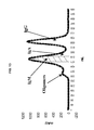
- FIG. 13 is a graph illustrating a chromatographic profile of an IgM preparation of the invention.
- the present invention relates to the discovery that IgM-containing immunoglobulin preparations derived from pooled human plasma comprise IgM that binds specifically to ⁇ amyloid peptides.
- the invention provides immunoglobulin preparations and methods useful for the treatment and/or prophylaxis of diseases and disorders associated with amyloidosis, including Alzheimer's disease.
- compositions or methods “comprising” one or more recited elements may include other elements not specifically recited.
- a composition that comprises IgM can encompass immunoglobulins of other types, and can include other proteinaceous and non-proteinaceous substances.
- the term “about” or “approximately” means that a value can fall within a scientifically acceptable range for that type of value, which also will depend on how quantitative a measurement of the value can be achieved given the available tools of measurement.
- antibody and “immunoglobulin” are used interchangeably herein, unless otherwise indicated expressly.
- a neurodegenerative disease or disorder is associated with amyloidosis when amyloid deposits or amyloid plaques are found in or in proximity to tissues affected by the disease, or when the disease is characterized by overproduction of a protein, particularly an amyloid protein, that is or can become insoluble.
- the amyloid plaques can provoke pathological effects directly or indirectly by known or unknown mechanisms.
- amyloid diseases include, but are not limited to, systemic diseases, such as chronic inflammatory illnesses, multiple myeloma, macroglobulinernia, familial amyloid polyneuropathy (Portuguese) and cardiomyopathy (Danish), systemic senile amyloidosis, familial amyloid polynephropathy (Iowa), familial amyloidosis (Finnish), Gerstrnann-Straussler-Scheinker syndrome, familial amyloid nephropathy with urticaria and deafness (Muckle-Wells syndrome), medullary carcinoma of thyroid, isolated atrial amyloid, and hemodialysis-associated amyloidosis (HAA); and amyloid-associated neurodegenerative diseases.
- systemic diseases such as chronic inflammatory illnesses, multiple myeloma, macroglobulinernia, familial amyloid polyneuropathy (Portuguese) and cardiomyopathy (Danish), systemic senile am
- neurodegenerative disease refers to a disease or disorder of the nervous system, particularly involving the brain, that manifests with symptoms characteristic of brain or nerve dysfunction, e.g., short-term or long-term memory lapse or defects, dementia, cognition defects, balance and coordination problems, and emotional and behavioral deficiencies.
- diseases are “associated with amyloidosis” when histopathological (biopsy) samples of brain tissue from subjects who demonstrate such symptoms reveal amyloid plaque formation.
- AD Alzheimert's disease
- traditional diagnosis depends on symptomology and, if relevant, family history.
- a physician will diagnose AD on the basis of symptoms of senile dementia, including cognitive dysfunction, retrograde amnesia (loss of memory for recent events), progressive impairment of remote memory, and possibly depression or other neurotic syndromes.
- the individual presents with slow disintegration of personality and intellect. Imaging may reveal large cell loss from the cerebral cortex and other brain areas.
- AD differs from senile dementia, however, by age of onset: AD is likely to occur in the fifth or sixth decade, whereas senile dementia occurs in the eighth decade or later.
- the neurodegenerative disease associated with amyloidosis is AD, a condition that includes sporadic AD, ApoE4-related AD, other mutant APP forms of AD (e.g., mutations at APP717, which are the most common APP mutations), mutant PS1 forms of familial AD (FAD) (see, WO 96/34099), mutant PS2 forms of FAD (see, WO 97/27296), and ⁇ -2-macroglobulin-polymorphism-related AD.
- AD a condition that includes sporadic AD, ApoE4-related AD, other mutant APP forms of AD (e.g., mutations at APP717, which are the most common APP mutations), mutant PS1 forms of familial AD (FAD) (see, WO 96/34099), mutant PS2 forms of FAD (see, WO 97/27296), and ⁇ -2-macroglobulin-polymorphism-related AD.
- the disease can be the rare Swedish disease characterized by a double KM to NL mutation in amyloid precursor protein (APP) near the amino-terminus of the ⁇ AP portion of APP (Levy et al., Science 248:1124-26 (1990)).
- APP amyloid precursor protein
- Another such disease is hereditary cerebral hemorrhage with amyloidosis (HCHA or HCHWA)-Dutch type (Rozemuller et al., Am. J. Pathol. 142:1449-57 (1993); Roos et al., Ann. N.Y. Acad. Sci. 640:155-60 (1991); Timmers et al., Neurosci. Lett.
- amloid refers generally to insoluble proteinaceous substances with particular physical characteristics independent of the composition of proteins or other molecules that are found in the substance. Amyloid can be identified by its amorphous structure, eosinophilic staining, changes in thioflavin fluorescence, and homogeneous appearance.
- Amyloid polypeptides Protein or peptide components of amyloid are termed herein “amyloid polypeptides,” and include, but are not limited to, ⁇ -amyloid peptide (A ⁇ ), including synthetic ⁇ APs corresponding to the first 28, 40, or 42 amino acids of A ⁇ , i.e., A ⁇ 1-28, A ⁇ 1-40, A ⁇ 1-42, respectively, as well as a synthetic ⁇ AP corresponding to amino acids 25-35 of A ⁇ , i.e., A ⁇ 25-35.
- a ⁇ ⁇ -amyloid peptide
- amyloid peptides include scrapie protein precursor or prion protein (associated with Creuzfeldt-Jacob's disease); synuclein (associated with Parkinson's disease), Huntington's protein (associated with Huntington's chorea), immunoglobulin, including ⁇ or ⁇ light or heavy chains, or fragments thereof, produced by myelomas; serum amyloid A; ⁇ 2-microglobulin; ApoA1; gelsolin; cystatin C; (pro)calcitonin; atrial natriuretic factor; islet amyloid polypeptide, also known as amylin (see, Westermark et al., Proc. Natl. Acad. Sci.
- amyloid is used herein to refer to substances that contain A ⁇ .
- Amyloidosis refers to the in vivo deposition or aggregation of proteins to form amyloid plaques or fibrils.
- the 42 amino acid (4.2 kDa) ⁇ -Amyloid Peptide (A ⁇ 1-42 or ⁇ AP) derives from a family of larger Amyloid Peptide Precursor (APP) proteins (Glenner and Wong, Biochem. Biophys. Res. Commun. 120:885-890 (1984); Glenner and Wong, Biochem. Biophys. Res. Commun. 122:1131-35 (1984); Goldgaber et al., Science 235:8778-8780 (1987); Kang et al., Nature 325:733-736 (1987); Robakis et al., Proc. Natl. Acad. Sci.
- APP Amyloid Peptide Precursor
- APP 25 is a transmembrane protein found in a number of isoforms, which in general are referred to herein as full length APP (flAPP).
- flAPP full length APP
- sAPP ⁇ soluble form of APP
- the “level of A ⁇ ” in a biological sample can be detected by any method known in the art, including by not limited to immunoassay, biochemical analysis (e.g., purification, gel electrophoresis, quantitative amino acid sequence analysis or composition analysis, Congo red or Thioflavin-T staining, and the like), or other methods known to detect A ⁇ .
- biochemical analysis e.g., purification, gel electrophoresis, quantitative amino acid sequence analysis or composition analysis, Congo red or Thioflavin-T staining, and the like
- fluorescence methods using Thioflavin T are used to detect aggregated peptide.
- a “biological sample” includes, but is not limited to body fluids (blood, blood cells, plasma, serum, cerebrospinal fluid, urine), tissues (e.g., spinal chord, nerves, etc.), or organs (preferably brain, but also including liver, kidney, pancreas, etc.).
- Assays for anti- ⁇ amyloid antibody can be accomplished by techniques known in the art, e.g., radioimmunoassay, ELISA (enzyme-linked immunosorbant assay), “sandwich” immunoassays, immunoradiometric assays, gel diffusion precipitation reactions, immunodiffusion assays, in situ immunoassays (using colloidal gold, enzyme or radioisotope labels, for example), Western blots, precipitation reactions, agglutination assays (e.g., gel agglutination assays, hemagglutination assays), complement fixation assays, immunofluorescence assays, protein A assays, and immunoelectrophoresis assays, etc.
- radioimmunoassay e.g., ELISA (enzyme-linked immunosorbant assay), “sandwich” immunoassays, immunoradiometric assays, gel diffusion precipitation reactions, immunodiffusion assay
- antibody binding is detected by detecting a label on the primary antibody.
- the primary antibody is detected by detecting binding of a secondary antibody or reagent to the primary antibody.
- the secondary antibody is labeled.
- Many means are known in the art for detecting binding in an immunoassay and are within the scope of the present invention. For example, to select antibodies which recognize a specific epitope of an amyloid peptide, one can assay generated hybridomas for a product which binds to an amyloid peptide fragment containing such epitope.
- the present invention relates to the discovery that IgM purified from donated, pooled human sources exhibits specificity toward A ⁇ 1-42 peptide.
- IgM-containing immunoglobulin preparations can be prepared in accordance with U.S. Pat. No. 6,307,028 to Lebing, et al., fully incorporated herein by reference.
- Lebing, et al. disclose a process for preparation of IVIG which includes an anion exchange chromatographic step, where this resin retains most of the IgM of the starting materials.
- IgM is eluted from this anion exchange resin, and subjected to gel filtration, followed by isoagglutinin removal by passing the preparation through a resin comprising immobilized synthetic antigen A and B.
- Oligomerization of IgM is reduced by processing at low concentrations, at relatively low pH, and by minimizing exposure of IgM to high salt. Final preparations are formulated at 0.2 M glycine (pH 4.2) to further avoid oligomerization. Further details regarding preparation of IgM according to the invention are provided in Example 1 below. However, it should be recognized that specific examples herein are only illustrative of the invention, and none are intended to be limiting of the scope of the invention as claimed.
- an ELISA assay to quantify anti-A ⁇ 1-42 in the IgM pool was developed.
- a microtiter plate was coated with synthetic peptide A ⁇ 1-42, its shortened version A ⁇ 22-35, as well as an irrelevant protein ⁇ 1-protease inhibitor, and varying concentrations of pooled, plasma-derived IgM were added to the plate.
- Bound IgM was detected using goat anti-human IgM conjugated with horseradish peroxidase. IgM showed a high and saturated binding to the full-size amyloid peptide ( FIG. 2 ), whereas very little or no binding was detected for the truncated peptide or ⁇ 1-protease inhibitor.
- a ⁇ 1-42 peptide derivatives such as A ⁇ 1-28, A ⁇ 25-35, or even the A ⁇ 1-40 peptide, were able to compete for the IgM binding. Without wishing to be bound by any particular theory, these results may indicate that either: 1) the IgMs against A ⁇ 1-42 require the peptide to be in a particular confirmation; or 2) that the epitope recognition site is in the C-terminal portion of the peptide.
- fragments of IgM generated using 2-mercaptoethylamine were tested for binding to A ⁇ 1-42 peptides.
- the newly generated IgM fragments were diluted and added to A ⁇ 1-42 and ⁇ 1PI coated wells to test for binding and specificity.
- the IgG-type fragments retained their binding and specificity characteristics, similar to the pentomeric IgM (see FIG. 8 ).
- IgM contains antibody against ⁇ -amyloid peptide A ⁇ 1-42 indicates that this immunoglobulin can be useful for the management of Alzheimer's disease.
- Intravenously delivered IgM can be used prophylactically, in individuals susceptible to Alzheimer's disease, or for treatment of patients diagnosed with Alzheimer's disease.
- monomeric IgM produced as a result of mild reduction of disulfides connecting all five subunits with the J-chain
- low-molecular weight derivatives of IgM for example, proteolytic fragments of IgM
- smaller IgM fragments may pass the brain blood barrier more efficiently and, therefore, be more potent than full length IgM.
- IgM derivatives can be tested according to the methods and procedures disclosed herein for retention of binding and selectivity toward A ⁇ 1-42 and A ⁇ -related peptides.
- patient includes human and other mammalian subjects that receive either prophylactic or therapeutic treatment.
- Immunoglobulin preparations comprising anti-A ⁇ IgM according to the present invention can be a safer alternative when used for passive immunization of those suffering from, or at risk for the development of, amyloid-related disease.
- the anti-amyloid peptide immunoglobulin preparations of the invention can be formulated in a pharmaceutical composition with a pharmaceutically acceptable carrier.
- pharmaceutically acceptable refers to molecular entities and compositions that are physiologically tolerable and do not typically produce an allergic or similar untoward reaction, such as gastric upset, dizziness and the like, when administered to a human.
- pharmaceutically acceptable can mean approved by a regulatory agency of the Federal or a state government or listed in the U.S. Pharmacopeia or other generally recognized pharmacopeia for use in animals, and more particularly in humans.
- carrier refers to a diluent, adjuvant, excipient, or vehicle with which the compound is administered.
- Such pharmaceutical carriers can be sterile liquids, such as water and oils, including those of petroleum, animal, vegetable or synthetic origin, such as peanut oil, soybean oil, mineral oil, sesame oil and the like.
- Water or aqueous solution saline solutions and aqueous dextrose and glycerol solutions are preferably employed as carriers, particularly for injectable solutions. Suitable pharmaceutical carriers are described in “Remington's Pharmaceutical Sciences” by E. W. Martin.
- compositions comprising the anti-amyloid immunoglobulin preparations of the invention can be introduced parenterally, transmucosally, e.g., orally (per os), nasally, or rectally, or transdermally.
- Parenteral routes include intravenous, intra-arteriole, intramuscular, intradermal, subcutaneous, intraperitoneal, intraventricular, and intracranial administration. Administration can be directly into the cerebrospinal fluid, e.g., by a spinal tap.
- the preparations of the invention can be delivered in a vesicle, in particular a liposome (see Langer, Science 249:1527-1533 (1990); Treat et al., in Liposomes in the Therapy of Infectious Disease and Cancer, Lopez-Berestein and Fidler (eds.), Liss: New York, pp. 353-365 (1989); Lopez-Berestein, ibid., pp. 317-327; see generally ibid.).
- a liposome see Langer, Science 249:1527-1533 (1990); Treat et al., in Liposomes in the Therapy of Infectious Disease and Cancer, Lopez-Berestein and Fidler (eds.), Liss: New York, pp. 353-365 (1989); Lopez-Berestein, ibid., pp. 317-327; see generally ibid.).
- the preparations of the invention can be delivered in a controlled release system.
- a polypeptide can be administered using intravenous infusion with a continuous pump, in a polymer matrix such as poly-lactic/glutamic acid (PLGA), a pellet containing a mixture of cholesterol and the anti-amyloid peptide antibody compound (SILASTICR, Dow Corning, Midland, Mich.; see U.S. Pat. No. 5,554,601) implanted subcutaneously, an implantable osmotic pump, a transdermal patch, liposomes, or other modes of administration.
- a pump can be used (see Langer (1990); Sefton, CRC Crit. Ref Biomed. Eng.
- polymeric materials can be used (see Medical Applications of Controlled Release, Langer and Wise (eds.), CRC Press: Boca Raton, Fla. (1974); Controlled Drug Bioavailability, Drug Product Design and Performance, Smolen and Ball (eds.), Wiley: N.Y. (1984); Ranger and Peppas, J. Macromol. Sci. Rev. Macromol. Chem. 23:61 (1983); see also Levy et al., Science 228:190 (1985); During et al., Ann.
- a controlled release system can be placed in proximity of the therapeutic target, i.e., the brain, thus requiring only a fraction of the systemic dose (see, e.g., Goodson, in Medical Applications of Controlled Release, vol. 2, pp. 115-138 (1984)).
- a controlled release device can be introduced into a subject in proximity of the site of amyloidosis. Controlled release systems are discussed in the review by Langer ( Science 249:1527-1533 (1990)).
- a disease or disorder subject to treatment or prevention according to the invention can be a neuropathy involving amyloid deposition, and can be associated with specific or general immunodeficiency.
- diseases include, but are not limited to, AD; Kuru, Creuzdfelt-Jacob's disease, and other spongiform encephalopathies; Parkinson's Disease; and Huntington's chorea.
- a constant in vivo supply of the anti-amyloid peptide antibodies from the immunoglobulin preparations of the invention can be ensured by providing a therapeutically effective dose (i.e., a dose effective to induce metabolic changes in a subject) at the necessary intervals, e.g., daily, every 12 hours, etc.
- a therapeutically effective dose i.e., a dose effective to induce metabolic changes in a subject
- the anti-amyloid peptide immunoglobulin preparation is administered for at least ten days, at least 100 days, or for the life of the recipient.
- prevent means to prophylactically interfere with a pathological mechanism that results in the disease or disorder, resulting in at least some clinically recognizable decrease in the rate of deterioration or ultimate extent of damage by the disease.
- a pathological mechanism can be an increase in processing of the amyloidogenic form of APP; dysregulation of A ⁇ clearance; or some combination of the two.
- treatment means to cause an improvement in a condition associated with the disease or disorder.
- treatment includes a reduction in the level of A ⁇ , regulation of the formation of A ⁇ , decrease in aggregation of A ⁇ or the formation of amyloid plaques, or improvement of a cognitive defect in a subject suffering from a disease or disorder associated with amyloidosis, e.g., AD or an animal model of AD.
- a “therapeutically effective amount” of the immunoglobulin preparations of the invention can treat or prevent a clinically significant deficit in the activity, function, and response of the host. Alternatively, a therapeutically effective amount can be sufficient to cause a clinically significant improvement of a disease condition in the host.
- a subject who “has an increased risk of developing” a neurological disease or disorder associated with amyloidosis can have a genetic predisposition to developing an amyloidosis, such as a person from a family that has members with familial AD (FAD). Alternatively, someone in his or her seventh or eighth decade is at greater risk for age-related AD.
- FAD familial AD
- a subject who “shows a symptom of” a neurological disease or disorder associated with amyloidosis presents with a symptom or complaint found in subjects who have or have had such a disease or disorder.
- these symptoms can include development of dementia, memory defects, and the like in the fifth and sixth decade, as discussed above.
- An “A ⁇ level reducing dose” is an amount of anti-amyloid peptide antibody that causes a decrease in the level of A ⁇ , e.g. in the brain or spinal fluid of a treated subject. Dosages can range from about 0.1 ⁇ g anti-amyloid peptide antibody per kg body weight ( ⁇ g/kg) to about 100 mg/kg; 0.5 ⁇ g anti-amyloid peptide antibody per kg body weight ( ⁇ g/kg) to about 50 mg/kg; or from about 5 ⁇ g/kg to about 10 mg/kg.
- the amount of anti-amyloid peptide antibody used to decrease the level of A ⁇ can be an amount corresponding to the level of anti-amyloid peptide antibody in a biological sample, especially blood (including plasma and serum) and cerebrospinal fluid (CSF), from a normal subject.
- a biological sample especially blood (including plasma and serum) and cerebrospinal fluid (CSF)
- “Reducing a level of amyloid- ⁇ (A ⁇ ) peptides” can refer to decreasing the amount of A ⁇ 1-42 in vivo.
- a ⁇ can accumulate in blood, cerebrospinal fluid, or organs.
- the primary organ of interest for reducing the level of A ⁇ is the brain, but A ⁇ levels can also be reduced in body fluids, tissues, and/or other organs by the practice of this invention.
- Effective doses of the compositions of the present invention, for the treatment of the above described conditions vary depending upon many different factors, including means of administration, target site, physiological state of the patient, whether the patient is human or an animal, other medications administered, and whether treatment is prophylactic or therapeutic. Treatment dosages can be titrated to optimize safety and efficacy.
- the dosage ranges from about 0.0001 to 2000 mg/kg, 200 to 1000 mg/kg, and more usually 0.01 to 100 mg/kg, of the host body weight.
- dosages can be 100 mg/kg body weight or 1000 mg/kg body weight or within the range of 100-1000 mg/kg.
- An exemplary treatment regime entails administration once per every two weeks or once a month or once every 3 to 6 months.
- Antibodies are usually administered on multiple occasions. Intervals between single dosages can be weekly, monthly or yearly. Intervals can also be irregular as indicated by measuring blood levels of antibody to A ⁇ in the patient.
- dosage is adjusted to achieve a plasma antibody concentration of 1-50 mg/ml, and in some methods 1-20 mg/ml.
- antibodies can be administered as a sustained release formulation, in which case less frequent administration is required. Dosage and frequency vary depending on the half-life of the antibodies in the patient.
- the dosage and frequency of administration can vary depending on whether the treatment is prophylactic or therapeutic.
- a relatively low dosage is administered at relatively infrequent intervals over a long period of time. Some patients continue to receive treatment for the rest of their lives.
- a relatively high dosage at relatively short intervals is sometimes required until progression of the disease is reduced or terminated, and preferably until the patient shows partial or complete amelioration of symptoms of disease. Thereafter, the patient can be administered a prophylactic regime.
- Agents of the invention can optionally be administered in combination with other agents that are at least partly effective in treatment of amyloidogenic disease.
- agents of the invention can also be administered in conjunction with other agents that increase passage of the agents of the invention across the blood-brain barrier.
- the procedures for monitoring passive immunization are similar to those that can be used for monitoring active immunization.
- the antibody profile following passive immunization typically shows an immediate peak in antibody concentration followed by an exponential decay. Without a further dosage, the decay approaches pretreatment levels within a period of days to months depending on the half-life of the antibody administered. For example the half-life of some human antibodies is of the order of 20 days.
- a baseline measurement of antibody to A ⁇ in the patient can be made before administration, a second measurement can be made soon thereafter to determine the peak antibody level, and one or more further measurements are made at intervals to monitor decay of antibody levels.
- a further dosage of antibody is administered.
- peak or subsequent measured levels less background can be compared with reference levels previously determined to constitute a beneficial prophylactic or therapeutic treatment regime in other patients. If the measured antibody level is significantly less than a reference level (e.g., less than the mean minus one standard deviation of the reference value in population of patients benefiting from treatment), administration of an additional dosage of antibody can be indicated.
- IgM was purified from the ANX column eluate from the IGIV preparation process (the second anion exchange step of the process as disclosed in U.S. Pat. No. 6,307,028 to Lebing, et al. (Lebing, et al.), the contents of which are fully incorporated herein by reference), followed by gel filtration on SUPEROSE 6 which yields greater than 90% pure IgM.
- the major impurities are IgA ( ⁇ 6-8%) and IgG ( ⁇ 2%).
- IgM was passed through a column containing immobilized synthetic antigen A and B. Details are provided below.
- ANX eluate is a pH 5.1, 0.5M acetate, 0.2% protein solution which can contain as much as 50% IgM.
- the non-IgM protein material present in ANX eluate is mostly IgG and IgA.
- 1.3 L of frozen ANX column eluate was thawed and the pH adjusted to 7.95 with 1.0 N NaOH.
- the material was concentrated on a 100 K PELLICON mini and/or a PELLICON XL cassette (Millipore Corporation, Bedford, Mass.) to 15 to 25 mg/ml.
- 65 ml of the concentrate (approximately 1150 mg of protein) was loaded onto a 5.0 cm ⁇ 70 cm SUPEROSE 6 FF Prep grade column (Pharmacia, Upsala, Sweden) equilibrated with TBS at a linear flow rate of 15.3 cm/hr.
- a 5.0 cm ⁇ 5.1 cm Atri/Btri PAA SEPHAROSE 6 FF column was plumbed in series.
- the central portion of the IgM peak (approximately 200 ml) was collected into sterile containers, sampled and immediately dialyzed against 0.2M glycine, pH 4.2, using sterile dialysis tubing.
- the dialysis consisted of four ⁇ 2 L changes over a period of 18 hrs at 4° C. Following dialysis, the sublots were sterile filtered and diluted to 2.0 mg/ml, then stored at +4° C. until bulking. Percent yields for each sublot were between 27 and 68% recovery of IgM. The majority of the IgM losses were due to oligomerization of IgM, which eluted in the void volume of the size-exclusion column.
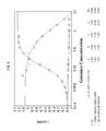
- Total protein was calculated using the published specific ( ⁇ 1% 280 ) extinction coefficient of 13.3. Amino acid analysis (Commonwealth Biotechnologies, Richmond, Va.) of this IgM preparation yielded a specific extinction coefficient of 13.4. Percent Oligomer was determined using a 10/30 SUPEROSE 6 PHARMACIA HR column and is defined as the percent area at 15.7 minutes divided by the total area of the chromatogram. Samples submitted for Isoagglutination Cross Match Test were concentrated to 50 mg/ml after pH adjustment to 7.5 with 10 ⁇ PBS. Purity by Reduced SDS-PAGE is defined by a two-step procedure.
- the content of the only unidentified X-band on the gel is determined; it gives the total percentage of immunoglobulins in the preparation.
- the content of IgA and IgG is determined by measuring the intensity of the heavy chain bands of all three immunoglobulins.
- the purity of IgM is equal to 100%-% Xband-% IgA-% IgG.
- IgM tends to form stable oligomers in a time- and concentration-dependent process.
- the purification strategy therefore, included steps to minimize IgM oligomerization such as processing at low pH, keeping the IgM preparations at low concentration, and minimizing IgM exposure to high salt.
- the final preparations of IgM were formulated in a 0.2 M Glycine (pH 4.2) solution to avoid oligomer formation; in this formulation IgM is stable and suitable for injections.
- IgM fragmentation kit Piercat. No. 44887. This kit has the ability to fragment IgM by three methods (See FIG. 6 ). Alkylation and reduction of IgM with 2-mercaptoethylamine and iodoacetamide was used to generate IgM derivatives of interest. Derived IgMs were analyzed on a 3-8% Tris-Acetate PAGE (See FIG. 7 ). Four main IgM fragments corresponding to the expected molecular weights for monomeric IgM ( ⁇ 200 KDa) and half of the monomeric IgM ( ⁇ 100 KDa) were noted.
- FIG. 6 illustrates the results of the MEA IgM fragmentation process, along with results using other methods.
- FIG. 7 shows the results of gel electrophoresis of IgM and MEA-treated IgM (showing fragment species corresponding to “IgG”-type and “r IgG” fragments illustrated in FIG. 6 ).
- a 96-well Nunc MAXISORP microtiter plate was passively coated with 100 ⁇ l of 2 ug/ml A ⁇ 1-42, A ⁇ 22-35, and an unrelated protein ( ⁇ -1 protease inhibitor, ⁇ 1PI) for 1 hour at 25° C. with gentle shaking.
- the last two rows in the plate were coated with 100 ul of PIERCE SUPERBLOCK to serve as a negative (non-specific) control.
- the plate was washed twice with 300 ⁇ l of wash buffer.
- the plate was blocked with 100 ⁇ l of SUPERBLOCK for 1 hour at 25° C. with gentle shaking and washed twice with wash buffer.
- 100 ⁇ l of IgM serially diluted in SUPERBLOCK was added to the plate and incubated for 2 hours at 25° C. with gentle shaking. The results are shown in FIG. 2 .
- a 96-well NUNC MAXISORP microtiter plate was passively coated with 100 ⁇ l of 2 ⁇ g/ml A ⁇ 1-42 and ⁇ 1PI for 1 hour at 25° C. with gentle shaking. Following the coating procedure, the plate was washed twice with wash buffer. The plate was blocked with 100 ⁇ l SUPERBLOCK for 1 hour at 25° C. with gentle shaking and washed twice with wash buffer. Previously 0.2 milligrams of IgM was incubated with an uncoupled and A ⁇ 1-42 coupled affinity purification column overnight at 4° C. with gentle rocking. The depleted IgM material (flowthrough) was serial diluted in SUPERBLOCK and incubated for 2 hours at 25° C. with gentle shaking. The results are shown in FIG. 3 .
- a 96-well NUNC MAXISORP microtiter plate was passively coated with 100 ⁇ l of 2 ug/ml A ⁇ 1-42 for 1 hour at 25° C. with gentle shaking. Following the coating procedure, the plate was washed twice with wash buffer. The plate was blocked with 100 ⁇ l of SUPERBLOCK for 1 hour at 25° C. with gentle shaking and washed twice with wash buffer. 100 ⁇ l of GAMUNEX serial diluted in SUPERBLOCK with 0.1 mg/ml IgM was added to the plate and incubated for 2 hours at 25° C. with gentle shaking. Also 100 ⁇ l of IgM and GAMUNEX, serial diluted in SUPERBLOCK, were added to the plate and served as positive and negative controls. The results are shown in FIG. 5 .
- a 96-well NUNC MAXISORP microtiter plate was passively coated with 100 ⁇ l of 2 ⁇ g/ml A ⁇ 1-42 and ⁇ 1PI for 1 hour at 25° C. with gentle shaking. Following the coating procedure, the plate was washed twice with wash buffer. The plate was blocked with 100 ⁇ l of SUPERBLOCK for 1 hour at 25° C. with gentle shaking and washed twice with wash buffer. 100 ⁇ l of IgM and 2-mercaptoethylamine treated IgM, serial diluted in SUPERBLOCK, was added to the plate and incubated for 2 hours at 25° C. with gentle shaking. The results are shown in FIG. 8 .
- a 96-well NUNC MAXISORP microtiter plate was passively coated with 100 ⁇ l of 2 ⁇ g/ml A ⁇ 1-40, A ⁇ 1-42, A ⁇ 1-43 and ⁇ 1PI (negative control) for 1 hour at 25° C. with gentle shaking. Following the coating procedure, the plate was washed twice with wash buffer. The plate was blocked with 100 ⁇ l of SUPERBLOCK for 1 hour at 25° C. with gentle shaking and washed twice with wash buffer. 100 ⁇ l of IgM serially diluted in SUPERBLOCK was added to plate and incubated for 2 hours at 25° C. with gentle shaking.
- a 96-well NUNC MAXISORP microtiter plate was passively coated with 100 ⁇ l of 2 ⁇ g/ml A ⁇ 1-42 for 1 hour at 25° C. with gentle shaking. Following the coating procedure, the plate was washed twice with wash buffer. The plate was blocked with 100 ⁇ l of SUPERBLOCK for 1 hour at 25° C. with gentle shaking and washed twice with wash buffer. 0.1 mg/ml solutions of A ⁇ 1-40, A ⁇ 1-42 and A ⁇ 1-43 were each spiked with 7 mg/ml IgM to obtain a 0.07 mg/ml IgM final concentration. Next, the solutions were serially diluted in 0.07 mg/ml IgM and 100 ⁇ l was added to plate and incubated for 2 hours at 25° C. with gentle shaking. The plate was then washed 6 times with wash buffer. 100 ⁇ l of horseradish peroxidase conjugated goat anti-human IgM was added to each well and the plate was incubated for 1 hour at 25° C. with gentle shaking.
- a 96-well NUNC MAXISORP microtiter plate was passively coated with 100 ul of 2 ug/ml A ⁇ 1-42 for 1 hour at 25° C. with gentle shaking. Following the coating procedure, the plate was washed twice with wash buffer. The plate was blocked with 100 ⁇ l of SUPERBLOCK for 1 hour at 25° C. with gentle shaking and washed twice with wash buffer.
- a 0.5 mg/ml solution of A ⁇ 37-42 and 0.1 mg/ml solutions of A ⁇ 1-40, A ⁇ 1-42 and A ⁇ 33-42 were each spiked with 7 mg/ml IgM to obtain a 0.07 mg/ml IgM final concentration. Next the solutions were serially diluted in 0.07 mg/ml IgM and 100 ul was added to plate and incubated for 2 hours at 25° C. with gentle shaking.
- a ⁇ 1-42 is able to completely inhibit IgM binding to A ⁇ 1-42 coated plates.
- the fragmented amyloid ⁇ peptides show no more inhibition than A ⁇ 1-40.
- This data demonstrates that in addition to amino acids 41 and 42 there appears to be a need for a peptide greater than 10 amino acids with possibly a tertiary structure to inhibit IgM binding to A ⁇ 1-42 coated plates. See FIG. 11 .
- a 96-well NUNC MAXISORP microtiter plate was passively coated with 100 ⁇ l of 2 ⁇ g/ml A ⁇ 1-42 for 1 hour at 25° C. with gentle shaking. Following the coating procedure, the plate was washed twice with wash buffer. The plate was blocked with 100 ⁇ l of SUPERBLOCK for 1 hour at 25° C. with gentle shaking and washed twice with wash buffer. 0.1 mg/ml solutions of A ⁇ 1-42 and scrambled A ⁇ 1-42 were each spiked with 7 mg/ml IgM to obtain a 0.07 mg/ml IgM final concentration. Next the solutions were serially diluted in 0.07 mg/ml IgM and 100 ⁇ l was added to plate and incubated for 2 hours at 25° C. with gentle shaking.
- IgM titer Data demonstrates the specificity of the IgM titer is to A ⁇ 1-42 and not to peptide of the same size containing all 42 amino acids. See FIG. 12 .
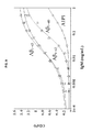
- the OCTET system utilizes Bio-Layer Interferometry (BLI) to measure concentration, affinity and kinetics between two proteins/peptides.
- BLI Bio-Layer Interferometry detects changes in the interference pattern (reflected light) as the number of molecules increases or decreases from the tip of the detector.
- Streptavidin-coated biosensors were first placed in wells containing PBS to equilibrate the detectors. Next, the biosensors were placed in wells containing 2 ⁇ g/ml biotinylated A ⁇ 1-42 and then moved to wells containing PBS so that a background interference profile could be created. These biosensors were then moved to wells that contained either IgM pool, monomeric IgM pool, or IgG pool (GAMUNEX) and the association rates were determined. Finally, the biosensors were moved to wells containing PBS to measure the dissociation rates.
- the B6; SJL-Tg(APPSWE)2576 Kha or “Tg2576” mouse expresses a mutated form of the human APP695 gene driven by a hamster prion protein gene promoter (see U.S. Pat. No. 5,877,399). These mice have normal spatial reference memory at three months of age but show impairment by 9 to 10 months of age (Hsiao, et al., “Correlative memory deficits, A ⁇ elevation, and amyloid plaques in transgenic mice,” Science 274:99-102 (1996)). Brain transgenic APP content is 5.6 times more than endogenous APP. This increase accompanies the appearance of certain behavioral deficits.
- Amyloid plaques appear to stimulate a cellular inflammatory response. Both hypertrophic astrocytes and activated microglia surround the plaques (Irizarry, M., “APP sw transgenic mice develop age-related A ⁇ deposits and neuropil abnormalities, but no neuronal loss in CA1 ,” J. Neuropathol. Exp. Neurol. 56:965-73 (1997); Frautschy, S. A., et al., “The microglial response to amyloid plaques in APPsw transgenic mice,” Am. J. Pathol.
- amyloid angiopathy appears in some vessels (Klunk, W., et al., “Staining of AD and Tg2576 mouse brain with X-34, a highly fluorescent derivative of chrysamine G and a potential in vivo probe for b-sheet fibrils,” Soc. Neurosci. Abstr 23:1638 (1997)).
- key markers of oxidative stress are induced in Tg2576 mouse brain (Pappolla, M. A., et al., “Evidence of oxidative stress and in vivo neurotoxicity of ⁇ -amyloid in a transgenic mouse model of Alzheimer's disease,” Am. J. Pathol. 152:871-7 (1998)), similar to what is seen in the brain of AD patients at autopsy.
- mice are randomly assigned to treatment groups as follows (Table 2).
- mice are injected with either saline or hIgM according to the following schedule (Table 3—See Khole, V., et al., “Identification of epididymis specific antigen by neonatal tolerization,” Am. J. Repro. Immunol. 44:350-356 (2000)).
- Neonatal Tolerization Immunization Schedule Day 1 Day 5 Day 21 Day 35 Day 49 Route c i.p. i.p. s.q. s.q. s.q. s.q. Concentration 20 ⁇ g/ ⁇ l 20 ⁇ g/ ⁇ l 100 ⁇ g/ ⁇ l 100 ⁇ g/ ⁇ l 100 ⁇ g/ ⁇ l Volume 50 ⁇ l 50 ⁇ l 100 ⁇ l a 100 ⁇ l a 100 ⁇ l a Adjuvant b No No Yes Yes Yes a Multiple sites. b TITERMAX ® Gold (CytRx Corporation, Parkway Technology Park, Georgia). c Injections will be made with a 27G, 1 ⁇ 2” needle.
- mice receive 100 ⁇ l (400 mg/kg) of hIgM without adjuvant, subcutaneously, on Tuesday and Friday of each week (Dodel, R. C., et al., “Intravenous immunoglobulins containing antibodies against ⁇ -amyloid for the treatment of Alzheimer's disease,” J. Neurol. Neurosurg. Psychiatry 75: 1472-4 (2004)). Antibody treatment continues until termination of the study.
- mice are sampled on day 56 for antibodies to hIgM. Not more than 50 ⁇ l whole blood is obtained by nicking the lateral saphenous vein superficially with a sterile #15 scalpel blade. Manual pressure is applied to achieve hemostasis before mice are returned to cages. The sample is centrifuged at room temperature to separate blood cells from plasma. Plasma is harvested and immediately frozen at ⁇ 80° C. until analysis.
- mice are humanely sacrificed using CO 2 gas at 12-15 months of age, or if they appear moribund prior to that time. Blood is obtained via the caudal vena cava following euthanasia. Brains are harvested, and half are flash frozen for analysis of soluble A ⁇ -peptide and half are placed in 10% neutral buffered formalin for immunohistochemial staining and histopathologic analysis. Stained plaques will be counted and soluble A ⁇ -peptide will be quantified in brain and plasma. Data will be examined for normality and equal variance. A t-test will be used to assess differences between treated and untreated mice. Groups will be considered different if the P-value ⁇ 0.05.
- the invention provides for a number of uses.
- the invention provides for the use of any of the A ⁇ -binding immunoglobulin preparations described herein in the treatment or prophylaxis of amyloidogenic disease, or in the manufacture of a medicament for use in the same.
Abstract
Description
| TABLE 1 |
| Characterization of Purified IgM. |
| Test | Method | Specification | Assay Results |
| Total Protein | A280/ml | 1.5-2.5 | mg/ml | 1.6 | mg/ml |
| % Oligomer | SEC-HPLC | NMT 5% | <1.0% |
| % IgA | Immunonephelometry | NMT 5% | 1.6% |
| IgG | Immunonephelometry | 0.03 | mg/ml | |
| IgM | Immunonephelometry | 2.51 | mg/ |
| CH50 |
| 100 CH50 | <75 | units | 43 |
| Isoagglutinin | Isoagglutination Cross Match | Negative at 1:64 at 50 | Negative at |
| Test | mg/ml | 1:4 |
| Endotoxin | LAL | <0.3 | EU/ml | <0.06 | EU/ml |
| pH | Undiluted | 4.0-4.4 | 4.24 |
| Purity | Reduced SDS-PAGE Gel | 91% |
| % Protein | Biuret | 1-2 | mg/ml | 2.20 | mg/ml |
| TABLE 2 |
| Approximate numbers of mice per treatment. |
| Strain | #/treatment | Saline | hIgM | ||
| Tg2576 | 26 | 8 | 32 | ||
| Wild-type | 8 | 8 | 8 | ||
| TABLE 3 |
| Neonatal Tolerization |
| Day |
| 1 | Day 5 | Day 21 | Day 35 | Day 49 |
| Routec | i.p. | i.p. | s.q. | s.q. | s.q. |
| Concentration | 20 μg/μl | 20 μg/ |
100 μg/ |
100 μg/ |
100 μg/μl |
| Volume | 50 μl | 50 |
100 |
100 |
100 μla |
| Adjuvantb | No | No | Yes | Yes | Yes |
| aMultiple sites. | |||||
| bTITERMAX ® Gold (CytRx Corporation, Parkway Technology Park, Georgia). | |||||
| cInjections will be made with a 27G, ½” needle. | |||||
Claims (10)
Priority Applications (1)
| Application Number | Priority Date | Filing Date | Title |
|---|---|---|---|
| US12/162,532 US8852585B2 (en) | 2006-01-30 | 2007-01-30 | Method of treatment and prophylaxis of diseases related to amyloid deposition using IgM |
Applications Claiming Priority (3)
| Application Number | Priority Date | Filing Date | Title |
|---|---|---|---|
| US76342206P | 2006-01-30 | 2006-01-30 | |
| US12/162,532 US8852585B2 (en) | 2006-01-30 | 2007-01-30 | Method of treatment and prophylaxis of diseases related to amyloid deposition using IgM |
| PCT/US2007/061269 WO2007106617A2 (en) | 2006-01-30 | 2007-01-30 | Method of treatment and prophylaxis of diseases related to amyloid deposition using igm |
Publications (2)
| Publication Number | Publication Date |
|---|---|
| US20090269359A1 US20090269359A1 (en) | 2009-10-29 |
| US8852585B2 true US8852585B2 (en) | 2014-10-07 |
Family
ID=38510126
Family Applications (1)
| Application Number | Title | Priority Date | Filing Date |
|---|---|---|---|
| US12/162,532 Active 2030-07-13 US8852585B2 (en) | 2006-01-30 | 2007-01-30 | Method of treatment and prophylaxis of diseases related to amyloid deposition using IgM |
Country Status (7)
| Country | Link |
|---|---|
| US (1) | US8852585B2 (en) |
| EP (1) | EP1981540B1 (en) |
| JP (2) | JP5697847B2 (en) |
| ES (1) | ES2409844T3 (en) |
| PL (1) | PL1981540T3 (en) |
| PT (1) | PT1981540E (en) |
| WO (1) | WO2007106617A2 (en) |
Families Citing this family (15)
| Publication number | Priority date | Publication date | Assignee | Title |
|---|---|---|---|---|
| DE10303974A1 (en) | 2003-01-31 | 2004-08-05 | Abbott Gmbh & Co. Kg | Amyloid β (1-42) oligomers, process for their preparation and their use |
| RS53270B2 (en) | 2005-11-30 | 2018-05-31 | Abbvie Deutschland | Monoclonal antibodies against amyloid beta protein and uses thereof |
| AU2006319358B2 (en) | 2005-11-30 | 2012-01-19 | AbbVie Deutschland GmbH & Co. KG | Anti-Abeta globulomer antibodies, antigen-binding moieties thereof, corresponding hybridomas, nucleic acids, vectors, host cells, methods of producing said antibodies, compositions comprising said antibodies, uses of said antibodies and methods of using said antibodies |
| US8455626B2 (en) | 2006-11-30 | 2013-06-04 | Abbott Laboratories | Aβ conformer selective anti-aβ globulomer monoclonal antibodies |
| EP2486928A1 (en) | 2007-02-27 | 2012-08-15 | Abbott GmbH & Co. KG | Method for the treatment of amyloidoses |
| US8048420B2 (en) | 2007-06-12 | 2011-11-01 | Ac Immune S.A. | Monoclonal antibody |
| US8613923B2 (en) | 2007-06-12 | 2013-12-24 | Ac Immune S.A. | Monoclonal antibody |
| ES2445590T3 (en) | 2007-10-05 | 2014-03-04 | Genentech, Inc. | Use of anti-beta amyloid antibody in eye diseases |
| MX336196B (en) | 2010-04-15 | 2016-01-11 | Abbvie Inc | Amyloid-beta binding proteins. |
| BR112013002297A2 (en) | 2010-07-30 | 2016-05-24 | Ac Immune Sa | safe and functional humanized antibodies |
| WO2012024187A1 (en) | 2010-08-14 | 2012-02-23 | Abbott Laboratories | Amyloid-beta binding proteins |
| HUE046752T2 (en) * | 2013-02-26 | 2020-03-30 | Baxalta GmbH | Treatment of central nervous system disorders by intranasal administration of immunoglobulin g |
| EP3552624A1 (en) * | 2013-05-06 | 2019-10-16 | Baxalta Incorporated | Treatment of alzheimer's disease subpopulations with pooled immunoglobulin g |
| US9815886B2 (en) | 2014-10-28 | 2017-11-14 | Adma Biologics, Inc. | Compositions and methods for the treatment of immunodeficiency |
| US10259865B2 (en) | 2017-03-15 | 2019-04-16 | Adma Biologics, Inc. | Anti-pneumococcal hyperimmune globulin for the treatment and prevention of pneumococcal infection |
Citations (11)
| Publication number | Priority date | Publication date | Assignee | Title |
|---|---|---|---|---|
| US5075425A (en) | 1989-08-17 | 1991-12-24 | Biotest Pharma Gmbh | Process for the preparation of a pharmaceutical which contains igg, iga and igm and can be administered intravenously |
| US5554601A (en) | 1993-11-05 | 1996-09-10 | University Of Florida | Methods for neuroprotection |
| WO1996034099A2 (en) | 1995-04-28 | 1996-10-31 | Hsc Research And Development Limited Partnership | Genetic sequences and proteins related to alzheimer's disease, and uses therefor |
| WO1997027296A1 (en) | 1996-01-26 | 1997-07-31 | Hsc Research And Development Limited Partnership | Nucleic acids and proteins related to alzheimer's disease, and uses therefor |
| US5877399A (en) | 1994-01-27 | 1999-03-02 | Johns Hopkins University | Transgenic mice expressing APP-Swedish mutation develop progressive neurologic disease |
| US6136312A (en) | 1996-10-14 | 2000-10-24 | Rotkreuzstifung Zentrallaboratorium Blutspendedienst Srk | Method for producing an IgM preparation for intravenous application |
| US6307028B1 (en) | 1997-06-20 | 2001-10-23 | Bayer Corporation Incorporated | Chromatographic method for high yield purification and viral inactivation of antibodies |
| EP1172378A1 (en) | 2000-07-12 | 2002-01-16 | Richard Dr. Dodel | Human beta-amyloid antibody and use thereof for treatment of alzheimer's disease |
| US20030185827A1 (en) * | 1994-04-29 | 2003-10-02 | Mayo Foundation | Human IgM antibodies, and diagnostic and therapeutic uses thereof particularly in the central nervous system |
| WO2003097093A1 (en) | 2002-05-15 | 2003-11-27 | Rauno Joks | METHOD OF TREATING IMMUNE-MEDIATED DISEASES BY ADMINISTRATION OF IgM |
| US7186410B2 (en) | 2001-05-11 | 2007-03-06 | Laboratoiore Francais Du Fractionnement Et Des Biotechnologies | Method for preparing human immunoglobulin concentrates for therapeutic use |
Family Cites Families (1)
| Publication number | Priority date | Publication date | Assignee | Title |
|---|---|---|---|---|
| TW200510532A (en) * | 2003-07-15 | 2005-03-16 | Chugai Pharmaceutical Co Ltd | IgM production by transformed cell and method of quantifying the same |
-
2007
- 2007-01-30 EP EP07756509A patent/EP1981540B1/en active Active
- 2007-01-30 PT PT77565091T patent/PT1981540E/en unknown
- 2007-01-30 ES ES07756509T patent/ES2409844T3/en active Active
- 2007-01-30 JP JP2008552621A patent/JP5697847B2/en active Active
- 2007-01-30 WO PCT/US2007/061269 patent/WO2007106617A2/en active Application Filing
- 2007-01-30 US US12/162,532 patent/US8852585B2/en active Active
- 2007-01-30 PL PL07756509T patent/PL1981540T3/en unknown
-
2013
- 2013-08-30 JP JP2013179382A patent/JP2013256529A/en active Pending
Patent Citations (12)
| Publication number | Priority date | Publication date | Assignee | Title |
|---|---|---|---|---|
| US5075425A (en) | 1989-08-17 | 1991-12-24 | Biotest Pharma Gmbh | Process for the preparation of a pharmaceutical which contains igg, iga and igm and can be administered intravenously |
| US5554601A (en) | 1993-11-05 | 1996-09-10 | University Of Florida | Methods for neuroprotection |
| US5877399A (en) | 1994-01-27 | 1999-03-02 | Johns Hopkins University | Transgenic mice expressing APP-Swedish mutation develop progressive neurologic disease |
| US20030185827A1 (en) * | 1994-04-29 | 2003-10-02 | Mayo Foundation | Human IgM antibodies, and diagnostic and therapeutic uses thereof particularly in the central nervous system |
| WO1996034099A2 (en) | 1995-04-28 | 1996-10-31 | Hsc Research And Development Limited Partnership | Genetic sequences and proteins related to alzheimer's disease, and uses therefor |
| WO1997027296A1 (en) | 1996-01-26 | 1997-07-31 | Hsc Research And Development Limited Partnership | Nucleic acids and proteins related to alzheimer's disease, and uses therefor |
| US6136312A (en) | 1996-10-14 | 2000-10-24 | Rotkreuzstifung Zentrallaboratorium Blutspendedienst Srk | Method for producing an IgM preparation for intravenous application |
| US6307028B1 (en) | 1997-06-20 | 2001-10-23 | Bayer Corporation Incorporated | Chromatographic method for high yield purification and viral inactivation of antibodies |
| EP1172378A1 (en) | 2000-07-12 | 2002-01-16 | Richard Dr. Dodel | Human beta-amyloid antibody and use thereof for treatment of alzheimer's disease |
| US7186410B2 (en) | 2001-05-11 | 2007-03-06 | Laboratoiore Francais Du Fractionnement Et Des Biotechnologies | Method for preparing human immunoglobulin concentrates for therapeutic use |
| WO2003097093A1 (en) | 2002-05-15 | 2003-11-27 | Rauno Joks | METHOD OF TREATING IMMUNE-MEDIATED DISEASES BY ADMINISTRATION OF IgM |
| US20040043019A1 (en) * | 2002-05-15 | 2004-03-04 | Rauno Joks | Method of treating immune-mediated diseases by administration of IgM |
Non-Patent Citations (60)
Also Published As
| Publication number | Publication date |
|---|---|
| ES2409844T3 (en) | 2013-06-28 |
| WO2007106617A3 (en) | 2008-03-20 |
| JP5697847B2 (en) | 2015-04-08 |
| US20090269359A1 (en) | 2009-10-29 |
| PT1981540E (en) | 2013-05-07 |
| EP1981540A2 (en) | 2008-10-22 |
| JP2013256529A (en) | 2013-12-26 |
| PL1981540T3 (en) | 2013-08-30 |
| EP1981540B1 (en) | 2013-03-20 |
| WO2007106617A2 (en) | 2007-09-20 |
| JP2009525286A (en) | 2009-07-09 |
Similar Documents
| Publication | Publication Date | Title |
|---|---|---|
| US8852585B2 (en) | Method of treatment and prophylaxis of diseases related to amyloid deposition using IgM | |
| JP7229980B2 (en) | Methods for immunological targeting of pathological tau proteins | |
| Kuo et al. | High levels of circulating Aβ42 are sequestered by plasma proteins in Alzheimer's disease | |
| JP4738696B2 (en) | Humanized antibodies that sequester Aβ peptides | |
| AU2018255221B2 (en) | Anti-N3pGlu amyloid beta peptide antibodies and uses thereof | |
| EP2361638B1 (en) | A beta 1-42 specific monoclonal antibodies with therapeutic properties | |
| Szabo et al. | Natural human antibodies to amyloid beta peptide | |
| US20060099211A1 (en) | Safer, more potent human immunoglobulin preparations for treating Alzheimer's disease | |
| US10988529B2 (en) | Combination therapy | |
| US20140314773A1 (en) | Compositions and methods for treating alzheimer's disease | |
| WO2002074240A2 (en) | Anti-amyloid antibody based diagnosis and treatment of a neurological disease or disorder | |
| EP1954718A2 (en) | Anti-a globulomer antibodies, antigen-binding moieties thereof, corresponding hybridomas, nucleic acids, vectors, host cells, methods of producing said antibodies, compositions comprising said antibodies, uses of said antibodies and methods of using said antibodies | |
| US20210355202A1 (en) | Antibody binding active alpha-synuclein | |
| WO2017123517A1 (en) | ANTI-N3pGlu AMYLOID BETA PEPTIDE ANTIBODIES AND USES THEREOF | |
| AU2012335014A1 (en) | Compositions and methods for treating Alzheimer's disease | |
| AU2001268828B2 (en) | Neurotoxic oligomers | |
| Mruthinti et al. | Relationship between the induction of RAGE cell-surface antigen and the expression of amyloid binding sites | |
| US7550144B2 (en) | Prion inhibition | |
| US20160280742A1 (en) | Compositions and Methods for Treating Alzheimer's Disease | |
| AU2015201763A1 (en) | A beta 1-42 specific monoclonal antibodies with therapeutic properties |
Legal Events
| Date | Code | Title | Description |
|---|---|---|---|
| AS | Assignment |
Owner name: TALECRIS BIOTHERAPEUTICS, INC., NORTH CAROLINA Free format text: ASSIGNMENT OF ASSIGNORS INTEREST;ASSIGNORS:SCUDERI, PHILIP, JR.;SAFAVI, AFSHIN;LANGEVIN, MATTHEW G.;REEL/FRAME:022718/0746;SIGNING DATES FROM 20080904 TO 20090416 Owner name: TALECRIS BIOTHERAPEUTICS, INC., NORTH CAROLINA Free format text: ASSIGNMENT OF ASSIGNORS INTEREST;ASSIGNORS:SCUDERI, PHILIP, JR.;SAFAVI, AFSHIN;LANGEVIN, MATTHEW G.;SIGNING DATES FROM 20080904 TO 20090416;REEL/FRAME:022718/0746 |
|
| AS | Assignment |
Owner name: DEUTSCHE BANK AG NEW YORK BRANCH, NEW YORK Free format text: SECURITY AGREEMENT;ASSIGNORS:GRIFOLS, S.A.;GRIFOLS INC.;TALECRIS BIOTHERAPEUTICS, INC.;REEL/FRAME:026390/0193 Effective date: 20110601 |
|
| AS | Assignment |
Owner name: GRIFOLS THERAPEUTICS INC., NORTH CAROLINA Free format text: CHANGE OF NAME;ASSIGNOR:TALECRIS BIOTHERAPEUTICS, INC.;REEL/FRAME:030650/0819 Effective date: 20110810 |
|
| AS | Assignment |
Owner name: DEUTSCHE BANK AG NEW YORK BRANCH, AS COLLATERAL AGENT, NEW YORK Free format text: SECURITY AGREEMENT;ASSIGNORS:GRIFOLS INC.;GRIFOLS THERAPEUTICS INC.;GRIFOLS-CHIRON DIAGNOSTICS CORP.;REEL/FRAME:032367/0001 Effective date: 20140227 Owner name: DEUTSCHE BANK AG NEW YORK BRANCH, AS COLLATERAL AG Free format text: SECURITY AGREEMENT;ASSIGNORS:GRIFOLS INC.;GRIFOLS THERAPEUTICS INC.;GRIFOLS-CHIRON DIAGNOSTICS CORP.;REEL/FRAME:032367/0001 Effective date: 20140227 |
|
| STCF | Information on status: patent grant |
Free format text: PATENTED CASE |
|
| AS | Assignment |
Owner name: TALECRIS BIOTHERAPEUTICS, INC., CALIFORNIA Free format text: RELEASE OF SECURITY INTEREST RECORDED AT REEL/FRAME 26390/0193;ASSIGNOR:DEUTSCHE BANK AG NEW YORK BRANCH;REEL/FRAME:041494/0017 Effective date: 20140227 Owner name: GRIFOLS, S.A., SPAIN Free format text: RELEASE OF SECURITY INTEREST RECORDED AT REEL/FRAME 26390/0193;ASSIGNOR:DEUTSCHE BANK AG NEW YORK BRANCH;REEL/FRAME:041494/0017 Effective date: 20140227 Owner name: GRIFOLS INC., CALIFORNIA Free format text: RELEASE OF SECURITY INTEREST RECORDED AT REEL/FRAME 26390/0193;ASSIGNOR:DEUTSCHE BANK AG NEW YORK BRANCH;REEL/FRAME:041494/0017 Effective date: 20140227 |
|
| AS | Assignment |
Owner name: GRIFOLS SHARED SERVICES NORTH AMERICA INC., CALIFORNIA Free format text: RELEASE BY SECURED PARTY;ASSIGNOR:DEUTSCHE BANK AG NEW YORK BRANK;REEL/FRAME:041638/0527 Effective date: 20170131 Owner name: GRIFOLS THERAPEUTICS INC., CALIFORNIA Free format text: RELEASE BY SECURED PARTY;ASSIGNOR:DEUTSCHE BANK AG NEW YORK BRANK;REEL/FRAME:041638/0527 Effective date: 20170131 Owner name: GRIFOLS DIAGNOSTIC SOLUTIONS INC., CALIFORNIA Free format text: RELEASE BY SECURED PARTY;ASSIGNOR:DEUTSCHE BANK AG NEW YORK BRANK;REEL/FRAME:041638/0527 Effective date: 20170131 Owner name: GRIFOLS SHARED SERVICES NORTH AMERICA INC., CALIFO Free format text: RELEASE BY SECURED PARTY;ASSIGNOR:DEUTSCHE BANK AG NEW YORK BRANK;REEL/FRAME:041638/0527 Effective date: 20170131 |
|
| AS | Assignment |
Owner name: BANK OF AMERICA, N.A., AS COLLATERAL AGENT, NORTH CAROLINA Free format text: SECURITY INTEREST;ASSIGNORS:GRIFOLS SHARED SERVICES NORTH AMERICA INC.;GRIFOLS THERAPEUTICS INC.;GRIFOLS BIOLOGICALS INC.;AND OTHERS;REEL/FRAME:041651/0487 Effective date: 20170131 Owner name: BANK OF AMERICA, N.A., AS COLLATERAL AGENT, NORTH Free format text: SECURITY INTEREST;ASSIGNORS:GRIFOLS SHARED SERVICES NORTH AMERICA INC.;GRIFOLS THERAPEUTICS INC.;GRIFOLS BIOLOGICALS INC.;AND OTHERS;REEL/FRAME:041651/0487 Effective date: 20170131 |
|
| AS | Assignment |
Owner name: EUROPEAN INVESTMENT BANK, AS LENDER, LUXEMBOURG Free format text: SECURITY INTEREST;ASSIGNORS:GRIFOLS SHARED SERVICES NORTH AMERICA, INC.;GRIFOLS THERAPEUTICS INC.;GRIFOLS BIOLOGICALS INC.;AND OTHERS;REEL/FRAME:044696/0846 Effective date: 20171205 Owner name: EUROPEAN INVESTMENT BANK, AS LENDER, LUXEMBOURG Free format text: SECURITY INTEREST;ASSIGNORS:GRIFOLS SHARED SERVICES NORTH AMERICA, INC.;GRIFOLS THERAPEUTICS INC.;GRIFOLS BIOLOGICALS INC.;AND OTHERS;REEL/FRAME:044697/0725 Effective date: 20171205 |
|
| AS | Assignment |
Owner name: GRIFOLS THERAPEUTICS LLC, CALIFORNIA Free format text: CHANGE OF NAME;ASSIGNOR:GRIFOLS THERAPEUTICS INC.;REEL/FRAME:045008/0573 Effective date: 20171222 |
|
| MAFP | Maintenance fee payment |
Free format text: PAYMENT OF MAINTENANCE FEE, 4TH YEAR, LARGE ENTITY (ORIGINAL EVENT CODE: M1551) Year of fee payment: 4 |
|
| AS | Assignment |
Owner name: GRIFOLS THERAPEUTICS LLC, NORTH CAROLINA Free format text: CHANGE OF ADDRESS;ASSIGNOR:GRIFOLS THERAPEUTICS LLC;REEL/FRAME:046286/0824 Effective date: 20180601 |
|
| AS | Assignment |
Owner name: EUROPEAN INVESTMENT BANK, AS LENDER, LUXEMBOURG Free format text: SECURITY INTEREST;ASSIGNORS:GRIFOLS SHARED SERVICES NORTH AMERICA, INC.;GRIFOLS THERAPEUTICS LLC;GRIFOLS BIOLOGICALS LLC;AND OTHERS;REEL/FRAME:047438/0836 Effective date: 20180907 |
|
| AS | Assignment |
Owner name: GRIFOLS BIOLOGICALS LLC (FORMERLY KNOWN AS GRIFOLS BIOLOGICALS, INC.), CALIFORNIA Free format text: RELEASE BY SECURED PARTY;ASSIGNOR:EUROPEAN INVESTMENT BANK;REEL/FRAME:051064/0021 Effective date: 20191115 Owner name: GRIFOLS THERAPEUTICS LLC (FORMERLY KNOWN AS GRIFOLS THERAPEUTICS, INC., CALIFORNIA Free format text: RELEASE BY SECURED PARTY;ASSIGNOR:EUROPEAN INVESTMENT BANK;REEL/FRAME:051064/0021 Effective date: 20191115 Owner name: GRIFOLS SHARED SERVICES NORTH AMERICA, INC., CALIFORNIA Free format text: RELEASE BY SECURED PARTY;ASSIGNOR:EUROPEAN INVESTMENT BANK;REEL/FRAME:051064/0021 Effective date: 20191115 Owner name: GRIFOLS BIOLOGICALS LLC (FORMERLY KNOWN AS GRIFOLS BIOLOGICALS, INC.), CALIFORNIA Free format text: RELEASE BY SECURED PARTY;ASSIGNOR:EUROPEAN INVESTMENT BANK;REEL/FRAME:051024/0722 Effective date: 20191115 Owner name: GRIFOLS THERAPEUTICS LLC (FORMERLY KNOWN AS GRIFOLS THERAPEUTICS, INC.), CALIFORNIA Free format text: RELEASE BY SECURED PARTY;ASSIGNOR:EUROPEAN INVESTMENT BANK;REEL/FRAME:051024/0722 Effective date: 20191115 Owner name: GRIFOLS SHARED SERVICES NORTH AMERICA, INC., CALIFORNIA Free format text: RELEASE BY SECURED PARTY;ASSIGNOR:EUROPEAN INVESTMENT BANK;REEL/FRAME:051024/0722 Effective date: 20191115 Owner name: GRIFOLS BIOLOGICALS LLC (FORMERLY KNOWN AS GRIFOLS Free format text: RELEASE BY SECURED PARTY;ASSIGNOR:EUROPEAN INVESTMENT BANK;REEL/FRAME:051064/0021 Effective date: 20191115 Owner name: GRIFOLS THERAPEUTICS LLC (FORMERLY KNOWN AS GRIFOL Free format text: RELEASE BY SECURED PARTY;ASSIGNOR:EUROPEAN INVESTMENT BANK;REEL/FRAME:051064/0021 Effective date: 20191115 Owner name: BIOMAT USA, INC., CALIFORNIA Free format text: RELEASE BY SECURED PARTY;ASSIGNOR:EUROPEAN INVESTMENT BANK;REEL/FRAME:051064/0021 Effective date: 20191115 Owner name: GRIFOLS SHARED SERVICES NORTH AMERICA, INC., CALIF Free format text: RELEASE BY SECURED PARTY;ASSIGNOR:EUROPEAN INVESTMENT BANK;REEL/FRAME:051064/0021 Effective date: 20191115 Owner name: GRIFOLS WORLDWIDE OPERATIONS USA, INC., CALIFORNIA Free format text: RELEASE BY SECURED PARTY;ASSIGNOR:EUROPEAN INVESTMENT BANK;REEL/FRAME:051064/0021 Effective date: 20191115 Owner name: GRIFOLS DIAGNOSTIC SOLUTIONS INC., CALIFORNIA Free format text: RELEASE BY SECURED PARTY;ASSIGNOR:EUROPEAN INVESTMENT BANK;REEL/FRAME:051064/0021 Effective date: 20191115 Owner name: GRIFOLS USA, LLC, CALIFORNIA Free format text: RELEASE BY SECURED PARTY;ASSIGNOR:EUROPEAN INVESTMENT BANK;REEL/FRAME:051064/0021 Effective date: 20191115 Owner name: EUROPEAN INVESTMENT BANK, LUXEMBOURG Free format text: SECURITY INTEREST;ASSIGNORS:GRIFOLS SHARED SERVICES NORTH AMERICA, INC.;GRIFOLS THERAPEUTICS LLC;GRIFOLS BIOLOGICALS LLC;AND OTHERS;REEL/FRAME:051021/0593 Effective date: 20191115 Owner name: EUROPEAN INVESTMENT BANK, LUXEMBOURG Free format text: SECURITY INTEREST;ASSIGNORS:GRIFOLS SHARED SERVICES NORTH AMERICA, INC.;GRIFOLS THERAPEUTICS LLC;GRIFOLS BIOLOGICALS LLC;AND OTHERS;REEL/FRAME:051024/0317 Effective date: 20191115 Owner name: EUROPEAN INVESTMENT BANK, LUXEMBOURG Free format text: SECURITY INTEREST;ASSIGNORS:GRIFOLS SHARED SERVICES NORTH AMERICA, INC.;GRIFOLS THERAPEUTICS LLC;GRIFOLS BIOLOGICALS LLC;AND OTHERS;REEL/FRAME:051024/0453 Effective date: 20191115 Owner name: GRIFOLS DIAGNOSTIC SOLUTIONS INC., CALIFORNIA Free format text: RELEASE BY SECURED PARTY;ASSIGNOR:EUROPEAN INVESTMENT BANK;REEL/FRAME:051024/0722 Effective date: 20191115 Owner name: GRIFOLS WORLDWIDE OPERATIONS USA, INC., CALIFORNIA Free format text: RELEASE BY SECURED PARTY;ASSIGNOR:EUROPEAN INVESTMENT BANK;REEL/FRAME:051024/0722 Effective date: 20191115 Owner name: BIOMAT USA, INC., CALIFORNIA Free format text: RELEASE BY SECURED PARTY;ASSIGNOR:EUROPEAN INVESTMENT BANK;REEL/FRAME:051024/0722 Effective date: 20191115 Owner name: GRIFOLS SHARED SERVICES NORTH AMERICA, INC., CALIF Free format text: RELEASE BY SECURED PARTY;ASSIGNOR:EUROPEAN INVESTMENT BANK;REEL/FRAME:051024/0722 Effective date: 20191115 Owner name: GRIFOLS BIOLOGICALS LLC (FORMERLY KNOWN AS GRIFOLS Free format text: RELEASE BY SECURED PARTY;ASSIGNOR:EUROPEAN INVESTMENT BANK;REEL/FRAME:051024/0722 Effective date: 20191115 Owner name: GRIFOLS THERAPEUTICS LLC (FORMERLY KNOWN AS GRIFOL Free format text: RELEASE BY SECURED PARTY;ASSIGNOR:EUROPEAN INVESTMENT BANK;REEL/FRAME:051024/0722 Effective date: 20191115 |
|
| AS | Assignment |
Owner name: GRIFOLS BIOLOGICALS LLC (FORMERLY KNOWN AS GRIFOLS BIOLOGICALS, INC.), CALIFORNIA Free format text: RELEASE BY SECURED PARTY;ASSIGNOR:BANK OF AMERICA, N.A., AS COLLATERAL AGENT;REEL/FRAME:051047/0153 Effective date: 20191115 Owner name: GRIFOLS THERAPEUTICS LLC (FORMERLY KNOWN AS GRIFOLS THERAPEUTICS, INC.), CALIFORNIA Free format text: RELEASE BY SECURED PARTY;ASSIGNOR:BANK OF AMERICA, N.A., AS COLLATERAL AGENT;REEL/FRAME:051047/0153 Effective date: 20191115 Owner name: GRIFOLS SHARED SERVICES NORTH AMERICA, INC., CALIFORNIA Free format text: RELEASE BY SECURED PARTY;ASSIGNOR:BANK OF AMERICA, N.A., AS COLLATERAL AGENT;REEL/FRAME:051047/0153 Effective date: 20191115 Owner name: GRIFOLS THERAPEUTICS LLC (FORMERLY KNOWN AS GRIFOL Free format text: RELEASE BY SECURED PARTY;ASSIGNOR:BANK OF AMERICA, N.A., AS COLLATERAL AGENT;REEL/FRAME:051047/0153 Effective date: 20191115 Owner name: BANK OF AMERICA, N.A., AS COLLATERAL AGENT, NORTH Free format text: SECURITY INTEREST;ASSIGNORS:GRIFOLS SHARED SERVICES NORTH AMERICA, INC.;GRIFOLS THERAPEUTICS LLC;GRIFOLS BIOLOGICALS LLC;AND OTHERS;REEL/FRAME:051047/0123 Effective date: 20191115 Owner name: BIOMAT USA, INC., CALIFORNIA Free format text: RELEASE BY SECURED PARTY;ASSIGNOR:BANK OF AMERICA, N.A., AS COLLATERAL AGENT;REEL/FRAME:051047/0153 Effective date: 20191115 Owner name: GRIFOLS SHARED SERVICES NORTH AMERICA, INC., CALIF Free format text: RELEASE BY SECURED PARTY;ASSIGNOR:BANK OF AMERICA, N.A., AS COLLATERAL AGENT;REEL/FRAME:051047/0153 Effective date: 20191115 Owner name: GRIFOLS BIOLOGICALS LLC (FORMERLY KNOWN AS GRIFOLS Free format text: RELEASE BY SECURED PARTY;ASSIGNOR:BANK OF AMERICA, N.A., AS COLLATERAL AGENT;REEL/FRAME:051047/0153 Effective date: 20191115 Owner name: GRIFOLS WORLDWIDE OPERATIONS USA, INC., CALIFORNIA Free format text: RELEASE BY SECURED PARTY;ASSIGNOR:BANK OF AMERICA, N.A., AS COLLATERAL AGENT;REEL/FRAME:051047/0153 Effective date: 20191115 Owner name: THE BANK OF NEW YORK MELLON, LONDON BRANCH, AS NOT Free format text: SECURITY INTEREST;ASSIGNORS:GRIFOLS SHARED SERVICES NORTH AMERICA, INC.;GRIFOLS THERAPEUTICS LLC;GRIFOLS BIOLOGICALS LLC;AND OTHERS;REEL/FRAME:051047/0139 Effective date: 20191115 Owner name: GRIFOLS DIAGNOSTIC SOLUTIONS INC., CALIFORNIA Free format text: RELEASE BY SECURED PARTY;ASSIGNOR:BANK OF AMERICA, N.A., AS COLLATERAL AGENT;REEL/FRAME:051047/0153 Effective date: 20191115 Owner name: GRIFOLS USA, LLC, CALIFORNIA Free format text: RELEASE BY SECURED PARTY;ASSIGNOR:BANK OF AMERICA, N.A., AS COLLATERAL AGENT;REEL/FRAME:051047/0153 Effective date: 20191115 |
|
| AS | Assignment |
Owner name: TALECRIS PLASMA RESOURCES, INC., CALIFORNIA Free format text: PARTIAL RELEASE OF SECURITY INTEREST IN PATENTS AT REEL/FRAME NO. 51047/0139;ASSIGNOR:THE BANK OF NEW YORK MELLON, LONDON BRANCH;REEL/FRAME:058292/0564 Effective date: 20211201 Owner name: BIOMAT USA, INC., CALIFORNIA Free format text: PARTIAL RELEASE OF SECURITY INTEREST IN PATENTS AT REEL/FRAME NO. 51047/0139;ASSIGNOR:THE BANK OF NEW YORK MELLON, LONDON BRANCH;REEL/FRAME:058292/0564 Effective date: 20211201 |
|
| AS | Assignment |
Owner name: TALECRIS PLASMA RESOURCES, INC., CALIFORNIA Free format text: PARTIAL RELEASE OF SECURITY INTEREST IN PATENTS AT REEL/FRAME NO. 51024/0317;ASSIGNOR:EUROPEAN INVESTMENT BANK;REEL/FRAME:058518/0524 Effective date: 20211201 Owner name: BIOMAT USA, INC., CALIFORNIA Free format text: PARTIAL RELEASE OF SECURITY INTEREST IN PATENTS AT REEL/FRAME NO. 51024/0317;ASSIGNOR:EUROPEAN INVESTMENT BANK;REEL/FRAME:058518/0524 Effective date: 20211201 Owner name: TALECRIS PLASMA RESOURCES, INC., CALIFORNIA Free format text: PARTIAL RELEASE OF SECURITY INTEREST IN PATENTS AT REEL/FRAME NO. 51024/0453;ASSIGNOR:EUROPEAN INVESTMENT BANK;REEL/FRAME:058351/0064 Effective date: 20211201 Owner name: BIOMAT USA, INC., CALIFORNIA Free format text: PARTIAL RELEASE OF SECURITY INTEREST IN PATENTS AT REEL/FRAME NO. 51024/0453;ASSIGNOR:EUROPEAN INVESTMENT BANK;REEL/FRAME:058351/0064 Effective date: 20211201 Owner name: TALECRIS PLASMA RESOURCES, INC., CALIFORNIA Free format text: PARTIAL RELEASE OF SECURITY INTEREST IN PATENTS AT REEL/FRAME NO. 51021/0593;ASSIGNOR:EUROPEAN INVESTMENT BANK;REEL/FRAME:058350/0980 Effective date: 20211201 Owner name: BIOMAT USA, INC., CALIFORNIA Free format text: PARTIAL RELEASE OF SECURITY INTEREST IN PATENTS AT REEL/FRAME NO. 51021/0593;ASSIGNOR:EUROPEAN INVESTMENT BANK;REEL/FRAME:058350/0980 Effective date: 20211201 |
|
| AS | Assignment |
Owner name: TALECRIS PLASMA RESOURCES, INC., CALIFORNIA Free format text: PARTIAL RELEASE OF SECURITY INTEREST;ASSIGNOR:BANK OF AMERICA, N.A., AS COLLATERAL AGENT;REEL/FRAME:058480/0221 Effective date: 20211201 Owner name: BIOMAT USA, INC., CALIFORNIA Free format text: PARTIAL RELEASE OF SECURITY INTEREST;ASSIGNOR:BANK OF AMERICA, N.A., AS COLLATERAL AGENT;REEL/FRAME:058480/0221 Effective date: 20211201 |
|
| MAFP | Maintenance fee payment |
Free format text: PAYMENT OF MAINTENANCE FEE, 8TH YEAR, LARGE ENTITY (ORIGINAL EVENT CODE: M1552); ENTITY STATUS OF PATENT OWNER: LARGE ENTITY Year of fee payment: 8 |