US9206216B2 - Modified nucleotides methods and kits - Google Patents
Modified nucleotides methods and kits Download PDFInfo
- Publication number
- US9206216B2 US9206216B2 US13/482,927 US201213482927A US9206216B2 US 9206216 B2 US9206216 B2 US 9206216B2 US 201213482927 A US201213482927 A US 201213482927A US 9206216 B2 US9206216 B2 US 9206216B2
- Authority
- US
- United States
- Prior art keywords
- rna
- peg
- compound
- cytidine
- alkane
- Prior art date
- Legal status (The legal status is an assumption and is not a legal conclusion. Google has not performed a legal analysis and makes no representation as to the accuracy of the status listed.)
- Active, expires
Links
- 0 */N=C(\C)NC.C.C.C.C.C.C.C.C.C.CC(C)=O.CCC.CCC.CCC.CNC(C)=O.CNC(C)=S.COC.COC(C)=O.CS(C)(=O)=O Chemical compound */N=C(\C)NC.C.C.C.C.C.C.C.C.C.CC(C)=O.CCC.CCC.CCC.CNC(C)=O.CNC(C)=S.COC.COC(C)=O.CS(C)(=O)=O 0.000 description 22
- OHLUUHNLEMFGTQ-UHFFFAOYSA-N CNC(C)=O Chemical compound CNC(C)=O OHLUUHNLEMFGTQ-UHFFFAOYSA-N 0.000 description 9
- WGHHOKKFAKUQKB-UHFFFAOYSA-N C.C.C.C.C.C=C.C=C.C=C.C=C.CCCCCCCOCCCCC.CCCCCOCCCCC Chemical compound C.C.C.C.C.C=C.C=C.C=C.C=C.CCCCCCCOCCCCC.CCCCCOCCCCC WGHHOKKFAKUQKB-UHFFFAOYSA-N 0.000 description 5
- HLXRDFHQHSTEKG-UHFFFAOYSA-N C.CNC(C)=O Chemical compound C.CNC(C)=O HLXRDFHQHSTEKG-UHFFFAOYSA-N 0.000 description 5
- CSCPPACGZOOCGX-UHFFFAOYSA-N CC(C)=O Chemical compound CC(C)=O CSCPPACGZOOCGX-UHFFFAOYSA-N 0.000 description 4
- ATUOYWHBWRKTHZ-UHFFFAOYSA-N CCC Chemical compound CCC ATUOYWHBWRKTHZ-UHFFFAOYSA-N 0.000 description 4
- IOCIUNMFPNOOTE-UHFFFAOYSA-N CC(C)=O.CCC.CCC.COC.CS(C)(=O)=O.CSC.C[SiH2]C Chemical compound CC(C)=O.CCC.CCC.COC.CS(C)(=O)=O.CSC.C[SiH2]C IOCIUNMFPNOOTE-UHFFFAOYSA-N 0.000 description 3
- SOPTYGHHYDMBBT-GSCFEYKZSA-N [H][C@]12NC(=O)N[C@@]1([H])CS[C@H]2CCCCC(=O)NCCOCCOCCOCCOCCC(=O)NCCCC1=CN([C@@H]2O[C@H](COP(=O)(O)O)C(OP(=O)(O)O)[C@@H]2O)C(=O)N=C1N Chemical compound [H][C@]12NC(=O)N[C@@]1([H])CS[C@H]2CCCCC(=O)NCCOCCOCCOCCOCCC(=O)NCCCC1=CN([C@@H]2O[C@H](COP(=O)(O)O)C(OP(=O)(O)O)[C@@H]2O)C(=O)N=C1N SOPTYGHHYDMBBT-GSCFEYKZSA-N 0.000 description 3
- PCKPESTZIXRNNT-UHFFFAOYSA-N C#CCN.C#CCNC(=O)C(F)(F)F.CC(=O)C(F)(F)F Chemical compound C#CCN.C#CCNC(=O)C(F)(F)F.CC(=O)C(F)(F)F PCKPESTZIXRNNT-UHFFFAOYSA-N 0.000 description 2
- ZZSXRXWVRKARPI-ULYIOASZSA-N C#CCNC(=O)C(F)(F)F.NC1=NC(=O)N([C@@H]2O[C@H](CO)C(O)[C@@H]2O)C=C1C#CCNC(=O)C(F)(F)F.NC1=NC(=O)N([C@@H]2O[C@H](CO)C(O)[C@@H]2O)C=C1I Chemical compound C#CCNC(=O)C(F)(F)F.NC1=NC(=O)N([C@@H]2O[C@H](CO)C(O)[C@@H]2O)C=C1C#CCNC(=O)C(F)(F)F.NC1=NC(=O)N([C@@H]2O[C@H](CO)C(O)[C@@H]2O)C=C1I ZZSXRXWVRKARPI-ULYIOASZSA-N 0.000 description 2
- BJRKGEJRNRPWTP-UHFFFAOYSA-N C.C.C.C.C.C.C.C=C.C=C.C=C.C=C.C=C.C=C.CC.CC.CC.CC.CC.CC.CCCCCCOCCCC.CCCCCCOCCCC.CCCCOCCCC Chemical compound C.C.C.C.C.C.C.C=C.C=C.C=C.C=C.C=C.C=C.CC.CC.CC.CC.CC.CC.CCCCCCOCCCC.CCCCCCOCCCC.CCCCOCCCC BJRKGEJRNRPWTP-UHFFFAOYSA-N 0.000 description 2
- GHBVCZCLZJZARO-UHFFFAOYSA-N C.C.C.C.C.C.C.CC(C)=O.CCC.CCC.COC.CS(C)(=O)=O.CSC.C[SiH2]C Chemical compound C.C.C.C.C.C.C.CC(C)=O.CCC.CCC.COC.CS(C)(=O)=O.CSC.C[SiH2]C GHBVCZCLZJZARO-UHFFFAOYSA-N 0.000 description 2
- BMPOJBHUOCDKGA-UHFFFAOYSA-N C.C.C.C.C.C.C=C.C=C.C=C.C=C.C=C.C=C.CC.CC.CC.CC.CC.CC.CCCCCCOCCCC.CCCCCCOCCCC.CCCCOCCCC Chemical compound C.C.C.C.C.C.C=C.C=C.C=C.C=C.C=C.C=C.CC.CC.CC.CC.CC.CC.CCCCCCOCCCC.CCCCCCOCCCC.CCCCOCCCC BMPOJBHUOCDKGA-UHFFFAOYSA-N 0.000 description 2
- JGNZITOOQKTDBA-JSJQRSTHSA-M CC(=O)NCCOCCOCCOCCOCCC(=O)NCCCC1=CN([C@@H]2O[C@H](COP(=O)(O)O)C(OP(=O)(O)O)[C@@H]2O)C(=O)N=C1N.CC(=O)NCCOCCOCCOCCOCCC(=O)NCCCC1=CN([C@@H]2O[C@H](COP(=O)(O)O)C(OP(=O)(O)O)[C@@H]2O)C(=O)N=C1N.CCN1=C(/C=C/C=C/C=C2/N(CCCCCC(=O)NCCOCCOCCOCCOCCC(=O)NCCCC3=CN([C@@H]4O[C@H](COP(=O)(O)O)C(OP(=O)(O)O)[C@@H]4O)C(=O)N=C3N)C3=C(C=C(S(=O)(=O)[O-])C=C3)C2(C)C)C(C)(C)C2=C1C=CC(S(=O)(=O)[O-])=C2.CN(C)C1=CC=C2C(=C1)OC1=CC(=[N+](C)C)C=CC1=C2C1=C(C(=O)O)C=CC=C1.O=C1C=CC2=C(C3=C(C(=O)O)C=CC=C3)C3=CC=C(O)C=C3OC2=C1.[K+] Chemical compound CC(=O)NCCOCCOCCOCCOCCC(=O)NCCCC1=CN([C@@H]2O[C@H](COP(=O)(O)O)C(OP(=O)(O)O)[C@@H]2O)C(=O)N=C1N.CC(=O)NCCOCCOCCOCCOCCC(=O)NCCCC1=CN([C@@H]2O[C@H](COP(=O)(O)O)C(OP(=O)(O)O)[C@@H]2O)C(=O)N=C1N.CCN1=C(/C=C/C=C/C=C2/N(CCCCCC(=O)NCCOCCOCCOCCOCCC(=O)NCCCC3=CN([C@@H]4O[C@H](COP(=O)(O)O)C(OP(=O)(O)O)[C@@H]4O)C(=O)N=C3N)C3=C(C=C(S(=O)(=O)[O-])C=C3)C2(C)C)C(C)(C)C2=C1C=CC(S(=O)(=O)[O-])=C2.CN(C)C1=CC=C2C(=C1)OC1=CC(=[N+](C)C)C=CC1=C2C1=C(C(=O)O)C=CC=C1.O=C1C=CC2=C(C3=C(C(=O)O)C=CC=C3)C3=CC=C(O)C=C3OC2=C1.[K+] JGNZITOOQKTDBA-JSJQRSTHSA-M 0.000 description 2
- ZSRVEKZCTOCULC-PSJJYFEKSA-N CC1(C)CC(C(=O)NCCOCCOCCOCCOCCC(=O)NCCCC2=CN([C@@H]3O[C@H](COP(=O)(O)O)C(OP(=O)(O)O)[C@@H]3O)C(=O)N=C2N)C(C)(C)N1[O-].CC1(C)CC(C(=O)NCCOCCOCCOCCOCCC(=O)NCCCC2=CN([C@@H]3O[C@H](COP(=O)(O)O)C(OP(=O)(O)O)[C@@H]3O)C(=O)N=C2N)CC(C)(C)N1[O-] Chemical compound CC1(C)CC(C(=O)NCCOCCOCCOCCOCCC(=O)NCCCC2=CN([C@@H]3O[C@H](COP(=O)(O)O)C(OP(=O)(O)O)[C@@H]3O)C(=O)N=C2N)C(C)(C)N1[O-].CC1(C)CC(C(=O)NCCOCCOCCOCCOCCC(=O)NCCCC2=CN([C@@H]3O[C@H](COP(=O)(O)O)C(OP(=O)(O)O)[C@@H]3O)C(=O)N=C2N)CC(C)(C)N1[O-] ZSRVEKZCTOCULC-PSJJYFEKSA-N 0.000 description 2
- GXDHCNNESPLIKD-UHFFFAOYSA-N CCCCC(C)C Chemical compound CCCCC(C)C GXDHCNNESPLIKD-UHFFFAOYSA-N 0.000 description 2
- QSIWJNLFNBXVJQ-RAUFKAIVSA-G CCN1=C(/C=C/C=C2/N(CCCCCC(=O)NCCOCCOCCOCCOCCC(=O)NCCCC3=CN([C@@H]4O[C@H](COP(=O)(O)O)C(OP(=O)(O)O)[C@@H]4O)C(=O)N=C3N)C3=C(C=C(S(=O)(=O)[O-])C=C3)C2(C)C)C(C)(C)C2=C1C=CC(S(=O)(=O)[O-])=C2.COCCN1=C(/C=C/C=C/C=C2/N(CCCCCC(=O)NCCOCCOCCOCCOCCC(=O)NCCCC3=CN([C@@H]4O[C@H](COP(=O)(O)O)C(OP(=O)(O)O)[C@@H]4O)C(=O)N=C3N)C3=C(C=C(S(=O)(=O)[O-])C=C3)C2(C)CCCS(=O)(=O)[O-])C(C)(CCCS(=O)(=O)[O-])C2=C1C=CC(S(=O)(=O)[O-])=C2.NC1=CC=C2C(=C1S(=O)(=O)CCCCCS(=O)(=O)[O-])[O+]=C1C(S(=O)(=O)[O-])=CC=CC1=C2C1=C(C(=O)O)C=C(C(=O)NCCOCCOCCOCCOCCC(=O)NCCCC2=CN([C@@H]3O[C@H](COP(=O)(O)O)C(OP(=O)(O)O)[C@@H]3O)C(=O)N=C2N)C=C1.[K+].[Na+].[Na+].[Na+] Chemical compound CCN1=C(/C=C/C=C2/N(CCCCCC(=O)NCCOCCOCCOCCOCCC(=O)NCCCC3=CN([C@@H]4O[C@H](COP(=O)(O)O)C(OP(=O)(O)O)[C@@H]4O)C(=O)N=C3N)C3=C(C=C(S(=O)(=O)[O-])C=C3)C2(C)C)C(C)(C)C2=C1C=CC(S(=O)(=O)[O-])=C2.COCCN1=C(/C=C/C=C/C=C2/N(CCCCCC(=O)NCCOCCOCCOCCOCCC(=O)NCCCC3=CN([C@@H]4O[C@H](COP(=O)(O)O)C(OP(=O)(O)O)[C@@H]4O)C(=O)N=C3N)C3=C(C=C(S(=O)(=O)[O-])C=C3)C2(C)CCCS(=O)(=O)[O-])C(C)(CCCS(=O)(=O)[O-])C2=C1C=CC(S(=O)(=O)[O-])=C2.NC1=CC=C2C(=C1S(=O)(=O)CCCCCS(=O)(=O)[O-])[O+]=C1C(S(=O)(=O)[O-])=CC=CC1=C2C1=C(C(=O)O)C=C(C(=O)NCCOCCOCCOCCOCCC(=O)NCCCC2=CN([C@@H]3O[C@H](COP(=O)(O)O)C(OP(=O)(O)O)[C@@H]3O)C(=O)N=C2N)C=C1.[K+].[Na+].[Na+].[Na+] QSIWJNLFNBXVJQ-RAUFKAIVSA-G 0.000 description 2
- IXBBEENIXXBAHV-ROFDQSTKSA-N CO.NC1=NC(=O)N([C@@H]2O[C@H](CO)C(O)[C@@H]2O)C=C1C#CCNC(=O)C(F)(F)F.NC1=NC(=O)N([C@@H]2O[C@H](CO)C(O)[C@@H]2O)C=C1CCCNC(=O)C(F)(F)F Chemical compound CO.NC1=NC(=O)N([C@@H]2O[C@H](CO)C(O)[C@@H]2O)C=C1C#CCNC(=O)C(F)(F)F.NC1=NC(=O)N([C@@H]2O[C@H](CO)C(O)[C@@H]2O)C=C1CCCNC(=O)C(F)(F)F IXBBEENIXXBAHV-ROFDQSTKSA-N 0.000 description 2
- YRFPFMURNKFJGZ-NJYUBKPNSA-J COCCN1=C(/C=C/C=C2/N(CCCCCC(=O)NCCOCCOCCOCCOCCC(=O)NCCCC3=CN([C@@H]4O[C@H](COP(=O)(O)O)C(OP(=O)(O)O)[C@@H]4O)C(=O)N=C3N)C3=C(C=C(S(=O)(=O)[O-])C=C3)C2(C)CCCS(=O)(=O)[O-])C(C)(CCCS(=O)(=O)[O-])C2=C1C=CC(S(=O)(=O)[O-])=C2.NCCOCCOCCOCCOCCC(=O)NCCCC1=CN([C@@H]2O[C@H](COP(=O)(O)O)C(OP(=O)(O)O)[C@@H]2O)C(=O)N=C1N.[Na+].[Na+].[Na+] Chemical compound COCCN1=C(/C=C/C=C2/N(CCCCCC(=O)NCCOCCOCCOCCOCCC(=O)NCCCC3=CN([C@@H]4O[C@H](COP(=O)(O)O)C(OP(=O)(O)O)[C@@H]4O)C(=O)N=C3N)C3=C(C=C(S(=O)(=O)[O-])C=C3)C2(C)CCCS(=O)(=O)[O-])C(C)(CCCS(=O)(=O)[O-])C2=C1C=CC(S(=O)(=O)[O-])=C2.NCCOCCOCCOCCOCCC(=O)NCCCC1=CN([C@@H]2O[C@H](COP(=O)(O)O)C(OP(=O)(O)O)[C@@H]2O)C(=O)N=C1N.[Na+].[Na+].[Na+] YRFPFMURNKFJGZ-NJYUBKPNSA-J 0.000 description 2
- FWSSGWXOMPTBDA-UAONWBGWSA-N COCCOCCC(=O)CCCC1=CC(/N=N/C2=CC=C(C(=O)NCCCC3=CN([C@@H]4O[C@H](COP(=O)(O)O)C(OP(=O)(O)O)[C@@H]4O)C(=O)N=C3N)C=C2)=C(O)C=C1.[H][C@]12NC(=O)N[C@@]1([H])CS[C@H]2CCCCC(=O)CCCOCCOCCC.[H][C@]12NC(=O)N[C@@]1([H])CS[C@H]2CCCCC(=O)CCCOCCOCCOCCOC(=O)CCC(=O)NCCCC1=CN([C@@H]2O[C@H](COP(=O)(O)O)C(OP(=O)(O)O)[C@@H]2O)C(=O)N=C1N.[H][C@]12NC(=O)N[C@@]1([H])CS[C@H]2CCCCC(=O)NCCOCCOCCOCCOCCC(=O)NCC1=CC(C(C)OC(=O)NCCCC2=CN([C@@H]3O[C@H](COP(=O)(O)O)C(OP(=O)(O)O)[C@@H]3O)C(=O)N=C2N)=C([N+](=O)[O-])C=C1 Chemical compound COCCOCCC(=O)CCCC1=CC(/N=N/C2=CC=C(C(=O)NCCCC3=CN([C@@H]4O[C@H](COP(=O)(O)O)C(OP(=O)(O)O)[C@@H]4O)C(=O)N=C3N)C=C2)=C(O)C=C1.[H][C@]12NC(=O)N[C@@]1([H])CS[C@H]2CCCCC(=O)CCCOCCOCCC.[H][C@]12NC(=O)N[C@@]1([H])CS[C@H]2CCCCC(=O)CCCOCCOCCOCCOC(=O)CCC(=O)NCCCC1=CN([C@@H]2O[C@H](COP(=O)(O)O)C(OP(=O)(O)O)[C@@H]2O)C(=O)N=C1N.[H][C@]12NC(=O)N[C@@]1([H])CS[C@H]2CCCCC(=O)NCCOCCOCCOCCOCCC(=O)NCC1=CC(C(C)OC(=O)NCCCC2=CN([C@@H]3O[C@H](COP(=O)(O)O)C(OP(=O)(O)O)[C@@H]3O)C(=O)N=C2N)=C([N+](=O)[O-])C=C1 FWSSGWXOMPTBDA-UAONWBGWSA-N 0.000 description 2
- ZUWBIZOZJBOOCF-MYSAMLTASA-N N.NC1=NC(=O)N([C@@H]2O[C@H](CO)C(O)[C@@H]2O)C=C1CCCNC(=O)C(F)(F)F.NCCCC1=CN([C@@H]2O[C@H](CO)C(O)[C@@H]2O)C(=O)N=C1N.O.O Chemical compound N.NC1=NC(=O)N([C@@H]2O[C@H](CO)C(O)[C@@H]2O)C=C1CCCNC(=O)C(F)(F)F.NCCCC1=CN([C@@H]2O[C@H](CO)C(O)[C@@H]2O)C(=O)N=C1N.O.O ZUWBIZOZJBOOCF-MYSAMLTASA-N 0.000 description 2
- OSRWPRNGMCFHOZ-BYEJFECRSA-N NCCCC1=CN([C@@H]2O[C@H](CO)C(O)[C@@H]2O)C(=O)N=C1N.[2H]CF.[H][C@]12NC(=O)N[C@@]1([H])CS[C@H]2CCCCC(=O)NCCOCCOCCOCCOCCC(=O)NCCCC1=CN([C@@H]2O[C@H](CO)C(O)[C@@H]2O)C(=O)N=C1N.[H][C@]12NC(=O)N[C@@]1([H])CS[C@H]2CCCCC(=O)NCCOCCOCCOCCOCCC(=O)ON1C(=O)CCC1=O Chemical compound NCCCC1=CN([C@@H]2O[C@H](CO)C(O)[C@@H]2O)C(=O)N=C1N.[2H]CF.[H][C@]12NC(=O)N[C@@]1([H])CS[C@H]2CCCCC(=O)NCCOCCOCCOCCOCCC(=O)NCCCC1=CN([C@@H]2O[C@H](CO)C(O)[C@@H]2O)C(=O)N=C1N.[H][C@]12NC(=O)N[C@@]1([H])CS[C@H]2CCCCC(=O)NCCOCCOCCOCCOCCC(=O)ON1C(=O)CCC1=O OSRWPRNGMCFHOZ-BYEJFECRSA-N 0.000 description 2
- BFIKIAMKTZYWJV-DOMTZQOSSA-N NCCCC1=CN([C@@H]2O[C@H](CO)C(O)[C@@H]2O)C(=O)N=C1N.[2H]CF.[H][C@]12NC(=O)N[C@@]1([H])CS[C@H]2CCCCC(=O)NCCOCCOCCOCCOCCC(=O)NCCSSCCC(=O)NCCCC1=CN([C@@H]2O[C@H](CO)C(O)[C@@H]2O)C(=O)N=C1N.[H][C@]12NC(=O)N[C@@]1([H])CS[C@H]2CCCCC(=O)NCCOCCOCCOCCOCCC(=O)NCCSSCCC(=O)ON1C(=O)CCC1=O Chemical compound NCCCC1=CN([C@@H]2O[C@H](CO)C(O)[C@@H]2O)C(=O)N=C1N.[2H]CF.[H][C@]12NC(=O)N[C@@]1([H])CS[C@H]2CCCCC(=O)NCCOCCOCCOCCOCCC(=O)NCCSSCCC(=O)NCCCC1=CN([C@@H]2O[C@H](CO)C(O)[C@@H]2O)C(=O)N=C1N.[H][C@]12NC(=O)N[C@@]1([H])CS[C@H]2CCCCC(=O)NCCOCCOCCOCCOCCC(=O)NCCSSCCC(=O)ON1C(=O)CCC1=O BFIKIAMKTZYWJV-DOMTZQOSSA-N 0.000 description 2
- HRSFVPGBRRVATF-RYINKSMESA-N NCCCC1=CN([C@@H]2O[C@H](CO)C(O)[C@@H]2O)C(=O)N=C1N.[2H]CF.[N-]=[N+]=NCCOCCOCCOCCOCCC(=O)NCCCC1=CN([C@@H]2O[C@H](CO)C(O)[C@@H]2O)C(=O)N=C1N.[N-]=[N+]=NCCOCCOCCOCCOCCC(=O)ON1C(=O)CCC1=O Chemical compound NCCCC1=CN([C@@H]2O[C@H](CO)C(O)[C@@H]2O)C(=O)N=C1N.[2H]CF.[N-]=[N+]=NCCOCCOCCOCCOCCC(=O)NCCCC1=CN([C@@H]2O[C@H](CO)C(O)[C@@H]2O)C(=O)N=C1N.[N-]=[N+]=NCCOCCOCCOCCOCCC(=O)ON1C(=O)CCC1=O HRSFVPGBRRVATF-RYINKSMESA-N 0.000 description 2
- VGLULZZZCLBJNQ-OPOWBUDSSA-N NCCCC1=CN([C@@H]2O[C@H](CO)C(O)[C@@H]2O)C(=O)N=C1N.[H][C@]12NC(=O)N[C@@]1([H])CS[C@H]2CCCCC(=O)NCCOCCOCCOCCOCCC(=O)NCCSSCCC(=O)NCCCC1=CN([C@@H]2O[C@H](CO)C(O)[C@@H]2O)C(=O)N=C1N.[H][C@]12NC(=O)N[C@@]1([H])CS[C@H]2CCCCC(=O)NCCOCCOCCOCCOCCC(=O)NCCSSCCC(=O)NCCCC1=CN([C@@H]2O[C@H](COP(=O)(O)O)C(OP(=O)(O)O)[C@@H]2O)C(=O)N=C1N.[H][C@]12NC(=O)N[C@@]1([H])CS[C@H]2CCCCC(=O)NCCOCCOCCOCCOCCC(=O)NCCSSCCC(=O)ON1C(=O)CCC1=O Chemical compound NCCCC1=CN([C@@H]2O[C@H](CO)C(O)[C@@H]2O)C(=O)N=C1N.[H][C@]12NC(=O)N[C@@]1([H])CS[C@H]2CCCCC(=O)NCCOCCOCCOCCOCCC(=O)NCCSSCCC(=O)NCCCC1=CN([C@@H]2O[C@H](CO)C(O)[C@@H]2O)C(=O)N=C1N.[H][C@]12NC(=O)N[C@@]1([H])CS[C@H]2CCCCC(=O)NCCOCCOCCOCCOCCC(=O)NCCSSCCC(=O)NCCCC1=CN([C@@H]2O[C@H](COP(=O)(O)O)C(OP(=O)(O)O)[C@@H]2O)C(=O)N=C1N.[H][C@]12NC(=O)N[C@@]1([H])CS[C@H]2CCCCC(=O)NCCOCCOCCOCCOCCC(=O)NCCSSCCC(=O)ON1C(=O)CCC1=O VGLULZZZCLBJNQ-OPOWBUDSSA-N 0.000 description 2
- NWBJBKYWHXWLDB-PHXKLTTKSA-N NCCOCCOCCOCCOCCC(=O)NCCCC1=CN([C@@H]2O[C@H](COP(=O)(O)O)C(OP(=O)(O)O)[C@@H]2O)C(=O)N=C1N.[N-]=[N+]=NCCOCCOCCOCCOCCC(=O)NCCCC1=CN([C@@H]2O[C@H](COP(=O)(O)O)C(OP(=O)(O)O)[C@@H]2O)C(=O)N=C1N Chemical compound NCCOCCOCCOCCOCCC(=O)NCCCC1=CN([C@@H]2O[C@H](COP(=O)(O)O)C(OP(=O)(O)O)[C@@H]2O)C(=O)N=C1N.[N-]=[N+]=NCCOCCOCCOCCOCCC(=O)NCCCC1=CN([C@@H]2O[C@H](COP(=O)(O)O)C(OP(=O)(O)O)[C@@H]2O)C(=O)N=C1N NWBJBKYWHXWLDB-PHXKLTTKSA-N 0.000 description 2
- UJMSKJVBNPURKN-UHFFFAOYSA-N C.C.C.C.C.C.C.C.C.C=C.C=C.C=C.C=C.C=C.C=C.CC.CC.CC.CC.CC.CC.CCCCCCOCCCC.CCCCCCOCCCC.CCCCOCCCC Chemical compound C.C.C.C.C.C.C.C.C.C=C.C=C.C=C.C=C.C=C.C=C.CC.CC.CC.CC.CC.CC.CCCCCCOCCCC.CCCCCCOCCCC.CCCCOCCCC UJMSKJVBNPURKN-UHFFFAOYSA-N 0.000 description 1
- ZJAQPEBJQDJLNE-UHFFFAOYSA-N C.C.C.C.C.C.C=C.C=C.C=C.C=C.CC.CC.CC.CC.CCCCCCOCCCC.CCCCOCCCC Chemical compound C.C.C.C.C.C.C=C.C=C.C=C.C=C.CC.CC.CC.CC.CCCCCCOCCCC.CCCCOCCCC ZJAQPEBJQDJLNE-UHFFFAOYSA-N 0.000 description 1
- VULXPOIRQVNXTR-UHFFFAOYSA-N C.C.C.C.C.C.CC(C)=O.CCC.CCC.COC.CS(C)(=O)=O.CSC.C[SiH2]C Chemical compound C.C.C.C.C.C.CC(C)=O.CCC.CCC.COC.CS(C)(=O)=O.CSC.C[SiH2]C VULXPOIRQVNXTR-UHFFFAOYSA-N 0.000 description 1
- FHMYIRROQNAFQV-HYTYUXCBSA-J C.COCCN1=C(/C=C/C=C2/N(CCCCCC(=O)NCCOCCOCCOCCOCCC(=O)NCCCC3=CN([C@@H]4O[C@H](COP(=O)(O)O)C(OP(=O)(O)O)[C@@H]4O)C(=O)N=C3N)C3=C(C=C(S(=O)(=O)[O-])C=C3)C2(C)CCCS(=O)(=O)[O-])C(C)(CCCS(=O)(=O)[O-])C2=C1C=CC(S(=O)(=O)[O-])=C2.NCCOCCOCCOCCOCCC(=O)NCCCC1=CN([C@@H]2O[C@H](COP(=O)(O)O)C(OP(=O)(O)O)[C@@H]2O)C(=O)N=C1N.[Na+].[Na+].[Na+] Chemical compound C.COCCN1=C(/C=C/C=C2/N(CCCCCC(=O)NCCOCCOCCOCCOCCC(=O)NCCCC3=CN([C@@H]4O[C@H](COP(=O)(O)O)C(OP(=O)(O)O)[C@@H]4O)C(=O)N=C3N)C3=C(C=C(S(=O)(=O)[O-])C=C3)C2(C)CCCS(=O)(=O)[O-])C(C)(CCCS(=O)(=O)[O-])C2=C1C=CC(S(=O)(=O)[O-])=C2.NCCOCCOCCOCCOCCC(=O)NCCCC1=CN([C@@H]2O[C@H](COP(=O)(O)O)C(OP(=O)(O)O)[C@@H]2O)C(=O)N=C1N.[Na+].[Na+].[Na+] FHMYIRROQNAFQV-HYTYUXCBSA-J 0.000 description 1
- KSNHFHVNCGTGPO-TWEJESDUSA-N C.NCCCC1=CN([C@@H]2O[C@H](CO)C(O)[C@@H]2O)C(=O)N=C1N.[2H]CF.[H][C@]12NC(=O)N[C@@]1([H])CS[C@H]2CCCCC(=O)NCCOCCOCCOCCOCCOCCOCCOCCOCCOCCOCCOCCOCCC(=O)NCCCC1=CN([C@@H]2O[C@H](CO)C(O)[C@@H]2O)C(=O)N=C1N.[H][C@]12NC(=O)N[C@@]1([H])CS[C@H]2CCCCC(=O)NCCOCCOCCOCCOCCOCCOCCOCCOCCOCCOCCOCCOCCC(=O)ON1C(=O)CCC1=O Chemical compound C.NCCCC1=CN([C@@H]2O[C@H](CO)C(O)[C@@H]2O)C(=O)N=C1N.[2H]CF.[H][C@]12NC(=O)N[C@@]1([H])CS[C@H]2CCCCC(=O)NCCOCCOCCOCCOCCOCCOCCOCCOCCOCCOCCOCCOCCC(=O)NCCCC1=CN([C@@H]2O[C@H](CO)C(O)[C@@H]2O)C(=O)N=C1N.[H][C@]12NC(=O)N[C@@]1([H])CS[C@H]2CCCCC(=O)NCCOCCOCCOCCOCCOCCOCCOCCOCCOCCOCCOCCOCCC(=O)ON1C(=O)CCC1=O KSNHFHVNCGTGPO-TWEJESDUSA-N 0.000 description 1
- IZFRIXROECNMDA-SJCLYONRSA-N C.O=P(Cl)(Cl)OP(=O)(Cl)Cl.[H][C@]12NC(=O)N[C@@]1([H])CS[C@H]2CCCCC(=O)NCCOCCOCCOCCOCCC(=O)NCCCC1=CN([C@@H]2O[C@H](CO)C(O)[C@@H]2O)C(=O)N=C1N.[H][C@]12NC(=O)N[C@@]1([H])CS[C@H]2CCCCC(=O)NCCOCCOCCOCCOCCC(=O)NCCCC1=CN([C@@H]2O[C@H](COP(=O)(O)O)C(OP(=O)(O)O)[C@@H]2O)C(=O)N=C1N Chemical compound C.O=P(Cl)(Cl)OP(=O)(Cl)Cl.[H][C@]12NC(=O)N[C@@]1([H])CS[C@H]2CCCCC(=O)NCCOCCOCCOCCOCCC(=O)NCCCC1=CN([C@@H]2O[C@H](CO)C(O)[C@@H]2O)C(=O)N=C1N.[H][C@]12NC(=O)N[C@@]1([H])CS[C@H]2CCCCC(=O)NCCOCCOCCOCCOCCC(=O)NCCCC1=CN([C@@H]2O[C@H](COP(=O)(O)O)C(OP(=O)(O)O)[C@@H]2O)C(=O)N=C1N IZFRIXROECNMDA-SJCLYONRSA-N 0.000 description 1
- YYZGXKCNGCIRMD-KSPQPCGQSA-N C.O=P(Cl)(Cl)OP(=O)(Cl)Cl.[H][C@]12NC(=O)N[C@@]1([H])CS[C@H]2CCCCC(=O)NCCOCCOCCOCCOCCC(=O)NCCSSCCC(=O)NCCCC1=CN([C@@H]2O[C@H](CO)C(O)[C@@H]2O)C(=O)N=C1N.[H][C@]12NC(=O)N[C@@]1([H])CS[C@H]2CCCCC(=O)NCCOCCOCCOCCOCCC(=O)NCCSSCCC(=O)NCCCC1=CN([C@@H]2O[C@H](COP(=O)(O)O)C(OP(=O)(O)O)[C@@H]2O)C(=O)N=C1N Chemical compound C.O=P(Cl)(Cl)OP(=O)(Cl)Cl.[H][C@]12NC(=O)N[C@@]1([H])CS[C@H]2CCCCC(=O)NCCOCCOCCOCCOCCC(=O)NCCSSCCC(=O)NCCCC1=CN([C@@H]2O[C@H](CO)C(O)[C@@H]2O)C(=O)N=C1N.[H][C@]12NC(=O)N[C@@]1([H])CS[C@H]2CCCCC(=O)NCCOCCOCCOCCOCCC(=O)NCCSSCCC(=O)NCCCC1=CN([C@@H]2O[C@H](COP(=O)(O)O)C(OP(=O)(O)O)[C@@H]2O)C(=O)N=C1N YYZGXKCNGCIRMD-KSPQPCGQSA-N 0.000 description 1
- FIQCSKAORANSPS-IXWGOCJYSA-N C.O=P(Cl)(Cl)OP(=O)(Cl)Cl.[H][C@]12NC(=O)N[C@@]1([H])CS[C@H]2CCCCC(=O)NCCOCCOCCOCCOCCOCCOCCOCCOCCOCCOCCOCCOCCC(=O)NCCCC1=CN([C@@H]2O[C@H](CO)C(O)[C@@H]2O)C(=O)N=C1N.[H][C@]12NC(=O)N[C@@]1([H])CS[C@H]2CCCCC(=O)NCCOCCOCCOCCOCCOCCOCCOCCOCCOCCOCCOCCOCCC(=O)NCCCC1=CN([C@@H]2O[C@H](COP(=O)(O)O)C(OP(=O)(O)O)[C@@H]2O)C(=O)N=C1N Chemical compound C.O=P(Cl)(Cl)OP(=O)(Cl)Cl.[H][C@]12NC(=O)N[C@@]1([H])CS[C@H]2CCCCC(=O)NCCOCCOCCOCCOCCOCCOCCOCCOCCOCCOCCOCCOCCC(=O)NCCCC1=CN([C@@H]2O[C@H](CO)C(O)[C@@H]2O)C(=O)N=C1N.[H][C@]12NC(=O)N[C@@]1([H])CS[C@H]2CCCCC(=O)NCCOCCOCCOCCOCCOCCOCCOCCOCCOCCOCCOCCOCCC(=O)NCCCC1=CN([C@@H]2O[C@H](COP(=O)(O)O)C(OP(=O)(O)O)[C@@H]2O)C(=O)N=C1N FIQCSKAORANSPS-IXWGOCJYSA-N 0.000 description 1
- VZXDSWLWCVSPNO-UNGWWMKQSA-N C.O=P(Cl)(Cl)OP(=O)(Cl)Cl.[N-]=[N+]=NCCOCCOCCOCCOCCC(=O)NCCCC1=CN([C@@H]2O[C@H](CO)C(O)[C@@H]2O)C(=O)N=C1N.[N-]=[N+]=NCCOCCOCCOCCOCCC(=O)NCCCC1=CN([C@@H]2O[C@H](COP(=O)(O)O)C(OP(=O)(O)O)[C@@H]2O)C(=O)N=C1N Chemical compound C.O=P(Cl)(Cl)OP(=O)(Cl)Cl.[N-]=[N+]=NCCOCCOCCOCCOCCC(=O)NCCCC1=CN([C@@H]2O[C@H](CO)C(O)[C@@H]2O)C(=O)N=C1N.[N-]=[N+]=NCCOCCOCCOCCOCCC(=O)NCCCC1=CN([C@@H]2O[C@H](COP(=O)(O)O)C(OP(=O)(O)O)[C@@H]2O)C(=O)N=C1N VZXDSWLWCVSPNO-UNGWWMKQSA-N 0.000 description 1
- NJNBHXUSRCYYHK-UHFFFAOYSA-N C=C.C=C.C=C.C=C.C=C.C=C.C=C.C=C.CC.CC.CC.CC.CC.CC.CC.CC.CCCCCCOCCCC.CCCCCCOCCCC.CCCCCCOCCCC.CCCCOCCCC Chemical compound C=C.C=C.C=C.C=C.C=C.C=C.C=C.C=C.CC.CC.CC.CC.CC.CC.CC.CC.CCCCCCOCCCC.CCCCCCOCCCC.CCCCCCOCCCC.CCCCOCCCC NJNBHXUSRCYYHK-UHFFFAOYSA-N 0.000 description 1
- GLFOPFDJOYKAEN-VNDQWNMSSA-N C=C1C[C@H](CCCCCC(=O)CCCOCCOCCOCCOCCOCCOCCOCCOCCOCCOCCOCCOCCC(=O)CCCCC[C@H](NC(=O)CCCCCNC(=O)CCC2(C)N=N2)C(=O)NCCCC2=CN([C@@H]3O[C@H](COP(=O)(O)O)[C@H](OP(=O)(O)O)C3O)C(=O)N=C2N)[C@H](C)N1.C[C@@H]1NC(=O)C[C@@H]1CCCCCC(=O)CCCOCCOCCOCCOCCC(=O)CCCCC[C@H](NC(=O)CCCCCNC(=O)CCC1(C)N=N1)C(=O)NCCCC1=CN([C@@H]2O[C@H](COP(=O)(O)O)[C@H](OP(=O)(O)O)C2O)C(=O)N=C1N.C[C@@H]1NC(=O)C[C@@H]1CCCCCC(=O)CCCOCCOCCOCCOCCOCCOCCOCCOCCOCCOCCOCCOCCC(=O)CCCCC[C@H](NC(=O)CCSSCCNC(=O)CCC1(C)N=N1)C(=O)NCCCC1=CN([C@@H]2O[C@H](COP(=O)(O)O)[C@H](OP(=O)(O)O)C2O)C(=O)N=C1N Chemical compound C=C1C[C@H](CCCCCC(=O)CCCOCCOCCOCCOCCOCCOCCOCCOCCOCCOCCOCCOCCC(=O)CCCCC[C@H](NC(=O)CCCCCNC(=O)CCC2(C)N=N2)C(=O)NCCCC2=CN([C@@H]3O[C@H](COP(=O)(O)O)[C@H](OP(=O)(O)O)C3O)C(=O)N=C2N)[C@H](C)N1.C[C@@H]1NC(=O)C[C@@H]1CCCCCC(=O)CCCOCCOCCOCCOCCC(=O)CCCCC[C@H](NC(=O)CCCCCNC(=O)CCC1(C)N=N1)C(=O)NCCCC1=CN([C@@H]2O[C@H](COP(=O)(O)O)[C@H](OP(=O)(O)O)C2O)C(=O)N=C1N.C[C@@H]1NC(=O)C[C@@H]1CCCCCC(=O)CCCOCCOCCOCCOCCOCCOCCOCCOCCOCCOCCOCCOCCC(=O)CCCCC[C@H](NC(=O)CCSSCCNC(=O)CCC1(C)N=N1)C(=O)NCCCC1=CN([C@@H]2O[C@H](COP(=O)(O)O)[C@H](OP(=O)(O)O)C2O)C(=O)N=C1N GLFOPFDJOYKAEN-VNDQWNMSSA-N 0.000 description 1
- YFKGPJRWBUGAJK-SFQVNWEUSA-N CC(C)CC(C(=O)NCCC(=O)NCCOCCOCCOCCOCCC(=O)NCCCC1=CN([C@@H]2O[C@H](COP(=O)(O)O)C(OP(=O)(O)O)[C@@H]2O)C(=O)N=C1N)N(C)C Chemical compound CC(C)CC(C(=O)NCCC(=O)NCCOCCOCCOCCOCCC(=O)NCCCC1=CN([C@@H]2O[C@H](COP(=O)(O)O)C(OP(=O)(O)O)[C@@H]2O)C(=O)N=C1N)N(C)C YFKGPJRWBUGAJK-SFQVNWEUSA-N 0.000 description 1
- KUYXABXDMXERRN-ZNSYRNKTSA-N CC(C)CC(C(=O)NCCC(=O)NCCOCCOCCOCCOCCC(=O)NCCCC1=CN([C@@H]2O[C@H](COP(=O)(O)O)C(OP(=O)(O)O)[C@@H]2O)C(=O)N=C1N)N(C)C.CC1CCCC(C)N1CC(=O)NCCC(=O)NCCOCCOCCOCCOCCC(=O)NCCCC1=CN([C@@H]2O[C@H](COP(=O)(O)O)C(OP(=O)(O)O)[C@@H]2O)C(=O)N=C1N.CN1CCN(CC(=O)NCCOCCOCCOCCOCCC(=O)NCCCC2=CN([C@@H]3O[C@H](COP(=O)(O)O)C(OP(=O)(O)O)[C@@H]3O)C(=O)N=C2N)CC1 Chemical compound CC(C)CC(C(=O)NCCC(=O)NCCOCCOCCOCCOCCC(=O)NCCCC1=CN([C@@H]2O[C@H](COP(=O)(O)O)C(OP(=O)(O)O)[C@@H]2O)C(=O)N=C1N)N(C)C.CC1CCCC(C)N1CC(=O)NCCC(=O)NCCOCCOCCOCCOCCC(=O)NCCCC1=CN([C@@H]2O[C@H](COP(=O)(O)O)C(OP(=O)(O)O)[C@@H]2O)C(=O)N=C1N.CN1CCN(CC(=O)NCCOCCOCCOCCOCCC(=O)NCCCC2=CN([C@@H]3O[C@H](COP(=O)(O)O)C(OP(=O)(O)O)[C@@H]3O)C(=O)N=C2N)CC1 KUYXABXDMXERRN-ZNSYRNKTSA-N 0.000 description 1
- YFKGPJRWBUGAJK-LQGALLSFSA-N CC(C)CC(C(NCCC(NCCOCCOCCOCCOCCC(NCCCC(C(N)=N1)=CN([C@@H](C2O)O[C@H](COP(O)(O)=O)C2OP(O)(O)=O)C1=O)=O)=O)=O)N(C)C Chemical compound CC(C)CC(C(NCCC(NCCOCCOCCOCCOCCC(NCCCC(C(N)=N1)=CN([C@@H](C2O)O[C@H](COP(O)(O)=O)C2OP(O)(O)=O)C1=O)=O)=O)=O)N(C)C YFKGPJRWBUGAJK-LQGALLSFSA-N 0.000 description 1
- KLSHNEWXGYUFOS-QKPIPHCMSA-N CC1CCCC(C)N1CC(=O)NCCC(=O)NCCOCCOCCOCCOCCC(=O)NCCCC1=CN([C@@H]2O[C@H](COP(=O)(O)O)C(OP(=O)(O)O)[C@@H]2O)C(=O)N=C1N.CC1CCCC(C)N1CC(=O)NCCC(=O)NCCOCCOCCOCCOCCOCCOCCOCCOCCOCCOCCOCCOCCC(=O)NCCCC1=CN([C@@H]2O[C@H](COP(=O)(O)O)C(OP(=O)(O)O)[C@@H]2O)C(=O)N=C1N.CN1CCN(CC(=O)NCCOCCOCCOCCOCCC(=O)NCCCC2=CN([C@@H]3O[C@H](COP(=O)(O)O)C(OP(=O)(O)O)[C@@H]3O)C(=O)N=C2N)CC1 Chemical compound CC1CCCC(C)N1CC(=O)NCCC(=O)NCCOCCOCCOCCOCCC(=O)NCCCC1=CN([C@@H]2O[C@H](COP(=O)(O)O)C(OP(=O)(O)O)[C@@H]2O)C(=O)N=C1N.CC1CCCC(C)N1CC(=O)NCCC(=O)NCCOCCOCCOCCOCCOCCOCCOCCOCCOCCOCCOCCOCCC(=O)NCCCC1=CN([C@@H]2O[C@H](COP(=O)(O)O)C(OP(=O)(O)O)[C@@H]2O)C(=O)N=C1N.CN1CCN(CC(=O)NCCOCCOCCOCCOCCC(=O)NCCCC2=CN([C@@H]3O[C@H](COP(=O)(O)O)C(OP(=O)(O)O)[C@@H]3O)C(=O)N=C2N)CC1 KLSHNEWXGYUFOS-QKPIPHCMSA-N 0.000 description 1
- PCSUNGQWKKJPOK-BKRWQELUSA-N CO.C[C@@H]1CC(=O)N[C@@H]1CCCCCC(=O)NCCOCCOCCOCCOCCC(=O)NCCCC1=CN([C@@H]2O[C@H](CO)C(O)[C@@H]2O)C(=O)N=C1N.C[C@@H]1CC(=O)N[C@@H]1CCCCCC(=O)NCCOCCOCCOCCOCCC(=O)NCCCC1=CN([C@@H]2O[C@H](COP(=O)(O)O)C(OP(=O)(O)O)[C@@H]2O)C(=O)N=C1N.C[C@@H]1CC(=O)N[C@@H]1CCCCCC(=O)ON1C(=O)CCC1=O.NCCOCCOCCOCCOCCC(=O)NCCCC1=CN([C@@H]2O[C@H](CO)C(O)[C@@H]2O)C(=O)N=C1N.O=P(Cl)(Cl)OP(=O)(Cl)Cl.[2H]CF Chemical compound CO.C[C@@H]1CC(=O)N[C@@H]1CCCCCC(=O)NCCOCCOCCOCCOCCC(=O)NCCCC1=CN([C@@H]2O[C@H](CO)C(O)[C@@H]2O)C(=O)N=C1N.C[C@@H]1CC(=O)N[C@@H]1CCCCCC(=O)NCCOCCOCCOCCOCCC(=O)NCCCC1=CN([C@@H]2O[C@H](COP(=O)(O)O)C(OP(=O)(O)O)[C@@H]2O)C(=O)N=C1N.C[C@@H]1CC(=O)N[C@@H]1CCCCCC(=O)ON1C(=O)CCC1=O.NCCOCCOCCOCCOCCC(=O)NCCCC1=CN([C@@H]2O[C@H](CO)C(O)[C@@H]2O)C(=O)N=C1N.O=P(Cl)(Cl)OP(=O)(Cl)Cl.[2H]CF PCSUNGQWKKJPOK-BKRWQELUSA-N 0.000 description 1
- DQVQQTBWXYJYLW-YHZNTIOUSA-N CO.C[C@@H]1CC(=O)N[C@@H]1CCCCCC(=O)NCCOCCOCCOCCOCCOCCOCCOCCOCCOCCOCCOCCOCCC(=O)NCCCC1=CN([C@@H]2O[C@H](CO)C(O)[C@@H]2O)C(=O)N=C1N.C[C@@H]1CC(=O)N[C@@H]1CCCCCC(=O)NCCOCCOCCOCCOCCOCCOCCOCCOCCOCCOCCOCCOCCC(=O)NCCCC1=CN([C@@H]2O[C@H](COP(=O)(O)O)C(OP(=O)(O)O)[C@@H]2O)C(=O)N=C1N.C[C@@H]1CC(=O)N[C@@H]1CCCCCC(=O)ON1C(=O)CCC1=O.NCCCC1=CN([C@@H]2O[C@H](CO)C(O)[C@@H]2O)C(=O)N=C1N.NCCOCCOCCOCCOCCOCCOCCOCCOCCOCCOCCOCCOCCC(=O)NCCCC1=CN([C@@H]2O[C@H](CO)C(O)[C@@H]2O)C(=O)N=C1N.O=P(Cl)(Cl)OP(=O)(Cl)Cl.[2H]CF.[N-]=[N+]=NCCOCCOCCOCCOCCOCCOCCOCCOCCOCCOCCOCCOCCC(=O)NCCCC1=CN([C@@H]2O[C@H](CO)C(O)[C@@H]2O)C(=O)N=C1N.[N-]=[N+]=NCCOCCOCCOCCOCCOCCOCCOCCOCCOCCOCCOCCOCCC(=O)ON1C(=O)CCC1=O Chemical compound CO.C[C@@H]1CC(=O)N[C@@H]1CCCCCC(=O)NCCOCCOCCOCCOCCOCCOCCOCCOCCOCCOCCOCCOCCC(=O)NCCCC1=CN([C@@H]2O[C@H](CO)C(O)[C@@H]2O)C(=O)N=C1N.C[C@@H]1CC(=O)N[C@@H]1CCCCCC(=O)NCCOCCOCCOCCOCCOCCOCCOCCOCCOCCOCCOCCOCCC(=O)NCCCC1=CN([C@@H]2O[C@H](COP(=O)(O)O)C(OP(=O)(O)O)[C@@H]2O)C(=O)N=C1N.C[C@@H]1CC(=O)N[C@@H]1CCCCCC(=O)ON1C(=O)CCC1=O.NCCCC1=CN([C@@H]2O[C@H](CO)C(O)[C@@H]2O)C(=O)N=C1N.NCCOCCOCCOCCOCCOCCOCCOCCOCCOCCOCCOCCOCCC(=O)NCCCC1=CN([C@@H]2O[C@H](CO)C(O)[C@@H]2O)C(=O)N=C1N.O=P(Cl)(Cl)OP(=O)(Cl)Cl.[2H]CF.[N-]=[N+]=NCCOCCOCCOCCOCCOCCOCCOCCOCCOCCOCCOCCOCCC(=O)NCCCC1=CN([C@@H]2O[C@H](CO)C(O)[C@@H]2O)C(=O)N=C1N.[N-]=[N+]=NCCOCCOCCOCCOCCOCCOCCOCCOCCOCCOCCOCCOCCC(=O)ON1C(=O)CCC1=O DQVQQTBWXYJYLW-YHZNTIOUSA-N 0.000 description 1
- CVNAQJHIYMDSGQ-DMMQCVHSSA-N COCCOCCC(=O)CCCOC1=CC=C(COC(=O)NCCCC2=CN([C@@H]3O[C@H](COP(=O)(O)O)C(OP(=O)(O)O)[C@@H]3O)C(=O)N=C2N)C=C1.[H][C@]12NC(=O)N[C@@]1([H])CS[C@H]2CCCCC(=O)CCCOCCOCCC Chemical compound COCCOCCC(=O)CCCOC1=CC=C(COC(=O)NCCCC2=CN([C@@H]3O[C@H](COP(=O)(O)O)C(OP(=O)(O)O)[C@@H]3O)C(=O)N=C2N)C=C1.[H][C@]12NC(=O)N[C@@]1([H])CS[C@H]2CCCCC(=O)CCCOCCOCCC CVNAQJHIYMDSGQ-DMMQCVHSSA-N 0.000 description 1
- UXQOIPIWRAZVHP-QKWHUKIMSA-N COCCOCCC(=O)CCCOC1=CC=C(COC(=O)NCCCC2=CN([C@@H]3O[C@H](COP(=O)(O)O)C(OP(=O)(O)O)[C@@H]3O)C(=O)N=C2N)C=C1.[H][C@]12NC(=O)N[C@@]1([H])CS[C@H]2CCCCC(=O)CCCOCCOCCC.[H][C@]12NC(=O)N[C@@]1([H])CS[C@H]2CCCCC(=O)NCCOCCOCCOCCOCCC(=O)N/N=C/C1=CC=C(C(=O)NCCCC2=CN([C@@H]3O[C@H](COP(=O)(O)O)C(OP(=O)(O)O)[C@@H]3O)C(=O)N=C2N)C=C1 Chemical compound COCCOCCC(=O)CCCOC1=CC=C(COC(=O)NCCCC2=CN([C@@H]3O[C@H](COP(=O)(O)O)C(OP(=O)(O)O)[C@@H]3O)C(=O)N=C2N)C=C1.[H][C@]12NC(=O)N[C@@]1([H])CS[C@H]2CCCCC(=O)CCCOCCOCCC.[H][C@]12NC(=O)N[C@@]1([H])CS[C@H]2CCCCC(=O)NCCOCCOCCOCCOCCC(=O)N/N=C/C1=CC=C(C(=O)NCCCC2=CN([C@@H]3O[C@H](COP(=O)(O)O)C(OP(=O)(O)O)[C@@H]3O)C(=O)N=C2N)C=C1 UXQOIPIWRAZVHP-QKWHUKIMSA-N 0.000 description 1
- HBOVNNBTZFGNSZ-OWSDMOJCSA-N C[C@@H]1CC(=O)N[C@@H]1CCCCCC(=O)NCCOCCOCCOCCOCCC(=O)NCCCC1=CN([C@@H]2O[C@H](CO)C(O)[C@@H]2O)C(=O)N=C1N.C[C@@H]1CC(=O)N[C@@H]1CCCCCC(=O)NCCOCCOCCOCCOCCC(=O)NCCCC1=CN([C@@H]2O[C@H](COP(=O)(O)O)C(OP(=O)(O)O)[C@@H]2O)C(=O)N=C1N.O=P(Cl)(Cl)OP(=O)(Cl)Cl Chemical compound C[C@@H]1CC(=O)N[C@@H]1CCCCCC(=O)NCCOCCOCCOCCOCCC(=O)NCCCC1=CN([C@@H]2O[C@H](CO)C(O)[C@@H]2O)C(=O)N=C1N.C[C@@H]1CC(=O)N[C@@H]1CCCCCC(=O)NCCOCCOCCOCCOCCC(=O)NCCCC1=CN([C@@H]2O[C@H](COP(=O)(O)O)C(OP(=O)(O)O)[C@@H]2O)C(=O)N=C1N.O=P(Cl)(Cl)OP(=O)(Cl)Cl HBOVNNBTZFGNSZ-OWSDMOJCSA-N 0.000 description 1
- QDGVCAVXYFGAOC-PWHBICEBSA-N C[C@@H]1CC(=O)N[C@@H]1CCCCCC(=O)NCCOCCOCCOCCOCCC(=O)NCCCC1=CN([C@@H]2O[C@H](CO)C(O)[C@@H]2O)C(=O)N=C1N.C[C@@H]1CC(=O)N[C@@H]1CCCCCC(=O)ON1C(=O)CCC1=O.NCCOCCOCCOCCOCCC(=O)NCCCC1=CN([C@@H]2O[C@H](CO)C(O)[C@@H]2O)C(=O)N=C1N.[2H]CF Chemical compound C[C@@H]1CC(=O)N[C@@H]1CCCCCC(=O)NCCOCCOCCOCCOCCC(=O)NCCCC1=CN([C@@H]2O[C@H](CO)C(O)[C@@H]2O)C(=O)N=C1N.C[C@@H]1CC(=O)N[C@@H]1CCCCCC(=O)ON1C(=O)CCC1=O.NCCOCCOCCOCCOCCC(=O)NCCCC1=CN([C@@H]2O[C@H](CO)C(O)[C@@H]2O)C(=O)N=C1N.[2H]CF QDGVCAVXYFGAOC-PWHBICEBSA-N 0.000 description 1
- QOKBQRGXRSOKMB-HNXJRQKHSA-N C[C@@H]1CC(=O)N[C@@H]1CCCCCC(=O)NCCOCCOCCOCCOCCC(=O)NCCCC1=CN([C@@H]2O[C@H](COP(=O)(O)O)C(OP(=O)(O)O)[C@@H]2O)C(=O)N=C1N Chemical compound C[C@@H]1CC(=O)N[C@@H]1CCCCCC(=O)NCCOCCOCCOCCOCCC(=O)NCCCC1=CN([C@@H]2O[C@H](COP(=O)(O)O)C(OP(=O)(O)O)[C@@H]2O)C(=O)N=C1N QOKBQRGXRSOKMB-HNXJRQKHSA-N 0.000 description 1
- QOKBQRGXRSOKMB-PWEURIBWSA-N C[C@@H]1CC(=O)N[C@@H]1CCCCCC(=O)NCCOCCOCCOCCOCCC(=O)NCCCC1=CN([C@@H]2O[C@H](COP(=O)(O)O)[C@H](OP(=O)(O)O)C2O)C(=O)N=C1N Chemical compound C[C@@H]1CC(=O)N[C@@H]1CCCCCC(=O)NCCOCCOCCOCCOCCC(=O)NCCCC1=CN([C@@H]2O[C@H](COP(=O)(O)O)[C@H](OP(=O)(O)O)C2O)C(=O)N=C1N QOKBQRGXRSOKMB-PWEURIBWSA-N 0.000 description 1
- DYGRQHUFXARKKN-PSWWTTRFSA-N C[C@@H]1CC(=O)N[C@@H]1CCCCCC(=O)NCCOCCOCCOCCOCCC(=O)NCCCC1=CN([C@@H]2O[C@H](COP(=O)(O)O)[C@H](OP(=O)(O)O)C2O)C(=O)N=C1N.C[C@@H]1CC(=O)N[C@@H]1CCCCCC(=O)NCCOCCOCCOCCOCCC(=O)NCCCC1=CN([C@@H]2O[C@H](COP(=O)(O)O)[C@H](OP(=O)(O)O)C2O)C(=O)N=C1N Chemical compound C[C@@H]1CC(=O)N[C@@H]1CCCCCC(=O)NCCOCCOCCOCCOCCC(=O)NCCCC1=CN([C@@H]2O[C@H](COP(=O)(O)O)[C@H](OP(=O)(O)O)C2O)C(=O)N=C1N.C[C@@H]1CC(=O)N[C@@H]1CCCCCC(=O)NCCOCCOCCOCCOCCC(=O)NCCCC1=CN([C@@H]2O[C@H](COP(=O)(O)O)[C@H](OP(=O)(O)O)C2O)C(=O)N=C1N DYGRQHUFXARKKN-PSWWTTRFSA-N 0.000 description 1
- VIKBZGJXTDGPMU-ITSAKSSFSA-N C[C@@H]1CC(=O)N[C@@H]1CCCCCC(=O)NCCOCCOCCOCCOCCOCCOCCOCCOCCOCCOCCOCCOCCC(=O)NCCCC1=CN([C@@H]2O[C@H](CO)C(O)[C@@H]2O)C(=O)N=C1N.C[C@@H]1CC(=O)N[C@@H]1CCCCCC(=O)NCCOCCOCCOCCOCCOCCOCCOCCOCCOCCOCCOCCOCCC(=O)NCCCC1=CN([C@@H]2O[C@H](COP(=O)(O)O)C(OP(=O)(O)O)[C@@H]2O)C(=O)N=C1N.O=P(Cl)(Cl)OP(=O)(Cl)Cl Chemical compound C[C@@H]1CC(=O)N[C@@H]1CCCCCC(=O)NCCOCCOCCOCCOCCOCCOCCOCCOCCOCCOCCOCCOCCC(=O)NCCCC1=CN([C@@H]2O[C@H](CO)C(O)[C@@H]2O)C(=O)N=C1N.C[C@@H]1CC(=O)N[C@@H]1CCCCCC(=O)NCCOCCOCCOCCOCCOCCOCCOCCOCCOCCOCCOCCOCCC(=O)NCCCC1=CN([C@@H]2O[C@H](COP(=O)(O)O)C(OP(=O)(O)O)[C@@H]2O)C(=O)N=C1N.O=P(Cl)(Cl)OP(=O)(Cl)Cl VIKBZGJXTDGPMU-ITSAKSSFSA-N 0.000 description 1
- XLKVZXFRZICLJU-WWSLZISPSA-N C[C@@H]1CC(=O)N[C@@H]1CCCCCC(=O)NCCOCCOCCOCCOCCOCCOCCOCCOCCOCCOCCOCCOCCC(=O)NCCCC1=CN([C@@H]2O[C@H](CO)C(O)[C@@H]2O)C(=O)N=C1N.C[C@@H]1CC(=O)N[C@@H]1CCCCCC(=O)ON1C(=O)CCC1=O.NCCOCCOCCOCCOCCOCCOCCOCCOCCOCCOCCOCCOCCC(=O)NCCCC1=CN([C@@H]2O[C@H](CO)C(O)[C@@H]2O)C(=O)N=C1N.[2H]CF Chemical compound C[C@@H]1CC(=O)N[C@@H]1CCCCCC(=O)NCCOCCOCCOCCOCCOCCOCCOCCOCCOCCOCCOCCOCCC(=O)NCCCC1=CN([C@@H]2O[C@H](CO)C(O)[C@@H]2O)C(=O)N=C1N.C[C@@H]1CC(=O)N[C@@H]1CCCCCC(=O)ON1C(=O)CCC1=O.NCCOCCOCCOCCOCCOCCOCCOCCOCCOCCOCCOCCOCCC(=O)NCCCC1=CN([C@@H]2O[C@H](CO)C(O)[C@@H]2O)C(=O)N=C1N.[2H]CF XLKVZXFRZICLJU-WWSLZISPSA-N 0.000 description 1
- NRMOPZAOHJLTJA-VTPOJXNOSA-N C[C@@H]1NC(=O)C[C@@H]1CCCCCC(=O)CCCOCCOCCOCCOCCC(=O)CCCCC[C@H](NC(=O)CCSSCCNC(=O)CCC1(C)N=N1)C(=O)NCCCC1=CN([C@@H]2O[C@H](COP(=O)(O)O)[C@H](OP(=O)(O)O)C2O)C(=O)N=C1N.[H][C@]12NC(=O)N[C@@]1([H])CS[C@H]2CCCCC(=O)CCCOCCOCCOCCOCCC(=O)CCCCC[C@H](NC(=O)CCSSCCNC(=O)CCC1(C)N=N1)C(=O)NCCCC1=CN([C@@H]2O[C@H](COP(=O)(O)O)[C@H](OP(=O)(O)O)C2O)C(=O)N=C1N.[H][C@]12NC(=O)N[C@@]1([H])CS[C@H]2CCCCC(=O)CCCOCCOCCOCCOCCOCCOCCOCCOCCOCCOCCOCCOCCC(=O)CCCCC[C@H](NC(=O)CCSSCCNC(=O)CCC1(C)N=N1)C(=O)NCCCC1=CN([C@@H]2O[C@H](COP(=O)(O)O)[C@H](OP(=O)(O)O)C2O)C(=O)N=C1N Chemical compound C[C@@H]1NC(=O)C[C@@H]1CCCCCC(=O)CCCOCCOCCOCCOCCC(=O)CCCCC[C@H](NC(=O)CCSSCCNC(=O)CCC1(C)N=N1)C(=O)NCCCC1=CN([C@@H]2O[C@H](COP(=O)(O)O)[C@H](OP(=O)(O)O)C2O)C(=O)N=C1N.[H][C@]12NC(=O)N[C@@]1([H])CS[C@H]2CCCCC(=O)CCCOCCOCCOCCOCCC(=O)CCCCC[C@H](NC(=O)CCSSCCNC(=O)CCC1(C)N=N1)C(=O)NCCCC1=CN([C@@H]2O[C@H](COP(=O)(O)O)[C@H](OP(=O)(O)O)C2O)C(=O)N=C1N.[H][C@]12NC(=O)N[C@@]1([H])CS[C@H]2CCCCC(=O)CCCOCCOCCOCCOCCOCCOCCOCCOCCOCCOCCOCCOCCC(=O)CCCCC[C@H](NC(=O)CCSSCCNC(=O)CCC1(C)N=N1)C(=O)NCCCC1=CN([C@@H]2O[C@H](COP(=O)(O)O)[C@H](OP(=O)(O)O)C2O)C(=O)N=C1N NRMOPZAOHJLTJA-VTPOJXNOSA-N 0.000 description 1
- JUXLYQKBYIQFLN-JMWGTIQOSA-N NCCCC1=CN([C@@H]2O[C@H](CO)C(O)[C@@H]2O)C(=O)N=C1N.[2H]CF.[H][C@]12NC(=O)N[C@@]1([H])CS[C@H]2CCCCC(=O)NCCOCCOCCOCCOCCOCCOCCOCCOCCOCCOCCOCCOCCC(=O)NCCCC1=CN([C@@H]2O[C@H](CO)C(O)[C@@H]2O)C(=O)N=C1N.[H][C@]12NC(=O)N[C@@]1([H])CS[C@H]2CCCCC(=O)NCCOCCOCCOCCOCCOCCOCCOCCOCCOCCOCCOCCOCCC(=O)ON1C(=O)CCC1=O Chemical compound NCCCC1=CN([C@@H]2O[C@H](CO)C(O)[C@@H]2O)C(=O)N=C1N.[2H]CF.[H][C@]12NC(=O)N[C@@]1([H])CS[C@H]2CCCCC(=O)NCCOCCOCCOCCOCCOCCOCCOCCOCCOCCOCCOCCOCCC(=O)NCCCC1=CN([C@@H]2O[C@H](CO)C(O)[C@@H]2O)C(=O)N=C1N.[H][C@]12NC(=O)N[C@@]1([H])CS[C@H]2CCCCC(=O)NCCOCCOCCOCCOCCOCCOCCOCCOCCOCCOCCOCCOCCC(=O)ON1C(=O)CCC1=O JUXLYQKBYIQFLN-JMWGTIQOSA-N 0.000 description 1
- NVJVDXZADURMJX-OKJCKUFWSA-N NCCCC1=CN([C@@H]2O[C@H](CO)C(O)[C@@H]2O)C(=O)N=C1N.[2H]CF.[N-]=[N+]=NCCOCCOCCOCCOCCOCCOCCOCCOCCOCCOCCOCCOCCC(=O)NCCCC1=CN([C@@H]2O[C@H](CO)C(O)[C@@H]2O)C(=O)N=C1N.[N-]=[N+]=NCCOCCOCCOCCOCCOCCOCCOCCOCCOCCOCCOCCOCCC(=O)ON1C(=O)CCC1=O Chemical compound NCCCC1=CN([C@@H]2O[C@H](CO)C(O)[C@@H]2O)C(=O)N=C1N.[2H]CF.[N-]=[N+]=NCCOCCOCCOCCOCCOCCOCCOCCOCCOCCOCCOCCOCCC(=O)NCCCC1=CN([C@@H]2O[C@H](CO)C(O)[C@@H]2O)C(=O)N=C1N.[N-]=[N+]=NCCOCCOCCOCCOCCOCCOCCOCCOCCOCCOCCOCCOCCC(=O)ON1C(=O)CCC1=O NVJVDXZADURMJX-OKJCKUFWSA-N 0.000 description 1
- ULKBTDYNFCAUNB-NOCYBIBBSA-N NCCOCCOCCOCCOCCC(=O)NCCCC1=CN([C@@H]2O[C@H](CO)C(O)[C@@H]2O)C(=O)N=C1N.[N-]=[N+]=NCCOCCOCCOCCOCCC(=O)NCCCC1=CN([C@@H]2O[C@H](CO)C(O)[C@@H]2O)C(=O)N=C1N Chemical compound NCCOCCOCCOCCOCCC(=O)NCCCC1=CN([C@@H]2O[C@H](CO)C(O)[C@@H]2O)C(=O)N=C1N.[N-]=[N+]=NCCOCCOCCOCCOCCC(=O)NCCCC1=CN([C@@H]2O[C@H](CO)C(O)[C@@H]2O)C(=O)N=C1N ULKBTDYNFCAUNB-NOCYBIBBSA-N 0.000 description 1
- LEXODMXBFKYSGP-BXNHIOKBSA-N NCCOCCOCCOCCOCCOCCOCCOCCOCCOCCOCCOCCOCCC(=O)NCCCC1=CN([C@@H]2O[C@H](CO)C(O)[C@@H]2O)C(=O)N=C1N.[N-]=[N+]=NCCOCCOCCOCCOCCOCCOCCOCCOCCOCCOCCOCCOCCC(=O)NCCCC1=CN([C@@H]2O[C@H](CO)C(O)[C@@H]2O)C(=O)N=C1N Chemical compound NCCOCCOCCOCCOCCOCCOCCOCCOCCOCCOCCOCCOCCC(=O)NCCCC1=CN([C@@H]2O[C@H](CO)C(O)[C@@H]2O)C(=O)N=C1N.[N-]=[N+]=NCCOCCOCCOCCOCCOCCOCCOCCOCCOCCOCCOCCOCCC(=O)NCCCC1=CN([C@@H]2O[C@H](CO)C(O)[C@@H]2O)C(=O)N=C1N LEXODMXBFKYSGP-BXNHIOKBSA-N 0.000 description 1
- YMABWVSHLOGBIN-FLBDWWNPSA-N O=P(Cl)(Cl)OP(=O)(Cl)Cl.[H][C@]12NC(=O)N[C@@]1([H])CS[C@H]2CCCCC(=O)NCCOCCOCCOCCOCCC(=O)NCCCC1=CN([C@@H]2O[C@H](CO)C(O)[C@@H]2O)C(=O)N=C1N.[H][C@]12NC(=O)N[C@@]1([H])CS[C@H]2CCCCC(=O)NCCOCCOCCOCCOCCC(=O)NCCCC1=CN([C@@H]2O[C@H](COP(=O)(O)O)C(OP(=O)(O)O)[C@@H]2O)C(=O)N=C1N Chemical compound O=P(Cl)(Cl)OP(=O)(Cl)Cl.[H][C@]12NC(=O)N[C@@]1([H])CS[C@H]2CCCCC(=O)NCCOCCOCCOCCOCCC(=O)NCCCC1=CN([C@@H]2O[C@H](CO)C(O)[C@@H]2O)C(=O)N=C1N.[H][C@]12NC(=O)N[C@@]1([H])CS[C@H]2CCCCC(=O)NCCOCCOCCOCCOCCC(=O)NCCCC1=CN([C@@H]2O[C@H](COP(=O)(O)O)C(OP(=O)(O)O)[C@@H]2O)C(=O)N=C1N YMABWVSHLOGBIN-FLBDWWNPSA-N 0.000 description 1
- XCNQQGQUVJRUQR-SGFDGRIESA-N O=P(Cl)(Cl)OP(=O)(Cl)Cl.[H][C@]12NC(=O)N[C@@]1([H])CS[C@H]2CCCCC(=O)NCCOCCOCCOCCOCCC(=O)NCCSSCCC(=O)NCCCC1=CN([C@@H]2O[C@H](CO)C(O)[C@@H]2O)C(=O)N=C1N.[H][C@]12NC(=O)N[C@@]1([H])CS[C@H]2CCCCC(=O)NCCOCCOCCOCCOCCC(=O)NCCSSCCC(=O)NCCCC1=CN([C@@H]2O[C@H](COP(=O)(O)O)C(OP(=O)(O)O)[C@@H]2O)C(=O)N=C1N Chemical compound O=P(Cl)(Cl)OP(=O)(Cl)Cl.[H][C@]12NC(=O)N[C@@]1([H])CS[C@H]2CCCCC(=O)NCCOCCOCCOCCOCCC(=O)NCCSSCCC(=O)NCCCC1=CN([C@@H]2O[C@H](CO)C(O)[C@@H]2O)C(=O)N=C1N.[H][C@]12NC(=O)N[C@@]1([H])CS[C@H]2CCCCC(=O)NCCOCCOCCOCCOCCC(=O)NCCSSCCC(=O)NCCCC1=CN([C@@H]2O[C@H](COP(=O)(O)O)C(OP(=O)(O)O)[C@@H]2O)C(=O)N=C1N XCNQQGQUVJRUQR-SGFDGRIESA-N 0.000 description 1
- VRIZOFSQFKJOEM-GOULVMPOSA-N O=P(Cl)(Cl)OP(=O)(Cl)Cl.[H][C@]12NC(=O)N[C@@]1([H])CS[C@H]2CCCCC(=O)NCCOCCOCCOCCOCCOCCOCCOCCOCCOCCOCCOCCOCCC(=O)NCCCC1=CN([C@@H]2O[C@H](CO)C(O)[C@@H]2O)C(=O)N=C1N.[H][C@]12NC(=O)N[C@@]1([H])CS[C@H]2CCCCC(=O)NCCOCCOCCOCCOCCOCCOCCOCCOCCOCCOCCOCCOCCC(=O)NCCCC1=CN([C@@H]2O[C@H](COP(=O)(O)O)C(OP(=O)(O)O)[C@@H]2O)C(=O)N=C1N Chemical compound O=P(Cl)(Cl)OP(=O)(Cl)Cl.[H][C@]12NC(=O)N[C@@]1([H])CS[C@H]2CCCCC(=O)NCCOCCOCCOCCOCCOCCOCCOCCOCCOCCOCCOCCOCCC(=O)NCCCC1=CN([C@@H]2O[C@H](CO)C(O)[C@@H]2O)C(=O)N=C1N.[H][C@]12NC(=O)N[C@@]1([H])CS[C@H]2CCCCC(=O)NCCOCCOCCOCCOCCOCCOCCOCCOCCOCCOCCOCCOCCC(=O)NCCCC1=CN([C@@H]2O[C@H](COP(=O)(O)O)C(OP(=O)(O)O)[C@@H]2O)C(=O)N=C1N VRIZOFSQFKJOEM-GOULVMPOSA-N 0.000 description 1
- CZNSDONKDBWSFO-FZHUUTAKSA-N O=P(Cl)(Cl)OP(=O)(Cl)Cl.[N-]=[N+]=NCCOCCOCCOCCOCCC(=O)NCCCC1=CN([C@@H]2O[C@H](CO)C(O)[C@@H]2O)C(=O)N=C1N.[N-]=[N+]=NCCOCCOCCOCCOCCC(=O)NCCCC1=CN([C@@H]2O[C@H](COP(=O)(O)O)C(OP(=O)(O)O)[C@@H]2O)C(=O)N=C1N Chemical compound O=P(Cl)(Cl)OP(=O)(Cl)Cl.[N-]=[N+]=NCCOCCOCCOCCOCCC(=O)NCCCC1=CN([C@@H]2O[C@H](CO)C(O)[C@@H]2O)C(=O)N=C1N.[N-]=[N+]=NCCOCCOCCOCCOCCC(=O)NCCCC1=CN([C@@H]2O[C@H](COP(=O)(O)O)C(OP(=O)(O)O)[C@@H]2O)C(=O)N=C1N CZNSDONKDBWSFO-FZHUUTAKSA-N 0.000 description 1
- VLGYPXCWPQGWIA-JQZHVZIRSA-N [H][C@]12NC(=O)N[C@@]1([H])CS[C@H]2CCCCC(=O)CCCOCCOCCOCCOCCC(=O)CCCCC[C@H](NC(=O)CCSSCCNC(=O)CCC1(C)N=N1)C(=O)NCCCC1=CN([C@@H]2O[C@H](COP(=O)(O)O)[C@H](OP(=O)(O)O)C2O)C(=O)N=C1N.[H][C@]12NC(=O)N[C@@]1([H])CS[C@H]2CCCCC(=O)CCCOCCOCCOCCOCCC(=O)CCCCC[C@H](NC(=O)CCSSCCNC(=O)CCC1(C)N=N1)C(=O)NCCCC1=CN([C@@H]2O[C@H](COP(=O)(O)O)[C@H](OP(=O)(O)O)C2O)C(=O)N=C1N Chemical compound [H][C@]12NC(=O)N[C@@]1([H])CS[C@H]2CCCCC(=O)CCCOCCOCCOCCOCCC(=O)CCCCC[C@H](NC(=O)CCSSCCNC(=O)CCC1(C)N=N1)C(=O)NCCCC1=CN([C@@H]2O[C@H](COP(=O)(O)O)[C@H](OP(=O)(O)O)C2O)C(=O)N=C1N.[H][C@]12NC(=O)N[C@@]1([H])CS[C@H]2CCCCC(=O)CCCOCCOCCOCCOCCC(=O)CCCCC[C@H](NC(=O)CCSSCCNC(=O)CCC1(C)N=N1)C(=O)NCCCC1=CN([C@@H]2O[C@H](COP(=O)(O)O)[C@H](OP(=O)(O)O)C2O)C(=O)N=C1N VLGYPXCWPQGWIA-JQZHVZIRSA-N 0.000 description 1
Images


Classifications
-
- C—CHEMISTRY; METALLURGY
- C12—BIOCHEMISTRY; BEER; SPIRITS; WINE; VINEGAR; MICROBIOLOGY; ENZYMOLOGY; MUTATION OR GENETIC ENGINEERING
- C12Q—MEASURING OR TESTING PROCESSES INVOLVING ENZYMES, NUCLEIC ACIDS OR MICROORGANISMS; COMPOSITIONS OR TEST PAPERS THEREFOR; PROCESSES OF PREPARING SUCH COMPOSITIONS; CONDITION-RESPONSIVE CONTROL IN MICROBIOLOGICAL OR ENZYMOLOGICAL PROCESSES
- C12Q1/00—Measuring or testing processes involving enzymes, nucleic acids or microorganisms; Compositions therefor; Processes of preparing such compositions
- C12Q1/68—Measuring or testing processes involving enzymes, nucleic acids or microorganisms; Compositions therefor; Processes of preparing such compositions involving nucleic acids
- C12Q1/6876—Nucleic acid products used in the analysis of nucleic acids, e.g. primers or probes
-
- C—CHEMISTRY; METALLURGY
- C07—ORGANIC CHEMISTRY
- C07H—SUGARS; DERIVATIVES THEREOF; NUCLEOSIDES; NUCLEOTIDES; NUCLEIC ACIDS
- C07H19/00—Compounds containing a hetero ring sharing one ring hetero atom with a saccharide radical; Nucleosides; Mononucleotides; Anhydro-derivatives thereof
- C07H19/02—Compounds containing a hetero ring sharing one ring hetero atom with a saccharide radical; Nucleosides; Mononucleotides; Anhydro-derivatives thereof sharing nitrogen
- C07H19/04—Heterocyclic radicals containing only nitrogen atoms as ring hetero atom
- C07H19/06—Pyrimidine radicals
- C07H19/10—Pyrimidine radicals with the saccharide radical esterified by phosphoric or polyphosphoric acids
-
- C—CHEMISTRY; METALLURGY
- C07—ORGANIC CHEMISTRY
- C07H—SUGARS; DERIVATIVES THEREOF; NUCLEOSIDES; NUCLEOTIDES; NUCLEIC ACIDS
- C07H19/00—Compounds containing a hetero ring sharing one ring hetero atom with a saccharide radical; Nucleosides; Mononucleotides; Anhydro-derivatives thereof
- C07H19/02—Compounds containing a hetero ring sharing one ring hetero atom with a saccharide radical; Nucleosides; Mononucleotides; Anhydro-derivatives thereof sharing nitrogen
- C07H19/04—Heterocyclic radicals containing only nitrogen atoms as ring hetero atom
- C07H19/16—Purine radicals
- C07H19/20—Purine radicals with the saccharide radical esterified by phosphoric or polyphosphoric acids
-
- C—CHEMISTRY; METALLURGY
- C12—BIOCHEMISTRY; BEER; SPIRITS; WINE; VINEGAR; MICROBIOLOGY; ENZYMOLOGY; MUTATION OR GENETIC ENGINEERING
- C12P—FERMENTATION OR ENZYME-USING PROCESSES TO SYNTHESISE A DESIRED CHEMICAL COMPOUND OR COMPOSITION OR TO SEPARATE OPTICAL ISOMERS FROM A RACEMIC MIXTURE
- C12P19/00—Preparation of compounds containing saccharide radicals
- C12P19/26—Preparation of nitrogen-containing carbohydrates
- C12P19/28—N-glycosides
- C12P19/30—Nucleotides
- C12P19/34—Polynucleotides, e.g. nucleic acids, oligoribonucleotides
-
- C—CHEMISTRY; METALLURGY
- C12—BIOCHEMISTRY; BEER; SPIRITS; WINE; VINEGAR; MICROBIOLOGY; ENZYMOLOGY; MUTATION OR GENETIC ENGINEERING
- C12Q—MEASURING OR TESTING PROCESSES INVOLVING ENZYMES, NUCLEIC ACIDS OR MICROORGANISMS; COMPOSITIONS OR TEST PAPERS THEREFOR; PROCESSES OF PREPARING SUCH COMPOSITIONS; CONDITION-RESPONSIVE CONTROL IN MICROBIOLOGICAL OR ENZYMOLOGICAL PROCESSES
- C12Q1/00—Measuring or testing processes involving enzymes, nucleic acids or microorganisms; Compositions therefor; Processes of preparing such compositions
- C12Q1/68—Measuring or testing processes involving enzymes, nucleic acids or microorganisms; Compositions therefor; Processes of preparing such compositions involving nucleic acids
- C12Q1/6813—Hybridisation assays
- C12Q1/6816—Hybridisation assays characterised by the detection means
-
- C—CHEMISTRY; METALLURGY
- C12—BIOCHEMISTRY; BEER; SPIRITS; WINE; VINEGAR; MICROBIOLOGY; ENZYMOLOGY; MUTATION OR GENETIC ENGINEERING
- C12Y—ENZYMES
- C12Y605/00—Ligases forming phosphoric ester bonds (6.5)
- C12Y605/01—Ligases forming phosphoric ester bonds (6.5) forming phosphoric ester bonds (6.5.1)
- C12Y605/01001—DNA ligase (ATP) (6.5.1.1)
-
- C—CHEMISTRY; METALLURGY
- C12—BIOCHEMISTRY; BEER; SPIRITS; WINE; VINEGAR; MICROBIOLOGY; ENZYMOLOGY; MUTATION OR GENETIC ENGINEERING
- C12Y—ENZYMES
- C12Y605/00—Ligases forming phosphoric ester bonds (6.5)
- C12Y605/01—Ligases forming phosphoric ester bonds (6.5) forming phosphoric ester bonds (6.5.1)
- C12Y605/01002—DNA ligase (NAD+) (6.5.1.2)
-
- C—CHEMISTRY; METALLURGY
- C12—BIOCHEMISTRY; BEER; SPIRITS; WINE; VINEGAR; MICROBIOLOGY; ENZYMOLOGY; MUTATION OR GENETIC ENGINEERING
- C12Y—ENZYMES
- C12Y605/00—Ligases forming phosphoric ester bonds (6.5)
- C12Y605/01—Ligases forming phosphoric ester bonds (6.5) forming phosphoric ester bonds (6.5.1)
- C12Y605/01003—RNA ligase (ATP) (6.5.1.3)
-
- C—CHEMISTRY; METALLURGY
- C12—BIOCHEMISTRY; BEER; SPIRITS; WINE; VINEGAR; MICROBIOLOGY; ENZYMOLOGY; MUTATION OR GENETIC ENGINEERING
- C12Q—MEASURING OR TESTING PROCESSES INVOLVING ENZYMES, NUCLEIC ACIDS OR MICROORGANISMS; COMPOSITIONS OR TEST PAPERS THEREFOR; PROCESSES OF PREPARING SUCH COMPOSITIONS; CONDITION-RESPONSIVE CONTROL IN MICROBIOLOGICAL OR ENZYMOLOGICAL PROCESSES
- C12Q2525/00—Reactions involving modified oligonucleotides, nucleic acids, or nucleotides
- C12Q2525/10—Modifications characterised by
- C12Q2525/117—Modifications characterised by incorporating modified base
-
- C—CHEMISTRY; METALLURGY
- C12—BIOCHEMISTRY; BEER; SPIRITS; WINE; VINEGAR; MICROBIOLOGY; ENZYMOLOGY; MUTATION OR GENETIC ENGINEERING
- C12Q—MEASURING OR TESTING PROCESSES INVOLVING ENZYMES, NUCLEIC ACIDS OR MICROORGANISMS; COMPOSITIONS OR TEST PAPERS THEREFOR; PROCESSES OF PREPARING SUCH COMPOSITIONS; CONDITION-RESPONSIVE CONTROL IN MICROBIOLOGICAL OR ENZYMOLOGICAL PROCESSES
- C12Q2563/00—Nucleic acid detection characterized by the use of physical, structural and functional properties
- C12Q2563/131—Nucleic acid detection characterized by the use of physical, structural and functional properties the label being a member of a cognate binding pair, i.e. extends to antibodies, haptens, avidin
Definitions
- Modified nucleotides methods to modify nucleotides with a moiety or label, such as biotin, that permit their detection and result in a modified nucleotide, methods of use of the modified nucleotide in quantitative and qualitative assays, and methods of synthesizing the disclosed modified nucleotides.
- the modified nucleotides have the structure P1-P2-Nus-Alk-Lnk-Obs, and include a salt, conjugate base, tautomer, or ionized form, where P1 is a phosphate group; P2 is a phosphate group; Nus is a nucleoside moiety comprising a sugar bound to a purine or pyrimidine base; Alk is a connecting group having the structure -//—(CH 2 ) m —Y—//- where Y is a bond or bond forming group selected from
- m is an integer ranging from 3 to 6 inclusive, and where the leftmost bond is to Nus and the rightmost bond is to Lnk; Lnk is a linking group having the structure
- n is an integer ranging from 2 to 48 inclusive;
- a 1 is a bond forming group selected from
- a 2 is a bond forming group selected from
- a 3 when present, is a bond forming group selected from
- X is a cleavable group that can undergo silicon-carbon cleavage, nucleophilic cleavage, redox cleavage, photochemical cleavage, enzymatic cleavage, or exchange-based cleavage, and the leftmost bond is to Alk and the rightmost bond is to Obs; and Obs is an observable label moiety.
- modified nucleotides also termed nucleotide analogs, retain biological activity.
- they are substrates for a variety of DNA and/or RNA polymerases.
- the modified nucleotide is added to an oligonucleotide or nucleic acid by routine methods, e.g., nick translation, random priming, polymerase chain reaction (PCR), 3′-end labeling, transcribing RNA using SP6, T3, or T7 RNA polymerases, etc.
- Modified nucleotides may be used to form labeled probes that may be used in, e.g., biological screening, diagnosis, etc.
- screening an array permits different constituents of a complex sample to be determined.
- an oligonucleotide probe containing a biotinylated nucleotide specifically binds to analytes in the sample that contain a complementary sequence, yielding an observable binding pattern detectable upon interrogating the array.
- an oligonucleotide probe containing a biontinylated nucleotide can be used to investigate small ribonucleic acids (RNAs) such as microRNAs (miRNAs), and their functional interactions with other RNA molecules or cellular proteins.
- RNAs small ribonucleic acids
- miRNAs microRNAs
- FIG. 1 shows synthesis of biotin-polyethylene glycol (PEG)-alkane-3′,5′-cytidine bisphosphate.
- FIG. 2 shows synthesis of biotin-linker-alkyne-3′,5′ cytidine bisphosphate.
- FIG. 3 shows synthesis of biotin-linker-alkene 3′,5′ cytidine bisphosphate.
- FIG. 4 shows functionality of a modified nucleotide containing an alkyne linkage.
- FIG. 5 shows functionality of a modified nucleotide containing an alkene linkage.
- FIG. 6 shows functionality of a modified nucleotide containing an alkane linkage.
- FIG. 7 shows functionality of a modified nucleotide containing an alkane linkage.
- FIG. 8 shows functionality of a modified nucleotide containing an alkane linkage.
- FIG. 9 schematically shows enrichment of RNA binding proteins using labeled RNA as bait.
- FIG. 10 shows synthesis of azide-(poly)ethylene glycol (PEG) (n) -alkane cytidine intermediate.
- FIG. 11 shows synthesis of R-PEG (n) -alkane-3′5′-bisphosphate cytidine.
- FIG. 12 shows two RNA pull-down labeling reagents.
- FIG. 13 shows RNA binding protein enrichment using overexpression lysate.
- FIG. 14 shows RNA binding protein enrichment using cell lysate.
- FIG. 15 shows RNA binding protein enrichment using endogenous lysate and desthiobiotin-PEG 4 -alkane-3′5′-bisphosphate cytidine.
- FIG. 16 shows RNA binding protein enrichment using overexpression lysate and biotin-PEG 12 -alkane-3′5′-bisphosphate cytidine.
- FIG. 17 shows RNA binding protein enrichment using crosslinking and biotin-PEG 12 -alkane-3′5′-bisphosphate cytidine.
- the nucleotide can be modified by adding at least one of the following substituents that function as detector molecules, either directly or indirectly: biotin and derivatives, azide, alkyne, aldehyde, diene, amine, disulfide, fluorophore, spin label, polyethyleneglycol (PEG). These substituents are added in various permutations, specific entities, and chain lengths.
- the modified nucleotide is a biotinylated nucleotide having the formula biotin-polyethylene glycol (PEG)-alkane-nucleotide with PEG having at least 7 carbon atoms and up to 100 carbon atoms.
- the compound includes the salt form, conjugate base, tautomer, and/or ionized form.
- the modified nucleotide is a ribonucleotide.
- the ribonucleotide can be, but is not limited to, cytidine.
- the biotinylated nucleotide is a cytidine 3′-5′-bisphosphate having a PEG 4 linker with the structure shown below.
- the modified ribonucleotide is incubated with an enzyme capable of ligating the biotinylated ribonucleotide to the RNA probe (e.g., a ligase such as T4 ligase), to result in a biotin-labeled RNA probe.
- an enzyme capable of ligating the biotinylated ribonucleotide to the RNA probe e.g., a ligase such as T4 ligase
- a ligase such as T4 ligase
- single stranded T4 ligase is used.
- double stranded T4 ligase is used.
- thermostable T4 ligase is used.
- Suitable ligases include T4 RNA Ligase 1 (applications include labeling of 3′-termini of RNA with 5′-[ 32 P] pCp, inter- and intramolecular joining of RNA and DNA molecules; synthesis of single-stranded oligodeoxyribonucleotides; and incorporation of unnatural amino acids into proteins); T4 RNA Ligase 2 (applications include ligating a nick in dsRNA, splintered RNA ligation, and ligating the 3′ OH of RNA to the 5′ phosphate of DNA in a double stranded structure); T4 RNA Ligase 2, truncated (applications include joining a single stranded adenylated primer to RNAs for cloning, and small RNA cloning); T4 RNA Ligase 2, truncated K227Q (applications include joining a single stranded adenylated primer to RNAs for cloning, small
- the modified nucleotide is purified prior to ligation.
- Subsequent assaying for the biotinylated probe permits detection of the presence, quantity, etc. of the ribonucleotide in the sample.
- the method is used with, e.g., and without limitation, mobility shift assays, Northern blots, in situ hybridization, etc.
- Biotin-labeled RNA probe can be detected using a streptavidin-conjugated reporter molecule such as, e.g. and without limitation, enzymes (e.g., peroxidases), fluorescent dyes, etc.
- One embodiment is a method of synthesizing biotin-PEG-4-alkane-3′,5′-cytidine-bisphosphate.
- the kit can also contain an enzyme, a control RNA (either labeled or unlabeled with the modified nucleotide), and buffer.
- the modified nucleotide has enhanced ligation efficiency over known compounds due to the presence of an alkane linkage.
- the alkane linkage also improves functionality of the modified nucleotide by decreasing reactivity of the modified nucleotide with cell lysates.
- the PEG spacer increases hydrophilicity of the modified nucleotide to increase accessibility of the biotin for detection.
- biotinylated nucleotide compounds have the following structure: P1-P2-Nus-Alk-Lnk-Obs (I) or its salt, conjugate base, tautomer, or ionized form where
- P1 and P2 are phosphate groups
- Nus is a nucleoside (a sugar (e.g., ribose) bound to a purine or pyrimidine base);
- Alk is a connecting group that can be directly or indirectly bonded between Nus and Lnk, having the structure -//—(CH 2 ) m —Y—//- in which Y is a bond forming group selected from
- m is an integer ranging from 3 to 6 inclusive, and the leftmost bond is to Nus and the rightmost bond is to Lnk;
- Lnk is a linking group between Alk and Obs, having the following structures
- n is an integer ranging from 2 to 48 inclusive;
- X is a cleavable group that can undergo silicon-carbon cleavage, nucleophilic cleavage, redox cleavage, photochemical cleavage, enzymatic cleavage, or exchange-based cleavage;
- Obs is an observable label.
- Y functions as a handle to permit attachment of detector molecules (e.g., fluorophore, biotin, etc.)
- detector molecules e.g., fluorophore, biotin, etc.
- the sugar When the sugar is ribose, it has the following attachments: P1 is attached at the 5′ position; P2 is attached at the 3′ position; and the purine or pyrimidine base is attached at the 1′ position.
- the purine or pyrimidine base is selected from cytosine (C), uracil (U), adenine (A), thymine (T), guanine (G), or inosine (I) and may be modified or unmodified.
- Embodiments include, but are not limited to, 1-methyladenine, N6-methyladenine, N6-isopentyladenine, N,N-dimethyladenine, 7-deazaadenine, 2-thiocytosine, 3-methylcytosine, N4-acetylcytosine, 2-thiocytosine, 1-methylguanine, 2-methylguanine, 7-methylguanine, N2,N2-dimethylguanine, 7-deazaguanine, 2-thiouracil, 6-thiopurine, or 2,6-diaminopurine.
- the modification may be an observable label.
- Observable labels include, but are not limited to, a chromogenic moiety, a fluorophore such as fluorescein, rhodamine, a commercial dye (e.g., DyLight® (Dyomics), Alexa®, Cy3, Cy5), a mass label, a spin label, or a moiety capable of binding an observable label, such as a streptavidin-binding label such as biotin, desthiobiotin or iminobiotin, or a secondary detection label such as azide, alkyne, aldehyde, or diene, which are capable of forming a covalent bond with an alkyne, phosphine, azide, hydrazide, alkoxyamine, or alkene present on an observable label.
- the observable label is biotin, and the compound is biotin-PEG 4 -alkane-3′′,5′′-cytidine-bisphosphate. In one embodiment, the observable label is an azide, and the compound is azido-PEG 4 -alkane-3′,5′-cytidine-bisphosphate. In one embodiment, the observable label is a fluorophore, and the compound is Cy5-PEG 4 -alkane-3′,5′-cytidine-bisphosphate. Labeling occurs with high efficiency and comparable sensitivity to radioisotope labeling, yet avoids the use of radioactivity with its concomitant disadvantages.
- n is an integer ranging from 2 to 24 inclusive
- the sugar is ribose
- the purine or pyrimidine base is A, C, G, U, or I
- m is 3
- n is 4
- the observable label is a streptavidin-binding label selected from biotin, desthiobiotin, or iminobiotin.
- modified nucleotide compounds have the following structure (II):
- Base* is a purine or pyrimidine base
- R is H, OH, CH 3 , or a hydroxyl protecting group
- Alk is a connecting group between Base* and Lnk, having the structure -//—(CH 2 ) m —Y—//- in which Y is a bond forming group selected from
- n is an integer ranging from 3 to 6 inclusive;
- Lnk is a linking group having the following structures:
- n is an integer ranging from 2 to 48 inclusive;
- a 1 is a bond forming group selected from
- a 2 is a bond forming group selected from
- a 3 is a bond forming group selected from
- X is a cleavable group that can undergo silicon-carbon cleavage, nucleophilic cleavage, redox cleavage, acid cleavage, base cleavage, photochemical cleavage, enzymatic cleavage, or exchange-based cleavage;
- Obs is an observable label moiety.
- the sugar group may be ribose or deoxyribose.
- the purine or pyrimidine base is selected from C, U, A, G, T, or I and may be modified or unmodified.
- Embodiments include, but are not limited to, 1-methyladenine, N6-methyladenine, N6-isopentyladenine, N,N-dimethyladenine, 7-deazaadenine, 2-thiocytosine, 3-methylcytosine, N4-acetylcytosine, 2-thiocytosine, 1-methylguanine, 2-methylguanine, 7-methylguanine, N2,N2-dimethylguanine, 7-deazaguanine, 2-thiouracil, 6-thiopurine, or 2,6-diaminopurine.
- the observable label may be a chromogenic moiety, a fluorophore such as fluorescein, rhodamine, a commercial dye (e.g., DyLight® (Dyomics), Alexa®, Cy3, Cy5), a mass label, a spin label, or a moiety capable of binding an observable label, such as a streptavidin-binding label such as biotin, desthiobiotin or iminobiotin, or a secondary detection label such as azide, alkyne, aldehyde, or diene.
- a fluorophore such as fluorescein, rhodamine
- a commercial dye e.g., DyLight® (Dyomics), Alexa®, Cy3, Cy5
- a mass label e.g., Alexa®, Cy3, Cy5
- a spin label e.g., Alexa®, Cy3, Cy5
- a moiety capable of binding an observable label such
- n is an integer ranging from 2 to 24 inclusive.
- the sugar is ribose
- the purine or pyrimidine base is A, C, G, U, or I
- m is 3
- n is 4
- the observable label is a streptavidin-binding label selected from biotin, desthiobiotin, or iminobiotin.
- the sugar is ribose
- the purine or pyrimidine base is C
- m is 3
- Lnk is
- Obs is selected from the group consisting of biotin, a fluorophore, and an azide.
- One embodiment is a method of labeling RNA by heating the desired RNA sample to at least 75° C. up to 95° C.
- the solution containing the RNA sample contained dimethylsulfoxide (DMSO) at a concentration ranging from 0% to 25%.
- DMSO dimethylsulfoxide
- the RNA sample was heated for 1 minute to 5 minutes, then rapidly cooled on ice to between 2° C. and 10° C. for at least one minute.
- the RNA then was contacted with one of the modified nucleotide compounds having the structure P1-P2-Nus-Alk-Lnk-Obs as described above.
- the nucleotide was ligated to the RNA to result in a labeled RNA.
- the modified nucleotide was ligated to the RNA using an enzyme such as, but not limited to, T4 RNA ligase, to result in a labeled RNA.
- RNA was heated to at least 75° C., and up to 95° C., then cooled for at least one minute to less than 10° C.
- the cooled RNA was then contacted with the biotinylated cytidine bisphosphate under reaction conditions using T4 RNA ligase and including PEG having molecular weight between about 1500 and 24,000 inclusive and at a concentration ranging from 5% PEG to 20% PEG inclusive.
- the reaction was incubated between 30 minutes and 16 hours at temperature ranging between 16° C. and 37° C. to ligate the biotinylated cytidine bisphosphate to the RNA, resulting in a modified RNA.
- biotin-PEG 4 modifications overview of biotin-PEG 4 -alkane-3′,5′-cytidine-bisphosphate (BPA-3′,5′-pCp, compound 6), overview of biotin-PEG 4 -SS-alkane-3′,5′-cytidine-bisphosphate (BP 4 SSA-3′,5′-pCp, compound 12), biotin-PEG 4 -SS-alkane-cytidine (BP 4 SSAC, compound 11), and detailed reactions for biotin-PEG 4 -SS-alkane-3′,5′-cytidine-bisphosphate (BP 4 SSA-3′,5′-pCp, compound 12); biotin-PEG 12 modifications; azido-PEG 4 modifications; fluorophore-PEG 4 modifications, DyLight 550-PEG 4 -alkane-3′,5′-cytidine-bisphosphate (Dy550P 4 A-3′
- One embodiment is a method of preparing biotin-polyethylene glycol (PEG)-alkane-3′,5′-cytidine-bisphosphate.
- the method reacts propargyl amine with methyl trifluoroacetate to result in propargyltrifluoroacetamide.
- the propargyltrifluoroacetamide reacts with 5-iodocytidine to result in 5-[3-(trifluoroacetamido)propynyl]cytidine.
- the 5-[3-(trifluoroacetamido)propynyl]cytidine then is converted to 5-[3-(trifluoroacetamido)propyl]cytidine.
- the 5-[3-(trifluoroacetamido)propyl]cytidine then is converted to 5-(3-aminopropyl)cytidine.
- the 5-(3-aminopropyl)cytidine then is reacted with NHS-PEG-biotin to result in biotin-PEG-alkane-cytidine.
- the biotin-PEG-alkane-cytidine then is reacted with diphosphoryl chloride to result in biotin-polyethylene glycol (PEG)-alkane-3′,5′-cytidine-bisphosphate.
- reaction was then diluted with 70 mL of 1:1 methanol-dichloromethane and the bicarbonate form of AGI X8 resin (12.00 g) was added. After stirring for about one h, the reaction mixture was filtered and the resin was washed with 1:1 methanol-dichloromethane. The combined filtrates were rapidly concentrated with a rotary evaporator. The residue was immediately purified by flash chromatography. Removal of solvent from the appropriate fractions afforded 1.84 g (67%) of 5-[3-(trifluoroacetamido)propynyl]cytidine as a light brown solid, which was confirmed by 1 H-NMR.
- Biotin-PEG 4 -alkane-cytidine (BPAC, 5) was prepared according to the following reaction:
- Biotin-PEG 4 -alkane-3′,5′-cytidine-bisphosphate (BPA-3′,5′-pCp, 6) was prepared according to the following reaction:
- BPAC (0.061 g, 0.079 mmol, 1.00 equiv.) was partially dissolved in diphosphoryl chloride (196 ⁇ L, 1.66 mmol, 21.00 equiv.), previously cooled to ⁇ 10° C. to ⁇ 15° C. in a 1-mL Reacti-Vial®°. The mixture was then stirred at ⁇ 10° C. to ⁇ 15° C. After 5 h, the reaction was quenched by addition of ice cold water (1-2 mL) and, immediately thereafter, with a chilled solution of 0.5 M TEAB buffer, pH 8.5 (17 mL).
- the reaction scheme to prepare biotin-polyethylene glycol (PEG)-SS-alkane-3′,5′-cytidine-bisphosphate is as follows.
- the 5-(3-aminopropyl)cytidine (compound 4) is reacted with NHS-SS-PEG-biotin to result in biotin-PEG-SS-alkane-cytidine (compound 11).
- the biotin-PEG-SS-alkane-cytidine (compound 11) then is reacted with diphosphoryl chloride to result in biotin-polyethylene glycol (PEG)-SS-alkane-3′,5′-cytidine-bisphosphate (compound 12).
- BP 4 SSAC (0.074 g, 0.079 mmol, 1.00 equiv.) was partially dissolved in diphosphoryl chloride (196 ⁇ L, 1.66 mmol, 21.00 equiv.), previously cooled to ⁇ 10° C. to ⁇ 15° C. in a 1-mL Reacti-Vial®. The mixture was then stirred at ⁇ 10° C. to ⁇ 15° C. After five hours, the reaction was quenched by addition of ice cold water (1-2 mL) and, immediately thereafter, with a chilled solution of 0.5M TEAB buffer, pH 8.5 (17 mL).
- the colorless solution was stirred at ambient temperature for 30 min and concentrated using a rotary evaporator until complete removal of TEAB.
- the solution was desalted using a C18 cartridge (Waters) and purified by FPLC (MonoQ 10/100GL column, GE) using a pH gradient. After a final desalting using again a C18 cartridge (Waters), BP 4 SSA-3′,5′-pCp (compound 12) was isolated after lyophilization as a white solid (5 mg, 6%), which was confirmed by 1 H-NMR and HPLC.
- Biotin-PEG 12 -alkane-cytidine (0.135 g, 0.120 mmol, 1.00 equiv., compound 7) was partially dissolved in diphosphoryl chloride (315 ⁇ L, 2.40 mmol, 20.00 equiv.), previously cooled to ⁇ 10 to ⁇ 15° C. in a 1-mL Reacti-VialTM. The mixture was stirred at ⁇ 10 to ⁇ 15° C. After five hours, the reaction was quenched by adding ice cold water (1-2 mL) and immediately after with a chilled solution of 0.5M TEAB buffer, pH 8.5 (40 mL).
- One embodiment is a method of preparing azido-PEG 4 -alkane-3′,5′-cytidine-bisphosphate.
- the 5-(3-aminopropyl)cytidine was synthesized as described above, then was reacted with NHS-PEG 4 -azide to result in azido-PEG 4 -alkane-cytidine.
- the azido-PEG 4 -alkane-cytidine was then reacted with diphosphoryl chloride to result in azido-PEG 4 -alkane-3′,5′-cytidine-bisphosphate.
- Azido-PEG 4 -alkane-cytidine (0.150 g, 0.262 mmol, 1.00 equiv., compound 9) was partially dissolved in diphosphoryl chloride (688 ⁇ L, 5.24 mmol, 20.00 equiv.), previously cooled to ⁇ 10 to ⁇ 15° C. in a 1 mL Reacti-VialTM. The mixture was then stirred at ⁇ 10 to ⁇ 15° C. After five hours, the reaction was quenched by adding ice cold water (2-3 mL) and then immediately with a chilled solution of 0.5M TEAB buffer, pH 8.5 (87 mL).
- DyLight 550-polyethylene glycol (PEG)-alkane-3′,5′-cytidine-bisphosphate (compound 14) is prepared as follows.
- the azido-PEG 4 -alkane-3′,5′-cytidine-bisphosphate (compound 10) was synthesized as described above, then allowed to react with tris(2-carboxyethyl)phosphine hydrochloride (TCEP) to result in amino-PEG 4 -alkane-3′,5′-cytidine bisphosphate (compound 13).
- TCEP tris(2-carboxyethyl)phosphine hydrochloride
- Azido-PEG 4 -alkane-3′,5′-bisphosphate-cytidine (3.56 ⁇ mol, 1.00 equiv., compound 10) was dissolved in 200 mM Tris/HCl, pH 7.5 (800 ⁇ L).
- Tris(2-carboxyethyl)phosphine hydrochloride (TCEP) (17.54 mg, approx. 5.00 equiv.) was dissolved in 200 mM Tris/HCl, pH 7.5 (688 ⁇ L).
- the TCEP solution 200 ⁇ L was added to the solution of azide and the reaction was mixed at ambient temperature.
- reaction mixture was purified by FPLC and the fractions containing product were treated directly with DyLight 550 NHS ester to result in amino-PEG 4 -alkane-3′,5′-bisphosphate cytidine (compound 13).
- reaction mixture was purified by FPLC (MonoQ 10/100GL column, GE) using a pH and salt gradient. Fractions containing product were dialyzed and subsequently lyophilized, yielding DyLight550-PEG 4 -alkane-3′,5′-cytidine-bisphosphate (compound 14) as a dark pink residue.
- fluorescent compounds include, but are not limited to, the following:
- Examples of compounds with mass labels include, but are not limited to, the following:
- Examples of compounds with a spin label include, but are not limited to, the following:
- Examples of compounds with alternative cleavage include, but are not limited to, the following:
- kits to label RNA with the compound described above contains the compound(s), ligase, ligase buffer, and labeling instructions.
- the kit contains additional kit components to enhance ligation efficiency including polyethylene glycol as a size exclusion reagent and DMSO to relax secondary structure.
- the kit also includes a control RNA that ligates with greater than 75% efficiency, and a synthetic biotinylated RNA control to assess ligation efficiency.
- kits contain methods for a typical ligation reaction using the reagents listed and/or instructions for using a nucleic acid comprising the labeled nucleotide in a method, such as mobility shift, Northern blot, pull-down assay, or in situ hybridization.
- the kit contains a described compound where the sugar is ribose, the purine or pyrimidine base is C, m is 3, Lnk is
- Obs is selected from the group consisting of biotin, a fluorophore, and an azide.
- RNA binding complexes For mobility shift assays, an excess of the labeled RNA was incubated with a solution containing the protein, RNA, or DNA of interest in an optimized binding buffer.
- the incubation conditions were empirically determined; incubation time typically ranged from 5 minutes to 1 hour, incubation temperatures typically ranged from 4° C. to room temperature (19° C. to 22° C.).
- the binding reaction was then subjected to electrophoresis to separate RNA binding complexes from free probe.
- the shifted RNA complex was then detected in-gel, or transferred to a positively charged membrane and detected using secondary detection reagents (i.e., with a chromogen, or by chemiluminescence).
- RNA was used for the detection of RNA that had been separated by electrophoresis and transferred onto a membrane.
- the labeled RNA was denatured for 5-10 minutes at 95° C. and quickly cooled on ice to less than 10° C.
- the denatured probe was then added to an optimized hybridization solution and incubated with the membrane at an empirically determined temperature for at least 1 hour, but up to overnight.
- the membrane was then washed and RNA was detected using secondary detection reagents (i.e., chromogen, by chemiluminescence).
- labeled RNA was incubated in a binding reaction containing the protein, RNA, or DNA of interest, an optimized binding buffer, and affinity resin. The resin was then washed, the RNA complex was eluted, and the protein, DNA, or RNA of interest was detected using techniques including but not limited to PCR, RT-PCR, Western blot, or microarray.
- the labeled RNA is used as a probe for the detection of the RNA or RNA complex of interest in cells.
- the labeled RNA may be used after cells have been fixed onto a support (i.e., a microscope slide, coverslip, tissue dish, microwell, etc.), or in suspension for flow cytometric analysis.
- the labeled RNA may be transfected into live cells, and detected directly or using secondary reagents.
- the RNA or RNA complex is visualized using techniques including but not limited to light or fluorescent microscopy, flow cytometric analysis, or microarray.
- T4 RNA ligase was used to label RNA with biotinylated cytidine 3′,5′ bisphosphate.
- Several molecules were synthesized to optimize the nucleotide for optimal ligation efficiency and functionality, for example, preservation of the interaction of the labeled RNA with other RNA or cellular proteins.
- Three different alkyl linkages were tested, including alkyne, alkene, and alkane, in combination with both LC (long chain), SC (short chain), and PEG spacers, as shown in FIGS. 1-3 .
- the molecules were tested for ligation efficiency and functionality utilizing established electrophoretic mobility shift (EMSA) controls.
- ESA electrophoretic mobility shift
- RNA probe is incubated with a cell lysate containing the protein(s) of interest in a binding reaction. The reaction is then electrophoresed on a non-denaturing gel. Unbound probe will migrate to the bottom of the gel, while protein bound probe will migrate more slowly, resulting in a bandshift.
- the alkyne compounds produced a functional gel shift ( FIG.
- the reaction buffer contained 20 U to 40 U T4 RNA ligase, 40 U RNase inhibitor, 50 mM Tris-HCl, 10 mM MgCl 2 , 10 mM DTT, 1 mM ATP (pH 7.8 at 25° C.), and 15% polyethylene glycol (PEG, MW 20,000).
- reactions were incubated at 37° C. for 30 minutes, or at 16° C. from 30 minutes to 24 hours, depending upon the RNA length and secondary structure.
- reactions contained 25 pmol to 50 pmol RNA, 1 nmol biotinylated nucleotide, and 20 U to 40 units of T4 RNA ligase in a 30 ⁇ l reaction volume.
- An excess of biotinylated nucleotide did not affect ligation efficiencies, and a range 1 pmol RNA to 200 pmol of RNA was tested in the ligation reaction.
- the concentration of PEG ranged from 5% to 20%.
- RNA was derived from the 3′ untranslated regions (UTR) of mRNA 28-42 nucleotides, miRNA (22-80 nucleotides), and catalytic RNA (451 nucleotides). RNA was derived synthetically, or from in vitro transcription reactions.
- RNA source bases
- IRE iron 5′ or 3′ UTR synthetic 28 2 hrs 16 C. responsive element element
- RNA RNA synthetic 42 30 minutes, polymerase 37° C. template RNA >1 hr 16° C. mir-16-1 mature micro synthetic 22 ON 16° C.
- RNA TNF ARE 3′ UTR element synthetic 37 2 hrs 16° C.
- COX-76 ARE 3′ UTR element in vitro ⁇ 70 overnight transcribed 16° C. mir-16-1 pre-miRNA in vitro ⁇ 70 overnight transcribed 16° C.
- Ligation efficiencies were greater than 70% with reactions using 25-50 pmol RNA, 1 nmol biotinylated nucleotide, 20-40 U T4 RNA ligase, 40 U RNase Inhibitor, 50 mM Tris-HCl, 10 mM MgCl 2 , 10 mM DTT, 1 mM ATP (pH 7.8 at 25° C.), and 15% PEG (MW 20,000).
- Ligation efficiencies were improved for RNAs with extensive RNA secondary structure or length by heating briefly before the ligation reaction; heating temperatures ranged from 80° C.-90° C. for 1-5 minutes, followed by rapid-cooling on ice for at least 1 minute to several hours. In some cases, adding 25% DMSO before heating enhanced ligation efficiency.
- Ligation efficiencies were assessed using dot blot and quantitative spot densitometry.
- a synthetically biotinylated RNA was used as a control where 100% biotinylation was assumed. Labeled RNA from the ligation reaction and the synthetically labeled RNA were first normalized to concentration, and then serially diluted to determine efficiency. A small volume was applied (spotted) onto a positively charged nylon membrane. The membrane was cross-linked using ultraviolet (UV) radiation. Biotinylated RNA was detected using a streptavidin horseradish peroxidase (HRP) substrate and chemiluminescent detection. The non-saturating spots, which are spots where the densitometry intensity value was not saturated, were quantitated using densitometry.
- HRP horseradish peroxidase
- labeled RNA was compared to the control standard to determine efficiency.
- samples were applied (spotted) in triplicate for two of the RNA samples for intra-assay variability, and each ligation with the optimized conditions was repeated at least three independent times for interassay variability.
- labeling integrity labeled RNA was separated by electrophoresis on a gel containing 5% acrylamide/8 M urea (denaturing gel), the RNA was transferred to a nylon membrane and was detected using chemiluminescence. The results indicated that the labeled probes were of high quality, of the correct size, and exhibited either minimal degradation or no degradation.
- In vitro transcribed RNA was derived through transcription from a digested plasmid containing the sequence of interest flanked by a T7 polymerase binding site and restriction enzyme site such that only the RNA of interest is transcribed.
- In vitro transcribed RNA was also derived through transcription of complementary primers containing a T7 RNA polymerase binding sequence element. Digested plasmid was purified by extraction with phenol:chloroform and ethanol precipitation. Complementary primers were annealed in a reaction containing 25 ⁇ M of each primer in 10 mM HEPES buffer (pH 7.3). Reactions were incubated at 95° C.
- Transcription reactions typically contained 500 ng-1 ⁇ g DNA, 0.5 mM each of ATP, CTP, UTP, and GTP, 1 ⁇ transcription buffer, 30 U T7 RNA polymerase, and 40 units RNAse inhibitor. Reactions were incubated for 30 minutes to 1 hour at 37° C. DNA was digested for ten minutes with RNAse-free DNAse I at 37° C., followed by inactivation with EDTA. RNA was then selectively precipitated with ethanol, and transcript purity was determined by either agarose or non-denaturing polyacrylamide gel electrophoresis. Precipitated RNA was then quantitated by UV-spectroscopy (absorbance at 260 nm/280 nm), and 25 pmol-50 pmol of RNA was used in each ligation reaction.
- RNA electrophoretic mobility shift assay The protein sources included cytosolic liver extract containing iron responsive element-iron responsive protein (IRE-IRP), lin-28 overexpression lysate (let-7-lin28), and purified RNA core polymerase (Epicentre).
- RNA polymerase template Dilutions of each RNA (nM) were incubated with the protein of interest in a 1 ⁇ binding reaction containing 10 mM HEPES (pH 7.3), 20 mM KCl, 1 mM MgCl 2 , 1 mM DTT, 2.5-10 ⁇ g tRNA, and 5% glycerol for 15-30 minutes at room temperature (about 20° C. to about 22° C.).
- Optimal binding conditions were achieved for RNA polymerase template by substituting tRNA with bovine serum albumin (BSA), and increasing the DTT concentration to 3 mM and the KCl concentration to 40 mM for the let-7-lin28 interaction.
- BSA bovine serum albumin
- Binding reactions composition were separated by electrophoresis on native 6% acrylamide DNA retardation gels for one hr, 100 V, at either room temperature or 4° C.
- the RNA was then transferred to a positively charged nylon membrane, cross-linked (UV irradiation), and then detected using chemiluminescence.
- Three binding reactions were assessed for each labeled RNA: 1) migration and intensity of the free probe that migrated toward the bottom of the gel; 2) intensity of the labeled RNA with protein, resulting in a bandshift of the RNA-protein complex; and 3) the competition reaction of the labeled RNA and the unlabeled RNA with protein ( FIG. 6 ). Each bandshift reaction was repeated three times with three independently labeled RNAs.
- Each of the 3 end-labeled probes was able to functionally bind its respective proteins and produce a robust bandshift, as shown for RNA template-RNA polymerase interaction ( FIG. 6A ), IRE-IRP interaction ( FIG. 6B ), and let-7-lin28 interaction ( FIG. 6C ).
- Each probe was also functional at the nanomolar level, indicating that the 50 pmol labeling reaction was sufficient for EMSA studies.
- biotin or other suitable moiety on the labeled nucleotide serves as an affinity handle for isolating RNA:protein complexes.
- the functionality of a described biotin-labeled RNA to serve as an affinity handle for isolating RNA complexes (containing RNA, DNA, RNA and DNA, or protein) using an affinity resin, bead, or sensor chip (e.g., pull-down) was determined using streptavidin agarose resin and surface plasmon resonance.
- IRE-RNA (SEQ ID NO: 1) was labeled using biotinylated cytidine bisphosphate, and T4 RNA ligase.
- the IRP protein which binds IRE RNA sequences, was cloned into a vector containing an HA tag and in vitro translated using an human cell-free human in vitro transcription/translation system. Before incubation with the biotinylated RNA, the IRP lysate was incubated with streptavidin agarose resin to reduce non-specific binding, and to remove endogenous biotin.
- the IRP lysate was then incubated with the labeled IRE, or with a non-specific control RNA (SEQ ID NO: 2) which was 3′-labeled with biotin, in binding buffer (10 mM HEPES pH 7.3, 20 mM KCl, 1 mM MgCl 2 , 1 mM DTT, 10% glycerol, 40U RNase inhibitor (RNasin®)) for 30 minutes at room temperature, and was then cross-linked with UV light (254 nm) for 10 minutes on ice. Binding reactions were then washed with PBS and the IRE-IRP complex was eluted from the resin.
- binding buffer (10 mM HEPES pH 7.3, 20 mM KCl, 1 mM MgCl 2 , 1 mM DTT, 10% glycerol, 40U RNase inhibitor (RNasin®)
- Lane 1 is 5 ⁇ l HA-IRP IVT lysate
- lane 2 is 25 ⁇ l flow-through fraction
- lane 3 is 50 ⁇ l wash fraction
- lane 4 is 25 ⁇ l eluted fraction.
- RNA polymerase II A binding response of RNA polymerase II was detected on the active RNA surface and specificity was confirmed by the loss of binding after injection of non-labeled control RNA. Twenty pmol labeled RNA was diluted into nuclease-free HEPES buffer (pH 7.3), injected at 5 ⁇ l/min for four minutes, and captured onto a commercially purchased streptavidin-coated sensor chip for the Biacore 3000®. Bacterial RNA polymerase (0.1 U/ ⁇ l) was then injected for two minutes. As shown in FIG. 8 , a binding response of RNA polymerase II was detected on the active RNA surface and specificity was confirmed by loss of binding after injecting non-labeled control RNA. Specificity was determined through competition of binding RNA polymerase with a 50-100 fold excess of non-labeled RNA polymerase template RNA that was injected for four minutes.
- One embodiment is a method to assay RNA using an RNA probe labeled with the compound described above and using the method described above.
- the labeled RNA can be synthesized as described above.
- the labeled RNA probe is contacted with the sample to be assayed under conditions to permit the labeled RNA to hybridize with RNA in the sample and to detect the hybridization in an assay, e.g., mobility shift, Northern blot, in situ hybridization, pull-down assay, etc. using, e.g., a streptavidin-conjugated reporter molecule such as an enzyme, a fluorescent compound, an isotope, a gold particle, etc.
- an assay e.g., mobility shift, Northern blot, in situ hybridization, pull-down assay, etc.
- a streptavidin-conjugated reporter molecule such as an enzyme, a fluorescent compound, an isotope, a gold particle, etc.
- RNA-protein interactions are limited by inefficient enrichment and release of the RNA-protein complex without disruption the interaction.
- Nucleotides modified with at least one moiety or affinity label e.g., biotin, enriched for and enhanced detection of protein interactions.
- Kits containing such modified nucleotides such as labeling kits that attach a label (e.g., fluorogenic substrate) to a nucleotide of interest and resulting in a labeled nucleotide probe, kits to isolate nucleotide binding proteins, and kits to add a crosslinker or another functionality, are also disclosed. Methods of synthesizing such modified nucleotides are also disclosed.
- RNA was 3′-end labeled with a modified cytidine-3′,5′-bisphosphate containing a spacer arm with an affinity handle using T4 RNA ligase.
- One affinity handle was desthiobiotin.
- One affinity handle was biotin.
- RNA:protein interactions known to one skilled in this art, including miRNA:Argonaute 2, poly A RNA:PolyA binding protein, and SNRNPA/U1 RNA.
- endogenous RNA binding proteins were obtained from cell lysates or expressed in human in vitro translated cell lysates. Non-specific binding was determined by incubation of lysate with beads only, or with an unrelated labeled RNA. Elution with biotin allowed for more flexibility for further downstream applications, e.g., mass spectrometry. The method enriched for additional proteins in the binding complex, evidenced by isolation of higher molecular weight complexes of miRNA:Argonaute detected by Western blot.
- RNA protein interactions Isolation of RNA protein interactions is limited by the tools used for isolation and the inefficiency associated with the enrichment and elution of the complex. Multiple approaches and tools are necessary to capture both the protein and RNA in the complexes. Both antibody and labeled RNA as bait are currently used for enrichment of the RNA binding protein complexes. These enrichment procedures have been further modified for in vivo use, such as using incorporation of 4-thio-uridine for in vivo crosslinking before capture of the complex with antibody.
- Attachment of the handle or detector to the modified nucleotide using a (poly)ethylene glycol (PEG) spacer was determined to be optimal for minimal interference with the RNA protein interaction and detection (U.S. Published Patent Application No. 2011/0262917).
- the disclosed method and kits further streamlines chemical synthesis of the modified nucleotide, such that the azide-PEG (n) -alkane cytidine intermediate serves as an affinity handle, or accommodates the addition of a variety of handles and detectors after reduction using NHS conjugation.
- the PEG spacer is further modified for additional applications.
- FIG. 9 shows enrichment of RNA binding proteins using labeled RNA as bait.
- Tools for isolation of RNA-protein interactions include, in one embodiment, 3′-end labeling RNA with a modified cytidine using T4 RNA ligase. Addition of a single label minimized interference with the RNA-protein complex. Additional labeling kits permits flexibility of label choice, and labeling of RNA that cannot be made synthetically.
- FIGS. 10 , 11 , and 12 show synthesis of, respectively, azide-PEG N -alkane cytidine intermediate, synthesis of R-PEG (n) -alkane-3′,5′-bisphosphate cytidine, and two RNA pull down labeling reagents.
- RNA protein complex was effecting by labeling synthetic RNA and preparing overexpression lysates using a human in vitro translation system, as shown in FIG. 13 .
- Synthetic RNA 50 pmol/reaction
- was end-labeled using a twenty fold excess of biotin-PEG 12 -alkane-3′,5′-bisphosphate cytidine with T4 ligase.
- Labeled RNA was incubated with 0.5 mg of streptavidin magnetic beads for thirty minutes at room temperature. Beads were then washed 2 ⁇ in 20 mM Tris (pH 7.5).
- PABPC1-GST and SNRPA1-GST proteins were expressed using a high yield cell-free human in vitro translation system. Lysates were diluted 1:10 in binding buffer before use. For the binding reaction, proteins were incubated with RNA-containing beads (both positive and negative controls), or base beads in 1 ⁇ binding buffer (PABPC1-10 mM Tris (pH 7.5), 2.5 mM MgCl 2 , 1-mM KCl, 15% glycerol, 0.5% Tween-20, 10 ⁇ g tRNA; SNRPA1-10 mM Tris (pH 7.5), 250 mM NaCl, 1 mM EDTA, 0.5% Tween-20, and 10 ⁇ g tRNA) for one hour at 4° C.
- RNA was labeled with biotin and desthiobiotin.
- Non-limiting examples include Poly(A) RNA and U1 RNA with Poly(A) binding protein and SNRPA1, respectively, and Poly(A) binding protein using endogenous HEK 293 lysate. Capture using end-labeled RNA effectively captured RNA binding proteins.
- the disclosed modified nucleotides label an RNA molecule to result in a labeled RNA probe.
- the labeled RNA probe is then used, e.g., in pull-down assays to isolate RNA-complexes containing RNA, DNA, RNA and DNA, and/or protein e.g., RNA-binding proteins.
- RNA/RNA-binding protein complexes In contrast to the disclosed method, previous methods to isolate RNA/RNA-binding protein complexes have used either the protein component of the complex or the DNA component of the complex, instead of the RNA component of the complex, as the capturing moiety or bait.
- U.S. Pat. Nos. 6,635,422 and 7,504,210 disclose use of proteins (antibodies to the RNA-binding protein of the RNA/protein complex) to isolate endogenously formed complexes. Subsequently, the complexed RNA was identified and used to create a gene expression profile of mRNA.
- WO 01/73115 uses double stranded DNA (dsDNA) as bait to isolate transcription factors and investigate modulators of transcription factor binding.
- the disclosed methods use a modified nucleotide that is a single-stranded labeled RNA as bait in various methods, e.g., to isolate RNA-binding proteins.
- the modified nucleotides have the following general structure (I):
- the composition can include a salt, conjugate base, tautomer, or ionized form.
- P1 is a phosphate group.
- P2 is a phosphate group.
- Nus is a nucleoside moiety containing a sugar bound to a purine or pyrimidine base.
- Alk is a connecting group having the structure -//—(CH 2 ) m —Y—//- where Y is a bond or bond forming group selected from
- Lnk is a linking group having the structure
- n is an integer ranging from 2 to 48 inclusive;
- a 1 is a bond forming group selected from
- a 2 is a bond forming group selected from
- a 3 when present, is a bond forming group selected from
- X is a cleavable group that can undergo silicon-carbon cleavage, nucleophilic cleavage, redox cleavage, photochemical cleavage, enzymatic cleavage, or exchange-based cleavage;
- Z is a branching group that contains modifying molecule (Mod);
- Mod is a modifier or modifying molecule (e.g., a crosslinking agent);
- Obs is an observable label moiety (e.g., an observable label in itself such as a chromogen or fluor, or a molecule that can be rendered observable, a detecting agent; etc.); and the leftmost bond is to Alk and the rightmost bond is to Mod.
- modified nucleotides also termed nucleotide analogs, retain biological activity.
- they are substrates for a variety of DNA and/or RNA polymerases.
- the modified nucleotide is added to an oligonucleotide or nucleic acid by routine methods, e.g., nick translation, random priming, polymerase chain reaction (PCR), 3′-end labeling, transcribing RNA using SP6, T3, or T7 RNA polymerases, etc.
- Modified nucleotides are used to generate labeled probes that may be used in a variety of methods and applications, e.g., biological screening, diagnosis, etc.
- biological screening an array permits different constituents in a complex sample to be determined.
- an oligonucleotide probe containing a biotinylated nucleotide specifically binds to analytes in the sample that contain a complementary component, e.g., a nucleic acid or a protein. This yields an observable binding pattern that is detectable upon interrogating the array.
- the complementary component is a protein
- the protein may bind and/or associate with the labeled probe containing a disclosed modified nucleotide.
- an oligonucleotide probe or RNA molecule containing a disclosed biotinylated nucleotide can be used to investigate interactions between the oligonucleotide probe or RNA molecule with RNA-binding proteins, e.g., using the labeled oligonucleotide probe or RNA molecule as bait to capture RNA-binding proteins in a pull-down assay.
- elution of the captured protein(s) and/or complex is accomplished by a soft-release, in which the analyte is eluted under mild conditions, e.g., under non-denaturing conditions, at physiological pH, and/or in the absence of detergent.
- elution is accomplished by competitive elution where a component or derivative of a binding moiety is added to compete with the capturing agent, for example, by adding biotin to competitively elute a biotin-streptavidin captured complex.
- a nucleotide is modified by adding at least one of the following observable label which function as detector and/or capture molecules and derivatives and variant of the moieties, and adding them either directly or indirectly: biotin, desthiobiotin, azide, alkyne, aldehyde, diene, amine, hydrazide, disulfide, fluorophore, spin label, mass tag, etc. known to one skilled in the art.
- These observable labels either alone or in combination, are added in various permutations, specific entities, chain lengths, etc.
- the modified nucleotide contains a polyethylene glycol group (PEG group, also termed a PEG spacer) having the structure —(CH 2 —O—CH 2 ) n —, where n is an integer from 2 to 48 inclusive.
- PEG group also termed a PEG spacer
- n is an integer from 2 to 48 inclusive.
- the modified nucleotide is biotinylated and has the structure
- the modified nucleotide is a desthiobiotinylated nucleotide having the formula
- the modified nucleotide is a mass tagged nucleotide having the formula
- the compound includes the salt form, conjugate base, tautomer, and/or ionized form.
- the modified nucleotide is a ribonucleotide.
- the ribonucleotide is cytidine.
- the ribonucleotide is adenosine.
- the ribonucleotide is uridine.
- the ribonucleotide is guanosine.
- the ribonucleotide is inosine.
- a nucleic acid refers to a RNA oligonucleotide, an RNA polynucleotide, a DNA oligonucleotide, or a DNA polynucleotide.
- the alkane linkage also improved functionality of the modified nucleotide by decreasing reactivity of the modified nucleotide with cell lysates.
- the PEG spacer increased hydrophilicity of the modified nucleotide to increase accessibility of the biotin for detection and/or capture.
- the crosslinking molecule is incorporated by incorporation of a branching group Z.
- P1 is attached at the 5′ position; P2 is attached at the 3′ position; and the purine or pyrimidine base is attached at the 1′ position.
- the purine or pyrimidine base is selected from cytosine (C), uracil (U), adenine (A), thymine (T), guanine (G), or inosine (I) and may be modified or unmodified.
- Embodiments include, but are not limited to, 1-methyladenine, N 6 -methyladenine, N 6 -isopentyladenine, N,N-dimethyladenine, 7-deazaadenine, 2-thiocytosine, 3-methylcytosine, N 4 -acetylcytosine, 2-thiocytosine, 1-methylguanine, 2-methylguanine, 7-methylguanine, N 2 ,N 2 -dimethylguanine, 7-deazaguanine, 2-thiouracil, 6-thiopurine, or 2,6-diaminopurine.
- the observable label such as a chromogenic moiety, a fluorophore such as fluorescein, rhodamine, a commercial dye (e.g., DyLight® (Dyomics), Alexa®, Cy3, Cy5), a mass tag, such as a commercial mass tag (e.g., Thermo Fisher Tandem Mass Tag (TMT)), a spin label, or a moiety capable of binding an observable label.
- the observable label is a molecule capable of binding or being captured by a corresponding binding partner, such as a streptavidin-binding label such as biotin, desthiobiotin or iminobiotin, or an antibody.
- the observable label is a secondary detection label such as azide, alkyne, aldehyde, amine, hydrazide, or diene, that is capable of forming a covalent bond with an alkyne, phosphine, azide, hydrazide, alkoxyamine, or alkene present on, e.g., an observable label.
- the modifying molecule if present, is a crosslinking agent, such as a photoactivatable crosslinking agent, and is capable of covalently attaching the modified nucleotide, and a nucleic acid in which the modified nucleotide has been incorporated, to another molecule.
- the observable label is desthiobiotin, and the compound is desthiobiotin-PEG 12 -alkane-3′,5′-cytidine-bisphosphate.
- the observable label is an azide, and the compound is azido-PEG 12 -alkane-3′,5′-cytidine-bisphosphate.
- the observable label is the mass tag TMT, and the compound is TMT-PEG 12 -alkane-3′,5′-cytidine-bisphosphate.
- the modified nucleotide contains more than one observable label, and is, e.g., 3′,5-cytidine-bisphosphate-Connecting Group-PEG n -Obs.
- the method achieves comparable sensitivity to radioisotope labeling, yet avoids the use of radioactivity with its concomitant disadvantages.
- n is an integer ranging from 2 to 24 inclusive
- the sugar is ribose
- the purine or pyrimidine base is A, C, G, U, or I
- m is 3
- n is 12
- the observable label is a streptavidin-binding label selected from biotin, desthiobiotin, or iminobiotin, or a mass tag
- the modifying molecule, if present, is a crosslinking agent.
- modified nucleotide compounds have the following general structure (II):
- Base* is a purine or pyrimidine base
- R is H, OH, CH 3 , or a hydroxyl protecting group
- Alk is a connecting group between Base* and Lnk, having the structure -//—(CH 2 ) m —Y—//- in which Y is a bond forming group selected from
- n is an integer ranging from 3 to 6 inclusive;
- Lnk is a linking group having the following structures:
- n is an integer ranging from 2 to 48 inclusive;
- a 1 is a bond forming group selected from
- a 2 is a bond forming group selected from
- a 3 is a bond forming group selected from
- X is a cleavable group that can undergo silicon-carbon cleavage, nucleophilic cleavage, redox cleavage, acid cleavage, base cleavage, photochemical cleavage, enzymatic cleavage, or exchange-based cleavage;
- Z is a branching group that contains a modifying molecule (Mod);
- Mod is a modifying molecule (e.g., a crosslinking agent);
- Obs is an observable label moiety (e.g., an observable label in itself such as a chromogen or fluor, or a molecule that can be rendered observable; a detecting agent; etc.)
- the sugar is ribose or deoxyribose.
- the purine or pyrimidine base is C, U, A, G, T, or I and may be modified or unmodified.
- Embodiments include, but are not limited to, 1-methyladenine, N6-methyladenine, N6-isopentyladenine, N,N-dimethyladenine, 7-deazaadenine, 2-thiocytosine, 3-methylcytosine, N4-acetylcytosine, 2-thiocytosine, 1-methylguanine, 2-methylguanine, 7-methylguanine, N2,N2-dimethylguanine, 7-deazaguanine, 2-thiouracil, 6-thiopurine, or 2,6-diaminopurine.
- the observable label is an label that is directly or indirectly observable.
- examples include, but are not limited to, a chromogen or fluorophore, e.g., fluorescein, rhodamine, commercially available dyes (e.g., DyLight® (Dyomics), Alexa®, Cy3, Cy5), etc., a mass tag, a spin label, or a moiety capable of binding an observable label.
- the observable label is a molecule capable of binding or being captured by a corresponding binding partner, such as a streptavidin-binding label such as biotin, desthiobiotin or iminobiotin, or an antibody.
- the observable label is a secondary detection label such as azide, alkyne, aldehyde, amine, hydrazide, or diene, which is capable of forming a covalent bond with an alkyne, phosphine, azide, hydrazide, alkoxyamine, or alkene present on, e.g., an observable label.
- the modifying molecule is a crosslinking agent, such as a photoactivatable crosslinking agent, and is capable of covalently attaching the modified nucleotide, and a nucleic acid in which the modified nucleotide has been incorporated, to another molecule.
- n is an integer ranging from 2 to 24 inclusive.
- the sugar is ribose
- the purine or pyrimidine base is A, C, G, U, or I
- m is 3
- n is 12
- the observable label is a streptavidin-binding label selected from biotin, desthiobiotin, or iminobiotin, or a mass tag
- the modifying molecule, if present, is a crosslinking agent.
- the sugar is ribose
- the purine or pyrimidine base is C
- m is 3
- Lnk is
- Mod when present Mod is a crosslinking agent, and Obs is desthiobiotin, fluorophore, mass tag, and/or azide.
- the mass tag is a dimethyl piperidine- or dimethyl piperizine-(DMP)-based chemical affinity tag that may be isotopically labeled.
- the DMP-based chemical affinity tags are detection and capture bioconjugation reagents, which contain a small, non-biological epitope.
- the DMP-based chemical affinity tags are strong antigens for antibodies that are developed against the epitope.
- a method for selectively capturing and eluting a sample containing a biomolecule labeled with the modified nucleotide, and/or a complex containing a biomolecule labeled with the modified nucleotide is described in U.S. Application Ser. No.
- the small molecule version of the epitope is piperidine, 2-S-methyl piperidine, 2-methyl piperidine, 2,2,4,4-tetramethyl piperidine, triethylamine, and/or diisopropylethylamine.
- the displacement molecule is not a substructure of the epitope and epitope analogs, and instead is triethylammonia (TEA), N,N-disopropylethylammonia (DIPEA), and/or triethylammonium bicarbonate (TEAB).
- the elution reagent contains more than one displacement molecule, where the displacement molecules may be a combination of a substructure of the epitope and epitope analogs, a combination of compounds that are not a substructure of the epitope and epitope analogs, and combinations of substructure of the epitope and epitope analogs and compounds that are not a substructure of the DMP epitope and epitope analogs.
- the elution reagent contains at least one buffer, e.g., ((hydroxymethyl)aminomethane) (Tris), 4-(2-hydroxyethyl)-1-piperazineethanesulfonic acid (HEPES), 2-[[1,3-dihydroxy-2-(hydroxymethyl)propan-2-yl]amino]ethanesulfonic acid (TES), phosphate, 2-(N-morpholino)ethanesulfonic acid (MES), 3-morpholinopropane-1-sulfonic acid (MOPS), 1,4-piperazinediethanesulfonic acid (PIPES), bicarbonate, carbonate, N-(2-hydroxy-1,1-bis(hydroxymethyl)ethyl)glycine (tricine), N,N-(bis(2-hydroxyethyl)glycine (bicine), etc.
- Tris ((hydroxymethyl)aminomethane)
- HEPES 4-(2-hydroxyethyl)-1-piperazineethanesul
- the elution reagent is removed by methods know in the art, e.g., vacuum drying, desalting with dialysis or reversed phase or size exclusion chromatography.
- Multiple versions of the chemical tags are constructed with heavy stable isotopes or unique linkers between the epitope and reactive groups, allowing labeling of multiple samples, mixing of these samples, and multiplexed capture prior to mass spectrometry analysis.
- the competitive elution reagent may be removed by dialysis, size-exclusion desalting resin, precipitation, or vacuum drying.
- the modified nucleotide is a cytidine 3′-5′-bisphosphate having a PEG 12 linker with the structure shown below.
- This structure had similar ligation efficiencies to biotin PEG 4 -alkane-bisphosphate cytidine.
- the alkane, versus alkyne, linkage makes the linker containing the detector less reactive, and thus less susceptible to degradation in cell and/or tissue lysates.
- RNA is labeled by heating a desired RNA sample to at least 75° C. and up to 95° C.; the solution containing the RNA sample may contain dimethylsulfoxide (DMSO) at a concentration ranging from 0% to 25%. The RNA sample was heated for 1 minute to 5 minutes, then rapidly cooled on ice to between 2° C. and 10° C. for at least one minute. The RNA then was contacted with one of the modified nucleotide compounds described above.
- DMSO dimethylsulfoxide
- the modified nucleotide was ligated to the RNA using an enzyme such as, but not limited to, T4 RNA ligase, to result in a labeled RNA.
- RNA was heated to at least 75° C., and up to 95° C., then cooled for at least one minute to less than 10° C.
- the cooled RNA was then contacted with the modified nucleotide under reaction conditions using T4 RNA ligase and, including in the ligation reaction, PEG having molecular weight between about 1500 and 24,000 inclusive and at a concentration ranging from 5% w/v PEG to 20% w/v PEG inclusive.
- the reaction was incubated between 30 minutes and 16 hours at a temperature ranging between 16° C. and 37° C. to ligate the modified nucleotide to the RNA, resulting in a labeled RNA.
- One embodiment is a method for labeling an RNA probe with a biotinylated nucleotide under conditions that label the RNA probe.
- the modified nucleotide was incubated with an enzyme capable of ligating the modified nucleotide to the RNA probe, e.g., a ligase such as T4 ligase, to result in a labeled RNA probe, e.g., a desthiobiotin-labeled and/or mass tag-labeled RNA probe.
- a ligase such as T4 ligase
- T4 ligase e.g., a desthiobiotin-labeled and/or mass tag-labeled RNA probe.
- single stranded T4 ligase was used.
- double stranded T4 ligase is used.
- thermostable T4 ligase is used.
- Suitable ligases include T4 RNA Ligase 1 (applications include labeling of 3′-termini of RNA with 5′-[ 32 P] pCp, inter- and intramolecular joining of RNA and DNA molecules; synthesis of single-stranded oligodeoxyribonucleotides; and incorporation of unnatural amino acids into proteins); T4 RNA Ligase 2 (applications include ligating a nick in dsRNA, splintered RNA ligation, and ligating the 3′ OH of RNA to the 5′ phosphate of DNA in a double stranded structure); T4 RNA Ligase 2, truncated (applications include joining a single stranded adenylated primer to RNAs for cloning, and small RNA cloning); T4 RNA Ligase 2, truncated K227Q (applications include joining a single stranded adenylated primer to RNAs for cloning, small
- the modified nucleotide is purified prior to ligation.
- Subsequent assaying for the labeled RNA probe permits detection of the presence, quantity, etc. of the ribonucleotide in the sample.
- the labeled RNA probe is used with, e.g. and without limitation, pull-down assays, such as for isolating RNA-binding proteins, mobility shift assays, Northern blots, in situ hybridization, etc.
- the biotin or desthiobiotin of a biotin-labeled RNA probe or desthiobiotin-labeled RNA probe is used as a capture moiety, e.g., by binding to streptavidin, or allows detection using a streptavidin-conjugated reporter molecule such as, e.g. and without limitation, enzymes (e.g., peroxidases), fluorescent dyes, etc.
- labeled RNA was incubated in a binding reaction containing the protein, RNA, or DNA of interest, an optimized binding buffer, and affinity resin. The resin was then washed, the RNA complex was eluted, and the protein, DNA, or RNA of interest was detected using techniques including but not limited to PCR, RT-PCR, Western blot, or microarray.
- the method analyzes RNA-binding proteins.
- a biological sample that contains at least one RNA-binding protein was contacted with an RNA molecule labeled with a described modified nucleotide under conditions suitable for forming a complex between the labeled RNA molecule and an RNA-binding protein.
- the biological sample may be, e.g., a purified protein, a tissue sample, whole tissue, whole organ, cell culture, cell extract, cell lysate, or in vitro translated protein lysate.
- the labeling nucleotide contains a first binding partner, such as a biotin moiety, e.g., desthiobiotin, or a mass tag.
- a second binding partner such as streptavidin or an antibody to the mass tag, was used to bind and capture the labeled RNA molecule/RNA-binding protein complex by interaction with the first binding partner, e.g., biotin moiety or mass tag, of the modified nucleotide used to label the RNA molecule.
- the second binding partner was attached to a solid support, such as a bead, plate, column, etc. as known in the art.
- RNA molecule/RNA-binding protein complex was attached to the solid support by interaction between the first and second binding partners.
- the RNA molecule/RNA-binding protein complex was then collected by removing it from the solid support, i.e., the complex is washed off the solid support using suitable conditions and solvents, such as competitively eluting the complex with a small molecule version of the mass tag epitope which is comprised of the tag itself or a fragment, substructure, or structural analog of the epitope.
- the eluted RNA-binding protein was detected and/or quantitated by Western blot.
- the interaction between the labeled RNA probe and the RNA-binding protein is stabilized by crosslinking the RNA-binding protein to the labeled RNA probe by the modifying molecule of the modified nucleotide or by adding crosslinkers (e.g., by exposure to ultraviolet (UV) light) after the RNA molecule/RNA-binding protein complex has bound to the affinity matrix.
- the modifying molecule is a photoactivatable crosslinking agent.
- RNA binding complexes For mobility shift assays, an excess of the labeled RNA was incubated with a solution containing the protein, RNA, or DNA of interest in an optimized binding buffer.
- the incubation conditions were empirically determined; incubation time typically ranged from 5 minutes to 1 hour, incubation temperatures typically ranged from 4° C. to room temperature (about 19° C. to about 22° C.).
- the binding reaction was then subjected to electrophoresis to separate RNA binding complexes from free probe.
- the shifted RNA complex was then detected in-gel, or transferred to a positively charged membrane and detected using secondary detection reagents, e.g., with a chromogen or by chemiluminescence.
- the labeled RNA was used to detect RNA that had been separated by electrophoresis and transferred onto a membrane.
- the labeled RNA was denatured for 5 minutes to 10 minutes at 95° C. and quickly cooled on ice to less than 10° C.
- the denatured probe was then added to an optimized hybridization solution and incubated with the membrane at an empirically determined temperature for at least 1 hour, but up to overnight.
- the membrane was then washed and RNA was detected using secondary detection reagents, e.g., with a chromogen or by chemiluminescence).
- the labeled RNA was used as a probe for the detection of the RNA or RNA complex of interest in cells.
- the labeled RNA may be used after cells have been fixed onto a support (i.e., a microscope slide, coverslip, tissue dish, microwell, etc.), or in suspension for flow cytometric analysis.
- the labeled RNA may be transfected into live cells, and detected directly or using secondary reagents.
- the RNA or RNA complex was visualized using techniques including but not limited to light or fluorescent microscopy, flow cytometric analysis, or microarray.
- One embodiment is a method to assay RNA using an RNA probe labeled with the compound described above and using the method described above.
- the labeled RNA can be synthesized as described above.
- the labeled RNA probe was contacted with the sample to be assayed under conditions to permit the labeled RNA to hybridize with RNA in the sample.
- the hybridized RNA was then detected in an assay, e.g., mobility shift, Northern blot, in situ hybridization, pull-down assay, etc. using, e.g., an observable label such as a biotin and a streptavidin-conjugated reporter molecule such as an enzyme, a fluorescent compound, an isotope, a gold particle, etc.
- kits containing any of the above described compounds and instructions for labeling a nucleic acid using the compound.
- the kit can also contain an enzyme, a control RNA either labeled or unlabeled with the modified nucleotide, and a buffer.
- kits to label RNA with the compound described above contains the compound(s), ligase, ligase buffer, and labeling instructions.
- the kit contains additional kit components to enhance ligation efficiency including polyethylene glycol as a size exclusion reagent and DMSO to relax secondary structure.
- the kit also includes a control RNA that ligates with greater than 75% efficiency, and a synthetic biotinylated RNA control to assess ligation efficiency.
- kits contain methods for a typical ligation reaction using the reagents listed and/or instructions for using a nucleic acid comprising the labeled nucleotide in a method, such as mobility shift, Northern blot, pull-down assay, or in situ hybridization.
- the kit contains a described compound where the sugar is ribose, the purine or pyrimidine base is C, m is 3, Lnk is
- Mod when present Mod is a crosslinking agent, and Obs is biotin, desthiobiotin, and/or a mass tag.
- kits for isolating RNA-binding proteins contains the disclosed modified nucleotide, where Obs is biotin, desthiobiotin, and/or a mass tag and instructions for using RNA labeled with the compound as bait to isolate an RNA-binding protein in a pull-down assay.
- the kit further contains reagents for crosslinking.
- biotin-PEG 4 modifications biotin-PEG 4 modifications, biotin-PEG 4 -alkane-3′,5′-cytidine-bisphosphate (BPA-3′,5′-pCp, compound 6), overview of biotin-PEG 4 -SS-alkane-3′,5′-cytidine-bisphosphate (BP 4 SSA-3′,5′-pCp, compound 12), biotin-PEG 4 -SS-alkane-cytidine (BP 4 SSAC, compound 11), and detailed reactions for biotin-PEG 4 -SS-alkane-3′,5′-cytidine-bisphosphate (BP 4 SSA-3′,5′-pCp, compound 12); biotin-PEG 12 modifications; azido-PEG 4 modifications; fluorophore-PEG 4 modifications, DyLight 550-PEG 4 -alkane-3′,5′-cytidine-bisphosphate (Dy550P 4 A-3′
- the 5-[3-(trifluoroacetamido)propyl]cytidine then is converted to 5-(3-aminopropyl)cytidine.
- the 5-(3-aminopropyl)cytidine then is reacted with NHS-PEG-biotin to result in biotin-PEG-alkane-cytidine.
- the biotin-PEG-alkane-cytidine then is reacted with diphosphoryl chloride to result in biotin-polyethylene glycol (PEG)-alkane-3′,5′-cytidine-bisphosphate.
- reaction was then diluted with 70 mL of 1:1 methanol-dichloromethane and the bicarbonate form of AGI X8 resin (12.00 g) was added. After stirring for about one h, the reaction mixture was filtered and the resin was washed with 1:1 methanol-dichloromethane. The combined filtrates were rapidly concentrated with a rotary evaporator. The residue was immediately purified by flash chromatography. Removal of solvent from the appropriate fractions afforded 1.84 g (67%) of 5-[3-(trifluoroacetamido)propynyl]cytidine as a light brown solid, which was confirmed by 1 H-NMR.
- Biotin-PEG 4 -alkane-cytidine (BPAC, 5) was prepared according to the following reaction:
- Biotin-PEG 4 -alkane-3′,5′-cytidine-bisphosphate (BPA-3′,5′-pCp, 6) was prepared according to the following reaction:
- BPAC (0.061 g, 0.079 mmol, 1.00 equiv.) was partially dissolved in diphosphoryl chloride (196 ⁇ L, 1.66 mmol, 21.00 equiv.), previously cooled to ⁇ 10° C. to ⁇ 15° C. in a 1-mL Reacti-VialTM. The mixture was then stirred at ⁇ 10° C. to ⁇ 15° C. After 5 h, the reaction was quenched by addition of ice cold water (1-2 mL) and, immediately thereafter, with a chilled solution of 0.5 M TEAB buffer, pH 8.5 (17 mL).
- the reaction scheme to prepare biotin-polyethylene glycol (PEG)-SS-alkane-3′,5′-cytidine-bisphosphate is as follows.
- the 5-(3-aminopropyl)cytidine (compound 4) is reacted with NHS-SS-PEG-biotin to result in biotin-PEG-SS-alkane-cytidine (compound 11).
- the biotin-PEG-SS-alkane-cytidine (compound 11) then is reacted with diphosphoryl chloride to result in biotin-polyethylene glycol (PEG)-SS-alkane-3′,5′-cytidine-bisphosphate (compound 12).
- BP 4 SSAC (0.074 g, 0.079 mmol, 1.00 equiv.) was partially dissolved in diphosphoryl chloride (196 ⁇ L, 1.66 mmol, 21.00 equiv.), previously cooled to ⁇ 10° C. to ⁇ 15° C. in a 1-mL Reacti-Vial®. The mixture was then stirred at ⁇ 10° C. to ⁇ 15° C. After five hours, the reaction was quenched by addition of ice cold water (1-2 mL) and, immediately thereafter, with a chilled solution of 0.5M TEAB buffer, pH 8.5 (17 mL).
- the colorless solution was stirred at ambient temperature for 30 min and concentrated using a rotary evaporator until complete removal of TEAB.
- the solution was desalted using a C18 cartridge (Waters) and purified by FPLC (MonoQ 10/100GL column, GE) using a pH gradient. After a final desalting using again a C18 cartridge (Waters), BP 4 SSA-3′,5′-pCp (compound 12) was isolated after lyophilization as a white solid (5 mg, 6%), which was confirmed by 1 H-NMR and HPLC.
- Biotin-PEG 12 -alkane-cytidine (0.135 g, 0.120 mmol, 1.00 equiv., compound 7) was partially dissolved in diphosphoryl chloride (315 ⁇ L, 2.40 mmol, 20.00 equiv.), previously cooled to ⁇ 10 to ⁇ 15° C. in a 1-mL Reacti-VialTM. The mixture was stirred at ⁇ 10 to ⁇ 15° C. After five hours, the reaction was quenched by adding ice cold water (1-2 mL) and immediately after with a chilled solution of 0.5M TEAB buffer, pH 8.5 (40 mL).
- One embodiment is a method of preparing azido-PEG 4 -alkane-3′,5′-cytidine-bisphosphate.
- the 5-(3-aminopropyl)cytidine was synthesized as described above, then was reacted with NHS-PEG-azide to result in azido-PEG 4 -alkane-cytidine.
- the azido-PEG 4 -alkane-cytidine was then reacted with diphosphoryl chloride to result in azido-PEG 4 -alkane-3′,5′-cytidine-bisphosphate.
- Azido-PEG 4 -alkane-cytidine (0.150 g, 0.262 mmol, 1.00 equiv., compound 9) was partially dissolved in diphosphoryl chloride (688 ⁇ L, 5.24 mmol, 20.00 equiv.), previously cooled to ⁇ 10 to ⁇ 15° C. in a 1 mL Reacti-VialTM. The mixture was then stirred at ⁇ 10 to ⁇ 15° C. After five hours, the reaction was quenched by adding ice cold water (2-3 mL) and then immediately with a chilled solution of 0.5M TEAB buffer, pH 8.5 (87 mL).
- DyLight 550-polyethylene glycol (PEG)-alkane-3′,5′-cytidine-bisphosphate (compound 14) is prepared as follows.
- the azido-PEG 4 -alkane-3′,5′-cytidine-bisphosphate (compound 10) was synthesized as described above, then allowed to react with tris(2-carboxyethyl)phosphine hydrochloride (TCEP) to result in amino-PEG 4 -alkane-3′,5′-cytidine bisphosphate (compound 13).
- TCEP tris(2-carboxyethyl)phosphine hydrochloride
- Azido-PEG 4 -alkane-3′,5′-bisphosphate-cytidine (3.56 ⁇ mol, 1.00 equiv., compound 10) was dissolved in 200 mM Tris/HCl, pH 7.5 (800 ⁇ L).
- Tris(2-carboxyethyl)phosphine hydrochloride (TCEP) (17.54 mg, approx. 5.00 equiv.) was dissolved in 200 mM Tris/HCl, pH 7.5 (688 ⁇ L).
- the TCEP solution 200 ⁇ L was added to the solution of azide and the reaction was mixed at ambient temperature.
- reaction mixture was purified by FPLC and the fractions containing product were treated directly with DyLight 550 NHS ester to result in amino-PEG 4 -alkane-3′,5′-bisphosphate cytidine (compound 13).
- reaction mixture was purified by FPLC (MonoQ 10/100GL column, GE) using a pH and salt gradient. Fractions containing product were dialyzed and subsequently lyophilized, yielding DyLight550-PEG 4 -alkane-3′,5′-cytidine-bisphosphate (compound 14) as a dark pink residue.
- the reaction scheme to prepare desthiobiotin-polyethylene glycol (PEG) 4 -alkane-3′,5′-cytidine-bisphosphate was as follows.
- the azido-PEG 4 -alkane-cytidine (compound 9) was reduced using triethylsilane and 20 wt % palladium hydroxide giving amino-PEG 4 -alkane-cytidine (compound 15).
- the amino-PEG 4 -alkane-cytidine (compound 15) was then reacted with NHS-desthiobiotin giving desthiobiotin-PEG 4 -alkane-cytidine (compound 16).
- Desthiobiotin-PEG 4 -alkane-cytidine was then reacted with diphosphoryl chloride giving desthiobiotin-PEG 4 -alkane-cytidine-bisphosphate (compound 17).
- Azido-PEG 4 -alkane-cytidine (9) (0.20 g, 0.349 mmol, 1.00 equiv.) was dissolved in methanol (6 mL). Palladium hydroxide (0.040 g, 20 wt./wt. % based on 9) and triethylsilane (0.406 g, 3.49 mmol, 10.00 equiv.) was added to the reaction mixture.
- Desthiobiotin-PEG 4 -alkane-cytidine (16) (0.117 g, 0.157 mmol, 1.00 equiv.) was partially dissolved in diphosphoryl chloride (391 ⁇ L, 3.30 mmol, 21.00 equiv.) in a 3 mL Reacti-VialTM. The mixture was then stirred at 0 to ⁇ 10° C. After five hours, the reaction was quenched by the addition of ice cold ultra-pure water (1 mL) immediately followed by a solution of 0.5M TEAB buffer, pH 8.5 (23 mL). The colorless solution was stirred at ambient temperature for 30 min and then stored overnight at 0-5° C.
- the pH was adjusted to 3.0-3.5 and the solution was concentrated using a rotary evaporator until TEAB was complete removed.
- the solution was desalted using a C18 cartridge (Waters) and purified by FPLC (MonoQ 10/100GL column, GE).
- the purified product was subsequently desalted using a C18 cartridge (Waters) giving desthiobiotin-PEG 4 -alkane-3′,5′-cytidine-bisphosphate (17) as a clear glass (9 mg, 6%) after lyophilization.
- the structure was confirmed by 1 H-NMR and HPLC.
- the reaction scheme to prepare desthiobiotin-polyethylene glycol (PEG) 12 -alkane-3′,5′-cytidine-bisphosphate was as follows.
- the 5-(3-aminopropyl)cytidine (compound 4) was synthesized as described above, then was reacted with NHS-PEG 12 -azide giving azido-PEG 12 -alkane-cytidine (compound 18).
- the azido-PEG 12 -alkane-cytidine (compound 18) was reduced using triethylsilane and 20 wt. % palladium hydroxide giving amino-PEG 12 -alkane-cytidine (compound 19).
- the amino-PEG 12 -alkane-cytidine (compound 19) was then reacted with NHS-desthiobiotin giving desthiobiotin-PEG 12 -alkane-cytidine (compound 20).
- Desthiobiotin-PEG 4 -alkane-cytidine was then reacted with diphosphoryl chloride giving desthiobiotin-PEG 12 -alkane-cytidine-bisphosphate (compound 21).
- reaction mixture was filtered through a pad of glass fiber and the filtrate was concentrated under reduced pressure giving amino-PEG 12 -alkane-cytidine (19) as an clear, colorless residue (0.10 g, 100%) which was confirmed by 1 H-NMR and used directly without further purification.
- Desthiobiotin-PEG 12 -alkane-cytidine (20) (0.054 g, 0.049 mmol, 1.00 equiv.) was partially dissolved in diphosphoryl chloride (122 ⁇ L, 1.03 mmol, 21.00 equiv.) in a 3 mL Reacti-VialTM. The mixture was then stirred at 0° C. to 10° C. After about five hours, the reaction was quenched by adding ice cold ultra-pure water (1 mL) immediately followed by a solution of 0.5M TEAB buffer, pH 8.5 (11 mL). The colorless solution was stirred at ambient temperature for 30 min and then stored overnight at 0-5° C.
- the pH was adjusted to 3.0-3.5 and the solution was concentrated using a rotary evaporator until TEAB was completely removed.
- the solution was desalted using a C18 cartridge (Waters) and purified by FPLC (MonoQ 10/100GL column, GE).
- the purified product was subsequently desalted using a C18 cartridge (Waters) giving desthiobiotin-PEG 12 -alkane-3′,5′-cytidine-bisphosphate (21) as a clear glass (4 mg, 6%) after lyophilization.
- the structure was confirmed by 1 H-NMR and HPLC.
- fluorescent compounds include, but are not limited to, the following:
- Examples of compounds with mass labels include, but are not limited to, the following:
- Examples of compounds with a spin label include, but are not limited to, the following:
- Examples of a desthiobiotin-containing compound include, but are not limited to, the following
- Examples of compounds with alternative cleavage include, but are not limited to, the following:
- Examples of compounds with photo-reactive group include, but are not limited to, the following:
- T4 RNA ligase was used to label RNA with biotinylated cytidine 3′,5′ bisphosphate.
- Several molecules were synthesized to optimize the nucleotide for optimal ligation efficiency and functionality, for example, preservation of the interaction of the labeled RNA with other RNA or cellular proteins.
- Three different alkyl linkages were tested, including alkyne, alkene, and alkane, in combination with both LC (long chain), SC (short chain), and PEG spacers, as shown in FIGS. 1-3 .
- the molecules were tested for ligation efficiency and functionality utilizing established electrophoretic mobility shift (EMSA) controls.
- ESA electrophoretic mobility shift
- RNA probe is incubated with a cell lysate containing the protein(s) of interest in a binding reaction. The reaction is then electrophoresed on a non-denaturing gel. Unbound probe will migrate to the bottom of the gel, while protein bound probe will migrate more slowly, resulting in a bandshift.
- the alkyne compounds produced a functional gel shift ( FIG.
- the reaction buffer contained 20 U to 40 U T4 RNA ligase, 40 U RNase inhibitor, 50 mM Tris-HCl, 10 mM MgCl 2 , 10 mM DTT, 1 mM ATP (pH 7.8 at 25° C.), and 15% polyethylene glycol (PEG, MW 20,000).
- reactions were incubated at 37° C. for 30 minutes, or at 16° C. from 30 minutes to 24 hours, depending upon the RNA length and secondary structure.
- reactions contained 25 pmol to 50 pmol RNA, 1 nmol biotinylated nucleotide, and 20 U to 40 units of T4 RNA ligase in a 30 ⁇ l reaction volume.
- An excess of biotinylated nucleotide did not affect ligation efficiencies, and a range 1 pmol RNA to 200 pmol of RNA was tested in the ligation reaction.
- the concentration of PEG ranged from 5% to 20%.
- RNA was derived from the 3′ untranslated regions (UTR) of mRNA 28-42 nucleotides, miRNA (22-80 nucleotides), and catalytic RNA (451 nucleotides). RNA was derived synthetically, or from in vitro transcription reactions.
- RNA source bases
- IRE iron 5′ or 3′ UTR synthetic 28 2 hrs 16 C. responsive element element
- RNA RNA synthetic 42 30 minutes, polymerase 37° C. template RNA >1 hr 16° C. mir-16-1 mature micro synthetic 22 ON 16° C.
- RNA TNF ARE 3′ UTR element synthetic 37 2 hrs 16° C.
- COX-76 ARE 3′ UTR element in vitro ⁇ 70 overnight transcribed 16° C. mir-16-1 pre-miRNA in vitro ⁇ 70 overnight transcribed 16° C.
- Ligation efficiencies were greater than 70% with reactions using 25-50 pmol RNA, 1 nmol biotinylated nucleotide, 20-40 U T4 RNA ligase, 40 U RNase Inhibitor, 50 mM Tris-HCl, 10 mM MgCl 2 , 10 mM DTT, 1 mM ATP (pH 7.8 at 25° C.), and 15% PEG (MW 20,000).
- Ligation efficiencies were improved for RNAs with extensive RNA secondary structure or length by heating briefly before the ligation reaction; heating temperatures ranged from 80° C.-90° C. for 1-5 minutes, followed by rapid-cooling on ice for at least 1 minute to several hours. In some cases, adding 25% DMSO before heating enhanced ligation efficiency.
- Ligation efficiencies were assessed using dot blot and quantitative spot densitometry.
- a synthetically biotinylated RNA was used as a control where 100% biotinylation was assumed. Labeled RNA from the ligation reaction and the synthetically labeled RNA were first normalized to concentration, and then serially diluted to determine efficiency. A small volume was applied (spotted) onto a positively charged nylon membrane. The membrane was cross-linked using ultraviolet (UV) radiation. Biotinylated RNA was detected using a streptavidin horseradish peroxidase (HRP) substrate and chemiluminescent detection. The non-saturating spots, which are spots where the densitometry intensity value was not saturated, were quantitated using densitometry.
- HRP horseradish peroxidase
- labeled RNA was compared to the control standard to determine efficiency.
- samples were applied (spotted) in triplicate for two of the RNA samples for intra-assay variability, and each ligation with the optimized conditions was repeated at least three independent times for interassay variability.
- labeling integrity labeled RNA was separated by electrophoresis on a gel containing 5% acrylamide/8 M urea (denaturing gel), the RNA was transferred to a nylon membrane and was detected using chemiluminescence. The results indicated that the labeled probes were of high quality, of the correct size, and exhibited either minimal degradation or no degradation.
- In vitro transcribed RNA was derived through transcription from a digested plasmid containing the sequence of interest flanked by a T7 polymerase binding site and restriction enzyme site such that only the RNA of interest is transcribed.
- In vitro transcribed RNA was also derived through transcription of complementary primers containing a T7 RNA polymerase binding sequence element. Digested plasmid was purified by extraction with phenol:chloroform and ethanol precipitation. Complementary primers were annealed in a reaction containing 25 ⁇ M of each primer in 10 mM HEPES buffer (pH 7.3). Reactions were incubated at 95° C.
- Transcription reactions typically contained 500 ng-1 ⁇ g DNA, 0.5 mM each of ATP, CTP, UTP, and GTP, 1 ⁇ transcription buffer, 30 U T7 RNA polymerase, and 40 units RNAse inhibitor. Reactions were incubated for 30 minutes to 1 hour at 37° C. DNA was digested for ten minutes with RNAse-free DNAse I at 37° C., followed by inactivation with EDTA. RNA was then selectively precipitated with ethanol, and transcript purity was determined by either agarose or non-denaturing polyacrylamide gel electrophoresis. Precipitated RNA was then quantitated by UV-spectroscopy (absorbance at 260 nm/280 nm), and 25 pmol-50 pmol of RNA was used in each ligation reaction.
- RNA electrophoretic mobility shift assay The protein sources included cytosolic liver extract containing iron responsive element-iron responsive protein (IRE-IRP), lin-28 overexpression lysate (let-7-lin28), and purified RNA core polymerase (Epicentre).
- RNA polymerase template Dilutions of each RNA (nM) were incubated with the protein of interest in a 1 ⁇ binding reaction containing 10 mM HEPES (pH 7.3), 20 mM KCl, 1 mM MgCl 2 , 1 mM DTT, 2.5-10 ⁇ g tRNA, and 5% glycerol for 15-30 minutes at room temperature (about 20° C. to about 22° C.).
- Optimal binding conditions were achieved for RNA polymerase template by substituting tRNA with bovine serum albumin (BSA), and increasing the DTT concentration to 3 mM and the KCl concentration to 40 mM for the let-7-lin28 interaction.
- BSA bovine serum albumin
- Binding reactions composition were separated by electrophoresis on native 6% acrylamide DNA retardation gels for one hr, 100 V, at either room temperature or 4° C.
- the RNA was then transferred to a positively charged nylon membrane, cross-linked (UV irradiation), and then detected using chemiluminescence.
- Three binding reactions were assessed for each labeled RNA: 1) migration and intensity of the free probe that migrated toward the bottom of the gel; 2) intensity of the labeled RNA with protein, resulting in a bandshift of the RNA-protein complex; and 3) the competition reaction of the labeled RNA and the unlabeled RNA with protein ( FIG. 6 ). Each bandshift reaction was repeated three times with three independently labeled RNAs.
- Each of the 3 end-labeled probes was able to functionally bind its respective proteins and produce a robust bandshift, as shown for RNA template-RNA polymerase interaction ( FIG. 6A ), IRE-IRP interaction ( FIG. 6B ), and let-7-lin28 interaction ( FIG. 6C ).
- Each probe was also functional at the nanomolar level, indicating that the 50 pmol labeling reaction was sufficient for EMSA studies.
- biotin, a mass tag, or other suitable moiety containing an alkane linkage and PEG 4 spacer, known by one skilled in the art, on the labeled nucleotide serves as an affinity handle for isolating RNA:protein complexes.
- the functionality of a described biotin-labeled RNA to serve as an affinity handle for isolating RNA complexes (containing RNA, DNA, RNA and DNA, or protein) using an affinity resin, bead, or sensor chip (e.g., pull-down) was determined using streptavidin agarose resin and surface plasmon resonance.
- IRE-RNA (SEQ ID NO: 1) was labeled using biotin PEG 4 -alkane-3′5′-bisphosphate cytidine, and T4 RNA ligase.
- the IRP protein which binds IRE RNA sequences, was cloned into a vector containing an HA tag and in vitro translated using an human cell-free human in vitro transcription/translation system. Before incubation with the biotinylated RNA, the IRP lysate was incubated with streptavidin agarose resin to reduce non-specific binding, and to remove endogenous biotin.
- the IRP lysate was then incubated with the labeled IRE, or with a non-specific control RNA (SEQ ID NO: 2) which was 3′-labeled with biotin, in binding buffer (10 mM HEPES pH 7.3, 20 mM KCl, 1 mM MgCl 2 , 1 mM DTT, 10% glycerol, 40U RNase inhibitor (RNasin®)) for 30 minutes at room temperature, and was then cross-linked with UV light (254 nm) for 10 minutes on ice. Binding reactions were then washed with PBS and the IRE-IRP complex was eluted from the resin.
- binding buffer (10 mM HEPES pH 7.3, 20 mM KCl, 1 mM MgCl 2 , 1 mM DTT, 10% glycerol, 40U RNase inhibitor (RNasin®)
- Lane 1 is 5 ⁇ l HA-IRP IVT lysate
- lane 2 is 25 ⁇ l flow-through fraction
- lane 3 is 50 ⁇ l wash fraction
- lane 4 is 25 ⁇ l eluted fraction.
- RNA polymerase II A binding response of RNA polymerase II was detected on the active RNA surface and specificity was confirmed by the loss of binding after injection of non-labeled control RNA. Twenty pmol labeled RNA was diluted into nuclease-free HEPES buffer (pH 7.3), injected at 5 ⁇ l/min for four minutes, and captured onto a commercially purchased streptavidin-coated sensor chip for the Biacore 3000®. Bacterial RNA polymerase (0.1 U/ ⁇ l) was then injected for two minutes. As shown in FIG. 8 , a binding response of RNA polymerase II was detected on the active RNA surface and specificity was confirmed by loss of binding after injecting non-labeled control RNA. Specificity was determined through competition of binding RNA polymerase with a 50-100 fold excess of non-labeled RNA polymerase template RNA that was injected for four minutes.
- Poly(A) 25 RNA was labeled using desthiobiotin-PEG 4 -alkane-3′5′-bisphosphate cytidine and T4 RNA ligase.
- the Poly(A) Binding Protein (PABP) which binds the poly(A) tracts of mRNA is ubiquitous and readily detectable in cell culture lysates.
- the labeled RNA 50 pmol was incubated with streptavidin magnetic beads (0.5 mg) for thirty minutes in 20 mM Tris-HCl (7.5), 100 mM NaCl, 1 mM EDTA.
- Binding Buffer (10 mM Tris-HCl, pH 7.5, 2.5 mM MgCl 2 , 10 mM KCl, 15% glycerol, 0.5% Tween-20, and 10 ⁇ g tRNA) for one hour at 4° C.
- An unrelated RNA was used as a negative control, and beads were incubated with lysate alone to assess background.
- Beads were washed in 20 mM Tris-HCl (7.5), 10 mM NaCl, 0.5% Tween-20, and protein was eluted by heating at 95° C. for 5-10 minutes using 1 ⁇ reducing sample buffer, or eluting with 4 mM biotin in 20 mM Tris (7.5). After separation by electrophoresis, PABP was detected using PABP antibody.
- Synthetic RNA (50 ⁇ mol/reaction) was end-labeled using a twenty-fold excess of desthiobiotin-PEG 4 -alkane-3′5′-bisphosphate cytidine with T4 RNA ligase. Labeled RNA was incubated with 0.5 mg streptavidin magnetic beads for thirty minutes at room temperature. Beads were then washed 2 ⁇ in 20 mM Tris (7.5).
- HEK 293 cell lysate 100 ⁇ g were incubated with RNA-containing beads (both positive and negative controls), or base beads in 1 ⁇ binding buffer (PABPC1-10 mM Tris (7.5), 2.5 mM MgCl 2 , 10 mM KCl, 15% glycerol, 0.5% Tween-20, 10 ⁇ g tRNA; SNRPA1-10 mM Tris (7.5), 250 mM NaCl, 1 mM EDTA, 0.5% Tween-20, and 10 ⁇ g tRNA) for 1 hour at 4° C. Beads were washed 3 ⁇ in 20 mM Tris (7.5), 10 mM NaCl, 0.5% Tween-20.
- RNA used for pull-downs are labeled above respective blots. Lane 1-flow-through (40 ⁇ l), Lanes 2-4-washes (40 ⁇ l), Lane 5 elution (20 ⁇ l).
- biotin or other suitable moiety containing n alkane linkage and PEG 12 spacer, known by one skilled in the art, on the labeled nucleotide serves as an affinity handle for isolating RNA:protein complexes.
- Poly(A) 25 RNA was labeled using biotin-PEG 12 -alkane-3′5′-bisphosphate cytidine and T4 RNA ligase.
- the Poly(A) Binding Protein (PABP) which binds the poly(A) tracts of mRNA, was cloned into a vector containing a GST-tag and expressed in a human cell-free in vitro translation (IVT) system. The lysate was diluted 1:10 for use in the binding reaction.
- the labeled RNA 50 pmol
- streptavidin magnetic beads 0.5 mg
- 20 mM Tris-HCl 7.5
- 100 mM NaCl 1 mM EDTA.
- Binding Buffer (10 mM Tris-HCl, pH 7.5, 2.5 mM MgCl 2 , 10 mM KCl, 15% glycerol, 0.5% Tween-20, and 10 ⁇ g tRNA) for one hour at 4° C.
- An unrelated RNA was used as a negative control, and beads were incubated with lysate alone to assess background.
- Beads were washed in 20 mM Tris-HCl1 (7.5), 10 mM NaCl, 0.5% Tween-20, and protein was eluted by heating at 95° C. for 5-10 minutes using 1 ⁇ reducing sample buffer, or eluting with 4 mM biotin in 20 mM Tris (7.5). After separation by electrophoresis, PABP was detected using PABP antibody.
- RNA (50 pmol/reaction) was end-labeled using a twenty-fold excess of Biotin-PEG 12 -Alkane-3′5′-Bisphosphate Cytidine with T4 RNA ligase. Labeled RNA was incubated with 0.5 mg of streptavidin magnetic beads for thirty minutes at room temperature. Beads were then washed 2 ⁇ in 20 mM Tris (7.5). PABPC1-GST and SNRPA1-GST proteins were expressed using high-yield human intro translation lysate. Lysates were diluted 1:10 in binding buffer before use.
- proteins were incubated with RNA-containing beads (both positive and negative controls), or base beads in 1 ⁇ binding buffer (PABPC1-10 mM Tris (7.5), 2.5 mM MgCl 2 , 10 mM KCl, 15% glycerol, 0.5% Tween-20, 10 ⁇ g tRNA; SNRPA1-10 mM Tris (7.5), 250 mM NaCl, 1 mM EDTA, 0.5% Tween-20, and 10 ⁇ g tRNA) for one hour at 4° C. Beads were washed 3 ⁇ in 20 mM Tris (7.5), 10 mM NaCl, 0.5% Tween-20. Complexes were eluted using 2 ⁇ reducing sample buffer.
- RNA used for pull-downs are labeled above respective blots. Lane 1-flow-through (40 ⁇ l), Lanes 2-4-washes (40 ⁇ l), Lane 5-Elution (20 ⁇ l). Similarly, U1A RNA was labeled, and a GST-tagged SNRPA1 was overexpressed in the IVT system.
- RNA-bound beads were incubated with the 2-3 ⁇ l of the IVT lysate in binding buffer (10 mM Tris-HCl, pH 7.5, 250 mM NaCl, 0.5% Tween-20, 1 mM EDTA, and 10 ⁇ g of tRNA) for one hour at 4° C. The results are shown in FIG. 16 .
- RNA used for pull-downs are labeled above respective blots. Lane 1-flow-through (40 ⁇ l), Lanes 2-4-washes (40 ⁇ l), Lane 5-Elution (20 ⁇ l).
- biotin or another suitable moiety containing a PEG 12 spacer, known by one skilled in the art, on the labeled nucleotide serves as an affinity handle for isolating RNA:protein complexes.
- the protein was crosslinked to the RNA for enrichment.
- Poly(A) 25 RNA was labeled using biotin-PEG 12 -alkane-3′5′-bisphosphate cytidine and T4 RNA ligase.
- the Poly(A) Binding Protein (PABP) which binds the poly(A) tracts of mRNA, was cloned into a vector containing a GST-tag and expressed in a human cell-free in vitro translation (IVT) system. The lysate was diluted 1:10 for use in the binding reaction.
- the labeled RNA 50 pmol
- streptavidin magnetic beads 0.5 mg
- 20 mM Tris-HCl 7.5
- 100 mM NaCl 1 mM EDTA.
- the beads were then incubated with 2-3 ⁇ l of diluted IVT lysate cell lysate in Binding Buffer (10 mM Tris-HCl, pH 7.5, 2.5 mM MgCl 2 , 10 mM KCl, 15% glycerol, 0.5% Tween-20, and 10 ⁇ g tRNA) for one hour at 4° C.
- Binding Buffer 10 mM Tris-HCl, pH 7.5, 2.5 mM MgCl 2 , 10 mM KCl, 15% glycerol, 0.5% Tween-20, and 10 ⁇ g tRNA
- An unrelated RNA was used as a negative control, and beads were incubated with lysate alone to assess background. After a gentle wash, the binding reaction was crosslinked on ice using UV light.
- RNA-protein complexes were crosslinked for ten minutes on ice. Exposure time-30 seconds.
- RNA used for pull-downs are labeled above respective blots. Lane 1-flow-through (40 ⁇ l), Lanes 2-4-washes (40 ⁇ l), Lane 5-Elution (20 ⁇ l).
Abstract
Description

and
m is an integer ranging from 3 to 6 inclusive, and where the leftmost bond is to Nus and the rightmost bond is to Lnk; Lnk is a linking group having the structure

where n is an integer ranging from 2 to 48 inclusive; A1 is a bond forming group selected from

A2 is a bond forming group selected from

A3, when present, is a bond forming group selected from

X is a cleavable group that can undergo silicon-carbon cleavage, nucleophilic cleavage, redox cleavage, photochemical cleavage, enzymatic cleavage, or exchange-based cleavage, and the leftmost bond is to Alk and the rightmost bond is to Obs; and Obs is an observable label moiety.

This structure had enhanced ligation efficiency over prior art biotinylated compounds due to the presence of the alkane adjacent to cytidine.

under conditions that label the RNA probe. The modified ribonucleotide is incubated with an enzyme capable of ligating the biotinylated ribonucleotide to the RNA probe (e.g., a ligase such as T4 ligase), to result in a biotin-labeled RNA probe. In one embodiment, single stranded T4 ligase is used. In one embodiment, double stranded T4 ligase is used. In one embodiment, thermostable T4 ligase is used. Examples of suitable ligases include T4 RNA Ligase 1 (applications include labeling of 3′-termini of RNA with 5′-[32P] pCp, inter- and intramolecular joining of RNA and DNA molecules; synthesis of single-stranded oligodeoxyribonucleotides; and incorporation of unnatural amino acids into proteins); T4 RNA Ligase 2 (applications include ligating a nick in dsRNA, splintered RNA ligation, and ligating the 3′ OH of RNA to the 5′ phosphate of DNA in a double stranded structure);

and instructions for labeling a nucleic acid using the compound. The kit can also contain an enzyme, a control RNA (either labeled or unlabeled with the modified nucleotide), and buffer.

and m is an integer ranging from 3 to 6 inclusive, and the leftmost bond is to Nus and the rightmost bond is to Lnk;

in which n is an integer ranging from 2 to 48 inclusive;
-
- A1 is a bond forming group selected from

-
- A2 is a bond forming group selected from

-
- A3 is a bond forming group selected from


or its salt, conjugate base, tautomer, or ionized form where

and
m is an integer ranging from 3 to 6 inclusive;

in which n is an integer ranging from 2 to 48 inclusive;


n is 4, A1 is
A2 is
and when present, A3 is
and Obs is selected from the group consisting of biotin, a fluorophore, and an azide.

Propargyl amine (4.00 g, 72.62 mmol, 1.00 equiv.) was added dropwise to methyl trifluoroacetate (11.16 g, 87.15 mmol, 1.20 equiv.) at 0° C. The reaction mixture was stirred at 0° C. for 2 h and then concentrated under reduced pressure to remove methanol. The product was purified by vacuum distillation yielding propargyltrifluoroacetamide as a colorless liquid (9.59 g, 87%). The structure was confirmed by 1H- and 19F-NMR.

A 100-mL three-necked flask was charged with 5-iodocytidine (2.66 g, 7.00 mmol, 1.00 equiv.), cuprous iodide (0.267 g, 1.40 mmol, 0.20 equiv.) and dry DMF (35 mL). After complete dissolution of the reaction mixture, propargyltrifluoroacetamide (3.17 g, 21.00 mmol, 3.00 equiv.), triethylamine (1.42 g, 14.00 mmol, 2.00 equiv.) and finally tetrakis (triphenylphosphine)palladium(0) (0.809 g, 0.70 mmol, 0.10 equiv.) were added to the reaction mixture under N2. The reaction was stirred at ambient temperature (around 19° C. to around 22° C.) under N2 for 18-24 h. The reaction was then diluted with 70 mL of 1:1 methanol-dichloromethane and the bicarbonate form of AGI X8 resin (12.00 g) was added. After stirring for about one h, the reaction mixture was filtered and the resin was washed with 1:1 methanol-dichloromethane. The combined filtrates were rapidly concentrated with a rotary evaporator. The residue was immediately purified by flash chromatography. Removal of solvent from the appropriate fractions afforded 1.84 g (67%) of 5-[3-(trifluoroacetamido)propynyl]cytidine as a light brown solid, which was confirmed by 1H-NMR.

5-[3-(trifluoroacetamido)propynyl]cytidine (1.25 g, 3.19 mmol, 1.00 equiv.) was dissolved in methanol (30 mL). Palladium hydroxide (0.25 g, 20 wt./wt. % based on propynyl cytidine) and triethylsilane (3.71 g, 31.90 mmol, 10.00 equiv.) were added to the reaction mixture. After 20-24 hours at ambient temperature, the reaction mixture was filtered through glass fiber and the filtrate was concentrated under reduced pressure giving a dark brown residue. The residue was purified by flash chromatography. Removal of solvent from the appropriate fractions afforded 0.85 g (71%) of 5-[3-(trifluoroacetamido)propyl]cytidine as a cream colored solid, which was confirmed by 1H-NM.

5-[3-(trifluoroacetamido)propyl]cytidine (0.69 g, 1.74 mmol) was dissolved in DI H2O (8.5 mL). After complete dissolution, concentrated ammonium hydroxide (NH4OH) (8.5 mL) was added to the reaction mixture. The reaction solution was stirred at ambient temperature for 2-3 h and then concentrated under reduced pressure giving the crude product as yellow-orange residue. The crude product was dissolved in deionized H2O (10 mL) and AG50W-X8 resin (2.5 g) was added to the solution. The suspension was stirred for 15 min and filtered over a bed of AG50W-X8 resin (2.5 g). The resin was washed with DI H2O and the product was then eluted off of the resin by washing the resin with deionized H2O/conc. NH4OH, 4:1, collecting fractions (monitored by TLC). Removal of solvent from the appropriate fractions afforded 0.51 g (98%) of 5-(3-aminopropyl)cytidine as light tan solid, which was confirmed by 1H-NMR.

NHS-PEG4-biotin (0.196 g, 0.333 mmol, 1.00 equiv.) was dissolved in DMF (10 mL). 5-(3-aminopropyl)cytidine) (0.100 g, 0.333 mmol, 1.00 equiv.) was added to the reaction solution. The reaction solution was stirred at ambient temperature under N2 atmosphere. After 20-24 h, the reaction mixture was concentrated under reduced pressure giving the crude product. The crude product was purified by flash chromatography. Removal of solvent from the appropriate fractions afforded 0.18 g (69%) of BPAC as a white solid, which was confirmed by 1H-NMR.

BPAC (0.061 g, 0.079 mmol, 1.00 equiv.) was partially dissolved in diphosphoryl chloride (196 μL, 1.66 mmol, 21.00 equiv.), previously cooled to −10° C. to −15° C. in a 1-mL Reacti-Vial®°. The mixture was then stirred at −10° C. to −15° C. After 5 h, the reaction was quenched by addition of ice cold water (1-2 mL) and, immediately thereafter, with a chilled solution of 0.5 M TEAB buffer, pH 8.5 (17 mL). Upon stabilization at neutral pH, the colorless solution was stirred at ambient temperature for 30 min and concentrated using a rotary evaporator until complete removal of TEAB. The solution was desalted using a C18 cartridge (Waters) and purified by FPLC (MonoQ 10/100GL column, GE) using a pH gradient. After a final desalting using again a C18 cartridge (Waters), BPA-3′,5′-pCp was isolated after lyophilization as a white solid (10 mg, 9%), which was confirmed by 1H-NMR & HPLC.

The reaction scheme to prepare biotin-polyethylene glycol (PEG)-SS-alkane-3′,5′-cytidine-bisphosphate is as follows. The 5-(3-aminopropyl)cytidine (compound 4) is reacted with NHS-SS-PEG-biotin to result in biotin-PEG-SS-alkane-cytidine (compound 11). The biotin-PEG-SS-alkane-cytidine (compound 11) then is reacted with diphosphoryl chloride to result in biotin-polyethylene glycol (PEG)-SS-alkane-3′,5′-cytidine-bisphosphate (compound 12).

NHS-PEG4-biotin (0.250 g, 0.333 mmol, 1.00 equiv.) was dissolved in DMF (10 mL). 5-(3-aminopropyl)cytidine) (0.100 g, 0.333 mmol, 1.00 equiv.) was added to the reaction solution. The reaction solution was stirred at ambient temperature under N2 atmosphere. After 20-24 hours, the reaction mixture was concentrated under reduced pressure giving the crude product. The crude product was purified by flash chromatography. Removal of solvent from the appropriate fractions afforded 0.19 g (61%) of BP4SSAC (compound 11) as a white solid, which was confirmed by 1H-NMR.

BP4SSAC (0.074 g, 0.079 mmol, 1.00 equiv.) was partially dissolved in diphosphoryl chloride (196 μL, 1.66 mmol, 21.00 equiv.), previously cooled to −10° C. to −15° C. in a 1-mL Reacti-Vial®. The mixture was then stirred at −10° C. to −15° C. After five hours, the reaction was quenched by addition of ice cold water (1-2 mL) and, immediately thereafter, with a chilled solution of 0.5M TEAB buffer, pH 8.5 (17 mL). Upon stabilization at neutral pH, the colorless solution was stirred at ambient temperature for 30 min and concentrated using a rotary evaporator until complete removal of TEAB. The solution was desalted using a C18 cartridge (Waters) and purified by FPLC (MonoQ 10/100GL column, GE) using a pH gradient. After a final desalting using again a C18 cartridge (Waters), BP4SSA-3′,5′-pCp (compound 12) was isolated after lyophilization as a white solid (5 mg, 6%), which was confirmed by 1H-NMR and HPLC.

NHS-PEG12-biotin (0.313 g, 0.333 mmol, 1.00 equiv.) was dissolved in DMF (10 mL). 5-(3-aminopropyl)cytidine) (0.100 g, 0.333 mmol, 1.00 equiv., compound 4) was added to the reaction solution. The reaction solution was stirred at ambient temperature under N2 atmosphere. After 20-24 h, the reaction mixture was concentrated under reduced pressure giving the crude product. The crude product was purified by flash chromatography. Removal of solvent from the appropriate fractions afforded 0.27 g (72%) of BP12AC (compound 7) as a light yellow foam, which was confirmed by 1H-NMR.

Biotin-PEG12-alkane-cytidine (0.135 g, 0.120 mmol, 1.00 equiv., compound 7) was partially dissolved in diphosphoryl chloride (315 μL, 2.40 mmol, 20.00 equiv.), previously cooled to −10 to −15° C. in a 1-mL Reacti-Vial™. The mixture was stirred at −10 to −15° C. After five hours, the reaction was quenched by adding ice cold water (1-2 mL) and immediately after with a chilled solution of 0.5M TEAB buffer, pH 8.5 (40 mL). Upon stabilization at neutral pH, the colorless solution was stirred at ambient temperature for 30 min and concentrated using a rotary evaporator until TEAB was completely removed. The solution was desalted using a C18 cartridge (Waters) and purified by FPLC (MonoQ 10/100GL column, GE) using a pH gradient. After final desalting using a C18 cartridge (Waters), biotin-PEG12-alkane-3′,5′-cytidine-bisphosphate (compound 8) was isolated after lyophilization as a sticky white solid (8 mg, 5%), which was confirmed by 1H-NMR and HPLC.

One embodiment is a method of preparing azido-PEG4-alkane-3′,5′-cytidine-bisphosphate. The 5-(3-aminopropyl)cytidine was synthesized as described above, then was reacted with NHS-PEG4-azide to result in azido-PEG4-alkane-cytidine. The azido-PEG4-alkane-cytidine was then reacted with diphosphoryl chloride to result in azido-PEG4-alkane-3′,5′-cytidine-bisphosphate.

Azido-PEG4-alkane-cytidine (0.150 g, 0.262 mmol, 1.00 equiv., compound 9) was partially dissolved in diphosphoryl chloride (688 μL, 5.24 mmol, 20.00 equiv.), previously cooled to −10 to −15° C. in a 1 mL Reacti-Vial™. The mixture was then stirred at −10 to −15° C. After five hours, the reaction was quenched by adding ice cold water (2-3 mL) and then immediately with a chilled solution of 0.5M TEAB buffer, pH 8.5 (87 mL). Upon stabilization at neutral pH, the colorless solution was stirred at ambient temperature for 30 min and concentrated using a rotary evaporator until TEAB was complete removed. The solution was desalted using a C18 cartridge (Waters) and purified by FPLC (MonoQ 10/100GL column, GE) using a pH gradient. After final desalting using again a C18 cartridge (Waters), azido-PEG4-alkane-3′,5′-cytidine-bisphosphate (compound 10) was isolated after lyophilization as a sticky white solid (10 mg, 6%), confirmed by 1H-NMR and HPLC.

DyLight 550-polyethylene glycol (PEG)-alkane-3′,5′-cytidine-bisphosphate (compound 14) is prepared as follows. The azido-PEG4-alkane-3′,5′-cytidine-bisphosphate (compound 10) was synthesized as described above, then allowed to react with tris(2-carboxyethyl)phosphine hydrochloride (TCEP) to result in amino-PEG4-alkane-3′,5′-cytidine bisphosphate (compound 13). The amino-PEG4-alkane-3′,5′-cytidine bisphosphate (compound 13) was then reacted with DyLight 550 NHS ester to result in 550-polyethylene glycol (PEG)-alkane-3′,5′-cytidine-bisphosphate (compound 14).

Azido-PEG4-alkane-3′,5′-bisphosphate-cytidine (3.56 μmol, 1.00 equiv., compound 10) was dissolved in 200 mM Tris/HCl, pH 7.5 (800 μL). Tris(2-carboxyethyl)phosphine hydrochloride (TCEP) (17.54 mg, approx. 5.00 equiv.) was dissolved in 200 mM Tris/HCl, pH 7.5 (688 μL). The TCEP solution (200 μL) was added to the solution of azide and the reaction was mixed at ambient temperature. After 1-3 h, the reaction mixture was purified by FPLC and the fractions containing product were treated directly with DyLight 550 NHS ester to result in amino-PEG4-alkane-3′,5′-bisphosphate cytidine (compound 13).

The pH of an FPLC fraction (2 mL) containing amino-PEG4-alkane-3′,5′-bisphosphate-cytidine (compound 13) was adjusted to pH 7.0 by adding 1M HEPES, pH 7.3. Separately, a 1 mM solution of DyLight 550 NHS ester was prepared by dissolving DyLight 550 NHS ester (MW=1040.05, 1 mg) in ultra pure water (960 μL). Amino-PEG4-alkane-3′,5′-bisphosphate-cytidine (0.25 mL) and DyLight 550 NHS ester (0.25 mL) were combined in a separate reaction vessel and were mixed with rotation for 1 h at ambient temperature. The reaction mixture was purified by FPLC (MonoQ 10/100GL column, GE) using a pH and salt gradient. Fractions containing product were dialyzed and subsequently lyophilized, yielding DyLight550-PEG4-alkane-3′,5′-cytidine-bisphosphate (compound 14) as a dark pink residue.


n is 4, A1 is
A2 is
and when present, A3 is
and Obs is selected from the group consisting of biotin, a fluorophore, and an azide.
| Optimal | |||||
| Length | reaction | ||||
| Description | RNA source | (bases) | conditions | ||
| IRE ( |
5′ or 3′ UTR | synthetic | 28 | 2 |
| responsive | element | |||
| element) | ||||
| RNA | RNA | synthetic | 42 | 30 minutes, |
| polymerase | 37° C. | |||
| template RNA | >1 |
|||
| mir-16-1 | mature micro | synthetic | 22 | ON 16° C. |
| RNA | ||||
| TNF ARE | 3′ UTR element | synthetic | 37 | 2 |
| 16° C. | ||||
| Let-7 | pre-miRNA | in vitro | ~70 | overnight |
| transcribed | 16° C. | |||
| hTR | catalytic RNA | in vitro | 451 | overnight |
| transcribed | 16° C. | |||
| COX-76 |
3′ UTR element | in vitro | ~70 | overnight |
| transcribed | 16° C. | |||
| mir-16-1 | pre-miRNA | in vitro | ~70 | overnight |
| transcribed | 16° C. | |||

The composition can include a salt, conjugate base, tautomer, or ionized form. P1 is a phosphate group. P2 is a phosphate group. Nus is a nucleoside moiety containing a sugar bound to a purine or pyrimidine base. Alk is a connecting group having the structure -//—(CH2)m—Y—//- where Y is a bond or bond forming group selected from

and m is an integer ranging from 3 to 6 inclusive; as shown, the leftmost bond in Alk is to Nus, the rightmost bond in Alk is to Lnk. Lnk is a linking group having the structure
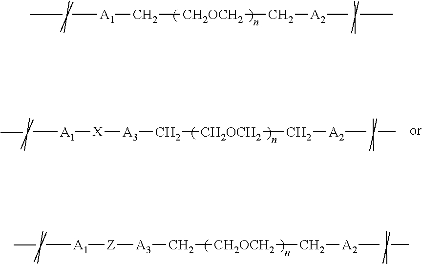
where n is an integer ranging from 2 to 48 inclusive; A1 is a bond forming group selected from

A2 is a bond forming group selected from

A3, when present, is a bond forming group selected from

X is a cleavable group that can undergo silicon-carbon cleavage, nucleophilic cleavage, redox cleavage, photochemical cleavage, enzymatic cleavage, or exchange-based cleavage;
Z is a branching group that contains modifying molecule (Mod); Mod is a modifier or modifying molecule (e.g., a crosslinking agent); Obs is an observable label moiety (e.g., an observable label in itself such as a chromogen or fluor, or a molecule that can be rendered observable, a detecting agent; etc.); and the leftmost bond is to Alk and the rightmost bond is to Mod.
with PEG having at least 7 carbon atoms and up to 100 carbon atoms, i.e., PEG4 to PEG50. In one embodiment, the modified nucleotide is a desthiobiotinylated nucleotide having the formula
In one embodiment, the modified nucleotide is a mass tagged nucleotide having the formula
For any of the disclosed inventive compounds, the compound includes the salt form, conjugate base, tautomer, and/or ionized form. In one embodiment, the modified nucleotide is a ribonucleotide. In one embodiment, the ribonucleotide is cytidine. In one embodiment, the ribonucleotide is adenosine. In one embodiment, the ribonucleotide is uridine. In one embodiment, the ribonucleotide is guanosine. In one embodiment, the ribonucleotide is inosine.

or its salt, conjugate base, tautomer, or ionized form where

and
m is an integer ranging from 3 to 6 inclusive;

in which n is an integer ranging from 2 to 48 inclusive;


n is 12, A1 is
A2 is
when present Z is
when present, A3 is
when present Mod is a crosslinking agent, and Obs is desthiobiotin, fluorophore, mass tag, and/or azide.

This structure had similar ligation efficiencies to biotin PEG4-alkane-bisphosphate cytidine. The alkane, versus alkyne, linkage makes the linker containing the detector less reactive, and thus less susceptible to degradation in cell and/or tissue lysates.

n is 12, A1 is
A2 is
when present Z is
when present A3 is
when present Mod is a crosslinking agent, and Obs is biotin, desthiobiotin, and/or a mass tag.

Propargyl amine (4.00 g, 72.62 mmol, 1.00 equiv.) was added dropwise to methyl trifluoroacetate (11.16 g, 87.15 mmol, 1.20 equiv.) at 0° C. The reaction mixture was stirred at 0° C. for 2 h and then concentrated under reduced pressure to remove methanol. The product was purified by vacuum distillation yielding propargyltrifluoroacetamide as a colorless liquid (9.59 g, 87%). The structure was confirmed by 1H- and 19F-NMR.

A 100-mL three-necked flask was charged with 5-iodocytidine (2.66 g, 7.00 mmol, 1.00 equiv.), cuprous iodide (0.267 g, 1.40 mmol, 0.20 equiv.) and dry DMF (35 mL). After complete dissolution of the reaction mixture, propargyltrifluoroacetamide (3.17 g, 21.00 mmol, 3.00 equiv.), triethylamine (1.42 g, 14.00 mmol, 2.00 equiv.) and finally tetrakis (triphenylphosphine)palladium(0) (0.809 g, 0.70 mmol, 0.10 equiv.) were added to the reaction mixture under N2. The reaction was stirred at ambient temperature (around 19° C. to around 22° C.) under N2 for 18-24 h. The reaction was then diluted with 70 mL of 1:1 methanol-dichloromethane and the bicarbonate form of AGI X8 resin (12.00 g) was added. After stirring for about one h, the reaction mixture was filtered and the resin was washed with 1:1 methanol-dichloromethane. The combined filtrates were rapidly concentrated with a rotary evaporator. The residue was immediately purified by flash chromatography. Removal of solvent from the appropriate fractions afforded 1.84 g (67%) of 5-[3-(trifluoroacetamido)propynyl]cytidine as a light brown solid, which was confirmed by 1H-NMR.

5-[3-(trifluoroacetamido)propynyl]cytidine (1.25 g, 3.19 mmol, 1.00 equiv.) was dissolved in methanol (30 mL). Palladium hydroxide (0.25 g, 20 wt./wt. % based on propynyl cytidine) and triethylsilane (3.71 g, 31.90 mmol, 10.00 equiv.) were added to the reaction mixture. After 20-24 hours at ambient temperature, the reaction mixture was filtered through glass fiber and the filtrate was concentrated under reduced pressure giving a dark brown residue. The residue was purified by flash chromatography. Removal of solvent from the appropriate fractions afforded 0.85 g (71%) of 5-[3-(trifluoroacetamido)propyl]cytidine as a cream colored solid, which was confirmed by 1H-NMR.

5-[3-(trifluoroacetamido)propyl]cytidine (0.69 g, 1.74 mmol) was dissolved in DI H2O (8.5 mL). After complete dissolution, concentrated ammonium hydroxide (NH4OH) (8.5 mL) was added to the reaction mixture. The reaction solution was stirred at ambient temperature for 2-3 h and then concentrated under reduced pressure giving the crude product as yellow-orange residue. The crude product was dissolved in deionized H2O (10 mL) and AG50W-X8 resin (2.5 g) was added to the solution. The suspension was stirred for 15 min and filtered over a bed of AG50W-X8 resin (2.5 g). The resin was washed with DI H2O and the product was then eluted off of the resin by washing the resin with deionized H2O/conc. NH4OH, 4:1, collecting fractions (monitored by TLC). Removal of solvent from the appropriate fractions afforded 0.51 g (98%) of 5-(3-aminopropyl)cytidine as light tan solid, which was confirmed by 1H-NMR.

NHS-PEG4-biotin (0.196 g, 0.333 mmol, 1.00 equiv.) was dissolved in DMF (10 mL). 5-(3-aminopropyl)cytidine) (0.100 g, 0.333 mmol, 1.00 equiv.) was added to the reaction solution. The reaction solution was stirred at ambient temperature under N2 atmosphere. After 20-24 h, the reaction mixture was concentrated under reduced pressure giving the crude product. The crude product was purified by flash chromatography. Removal of solvent from the appropriate fractions afforded 0.18 g (69%) of BPAC as a white solid, which was confirmed by 1H-NMR.

BPAC (0.061 g, 0.079 mmol, 1.00 equiv.) was partially dissolved in diphosphoryl chloride (196 μL, 1.66 mmol, 21.00 equiv.), previously cooled to −10° C. to −15° C. in a 1-mL Reacti-Vial™. The mixture was then stirred at −10° C. to −15° C. After 5 h, the reaction was quenched by addition of ice cold water (1-2 mL) and, immediately thereafter, with a chilled solution of 0.5 M TEAB buffer, pH 8.5 (17 mL). Upon stabilization at neutral pH, the colorless solution was stirred at ambient temperature for 30 min and concentrated using a rotary evaporator until complete removal of TEAB. The solution was desalted using a C18 cartridge (Waters) and purified by FPLC (MonoQ 10/100GL column, GE) using a pH gradient. After a final desalting using again a C18 cartridge (Waters), BPA-3′,5′-pCp was isolated after lyophilization as a white solid (10 mg, 9%), which was confirmed by 1H-NMR & HPLC.

The reaction scheme to prepare biotin-polyethylene glycol (PEG)-SS-alkane-3′,5′-cytidine-bisphosphate is as follows. The 5-(3-aminopropyl)cytidine (compound 4) is reacted with NHS-SS-PEG-biotin to result in biotin-PEG-SS-alkane-cytidine (compound 11). The biotin-PEG-SS-alkane-cytidine (compound 11) then is reacted with diphosphoryl chloride to result in biotin-polyethylene glycol (PEG)-SS-alkane-3′,5′-cytidine-bisphosphate (compound 12).

NHS-SS-PEG4-biotin (0.250 g, 0.333 mmol, 1.00 equiv.) was dissolved in DMF (10 mL). 5-(3-aminopropyl)cytidine) (0.100 g, 0.333 mmol, 1.00 equiv.) was added to the reaction solution. The reaction solution was stirred at ambient temperature under N2 atmosphere. After 20-24 hours, the reaction mixture was concentrated under reduced pressure giving the crude product. The crude product was purified by flash chromatography. Removal of solvent from the appropriate fractions afforded 0.19 g (61%) of BP4SSAC (compound 11) as a white solid, which was confirmed by 1H-NMR.

BP4SSAC (0.074 g, 0.079 mmol, 1.00 equiv.) was partially dissolved in diphosphoryl chloride (196 μL, 1.66 mmol, 21.00 equiv.), previously cooled to −10° C. to −15° C. in a 1-mL Reacti-Vial®. The mixture was then stirred at −10° C. to −15° C. After five hours, the reaction was quenched by addition of ice cold water (1-2 mL) and, immediately thereafter, with a chilled solution of 0.5M TEAB buffer, pH 8.5 (17 mL). Upon stabilization at neutral pH, the colorless solution was stirred at ambient temperature for 30 min and concentrated using a rotary evaporator until complete removal of TEAB. The solution was desalted using a C18 cartridge (Waters) and purified by FPLC (MonoQ 10/100GL column, GE) using a pH gradient. After a final desalting using again a C18 cartridge (Waters), BP4SSA-3′,5′-pCp (compound 12) was isolated after lyophilization as a white solid (5 mg, 6%), which was confirmed by 1H-NMR and HPLC.

NHS-PEG12-biotin (0.313 g, 0.333 mmol, 1.00 equiv.) was dissolved in DMF (10 mL). 5-(3-aminopropyl)cytidine) (0.100 g, 0.333 mmol, 1.00 equiv., compound 4) was added to the reaction solution. The reaction solution was stirred at ambient temperature under N2 atmosphere. After 20-24 h, the reaction mixture was concentrated under reduced pressure giving the crude product. The crude product was purified by flash chromatography. Removal of solvent from the appropriate fractions afforded 0.27 g (72%) of BP12AC (compound 7) as a light yellow foam, which was confirmed by 1H-NMR.

Biotin-PEG12-alkane-cytidine (0.135 g, 0.120 mmol, 1.00 equiv., compound 7) was partially dissolved in diphosphoryl chloride (315 μL, 2.40 mmol, 20.00 equiv.), previously cooled to −10 to −15° C. in a 1-mL Reacti-Vial™. The mixture was stirred at −10 to −15° C. After five hours, the reaction was quenched by adding ice cold water (1-2 mL) and immediately after with a chilled solution of 0.5M TEAB buffer, pH 8.5 (40 mL). Upon stabilization at neutral pH, the colorless solution was stirred at ambient temperature for 30 min and concentrated using a rotary evaporator until TEAB was completely removed. The solution was desalted using a C18 cartridge (Waters) and purified by FPLC (MonoQ 10/100GL column, GE) using a pH gradient. After final desalting using a C18 cartridge (Waters), biotin-PEG12-alkane-3′,5′-cytidine-bisphosphate (compound 8) was isolated after lyophilization as a sticky white solid (8 mg, 5%), which was confirmed by 1H-NMR and HPLC.

One embodiment is a method of preparing azido-PEG4-alkane-3′,5′-cytidine-bisphosphate. The 5-(3-aminopropyl)cytidine was synthesized as described above, then was reacted with NHS-PEG-azide to result in azido-PEG4-alkane-cytidine. The azido-PEG4-alkane-cytidine was then reacted with diphosphoryl chloride to result in azido-PEG4-alkane-3′,5′-cytidine-bisphosphate.

Azido-PEG4-alkane-cytidine (0.150 g, 0.262 mmol, 1.00 equiv., compound 9) was partially dissolved in diphosphoryl chloride (688 μL, 5.24 mmol, 20.00 equiv.), previously cooled to −10 to −15° C. in a 1 mL Reacti-Vial™. The mixture was then stirred at −10 to −15° C. After five hours, the reaction was quenched by adding ice cold water (2-3 mL) and then immediately with a chilled solution of 0.5M TEAB buffer, pH 8.5 (87 mL). Upon stabilization at neutral pH, the colorless solution was stirred at ambient temperature for 30 min and concentrated using a rotary evaporator until TEAB was complete removed. The solution was desalted using a C18 cartridge (Waters) and purified by FPLC (MonoQ 10/100GL column, GE) using a pH gradient. After final desalting using again a C18 cartridge (Waters), azido-PEG4-alkane-3′,5′-cytidine-bisphosphate (compound 10) was isolated after lyophilization as a sticky white solid (10 mg, 6%), confirmed by 1H-NMR and HPLC.

Azido-PEG4-alkane-3′,5′-bisphosphate-cytidine (3.56 μmol, 1.00 equiv., compound 10) was dissolved in 200 mM Tris/HCl, pH 7.5 (800 μL). Tris(2-carboxyethyl)phosphine hydrochloride (TCEP) (17.54 mg, approx. 5.00 equiv.) was dissolved in 200 mM Tris/HCl, pH 7.5 (688 μL). The TCEP solution (200 μL) was added to the solution of azide and the reaction was mixed at ambient temperature. After 1-3 h, the reaction mixture was purified by FPLC and the fractions containing product were treated directly with DyLight 550 NHS ester to result in amino-PEG4-alkane-3′,5′-bisphosphate cytidine (compound 13).

The pH of an FPLC fraction (2 mL) containing amino-PEG4-alkane-3′,5′-bisphosphate-cytidine (compound 13) was adjusted to pH 7.0 by adding 1M HEPES, pH 7.3. Separately, a 1 mM solution of DyLight 550 NHS ester was prepared by dissolving DyLight 550 NHS ester (MW=1040.05, 1 mg) in ultra pure water (960 μL). Amino-PEG4-alkane-3′,5′-bisphosphate-cytidine (0.25 mL) and DyLight 550 NHS ester (0.25 mL) were combined in a separate reaction vessel and were mixed with rotation for 1 h at ambient temperature. The reaction mixture was purified by FPLC (MonoQ 10/100GL column, GE) using a pH and salt gradient. Fractions containing product were dialyzed and subsequently lyophilized, yielding DyLight550-PEG4-alkane-3′,5′-cytidine-bisphosphate (compound 14) as a dark pink residue.

The reaction scheme to prepare desthiobiotin-polyethylene glycol (PEG)4-alkane-3′,5′-cytidine-bisphosphate was as follows. The azido-PEG4-alkane-cytidine (compound 9) was reduced using triethylsilane and 20 wt % palladium hydroxide giving amino-PEG4-alkane-cytidine (compound 15). The amino-PEG4-alkane-cytidine (compound 15) was then reacted with NHS-desthiobiotin giving desthiobiotin-PEG4-alkane-cytidine (compound 16). Desthiobiotin-PEG4-alkane-cytidine was then reacted with diphosphoryl chloride giving desthiobiotin-PEG4-alkane-cytidine-bisphosphate (compound 17).

Azido-PEG4-alkane-cytidine (9) (0.20 g, 0.349 mmol, 1.00 equiv.) was dissolved in methanol (6 mL). Palladium hydroxide (0.040 g, 20 wt./wt. % based on 9) and triethylsilane (0.406 g, 3.49 mmol, 10.00 equiv.) was added to the reaction mixture. After 20-24 hours at ambient temperature, the reaction mixture was filtered through a pad of glass fiber and the filtrate was concentrated under reduced pressure giving amino-PEG4-alkane-cytidine (15) as an off-white residue (0.19 g, 99%) which was confirmed by 1H-NMR and used directly without further purification.

NHS-desthiobiotin (0.102 g, 0.329 mmol, 1.00 equiv.) was added to a solution of amino-PEG4-alkane-cytidine (15) (0.18 g, 0.329 mmol, 1.00 equiv.) in DMF (3 mL). The reaction solution was stirred at ambient temperature under N2 atmosphere. After 20-24 h, the reaction mixture was concentrated under reduced pressure giving the crude product as a pale yellow residue. The crude product was purified by flash chromatography. Removal of solvent from the appropriate fractions gave desthiobiotin-PEG4-alkane-cytidine (16) as a clear, near colorless glass (0.185 g, 76%) which was confirmed by 1H-NMR.

Desthiobiotin-PEG4-alkane-cytidine (16) (0.117 g, 0.157 mmol, 1.00 equiv.) was partially dissolved in diphosphoryl chloride (391 μL, 3.30 mmol, 21.00 equiv.) in a 3 mL Reacti-Vial™. The mixture was then stirred at 0 to −10° C. After five hours, the reaction was quenched by the addition of ice cold ultra-pure water (1 mL) immediately followed by a solution of 0.5M TEAB buffer, pH 8.5 (23 mL). The colorless solution was stirred at ambient temperature for 30 min and then stored overnight at 0-5° C. The pH was adjusted to 3.0-3.5 and the solution was concentrated using a rotary evaporator until TEAB was complete removed. The solution was desalted using a C18 cartridge (Waters) and purified by FPLC (MonoQ 10/100GL column, GE). The purified product was subsequently desalted using a C18 cartridge (Waters) giving desthiobiotin-PEG4-alkane-3′,5′-cytidine-bisphosphate (17) as a clear glass (9 mg, 6%) after lyophilization. The structure was confirmed by 1H-NMR and HPLC.

The reaction scheme to prepare desthiobiotin-polyethylene glycol (PEG)12-alkane-3′,5′-cytidine-bisphosphate was as follows. The 5-(3-aminopropyl)cytidine (compound 4) was synthesized as described above, then was reacted with NHS-PEG12-azide giving azido-PEG12-alkane-cytidine (compound 18). The azido-PEG12-alkane-cytidine (compound 18) was reduced using triethylsilane and 20 wt. % palladium hydroxide giving amino-PEG12-alkane-cytidine (compound 19). The amino-PEG12-alkane-cytidine (compound 19) was then reacted with NHS-desthiobiotin giving desthiobiotin-PEG12-alkane-cytidine (compound 20). Desthiobiotin-PEG4-alkane-cytidine was then reacted with diphosphoryl chloride giving desthiobiotin-PEG12-alkane-cytidine-bisphosphate (compound 21).

NHS-PEG12-azide (0.445 g, 0.607 mmol, 1.00 equiv.) was dissolved in DMF (10 mL). 5-(3-aminopropyl)cytidine (4) (0.182 g, 0.607 mmol, 1.00 equiv.) was added to the reaction solution as a solid. The reaction solution was stirred at ambient temperature under N2 atmosphere. After 18-24 hours, the reaction mixture was concentrated under reduced pressure giving the crude product as a pale amber oil. The crude product was purified by flash chromatography. Removal of solvent from the appropriate fractions gave of azido-PEG12-alkane-cytidine (18) as a clear, near colorless residue (0.35 g, 62%) the structure of which was confirmed by 1H-NMR.

Azido-PEG12-alkane-cytidine (18) (0.10 g, 0.108 mmol, 1.00 equiv.) was dissolved in methanol (3 mL). Palladium hydroxide (0.020 g, 20 wt./wt. % based on 18) and triethylsilane (0.126 g, 1.08 mmol, 10.00 equiv.) was added to the reaction mixture. After 20-24 hours at ambient temperature, the reaction mixture was filtered through a pad of glass fiber and the filtrate was concentrated under reduced pressure giving amino-PEG12-alkane-cytidine (19) as an clear, colorless residue (0.10 g, 100%) which was confirmed by 1H-NMR and used directly without further purification.

NHS-desthiobiotin (0.035 g, 0.111 mmol, 1.00 equiv.) was added to a solution of amino-PEG12-alkane-cytidine (19) (0.10 g, 0.111 mmol, 1.00 equiv.) in DMF (2 mL). The reaction solution was stirred at ambient temperature under N2 atmosphere. After 20-24 h, the reaction mixture was concentrated under reduced pressure giving the crude product as a pale yellow residue. The crude product was purified by flash chromatography. Removal of solvent from the appropriate fractions gave desthiobiotin-PEG12-alkane-cytidine (20) as a clear, near light yellow glass (0.054 g, 44%) which was confirmed by 1H-NMR and used directly without further purification.

Desthiobiotin-PEG12-alkane-cytidine (20) (0.054 g, 0.049 mmol, 1.00 equiv.) was partially dissolved in diphosphoryl chloride (122 μL, 1.03 mmol, 21.00 equiv.) in a 3 mL Reacti-Vial™. The mixture was then stirred at 0° C. to 10° C. After about five hours, the reaction was quenched by adding ice cold ultra-pure water (1 mL) immediately followed by a solution of 0.5M TEAB buffer, pH 8.5 (11 mL). The colorless solution was stirred at ambient temperature for 30 min and then stored overnight at 0-5° C. The pH was adjusted to 3.0-3.5 and the solution was concentrated using a rotary evaporator until TEAB was completely removed. The solution was desalted using a C18 cartridge (Waters) and purified by FPLC (MonoQ 10/100GL column, GE). The purified product was subsequently desalted using a C18 cartridge (Waters) giving desthiobiotin-PEG12-alkane-3′,5′-cytidine-bisphosphate (21) as a clear glass (4 mg, 6%) after lyophilization. The structure was confirmed by 1H-NMR and HPLC.


| Optimal | |||||
| Length | reaction | ||||
| Description | RNA source | (bases) | conditions | ||
| IRE ( |
5′ or 3′ UTR | synthetic | 28 | 2 |
| responsive | element | |||
| element) | ||||
| RNA | RNA | synthetic | 42 | 30 minutes, |
| polymerase | 37° C. | |||
| template RNA | >1 |
|||
| mir-16-1 | mature micro | synthetic | 22 | ON 16° C. |
| RNA | ||||
| TNF ARE | 3′ UTR element | synthetic | 37 | 2 |
| 16° C. | ||||
| Let-7 | pre-miRNA | in vitro | ~70 | overnight |
| transcribed | 16° C. | |||
| hTR | catalytic RNA | in vitro | 451 | overnight |
| transcribed | 16° C. | |||
| COX-76 |
3′ UTR element | in vitro | ~70 | overnight |
| transcribed | 16° C. | |||
| mir-16-1 | pre-miRNA | in vitro | ~70 | overnight |
| transcribed | 16° C. | |||
- Khanan et. Al. (2006) Poly(A)-Binding Protein Binds to A-Rich Sequences via
RNA Binding Domains 1+2 and 3+4. RNA Biology. 3: 170-177. - Rimmele and Belasco. (1998) Target discrimination by RNA binding proteins: role of the ancillary protein U2A′ and a critical leucine residue in differentiating the RNA-binding specificity of spliceosomal proteins U1A and U2B″. RNA. 4: 1386-1396.
- DNASU Plasmid Repository, Arizona State University BioDesign Institute.
- U.S. Patent Publication No. 2011/0262917.
- England et al. Specific labeling of 3′ termini of RNA with T4 RNA ligase (1980) Methods Enzym. 65: 65-74.
- Brennan and Gumport. T4 RNA ligase catalyzed synthesis of base-analogue-containing oligodeoxyribonucleotides and a characterization of their thermal stabilities. (1985) Nucleic Acids Res. 13: 8665-8684.
- Hinton et al. The preparative synthesis of oligodeoxyribonucleotides using RNA ligase. (1982) Nucleic Acids Res. 10:1877-1894.
- Walker et al. T4-induced RNA ligase joins single-stranded oligoribonucleotides (1975) PNAS 72: 122-126.
- Richardson and Gumport. Biotin and fluorescent labeling of RNA using T4 RNA ligase. (1983) Nucleic Acids Res. 11: 6167-6184.
- England et al. Dinucleoside pyrophosphates are substrates for T4-induced RNA ligase (1977) PNAS 74: 4839-4842.
- Keith. Optimization of conditions for labeling the 3′ OH end of tRNA using T4 RNA ligase. (1983) Biochimie 65: 367-70.
- Romaniuk et al. Joining of RNA molecules with RNA ligase (1983) Methods Enzym 100: 52-59.
- Romaniuk et al. The effect of acceptor ribonucleotide sequence for the T4 RNA ligase reaction. (1982) European J of Biochem. 125: 639-643.
- Park et al. Useful tools for biomolecule isolation, detection, and identification: acylhydrazone-based cleavable linkers. (2009) Chemistry & Biology 16: 763-772.
- Shigdel et al. Diazirine-based DNA photo-cross-linking probes for the study of protein-DNA interactions (2008) Angew. Chem. Int. 47: 90-93.
- Costas et al. RNA-protein crosslinking to AMP residues at internal positions in RNA with a new photocrosslinking ATP analog (2000) Nucl. Acids. Res. 28: 1849-1858.
- Gomes and Gozzo (2010). Chemical cross-linking with a diazirine photoactivatable cross-linker investigated by MALDI- and ESI-MS/MS. J. Mass. Spectrom. 45:892-9.
- Liu and Sun. Direct isolation of specific RNA-interacting proteins using a novel affinity medium. (2005) Nucl. Acids Res. 33: 1-5.
- Bachler et al. StreptoTag: A novel method for the isolation of RNA-binding proteins. (1999). RNA 5: 1509-1516.
Claims (26)


Priority Applications (4)
| Application Number | Priority Date | Filing Date | Title |
|---|---|---|---|
| US13/482,927 US9206216B2 (en) | 2010-04-21 | 2012-05-29 | Modified nucleotides methods and kits |
| EP12187509.0A EP2669291A1 (en) | 2012-05-29 | 2012-10-05 | Modified Nucleotides Methods and Kits |
| US14/883,810 US9828633B1 (en) | 2010-04-21 | 2015-10-15 | Modified nucleotides methods and kits |
| US15/792,875 US10526652B2 (en) | 2010-04-21 | 2017-10-25 | Modified nucleotides methods and kits |
Applications Claiming Priority (3)
| Application Number | Priority Date | Filing Date | Title |
|---|---|---|---|
| US32645010P | 2010-04-21 | 2010-04-21 | |
| US13/090,729 US8536323B2 (en) | 2010-04-21 | 2011-04-20 | Modified nucleotides |
| US13/482,927 US9206216B2 (en) | 2010-04-21 | 2012-05-29 | Modified nucleotides methods and kits |
Related Parent Applications (1)
| Application Number | Title | Priority Date | Filing Date |
|---|---|---|---|
| US13/090,729 Continuation-In-Part US8536323B2 (en) | 2010-04-21 | 2011-04-20 | Modified nucleotides |
Related Child Applications (1)
| Application Number | Title | Priority Date | Filing Date |
|---|---|---|---|
| US14/883,810 Division US9828633B1 (en) | 2010-04-21 | 2015-10-15 | Modified nucleotides methods and kits |
Publications (2)
| Publication Number | Publication Date |
|---|---|
| US20120252691A1 US20120252691A1 (en) | 2012-10-04 |
| US9206216B2 true US9206216B2 (en) | 2015-12-08 |
Family
ID=46928009
Family Applications (3)
| Application Number | Title | Priority Date | Filing Date |
|---|---|---|---|
| US13/482,927 Active 2032-12-12 US9206216B2 (en) | 2010-04-21 | 2012-05-29 | Modified nucleotides methods and kits |
| US14/883,810 Active 2031-05-31 US9828633B1 (en) | 2010-04-21 | 2015-10-15 | Modified nucleotides methods and kits |
| US15/792,875 Active 2031-06-02 US10526652B2 (en) | 2010-04-21 | 2017-10-25 | Modified nucleotides methods and kits |
Family Applications After (2)
| Application Number | Title | Priority Date | Filing Date |
|---|---|---|---|
| US14/883,810 Active 2031-05-31 US9828633B1 (en) | 2010-04-21 | 2015-10-15 | Modified nucleotides methods and kits |
| US15/792,875 Active 2031-06-02 US10526652B2 (en) | 2010-04-21 | 2017-10-25 | Modified nucleotides methods and kits |
Country Status (1)
| Country | Link |
|---|---|
| US (3) | US9206216B2 (en) |
Cited By (6)
| Publication number | Priority date | Publication date | Assignee | Title |
|---|---|---|---|---|
| US20150252070A1 (en) * | 2012-09-04 | 2015-09-10 | Kabushiki Kaisha Dnaform | Compound, nucleic acid, labeling substance, and detection method |
| US20150266917A1 (en) * | 2012-09-21 | 2015-09-24 | Osaka University | Oligonucleotide and artificial nucleoside having guanidine bridge |
| US20160024573A1 (en) * | 2013-03-15 | 2016-01-28 | Ibis Biosciences, Inc. | Photocleavable deoxynucleotides with high-resolution control of deprotection kinetics |
| US9828633B1 (en) * | 2010-04-21 | 2017-11-28 | Pierce Biotechnology, Inc. | Modified nucleotides methods and kits |
| US10386338B2 (en) * | 2017-10-30 | 2019-08-20 | Cynthia Rena Wright | DNA/RNA PEMS microcantilever probe for detection of viral infection and detection of genetic variants |
| US11926817B2 (en) | 2019-08-09 | 2024-03-12 | Nutcracker Therapeutics, Inc. | Microfluidic apparatus and methods of use thereof |
Families Citing this family (3)
| Publication number | Priority date | Publication date | Assignee | Title |
|---|---|---|---|---|
| GB201414098D0 (en) | 2014-08-08 | 2014-09-24 | Illumina Cambridge Ltd | Modified nucleotide linkers |
| JPWO2020246602A1 (en) * | 2019-06-07 | 2020-12-10 | ||
| CN113943312A (en) * | 2020-07-17 | 2022-01-18 | 轶诺(浙江)药业有限公司 | Intestinal tract cracking type co-drug and preparation and application thereof |
Citations (25)
| Publication number | Priority date | Publication date | Assignee | Title |
|---|---|---|---|---|
| US4914210A (en) | 1987-10-02 | 1990-04-03 | Cetus Corporation | Oligonucleotide functionalizing reagents |
| US5047519A (en) | 1986-07-02 | 1991-09-10 | E. I. Du Pont De Nemours And Company | Alkynylamino-nucleotides |
| US5151507A (en) | 1986-07-02 | 1992-09-29 | E. I. Du Pont De Nemours And Company | Alkynylamino-nucleotides |
| US5247081A (en) | 1987-08-28 | 1993-09-21 | Imperial Chemical Industries Plc | Protected biotin derivatives |
| US5558991A (en) | 1986-07-02 | 1996-09-24 | E. I. Du Pont De Nemours And Company | DNA sequencing method using acyclonucleoside triphosphates |
| US5567811A (en) | 1990-05-03 | 1996-10-22 | Amersham International Plc | Phosphoramidite derivatives, their preparation and the use thereof in the incorporation of reporter groups on synthetic oligonucleotides |
| US5608063A (en) | 1986-07-02 | 1997-03-04 | E. I. Du Pont De Nemours And Company | Method, system and reagents for DNA sequencing |
| US5684142A (en) * | 1995-06-07 | 1997-11-04 | Oncor, Inc. | Modified nucleotides for nucleic acid labeling |
| US5888819A (en) | 1991-03-05 | 1999-03-30 | Molecular Tool, Inc. | Method for determining nucleotide identity through primer extension |
| US5952174A (en) | 1994-02-07 | 1999-09-14 | Orchid Biocomputer, Inc. | Ligase/polymerase-mediated genetic bit analysis of single nucleotide polymorphisms and its use in genetic analysis |
| US6004744A (en) | 1991-03-05 | 1999-12-21 | Molecular Tool, Inc. | Method for determining nucleotide identity through extension of immobilized primer |
| US6013431A (en) | 1990-02-16 | 2000-01-11 | Molecular Tool, Inc. | Method for determining specific nucleotide variations by primer extension in the presence of mixture of labeled nucleotides and terminators |
| US6114350A (en) | 1999-04-19 | 2000-09-05 | Nen Life Science Products, Inc. | Cyanine dyes and synthesis methods thereof |
| WO2004052907A1 (en) | 2002-12-05 | 2004-06-24 | Affymetrix, Inc. | Nucleic acid labeling compounds |
| US20080045418A1 (en) | 2005-02-04 | 2008-02-21 | Xia Xueliang J | Method of labeling and profiling rnas |
| US7344701B2 (en) * | 2004-02-03 | 2008-03-18 | Biosearch Technologies, Inc. | Xanthene dyes |
| US7361465B2 (en) | 2004-09-07 | 2008-04-22 | Applera Corporation | Methods and compositions for tailing and amplifying RNA |
| US7491818B2 (en) | 1998-07-31 | 2009-02-17 | Affymetrix, Inc. | Nucleic acid labeling compounds |
| US7504215B2 (en) | 2002-07-12 | 2009-03-17 | Affymetrix, Inc. | Nucleic acid labeling methods |
| US7524942B2 (en) | 2006-07-31 | 2009-04-28 | Agilent Technologies, Inc. | Labeled nucleotide composition |
| US7541144B2 (en) | 2005-01-31 | 2009-06-02 | Agilent Technologies, Inc. | RNA labeling method |
| US20090176732A1 (en) | 2007-12-21 | 2009-07-09 | Alios Biopharma Inc. | Protected nucleotide analogs |
| US7572585B2 (en) | 2006-07-31 | 2009-08-11 | Agilent Technologies, Inc. | Enzymatic labeling of RNA |
| US20090286753A1 (en) | 2004-12-29 | 2009-11-19 | Exiqon A/S | Novel oligonucleotide compositions and probe sequences useful for detection and analysis of micrornas and their target mrnas |
| US8536323B2 (en) * | 2010-04-21 | 2013-09-17 | Pierce Biotechnology, Inc. | Modified nucleotides |
Family Cites Families (4)
| Publication number | Priority date | Publication date | Assignee | Title |
|---|---|---|---|---|
| US5650209A (en) | 1995-06-06 | 1997-07-22 | Moore Business Forms, Inc. | Manufacture of telephone debit cards |
| US20030165849A1 (en) | 2000-11-28 | 2003-09-04 | Biliang Zhang | Methods and reagents for introducing a sulfhydryl group into the 5'-terminus of RNA |
| US20080103053A1 (en) | 2005-11-22 | 2008-05-01 | Helicos Biosciences Corporation | Methods and compositions for sequencing a nucleic acid |
| US9206216B2 (en) * | 2010-04-21 | 2015-12-08 | Pierce Biotechnology, Inc. | Modified nucleotides methods and kits |
-
2012
- 2012-05-29 US US13/482,927 patent/US9206216B2/en active Active
-
2015
- 2015-10-15 US US14/883,810 patent/US9828633B1/en active Active
-
2017
- 2017-10-25 US US15/792,875 patent/US10526652B2/en active Active
Patent Citations (28)
| Publication number | Priority date | Publication date | Assignee | Title |
|---|---|---|---|---|
| US5558991A (en) | 1986-07-02 | 1996-09-24 | E. I. Du Pont De Nemours And Company | DNA sequencing method using acyclonucleoside triphosphates |
| US5047519A (en) | 1986-07-02 | 1991-09-10 | E. I. Du Pont De Nemours And Company | Alkynylamino-nucleotides |
| US5151507A (en) | 1986-07-02 | 1992-09-29 | E. I. Du Pont De Nemours And Company | Alkynylamino-nucleotides |
| US5608063A (en) | 1986-07-02 | 1997-03-04 | E. I. Du Pont De Nemours And Company | Method, system and reagents for DNA sequencing |
| US5247081A (en) | 1987-08-28 | 1993-09-21 | Imperial Chemical Industries Plc | Protected biotin derivatives |
| US4914210A (en) | 1987-10-02 | 1990-04-03 | Cetus Corporation | Oligonucleotide functionalizing reagents |
| US6013431A (en) | 1990-02-16 | 2000-01-11 | Molecular Tool, Inc. | Method for determining specific nucleotide variations by primer extension in the presence of mixture of labeled nucleotides and terminators |
| US5567811A (en) | 1990-05-03 | 1996-10-22 | Amersham International Plc | Phosphoramidite derivatives, their preparation and the use thereof in the incorporation of reporter groups on synthetic oligonucleotides |
| US5888819A (en) | 1991-03-05 | 1999-03-30 | Molecular Tool, Inc. | Method for determining nucleotide identity through primer extension |
| US6004744A (en) | 1991-03-05 | 1999-12-21 | Molecular Tool, Inc. | Method for determining nucleotide identity through extension of immobilized primer |
| US5952174A (en) | 1994-02-07 | 1999-09-14 | Orchid Biocomputer, Inc. | Ligase/polymerase-mediated genetic bit analysis of single nucleotide polymorphisms and its use in genetic analysis |
| US5684142A (en) * | 1995-06-07 | 1997-11-04 | Oncor, Inc. | Modified nucleotides for nucleic acid labeling |
| US7491818B2 (en) | 1998-07-31 | 2009-02-17 | Affymetrix, Inc. | Nucleic acid labeling compounds |
| US6204389B1 (en) | 1999-04-19 | 2001-03-20 | Nen Life Science Products, Inc. | Cyanine dyes and synthesis methods thereof |
| US6197956B1 (en) | 1999-04-19 | 2001-03-06 | Nen Life Science Products, Inc. | Cyanine dyes and synthesis methods thereof |
| US6224644B1 (en) | 1999-04-19 | 2001-05-01 | Nen Life Science Products, Inc. | Cyanine dyes and synthesis methods thereof |
| US6114350A (en) | 1999-04-19 | 2000-09-05 | Nen Life Science Products, Inc. | Cyanine dyes and synthesis methods thereof |
| US7504215B2 (en) | 2002-07-12 | 2009-03-17 | Affymetrix, Inc. | Nucleic acid labeling methods |
| WO2004052907A1 (en) | 2002-12-05 | 2004-06-24 | Affymetrix, Inc. | Nucleic acid labeling compounds |
| US7344701B2 (en) * | 2004-02-03 | 2008-03-18 | Biosearch Technologies, Inc. | Xanthene dyes |
| US7361465B2 (en) | 2004-09-07 | 2008-04-22 | Applera Corporation | Methods and compositions for tailing and amplifying RNA |
| US20090286753A1 (en) | 2004-12-29 | 2009-11-19 | Exiqon A/S | Novel oligonucleotide compositions and probe sequences useful for detection and analysis of micrornas and their target mrnas |
| US7541144B2 (en) | 2005-01-31 | 2009-06-02 | Agilent Technologies, Inc. | RNA labeling method |
| US20080045418A1 (en) | 2005-02-04 | 2008-02-21 | Xia Xueliang J | Method of labeling and profiling rnas |
| US7524942B2 (en) | 2006-07-31 | 2009-04-28 | Agilent Technologies, Inc. | Labeled nucleotide composition |
| US7572585B2 (en) | 2006-07-31 | 2009-08-11 | Agilent Technologies, Inc. | Enzymatic labeling of RNA |
| US20090176732A1 (en) | 2007-12-21 | 2009-07-09 | Alios Biopharma Inc. | Protected nucleotide analogs |
| US8536323B2 (en) * | 2010-04-21 | 2013-09-17 | Pierce Biotechnology, Inc. | Modified nucleotides |
Non-Patent Citations (40)
| Title |
|---|
| (D2) Gomes and Gozzo. Chemical cross-linking with a diazirine photoactivatable cross-linker investigated by MALDI- and ESI-MS/MS. J, Mass Spectrom. vol. 45 (2010), pp. 892-899. |
| (D3) Shigdel et al. Diazirine-Based DNA Photo-Cross-Linking Probes for the Study of Protein-DNA Interactions. Angew. Chem. Int. Ed. vol. 47 (2008), pp. 90-93. |
| (D4) Hanna et al. Synthesis and characterization of a new photocrosslinking CTP analog and its use in photoaffinity labeling E.coli and T7 RNA polymerases. Nucleic Acids Research, vol. 21, No. 9 (1993), pp. 2073-2079. |
| Agafonov et al., "C-Terminal Modifications of a Protein by UAG-Encoded Incorporation of Puromycin during in vitro Protein Synthesis in the Absence of Release Factor 1" ChemBioChem (2006) vol. 7 pp. 330-336. * |
| Bachler, M., Schroeder, R., and U.W. Ahsen. StreptoTag: A novel method for the isolation of RNA-binding proteins. (1999). RNA 5: 1509-1516. |
| Barone et al. Novel Nucleoside Triphosphate Analogs for the Enzymatic Labeling of Nucleic Acids in Nucleosides, Nucleotides & Nucleic Acids, 20(4-7), 1141-1145 (2001). |
| Brennan, Catherine A. and Gumport, Richard I. T4 RNA ligase catalyzed synthesis of base analogue-containing oligodeoxyribonucleotides and a characterization of their thermal stabilities. Nucleic Acids Research, vol. 13, No. 24, pp. 8665-8684, 1985. |
| Cole, K. et al., Direct labeling of RNA with multiple biotins allows sensitive expression profiling of acute leukemia class predictor genes (2004) Nucleic Acids Res 32(11); e86. |
| Costas, C., Yuriev, E., Meyer, K.L., Guion, T.S., and M.M. Hanna. RNA-protein crosslinking to AMP residues at internal positions in RNA with a new photocrosslinking ATP analog (2000) Nucl. Acids. Res. 28: 1849-1858. |
| England, T.E., et al. Dinucleoside pyrophosphates are substrates for T4-induced RNA ligase. Proc. Natl. Acad. Sci. USA, Biochemistry, vol. 74, No. 11, pp. 4839-4842, Nov. 1977. |
| England, TE, Bruce, AG, and OC Uhlenbeck. Specific labeling of 3' termini of RNA with T4 RNA ligase (1980) Methods Enzym. 65: 65-74. |
| England, TE, Bruce, AG, and OC Uhlenbeck. Specific labeling of 3′ termini of RNA with T4 RNA ligase (1980) Methods Enzym. 65: 65-74. |
| Extended European Search Report, EP 12187509.0, mailed Jun. 4, 2013 (8 pages). |
| G Keith. Optimization of conditions for labeling the 3' OH end of tRNA using T4 RNA ligase. (1983) Biochimie 65: 367-70. |
| G Keith. Optimization of conditions for labeling the 3′ OH end of tRNA using T4 RNA ligase. (1983) Biochimie 65: 367-70. |
| Gomes, A.F. and Gozzo, F.C. (2010). Chemical cross-linking with a diazirine photoactivatable cross-linker investigated by MALDI- and ESI-MS/MS. J. Mass. Spectrom. 45:892-9. |
| Hall-Pogar, et al. Specific trans-acting proteins interact with auxiliary RNA polyadenylation elements in the COX-2 3′-UTR. RNA (2007), 13:1103-1115, Cold Spring Harbor Laboratory Press. |
| Hall-Pogar, et al. Specific trans-acting proteins interact with auxiliary RNA polyadenylation elements in the COX-2 3'-UTR. RNA (2007), 13:1103-1115, Cold Spring Harbor Laboratory Press. |
| Hinton, D.M., et al. The preparative synthesis of oligodeoxyribonucleotides using RNA ligase. Nucleic Acids Research, vol. 10, No. 6, pp. 18771894, 1982. |
| Hobbs, F. W. Jr. Palladium-Catalyzed Synthesis of Alkynylamino Nucleosides. A Universal Linker for Nucleic Acids. (1989) J. Org. Chem 54(14): 3420-3422. |
| Khanam, T, et. al. (2006) Poly(A)-Binding Protein Binds to A-Rich Sequences via RNA Binding Domains 1+2 and 3+4. RNA Biology. 3: 170-177. |
| Langer, P. R. et al., Enzymatic synthesis of biotin-labelled polynucleotides: novel nucleic acid affinity probes. (1981) PNAS 78(11): 6633-6637. |
| Lee, S.E. et al., Enhancing the catalytic repertoire of nucleic acids: a systematic study of length and rigidity (2001) Nucleic Acids Res. 29(7): 1565-1573. |
| Leibold, E.A. and Munro, H.N. Cytoplasmic protein binds in vitro to a highly conserved sequence in the 5' untranslated region of ferritin heavy- and light-subunit mRNAs. Cell Biology, vol. 85, pp. 2171-2175, Apr. 1988. |
| Leibold, E.A. and Munro, H.N. Cytoplasmic protein binds in vitro to a highly conserved sequence in the 5′ untranslated region of ferritin heavy- and light-subunit mRNAs. Cell Biology, vol. 85, pp. 2171-2175, Apr. 1988. |
| Liu, D., and L. Sun. Direct isolation of specific RNA-interacting proteins using a novel affinity medium. (2005) Nucl. Acids Res. 33: 1-5. |
| McKinley, B.A. and Sukhodolets, M.V. Escherichia coli RNA polymerase-associated SWI/SNF protein RapA: evidence for RNA-directed binding and remodeling activity. Nucleic Acids Research, 2007, vol. 35, No. 21, pp. 7044-7060, Oct. 2, 2007. |
| O'Connor, et al. Two Purified Domains of Telomerase Reverse Transcriptase Reconstitute Sequence-specific Interactions with RNA. The Journal of Biological Chemistry, vol. 280, No. 17, Apr. 29, pp. 17533-17539, 2005. |
| Park, K. D., Liu, R., and H. Kohn. Useful tools for biomolecule isolation, detection, and identification: acylhydrazone-based cleavable linkers. (2009) Chemistry & Biology 16: 763-772. |
| Piskounova, et al. Determinants of MicroRNA Processing Inhibition by the Developmentally Regulated RNA-binding Protein Lin28. Journal of Biological Chemistry, vol. 283 No. 31 pp. 21310-21314, Aug. 1, 2008. |
| RiboTrap Kit, MBL International Corporation, available at http://www.mblintl.com/product/rn1011-rn1012 Code No. RN1011/RN1012, copyright 2010. |
| Richardson, Ross W. and Gumport, Richard I. Biotin and fluorescent labeling of Rna using T4 RNA ligase. Nucleic Acids Research, vol. 11, No. 18, pp. 6167-6184, 1983. |
| Rimmele, M.E., and J.G. Belasco. (1998) Target discrimination by RNA binding proteins: role of the ancillary protein U2A' and a critical leucine residue in differentiating the RNA-binding specificity of spliceosomal proteins U1A and U2B. RNA. 4: 1386-1396. |
| Romaniuk, E. et al., Joining of RNA molecules with RNA ligase (1983) Methods Enzym 100: 52-59. |
| Romaniuk, E., et al. The Effect of Acceptor Oligoribonucleotide Sequence on the T4 RNA Ligase Reaction. Eur. J. Biochem, 125, 639-643 (1982). |
| Shigdel, U. K., and Zhang, J., and C. He. Diazirine-based DNA photo-cross-linking probes for the study of protein-DNA interactions (2008) Angew. Chem. Int. 47: 90-93. |
| Sukhodolets, M.V. and Ding, J.J. RapA, A Novel RNA Polymerase-Associated Protein, Is a Bacterial Homolog of SWI2/SNF2. The Journal of Biological Chemistry, vol. 273, No. 12, pp. 7018-7023, Mar. 20, 1998. |
| Ueda, C.T. and Roberts, R.W. Analysis of a long-range interaction between conserved domains of human telomerase RNA. RNA 2004 10: 139-147, pp. 139-147. |
| United Kingdom Search Report, GB1106948.1, search date of Jul. 28, 2011, 3 pages. |
| Walker, G.C., et al. T4-Induced RNA Ligase Joins Single-Stranded Oligoribonucleotides. Proc. Nat. Acad. Sci. USA, vol. 72, No. 1, pp. 122-126, Jan. 1975. |
Cited By (8)
| Publication number | Priority date | Publication date | Assignee | Title |
|---|---|---|---|---|
| US9828633B1 (en) * | 2010-04-21 | 2017-11-28 | Pierce Biotechnology, Inc. | Modified nucleotides methods and kits |
| US20150252070A1 (en) * | 2012-09-04 | 2015-09-10 | Kabushiki Kaisha Dnaform | Compound, nucleic acid, labeling substance, and detection method |
| US10294261B2 (en) * | 2012-09-04 | 2019-05-21 | Kabushiki Kaisha Dnaform | Compound, nucleic acid, labeling substance, and detection method |
| US20150266917A1 (en) * | 2012-09-21 | 2015-09-24 | Osaka University | Oligonucleotide and artificial nucleoside having guanidine bridge |
| US10377789B2 (en) * | 2012-09-21 | 2019-08-13 | Osaka University | Oligonucleotide and artificial nucleoside having guanidine bridge |
| US20160024573A1 (en) * | 2013-03-15 | 2016-01-28 | Ibis Biosciences, Inc. | Photocleavable deoxynucleotides with high-resolution control of deprotection kinetics |
| US10386338B2 (en) * | 2017-10-30 | 2019-08-20 | Cynthia Rena Wright | DNA/RNA PEMS microcantilever probe for detection of viral infection and detection of genetic variants |
| US11926817B2 (en) | 2019-08-09 | 2024-03-12 | Nutcracker Therapeutics, Inc. | Microfluidic apparatus and methods of use thereof |
Also Published As
| Publication number | Publication date |
|---|---|
| US20120252691A1 (en) | 2012-10-04 |
| US9828633B1 (en) | 2017-11-28 |
| US20180127822A1 (en) | 2018-05-10 |
| US10526652B2 (en) | 2020-01-07 |
Similar Documents
| Publication | Publication Date | Title |
|---|---|---|
| US10526652B2 (en) | Modified nucleotides methods and kits | |
| EP2669291A1 (en) | Modified Nucleotides Methods and Kits | |
| US8536323B2 (en) | Modified nucleotides | |
| US11773130B2 (en) | 3′ protected nucleotides | |
| US8058414B2 (en) | Unnatural polymerase substrates that can sustain enzymatic synthesis of double stranded nucleic acids from a nucleic acid template and methods of use | |
| JPH04507402A (en) | cross-linked oligonucleotide | |
| JPH07504087A (en) | Applications of fluorescent N-nucleosides and fluorescent N-nucleoside structural analogs | |
| EP2301946B1 (en) | Nucleoside triphosphate derivative, nucleic acid probe, multilabeled nucleic acid probe, method for production of multilabeled nucleic acid probe, and method for detection of target nucleic acid | |
| US6875858B1 (en) | Functionalized polynucleotide compound, optionally marked and method for detecting a target nucleic acid | |
| US20100075319A1 (en) | Blocking Agents Comprising Non-Natural Nucleic Acids and Detection Methods Using such Blocking Agents | |
| US6537783B1 (en) | Prefunctionalized nucleotide and process for amplifying a sequence using a prefunctionalized nucleotide | |
| US8835625B2 (en) | Propargyl substituted nucleoside compounds and methods | |
| JPH01500353A (en) | Nucleic acid detection probe containing 2'-deoxyadenosine derivative | |
| JPS6125499A (en) | Nucleic acid probe bonded to radioactive marker | |
| JP5704481B2 (en) | Nucleic acid detection kit | |
| EP0244860B1 (en) | Polynucleotide probes and a method for their preparation | |
| R Battistini et al. | A facile, microwave-assisted synthesis of an adenosine-ribose probe for binding-based profiling of nucleoside and nucleotide-binding proteins | |
| US20050003371A1 (en) | Modified nucleotides and methods of labeling nucleic acids | |
| Kitaoka et al. | Conjugation of enzymes on RNA probes through Cu (I) catalyzed alkyne‐azide cycloaddition | |
| WO2003066645A2 (en) | Base-labeled nucleosides and nucleotides |
Legal Events
| Date | Code | Title | Description |
|---|---|---|---|
| AS | Assignment |
Owner name: PIERCE BIOTECHNOLOGY, INC., ILLINOIS Free format text: ASSIGNMENT OF ASSIGNORS INTEREST;ASSIGNORS:ETIENNE, CHRISTOPHER L.;OPPERMAN, KAY K.;KABOORD, BARBARA J.;AND OTHERS;SIGNING DATES FROM 20120531 TO 20120604;REEL/FRAME:028335/0114 |
|
| STCF | Information on status: patent grant |
Free format text: PATENTED CASE |
|
| MAFP | Maintenance fee payment |
Free format text: PAYMENT OF MAINTENANCE FEE, 4TH YEAR, LARGE ENTITY (ORIGINAL EVENT CODE: M1551); ENTITY STATUS OF PATENT OWNER: LARGE ENTITY Year of fee payment: 4 |
|
| MAFP | Maintenance fee payment |
Free format text: PAYMENT OF MAINTENANCE FEE, 8TH YEAR, LARGE ENTITY (ORIGINAL EVENT CODE: M1552); ENTITY STATUS OF PATENT OWNER: LARGE ENTITY Year of fee payment: 8 |