US9433709B2 - Interventional medical device and manufacturing method thereof - Google Patents
Interventional medical device and manufacturing method thereof Download PDFInfo
- Publication number
- US9433709B2 US9433709B2 US14/677,741 US201514677741A US9433709B2 US 9433709 B2 US9433709 B2 US 9433709B2 US 201514677741 A US201514677741 A US 201514677741A US 9433709 B2 US9433709 B2 US 9433709B2
- Authority
- US
- United States
- Prior art keywords
- drug
- suppressing
- stent
- proliferation
- medical device
- Prior art date
- Legal status (The legal status is an assumption and is not a legal conclusion. Google has not performed a legal analysis and makes no representation as to the accuracy of the status listed.)
- Active
Links
Images


Classifications
-
- A—HUMAN NECESSITIES
- A61—MEDICAL OR VETERINARY SCIENCE; HYGIENE
- A61L—METHODS OR APPARATUS FOR STERILISING MATERIALS OR OBJECTS IN GENERAL; DISINFECTION, STERILISATION OR DEODORISATION OF AIR; CHEMICAL ASPECTS OF BANDAGES, DRESSINGS, ABSORBENT PADS OR SURGICAL ARTICLES; MATERIALS FOR BANDAGES, DRESSINGS, ABSORBENT PADS OR SURGICAL ARTICLES
- A61L31/00—Materials for other surgical articles, e.g. stents, stent-grafts, shunts, surgical drapes, guide wires, materials for adhesion prevention, occluding devices, surgical gloves, tissue fixation devices
- A61L31/08—Materials for coatings
- A61L31/10—Macromolecular materials
-
- A—HUMAN NECESSITIES
- A61—MEDICAL OR VETERINARY SCIENCE; HYGIENE
- A61F—FILTERS IMPLANTABLE INTO BLOOD VESSELS; PROSTHESES; DEVICES PROVIDING PATENCY TO, OR PREVENTING COLLAPSING OF, TUBULAR STRUCTURES OF THE BODY, e.g. STENTS; ORTHOPAEDIC, NURSING OR CONTRACEPTIVE DEVICES; FOMENTATION; TREATMENT OR PROTECTION OF EYES OR EARS; BANDAGES, DRESSINGS OR ABSORBENT PADS; FIRST-AID KITS
- A61F2/00—Filters implantable into blood vessels; Prostheses, i.e. artificial substitutes or replacements for parts of the body; Appliances for connecting them with the body; Devices providing patency to, or preventing collapsing of, tubular structures of the body, e.g. stents
- A61F2/82—Devices providing patency to, or preventing collapsing of, tubular structures of the body, e.g. stents
-
- A—HUMAN NECESSITIES
- A61—MEDICAL OR VETERINARY SCIENCE; HYGIENE
- A61L—METHODS OR APPARATUS FOR STERILISING MATERIALS OR OBJECTS IN GENERAL; DISINFECTION, STERILISATION OR DEODORISATION OF AIR; CHEMICAL ASPECTS OF BANDAGES, DRESSINGS, ABSORBENT PADS OR SURGICAL ARTICLES; MATERIALS FOR BANDAGES, DRESSINGS, ABSORBENT PADS OR SURGICAL ARTICLES
- A61L31/00—Materials for other surgical articles, e.g. stents, stent-grafts, shunts, surgical drapes, guide wires, materials for adhesion prevention, occluding devices, surgical gloves, tissue fixation devices
- A61L31/14—Materials characterised by their function or physical properties, e.g. injectable or lubricating compositions, shape-memory materials, surface modified materials
- A61L31/146—Porous materials, e.g. foams or sponges
-
- A—HUMAN NECESSITIES
- A61—MEDICAL OR VETERINARY SCIENCE; HYGIENE
- A61L—METHODS OR APPARATUS FOR STERILISING MATERIALS OR OBJECTS IN GENERAL; DISINFECTION, STERILISATION OR DEODORISATION OF AIR; CHEMICAL ASPECTS OF BANDAGES, DRESSINGS, ABSORBENT PADS OR SURGICAL ARTICLES; MATERIALS FOR BANDAGES, DRESSINGS, ABSORBENT PADS OR SURGICAL ARTICLES
- A61L31/00—Materials for other surgical articles, e.g. stents, stent-grafts, shunts, surgical drapes, guide wires, materials for adhesion prevention, occluding devices, surgical gloves, tissue fixation devices
- A61L31/14—Materials characterised by their function or physical properties, e.g. injectable or lubricating compositions, shape-memory materials, surface modified materials
- A61L31/16—Biologically active materials, e.g. therapeutic substances
-
- C—CHEMISTRY; METALLURGY
- C08—ORGANIC MACROMOLECULAR COMPOUNDS; THEIR PREPARATION OR CHEMICAL WORKING-UP; COMPOSITIONS BASED THEREON
- C08L—COMPOSITIONS OF MACROMOLECULAR COMPOUNDS
- C08L33/00—Compositions of homopolymers or copolymers of compounds having one or more unsaturated aliphatic radicals, each having only one carbon-to-carbon double bond, and only one being terminated by only one carboxyl radical, or of salts, anhydrides, esters, amides, imides or nitriles thereof; Compositions of derivatives of such polymers
- C08L33/04—Homopolymers or copolymers of esters
- C08L33/06—Homopolymers or copolymers of esters of esters containing only carbon, hydrogen and oxygen, which oxygen atoms are present only as part of the carboxyl radical
- C08L33/10—Homopolymers or copolymers of methacrylic acid esters
-
- C—CHEMISTRY; METALLURGY
- C08—ORGANIC MACROMOLECULAR COMPOUNDS; THEIR PREPARATION OR CHEMICAL WORKING-UP; COMPOSITIONS BASED THEREON
- C08L—COMPOSITIONS OF MACROMOLECULAR COMPOUNDS
- C08L5/00—Compositions of polysaccharides or of their derivatives not provided for in groups C08L1/00 or C08L3/00
- C08L5/08—Chitin; Chondroitin sulfate; Hyaluronic acid; Derivatives thereof
-
- C—CHEMISTRY; METALLURGY
- C08—ORGANIC MACROMOLECULAR COMPOUNDS; THEIR PREPARATION OR CHEMICAL WORKING-UP; COMPOSITIONS BASED THEREON
- C08L—COMPOSITIONS OF MACROMOLECULAR COMPOUNDS
- C08L67/00—Compositions of polyesters obtained by reactions forming a carboxylic ester link in the main chain; Compositions of derivatives of such polymers
- C08L67/04—Polyesters derived from hydroxycarboxylic acids, e.g. lactones
-
- C—CHEMISTRY; METALLURGY
- C08—ORGANIC MACROMOLECULAR COMPOUNDS; THEIR PREPARATION OR CHEMICAL WORKING-UP; COMPOSITIONS BASED THEREON
- C08L—COMPOSITIONS OF MACROMOLECULAR COMPOUNDS
- C08L75/00—Compositions of polyureas or polyurethanes; Compositions of derivatives of such polymers
- C08L75/04—Polyurethanes
-
- C—CHEMISTRY; METALLURGY
- C08—ORGANIC MACROMOLECULAR COMPOUNDS; THEIR PREPARATION OR CHEMICAL WORKING-UP; COMPOSITIONS BASED THEREON
- C08L—COMPOSITIONS OF MACROMOLECULAR COMPOUNDS
- C08L89/00—Compositions of proteins; Compositions of derivatives thereof
-
- A—HUMAN NECESSITIES
- A61—MEDICAL OR VETERINARY SCIENCE; HYGIENE
- A61F—FILTERS IMPLANTABLE INTO BLOOD VESSELS; PROSTHESES; DEVICES PROVIDING PATENCY TO, OR PREVENTING COLLAPSING OF, TUBULAR STRUCTURES OF THE BODY, e.g. STENTS; ORTHOPAEDIC, NURSING OR CONTRACEPTIVE DEVICES; FOMENTATION; TREATMENT OR PROTECTION OF EYES OR EARS; BANDAGES, DRESSINGS OR ABSORBENT PADS; FIRST-AID KITS
- A61F2250/00—Special features of prostheses classified in groups A61F2/00 - A61F2/26 or A61F2/82 or A61F9/00 or A61F11/00 or subgroups thereof
- A61F2250/0058—Additional features; Implant or prostheses properties not otherwise provided for
- A61F2250/0067—Means for introducing or releasing pharmaceutical products into the body
- A61F2250/0068—Means for introducing or releasing pharmaceutical products into the body the pharmaceutical product being in a reservoir
-
- A—HUMAN NECESSITIES
- A61—MEDICAL OR VETERINARY SCIENCE; HYGIENE
- A61L—METHODS OR APPARATUS FOR STERILISING MATERIALS OR OBJECTS IN GENERAL; DISINFECTION, STERILISATION OR DEODORISATION OF AIR; CHEMICAL ASPECTS OF BANDAGES, DRESSINGS, ABSORBENT PADS OR SURGICAL ARTICLES; MATERIALS FOR BANDAGES, DRESSINGS, ABSORBENT PADS OR SURGICAL ARTICLES
- A61L2300/00—Biologically active materials used in bandages, wound dressings, absorbent pads or medical devices
- A61L2300/40—Biologically active materials used in bandages, wound dressings, absorbent pads or medical devices characterised by a specific therapeutic activity or mode of action
- A61L2300/416—Anti-neoplastic or anti-proliferative or anti-restenosis or anti-angiogenic agents, e.g. paclitaxel, sirolimus
-
- A—HUMAN NECESSITIES
- A61—MEDICAL OR VETERINARY SCIENCE; HYGIENE
- A61L—METHODS OR APPARATUS FOR STERILISING MATERIALS OR OBJECTS IN GENERAL; DISINFECTION, STERILISATION OR DEODORISATION OF AIR; CHEMICAL ASPECTS OF BANDAGES, DRESSINGS, ABSORBENT PADS OR SURGICAL ARTICLES; MATERIALS FOR BANDAGES, DRESSINGS, ABSORBENT PADS OR SURGICAL ARTICLES
- A61L2420/00—Materials or methods for coatings medical devices
- A61L2420/02—Methods for coating medical devices
Definitions
- the present application relates to the technical field of medical devices, inparticular, to an interventional medical device containing drugs and manufacturing method thereof.
- a drug coating was coated onto the stent implanted in the body so as to avoid the incidence of in-stent restenosis after interventional treatment.
- Drugs mostly carried by currently used drug-eluting stents are those for inhibiting intimal hyperplasia or tunica media hyperplasia, including rapamycin, paclitaxel and derivatives thereof, etc.
- the stent carrying above-mentioned drug is implanted into a human body, the stent will continuously release the drug for inhibiting intimal hyperplasia or tunica media hyperplasia into vessel wall to reduce the occurrence rate of in-stent restenosis.
- vascular restenosis formation is not only related to intimal hyperplasia or tunica media hyperplasia after vascular injury, but also related to vascular remodelling.
- Vascular remodeling is the main factor for in-stent restenosis, accounting for 70% possible causes of restenosis, while intimal hyperplasia or tunica media hyperplasia accounts for only 30% possible causes of restenosis.
- the current drug-eluting stents for inhibiting intimal hyperplasia or tunica media hyperplasia can not reduce the incidence of in-stent restenosis to the greatest extent.
- the current drugs for inhibiting intimal hyperplasia or tunica media hyperplasia such as rapamycin, paclitaxel and derivatives thereof, may suppress endothelial cell growth, and delay vascular endothelialisation. The problem that blood vessels can not be completely endothelialized may cause late thrombosis.
- the examples of the present application provide an interventional medical device and manufacturing method thereof.
- the interventional medical device can promote vascular compensatory expansion by suppressing proliferation of adventitial fibroblasts, after it is implanted into a human body. And it may also inhibit intimal hyperplasia to reduce the occurrence rate of in-stent restenosis.
- An interventional medical device comprising a stent body with a drug releasing structure on its surface, and the drugs in the drug releasing structure are drugs for suppressing proliferation of adventitial fibroblasts and for suppressing proliferation of intimal cells and/or smooth muscle cells.
- the drug releasing structure is a dense mixed layer formed by a polymer and the drug for suppressing adventitial fibroblast proliferation and the drug for suppressing proliferation of intimal cells and/or smooth muscle cells.
- the polymer includes polylactic acid, polyethylene glycol, styrene-butene copolymer, polycaprolactone, poly(butyl methacrylate), poly(ethyl methacrylate), polyvinyl ethyl acetate, polyurethane, polyvinyl pyrrolidone, polyphosphorylcholine, silk protein, gelatin, chitin and/or hyaluronic acid.
- the drug releasing structure is a microporous structure prepared on the surface of the stent body or a microporous coating structure formed on the surface of the stent body, and the drug is loaded into the microporous structure or the microporous coating structure.
- the drug for suppressing adventitial fibroblast proliferation include at least one drug selected from the group consisting of tanshinone, asiaticoside, madecassoside, ligustrazine, dracorhodin, Rosuvastatin, and angiotensin.
- the drug for suppressing proliferation of intimal cells and/or smooth muscle cells include at least one drug selected from the group consisting of rapamycin and derivative thereof, paclitaxel and derivative thereof.
- the stent body comprises coronary artery stent, intracranial vascular stent, peripheral vascular stent, intraoperative stent, heart valve stent, biliary tract stent, esophageal stent, intestinal tract stent, pancreatic duct stent, urethral stent or tracheal stent.
- a method for preparing an interventional medical device comprising:
- preparing a microporous structure on the surface of the stent body comprises forming micropores on the surface of the stent body by anodic oxidation, micro-arc oxidation and/or chemical corrosion.
- preparing a microporous structure on the surface of the stent body comprises preparing a coating having micropores on the surface of the stent body.
- loading the drugs within the formulated solution into said microporous structure comprises loading the drugs within said solution into said microporous structure by ultrasonic spraying, air spraying and/or dipping.
- a method for preparing an interventional medical device comprising:
- a mixed solution of a drug for suppressing adventitial fibroblast proliferation and a polymer as well as a mixed solution of a drug for suppressing proliferation of intimal cells and/or smooth muscle cells and a polymer, respectively, or formulating a mixed solution of a drug for suppressing adventitial fibroblast proliferation, a drug for suppressing proliferation of intimal cells and/or smooth muscle cells and a polymer;
- the coating comprises ultrasonic spraying, air spraying and/or dipping.
- the drug for suppressing adventitial fibroblast proliferation carried thereon can be slowly released into vessel wall cells in contact with the stent body after it is implanted into a human body, thus inhibiting proliferation of adventitial fibroblasts, functioning in vascular remodeling by blocking fibroblast proliferation, promoting the compensatory expansion of the damaged blood vessel, thereby reducing the occurrence rate of in-stent restenosis.
- the drug for suppressing proliferation of intimal cells and/or smooth muscle cells can suppress intimal proliferation of the vessel to some extent. Combination of the two kinds of drugs can greatly reduce the occurrence rate of in-stent restenosis.
- the interventional medical device compared with the current drug-eluting stents using rapamycin, paclitaxel and derivatives thereof, the interventional medical device provided by the examples of the present application not only has low inhibition rate on endothelial cells, but also promotes endothelial cell growth and accelerates the process of endothelialization.
- FIG. 1 is a structural schematic diagram of a specific embodiment of the interventional medical device provided by the present application.
- FIG. 2 is a structural schematic diagram of another specific embodiment of the interventional medical device provided by the present application.
- FIG. 3 is a structural schematic diagram of another specific embodiment of the interventional medical device provided by the present application.
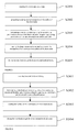
- FIG. 4 is a technological process of the preparation method of the interventional medical device provided by the present application.
- FIG. 5 is another technological process of the preparation method of the interventional medical device provided by the present application.
- FIG. 6 is another technological process of the preparation method of the interventional medical device provided by the present application.
- FIG. 7 is another technological process of the preparation method of the interventional medical device provided by the present application.
- the examples of the present application provide an interventional medical device, comprising a stent body with a drug releasing structure, wherein the drugs in the drug releasing structure are drugs for suppressing proliferation of adventitial fibroblasts and for suppressing proliferation of intimal cells and/or smooth muscle cells.
- FIG. 1 is a structural schematic diagram of a specific embodiment of the interventional medical device provided by the present application.
- 1 indicates stent body
- 2 indicates drug releasing coating
- Drug releasing coating 2 is coated on the outer surface of stent body 1 , in which:
- Stent body 1 can be a coronary artery stent, intracranial vascular stent, peripheral vascular stent, intraoperative stent, heart valve stent, biliary tract stent, esophageal stent, intestinal tract stent, pancreatic duct stent, urethral stent or tracheal stent.
- the material of stent body 1 can be a material with good biocompatibility and mechanical characteristics, such as stainless steel, cobalt-based alloy, nickel-based alloy, titanium alloy, degradable magnesium alloy or a polymer material, etc.
- Drug releasing coating 2 is a dense mixed layer formed by the polymer and the drug for suppressing adventitial fibroblast proliferation and the drug for suppressing proliferation of intimal cells and/or smooth muscle cells. That is, drug releasing coating 2 is used as a carrier to allow the surface of stent body 1 to carry drugs.
- Drug for inhibiting adventitial fibroblast proliferation includes at least one drug selected from the group consisting of tanshinone, asiaticoside, madecassoside, ligustrazine, dracorhodin, Rosuvastatin, angiotensin.
- asiaticoside is preferred.
- Drug for suppressing proliferation of intimal cells and/or smooth muscle cells can be at least one drug selected from the group consisting of rapamycin and derivative thereof and paclitaxel and derivative thereof, with rapamycin is preferred.
- the polymer in drug releasing coating 2 can be a polymer having biocompatibility and controlled release properties, such as, polylactic acid, polyethylene glycol, styrene-butene copolymer, polycaprolactone, poly(butyl methacrylate), poly(ethyl methacrylate), polyvinyl ethyl acetate, polyurethane, polyvinyl pyrrolidone, polyphosphorylcholine, silk protein, gelatin, chitin and/or hyaluronic acid.
- polylactic acid polyethylene glycol, styrene-butene copolymer, polycaprolactone, poly(butyl methacrylate), poly(ethyl methacrylate), polyvinyl ethyl acetate, polyurethane, polyvinyl pyrrolidone, polyphosphorylcholine, silk protein, gelatin, chitin and/or hyaluronic acid.
- Asiaticoside is the total glycosides extracted from Umbelliferae Centella asiatica . Asiaticoside can inhibit the pathological role of TGF-beta by increasing expression of Smad7 that inhibits Smad transduction signal, thereby functioning in vascular remodeling by blocking fibroblast proliferation, promoting vascular compensatory expansion, thus reducing the occurrence rate of in-stent restenosis.
- the interventional medical device provided by the examples of the present application also promotes endothelial cell growth and accelerates the process of endothelialization.
- FIG. 2 is a structural schematic diagram of another specific embodiment of the interventional medical device provided by the present application.
- 1 indicates stent body
- 3 indicates micropores formed on the surface of the stent.
- the drug releasing structure is micropore 3 .
- Micropore 3 may be obtained by oxidating or eroding the surface of stent body 1 .
- Drugs may be loaded within micropore 3 , thus, the surface of stent body 1 will carry drugs.
- FIG. 3 is structural schematic diagram of another specific embodiment of the interventional medical device provided by the present application.
- micropore 3 is obtained by directly oxidating or eroding the surface of stent body 1 .
- a layer of microporous coating can be prepared on the surface of stent body 1 .
- 1 indicates stent body
- 4 indicates microporous coating. This eliminates the need for oxidating or eroding the surface of stent body 1 .
- microporous coating 4 is directly prepared on the surface of stent body 1 to obtain micropores loaded with drugs.
- FIG. 4 is a technological process of the preparation method of the interventional medical device provided by the present application.
- the preparation method of the interventional medical device comprises:
- Step S 101 cleaning the stent body and drying.
- Step S 102 preparing micropores on the surface of the stent body.
- Micropores on the surface of the stent body are formed by electrochemical corrosion and/or chemical corrosion. Electrochemical corrosion includes anodic oxidation, micro-arc oxidation and so on. Micropores can be formed on the surface of the stent body by this step.
- FIG. 2 shows their structural schematic diagram.
- Step S 103 formulating a solution containing a drug for suppressing proliferation of adventitial fibroblasts and a drug for suppressing proliferation of intimal cells and/or smooth muscle cells.
- the drug for suppressing proliferation of adventitial fibroblasts is asiaticoside
- the drug for suppressing proliferation of intimal cells and/or smooth muscle cells is rapamycin.
- 10 mg rapamycin and 30 mg asiaticoside are dissolved in 10 ml ethanol solution. After they are dissolved, the mixture is mixed thoroughly.
- Step S 104 loading the drugs within the formulated solution into the micropores of the stent body.
- the stent body with micropores on its surface obtained in step S 102 is immersed into the solution formulated in step S 103 , so that the drugs within the solution can be loaded into the micropores on the surface of the stent body.
- Step S 105 drying the stent body to get the interventional medical device.
- FIG. 5 is another technological process of the preparation method of the interventional medical device provided by the present application.
- the preparation method of the interventional medical device comprises:
- Step S 201 cleaning the stent body and drying.
- Step S 202 preparing a coating having micropores on the surface of the stent body.
- Particular process includes the following steps: the silk protein solution is uniformly coated on the surface of the stent body. Then the stent body is subject to thermal or chemical denaturation, and infiltration by pure water. After that, the stent body is freezed and then the temperature is increased to dry the stent body. A coating with microporous structure is thus formed on the surface of the stent body.
- Step S 203 formulating a solution containing a drug for suppressing proliferation of adventitial fibroblasts and a drug for suppressing proliferation of intimal cells and/or smooth muscle cells.
- the drug for suppressing proliferation of adventitial fibroblasts is asiaticoside
- the drug for suppressing proliferation of intimal cells and/or smooth muscle cells is rapamycin.
- 10 mg rapamycin and 50 mg asiaticoside are dissolved in 10 ml ethanol solution. After they are dissolved, the mixture is mixed thoroughly.
- Step S 204 loading the drugs within the formulated solution into the micropores of the coating on the surface of the stent body.
- the stent body with a microporous coating on its surface obtained in step S 202 is immersed into the formulated solution, so that the drugs within the solution can be loaded into the micropores of the coating on the surface of the stent body.
- Step S 205 drying the stent body to get the interventional medical device.
- FIG. 6 is another technological process of the preparation method of the interventional medical device provided by the present application.
- the preparation method of the interventional medical device comprises:
- Step S 301 cleaning the stent body and drying.
- Step S 302 formulating a mixed solution containing a drug for suppressing adventitial fibroblast proliferation, a drug for suppressing intimal cells and/or smooth muscle cell proliferation and a polymer.
- the polymer is polylactic acid
- the drug for suppressing proliferation of adventitial fibroblasts is preferably asiaticoside
- the drug for suppressing proliferation of intimal cells and/or smooth muscle cells is preferably rapamycin.
- a mixed solution of polylactic acid, asiaticoside and rapamycin is formulated, in which the ratio of asiaticoside and rapamycin is in the range of 2:1 ⁇ 5:1, and the ratio of polylactic acid and asiaticoside is in the range of 1:1 ⁇ 5:1.
- 10 mg rapamycin, 30 mg asiaticoside and 10 mg polylactic acid can be added to 10 ml tetrahydrofuran. After they are sufficiently dissolved, the mixture is mixed uniformly.
- Step S 303 coating the surface of the stent body with the mixed solution.
- the mixed solution formulated in step 302 can be coated on the stent body by ultrasonic spraying, air spraying or dipping.
- Step S 304 drying the stent body to get the interventional medical device.
- a mixed solution of two drugs and the polymer is formulated.
- two drugs can be mixed with the polymer respectively.
- the mixed solutions of each drug are successively coated on the surface of the stent body.
- FIG. 7 is another technological process of the preparation method of the interventional medical device provided by the present application.
- the preparation method of the interventional medical device comprises:
- Step S 401 cleaning the stent body and drying.
- Step S 402 formulating a mixed solution containing a drug for suppressing adventitial fibroblast proliferation and a polymer, as well as a mixed solution containing a drug for suppressing intimal cells and/or smooth muscle cell proliferation and a polymer.
- the polymer is polylactic acid
- the drug for suppressing proliferation of adventitial fibroblasts is asiaticoside
- the drug for suppressing proliferation of intimal cells and/or smooth muscle cells is rapamycin.
- Solution of polylactic acid and asiaticoside and solution of polylactic acid and rapamycin are formulated respectively, in which, the ratio of polylactic acid and asiaticoside is in the range of 1:1 ⁇ 4:1, and the ratio of polylactic acid and rapamycin is in the range of 1:1 ⁇ 4:1.
- 30 mg asiaticoside and 60 mg polylactic acid are added to 10 ml tetrahydrofuran.
- the mixture After they are sufficiently dissolved, the mixture is mixed uniformly to get a first drug solution. Additionally, 10 mg rapamycin and 20 mg polylactic acid are added to 10 ml tetrahydrofuran. After they are sufficiently dissolved, the mixture is mixed uniformly to get a second drug solution.
- Step S 403 coating the surface of the stent body with the resultant two mixed solutions on, respectively and successively.
- asiaticoside/polylactic acid solution can be coated on the surface of the stent body first. Then the sprayed stent body is placed in the air for 4 h to make its surface dried. After that, rapamycin/polylactic acid solution is coated on the surface of the dried stent body.
- asiaticoside/polylactic acid solution is sprayed first, followed by rapamycin/polylactic acid solution.
- rapamycin/polylactic acid solution This is only an example of the present application, and should not be construed to limit the present application.
- the spraying order of the two drug solutions can be freely chosen.
- ultrasonic spraying air spraying or dipping and other means may be used.
- Step S 404 drying the stent body to get the interventional medical device.
Abstract
An interventional medical device and manufacturing method thereof. The interventional medical device comprises: a stent body (1); a surface of the stent body (1) being provided with a drug releasing structure (3), and drug in the drug releasing structure (3) being drug for suppressing proliferation of adventitial fibroblasts and a drug for suppressing proliferation of intimal and/or smooth muscle cells. In use, after interventional medical device is implanted into a human body, the drug for suppressing proliferation of adventitial fibroblasts carried thereon can promote the compensatory expansion of the vessel, and the drug for suppressing proliferation of intimal cells and/or smooth muscle cells carried thereon can suppress intimal proliferation of the vessel. The combination of the two kinds of drugs greatly reduces the occurrence rate of in-stent restenosis.
Description
The present application relates to the technical field of medical devices, inparticular, to an interventional medical device containing drugs and manufacturing method thereof.
In recent years, a drug coating was coated onto the stent implanted in the body so as to avoid the incidence of in-stent restenosis after interventional treatment. Drugs mostly carried by currently used drug-eluting stents are those for inhibiting intimal hyperplasia or tunica media hyperplasia, including rapamycin, paclitaxel and derivatives thereof, etc. When the stent carrying above-mentioned drug is implanted into a human body, the stent will continuously release the drug for inhibiting intimal hyperplasia or tunica media hyperplasia into vessel wall to reduce the occurrence rate of in-stent restenosis.
Studies have shown that vascular restenosis formation is not only related to intimal hyperplasia or tunica media hyperplasia after vascular injury, but also related to vascular remodelling. Vascular remodeling is the main factor for in-stent restenosis, accounting for 70% possible causes of restenosis, while intimal hyperplasia or tunica media hyperplasia accounts for only 30% possible causes of restenosis.
Therefore, the current drug-eluting stents for inhibiting intimal hyperplasia or tunica media hyperplasia can not reduce the incidence of in-stent restenosis to the greatest extent. In addition, the current drugs for inhibiting intimal hyperplasia or tunica media hyperplasia, such as rapamycin, paclitaxel and derivatives thereof, may suppress endothelial cell growth, and delay vascular endothelialisation. The problem that blood vessels can not be completely endothelialized may cause late thrombosis.
In view thereof, the examples of the present application provide an interventional medical device and manufacturing method thereof. The interventional medical device can promote vascular compensatory expansion by suppressing proliferation of adventitial fibroblasts, after it is implanted into a human body. And it may also inhibit intimal hyperplasia to reduce the occurrence rate of in-stent restenosis.
In order to achieve the above objects, the examples of the present application provide the following technical solutions:
An interventional medical device, comprising a stent body with a drug releasing structure on its surface, and the drugs in the drug releasing structure are drugs for suppressing proliferation of adventitial fibroblasts and for suppressing proliferation of intimal cells and/or smooth muscle cells.
Preferably, the drug releasing structure is a dense mixed layer formed by a polymer and the drug for suppressing adventitial fibroblast proliferation and the drug for suppressing proliferation of intimal cells and/or smooth muscle cells.
Preferably, the polymer includes polylactic acid, polyethylene glycol, styrene-butene copolymer, polycaprolactone, poly(butyl methacrylate), poly(ethyl methacrylate), polyvinyl ethyl acetate, polyurethane, polyvinyl pyrrolidone, polyphosphorylcholine, silk protein, gelatin, chitin and/or hyaluronic acid.
Preferably, the drug releasing structure is a microporous structure prepared on the surface of the stent body or a microporous coating structure formed on the surface of the stent body, and the drug is loaded into the microporous structure or the microporous coating structure.
Preferably, the drug for suppressing adventitial fibroblast proliferation include at least one drug selected from the group consisting of tanshinone, asiaticoside, madecassoside, ligustrazine, dracorhodin, Rosuvastatin, and angiotensin.
Preferably, the drug for suppressing proliferation of intimal cells and/or smooth muscle cells include at least one drug selected from the group consisting of rapamycin and derivative thereof, paclitaxel and derivative thereof.
Preferably, the stent body comprises coronary artery stent, intracranial vascular stent, peripheral vascular stent, intraoperative stent, heart valve stent, biliary tract stent, esophageal stent, intestinal tract stent, pancreatic duct stent, urethral stent or tracheal stent.
A method for preparing an interventional medical device, comprising:
preparing a microporous structure on the surface of a stent body;
formulating a solution containing a drug for suppressing proliferation of adventitial fibroblasts and a drug for suppressing proliferation of intimal cells and/or smooth muscle cells;
loading the drugs within the formulated solution into said microporous structure;
drying the stent body to obtain the interventional medical device.
Preferably, preparing a microporous structure on the surface of the stent body comprises forming micropores on the surface of the stent body by anodic oxidation, micro-arc oxidation and/or chemical corrosion.
Preferably, preparing a microporous structure on the surface of the stent body comprises preparing a coating having micropores on the surface of the stent body.
Preferably, loading the drugs within the formulated solution into said microporous structure comprises loading the drugs within said solution into said microporous structure by ultrasonic spraying, air spraying and/or dipping.
A method for preparing an interventional medical device, comprising:
formulating a mixed solution of a drug for suppressing adventitial fibroblast proliferation and a polymer, as well as a mixed solution of a drug for suppressing proliferation of intimal cells and/or smooth muscle cells and a polymer, respectively, or formulating a mixed solution of a drug for suppressing adventitial fibroblast proliferation, a drug for suppressing proliferation of intimal cells and/or smooth muscle cells and a polymer;
successively coating the surface of the stent body with the mixed solution of the drug for suppressing adventitial fibroblast proliferation and the polymer, as well as the mixed solution of the drug for suppressing proliferation of intimal cells and/or smooth muscle cells and the polymer, or coating the surface of the stent body with the mixed solution of the drug for suppressing adventitial fibroblast proliferation and the drug for suppressing proliferation of intimal cells and/or smooth muscle cells and the polymer;
drying the stent body to obtain the interventional medical device.
Preferably, the coating comprises ultrasonic spraying, air spraying and/or dipping.
It can be seen from the above technical solutions that, when the interventional medical device is used, the drug for suppressing adventitial fibroblast proliferation carried thereon can be slowly released into vessel wall cells in contact with the stent body after it is implanted into a human body, thus inhibiting proliferation of adventitial fibroblasts, functioning in vascular remodeling by blocking fibroblast proliferation, promoting the compensatory expansion of the damaged blood vessel, thereby reducing the occurrence rate of in-stent restenosis. Meanwhile, the drug for suppressing proliferation of intimal cells and/or smooth muscle cells can suppress intimal proliferation of the vessel to some extent. Combination of the two kinds of drugs can greatly reduce the occurrence rate of in-stent restenosis.
In addition, compared with the current drug-eluting stents using rapamycin, paclitaxel and derivatives thereof, the interventional medical device provided by the examples of the present application not only has low inhibition rate on endothelial cells, but also promotes endothelial cell growth and accelerates the process of endothelialization.
In order to more clearly illustrate the technical solutions of the examples of the present application or the prior art, the accompanying drawings which are required to be used in the description of the examples or the prior art will be briefly introduced below. It is apparent that the accompanying drawings in the following description are merely some examples described in the present application. For those of ordinary skill in the art, it is also possible to derive other drawings according to these drawings without creative efforts.
In order to make those skilled in the art better understand technical solutions of the present application, the technical solutions of the examples of the present application will be clearly and fully described below by making reference to the accompanying drawings of the examples of the present application. Obviously, the described examples are merely a part of the examples of the present application, but not all examples. Based on the examples of the present application, all other examples obtained by those of ordinary skill in the art without creative efforts, should fall within the protection scope of the present application.
The examples of the present application provide an interventional medical device, comprising a stent body with a drug releasing structure, wherein the drugs in the drug releasing structure are drugs for suppressing proliferation of adventitial fibroblasts and for suppressing proliferation of intimal cells and/or smooth muscle cells.
As shown in FIG. 1, 1 indicates stent body, 2 indicates drug releasing coating. Drug releasing coating 2 is coated on the outer surface of stent body 1, in which:
Drug for inhibiting adventitial fibroblast proliferation includes at least one drug selected from the group consisting of tanshinone, asiaticoside, madecassoside, ligustrazine, dracorhodin, Rosuvastatin, angiotensin. In the examples of the present application, asiaticoside is preferred. Drug for suppressing proliferation of intimal cells and/or smooth muscle cells can be at least one drug selected from the group consisting of rapamycin and derivative thereof and paclitaxel and derivative thereof, with rapamycin is preferred. In addition, the polymer in drug releasing coating 2 can be a polymer having biocompatibility and controlled release properties, such as, polylactic acid, polyethylene glycol, styrene-butene copolymer, polycaprolactone, poly(butyl methacrylate), poly(ethyl methacrylate), polyvinyl ethyl acetate, polyurethane, polyvinyl pyrrolidone, polyphosphorylcholine, silk protein, gelatin, chitin and/or hyaluronic acid.
Asiaticoside is the total glycosides extracted from Umbelliferae Centella asiatica. Asiaticoside can inhibit the pathological role of TGF-beta by increasing expression of Smad7 that inhibits Smad transduction signal, thereby functioning in vascular remodeling by blocking fibroblast proliferation, promoting vascular compensatory expansion, thus reducing the occurrence rate of in-stent restenosis.
Furthermore, studies have found that asiaticoside could also promote endothelial cell growth and accelerate endothelialization process. For detailed, see “Experimental study of the effect of asiaticoside on preventing restenosis after percutaneous coronary intervention (CLC R541.4 Article ID: 1671-8259 (2005) 05-0477-03).
Thus it can be seen that, compared with the current drug-eluting stents using rapamycin, paclitaxel and derivatives thereof, the interventional medical device provided by the examples of the present application also promotes endothelial cell growth and accelerates the process of endothelialization.
As shown in FIG. 2, 1 indicates stent body, 3 indicates micropores formed on the surface of the stent. In the example of the present application, the drug releasing structure is micropore 3. Micropore 3 may be obtained by oxidating or eroding the surface of stent body 1. Drugs may be loaded within micropore 3, thus, the surface of stent body 1 will carry drugs.
In the interventional medical device shown in FIG. 2 , micropore 3 is obtained by directly oxidating or eroding the surface of stent body 1. However, in the example of the present application, a layer of microporous coating can be prepared on the surface of stent body 1. As shown in FIG. 3, 1 indicates stent body, 4 indicates microporous coating. This eliminates the need for oxidating or eroding the surface of stent body 1. On the contrary, microporous coating 4 is directly prepared on the surface of stent body 1 to obtain micropores loaded with drugs.
As shown in FIG. 4 , in the example of the present application, taking metal stent as an example of the stent body, the preparation method of the interventional medical device comprises:
Step S101: cleaning the stent body and drying.
During the preparation of the interventional medical device, in order to prevent residual stains on the stent body from affecting the quality of the interventional medical device, it is necessary to clean the stent body first.
Step S102: preparing micropores on the surface of the stent body.
Micropores on the surface of the stent body are formed by electrochemical corrosion and/or chemical corrosion. Electrochemical corrosion includes anodic oxidation, micro-arc oxidation and so on. Micropores can be formed on the surface of the stent body by this step. FIG. 2 shows their structural schematic diagram.
Step S103: formulating a solution containing a drug for suppressing proliferation of adventitial fibroblasts and a drug for suppressing proliferation of intimal cells and/or smooth muscle cells.
In the example of the present application, preferably, the drug for suppressing proliferation of adventitial fibroblasts is asiaticoside, and the drug for suppressing proliferation of intimal cells and/or smooth muscle cells is rapamycin. A mixed solution of asiaticoside and rapamycin is formulated, in which asiaticoside:rapamycin=2:1˜5:1. When formulating, 10 mg rapamycin and 30 mg asiaticoside are dissolved in 10 ml ethanol solution. After they are dissolved, the mixture is mixed thoroughly.
Step S104: loading the drugs within the formulated solution into the micropores of the stent body.
The stent body with micropores on its surface obtained in step S102 is immersed into the solution formulated in step S103, so that the drugs within the solution can be loaded into the micropores on the surface of the stent body.
Step S105: drying the stent body to get the interventional medical device.
As shown in FIG. 5 , in the example of the present application, the preparation method of the interventional medical device comprises:
Step S201: cleaning the stent body and drying.
Step S202: preparing a coating having micropores on the surface of the stent body.
Particular process includes the following steps: the silk protein solution is uniformly coated on the surface of the stent body. Then the stent body is subject to thermal or chemical denaturation, and infiltration by pure water. After that, the stent body is freezed and then the temperature is increased to dry the stent body. A coating with microporous structure is thus formed on the surface of the stent body.
Step S203: formulating a solution containing a drug for suppressing proliferation of adventitial fibroblasts and a drug for suppressing proliferation of intimal cells and/or smooth muscle cells.
In the example of the present application, preferably, the drug for suppressing proliferation of adventitial fibroblasts is asiaticoside, and the drug for suppressing proliferation of intimal cells and/or smooth muscle cells is rapamycin. When formulating, 10 mg rapamycin and 50 mg asiaticoside are dissolved in 10 ml ethanol solution. After they are dissolved, the mixture is mixed thoroughly.
Step S204: loading the drugs within the formulated solution into the micropores of the coating on the surface of the stent body.
The stent body with a microporous coating on its surface obtained in step S202 is immersed into the formulated solution, so that the drugs within the solution can be loaded into the micropores of the coating on the surface of the stent body.
Step S205: drying the stent body to get the interventional medical device.
As shown in FIG. 6 , in the example of the present application, the preparation method of the interventional medical device comprises:
Step S301: cleaning the stent body and drying.
Step S302: formulating a mixed solution containing a drug for suppressing adventitial fibroblast proliferation, a drug for suppressing intimal cells and/or smooth muscle cell proliferation and a polymer.
In the example of the present application, the polymer is polylactic acid, the drug for suppressing proliferation of adventitial fibroblasts is preferably asiaticoside, and the drug for suppressing proliferation of intimal cells and/or smooth muscle cells is preferably rapamycin. A mixed solution of polylactic acid, asiaticoside and rapamycin is formulated, in which the ratio of asiaticoside and rapamycin is in the range of 2:1˜5:1, and the ratio of polylactic acid and asiaticoside is in the range of 1:1˜5:1. When formulating, 10 mg rapamycin, 30 mg asiaticoside and 10 mg polylactic acid can be added to 10 ml tetrahydrofuran. After they are sufficiently dissolved, the mixture is mixed uniformly.
Step S303: coating the surface of the stent body with the mixed solution.
In the example of the present application, the mixed solution formulated in step 302 can be coated on the stent body by ultrasonic spraying, air spraying or dipping.
Step S304: drying the stent body to get the interventional medical device.
In FIG. 6 , a mixed solution of two drugs and the polymer is formulated. However, in the practical application, two drugs can be mixed with the polymer respectively. Then the mixed solutions of each drug are successively coated on the surface of the stent body.
As shown in FIG. 7 , in the example of the present application, the preparation method of the interventional medical device comprises:
Step S401: cleaning the stent body and drying.
Step S402: formulating a mixed solution containing a drug for suppressing adventitial fibroblast proliferation and a polymer, as well as a mixed solution containing a drug for suppressing intimal cells and/or smooth muscle cell proliferation and a polymer.
In the example of the present application, preferably, the polymer is polylactic acid, the drug for suppressing proliferation of adventitial fibroblasts is asiaticoside, and the drug for suppressing proliferation of intimal cells and/or smooth muscle cells is rapamycin. Solution of polylactic acid and asiaticoside and solution of polylactic acid and rapamycin are formulated respectively, in which, the ratio of polylactic acid and asiaticoside is in the range of 1:1˜4:1, and the ratio of polylactic acid and rapamycin is in the range of 1:1˜4:1. When formulating, 30 mg asiaticoside and 60 mg polylactic acid are added to 10 ml tetrahydrofuran. After they are sufficiently dissolved, the mixture is mixed uniformly to get a first drug solution. Additionally, 10 mg rapamycin and 20 mg polylactic acid are added to 10 ml tetrahydrofuran. After they are sufficiently dissolved, the mixture is mixed uniformly to get a second drug solution.
Step S403: coating the surface of the stent body with the resultant two mixed solutions on, respectively and successively.
When coating, asiaticoside/polylactic acid solution can be coated on the surface of the stent body first. Then the sprayed stent body is placed in the air for 4 h to make its surface dried. After that, rapamycin/polylactic acid solution is coated on the surface of the dried stent body.
During above-mentioned spray coating, asiaticoside/polylactic acid solution is sprayed first, followed by rapamycin/polylactic acid solution. This is only an example of the present application, and should not be construed to limit the present application. Those of ordinary skill in the art should know that, in other examples, the spraying order of the two drug solutions can be freely chosen.
In addition, in the example of the present application, ultrasonic spraying, air spraying or dipping and other means may be used.
Step S404: drying the stent body to get the interventional medical device.
The above examples are only preferred embodiments of the present application. With these examples the skilled person can understand or realize the present application. Various modifications to these examples will be apparent to the skilled person in the art, and the generic principles defined herein may be implemented in other examples without departing from the spirit or scope of the present application. Accordingly, the present application will not be limited to these examples described herein, but meet the widest scope consistent with the principles and novel features disclosed herein.
Claims (13)
1. An interventional medical device, comprising a stent body with a drug releasing structure on its surface and at least one drug for suppressing proliferation of adventitial fibroblast fibroblasts and at least one drug for suppressing proliferation of intimal cells and/or smooth muscle cells loaded on or in said drug releasing structure, wherein said at least one drug for suppressing adventitial fibroblast proliferation is or includes asiaticoside and said at least one drug for suppressing proliferation of intimal cells and/or smooth muscle cells is or includes rapamycin, wherein the ratio between the weight of asiaticoside and that of rapamycin present in the drug releasing structure is from 2:1 to 5:1.
2. The interventional medical device according to claim 1 , wherein said drug releasing structure is a dense mixed layer formed by a polymer and the drug for suppressing adventitial fibroblast proliferation and the drug for suppressing proliferation of intimal cells and/or smooth muscle cells.
3. The interventional medical device according to claim 2 , wherein said polymer includes polylactic acid, polyethylene glycol, styrene-butene copolymer, polycaprolactone, poly(butyl methacrylate), poly (ethyl methacrylate), polyvinyl ethyl acetate, polyurethane, polyvinyl pyrrolidone, polyphosphorylcholine, silk protein, gelatin, chitin and/or hyaluronic acid.
4. The interventional medical device according to claim 1 , wherein, said drug releasing structure is a microporous structure prepared on the surface of said stent body or a microporous coating structure formed on the surface of said stent body, and the drugs are loaded into said microporous structure or microporous coating structure.
5. The interventional medical device according to claim 1 , wherein said at least one drug for suppressing adventitial fibroblast proliferation additionally includes at least one drug selected from the group consisting of tanshinone, madecassoside, ligustrazine, dracorhodin, rosuvastatin, and angiotensin.
6. The interventional medical device according to claim 1 , wherein said at least one drug for suppressing proliferation of intimal cells and/or smooth muscle cells additionally includes at least one drug selected from the group consisting of derivatives of rapamycin and paclitaxel and derivative thereof.
7. The interventional medical device according to claim 1 , wherein said stent body comprises a coronary artery stent, an intracranial vascular stent, a peripheral vascular stent, an intraoperative stent, a heart valve stent, a biliary tract stent, an esophageal stent, an intestinal tract stent, a pancreatic duct stent, an urethral stent or a tracheal stent.
8. A method for preparing an interventional medical device, wherein said method comprises:
preparing a microporous structure on the surface of the stent body;
formulating a solution containing at least one drug for suppressing proliferation of adventitial fibroblasts and at least one drug for suppressing proliferation of intimal cells and/or smooth muscle cells, wherein said at least one drug for suppressing adventitial fibroblast proliferation is or includes asiaticoside and said at least one drug for suppressing proliferation of intimal cells and/or smooth muscle cells is or includes rapamycin,
loading the drugs within the formulated solution into said microporous structure, wherein the ratio between the weight of asiaticoside and that of rapamycin present in the microporous structure is from 2:1 to 5:1;
drying the stent body to obtain said interventional medical device.
9. The method according to claim 8 , wherein, preparing a microporous structure on the surface of the stent body comprises forming micropores on the surface of the stent body by anodic oxidation, micro-arc oxidation and/or chemical corrosion.
10. The method according to claim 8 , wherein preparing a microporous structure on the surface of the stent body comprises preparing a coating having micropores on the surface of said stent body.
11. The method according to claim 8 , wherein loading the drugs within the formulated solution into said microporous structure comprises loading the drugs within said solution into said microporous structure by ultrasonic spraying, air spraying and/or dipping.
12. A method for preparing an interventional medical device, wherein said method comprises:
formulating a mixed solution of a drug for suppressing adventitial fibroblast proliferation and a polymer, as well as a mixed solution of a drug for suppressing proliferation of intimal cells and/or smooth muscle cells and a polymer, respectively, or formulating a mixed solution of a drug for suppressing adventitial fibroblast proliferation, a drug for suppressing proliferation of intimal cells and/or smooth muscle cells and a polymer, wherein said drug for suppressing adventitial fibroblast proliferation is asiaticoside and said drug for suppressing proliferation of intimal cells and/or smooth muscle cells is rapamycin;
successively coating the surface of the stent body with the mixed solution of the drug for suppressing adventitial fibroblast proliferation and the polymer, as well as the mixed solution of the drug for suppressing proliferation of intimal cells and/or smooth muscle cells and the polymer, or coating the surface of the stent body with the mixed solution of the drug for suppressing adventitial fibroblast proliferation, the drug for suppressing proliferation of intimal cells and/or smooth muscle cells and the polymer;
drying the stent body to obtain the interventional medical device, wherein the ratio between the weight of asiaticoside and that of rapamycin present in the interventional medical device is from 2:1 to 5:1.
13. The method according to claim 12 , wherein said coating comprises ultrasonic spraying, air spraying and/or dipping.
Priority Applications (1)
| Application Number | Priority Date | Filing Date | Title |
|---|---|---|---|
| US14/677,741 US9433709B2 (en) | 2011-09-29 | 2015-04-02 | Interventional medical device and manufacturing method thereof |
Applications Claiming Priority (6)
| Application Number | Priority Date | Filing Date | Title |
|---|---|---|---|
| CN201110295324 | 2011-09-29 | ||
| CN2011102953249A CN102499798A (en) | 2011-09-29 | 2011-09-29 | Interventional medical device and preparation method thereof |
| CN201110295324.9 | 2011-09-29 | ||
| PCT/CN2012/070400 WO2013044603A1 (en) | 2011-09-29 | 2012-01-16 | Interventional medical device and manufacturing method thereof |
| US201414348857A | 2014-03-31 | 2014-03-31 | |
| US14/677,741 US9433709B2 (en) | 2011-09-29 | 2015-04-02 | Interventional medical device and manufacturing method thereof |
Related Parent Applications (2)
| Application Number | Title | Priority Date | Filing Date |
|---|---|---|---|
| US14/348,857 Continuation US20140248327A1 (en) | 2011-09-29 | 2012-01-16 | Interventional medical device and manufacturing method thereof |
| PCT/CN2012/070400 Continuation WO2013044603A1 (en) | 2011-09-29 | 2012-01-16 | Interventional medical device and manufacturing method thereof |
Publications (2)
| Publication Number | Publication Date |
|---|---|
| US20150209485A1 US20150209485A1 (en) | 2015-07-30 |
| US9433709B2 true US9433709B2 (en) | 2016-09-06 |
Family
ID=46211977
Family Applications (2)
| Application Number | Title | Priority Date | Filing Date |
|---|---|---|---|
| US14/348,857 Abandoned US20140248327A1 (en) | 2011-09-29 | 2012-01-16 | Interventional medical device and manufacturing method thereof |
| US14/677,741 Active US9433709B2 (en) | 2011-09-29 | 2015-04-02 | Interventional medical device and manufacturing method thereof |
Family Applications Before (1)
| Application Number | Title | Priority Date | Filing Date |
|---|---|---|---|
| US14/348,857 Abandoned US20140248327A1 (en) | 2011-09-29 | 2012-01-16 | Interventional medical device and manufacturing method thereof |
Country Status (7)
| Country | Link |
|---|---|
| US (2) | US20140248327A1 (en) |
| EP (1) | EP2762110A4 (en) |
| JP (1) | JP2014530058A (en) |
| CN (1) | CN102499798A (en) |
| BR (1) | BR112014007580A2 (en) |
| IN (1) | IN2014CN02578A (en) |
| WO (1) | WO2013044603A1 (en) |
Families Citing this family (7)
| Publication number | Priority date | Publication date | Assignee | Title |
|---|---|---|---|---|
| CN102397119A (en) * | 2011-09-29 | 2012-04-04 | 微创医疗器械(上海)有限公司 | Interventional medical appliance and manufacturing method thereof |
| AU2015328825B2 (en) * | 2014-10-06 | 2019-02-21 | Gatt Technologies B.V. | Tissue-adhesive porous haemostatic product |
| JP6438048B2 (en) * | 2014-12-25 | 2018-12-12 | オリンパス株式会社 | Bone implant and its manufacturing method |
| CN105816921A (en) * | 2016-04-20 | 2016-08-03 | 山东百多安医用材料改性工程技术中心 | Bionic vascular stent and preparation method thereof |
| CN106512188A (en) * | 2016-12-13 | 2017-03-22 | 天津飞捷科技有限公司 | Mechanical and electrical integrated targeted drug release intervention medical instrument and preparation method thereof |
| CN109602523B (en) * | 2019-01-03 | 2024-02-09 | 科塞尔医疗科技(苏州)有限公司 | Recoverable medicine support |
| CN110283296B (en) * | 2019-06-20 | 2020-07-31 | 中国科学院长春应用化学研究所 | Difunctional polyurethane and preparation method and application thereof |
Citations (16)
| Publication number | Priority date | Publication date | Assignee | Title |
|---|---|---|---|---|
| US6273913B1 (en) * | 1997-04-18 | 2001-08-14 | Cordis Corporation | Modified stent useful for delivery of drugs along stent strut |
| US20040086542A1 (en) * | 1999-12-23 | 2004-05-06 | Hossainy Syed F.A. | Coating for implantable devices and a method of forming the same |
| US20050060028A1 (en) * | 2001-10-15 | 2005-03-17 | Roland Horres | Coating of stents for preventing restenosis |
| US20060199876A1 (en) * | 2005-03-04 | 2006-09-07 | The University Of British Columbia | Bioceramic composite coatings and process for making same |
| US20070037883A1 (en) | 2003-02-28 | 2007-02-15 | Dusting Gregory J | Therapeutic compositions |
| CN1935274A (en) * | 2006-10-20 | 2007-03-28 | 东南大学 | Coronary artery medicinal-coating stent |
| US20070219613A1 (en) | 2003-10-06 | 2007-09-20 | Xtent, Inc. | Apparatus and methods for interlocking stent segments |
| US20070292470A1 (en) * | 2006-06-15 | 2007-12-20 | Medtronic Vascular, Inc. | Implantable Medical Devices and Methods for Making the Same |
| CN101318032A (en) | 2007-06-06 | 2008-12-10 | 李京倖 | Small-diameter tissue engineering artificial blood vessel and preparation method thereof |
| EP2014308A2 (en) | 2007-07-10 | 2009-01-14 | Cordis Corporation | A coating employing an anti-thrombotic conjugate |
| US20090112310A1 (en) * | 2006-12-14 | 2009-04-30 | Lepu Medicql Technology (Beijing) Co., Ltd. | Nanoporous Drug Release Structure for Drug Elute Instruments and the Preparation Method Thereof |
| CN101879102A (en) | 2009-05-07 | 2010-11-10 | 微创医疗器械(上海)有限公司 | Groove carrying-type coating decomposable drug eluting stent |
| US20110202125A1 (en) | 2007-10-12 | 2011-08-18 | Microport Medical (Shanghai)Co., Ltd. | Artificial stent and its preparation method |
| US20110202122A1 (en) | 2008-09-17 | 2011-08-18 | Terumo Kabushiki Kaisha | Stent |
| US8003122B2 (en) | 2004-03-31 | 2011-08-23 | Cordis Corporation | Device for local and/or regional delivery employing liquid formulations of therapeutic agents |
| WO2013044605A1 (en) | 2011-09-29 | 2013-04-04 | 上海微创医疗器械(集团)有限公司 | Interventional medical device and manufacturing method thereof |
Family Cites Families (14)
| Publication number | Priority date | Publication date | Assignee | Title |
|---|---|---|---|---|
| EP1308179A1 (en) * | 2001-10-30 | 2003-05-07 | Boehringer Ingelheim Pharma GmbH & Co.KG | Improved endoprosthetic device |
| US20050019404A1 (en) * | 2003-06-30 | 2005-01-27 | Hsing-Wen Sung | Drug-eluting biodegradable stent |
| JP4588986B2 (en) * | 2002-08-20 | 2010-12-01 | テルモ株式会社 | Implantable medical device |
| US7488343B2 (en) * | 2003-09-16 | 2009-02-10 | Boston Scientific Scimed, Inc. | Medical devices |
| US20050266040A1 (en) * | 2004-05-28 | 2005-12-01 | Brent Gerberding | Medical devices composed of porous metallic materials for delivering biologically active materials |
| CN101065153B (en) * | 2004-08-13 | 2013-02-20 | 罗格斯州立大学 | Radiopaque polymeric stents |
| US20070224235A1 (en) * | 2006-03-24 | 2007-09-27 | Barron Tenney | Medical devices having nanoporous coatings for controlled therapeutic agent delivery |
| US8187620B2 (en) * | 2006-03-27 | 2012-05-29 | Boston Scientific Scimed, Inc. | Medical devices comprising a porous metal oxide or metal material and a polymer coating for delivering therapeutic agents |
| PL2386322T3 (en) * | 2006-07-03 | 2018-06-29 | Hemoteq Ag | Production, method and use of medical products which release agents for opening blood vessels on a permanent basis |
| US8187255B2 (en) * | 2007-02-02 | 2012-05-29 | Boston Scientific Scimed, Inc. | Medical devices having nanoporous coatings for controlled therapeutic agent delivery |
| CA2680229A1 (en) * | 2007-03-09 | 2008-09-18 | Jay S. Yadav | Bioabsorbable coatings for medical devices |
| EP3791826A1 (en) * | 2007-12-18 | 2021-03-17 | Intersect ENT, Inc. | Self-expanding devices |
| KR100947094B1 (en) * | 2008-01-02 | 2010-03-10 | 주식회사 디오 | Stent for medical use and manufacturing method thereof |
| US20100280600A1 (en) * | 2009-04-30 | 2010-11-04 | Vipul Bhupendra Dave | Dual drug stent |
-
2011
- 2011-09-29 CN CN2011102953249A patent/CN102499798A/en active Pending
-
2012
- 2012-01-16 JP JP2014532219A patent/JP2014530058A/en active Pending
- 2012-01-16 BR BR112014007580A patent/BR112014007580A2/en not_active IP Right Cessation
- 2012-01-16 US US14/348,857 patent/US20140248327A1/en not_active Abandoned
- 2012-01-16 EP EP12834752.3A patent/EP2762110A4/en not_active Withdrawn
- 2012-01-16 IN IN2578CHN2014 patent/IN2014CN02578A/en unknown
- 2012-01-16 WO PCT/CN2012/070400 patent/WO2013044603A1/en active Application Filing
-
2015
- 2015-04-02 US US14/677,741 patent/US9433709B2/en active Active
Patent Citations (16)
| Publication number | Priority date | Publication date | Assignee | Title |
|---|---|---|---|---|
| US6273913B1 (en) * | 1997-04-18 | 2001-08-14 | Cordis Corporation | Modified stent useful for delivery of drugs along stent strut |
| US20040086542A1 (en) * | 1999-12-23 | 2004-05-06 | Hossainy Syed F.A. | Coating for implantable devices and a method of forming the same |
| US20050060028A1 (en) * | 2001-10-15 | 2005-03-17 | Roland Horres | Coating of stents for preventing restenosis |
| US20070037883A1 (en) | 2003-02-28 | 2007-02-15 | Dusting Gregory J | Therapeutic compositions |
| US20070219613A1 (en) | 2003-10-06 | 2007-09-20 | Xtent, Inc. | Apparatus and methods for interlocking stent segments |
| US8003122B2 (en) | 2004-03-31 | 2011-08-23 | Cordis Corporation | Device for local and/or regional delivery employing liquid formulations of therapeutic agents |
| US20060199876A1 (en) * | 2005-03-04 | 2006-09-07 | The University Of British Columbia | Bioceramic composite coatings and process for making same |
| US20070292470A1 (en) * | 2006-06-15 | 2007-12-20 | Medtronic Vascular, Inc. | Implantable Medical Devices and Methods for Making the Same |
| CN1935274A (en) * | 2006-10-20 | 2007-03-28 | 东南大学 | Coronary artery medicinal-coating stent |
| US20090112310A1 (en) * | 2006-12-14 | 2009-04-30 | Lepu Medicql Technology (Beijing) Co., Ltd. | Nanoporous Drug Release Structure for Drug Elute Instruments and the Preparation Method Thereof |
| CN101318032A (en) | 2007-06-06 | 2008-12-10 | 李京倖 | Small-diameter tissue engineering artificial blood vessel and preparation method thereof |
| EP2014308A2 (en) | 2007-07-10 | 2009-01-14 | Cordis Corporation | A coating employing an anti-thrombotic conjugate |
| US20110202125A1 (en) | 2007-10-12 | 2011-08-18 | Microport Medical (Shanghai)Co., Ltd. | Artificial stent and its preparation method |
| US20110202122A1 (en) | 2008-09-17 | 2011-08-18 | Terumo Kabushiki Kaisha | Stent |
| CN101879102A (en) | 2009-05-07 | 2010-11-10 | 微创医疗器械(上海)有限公司 | Groove carrying-type coating decomposable drug eluting stent |
| WO2013044605A1 (en) | 2011-09-29 | 2013-04-04 | 上海微创医疗器械(集团)有限公司 | Interventional medical device and manufacturing method thereof |
Non-Patent Citations (2)
| Title |
|---|
| International Search Report and Written Opinion for PCT/CN2012/070400, dated Jul. 5, 2012, 12 pages. |
| International Search Report and Written Opinion for PCT/CN2012/070455, dated Apr. 26, 2012, 13 pages. |
Also Published As
| Publication number | Publication date |
|---|---|
| US20140248327A1 (en) | 2014-09-04 |
| EP2762110A1 (en) | 2014-08-06 |
| BR112014007580A2 (en) | 2017-04-11 |
| EP2762110A4 (en) | 2015-05-06 |
| JP2014530058A (en) | 2014-11-17 |
| WO2013044603A1 (en) | 2013-04-04 |
| US20150209485A1 (en) | 2015-07-30 |
| IN2014CN02578A (en) | 2015-08-07 |
| CN102499798A (en) | 2012-06-20 |
Similar Documents
| Publication | Publication Date | Title |
|---|---|---|
| US9433709B2 (en) | Interventional medical device and manufacturing method thereof | |
| US20150004207A1 (en) | Interventional medical device and manufacturing method thereof | |
| US9522219B2 (en) | Resorbable stents which contain a magnesium alloy | |
| JP5636450B2 (en) | Drug sustained release stent | |
| CN102309368B (en) | Body lumen drug-carrying bracket and preparation method thereof | |
| CN110139681A (en) | The bracket made of biodegradable magnesium alloy with magnesium fluoride coating and organic coating | |
| CN101721266B (en) | Absorbable magnesium alloy stent of anticorrosion and drug release composite coating and preparation method thereof | |
| CN101721753B (en) | Absorbable magnesium alloy bracket of inorganic and organic antiseptic biocompatible composite coating and preparation method thereof | |
| WO2010081393A1 (en) | Medicament eluting apparatus with micro-hole structure capable of storing and releasing multiple medicines and preparation method | |
| JP2010517713A (en) | Coronary stent with asymmetric drug release controlled coating | |
| CN107913119A (en) | A kind of interventional medical device and preparation method thereof | |
| CN101081316A (en) | Novel medicine eluting supporting stand | |
| WO2008061431A1 (en) | Vessel stent with multi drug-coatings | |
| CN113476669A (en) | Medicine coating composition and coating process thereof | |
| KR101595267B1 (en) | Consecutive Drug releaseing stent for restenosis and inflammatory regulation and manufacturing method thereof | |
| JP5597625B2 (en) | Drug eluting stent | |
| CN104707185A (en) | Ginkgolide B composite medicine eluting stent | |
| CN105797207A (en) | Drug release carrier on metal substrate and preparation method thereof | |
| KR20110093009A (en) | A manufacturing method of drug coated stent and a stent manufactured by the same | |
| CN106178139A (en) | A kind of support and preparation method thereof | |
| CN201350138Y (en) | Medicine coating stent with biological activity | |
| JP2002085549A (en) | Material for intravascular treatment and appliance for intravascular treatment | |
| JP2016005533A (en) | Stent | |
| JP2015226574A (en) | Stent having medicine release layer | |
| CN104707184A (en) | Drug coating stent based on HIV protease inhibitor saquinavir as well as preparation method and application thereof |
Legal Events
| Date | Code | Title | Description |
|---|---|---|---|
| AS | Assignment |
Owner name: SHANGHAI MICROPORT MEDICAL (GROUP) CO., LTD., CHIN Free format text: ASSIGNMENT OF ASSIGNORS INTEREST;ASSIGNORS:ZHANG, DADONG;CAI, XU;YUE, CHENGYUN;AND OTHERS;SIGNING DATES FROM 20151217 TO 20151218;REEL/FRAME:038754/0151 |
|
| STCF | Information on status: patent grant |
Free format text: PATENTED CASE |
|
| MAFP | Maintenance fee payment |
Free format text: PAYMENT OF MAINTENANCE FEE, 4TH YEAR, LARGE ENTITY (ORIGINAL EVENT CODE: M1551); ENTITY STATUS OF PATENT OWNER: LARGE ENTITY Year of fee payment: 4 |