US9486157B2 - Intubation monitoring apparatus and method - Google Patents
Intubation monitoring apparatus and method Download PDFInfo
- Publication number
- US9486157B2 US9486157B2 US13/043,648 US201113043648A US9486157B2 US 9486157 B2 US9486157 B2 US 9486157B2 US 201113043648 A US201113043648 A US 201113043648A US 9486157 B2 US9486157 B2 US 9486157B2
- Authority
- US
- United States
- Prior art keywords
- impedance
- processing unit
- endo
- tracheal tube
- thoracic
- Prior art date
- Legal status (The legal status is an assumption and is not a legal conclusion. Google has not performed a legal analysis and makes no representation as to the accuracy of the status listed.)
- Expired - Lifetime, expires
Links
- 238000000034 method Methods 0.000 title claims abstract description 41
- 238000012544 monitoring process Methods 0.000 title claims abstract description 23
- 238000002627 tracheal intubation Methods 0.000 title claims description 34
- 210000000115 thoracic cavity Anatomy 0.000 claims abstract description 37
- 238000009423 ventilation Methods 0.000 claims abstract description 33
- 238000012545 processing Methods 0.000 claims description 43
- 238000001514 detection method Methods 0.000 claims description 9
- 238000002847 impedance measurement Methods 0.000 abstract description 3
- 238000005259 measurement Methods 0.000 description 26
- 210000000038 chest Anatomy 0.000 description 14
- 210000004072 lung Anatomy 0.000 description 14
- 210000003437 trachea Anatomy 0.000 description 12
- 238000010586 diagram Methods 0.000 description 9
- 230000003213 activating effect Effects 0.000 description 6
- 239000000523 sample Substances 0.000 description 6
- 206010058151 Pulseless electrical activity Diseases 0.000 description 3
- 230000017531 blood circulation Effects 0.000 description 3
- 230000000241 respiratory effect Effects 0.000 description 3
- 230000029058 respiratory gaseous exchange Effects 0.000 description 3
- 210000002784 stomach Anatomy 0.000 description 3
- 238000012360 testing method Methods 0.000 description 3
- 208000010496 Heart Arrest Diseases 0.000 description 2
- 230000006378 damage Effects 0.000 description 2
- 239000007789 gas Substances 0.000 description 2
- 230000001939 inductive effect Effects 0.000 description 2
- 238000003780 insertion Methods 0.000 description 2
- 230000037431 insertion Effects 0.000 description 2
- 210000000867 larynx Anatomy 0.000 description 2
- 210000001519 tissue Anatomy 0.000 description 2
- 206010002091 Anaesthesia Diseases 0.000 description 1
- 206010007617 Cardio-respiratory arrest Diseases 0.000 description 1
- 208000009378 Low Cardiac Output Diseases 0.000 description 1
- 208000037656 Respiratory Sounds Diseases 0.000 description 1
- 206010038669 Respiratory arrest Diseases 0.000 description 1
- 235000001537 Ribes X gardonianum Nutrition 0.000 description 1
- 235000001535 Ribes X utile Nutrition 0.000 description 1
- 235000016919 Ribes petraeum Nutrition 0.000 description 1
- 244000281247 Ribes rubrum Species 0.000 description 1
- 235000002355 Ribes spicatum Nutrition 0.000 description 1
- 208000027418 Wounds and injury Diseases 0.000 description 1
- 210000000683 abdominal cavity Anatomy 0.000 description 1
- 238000001949 anaesthesia Methods 0.000 description 1
- 230000037005 anaesthesia Effects 0.000 description 1
- QVGXLLKOCUKJST-UHFFFAOYSA-N atomic oxygen Chemical compound [O] QVGXLLKOCUKJST-UHFFFAOYSA-N 0.000 description 1
- 239000008280 blood Substances 0.000 description 1
- 210000004369 blood Anatomy 0.000 description 1
- 238000004364 calculation method Methods 0.000 description 1
- 239000003990 capacitor Substances 0.000 description 1
- 230000000747 cardiac effect Effects 0.000 description 1
- 238000002680 cardiopulmonary resuscitation Methods 0.000 description 1
- 230000006835 compression Effects 0.000 description 1
- 238000007906 compression Methods 0.000 description 1
- 238000004590 computer program Methods 0.000 description 1
- 239000004020 conductor Substances 0.000 description 1
- 230000007423 decrease Effects 0.000 description 1
- 239000000706 filtrate Substances 0.000 description 1
- 230000006870 function Effects 0.000 description 1
- 208000014674 injury Diseases 0.000 description 1
- 239000012774 insulation material Substances 0.000 description 1
- 150000002500 ions Chemical class 0.000 description 1
- 210000004877 mucosa Anatomy 0.000 description 1
- 239000001301 oxygen Substances 0.000 description 1
- 229910052760 oxygen Inorganic materials 0.000 description 1
- 238000005086 pumping Methods 0.000 description 1
- 230000000284 resting effect Effects 0.000 description 1
- 230000035939 shock Effects 0.000 description 1
- 230000011664 signaling Effects 0.000 description 1
- 230000007704 transition Effects 0.000 description 1
Images


Classifications
-
- A—HUMAN NECESSITIES
- A61—MEDICAL OR VETERINARY SCIENCE; HYGIENE
- A61B—DIAGNOSIS; SURGERY; IDENTIFICATION
- A61B5/00—Measuring for diagnostic purposes; Identification of persons
- A61B5/05—Detecting, measuring or recording for diagnosis by means of electric currents or magnetic fields; Measuring using microwaves or radio waves
- A61B5/053—Measuring electrical impedance or conductance of a portion of the body
- A61B5/0538—Measuring electrical impedance or conductance of a portion of the body invasively, e.g. using a catheter
-
- A—HUMAN NECESSITIES
- A61—MEDICAL OR VETERINARY SCIENCE; HYGIENE
- A61B—DIAGNOSIS; SURGERY; IDENTIFICATION
- A61B5/00—Measuring for diagnostic purposes; Identification of persons
- A61B5/08—Detecting, measuring or recording devices for evaluating the respiratory organs
- A61B5/0809—Detecting, measuring or recording devices for evaluating the respiratory organs by impedance pneumography
-
- A—HUMAN NECESSITIES
- A61—MEDICAL OR VETERINARY SCIENCE; HYGIENE
- A61M—DEVICES FOR INTRODUCING MEDIA INTO, OR ONTO, THE BODY; DEVICES FOR TRANSDUCING BODY MEDIA OR FOR TAKING MEDIA FROM THE BODY; DEVICES FOR PRODUCING OR ENDING SLEEP OR STUPOR
- A61M16/00—Devices for influencing the respiratory system of patients by gas treatment, e.g. mouth-to-mouth respiration; Tracheal tubes
- A61M16/04—Tracheal tubes
- A61M16/0488—Mouthpieces; Means for guiding, securing or introducing the tubes
-
- A—HUMAN NECESSITIES
- A61—MEDICAL OR VETERINARY SCIENCE; HYGIENE
- A61M—DEVICES FOR INTRODUCING MEDIA INTO, OR ONTO, THE BODY; DEVICES FOR TRANSDUCING BODY MEDIA OR FOR TAKING MEDIA FROM THE BODY; DEVICES FOR PRODUCING OR ENDING SLEEP OR STUPOR
- A61M16/00—Devices for influencing the respiratory system of patients by gas treatment, e.g. mouth-to-mouth respiration; Tracheal tubes
- A61M16/04—Tracheal tubes
- A61M16/0402—Special features for tracheal tubes not otherwise provided for
- A61M16/0411—Special features for tracheal tubes not otherwise provided for with means for differentiating between oesophageal and tracheal intubation
Landscapes
- Health & Medical Sciences (AREA)
- Life Sciences & Earth Sciences (AREA)
- General Health & Medical Sciences (AREA)
- Heart & Thoracic Surgery (AREA)
- Veterinary Medicine (AREA)
- Public Health (AREA)
- Animal Behavior & Ethology (AREA)
- Engineering & Computer Science (AREA)
- Biomedical Technology (AREA)
- Pathology (AREA)
- Medical Informatics (AREA)
- Molecular Biology (AREA)
- Surgery (AREA)
- Pulmonology (AREA)
- Biophysics (AREA)
- Physics & Mathematics (AREA)
- Nuclear Medicine, Radiotherapy & Molecular Imaging (AREA)
- Radiology & Medical Imaging (AREA)
- Anesthesiology (AREA)
- Otolaryngology (AREA)
- Physiology (AREA)
- Emergency Medicine (AREA)
- Hematology (AREA)
- Measurement And Recording Of Electrical Phenomena And Electrical Characteristics Of The Living Body (AREA)
- Measurement Of The Respiration, Hearing Ability, Form, And Blood Characteristics Of Living Organisms (AREA)
- Spinning Or Twisting Of Yarns (AREA)
- Investigating Or Analyzing Materials By The Use Of Electric Means (AREA)
- Accessories For Mixers (AREA)
- Gyroscopes (AREA)
- Blast Furnaces (AREA)
Abstract
The invention comprises an apparatus and a method for externally assessing and monitoring placement of an endo-tracheal tube for ventilation of patients based on thoracic impedance measurement.
Description
This application is a divisional application of U.S. patent application Ser. No. 10/517,989 (now U.S. Pat. No. 7,925,339) which is a national stage of International Application No. PCT/NO2003/000208 filed Jun. 19, 2003, which claims priority from U.S. Provisional Application No. 60/390,115 filed Jun. 21, 2002 and (NO) Application No. 20022960 filed Jun. 19, 2002, the entire contents of each of the above-identified applications are incorporated herein by reference.
The present invention relates to an apparatus and a method for assessing correct endo-tracheal intubation immediately after intubation and for monitoring correct placement of the endo-tracheal tube over time.
Endo-tracheal tubes are used during general anaesthesia, intensive care, and cardiopulmonary resuscitation. The endo-tracheal tube is used to secure the airways to the patient's lungs. Insertion of an endo-tracheal tube is carried out with the aid of a laryngoscope. This device allows the operator to visually identify the larynx and pass the tube through it into the trachea. In some cases the larynx may not be seen and the chance arises of a tube being erroneously passed into the oesophagus as opposed to the trachea.
An endo-tracheal tube erroneously passed into the oesophagus does not provide an adequate airway, so that the patient may become deprived of oxygen, and serious harm or even death may result.
During transportation or moving of a patient the endo-tracheal tube may be unwontedly relocated, without any clinical signs of this condition appearing before it is too late.
Recognition of correct endrotracheal intubation as soon as possible is thus of paramount importance. It is also important to be able to monitor intubation in the cases where there is a risk of unwonted relocation of the tube.
The usual method of confirming the correct location of the tube involves pumping a quantity of air or gas through the tube into the patient. With a stethoscope it is possible to hear normal ventilation sounds if the tube is placed in the trachea. However, in a stress situation and/or a noisy environment, it is difficult to hear these important ventilation sounds.
As a consequence of this, several devices which are not based on hearing ventilation sounds are introduced. These may e.g. be based on pressure and End tidal CO2 measurements. End Tidal CO2 measurements are based on an analysis of the patient's expiration air, if this air contains CO2, it has been in the patient's lungs and the intubation is correct. However, a measurement of CO2 contents depends on several parameters (blood flow through the lungs, gas exchange in the lungs, ventilation volume per ventilation, ventilation amount per minute, etc). In cases where the blood flow through the lungs is strongly reduced, and the patient ventilation is constant, the End Tidal CO2 level will be almost zero. This can be wrongly interpreted as an incorrect intubation. As End Tidal CO2 depends on other physiological parameters for the patient that can be failing at the time intubation is performed, it cannot be considered as a reliable method.
Other devices use the change of transthoracal diameter as a factor to detect correct intubation. Again transthoracic diameter measurement can be affected by several sources of error. Sometimes the patient's thorax is so rigid that it does not expand appreciably by inhalation. In these cases, the abdominal cavity expands as a consequence of diaphragm movement. These will of course be erroneously interpreted as the patient receiving air in the stomach (wrong intubation).
WO 89/07415 describes an apparatus for locating an endo-tracheal or endo-oesophageal tube. This apparatus comprises a tube for insertion into the trachea or oesophagus of a patient. The tube is provided at its' distal end with a pair of electrodes arranged in such manner that the impedance measured between the conductors varies depending on the probe being placed in the trachea or in the oesophagus. If the probe is in the trachea, which is a somewhat rigid tube, one of the electrodes will be in contact with the trachea while the other is not. This will lead to one impedance value. If the probe is in the oesophagus, which is a soft tube which will contract on the probe, both electrodes will touch the mucosa and a second impedance value will be read. This measuring principle is highly unreliable, as it is based on a presumption on how the patient's trachea/oesophagus will react upon placement of the probe. It will not yield reliable results for patient with non-standard configuration of the trachea or the oesophagus, or patients with injuries to these parts of the body. Besides, use of this apparatus together with a common intubation probe will lead to a double intubation.
Due to the above mentioned disadvantages related to the prior art methods, there exists a need for an apparatus and a method for assessing correct positioning of an endo-tracheal tube both immediately after intubation and for monitoring its' positioning over time.
The present invention is based on impedance measurement of the thoracic cavity. This measurement is performed from the outside of the thorax. Impedance measurement involves the use of at least two electrodes which receive an approximately constant direct (or alternating) current, measurement of the direct (or alternating) voltage between the electrodes and calculation of the ratio voltage/current (impedance). In a preferred embodiment of the invention, the apparatus comprises four electrodes to avoid introducing the electrodes' impedance in the measurement; the supplied current is alternative current with a frequency range of between 50 and 100 kHz, e.g. 80 kHz.
The principle behind the invention is that transthoracal impedance of inflated lungs is different from the impedance of deflated (or empty) lungs. This is due to the presence of an insulation material (air) between electrodes when the lungs are inflated. The invention makes use of this change in impedance to distinguish between correct and incorrect intubation. When the endo-tracheal tube is placed in the trachea and the lungs are ventilated, a change in transthoracal impedance will be noticeable immediately. On the contrary, when the tube is placed in the oesophagus or stomach it will not be possible to measure any significant impedance change of the thorax.
The invention comprises thus an apparatus for immediate and continuous position monitoring of an endo-tracheal tube for ventilation of patients. The apparatus for assessing and monitoring placement of an endo-tracheal tube for ventilation of patients comprises
-
- a measuring unit with at least two measuring electrodes,
- a power source for activating the measuring electrodes,
- a user interface device to start/stop the monitoring,
- a display or an alarm device for signalling whether the lungs are being inflated or not, and consequently whether the intubation device is correctly or incorrectly positioned, and
- a connection unit for transmitting signals between the electrodes, the power source, the user interface device and the display device. The apparatus is characterised in that the electrodes are adapted for placement on the thoracic cavity so as to measure the thoracic impedance externally.
The apparatus according to the invention permits to assess correct intubation swiftly and reliably without subjecting the patient to unnecessary stress. It can be used in injured patients, in patients that are not breathing, in cases where there is PEA, and also together with defibrillation devices.
The apparatus according to the invention can be used together with any intubation devices and this provides high flexibility.
In a preferred embodiment of the invention the measuring unit comprises four electrodes, two electrodes adapted to apply current to the thoracic cavity and two electrodes adapted to measure voltage drop across the thoracic cavity.
In a further preferred embodiment of the invention the connection unit comprises a processing unit for: receiving a start command from a user interface device, controlling the measurement process, calculating and analysing impedance signals, identifying significant impedance changes over time, and transmitting a signal representative of “ventilation” or “no ventilation” to a display or an alarm device, and a memory unit for storage of measured, calculated and reference values.
The invention comprises also a method for assessing and monitoring placement of an endo-tracheal tube for ventilation of patients, where a) thoracic impedance signals are obtained based on measurement data obtained from a measuring unit, and characterised by,
b) analysing the impedance signals to identify changes in impedance over time,
c) comparing the impedance changes to a predetermined threshold value, and
d) activating a first display or alarm device if the changes' magnitude does exceed the predetermined value to signalise correct intubation and/or activating a second display or alarm device different from the first device if the changes' magnitude does not exceed the predetermined value to indicate incorrect intubation.
In an advantageous embodiment of the method according to the invention, steps a)-c) are performed at a processing unit connected to the measuring unit, and the threshold value is stored in a storage unit connected to the processing unit. In a further variant of this embodiment, previous to steps a) a start signal is given to the processing unit by a user, and steps a)-d) are repeated a during a predetermined period of time or until a stop signal is given to the processing unit by a user
It is important to point out that the apparatus according to the invention only has a low power consumption, since it only requires power for impedance measuring devices, processing/memory devices and alarm devices. This clearly distinguishes the invention from defibrillator devices, which require high power when operated to give shock. Since the power needs of the apparatus according to the invention are low, it is possible in one embodiment, to provide an apparatus where the power source comprises low energy, small portable batteries.
The measurement electrodes in the apparatus according to the invention are adapted to provide high sensibility. They are adapted to contact the skin with lowest possible losses. The electrodes have as a main function to facilitate the transition between current conduction in the electrode (electrons) and in tissue (ions).
In a simplified version, the apparatus comprises an activating switch (user interface device), two electrodes and a light emitting device. When the electrodes are in place on the patient's chest, the apparatus is turned on. The patient's impedance is measured and as soon as a change in impedance which exceeds a predetermined threshold is detected the light emitting device is turned on. The light emitting device will be activated as long as the impedance value lies over the threshold and will be turned off automatically when the impedance decreases below it. This embodiment permits a continuous monitoring of the intubation. If a change of impedance exceeding the threshold is not detected, a second alarm or display device is activated to signalise incorrect intubation.
It is possible to provide the device with a switch having three positions: off, single measurement, monitoring. In the “off” position impedance, detection is not performed. In the “single measurement” position, the apparatus measures the impedance value a predetermined number of times or during a predetermined period of time before it stops. This operation modus will be useful for monitoring adult patients because once the intubation is correct the chances of the tube coming out of place are low. In the “monitoring” position, the measurements will be performed continuously until the apparatus is turned off. This operation modus is useful for monitoring of small children and also for monitoring patients in turbulent conditions (in a helicopter, a boat, mountain rescue, etc). In the “monitoring” position, it is also possible to measure and analyze the patient's breathing rate, and to activate a further alarm device if this rate does not lie within a predetermined range.
It is also possible to supply the apparatus with several light/sound emitting or display devices. This will help avoid the situation where no alarm is activated due to failure of the equipment. As mentioned before a preferred embodiment of the invention will then have a first signal output indicating that there is ventilation of the lungs and a second signal output indicating absence of ventilation. This second signal output will be activated after a predetermined period of time (e.g. 5 seconds) without an impedance change being detected.
In one embodiment of the invention, the apparatus comprises a device for controlling the batteries' charge condition and a device for checking correct functioning of the alarm devices (light/sound emitting devices). This device comprises in one embodiment a display showing battery charge condition and e.g. a button that upon pressure forces a connection of the light/sound device to the battery so that said device is activated.
The apparatus can also comprise devices for control of the electrodes.
The user interface device can also in one embodiment of the invention permit inputting reference and threshold values for thoracic impedance to the processing units. It can also permit inputting patient characteristics, as e.g. patient age or choosing between patient groups to specify which group the patient belongs to.
Although the apparatus according to the invention is in one embodiment envisaged as an independent portable apparatus, it is also possible to incorporate it in defibrillators or other devices used in resuscitation/monitoring procedures (e.g. ECG devices). A device comprising an ECG apparatus and the apparatus according to the invention will permit detection of Pulseless Electrical Activity (PEA), that is, a condition where the heart sends electrical signals but the patient does not breathe.
As soon as the pads or electrodes are placed on the thorax the apparatus will measure the unique impedance for that patient at “resting” level, this value will be stored together with a time reference for later use. It is documented that each thorax has its unique impedance. When the amount of air or blood in the thorax is changed either by ventilation or by blood flow or by chest compressions (in cardiac arrest) the thoracal impedance will change. A new measurement and a comparison with the stored value will result in a significant difference in impedance values if the lungs are inflated. Only one ventilation is then needed to measure impedance change due to an air volume change of the thorax.
The invention will now be explained in further details by means of non limiting examples illustrated in the attached drawings, where:
In a preferred embodiment of the invention which will be described in detail later, the measuring unit comprises four electrodes, two electrodes to apply a current to the thoracic cavity and two to pick up a voltage signal. It is possible to position one current electrode and one voltage electrode in the same pad so that only two pads are placed on the patient's thorax.
The term “impedance” refers generally to a complex value comprising a resistive and an inductive/capacitive part, but it is clearly possible to implement the invention by measuring only the resistive/capacitive and/or inductive part of the impedance. The measurements can be performed by means of AC or DC voltage/current. In the DC case, only the resistive part of the impedance will be measured. However, use of DC for measurement will be inappropriate because the body tissue is capacitive. A DC measurement will then just reflect the resistance of the skin layer. Because of this, in a preferred embodiment of the invention AC voltage/current is used.
It will be evident that measurement of voltage/current/conductance may be employed in an equivalent way for determining the thoracic impedance.
If a reference value Z0 is not available, the result of the first measurement or a magnitude derivated from earlier measurements will constitute a new Z0 (step 29), and the process will continue as stated before in relation to steps 21 to 28.
The memory or storage unit 11 further comprises a memory portion containing executable computer program instructions for performing the method according to the invention when executed by the processing unit 10.
The implementation of these executable instructions is an ordinary task for a person skilled in the art, based on the disclosure of the invention.
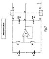
-
- a power supply source which energises the different components, this source is shown in further detail in
FIG. 7 , - a “current carrying circuit” that feeds the electrodes e3 e4 for applying a current to the thorax, the current is supplied by a
current source 9, and has a magnitude of 500 μA and a frequency of 80 Hz, this circuit is shown in further detail inFIG. 8 , - a “pick up” circuit or detecting circuit comprising electrodes e1 and e2, an
instrument amplifier 12 for measuring/amplifying difference signals, a low pass filter 13 (to filtrate DC before rectification, since only the AC component of the signal is of interest) and aprecision rectifier 14 to provide a DC output, this circuit is shown in further detail inFIG. 9 .
- a power supply source which energises the different components, this source is shown in further detail in
When the apparatus is connected to the thorax, a current is sent through the current electrodes e3 and e4 which are connected to the currant carrying circuit. The other two electrodes e1 and e2 which are connected to the pick up circuit will register the voltage drop across the thorax. This voltage drop is sent to a processing unit which as mentioned before controls display and alarm devices.
The measuring unit shown in FIGS. 6-9 was used to test the method according to the invention on a pig.
The pig was placed supine on an operating table and anesthetized. A first endrotracheal tube was placed in the trachea. During preparation the pig was ventilated through this tube (tube 1). A second tube (tube 2) was placed through the oesophagus in order to ventilate the stomach (incorrect intubation). Electrodes e1, e2, e3, e4 were attached to the pig's chest at four different places and these were connected to the measuring unit. The output of the measuring unit was connected to an oscilloscope.
During ventilation of the lungs through tube 1 fluctuations were observed in the oscilloscope's output, these fluctuations correspond to impedance changes and are shown in FIG. 10 . The fluctuations were in the order of magnitude of 10 mV.
The ventilation bag was then connected to tube 2 and “ventilation” attempts were performed. Measurements of impedance changes were performed, and no fluctuation was detected. The results are shown in FIG. 11 .
The output signal of the apparatus according to the invention (light/sound/image) for monitoring intubation will not be influenced by ambient noise (as opposed to the prior art stethoscope technique, it being difficult to hear lung sounds with a stethoscope), low cardiac output (which will influence ET CO2 measurements), and does not imply the delay of several ventilation attempts before satisfied.
The apparatus can be used for monitoring adults, children, and newborns. As stated before, it may be used as an alarm device during intensive care and transportation of all age groups of patients, in order to monitor correct placement of the tube.
The invention comprises also a method for immediate detection of correct/incorrect intubation, that is, instant detection of correct/incorrect placement of the tube during intensive care and transportation of intubated patients.
Claims (28)
1. A method for externally assessing or monitoring placement of an endo-tracheal tube for ventilation of a patient, said method comprising
transmitting a current through at least two external measuring electrodes placed on a thoracic region of said patient;
receiving, at a measuring unit via the at least two external measuring electrodes, thoracic impedance signals of said patient to assess or monitor placement of said endo-tracheal tube for ventilation of said patient;
detecting, using a processing unit connected to the measuring unit, whether the received impedance signals indicate an impedance change;
upon detection of an impedance change, comparing, using the processing unit, the detected impedance change to a predetermined threshold value indicative of a thoracic air volume change of said patient;
transmitting a signal to at least one of an alarm device or a display device connected to said processing unit when the comparison indicates that the at least one impedance change is greater than or equal to the predetermined threshold value; and
generating, in response to the signal, an indication that the proper endo-tracheal tube placement is achieved.
2. The method according to claim 1 , wherein the threshold value is stored in a storage unit connected to the processing unit.
3. The method according to claim 1 , wherein a start signal is given to the processing unit by a user to begin transmitting the current and receiving thoracic impedance signals and the detecting step, the comparing step, and the transmitting a signal step, are repeated during a predetermined period of time or until a stop signal is given to the processing unit by a user.
4. The method according to claim 1 , wherein the alarm device comprises at least one of a sound emitting device and a light emitting device.
5. The method according to claim 1 , wherein the alarm device comprises a user interface configured to display at least one of the detected impedance change, an indication of incorrect intubation of an endo-tracheal tube, and an indication of correct intubation of an endo-tracheal tube.
6. The method according to claim 1 , further comprising:
storing in a storage unit, at least one of the received impedance signals, the comparison of the impedance changes, and the threshold value.
7. The method according to claim 1 , further comprising:
receiving, via a user interface connected to the processing unit, at least one of reference thoracic impedance values, threshold impedance values, and patient characteristics.
8. A method for externally assessing and monitoring placement of an endo-tracheal tube for ventilation in a recipient, said method comprising
transmitting a current through at least two external measuring electrodes placed on a thoracic region of said recipient;
receiving, at a measuring unit via the at least two external measuring electrodes, thoracic impedance signals of said recipient to assess or monitor placement of said endo-tracheal tube for ventilation of said recipient;
detecting, using a processing unit connected to said measuring unit, whether the received impedance signals indicate an impedance change;
upon detection of an impedance change, comparing, using said processing unit, the detected impedance change to a predetermined threshold value indicative of a thoracic air volume change of said recipient;
transmitting a signal to at least one of an alarm device or display device connected to said processing unit if the at least one impedance change is less than the predetermined value; and
generating, in response to the signal, an indication of improper endo-tracheal tube placement.
9. The method according to claim 8 , wherein the threshold value is stored in a storage unit which is connected to the processing unit.
10. The method according to claim 8 , wherein a start signal is given to the processing unit by a user to begin transmitting the current and receiving thoracic impedance signals and the detecting step, the comparing step, and the transmitting a signal step, are repeated a during a predetermined period of time or until a stop signal is given to the processing unit by a user.
11. The method according to claim 8 , wherein the alarm device comprises at least one of a sound emitting device and a light emitting device.
12. The method according to claim 8 , wherein the alarm device comprises a user interface configured to display at least one of the detected impedance change, an indication of incorrect intubation of an endo-tracheal tube, and an indication of correct intubation of an endo-tracheal tube.
13. The method according to claim 8 , further comprising:
storing in a storage unit, at least one of the received impedance signals, the comparison of the impedance changes, and the threshold value.
14. The method according to claim 8 , further comprising:
receiving, via a user interface connected to the processing unit, at least one of reference thoracic impedance values, threshold impedance values, and recipient characteristics.
15. A non-transitory computer-readable medium comprising instructions that when executed perform a method for externally assessing or monitoring placement of an endo-tracheal tube for ventilation of a patient, said method comprising:
transmitting a current through at least two external measuring electrodes placed on a thoracic region of said patient;
receiving, at a measuring unit via the at least two measuring electrodes, thoracic impedance signals of said patient to externally assess or monitor placement of said endo-tracheal tube for ventilation of said patient;
detecting, using a processing unit connected to the measuring unit, whether the received impedance signals indicate an impedance change;
upon detection of an impedance change, comparing, using the processing unit, the detected impedance change to a predetermined threshold value indicative of a thoracic air volume change of said patient;
transmitting a signal to at least one of an alarm device or a display device connected to said processing unit when the comparison indicates that the at least one impedance change is greater than or equal to the predetermined threshold value; and
generating, in response to the signal, an indication that the proper endo-tracheal tube placement is achieved.
16. The non-transitory computer-readable medium according to claim 15 , wherein the threshold value is stored in a storage unit connected to the processing unit.
17. The non-transitory computer-readable medium according to claim 15 , wherein a start signal is given to the processing unit by a user to begin transmitting the current and receiving thoracic impedance signals and the detecting step, the comparing step, and the transmitting a signal step, are repeated a during a predetermined period of time or until a stop signal is given to the processing unit by a user.
18. The non-transitory computer-readable medium according to claim 15 , wherein the alarm device comprises at least one of a sound emitting device and a light emitting device.
19. The non-transitory computer-readable medium according to claim 15 , wherein the alarm device comprises a user interface configured to display at least one of the detected impedance change, an indication of incorrect intubation of an endo-tracheal tube, and an indication of correct intubation of an endo-tracheal tube.
20. The non-transitory computer-readable medium according to claim 15 , further comprising:
storing in a storage unit, at least one of the received impedance signals, the comparison of the impedance changes, and the threshold value.
21. The non-transitory computer-readable medium according to claim 15 , further comprising:
receiving, via a user interface connected to the processing unit, at least one of reference thoracic impedance values, threshold impedance values, and patient characteristics.
22. A non-transitory computer-readable medium comprising instructions that when executed perform a method for externally assessing and monitoring placement of an endo-tracheal tube for ventilation in a recipient, said method comprising:
transmitting a current through at least two external measuring electrodes placed on a thoracic region of said recipient;
receiving, at a measuring unit via the at least two external measuring electrodes, thoracic impedance signals of said recipient to assess or monitor placement of said endo-tracheal tube for ventilation of said recipient;
detecting, using a processing unit connected to said measuring unit, whether the received impedance signals indicate an impedance;
upon detection of an impedance change, comparing, using said processing unit, the detected impedance change to a predetermined threshold value indicative of a thoracic air volume change of said recipient;
transmitting a signal to at least one of an alarm device or display device connected to said processing unit if the at least one impedance change is less than the predetermined value; and
generating, in response to the signal, an indication of improper endo-tracheal tube placement.
23. The non-transitory computer-readable medium according to claim 22 , wherein the threshold value is stored in a storage unit which is connected to the processing unit.
24. The non-transitory computer-readable medium according to claim 22 , wherein a start signal is given to the processing unit by a user to begin transmitting the current and receiving thoracic impedance signals and the detecting step, the comparing step, and the transmitting a signal step, are repeated a during a predetermined period of time or until a stop signal is given to the processing unit by a user.
25. The non-transitory computer-readable medium according to claim 22 , wherein the alarm device comprises at least one of a sound emitting device and a light emitting device.
26. The non-transitory computer-readable medium according to claim 22 , wherein the alarm device comprises a user interface configured to display at least one of the detected impedance change, an indication of incorrect intubation of an endo-tracheal tube, and an indication of correct intubation of an endo-tracheal tube.
27. The non-transitory computer-readable medium according to claim 22 , further comprising:
storing in a storage unit, at least one of the received impedance signals, the comparison of the impedance changes, and the threshold value.
28. The non-transitory computer-readable medium according to claim 22 , further comprising:
receiving, via a user interface connected to the processing unit, at least one of reference thoracic impedance values, threshold impedance values, and recipient characteristics.
Priority Applications (1)
| Application Number | Priority Date | Filing Date | Title |
|---|---|---|---|
| US13/043,648 US9486157B2 (en) | 2002-06-19 | 2011-03-09 | Intubation monitoring apparatus and method |
Applications Claiming Priority (7)
| Application Number | Priority Date | Filing Date | Title |
|---|---|---|---|
| NO20022960 | 2002-06-19 | ||
| NO20022960A NO20022960D0 (en) | 2002-06-19 | 2002-06-19 | Apparatus and method for intubation monitoring |
| NO20022950 | 2002-06-19 | ||
| US39011502P | 2002-06-21 | 2002-06-21 | |
| PCT/NO2003/000208 WO2004004541A2 (en) | 2002-06-19 | 2003-06-19 | Intubation monitoring apparatus and method |
| US10/517,989 US7925339B2 (en) | 2002-06-19 | 2003-06-19 | Intubation monitoring apparatus and method |
| US13/043,648 US9486157B2 (en) | 2002-06-19 | 2011-03-09 | Intubation monitoring apparatus and method |
Related Parent Applications (3)
| Application Number | Title | Priority Date | Filing Date |
|---|---|---|---|
| PCT/NO2003/000208 Division WO2004004541A2 (en) | 2002-06-19 | 2003-06-19 | Intubation monitoring apparatus and method |
| US10/517,989 Division US7925339B2 (en) | 2002-06-19 | 2003-06-19 | Intubation monitoring apparatus and method |
| US10517989 Division | 2003-06-19 |
Publications (2)
| Publication Number | Publication Date |
|---|---|
| US20110224568A1 US20110224568A1 (en) | 2011-09-15 |
| US9486157B2 true US9486157B2 (en) | 2016-11-08 |
Family
ID=44560623
Family Applications (2)
| Application Number | Title | Priority Date | Filing Date |
|---|---|---|---|
| US10/517,989 Active 2028-03-06 US7925339B2 (en) | 2002-06-19 | 2003-06-19 | Intubation monitoring apparatus and method |
| US13/043,648 Expired - Lifetime US9486157B2 (en) | 2002-06-19 | 2011-03-09 | Intubation monitoring apparatus and method |
Family Applications Before (1)
| Application Number | Title | Priority Date | Filing Date |
|---|---|---|---|
| US10/517,989 Active 2028-03-06 US7925339B2 (en) | 2002-06-19 | 2003-06-19 | Intubation monitoring apparatus and method |
Country Status (8)
| Country | Link |
|---|---|
| US (2) | US7925339B2 (en) |
| EP (1) | EP1517635B1 (en) |
| JP (1) | JP2006511249A (en) |
| AT (1) | ATE375751T1 (en) |
| AU (1) | AU2003279634A1 (en) |
| DE (1) | DE60316944T2 (en) |
| NO (1) | NO20022960D0 (en) |
| WO (1) | WO2004004541A2 (en) |
Cited By (1)
| Publication number | Priority date | Publication date | Assignee | Title |
|---|---|---|---|---|
| US11229760B2 (en) | 2018-01-17 | 2022-01-25 | Zoll Medical Corporation | Systems and methods for assisting patient airway management |
Families Citing this family (13)
| Publication number | Priority date | Publication date | Assignee | Title |
|---|---|---|---|---|
| US7308304B2 (en) * | 2003-02-14 | 2007-12-11 | Medtronic Physio-Control Corp. | Cooperating defibrillators and external chest compression devices |
| NO321585B1 (en) | 2004-07-15 | 2006-06-06 | Laerdal Medical As | Piping |
| US8630700B2 (en) * | 2007-12-19 | 2014-01-14 | St. Jude Medical, AB | Implantable heart monitoring device, system and method |
| EP2407100A1 (en) * | 2010-07-15 | 2012-01-18 | Tanita Corporation | Respiration characteristic analysis |
| EP2407102A1 (en) * | 2010-07-15 | 2012-01-18 | Tanita Corporation | Respiration characteristic analysis apparatus and respiration characteristic analysis system |
| CA2863602A1 (en) | 2011-05-04 | 2013-11-08 | The Regents Of The University Of Michigan | Intubation device |
| US9526856B2 (en) | 2011-12-15 | 2016-12-27 | The Board Of Trustees Of The Leland Stanford Junior University | Devices and methods for preventing tracheal aspiration |
| US10058669B2 (en) | 2012-06-05 | 2018-08-28 | Texas Heart Institute | Location determining endotracheal tube and methods |
| EP4074254A1 (en) | 2012-12-21 | 2022-10-19 | Zoll Medical Corporation | Ventilation monitoring |
| DE102013200712A1 (en) * | 2013-01-18 | 2014-07-24 | Rwth Aachen | Device and method for controlling the placement of a ventilation tube |
| WO2015069804A1 (en) | 2013-11-05 | 2015-05-14 | Ciel Medical, Inc. | Devices and methods for airway measurement |
| JP7073377B2 (en) * | 2016-12-06 | 2022-05-23 | アート メディカル リミテッド | System for sensing lung fluid |
| KR102215392B1 (en) * | 2019-07-31 | 2021-02-10 | 한국외국어대학교 연구산학협력단 | Apparatus and Method for Detecting Malposition of Implanted leads of Pacemaker |
Citations (12)
| Publication number | Priority date | Publication date | Assignee | Title |
|---|---|---|---|---|
| US4403215A (en) | 1977-12-27 | 1983-09-06 | Hellige, Gmbh | Apparatus for automatically monitoring body functions |
| US4449537A (en) | 1982-03-08 | 1984-05-22 | Hewlett-Packard Gmbh | Respiration monitor |
| FR2652255A1 (en) | 1989-09-22 | 1991-03-29 | Centre Nat Rech Scient | Appliance for monitoring and measuring the respiratory activity of a patient |
| US5305745A (en) | 1988-06-13 | 1994-04-26 | Fred Zacouto | Device for protection against blood-related disorders, notably thromboses, embolisms, vascular spasms, hemorrhages, hemopathies and the presence of abnormal elements in the blood |
| US5445144A (en) | 1993-12-16 | 1995-08-29 | Purdue Research Foundation | Apparatus and method for acoustically guiding, positioning, and monitoring a tube within a body |
| EP0747005A1 (en) | 1995-05-26 | 1996-12-11 | Instrumentarium Oy | Procedure for measuring a patient's impedance |
| US5653241A (en) | 1994-08-23 | 1997-08-05 | Colin Corporation | Blood-pressure monitor apparatus |
| WO1999007415A1 (en) | 1997-08-12 | 1999-02-18 | Bracco Research S.A. | Administrable compositions and methods for magnetic resonance imaging |
| US20010011159A1 (en) | 1997-10-17 | 2001-08-02 | Cantrell Elroy T. | Chest mounted cardio pulmonary resuscitation device and system |
| US20020032383A1 (en) | 2000-07-21 | 2002-03-14 | Weil Max Harry | Cardiac/respiratory arrest detector |
| US20020035339A1 (en) | 2000-08-04 | 2002-03-21 | Robert Kavet | Apparatus and method for measuring current flow in an animal or human body |
| US20030109795A1 (en) | 2001-12-10 | 2003-06-12 | Pranalytica, Inc. | Method of analyzing components of alveolar breath |
Family Cites Families (1)
| Publication number | Priority date | Publication date | Assignee | Title |
|---|---|---|---|---|
| WO1989007415A1 (en) | 1988-02-19 | 1989-08-24 | Antec Systems Limited | Method and apparatus for locating an endo-tracheal or endo-oesophageal tube |
-
2002
- 2002-06-19 NO NO20022960A patent/NO20022960D0/en unknown
-
2003
- 2003-06-19 AU AU2003279634A patent/AU2003279634A1/en not_active Abandoned
- 2003-06-19 US US10/517,989 patent/US7925339B2/en active Active
- 2003-06-19 DE DE60316944T patent/DE60316944T2/en not_active Expired - Lifetime
- 2003-06-19 WO PCT/NO2003/000208 patent/WO2004004541A2/en active IP Right Grant
- 2003-06-19 AT AT03762930T patent/ATE375751T1/en not_active IP Right Cessation
- 2003-06-19 JP JP2004519372A patent/JP2006511249A/en active Pending
- 2003-06-19 EP EP03762930A patent/EP1517635B1/en not_active Expired - Lifetime
-
2011
- 2011-03-09 US US13/043,648 patent/US9486157B2/en not_active Expired - Lifetime
Patent Citations (12)
| Publication number | Priority date | Publication date | Assignee | Title |
|---|---|---|---|---|
| US4403215A (en) | 1977-12-27 | 1983-09-06 | Hellige, Gmbh | Apparatus for automatically monitoring body functions |
| US4449537A (en) | 1982-03-08 | 1984-05-22 | Hewlett-Packard Gmbh | Respiration monitor |
| US5305745A (en) | 1988-06-13 | 1994-04-26 | Fred Zacouto | Device for protection against blood-related disorders, notably thromboses, embolisms, vascular spasms, hemorrhages, hemopathies and the presence of abnormal elements in the blood |
| FR2652255A1 (en) | 1989-09-22 | 1991-03-29 | Centre Nat Rech Scient | Appliance for monitoring and measuring the respiratory activity of a patient |
| US5445144A (en) | 1993-12-16 | 1995-08-29 | Purdue Research Foundation | Apparatus and method for acoustically guiding, positioning, and monitoring a tube within a body |
| US5653241A (en) | 1994-08-23 | 1997-08-05 | Colin Corporation | Blood-pressure monitor apparatus |
| EP0747005A1 (en) | 1995-05-26 | 1996-12-11 | Instrumentarium Oy | Procedure for measuring a patient's impedance |
| WO1999007415A1 (en) | 1997-08-12 | 1999-02-18 | Bracco Research S.A. | Administrable compositions and methods for magnetic resonance imaging |
| US20010011159A1 (en) | 1997-10-17 | 2001-08-02 | Cantrell Elroy T. | Chest mounted cardio pulmonary resuscitation device and system |
| US20020032383A1 (en) | 2000-07-21 | 2002-03-14 | Weil Max Harry | Cardiac/respiratory arrest detector |
| US20020035339A1 (en) | 2000-08-04 | 2002-03-21 | Robert Kavet | Apparatus and method for measuring current flow in an animal or human body |
| US20030109795A1 (en) | 2001-12-10 | 2003-06-12 | Pranalytica, Inc. | Method of analyzing components of alveolar breath |
Non-Patent Citations (2)
| Title |
|---|
| Maeda, Teaching by Certified Nurse of Intensive Care of Serious Injury, Prohibition Administration of Artifical Breathing 50, for Adults, Fixing of Endotracheal tube, Nursing Today, Japan, Japanese Nursing Association, May 1, 2002, vol. 17-6, No. 207, pp. 121-123. |
| Mehta et al. An assessment of the ability of impedance respirometry to distinguish oesophageal from tracheal intubation. Anaesthesia, 2002, 5r7, pp. 1090-1093. |
Cited By (2)
| Publication number | Priority date | Publication date | Assignee | Title |
|---|---|---|---|---|
| US11229760B2 (en) | 2018-01-17 | 2022-01-25 | Zoll Medical Corporation | Systems and methods for assisting patient airway management |
| US11904095B2 (en) | 2018-01-17 | 2024-02-20 | Zoll Medical Corporation | Systems and methods for assisting patient airway management |
Also Published As
| Publication number | Publication date |
|---|---|
| DE60316944D1 (en) | 2007-11-29 |
| AU2003279634A8 (en) | 2004-01-23 |
| NO20022960D0 (en) | 2002-06-19 |
| ATE375751T1 (en) | 2007-11-15 |
| EP1517635B1 (en) | 2007-10-17 |
| DE60316944T2 (en) | 2008-07-24 |
| WO2004004541A3 (en) | 2004-06-10 |
| US7925339B2 (en) | 2011-04-12 |
| JP2006511249A (en) | 2006-04-06 |
| US20110224568A1 (en) | 2011-09-15 |
| EP1517635A2 (en) | 2005-03-30 |
| US20050256422A1 (en) | 2005-11-17 |
| AU2003279634A1 (en) | 2004-01-23 |
| WO2004004541A2 (en) | 2004-01-15 |
Similar Documents
| Publication | Publication Date | Title |
|---|---|---|
| US9486157B2 (en) | Intubation monitoring apparatus and method | |
| US20220152328A1 (en) | Ventilation Monitoring | |
| US6821254B2 (en) | Cardiac/respiratory arrest detector | |
| US20100036266A1 (en) | Device and method for detecting heart beats using airway pressure | |
| EP2352425B1 (en) | Carbon dioxide monitoring system | |
| US10070804B2 (en) | Apparatus and method for the collection of samples of exhaled air | |
| US11571179B2 (en) | System for positioning an intubation tube | |
| US7774054B2 (en) | Method and system to determine correct tube placement during resuscitation | |
| US20100137723A1 (en) | Apnea type determining apparatus and method | |
| JP2007244879A5 (en) | Automated resuscitation device for sensing and facilitating ventilation | |
| AU3261500A (en) | Method and system of determining whether a lifeless person has a pulse | |
| US20210093814A1 (en) | Apparatus and method for improved assisted ventilation | |
| JP2018528814A (en) | Monitoring device with multi-parameter hyperventilation alert | |
| KR101678545B1 (en) | An endotracheal intubation system enabling verification of tube position using electrical stimulatio | |
| JP2010214024A (en) | Respiratory exercise support device |
Legal Events
| Date | Code | Title | Description |
|---|---|---|---|
| STCF | Information on status: patent grant |
Free format text: PATENTED CASE |
|
| FEPP | Fee payment procedure |
Free format text: PAYOR NUMBER ASSIGNED (ORIGINAL EVENT CODE: ASPN); ENTITY STATUS OF PATENT OWNER: LARGE ENTITY |
|
| MAFP | Maintenance fee payment |
Free format text: PAYMENT OF MAINTENANCE FEE, 4TH YEAR, LARGE ENTITY (ORIGINAL EVENT CODE: M1551); ENTITY STATUS OF PATENT OWNER: LARGE ENTITY Year of fee payment: 4 |