US9554117B2 - System and method for non-invasive patient-image registration - Google Patents
System and method for non-invasive patient-image registration Download PDFInfo
- Publication number
- US9554117B2 US9554117B2 US14/077,462 US201314077462A US9554117B2 US 9554117 B2 US9554117 B2 US 9554117B2 US 201314077462 A US201314077462 A US 201314077462A US 9554117 B2 US9554117 B2 US 9554117B2
- Authority
- US
- United States
- Prior art keywords
- blood vessel
- camera
- image
- near infrared
- feature point
- Prior art date
- Legal status (The legal status is an assumption and is not a legal conclusion. Google has not performed a legal analysis and makes no representation as to the accuracy of the status listed.)
- Active, expires
Links
Images


Classifications
-
- A—HUMAN NECESSITIES
- A61—MEDICAL OR VETERINARY SCIENCE; HYGIENE
- A61B—DIAGNOSIS; SURGERY; IDENTIFICATION
- A61B5/00—Measuring for diagnostic purposes; Identification of persons
- A61B5/05—Detecting, measuring or recording for diagnosis by means of electric currents or magnetic fields; Measuring using microwaves or radio waves
- A61B5/055—Detecting, measuring or recording for diagnosis by means of electric currents or magnetic fields; Measuring using microwaves or radio waves involving electronic [EMR] or nuclear [NMR] magnetic resonance, e.g. magnetic resonance imaging
-
- H04N13/0221—
-
- A—HUMAN NECESSITIES
- A61—MEDICAL OR VETERINARY SCIENCE; HYGIENE
- A61B—DIAGNOSIS; SURGERY; IDENTIFICATION
- A61B6/00—Apparatus for radiation diagnosis, e.g. combined with radiation therapy equipment
- A61B6/02—Devices for diagnosis sequentially in different planes; Stereoscopic radiation diagnosis
- A61B6/03—Computerised tomographs
-
- A—HUMAN NECESSITIES
- A61—MEDICAL OR VETERINARY SCIENCE; HYGIENE
- A61B—DIAGNOSIS; SURGERY; IDENTIFICATION
- A61B90/00—Instruments, implements or accessories specially adapted for surgery or diagnosis and not covered by any of the groups A61B1/00 - A61B50/00, e.g. for luxation treatment or for protecting wound edges
- A61B90/36—Image-producing devices or illumination devices not otherwise provided for
- A61B90/361—Image-producing devices, e.g. surgical cameras
-
- G06T7/0028—
-
- G—PHYSICS
- G06—COMPUTING; CALCULATING OR COUNTING
- G06T—IMAGE DATA PROCESSING OR GENERATION, IN GENERAL
- G06T7/00—Image analysis
- G06T7/30—Determination of transform parameters for the alignment of images, i.e. image registration
- G06T7/33—Determination of transform parameters for the alignment of images, i.e. image registration using feature-based methods
-
- G—PHYSICS
- G16—INFORMATION AND COMMUNICATION TECHNOLOGY [ICT] SPECIALLY ADAPTED FOR SPECIFIC APPLICATION FIELDS
- G16Z—INFORMATION AND COMMUNICATION TECHNOLOGY [ICT] SPECIALLY ADAPTED FOR SPECIFIC APPLICATION FIELDS, NOT OTHERWISE PROVIDED FOR
- G16Z99/00—Subject matter not provided for in other main groups of this subclass
-
- H—ELECTRICITY
- H04—ELECTRIC COMMUNICATION TECHNIQUE
- H04N—PICTORIAL COMMUNICATION, e.g. TELEVISION
- H04N13/00—Stereoscopic video systems; Multi-view video systems; Details thereof
- H04N13/20—Image signal generators
- H04N13/204—Image signal generators using stereoscopic image cameras
- H04N13/207—Image signal generators using stereoscopic image cameras using a single 2D image sensor
- H04N13/221—Image signal generators using stereoscopic image cameras using a single 2D image sensor using the relative movement between cameras and objects
-
- A—HUMAN NECESSITIES
- A61—MEDICAL OR VETERINARY SCIENCE; HYGIENE
- A61B—DIAGNOSIS; SURGERY; IDENTIFICATION
- A61B34/00—Computer-aided surgery; Manipulators or robots specially adapted for use in surgery
- A61B34/20—Surgical navigation systems; Devices for tracking or guiding surgical instruments, e.g. for frameless stereotaxis
- A61B2034/2046—Tracking techniques
- A61B2034/2055—Optical tracking systems
-
- A—HUMAN NECESSITIES
- A61—MEDICAL OR VETERINARY SCIENCE; HYGIENE
- A61B—DIAGNOSIS; SURGERY; IDENTIFICATION
- A61B90/00—Instruments, implements or accessories specially adapted for surgery or diagnosis and not covered by any of the groups A61B1/00 - A61B50/00, e.g. for luxation treatment or for protecting wound edges
- A61B90/36—Image-producing devices or illumination devices not otherwise provided for
- A61B2090/364—Correlation of different images or relation of image positions in respect to the body
-
- A—HUMAN NECESSITIES
- A61—MEDICAL OR VETERINARY SCIENCE; HYGIENE
- A61B—DIAGNOSIS; SURGERY; IDENTIFICATION
- A61B2576/00—Medical imaging apparatus involving image processing or analysis
- A61B2576/02—Medical imaging apparatus involving image processing or analysis specially adapted for a particular organ or body part
-
- A—HUMAN NECESSITIES
- A61—MEDICAL OR VETERINARY SCIENCE; HYGIENE
- A61B—DIAGNOSIS; SURGERY; IDENTIFICATION
- A61B6/00—Apparatus for radiation diagnosis, e.g. combined with radiation therapy equipment
- A61B6/50—Clinical applications
- A61B6/504—Clinical applications involving diagnosis of blood vessels, e.g. by angiography
-
- A—HUMAN NECESSITIES
- A61—MEDICAL OR VETERINARY SCIENCE; HYGIENE
- A61B—DIAGNOSIS; SURGERY; IDENTIFICATION
- A61B8/00—Diagnosis using ultrasonic, sonic or infrasonic waves
- A61B8/08—Detecting organic movements or changes, e.g. tumours, cysts, swellings
- A61B8/0891—Detecting organic movements or changes, e.g. tumours, cysts, swellings for diagnosis of blood vessels
-
- G—PHYSICS
- G06—COMPUTING; CALCULATING OR COUNTING
- G06T—IMAGE DATA PROCESSING OR GENERATION, IN GENERAL
- G06T2207/00—Indexing scheme for image analysis or image enhancement
- G06T2207/10—Image acquisition modality
- G06T2207/10004—Still image; Photographic image
- G06T2207/10012—Stereo images
-
- G—PHYSICS
- G06—COMPUTING; CALCULATING OR COUNTING
- G06T—IMAGE DATA PROCESSING OR GENERATION, IN GENERAL
- G06T2207/00—Indexing scheme for image analysis or image enhancement
- G06T2207/10—Image acquisition modality
- G06T2207/10048—Infrared image
-
- G—PHYSICS
- G06—COMPUTING; CALCULATING OR COUNTING
- G06T—IMAGE DATA PROCESSING OR GENERATION, IN GENERAL
- G06T2207/00—Indexing scheme for image analysis or image enhancement
- G06T2207/10—Image acquisition modality
- G06T2207/10072—Tomographic images
-
- G—PHYSICS
- G06—COMPUTING; CALCULATING OR COUNTING
- G06T—IMAGE DATA PROCESSING OR GENERATION, IN GENERAL
- G06T2207/00—Indexing scheme for image analysis or image enhancement
- G06T2207/20—Special algorithmic details
- G06T2207/20036—Morphological image processing
- G06T2207/20044—Skeletonization; Medial axis transform
-
- G—PHYSICS
- G06—COMPUTING; CALCULATING OR COUNTING
- G06T—IMAGE DATA PROCESSING OR GENERATION, IN GENERAL
- G06T2207/00—Indexing scheme for image analysis or image enhancement
- G06T2207/30—Subject of image; Context of image processing
- G06T2207/30004—Biomedical image processing
- G06T2207/30101—Blood vessel; Artery; Vein; Vascular
-
- G—PHYSICS
- G06—COMPUTING; CALCULATING OR COUNTING
- G06T—IMAGE DATA PROCESSING OR GENERATION, IN GENERAL
- G06T2207/00—Indexing scheme for image analysis or image enhancement
- G06T2207/30—Subject of image; Context of image processing
- G06T2207/30172—Centreline of tubular or elongated structure
Landscapes
- Health & Medical Sciences (AREA)
- Engineering & Computer Science (AREA)
- Life Sciences & Earth Sciences (AREA)
- Surgery (AREA)
- Medical Informatics (AREA)
- Nuclear Medicine, Radiotherapy & Molecular Imaging (AREA)
- Physics & Mathematics (AREA)
- Pathology (AREA)
- Veterinary Medicine (AREA)
- Biomedical Technology (AREA)
- Heart & Thoracic Surgery (AREA)
- Public Health (AREA)
- Molecular Biology (AREA)
- Animal Behavior & Ethology (AREA)
- General Health & Medical Sciences (AREA)
- Oral & Maxillofacial Surgery (AREA)
- Signal Processing (AREA)
- Computer Vision & Pattern Recognition (AREA)
- Multimedia (AREA)
- General Physics & Mathematics (AREA)
- Theoretical Computer Science (AREA)
- High Energy & Nuclear Physics (AREA)
- Radiology & Medical Imaging (AREA)
- Biophysics (AREA)
- Optics & Photonics (AREA)
- Ultra Sonic Daignosis Equipment (AREA)
- Apparatus For Radiation Diagnosis (AREA)
Abstract
A system for non-invasive registration between a patient and a three-dimensional (3D) medical image includes a near infrared 3D camera 110 which extracts a 3D blood vessel image I2 of a patients registration target area during surgical operation; a camera position tracer 120 which traces a position of the near infrared 3D camera 110 and calculates a real world coordinate system of the 3D blood vessel image I2; a controller 130 which extracts a first blood vessel pattern from a 3D medical image I1 of the registration target area, extracts a second blood vessel pattern from the 3D blood vessel image I2, and performs position registration between the patient and the 3D medical image I1 through the extracted first and second blood vessel patterns; and a display 140 which displays a registration result calculated by the controller 130.
Description
This application claims priority from Korean Patent Application No. 10-2013-0096016, filed on Aug. 13, 2013 in the Korean Intellectual Property Office, the disclosure of which is incorporated herein by reference.
Field
Apparatuses and methods consistent with the exemplary embodiments relate to a system and method for non-invasive registration between a patient and a patient's three-dimensional (3D) image for registration of a position and a pose between a patient and a patient's 3D medical image, and more particularly to a system and method for non-invasive registration between a patient and a patient's three-dimensional (3D) image, which can align a patient's 3D medical image with a patient by a non-invasive method using a feature point based on a blood vessel pattern, where a patient's blood vessel is extended, without using an artificial fiducial marker.
Description of the Related Art
In general, a minimally invasive surgery is a surgery method that 3˜4 small holes are pierced through a skin and an endoscope and a narrow and long surgical instrument are put into the holes so as to minimize skin incision. Such a minimally invasive surgery has been annually increased due to less pain and short recovery time along with minimal scar left when compared to general surgery.
However, in the minimally invasive surgery, an operating surgeon cannot directly see an affected area and therefore indirectly checks an image gained from the endoscope through a monitor. At this time, it is not easy to observe a targeted affected area because a view direction of the surgeon is different from a direction of the endoscope and the endoscope is short-sighted. Further, it is unintuitive and thus difficult to treat the affected area by controlling a surgical instrument while watching an image from the endoscope.
Meanwhile, a non-invasive surgery is a surgery method that ultrasonic waves, radiation, magnetic field, etc. are used for treatment without skin incision. In this surgery method, an operating surgeon cannot directly see an affected area and also cannot use the image from the endoscope in real time. Therefore, surgery navigation has been required to guide the surgical operation of the operating surgeon during an operation through a three-dimensional (3D) medical image (computer tomography (CT), a magnetic resonance imaging (MRI), etc.) of a patient taken for a diagnosis and a surgical plan before the operation.
For the surgery navigation, a patient's 3D medical image has to be first aligned with an intraoperative patient with respect to coordinates. That is, if coordinate transformation between a medical image coordinate system of the 3D medical image and a patient coordinate system (i.e., a coordinate system arbitrarily set up in the real world) is known, it is possible to determine what position of a patient a certain position on the medical image corresponds to. This is called patient-image registration.
As a method generally used for the patient-image registration, there is a method of performing the patient-image registration by taking a medical image in the state that fiducial markers are attached to a patient, and aligning the fiducial markers from previously taken medical image with the fiducial markers that is attached to the patient.
In the case of using the fiducial marker, there are an invasive method of fixing the fiducial markers to a patient's bone, and a non-invasive method of attaching the fiducial marker on a patient's skin. The invasive method burdens a patient due to scar on bone even though registration is relatively precise. On the other hand, the non-invasive method is not applicable to microsurgical operation due to low precision.
Accordingly, a markerless patient-image registration method without using the fiducial markers has recently been researched, in which 3D medical image registration is performed using a patient's two dimensional (2D) sectional image based on ultrasonic waves or X-rays, or using a 3D scan model of a patient's skin surface.
However, the registration between the 2D sectional image and the 3D medical image needs long calculation time and is vulnerable to soft tissue transformation of a human body. Also, the registration between the 3D scan model of a patient's skin surface and a skin surface model extracted from the 3D medical image may have a large error due to change in shape of the skin surface.
Consequently, patient-image registration method which does not use the fiducial markers and is minimally affected by change in a patient's soft tissue and a skin surface are required.
KR Laid-Open No. 10-2013-0045774 (KR Application No. 10-2011-0110189)
The present invention is conceived to solve the foregoing problems, and an aspect thereof provides a system and method for non-invasive registration between a patient and a 3D medical image, which can perform position registration between a 3D image of a blood vessel located in the patient's target registration area, taken using near infrared light penetrating a skin of a human body and having a wavelength largely absorbed in the blood vessel, and a 3D medical image such as magnetic resonance imaging (MRI), computer tomography (CT), etc.
According to an aspect of another exemplary embodiment, a system for non-invasive registration between a patient and a three-dimensional (3D) medical image is provided including: a near infrared 3D camera 110 which extracts a 3D blood vessel image I2 of a patient's registration target area at a surgical operation; a camera position tracer 120 which traces a position of the near infrared 3D camera 110 and calculates a real world coordinate system of the 3D blood vessel image I2; a controller 130 which extracts a first blood vessel pattern from a 3D medical image I1 of the registration target area, extracts a second blood vessel pattern from the 3D blood vessel image I2, and performs position registration between the patient and the 3D medical image I1 through the extracted first and second blood vessel patterns; and a display 140 which displays a registration result calculated by the controller 130.
The controller 130 may comprise a 3D medical image database (DB) 131 which stores a 3D medical image I1 obtained by taking the registration target area before a surgical operation; a blood vessel model generator 132 which reconstitutes the blood vessel model with a blood vessel image separated from the 3D medical image I1; a first feature point extractor 133 which extracts a branch point, from which a blood vessel is branched, by detecting a skeleton line SL of the blood vessel model, and selects each extracted branch point as a feature point of the first blood vessel pattern; a 3D blood vessel image detector 134 which extracts a 3D blood vessel image I2 by separating a blood vessel image from a near infrared 3D image taken by the near infrared 3D camera 110, and a the second feature point extractor 135 which extracts a branch point of a patient's blood vessel at a surgical operation through the extracted 3D blood vessel image I2 and a real world coordinate system of the 3D blood vessel image I2 measured by the camera position tracer 120, and selects each extracted branch point as a feature point of the second blood vessel pattern; and a registrator 136 which performs registration by optimally matching a first feature point group P1 grouping a plurality of first feature points, and a second feature point group P2 grouping a plurality of second feature points.
The first feature point extractor 133 may store 3D coordinates of a feature point of a first blood vessel pattern selected with respect to a medical image coordinate system, the near infrared 3D camera 110 may take blood vessel images seen by having near infrared light pass through a patient's registration target area to obtain two 3D blood vessel images I2 that are spaced apart at a predetermined distance to have parallax, and the second feature point extractor 135 may select a second feature point represented by 3D coordinates with respect to a 3D camera coordinate system, using parallax of a pair of corresponding branch points respectively calculated from two 3D blood vessel images I2 of the near infrared 3D camera 110.
The camera position tracer 120 may measure six degrees of freedom in position of the near infrared 3D camera 110, which includes three rotation positions and three translation positions, and calculates a coordinate transformation matrix M from the position tracer coordinate system to the 3D camera coordinate system by tracing the position of the near infrared 3D camera 110 in real time; the second feature point extractor 135 may transform and save the second feature point in the 3D camera coordinate system into that in a position tracer coordinate system by the coordinate transformation matrix M; and the registrator 136 may calculate a position relationship T of a position tracer coordinate system with respect to a medical image coordinate system in a state that a second feature point group P2 in the position tracer coordinate system is rotated and translated to be optimally matched with the first feature point group P1.
The camera position tracer 120 may comprise a mechanical position tracing device 120 a which is fastened to an end of an articulated link and measures six degrees of freedom in position of the near infrared 3D camera 110 by sensing a physical displacement due to link work of each joint.
The camera position tracer 120 may comprise an optical position tracing device 120 b which uses a 3D camera taking fiducial markers 121 mounted on the near infrared 3D camera 110 to calculate a 3D position vector and a 3D direction vector of the fiducial marker 121 and measure six degrees of freedom in position of the near infrared 3D camera 110.
The first feature point extractor 133 may extract a border line BL representing an outline of a blood vessel from the blood vessel model, and extract a center line, which is extended from the extracted border line BL along the blood vessel, as a skeleton line SL;
According to an aspect of another exemplary embodiment, a method of non-invasive registration between a patient and a three-dimensional (3D) medical image is provided including: extracting a 3D blood vessel image I2 by taking a blood vessel image seen by having near infrared light pass through a registration target area with a near infrared 3D camera 110 (S210); tracing a camera position by tracing a position of the near infrared 3D camera 110 in real time with the camera position tracer 120 and calculating a real world coordinate system of the 3D blood vessel image I2 (S220); extracting a blood vessel pattern with a controller 130 by extracting a first blood vessel pattern from a 3D medical image I1 of the registration target area and extracting a second blood vessel pattern from the 3D blood vessel image I2 (S230); image registration by performing position registration between a patient and the 3D medical image I1 through the extracted first and second blood vessel patterns (S240); and displaying a registration result by displaying the registration result calculated by the controller 130 on a screen (S250).
In accordance with the system and method for the non-invasive registration between a patient and the 3D medical image, it has the following effects:
First, it is possible to non-invasively perform the registration between the patient and the 3D medical image I1 through the position registration between the 3D blood vessel image I2 of the blood vessel arranged in the patient's registration target area obtained using the near infrared light and the blood vessel model included in the 3D medical image I1 obtained by taking the registration target area, without using any fiducial markers.
Second, each blood vessel pattern of the blood vessel images reconstituted from the 3D blood vessel image I2 and the 3D medical image I1 obtained using the near infrared light is analyzed to calculate the feature point for the registration of the branch point of the blood vessel, and thus the registration is performed with respect to the calculated feature point, thereby not only minimize effect on skin deformation but also reducing an error at the position registration as compared with the conventional registration method using the feature point on a patient's skin.
Third, the position registration is performed based on the feature point according to the blood vessel pattern extended from the blood vessel distributed throughout a human body, thereby being advantageously applicable to various surgical areas and usable as medical information in connection with various medical images.
Fourth, while the feature point of the blood vessel pattern is extracted from the 3D blood vessel image and the 3D medical image, a center line extended along the blood vessel displayed in each image is extracted as a skeleton line, and the branch point of each skeleton line is selected as the feature point, thereby minimizing an error about a border line of the blood vessel and further reducing the registration error.
The above and/or other aspects will become apparent and more readily appreciated from the following description of exemplary embodiments, taken in conjunction with the accompanying drawings, in which:
Hereinafter, exemplary embodiments according to the present invention will be described in detail with reference to accompanying drawings. Prior to this, terms or words used in this specification and claims have to be interpreted as the meaning and concept adaptive to the technical idea of the present invention rather than typical or dictionary interpretation on a principle that an inventor is allowed to properly define the concept of the terms in order to explain his/her own invention in the best way.
Therefore, because embodiments disclosed in this specification and configurations illustrated in the drawings are nothing but preferred examples of the present invention and do not fully describe the technical idea of the present invention, it will be appreciated that there are various equivalents and alterations replacing them at the filing date of the present application.
In minimally invasive surgery, a three-dimensional (3D) medical image (computer tomography (CT), a magnetic resonance imaging (MRI), etc.) of a patient is indispensably used for a diagnosis and a surgical plan, and relative position between an intraoperative patient and the 3D medical image is necessary for surgery navigation and surgery guide. Accordingly, an exemplary embodiment provides technology of non-invasive and high precision registration between a patient and the medical image.
The system for the non-invasive registration between a patient and a 3D medical image according to an exemplary embodiment (hereinafter, referred to as an ‘image registration system’) is to align a patient's 3D medical image with the patient by a non-invasive method that does not use an artificial fiducial marker but use a feature point based on a blood vessel pattern, in which a patient's blood vessel is extended. As shown in FIGS. 1 and 2 , the image registration system includes a near infrared 3D camera 110, a camera position tracer 120, a controller 130 and a display 140.
The near infrared 3D camera 110 is an image taking means to take an image for extracting a 3D blood vessel image I2 of a patient's registration target area during a surgical operation. As shown in FIG. 2 , the near infrared 3D camera 110 includes a near infrared light source 111, an LP filter 112 and, a unit camera 113.
Here, the registration target area is a body part taken by each image taking means for position registration between a patient and the 3D medical image I1. The registration target area may be any position as long as it is a part of a patient's body, of which a blood vessel is seen through using the near infrared light, in addition to a surgical part to which a surgical operation is applied.
The near infrared light source 111 is a light emitting means that is provided integrally with the near infrared 3D camera 110 or adjacent to the near infrared 3D camera 110 in the form of an independent module, and emits the near infrared light to the registration target area. The near infrared light source 111 emits near infrared light having a centroid wavelength of a wavelength range (e.g., 700 nm to 800 nm) in which a large amount of deoxyhemoglobin in blood flowing inside the blood vessel such as a vein, an artery, etc. absorbs more light than water occupying the most of the body.
Therefore, when the near infrared light source 111 is emitted to the registration target area, a blood vessel image located inside a patient's skin is seen through, thereby having an environment under which the near infrared 3D camera 110 can take the blood vessel image. Although it is not shown, a diffuse filter for diffusing and transmitting the near infrared light may be placed in front of the near infrared light source 111 so that the intensity of the near infrared light emitted from the light source can become uniform. Further, polarizing plates (not shown) orthogonal to each other may be respectively placed in front of the diffuse filter and the LP filter 112, so that reflected light of the near infrared light emitted to the registration target area can be prevented from being reflected and entering the near infrared 3D camera 110.
The LP filter 112 serves to block light of a near infrared range and visible light. Also, the unit camera 113 uses a charge coupled device (CCD) or complementary metal oxide semiconductor (CMOS) sensor to sense and take a blood vessel image seen through the registration target area by the near infrared light. Further, two unit cameras 113 are spaced apart at a predetermined distance in order to extract the 3D blood vessel image I2 having a 3D form, thereby generating two near infrared blood vessel images having parallax.
The camera position tracer 120 is a tracing means to trace the position of the near infrared 3D camera 110 and calculate the real world coordinates of the 3D blood vessel image I2. The camera position tracer 120 is a reference for a real world coordinate system so that the real world coordinates of the image taken by the near infrared 3D camera 110 can be extracted by tracing the position of the near infrared 3D camera 110 in real time.
Here, the camera position tracer 120 is sorted in accordance with methods of tracing the position of the near infrared 3D camera 110. As shown in (a) of FIG. 2 , the camera position tracer 120 may be a mechanical position tracing device 120 a that is fastened to an end of an articulated link and measures six degrees of freedom in position of the near infrared 3D camera 110 by sensing a physical displacement due to link movement of each join.
Also, as shown in (b) of FIG. 2 , the camera position tracer 120 may be an optical position tracing device 120 b that uses the 3D camera taking the fiducial marker 121 mounted to the near infrared 3D camera 110 to calculate a 3D position vector and a 3D direction vector of the fiducial marker 121, and thus measures six degrees of freedom in position of the near infrared 3D camera 110.
The measured data about the six degrees of freedom in position of the near infrared 3D camera 110 measured by the camera position tracer 120 is transmitted to the controller 130 and used as raw data needed for transforming second feature points (to be described later) into real world coordinate values.
The controller 130 is an operator that extracts a first blood vessel pattern from the 3D medical image I1 of the registration target area, extracts a second blood vessel pattern from the 3D blood vessel image I2, and performs position registration between the 3D medical image I1 and a patient through the extracted first and second blood vessel patterns. As shown in FIG. 1 , the controller 130 includes a 3D medical image DB 131, a blood vessel model generator 132, a first feature point extractor 133, a 3D blood vessel image detector 134, a second feature point extractor 135 and a registrator 136.
The 3D medical image DB 131 is a database that stores the 3D medical image I1 obtained by taking the registration target area before a surgical operation, and the blood vessel model generator 132 reconstitutes the blood vessel model by segmenting the blood vessel image from the 3D medical image I1 stored in the 3D medical image DB 131.
Also, the first feature point extractor 133 detects a skeleton line SL of the blood vessel model generated by the blood vessel model generator 132, extracts a branch point where the blood vessel is branched, and selects each extracted branch point as a first feature point of the first blood vessel pattern.
The 3D blood vessel image detector 134 extracts the 3D blood vessel image I2 by segmenting the blood vessel image from the near infrared 3D image taken by the near infrared 3D camera 110. The second feature point extractor 135 extracts the branch point of a patient's blood vessel during the surgical operation using the 3D blood vessel image I2 extracted by the 3D blood vessel image detector 134 and the real world coordinate system of the 3D blood vessel image I2 measured by the camera position tracer 120, and selects each extracted branch point as a second feature point of the second blood vessel pattern.
The registrator 136 performs registration by best matching a first feature point group P1 including a plurality of first feature points, and a second feature point group P2 including a plurality of second feature points. Here, the number of first feature points and the number of second feature points respectively included in the feature point groups P1 and P2 are determined in consideration of the numbers required to optimally match the feature point groups P1 and P2. That is, if each number of included feature points is insufficient, there is a limit to optimally match the feature point groups P1 and P2. On the other hand, if each number of included feature points is surplus, there is a limit to obtain the respective feature points at the same time. Preferably, at least three feature points may constitute each of the feature point groups P1 and P2
Meanwhile, the first feature point extractor 133 stores 3D coordinates of the first feature point of the first blood vessel pattern selected with respect to the medical image coordinate system. The near infrared 3D camera 110 takes the blood vessel image seen through a patient's registration target area by the near infrared light to get two 3D blood vessel images I2 having parallax. The second feature point extractor 135 selects the second feature point represented with 3D coordinates in the 3D camera coordinate system based on parallax of a corresponding pair of branch points respectively calculated from two 3D blood vessel image I2 of the near infrared 3D camera 110.
Here, as shown in FIG. 3 , the first feature point extractor 133 extracts a bolder line BL representing an outline of the blood vessel in the blood vessel model, and extracts a center line extended from the extracted border line BL along the blood vessel as the skeleton line SL, thereby extracting the skeleton line SL.
Also, in extracting the second feature point from the 3D blood vessel image I2, like the first feature point extractor 133 as shown in FIG. 4 , the second feature point extractor 135 extracts the border line BL representing an outline of the blood vessel image marked in the 3D blood vessel image I2, extracts a center line extended from the extracted border line along the blood vessel as the skeleton line, and selects the branch point, from which each skeleton line is branched, as the second feature point.
Thus, when the feature point according to the blood vessel pattern is extracted from the 3D blood vessel image I2 and the 3D medical image I1, the center line extended along the blood vessel displayed in each image is extracted as the skeleton line, and the branch point of each skeleton line is selected as the feature point, thereby minimizing an error in the border line of the blood vessel, and further reducing a registration error.
In addition, the camera position tracer 120 measures six degrees of freedom in position including three rotation positions and three translation positions about the near infrared 3D camera 110, and tracts the position of the near infrared 3D camera 110 in real time, so that a coordinate transformation matrix M from the position tracer coordinate system to the 3D camera coordinate system can be calculated. At this time, the second feature point extractor 135 transforms the second feature point in the 3D camera coordinate system into that in the position tracer coordinate system by the coordinate transformation matrix M. The registrator 136 rotates and translates the second feature point group P2 of the position tracer coordinate system to optimally match with the first feature point group P1 and thus calculates a position relationship T of the position tracer coordinate system with respect to the medical image coordinate system.
Here, the camera position tracer 120 is fixed to the real world, and thus it is possible to select the position tracer coordinate system to be the real world coordinate system. Since a patient's reference coordinates are involved in the real world coordinate system, the position relationship T is a patient's position relationship with respect to the medical image as a result of a patient-medical image registration.
Meanwhile, the display 140 displays a registration result calculated by the controller 130 in accordance with a control signal of the controller 130 on the screen.
Next, a method of non-invasive registration between a patient and the 3D medical image, using a system for non-invasive registration between a patient and the 3D medical image, will be described according to an exemplary embodiment.
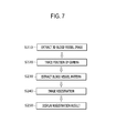
As shown in FIG. 7 , the method of non-invasive registration between a patient and the 3D medical image according to an exemplary embodiment includes extracting a 3D blood vessel image (S210), tracing the position of the camera (S220), extracting the blood vessel pattern (S230), image registration S240) and displaying a registration result (S250).
At the operation S210 of extracting the 3D blood vessel image, the near infrared 3D camera 110 takes the blood vessel image seen by having the near infrared light pass through a patient's the registration target area, thereby extracting the 3D blood vessel image I2. At this time, two near infrared blood vessel images having parallax are generated in accordance with distances where two unit cameras 113 provided in the near infrared 3D camera 110 are spaced part from each other, and the generated blood vessel images are transmitted to the controller 130 along a signal line connected to the controller 130.
At the operation S220 of tracing the position of the camera, the camera position tracer 120 traces the position of the near infrared 3D camera 110 in real time to calculate the real world coordinate system for the 3D blood vessel image I2. At this time, the position of the near infrared 3D camera 110 is traced in real time by the camera position tracer 120 such as the mechanical position tracing device 120 a, the optical position tracing device 120 b, etc., thereby extracting the real world coordinate values of the image taken by the near infrared 3D camera 110. The measured data about six degrees of freedom in position of the near infrared 3D camera 110, measured by the camera position tracer 120 is transmitted to the controller 130 and used as raw data needed for representing the second feature point in the real world coordinate system.
At the operation S230 of extracting the blood vessel pattern, the controller 130 is used to extract the first blood vessel pattern from the 3D medical image I1 of the registration target area and the second blood vessel pattern from the 3D blood vessel image I2. At the operation S240 of image registration, the first blood vessel pattern and the second blood vessel pattern extracted in the operation S230 of extracting the blood vessel pattern are used to perform position registration between the 3D medical image I1 and a patient. Thus, if the blood vessel model generator 132 of the controller 130 separates the blood vessel image from the 3D medical image I1 stored in the 3D medical image DB 131 and reconstitutes the blood vessel model, the first feature point extractor 133 detects the skeleton line SL of the generated blood vessel model and selects each extracted branch point as the first feature point of the first blood vessel pattern.
Also, the 3D blood vessel image detector 134 of the controller 130 extracts the 3D blood vessel image I2 by separating the blood vessel image from the near infrared 3D image transmitted from the near infrared 3D camera 110, and the second feature point extractor 135 extracts the branch point of a patient's blood vessel during the surgical operation through the extracted 3D blood vessel image I2 and the real world coordinate system of the 3D blood vessel image I2 measured by the camera position tracer 120, thereby selecting each extracted branch point as the second feature point of the second blood vessel pattern. At this time, the second feature point extractor 135 transforms the second feature point in the 3D camera coordinate system into that in the position tracer coordinate system through the coordinate transformation matrix M.
Then, the registrator 136 of the controller 130 performs registration by optimally matching the first feature point group P1 that compose of three or more first feature points with the second feature point group P2 that compose of three or more second feature points, in which the second feature point group P2 transformed in the position tracer coordinate system is rotated and translated to optimally match with the first feature point group P1 and then a position relationship T of the position tracer coordinate system is calculated with respect to the medical image coordinate system. In accordance with data about the position relationship T calculated by the registrator 136, the calculated registration result is displayed on the screen of the display 140.
Meanwhile, FIG. 6 shows a flowchart of performing the position registration following the system and method for non-invasive registration between a patient and the 3D medical image in accordance with an exemplary embodiment. Referring to FIG. 6 , the 3D medical image I1 of the patient is first obtained from the 3D medical image DB 131, and the 3D the blood vessel model is reconstituted with respect to the vicinity of the affected area. Then, the first feature point is extracted from the blood vessel model and additionally stored in the first feature point group P1.
In succession, to register the patient to the medical image during the surgical operation, the near infrared 3D camera 110 is arranged in a direction toward the affected area, and the position of the near infrared 3D camera 110 at this time is traced. Also, when the near infrared 3D image is taken, the 3D blood vessel image I2 is detected, and the second feature point is extracted from the 3D blood vessel image I2, thereby storing it in the second feature point group P2.
At this time, if the number of extracted second feature point is insufficient, the near infrared 3D camera 110 is rearranged to extract the second feature point again and add it to the second feature point group P2.
On the other hand, if number of added second feature points is sufficient, the first feature point group P1 and the second feature point group P2 are registered, and then the registration result is displayed on the display 140.
With the foregoing configuration and functions of the system and method for the non-invasive registration between a patient and the 3D medical image according to an exemplary embodiment, it is possible to non-invasively perform the registration between the patient and the 3D medical image I1 through the position registration between the 3D blood vessel image I2 of the blood vessel arranged in the patient's registration target area obtained using the near infrared light and the blood vessel model included in the 3D medical image I1 obtained by taking the registration target area, without using any fiducial markers.
Also, each blood vessel pattern of the blood vessel images reconstituted from the 3D blood vessel image I2 and the 3D medical image I1 obtained using the near infrared light is analyzed to calculate the feature point for the registration of the branch point of the blood vessel, and thus the registration is performed with respect to the calculated feature point, thereby not only being minimally affected by skin deformation but also reducing an error at the position registration as compared with the conventional registration method using the feature point on a patient's skin.
Furthermore, the position registration is performed based on the feature point according to the blood vessel pattern extended from the blood vessel distributed throughout a human body, thereby being advantageously applicable to various surgical areas and usable as medical information in connection with various medical images.
Although a few exemplary embodiments have been shown and described, it will be appreciated by those skilled in the art that changes may be made in these exemplary embodiments without departing from the principles and spirit of the invention, the scope of which is defined in the appended claims and their equivalents.
Claims (7)
1. A system for non-invasive registration between a patient and a three-dimensional (3D) medical image, the system comprising:
a near infrared 3D camera configured to extract a 3D blood vessel image of a patient's registration target area;
a camera position tracer configured to trace a position of the near infrared 3D camera and to calculate a real world coordinate system of the 3D blood vessel image;
a controller comprising:
a 3D medical image database configured to store a 3D medical image obtained by taking the registration target area;
a blood vessel model generator configured to reconstitute a blood vessel model by separating a blood vessel image from the 3D medical image;
a first feature point extractor configured to extract a branch point from which a blood vessel is branched by detecting a skeleton line of the blood vessel model, and to select each extracted branch point as a feature point of a first blood vessel pattern;
a 3D blood vessel image detector configured to extract a 3D blood vessel image by separating a blood vessel image from a near infrared 3D image taken by the near infrared 3D camera, and a second feature point extractor configured to extract a branch point of a patient's blood vessel through the extracted 3D blood vessel image and a real world coordinate system of the 3D blood vessel image measured by the camera position tracer, and to select each extracted branch point as a feature point of a second blood vessel pattern; and
a registrator configured to determine a registration result by optimally matching a first feature point group comprising of a plurality of first feature points, and a second feature point group comprising of a plurality of second feature points; and
a display configured to display the registration result.
2. The system according to claim 1 , wherein
the first feature point extractor further configured to store 3D coordinates of a feature point of a first blood vessel pattern selected with respect to a medical image coordinate system,
the near infrared 3D camera is further configured to take blood vessel images seen by near infrared light through the patient's registration target area to obtain two 3D blood vessel images spaced apart at a distance to have parallax, and
the second feature point extractor is further configure to select a second feature point represented by 3D coordinates with respect to a 3D camera coordinate system, using parallax of a pair of corresponding branch points respectively calculated from two 3D blood vessel images of the near infrared 3D camera.
3. The system according to claim 2 , wherein
the camera position tracer is further configured to measure six degrees of freedom in position of the near infrared 3D camera, which comprises three rotation positions and three translation positions, and to calculate a coordinate transformation matrix from the position tracer coordinate system to the 3D camera coordinate system by tracing the position of the near infrared 3D camera in real time,
the second feature point extractor is further configured to transform and to save the second feature point in the 3D camera coordinate system into a position tracer coordinate system by the coordinate transformation matrix, and
the registrator is a non-invasive registration system between patient and 3D medical image, and is further configured to calculate a position relationship of a position tracer coordinate system with respect to a medical image coordinate system in a state that a second feature point group in the position tracer coordinate system is rotated and translated to be optimally matched with the first feature point group.
4. The system according to claim 3 , wherein the camera position tracer comprises a mechanical position tracing device which is fastened to an end of an articulated link and is configured to measure six degrees of freedom in position of the near infrared 3D camera by sensing a physical displacement due to link movement of joints.
5. The system according to claim 3 , wherein the camera position tracer comprises an optical position tracing device which is configured to use a 3D camera taking fiducial markers mounted to the near infrared 3D camera to calculate a 3D position vector and a 3D direction vector of the fiducial marker and to measure six degrees of freedom in position of the near infrared 3D camera.
6. The system according to claim 1 , wherein the first feature point extractor is further configured to extract a border line representing an outline of a blood vessel from the blood vessel model, and to extract a center line, wherein the center line is extracted from the extracted border line along the blood vessel, as the skeleton line.
7. A method of non-invasive registration between a patient and a three-dimensional (3D) medical image, the method comprising:
extracting a 3D blood vessel image by taking a blood vessel image by passing near infrared light through a registration target area with a near infrared 3D camera;
tracing a camera position by tracing a position of the near infrared 3D camera in real time with a camera position tracer and calculating a real world coordinate system of the 3D blood vessel image;
storing a 30 medical image obtained by taking the registration target area;
reconstituting a blood vessel model by separating a blood vessel image from the 3D medical image;
extracting a branch point from which a blood vessel is branched by detecting a skeleton line of the blood vessel model;
selecting each extracted branch point as a feature point of a first blood vessel pattern;
extracting a 3D blood vessel image by separating a blood vessel image from a near infrared 3D image taken by the near infrared 3D camera;
extracting a branch point of a patient's blood vessel through the extracted 3D blood vessel image and a real world coordinate system of the 3D blood vessel image measured by the camera position tracer;
selecting each extracted branch point as a feature point of a second blood vessel pattern,
determining a registration result by optimally matching a first feature point group comprising of a plurality of first feature points, and a second feature point group comprising of a plurality of second feature points; and
displaying the registration result on a screen.
Applications Claiming Priority (2)
| Application Number | Priority Date | Filing Date | Title |
|---|---|---|---|
| KR1020130096016A KR101572487B1 (en) | 2013-08-13 | 2013-08-13 | System and Method For Non-Invasive Patient-Image Registration |
| KR10-2013-0096016 | 2013-08-13 |
Publications (2)
| Publication Number | Publication Date |
|---|---|
| US20150049174A1 US20150049174A1 (en) | 2015-02-19 |
| US9554117B2 true US9554117B2 (en) | 2017-01-24 |
Family
ID=52466556
Family Applications (1)
| Application Number | Title | Priority Date | Filing Date |
|---|---|---|---|
| US14/077,462 Active 2034-07-27 US9554117B2 (en) | 2013-08-13 | 2013-11-12 | System and method for non-invasive patient-image registration |
Country Status (2)
| Country | Link |
|---|---|
| US (1) | US9554117B2 (en) |
| KR (1) | KR101572487B1 (en) |
Cited By (4)
| Publication number | Priority date | Publication date | Assignee | Title |
|---|---|---|---|---|
| JP2016140714A (en) * | 2015-02-05 | 2016-08-08 | 富士通株式会社 | Image display device, image display program, and image display method |
| US11357593B2 (en) | 2019-01-10 | 2022-06-14 | Covidien Lp | Endoscopic imaging with augmented parallax |
| US11399900B2 (en) * | 2012-06-21 | 2022-08-02 | Globus Medical, Inc. | Robotic systems providing co-registration using natural fiducials and related methods |
| US11612314B2 (en) | 2017-10-31 | 2023-03-28 | Samsung Electronics Co., Ltd. | Electronic device and method for determining degree of conjunctival hyperemia by using same |
Families Citing this family (32)
| Publication number | Priority date | Publication date | Assignee | Title |
|---|---|---|---|---|
| US9129417B2 (en) * | 2012-02-21 | 2015-09-08 | Siemens Aktiengesellschaft | Method and system for coronary artery centerline extraction |
| US10758315B2 (en) | 2012-06-21 | 2020-09-01 | Globus Medical Inc. | Method and system for improving 2D-3D registration convergence |
| US11864839B2 (en) | 2012-06-21 | 2024-01-09 | Globus Medical Inc. | Methods of adjusting a virtual implant and related surgical navigation systems |
| US11857266B2 (en) | 2012-06-21 | 2024-01-02 | Globus Medical, Inc. | System for a surveillance marker in robotic-assisted surgery |
| US11896446B2 (en) | 2012-06-21 | 2024-02-13 | Globus Medical, Inc | Surgical robotic automation with tracking markers |
| US11963755B2 (en) | 2012-06-21 | 2024-04-23 | Globus Medical Inc. | Apparatus for recording probe movement |
| US10624710B2 (en) | 2012-06-21 | 2020-04-21 | Globus Medical, Inc. | System and method for measuring depth of instrumentation |
| US11253327B2 (en) | 2012-06-21 | 2022-02-22 | Globus Medical, Inc. | Systems and methods for automatically changing an end-effector on a surgical robot |
| US11793570B2 (en) | 2012-06-21 | 2023-10-24 | Globus Medical Inc. | Surgical robotic automation with tracking markers |
| US10874466B2 (en) | 2012-06-21 | 2020-12-29 | Globus Medical, Inc. | System and method for surgical tool insertion using multiaxis force and moment feedback |
| US11786324B2 (en) | 2012-06-21 | 2023-10-17 | Globus Medical, Inc. | Surgical robotic automation with tracking markers |
| US11864745B2 (en) | 2012-06-21 | 2024-01-09 | Globus Medical, Inc. | Surgical robotic system with retractor |
| US10799298B2 (en) | 2012-06-21 | 2020-10-13 | Globus Medical Inc. | Robotic fluoroscopic navigation |
| US11589771B2 (en) | 2012-06-21 | 2023-02-28 | Globus Medical Inc. | Method for recording probe movement and determining an extent of matter removed |
| US11857149B2 (en) | 2012-06-21 | 2024-01-02 | Globus Medical, Inc. | Surgical robotic systems with target trajectory deviation monitoring and related methods |
| JP6289142B2 (en) * | 2014-02-07 | 2018-03-07 | キヤノン株式会社 | Image processing apparatus, image processing method, program, and storage medium |
| JP6393106B2 (en) * | 2014-07-24 | 2018-09-19 | キヤノン株式会社 | Image processing apparatus, image processing method, and program |
| KR101699857B1 (en) | 2015-04-28 | 2017-01-25 | 부산대학교 산학협력단 | Apparatus and System for Optical Imaging using Near Infrared Fluorescence and Method for controlling the same |
| US11883217B2 (en) | 2016-02-03 | 2024-01-30 | Globus Medical, Inc. | Portable medical imaging system and method |
| KR101978316B1 (en) * | 2016-02-22 | 2019-05-14 | 연세대학교 산학협력단 | 3D volume mesh generation method for arterial blood flow dynamics simulation using the mesh morphing technique |
| JP7123309B2 (en) * | 2016-06-29 | 2022-08-23 | ニラマイ・ヘルス・アナリティックス・ピーブイティー・エルティーディ | Blood vessel extraction in 2D thermography |
| US10460512B2 (en) * | 2017-11-07 | 2019-10-29 | Microsoft Technology Licensing, Llc | 3D skeletonization using truncated epipolar lines |
| KR101843992B1 (en) * | 2017-11-30 | 2018-05-14 | 재단법인 구미전자정보기술원 | Augmented reality based cannula guide system for interventional cardiology procedures and method thereof |
| CN108294772B (en) * | 2017-12-31 | 2021-12-17 | 北京纳米维景科技有限公司 | CT scanning visual positioning method and CT system |
| KR102014355B1 (en) * | 2018-02-20 | 2019-08-26 | (주)휴톰 | Method and apparatus for calculating location information of surgical device |
| WO2019164271A1 (en) * | 2018-02-20 | 2019-08-29 | (주)휴톰 | Virtual body model generation method and device |
| CN110575255B (en) * | 2018-06-07 | 2022-08-16 | 格罗伯斯医疗有限公司 | Robotic system and related methods for providing co-registration using natural fiducials |
| KR20210059279A (en) | 2019-11-15 | 2021-05-25 | 한국광기술원 | Near Infrared Fluorescence Image Acquisition and Image Projection System for Guiding Cancer Surgery |
| CN115428443A (en) * | 2020-04-22 | 2022-12-02 | 钛隼生物科技股份有限公司 | Method and system for enhancing medical scanning image information on extended real-world image |
| CN112215876B (en) * | 2020-10-22 | 2022-10-04 | 烟台艾睿光电科技有限公司 | Double-spectrum image registration fusion method, device, equipment and storage medium |
| KR102580750B1 (en) * | 2020-12-30 | 2023-09-19 | 서울대학교산학협력단 | 3d image registration method based on markerless, method for tracking 3d object and apparatus implementing the same method |
| CN115409689B (en) * | 2021-05-28 | 2023-09-29 | 南京博视医疗科技有限公司 | Registration method and device for multi-modal retina fundus images |
Citations (17)
| Publication number | Priority date | Publication date | Assignee | Title |
|---|---|---|---|---|
| US20030000535A1 (en) * | 2001-06-27 | 2003-01-02 | Vanderbilt University | Method and apparatus for collecting and processing physical space data for use while performing image-guided surgery |
| US20030053697A1 (en) * | 2000-04-07 | 2003-03-20 | Aylward Stephen R. | Systems and methods for tubular object processing |
| US20080144773A1 (en) * | 2005-04-20 | 2008-06-19 | Visionsense Ltd. | System and Method for Producing an Augmented Image of an Organ of a Patient |
| US20080317321A1 (en) * | 2007-06-25 | 2008-12-25 | Hui Zhang | High quality volume rendering with graphics processing unit |
| US20090005668A1 (en) * | 2007-06-30 | 2009-01-01 | West Jay B | Non-invasive method for using 2D angiographic images for radiosurgical target definition |
| US20100098299A1 (en) * | 2007-02-26 | 2010-04-22 | Muquit Mohammad Abdul | Information Extraction Method, Information Extraction Device, Program, Registration Device, and Verification Device |
| US20100172567A1 (en) * | 2007-04-17 | 2010-07-08 | Prokoski Francine J | System and method for using three dimensional infrared imaging to provide detailed anatomical structure maps |
| US7826889B2 (en) * | 2000-08-21 | 2010-11-02 | Spectrum Dynamics Llc | Radioactive emission detector equipped with a position tracking system and utilization thereof with medical systems and in medical procedures |
| US20110158487A1 (en) * | 2008-02-19 | 2011-06-30 | Kabushiki Kaisha Toshiba | Medical image display device and image displaying method |
| US20120215094A1 (en) * | 2011-02-18 | 2012-08-23 | Voxel Rad, Ltd. | Systems and methods for 3d stereoscopic angiovision, angionavigation and angiotherapeutics |
| WO2012117381A1 (en) * | 2011-03-03 | 2012-09-07 | Koninklijke Philips Electronics N.V. | System and method for automated initialization and registration of navigation system |
| KR20130045774A (en) | 2011-10-26 | 2013-05-06 | 주식회사 고영테크놀러지 | Registration method of images for surgery |
| US8463360B2 (en) * | 2006-02-09 | 2013-06-11 | National University Corporation Hamamatsu University School Of Medicine | Surgery support device, surgery support method, and computer readable recording medium storing surgery support program |
| US20140005527A1 (en) * | 2012-06-29 | 2014-01-02 | General Electric Company | Method and system for dynamic referencing and registration used with surgical and interventional procedures |
| US20140072196A1 (en) * | 2012-09-07 | 2014-03-13 | Korea Advanced Institute Of Science And Technology | Method and apparatus for medical image registration |
| US20140148690A1 (en) * | 2012-11-26 | 2014-05-29 | Samsung Electronics Co., Ltd. | Method and apparatus for medical image registration |
| US20140218720A1 (en) * | 2013-02-04 | 2014-08-07 | Novadaq Technologies Inc. | Combined radiationless automated three dimensional patient habitus imaging with scintigraphy |
-
2013
- 2013-08-13 KR KR1020130096016A patent/KR101572487B1/en active IP Right Grant
- 2013-11-12 US US14/077,462 patent/US9554117B2/en active Active
Patent Citations (20)
| Publication number | Priority date | Publication date | Assignee | Title |
|---|---|---|---|---|
| US20030053697A1 (en) * | 2000-04-07 | 2003-03-20 | Aylward Stephen R. | Systems and methods for tubular object processing |
| US7826889B2 (en) * | 2000-08-21 | 2010-11-02 | Spectrum Dynamics Llc | Radioactive emission detector equipped with a position tracking system and utilization thereof with medical systems and in medical procedures |
| US20030000535A1 (en) * | 2001-06-27 | 2003-01-02 | Vanderbilt University | Method and apparatus for collecting and processing physical space data for use while performing image-guided surgery |
| US20080144773A1 (en) * | 2005-04-20 | 2008-06-19 | Visionsense Ltd. | System and Method for Producing an Augmented Image of an Organ of a Patient |
| US8463360B2 (en) * | 2006-02-09 | 2013-06-11 | National University Corporation Hamamatsu University School Of Medicine | Surgery support device, surgery support method, and computer readable recording medium storing surgery support program |
| US20100098299A1 (en) * | 2007-02-26 | 2010-04-22 | Muquit Mohammad Abdul | Information Extraction Method, Information Extraction Device, Program, Registration Device, and Verification Device |
| US8463006B2 (en) * | 2007-04-17 | 2013-06-11 | Francine J. Prokoski | System and method for using three dimensional infrared imaging to provide detailed anatomical structure maps |
| US20100172567A1 (en) * | 2007-04-17 | 2010-07-08 | Prokoski Francine J | System and method for using three dimensional infrared imaging to provide detailed anatomical structure maps |
| US20080317321A1 (en) * | 2007-06-25 | 2008-12-25 | Hui Zhang | High quality volume rendering with graphics processing unit |
| US20090005668A1 (en) * | 2007-06-30 | 2009-01-01 | West Jay B | Non-invasive method for using 2D angiographic images for radiosurgical target definition |
| US20110158487A1 (en) * | 2008-02-19 | 2011-06-30 | Kabushiki Kaisha Toshiba | Medical image display device and image displaying method |
| US8374410B2 (en) * | 2008-02-19 | 2013-02-12 | Kabushiki Kaisha Toshiba | Medical image display device and image displaying method |
| US20120215094A1 (en) * | 2011-02-18 | 2012-08-23 | Voxel Rad, Ltd. | Systems and methods for 3d stereoscopic angiovision, angionavigation and angiotherapeutics |
| WO2012117381A1 (en) * | 2011-03-03 | 2012-09-07 | Koninklijke Philips Electronics N.V. | System and method for automated initialization and registration of navigation system |
| US20140193053A1 (en) * | 2011-03-03 | 2014-07-10 | Koninklijke Philips N.V. | System and method for automated initialization and registration of navigation system |
| KR20130045774A (en) | 2011-10-26 | 2013-05-06 | 주식회사 고영테크놀러지 | Registration method of images for surgery |
| US20140005527A1 (en) * | 2012-06-29 | 2014-01-02 | General Electric Company | Method and system for dynamic referencing and registration used with surgical and interventional procedures |
| US20140072196A1 (en) * | 2012-09-07 | 2014-03-13 | Korea Advanced Institute Of Science And Technology | Method and apparatus for medical image registration |
| US20140148690A1 (en) * | 2012-11-26 | 2014-05-29 | Samsung Electronics Co., Ltd. | Method and apparatus for medical image registration |
| US20140218720A1 (en) * | 2013-02-04 | 2014-08-07 | Novadaq Technologies Inc. | Combined radiationless automated three dimensional patient habitus imaging with scintigraphy |
Non-Patent Citations (1)
| Title |
|---|
| Kandani et al., Development of blood vessel searching system for HMS, Aug. 27, 2008, Infrared Systems and Photoelectronic Technology III Proc. SPIE 7055. * |
Cited By (7)
| Publication number | Priority date | Publication date | Assignee | Title |
|---|---|---|---|---|
| US11399900B2 (en) * | 2012-06-21 | 2022-08-02 | Globus Medical, Inc. | Robotic systems providing co-registration using natural fiducials and related methods |
| JP2016140714A (en) * | 2015-02-05 | 2016-08-08 | 富士通株式会社 | Image display device, image display program, and image display method |
| US20160232668A1 (en) * | 2015-02-05 | 2016-08-11 | Fujitsu Limited | Image display apparatus and image display method |
| US9846939B2 (en) * | 2015-02-05 | 2017-12-19 | Fujitsu Limited | Image display apparatus and image display method |
| US11612314B2 (en) | 2017-10-31 | 2023-03-28 | Samsung Electronics Co., Ltd. | Electronic device and method for determining degree of conjunctival hyperemia by using same |
| US11357593B2 (en) | 2019-01-10 | 2022-06-14 | Covidien Lp | Endoscopic imaging with augmented parallax |
| US11793390B2 (en) | 2019-01-10 | 2023-10-24 | Covidien Lp | Endoscopic imaging with augmented parallax |
Also Published As
| Publication number | Publication date |
|---|---|
| KR101572487B1 (en) | 2015-12-02 |
| KR20150019311A (en) | 2015-02-25 |
| US20150049174A1 (en) | 2015-02-19 |
Similar Documents
| Publication | Publication Date | Title |
|---|---|---|
| US9554117B2 (en) | System and method for non-invasive patient-image registration | |
| US20230384734A1 (en) | Method and system for displaying holographic images within a real object | |
| US20200305986A1 (en) | Devices, systems and methods for natural feature tracking of surgical tools and other objects | |
| US20200405433A1 (en) | System and method for dynamic validation, correction of registration for surgical navigation | |
| TWI741359B (en) | Mixed reality system integrated with surgical navigation system | |
| Navab et al. | Camera augmented mobile C-arm (CAMC): calibration, accuracy study, and clinical applications | |
| JP6404713B2 (en) | System and method for guided injection in endoscopic surgery | |
| CA2973479C (en) | System and method for mapping navigation space to patient space in a medical procedure | |
| US20110105895A1 (en) | Guided surgery | |
| US10543045B2 (en) | System and method for providing a contour video with a 3D surface in a medical navigation system | |
| US10357317B2 (en) | Handheld scanner for rapid registration in a medical navigation system | |
| US20140031668A1 (en) | Surgical and Medical Instrument Tracking Using a Depth-Sensing Device | |
| TW201801682A (en) | An image guided augmented reality method and a surgical navigation of wearable glasses using the same | |
| CA3029348C (en) | Intraoperative medical imaging method and system | |
| KR102105974B1 (en) | Medical imaging system | |
| JP7181298B2 (en) | Systems and methods for detecting abnormal tissue using vascular features | |
| Yaniv et al. | Applications of augmented reality in the operating room | |
| De Paolis et al. | Augmented reality in minimally invasive surgery | |
| Leventon | A registration, tracking, and visualization system for image-guided surgery | |
| US20210330395A1 (en) | Location pad surrounding at least part of patient eye for tracking position of a medical instrument | |
| Liu et al. | Augmented Reality in Image-Guided Robotic Surgery | |
| WO2023147433A2 (en) | Augmented reality systems and methods for surgical planning and guidance | |
| Singla | Intra-operative ultrasound-based augmented reality for laparoscopic surgical guidance | |
| Liu | AUGMENTED REALITY AND INTRAOPERATIVE C-ARM CONE-BEAM COMPUTED TOMOGRAPHY FOR IMAGE-GUIDED ROBOTIC SURGERY | |
| TW201304737A (en) | Method for auxiliary perspective of multi-position |
Legal Events
| Date | Code | Title | Description |
|---|---|---|---|
| AS | Assignment |
Owner name: KOREA INSTITUTE OF SCIENCE AND TECHNOLOGY, KOREA, Free format text: ASSIGNMENT OF ASSIGNORS INTEREST;ASSIGNORS:LEE, DEUKHEE;PARK, SE HYUNG;LIM, SUNGHWAN;SIGNING DATES FROM 20131106 TO 20131113;REEL/FRAME:031882/0748 |
|
| STCF | Information on status: patent grant |
Free format text: PATENTED CASE |
|
| MAFP | Maintenance fee payment |
Free format text: PAYMENT OF MAINTENANCE FEE, 4TH YR, SMALL ENTITY (ORIGINAL EVENT CODE: M2551); ENTITY STATUS OF PATENT OWNER: SMALL ENTITY Year of fee payment: 4 |