WO1984003358A1 - Insoluble surfaces treated to inhibit non-specific protein binding - Google Patents
Insoluble surfaces treated to inhibit non-specific protein binding Download PDFInfo
- Publication number
- WO1984003358A1 WO1984003358A1 PCT/US1984/000257 US8400257W WO8403358A1 WO 1984003358 A1 WO1984003358 A1 WO 1984003358A1 US 8400257 W US8400257 W US 8400257W WO 8403358 A1 WO8403358 A1 WO 8403358A1
- Authority
- WO
- WIPO (PCT)
- Prior art keywords
- set forth
- biological substance
- surface portion
- substantial
- polysaccharide
- Prior art date
Links
Classifications
-
- G—PHYSICS
- G01—MEASURING; TESTING
- G01N—INVESTIGATING OR ANALYSING MATERIALS BY DETERMINING THEIR CHEMICAL OR PHYSICAL PROPERTIES
- G01N33/00—Investigating or analysing materials by specific methods not covered by groups G01N1/00 - G01N31/00
- G01N33/48—Biological material, e.g. blood, urine; Haemocytometers
- G01N33/50—Chemical analysis of biological material, e.g. blood, urine; Testing involving biospecific ligand binding methods; Immunological testing
- G01N33/53—Immunoassay; Biospecific binding assay; Materials therefor
- G01N33/543—Immunoassay; Biospecific binding assay; Materials therefor with an insoluble carrier for immobilising immunochemicals
- G01N33/54393—Improving reaction conditions or stability, e.g. by coating or irradiation of surface, by reduction of non-specific binding, by promotion of specific binding
-
- G—PHYSICS
- G01—MEASURING; TESTING
- G01N—INVESTIGATING OR ANALYSING MATERIALS BY DETERMINING THEIR CHEMICAL OR PHYSICAL PROPERTIES
- G01N33/00—Investigating or analysing materials by specific methods not covered by groups G01N1/00 - G01N31/00
- G01N33/48—Biological material, e.g. blood, urine; Haemocytometers
- G01N33/50—Chemical analysis of biological material, e.g. blood, urine; Testing involving biospecific ligand binding methods; Immunological testing
- G01N33/53—Immunoassay; Biospecific binding assay; Materials therefor
- G01N33/543—Immunoassay; Biospecific binding assay; Materials therefor with an insoluble carrier for immobilising immunochemicals
- G01N33/544—Immunoassay; Biospecific binding assay; Materials therefor with an insoluble carrier for immobilising immunochemicals the carrier being organic
- G01N33/548—Carbohydrates, e.g. dextran
Definitions
- This invention relates to biological or immunological substances attached to solid carriers for use in diagnostic tests, enzyme processes, affinity purifications, and the like.
- Soluble biological substances attached to solid carriers have many uses in diagnostic tests, enzyme processes, and affinity purifications. For example, attachment of antibodies or antigens to a solid carrier allows their immunological partners to be easily removed from a mixture of many substances. Similarly, attaching enzymes to a solid carrier allows them to be easily removed from the reaction mixture or to be used in a continuous flow process. Heterogeneous radioimmunoassays and enzyme immunoassays rely on attachment of one or more of the reactants to a solid phase to venable separation from the free reactants.
- Agglutination assays utilize indicator or carrier particles (upon which are carried the appropriate immunological material) in order to make the immunological complex more easily visible. Separation and identification of cells, cellular constituents, and bacteria are aided by antibodies or antigens coupled to solids. Biological particles will, for example, specifically adhere to solids coated with appropriate antibodies and antigens so that separation from other particles can be affected. Identification of biological particles can be made through the specific adherence of small particles coated with appropriate antibody or antigen. These small particles can incorporate a substance such as a fluorescent dye, radioactive tracer, or electron dense substance which makes their presence more readily detectable.
- U.S. Patent No. 3,645,852 discloses a process wherein cyanogen halides are used to activate a water insoluble polymer which then couples to a water soluble protein.
- Water soluble carbodiimides can be used as a condensing agent to bind protein to polymeric carrier particles according to U.S. Patent No. 3,857,931.
- Biological substances can be covalently bound to plastic materials whose surfaces have been coated with glutaraldehyde as discussed in U.S. Patent No. 4,001,583.
- preactivated latex-polysaccharide particles cannot be readily stored or shipped to an ultimate user who would then be able to attach any desired immunologically active material.
- the Shiff's bases produced by the reaction of the amino groups of the immunologically active material with the dialdehydes must be stabilized by sodium borohydride. This must be carried out at 0°C after removal of excess immunologically active material to keep denaturation at a minimum.
- the latex particles in U. S. Patent No. 4,264,766 have the surfaces entirely covered with the amino polysaccharide which is bonded to the carboxyl groups on the latex surface. This prevents proteins in solution, other than those that are partners for the attached immunologically active material, from becoming attached to the latex particles.
- a problem with this method is that formation of the dialdehydes, reaction with the dialdehydes, and reduction of the Shiff's bases are reactions which require a good deal of skill and care and thus involve a good deal of expense and time.
- a composition is set out which is useful for specifically binding to a specific binding protein which is a specific binding partner to a biological substance when the protein is associated with other proteins.
- the composition includes a water insoluble support having a surface having the capability of associating with the specific binding protein and with the other proteins.
- the composition also includes a polysaccharide coating covering a first substantial portion of the surface sufficiently to substantially prevent binding of protein to said first substantial surface portion and not covering a second substantial surface portion of the surface, the surface consisting essentially of the first and second substantial surface portions.
- the aforementioned composition further includes a biological substance attached to the second substantial surface portion.
- a method is provided of preparing a water insoluble surface of a solid support for specifically binding to a specific binding protein which is a specific binding partner to a biological substance when the protein is associated with other proteins.
- the method comprises providing a solid support having a water insoluble surface capable of associating with the specific binding protein and with other proteins and covering a first substantial portion of the surface with a polysaccharide coating while not covering a second substantial portion of the surface with a polysaccharide coating, the surface consisting essentially of the first substantial surface portion and the second substantial surface portion.
- a process for assaying an aqueous sample containing a specifically binding protein having a first binding site which is a specific binding partner to a first biological substance, the specifically binding protein being in association with other proteins, with increased specificity and sensitivity.
- the process comprises contacting an aqueous sample with a first solid support having a first water insoluble surface capable of associating with the specific binding protein and with the other proteins, the first surface consisting essentially of a first substantial surface portion shielded by a polysaccharide coating and a second substantial surface portion having the first biological substance attached to it.
- the aqueous sample is separated from the first solid support and the amount of specifically binding protein bound to the attached first biological substance is detected.
- Another embodiment still of the present invention comprises a process for reducing adherence of undesired proteins to a water insoluble surface consisting essentially of a first substantial surface portion and a second substantial surface portion while providing the surface with the capability for binding to a specifically binding protein which is a specific binding partner to a biological substance.
- the process comprises shielding the first substantial surface portion with a polysaccharide coating and attaching the biological substance to the second substantial surface portion.
- the kit comprises a solid support having a first water insoluble surface capable of associating with the specific binding protein and with other proteins, the first surface consisting essentially of a shielded first substantial surface portion and a second substantial surface portion having the first biological substance attached to it.
- the kit further includes a plurality of solid particles, each having a second insoluble surface capable of associating with the specific binding protein and with other proteins, the second surfaces each consisting essentially of a first substantial surface portion shielded by a polysaccharide coating and a second substantial surface portion having the second biological substance attached to it.
- the present invention is based upon discovery that if a polysaccharide coating covers a first portion of the surface of a solid support that is capable of associating with proteins, generally, and if a specific biological substance is attached to the rest of the surface, then proteins which are not specific binding partners for the biological substance will not be able to attach to the surface even in those portions where the surface is only attached to the biological substance.
- Partially coated compositions of the present invention are relatively easy to make and quite easy to attach to biological substances. Generally, they can be made quite inexpensively. And, the compositions of the present invention can be stored or shipped in condition for the attachment of any desired biological substance.
- a composition is provided which is useful for specifically binding to a specific binding protein which is a specific binding partner to a biological substance when the protein is associated with other proteins .
- biological substance is used broadly to indicate any substance which is a specific binding partner to a specific binding protein.
- Illustrative of the biological substance are enzymes, antibodies, natural receptors, e.g., thyroxine binding globulin and avidin, globins , e.g., hemoglobin, ocular lens proteins, surface antigens, histo-compatible antigens and the like.
- a specific binding protein can be any protein which it is desired to link to a water insoluble support. A long list of such substances appears in previously mentioned U. S. Patent No. 4,264,766.
- composition of the invention includes a water insoluble support having a surface having the capability of associating with the specific binding protein and with other proteins as well.
- the solid support may be in the form of micro or macro-particles, or in the form of macroextensive surfaces such as walls, flat plates, wells, and the like, all of which can be used in the separation of proteinaceous mixtures.
- the water insoluble support is in the form of a plurality of particles it is preferred that they have a specific gravity near that of water so as to enable them to be stably suspended in an aqueous medium.
- Such particles will generally be from about 0.2 micron to about 1 cm in diameter.
- the solid support itself must be inert with respect to immunological diagnostic tests.
- a large number of materials can be used as the water insoluble support.
- Of particular interest are latexes as described in U.S. Patents Nos . 4,046,723; 4,118,349; 4,140,662 and 4,264,766. Glass surfaces which may be used are described in U.S. Patent No. 4,169,138. Other useful polymers may be found in U.S. Patents Nos.
- the solid support be a latex and have active groups which are capable of forming a covalent linkage with a polyhydroxy compound.
- the latex supports can have active groups such as carboxyl groups, amine groups, or groups convertable into them.
- Useful active groups on the latex support are those containing an active hydrogen such as -COOH,-CONH 2 , primary and secondary amine groups, or nitryl groups.
- the preferred latex material is polystyrene for the practice of the present invention.
- the polystyrene will preferably also contain copolymerized therewith a carboxyl containing compound such as acrylic acid, methacrylic acid, or the like.
- a polysaccharide coating is provided covering a first substantial surface portion of the surface of the water insoluble support sufficiently to substantially prevent binding of proteins to the first substantial surface portion of the water insoluble support.
- the polysaccharide coating is also required to not cover a second substantial surface portion of the support.
- the polysaccharide coating will normally be formed of polysaccharides characterized by being water soluble, relatively high molecular weight, normally in excess of 5,000 daltons, more usually in excess of 10,000 daltons, and may be 1,000,000 daltons or higher in molecular weight.
- the polysaccharide may be a polymer or copolymer of glycose(s), e.g., glucose and fructose, a mixture of carbohydrates, such as neuraminic acids, uronic acids, glycosamines, or the like.
- the polysaccharide may be a combination of block or alternating copolymers or combinations thereof of saccharides and condensation monomers, particularly epoxides.
- Polysaccharides of particular interest include dextran, Ficoll (this is a synthetic copolymer of sucrose and epichlorohydron, a trademark of Pharmacia Fine Chemicals, Piscataway, New Jersey), agarose, hyaluronic acid, etc.
- an amino group normally being bonded to a short alkylene chain of from about 2 - 6 carbon atoms, which are bonded to functionalities of the polysaccharide.
- an amino group normally being bonded to a short alkylene chain of from about 2 - 6 carbon atoms, which are bonded to functionalities of the polysaccharide.
- Particularly convenient is the reaction product of diamines with carboxyl functionalities present on the polysaccharide. See, for example, Inman, J. of Immunology 114 , 704-709 (1975) .
- relatively high molecular weight amino polysaccharides such as the previously mentioned Ficoll, generally with molecular weights of 1,000 to 1,000,000 or more. Excellent results have been obtained with an amino Ficoll with a molecular weight of approximately
- the amount of the polysaccharide attached must be controlled to be between about 0.5 x 10 -7 and about 3 x 10 -7 grams per square centimeter of the area of the entire water insoluble surface in order to obtain a covering of only a first substantial surface portion of the water insoluble surface while leaving a second substantial surface portion of the water insoluble surface uncovered with polysaccharide. This corresponds to from about 700 to about 4000 molecules per square micron when the molecular weight of the polysaccharide is about 400,000.
- the biological substance is attached to the second substantial surface portion of the surface.
- This attachment can be by hydrophobic adsorption, electrostatic bonding, covalent bonding, or combinations thereof.
- the biological substance is covalently bonded to the second substantial surface portion.
- An activating agent such as a water soluble carbodiimide can be utilized to form an adduct with carbonyl groups on the latex surface. The carbodiimide adducts is then reacted with amine groups on the biological substance to leave an amide linkage to the latex support.
- Other useful activating agents include the Woodward reagent K (N-ethyl-5-phenyl-isoxazolium-3'-sulfonate) or a water soluble chloroformiate.
- the subject compositions can be used wherever an insoluble material is used for specific binding to a protein present in a mixture. This situation is encountered in competitive protein binding assays, cell sorting, cytology, histology, and the like. Since the procedure can vary very widely, the subject invention generally involves combining the insoluble material, as a particle or surface of a larger structure, with a protein mixture and allowing a sufficient time for binding between the biological substance on the surface and the specific protein binding partner. The solid surface is then washed free of non-specifically bound protein, leaving only specifically bound protein.
- the labels may include radioactive isotopes, fluorescers, magnetic materials, enzymes, enzyme substrates, dyes for producing colors, or the like.
- the labels may be bonded to the water insoluble surface, the polysaccharide, or the biological substance, desirably being bonded to the water insoluble surface or the polysaccharide. If desired, the labels may be uniformly dispersed throughout the particles.
- kits for assaying samples potentially containing a specifically binding protein having a first binding site which is a specific binding partner to a first biological substance and a second binding site which is a binding partner to a second biological substance, the specifically binding protein being in association with other proteins.
- a kit includes a macroextensive surface wall defined on a support such as a slide, a well, or the like.
- the support has a first water insoluble surface which is capable of associating with the specific binding protein and with other proteins.
- the first water insoluble surface consists essentially of a first substantial surface portion shielded by a polysaccharide coating and a second substantial surface portion having the first biological substance attached to it.
- the kit also includes a plurality of solid particles, each of which has a second insoluble surface capable of associating with the specific binding protein and with other proteins.
- the second insoluble surfaces each consist essentially of a first substantial surface portion shielded by a polysaccharide coating and a second substantial surface portion having the second biological substance attached to it.
- the particles can be labelled with a label capable of providing a detectable signal.
- the particles may be colored by a color imparting entity such as a dye and the signal will simply comprise the color itself.
- Alternatively, such other labels as have been previously discussed may be utilized.
- compositions of the present invention can be produced in a number of ways, several of which are disclosed in following:
- compositions of the present invention can be produced by providing a solid support having a water insoluble surface capable of associating with a specific binding protein and with other proteins. A first substantial surface portion of the surface is covered with a polysaccharide coating while a second substantial surface portion of the surface is not covered with a polysaccharide coating. This is accomplished by nitrating the surface to add nitro groups utilizing, for example, a mixture of nitric and sulfuric acids. After the acid mixture has been washed off of the surface, the nitro groups are reduced to amino groups utilizing a convenient reducing agent such as stannous chloride along with hydrochloric acid. The solid support is again washed and cyanuric halide moieties are attached to the amino groups.
- Cyanuric halide is generally added to an aqueous solution in contact with the solid surface with the cyanuric halide itself being in an ethanol solution because of its generally low solubility in water. It is believed that the cyanuric halide is actually in the form of a monoethoxy derivative when added. Any excess cyanuric chloride is washed away. It is believed that the remaining halide on the cyanuric halide moiety then hydrolizes to a hydroxy! group (pKa about 10 -7 ) which dissociates into a hydronium ion and a net negatively charged surface. This serves to promote electrostatic binding of amino polysaccharides to a surface which does not necessarily have carboxyl groups.
- the amino polysaccharide molecules are then attached to the surface.
- the intermediate product thus formed is a solid support having a water insoluble surface having an amino polysaccharide electrostatically attached to at least a first surface portion thereof.
- the intermediate product can then be reacted with a cyanuric halide to cross-link adjacent amino polysaccharide molecules and to form a usable composition.
- the reaction with cyanuric halide also serves, due to its acidity and ionic strength, to free at least the second surface portion from amino polysaccharide coverage.
- a biological substance as described above can be attached to the second substantial surface portion of the water insoluble surface. This can be accomplished by hydrophobic adsorption, by electrostatic bonding, and, more preferably, by covalent bonding as via utilizing an activating agent as set out above.
- Another method of making the composition of the present invention is to provide a water insoluble support which has carboxyl groups on its surface.
- the surface is contacted with an amino polysaccharide in an amount more than sufficient to cover the surface with a raonomolecular layer of the amino polysaccharide.
- an excess of amino Ficoll can be contacted with the surface in a water solution.
- the amino polysaccharide is held to the surface by electrostatic bonding. This is known since acid and high salt solutions lead to removal of the polysaccharide.
- the excess amino polysaccharide is washed off of the solid support with water. Thereafter, a cyanuric halide, in ethanol solution as set out above, is added to the amino polysaccharide coated solid surface.
- the cyanuric halide is used in a sufficient quantity to convert at least a significant portion of the amino groups to cyanuric halide adducts and to thereby cross-link the various amino groups with one another.
- a first substantial surface portion of the water insoluble surface is coated with amino polysaccharide while a second substantial surface portion of the water insoluble surface is not coated with amino polysaccharide.
- Another alternative method of forming a composition in accordance with the present invention comprises reacting water insoluble surfaces having active groups such as carboxyl groups with less than enough amino polysaccharide to cover the entire water insoluble surface with amino polysaccharide, and with a water soluble carbodiimide, all in a single reaction.
- the resultant product includes the amino polysaccharide covalently bonded via the carbodiimide to a first substantial surface portion of the water soluble surface through, e.g., the carboxyl groups. A second substantial portion of the surface remains uncovered by amino polysaccharide molecules.
- a biological substance can be attached to the second substantial surface portion by any desired method.
- This method has the advantage that if an excess of carbodiimide is utilized there can still be carbodiimide activated active (e.g. , carboxyl) groups on the second substantial surface portion ready to covalently bond to a desired biological substance.
- carbodiimide activated active e.g. , carboxyl
- a solid support having a water insoluble surface having active (e.g. , carboxyl) groups is reacted with an excess of an activator compound such as a water soluble carbodiimide to form an adduct, e.g., a carbodiimide adduct. Any excess activator is washed off. Less than enough of the biological substance is added to react with all activated sites. After the reaction is completed the biological substance remains attached to the second substantial surface portion. The surface is then washed to remove any reaction products. Water is again contacted with the surface and an amino polysaccharide is added which then links to the active groups which remain and which have been activated by the activator.
- an activator compound such as a water soluble carbodiimide
- the resulting product has both the amino polysaccharide and the biological substance covalently attached to the water insoluble surface via the active groups and through use of the activating agent. While several of the above described methods have called for the use of a carboxyl active group and a carbodiimide activating agent it should be noted that other active groups and other activating agents, for example those previously set out, may be utilized where appropriate.
- an assaying process is set out for assaying a sample containing a specifically binding protein having a first binding site which is a specific binding partner to a first biological substance, the specifically binding protein being in association with other proteins.
- the process operates by contacting the first support having a first water insoluble surface capable of associating with the specific binding protein and with other proteins, the first surface having a first substantial surface portion shielded by a polysaccharide coating and a second substantial surface portion having the first biological substance attached to it, with an aqueous solution of the sample.
- the aqueous sample solution is separated from the first solid support and from the specifically binding protein bound to the attached first biological substance.
- the support would generally comprise a plurality of particles and they would normally be labelled with a label capable of detection.
- the support would comprise a macroextensive surface such as a plate or a well on a plate having one or more wells.
- the amino polysaccharide, the biological substance, etc., are as defined previously.
- the aforementioned first solid support is a macroextensive surface and the detecting step comprises contacting an aqueous solution having a second solid support in the nature of a plurality of particles, the second solid support having a second water insoluble surface capable of associating with the specific binding protein and other proteins, the second surface having an additional first substantial surface portion shielded by a polysaccharide coating and an additional second substantial surface portion having a second biological substance attached thereto, with the first macroextensive surface, i.e., with the plate or well.
- the specifically binding protein is selected to be of a nature to have a second binding site which is a specific partner to the second biological substance. The degree of adherence of the particles to the macroextensive surface is then observed .
- a process is also set out for reducing adherence of undesirable proteins to a water insoluble surface consisting essentially of a first substantial surface portion and a second substantial surface portion while providing the surface with the capability for binding to a specifically binding protein which is a specific binding partner to a biological substance.
- the first, substantial surface portion is shielded with a polysaccharide coating and the biological substance is attached to the second substantial surface portion. Shielding and attaching steps may be simultaneous or either may precede the other.
- the surface in this instance would generally be a macroscopically extensive surface.
- the adherence reducing process provides an alternative to the utilization of bovine serum albumin on such macroextensive surfaces. This is particularly advantageous because of the expense of bovine serum albumin and because of the lack of complete uniformity of bovine serum albumin from one batch to the next, which nonuniformity is inherent in the biological production of this reactant .
- a kit for assaying samples potentially contanning a specifically binding protein having a first binding site which is a specific binding partner to a first biological substance and a second binding site which is a specific binding partner to a second biological substance, the specifically binding protein being in association with other proteins.
- the kit comprises a solid support having a first water insoluble surface, the support being a. macroextensive wall and the first water insoluble surface being a wall surface.
- the first water insoluble surface is capable of associating with the specific binding protein and with other proteins.
- the first surface consists essentially of a first substantial surface portion shielded by a polysaccharide coating and a second substantial surface portion having the first biological substance attached to it.
- a plurality of solid particles also forms a part of the kit.
- Each particle has a second insoluble surface capable of associating with the specific binding protein and with other proteins.
- the second surfaces each consist essentially of a first substantial surface portion shielded by a polysaccharide coating and a second substantial surface portion having- the second biological substance attached to it.
- the particles would be labelled with a label capable of providing a detectable signal. For most purposes the label would simply comprise a color imparting entity and the signal would comprise the color.
- the kit comprises the plurality of particles as in the kit just described, but the wall support having the first water insoluble surface, while it has the first biological substance attached to it, does not have the polysaccharide coating on a first substantial portion thereof. Instead, bovine serum albumin or other shielding agent is utilized.
- Polystyrene microspheres (Polysciences Inc., Warrington, PA), 5 grams, of one micron diameter, were suspended in 50 ml of ice cold 75% H 2 SO 4 and added to 100 ml of ice cold 1:1 HNO 3 :H 2 SO 4 . The resulting suspension was stirred for thirty minutes in an ice bath, then quenched by pouring into 1L of ice cold water. The thereby nitrated microspheres were washed in water three times utilizing centrifu ⁇ ation for separation.
- the nitrated microspheres were suspended in 100 ml concentrated HCl along with 110 grams of SnCl 2 .2H 2 O and stirred at room temperature for ten hours.
- the amino-microspheres were suspended in 900 ml of cold water and dispersed by sonication in a stainless steel ultrasonic bath cleaner. Cyanuric chloride 0.5 gram, a 2 to 1 mix of water and alcohol (150 ml) was added and the resultant solution was incubated with intermittent sonication for thirty minutes. The amino groups were thereby converted to cyanuric chloride adducts. The suspension was centrifuged to remove the microspheres, which are believed to contain hydroxyl groups formed by hydrolysis of remaining chloride of the cyanuric chloride adducts, and washed twice with cold water by centrifugation.
- AECM-Ficoll Carboxymethylated Ficoll
- microspheres were suspended in 1 liter of cold water by sonication and 1.2 grams of AECM-Ficoll dissolved in 50 ml of water was added. The resultant mixture was incubated with intermittent sonication for twelve hours. The AECM-Ficoll coupled microspheres were washed twice in v/ater by centrifugation. They were vacuum filtered through Whatman #1 paper to remove any clumped microspheres. The AECM-Ficoll utilized had a molecular weight of approximately 400,000 and had about 80 amino groups per molecule. Further Treatment Of Coated Microspheres With Cyanuric Chloride to Cross-link the AECM-Ficoll
- the AECM-Ficoll electrostatically coated microspheres were suspended in 750 ml of water cooled to 0°C. Cyanuric chloride (1 gram) dissolved in 200 ml of cold 1:1 alcohol:water was added to the suspension to cross-link the AeCM-Ficoll. Only a portion of the surface remained covered with AECM-Ficoll because of the acidity caused by the formation of HCl as the cyanuric chloride reacted. The resulting suspension was incubated at 0°C for thirty-five minutes and microspheres were separated from the suspension by centrifugation. They were washed in cold water three times by centrifugation. The microspheres were coupled to proteins by incubation with them at room temperature. It was found that they could be stored, at about 4°C, for over a year without significant deterioration.
- Nitrated spheres were covered with a solution of 110 grams SnCl 2 .2H 2 O dissolved in 100 ml of concentrated hydrochloric acid and incubated with occasional stirring for fifteen hours at room temperature to reduce the nitro groups to amino groups.
- the SnCl 2 solution was decanted and the spheres were washed in turn with water, 0.1N HCl, water, and 0.1N NaOH and water.
- the amino-spheres were covered with a solution of 0.1 gram cyanuric chloride dissolved in 10 ml of alcohol and 200 ml of water and incubated at room temperature for forty minutes.
- the cyanuric chloride solution was decanted off of the spheres and the spheres were washed with several portions of water.
- the spheres were covered with a solution of 0.14 grams AECM-Ficoll dissolved in 120 ml of water and incubated with occasional stirring for twelve hours.
- the AECM-Ficoll solution was decanted off of the spheres and the spheres were washed two times with cold water. This provided an electrostatically bound coating of AECM-Ficoll on the spheres.
- the AECM-Ficoll electrostatically coated spheres were cooled to 0°C and covered with a solution of 150 ml of ice cold water containing 0.18 grams of cyanuric chloride and 18 ml of alcohol. Incubation was continued for thirty minutes at 0°C. The cyanuric chloride solution was decanted off of the spheres and the spheres were washed three times in ice cold water. Only a portion of the surface remained covered with AECM-Ficoll because of the acidity caused by the formation of HCl as the cyanuric chloride reacted. The AECM-Ficoll utilized in the above reaction had about 80 amino groups per molecule of the AECM-Ficoll. The spheres now react with protein macromoiecules such as IgG, enzymes, etc. They can be stored for several months at about 0°C without significant deterioration.
- protein macromoiecules such as IgG, enzymes, etc. They can be stored for several months
- MOPC-21 myeloma protein was added to make the final protein concentration 3 micrograms per ml in a total volume of 1.1 ml normal saline. After one hour of incubation at room temperature the unbound protein was removed by three washes in normal saline via centrifugation. MOPC-21 coupled microspheres were then incubated for one hour at room temperature in 1 ml of saline containing 0.642 milligrams per milliliter I 125 labelled monoclonal antibodies, anti-4a.
- microspheres used were suspension polymerized at 60°C from 95.2 parts by weight of freshly vacuum distilled ⁇ tyrene and 4.8 parts by weight of freshly vacuum distilled methacrylic acid using potassium persulfate as catalyst. After polymerization was complete the microspheres were washed with dilute NaOH, filtered through glass wool, brought to pH 7 with dilute HCl , washed 3 times with distilled water by centrifugation and the concentration of solids adjusted to 0.745% by weight. Scanning electron microscopy was used to determine that the microspheres had a 0.5 micron diameter with less than 3% standard deviation.
- C 14 AECM Ficoll was prepared by reaction of 50 mg CM Ficoll (80 carboxyl groups per 40,000 molecular weight by titration) with 66.5 mg ethylene diamine hydrochloride-C 14 (containing 25 microcuries C 14 ) and 75 mg (3-dimethyl aminopropyl) ethyl carbodiimide hydrochloride (EDAC) at pH 4.7 in 5 ml
- the C 14 AECM Ficoll was separated from the low molecular weight reactants by gel filtration and exhaustive dialysis against distilled water at 4°C.
- Ficoll have weight average molecular weights of 443,000 by HPLC (high performance liquid chromatography) so that the C 14 AECM Ficoll had 1.7 x 10 -11 cpm/molecule under the conditions of measurement.
- HPLC high performance liquid chromatography
- microspheres were treated as were those in Table 1 but the pH of the solutions used for binding and washing were as specified in Table 2.
- AECM-Ficoll is bound at lower pH values, e.g., below about 6, than at higher pH values.
- a method is provided of controlling the amount of AECM-Ficoll attached to the microspheres.
- microspheres were treated as were those in Table 1 but the solutions used for binding and washing were as specified in Table 3.
- PBS phosphate buffered saline, pH 7.3
- Table 4 reports data on covalent binding of AECM-Ficoll using a water soluble carbodiimide activating agent.
- the data of Table 4 were obtained by reaction of C AECM Ficoll (2120 cpm) with microspheres in the presence of various amounts of carbodiimide followed by washing 3 times with pH 7.3 PBS. Incubation of the microspheres of Table 4 with non-radioactive AECM Ficoll demonstrated that, except for the case where no EDAC was used, the AECM Ficoll is not exchangeable. Presumably the amino groups of the AECM Ficoll have been covalently coupled through amide linkages to the carboxyl groups on the surface of the microspheres.
- the PBS appears to inhibit the binding of AECM Ficoll to the microspheres by limiting their approach to one another since it does not interfere with the EDAC activation of the microspheres.
- the usefulness of cyanuric chloride to couple AECM Ficoll microspheres was also investigated as outlined in Table 6.
- the protocol used consisted of reacting 100 microliters of microspheres with either C 14 or non-C 14 AECM Ficoll with or without EDAC in H 2 O, washing 3 times with H 2 O (1st wash) , reacting with cyanuric chloride for 30 minutes, washing 3 times with H 2 O (2nd wash) , reacting with either C 14 or non-C 14 AECM Ficoll for 1 hour, washing 3 times with
- non-exchangeable cpm of AECM Ficoll can be bound by use of cyanuric chloride on microspheres which already have an ionic coating of AECM Ficoll.
- cyanuric chloride treatment of microspheres which already had an EDAC coupled layer of AECM Ficoll could not facilitate uptake of additional AECM Ficoll, e.g., the 3rd wash of sample 4, Table 6, contains 98% of the counts added after the cyanuric chloride treatment.
- the excess counts above 250 on the microspheres for samples 2 and 3 are apparently due to ionic bonding of AECM Ficoll.
- cyanuric chloride non-exchangeably couples the AECM Ficoll is presumably by cross-linking the amino groups on some of the adjacent AECM Ficoll molecules. Those AECM Ficoll molecules thereby cross-linked, to the few such molecules which are so strongly bound that they do not wash off in PBS (e.g. , sample 6, Table 3) , are retained on the microsphere surface. The remainder are washed off.
- PBS e.g. , sample 6, Table 3
- a surface substantially completed covered with AECM Ficoll cannot be used to practice the present invention since proteins, antibodies and the like cannot be bound to it either with cyanuric chloride or with EDAC as shown by some of the other examples.
- U.S. Patent No. 4,264,766 teaches a complicated oxidation, coupling, and reduction procedure to attach proteins to a polysaccharide coating, but this is not simple in the hands of the user and not all proteins retain their biological activity after the borohydride reduction which is necessary to practice that invention.
- the present invention provides proteins, antibodies, etc., attached to the microsphere surface with most or all of the other area of the surface covered by polysaccharide.
- the examples allow for coupling the protein before or after the polysaccharide either covalently or non-covalently.
- the material of the present invention with a partial layer of AECM Ficoll, has been prepared by a number of methods and storage stability at 4°C for longer than a year is routinely observed. This material is readily coupled to protein by simply mixing it with protein, which is a considerable advantage to the user who wishes to couple his own protein and wants long shelf life. Once coupled to portein, the resulting reagent has a shelf life of greater than six months .
- Example V Example V
- T-15 antibody at 1 mg/ml in PBS were added to the tubes thus: Tubes 1, 5, 9, 13, 17 received 50 microliters of T-15. Tubes 2, 6, 10, 14, 18 received 25 microliters of T-15. Tubes 3, 7, 11, 15, 19 received 10 microliters of T-15. Tubes 4, 8, 12, 16, 20 received 5 microliters of T-- 15. All the tubes were sonicated briefly and incubated at room temperature for 1 hour.
- microsphere suspensions in tubes 1-4 were v/ashed twice in PBS by centrifugation and resuspension and resuspended in 500 microliters of PBS with sonication.
- Tubes 5-8 were washed twice in PBS containing 1% bovine serum albumin and 0.1% sodium ozide (protein buffer) and resuspended in 500 microliters of protein buffer with sonication. These were kept for 48 hours at 4°C.
- the microspheres in tubes 9-20 were washed twice in distilled water and suspended in 500 microliters of distilled water with sonication.
- Tubes 9-20 were incubated for 1 hour at room temperature and an additional 48 hours at 4°C.
- Tubes 9-12 were washed 3 times in PBS by centrifugation and resuspension, and the washes were pooled for each tube.
- the pooled washes and the washed microspheres were transferred to scintillation vials, 20 ml of scintillation fluid (a standard fluid which fluoresces when exposed to radiation) was added to each vial and they were counted.
- scintillation fluid a standard fluid which fluoresces when exposed to radiation
- the highest antibody concentration used (tube 9) was estimated to allow a complete monolayer coverage of the microspheres in the sample. That this did not occur, as implied by the similar quantity of binding of C 14 AECM Ficoll, is indicative of the difficulty the negatively changed antibodies have in approaching the negatively changed microspheres.
- microspheres in tubes 13-16 were washed twice in PBS and resuspended in 500 microliters of PBS with brief sonication.
- the microspheres in tubes 17-20 were washed twice in protein buffer, resuspended in 500 microliters of protein buffer and sonicated briefly.
- the PCBGG is the specific binding partner for the T-15 antibody and any attachment of the microspheres (all T-15 coupled in this example) in the absence of PCBGG indicates non-specific sticking. All of the microspheres exhibited specific binding to the PCBGG plate coat. The microspheres which had AECM Ficoll (tubes 13-20) exhibited no detectable non-specific sticking even when the binding was carried out in the absence of protein in the buffer (tubes 13-16) . The microspheres without AECM Ficoll exhibited non-specific sticking in all cases and particularly in the absence of protein in the buffer (tubes 1-4). This example provides clear evidence that the ratio of specific to non-specific binding is enhanced by coating that part of the microsphere surface which is not coupled to antibody with AECM Ficoll. It also demonstrates that the amount of non-antibody coupled surface can be substantial for carboxylated microspheres even when an excess of antibody and covalent coupling is used.
- EDAC/AECM Ficoll/CTC Microspheres Green fluorescent 0.72 micron diameter carboxylated polystyrene microspheres prepared generally as previously described were given partial surface coverings of covalently coupled AECM Ficoll in the following manner. A 44.5 ml suspension containing 3.75 grams of microspheres was added to 706 ml H 2 O and then 39 ml of water containing 0.41 grams of AECM Ficoll was added. The suspension was mixed thoroughly and then 75 mg EDAC dissolved in 30 ml H 2 O was added with swirling and sonication. The pH at this point was less than 5. The suspension was allowed to sit at room temperature for two days then washed 3 times in distilled water.
- microspheres After resuspension in 1 liter of water with sonication the microspheres were filtered through Whatman #1 paper with suction to remove any clumps and centrifuged once more and the supernatant discarded. The microspheres were then resuspended in 186 ml of cold water and sonicated until a single microsphere suspension was obtained. To this suspension v/as added 0.36 grams cyanuric chloride dissolved in 36 ml ETOH and 76 ml cold water. The suspension was sonicated and kept in an ice bath for 30 minutes followed by centrifugation at 4°C. The supernatant was discarded and the microspheres were washed with cold water 3 times by centrifugation. The microspheres were resuspended with sonication in 320 ml H 2 O and contained 1.07% solids by weight.
- Example VI A portion of the microspheres of Example VI were coupled to My-1, a mouse monoclonal antibody IgM (kappa) , by incubating for 75 minutes 0.1 mg My-1 with 10 ml of the microspheres which had been suspended by sonication in 100 ml normal saline at room temperature. The microspheres were then pelleted at 10,000 x g at 4°C for 10 minutes in a refrigerated centrifuge and the supernatant discarded. The pellet was resuspended with sonication in 100 ml RPMI 1640 cell culture medium containing 10% newborn calf serum, pelleted as before and the supernatant discarded.
- IgM mouse monoclonal antibody
- HL-60 and DAUDI cells were resuspended at a concentration of 1 x 10 6 cells/ml in HBSS (Hank's Buffered Salt Solution) containing 0.1% BSA (bovine serum albumin) and 0.1% sodium azide in separate tubes. Aliquots of 0.5 ml of both cell suspensions were placed in separate 2 ml wells of a 24 well tissue culture plate. Twenty microliters of the My-1 antibody reagent prepared above was added to both cell suspensions and mixed gently. The plate was centrifuged at 150 x g for 9 minutes at 4°C, then incubated for 1 hour at 4°C.
- the My-1 antibody recognizes a cell surface marker on the surface of human granulocytes and HL-60 cells. This marker is absent on DAUDI and other human white blood cells. Similar results were obtained on human white blood cells where the cells labelled were granulocytes.
- Microspheres A) 0.99 micron diameter containing fluorescent blue dye B) 0.54 micron diameter containing fluorescent blue dye C) 1.01 micron diameter, undyed
- volumes containing 1.25 grams of microspheres A, B, & C were put into three beakers labelled, respectively, A1 , B1, C1, each containing 220 ml of distilled water, and were sonicated to disperse the microspheres.
- A1 , B1, C1 each containing 220 ml of distilled water
- EDAC carboxyimide
- microspheres were washed three times in distilled water, by centrifugation and resuspension with sonication and were resuspended in a volume of 200 ml of distilled water. The suspensions were then filtered through a Whatman #1 filter paper. The microspheres suspensions A1 , B1 and C1 were brought to 160 ml, 190 ml and 90 ml respectively with distilled water and sonicated well. Aliquots from the treated microspheres A1 , B1 and C1 were then tested for their percentage by weight and the necessary adjustments made to the remaining microspheres. Thus A1 adjusted to 1.47%, B1 to 0.80% and C1 to 1.5%; the total volumes being A 165 ml, B 200 ml, C 100 ml. Treatment II
- microsphere suspensions were each brought to a volume of 70 ml in distilled water and sonicated well. 0.13 grams of cyanuric chloride dissolved in 10 ml of ETOH and diluted in 20 ml of distilled water was added to each of the three suspensions and dispersed by sonicating well. After 1 ⁇ 2 hour incubation the suspensions were washed 3 times in distilled water, by centrifugation and resuspension with sonication, were resuspened in 190 ml distilled water and sonicated well. Aliquots were taken and the volumes and % solids were adjusted (see above in Treatment I) to A 197 ml at 1.47%, B 199 ml at 0.80% and C 193 ml at 1.5%.
- microsphere suspensions A1 , B1, C1 (Treatment I) and A2 , B2 and C2 (Treatment II) were prepared as just described.
- 500 microliters of phosphate buffered saline (PBS) was added, then 50 microliters of one of the six microsphere suspensions (A1 , B1, C1, A2, B2 and C2) was placed in each tube and the tubes were sonicated briefly.
- 25 mililiters of T-15 antibody at 4 mg/ml was added to each tube; the tubes were sonicated and incubated at room temperature for 1 hour.
- microsphere suspensions were then washed 3 times in PBS containing 1% fetal calf serum plus 0.1% azide (protein buffer) by centrifugation and resuspension, and resuspended by sonication in 500 microliters of protein buffer.
- 96 well microliter plate (Dynatech, Inc.) had been prepared by placing 50 microliters of T-15 at 0.1 mg/ml in PBS in each of the first 6 wells. 50 microliters of phosphocholine - bovine gamma globulin conjugate (PCBGG) at 0.1 mg/ml in PBS in each of the next 6 wells, and 50 microliters of protein buffer in each of the next 6 wells. These plate coats were incubated at room temperature for 1 hour then the contents of the wells were removed by suction and the wells were washed 3 times with protein buffer by filling the wells to the top and aspirating the contents.
- PCBGG phosphocholine - bovine gamma globulin conjugate
- Carboxylated polystyrene microsphere suspensions were prepared in bottles of disti lled water with the bottles labelled D1, E1 and F1, as follows :
- each of samples from flasks D2, E2 and F2 were individually placed in tubes labelled, respectively, D5, E5 and F5 , each containing 500 microliters of PBS.
- the tubes were sonicated and 25 microliters of UPC 10 (a mouse myelona protein) at 1 mg/ml in PBS was added to tubes D4, E4 and F4.
- the tubes D4, E4, F4, D5, E5 and F5 were incubated for 1 hour at room temperature.
- the microspheres in these tubes were then washed 3 times in PBS containing 1% fetal calf serum and 0.1% sodium azide (protein buffer) by centrifugation and resuspension. They were each resuspended in 500 microliters of protein buffer with sonication.
- Example X The microspheres in D1, E1 and F1 in PBS did not bind to the protein coats on the plate even after attempted cyanuric chloride treatment thus demonstrating their inability to bind to protein after polysaccharide coating as in Treatment I of Example VIII.
- Example X The microspheres in D1, E1 and F1 in PBS did not bind to the protein coats on the plate even after attempted cyanuric chloride treatment thus demonstrating their inability to bind to protein after polysaccharide coating as in Treatment I of Example VIII.
- Example X Example X
- the T-15 antibody was at three different concentrations. 50 microliters of microspheres were placed in each of tubes labelled nos. 1, 3, 5, 7, 9 each containing 500 microliters of phosphate buffered saline. The tubes were sonicated to disperse the microspheres. 0.05 mg T-15 antibody and 0.05 mg EDAC was added to tube 1, 0.05 mg T-15 antibody and 0.005 mg EDAC to tube 3, 0.005 mg T-15 and 0.05 mg EDAC to tube 5, 0.005 mg T-15 and 0.005 mg EDAC to tube 7, and 0.05 mg T-15 and no EDAC to tube 9.
- Each tube was sonicated briefly after addition of protein and after addition of EDAC.
- the microsphere suspensions were incubated at room temperature for 1 hour, were wached 3 times in PBS and 1% fetal calf serum and .1% sodium azide (protein buffer) by centrifugation and resuspension and resuspended in 500 microliters of protein buffer with sonication.
- 15 wells of a flexible, U-bottom, PVC, 96 well microliter plate had been prepared by placing 50 microliters of T-15 antibody at 0.1 mg/ml in PBS in each of the first 5 wells, 50 microliters of phosphocholine bovine gamma globulin (PCBGG) at 0.1 mg/ml in PBS in each of the second 5 wells, and 50 microliters of protein buffer in each of the last 5 wells. After 1 hours incubation at room temperature the contents of the wells were removed by suction and the wells washed 3 times in protein buffer by filling the wells to the top and aspirating the contents.
- PCBGG phosphocholine bovine gamma globulin
- a flexible, U-bottom polyvinyl chloride, 96 well microliter plate (Dynatech, Inc.) was used. Clear polyvinyl chloride strips have also been substituted for the wells of the microliter plate, the strips being placed in the reagents instead bf the reagents being placed in the wells.
- Microspheres 0.7 micron in diameter were prepared containing fluorescent green dye and were treated with AECM Ficoll/cyanuric chloride to provide a partial coating as in Treatment II, Example VIII. 100 microliters of the microspheres were placed in a tube containing 1 ml PBS and sonicated. 50 microliters of T-15 antibody at 4 mg/ml in PBS was added and mixed by sonication.
- microsphere suspension was washed three times in protein buffer, by centrifugation and resuspension with sonication. The microspheres were resuspended in 1 ml of protein buffer and sonicated well.
- a flexible U-bottomed PVC 96 well microliter plate was prepared: the first 6 wells received 50 mililiter each of T-15 antibody at 0.1 mg/ml in phosphate buffered saline. Well 7 received 50 microliters of phosphocholine bovine gamma globulin (PCBGG) at 0.1 mg/ml in PBS. Well 8 received 50 microliters of PBS containing 1% bovine serum albumin and 0.1% sodium azide (protein buffer). The plate coats were incubated for 1 hour at room temperature; the well contents were aspirated and the wells washed 3 times in protein buffer by filling to the top and aspirating.
- PCBGG phosphocholine bovine gamma globulin
- protein buffer protein buffer
- a clear polyvinyl chloride strip is incubated with antibody #1, then coated with gelatin or bovine serum albumin. It is then placed in the suspect pregnancy urine and incubated for between one and sixty minutes, after which the strip is incubated in microspheres which are coupled to antibody #2. These microspheres are carboxylated polystyrene which have been activated with carbodiimide, washed, reacted with antibody #2, washed, and reacted with AECM Ficoll.
- Antibody #1 and antibody #2 are antibodies which have the property of being able to simultaneously react with human chorionic gonadotrophin or with the beta sub unit of human chorionic gonadotrophin, i.e., a given molecule of human chorionic gonadotrophin or beta sub unit of human chorionic gonadotrophin can have both antibody #1 and antibody #2 attached to it simultaneously. After the strip has been incubated in the microspheres for between one and sixty minutes, it is removed and rinsed. A positive test is indicated by the plastic strip having a cloudy appearance caused by adherence of the microspheres. This assay is also applicable for chorionic gonadotrophin in the urine of species other than humans. Industrial Applicability
- novel compositions comprising water insoluble surfaces partially coated with water soluble polysaccharides, normally amino functionalized, and partially attached to molecules of a biological substance. Clean accurate and highly selective separation is obtainable between molecules which are binding partners to the biological substance and binding partners which are not binding partners to the biological substance.
Landscapes
- Health & Medical Sciences (AREA)
- Life Sciences & Earth Sciences (AREA)
- Immunology (AREA)
- Engineering & Computer Science (AREA)
- Molecular Biology (AREA)
- Chemical & Material Sciences (AREA)
- Urology & Nephrology (AREA)
- Biomedical Technology (AREA)
- Hematology (AREA)
- Medicinal Chemistry (AREA)
- Biotechnology (AREA)
- Microbiology (AREA)
- Food Science & Technology (AREA)
- Cell Biology (AREA)
- Physics & Mathematics (AREA)
- Analytical Chemistry (AREA)
- Biochemistry (AREA)
- General Health & Medical Sciences (AREA)
- General Physics & Mathematics (AREA)
- Pathology (AREA)
- Chemical Kinetics & Catalysis (AREA)
- Immobilizing And Processing Of Enzymes And Microorganisms (AREA)
Abstract
A composition is set out which has improved selectivity and sensitivity for use in immunoassays. The composition comprises a solid support having a surface partially coated with a polysaccharide and elsewhere not covered by such a coating but instead attached to a biological substance which is a specific binding partner to a specific binding protein. Methods of making the composition and assays and kits using the composition are also disclosed.
Description
DESCRIPTION
Insoluble Surfaces Treated To Inhibit Ron-Specific Protein Binding
Technical Field This invention relates to biological or immunological substances attached to solid carriers for use in diagnostic tests, enzyme processes, affinity purifications, and the like.
Background Art Soluble biological substances attached to solid carriers have many uses in diagnostic tests, enzyme processes, and affinity purifications. For example, attachment of antibodies or antigens to a solid carrier allows their immunological partners to be easily removed from a mixture of many substances. Similarly, attaching enzymes to a solid carrier allows them to be easily removed from the reaction mixture or to be used in a continuous flow process. Heterogeneous radioimmunoassays and enzyme immunoassays rely on attachment of one or more of the reactants to a solid phase to venable separation
from the free reactants. Agglutination assays (to determine the presence of an antigen or antibody in a fluid) utilize indicator or carrier particles (upon which are carried the appropriate immunological material) in order to make the immunological complex more easily visible. Separation and identification of cells, cellular constituents, and bacteria are aided by antibodies or antigens coupled to solids. Biological particles will, for example, specifically adhere to solids coated with appropriate antibodies and antigens so that separation from other particles can be affected. Identification of biological particles can be made through the specific adherence of small particles coated with appropriate antibody or antigen. These small particles can incorporate a substance such as a fluorescent dye, radioactive tracer, or electron dense substance which makes their presence more readily detectable. Two of the major difficulties in the use of solid carriers as described above are reliably attaching the soluble biological substances and preventing non-specific sticking of undesired substances to the carrier. The consequences of these problems include excessively high background and low sensitivity in assays and loss of material and low purity in affinity purifications and enzyme processes.
The solution to the first of these problems has been approached through covalent bonding of proteins and peptides to polymer solids. For example, U.S. Patent No. 3,645,852 discloses a process wherein cyanogen halides are used to
activate a water insoluble polymer which then couples to a water soluble protein. Water soluble carbodiimides can be used as a condensing agent to bind protein to polymeric carrier particles according to U.S. Patent No. 3,857,931. Biological substances can be covalently bound to plastic materials whose surfaces have been coated with glutaraldehyde as discussed in U.S. Patent No. 4,001,583. In U.S. Patent No. 4,046,723 a three-step method is revealed for coupling proteins to a latex having surface carboxylic amide groups. A process for the manufacture of protein or peptide polystyrene latex compounds is described in U.S. Patent No. 4,118,349 in which the linkage is effected by means of an aromatic diazonium compound. A two-step process is disclosed in U.S. Patent No. 4,140,662 which links immunological substances to latex polymers via reactions with a diamine mediated by a carbodiimide followed by reaction with a bifunctional aldehyde. These and other methods known to couple biological substances to polymer materials (see, for example, Kiefer "The Chemical Modification Of Proteins, Haptens, And Solid Supports", Immunological Methods, Acedemic Press, 1979, Pages 137-150) are undoubtedly more generally reliable than the hydrophobic bonding which was used prior to the covalent bonding methods. However, they do not alleviate the problem of non-specific sticking and sometimes make it worse. U. S. Patent No. 4,264,766, issued April
28, 1981, discloses an invention which solves some of the above problems by covalently bonding a water soluble polyhydroxy compound, preferably an amino
polysaccharide, to a latex carrier, preferably a carboxylated polymer, via a water-soluble carbodiimide, the Woodward Reagent
K (N-ethyl-5-phenyl-isoxazoliura-3'-sulfonate) or a water-soluble chloroformiate. The amino groups of the amino polysaccharide which are not bonded to the latex carrier are converted to hydroxy1 groups. Then, the polysaccharide is activated with periodate to oxidize some of the glucose rings to dialdehydes. Thereafter, an immunologically active material is reacted with the thus activated polysaccharide. The reaction with the immulogically active material must be performed shortly after formation of the dialdehydes because the dialdehyde containing polysaccharide is subject to relatively fast degradation. Thus, preactivated latex-polysaccharide particles cannot be readily stored or shipped to an ultimate user who would then be able to attach any desired immunologically active material. Furthermore, the Shiff's bases produced by the reaction of the amino groups of the immunologically active material with the dialdehydes must be stabilized by sodium borohydride. This must be carried out at 0°C after removal of excess immunologically active material to keep denaturation at a minimum.
The latex particles in U. S. Patent No. 4,264,766 have the surfaces entirely covered with the amino polysaccharide which is bonded to the carboxyl groups on the latex surface. This prevents proteins in solution, other than those that are partners for the attached immunologically active material, from becoming attached to the latex
particles. A problem with this method is that formation of the dialdehydes, reaction with the dialdehydes, and reduction of the Shiff's bases are reactions which require a good deal of skill and care and thus involve a good deal of expense and time.
Disclosure Of Invention
In accordance with one embodiment of the present invention a composition is set out which is useful for specifically binding to a specific binding protein which is a specific binding partner to a biological substance when the protein is associated with other proteins. The composition includes a water insoluble support having a surface having the capability of associating with the specific binding protein and with the other proteins. The composition also includes a polysaccharide coating covering a first substantial portion of the surface sufficiently to substantially prevent binding of protein to said first substantial surface portion and not covering a second substantial surface portion of the surface, the surface consisting essentially of the first and second substantial surface portions. In accordance with another embodiment of the present invention the aforementioned composition further includes a biological substance attached to the second substantial surface portion.
In accordance with yet another embodiment of the present invention a method is provided of preparing a water insoluble surface of a solid support for specifically binding to a specific
binding protein which is a specific binding partner to a biological substance when the protein is associated with other proteins. The method comprises providing a solid support having a water insoluble surface capable of associating with the specific binding protein and with other proteins and covering a first substantial portion of the surface with a polysaccharide coating while not covering a second substantial portion of the surface with a polysaccharide coating, the surface consisting essentially of the first substantial surface portion and the second substantial surface portion.
In accordance with still another embodiment of the present invention a process is set out for assaying an aqueous sample containing a specifically binding protein having a first binding site which is a specific binding partner to a first biological substance, the specifically binding protein being in association with other proteins, with increased specificity and sensitivity. The process comprises contacting an aqueous sample with a first solid support having a first water insoluble surface capable of associating with the specific binding protein and with the other proteins, the first surface consisting essentially of a first substantial surface portion shielded by a polysaccharide coating and a second substantial surface portion having the first biological substance attached to it. The aqueous sample is separated from the first solid support and the amount of specifically binding protein bound to the attached first biological substance is detected.
Another embodiment still of the present invention comprises a process for reducing adherence of undesired proteins to a water insoluble surface consisting essentially of a first substantial surface portion and a second substantial surface portion while providing the surface with the capability for binding to a specifically binding protein which is a specific binding partner to a biological substance. The process comprises shielding the first substantial surface portion with a polysaccharide coating and attaching the biological substance to the second substantial surface portion.
Yet a further embodiment of the present invention provides a kit for assaying samples potentially containing a specifically binding protein having a first binding site which is a specific binding partner to a first biological substance and a second binding site which is a binding partner to a second biological substance, the specifically binding protein being in association with other proteins, with increased specificity and sensitivity. The kit comprises a solid support having a first water insoluble surface capable of associating with the specific binding protein and with other proteins, the first surface consisting essentially of a shielded first substantial surface portion and a second substantial surface portion having the first biological substance attached to it. The kit further includes a plurality of solid particles, each having a second insoluble surface capable of associating with the specific binding protein and with other proteins,
the second surfaces each consisting essentially of a first substantial surface portion shielded by a polysaccharide coating and a second substantial surface portion having the second biological substance attached to it.
The present invention is based upon discovery that if a polysaccharide coating covers a first portion of the surface of a solid support that is capable of associating with proteins, generally, and if a specific biological substance is attached to the rest of the surface, then proteins which are not specific binding partners for the biological substance will not be able to attach to the surface even in those portions where the surface is only attached to the biological substance. Partially coated compositions of the present invention are relatively easy to make and quite easy to attach to biological substances. Generally, they can be made quite inexpensively. And, the compositions of the present invention can be stored or shipped in condition for the attachment of any desired biological substance.
Best Mode For Carrying Out The Invention
A composition is provided which is useful for specifically binding to a specific binding protein which is a specific binding partner to a biological substance when the protein is associated with other proteins . The term "biological substance" is used broadly to indicate any substance which is a specific binding partner to a specific binding protein. Illustrative of the biological substance are enzymes, antibodies, natural
receptors, e.g., thyroxine binding globulin and avidin, globins , e.g., hemoglobin, ocular lens proteins, surface antigens, histo-compatible antigens and the like. A specific binding protein can be any protein which it is desired to link to a water insoluble support. A long list of such substances appears in previously mentioned U. S. Patent No. 4,264,766.
The composition of the invention includes a water insoluble support having a surface having the capability of associating with the specific binding protein and with other proteins as well. The solid support may be in the form of micro or macro-particles, or in the form of macroextensive surfaces such as walls, flat plates, wells, and the like, all of which can be used in the separation of proteinaceous mixtures.
In those aspects of the present invention wherein the water insoluble support is in the form of a plurality of particles it is preferred that they have a specific gravity near that of water so as to enable them to be stably suspended in an aqueous medium. Such particles will generally be from about 0.2 micron to about 1 cm in diameter. The solid support itself must be inert with respect to immunological diagnostic tests. A large number of materials can be used as the water insoluble support. Of particular interest are latexes as described in U.S. Patents Nos . 4,046,723; 4,118,349; 4,140,662 and 4,264,766. Glass surfaces which may be used are described in U.S. Patent No. 4,169,138. Other useful polymers may be found in U.S. Patents Nos. 3,619,371; 3,700,609; 3,853,987;
4,108,972 and 4,201,763. In each of these patents a wide variety of linking groups are disclosed for bonding to various biological substances, particularly proteins. It is preferred that the solid support be a latex and have active groups which are capable of forming a covalent linkage with a polyhydroxy compound. Accordingly, the latex supports can have active groups such as carboxyl groups, amine groups, or groups convertable into them. Useful active groups on the latex support are those containing an active hydrogen such as -COOH,-CONH2, primary and secondary amine groups, or nitryl groups. U.S. Patent No. 4,264,766 discloses a number latex materials which are particularly suitable for use in accordance with the present invention. The preferred latex material is polystyrene for the practice of the present invention. The polystyrene will preferably also contain copolymerized therewith a carboxyl containing compound such as acrylic acid, methacrylic acid, or the like.
A polysaccharide coating is provided covering a first substantial surface portion of the surface of the water insoluble support sufficiently to substantially prevent binding of proteins to the first substantial surface portion of the water insoluble support. The polysaccharide coating is also required to not cover a second substantial surface portion of the support. The polysaccharide coating will normally be formed of polysaccharides characterized by being water soluble, relatively high molecular weight, normally in excess of 5,000 daltons, more usually in excess of 10,000 daltons,
and may be 1,000,000 daltons or higher in molecular weight. The polysaccharide may be a polymer or copolymer of glycose(s), e.g., glucose and fructose, a mixture of carbohydrates, such as neuraminic acids, uronic acids, glycosamines, or the like. In addition, the polysaccharide may be a combination of block or alternating copolymers or combinations thereof of saccharides and condensation monomers, particularly epoxides. Polysaccharides of particular interest include dextran, Ficoll (this is a synthetic copolymer of sucrose and epichlorohydron, a trademark of Pharmacia Fine Chemicals, Piscataway, New Jersey), agarose, hyaluronic acid, etc. Of particular interest is the presence of an amino group, normally being bonded to a short alkylene chain of from about 2 - 6 carbon atoms, which are bonded to functionalities of the polysaccharide. Particularly convenient is the reaction product of diamines with carboxyl functionalities present on the polysaccharide. See, for example, Inman, J. of Immunology 114 , 704-709 (1975) .
In accordance with the present invention it is particularly preferred to use relatively high molecular weight amino polysaccharides such as the previously mentioned Ficoll, generally with molecular weights of 1,000 to 1,000,000 or more. Excellent results have been obtained with an amino Ficoll with a molecular weight of approximately
400,000.
The amount of the polysaccharide attached must be controlled to be between about 0.5 x 10⁻⁷
and about 3 x 10⁻⁷ grams per square centimeter of the area of the entire water insoluble surface in order to obtain a covering of only a first substantial surface portion of the water insoluble surface while leaving a second substantial surface portion of the water insoluble surface uncovered with polysaccharide. This corresponds to from about 700 to about 4000 molecules per square micron when the molecular weight of the polysaccharide is about 400,000.
In accordance with one of the aspects of the present invention the biological substance is attached to the second substantial surface portion of the surface. This attachment can be by hydrophobic adsorption, electrostatic bonding, covalent bonding, or combinations thereof. Preferably, the biological substance is covalently bonded to the second substantial surface portion. When the biological substance is covalently bonded to the second substantial portion the covalent bonding can be accomplished in any of a number of ways . An activating agent such as a water soluble carbodiimide can be utilized to form an adduct with carbonyl groups on the latex surface. The carbodiimide adducts is then reacted with amine groups on the biological substance to leave an amide linkage to the latex support. Other useful activating agents include the Woodward reagent K (N-ethyl-5-phenyl-isoxazolium-3'-sulfonate) or a water soluble chloroformiate.
The subject compositions can be used wherever an insoluble material is used for specific binding to a protein present in a mixture. This
situation is encountered in competitive protein binding assays, cell sorting, cytology, histology, and the like. Since the procedure can vary very widely, the subject invention generally involves combining the insoluble material, as a particle or surface of a larger structure, with a protein mixture and allowing a sufficient time for binding between the biological substance on the surface and the specific protein binding partner. The solid surface is then washed free of non-specifically bound protein, leaving only specifically bound protein.
Of particular interest are situations employing particles, which may be labelled or unlabelled. The labels may include radioactive isotopes, fluorescers, magnetic materials, enzymes, enzyme substrates, dyes for producing colors, or the like. The labels may be bonded to the water insoluble surface, the polysaccharide, or the biological substance, desirably being bonded to the water insoluble surface or the polysaccharide. If desired, the labels may be uniformly dispersed throughout the particles.
Also of particular interest is a situation wherein a kit is supplied for assaying samples potentially containing a specifically binding protein having a first binding site which is a specific binding partner to a first biological substance and a second binding site which is a binding partner to a second biological substance, the specifically binding protein being in association with other proteins. Such a kit includes a macroextensive surface wall defined on a
support such as a slide, a well, or the like. The support has a first water insoluble surface which is capable of associating with the specific binding protein and with other proteins. The first water insoluble surface consists essentially of a first substantial surface portion shielded by a polysaccharide coating and a second substantial surface portion having the first biological substance attached to it. The kit also includes a plurality of solid particles, each of which has a second insoluble surface capable of associating with the specific binding protein and with other proteins. The second insoluble surfaces each consist essentially of a first substantial surface portion shielded by a polysaccharide coating and a second substantial surface portion having the second biological substance attached to it. The particles can be labelled with a label capable of providing a detectable signal. For example, the particles may be colored by a color imparting entity such as a dye and the signal will simply comprise the color itself. Alternatively, such other labels as have been previously discussed may be utilized.
Methods Of Production The compositions of the present invention can be produced in a number of ways, several of which are disclosed in following:
Method I;
The compositions of the present invention can be produced by providing a solid support having a water insoluble surface capable of associating
with a specific binding protein and with other proteins. A first substantial surface portion of the surface is covered with a polysaccharide coating while a second substantial surface portion of the surface is not covered with a polysaccharide coating. This is accomplished by nitrating the surface to add nitro groups utilizing, for example, a mixture of nitric and sulfuric acids. After the acid mixture has been washed off of the surface, the nitro groups are reduced to amino groups utilizing a convenient reducing agent such as stannous chloride along with hydrochloric acid. The solid support is again washed and cyanuric halide moieties are attached to the amino groups. Cyanuric halide is generally added to an aqueous solution in contact with the solid surface with the cyanuric halide itself being in an ethanol solution because of its generally low solubility in water. It is believed that the cyanuric halide is actually in the form of a monoethoxy derivative when added. Any excess cyanuric chloride is washed away. It is believed that the remaining halide on the cyanuric halide moiety then hydrolizes to a hydroxy! group (pKa about 10-7) which dissociates into a hydronium ion and a net negatively charged surface. This serves to promote electrostatic binding of amino polysaccharides to a surface which does not necessarily have carboxyl groups. The amino polysaccharide molecules are then attached to the surface. The intermediate product thus formed is a solid support having a water insoluble surface having an amino polysaccharide electrostatically attached to at least a first surface portion
thereof. The intermediate product can then be reacted with a cyanuric halide to cross-link adjacent amino polysaccharide molecules and to form a usable composition. The reaction with cyanuric halide also serves, due to its acidity and ionic strength, to free at least the second surface portion from amino polysaccharide coverage.
A biological substance as described above can be attached to the second substantial surface portion of the water insoluble surface. This can be accomplished by hydrophobic adsorption, by electrostatic bonding, and, more preferably, by covalent bonding as via utilizing an activating agent as set out above.
Method II:
Another method of making the composition of the present invention is to provide a water insoluble support which has carboxyl groups on its surface. The surface is contacted with an amino polysaccharide in an amount more than sufficient to cover the surface with a raonomolecular layer of the amino polysaccharide. For example, an excess of amino Ficoll can be contacted with the surface in a water solution. The amino polysaccharide is held to the surface by electrostatic bonding. This is known since acid and high salt solutions lead to removal of the polysaccharide.
The excess amino polysaccharide is washed off of the solid support with water. Thereafter, a cyanuric halide, in ethanol solution as set out above, is added to the amino polysaccharide coated solid surface. The cyanuric halide is used in a
sufficient quantity to convert at least a significant portion of the amino groups to cyanuric halide adducts and to thereby cross-link the various amino groups with one another. At the end of this reaction a first substantial surface portion of the water insoluble surface is coated with amino polysaccharide while a second substantial surface portion of the water insoluble surface is not coated with amino polysaccharide. While it is believed that the surface was originally covered with electrostatically bound amino polysaccharide it has been experimentally shown that the surface, after the reaction with the cyanuric halide moiety, is no longer completely covered with an amino polysaccharide. Instead, portions of the surface are available for bonding to biological substances. Further, acid and/or high salt solutions no longer remove the amino polysaccharide after it has been reacted with the cyanuric halide moiety. Generally, the amount of the amino polysaccharide which remains on the water insoluble surface is between about 0.5 x 10 -7 and about 3 x 10⁻⁷ grams per square centimeter of the area of the water insoluble surface. The biological substance can be attached to the second substantial surface portion via hydrophobic adsorption or covalent bonding, all as previously described.
Method III:
Another alternative method of forming a composition in accordance with the present invention comprises reacting water insoluble surfaces having active groups such as carboxyl groups with less than enough amino polysaccharide to cover the entire water insoluble surface with amino polysaccharide, and with a water soluble carbodiimide, all in a single reaction. The resultant product includes the amino polysaccharide covalently bonded via the carbodiimide to a first substantial surface portion of the water soluble surface through, e.g., the carboxyl groups. A second substantial portion of the surface remains uncovered by amino polysaccharide molecules.
Once again, a biological substance can be attached to the second substantial surface portion by any desired method.
This method has the advantage that if an excess of carbodiimide is utilized there can still be carbodiimide activated active (e.g. , carboxyl) groups on the second substantial surface portion ready to covalently bond to a desired biological substance.
Method IV:
In yet another alternate method of forming a composition of the present invention, a solid support having a water insoluble surface having active (e.g. , carboxyl) groups is reacted with an excess of an activator compound such as a water soluble carbodiimide to form an adduct, e.g., a carbodiimide adduct. Any excess activator is washed
off. Less than enough of the biological substance is added to react with all activated sites. After the reaction is completed the biological substance remains attached to the second substantial surface portion. The surface is then washed to remove any reaction products. Water is again contacted with the surface and an amino polysaccharide is added which then links to the active groups which remain and which have been activated by the activator. The resulting product has both the amino polysaccharide and the biological substance covalently attached to the water insoluble surface via the active groups and through use of the activating agent. While several of the above described methods have called for the use of a carboxyl active group and a carbodiimide activating agent it should be noted that other active groups and other activating agents, for example those previously set out, may be utilized where appropriate.
Assaying Processes:
Assaying processes also form an important part of the present invention. In accordance with the invention an assaying process is set out for assaying a sample containing a specifically binding protein having a first binding site which is a specific binding partner to a first biological substance, the specifically binding protein being in association with other proteins. The process operates by contacting the first support having a first water insoluble surface capable of associating with the specific binding protein and with other
proteins, the first surface having a first substantial surface portion shielded by a polysaccharide coating and a second substantial surface portion having the first biological substance attached to it, with an aqueous solution of the sample. The aqueous sample solution is separated from the first solid support and from the specifically binding protein bound to the attached first biological substance. The support would generally comprise a plurality of particles and they would normally be labelled with a label capable of detection. In one embodiment the support would comprise a macroextensive surface such as a plate or a well on a plate having one or more wells. The amino polysaccharide, the biological substance, etc., are as defined previously.
In one particular preferred embodiment of the invention, the aforementioned first solid support is a macroextensive surface and the detecting step comprises contacting an aqueous solution having a second solid support in the nature of a plurality of particles, the second solid support having a second water insoluble surface capable of associating with the specific binding protein and other proteins, the second surface having an additional first substantial surface portion shielded by a polysaccharide coating and an additional second substantial surface portion having a second biological substance attached thereto, with the first macroextensive surface, i.e., with the plate or well. The specifically binding protein is selected to be of a nature to have a second binding site which is a specific partner to the second
biological substance. The degree of adherence of the particles to the macroextensive surface is then observed .
A process is also set out for reducing adherence of undesirable proteins to a water insoluble surface consisting essentially of a first substantial surface portion and a second substantial surface portion while providing the surface with the capability for binding to a specifically binding protein which is a specific binding partner to a biological substance. The first, substantial surface portion is shielded with a polysaccharide coating and the biological substance is attached to the second substantial surface portion. Shielding and attaching steps may be simultaneous or either may precede the other. The surface in this instance would generally be a macroscopically extensive surface. In this manner, the adherence reducing process provides an alternative to the utilization of bovine serum albumin on such macroextensive surfaces. This is particularly advantageous because of the expense of bovine serum albumin and because of the lack of complete uniformity of bovine serum albumin from one batch to the next, which nonuniformity is inherent in the biological production of this reactant .
Assay Kits:
Also in accordance with the present invention, a kit is provided for assaying samples potentially contanning a specifically binding protein having a first binding site which is a specific binding partner to a first biological
substance and a second binding site which is a specific binding partner to a second biological substance, the specifically binding protein being in association with other proteins. The kit comprises a solid support having a first water insoluble surface, the support being a. macroextensive wall and the first water insoluble surface being a wall surface. The first water insoluble surface is capable of associating with the specific binding protein and with other proteins. The first surface consists essentially of a first substantial surface portion shielded by a polysaccharide coating and a second substantial surface portion having the first biological substance attached to it. A plurality of solid particles also forms a part of the kit. Each particle has a second insoluble surface capable of associating with the specific binding protein and with other proteins. The second surfaces each consist essentially of a first substantial surface portion shielded by a polysaccharide coating and a second substantial surface portion having- the second biological substance attached to it. Generally, the particles would be labelled with a label capable of providing a detectable signal. For most purposes the label would simply comprise a color imparting entity and the signal would comprise the color.
In accordance with yet another embodiment of the present invention the kit comprises the plurality of particles as in the kit just described, but the wall support having the first water insoluble surface, while it has the first biological substance attached to it, does not have the polysaccharide coating on a first substantial
portion thereof. Instead, bovine serum albumin or other shielding agent is utilized.
The invention will be better understood by reference to the following illustrative examples.
Example I
Polystyrene Microspheres
Nitration:
Polystyrene microspheres (Polysciences Inc., Warrington, PA), 5 grams, of one micron diameter, were suspended in 50 ml of ice cold 75% H2SO4 and added to 100 ml of ice cold 1:1 HNO3:H2SO4. The resulting suspension was stirred for thirty minutes in an ice bath, then quenched by pouring into 1L of ice cold water. The thereby nitrated microspheres were washed in water three times utilizing centrifuσation for separation.
Reduction:
The nitrated microspheres were suspended in 100 ml concentrated HCl along with 110 grams of SnCl2.2H2O and stirred at room temperature for ten hours. The microspheres, which had had their nitrate groups reduced to amino groups, were separated from the SnCl2 solution and washed two times in water by centrifugation. The microspheres were then washed in turn in water, 0.1N HCl, H2O. , 0.1N NaOH, and H2O.
Activating The Reduced Microspheres With Cyanuric Chloride:
The amino-microspheres were suspended in 900 ml of cold water and dispersed by sonication in a stainless steel ultrasonic bath cleaner. Cyanuric chloride 0.5 gram, a 2 to 1 mix of water and alcohol (150 ml) was added and the resultant solution was incubated with intermittent sonication for thirty minutes. The amino groups were thereby converted to cyanuric chloride adducts. The suspension was centrifuged to remove the microspheres, which are believed to contain hydroxyl groups formed by hydrolysis of remaining chloride of the cyanuric chloride adducts, and washed twice with cold water by centrifugation.
Coating The Microspheres With N- (2-aminoethyl)
Carboxymethylated Ficoll (AECM-Ficoll) :
The microspheres were suspended in 1 liter of cold water by sonication and 1.2 grams of AECM-Ficoll dissolved in 50 ml of water was added. The resultant mixture was incubated with intermittent sonication for twelve hours. The AECM-Ficoll coupled microspheres were washed twice in v/ater by centrifugation. They were vacuum filtered through Whatman #1 paper to remove any clumped microspheres. The AECM-Ficoll utilized had a molecular weight of approximately 400,000 and had about 80 amino groups per molecule.
Further Treatment Of Coated Microspheres With Cyanuric Chloride to Cross-link the AECM-Ficoll
The AECM-Ficoll electrostatically coated microspheres were suspended in 750 ml of water cooled to 0°C. Cyanuric chloride (1 gram) dissolved in 200 ml of cold 1:1 alcohol:water was added to the suspension to cross-link the AeCM-Ficoll. Only a portion of the surface remained covered with AECM-Ficoll because of the acidity caused by the formation of HCl as the cyanuric chloride reacted. The resulting suspension was incubated at 0°C for thirty-five minutes and microspheres were separated from the suspension by centrifugation. They were washed in cold water three times by centrifugation. The microspheres were coupled to proteins by incubation with them at room temperature. It was found that they could be stored, at about 4°C, for over a year without significant deterioration.
Example II
Polystyrene Macrospheres
Nitration:
About 250 3/8 inch diameter polystyrene spheres were covered with 120 ml of 0°C 2:1 H2SO4:HNO3 and incubated with gentle mixing at 0°C for fifteen minutes. The mixed acid was poured off and the spheres were rinsed with 0°C 50% sulphuric acid, then with ice cold water until the wash water tested neutral.
Reduction:
Nitrated spheres were covered with a solution of 110 grams SnCl2.2H2O dissolved in 100 ml of concentrated hydrochloric acid and incubated with occasional stirring for fifteen hours at room temperature to reduce the nitro groups to amino groups. The SnCl2 solution was decanted and the spheres were washed in turn with water, 0.1N HCl, water, and 0.1N NaOH and water.
Activating With Cyanuric Chloride :
The amino-spheres were covered with a solution of 0.1 gram cyanuric chloride dissolved in 10 ml of alcohol and 200 ml of water and incubated at room temperature for forty minutes. The cyanuric chloride solution was decanted off of the spheres and the spheres were washed with several portions of water.
Coating With AECM-Ficoll:
The spheres were covered with a solution of 0.14 grams AECM-Ficoll dissolved in 120 ml of water and incubated with occasional stirring for twelve hours. The AECM-Ficoll solution was decanted off of the spheres and the spheres were washed two times with cold water. This provided an electrostatically bound coating of AECM-Ficoll on the spheres.
Further Treatment of The Coated Spheres With Cyanuric Chloride to Cross-link The AECM-Ficoll:
The AECM-Ficoll electrostatically coated spheres were cooled to 0°C and covered with a
solution of 150 ml of ice cold water containing 0.18 grams of cyanuric chloride and 18 ml of alcohol. Incubation was continued for thirty minutes at 0°C. The cyanuric chloride solution was decanted off of the spheres and the spheres were washed three times in ice cold water. Only a portion of the surface remained covered with AECM-Ficoll because of the acidity caused by the formation of HCl as the cyanuric chloride reacted. The AECM-Ficoll utilized in the above reaction had about 80 amino groups per molecule of the AECM-Ficoll. The spheres now react with protein macromoiecules such as IgG, enzymes, etc. They can be stored for several months at about 0°C without significant deterioration.
Example III
Testing Of Microspheres For Protein Binding To 0.12 milligram of AECM-Fico ll microspheres prepared as described above was added sufficient MOPC-21 myeloma protein to make the final protein concentration 3 micrograms per ml in a total volume of 1.1 ml normal saline. After one hour of incubation at room temperature the unbound protein was removed by three washes in normal saline via centrifugation. MOPC-21 coupled microspheres were then incubated for one hour at room temperature in 1 ml of saline containing 0.642 milligrams per milliliter I125 labelled monoclonal antibodies, anti-4a. After washing the unbound labelled protein from the microspheres with saline containing 0.1% bovine serum albumin (BSA) by centrifugation three times, the bound label was counted. The result was
0.013% anti-4a by weight which corresponds to about 200 IgG molecules per microsphere.
Example IV
AECM-Ficoll Binding To Carboxylated Polystyrene Microspheres
Several experiments were performed to determine the extent of the binding of AECM Ficoll to carboxylated polystyrene microspheres. The microspheres used were suspension polymerized at 60°C from 95.2 parts by weight of freshly vacuum distilled εtyrene and 4.8 parts by weight of freshly vacuum distilled methacrylic acid using potassium persulfate as catalyst. After polymerization was complete the microspheres were washed with dilute NaOH, filtered through glass wool, brought to pH 7 with dilute HCl , washed 3 times with distilled water by centrifugation and the concentration of solids adjusted to 0.745% by weight. Scanning electron microscopy was used to determine that the microspheres had a 0.5 micron diameter with less than 3% standard deviation. A 100 microliter sample thus had 8.5 x 10⁹ square microns surface area. C 14 AECM Ficoll was prepared by reaction of 50 mg CM Ficoll (80 carboxyl groups per 40,000 molecular weight by titration) with 66.5 mg ethylene diamine hydrochloride-C14 (containing 25 microcuries C14) and 75 mg (3-dimethyl aminopropyl) ethyl carbodiimide hydrochloride (EDAC) at pH 4.7 in 5 ml
H2O for 24 hours. The C14 AECM Ficoll was separated from the low molecular weight reactants by gel filtration and exhaustive dialysis against distilled
water at 4°C. The C14 AECM Ficoll, at 0.91% by weight in H2O, had 2120 cpm/10 microliters.
Similarly prepared samples of cold (non-C14) AECM
Ficoll have weight average molecular weights of 443,000 by HPLC (high performance liquid chromatography) so that the C14 AECM Ficoll had 1.7 x 10⁻¹¹ cpm/molecule under the conditions of measurement. The binding of C 14 AECM Ficoll to 100 microliters of the carboxylated microspheres was carried out by simple contact in a total volume of
500 microliters of water, or buffer, etc., for the required time, then the microspheres were washed 3 times by centrifugation with water, or buffer, etc., and the pooled washes and washed microspheres were counted for C14. Table 1 shows the amount of C14
AECM Ficoll remaining on the microspheres after incubation in water for the times specified followed by washing in water.
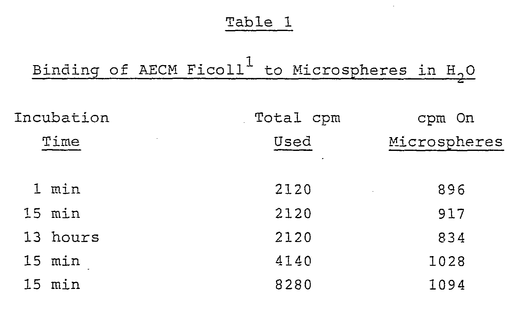
The data in Table 1 show that saturation of the microspheres with AECM Ficoll occurs rapidly and provides microspheres which exhibit 1094 cpm.
The effect of pH on the quantity of AECM Ficoll bound to microspheres was then determined.
The microspheres were treated as were those in Table 1 but the pH of the solutions used for binding and washing were as specified in Table 2.

2) pH of distilled H2O adjusted with NaOH or HCl.
Significantly less AECM-Ficoll is bound at lower pH values, e.g., below about 6, than at higher pH values. Thus, a method is provided of controlling the amount of AECM-Ficoll attached to the microspheres.
The data in Table 2 imply that salt formation (ionic binding) , probably between the amino groups on the AECM Ficoll and the carboxyl groups on the microspheres, is responsible for the binding observed. This ionic binding will be at a maximum at the equivalence point which for a weak acid and a weak base is given by: pH = ½ log Kw - ½ log Ka + ½ Kb. The values of Ka and Kb are not known for the substances used, however, one can get an estimate from similar compounds, viz , Ka = 4.73 for acetic acid, Kb = 3.25 for ethylamine. The
equivalence point thus calculated is pH 7.74 in good agreement with the maximum binding observed at pH 7.9.
The effect of salts on the quantity of AECM-Ficoll bound to microspheres was then determined. The microspheres were treated as were those in Table 1 but the solutions used for binding and washing were as specified in Table 3.

1) 2120 cpm used 2) NS = normal saline
3) PBS = phosphate buffered saline, pH 7.3
The data in Table 3 show that the AECM Ficoll is not only prevented from binding but is easily removed from the microspheres by washing with dilute ionic solutions. When the microspheres of Table 3 were incubated with non-radioactive AECM Ficoll only about 50 cpm of the AECM Ficoll was not exchangeable. Sample 6 in Table 3 illustrates that
a portion of the AECM Ficoll is either very strongly ionically bound, or, more likely, forms covalent bonds to the microspheres.
Table 4 reports data on covalent binding of AECM-Ficoll using a water soluble carbodiimide activating agent.

The data of Table 4 were obtained by reaction of C AECM Ficoll (2120 cpm) with microspheres in the presence of various amounts of carbodiimide followed by washing 3 times with pH 7.3 PBS. Incubation of the microspheres of Table 4 with non-radioactive AECM Ficoll demonstrated that, except for the case where no EDAC was used, the AECM Ficoll is not exchangeable. Presumably the amino groups of the AECM Ficoll have been covalently coupled through amide linkages to the carboxyl groups on the surface of the microspheres. The reason that the amount of AECM Ficoll couoled is
decreased by the larger amounts of EDAC can be explained by the inhibiting effect (inhibiting of initial ionic binding of the AECM-Ficoll to the microspheres) of the higher ionic strength noted earlier. However, once the AECM Ficoll gets onto the surface in the presence of EDAC it becomes covalently attached since the PBS wash does not remove this AECM Ficoll as it did the ionically bound AECM Ficoll (see Table 3 for comparison). To test the hypothesis of covalent bonding and to determine if microspheres remain activated to effect covalent coupling after the excess EDAC has been washed off with H2O, the experiments summarized in Table 5 were performed.

The protocol consisted of incubating the microspheres with EDAC (an excess) and C 14 AECM
Ficoll simultaneously for 1 hour, then washing with
PBS for samples 1 and 3. Samples 1 and 3 were then
counted. Samples 2 and 4 were incubated for 1 hour with EDAC, washed twice with H2O, resuspended in H2O - with C AECM Ficoll added, incubated for 1 more hour, washed with PBS and then counted. The microsphere samples were negative for exchange with non-radioactive AECM Ficoll, indicating covalent binding. These results show that EDAC activates the carboxylated microspheres for covalent binding in spite of the presence of PBS and that the activation remains after removal of excess EDAC. The PBS appears to inhibit the binding of AECM Ficoll to the microspheres by limiting their approach to one another since it does not interfere with the EDAC activation of the microspheres. The usefulness of cyanuric chloride to couple AECM Ficoll microspheres was also investigated as outlined in Table 6.

The protocol used consisted of reacting 100 microliters of microspheres with either C 14 or non-C 14 AECM Ficoll with or without EDAC in H2O, washing 3 times with H2O (1st wash) , reacting with cyanuric chloride for 30 minutes, washing 3 times with H2O (2nd wash) , reacting with either C 14 or non-C 14 AECM Ficoll for 1 hour, washing 3 times with
H2O (3rd wash), then counting the microspheres. The data (sample 1) indicate that about
250 non-exchangeable cpm of AECM Ficoll can be bound by use of cyanuric chloride on microspheres which already have an ionic coating of AECM Ficoll. However, cyanuric chloride treatment of microspheres which already had an EDAC coupled layer of AECM
Ficoll could not facilitate uptake of additional AECM Ficoll, e.g., the 3rd wash of sample 4, Table 6, contains 98% of the counts added after the cyanuric chloride treatment. The excess counts above 250 on the microspheres for samples 2 and 3 are apparently due to ionic bonding of AECM Ficoll. The mechanism by which cyanuric chloride non-exchangeably couples the AECM Ficoll is presumably by cross-linking the amino groups on some of the adjacent AECM Ficoll molecules. Those AECM Ficoll molecules thereby cross-linked, to the few such molecules which are so strongly bound that they do not wash off in PBS (e.g. , sample 6, Table 3) , are retained on the microsphere surface. The remainder are washed off.
In summary, the maximum of 1094 cpm achieved for ionically bonding AECM Ficoll to 100 microliters of microspheres in H2O corresponds to
7570 molecules/square micron of surface area. This is equal to 1094 cpm/ (1.7 x 10 cpm/molecule) x (8.5 x 10⁹ square micron/100 microliters). This means that an AECM Ficoll molecule occupies 13,210 square Angstroms on the. surface; assuming a cubical shape for the molecule one can calculate 115 Angstroms on an edge and a volume of 1.5 x 10⁶ cubic Angstroms. From the 443,000 average molecular weight, Avogadro's number and the assumed volume, one can calculate a density 0.49 g/cc. Similarly the maximum of 697 cpm covalently bound by EDAC gives 0.25 g/cc. The swelling of polysaccharides upon solution in H2O to several times their dry volume is a well known phenomenon and could account for the observed results. It is likely, however,
that a combination of swelling and flattening out on the surface is responsible for the surface coverage observed .
A surface substantially completed covered with AECM Ficoll cannot be used to practice the present invention since proteins, antibodies and the like cannot be bound to it either with cyanuric chloride or with EDAC as shown by some of the other examples. U.S. Patent No. 4,264,766 teaches a complicated oxidation, coupling, and reduction procedure to attach proteins to a polysaccharide coating, but this is not simple in the hands of the user and not all proteins retain their biological activity after the borohydride reduction which is necessary to practice that invention.
The present invention provides proteins, antibodies, etc., attached to the microsphere surface with most or all of the other area of the surface covered by polysaccharide. The examples allow for coupling the protein before or after the polysaccharide either covalently or non-covalently. The material of the present invention, with a partial layer of AECM Ficoll, has been prepared by a number of methods and storage stability at 4°C for longer than a year is routinely observed. This material is readily coupled to protein by simply mixing it with protein, which is a considerable advantage to the user who wishes to couple his own protein and wants long shelf life. Once coupled to portein, the resulting reagent has a shelf life of greater than six months .
Example V
Comparison of Microspheres With With and Without AECM Ficoll Into each of 20 tubes was placed 400 microliters of Phosphate Buffered Saline (PBS) at pH 7 and 100 microliters of carboxylated polystyrene microspheres (0.5 microns diameter, 0.74% by weight in distilled water) . The tubes were briefly sonicated, then 5 microliters of EDAC at 10 mg/ml was added. The tubes were sonicated again and allowed to incubate at room temperature for 2 hours. After incubation, the microsphere suspensions were washed twice in PBS, by centrifugation and resuspension, resuspended in 500 microliters of PBS and sonicated briefly.
Varying amounts of T-15 antibody at 1 mg/ml in PBS were added to the tubes thus: Tubes 1, 5, 9, 13, 17 received 50 microliters of T-15. Tubes 2, 6, 10, 14, 18 received 25 microliters of T-15. Tubes 3, 7, 11, 15, 19 received 10 microliters of T-15. Tubes 4, 8, 12, 16, 20 received 5 microliters of T-- 15. All the tubes were sonicated briefly and incubated at room temperature for 1 hour.
After incubation, the microsphere suspensions in tubes 1-4 were v/ashed twice in PBS by centrifugation and resuspension and resuspended in 500 microliters of PBS with sonication.
Tubes 5-8 were washed twice in PBS containing 1% bovine serum albumin and 0.1% sodium ozide (protein buffer) and resuspended in 500 microliters of protein buffer with sonication. These were kept for 48 hours at 4°C. The
microspheres in tubes 9-20 were washed twice in distilled water and suspended in 500 microliters of distilled water with sonication.
To the microspheres in tubes 13-20 were added 10 microliters of non-radioactive AECM Ficoll.
Tubes 9-20 were incubated for 1 hour at room temperature and an additional 48 hours at 4°C.
Tubes 9-12 were washed 3 times in PBS by centrifugation and resuspension, and the washes were pooled for each tube. The pooled washes and the washed microspheres were transferred to scintillation vials, 20 ml of scintillation fluid (a standard fluid which fluoresces when exposed to radiation) was added to each vial and they were counted. The following results were obtained:
Tube Microspheres (cpm) Washes (cpm)
9 569 1831
10 512 2038
11 413 1933
12 477 1949
10 microliters C14 AECM Ficoll = 2120 cpm
These results indicate that an essentially constant quantity of radiolabelled AECM Ficoll was bound in spite of the ten-fold reduction in monoclonal antibody used from tube 9 to tube 12.
The highest antibody concentration used (tube 9) was estimated to allow a complete monolayer coverage of the microspheres in the sample. That this did not occur, as implied by the similar quantity of binding of C 14 AECM Ficoll, is indicative of the difficulty
the negatively changed antibodies have in approaching the negatively changed microspheres.
The microspheres in tubes 13-16 were washed twice in PBS and resuspended in 500 microliters of PBS with brief sonication. The microspheres in tubes 17-20 were washed twice in protein buffer, resuspended in 500 microliters of protein buffer and sonicated briefly. Meanwhile 48 wells of a flexible U-bottom PVC (polyvinyl chloride) 96 well microliter plate (Dynatech, Inc.) had been prepared by placing 50 microliters of T-15 at 0.1 mg/ml in PBS in each of the first 16 wells, 50 microliters of phosphocholine - bovine gamma globulin conjugate (PCBGG, the specific binding partner for the T-15 antibody) at 0.1 mg/ml in PBS in each of the next 16 wells, and 50 microliters of protein buffer in each of the last 16 wells and allowing them to incubate for 1 hour at room temperature. The contents of the wells were removed by suction and the wells were washed three times with protein buffer by filling them to the top and aspirating the contents. Then 50 microliters of the prepared microspheres from tubes 1-8 and 13-20 were placed in the wells so that each combination of microspheres was allowed to incubate for 1 hour at room temperature, aspirated off and the wells washed 3 times with protein buffer and then twice with distilled water by aspiration. The wells were allowed to dry and were read by eye using 4, 3, 2, 1, and negative (-1) to designate the relative number of microspheres bound to the wells. The results were as follows:
Microsphere Tube 1 2 8 13 14 15 16 17 18 19 20
Well
Coating
T-15 1 1 1 1 1 1 1 1 -1 -1 -1 - 1 - 1 -1 -1 -1
PCBGG 4 4 4 4 4 4 4 4 4 4 4 4 4 4 4 4 buffer 1 1 1 2 1 1 1 1 -1 -1 -1 -1 -1 -1 -1 -1
The PCBGG is the specific binding partner for the T-15 antibody and any attachment of the microspheres (all T-15 coupled in this example) in the absence of PCBGG indicates non-specific sticking. All of the microspheres exhibited specific binding to the PCBGG plate coat. The microspheres which had AECM Ficoll (tubes 13-20) exhibited no detectable non-specific sticking even when the binding was carried out in the absence of protein in the buffer (tubes 13-16) . The microspheres without AECM Ficoll exhibited non-specific sticking in all cases and particularly in the absence of protein in the buffer (tubes 1-4). This example provides clear evidence that the ratio of specific to non-specific binding is enhanced by coating that part of the microsphere surface which is not coupled to antibody with AECM Ficoll. It also demonstrates that the amount of non-antibody coupled surface can be substantial for carboxylated microspheres even when an excess of antibody and covalent coupling is used.
Example VI
Production of EDAC/AECM Ficoll/CTC Microspheres Green fluorescent 0.72 micron diameter carboxylated polystyrene microspheres prepared generally as previously described were given partial surface coverings of covalently coupled AECM Ficoll in the following manner. A 44.5 ml suspension containing 3.75 grams of microspheres was added to 706 ml H2O and then 39 ml of water containing 0.41 grams of AECM Ficoll was added. The suspension was mixed thoroughly and then 75 mg EDAC dissolved in 30 ml H2O was added with swirling and sonication. The pH at this point was less than 5. The suspension was allowed to sit at room temperature for two days then washed 3 times in distilled water. After resuspension in 1 liter of water with sonication the microspheres were filtered through Whatman #1 paper with suction to remove any clumps and centrifuged once more and the supernatant discarded. The microspheres were then resuspended in 186 ml of cold water and sonicated until a single microsphere suspension was obtained. To this suspension v/as added 0.36 grams cyanuric chloride dissolved in 36 ml ETOH and 76 ml cold water. The suspension was sonicated and kept in an ice bath for 30 minutes followed by centrifugation at 4°C. The supernatant was discarded and the microspheres were washed with cold water 3 times by centrifugation. The microspheres were resuspended with sonication in 320 ml H2O and contained 1.07% solids by weight.
Incubation of 10 microliters of these microspheres with 5 microliters of I 125 T-15 antibody (containing
17,148 cpm) in 200 microliters of normal saline for 75 minutes followed by washing 3 times with protein buffer by centrifugation gave 2110 cpm bound to the microspheres.
Example VII
Labelling Tissue Culured Cells
Coupling Antibody to Microspheres:
A portion of the microspheres of Example VI were coupled to My-1, a mouse monoclonal antibody IgM (kappa) , by incubating for 75 minutes 0.1 mg My-1 with 10 ml of the microspheres which had been suspended by sonication in 100 ml normal saline at room temperature. The microspheres were then pelleted at 10,000 x g at 4°C for 10 minutes in a refrigerated centrifuge and the supernatant discarded. The pellet was resuspended with sonication in 100 ml RPMI 1640 cell culture medium containing 10% newborn calf serum, pelleted as before and the supernatant discarded. This operation was repeated once more, then the pellet was resuspended in 20 ml of RPMI 1640 cell culture medium with 10% newborn calf serum, sonicated and stored aseptically at 4°C. These My-1 coupled microsphere have been kept for longer than six months with full retention of activity.
Specific Binding of Antibody Coupled Microspheres To Cells:
Cell suspensions (10 ml) of both HL-60 culture cells and DAUDI human white tissue culture
cells in flasks were layered over 3 ml Histopaque -1077 cell layering medium (trademark of Sigma Chemical Company) in separate 15 ml centrifuge tubes and centrifuged at 850 x g for 30 minutes at room temperature. The supernatants were discarded and the interface layers containing the cells were washed twice with RPMI 1640 cell culture medium containing 10% foetal calf serum (FCS) . The HL-60 and DAUDI cells were resuspended at a concentration of 1 x 10⁶ cells/ml in HBSS (Hank's Buffered Salt Solution) containing 0.1% BSA (bovine serum albumin) and 0.1% sodium azide in separate tubes. Aliquots of 0.5 ml of both cell suspensions were placed in separate 2 ml wells of a 24 well tissue culture plate. Twenty microliters of the My-1 antibody reagent prepared above was added to both cell suspensions and mixed gently. The plate was centrifuged at 150 x g for 9 minutes at 4°C, then incubated for 1 hour at 4°C. The suspensions were mixed and layered over 1 ml FCS in separate 12 x 75 mm glass tubes and centrifuged at 250 x g for 10 minutes at room temperature. All free microspheres remained in the supernatant above the FCS and were discarded. The cell pellets were then gently resuspended in 0.1 ml RPMI 1640. Twenty microliters of both cell suspensions were examined by fluorescent microscopy for My-1 antibody microsphere attachment to cells.
Results :
% Positive Cell Type (at least 5 microspheres/cell) HL-60 72 DAUDI 3.8
The My-1 antibody recognizes a cell surface marker on the surface of human granulocytes and HL-60 cells. This marker is absent on DAUDI and other human white blood cells. Similar results were obtained on human white blood cells where the cells labelled were granulocytes.
Example VIII
Comparison of Microspheres Coupled to AECM Ficoll by Two Methods Aliquots from 3 different batches of carboxylated polystyrene microspheres were taken and processed in two different ways to give them two differently reacting surfaces. They were then used in an experiment to show the different surface qualities.
Microspheres: A) 0.99 micron diameter containing fluorescent blue dye B) 0.54 micron diameter containing fluorescent blue dye C) 1.01 micron diameter, undyed
All the microspheres were at pH 7 in distilled water.
Treatment I:
Covalently Coupled Using Carbodiimide;
Surface Fully Covered
Volumes containing 1.25 grams of microspheres A, B, & C were put into three beakers labelled, respectively, A1 , B1, C1, each containing 220 ml of distilled water, and were sonicated to disperse the microspheres. In turn, first 15 ml of AECM Ficoll at 0.925% by weight (an excess over the quantity needed to completely cover the surfaces of the microspheres) and then 0.025 grams EDAC (carbodiimide) in 10 ml ETOH , were added to each beaker. The suspensions was sonicated well after each addition; the suspensions were allowed to incubate at room temperature for 12 hours. The microspheres were washed three times in distilled water, by centrifugation and resuspension with sonication and were resuspended in a volume of 200 ml of distilled water. The suspensions were then filtered through a Whatman #1 filter paper. The microspheres suspensions A1 , B1 and C1 were brought to 160 ml, 190 ml and 90 ml respectively with distilled water and sonicated well. Aliquots from the treated microspheres A1 , B1 and C1 were then tested for their percentage by weight and the necessary adjustments made to the remaining microspheres. Thus A1 adjusted to 1.47%, B1 to 0.80% and C1 to 1.5%; the total volumes being A 165 ml, B 200 ml, C 100 ml.
Treatment II
Ionically Coupled Followed By Cyanuric Halide Cross-Linking
Volumes of A, B and C containing 2.94 grams, 1.6 grams and 2.96 grams of microspheres, respectively, were put into beakers labelled, respectively, A2 , B2 and C2, each containing 90 ml of distilled water. 10 ml of AECM Ficoll at 0.685% by weight was added to each beaker and dispersed by thorough sonication. After about 15 minutes incubation at room temperature the suspensions were washed twice in distilled water by centrifugation and resuspension with sonication and resuspended in 125 ml distilled water, sonicated and filtered through Whatman #1 filter paper. The microsphere suspensions were each brought to 190 ml with distilled water. Aliquots were taken and the volumes adjusted as in Treatment I to A2 197 ml at 1.47%, B2 199 ml at 0.80% and C2 193 ml at 1.5% by weight.
The microsphere suspensions were each brought to a volume of 70 ml in distilled water and sonicated well. 0.13 grams of cyanuric chloride dissolved in 10 ml of ETOH and diluted in 20 ml of distilled water was added to each of the three suspensions and dispersed by sonicating well. After ½ hour incubation the suspensions were washed 3 times in distilled water, by centrifugation and resuspension with sonication, were resuspened in 190 ml distilled water and sonicated well. Aliquots were taken and the volumes and % solids were
adjusted (see above in Treatment I) to A 197 ml at 1.47%, B 199 ml at 0.80% and C 193 ml at 1.5%.
Experiment
Six samples containing the microsphere suspensions A1 , B1, C1 (Treatment I) and A2 , B2 and C2 (Treatment II) were prepared as just described. To each of six tubes 500 microliters of phosphate buffered saline (PBS) was added, then 50 microliters of one of the six microsphere suspensions (A1 , B1, C1, A2, B2 and C2) was placed in each tube and the tubes were sonicated briefly. 25 mililiters of T-15 antibody at 4 mg/ml was added to each tube; the tubes were sonicated and incubated at room temperature for 1 hour. The microsphere suspensions were then washed 3 times in PBS containing 1% fetal calf serum plus 0.1% azide (protein buffer) by centrifugation and resuspension, and resuspended by sonication in 500 microliters of protein buffer.
Meanwhile, 18 wells of a flexible U-bottom PVC, 96 well microliter plate (Dynatech, Inc.) had been prepared by placing 50 microliters of T-15 at 0.1 mg/ml in PBS in each of the first 6 wells. 50 microliters of phosphocholine - bovine gamma globulin conjugate (PCBGG) at 0.1 mg/ml in PBS in each of the next 6 wells, and 50 microliters of protein buffer in each of the next 6 wells. These plate coats were incubated at room temperature for 1 hour then the contents of the wells were removed by suction and the wells were washed 3 times with protein buffer by filling the wells to the top and aspirating the contents. Then 50 microliters from the prepared microspheres A1, B1, C1, A2 , B2, C2
were placed in the appropriate wells so that each one was reacted with wells treated with T-15, PCBGG and protein buffer. After an incubation of 1 hour the contents were aspirated from the wells and the wells were washed 3 times in protein buffer and twice in distilled water. The plate was allowed to dry and read by eye using 4, 3, 2, 1 and negative (-1) to designate the number of spheres bound to the wells .
Tube # A1 B1 C1 A2 B2 C2
Well
Coating
T-15 -1 -1 -1 -1 -1 -1
PCBGG -1 -1 -1 4 3 4
Protein -1 -1 -1 -1 -1 -1
Buffer
The above described experiments demonstrate that the microspheres prepared in accordance with Treatment II (partially AECM Ficoll covered) showed specific binding to PCBGG, their specific binding partner, whereas the microspheres prepared in accordance with Treatment I (wherein the surface was completely covered with AECM Ficoll) did not bind to PCBGG or elsewhere. Thus, completely AECM Ficoll covered microspheres do not have the capability of binding to biologically active materials.
Example IX
Comparison of EADC/AECM Ficoll Treated Microspheres With Aliquots of These Spheres When Further Reacted With Cyanuric Chloride
Carboxylated polystyrene microsphere suspensions were prepared in bottles of disti lled water with the bottles labelled D1, E1 and F1, as follows :
D1 1.3 micron diameter at 1.9% by weight containing fluorescent green dye E1 0.98 micron diameter at 1.5% by weight containing fluorescent red dye F1 0.7 micron diameter at 1.04% by weight containing fluorescent red dye
These three batches of microsphere had been processed by Treatment I as in Example VIII to completely cover the microspheres surfaces with AECM Ficoll.
0.625 gram samples of microspheres D1, E1 and F1 were individually suspended in 35 ml of distilled water with sonication in respective 250 ml Erlenmeyer flasks labelled, respectively, D2, E2 and F2. The suspensions were cooled on ice and 0.065 grams cyanuric chloride in 5 ml ETOH, diluted with 10 ml of cold distilled water, was added to each. The suspensions were sonicated and incubated on ice for d30 minutes. They were washed 3 times in cold
distilled water, by centrifugation and resuspension with sonication, and were resuspended in distilled water (30 ml (D2) , 30 ml (E2) and 50 ml (F2)) and were adjusted to their correct volume and percent solids by weight which were: D2 40 ml at 1.9%, E2 40 ml at 1.5%, F2 100 ml at 1.04%.
50 microliters each of aliquots from bottles D1, E1 and F1 were individually washed once in phosphate buffered saline (PBS) and individually resuspended in 500 microliters of PBS with sonication. These were labelled, respectively, D3, E3 and F3. 50 microliters of each of samples from flasks D2, E2 and F2 were individually placed in tubes labelled, respectively, D4, E4 and F4, each containing 500 microliters of PBS. The tubes were sonicated and 25 microliters of goat anti mouse at 1 mg/ml in PBS was added to each tube. 50 microliters of each of samples from flasks D2, E2 and F2 were individually placed in tubes labelled, respectively, D5, E5 and F5 , each containing 500 microliters of PBS. The tubes were sonicated and 25 microliters of UPC 10 (a mouse myelona protein) at 1 mg/ml in PBS was added to tubes D4, E4 and F4. The tubes D4, E4, F4, D5, E5 and F5 were incubated for 1 hour at room temperature. The microspheres in these tubes were then washed 3 times in PBS containing 1% fetal calf serum and 0.1% sodium azide (protein buffer) by centrifugation and resuspension. They were each resuspended in 500 microliters of protein buffer with sonication.
Meanwhile, 27 wells of a flexible U-bottom PVC 96 well microliter plate (Dynatech , Inc.) had been prepared by placing 50 microliters of goat and
mouse antibody at 0.1 mg/ml in PBS in each of the first 9 wells, 50 microliters of UPC 10 at 0.1 mg/ml in each of the next 9 wells and 50 microliters of protein buffer in each of the last 9 wells. These wells were incubated for 1 hour at room temperature, the liquids were aspirated from the wells , and the wells were washed 3 times in protein buffer by filling the wells to the top and aspirating the contents.
Next, 50 microliters each of aliquots from respective tubes D3 , D4, D5, E3 , E4 , E5 , F3 , F4, F5 were placed in the appropriate wells of the coated microliter plate and incubated for 1 hour at room temperature. They were then washed 3 times in protein buffer, twice in distilled water and allowed to dry. All showed negative (-1) binding:
Tube # D3 E3 F3 D4 E4 F4 D5 E5 F5
Well Coating
Goat Anti
Mouse -1 -1 -1 -1 -1 -1 -1 -1 -1
UPC 10 -1 -1 -1 -1 -1 -1 -1 -1 -1
Protein Buffer -1 -1 -1 -1 -1 -1 -1 -1 -1
The microspheres in D1, E1 and F1 in PBS did not bind to the protein coats on the plate even after attempted cyanuric chloride treatment thus demonstrating their inability to bind to protein after polysaccharide coating as in Treatment I of Example VIII.
Example X
An Experiment Using Carboxylated Polystyrene 0.7 Micrron Diameter Microspheres Containing Red Dye in Distilled Water at 1.04% by Weight, the Microspheres Having Been Treated With EADC/AECM Ficoll As In Treatment I of Example VIII
Varying concentrations of EADC v/ere compared to find if there was an optimum concentration for coupling protein to these microspheres. The T-15 antibody was at three different concentrations. 50 microliters of microspheres were placed in each of tubes labelled nos. 1, 3, 5, 7, 9 each containing 500 microliters of phosphate buffered saline. The tubes were sonicated to disperse the microspheres. 0.05 mg T-15 antibody and 0.05 mg EDAC was added to tube 1, 0.05 mg T-15 antibody and 0.005 mg EDAC to tube 3, 0.005 mg T-15 and 0.05 mg EDAC to tube 5, 0.005 mg T-15 and 0.005 mg EDAC to tube 7, and 0.05 mg T-15 and no EDAC to tube 9. Each tube was sonicated briefly after addition of protein and after addition of EDAC. The microsphere suspensions were incubated at room temperature for 1 hour, were wached 3 times in PBS and 1% fetal calf serum and .1% sodium azide (protein buffer) by centrifugation and resuspension and resuspended in 500 microliters of protein buffer with sonication.
Meanwhile, 15 wells of a flexible, U-bottom, PVC, 96 well microliter plate (Dynatech, Inc.) had been prepared by placing 50 microliters of T-15 antibody at 0.1 mg/ml in PBS in each of the
first 5 wells, 50 microliters of phosphocholine bovine gamma globulin (PCBGG) at 0.1 mg/ml in PBS in each of the second 5 wells, and 50 microliters of protein buffer in each of the last 5 wells. After 1 hours incubation at room temperature the contents of the wells were removed by suction and the wells washed 3 times in protein buffer by filling the wells to the top and aspirating the contents. Next, 50 microliters from the T-15/microspheres 1, 3, 5, 7 and 9 were placed in the appropriate wells and incubated for 1 hour at room temperature. The contents of the wells were removed by suction and the wells washed 3 times in protein buffer and twice in distilled water, by filling and aspirating. The plate was allowed to dry and then read by eye using 4, 3, 2, 1 and negative (-1) to designate the number of spheres bound to the well walls.
Tube # 1 3 5 7 9
Well Coating
T-15 -1 -1 -1 -1 -1
PCBGG 1 1 1 1 -1
Protein Buffer -1 -1 -1 -1 -1
The small number of microspheres in wells 1, 3, 5 and 7 with the PCBGG coat show that some specific binding has occurred but not sufficient for reliable results.
Example XI
Assay Kit for Soluble Antigens
In this example a flexible, U-bottom polyvinyl chloride, 96 well microliter plate (Dynatech, Inc.) was used. Clear polyvinyl chloride strips have also been substituted for the wells of the microliter plate, the strips being placed in the reagents instead bf the reagents being placed in the wells. Microspheres 0.7 micron in diameter were prepared containing fluorescent green dye and were treated with AECM Ficoll/cyanuric chloride to provide a partial coating as in Treatment II, Example VIII. 100 microliters of the microspheres were placed in a tube containing 1 ml PBS and sonicated. 50 microliters of T-15 antibody at 4 mg/ml in PBS was added and mixed by sonication. After 1 hours incubation at room temperature this microsphere suspension was washed three times in protein buffer, by centrifugation and resuspension with sonication. The microspheres were resuspended in 1 ml of protein buffer and sonicated well.
10 fold dilutions of PCBGG in protein buffer were prepared in separate tubes designated 1, 2, 3, 4 and 5; the concentration of tube 1 being 0.1 mg/ml; tube 2 0.01 mg/ml; etc.
Meanwhile, a flexible U-bottomed PVC 96 well microliter plate was prepared: the first 6 wells received 50 mililiter each of T-15 antibody at 0.1 mg/ml in phosphate buffered saline. Well 7 received 50 microliters of phosphocholine bovine
gamma globulin (PCBGG) at 0.1 mg/ml in PBS. Well 8 received 50 microliters of PBS containing 1% bovine serum albumin and 0.1% sodium azide (protein buffer). The plate coats were incubated for 1 hour at room temperature; the well contents were aspirated and the wells washed 3 times in protein buffer by filling to the top and aspirating.
50 microliters from tubes 1-5 were placed in wells 1-5 and 50 microliters of protein buffer in wells 6-8. After 1 hour incubation at room temperature the contents of the wells were aspirated and the wells washed 3 times in protein buffer by filling and aspirating.
50 microliters of the T-15 antibody coupled microspheres were placed in wells 1-8. The plate was incubated for 1 hour at room temperature, the wells were aspirated and washed three times in protein buffer and twice in distilled water. The amount of sticking of microspheres was recorded on the 4, 3, 2, 1 and negative (-1) scale previously described and the results were as follows:
Tube # 1 2 3 4 5 6 7 8
Well Coating
T-15 + PCBGG dilutions 4 3 2 -1 -1 -1 4 -1 PCBGG in mg/ml .1 .01 .001 .0001 .00001
This experiment demonstrated that a T-15 plate coat will bind a layer of PCBGG to it and that
microspheres with T-15 antibodies on them will then stick to the phosphocholine moieties which are not bound and which form a portion of the plate coat.
Example XII
Assay Kit for Pregnancy Test
A clear polyvinyl chloride strip is incubated with antibody #1, then coated with gelatin or bovine serum albumin. It is then placed in the suspect pregnancy urine and incubated for between one and sixty minutes, after which the strip is incubated in microspheres which are coupled to antibody #2. These microspheres are carboxylated polystyrene which have been activated with carbodiimide, washed, reacted with antibody #2, washed, and reacted with AECM Ficoll. Antibody #1 and antibody #2 are antibodies which have the property of being able to simultaneously react with human chorionic gonadotrophin or with the beta sub unit of human chorionic gonadotrophin, i.e., a given molecule of human chorionic gonadotrophin or beta sub unit of human chorionic gonadotrophin can have both antibody #1 and antibody #2 attached to it simultaneously. After the strip has been incubated in the microspheres for between one and sixty minutes, it is removed and rinsed. A positive test is indicated by the plastic strip having a cloudy appearance caused by adherence of the microspheres. This assay is also applicable for chorionic gonadotrophin in the urine of species other than humans.
Industrial Applicability
In accordance with one embodiment of the subject invention, novel compositions are provided comprising water insoluble surfaces partially coated with water soluble polysaccharides, normally amino functionalized, and partially attached to molecules of a biological substance. Clean accurate and highly selective separation is obtainable between molecules which are binding partners to the biological substance and binding partners which are not binding partners to the biological substance.
Although the foregoing invention has been described in some detail by way of illustration and example for the purposes of clarity of understanding, it should be recognized that certain changes and modifications may practiced within the scope of the appended claims.
Claims
1. A composition useful for specifically binding to a specific binding protein which is a specific binding partner to a biological substance when said protein is associated with other proteins, comprising: a water insoluble support having a surface having the capability of associating with said specific binding protein and with said other proteins; and a polysaccharide coating covering a first substantial surface portion of said surface sufficiently to substantially prevent binding of proteins to said first substantial surface portion and not covering a second substantial surface portion of said surface.
2. A composition as set forth in claim 1, wherein said support comprises a latex.
3. A composition as set forth in claim 1, further including: said biological substance attached to substantially said entire second substantial surface portion.
4. A composition as set forth in claim 3 , wherein said biological substance is covalently bonded to said second substantial surface portion.
5. A composition as set forth in claim 3, wherein said biological substance is hydrophobically adsorbed on said second substantial surface portion.
6. A composition as set forth in claim 1, wherein said polysaccharide coating comprises an amino polysaccharide coating and is cross-linked via cyanuric halide moieties.
7. A composition as set forth in claim 1, wherein said support includes carboxyl functions and said polysaccharide is covalently bonded to said first substantial surface portion via said carboxyl functions.
8. A composition as set forth in claim
7, wherein said covalent bonding is accomplished through use of a carbodiimide activator.
9. A composition as set forth in claim
8, wherein said biological substance is covalently bonded to said second substantial surface portion.
10. A composition as set forth in claim 1, wherein said polysaccharide coating is electrostatically held to said first substantial surface portion.
11. A composition as set forth in claim 1, wherein said support is a particle and said composition is labelled with a label capable of providing a detectable signal.
12. A composition as set forth in claim 1, wherein said support is a macroextensive surface.
13. A composition as set forth in claim 12, wherein said macroextensive surface includes a well.
14. A composition as set forth in claim
1, wherein said polysaccharide coating is in an amount ot from about 0.5 x 10⁻⁷ to about 3 x 10⁻⁷ gms/cm2 of said surface.
15. A method of preparing a water insoluble surface of a solid support for specifically binding to a specific binding protein which is a specific binding partner to a biological substance when said protein is associated with other proteins, comprising: providing a solid support having a water insoluble surface capable of associating with said specific binding protein and with other proteins; and covering a first substantial portion of said surface with a polysaccharide coating while not covering a second substantial portion of said surface with said polysaccharide coating.
16. A method as set forth in claim 15, wherein said covering comprises the steps of: nitrating said surface to add nitro groups ; reducing the nitro groups to amino groups ; attaching cyanuric halide moieties to said amino groups; and electrostatically attaching amino polysaccharide molecules to said first substantial surface portion.
17. A method as set forth in claim 16, further including: contacting a cyanuric halide with said electrostatically attached amino polysaccharide molecules to cross-link adjacent amino polysaccharide molecules.
18. A method as set forth in claim 17, wherein the amount of said amino polysaccharide attached is between about 0.5 x 10 -7 and about 3 x 10 -7 grams per square centimeter of the area of said surface.
19. A method as set forth in claim 16, wherein the amount of said amino polysaccharide attached is between about
0.5 x 10 -7 and about 3 x 10⁻⁷ grams per square centimeter of the area of said surface.
20. A method as set forth in claim 16, further including, as an added step: attaching said biological substance to said second substantial surface portion.
21. A method as set forth in claim 20, wherein said biological substance attaching comprises hydrophobically adsorbing said biological substance on said second substantial surface portion.
22. A method as set forth in claim 20, wherein said biological substance attaching comprises covalently bonding said biological substance to said second substantial surface portion.
23. A method as set forth in claim 15, wherein said covering comprises the steps of: contacting said surface with an amino polysaccharide in an amount more than sufficient to cover said surface with a monomolecular layer of said amino polysaccharide; washing excess amino polysaccharide from said surface with a substantially neutral aqueous solution having little or no salt content while leaving behind a portion of said amino polysaccharide electrostatically bound to said surface ; and cross-linking a portion of said left behind portion of said amino polysaccharide.
24. A method as set forth in claim 23, wherein said cross-linking comprises reacting a cyanuric halide compound with said portion of said left behind portion of said amino polysaccharide and concurrently exposing said second substantial surface portion.
25. A method as set forth in claim 24, wherein said cyanuric halide compound comprises the reaction product of said cyanuric halide with ethanol.
26. A method as set forth in claim 23, wherein the amount of said amino polysaccharide cross-linked is between about 0.5 x 10 -7 and about 3 x 10 -7 grams per centimeter of the area of said surface.
27. A method as set forth in claim 23, further including, as an added step: attaching said biological substance to said second substantial surface portion.
28. A method as set forth in claim 27, wherein said biological substance attaching comprises hydrophobically adsorbing said biological substance on said second substantial surface portion.
29. A method as set forth in claim 27, wherein said biological substance attaching comprises covalently bonding said biological substance to said second substantial surface portion.
30. A method as set forth in claim 15, wherein said surface includes active groups capable of reaction with a water soluble activator and wherein said covering comprises the step of: contacting, in an aqueous solution, said surface, an amino polysaccharide compound, and said water soluble activator.
31. A method as set forth in claim 30, wherein said aqueous solution is substantially neutral and substantially salt free and wherein the amount of said amino polysaccharide is insufficient to fully cover said surface.
32. A method as set forth in claim 30, wherein said activator comprises a water soluble carbodiimide.
33. A. method as set forth in claim 30, wherein the amount of said amino polysaccharide attached is between about0.5 x 10 -7 and about 3 x 10 -7 grams per square centimeter of the area of said surface.
34. A method as set forth in claim 30, wherein said amino polysaccharide compound is covalently bonded by said contacting step to said active groups on said first surface portion.
35. A method as set forth in claim 30, further including, as an added step: attaching said biological substance to said second substantial surface portion,
36. A method as set forth in claim 35, wherein said biological substance attaching comprises covalently bonding said biological substance to said second substantial surface portion via reaction of said biological substance with activator adducts to the active groups on said second substantial surface portion.
37. A method as set forth in claim 36, further including, prior to said biological substance bonding, washing off any excess activator.
38. A method as set forth in claim 15, wherein said surface includes active groups capable of reaction with an activator compound and wherein said covering comprises the steps of: reacting said surface with an excess of said activator compound; washing off any excess activator compound; adding said biologial substance in an amount insufficient to react with all of the activator adducts to the active groups on said surface to thereby covalently bond said biological substance to said second substantial surface portion; washing said surface; contacting said surface, in the presence of water, with an amino polysaccharide compound to covalently bond said amino polysaccharide compound, via reaction of amino groups thereof, to said first substantial surface portion.
39. A method as set forth in claim 38, wherein said activator comprises a water soluble carbodiimide.
40. A method as set forth in claim 15, wherein said surface includes active groups capable of reaction with an activator and wherein said covering comprises the steps of: attaching an amino polysaccharide to said first substantial surface portion; contacting said surface with a water soluble activator to form adducts with said active groups on said second substantial surface portion; and reacting said adducts with said biological substance to covalently bond said biological substance via said active groups to said second substantial surface portion.
41. A method as set forth in claim 40, wherein said activator comprises a carbodiimide activator.
42. A method as set forth in claim 15, wherein said support comprises a plurality of water suspendable particles.
43. A method as set forth in claim 39, wherein said support is labelled with a label capable of providing a detectable signal.
44. A method as set forth in claim 43, wherein said label comprises a color imparting entity and said signal comprises said color.
45. A method as set forth in claim 41, wherein said support comprises a macroextensive support having a macroextensive surface.
46. A method as set forth in claim 45, wherein said macroextensive support includes a well defining said macroextensive surface.
47. A process for assaying an aqueous sample containing a specifically binding protein having a first binding site which is a specific binding partner to a first biological substance, said specifically binding protein being in association with other proteins, with increased specificity and sensitivity, comprising: contacting a first solid support having a first water insoluble surface capable of associating with said specific binding protein and with other proteins, said first surface having a first substantial portion thereof shielded by a polysaccharide coating and a second substantial portion thereof having said first biological substance attached thereto, with said aqueous sample; and detecting the amount of said specifically binding protein bound to said attached first biological substance.
48. A process as set forth in claim 47, including, prior to said detecting: separating said aqueous sample from said first solid support.
49. A process as set forth in claim 47, wherein said support comprises a plurality of particles.
50. A process as set forth in claim 49, wherein said particles are labelled with a label capable of detection.
51. A process as set forth in claim 47, wherein said support comprises a macroextensive support having a macroextensive surface.
52. A process as set forth in claim 51, wherein said macroextensive support includes a well defining said macroextensive surface.
53. A process as set forth in claim 47, wherein said polysaccharide is an amino polysaccharide and wherein said biological substance is attached to said second substantial surface portion via carboxyl groups.
54. A process as set forth in claim 51, wherein said detecting step comprises: contacting with said macroextensive surface an aqueous solution having a second solid support comprising a plurality of particles, said second solid support having a second water insoluble surface capable of associating with said specific binding protein and with other proteins, said second surface having an additional first substantial portion shielded by a polysaccharide coating and an additional second substantial surface portion having a second biological substance attached thereto, said specifically binding protein having a second binding site which is a specific partner to said second biological substance; and observing the degree of adherence of said particles to said macroextensive surface member.
55. A process as set forth in claim 54, wherein said particles are labelled with a label capable of detection.
56. A process for reducing adherence of undesired proteins to a water insoluble surface consisting essentially of a first substantial surface portion and a second substantial surface portion while providing said surface with the capability for binding to a specifically binding protein which is a specific binding partner to a biological substance, comprising: shielding said first substantial surface portion with a polysaccharide coating; and attaching said biological substance to said second substantial surface portion.
57. A process as set forth in claim 56, wherein said shielding step precedes said attaching step.
58. A process as set forth in claim 56, wherein said shielding and attaching steps are substantially simultaneous.
59. A process as set forth in claim 56, v/herein said attaching step precedes said shielding step.
60. A process as set forth in claim 56, wherein said surface is on a macroextensive support.
61. A kit for assaying samples potentially containing a specifically binding protein having a first binding site which is a specific binding partner to a first biological substance and a second binding site which is a specific binding partner to a second biological substance, said specifically binding protein being in association with other porteins, with increased specificity and sensitivity, comprising: a solid macroextensive support having a first water insoluble surface capable of associating with said specific binding portein and with other proteins, said first surface consisting essentially of a first substantial surface portion shielded by a polysaccharide coating and a second substantial surface portion having said first biological substance attached thereto; and a plurality of solid particles, each having a second insoluble surface capable of associating with said specific binding protein and with other proteins, said second surfaces each consisting essentially of a first substantial surface portion shielded by a polysaccharide coating and a second substantial surface portion having said second biological substance attached thereto.
62. A kit as set forth in claim 61, wherein said particles are labelled with a label capable of providing a detectable signal.
63. A kit as set forth in claim 62, wherein said label comprises a color imparting entity and said signal comprises said color.
64. A kit for assaying samples potentially containing a specifically binding protein having a first binding site which is a specific binding partner to a first biological substance and a second binding site which is a specific binding partner to a second biological substance, said specifically binding protein being in association with other proteins, with increased specificity and sensitivity, comprising: a macroextensive solid support having a first water insoluble surface capable of associating with said specific binding protein and with other proteins, said first surface consisting essentially of a first substantial surface portion shielded to prevent association with said specific binding protein and with other proteins and a second substantial surface portion having said first bio-logical substance attached thereto; and a plurality of solid particles, each having a second insoluble surface capable of associating with said specific binding protein and with other proteins, said second surfaces each consisting essentially of a first substantial surface portion shielded by a polysaccharide coating and a second substantial surface portion hving said second bioloαical substance attached thereto.
65. A kit as set forth in claim 64 , wherein said particles are labelled with a label capable of providing a detectable signal.
66 . A kit as set forth in claim 65, wherein said label comprises a color imparting entity and said signal comprises said color.
Priority Applications (1)
| Application Number | Priority Date | Filing Date | Title |
|---|---|---|---|
| DK510084A DK510084D0 (en) | 1983-02-25 | 1984-10-25 | NON-SOLUBLE SURFACES TREATED FOR NON-SPECIFIC PROTEIN BINDING |
Applications Claiming Priority (1)
| Application Number | Priority Date | Filing Date | Title |
|---|---|---|---|
| US46993983A | 1983-02-25 | 1983-02-25 |
Publications (1)
| Publication Number | Publication Date |
|---|---|
| WO1984003358A1 true WO1984003358A1 (en) | 1984-08-30 |
Family
ID=23865635
Family Applications (1)
| Application Number | Title | Priority Date | Filing Date |
|---|---|---|---|
| PCT/US1984/000257 WO1984003358A1 (en) | 1983-02-25 | 1984-02-22 | Insoluble surfaces treated to inhibit non-specific protein binding |
Country Status (5)
| Country | Link |
|---|---|
| EP (1) | EP0136347A1 (en) |
| JP (1) | JPS60500973A (en) |
| DK (1) | DK510084D0 (en) |
| IT (1) | IT8447751A0 (en) |
| WO (1) | WO1984003358A1 (en) |
Cited By (6)
| Publication number | Priority date | Publication date | Assignee | Title |
|---|---|---|---|---|
| EP0170746A1 (en) * | 1984-08-07 | 1986-02-12 | Covalent Technology Corporation | Biologically active material test |
| EP0227173A2 (en) * | 1985-12-23 | 1987-07-01 | Janssen Pharmaceutica N.V. | New method for the detection of specific binding agents and their corresponding bindable substances |
| EP0260188A1 (en) * | 1986-09-12 | 1988-03-16 | I.B.A.L. Société à Responsabilité Limitée dite: | Apparatus for the analysis of a biological liquid by means of immunoadherence and flakes to be used in this apparatus |
| US7163998B2 (en) | 2003-09-09 | 2007-01-16 | Eastman Kodak Company | Stabilized polymer beads and method of preparation |
| US7179660B1 (en) | 2000-03-06 | 2007-02-20 | Dade Behring Marburg Gmbh | Carriers coated with polysaccharides, their preparation and use |
| EP2230515A1 (en) * | 2009-03-16 | 2010-09-22 | Polymer Laboratories Limited | Passivation of surfaces after ligand coupling |
Citations (10)
| Publication number | Priority date | Publication date | Assignee | Title |
|---|---|---|---|---|
| US4059685A (en) * | 1973-03-19 | 1977-11-22 | Summa Corporation | Immobilized immunoadsorbent |
| US4066744A (en) * | 1973-04-04 | 1978-01-03 | Akzona Incorporated | Serological reagent, test slide and method for gonorrheal antibodies |
| US4123509A (en) * | 1974-12-19 | 1978-10-31 | American Home Products Corporation | Pregnancy test |
| US4169138A (en) * | 1974-05-29 | 1979-09-25 | Pharmacia Diagnostics Ab | Method for the detection of antibodies |
| US4244940A (en) * | 1978-09-05 | 1981-01-13 | Bio-Rad Laboratories, Inc. | Single-incubation two-site immunoassay |
| US4264766A (en) * | 1877-09-19 | 1981-04-28 | Hoffmann-La Roche Inc. | Immunological diagnostic reagents |
| US4299916A (en) * | 1979-12-26 | 1981-11-10 | Syva Company | Preferential signal production on a surface in immunoassays |
| US4315908A (en) * | 1978-12-29 | 1982-02-16 | Tamar Zer | Method of determining human chorionic gonadotropin (HCG) in the urine |
| US4320194A (en) * | 1979-07-30 | 1982-03-16 | Gist-Brocades N.V. | Process for the production of carrier particles from yeast cells and for producing diagnostic agents therefrom and test kits containing such agents |
| US4360358A (en) * | 1980-03-07 | 1982-11-23 | Yash Sharma | Immunoassay with solid phase having coating containing blood platelet substitute |
-
1984
- 1984-02-22 WO PCT/US1984/000257 patent/WO1984003358A1/en not_active Application Discontinuation
- 1984-02-22 JP JP59501287A patent/JPS60500973A/en active Pending
- 1984-02-22 EP EP84901260A patent/EP0136347A1/en not_active Withdrawn
- 1984-02-24 IT IT8447751A patent/IT8447751A0/en unknown
- 1984-10-25 DK DK510084A patent/DK510084D0/en not_active Application Discontinuation
Patent Citations (10)
| Publication number | Priority date | Publication date | Assignee | Title |
|---|---|---|---|---|
| US4264766A (en) * | 1877-09-19 | 1981-04-28 | Hoffmann-La Roche Inc. | Immunological diagnostic reagents |
| US4059685A (en) * | 1973-03-19 | 1977-11-22 | Summa Corporation | Immobilized immunoadsorbent |
| US4066744A (en) * | 1973-04-04 | 1978-01-03 | Akzona Incorporated | Serological reagent, test slide and method for gonorrheal antibodies |
| US4169138A (en) * | 1974-05-29 | 1979-09-25 | Pharmacia Diagnostics Ab | Method for the detection of antibodies |
| US4123509A (en) * | 1974-12-19 | 1978-10-31 | American Home Products Corporation | Pregnancy test |
| US4244940A (en) * | 1978-09-05 | 1981-01-13 | Bio-Rad Laboratories, Inc. | Single-incubation two-site immunoassay |
| US4315908A (en) * | 1978-12-29 | 1982-02-16 | Tamar Zer | Method of determining human chorionic gonadotropin (HCG) in the urine |
| US4320194A (en) * | 1979-07-30 | 1982-03-16 | Gist-Brocades N.V. | Process for the production of carrier particles from yeast cells and for producing diagnostic agents therefrom and test kits containing such agents |
| US4299916A (en) * | 1979-12-26 | 1981-11-10 | Syva Company | Preferential signal production on a surface in immunoassays |
| US4360358A (en) * | 1980-03-07 | 1982-11-23 | Yash Sharma | Immunoassay with solid phase having coating containing blood platelet substitute |
Non-Patent Citations (1)
| Title |
|---|
| Scandinavian Journal of Immunology, Supplement No. 7, 8, issued 1978, S. AVRAMAES et al., "Coupling of Enzymes to Antibodies and Antigens" * |
Cited By (10)
| Publication number | Priority date | Publication date | Assignee | Title |
|---|---|---|---|---|
| EP0170746A1 (en) * | 1984-08-07 | 1986-02-12 | Covalent Technology Corporation | Biologically active material test |
| EP0227173A2 (en) * | 1985-12-23 | 1987-07-01 | Janssen Pharmaceutica N.V. | New method for the detection of specific binding agents and their corresponding bindable substances |
| EP0227173A3 (en) * | 1985-12-23 | 1989-08-02 | Janssen Pharmaceutica N.V. | New method for the detection of specific binding agents and their corresponding bindable substances |
| US5498551A (en) * | 1985-12-23 | 1996-03-12 | Janssen Pharmaceutica N.V. | Method for the detection of specific binding agents and their corresponding bindable substances |
| EP0260188A1 (en) * | 1986-09-12 | 1988-03-16 | I.B.A.L. Société à Responsabilité Limitée dite: | Apparatus for the analysis of a biological liquid by means of immunoadherence and flakes to be used in this apparatus |
| FR2603991A1 (en) * | 1986-09-12 | 1988-03-18 | Ibal | DEVICE FOR THE ANALYSIS OF A BIOLOGICAL LIQUID BY IMPLEMENTING IMMUNO-ADHERENCE REACTIONS, AND FLAKES FOR USE IN THIS DEVICE |
| US7179660B1 (en) | 2000-03-06 | 2007-02-20 | Dade Behring Marburg Gmbh | Carriers coated with polysaccharides, their preparation and use |
| US7163998B2 (en) | 2003-09-09 | 2007-01-16 | Eastman Kodak Company | Stabilized polymer beads and method of preparation |
| EP2230515A1 (en) * | 2009-03-16 | 2010-09-22 | Polymer Laboratories Limited | Passivation of surfaces after ligand coupling |
| US9709563B2 (en) | 2009-03-16 | 2017-07-18 | Agilent Technologies, Inc. | Passivation of surfaces after ligand coupling |
Also Published As
| Publication number | Publication date |
|---|---|
| IT8447751A0 (en) | 1984-02-24 |
| EP0136347A1 (en) | 1985-04-10 |
| DK510084A (en) | 1984-10-25 |
| JPS60500973A (en) | 1985-06-27 |
| DK510084D0 (en) | 1984-10-25 |
Similar Documents
| Publication | Publication Date | Title |
|---|---|---|
| US4703018A (en) | High refractive index haloalkyl-functional shell-core polymers and their use in light scattering immunoassays | |
| US4656144A (en) | Immunoparticles and process for preparing same | |
| US4582810A (en) | Immuno-agglutination particle suspensions | |
| CA1226215A (en) | Immunoassay of antigens | |
| US5384073A (en) | Ligand gold bonding | |
| US4870007A (en) | Immobilized biotinylated receptor in test device, kit and method for determining a ligand | |
| US4418152A (en) | Immunological, diagnostic reagents having particulate carriers of glycidyl acrylate polymers | |
| US5879881A (en) | Solid phase system for use in ligand-receptor assays | |
| US4201763A (en) | Solid phase immunofluorescent assay method | |
| US5397695A (en) | Attachment of compounds to polymeric particles using carbamoylonium compounds and a kit containing same | |
| AU724475B2 (en) | Synthetic particles as agglutination reagents | |
| AU648625B2 (en) | Test method and reagent kit therefor | |
| EP0064318B1 (en) | A method and a kit for the assay of antibodies to soluble antigens | |
| AU3939493A (en) | Immunoassays employing generic anti-hapten antibodies and materials for use therein | |
| JP2816414B2 (en) | Submicron particles, preparation and use in immunological diagnostics | |
| CA1300499C (en) | Water-insoluble reagent, elements containing same and methods of use | |
| US5143825A (en) | Stabilized substrate for use in an immunoassay | |
| WO1987004794A1 (en) | Latex agglutination using avidin/biotin system | |
| WO1984003358A1 (en) | Insoluble surfaces treated to inhibit non-specific protein binding | |
| EP0308235B1 (en) | Attachment of compounds to polymeric particles using carbamoylonium compounds | |
| EP0056254A1 (en) | Treatment of insoluble surfaces to inhibit nonspecific protein binding | |
| Hobbs | Solid-phase immunoassay of serum antibodies to peptides covalent antigen binding to adsorbed phenylalanine-lysine copolymers | |
| EP0152254A2 (en) | Chromogenic support immunoassay utilizing labeled complement components | |
| JPS60111158A (en) | Reagent for testing specific connection | |
| Singer | The use of polystyrene latexes in medicine |
Legal Events
| Date | Code | Title | Description |
|---|---|---|---|
| AK | Designated states |
Designated state(s): DK FI JP |
|
| AL | Designated countries for regional patents |
Designated state(s): BE DE FR GB NL SE |
|
| WWE | Wipo information: entry into national phase |
Ref document number: 1984901260 Country of ref document: EP |
|
| WWP | Wipo information: published in national office |
Ref document number: 1984901260 Country of ref document: EP |
|
| WWW | Wipo information: withdrawn in national office |
Ref document number: 1984901260 Country of ref document: EP |