WO1994001105A1 - Method of treating hyperproliferative vascular disease - Google Patents

Method of treating hyperproliferative vascular disease Download PDF

Info

Publication number
WO1994001105A1
WO1994001105A1 PCT/US1993/006410 US9306410W WO9401105A1 WO 1994001105 A1 WO1994001105 A1 WO 1994001105A1 US 9306410 W US9306410 W US 9306410W WO 9401105 A1 WO9401105 A1 WO 9401105A1
Authority
WO
WIPO (PCT)
Prior art keywords
mycophenolic acid
mammal
vascular
vascular disease
smooth muscle
Prior art date
Application number
PCT/US1993/006410
Other languages
French (fr)
Inventor
Clare R. Gregory
Randall E. Morris
Original Assignee
The Board Of Trustees Of The Leland Stanford Junior University
Priority date (The priority date is an assumption and is not a legal conclusion. Google has not performed a legal analysis and makes no representation as to the accuracy of the date listed.)
Filing date
Publication date
Application filed by The Board Of Trustees Of The Leland Stanford Junior University filed Critical The Board Of Trustees Of The Leland Stanford Junior University
Priority to EP93917017A priority Critical patent/EP0746314A4/en
Publication of WO1994001105A1 publication Critical patent/WO1994001105A1/en

Links

Classifications

    • AHUMAN NECESSITIES
    • A61MEDICAL OR VETERINARY SCIENCE; HYGIENE
    • A61KPREPARATIONS FOR MEDICAL, DENTAL OR TOILETRY PURPOSES
    • A61K31/00Medicinal preparations containing organic active ingredients
    • A61K31/33Heterocyclic compounds
    • A61K31/335Heterocyclic compounds having oxygen as the only ring hetero atom, e.g. fungichromin
    • A61K31/365Lactones

Definitions

  • the present invention relates to the use of mycophenolic acid (MPA) for the treatment of hyperproliferative vascular disease in a mammal, including inti al smooth muscle cell hyperplasia, restenosis, and vascular occlusion.
  • MPA mycophenolic acid
  • Partial blockage of the blood vessels leading to the heart is one cause of heart disease. More severe blockage of blood vessels often leads to hypertension, ischemic injury, stroke, or myocardial infarction.
  • Vascular occlusion is typically preceded by vascular stenosis which can be the result of intimal smooth muscle cell hyperplasia.
  • vascular smooth muscle injury and disruption of the integrity of the endothelial lining are underlying cause of intimal smooth muscle cell hyperplasia.
  • Occlusive coronary atherosclerosis remains the major cause of mortality and morbidity in industrialized countries.
  • T-cells and macrophages also migrate into the neotima (Haudenschild; Clowes, 1985; Clowes, 1989; Manderson; Forrester). This cascade of events is not limited to arterial injury, but also occurs following injury to veins and arterioles.
  • the overall disease process can be termed a hyperproliferative vascular disease because of the etiology of the disease process.
  • Vascular injury causing intimal thickening can be broadly categorized as being either biologically or mechanically induced.
  • Atherosclerosis One of the most commonly occurring forms of biologically mediated vascular injury leading to stenosis is Atherosclerosis.
  • Atherosclerotic lesions include massive accumulation of lipid laden "foam cells” derived from monocyte/macrophage and smooth muscle cells. Formation of "foam cell” regions is associated with a breech of endothelial integrity and basal lamina destruction. Triggered by these events, restenosis is produced by a rapid and selective proliferation of vascular smooth muscle cells with increased new basal lamina (extracellular matrix) formation and results in eventual blocking of arterial pathways (Davies) .
  • vascular stenosis can be detected and evaluated using angiographic or sonographic imaging techniques (Evans) and is often treated by percutaneous translu inal coronary angioplasty (balloon catheterization) .
  • Evans angiographic or sonographic imaging techniques
  • the blood flow is reduced in approximately 30-40 percent of treated patients as a result of restenosis caused by a response to mechanical vascular injury suffered during the angioplasty procedure (Pepine; Hardoff) .
  • numerous pharmaceutical agents have been employed clinically, concurrent with or following angioplasty. Most pharmaceutical agents employed in an attempt to prevent or reduce the extent of restenosis have been unsuccessful.
  • lovastatin Sehni; Gellman
  • thromboxane - ⁇ synthetase inhibitors such as DP-1904 (Yabe) ; eicosapentanoic acid (Nye) ; ciprostene (a prostacyclin analog) (Demke; Darius) ; trapidil (a platelet derived growth factor) (Okamoto) ; angiotensin converting enzyme inhibitors (Gott Kunststoff) ; low molecular weight heparin (de Vries) ; and 5-(3'- pyridinylmethyl)benzofuran-2-carboxylate (Gorman, et al.
  • This invention provides a method of preventing or treating hyperproliferative vascular disease in a mammal in need thereof by administering an amount of mycophenolic acid (MPA) effective to inhibit intimal thickening.
  • Administration of the mycophenolic acid can be accomplished by a number of methods including orally, parenterally, intravascularly, intranasally, intra-bronchially, transdermally, rectally, or via a vascular stent impregnated with mycophenolic acid.
  • mycophenolic acid a number of derivatives thereof may be useful in the practice of the present invention including morpholinoethylesters of mycophenolic acid and heterocyclic aminoalkyl esters of mycophenolic acid.
  • the form of the mycophenolic acid may also include a pharmaceutically acceptable salt.
  • mycophenolic acid is useful, alone or in combination with other treatments, in preventing or treating intimal smooth muscle cell hyperplasia, restenosis, and vascular occlusion in a mammal, particularly following either biologically or mechanically mediated vascular injury.
  • Biologically mediated vascular injury includes, but is not limited to, injury attributed to autoimmune disorders; alloimmune related disorders; infectious disorders including endotoxins and herpes viruses, such as cytomegalovirus; metabolic disorders such as atherosclerosis; and vascular injury resulting from hypothermia and irradiation.
  • Mechanically mediated vascular injury includes, but is not limited to, vascular injury caused by catheterization procedures or vascular scraping procedures such as percutaneous transluminal coronary angioplasty; vascular surgery; transplantational surgery; laser treatment; and other invasive procedures which disrupt the integrity of the vascular intima and endothelium.
  • the method of the present invention includes the prophylactic prevention of hyperproliferative vascular disease in a susceptible mammal and/or treatment in order to arrest the development and retard the progression of hyperproliferative vascular disease in a susceptible mammal.
  • Other combinations containing mycophenolic acid that are useful for preventing or treating hyperproliferative vascular disease will be apparent to one skilled in the art. These include, but are not limited to, using mycophenolic acid in combination with other antiproliferative antimetabolites or other drugs useful for the treatment of hyperproliferative diseases.
  • IMP-DH inosine monophosphate dehydrogenase
  • the present invention also includes a composition for the use in preventing or treating hyperproliferative vascular disease in a mammal which comprises an amount of mycophenolic acid effective to inhibit intimal thickening and a pharmaceutically acceptable carrier.
  • the composition can be used as described above.
  • Figure 1 shows a schematic representation of the cross-section of the carotid artery (adapted from Ip, et al . ) .
  • the numbers refer to the following arterial cell layers: 1. Dysfunctional endothelium; 2. Endothelium; 3. Intima; 4. Media; and 5.
  • Figures 2A to 2D show bar graphs presenting the data which demonstrate the effects of a number of drugs on 3 H-uridine and 3 H-thymidine incorporation of rat aortic smooth muscle cells.
  • Figure 3 shows a bar graph presenting the data for intima percent of rat arteries 14 days after balloon catheter injury with a variety of drug treatments.
  • MPA mycophenolic acid
  • bFGF Basic fibroblast growth factor
  • CsA cyclosporine
  • RPM rapamycin
  • MPA mycophenolic acid
  • CsA, FK506, MPA, RPM, and rapamycin plus mycophenolic acid were evaluated in an in vivo standard pharmacological test procedure that emulates the vascular injury suffered and restenosis that develops following percutaneous translu inal coronary angioplasty in humans (Chezier; Fishman; Haudenschild; Clowes, 1983; Clowes, 1989; Ferns, 1991) .
  • One rat in each group served as a balloon-injured, no treatment positive control.
  • mycophenolic acid is useful in preventing or treating intimal smooth muscle cell hyperplasia, restenosis, and vascular occlusion in a mammal, particularly following either biologically or mechanically mediated vascular injury, or under conditions that would predispose a mammal to suffering such a vascular injury.
  • inosine monophosphate dehydrogenase IMP-DH
  • MPA inhibits DNA synthesis in activated immune cells, such as monocytes, relatively selectively: it prevents guanosine synthesis by blocking inosine monophosphate dehydrogenase activity (Eugui, et al . ) .
  • IMP-DH in addition to MPA
  • examples of other such molecules, which inhibit IMP-DH include but are not limited to the following: Mizoribine (bredinin) (Sakaguchi, et al . ) ; and Ribovirin, tiazofurin and selenazarfurin (Lee, et al . ) .
  • Mycophenolic acid is an antibiotic substance which is produced by Penicillium brevi-compactum and related species (The Merk Index. Tenth Edition, 1983). A number of forms of mycophenolic acid have been derived which are useful in methods of treating autoimmune disorders, psoriasis and other inflammatory diseases.
  • mycophenolic acid is used to refer to mycophenolic acid itself and to pharmaceutically active derivatives thereof. A number of derivatives of mycophenolic acid are taught in Ohsugi, et al . , Sweeney, et al . , and U.S. Patent Nos. 4,686,234, 4,727,069, 4,753,935, 4,786,637, all herein incorporated by reference, as well as the pharmaceutically acceptable salts thereof.
  • the treatment methods of the present invention include the delivery of any such derivatives of mycophenolic acid into a mammalian system, in order to inhibit intimal thickening, which, when the derivative is administered to the mammalian host, results in the therapeutic form of the derivative being mycophenolic acid — regardless of the derivative form by which it was originally introduced into the mammalian host.
  • a number of pharmaceutically acceptable salts of these compounds are also available.
  • Such pharmaceutically acceptable salts include any salt derived from bases or acids, where the base or acid is inorganic or organic.
  • Inorganic acids include, but are not limited to, hydrochloric acid, sulfuric acid and phosphoric acid.
  • Organic acids include, but are not limited to, acetic acid, pyruvic acid, succinic acid, oxalic acid and maleic acid.
  • Inorganic bases include, but are not limited to, sodium, lithium, potassium, calcium, magnesium and ammonium.
  • Organic bases include, but are not limited to, primary, secondary and tertiary amines.
  • mycophenolic acid can be tested for use in the method of the present invention as described above, and as described in Examples 1 and 2.
  • mycophenolic acid is administered to a mammal in need of treatment at a therapeutically effective amount of the compound.
  • Mycophenolic acid when employed in the prevention or treatment of hyperproliferative vascular disease can be formulated neat or with a the addition of a pharmaceutical carrier.
  • the pharmaceutical carrier may be solid or liquid. The formulation is then administered in a therapeutically effective dose to a mammal in need thereof.
  • a solid carrier can include one or more substances.
  • the carrier may also act to provide flavoring agents, lubricants, solubilizers, suspending agents, filters, glidants, compression aids, binders or tablet-disintegrating agents.
  • the carrier can also function as an encapsulating material. In powders, the carrier is typically a finely rendered solid which is in a mixture with the finely rendered active ingredient — mycophenolic acid.
  • a carrier with the necessary compression properties is mixed with the mycophenolic acid in suitable proportions. The mixture is then compacted in the shape and size desired.
  • the powders and tablets preferably contain up to 99% of the active ingredient.
  • suitable solid carriers include, but not limited to, the following: sugars, lactose, dextrin, starch, gelatin, calcium phosphate, magnesium stearate, talc, polyvinylpyrrolidone, low melting waxes, ion exchange resins, cellulose, methyl cellulose, and sodium carboxymethyl cellulose.
  • Liquid carriers can be used in the preparation of elixirs, solutions, emulsions, syrups, suspensions and pressurized compositions.
  • the mycophenolic acid is dissolved or suspended in a pharmaceutically acceptable liquid carrier such as water, an organic solvent, a mixture of both, or pharmaceutically accepted oils or fats.
  • a pharmaceutically acceptable liquid carrier such as water, an organic solvent, a mixture of both, or pharmaceutically accepted oils or fats.
  • the liquid carrier can contain other suitable pharmaceutical additives including, but not limited to, the following: solubilizers, flavoring agents, suspending agents, emulsifiers, buffers, thickening agents, colors, viscosity regulators, preservatives, sweeteners, stabilizers and osmolarity regulators.
  • liquid carriers for oral and parenteral administration of mycophenolic acid preparations include water (partially containing additives as above, e.g., cellulose derivatives, preferably sodium carboxymethyl cellulose solution) , alcohols (including monohydric alcohols and polyhydric alcohols, e.g., glycols) and their derivatives, and oils (e.g., fractionated coconut oil and arachis oil) .
  • water partially containing additives as above, e.g., cellulose derivatives, preferably sodium carboxymethyl cellulose solution
  • alcohols including monohydric alcohols and polyhydric alcohols, e.g., glycols
  • oils e.g., fractionated coconut oil and arachis oil
  • the carrier can also be an oily ester such as ethyl oleate and isopropyl myristate.
  • Sterile carriers are useful in sterile liquid form compositions for parenteral administration.
  • Sterile liquid pharmaceutical compositions, solutions or suspensions can be utilized by, for example, intraperitoneal injection, subcutaneous injection, or intravenously.
  • Mycophenolic acid can be also be administered intravascularly or via a vascular stent impregnated with mycophenolic acid, during balloon catheterization to provide localized effects immediately following injury.
  • Mycophenolic acid-containing compositions of the present invention can also be administered orally either in liquid or solid composition form.
  • the liquid carrier for pressurized compositions can be halogenated hydrocarbon or other pharmaceutically acceptable propellent.
  • Such pressurized compositions may also be lipid encapsulated for delivery via inhalation.
  • mycophenolic acid may be formulated into an aqueous or partially aqueous solution, which can then be utilized in the form of an aerosol.
  • Mycophenolic acid can be rectally administered in the form of a conventional suppository.
  • the drug may be administered transdermally through the use of a transdermal patch containing the active compound and a carrier that is inert to the active compound, is non-toxic to the skin, and allows delivery of the agent for systemic absorption into the blood stream via the skin.
  • Carriers for transdermal absorption may include pastes, e.g., absorptive powders dispersed in petroleum or hydrophilic petroleum containing the active ingredient with or without a carrier, or a matrix containing the active ingredient; creams and ointments, e.g., viscous liquid or semi-solid emulsions of either the oil-in-water or the water-in- oil type; gels and occlusive devices.
  • Preparations of mycophenolic acid may be administered topically as a solution, cream, or lotion, by formulation with pharmaceutically acceptable vehicles containing the active compound.
  • the dosage requirements for treatment with mycophenolic acid vary with the particular compositions employed, the route of administration, the severity of the symptoms presented, the form of mycophenolic acid and the particular subject being treated. Based on the results obtained in the standard pharmacological test procedures, projected daily intravenous dosages of mycophenolic acid, when administered as the sole active compound, would be in the range of approximately 500-4000 mg/d, p.o.
  • treatment with mycophenolic acid is initiated with small dosages: less than the optimum dose.
  • the dosage is increased until the optimum effect, under the conditions of treatment, is reached.
  • Precise dosages for oral, parenteral, intravascular, intranasal, intrabronchial, transdermal, or rectal administration are determined by the administering physician based on experience with the individual subject treated.
  • mycophenolic acid is administered at a concentration that affords effective results without causing any harmful or deleterious side effects.
  • concentration can be achieved by administration of either a single unit dose, or by the administration of the dose divided into convenient subunits at suitable intervals throughout the day.
  • Partial blockage of the blood vessels leading to the heart is one cause of heart disease. More severe blockage of blood vessels often leads to hypertension, ischemic injury, stroke, or myocardial infarction.
  • vascular occlusion is typically preceded by vascular stenosis which can be the result of intimal smooth muscle cell hyperplasia.
  • One underlying cause of intimal smooth muscle cell hyperplasia is vascular smooth muscle injury and disruption of the integrity of the endothelial lining.
  • vascular stenosis can be detected and evaluated using angiographic or sonographic imaging techniques (Evans) and is often treated by percutaneous transluminal coronary angioplasty (balloon catheterization) .
  • Evans angiographic or sonographic imaging techniques
  • the blood flow is reduced in approximately 30-40 percent of treated patients as a result of restenosis caused by a response to mechanical vascular injury suffered during the angioplasty procedure (Pepine; Hardoff) .
  • numerous pharmaceutical agents have been employed clinically, concurrent with or following angioplasty. Most pharmaceutical agents employed in an attempt to prevent or reduce the extent of restenosis have been unsuccessful.
  • mycophenolic acid is nontoxic and an effective agent, in vivo, for preventing intimal smooth muscle thickening following arterial injury.
  • the use of mycophenolic acid may provide therapeutic strategies for the control of hyperproliferative vascular disease following heart transplantation.
  • the present invention generally relates to the use of mycophenolic acid for the treatment of hyperproliferative vascular disease in a mammal, including intimal smooth muscle cell hyperplasia, restenosis, and vascular occlusion.
  • the immunosuppressive agents were obtained from the following sources: cyclosporine (CsA) (Sandoz, Inc., East Hannover, NJ) , FK506 (Fujisawa, Inc.,
  • rapamycin Wyeth-Ayerst, Inc. , Princeton, NJ
  • MPA mycophenolic acid
  • cyclosporine CsA
  • FK506, rapamycin RPM
  • mycophenolic acid MPA
  • CsA cyclosporine
  • RPM rapamycin
  • MPA mycophenolic acid
  • Rat aortic smooth muscle cells were grown in culture essentially as described by Geisterfer, et al . Briefly, rat smooth muscle cells were maintained in a 1:1 mixture of defined Eagle's medium (DEM) (Gibco BRL, Gaithersburg MD) and Ham's F12 medium (Gibco BRL) with 10% fetal calf serum, penicillin (100 U/mL) , streptomycin (100 mg/mL) and 25 mL Hepes at pH 7.4.
  • DEM defined Eagle's medium
  • Ham's F12 medium Gib's F12 medium
  • Multi-well plate cultures of smooth muscle cells were maintained in defined serum free media containing 1:1 DEM and Ham's F12 medium, insulin (5 x 10 "7 M) , transferrin (5 ⁇ g/mL) , and ascorbate (0.2 mM) for 72 hours before the addition of test compounds. After 72 hours, dilutions of the test compounds were added to the smooth muscle cell culture and media mixtures.
  • bFGF basic fibroblast growth factor (Gibco BRL) was added at a concentration of approximately 15 ⁇ g/ml.
  • 3 H- thymidine or 3 H-uridine was added at 12 hours after the growth factor was added, and the cells were harvested at 18 hours.
  • the amount of incorporated radioactive label was measured using a scintillation counter.
  • FIGS 2A to 2D present the data for the effects of CsA, FK506, RPM, and MPA on 3 H-thymidine ( Figures 2C and 2D) and 3 H-uridine ( Figures 2A and 2B) incorporation in rat aortic smooth muscle cells (SMC) .
  • each bar represents the mean dissociations per minute of four cultures;
  • bFGF indicates the addition of 15 ⁇ g/ml of basic FGF;
  • concentrations of the addition of each drug are given at the bottom of each panel of the figure (1000 nM, 100 nM, 10 nM or 1 nM) .
  • the only drug that was cytotoxic to SMC was CsA; 1000 nM caused both histopathologic abnormalities and a four-fold increase in lactate dehydrogenase levels in supernatant fluids compared to controls. RPM inhibited significantly basal- and bFGF-stimulated SMC DNA synthesis ( Figures 2C and 2D) . Only high concentrations of the other drugs were inhibitory.
  • Intimal smooth muscle proliferation was produced by balloon catheter injury to the left carotid artery of groups of 6, male Sprague-Dawley rats. Endothelial denudation and vascular injury were achieved in the left carotid arteries of male Sprague-Dawley rats.
  • a balloon catheter (2 French Fogarty, Edwards Laboratories, Santa Anna CA) was passed through the external carotid artery into the aorta. The balloon was then inflated with an amount of water sufficient to distend the common carotid artery and was then pulled back to the external carotid artery. The inflation and pull back were repeated three times.
  • An injured group not treated with any drug was used as an injured control to establish the amount of intimal groups in the absence of treatment.
  • the right carotid was used as an uninjured control in all groups.
  • the data presented in Table 1 are also represented in Figure 3.
  • the bars in Figure 3 represent the following: control, untreated and uninjured rats; CsA, cyclosporin at 3 mg/kg/IP; FK506, at 4 mg/kg/PO; RPM, rapamycin at 1.5 mg/kg/IP; MPA, mycophenolic acid at 40 mg/kg/PO; and RPM + MPA at 1.5 mg/kg/IP and 40 mg/kg/PO, respectively.
  • the asterisk shows a significant value at p less than or equal to 0.05 using the Student T-test (two-tailed) for two means.

Abstract

This invention provides a method of preventing or treating hyperproliferative vascular disease in a mammal by administering an amount of mycophenolic acid effective to inhibit intimal thickening.

Description

METHOD OF TREATING HYPERPROLIFERATIVE VASCULAR DISEASE
Field of the Invention
The present invention relates to the use of mycophenolic acid (MPA) for the treatment of hyperproliferative vascular disease in a mammal, including inti al smooth muscle cell hyperplasia, restenosis, and vascular occlusion.
References
Allison, A.C., et al . , U.S. Patent No. 4,786,637, issued 22 November 1988. Califf, R. , et al . , in Textbook of
Interventional Cardiology, E. Topol, Ed., (W.B. Saunders Co., Philadelphia, 1990), pages 363-394.
Calne, R. , European Patent Application 401,747.
Chevru, A., Surg. Gynecol. Obstet. 171;443 (1990) .
Clowes, A.W. , Lab. Invest. 3_2.:339 (1975).
Clowes, A.W. , Circ. Res. 5_6:139 (1985).
Clowes, A.W. , J. Cardiovas. Pharm. 14. (Suppl. 6):S12 (1989). Darius, H. , Eur. Heart J. .12. (Suppl.):26 (1991).
Uavies, P.F., Atrierosclerosis Lab. Invest. 55;5 (1986) . de Vries, C. , Eur. Heart J. .12. (Suppl.) :386 (1991). Dem e, D. , Brit. J. Haematol 76. (Suppl.):20 (1990) .
Eugui, E. , et al . , Scand. J. Immunol. 33:161 (1991) .
Evans, R.G., JAMA 265:2382 (1991). Ferns, G.A. , Circulation 80 (Suppl.) :184 (1989).
Ferns, G.A. , Am. J. Path. 137:403 (1990).
Ferns, G.A. , Science 253:1129 (1991). Fingerle, J. , Arteriosclerosis ifi:1082 (1990). Fishman, J. Lab. Invest. 3_2:339 (1975). Forrester, J. , J, Am. Coll. Cardiol. 17:758 (1991) . Geisterfer, A.T.T., et al . , Circ. Res. 62:749- 756 (1988) .
Gellman, J. , J. Am. Coll. Cardiol. 17:251 (1991) .
Gorman, R.R. , et al . , Prostaglandins 26(2) :325- 342 (1983).
Gottlieb, N., J. Am. Coll. Cardiol. r7 (Suppl. A): 181A (1991).
Graham, L.M. , et al . , J. Surg. Res. 46:611-615 (1989) . Hardoff, R. , J. Am. Coll. Cardiol. 15:1486 (1990) .
Haudenschild, C. , Lab. Invest. 4JL:407 (1979). Ip, J.H., et al . , JACC 15(7) :1667-1687 (1990). Jonasson, L. , Proc. Natl. Acad. Sci. 85:2303 (1988).
Lee, H-Y., et al . , Cancer Research 45:5512-5520 (1985) .
Manderson, J., Arterio. 9.:289 (1989). Martel, R. , Can J. Physiol. Pharm. 55:48 (1977). Morris, R. , Med. Sci. Res. 17:877 (1989).
Morris, R.E., Transplantation Rev. .6:39 (1992). Nelson, P.H. , et al . , U.S. Patent No. 4,686,234, issued 11 August 1987.
Nelson, P.H., et al . , U.S. Patent No. 4,727,069, issued 23 February 1988.
Nelson, P.H., et al . , U.S. Patent No. 4,753,935, issued 28 June 1988.
Nye, E., Aust. N.Z. J. Med. 2C):549 (1990). Ohsugi, et al . , Cancer Res. 3_6:2923-2927 (1976). Okamoto, S., Circulation 82. (Suppl.) :428 (1990). Papadimitriou, J. , et al . , Ultrastruct. Pathol. 11:343 (1989).
Payne, J.E., et al . , Aust. N.Z. J. Surg. 61:619- 625 (1991) . Pepine, C, Circulation J1:1753 (1990).
Reidy, M. , Lab. Invest. 5_9:36 (1988).
Sahni, R. , Circulation jJO. (Suppl.) :65 (1989).
Sakaguchi, K. , et al . , Cancer Research 35:1643- 1648. Sokoloski, J. , et al . , Cancer Res. 46:2314 (1986) .
Schwartz, S.M., Human Pathology 18:240 (1987).
Staruch, M. , FASEB 2:3411 (1989).
Sweeney, M.J., et al., Cancer Res. 32:1795-1802 (1972).
Sehgal, S.N., et al . , U.S. Patent No. 3,929,992, issued 30 December 1975.
Wynalda, M.A. , et al . , Prostaglandins 26(2) :311- 324 (1983). Yabe, Y., Circulation 80 (Suppl.) :260 (1989).
Background of the Invention
Partial blockage of the blood vessels leading to the heart is one cause of heart disease. More severe blockage of blood vessels often leads to hypertension, ischemic injury, stroke, or myocardial infarction. Vascular occlusion is typically preceded by vascular stenosis which can be the result of intimal smooth muscle cell hyperplasia. One underlying cause of intimal smooth muscle cell hyperplasia is vascular smooth muscle injury and disruption of the integrity of the endothelial lining. Occlusive coronary atherosclerosis remains the major cause of mortality and morbidity in industrialized countries. Arterial intimal thickening after injury is the result of the following series of events: 1) initiation of smooth muscle cell (SMC) proliferation within hours of injury, 2) SMC migration to the intima, and 3) further SMC proliferation in the intima with deposition of matrix (Clowes, et al . , 1989) . Investigations of the pathogenesis of intimal thickening following arterial injury have shown that platelets, endothelial cells, macrophages and smooth muscle cells release paracrine and autocrine growth factors (such as platelet derived growth factor (PDGFa) , epidermal growth factor, insulin-like growth factor, and transforming growth factor and cytokines that result in the smooth muscle cell proliferation and migration (Ip, et al . ) . T-cells and macrophages also migrate into the neotima (Haudenschild; Clowes, 1985; Clowes, 1989; Manderson; Forrester). This cascade of events is not limited to arterial injury, but also occurs following injury to veins and arterioles. The overall disease process can be termed a hyperproliferative vascular disease because of the etiology of the disease process.
Vascular injury causing intimal thickening can be broadly categorized as being either biologically or mechanically induced. One of the most commonly occurring forms of biologically mediated vascular injury leading to stenosis is Atherosclerosis. The migration and proliferation of vascular smooth muscle plays a crucial role in the pathogenesis of atherosclerosis. Atherosclerotic lesions include massive accumulation of lipid laden "foam cells" derived from monocyte/macrophage and smooth muscle cells. Formation of "foam cell" regions is associated with a breech of endothelial integrity and basal lamina destruction. Triggered by these events, restenosis is produced by a rapid and selective proliferation of vascular smooth muscle cells with increased new basal lamina (extracellular matrix) formation and results in eventual blocking of arterial pathways (Davies) .
Mechanical injuries leading to intimal thickening result following balloon angioplasty, vascular surgery, transplantation surgery, and other similar invasive processes that disrupt vascular integrity. Although balloon angioplasty can dilate arterial stenosis effectively, restenosis occurs in 30-40% of patients after 6 months (Califf, et al . , 1990) . Intimal thickening following balloon catheter injury has been studied in animals as a model for arterial restenosis that occurs in human patients following balloon angioplasty. De-endothelialization with an intraarterial catheter, which dilates an artery, injures the innermost layers of medial smooth muscle and may even kill some of the innermost cells (Schwartz; Fingerle; Clowes, 1975, Ferns, 1989, Reidy, 1988) .
Injury to the innermost layers of medial smooth muscle is followed by a proliferation of the medial smooth muscle cells, after which many of them migrate into the intima through fenestrae in the internal elastic lamina and proliferate to form a neo-intimal lesion.
Typically, vascular stenosis can be detected and evaluated using angiographic or sonographic imaging techniques (Evans) and is often treated by percutaneous translu inal coronary angioplasty (balloon catheterization) . Within a few months following angioplasty, however, the blood flow is reduced in approximately 30-40 percent of treated patients as a result of restenosis caused by a response to mechanical vascular injury suffered during the angioplasty procedure (Pepine; Hardoff) . In an attempt to prevent restenosis or reduced intimal smooth muscle cell proliferation following angioplasty, numerous pharmaceutical agents have been employed clinically, concurrent with or following angioplasty. Most pharmaceutical agents employed in an attempt to prevent or reduce the extent of restenosis have been unsuccessful. The following list identifies several of the agents for which favorable clinical results have been reported: lovastatin (Sahni; Gellman) ; thromboxane -^ synthetase inhibitors such as DP-1904 (Yabe) ; eicosapentanoic acid (Nye) ; ciprostene (a prostacyclin analog) (Demke; Darius) ; trapidil (a platelet derived growth factor) (Okamoto) ; angiotensin converting enzyme inhibitors (Gottlieb) ; low molecular weight heparin (de Vries) ; and 5-(3'- pyridinylmethyl)benzofuran-2-carboxylate (Gorman, et al./ Payne, et al.; Wynalda, et al . } Graham, et al . ) . The use of balloon catheter induced arterial injury in a variety of mammals has been developed as a standard model of vascular injury that will lead to intimal thickening and eventual vascular^narrowing (Chevru; Fishman; Clowes, 1983; Clowes, 1991). Many compounds have been evaluated in this standard animal model in an attempt to develop better agents for preventing or reducing smooth muscle proliferation and intimal thickening.
Summary of the Invention
This invention provides a method of preventing or treating hyperproliferative vascular disease in a mammal in need thereof by administering an amount of mycophenolic acid (MPA) effective to inhibit intimal thickening. Administration of the mycophenolic acid can be accomplished by a number of methods including orally, parenterally, intravascularly, intranasally, intra-bronchially, transdermally, rectally, or via a vascular stent impregnated with mycophenolic acid. In addition to mycophenolic acid, a number of derivatives thereof may be useful in the practice of the present invention including morpholinoethylesters of mycophenolic acid and heterocyclic aminoalkyl esters of mycophenolic acid. Further, the form of the mycophenolic acid may also include a pharmaceutically acceptable salt.
As such, mycophenolic acid is useful, alone or in combination with other treatments, in preventing or treating intimal smooth muscle cell hyperplasia, restenosis, and vascular occlusion in a mammal, particularly following either biologically or mechanically mediated vascular injury. Biologically mediated vascular injury includes, but is not limited to, injury attributed to autoimmune disorders; alloimmune related disorders; infectious disorders including endotoxins and herpes viruses, such as cytomegalovirus; metabolic disorders such as atherosclerosis; and vascular injury resulting from hypothermia and irradiation.
Mechanically mediated vascular injury includes, but is not limited to, vascular injury caused by catheterization procedures or vascular scraping procedures such as percutaneous transluminal coronary angioplasty; vascular surgery; transplantational surgery; laser treatment; and other invasive procedures which disrupt the integrity of the vascular intima and endothelium.
The method of the present invention includes the prophylactic prevention of hyperproliferative vascular disease in a susceptible mammal and/or treatment in order to arrest the development and retard the progression of hyperproliferative vascular disease in a susceptible mammal. Other combinations containing mycophenolic acid that are useful for preventing or treating hyperproliferative vascular disease will be apparent to one skilled in the art. These include, but are not limited to, using mycophenolic acid in combination with other antiproliferative antimetabolites or other drugs useful for the treatment of hyperproliferative diseases.
In addition to MPA, a number of molecules which inhibit inosine monophosphate dehydrogenase (IMP-DH) may be useful in the method of the present invention for suppression of intimal thickening after vascular injury. The following are exemplary of such molecules: Mizoribine (bredinin) , Ribovirin, tiazofurin and selenazarfurin. The present invention also includes a composition for the use in preventing or treating hyperproliferative vascular disease in a mammal which comprises an amount of mycophenolic acid effective to inhibit intimal thickening and a pharmaceutically acceptable carrier. The composition can be used as described above.
Brief Description of the Figures
Figure 1 shows a schematic representation of the cross-section of the carotid artery (adapted from Ip, et al . ) . In the figure the numbers refer to the following arterial cell layers: 1. Dysfunctional endothelium; 2. Endothelium; 3. Intima; 4. Media; and 5. Adventitia. Figures 2A to 2D show bar graphs presenting the data which demonstrate the effects of a number of drugs on 3H-uridine and 3H-thymidine incorporation of rat aortic smooth muscle cells. Figure 3 shows a bar graph presenting the data for intima percent of rat arteries 14 days after balloon catheter injury with a variety of drug treatments.
Detailed Description of the Invention
I. Mycophenolic Acid and Hyperproliferative Vascular Disease.
The effect of mycophenolic acid (MPA) on hyperproliferative vascular disease was evaluated using in vitro and in vivo standard pharmacological test procedures. The in vivo test emulates the hyperproliferative effects observed in mammals undergoing intimal smooth muscle (Figure 1) proliferation — a common model for the development of restenosis.
A. In Vitro Analysis.
Basic fibroblast growth factor (bFGF) has been shown to be a key mitogen for vascular SMCs following injury. bFGF was used to stimulate intimal smooth muscle cell proliferation in vitro (a standard pharmacological test procedure which emulates the intimal smooth muscle cell proliferation observed following vascular injury) . The effects of different concentrations of cyclosporine (CsA) , FK506, rapamycin (RPM) , and mycophenolic acid (MPA) on bFGF's mitogenic effects on SMC were tested in vitro. The drugs were added individually to wells containing confluent cultures of rat aortic SMC made quiescent in defined serum-free media (Example 1) . Twenty-four hours later, bFGF (15 μg/ml) was added and the synthesis of both RNA and DNA were measured. The results of this analysis are presented in Figures 2A to 2D. The only drug that was cytotoxic to SMC was CsA; 1000 nM caused both histopathologic abnormalities and a four-fold increase in lactate dehydrogenase levels in supernatant fluids compared to controls. RPM inhibited significantly basal- and bFGF-stimulated SMC DNA synthesis (Figures 2C and 2D) . Only high concentrations of CsA, FK506 and MPA were inhibitory.
B- Jn Vivo Analysis.
CsA, FK506, MPA, RPM, and rapamycin plus mycophenolic acid (RPM/MPA) were evaluated in an in vivo standard pharmacological test procedure that emulates the vascular injury suffered and restenosis that develops following percutaneous translu inal coronary angioplasty in humans (Chevru; Fishman; Haudenschild; Clowes, 1983; Clowes, 1989; Ferns, 1991) .
To examine the possible efficacy of these agents on restenosis, the left carotid arteries of male Sprague Dawley rats were injured by three passes with an inflated 2 Fr balloon catheter on day 0; the right, uninjured carotid arteries were negative controls (Example 2) . Rats were treated daily starting after balloon injury (days 0-13) with CsA (6 or 3 mg/kg/d IP, N = 6/group) , FK506 (4 mg/kg/d PO, N = 5), MPA (40 mg/kg/d PO, N = 8) , RPM (1.5, 3 or 6 mg/kg/d IP, N = 5) or combined treatment (1.5 mg/kg/d of RPM plus 40 mg/kg/d of MPA, N = 5) . One rat in each group served as a balloon-injured, no treatment positive control. All rats were euthanized on day 14 and midportions of both carotid arteries were excised, frozen and sectioned for histopathologic, morphometeric, and immunohistochemical assays. The results of quantitation of intimal and media thickening are presented in Table 1 (Example 2) and Figure 3.
In the rats treated with 6 mg/kg of CsA, the injured arteries became thrombosed or were completely occluded by the thickened intima. A dose of CsA (3 mg/kg) caused thrombosis of two of the injured vessels and failed to reduce the mean intimal percent (morphometric quantitation of: (intima area/intima area + media area) x 100) in the remaining 4 arteries (p = 0.801 vs. control) (Table 1; Figure 3)). Treatment of FK506 did not decrease intimal percent compared to untreated controls (p = 0.6847), but treatment with MPA decreased intimal percent by 52% (p = 0.0254) (Table 1). A RPM dose of 1.5 mg/kg decreased intimal percent by 45% (p = 0.0338) (Figure 3) . This was the maximally effective dose in this study, since higher doses of RPM did not result in further reduction in intimal thickness (p > 0.5). The response of the arterial wall to balloon injury was most effectively suppressed by combined treatment with RPM plus MPA; the mean intimal percent for this group was reduced by 97% (p = 0.000085). The uninjured right carotid arteries in rats from all groups were histopathologically normal.
The results indicate that in vivo the following drugs, and combinations, are effective in preventing restenosis that develops following percutaneous transluminal coronary angioplasty: RPM/MPA > MPA > RPM. The results demonstrate that MPA alone is useful in preventing or treating hyperproliferative vascular disease. Specifically, mycophenolic acid is useful in preventing or treating intimal smooth muscle cell hyperplasia, restenosis, and vascular occlusion in a mammal, particularly following either biologically or mechanically mediated vascular injury, or under conditions that would predispose a mammal to suffering such a vascular injury.
C. In Vivo versus In Vitro Results with MPA. Although MPA blocks intimal thickening, it does not appear to block DNA or RNA production in bFGF stimulated SMC (see above) . Accordingly, the in vitro results in no way predicted that it would be effective in vivo to treat hyperproliferative vascular disease with mycophenolic acid.
Experiments performed in support of the present invention indicate that mycophenolic acid's inhibition of inosine monophosphate dehydrogenase (IMP-DH) is responsible for its effects on suppression of intimal thickening. Immune cells depend solely on the de novo biosynthetic pathway for guanosine synthesis, i.e., there is no active salvage pathway. MPA inhibits DNA synthesis in activated immune cells, such as monocytes, relatively selectively: it prevents guanosine synthesis by blocking inosine monophosphate dehydrogenase activity (Eugui, et al . ) . The resistance of fibroblasts and SMC to antiproliferative effects of MPA, when these cells are stimulated in vivo by bFGF (Figures 2B and 2D) , may be explained by MPA having no effect on nucleic acid salvage pathways in SMC. Accordingly, unlike RPM, it is not likely that MPA inhibits intimal thickening after balloon injury by acting on SMC directly to prevent their proliferation. Experiments performed in support of the present invention suggest that the effects of MPA on monocytes may be indirectly responsible for its efficacy in preventing restenosis. In particular, low levels of guanosine caused by inhibition of IMP- DH may be responsible for the inability of monocytes to synthesize DNA and adhesion molecules. Adhesion molecules are typically glycosylated proteins and the glycosylation of these proteins proceeds through guanosine linked sugars.
In view of the above results, a number of molecules which inhibit IMP-DH, in addition to MPA, may be useful in the method of the present invention for suppression of intimal thickening after vascular injury. Examples of other such molecules, which inhibit IMP-DH, include but are not limited to the following: Mizoribine (bredinin) (Sakaguchi, et al . ) ; and Ribovirin, tiazofurin and selenazarfurin (Lee, et al . ) .
Neither treatment with RPM nor MPA alone completely prevented intimal thickening after balloon catheter arterial injury in this study. This suggests that there are specific pathways responsible for vascular remodeling after mechanical injury that are resistant to the individual actions of RPM or MPA at the doses used. Accordingly, this result suggests that MPA, alone or in combination with other IMP-DH inhibitors, may be particularly useful in treatment of intimal thickening when combined with other active agents, where the other agents affect the RPM-like pathway.
II. Mycophenolic Acid
Mycophenolic acid is an antibiotic substance which is produced by Penicillium brevi-compactum and related species (The Merk Index. Tenth Edition, 1983). A number of forms of mycophenolic acid have been derived which are useful in methods of treating autoimmune disorders, psoriasis and other inflammatory diseases. In the present application, the term "mycophenolic acid" is used to refer to mycophenolic acid itself and to pharmaceutically active derivatives thereof. A number of derivatives of mycophenolic acid are taught in Ohsugi, et al . , Sweeney, et al . , and U.S. Patent Nos. 4,686,234, 4,727,069, 4,753,935, 4,786,637, all herein incorporated by reference, as well as the pharmaceutically acceptable salts thereof.
Typically, after introduction of derivatives of mycophenolic acid into the body, the derivatives are converted to mycophenolic acid. Accordingly, the treatment methods of the present invention include the delivery of any such derivatives of mycophenolic acid into a mammalian system, in order to inhibit intimal thickening, which, when the derivative is administered to the mammalian host, results in the therapeutic form of the derivative being mycophenolic acid — regardless of the derivative form by which it was originally introduced into the mammalian host. In addition to structurally modified variant compounds of mycophenolic acid, a number of pharmaceutically acceptable salts of these compounds are also available. Such pharmaceutically acceptable salts include any salt derived from bases or acids, where the base or acid is inorganic or organic. Inorganic acids include, but are not limited to, hydrochloric acid, sulfuric acid and phosphoric acid. Organic acids include, but are not limited to, acetic acid, pyruvic acid, succinic acid, oxalic acid and maleic acid. Inorganic bases include, but are not limited to, sodium, lithium, potassium, calcium, magnesium and ammonium. Organic bases include, but are not limited to, primary, secondary and tertiary amines.
Other variant forms of mycophenolic acid can be tested for use in the method of the present invention as described above, and as described in Examples 1 and 2. Typically, mycophenolic acid is administered to a mammal in need of treatment at a therapeutically effective amount of the compound.
III. Pharmaceutical Preparations of Mycophenolic Acid.
Mycophenolic acid when employed in the prevention or treatment of hyperproliferative vascular disease, can be formulated neat or with a the addition of a pharmaceutical carrier. The pharmaceutical carrier may be solid or liquid. The formulation is then administered in a therapeutically effective dose to a mammal in need thereof. A solid carrier can include one or more substances. The carrier may also act to provide flavoring agents, lubricants, solubilizers, suspending agents, filters, glidants, compression aids, binders or tablet-disintegrating agents. The carrier can also function as an encapsulating material. In powders, the carrier is typically a finely rendered solid which is in a mixture with the finely rendered active ingredient — mycophenolic acid. In tablet form, a carrier with the necessary compression properties is mixed with the mycophenolic acid in suitable proportions. The mixture is then compacted in the shape and size desired. The powders and tablets preferably contain up to 99% of the active ingredient. A number of suitable solid carriers are available, including, but not limited to, the following: sugars, lactose, dextrin, starch, gelatin, calcium phosphate, magnesium stearate, talc, polyvinylpyrrolidone, low melting waxes, ion exchange resins, cellulose, methyl cellulose, and sodium carboxymethyl cellulose. Liquid carriers can be used in the preparation of elixirs, solutions, emulsions, syrups, suspensions and pressurized compositions. The mycophenolic acid is dissolved or suspended in a pharmaceutically acceptable liquid carrier such as water, an organic solvent, a mixture of both, or pharmaceutically accepted oils or fats. The liquid carrier can contain other suitable pharmaceutical additives including, but not limited to, the following: solubilizers, flavoring agents, suspending agents, emulsifiers, buffers, thickening agents, colors, viscosity regulators, preservatives, sweeteners, stabilizers and osmolarity regulators. Suitable examples of liquid carriers for oral and parenteral administration of mycophenolic acid preparations include water (partially containing additives as above, e.g., cellulose derivatives, preferably sodium carboxymethyl cellulose solution) , alcohols (including monohydric alcohols and polyhydric alcohols, e.g., glycols) and their derivatives, and oils (e.g., fractionated coconut oil and arachis oil) .
For parenteral administration of mycophenolic acid the carrier can also be an oily ester such as ethyl oleate and isopropyl myristate. Sterile carriers are useful in sterile liquid form compositions for parenteral administration.
Sterile liquid pharmaceutical compositions, solutions or suspensions can be utilized by, for example, intraperitoneal injection, subcutaneous injection, or intravenously. Mycophenolic acid, can be also be administered intravascularly or via a vascular stent impregnated with mycophenolic acid, during balloon catheterization to provide localized effects immediately following injury. Mycophenolic acid-containing compositions of the present invention can also be administered orally either in liquid or solid composition form.
The liquid carrier for pressurized compositions can be halogenated hydrocarbon or other pharmaceutically acceptable propellent. Such pressurized compositions may also be lipid encapsulated for delivery via inhalation. For administration by intranasal or intrabronchial inhalation or insufflation, mycophenolic acid may be formulated into an aqueous or partially aqueous solution, which can then be utilized in the form of an aerosol.
Mycophenolic acid can be rectally administered in the form of a conventional suppository. Alternatively, the drug may be administered transdermally through the use of a transdermal patch containing the active compound and a carrier that is inert to the active compound, is non-toxic to the skin, and allows delivery of the agent for systemic absorption into the blood stream via the skin. Carriers for transdermal absorption may include pastes, e.g., absorptive powders dispersed in petroleum or hydrophilic petroleum containing the active ingredient with or without a carrier, or a matrix containing the active ingredient; creams and ointments, e.g., viscous liquid or semi-solid emulsions of either the oil-in-water or the water-in- oil type; gels and occlusive devices. Preparations of mycophenolic acid, may be administered topically as a solution, cream, or lotion, by formulation with pharmaceutically acceptable vehicles containing the active compound.
The dosage requirements for treatment with mycophenolic acid vary with the particular compositions employed, the route of administration, the severity of the symptoms presented, the form of mycophenolic acid and the particular subject being treated. Based on the results obtained in the standard pharmacological test procedures, projected daily intravenous dosages of mycophenolic acid, when administered as the sole active compound, would be in the range of approximately 500-4000 mg/d, p.o.
Typically, treatment with mycophenolic acid is initiated with small dosages: less than the optimum dose. The dosage is increased until the optimum effect, under the conditions of treatment, is reached. Precise dosages for oral, parenteral, intravascular, intranasal, intrabronchial, transdermal, or rectal administration are determined by the administering physician based on experience with the individual subject treated.
In general, mycophenolic acid is administered at a concentration that affords effective results without causing any harmful or deleterious side effects. Such a concentration can be achieved by administration of either a single unit dose, or by the administration of the dose divided into convenient subunits at suitable intervals throughout the day.
IV. Utility
Mechanical injuries leading to intimal thickening result following balloon angioplasty, vascular surgery, transplantation surgery, and other similar invasive processes that disrupt vascular integrity. Injury to the innermost layers of medial smooth muscle is followed by a proliferation of the medial smooth muscle cells, after which many of them migrate into the intima through fencstrac in the internal elastic lamina and proliferate to form a neo-intimal lesion.
Partial blockage of the blood vessels leading to the heart is one cause of heart disease. More severe blockage of blood vessels often leads to hypertension, ischemic injury, stroke, or myocardial infarction. Vascular occlusion is typically preceded by vascular stenosis which can be the result of intimal smooth muscle cell hyperplasia. One underlying cause of intimal smooth muscle cell hyperplasia is vascular smooth muscle injury and disruption of the integrity of the endothelial lining.
Typically, vascular stenosis can be detected and evaluated using angiographic or sonographic imaging techniques (Evans) and is often treated by percutaneous transluminal coronary angioplasty (balloon catheterization) . Within a few months following angioplasty, however, the blood flow is reduced in approximately 30-40 percent of treated patients as a result of restenosis caused by a response to mechanical vascular injury suffered during the angioplasty procedure (Pepine; Hardoff) . In an attempt to prevent restenosis or reduced intimal smooth muscle cell proliferation following angioplasty, numerous pharmaceutical agents have been employed clinically, concurrent with or following angioplasty. Most pharmaceutical agents employed in an attempt to prevent or reduce the extent of restenosis have been unsuccessful. Experiments performed in support of the present invention indicate that mycophenolic acid is nontoxic and an effective agent, in vivo, for preventing intimal smooth muscle thickening following arterial injury. The use of mycophenolic acid may provide therapeutic strategies for the control of hyperproliferative vascular disease following heart transplantation. The present invention generally relates to the use of mycophenolic acid for the treatment of hyperproliferative vascular disease in a mammal, including intimal smooth muscle cell hyperplasia, restenosis, and vascular occlusion.
The following examples illustrate, but in no way are intended to limit the present invention.
Materials and Methods The immunosuppressive agents were obtained from the following sources: cyclosporine (CsA) (Sandoz, Inc., East Hannover, NJ) , FK506 (Fujisawa, Inc.,
Usaka, Japan) , rapamycin (RPM) (Wyeth-Ayerst, Inc. , Princeton, NJ) , and mycophenolic acid (MPA) (Sigma Chemical Co., St. Louis, MO).
Example 1
Treatment of Rat Aortic Smooth Muscle Cell Cultures with MPA
The immunosuppressive agents cyclosporine (CsA) , FK506, rapamycin (RPM), and mycophenolic acid (MPA), were compared in vitro for antiproliferative effects against vascular smooth muscle cells. Rat aortic smooth muscle cells were grown in culture essentially as described by Geisterfer, et al . Briefly, rat smooth muscle cells were maintained in a 1:1 mixture of defined Eagle's medium (DEM) (Gibco BRL, Gaithersburg MD) and Ham's F12 medium (Gibco BRL) with 10% fetal calf serum, penicillin (100 U/mL) , streptomycin (100 mg/mL) and 25 mL Hepes at pH 7.4. Cells were incubated at 37°C in a humidified atmosphere of 5% C02 with media changes every 2-3 days. Each compound tested was diluted with an appropriate solvent to obtain a 1 mM stock solution. Ethanol was used as the solvent for rapamycin, methanol for FK506, dimethylsulfoxide (DMSO) for mycophenolic acid and 20% "TWEEN 80" (Sigma, St. Louis MO) in ethanol was the vehicle for cyclosporin A. Test concentrations of drug were obtained by diluting appropriate concentrations of stock solution with serum free media.
Multi-well plate cultures of smooth muscle cells were maintained in defined serum free media containing 1:1 DEM and Ham's F12 medium, insulin (5 x 10"7 M) , transferrin (5 μg/mL) , and ascorbate (0.2 mM) for 72 hours before the addition of test compounds. After 72 hours, dilutions of the test compounds were added to the smooth muscle cell culture and media mixtures.
After 24 hours bFGF (basic fibroblast growth factor (Gibco BRL)) was added at a concentration of approximately 15 μg/ml.
For the measurement of DNA or RNA synthesis, 3H- thymidine or 3H-uridine, respectively, was added at 12 hours after the growth factor was added, and the cells were harvested at 18 hours. The amount of incorporated radioactive label was measured using a scintillation counter.
Figures 2A to 2D present the data for the effects of CsA, FK506, RPM, and MPA on 3H-thymidine (Figures 2C and 2D) and 3H-uridine (Figures 2A and 2B) incorporation in rat aortic smooth muscle cells (SMC) . In the figures: each bar represents the mean dissociations per minute of four cultures; bFGF indicates the addition of 15 μg/ml of basic FGF; and for CsA, FK506, RPM, and MPA, the concentrations of the addition of each drug are given at the bottom of each panel of the figure (1000 nM, 100 nM, 10 nM or 1 nM) .
The only drug that was cytotoxic to SMC was CsA; 1000 nM caused both histopathologic abnormalities and a four-fold increase in lactate dehydrogenase levels in supernatant fluids compared to controls. RPM inhibited significantly basal- and bFGF-stimulated SMC DNA synthesis (Figures 2C and 2D) . Only high concentrations of the other drugs were inhibitory.
Example 2
Balloon Catheter Injury Assays for Intimal Thickening Intimal smooth muscle proliferation was produced by balloon catheter injury to the left carotid artery of groups of 6, male Sprague-Dawley rats. Endothelial denudation and vascular injury were achieved in the left carotid arteries of male Sprague-Dawley rats. A balloon catheter (2 French Fogarty, Edwards Laboratories, Santa Anna CA) was passed through the external carotid artery into the aorta. The balloon was then inflated with an amount of water sufficient to distend the common carotid artery and was then pulled back to the external carotid artery. The inflation and pull back were repeated three times. This procedure leads to complete denudation of the endothelium throughout the common carotid artery, and also some injury typically occurs to the medial smooth muscle cells. During a 14-day post-operative period (day 0 to day 13) , these rats were divided into 6 groups and treated daily with rapamycin (1.5 mg/kg/d, N=5/group; i.p.), FK506 (4 mg/kg/d, N=5/group; p.o.), MPA (40 rog/kg/d, N=8/group; p.o.), cyclosporin A (3 mg/kg/d, N=6/group; i.p.), or rapamycin plus mycophenolic acid (1.5 mg/kg/d of RPM plus 40 mg/kg/d of MPA, N=5/group: i.p., and p.o., respectively).
An injured group not treated with any drug was used as an injured control to establish the amount of intimal groups in the absence of treatment. The right carotid was used as an uninjured control in all groups.
After the 14 day period, the rats were sacrificed, the carotids removed. The mean areas of the intima, media and total blood vessel wall were measured by morphometry. The injured artery was also examined using histopathologic assays. Results are presented as an intima percent, expressed as follows:
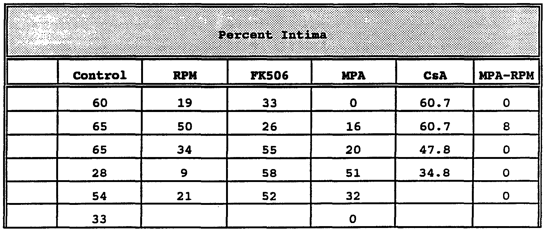
area of intima 100 area of intima + area of media Table 1 shows the data obtained in the above experiment.
Table 1
Figure imgf000025_0001
Figure imgf000026_0001
The data presented in Table 1 are also represented in Figure 3. In the figure, the intima percent of 5 to 8 rat carotid arteries 14 days after balloon catheter injury are shown. All drugs were administered starting the day of surgery and for 13 additional days. The bars in Figure 3 represent the following: control, untreated and uninjured rats; CsA, cyclosporin at 3 mg/kg/IP; FK506, at 4 mg/kg/PO; RPM, rapamycin at 1.5 mg/kg/IP; MPA, mycophenolic acid at 40 mg/kg/PO; and RPM + MPA at 1.5 mg/kg/IP and 40 mg/kg/PO, respectively. The asterisk shows a significant value at p less than or equal to 0.05 using the Student T-test (two-tailed) for two means.
In vivo, combined RPM + MPA treatment resulted in an approximately 97% decrease in mean percentage' of intimal thickening, relative to the untreated control group. Separate RPM and MPA treatment resulted in approximately 45% and 52% decrease in mean percentage of intimal thickening, respectively, relative to the untreated control group. CsA and FK506 had little or no effect on smooth muscle intimal thickening.
While the invention has been described with reference to specific methods and embodiments, it will be appreciated that various modifications and changes may be made without departing from the invention.

Claims

IT IS CLAIMED:
1. A method of preventing or treating hyperproliferative vascular disease in a susceptible mammal, comprising, administering to said mammal an amount of mycophenolic acid effective to inhibit intimal thickening in said mammal.
2. The method of claim 1, wherein the administering is accomplished orally, parenterally, intravascularly, intranasally, intrabronchially, transdermally, rectally, or via a vascular stent impregnated with mycophenolic acid.
3. The method of claim 1, wherein the mycophenolic acid is administered concurrent with said mammal undergoing a percutaneous transluminal coronary angioplasty procedure.
4. The method of claim 3, which further comprises administering the mycophenolic acid subsequent to said mammal undergoing a percutaneous transluminal coronary angioplasty procedure.
5. The method of claim 1, wherein the hyperproliferative vascular disease is selected from the group consisting of intimal smooth muscle cell hyperplasia, restenosis, and vascular occlusion.
6. The method of claim 5, wherein the hyperproliferative vascular disease is restenosis. 7. The method of claim 6, wherein the mycophenolic acid which is selected is in the form of a pharmaceutically acceptable salt.
8. The method of claim 1, wherein the mycophenolic acid is administered prior to, concurrent with and/or subsequent to said mammal sustaining a biologically mediated vascular injury.
9. The method of claim 1, wherein the mycophenolic acid is administered prior to, concurrent with and/or subsequent to said mammal sustaining a mechanically mediated vascular injury.
10. A composition for the use in preventing or treating hyperproliferative vascular disease in a susceptible mammal which comprises an amount of mycophenolic acid effective to inhibit intimal thickening in said mammal and a pharmaceutically acceptable carrier.
11. The composition of claim 8, wherein the hyperproliferative vascular disease is selected from the group consisting of intimal smooth muscle cell hyperplasia, restenosis, and vascular occlusion.
12. The composition of claim 10, wherein the mycophenolic acid which is selected is in the form of a pharmaceutically acceptable salt. AMENDED CLAIMS
[received by the International Bureau on 21 December 1993 (21.12.93); original-claims 1,10,11 and 12 amended; other claims unchanged (2 pages)]
1. A method of preventing or treating hyperproliferative vascular disease in a susceptible mammal, comprising, administering to said mammal an amount of mycophenolic acid effective to inhibit intimal thickening in said mammal.
2. The method of claim 1, wherein the administering is accomplished orally, parenterally, intravascularly, intranasally, intrabronchially, transdermally, rectally, or via a vascular stent impregnated with mycophenolic acid.
3. The method of claim 1, wherein the mycophenolic acid is administered concurrent with said mammal undergoing a percutaneous transluminal coronary angioplasty procedure. 0
4. The method of claim 3, which further comprises administering the mycophenolic acid subsequent to said mammal undergoing a percutaneous transluminal coronary angioplasty procedure. 5
5. The method of claim 1, wherein the hyperproliferative vascular disease is selected from the group consisting of intimal smooth muscle cell hyperplasia, restenosis, and vascular occlusion. 0
6. The method of claim 5, wherein the hyperproliferative vascular disease is restenosis.
7. The method of claim 6, wherein the mycophenolic acid which is selected is in the form of a pharmaceutically acceptable salt.
8. The method of claim 1, wherein the mycophenolic acid is administered prior to, concurrent with and/or subsequent to said mammal sustaining a biologically mediated vascular injury.
9. The method of claim 1, wherein the mycophenolic acid is administered prior to, concurrent with and/or subsequent to said mammal sustaining a mechanically mediated vascular injury.
10. Use of mycophenolic acid for the manufacture of a medicament for preventing or treating hyperproliferative vascular disease in a susceptible mammal.
11. The use of claim 10, wherein the hyperproliferative vascular disease is selected from the group consisting of intimal smooth muscle cell hyperplasia, restenosis, and vascular occlusion.
12. The use of claim 10, wherein the mycophenolic acid which is selected is in the form of a pharmaceutically acceptable salt.
PCT/US1993/006410 1992-07-10 1993-07-07 Method of treating hyperproliferative vascular disease WO1994001105A1 (en)

Priority Applications (1)

Application Number Priority Date Filing Date Title
EP93917017A EP0746314A4 (en) 1992-07-10 1993-07-07 Method of treating hyperproliferative vascular disease

Applications Claiming Priority (2)

Application Number Priority Date Filing Date Title
US911,681 1986-09-25
US07/911,681 US5283257A (en) 1992-07-10 1992-07-10 Method of treating hyperproliferative vascular disease

Publications (1)

Publication Number Publication Date
WO1994001105A1 true WO1994001105A1 (en) 1994-01-20

Family

ID=25430686

Family Applications (1)

Application Number Title Priority Date Filing Date
PCT/US1993/006410 WO1994001105A1 (en) 1992-07-10 1993-07-07 Method of treating hyperproliferative vascular disease

Country Status (4)

Country Link
US (1) US5283257A (en)
EP (1) EP0746314A4 (en)
CA (1) CA2139851A1 (en)
WO (1) WO1994001105A1 (en)

Cited By (20)

* Cited by examiner, † Cited by third party
Publication number Priority date Publication date Assignee Title
US5807876A (en) * 1996-04-23 1998-09-15 Vertex Pharmaceuticals Incorporated Inhibitors of IMPDH enzyme
US5932600A (en) * 1997-03-14 1999-08-03 Vertex Pharmaceuticals Incorporated Inhibitors of IMPDH enzyme
US6025391A (en) * 1996-04-12 2000-02-15 Novartis Ag Enteric-coated pharmaceutical compositions of mycophenolate
US6054472A (en) * 1996-04-23 2000-04-25 Vertex Pharmaceuticals, Incorporated Inhibitors of IMPDH enzyme
US6107052A (en) * 1999-06-09 2000-08-22 Roche Diagnostics Corporation Enzymatic measurement of mycophenolic acid
US6128582A (en) * 1996-04-30 2000-10-03 Vertex Pharmaceuticals Incorporated Molecules comprising an IMPDH-like binding pocket and encoded data storage medium capable of graphically displaying them
US6399773B1 (en) 1998-10-29 2002-06-04 Bristol-Myers Squibb Co. Compounds derived from an amine nucleus that are inhibitors of IMPDH enzyme
US6420403B1 (en) 1998-10-29 2002-07-16 Edwin J. Iwanowicz Inhibitors of IMPDH enzyme
US6518291B1 (en) 1997-03-14 2003-02-11 Vertex Pharmaceuticals, Incorporated Inhibitors of IMPDH enzyme
US6541496B1 (en) 1996-04-23 2003-04-01 Vertex Pharmaceuticals Incorporated Inhibitors of IMPDH enzyme
US6596747B2 (en) 1998-10-29 2003-07-22 Bristol-Myers Squibb Company Compounds derived from an amine nucleus and pharmaceutical compositions comprising same
US6624184B1 (en) 1998-10-29 2003-09-23 Bristol-Myers Squibb Company Amide and diamide inhibitors of IMPDH enzyme for use in treating IMPDH-associated disorders
EP1349562A2 (en) * 2001-01-11 2003-10-08 Duke University Inhibiting gs-fdh to modulate no bioactivity
EP1441224A2 (en) 2003-01-14 2004-07-28 Roche Diagnostics GmbH Enzyme inhibition immunoassay
US6916809B2 (en) 2001-12-21 2005-07-12 Bristol-Myers Squibb Company Heterocyclic acridone inhibitors of IMPDH enzyme
US6919335B2 (en) 2000-04-24 2005-07-19 Bristol-Myers Squibb Co. Heterocycles that are inhibitors of IMPDH enzyme
EP1559779A1 (en) 2004-01-30 2005-08-03 Boehringer Mannheim Gmbh Modified inosine monophosphate dehydrogenases
EP1964561A1 (en) 1999-03-19 2008-09-03 Vertex Pharmaceuticals Incorporated Inhibitors of IMPDH enzyme
US7777069B2 (en) 1999-06-25 2010-08-17 Vertex Pharmaceuticals Incorporated Prodrugs of carbamate inhibitors of IMPDH
US8957071B2 (en) 2005-02-08 2015-02-17 Aspreva Pharmaceuticals S.A. Treatment of vascular, autoimmune and inflammatory diseases using low dosages of IMPDH inhibitors

Families Citing this family (100)

* Cited by examiner, † Cited by third party
Publication number Priority date Publication date Assignee Title
JPH06116151A (en) * 1991-08-06 1994-04-26 Asahi Chem Ind Co Ltd Composition for treating proliferative dermatopathy
US5811447A (en) * 1993-01-28 1998-09-22 Neorx Corporation Therapeutic inhibitor of vascular smooth muscle cells
US6515009B1 (en) * 1991-09-27 2003-02-04 Neorx Corporation Therapeutic inhibitor of vascular smooth muscle cells
US5516781A (en) * 1992-01-09 1996-05-14 American Home Products Corporation Method of treating restenosis with rapamycin
US6306421B1 (en) 1992-09-25 2001-10-23 Neorx Corporation Therapeutic inhibitor of vascular smooth muscle cells
US5770609A (en) * 1993-01-28 1998-06-23 Neorx Corporation Prevention and treatment of cardiovascular pathologies
US6395494B1 (en) 1993-05-13 2002-05-28 Neorx Corporation Method to determine TGF-β
US6251920B1 (en) 1993-05-13 2001-06-26 Neorx Corporation Prevention and treatment of cardiovascular pathologies
US6663881B2 (en) * 1993-01-28 2003-12-16 Neorx Corporation Therapeutic inhibitor of vascular smooth muscle cells
US5981568A (en) 1993-01-28 1999-11-09 Neorx Corporation Therapeutic inhibitor of vascular smooth muscle cells
US5595722A (en) * 1993-01-28 1997-01-21 Neorx Corporation Method for identifying an agent which increases TGF-beta levels
US6491938B2 (en) 1993-05-13 2002-12-10 Neorx Corporation Therapeutic inhibitor of vascular smooth muscle cells
WO1996040098A2 (en) 1995-06-07 1996-12-19 Neorx Corporation Prevention and treatment of cardiovascular pathologies with tamoxifen analogues
CA2162586C (en) * 1993-05-13 2006-01-03 David J. Grainger Prevention and treatment of pathologies associated with abnormally proliferative smooth muscle cells
US6362160B1 (en) * 1993-06-30 2002-03-26 The Johns Hopkins University School Of Medicine Immunophilin-binding agents prevent glutamate neurotoxicity associated with vascular stroke and neurodegenerative diseases
US5407955A (en) * 1994-02-18 1995-04-18 Eli Lilly And Company Methods for lowering serum cholesterol and inhibiting smooth muscle cell proliferation, restenosis, endometriosis, and uterine fibroid disease
WO1996006847A1 (en) * 1994-08-31 1996-03-07 Pfizer Inc. Process for preparing demethylrapamycins
US5665591A (en) * 1994-12-06 1997-09-09 Trustees Of Boston University Regulation of smooth muscle cell proliferation
US20030083733A1 (en) * 1997-10-10 2003-05-01 Neorx Corporation Therapeutic inhibitor of vascular smooth muscle cells
US6099562A (en) * 1996-06-13 2000-08-08 Schneider (Usa) Inc. Drug coating with topcoat
US20020091433A1 (en) * 1995-04-19 2002-07-11 Ni Ding Drug release coated stent
US5837313A (en) * 1995-04-19 1998-11-17 Schneider (Usa) Inc Drug release stent coating process
US7550005B2 (en) * 1995-06-07 2009-06-23 Cook Incorporated Coated implantable medical device
US6774278B1 (en) * 1995-06-07 2004-08-10 Cook Incorporated Coated implantable medical device
US7611533B2 (en) * 1995-06-07 2009-11-03 Cook Incorporated Coated implantable medical device
US7896914B2 (en) * 1995-06-07 2011-03-01 Cook Incorporated Coated implantable medical device
GB9606452D0 (en) * 1996-03-27 1996-06-05 Sandoz Ltd Organic compounds
US6033357A (en) * 1997-03-28 2000-03-07 Navius Corporation Intravascular radiation delivery device
US6309339B1 (en) 1997-03-28 2001-10-30 Endosonics Corporation Intravascular radiation delivery device
US6273913B1 (en) 1997-04-18 2001-08-14 Cordis Corporation Modified stent useful for delivery of drugs along stent strut
US6241762B1 (en) * 1998-03-30 2001-06-05 Conor Medsystems, Inc. Expandable medical device with ductile hinges
US7208010B2 (en) * 2000-10-16 2007-04-24 Conor Medsystems, Inc. Expandable medical device for delivery of beneficial agent
US20030040790A1 (en) * 1998-04-15 2003-02-27 Furst Joseph G. Stent coating
US20070087028A1 (en) * 1998-04-16 2007-04-19 Robert Falotico Intraluminal devices for the prevention and treatment of vascular disease
US7967855B2 (en) * 1998-07-27 2011-06-28 Icon Interventional Systems, Inc. Coated medical device
US8070796B2 (en) * 1998-07-27 2011-12-06 Icon Interventional Systems, Inc. Thrombosis inhibiting graft
US20050002986A1 (en) * 2000-05-12 2005-01-06 Robert Falotico Drug/drug delivery systems for the prevention and treatment of vascular disease
US8236048B2 (en) * 2000-05-12 2012-08-07 Cordis Corporation Drug/drug delivery systems for the prevention and treatment of vascular disease
US6776796B2 (en) 2000-05-12 2004-08-17 Cordis Corportation Antiinflammatory drug and delivery device
US20040243097A1 (en) * 2000-05-12 2004-12-02 Robert Falotico Antiproliferative drug and delivery device
US20020111590A1 (en) * 2000-09-29 2002-08-15 Davila Luis A. Medical devices, drug coatings and methods for maintaining the drug coatings thereon
US20020051730A1 (en) * 2000-09-29 2002-05-02 Stanko Bodnar Coated medical devices and sterilization thereof
US7261735B2 (en) * 2001-05-07 2007-08-28 Cordis Corporation Local drug delivery devices and methods for maintaining the drug coatings thereon
ATE343969T1 (en) * 2000-09-29 2006-11-15 Cordis Corp COATED MEDICAL DEVICES
DE20122506U1 (en) * 2000-10-16 2005-12-08 Conor Medsystems, Inc., Menlo Park Expandable medical device for delivering a beneficial agent
US6471980B2 (en) 2000-12-22 2002-10-29 Avantec Vascular Corporation Intravascular delivery of mycophenolic acid
US7077859B2 (en) * 2000-12-22 2006-07-18 Avantec Vascular Corporation Apparatus and methods for variably controlled substance delivery from implanted prostheses
US20050125054A1 (en) * 2000-12-22 2005-06-09 Avantec Vascular Corporation Devices delivering therapeutic agents and methods regarding the same
US7018405B2 (en) * 2000-12-22 2006-03-28 Avantec Vascular Corporation Intravascular delivery of methylprednisolone
US20050203612A1 (en) * 2000-12-22 2005-09-15 Avantec Vascular Corporation Devices delivering therapeutic agents and methods regarding the same
US20030050692A1 (en) * 2000-12-22 2003-03-13 Avantec Vascular Corporation Delivery of therapeutic capable agents
US20030033007A1 (en) * 2000-12-22 2003-02-13 Avantec Vascular Corporation Methods and devices for delivery of therapeutic capable agents with variable release profile
US7083642B2 (en) * 2000-12-22 2006-08-01 Avantec Vascular Corporation Delivery of therapeutic capable agents
US6939375B2 (en) 2000-12-22 2005-09-06 Avantac Vascular Corporation Apparatus and methods for controlled substance delivery from implanted prostheses
US20020082678A1 (en) * 2000-12-22 2002-06-27 Motasim Sirhan Intravascular delivery of mizoribine
JP4584537B2 (en) 2001-01-16 2010-11-24 ヴァスキュラー セラピーズ リミテッド ライアビリティ カンパニー Implantable devices and methods for preventing or treating hemodialysis vascular access and other vascular graft dysfunctions, including resorbable matrix materials and antiproliferative agents
US6613083B2 (en) * 2001-05-02 2003-09-02 Eckhard Alt Stent device and method
US8182527B2 (en) * 2001-05-07 2012-05-22 Cordis Corporation Heparin barrier coating for controlled drug release
ES2278952T3 (en) * 2001-07-26 2007-08-16 Avantec Vascular Corporation DEVICES FOR MANAGING THERAPEUTIC AGENTS WITH VARIABLE LIBERATION PROFILE.
US7842083B2 (en) * 2001-08-20 2010-11-30 Innovational Holdings, Llc. Expandable medical device with improved spatial distribution
US20040137066A1 (en) * 2001-11-26 2004-07-15 Swaminathan Jayaraman Rationally designed therapeutic intravascular implant coating
US6641611B2 (en) 2001-11-26 2003-11-04 Swaminathan Jayaraman Therapeutic coating for an intravascular implant
US7195640B2 (en) * 2001-09-25 2007-03-27 Cordis Corporation Coated medical devices for the treatment of vulnerable plaque
US7108701B2 (en) * 2001-09-28 2006-09-19 Ethicon, Inc. Drug releasing anastomosis devices and methods for treating anastomotic sites
US20030065382A1 (en) * 2001-10-02 2003-04-03 Fischell Robert E. Means and method for the treatment of coronary artery obstructions
US8740973B2 (en) * 2001-10-26 2014-06-03 Icon Medical Corp. Polymer biodegradable medical device
CA2466432A1 (en) * 2001-11-08 2003-05-15 Atrium Medical Corporation Intraluminal device with a coating containing a therapeutic agent
CN101717410B (en) * 2002-02-01 2015-04-29 阿里亚德医药股份有限公司 Phosphorus-containing compounds & uses thereof
EP1516597A4 (en) * 2002-06-27 2010-11-10 Microport Medical Shanghai Co Drug eluting stent
EP1539041A1 (en) * 2002-07-25 2005-06-15 Avantec Vascular Corporation Devices delivering therapeutic agents and methods regarding the same
US8016881B2 (en) * 2002-07-31 2011-09-13 Icon Interventional Systems, Inc. Sutures and surgical staples for anastamoses, wound closures, and surgical closures
US7306580B2 (en) * 2003-04-16 2007-12-11 Cook Incorporated Medical device with therapeutic agents
AR045957A1 (en) * 2003-10-03 2005-11-16 Novartis Ag PHARMACEUTICAL COMPOSITION AND COMBINATION
US7211108B2 (en) * 2004-01-23 2007-05-01 Icon Medical Corp. Vascular grafts with amphiphilic block copolymer coatings
US20050171596A1 (en) * 2004-02-03 2005-08-04 Furst Joseph G. Stents with amphiphilic copolymer coatings
US9012506B2 (en) 2004-09-28 2015-04-21 Atrium Medical Corporation Cross-linked fatty acid-based biomaterials
EP1811933B1 (en) 2004-09-28 2016-03-23 Atrium Medical Corporation Barrier layer
US9000040B2 (en) 2004-09-28 2015-04-07 Atrium Medical Corporation Cross-linked fatty acid-based biomaterials
WO2006086500A2 (en) * 2005-02-08 2006-08-17 Aspreva Pharmaceuticals Sa Compositions and methods for treating vascular, autoimmune and inflammatory diseases
US8323333B2 (en) * 2005-03-03 2012-12-04 Icon Medical Corp. Fragile structure protective coating
US7540995B2 (en) 2005-03-03 2009-06-02 Icon Medical Corp. Process for forming an improved metal alloy stent
WO2006110197A2 (en) * 2005-03-03 2006-10-19 Icon Medical Corp. Polymer biodegradable medical device
US20060200048A1 (en) * 2005-03-03 2006-09-07 Icon Medical Corp. Removable sheath for device protection
US20060201601A1 (en) * 2005-03-03 2006-09-14 Icon Interventional Systems, Inc. Flexible markers
AU2006221046B2 (en) * 2005-03-03 2012-02-02 Icon Medical Corp. Improved metal alloys for medical device
US9107899B2 (en) 2005-03-03 2015-08-18 Icon Medical Corporation Metal alloys for medical devices
US20060264914A1 (en) * 2005-03-03 2006-11-23 Icon Medical Corp. Metal alloys for medical devices
US9278161B2 (en) 2005-09-28 2016-03-08 Atrium Medical Corporation Tissue-separating fatty acid adhesion barrier
US9427423B2 (en) 2009-03-10 2016-08-30 Atrium Medical Corporation Fatty-acid based particles
WO2008008291A2 (en) * 2006-07-13 2008-01-17 Icon Medical Corp. Stent
WO2008022284A2 (en) * 2006-08-16 2008-02-21 Aspreva Pharmaceuticals Ltd. Compositions and methods for treating vascular, autoimmune, and inflammatory diseases
CA2669415A1 (en) * 2006-11-14 2008-05-22 Ariad Pharmaceuticals, Inc. Solid dosage form comprising ap23573
JP2011527339A (en) * 2008-07-09 2011-10-27 アスプレバ インターナショナル リミテッド PH-specific solution of sodium mycophenolate for the treatment of eye disorders
US20110038910A1 (en) 2009-08-11 2011-02-17 Atrium Medical Corporation Anti-infective antimicrobial-containing biomaterials
US8398916B2 (en) 2010-03-04 2013-03-19 Icon Medical Corp. Method for forming a tubular medical device
EP2593141B1 (en) 2010-07-16 2018-07-04 Atrium Medical Corporation Composition and methods for altering the rate of hydrolysis of cured oil-based materials
WO2013041205A1 (en) * 2011-09-19 2013-03-28 Pyxirion Pharma Gmbh Novel therapeutic concepts for treating vascular diseases
US9867880B2 (en) 2012-06-13 2018-01-16 Atrium Medical Corporation Cured oil-hydrogel biomaterial compositions for controlled drug delivery
BR112016030273A2 (en) 2014-06-24 2017-08-22 Icon Medical Corp MEDICAL DEVICE AND METHOD FOR FORMING SAID DEVICE
US11766506B2 (en) 2016-03-04 2023-09-26 Mirus Llc Stent device for spinal fusion

Citations (2)

* Cited by examiner, † Cited by third party
Publication number Priority date Publication date Assignee Title
US3705946A (en) * 1971-05-25 1972-12-12 Lilly Co Eli Method of treating hyperuricemia
US3880995A (en) * 1973-05-14 1975-04-29 Lilly Co Eli Treatment of arthritis with mycophenolic acid and derivatives

Family Cites Families (2)

* Cited by examiner, † Cited by third party
Publication number Priority date Publication date Assignee Title
CA2086642C (en) * 1992-01-09 2004-06-15 Randall E. Morris Method of treating hyperproliferative vascular disease
WO1994012184A1 (en) * 1992-11-24 1994-06-09 Syntex (U.S.A.) Inc. Use of mycophenolic acid, mycophenolate mofetil or derivate thereof to inhibit stenosis

Patent Citations (2)

* Cited by examiner, † Cited by third party
Publication number Priority date Publication date Assignee Title
US3705946A (en) * 1971-05-25 1972-12-12 Lilly Co Eli Method of treating hyperuricemia
US3880995A (en) * 1973-05-14 1975-04-29 Lilly Co Eli Treatment of arthritis with mycophenolic acid and derivatives

Non-Patent Citations (1)

* Cited by examiner, † Cited by third party
Title
See also references of EP0746314A4 *

Cited By (36)

* Cited by examiner, † Cited by third party
Publication number Priority date Publication date Assignee Title
US6172107B1 (en) 1996-04-12 2001-01-09 Novartis Ag Entric-coated pharmaceutical compositions
US6306900B1 (en) 1996-04-12 2001-10-23 Novartis Ag Enteric coated pharmaceutical compositions
US6025391A (en) * 1996-04-12 2000-02-15 Novartis Ag Enteric-coated pharmaceutical compositions of mycophenolate
US6054472A (en) * 1996-04-23 2000-04-25 Vertex Pharmaceuticals, Incorporated Inhibitors of IMPDH enzyme
US6967214B2 (en) 1996-04-23 2005-11-22 Vertex Pharmaceuticals Incorporated Inhibitors of IMPDH enzyme
US6344465B1 (en) 1996-04-23 2002-02-05 Vertex Pharmaceuticals, Incorporated Inhibitors of IMPDH enzyme
US5807876A (en) * 1996-04-23 1998-09-15 Vertex Pharmaceuticals Incorporated Inhibitors of IMPDH enzyme
US7329681B2 (en) 1996-04-23 2008-02-12 Vertex Pharmaceuticals Incorporated Inhibitors of IMPDH enzyme
US6541496B1 (en) 1996-04-23 2003-04-01 Vertex Pharmaceuticals Incorporated Inhibitors of IMPDH enzyme
US7527952B2 (en) 1996-04-30 2009-05-05 Vertex Pharmaceuticals Incorporated Crystals of inosine monophosphate dehydrogenase/oxidized inosine monophosphate thiomidate intermediate/mycophenolic acid (IIMPDH/XMP*/MPA)
US6128582A (en) * 1996-04-30 2000-10-03 Vertex Pharmaceuticals Incorporated Molecules comprising an IMPDH-like binding pocket and encoded data storage medium capable of graphically displaying them
US7216041B1 (en) 1996-04-30 2007-05-08 Vertex Pharmaceuticals Incorporated Methods of using the structure coordinates of molecules comprising an IMPDH-like binding pocket
US5932600A (en) * 1997-03-14 1999-08-03 Vertex Pharmaceuticals Incorporated Inhibitors of IMPDH enzyme
US6518291B1 (en) 1997-03-14 2003-02-11 Vertex Pharmaceuticals, Incorporated Inhibitors of IMPDH enzyme
US6399773B1 (en) 1998-10-29 2002-06-04 Bristol-Myers Squibb Co. Compounds derived from an amine nucleus that are inhibitors of IMPDH enzyme
US7205324B2 (en) 1998-10-29 2007-04-17 Bristol-Myers Squibb Company Inhibitors of IMPDH enzyme
US6624184B1 (en) 1998-10-29 2003-09-23 Bristol-Myers Squibb Company Amide and diamide inhibitors of IMPDH enzyme for use in treating IMPDH-associated disorders
US6420403B1 (en) 1998-10-29 2002-07-16 Edwin J. Iwanowicz Inhibitors of IMPDH enzyme
US6596747B2 (en) 1998-10-29 2003-07-22 Bristol-Myers Squibb Company Compounds derived from an amine nucleus and pharmaceutical compositions comprising same
US6617323B2 (en) 1998-10-29 2003-09-09 Bristol-Myers Squibb Co. Amino-substituted compounds useful as inhibitors of IMPDH enzyme
US7053111B2 (en) 1998-10-29 2006-05-30 Bristol-Myers Squibb Co. Inhibitors of IMPDH enzyme
US7060720B2 (en) 1998-10-29 2006-06-13 Bristol-Myers Squibb Co. Inhibitors of IMPDH enzyme
US8268873B2 (en) 1999-03-19 2012-09-18 Vertex Pharmaceuticals Incorporated Inhibitors of IMPDH enzyme
EP1964561A1 (en) 1999-03-19 2008-09-03 Vertex Pharmaceuticals Incorporated Inhibitors of IMPDH enzyme
US6107052A (en) * 1999-06-09 2000-08-22 Roche Diagnostics Corporation Enzymatic measurement of mycophenolic acid
US7777069B2 (en) 1999-06-25 2010-08-17 Vertex Pharmaceuticals Incorporated Prodrugs of carbamate inhibitors of IMPDH
US6919335B2 (en) 2000-04-24 2005-07-19 Bristol-Myers Squibb Co. Heterocycles that are inhibitors of IMPDH enzyme
EP1349562A4 (en) * 2001-01-11 2008-08-06 Univ Duke Inhibiting gs-fdh to modulate no bioactivity
US7615535B2 (en) 2001-01-11 2009-11-10 Duke University Inhibiting GS-FDH to modulate no bioactivity
US8217006B2 (en) 2001-01-11 2012-07-10 Duke University Inhibiting GS-FDH to modulate no bioactivity
EP1349562A2 (en) * 2001-01-11 2003-10-08 Duke University Inhibiting gs-fdh to modulate no bioactivity
US7312209B2 (en) 2001-12-21 2007-12-25 Bristol-Myers Squibb Company Acridone inhibitors of IMPDH enzyme
US6916809B2 (en) 2001-12-21 2005-07-12 Bristol-Myers Squibb Company Heterocyclic acridone inhibitors of IMPDH enzyme
EP1441224A2 (en) 2003-01-14 2004-07-28 Roche Diagnostics GmbH Enzyme inhibition immunoassay
EP1559779A1 (en) 2004-01-30 2005-08-03 Boehringer Mannheim Gmbh Modified inosine monophosphate dehydrogenases
US8957071B2 (en) 2005-02-08 2015-02-17 Aspreva Pharmaceuticals S.A. Treatment of vascular, autoimmune and inflammatory diseases using low dosages of IMPDH inhibitors

Also Published As

Publication number Publication date
EP0746314A4 (en) 1997-03-19
US5283257A (en) 1994-02-01
CA2139851A1 (en) 1994-01-20
EP0746314A1 (en) 1996-12-11

Similar Documents

Publication Publication Date Title
US5283257A (en) Method of treating hyperproliferative vascular disease
US5516781A (en) Method of treating restenosis with rapamycin
KR100276142B1 (en) Composition for the treatment of hyperproliferative vascular disease
JP2550277B2 (en) Pharmaceutical composition for preventing or treating hyperproliferative vascular disease
US5021404A (en) Angiostatic collagen modulators
JP2810599B2 (en) Pharmaceutical composition for the treatment of immunoinflammatory diseases
JP2637682B2 (en) Pharmaceutical composition for treating immunoinflammatory bowel disease
US6514949B1 (en) Method compositions for treating the inflammatory response
CA2074641C (en) Method of treating ocular inflammation
US6670334B2 (en) Method and compositions for treating the inflammatory response
JP2004521062A (en) Monosaccharides and disaccharides for the treatment of disorders related to nitric oxide
EP0305181A2 (en) Ethyl-(+)-apovincaminate for treating demyelinization clinical patterns of autoimmune origin
Sharpe et al. Anesthetic considerations for the previously transplanted patient
KR100356899B1 (en) Preventives and remedies for ulcerous colitis and/or crohn's disease

Legal Events

Date Code Title Description
AK Designated states

Kind code of ref document: A1

Designated state(s): CA

AL Designated countries for regional patents

Kind code of ref document: A1

Designated state(s): AT BE CH DE DK ES FR GB GR IE IT LU MC NL PT SE

121 Ep: the epo has been informed by wipo that ep was designated in this application
DFPE Request for preliminary examination filed prior to expiration of 19th month from priority date (pct application filed before 20040101)
WWE Wipo information: entry into national phase

Ref document number: 2139851

Country of ref document: CA

WWE Wipo information: entry into national phase

Ref document number: 1993917017

Country of ref document: EP

WWP Wipo information: published in national office

Ref document number: 1993917017

Country of ref document: EP

WWW Wipo information: withdrawn in national office

Ref document number: 1993917017

Country of ref document: EP