WO1994004197A1 - Particles for nmr imaging and method of manufacture - Google Patents
Particles for nmr imaging and method of manufacture Download PDFInfo
- Publication number
- WO1994004197A1 WO1994004197A1 PCT/EP1993/002046 EP9302046W WO9404197A1 WO 1994004197 A1 WO1994004197 A1 WO 1994004197A1 EP 9302046 W EP9302046 W EP 9302046W WO 9404197 A1 WO9404197 A1 WO 9404197A1
- Authority
- WO
- WIPO (PCT)
- Prior art keywords
- particles
- surfactant
- particle
- iron oxide
- core
- Prior art date
Links
Classifications
-
- A—HUMAN NECESSITIES
- A61—MEDICAL OR VETERINARY SCIENCE; HYGIENE
- A61K—PREPARATIONS FOR MEDICAL, DENTAL OR TOILETRY PURPOSES
- A61K49/00—Preparations for testing in vivo
- A61K49/06—Nuclear magnetic resonance [NMR] contrast preparations; Magnetic resonance imaging [MRI] contrast preparations
- A61K49/18—Nuclear magnetic resonance [NMR] contrast preparations; Magnetic resonance imaging [MRI] contrast preparations characterised by a special physical form, e.g. emulsions, microcapsules, liposomes
- A61K49/1806—Suspensions, emulsions, colloids, dispersions
-
- A—HUMAN NECESSITIES
- A61—MEDICAL OR VETERINARY SCIENCE; HYGIENE
- A61K—PREPARATIONS FOR MEDICAL, DENTAL OR TOILETRY PURPOSES
- A61K49/00—Preparations for testing in vivo
- A61K49/06—Nuclear magnetic resonance [NMR] contrast preparations; Magnetic resonance imaging [MRI] contrast preparations
- A61K49/18—Nuclear magnetic resonance [NMR] contrast preparations; Magnetic resonance imaging [MRI] contrast preparations characterised by a special physical form, e.g. emulsions, microcapsules, liposomes
- A61K49/1818—Nuclear magnetic resonance [NMR] contrast preparations; Magnetic resonance imaging [MRI] contrast preparations characterised by a special physical form, e.g. emulsions, microcapsules, liposomes particles, e.g. uncoated or non-functionalised microparticles or nanoparticles
- A61K49/1821—Nuclear magnetic resonance [NMR] contrast preparations; Magnetic resonance imaging [MRI] contrast preparations characterised by a special physical form, e.g. emulsions, microcapsules, liposomes particles, e.g. uncoated or non-functionalised microparticles or nanoparticles coated or functionalised microparticles or nanoparticles
- A61K49/1824—Nuclear magnetic resonance [NMR] contrast preparations; Magnetic resonance imaging [MRI] contrast preparations characterised by a special physical form, e.g. emulsions, microcapsules, liposomes particles, e.g. uncoated or non-functionalised microparticles or nanoparticles coated or functionalised microparticles or nanoparticles coated or functionalised nanoparticles
- A61K49/1827—Nuclear magnetic resonance [NMR] contrast preparations; Magnetic resonance imaging [MRI] contrast preparations characterised by a special physical form, e.g. emulsions, microcapsules, liposomes particles, e.g. uncoated or non-functionalised microparticles or nanoparticles coated or functionalised microparticles or nanoparticles coated or functionalised nanoparticles having a (super)(para)magnetic core, being a solid MRI-active material, e.g. magnetite, or composed of a plurality of MRI-active, organic agents, e.g. Gd-chelates, or nuclei, e.g. Eu3+, encapsulated or entrapped in the core of the coated or functionalised nanoparticle
- A61K49/1833—Nuclear magnetic resonance [NMR] contrast preparations; Magnetic resonance imaging [MRI] contrast preparations characterised by a special physical form, e.g. emulsions, microcapsules, liposomes particles, e.g. uncoated or non-functionalised microparticles or nanoparticles coated or functionalised microparticles or nanoparticles coated or functionalised nanoparticles having a (super)(para)magnetic core, being a solid MRI-active material, e.g. magnetite, or composed of a plurality of MRI-active, organic agents, e.g. Gd-chelates, or nuclei, e.g. Eu3+, encapsulated or entrapped in the core of the coated or functionalised nanoparticle having a (super)(para)magnetic core coated or functionalised with a small organic molecule
- A61K49/1839—Nuclear magnetic resonance [NMR] contrast preparations; Magnetic resonance imaging [MRI] contrast preparations characterised by a special physical form, e.g. emulsions, microcapsules, liposomes particles, e.g. uncoated or non-functionalised microparticles or nanoparticles coated or functionalised microparticles or nanoparticles coated or functionalised nanoparticles having a (super)(para)magnetic core, being a solid MRI-active material, e.g. magnetite, or composed of a plurality of MRI-active, organic agents, e.g. Gd-chelates, or nuclei, e.g. Eu3+, encapsulated or entrapped in the core of the coated or functionalised nanoparticle having a (super)(para)magnetic core coated or functionalised with a small organic molecule the small organic molecule being a lipid, a fatty acid having 8 or more carbon atoms in the main chain, or a phospholipid
-
- Y—GENERAL TAGGING OF NEW TECHNOLOGICAL DEVELOPMENTS; GENERAL TAGGING OF CROSS-SECTIONAL TECHNOLOGIES SPANNING OVER SEVERAL SECTIONS OF THE IPC; TECHNICAL SUBJECTS COVERED BY FORMER USPC CROSS-REFERENCE ART COLLECTIONS [XRACs] AND DIGESTS
- Y10—TECHNICAL SUBJECTS COVERED BY FORMER USPC
- Y10S—TECHNICAL SUBJECTS COVERED BY FORMER USPC CROSS-REFERENCE ART COLLECTIONS [XRACs] AND DIGESTS
- Y10S977/00—Nanotechnology
- Y10S977/70—Nanostructure
- Y10S977/832—Nanostructure having specified property, e.g. lattice-constant, thermal expansion coefficient
- Y10S977/838—Magnetic property of nanomaterial
-
- Y—GENERAL TAGGING OF NEW TECHNOLOGICAL DEVELOPMENTS; GENERAL TAGGING OF CROSS-SECTIONAL TECHNOLOGIES SPANNING OVER SEVERAL SECTIONS OF THE IPC; TECHNICAL SUBJECTS COVERED BY FORMER USPC CROSS-REFERENCE ART COLLECTIONS [XRACs] AND DIGESTS
- Y10—TECHNICAL SUBJECTS COVERED BY FORMER USPC
- Y10S—TECHNICAL SUBJECTS COVERED BY FORMER USPC CROSS-REFERENCE ART COLLECTIONS [XRACs] AND DIGESTS
- Y10S977/00—Nanotechnology
- Y10S977/902—Specified use of nanostructure
- Y10S977/904—Specified use of nanostructure for medical, immunological, body treatment, or diagnosis
- Y10S977/905—Specially adapted for travel through blood circulatory system
-
- Y—GENERAL TAGGING OF NEW TECHNOLOGICAL DEVELOPMENTS; GENERAL TAGGING OF CROSS-SECTIONAL TECHNOLOGIES SPANNING OVER SEVERAL SECTIONS OF THE IPC; TECHNICAL SUBJECTS COVERED BY FORMER USPC CROSS-REFERENCE ART COLLECTIONS [XRACs] AND DIGESTS
- Y10—TECHNICAL SUBJECTS COVERED BY FORMER USPC
- Y10S—TECHNICAL SUBJECTS COVERED BY FORMER USPC CROSS-REFERENCE ART COLLECTIONS [XRACs] AND DIGESTS
- Y10S977/00—Nanotechnology
- Y10S977/902—Specified use of nanostructure
- Y10S977/904—Specified use of nanostructure for medical, immunological, body treatment, or diagnosis
- Y10S977/927—Diagnostic contrast agent
-
- Y—GENERAL TAGGING OF NEW TECHNOLOGICAL DEVELOPMENTS; GENERAL TAGGING OF CROSS-SECTIONAL TECHNOLOGIES SPANNING OVER SEVERAL SECTIONS OF THE IPC; TECHNICAL SUBJECTS COVERED BY FORMER USPC CROSS-REFERENCE ART COLLECTIONS [XRACs] AND DIGESTS
- Y10—TECHNICAL SUBJECTS COVERED BY FORMER USPC
- Y10S—TECHNICAL SUBJECTS COVERED BY FORMER USPC CROSS-REFERENCE ART COLLECTIONS [XRACs] AND DIGESTS
- Y10S977/00—Nanotechnology
- Y10S977/902—Specified use of nanostructure
- Y10S977/932—Specified use of nanostructure for electronic or optoelectronic application
- Y10S977/953—Detector using nanostructure
- Y10S977/96—Of magnetic property
-
- Y—GENERAL TAGGING OF NEW TECHNOLOGICAL DEVELOPMENTS; GENERAL TAGGING OF CROSS-SECTIONAL TECHNOLOGIES SPANNING OVER SEVERAL SECTIONS OF THE IPC; TECHNICAL SUBJECTS COVERED BY FORMER USPC CROSS-REFERENCE ART COLLECTIONS [XRACs] AND DIGESTS
- Y10—TECHNICAL SUBJECTS COVERED BY FORMER USPC
- Y10T—TECHNICAL SUBJECTS COVERED BY FORMER US CLASSIFICATION
- Y10T428/00—Stock material or miscellaneous articles
- Y10T428/29—Coated or structually defined flake, particle, cell, strand, strand portion, rod, filament, macroscopic fiber or mass thereof
- Y10T428/2982—Particulate matter [e.g., sphere, flake, etc.]
- Y10T428/2991—Coated
- Y10T428/2998—Coated including synthetic resin or polymer
-
- Y—GENERAL TAGGING OF NEW TECHNOLOGICAL DEVELOPMENTS; GENERAL TAGGING OF CROSS-SECTIONAL TECHNOLOGIES SPANNING OVER SEVERAL SECTIONS OF THE IPC; TECHNICAL SUBJECTS COVERED BY FORMER USPC CROSS-REFERENCE ART COLLECTIONS [XRACs] AND DIGESTS
- Y10—TECHNICAL SUBJECTS COVERED BY FORMER USPC
- Y10T—TECHNICAL SUBJECTS COVERED BY FORMER US CLASSIFICATION
- Y10T436/00—Chemistry: analytical and immunological testing
- Y10T436/24—Nuclear magnetic resonance, electron spin resonance or other spin effects or mass spectrometry
Definitions
- the invention relates to iron oxide particles which when in suspension are injectable into the blood stream of patients.
- the particles have enhanced stability against agglomeration, and being relatively "invisible" to the reticulo-endothelial system (RES), they show an increased resistance to removal by macrophages.
- the particles are useful for production of contrast agents for blood-pool imaging.
- Ferromagnetic species or superparamagnetic magnetite microcrystals have been used as contrast agents for the nuclear magnetic resonance imaging (MRI) of the liver and spleen. Their use as contrast agents in these organs is based on the observation that soon after injection the particles are recognized by the RES and rapidly captured. The particles are then removed from the bloodstream, stored in the liver and spleen and subsequently eliminated. Much effort has been devoted toward improvement of the known formulations with the aim to increasing the uptake of the superparamagnetic particles in the targeted organs e.g. liver, spleen or bone marrow prior to their elimination from the body thus rendering their use more practical.
- MRI nuclear magnetic resonance imaging
- EP-A-0 272 091 discloses coating solid particles of an active ingredient, i.e. magnetite (and other diagnostic agents or drugs) said ingredient constituting the core of the particles, with a first layer of a monomolecular amphiphile which can associate with the ingredient of the core; then, the system comprises a second outer layer, which may include a bimolecular layer of phospholipids (i.e. a liposome membrane analog) which encapsulates the amphiphile.
- magnetite particles coated with palmitic acid as surfactant were encapsulated in liposomes made from a mixture of cholesterol and distearoylphosphatidylcholine.
- One object of the arrangement is to stabilize the active ingredient in the circulation against removal.
- EP-A-0 275 285 discloses coated and uncoated magnetite particles for use as a contrast agent for NMR imaging. When coated, the particles are surrounded by a polymer to which biologically active molecules may be attached. In the case of coated particles, the biological molecules can be chosen to target specific organs or tissues.
- Polymeric coatings disclosed may be made from proteins such as albumin, polysaccharides such as dextran, polypeptides such as polyglutamates or polylysines or organosilanes such as N-2-aminoethyl-3-aminopropyltrimethoxy-silane.
- Biological molecules that may be covalently attached to the coating are antibodies, carbohydrates or hormones which may enhance specificity and biodistribution of the particles to specific sites in the organism.
- EP-A-0 354 855 discloses liposomes as drug-carrier vesicles containing polyethylene glycol bound phospholipid in the lipid layer of the vesicle. The hydrophobic moiety of the phospholipid is sunk in the membrane-constituting lipids or is bound thereto, while the hydrophilic moiety of the polyethylene glycol protrudes therefrom and extends into the surrounding medium.
- the liposomic vesicles are said to be useful for preparation of artificial eiythrocytes by encapsulation of hemoglobin in the vesicles.
- US-A-4,904,479 discloses coating polystyrene particles with amphiphilic block copolymers having simultaneously hydrophilic and hydrophobic segments (e.g. Poloxamer ® and Poloxamine ® ).
- the coating is intended to minimize opsonization after injection and enable directing the particles to the bone marrow rather than to the liver or spleen.
- Poloxamer ® and Poloxamine ® are amphiphilic block-copolymers comprising consecutive hydrophobic polyoxypropylene segments and hydrophilic polyoxyethylene segments; it is believed that for protection against uptake by the liver, the hydrophilic segments stick out from the surface of the particle outer coating, thus sterically preventing the deposition thereto of opsonin and making the particles less recognizable by the macrophages.
- the particles of the prior art require expensive manufacturing techniques and produce particles which, upon injection, are recognized by the RES and easily removed from the blood. Such particles and the contrast agents produced therefrom cannot be used in applications for which a relatively long biological half-life is required.
- Contrast agents with prolonged presence in the blood i.e. good resistance to uptake by RES and a relatively low diffusivity into the tissue or extravascular spots are recognized in the art as particularly useful "blood pool” agents.
- Long biological half-lifes are sometimes desirable for the blood pool agents if one wants to produce meaningful analytical results eliminating repeated injections and heavy use of contrast media.
- it would be necessary to produce "stealth" particles which, for a period of time, would not be recognized by the RES and which would still provide sufficient magnetic relaxation response.
- Existence of a real "stealth” iron oxide particle would enable NMR analysis of the body as a whole and not only analysis of localized parts or specific organs, as done with contrast agents known so far.
- the stealth particles would thus make possible measurements of blood volumes and the blood perfusion of various organs, including brain, using non-invasive techniques. For instance, monitoring variations in blood oxygenation of the brain cortex during activation tasks would become possible.
- the invention relates to blood pool contrast agents for diagnostic image analysis of human or animal body, preferably by MRI analysis, which remain in the blood stream for prolonged periods of time and thus enable measurements of blood volumes and the blood perfusion of various organs. More specifically, the invention relates to contrast agents which are particularly resistant to rapid uptake by the RES and which, upon injection, remain present in the blood stream much longer then the blood pool contrast agents known so far.
- the blood pool agents of the invention comprise iron oxide particles stabilized by a three dimensional shell layer containing molecules of an amphipatic compound and a non-ionic surfactant.
- the amphipatic compound has a hydrophilic negatively charged phosphorus containing head moiety bonded to a hydrophobic tail moiety and is characterized by being in micellar form.
- the non-ionic surfactant of the three dimensional shell layer causes the amphipatic compound to be in said micellar form and the hydrophilic phosphorus containing (preferably phosphoryl) head moiety of the amphiphile bears at least two negative charges.
- the three dimensional shell is formed from molecules of the amphipatic compound whose negative phosphoryl head moieties are pointing towards the iron oxide core and the hydrophobic tail moieties protrude outwardly therefrom forming an urchin-like structure.
- the urchin-like structure serves as a base for building the three dimensional shell by anchoring thereto the non-ionic surfactant.
- the outer layer comprises a non-ionic surfactant whose hydrophobic moieties are interlaced or intertwined with the alkyl or alkenyl chain of the ester or glycerophospholipid further stabilizing the structure.
- the natural ability of the non-ionic surfactant to cause micellization of these compounds is to be deployed.
- the preferred glycerophospholipids consist of a mono-phosphate ester of a substituted or partially substituted polyalcohol, at least one other alcoholic function of said polyalcohol being esterified by a long chain, saturated or unsaturated, aliphatic fatty acid, or etherified by a long chain, saturated or unsaturated alcohol, the other two acidic functions of the phosphoric acid being either free or salified with alkali or earth-alkali metals. More specifically, the glycerophospholipid is preferably a monophosphate of a fatty acid glyceride selected from dimyristoylphosphatidic acid, dipalmitoylphosphatidic acid, or distearoylphosphatidic acid.
- a preferred non-ionic surfactant is a physiologically acceptable surfactant with at least one block-copolymer having polyoxyethylene and polyoxypropylene segments or polyethyleneglycolhexadecylether.
- Surfactants of this kind are commercially available under the trademarks of Pluronic ® , Poloxamer ® , Poloxamine ® , Synperonic ® or BRIJ ® .
- the particles of the invention may be sterilized and then lyophilized to produce a sterile powder which can be stored for prolonged periods.
- the contrast agent of the invention is reconstituted from the lyophilized pulverulent formulation by dispersing the powder in a physiologically acceptable liquid carrier. The suspension obtained is ready for administration.
- the invention further relates to a process of manufacturing of particles as well as their use as contrast agents in NMR Imaging of human or animal body.
- Fig. la is a cross-sectional schematic diagram of an iron oxide urchin-like structure pertaining to the particle of the invention with non-ionic surfactant forming a three dimensional shell around the particle.
- Fig. lb is a section of a three-dimensional view of the iron oxide urchin-like structure pertaining to the particle of the invention.
- Fig. 2 is a schematic diagram showing comparison of residence times or "activity” (expressed as the Area Under Curve (AUC) of iron oxide particles of the invention and that obtained for particles of the prior art.
- AUC Area Under Curve
- the particles of the present invention consist essentially of an iron oxide core and an outer layer consisting of molecules of an amphipatic compound and a non-ionic surfactant.
- the amphipatic compound which has a relatively strongly charged hydrophilic moiety that sticks to the surface of the iron oxide may be a mono alkyl or cycloalkyl or alkenyl phosphoric acid ester or a negatively charged phospholipid.
- the term "cyclo" used here implies that the cyclic part of the molecule may be 5-7 membered, may be saturated or not (aromatic) or may contain heteroatoms (heterocycles).
- the outer layer having the shape of a three dimensional shell comprises a non- ionic surfactant whose hydrophobic moieties are interlaced or intertwined with the alkyl or alkenyl chain of the ester further stabilizing the three dimensional structure.
- the non-ionic surfactant must also be able to micellize the phosphoric acid monoester or phospholipid as only when in micellar form, these compounds will exhibit sufficient affinity for the iron oxide core and, consequently may be used as stabilizers of magnetite particles.
- the affinity between the phosphoryl head and F ⁇ 3 ⁇ 4 must be such that, in the presence of a non-ionic surfactant and upon sonication, the micellar phospholipid or monoester form an urchin-like precursor which subsequently serves as template for the construction of the outer three dimensional shell which is built through the interaction of the hydrophobic segments of the surfactant and the hydrophobic moieties of the amphipatic compound.
- the three dimensional structure will then provide a stable "RES non-recognizable" particle.
- the amphipatic compound, preferably a glycerophospholipid, forming the outer layer does not form liposome vesicles or a liposome-like film around the iron oxide core.
- the three dimensional shell is formed from phospholipid molecules whose negative phosphoryl head moieties point towards the iron oxide core and the hydrophobic tail moieties protrude outwardly therefrom forming the urchin-like precursor structure.
- the urchin-like structure always serves as foundation for building the three dimensional shell or layer, whether the starting amphipatic compound is a mono alkyl or mono alkenyl phosphoric acid ester or a micellar glycerophospholipid. In either case the resulting particles have increased stability against removal and will exhibit excellent contrast agent properties. It should be born in mind that amphiphilic molecules participating in liposome-like films are organized in a reverse order tail- to-tall, the hydrophilic heads prodruding outwardly.
- glycerophospholipids When glycerophospholipids are used, for obtaining the urchin ⁇ like base around the iron oxide particles whereby the complete three dimensional shell can be constructed, it is important to either (a) energize the iron oxide particles in the presence of a glycerophospho- lipid and a non-ionic surfactant simultaneously or (b) prepare a suspension of the iron oxide particles with a glycerophospholipid, energize the suspension, add a non-ionic surfactant and then, optionally repeat the energizing step. Energizing is carried out by dispersing the iron oxide particles evenly or homogenizing the dispersion which accelerates micellization and /or destruction of liposomes or laminates which may have formed earlier. Energizing may be effected using various methods including agitation; however, sonication, microfluidization, advantageously under moderate heating (30-50°C) are preferred.
- the construction of the three dimensional shell which stabilizes the particles is carried out via an urchin-like basic structure formed between the iron oxide and the glycerophospho- lipid in micellar form.
- the phospholipids are first micellised by sonication or microfluidisation in the presence of the surfactant and then the negative charges of the amphiphile in the micelles interact with the iron oxide core while their electrically neutral hydrophobic ends are attracted by the surfactant.
- the surfactant serves several functions. It causes micellisation of the amphiphile, it assists orientation and structuring of the micelles around the core (facilitating formation of the urchin-like precursor) and finally anchors itself to the hydrophobic segments of the amphiphile to provide a three dimensional shell around the particle.
- the anchoring of the surfactant occurs via its hydrophobic polyoxypropylene (POP) segments which interact with the hydrophobic part of the glycerophospholipid or a monoester of phosphoric acid.
- POP polyoxypropylene
- the hydrophobic part is anchored by Van der Waals forces with the polyoxypropylene (POP) segments while the hydrophilic polyoxyethylene (POE) segments protrude outwardly into the solution, thus probably inhibiting opsonisation and agglomeration of the magnetite particles.
- the formation of the urchin-like structure is basically the same as above and the non-ionic surfactant has its hydrophobic moieties interlaced or intertwined with the alkyl or alkenyl chains of the phosphoric acid ester and its hydrophilic moieties protrude into the solution.
- dipalmitoylphosphatidylglycerol DPPG
- dimyristoylphosphatidyl glycerol DMPG
- dicetylphosphate DCP
- DPPG dipalmitoylphosphatidylglycerol
- DMPG dimyristoylphosphatidyl glycerol
- DCP dicetylphosphate
- the compound immediately adjacent to the particle should have, in addition to the availability of the negative charges on both oxygen atoms in the phosphoryl head, a relatively long alkyl or alkenyl chain or a hydrophobic cycloalkyl attached to the phosphorus atom, either directly, or via an intermediate alkylene or oxygen bridge. Therefore, to obtain the stable stealth particles and therefrom the contrast agents of the invention, the compound immediately adjacent to the magnetite core must be a mono alkyl or alkenyl or cycloalkyl containing ester of phosphoric acid, a monoalkyl phosphonate or a glycerophospholipid whose alkyl or alkenyl chains have a relatively strong hydrophobic character.
- alkyl or alkenyl chains of sufficient length can properly interact with another amphipatic compound, and provide an anchor for a sufficient amount of the non- ionic surfactant or block copolymer to produce a stable three dimensional structure around the iron core.
- Alkyl or alkenyl chains with at least eight carbon atoms, preferably with at least ten and more preferably at least twelve carbon atoms are particularly desirable.
- Short hydrophobic alkyl or alkenyl chains or macrocycle ligands do not provide stable particles which indicate that one should have either a firm interaction between the intermediate layer and the surfactant, or sufficient amount of anchored surfactant must be available to obtain the desired result.
- a very efficient protection of the particles against removal from circulation may be obtained if a primary layer immediately adjacent to the particle comprises an amphipatic substance with a relatively strongly ionized negative function and a relatively long and efficient hydrophobic organic chain, and that a further layer interlaced or intertwined therewith is built from a block copolymer having, in succession, hydrophilic and hydrophobic segments, e.g. surfactants of the Poloxamer ® , Synperonic ® or Pluronic ® type.
- the quantities of mono alkyl phosphoric acid ester or glycerophospholipid and block copolymer, relative to each other, should be substantial, i.e. the weight ratio of the earlier to the later should be in the range from 1:100 to 10:1, preferably from 1:30 to 5: 1 and more preferably from 1:10 to 1:1. Also, the weight ratio of the mono alkyl phosphoric acid ester to that of the iron oxide core should be between 1:5 and 100:1 preferably between 20:1 and 1: 1.
- the stable, to the RES invisible, particles may be produced from a mixture of two or more compounds selected from ionic and neutral phospholipids, mono alkyl or alkenyl esters of phospholipids and/or other non-phospholipid compounds by a method in which iron oxide particles are suspended or admixed with a negatively charged amphipatic compound or a mixture of the negatively charged phospholipids and other non-phospholipids and a surfactant, in a physiologically acceptable aqueous phase.
- the mixture formed is sonicated or microfluidized to micellize the amphipatic compound, create an urchin-like structure and, therefrom, a three dimensional shell around the iron oxide particles.
- non-phospholipid compounds which may be useful for the stable magnetite particles of the invention are compounds like cholesterol, ergosterol, phytosterol, sitosterol, lanosterol, tocopherol, etc..
- the mixture may be further sterilized and/or lyophilized to produce a dry powder with a long shelf life.
- the surfactant may be added to the mixture of the iron oxide and the amphipatic compound after the sonication or microfluidization. In such a case, however, the sonication, microfluidization or heating step is optionally repeated with all the components together.
- the suspensions of the invention may also be prepared from pulverulent or powder formulations comprising iron oxide particles. Formulations in powder form are usually prepared by lyophilisation or drying freshly prepared solutions comprising iron oxide particles, phospholipids and non-ionic surfactants. Prior to lyophllization or drying these solutions are sterilized. The sterilization may be carried out using any of the known techniques i.e. heating, filtration, ⁇ -rays, etc. Alternatively, it may also be possible to sterilize suspensions obtained using lyophilized powders which were stored for a longer period of time.
- Particles prepared according to the invention are found to be useful as blood pool contrast agents for in vivo NMR imaging of organs of human or animal body.
- the imaging is carried out by administering to patients, usually via an intravenous injection, an aqueous suspension of magnetite particles according to the invention in a physiologically acceptable aqueous carrier and analyzing the change of magnetic relaxation (Ti & T 2 components) of the proton spin of H 2 O in the vicinity of organs under investigation in the magnetic field generated by an NMR analyzer.
- total phospholipids is 1 to 4 or more. From the results in the Table, it can be also seen that for best protecting the particles against removal from circulation, the amount by weight of DPPA relative to the core is 5:1 or more.
- the DPPA in the form of its monosodium salt is replaced by the free acid or by other water soluble salts (from other metals) similar results are obtained. Moreover, if the DPPA is replaced by other phosphatidic acids, such as the distearoyl-, dimyristoyl-, dilauroyl- analogs as well as the higher homologs (C20-26 acids), the results are not significantly different. It should also be noted that in the absence of component (b), there is still obtained a degree of protection of the magnetite particles with component (a) alone. This, however, is insufficient for protecting particles to be used for diagnostic purposes.
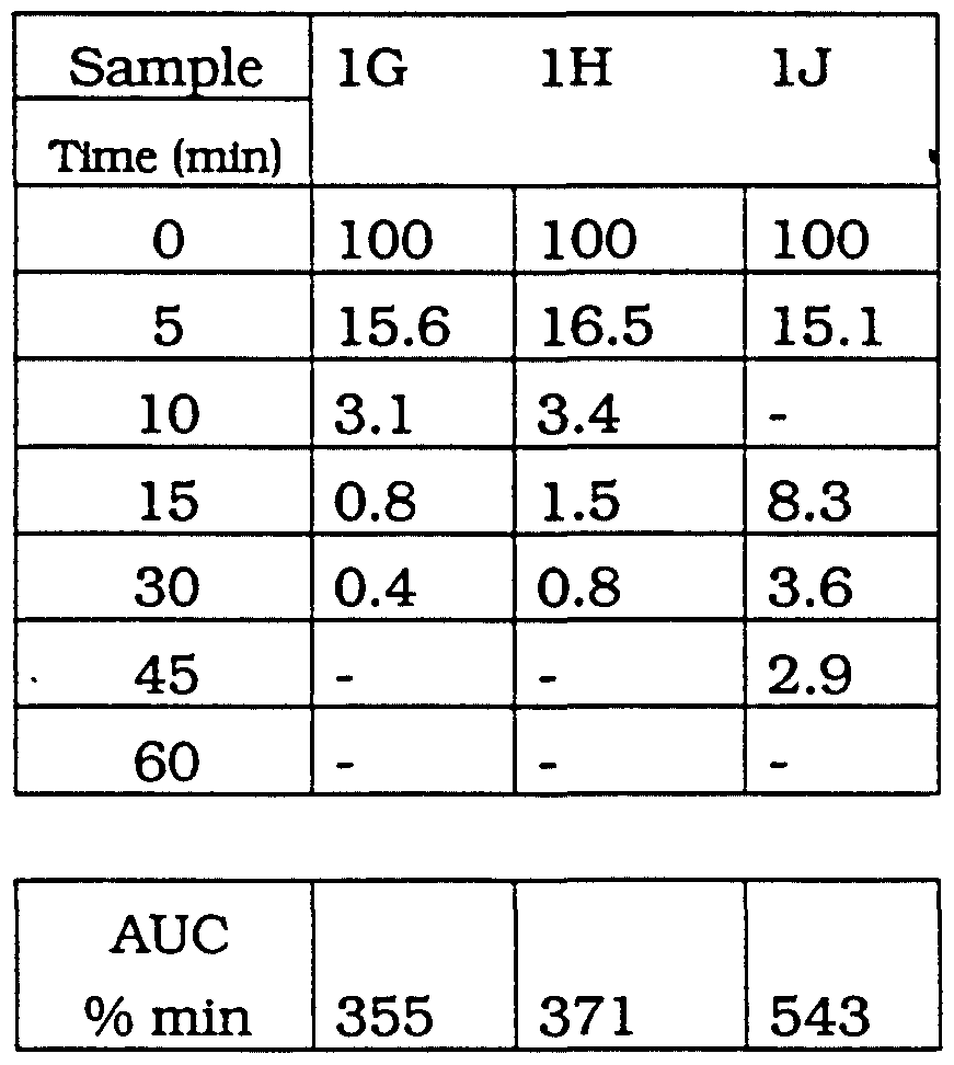
- component a is replaced by an analog compound having but only one ionic function on the phosphate group, i.e. dipalmitoylphosphatidyl glycerol (DPPG) (Sample 1G) the layer obtained offers practically no protection. This being so even with addition of Synperonlc ® (Sample 1H).
- DPPG dipalmitoylphosphatidyl glycerol
- Example IJ there are also provided in Table 2 results obtained with a sample of dextran coated magnetite (AMI-25 prepared according to EP-A-0 274 285 of Advanced Magnetics, Inc.).
- phospholipids are claimed to be useful material for production of stable iron oxide particles.
- DPPC dipalmitoylphosphatidylcholine
- DMPC dimyristoylphosphatidyl- choline
- the contrast agents made may be useful for imaging of liver and spleen or the bone marrow but not as long lasting blood pool agents.
- Much the same has been found for phospholipids whose phosphoryl head moieties have only one negative charge available for interaction with the magnetite core.
- dipalmitoylphosphatidylglycerol DPPG
- dimyristoylphosphatidyl- glycerol DMPG
- dicetylphosphate DCP
- DPPG dipalmitoylphosphatidylglycerol
- DMPG dimyristoylphosphatidyl- glycerol
- DCP dicetylphosphate
- DLPA l,2-Dilauroyl-glycero-3-phosphatidic acid
- DMPA l,2-Dimyristoyl-glycero-3-phosphatidic acid
- DPPA l,2-Dipalmitoyl-glycero-3-phosphatidic acid
- DMPC l,2-Dimyristoyl-glycero-3-phosphocholine
- DPPC l,2-Dipalmitoyl-glycero-3-phosphocholine
- DSPC l,2-Distearoyl-glycero-3-phosphocholine
- DAPC l,2-Diarachidoyl-glycero-3-phosphocholine
- DMPG 1 ,2-Dimyristoyl-glycero-3-phosphoglycerol
- DPPG 1 ,2-Dipalmitoyl-glycero-3-phosphoglycerol
- PHOSPHATE Phosphoric acid mono ester of cetyl alcohol (C 16 ).
- BRIJ Polyethyleneglycol hexadecylether
- MAG(V) Magnetite with liposomes prepared according to
- AMI 25 Dextran-coated magnetite prepared according to EP-A-0 275 285.
- DPPC/DPPA DPPC to DPPA weight ratio 7.5/2.5
- DPPC/DPPA 3 DPPC to DPPA weight ratio 5.0/5.0
- DPPC/DPPA 4 DPPC to DPPA weight ratio 2.5/7.5
- DMPG 12
- DPPA dipalmitoylphosphatldic acid
- DMPA dimyristoyphosphatidic acid
- iron oxide particles prepared with, for example, dipalmitoyl- phosphatidic acid (DPPA) in micellar form and Synperonlc® F108 using the method of the invention will produce particles well protected against removal from the blood stream.
- DPPA dipalmitoyl- phosphatidic acid
- Synperonlc® F108 using the method of the invention
Abstract
Description


Claims
Priority Applications (11)
| Application Number | Priority Date | Filing Date | Title |
|---|---|---|---|
| CA002119654A CA2119654C (en) | 1992-08-13 | 1993-07-30 | Particles for nmr imaging and method of manufacture |
| DE69305730T DE69305730T2 (en) | 1992-08-13 | 1993-07-30 | PARTICLES FOR NMR IMAGING AND PRODUCTION METHOD |
| AU47054/93A AU660108B2 (en) | 1992-08-13 | 1993-07-30 | Particles for NMR imaging and method of manufacture |
| EP93917714A EP0607401B1 (en) | 1992-08-13 | 1993-07-30 | Particles for nmr imaging and method of manufacture |
| KR1019940701200A KR100307773B1 (en) | 1992-08-13 | 1993-07-30 | Particles and Manufacturing Methods for NMR Imaging |
| DK93917714.3T DK0607401T3 (en) | 1992-08-13 | 1993-07-30 | Particles for NMR imaging and method of preparation thereof |
| HU9401038A HU221805B1 (en) | 1992-08-13 | 1993-07-30 | Iron-oxide particles for nmr imaging and method of manufacture them |
| JP6505830A JP2968588B2 (en) | 1992-08-13 | 1993-07-30 | Particles for NMR imaging and method of manufacture |
| NO941311A NO307244B1 (en) | 1992-08-13 | 1994-04-12 | Particles for NMR imaging, use of the same for NMR imaging, powdered formulation of the same, injectable aqueous suspension of the same and process for the preparation of the same |
| FI941677A FI941677A0 (en) | 1992-08-13 | 1994-04-12 | Particles suitable for NMR imaging and method of preparation |
| GR960403292T GR3022288T3 (en) | 1992-08-13 | 1997-01-16 | Particles for nmr imaging and method of manufacture. |
Applications Claiming Priority (4)
| Application Number | Priority Date | Filing Date | Title |
|---|---|---|---|
| EP92810618 | 1992-08-13 | ||
| EP92810618.6 | 1992-08-13 | ||
| EP93810380 | 1993-05-25 | ||
| EP93810380.1 | 1993-05-25 |
Publications (1)
| Publication Number | Publication Date |
|---|---|
| WO1994004197A1 true WO1994004197A1 (en) | 1994-03-03 |
Family
ID=26132520
Family Applications (1)
| Application Number | Title | Priority Date | Filing Date |
|---|---|---|---|
| PCT/EP1993/002046 WO1994004197A1 (en) | 1992-08-13 | 1993-07-30 | Particles for nmr imaging and method of manufacture |
Country Status (20)
| Country | Link |
|---|---|
| US (3) | US5464696A (en) |
| EP (1) | EP0607401B1 (en) |
| JP (1) | JP2968588B2 (en) |
| KR (1) | KR100307773B1 (en) |
| CN (1) | CN1076205C (en) |
| AT (1) | ATE144714T1 (en) |
| AU (1) | AU660108B2 (en) |
| CA (1) | CA2119654C (en) |
| DE (1) | DE69305730T2 (en) |
| DK (1) | DK0607401T3 (en) |
| ES (1) | ES2094558T3 (en) |
| FI (1) | FI941677A0 (en) |
| GR (1) | GR3022288T3 (en) |
| HU (1) | HU221805B1 (en) |
| IL (1) | IL106493A (en) |
| IS (1) | IS1648B (en) |
| MX (1) | MX9304924A (en) |
| NO (1) | NO307244B1 (en) |
| NZ (1) | NZ254791A (en) |
| WO (1) | WO1994004197A1 (en) |
Cited By (15)
| Publication number | Priority date | Publication date | Assignee | Title |
|---|---|---|---|---|
| WO1996004017A1 (en) * | 1994-08-04 | 1996-02-15 | Institut für Diagnostikforschung GmbH an der Freien Universität Berlin | Iron-containing nanoparticles with double coating and their use in diagnosis and therapy |
| WO1997000087A1 (en) * | 1995-06-15 | 1997-01-03 | Bracco Research S.A. | Blood-pool imaging compositions use and method |
| DE19529921A1 (en) * | 1995-08-01 | 1997-02-06 | Schering Ag | Ventilation imaging means for the lungs |
| WO1997016474A1 (en) * | 1995-11-01 | 1997-05-09 | Bracco Research S.A. | Targeted magnetically labeled molecular marker systems for the nmr imaging |
| EP0808125A1 (en) * | 1995-01-27 | 1997-11-26 | Mallinckrodt Medical, Inc. | Calcium/oxyanion-containing particles for use in medical diagnostic imaging |
| US6123920A (en) * | 1996-01-10 | 2000-09-26 | Nycomed Imaging As | Superparamagnetic contrast media coated with starch and polyalkylene oxides |
| US6423296B1 (en) | 1996-01-10 | 2002-07-23 | Amersham Health As | Constrast media |
| EP2279757A2 (en) | 2000-06-02 | 2011-02-02 | Bracco Suisse SA | Compounds for targeting endothelial cells |
| US9180210B2 (en) | 2008-08-14 | 2015-11-10 | Commissariat A L'energie Atomique Et Aux Energies Alternatives | Nanocrystal nano-emulsion |
| US9248204B2 (en) | 2004-08-18 | 2016-02-02 | Bracco Suisse S.A. | Gas-filled microvesicles composition for contrast imaging |
| US9289517B2 (en) | 2008-08-14 | 2016-03-22 | Commissariat A L'energie Atomique Et Aux Energies Alternatives | Fluorescent emulsion of indocyanine green |
| US9364569B2 (en) | 2003-02-04 | 2016-06-14 | Bracco Suisse S.A. | Ultrasound contrast agents and process for the preparation thereof |
| US9750821B2 (en) | 2003-12-22 | 2017-09-05 | Bracco Suisse S.A. | Gas-filled microvesicle assembly for contrast imaging |
| US10092506B2 (en) | 2008-08-14 | 2018-10-09 | Commissariat A L'energie Atomique Et Aux Energies Alternatives | Encapsulation of lipophilic or amphiphilic therapeutic agents in nano-emulsion |
| FR3103376A1 (en) * | 2019-11-26 | 2021-05-28 | Universite de Bordeaux | Biocompatible oily ferrofluid and method of preparation |
Families Citing this family (36)
| Publication number | Priority date | Publication date | Assignee | Title |
|---|---|---|---|---|
| US6465188B1 (en) * | 1990-06-11 | 2002-10-15 | Gilead Sciences, Inc. | Nucleic acid ligand complexes |
| TW319763B (en) * | 1995-02-01 | 1997-11-11 | Epix Medical Inc | |
| DE19508772C2 (en) * | 1995-03-01 | 1998-01-29 | Schering Ag | Methods and connections for the detection of analytes by means of remanence measurement and their use |
| US8071737B2 (en) * | 1995-05-04 | 2011-12-06 | Glead Sciences, Inc. | Nucleic acid ligand complexes |
| AU7104598A (en) | 1997-04-09 | 1998-10-30 | Philipp Lang | New technique to monitor drug delivery noninvasively (in vivo) |
| US6337215B1 (en) * | 1997-12-01 | 2002-01-08 | International Business Machines Corporation | Magnetic particles having two antiparallel ferromagnetic layers and attached affinity recognition molecules |
| US6278893B1 (en) * | 1998-01-05 | 2001-08-21 | Nycomed Imaging As | Method of magnetic resonance imaging of a sample with ex vivo polarization of an MR imaging agent |
| EP1118009A1 (en) * | 1998-09-28 | 2001-07-25 | Nycomed Imaging As | Method of magnetic resonance imaging |
| US6283448B1 (en) * | 2000-04-19 | 2001-09-04 | Daniel Webster Denton | Offset butterfly valve |
| US20050059031A1 (en) * | 2000-10-06 | 2005-03-17 | Quantum Dot Corporation | Method for enhancing transport of semiconductor nanocrystals across biological membranes |
| ATE425457T1 (en) * | 2000-10-06 | 2009-03-15 | Life Technologies Corp | CELLS WITH A SPECTRAL SIGNATURE AND METHOD FOR THEIR PRODUCTION AND USE |
| US20090060992A1 (en) * | 2002-05-08 | 2009-03-05 | University Of Central Florida Research Foundation, Inc., | Preparation of magneto-vesicles with DOPE/DDAB layers |
| US20070128117A1 (en) * | 2003-02-04 | 2007-06-07 | Bracco International B.V. | Ultrasound contrast agents and process for the preparation thereof |
| DE10331439B3 (en) | 2003-07-10 | 2005-02-03 | Micromod Partikeltechnologie Gmbh | Magnetic nanoparticles with improved magnetic properties |
| JP2007515471A (en) * | 2003-12-22 | 2007-06-14 | ブラッコ・リサーチ・ソシエテ・アノニム | Assembly of gas-filled microvesicles with active ingredients for contrast imaging |
| US20050260137A1 (en) * | 2004-05-18 | 2005-11-24 | General Electric Company | Contrast agents for magnetic resonance imaging |
| US7229690B2 (en) * | 2004-07-26 | 2007-06-12 | Massachusetts Institute Of Technology | Microspheres including nanoparticles |
| JPWO2006028129A1 (en) * | 2004-09-10 | 2008-05-08 | 東レ株式会社 | Pharmaceutical formulation |
| US20070264199A1 (en) * | 2005-09-26 | 2007-11-15 | Labhasetwar Vinod D | Magnetic nanoparticle composition and methods for using the same |
| US8361494B2 (en) * | 2006-03-10 | 2013-01-29 | The Trustees Of The University Of Pennsylvania | Biomimetic iron-oxide-containing lipoprotein and related materials |
| DE602007006968D1 (en) | 2006-09-05 | 2010-07-15 | Bracco Research Sa | GAS-FILLED MICROVESICLES WITH POLYMER-MODIFIED LIPIDES |
| US8501159B2 (en) * | 2006-12-18 | 2013-08-06 | Colorobbia Italia S.P.A. | Magnetic nanoparticles for the application in hyperthermia, preparation thereof and use in constructs having a pharmacological application |
| US8784659B2 (en) * | 2007-08-08 | 2014-07-22 | General Electric Company | Method for controlling microbial biofilm in aqueous systems |
| WO2009145813A1 (en) * | 2008-03-04 | 2009-12-03 | Qd Vision, Inc. | Particles including nanoparticles, uses thereof, and methods |
| AU2009301141B2 (en) * | 2008-10-07 | 2015-08-27 | Bracco Suisse S.A. | Targeting construct comprising anti-polymer antibody and liposomes or microvesicles binding to the same |
| WO2011031876A1 (en) | 2009-09-09 | 2011-03-17 | Qd Vision, Inc. | Formulations including nanoparticles |
| KR101865888B1 (en) | 2009-09-09 | 2018-06-08 | 삼성전자주식회사 | Particles including nanoparticles, uses thereof, and methods |
| JP5678565B2 (en) * | 2009-12-08 | 2015-03-04 | Jnc株式会社 | Magnetic fine particles and method for producing the same |
| US20110217379A1 (en) * | 2010-03-08 | 2011-09-08 | Davis Llp | Magnetic nanomaterials and methods for chemoembolisation |
| KR101174470B1 (en) | 2010-04-08 | 2012-08-16 | 한국세라믹기술원 | Magnetic silica-liposome nanospheres and preparation method thereof |
| TWI428284B (en) | 2010-11-05 | 2014-03-01 | Nat Univ Chung Cheng | Sea urchin iron oxide and its manufacturing method |
| US8889103B2 (en) | 2010-12-15 | 2014-11-18 | General Electric Company | Diagnostic agent composition and associated methods thereof |
| US8895068B2 (en) | 2010-12-15 | 2014-11-25 | General Electric Company | Nanoparticle composition and associated methods thereof |
| WO2012136813A2 (en) | 2011-04-07 | 2012-10-11 | Universitetet I Oslo | Agents for medical radar diagnosis |
| JP6313329B2 (en) | 2012-12-21 | 2018-04-18 | ブラッコ・スイス・ソシエテ・アノニムBracco Suisse SA | Gas filled micro vesicle |
| US20160346408A1 (en) * | 2015-05-26 | 2016-12-01 | Intezyne Technologies | Iron stabilized micelles as magnetic contrast agents |
Citations (5)
| Publication number | Priority date | Publication date | Assignee | Title |
|---|---|---|---|---|
| EP0249229A2 (en) * | 1986-06-12 | 1987-12-16 | BASF Aktiengesellschaft | Superparamagnetic solid particles |
| EP0272091A2 (en) * | 1986-12-15 | 1988-06-22 | Nexstar Pharmaceuticals, Inc. | Delivery vehicles with amphiphile-associated active ingredient |
| EP0284549A2 (en) * | 1987-03-24 | 1988-09-28 | Silica Gel Ges.mbH Absorptionstechnik, Apparatebau | Magnetic fluid compositions |
| WO1991014454A1 (en) * | 1990-03-29 | 1991-10-03 | Skua Investments Limited | Pharmaceutical formulations |
| EP0494615A1 (en) * | 1991-01-09 | 1992-07-15 | Byk Gulden Lomberg Chemische Fabrik Gmbh | Echo contrast agent |
Family Cites Families (26)
| Publication number | Priority date | Publication date | Assignee | Title |
|---|---|---|---|---|
| US4331654A (en) * | 1980-06-13 | 1982-05-25 | Eli Lilly And Company | Magnetically-localizable, biodegradable lipid microspheres |
| US4331645A (en) * | 1981-04-20 | 1982-05-25 | Reynolds Metals Company | Alumina from alkali metal-aluminum chloride complexes |
| NL194579C (en) * | 1983-01-21 | 2002-08-05 | Schering Ag | Diagnostic. |
| JPS6072830A (en) * | 1983-09-29 | 1985-04-24 | Kao Corp | Composition for vesicle |
| GB8408127D0 (en) * | 1984-03-29 | 1984-05-10 | Nyegaard & Co As | Contrast agents |
| US4728575A (en) * | 1984-04-27 | 1988-03-01 | Vestar, Inc. | Contrast agents for NMR imaging |
| DE3515373A1 (en) * | 1985-04-27 | 1986-11-06 | Merck Patent Gmbh, 6100 Darmstadt | NITROGENIC HETEROCYCLES |
| US4675173A (en) * | 1985-05-08 | 1987-06-23 | Molecular Biosystems, Inc. | Method of magnetic resonance imaging of the liver and spleen |
| JPS6295134A (en) * | 1985-10-21 | 1987-05-01 | Nippon Saafuakutanto Kogyo Kk | Production of liposome |
| US4951675A (en) * | 1986-07-03 | 1990-08-28 | Advanced Magnetics, Incorporated | Biodegradable superparamagnetic metal oxides as contrast agents for MR imaging |
| US5262176A (en) * | 1986-07-03 | 1993-11-16 | Advanced Magnetics, Inc. | Synthesis of polysaccharide covered superparamagnetic oxide colloids |
| US4929589A (en) * | 1986-12-29 | 1990-05-29 | Aluminum Company Of America | Metal oxide/hydroxide particles coated with phosphate esters |
| US5124289A (en) * | 1986-12-29 | 1992-06-23 | Aluminum Company Of America | Surface treated permeable inorganic membranes and process of making same |
| US4994429A (en) * | 1986-12-29 | 1991-02-19 | Aluminum Company Of America | Active material useful as adsorbent comprising metal oxide/hydroxide particles reacted with phosphorus-containing organic acid group of organic compound having unreacted acid group |
| US4983566A (en) * | 1987-03-09 | 1991-01-08 | Aluminum Company Of America | Surface-modified adsorbent comprising metal oxide/hydroxide particles reacted with one or more perfluorinated organic acids |
| US5000960A (en) * | 1987-03-13 | 1991-03-19 | Micro-Pak, Inc. | Protein coupling to lipid vesicles |
| JPH0720857B2 (en) * | 1988-08-11 | 1995-03-08 | テルモ株式会社 | Liposome and its manufacturing method |
| US5230882A (en) * | 1989-12-22 | 1993-07-27 | Unger Evan C | Liposomes as contrast agents for ultrasonic imaging and methods for preparing the same |
| US5091188A (en) * | 1990-04-26 | 1992-02-25 | Haynes Duncan H | Phospholipid-coated microcrystals: injectable formulations of water-insoluble drugs |
| US5246707A (en) * | 1990-04-26 | 1993-09-21 | Haynes Duncan H | Sustained release delivery of water-soluble bio-molecules and drugs using phospholipid-coated microcrystals, microdroplets and high-concentration liposomes |
| CA2046997C (en) * | 1990-07-16 | 2000-12-12 | Hiroshi Kikuchi | Liposomal products |
| JP3272736B2 (en) * | 1991-01-31 | 2002-04-08 | 協和醗酵工業株式会社 | Liposome preparation |
| CA2064683A1 (en) * | 1992-03-26 | 1993-09-27 | Krishna Mohan Rao Kallury | Formation of thermostable enzymes with extra-ordinary heat tolerance by immobilization on phospholipid matrices |
| US5411730A (en) * | 1993-07-20 | 1995-05-02 | Research Corporation Technologies, Inc. | Magnetic microparticles |
| US5494744A (en) * | 1994-10-12 | 1996-02-27 | Kimberly-Clark Corporation | Method of applying a protein coating to a substrate and article thereof |
| EP2499229B1 (en) * | 2009-11-10 | 2015-03-04 | Microphyt | Reaction casing for a photosynthetic reactor and associated photosynthetic reactor |
-
1993
- 1993-07-26 US US08/096,414 patent/US5464696A/en not_active Expired - Lifetime
- 1993-07-27 IL IL10649393A patent/IL106493A/en not_active IP Right Cessation
- 1993-07-27 IS IS4058A patent/IS1648B/en unknown
- 1993-07-30 AT AT93917714T patent/ATE144714T1/en not_active IP Right Cessation
- 1993-07-30 AU AU47054/93A patent/AU660108B2/en not_active Ceased
- 1993-07-30 EP EP93917714A patent/EP0607401B1/en not_active Expired - Lifetime
- 1993-07-30 NZ NZ254791A patent/NZ254791A/en unknown
- 1993-07-30 ES ES93917714T patent/ES2094558T3/en not_active Expired - Lifetime
- 1993-07-30 HU HU9401038A patent/HU221805B1/en not_active IP Right Cessation
- 1993-07-30 DE DE69305730T patent/DE69305730T2/en not_active Expired - Lifetime
- 1993-07-30 DK DK93917714.3T patent/DK0607401T3/en active
- 1993-07-30 KR KR1019940701200A patent/KR100307773B1/en not_active IP Right Cessation
- 1993-07-30 JP JP6505830A patent/JP2968588B2/en not_active Expired - Fee Related
- 1993-07-30 WO PCT/EP1993/002046 patent/WO1994004197A1/en active IP Right Grant
- 1993-07-30 CA CA002119654A patent/CA2119654C/en not_active Expired - Fee Related
- 1993-08-12 CN CN93116505A patent/CN1076205C/en not_active Expired - Fee Related
- 1993-08-13 MX MX9304924A patent/MX9304924A/en unknown
-
1994
- 1994-04-12 FI FI941677A patent/FI941677A0/en unknown
- 1994-04-12 NO NO941311A patent/NO307244B1/en not_active IP Right Cessation
-
1995
- 1995-04-13 US US08/421,330 patent/US5545395A/en not_active Expired - Lifetime
- 1995-04-13 US US08/421,329 patent/US5587199A/en not_active Expired - Lifetime
-
1997
- 1997-01-16 GR GR960403292T patent/GR3022288T3/en unknown
Patent Citations (5)
| Publication number | Priority date | Publication date | Assignee | Title |
|---|---|---|---|---|
| EP0249229A2 (en) * | 1986-06-12 | 1987-12-16 | BASF Aktiengesellschaft | Superparamagnetic solid particles |
| EP0272091A2 (en) * | 1986-12-15 | 1988-06-22 | Nexstar Pharmaceuticals, Inc. | Delivery vehicles with amphiphile-associated active ingredient |
| EP0284549A2 (en) * | 1987-03-24 | 1988-09-28 | Silica Gel Ges.mbH Absorptionstechnik, Apparatebau | Magnetic fluid compositions |
| WO1991014454A1 (en) * | 1990-03-29 | 1991-10-03 | Skua Investments Limited | Pharmaceutical formulations |
| EP0494615A1 (en) * | 1991-01-09 | 1992-07-15 | Byk Gulden Lomberg Chemische Fabrik Gmbh | Echo contrast agent |
Non-Patent Citations (1)
| Title |
|---|
| R. H. MÜLLER ET AL.: "in vitro characterization of poly (methyl-methacrylate) nanoparticles and correlation to their in vivo fate", JOURNAL OF CONTROLLED RELEASE, vol. 20, no. 3, August 1992 (1992-08-01), AMSTERDAM (NL), pages 237 - 246, XP000289755 * |
Cited By (25)
| Publication number | Priority date | Publication date | Assignee | Title |
|---|---|---|---|---|
| CN1103604C (en) * | 1994-08-04 | 2003-03-26 | 柏林弗赖恩大学诊断研究学院有限公司 | Iron-containing nanoparticles with double coating and their use in diagnosis and therapy |
| WO1996004017A1 (en) * | 1994-08-04 | 1996-02-15 | Institut für Diagnostikforschung GmbH an der Freien Universität Berlin | Iron-containing nanoparticles with double coating and their use in diagnosis and therapy |
| AU703042B2 (en) * | 1994-08-04 | 1999-03-11 | Institut Fur Diagnostikforschung Gmbh An Der Freien Universitat Berlin | Iron-containing nanoparticles with double coating and their use in diagnosis and therapy |
| US6048515A (en) * | 1994-08-04 | 2000-04-11 | Institut Fur Diagnostikforschung Gmbh | Iron-containing nanoparticles with double coating and their use in diagnosis and therapy |
| EP0808125A1 (en) * | 1995-01-27 | 1997-11-26 | Mallinckrodt Medical, Inc. | Calcium/oxyanion-containing particles for use in medical diagnostic imaging |
| EP0808125A4 (en) * | 1995-01-27 | 2000-07-12 | Mallinckrodt Medical Inc | Calcium/oxyanion-containing particles for use in medical diagnostic imaging |
| US5833948A (en) * | 1995-06-15 | 1998-11-10 | Bracco Research S.A. | Blood-pool imaging composition comprising micelles containing a lipophilic chelating agent and a non-ionic surfactant |
| WO1997000087A1 (en) * | 1995-06-15 | 1997-01-03 | Bracco Research S.A. | Blood-pool imaging compositions use and method |
| DE19529921A1 (en) * | 1995-08-01 | 1997-02-06 | Schering Ag | Ventilation imaging means for the lungs |
| WO1997016474A1 (en) * | 1995-11-01 | 1997-05-09 | Bracco Research S.A. | Targeted magnetically labeled molecular marker systems for the nmr imaging |
| US5910300A (en) * | 1995-11-01 | 1999-06-08 | Bracco Research S.A. | Amphiphilic linkers for coupling administrable diagnostically or physiologically active agents and bioselective targeting compounds |
| US6123920A (en) * | 1996-01-10 | 2000-09-26 | Nycomed Imaging As | Superparamagnetic contrast media coated with starch and polyalkylene oxides |
| US6423296B1 (en) | 1996-01-10 | 2002-07-23 | Amersham Health As | Constrast media |
| EP2286843A2 (en) | 2000-06-02 | 2011-02-23 | Bracco Suisse SA | Compounds for targeting endothelial cells |
| EP2279757A2 (en) | 2000-06-02 | 2011-02-02 | Bracco Suisse SA | Compounds for targeting endothelial cells |
| US9364569B2 (en) | 2003-02-04 | 2016-06-14 | Bracco Suisse S.A. | Ultrasound contrast agents and process for the preparation thereof |
| US9750821B2 (en) | 2003-12-22 | 2017-09-05 | Bracco Suisse S.A. | Gas-filled microvesicle assembly for contrast imaging |
| US9248204B2 (en) | 2004-08-18 | 2016-02-02 | Bracco Suisse S.A. | Gas-filled microvesicles composition for contrast imaging |
| US10076580B2 (en) | 2004-08-18 | 2018-09-18 | Bracco Suisse S.A. | Gas-filled microvesicles composition for contrast imaging |
| US9180210B2 (en) | 2008-08-14 | 2015-11-10 | Commissariat A L'energie Atomique Et Aux Energies Alternatives | Nanocrystal nano-emulsion |
| US9289517B2 (en) | 2008-08-14 | 2016-03-22 | Commissariat A L'energie Atomique Et Aux Energies Alternatives | Fluorescent emulsion of indocyanine green |
| US10092506B2 (en) | 2008-08-14 | 2018-10-09 | Commissariat A L'energie Atomique Et Aux Energies Alternatives | Encapsulation of lipophilic or amphiphilic therapeutic agents in nano-emulsion |
| FR3103376A1 (en) * | 2019-11-26 | 2021-05-28 | Universite de Bordeaux | Biocompatible oily ferrofluid and method of preparation |
| WO2021105620A1 (en) * | 2019-11-26 | 2021-06-03 | Universite de Bordeaux | Biocompatible oily ferrofluid and preparation process |
| CN115003337A (en) * | 2019-11-26 | 2022-09-02 | 波尔多大学 | Biocompatible oily ferrofluid and preparation method thereof |
Also Published As
| Publication number | Publication date |
|---|---|
| EP0607401A1 (en) | 1994-07-27 |
| EP0607401B1 (en) | 1996-10-30 |
| FI941677A (en) | 1994-04-12 |
| FI941677A0 (en) | 1994-04-12 |
| GR3022288T3 (en) | 1997-04-30 |
| HU9401038D0 (en) | 1994-07-28 |
| NZ254791A (en) | 1996-11-26 |
| IL106493A (en) | 1997-02-18 |
| DK0607401T3 (en) | 1996-11-25 |
| JP2968588B2 (en) | 1999-10-25 |
| NO941311D0 (en) | 1994-04-12 |
| AU660108B2 (en) | 1995-06-08 |
| ES2094558T3 (en) | 1997-01-16 |
| IL106493A0 (en) | 1993-11-15 |
| JPH07503976A (en) | 1995-04-27 |
| US5464696A (en) | 1995-11-07 |
| DE69305730T2 (en) | 1997-05-28 |
| US5545395A (en) | 1996-08-13 |
| US5587199A (en) | 1996-12-24 |
| MX9304924A (en) | 1994-08-31 |
| HUT75149A (en) | 1997-04-28 |
| CA2119654C (en) | 2001-02-06 |
| AU4705493A (en) | 1994-03-15 |
| DE69305730D1 (en) | 1996-12-05 |
| KR100307773B1 (en) | 2001-12-28 |
| HU221805B1 (en) | 2003-01-28 |
| IS1648B (en) | 1997-03-25 |
| CA2119654A1 (en) | 1994-03-03 |
| ATE144714T1 (en) | 1996-11-15 |
| NO307244B1 (en) | 2000-03-06 |
| CN1089470A (en) | 1994-07-20 |
| IS4058A (en) | 1994-02-14 |
| NO941311L (en) | 1994-04-12 |
| CN1076205C (en) | 2001-12-19 |
Similar Documents
| Publication | Publication Date | Title |
|---|---|---|
| US5464696A (en) | Particles for NMR imaging | |
| Kabalka et al. | Gadolinium‐labeled liposomes containing paramagnetic amphipathic agents: targeted MRI contrast agents for the liver | |
| EP1793868B1 (en) | Liposomal contrast agents for cest imaging | |
| JP4335976B2 (en) | Blood storage imaging composition and uses thereof | |
| US6132763A (en) | Liposomes | |
| Torchilin | PEG-based micelles as carriers of contrast agents for different imaging modalities | |
| Bulte et al. | Short‐vs. long‐circulating magnetoliposomes as bone marrow‐seeking MR contrast agents | |
| CA1319614C (en) | Delivery vehicles with amphiphile-associated active ingredient | |
| EP1750671B1 (en) | Liposomal assembly for therapeutic and/or diagnostic use | |
| US20030152618A1 (en) | Liposomes | |
| AU680513B2 (en) | Reduction of liposome-induced adverse physiological reactions | |
| US6468505B1 (en) | Technique to monitor drug delivery noninvasively in vivo | |
| Tilcock | Imaging tools: liposomal agents for nuclear medicine, computed tomography, magnetic resonance, and ultrasound | |
| US20070237721A1 (en) | Targeted mr imaging agents | |
| Gore et al. | Liposomes for paramagnetic contrast enhancement in nmr imaging | |
| Päuser et al. | Superparamagnetic iron oxide particles as marker substances for searching tumor specific liposomes with magnetic resonance imaging | |
| Papahadjopoulos et al. | Liposome targeting to tumor cells in vivo | |
| Mor | A BRIEF REVIEW ON LIPOSOME–AS DRUG CARRIER |
Legal Events
| Date | Code | Title | Description |
|---|---|---|---|
| AK | Designated states |
Kind code of ref document: A1 Designated state(s): AU CA FI HU JP KR NO NZ |
|
| AL | Designated countries for regional patents |
Kind code of ref document: A1 Designated state(s): AT BE CH DE DK ES FR GB GR IE IT LU MC NL PT SE |
|
| WWE | Wipo information: entry into national phase |
Ref document number: 1993917714 Country of ref document: EP |
|
| WWE | Wipo information: entry into national phase |
Ref document number: 2119654 Country of ref document: CA |
|
| WWE | Wipo information: entry into national phase |
Ref document number: 254791 Country of ref document: NZ |
|
| WWE | Wipo information: entry into national phase |
Ref document number: 941677 Country of ref document: FI |
|
| 121 | Ep: the epo has been informed by wipo that ep was designated in this application | ||
| WWP | Wipo information: published in national office |
Ref document number: 1993917714 Country of ref document: EP |
|
| WWG | Wipo information: grant in national office |
Ref document number: 1993917714 Country of ref document: EP |