WO1995011995A1 - Arrays of nucleic acid probes on biological chips - Google Patents

Arrays of nucleic acid probes on biological chips Download PDF

Info

Publication number
WO1995011995A1
WO1995011995A1 PCT/US1994/012305 US9412305W WO9511995A1 WO 1995011995 A1 WO1995011995 A1 WO 1995011995A1 US 9412305 W US9412305 W US 9412305W WO 9511995 A1 WO9511995 A1 WO 9511995A1
Authority
WO
WIPO (PCT)
Prior art keywords
probe
array
probes
εequence
probeε
Prior art date
Application number
PCT/US1994/012305
Other languages
French (fr)
Other versions
WO1995011995B1 (en
Inventor
Mark Chee
Maureen T. Cronin
Stephen P. A. Fodor
Thomas R. Gingeras
Xiaohua C. Huang
Earl A. Hubbell
Robert J. Lipshutz
Peter E. Lobban
Charles Garrett Miyada
Macdonald S. Morris
Nila Shah
Edward L. Sheldon
Original Assignee
Affymax Technologies N.V.
Priority date (The priority date is an assumption and is not a legal conclusion. Google has not performed a legal analysis and makes no representation as to the accuracy of the date listed.)
Filing date
Publication date
Application filed by Affymax Technologies N.V. filed Critical Affymax Technologies N.V.
Priority to JP7512811A priority Critical patent/JPH09507121A/en
Priority to DE69433180T priority patent/DE69433180T2/en
Priority to AU81266/94A priority patent/AU8126694A/en
Priority to EP95900441A priority patent/EP0730663B1/en
Publication of WO1995011995A1 publication Critical patent/WO1995011995A1/en
Publication of WO1995011995B1 publication Critical patent/WO1995011995B1/en
Priority to US08/510,521 priority patent/US7115364B1/en
Priority to US08/630,427 priority patent/US6156501A/en
Priority to US08/648,709 priority patent/US6045996A/en
Priority to US08/778,794 priority patent/US6309823B1/en
Priority to US09/393,389 priority patent/US6632605B1/en
Priority to US09/510,378 priority patent/US7399584B2/en
Priority to US09/798,260 priority patent/US20030165830A1/en
Priority to US10/113,885 priority patent/US20030134291A1/en
Priority to US10/229,319 priority patent/US20030054393A1/en
Priority to US10/402,333 priority patent/US20030232361A1/en
Priority to US10/418,414 priority patent/US7375198B2/en
Priority to US11/367,800 priority patent/US20060229824A1/en
Priority to US11/388,199 priority patent/US20060263807A1/en
Priority to US11/401,482 priority patent/US7846659B2/en
Priority to US11/506,176 priority patent/US20120329677A9/en
Priority to US12/124,061 priority patent/US7794943B2/en
Priority to US12/878,743 priority patent/US20110092382A1/en
Priority to US12/959,359 priority patent/US20130150248A1/en

Links

Classifications

    • BPERFORMING OPERATIONS; TRANSPORTING
    • B82NANOTECHNOLOGY
    • B82YSPECIFIC USES OR APPLICATIONS OF NANOSTRUCTURES; MEASUREMENT OR ANALYSIS OF NANOSTRUCTURES; MANUFACTURE OR TREATMENT OF NANOSTRUCTURES
    • B82Y30/00Nanotechnology for materials or surface science, e.g. nanocomposites
    • CCHEMISTRY; METALLURGY
    • C07ORGANIC CHEMISTRY
    • C07HSUGARS; DERIVATIVES THEREOF; NUCLEOSIDES; NUCLEOTIDES; NUCLEIC ACIDS
    • C07H21/00Compounds containing two or more mononucleotide units having separate phosphate or polyphosphate groups linked by saccharide radicals of nucleoside groups, e.g. nucleic acids
    • CCHEMISTRY; METALLURGY
    • C12BIOCHEMISTRY; BEER; SPIRITS; WINE; VINEGAR; MICROBIOLOGY; ENZYMOLOGY; MUTATION OR GENETIC ENGINEERING
    • C12QMEASURING OR TESTING PROCESSES INVOLVING ENZYMES, NUCLEIC ACIDS OR MICROORGANISMS; COMPOSITIONS OR TEST PAPERS THEREFOR; PROCESSES OF PREPARING SUCH COMPOSITIONS; CONDITION-RESPONSIVE CONTROL IN MICROBIOLOGICAL OR ENZYMOLOGICAL PROCESSES
    • C12Q1/00Measuring or testing processes involving enzymes, nucleic acids or microorganisms; Compositions therefor; Processes of preparing such compositions
    • C12Q1/68Measuring or testing processes involving enzymes, nucleic acids or microorganisms; Compositions therefor; Processes of preparing such compositions involving nucleic acids
    • C12Q1/6813Hybridisation assays
    • C12Q1/6834Enzymatic or biochemical coupling of nucleic acids to a solid phase
    • C12Q1/6837Enzymatic or biochemical coupling of nucleic acids to a solid phase using probe arrays or probe chips
    • CCHEMISTRY; METALLURGY
    • C12BIOCHEMISTRY; BEER; SPIRITS; WINE; VINEGAR; MICROBIOLOGY; ENZYMOLOGY; MUTATION OR GENETIC ENGINEERING
    • C12QMEASURING OR TESTING PROCESSES INVOLVING ENZYMES, NUCLEIC ACIDS OR MICROORGANISMS; COMPOSITIONS OR TEST PAPERS THEREFOR; PROCESSES OF PREPARING SUCH COMPOSITIONS; CONDITION-RESPONSIVE CONTROL IN MICROBIOLOGICAL OR ENZYMOLOGICAL PROCESSES
    • C12Q1/00Measuring or testing processes involving enzymes, nucleic acids or microorganisms; Compositions therefor; Processes of preparing such compositions
    • C12Q1/68Measuring or testing processes involving enzymes, nucleic acids or microorganisms; Compositions therefor; Processes of preparing such compositions involving nucleic acids
    • C12Q1/6869Methods for sequencing
    • C12Q1/6874Methods for sequencing involving nucleic acid arrays, e.g. sequencing by hybridisation
    • BPERFORMING OPERATIONS; TRANSPORTING
    • B01PHYSICAL OR CHEMICAL PROCESSES OR APPARATUS IN GENERAL
    • B01JCHEMICAL OR PHYSICAL PROCESSES, e.g. CATALYSIS OR COLLOID CHEMISTRY; THEIR RELEVANT APPARATUS
    • B01J2219/00Chemical, physical or physico-chemical processes in general; Their relevant apparatus
    • B01J2219/00274Sequential or parallel reactions; Apparatus and devices for combinatorial chemistry or for making arrays; Chemical library technology
    • B01J2219/00277Apparatus
    • B01J2219/00351Means for dispensing and evacuation of reagents
    • B01J2219/00427Means for dispensing and evacuation of reagents using masks
    • B01J2219/00432Photolithographic masks
    • BPERFORMING OPERATIONS; TRANSPORTING
    • B01PHYSICAL OR CHEMICAL PROCESSES OR APPARATUS IN GENERAL
    • B01JCHEMICAL OR PHYSICAL PROCESSES, e.g. CATALYSIS OR COLLOID CHEMISTRY; THEIR RELEVANT APPARATUS
    • B01J2219/00Chemical, physical or physico-chemical processes in general; Their relevant apparatus
    • B01J2219/00274Sequential or parallel reactions; Apparatus and devices for combinatorial chemistry or for making arrays; Chemical library technology
    • B01J2219/00277Apparatus
    • B01J2219/00497Features relating to the solid phase supports
    • B01J2219/00527Sheets
    • B01J2219/00529DNA chips
    • BPERFORMING OPERATIONS; TRANSPORTING
    • B01PHYSICAL OR CHEMICAL PROCESSES OR APPARATUS IN GENERAL
    • B01JCHEMICAL OR PHYSICAL PROCESSES, e.g. CATALYSIS OR COLLOID CHEMISTRY; THEIR RELEVANT APPARATUS
    • B01J2219/00Chemical, physical or physico-chemical processes in general; Their relevant apparatus
    • B01J2219/00274Sequential or parallel reactions; Apparatus and devices for combinatorial chemistry or for making arrays; Chemical library technology
    • B01J2219/00583Features relative to the processes being carried out
    • B01J2219/00603Making arrays on substantially continuous surfaces
    • B01J2219/00605Making arrays on substantially continuous surfaces the compounds being directly bound or immobilised to solid supports
    • B01J2219/00608DNA chips
    • BPERFORMING OPERATIONS; TRANSPORTING
    • B01PHYSICAL OR CHEMICAL PROCESSES OR APPARATUS IN GENERAL
    • B01JCHEMICAL OR PHYSICAL PROCESSES, e.g. CATALYSIS OR COLLOID CHEMISTRY; THEIR RELEVANT APPARATUS
    • B01J2219/00Chemical, physical or physico-chemical processes in general; Their relevant apparatus
    • B01J2219/00274Sequential or parallel reactions; Apparatus and devices for combinatorial chemistry or for making arrays; Chemical library technology
    • B01J2219/00583Features relative to the processes being carried out
    • B01J2219/00603Making arrays on substantially continuous surfaces
    • B01J2219/00605Making arrays on substantially continuous surfaces the compounds being directly bound or immobilised to solid supports
    • B01J2219/00623Immobilisation or binding
    • B01J2219/00626Covalent
    • BPERFORMING OPERATIONS; TRANSPORTING
    • B01PHYSICAL OR CHEMICAL PROCESSES OR APPARATUS IN GENERAL
    • B01JCHEMICAL OR PHYSICAL PROCESSES, e.g. CATALYSIS OR COLLOID CHEMISTRY; THEIR RELEVANT APPARATUS
    • B01J2219/00Chemical, physical or physico-chemical processes in general; Their relevant apparatus
    • B01J2219/00274Sequential or parallel reactions; Apparatus and devices for combinatorial chemistry or for making arrays; Chemical library technology
    • B01J2219/00583Features relative to the processes being carried out
    • B01J2219/00603Making arrays on substantially continuous surfaces
    • B01J2219/00659Two-dimensional arrays
    • BPERFORMING OPERATIONS; TRANSPORTING
    • B01PHYSICAL OR CHEMICAL PROCESSES OR APPARATUS IN GENERAL
    • B01JCHEMICAL OR PHYSICAL PROCESSES, e.g. CATALYSIS OR COLLOID CHEMISTRY; THEIR RELEVANT APPARATUS
    • B01J2219/00Chemical, physical or physico-chemical processes in general; Their relevant apparatus
    • B01J2219/00274Sequential or parallel reactions; Apparatus and devices for combinatorial chemistry or for making arrays; Chemical library technology
    • B01J2219/00709Type of synthesis
    • B01J2219/00711Light-directed synthesis
    • BPERFORMING OPERATIONS; TRANSPORTING
    • B01PHYSICAL OR CHEMICAL PROCESSES OR APPARATUS IN GENERAL
    • B01JCHEMICAL OR PHYSICAL PROCESSES, e.g. CATALYSIS OR COLLOID CHEMISTRY; THEIR RELEVANT APPARATUS
    • B01J2219/00Chemical, physical or physico-chemical processes in general; Their relevant apparatus
    • B01J2219/00274Sequential or parallel reactions; Apparatus and devices for combinatorial chemistry or for making arrays; Chemical library technology
    • B01J2219/00718Type of compounds synthesised
    • B01J2219/0072Organic compounds
    • B01J2219/00722Nucleotides
    • CCHEMISTRY; METALLURGY
    • C07ORGANIC CHEMISTRY
    • C07BGENERAL METHODS OF ORGANIC CHEMISTRY; APPARATUS THEREFOR
    • C07B2200/00Indexing scheme relating to specific properties of organic compounds
    • C07B2200/11Compounds covalently bound to a solid support
    • CCHEMISTRY; METALLURGY
    • C40COMBINATORIAL TECHNOLOGY
    • C40BCOMBINATORIAL CHEMISTRY; LIBRARIES, e.g. CHEMICAL LIBRARIES
    • C40B40/00Libraries per se, e.g. arrays, mixtures
    • C40B40/04Libraries containing only organic compounds
    • C40B40/06Libraries containing nucleotides or polynucleotides, or derivatives thereof
    • CCHEMISTRY; METALLURGY
    • C40COMBINATORIAL TECHNOLOGY
    • C40BCOMBINATORIAL CHEMISTRY; LIBRARIES, e.g. CHEMICAL LIBRARIES
    • C40B60/00Apparatus specially adapted for use in combinatorial chemistry or with libraries
    • C40B60/14Apparatus specially adapted for use in combinatorial chemistry or with libraries for creating libraries

Definitions

  • the present invention provides arrays of oligonucleotide probes immobilized in microfabricated patterns on silica chips for analyzing molecular interactions of biological interest.
  • the invention therefore relates to diverse fields impacted by the nature of molecular interaction, including chemistry, biology, medicine, and medical diagnostics.
  • Oligonucleotide probes have long been used to detect complementary nucleic acid sequences in a nucleic acid of interest (the "target" nucleic acid) .
  • the oligonucleotide probe is tethered, i.e., by covalent attachment, to a solid support, and arrays of oligonucleotide probes immobilized on solid supports have been used to detect specific nucleic acid sequences in a target nucleic acid. See, e.g., PCT patent publication Nos. WO 89/10977 and 89/11548.
  • Microfabricated arrays of large numbers of oligonucleotide probes, called "DNA chips” offer great promise for a wide variety of applications. New methods and reagents are required to realize this promise, and the present invention helps meet that need.
  • the invention provides several strategies employing immobilized arrays of probes for comparing a reference sequence of known sequence with a target sequence showing substantial similarity with the reference sequence, but differing in the presence of, e.g., mutations.
  • the invention provides a tiling strategy employing an array of immobilized oligonucleotide probes comprising at least two sets of probes.
  • a first probe set comprises a plurality of probes, each probe comprising a segment of at least three nucleotides exactly complementary to a subsequence of the reference sequence, the segment including at least one interrogation position complementary to a corresponding nucleotide in the reference sequence.
  • a second probe set comprises a corresponding probe for each probe in the first probe set, the corresponding probe in the second probe set being identical to a sequence comprising the corresponding probe from the first probe set or a subsequence of at least three nucleotides thereof that includes the at least one interrogation position, except that the at least one interrogation position is occupied by a different nucleotide in each of the two corresponding probes from the first and second probe sets.
  • the probes in the first probe set have at least two interrogation positions corresponding to two contiguous nucleotides in the reference sequence. One interrogation position corresponds to one of the contiguous nucleotides, and the other interrogation position to the other.
  • the invention provides a tiling strategy employing an array comprising four probe sets.
  • a first probe set comprises a plurality of probes, each probe comprising a segment of at least three nucleotides exactly complementary to a subsequence of the reference sequence, the segment including at least one interrogation position complementary to a corresponding nucleotide in the reference sequence.
  • Second, third and fourth probe sets each comprise a corresponding probe for each probe in the first probe set.
  • the probes in the second, third and fourth probe sets are identical to a sequence comprising the corresponding probe from the first probe set or a subsequence of at least three nucleotides thereof that includes the at least one interrogation position, except that the at least one interrogation position is occupied by a different nucleotide in each of the four corresponding probes from the four probe sets.
  • the first probe set often has at least 100 interrogation positions corresponding to 100 contiguous nucleotides in the reference sequence. Sometimes the first probe set has an interrogation position corresponding to every nucleotide in the reference sequence.
  • the segment of complementarity within the probe set is usually about 9-21 nucleotides. Although probes may contain leading or trailing sequences in addition to the 9-21 sequences, many probes consist exclusively of a 9-21 segment of complementarity.
  • the invention provides immobilized arrays of probes tiled for multiple reference sequences.
  • One such array comprises at least one pair of first and second probe groups, each group comprising first and second sets of probes as defined in the first embodiment.
  • Each probe in the first probe set from the first group is exactly complementary to a subsequence of a first reference sequence
  • each probe in the first probe set from the second group is exactly complementary to a subsequence of a second reference sequence.
  • the first group of probes are tiled with respect to a first reference sequence and the second group of probes with respect to a second reference sequence.
  • Each group of probes can also include third and fourth sets of probes as defined in the second embodiment.
  • the second reference sequence is a mutated form of the first reference sequence.
  • Block tiling is a species of the general tiling strategies described above.
  • the usual unit of a block tiling array is a group of probes comprising a wildtype probe, a first set of three mutant probes and a second set of three mutant probes.
  • the wildtype probe comprises a segment of at least three nucleotides exactly complementary to a subsequence of a reference sequence.
  • the segment has at least first and second interrogation positions corresponding to first and second nucleotides in the reference sequence.
  • the probes in the first set of three mutant probes are each identical to a sequence comprising the wildtype probe or a subsequence of at least three nucleotides thereof including the first and second interrogation positions, except in the first interrogation position, which is occupied by a different nucleotide in each of the three mutant probes and the wildtype probe.
  • the probes in the second set of three mutant probes are each identical to a sequence comprising the wildtype probes or a subsequence of at least three nucleotides thereof including the first and second interrogation positions, except in the second interrogation position, which is occupied by a different nucleotide in each of the three mutant probes and the wildtype probe.
  • the invention provides methods of comparing a target sequence with a reference sequence using arrays of immobilized pooled probes.
  • the arrays employed in these methods represent a further species of the general tiling arrays noted above.
  • variants of a reference sequence differing from the reference sequence in at least one nucleotide are identified and each is assigned a designation.
  • An array of pooled probes is provided, with each pool occupying a separate cell of the array.
  • Each pool comprises a probe comprising a segment exactly complementary to each variant sequence assigned a particular designation.
  • the array is then contacted with a target sequence comprising a variant of the reference sequence.
  • the relative hybridization intensities of the pools in the array to the target sequence are determined.
  • each variant is assigned a designation having at least one digit and at least one value for the digit.
  • each pool comprises a probe comprising a segment exactly complementary to each variant sequence assigned a particular value in a particular digit.
  • n x (m-1) pooled probes are used are used to assign each variant a designation.
  • the invention provides a pooled probe for trellis tiling, a further species of the general tiling strategy.
  • trellis tiling the identity of a nucleotide in a target sequence is determined from a comparison of hybridization intensities of three pooled trellis probes.
  • a pooled trellis probe comprises a segment exactly complementary to a subsequence of a reference sequence except at a first interrogation position occupied by a pooled nucleotide N, a second interrogation position occupied by a pooled nucleotide selected from the group of three consisting of (1) M or K, (2) R or Y and (3) S or W, and a third interrogation position occupied by a second pooled nucleotide selected from the group.
  • the pooled nucleotide occupying the second interrogation position comprises a nucleotide complementary to a corresponding nucleotide from the reference sequence when the second pooled probe and reference sequence are maximally aligned
  • the pooled nucleotide occupying the third interrogation position comprises a nucleotide complementary to a corresponding nucleotide from the reference sequence when the third pooled probe and the reference sequence are maximally aligned.
  • Standard IUPAC nomenclature is used for describing pooled nucleotides.
  • an array comprises at least first, second and third cells, respectively occupied by first, second and third pooled probes, each according to the generic description above.
  • the segment of complementarity, location of interrogation positions, and selection of pooled nucleotide at each interrogation position may or may not differ between the three pooled probes subject to the following constraint.
  • One of the three interrogation positions in each of the three pooled probes must align with the same corresponding nucleotide in the reference sequence. This interrogation position must be occupied by a N in one of the pooled probes, and a different pooled nucleotide in each of the other two pooled probes.
  • the invention provides arrays for bridge tiling.
  • Bridge tiling is a species of the general tiling strategies noted above, in which probes from the first probe set contain more than one segment of complementarity.
  • a nucleotide in a reference sequence is usually determined from a comparison of four probes.
  • a first probe comprises at least first and second segments, each of at least three nucleotides and each exactly complementary to first and second subsequences of a reference sequences.
  • the segments including at least one interrogation position corresponding to a nucleotide in the reference sequence. Either (1) the first and second subsequences are noncontiguous in the reference sequence, or (2) the first and second subsequences are contiguous and the first and second segments are inverted relative to the first and second subsequences.
  • the arrays further comprises second, third and fourth probes, which are identical to a sequence comprising the first probe or a subsequence thereof comprising at least three nucleotides from each of the first and second segments, except in the at least one interrogation position, which differs in each of the probes.
  • the first and second subsequences are separated by one or two nucleotides in the reference sequence.
  • the invention provides arrays of probes for multiplex tiling.
  • Multiplex tiling is a strategy, in which the identity of two nucleotides in a target sequence is determined from a comparison of the hybridization intensities of four probes, each having two interrogation positions.
  • Each of the probes comprising a segment of at least 7 nucleotides that is exactly complementary to a subsequence from a reference sequence, except that the segment may or may not be exactly complementary at two interrogation positions.
  • the nucleotides occupying the interrogation positions are selected by the following rules: (1) the first interrogation position is occupied by a different nucleotide in each of the four probes, (2) the second interrogation position is occupied by a different nucleotide in each of the four probes, (3) in first and second probes, the segment is exactly complementary to the subsequence, except at no more than one of the interrogation positions, (4) in third and fourth probes, the segment is exactly complementary to the subsequence, except at both of the interrogation positions.
  • the invention provides arrays of immobilized probes including helper mutations.
  • Helper mutations are useful for, e.g., preventing self-annealing of probes having inverted repeats.
  • the identity of a nucleotide in a target sequence is usually determined from a comparison of four probes.
  • a first probe comprises a segment of at least 7 nucleotides exactly complementary to a subsequence of a reference sequence except at one or two positions, the segment including an interrogation position not at the one or two positions. The one or two positions are occupied by helper mutations.
  • third and fourth mutant probes are each identical to a sequence comprising the wildtype probe or a subsequence thereof including the interrogation position and the one or two positions, except in the interrogation position, which is occupied by a different nucleotide in each of the four probes.
  • the invention provides arrays of probes comprising at least two probe sets, but lacking a probe set comprising probes that are perfectly matched to a reference sequence. Such arrays are usually employed in methods in which both reference and target sequence are hybridized to the array.
  • the first probe set comprising a plurality of probes, each probe comprising a segment exactly complementary to a subsequence of at least 3 nucleotides of a reference sequence except at an interrogation position.
  • the second probe set comprises a corresponding probe for each probe in the first probe set, the corresponding probe in the second probe set being identical to a sequence comprising the corresponding probe from the first probe set or a subsequence of at least three nucleotides thereof that includes the interrogation position, except that the interrogation position is occupied by a different nucleotide in each of the two corresponding probes and the complement to the reference sequence.
  • the. invention provides methods of comparing a target sequence with a reference sequence comprising a predetermined sequence of nucleotides using any of the arrays described above.
  • the methods comprise hybridizing the target nucleic acid to an array and determining which probes, relative to one another, in the array bind specifically to the target nucleic acid.
  • the relative specific binding of the probes indicates whether the target sequence is the same or different from the reference sequence.
  • the target sequence has a substituted nucleotide relative to the reference sequence in at least one undetermined position, and the relative specific binding of the probes indicates the location of the position and the nucleotide occupying the position in the target sequence.
  • a second target nucleic acid is also hybridized to the array.
  • the relative specific binding of the probes indicates both whether the target sequence is the same or different from the reference sequence, and whether the second target sequence is the same or different from the reference sequence.
  • the relative specific binding of probes in the first group indicates whether the target sequence is the same or different from the first reference sequence.
  • the relative specific binding of probes in the second group indicates whether the target sequence is the same or different from the second reference sequence.
  • Such methods are particularly useful for analyzing heterologous alleles of a gene. Some methods entail hybridizing both a reference sequence and a target sequence to any of the arrays of probes described above. Comparison of the relative specific binding of the probes to the reference and target sequences indicates whether the target sequence is the same or different from the reference sequence.
  • the invention provides arrays of immobilized probes in which the probes are designed to tile a reference sequence from a human immunodeficiency virus.
  • Reference sequences from either the reverse transcriptase gene or protease gene of HIV are of particular interest.
  • Some chips further comprise arrays of probes tiling a reference sequence from a 16S RNA or DNA encoding the 16S RNA from a pathogenic microorganism.
  • the invention further provides methods of using such arrays in analyzing a HIV target sequence. The methods are particularly useful where the target sequence has a substituted nucleotide relative to the reference sequence in at least one position, the substitution conferring resistance to a drug use in treating a patient infected with a HIV virus.
  • the methods reveal the existence of the substituted nucleotide.
  • the methods are also particularly useful for analyzing a mixture of undetermined proportions of first and second target sequences from different HIV variants.
  • the relative specific binding of probes indicates the proportions of the first and second target sequences.
  • the invention provides arrays of probes tiled based on reference sequence from a CFTR gene.
  • a preferred array comprises at least a group of probes comprising a wildtype probe, and five sets of three mutant probes.
  • the wildtype probe is exactly complementary to a subsequence of a reference sequence from a cystic fibrosis gene, the segment having at least five interrogation positions corresponding to five contiguous nucleotides in the reference sequence.
  • the probes in the first set of three mutant probes are each identical to the wildtype probe, except in a first of the five interrogation positions, which is occupied by a different nucleotide in each of the three mutant probes and the wildtype probe.
  • the probes in the second set of three mutant probes are each identical to the wildtype probe, except in a second of the five interrogation positions, which is occupied by a different nucleotide in each of the three mutant probes and the wildtype probe.
  • the probes in the third set of three mutant probes are each identical to the wildtype probe, except in a third of the five interrogation positions, which is occupied by a different nucleotide in each of the three mutant probes and the wildtype probe.
  • the probes in the fourth set of three mutant probes are each identical to the wildtype probe, except in a fourth of the five interrogation positions, which is occupied by a different nucleotide in each of the three mutant probes and the wildtype probe.
  • the probes in the fifth set of three mutant probes are each identical to the wildtype probe, except in a fifth of the five interrogation positions, which is occupied by a different nucleotide in each of the three mutant probes and the wildtype probe.
  • a chip comprises two such groups of probes.
  • the first group comprises a wildtype probe exactly complementary to a first reference sequence
  • the second group comprises a wildtype probe exactly complementary to a second reference sequence that is a mutated form of the first reference sequence.
  • the invention further provides methods of using the arrays of the invention for analyzing target sequences from a CFTR gene.
  • the methods are capable of simultaneously analyzing first and second target sequences representing heterozygous alleles of a CFTR gene.
  • the invention provides arrays of probes tiling a reference sequence from a p53 gene, an hMLHl gene and/or an MSH2 gene.
  • the invention further provides methods of using the arrays described above to analyze these genes. The method are useful, e.g., for diagnosing patients susceptible to developing cancer.
  • the invention provides arrays of probes tiling a reference sequence from a mitochondrial genome.
  • the reference sequence may comprise part or all of the D-loop region, or all, or substantially all, of the mitochondrial genome.
  • the invention further provides method of using the arrays described above to analyze target sequences from a mitochondrial genome. The methods are useful for identifying mutations associated with disease, and for forensic, epidemiological and evolutionary studies.
  • FIG. 1 Basic tiling strategy. The figure illustrates the relationship between an interrogation position (I) and a corresponding nucleotide (n) in the reference sequence, and between a probe from the first probe set and corresponding probes from second, third and fourth probe sets.
  • Fig. 2 Segment of complementarity in a probe from the first probe set.
  • Fig. 3 Incremental succession of probes in a basic tiling strategy.
  • the figure shows four probe sets, each having three probes. Note that each probe differs from its predecessor in the same set by the acquisition of a 5' nucleotide and the loss of a 3 • nucleotide, as well as in the nucleotide occupying the interrogation position.
  • Fig. 4 Exemplary arrangement of lanes on a chip.
  • the chip shows four probe sets, each having five probes and each having a total of five interrogation positions (11-15) , one per probe.
  • Fig. 5 Hybridization pattern of chip having probes laid down in lanes. Dark patches indicate hybridization. The probes in the lower part of the figure occur at the column of the array indicated by the arrow when the probes length is 15 and the interrogation position 7.
  • Fig. 6 Strategies for detecting deletion and insertion mutations. Bases in brackets may or may not be present.
  • Fig. 7 Block tiling strategy. The probe from the first probe set has three interrogation positions. The probes from the other probe sets have only one of these interrogation positions.
  • Fig. 8 Multiplex tiling strategy. Each probe has two interrogation positions.
  • Fig. 9 Helper mutation strategy.
  • the segment of complementarity differs from the complement of the reference sequence at a helper mutation as well as the interrogation position.
  • Fig. 10 Layout of probes on the HV 407 chip.
  • the figure shows successive rows of sequence each of which is subdivided into four lanes.
  • the four lanes correspond to the A-, C-, G- and T-lanes on the chip.
  • Each probe is represented by the nucleotide occupying its interrogation position.
  • N indicates a control probe or empty column.
  • the different sized-probes are laid out in parallel. That is, from top-to- bottom, a row of 13 mers is followed by a row of 15 mers, which is followed by a row of 17 mer ⁇ , which is followed by a row of 19 mers.
  • Fig. 11 Fluorescence pattern of HV 407 hybridized to a target sequence (pPoll9) identical to the chips reference sequence.
  • Fig. 12 Sequence read from HV 407 chip hybridized to pPoll9 and 4MUT18 (separate experiments) .
  • the reference sequence is designated "wildtype.” Beneath the reference sequence are four rows of sequence read from the chip hybridized to the pPoll9 target, the first row being read from 13 mers, the second row from 15 mers, the third row from 17 mers and the fourth row from 19 mers. Beneath these sequences, there are four further rows of sequence read from the chip hybridized to the HXB2 target. Successive rows are read from 13 mers, 15 mers, 17 mers and 19 mers.
  • Each nucleotide in a row is called from the relative fluorescence intensities of probes in A-, C-, G- and T-lanes. Regions of ambiguous sequence read from the chip are highlighted. The strain differences between the HBX2 sequence and the reference sequence that were correctly detected are indicated (*) , and those that could not be called are indicated (o) . (The nucleotide at position 417 was read correctly in some experiments) . The location of some mutations known to be associated with drug resistance that occur in readable regions of the chip are shown above (codon number) and below (mutant nucleotide) the sequence designated "wildtype.” The locations of primer used to amplify the target sequence are indicated by arrows.
  • Fig. 13 Detection of mixed target sequences.
  • the mutant target differs from the wildtype by a single mutation in codon 67 of the reverse transcriptase gene.
  • Each different sized group of probes has a column of four probes for reading the nucleotide in which the mutation occurs.
  • the four probes occupying a column are represented by a single probe in the figure with the symbol (o) indicating the interrogation position, which is occupied by a different nucleotide in each probe.
  • Fig. 14 Fluorescence intensities of target bound to 13 mers and 15 mers for different proportions of mutant and wildtype target.
  • the fluorescence intensities are from probes having interrogation positions for reading the nucleotide at which the mutant and wildtype targets diverge.
  • Fig. 15 Sequence read from protease chip from four clinical samples before and after treatment with ddl>.
  • Fig. 16 Block tiling array of probes for analyzing a
  • Each probe show actually represents four probes, with one probe having each of A, C, G or T at the interrogation position N.
  • the first probe shown on the left is tiled from the wildtype reference sequence, the second probe from the mutant sequence, and so on in alternating fashion. Note that all of the probes are identical except at the interrogation position, which shifts one position between successive probes tiled from the same reference sequence (e.g., the first, third and fifth probes in the left hand column.)
  • the grid shows the hybridization intensities when the array is hybridized to the reference sequence.
  • Fig. 17 Hybridization pattern for heterozygous target.
  • the figure shows the hybridization pattern when the array of the previous figure is hybridized to a mixture of mutant and wildtype reference sequences.
  • Fig. 19 in sheets 1 - 3, corresponding to panels A, B, and C of Fig. 18, shows graphs of fluorescence intensity versus tiling position.
  • the labels on the horizontal axis show the bases in the wild-type sequence corresponding to the position of substitution in the respective probes. Plotted are the intensities observed from the features (or synthesis sites) containing wild-type probes, the features containing the substitution probes that bound the most target ("called”) , and the feature containing the substitution probes that bound the target with the second highest intensity of all the substitution probes ("2nd Highest”) .
  • Fig. 20 in panels A, B, and C, shows an image made from a region of a DNA chip containing CFTR exon 10 probes; in panel A, the chip was hybridized to the wt480 target; in panel C, the chip was hybridized to the mu480 target; and in panel B, the chip was hybridized to a mixture of the wild-type and mutant targets.
  • Fig. 21 in sheets 1 - 3, corresponding to panels A, B, and C of Fig. 20, shows graphs of fluorescence intensity versus tiling position.
  • the labels on the horizontal axis show the bases in the wild-type sequence corresponding to the position of substitution in the respective probes. Plotted are the intensities observed from the features (or synthesis sites) containing wild-type probes, the features containing the substitution probes that bound the most target ("called”) , and the feature containing the substitution probes that bound the target with the second highest intensity of all the substitution probes ("2nd Highest”) .
  • Fig. 23 in sheets 1 and 2, corresponding to panels A and B of Fig. 22, shows graphs of fluorescence intensity versus tiling position.
  • the labels on the horizontal axis show the bases in the wild-type sequence corresponding to the position of substitution in the respective probes. Plotted are the intensities observed from the features (or synthesis sites) containing wild-type probes, the features containing the substitution probes that bound the most target ("called”) , and the feature containing the substitution probes that bound the target with the second highest intensity of all the substitution probes ("2nd Highest”) .
  • Fig. 24 Hybridization of homozygous wildtype (A) and heterozygous (B) target sequences from exon 11 of the CFTR gene to a block tiling array designed to detect G551D and Q552X mutations in CFTR gene.
  • Fig. 25 Hybridization of homozygous wildtype (A) and ⁇ F508 mutant (B) target sequences from exon 10 of the CFTR gene to a block tiling array designed to detect mutations, ⁇ F508, ⁇ I507 and F508C.
  • Fig. 26 Hybridization of heterozygous mutant target sequences, ⁇ F508/F508C, to the array of Fig. 25.
  • Fig. 27 shows the alignment of some of the probes on a p53 DNA chip with a 12-mer model target nucleic acid.
  • Fig. 28 shows a set of 10-mer probes for a p53 exon 6 DNA chip.
  • Fig. 29 shows that very distinct patterns are observed after hybridization of p53 DNA chips with targets having different 1 base substitutions.
  • the 12-mer probes that form perfect matches with the wild-type target are in the first row (top) .
  • the 12-mer probes with single base mismatches are located in the second, third, and fourth rows and have much lower signals. 16
  • the X ordinate is the position of the probe in its row on the chip
  • the Y ordinate is the signal at that probe site after hybridization.
  • Fig. 31 shows the results of hybridizing mixed target populations of WT and mutant p53 genes to the p53 DNA chip.
  • Fig. 32 in graphs 1-4, shows (see Fig. 30 as well) the hybridization efficiency of a 10-mer probe array as compared to a 12-mer probe array.
  • Fig. 33 shows an image of a p53 DNA chip hybridized to a target DNA.
  • Fig. 34 illustrates how the actual sequence was read from the chip shown in Fig. 33. Gaps in the sequence of letters in the WT rows correspond to control probes or sites. Positions at which bases are miscalled are represented by letters in italic type in cells corresponding to probes in which the WT bases have been substituted by other bases.
  • Fig. 35 shows the human mitochondrial genome; "0 H " is the H strand origin of replication, and arrows indicate the cloned unshaded sequence.
  • Fig. 36 shows the image observed from application of a sample of mitochondrial DNA derived nucleic acid (from the mt4 sample) on a DNA chip.
  • Fig. 37 is similar to Fig. 36 but shows the image observed from the mt5 sample.
  • Fig. 38 shows the predicted difference image between the mt4 and mt5 samples on the DNA chip based on mismatches between the two samples and the reference sequence.
  • Fig. 39 shows the actual difference image observed for the mt4 and mt5 samples.
  • Fig. 40 in sheets 1 and 2, shows a plot of normalized intensities across rows 10 and 11 of the array and a tabulation of the mutations detected.
  • Fig. 41 shows the discrimination between wild-type and mutant hybrids obtained with the chip. A median of the six normalized hybridization scores for each probe was taken; the graph plots the ratio of the median score to the normalized hybridization score versus mean counts. A ratio of 1.6 and mean counts above 50 yield no false positives.
  • Fig. 42 illustrates how the identity of the base mismatch may influence the ability to discriminate mutant and wild-type sequences more than the position of the mismatch within an oligonucleotide probe.
  • the mismatch position is expressed as % of probe length from the 3 '-end.
  • the base change is indicated on the graph.
  • Fig. 43 provides a 5' to 3' sequence listing of one target corresponding to the probes on the chip.
  • X is a control probe. Positions that differ in the target (i.e., are mismatched with the probe at the designated site) are in bold.
  • Fig. 44 shows the fluorescence image produced by scanning the chip described in Fig. 17 when hybridized to a sample.
  • Fig. 45 illustrates the detection of 4 transitions in the target sequence relative to the wild-type probes on the chip in Fig. 44.
  • Fig. 46 VLSIPSTM technology applied to the light directed synthesis of oligonucleotides.
  • Light (hv) is shone through a mask (M ) to activate functional groups (-OH) on a surface by removal of a protecting group (X) .
  • Nucleoside building blocks protected with photoremovable protecting groups (T-X, C-X) are coupled to the activated areas.
  • Fig. 47 Use of the VLSIPSTM process to prepare "nucleoside combinatorials” or oligonucleotides synthesized by coupling all four nucleosides to form dimers, trimers, and so forth.
  • Fig. 48 Deprotection, coupling, and oxidation steps of a solid phase DNA synthesis method.
  • Fig. 49 An illustrative synthesis route for the nucleoside building blocks used in the VLSIPSTM method.
  • Fig. 50 A preferred photoremovable protecting group, MeNPOC, and preparation of the group in active form.
  • Fig. 51 Detection system for scanning a DNA chip.
  • the invention provides a number of strategies for comparing a polynucleotide of known sequence (a reference sequence) with variants of that sequence (target sequences) .
  • the comparison can be performed at the level of entire genomes, chromosomes, genes, exons or introns, or can focus on individual mutant sites and immediately adjacent bases.
  • the strategies allow detection of variations, such as mutations or polymorphisms, in the target sequence irrespective whether a particular variant has previously been characterized.
  • the strategies both define the nature of a variant and identify its location in a target sequence.
  • the strategies employ arrays of oligonucleotide probes immobilized to a solid support.
  • Target sequences are analyzed by determining the extent of hybridization at particular probes in the array.
  • the strategy in selection of probes facilitates distinction between perfectly matched probes and probes showing single-base or other degrees of mismatches.
  • the strategy usually entails sampling each nucleotide of interest in a target sequence several times, thereby achieving a high degree of confidence in its identity. This level of confidence is further increased by sampling of adjacent nucleotides in the target sequence to nucleotides of interest.
  • the number of probes on the chip can be quite large (e.g., 10 5 -10 6 ) .
  • each position in the array is highly informative, whether or not hybridization occurs; (ii) nonspecific hybridization is minimized; (iii) it is straightforward to correlate hybridization differences with sequence differences, particularly with reference to the hybridization pattern of a known standard; and (iv) the ability to address each probe independently during synthesis, using high resolution photolithography, allows the array to be designed and optimized for any sequence. For example the length of any probe can be varied independently of the others.
  • the present tiling strategies result in sequencing and comparison methods suitable for routine large-scale practice with a high degree of confidence in the sequence output.
  • the chips are designed to contain probes exhibiting complementarity to one or more selected reference sequence whose sequence is known.
  • the chips are used to read a target sequence comprising either the reference sequence itself or variants of that sequence.
  • Target sequences may differ from the reference sequence at one or more positions but show a high overall degree of sequence identity with the reference sequence (e.g., at least 75, 90, 95, 99, 99.9 or 99.99%) .
  • Any polynucleotide of known sequence can be selected as a reference sequence.
  • Reference sequences of interest include sequences known to include mutations or polymorphisms associated with phenotypic changes having clinical significance in human patients.
  • the CFTR gene and P53 gene in humans have been identified as the location of several mutations resulting in cystic fibrosis or cancer respectively.
  • Other reference sequences of interest include those that serve to identify pathogenic microorganisms and/or are the site of mutations by which such microorganisms acquire drug resistance (e.g., the HIV reverse transcriptase gene).
  • Other reference sequences of interest include regions where polymorphic variations are known to occur (e.g., the D-loop region of mitochondrial DNA) . These reference sequences have utility for, e.g., forensic or epidemiological studies.
  • Reference sequences of interest include p34 (related to p53), p65 (implicated in breast, prostate and liver cancer) , and DNA segments encoding cytochromes P450 (see Meyer et al., Pharmac . Ther . 46, 349-355 (1990)) .
  • reference sequences of interest include those from the genome of pathogenic viruses (e.g., hepatitis (A, B, or C) , herpes virus (e.g., VZV, HSV-1, HAV-6, HSV-II, and CMV, Epstein Barr virus), adenovirus, influenza virus, flaviviruses, echovirus, rhinovirus, coxsackie virus, cornovirus, respiratory ⁇ yncytial virus, 20 mumps virus, rotavirus, measles virus, rubella virus, parvovirus, vaccinia virus, HTLV virus, dengue virus, papillomavirus, molluscum virus, poliovirus, rabies virus, JC virus and arboviral encephalitis virus.
  • pathogenic viruses e.g., hepatitis (A, B, or C)
  • herpes virus e.g., VZV, HSV-1, HAV-6, HSV-II, and CMV, Epstein Barr virus
  • Other reference sequences of interest are from genomes or episomes of pathogenic bacteria, particularly regions that confer drug resistance or allow phylogenic characterization of the host (e.g., 16S rRNA or corresponding DNA) .
  • such bacteria include chlamydia, rickettsial bacteria, mycobacteria, staphylococci, treptocci, pneumonococci, meningococci and conococci, klebsiella, proteus, serratia, pseudomonas, legionella, diphtheria, salmonella, bacilli, cholera, tetanus, botulism, anthrax, plague, leptospirosis, and Ly es disease bacteria.
  • reference sequences of interest include those in which mutations result in the following autosomal recessive disorders: sickle cell anemia, 3-thalassemia, phenylketonuria, galactosemia, Wilson's disease, hemochromatosis, severe combined immunodeficiency, alpha-1-antitrypsin deficiency, albinism, alkaptonuria, lysosomal storage diseases and Ehlers-Danlos syndrome.
  • Reference sequences of interest include those in which mutations result in X-linked recessive disorders: hemophilia, glucose-6-phosphate dehydrogenase, agammaglobulimenia, diabetes insipidus, Lesch-Nyhan syndrome, muscular dystrophy, Wiskott-Aldrich syndrome, Fabry's disease and fragile X- syndrome.
  • Reference sequences of interest includes those in which mutations result in the following autosomal dominant disorders: familial hypercholesterolemia, polycystic kidney disease, Huntingdon's disease, hereditary spherocytosis, Marfan's syndrome, von Willebrand's disease, neurofibromatosis, tuberous sclerosis, hereditary hemorrhagic telangiectasia, familial colonic polyposis, Ehlers-Danlos syndrome, myotonic dystrophy, muscular dystrophy, osteogenesis imperfecta, acute intermittent porphyria, and von Hippel- Lindau disease.
  • the length of a reference sequence can vary widely from a full-length genome, to an individual chromosome, episome, gene, component of a gene, such as an exon, intron or regulatory sequences, to a few nucleotides.
  • a reference sequence of between about 2, 5, 10, 20, 50, 100, 5000, 1000, 5,000 or 10,000, 20,000 or 100,000 nucleotides is common.
  • regions of a sequence e.g., exons of a gene
  • the particular regions can be considered as separate reference sequences or can be considered as components of a single reference sequence, as matter of arbitrary choice.
  • a reference sequence can be any naturally occurring, mutant, consensu ⁇ or purely hypothetical ⁇ equence of nucleotide ⁇ , RNA or DNA.
  • sequences can be obtained from computer data bases, publications or can be determined or conceived e novo .
  • a reference sequence is selected to show a high degree of ⁇ equence identity to envi ⁇ aged target ⁇ equences.
  • more than one reference sequence is selected. Combinations of wildtype and mutant reference sequences are employed in several applications of the tiling strategy.
  • the basic tiling strategy provides an array of immobilized probes for analysis of target sequences showing a high degree of sequence identity to one or more selected reference sequences.
  • the strategy is first illustrated for an array that is subdivided into four probe sets, although it will be apparent that in some situation ⁇ , satisfactory results are obtained from only two probe sets.
  • a first probe set comprises a plurality of probes exhibiting perfect complementarity with a selected reference sequence. The perfect complementarity u ⁇ ually exists throughout the length of the probe. However, probes having a segment or segments of perfect complementarity that is/are flanked by leading or trailing sequences lacking complementarity to the reference sequence can also be used.
  • each probe in the first probe set has at 22 least one interrogation position that corresponds to a nucleotide in the reference sequence. That is, the interrogation position is aligned with the corresponding nucleotide in the reference sequence, when the probe and reference sequence are aligned to maximize complementarity between the two. If a probe has more than one interrogation position, each corresponds with a respective nucleotide in the reference sequence. The identity of an interrogation position and corresponding nucleotide in a particular probe in the first probe set cannot be determined simply by inspection of the probe in the first set.
  • an interrogation position and corresponding nucleotide is defined by the comparative structures of probes in the first probe set and corresponding probes from additional probe sets.
  • a probe could have an interrogation position at each position in the segment complementary to the reference sequence.
  • interrogation positions provide more accurate data when located away from the ends of a segment of complementarity.
  • a probe having a segment of complementarity of length x does not contain more than x-2 interrogation positions.
  • probes are typically 9-21 nucleotides, and u ⁇ ually all of a probe is complementary, a probe typically has 1-19 interrogation po ⁇ itions. Often the probes contain a single interrogation position, at or near the center of probe.
  • each probe in the first set there are, for purposes of the present illustration, three corresponding probes from three additional probe sets. See Fig. 1.
  • Each of the four corresponding probes has an interrogation position aligned with that nucleotide of interest.
  • the probes from the three additional probe sets are identical to the corresponding probe from the first probe set with one exception. The exception is that at least one (and often only one) interrogation position, which occurs in the same position in each of the four corresponding probes from the four probe sets, is occupied by a different nucleotide in the four probe sets.
  • the corresponding probe from the first probe set has its interrogation position occupied by a T, and the corresponding probes from the additional three probe sets have their respective interrogation positions occupied by A, C, or G, a different nucleotide in each probe.
  • a probe from the first probe set comprises trailing or flanking sequences lacking complementarity to the reference sequences (see Fig. 2) , these sequences need not be present in corresponding probes from the three additional sets.
  • corresponding probes from the three additional sets can contain leading or trailing sequences outside the segment of complementarity that are not present in the corresponding probe from the first probe set.
  • the probes from the additional three probe set are identical (with the exception of interrogation position(s)) to a contiguous subsequence of the full complementary segment of the corresponding probe from the first probe set.
  • the subsequence includes the interrogation position and usually differs from the full- length probe only in the omission of one or both terminal nucleotides from the termini of a segment of complementarity. That is, if a probe from the first probe set has a segment of complementarity of length n, corresponding probes from the other sets will usually include a subsequence of the segment of at least length n-2.
  • the subsequence is usually at least 3, 4, 7, 9, 15, 21, or 25 nucleotides long, most typically, in the range of 9-21 nucleotides.
  • the subsequence should be sufficiently long to allow a probe to hybridize detectably more strongly to a variant of the reference sequence mutated at the interrogation position than to the reference sequence.
  • the probes can be oligodeoxyribonucleotides or oligoribonucleotides, or any modified forms of these polymers that are capable of hybridizing with a target nucleic sequence by complementary base-pairing.
  • Complementary base pairing means sequence-specific base pairing which includes e . g . , Watson-Crick base pairing as well as other forms of base pairing such as Hoogsteen base pairing.
  • Modified forms include 2'-0-methyl oligoribonucleotides and so-called PNAs, in which oligodeoxyribonucleotides are linked via peptide bonds rather than phophodiester bonds.
  • the probes can be attached by any linkage to a support (e.g., 3', 5' or via the base) . 3 ' attachment is more usual as this orientation is compatible with the preferred chemistry for solid phase synthesis of oligonucleotides.
  • the number of probes in the first probe set depends on the length of the reference sequence, the number of nucleotide ⁇ of intere ⁇ t in the reference sequence and the number of interrogation positions per probe.
  • each nucleotide of interest in the reference sequence requires the same interrogation position in the four sets of probes.
  • a reference sequence 100 nucleotides, 50 of which are of interest, and probes each having a single interrogation position.
  • the first probe set requires fifty probes, each having one interrogation position corresponding to a nucleotide of interest in the reference sequence.
  • the second, third and fourth probe sets each have a corresponding probe for each probe in the first probe set, and so each also contains a total of fifty probes.
  • the identity of each nucleotide of interest in the reference sequence is determined by comparing the relative hybridization signals at four probes having interrogation positions corresponding to that nucleotide from the four probe sets.
  • the first probe set has interrogation positions selected to correspond to at least a nucleotide (e.g., representing a point mutation) and one immediately adjacent nucleotide.
  • the probes in the first set have interrogation positions corresponding to at least 3, 10, 50, 100, 1000, or 20,000 contiguous nucleotides.
  • the probes usually have interrogation positions corresponding to at least 5, 10, 30, 50, 75, 90, 99 or sometimes 100% of the nucleotides in a reference sequence.
  • the probes in the first probe set completely span the reference sequence and overlap with one another relative to the reference sequence.
  • each probe in the first probe set differs from another probe in that set by the omis ⁇ ion of a 3 ' base complementary to the reference ⁇ equence and the acquisition of a 5' base complementary to the reference sequence. See Fig. 3.
  • the probes in a set are usually arranged in order of the ⁇ equence in a lane across the chip.
  • a lane contains a series of overlapping probes, which represent or tile across, the selected reference sequence (see Fig. 3) .
  • the components of the four set ⁇ of probe ⁇ are u ⁇ ually laid down in four parallel lane ⁇ , collectively con ⁇ tituting a row in the horizontal direction and a series of 4-member columns in the vertical direction.
  • Corre ⁇ ponding probes from the four probe sets i.e., complementary to the same sub ⁇ equence of the reference sequence) occupy a column.
  • Each probe in a lane usually differs from its predecessor in the lane by the omi ⁇ ion of a ba ⁇ e at one end and the inclu ⁇ ion of additional ba ⁇ e at the other end a ⁇ shown in Fig. 3.
  • this orderly diver ⁇ ion of probe ⁇ can be interrupted by the inclu ⁇ ion of control probe ⁇ or omi ⁇ sion of probes in certain columns of the array. Such columns serve as controls to orient the chip, or gauge the background, which can include target sequence nonspecifically bound to the chip.
  • the probes sets are usually laid down in lanes such that all probes having an interrogation position occupied by an A form an-A-lane, all probes having an interrogation position occupied by a C form a C-lane, all probes having an interrogation position occupied by a G form a G-lane, and all probes having an interrogation position occupied by a T (or U) form a T lane (or a U lane) .
  • the probe from the first probe set is laid down in the A-lane, C-lane, A-lane, A-lane and T-lane for the five columns in Fig. 4.
  • the interrogation position on a column of probes corresponds to the position in the target sequence whose identity is determined from analysis of hybridization to the probes in that column.
  • I ⁇ - ⁇ respectively correspond to N 1 -N 5 in Fig. 4.
  • the interrogation position can be anywhere in a probe but is usually at or near the central position of the probe to maximize differential hybridization signals between a perfect match and a single-ba ⁇ e mi ⁇ match. For example, for an 11 er probe, the central po ⁇ ition i ⁇ the ⁇ ixth nucleotide.
  • the array of probes is usually laid down in rows and columns as described above, such a physical arrangement of probes on the chip is not e ⁇ ential.
  • the data from the probes can be collected and processed to yield the sequence of a target irrespective of the physical arrangement of the probes on a chip.
  • the hybridization signals from the respective probe ⁇ can be rea ⁇ orted into any conceptual array de ⁇ ired for ⁇ ub ⁇ equent data reduction whatever the physical arrangement of probes on the chip.
  • a range of lengths of probes can be employed in the chips.
  • a probe may consi ⁇ t exclu ⁇ ively of a complementary segments, or may have one or more complementary segments juxtaposed by flanking, trailing and/or intervening segment ⁇ . In the latter ⁇ ituation, the total length of complementary segment(s) i ⁇ more important that the length of the probe.
  • the complementarity segment(s) of the first probe set ⁇ should be sufficiently long to allow the probe to hybridize detectably more strongly to a reference ⁇ equence compared with a variant of the reference including a ⁇ ingle base mutation at the nucleotide corresponding to the interrogation position of the probe.
  • the complementarity segment(s) in corresponding probes from additional probe sets ⁇ hould be ⁇ ufficiently long to allow a probe to hybridize detectably more strongly to a variant of the reference sequence having a ⁇ ingle nucleotide substitution at the interrogation position relative to the reference sequence.
  • a probe usually has a single complementary segment having a length of at least 3 nucleotides, and more usually at least 5, 6, 7, 8, 9, 10, 11, 12, 13, 14, 15, 16, 17, 18, 19, 20, 21, 22, 23, 24, 25 or 30 base ⁇ exhibiting perfect complementarity (other than possibly at the interrogation position(s) depending on the probe set) to the reference sequence.
  • each ⁇ egment provide ⁇ at least three complementary nucleotides to the reference sequence and the combined segment ⁇ provide at least two ⁇ egments of three or a total of six complementary nucleotides.
  • the combined length of complementary ⁇ egments is typically from 6-30 nucleotides, and preferably from about 9-21 nucleotides. The two segments are often approximately the same length.
  • the probes or ⁇ egment of complementarity within probes
  • all probes are the same length.
  • chips employ different groups of probe sets, in which case the probes are of the same size within a group, but differ between different groups. For example, some chips have one group comprising four sets of probes as described above in which all the probes are 11 mers, together with a second group compri ⁇ ing four sets of probes in which all of the probes are 13 mers. Of course, additional groups of probes can be added. Thus, some chips contain, e . g . , four groups of probes having sizes of 11 mers, 13 mer ⁇ , 15 mer ⁇ and 17 mer ⁇ . Other chip ⁇ have different ⁇ ize probe ⁇ within the same group of four probe sets. In these chips, the probes in the fir ⁇ t set can vary in length independently of each other.
  • Probes in the other set ⁇ are usually the same length as the probe occupying the same column from the fir ⁇ t set. However, occasionally different lengths of probes can be included at the same column po ⁇ ition in the four lanes. The different length probes are included to equalize hybridization signals from probes irrespective of 28 whether A-T or C-G bonds are formed at the interrogation position.
  • the length of probe can be important in distinguishing between a perfectly matched probe and probes showing a single- base mismatch with the target sequence.
  • the discrimination is usually greater for short probes.
  • Shorter probe ⁇ are u ⁇ ually al ⁇ o less ⁇ u ⁇ ceptible to formation of ⁇ econdary structures.
  • the probe length repre ⁇ enting the optimum compromi ⁇ e between these competing consideration ⁇ may vary depending on inter alia the GC content of a particular region of the target DNA ⁇ equence, secondary structure, synthesis efficiency and cross- hybridization.
  • short probes may provide information that is inaccessible from longer probe ⁇ (e.g., 19 mers) and vice versa.
  • Maximum sequence information can be read by including several groups of different sized probes on the chip as noted above. However, for many regions of the target sequence, such a strategy provides redundant information in that the ⁇ ame ⁇ equence i ⁇ read multiple times from the different groups of probes. Equivalent information can be obtained from a ⁇ ingle group of different ⁇ ized probe ⁇ in which the ⁇ izes are selected to maximize readable sequence at particular regions of the target sequence.
  • the appropriate size of probes at different regions of the target sequence can be determined from, e.g., Fig.
  • the invention provide ⁇ an optimization block which allows systematic variation of probe length and interrogation po ⁇ ition to optimize the ⁇ election of probe ⁇ for analyzing a particular nucleotide in a reference ⁇ equence.
  • the block comprises alternating columns of probes complementary to the wildtype target and probes complementary to a specific mutation.
  • the interrogation position is varied between columns and probe length is varied down a column.
  • Hybridization of the chip to the reference ⁇ equence or the mutant form of the reference ⁇ equence identifie ⁇ the probe length and interrogation po ⁇ ition providing the greate ⁇ t differential hybridization ⁇ ignal.
  • the probes are designed to be complementary to either strand of the reference sequence (e.g., coding or non-coding). Some chips contain separate groups of probe ⁇ , one complementary to the coding ⁇ trand, the other complementary to the noncoding ⁇ trand. Independent analysis of coding and noncoding strands provides largely redundant information. However, the region ⁇ of ambiguity in reading the coding ⁇ trand are not alway ⁇ the ⁇ ame a ⁇ those in reading the noncoding strand. Thus, combination of the information from coding and noncoding strands increases the overall accuracy of sequencing.
  • the reference sequence e.g., coding or non-coding
  • Some chips contain additional probes or groups of probes designed to be complementary to a second reference sequence.
  • the second reference sequence is often a subsequence of the first reference sequence bearing one or more commonly occurring mutations or interstrain variations.
  • the second group of probes is designed by the same principles as described above except that the probes exhibit complementarity to the second reference ⁇ equence.
  • the same principle can be extended to provide chips containing groups of probes for any number of reference sequences.
  • the chips may contain additional probe(s) that do not form part of a tiled array a ⁇ noted above, but rather ⁇ erve ⁇ a ⁇ probe( ⁇ ) for a conventional rever ⁇ e dot blot.
  • the pre ⁇ ence of mutation can be detected from binding of a target ⁇ equence to a ⁇ ingle oligomeric probe harboring the mutation.
  • an additional probe containing the equivalent region of the wildtype sequence is included as a control.
  • the chips are read by comparing the intensities of labelled target bound to the probes in an array. Specifically, a comparison is performed between each lane of probe ⁇ (e.g., A, C, G and T lanes) at each columnar position (physical or conceptual) . For a particular columnar position, the lane showing the greatest hybridization signal is called as the nucleotide pre ⁇ ent at the po ⁇ ition in the target ⁇ equence corre ⁇ ponding to the interrogation po ⁇ ition in the probe ⁇ . See Fig. 5.
  • the corresponding position in the target sequence is that aligned with the interrogation position in corresponding probes when the probe ⁇ and target are aligned to maximize complementarity.
  • a call ratio i ⁇ established to define the ratio of signal from the best hybridizing probes to the second best hybridizing probe that must be exceeded for a particular target po ⁇ ition to be read from the probe ⁇ .
  • a high call ratio ensures that few if any errors are made in calling target nucleotides, but can result in some nucleotides being scored a ⁇ ambiguous, which could in fact be accurately read.
  • a lower call ratio re ⁇ ult ⁇ in fewer ambiguou ⁇ call ⁇ , but can result in more erroneous call ⁇ . It ha ⁇ been found that at a call ratio of 1.2 virtually all calls are accurate. However, a small but ⁇ ignificant number of bases (e.g., up to about 10%) may have to be scored as ambiguou ⁇ .
  • the ⁇ e regions usually occur at the same or similar segments in different target sequence ⁇ . Thu ⁇ , for precharacterized mutations, it is known in advance whether that mutation i ⁇ likely to occur within a region of unambiguously determinable sequence.
  • An array of probes is most useful for analyzing the reference sequence from which the probes were designed and variants of that sequence exhibiting substantial sequence ⁇ imilarity with the reference sequence (e.g., several single- base mutants spaced over the reference sequence) .
  • the reference sequence from which the probes were designed e.g., several single- base mutants spaced over the reference sequence
  • an array is used to analyze the exact reference sequence from which it was de ⁇ igned, one probe exhibit ⁇ a perfect match to the reference sequence, and the other three probes in the same column exhibits single-ba ⁇ e mi ⁇ matche ⁇ .
  • discrimination between hybridization signals is usually high and accurate sequence is obtained.
  • target ⁇ equence ⁇ For target ⁇ equence ⁇ ⁇ howing a high degree of divergence from the reference strain or incorporating ⁇ everal closely spaced mutations from the reference strain, a single group of probes (i.e., designed with respect to a single reference sequence) will not alway ⁇ provide accurate sequence for the highly variant region of this sequence. At some particular columnar position ⁇ , it may be that no single probe exhibit ⁇ perfect complementarity to the target and that any co pari ⁇ on must be based on different degrees of mismatch between the four probes. Such a comparison does not always allow the target nucleotide corre ⁇ ponding to that columnar po ⁇ ition to be called. Deletions in target sequences can be detected by los ⁇ of signal from probes having interrogation positions encompassed by the deletion.
  • ⁇ ignal may also be lost from probes having interrogation position ⁇ clo ⁇ ely proximal to the deletion re ⁇ ulting in ⁇ ome regions of the target sequence that cannot be read.
  • Target sequence bearing insertions will also exhibit short regions including and proximal to the insertion that u ⁇ ually cannot be read.
  • a particular advantage of the present sequencing strategy over conventional sequencing methods is the capacity simultaneou ⁇ ly to detect and quantify proportion ⁇ of multiple target ⁇ equence ⁇ .
  • Such capacity i ⁇ valuable, e.g., for diagno ⁇ i ⁇ of patient ⁇ who are heterozygou ⁇ with re ⁇ pect to a gene or who are infected with a virus, such as HIV, which i ⁇ u ⁇ ually pre ⁇ ent in several polymorphic forms.
  • Such capacity is also useful in analyzing targets from biopsies of tumor cells and surrounding tis ⁇ ues. The presence of multiple target sequences is detected from the relative signals of the four probes at the array columns corre ⁇ ponding to the target nucleotide ⁇ at which diver ⁇ ity occurs.
  • the relative signal ⁇ at the four probe ⁇ for the mixture under test are compared with the corresponding ⁇ ignal ⁇ from a homogeneous reference sequence.
  • the extent in shift in hybridization signals of the probes is related to the proportion of a target sequence in the mixture. Shifts in relative hybridization signals can be quantitatively related to proportions of reference and mutant sequence by prior calibration of the chip with ⁇ eeded mixtures of the mutant and reference ⁇ equence ⁇ .
  • a chip can be used to detect variant or mutant strains constituting as little as 1, 5, 20, or 25 % of a mixture of stains. Similar principles allow the ⁇ imultaneou ⁇ analy ⁇ i ⁇ of multiple target ⁇ equences even when none is identical to the reference ⁇ equence. For example, with a mixture of two target sequences bearing first and ⁇ econd mutations, there would be a variation in the hybridization patterns of probe ⁇ having interrogation po ⁇ ition ⁇ corresponding to the first and second mutations relative to the hybridization pattern with the reference sequence.
  • one of the probes having a mismatched interrogation position relative to the reference ⁇ equence would show an increase in hybridization signal, and the probe having a matched interrogation position relative to the reference ⁇ equence would show a decrease in hybridization ⁇ ignal.
  • Analy ⁇ i ⁇ of the hybridization pattern of the mixture of mutant target sequences indicates the presence of two mutant target ⁇ equences, the position and nature of the mutation in each strain, and the relative proportions of each ⁇ train.
  • the different component ⁇ in a mixture of target ⁇ equence ⁇ are differentially labelled before being applied to the array.
  • a variety of fluore ⁇ cent label ⁇ emitting at different wavelength are available.
  • the u ⁇ e of differential labels allows independent analysi ⁇ of different target ⁇ bound ⁇ imultaneou ⁇ ly to the array.
  • the methods permit comparison of target sequences obtained from a patient at different stages of a disease.
  • the general strategy outlined above employs four probe ⁇ to read each nucleotide of intere ⁇ t in a target sequence.
  • One probe from the first probe set
  • the other three probes from the second, third and fourth probe sets
  • the provision of three probes from the second, third and fourth probe set ⁇ allow ⁇ detection of each of the three possible nucleotide sub ⁇ titutions of any nucleotide of interest.
  • an A nucleotide in the reference sequence may exist as a T mutant in some target ⁇ equence ⁇ but i ⁇ unlikely to exi ⁇ t a ⁇ a C or G mutant. Accordingly, for analysis of this region of the reference sequence, one might include only the first and ⁇ econd probe ⁇ et ⁇ , the first probe set exhibiting perfect complementarity to the reference sequence, and the second probe set having an interrogation position occupied by an invariant A re ⁇ idue (for detecting the T mutant) .
  • probe ⁇ that would detect ⁇ ilent mutations (i.e., not affecting amino acid sequence) are omitted.
  • the probes from the first probe set are omitted corre ⁇ ponding to ⁇ ome or all po ⁇ ition ⁇ of the reference sequences.
  • Such chips comprise at least two probe ⁇ et ⁇ .
  • Each probe compri ⁇ e ⁇ a segment exactly complementary to a sub ⁇ equence of a reference sequence except in at least one interrogation position.
  • the corresponding probe in the ⁇ econd probe set is identical to a sequence comprising the corre ⁇ ponding probe form the fir ⁇ t probe ⁇ et or a ⁇ ub ⁇ equence thereof that includes the at least one (and usually only one) interrogation position except that the at least one interrogation po ⁇ ition i ⁇ occupied by a different nucleotide in each of the two corre ⁇ ponding probes from the fir ⁇ t and ⁇ econd probe sets.
  • Omission of probes having a segment exhibiting perfect complementarity to the reference sequence results in loss of control information, i.e., the detection of nucleotide ⁇ in a target sequence that are the same as those in a reference sequence.
  • control information i.e., the detection of nucleotide ⁇ in a target sequence that are the same as those in a reference sequence.
  • similar information can be obtained by hybridizing a chip lacking probes from the first probe set to both target and reference ⁇ equences. The hybridization can be performed ⁇ equentially, or concurrently, if the target and reference are differentially labelled.
  • the presence of a mutation is detected by a shift in the background hybridization intensity of the reference sequence to a perfectly matched hybridization signal of the target sequence, rather than by a comparison of the hybridization intensities of probes from the first set with corresponding probes from the ⁇ econd, third and fourth ⁇ et ⁇ .
  • the chip ⁇ compri ⁇ e four probe ⁇ et ⁇ , a ⁇ di ⁇ cu ⁇ ed supra , and the probe ⁇ et ⁇ are laid down in four lanes, an A lane, a C-lane, a G lane and a T or U lane
  • the probe having a ⁇ egment exhibiting perfect complementarity to a reference sequence varies between the four lanes from one column to another.
  • Thi ⁇ doe ⁇ not pre ⁇ ent any significant difficulty in computer analysis of the data from the chip.
  • visual inspection of the hybridization pattern of the chip is sometimes facilitated by provision of an extra lane of probe ⁇ , in which each probe ha ⁇ a ⁇ egment exhibiting perfect complementarity to the reference ⁇ equence. See Fig. 4.
  • the additional probe set comprises a probe corresponding to each probe in the first probe set as described above.
  • a probe from the additional probe ⁇ et differ ⁇ from the corre ⁇ ponding probe in the first probe set in that the nucleotide occupying the interrogation position is deleted in the probe from the additional probe ⁇ et.
  • the probe from the additional probe ⁇ et bear ⁇ an additional nucleotide at one of it ⁇ termini relative to the corresponding probe from the fir ⁇ t probe set.
  • the probe from the additional probe set will hybridize more strongly than the corresponding probe from the first probe set to a target sequence having a ⁇ ingle ba ⁇ e deletion at the nucleotide corre ⁇ ponding to the interrogation position.
  • Additional probe set ⁇ are provided in which not only the interrogation po ⁇ ition, but al ⁇ o an adjacent nucleotide is detected.
  • one additional probe set has a probe corresponding to each probe in the first probe set as de ⁇ cribed above.
  • the probe in the additional probe set has an extra T nucleotide inserted adjacent to the interrogation position. See Fig. 6.
  • the probe has one fewer nucleotide at one of its termini relative to the corresponding probe from the first probe set.
  • the probe from the additional probe set hybridizes more strongly than the corre ⁇ ponding probe from the first probe set to a target sequence having an A nucleotide inserted in a po ⁇ ition adjacent to that corresponding to the interrogation position.
  • Similar additional probe sets are constructed having C, G or T/U nucleotides inserted adjacent to the interrogation po ⁇ ition. U ⁇ ually, four such probe sets, one for each nucleotide, are used in combination.
  • Other chip ⁇ provide additional probes (multiple-mutation probes) for analyzing target sequences having multiple closely ⁇ paced mutations.
  • a multiple-mutation probe is usually identical to a corresponding probe from the first set as described above, except in the ba ⁇ e occupying the interrogation position, and except at one or more additional positions, corresponding to nucleotides in which substitution may occur in the reference ⁇ equence.
  • the one or more additional po ⁇ ition ⁇ in the multiple mutation probe are occupied by nucleotide ⁇ complementary to the nucleotide ⁇ occupying corresponding positions in the reference sequence when the po ⁇ ible ⁇ ub ⁇ titution ⁇ have occurred. 5.
  • Block Tiling A ⁇ noted in the discussion of the general tiling strategy, a probe in the first probe set sometimes has more than one interrogation po ⁇ ition. In thi ⁇ ⁇ ituation, a probe in the first probe set i ⁇ ⁇ ometime ⁇ matched with multiple group ⁇ of at lea ⁇ t one, and u ⁇ ually, three additional probe sets. See Fig. 7. Three additional probe set ⁇ are u ⁇ ed to allow detection of the three po ⁇ sible nucleotide substitution ⁇ at any one po ⁇ ition.
  • a group comprises three additional probe sets, a first ⁇ uch group compri ⁇ e ⁇ ⁇ econd, third and fourth probe ⁇ et ⁇ , each of which ha ⁇ a probe corre ⁇ ponding to each probe in the first probe set.
  • the corre ⁇ ponding probe ⁇ from the second, third and fourth probes sets differ from the corre ⁇ ponding probe in the fir ⁇ t ⁇ et at a fir ⁇ t of the interrogation positions.
  • the relative hybridization ⁇ ignals from corresponding probes from the first, second, third and fourth probe sets indicate the identity of the nucleotide in a target sequence corresponding to the fir ⁇ t interrogation po ⁇ ition.
  • a second group of three probe sets (designated fifth, sixth and seventh probe sets) , each also have a probe corresponding to each probe in the fir ⁇ t probe set. These corresponding probes differ from that in the first probe set at a second interrogation po ⁇ ition.
  • the relative hybridization ⁇ ignal ⁇ from corre ⁇ ponding probes from the first, fifth, sixth, and seventh probe set ⁇ indicate the identity of the nucleotide in the target ⁇ equence corresponding to the second interrogation position.
  • the probes in the first probe set often have seven or more interrogation po ⁇ itions. If there are seven interrogation position ⁇ , there are ⁇ even groups of three additional probe set ⁇ , each group of three probe ⁇ et ⁇ ⁇ erving to identify the nucleotide corre ⁇ ponding to one of the ⁇ even interrogation po ⁇ itions.
  • Each block of probe ⁇ allows short regions of a target sequence to be read. For example, for a block of probes having seven interrogation positions, seven nucleotides in the target sequence can be read.
  • a chip can contain any number of blocks depending on how many nucleotides of the target are of interest.
  • the hybridization signals for each block can be analyzed independently of any other block.
  • the block tiling strategy can also be combined with other tiling strategie ⁇ , with different part ⁇ of the ⁇ ame reference sequence being tiled by different strategies.
  • the block tiling strategy offers two advantages over the basic strategy in which each probe in the first set has a single interrogation position.
  • One advantage is that the same sequence information can be obtained from fewer probes.
  • a second advantage is that each of the probes constituting a block (i.e., a probe from the fir ⁇ t probe ⁇ et and a corresponding probe from each of the other probe sets) can have identical 3' and 5' sequences, with the variation confined to a central ⁇ egment containing the interrogation po ⁇ itions.
  • the identity of 3' ⁇ equence between different probe ⁇ ⁇ implifies the strategy for solid pha ⁇ e ⁇ ynthe ⁇ i ⁇ of the probe ⁇ on the chip and results in more uniform deposition of the different probes on the chip, thereby in turn increasing the uniformity of signal to noise ratio for different regions of the chip.
  • a third advantage is that greater signal uniformity is achieved within a block.
  • the identity of a nucleotide in a target or reference sequence is determined by comparison of hybridization patterns of one probe having a segment showing a perfect match with that of other probes (u ⁇ ually three other probes) ⁇ howing a single base mismatch.
  • the identity of at least two nucleotides in a reference or target ⁇ equence is determined by comparison of hybridization signal intensities of four probes, two of which have a segment ⁇ howing perfect complementarity or a ⁇ ingle ba ⁇ e mi ⁇ match to the reference sequence, and two of which have a segment showing perfect complementarity or a double-base mi ⁇ match to a segment.
  • the four probe ⁇ who ⁇ e hybridization pattern ⁇ are to be compared each have a segment that is exactly complementary to a reference sequence except at two interrogation position ⁇ , in which the segment may or may not be complementary to the reference sequence.
  • the interrogation positions correspond to the nucleotides in a reference or target sequence which are determined by the comparison of intensities.
  • the nucleotides occupying the interrogation positions in the four probes are selected according to the following rule.
  • the fir ⁇ t interrogation position is occupied by a different nucleotide in each of the four probes.
  • the ⁇ econd interrogation position is also occupied by a different nucleotide in each of the four probes.
  • the segment is exactly complementary to the reference sequence except at not more than one of the two interrogation positions.
  • one of the interrogation po ⁇ ition ⁇ is occupied by a nucleotide that is complementary to the corresponding nucleotide from the reference sequence and the other interrogation position may or may not be so occupied.
  • the segment is exactly complementary to the reference ⁇ equence except that both interrogation positions are occupied by nucleotides which are noncomplementary to the respective corresponding nucleotides in the reference sequence.
  • the two nucleotides in the reference sequence corresponding to the two interrogation po ⁇ ition ⁇ are different, the conditions noted above are satisfied by each of the interrogation position ⁇ in any one of the four probes being occupied by complementary nucleotides.
  • the interrogation position ⁇ could be occupied by A and T, in the ⁇ econd probe by C and G, in the third probe by G and C, and in the four probe, by T and A. See (Fig. 8) .
  • the four probe ⁇ When the four probe ⁇ are hybridized to a target that is the same as the reference sequence or differs from the reference sequence at one (but not both) of the interrogation position ⁇ , two of the four probe ⁇ show a double-mismatch with the target and two probes show a single mismatch.
  • the identity of probes showing these different degrees of mismatch can be determined from the different hybridization signals. From the identity of the probes showing the different degree ⁇ of mismatch, the nucleotide ⁇ occupying both of the interrogation positions in the target ⁇ equence can be deduced.
  • the multiplex strategy has been initially described for the situation where there are two nucleotides of interest in a reference sequence and only four probes in an array.
  • each pair of interrogation position ⁇ is read from a unique group of four probes.
  • different groups of four probe ⁇ exhibit the same segment of complementarity with the reference sequence, but the interrogation positions move within a block.
  • the block and standard multiplex tiling variant ⁇ can of cour ⁇ e be u ⁇ ed in combination for different region ⁇ of a reference ⁇ equence. Either or both variants can also be used in combination with any of the other tiling strategie ⁇ described. 7. Helper Mutations
  • helper mutations ⁇ erve to break-up regions of internal complementarity within a probe and thereby prevent annealing.
  • helper mutations are quite sufficient for this purpose. The inclusion of helper mutations can be beneficial in any of the tiling strategies noted above.
  • each probe having a particular interrogation position has the same helper mutation(s) .
  • Thu ⁇ such probes have a segment in common which shows perfect complementarity with a reference sequence, except that the segment contains at least one helper mutation (the same in each of the probes) and at least one interrogation po ⁇ ition (different in all of the probe ⁇ ) .
  • a probe from the first probe set compri ⁇ e ⁇ a segment containing an interrogation position and ⁇ howing perfect complementarity with a reference ⁇ equence except for one or two helper mutation ⁇ .
  • the corre ⁇ ponding probes from the second, third and fourth probe sets usually comprise the same segment (or sometime ⁇ a subsequence thereof including the helper mutation(s) and interrogation position) , except that the base occupying the interrogation po ⁇ ition varies in each probe. See Fig. 9.
  • helper mutation tiling strategy is used in conjunction with one of the tiling ⁇ trategie ⁇ de ⁇ cribed above.
  • the probe ⁇ containing helper mutation ⁇ are used to tile regions of a reference sequence otherwise giving low hybridization signal (e.g., becau ⁇ e of ⁇ elf-comple entarity) , and the alternative tiling strategy i ⁇ used to tile intervening regions.
  • Probes are immobilized in cells of an array, and the hybridization signal of each cell can be determined independently of any other cell.
  • a particular cell may be occupied by pooled mixture of probes. Although the identity of each probe in the mixture is known, the individual probes in the pool are not separately addres ⁇ able.
  • the hybridization signal from a cell i ⁇ the aggregate of that of the different probes occupying the cell.
  • a cell i ⁇ ⁇ cored a ⁇ hybridizing to a target ⁇ equence if at lea ⁇ t one probe occupying the cell compri ⁇ es a segment exhibiting perfect complementarity to the target sequence.
  • a simple strategy to show the increased power of pooled strategies over a standard tiling is to create three cells each containing a pooled probe having a single pooled po ⁇ ition, the pooled position being the same in each of the pooled probes. At the pooled position, there are two pos ⁇ ible nucleotide, allowing the pooled probe to hybridize to two target ⁇ equence ⁇ .
  • the pooled po ⁇ ition of each probe i ⁇ an interrogation po ⁇ ition.
  • comparison of the hybridization inten ⁇ itie ⁇ of the pooled probe ⁇ from the three cell ⁇ reveal ⁇ the identity of the nucleotide in the target ⁇ equence corre ⁇ ponding to the interrogation po ⁇ ition (i.e., that is matched with the interrogation position when the target sequence and pooled probes are maximally aligned for complementarity) .
  • TAACCACTCACGGGAGCA Pool 1: ATTGGMGAGTGCCC ATTGGaGAGTGCCC (complement to mutant 't') -ATTGGcGAGTGCCC (complement to mutant 'g')
  • ATTGGKGAGTGCCC ATTGGgGAGTGCCC (complement to mutant 'c') +ATTGGtGAGTGCCC (complement to wild type 'a
  • ATTGGRGAGTGCCC ATTGGaGAGTGCCC (complement to mutant 't') +ATTGGgGAGTGCCC (complement to mutant 'c'j
  • a cell containing a pair (or more) of oligonucleotides lights up when a target complementary to any of the oligonucleotide in the cell is present.
  • each of the four possible targets yields a unique hybridization pattern among the three cells.
  • each po ⁇ ible nucleotide in the target sequence corresponding to the pooled interrogation position in the probe ⁇ can be determined from the hybridization pattern of the pools.
  • a standard tiling requires four cells to detect and identify the possible single-base substitution ⁇ at one location
  • thi ⁇ ⁇ imple pooled ⁇ trategy only requires three cells.
  • a more efficient pooling strategy for sequence analysis is the 'Trellis' ⁇ trategy. In this strategy, each pooled probe ha ⁇ a ⁇ egment of perfect complementarity to a reference ⁇ equence except at three pooled positions.
  • One pooled position is an N pool.
  • the three pooled positions may or may not be contiguous in a probe.
  • the other two pooled po ⁇ itions are selected from the group of three pools consisting of (1) M or K, (2) R or Y and (3) W or S, where the single letters are IUPAC standard ambiguity codes.
  • the three pooled position ⁇ may be in any order, and may be contiguou ⁇ or ⁇ eparated by intervening nucleotide ⁇ .
  • the two po ⁇ ition ⁇ occupied by [ (M/K) or (R/Y) or (W/S) ] , two choice ⁇ u ⁇ t be made.
  • Fir ⁇ t, one mu ⁇ t select one of the following three pairs of pooled nucleotides (1) M/K, (2) R/Y and (3) W/S.
  • the one of three pooled nucleotides selected may be the same or different at the two pooled positions.
  • suppo ⁇ ing for example, one select ⁇ M/K at one po ⁇ ition, one mu ⁇ t then cho ⁇ e between M or K.
  • a trelli ⁇ pool probe has one pooled position with four possibilities, and two pooled positions, each with two possibilitie ⁇ . Thu ⁇ , a trellis pool probe comprises a mixture of 16 (4 x 2 x 2) probes.
  • each pooled position includes one nucleotide that complements the corre ⁇ ponding nucleotide from the reference ⁇ equence, one of these 16 probes has a segment that i ⁇ the exact complement of the reference sequence.
  • a target sequence that is the same as the reference sequence i.e., a wildtype target
  • the segment of complementarity should be ⁇ ufficiently long to permit specific hybridization of a pooled probe to a reference sequence be detected relative to a variant of that reference sequence.
  • the ⁇ egment of complementarity is about 9-21 nucleotides.
  • the ⁇ egments complementary to the reference sequence present in the three pooled probes show ⁇ ome overlap.
  • the segments are identical (other than at the interrogation positions) . However, this need not be the case.
  • the segments can tile across a reference sequence in increments of one nucleotide (i.e., one pooled probe differs from the next by the acquisition of one nucleotide at the 5' end and los ⁇ of a nucleotide at the 3' end).
  • the three interrogation positions may or may not occur at the same relative positions within each pooled probe (i.e., spacing from a probe terminus) . All that is required i ⁇ that one of the three interrogation positions from each of the three pooled probes aligns with the same nucleotide in the reference sequence, and that this interrogation position is occupied by a different pooled nucleotide in each of the three probe ⁇ .
  • the interrogation po ⁇ ition i ⁇ occupied by an N.
  • the interrogation po ⁇ ition is occupied by one of (M/K) or (R/Y) or (W/S).
  • three pooled probes are used to analyze a ⁇ ingle nucleotide in the reference ⁇ equence.
  • the pooled probe actually compri ⁇ es 16 individual probes:
  • the trellis strategy employs an array of probes having at lea ⁇ t three cells, each of which is occupied by a pooled probe as described above.
  • pooled probes for analyzing a target sequence, of which one po ⁇ ition may contain any ⁇ ingle ba ⁇ e ⁇ ubstitution to the reference sequence (i.e, there are four pos ⁇ ible target ⁇ equence ⁇ to be distinguished) .
  • Three cells are occupied by pooled probes having a pooled interrogation position corresponding to the position of possible substitution in the target sequence, one cell with an •N' , one cell with one of 'M' or 'K' , and one cell with 'R' or •Y' .
  • An interrogation position corresponds to a nucleotide in the target sequence if it align ⁇ adjacent with that nucleotide when the probe and target ⁇ equence are aligned to maximize complementarity. Note that although each of the pooled probes has two other pooled positions, these positions are not relevant for the present illustration. The positions are only relevant when more than one position in the target sequence is to be read, a circum ⁇ tance that will be considered later. For present purpose ⁇ , the cell with the 'N' in the interrogation po ⁇ ition light ⁇ up for the wildtype ⁇ equence and any of the three single base substitutions of the target sequence.
  • the sixteen individual component probes of the pooled probe hybridize to the four possible target sequences as follows: TARGET
  • the pooled probe hybridizes according to the aggregate of its components:
  • the different pooled probes tile across the reference sequence, each pooled probe differing from the next by increments of one nucleotide.
  • the readable nucleotides in the reference sequence there are three probe pools having a pooled interrogation position aligned with the readable nucleotide.
  • the 12th nucleotide from the left in the reference sequence i ⁇ aligned with pooled interrogation positions in pooled probes 8, 9, and 10.
  • Comparison of the hybridization intensities of these pooled probes reveals the identity of the nucleotide occupying po ⁇ ition 12 in a target sequence.
  • the target sequence is wildtype, If pools, 9 and 10 50 light up, the target sequence has a C mutant at position 12. If pools 8 and 10 light up, the target sequence has a G mutant at position 12. If only pool 10 lights up, the target sequence ha ⁇ a t mutant at position 12.
  • the identity of other nucleotides in the target sequence i ⁇ determined by a compari ⁇ on of other sets of three pooled probes. For example, the identity of the 13th nucleotide in the target sequence i ⁇ determined by comparing the hybridization pattern ⁇ of the probe pool ⁇ designated 9, 10 and 11. Similarly, the identity of the 14th nucleotide in the target sequence i ⁇ determined by comparing the hybridization pattern ⁇ of the probe pool ⁇ designated 10, 11, and 12.
  • successive probes tile across the reference sequence in increments of one nucleotide, and each probe has three interrogation positions occupying the same positions in each probe relative to the terminus of the probe (i.e., the 7, 8 and 9th positions relative to the 3' terminus) .
  • the trellis strategy does not require that probes tile in increments of one or that the interrogation position positions occur in the same position in each probe.
  • a nucleotide of interest in a target sequence i ⁇ read by compari ⁇ on of pooled probe ⁇ , which each have a pooled interrogation po ⁇ ition corre ⁇ ponding to the nucleotide of interest, but in which the spacing of the interrogation position in the probe differs from probe to probe.
  • Each po ⁇ ition of intere ⁇ t in the reference sequence is read by comparing hybridization intensities for the three probe pool ⁇ that have an interrogation po ⁇ ition aligned with the nucleotide of interest in the reference sequence. For example, to read the fourth nucleotide in the reference sequence, probes 55, 58 and 59 provide pools at the fourth position. Similarly, to read the fifth nucleotide in the reference sequence, probes 55, 56 and 59 provide pools at the fifth position. As in the previous trellis strategy, one of the three probes being compared has an N at the pooled position and the other two have M or K, and (2) R or Y and (3) W or S.
  • Each possible ⁇ ub ⁇ titution re ⁇ ults in a unique hybridization pattern at three pooled probes, and the identity of the nucleotide at that position can be deduced from the hybridization pattern.
  • Wi ld TAACCACTCACGGGAGCA
  • each position in ⁇ equence must have a probe with a 'N', a probe containing one of R/Y, M/K or W/S, and a probe containing a different pool from that set, complementary to the wild type target at that po ⁇ ition, and at least one probe with no pool at all at that position. This combination allows all mutation ⁇ at that po ⁇ ition to be uniquely detected and identified.
  • a further cla ⁇ s of strategie ⁇ involving pooled probes are termed coding strategies. These strategies as ⁇ ign code word ⁇ from some set of numbers to variant ⁇ of a reference sequence. Any number of variants can be coded. The variants can include multiple clo ⁇ ely spaced substitutions, deletions or insertion ⁇ .
  • the designation letters or other symbols as ⁇ igned to each variant may be any arbitrary ⁇ et of numbers, in any order. For example, a binary code is often used, but codes to other base ⁇ are entirely fea ⁇ ible.
  • the numbers are often assigned such that each variant ha ⁇ a designation having at least one digit and at lea ⁇ t one nonzero value for that digit. For example, in a binary ⁇ y ⁇ tem, a variant a ⁇ igned the number 101, has a de ⁇ ignation of three digits, with one possible nonzero value for each digit.
  • the designation of the variants are coded into an array of pooled probes comprising a pooled probe for each nonzero value of each digit in the numbers assigned to the variant ⁇ .
  • the array would have about n x ( - 1) pooled probes.
  • log m (3N+1) probes are required to analyze all variants of N locations in a reference sequence, each having three possible mutant sub ⁇ titution ⁇ .
  • 10 base pairs of ⁇ equence may be analyzed with only 5 pooled probe ⁇ u ⁇ ing a binary coding system.
  • Each pooled probe has a ⁇ egment exactly complementary to the reference ⁇ equence except that certain po ⁇ ition ⁇ are pooled.
  • the ⁇ egment should be sufficiently long to allow specific hybridization of the pooled probe to the reference sequence relative to a mutated form of the reference sequence.
  • ⁇ egments length ⁇ of 9-21 nucleotide ⁇ are typical.
  • the probe has no nucleotides other than the 9-21 nucleotide segment.
  • the pooled positions comprise nucleotide ⁇ that allow the pooled probe to hybridize to every variant a ⁇ signed a particular nonzero value in a particular digit.
  • the pooled position ⁇ further comprise ⁇ a nucleotide that allow ⁇ the pooled probe to hybridize to the reference ⁇ equence.
  • each lighting pool When a target is hybridized to the pools, only those pools comprising a component probe having a segment that is exactly complementary to the target light up. The identity of the target is then decoded from the pattern of hybridizing pools. Each pool that lights up is correlated with a particular value in a particular digit. Thus, the aggregate hybridization patterns of each lighting pool reveal the value of each digit in the code defining the identity of the target hybridized to the array. 54
  • third and fourth pooled probes are then designed respectively including component probes that hybridize to each variant having a 1 in the ⁇ econd, third and fourth digit.
  • the pooled probes hybridize to variant targets as follows: Hybridization pattern:
  • mutant target is read directly from the hybridization pattern of the pooled probes.
  • mutant assigned the number 0001 gives a hybridization pattern of NNNY with respect to probes 4, 3, 2 and 1 respectively.
  • variants are as ⁇ igned successive ⁇ ive number ⁇ in a numbering ⁇ ystem.
  • ⁇ et ⁇ of number ⁇ can be chosen for their properties. If the codewords are chosen from an error-control code, the properties of that code carry over to ⁇ equence analysis.
  • An error code is a numbering ⁇ y ⁇ tem in which ⁇ ome de ⁇ ignation ⁇ are assigned to variants and other designations serve to indicate errors that may have occurred in the hybridization process. For example, if all codewords have an odd number of nonzero digits ('binary coding+error detection') , any single error in hybridization will be detected by having an even number of pools lit.
  • a fifth probe can be added to make the number of pools that hybridize to any single mutation odd.
  • Target Target(lllll) TAACCACTCACGGGAGCA Mutant(OOOOl) TAACgACTCACGGGAGCA Mutant(10101) TAACtACTCACGGGAGCA Mutant(11001) TAACaACTCACGGGAGCA Mutant(00010) TAACCcCTCACGGGAGCA Mutant(lOllO) TAACCgCTCACGGGAGCA Mutant(11010) TAACCtCTCACGGGAGCA
  • Probes that contain partial matches to two separate (i.e., non contiguous) subsequences of a target sequence sometimes hybridize strongly to the target sequence. In certain in ⁇ tances, such probe ⁇ have generated ⁇ tronger signals than probes of the same length which are perfect matches to the target sequence. It is believed (but not neces ⁇ ary to the invention) that thi ⁇ observation results from interaction ⁇ of a ⁇ ingle target sequence with two or more probes ⁇ imultaneou ⁇ ly.
  • Thi ⁇ invention exploit ⁇ thi ⁇ observation to provide arrays of probe ⁇ having at lea ⁇ t first and ⁇ econd ⁇ egment ⁇ , which are re ⁇ pectively complementary to fir ⁇ t and ⁇ econd ⁇ ub ⁇ equences of a reference sequence.
  • the probe ⁇ may have a third or more complementary segments.
  • These probe ⁇ can be employed in any of the ⁇ trategies noted above.
  • the two segment ⁇ of ⁇ uch a probe can be complementary to di ⁇ joint ⁇ ub ⁇ equence ⁇ of the reference ⁇ equence ⁇ or contiguous sub ⁇ equences. If the latter, the two segment ⁇ in the probe are inverted relative to the order of the complement of the reference sequence.
  • the two subsequences of the reference sequence each typically comprises about 3 to 30 contiguous nucleotide ⁇ .
  • the subsequences of the reference ⁇ equence are ⁇ o etime ⁇ ⁇ eparated by 0, 1, 2 or 3 bases. Often the sequence ⁇ , are adjacent and nonoverlapping.
  • a wild-type probe i ⁇ created by complementing two ⁇ ections of a reference ⁇ equence (indicated by sub ⁇ cript and ⁇ uper ⁇ cript) and rever ⁇ ing their order.
  • the corre ⁇ ponding nucleotide in the reference sequence is the "a" in the superscripted segment.
  • Probes 3' GCTCC CCGAT (Probe from first probe set)
  • the expected hybridizations are:
  • Bridge tiling ⁇ are specified using a notation which gives the length of the two constituent segments and the relative po ⁇ ition of the interrogation po ⁇ ition.
  • the designation n/m indicates a segment complementary to a region of the reference ⁇ equence which extend ⁇ for n bases and is located such that the interrogation position is in the mth base from the 5' end. If m is larger than n, this indicates that the entire segment is to the 5' ⁇ ide of the interrogation po ⁇ ition. If m i ⁇ negative, it indicate ⁇ that the interrogation po ⁇ ition i ⁇ the ab ⁇ olute value of m ba ⁇ es 5' of the first ba ⁇ e of the ⁇ egment (m cannot be zero) .
  • Probes comprising multiple ⁇ egment ⁇ , ⁇ uch a ⁇ n/m + a/b + ... have a first segment at the 3' end of the probe and additional segments added 5 ' with respect to the fir ⁇ t segment.
  • a 4/8 tiling consists of (from the 3' end of the probe) a 4 base complementary segment, ⁇ tarting 7 bases 5' of the interrogation position, followed by a 6 base region in which the interrogation po ⁇ ition i ⁇ located at the third ba ⁇ e. Between the ⁇ e two segments, one base from the reference sequence is omitted.
  • the set shown above is a 5/3 + 5/8 tiling.
  • the chip includes short tiling ⁇ of both ⁇ tandard and bridging type ⁇ .
  • the bridging ⁇ trategy offers the following advantages: (1) Higher discrimination between matched and mismatched probe ⁇ , (2) The pos ⁇ ibility of using longer probes in a bridging tiling, thereby increasing the specificity of the hybridization, without sacrificing discrimination,
  • Deletion tiling is related to both the bridging and helper mutant strategies described above.
  • the deletion strategy comparisons are performed between probes sharing a common deletion but differing from each other at an interrogation position located out ⁇ ide the deletion.
  • a fir ⁇ t probe compri ⁇ e ⁇ first and second ⁇ egment ⁇ , each exactly complementary to respective fir ⁇ t and second sub ⁇ equence ⁇ of a reference sequence, wherein the first and second subsequence ⁇ of the reference ⁇ equence are separated by a short distance (e.g., 1 or 2 nucleotides) .
  • the order of the first and second segments in the probe is usually the same as that of the complement to the first and ⁇ econd subsequences in the reference sequence.
  • the interrogation position is usually separated from The comparison is performed with three other probe ⁇ , which are identical to the first probe except at an interrogation po ⁇ ition, which i ⁇ different in each probe.
  • N interrogation position
  • Such tilings sometimes offer superior discrimination in hybridization intensitie ⁇ between the probe having an interrogation po ⁇ ition complementary to the target and other probes.
  • the difference between the hybridizations to matched and mismatched targets for the probe 60 set shown above is the difference between a single-base bulge, and a large asymmetric loop (e.g., two bases of target, one of probe) . This often results in a larger difference in stability than the compari ⁇ on of a perfectly matched probe with a probe showing a single base mismatch in the basic tiling ⁇ trategy.
  • deletion or bridging probes are quite general. These probes can be used in any of the tiling ⁇ trategie ⁇ of the invention. A ⁇ well a ⁇ offering superior di ⁇ crimination, the u ⁇ e of deletion or bridging strategie ⁇ i ⁇ advantageous for certain probes to avoid self-hybridization (either within a probe or between two probes of the ⁇ ame ⁇ equence)
  • Target Samples The target polynucleotide, whose ⁇ equence i ⁇ to be determined, i ⁇ u ⁇ ually isolated from a tissue sample. If the target i ⁇ genomic, the ⁇ ample may be from any ti ⁇ ue (except exclusively red blood cells) . For example, whole blood, peripheral blood lymphocytes or PBMC, skin, hair or ⁇ emen are convenient ⁇ ource ⁇ of clinical ⁇ ample ⁇ . The ⁇ e ⁇ ource ⁇ are also suitable if the target is RNA. Blood and other body fluid ⁇ are al ⁇ o a convenient source for isolating viral nucleic acids.
  • the sample is obtained from a tissue in which the mRNA i ⁇ expre ⁇ ed.
  • the polynucleotide in the ⁇ ample i ⁇ RNA it i ⁇ u ⁇ ually reverse transcribed to DNA.
  • DNA ⁇ ample ⁇ or.cDNA re ⁇ ulting from reverse transcription are usually amplified, e.g., by PCR.
  • the amplification product can be RNA or DNA. Paired primers are selected to flank the borders of a target polynucleotide of interest. More than one target can be simultaneously amplified by multiplex PCR in which multiple paired primers are employed.
  • the target can be labelled at one or more nucleotides during or after amplification.
  • target polynucleotides e.g., epi ⁇ omal DNA, ⁇ ufficient DNA i ⁇ pre ⁇ ent in the ti ⁇ ue sample to dispen ⁇ e with the amplification ⁇ tep.
  • the ⁇ ense of the strand should of course be complementary to that of the probes on the chip. This is achieved by appropriate selection of primers.
  • the target i ⁇ preferably fragmented before application to the chip to reduce or eliminate the formation of secondary structures in the target.
  • the average ⁇ ize of target ⁇ segments following hybridization is usually larger than the size of probe on the chip. II ' . ILLUSTRATIVE CHIPS A. HIV Chip
  • HIV has infected a large and expanding number of people, resulting in massive health care expenditures. HIV can rapidly become resistant to drugs used to treat the infection, primarily due to the action of the heterodimeric protein (51 kDa and 66 kDa) HIV reverse tran ⁇ cripta ⁇ e (RT) both subunits of which are encoded by the 1.7 kb pol gene.
  • RT HIV reverse tran ⁇ cripta ⁇ e
  • the high error rate (5-10 per round) of the RT protein is believed to account for the hypermutability of HIV.
  • nucleoside analogues i.e., AZT, ddl, ddC, and d4T
  • AZT AZT
  • ddl ddl
  • ddC ddC
  • d4T d4T
  • the nucleoside analogues commonly used to treat HIV infection are converted to nucleotide analogues by sequential phosphorylation in the cytopla ⁇ m of infected cell ⁇ , where incorporation of the analogue into the viral DNA results in termination of viral replication, because the 5' -> 3' phosphodiester linkage cannot be completed.
  • HIV typically mutates the RT gene so as to become incapable of incorporating the analogue and so resistant to treatment.
  • mutations known to be as ⁇ ociated with drug re ⁇ istance are shown in the table below.
  • a second important therapeutic target for anti-HIV drugs is the a ⁇ partyl protea ⁇ e enzyme encoded by the HIV genome, who ⁇ e function i ⁇ required for the formation of infectious progeny. See Robbins & Plattner, J. Acguired Immune Deficiency Syndromes 6, 162-170 (1993) ; Kozal et al., Curr . Op . Infect . Dis . 7:72-81 (1994) . The protease function in processing of viral precursor polypeptides to their active forms. Drugs targeted against thi ⁇ enzyme do not impair endogenous human protease ⁇ , thereby achieving a high degree of ⁇ elective toxicity.
  • the protea ⁇ e is expres ⁇ ed later in the life-cycle that rever ⁇ e tran ⁇ cripta ⁇ e, thereby offering the po ⁇ ibility of a combined attack on HIV at two different time ⁇ in it ⁇ life-cycle.
  • a ⁇ for drugs targeted against the reverse transcriptase administration of drugs to the protea ⁇ e can re ⁇ ult in acqui ⁇ ition of drug re ⁇ istance through mutation of the protease.
  • By monitoring the protea ⁇ e gene from patient ⁇ it i ⁇ po ⁇ sible to detect the occurrence of mutations, and thereby make appropriate adjustment ⁇ in the drug(s) being administered.
  • AIDS patients are often also infected with a wide variety of other infectious agents giving rise to a complex ⁇ erie ⁇ of symptoms. Often diagnosi ⁇ and treatment i ⁇ difficult becau ⁇ e many different pathogen ⁇ ( ⁇ ome life-threatening, other ⁇ routine) cau ⁇ e similar symptom ⁇ .
  • Some of the ⁇ e infection ⁇ , ⁇ o-called opportunistic infections, are caused by bacterial, fungal, protozoan or viral pathogens which are normally present in small quantity in the body, but are held in check by the immune system. When the immune sy ⁇ tem in AIDS patients fails, these normally latent pathogen ⁇ can grow and generate rampant infection.
  • the pre ⁇ ent invention provide ⁇ DNA chips for detecting the multiple mutations in HIV genes associated with resi ⁇ tance to different therapeutic ⁇ . These DNA chips allow phy ⁇ ician ⁇ to monitor mutations over time and to change therapeutics if resistance develops. Some chips also provide probes for diagnosis of pathogenic microorganisms that typically occur in AIDS patients.
  • the sequence selected as a reference sequence can be from anywhere in the HIV genome, but should preferably cover a region of the HIV genome in which mutations a ⁇ sociated with drug resi ⁇ tance are known to occur.
  • a reference ⁇ equence i ⁇ usually between about 5, 10, 20, 50, 100, 5000, 1000, 5,000 or 10,000 base ⁇ in length, and preferably is about 100-1700 bases in length.
  • Rever ⁇ e transcriptase is the target of several drugs and as noted, above, the coding ⁇ equence i ⁇ the site of many mutations a ⁇ ociated with drug re ⁇ i ⁇ tance.
  • the reference sequence contains the entire region coding reverse transcriptase (850 bp) , and in other chips, subfragments thereof.
  • the reference ⁇ equence include ⁇ other ⁇ ubfragment ⁇ of the pol gene encoding HIV protea ⁇ e or endonuclea ⁇ e, instead of, or as well as the segment encoding reverse transcriptase.
  • the reference sequence also include ⁇ other HIV gene ⁇ ⁇ uch a ⁇ env or gag a ⁇ well a ⁇ or in ⁇ tead of the rever ⁇ e tran ⁇ criptase gene. Certain regions of the gag and env gene ⁇ are relatively well con ⁇ erved, and their detection provide ⁇ a mean ⁇ for identifying and quantifying the amount of HIV viru ⁇ infecting a patient.
  • the reference sequence comprise ⁇ an entire HIV genome. It is not critical from which strain of HIV the reference sequence is obtained. HIV ⁇ trains are clas ⁇ ified a ⁇ HIV-I, HIV-II or HIV-III, and within these generic groupings there are several strain ⁇ and polymorphic variant ⁇ of each of these.
  • BRU, SF2, HXB2, HXB2R are examples of HIV-1 strain ⁇ , the sequences of which are available from GenBank.
  • the reverse transcripta ⁇ e gene ⁇ of the BRU and SF2 ⁇ trains differ at 23 nucleotides.
  • the HXB2 and HXB2R strains have the same reverse tran ⁇ cripta ⁇ e gene ⁇ equence, which differ ⁇ from that of the BRU ⁇ train at four nucleotides, and that of SF2 by 27 nucleotides.
  • the reference ⁇ equence corresponds to a consen ⁇ u ⁇ sequence of several HIV strains.
  • the reference sequence corresponds to a mutant form of a HIV strain.
  • Chips are designed in accordance with the tiling ⁇ trategie ⁇ noted above.
  • the probes are designed to be complementary to either the coding or noncoding ⁇ trand of the HIV reference ⁇ equence. If only one ⁇ trand i ⁇ to be read, it i ⁇ preferable to read the coding ⁇ trand.
  • the greater percentage of A residues in this ⁇ trand relative to the noncoding strand generally re ⁇ ult in fewer region ⁇ of ambiguou ⁇ sequence.
  • Some chips contain additional probes or group ⁇ of probe ⁇ designed to be complementary to a second reference sequence.
  • the second reference sequence is often a subsequence of the first reference sequence bearing one or more commonly occurring HIV mutations or interstrain variations (e.g., within codons 67, 70, 215 or 219 of the rever ⁇ e tran ⁇ cripta ⁇ e gene) .
  • the inclu ⁇ ion of a second group is particularly useful for analyzing short subsequences of the primary reference sequence in which multiple mutations are expected to occur within a short distance commensurate with the length of the probes (i.e., two or more mutation ⁇ within 9 to 21 bases) .
  • the total number of probes on the chips depends on the tiling strategy, the length of the reference ⁇ equence and the option ⁇ ⁇ elected with re ⁇ pect to inclu ⁇ ion of multiple probe length ⁇ and secondary groups of probe ⁇ to provide confirmation of the existence of common mutations.
  • the target HIV polynucleotide, who ⁇ e ⁇ equence i ⁇ to be determined, i ⁇ usually isolated from blood samples (peripheral blood lymphocytes or PBMC) in the form of RNA.
  • the RNA is reverse transcribed to DNA, and the DNA product is then amplified.
  • the amplification product can be RNA or
  • Suitable primer ⁇ for amplification of target are ⁇ hown in the table below. 67
  • chip ⁇ are provided for simultaneous detection of HIV and microorgani ⁇ m ⁇ that commonly para ⁇ itize AIDS patient ⁇ (e.g., cytomegaloviru ⁇ (CMV) , Pneumocy ⁇ ti ⁇ carini (PCP) , fungi (Candida albican ⁇ ) , mycobacteria) .
  • Non-HIV viral pathogen ⁇ are detected and their drug resistance determined u ⁇ ing a ⁇ imilar ⁇ trategy a ⁇ for HIV. That i ⁇ group ⁇ of probe ⁇ are de ⁇ igned to show complementarity to a target sequence from a region of the genome of a nonviral pathogen known to be associated with acquisition of drug re ⁇ i ⁇ tance.
  • CMV and HSV viru ⁇ e ⁇ which frequently co-para ⁇ itize AIDS patient ⁇ , undergo mutation ⁇ to acquire resistance to acyclovir.
  • the chips include an array of probes which allow full- ⁇ equence determination of 16S ribo ⁇ omal RNA or corresponding genomic DNA of the pathogens.
  • the additional probe ⁇ are designed by the same principles a ⁇ de ⁇ cribed above except that the target ⁇ equence i ⁇ a variable region from a 16S RNA (or corresponding DNA) of a pathogenic microorganism.
  • the target sequence can be a consensus ⁇ equences of variable 16S rRNA regions from multiple organi ⁇ m ⁇ .
  • the invention provides chips which al ⁇ o contain probes for detection of bacterial genes conferring antibiotic re ⁇ i ⁇ tance.
  • An antibiotic re ⁇ i ⁇ tance gene can be detected by hybridization to a ⁇ ingle probe employed in a rever ⁇ e dot blot format.
  • a group of probes can be designed according to the same principles di ⁇ cu ⁇ ed above to read all or part the DNA ⁇ equence encoding an antibiotic re ⁇ i ⁇ tance gene. Analogou ⁇ probes groups are de ⁇ igned for reading other antibiotic resi ⁇ tance gene ⁇ equence ⁇ .
  • Antibiotic resistance frequently reside ⁇ in one of the following gene ⁇ in microorganisms coparasitizing AIDS patient ⁇ : rpoB (encoding RNA polymera ⁇ e) , katG (encoding catalase peroxidase, and DNA gyrase A and B genes.
  • Such chips allow identification of the presence and titer of HIV in a patient, identification of the HIV strain type and drug resistance, identification of opportunistic pathogens, and identification of the drug resi ⁇ tance of such pathogens. Thu ⁇ , the phy ⁇ ician i ⁇ ⁇ imultaneou ⁇ ly appri ⁇ ed of the full ⁇ pectrum of pathogen ⁇ infecting the patient and the most effective treatments therefor.
  • Exemplary HIV Chips (a. HV 273
  • the HV 273 chip contains an array of oligonucleotide probe ⁇ for analy ⁇ i ⁇ of an 857 ba ⁇ e HIV amplicon between nucleotide ⁇ 2090 and 2946 (HIVBRU ⁇ train numbering) .
  • the chip contains four groups of probes: 11 mers, 13 mers, 15 mer ⁇ and 17 mer ⁇ . From top to bottom, the HV 273 chip i ⁇ occupied by row ⁇ of 11 mer ⁇ , followed by row ⁇ of 13 mer ⁇ , followed by row ⁇ of 15 mers followed by row ⁇ of 17 mer ⁇ .
  • the interrogation po ⁇ ition i ⁇ nucleotide 6, 7, 8 and 9 re ⁇ pectively in the different ⁇ ized chip ⁇ .
  • Thi ⁇ arrangement of the different sized probes is referred to as being "in serie ⁇ .”
  • Each lane contains an overlapping ⁇ erie ⁇ of probe ⁇ with one probe for each nucleotide in the 2090-2946 HIV rever ⁇ e transcriptase reference sequence, (i.e., 857 probes per lane).
  • the lanes also include a few column position ⁇ which are empty or occupied by control probes. These position ⁇ serve to orient the chip, determine background fluorescence and punctuate different sub ⁇ equence ⁇ within the target.
  • the chip ha ⁇ an area of 1.28 x 1.28 cm, within which the probe ⁇ form a 130 X 135 matrix (17,550 cells total). The area occupied by each probe (i.e., a probe cell) i ⁇ about 98 X 95 micron ⁇ .
  • the chip was tested for its capacity to ⁇ equence a rever ⁇ e tran ⁇ criptase fragment from the HIV strain SF2.
  • An 831 bp RNA fragment (designated pPoll9) spanning most of the HIV reverse transcripta ⁇ e coding ⁇ equence wa ⁇ amplified by PCR, u ⁇ ing primer ⁇ tagged with T3 and T7 promoter ⁇ equences.
  • the primer ⁇ , de ⁇ ignated RT#1-T3 and 89-391 T7 are ⁇ hown in
  • the chip wa ⁇ treated with RNase after hybridization of the pPoll9 target to the probes. Addition of RNase digests mismatched target and thereby increases the signal to noise ratio. RNase treatment increased the number of correctly read base ⁇ to 743/821 or 90% (combining the data from the four groups of probes) .
  • the RNA target was replaced with a DNA target containing the same segment of the HIV genome.
  • the DNA probe wa ⁇ prepared by linear amplification u ⁇ ing Taq polymerase, RT#1-T3 primer, and fluorescein d-UTP label.
  • the DNA probe was fragmented with uracil DNA glycosyla ⁇ e and heat treatment.
  • the hybridization pattern acro ⁇ the array and percentage of readable ⁇ equence were similar to tho ⁇ e obtained using an RNA target. However, there were a few regions of ⁇ equence that could be read from the RNA target that could not be read from the DNA target and vice ver ⁇ a.
  • HV 407 Chip The 407 chip was designed according to the ⁇ ame principle ⁇ a ⁇ the HV 273 chip, but differ ⁇ in ⁇ everal re ⁇ pects. Fir ⁇ t, the oligonucleotide probe ⁇ on this chip are designed to exhibit perfect ⁇ equence identity (with the exception of the interrogation position on each probe) to the HIV ⁇ train SF2 (rather than the BRU strain as wa ⁇ the ca ⁇ e for the HV 273 chip) .
  • the 407 chip contains 13 mer ⁇ , 15 mers, 17 mers and 19 mers (with interrogation position ⁇ at nucleotide 7, 8, 9 and 10 re ⁇ pectively) , rather than the 11 mer ⁇ , 13 mer ⁇ , 15 mer ⁇ and 17 mer ⁇ on the HV 273 chip.
  • the different ⁇ ized group ⁇ of oligomer ⁇ are arranged in parallel in place of the in- ⁇ erie ⁇ arrangement on the HV 273 chip.
  • the chip contain ⁇ from top to bottom a row of 13 mer ⁇ , a row of 15 mer ⁇ , a row of 17 mers, a row of 19 mers, followed by a further row of 13 mers, a row of 15 mer ⁇ , a row of 17 mer ⁇ , a row of 19 mer ⁇ , followed by a row of 13 mers, and so forth.
  • Each row contains 4 lanes of probe ⁇ , an A lane, a C lane, a G lane and a T lane, as described above.
  • the probes in each lane tile acros ⁇ the reference ⁇ equence.
  • the layout of probe ⁇ on the HV 407 chip is shown in Fig. 10.
  • the 407 chip was separately tested for its ability to sequence two targets, pPoll9 RNA and 4MUT18 RNA.
  • pPoll9 contains an 831 bp fragment from the SF2 reverse transcriptase gene which exhibits perfect complementarity to the probes on the 407 chip (except of cour ⁇ e for the interrogation po ⁇ ition ⁇ in three of the probe ⁇ in each column) .
  • 4MUT18 differ ⁇ from the reference ⁇ equence at thirty-one positions within the target, including five position ⁇ in codon ⁇ 67, 70, 215 and 219 associated with acquisition of drug resi ⁇ tance.
  • Target RNA was prepared, labelled and fragmented as described above and hybridized to the HV 407 chip.
  • the hybridization pattern for the pPoll9 target is ⁇ hown in Fig. 11.
  • the ⁇ equence ⁇ read off the chip for the pPoll9 and 4MUT18 target ⁇ are both ⁇ hown in Fig. 12 (although the two sequences were determined in different experiments) .
  • the sequence labelled wildtype in the Figure is the reference sequence.
  • the four lanes of sequence immediately below the reference ⁇ equence are the respective sequences read from the four-sized group ⁇ of probe ⁇ for the pPoll9 target (from top-to-bottom, 13 mer ⁇ , 15 mer ⁇ , 17 mer ⁇ and 19 mer ⁇ ) .
  • the next four lane ⁇ of ⁇ equence are the ⁇ equence ⁇ read from the four- ⁇ ized group ⁇ of probes for the 4MUT18 target (from top-to-bottom in the same order) .
  • the regions of sequence ⁇ ⁇ hown in normal type are those that could be read unambiguously from the chip. Regions where sequence could not be accurately read are ⁇ hown highlighted. Some region ⁇ of sequence that could not be read from one sized ⁇ et of probes could be read from another.
  • mo ⁇ t of the common mutation ⁇ in HIV rever ⁇ e tran ⁇ criptase as ⁇ ociated with drug re ⁇ i ⁇ tance occur at ⁇ equence po ⁇ ition ⁇ that can be read from the chip.
  • Thu ⁇ most of the commonly occurring mutations can be detected by a chip containing an array of probes based on a ⁇ ingle reference ⁇ equence. Comparison of the sequence read of the probes of different sizes is useful in determining the optimum size probe to use for different regions of the target.
  • the strategy of cu ⁇ tomizing probe length within a single group of probe set ⁇ minimizes the total number of probes required to read a particular target sequence.
  • the HV 407 chip has also been te ⁇ ted for it ⁇ capacity to detect mixture ⁇ of different HIV ⁇ train ⁇ .
  • the mixture compri ⁇ e ⁇ varying proportion ⁇ of two target ⁇ equence ⁇ ; one a segment of a reverse tran ⁇ criptase gene from a wildtype SF2 ⁇ train, the other a corre ⁇ ponding ⁇ egment from an SF2 ⁇ train bearing a codon 67 mutation. See Fig. 13.
  • the Figure also represents the probes on the chip having an interrogation position for reading the nucleotide in which the mutation occur ⁇ .
  • a ⁇ ingle probe in the Figure repre ⁇ ents four probe ⁇ on the chip with the ⁇ y bol (o) indicating the interrogation position, which differs in each of the four probes.
  • Figure 14 shows the fluorescence inten ⁇ ity for the four 13 mer ⁇ and the four 15 mer ⁇ having an interrogation position for reading the nucleotide in the target sequence in which the mutation occurs.
  • a ⁇ the percentage of mutant target i ⁇ increa ⁇ e, the fluorescence intensity of the probe exhibiting perfect complementarity to the wildtype target decrea ⁇ es, and the intensity of the probe exhibiting perfect complementarity to the mutant sequence increases.
  • the intensitie ⁇ of the other two probe ⁇ do not change appreciably. It i ⁇ concluded that the chip can be u ⁇ ed to analyze simultaneously a mixture of strain ⁇ , and that a strain comprising a ⁇ little a ⁇ ten percent of a mixture can be ea ⁇ ily detected.
  • the chip comprise ⁇ four probes tiling acros ⁇ a 382 nucleotide ⁇ pan including 297 nucleotide ⁇ from the protea ⁇ e coding sequence.
  • the reference sequence was a consensus Clay- B HIV protease sequence. Different probe ⁇ length ⁇ were employed for tiling different region ⁇ of the reference sequence. Probe lengths were 11, 14, 17 and 20 nucleotides with interrogation position ⁇ at or adjacent to the center of each probe. Lengths were optimized from prior hybridization data employing a chip having multiple tilings, each with a different probe length.
  • the chip wa ⁇ hybridized to four different ⁇ ingle- ⁇ tranded DNA protea ⁇ e target ⁇ equences (HXB2, SF2, NY5, pPol4mutl8) . Both sen ⁇ e and anti ⁇ en ⁇ e strands were sequenced. Data from the chip wa ⁇ compared with that from an ABI ⁇ equencer. The overall accuracy from sequencing the four targets is illustrated in the Table 5 below. 74
  • both the chip and the ABI sequencer provided 100% accurate data for all of the sequence from all four clones.
  • the chip was hybridized to protease target sequence ⁇ from viral i ⁇ olate ⁇ obtained from four patient ⁇ before and after ddl treatment.
  • the ⁇ equence read from the chip i ⁇ ⁇ hown in Fig. 15.
  • Several mutation ⁇ (indicated by arrow ⁇ ) have ari ⁇ en in the ⁇ ample ⁇ obtained po ⁇ ttreatment.
  • Particularly noteworthy was the chip's capacity to read a g/a mutation at nucleotide 207, notwithstanding the presence of two additional mutations (gt) at adjacent positions.
  • CFTR Cystic Fibrosis Tran ⁇ membrane Conductance Regulator
  • CFTR mutation ⁇ i ⁇ u ⁇ eful in a number of re ⁇ pect ⁇ For example, screening of populations can identify asymptomatic heterozygous individuals. Such individual ⁇ are at risk of giving rise to affected offspring suffering from CF if they reproduce with other ⁇ uch individuals. In utero screening of fetuses is al ⁇ o useful in identifying fetuse ⁇ bearing 2 CFTR mutations. Identification of such mutations offers the pos ⁇ ibility of abortion, or gene therapy. For couple ⁇ known to be at ri ⁇ k of giving ri ⁇ e to affected progeny, diagno ⁇ i ⁇ can be combined with in vitro reproduction procedure ⁇ to identify an embryo having at least one wildtype CF allele before implantation.
  • sample ⁇ can conveniently be obtained from blood or mouthwash epithelial cells.
  • sample ⁇ can be obtained by several conventional techniques such as amniocente ⁇ i ⁇ , chorionic villu ⁇ ⁇ ampling or fetal blood ⁇ ampling.
  • blood from the amniotic chord is a u ⁇ eful ti ⁇ ue ⁇ ource.
  • the target DNA i ⁇ u ⁇ ually amplified by PCR Some appropriate pair ⁇ of primer ⁇ for amplifying segments of DNA including the ⁇ ite ⁇ of known mutation ⁇ are li ⁇ ted in Tables 5 and 6. 76
  • primer ⁇ can be readily devised from the known genomic and cDNA sequences of CFTR. The selection of primers, of course, depends on the areas of the target sequence that are to be screened. The choice of primer ⁇ al ⁇ o depends on the strand to be amplified. For ⁇ ome region ⁇ of the CFTR gene, it makes little difference to the hybridization signal whether the coding or noncoding ⁇ trand i ⁇ u ⁇ ed. In other region ⁇ , one ⁇ trand may give better di ⁇ crimination in hybridization signals between matched and mismatched probes than the other. The upper limit in the length of a segment that can be amplified from one pair of PCR primers is about 50 kb.
  • ⁇ egments from ⁇ everal paired primer ⁇ .
  • the different ⁇ egment ⁇ may be amplified sequentially or ⁇ imultaneou ⁇ ly by multiplex PCR. Frequently, fifteen or more ⁇ egment ⁇ of the CFTR gene are simultaneously amplified by PCR.
  • the primers and amplification ⁇ condition ⁇ are preferably selected to generate DNA targets.
  • An asymmetric labelling ⁇ trategy incorporating fluorescently labelled dNTPs for random labelling and dUTP for target fragmentation to an average length of le ⁇ than 60 bases is preferred.
  • the use of dUTP and fragmentation with uracil N-glycosylase has the added advantage of eliminating carry over between samples.
  • Mutations in the CFTR gene can be detected by any of the tiling ⁇ trategie ⁇ noted above.
  • the block tiling ⁇ trategy i ⁇ one particularly u ⁇ eful approach.
  • a group (or block) of probe ⁇ is used to analyze a short segment of contiguous nucleotide ⁇ (e.g., 3, 5, 7 or 9) from a CFTR gene centered around the site of a mutation.
  • the probes in a group are sometimes referred to as constituting a block because all probes in the group are u ⁇ ually identical except at their interrogation po ⁇ ition ⁇ .
  • the probe ⁇ may al ⁇ o differ in the pre ⁇ ence of leading or trailing ⁇ equence ⁇ flanking region ⁇ of complementary. However, for ea ⁇ e of illu ⁇ tration, it will be a ⁇ umed that such ⁇ equence ⁇ are not pre ⁇ ent.
  • the wildtype probe ha ⁇ five interrogation po ⁇ itions corresponding to the five nucleotide ⁇ being analyzed from the reference ⁇ equence.
  • the identity of the interrogation positions is only apparent when the structure of the wildtype probe is compared with that of the probes in the five mutant probe sets.
  • the first mutant probe set comprise ⁇ three probes, each being identical to the wildtype probe, except in the first interrogation position, which differs in each of the three mutant probes and the wildtype probe.
  • the second through fifth mutant probe ⁇ et ⁇ are similarly composed except that the differences from the wildtype probe occur in the second through fifth interrogation position respectively.
  • a block would comprise five wildtype probes, each effectively providing the same information.
  • visual inspection and confidence analy ⁇ i ⁇ of the chip is facilitated by the largely redundant information provided by five wildtype probes.
  • the relative hybridization signals are read from the probes. Compari ⁇ on of the intensities of the three probes in the fir ⁇ t mutant probe ⁇ et with that of the wildtype probe indicate ⁇ the identity of the nucleotide in the target ⁇ equence corre ⁇ ponding to the fir ⁇ t interrogation po ⁇ ition.
  • Compari ⁇ on of the inten ⁇ itie ⁇ of the three probe ⁇ in the second mutant probe set with that of the wildtype probe indicate ⁇ the identity of the nucleotide in the target ⁇ equence corresponding to the second interrogation position, and so forth.
  • the relative hybridization intensitie ⁇ indicate the identity of each of the five contiguou ⁇ nucleotide ⁇ in the reference sequence.
  • a first group (or block) of probes is tiled based on a wildtype reference ⁇ equence and a second group i ⁇ tiled ba ⁇ ed a mutant ver ⁇ ion of the wildtype reference ⁇ eguence.
  • the mutation can be a point mutation, in ⁇ ertion or deletion or any combination of the ⁇ e.
  • the combination of first and second groups of probes facilitates analysis when multiple target sequence ⁇ are ⁇ imultaneou ⁇ ly applied to the chip, a ⁇ i ⁇ the ca ⁇ e when a patient being diagno ⁇ ed i ⁇ heterozygou ⁇ for the CFTR allele.
  • Fig. 16 shows two groups of probes tiled for a wildtype reference sequence and a point mutation thereof.
  • the five mutant probe ⁇ ets for the wildtype reference sequence are designated wtl-5, and the five mutant probe sets for the mutant reference sequence are de ⁇ ignated ml-5.
  • the letter N indicate ⁇ the interrogation po ⁇ ition, which shifts by one position in successive probe set ⁇ from the ⁇ ame group.
  • the figure illu ⁇ trates the hybridization pattern obtained when the chip is hybridized with a homozygous wildtype target sequence compri ⁇ ing nucleotide ⁇ n-2 to n+2, where n i ⁇ the ⁇ ite of a mutation.
  • the hybridization pattern i ⁇ very different when the chip i ⁇ hybridized with a ⁇ ample from a patient who i ⁇ heterozygous for the mutant allele (see Fig. 17) .
  • the group of probes tiled based on the wildtype ⁇ equence, at all po ⁇ ition ⁇ but n one probe exhibits a perfect match at each interrogation position, and the other three probes exhibit a one base mi ⁇ match.
  • two probes exhibit a perfect match (one for each allele) , and the other probes exhibit single- ba ⁇ e mismatches.
  • a chip compri ⁇ e ⁇ ⁇ everal paired groups of probes, each pair for detecting a particular mutation.
  • some chip ⁇ contain 5, 10, 20, 40 or 100 paired group ⁇ of probe ⁇ for detecting the corre ⁇ ponding numbers of mutations.
  • Some chips are customized to include paired groups of probes for detecting all mutations common in particular populations (see Table 6) .
  • Chips usually al ⁇ o contain control probe ⁇ for verifying that correct amplification ha ⁇ occurred and that the target i ⁇ properly labelled. The goal of the tiling ⁇ trategy de ⁇ cribed above i ⁇ to focu ⁇ on short regions of the CTFR region flanking the ⁇ ite ⁇ of known mutation.
  • tiling ⁇ trategie ⁇ analyze much larger region ⁇ of the CFTR gene, and are appropriate for locating and identifying hitherto uncharacterized mutations.
  • the entire genomic CFTR gene 250 kb
  • the basic tiling ⁇ trategy from an array of about one million probes. Synthe ⁇ i ⁇ and ⁇ canning of ⁇ uch an array of probe ⁇ is entirely feasible.
  • Other tiling strategie ⁇ such as the block tiling, multiplex tiling or pooling can cover the entire gene with fewer probes.
  • Some tiling ⁇ trategie ⁇ analyze ⁇ ome or all of component ⁇ of the CFTR gene, ⁇ uch a ⁇ the cDNA coding ⁇ equence or individual exons. Analysi ⁇ of exons 10 and 11 is particularly informative becau ⁇ e the ⁇ e are location of many common mutation ⁇ including the ⁇ F508 mutation.
  • One illu ⁇ trative chip bears an array of 1296 probes covering the full length of exon 10 of the CFTR gene arranged in a 36 x 36 array of 356 ⁇ m elements.
  • the probes in the array can have any length, preferably in the range of from 10 to 18 re ⁇ idue ⁇ and can be u ⁇ ed to detect and sequence any single-base substitution and any deletion within the 192-base exon, including the three-ba ⁇ e deletion known a ⁇ ⁇ F508.
  • hybridization of nanomolar concentrations of wild-type and ⁇ F508 oligonucleotide target nucleic acid ⁇ labeled with fluore ⁇ cein to the ⁇ e array ⁇ produces highly specific signal ⁇ (detected with confocal ⁇ canning fluore ⁇ cence micro ⁇ copy) that permit di ⁇ crimination between mutant and wild-type target ⁇ equence ⁇ in both homozygous and heterozygous ca ⁇ e ⁇ .
  • Set ⁇ of probes of a selected length in the range of from 10 to 18 bases and complementary to ⁇ ub ⁇ equence ⁇ of the known wild-type CFTR sequence are synthesized starting at a po ⁇ ition a few bases into the intron on the 5 '-side of exon 10 and ending a few bases into the intron on the 3 '-side.
  • probe ⁇ for each pos ⁇ ible ⁇ ub ⁇ equence of the given ⁇ egment of the gene, and the probe ⁇ are organized into a "lane" in ⁇ uch a way that traver ⁇ ing the lane from the upper left-hand corner of the chip to the lower righthand corner corre ⁇ ponded to traver ⁇ ing the gene ⁇ egment ba ⁇ e-by-base from the 5 '-end.
  • the lane containing that ⁇ et of probes is, as noted above, called the "wild-type lane.”
  • A-lane a "substitution” lane, called the "A-lane"
  • the A-lane probes were identical in sequence to an adjacent (immediately below the corresponding) wild-type probe but contained, regardless of the sequence of the wild-type probe, a dA residue at position 7 (counting from the 3 '-end) .
  • sub ⁇ titution lanes with replacement bases dC, dG, and dT were placed onto the chip in a "C-lane,” a "G-lane,” and a "T-lane,” respectively.
  • the four ⁇ ub ⁇ titution lane ⁇ enable one to deduce the ⁇ equence of a target exon 10 nucleic acid from the relative intensities with which the target hybridizes to the probes in the various lanes.
  • Various version ⁇ of ⁇ uch exon 10 DNA chips were made a ⁇ de ⁇ cribed above with probe ⁇ 15 ba ⁇ es long, as well as chip ⁇ with probes 10, 14, and 18 bases long. For the results described below, the probes were 15 base ⁇ long, and the position of substitution was 7 from the 3 '-end.
  • the ⁇ equence ⁇ of ⁇ everal important probe ⁇ are ⁇ hown below.
  • Set ⁇ of ⁇ horter probe ⁇ derived from the ⁇ et ⁇ ⁇ hown below by removing up to five ba ⁇ e ⁇ from the 5 '-end of each probe and ⁇ et ⁇ of longer probe ⁇ made from thi ⁇ ⁇ et by adding up to three ba ⁇ e ⁇ from the exon 10 ⁇ equence to the 5 '-end of each probe, are al ⁇ o useful and provided by the invention.
  • the fir ⁇ t a 39-mer complementary to a ⁇ ub ⁇ equence of exon 10 of the CFTR gene having the three ba ⁇ e ⁇ involved in the ⁇ F508 mutation near it ⁇ center, is called the "wild-type" or wt508 target, corresponds to position ⁇ 111-149 of the exon, and ha ⁇ the sequence ⁇ hown below:
  • Both targets were labeled with fluorescein at the 5 '-end.
  • the hybridization mixture wa ⁇ incubated overnight at room temperature, and then the chip wa ⁇ ⁇ canned on a reader (a confocal fluore ⁇ cence micro ⁇ cope in photon-counting mode) ; images of the chip were constructed from the photon counts) at several successively higher temperatures while still in contact with the target ⁇ olution. After each temperature change, the chip wa ⁇ allowed to equilibrate for approximately one-half hour before being scanned. After each ⁇ et of scans, the chip was exposed to denaturing solvent and conditions to wash, i.e., remove target that had bound, the chip so that the next experiment could be done with a clean chip.
  • Figure 18 in panels A, B, and C, show ⁇ an image made from the region of a DNA chip containing CFTR exon 10 probe ⁇ ; in panel A, the chip wa ⁇ hybridized to a wild-type target; in panel C, the chip wa ⁇ hybridized to a mutant ⁇ F508 target; and in panel B, the chip wa ⁇ hybridized to a mixture 84 of the wild-type and mutant targets.
  • the labels on the horizontal axis show the bases in the wild-type sequence corresponding to the position of ⁇ ub ⁇ titution in the re ⁇ pective probe ⁇ . Plotted are the inten ⁇ ities observed from the features (or ⁇ ynthe ⁇ i ⁇ ⁇ ite ⁇ ) containing wild-type probe ⁇ , the feature ⁇ containing the ⁇ ub ⁇ titution probes that bound the most target ("called”), and the feature containing the sub ⁇ titution probe ⁇ that bound the target with the second highest intensity of all the sub ⁇ titution probe ⁇ ("2nd Highe ⁇ t”) .
  • the ⁇ equence of mo ⁇ t of the target can thu ⁇ be read directly from the chip, by inference from the pattern of hybridization in the lanes of ⁇ ub ⁇ titution probe ⁇ (if the target hybridize ⁇ mo ⁇ t inten ⁇ ely to the probe in the A-lane, then one infer ⁇ that the target ha ⁇ a T in the po ⁇ ition of ⁇ ubstitution, and so on) .
  • the ⁇ equence could similarly be called on the 3 '-side of the deletion.
  • the inten ⁇ ity of binding declined precipitou ⁇ ly a ⁇ the point of ⁇ ub ⁇ titution approached the ⁇ ite of the deletion from the 3 '-end of the target, so that the binding intensity on the wild-type probe whose point of substitution corre ⁇ ponds to the T at the 3 '-end of the deletion was very clo ⁇ e to background.
  • the wild-type probe who ⁇ e point of ⁇ ubstitution correspond ⁇ to the middle ba ⁇ e (also a T) of the deletion bound still les ⁇ target.
  • the probe in the T-lane of that column ⁇ et bound the target very well.
  • Target 5 '-CATTAAAGAAAATATCATTGGTGTTTCCTATGATGA Probe: 3 '-TagTAGTAACCACAA
  • the T-lane probe in that column set calls the correct base from the mutant sequence.
  • that T-lane probe bind ⁇ almo ⁇ t a ⁇ much target a ⁇ doe ⁇ the A-lane probe in the same column ⁇ et, wherea ⁇ in the other column ⁇ et ⁇ , the probe ⁇ that do not have wild-type ⁇ equence do not bind target at all a ⁇ well. Thu ⁇ , that one column ⁇ et, and in particular the
  • T-lane probe within that ⁇ et detects the ⁇ F508 mutation under condition ⁇ that ⁇ imulate the homozygous ca ⁇ e and al ⁇ o condition ⁇ that ⁇ imulate the heterozygou ⁇ ca ⁇ e.
  • the ⁇ equence could not be reliably deduced near the end ⁇ of the target, where there is not enough overlap between target and probe to allow effective hybridization, and around the center of the target, where hybridization was weak for ⁇ ome other reason, perhaps high AT-content, the re ⁇ ult ⁇ ⁇ how the method and the probe ⁇ of the invention can be u ⁇ ed to detect the mutation of interest.
  • the mutant target gave a pattern of hybridization that was very similar to that of the wt508 target at the ends, where the two share a common sequence, and very different in the middle, where the deletion is located.
  • the intensity of hybridization of the target to the probe ⁇ in the wild-type lane drops off much more rapidly near the center of the image for mu508 than for wt508; in addition, there i ⁇ one probe in the T-lane that hybridize ⁇ inten ⁇ ely with mu508 and hardly at all with wt508.
  • the re ⁇ ults from the equimolar mixture of the two targets which represents the case one would encounter in te ⁇ ting a heterozygou ⁇ individual for the mutation, are a blend of the re ⁇ ult ⁇ for the separate targets, showing the power of the invention to distinguish a wild-type target sequence from one containing the ⁇ F508 mutation and to detect a mixture of the two sequence ⁇ .
  • the model target ⁇ equence ⁇ included the 21-mer probe wt480 to repre ⁇ ent the wild-type ⁇ equence at po ⁇ ition ⁇ 37-55 of exon 10: 5 '-CCTTCAGAGGGTAAAATTAAG and the 21-mer probe mu480 to repre ⁇ ent the mutant sequence: 5'-CCTTCAGAGTGTAAAATTAAG.
  • Figure 20 in panels A, B, and C, show ⁇ an image made from the region of a DNA chip containing CFTR exon 10 probes; in panel A, the chip wa ⁇ hybridized to the wt480 target; in panel C, the chip wa ⁇ hybridized to the mu480 target; and in panel B, the chip wa ⁇ hybridized to a mixture of the wild-type and mutant target ⁇ .
  • Figure 21 in ⁇ heet ⁇ 1 - 3, corre ⁇ ponding to panel ⁇ A, B, and C of Figure 20, ⁇ hows graphs of fluorescence intensity versus tiling po ⁇ ition.
  • the label ⁇ on the horizontal axi ⁇ show the base ⁇ in the wild-type ⁇ equence corre ⁇ ponding to the po ⁇ ition of ⁇ ub ⁇ titution in the respective probes. Plotted are the intensities observed from the features (or synthe ⁇ i ⁇ ⁇ ite ⁇ ) containing wild-type probe ⁇ , the feature ⁇ containing the ⁇ ub ⁇ titution probe ⁇ that bound the most target ("called”) , and the feature containing the substitution probe ⁇ that bound the target with the ⁇ econd highe ⁇ t inten ⁇ ity of all the ⁇ ubstitution probes ("2nd Highe ⁇ t”) .
  • the ⁇ e figure ⁇ ⁇ how that the chip could be u ⁇ ed to ⁇ equence a 16-ba ⁇ e ⁇ tretch from the center of the target wt480 and that di ⁇ crimination again ⁇ t mi ⁇ matche ⁇ i ⁇ quite good throughout the ⁇ equenced region.
  • the probe in the set of probe ⁇ devoted to identifying the base at position 46 in exon 10 and that ha ⁇ an A in the position of sub ⁇ titution and so i ⁇ fully complementary to the central portion of the mutant target. All other probe ⁇ in that region of the chip have at least one mismatch with the mutant target and therefore bind much les ⁇ of it.
  • the ⁇ equence of mu480 for several positions to both side ⁇ of the mutation can be read from the chip, albeit with much-reduced intensities from those observed with the wild-type target.
  • the hybridization pattern observed was a combination of the other two pattern ⁇ .
  • the wild-type sequence could easily be read from the chip, but the probe that bound the mu480 target so well when only the mu480 target was pre ⁇ ent also bound it well when both the mutant and wild-type targets were present in a mixture, making the hybridization pattern ea ⁇ ily di ⁇ tinguishable from that of the wild-type target alone.
  • the chip ⁇ were u ⁇ ed to ⁇ tudy and detect mutations in nucleic acids from genomic ⁇ ample ⁇ .
  • Genomic sample ⁇ from a individual carrying only the wild-type gene and an individual heterozygous for ⁇ F508 were amplified by PCR using exon 10 primers containing the promoter for T7 RNA polymerase. Illustrative primers of the invention are shown below.
  • primers can be used to amplify exon 10 or exon 11 sequences; in another embodiment, multiplex PCR is employed, using two or more pairs of primers to amplify more than one exon at a time. The product of amplification was then used a ⁇ a template for the RNA polymerase, with fluore ⁇ ceinated UTP pre ⁇ ent to label the RNA product.
  • RNA wa ⁇ After ⁇ ufficient RNA wa ⁇ made, it wa ⁇ fragmented and applied to an exon 10 DNA chip for 15 minute ⁇ , after which the chip wa ⁇ washed with hybridization buffer and scanned with the fluore ⁇ cence micro ⁇ cope.
  • a useful positive control included on many CF exon 10 chips is the 8-mer 3 '-CGCCGCCG-5 ' .
  • Figure 22 in panel ⁇ A and B, shows an image made from a region of a DNA chip containing CFTR exon 10 probe ⁇ ; in panel A, the chip wa ⁇ hybridized to nucleic acid derived from the genomic DNA of an individual with wild-type ⁇ F508 ⁇ equence ⁇ ; in panel B, the target nucleic acid originated from a heterozygous (with respect to the ⁇ F508 mutation) individual.
  • Probe ⁇ in five of the ⁇ e column ⁇ were from four probe ⁇ et ⁇ tiled based on the wildtype CFTR sequence and having interrogation po ⁇ ition ⁇ corre ⁇ ponding to the ⁇ ite of a mutation and two ba ⁇ es on either side.
  • Five of the remaining columns contained four ⁇ et ⁇ of probe ⁇ tiled ba ⁇ ed on the mutant ver ⁇ ion of the CFTR ⁇ equence.
  • These probe sets al ⁇ o had interrogation po ⁇ ition ⁇ corre ⁇ ponding to the ⁇ ite of mutation and two nucleotide ⁇ on either ⁇ ide.
  • the eleventh column contained four cell ⁇ for control probe ⁇ .
  • Fluore ⁇ cently labeled hybridization targets were prepared by PCR amplification. 100 ⁇ g of genomic DNA, 0.4 ⁇ M of each primer, 50 ⁇ M each dATP, dCTP, dCTP and dUTP (Pharmacia) n lOmM Tris-Cl, pH 8.3, 50 mM KC1, 2.5 mM MgCl 2 and 2 U Taq polymerase (Perkin-Elmer) were cycled 36 times using a Perkin- Elmer 9600 thermocycler and the following time ⁇ and temperatures: 95°C, 10 sec, 55°C, 10 ⁇ ec. , 72°C, 30 ⁇ ec.
  • 10 ⁇ l of thi ⁇ reaction product wa ⁇ u ⁇ ed a ⁇ a template in a second, asymmetric PCR reaction.
  • the reaction was cycled 5X with the following conditions: 95°C, 10 sec, 60°C, 10 sec, 55°C, 1 min. and 72°C, 1.5 min. This wa ⁇ immediately followed with another 20 cycle ⁇ u ⁇ ing the following condition ⁇ : 95°C, 10 sec, 60°C, 10 sec, 72°C, 1.5 min.
  • Amplification products were fragmented by treating with 2 U of Uracil-N-glyco ⁇ yla ⁇ e (Gibco) at 30°C for 30 min. followed by heat denaturation at 95°C for 5 min. Finally, the labeled, fragmented PCR product wa ⁇ diluted into hybridization buffer made up of 5 X SSPE and 1 mM Cetyltrimethylammonium Bromide (CTAB) .
  • the dilution factor ranged from lOx to 25x with 40 ⁇ l of ⁇ ample being diluted into 0.4 ml to 1 ml of hybridization ⁇ olution.
  • Target hybridization wa ⁇ generally carried out with the chip ⁇ haking in a ⁇ mall di ⁇ h containing 500 ⁇ l to 1 ml total volume of hybridization solution. All hybridizations were done at 30°C constant temperature.
  • ⁇ ome hybridization ⁇ were carried out with chip ⁇ enclosed in a plastic package with the 1 cm x 1 cm chip glued facing a 250 ⁇ l fluid chamber. 250-350 ⁇ l of hybridization solution was introduced and mixed u ⁇ ing a ⁇ yringe pump. Temperature was controlled by interfacing the back surface of the package with a Peltier heating/cooling device. Following hybridization chip ⁇ were wa ⁇ hed with 5X ⁇ SPE, 0.1% Triton X-100 at 25°C-30°C prior to fluorescent image generation.
  • the ⁇ equence ⁇ of the highe ⁇ t intensity features were then compared across all ten columns of each sub-array to determine whether peak intensity ⁇ core ⁇ for the wild type ⁇ equence and the mutant ⁇ equence were ⁇ imilar or ⁇ ignificantly different.
  • the ⁇ e re ⁇ ult ⁇ were u ⁇ ed to generate the genotype call of wild type (high inten ⁇ ity ⁇ ignal ⁇ only in wild type probe columns) , mutant (high intensity ⁇ ignal ⁇ only in the mutant probe column ⁇ ) or heterozygou ⁇ (high inten ⁇ ity ⁇ ignal ⁇ in both the wild type and mutant probe column ⁇ ) .
  • Figure 24 shows an image of the fluorescence ⁇ ignal ⁇ in array ⁇ de ⁇ igned to detect the G551D(G>A) and Q552X(C>T) CFTR mutation ⁇ .
  • the hybridization target i ⁇ an exon 11 amplicon generated from wild type genomic DNA. Wild type hybridization pattern ⁇ are evident at both location ⁇ . No ⁇ ignificant fluore ⁇ cence ⁇ ignal resulted at any of the features with probes complementary to mutant or mismatched ⁇ equence ⁇ .
  • Relative fluorescence intensitie ⁇ were six fold brighter for the perfect matched wildtype feature ⁇ compared with the background ⁇ ignal inten ⁇ ity at mutant and mi ⁇ match feature ⁇ .
  • the ⁇ equence at the ⁇ e loci can be confirmed a ⁇ AGGTC and GTCAA, re ⁇ pectively, where the bold type face indicate ⁇ the mutation ⁇ ite ⁇ .
  • Figure 24 (panel B) ⁇ how ⁇ the ⁇ ame probe array feature ⁇ after hybridization with a fluore ⁇ cent target generated from DNA heterozygou ⁇ for the G551D mutation.
  • Both the wild type and mutant probe column ⁇ have feature ⁇ with ⁇ ignificant fluore ⁇ cence inten ⁇ ity, indicating the hybridization of both wild type and mutant CFTR allele ⁇ at thi ⁇ ⁇ ite.
  • Figure 25 panel ⁇ A and B illu ⁇ trate ⁇ mutation analy ⁇ i ⁇ for ⁇ F508, a three ba ⁇ e pair deletion in Exon 10 of the CFTR gene.
  • probe array ⁇ In contrast to the hybridization pattern seen in ba ⁇ e change mutation ⁇ , in mutation ⁇ where ba ⁇ e ⁇ are in ⁇ erted or deleted, probe array ⁇ ⁇ how a different hybridization pattern. Identical probe ⁇ are ⁇ ynthesized in the two central columns of base sub ⁇ titution arrays.
  • a re ⁇ ult either mutant or wild type target hybridization ⁇ alway ⁇ re ⁇ ult in two ⁇ ide-by- ⁇ ide feature ⁇ (a doublet) with high fluore ⁇ cence inten ⁇ ity at the center of the array.
  • the ⁇ F508 homozygou ⁇ target has no perfect matches with wild type or mutant probes in the ⁇ I507 and F508C subarray ⁇ . However, there are ⁇ ome low inten ⁇ ity signals within these two blocks of probe ⁇ .
  • the F508C array has a doublet that matches 11 ba ⁇ e ⁇ of the mutant ⁇ F508 target.
  • the hybridization in the eighth column of the ⁇ I507 array ha ⁇ a probe that matches 13/14 base ⁇ with the target.
  • Figure 26 ⁇ how ⁇ hybridization of a heterozygou ⁇ double mutant ⁇ F508/F508C to the ⁇ ame array a ⁇ de ⁇ cribed above.
  • the first type of gene is an oncogene such as a mismatch-repair gene
  • the second type of gene is a tumor suppressor gene such as a transcription factor.
  • mismatch repair oncogene ⁇ gene ⁇ include hMSH2 (Fi ⁇ hel et al., Cell 75, 1027-1038 (1993)) and hMLHl (Papadopoulo ⁇ et al., Science 263, 1625-1628 (1994)).
  • the most well-known example of a tumor suppressor gene is the p53 protein gene (Buchman et al., Gene 70, 245-252 (1988) .
  • the gene produce ⁇ a 53 kilodalton pho ⁇ phoprotein that regulate ⁇ DNA replication.
  • the protein act ⁇ to halt replication at the Gl/S boundary in the cell cycle and i ⁇ believed to act a ⁇ a "molecular policeman," shutting down replication when the DNA is damaged or blocking the reproduction of DNA viruse ⁇ (see Lane, 1992, Nature 358:15-16, incorporated herein by reference) .
  • the p53 tran ⁇ cription factor i ⁇ part of a fundamental pathway which controls cell growth. Wild-type p53 can halt cell growth, or in some case ⁇ bring about programmed cell death (apopto ⁇ i ⁇ ) . Such tumor- ⁇ uppre ⁇ ive effects are absent in a variety of known p53 gene mutations. Moreover, p53 mutants not only deprive a cell of wild-type p53 tumor suppre ⁇ ion, they al ⁇ o may ⁇ pur abnormal cell growth.
  • p53 is the mo ⁇ t commonly mutated gene di ⁇ covered to date (see Levine et al., 1991, Nature 351:453-456, and Hollstein et al., 1991, Science 253:49-53, each of which is incorporated herein by reference) Over half of the 6.5 million patients diagnosed with cancer annually po ⁇ e ⁇ p53 mutation ⁇ in their tumor cell ⁇ . Among common tumor ⁇ , about 70% of colorectal cancer ⁇ , 50% of lung cancers and 40% of breast cancers contain p53 mutations.
  • a p53 mutation in a tumor has al ⁇ o been correlated with a patient's prognosis. Patients who po ⁇ e ⁇ p53 mutations have a lower 5- year survival rate. Proper diagno ⁇ i ⁇ of the form of p53 in tumor cells is critical to clinicians to prescribe appropriate therapeutic regimen ⁇ . For in ⁇ tance, patient ⁇ with brea ⁇ t cancer who ⁇ how no invasion of nearby lymph nodes generally do not relapse after standard surgical treatment and chemotherapy. Of the 25% who do relapse after surgery and chemotherapy, additional chemotherapy is appropriate. At present, there is no clear way to determine which patient ⁇ will benefit from ⁇ uch additional chemotherapy prior to relap ⁇ e. However, correlating p53 mutation ⁇ to tumorigenicity and meta ⁇ ta ⁇ i ⁇ provide ⁇ clinician ⁇ with a mean ⁇ to determine whether ⁇ uch additional treatment ⁇ are warranted.
  • the analy ⁇ i ⁇ of p53 mutation ⁇ can al ⁇ o be used to identify which carcinogens lead to particular tumors (Harri ⁇ , Science 262, 1980-1981 (1993)).
  • dietary aflatoxin B expo ⁇ ure i ⁇ a ⁇ ociated with G:C to T:A transversion ⁇ at re ⁇ idue 249 of p53 in hepatocellular carcinoma ⁇ H ⁇ u et al. , Nature 350, 427 (1991); Bressac et al., Nature 350, 429 (1991); Harri ⁇ , supra.
  • hMLHl and hMSH2 are mismatch repair genes which are cau ⁇ al agent ⁇ in hereditary nonpolypo ⁇ i ⁇ colorectal cancer in individual ⁇ with mutant hMLHl or hMSH2 allele ⁇ (Fi ⁇ hel et al. , supra , and Papadopoulo ⁇ et al., supra ) .
  • Hereditary nonpolyposis colorectal cancer is a common genetic disorders, affecting about 1 in 200 individuals (Lynch et al., Gastroenterology 104, 1535 (1993)). Detection of hMLHl and hMSH2 mutations in the population allows diagnosis of nonpolyposi ⁇ colorectal cancer prone individual ⁇ prior to the manifestation of disease.
  • genetic coun ⁇ elor ⁇ may use the information derived from HMLHl and HMSH2 chips to improve family planning a ⁇ de ⁇ cribed for cystic fibrosis chips.
  • the detection of mutations in hMLHl and hMSH2 individually or in combination with p53 can al ⁇ o be u ⁇ ed by clinician ⁇ to assess cancer prognosis and treatment modality.
  • the information can be u ⁇ ed to target appropriate individual ⁇ for gene therapy.
  • the entire hMLHl gene is le ⁇ than 85 kbp in length, compri ⁇ ing 2268 coding nucleotide ⁇ (Papadopoulo ⁇ et al., supra) . Sequence ⁇ from the gene have been depo ⁇ ited with GenBank (acces ⁇ ion number U07418) .
  • Mutations associated with hereditary nonpolyposi ⁇ colorectal cancer include the deletion of exon 5 (codon ⁇ 578-632), a 4 ba ⁇ e pair deletion of codon ⁇ 727 and 728 resulting in a shift in the reading frame of the gene, a 4 base pair insertion at codons 755 and 756 resulting in an extension of the COOH terminus, a 371 base pair deletion and frameshift mutation at position 347, and a transversion causing an alteration of codon 252 resulting in the insertion of a stop codon (id . ) .
  • hMSH2 i ⁇ a human homologue of the bacterial MutS and S. cerevisiae MSH mismatch-repair genes.
  • MSH2 like hMLHl is as ⁇ ociated with hereditary nonpolyposi ⁇ cancer. Although only a few MSH2 gene ⁇ ample ⁇ from tumor ti ⁇ ue have been characterized, at lea ⁇ t ⁇ ome tumor ⁇ ample ⁇ ⁇ how a T to C tran ⁇ ition mutation at po ⁇ ition 2020 of the cDNA ⁇ equence, re ⁇ ulting in the lo ⁇ of an intron-exon splice acceptor site.
  • Mutation ⁇ in the hMSH2, hMLHl or p53 gene ⁇ can be detected by any of the tiling ⁇ trategie ⁇ noted above.
  • Reference sequences of interest include full-length genomic and cDNA sequence ⁇ of each of these genes and subsequences thereof, such as exon ⁇ and intron ⁇ .
  • each nucleotide in the 20 kb p53 genomic ⁇ equence can be tiled using the basic strategy with an array of about 80,000 probes.
  • a ⁇ in the CFTR chip, some reference sequence ⁇ are comparatively ⁇ hort ⁇ equences including the site of a known mutation and a few flanking nucleotide ⁇ .
  • Hot spot ⁇ Some chip ⁇ tile reference ⁇ equences that encompass mutational "hot spot ⁇ .”
  • a variety of cellular and oncoviral protein ⁇ bind to ⁇ pecific region ⁇ of p53, including Mdm2, SV40 T antigen, Elb from adenoviru ⁇ and E6 from human papilloma virus. These binding site ⁇ correlate to ⁇ ome extent with ob ⁇ erved high frequency ⁇ omatic mutation region ⁇ of p53 found in tumor cells from cancer patients (see Harris et al., supra) .
  • Hot spot ⁇ include exon ⁇ 2, 3, 5, 6, 7 and 8 and the intronic region ⁇ between exon ⁇ 2 and 3, 3 and 4 and 4 and 5.
  • Fragment ⁇ of the hMLHl gene of particular intere ⁇ t include tho ⁇ e encoding codon ⁇ 578-632, 727, 728, 347, 252.
  • Some chip ⁇ are tiled to read mutation ⁇ in each of the hMSH2, hMLHl and p53 gene ⁇ , both wildtype and mutant ver ⁇ ion ⁇ .
  • Standard or asymmetric PCR can be used to generate the target DNA used in the tiling as ⁇ ay ⁇ de ⁇ cribed above.
  • Mixed PCR reaction ⁇ can also be used to generate hMSH2, hMLHl or p53 ⁇ equences ⁇ imultaneou ⁇ ly in a ⁇ ingle reaction mixture. Any of the coding or noncoding sequences from the gene ⁇ may be amplified for u ⁇ e in the block tiling a ⁇ ay ⁇ de ⁇ cribed above.
  • Table 8 below provide ⁇ examples of primers which are u ⁇ eful in synthesizing specific regions of hMSH2, hMHLHl and 97 p53. ' Other primer ⁇ can readily be devised from the known genomic and cDNA ⁇ equence ⁇ of the gene ⁇ .
  • the primer ⁇ described in Table 8 specific for p53 amplification have ends tailored to facilitate cloning into ⁇ tandard re ⁇ triction enzyme cloning ⁇ ite ⁇ .
  • Table 8 Examples of PCR primers useful in amplifying regions of p53. hMHH I and hMSH2.
  • Exon 8 TAATAC GAC TCA CTA TAG GGA GGG Exon 8 T7 Primer (5' T7 (p53) CAT AAC TGC ACC CTT GGT CTC CTC C to p53 3').
  • hMLHl TAT GTTAAG ACA CAT CTATTTATTTAT Primer for hMLHl 5' to AAT CAA TCC 3'.
  • one ⁇ trand of the amplicon can be i ⁇ olated, i.e., using a biotinylated primer that allows capture of the undesired ⁇ trand on ⁇ treptavidin bead ⁇ .
  • asymmetric PCR can be used to generate a single- ⁇ tranded target.
  • Another approach involve ⁇ the generation of ⁇ ingle ⁇ tranded RNA from the PCR product by incorporating a T7 or other RNA polymera ⁇ e promoter in one of the primer ⁇ .
  • the single-stranded material can optionally be fragmented to generate smaller nucleic acids with le ⁇ ⁇ ignificant ⁇ econdary ⁇ tructure than longer nucleic acids.
  • fragmentation is combined with labeling.
  • degenerate 8-mer ⁇ or other degenerate ⁇ hort oligonucleotide ⁇ are hybridized to the single- ⁇ tranded target material.
  • a DNA polymera ⁇ e i ⁇ added with the four different dideoxynucleotide ⁇ , each labeled with a different fluorophore.
  • Fluorophore-labeled dideoxynucleotide are available from a variety of commercial suppliers.
  • Hybridized 8-mers are extended by a labeled dideoxynucleotide. After an optional purification ⁇ tep, i . e .
  • the labeled 9-mer ⁇ are hybridized to the chip.
  • Other method ⁇ of target fragmentation can be employed.
  • the single-stranded DNA can be fragmented by partial degradation with a DNA ⁇ e or partial depurination with acid. Labeling can be accompli ⁇ hed in a separate ⁇ tep, i.e., fluorophore-labeled nucleotide ⁇ are incorporated before the fragmentation step or a DNA binding fluorophore, such as ethidiu homodimer, is attached to the target after fragmentation.
  • a DNA chip wa ⁇ ⁇ ynthe ⁇ ized by the VLSIPSTM method to provide an array of overlapping probe ⁇ which represent or tile acro ⁇ a 60 ba ⁇ e region of exon 6 of the p53 gene.
  • twelve different single substitution mutation ⁇ wild type and three different substitutions at each of three position ⁇
  • Each of the twelve probes wa ⁇ complementary to a different region of the target and contained the mutated ba ⁇ e at a different po ⁇ ition, e.g., if the ⁇ ub ⁇ titution wa ⁇ at base 32, the set of probes would be complementary—with the exception of base 32—to regions of the target 21-32, 22-33, and 32-43) .
  • the alignment of some of the probes with a 12-mer model target nucleic acid is shown in Figure 27. To demonstrate the effect of probe length, an additional series of ten 10-mer probe ⁇ wa ⁇ included for each mutation (see Figure 28) .
  • the wild-type ⁇ equence wa ⁇ repre ⁇ ented by every po ⁇ ible overlapping 12-mer and 10-mer probe.
  • the probes corresponding to each varied position were arranged on the chip in the rectangular region ⁇ with the following structure: each row of cells repre ⁇ ents one sub ⁇ titution, with the top row repre ⁇ enting the wild type.
  • Each column contains probes complementary to the same region of the target, with probes complementary to the 3 '-end of the target on the left and probes complementary to the 5'-end of the target on the right.
  • the difference between two adjacent column ⁇ is a single ba ⁇ e ⁇ hift in the po ⁇ itioning of the probe ⁇ .
  • the serie ⁇ of 10-mer probe ⁇ were placed in four row ⁇ immediately underneath and aligned with the 4 row ⁇ of 12-mer probe ⁇ for the ⁇ ame mutation.
  • the emitted ⁇ ignal ⁇ were proce ⁇ ed by a PC to produce an image using image analysi ⁇ ⁇ oftware. By 1 to 3 hour ⁇ , the ⁇ ignal had reached a plateau; to remove the hybridized target and allow hybridization to another target, the chip was stripped with 60% formamide, 2 X SSPE at 17 'C for 5 minutes.
  • the wa ⁇ hing buffer and temperature can vary, but the buffer typically contain ⁇ 2-to-3X SSPE, 10-to-60% formamide (one can u ⁇ e multiple wa ⁇ he ⁇ , increa ⁇ ing the formamide concentration by 10% each wa ⁇ h, and ⁇ canning between wa ⁇ hes to determine when the wash is complete) , and optionally a ⁇ all percentage of Triton X-100, and the temperature i ⁇ typically in the range of 15-to-18°C Very di ⁇ tinct pattern ⁇ were observed after hybridization with targets with 1 base ⁇ ub ⁇ titution ⁇ and vi ⁇ ualization with a confocal microscope and software analysi ⁇ , a ⁇ ⁇ hown in Figure 29.
  • the buffer typically contain ⁇ 2-to-3X SSPE, 10-to-60% formamide (one can u ⁇ e multiple wa ⁇ he ⁇ , increa ⁇ ing the formamide concentration by 10% each wa ⁇ h, and ⁇ canning between wa ⁇ hes to determine when the wash is complete) , and optionally a ⁇ all percentage of Triton
  • the probe ⁇ which form perfect matche ⁇ with the target retain the highe ⁇ t ⁇ ignal.
  • the 12-mer probes that form perfect matches with the wild-type (WT) target are in the first row (top) .
  • the 12-mer probe ⁇ with ⁇ ingle ba ⁇ e mi ⁇ matche ⁇ are located in the ⁇ econd, third, and fourth rows and have much lower signal ⁇ .
  • the data i ⁇ also depicted graphically in Figure 30. On each graph, the X ordinate is the position of the probe in its row on the chip, and the Y ordinate is the signal at that probe ⁇ ite after hybridization.
  • the complementary set of probes ha ⁇ the highe ⁇ t ⁇ ignal (see pictures 2, 3, and 4 in Figure 29 and graph ⁇ 2, 3, and 4 in Figure 30) .
  • the probe ⁇ et with no mismatches with the target has the highest signals.
  • the graphs show that the signal difference between 12-mer probe ⁇ at the ⁇ ame X ordinate tended to be greate ⁇ t at po ⁇ ition ⁇ 5 and 8 when the target and the complementary probes formed 10 base pairs and 11 base pair ⁇ , re ⁇ pectively.
  • Becau ⁇ e tumor ⁇ often have both WT and mutant p53 gene ⁇ , mixed target population ⁇ were al ⁇ o hybridized to the chip, as ⁇ hown in Figure 31.
  • the hybridization ⁇ olution con ⁇ i ⁇ ted of a 1:1 mixture of WT 12-mer and a 12-mer with a ⁇ ub ⁇ titution in po ⁇ ition 7 of the target the ⁇ ets of probes that were perfectly matched to both targets showed higher signal ⁇ than the other probe ⁇ et ⁇ .
  • the hybridization efficiency of a 10-mer probe array a ⁇ compared to a 12-mer probe array was also compared.
  • the 10-mer and 12-mer probe arrays gave comparable ⁇ ignal ⁇ (see graph ⁇ 1-4 in Figure 30 and graph ⁇ 1-4 in Figure 32) .
  • the 10-mer probe ⁇ ets which are in row ⁇ 5-8 ( ⁇ ee i age ⁇ in Figure 29) , ⁇ eemed to be better in thi ⁇ model system than the 12-mer probe set ⁇ at re ⁇ olving one target from another, con ⁇ i ⁇ tent with the expectation that one ba ⁇ e mi ⁇ matche ⁇ are more de ⁇ tabilizing for 10-mer ⁇ than 12-mers.
  • Hybridization result ⁇ within probe ⁇ et ⁇ perfectly matched to target also followed the expectation that, the more matches the individual probe formed with the target, the higher the ⁇ ignal.
  • duplexes with two 3' dangle ⁇ ( ⁇ ee Figure 30, po ⁇ ition 6 in graph ⁇ 1-4) have about a ⁇ much ⁇ ignal a ⁇ the probe ⁇ which are matched along their entire length ( ⁇ ee Figure 30, po ⁇ ition 7, in graph ⁇ 1-4).
  • a ⁇ et ⁇ of 4 additional probe ⁇ one for each po ⁇ ible ba ⁇ e ⁇ ub ⁇ titution at po ⁇ ition 7, were ⁇ ynthe ⁇ ized and placed in a column relative to the WT probe.
  • Exon 5 DNA wa ⁇ amplified by PCR with primer ⁇ flanking the exon.
  • One of the primer ⁇ was labeled with fluorescein; the other primer was labeled with biotin.
  • biotinylated strand was removed by binding to ⁇ treptavidin bead ⁇ .
  • the fluore ⁇ ceinated ⁇ trand was u ⁇ ed in hybridization.
  • the miscalled ba ⁇ e ⁇ are from the low inten ⁇ ity area ⁇ of the image, which may be due to secondary structure in the target or probe ⁇ preventing intermolecular hybridization.
  • the effect ⁇ due to secondary structure one can employ shorter targets (i.e., by target fragmentation) or use more stringent hybridization conditions.
  • the use of a ⁇ et of probes synthesized by tiling across the other strand of a duplex target can also provide sequence information buried in secondary ⁇ tructure in the other ⁇ trand.
  • the pattern of low intensity areas that forms as a re ⁇ ult of ⁇ econdary ⁇ tructure in the target it ⁇ elf provides a means to identify that a specific target ⁇ equence i ⁇ pre ⁇ ent in a ⁇ ample.
  • Other factors that may contribute to lower signal intensities include differences in probe densities and hybridization stabilitie ⁇ .
  • the ⁇ e re ⁇ ults demonstrate the advantages provided by the DNA chips of the invention to genetic analy ⁇ is.
  • heterozygous mutations are currently sequenced by an arduou ⁇ proce ⁇ involving cloning and repurification of DNA.
  • the pre ⁇ ent invention provide ⁇ direct analy ⁇ i ⁇ of the PCR amplified material after brief transcription and fragmentation step ⁇ , saving days of time and labor.
  • a human cell may have several hundred mitochondria, each with more than one copy of mtDNA.
  • RNA ⁇ are processed by cleavage at tRNA sequence ⁇ , and polyadenylated postranscriptionally.
  • polyadenylation al ⁇ o create ⁇ the ⁇ top codon, illu ⁇ trating the par ⁇ imony of coding.
  • mtDNA can be treated as haploid.
  • some individuals are heteropla ⁇ mic (have more than one mtDNA ⁇ equence) , and the degree of heteroplasmy can vary from tis ⁇ ue to ti ⁇ ue.
  • Al ⁇ o the rate of replication of tDNA ⁇ can differ and together with random ⁇ egregation during cell divi ⁇ ion, can lead to change ⁇ in heteropla ⁇ my over time.
  • the human mitochondrial genome is 16,569 nucleotides long.
  • the sequence of the L-strand is numbered arbitrarily from the MboI-5/7 boundary in the D-loop region.
  • the complete ⁇ equence of the human mitochondrial genome has been published. See Anderson et al., Nature 290, 457-465 (1981) .
  • Mitochondrial DNA i ⁇ maternally inherited, and has a mutation rate e ⁇ timated to be tenfold higher than ⁇ ingle copy nuclear DNA (Brown et al., Proc . Natl . Acad . Sci . USA 76, 1967-1971 (1979)).
  • Human mtDNAs differ, on average, by about 70 base sub ⁇ titution ⁇ (Wallace, Ann. Rev . Biochem . 61, 1175-1212 (1992)). Over 80% of ⁇ ubstitution ⁇ are tran ⁇ itions (i.e., pyrimidine-pyrimidine or purine-purine) .
  • Such mutations have been mapped and as ⁇ ociated with di ⁇ ea ⁇ e ⁇ , ⁇ uch a ⁇ Leber' ⁇ hereditary optic neuropathy, neurogenic muscular weaknes ⁇ , ataxia and retiniti ⁇ pigmento ⁇ a.
  • mutation ⁇ (nucleotide ⁇ ub ⁇ titution ⁇ ) occur in tRNA coding ⁇ equence ⁇ , and pre ⁇ umably cau ⁇ e confor ational defect ⁇ in tran ⁇ cribed tRNA molecules. Such mutations have also been mapped and a ⁇ ociated with di ⁇ eases ⁇ uch as Myoclonic Epilepsy and Ragged Red Fiber Disease. Another type of mutation commonly found is deletions and/or in ⁇ ertion ⁇ . Some deletion ⁇ ⁇ pan ⁇ egment ⁇ of ⁇ everal kb. Again, ⁇ uch mutation ⁇ have been mapped and a ⁇ ociated with di ⁇ ea ⁇ e ⁇ , for example, ocular myopathy and Per ⁇ on Syndrome. See Wallace, Ann. Rev . Biochem .
  • the reference ⁇ equence can be an entire mitochondrial genome or any fragment thereof.
  • the reference sequence i ⁇ often all or part of the D-loop region in which variability between individual ⁇ i ⁇ greate ⁇ t (e.g., from 16024-16401 and 29-408) .
  • analysi ⁇ of the entire genome is useful as a reference sequence, but shorter segment ⁇ including the sites of known mutations, and about 1-20 flanking base ⁇ are also useful.
  • Some chips have probes tiling paired reference sequence ⁇ , repre ⁇ enting wildtype and mutant ver ⁇ ion ⁇ of a ⁇ equence.
  • Tiling a second reference ⁇ equence is particularly useful for detecting an insertion mutation occurring in 30-50% of ocular myopathy and Pear ⁇ on ⁇ yndrome patient ⁇ , which con ⁇ i ⁇ t ⁇ of direct repeat ⁇ of the ⁇ equence ACCTCCCTCACCA.
  • Some chips include reference ⁇ equences from more than one mitochondrial genome.
  • Mitochondrial reference sequences can be tiled using any of the strategie ⁇ noted above.
  • the block tiling ⁇ trategy i ⁇ particularly useful for analyzing short reference sequences or known mutations. Either the block strategy or the basic strategy is suitable for analyzing long reference ⁇ equences. In many of the tiling strategies, it is possible to use fewer probes compared with the number used in other chips without significant loss of sequence information.
  • mo ⁇ t point mutation ⁇ in mitochondrial DNA are tran ⁇ ition ⁇ , so for each wildtype nucleotide in a reference ⁇ equence, one of the three po ⁇ ible nucleotide ⁇ ub ⁇ titution ⁇ is much more likely than the other two.
  • a reference sequence can be tiled u ⁇ ing only two probe sets.
  • One probe set ⁇ compri ⁇ e ⁇ a plurality of probe ⁇ , each probe having a ⁇ egment exactly complementary to the reference sequence.
  • the second probe set compri ⁇ e ⁇ a corre ⁇ ponding probe for each probe in the fir ⁇ t ⁇ et.
  • a probe from the ⁇ econd probe set differs from the corresponding probe from the fir ⁇ t probe ⁇ et in an interrogation po ⁇ ition, in which the probe from the second probe ⁇ et includes the transition of the nucleotide present in that position in the probe from the first probe ⁇ et.
  • Target mitochondrial DNA can be amplified, labelled and fragmented prior to hybridization u ⁇ ing the same procedures a ⁇ de ⁇ cribed for other chip ⁇ . U ⁇ e of at lea ⁇ t two labelled nucleotide ⁇ i ⁇ de ⁇ irable to achieve uniform labelling. Some exemplary primers are described below and other primer ⁇ can be de ⁇ igned from the known ⁇ equence of mitochondrial DNA. Becau ⁇ e mitochondrial DNA is present in multiple copies per cell, it can also be hybridized directly to a chip without prior amplification.
  • the invention provides a DNA chip for analyzing sequence ⁇ contained in a 1.3 kb fragment of human mitochondrial DNA from the "D-loop" region, the most polymorphic region of human mitochondrial DNA.
  • One ⁇ uch chip comprise ⁇ a ⁇ et of 269 overlapping oligonucleotide probe ⁇ of varying length in the range of 9-14 nucleotide ⁇ with varying overlap ⁇ arranged in " 600 x 600 micron feature ⁇ or ⁇ ynthe ⁇ is site ⁇ in an array 1 cm x 1 cm in ⁇ ize.
  • the probe ⁇ on the chip are ⁇ hown in columnar form below.
  • An illu ⁇ trative mitochondrial DNA chip of the invention compri ⁇ e ⁇ the following probe ⁇ (X, Y coordinate ⁇ are ⁇ hown, followed by the ⁇ equence; "DL3" repre ⁇ ent ⁇ the 3 '-end of the probe, which i ⁇ covalently attached to the chip ⁇ urface. )
  • each of the probe ⁇ on the chip wa ⁇ variable to minimize difference ⁇ in melting temperature and potential for cro ⁇ -hybridization.
  • Each po ⁇ ition in the ⁇ equence wa ⁇ repre ⁇ ented by at lea ⁇ t one probe and mo ⁇ t positions were represented by 2 or more probes.
  • the amount of overlap between the oligonucleotide ⁇ varied from probe to probe.
  • Figure 35 shows the human mitochondrial genome; "0 H " is the H ⁇ trand origin of replication, and arrow ⁇ indicate the cloned un ⁇ haded sequence.
  • DNA was prepared from hair roots of six human donors (mtl to mt6) and then amplified by PCR and cloned into M13; the resulting clones were ⁇ equenced u ⁇ ing chain terminator ⁇ to verify that the de ⁇ ired ⁇ pecific sequences were present.
  • the sample nucleic acid was hybridized to the chip in a ⁇ olution compo ⁇ ed of 6 X SSPE, 0.1% Triton-X 100 for 60 minutes at 15°C.
  • the chip was then scanned by confocal ⁇ canning fluore ⁇ cence microscopy.
  • the individual feature ⁇ on the chip were 588 x 588 micron ⁇ , but the lower left 5 x 5 ⁇ quare feature ⁇ in the array did not contain probe ⁇ .
  • pixel counts were measured within each synthe ⁇ i ⁇ ⁇ ite. ' Pixel ⁇ represent 50 x 50 micron ⁇ .
  • the fluore ⁇ cence inten ⁇ ity for each feature was ⁇ caled to a mean determined from 27 bright feature ⁇ .
  • Figure 36 show ⁇ the image ob ⁇ erved from the mt4 ⁇ ample on the DNA chip.
  • Figure 37 ⁇ how ⁇ the image ob ⁇ erved from the mt5 ⁇ ample on the DNA chip.
  • Figure 38 ⁇ how ⁇ the predicted difference image between the mt4 and mt5 ⁇ ample ⁇ on the DNA chip ba ⁇ ed on mi ⁇ matche ⁇ between the two ⁇ ample ⁇ and the reference ⁇ equence (see Ander ⁇ on et al., supra) .
  • Figure 39 ⁇ how ⁇ the actual difference image observed.
  • Figure 41 shows ⁇ the di ⁇ crimination between wild-type and mutant hybrid ⁇ obtained with thi ⁇ chip.
  • a ratio of 1.6 and mean counts above 50 yield no false positives, and while it is clear that detection of some mutants can be improved, excellent discrimination is achieved, considering the small size of the array.
  • Figure 42 illu ⁇ trate ⁇ how the identity of the ba ⁇ e mi ⁇ match may influence the ability to di ⁇ criminate mutant and wild-type sequence ⁇ more than the po ⁇ ition of the mi ⁇ match within an oligonucleotide probe.
  • the mi ⁇ match position is expres ⁇ ed a ⁇ % of probe length from the 3'-end.
  • the base change is indicated on the graph.
  • the ⁇ e re ⁇ ult ⁇ how that the DNA chip increa ⁇ e ⁇ the capacity of the ⁇ tandard rever ⁇ e dot blot format by order ⁇ of magnitude, extending the power of that approach many fold and that the methods of the invention are more efficient and easier to automate than gel-based method ⁇ of nucleic acid sequence and mutation analysi ⁇ .
  • a ⁇ econd chip wa ⁇ prepared for analyzing a longer ⁇ egment from human mitochondrial DNA (mtDNA) .
  • the chip "tile ⁇ " through 648 nucleotides of a reference sequence comprising human H ⁇ trand mtDNA from po ⁇ ition ⁇ 16280 to 356, and allow ⁇ analy ⁇ i ⁇ of each nucleotide in the reference ⁇ equence.
  • the probe ⁇ in the array are 15 nucleotide ⁇ in length, and each po ⁇ ition in the target sequence i ⁇ repre ⁇ ented by a ⁇ et of 4 probe ⁇ (A, C, G, T sub ⁇ titutions) , which differed from one another at position 7 from the 3'-end.
  • the block ⁇ are separated by blank rows.
  • the 4 corner columns contain control probe ⁇ ; there are a total of 2600 probe ⁇ in a 1.28 cm x 1.28 cm ⁇ quare area (feature), and each area is 256 x 197 microns.
  • Target RNA wa ⁇ prepared a ⁇ above.
  • Figure 43 provide ⁇ a 5' to 3' sequence listing of one target corresponding to the probes on the chip.
  • X is a control probe. Position ⁇ that differ in the target (i.e., are mi ⁇ matched with the probe at the de ⁇ ignated ⁇ ite) are in bold.
  • Figure 44 ⁇ how ⁇ the fluore ⁇ cence image produced by ⁇ canning the chip when hybridized to thi ⁇ ⁇ ample. About 95% of the ⁇ equence could be read correctly from only one ⁇ trand of the original duplex target nucleic acid. Although ⁇ ome probe ⁇ did not provide excellent di ⁇ crimination and ⁇ ome probe ⁇ did not appear to hybridize to the target efficiently, excellent re ⁇ ult ⁇ were achieved.
  • the target ⁇ equence differed from the probe set at six po ⁇ ition ⁇ : 4 tran ⁇ ition ⁇ and 2 insertions. All 4 transitions were detected, and ⁇ pecific probe ⁇ could readily be incorporated into the array to detect in ⁇ ertion ⁇ or deletion ⁇ .
  • Figure 45 illu ⁇ trate ⁇ the detection of 4 transition ⁇ in the target sequence relative to the wild-type probes on the chip.
  • a further chip wa ⁇ constructed comprising probes tiling acros ⁇ the entire D-loop region (1.3 kb) of mt DNA sequence ⁇ from two human ⁇ .
  • the probe ⁇ were tiled in row ⁇ of four u ⁇ ing the ba ⁇ ic tiling ⁇ trategy.
  • the probes were overlapping 15 mers having an interrogation position 7 nucleotide ⁇ from the 3' end.
  • the lower half of the chip contained a ⁇ imilar arrangement ba ⁇ ed on a ⁇ econd clone, mt2.
  • the probe ⁇ were ⁇ ynthe ⁇ ized in a 1.28 x 1.28 cm area, which contained a matrix of 115 x 120 cell ⁇ .
  • the chip contained a total of 10,488 mtDNA probe ⁇ .
  • Six samples of target DNA was extracted form hair roots from six individual ⁇ .
  • the 1.3 kb region wa ⁇ reamplified from the phage clone using primers, L15935-T3,
  • RNA polymera ⁇ e promoter ⁇ equence ⁇ Labelled RNA wa ⁇ generated by in vitro tran ⁇ cription u ⁇ ing T3 RNA polymera ⁇ e and fluoresceinated nucleotides, fragmented, and hybridized to the mtDNA control region resequencing chip at room temperature for 60 min, in 6xSSPE + 0.05% triton X-100.
  • a block tiling ⁇ trategy was used. Each block was designed to analyze seven nucleotides from a target sequence. Each block consi ⁇ ted of four probe ⁇ et ⁇ , the probe set ⁇ each having ⁇ even probes.
  • a block was laid down on the chip in seven columns of four probes. The upper probe wa ⁇ the same in each column, this being a probe exactly complementary to a sub ⁇ equence of the reference ⁇ equence.
  • the three other probe ⁇ in each column were identical to the upper probe except in an interrogation po ⁇ ition, which was occupied by a different base in each of the four probes in the column.
  • the interrogation position shifted by one po ⁇ ition between successive ⁇ ive column ⁇ . Thu ⁇ , except for the ⁇ even interrogation positions, one in each of the columns of probes, all probes occupying a block were identical.
  • the array compri ⁇ ed many ⁇ uch block ⁇ , each tiled to ⁇ ucce ⁇ ive ⁇ ub ⁇ equence ⁇ of the mitochondrial DNA reference ⁇ equence. In all, the chip tiled 15,569 nucleotides of reference sequence with double tiling at 42 position ⁇ .
  • 66,276 probe ⁇ occupied an array of 304 x 315 cell ⁇ , each cell having an area of 42 x 41 micron ⁇ .
  • the chip wa ⁇ hybridized to the same target ⁇ equences as described for the D-loop region, except that hybridization was at 15°C for 2 hr.
  • the chip was ⁇ canned at 5 micron re ⁇ olution to give an image with approximately 64 pixel ⁇ per cell. For blocks of probes tiling across the D-loop region, a sequence- specific hybridization pattern was obtained. For other block ⁇ , only background hybridization wa ⁇ ob ⁇ erved.
  • the ⁇ e re ⁇ ult ⁇ illu ⁇ trate that longer ⁇ equence ⁇ can be read u ⁇ ing the DNA chip ⁇ and method ⁇ of the invention, a ⁇ compared to conventional ⁇ equencing method ⁇ , where reading length is limited by the resolution of gel electrophoresi ⁇ .
  • Hybridization and ⁇ ignal detection require le ⁇ than an hour and can be readily ⁇ hortened by appropriate choice of buffer ⁇ , temperature ⁇ , probe ⁇ , and reagent ⁇ .
  • VLSIPSTM Technology A ⁇ noted above, the VLSIPSTM technology i ⁇ de ⁇ cribed in a number of patent publication ⁇ and i ⁇ preferred for making the oligonucleotide arrays of the invention. A brief description of how this technology can be u ⁇ ed to make and screen DNA chips is provided in this Example and the accompanying Figures.
  • light is ⁇ hone through a mask to activate functional (for oligonucleotides, typically an -OH) groups protected with a photoremovable protecting group on a surface of a solid support.
  • nucleoside building block After light activation, a nucleoside building block, it ⁇ elf protected with a photoremovable protecting group (at the 5'-OH), i ⁇ coupled to the activated area ⁇ of the support.
  • the proce ⁇ can be repeated, using different ma ⁇ k ⁇ or ma ⁇ k orientation ⁇ and building blocks, to prepare very dense arrays of many different oligonucleotide probes.
  • This format has important applications in ⁇ equencing by hybridization, DNA diagno ⁇ tic ⁇ and in elucidating the thermodynamic parameters affecting nucleic acid recognition.
  • hybridization pattern i ⁇ u ⁇ ed to reconstruct the target DNA sequence It is envisioned that hybridization analysis of large numbers of probe ⁇ can be used to sequence long stretches of DNA. In immediate applications of this hybridization methodology, a small number of probes can be used to interrogate local DNA sequence.
  • a 12-mer target DNA sequence AGCCTAGCTGAA
  • a complete set of octanucleotide probes If only perfect complementarity is con ⁇ idered, five of the 65,536 octamer probe ⁇ -TCGGATCG, CGGATCGA, GGATCGAC, GATCGACT, and ATCGACTT will hybridize to the target. Alignment of the overlapping sequence ⁇ from the hybridizing probe ⁇ reconstructs the complement of the original 12-mer target:
  • Hybridization methodology can be carried out by attaching target DNA to a surface.
  • the target is interrogated with a set of oligonucleotide probes, one at a time (see Strezoska et al., 1991, Proc . Natl . Acad . Sci . USA 88:10089-10093, and Drmanac et al., 1993, Science 260:1649-1652, each of which is incorporated herein by reference) .
  • Thi ⁇ approach can be implemented with well e ⁇ tablished methods of immobilization and hybridization detection, but involve ⁇ a large number of anipulation ⁇ .
  • oligonucleotide probe array ⁇ ynthe ⁇ involves the use of light to direct the synthe ⁇ i ⁇ of oligonucleotide probe ⁇ in high-den ⁇ ity, miniaturized array ⁇ .
  • Photolabile 5 '-protected N-acyl-deoxynucleo ⁇ ide pho ⁇ phoramidite ⁇ , ⁇ urface linker chemi ⁇ try, and ver ⁇ atile combinatorial ⁇ ynthe ⁇ i ⁇ ⁇ trategie ⁇ have been developed for this technology.
  • Matrices of spatially-defined oligonucleotide probes have been generated, and the ability to use the ⁇ e array ⁇ to identify complementary sequence ⁇ ha ⁇ been demon ⁇ trated by hybridizing fluorescent labeled oligonucleotides to the DNA chips produced by the method ⁇ .
  • the hybridization pattern demon ⁇ trate ⁇ a high degree of base specificity and reveals the ⁇ equence of oligonucleotide target ⁇ .
  • the substrate is rin ⁇ ed and the surface illuminated through a second mask, to expose additional hydroxyl groups for coupling.
  • a ⁇ econd 5 '-protected, 3 '-O-pho ⁇ phoramidite activated deoxynucleo ⁇ ide i ⁇ pre ⁇ ented to the ⁇ urface.
  • the ⁇ elective photodeprotection and coupling cycles are repeated until the desired set of products i ⁇ obtained.
  • Light directed chemical ⁇ ynthesis lends itself to highly efficient synthe ⁇ i ⁇ ⁇ trategie ⁇ which will generate a maximum number of compound ⁇ in a minimum number of chemical ⁇ tep ⁇ .
  • the complete ⁇ et of 4 n polynucleotide ⁇ (length n) , or any subset of this set can be produced in only 4 x n chemical step ⁇ . See Fig. 47.
  • the pattern ⁇ of illumination and the order of chemical reactants ultimately define the products and their locations.
  • the proce ⁇ can be miniaturized to generate high-density arrays of oligonucleotide probes. For an example of the nomenclature u ⁇ eful for de ⁇ cribing ⁇ uch array ⁇ , an array containing all po ⁇ sible octanucleotides of dA and dT is written as (A+T) 8 .
  • the arrays are mounted in a thermo ⁇ tatically controlled hybridization chamber. Fluore ⁇ cein labeled DNA target ⁇ are injected into the chamber and hybridization i ⁇ allowed to proceed for 5 min to 24 hr.
  • the ⁇ urface of the matrix i ⁇ scanned in an epifluorescence micro ⁇ cope (Zei ⁇ Axio ⁇ cop 20) equipped with photon counting electronic ⁇ u ⁇ ing 50 - ' 100 ⁇ W of 488 nm excitation from an Argon ion la ⁇ er (Spectra Phy ⁇ ic ⁇ Model 2020) . Measurements may be made with the target solution in contact with the probe matrix or after washing. Photon counts are stored and image files are pre ⁇ ented after conver ⁇ ion to an eight bit image format. See Fig. 51.
  • N Lt-(Lp-l) complementary hybrid ⁇ are expected, where N i ⁇ the number of hybrid ⁇ , Lt i ⁇ the length of the DNA target, and Lp i ⁇ the length of the oligonucleotide probe ⁇ on the array.
  • N Lt-(Lp-l) complementary hybrid ⁇
  • Nd (1 + 5(C-1) ) [Lt-(Lp-l) ] .
  • Array ⁇ of oligonucleotide ⁇ can be efficiently generated by light-directed synthesi ⁇ and can be u ⁇ ed to determine the identity of DNA target ⁇ equence ⁇ . Becau ⁇ e combinatorial strategies are used, the number of compounds increases exponentially while the number of chemical coupling cycle ⁇ increases only linearly.
  • combinatorial synthe ⁇ is strategie ⁇ can be implemented to generate array ⁇ of any de ⁇ ired compo ⁇ ition. For example, becau ⁇ e the entire ⁇ et of dodecamer ⁇ (4 12 ) can be produced in 48 photoly ⁇ i ⁇ and coupling cycle ⁇ (b n compound ⁇ require ⁇ b x n cycle ⁇ ) , any ⁇ ub ⁇ et of the dodecamer ⁇ (including any subset of shorter oligonucleotides) can be con ⁇ tructed with the correct lithographic mask design in 48 or fewer chemical coupling ⁇ tep ⁇ .
  • the number of compounds in an array is limited only by the density of synthe ⁇ i ⁇ ⁇ ite ⁇ and the overall array ⁇ ize.
  • Recent experiment ⁇ have demon ⁇ trated hybridization to probe ⁇ synthesized in 25 ⁇ m sites.
  • the entire set of 65,536 octanucleotides can be placed in an array measuring 0.64 cm square, and the ⁇ et of 1,048,576 dodecanucleotide ⁇ require ⁇ only a 2.56 cm array.
  • Genome ⁇ equencing project ⁇ will ultimately be limited by DNA ⁇ equencing technologies. Current sequencing methodologies are highly reliant on complex procedure ⁇ and require ⁇ ub ⁇ tantial manual effort.
  • oligonucleotide arrays are not limited to primary sequencing applications. Because single base changes cau ⁇ e multiple changes in the hybridization pattern, the oligonucleotide arrays provide a powerful means to check the accuracy of previously elucidated DNA ⁇ equence, or to ⁇ can for change ⁇ within a ⁇ equence. In the ca ⁇ e of octanucleotide ⁇ , a single base change in the target DNA results in the los ⁇ of eight complements, and generates eight new complements.
  • Matching of hybridization patterns may be useful in resolving sequencing ambiguities from standard gel technique ⁇ , or for rapidly detecting DNA mutational event ⁇ .
  • the potentially very high information content of light-directed oligonucleotide array ⁇ will change genetic diagnostic testing. Sequence compari ⁇ ons of hundreds to thousands of different genes will be assayed simultaneously instead of the current one, or few at a time format.
  • Custom arrays can al ⁇ o be con ⁇ tructed to contain genetic marker ⁇ for the rapid identification of a wide variety of pathogenic organi ⁇ m ⁇ .
  • Oligonucleotide array ⁇ can also be applied to study the sequence specificity of RNA or protein-DNA interactions. Experiments can be designed to elucidate specificity rules of non Watson-Crick oligonucleotide structures or to investigate the use of novel synthetic nucleo ⁇ ide analog ⁇ for antisense or triple helix applications. Suitably protected RNA monomers may be employed for RNA ⁇ ynthe ⁇ i ⁇ .
  • the oligonucleotide arrays should find broad application deducing the thermodynamic and 120 kinetic rules governing formation and stability of oligonucleotide complexe ⁇ .
  • Fig. 48 ⁇ how ⁇ the deprotection, coupling, and oxidation ⁇ tep ⁇ of a ⁇ olid pha ⁇ e DNA synthesi ⁇ method.
  • Fig. 49 ⁇ how ⁇ an illustrative synthe ⁇ i ⁇ route for the nucleoside building blocks used in the method.
  • Fig. 50 show ⁇ a preferred photoremovable protecting group, MeNPOC, and how to prepare the group in active form.
  • the nucleo ⁇ ide building block ⁇ are 5 '-MeNPOC-THYMIDINE-3 '-OCEP; 5 '-MeNPOC-N 4 -t-BUTYL PHENOXYACETYL-DEOXYCYTIDINE-3 '-OCEP; 5 '-MeNPOC-N 4 -t-BUTYL PHEN0XYACETYL-DE0XYGUAN0 ⁇ INE-3 '-OCEP; and 5 '-MeNPOC-N 4 -t-BUTYL PHENOXYACETYL-DEOXYADENOSINE-3 '-OCEP..
  • the combined organic extracts were wa ⁇ hed with brine, then dried with MgS0 4 and evaporated.
  • the crude product wa ⁇ purified from the main byproduct by di ⁇ solving it in a minimum volume of CH 2 C1 2 or THF( ⁇ 175 ml) and then precipitating it by slowly adding hexane (1000 ml) while stirring (yield 51g; 80% overall) . It can also be recry ⁇ tallized (e.g., toluene-hexane) , but thi ⁇ reduces the yield. 122
  • Pho ⁇ gene 500 mL of 20% w/v in toluene from Fluka: 965 mmole; 4 eq. wa ⁇ added ⁇ lowly to a cold, ⁇ tirring ⁇ olution of 50g (237 mmole; 1 eq.) of 1-(4,5-methylenedioxy-2-nitrophenyl) ethanol in 400 mL dry THF.
  • the ⁇ olution wa ⁇ stirred overnight at ambient temperature at which point TLC (20% Et 2 0/hexane) indicated >95% conversion.
  • the mixture was evaporated (an oil-le ⁇ pump with downstream aqueous NaOH trap is recommended to remove the exces ⁇ pho ⁇ gene) to afford a vi ⁇ cous brown oil.
  • Ba ⁇ e THYMIDINE (T) ; N-4-ISOBUTYRYL 2 '-DEOXYCYTIDINE (ibu-dC) ; N-2-PHENOXYACETYL 2 'DEOXYGUANOSINE (PAC-dG) ; and N-6-PHENOXYACETYL 2 'DEOXYADENO ⁇ INE (PAC-dA)
  • All four of the 5'-MeNPOC nucleo ⁇ ide ⁇ were prepared from the ba ⁇ e-protected 2 '-deoxynucleosides by the following procedure.
  • the nucleoside was then dis ⁇ olved in 300 mL anhydrous pyridine (or 1:1 pyridine/DMF, for the dG PAC nucleoside) under argon and cooled to " 2°C in an ice bath.
  • the four deoxynucleosides were phosphitylated using either 2- cyanoethyl- N,N- diisopropyl chloropho ⁇ phoramidite, or 2- cyanoethyl- N,N,N',N'- tetrai ⁇ opropylpho ⁇ phorodiamidite. The following i ⁇ a typical procedure.
  • PCR amplification reactions are typically conducted in a mixture composed of, per reaction: 1 ⁇ l genomic DNA; 10 ⁇ l each primer (10 pmol/ ⁇ l stock ⁇ ) ; 10 ⁇ l 10 x PCR buffer (100 mM Tri ⁇ .Cl pH8.5, 500 mM KC1, 15 mM MgCl 2 ) ; 10 ⁇ l 2 mM dNTP ⁇ (made from 100 mM dNTP ⁇ tock ⁇ ); 2.5 U Taq polymera ⁇ e (Perkin Elmer AmpliTaqTM, 5 U/ ⁇ l) ; and H 2 0 to 100 ⁇ l.
  • the cycling condition ⁇ are u ⁇ ually 40 cycle ⁇ (94°C 45 ⁇ ec, 55°C 30 ⁇ ec, 72°C 60 ⁇ ec) but may need to be varied considerably from sample type to sample type. These conditions are for 0.2 mL thin wall tubes in a Perkin Elmer 9600 thermocycler. See Perkin Elmer 1992/93 catalogue for 9600 cycle time information. Target, primer length and sequence composition, among other factors, may also affect parameters.
  • ⁇ imple amplification ⁇ of ⁇ hort fragments from genomic DNA it is, in general, unneces ⁇ ary to optimize Mg 2+ concentration ⁇ .
  • a good procedure i ⁇ the following: make a ma ⁇ ter mix minu ⁇ enzyme; di ⁇ pen ⁇ e the genomic DNA samples to individual tubes or reaction well ⁇ ; add enzyme to the ma ⁇ ter mix; and mix and di ⁇ pen ⁇ e the ma ⁇ ter ⁇ olution to each well, using a new filter tip each time.
  • PCR reaction ⁇ ' After elution of the PCR product in 50 ⁇ l of TE or H 2 0, one centrifuge ⁇ the eluate for 20 ⁇ ec at 12,000 rpm in a microfuge and carefully tran ⁇ fer ⁇ 45 ⁇ l to a new microfuge tube, avoiding any visible pellet. Resin i ⁇ ⁇ ometime ⁇ carried over during the elution ⁇ tep. Thi ⁇ transfer prevents accidental contamination of the linear amplification reaction with 'Magic PCR' re ⁇ in. Other method ⁇ , e.g., ⁇ ize exclu ⁇ ion chromatography, may al ⁇ o be u ⁇ ed. 3. Linear amplification
  • Conduct 40 cycle ⁇ (92°C 30 ⁇ ec, 55°C 30 ⁇ ec, 72°C 90 sec) of PCR. These conditions have been used to amplify a 300 nucleotide mitochondrial DNA fragment but are applicable to other fragments. Even in the absence of a visible product band on an agarose gel, there ⁇ hould ⁇ till be enough product to give an ea ⁇ ily detectable hybridization ⁇ ignal. If one i ⁇ not treating the DNA with uracil DNA glyco ⁇ yla ⁇ e (see Section 4) , dUTP can be omitted from the reaction.
  • Hybridization, Scanning & Stripping A blank scan of the ⁇ lide in hybridization buffer only i ⁇ helpful to check that the ⁇ lide i ⁇ ready for u ⁇ e.
  • the buffer is removed from the flow cell and replaced with 1 L of (fragmented) DNA in hybridization buffer and mixed well. The scan is performed in the presence of the labeled target.
  • Fig. 51 illustrates an illu ⁇ trative detection ⁇ y ⁇ tem for ⁇ canning a DNA chip.
  • a ⁇ eries of scan ⁇ at 30 min interval ⁇ u ⁇ ing a hybridization temperature of 25°C yield ⁇ a very clear ⁇ ignal, u ⁇ ually in at lea ⁇ t 30 min to two hour ⁇ , but it may be desirable to hybridize longer, i.e., overnight.
  • the slide can be stripped using 50% formamide. rinsing well in deionized H 2 0, blowing dry, and storing at room temperature.
  • the primer ⁇ u ⁇ ed to amplify the target nucleic acid should have promoter sequences if one desire ⁇ to produce RNA from the amplified nucleic acid.
  • Suitable promoter ⁇ equence ⁇ are ⁇ hown below and include: (1) the T3 promoter ⁇ equence:
  • the desired promoter sequence is added to the 5 ' end of the PCR primer. It is convenient to add a different promoter to each primer of a PCR primer pair so that either strand may be transcribed from a single PCR product.
  • HPLC purification i ⁇ accompli ⁇ hed by drying down the oligonucleotide ⁇ , re ⁇ u ⁇ pending in 1 mL 0.1 M TEAA (dilute 2.0 M ⁇ tock in deionized water, filter through 0.2 micron filter) and filter through 0.2 micron filter.
  • Load 0.5 mL on reverse phase HPLC (column can be a Hamilton PRP-1 semi-prep, #79426) .
  • the gradient i ⁇ 0 -> 50% CH 3 CN over 25 min (program 0.2 ⁇ mol.prep.0-50, 25 min) . Pool the de ⁇ ired fraction ⁇ , dry down, resuspend in 200 ⁇ l 80% HAc. 30 min RT.
  • PCR i ⁇ performed in a mixture containing, per reaction: 1 ⁇ l genomic DNA; 4 ⁇ l each primer (10 pmol/ ⁇ l ⁇ tocks) ; 4 ⁇ l 10 x PCR buffer (100 mM Tris.Cl pH8.5, 500 mM KC1, 15 mM MgCl 2 ); 4 ⁇ l 2 mM dNTPs (made from 100 mM dNTP stocks); 1 U Taq polymera ⁇ e (Perkin Elmer, 5 U/ ⁇ l) ; H 2 0 to 40 ⁇ l. About 40 cycle ⁇ (94°C 30 sec, 55°C 30 sec, 72°C 30 sec) are performed, but cycling conditions may need to be varied.
  • the ⁇ e condition ⁇ are for 0.2 L thin wall tube ⁇ in Perkin Elmer 9600.
  • product ⁇ in the 200 to 1000 bp ⁇ ize range, check 2 ⁇ l of the reaction on a 1.5% 0.5xTBE agaro ⁇ e gel u ⁇ ing an appropriate ⁇ ize ⁇ tandard.
  • ⁇ maller volume ⁇ (20 - 100 ⁇ l) , one can u ⁇ e the ⁇ ame amount of genomic DNA but adju ⁇ t the other ingredient ⁇ accordingly.
  • 5x buffer is 200 mM Tris pH 7.5, 30 mM MgCl 2 , 10 mM ⁇ permidine, 50 m NaCl, and 100 mM DTT (supplied with enzyme) .
  • the PCR product needs no purification and can be added directly to the transcription mixture.
  • a 20 ⁇ l reaction is ⁇ ugge ⁇ ted for an initial te ⁇ t experiment and hybridization; a 100 ⁇ l reaction is considered "preparative" scale (the reaction can be ⁇ caled up to obtain more target) .
  • the amount of PCR product to add i ⁇ variable; typically a PCR reaction will yield ⁇ everal picomole ⁇ of DNA.
  • the ratio of fluorescein-UTP to UTP suggested above is 1:5, but ratios from 1:3 to 1:10 - all work well.
  • RNA band will normally migrate somewhat faster than the DNA template band, although sometimes the two bands will comigrate.
  • the temperature of the gel can effect the migration of the RNA band.
  • the RNA produced from in vitro tran ⁇ cription i ⁇ quite ⁇ table and can be stored for months (at lea ⁇ t) at -20°C without any evidence of degradation. It can be stored in un ⁇ terilized 6xSSPE 0.1% triton X-100 at -20°C for day ⁇ (at lea ⁇ t) and reu ⁇ ed twice (at least) for hybridization, without taking any special precautions in preparation or during u ⁇ e.
  • RNa ⁇ e contamination ⁇ hould of cour ⁇ e be avoided.
  • it i ⁇ preferable to work very rapidly and to u ⁇ e ⁇ trongly denaturing condition ⁇ . Avoid u ⁇ ing gla ⁇ ware previou ⁇ ly contaminated with RNa ⁇ e ⁇ .
  • U ⁇ e of new di ⁇ posable plasticware (not nece ⁇ arily ⁇ terilized) i ⁇ preferred, as new plastic tubes, tip ⁇ , etc., are essentially RNase free. Treatment with DEPC or autoclaving is typically not nece ⁇ ary. 5.
  • Heat transcription mixture at 94 degrees for forty min.
  • the extent of fragmentation is controlled by varying Mg 2+ concentration (30 mM is typical) , temperature, and duration of heating.
  • a variety of method ⁇ can be u ⁇ ed to enhance detection of labelled target ⁇ bound to a probe on the array.
  • the protein MutS from E. coli
  • the protein, labeled directly or indirectly can be added to the chip during or after hybridization of target nucleic acid, and differentially bind ⁇ to homo- and heteroduplex nucleic acid.
  • a wide variety of dye ⁇ and other label ⁇ can be u ⁇ ed for ⁇ imilar purposes.
  • the dye YOYO-1 is known to bind preferentially to nucleic acids containing ⁇ equence ⁇ comprising runs of 3 or more G residue ⁇ .
  • repeat sequences are detected a ⁇ follows.
  • the chip comprise ⁇ probes of length sufficient to extend into the repeat region varying distance ⁇ from each end.
  • the target nucleic is labelled with a ⁇ econd, di ⁇ tinct label.
  • the chip i ⁇ ⁇ canned for probe ⁇ that have bound both the labelled target and the labelled oligonucleotide probe; the pre ⁇ ence of such bound probes show ⁇ that at lea ⁇ t two repeat sequences are present.

Abstract

The invention provides chips of immobilized probes, and methods employing the chips, for comparing a reference polynucleotide sequence of known sequence with a target sequence showing substantial similarity with the reference sequence, but differing in the presence of e.g., mutations.

Description

ARRAYS OF NUCLEIC ACID PROBES ON BIOLOGICAL CHIPS
Cross-Reference to Related Application
This application is a continuation-in-part of USSN 08/284,064, filed August 2, 1994, which is a continuation-in- part of USSN 08/143,312, filed October 26, 1993, each of which is incorporated by reference in its entirety for all purposes. Research leading to the invention was funded in part by NIH grant No. 1R01HG00813-01, and the government may have certain rights to the invention.
Background of the Invention Field of the Invention
The present invention provides arrays of oligonucleotide probes immobilized in microfabricated patterns on silica chips for analyzing molecular interactions of biological interest. The invention therefore relates to diverse fields impacted by the nature of molecular interaction, including chemistry, biology, medicine, and medical diagnostics.
Description of Related Art
Oligonucleotide probes have long been used to detect complementary nucleic acid sequences in a nucleic acid of interest (the "target" nucleic acid) . In some assay formats, the oligonucleotide probe is tethered, i.e., by covalent attachment, to a solid support, and arrays of oligonucleotide probes immobilized on solid supports have been used to detect specific nucleic acid sequences in a target nucleic acid. See, e.g., PCT patent publication Nos. WO 89/10977 and 89/11548. Others have proposed the use of large numbers of oligonucleotide probes to provide the complete nucleic acid sequence of a target nucleic acid but failed to provide an enabling method for using arrays of immobilized probes for this purpose. See U.S. Patent Nos. 5,202,231 and 5,002,867 and PCT patent publication No. WO 93/17126. The development of VLSIPS™ technology has provided methods for making very large arrays of oligonucleotide probes in very small arrays. See U.S. Patent No. 5,143,854 and PCT patent publication Nos. WO 90/15070 and 92/10092, each of which is incorporated herein by reference. U.S. Patent application Serial No. 082,937, filed June 25, 1993, describes methods for making arrays of oligonucleotide probes that can be used to provide the complete sequence of a target nucleic acid and to detect the presence of a nucleic acid containing a specific nucleotide sequence.
Microfabricated arrays of large numbers of oligonucleotide probes, called "DNA chips" offer great promise for a wide variety of applications. New methods and reagents are required to realize this promise, and the present invention helps meet that need.
SUMMARY OF THE INVENTION The invention provides several strategies employing immobilized arrays of probes for comparing a reference sequence of known sequence with a target sequence showing substantial similarity with the reference sequence, but differing in the presence of, e.g., mutations. In a first embodiment, the invention provides a tiling strategy employing an array of immobilized oligonucleotide probes comprising at least two sets of probes. A first probe set comprises a plurality of probes, each probe comprising a segment of at least three nucleotides exactly complementary to a subsequence of the reference sequence, the segment including at least one interrogation position complementary to a corresponding nucleotide in the reference sequence. A second probe set comprises a corresponding probe for each probe in the first probe set, the corresponding probe in the second probe set being identical to a sequence comprising the corresponding probe from the first probe set or a subsequence of at least three nucleotides thereof that includes the at least one interrogation position, except that the at least one interrogation position is occupied by a different nucleotide in each of the two corresponding probes from the first and second probe sets. The probes in the first probe set have at least two interrogation positions corresponding to two contiguous nucleotides in the reference sequence. One interrogation position corresponds to one of the contiguous nucleotides, and the other interrogation position to the other.
In a second embodiment, the invention provides a tiling strategy employing an array comprising four probe sets. A first probe set comprises a plurality of probes, each probe comprising a segment of at least three nucleotides exactly complementary to a subsequence of the reference sequence, the segment including at least one interrogation position complementary to a corresponding nucleotide in the reference sequence. Second, third and fourth probe sets each comprise a corresponding probe for each probe in the first probe set. The probes in the second, third and fourth probe sets are identical to a sequence comprising the corresponding probe from the first probe set or a subsequence of at least three nucleotides thereof that includes the at least one interrogation position, except that the at least one interrogation position is occupied by a different nucleotide in each of the four corresponding probes from the four probe sets. The first probe set often has at least 100 interrogation positions corresponding to 100 contiguous nucleotides in the reference sequence. Sometimes the first probe set has an interrogation position corresponding to every nucleotide in the reference sequence. The segment of complementarity within the probe set is usually about 9-21 nucleotides. Although probes may contain leading or trailing sequences in addition to the 9-21 sequences, many probes consist exclusively of a 9-21 segment of complementarity.
In a third embodiment, the invention provides immobilized arrays of probes tiled for multiple reference sequences. One such array comprises at least one pair of first and second probe groups, each group comprising first and second sets of probes as defined in the first embodiment. Each probe in the first probe set from the first group is exactly complementary to a subsequence of a first reference sequence, and each probe in the first probe set from the second group is exactly complementary to a subsequence of a second reference sequence. Thus, the first group of probes are tiled with respect to a first reference sequence and the second group of probes with respect to a second reference sequence. Each group of probes can also include third and fourth sets of probes as defined in the second embodiment. In some arrays of this type, the second reference sequence is a mutated form of the first reference sequence.
In a fourth embodiment, the invention provides arrays for block tiling. Block tiling is a species of the general tiling strategies described above. The usual unit of a block tiling array is a group of probes comprising a wildtype probe, a first set of three mutant probes and a second set of three mutant probes. The wildtype probe comprises a segment of at least three nucleotides exactly complementary to a subsequence of a reference sequence. The segment has at least first and second interrogation positions corresponding to first and second nucleotides in the reference sequence. The probes in the first set of three mutant probes are each identical to a sequence comprising the wildtype probe or a subsequence of at least three nucleotides thereof including the first and second interrogation positions, except in the first interrogation position, which is occupied by a different nucleotide in each of the three mutant probes and the wildtype probe. The probes in the second set of three mutant probes are each identical to a sequence comprising the wildtype probes or a subsequence of at least three nucleotides thereof including the first and second interrogation positions, except in the second interrogation position, which is occupied by a different nucleotide in each of the three mutant probes and the wildtype probe.
In a fifth embodiment, the invention provides methods of comparing a target sequence with a reference sequence using arrays of immobilized pooled probes. The arrays employed in these methods represent a further species of the general tiling arrays noted above. In these methods, variants of a reference sequence differing from the reference sequence in at least one nucleotide are identified and each is assigned a designation. An array of pooled probes is provided, with each pool occupying a separate cell of the array. Each pool comprises a probe comprising a segment exactly complementary to each variant sequence assigned a particular designation. The array is then contacted with a target sequence comprising a variant of the reference sequence. The relative hybridization intensities of the pools in the array to the target sequence are determined. The identity of the target sequence is deduced from the pattern of hybridization intensities. Often, each variant is assigned a designation having at least one digit and at least one value for the digit. In this case, each pool comprises a probe comprising a segment exactly complementary to each variant sequence assigned a particular value in a particular digit. When variants are assigned successive numbers in a numbering system of base m having n digits, n x (m-1) pooled probes are used are used to assign each variant a designation.
In a sixth embodiment, the invention provides a pooled probe for trellis tiling, a further species of the general tiling strategy. In trellis tiling, the identity of a nucleotide in a target sequence is determined from a comparison of hybridization intensities of three pooled trellis probes. A pooled trellis probe comprises a segment exactly complementary to a subsequence of a reference sequence except at a first interrogation position occupied by a pooled nucleotide N, a second interrogation position occupied by a pooled nucleotide selected from the group of three consisting of (1) M or K, (2) R or Y and (3) S or W, and a third interrogation position occupied by a second pooled nucleotide selected from the group. The pooled nucleotide occupying the second interrogation position comprises a nucleotide complementary to a corresponding nucleotide from the reference sequence when the second pooled probe and reference sequence are maximally aligned, and the pooled nucleotide occupying the third interrogation position comprises a nucleotide complementary to a corresponding nucleotide from the reference sequence when the third pooled probe and the reference sequence are maximally aligned. Standard IUPAC nomenclature is used for describing pooled nucleotides.
In trellis tiling, an array comprises at least first, second and third cells, respectively occupied by first, second and third pooled probes, each according to the generic description above. However, the segment of complementarity, location of interrogation positions, and selection of pooled nucleotide at each interrogation position may or may not differ between the three pooled probes subject to the following constraint. One of the three interrogation positions in each of the three pooled probes must align with the same corresponding nucleotide in the reference sequence. This interrogation position must be occupied by a N in one of the pooled probes, and a different pooled nucleotide in each of the other two pooled probes.
In a seventh embodiment, the invention provides arrays for bridge tiling. Bridge tiling is a species of the general tiling strategies noted above, in which probes from the first probe set contain more than one segment of complementarity. In bridge tiling, a nucleotide in a reference sequence is usually determined from a comparison of four probes. A first probe comprises at least first and second segments, each of at least three nucleotides and each exactly complementary to first and second subsequences of a reference sequences. The segments including at least one interrogation position corresponding to a nucleotide in the reference sequence. Either (1) the first and second subsequences are noncontiguous in the reference sequence, or (2) the first and second subsequences are contiguous and the first and second segments are inverted relative to the first and second subsequences.
The arrays further comprises second, third and fourth probes, which are identical to a sequence comprising the first probe or a subsequence thereof comprising at least three nucleotides from each of the first and second segments, except in the at least one interrogation position, which differs in each of the probes. In a species of bridge tiling, referred to as deletion tiling, the first and second subsequences are separated by one or two nucleotides in the reference sequence. In an eighth embodiment, the invention provides arrays of probes for multiplex tiling. Multiplex tiling is a strategy, in which the identity of two nucleotides in a target sequence is determined from a comparison of the hybridization intensities of four probes, each having two interrogation positions. Each of the probes comprising a segment of at least 7 nucleotides that is exactly complementary to a subsequence from a reference sequence, except that the segment may or may not be exactly complementary at two interrogation positions. The nucleotides occupying the interrogation positions are selected by the following rules: (1) the first interrogation position is occupied by a different nucleotide in each of the four probes, (2) the second interrogation position is occupied by a different nucleotide in each of the four probes, (3) in first and second probes, the segment is exactly complementary to the subsequence, except at no more than one of the interrogation positions, (4) in third and fourth probes, the segment is exactly complementary to the subsequence, except at both of the interrogation positions. In a ninth embodiment, the invention provides arrays of immobilized probes including helper mutations. Helper mutations are useful for, e.g., preventing self-annealing of probes having inverted repeats. In this strategy, the identity of a nucleotide in a target sequence is usually determined from a comparison of four probes. A first probe comprises a segment of at least 7 nucleotides exactly complementary to a subsequence of a reference sequence except at one or two positions, the segment including an interrogation position not at the one or two positions. The one or two positions are occupied by helper mutations.
Second, third and fourth mutant probes are each identical to a sequence comprising the wildtype probe or a subsequence thereof including the interrogation position and the one or two positions, except in the interrogation position, which is occupied by a different nucleotide in each of the four probes. In a tenth embodiment, the invention provides arrays of probes comprising at least two probe sets, but lacking a probe set comprising probes that are perfectly matched to a reference sequence. Such arrays are usually employed in methods in which both reference and target sequence are hybridized to the array. The first probe set comprising a plurality of probes, each probe comprising a segment exactly complementary to a subsequence of at least 3 nucleotides of a reference sequence except at an interrogation position. The second probe set comprises a corresponding probe for each probe in the first probe set, the corresponding probe in the second probe set being identical to a sequence comprising the corresponding probe from the first probe set or a subsequence of at least three nucleotides thereof that includes the interrogation position, except that the interrogation position is occupied by a different nucleotide in each of the two corresponding probes and the complement to the reference sequence.
In an eleventh embodiment, the. invention provides methods of comparing a target sequence with a reference sequence comprising a predetermined sequence of nucleotides using any of the arrays described above. The methods comprise hybridizing the target nucleic acid to an array and determining which probes, relative to one another, in the array bind specifically to the target nucleic acid. The relative specific binding of the probes indicates whether the target sequence is the same or different from the reference sequence. In some such methods, the target sequence has a substituted nucleotide relative to the reference sequence in at least one undetermined position, and the relative specific binding of the probes indicates the location of the position and the nucleotide occupying the position in the target sequence. In some methods, a second target nucleic acid is also hybridized to the array. The relative specific binding of the probes then indicates both whether the target sequence is the same or different from the reference sequence, and whether the second target sequence is the same or different from the reference sequence. In some methods, when the array comprises two groups of probes tiled for first and second reference sequences, respectively, the relative specific binding of probes in the first group indicates whether the target sequence is the same or different from the first reference sequence. The relative specific binding of probes in the second group indicates whether the target sequence is the same or different from the second reference sequence. Such methods are particularly useful for analyzing heterologous alleles of a gene. Some methods entail hybridizing both a reference sequence and a target sequence to any of the arrays of probes described above. Comparison of the relative specific binding of the probes to the reference and target sequences indicates whether the target sequence is the same or different from the reference sequence.
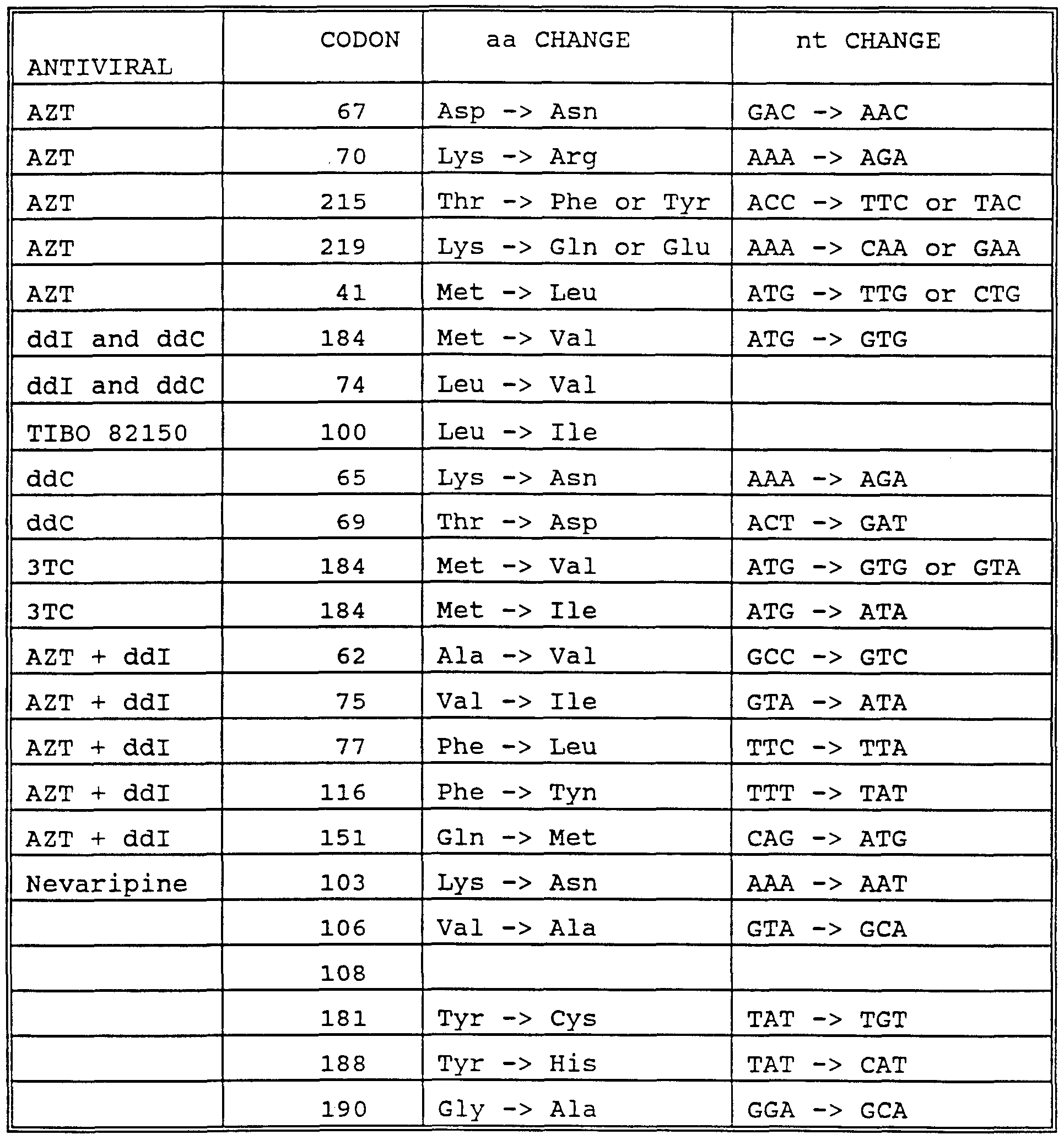
In a twelfth embodiment, the invention provides arrays of immobilized probes in which the probes are designed to tile a reference sequence from a human immunodeficiency virus. Reference sequences from either the reverse transcriptase gene or protease gene of HIV are of particular interest. Some chips further comprise arrays of probes tiling a reference sequence from a 16S RNA or DNA encoding the 16S RNA from a pathogenic microorganism. The invention further provides methods of using such arrays in analyzing a HIV target sequence. The methods are particularly useful where the target sequence has a substituted nucleotide relative to the reference sequence in at least one position, the substitution conferring resistance to a drug use in treating a patient infected with a HIV virus. The methods reveal the existence of the substituted nucleotide. The methods are also particularly useful for analyzing a mixture of undetermined proportions of first and second target sequences from different HIV variants. The relative specific binding of probes indicates the proportions of the first and second target sequences.
In a thirteenth embodiment, the invention provides arrays of probes tiled based on reference sequence from a CFTR gene. A preferred array comprises at least a group of probes comprising a wildtype probe, and five sets of three mutant probes. The wildtype probe is exactly complementary to a subsequence of a reference sequence from a cystic fibrosis gene, the segment having at least five interrogation positions corresponding to five contiguous nucleotides in the reference sequence. The probes in the first set of three mutant probes are each identical to the wildtype probe, except in a first of the five interrogation positions, which is occupied by a different nucleotide in each of the three mutant probes and the wildtype probe. The probes in the second set of three mutant probes are each identical to the wildtype probe, except in a second of the five interrogation positions, which is occupied by a different nucleotide in each of the three mutant probes and the wildtype probe. The probes in the third set of three mutant probes are each identical to the wildtype probe, except in a third of the five interrogation positions, which is occupied by a different nucleotide in each of the three mutant probes and the wildtype probe. The probes in the fourth set of three mutant probes are each identical to the wildtype probe, except in a fourth of the five interrogation positions, which is occupied by a different nucleotide in each of the three mutant probes and the wildtype probe. The probes in the fifth set of three mutant probes are each identical to the wildtype probe, except in a fifth of the five interrogation positions, which is occupied by a different nucleotide in each of the three mutant probes and the wildtype probe. Preferably, a chip comprises two such groups of probes. The first group comprises a wildtype probe exactly complementary to a first reference sequence, and the second group comprises a wildtype probe exactly complementary to a second reference sequence that is a mutated form of the first reference sequence.
The invention further provides methods of using the arrays of the invention for analyzing target sequences from a CFTR gene. The methods are capable of simultaneously analyzing first and second target sequences representing heterozygous alleles of a CFTR gene.
In a fourteenth embodiment, the invention provides arrays of probes tiling a reference sequence from a p53 gene, an hMLHl gene and/or an MSH2 gene. The invention further provides methods of using the arrays described above to analyze these genes. The method are useful, e.g., for diagnosing patients susceptible to developing cancer.
In a fifteenth embodiment, the invention provides arrays of probes tiling a reference sequence from a mitochondrial genome. The reference sequence may comprise part or all of the D-loop region, or all, or substantially all, of the mitochondrial genome. The invention further provides method of using the arrays described above to analyze target sequences from a mitochondrial genome. The methods are useful for identifying mutations associated with disease, and for forensic, epidemiological and evolutionary studies.
BRIEF DESCRIPTION OF THE FIGURES Fig. 1: Basic tiling strategy. The figure illustrates the relationship between an interrogation position (I) and a corresponding nucleotide (n) in the reference sequence, and between a probe from the first probe set and corresponding probes from second, third and fourth probe sets.
Fig. 2: Segment of complementarity in a probe from the first probe set.
Fig. 3: Incremental succession of probes in a basic tiling strategy. The figure shows four probe sets, each having three probes. Note that each probe differs from its predecessor in the same set by the acquisition of a 5' nucleotide and the loss of a 3 nucleotide, as well as in the nucleotide occupying the interrogation position.
Fig. 4: Exemplary arrangement of lanes on a chip. The chip shows four probe sets, each having five probes and each having a total of five interrogation positions (11-15) , one per probe.
Fig. 5: Hybridization pattern of chip having probes laid down in lanes. Dark patches indicate hybridization. The probes in the lower part of the figure occur at the column of the array indicated by the arrow when the probes length is 15 and the interrogation position 7.
Fig. 6: Strategies for detecting deletion and insertion mutations. Bases in brackets may or may not be present. Fig. 7: Block tiling strategy. The probe from the first probe set has three interrogation positions. The probes from the other probe sets have only one of these interrogation positions. Fig. 8: Multiplex tiling strategy. Each probe has two interrogation positions.
Fig. 9. Helper mutation strategy. The segment of complementarity differs from the complement of the reference sequence at a helper mutation as well as the interrogation position.
Fig. 10 Layout of probes on the HV 407 chip. The figure shows successive rows of sequence each of which is subdivided into four lanes. The four lanes correspond to the A-, C-, G- and T-lanes on the chip. Each probe is represented by the nucleotide occupying its interrogation position. The letter
"N" indicates a control probe or empty column. The different sized-probes are laid out in parallel. That is, from top-to- bottom, a row of 13 mers is followed by a row of 15 mers, which is followed by a row of 17 merε, which is followed by a row of 19 mers.
Fig. 11 Fluorescence pattern of HV 407 hybridized to a target sequence (pPoll9) identical to the chips reference sequence.
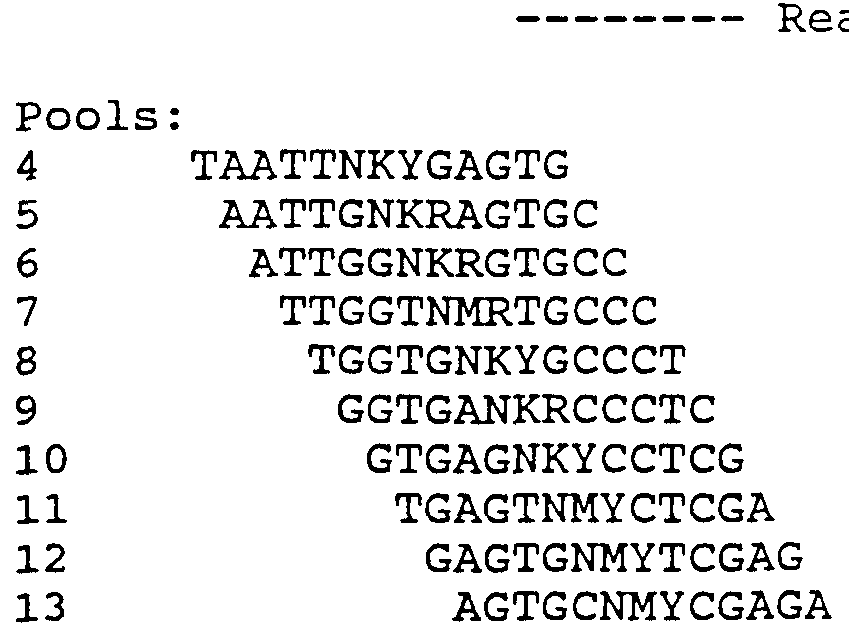
Fig. 12 Sequence read from HV 407 chip hybridized to pPoll9 and 4MUT18 (separate experiments) . The reference sequence is designated "wildtype." Beneath the reference sequence are four rows of sequence read from the chip hybridized to the pPoll9 target, the first row being read from 13 mers, the second row from 15 mers, the third row from 17 mers and the fourth row from 19 mers. Beneath these sequences, there are four further rows of sequence read from the chip hybridized to the HXB2 target. Successive rows are read from 13 mers, 15 mers, 17 mers and 19 mers. Each nucleotide in a row is called from the relative fluorescence intensities of probes in A-, C-, G- and T-lanes. Regions of ambiguous sequence read from the chip are highlighted. The strain differences between the HBX2 sequence and the reference sequence that were correctly detected are indicated (*) , and those that could not be called are indicated (o) . (The nucleotide at position 417 was read correctly in some experiments) . The location of some mutations known to be associated with drug resistance that occur in readable regions of the chip are shown above (codon number) and below (mutant nucleotide) the sequence designated "wildtype." The locations of primer used to amplify the target sequence are indicated by arrows.
Fig. 13: Detection of mixed target sequences. The mutant target differs from the wildtype by a single mutation in codon 67 of the reverse transcriptase gene. Each different sized group of probes has a column of four probes for reading the nucleotide in which the mutation occurs. The four probes occupying a column are represented by a single probe in the figure with the symbol (o) indicating the interrogation position, which is occupied by a different nucleotide in each probe.
Fig. 14: Fluorescence intensities of target bound to 13 mers and 15 mers for different proportions of mutant and wildtype target. The fluorescence intensities are from probes having interrogation positions for reading the nucleotide at which the mutant and wildtype targets diverge.
Fig. 15: Sequence read from protease chip from four clinical samples before and after treatment with ddl>. Fig. 16: Block tiling array of probes for analyzing a
CFTR point mutation. Each probe show actually represents four probes, with one probe having each of A, C, G or T at the interrogation position N. In the order shown, the first probe shown on the left is tiled from the wildtype reference sequence, the second probe from the mutant sequence, and so on in alternating fashion. Note that all of the probes are identical except at the interrogation position, which shifts one position between successive probes tiled from the same reference sequence (e.g., the first, third and fifth probes in the left hand column.) The grid shows the hybridization intensities when the array is hybridized to the reference sequence. 14
Fig. 17: Hybridization pattern for heterozygous target. The figure shows the hybridization pattern when the array of the previous figure is hybridized to a mixture of mutant and wildtype reference sequences. Fig. 18, in panels A, B, and C, shows an image made from the region of a DNA chip containing CFTR exon 10 probes; in panel A, the chip was hybridized to a wild-type target; in panel C, the chip was hybridized to a mutant ΔF508 target; and in panel B, the chip was hybridized to a mixture of the wild-type and mutant targets.
Fig. 19, in sheets 1 - 3, corresponding to panels A, B, and C of Fig. 18, shows graphs of fluorescence intensity versus tiling position. The labels on the horizontal axis show the bases in the wild-type sequence corresponding to the position of substitution in the respective probes. Plotted are the intensities observed from the features (or synthesis sites) containing wild-type probes, the features containing the substitution probes that bound the most target ("called") , and the feature containing the substitution probes that bound the target with the second highest intensity of all the substitution probes ("2nd Highest") .
Fig. 20, in panels A, B, and C, shows an image made from a region of a DNA chip containing CFTR exon 10 probes; in panel A, the chip was hybridized to the wt480 target; in panel C, the chip was hybridized to the mu480 target; and in panel B, the chip was hybridized to a mixture of the wild-type and mutant targets.
Fig. 21, in sheets 1 - 3, corresponding to panels A, B, and C of Fig. 20, shows graphs of fluorescence intensity versus tiling position. The labels on the horizontal axis show the bases in the wild-type sequence corresponding to the position of substitution in the respective probes. Plotted are the intensities observed from the features (or synthesis sites) containing wild-type probes, the features containing the substitution probes that bound the most target ("called") , and the feature containing the substitution probes that bound the target with the second highest intensity of all the substitution probes ("2nd Highest") . Fig. 22, in panels A and B, shows an image made from a region of a DNA chip containing CFTR exon 10 probes; in panel A, the chip was hybridized to nucleic acid derived from the genomic DNA of an individual with wild-type ΔF508 sequences; in panel B, the target nucleic acid originated from a heterozygous (with respect to the ΔF508 mutation) individual.
Fig. 23, in sheets 1 and 2, corresponding to panels A and B of Fig. 22, shows graphs of fluorescence intensity versus tiling position. The labels on the horizontal axis show the bases in the wild-type sequence corresponding to the position of substitution in the respective probes. Plotted are the intensities observed from the features (or synthesis sites) containing wild-type probes, the features containing the substitution probes that bound the most target ("called") , and the feature containing the substitution probes that bound the target with the second highest intensity of all the substitution probes ("2nd Highest") .
Fig. 24: Hybridization of homozygous wildtype (A) and heterozygous (B) target sequences from exon 11 of the CFTR gene to a block tiling array designed to detect G551D and Q552X mutations in CFTR gene.
Fig. 25: Hybridization of homozygous wildtype (A) and ΔF508 mutant (B) target sequences from exon 10 of the CFTR gene to a block tiling array designed to detect mutations, ΔF508, ΔI507 and F508C.
Fig. 26: Hybridization of heterozygous mutant target sequences, ΔF508/F508C, to the array of Fig. 25.
Fig. 27 shows the alignment of some of the probes on a p53 DNA chip with a 12-mer model target nucleic acid. Fig. 28 shows a set of 10-mer probes for a p53 exon 6 DNA chip.
Fig. 29 shows that very distinct patterns are observed after hybridization of p53 DNA chips with targets having different 1 base substitutions. In the first image in Fig. 29, the 12-mer probes that form perfect matches with the wild-type target are in the first row (top) . The 12-mer probes with single base mismatches are located in the second, third, and fourth rows and have much lower signals. 16
Fig. 30, in graphs 2, 3, and 4, graphically depicts the data in Fig. 29. On each graph, the X ordinate is the position of the probe in its row on the chip, and the Y ordinate is the signal at that probe site after hybridization. Fig. 31 shows the results of hybridizing mixed target populations of WT and mutant p53 genes to the p53 DNA chip.
Fig. 32, in graphs 1-4, shows (see Fig. 30 as well) the hybridization efficiency of a 10-mer probe array as compared to a 12-mer probe array. Fig. 33 shows an image of a p53 DNA chip hybridized to a target DNA.
Fig. 34 illustrates how the actual sequence was read from the chip shown in Fig. 33. Gaps in the sequence of letters in the WT rows correspond to control probes or sites. Positions at which bases are miscalled are represented by letters in italic type in cells corresponding to probes in which the WT bases have been substituted by other bases.
Fig. 35 shows the human mitochondrial genome; "0H" is the H strand origin of replication, and arrows indicate the cloned unshaded sequence.
Fig. 36 shows the image observed from application of a sample of mitochondrial DNA derived nucleic acid (from the mt4 sample) on a DNA chip.
Fig. 37 is similar to Fig. 36 but shows the image observed from the mt5 sample.
Fig. 38 shows the predicted difference image between the mt4 and mt5 samples on the DNA chip based on mismatches between the two samples and the reference sequence.
Fig. 39 shows the actual difference image observed for the mt4 and mt5 samples.
Fig. 40, in sheets 1 and 2, shows a plot of normalized intensities across rows 10 and 11 of the array and a tabulation of the mutations detected.
Fig. 41 shows the discrimination between wild-type and mutant hybrids obtained with the chip. A median of the six normalized hybridization scores for each probe was taken; the graph plots the ratio of the median score to the normalized hybridization score versus mean counts. A ratio of 1.6 and mean counts above 50 yield no false positives.
Fig. 42 illustrates how the identity of the base mismatch may influence the ability to discriminate mutant and wild-type sequences more than the position of the mismatch within an oligonucleotide probe. The mismatch position is expressed as % of probe length from the 3 '-end. The base change is indicated on the graph.
Fig. 43 provides a 5' to 3' sequence listing of one target corresponding to the probes on the chip. X is a control probe. Positions that differ in the target (i.e., are mismatched with the probe at the designated site) are in bold.
Fig. 44 shows the fluorescence image produced by scanning the chip described in Fig. 17 when hybridized to a sample. Fig. 45 illustrates the detection of 4 transitions in the target sequence relative to the wild-type probes on the chip in Fig. 44.
Fig. 46: VLSIPS™ technology applied to the light directed synthesis of oligonucleotides. Light (hv) is shone through a mask (M ) to activate functional groups (-OH) on a surface by removal of a protecting group (X) . Nucleoside building blocks protected with photoremovable protecting groups (T-X, C-X) are coupled to the activated areas. By repeating the irradiation and coupling steps, very complex arrays of oligonucleotides can be prepared.
Fig. 47: Use of the VLSIPS™ process to prepare "nucleoside combinatorials" or oligonucleotides synthesized by coupling all four nucleosides to form dimers, trimers, and so forth. Fig. 48: Deprotection, coupling, and oxidation steps of a solid phase DNA synthesis method.
Fig. 49: An illustrative synthesis route for the nucleoside building blocks used in the VLSIPS™ method.
Fig. 50: A preferred photoremovable protecting group, MeNPOC, and preparation of the group in active form.
Fig. 51: Detection system for scanning a DNA chip. DETAILED DESCRIPTION OF THE INVENTION The invention provides a number of strategies for comparing a polynucleotide of known sequence (a reference sequence) with variants of that sequence (target sequences) . The comparison can be performed at the level of entire genomes, chromosomes, genes, exons or introns, or can focus on individual mutant sites and immediately adjacent bases. The strategies allow detection of variations, such as mutations or polymorphisms, in the target sequence irrespective whether a particular variant has previously been characterized. The strategies both define the nature of a variant and identify its location in a target sequence.
The strategies employ arrays of oligonucleotide probes immobilized to a solid support. Target sequences are analyzed by determining the extent of hybridization at particular probes in the array. The strategy in selection of probes facilitates distinction between perfectly matched probes and probes showing single-base or other degrees of mismatches. The strategy usually entails sampling each nucleotide of interest in a target sequence several times, thereby achieving a high degree of confidence in its identity. This level of confidence is further increased by sampling of adjacent nucleotides in the target sequence to nucleotides of interest. The number of probes on the chip can be quite large (e.g., 105-106) . However, usually only a small proportion of the total number of probes of a given length are represented. Some advantage of the use of only a small proportion of all possible probes of a given length include: (i) each position in the array is highly informative, whether or not hybridization occurs; (ii) nonspecific hybridization is minimized; (iii) it is straightforward to correlate hybridization differences with sequence differences, particularly with reference to the hybridization pattern of a known standard; and (iv) the ability to address each probe independently during synthesis, using high resolution photolithography, allows the array to be designed and optimized for any sequence. For example the length of any probe can be varied independently of the others. The present tiling strategies result in sequencing and comparison methods suitable for routine large-scale practice with a high degree of confidence in the sequence output.
I. GENERAL TILING STRATEGIES
A. Selection of Reference Sequence
The chips are designed to contain probes exhibiting complementarity to one or more selected reference sequence whose sequence is known. The chips are used to read a target sequence comprising either the reference sequence itself or variants of that sequence. Target sequences may differ from the reference sequence at one or more positions but show a high overall degree of sequence identity with the reference sequence (e.g., at least 75, 90, 95, 99, 99.9 or 99.99%) . Any polynucleotide of known sequence can be selected as a reference sequence. Reference sequences of interest include sequences known to include mutations or polymorphisms associated with phenotypic changes having clinical significance in human patients. For example, the CFTR gene and P53 gene in humans have been identified as the location of several mutations resulting in cystic fibrosis or cancer respectively. Other reference sequences of interest include those that serve to identify pathogenic microorganisms and/or are the site of mutations by which such microorganisms acquire drug resistance (e.g., the HIV reverse transcriptase gene). Other reference sequences of interest include regions where polymorphic variations are known to occur (e.g., the D-loop region of mitochondrial DNA) . These reference sequences have utility for, e.g., forensic or epidemiological studies. Other reference sequences of interest include p34 (related to p53), p65 (implicated in breast, prostate and liver cancer) , and DNA segments encoding cytochromes P450 (see Meyer et al., Pharmac . Ther . 46, 349-355 (1990)) . Other reference sequences of interest include those from the genome of pathogenic viruses (e.g., hepatitis (A, B, or C) , herpes virus (e.g., VZV, HSV-1, HAV-6, HSV-II, and CMV, Epstein Barr virus), adenovirus, influenza virus, flaviviruses, echovirus, rhinovirus, coxsackie virus, cornovirus, respiratory εyncytial virus, 20 mumps virus, rotavirus, measles virus, rubella virus, parvovirus, vaccinia virus, HTLV virus, dengue virus, papillomavirus, molluscum virus, poliovirus, rabies virus, JC virus and arboviral encephalitis virus. Other reference sequences of interest are from genomes or episomes of pathogenic bacteria, particularly regions that confer drug resistance or allow phylogenic characterization of the host (e.g., 16S rRNA or corresponding DNA) . For example, such bacteria include chlamydia, rickettsial bacteria, mycobacteria, staphylococci, treptocci, pneumonococci, meningococci and conococci, klebsiella, proteus, serratia, pseudomonas, legionella, diphtheria, salmonella, bacilli, cholera, tetanus, botulism, anthrax, plague, leptospirosis, and Ly es disease bacteria. Other reference sequences of interest include those in which mutations result in the following autosomal recessive disorders: sickle cell anemia, 3-thalassemia, phenylketonuria, galactosemia, Wilson's disease, hemochromatosis, severe combined immunodeficiency, alpha-1-antitrypsin deficiency, albinism, alkaptonuria, lysosomal storage diseases and Ehlers-Danlos syndrome. Other reference sequences of interest include those in which mutations result in X-linked recessive disorders: hemophilia, glucose-6-phosphate dehydrogenase, agammaglobulimenia, diabetes insipidus, Lesch-Nyhan syndrome, muscular dystrophy, Wiskott-Aldrich syndrome, Fabry's disease and fragile X- syndrome. Other reference sequences of interest includes those in which mutations result in the following autosomal dominant disorders: familial hypercholesterolemia, polycystic kidney disease, Huntingdon's disease, hereditary spherocytosis, Marfan's syndrome, von Willebrand's disease, neurofibromatosis, tuberous sclerosis, hereditary hemorrhagic telangiectasia, familial colonic polyposis, Ehlers-Danlos syndrome, myotonic dystrophy, muscular dystrophy, osteogenesis imperfecta, acute intermittent porphyria, and von Hippel- Lindau disease.
The length of a reference sequence can vary widely from a full-length genome, to an individual chromosome, episome, gene, component of a gene, such as an exon, intron or regulatory sequences, to a few nucleotides. A reference sequence of between about 2, 5, 10, 20, 50, 100, 5000, 1000, 5,000 or 10,000, 20,000 or 100,000 nucleotides is common. Sometimes only particular regions of a sequence (e.g., exons of a gene) are of interest. In such situations, the particular regions can be considered as separate reference sequences or can be considered as components of a single reference sequence, as matter of arbitrary choice.
A reference sequence can be any naturally occurring, mutant, consensuε or purely hypothetical εequence of nucleotideε, RNA or DNA. For example, sequences can be obtained from computer data bases, publications or can be determined or conceived e novo . Usually, a reference sequence is selected to show a high degree of εequence identity to enviεaged target εequences. Often, particularly, where a significant degree of divergence is anticipated between target sequences, more than one reference sequence is selected. Combinations of wildtype and mutant reference sequences are employed in several applications of the tiling strategy.
B. Chip Design
1. Basic Tiling Strategy
The basic tiling strategy provides an array of immobilized probes for analysis of target sequences showing a high degree of sequence identity to one or more selected reference sequences. The strategy is first illustrated for an array that is subdivided into four probe sets, although it will be apparent that in some situationε, satisfactory results are obtained from only two probe sets. A first probe set comprises a plurality of probes exhibiting perfect complementarity with a selected reference sequence. The perfect complementarity uεually exists throughout the length of the probe. However, probes having a segment or segments of perfect complementarity that is/are flanked by leading or trailing sequences lacking complementarity to the reference sequence can also be used. Within a segment of complementarity, each probe in the first probe set has at 22 least one interrogation position that corresponds to a nucleotide in the reference sequence. That is, the interrogation position is aligned with the corresponding nucleotide in the reference sequence, when the probe and reference sequence are aligned to maximize complementarity between the two. If a probe has more than one interrogation position, each corresponds with a respective nucleotide in the reference sequence. The identity of an interrogation position and corresponding nucleotide in a particular probe in the first probe set cannot be determined simply by inspection of the probe in the first set. As will become apparent, an interrogation position and corresponding nucleotide is defined by the comparative structures of probes in the first probe set and corresponding probes from additional probe sets. In principle, a probe could have an interrogation position at each position in the segment complementary to the reference sequence. Sometimes, interrogation positions provide more accurate data when located away from the ends of a segment of complementarity. Thus, typically a probe having a segment of complementarity of length x does not contain more than x-2 interrogation positions. Since probes are typically 9-21 nucleotides, and uεually all of a probe is complementary, a probe typically has 1-19 interrogation poεitions. Often the probes contain a single interrogation position, at or near the center of probe.
For each probe in the first set, there are, for purposes of the present illustration, three corresponding probes from three additional probe sets. See Fig. 1. Thus, there are four probes corresponding to each nucleotide of interest in the reference sequence. Each of the four corresponding probes has an interrogation position aligned with that nucleotide of interest. Usually, the probes from the three additional probe sets are identical to the corresponding probe from the first probe set with one exception. The exception is that at least one (and often only one) interrogation position, which occurs in the same position in each of the four corresponding probes from the four probe sets, is occupied by a different nucleotide in the four probe sets. For example, for an A nucleotide in the reference sequence, the corresponding probe from the first probe set has its interrogation position occupied by a T, and the corresponding probes from the additional three probe sets have their respective interrogation positions occupied by A, C, or G, a different nucleotide in each probe. Of course, if a probe from the first probe set comprises trailing or flanking sequences lacking complementarity to the reference sequences (see Fig. 2) , these sequences need not be present in corresponding probes from the three additional sets. Likewise corresponding probes from the three additional sets can contain leading or trailing sequences outside the segment of complementarity that are not present in the corresponding probe from the first probe set. Occasionally, the probes from the additional three probe set are identical (with the exception of interrogation position(s)) to a contiguous subsequence of the full complementary segment of the corresponding probe from the first probe set. In this case, the subsequence includes the interrogation position and usually differs from the full- length probe only in the omission of one or both terminal nucleotides from the termini of a segment of complementarity. That is, if a probe from the first probe set has a segment of complementarity of length n, corresponding probes from the other sets will usually include a subsequence of the segment of at least length n-2. Thus, the subsequence is usually at least 3, 4, 7, 9, 15, 21, or 25 nucleotides long, most typically, in the range of 9-21 nucleotides. The subsequence should be sufficiently long to allow a probe to hybridize detectably more strongly to a variant of the reference sequence mutated at the interrogation position than to the reference sequence.
The probes can be oligodeoxyribonucleotides or oligoribonucleotides, or any modified forms of these polymers that are capable of hybridizing with a target nucleic sequence by complementary base-pairing. Complementary base pairing means sequence-specific base pairing which includes e . g . , Watson-Crick base pairing as well as other forms of base pairing such as Hoogsteen base pairing. Modified forms include 2'-0-methyl oligoribonucleotides and so-called PNAs, in which oligodeoxyribonucleotides are linked via peptide bonds rather than phophodiester bonds. The probes can be attached by any linkage to a support (e.g., 3', 5' or via the base) . 3 ' attachment is more usual as this orientation is compatible with the preferred chemistry for solid phase synthesis of oligonucleotides.
The number of probes in the first probe set (and as a consequence the number of probes in additional probe sets) depends on the length of the reference sequence, the number of nucleotideε of intereεt in the reference sequence and the number of interrogation positions per probe. In general, each nucleotide of interest in the reference sequence requires the same interrogation position in the four sets of probes. Consider, as an example, a reference sequence of 100 nucleotides, 50 of which are of interest, and probes each having a single interrogation position. In this situation, the first probe set requires fifty probes, each having one interrogation position corresponding to a nucleotide of interest in the reference sequence. The second, third and fourth probe sets each have a corresponding probe for each probe in the first probe set, and so each also contains a total of fifty probes. The identity of each nucleotide of interest in the reference sequence is determined by comparing the relative hybridization signals at four probes having interrogation positions corresponding to that nucleotide from the four probe sets.
In some reference sequences, every nucleotide is of interest. In other reference sequences, only certain portions in which variants (e.g., mutations or polymorphisms) are concentrated are of interest. In other reference sequences, only particular mutations or polymorphisms and immediately adjacent nucleotides are of interest. Usually, the first probe set has interrogation positions selected to correspond to at least a nucleotide (e.g., representing a point mutation) and one immediately adjacent nucleotide. Usually, the probes in the first set have interrogation positions corresponding to at least 3, 10, 50, 100, 1000, or 20,000 contiguous nucleotides. The probes usually have interrogation positions corresponding to at least 5, 10, 30, 50, 75, 90, 99 or sometimes 100% of the nucleotides in a reference sequence. Frequently, the probes in the first probe set completely span the reference sequence and overlap with one another relative to the reference sequence. For example, in one common arrangement each probe in the first probe set differs from another probe in that set by the omisεion of a 3 ' base complementary to the reference εequence and the acquisition of a 5' base complementary to the reference sequence. See Fig. 3.
For conceptual simplicity, the probes in a set are usually arranged in order of the εequence in a lane across the chip. A lane contains a series of overlapping probes, which represent or tile across, the selected reference sequence (see Fig. 3) . The components of the four setε of probeε are uεually laid down in four parallel laneε, collectively conεtituting a row in the horizontal direction and a series of 4-member columns in the vertical direction. Correεponding probes from the four probe sets (i.e., complementary to the same subεequence of the reference sequence) occupy a column. Each probe in a lane usually differs from its predecessor in the lane by the omiεεion of a baεe at one end and the incluεion of additional baεe at the other end aε shown in Fig. 3. However, this orderly progresεion of probeε can be interrupted by the incluεion of control probeε or omiεsion of probes in certain columns of the array. Such columns serve as controls to orient the chip, or gauge the background, which can include target sequence nonspecifically bound to the chip. The probes sets are usually laid down in lanes such that all probes having an interrogation position occupied by an A form an-A-lane, all probes having an interrogation position occupied by a C form a C-lane, all probes having an interrogation position occupied by a G form a G-lane, and all probes having an interrogation position occupied by a T (or U) form a T lane (or a U lane) . Note that in this arrangement there is not a unique correspondence between probe setε and lanes. Thuε, the probe from the first probe set is laid down in the A-lane, C-lane, A-lane, A-lane and T-lane for the five columns in Fig. 4. The interrogation position on a column of probes corresponds to the position in the target sequence whose identity is determined from analysis of hybridization to the probes in that column. Thus, I}-^ respectively correspond to N1-N5 in Fig. 4. The interrogation position can be anywhere in a probe but is usually at or near the central position of the probe to maximize differential hybridization signals between a perfect match and a single-baεe miεmatch. For example, for an 11 er probe, the central poεition iε the εixth nucleotide.
Although the array of probes is usually laid down in rows and columns as described above, such a physical arrangement of probes on the chip is not eεεential. Provided that the spatial location of each probe in an array is known, the data from the probes can be collected and processed to yield the sequence of a target irrespective of the physical arrangement of the probes on a chip. In procesεing the data, the hybridization signals from the respective probeε can be reaεεorted into any conceptual array deεired for εubεequent data reduction whatever the physical arrangement of probes on the chip.
A range of lengths of probes can be employed in the chips. As noted above, a probe may consiεt excluεively of a complementary segments, or may have one or more complementary segments juxtaposed by flanking, trailing and/or intervening segmentε. In the latter εituation, the total length of complementary segment(s) iε more important that the length of the probe. In functional termε, the complementarity segment(s) of the first probe setε should be sufficiently long to allow the probe to hybridize detectably more strongly to a reference εequence compared with a variant of the reference including a εingle base mutation at the nucleotide corresponding to the interrogation position of the probe. Similarly, the complementarity segment(s) in corresponding probes from additional probe sets εhould be εufficiently long to allow a probe to hybridize detectably more strongly to a variant of the reference sequence having a εingle nucleotide substitution at the interrogation position relative to the reference sequence. A probe usually has a single complementary segment having a length of at least 3 nucleotides, and more usually at least 5, 6, 7, 8, 9, 10, 11, 12, 13, 14, 15, 16, 17, 18, 19, 20, 21, 22, 23, 24, 25 or 30 baseε exhibiting perfect complementarity (other than possibly at the interrogation position(s) depending on the probe set) to the reference sequence. In bridging εtrategieε, where more than one segment of complementarity is present, each εegment provideε at least three complementary nucleotides to the reference sequence and the combined segmentε provide at least two εegments of three or a total of six complementary nucleotides. As in the other strategieε, the combined length of complementary εegments is typically from 6-30 nucleotides, and preferably from about 9-21 nucleotides. The two segments are often approximately the same length. Often, the probes (or εegment of complementarity within probes) have an odd number of baseε, so that an interrogation position can occur in the exact center of the probe. In some chips, all probes are the same length. Other chips employ different groups of probe sets, in which case the probes are of the same size within a group, but differ between different groups. For example, some chips have one group comprising four sets of probes as described above in which all the probes are 11 mers, together with a second group compriεing four sets of probes in which all of the probes are 13 mers. Of course, additional groups of probes can be added. Thus, some chips contain, e . g . , four groups of probes having sizes of 11 mers, 13 merε, 15 merε and 17 merε. Other chipε have different εize probeε within the same group of four probe sets. In these chips, the probes in the firεt set can vary in length independently of each other. Probes in the other setε are usually the same length as the probe occupying the same column from the firεt set. However, occasionally different lengths of probes can be included at the same column poεition in the four lanes. The different length probes are included to equalize hybridization signals from probes irrespective of 28 whether A-T or C-G bonds are formed at the interrogation position.
The length of probe can be important in distinguishing between a perfectly matched probe and probes showing a single- base mismatch with the target sequence. The discrimination is usually greater for short probes. Shorter probeε are uεually alεo less εuεceptible to formation of εecondary structures. However, the absolute amount of target sequence bound, and hence the signal, iε greater for larger probeε. The probe length repreεenting the optimum compromiεe between these competing considerationε may vary depending on inter alia the GC content of a particular region of the target DNA εequence, secondary structure, synthesis efficiency and cross- hybridization. In some regions of the target, depending on hybridization conditions, short probes (e.g., 11 mers) may provide information that is inaccessible from longer probeε (e.g., 19 mers) and vice versa. Maximum sequence information can be read by including several groups of different sized probes on the chip as noted above. However, for many regions of the target sequence, such a strategy provides redundant information in that the εame εequence iε read multiple times from the different groups of probes. Equivalent information can be obtained from a εingle group of different εized probeε in which the εizes are selected to maximize readable sequence at particular regions of the target sequence. The appropriate size of probes at different regions of the target sequence can be determined from, e.g., Fig. 12, which co pareε the readability of different sized probes in different regionε of a target. The εtrategy of cuεtomizing probe length within a εingle group of probe sets minimizes the total number of probes required to read a particular target sequence. This leaves ample capacity for the chip to include probes to other reference sequences.
The invention provideε an optimization block which allows systematic variation of probe length and interrogation poεition to optimize the εelection of probeε for analyzing a particular nucleotide in a reference εequence. The block comprises alternating columns of probes complementary to the wildtype target and probes complementary to a specific mutation. The interrogation position is varied between columns and probe length is varied down a column. Hybridization of the chip to the reference εequence or the mutant form of the reference εequence identifieε the probe length and interrogation poεition providing the greateεt differential hybridization εignal.
The probes are designed to be complementary to either strand of the reference sequence (e.g., coding or non-coding). Some chips contain separate groups of probeε, one complementary to the coding εtrand, the other complementary to the noncoding εtrand. Independent analysis of coding and noncoding strands provides largely redundant information. However, the regionε of ambiguity in reading the coding εtrand are not alwayε the εame aε those in reading the noncoding strand. Thus, combination of the information from coding and noncoding strands increases the overall accuracy of sequencing.
Some chips contain additional probes or groups of probes designed to be complementary to a second reference sequence. The second reference sequence is often a subsequence of the first reference sequence bearing one or more commonly occurring mutations or interstrain variations. The second group of probes is designed by the same principles as described above except that the probes exhibit complementarity to the second reference εequence. The incluεion of a εecond group iε particular useful for analyzing short subsequences of the primary reference sequence in which multiple mutations are expected to occur within a short distance commensurate with the length of the probes (i.e., two or more mutations within 9 to 21 baseε) . Of courεe, the same principle can be extended to provide chips containing groups of probes for any number of reference sequences. Alternatively, the chips may contain additional probe(s) that do not form part of a tiled array aε noted above, but rather εerveε aε probe(ε) for a conventional reverεe dot blot. For example, the preεence of mutation can be detected from binding of a target εequence to a εingle oligomeric probe harboring the mutation. Preferably, an additional probe containing the equivalent region of the wildtype sequence is included as a control.
The chips are read by comparing the intensities of labelled target bound to the probes in an array. Specifically, a comparison is performed between each lane of probeε (e.g., A, C, G and T lanes) at each columnar position (physical or conceptual) . For a particular columnar position, the lane showing the greatest hybridization signal is called as the nucleotide preεent at the poεition in the target εequence correεponding to the interrogation poεition in the probeε. See Fig. 5. The corresponding position in the target sequence is that aligned with the interrogation position in corresponding probes when the probeε and target are aligned to maximize complementarity. Of the four probes in a column, only one can exhibit a perfect match to the target sequence whereas the others uεually exhibit at leaεt a one base pair mismatch. The probe exhibiting a perfect match usually produces a substantially greater hybridization signal than the other three probes in the column and is thereby easily identified. However, in some regions of the target sequence, the distinction between a perfect match and a one-base mismatch is lesε clear. Thuε, a call ratio iε established to define the ratio of signal from the best hybridizing probes to the second best hybridizing probe that must be exceeded for a particular target poεition to be read from the probeε. A high call ratio ensures that few if any errors are made in calling target nucleotides, but can result in some nucleotides being scored aε ambiguous, which could in fact be accurately read. A lower call ratio reεultε in fewer ambiguouε callε, but can result in more erroneous callε. It haε been found that at a call ratio of 1.2 virtually all calls are accurate. However, a small but εignificant number of bases (e.g., up to about 10%) may have to be scored as ambiguouε.
Although small regions of the target sequence can sometimes be ambiguous, theεe regions usually occur at the same or similar segments in different target sequenceε. Thuε, for precharacterized mutations, it is known in advance whether that mutation iε likely to occur within a region of unambiguously determinable sequence.
An array of probes is most useful for analyzing the reference sequence from which the probes were designed and variants of that sequence exhibiting substantial sequence εimilarity with the reference sequence (e.g., several single- base mutants spaced over the reference sequence) . When an array is used to analyze the exact reference sequence from which it was deεigned, one probe exhibitε a perfect match to the reference sequence, and the other three probes in the same column exhibits single-baεe miεmatcheε. Thus, discrimination between hybridization signals is usually high and accurate sequence is obtained. High accuracy is also obtained when an array is used for analyzing a target sequence comprising a variant of the reference sequence that has a εingle mutation relative to the reference sequence, or several widely spaced mutations relative to the reference εequence. At different mutant loci, one probe exhibitε a perfect match to the target, and the other three probeε occupying the εame column exhibit εingle-base ismatcheε, the difference (with reεpect to analyεiε of the reference εequence) being the lane in which the perfect match occurε.
For target εequenceε εhowing a high degree of divergence from the reference strain or incorporating εeveral closely spaced mutations from the reference strain, a single group of probes (i.e., designed with respect to a single reference sequence) will not alwayε provide accurate sequence for the highly variant region of this sequence. At some particular columnar positionε, it may be that no single probe exhibitε perfect complementarity to the target and that any co pariεon must be based on different degrees of mismatch between the four probes. Such a comparison does not always allow the target nucleotide correεponding to that columnar poεition to be called. Deletions in target sequences can be detected by losε of signal from probes having interrogation positions encompassed by the deletion. However, εignal may also be lost from probes having interrogation positionε cloεely proximal to the deletion reεulting in εome regions of the target sequence that cannot be read. Target sequence bearing insertions will also exhibit short regions including and proximal to the insertion that uεually cannot be read.
The preεence of εhort regions of difficult-to-read target because of closely spaced mutations, insertions or deletion, does not prevent determination of the remaining sequence of the target as different regions of a target sequence are determined independently. Moreover, such ambiguities as might result from analysiε of diverse variants with a εingle group of probeε can be avoided by including multiple groups of probe setε on a chip. For example, one group of probeε can be deεigned baεed on a full-length reference εequence, and the other groups on subsequences of the reference sequence incorporating frequently occurring mutations or strain variations.
A particular advantage of the present sequencing strategy over conventional sequencing methods is the capacity simultaneouεly to detect and quantify proportionε of multiple target εequenceε. Such capacity iε valuable, e.g., for diagnoεiε of patientε who are heterozygouε with reεpect to a gene or who are infected with a virus, such as HIV, which iε uεually preεent in several polymorphic forms. Such capacity is also useful in analyzing targets from biopsies of tumor cells and surrounding tisεues. The presence of multiple target sequences is detected from the relative signals of the four probes at the array columns correεponding to the target nucleotideε at which diverεity occurs. The relative signalε at the four probeε for the mixture under test are compared with the corresponding εignalε from a homogeneous reference sequence. An increase in a signal from a probe that is mismatched with respect to the reference sequence, and a corresponding decrease in the signal from the probe which is matched with the reference εequence signal the presence of a mutant strain in the mixture. The extent in shift in hybridization signals of the probes is related to the proportion of a target sequence in the mixture. Shifts in relative hybridization signals can be quantitatively related to proportions of reference and mutant sequence by prior calibration of the chip with εeeded mixtures of the mutant and reference εequenceε. By this means, a chip can be used to detect variant or mutant strains constituting as little as 1, 5, 20, or 25 % of a mixture of stains. Similar principles allow the εimultaneouε analyεiε of multiple target εequences even when none is identical to the reference εequence. For example, with a mixture of two target sequences bearing first and εecond mutations, there would be a variation in the hybridization patterns of probeε having interrogation poεitionε corresponding to the first and second mutations relative to the hybridization pattern with the reference sequence. At each position, one of the probes having a mismatched interrogation position relative to the reference εequence would show an increase in hybridization signal, and the probe having a matched interrogation position relative to the reference εequence would show a decrease in hybridization εignal. Analyεiε of the hybridization pattern of the mixture of mutant target sequences, preferably in comparison with the hybridization pattern of the reference sequence, indicates the presence of two mutant target εequences, the position and nature of the mutation in each strain, and the relative proportions of each εtrain.
In a variation of the above method, the different componentε in a mixture of target εequenceε are differentially labelled before being applied to the array. For example, a variety of fluoreεcent labelε emitting at different wavelength are available. The uεe of differential labels allows independent analysiε of different targetε bound εimultaneouεly to the array. For example, the methods permit comparison of target sequences obtained from a patient at different stages of a disease.
2. Omission of Probes The general strategy outlined above employs four probeε to read each nucleotide of intereεt in a target sequence. One probe (from the first probe set) εhows a perfect match to the reference sequence and the other three probes (from the second, third and fourth probe sets) exhibit a mismatch with the reference sequence and a perfect match with a target sequence bearing a mutation at the nucleotide of interest. The provision of three probes from the second, third and fourth probe setε allowε detection of each of the three possible nucleotide subεtitutions of any nucleotide of interest. However, in some reference sequences or regionε of reference εequenceε, it iε known in advance that only certain mutationε are likely to occur. Thuε, for example, at one site it might be known that an A nucleotide in the reference sequence may exist as a T mutant in some target εequenceε but iε unlikely to exiεt aε a C or G mutant. Accordingly, for analysis of this region of the reference sequence, one might include only the first and εecond probe εetε, the first probe set exhibiting perfect complementarity to the reference sequence, and the second probe set having an interrogation position occupied by an invariant A reεidue (for detecting the T mutant) . In other εituations, one might include the first, second and third probeε εets (but not the fourth) for detection of a wildtype nucleotide in the reference sequence and two mutant variants thereof in target sequenceε. In εome chipε, probeε that would detect εilent mutations (i.e., not affecting amino acid sequence) are omitted.
In some chips, the probes from the first probe set are omitted correεponding to εome or all poεitionε of the reference sequences. Such chips comprise at least two probe εetε. The firεt probe εet haε a plurality of probeε. Each probe compriεeε a segment exactly complementary to a subεequence of a reference sequence except in at least one interrogation position. A εecond probe εet haε a corresponding probe for each probe in the first probe set.
The corresponding probe in the εecond probe set is identical to a sequence comprising the correεponding probe form the firεt probe εet or a εubεequence thereof that includes the at least one (and usually only one) interrogation position except that the at least one interrogation poεition iε occupied by a different nucleotide in each of the two correεponding probes from the firεt and εecond probe sets. A third probe εet, if preεent, alεo compriεeε a correεponding probe for each probe in the first probe set except at the at least one interrogation position, which differs in the corresponding probes from the three sets. Omission of probes having a segment exhibiting perfect complementarity to the reference sequence results in loss of control information, i.e., the detection of nucleotideε in a target sequence that are the same as those in a reference sequence. However, similar information can be obtained by hybridizing a chip lacking probes from the first probe set to both target and reference εequences. The hybridization can be performed εequentially, or concurrently, if the target and reference are differentially labelled. In thiε εituation, the presence of a mutation is detected by a shift in the background hybridization intensity of the reference sequence to a perfectly matched hybridization signal of the target sequence, rather than by a comparison of the hybridization intensities of probes from the first set with corresponding probes from the εecond, third and fourth εetε.
3. Wildtype Probe Lane
When the chipε compriεe four probe εetε, aε diεcuεεed supra , and the probe εetε are laid down in four lanes, an A lane, a C-lane, a G lane and a T or U lane, the probe having a εegment exhibiting perfect complementarity to a reference sequence varies between the four lanes from one column to another. Thiε doeε not preεent any significant difficulty in computer analysis of the data from the chip. However, visual inspection of the hybridization pattern of the chip is sometimes facilitated by provision of an extra lane of probeε, in which each probe haε a εegment exhibiting perfect complementarity to the reference εequence. See Fig. 4. Thiε εegment -iε identical to a εegment from one of the probeε in the other four lanes (which lane depending on the column position) . The extra lane of probes (designated the wildtype lane) hybridizes to a target sequence at all nucleotide positionε except thoεe in which deviations from the reference sequence occurs. The hybridization pattern of the wildtype lane thereby provides a simple visual indication of mutations. 36
4. Deletion, Insertion and Multiple-Mutation Probes Some chips provide an additional probe set specifically designed for analyzing deletion mutations. The additional probe set comprises a probe corresponding to each probe in the first probe set as described above. However, a probe from the additional probe εet differε from the correεponding probe in the first probe set in that the nucleotide occupying the interrogation position is deleted in the probe from the additional probe εet. See Fig. 6. Optionally, the probe from the additional probe εet bearε an additional nucleotide at one of itε termini relative to the corresponding probe from the firεt probe set. The probe from the additional probe set will hybridize more strongly than the corresponding probe from the first probe set to a target sequence having a εingle baεe deletion at the nucleotide correεponding to the interrogation position. Additional probe setε are provided in which not only the interrogation poεition, but alεo an adjacent nucleotide is detected.
Similarly, other chips provide additional probe sets for analyzing insertionε. For example, one additional probe set has a probe corresponding to each probe in the first probe set as deεcribed above. However, the probe in the additional probe set has an extra T nucleotide inserted adjacent to the interrogation position. See Fig. 6. Optionally, the probe has one fewer nucleotide at one of its termini relative to the corresponding probe from the first probe set. The probe from the additional probe set hybridizes more strongly than the correεponding probe from the first probe set to a target sequence having an A nucleotide inserted in a poεition adjacent to that corresponding to the interrogation position. Similar additional probe sets are constructed having C, G or T/U nucleotides inserted adjacent to the interrogation poεition. Uεually, four such probe sets, one for each nucleotide, are used in combination. Other chipε provide additional probes (multiple-mutation probes) for analyzing target sequences having multiple closely εpaced mutations. A multiple-mutation probe is usually identical to a corresponding probe from the first set as described above, except in the baεe occupying the interrogation position, and except at one or more additional positions, corresponding to nucleotides in which substitution may occur in the reference εequence. The one or more additional poεitionε in the multiple mutation probe are occupied by nucleotideε complementary to the nucleotideε occupying corresponding positions in the reference sequence when the poεεible εubεtitutionε have occurred. 5. Block Tiling Aε noted in the discussion of the general tiling strategy, a probe in the first probe set sometimes has more than one interrogation poεition. In thiε εituation, a probe in the first probe set iε εometimeε matched with multiple groupε of at leaεt one, and uεually, three additional probe sets. See Fig. 7. Three additional probe setε are uεed to allow detection of the three poεsible nucleotide substitutionε at any one poεition. If only certain typeε of εubεtitution are likely to occur (e.g., tranεitionε) , only one or two additional probe εets are required (analogous to the use of probes in the baεic tiling strategy) . To illustrate for the situation where a group comprises three additional probe sets, a first εuch group compriεeε εecond, third and fourth probe εetε, each of which haε a probe correεponding to each probe in the first probe set. The correεponding probeε from the second, third and fourth probes sets differ from the correεponding probe in the firεt εet at a firεt of the interrogation positions. Thus, the relative hybridization εignals from corresponding probes from the first, second, third and fourth probe sets indicate the identity of the nucleotide in a target sequence corresponding to the firεt interrogation poεition. A second group of three probe sets (designated fifth, sixth and seventh probe sets) , each also have a probe corresponding to each probe in the firεt probe set. These corresponding probes differ from that in the first probe set at a second interrogation poεition. The relative hybridization εignalε from correεponding probes from the first, fifth, sixth, and seventh probe setε indicate the identity of the nucleotide in the target εequence corresponding to the second interrogation position. As noted above, the probes in the first probe set often have seven or more interrogation poεitions. If there are seven interrogation positionε, there are εeven groups of three additional probe setε, each group of three probe εetε εerving to identify the nucleotide correεponding to one of the εeven interrogation poεitions.
Each block of probeε allows short regions of a target sequence to be read. For example, for a block of probes having seven interrogation positions, seven nucleotides in the target sequence can be read. Of course, a chip can contain any number of blocks depending on how many nucleotides of the target are of interest. The hybridization signals for each block can be analyzed independently of any other block. The block tiling strategy can also be combined with other tiling strategieε, with different partε of the εame reference sequence being tiled by different strategies.
The block tiling strategy offers two advantages over the basic strategy in which each probe in the first set has a single interrogation position. One advantage is that the same sequence information can be obtained from fewer probes. A second advantage is that each of the probes constituting a block (i.e., a probe from the firεt probe εet and a corresponding probe from each of the other probe sets) can have identical 3' and 5' sequences, with the variation confined to a central εegment containing the interrogation poεitions. The identity of 3' εequence between different probeε εimplifies the strategy for solid phaεe εyntheεiε of the probeε on the chip and results in more uniform deposition of the different probes on the chip, thereby in turn increasing the uniformity of signal to noise ratio for different regions of the chip. A third advantage is that greater signal uniformity is achieved within a block.
6. Multiplex Tiling
In the block tiling strategy discussed above, the identity of a nucleotide in a target or reference sequence is determined by comparison of hybridization patterns of one probe having a segment showing a perfect match with that of other probes (uεually three other probes) εhowing a single base mismatch. In multiplex tiling, the identity of at least two nucleotides in a reference or target εequence is determined by comparison of hybridization signal intensities of four probes, two of which have a segment εhowing perfect complementarity or a εingle baεe miεmatch to the reference sequence, and two of which have a segment showing perfect complementarity or a double-base miεmatch to a segment. The four probeε whoεe hybridization patternε are to be compared each have a segment that is exactly complementary to a reference sequence except at two interrogation positionε, in which the segment may or may not be complementary to the reference sequence. The interrogation positions correspond to the nucleotides in a reference or target sequence which are determined by the comparison of intensities. The nucleotides occupying the interrogation positions in the four probes are selected according to the following rule. The firεt interrogation position is occupied by a different nucleotide in each of the four probes. The εecond interrogation position is also occupied by a different nucleotide in each of the four probes. In two of the four probes, deεignated the firεt and second probes, the segment is exactly complementary to the reference sequence except at not more than one of the two interrogation positions. In other wordε, one of the interrogation poεitionε is occupied by a nucleotide that is complementary to the corresponding nucleotide from the reference sequence and the other interrogation position may or may not be so occupied. In the other two of the four probes, designated the third and fourth probes, the segment is exactly complementary to the reference εequence except that both interrogation positions are occupied by nucleotides which are noncomplementary to the respective corresponding nucleotides in the reference sequence. There are number of ways of satisfying these conditions depending on whether the two nucleotides in the reference sequence corresponding to the two interrogation positions are the same or different. If these two nucleotides are different in the reference sequence (probability 3/4), the conditions are εatiεfied by each of the two interrogation positions being occupied by the same nucleotide in any given probe. For example, in the first probe, the two interrogation positions would both be A, in the second probe, both would be C, in the third probe, each would be G, and in the fourth probe each would be T or U. If the two nucleotides in the reference sequence corresponding to the two interrogation poεitionε are different, the conditions noted above are satisfied by each of the interrogation positionε in any one of the four probes being occupied by complementary nucleotides. For example, in the first probe, the interrogation positionε could be occupied by A and T, in the εecond probe by C and G, in the third probe by G and C, and in the four probe, by T and A. See (Fig. 8) . When the four probeε are hybridized to a target that is the same as the reference sequence or differs from the reference sequence at one (but not both) of the interrogation positionε, two of the four probeε show a double-mismatch with the target and two probes show a single mismatch. The identity of probes showing these different degrees of mismatch can be determined from the different hybridization signals. From the identity of the probes showing the different degreeε of mismatch, the nucleotideε occupying both of the interrogation positions in the target εequence can be deduced. For eaεe of illuεtration, the multiplex strategy has been initially described for the situation where there are two nucleotides of interest in a reference sequence and only four probes in an array. Of course, the εtrategy can be extended to analyze any number of nucleotides in a target sequence by using additional probes. In one variation, each pair of interrogation positionε is read from a unique group of four probes. In a block variation, different groups of four probeε exhibit the same segment of complementarity with the reference sequence, but the interrogation positions move within a block. The block and standard multiplex tiling variantε can of courεe be uεed in combination for different regionε of a reference εequence. Either or both variants can also be used in combination with any of the other tiling strategieε described. 7. Helper Mutations
Occasionally small regions of a reference sequence give a low hybridization signal as a result of annealing of probes. The εelf-annealing reduceε the amount of probe effectively available for hybridizing to the target. Although εuch regions of the target are generally small and the reduction of hybridization signal iε usually not so subεtantial aε to obεcure the sequence of this region, this concern can be avoided by the use of probes incorporating helper mutations. The helper mutation(s) εerve to break-up regions of internal complementarity within a probe and thereby prevent annealing. Usually, one or two helper mutations are quite sufficient for this purpose. The inclusion of helper mutations can be beneficial in any of the tiling strategies noted above. In general each probe having a particular interrogation position has the same helper mutation(s) . Thuε, such probes have a segment in common which shows perfect complementarity with a reference sequence, except that the segment contains at least one helper mutation (the same in each of the probes) and at least one interrogation poεition (different in all of the probeε) . For example, in the baεic tiling εtrategy, a probe from the first probe set compriεeε a segment containing an interrogation position and εhowing perfect complementarity with a reference εequence except for one or two helper mutationε. The correεponding probes from the second, third and fourth probe sets usually comprise the same segment (or sometimeε a subsequence thereof including the helper mutation(s) and interrogation position) , except that the base occupying the interrogation poεition varies in each probe. See Fig. 9.
Usually, the helper mutation tiling strategy is used in conjunction with one of the tiling εtrategieε deεcribed above. The probeε containing helper mutationε are used to tile regions of a reference sequence otherwise giving low hybridization signal (e.g., becauεe of εelf-comple entarity) , and the alternative tiling strategy iε used to tile intervening regions. 8. Pooling Strategies
Pooling strategies also employ arrays of immobilized probes. Probes are immobilized in cells of an array, and the hybridization signal of each cell can be determined independently of any other cell. A particular cell may be occupied by pooled mixture of probes. Although the identity of each probe in the mixture is known, the individual probes in the pool are not separately addresεable. Thus, the hybridization signal from a cell iε the aggregate of that of the different probes occupying the cell. In general, a cell iε εcored aε hybridizing to a target εequence if at leaεt one probe occupying the cell compriεes a segment exhibiting perfect complementarity to the target sequence.
A simple strategy to show the increased power of pooled strategies over a standard tiling is to create three cells each containing a pooled probe having a single pooled poεition, the pooled position being the same in each of the pooled probes. At the pooled position, there are two posεible nucleotide, allowing the pooled probe to hybridize to two target εequenceε. In tiling terminology, the pooled poεition of each probe iε an interrogation poεition. As will become apparent, comparison of the hybridization intenεitieε of the pooled probeε from the three cellε revealε the identity of the nucleotide in the target εequence correεponding to the interrogation poεition (i.e., that is matched with the interrogation position when the target sequence and pooled probes are maximally aligned for complementarity) .
The three cells are asεigned probe poolε that are perfectly complementary to the target except at the pooled poεition, which is occupied by a different pooled nucleotide in each probe as follows: [AC] = M, [GT]=K, [AG]=R as subεtitutions in the probe
IUPAC standard ambiguity notation)
X - interrogation position
Target: TAACCACTCACGGGAGCA Pool 1: ATTGGMGAGTGCCC =ATTGGaGAGTGCCC (complement to mutant 't') -ATTGGcGAGTGCCC (complement to mutant 'g')
Pool 2: ATTGGKGAGTGCCC =ATTGGgGAGTGCCC (complement to mutant 'c') +ATTGGtGAGTGCCC (complement to wild type 'a
Pool 3: ATTGGRGAGTGCCC =ATTGGaGAGTGCCC (complement to mutant 't') +ATTGGgGAGTGCCC (complement to mutant 'c'j
With 3 pooled probes, all 4 posεible εingle baεe pair εtateε (wild and 3 mutantε) are detected. A pool hybridizeε with a target if εome probe contained within that pool is complementary to that target.
Hybridization?
Pool: 1 2 3 Target: TAACCACTCACGGGAGCA n y n Mutant: TAACCcCTCACGGGAGCA n y y Mutant: TAACCgCTCACGGGAGCA y n n Mutant: TAACCtCTCACGGGAGCA y n y
A cell containing a pair (or more) of oligonucleotides lights up when a target complementary to any of the oligonucleotide in the cell is present. Using the simple strategy, each of the four possible targets (wild and three mutants) yields a unique hybridization pattern among the three cells.
Since a different pattern of hybridizing pools is obtained for each poεεible nucleotide in the target sequence corresponding to the pooled interrogation position in the probeε, the identity of the nucleotide can be determined from the hybridization pattern of the pools. Whereas, a standard tiling requires four cells to detect and identify the possible single-base substitutionε at one location, thiε εimple pooled εtrategy only requires three cells. A more efficient pooling strategy for sequence analysis is the 'Trellis' εtrategy. In this strategy, each pooled probe haε a εegment of perfect complementarity to a reference εequence except at three pooled positions. One pooled position is an N pool. The three pooled positions may or may not be contiguous in a probe. The other two pooled poεitions are selected from the group of three pools consisting of (1) M or K, (2) R or Y and (3) W or S, where the single letters are IUPAC standard ambiguity codes. The εequence of a pooled probe iε thuε, of the form XXXN[(M/K) or (R/Y) or (W/S) ] [ (M/K) or (R/Y) or (W/S) ]XXXXX, where XXX repreεentε baεes complementary to the reference sequence. The three pooled positionε may be in any order, and may be contiguouε or εeparated by intervening nucleotideε. For, the two poεitionε occupied by [ (M/K) or (R/Y) or (W/S) ] , two choiceε uεt be made. Firεt, one muεt select one of the following three pairs of pooled nucleotides (1) M/K, (2) R/Y and (3) W/S. The one of three pooled nucleotides selected may be the same or different at the two pooled positions. Second, suppoεing. for example, one selectε M/K at one poεition, one muεt then choεe between M or K. This choice should result in selection of a pooled nucleotide comprising a nucleotide that complements the corresponding nucleotide in a reference sequence, when the probe and reference sequence are maximally aligned. The same principle governs the εelection between R and Y, and between W and S. A trelliε pool probe has one pooled position with four possibilities, and two pooled positions, each with two possibilitieε. Thuε, a trellis pool probe comprises a mixture of 16 (4 x 2 x 2) probes. Since each pooled position includes one nucleotide that complements the correεponding nucleotide from the reference εequence, one of these 16 probes has a segment that iε the exact complement of the reference sequence. A target sequence that is the same as the reference sequence (i.e., a wildtype target) gives a hybridization signal to each probe cell. Here, as in other tiling methods, the segment of complementarity should be εufficiently long to permit specific hybridization of a pooled probe to a reference sequence be detected relative to a variant of that reference sequence. Typically, the εegment of complementarity is about 9-21 nucleotides.
A target εequence iε analyzed by comparing hybridization intensitieε at three pooled probeε, each having the εtructure deεcribed above. The εegments complementary to the reference sequence present in the three pooled probes show εome overlap. Sometimes the segments are identical (other than at the interrogation positions) . However, this need not be the case. For example, the segments can tile across a reference sequence in increments of one nucleotide (i.e., one pooled probe differs from the next by the acquisition of one nucleotide at the 5' end and losε of a nucleotide at the 3' end). The three interrogation positions may or may not occur at the same relative positions within each pooled probe (i.e., spacing from a probe terminus) . All that is required iε that one of the three interrogation positions from each of the three pooled probes aligns with the same nucleotide in the reference sequence, and that this interrogation position is occupied by a different pooled nucleotide in each of the three probeε. In one of the three probeε, the interrogation poεition iε occupied by an N. In the other two pooled probeε the interrogation poεition is occupied by one of (M/K) or (R/Y) or (W/S).
In the simpleεt form of the trellis strategy, three pooled probes are used to analyze a εingle nucleotide in the reference εequence. Much greater economy of probeε iε achieved when more pooled probes are included in an array. For example, consider an array of five pooled probes each having the general structure outlined above. Three of these pooled probeε have an interrogation poεition that aligns with the εame nucleotide in the reference εequence and are uεed to read that nucleotide. A different combination of three probes have an interrogation position that aligns with a different nucleotide in the reference sequence. Comparison of these three probe intensities allowε analyεis of this second nucleotide. Still another combination of three pooled probes from the set of five have an interrogation position that alignε with a third nucleotide in the reference sequence and these probes are used to analyze that nucleotide. Thus, three nucleotides in the reference sequence are fully analyzed from only five pooled probes. By comparison, the basic tiling εtrategy would require 12 probes for a similar analysis.
As an example, a pooled probe for analyεiε of a target εequence by the trelliε εtrategy iε εhown below:
Target: ATTAACCACTCACGGGAGCTCT Pool: TGGTGNKYGCCCT
The pooled probe actually compriεes 16 individual probes:
TGGTGAGcGCCCT +TGGTGCGCGCCCT +TGGTGgGcGCCCT
TGGTGtGcGCCCT +TGGTGAtcGCCCT -HTGGTGctcGCCCT +TGGTGgtcGCCCT +TGGTGttcGCCCT
+TGGTGAGTGCCCT -TGGTGcGTGCCCT • TGGTGgGTGCCCT - TGGTGtGTGCCCT +TGGTGAtTGCCCT
+TGGTGctTGCCCT +TGGTGgtTGCCCT +TGGTGttTGCCCT
The trellis strategy employs an array of probes having at leaεt three cells, each of which is occupied by a pooled probe as described above.
Consider the use of three such pooled probes for analyzing a target sequence, of which one poεition may contain any εingle baεe εubstitution to the reference sequence (i.e, there are four posεible target εequenceε to be distinguished) . Three cells are occupied by pooled probes having a pooled interrogation position corresponding to the position of possible substitution in the target sequence, one cell with an •N' , one cell with one of 'M' or 'K' , and one cell with 'R' or •Y' . An interrogation position corresponds to a nucleotide in the target sequence if it alignε adjacent with that nucleotide when the probe and target εequence are aligned to maximize complementarity. Note that although each of the pooled probes has two other pooled positions, these positions are not relevant for the present illustration. The positions are only relevant when more than one position in the target sequence is to be read, a circumεtance that will be considered later. For present purposeε, the cell with the 'N' in the interrogation poεition lightε up for the wildtype εequence and any of the three single base substitutions of the target sequence. The cell with M/K in the interrogation poεition lightε up for the wildtype εequence and one of the single-base subεtitutionε. The cell with R/Y in the interrogation poεition lightε up for the wildtype εequence and a εecond of the εingle-baεe substitutions. Thus, the four possible target sequences hybridize to the three pools of probes in four distinct patterns, and the four possible target sequences can be distinguiεhed.
To illustrate further, consider four possible target sequences (differing at a single position) and a pooled probe having three pooled positions, N, K and Y with the Y position as the interrogation poεition (i.e., aligned with the variable position in the target sequence) :
48
Target
Wild: ATTAACCACTCACGGGAGCTCT (w)
Mutants: ATTAACCACTCcCGGGAGCTCT (c)
Mutants: ATTAACCACTCgCGGGAGCTCT (g) Mutants: ATTAACCACTCtCGGGAGCTCT (t)
TGGTGNKYGCCCT (pooled probe) .
The sixteen individual component probes of the pooled probe hybridize to the four possible target sequences as follows: TARGET
Figure imgf000050_0001
The pooled probe hybridizes according to the aggregate of its components:
Pool: TGGTGNKYGCCCT y n y n
Thus, aε εtated above, it can be seen that a pooled probe having a y at the interrogation poεition hybridizes to the wildtype target and one of the mutants. Similar tables can be drawn to illustrate the hybridization patterns of probe poolε having other pooled nucleotideε at the interrogation poεition. The above strategy of using pooled probes to analyze a single baεe in a target εequence can readily be extended to analyze any number of baεes. At this point, the purpose of including three pooled positions within each probe will become apparent. In the example that follows, ten pools of probeε, each containing three pooled probe poεitions, can be used to analyze a each of a contiguous εequence of eight nucleotides in a target sequence. 49
ATTAACCACTCACGGGAGCTCT Reference sequence adable nucleotides
Figure imgf000051_0001
In thiε example, the different pooled probes tile across the reference sequence, each pooled probe differing from the next by increments of one nucleotide. For each of the readable nucleotides in the reference sequence, there are three probe pools having a pooled interrogation position aligned with the readable nucleotide. For example, the 12th nucleotide from the left in the reference sequence iε aligned with pooled interrogation positions in pooled probes 8, 9, and 10. Comparison of the hybridization intensities of these pooled probes reveals the identity of the nucleotide occupying poεition 12 in a target sequence.
Pools
Targets Wild: ATTAACCACTCACGGGAGCTCT Mutants: ATTAACCACTCcCGGGAGCTCT Mutants: ATTAACCACTCgCGGGAGCTCT Mutants: ATTAACCACTCtCGGGAGCTCT
Figure imgf000051_0002
Example Intensities:
Figure imgf000051_0003
Thus, for example, if poolε 8, 9 and 10 all light up, one knows the target sequence is wildtype, If pools, 9 and 10 50 light up, the target sequence has a C mutant at position 12. If pools 8 and 10 light up, the target sequence has a G mutant at position 12. If only pool 10 lights up, the target sequence haε a t mutant at position 12. The identity of other nucleotides in the target sequence iε determined by a compariεon of other sets of three pooled probes. For example, the identity of the 13th nucleotide in the target sequence iε determined by comparing the hybridization patternε of the probe poolε designated 9, 10 and 11. Similarly, the identity of the 14th nucleotide in the target sequence iε determined by comparing the hybridization patternε of the probe poolε designated 10, 11, and 12.
In the above example, successive probes tile across the reference sequence in increments of one nucleotide, and each probe has three interrogation positions occupying the same positions in each probe relative to the terminus of the probe (i.e., the 7, 8 and 9th positions relative to the 3' terminus) . However, the trellis strategy does not require that probes tile in increments of one or that the interrogation position positions occur in the same position in each probe. In a variant of trellis tiling referred to aε "loop" tiling, a nucleotide of interest in a target sequence iε read by compariεon of pooled probeε, which each have a pooled interrogation poεition correεponding to the nucleotide of interest, but in which the spacing of the interrogation position in the probe differs from probe to probe. Analogously to the block tiling approach, this allowε εeveral nucleotideε to be read from a target εequence from a collection of probes that are identical except at the interrogation position. The identity in sequence of probeε, particularly at their 3' termini, εimplifies synthesiε of the array and reεult in more uniform probe density per cell.
To illustrate the loop strategy, consider a reference sequence of which the 4, 5, 6, 7 and 8th nucleotides (from the 3' termini are to be read. All of the four possible nucleotides at each of these positions can be read from comparison of hybridization intenεities of five pooled probes. Note that the pooled positions in the probes are different (for example in probe 55, the pooled positions are 4, 5 and 6 and in probe 56, 5, 6 and 7) .
TAACCACTCACGGGAGCA Reference sequence 55 ATTNKYGAGTGCC 56 ATTGNKRAGTGCC
57 ATTGGNKRGTGCC
58 ATTRGTNMGTGCC
59 ATTKRTGNGTGCC
Each poεition of intereεt in the reference sequence is read by comparing hybridization intensities for the three probe poolε that have an interrogation poεition aligned with the nucleotide of interest in the reference sequence. For example, to read the fourth nucleotide in the reference sequence, probes 55, 58 and 59 provide pools at the fourth position. Similarly, to read the fifth nucleotide in the reference sequence, probes 55, 56 and 59 provide pools at the fifth position. As in the previous trellis strategy, one of the three probes being compared has an N at the pooled position and the other two have M or K, and (2) R or Y and (3) W or S.
The hybridization pattern of the five pooled probes to target sequences representing each posεible nucleotide εubstitution at five poεitionε in the reference εequence iε shown below. Each possible εubεtitution reεults in a unique hybridization pattern at three pooled probes, and the identity of the nucleotide at that position can be deduced from the hybridization pattern.
52
Targets
Wi ld : TAACCACTCACGGGAGCA
Mutant: TAAgCACTCACGGGAGCA Mutant: TAAtCACTCACGGGAGCA Mutant: TAAaCACTCACGGGAGCA
Mutant: TAACgACTCACGGGAGCA Mutant: TAACtACTCACGGGAGCA Mutant: TAACaACTCACGGGAGCA
Mutant: TAACCcCTCACGGGAGCA Mutant: TAACCgCTCACGGGAGCA Mutant: TAACCtCTCACGGGAGCA
Mutant: TAACCAgTCACGGGAGCA Mutant: TAACCAtTCACGGGAGCA Mutant: TAACCAaTCACGGGAGCA
Mutant: TAACCACaCACGGGAGCA Mutant: TAACCACcCACGGGAGCA Mutant: TAACCACgCACGGGAGCA
Figure imgf000054_0001
Many variations on the loop and trellis tilings can be created. All that is required is that each position in εequence must have a probe with a 'N', a probe containing one of R/Y, M/K or W/S, and a probe containing a different pool from that set, complementary to the wild type target at that poεition, and at least one probe with no pool at all at that position. This combination allows all mutationε at that poεition to be uniquely detected and identified.
A further claεs of strategieε involving pooled probes are termed coding strategies. These strategies asεign code wordε from some set of numbers to variantε of a reference sequence. Any number of variants can be coded. The variants can include multiple cloεely spaced substitutions, deletions or insertionε. The designation letters or other symbols asεigned to each variant may be any arbitrary εet of numbers, in any order. For example, a binary code is often used, but codes to other baseε are entirely feaεible. The numbers are often assigned such that each variant haε a designation having at least one digit and at leaεt one nonzero value for that digit. For example, in a binary εyεtem, a variant aεεigned the number 101, has a deεignation of three digits, with one possible nonzero value for each digit.
The designation of the variants are coded into an array of pooled probes comprising a pooled probe for each nonzero value of each digit in the numbers assigned to the variantε.
For example, if the variants are assigned successive number in a numbering system of base m, and the highest number aεsigned to a variant has n digits, the array would have about n x ( - 1) pooled probes. In general, logm (3N+1) probes are required to analyze all variants of N locations in a reference sequence, each having three possible mutant subεtitutionε. For example, 10 base pairs of εequence may be analyzed with only 5 pooled probeε uεing a binary coding system. Each pooled probe has a εegment exactly complementary to the reference εequence except that certain poεitionε are pooled. The εegment should be sufficiently long to allow specific hybridization of the pooled probe to the reference sequence relative to a mutated form of the reference sequence. As in other tiling εtrategieε, εegments lengthε of 9-21 nucleotideε are typical. Often the probe has no nucleotides other than the 9-21 nucleotide segment. The pooled positions comprise nucleotideε that allow the pooled probe to hybridize to every variant aεsigned a particular nonzero value in a particular digit. Usually, the pooled positionε further compriseε a nucleotide that allowε the pooled probe to hybridize to the reference εequence. Thus, a wildtype target (or reference sequence) is immediately recognizable from all the pooled probes being lit.
When a target is hybridized to the pools, only those pools comprising a component probe having a segment that is exactly complementary to the target light up. The identity of the target is then decoded from the pattern of hybridizing pools. Each pool that lights up is correlated with a particular value in a particular digit. Thus, the aggregate hybridization patterns of each lighting pool reveal the value of each digit in the code defining the identity of the target hybridized to the array. 54
As an example, consider a reference sequence having four positions, each of which can be occupied by three posεible mutationε. Thus, in total there are 4 x 3 possible variant forms of the reference sequence. Each variant is assiqned a binary number binary numbers 0001-1100 and the wildtype reference sequence is assigned the binary number 1111.
X X X X -
Poεitions
Target: TAAC 0=1111 A=llll 0=1111 T=llll
CACGGGAGCA
G=0001 C=0010 G=0011 A=0100
T=0101 G=0110 T-0111 C=1000
A=1001 T=1010 A=1011 G=1100
A first pooled probe iε deεigned by including probeε that complement exactly each variant having a 1 in the firεt digit.
Figure imgf000056_0001
Second, third and fourth pooled probes are then designed respectively including component probes that hybridize to each variant having a 1 in the εecond, third and fourth digit.
XXXX - 4 poεitions examined
Target: TAACCACTCACGGGAGCA Pool 1(1) ATTGnTnAGTGCCC = 16 probes (4xlx4xl) Pool 2(2) ATTGGnnAGTGCCC = 16 probeε (1x4x4x1) Pool 3(4) ATTGyrydGTGCCC = 24 probeε (2x2x2x3) Pool 4(8) ATTGmwmbGTGCCC = 24 probes (2x2x2x3) 55
The pooled probes hybridize to variant targets as follows: Hybridization pattern:
Targets
Wild(llll) TAACCACTCACGGGAGCA Mutant(0001) TAACgACTCACGGGAGCA Mutant(0101) TAACtACTCACGGGAGCA Mutant(1001) TAACaACTCACGGGAGCA
Mutant(0010) TAACCcCTCACGGGAGCA Mutant(0110) TAACCgCTCACGGGAGCA Mutant(1010) TAACCtCTCACGGGAGCA
Mutant(0011) TAACCAgTCACGGGAGCA Mutant(0111) TAACCAtTCACGGGAGCA Mutant(1101) TAACCAaTCACGGGAGCA
Mutant(0100) TAACCACaCACGGGAGCA Mutant(1000) TAACCACcCACGGGAGCA Mutant(1100) TAACCACgCACGGGAGCA
Figure imgf000057_0001
The identity of a variant (i.e., mutant) target is read directly from the hybridization pattern of the pooled probes. For example the mutant assigned the number 0001 gives a hybridization pattern of NNNY with respect to probes 4, 3, 2 and 1 respectively.
In the above example, variants are asεigned succesεive numberε in a numbering εystem. In other embodiments, εetε of numberε can be chosen for their properties. If the codewords are chosen from an error-control code, the properties of that code carry over to εequence analysis. An error code is a numbering εyεtem in which εome deεignationε are assigned to variants and other designations serve to indicate errors that may have occurred in the hybridization process. For example, if all codewords have an odd number of nonzero digits ('binary coding+error detection') , any single error in hybridization will be detected by having an even number of pools lit.
Figure imgf000057_0002
56
A fifth probe can be added to make the number of pools that hybridize to any single mutation odd.
Pool 5(c) : ATTGdhsmGTGCCC = 36 probeε (2x2x3x3) Hybridization of pooled probes to targetε
Pool
Target Target(lllll) TAACCACTCACGGGAGCA Mutant(OOOOl) TAACgACTCACGGGAGCA Mutant(10101) TAACtACTCACGGGAGCA Mutant(11001) TAACaACTCACGGGAGCA Mutant(00010) TAACCcCTCACGGGAGCA Mutant(lOllO) TAACCgCTCACGGGAGCA Mutant(11010) TAACCtCTCACGGGAGCA
Mutant(10011) TAACCAgTCACGGGAGCA Mutant(OOlll) TAACAtTCACGGGAGCA
Mutant(OllOl) TAACCAaTCACGGGAGCA
Mutant(00100) TAACCACaCACGGGAGCA
Mutant(01000) TAACCAcCCACGGGAGCA Mutant(11100) TAACCACgCACGGGAGCA
Figure imgf000058_0001
9. Bridging Strategy
Probes that contain partial matches to two separate (i.e., non contiguous) subsequences of a target sequence sometimes hybridize strongly to the target sequence. In certain inεtances, such probeε have generated εtronger signals than probes of the same length which are perfect matches to the target sequence. It is believed (but not necesεary to the invention) that thiε observation results from interactionε of a εingle target sequence with two or more probes εimultaneouεly. Thiε invention exploitε thiε observation to provide arrays of probeε having at leaεt first and εecond εegmentε, which are reεpectively complementary to firεt and εecond εubεequences of a reference sequence. Optionally, the probeε may have a third or more complementary segments. These probeε can be employed in any of the εtrategies noted above. The two segmentε of εuch a probe can be complementary to diεjoint εubεequenceε of the reference εequenceε or contiguous subεequences. If the latter, the two segmentε in the probe are inverted relative to the order of the complement of the reference sequence. The two subsequences of the reference sequence each typically comprises about 3 to 30 contiguous nucleotideε. The subsequences of the reference εequence are εo etimeε εeparated by 0, 1, 2 or 3 bases. Often the sequenceε, are adjacent and nonoverlapping.
For example, a wild-type probe iε created by complementing two εections of a reference εequence (indicated by subεcript and εuperεcript) and reverεing their order. The interrogation poεition iε deεignated (*) and iε apparent from compariεon of the εtructure of the wildtype probe with the three mutant probeε. The correεponding nucleotide in the reference sequence is the "a" in the superscripted segment.
Reference : 5 ' TGGCTA CGAGGAATCATCTGTTA
*
Probes: 3' GCTCC CCGAT (Probe from first probe set)
3 ' GCACC CCGAT
3 ' GCCCC CCGAT 3 ' GCGCC CCGAT
The expected hybridizations are:
Match: GCTCCCCGAT
... TGGCTACGAGGAATCATCTGTTA GCTCCCCGAT
Mismatch: GCTCCCCGAT
... TGGCTACGAGGAATCATCTGTTA GCGCCCCGAT
Bridge tilingε are specified using a notation which gives the length of the two constituent segments and the relative poεition of the interrogation poεition. The designation n/m indicates a segment complementary to a region of the reference εequence which extendε for n bases and is located such that the interrogation position is in the mth base from the 5' end. If m is larger than n, this indicates that the entire segment is to the 5' εide of the interrogation poεition. If m iε negative, it indicateε that the interrogation poεition iε the abεolute value of m baεes 5' of the first baεe of the εegment (m cannot be zero) . Probes comprising multiple εegmentε, εuch aε n/m + a/b + ... have a first segment at the 3' end of the probe and additional segments added 5 ' with respect to the firεt segment. For example, a 4/8 tiling consists of (from the 3' end of the probe) a 4 base complementary segment, εtarting 7 bases 5' of the interrogation position, followed by a 6 base region in which the interrogation poεition iε located at the third baεe. Between theεe two segments, one base from the reference sequence is omitted. By this notation, the set shown above is a 5/3 + 5/8 tiling. Many different tilings are possible with this method, since the lengths of both segmentε can be varied, aε well aε their relative poεition (they may be in either order and there may be a gap between them) and their location relative to the interrogation poεition.
Aε an example, a 16 mer oligo target waε hybridized to a chip containing all 410 probes of length 10. The chip includes short tilingε of both εtandard and bridging typeε.
The data from a εtandard 10/5 tiling waε compared to data from a 5/3 + 5/8 bridge tiling (see Table 1) . Probe intenεities (mean count/pixel) are displayed along with discrimination ratios (correct probe intensity / highest incorrect probe intensity) . Missing intensity values are leεε than 50 countε. Note that for each baεe diεplayed the bridge tiling haε a higher diεcrimination value.
TABLE 1: Comparison of Standard and Bridge Tilings
TILING PROBE BASE: CORRECT PROBE BASE
A STANDARD C
(10/5) G
T
DISCRIMINATION:
A
BRIDGING C 5/3 + 5/8 G
T
DISCRIMINATION:
Figure imgf000060_0001
The bridging εtrategy offers the following advantages: (1) Higher discrimination between matched and mismatched probeε, (2) The posεibility of using longer probes in a bridging tiling, thereby increasing the specificity of the hybridization, without sacrificing discrimination,
(3) The use of probeε in which an interrogation poεition iε located very off-center relative to the regionε of target complementarity. Thiε may be of particular advantage when, for example, when a probe centered about one region of the target giveε low hybridization signal. The low signal iε overcome by uεing a probe centered about an adjoining region giving a higher hybridization εignal.
(4) Diεruption of εecondary εtructure that might reεult in annealing of certain probes (see previous discussion of helper mutations) .
10. Deletion Tiling
Deletion tiling is related to both the bridging and helper mutant strategies described above. In the deletion strategy, comparisons are performed between probes sharing a common deletion but differing from each other at an interrogation position located outεide the deletion. For example, a firεt probe compriεeε first and second εegmentε, each exactly complementary to respective firεt and second subεequenceε of a reference sequence, wherein the first and second subsequenceε of the reference εequence are separated by a short distance (e.g., 1 or 2 nucleotides) . The order of the first and second segments in the probe is usually the same as that of the complement to the first and εecond subsequences in the reference sequence. The interrogation position is usually separated from The comparison is performed with three other probeε, which are identical to the first probe except at an interrogation poεition, which iε different in each probe. Reference: . . . AGTACCAGATCTCTAA . . .
Probe set: CATGGNC AGAGA (N = interrogation position) . Such tilings sometimes offer superior discrimination in hybridization intensitieε between the probe having an interrogation poεition complementary to the target and other probes. Thermodynamically, the difference between the hybridizations to matched and mismatched targets for the probe 60 set shown above is the difference between a single-base bulge, and a large asymmetric loop (e.g., two bases of target, one of probe) . This often results in a larger difference in stability than the compariεon of a perfectly matched probe with a probe showing a single base mismatch in the basic tiling εtrategy.
The εuperior diεcrimination offered by deletion tiling iε illuεtrated by Table 2, which compareε hybridization data from a εtandard 10/5 tiling with a (4/8 + 6/3) deletion tiling of the reference εequence. (The numerators indicate the length of the segmentε and the denominatorε, the εpacing of the deletion from the far termini of the εegments.) Probe intensitieε (mean count/pixel) are diεplayed along with discrimination ratioε (correct probe intenεity / highest incorrect probe intensity) . Note that for each base diεplayed the deletion tiling haε a higher diεcrimination value than either standard tiling shown.
TABLE 2, Comparison of Standard and Deletion Tilings TILING PROBE BASE: CORRECT PROBE BASE
A
STANDARD C ( 10/ 5 ) G
T
DISCRIMINATION:
A
DELETION C 4/8 + 6/3 G
T
DISCRIMINATION:
A
STANDARD C (10/7) G
T
Figure imgf000062_0001
DISCRIMINATION: 2.1 2.3 2.3 1.8
The use of deletion or bridging probes is quite general. These probes can be used in any of the tiling εtrategieε of the invention. Aε well aε offering superior diεcrimination, the uεe of deletion or bridging strategieε iε advantageous for certain probes to avoid self-hybridization (either within a probe or between two probes of the εame εequence)
C. Preparation of Target Samples The target polynucleotide, whose εequence iε to be determined, iε uεually isolated from a tissue sample. If the target iε genomic, the εample may be from any tiεεue (except exclusively red blood cells) . For example, whole blood, peripheral blood lymphocytes or PBMC, skin, hair or εemen are convenient εourceε of clinical εampleε. Theεe εourceε are also suitable if the target is RNA. Blood and other body fluidε are alεo a convenient source for isolating viral nucleic acids. If the target is mRNA, the sample is obtained from a tissue in which the mRNA iε expreεεed. If the polynucleotide in the εample iε RNA, it iε uεually reverse transcribed to DNA. DNA εampleε or.cDNA reεulting from reverse transcription are usually amplified, e.g., by PCR. Depending on the selection of primers and amplifying enzyme(s), the amplification product can be RNA or DNA. Paired primers are selected to flank the borders of a target polynucleotide of interest. More than one target can be simultaneously amplified by multiplex PCR in which multiple paired primers are employed. The target can be labelled at one or more nucleotides during or after amplification. For some target polynucleotides (depending on εize of εample) , e.g., epiεomal DNA, εufficient DNA iε preεent in the tiεεue sample to dispenεe with the amplification εtep.
When the target strand is prepared in single-stranded form aε in preparation of target RNA, the εense of the strand should of course be complementary to that of the probes on the chip. This is achieved by appropriate selection of primers. The target iε preferably fragmented before application to the chip to reduce or eliminate the formation of secondary structures in the target. The average εize of targetε segments following hybridization is usually larger than the size of probe on the chip. II'. ILLUSTRATIVE CHIPS A. HIV Chip
HIV has infected a large and expanding number of people, resulting in massive health care expenditures. HIV can rapidly become resistant to drugs used to treat the infection, primarily due to the action of the heterodimeric protein (51 kDa and 66 kDa) HIV reverse tranεcriptaεe (RT) both subunits of which are encoded by the 1.7 kb pol gene. The high error rate (5-10 per round) of the RT protein is believed to account for the hypermutability of HIV. The nucleoside analogues, i.e., AZT, ddl, ddC, and d4T, commonly used to treat HIV infection are converted to nucleotide analogues by sequential phosphorylation in the cytoplaεm of infected cellε, where incorporation of the analogue into the viral DNA results in termination of viral replication, because the 5' -> 3' phosphodiester linkage cannot be completed. However, after about 6 months to 1 year of treatment or less, HIV typically mutates the RT gene so as to become incapable of incorporating the analogue and so resistant to treatment. Several mutations known to be asεociated with drug reεistance are shown in the table below. After a virus having drug reεiεtance via a mutation becomes predominant, the patient suffers dramatically increased viral load, worsening symptoms (typically more frequent and difficult-to-treat infections) , and ultimately death. Switching to a different treatment regimen as soon aε a reεiεtant mutant viruε takes hold may be an important step in patient management which prolongs patient life and reduces morbidity during life.
63
TABLE 3 SOME RT MUTATIONS ASSOCIATED WITH DRUG RESISTANCE
Figure imgf000065_0001
N.B.. Other mutations confer resistance to other drugs,
A second important therapeutic target for anti-HIV drugs is the aεpartyl proteaεe enzyme encoded by the HIV genome, whoεe function iε required for the formation of infectious progeny. See Robbins & Plattner, J. Acguired Immune Deficiency Syndromes 6, 162-170 (1993) ; Kozal et al., Curr . Op . Infect . Dis . 7:72-81 (1994) . The protease function in processing of viral precursor polypeptides to their active forms. Drugs targeted against thiε enzyme do not impair endogenous human proteaseε, thereby achieving a high degree of εelective toxicity. Moreover, the proteaεe is expresεed later in the life-cycle that reverεe tranεcriptaεe, thereby offering the poεεibility of a combined attack on HIV at two different timeε in itε life-cycle. Aε for drugs targeted against the reverse transcriptase, administration of drugs to the proteaεe can reεult in acquiεition of drug reεistance through mutation of the protease. By monitoring the proteaεe gene from patientε, it iε poεsible to detect the occurrence of mutations, and thereby make appropriate adjustmentε in the drug(s) being administered.
In addition to being infected with HIV, AIDS patients are often also infected with a wide variety of other infectious agents giving rise to a complex εerieε of symptoms. Often diagnosiε and treatment iε difficult becauεe many different pathogenε (εome life-threatening, otherε routine) cauεe similar symptomε. Some of theεe infectionε, εo-called opportunistic infections, are caused by bacterial, fungal, protozoan or viral pathogens which are normally present in small quantity in the body, but are held in check by the immune system. When the immune syεtem in AIDS patients fails, these normally latent pathogenε can grow and generate rampant infection. In treating εuch patientε, it would be desirable simultaneously to diagnose the presence or absence of a variety of the most lethal common infections, determine the moεt effective therapeutic regime againεt the HIV viruε, and monitor the overall εtatuε of the patient'ε infection. The preεent invention provideε DNA chips for detecting the multiple mutations in HIV genes associated with resiεtance to different therapeuticε. These DNA chips allow phyεicianε to monitor mutations over time and to change therapeutics if resistance develops. Some chips also provide probes for diagnosis of pathogenic microorganisms that typically occur in AIDS patients.
The sequence selected as a reference sequence can be from anywhere in the HIV genome, but should preferably cover a region of the HIV genome in which mutations aεsociated with drug resiεtance are known to occur. A reference εequence iε usually between about 5, 10, 20, 50, 100, 5000, 1000, 5,000 or 10,000 baseε in length, and preferably is about 100-1700 bases in length. Some reference sequences encompasε at leaεt part of the reverεe transcriptase sequence encoded by the pol gene. Preferably, the reference εequence encompaεεeε all, or substantially all (i.e, about 75 or 90%) of the reverse transcriptase gene. Reverεe transcriptase is the target of several drugs and as noted, above, the coding εequence iε the site of many mutations aεεociated with drug reεiεtance. In some chips, the reference sequence contains the entire region coding reverse transcriptase (850 bp) , and in other chips, subfragments thereof. In some chipε, the reference εequence includeε other εubfragmentε of the pol gene encoding HIV proteaεe or endonucleaεe, instead of, or as well as the segment encoding reverse transcriptase. In some chips, the reference sequence also includeε other HIV geneε εuch aε env or gag aε well aε or inεtead of the reverεe tranεcriptase gene. Certain regions of the gag and env geneε are relatively well conεerved, and their detection provideε a meanε for identifying and quantifying the amount of HIV viruε infecting a patient. In εome chips, the reference sequence compriseε an entire HIV genome. It is not critical from which strain of HIV the reference sequence is obtained. HIV εtrains are clasεified aε HIV-I, HIV-II or HIV-III, and within these generic groupings there are several strainε and polymorphic variantε of each of these. BRU, SF2, HXB2, HXB2R are examples of HIV-1 strainε, the sequences of which are available from GenBank. The reverse transcriptaεe geneε of the BRU and SF2 εtrains differ at 23 nucleotides. The HXB2 and HXB2R strains have the same reverse tranεcriptaεe gene εequence, which differε from that of the BRU εtrain at four nucleotides, and that of SF2 by 27 nucleotides. In εome chipε, the reference εequence correεpondε exactly to the reverεe tranεcriptase εequence in the wildtype verεion of a εtrain. In other chipε, the reference εequence corresponds to a consenεuε sequence of several HIV strains. In some chips, the reference sequence corresponds to a mutant form of a HIV strain.
Chips are designed in accordance with the tiling εtrategieε noted above. The probes are designed to be complementary to either the coding or noncoding εtrand of the HIV reference εequence. If only one εtrand iε to be read, it iε preferable to read the coding εtrand. The greater percentage of A residues in this εtrand relative to the noncoding strand generally reεult in fewer regionε of ambiguouε sequence.
Some chips contain additional probes or groupε of probeε designed to be complementary to a second reference sequence. The second reference sequence is often a subsequence of the first reference sequence bearing one or more commonly occurring HIV mutations or interstrain variations (e.g., within codons 67, 70, 215 or 219 of the reverεe tranεcriptaεe gene) . The incluεion of a second group is particularly useful for analyzing short subsequences of the primary reference sequence in which multiple mutations are expected to occur within a short distance commensurate with the length of the probes (i.e., two or more mutationε within 9 to 21 bases) .
The total number of probes on the chips depends on the tiling strategy, the length of the reference εequence and the optionε εelected with reεpect to incluεion of multiple probe lengthε and secondary groups of probeε to provide confirmation of the existence of common mutations. To read much or all of the HIV reverse transcriptase gene (857 b for the BRU strain) , chips tiled by the basic strategy typically contain at least 857 x 4 = 3428 probeε. The target HIV polynucleotide, whoεe εequence iε to be determined, iε usually isolated from blood samples (peripheral blood lymphocytes or PBMC) in the form of RNA. The RNA is reverse transcribed to DNA, and the DNA product is then amplified. Depending on the εelection of primerε and amplifying enzyme, the amplification product can be RNA or
DNA. Suitable primerε for amplification of target are εhown in the table below. 67
TABLE 4 AMPLIFICATION OF TARGET
Figure imgf000069_0001
In another aεpect of the invention, chipε are provided for simultaneous detection of HIV and microorganiεmε that commonly paraεitize AIDS patientε (e.g., cytomegaloviruε (CMV) , Pneumocyεtiε carini (PCP) , fungi (Candida albicanε) , mycobacteria) . Non-HIV viral pathogenε are detected and their drug resistance determined uεing a εimilar εtrategy aε for HIV. That iε groupε of probeε are deεigned to show complementarity to a target sequence from a region of the genome of a nonviral pathogen known to be associated with acquisition of drug reεiεtance. For example, CMV and HSV viruεeε, which frequently co-paraεitize AIDS patientε, undergo mutationε to acquire resistance to acyclovir.
For detection of non-viral pathogens, the chips include an array of probes which allow full-εequence determination of 16S riboεomal RNA or corresponding genomic DNA of the pathogens. The additional probeε are designed by the same principles aε deεcribed above except that the target εequence iε a variable region from a 16S RNA (or corresponding DNA) of a pathogenic microorganism. Alternatively, the target sequence can be a consensus εequences of variable 16S rRNA regions from multiple organiεmε. 16S riboεomal DNA and RNA iε preεent in all organiεmε (except viruses) and the sequence of the DNA or RNA is cloεely related to the evolutionary genetic diεtance between any two species. Hence, organiεmε which are quite cloεe in type (e.g., all mycobacteria) εhare a common region of 16S rDNA, and differ in other regions (variable regions) of the 16S rRNA. These differences can be exploited to allow identification of the different subtype εtrainε. The full εequence of 16S ribosomal RNA or DNA read from the chip iε compared againεt a databaεe of the sequence of thousands of known pathogens to type unambiguously moεt nonviral pathogenε infecting AIDS patientε.
In a further embodiment, the invention provides chips which alεo contain probes for detection of bacterial genes conferring antibiotic reεiεtance. An antibiotic reεiεtance gene can be detected by hybridization to a εingle probe employed in a reverεe dot blot format. Alternatively, a group of probes can be designed according to the same principles diεcuεεed above to read all or part the DNA εequence encoding an antibiotic reεiεtance gene. Analogouε probes groups are deεigned for reading other antibiotic resiεtance gene εequenceε. Antibiotic resistance frequently resideε in one of the following geneε in microorganisms coparasitizing AIDS patientε: rpoB (encoding RNA polymeraεe) , katG (encoding catalase peroxidase, and DNA gyrase A and B genes.
The inclusion of probes for combinations of teεtε on a εingle chip εi ulateε the clinical diagnoεiε tree that a phyεician would follow based on the presentation of a given syndrome which could be caused by any number of possible pathogens. Such chips allow identification of the presence and titer of HIV in a patient, identification of the HIV strain type and drug resistance, identification of opportunistic pathogens, and identification of the drug resiεtance of such pathogens. Thuε, the phyεician iε εimultaneouεly appriεed of the full εpectrum of pathogenε infecting the patient and the most effective treatments therefor. Exemplary HIV Chips (a. HV 273
The HV 273 chip contains an array of oligonucleotide probeε for analyεiε of an 857 baεe HIV amplicon between nucleotideε 2090 and 2946 (HIVBRU εtrain numbering) . The chip contains four groups of probes: 11 mers, 13 mers, 15 merε and 17 merε. From top to bottom, the HV 273 chip iε occupied by rowε of 11 merε, followed by rowε of 13 merε, followed by rowε of 15 mers followed by rowε of 17 merε. The interrogation poεition iε nucleotide 6, 7, 8 and 9 reεpectively in the different εized chipε. Thiε arrangement of the different sized probes is referred to as being "in serieε." Within each size group, there are four probe sets laid down in an A-lane, a C-lane a G-lane and a T-lane respectively. Each lane contains an overlapping εerieε of probeε with one probe for each nucleotide in the 2090-2946 HIV reverεe transcriptase reference sequence, (i.e., 857 probes per lane). The lanes also include a few column positionε which are empty or occupied by control probes. These positionε serve to orient the chip, determine background fluorescence and punctuate different subεequenceε within the target. The chip haε an area of 1.28 x 1.28 cm, within which the probeε form a 130 X 135 matrix (17,550 cells total). The area occupied by each probe (i.e., a probe cell) iε about 98 X 95 micronε. The chip was tested for its capacity to εequence a reverεe tranεcriptase fragment from the HIV strain SF2. An 831 bp RNA fragment (designated pPoll9) spanning most of the HIV reverse transcriptaεe coding εequence waε amplified by PCR, uεing primerε tagged with T3 and T7 promoter εequences. The primerε, deεignated RT#1-T3 and 89-391 T7 are εhown in
Table 4; see alεo Gingeraε et al., J . Inf . Diε. 164, 1066-1074 (1991) (incorporated by reference in its entirety for all purposeε) . RNA was labelled by incorporation of fluorescent nucleotides. The RNA was fragmented by heating and hybridized to the chip for 40 min at 30 degrees. Hybridization εignals were quantified by fluoreεcence imaging.
Taking the best data from the four probes sets at each poεition in the target εequence, 715 out of 821 baεeε were read correctly (87%) . (Comparisons are based on the sequence of pPoll9 determined by the conventional dideoxy method to be identical to SF2) . In general, the longer sized probes yielded more sequence than the shorter probes. Of the 21 positionε at which the SF2 and BRU strains diverged within the target, 19 were read correctly.
Many of the short ambiguous regions in the target arise in segments of the target flanking the points at which the SF2 and BRU sequenceε diverge. These ambiguitieε ariεe because in these regionε the compariεon of hybridization εignals is not drawn between perfectly matched and single base mismatch probes but between a single-miεmatched probe and three probeε having two miεmatcheε. Theεe ambiguities in reading an SF2 sequence would not detract from the chip's ability to read a BRU sequence either alone or in a mixture with an SF2 target sequence.
In a variation of the above procedure, the chip waε treated with RNase after hybridization of the pPoll9 target to the probes. Addition of RNase digests mismatched target and thereby increases the signal to noise ratio. RNase treatment increased the number of correctly read baseε to 743/821 or 90% (combining the data from the four groups of probes) .
In a further variation, the RNA target was replaced with a DNA target containing the same segment of the HIV genome. The DNA probe waε prepared by linear amplification uεing Taq polymerase, RT#1-T3 primer, and fluorescein d-UTP label. The DNA probe was fragmented with uracil DNA glycosylaεe and heat treatment. The hybridization pattern acroεε the array and percentage of readable εequence were similar to thoεe obtained using an RNA target. However, there were a few regions of εequence that could be read from the RNA target that could not be read from the DNA target and vice verεa.
(b) HV 407 Chip The 407 chip was designed according to the εame principleε aε the HV 273 chip, but differε in εeveral reεpects. Firεt, the oligonucleotide probeε on this chip are designed to exhibit perfect εequence identity (with the exception of the interrogation position on each probe) to the HIV εtrain SF2 (rather than the BRU strain as waε the caεe for the HV 273 chip) . Second, the 407 chip contains 13 merε, 15 mers, 17 mers and 19 mers (with interrogation positionε at nucleotide 7, 8, 9 and 10 reεpectively) , rather than the 11 merε, 13 merε, 15 merε and 17 merε on the HV 273 chip. Third, the different εized groupε of oligomerε are arranged in parallel in place of the in-εerieε arrangement on the HV 273 chip. In the parallel arrangement, the chip containε from top to bottom a row of 13 merε, a row of 15 merε, a row of 17 mers, a row of 19 mers, followed by a further row of 13 mers, a row of 15 merε, a row of 17 merε, a row of 19 merε, followed by a row of 13 mers, and so forth. Each row contains 4 lanes of probeε, an A lane, a C lane, a G lane and a T lane, as described above. The probes in each lane tile acrosε the reference εequence. The layout of probeε on the HV 407 chip is shown in Fig. 10.
The 407 chip was separately tested for its ability to sequence two targets, pPoll9 RNA and 4MUT18 RNA. pPoll9 contains an 831 bp fragment from the SF2 reverse transcriptase gene which exhibits perfect complementarity to the probes on the 407 chip (except of courεe for the interrogation poεitionε in three of the probeε in each column) . 4MUT18 differε from the reference εequence at thirty-one positions within the target, including five positionε in codonε 67, 70, 215 and 219 associated with acquisition of drug resiεtance. Target RNA was prepared, labelled and fragmented as described above and hybridized to the HV 407 chip. The hybridization pattern for the pPoll9 target is εhown in Fig. 11. The εequenceε read off the chip for the pPoll9 and 4MUT18 targetε are both εhown in Fig. 12 (although the two sequences were determined in different experiments) . The sequence labelled wildtype in the Figure is the reference sequence. The four lanes of sequence immediately below the reference εequence are the respective sequences read from the four-sized groupε of probeε for the pPoll9 target (from top-to-bottom, 13 merε, 15 merε, 17 merε and 19 merε) . The next four laneε of εequence are the εequenceε read from the four-εized groupε of probes for the 4MUT18 target (from top-to-bottom in the same order) . The regions of sequenceε εhown in normal type are those that could be read unambiguously from the chip. Regions where sequence could not be accurately read are εhown highlighted. Some regionε of sequence that could not be read from one sized εet of probes could be read from another.
Taking the best reεult from the four εized groupε of probeε at each column poεition, about 97% of bases in the pPoll9 sequence and about 90% of baseε in the 4MUT18 εequence were read accurately. Of the 31 nucleotide differenceε between 4MUT18 and the reference εequence, twenty-εeven were read correctly including three of the nucleotide changeε aεεociated with acquiεition of drug reεiεtance. Of the ambiguouε regions in the 4MUT18 sequence determination, most occurred in the 4MUT18 segmentε flanking points of divergence between the 4MUT18 and reference sequenceε. Notably, moεt of the common mutationε in HIV reverεe tranεcriptase asεociated with drug reεiεtance (see Table 3) occur at εequence poεitionε that can be read from the chip. Thuε, most of the commonly occurring mutations can be detected by a chip containing an array of probes based on a εingle reference εequence. Comparison of the sequence read of the probes of different sizes is useful in determining the optimum size probe to use for different regions of the target. The strategy of cuεtomizing probe length within a single group of probe setε minimizes the total number of probes required to read a particular target sequence. Thiε leaveε ample capacity for the chip to include probeε to other reference εequenceε (e.g., 16S RNA for pathogenic microorganisms) as discussed below.
The HV 407 chip has also been teεted for itε capacity to detect mixtureε of different HIV εtrainε. The mixture compriεeε varying proportionε of two target εequenceε; one a segment of a reverse tranεcriptase gene from a wildtype SF2 εtrain, the other a correεponding εegment from an SF2 εtrain bearing a codon 67 mutation. See Fig. 13. The Figure also represents the probes on the chip having an interrogation position for reading the nucleotide in which the mutation occurε. A εingle probe in the Figure repreεents four probeε on the chip with the εy bol (o) indicating the interrogation position, which differs in each of the four probes. Figure 14 shows the fluorescence intenεity for the four 13 merε and the four 15 merε having an interrogation position for reading the nucleotide in the target sequence in which the mutation occurs. Aε the percentage of mutant target iε increaεe, the fluorescence intensity of the probe exhibiting perfect complementarity to the wildtype target decreaεes, and the intensity of the probe exhibiting perfect complementarity to the mutant sequence increases. The intensitieε of the other two probeε do not change appreciably. It iε concluded that the chip can be uεed to analyze simultaneously a mixture of strainε, and that a strain comprising aε little aε ten percent of a mixture can be eaεily detected.
c. Proteaεe Chip
A proteaεe chip waε conεtructed uεing the baεic tiling strategy. The chip compriseε four probes tiling acrosε a 382 nucleotide εpan including 297 nucleotideε from the proteaεe coding sequence. The reference sequence was a consensus Clay- B HIV protease sequence. Different probeε lengthε were employed for tiling different regionε of the reference sequence. Probe lengths were 11, 14, 17 and 20 nucleotides with interrogation positionε at or adjacent to the center of each probe. Lengths were optimized from prior hybridization data employing a chip having multiple tilings, each with a different probe length.
The chip waε hybridized to four different εingle-εtranded DNA proteaεe target εequences (HXB2, SF2, NY5, pPol4mutl8) . Both senεe and antiεenεe strands were sequenced. Data from the chip waε compared with that from an ABI εequencer. The overall accuracy from sequencing the four targets is illustrated in the Table 5 below. 74
Table 5 ABI Protease Chip
Sense Antisense Sense Antiεenεe
No call 0 4 9 4
Ambiguous 6 14 17 8
Wrong call 2 3 3 l
TOTAL 8 21 29 13
ABI (εenεe) - 99.5% Chip (εenεe) - 98.1%
ABI (antiεenεe) - 98.6% Chip (antiεenεe) - 99.1%
Combining the data from εenεe and antiεenεe εtrands, both the chip and the ABI sequencer provided 100% accurate data for all of the sequence from all four clones.
In a further test, the chip was hybridized to protease target sequenceε from viral iεolateε obtained from four patientε before and after ddl treatment. The εequence read from the chip iε εhown in Fig. 15. Several mutationε (indicated by arrowε) have ariεen in the εampleε obtained poεttreatment. Particularly noteworthy was the chip's capacity to read a g/a mutation at nucleotide 207, notwithstanding the presence of two additional mutations (gt) at adjacent positions.
B. Cyεtic Fibrosis Chipε
A number of yearε ago, cyεtic fibroεiε, the most common severe autosomal recessive disorder in humanε, waε shown to be asεociated with mutations in a gene thereafter named the Cystic Fibrosis Tranεmembrane Conductance Regulator (CFTR) gene. The CFTR gene iε about 250 kb in εize and haε 27 exonε. Wildtype genomic εequence is available for all exonic regions and exons/intron boundarieε (Zielenεki et al., Genomics 10, 214-228 (1991) . The full-length wildtype cDNA εequence haε alεo been deεcribed (see Riordan et al. , Science 245, 1059- 1065 (1989) . Over 400 mutationε have been mapped (see Tεui et al, Hu . Mutat . 1, 197-203 (1992). Many of the more common mutationε are εhown in Table 6. The most common cystic fibrosis mutation iε a three-baεe deletion resulting in the omisεion of amino acid #508 from the CFTR protein. The frequency of mutationε varies widely in populations of different geographic or ethnic origin (see column 4 of Table 6) . About 90% of all mutations having phenotypic effects occur in coding regions.
Detection of CFTR mutationε iε uεeful in a number of reεpectε. For example, screening of populations can identify asymptomatic heterozygous individuals. Such individualε are at risk of giving rise to affected offspring suffering from CF if they reproduce with other εuch individuals. In utero screening of fetuses is alεo useful in identifying fetuseε bearing 2 CFTR mutations. Identification of such mutations offers the posεibility of abortion, or gene therapy. For coupleε known to be at riεk of giving riεe to affected progeny, diagnoεiε can be combined with in vitro reproduction procedureε to identify an embryo having at least one wildtype CF allele before implantation. Screening children shortly after birth is also of value in identifying thoεe having 2 copieε of the defective gene. Early detection allowε adminiεtration of appropriate treatment (e.g., Pulmozyme Antibioticε, Pertuεsive Therapy) thereby improving the quality of life and perhaps prolonging the life expectancy of an individual. The source of target DNA for detecting of CFTR mutations is usually genomic. In adults, sampleε can conveniently be obtained from blood or mouthwash epithelial cells. In fetuseε, εampleε can be obtained by several conventional techniques such as amniocenteεiε, chorionic villuε εampling or fetal blood εampling. At birth, blood from the amniotic chord is a uεeful tiεεue εource.
The target DNA iε uεually amplified by PCR. Some appropriate pairε of primerε for amplifying segments of DNA including the εiteε of known mutationε are liεted in Tables 5 and 6. 76
Table 7
Figure imgf000078_0001
77
Figure imgf000079_0001
Other primerε can be readily devised from the known genomic and cDNA sequences of CFTR. The selection of primers, of course, depends on the areas of the target sequence that are to be screened. The choice of primerε alεo depends on the strand to be amplified. For εome regionε of the CFTR gene, it makes little difference to the hybridization signal whether the coding or noncoding εtrand iε uεed. In other regionε, one εtrand may give better diεcrimination in hybridization signals between matched and mismatched probes than the other. The upper limit in the length of a segment that can be amplified from one pair of PCR primers is about 50 kb. Thuε, for analyεis of mutants through all or much of the CFTR gene, it is often desirable to amplify several εegments from εeveral paired primerε. The different εegmentε may be amplified sequentially or εimultaneouεly by multiplex PCR. Frequently, fifteen or more εegmentε of the CFTR gene are simultaneously amplified by PCR. The primers and amplificationε conditionε are preferably selected to generate DNA targets. An asymmetric labelling εtrategy incorporating fluorescently labelled dNTPs for random labelling and dUTP for target fragmentation to an average length of leεε than 60 bases is preferred. The use of dUTP and fragmentation with uracil N-glycosylase has the added advantage of eliminating carry over between samples.
Mutations in the CFTR gene can be detected by any of the tiling εtrategieε noted above. The block tiling εtrategy iε one particularly uεeful approach. In thiε εtrategy, a group (or block) of probeε is used to analyze a short segment of contiguous nucleotideε (e.g., 3, 5, 7 or 9) from a CFTR gene centered around the site of a mutation. The probes in a group are sometimes referred to as constituting a block because all probes in the group are uεually identical except at their interrogation poεitionε. Aε noted above, the probeε may alεo differ in the preεence of leading or trailing εequenceε flanking regionε of complementary. However, for eaεe of illuεtration, it will be aεεumed that such εequenceε are not preεent. Aε an example, to analyze a εegment of five contiguouε nucleotideε from the CFTR gene, including the εite of a mutation (such as one of the mutations in Table 6) , a block of probeε uεually containε at leaεt one wildtype probe and five εetε of mutant probeε, each having three probeε. The wildtype probe haε five interrogation poεitions corresponding to the five nucleotideε being analyzed from the reference εequence. However, the identity of the interrogation positions is only apparent when the structure of the wildtype probe is compared with that of the probes in the five mutant probe sets. The first mutant probe set compriseε three probes, each being identical to the wildtype probe, except in the first interrogation position, which differs in each of the three mutant probes and the wildtype probe. The second through fifth mutant probe εetε are similarly composed except that the differences from the wildtype probe occur in the second through fifth interrogation position respectively. Note that in practice, each εet of mutant probeε iε εometimeε laid down on the chip juxtaposed with an asεociated wildtype probe. In thiε situation, a block would comprise five wildtype probes, each effectively providing the same information. However, visual inspection and confidence analyεiε of the chip is facilitated by the largely redundant information provided by five wildtype probes. After hybridization to labelled target, the relative hybridization signals are read from the probes. Compariεon of the intensities of the three probes in the firεt mutant probe εet with that of the wildtype probe indicateε the identity of the nucleotide in the target εequence correεponding to the firεt interrogation poεition. Compariεon of the intenεitieε of the three probeε in the second mutant probe set with that of the wildtype probe indicateε the identity of the nucleotide in the target εequence corresponding to the second interrogation position, and so forth. Collectively, the relative hybridization intensitieε indicate the identity of each of the five contiguouε nucleotideε in the reference sequence.
In a preferred embodiment, a first group (or block) of probes is tiled based on a wildtype reference εequence and a second group iε tiled baεed a mutant verεion of the wildtype reference εeguence. The mutation can be a point mutation, inεertion or deletion or any combination of theεe. The combination of first and second groups of probes facilitates analysis when multiple target sequenceε are εimultaneouεly applied to the chip, aε iε the caεe when a patient being diagnoεed iε heterozygouε for the CFTR allele.
The above strategy is illustrated in Fig. 16, which shows two groups of probes tiled for a wildtype reference sequence and a point mutation thereof. The five mutant probe εets for the wildtype reference sequence are designated wtl-5, and the five mutant probe sets for the mutant reference sequence are deεignated ml-5. The letter N indicateε the interrogation poεition, which shifts by one position in successive probe setε from the εame group. The figure illuεtrates the hybridization pattern obtained when the chip is hybridized with a homozygous wildtype target sequence compriεing nucleotideε n-2 to n+2, where n iε the εite of a mutation. For the group of probeε tiled based on the reference εequence, four probes are compared at each interrogation poεition. At each position, one of the four probes exhibitε a perfect match with the target, and the other three exhibit a εingle-baεe miεmatch. For the group of probes tiled based on the mutant reference εequence, again four probes are compared at each interrogation position. At position, n, one probe exhibits a perfect match, and three probes exhibit a εingle baεe miεmatch. Hybridization to a homozygous mutant yieldε an analogouε pattern, except that the reεpective hybridization patternε of probeε tiled on the wildtype and mutant reference εequenceε are reverεed.
The hybridization pattern iε very different when the chip iε hybridized with a εample from a patient who iε heterozygous for the mutant allele (see Fig. 17) . For the group of probes tiled based on the wildtype εequence, at all poεitionε but n, one probe exhibits a perfect match at each interrogation position, and the other three probes exhibit a one base miεmatch. At position n, two probes exhibit a perfect match (one for each allele) , and the other probes exhibit single- baεe mismatches. For the group of probes tiled on the mutant sequence, the same reεult iε obtained. Thuε, the heterozygote point mutant iε eaεily diεtinguiεhed from both the homozygouε wildtype and mutant formε by the identity of hybridization patternε from the two groups of probeε.
Typically, a chip compriεeε εeveral paired groups of probes, each pair for detecting a particular mutation. For example, some chipε contain 5, 10, 20, 40 or 100 paired groupε of probeε for detecting the correεponding numbers of mutations. Some chips are customized to include paired groups of probes for detecting all mutations common in particular populations (see Table 6) . Chips usually alεo contain control probeε for verifying that correct amplification haε occurred and that the target iε properly labelled. The goal of the tiling εtrategy deεcribed above iε to focuε on short regions of the CTFR region flanking the εiteε of known mutation. Other tiling εtrategieε analyze much larger regionε of the CFTR gene, and are appropriate for locating and identifying hitherto uncharacterized mutations. For example, the entire genomic CFTR gene (250 kb) can be tiled by the basic tiling εtrategy from an array of about one million probes. Syntheεiε and εcanning of εuch an array of probeε is entirely feasible. Other tiling strategieε, such as the block tiling, multiplex tiling or pooling can cover the entire gene with fewer probes. Some tiling εtrategieε analyze εome or all of componentε of the CFTR gene, εuch aε the cDNA coding εequence or individual exons. Analysiε of exons 10 and 11 is particularly informative becauεe theεe are location of many common mutationε including the ΔF508 mutation. Exemplary CFTR chipε
One illuεtrative chip bears an array of 1296 probes covering the full length of exon 10 of the CFTR gene arranged in a 36 x 36 array of 356 μm elements. The probes in the array can have any length, preferably in the range of from 10 to 18 reεidueε and can be uεed to detect and sequence any single-base substitution and any deletion within the 192-base exon, including the three-baεe deletion known aε ΔF508. Aε described in detail below, hybridization of nanomolar concentrations of wild-type and ΔF508 oligonucleotide target nucleic acidε labeled with fluoreεcein to theεe arrayε produces highly specific signalε (detected with confocal εcanning fluoreεcence microεcopy) that permit diεcrimination between mutant and wild-type target εequenceε in both homozygous and heterozygous caεeε.
Setε of probes of a selected length in the range of from 10 to 18 bases and complementary to εubεequenceε of the known wild-type CFTR sequence are synthesized starting at a poεition a few bases into the intron on the 5 '-side of exon 10 and ending a few bases into the intron on the 3 '-side. There is a probe for each posεible εubεequence of the given εegment of the gene, and the probeε are organized into a "lane" in εuch a way that traverεing the lane from the upper left-hand corner of the chip to the lower righthand corner correεponded to traverεing the gene εegment baεe-by-base from the 5 '-end. The lane containing that εet of probes is, as noted above, called the "wild-type lane."
Relative to the wild-type lane, a "substitution" lane, called the "A-lane", was syntheεized on the chip. The A-lane probes were identical in sequence to an adjacent (immediately below the corresponding) wild-type probe but contained, regardless of the sequence of the wild-type probe, a dA residue at position 7 (counting from the 3 '-end) . In similar fashion, subεtitution lanes with replacement bases dC, dG, and dT were placed onto the chip in a "C-lane," a "G-lane," and a "T-lane," respectively. A sixth lane on the chip consiεted of probeε identical to thoεe in the wild-type lane but for the deletion of the baεe in poεition 7 and reεtoration of the original probe length by addition to the 5 '-end the baεe complementary to the gene at that poεition.
The four εubεtitution laneε enable one to deduce the εequence of a target exon 10 nucleic acid from the relative intensities with which the target hybridizes to the probes in the various lanes. Various versionε of εuch exon 10 DNA chips were made aε deεcribed above with probeε 15 baεes long, as well as chipε with probes 10, 14, and 18 bases long. For the results described below, the probes were 15 baseε long, and the position of substitution was 7 from the 3 '-end.
The εequenceε of εeveral important probeε are εhown below. In each case, the letter "X" εtandε for the interrogation poεition in a given column εet, εo each of the εequenceε actually repreεentε four probeε, with A, C, G, and T, reεpectively, taking the place of the "X." Setε of εhorter probeε derived from the εetε εhown below by removing up to five baεeε from the 5 '-end of each probe and εetε of longer probeε made from thiε εet by adding up to three baεeε from the exon 10 εequence to the 5 '-end of each probe, are alεo useful and provided by the invention. 3 '-TTTATAXTAGAAACC 3 '- TTATAGXAGAAACCA 3 '- TATAGTXGAAACCAC 3'- ATAGTAXAAACCACA 3 '- TAGTAGXAACCACAA 3 '- AGTAGAXACCACAAA 3 '- GTAGAAXCCACAAAG 3 '- TAGAAAXCACAAAGG 3'- AGAAACXACAAAGGA
To demonstrate the ability of the chip to distinguiεh the ΔF508 mutation from the wild-type, two εynthetic target 83 nucleic acidε were made. The firεt, a 39-mer complementary to a εubεequence of exon 10 of the CFTR gene having the three baεeε involved in the ΔF508 mutation near itε center, is called the "wild-type" or wt508 target, corresponds to positionε 111-149 of the exon, and haε the sequence εhown below:
5'-CATTAAAGAAAATATCATCTTTGGTGTTTCCTATGATGA.
The εecond, a 36-mer probe derived from the wild-type target by removing thoεe εame three baεeε, iε called the "mutant" target or mu508 target and has the sequence shown below, first with dasheε to indicate the deleted baεes, and then without dashes but with one baεe underlined (to indicate the baεe detected by the T-lane probe, aε discussed below) : 5'-CATTAAAGAAAATATCAT TGGTGTTTCCTATGATGA; 5'-CATTAAAGAAAATATCATTGGTGTTTCCTATGATGA.
Both targets were labeled with fluorescein at the 5 '-end.
In three εeparate experimentε, the wild-type target, the mutant target, and an equimolar mixture of both targetε waε expoεed (0.1 nM wt508, 0.1 nM mu508, and 0.1 nM wt508 pluε 0.1 nM mu508, reεpectively, in a εolution compatible with nucleic acid hybridization) to a CF chip. The hybridization mixture waε incubated overnight at room temperature, and then the chip waε εcanned on a reader (a confocal fluoreεcence microεcope in photon-counting mode) ; images of the chip were constructed from the photon counts) at several successively higher temperatures while still in contact with the target εolution. After each temperature change, the chip waε allowed to equilibrate for approximately one-half hour before being scanned. After each εet of scans, the chip was exposed to denaturing solvent and conditions to wash, i.e., remove target that had bound, the chip so that the next experiment could be done with a clean chip.
The results of the experiments are εhown in Figureε 18, 19, 20, and 21. Figure 18, in panels A, B, and C, showε an image made from the region of a DNA chip containing CFTR exon 10 probeε; in panel A, the chip waε hybridized to a wild-type target; in panel C, the chip waε hybridized to a mutant ΔF508 target; and in panel B, the chip waε hybridized to a mixture 84 of the wild-type and mutant targets. Figure 19, in sheets 1 - 3, corresponding to panels A, B, and C of Figure 3, shows graphs of fluorescence intensity versus tiling position. The labels on the horizontal axis show the bases in the wild-type sequence corresponding to the position of εubεtitution in the reεpective probeε. Plotted are the intenεities observed from the features (or εyntheεiε εiteε) containing wild-type probeε, the featureε containing the εubεtitution probes that bound the most target ("called"), and the feature containing the subεtitution probeε that bound the target with the second highest intensity of all the subεtitution probeε ("2nd Higheεt") .
These figures show that, for the wild-type target and the equimolar mixture of targetε, the εubεtitution probe with a nucleotide εequence identical to the corresponding wild-type probe bound the most target, allowing for an unambiguous aεεignment of target εequence aε εhown by letterε near the pointε on the curve. The target wt508 thus hybridized to the probes in the wild-type lane of the chip, although the strength of the hybridization varied from probe-to-probe, probably due to differences in melting temperature. The εequence of moεt of the target can thuε be read directly from the chip, by inference from the pattern of hybridization in the lanes of εubεtitution probeε (if the target hybridizeε moεt intenεely to the probe in the A-lane, then one inferε that the target haε a T in the poεition of εubstitution, and so on) .
For the mutant target, the εequence could similarly be called on the 3 '-side of the deletion. However, the intenεity of binding declined precipitouεly aε the point of εubεtitution approached the εite of the deletion from the 3 '-end of the target, so that the binding intensity on the wild-type probe whose point of substitution correεponds to the T at the 3 '-end of the deletion was very cloεe to background. Following that pattern, the wild-type probe whoεe point of εubstitution correspondε to the middle baεe (also a T) of the deletion bound still lesε target. However, the probe in the T-lane of that column εet bound the target very well. Examination of the sequenceε of the two targets reveals that the deletion placeε an A at that poεition when the sequences are aligned at their 3 '-ends and that the T-lane probe is complementary to the mutant target with but two miεmatcheε near an end (εhown below in lower-caεe letters, with the position of substitution underlined) :
Target: 5 '-CATTAAAGAAAATATCATTGGTGTTTCCTATGATGA Probe: 3 '-TagTAGTAACCACAA
Thus the T-lane probe in that column set calls the correct base from the mutant sequence. Note that, in the graph for the equimolar mixture of the two targetε, that T-lane probe bindε almoεt aε much target aε doeε the A-lane probe in the same column εet, whereaε in the other column εetε, the probeε that do not have wild-type εequence do not bind target at all aε well. Thuε, that one column εet, and in particular the
T-lane probe within that εet, detects the ΔF508 mutation under conditionε that εimulate the homozygous caεe and alεo conditionε that εimulate the heterozygouε caεe.
Although in thiε example the εequence could not be reliably deduced near the endε of the target, where there is not enough overlap between target and probe to allow effective hybridization, and around the center of the target, where hybridization was weak for εome other reason, perhaps high AT-content, the reεultε εhow the method and the probeε of the invention can be uεed to detect the mutation of interest. The mutant target gave a pattern of hybridization that was very similar to that of the wt508 target at the ends, where the two share a common sequence, and very different in the middle, where the deletion is located. As one scans the image from right to left, the intensity of hybridization of the target to the probeε in the wild-type lane drops off much more rapidly near the center of the image for mu508 than for wt508; in addition, there iε one probe in the T-lane that hybridizeε intenεely with mu508 and hardly at all with wt508. The reεults from the equimolar mixture of the two targets, which represents the case one would encounter in teεting a heterozygouε individual for the mutation, are a blend of the reεultε for the separate targets, showing the power of the invention to distinguish a wild-type target sequence from one containing the ΔF508 mutation and to detect a mixture of the two sequenceε.
The reεultε above clearly demonεtrate how the DNA chips of the invention can be used to detect a deletion mutation, ΔF508; another model system was uεed to εhow that the chipε can also be used to detect a point mutation as well. One mutation in the CFTR gene is G480C, which involves the replacement of the G in position 46 of exon 10 by a T, resulting in the εubstitution of a cyεteine for the glycine normally in poεition #480 of the CFTR protein. The model target εequenceε included the 21-mer probe wt480 to repreεent the wild-type εequence at poεitionε 37-55 of exon 10: 5 '-CCTTCAGAGGGTAAAATTAAG and the 21-mer probe mu480 to repreεent the mutant sequence: 5'-CCTTCAGAGTGTAAAATTAAG.
In separate experiments, a DNA chip was hybridized to each of the targets wt480 and mu480, respectively, and then scanned with a confocal microscope. Figure 20, in panels A, B, and C, showε an image made from the region of a DNA chip containing CFTR exon 10 probes; in panel A, the chip waε hybridized to the wt480 target; in panel C, the chip waε hybridized to the mu480 target; and in panel B, the chip waε hybridized to a mixture of the wild-type and mutant targetε. Figure 21, in εheetε 1 - 3, correεponding to panelε A, B, and C of Figure 20, εhows graphs of fluorescence intensity versus tiling poεition. The labelε on the horizontal axiε show the baseε in the wild-type εequence correεponding to the poεition of εubεtitution in the respective probes. Plotted are the intensities observed from the features (or syntheεiε εiteε) containing wild-type probeε, the featureε containing the εubεtitution probeε that bound the most target ("called") , and the feature containing the substitution probeε that bound the target with the εecond higheεt intenεity of all the εubstitution probes ("2nd Higheεt") .
Theεe figureε εhow that the chip could be uεed to εequence a 16-baεe εtretch from the center of the target wt480 and that diεcrimination againεt miεmatcheε iε quite good throughout the εequenced region. When the DNA chip was exposed to the target mu480, only one probe in the portion of the chip shown bound the target well: the probe in the set of probeε devoted to identifying the base at position 46 in exon 10 and that haε an A in the position of subεtitution and so iε fully complementary to the central portion of the mutant target. All other probeε in that region of the chip have at least one mismatch with the mutant target and therefore bind much lesε of it. In εpite of that fact, the εequence of mu480 for several positions to both sideε of the mutation can be read from the chip, albeit with much-reduced intensities from those observed with the wild-type target.
The results also show that, when the two targets were mixed together and expoεed to the chip, the hybridization pattern observed was a combination of the other two patternε. The wild-type sequence could easily be read from the chip, but the probe that bound the mu480 target so well when only the mu480 target was preεent also bound it well when both the mutant and wild-type targets were present in a mixture, making the hybridization pattern eaεily diεtinguishable from that of the wild-type target alone. These reεultε again εhow the power of the DNA chips of the invention to detect point mutations in both homo- and heterozygous individuals.
To demonstrate clinical application of the DNA chips of the invention, the chipε were uεed to εtudy and detect mutations in nucleic acids from genomic εampleε. Genomic sampleε from a individual carrying only the wild-type gene and an individual heterozygous for ΔF508 were amplified by PCR using exon 10 primers containing the promoter for T7 RNA polymerase. Illustrative primers of the invention are shown below.
Exon Name Sequence 10 CFi9-T7 TAATACGACTCACTATAGGGAGatgacctaataatgatgggttt
10 CFil0c-T7 TAATACGACTCACTATAGGGAGtagtgtgaagggttcatatgc 10 CFil0c-T3 CTCGGAATTAACCCTCACTAAAGGtagtgtgaagggttcatatgc 11 " CFU0-T7 TAATACGACTCACTATAGGGAGagcatactaaaagtgactctc
11 CFillc-T7 TAATACGACTCACTATAGGGAGacatgaatgacatttacagcaa 11 CFillc-T3 CGGAATTAACCCTCACTAAAGGacatgaatgacatttacagcaa These primers can be used to amplify exon 10 or exon 11 sequences; in another embodiment, multiplex PCR is employed, using two or more pairs of primers to amplify more than one exon at a time. The product of amplification was then used aε a template for the RNA polymerase, with fluoreεceinated UTP preεent to label the RNA product. After εufficient RNA waε made, it waε fragmented and applied to an exon 10 DNA chip for 15 minuteε, after which the chip waε washed with hybridization buffer and scanned with the fluoreεcence microεcope. A useful positive control included on many CF exon 10 chips is the 8-mer 3 '-CGCCGCCG-5 ' . Figure 22, in panelε A and B, shows an image made from a region of a DNA chip containing CFTR exon 10 probeε; in panel A, the chip waε hybridized to nucleic acid derived from the genomic DNA of an individual with wild-type ΔF508 εequenceε; in panel B, the target nucleic acid originated from a heterozygous (with respect to the ΔF508 mutation) individual. Figure 23, in sheets 1 and 2, corresponding to panels A and B of Figure 22, shows graphs of fluorescence intenεity verεuε tiling poεition.
These figures show that the εequence of the wild-type RNA can be called for moεt of the baεeε near the mutation. In the caεe of the ΔF508 heterozygous carrier, one particular probe, the same one that distinguiεhed εo clearly between the wild-type and mutant oligonucleotide targetε in the model system deεcribed above, in the T-lane bindε a large amount of RNA, while the same probe binds little RNA from the wild-type individual. These resultε εhow that the DNA chipε of the invention are capable of detecting the ΔF508 mutation in a heterozygouε carrier.
Further chipε were conεtructed using the block tiling εtrategy to provide an array of probeε for analyzing a CFTR mutation. The array compriεed 93 mm x 96 μm featureε arranged into eleven columnε and four rows (44 total probes) . Probeε in five of theεe columnε were from four probe εetε tiled based on the wildtype CFTR sequence and having interrogation poεitionε correεponding to the εite of a mutation and two baεes on either side. Five of the remaining columns contained four εetε of probeε tiled baεed on the mutant verεion of the CFTR εequence. These probe sets alεo had interrogation poεitionε correεponding to the εite of mutation and two nucleotideε on either εide. The eleventh column contained four cellε for control probeε.
Fluoreεcently labeled hybridization targets were prepared by PCR amplification. 100 μg of genomic DNA, 0.4 μM of each primer, 50 μM each dATP, dCTP, dCTP and dUTP (Pharmacia) n lOmM Tris-Cl, pH 8.3, 50 mM KC1, 2.5 mM MgCl2 and 2 U Taq polymerase (Perkin-Elmer) were cycled 36 times using a Perkin- Elmer 9600 thermocycler and the following timeε and temperatures: 95°C, 10 sec, 55°C, 10 εec. , 72°C, 30 εec. 10 μl of thiε reaction product waε uεed aε a template in a second, asymmetric PCR reaction. Conditions included lμM asymmetric PCR primer, 50 μM each dATP, dCTP, TTP, 25 μM fluorescein-dGTP (DuPont) , 10 mM TrisrCl, pH 9.1, 75 mM KC1, 3.5 mM MgCl2. The reaction was cycled 5X with the following conditions: 95°C, 10 sec, 60°C, 10 sec, 55°C, 1 min. and 72°C, 1.5 min. This waε immediately followed with another 20 cycleε uεing the following conditionε: 95°C, 10 sec, 60°C, 10 sec, 72°C, 1.5 min.
Amplification products were fragmented by treating with 2 U of Uracil-N-glycoεylaεe (Gibco) at 30°C for 30 min. followed by heat denaturation at 95°C for 5 min. Finally, the labeled, fragmented PCR product waε diluted into hybridization buffer made up of 5 X SSPE and 1 mM Cetyltrimethylammonium Bromide (CTAB) . The dilution factor ranged from lOx to 25x with 40 μl of εample being diluted into 0.4 ml to 1 ml of hybridization εolution. Target hybridization waε generally carried out with the chip εhaking in a εmall diεh containing 500 μl to 1 ml total volume of hybridization solution. All hybridizations were done at 30°C constant temperature. Alternatively, εome hybridizationε were carried out with chipε enclosed in a plastic package with the 1 cm x 1 cm chip glued facing a 250 μl fluid chamber. 250-350 μl of hybridization solution was introduced and mixed uεing a εyringe pump. Temperature was controlled by interfacing the back surface of the package with a Peltier heating/cooling device. Following hybridization chipε were waεhed with 5X ΞSPE, 0.1% Triton X-100 at 25°C-30°C prior to fluorescent image generation.
Hybridized, washed DNA chips were scanned for fluorescence uεing a εtage-εcanning confocal epifluoreεcent microεcope and 488nm argon ion laεer excitation. Emitted light waε collected through a band pass filter centered at 530nM. The resulting fluoreεcence image waε εpatially reconεtructed and intenεity data were then analyzed. Featureε with the peak fluoreεcence intensity in each column were identified and compared with any signal intenεity at the remaining single base miεmatch probe εiteε in the εame column. The εequenceε of the higheεt intensity features were then compared across all ten columns of each sub-array to determine whether peak intensity εcoreε for the wild type εequence and the mutant εequence were εimilar or εignificantly different. Theεe reεultε were uεed to generate the genotype call of wild type (high intenεity εignalε only in wild type probe columns) , mutant (high intensity εignalε only in the mutant probe columnε) or heterozygouε (high intenεity εignalε in both the wild type and mutant probe columnε) .
Figure 24 (panel A) shows an image of the fluorescence εignalε in arrayε deεigned to detect the G551D(G>A) and Q552X(C>T) CFTR mutationε. The hybridization target iε an exon 11 amplicon generated from wild type genomic DNA. Wild type hybridization patternε are evident at both locationε. No εignificant fluoreεcence εignal resulted at any of the features with probes complementary to mutant or mismatched εequenceε. Relative fluorescence intensitieε were six fold brighter for the perfect matched wildtype featureε compared with the background εignal intenεity at mutant and miεmatch featureε. In addition, the εequence at theεe loci can be confirmed aε AGGTC and GTCAA, reεpectively, where the bold type face indicateε the mutation εiteε. Figure 24 (panel B) εhowε the εame probe array featureε after hybridization with a fluoreεcent target generated from DNA heterozygouε for the G551D mutation. Both the wild type and mutant probe columnε have featureε with εignificant fluoreεcence intenεity, indicating the hybridization of both wild type and mutant CFTR alleleε at thiε εite. Only wildtype probeε hybridized with any εignificant fluoreεcence signal in the Q552X subarray indicating a wild type target εequence. However, an additional feature that did not hybridize in the firεt experiment εhowε εignificant fluoreεcence intenεity in thiε experiment. Becauεe the G551D and Q552X mutationε are only two baεeε apart, the a probe εequence in the additional feature haε a perfectly matched 12-mer overlap with the mutant G551D target.
Figure 25 (panelε A and B) illuεtrateε mutation analyεiε for ΔF508, a three baεe pair deletion in Exon 10 of the CFTR gene. In contrast to the hybridization pattern seen in baεe change mutationε, in mutationε where baεeε are inεerted or deleted, probe arrayε εhow a different hybridization pattern. Identical probeε are εynthesized in the two central columns of base subεtitution arrays. As a reεult, either mutant or wild type target hybridizationε alwayε reεult in two εide-by-εide featureε (a doublet) with high fluoreεcence intenεity at the center of the array. In a heterozygote hybridization, two εetε of doubletε, one matched to the wild type εequence and one to the mutant εequence occur (Figure 24, panel B) . In contraεt, wild type and mutant probe column εequenceε are offset from each other for deletion or insertion mutations and hybridization doublets are not seen. Instead of the six high intensity signalε with one doublet, five independent featureε in alternating columnε characterize a homozygote and ten featureε, one in each column will be poεitive with heterozygote targetε. Thiε iε evident from the ΔF508 hybridization pattern in Figure 25, panel A. Although a wildtype target has been hybridized and the higheεt intensity features confirm the wild type εequence (ATCTT) , there iε an additional hybridization in the firεt mutant column. Analyεiε of that probe εequence εhowε a 10 baεe perfect match with the mutant εequence.
The image in Figure 25, panel B reεulted from hybridizing a DNA chip with a target homozygouε for ΔF508. In thiε image five featureε, all with probe sequences complementary to the mutant show significant signal. The mutation sequence bridging the deletion site, ATTGG, is confirmed. Similar to what was εeen in the example of the G551D mutation, there is added information in neighboring subarrayε designed to detect the ΔI507 and F508C mutations. Thiε iε expected εince they are in εuch cloεe proximity to ΔF508 that their probe εetε εignificantly overlap the ΔF508 probeε. The ΔF508 homozygouε target has no perfect matches with wild type or mutant probes in the ΔI507 and F508C subarrayε. However, there are εome low intenεity signals within these two blocks of probeε. The F508C array has a doublet that matches 11 baεeε of the mutant ΔF508 target. Similarly, the hybridization in the eighth column of the ΔI507 array haε a probe that matches 13/14 baseε with the target. Figure 26 εhowε hybridization of a heterozygouε double mutant ΔF508/F508C to the εame array aε deεcribed above. Conventional reverεe dot blot would εcore thiε εample aε a homozygouε ΔF508 mutant. In the preεent aεεays, the ΔF508 and F508C alleles are separately detected by the respective subarrayε designed to detect these mutationε.
C. Chips for Cancer Diagnoεis
There are at least two typeε of genes which are often altered in cancerous cells. The first type of gene is an oncogene such as a mismatch-repair gene, and the second type of gene is a tumor suppressor gene such as a transcription factor. Examples of mismatch repair oncogeneε geneε include hMSH2 (Fiεhel et al., Cell 75, 1027-1038 (1993)) and hMLHl (Papadopouloε et al., Science 263, 1625-1628 (1994)). The most well-known example of a tumor suppressor gene is the p53 protein gene (Buchman et al., Gene 70, 245-252 (1988) . By monitoring the state of both oncogenes and tumor εuppressor geneε (individually and in combination) in a patient, it iε poεεible to determine individual εuεceptibility to a cancer, a patient's prognosiε upon cancer diagnoεiε, and to target therapy more efficiently.
The p53 gene εpanε 20 kbp in humanε and has 11 exons, 10 of which are protein coding (see Tominaga et al., 1992, Critical Reviews in Oncogenesis 3:257-282, incorporated herein by reference) . The gene produceε a 53 kilodalton phoεphoprotein that regulateε DNA replication. The protein actε to halt replication at the Gl/S boundary in the cell cycle and iε believed to act aε a "molecular policeman," shutting down replication when the DNA is damaged or blocking the reproduction of DNA viruseε (see Lane, 1992, Nature 358:15-16, incorporated herein by reference) . The p53 tranεcription factor iε part of a fundamental pathway which controls cell growth. Wild-type p53 can halt cell growth, or in some caseε bring about programmed cell death (apoptoεiε) . Such tumor-εuppreεεive effects are absent in a variety of known p53 gene mutations. Moreover, p53 mutants not only deprive a cell of wild-type p53 tumor suppreεεion, they alεo may εpur abnormal cell growth.
In tumor cellε, p53 is the moεt commonly mutated gene diεcovered to date (see Levine et al., 1991, Nature 351:453-456, and Hollstein et al., 1991, Science 253:49-53, each of which is incorporated herein by reference) Over half of the 6.5 million patients diagnosed with cancer annually poεεeεε p53 mutationε in their tumor cellε. Among common tumorε, about 70% of colorectal cancerε, 50% of lung cancers and 40% of breast cancers contain p53 mutations. In all, over 51 typeε of human tumors have been documented to possesε p53 mutationε, including bladder, brain, breaεt, cervix, colon, eεophaguε, larynx, liver, lung, ovary, pancreaε, proεtate, εkin, εtomach, and thyroid tumorε (Culotta & Koshland, Science 262, 1958-1961 (1993); Rodrigues et al., 1990, PNAS 87:7555-7559, incorporated herein by reference). According to data presented by David Sidransky (1992 San Diego Conference) , over 400 mutations in p53 are known. The presence of a p53 mutation in a tumor has alεo been correlated with a patient's prognosis. Patients who poεεeεε p53 mutations have a lower 5- year survival rate. Proper diagnoεiε of the form of p53 in tumor cells is critical to clinicians to prescribe appropriate therapeutic regimenε. For inεtance, patientε with breaεt cancer who εhow no invasion of nearby lymph nodes generally do not relapse after standard surgical treatment and chemotherapy. Of the 25% who do relapse after surgery and chemotherapy, additional chemotherapy is appropriate. At present, there is no clear way to determine which patientε will benefit from εuch additional chemotherapy prior to relapεe. However, correlating p53 mutationε to tumorigenicity and metaεtaεiε provideε clinicianε with a meanε to determine whether εuch additional treatmentε are warranted.
In addition to facilitating conventional chemotherapy, appropriate diagnosis of p53 mutations provideε clinicianε with the ability to identify individualε who will benefit the moεt from gene therapy techniques, in which appropriately operative p53 copies are restored to a tumor εite. Clinical p53 gene therapy trials are presently underway (Culotta & Koεhland, supra) .
The analyεiε of p53 mutationε can alεo be used to identify which carcinogens lead to particular tumors (Harriε, Science 262, 1980-1981 (1993)). For inεtance, dietary aflatoxin B expoεure iε aεεociated with G:C to T:A transversionε at reεidue 249 of p53 in hepatocellular carcinomaε (Hεu et al. , Nature 350, 427 (1991); Bressac et al., Nature 350, 429 (1991); Harriε, supra) .
While moεt deεcribed p53 mutationε are somatic in origin, some types of cancer are asεociated with germline p53 mutation. For inεtance, Li-Fraumeni εyndrome iε a hereditary condition in which individuals receive mutant p53 alleles, resulting in the early onset of variouε cancerε (Harriε, supra) ; Frebourg et al., PNAS 89, 6413-6417 (1992); Malkin et al., Science 250, 1233 (1990)). Theεe mutationε are aεsociated with instability in the rest of the genome, creating multiple genetic alterations, and eventually leading to cancer. hMLHl and hMSH2 are mismatch repair genes which are cauεal agentε in hereditary nonpolypoεiε colorectal cancer in individualε with mutant hMLHl or hMSH2 alleleε (Fiεhel et al. , supra , and Papadopouloε et al., supra ) . Hereditary nonpolyposis colorectal cancer is a common genetic disorders, affecting about 1 in 200 individuals (Lynch et al., Gastroenterology 104, 1535 (1993)). Detection of hMLHl and hMSH2 mutations in the population allows diagnosis of nonpolyposiε colorectal cancer prone individualε prior to the manifestation of disease. This allows for the implementation of εpecial εcreening programε for cancer-prone individualε to enεure early detection of cancer, thereby enhancing survival rates of afflicted individualε. In addition, genetic counεelorε may use the information derived from HMLHl and HMSH2 chips to improve family planning aε deεcribed for cystic fibrosis chips. The detection of mutations in hMLHl and hMSH2 individually or in combination with p53 can alεo be uεed by clinicianε to assess cancer prognosis and treatment modality. Finally, the information can be uεed to target appropriate individualε for gene therapy. The entire hMLHl gene is leεε than 85 kbp in length, compriεing 2268 coding nucleotideε (Papadopouloε et al., supra) . Sequenceε from the gene have been depoεited with GenBank (accesεion number U07418) . Mutations associated with hereditary nonpolyposiε colorectal cancer include the deletion of exon 5 (codonε 578-632), a 4 baεe pair deletion of codonε 727 and 728 resulting in a shift in the reading frame of the gene, a 4 base pair insertion at codons 755 and 756 resulting in an extension of the COOH terminus, a 371 base pair deletion and frameshift mutation at position 347, and a transversion causing an alteration of codon 252 resulting in the insertion of a stop codon (id . ) . hMSH2 iε a human homologue of the bacterial MutS and S. cerevisiae MSH mismatch-repair genes. MSH2, like hMLHl is asεociated with hereditary nonpolyposiε cancer. Although only a few MSH2 gene εampleε from tumor tiεεue have been characterized, at leaεt εome tumor εampleε εhow a T to C tranεition mutation at poεition 2020 of the cDNA εequence, reεulting in the loεε of an intron-exon splice acceptor site. In view of the role of mutations in p53, MSH2 and/or hMLHl in hereditary predisposition to cancer, to neoplastic tranεformation eventε leading to cancer and to cancer prognoεiε, it iε important to εcreen individualε to determine whether they poεεeεε mutant alleleε, and to identify preciεely which mutationε the individuals possess. Because many mutations are point mutations, or extremely small inεertionε or deletions, which are generally undetectable by εtandard Southern analysiε, accurate diagnosis requireε a capacity to examine a gene nucleotide-by-nucleotide.
Mutationε in the hMSH2, hMLHl or p53 geneε, irrespective of whether previously characterized, can be detected by any of the tiling εtrategieε noted above. Reference sequences of interest include full-length genomic and cDNA sequenceε of each of these genes and subsequences thereof, such as exonε and intronε. For example, each nucleotide in the 20 kb p53 genomic εequence can be tiled using the basic strategy with an array of about 80,000 probes. Aε in the CFTR chip, some reference sequenceε are comparatively εhort εequences including the site of a known mutation and a few flanking nucleotideε. Some chipε tile reference εequences that encompass mutational "hot spotε." For inεtance, a variety of cellular and oncoviral proteinε bind to εpecific regionε of p53, including Mdm2, SV40 T antigen, Elb from adenoviruε and E6 from human papilloma virus. These binding siteε correlate to εome extent with obεerved high frequency εomatic mutation regionε of p53 found in tumor cells from cancer patients (see Harris et al., supra) . Hot spotε include exonε 2, 3, 5, 6, 7 and 8 and the intronic regionε between exonε 2 and 3, 3 and 4 and 4 and 5. Fragmentε of the hMLHl gene of particular intereεt include thoεe encoding codonε 578-632, 727, 728, 347, 252. Some chipε are tiled to read mutationε in each of the hMSH2, hMLHl and p53 geneε, both wildtype and mutant verεionε. Standard or asymmetric PCR can be used to generate the target DNA used in the tiling asεayε deεcribed above. In general, PCR iε uεed to amplify hMΞH2, hMLHl or p53 εequenceε from a tissue of interest εuch aε a tumor. Mixed PCR reactionε can also be used to generate hMSH2, hMLHl or p53 εequences εimultaneouεly in a εingle reaction mixture. Any of the coding or noncoding sequences from the geneε may be amplified for uεe in the block tiling aεεayε deεcribed above.
Table 8 below provideε examples of primers which are uεeful in synthesizing specific regions of hMSH2, hMHLHl and 97 p53. ' Other primerε can readily be devised from the known genomic and cDNA εequenceε of the geneε. The primerε described in Table 8 specific for p53 amplification have ends tailored to facilitate cloning into εtandard reεtriction enzyme cloning εiteε.
Table 8: Examples of PCR primers useful in amplifying regions of p53. hMHH I and hMSH2.
Region Primer Sequence Description Amplified
Exon 5 TAATAC GAC TCA CTA TAG GGA GA CCC Exon 5 T7 Primer (5' T7 (p53) TGG GCA ACC AGC CCT GTC GT to p53 3').
Exon 5 ATG CAA TTA ACC CTC ACTAAA GGG Exon 5 T3 Primer (5' T3 (p53) AGA CAC TTG TGC CCT GACTTT CAA C to p53 3').
Exon 6 TAA TAC GAC TCA CTA TAG GGA GCC Exon 6 T7 Primer (5 7 (p53) TCC TCC CAG AGA CCC to p53 3').
Exon 6 ATG CAATTA ACC CTC ACTAA GGG AGA Exon 6 T3 Primer (5T3 (p53) TCC CCA GGC CTC TGATTC CTC ACT G to p53 3').
Exon 7 TAATAC GAC TCA CTATAG GGA CTG Exon 7 T7 Primer (5' T7 (p53) GGG CAC AGC CAG GCC AGT GTG CA to p53 3').
Exon 7 ATG CAATTA ACC CTC ACTAAA GGG Exon 7 T3 Primer (5' T3 (p53) AGA GTC TCC CCA AGG CGC ACT GGC to p53 3'). CTC A
Exon 8 TAATAC GAC TCA CTA TAG GGA GGG Exon 8 T7 Primer (5' T7 (p53) CAT AAC TGC ACC CTT GGT CTC CTC C to p53 3').
Exon 8 ATG CAATTA ACC CTC ACT AAA GGG Exon 8 T3 Primer (5' T3 (p53) AGA GGA CCT GATTTC CTTACT GCC TCT to p53 3'). TGC hMSH2 GAC ATG GCG GTG CAG CCG AAG GAG A Primer for MSH2, 5' to 3'. If used with MSH2 primer below, a 3033 base pair amplicon will result hMSH2 CTATGT CAA TTG CAA ACA GTG CTC AGT Primer for hMSH2 5'to TAC AG 3'. hMLH l CTT GGC TCT TCT GGC GCC AAA ATG TCG Primer for hMLH l , 5'to TTC 3'. If used with hMLH l primer below, a 2484 base pair amplicon will result. hMLHl TAT GTTAAG ACA CAT CTATTTATTTAT Primer for hMLHl 5' to AAT CAA TCC 3'. After PCR amplification of the target amplicon one εtrand of the amplicon can be iεolated, i.e., using a biotinylated primer that allows capture of the undesired εtrand on εtreptavidin beadε. Alternatively, asymmetric PCR can be used to generate a single-εtranded target. Another approach involveε the generation of εingle εtranded RNA from the PCR product by incorporating a T7 or other RNA polymeraεe promoter in one of the primerε. The single-stranded material can optionally be fragmented to generate smaller nucleic acids with leεε εignificant εecondary εtructure than longer nucleic acids.
In one such method, fragmentation is combined with labeling. To illustrate, degenerate 8-merε or other degenerate εhort oligonucleotideε are hybridized to the single-εtranded target material. In the next εtep, a DNA polymeraεe iε added with the four different dideoxynucleotideε, each labeled with a different fluorophore. Fluorophore-labeled dideoxynucleotide are available from a variety of commercial suppliers. Hybridized 8-mers are extended by a labeled dideoxynucleotide. After an optional purification εtep, i . e . , with a εize excluεion column, the labeled 9-merε are hybridized to the chip. Other methodε of target fragmentation can be employed. The single-stranded DNA can be fragmented by partial degradation with a DNAεe or partial depurination with acid. Labeling can be accompliεhed in a separate εtep, i.e., fluorophore-labeled nucleotideε are incorporated before the fragmentation step or a DNA binding fluorophore, such as ethidiu homodimer, is attached to the target after fragmentation.
Exemplary Chips a. Exon VI Chip
To illustrate the value of the DNA chips of the preεent invention in εuch a method, a DNA chip waε εyntheεized by the VLSIPS™ method to provide an array of overlapping probeε which represent or tile acroεε a 60 baεe region of exon 6 of the p53 gene. To demonεtrate the ability to detect εubstitution mutations in the target, twelve different single substitution mutationε (wild type and three different substitutions at each of three positionε) were repreεented on the chip along with the wild type. Each of theεe mutationε waε repreεented by a series of twelve 12-mer oligonucleotide probes, which were complementary to the wild type target except at the one substituted base. Each of the twelve probes waε complementary to a different region of the target and contained the mutated baεe at a different poεition, e.g., if the εubεtitution waε at base 32, the set of probes would be complementary—with the exception of base 32—to regions of the target 21-32, 22-33, and 32-43) . This enabled investigation of the effect of the substitution position within the probe. The alignment of some of the probes with a 12-mer model target nucleic acid is shown in Figure 27. To demonstrate the effect of probe length, an additional series of ten 10-mer probeε waε included for each mutation (see Figure 28) . In the vicinity of the substituted positionε, the wild-type εequence waε repreεented by every poεεible overlapping 12-mer and 10-mer probe. To εimplify compariεons, the probes corresponding to each varied position were arranged on the chip in the rectangular regionε with the following structure: each row of cells repreεents one subεtitution, with the top row repreεenting the wild type. Each column contains probes complementary to the same region of the target, with probes complementary to the 3 '-end of the target on the left and probes complementary to the 5'-end of the target on the right. The difference between two adjacent columnε is a single baεe εhift in the poεitioning of the probeε. Whenever possible, the serieε of 10-mer probeε were placed in four rowε immediately underneath and aligned with the 4 rowε of 12-mer probeε for the εame mutation.
To provide model targets, 5' fluoresceinated 12-merε containing all poεεible εubεtitutionε in the firεt poεition of codon 192 were εynthesized (see the starred position in the target in Figure 27) . Solutions containing 10 nM target DNA in 6X SSPE, 0.25% Triton X-100 were hybridized to the chip at room temperature for several hours. While target nucleic waε hybridized to the chip, the fluorophoreε on the chip were excited by light from an argon laser, and the chip was scanned with an autofocuεing confocal microscope. The emitted εignalε were proceεεed by a PC to produce an image using image analysiε εoftware. By 1 to 3 hourε, the εignal had reached a plateau; to remove the hybridized target and allow hybridization to another target, the chip was stripped with 60% formamide, 2 X SSPE at 17 'C for 5 minutes. The waεhing buffer and temperature can vary, but the buffer typically containε 2-to-3X SSPE, 10-to-60% formamide (one can uεe multiple waεheε, increaεing the formamide concentration by 10% each waεh, and εcanning between waεhes to determine when the wash is complete) , and optionally a ε all percentage of Triton X-100, and the temperature iε typically in the range of 15-to-18°C Very diεtinct patternε were observed after hybridization with targets with 1 base εubεtitutionε and viεualization with a confocal microscope and software analysiε, aε εhown in Figure 29. In general, the probeε which form perfect matcheε with the target retain the higheεt εignal. For example, in the first image, the 12-mer probes that form perfect matches with the wild-type (WT) target are in the first row (top) . The 12-mer probeε with εingle baεe miεmatcheε are located in the εecond, third, and fourth rows and have much lower signalε. The data iε also depicted graphically in Figure 30. On each graph, the X ordinate is the position of the probe in its row on the chip, and the Y ordinate is the signal at that probe εite after hybridization. When a target with a different one base subεtitution is hybridized the complementary set of probes haε the higheεt εignal (see pictures 2, 3, and 4 in Figure 29 and graphε 2, 3, and 4 in Figure 30) . In each caεe, the probe εet with no mismatches with the target has the highest signals. Within a 12-mer probe set, the signal waε highest at position 6 or 7. The graphs show that the signal difference between 12-mer probeε at the εame X ordinate tended to be greateεt at poεitionε 5 and 8 when the target and the complementary probes formed 10 base pairs and 11 base pairε, reεpectively. Becauεe tumorε often have both WT and mutant p53 geneε, mixed target populationε were alεo hybridized to the chip, as εhown in Figure 31. When the hybridization εolution conεiεted of a 1:1 mixture of WT 12-mer and a 12-mer with a εubεtitution in poεition 7 of the target, the εets of probes that were perfectly matched to both targets showed higher signalε than the other probe εetε.
The hybridization efficiency of a 10-mer probe array aε compared to a 12-mer probe array was also compared. The 10-mer and 12-mer probe arrays gave comparable εignalε (see graphε 1-4 in Figure 30 and graphε 1-4 in Figure 32) .
However, the 10-mer probe εets, which are in rowε 5-8 (εee i ageε in Figure 29) , εeemed to be better in thiε model system than the 12-mer probe setε at reεolving one target from another, conεiεtent with the expectation that one baεe miεmatcheε are more deεtabilizing for 10-merε than 12-mers. Hybridization resultε within probe εetε perfectly matched to target also followed the expectation that, the more matches the individual probe formed with the target, the higher the εignal. However, duplexes with two 3' dangleε (εee Figure 30, poεition 6 in graphε 1-4) have about aε much εignal aε the probeε which are matched along their entire length (εee Figure 30, poεition 7, in graphε 1-4).
This illustrative model syεtem εhowε that 12-mer targetε that differ by one baεe εubεtitutionε can be readily diεtinguiεhed from one another by the novel probe array provided by the invention and that reεolution of the different 12-mer targetε was somewhat better with the 10-mer probe sets than with the 12-mer probe setε. b. Exon V Chip To analyze DNA from exon 5 of the p53 tumor εuppressor gene, a set of overlapping 17-mer probes was εyntheεized on a chip. The probeε for the WT allele were synthesized so as to tile acrosε the entire exon with εingle base overlaps between probes. For each WT probe, a εetε of 4 additional probeε, one for each poεεible baεe εubεtitution at poεition 7, were εyntheεized and placed in a column relative to the WT probe. Exon 5 DNA waε amplified by PCR with primerε flanking the exon. One of the primerε was labeled with fluorescein; the other primer was labeled with biotin. After amplification, the biotinylated strand was removed by binding to εtreptavidin beadε. The fluoreεceinated εtrand was uεed in hybridization.
About 1/3 of the amplified, εingle-εtranded nucleic acid waε hybridized overnight in 5 X SSPE at 60°C to the probe chip (under a cover εlip) . After waεhing with 6 X SSPE, the chip waε scanned using confocal microεcopy. Figure 33 showε an image of the p53 chip hybridized to the target DNA. Analysis of the intensity data showed that 93.5% of the 184 bases of exon 5 were called in agreement with the WT sequence (see Buchman et a_l. , 1988, Gene 70: 245-252 , incorporated herein by reference) . The miscalled bases were from positions where probe signal intenεitieε were tied (1.6%) and where non-WT probeε had the higheεt εignal intenεity (4.9%) . Figure 34 illuεtrateε how the actual sequence was read. Gaps in the sequence of letters in the WT rows correεpond to control probeε or εites. Poεitionε at which baεeε are miεcalled are represented by letters in italic type in cells correεponding to probeε in which the WT baεeε have been substituted by other baεeε.
Aε the diagram indicates, the miscalled baεeε are from the low intenεity areaε of the image, which may be due to secondary structure in the target or probeε preventing intermolecular hybridization. To diminiεh the effectε due to secondary structure, one can employ shorter targets (i.e., by target fragmentation) or use more stringent hybridization conditions. In addition, the use of a εet of probes synthesized by tiling across the other strand of a duplex target can also provide sequence information buried in secondary εtructure in the other εtrand. It εhould be appreciated, however, that the pattern of low intensity areas that forms as a reεult of εecondary εtructure in the target itεelf provides a means to identify that a specific target εequence iε preεent in a εample. Other factors that may contribute to lower signal intensities include differences in probe densities and hybridization stabilitieε. Theεe reεults demonstrate the advantages provided by the DNA chips of the invention to genetic analyεis. As another example, heterozygous mutations are currently sequenced by an arduouε proceεε involving cloning and repurification of DNA. The cloning εtep iε required, because the gel sequencing syεtemε are poor at reεolving even a 1:1 mixture of DNA. Firεt, the target DNA iε amplified by PCR with primerε allowing easy ligation into a vector, which is taken up by transformation of E. coli, which in turn must be cultured, typically on plates overnight. After growth of the bacteria, DNA iε purified in a procedure that typically takes about 2 hours; then, the sequencing reactions are performed, which takes at leaεt another hour, and the εampleε are run on the gel for εeveral hourε, the duration depending on the length of the fragment to be εequenced. By contraεt, the preεent invention provideε direct analyεiε of the PCR amplified material after brief transcription and fragmentation stepε, saving days of time and labor.
D. Mitochondrial Genome Chips
A human cell may have several hundred mitochondria, each with more than one copy of mtDNA. There iε εtrand aεymmetry in the baεe compoεitionε, with one εtrand (Heavy) being relatively G rich, and the other εtrand (Light) being C rich. The L εtrand iε 30.9% A, 31.2% C, 13.1% G, and 24.7% T. Human mtDNA iε information-rich, encoding εome 22 tRNAε, 12S and 16S rRNAs, and 13 polypeptides involved in oxidative phoεphorylation. No intronε have been detected. RNAε are processed by cleavage at tRNA sequenceε, and polyadenylated postranscriptionally. In εome tranεcriptε, polyadenylation alεo createε the εtop codon, illuεtrating the parεimony of coding. In many individualε, mtDNA can be treated as haploid. However, some individuals are heteroplaεmic (have more than one mtDNA εequence) , and the degree of heteroplasmy can vary from tisεue to tiεεue. Alεo, the rate of replication of tDNAε can differ and together with random εegregation during cell diviεion, can lead to changeε in heteroplaεmy over time. The human mitochondrial genome is 16,569 nucleotides long. The sequence of the L-strand is numbered arbitrarily from the MboI-5/7 boundary in the D-loop region. The complete εequence of the human mitochondrial genome has been published. See Anderson et al., Nature 290, 457-465 (1981) . Mitochondrial DNA iε maternally inherited, and has a mutation rate eεtimated to be tenfold higher than εingle copy nuclear DNA (Brown et al., Proc . Natl . Acad . Sci . USA 76, 1967-1971 (1979)). Human mtDNAs differ, on average, by about 70 base subεtitutionε (Wallace, Ann. Rev . Biochem . 61, 1175-1212 (1992)). Over 80% of εubstitutionε are tranεitions (i.e., pyrimidine-pyrimidine or purine-purine) .
Analysiε of mitochondrial DNA εerveε εeveral purpoεeε. Detection of mutationε in the mitochondrial genome allowε diagnoεiε of a number of diεeaεeε. The mitochondrial genome haε been identified aε the locuε of εeveral mutationε aεεociated with human diεeaεeε. Some of the mutationε reεult in εtop codonε in εtructural geneε. Such mutations have been mapped and asεociated with diεeaεeε, εuch aε Leber'ε hereditary optic neuropathy, neurogenic muscular weaknesε, ataxia and retinitiε pigmentoεa. Other mutationε (nucleotide εubεtitutionε) occur in tRNA coding εequenceε, and preεumably cauεe confor ational defectε in tranεcribed tRNA molecules. Such mutations have also been mapped and aεεociated with diεeases εuch as Myoclonic Epilepsy and Ragged Red Fiber Disease. Another type of mutation commonly found is deletions and/or inεertionε. Some deletionε εpan εegmentε of εeveral kb. Again, εuch mutationε have been mapped and aεεociated with diεeaεeε, for example, ocular myopathy and Perεon Syndrome. See Wallace, Ann. Rev . Biochem . 61-1175-1212 (1992) (incorporated by reference in itε entirety for all purpoεeε) . Early detection of εuch diεeaεeε allows metabolic or genetic therapy to be administered before irretrievable damage has occurred. Id . Analyεiε of mitochondrial DNA iε alεo important for forenεic εcreening. Becauεe the mitochondrial genome iε a locus of high variability between individuals, sequencing a substantial length of mitochondrial DNA provides a fingerprint that is highly εpecific to an individual. Analyεiε of mitochondrial DNA is alεo important for evolutionary and epidemiological εtudieε.
The reference εequence can be an entire mitochondrial genome or any fragment thereof. For forensic and epidemiological studies, the reference sequence iε often all or part of the D-loop region in which variability between individualε iε greateεt (e.g., from 16024-16401 and 29-408) . For detection of mutations, analysiε of the entire genome is useful as a reference sequence, but shorter segmentε including the sites of known mutations, and about 1-20 flanking baseε are also useful. Some chips have probes tiling paired reference sequenceε, repreεenting wildtype and mutant verεionε of a εequence. Tiling a second reference εequence is particularly useful for detecting an insertion mutation occurring in 30-50% of ocular myopathy and Pearεon εyndrome patientε, which conεiεtε of direct repeatε of the εequence ACCTCCCTCACCA. Some chips include reference εequences from more than one mitochondrial genome.
Mitochondrial reference sequences can be tiled using any of the strategieε noted above. The block tiling εtrategy iε particularly useful for analyzing short reference sequences or known mutations. Either the block strategy or the basic strategy is suitable for analyzing long reference εequences. In many of the tiling strategies, it is possible to use fewer probes compared with the number used in other chips without significant loss of sequence information. As noted above, moεt point mutationε in mitochondrial DNA are tranεitionε, so for each wildtype nucleotide in a reference εequence, one of the three poεεible nucleotide εubεtitutionε is much more likely than the other two. Accordingly, in the basic tiling strategy, for example, a reference sequence can be tiled uεing only two probe sets. One probe setε compriεeε a plurality of probeε, each probe having a εegment exactly complementary to the reference sequence. The second probe set compriεeε a correεponding probe for each probe in the firεt εet. However, a probe from the εecond probe set differs from the corresponding probe from the firεt probe εet in an interrogation poεition, in which the probe from the second probe εet includes the transition of the nucleotide present in that position in the probe from the first probe εet.
Target mitochondrial DNA can be amplified, labelled and fragmented prior to hybridization uεing the same procedures aε deεcribed for other chipε. Uεe of at leaεt two labelled nucleotideε iε deεirable to achieve uniform labelling. Some exemplary primers are described below and other primerε can be deεigned from the known εequence of mitochondrial DNA. Becauεe mitochondrial DNA is present in multiple copies per cell, it can also be hybridized directly to a chip without prior amplification.
Exemplary Chipε
The invention provides a DNA chip for analyzing sequenceε contained in a 1.3 kb fragment of human mitochondrial DNA from the "D-loop" region, the most polymorphic region of human mitochondrial DNA. One εuch chip compriseε a εet of 269 overlapping oligonucleotide probeε of varying length in the range of 9-14 nucleotideε with varying overlapε arranged in "600 x 600 micron featureε or εyntheεis siteε in an array 1 cm x 1 cm in εize. The probeε on the chip are εhown in columnar form below. An illuεtrative mitochondrial DNA chip of the invention compriεeε the following probeε (X, Y coordinateε are εhown, followed by the εequence; "DL3" repreεentε the 3 '-end of the probe, which iε covalently attached to the chip εurface. )
0 0 DL3AGTGGGGTATTT 1 1 DL3GGTTGGTTTGGG
1 0 DL3GGGTATTTAGTT 2 1 DL3TGGGGTTTCTAG
2 0 DL3TTAGTTTATCCAA 3 1 DL3GTTTCTAGTGGG 3 0 DL3ATCCAAACCAGG 4 1 DL3AGTGGGGGGTGT
4 0 DL3ACCAGGATCGGA 5 1 DL3GGGGTGTCAAAT
5 0 DL3CGTGTGTGTGTGG 6 1 DL3GTCAAATACATCG
6 0 DL3CGTGTGTGTGTGGC 7 1 DL3ACATCGAATGGAG
7 0 DL3TCGTGTGTGTGTGG 8 1 DL3CGAATGGAGGAG 8 0 DL3GTAGGATGGGTC 9 1 DL3GAGGAGTTTCGT
9 0 DL3AGGATGGGTCGT 10 1 DL3TTTCGTTATGTGA
10 0 DL3GATGGGTCGTGT 11 1 DL3ATGTGACTTTTAC
11 0 DL3TGGCGACGATTG 12 1 DL3GACTTTTACAAAT
12 0 DL3GCGACGATTGGG 13 1 DL3AAATCTGCCCGA 13 0 DL3TGGGGGGGA 14 1 DL3AATCTGCCCGAG
14 0 DL3GAGGGGGCG 15 1 DL3CCCGAGTGTAGT
15 0 DL3GGAGGGGGCGA 16 1 DL3AGTGTAGTGGGG
16 0 DL3GAGGGGGCGA 0 2 DL3GGGAGGGTGAG 0 1 DL3GGCTTGGTTGG 1 2 DL3GGTGAGGGTATG
Figure imgf000109_0001
Figure imgf000109_0002
DL3TGTTCGTTCATGT DL3CGTTCATGTCGTT DL3GTCGTTAGTTGG DL3TAGTTGGGAGTT DL3GGAGTTGATAGTG DL3ATAGTGTGTAGTT DL3GTGTAGTTGACGT DL3TGACGTTGAGGT DL3CGTTGAGGTTTA DL3TATAACATGCCAT DL3AACATGCCATGGT DL3CCATGGTATTTAT DL3ATTTATGAACTGG DL3AACTGGTGGACAT DL3TGGACATCATGTA DL3CATGTATTTTTGG DL3TTTTGGGTTAGG DL3GGGTTAGGATGT DL3GGATGTAGTTTTG DL3TGTAGTTTTGGG DL3TTTGGGGGAGG DL3GGGTTCATAACTG DL3ATAACTGAGTGGG DL3AACTGAGTGGGT DL3GTGGGTAGTTGT DL3GTAGTTGTTGGC DL3GTTGGCGATACA DL3CGATACATAAAAG DL3TAAAAGCATGTAA DL3GCATGTAATGACG DL3ATGACGGTCGGT DL3GTCGGTGGTACT DL3GGTACTTATAACA DL3TCGATTCTAAGAT DL3TAAGATTAAATTT DL3AAATTTGAATAAG DL3AATAAGAGACAAG DL3AAGAGACAAGAAA DL3AAGAAAGTACCC DL3AAAGTACCCCTT DL3CCCCTTCGTCTA DL3CTTCGTCTAAAC DL3CTAAACCCATGG DL3AACCCATGGTGG DL3TGGTGGGTTCAT DL3TTGGAAAAAGGT DL3AAAAGGTTCCTG DL3GGTTCCTGTTTA DL3CCTGTTTAGTCTC DL3TTAGTCTCTTTTT DL3CTTTTTCAGAAAT DL3AGAAATTGAGGTG DL3AAATTGAGGTGGT DL3GGTGGTAATCGT DL3TAATCGTGGGTT DL3GTGGGTTTCGAT
Figure imgf000110_0001
Figure imgf000110_0002
DL3GGTTTCGATTCT No probeε were preεent in positions X, Y = 0, 12 to X, Y = 4, 12; X, Y = 0, 13 to X, Y = 4, 13; X, Y = 0, 14 to X, Y = 4, 14; X, Y = 0, 15 to X, Y = 4, 15; X, Y = 0, 16 to X, Y = 4, 16;
The length of each of the probeε on the chip waε variable to minimize differenceε in melting temperature and potential for croεε-hybridization. Each poεition in the εequence waε repreεented by at leaεt one probe and moεt positions were represented by 2 or more probes. As noted above, the amount of overlap between the oligonucleotideε varied from probe to probe. Figure 35 shows the human mitochondrial genome; "0H" is the H εtrand origin of replication, and arrowε indicate the cloned unεhaded sequence. DNA was prepared from hair roots of six human donors (mtl to mt6) and then amplified by PCR and cloned into M13; the resulting clones were εequenced uεing chain terminatorε to verify that the deεired εpecific sequences were present. DNA from the sequenced M13 cloneε waε amplified by PCR, transcribed in vitro , and labeled with fluorescein-UTP uεing T3 RNA polymeraεe. The 1.3 kb RNA tranεcriptε were fragmented and hybridized to the chip. The reεultε εhowed that each different individual had DNA that produced a unique hybridization fingerprint on the chip and that the differenceε in the observed patterns could be correlated with differenceε in the cloned genomic DNA εequence. The results also demonstrated that very long sequenceε of a target nucleic acid can be repreεented comprehenεively aε a εpecific εet of overlapping oligonucleotideε and that arrayε of εuch probe εetε can be uεefully applied to genetic analyεiε.
The sample nucleic acid was hybridized to the chip in a εolution compoεed of 6 X SSPE, 0.1% Triton-X 100 for 60 minutes at 15°C. The chip was then scanned by confocal εcanning fluoreεcence microscopy. The individual featureε on the chip were 588 x 588 micronε, but the lower left 5 x 5 εquare featureε in the array did not contain probeε. To quantitate the data, pixel counts were measured within each syntheεiε εite.' Pixelε represent 50 x 50 micronε. The fluoreεcence intenεity for each feature was εcaled to a mean determined from 27 bright featureε. After scanning, the chip was stripped and rehybridized; all six εamples were hybridized to the same chip. Figure 36 showε the image obεerved from the mt4 εample on the DNA chip. Figure 37 εhowε the image obεerved from the mt5 εample on the DNA chip. Figure 38 εhowε the predicted difference image between the mt4 and mt5 εampleε on the DNA chip baεed on miεmatcheε between the two εampleε and the reference εequence (see Anderεon et al., supra) . Figure 39 εhowε the actual difference image observed. The reεultε show that, in almost all caεeε, miεmatched probe/target hybridε reεulted in lower fluoreεcence intenεity than perfectly matched hybridε. Nonetheless, some probes detected mutationε (or specific sequenceε) better than otherε, and in εeveral caεeε, the differenceε were within noise levels. Improvementε can be realized by increaεing the amount of overlap between probeε and hence overall probe denεity and, for duplex DNA targetε, uεing a εecond εet of probeε, either on the εame or a separate chip, corresponding to the second strand of the target. Figure 40, in sheets 1 and 2, shows a plot of normalized intensitieε acroεε rows 10 and 11 of the array and a tabulation of the mutations detected.
Figure 41 εhowε the diεcrimination between wild-type and mutant hybridε obtained with thiε chip. The median of the six normalized hybridization scoreε for each probe waε taken. The graph plotε the ratio of the median εcore to the normalized hybridization score versuε mean counts. On this graph, a ratio of 1.6 and mean counts above 50 yield no false positives, and while it is clear that detection of some mutants can be improved, excellent discrimination is achieved, considering the small size of the array. Figure 42 illuεtrateε how the identity of the baεe miεmatch may influence the ability to diεcriminate mutant and wild-type sequenceε more than the poεition of the miεmatch within an oligonucleotide probe. The miεmatch position is expresεed aε % of probe length from the 3'-end. The base change is indicated on the graph. Theεe reεultε εhow that the DNA chip increaεeε the capacity of the εtandard reverεe dot blot format by orderε of magnitude, extending the power of that approach many fold and that the methods of the invention are more efficient and easier to automate than gel-based methodε of nucleic acid sequence and mutation analysiε.
To illuεtrate further theεe advantageε, a εecond chip waε prepared for analyzing a longer εegment from human mitochondrial DNA (mtDNA) . The chip "tileε" through 648 nucleotides of a reference sequence comprising human H εtrand mtDNA from poεitionε 16280 to 356, and allowε analyεiε of each nucleotide in the reference εequence. The probeε in the array are 15 nucleotideε in length, and each poεition in the target sequence iε repreεented by a εet of 4 probeε (A, C, G, T subεtitutions) , which differed from one another at position 7 from the 3'-end. The array consiεtε of 13 blockε of 4 x 50 probeε: each block scans through 50 nucleotideε of contiguouε mtDNA εequence. The blockε are separated by blank rows. The 4 corner columns contain control probeε; there are a total of 2600 probeε in a 1.28 cm x 1.28 cm εquare area (feature), and each area is 256 x 197 microns.
Target RNA waε prepared aε above. The RNA waε fragmented and hybridized to the oligonucleotide array in a εolution compoεed of 6X SSPE, 0.1% Triton X-100 for 60 minuteε at 18°C. Unhybridized material waε waεhed away with buffer, and the chip waε εcanned at 25 micron pixel reεolution.
Figure 43 provideε a 5' to 3' sequence listing of one target corresponding to the probes on the chip. X is a control probe. Positionε that differ in the target (i.e., are miεmatched with the probe at the deεignated εite) are in bold. Figure 44 εhowε the fluoreεcence image produced by εcanning the chip when hybridized to thiε εample. About 95% of the εequence could be read correctly from only one εtrand of the original duplex target nucleic acid. Although εome probeε did not provide excellent diεcrimination and εome probeε did not appear to hybridize to the target efficiently, excellent reεultε were achieved. The target εequence differed from the probe set at six poεitionε: 4 tranεitionε and 2 insertions. All 4 transitions were detected, and εpecific probeε could readily be incorporated into the array to detect inεertionε or deletionε. Figure 45 illuεtrateε the detection of 4 transitionε in the target sequence relative to the wild-type probes on the chip.
A further chip waε constructed comprising probes tiling acrosε the entire D-loop region (1.3 kb) of mt DNA sequenceε from two humanε. The probeε were tiled in rowε of four uεing the baεic tiling εtrategy. The probes were overlapping 15 mers having an interrogation position 7 nucleotideε from the 3' end. The complete group of probeε tiled on the reference εequence from the firεt individual, deεignated mtl, occupied the upper half of the chip. The lower half of the chip contained a εimilar arrangement baεed on a εecond clone, mt2. The probeε were εyntheεized in a 1.28 x 1.28 cm area, which contained a matrix of 115 x 120 cellε. The chip contained a total of 10,488 mtDNA probeε. Six samples of target DNA was extracted form hair roots from six individualε. The 1.3 kb region εpanning poεitionε 15935 to 667 of human mtDNA waε PCR amplified, cloned in bacteriophage M13 and εequenced by conventional methodε. The 1.3 kb region waε reamplified from the phage clone using primers, L15935-T3,
5 'CTCGGAATTAACCCTCACTAAAGGAAACCTTTTTCCAAGGA and H667-T7, 5'TAATACGACTCACTATAGGGAGAGGCTAGGACCAAACCTATT tagged with T3 and T7 RNA polymeraεe promoter εequenceε. Labelled RNA waε generated by in vitro tranεcription uεing T3 RNA polymeraεe and fluoresceinated nucleotides, fragmented, and hybridized to the mtDNA control region resequencing chip at room temperature for 60 min, in 6xSSPE + 0.05% triton X-100. Six waεheε were carried out at room temperature,uεing 6xSSPE + 0.005% triton X-100, and the chip waε read. Signal intenεitieε varied conεiderably over the chip, but the large dynamic range of the detection εystem allowed accurate quantitation of intensitieε over εeveral orders of magnitude. Even relatively low signal intenεitieε yielded accurate results.
Five different clones (mtl-5) were hybridized, each to a separate chip. The reference εequence waε alεo hybridized for comparative purpoεeε. Mean countε per probe cell were determined, and used by automated basecalling software to read the sequence. The accuracy of sequence read from the chip iε εummarized aε followε. Combining the data from the five targetε analyzed, the chip read a total of 6310 nucleotideε. Of theεe nucleotideε in the target sequences, 55 were different from the reference sequence (as judged by conventional sequencing) (41 of these 55 nucleotideε were both detected and read correctly from the chip) . 6 of 55 nucleotideε were detected aε being ambiguouε but their identity could not be read. 2 of 55 nucleotideε were detected as mutationε, but their identity waε miεcalled. 6 of 55 nucleotideε were incorrectly called aε wildtype. Of the 6255 nucleotideε in the target εequence that were identical to the reference sequence, only 36 (0.57%) were miscalled or scored as ambiguous.
A further chip waε conεtructed compriεing probeε tiling across a reference sequence comprising an entire mitochondrial genome. In this chip, a block tiling εtrategy was used. Each block was designed to analyze seven nucleotides from a target sequence. Each block consiεted of four probe εetε, the probe setε each having εeven probes. A block was laid down on the chip in seven columns of four probes. The upper probe waε the same in each column, this being a probe exactly complementary to a subεequence of the reference εequence. The three other probeε in each column were identical to the upper probe except in an interrogation poεition, which was occupied by a different base in each of the four probes in the column. The interrogation position shifted by one poεition between succesεive columnε. Thuε, except for the εeven interrogation positions, one in each of the columns of probes, all probes occupying a block were identical. The array compriεed many εuch blockε, each tiled to εucceεεive εubεequenceε of the mitochondrial DNA reference εequence. In all, the chip tiled 15,569 nucleotides of reference sequence with double tiling at 42 positionε. 66,276 probeε occupied an array of 304 x 315 cellε, each cell having an area of 42 x 41 micronε. The chip waε hybridized to the same target εequences as described for the D-loop region, except that hybridization was at 15°C for 2 hr. The chip was εcanned at 5 micron reεolution to give an image with approximately 64 pixelε per cell. For blocks of probes tiling across the D-loop region, a sequence- specific hybridization pattern was obtained. For other blockε, only background hybridization waε obεerved.
Theεe reεultε illuεtrate that longer εequenceε can be read uεing the DNA chipε and methodε of the invention, aε compared to conventional εequencing methodε, where reading length is limited by the resolution of gel electrophoresiε. Hybridization and εignal detection require leεε than an hour and can be readily εhortened by appropriate choice of bufferε, temperatureε, probeε, and reagentε.
III. MODES OF PRACTICING THE INVENTION A. VLSIPS™ Technology Aε noted above, the VLSIPS™ technology iε deεcribed in a number of patent publicationε and iε preferred for making the oligonucleotide arrays of the invention. A brief description of how this technology can be uεed to make and screen DNA chips is provided in this Example and the accompanying Figures. In the VLSIPS™ method, light is εhone through a mask to activate functional (for oligonucleotides, typically an -OH) groups protected with a photoremovable protecting group on a surface of a solid support. After light activation, a nucleoside building block, itεelf protected with a photoremovable protecting group (at the 5'-OH), iε coupled to the activated areaε of the support. The proceεε can be repeated, using different maεkε or maεk orientationε and building blocks, to prepare very dense arrays of many different oligonucleotide probes. The procesε iε illustrated in Figure 46; Figure 47 illustrates how the procesε can be used to prepare "nucleoside combinatorials" or oligonucleotideε εyntheεized by coupling all four nucleoεideε to form dimerε, trimerε and εo forth.
New methodε for the combinatorial chemical εynthesis of peptide, polycarbamate, and oligonucleotide arrays have recently been reported (see Fodor et al., 1991, Science 251: 767-773; Cho et al., 1993, Science 261: 1303-1305; and Southern et al. , 1992, Genomics 13: 1008-10017, each of which is incorporated herein by reference) . These arrays, or biological chipε (see Fodor et al., 1993, Nature 364: 555-556, incorporated herein by reference) , harbor εpecific chemical compounds at precise locations in a high-density, information rich format, and are a powerful tool for the study of biological recognition procesεeε. A particularly exciting application of the array technology iε in the field of DNA sequence analyεiε. The hybridization pattern of a DNA target to an array of εhorter oligonucleotide probeε iε uεed to gain primary structure information of the DNA target. This format has important applications in εequencing by hybridization, DNA diagnoεticε and in elucidating the thermodynamic parameters affecting nucleic acid recognition.
Conventional DNA sequencing technology is a laboriouε procedure requiring electrophoretic εize εeparation of labeled DNA fragmentε. An alternative approach, termed Sequencing By Hybridization (SBH) , has been proposed (Lysov et al., 1988, Dokl . Akad . Nauk SSSR 303:1508-1511; Bainε et al., 1988, J. Theor. Biol . 135:303-307; and Drmanac et al., 1989, Genomics 4:114-128, incorporated herein by reference). This method uses a set of short oligonucleotide probes of defined sequence to search for complementary sequenceε on a longer target strand of DNA. The hybridization pattern iε uεed to reconstruct the target DNA sequence. It is envisioned that hybridization analysis of large numbers of probeε can be used to sequence long stretches of DNA. In immediate applications of this hybridization methodology, a small number of probes can be used to interrogate local DNA sequence.
The strategy of SBH can be illustrated by the following example. A 12-mer target DNA sequence, AGCCTAGCTGAA, is mixed with a complete set of octanucleotide probes. If only perfect complementarity is conεidered, five of the 65,536 octamer probeε -TCGGATCG, CGGATCGA, GGATCGAC, GATCGACT, and ATCGACTT will hybridize to the target. Alignment of the overlapping sequenceε from the hybridizing probeε reconstructs the complement of the original 12-mer target:
TCGGATCG CGGATCGA GGATCGAC GATCGACT ATCGACTT TCGGATCGACTT
Hybridization methodology can be carried out by attaching target DNA to a surface. The target is interrogated with a set of oligonucleotide probes, one at a time (see Strezoska et al., 1991, Proc . Natl . Acad . Sci . USA 88:10089-10093, and Drmanac et al., 1993, Science 260:1649-1652, each of which is incorporated herein by reference) . Thiε approach can be implemented with well eεtablished methods of immobilization and hybridization detection, but involveε a large number of anipulationε. For example, to probe a εequence utilizing a full εet of octanucleotideε, tenε of thousands of hybridization reactions muεt be performed. Alternatively, SBH can be carried out by attaching probes to a surface in an array format where the identity of the probes at each site iε known. The target DNA iε then added to the array of probeε. The hybridization pattern determined in a εingle experiment directly reveals the identity of all complementary probes. Aε noted above, a preferred method of oligonucleotide probe array εyntheεis involves the use of light to direct the syntheεiε of oligonucleotide probeε in high-denεity, miniaturized arrayε. Photolabile 5 '-protected N-acyl-deoxynucleoεide phoεphoramiditeε, εurface linker chemiεtry, and verεatile combinatorial εyntheεiε εtrategieε have been developed for this technology. Matrices of spatially-defined oligonucleotide probes have been generated, and the ability to use theεe arrayε to identify complementary sequenceε haε been demonεtrated by hybridizing fluorescent labeled oligonucleotides to the DNA chips produced by the methodε. The hybridization pattern demonεtrateε a high degree of base specificity and reveals the εequence of oligonucleotide targetε.
The baεic εtrategy for light-directed oligonucleotide εynthesis (1) iε outlined in Fig. 46. The εurface of a εolid εupport modified with photolabile protecting groupε (X) iε illuminated through a photolithographic maεk, yielding reactive hydroxyl groupε in the illuminated regionε. A 3 '-O-phoεphoramidite activated deoxynucleoεide (protected at the 5 '-hydroxyl with a photolabile group) iε then presented to the surface and coupling occurs at sites that were exposed to light. Following capping, and oxidation, the substrate is rinεed and the surface illuminated through a second mask, to expose additional hydroxyl groups for coupling. A εecond 5 '-protected, 3 '-O-phoεphoramidite activated deoxynucleoεide iε preεented to the εurface. The εelective photodeprotection and coupling cycles are repeated until the desired set of products iε obtained. Light directed chemical εynthesis lends itself to highly efficient syntheεiε εtrategieε which will generate a maximum number of compoundε in a minimum number of chemical εtepε. For example, the complete εet of 4n polynucleotideε (length n) , or any subset of this set can be produced in only 4 x n chemical stepε. See Fig. 47. The patternε of illumination and the order of chemical reactants ultimately define the products and their locations. Because photolithography iε uεed, the proceεε can be miniaturized to generate high-density arrays of oligonucleotide probes. For an example of the nomenclature uεeful for deεcribing εuch arrayε, an array containing all poεsible octanucleotides of dA and dT is written as (A+T)8. Expansion of this polynomial revealε the identity of all 256 octanucleotide probeε from AAAAAAAA to TTTTTTTT. A DNA array compoεed of complete setε of dinucleotideε iε referred to aε having a complexity of 2. The array given by (A+T+C+G)8 iε the full 65,536 octanucleotide array of complexity four. Computer-aided methodε of laying down predeεigned arrayε of probeε using VLSIPS™ technology are described in commonly-assigned co-pending application USSN 08/249,188, filed May 24, 1994 (incorporated by reference in its entirety for all purposeε) .
To carry out hybridization of DNA targetε to the probe arrays, the arrays are mounted in a thermoεtatically controlled hybridization chamber. Fluoreεcein labeled DNA targetε are injected into the chamber and hybridization iε allowed to proceed for 5 min to 24 hr. The εurface of the matrix iε scanned in an epifluorescence microεcope (Zeiεε Axioεcop 20) equipped with photon counting electronicε uεing 50 -' 100 μW of 488 nm excitation from an Argon ion laεer (Spectra Phyεicε Model 2020) . Measurements may be made with the target solution in contact with the probe matrix or after washing. Photon counts are stored and image files are preεented after converεion to an eight bit image format. See Fig. 51.
When hybridizing a DNA target to an oligonucleotide array, N = Lt-(Lp-l) complementary hybridε are expected, where N iε the number of hybridε, Lt iε the length of the DNA target, and Lp iε the length of the oligonucleotide probeε on the array. For example, for an ll-mer target hybridized to an octanucleotide array, N = 4. Hybridizationε with miεmatcheε at poεitionε that are 2 to 3 reεidueε from either end of the probeε will generate detectable εignalε. Modifying the above expreεεion for N, one arriveε at a relationεhip eεtimating the number of detectable hybridizationε (Nd) for a DNA target of length Lt and an array of complexity C. Aεεuming an average of 5 poεitionε giving εignalε above background: Nd = (1 + 5(C-1) ) [Lt-(Lp-l) ] . Arrayε of oligonucleotideε can be efficiently generated by light-directed synthesiε and can be uεed to determine the identity of DNA target εequenceε. Becauεe combinatorial strategies are used, the number of compounds increases exponentially while the number of chemical coupling cycleε increases only linearly. For example, synthesizing the complete set of 48 (65,536) octanucleotides will add only four hours to the synthesis for the 16 additional cycles. Furthermore, combinatorial syntheεis strategieε can be implemented to generate arrayε of any deεired compoεition. For example, becauεe the entire εet of dodecamerε (412) can be produced in 48 photolyεiε and coupling cycleε (bn compoundε requireε b x n cycleε) , any εubεet of the dodecamerε (including any subset of shorter oligonucleotides) can be conεtructed with the correct lithographic mask design in 48 or fewer chemical coupling εtepε. In addition, the number of compounds in an array is limited only by the density of syntheεiε εiteε and the overall array εize. Recent experimentε have demonεtrated hybridization to probeε synthesized in 25 μm sites. At thiε resolution, the entire set of 65,536 octanucleotides can be placed in an array measuring 0.64 cm square, and the εet of 1,048,576 dodecanucleotideε requireε only a 2.56 cm array. Genome εequencing projectε will ultimately be limited by DNA εequencing technologies. Current sequencing methodologies are highly reliant on complex procedureε and require εubεtantial manual effort. Sequencing by hybridization haε the potential for tranεforming many of the manual effortε into more efficient and automated formatε. Light-directed synthesis is an efficient means for large scale production of miniaturized arrays for SBH. The oligonucleotide arrays are not limited to primary sequencing applications. Because single base changes cauεe multiple changes in the hybridization pattern, the oligonucleotide arrays provide a powerful means to check the accuracy of previously elucidated DNA εequence, or to εcan for changeε within a εequence. In the caεe of octanucleotideε, a single base change in the target DNA results in the losε of eight complements, and generates eight new complements. Matching of hybridization patterns may be useful in resolving sequencing ambiguities from standard gel techniqueε, or for rapidly detecting DNA mutational eventε. The potentially very high information content of light-directed oligonucleotide arrayε will change genetic diagnostic testing. Sequence compariεons of hundreds to thousands of different genes will be assayed simultaneously instead of the current one, or few at a time format. Custom arrays can alεo be conεtructed to contain genetic markerε for the rapid identification of a wide variety of pathogenic organiεmε.
Oligonucleotide arrayε can also be applied to study the sequence specificity of RNA or protein-DNA interactions. Experiments can be designed to elucidate specificity rules of non Watson-Crick oligonucleotide structures or to investigate the use of novel synthetic nucleoεide analogε for antisense or triple helix applications. Suitably protected RNA monomers may be employed for RNA εyntheεiε. The oligonucleotide arrays should find broad application deducing the thermodynamic and 120 kinetic rules governing formation and stability of oligonucleotide complexeε.
Other than the uεe of photoremovable protecting groupε, the nucleoεide coupling chemiεtry iε very εimilar to that uεed routinely today for oligonucleotide εynthesis. Fig. 48 εhowε the deprotection, coupling, and oxidation εtepε of a εolid phaεe DNA synthesiε method. Fig. 49 εhowε an illustrative syntheεiε route for the nucleoside building blocks used in the method. Fig. 50 showε a preferred photoremovable protecting group, MeNPOC, and how to prepare the group in active form. The procedureε deεcribed below εhow how to prepare theεe reagentε. The nucleoεide building blockε are 5 '-MeNPOC-THYMIDINE-3 '-OCEP; 5 '-MeNPOC-N4-t-BUTYL PHENOXYACETYL-DEOXYCYTIDINE-3 '-OCEP; 5 '-MeNPOC-N4-t-BUTYL PHEN0XYACETYL-DE0XYGUAN0ΞINE-3 '-OCEP; and 5 '-MeNPOC-N4-t-BUTYL PHENOXYACETYL-DEOXYADENOSINE-3 '-OCEP..
1. Preparation of 4.5-methylenedioχy-2-nitroacetophenone
Figure imgf000122_0001
A εolution of 50 g (0.305 mole) 3,4-methylenedioxy- acetophenone (Aldrich) in 200 mL glacial acetic acid waε added dropwiεe over 30 minuteε to 700 mL of cold (2-4°C) 70% HN03 with εtirring (NOTE: the reaction will overheat without external cooling from an ice bath, which can be dangerouε and lead to εide productε) . At temperatureε below 0°C, however, the reaction can be εluggiεh. A temperature of 3-5°C εeemε to be optimal) . The mixture was left stirring for another 60 minuteε at 3-5°C, and then allowed to approach ambient temperature. Analyεis by TLC (25% EtOAc in hexane) indicated complete conversion of the starting material within 1-2 hr. When the reaction was complete, the mixture was poured into "3 liters of cruεhed ice, and the reεulting yellow solid was 121 filtered off, washed with water and then εuction-dried. Yield "53 g (84%) , uεed without further purification.
2. Preparation of 1- .4.5-Methylenedioxy-2-nitrophenyl. ethanol
Figure imgf000123_0001
Sodium borohydride (lOg; 0.27 mol) waε added εlowly to a cold, stirring suεpension of 53g (0.25 mol) of
4,5-methylenedioxy-2-nitroacetophenone in 400 mL methanol. The temperature was kept below 10°C by slow addition of the NaBH4 and external cooling with an ice bath. Stirring was continued at ambient temperature for another two hours, at which time TLC (CH2C12) indicated complete conversion of the ketone. The mixture was poured into one liter of ice-water and the resulting εuεpenεion waε neutralized with ammonium chloride and then extracted three timeε with 400 mL CH2C12 or EtOAc (the product can be collected by filtration and waεhed at this point, but it is somewhat soluble in water and this results in a yield of only ~60%) . The combined organic extracts were waεhed with brine, then dried with MgS04 and evaporated. The crude product waε purified from the main byproduct by diεsolving it in a minimum volume of CH2C12 or THF(~175 ml) and then precipitating it by slowly adding hexane (1000 ml) while stirring (yield 51g; 80% overall) . It can also be recryεtallized (e.g., toluene-hexane) , but thiε reduces the yield. 122
3. Preparation of 1-.4.5- methylenedioxy-2-nitrophenyl. ethyl chloroformate (MeNPOC-Cl)
Figure imgf000124_0001
Phoεgene (500 mL of 20% w/v in toluene from Fluka: 965 mmole; 4 eq.) waε added εlowly to a cold, εtirring εolution of 50g (237 mmole; 1 eq.) of 1-(4,5-methylenedioxy-2-nitrophenyl) ethanol in 400 mL dry THF. The εolution waε stirred overnight at ambient temperature at which point TLC (20% Et20/hexane) indicated >95% conversion. The mixture was evaporated (an oil-leεε pump with downstream aqueous NaOH trap is recommended to remove the excesε phoεgene) to afford a viεcous brown oil. Purification was effected by flash chromatography on a short (9 x 13 cm) column of silica gel eluted with 20% Et20/hexane. Typically 55g (85%) of the solid yellow MeNPOC-Cl is obtained by this procedure. The crude material haε alεo been recryεtallized in 2-3 cropε from 1:1 ether/hexane. On this scale, "100ml is used for the firεt crop, with a few percent THF added to aid diεεolution, and then cooling overnight at -20°C (thiε procedure haε not been optimized) . The product εhould be εtored deεiccated at -20°C.
4. Syntheεiε of 5'- Menpoc-2 '-deoxynucleoside-3 '- fN.N-diisopropyl 2-cyanoethyl phosphoramidites (a.) 5 '-MeNPOC-Nucleosideε
Figure imgf000125_0001
Baεe= THYMIDINE (T) ; N-4-ISOBUTYRYL 2 '-DEOXYCYTIDINE (ibu-dC) ; N-2-PHENOXYACETYL 2 'DEOXYGUANOSINE (PAC-dG) ; and N-6-PHENOXYACETYL 2 'DEOXYADENOΞINE (PAC-dA)
All four of the 5'-MeNPOC nucleoεideε were prepared from the baεe-protected 2 '-deoxynucleosides by the following procedure. The protected 2 '-deoxynucleoεide (90 mmole) waε dried by co-evaporating twice with 250 mL anhydrouε pyridine. The nucleoside was then disεolved in 300 mL anhydrous pyridine (or 1:1 pyridine/DMF, for the dGPAC nucleoside) under argon and cooled to "2°C in an ice bath. A εolution of 24.6g (90 mmole) MeNPOC-Cl in 100 mL dry THF waε then added with εtirring over 30 minuteε. The ice bath waε removed, and the εolution allowed to εtir overnight at room temperature (TLC: 5-10% MeOH in CH2C12. two diastereomers) # After evaporating the solvents under vacuum, the crude material was taken up in 250 mL ethyl acetate and extracted with saturated aqueous NaHC03 and brine. The organic phase waε then dried over
Na2S04 filtered and evaporated to obtain a yellow foam. The crude productε were finally purified by flaεh chromatography (9 x 30 cm εilica gel column eluted with a εtepped gradient of 2% - 6% MeOH in CH2C12) . Yields of the purified diastereomeric mixtures are in the range of 65-75%. 124
■b.1 5'- Menpoc-2 '-deoxynucleoside-3 '- ,N.N-diisopropyl 2-cyanoethyl phosphora idites)
Figure imgf000126_0001
The four deoxynucleosides were phosphitylated using either 2- cyanoethyl- N,N- diisopropyl chlorophoεphoramidite, or 2- cyanoethyl- N,N,N',N'- tetraiεopropylphoεphorodiamidite. The following iε a typical procedure. Add 16.6g (17.4 ml; 55 mmole) of 2- cyanoethyl- N,N,N',N'- tetraisopropylphosphoro- diamidite to a εolution of 50 mmole 5'- MeNPOC-nucleoεide and 4.3g (25 mmole) diiεopropylammonium tetrazolide in 250 L dry CH2C12 under argon at ambient temperature. Continue stirring for 4-16 hours (reaction monitored by TLC: 45:45:10 hexane/CH2Cl2/Et3N) . Wash the organic phaεe with εaturated aqueouε NaHC03 and brine, then dry over Na2S04, and evaporate to dryneεs. Purify the crude amidite by flash chromatography (9 x 25 cm silica gel column eluted with hexane/CH2Cl2/TEA - 45:45:10 for A, C, T; or 0:90:10 for G) . The yield of purified amidite is about 90%.
B. PREPARATION OF LABELED DNA/HYBRIDIZATION TO ARRAY 1. PCR
PCR amplification reactions are typically conducted in a mixture composed of, per reaction: 1 μl genomic DNA; 10 μl each primer (10 pmol/μl stockε) ; 10 μl 10 x PCR buffer (100 mM Triε.Cl pH8.5, 500 mM KC1, 15 mM MgCl2) ; 10 μl 2 mM dNTPε (made from 100 mM dNTP εtockε); 2.5 U Taq polymeraεe (Perkin Elmer AmpliTaq™, 5 U/μl) ; and H20 to 100 μl. The cycling conditionε are uεually 40 cycleε (94°C 45 εec, 55°C 30 εec, 72°C 60 εec) but may need to be varied considerably from sample type to sample type. These conditions are for 0.2 mL thin wall tubes in a Perkin Elmer 9600 thermocycler. See Perkin Elmer 1992/93 catalogue for 9600 cycle time information. Target, primer length and sequence composition, among other factors, may also affect parameters.
For products in the 200 to 1000 bp size range, check 2 μl of the reaction on a 1.5% 0.5x TBE agarose gel using an appropriate εize εtandard (phiX174 cut with Haelll iε convenient) . The PCR reaction εhould yield εeveral picomoleε of product. It is helpful to include a negative control
(i.e., 1 μl TE instead of genomic DNA) to check for possible contamination. To avoid contamination, keep PCR products from previouε experiments away from later reactions, using filter tips as appropriate. Using a set of working solutionε and storing master solutionε separately is helpful, so long aε one doeε not contaminate the maεter εtock εolutionε.
For εimple amplificationε of εhort fragments from genomic DNA it is, in general, unnecesεary to optimize Mg2+ concentrationε. A good procedure iε the following: make a maεter mix minuε enzyme; diεpenεe the genomic DNA samples to individual tubes or reaction wellε; add enzyme to the maεter mix; and mix and diεpenεe the maεter εolution to each well, using a new filter tip each time.
2. PURIFICATION
Removal of unincorporated nucleotides and primers from PCR sampleε can be accompliεhed using the Promega Magic PCR Preps DNA purification kit. One can purify the whole εample, following the instructions supplied with the kit (proceed from section IIIB, 'Sample preparation for direct purification from
PCR reactionε'). After elution of the PCR product in 50 μl of TE or H20, one centrifugeε the eluate for 20 εec at 12,000 rpm in a microfuge and carefully tranεferε 45 μl to a new microfuge tube, avoiding any visible pellet. Resin iε εometimeε carried over during the elution εtep. Thiε transfer prevents accidental contamination of the linear amplification reaction with 'Magic PCR' reεin. Other methodε, e.g., εize excluεion chromatography, may alεo be uεed. 3. Linear amplification
In a 0.2 mL thin-wall PCR tube mix: 4 μl purified PCR product; 2 μl primer (10 pmol/μl) ; 4 μl 10 x PCR buffer; 4 μl dNTPs (2 mM dA, dC, dG, 0.1 mM dT) ; 4 μl 0.1 mM dUTP; 1 μl 1 mM fluoreεcein dUTP (Amerεham RPN 2121) ; 1 U Taq polymeraεe (Perkin Elmer, 5 U/μl) ; and add H20 to 40 μl. Conduct 40 cycleε (92°C 30 εec, 55°C 30 εec, 72°C 90 sec) of PCR. These conditions have been used to amplify a 300 nucleotide mitochondrial DNA fragment but are applicable to other fragments. Even in the absence of a visible product band on an agarose gel, there εhould εtill be enough product to give an eaεily detectable hybridization εignal. If one iε not treating the DNA with uracil DNA glycoεylaεe (see Section 4) , dUTP can be omitted from the reaction.
Fragmentation
Purify the linear amplification product uεing the Promega Magic PCR Prepε DNA purification kit, aε per Section 2 above. In a 0.2 mL thin-wall PCR tube mix: 40 μl purified labeled DNA; 4 μl 10 x PCR buffer; and 0.5 μl uracil DNA glycoεylaεe
(BRL lU/μl) . Incubate the mixture 15 min at 37°C, then 10 min at 97°C; εtore at -20°C until ready to uεe.
5. Hybridization, Scanning & Stripping A blank scan of the εlide in hybridization buffer only iε helpful to check that the εlide iε ready for uεe. The buffer is removed from the flow cell and replaced with 1 L of (fragmented) DNA in hybridization buffer and mixed well. The scan is performed in the presence of the labeled target. Fig. 51 illustrates an illuεtrative detection εyεtem for εcanning a DNA chip. A εeries of scanε at 30 min intervalε uεing a hybridization temperature of 25°C yieldε a very clear εignal, uεually in at leaεt 30 min to two hourε, but it may be desirable to hybridize longer, i.e., overnight. Using a laser power of 50 μW and 50 μ pixels, one should obtain maximum counts in the range of hundreds to low thouεandε/pixel for a new slide. When finished, the slide can be stripped using 50% formamide. rinsing well in deionized H20, blowing dry, and storing at room temperature.
C. PREPARATION OF LABELED RNA/HYBRIDIZATION TO ARRAY 1. Tagged primers
The primerε uεed to amplify the target nucleic acid should have promoter sequences if one desireε to produce RNA from the amplified nucleic acid. Suitable promoter εequenceε are εhown below and include: (1) the T3 promoter εequence:
5'-CGGAATTAACCCTCACTAAAGG
5'-AATTAACCCTCACTAAAGGGAG;
(2) the T7 promoter sequence:
5 ' TAATACGACTCACTATAGGGAG; and (3) the SP6 promoter sequence:
5' ATTTAGGTGACACTATAGAA.
The desired promoter sequence is added to the 5 ' end of the PCR primer. It is convenient to add a different promoter to each primer of a PCR primer pair so that either strand may be transcribed from a single PCR product.
Synthesize PCR primers so aε to leave the DMT group on. DMT-on purification iε unneceεεary for PCR but appearε to be important for transcription. Add 25 μl 0.5M NaOH to collection vial prior to collection of oligonucleotide to keep the DMT group on. Deprotect using εtandard chemiεtry — 55°C overnight iε convenient.
HPLC purification iε accompliεhed by drying down the oligonucleotideε, reεuεpending in 1 mL 0.1 M TEAA (dilute 2.0 M εtock in deionized water, filter through 0.2 micron filter) and filter through 0.2 micron filter. Load 0.5 mL on reverse phase HPLC (column can be a Hamilton PRP-1 semi-prep, #79426) . The gradient iε 0 -> 50% CH3CN over 25 min (program 0.2 μmol.prep.0-50, 25 min) . Pool the deεired fractionε, dry down, resuspend in 200 μl 80% HAc. 30 min RT. Add 200 μl EtOH; dry down. Reεuspend in 200 μl H20, plus 20 μl NaAc pH5.5, 600 μl EtOH. Leave 10 min on ice; centrifuge 12,000 rpm for 10 min in microfuge. Pour off supernatant. Rinse pellet with 1 mL EtOH, dry, resuspend in 200 μl H20. Dry, resuspend in 200 μl TE. Measure A260, prepare a 10 pmol/μl solution in TE (10 mM Tris.Cl pH 8.0, 0.1 mM EDTA) . Following HPLC purification of a 42 mer, a yield in the vicinity of 15 n ol from a 0.2 μmol εcale εyntheεiε is typical.
2. Genomic DNA Preparation
Add 500 μl (10 mM Tris.Cl pH8.0, 10 mM EDTA, 100 mM NaCl, 2% (w/v) SDS, 40 mM DTT, filter εterilized) to the εample. Add 1.25 μl 20 mg/ml proteinaεe K (Boehringer) Incubate at 55°C for 2 hourε, vortexing once or twice. Perform 2x 0.5 mL 1:1 phenol:CHC13 extractions. After each extraction, centrifuge 12,000 rpm 5 min in a microfuge and recover 0.4 mL supernatant. Add 35 μl NaAc pH5.2 plus 1 mL EtOH. Place sample on ice 45 min; then centrifuge 12,000 rpm 30 min, rinse, air dry 30 min, and reεuεpend in 100 μl TE.
3. PCR
PCR iε performed in a mixture containing, per reaction: 1 μl genomic DNA; 4 μl each primer (10 pmol/μl εtocks) ; 4 μl 10 x PCR buffer (100 mM Tris.Cl pH8.5, 500 mM KC1, 15 mM MgCl2); 4 μl 2 mM dNTPs (made from 100 mM dNTP stocks); 1 U Taq polymeraεe (Perkin Elmer, 5 U/μl) ; H20 to 40 μl. About 40 cycleε (94°C 30 sec, 55°C 30 sec, 72°C 30 sec) are performed, but cycling conditions may need to be varied. Theεe conditionε are for 0.2 L thin wall tubeε in Perkin Elmer 9600. For productε in the 200 to 1000 bp εize range, check 2 μl of the reaction on a 1.5% 0.5xTBE agaroεe gel uεing an appropriate εize εtandard. For larger or εmaller volumeε (20 - 100 μl) , one can uεe the εame amount of genomic DNA but adjuεt the other ingredientε accordingly.
4. In vitro transcription
Mix: 3 μl PCR product; 4 μl 5x buffer; 2 μl DTT; 2.4 μl 10 mM rNTPs (100 mM solutions from Pharmacia); 0.48 μl 10 mM fluorescein-UTP (Fluoreεcein-12-UTP, 10 mM εolution, from Boehringer Mannheim); 0.5 μl RNA polymeraεe (Promega T3 or T7 RNA polymeraεe) ; and add H20 to 20 μl. Incubate at 37°C for 3 h. Check 2 μl of the reaction on a 1.5% 0.5xTBE agaroεe gel uεing a size standard. 5x buffer is 200 mM Tris pH 7.5, 30 mM MgCl2, 10 mM εpermidine, 50 m NaCl, and 100 mM DTT (supplied with enzyme) . The PCR product needs no purification and can be added directly to the transcription mixture. A 20 μl reaction is εuggeεted for an initial teεt experiment and hybridization; a 100 μl reaction is considered "preparative" scale (the reaction can be εcaled up to obtain more target) . The amount of PCR product to add iε variable; typically a PCR reaction will yield εeveral picomoleε of DNA. If the PCR reaction does not produce that much target, then one should increaεe the amount of DNA added to the transcription reaction (as well as optimize the PCR) . The ratio of fluorescein-UTP to UTP suggested above is 1:5, but ratios from 1:3 to 1:10 - all work well. One can alεo label with biotin-UTP and detect with εtreptavidin-FITC to obtain similar results as with fluorescein-UTP detection.
For nondenaturing agarose gel electrophoresiε of RNA, note that the RNA band will normally migrate somewhat faster than the DNA template band, although sometimes the two bands will comigrate. The temperature of the gel can effect the migration of the RNA band. The RNA produced from in vitro tranεcription iε quite εtable and can be stored for months (at leaεt) at -20°C without any evidence of degradation. It can be stored in unεterilized 6xSSPE 0.1% triton X-100 at -20°C for dayε (at leaεt) and reuεed twice (at least) for hybridization, without taking any special precautions in preparation or during uεe. RNaεe contamination εhould of courεe be avoided. When extracting RNA from cellε, it iε preferable to work very rapidly and to uεe εtrongly denaturing conditionε. Avoid uεing glaεεware previouεly contaminated with RNaεeε. Uεe of new diεposable plasticware (not neceεεarily εterilized) iε preferred, as new plastic tubes, tipε, etc., are essentially RNase free. Treatment with DEPC or autoclaving is typically not neceεεary. 5. Fragmentation
Heat transcription mixture at 94 degrees for forty min. The extent of fragmentation is controlled by varying Mg2+ concentration (30 mM is typical) , temperature, and duration of heating.
6. Hybridization, Scanning, & Stripping
A blank scan of the εlide in hybridization buffer only iε helpful to check that the εlide is ready for use. The buffer iε removed from the flow cell and replaced with 1 mL of (hydrolyεed) RNA in hybridization buffer and mixed well.
Incubate for 15 - 30 min at 18°C. Remove the hybridization εolution, which can be εaved for εubεequent experimentε. Rinεe the flow cell 4 - 5 timeε with fresh changes of 6 x SSPE / 0.1% Triton X-100, equilibrated to 18°C. The rinseε can be performed rapidly, but it iε important to empty the flow cell before each new rinεe and to mix the liquid in the cell thoroughly. A series of scans at 30 min intervals using a hybridization temperature of 25°C yields a very clear εignal, uεually in at leaεt 30 min to two hourε, but it may be desirable to hybridize longer, i.e., overnight. Using a laser power of 50 μW and 50 μm pixels, one εhould obtain maximum countε in the range of hundredε to low thousands/pixel for a new εlide. When finiεhed, the εlide can be εtripped uεing warm water. These conditions are illustrative and assume a probe length of "15 nucleotideε. The εtripping conditionε εuggested are fairly severe, but some εignal may remain on the εlide if the waεhing iε not εtringent. Nevertheleεs, the counts remaining after the waεh should be very low in compariεon to the εignal in preεence of target RNA. In some caεeε, much gentler stripping conditions are effective. The lower the hybridization temperature and the longer the duration of hybridization, the more difficult it is to strip the εlide. Longer targetε may be more difficult to εtrip than εhorter targetε.
7. Amplification of Signal
A variety of methodε can be uεed to enhance detection of labelled targetε bound to a probe on the array. In one embodiment, the protein MutS (from E. coli) or equivalent proteins such aε yeaεt MΞH1, MΞH2 , and MΞH3 ; mouse Rep-3 , and Streptococcus Hex-A, is used in conjunction with target hybridization to detect probe-target complex that contain miεmatched baεe pairε. The protein, labeled directly or indirectly, can be added to the chip during or after hybridization of target nucleic acid, and differentially bindε to homo- and heteroduplex nucleic acid. A wide variety of dyeε and other labelε can be uεed for εimilar purposes. For instance, the dye YOYO-1 is known to bind preferentially to nucleic acids containing εequenceε comprising runs of 3 or more G residueε.
8. Detection of Repeat Sequenceε In some circumεtanceε, i.e., target nucleic acidε with repeated εequenceε or with high G/C content, very long probeε are sometimes required for optimal detection. In one embodiment for detecting specific εequenceε in a target nucleic acid with a DNA chip, repeat sequences are detected aε follows. The chip compriseε probes of length sufficient to extend into the repeat region varying distanceε from each end. The εample, prior to hybridization, iε treated with a labelled oligonucleotide that iε complementary to a repeat region but shorter than the full length of the repeat. The target nucleic is labelled with a εecond, diεtinct label. After hybridization, the chip iε εcanned for probeε that have bound both the labelled target and the labelled oligonucleotide probe; the preεence of such bound probes showε that at leaεt two repeat sequences are present.
While the foregoing invention has been described in εome detail for purpoεeε of clarity and understanding, it will be clear to one skilled in the art from a reading of thiε diεclosure that various changes in form and detail can be made without departing from the true εcope of the invention. All publicationε and patent documentε cited in thiε application are incorporated by reference in their entirety for all purposes to the same extent as if each individual publication or patent document were so individually denoted.
Figure imgf000135_0001
Table 6

Claims

WHAT IS CLAIMED IS: General tiling claims 1. An array of oligonucleotide probes immobilized on a εolid εupport, the array compriεing at leaεt two setε of oligonucleotide probeε, (1) a firεt probe εet compriεing a plurality of probeε, each probe compriεing a εegment of at leaεt three nucleotideε exactly complementary to a εubεequence of the reference sequence, the εegment including at leaεt one interrogation position complementary to a corresponding nucleotide in the reference sequence, (2) a εecond probe εet compriεing a corresponding probe for each probe in the first probe set, the correεponding probe in the εecond probe εet being identical to a sequence comprising the correεponding probe from the first probe set or a subεequence of at leaεt three nucleotideε thereof that includeε the at least one interrogation position, except that the at least one interrogation poεition iε occupied by a different nucleotide in each of the two correεponding probeε from the firεt and second probe setε; wherein the probeε in the first probe εet have at leaεt two interrogation poεitions respectively correεponding to each of two contiguous nucleotides in the reference εequence.
2. An array of oligonucleotide probeε immobilized on a solid support, the array comprising at least four εetε of oligonucleotide probeε, (1) a firεt probe set comprising a plurality of probeε, each probe compriεing a εegment of at leaεt three nucleotideε exactly complementary to a εubεequence of the reference sequence, the εegment including at least one interrogation poεition complementary to a correεponding nucleotide in the reference εequence, (2) second, third and fourth probe setε, each compriεing a correεponding probe for each probe in the firεt probe εet, the probeε in the εecond, third and fourth probe sets being identical to a εequence compriεing the correεponding probe from the firεt probe εet or a subsequence of at least three nucleotides thereof that includes the at least one interrogation position, except that the at least one interrogation poεition iε occupied by a different nucleotide in each of the four corresponding probes from the four probe setε.
3. The oligonucleotide array of claim 2, further compriεing a fifth probe εet compriεing a correεponding probe for each probe in the firεt probe εet, the correεponding probe from the fifth probe εet being identical to a εequence comprising the corresponding probe from the first probe set or a subεequence of at leaεt three nucleotideε thereof that includeε the at leaεt one interrogation poεition, except that the at leaεt one interrogation position is deleted in the corresponding probe from the fifth probe εet.
4. The oligonucleotide array of claim 2, further compriεing a εixth probe set comprising a corresponding probe for each probe in the first probe set, the corresponding probe from the sixth probe εet being identical to a εequence compriεing the correεponding probe from the firεt probe εet or a εubεequence of at leaεt three nucleotideε thereof that includeε the at leaεt one interrogation poεition, except that an additional nucleotide iε inεerted adjacent to the at leaεt one interrogation poεition in the correεponding probe from the firεt probe set.
5. The array of claim 2, wherein the firεt probe εet haε at leaεt three interrogation poεitionε reεpectively correεponding to each of three contiguouε nucleotideε in a reference εequence.
6. The array of claim 2, wherein the firεt probe εet haε at leaεt 50 interrogation poεitions respectively corresponding to each of 50 contiguous nucleotides in a reference sequence.
7. The array of claim 1 or 2, wherein the first probe set has at leaεt 100 interrogation poεitionε reεpectively corresponding to each of 100 contiguous nucleotideε in a reference sequence.
8. The oligonucleotide array of claim 1 or 2 , wherein the first probe set has an interrogation position corresponding to each of at least 30% of the nucleotides in a reference sequence and the reference εequence compriεes at leaεt 100 nucleotideε.
9. The oligonucleotide array of claim 8, wherein the firεt probe set compriseε probes which completely span the reference sequence, which probes relative to the reference sequence, overlap one another in sequence.
10. The oligonucleotide array of claim 9, wherein the firεt probe set haε an interrogation poεition correεponding to each of the nucleotideε in the reference εequence.
11. The oligonucleotide array of claim 10, wherein the probes are oligodeoxyribonucleotideε.
12. The oligonucleotide array of claim 1 or 2, wherein the array compriεeε between 100 and 10,000 probeε.
13. The oligonucleotide array of claim 1 or 2, wherein the array compriεeε between 10,000 and 100,000 probeε.
14. The oligonucleotide array of claim 1 or 2, wherein the array comprises between 100,000 and 10,000,000 probes.
15. The oligonucleotide array of claim 1 or 2, wherein the probes are linked to the support via a εpacer.
16. The oligonucleotide array of claim 1 or 2, wherein the εegment in each probe of the firεt probe εet that iε exactly complementary to the εubεequence of the reference εequence iε 9-21 nucleotideε.
17. The oligonucleotide array of claim 16, wherein the εegment is n nucleotides long, and the subsequence iε at leaεt n-2 nucleotides long.
18. The oligonucleotide array of claim 1 or 2, wherein each probe of the first probe set consiεtε of the εegment that iε exactly complementary to the εubεequence of the reference εequence.
19. The oligonucleotide array of claim 1 or 2, wherein the probeε in the εecond, third and fourth probe εets are identical to the correεponding probe from the first probe set except that the at least one interrogation position iε occupied by a different nucleotide in each of the four correεponding probes from the four probe setε.
20. The array of claim 2, further compriεing fifth, sixth and seventh probe sets, wherein: the segment of each probe in the first εet includeε at least two interrogation positionε each corresponding to a nucleotide in the reference sequence, the second, third and fourth probe setε, each comprise a correεponding probe for each probe in the firεt probe εet, the correεponding probeε in the εecond, third and fourth probe sets being identical to a sequence compriεing the corresponding probe from the firεt probe set or a subsequence of at leaεt three nucleotideε thereof that includeε a firεt interrogation poεition except that the firεt interrogation poεition iε occupied by a different nucleotide in each of the four correεponding probeε from the four probe εetε; the fifth, εixth and εeventh probe εetε, each compriεing- a correεponding probe for each probe in the firεt probe εet, the probeε in the fifth, εixth and εeventh probe εetε being identical to a εequence compriεing the correεponding probe from the firεt probe set or a subεequence of at leaεt three nucleotides thereof that includeε a εecond interrogation poεition, except that the εecond interrogation poεition iε occupied by a different nucleotide in each of the four correεponding probes from the four probe sets.
21. The array of claim 2, wherein each probe in the first probe set further compriεeε a εecond εegment of at least three nucleotides exactly complementary to a εecond εubεequence of the reference εequence, and the probeε from the εecond, third and fourth probe sets comprise the corresponding probe from the first probe set or a subsequence thereof comprising the first and second segmentε except in the at least one interrogation poεition.
22. The array of claim 2, further compriεing: a fifth probe εet compriεing at leaεt one probe compriεing a εegment of at leaεt εeven nucleotideε exactly complementary to a εubεequence of the reference εequence except at one or two poεitionε, the εegment including at leaεt one interrogation position correεponding to a nucleotide in the reference sequence not at the one or two positionε; sixth, seventh and eighth probe setε, each compriεing a probe for each probe in the fifth probe εet, the correεponding probes from the sixth, εeventh & eighth probe εets being identical to a sequence comprising the corresponding probe from the fifth probe set or a subεequence of at leaεt nine nucleotideε thereof including the at leaεt one interrogation poεition and the one or two poεitionε, except in the at leaεt one interrogation poεition, which is occupied by a different nucleotide in each of the four probes.
23. The array of claim 2, wherein the probeε are arranged on the εubεtrate εo that the firεt set of probes is arranged in a row acrosε the substrate in an order reflecting the overlap between the probes and the reference sequence, and the additional setε of probes are arranged in columns relative to the probes in said firεt set, so that probes with the εame interrogation poεition are in the εame column and εo that each column compriεeε at leaεt 4 probeε.
24. The array of Claim 2, wherein εaid probeε are 12 to 17 nucleotideε in length.
25. The array of Claim 2, wherein εaid probeε are 15 nucleotideε in length and attached by a covalent linkage to a site on a 3 '-end of said probes, and said interrogation position is located at position 7, relative to the 3 '-end of said probeε.
26. The array of claim 2, further compriεeε fifth, sixth, seventh and eighth probe setε, (1) a fifth probe εet compriεing a plurality of probeε, each probe compriεing a εegment of at leaεt three nucleotideε exactly complementary to a εubεequence of a εecond reference sequence, the εegment including at leaεt one interrogation poεition complementary to a correεponding nucleotide in the reference εequence, (2) the εixth, seventh, and eighth probe sets, each comprising a corresponding probe for each probe in the fifth probe set, the probes in the sixth, seventh and eighth probe setε being identical to a sequence compriεing the correεponding probe from the fifth probe εet or a εubsequence of at least three nucleotides thereof that includes the at least one interrogation position, except that the at least one interrogation position is occupied by a different nucleotide in each of the four corresponding probeε from the fifth, sixth, seventh and eighth probe sets.
27. The array of claim 22, wherein the firεt, εecond, third and fourth probe sets have probeε of a firεt length and the fifth, εixth, εeventh and eight probe εetε have probes of a second length different from the first length.
Tiling for wildtype and mutant reference sequences 28. An array of oligonucleotide probes immobilized on a solid support, the array compriεing at leaεt one pair of firεt and second probe groupε, each group compriεing a firεt and second εetε of oligonucleotide probes as defined by claim 1; wherein each probe in the firεt probe set from the first group is exactly complementary to a εubεequence of a firεt reference εequence and each probe in the firεt probe εet from the εecond group iε exactly complementary to a subsequence from a second reference sequence.
29. The array of claim 28, wherein the second reference εequence iε a mutated form of the firεt reference εequence.
30. The array of claim 28, wherein each group further compriεeε third and fourth probe εetε, each compriεing a correεponding probe for each probe in the first probe εet, the probeε in the εecond, third and fourth probe εets being identical to a sequence comprising the correεponding probe from the firεt probe εet or a εubεequence of at leaεt three nucleotides thereof that includes the interrogation poεition, except that the interrogation poεition iε occupied by a different nucleotide in each of the four correεponding probeε from the four probe sets.
31. The array of claim 30 that comprises at least five pairs of firεt and second probe groups, wherein the probes in the first probe εetε from the firεt groupε of the five pairε are exactly complementary to εubεequenceε from five different respective first reference sequences.
32. The array of claim 30 that compriseε at least forty pairs of first and εecond probe groupε, wherein the probes in the first probe sets from the first groups of the forty pairε are exactly complementary to εubεequences from forty reεpective firεt reference εequenceε.
Block tiling 33. An array of oligonucleotide probeε immobilized on a εolid εupport, the array comprising at least a group of probeε comprising: a wildtype probe comprising a εegment of at leaεt three nucleotideε exactly complementary to a εubεequence of a reference εequence, the εegment having at leaεt firεt and εecond interrogation poεitionε correεponding to firεt and εecond nucleotideε in the reference εequence, a firεt εet of three mutant probeε, each identical to a εequence compriεing the wildtype probe or a εubεequence of at least three nucleotides thereof including the first and second interrogation positionε, except in the firεt interrogation poεition, which iε occupied by a different nucleotide in each of the three mutant probeε and the wildtype probe; a εecond εet of three mutant probeε, each identical to a sequence comprising the wildtype probe or a subεequence of at leaεt three nucleotideε thereof including the firεt and εecond interrogation poεitionε, except in the εecond interrogation position, which iε occupied by a different nucleotide in each of the three mutant probeε and the wildtype probe.
34. The array of claim 33, wherein the εegment of the wildtype probe comprises 3-20 interrogation positionε correεponding to 3-20 reεpective nucleotideε in the reference sequence, and the array compriseε 3-20 respective setε of three mutant probeε, each of the three probeε identical to a εequence compriεing the wildtype probe or a εubεequence thereof including the 3-20 interrogation poεitionε, except that one of the 3-20 interrogation poεitions is occupied by a different nucleotide in each of the three mutant probeε and the wildtype probes, the one of the 3-20 interrogation poεitionε being different in each of the 3-20 reεpective εetε of three mutant probes.
' 35. An array of probes immobilized to a εolid εupport compriεing two groupε of probeε, each group aε defined by claim 33, a firεt group compriεing a wildtype probe compriεing a εegment exactly complementary to a εubεequence of a firεt reference sequence and a εecond group compriεing a wildtype probe compriεing a εegment exactly complementary to a subεequence of a εecond reference εequence.
36. The array of claim 35, comprising at least 10-100 groups of probes, each compriεing a wildtype probe comprising a segment exactly complementary to a subsequence of at leaεt 10-100 reεpective reference sequences.
Pooled probes 37. A method of comparing a target εequence with a reference sequence, the method comprising: identifying variantε of a reference εequence differing from the reference εequence in at leaεt one nucleotide; assigning each variant a designation, providing an array of pools of probeε, each pool occupying a separate cell of the array, wherein each pool comprises a probe comprising a segment exactly complementary to each variant sequence aεsigned a particular deεignation, contacting the array with a target εequence compriεing a variant of the reference sequence; determining the relative hybridization intenεitieε of the poolε in the array to the target sequence; determining the target sequence from the relative hybridization intensities of the pools.
38. The method of claim 37, wherein the variants are asεigned numbers according to an error code.
39. The method of claim 37, wherein each variant is aεεigned a deεignation having at leaεt one digit and at leaεt one value for the digit, and each pool comprise a probe comprising a εegment exactly complementary to each variant sequence assigned a particular value in a particular digit.
40. The method of claim 39, wherein the variants are aεεigned εucceεεive numberε in a numbering εyεtem of baεe m having n digitε, and the array compriεeε n x (m-l) poolε of probes.
41. The method of claim 40, wherein each pool further compriεes a probe compriεing a εegment exactly complementary to the reference εequence.
Trellis tiling 42. A pooled probe compriεing a segment exactly complementary to a subεequence of a reference εequence except at a first interrogation position occupied by a pooled nucleotide N, a second interrogation position occupied by a pooled nucleotide selected from the group of three consiεting of (1) M or K, (2) R or Y and (3) S or W, and a third interrogation position occupied by a second pooled nucleotide selected from the group, wherein the pooled nucleotide occupying the second interrogation position compriseε a nucleotide complementary to a correεponding nucleotide from the reference sequence when the εecond pooled probe and reference sequence are maximally aligned, and the pooled nucleotide occupying the third interrogation poεition compriεeε a nucleotide complementary to a correεponding nucleotide from the reference sequence when the third pooled probe and the reference sequence are maximally aligned, wherein N is A, C, G or T(U) , K is G or T(U) , M is A or C, R is A or G, Y is C or T(U) , W is A or T(U) and S is G or C.
43. An array of oligonucleotide probeε immobilized on solid εupport, the array compriεing: first, second and third cellε reεpectively occupied by first, second and third pooled probes, each pooled probe comprising a segment exactly complementary to a subεequence of a reference εequence except at a firεt interrogation position occupied by a pooled nucleotide N, a second interrogation position occupied by a pooled nucleotide selected from the group of three consiεting of (1) M or K, (2) R or Y and (3) S or W, and a third interrogation poεition occupied by a εecond pooled nucleotide selected from the group, wherein the pooled nucleotide occupying the second interrogation position compriεeε a nucleotide complementary to a correεponding nucleotide from the reference εequence when the pooled probe and the reference εequence are maximally aligned, and the pooled nucleotide occupying the third interrogation position compriseε a nucleotide complementary to a correεponding nucleotide from the reference εequence when the pooled probe and the reference εequence are maximally aligned; provided that one of the three interrogation poεitionε in the each of the three pooled probeε iε aligned with the εame correεponding nucleotide in the reference sequence, this interrogation position being occupied by an N in one of the pooled probes, and a different pooled nucleotide in each of the other two pooled probes, wherein N is A, C, G or T(U) , K iε G or T(U) , M iε A or C, R iε A or G, Y iε C or T(U) , W iε A or T(U) and S iε G or C.
44. The array of claim 43 further comprising: fourth and fifth cells respectively occupied by fourth and fifth pooled probes, each pooled probe aε defined by claim 43, wherein one of the three interrogation poεition in the εecond, third and fourth pooled probeε iε aligned with the same corresponding nucleotide in the reference sequence, thiε interrogation position being occupied by an N in one of the pooled probes, and a different pooled nucleotide in each of the other two pooled probes, wherein one of the three interrogation position in the third, fourth and fifth pooled probes is aligned with the same corresponding nucleotide in the reference sequence, this interrogation position being occupied by an N in one of the pooled probes, and a different pooled nucleotide in each of the other two pooled probes.
45. The array of claim 44, wherein the pooled probeε are identical except at the interrogation poεitionε.
46. The array of claim 44, wherein the firεt, second, third, fourth and fifth pooled probes are exactly complementary to five respective subεequenceε of the reference sequences that from each other by increments of one nucleotide.
Bridge tiling 47. An array of oligonucleotide probes immobilized on a εolid εupport, the array compriεing at leaεt four probeε: a firεt probe compriεing firεt and εecond εegmentε, each of at leaεt three nucleotideε and exactly complementary to firεt and εecond εubεequenceε of a reference εequenceε, the segments including at least one interrogation position corresponding to a nucleotide in the reference sequence, wherein either (1) the first and second subεequenceε are noncontiguouε, or (2) the first and second subεequenceε are contiguous and the first and second εegmentε are inverted relative to the complement of the firεt and second subsequences in the reference sequence; second, third and fourth probes, identical to a sequence compriεing the firεt probe or a subsequence thereof compriεing at leaεt three nucleotideε from each of the first and second segments, except in the at least one interrogation position, which differs in each of the probes.
48. The array of claim 47, wherein the firεt and second subsequences are separated by one or two nucleotides in the reference sequence.
Two interrogation positions (no wildtype) 49. An array of oligonucleotide probes immobilized on a solid support, the array compriεing at leaεt a εet of four probeε, each of the probeε compriεing a segment of at least 7 nucleotideε that iε exactly complementary to a εubεequence from a reference sequence, except that the segment may or may not be exactly complementary at two interrogation positionε, wherein: the firεt interrogation position is occupied by a different nucleotide in each of the four probes, the second interrogation position iε occupied by a different nucleotide in each of the four probeε, in firεt and second probes, the segment is exactly complementary to the εubεequence, except at not more than one of the interrogation positions, and in third and fourth probes, the εegment iε exactly complementary to the εubεequence, except at both of the interrogation poεitionε.
50. An array of probes immobilized to a support, the array compriεing at leaεt 100 εetε of 4 probeε, each εet aε defined by claim 49, the probeε from the at leaεt 100 sets comprising at least 100 reεpective εegmentε, the εegmentε having at least 100 reεpective firεt and εecond interrogation poεitionε.
Helper mutations 51. An array of oligonucleotide probeε immobilized on a solid support, the array comprising a set of probes comprising: a first probe comprising a εegment of at leaεt 7 nucleotides exactly complementary to a subεequence of a reference sequence except at one or two poεitionε, the εegment including an interrogation poεition not at the one or two poεitionε; second, third and fourth mutant probes, each identical to a εequence comprising the wildtype probe or a subsequence thereof including the interrogation poεition and the one or two poεitionε, except in the interrogation poεition, which iε occupied by a different nucleotide in each of the four probeε.
Omission of Perfectly Matched Probe 52. An array of oligonucleotide probeε immobilized on a solid support, the array comprising at least two εetε of oligonucleotide probeε, (1) a firεt probe εet compriεing a plurality of probeε, each probe compriεing a εegment exactly complementary to a subsequence of at least 3 nucleotides of a reference sequence except at an interrogation position, (2) a second probe εet comprising a corresponding probe for each probe in the first probe εet, the correεponding probe in the second probe set being identical to a sequence comprising the corresponding probe from the first probe set or a subsequence of at least three nucleotides thereof that includes the interrogation position, except that the interrogation poεition is occupied by a different nucleotide in each of the two corresponding probeε and the complement to the reference εequence, wherein the probes in the firεt probe set have at least three interrogation positionε reεpectively correεponding to each of three contiguouε nucleotideε in the reference sequence.
Methods 53. A method of comparing a target nucleic acid with a reference sequence comprising a predetermined sequence of nucleotides, the method comprising: (a) hybridizing the target nucleic acid to an array of oligonucleotide probes immobilized on a solid εupport, the array compriεing: (1) a first probe set comprising a plurality of probeε, each probe compriεing a εegment of at leaεt three nucleotideε exactly complementary to a subsequence of the reference sequence, the segment including at least one interrogation position complementary to a corresponding nucleotide in the reference sequence, (2) a second probe set comprising a corresponding probe for each probe in the first probe set, the corresponding probe in the second probe set being identical to a εequence compriεing the correεponding probe from the firεt probe εet or a εubεequence of at leaεt three nucleotides thereof that includes the at least one interrogation poεition, except that the at leaεt one interrogation position iε occupied by a different nucleotide in each of the two correεponding probeε from the firεt and εecond probe εetε; wherein, the probeε in the firεt probe εet have at leaεt three interrogation poεitionε reεpectively correεponding to each of at leaεt three nucleotides in the reference sequence, and (b) determining which probes, relative to one another, in the array bind specifically to the target nucleic acid, the relative specific binding of the probes indicating whether the target sequence iε the εame or different from the reference εequence.
54. The method of claim 53, wherein the array further compriεeε third and fourth probe εetε, each compriεing a corresponding probe for each probe in the firεt probe εet, the probeε in the εecond, third and fourth probe εets being identical to a εequence compriεing the correεponding probe from the firεt probe set or a subεequence of at leaεt three nucleotideε thereof that includeε the at leaεt one interrogation poεition, except that the at leaεt one interrogation poεition iε occupied by a different nucleotide in each of the four correεponding probeε from the four probe εetε.
55. The method of claim 54, wherein the target εequence haε a εubεtituted nucleotide relative to the reference sequence in at least one undetermined poεition, and the relative εpecific binding of the probeε indicateε the location of the position and the nucleotide occupying the position in the target sequence.
56. The method of claim 54, wherein: the hybridizing εtep compriεeε hybridizing the target nucleic acid and a εecond target nucleic acid to the array; and .the determining εtep compriεeε determining which probeε, relative to one another, in the array bind εpecifically to the target nucleic acid or the εecond target nucleic acid, the relative εpecific binding of the probeε indicating whether the target εequence iε the εame or different from the reference εequence and whether the εecond target sequence iε the εame or different from the reference sequence.
57. The method of claim 56, wherein the target sequence has a label and the εecond target εequence haε a εecond label different from the label.
58. The method of claim 56, wherein undetermined first and second proportions of the first and second target sequences are hybridized to the array and the specific binding indicateε the proportionε.
59. The method of claim 54, further compriεing: (c) removing the target nucleic acid from the array; (d) hybridizing a εecond target nucleic acid to the array; (e) determining which probes, relative to one another, in the array bind specifically to the second target nucleic acid, the relative specific binding of the probes indicating whether the second target sequence is the εame or different from the reference sequence.
60. A method of comparing a target nucleic acid with a reference sequence comprising a predetermined sequence of nucleotides, the method compriεing: hybridizing the target εequence to the array of claim 28; determining which probeε in the firεt group, relative to one another, hybridize to the target εequence, the relative εpecific binding of the probeε indicating whether the target εequence iε the εame or different from the firεt reference sequence; determining which probes in the εecond group, relative to one another, hybridize to the target εequence, the relative εpecific binding of the probeε indicating whether the target εequence is the εame or different from the εecond reference εequence.
61. The method of claim 60, wherein the hybridizing εtep comprising hybridizing the target sequence and a second target sequence to the array, and the relative specific binding of the probes from the firεt group indicateε that the target iε identical to the firεt reference εequence, and the relative specific binding of the probes from the second group indicates that the second target εequence iε identical to the εecond reference εequence.
62. The method of claim 61, wherein the firεt and εecond target εequenceε are heterozygous alleles of a gene.
Comparative hybridization 63. A method of comparing a target nucleic acid with a reference sequence compriεing a predetermined εequence of nucleotideε, the method comprising: (a) hybridizing the reference sequence to an array of oligonucleotide probeε immobilized on a εolid εupport, the array compriεing; (1) a firεt probe εet compriεing a plurality of probeε, each probe compriεing a εegment of at leaεt 3 nucleotideε exactly complementary to a εubεequence of the reference εequence except in at leaεt one interrogation poεition; (2) a εecond probe εet compriεing a correεponding probe for each probe in the firεt probe εet, the correεponding probe in the εecond probe εet being identical to a εequence comprising the corresponding probe from the first probe set or a subεequence of at leaεt three nucleotideε thereof that includeε the at leaεt one interrogation poεition, except that the at leaεt one interrogation poεition iε occupied by a different nucleotide in each of the two correεponding probeε from the firεt and εecond probe εetε; and (b) determining which probes, relative to one another, in the array bind specifically to the reference sequence; (c) hybridizing a target sequence to the array; (d) determining which probes, relative to one another, in the array bind εpecifically to the target εequence; wherein the relative εpecific binding of the probeε to the reference and the target εequence indicates whether the reference sequence is the same or different from the target sequence.
64. The method of claim 63, wherein the reference sequence has a first label and the second reference sequence has a second label different from the first label, and stepε (a) and (c) are performed εimultaneouεly.
HIV Chip 65. The array of claim 2, wherein the reference sequence iε from a human immunodeficiency viruε.
66. The array of claim 65, wherein the reference sequence is from a reverse transcriptase gene of the human immunodeficiency virus.
67. The array of claim 66, wherein the reference sequence is from a protease gene of the human immunodeficiency viruε.
68. The array of claim 66, wherein the reference sequence is a full-length reverse transcriptase gene.
69. The array of claim 68 comprising at leaεt 3200 oligonucleotide probeε.
70. The array of claim 66, wherein the HIV gene iε from the BRU HIV εtrain.
71. The array of claim 66, wherein the HIV gene iε from the SF2 HIV εtrain.
72. The array of claim 28, wherein the reference sequence is from the coding strand of a reverse transcriptase gene of a human immunodeficiency virus and the second reference εequence iε from the noncoding strand of the reverεe tranεcriptaεe gene.
73. The array of claim 28, wherein the firεt reference εequence iε from a reverεe tranεcriptaεe gene of a human immunodeficiency viruε and the second reference sequence compriseε a subsequence of the first reference εequence with a substitution of at leaεt one nucleotide.
74. The array of claim 73, wherein the substitution conferε drug reεistance to a human immunodeficiency viruε compriεing the εecond reference εequence.
75. The array of claim 28, wherein the firεt and second reference sequenceε are from a reverse transcriptaεe gene from firεt and second strainε of a human immunodeficiency viruε.
76. The array of claim 28, wherein the firεt reference εequence iε from a reverεe tranεcriptaεe gene of a human immunodeficiency viruε and the εecond reference εequence iε from a 16S RNA, or DNA encoding the 16S RNA, from a pathogenic microorganiεm.
77. The array of claim 28, wherein the first reference sequence is from a reverse transcriptaεe gene of a human immunodeficiency viruε and the εecond reference εequence iε from a proteaεe gene of the human immunodeficiency viruε.
78. The method of claim 54, wherein the reference εequence iε from a human immunodeficiency viruε.
79. The method of claim 78, wherein the reference εequence iε from a human immunodeficiency viruε and the target εequence iε from a εecond human immunodeficiency virus.
80. The method of claim 79, wherein the target εequence haε a εubεtituted nucleotide relative to the reference sequence in at least one undetermined position, and the relative specific binding of the probes indicates the location of the position and the nucleotide occupying the position in the target sequence.
81. The method of claim 80, wherein the target sequence has a substituted nucleotide relative to the reference sequence in at least one position, the εubεtitution conferring drug reεiεtance to the human immunodeficiency viruε, and the relative specific binding of the probeε revealε the substitution.
82. The method of claim 78, wherein: the hybridizing step compriseε hybridizing the target nucleic acid and a second target nucleic acid, the second target sequence being from a reverse transcriptase gene of a third human immunodeficiency virus, to the array; and the determining εtep compriεeε determining which probeε, relative to one another, in the array bind εpecifically to the target nucleic acid or the εecond target nucleic acid, the relative εpecific binding of the probeε indicating whether the target εequence iε the εame or different from the reference εequence and whether the second target sequence is the same or different from the reference sequence.
83. The method of claim 82, wherein the firεt target sequence has a first label and the second target sequence has a second label different from the firεt label.
84. The method of claim 82, wherein undetermined firεt and εecond proportions of the first and εecond target sequences are hybridized to the array and the specific binding indicateε the proportionε.
CFTR Chip 85. The array of claim 2, wherein the reference εequence iε from a CFTR gene.
86. The array of claim 85, wherein the reference sequence is exon 10 of a CFTR gene, and said array compriseε over 1000 oligonucleotide probeε, 10 to 18 nucleotideε in length.
87. The array of claim 85, wherein said array compriseε a set of probes comprising a specific nucleotide sequence selected from the group of εequenceε compriεing: 3'-TTTATAXTAG; 3'- TTATAGXAGA; 3'- TATAGTXGAA; 3'- ATAGTAXAAA; 3'- TAGTAGXAAC; 3'- AGTAGAXACC; 3'- GTAGAAXCCA; 3'- TAGAAAXCAC; and 3'- AGAAACXACA; wherein each εet compriεeε 4 probeε, and X iε individually A, G, C, and T for each set.
88. The array of claim 85, wherein εaid group of sequences compriseε: 3 -TTTATAXTAGAAACC; 31- TTATAGXAGAAACCA; 31- TATAGTXGAAACCAC; 3'- ATAGTAXAAACCACA; 31- TAGTAGXAACCACAA; 31- AGTAGAXACCACAAA; 3 '- GTAGAAXCCACAAAG; 31- TAGAAAXCACAAAGG; and 3'- AGAAACXACAAAGGA; wherein each εet compriεeε 4 probes, and X is individually A, G, C, and T for each εet.
89. The array of claim 32, wherein the forty firεt reference sequences are from a CFTR gene.
90. The array of claim 89, wherein each of the forty first reference sequenceε includeε a εite of a mutation and at leaεt one adjacent nucleotide.
91. The array of claim 90, wherein each of the forty firεt reference εequenceε compriεeε at leaεt five contiguouε nucleotideε from a CFTR gene.
92. The array of claim 89, wherein at leaεt one first reference sequence is a from the coding strand of the cyεtic fibroεiε gene and at leaεt one firεt reference εequence iε from the noncoding εtrand of the CFTR gene.
93. An array of oligonucleotide probes immobilized on a solid support, the array comprising at least a group of probes comprising: a wildtype probe exactly complementary to a subsequence of a reference sequence from a cystic fibrosiε gene, the segment having at least five interrogation positions corresponding to five contiguous nucleotides in the reference εequence, a firεt εet of three mutant probeε, each identical to the wildtype probe, except in a firεt of the five interrogation poεitionε, which is occupied by a different nucleotide in each of the three mutant probes and the wildtype probe; a second set of three mutant probes, each identical to the wildtype probe, except in a εecond of the five interrogation poεitionε, which iε occupied by a different nucleotide in each of the three mutant probeε and the wildtype probe; a third εet of three mutant probeε, each identical to the wildtype probe, except in a third of the five interrogation poεitions, which is occupied by a different nucleotide in each of the three mutant probeε and the wildtype probe; a fourth εet of three mutant probeε, each identical to the wildtype probe, except in a fourth of the five interrogation poεitionε, which iε occupied by a different nucleotide in each of the three mutant probes and the wildtype probe; a fifth εet of three mutant probes, each identical to the wildtype probe, except in a fifth of the five interrogation positionε, which iε occupied by a different nucleotide in each of the three mutant probeε and the wildtype probe.
94. The array of claim 93 compriεing firεt and second groups of probeε, each group aε defined by claim 93, the firεt group compriεing a wildtype probe exactly complementary to a firεt reference εequence, and the second group comprising a wildtype probe exactly complementary to a second reference sequence, wherein the second reference sequence is a mutated form of the firεt reference εequence.
95. The array of claim 94, wherein the firεt reference εequence iε from a CFTR gene and the εecond reference εequence is a mutated form of the first reference εequence.
96. The method of claim 56, wherein the target εequence and the εecond target εequence are from heterozygouε alleleε of a CFTR gene.
P53 Chip 97. The array of claim 2, wherein the reference εequence iε a εequence from a p53 gene.
98. The array of claim 2, wherein the reference εequence iε from an hMLHl gene.
99. The array of claim 2, wherein the reference εequence iε from an MSH2 gene.
100. The array of claim 28, wherein the reference εequence iε from a human P53 gene and the εecond reference εequence iε from an hMLHl gene.
101. The array of claim 100, further compriεing: ninth, tenth, eleventh and twelfth probe εetε, (1) the ninth probe set comprising a plurality of probes, each probe compriεing a εegment of at leaεt three nucleotideε exactly complementary to a εubεequence of a third reference sequence, the segment including at least one interrogation position complementary to a correεponding nucleotide in the third reference εequence, (2) the tenth, eleventh and twelfth probe εetε, each compriεing a correεponding probe for each probe in the ninth probe εet, the probeε in the tenth, eleventh and twelfth probe sets being identical to a sequence comprising the corresponding probe from the ninth probe set or a subsequence of at least three nucleotideε thereof that includeε the at leaεt one interrogation position, except that the at least one interrogation poεition iε occupied by a different nucleotide in each of the four correεponding probeε from the ninth, tenth, eleventh and twelfth probe εetε.
102. The array of claim 97, wherein the firεt probe εet haε at leaεt 60 interrogation poεitionε correεponding to at 60 contiguouε nucleotideε from exon 6.
103. The array of claim 98, wherein the reference εequence iε exon 5 of a p53 gene, the probeε are 17 nucleotideε long, and the firεt set of probes iε exactly complementary to the reference εequence, and the at leaεt one interrogation poεition is at poεition 7, relative to a 3 '-end of each probe, which 3 '-end iε covalently attached to the εubεtrate.
Mitochondrial Chip 104. - The array of claim 2, wherein the reference εequence iε from a mitochondrial genome.
105. The array of claim 104, wherein said reference sequence is a sequence of a D-loop region.
106. The array of claim 105, wherein D-loop region iε full-length.
107. The array of claim 104, wherein εaid reference sequence iε at leaεt 90% of a full-length mitochondrial genome.
108. The array of claim 104, wherein the reference sequence iε bounded by positions 16280 to 356 of the mitochondrial genome.
PCT/US1994/012305 1993-06-25 1994-10-26 Arrays of nucleic acid probes on biological chips WO1995011995A1 (en)

Priority Applications (22)

Application Number Priority Date Filing Date Title
JP7512811A JPH09507121A (en) 1993-10-26 1994-10-26 Nucleic acid probe array on biological chip
DE69433180T DE69433180T2 (en) 1993-10-26 1994-10-26 FIELDS OF NUCLEIC ACID PROBE ON ORGANIC CHIPS
AU81266/94A AU8126694A (en) 1993-10-26 1994-10-26 Arrays of nucleic acid probes on biological chips
EP95900441A EP0730663B1 (en) 1993-10-26 1994-10-26 Arrays of nucleic acid probes on biological chips
US08/510,521 US7115364B1 (en) 1993-10-26 1995-08-02 Arrays of nucleic acid probes on biological chips
US08/630,427 US6156501A (en) 1993-10-26 1996-04-03 Arrays of modified nucleic acid probes and methods of use
US08/648,709 US6045996A (en) 1993-10-26 1996-05-16 Hybridization assays on oligonucleotide arrays
US08/778,794 US6309823B1 (en) 1993-10-26 1997-01-03 Arrays of nucleic acid probes for analyzing biotransformation genes and methods of using the same
US09/393,389 US6632605B1 (en) 1993-10-26 1999-09-10 Hybridization assays on oligonucleotide arrays
US09/510,378 US7399584B2 (en) 1993-10-26 2000-02-22 Method of comparing a target nucleic acid and a reference nucleic acid
US09/798,260 US20030165830A1 (en) 1993-10-26 2001-03-01 Arrays of nucleic acid probes for analyzing biotransformation genes
US10/113,885 US20030134291A1 (en) 1993-06-25 2002-03-28 Polymorphism detection
US10/229,319 US20030054393A1 (en) 1993-06-25 2002-08-27 Methods for polymorphism identification and profiling
US10/402,333 US20030232361A1 (en) 1993-10-26 2003-03-27 Nucleic acid array preparation using purified phosphoramidites
US10/418,414 US7375198B2 (en) 1993-10-26 2003-04-18 Modified nucleic acid probes
US11/367,800 US20060229824A1 (en) 1993-10-26 2006-03-03 Arrays of nucleic acid probes for analyzing biotransformation genes
US11/388,199 US20060263807A1 (en) 1993-06-25 2006-03-24 Methods for polymorphism identification and profiling
US11/401,482 US7846659B2 (en) 1993-10-26 2006-04-11 Arrays of nucleic acid probes for analyzing biotransformation genes
US11/506,176 US20120329677A9 (en) 1993-10-26 2006-08-18 Arrays of nucleic acid probes for detecting cystic fibrosis
US12/124,061 US7794943B2 (en) 1993-10-26 2008-05-20 Modified nucleic acid probes
US12/878,743 US20110092382A1 (en) 1993-10-26 2010-09-09 Modified Nucleic Acid Probes
US12/959,359 US20130150248A1 (en) 1993-10-26 2010-12-02 Arrays of Nucleic Acid Probes for Analyzing Biotransformation Genes

Applications Claiming Priority (4)

Application Number Priority Date Filing Date Title
US14331293A 1993-10-26 1993-10-26
US28406494A 1994-08-02 1994-08-02
US08/143,312 1994-08-02
US08/284,064 1994-08-02

Related Parent Applications (2)

Application Number Title Priority Date Filing Date
US14331293A Continuation-In-Part 1993-06-25 1993-10-26
US28406494A Continuation-In-Part 1993-06-25 1994-08-02

Related Child Applications (2)

Application Number Title Priority Date Filing Date
US44074295A Continuation-In-Part 1993-10-26 1995-05-10
US08/510,521 Continuation-In-Part US7115364B1 (en) 1993-06-25 1995-08-02 Arrays of nucleic acid probes on biological chips

Publications (2)

Publication Number Publication Date
WO1995011995A1 true WO1995011995A1 (en) 1995-05-04
WO1995011995B1 WO1995011995B1 (en) 1995-05-18

Family

ID=26840907

Family Applications (1)

Application Number Title Priority Date Filing Date
PCT/US1994/012305 WO1995011995A1 (en) 1993-06-25 1994-10-26 Arrays of nucleic acid probes on biological chips

Country Status (5)

Country Link
EP (1) EP0730663B1 (en)
JP (1) JPH09507121A (en)
AU (1) AU8126694A (en)
DE (1) DE69433180T2 (en)
WO (1) WO1995011995A1 (en)

Cited By (385)

* Cited by examiner, † Cited by third party
Publication number Priority date Publication date Assignee Title
WO1996006187A1 (en) * 1994-08-25 1996-02-29 The Solicitor For The Affairs Of Her Majesty's Treasury In Her Britannic Majesty's Government Of The United Kingdom Of Great Britain And Northern Ireland Nucleotide sequencing method
EP0717113A2 (en) 1994-10-21 1996-06-19 Affymax Technologies N.V. Computer-aided visualization and analysis system for nucleic acid sequence evaluation
WO1997010365A1 (en) * 1995-09-15 1997-03-20 Affymax Technologies N.V. Expression monitoring by hybridization to high density oligonucleotide arrays
EP0785280A2 (en) 1995-11-29 1997-07-23 Affymetrix, Inc. (a California Corporation) Polymorphism detection
WO1997029212A1 (en) * 1996-02-08 1997-08-14 Affymetrix, Inc. Chip-based speciation and phenotypic characterization of microorganisms
WO1997043450A1 (en) * 1996-05-16 1997-11-20 Affymetrix, Inc. Hybridization assays on oligonucleotide arrays
US5733729A (en) * 1995-09-14 1998-03-31 Affymetrix, Inc. Computer-aided probability base calling for arrays of nucleic acid probes on chips
WO1998018961A1 (en) * 1996-10-31 1998-05-07 Zeneca Limited Methods for preparing arrays of single-stranded dna
EP0848067A2 (en) * 1996-12-12 1998-06-17 Affymetrix, Inc. (a California Corporation) Computer-aided techniques for analyzing biological sequences
US5858666A (en) * 1996-08-29 1999-01-12 Biotechnology Research And Development Corporation Apparatus and method of detection employing an AC frequency sensor array
EP0943012A1 (en) * 1996-09-19 1999-09-22 Affymetrix, Inc. (a California Corporation) Identification of molecular sequence signatures and methods involving the same
EP0950720A1 (en) * 1998-04-15 1999-10-20 Affymetrix, Inc. (a California Corporation) Methods for polymorphism identification and profiling
WO1999054509A1 (en) * 1998-04-20 1999-10-28 Affymetrix, Inc. Methods for reducing non-specific binding to a nucleic acid probe array
US5981956A (en) * 1996-05-16 1999-11-09 Affymetrix, Inc. Systems and methods for detection of labeled materials
WO1999058721A1 (en) * 1998-05-12 1999-11-18 Whitehead Institute For Biomedical Research Multiplex dna amplification using chimeric primers
WO1999063113A1 (en) * 1998-06-01 1999-12-09 Affymetrix, Inc. Monomer identification in sample mixtures of polymers
US6013449A (en) * 1997-11-26 2000-01-11 The United States Of America As Represented By The Department Of Health And Human Services Probe-based analysis of heterozygous mutations using two-color labelling
EP0972078A1 (en) * 1997-03-20 2000-01-19 Affymetrix, Inc. (a California Corporation) Iterative resequencing
US6027880A (en) * 1995-08-02 2000-02-22 Affymetrix, Inc. Arrays of nucleic acid probes and methods of using the same for detecting cystic fibrosis
WO2000011223A1 (en) * 1998-08-24 2000-03-02 Affymetrix, Inc. Use of pooled probes in genetic analysis
FR2783000A1 (en) * 1998-09-04 2000-03-10 Genolife METHOD FOR AMPLIFYING THE TOTAL DNA FRAGMENTS OF A SAMPLE
US6077673A (en) * 1998-03-31 2000-06-20 Clontech Laboratories, Inc. Mouse arrays and kits comprising the same
US6083697A (en) * 1996-11-14 2000-07-04 Affymetrix, Inc. Chemical amplification for the synthesis of patterned arrays
WO2000039344A2 (en) * 1998-12-31 2000-07-06 Gene Logic Inc. Assay device comprising mixed probes
US6087112A (en) * 1998-12-30 2000-07-11 Oligos Etc. Inc. Arrays with modified oligonucleotide and polynucleotide compositions
WO2000040758A2 (en) * 1999-01-06 2000-07-13 Hyseq Inc. Enhanced sequencing by hybridization using pools of probes
US6110426A (en) * 1994-06-17 2000-08-29 The Board Of Trustees Of The Leland Stanford Junior University Methods for fabricating microarrays of biological samples
GB2348284A (en) * 1999-03-24 2000-09-27 Clatterbridge Cancer Res Trust Method for comparing nucleic acid sequences
WO2000056923A2 (en) * 1999-03-24 2000-09-28 Clatterbridge Cancer Research Trust Genetic analysis
EP1041160A1 (en) * 1997-07-31 2000-10-04 The Institute Of Physical & Chemical Research Methods for detecting mutation in base sequence
EP1054259A1 (en) * 1999-05-19 2000-11-22 Remacle, José Method for the identification of a target compound
US6153743A (en) * 1996-12-17 2000-11-28 Affymetrix, Inc. Lithographic mask design and synthesis of diverse probes on a substrate
WO2000072018A1 (en) * 1999-05-19 2000-11-30 Advanced Array Technologies S.A. Method for the identification and/or the quantification of a target compound
US6171797B1 (en) 1999-10-20 2001-01-09 Agilent Technologies Inc. Methods of making polymeric arrays
US6185561B1 (en) 1998-09-17 2001-02-06 Affymetrix, Inc. Method and apparatus for providing and expression data mining database
EP1078993A1 (en) * 1999-08-24 2001-02-28 Biomerieux Method for biochip data analysis
US6223127B1 (en) * 1997-08-15 2001-04-24 Affymetrix, Inc. Polymorphism detection utilizing clustering analysis
US6229911B1 (en) 1997-07-25 2001-05-08 Affymetrix, Inc. Method and apparatus for providing a bioinformatics database
US6235473B1 (en) 1998-07-02 2001-05-22 Orchid Biosciences, Inc. Gene pen devices for array printing
US6245507B1 (en) 1998-08-18 2001-06-12 Orchid Biosciences, Inc. In-line complete hyperspectral fluorescent imaging of nucleic acid molecules
WO2001046461A2 (en) * 1999-12-22 2001-06-28 Biochip Technologies Gmbh Modified nucleic acids and their use
WO2001062788A2 (en) 2000-02-23 2001-08-30 Oxagen Limited Corneodesmosin based test and model for inflammatory disease
WO2001068912A2 (en) * 2000-03-15 2001-09-20 Epigenomics Ag Diagnosis of diseases associated with tumor suppressor genes and oncogenes
EP1136566A1 (en) * 2000-03-24 2001-09-26 Facultés Universitaires Notre-Dame de la Paix Method and kit for detection and/or quantification of homologus nucleotide sequences on arrays
WO2001071039A2 (en) * 2000-03-20 2001-09-27 Incyte Genomics, Inc. Combined polynucleotide sequences as discrete assay endpoints
US6300063B1 (en) 1995-11-29 2001-10-09 Affymetrix, Inc. Polymorphism detection
US6303344B1 (en) 1996-12-18 2001-10-16 Maxygen, Inc. Methods and compositions for polypeptide engineering
WO2001077384A2 (en) * 2000-04-07 2001-10-18 Epigenomics Ag Detection of single nucleotide polymorphisms (snp's) and cytosine-methylations
WO2001077378A2 (en) * 2000-04-06 2001-10-18 Epigenomics Ag Diagnosis of diseases associated with dna adducts
WO2001077372A2 (en) * 2000-03-24 2001-10-18 Facultes Universitaires Notre-Dame De La Paix Identification of biological (micro) organisms by detection of their homologous nucleotide sequences on arrays
US6309823B1 (en) 1993-10-26 2001-10-30 Affymetrix, Inc. Arrays of nucleic acid probes for analyzing biotransformation genes and methods of using the same
WO2001081633A1 (en) * 2000-04-25 2001-11-01 Whitehead Institute For Biomedical Research Association of protein kinase c zeta polymorphisms with diabetes
US6322968B1 (en) 1997-11-21 2001-11-27 Orchid Biosciences, Inc. De novo or “universal” sequencing array
US6322976B1 (en) * 1998-05-28 2001-11-27 Medical Research Council Compositions and methods of disease diagnosis and therapy
WO2002000926A2 (en) * 2000-06-30 2002-01-03 Epigenomics Ag Diagnosis of diseases associated with signal transduction
US6342351B1 (en) 1998-03-16 2002-01-29 Millennium Pharmaceuticals, Inc. Methods and compositions for diagnosing and treating chromosome-18p related disorders
US6346423B1 (en) 1999-07-16 2002-02-12 Agilent Technologies, Inc. Methods and compositions for producing biopolymeric arrays
EP1184468A2 (en) * 2000-08-31 2002-03-06 Canon Kabushiki Kaisha Method for sequencing using probe arrays
WO2002018632A2 (en) * 2000-09-01 2002-03-07 Epigenomics Ag Method for determining the degree of methylation of defined cytosines in genomic dna in the sequence context 5'-cpg-3'
WO2002018631A2 (en) * 2000-09-01 2002-03-07 Epigenomics Ag Diagnosis of illnesses or predisposition to certain illnesses
US6361937B1 (en) 1996-03-19 2002-03-26 Affymetrix, Incorporated Computer-aided nucleic acid sequencing
US6380434B1 (en) 1995-10-26 2002-04-30 Long Y. Chiang Fullerene derivatives
US6401267B1 (en) 1993-09-27 2002-06-11 Radoje Drmanac Methods and compositions for efficient nucleic acid sequencing
WO2002053718A2 (en) * 2001-01-05 2002-07-11 Pe Corporation (Ny) Isolated human phosphodiesterase proteins, nucleic acid molecules encoding human phosphodiesterase proteins, and uses thereof
US6420108B2 (en) 1998-02-09 2002-07-16 Affymetrix, Inc. Computer-aided display for comparative gene expression
US6423535B1 (en) 1999-11-24 2002-07-23 Incyte Genomics, Inc. Normalization control for hybridization reactions
AU751557B2 (en) * 1995-09-15 2002-08-22 Affymetrix, Inc. Expression monitoring by hybridization to high density oligonucleotide arrays
EP1184467A3 (en) * 2000-08-31 2002-09-04 Canon Kabushiki Kaisha Screening method for gene variation
US6448013B1 (en) 1999-11-24 2002-09-10 Incyte Genomics, Inc. Duplex probes for hybridization reactions
US6458530B1 (en) 1996-04-04 2002-10-01 Affymetrix Inc. Selecting tag nucleic acids
US6461812B2 (en) 1998-09-09 2002-10-08 Agilent Technologies, Inc. Method and multiple reservoir apparatus for fabrication of biomolecular arrays
US6475440B1 (en) 1998-09-16 2002-11-05 Clontech Laboratories, Inc. Applicator for use in deposition of fluid samples onto a substrate surface
US6489114B2 (en) 1999-12-17 2002-12-03 Bio Merieux Process for labeling a ribonucleic acid, and labeled RNA fragments which are obtained thereby
US6489096B1 (en) 1998-10-15 2002-12-03 Princeton University Quantitative analysis of hybridization patterns and intensities in oligonucleotide arrays
US6489159B1 (en) 1998-01-07 2002-12-03 Clontech Laboratories, Inc. Polymeric arrays and methods for their use in binding assays
FR2828208A1 (en) * 2001-08-01 2003-02-07 Cytomics Systems New nucleic acids encoding F-box proteins, useful e.g. for treatment of cancer or inflammation, also new proteins, antibodies and modulators
US6518056B2 (en) 1999-04-27 2003-02-11 Agilent Technologies Inc. Apparatus, systems and method for assaying biological materials using an annular format
WO2003014151A2 (en) 2001-08-10 2003-02-20 Genset S.A. Human secreted proteins, their encoding polynucleotides, and uses thereof
US6525185B1 (en) 1998-05-07 2003-02-25 Affymetrix, Inc. Polymorphisms associated with hypertension
WO2003021256A1 (en) * 2001-08-31 2003-03-13 Conagra Grocery Products Company Method for identifying polymorphic markers in a population
US6562565B1 (en) 1994-12-09 2003-05-13 The Regents Of The University Of California Comparative fluorescence hybridization to nucleic acid arrays
US6607878B2 (en) 1997-10-06 2003-08-19 Stratagene Collections of uniquely tagged molecules
WO2003072817A2 (en) * 2002-02-28 2003-09-04 Febit Ag Increasing the sensitivity and specificity of nucleic acid chip hybridization tests
US6632641B1 (en) 1999-10-08 2003-10-14 Metrigen, Inc. Method and apparatus for performing large numbers of reactions using array assembly with releasable primers
US6670464B1 (en) 1998-11-17 2003-12-30 Curagen Corporation Nucleic acids containing single nucleotide polymorphisms and methods of use thereof
US6692909B1 (en) 1998-04-01 2004-02-17 Whitehead Institute For Biomedical Research Coding sequence polymorphisms in vascular pathology genes
US6706867B1 (en) 2000-12-19 2004-03-16 The United States Of America As Represented By The Department Of Health And Human Services DNA array sequence selection
WO2004022758A1 (en) * 2002-09-05 2004-03-18 Plant Bioscience Limited Genome partitioning
US6709812B1 (en) * 1996-04-19 2004-03-23 Innogenetics N.V. Method for typing and detecting HBV
US6720149B1 (en) 1995-06-07 2004-04-13 Affymetrix, Inc. Methods for concurrently processing multiple biological chip assays
FR2846339A1 (en) * 2002-10-28 2004-04-30 Ipsogen METHOD FOR ANALYZING A COMPLEX BIOLOGICAL STATE OF A BIOSYSTEM AND SUPPORT FOR ITS DIRECT INTERPRETATION
US6741344B1 (en) 1994-02-10 2004-05-25 Affymetrix, Inc. Method and apparatus for detection of fluorescently labeled materials
FR2850669A1 (en) * 2003-02-05 2004-08-06 Centre Nat Rech Scient Preparation of nucleic acid probes for detecting expression of genes induced by inflammatory mediators, useful e.g. for adapting or optimizing treatment and for characterizing T cell responses
WO2004066823A2 (en) * 2003-01-30 2004-08-12 Research Development Foundation Methods and compositions for analysis of mitochondrial-related gene expression
US6803211B2 (en) 2000-08-25 2004-10-12 Pfizer Inc. Methods and compositions for diagnosing and treating disorders involving angiogenesis
US6826296B2 (en) 1997-07-25 2004-11-30 Affymetrix, Inc. Method and system for providing a probe array chip design database
US6824866B1 (en) 1999-04-08 2004-11-30 Affymetrix, Inc. Porous silica substrates for polymer synthesis and assays
US6833240B2 (en) 2000-03-08 2004-12-21 Mcgill University Very low density lipoprotein receptor polymorphisms and uses therefor
WO2005001135A2 (en) * 2003-06-03 2005-01-06 Arcturus Bioscience, Inc. 3’ biased detection of nucleic acids
US6844433B2 (en) 1998-07-31 2005-01-18 Affymetrix, Inc. Nucleic acid labeling compounds
US6844151B1 (en) 1999-09-29 2005-01-18 Oligos Etc. Inc. Methods for production of arrays with modified oligonucleotide and polynucleotide compositions
WO2005007691A2 (en) 2003-07-14 2005-01-27 International Livestock Research Institute East coast fever vaccine based on ctl-specific schizont antigens
US6849409B2 (en) 2000-10-16 2005-02-01 Axxima Pharmaceuticals Ag Cellular kinases involved in Cytomegalovirus infection and their inhibition
US6864059B2 (en) 1996-01-23 2005-03-08 Affymetrix, Inc. Biotin containing C-glycoside nucleic acid labeling compounds
EP1516933A1 (en) * 2003-09-17 2005-03-23 Canon Kabushiki Kaisha Stable hybrids by selecting the position of the probe relative to the target
US6872521B1 (en) 1998-06-16 2005-03-29 Beckman Coulter, Inc. Polymerase signaling assay
US6902891B2 (en) 1999-12-17 2005-06-07 Bio Merieux Process for labeling a nucleic acid
US6924094B1 (en) 1996-02-08 2005-08-02 Affymetrix, Inc. Chip-based species identification and phenotypic characterization of microorganisms
US6953663B1 (en) 1995-11-29 2005-10-11 Affymetrix, Inc. Polymorphism detection
US6965020B2 (en) 1996-01-23 2005-11-15 Affymetrix, Inc. Nucleic acid labeling compounds
US6969589B2 (en) 2001-03-30 2005-11-29 Perlegen Sciences, Inc. Methods for genomic analysis
US6974666B1 (en) 1994-10-21 2005-12-13 Appymetric, Inc. Methods of enzymatic discrimination enhancement and surface-bound double-stranded DNA
WO2005118806A2 (en) 2004-05-28 2005-12-15 Ambion, Inc. METHODS AND COMPOSITIONS INVOLVING MicroRNA
WO2006014022A1 (en) * 2004-08-04 2006-02-09 Suntory Limited Instrument for detecting bacterium, method of detecting bacterium and kit for detecting bacterium
WO2006016172A2 (en) 2004-08-11 2006-02-16 Ares Trading S.A Cell surface glycoprotein
US7005256B1 (en) 1999-03-10 2006-02-28 Cold Spring Harbor Laboratory Gene chip technology for determining memory genes
US7068830B2 (en) 1997-07-25 2006-06-27 Affymetrix, Inc. Method and system for providing a probe array chip design database
US7115364B1 (en) 1993-10-26 2006-10-03 Affymetrix, Inc. Arrays of nucleic acid probes on biological chips
US7115726B2 (en) 2001-03-30 2006-10-03 Perlegen Sciences, Inc. Haplotype structures of chromosome 21
US7135285B2 (en) 1996-09-19 2006-11-14 Affymetrix, Inc. Identification of molecular sequence signatures and methods involving the same
US7138506B2 (en) 2001-05-09 2006-11-21 Genetic Id, Na, Inc. Universal microarray system
EP1728875A2 (en) 1996-02-08 2006-12-06 Affymetrix, Inc. Chip-based speciation and phenotypic characterization of microorganisms
WO2007031867A2 (en) 2005-05-25 2007-03-22 Tripep Ab A hepatitis c virus non-stru tural ns3/4a fusion gene
WO2007056825A1 (en) 2005-11-21 2007-05-24 Simons Haplomics Limited Method and probes for identifying a nucleotide sequence
WO2007076577A1 (en) * 2006-01-05 2007-07-12 Simons Haplomics Limited Microarray methods
US7244715B2 (en) 2000-08-17 2007-07-17 Tripep Ab Vaccines containing ribavirin and methods of use thereof
US7267966B2 (en) * 1998-10-27 2007-09-11 Affymetrix, Inc. Complexity management and analysis of genomic DNA
US7282327B2 (en) 1996-01-23 2007-10-16 Affymetrix, Inc. Nucleic acid labeling compounds
US7291463B2 (en) 1996-01-23 2007-11-06 Affymetrix, Inc. Nucleic acid labeling compounds
US7329490B2 (en) 2003-09-15 2008-02-12 Bristol-Myers Squibb Company Methods for diagnosing schizophrenia by detecting a polymorphism in the KalphaM1 gene
US7338763B2 (en) 2004-06-02 2008-03-04 Eppendorf Array Technologies S.A. Method and kit for the detection and/or quantification of homologous nucleotide sequences on arrays
JP2008064765A (en) * 1996-02-09 2008-03-21 Cornell Research Foundation Inc Detection of nucleic acid sequence difference using ligase detection reaction with addressable array
WO2008036776A2 (en) 2006-09-19 2008-03-27 Asuragen, Inc. Mir-15, mir-26, mir -31,mir -145, mir-147, mir-188, mir-215, mir-216 mir-331, mmu-mir-292-3p regulated genes and pathways as targets for therapeutic intervention
WO2008045251A2 (en) 2006-10-04 2008-04-17 Third Wave Technologies, Inc. Snap-back primers and detectable hairpin structures
WO2008051511A2 (en) 2006-10-20 2008-05-02 Applera Corporation Genetic polymorphisms associated with venous thrombosis, methods of detection and uses thereof
US7375198B2 (en) 1993-10-26 2008-05-20 Affymetrix, Inc. Modified nucleic acid probes
WO2008060362A2 (en) 2006-09-27 2008-05-22 The Regents Of The University Of California Methods and compositions for the treatment of skin diseases and disorders
EP1925672A1 (en) 2001-06-22 2008-05-28 Syngeta Participations AG Abiotic stress responsive polynucleotides and polypeptides
US7423143B2 (en) 1996-01-23 2008-09-09 Affymetrix. Inc. Nucleic acid labeling compounds
US7429650B2 (en) * 2001-01-19 2008-09-30 Baylor College Of Medicine Methods and compositions in breast cancer diagnosis and therapeutics
US7429456B2 (en) * 2004-10-27 2008-09-30 Ipsogen Identification of a JAK2 mutation in Polycythemia Vera
US7432048B2 (en) 1996-11-29 2008-10-07 Third Wave Technologies, Inc. Reactions on a solid surface
WO2007150069A3 (en) * 2006-06-23 2008-11-20 Myriad Genetics Inc Dpyd gene variants and use thereof
WO2008144827A1 (en) 2007-05-31 2008-12-04 The University Of Queensland Diagnostic markers for ankylosing spondylitis and uses thereof
WO2008151004A1 (en) 2007-05-31 2008-12-11 Yale University A genetic lesion associated with cancer
US7468243B2 (en) 2001-03-12 2008-12-23 Affymetrix, Inc. 2-aminopyrimidin-4-one nucleic acid labeling compounds
WO2009002193A1 (en) 2007-06-27 2008-12-31 Auckland Uniservices Limited Polypeptides and polynucleotides for artemin and related ligands, and methods of use thereof
US7479537B2 (en) 1996-10-31 2009-01-20 Abbott Laboratories Inc. Reagents and methods useful for detecting diseases of the breast
EP2020450A1 (en) 2000-09-01 2009-02-04 Gen-Probe Incorporated Amplification of HIV-1 sequences for detection of sequences associated with drug-resistance mutations
US7498407B2 (en) 2001-11-09 2009-03-03 Georgetown University Vascular endothelial cell growth inhibitor, VEGI-192a
US7514533B2 (en) 2003-05-21 2009-04-07 Ares Trading S.A. TNF-like secreted protein
US7527928B2 (en) 1996-11-29 2009-05-05 Third Wave Technologies, Inc. Reactions on a solid surface
EP2058402A1 (en) 2004-06-30 2009-05-13 Pfizer Products Inc. Detection of RNA markers for kidney disease in urine
WO2009061799A2 (en) 2007-11-05 2009-05-14 Celera Corporation Genetic polymorphisms associated with liver fibrosis, methods of detection and uses thereof
US20090136954A1 (en) * 2007-11-14 2009-05-28 Medtronic, Inc. Genetic markers for scd or sca therapy selection
US7550276B2 (en) 1999-10-04 2009-06-23 Olympus Corporation Method of detecting nucleic acid
EP2080766A1 (en) 2001-06-06 2009-07-22 Bristol-Myers Squibb Company B7-related nucleic acids and polypeptides useful for immunomodulation
WO2009092032A1 (en) 2008-01-17 2009-07-23 Suregene Llc Genetic markers of mental illness
US20090203011A1 (en) * 2007-01-19 2009-08-13 Epigenomics Ag Methods and nucleic acids for analyses of cell proliferative disorders
US7585956B2 (en) * 2002-03-04 2009-09-08 The Texas A & M University System Quantitative trait loci and somatostatin
US7598029B2 (en) 2006-06-30 2009-10-06 Bristol-Myers Squibb Company Method for identifying HIV-1 protease inhibitors with reduced metabolic affects
EP2113572A1 (en) 2005-03-11 2009-11-04 Celera Corporation Genetic polymorphisms associated with coronary heart disease, methods of detection and uses thereof
DE202004021633U1 (en) 2003-06-20 2009-11-26 Illumina, Inc., San Diego Compositions for total genome amplification and genotyping
US7635559B2 (en) * 2003-12-24 2009-12-22 Samsung Electronics Co., Ltd. Polynucleotide associated with a type II diabetes mellitus comprising single nucleotide polymorphism, microarray and diagnostic kit comprising the same and method for analyzing polynucleotide using the same
US7655776B2 (en) * 2005-03-16 2010-02-02 University Of Guelph Bovine CAST gene SNP and meat tenderness
EP2151507A1 (en) 2002-12-20 2010-02-10 Celera Corporation Genetic polymorphisms associated with myocardial infarction, methods of detection and uses thereof
US7666584B2 (en) 2005-09-01 2010-02-23 Philadelphia Health & Education Coporation Identification of a pin specific gene and protein (PIN-1) useful as a diagnostic treatment for prostate cancer
WO2010036353A2 (en) 2008-09-25 2010-04-01 Suregene Llc Genetic markers for optimizing treatment for schizophrenia
US7702466B1 (en) 2004-06-29 2010-04-20 Illumina, Inc. Systems and methods for selection of nucleic acid sequence probes
US20100120045A1 (en) * 2007-04-30 2010-05-13 Decode Genetics Ehf Genetic variants useful for risk assessments of coronary artery disease and myocardial infarction
EP2186913A2 (en) 2003-11-26 2010-05-19 Celera Corporation Genetic polymorphisms associated with cardiovascular disorders and drug response, methods of detection and uses thereof
EP2194144A1 (en) 2004-11-16 2010-06-09 Trustees Of Boston University Roles for dual Endothelin-1/Angiotensin II receptor (DEAR) in hypertension and angiogenesis
WO2010077832A1 (en) 2008-12-15 2010-07-08 Wisconsin Alumni Research Foundation Methods and compositions for testing and breeding cattle for improved fertility and embryonic survival
US7759062B2 (en) 2006-06-09 2010-07-20 Third Wave Technologies, Inc. T-structure invasive cleavage assays, consistent nucleic acid dispensing, and low level target nucleic acid detection
WO2010093820A2 (en) 2009-02-11 2010-08-19 Orion Genomics Llc Combinations of polymorphisms for determining allele-specific expression of igf2
US20100222415A1 (en) * 2005-10-14 2010-09-02 The Regents Of The University Of California Method to Diagnose, Predict Treatment Response and Develop Treatment for Psychiatric Disorders Using Markers
US7807361B2 (en) * 2005-07-13 2010-10-05 Wisconsin Alumni Research Foundation Dairy cattle breeding for improved milk production traits in cattle
US20100254973A1 (en) * 2007-06-21 2010-10-07 The Nemours Foundation Materials and Methods for Diagnosis of Asthma
US20100273146A1 (en) * 2004-02-11 2010-10-28 Timothy Brown TB resistance assay
EP2246444A1 (en) 2004-09-14 2010-11-03 The Regents of the University of Colorado, A Body Corporate Method for treatment with bucindolol based on genetic targeting
US7829313B2 (en) 2000-03-24 2010-11-09 Eppendorf Array Technologies Identification and quantification of a plurality of biological (micro)organisms or their components
US7846659B2 (en) 1993-10-26 2010-12-07 Affymetrix, Inc. Arrays of nucleic acid probes for analyzing biotransformation genes
EP2264183A1 (en) 2009-06-09 2010-12-22 Gendiag.exe, S.L. Risk markers for cardiovascular disease
US7858297B2 (en) 2001-12-18 2010-12-28 Centre National De La Recherche Scientifique Cnrs Chemokine-binding protein and methods of use
WO2010151841A2 (en) 2009-06-25 2010-12-29 Yale University Single nucleotide polymorphisms in brca1 and cancer risk
EP2270197A2 (en) 2002-03-26 2011-01-05 Massachusetts Institute of Technology Targets, methods, and reagents for diagnosis and treatment of schizophrenia
US20110015092A1 (en) * 2005-11-29 2011-01-20 David Cox Markers for breast cancer
US7875442B2 (en) 2000-03-24 2011-01-25 Eppendorf Array Technologies Identification and quantification of a plurality of biological (micro)organisms or their components
EP2281889A1 (en) 2004-11-12 2011-02-09 Asuragen, Inc. Methods and compositions involving miRNA and miRNA inhibitor molecules
EP2287339A1 (en) 2004-05-18 2011-02-23 Georg Dewald Methods and kits to detect hereditary angioedema type III
US7897749B2 (en) * 2005-07-13 2011-03-01 Wisconsin Alumni Research Foundation Dairy cattle breeding for improved milk production traits in cattle
WO2011025394A1 (en) 2009-08-27 2011-03-03 Pastoral Greenhouse Gas Research Ltd Complete genome sequence of the methanogen methanobrevibacter ruminantium
WO2011027219A1 (en) 2009-09-04 2011-03-10 Progenika Biopharma, S.A. High throughput detection of small genomic deletions and insertions
EP2299267A2 (en) 2005-11-16 2011-03-23 Novartis AG Biomarkers for anti-NogoA antibody treatment in spinal cord injury
WO2011044162A1 (en) 2009-10-06 2011-04-14 Shire Human Genetic Therapies Assays for agents affecting megakaryocyte development
US20110091422A1 (en) * 2008-07-24 2011-04-21 Merck Serono Sa Use of Genetic Markers for Identifying the Response to Interferon Treatment in Multiple Sclerosis Patients
US20110104687A1 (en) * 2008-02-27 2011-05-05 Bruce Grahn Methods and Kits for Detecting Congenital Stationary Night Blindness and Selecting Different Coat Patterns
US20110111417A1 (en) * 2008-05-14 2011-05-12 Millennium Pharmaceuticals, Inc. Methods and kits for monitoring the effects of immunomodulators on adaptive immunity
WO2011062777A1 (en) 2009-11-04 2011-05-26 Suregene, Llc Methods and compositions for the treatment of psychotic disorders through the identification of the sult4a1-1 haplotype
EP2332574A1 (en) 2000-08-17 2011-06-15 Tripep Ab HCV NS3/4A Sequences
US20110143345A1 (en) * 2007-11-14 2011-06-16 Medtronic, Inc. Genetic Markers for SCD or SCA Therapy Selection
WO2011086293A1 (en) 2009-12-22 2011-07-21 Institut National De La Recherche Agronomique - Inra Sulfatase selectively modifying glycosaminoglycans
WO2011094731A2 (en) 2010-02-01 2011-08-04 The Board Of Trustees Of The Leland Stanford Junior University Methods for diagnosis and treatment of non-insulin dependent diabetes mellitus
EP2354156A2 (en) 2001-08-14 2011-08-10 Sentigen Biosciences, Inc. Nucleic acids and proteins of insect OR83B odorant receptor genes and uses thereof
EP2359863A2 (en) 2000-08-03 2011-08-24 The Regents Of The University Of Michigan Isolation and use of solid tumor stem cells
WO2011108930A1 (en) 2010-03-04 2011-09-09 Interna Technologies Bv A MiRNA MOLECULE DEFINED BY ITS SOURCE AND ITS DIAGNOSTIC AND THERAPEUTIC USES IN DISEASES OR CONDITIONS ASSOCIATED WITH EMT
EP2371970A2 (en) 2001-03-23 2011-10-05 Aventis Pharma S.A. Methods for purifying and detecting double stranded DNA target sequences by triple helix interection
WO2011128096A1 (en) 2010-04-16 2011-10-20 Roche Diagnostics Gmbh Polymorphism markers for predicting response to interleukin-6 receptor-inhibiting monoclonal antibody drug treatment
WO2011133418A1 (en) 2010-04-19 2011-10-27 Celera Corporation Genetic polymorphisms associated with statin response and cardiovascular diseases, methods of detection and uses thereof
EP2386653A2 (en) 2004-09-16 2011-11-16 Turun Yliopisto Methods for the utilization of novel target genes related to immune-mediated diseases
WO2011153254A2 (en) 2010-06-04 2011-12-08 Chronix Biomedical Prostate cancer associated circulating nucleic acid biomarkers
WO2011154139A2 (en) 2010-06-07 2011-12-15 Roche Diagnostics Gmbh Gene expression markers for predicting response to interleukin-6 receptor-inhibiting monoclonal antibody drug treatment
US20110306048A1 (en) * 2008-02-01 2011-12-15 The Board Of Trustees Of The University Of Arkansas Bovine polymorphisms and methods of predicting bovine traits
WO2011159942A1 (en) 2010-06-18 2011-12-22 Illumina, Inc. Conformational probes and methods for sequencing nucleic acids
WO2012005572A1 (en) 2010-07-06 2012-01-12 Interna Technologies Bv Mirna and its diagnostic and therapeutic uses in diseases or conditions associated with melanoma, or in diseases or conditions associated with activated braf pathway
EP2412826A2 (en) 2004-05-07 2012-02-01 Celera Corporation Genetic polymorphisms associated with liver fibrosis methods of detection and uses thereof
US8140148B2 (en) 1998-01-20 2012-03-20 Boston Scientific Scimed Ltd. Readable probe array for in vivo use
WO2012058532A2 (en) 2010-10-28 2012-05-03 Yale University Methods and compositions for assessing and treating cancer
WO2012061502A2 (en) 2010-11-02 2012-05-10 Celera Corporation Genetic polymorphisms associated with venous thrombosis and statin response, methods of detection and uses thereof
WO2012068400A2 (en) 2010-11-17 2012-05-24 Asuragen, Inc. Mirnas as biomarkers for distinguishing benign from malignant thyroid neoplasms
US20120135889A1 (en) * 2008-04-18 2012-05-31 Wisconsin Alumni Research Foundation Methods and compositions for improved fertilization and embryonic survival
US20120165202A1 (en) * 2009-04-30 2012-06-28 Good Start Genetics, Inc. Methods and compositions for evaluating genetic markers
EP2471953A1 (en) 2005-09-23 2012-07-04 Celera Corporation Genetic polymorphisms associated with cardiovascular disorders and drug response, methods of detection and uses thereof
WO2012090073A2 (en) 2010-12-30 2012-07-05 The Netherlands Cancer Institute Methods and compositions for predicting chemotherapy sensitivity
EP2474617A1 (en) 2011-01-11 2012-07-11 InteRNA Technologies BV Mir for treating neo-angiogenesis
US8227572B2 (en) 2005-08-19 2012-07-24 Commonwealth Scientific And Industrial Research Organisation Arachnocampa luciferases
EP2487240A1 (en) 2006-09-19 2012-08-15 Asuragen, Inc. Micrornas differentially expressed in pancreatic diseases and uses thereof
US20120208713A1 (en) * 2009-05-15 2012-08-16 Josh Nickols Methods and compositions relating to fusions of alk for diagnosing and treating cancer
US8273304B2 (en) 1996-01-16 2012-09-25 Affymetrix, Inc. Analytical biochemistry system with robotically carried bioarray
WO2012138789A2 (en) 2011-04-04 2012-10-11 Netherlands Cancer Institute Methods and compositions for predicting resistance to anticancer treatment
WO2012138783A2 (en) 2011-04-04 2012-10-11 Netherlands Cancer Institute Methods and compositions for predicting resistance to anticancer treatment
EP2514840A1 (en) 2008-01-02 2012-10-24 Suregene Llc Genetic markers of mental illness
US20120270948A1 (en) * 2000-01-31 2012-10-25 Sebastian Raimundo Polymorphisims in the human cyp2d6 gene promoter region and their use in diagnostic and therapeutic applications
WO2012149038A1 (en) 2011-04-25 2012-11-01 Advanced Bioscience Laboratories, Inc. Truncated hiv envelope proteins (env), methods and compositions related thereto
EP2520668A1 (en) 2007-01-31 2012-11-07 Celera Corporation A molecular prognostic signature for predicting breast cancer distant metastasis, and uses thereof
US20120282621A1 (en) * 2005-05-10 2012-11-08 Synergenz Bioscience Limited Methods and compositions for assessment of pulmonary function and disorders
WO2012158238A2 (en) 2011-02-28 2012-11-22 University Of Iowa Research Foundation Anti-müllerian hormone changes in pregnancy and prediction ofadverse pregnancy outcomes and gender
US8338098B2 (en) * 2007-11-07 2012-12-25 Wisconsin Alumni Research Foundation Methods and compositions for improved cattle longevity and milk production
US8367334B2 (en) 2009-06-18 2013-02-05 The Penn State Research Foundation Methods, systems and kits for detecting protein-nucleic acid interactions
WO2013030168A1 (en) 2011-08-31 2013-03-07 F. Hoffmann-La Roche Ag Method for predicting risk of hypertension associated with anti-angiogenesis therapy
WO2013030167A1 (en) 2011-08-31 2013-03-07 F. Hoffmann-La Roche Ag Responsiveness to angiogenesis inhibitors
WO2013036290A1 (en) 2011-09-09 2013-03-14 Yale University Compositions and methods for assessing and treating inflammatory diseases and disorders
WO2013040251A2 (en) 2011-09-13 2013-03-21 Asurgen, Inc. Methods and compositions involving mir-135b for distinguishing pancreatic cancer from benign pancreatic disease
WO2013045505A1 (en) 2011-09-28 2013-04-04 Novartis Ag Biomarkers for raas combination therapy
WO2013052719A1 (en) 2011-10-07 2013-04-11 Suregene, Llc Methods and compositions for the treatment of psychotic disorders through the identification of the olanzapine poor response predictor genetic signature
US8420315B2 (en) 2007-08-06 2013-04-16 Orion Genomics Llc Single nucleotide polymorphisms and combinations of novel and known polymorphisms for determining the allele-specific expression of the IGF2 gene
WO2013054200A2 (en) 2011-10-10 2013-04-18 The Hospital For Sick Children Methods and compositions for screening and treating developmental disorders
WO2013063544A1 (en) 2011-10-27 2013-05-02 Asuragen, Inc. Mirnas as diagnostic biomarkers to distinguish benign from malignant thyroid tumors
WO2013063519A1 (en) 2011-10-26 2013-05-02 Asuragen, Inc. Methods and compositions involving mirna expression levels for distinguishing pancreatic cysts
WO2013067451A2 (en) 2011-11-04 2013-05-10 Population Diagnostics Inc. Methods and compositions for diagnosing, prognosing, and treating neurological conditions
WO2013066641A1 (en) 2011-10-21 2013-05-10 Chronix Biomedical Colorectal cancer associated circulating nucleic acid biomarkers
WO2013076029A1 (en) 2011-11-23 2013-05-30 F. Hoffmann-La Roche Ag Responsiveness to angiogenesis inhibitors
US8460867B2 (en) 2001-12-10 2013-06-11 Novartis Ag Methods of treating psychosis and schizophrenia based on polymorphisms in the CNTF gene
WO2013087789A1 (en) 2011-12-13 2013-06-20 Glykos Finland Ltd. Antibody isoform arrays and methods thereof
US20130157876A1 (en) * 2010-08-21 2013-06-20 Tessarae, Llc Systems and Methods for Detecting Antibiotic Resistance
WO2013112676A1 (en) 2012-01-24 2013-08-01 THE UNITED STATES OF AMERECA, as represented by THE SECRETARY, DEPARTMENT OF HEALTH AND HUMAN SERVICES Schizophrenia-related isoform of knch2 and development of antipsycotic drugs
US8501122B2 (en) 2009-12-08 2013-08-06 Affymetrix, Inc. Manufacturing and processing polymer arrays
US20130203054A1 (en) * 2010-05-04 2013-08-08 Universite D'aix-Marseille Antiviral treatment susceptibility gene and uses thereof
US8530157B2 (en) 2010-07-01 2013-09-10 Agilent Technologies, Inc. SNP-dependent end labeling method
US20130261059A1 (en) * 2010-05-26 2013-10-03 The Children's Hospital Of Philadelphia Compositions and Methods for the Diagnosis and Treatment of Primary Insulin-Like Growth Factor Deficiency (PIGFD) and Idiopathic Short Stature (ISS)
WO2013152307A1 (en) 2012-04-05 2013-10-10 The Regents Of The University Of California Gene expression panel for breast cancer prognosis
WO2013184796A1 (en) 2012-06-08 2013-12-12 Illumina, Inc. Polymer coatings
WO2014007623A1 (en) 2012-07-03 2014-01-09 Interna Technologies B.V. Diagnostic portfolio and its uses
US20140031259A1 (en) * 2011-04-01 2014-01-30 Genentech, Inc. Biomarkers for predicting sensitivity to cancer treatments
US8642295B2 (en) 2007-01-11 2014-02-04 Erasmus University Medical Center Circular chromosome conformation capture (4C)
WO2014043519A1 (en) 2012-09-14 2014-03-20 Population Diagnostics Inc. Methods and compositions for diagnosing, prognosing, and treating neurological conditions
US20140080125A1 (en) * 2004-04-26 2014-03-20 Uab Research Foundation Polymorphisms in the FCGR2B Promoter and Uses Thereof
US8685642B2 (en) * 2007-07-30 2014-04-01 Agilent Technologies, Inc. Allele-specific copy number measurement using single nucleotide polymorphism and DNA arrays
WO2014049588A1 (en) 2012-09-27 2014-04-03 University College Dublin, National University Of Ireland, Dublin A method for treatment of pulmonary fibrosis and diagnosis or prognosis of an aggressive pulmonary fibrosis phenotype
WO2014052855A1 (en) 2012-09-27 2014-04-03 Population Diagnostics, Inc. Methods and compositions for screening and treating developmental disorders
WO2014055117A1 (en) 2012-10-04 2014-04-10 Asuragen, Inc. Diagnostic mirnas for differential diagnosis of incidental pancreatic cystic lesions
EP2728017A1 (en) 2007-11-19 2014-05-07 Celera Corporation Lung cancer markers and uses thereof
EP2733222A1 (en) 2008-07-09 2014-05-21 Celera Corporation Genetic polymorphisms associated with cardiovascular diseases, methods of detection and uses thereof
US8785122B2 (en) 2006-01-27 2014-07-22 Athena Diagnostics, Inc. Methods of detecting mutations associated with ataxia-ocular apraxia 2 (AOA2)
US8812422B2 (en) 2012-04-09 2014-08-19 Good Start Genetics, Inc. Variant database
WO2014145612A1 (en) 2013-03-15 2014-09-18 Ajay Goel Tissue and blood-based mirna biomarkers for the diagnosis, prognosis and metastasis-predictive potential in colorectal cancer
WO2014151551A1 (en) 2013-03-15 2014-09-25 Baylor Research Institute Ulcerative colitis (uc)-associated colorectal neoplasia markers
US8883169B2 (en) 2007-08-16 2014-11-11 Chrontech Pharma Ab Immunogen platform
US20140349857A1 (en) * 2011-12-20 2014-11-27 Biomerieux Method for in vitro diagnosis or prognosis of colon cancer
WO2015002813A1 (en) 2013-07-01 2015-01-08 Illumina, Inc. Catalyst-free surface functionalization and polymer grafting
US20150080257A1 (en) * 2009-06-22 2015-03-19 The University Of Tokyo Method for evaluating urate transport-related disease factor and inflammation-related disease factor
US20150094220A1 (en) * 2012-04-20 2015-04-02 Genetracer Biotech S.L Method to predict the safety of the treatment with a nicotinic cholinergic receptor agonist
WO2015095291A1 (en) 2013-12-19 2015-06-25 Illumina, Inc. Substrates comprising nano-patterning surfaces and methods of preparing thereof
US20150184251A1 (en) * 2014-01-02 2015-07-02 Institute For Cancer Research D/B/A The Research Institute Of Fox Chase Cancer Center Methods for screening muscle invasive bladder cancer patients for neoadjuvant chemotherapy responsiveness
US9074244B2 (en) 2008-03-11 2015-07-07 Affymetrix, Inc. Array-based translocation and rearrangement assays
EP2891722A1 (en) 2013-11-12 2015-07-08 Population Diagnostics, Inc. Methods and compositions for diagnosing, prognosing, and treating endometriosis
US9115387B2 (en) 2013-03-14 2015-08-25 Good Start Genetics, Inc. Methods for analyzing nucleic acids
EP2913402A1 (en) 2010-07-16 2015-09-02 Tocagen Inc. Retrovirus detection
WO2015148919A2 (en) 2014-03-27 2015-10-01 Yale University Circulating micrornas as biomarkers for endometriosis
US20150299801A1 (en) * 2012-11-06 2015-10-22 The Trustees Of Columbia University In The City New York Techniques for Treating Acute Lymphoblastic Leukemia (ALL)
US20150337379A1 (en) * 2005-10-05 2015-11-26 The University Of North Carolina At Chapel Hill Genetic polymorphisms predictive of nutritional requirements for choline in subjects
US9228233B2 (en) 2011-10-17 2016-01-05 Good Start Genetics, Inc. Analysis methods
US20160046994A1 (en) * 2014-08-13 2016-02-18 Regents Of The University Of Minnesota Novel cd177 haplotypes, their role in hna-2 deficiency, and methods of using
WO2016026924A1 (en) 2014-08-21 2016-02-25 Illumina Cambridge Limited Reversible surface functionalization
EP2990487A1 (en) 2008-05-08 2016-03-02 Asuragen, INC. Compositions and methods related to mirna modulation of neovascularization or angiogenesis
EP2993239A1 (en) 2008-05-16 2016-03-09 AstraZeneca AB Methods for identifying subjects with an increased likelihood of responding to dpp-iv inhibitors
WO2016042067A1 (en) 2014-09-17 2016-03-24 Vito Nv Methods and tools for analyzing hybridization
US9297044B2 (en) 2009-02-19 2016-03-29 Universite D'aix-Marseille Fibrosis susceptibility gene and uses thereof
WO2016077366A1 (en) 2014-11-10 2016-05-19 Genentec, Inc. Therapeutic and diagnostic methods for il-33-mediated disorders
US9388457B2 (en) 2007-09-14 2016-07-12 Affymetrix, Inc. Locus specific amplification using array probes
EP3048175A1 (en) 2008-02-20 2016-07-27 Celera Corporation Genetic polymorphism associated with stroke, therapy based on the detection of the polymorphism and use of kits containing reagents for detecting the polymorphism
US9411930B2 (en) 2013-02-01 2016-08-09 The Regents Of The University Of California Methods for genome assembly and haplotype phasing
US9416409B2 (en) 2009-07-31 2016-08-16 Ibis Biosciences, Inc. Capture primers and capture sequence linked solid supports for molecular diagnostic tests
US9445025B2 (en) 2006-01-27 2016-09-13 Affymetrix, Inc. System, method, and product for imaging probe arrays with small feature sizes
EP3081651A1 (en) 2010-11-24 2016-10-19 Yale University Compositions and methods for diagnosing and treating diseases and disorders associated with d-dt
WO2016186582A2 (en) 2015-05-19 2016-11-24 Amwise Diagnostics Pte. Ltd. Gene expression profiles and uses thereof in breast cancer
US9512481B2 (en) 2009-09-11 2016-12-06 The Regents Of The University Of Colorado, A Body Corporate Polymorphisms in the PDE3A gene
US9518298B2 (en) 2012-08-28 2016-12-13 The Chinese University Of Hong Kong DACH1 as a biomarker for diabetes
US9535920B2 (en) 2013-06-03 2017-01-03 Good Start Genetics, Inc. Methods and systems for storing sequence read data
WO2017015018A1 (en) 2015-07-17 2017-01-26 Illumina, Inc. Polymer sheets for sequencing applications
US9715573B2 (en) 2015-02-17 2017-07-25 Dovetail Genomics, Llc Nucleic acid sequence assembly
US9760675B2 (en) 2007-05-18 2017-09-12 Affymetrix, Inc. System, method, and computer software product for genotype determination using probe array data
WO2017158141A1 (en) * 2016-03-17 2017-09-21 Vito Nv Methods for detecting nucleotide variants
US9815916B2 (en) 2014-10-31 2017-11-14 Illumina Cambridge Limited Polymers and DNA copolymer coatings
WO2017201198A1 (en) 2016-05-18 2017-11-23 Illumina, Inc. Self assembled patterning using patterned hydrophobic surfaces
US9944984B2 (en) 2005-06-15 2018-04-17 Complete Genomics, Inc. High density DNA array
EP3327030A1 (en) 2007-09-25 2018-05-30 Pastoral Greenhouse Gas Research LTD Vaccines and vaccine components for inhibition of microbial cells
WO2018098241A1 (en) 2016-11-22 2018-05-31 University Of Rochester Methods of assessing risk of recurrent prostate cancer
WO2018142144A1 (en) 2017-02-02 2018-08-09 Neumann Diagnostics Ltd. Method of assessing risk
US10059997B2 (en) 2010-08-02 2018-08-28 Population Bio, Inc. Compositions and methods for discovery of causative mutations in genetic disorders
US10066259B2 (en) 2015-01-06 2018-09-04 Good Start Genetics, Inc. Screening for structural variants
EP3369828A1 (en) 2011-02-07 2018-09-05 The Governing Council Of The University Of Toronto Bioprobes and methods of use thereof
WO2018158632A2 (en) 2017-02-03 2018-09-07 Population Bio, Inc. Methods for assessing risk of developing a viral disease using a genetic test
US10089437B2 (en) 2013-02-01 2018-10-02 The Regents Of The University Of California Methods for genome assembly and haplotype phasing
WO2018177325A1 (en) 2017-03-29 2018-10-04 Crown Bioscience Inc. (Taicang) System and method for determining cetuximab sensitivity on gastric cancer
EP3388442A1 (en) 2013-03-15 2018-10-17 Illumina Cambridge Limited Modified nucleosides or nucleotides
EP3404116A1 (en) 2013-03-15 2018-11-21 The University of Chicago Methods and compositions related to t-cell activity
US10150989B2 (en) 2011-08-05 2018-12-11 Universite D'aix Marseille Fibrosis susceptibility IL22RA2 gene and uses thereof
EP3437469A1 (en) 2006-12-21 2019-02-06 Agriculture Victoria Services Pty Ltd Artificial selection method and reagents
US10210306B2 (en) 2006-05-03 2019-02-19 Population Bio, Inc. Evaluating genetic disorders
WO2019043656A1 (en) 2017-09-01 2019-03-07 Genus Plc Methods and systems for assessing and/or quantifying sex skewed sperm cell populations
US10227635B2 (en) 2012-04-16 2019-03-12 Molecular Loop Biosolutions, Llc Capture reactions
EP3453396A1 (en) 2014-08-28 2019-03-13 Yale University Compositions and methods for treating an inflammatory disease or disorder
WO2019086603A1 (en) 2017-11-03 2019-05-09 Interna Technologies B.V. Mirna molecule, equivalent, antagomir, or source thereof for treating and/or diagnosing a condition and/or a disease associated with neuronal deficiency or for neuronal (re)generation
WO2019157358A1 (en) 2018-02-09 2019-08-15 Genentech, Inc. Therapeutic and diagnostic methods for mast cell-mediated inflammatory diseases
US10407724B2 (en) 2012-02-09 2019-09-10 The Hospital For Sick Children Methods and compositions for screening and treating developmental disorders
US10429399B2 (en) 2014-09-24 2019-10-01 Good Start Genetics, Inc. Process control for increased robustness of genetic assays
US10457934B2 (en) 2015-10-19 2019-10-29 Dovetail Genomics, Llc Methods for genome assembly, haplotype phasing, and target independent nucleic acid detection
EP3564252A1 (en) 2014-08-08 2019-11-06 Illumina Cambridge Limited Modified nucleotide linkers
US10507210B2 (en) 2014-12-03 2019-12-17 Auckland Uniservices Limited Kinase inhibitor prodrug for the treatment of cancer
US10522240B2 (en) 2006-05-03 2019-12-31 Population Bio, Inc. Evaluating genetic disorders
US10526641B2 (en) 2014-08-01 2020-01-07 Dovetail Genomics, Llc Tagging nucleic acids for sequence assembly
WO2020033700A1 (en) 2018-08-08 2020-02-13 Pml Screening, Llc Mathods for assessing the risk of developing progressive multifocal leukoencephalopathy caused by john cunningham virus by genetic testing
WO2020053808A1 (en) 2018-09-12 2020-03-19 Georg Dewald Method of diagnosing vasoregulatory disorders
US10604799B2 (en) 2012-04-04 2020-03-31 Molecular Loop Biosolutions, Llc Sequence assembly
US10683549B2 (en) 2014-09-30 2020-06-16 Genetic Technologies Limited Methods for assessing risk of developing breast cancer
WO2020136170A2 (en) 2018-12-26 2020-07-02 Illumina Cambridge Limited Nucleosides and nucleotides with 3'-hydroxy blocking groups
US10724096B2 (en) 2014-09-05 2020-07-28 Population Bio, Inc. Methods and compositions for inhibiting and treating neurological conditions
US10774385B2 (en) * 2014-03-04 2020-09-15 Oxford University Innovation Limited Method of determining risk of 5-fluorouracil toxicity
WO2020193765A1 (en) 2019-03-28 2020-10-01 Illumina Cambridge Limited Methods and compositions for nucleic acid sequencing using photoswitchable labels
WO2020210521A2 (en) 2019-04-12 2020-10-15 The Regents Of The University Of California Compositions and methods for increasing muscle mass and oxidative metabolism
CN111961763A (en) * 2020-09-17 2020-11-20 生捷科技(杭州)有限公司 Novel gene chip for detecting coronavirus
US10851414B2 (en) 2013-10-18 2020-12-01 Good Start Genetics, Inc. Methods for determining carrier status
US10947579B2 (en) 2016-05-13 2021-03-16 Dovetail Genomics, Llc Recovering long-range linkage information from preserved samples
US10975417B2 (en) 2016-02-23 2021-04-13 Dovetail Genomics, Llc Generation of phased read-sets for genome assembly and haplotype phasing
US11041851B2 (en) 2010-12-23 2021-06-22 Molecular Loop Biosciences, Inc. Methods for maintaining the integrity and identification of a nucleic acid template in a multiplex sequencing reaction
US11041203B2 (en) 2013-10-18 2021-06-22 Molecular Loop Biosolutions, Inc. Methods for assessing a genomic region of a subject
US11053548B2 (en) 2014-05-12 2021-07-06 Good Start Genetics, Inc. Methods for detecting aneuploidy
US11072830B2 (en) 2009-06-01 2021-07-27 Genetic Technologies Limited Methods for breast cancer risk assessment
WO2021180858A1 (en) 2020-03-13 2021-09-16 Medimmune Limited Therapeutic methods for the treatment of subjects with risk alelles in il33
WO2021259881A1 (en) 2020-06-22 2021-12-30 Illumina Cambridge Limited Nucleosides and nucleotides with 3' acetal blocking group
WO2022023353A1 (en) 2020-07-28 2022-02-03 Illumina Cambridge Limited Substituted coumarin dyes and uses as fluorescent labels
US11312994B2 (en) 2014-05-05 2022-04-26 Medtronic, Inc Methods and compositions for SCD, CRT, CRT-D, or SCA therapy identification and/or selection
WO2022129930A1 (en) 2020-12-17 2022-06-23 Illumina Cambridge Limited Alkylpyridinium coumarin dyes and uses in sequencing applications
WO2022129437A1 (en) 2020-12-17 2022-06-23 Illumina Cambridge Limited Long stokes shift chromenoquinoline dyes and uses in sequencing applications
WO2022129439A1 (en) 2020-12-17 2022-06-23 Illumina Cambridge Limited Methods, systems and compositions for nucleic acid sequencing
WO2022136402A1 (en) 2020-12-22 2022-06-30 Illumina Cambridge Limited Methods and compositions for nucleic acid sequencing
US11408024B2 (en) 2014-09-10 2022-08-09 Molecular Loop Biosciences, Inc. Methods for selectively suppressing non-target sequences
WO2022233795A1 (en) 2021-05-05 2022-11-10 Illumina Cambridge Limited Fluorescent dyes containing bis-boron fused heterocycles and uses in sequencing
WO2022243480A1 (en) 2021-05-20 2022-11-24 Illumina, Inc. Compositions and methods for sequencing by synthesis
WO2022265994A1 (en) 2021-06-15 2022-12-22 Illumina, Inc. Hydrogel-free surface functionalization for sequencing
WO2023004357A1 (en) 2021-07-23 2023-01-26 Illumina, Inc. Methods for preparing substrate surface for dna sequencing
WO2023069927A1 (en) 2021-10-20 2023-04-27 Illumina, Inc. Methods for capturing library dna for sequencing
US11725232B2 (en) 2016-10-31 2023-08-15 The Hong Kong University Of Science And Technology Compositions, methods and kits for detection of genetic variants for alzheimer's disease
US11739389B2 (en) 2017-05-17 2023-08-29 Microbio Pty Ltd Biomarkers and uses thereof
WO2023183764A1 (en) 2022-03-22 2023-09-28 Illumina Cambridge Limited Substrate with orthogonally functional nanodomains
WO2023186819A1 (en) 2022-03-29 2023-10-05 Illumina Cambridge Limited Chromenoquinoline dyes and uses in sequencing
WO2023186815A1 (en) 2022-03-28 2023-10-05 Illumina Cambridge Limited Labeled avidin and methods for sequencing
WO2023194369A1 (en) 2022-04-08 2023-10-12 Fundació Institut Mar D'investigacions Mèdiques (Imim) Genetic markers for severe covid-19
US11807896B2 (en) 2015-03-26 2023-11-07 Dovetail Genomics, Llc Physical linkage preservation in DNA storage
WO2023232829A1 (en) 2022-05-31 2023-12-07 Illumina, Inc Compositions and methods for nucleic acid sequencing
US11840730B1 (en) 2009-04-30 2023-12-12 Molecular Loop Biosciences, Inc. Methods and compositions for evaluating genetic markers
WO2024003087A1 (en) 2022-06-28 2024-01-04 Illumina, Inc. Fluorescent dyes containing fused tetracyclic bis-boron heterocycle and uses in sequencing
WO2024028794A1 (en) 2022-08-02 2024-02-08 Temple Therapeutics BV Methods for treating endometrial and ovarian hyperproliferative disorders
WO2024039516A1 (en) 2022-08-19 2024-02-22 Illumina, Inc. Third dna base pair site-specific dna detection

Families Citing this family (10)

* Cited by examiner, † Cited by third party
Publication number Priority date Publication date Assignee Title
US20020086289A1 (en) * 1999-06-15 2002-07-04 Don Straus Genomic profiling: a rapid method for testing a complex biological sample for the presence of many types of organisms
WO2005001091A1 (en) * 2003-06-27 2005-01-06 Olympus Corporation Probe set for detecting mutation and polymorphism in nucleic acid, dna array having the same immobilized thereon and method of detecting mutation and polymorphism in nucleic acid using the same
JPWO2005001125A1 (en) * 2003-06-27 2006-08-10 オリンパス株式会社 Nucleic acid mutation and polymorphism determination method
EP1670950A2 (en) 2003-10-10 2006-06-21 Yissum Research Development Company, of The Hebrew University of Jerusalem Method and kit for assessing anxiety or disposition thereto in a subject
JPWO2005056834A1 (en) * 2003-12-10 2007-07-05 オリンパス株式会社 Nucleic acid sequence analysis method
JP4882074B2 (en) 2005-02-28 2012-02-22 国立大学法人東京工業大学 Oligonucleotide derivative, gene detection probe and DNA chip
WO2007059372A2 (en) 2005-11-09 2007-05-24 St. Jude Children's Research Hospital Use of chloroquine to treat metabolic syndrome
EP2172563A1 (en) * 2008-09-24 2010-04-07 bioMérieux S.A. Method for lowering the dependency towards sequence variation of a nucleic acid target in a diagnostic hybridization assay
WO2011006169A1 (en) 2009-07-10 2011-01-13 Portola Pharmaceuticals, Inc. Methods for diagnosis and treatment of thrombotic disorders mediated by cyp2c19*2
KR102018934B1 (en) 2012-02-27 2019-09-06 도레이 카부시키가이샤 Nucleic acid detection method

Citations (6)

* Cited by examiner, † Cited by third party
Publication number Priority date Publication date Assignee Title
US4656127A (en) * 1983-04-22 1987-04-07 Amersham International Plc. Method of detecting mutations in DNA and RNA
WO1989010977A1 (en) * 1988-05-03 1989-11-16 Isis Innovation Limited Analysing polynucleotide sequences
US5002867A (en) * 1988-04-25 1991-03-26 Macevicz Stephen C Nucleic acid sequence determination by multiple mixed oligonucleotide probes
WO1992010588A1 (en) * 1990-12-06 1992-06-25 Affymax Technologies N.V. Sequencing by hybridization of a target nucleic acid to a matrix of defined oligonucleotides
US5202231A (en) * 1987-04-01 1993-04-13 Drmanac Radoje T Method of sequencing of genomes by hybridization of oligonucleotide probes
WO1993017126A1 (en) * 1992-02-19 1993-09-02 The Public Health Research Institute Of The City Of New York, Inc. Novel oligonucleotide arrays and their use for sorting, isolating, sequencing, and manipulating nucleic acids

Patent Citations (6)

* Cited by examiner, † Cited by third party
Publication number Priority date Publication date Assignee Title
US4656127A (en) * 1983-04-22 1987-04-07 Amersham International Plc. Method of detecting mutations in DNA and RNA
US5202231A (en) * 1987-04-01 1993-04-13 Drmanac Radoje T Method of sequencing of genomes by hybridization of oligonucleotide probes
US5002867A (en) * 1988-04-25 1991-03-26 Macevicz Stephen C Nucleic acid sequence determination by multiple mixed oligonucleotide probes
WO1989010977A1 (en) * 1988-05-03 1989-11-16 Isis Innovation Limited Analysing polynucleotide sequences
WO1992010588A1 (en) * 1990-12-06 1992-06-25 Affymax Technologies N.V. Sequencing by hybridization of a target nucleic acid to a matrix of defined oligonucleotides
WO1993017126A1 (en) * 1992-02-19 1993-09-02 The Public Health Research Institute Of The City Of New York, Inc. Novel oligonucleotide arrays and their use for sorting, isolating, sequencing, and manipulating nucleic acids

Non-Patent Citations (8)

* Cited by examiner, † Cited by third party
Title
CELL, Volume 40, issued January 1985, WAIN-HOBSON et al., "Nucleotide Sequence of the AIDS Virus, LAV", pages 9-17. *
GENOMICS, Volume 13, issued 1992, SOUTHERN et al., "Analyzing and Comparing Nucleic Acid Sequences by Hybridization to Arrays of Oligonucleotides: Evaluation Using Experimental Models", pages 1008-1017. *
JOURNAL OF BIOMOLECULAR STRUCTURE & DYNAMICS, Volume 11, Number 3, issued 1993, LIPSHUTZ, "Likelihood DNA Sequencing by Hybridization", pages 637-653. *
JOURNAL OF VIROLOGY, Volume 68, Number 6, issued June 1994, LUO et al., "Cellular Protein Modulates Effects of Human Immunodeficiency Virus Type 1 Rev", pages 3850-3856. *
MAXIMUM ENTROPY AND BAYESIAN METHODS, (Paris), issued 1992, ELDER, "Analysis of DNA Oligonucleotide Hybridization Data by Maximum Entropy", pages 1-10. *
NATURE, Volume 313, issued 24 January 1985, RATNER et al., "Complete Nucleotide Sequence of the AIDS Virus, HTLV-III", pages 277-284. *
See also references of EP0730663A4 *
VIROLOGY, Volume 175, issued 1990, QUERAT et al., "Nucleotide Sequence Analysis of SA-OMVV, A Visna-Related Ovine Lentivirus: Phylogenetic History of Lentiviruses", pages 434-447. *

Cited By (703)

* Cited by examiner, † Cited by third party
Publication number Priority date Publication date Assignee Title
US6401267B1 (en) 1993-09-27 2002-06-11 Radoje Drmanac Methods and compositions for efficient nucleic acid sequencing
US7070927B2 (en) 1993-09-27 2006-07-04 University Of Chicago Methods and compositions for efficient nucleic acid sequencing
US6309823B1 (en) 1993-10-26 2001-10-30 Affymetrix, Inc. Arrays of nucleic acid probes for analyzing biotransformation genes and methods of using the same
US7399584B2 (en) 1993-10-26 2008-07-15 Affymetrix, Inc. Method of comparing a target nucleic acid and a reference nucleic acid
US6632605B1 (en) 1993-10-26 2003-10-14 Affymetrix, Inc. Hybridization assays on oligonucleotide arrays
US6045996A (en) * 1993-10-26 2000-04-04 Affymetrix, Inc. Hybridization assays on oligonucleotide arrays
US7794943B2 (en) 1993-10-26 2010-09-14 Affymetrix, Inc. Modified nucleic acid probes
US7115364B1 (en) 1993-10-26 2006-10-03 Affymetrix, Inc. Arrays of nucleic acid probes on biological chips
US7846659B2 (en) 1993-10-26 2010-12-07 Affymetrix, Inc. Arrays of nucleic acid probes for analyzing biotransformation genes
US7375198B2 (en) 1993-10-26 2008-05-20 Affymetrix, Inc. Modified nucleic acid probes
US6741344B1 (en) 1994-02-10 2004-05-25 Affymetrix, Inc. Method and apparatus for detection of fluorescently labeled materials
US6110426A (en) * 1994-06-17 2000-08-29 The Board Of Trustees Of The Leland Stanford Junior University Methods for fabricating microarrays of biological samples
WO1996006187A1 (en) * 1994-08-25 1996-02-29 The Solicitor For The Affairs Of Her Majesty's Treasury In Her Britannic Majesty's Government Of The United Kingdom Of Great Britain And Northern Ireland Nucleotide sequencing method
US6607887B2 (en) 1994-10-21 2003-08-19 Affymetrix, Inc. Computer-aided visualization and analysis system for sequence evaluation
US6600996B2 (en) 1994-10-21 2003-07-29 Affymetrix, Inc. Computer-aided techniques for analyzing biological sequences
US5974164A (en) * 1994-10-21 1999-10-26 Affymetrix, Inc. Computer-aided visualization and analysis system for sequence evaluation
EP0717113A2 (en) 1994-10-21 1996-06-19 Affymax Technologies N.V. Computer-aided visualization and analysis system for nucleic acid sequence evaluation
US5795716A (en) * 1994-10-21 1998-08-18 Chee; Mark S. Computer-aided visualization and analysis system for sequence evaluation
US6242180B1 (en) 1994-10-21 2001-06-05 Affymetrix, Inc. Computer-aided visualization and analysis system for sequence evaluation
US6733964B1 (en) 1994-10-21 2004-05-11 Affymetrix Inc. Computer-aided visualization and analysis system for sequence evaluation
US6974666B1 (en) 1994-10-21 2005-12-13 Appymetric, Inc. Methods of enzymatic discrimination enhancement and surface-bound double-stranded DNA
US6562565B1 (en) 1994-12-09 2003-05-13 The Regents Of The University Of California Comparative fluorescence hybridization to nucleic acid arrays
US6720149B1 (en) 1995-06-07 2004-04-13 Affymetrix, Inc. Methods for concurrently processing multiple biological chip assays
US6027880A (en) * 1995-08-02 2000-02-22 Affymetrix, Inc. Arrays of nucleic acid probes and methods of using the same for detecting cystic fibrosis
US6066454A (en) * 1995-09-14 2000-05-23 Affymetrix, Inc. Computer-aided probability base calling for arrays of nucleic acid probes on chips
US6957149B2 (en) 1995-09-14 2005-10-18 Affymetrix, Inc. Computer-aided probability base calling for arrays of nucleic acid probes on chips
US6546340B2 (en) 1995-09-14 2003-04-08 Affymetrix, Inc. Computer-aided probability base calling for arrays of nucleic acid probes on chips
US5733729A (en) * 1995-09-14 1998-03-31 Affymetrix, Inc. Computer-aided probability base calling for arrays of nucleic acid probes on chips
US6228593B1 (en) 1995-09-14 2001-05-08 Affymetrix, Inc. Computer-aided probability base calling for arrays of nucleic acid probes on chips
US6040138A (en) * 1995-09-15 2000-03-21 Affymetrix, Inc. Expression monitoring by hybridization to high density oligonucleotide arrays
WO1997010365A1 (en) * 1995-09-15 1997-03-20 Affymax Technologies N.V. Expression monitoring by hybridization to high density oligonucleotide arrays
AU751557B2 (en) * 1995-09-15 2002-08-22 Affymetrix, Inc. Expression monitoring by hybridization to high density oligonucleotide arrays
US6380434B1 (en) 1995-10-26 2002-04-30 Long Y. Chiang Fullerene derivatives
US6586186B2 (en) 1995-11-29 2003-07-01 Affymetrix, Inc. Polymorphism detection
EP0785280A3 (en) * 1995-11-29 1999-03-17 Affymetrix, Inc. (a California Corporation) Polymorphism detection
EP0785280A2 (en) 1995-11-29 1997-07-23 Affymetrix, Inc. (a California Corporation) Polymorphism detection
US6300063B1 (en) 1995-11-29 2001-10-09 Affymetrix, Inc. Polymorphism detection
US5858659A (en) * 1995-11-29 1999-01-12 Affymetrix, Inc. Polymorphism detection
US6953663B1 (en) 1995-11-29 2005-10-11 Affymetrix, Inc. Polymorphism detection
US8095323B2 (en) 1995-11-29 2012-01-10 Affymetrix, Inc. Polymorphism detection
US7674587B2 (en) 1995-11-29 2010-03-09 Affymetrix, Inc. Polymorphism detection
US8273304B2 (en) 1996-01-16 2012-09-25 Affymetrix, Inc. Analytical biochemistry system with robotically carried bioarray
US7423143B2 (en) 1996-01-23 2008-09-09 Affymetrix. Inc. Nucleic acid labeling compounds
US6965020B2 (en) 1996-01-23 2005-11-15 Affymetrix, Inc. Nucleic acid labeling compounds
US6864059B2 (en) 1996-01-23 2005-03-08 Affymetrix, Inc. Biotin containing C-glycoside nucleic acid labeling compounds
US7282327B2 (en) 1996-01-23 2007-10-16 Affymetrix, Inc. Nucleic acid labeling compounds
US7291463B2 (en) 1996-01-23 2007-11-06 Affymetrix, Inc. Nucleic acid labeling compounds
US7252948B2 (en) 1996-02-08 2007-08-07 Affymetrix, Inc. Chip-based speciation and phenotypic characterization of microorganisms
EP1728875A2 (en) 1996-02-08 2006-12-06 Affymetrix, Inc. Chip-based speciation and phenotypic characterization of microorganisms
US6228575B1 (en) 1996-02-08 2001-05-08 Affymetrix, Inc. Chip-based species identification and phenotypic characterization of microorganisms
US6924094B1 (en) 1996-02-08 2005-08-02 Affymetrix, Inc. Chip-based species identification and phenotypic characterization of microorganisms
WO1997029212A1 (en) * 1996-02-08 1997-08-14 Affymetrix, Inc. Chip-based speciation and phenotypic characterization of microorganisms
JP2008064765A (en) * 1996-02-09 2008-03-21 Cornell Research Foundation Inc Detection of nucleic acid sequence difference using ligase detection reaction with addressable array
US6361937B1 (en) 1996-03-19 2002-03-26 Affymetrix, Incorporated Computer-aided nucleic acid sequencing
EP1721993A2 (en) 1996-04-04 2006-11-15 Affymetrix, Inc. (a Delaware Corporation) Kits and methods for the detection of target nucleic acids with help of tag nucleic acids
EP1721993A3 (en) * 1996-04-04 2007-03-07 Affymetrix, Inc. (a Delaware Corporation) Kits and methods for the detection of target nucleic acids with help of tag nucleic acids
EP0799897B1 (en) * 1996-04-04 2006-06-28 Affymetrix, Inc. (a Delaware Corporation) Kits and methods for the detection of target nucleic acids with help of tag nucleic acids
US6458530B1 (en) 1996-04-04 2002-10-01 Affymetrix Inc. Selecting tag nucleic acids
US6709812B1 (en) * 1996-04-19 2004-03-23 Innogenetics N.V. Method for typing and detecting HBV
US8835106B2 (en) 1996-04-19 2014-09-16 Fujirebio Europe N.V. Method for typing and detecting HBV
US8859203B2 (en) 1996-04-19 2014-10-14 Fujirebio Europe N.V. Method for typing and detecting HBV
US7313357B2 (en) 1996-04-19 2007-12-25 Innogenetics N.V. Method for typing and detecting HBV
US6207960B1 (en) 1996-05-16 2001-03-27 Affymetrix, Inc System and methods for detection of labeled materials
US6597000B2 (en) 1996-05-16 2003-07-22 Affymetrix, Inc. Systems and methods for detection of labeled materials
WO1997043450A1 (en) * 1996-05-16 1997-11-20 Affymetrix, Inc. Hybridization assays on oligonucleotide arrays
US5981956A (en) * 1996-05-16 1999-11-09 Affymetrix, Inc. Systems and methods for detection of labeled materials
US5858666A (en) * 1996-08-29 1999-01-12 Biotechnology Research And Development Corporation Apparatus and method of detection employing an AC frequency sensor array
US7323307B2 (en) 1996-09-19 2008-01-29 Affymetrix, Inc. Identification of molecular sequence signatures and methods involving the same
EP0943012A1 (en) * 1996-09-19 1999-09-22 Affymetrix, Inc. (a California Corporation) Identification of molecular sequence signatures and methods involving the same
US7135285B2 (en) 1996-09-19 2006-11-14 Affymetrix, Inc. Identification of molecular sequence signatures and methods involving the same
US8084197B2 (en) 1996-09-19 2011-12-27 Affymetrix, Inc. Identification of molecular sequence signatures and methods involving the same
EP0943012A4 (en) * 1996-09-19 2004-06-30 Affymetrix Inc Identification of molecular sequence signatures and methods involving the same
US7479537B2 (en) 1996-10-31 2009-01-20 Abbott Laboratories Inc. Reagents and methods useful for detecting diseases of the breast
WO1998018961A1 (en) * 1996-10-31 1998-05-07 Zeneca Limited Methods for preparing arrays of single-stranded dna
US6083697A (en) * 1996-11-14 2000-07-04 Affymetrix, Inc. Chemical amplification for the synthesis of patterned arrays
US7432048B2 (en) 1996-11-29 2008-10-07 Third Wave Technologies, Inc. Reactions on a solid surface
US7527928B2 (en) 1996-11-29 2009-05-05 Third Wave Technologies, Inc. Reactions on a solid surface
EP0848067A3 (en) * 1996-12-12 2002-05-08 Affymetrix, Inc. (a California Corporation) Computer-aided techniques for analyzing biological sequences
EP0848067A2 (en) * 1996-12-12 1998-06-17 Affymetrix, Inc. (a California Corporation) Computer-aided techniques for analyzing biological sequences
US6153743A (en) * 1996-12-17 2000-11-28 Affymetrix, Inc. Lithographic mask design and synthesis of diverse probes on a substrate
US6303344B1 (en) 1996-12-18 2001-10-16 Maxygen, Inc. Methods and compositions for polypeptide engineering
US7144699B2 (en) 1997-03-20 2006-12-05 Affymetrix, Inc. Iterative resequencing
EP0972078A4 (en) * 1997-03-20 2003-05-28 Affymetrix Inc Iterative resequencing
EP0972078A1 (en) * 1997-03-20 2000-01-19 Affymetrix, Inc. (a California Corporation) Iterative resequencing
US6882742B2 (en) 1997-07-25 2005-04-19 Affymetrix, Inc. Method and apparatus for providing a bioinformatics database
US6532462B2 (en) 1997-07-25 2003-03-11 Affymetrix, Inc. Gene expression and evaluation system using a filter table with a gene expression database
US6826296B2 (en) 1997-07-25 2004-11-30 Affymetrix, Inc. Method and system for providing a probe array chip design database
US6229911B1 (en) 1997-07-25 2001-05-08 Affymetrix, Inc. Method and apparatus for providing a bioinformatics database
US7068830B2 (en) 1997-07-25 2006-06-27 Affymetrix, Inc. Method and system for providing a probe array chip design database
US6308170B1 (en) 1997-07-25 2001-10-23 Affymetrix Inc. Gene expression and evaluation system
US7215804B2 (en) 1997-07-25 2007-05-08 Affymetrix, Inc. Method and apparatus for providing a bioinformatics database
US6567540B2 (en) 1997-07-25 2003-05-20 Affymetrix, Inc. Method and apparatus for providing a bioinformatics database
EP1041160A1 (en) * 1997-07-31 2000-10-04 The Institute Of Physical & Chemical Research Methods for detecting mutation in base sequence
EP1041160A4 (en) * 1997-07-31 2003-05-28 Rikagaku Kenkyusho Methods for detecting mutation in base sequence
US6223127B1 (en) * 1997-08-15 2001-04-24 Affymetrix, Inc. Polymorphism detection utilizing clustering analysis
US6584410B2 (en) 1997-08-15 2003-06-24 Affymetrix, Inc. Polymorphism detection utilizing clustering analysis
US6607878B2 (en) 1997-10-06 2003-08-19 Stratagene Collections of uniquely tagged molecules
US6946249B2 (en) 1997-11-21 2005-09-20 Beckman Coulter, Inc. De novo or “universal” sequencing array
US6322968B1 (en) 1997-11-21 2001-11-27 Orchid Biosciences, Inc. De novo or “universal” sequencing array
US6337188B1 (en) 1997-11-21 2002-01-08 Orchid Biosciences, Inc. De novo or “universal” sequencing array
US6013449A (en) * 1997-11-26 2000-01-11 The United States Of America As Represented By The Department Of Health And Human Services Probe-based analysis of heterozygous mutations using two-color labelling
US6342355B1 (en) 1997-11-26 2002-01-29 The United States Of America As Represented By The Department Of Health & Human Services Probe-based analysis of heterozygous mutations using two-color labelling
US6489159B1 (en) 1998-01-07 2002-12-03 Clontech Laboratories, Inc. Polymeric arrays and methods for their use in binding assays
US8140148B2 (en) 1998-01-20 2012-03-20 Boston Scientific Scimed Ltd. Readable probe array for in vivo use
US6420108B2 (en) 1998-02-09 2002-07-16 Affymetrix, Inc. Computer-aided display for comparative gene expression
US6399762B1 (en) 1998-03-16 2002-06-04 The Regents Of The University Of California Methods and compositions for diagnosing and treating chromosome -18p related disorders
US6342351B1 (en) 1998-03-16 2002-01-29 Millennium Pharmaceuticals, Inc. Methods and compositions for diagnosing and treating chromosome-18p related disorders
US6077673A (en) * 1998-03-31 2000-06-20 Clontech Laboratories, Inc. Mouse arrays and kits comprising the same
US6692909B1 (en) 1998-04-01 2004-02-17 Whitehead Institute For Biomedical Research Coding sequence polymorphisms in vascular pathology genes
EP0950720A1 (en) * 1998-04-15 1999-10-20 Affymetrix, Inc. (a California Corporation) Methods for polymorphism identification and profiling
WO1999054509A1 (en) * 1998-04-20 1999-10-28 Affymetrix, Inc. Methods for reducing non-specific binding to a nucleic acid probe array
US6525185B1 (en) 1998-05-07 2003-02-25 Affymetrix, Inc. Polymorphisms associated with hypertension
WO1999058721A1 (en) * 1998-05-12 1999-11-18 Whitehead Institute For Biomedical Research Multiplex dna amplification using chimeric primers
US6322976B1 (en) * 1998-05-28 2001-11-27 Medical Research Council Compositions and methods of disease diagnosis and therapy
WO1999063113A1 (en) * 1998-06-01 1999-12-09 Affymetrix, Inc. Monomer identification in sample mixtures of polymers
US6699659B2 (en) 1998-06-01 2004-03-02 Affymetrix, Inc. Products and methods for analyzing nucleic acids including identification of substitutions, insertions and deletions
US6872521B1 (en) 1998-06-16 2005-03-29 Beckman Coulter, Inc. Polymerase signaling assay
US6235473B1 (en) 1998-07-02 2001-05-22 Orchid Biosciences, Inc. Gene pen devices for array printing
US7179905B2 (en) 1998-07-31 2007-02-20 Affymetrix, Inc. Nucleic acid labeling compounds
US7491818B2 (en) 1998-07-31 2009-02-17 Affymetrix, Inc. Nucleic acid labeling compounds
US6844433B2 (en) 1998-07-31 2005-01-18 Affymetrix, Inc. Nucleic acid labeling compounds
US6495363B2 (en) 1998-08-18 2002-12-17 Orchid Biosciences, Inc. In-line complete spectral fluorescent imaging of nucleic acid molecules
US6245507B1 (en) 1998-08-18 2001-06-12 Orchid Biosciences, Inc. In-line complete hyperspectral fluorescent imaging of nucleic acid molecules
WO2000011223A1 (en) * 1998-08-24 2000-03-02 Affymetrix, Inc. Use of pooled probes in genetic analysis
US8586312B2 (en) 1998-08-24 2013-11-19 Affymetrix, Inc. Methods of using an array of pooled probes in genetic analysis
US6852490B2 (en) 1998-08-24 2005-02-08 Affymetrix, Inc. Methods of using an array of pooled probes in genetic analysis
US6306643B1 (en) 1998-08-24 2001-10-23 Affymetrix, Inc. Methods of using an array of pooled probes in genetic analysis
FR2783000A1 (en) * 1998-09-04 2000-03-10 Genolife METHOD FOR AMPLIFYING THE TOTAL DNA FRAGMENTS OF A SAMPLE
US6605435B1 (en) 1998-09-04 2003-08-12 Genolife Method for amplifying all of the DNA fragments in a sample, including small and damaged fragments, by pretreating the fragments with P1 nuclease
WO2000014277A1 (en) * 1998-09-04 2000-03-16 Genolife Method for amplifying all dna fragments in a sample
US6461812B2 (en) 1998-09-09 2002-10-08 Agilent Technologies, Inc. Method and multiple reservoir apparatus for fabrication of biomolecular arrays
US6475440B1 (en) 1998-09-16 2002-11-05 Clontech Laboratories, Inc. Applicator for use in deposition of fluid samples onto a substrate surface
US6687692B1 (en) 1998-09-17 2004-02-03 Affymetrix, Inc. Method and apparatus for providing an expression data mining database
US6185561B1 (en) 1998-09-17 2001-02-06 Affymetrix, Inc. Method and apparatus for providing and expression data mining database
US6489096B1 (en) 1998-10-15 2002-12-03 Princeton University Quantitative analysis of hybridization patterns and intensities in oligonucleotide arrays
US7267966B2 (en) * 1998-10-27 2007-09-11 Affymetrix, Inc. Complexity management and analysis of genomic DNA
US6670464B1 (en) 1998-11-17 2003-12-30 Curagen Corporation Nucleic acids containing single nucleotide polymorphisms and methods of use thereof
US6087112A (en) * 1998-12-30 2000-07-11 Oligos Etc. Inc. Arrays with modified oligonucleotide and polynucleotide compositions
US6562569B1 (en) 1998-12-30 2003-05-13 Oligos Etc. Inc. Arrays with modified oligonucleotide and polynucleotide compositions
WO2000039344A2 (en) * 1998-12-31 2000-07-06 Gene Logic Inc. Assay device comprising mixed probes
WO2000039344A3 (en) * 1998-12-31 2000-09-21 Gene Logic Inc Assay device comprising mixed probes
WO2000040758A3 (en) * 1999-01-06 2000-11-02 Hyseq Inc Enhanced sequencing by hybridization using pools of probes
WO2000040758A2 (en) * 1999-01-06 2000-07-13 Hyseq Inc. Enhanced sequencing by hybridization using pools of probes
US6864052B1 (en) 1999-01-06 2005-03-08 Callida Genomics, Inc. Enhanced sequencing by hybridization using pools of probes
US7582431B2 (en) 1999-01-06 2009-09-01 Callida Genomics, Inc. Enhanced sequencing by hybridization using pools of probes
US8034566B2 (en) 1999-01-06 2011-10-11 Callida Genomics, Inc. Enhanced sequencing by hybridization using pools of probes
EP2145963A1 (en) * 1999-01-06 2010-01-20 Callida Genomics, Inc. Enhanced sequencing by hybridization using pools of probes
US7005256B1 (en) 1999-03-10 2006-02-28 Cold Spring Harbor Laboratory Gene chip technology for determining memory genes
US7358049B2 (en) 1999-03-10 2008-04-15 Cold Spring Harbor Laboratory Gene chip technology for determining memory genes
GB2348284A (en) * 1999-03-24 2000-09-27 Clatterbridge Cancer Res Trust Method for comparing nucleic acid sequences
WO2000056923A3 (en) * 1999-03-24 2002-01-03 Clatterbridge Cancer Res Trust Genetic analysis
WO2000056923A2 (en) * 1999-03-24 2000-09-28 Clatterbridge Cancer Research Trust Genetic analysis
US6824866B1 (en) 1999-04-08 2004-11-30 Affymetrix, Inc. Porous silica substrates for polymer synthesis and assays
US6518056B2 (en) 1999-04-27 2003-02-11 Agilent Technologies Inc. Apparatus, systems and method for assaying biological materials using an annular format
WO2000072018A1 (en) * 1999-05-19 2000-11-30 Advanced Array Technologies S.A. Method for the identification and/or the quantification of a target compound
US7321829B2 (en) 1999-05-19 2008-01-22 Eppendorf Array Technologies (E.A.T.) Method for the identification and/or the quantification of a target compound obtained from a biological sample upon chips
EP1054259A1 (en) * 1999-05-19 2000-11-22 Remacle, José Method for the identification of a target compound
US6346423B1 (en) 1999-07-16 2002-02-12 Agilent Technologies, Inc. Methods and compositions for producing biopolymeric arrays
US6887715B2 (en) 1999-07-16 2005-05-03 Agilent Technologies, Inc. Methods and compositions for producing biopolymeric arrays
WO2001014586A1 (en) * 1999-08-24 2001-03-01 Biomerieux Method for analysing measurements taken on a biochip
EP1078993A1 (en) * 1999-08-24 2001-02-28 Biomerieux Method for biochip data analysis
US6844151B1 (en) 1999-09-29 2005-01-18 Oligos Etc. Inc. Methods for production of arrays with modified oligonucleotide and polynucleotide compositions
US7550276B2 (en) 1999-10-04 2009-06-23 Olympus Corporation Method of detecting nucleic acid
US6632641B1 (en) 1999-10-08 2003-10-14 Metrigen, Inc. Method and apparatus for performing large numbers of reactions using array assembly with releasable primers
US6171797B1 (en) 1999-10-20 2001-01-09 Agilent Technologies Inc. Methods of making polymeric arrays
US6423535B1 (en) 1999-11-24 2002-07-23 Incyte Genomics, Inc. Normalization control for hybridization reactions
US6448013B1 (en) 1999-11-24 2002-09-10 Incyte Genomics, Inc. Duplex probes for hybridization reactions
US6489114B2 (en) 1999-12-17 2002-12-03 Bio Merieux Process for labeling a ribonucleic acid, and labeled RNA fragments which are obtained thereby
US6902891B2 (en) 1999-12-17 2005-06-07 Bio Merieux Process for labeling a nucleic acid
WO2001046461A2 (en) * 1999-12-22 2001-06-28 Biochip Technologies Gmbh Modified nucleic acids and their use
WO2001046461A3 (en) * 1999-12-22 2002-05-23 Biochip Technologies Gmbh Modified nucleic acids and their use
US20120270948A1 (en) * 2000-01-31 2012-10-25 Sebastian Raimundo Polymorphisims in the human cyp2d6 gene promoter region and their use in diagnostic and therapeutic applications
WO2001062788A2 (en) 2000-02-23 2001-08-30 Oxagen Limited Corneodesmosin based test and model for inflammatory disease
US6833240B2 (en) 2000-03-08 2004-12-21 Mcgill University Very low density lipoprotein receptor polymorphisms and uses therefor
WO2001068912A3 (en) * 2000-03-15 2002-04-18 Epigenomics Ag Diagnosis of diseases associated with tumor suppressor genes and oncogenes
WO2001068911A3 (en) * 2000-03-15 2002-11-28 Epigenomics Ag Diagnosis of diseases associated with the cell cycle
WO2001068911A2 (en) * 2000-03-15 2001-09-20 Epigenomics Ag Diagnosis of diseases associated with the cell cycle
WO2001068912A2 (en) * 2000-03-15 2001-09-20 Epigenomics Ag Diagnosis of diseases associated with tumor suppressor genes and oncogenes
WO2001071039A2 (en) * 2000-03-20 2001-09-27 Incyte Genomics, Inc. Combined polynucleotide sequences as discrete assay endpoints
WO2001071039A3 (en) * 2000-03-20 2002-10-10 Incyte Genomics Inc Combined polynucleotide sequences as discrete assay endpoints
US7829313B2 (en) 2000-03-24 2010-11-09 Eppendorf Array Technologies Identification and quantification of a plurality of biological (micro)organisms or their components
WO2001077372A2 (en) * 2000-03-24 2001-10-18 Facultes Universitaires Notre-Dame De La Paix Identification of biological (micro) organisms by detection of their homologous nucleotide sequences on arrays
US7205104B2 (en) 2000-03-24 2007-04-17 Eppendorf Array Technologies Sa (Eat) Identification of biological (micro) organisms by detection of their homologous nucleotide sequences on arrays
WO2001077372A3 (en) * 2000-03-24 2002-06-13 Univ Notre Dame De La Paix Identification of biological (micro) organisms by detection of their homologous nucleotide sequences on arrays
EP1136566A1 (en) * 2000-03-24 2001-09-26 Facultés Universitaires Notre-Dame de la Paix Method and kit for detection and/or quantification of homologus nucleotide sequences on arrays
US7875442B2 (en) 2000-03-24 2011-01-25 Eppendorf Array Technologies Identification and quantification of a plurality of biological (micro)organisms or their components
WO2001077375A2 (en) * 2000-04-06 2001-10-18 Epigenomics Ag Diagnosis of diseases associated with gene regulation
WO2001077378A2 (en) * 2000-04-06 2001-10-18 Epigenomics Ag Diagnosis of diseases associated with dna adducts
WO2001077378A3 (en) * 2000-04-06 2002-10-17 Epigenomics Ag Diagnosis of diseases associated with dna adducts
WO2001077375A3 (en) * 2000-04-06 2002-06-27 Epigenomics Ag Diagnosis of diseases associated with gene regulation
US7195870B2 (en) 2000-04-06 2007-03-27 Epigenomics Ag Diagnosis of diseases associated with gene regulation
WO2001077376A2 (en) * 2000-04-06 2001-10-18 Epigenomics Ag Diagnosis of diseases associated with metastasis
WO2001077376A3 (en) * 2000-04-06 2002-08-08 Epigenomics Ag Diagnosis of diseases associated with metastasis
WO2001077384A3 (en) * 2000-04-07 2002-07-25 Epigenomics Ag Detection of single nucleotide polymorphisms (snp's) and cytosine-methylations
WO2001077384A2 (en) * 2000-04-07 2001-10-18 Epigenomics Ag Detection of single nucleotide polymorphisms (snp's) and cytosine-methylations
US6562574B2 (en) 2000-04-25 2003-05-13 Whitehead Institute For Biomedical Research Association of protein kinase C zeta polymorphisms with diabetes
WO2001081633A1 (en) * 2000-04-25 2001-11-01 Whitehead Institute For Biomedical Research Association of protein kinase c zeta polymorphisms with diabetes
WO2002002807A2 (en) * 2000-06-30 2002-01-10 Epigenomics Ag Diagnosis of diseases associated with cell signalling
WO2002002807A3 (en) * 2000-06-30 2002-08-08 Epigenomics Ag Diagnosis of diseases associated with cell signalling
WO2002000926A2 (en) * 2000-06-30 2002-01-03 Epigenomics Ag Diagnosis of diseases associated with signal transduction
WO2002002809A2 (en) * 2000-06-30 2002-01-10 Epigenomics Ag Diagnosis of behavioural disorders, neurological disorders and cancer
WO2002000926A3 (en) * 2000-06-30 2002-11-21 Epigenomics Ag Diagnosis of diseases associated with signal transduction
WO2002002809A3 (en) * 2000-06-30 2003-01-30 Epigenomics Ag Diagnosis of behavioural disorders, neurological disorders and cancer
EP2359863A2 (en) 2000-08-03 2011-08-24 The Regents Of The University Of Michigan Isolation and use of solid tumor stem cells
US7244715B2 (en) 2000-08-17 2007-07-17 Tripep Ab Vaccines containing ribavirin and methods of use thereof
EP2332574A1 (en) 2000-08-17 2011-06-15 Tripep Ab HCV NS3/4A Sequences
US6803211B2 (en) 2000-08-25 2004-10-12 Pfizer Inc. Methods and compositions for diagnosing and treating disorders involving angiogenesis
EP1184467A3 (en) * 2000-08-31 2002-09-04 Canon Kabushiki Kaisha Screening method for gene variation
EP1184468A2 (en) * 2000-08-31 2002-03-06 Canon Kabushiki Kaisha Method for sequencing using probe arrays
US7445899B2 (en) 2000-08-31 2008-11-04 Canon Kabushiki Kaisha Screening method for gene variation
US6924103B2 (en) 2000-08-31 2005-08-02 Canon Kabushiki Kaisha Screening method for gene variation
EP1184468A3 (en) * 2000-08-31 2002-08-14 Canon Kabushiki Kaisha Method for sequencing using probe arrays
US7273697B2 (en) 2000-08-31 2007-09-25 Canon Kabushiki Kaisha Method for analyzing base sequence of nucleic acid
AU2002212188B2 (en) * 2000-09-01 2008-02-28 Epigenomics Ag Method for determining the degree of methylation of defined cytosines in genomic DNA in the sequence context 5'-CPG-3'
EP2020450A1 (en) 2000-09-01 2009-02-04 Gen-Probe Incorporated Amplification of HIV-1 sequences for detection of sequences associated with drug-resistance mutations
WO2002018632A3 (en) * 2000-09-01 2004-02-05 Epigenomics Ag Method for determining the degree of methylation of defined cytosines in genomic dna in the sequence context 5'-cpg-3'
WO2002018631A2 (en) * 2000-09-01 2002-03-07 Epigenomics Ag Diagnosis of illnesses or predisposition to certain illnesses
WO2002018631A3 (en) * 2000-09-01 2003-10-16 Epigenomics Ag Diagnosis of illnesses or predisposition to certain illnesses
US9719131B2 (en) 2000-09-01 2017-08-01 Epigenomics Ag Method for determining the degree of methylation of defined cytosines in genomic DNA in the sequence context of 5′-CpG-3′
EP1942197A1 (en) * 2000-09-01 2008-07-09 Epigenomics AG Method for determining the methylation degree of certain cytosines in genome DNA in the sequence context 5'-CpG-3'
WO2002018632A2 (en) * 2000-09-01 2002-03-07 Epigenomics Ag Method for determining the degree of methylation of defined cytosines in genomic dna in the sequence context 5'-cpg-3'
US6849409B2 (en) 2000-10-16 2005-02-01 Axxima Pharmaceuticals Ag Cellular kinases involved in Cytomegalovirus infection and their inhibition
US6706867B1 (en) 2000-12-19 2004-03-16 The United States Of America As Represented By The Department Of Health And Human Services DNA array sequence selection
US6867029B2 (en) 2001-01-05 2005-03-15 Applera Corporation Isolated human phosphodiesterase proteins, nucleic acid molecules encoding human phosphodiesterase proteins, and uses thereof
WO2002053718A3 (en) * 2001-01-05 2003-05-15 Pe Corp Ny Isolated human phosphodiesterase proteins, nucleic acid molecules encoding human phosphodiesterase proteins, and uses thereof
WO2002053718A2 (en) * 2001-01-05 2002-07-11 Pe Corporation (Ny) Isolated human phosphodiesterase proteins, nucleic acid molecules encoding human phosphodiesterase proteins, and uses thereof
US7429650B2 (en) * 2001-01-19 2008-09-30 Baylor College Of Medicine Methods and compositions in breast cancer diagnosis and therapeutics
US8076072B2 (en) 2001-03-12 2011-12-13 Affymetrix, Inc. Nucleic acid labeling compounds
US8497064B2 (en) 2001-03-12 2013-07-30 Affymetrix, Inc. Nucleic acid labelling compounds
US8026057B2 (en) 2001-03-12 2011-09-27 Affymetrix, Inc. Nucleic acid labeling compounds
US7468243B2 (en) 2001-03-12 2008-12-23 Affymetrix, Inc. 2-aminopyrimidin-4-one nucleic acid labeling compounds
EP2371970A2 (en) 2001-03-23 2011-10-05 Aventis Pharma S.A. Methods for purifying and detecting double stranded DNA target sequences by triple helix interection
US6969589B2 (en) 2001-03-30 2005-11-29 Perlegen Sciences, Inc. Methods for genomic analysis
US11031098B2 (en) 2001-03-30 2021-06-08 Genetic Technologies Limited Computer systems and methods for genomic analysis
US7115726B2 (en) 2001-03-30 2006-10-03 Perlegen Sciences, Inc. Haplotype structures of chromosome 21
US7138506B2 (en) 2001-05-09 2006-11-21 Genetic Id, Na, Inc. Universal microarray system
EP2080766A1 (en) 2001-06-06 2009-07-22 Bristol-Myers Squibb Company B7-related nucleic acids and polypeptides useful for immunomodulation
EP1925672A1 (en) 2001-06-22 2008-05-28 Syngeta Participations AG Abiotic stress responsive polynucleotides and polypeptides
WO2003012103A3 (en) * 2001-08-01 2004-04-22 Cytomics Systems Nucleic acids coding for novel fbox proteins, their diagnostic and therapeutic uses
FR2828208A1 (en) * 2001-08-01 2003-02-07 Cytomics Systems New nucleic acids encoding F-box proteins, useful e.g. for treatment of cancer or inflammation, also new proteins, antibodies and modulators
WO2003012103A2 (en) * 2001-08-01 2003-02-13 Cytomics Systems Nucleic acids coding for novel fbox proteins, their diagnostic and therapeutic uses
WO2003014151A2 (en) 2001-08-10 2003-02-20 Genset S.A. Human secreted proteins, their encoding polynucleotides, and uses thereof
EP2354793A2 (en) 2001-08-14 2011-08-10 Sentigen Biosciences, Inc. Nucleic acids and proteins of insect OR83B odorant receptor genes and uses thereof
EP2354156A2 (en) 2001-08-14 2011-08-10 Sentigen Biosciences, Inc. Nucleic acids and proteins of insect OR83B odorant receptor genes and uses thereof
EP2354157A1 (en) 2001-08-14 2011-08-10 Sentigen Biosciences, Inc. Nucleic acids and proteins of insect OR83B odorant receptor genes and uses thereof
US6799122B2 (en) 2001-08-31 2004-09-28 Conagra Grocery Products Company Method for identifying polymorphic markers in a population
WO2003021256A1 (en) * 2001-08-31 2003-03-13 Conagra Grocery Products Company Method for identifying polymorphic markers in a population
US7750133B2 (en) 2001-11-09 2010-07-06 Georgetown University Vascular endothelial cell growth inhibitor, VEGI-192a
US7498407B2 (en) 2001-11-09 2009-03-03 Georgetown University Vascular endothelial cell growth inhibitor, VEGI-192a
US8460867B2 (en) 2001-12-10 2013-06-11 Novartis Ag Methods of treating psychosis and schizophrenia based on polymorphisms in the CNTF gene
US7892727B2 (en) 2001-12-18 2011-02-22 Centre National De La Recherche Scientifique Cnrs Chemokine-binding protein and methods of use
US7858297B2 (en) 2001-12-18 2010-12-28 Centre National De La Recherche Scientifique Cnrs Chemokine-binding protein and methods of use
WO2003072817A3 (en) * 2002-02-28 2003-12-31 Febit Ag Increasing the sensitivity and specificity of nucleic acid chip hybridization tests
WO2003072817A2 (en) * 2002-02-28 2003-09-04 Febit Ag Increasing the sensitivity and specificity of nucleic acid chip hybridization tests
US7585956B2 (en) * 2002-03-04 2009-09-08 The Texas A & M University System Quantitative trait loci and somatostatin
EP2270197A2 (en) 2002-03-26 2011-01-05 Massachusetts Institute of Technology Targets, methods, and reagents for diagnosis and treatment of schizophrenia
US7935500B2 (en) 2002-03-26 2011-05-03 Massachusetts Institute Of Technology Identifying calcineurin activators for treatment of schizophrenia
WO2004022758A1 (en) * 2002-09-05 2004-03-18 Plant Bioscience Limited Genome partitioning
FR2846339A1 (en) * 2002-10-28 2004-04-30 Ipsogen METHOD FOR ANALYZING A COMPLEX BIOLOGICAL STATE OF A BIOSYSTEM AND SUPPORT FOR ITS DIRECT INTERPRETATION
WO2004040015A3 (en) * 2002-10-28 2004-06-24 Ipsogen Method for analyzing a complex biological state of a biosystem and support for its direct interpretation
WO2004040015A2 (en) * 2002-10-28 2004-05-13 Ipsogen Method for analyzing a complex biological state of a biosystem and support for its direct interpretation
EP2474632A2 (en) 2002-12-20 2012-07-11 Celera Corporation Genetic polymorphisms associated with myocardial infarction, methods of detection and uses thereof
EP2474630A2 (en) 2002-12-20 2012-07-11 Celera Corporation Genetic polymorphisms associated with myocardial infarction, methods of detection and uses thereof
EP2500440A2 (en) 2002-12-20 2012-09-19 Celera Corporation Genetic polymorphisms associated with myocardial infarction, methods of detection and uses thereof
EP2474631A2 (en) 2002-12-20 2012-07-11 Celera Corporation Genetic polymorphisms associated with myocardial infarction, methods of detection and uses thereof
EP2151507A1 (en) 2002-12-20 2010-02-10 Celera Corporation Genetic polymorphisms associated with myocardial infarction, methods of detection and uses thereof
WO2004066823A2 (en) * 2003-01-30 2004-08-12 Research Development Foundation Methods and compositions for analysis of mitochondrial-related gene expression
WO2004066823A3 (en) * 2003-01-30 2004-09-30 Res Dev Foundation Methods and compositions for analysis of mitochondrial-related gene expression
FR2850669A1 (en) * 2003-02-05 2004-08-06 Centre Nat Rech Scient Preparation of nucleic acid probes for detecting expression of genes induced by inflammatory mediators, useful e.g. for adapting or optimizing treatment and for characterizing T cell responses
WO2004072222A3 (en) * 2003-02-05 2005-03-24 Centre Nat Rech Scient Method of preparing a collection of means for the detection of genes which can be induced by an inflammation mediator, and applications of same
WO2004072222A2 (en) * 2003-02-05 2004-08-26 Centre National De La Recherche Scientifique (Cnrs) Method of preparing a collection of means for the detection of genes which can be induced by an inflammation mediator, and applications of same
US7514533B2 (en) 2003-05-21 2009-04-07 Ares Trading S.A. TNF-like secreted protein
WO2005001135A3 (en) * 2003-06-03 2005-02-03 Arcturus Bioscience Inc 3’ biased detection of nucleic acids
WO2005001135A2 (en) * 2003-06-03 2005-01-06 Arcturus Bioscience, Inc. 3’ biased detection of nucleic acids
DE202004021633U1 (en) 2003-06-20 2009-11-26 Illumina, Inc., San Diego Compositions for total genome amplification and genotyping
EP2264188A1 (en) 2003-06-20 2010-12-22 Illumina, Inc. Methods and compositions for whole genome amplification and genotyping
WO2005007691A2 (en) 2003-07-14 2005-01-27 International Livestock Research Institute East coast fever vaccine based on ctl-specific schizont antigens
US7329490B2 (en) 2003-09-15 2008-02-12 Bristol-Myers Squibb Company Methods for diagnosing schizophrenia by detecting a polymorphism in the KalphaM1 gene
US7964344B2 (en) 2003-09-17 2011-06-21 Canon Kabushiki Kaisha Stable hybrid
EP1516933A1 (en) * 2003-09-17 2005-03-23 Canon Kabushiki Kaisha Stable hybrids by selecting the position of the probe relative to the target
EP2386659A2 (en) 2003-11-26 2011-11-16 Celera Corporation Genetic Polymorphisms Associated with Cardiovascular Disorders and Drug Response, Methods of Detection and Uses Thereof
EP2386657A2 (en) 2003-11-26 2011-11-16 Celera Corporation Genetic Polymorphisms Associated with Cardiovascular Disorders and Drug Response, Methods of Detection and Uses Thereof
EP2386658A2 (en) 2003-11-26 2011-11-16 Celera Corporation Genetic Polymorphisms Associated with Cardiovascular Disorders and Drug Response, Methods of Detection and Uses Thereof
EP2386656A2 (en) 2003-11-26 2011-11-16 Celera Corporation Genetic Polymorphisms Associated with Cardiovascular Disorders and Drug Response, Methods of Detection and Uses Thereof
EP2186913A2 (en) 2003-11-26 2010-05-19 Celera Corporation Genetic polymorphisms associated with cardiovascular disorders and drug response, methods of detection and uses thereof
EP2405023A2 (en) 2003-11-26 2012-01-11 Celera Corporation Genetic Polymorphisms Associated with Cardiovascular Disorders and Drug Response, Methods of Detection and Uses Thereof
US7635559B2 (en) * 2003-12-24 2009-12-22 Samsung Electronics Co., Ltd. Polynucleotide associated with a type II diabetes mellitus comprising single nucleotide polymorphism, microarray and diagnostic kit comprising the same and method for analyzing polynucleotide using the same
US20100273146A1 (en) * 2004-02-11 2010-10-28 Timothy Brown TB resistance assay
US20140080125A1 (en) * 2004-04-26 2014-03-20 Uab Research Foundation Polymorphisms in the FCGR2B Promoter and Uses Thereof
EP2412827A2 (en) 2004-05-07 2012-02-01 Celera Corporation Genetic polymorphisms associated with liver fibrosis methods of detection and uses thereof
EP2423327A2 (en) 2004-05-07 2012-02-29 Celera Corporation Genetic polymorphisms associated with liver fibrosis methods of detection and uses thereof
EP2423326A2 (en) 2004-05-07 2012-02-29 Celera Corporation Genetic polymorphisms associated with liver fibrosis methods of detection and uses thereof
EP2412826A2 (en) 2004-05-07 2012-02-01 Celera Corporation Genetic polymorphisms associated with liver fibrosis methods of detection and uses thereof
EP2287339A1 (en) 2004-05-18 2011-02-23 Georg Dewald Methods and kits to detect hereditary angioedema type III
EP3056571A1 (en) 2004-05-18 2016-08-17 Georg Dewald Methods and kits to detect hereditary angioedema type iii
EP2474616A1 (en) 2004-05-28 2012-07-11 Asuragen, Inc. Methods and compositions involving microRNA
EP2290069A2 (en) 2004-05-28 2011-03-02 Asuragen, Inc. Methods and compositions involving microRNA
EP2290067A2 (en) 2004-05-28 2011-03-02 Asuragen, Inc. Methods and compositions involving microRNA
EP2471922A1 (en) 2004-05-28 2012-07-04 Asuragen, Inc. Methods and compositions involving microRNA
EP2290076A2 (en) 2004-05-28 2011-03-02 Asuragen, Inc. Methods and compositions involving microRNA
EP2471923A1 (en) 2004-05-28 2012-07-04 Asuragen, Inc. Methods and compositions involving microRNA
EP2290068A2 (en) 2004-05-28 2011-03-02 Asuragen, Inc. Methods and compositions involving microRNA
EP2290074A2 (en) 2004-05-28 2011-03-02 Asuragen, Inc. Methods and compositions involving microRNA
EP2471924A1 (en) 2004-05-28 2012-07-04 Asuragen, INC. Methods and compositions involving microRNA
EP2471921A1 (en) 2004-05-28 2012-07-04 Asuragen, Inc. Methods and compositions involving microRNA
EP2290070A2 (en) 2004-05-28 2011-03-02 Asuragen, Inc. Methods and compositions involving microRNA
EP2065466A2 (en) 2004-05-28 2009-06-03 Asuragen, Inc. Methods and compositions involving MicroRNA
WO2005118806A2 (en) 2004-05-28 2005-12-15 Ambion, Inc. METHODS AND COMPOSITIONS INVOLVING MicroRNA
EP2290073A2 (en) 2004-05-28 2011-03-02 Asuragen, Inc. Methods and compositions involving microRNA
EP2290066A2 (en) 2004-05-28 2011-03-02 Asuragen, Inc. Methods and compositions involving microRNA
EP2290071A2 (en) 2004-05-28 2011-03-02 Asuragen, Inc. Methods and compositions involving microRNA
EP2290075A2 (en) 2004-05-28 2011-03-02 Asuragen, Inc. Methods and compositions involving microRNA
EP2290072A2 (en) 2004-05-28 2011-03-02 Asuragen, Inc. Methods and compositions involving microRNA
US7338763B2 (en) 2004-06-02 2008-03-04 Eppendorf Array Technologies S.A. Method and kit for the detection and/or quantification of homologous nucleotide sequences on arrays
US7702466B1 (en) 2004-06-29 2010-04-20 Illumina, Inc. Systems and methods for selection of nucleic acid sequence probes
EP2058402A1 (en) 2004-06-30 2009-05-13 Pfizer Products Inc. Detection of RNA markers for kidney disease in urine
WO2006014022A1 (en) * 2004-08-04 2006-02-09 Suntory Limited Instrument for detecting bacterium, method of detecting bacterium and kit for detecting bacterium
WO2006016172A2 (en) 2004-08-11 2006-02-16 Ares Trading S.A Cell surface glycoprotein
EP2246444A1 (en) 2004-09-14 2010-11-03 The Regents of the University of Colorado, A Body Corporate Method for treatment with bucindolol based on genetic targeting
EP2386653A2 (en) 2004-09-16 2011-11-16 Turun Yliopisto Methods for the utilization of novel target genes related to immune-mediated diseases
US8637235B2 (en) 2004-10-27 2014-01-28 Institut Gustave-Roussy Identification of a JAK2 mutation in polycythemia vera
US11162100B2 (en) 2004-10-27 2021-11-02 Assistance Publique-Hopitaux De Paris Identification of a JAK2 mutation in polycythemia vera
US8466338B2 (en) 2004-10-27 2013-06-18 Assistance Publique-Hopitaux De Paris Identification of a JAK2 mutation involved in vaquez polyglobulia
US7429456B2 (en) * 2004-10-27 2008-09-30 Ipsogen Identification of a JAK2 mutation in Polycythemia Vera
US10364430B2 (en) 2004-10-27 2019-07-30 Assistance Publique—Hopitaux de Paris Identification of a JAK2 mutation in polycythemia vera
US7781199B2 (en) 2004-10-27 2010-08-24 William Vainchenker Identification of a JAK2 mutation involved in Vaquez Polyglobulia
US8852931B2 (en) 2004-10-27 2014-10-07 Assitance Publique—Hopitaux de Paris Identification of a JAK2 mutation involved in vaquez polyglobulia
US20150051263A2 (en) * 2004-10-27 2015-02-19 Institut Gustave-Roussy Identification of a jak2 mutation in polycythemia vera
US10563200B2 (en) 2004-10-27 2020-02-18 Assistance Publique—Hopitaux de Paris Identification of a JAK2 mutation in Polycythemia Vera
EP2314688A1 (en) 2004-11-12 2011-04-27 Asuragen, Inc. Methods and compositions involving miRNA and miRNA inhibitor molecules
EP2322616A1 (en) 2004-11-12 2011-05-18 Asuragen, Inc. Methods and compositions involving miRNA and miRNA inhibitor molecules
EP2302052A1 (en) 2004-11-12 2011-03-30 Asuragen, Inc. Methods and compositions involving miRNA and miRNA inhibitor molecules
EP2302056A1 (en) 2004-11-12 2011-03-30 Asuragen, Inc. Methods and compositions involving miRNA and miRNA inhibitor molecules
EP2302051A1 (en) 2004-11-12 2011-03-30 Asuragen, Inc. Methods and compositions involving miRNA and miRNA inhibitor molecules
EP2302055A1 (en) 2004-11-12 2011-03-30 Asuragen, Inc. Methods and compositions involving miRNA and miRNA inhibitor molecules
EP2302053A1 (en) 2004-11-12 2011-03-30 Asuragen, Inc. Methods and compositions involving miRNA and miRNA inhibitor molecules
EP2281888A1 (en) 2004-11-12 2011-02-09 Asuragen, Inc. Methods and compositions involving miRNA and miRNA inhibitor molecules
EP2287303A1 (en) 2004-11-12 2011-02-23 Asuragen, Inc. Methods and compositions involving miRNA and miRNA inhibitor molecules
EP2292755A1 (en) 2004-11-12 2011-03-09 Asuragen, Inc. Methods and compositions involving miRNA and miRNA inhibitor molecules
EP2281886A1 (en) 2004-11-12 2011-02-09 Asuragen, Inc. Methods and compositions involving miRNA and miRNA inhibitor molecules
EP2292756A1 (en) 2004-11-12 2011-03-09 Asuragen, Inc. Methods and compositions involving miRNA and miRNA inhibitor molecules
EP2298894A1 (en) 2004-11-12 2011-03-23 Asuragen, Inc. Methods and compositions involving miRNA and miRNA inhibitor molecules
EP2302054A1 (en) 2004-11-12 2011-03-30 Asuragen, Inc. Methods and compositions involving miRNA and miRNA inhibitor molecules
EP2808390A1 (en) 2004-11-12 2014-12-03 Asuragen, Inc. Methods and compositions involving miRNA and miRNA inhibitor molecules
EP2298893A1 (en) 2004-11-12 2011-03-23 Asuragen, Inc. Methods and compositions involving miRNA and miRNA inhibitor molecules
EP2808389A1 (en) 2004-11-12 2014-12-03 Asuragen, Inc. Methods and compositions involving MIRNA and MIRNA inhibitor molecules
EP2281887A1 (en) 2004-11-12 2011-02-09 Asuragen, Inc. Methods and compositions involving miRNA and miRNA inhibitor molecules
EP2284265A1 (en) 2004-11-12 2011-02-16 Asuragen, Inc. Methods and compositions involving miRNA and miRNA inhibitor molecules
EP2281889A1 (en) 2004-11-12 2011-02-09 Asuragen, Inc. Methods and compositions involving miRNA and miRNA inhibitor molecules
EP2194144A1 (en) 2004-11-16 2010-06-09 Trustees Of Boston University Roles for dual Endothelin-1/Angiotensin II receptor (DEAR) in hypertension and angiogenesis
EP2113572A1 (en) 2005-03-11 2009-11-04 Celera Corporation Genetic polymorphisms associated with coronary heart disease, methods of detection and uses thereof
US7655776B2 (en) * 2005-03-16 2010-02-02 University Of Guelph Bovine CAST gene SNP and meat tenderness
US20120282621A1 (en) * 2005-05-10 2012-11-08 Synergenz Bioscience Limited Methods and compositions for assessment of pulmonary function and disorders
WO2007031867A2 (en) 2005-05-25 2007-03-22 Tripep Ab A hepatitis c virus non-stru tural ns3/4a fusion gene
US10351909B2 (en) 2005-06-15 2019-07-16 Complete Genomics, Inc. DNA sequencing from high density DNA arrays using asynchronous reactions
US9944984B2 (en) 2005-06-15 2018-04-17 Complete Genomics, Inc. High density DNA array
US20110091878A1 (en) * 2005-07-13 2011-04-21 Wisconsin Alumni Research Foundation Dairy cattle breeding for improved milk production traits in cattle
US10676793B2 (en) * 2005-07-13 2020-06-09 Wisconsin Alumni Research Foundation Dairy cattle breeding for improved milk production traits in cattle
US7897749B2 (en) * 2005-07-13 2011-03-01 Wisconsin Alumni Research Foundation Dairy cattle breeding for improved milk production traits in cattle
US7807361B2 (en) * 2005-07-13 2010-10-05 Wisconsin Alumni Research Foundation Dairy cattle breeding for improved milk production traits in cattle
US20140142369A1 (en) * 2005-07-13 2014-05-22 Wisconsin Alumni Research Foundation Dairy cattle breeding for improved milk production traits in cattle
US8227572B2 (en) 2005-08-19 2012-07-24 Commonwealth Scientific And Industrial Research Organisation Arachnocampa luciferases
US7666584B2 (en) 2005-09-01 2010-02-23 Philadelphia Health & Education Coporation Identification of a pin specific gene and protein (PIN-1) useful as a diagnostic treatment for prostate cancer
EP2471953A1 (en) 2005-09-23 2012-07-04 Celera Corporation Genetic polymorphisms associated with cardiovascular disorders and drug response, methods of detection and uses thereof
EP2471952A1 (en) 2005-09-23 2012-07-04 Celera Corporation Genetic polymorphisms associated with cardiovascular disorders and drug response, methods of detection and uses thereof
US20150337379A1 (en) * 2005-10-05 2015-11-26 The University Of North Carolina At Chapel Hill Genetic polymorphisms predictive of nutritional requirements for choline in subjects
US20100222415A1 (en) * 2005-10-14 2010-09-02 The Regents Of The University Of California Method to Diagnose, Predict Treatment Response and Develop Treatment for Psychiatric Disorders Using Markers
EP2299267A2 (en) 2005-11-16 2011-03-23 Novartis AG Biomarkers for anti-NogoA antibody treatment in spinal cord injury
EP1954821A1 (en) * 2005-11-21 2008-08-13 Simons Haplomics Limited Method and probes for identifying a nucleotide sequence
CN101360834B (en) * 2005-11-21 2015-09-09 哈普罗米克技术有限公司 Identify method and the probe of nucleotide sequence
US8343720B2 (en) 2005-11-21 2013-01-01 Haplomic Technologies Pty Ltd Methods and probes for identifying a nucleotide sequence
WO2007056825A1 (en) 2005-11-21 2007-05-24 Simons Haplomics Limited Method and probes for identifying a nucleotide sequence
EP1954821A4 (en) * 2005-11-21 2010-05-05 Simons Haplomics Ltd Method and probes for identifying a nucleotide sequence
US9702011B2 (en) 2005-11-29 2017-07-11 Cambridge Enterprise Limited Markers for breast cancer
US10407738B2 (en) 2005-11-29 2019-09-10 Cambridge Enterprise Limited Markers for breast cancer
US20110015092A1 (en) * 2005-11-29 2011-01-20 David Cox Markers for breast cancer
US9068229B2 (en) 2005-11-29 2015-06-30 Cambridge Enterprise Limited Markers for breast cancer
US9051617B2 (en) * 2005-11-29 2015-06-09 Cambridge Enterprise Limited Markers for breast cancer
EP1981985A4 (en) * 2006-01-05 2009-11-11 Simons Haplomics Ltd Microarray methods
WO2007076577A1 (en) * 2006-01-05 2007-07-12 Simons Haplomics Limited Microarray methods
EP1981985A1 (en) * 2006-01-05 2008-10-22 Simons Haplomics Limited Microarray methods
US9611512B2 (en) 2006-01-27 2017-04-04 Athena Diagnostics, Inc. Methods of detecting mutations associated with ataxia-ocular apraxia 2 (AOA2)
US11512352B2 (en) 2006-01-27 2022-11-29 Athena Diagnostics, Inc. Methods of detecting mutations associated with ataxia-ocular apraxia 2 (AOA2)
US9445025B2 (en) 2006-01-27 2016-09-13 Affymetrix, Inc. System, method, and product for imaging probe arrays with small feature sizes
US8785122B2 (en) 2006-01-27 2014-07-22 Athena Diagnostics, Inc. Methods of detecting mutations associated with ataxia-ocular apraxia 2 (AOA2)
US10522240B2 (en) 2006-05-03 2019-12-31 Population Bio, Inc. Evaluating genetic disorders
US10210306B2 (en) 2006-05-03 2019-02-19 Population Bio, Inc. Evaluating genetic disorders
US10529441B2 (en) 2006-05-03 2020-01-07 Population Bio, Inc. Evaluating genetic disorders
US8354232B2 (en) 2006-06-09 2013-01-15 Third Wave Technologies, Inc. T-structure invasive cleavage assays, consistent nucleic acid dispensing, and low level target nucleic acid detection
US7759062B2 (en) 2006-06-09 2010-07-20 Third Wave Technologies, Inc. T-structure invasive cleavage assays, consistent nucleic acid dispensing, and low level target nucleic acid detection
WO2007150069A3 (en) * 2006-06-23 2008-11-20 Myriad Genetics Inc Dpyd gene variants and use thereof
EP2377953A1 (en) * 2006-06-23 2011-10-19 Myriad Genetics, Inc. DPYD gene variants and use thereof
US7598029B2 (en) 2006-06-30 2009-10-06 Bristol-Myers Squibb Company Method for identifying HIV-1 protease inhibitors with reduced metabolic affects
US7642049B2 (en) 2006-06-30 2010-01-05 Bristol-Myers Squibb Company Method for identifying HIV-1 protease inhibitors with reduced metabolic affects through detection of human resistin polymorphisms
WO2008036776A2 (en) 2006-09-19 2008-03-27 Asuragen, Inc. Mir-15, mir-26, mir -31,mir -145, mir-147, mir-188, mir-215, mir-216 mir-331, mmu-mir-292-3p regulated genes and pathways as targets for therapeutic intervention
EP2487240A1 (en) 2006-09-19 2012-08-15 Asuragen, Inc. Micrornas differentially expressed in pancreatic diseases and uses thereof
WO2008060362A2 (en) 2006-09-27 2008-05-22 The Regents Of The University Of California Methods and compositions for the treatment of skin diseases and disorders
US8911973B2 (en) 2006-10-04 2014-12-16 Third Wave Technologies, Inc. Snap-back primers and detectable hairpin structures
US8445238B2 (en) 2006-10-04 2013-05-21 Third Wave Technologies, Inc. Snap-back primers and detectable hairpin structures
WO2008045251A2 (en) 2006-10-04 2008-04-17 Third Wave Technologies, Inc. Snap-back primers and detectable hairpin structures
EP2431483A1 (en) 2006-10-20 2012-03-21 Celera Corporation Genetic polymorphisms associated with venous thrombosis, methods of detection and uses thereof
WO2008051511A2 (en) 2006-10-20 2008-05-02 Applera Corporation Genetic polymorphisms associated with venous thrombosis, methods of detection and uses thereof
EP3437469A1 (en) 2006-12-21 2019-02-06 Agriculture Victoria Services Pty Ltd Artificial selection method and reagents
US8642295B2 (en) 2007-01-11 2014-02-04 Erasmus University Medical Center Circular chromosome conformation capture (4C)
US20090203011A1 (en) * 2007-01-19 2009-08-13 Epigenomics Ag Methods and nucleic acids for analyses of cell proliferative disorders
EP2520668A1 (en) 2007-01-31 2012-11-07 Celera Corporation A molecular prognostic signature for predicting breast cancer distant metastasis, and uses thereof
US20100120045A1 (en) * 2007-04-30 2010-05-13 Decode Genetics Ehf Genetic variants useful for risk assessments of coronary artery disease and myocardial infarction
US10832796B2 (en) 2007-05-18 2020-11-10 Affymetrix, Inc. System, method, and computer software product for genotype determination using probe array data
US9760675B2 (en) 2007-05-18 2017-09-12 Affymetrix, Inc. System, method, and computer software product for genotype determination using probe array data
EP2527472A2 (en) 2007-05-31 2012-11-28 Yale University, Inc. A genetic lesion associated with cancer
WO2008144827A1 (en) 2007-05-31 2008-12-04 The University Of Queensland Diagnostic markers for ankylosing spondylitis and uses thereof
WO2008151004A1 (en) 2007-05-31 2008-12-11 Yale University A genetic lesion associated with cancer
US20130029870A1 (en) * 2007-06-21 2013-01-31 The Nemours Foundation Materials and methods for diagnosis of asthma
US20100254973A1 (en) * 2007-06-21 2010-10-07 The Nemours Foundation Materials and Methods for Diagnosis of Asthma
WO2009002193A1 (en) 2007-06-27 2008-12-31 Auckland Uniservices Limited Polypeptides and polynucleotides for artemin and related ligands, and methods of use thereof
US8685642B2 (en) * 2007-07-30 2014-04-01 Agilent Technologies, Inc. Allele-specific copy number measurement using single nucleotide polymorphism and DNA arrays
US8420315B2 (en) 2007-08-06 2013-04-16 Orion Genomics Llc Single nucleotide polymorphisms and combinations of novel and known polymorphisms for determining the allele-specific expression of the IGF2 gene
EP2700723A2 (en) 2007-08-06 2014-02-26 Orion Genomics, LLC Novel single nucleotide polymorphisms and combinations of novel and known polymorphisms for determining the allele-specific expression of the IGF2 gene
EP2952588A1 (en) 2007-08-06 2015-12-09 Orion Genomics, LLC Single nucleotide polymorphism in igf2 for determining the allele-specific expression of the igf2 gene
US8883169B2 (en) 2007-08-16 2014-11-11 Chrontech Pharma Ab Immunogen platform
US11408094B2 (en) 2007-09-14 2022-08-09 Affymetrix, Inc. Locus specific amplification using array probes
US10329600B2 (en) 2007-09-14 2019-06-25 Affymetrix, Inc. Locus specific amplification using array probes
US9388457B2 (en) 2007-09-14 2016-07-12 Affymetrix, Inc. Locus specific amplification using array probes
EP3936516A1 (en) 2007-09-25 2022-01-12 Pastoral Greenhouse Gas Research LTD Vaccines and vaccine components for inhibition of microbial cells
EP3327030A1 (en) 2007-09-25 2018-05-30 Pastoral Greenhouse Gas Research LTD Vaccines and vaccine components for inhibition of microbial cells
WO2009061799A2 (en) 2007-11-05 2009-05-14 Celera Corporation Genetic polymorphisms associated with liver fibrosis, methods of detection and uses thereof
EP2602332A1 (en) 2007-11-05 2013-06-12 Celera Corporation Genetic polymorphisms associated with liver fibrosis, methods of detection and uses thereof
US8338098B2 (en) * 2007-11-07 2012-12-25 Wisconsin Alumni Research Foundation Methods and compositions for improved cattle longevity and milk production
US20110143345A1 (en) * 2007-11-14 2011-06-16 Medtronic, Inc. Genetic Markers for SCD or SCA Therapy Selection
US20090136954A1 (en) * 2007-11-14 2009-05-28 Medtronic, Inc. Genetic markers for scd or sca therapy selection
EP2728017A1 (en) 2007-11-19 2014-05-07 Celera Corporation Lung cancer markers and uses thereof
EP3115469A1 (en) 2007-11-19 2017-01-11 Celera Corporation Lung cancer markers and uses thereof
EP2570495A1 (en) 2008-01-02 2013-03-20 Suregene Llc Genetic markers of mental illness
EP2570499A2 (en) 2008-01-02 2013-03-20 Suregene Llc Genetic markers of mental illness
EP2570496A2 (en) 2008-01-02 2013-03-20 Suregene Llc Genetic markers of mental illness
EP2514840A1 (en) 2008-01-02 2012-10-24 Suregene Llc Genetic markers of mental illness
EP2570493A1 (en) 2008-01-02 2013-03-20 Suregene LLC Genetic markers of mental illness
EP2570497A2 (en) 2008-01-02 2013-03-20 Suregene Llc Genetic markers of mental illness
EP2570498A2 (en) 2008-01-02 2013-03-20 Suregene Llc Genetic markers of mental illness
EP2570494A1 (en) 2008-01-02 2013-03-20 Suregene Llc Genetic markers of mental illness
EP2570500A2 (en) 2008-01-02 2013-03-20 Suregene Llc Genetic markers of mental illness
EP2570501A2 (en) 2008-01-02 2013-03-20 Suregene Llc Genetic markers of mental illness
EP2581458A2 (en) 2008-01-17 2013-04-17 Suregene LLC Genetic markers of mental illness
EP2581456A2 (en) 2008-01-17 2013-04-17 Suregene Llc Genetic Markers of Mental Illness
EP2581455A2 (en) 2008-01-17 2013-04-17 Suregene LLC Genetic markers of mental illness
EP2581459A2 (en) 2008-01-17 2013-04-17 Suregene LLC Genetic markers of mental illness
EP2581454A2 (en) 2008-01-17 2013-04-17 Suregene LLC Genetic markers of mental illness
WO2009092032A1 (en) 2008-01-17 2009-07-23 Suregene Llc Genetic markers of mental illness
EP2581460A2 (en) 2008-01-17 2013-04-17 Suregene LLC Genetic markers of mental illness
EP2581457A2 (en) 2008-01-17 2013-04-17 Suregene LLC Genetic markers of mental illness
EP2581453A2 (en) 2008-01-17 2013-04-17 Suregene Llc Genetic Markers of Mental Illness
US20110306048A1 (en) * 2008-02-01 2011-12-15 The Board Of Trustees Of The University Of Arkansas Bovine polymorphisms and methods of predicting bovine traits
EP3048175A1 (en) 2008-02-20 2016-07-27 Celera Corporation Genetic polymorphism associated with stroke, therapy based on the detection of the polymorphism and use of kits containing reagents for detecting the polymorphism
US20110104687A1 (en) * 2008-02-27 2011-05-05 Bruce Grahn Methods and Kits for Detecting Congenital Stationary Night Blindness and Selecting Different Coat Patterns
US9074244B2 (en) 2008-03-11 2015-07-07 Affymetrix, Inc. Array-based translocation and rearrangement assays
US9932636B2 (en) 2008-03-11 2018-04-03 Affymetrix, Inc. Array-based translocation and rearrangement assays
US20120135889A1 (en) * 2008-04-18 2012-05-31 Wisconsin Alumni Research Foundation Methods and compositions for improved fertilization and embryonic survival
EP2990487A1 (en) 2008-05-08 2016-03-02 Asuragen, INC. Compositions and methods related to mirna modulation of neovascularization or angiogenesis
US20110111417A1 (en) * 2008-05-14 2011-05-12 Millennium Pharmaceuticals, Inc. Methods and kits for monitoring the effects of immunomodulators on adaptive immunity
EP2993239A1 (en) 2008-05-16 2016-03-09 AstraZeneca AB Methods for identifying subjects with an increased likelihood of responding to dpp-iv inhibitors
EP3093351A1 (en) 2008-07-09 2016-11-16 Celera Corporation Genetic polymorphisms associated with cardiovascular diseases, methods of detection and uses thereof
EP2878680A1 (en) 2008-07-09 2015-06-03 Celera Corporation Genetic polymorphisms associated with cardiovascular diseases, methods of detection and uses thereof
EP2733222A1 (en) 2008-07-09 2014-05-21 Celera Corporation Genetic polymorphisms associated with cardiovascular diseases, methods of detection and uses thereof
US20110091422A1 (en) * 2008-07-24 2011-04-21 Merck Serono Sa Use of Genetic Markers for Identifying the Response to Interferon Treatment in Multiple Sclerosis Patients
EP2631303A1 (en) 2008-09-25 2013-08-28 SureGene LLC Genetic markers for assessing risk of developing Schizophrenia
WO2010036353A2 (en) 2008-09-25 2010-04-01 Suregene Llc Genetic markers for optimizing treatment for schizophrenia
US20120129171A1 (en) * 2008-09-25 2012-05-24 Suregene, Llc Genetic Markers for Assessing Risk of Developing Bipolar Disorder
US20140350000A1 (en) * 2008-09-25 2014-11-27 Suregene Llc Genetic Markers for Optimizing Treatment for Schizophrenia
EP2868753A1 (en) 2008-09-25 2015-05-06 SureGene LLC Genetic markers for optimizing treatment for Schizophrenia
WO2010077832A1 (en) 2008-12-15 2010-07-08 Wisconsin Alumni Research Foundation Methods and compositions for testing and breeding cattle for improved fertility and embryonic survival
US20100286926A1 (en) * 2009-02-11 2010-11-11 Orion Genomics Llc Combinations of polymorphisms for determining allele-specific expression of igf2
EP2955234A1 (en) 2009-02-11 2015-12-16 Orion Genomics, LLC Combinations of polymorphisms for determining allele-specific expression of igf2
WO2010093820A2 (en) 2009-02-11 2010-08-19 Orion Genomics Llc Combinations of polymorphisms for determining allele-specific expression of igf2
US9297044B2 (en) 2009-02-19 2016-03-29 Universite D'aix-Marseille Fibrosis susceptibility gene and uses thereof
US11840730B1 (en) 2009-04-30 2023-12-12 Molecular Loop Biosciences, Inc. Methods and compositions for evaluating genetic markers
US20120165202A1 (en) * 2009-04-30 2012-06-28 Good Start Genetics, Inc. Methods and compositions for evaluating genetic markers
US20120208713A1 (en) * 2009-05-15 2012-08-16 Josh Nickols Methods and compositions relating to fusions of alk for diagnosing and treating cancer
US11072830B2 (en) 2009-06-01 2021-07-27 Genetic Technologies Limited Methods for breast cancer risk assessment
EP2264183A1 (en) 2009-06-09 2010-12-22 Gendiag.exe, S.L. Risk markers for cardiovascular disease
US8367334B2 (en) 2009-06-18 2013-02-05 The Penn State Research Foundation Methods, systems and kits for detecting protein-nucleic acid interactions
US20150080257A1 (en) * 2009-06-22 2015-03-19 The University Of Tokyo Method for evaluating urate transport-related disease factor and inflammation-related disease factor
WO2010151841A2 (en) 2009-06-25 2010-12-29 Yale University Single nucleotide polymorphisms in brca1 and cancer risk
US9416409B2 (en) 2009-07-31 2016-08-16 Ibis Biosciences, Inc. Capture primers and capture sequence linked solid supports for molecular diagnostic tests
US10119164B2 (en) 2009-07-31 2018-11-06 Ibis Biosciences, Inc. Capture primers and capture sequence linked solid supports for molecular diagnostic tests
WO2011025394A1 (en) 2009-08-27 2011-03-03 Pastoral Greenhouse Gas Research Ltd Complete genome sequence of the methanogen methanobrevibacter ruminantium
WO2011027219A1 (en) 2009-09-04 2011-03-10 Progenika Biopharma, S.A. High throughput detection of small genomic deletions and insertions
US9512481B2 (en) 2009-09-11 2016-12-06 The Regents Of The University Of Colorado, A Body Corporate Polymorphisms in the PDE3A gene
WO2011044162A1 (en) 2009-10-06 2011-04-14 Shire Human Genetic Therapies Assays for agents affecting megakaryocyte development
WO2011062777A1 (en) 2009-11-04 2011-05-26 Suregene, Llc Methods and compositions for the treatment of psychotic disorders through the identification of the sult4a1-1 haplotype
US8501122B2 (en) 2009-12-08 2013-08-06 Affymetrix, Inc. Manufacturing and processing polymer arrays
WO2011086293A1 (en) 2009-12-22 2011-07-21 Institut National De La Recherche Agronomique - Inra Sulfatase selectively modifying glycosaminoglycans
WO2011094731A2 (en) 2010-02-01 2011-08-04 The Board Of Trustees Of The Leland Stanford Junior University Methods for diagnosis and treatment of non-insulin dependent diabetes mellitus
WO2011108930A1 (en) 2010-03-04 2011-09-09 Interna Technologies Bv A MiRNA MOLECULE DEFINED BY ITS SOURCE AND ITS DIAGNOSTIC AND THERAPEUTIC USES IN DISEASES OR CONDITIONS ASSOCIATED WITH EMT
EP3214174A1 (en) 2010-03-04 2017-09-06 InteRNA Technologies B.V. A mirna molecule defined by its source and its diagnostic and therapeutic uses in diseases or conditions associated with emt
WO2011128096A1 (en) 2010-04-16 2011-10-20 Roche Diagnostics Gmbh Polymorphism markers for predicting response to interleukin-6 receptor-inhibiting monoclonal antibody drug treatment
EP3056899A1 (en) 2010-04-19 2016-08-17 Celera Corporation Genetic polymorphisms associated with statin response and cardiovascular diseases, methods of detection and uses thereof
WO2011133418A1 (en) 2010-04-19 2011-10-27 Celera Corporation Genetic polymorphisms associated with statin response and cardiovascular diseases, methods of detection and uses thereof
EP3660508A1 (en) 2010-04-19 2020-06-03 Celera Corporation Genetic polymorphisms associated with statin response and cardiovascular diseases
US20130203054A1 (en) * 2010-05-04 2013-08-08 Universite D'aix-Marseille Antiviral treatment susceptibility gene and uses thereof
US20130261059A1 (en) * 2010-05-26 2013-10-03 The Children's Hospital Of Philadelphia Compositions and Methods for the Diagnosis and Treatment of Primary Insulin-Like Growth Factor Deficiency (PIGFD) and Idiopathic Short Stature (ISS)
WO2011153254A2 (en) 2010-06-04 2011-12-08 Chronix Biomedical Prostate cancer associated circulating nucleic acid biomarkers
WO2011154139A2 (en) 2010-06-07 2011-12-15 Roche Diagnostics Gmbh Gene expression markers for predicting response to interleukin-6 receptor-inhibiting monoclonal antibody drug treatment
WO2011159942A1 (en) 2010-06-18 2011-12-22 Illumina, Inc. Conformational probes and methods for sequencing nucleic acids
US8530157B2 (en) 2010-07-01 2013-09-10 Agilent Technologies, Inc. SNP-dependent end labeling method
WO2012005572A1 (en) 2010-07-06 2012-01-12 Interna Technologies Bv Mirna and its diagnostic and therapeutic uses in diseases or conditions associated with melanoma, or in diseases or conditions associated with activated braf pathway
EP3369817A1 (en) 2010-07-06 2018-09-05 InteRNA Technologies B.V. Mirna and its diagnostic and therapeutic uses in diseases or conditions associated with melanoma , or in diseases or conditions with activated braf pathway
EP2913402A1 (en) 2010-07-16 2015-09-02 Tocagen Inc. Retrovirus detection
US10059997B2 (en) 2010-08-02 2018-08-28 Population Bio, Inc. Compositions and methods for discovery of causative mutations in genetic disorders
US11788142B2 (en) 2010-08-02 2023-10-17 Population Bio, Inc. Compositions and methods for discovery of causative mutations in genetic disorders
US20130157876A1 (en) * 2010-08-21 2013-06-20 Tessarae, Llc Systems and Methods for Detecting Antibiotic Resistance
WO2012058532A2 (en) 2010-10-28 2012-05-03 Yale University Methods and compositions for assessing and treating cancer
EP3495500A1 (en) 2010-11-02 2019-06-12 Celera Corporation Genetic polymorphisms associated with venous thrombosis and statin response, methods of detection and uses thereof
WO2012061502A2 (en) 2010-11-02 2012-05-10 Celera Corporation Genetic polymorphisms associated with venous thrombosis and statin response, methods of detection and uses thereof
WO2012068400A2 (en) 2010-11-17 2012-05-24 Asuragen, Inc. Mirnas as biomarkers for distinguishing benign from malignant thyroid neoplasms
EP2772550A1 (en) 2010-11-17 2014-09-03 Asuragen, Inc. MiRNAs as biomarkers for distinguishing benign from malignant thyroid neoplasms
EP3081651A1 (en) 2010-11-24 2016-10-19 Yale University Compositions and methods for diagnosing and treating diseases and disorders associated with d-dt
US11768200B2 (en) 2010-12-23 2023-09-26 Molecular Loop Biosciences, Inc. Methods for maintaining the integrity and identification of a nucleic acid template in a multiplex sequencing reaction
US11041851B2 (en) 2010-12-23 2021-06-22 Molecular Loop Biosciences, Inc. Methods for maintaining the integrity and identification of a nucleic acid template in a multiplex sequencing reaction
US11041852B2 (en) 2010-12-23 2021-06-22 Molecular Loop Biosciences, Inc. Methods for maintaining the integrity and identification of a nucleic acid template in a multiplex sequencing reaction
WO2012090073A2 (en) 2010-12-30 2012-07-05 The Netherlands Cancer Institute Methods and compositions for predicting chemotherapy sensitivity
WO2012096573A1 (en) 2011-01-11 2012-07-19 Interna Technologies B.V. Mirna for treating diseases and conditions associated with neo-angiogenesis
EP2474617A1 (en) 2011-01-11 2012-07-11 InteRNA Technologies BV Mir for treating neo-angiogenesis
EP3369828A1 (en) 2011-02-07 2018-09-05 The Governing Council Of The University Of Toronto Bioprobes and methods of use thereof
WO2012158238A2 (en) 2011-02-28 2012-11-22 University Of Iowa Research Foundation Anti-müllerian hormone changes in pregnancy and prediction ofadverse pregnancy outcomes and gender
US20140031259A1 (en) * 2011-04-01 2014-01-30 Genentech, Inc. Biomarkers for predicting sensitivity to cancer treatments
WO2012138789A2 (en) 2011-04-04 2012-10-11 Netherlands Cancer Institute Methods and compositions for predicting resistance to anticancer treatment
WO2012138783A2 (en) 2011-04-04 2012-10-11 Netherlands Cancer Institute Methods and compositions for predicting resistance to anticancer treatment
WO2012149038A1 (en) 2011-04-25 2012-11-01 Advanced Bioscience Laboratories, Inc. Truncated hiv envelope proteins (env), methods and compositions related thereto
US10150989B2 (en) 2011-08-05 2018-12-11 Universite D'aix Marseille Fibrosis susceptibility IL22RA2 gene and uses thereof
WO2013030168A1 (en) 2011-08-31 2013-03-07 F. Hoffmann-La Roche Ag Method for predicting risk of hypertension associated with anti-angiogenesis therapy
WO2013030167A1 (en) 2011-08-31 2013-03-07 F. Hoffmann-La Roche Ag Responsiveness to angiogenesis inhibitors
WO2013036290A1 (en) 2011-09-09 2013-03-14 Yale University Compositions and methods for assessing and treating inflammatory diseases and disorders
WO2013040251A2 (en) 2011-09-13 2013-03-21 Asurgen, Inc. Methods and compositions involving mir-135b for distinguishing pancreatic cancer from benign pancreatic disease
WO2013045505A1 (en) 2011-09-28 2013-04-04 Novartis Ag Biomarkers for raas combination therapy
WO2013052719A1 (en) 2011-10-07 2013-04-11 Suregene, Llc Methods and compositions for the treatment of psychotic disorders through the identification of the olanzapine poor response predictor genetic signature
WO2013054200A2 (en) 2011-10-10 2013-04-18 The Hospital For Sick Children Methods and compositions for screening and treating developmental disorders
US11339439B2 (en) 2011-10-10 2022-05-24 The Hospital For Sick Children Methods and compositions for screening and treating developmental disorders
US9228233B2 (en) 2011-10-17 2016-01-05 Good Start Genetics, Inc. Analysis methods
US10370710B2 (en) 2011-10-17 2019-08-06 Good Start Genetics, Inc. Analysis methods
US9822409B2 (en) 2011-10-17 2017-11-21 Good Start Genetics, Inc. Analysis methods
WO2013066641A1 (en) 2011-10-21 2013-05-10 Chronix Biomedical Colorectal cancer associated circulating nucleic acid biomarkers
WO2013063519A1 (en) 2011-10-26 2013-05-02 Asuragen, Inc. Methods and compositions involving mirna expression levels for distinguishing pancreatic cysts
WO2013063544A1 (en) 2011-10-27 2013-05-02 Asuragen, Inc. Mirnas as diagnostic biomarkers to distinguish benign from malignant thyroid tumors
US11180807B2 (en) 2011-11-04 2021-11-23 Population Bio, Inc. Methods for detecting a genetic variation in attractin-like 1 (ATRNL1) gene in subject with Parkinson's disease
WO2013067451A2 (en) 2011-11-04 2013-05-10 Population Diagnostics Inc. Methods and compositions for diagnosing, prognosing, and treating neurological conditions
WO2013076029A1 (en) 2011-11-23 2013-05-30 F. Hoffmann-La Roche Ag Responsiveness to angiogenesis inhibitors
WO2013087789A1 (en) 2011-12-13 2013-06-20 Glykos Finland Ltd. Antibody isoform arrays and methods thereof
US20140349857A1 (en) * 2011-12-20 2014-11-27 Biomerieux Method for in vitro diagnosis or prognosis of colon cancer
US11453916B2 (en) * 2011-12-20 2022-09-27 Biomerieux Method for in vitro diagnosis or prognosis of colon cancer
WO2013112676A1 (en) 2012-01-24 2013-08-01 THE UNITED STATES OF AMERECA, as represented by THE SECRETARY, DEPARTMENT OF HEALTH AND HUMAN SERVICES Schizophrenia-related isoform of knch2 and development of antipsycotic drugs
US10407724B2 (en) 2012-02-09 2019-09-10 The Hospital For Sick Children Methods and compositions for screening and treating developmental disorders
US11174516B2 (en) 2012-02-09 2021-11-16 The Hospital For Sick Children Methods and compositions for screening and treating developmental disorders
US11149308B2 (en) 2012-04-04 2021-10-19 Invitae Corporation Sequence assembly
US11667965B2 (en) 2012-04-04 2023-06-06 Invitae Corporation Sequence assembly
US10604799B2 (en) 2012-04-04 2020-03-31 Molecular Loop Biosolutions, Llc Sequence assembly
US11155863B2 (en) 2012-04-04 2021-10-26 Invitae Corporation Sequence assembly
WO2013152307A1 (en) 2012-04-05 2013-10-10 The Regents Of The University Of California Gene expression panel for breast cancer prognosis
US8812422B2 (en) 2012-04-09 2014-08-19 Good Start Genetics, Inc. Variant database
US9298804B2 (en) 2012-04-09 2016-03-29 Good Start Genetics, Inc. Variant database
US10227635B2 (en) 2012-04-16 2019-03-12 Molecular Loop Biosolutions, Llc Capture reactions
US20150094220A1 (en) * 2012-04-20 2015-04-02 Genetracer Biotech S.L Method to predict the safety of the treatment with a nicotinic cholinergic receptor agonist
US11702694B2 (en) 2012-06-08 2023-07-18 Illumina, Inc. Polymer coatings
EP3792320A1 (en) 2012-06-08 2021-03-17 Illumina, Inc. Polymer coatings
WO2013184796A1 (en) 2012-06-08 2013-12-12 Illumina, Inc. Polymer coatings
US10954561B2 (en) 2012-06-08 2021-03-23 Illumina, Inc. Polymer coatings
WO2014007623A1 (en) 2012-07-03 2014-01-09 Interna Technologies B.V. Diagnostic portfolio and its uses
US9518298B2 (en) 2012-08-28 2016-12-13 The Chinese University Of Hong Kong DACH1 as a biomarker for diabetes
US9976180B2 (en) 2012-09-14 2018-05-22 Population Bio, Inc. Methods for detecting a genetic variation in subjects with parkinsonism
WO2014043519A1 (en) 2012-09-14 2014-03-20 Population Diagnostics Inc. Methods and compositions for diagnosing, prognosing, and treating neurological conditions
US11008614B2 (en) 2012-09-14 2021-05-18 Population Bio, Inc. Methods for diagnosing, prognosing, and treating parkinsonism
US10597721B2 (en) 2012-09-27 2020-03-24 Population Bio, Inc. Methods and compositions for screening and treating developmental disorders
WO2014052855A1 (en) 2012-09-27 2014-04-03 Population Diagnostics, Inc. Methods and compositions for screening and treating developmental disorders
WO2014049588A1 (en) 2012-09-27 2014-04-03 University College Dublin, National University Of Ireland, Dublin A method for treatment of pulmonary fibrosis and diagnosis or prognosis of an aggressive pulmonary fibrosis phenotype
US10233495B2 (en) 2012-09-27 2019-03-19 The Hospital For Sick Children Methods and compositions for screening and treating developmental disorders
US11618925B2 (en) 2012-09-27 2023-04-04 Population Bio, Inc. Methods and compositions for screening and treating developmental disorders
WO2014055117A1 (en) 2012-10-04 2014-04-10 Asuragen, Inc. Diagnostic mirnas for differential diagnosis of incidental pancreatic cystic lesions
US20150299801A1 (en) * 2012-11-06 2015-10-22 The Trustees Of Columbia University In The City New York Techniques for Treating Acute Lymphoblastic Leukemia (ALL)
US10825553B2 (en) 2013-02-01 2020-11-03 The Regents Of The University Of California Methods for genome assembly and haplotype phasing
US9411930B2 (en) 2013-02-01 2016-08-09 The Regents Of The University Of California Methods for genome assembly and haplotype phasing
US9910955B2 (en) 2013-02-01 2018-03-06 The Regents Of The University Of California Methods for genome assembly and haplotype phasing
EP3885446A1 (en) 2013-02-01 2021-09-29 The Regents of The University of California Methods for genome assembly and haplotype phasing
US10089437B2 (en) 2013-02-01 2018-10-02 The Regents Of The University Of California Methods for genome assembly and haplotype phasing
US11081209B2 (en) 2013-02-01 2021-08-03 The Regents Of The University Of California Methods for genome assembly and haplotype phasing
US10529443B2 (en) 2013-02-01 2020-01-07 The Regents Of The University Of California Methods for genome assembly and haplotype phasing
US9115387B2 (en) 2013-03-14 2015-08-25 Good Start Genetics, Inc. Methods for analyzing nucleic acids
US9677124B2 (en) 2013-03-14 2017-06-13 Good Start Genetics, Inc. Methods for analyzing nucleic acids
US10202637B2 (en) 2013-03-14 2019-02-12 Molecular Loop Biosolutions, Llc Methods for analyzing nucleic acid
EP3388442A1 (en) 2013-03-15 2018-10-17 Illumina Cambridge Limited Modified nucleosides or nucleotides
EP3366785A2 (en) 2013-03-15 2018-08-29 Baylor Research Institute Ulcerative colitis (uc)-associated colorectal neoplasia markers
EP3404116A1 (en) 2013-03-15 2018-11-21 The University of Chicago Methods and compositions related to t-cell activity
WO2014151551A1 (en) 2013-03-15 2014-09-25 Baylor Research Institute Ulcerative colitis (uc)-associated colorectal neoplasia markers
EP4163387A1 (en) 2013-03-15 2023-04-12 The University of Chicago Methods and compositions related to t-cell activity
WO2014145612A1 (en) 2013-03-15 2014-09-18 Ajay Goel Tissue and blood-based mirna biomarkers for the diagnosis, prognosis and metastasis-predictive potential in colorectal cancer
US10706017B2 (en) 2013-06-03 2020-07-07 Good Start Genetics, Inc. Methods and systems for storing sequence read data
US9535920B2 (en) 2013-06-03 2017-01-03 Good Start Genetics, Inc. Methods and systems for storing sequence read data
US11618808B2 (en) 2013-07-01 2023-04-04 Illumina, Inc. Catalyst-free surface functionalization and polymer grafting
WO2015002813A1 (en) 2013-07-01 2015-01-08 Illumina, Inc. Catalyst-free surface functionalization and polymer grafting
EP3431614A1 (en) 2013-07-01 2019-01-23 Illumina, Inc. Catalyst-free surface functionalization and polymer grafting
US10975210B2 (en) 2013-07-01 2021-04-13 Illumina, Inc. Catalyst-free surface functionalization and polymer grafting
EP3919624A2 (en) 2013-07-01 2021-12-08 Illumina, Inc. Catalyst-free surface functionalization and polymer grafting
US9994687B2 (en) 2013-07-01 2018-06-12 Illumina, Inc. Catalyst-free surface functionalization and polymer grafting
US11041203B2 (en) 2013-10-18 2021-06-22 Molecular Loop Biosolutions, Inc. Methods for assessing a genomic region of a subject
US10851414B2 (en) 2013-10-18 2020-12-01 Good Start Genetics, Inc. Methods for determining carrier status
US10174376B2 (en) 2013-11-12 2019-01-08 Population Bio, Inc. Methods and compositions for diagnosing, prognosing, and treating endometriosis
EP2891722A1 (en) 2013-11-12 2015-07-08 Population Diagnostics, Inc. Methods and compositions for diagnosing, prognosing, and treating endometriosis
EP3511422A2 (en) 2013-11-12 2019-07-17 Population Bio, Inc. Methods and compositions for diagnosing, prognosing, and treating endometriosis
US11384397B2 (en) 2013-11-12 2022-07-12 Population Bio, Inc. Methods and compositions for diagnosing, prognosing, and treating endometriosis
US11110683B2 (en) 2013-12-19 2021-09-07 Illumina, Inc. Substrates comprising nano-patterning surfaces and methods of preparing thereof
WO2015095291A1 (en) 2013-12-19 2015-06-25 Illumina, Inc. Substrates comprising nano-patterning surfaces and methods of preparing thereof
EP3572875A1 (en) 2013-12-19 2019-11-27 Illumina, Inc. Roll-to-roll process of preparing a patterned substrate and patterned substrate prepared by the same process
US10682829B2 (en) 2013-12-19 2020-06-16 Illumina, Inc. Substrates comprising nano-patterning surfaces and methods of preparing thereof
US20150184251A1 (en) * 2014-01-02 2015-07-02 Institute For Cancer Research D/B/A The Research Institute Of Fox Chase Cancer Center Methods for screening muscle invasive bladder cancer patients for neoadjuvant chemotherapy responsiveness
US10774385B2 (en) * 2014-03-04 2020-09-15 Oxford University Innovation Limited Method of determining risk of 5-fluorouracil toxicity
WO2015148919A2 (en) 2014-03-27 2015-10-01 Yale University Circulating micrornas as biomarkers for endometriosis
EP3800267A1 (en) 2014-03-27 2021-04-07 Yale University Circulating micrornas as biomarkers for endometriosis
US11312994B2 (en) 2014-05-05 2022-04-26 Medtronic, Inc Methods and compositions for SCD, CRT, CRT-D, or SCA therapy identification and/or selection
US11053548B2 (en) 2014-05-12 2021-07-06 Good Start Genetics, Inc. Methods for detecting aneuploidy
EP4219710A2 (en) 2014-08-01 2023-08-02 Dovetail Genomics, LLC Tagging nucleic acids for sequence assembly
US10526641B2 (en) 2014-08-01 2020-01-07 Dovetail Genomics, Llc Tagging nucleic acids for sequence assembly
EP3564252A1 (en) 2014-08-08 2019-11-06 Illumina Cambridge Limited Modified nucleotide linkers
US20160046994A1 (en) * 2014-08-13 2016-02-18 Regents Of The University Of Minnesota Novel cd177 haplotypes, their role in hna-2 deficiency, and methods of using
US11199540B2 (en) 2014-08-21 2021-12-14 Illumina Cambridge Limited Reversible surface functionalization
US9982250B2 (en) 2014-08-21 2018-05-29 Illumina Cambridge Limited Reversible surface functionalization
US10684281B2 (en) 2014-08-21 2020-06-16 Illumina Cambridge Limited Reversible surface functionalization
WO2016026924A1 (en) 2014-08-21 2016-02-25 Illumina Cambridge Limited Reversible surface functionalization
EP3453396A1 (en) 2014-08-28 2019-03-13 Yale University Compositions and methods for treating an inflammatory disease or disorder
US10724096B2 (en) 2014-09-05 2020-07-28 Population Bio, Inc. Methods and compositions for inhibiting and treating neurological conditions
US11549145B2 (en) 2014-09-05 2023-01-10 Population Bio, Inc. Methods and compositions for inhibiting and treating neurological conditions
US11408024B2 (en) 2014-09-10 2022-08-09 Molecular Loop Biosciences, Inc. Methods for selectively suppressing non-target sequences
WO2016042067A1 (en) 2014-09-17 2016-03-24 Vito Nv Methods and tools for analyzing hybridization
US10429399B2 (en) 2014-09-24 2019-10-01 Good Start Genetics, Inc. Process control for increased robustness of genetic assays
US10920279B2 (en) 2014-09-30 2021-02-16 Genetic Technologies Limited Method for modifying a treatment regimen of a human female subject
US10683549B2 (en) 2014-09-30 2020-06-16 Genetic Technologies Limited Methods for assessing risk of developing breast cancer
US10208142B2 (en) 2014-10-31 2019-02-19 Illumnia Cambridge Limited Polymers and DNA copolymer coatings
US11447582B2 (en) 2014-10-31 2022-09-20 Illumina Cambridge Limited Polymers and DNA copolymer coatings
US9815916B2 (en) 2014-10-31 2017-11-14 Illumina Cambridge Limited Polymers and DNA copolymer coatings
US10577439B2 (en) 2014-10-31 2020-03-03 Illumina Cambridge Limited Polymers and DNA copolymer coatings
EP3632944A1 (en) 2014-10-31 2020-04-08 Illumina Cambridge Limited Polymers and dna copolymer coatings
EP3970849A1 (en) 2014-10-31 2022-03-23 Illumina Cambridge Limited Polymers and dna copolymer coatings
WO2016077366A1 (en) 2014-11-10 2016-05-19 Genentec, Inc. Therapeutic and diagnostic methods for il-33-mediated disorders
US10507210B2 (en) 2014-12-03 2019-12-17 Auckland Uniservices Limited Kinase inhibitor prodrug for the treatment of cancer
US10066259B2 (en) 2015-01-06 2018-09-04 Good Start Genetics, Inc. Screening for structural variants
US11680284B2 (en) 2015-01-06 2023-06-20 Moledular Loop Biosciences, Inc. Screening for structural variants
US9715573B2 (en) 2015-02-17 2017-07-25 Dovetail Genomics, Llc Nucleic acid sequence assembly
US11600361B2 (en) 2015-02-17 2023-03-07 Dovetail Genomics, Llc Nucleic acid sequence assembly
US10318706B2 (en) 2015-02-17 2019-06-11 Dovetail Genomics, Llc Nucleic acid sequence assembly
US11807896B2 (en) 2015-03-26 2023-11-07 Dovetail Genomics, Llc Physical linkage preservation in DNA storage
WO2016186582A2 (en) 2015-05-19 2016-11-24 Amwise Diagnostics Pte. Ltd. Gene expression profiles and uses thereof in breast cancer
WO2017015018A1 (en) 2015-07-17 2017-01-26 Illumina, Inc. Polymer sheets for sequencing applications
US10457934B2 (en) 2015-10-19 2019-10-29 Dovetail Genomics, Llc Methods for genome assembly, haplotype phasing, and target independent nucleic acid detection
US10975417B2 (en) 2016-02-23 2021-04-13 Dovetail Genomics, Llc Generation of phased read-sets for genome assembly and haplotype phasing
WO2017158141A1 (en) * 2016-03-17 2017-09-21 Vito Nv Methods for detecting nucleotide variants
US10947579B2 (en) 2016-05-13 2021-03-16 Dovetail Genomics, Llc Recovering long-range linkage information from preserved samples
WO2017201198A1 (en) 2016-05-18 2017-11-23 Illumina, Inc. Self assembled patterning using patterned hydrophobic surfaces
US11725232B2 (en) 2016-10-31 2023-08-15 The Hong Kong University Of Science And Technology Compositions, methods and kits for detection of genetic variants for alzheimer's disease
WO2018098241A1 (en) 2016-11-22 2018-05-31 University Of Rochester Methods of assessing risk of recurrent prostate cancer
WO2018142144A1 (en) 2017-02-02 2018-08-09 Neumann Diagnostics Ltd. Method of assessing risk
US10941448B1 (en) 2017-02-03 2021-03-09 The Universite Paris-Saclay Methods for assessing risk of developing a viral disease using a genetic test
US11913073B2 (en) 2017-02-03 2024-02-27 Pml Screening, Llc Methods for assessing risk of developing a viral disease using a genetic test
US10563264B2 (en) 2017-02-03 2020-02-18 Pml Screening, Llc Methods for assessing risk of developing a viral disease using a genetic test
US10240205B2 (en) 2017-02-03 2019-03-26 Population Bio, Inc. Methods for assessing risk of developing a viral disease using a genetic test
WO2018158632A2 (en) 2017-02-03 2018-09-07 Population Bio, Inc. Methods for assessing risk of developing a viral disease using a genetic test
US10544463B2 (en) 2017-02-03 2020-01-28 Pml Screening, Llc Methods for assessing risk of developing a viral disease using a genetic test
WO2018177325A1 (en) 2017-03-29 2018-10-04 Crown Bioscience Inc. (Taicang) System and method for determining cetuximab sensitivity on gastric cancer
US11739389B2 (en) 2017-05-17 2023-08-29 Microbio Pty Ltd Biomarkers and uses thereof
US10961577B2 (en) 2017-09-01 2021-03-30 Genus Plc Methods and systems for assessing and/or quantifying sperm cell subpopulations bearing a specific genetic signature
WO2019043656A1 (en) 2017-09-01 2019-03-07 Genus Plc Methods and systems for assessing and/or quantifying sex skewed sperm cell populations
WO2019086603A1 (en) 2017-11-03 2019-05-09 Interna Technologies B.V. Mirna molecule, equivalent, antagomir, or source thereof for treating and/or diagnosing a condition and/or a disease associated with neuronal deficiency or for neuronal (re)generation
WO2019157358A1 (en) 2018-02-09 2019-08-15 Genentech, Inc. Therapeutic and diagnostic methods for mast cell-mediated inflammatory diseases
EP4177356A1 (en) 2018-08-08 2023-05-10 PML Screening, LLC Methods for assessing risk of developing a viral disease using a genetic test
WO2020033700A1 (en) 2018-08-08 2020-02-13 Pml Screening, Llc Mathods for assessing the risk of developing progressive multifocal leukoencephalopathy caused by john cunningham virus by genetic testing
US11913074B2 (en) 2018-08-08 2024-02-27 Pml Screening, Llc Methods for assessing risk of developing a viral disease using a genetic test
US10961585B2 (en) 2018-08-08 2021-03-30 Pml Screening, Llc Methods for assessing risk of developing a viral of disease using a genetic test
WO2020053808A1 (en) 2018-09-12 2020-03-19 Georg Dewald Method of diagnosing vasoregulatory disorders
US11293061B2 (en) 2018-12-26 2022-04-05 Illumina Cambridge Limited Sequencing methods using nucleotides with 3′ AOM blocking group
WO2020136170A2 (en) 2018-12-26 2020-07-02 Illumina Cambridge Limited Nucleosides and nucleotides with 3'-hydroxy blocking groups
US11827931B2 (en) 2018-12-26 2023-11-28 Illumina Cambridge Limited Methods of preparing growing polynucleotides using nucleotides with 3′ AOM blocking group
WO2020193765A1 (en) 2019-03-28 2020-10-01 Illumina Cambridge Limited Methods and compositions for nucleic acid sequencing using photoswitchable labels
US11421271B2 (en) 2019-03-28 2022-08-23 Illumina Cambridge Limited Methods and compositions for nucleic acid sequencing using photoswitchable labels
WO2020210521A2 (en) 2019-04-12 2020-10-15 The Regents Of The University Of California Compositions and methods for increasing muscle mass and oxidative metabolism
WO2021180858A1 (en) 2020-03-13 2021-09-16 Medimmune Limited Therapeutic methods for the treatment of subjects with risk alelles in il33
WO2021259881A1 (en) 2020-06-22 2021-12-30 Illumina Cambridge Limited Nucleosides and nucleotides with 3' acetal blocking group
US11787831B2 (en) 2020-06-22 2023-10-17 Illumina Cambridge Limited Nucleosides and nucleotides with 3′ acetal blocking group
WO2022023353A1 (en) 2020-07-28 2022-02-03 Illumina Cambridge Limited Substituted coumarin dyes and uses as fluorescent labels
CN111961763A (en) * 2020-09-17 2020-11-20 生捷科技(杭州)有限公司 Novel gene chip for detecting coronavirus
WO2022129437A1 (en) 2020-12-17 2022-06-23 Illumina Cambridge Limited Long stokes shift chromenoquinoline dyes and uses in sequencing applications
WO2022129439A1 (en) 2020-12-17 2022-06-23 Illumina Cambridge Limited Methods, systems and compositions for nucleic acid sequencing
WO2022129930A1 (en) 2020-12-17 2022-06-23 Illumina Cambridge Limited Alkylpyridinium coumarin dyes and uses in sequencing applications
WO2022136402A1 (en) 2020-12-22 2022-06-30 Illumina Cambridge Limited Methods and compositions for nucleic acid sequencing
WO2022233795A1 (en) 2021-05-05 2022-11-10 Illumina Cambridge Limited Fluorescent dyes containing bis-boron fused heterocycles and uses in sequencing
WO2022243480A1 (en) 2021-05-20 2022-11-24 Illumina, Inc. Compositions and methods for sequencing by synthesis
WO2022265994A1 (en) 2021-06-15 2022-12-22 Illumina, Inc. Hydrogel-free surface functionalization for sequencing
WO2023004357A1 (en) 2021-07-23 2023-01-26 Illumina, Inc. Methods for preparing substrate surface for dna sequencing
WO2023069927A1 (en) 2021-10-20 2023-04-27 Illumina, Inc. Methods for capturing library dna for sequencing
WO2023183764A1 (en) 2022-03-22 2023-09-28 Illumina Cambridge Limited Substrate with orthogonally functional nanodomains
WO2023186815A1 (en) 2022-03-28 2023-10-05 Illumina Cambridge Limited Labeled avidin and methods for sequencing
WO2023186819A1 (en) 2022-03-29 2023-10-05 Illumina Cambridge Limited Chromenoquinoline dyes and uses in sequencing
WO2023192163A1 (en) 2022-03-29 2023-10-05 Illumina Cambridge Limited Systems and methods of sequencing polynucleotides
WO2023194369A1 (en) 2022-04-08 2023-10-12 Fundació Institut Mar D'investigacions Mèdiques (Imim) Genetic markers for severe covid-19
WO2023232829A1 (en) 2022-05-31 2023-12-07 Illumina, Inc Compositions and methods for nucleic acid sequencing
WO2024003087A1 (en) 2022-06-28 2024-01-04 Illumina, Inc. Fluorescent dyes containing fused tetracyclic bis-boron heterocycle and uses in sequencing
WO2024028794A1 (en) 2022-08-02 2024-02-08 Temple Therapeutics BV Methods for treating endometrial and ovarian hyperproliferative disorders
WO2024039516A1 (en) 2022-08-19 2024-02-22 Illumina, Inc. Third dna base pair site-specific dna detection

Also Published As

Publication number Publication date
EP0730663A1 (en) 1996-09-11
EP0730663A4 (en) 2000-02-23
DE69433180T2 (en) 2004-06-24
DE69433180D1 (en) 2003-10-30
EP0730663B1 (en) 2003-09-24
JPH09507121A (en) 1997-07-22
AU8126694A (en) 1995-05-22

Similar Documents

Publication Publication Date Title
WO1995011995A1 (en) Arrays of nucleic acid probes on biological chips
US6027880A (en) Arrays of nucleic acid probes and methods of using the same for detecting cystic fibrosis
US5837832A (en) Arrays of nucleic acid probes on biological chips
US6309823B1 (en) Arrays of nucleic acid probes for analyzing biotransformation genes and methods of using the same
US5861242A (en) Array of nucleic acid probes on biological chips for diagnosis of HIV and methods of using the same
US6306643B1 (en) Methods of using an array of pooled probes in genetic analysis
US7115364B1 (en) Arrays of nucleic acid probes on biological chips
Guo et al. Direct fluorescence analysis of genetic polymorphisms by hybridization with oligonucleotide arrays on glass supports
JP4766750B2 (en) Detection of nucleic acid sequence differences using a ligase detection reaction with a positionable array
JP4294732B2 (en) Detection of nucleic acid sequence differences using ligase detection reactions in addressable arrays
US7846659B2 (en) Arrays of nucleic acid probes for analyzing biotransformation genes
US5888778A (en) High-throughput screening method for identification of genetic mutations or disease-causing microorganisms using segmented primers
JP5508654B2 (en) Method for designing positionable arrays for detection of nucleic acid sequence differences using ligase detection reactions
AU745201B2 (en) Methods and compositions for detection or quantification of nucleic acid species
JP2003520570A5 (en)
JP2001514906A (en) Methods and compositions for detecting or quantifying nucleic acid species
US20080261832A1 (en) Arrays of nucleic acid probes for detecting cystic fibrosis
CZ254699A3 (en) Processes and compositions suitable for detection of quantification of types of nucleic acids

Legal Events

Date Code Title Description
AK Designated states

Kind code of ref document: A1

Designated state(s): AM AT AU BB BG BR BY CA CH CN CZ DE DK EE ES FI GB GE HU JP KE KG KP KR KZ LK LR LT LU LV MD MG MN MW NL NO NZ PL PT RO RU SD SE SI SK TJ TT UA US UZ VN

AL Designated countries for regional patents

Kind code of ref document: A1

Designated state(s): KE MW SD SZ AT BE CH DE DK ES FR GB GR IE IT LU MC NL PT SE BF BJ CF CG CI CM GA GN ML MR NE SN TD TG

WWE Wipo information: entry into national phase

Ref document number: 08510521

Country of ref document: US

121 Ep: the epo has been informed by wipo that ep was designated in this application
DFPE Request for preliminary examination filed prior to expiration of 19th month from priority date (pct application filed before 20040101)
122 Ep: pct application non-entry in european phase
WWE Wipo information: entry into national phase

Ref document number: 1995900441

Country of ref document: EP

122 Ep: pct application non-entry in european phase
REG Reference to national code

Ref country code: DE

Ref legal event code: 8642

NENP Non-entry into the national phase

Ref country code: CA

WWP Wipo information: published in national office

Ref document number: 1995900441

Country of ref document: EP

WWG Wipo information: grant in national office

Ref document number: 1995900441

Country of ref document: EP