WO1996012811A2 - Yeast telomerase components and methods using them - Google Patents
Yeast telomerase components and methods using them Download PDFInfo
- Publication number
- WO1996012811A2 WO1996012811A2 PCT/US1995/013801 US9513801W WO9612811A2 WO 1996012811 A2 WO1996012811 A2 WO 1996012811A2 US 9513801 W US9513801 W US 9513801W WO 9612811 A2 WO9612811 A2 WO 9612811A2
- Authority
- WO
- WIPO (PCT)
- Prior art keywords
- seq
- segment
- gene
- telomerase
- cell
- Prior art date
Links
- 108010017842 Telomerase Proteins 0.000 title claims abstract description 244
- 238000000034 method Methods 0.000 title claims abstract description 153
- 240000004808 Saccharomyces cerevisiae Species 0.000 title claims description 103
- 108090000623 proteins and genes Proteins 0.000 claims abstract description 385
- 108090000765 processed proteins & peptides Proteins 0.000 claims abstract description 103
- 102000004196 processed proteins & peptides Human genes 0.000 claims abstract description 91
- 229920001184 polypeptide Polymers 0.000 claims abstract description 70
- 239000000203 mixture Substances 0.000 claims abstract description 43
- 102000055501 telomere Human genes 0.000 claims description 344
- 108091035539 telomere Proteins 0.000 claims description 343
- 210000003411 telomere Anatomy 0.000 claims description 343
- 210000004027 cell Anatomy 0.000 claims description 335
- 108020004414 DNA Proteins 0.000 claims description 150
- 230000014509 gene expression Effects 0.000 claims description 139
- 230000000694 effects Effects 0.000 claims description 138
- 150000007523 nucleic acids Chemical class 0.000 claims description 126
- 102000039446 nucleic acids Human genes 0.000 claims description 111
- 108020004707 nucleic acids Proteins 0.000 claims description 111
- 210000000349 chromosome Anatomy 0.000 claims description 94
- 102000004169 proteins and genes Human genes 0.000 claims description 77
- 239000003550 marker Substances 0.000 claims description 62
- 239000002773 nucleotide Substances 0.000 claims description 61
- 125000003729 nucleotide group Chemical group 0.000 claims description 61
- 101150009006 HIS3 gene Proteins 0.000 claims description 53
- 239000013598 vector Substances 0.000 claims description 51
- 230000000295 complement effect Effects 0.000 claims description 50
- 238000009396 hybridization Methods 0.000 claims description 49
- 101710163881 5,6-dihydroxyindole-2-carboxylic acid oxidase Proteins 0.000 claims description 48
- 101100394989 Rhodopseudomonas palustris (strain ATCC BAA-98 / CGA009) hisI gene Proteins 0.000 claims description 47
- 239000000126 substance Substances 0.000 claims description 47
- 241000223782 Ciliophora Species 0.000 claims description 45
- 108091028043 Nucleic acid sequence Proteins 0.000 claims description 40
- 108010057210 telomerase RNA Proteins 0.000 claims description 37
- 230000002068 genetic effect Effects 0.000 claims description 31
- 241000282414 Homo sapiens Species 0.000 claims description 30
- 230000010076 replication Effects 0.000 claims description 28
- 230000027455 binding Effects 0.000 claims description 25
- 125000003275 alpha amino acid group Chemical group 0.000 claims description 17
- 238000002360 preparation method Methods 0.000 claims description 13
- 210000005253 yeast cell Anatomy 0.000 claims description 13
- 230000002401 inhibitory effect Effects 0.000 claims description 12
- 102100030981 Beta-alanine-activating enzyme Human genes 0.000 claims description 10
- 101000773364 Homo sapiens Beta-alanine-activating enzyme Proteins 0.000 claims description 10
- -1 LEU2 Proteins 0.000 claims description 9
- 206010028980 Neoplasm Diseases 0.000 claims description 9
- 230000000754 repressing effect Effects 0.000 claims description 8
- 201000011510 cancer Diseases 0.000 claims description 6
- 239000003814 drug Substances 0.000 claims description 6
- 210000004962 mammalian cell Anatomy 0.000 claims description 6
- 239000007787 solid Substances 0.000 claims description 6
- 230000004936 stimulating effect Effects 0.000 claims description 6
- 230000001717 pathogenic effect Effects 0.000 claims description 5
- 210000004881 tumor cell Anatomy 0.000 claims description 5
- 101150108015 STR6 gene Proteins 0.000 claims description 4
- 101100386054 Saccharomyces cerevisiae (strain ATCC 204508 / S288c) CYS3 gene Proteins 0.000 claims description 4
- 210000003850 cellular structure Anatomy 0.000 claims description 4
- 238000012258 culturing Methods 0.000 claims description 4
- 230000001737 promoting effect Effects 0.000 claims description 4
- 230000003362 replicative effect Effects 0.000 claims description 4
- 101150035983 str1 gene Proteins 0.000 claims description 4
- 101150009136 tlcA gene Proteins 0.000 claims description 4
- 241000255601 Drosophila melanogaster Species 0.000 claims description 3
- 230000002147 killing effect Effects 0.000 claims description 3
- 244000052769 pathogen Species 0.000 claims description 3
- 101150008604 CAN1 gene Proteins 0.000 claims description 2
- 238000007423 screening assay Methods 0.000 abstract description 4
- 101150050575 URA3 gene Proteins 0.000 description 297
- 101100246753 Halobacterium salinarum (strain ATCC 700922 / JCM 11081 / NRC-1) pyrF gene Proteins 0.000 description 229
- 239000013612 plasmid Substances 0.000 description 167
- 239000012634 fragment Substances 0.000 description 106
- 108091032973 (ribonucleotides)n+m Proteins 0.000 description 93
- 235000014680 Saccharomyces cerevisiae Nutrition 0.000 description 88
- 230000030279 gene silencing Effects 0.000 description 78
- 235000018102 proteins Nutrition 0.000 description 68
- 230000002759 chromosomal effect Effects 0.000 description 60
- 101100477602 Saccharomyces cerevisiae (strain ATCC 204508 / S288c) SIR3 gene Proteins 0.000 description 56
- SEHFUALWMUWDKS-UHFFFAOYSA-N 5-fluoroorotic acid Chemical compound OC(=O)C=1NC(=O)NC(=O)C=1F SEHFUALWMUWDKS-UHFFFAOYSA-N 0.000 description 54
- ISAKRJDGNUQOIC-UHFFFAOYSA-N Uracil Chemical compound O=C1C=CNC(=O)N1 ISAKRJDGNUQOIC-UHFFFAOYSA-N 0.000 description 54
- 239000000523 sample Substances 0.000 description 53
- 239000002609 medium Substances 0.000 description 52
- 238000013518 transcription Methods 0.000 description 51
- 230000035897 transcription Effects 0.000 description 51
- 102100030310 5,6-dihydroxyindole-2-carboxylic acid oxidase Human genes 0.000 description 47
- 108010077544 Chromatin Proteins 0.000 description 47
- 210000003483 chromatin Anatomy 0.000 description 47
- 238000004458 analytical method Methods 0.000 description 44
- 235000001014 amino acid Nutrition 0.000 description 42
- 229940024606 amino acid Drugs 0.000 description 41
- 230000002103 transcriptional effect Effects 0.000 description 41
- 150000001413 amino acids Chemical class 0.000 description 40
- 239000000047 product Substances 0.000 description 38
- 230000035772 mutation Effects 0.000 description 37
- 108010033040 Histones Proteins 0.000 description 31
- 230000009466 transformation Effects 0.000 description 31
- 241001465754 Metazoa Species 0.000 description 30
- 102000006947 Histones Human genes 0.000 description 28
- 230000006870 function Effects 0.000 description 27
- 101150026756 sir1 gene Proteins 0.000 description 27
- 229940035893 uracil Drugs 0.000 description 27
- 102100034065 Atypical chemokine receptor 4 Human genes 0.000 description 26
- 101000798902 Homo sapiens Atypical chemokine receptor 4 Proteins 0.000 description 26
- 230000020973 chromatin silencing at telomere Effects 0.000 description 23
- 230000007246 mechanism Effects 0.000 description 23
- 102100031795 All-trans-retinol dehydrogenase [NAD(+)] ADH4 Human genes 0.000 description 22
- 101000775437 Homo sapiens All-trans-retinol dehydrogenase [NAD(+)] ADH4 Proteins 0.000 description 22
- 230000012010 growth Effects 0.000 description 22
- 230000007480 spreading Effects 0.000 description 21
- 238000003892 spreading Methods 0.000 description 21
- QIVBCDIJIAJPQS-VIFPVBQESA-N L-tryptophane Chemical compound C1=CC=C2C(C[C@H](N)C(O)=O)=CNC2=C1 QIVBCDIJIAJPQS-VIFPVBQESA-N 0.000 description 20
- QIVBCDIJIAJPQS-UHFFFAOYSA-N Tryptophan Natural products C1=CC=C2C(CC(N)C(O)=O)=CNC2=C1 QIVBCDIJIAJPQS-UHFFFAOYSA-N 0.000 description 20
- 230000015572 biosynthetic process Effects 0.000 description 20
- 108700028369 Alleles Proteins 0.000 description 19
- 108091026890 Coding region Proteins 0.000 description 19
- 102000004190 Enzymes Human genes 0.000 description 19
- 108090000790 Enzymes Proteins 0.000 description 19
- 101000654471 Mus musculus NAD-dependent protein deacetylase sirtuin-1 Proteins 0.000 description 19
- 229940088598 enzyme Drugs 0.000 description 19
- 241000255581 Drosophila <fruit fly, genus> Species 0.000 description 18
- 101150057433 HHF2 gene Proteins 0.000 description 18
- 230000037426 transcriptional repression Effects 0.000 description 18
- 108010034791 Heterochromatin Proteins 0.000 description 17
- 101100284335 Neurospora crassa (strain ATCC 24698 / 74-OR23-1A / CBS 708.71 / DSM 1257 / FGSC 987) hH4-2 gene Proteins 0.000 description 17
- 238000003556 assay Methods 0.000 description 17
- 210000004458 heterochromatin Anatomy 0.000 description 17
- 101150096273 ADE2 gene Proteins 0.000 description 16
- 108010054576 Deoxyribonuclease EcoRI Proteins 0.000 description 16
- 239000002299 complementary DNA Substances 0.000 description 16
- 238000012217 deletion Methods 0.000 description 16
- 101150006137 sir gene Proteins 0.000 description 16
- 238000012360 testing method Methods 0.000 description 16
- 101100227028 Arabidopsis thaliana FER gene Proteins 0.000 description 15
- 102100033929 Sodium-dependent noradrenaline transporter Human genes 0.000 description 15
- 230000037430 deletion Effects 0.000 description 15
- 230000001965 increasing effect Effects 0.000 description 15
- 239000012528 membrane Substances 0.000 description 15
- 102100038740 Activator of RNA decay Human genes 0.000 description 14
- 101000741919 Homo sapiens Activator of RNA decay Proteins 0.000 description 14
- 101000884385 Homo sapiens Arylamine N-acetyltransferase 1 Proteins 0.000 description 14
- 101000848625 Homo sapiens E3 ubiquitin-protein ligase TRIM23 Proteins 0.000 description 14
- 101001034811 Homo sapiens Eukaryotic translation initiation factor 4 gamma 2 Proteins 0.000 description 14
- 101000588230 Homo sapiens N-alpha-acetyltransferase 10 Proteins 0.000 description 14
- 101000639975 Homo sapiens Sodium-dependent noradrenaline transporter Proteins 0.000 description 14
- 108091023040 Transcription factor Proteins 0.000 description 14
- 230000002018 overexpression Effects 0.000 description 14
- 206010035226 Plasma cell myeloma Diseases 0.000 description 13
- 108020004999 messenger RNA Proteins 0.000 description 13
- 201000000050 myeloid neoplasm Diseases 0.000 description 13
- 241000894007 species Species 0.000 description 13
- 102000040945 Transcription factor Human genes 0.000 description 12
- 241000700605 Viruses Species 0.000 description 12
- 239000013604 expression vector Substances 0.000 description 12
- 241000206602 Eukaryota Species 0.000 description 11
- 150000001875 compounds Chemical class 0.000 description 11
- 230000001419 dependent effect Effects 0.000 description 11
- 238000001514 detection method Methods 0.000 description 11
- 230000004927 fusion Effects 0.000 description 11
- 239000000758 substrate Substances 0.000 description 11
- 108091034117 Oligonucleotide Proteins 0.000 description 10
- 238000002105 Southern blotting Methods 0.000 description 10
- 230000001413 cellular effect Effects 0.000 description 10
- 238000010276 construction Methods 0.000 description 10
- 210000005260 human cell Anatomy 0.000 description 10
- 238000001727 in vivo Methods 0.000 description 10
- 238000004519 manufacturing process Methods 0.000 description 10
- 108020004705 Codon Proteins 0.000 description 9
- DHMQDGOQFOQNFH-UHFFFAOYSA-N Glycine Chemical compound NCC(O)=O DHMQDGOQFOQNFH-UHFFFAOYSA-N 0.000 description 9
- KDXKERNSBIXSRK-YFKPBYRVSA-N L-lysine Chemical compound NCCCC[C@H](N)C(O)=O KDXKERNSBIXSRK-YFKPBYRVSA-N 0.000 description 9
- KDXKERNSBIXSRK-UHFFFAOYSA-N Lysine Natural products NCCCCC(N)C(O)=O KDXKERNSBIXSRK-UHFFFAOYSA-N 0.000 description 9
- 230000007423 decrease Effects 0.000 description 9
- 230000000670 limiting effect Effects 0.000 description 9
- 210000004379 membrane Anatomy 0.000 description 9
- 241000588724 Escherichia coli Species 0.000 description 8
- ZHNUHDYFZUAESO-UHFFFAOYSA-N Formamide Chemical compound NC=O ZHNUHDYFZUAESO-UHFFFAOYSA-N 0.000 description 8
- 101100377933 Schizosaccharomyces pombe (strain 972 / ATCC 24843) aca1 gene Proteins 0.000 description 8
- 101150011703 URA1 gene Proteins 0.000 description 8
- 239000002585 base Substances 0.000 description 8
- 239000003153 chemical reaction reagent Substances 0.000 description 8
- 238000005516 engineering process Methods 0.000 description 8
- 230000001973 epigenetic effect Effects 0.000 description 8
- 229930182830 galactose Natural products 0.000 description 8
- 210000004408 hybridoma Anatomy 0.000 description 8
- 238000003780 insertion Methods 0.000 description 8
- 230000037431 insertion Effects 0.000 description 8
- 230000008569 process Effects 0.000 description 8
- 239000006152 selective media Substances 0.000 description 8
- 230000003612 virological effect Effects 0.000 description 8
- 108010014303 DNA-directed DNA polymerase Proteins 0.000 description 7
- 102000016928 DNA-directed DNA polymerase Human genes 0.000 description 7
- 101100453960 Drosophila melanogaster klar gene Proteins 0.000 description 7
- 241000196324 Embryophyta Species 0.000 description 7
- 241000238631 Hexapoda Species 0.000 description 7
- 241000699666 Mus <mouse, genus> Species 0.000 description 7
- 239000004677 Nylon Substances 0.000 description 7
- 108700026244 Open Reading Frames Proteins 0.000 description 7
- 230000008859 change Effects 0.000 description 7
- 210000003527 eukaryotic cell Anatomy 0.000 description 7
- HNDVDQJCIGZPNO-UHFFFAOYSA-N histidine Natural products OC(=O)C(N)CC1=CN=CN1 HNDVDQJCIGZPNO-UHFFFAOYSA-N 0.000 description 7
- 230000002163 immunogen Effects 0.000 description 7
- 230000001404 mediated effect Effects 0.000 description 7
- 230000004048 modification Effects 0.000 description 7
- 238000012986 modification Methods 0.000 description 7
- 210000000633 nuclear envelope Anatomy 0.000 description 7
- 229920001778 nylon Polymers 0.000 description 7
- 230000008520 organization Effects 0.000 description 7
- 238000007747 plating Methods 0.000 description 7
- 238000000746 purification Methods 0.000 description 7
- 230000001718 repressive effect Effects 0.000 description 7
- 230000035945 sensitivity Effects 0.000 description 7
- 210000000952 spleen Anatomy 0.000 description 7
- 238000006467 substitution reaction Methods 0.000 description 7
- 230000001131 transforming effect Effects 0.000 description 7
- 239000004475 Arginine Substances 0.000 description 6
- 101100032284 Candida albicans (strain SC5314 / ATCC MYA-2876) URA9 gene Proteins 0.000 description 6
- 239000003298 DNA probe Substances 0.000 description 6
- WSFSSNUMVMOOMR-UHFFFAOYSA-N Formaldehyde Chemical compound O=C WSFSSNUMVMOOMR-UHFFFAOYSA-N 0.000 description 6
- 101150094690 GAL1 gene Proteins 0.000 description 6
- 102100028501 Galanin peptides Human genes 0.000 description 6
- 102100039556 Galectin-4 Human genes 0.000 description 6
- 101100121078 Homo sapiens GAL gene Proteins 0.000 description 6
- 101000608765 Homo sapiens Galectin-4 Proteins 0.000 description 6
- ROHFNLRQFUQHCH-YFKPBYRVSA-N L-leucine Chemical compound CC(C)C[C@H](N)C(O)=O ROHFNLRQFUQHCH-YFKPBYRVSA-N 0.000 description 6
- 241000829100 Macaca mulatta polyomavirus 1 Species 0.000 description 6
- 101150024773 SIR3 gene Proteins 0.000 description 6
- JLCPHMBAVCMARE-UHFFFAOYSA-N [3-[[3-[[3-[[3-[[3-[[3-[[3-[[3-[[3-[[3-[[3-[[5-(2-amino-6-oxo-1H-purin-9-yl)-3-[[3-[[3-[[3-[[3-[[3-[[5-(2-amino-6-oxo-1H-purin-9-yl)-3-[[5-(2-amino-6-oxo-1H-purin-9-yl)-3-hydroxyoxolan-2-yl]methoxy-hydroxyphosphoryl]oxyoxolan-2-yl]methoxy-hydroxyphosphoryl]oxy-5-(5-methyl-2,4-dioxopyrimidin-1-yl)oxolan-2-yl]methoxy-hydroxyphosphoryl]oxy-5-(6-aminopurin-9-yl)oxolan-2-yl]methoxy-hydroxyphosphoryl]oxy-5-(6-aminopurin-9-yl)oxolan-2-yl]methoxy-hydroxyphosphoryl]oxy-5-(6-aminopurin-9-yl)oxolan-2-yl]methoxy-hydroxyphosphoryl]oxy-5-(6-aminopurin-9-yl)oxolan-2-yl]methoxy-hydroxyphosphoryl]oxyoxolan-2-yl]methoxy-hydroxyphosphoryl]oxy-5-(5-methyl-2,4-dioxopyrimidin-1-yl)oxolan-2-yl]methoxy-hydroxyphosphoryl]oxy-5-(4-amino-2-oxopyrimidin-1-yl)oxolan-2-yl]methoxy-hydroxyphosphoryl]oxy-5-(5-methyl-2,4-dioxopyrimidin-1-yl)oxolan-2-yl]methoxy-hydroxyphosphoryl]oxy-5-(5-methyl-2,4-dioxopyrimidin-1-yl)oxolan-2-yl]methoxy-hydroxyphosphoryl]oxy-5-(6-aminopurin-9-yl)oxolan-2-yl]methoxy-hydroxyphosphoryl]oxy-5-(6-aminopurin-9-yl)oxolan-2-yl]methoxy-hydroxyphosphoryl]oxy-5-(4-amino-2-oxopyrimidin-1-yl)oxolan-2-yl]methoxy-hydroxyphosphoryl]oxy-5-(4-amino-2-oxopyrimidin-1-yl)oxolan-2-yl]methoxy-hydroxyphosphoryl]oxy-5-(4-amino-2-oxopyrimidin-1-yl)oxolan-2-yl]methoxy-hydroxyphosphoryl]oxy-5-(6-aminopurin-9-yl)oxolan-2-yl]methoxy-hydroxyphosphoryl]oxy-5-(4-amino-2-oxopyrimidin-1-yl)oxolan-2-yl]methyl [5-(6-aminopurin-9-yl)-2-(hydroxymethyl)oxolan-3-yl] hydrogen phosphate Polymers Cc1cn(C2CC(OP(O)(=O)OCC3OC(CC3OP(O)(=O)OCC3OC(CC3O)n3cnc4c3nc(N)[nH]c4=O)n3cnc4c3nc(N)[nH]c4=O)C(COP(O)(=O)OC3CC(OC3COP(O)(=O)OC3CC(OC3COP(O)(=O)OC3CC(OC3COP(O)(=O)OC3CC(OC3COP(O)(=O)OC3CC(OC3COP(O)(=O)OC3CC(OC3COP(O)(=O)OC3CC(OC3COP(O)(=O)OC3CC(OC3COP(O)(=O)OC3CC(OC3COP(O)(=O)OC3CC(OC3COP(O)(=O)OC3CC(OC3COP(O)(=O)OC3CC(OC3COP(O)(=O)OC3CC(OC3COP(O)(=O)OC3CC(OC3COP(O)(=O)OC3CC(OC3COP(O)(=O)OC3CC(OC3COP(O)(=O)OC3CC(OC3CO)n3cnc4c(N)ncnc34)n3ccc(N)nc3=O)n3cnc4c(N)ncnc34)n3ccc(N)nc3=O)n3ccc(N)nc3=O)n3ccc(N)nc3=O)n3cnc4c(N)ncnc34)n3cnc4c(N)ncnc34)n3cc(C)c(=O)[nH]c3=O)n3cc(C)c(=O)[nH]c3=O)n3ccc(N)nc3=O)n3cc(C)c(=O)[nH]c3=O)n3cnc4c3nc(N)[nH]c4=O)n3cnc4c(N)ncnc34)n3cnc4c(N)ncnc34)n3cnc4c(N)ncnc34)n3cnc4c(N)ncnc34)O2)c(=O)[nH]c1=O JLCPHMBAVCMARE-UHFFFAOYSA-N 0.000 description 6
- ODKSFYDXXFIFQN-UHFFFAOYSA-N arginine Natural products OC(=O)C(N)CCCNC(N)=N ODKSFYDXXFIFQN-UHFFFAOYSA-N 0.000 description 6
- 210000003719 b-lymphocyte Anatomy 0.000 description 6
- 101150018117 cobB gene Proteins 0.000 description 6
- 230000001276 controlling effect Effects 0.000 description 6
- 239000000284 extract Substances 0.000 description 6
- 239000000499 gel Substances 0.000 description 6
- 230000003053 immunization Effects 0.000 description 6
- 239000000543 intermediate Substances 0.000 description 6
- 230000013011 mating Effects 0.000 description 6
- 238000007899 nucleic acid hybridization Methods 0.000 description 6
- 239000012071 phase Substances 0.000 description 6
- 230000006798 recombination Effects 0.000 description 6
- 238000005215 recombination Methods 0.000 description 6
- 230000002829 reductive effect Effects 0.000 description 6
- 230000001105 regulatory effect Effects 0.000 description 6
- 230000002441 reversible effect Effects 0.000 description 6
- 239000007320 rich medium Substances 0.000 description 6
- 101150089009 sir2 gene Proteins 0.000 description 6
- 238000002741 site-directed mutagenesis Methods 0.000 description 6
- 239000004575 stone Substances 0.000 description 6
- 230000001629 suppression Effects 0.000 description 6
- 238000011144 upstream manufacturing Methods 0.000 description 6
- 230000035899 viability Effects 0.000 description 6
- 238000005406 washing Methods 0.000 description 6
- 241000282412 Homo Species 0.000 description 5
- FBOZXECLQNJBKD-ZDUSSCGKSA-N L-methotrexate Chemical compound C=1N=C2N=C(N)N=C(N)C2=NC=1CN(C)C1=CC=C(C(=O)N[C@@H](CCC(O)=O)C(O)=O)C=C1 FBOZXECLQNJBKD-ZDUSSCGKSA-N 0.000 description 5
- OUYCCCASQSFEME-QMMMGPOBSA-N L-tyrosine Chemical compound OC(=O)[C@@H](N)CC1=CC=C(O)C=C1 OUYCCCASQSFEME-QMMMGPOBSA-N 0.000 description 5
- 239000004472 Lysine Substances 0.000 description 5
- 241000124008 Mammalia Species 0.000 description 5
- 241000283973 Oryctolagus cuniculus Species 0.000 description 5
- 108020004518 RNA Probes Proteins 0.000 description 5
- 239000003391 RNA probe Substances 0.000 description 5
- MTCFGRXMJLQNBG-UHFFFAOYSA-N Serine Natural products OCC(N)C(O)=O MTCFGRXMJLQNBG-UHFFFAOYSA-N 0.000 description 5
- 102000005421 acetyltransferase Human genes 0.000 description 5
- 108020002494 acetyltransferase Proteins 0.000 description 5
- 239000002671 adjuvant Substances 0.000 description 5
- 108091007433 antigens Proteins 0.000 description 5
- 230000008901 benefit Effects 0.000 description 5
- 230000022131 cell cycle Effects 0.000 description 5
- 238000005119 centrifugation Methods 0.000 description 5
- 239000003795 chemical substances by application Substances 0.000 description 5
- 238000010367 cloning Methods 0.000 description 5
- 230000002950 deficient Effects 0.000 description 5
- 238000011161 development Methods 0.000 description 5
- 230000018109 developmental process Effects 0.000 description 5
- 238000001962 electrophoresis Methods 0.000 description 5
- 238000001502 gel electrophoresis Methods 0.000 description 5
- 230000000977 initiatory effect Effects 0.000 description 5
- 230000016507 interphase Effects 0.000 description 5
- 239000003446 ligand Substances 0.000 description 5
- 235000018977 lysine Nutrition 0.000 description 5
- 229960000485 methotrexate Drugs 0.000 description 5
- 238000002703 mutagenesis Methods 0.000 description 5
- 231100000350 mutagenesis Toxicity 0.000 description 5
- 230000008488 polyadenylation Effects 0.000 description 5
- 238000000159 protein binding assay Methods 0.000 description 5
- 238000013207 serial dilution Methods 0.000 description 5
- 210000001082 somatic cell Anatomy 0.000 description 5
- 238000003786 synthesis reaction Methods 0.000 description 5
- OUYCCCASQSFEME-UHFFFAOYSA-N tyrosine Natural products OC(=O)C(N)CC1=CC=C(O)C=C1 OUYCCCASQSFEME-UHFFFAOYSA-N 0.000 description 5
- YBJHBAHKTGYVGT-ZKWXMUAHSA-N (+)-Biotin Chemical compound N1C(=O)N[C@@H]2[C@H](CCCCC(=O)O)SC[C@@H]21 YBJHBAHKTGYVGT-ZKWXMUAHSA-N 0.000 description 4
- YIKWKLYQRFRGPM-UHFFFAOYSA-N 1-dodecylguanidine acetate Chemical compound CC(O)=O.CCCCCCCCCCCCN=C(N)N YIKWKLYQRFRGPM-UHFFFAOYSA-N 0.000 description 4
- 229930024421 Adenine Natural products 0.000 description 4
- GFFGJBXGBJISGV-UHFFFAOYSA-N Adenine Chemical compound NC1=NC=NC2=C1N=CN2 GFFGJBXGBJISGV-UHFFFAOYSA-N 0.000 description 4
- 101100107610 Arabidopsis thaliana ABCF4 gene Proteins 0.000 description 4
- 239000004471 Glycine Substances 0.000 description 4
- QNAYBMKLOCPYGJ-REOHCLBHSA-N L-alanine Chemical compound C[C@H](N)C(O)=O QNAYBMKLOCPYGJ-REOHCLBHSA-N 0.000 description 4
- FFEARJCKVFRZRR-BYPYZUCNSA-N L-methionine Chemical compound CSCC[C@H](N)C(O)=O FFEARJCKVFRZRR-BYPYZUCNSA-N 0.000 description 4
- COLNVLDHVKWLRT-QMMMGPOBSA-N L-phenylalanine Chemical compound OC(=O)[C@@H](N)CC1=CC=CC=C1 COLNVLDHVKWLRT-QMMMGPOBSA-N 0.000 description 4
- 101100523604 Mus musculus Rassf5 gene Proteins 0.000 description 4
- 239000002202 Polyethylene glycol Substances 0.000 description 4
- 101710182846 Polyhedrin Proteins 0.000 description 4
- 241000700159 Rattus Species 0.000 description 4
- 108020004511 Recombinant DNA Proteins 0.000 description 4
- 102000007056 Recombinant Fusion Proteins Human genes 0.000 description 4
- 108010008281 Recombinant Fusion Proteins Proteins 0.000 description 4
- 108091081062 Repeated sequence (DNA) Proteins 0.000 description 4
- 101710203837 Replication-associated protein Proteins 0.000 description 4
- 101100438644 Saccharomyces cerevisiae (strain ATCC 204508 / S288c) CYS4 gene Proteins 0.000 description 4
- 101100068078 Saccharomyces cerevisiae (strain ATCC 204508 / S288c) GCN4 gene Proteins 0.000 description 4
- 101150094640 Siae gene Proteins 0.000 description 4
- 108091081024 Start codon Proteins 0.000 description 4
- 101150006914 TRP1 gene Proteins 0.000 description 4
- 102100038346 Telomeric repeat-binding factor 2-interacting protein 1 Human genes 0.000 description 4
- 239000012190 activator Substances 0.000 description 4
- 229960000643 adenine Drugs 0.000 description 4
- 235000004279 alanine Nutrition 0.000 description 4
- 239000000427 antigen Substances 0.000 description 4
- 102000036639 antigens Human genes 0.000 description 4
- 230000001580 bacterial effect Effects 0.000 description 4
- 230000033228 biological regulation Effects 0.000 description 4
- 238000006243 chemical reaction Methods 0.000 description 4
- 238000003776 cleavage reaction Methods 0.000 description 4
- 230000002380 cytological effect Effects 0.000 description 4
- 238000003745 diagnosis Methods 0.000 description 4
- FDGQSTZJBFJUBT-UHFFFAOYSA-N hypoxanthine Chemical compound O=C1NC=NC2=C1NC=N2 FDGQSTZJBFJUBT-UHFFFAOYSA-N 0.000 description 4
- 208000000509 infertility Diseases 0.000 description 4
- 230000036512 infertility Effects 0.000 description 4
- 231100000535 infertility Toxicity 0.000 description 4
- XIXADJRWDQXREU-UHFFFAOYSA-M lithium acetate Chemical compound [Li+].CC([O-])=O XIXADJRWDQXREU-UHFFFAOYSA-M 0.000 description 4
- 229930182817 methionine Natural products 0.000 description 4
- 210000004940 nucleus Anatomy 0.000 description 4
- COLNVLDHVKWLRT-UHFFFAOYSA-N phenylalanine Natural products OC(=O)C(N)CC1=CC=CC=C1 COLNVLDHVKWLRT-UHFFFAOYSA-N 0.000 description 4
- 229920001223 polyethylene glycol Polymers 0.000 description 4
- 238000012545 processing Methods 0.000 description 4
- 230000000644 propagated effect Effects 0.000 description 4
- 238000003259 recombinant expression Methods 0.000 description 4
- 108091008146 restriction endonucleases Proteins 0.000 description 4
- 230000007017 scission Effects 0.000 description 4
- 238000000926 separation method Methods 0.000 description 4
- 239000000243 solution Substances 0.000 description 4
- 101150117395 str4 gene Proteins 0.000 description 4
- 238000004448 titration Methods 0.000 description 4
- 241000701161 unidentified adenovirus Species 0.000 description 4
- XLYOFNOQVPJJNP-UHFFFAOYSA-N water Substances O XLYOFNOQVPJJNP-UHFFFAOYSA-N 0.000 description 4
- MTCFGRXMJLQNBG-REOHCLBHSA-N (2S)-2-Amino-3-hydroxypropansäure Chemical compound OC[C@H](N)C(O)=O MTCFGRXMJLQNBG-REOHCLBHSA-N 0.000 description 3
- AGNGYMCLFWQVGX-AGFFZDDWSA-N (e)-1-[(2s)-2-amino-2-carboxyethoxy]-2-diazonioethenolate Chemical compound OC(=O)[C@@H](N)CO\C([O-])=C\[N+]#N AGNGYMCLFWQVGX-AGFFZDDWSA-N 0.000 description 3
- OYIFNHCXNCRBQI-UHFFFAOYSA-N 2-aminoadipic acid Chemical compound OC(=O)C(N)CCCC(O)=O OYIFNHCXNCRBQI-UHFFFAOYSA-N 0.000 description 3
- TVZGACDUOSZQKY-LBPRGKRZSA-N 4-aminofolic acid Chemical compound C1=NC2=NC(N)=NC(N)=C2N=C1CNC1=CC=C(C(=O)N[C@@H](CCC(O)=O)C(O)=O)C=C1 TVZGACDUOSZQKY-LBPRGKRZSA-N 0.000 description 3
- 101150097308 ARD1 gene Proteins 0.000 description 3
- 101100495264 Arabidopsis thaliana CDC25 gene Proteins 0.000 description 3
- 125000001433 C-terminal amino-acid group Chemical group 0.000 description 3
- 208000037088 Chromosome Breakage Diseases 0.000 description 3
- 102000053602 DNA Human genes 0.000 description 3
- 230000004543 DNA replication Effects 0.000 description 3
- IAZDPXIOMUYVGZ-UHFFFAOYSA-N Dimethylsulphoxide Chemical compound CS(C)=O IAZDPXIOMUYVGZ-UHFFFAOYSA-N 0.000 description 3
- LFQSCWFLJHTTHZ-UHFFFAOYSA-N Ethanol Chemical compound CCO LFQSCWFLJHTTHZ-UHFFFAOYSA-N 0.000 description 3
- 208000034454 F12-related hereditary angioedema with normal C1Inh Diseases 0.000 description 3
- 102000005720 Glutathione transferase Human genes 0.000 description 3
- 108010070675 Glutathione transferase Proteins 0.000 description 3
- 108091027305 Heteroduplex Proteins 0.000 description 3
- 108091092195 Intron Proteins 0.000 description 3
- ODKSFYDXXFIFQN-BYPYZUCNSA-P L-argininium(2+) Chemical compound NC(=[NH2+])NCCC[C@H]([NH3+])C(O)=O ODKSFYDXXFIFQN-BYPYZUCNSA-P 0.000 description 3
- ROHFNLRQFUQHCH-UHFFFAOYSA-N Leucine Natural products CC(C)CC(N)C(O)=O ROHFNLRQFUQHCH-UHFFFAOYSA-N 0.000 description 3
- 241000699670 Mus sp. Species 0.000 description 3
- 238000012408 PCR amplification Methods 0.000 description 3
- 108010081734 Ribonucleoproteins Proteins 0.000 description 3
- 102000004389 Ribonucleoproteins Human genes 0.000 description 3
- 241000220317 Rosa Species 0.000 description 3
- 101150057168 STR3 gene Proteins 0.000 description 3
- 241000235070 Saccharomyces Species 0.000 description 3
- 101100204266 Strobilurus tenacellus str5 gene Proteins 0.000 description 3
- 108091081400 Subtelomere Proteins 0.000 description 3
- AYFVYJQAPQTCCC-UHFFFAOYSA-N Threonine Natural products CC(O)C(N)C(O)=O AYFVYJQAPQTCCC-UHFFFAOYSA-N 0.000 description 3
- 239000004473 Threonine Substances 0.000 description 3
- 238000001042 affinity chromatography Methods 0.000 description 3
- 239000011543 agarose gel Substances 0.000 description 3
- 229960003896 aminopterin Drugs 0.000 description 3
- 210000000628 antibody-producing cell Anatomy 0.000 description 3
- 238000013459 approach Methods 0.000 description 3
- 229950011321 azaserine Drugs 0.000 description 3
- 230000004888 barrier function Effects 0.000 description 3
- 230000004071 biological effect Effects 0.000 description 3
- 230000003833 cell viability Effects 0.000 description 3
- 230000001332 colony forming effect Effects 0.000 description 3
- 230000007547 defect Effects 0.000 description 3
- 238000013461 design Methods 0.000 description 3
- 230000029087 digestion Effects 0.000 description 3
- 229940079593 drug Drugs 0.000 description 3
- 238000002825 functional assay Methods 0.000 description 3
- 230000035876 healing Effects 0.000 description 3
- 208000016861 hereditary angioedema type 3 Diseases 0.000 description 3
- 101150032598 hisG gene Proteins 0.000 description 3
- 238000002649 immunization Methods 0.000 description 3
- 230000005847 immunogenicity Effects 0.000 description 3
- 230000002779 inactivation Effects 0.000 description 3
- 238000010348 incorporation Methods 0.000 description 3
- 239000003112 inhibitor Substances 0.000 description 3
- 230000002452 interceptive effect Effects 0.000 description 3
- 210000005053 lamin Anatomy 0.000 description 3
- 230000000813 microbial effect Effects 0.000 description 3
- 230000037361 pathway Effects 0.000 description 3
- 125000001151 peptidyl group Chemical group 0.000 description 3
- JTJMJGYZQZDUJJ-UHFFFAOYSA-N phencyclidine Chemical compound C1CCCCN1C1(C=2C=CC=CC=2)CCCCC1 JTJMJGYZQZDUJJ-UHFFFAOYSA-N 0.000 description 3
- 239000013600 plasmid vector Substances 0.000 description 3
- 230000000750 progressive effect Effects 0.000 description 3
- 230000002285 radioactive effect Effects 0.000 description 3
- 230000009467 reduction Effects 0.000 description 3
- 238000010839 reverse transcription Methods 0.000 description 3
- 229940081969 saccharomyces cerevisiae Drugs 0.000 description 3
- 238000012216 screening Methods 0.000 description 3
- 238000004904 shortening Methods 0.000 description 3
- 231100000331 toxic Toxicity 0.000 description 3
- 230000002588 toxic effect Effects 0.000 description 3
- 238000013519 translation Methods 0.000 description 3
- 238000011282 treatment Methods 0.000 description 3
- OSJPPGNTCRNQQC-UWTATZPHSA-N 3-phospho-D-glyceric acid Chemical compound OC(=O)[C@H](O)COP(O)(O)=O OSJPPGNTCRNQQC-UWTATZPHSA-N 0.000 description 2
- KDCGOANMDULRCW-UHFFFAOYSA-N 7H-purine Chemical compound N1=CNC2=NC=NC2=C1 KDCGOANMDULRCW-UHFFFAOYSA-N 0.000 description 2
- 102000002260 Alkaline Phosphatase Human genes 0.000 description 2
- 108020004774 Alkaline Phosphatase Proteins 0.000 description 2
- DCXYFEDJOCDNAF-UHFFFAOYSA-N Asparagine Natural products OC(=O)C(N)CC(N)=O DCXYFEDJOCDNAF-UHFFFAOYSA-N 0.000 description 2
- IJGRMHOSHXDMSA-UHFFFAOYSA-N Atomic nitrogen Chemical compound N#N IJGRMHOSHXDMSA-UHFFFAOYSA-N 0.000 description 2
- 101150071434 BAR1 gene Proteins 0.000 description 2
- 241000894006 Bacteria Species 0.000 description 2
- 102100026189 Beta-galactosidase Human genes 0.000 description 2
- 108091003079 Bovine Serum Albumin Proteins 0.000 description 2
- 241000282461 Canis lupus Species 0.000 description 2
- 208000005623 Carcinogenesis Diseases 0.000 description 2
- 208000037051 Chromosomal Instability Diseases 0.000 description 2
- 108020004635 Complementary DNA Proteins 0.000 description 2
- FBPFZTCFMRRESA-FSIIMWSLSA-N D-Glucitol Natural products OC[C@H](O)[C@H](O)[C@@H](O)[C@H](O)CO FBPFZTCFMRRESA-FSIIMWSLSA-N 0.000 description 2
- 108020003215 DNA Probes Proteins 0.000 description 2
- 108010031746 Dam methyltransferase Proteins 0.000 description 2
- 101100270214 Dictyostelium discoideum natA gene Proteins 0.000 description 2
- 108010022894 Euchromatin Proteins 0.000 description 2
- 108091029865 Exogenous DNA Proteins 0.000 description 2
- 101150082479 GAL gene Proteins 0.000 description 2
- 108700039691 Genetic Promoter Regions Proteins 0.000 description 2
- 108700007698 Genetic Terminator Regions Proteins 0.000 description 2
- 108010091358 Hypoxanthine Phosphoribosyltransferase Proteins 0.000 description 2
- 102000018251 Hypoxanthine Phosphoribosyltransferase Human genes 0.000 description 2
- UGQMRVRMYYASKQ-UHFFFAOYSA-N Hypoxanthine nucleoside Natural products OC1C(O)C(CO)OC1N1C(NC=NC2=O)=C2N=C1 UGQMRVRMYYASKQ-UHFFFAOYSA-N 0.000 description 2
- DCXYFEDJOCDNAF-REOHCLBHSA-N L-asparagine Chemical compound OC(=O)[C@@H](N)CC(N)=O DCXYFEDJOCDNAF-REOHCLBHSA-N 0.000 description 2
- CKLJMWTZIZZHCS-REOHCLBHSA-N L-aspartic acid Chemical compound OC(=O)[C@@H](N)CC(O)=O CKLJMWTZIZZHCS-REOHCLBHSA-N 0.000 description 2
- WHUUTDBJXJRKMK-VKHMYHEASA-N L-glutamic acid Chemical compound OC(=O)[C@@H](N)CCC(O)=O WHUUTDBJXJRKMK-VKHMYHEASA-N 0.000 description 2
- HNDVDQJCIGZPNO-YFKPBYRVSA-N L-histidine Chemical compound OC(=O)[C@@H](N)CC1=CN=CN1 HNDVDQJCIGZPNO-YFKPBYRVSA-N 0.000 description 2
- AGPKZVBTJJNPAG-WHFBIAKZSA-N L-isoleucine Chemical compound CC[C@H](C)[C@H](N)C(O)=O AGPKZVBTJJNPAG-WHFBIAKZSA-N 0.000 description 2
- KZSNJWFQEVHDMF-BYPYZUCNSA-N L-valine Chemical compound CC(C)[C@H](N)C(O)=O KZSNJWFQEVHDMF-BYPYZUCNSA-N 0.000 description 2
- 101150068888 MET3 gene Proteins 0.000 description 2
- 239000007993 MOPS buffer Substances 0.000 description 2
- BACYUWVYYTXETD-UHFFFAOYSA-N N-Lauroylsarcosine Chemical compound CCCCCCCCCCCC(=O)N(C)CC(O)=O BACYUWVYYTXETD-UHFFFAOYSA-N 0.000 description 2
- 101100022915 Neurospora crassa (strain ATCC 24698 / 74-OR23-1A / CBS 708.71 / DSM 1257 / FGSC 987) cys-11 gene Proteins 0.000 description 2
- 238000000636 Northern blotting Methods 0.000 description 2
- 108020004711 Nucleic Acid Probes Proteins 0.000 description 2
- 108010047956 Nucleosomes Proteins 0.000 description 2
- 241000248501 Oxytricha Species 0.000 description 2
- 102000003992 Peroxidases Human genes 0.000 description 2
- ISWSIDIOOBJBQZ-UHFFFAOYSA-N Phenol Chemical compound OC1=CC=CC=C1 ISWSIDIOOBJBQZ-UHFFFAOYSA-N 0.000 description 2
- ONIBWKKTOPOVIA-UHFFFAOYSA-N Proline Natural products OC(=O)C1CCCN1 ONIBWKKTOPOVIA-UHFFFAOYSA-N 0.000 description 2
- 108010059712 Pronase Proteins 0.000 description 2
- 108700008625 Reporter Genes Proteins 0.000 description 2
- 230000018199 S phase Effects 0.000 description 2
- 241000607142 Salmonella Species 0.000 description 2
- 101100022918 Schizosaccharomyces pombe (strain 972 / ATCC 24843) sua1 gene Proteins 0.000 description 2
- 108010071390 Serum Albumin Proteins 0.000 description 2
- 102000007562 Serum Albumin Human genes 0.000 description 2
- 102000039471 Small Nuclear RNA Human genes 0.000 description 2
- 241000256251 Spodoptera frugiperda Species 0.000 description 2
- IQFYYKKMVGJFEH-XLPZGREQSA-N Thymidine Chemical compound O=C1NC(=O)C(C)=CN1[C@@H]1O[C@H](CO)[C@@H](O)C1 IQFYYKKMVGJFEH-XLPZGREQSA-N 0.000 description 2
- 241000723873 Tobacco mosaic virus Species 0.000 description 2
- 108010046334 Urease Proteins 0.000 description 2
- KZSNJWFQEVHDMF-UHFFFAOYSA-N Valine Natural products CC(C)C(N)C(O)=O KZSNJWFQEVHDMF-UHFFFAOYSA-N 0.000 description 2
- IXKSXJFAGXLQOQ-XISFHERQSA-N WHWLQLKPGQPMY Chemical compound C([C@@H](C(=O)N[C@@H](CC=1C2=CC=CC=C2NC=1)C(=O)N[C@@H](CC(C)C)C(=O)N[C@@H](CCC(N)=O)C(=O)N[C@@H](CC(C)C)C(=O)N1CCC[C@H]1C(=O)NCC(=O)N[C@@H](CCC(N)=O)C(=O)N[C@@H](CC(O)=O)C(=O)N1CCC[C@H]1C(=O)N[C@@H](CCSC)C(=O)N[C@@H](CC=1C=CC(O)=CC=1)C(O)=O)NC(=O)[C@@H](N)CC=1C2=CC=CC=C2NC=1)C1=CNC=N1 IXKSXJFAGXLQOQ-XISFHERQSA-N 0.000 description 2
- 230000021736 acetylation Effects 0.000 description 2
- 238000006640 acetylation reaction Methods 0.000 description 2
- 230000003213 activating effect Effects 0.000 description 2
- 230000004075 alteration Effects 0.000 description 2
- 229960000723 ampicillin Drugs 0.000 description 2
- AVKUERGKIZMTKX-NJBDSQKTSA-N ampicillin Chemical compound C1([C@@H](N)C(=O)N[C@H]2[C@H]3SC([C@@H](N3C2=O)C(O)=O)(C)C)=CC=CC=C1 AVKUERGKIZMTKX-NJBDSQKTSA-N 0.000 description 2
- 230000000890 antigenic effect Effects 0.000 description 2
- 229960001230 asparagine Drugs 0.000 description 2
- 235000009582 asparagine Nutrition 0.000 description 2
- 229940009098 aspartate Drugs 0.000 description 2
- 238000000211 autoradiogram Methods 0.000 description 2
- 108010005774 beta-Galactosidase Proteins 0.000 description 2
- 230000008827 biological function Effects 0.000 description 2
- 239000012472 biological sample Substances 0.000 description 2
- 229960002685 biotin Drugs 0.000 description 2
- 235000020958 biotin Nutrition 0.000 description 2
- 239000011616 biotin Substances 0.000 description 2
- 230000000903 blocking effect Effects 0.000 description 2
- 210000004369 blood Anatomy 0.000 description 2
- 239000008280 blood Substances 0.000 description 2
- 238000009835 boiling Methods 0.000 description 2
- 229940098773 bovine serum albumin Drugs 0.000 description 2
- 230000036952 cancer formation Effects 0.000 description 2
- 231100000504 carcinogenesis Toxicity 0.000 description 2
- 239000000969 carrier Substances 0.000 description 2
- 230000015556 catabolic process Effects 0.000 description 2
- 230000003197 catalytic effect Effects 0.000 description 2
- 238000000423 cell based assay Methods 0.000 description 2
- 230000034303 cell budding Effects 0.000 description 2
- 230000032823 cell division Effects 0.000 description 2
- 210000002230 centromere Anatomy 0.000 description 2
- 238000004587 chromatography analysis Methods 0.000 description 2
- 239000013611 chromosomal DNA Substances 0.000 description 2
- 230000005886 chromosome breakage Effects 0.000 description 2
- 210000001726 chromosome structure Anatomy 0.000 description 2
- 238000004440 column chromatography Methods 0.000 description 2
- 238000004590 computer program Methods 0.000 description 2
- 235000018417 cysteine Nutrition 0.000 description 2
- XUJNEKJLAYXESH-UHFFFAOYSA-N cysteine Natural products SCC(N)C(O)=O XUJNEKJLAYXESH-UHFFFAOYSA-N 0.000 description 2
- OPTASPLRGRRNAP-UHFFFAOYSA-N cytosine Chemical compound NC=1C=CNC(=O)N=1 OPTASPLRGRRNAP-UHFFFAOYSA-N 0.000 description 2
- 230000003247 decreasing effect Effects 0.000 description 2
- 238000006731 degradation reaction Methods 0.000 description 2
- 208000037265 diseases, disorders, signs and symptoms Diseases 0.000 description 2
- 230000004064 dysfunction Effects 0.000 description 2
- 238000004520 electroporation Methods 0.000 description 2
- 239000003623 enhancer Substances 0.000 description 2
- 230000002255 enzymatic effect Effects 0.000 description 2
- ZMMJGEGLRURXTF-UHFFFAOYSA-N ethidium bromide Chemical compound [Br-].C12=CC(N)=CC=C2C2=CC=C(N)C=C2[N+](CC)=C1C1=CC=CC=C1 ZMMJGEGLRURXTF-UHFFFAOYSA-N 0.000 description 2
- 229960005542 ethidium bromide Drugs 0.000 description 2
- 210000000632 euchromatin Anatomy 0.000 description 2
- 230000001747 exhibiting effect Effects 0.000 description 2
- 239000013613 expression plasmid Substances 0.000 description 2
- 238000005194 fractionation Methods 0.000 description 2
- 239000012737 fresh medium Substances 0.000 description 2
- 108020001507 fusion proteins Proteins 0.000 description 2
- 102000037865 fusion proteins Human genes 0.000 description 2
- 101150045500 galK gene Proteins 0.000 description 2
- 210000004602 germ cell Anatomy 0.000 description 2
- 229930195712 glutamate Natural products 0.000 description 2
- ZDXPYRJPNDTMRX-UHFFFAOYSA-N glutamine Natural products OC(=O)C(N)CCC(N)=O ZDXPYRJPNDTMRX-UHFFFAOYSA-N 0.000 description 2
- 108020004445 glyceraldehyde-3-phosphate dehydrogenase Proteins 0.000 description 2
- 102000006602 glyceraldehyde-3-phosphate dehydrogenase Human genes 0.000 description 2
- 230000013595 glycosylation Effects 0.000 description 2
- 238000006206 glycosylation reaction Methods 0.000 description 2
- UYTPUPDQBNUYGX-UHFFFAOYSA-N guanine Chemical compound O=C1NC(N)=NC2=C1N=CN2 UYTPUPDQBNUYGX-UHFFFAOYSA-N 0.000 description 2
- 238000004128 high performance liquid chromatography Methods 0.000 description 2
- 230000028993 immune response Effects 0.000 description 2
- 238000003018 immunoassay Methods 0.000 description 2
- 230000006698 induction Effects 0.000 description 2
- 238000002347 injection Methods 0.000 description 2
- 239000007924 injection Substances 0.000 description 2
- 230000003993 interaction Effects 0.000 description 2
- 238000011835 investigation Methods 0.000 description 2
- 229960000310 isoleucine Drugs 0.000 description 2
- AGPKZVBTJJNPAG-UHFFFAOYSA-N isoleucine Natural products CCC(C)C(N)C(O)=O AGPKZVBTJJNPAG-UHFFFAOYSA-N 0.000 description 2
- 108010045069 keyhole-limpet hemocyanin Proteins 0.000 description 2
- 231100000518 lethal Toxicity 0.000 description 2
- 230000001665 lethal effect Effects 0.000 description 2
- 238000009630 liquid culture Methods 0.000 description 2
- 210000001165 lymph node Anatomy 0.000 description 2
- 210000004698 lymphocyte Anatomy 0.000 description 2
- 101150109301 lys2 gene Proteins 0.000 description 2
- NUJOXMJBOLGQSY-UHFFFAOYSA-N manganese dioxide Chemical compound O=[Mn]=O NUJOXMJBOLGQSY-UHFFFAOYSA-N 0.000 description 2
- 239000000463 material Substances 0.000 description 2
- 239000011159 matrix material Substances 0.000 description 2
- 239000002207 metabolite Substances 0.000 description 2
- 244000005700 microbiome Species 0.000 description 2
- 239000003607 modifier Substances 0.000 description 2
- 239000002853 nucleic acid probe Substances 0.000 description 2
- 210000001623 nucleosome Anatomy 0.000 description 2
- 235000015097 nutrients Nutrition 0.000 description 2
- 238000007500 overflow downdraw method Methods 0.000 description 2
- 210000005259 peripheral blood Anatomy 0.000 description 2
- 239000011886 peripheral blood Substances 0.000 description 2
- 108040007629 peroxidase activity proteins Proteins 0.000 description 2
- 239000008177 pharmaceutical agent Substances 0.000 description 2
- 230000025687 phenotypic switching Effects 0.000 description 2
- 230000026731 phosphorylation Effects 0.000 description 2
- 238000006366 phosphorylation reaction Methods 0.000 description 2
- 238000006116 polymerization reaction Methods 0.000 description 2
- 230000029279 positive regulation of transcription, DNA-dependent Effects 0.000 description 2
- 125000002924 primary amino group Chemical group [H]N([H])* 0.000 description 2
- 235000004252 protein component Nutrition 0.000 description 2
- 150000003212 purines Chemical class 0.000 description 2
- 101150116440 pyrF gene Proteins 0.000 description 2
- 150000003230 pyrimidines Chemical class 0.000 description 2
- 238000011002 quantification Methods 0.000 description 2
- 238000003127 radioimmunoassay Methods 0.000 description 2
- 230000008707 rearrangement Effects 0.000 description 2
- 230000003252 repetitive effect Effects 0.000 description 2
- 230000001850 reproductive effect Effects 0.000 description 2
- 230000000717 retained effect Effects 0.000 description 2
- 108700004121 sarkosyl Proteins 0.000 description 2
- 230000009758 senescence Effects 0.000 description 2
- 210000002966 serum Anatomy 0.000 description 2
- 230000003584 silencer Effects 0.000 description 2
- 108091029842 small nuclear ribonucleic acid Proteins 0.000 description 2
- RPACBEVZENYWOL-XFULWGLBSA-M sodium;(2r)-2-[6-(4-chlorophenoxy)hexyl]oxirane-2-carboxylate Chemical compound [Na+].C=1C=C(Cl)C=CC=1OCCCCCC[C@]1(C(=O)[O-])CO1 RPACBEVZENYWOL-XFULWGLBSA-M 0.000 description 2
- 239000007790 solid phase Substances 0.000 description 2
- 239000000600 sorbitol Substances 0.000 description 2
- 230000009870 specific binding Effects 0.000 description 2
- 230000008093 supporting effect Effects 0.000 description 2
- RWQNBRDOKXIBIV-UHFFFAOYSA-N thymine Chemical compound CC1=CNC(=O)NC1=O RWQNBRDOKXIBIV-UHFFFAOYSA-N 0.000 description 2
- 238000012546 transfer Methods 0.000 description 2
- 230000017105 transposition Effects 0.000 description 2
- 239000004474 valine Substances 0.000 description 2
- DIGQNXIGRZPYDK-WKSCXVIASA-N (2R)-6-amino-2-[[2-[[(2S)-2-[[2-[[(2R)-2-[[(2S)-2-[[(2R,3S)-2-[[2-[[(2S)-2-[[2-[[(2S)-2-[[(2S)-2-[[(2R)-2-[[(2S,3S)-2-[[(2R)-2-[[(2S)-2-[[(2S)-2-[[(2S)-2-[[2-[[(2S)-2-[[(2R)-2-[[2-[[2-[[2-[(2-amino-1-hydroxyethylidene)amino]-3-carboxy-1-hydroxypropylidene]amino]-1-hydroxy-3-sulfanylpropylidene]amino]-1-hydroxyethylidene]amino]-1-hydroxy-3-sulfanylpropylidene]amino]-1,3-dihydroxypropylidene]amino]-1-hydroxyethylidene]amino]-1-hydroxypropylidene]amino]-1,3-dihydroxypropylidene]amino]-1,3-dihydroxypropylidene]amino]-1-hydroxy-3-sulfanylpropylidene]amino]-1,3-dihydroxybutylidene]amino]-1-hydroxy-3-sulfanylpropylidene]amino]-1-hydroxypropylidene]amino]-1,3-dihydroxypropylidene]amino]-1-hydroxyethylidene]amino]-1,5-dihydroxy-5-iminopentylidene]amino]-1-hydroxy-3-sulfanylpropylidene]amino]-1,3-dihydroxybutylidene]amino]-1-hydroxy-3-sulfanylpropylidene]amino]-1,3-dihydroxypropylidene]amino]-1-hydroxyethylidene]amino]-1-hydroxy-3-sulfanylpropylidene]amino]-1-hydroxyethylidene]amino]hexanoic acid Chemical compound C[C@@H]([C@@H](C(=N[C@@H](CS)C(=N[C@@H](C)C(=N[C@@H](CO)C(=NCC(=N[C@@H](CCC(=N)O)C(=NC(CS)C(=N[C@H]([C@H](C)O)C(=N[C@H](CS)C(=N[C@H](CO)C(=NCC(=N[C@H](CS)C(=NCC(=N[C@H](CCCCN)C(=O)O)O)O)O)O)O)O)O)O)O)O)O)O)O)N=C([C@H](CS)N=C([C@H](CO)N=C([C@H](CO)N=C([C@H](C)N=C(CN=C([C@H](CO)N=C([C@H](CS)N=C(CN=C(C(CS)N=C(C(CC(=O)O)N=C(CN)O)O)O)O)O)O)O)O)O)O)O)O DIGQNXIGRZPYDK-WKSCXVIASA-N 0.000 description 1
- BRZYSWJRSDMWLG-DJWUNRQOSA-N (2r,3r,4r,5r)-2-[(1s,2s,3r,4s,6r)-4,6-diamino-3-[(2s,3r,4r,5s,6r)-3-amino-4,5-dihydroxy-6-[(1r)-1-hydroxyethyl]oxan-2-yl]oxy-2-hydroxycyclohexyl]oxy-5-methyl-4-(methylamino)oxane-3,5-diol Chemical compound O1C[C@@](O)(C)[C@H](NC)[C@@H](O)[C@H]1O[C@@H]1[C@@H](O)[C@H](O[C@@H]2[C@@H]([C@@H](O)[C@H](O)[C@@H]([C@@H](C)O)O2)N)[C@@H](N)C[C@H]1N BRZYSWJRSDMWLG-DJWUNRQOSA-N 0.000 description 1
- 102000040650 (ribonucleotides)n+m Human genes 0.000 description 1
- GZCWLCBFPRFLKL-UHFFFAOYSA-N 1-prop-2-ynoxypropan-2-ol Chemical compound CC(O)COCC#C GZCWLCBFPRFLKL-UHFFFAOYSA-N 0.000 description 1
- OWEGMIWEEQEYGQ-UHFFFAOYSA-N 100676-05-9 Natural products OC1C(O)C(O)C(CO)OC1OCC1C(O)C(O)C(O)C(OC2C(OC(O)C(O)C2O)CO)O1 OWEGMIWEEQEYGQ-UHFFFAOYSA-N 0.000 description 1
- BFFPVEVGHKMWLT-UHFFFAOYSA-N 2-amino-3,7-dihydropurin-6-one;3,7-dihydropurin-6-one Chemical compound O=C1NC=NC2=C1NC=N2.O=C1NC(N)=NC2=C1NC=N2 BFFPVEVGHKMWLT-UHFFFAOYSA-N 0.000 description 1
- IJJWOSAXNHWBPR-HUBLWGQQSA-N 5-[(3as,4s,6ar)-2-oxo-1,3,3a,4,6,6a-hexahydrothieno[3,4-d]imidazol-4-yl]-n-(6-hydrazinyl-6-oxohexyl)pentanamide Chemical compound N1C(=O)N[C@@H]2[C@H](CCCCC(=O)NCCCCCC(=O)NN)SC[C@@H]21 IJJWOSAXNHWBPR-HUBLWGQQSA-N 0.000 description 1
- 102100040768 60S ribosomal protein L32 Human genes 0.000 description 1
- LPXQRXLUHJKZIE-UHFFFAOYSA-N 8-azaguanine Chemical compound NC1=NC(O)=C2NN=NC2=N1 LPXQRXLUHJKZIE-UHFFFAOYSA-N 0.000 description 1
- 229960005508 8-azaguanine Drugs 0.000 description 1
- 101150047137 ABF1 gene Proteins 0.000 description 1
- 101150089022 ADH4 gene Proteins 0.000 description 1
- 101150003408 ADK2 gene Proteins 0.000 description 1
- 102000013563 Acid Phosphatase Human genes 0.000 description 1
- 108010051457 Acid Phosphatase Proteins 0.000 description 1
- 102100025057 Adenylate kinase 2, mitochondrial Human genes 0.000 description 1
- 229920000936 Agarose Polymers 0.000 description 1
- 108010088751 Albumins Proteins 0.000 description 1
- 102000009027 Albumins Human genes 0.000 description 1
- 101710187573 Alcohol dehydrogenase 2 Proteins 0.000 description 1
- 101710133776 Alcohol dehydrogenase class-3 Proteins 0.000 description 1
- GUBGYTABKSRVRQ-XLOQQCSPSA-N Alpha-Lactose Chemical compound O[C@@H]1[C@@H](O)[C@@H](O)[C@@H](CO)O[C@H]1O[C@@H]1[C@@H](CO)O[C@H](O)[C@H](O)[C@H]1O GUBGYTABKSRVRQ-XLOQQCSPSA-N 0.000 description 1
- 101100058152 Arabidopsis thaliana UCC3 gene Proteins 0.000 description 1
- 206010003445 Ascites Diseases 0.000 description 1
- 201000001320 Atherosclerosis Diseases 0.000 description 1
- 108090001008 Avidin Proteins 0.000 description 1
- 238000011725 BALB/c mouse Methods 0.000 description 1
- 235000014469 Bacillus subtilis Nutrition 0.000 description 1
- 101100290837 Bacillus subtilis (strain 168) metAA gene Proteins 0.000 description 1
- DWRXFEITVBNRMK-UHFFFAOYSA-N Beta-D-1-Arabinofuranosylthymine Natural products O=C1NC(=O)C(C)=CN1C1C(O)C(O)C(CO)O1 DWRXFEITVBNRMK-UHFFFAOYSA-N 0.000 description 1
- 101100289888 Caenorhabditis elegans lys-5 gene Proteins 0.000 description 1
- 101100459439 Caenorhabditis elegans nac-2 gene Proteins 0.000 description 1
- 241000283707 Capra Species 0.000 description 1
- 201000009030 Carcinoma Diseases 0.000 description 1
- 102000014914 Carrier Proteins Human genes 0.000 description 1
- 108010078791 Carrier Proteins Proteins 0.000 description 1
- 241000701489 Cauliflower mosaic virus Species 0.000 description 1
- 241000700199 Cavia porcellus Species 0.000 description 1
- 102000017589 Chromo domains Human genes 0.000 description 1
- 108050005811 Chromo domains Proteins 0.000 description 1
- 206010008805 Chromosomal abnormalities Diseases 0.000 description 1
- 208000031404 Chromosome Aberrations Diseases 0.000 description 1
- 241000699800 Cricetinae Species 0.000 description 1
- 241000699802 Cricetulus griseus Species 0.000 description 1
- 241001308924 Cyclorana maini Species 0.000 description 1
- 101150074155 DHFR gene Proteins 0.000 description 1
- 108010017826 DNA Polymerase I Proteins 0.000 description 1
- 102000004594 DNA Polymerase I Human genes 0.000 description 1
- 230000007067 DNA methylation Effects 0.000 description 1
- 230000008836 DNA modification Effects 0.000 description 1
- 102100024455 DNA repair protein SWI5 homolog Human genes 0.000 description 1
- 102000052510 DNA-Binding Proteins Human genes 0.000 description 1
- 108700020911 DNA-Binding Proteins Proteins 0.000 description 1
- 108090000204 Dipeptidase 1 Proteins 0.000 description 1
- 101100031802 Drosophila melanogaster Paics gene Proteins 0.000 description 1
- 108700020784 Drosophila su Proteins 0.000 description 1
- 101710140859 E3 ubiquitin ligase TRAF3IP2 Proteins 0.000 description 1
- 102100026620 E3 ubiquitin ligase TRAF3IP2 Human genes 0.000 description 1
- KCXVZYZYPLLWCC-UHFFFAOYSA-N EDTA Chemical compound OC(=O)CN(CC(O)=O)CCN(CC(O)=O)CC(O)=O KCXVZYZYPLLWCC-UHFFFAOYSA-N 0.000 description 1
- 238000002965 ELISA Methods 0.000 description 1
- 241000588921 Enterobacteriaceae Species 0.000 description 1
- YQYJSBFKSSDGFO-UHFFFAOYSA-N Epihygromycin Natural products OC1C(O)C(C(=O)C)OC1OC(C(=C1)O)=CC=C1C=C(C)C(=O)NC1C(O)C(O)C2OCOC2C1O YQYJSBFKSSDGFO-UHFFFAOYSA-N 0.000 description 1
- 241001302584 Escherichia coli str. K-12 substr. W3110 Species 0.000 description 1
- 241000701959 Escherichia virus Lambda Species 0.000 description 1
- 241001524679 Escherichia virus M13 Species 0.000 description 1
- 108700039887 Essential Genes Proteins 0.000 description 1
- 206010017533 Fungal infection Diseases 0.000 description 1
- 241000233866 Fungi Species 0.000 description 1
- 230000005526 G1 to G0 transition Effects 0.000 description 1
- 108700028146 Genetic Enhancer Elements Proteins 0.000 description 1
- 102000030595 Glucokinase Human genes 0.000 description 1
- 108010021582 Glucokinase Proteins 0.000 description 1
- 102000005731 Glucose-6-phosphate isomerase Human genes 0.000 description 1
- 108010070600 Glucose-6-phosphate isomerase Proteins 0.000 description 1
- SXRSQZLOMIGNAQ-UHFFFAOYSA-N Glutaraldehyde Chemical compound O=CCCCC=O SXRSQZLOMIGNAQ-UHFFFAOYSA-N 0.000 description 1
- 102000005548 Hexokinase Human genes 0.000 description 1
- 108700040460 Hexokinases Proteins 0.000 description 1
- 101710103773 Histone H2B Proteins 0.000 description 1
- 102100021639 Histone H2B type 1-K Human genes 0.000 description 1
- 101000672453 Homo sapiens 60S ribosomal protein L32 Proteins 0.000 description 1
- 101100019564 Homo sapiens AK2 gene Proteins 0.000 description 1
- 101000832371 Homo sapiens DNA repair protein SWI5 homolog Proteins 0.000 description 1
- 101100025200 Homo sapiens MSC gene Proteins 0.000 description 1
- 101001012021 Homo sapiens Mammalian ependymin-related protein 1 Proteins 0.000 description 1
- 101000830414 Homo sapiens Probable ATP-dependent RNA helicase DDX47 Proteins 0.000 description 1
- 101001073409 Homo sapiens Retrotransposon-derived protein PEG10 Proteins 0.000 description 1
- 101001094545 Homo sapiens Retrotransposon-like protein 1 Proteins 0.000 description 1
- 101001046426 Homo sapiens cGMP-dependent protein kinase 1 Proteins 0.000 description 1
- 101150003028 Hprt1 gene Proteins 0.000 description 1
- 241000701109 Human adenovirus 2 Species 0.000 description 1
- 101100321817 Human parvovirus B19 (strain HV) 7.5K gene Proteins 0.000 description 1
- AVXURJPOCDRRFD-UHFFFAOYSA-N Hydroxylamine Chemical compound ON AVXURJPOCDRRFD-UHFFFAOYSA-N 0.000 description 1
- VSNHCAURESNICA-UHFFFAOYSA-N Hydroxyurea Chemical compound NC(=O)NO VSNHCAURESNICA-UHFFFAOYSA-N 0.000 description 1
- XQFRJNBWHJMXHO-RRKCRQDMSA-N IDUR Chemical compound C1[C@H](O)[C@@H](CO)O[C@H]1N1C(=O)NC(=O)C(I)=C1 XQFRJNBWHJMXHO-RRKCRQDMSA-N 0.000 description 1
- 108060003951 Immunoglobulin Proteins 0.000 description 1
- 108020005350 Initiator Codon Proteins 0.000 description 1
- 102100034353 Integrase Human genes 0.000 description 1
- 102000004195 Isomerases Human genes 0.000 description 1
- 108090000769 Isomerases Proteins 0.000 description 1
- LEVWYRKDKASIDU-IMJSIDKUSA-N L-cystine Chemical compound [O-]C(=O)[C@@H]([NH3+])CSSC[C@H]([NH3+])C([O-])=O LEVWYRKDKASIDU-IMJSIDKUSA-N 0.000 description 1
- 101150007280 LEU2 gene Proteins 0.000 description 1
- GUBGYTABKSRVRQ-QKKXKWKRSA-N Lactose Natural products OC[C@H]1O[C@@H](O[C@H]2[C@H](O)[C@@H](O)C(O)O[C@@H]2CO)[C@H](O)[C@@H](O)[C@H]1O GUBGYTABKSRVRQ-QKKXKWKRSA-N 0.000 description 1
- 102000006835 Lamins Human genes 0.000 description 1
- 108010047294 Lamins Proteins 0.000 description 1
- 241000254158 Lampyridae Species 0.000 description 1
- 108060001084 Luciferase Proteins 0.000 description 1
- GUBGYTABKSRVRQ-PICCSMPSSA-N Maltose Natural products O[C@@H]1[C@@H](O)[C@H](O)[C@@H](CO)O[C@@H]1O[C@@H]1[C@@H](CO)OC(O)[C@H](O)[C@H]1O GUBGYTABKSRVRQ-PICCSMPSSA-N 0.000 description 1
- 102100030031 Mammalian ependymin-related protein 1 Human genes 0.000 description 1
- 241001068914 Melicope knudsenii Species 0.000 description 1
- 102000003792 Metallothionein Human genes 0.000 description 1
- 108090000157 Metallothionein Proteins 0.000 description 1
- YXOLAZRVSSWPPT-UHFFFAOYSA-N Morin Chemical compound OC1=CC(O)=CC=C1C1=C(O)C(=O)C2=C(O)C=C(O)C=C2O1 YXOLAZRVSSWPPT-UHFFFAOYSA-N 0.000 description 1
- 206010068052 Mosaicism Diseases 0.000 description 1
- 108700005084 Multigene Family Proteins 0.000 description 1
- 241001529936 Murinae Species 0.000 description 1
- 241000711408 Murine respirovirus Species 0.000 description 1
- 102100038169 Musculin Human genes 0.000 description 1
- 241000187479 Mycobacterium tuberculosis Species 0.000 description 1
- 108010056296 N-Terminal Acetyltransferases Proteins 0.000 description 1
- 102100026781 N-alpha-acetyltransferase 15, NatA auxiliary subunit Human genes 0.000 description 1
- 101150022109 NAT11 gene Proteins 0.000 description 1
- 241001045988 Neogene Species 0.000 description 1
- 239000000020 Nitrocellulose Substances 0.000 description 1
- KYRVNWMVYQXFEU-UHFFFAOYSA-N Nocodazole Chemical compound C1=C2NC(NC(=O)OC)=NC2=CC=C1C(=O)C1=CC=CS1 KYRVNWMVYQXFEU-UHFFFAOYSA-N 0.000 description 1
- 102000007999 Nuclear Proteins Human genes 0.000 description 1
- 108010089610 Nuclear Proteins Proteins 0.000 description 1
- 101710163270 Nuclease Proteins 0.000 description 1
- 108091005461 Nucleic proteins Proteins 0.000 description 1
- 102000011931 Nucleoproteins Human genes 0.000 description 1
- 108010061100 Nucleoproteins Proteins 0.000 description 1
- 208000001388 Opportunistic Infections Diseases 0.000 description 1
- 101100438011 Oryza sativa subsp. japonica BZIP12 gene Proteins 0.000 description 1
- 101000689689 Oryzias latipes Alpha-1A adrenergic receptor Proteins 0.000 description 1
- 208000001132 Osteoporosis Diseases 0.000 description 1
- 108010058846 Ovalbumin Proteins 0.000 description 1
- 101100378536 Ovis aries ADRB1 gene Proteins 0.000 description 1
- 101150015433 PPR1 gene Proteins 0.000 description 1
- UOZODPSAJZTQNH-UHFFFAOYSA-N Paromomycin II Natural products NC1C(O)C(O)C(CN)OC1OC1C(O)C(OC2C(C(N)CC(N)C2O)OC2C(C(O)C(O)C(CO)O2)N)OC1CO UOZODPSAJZTQNH-UHFFFAOYSA-N 0.000 description 1
- 239000006002 Pepper Substances 0.000 description 1
- 102000001105 Phosphofructokinases Human genes 0.000 description 1
- 108010069341 Phosphofructokinases Proteins 0.000 description 1
- 102000012288 Phosphopyruvate Hydratase Human genes 0.000 description 1
- 108010022181 Phosphopyruvate Hydratase Proteins 0.000 description 1
- 108091000080 Phosphotransferase Proteins 0.000 description 1
- 241000235648 Pichia Species 0.000 description 1
- 241000255969 Pieris brassicae Species 0.000 description 1
- 241000276498 Pollachius virens Species 0.000 description 1
- 241000589516 Pseudomonas Species 0.000 description 1
- 108010011939 Pyruvate Decarboxylase Proteins 0.000 description 1
- 102000013009 Pyruvate Kinase Human genes 0.000 description 1
- 108020005115 Pyruvate Kinase Proteins 0.000 description 1
- 108020005067 RNA Splice Sites Proteins 0.000 description 1
- 108010065868 RNA polymerase SP6 Proteins 0.000 description 1
- MUPFEKGTMRGPLJ-RMMQSMQOSA-N Raffinose Natural products O(C[C@H]1[C@@H](O)[C@H](O)[C@@H](O)[C@@H](O[C@@]2(CO)[C@H](O)[C@@H](O)[C@@H](CO)O2)O1)[C@@H]1[C@H](O)[C@@H](O)[C@@H](O)[C@@H](CO)O1 MUPFEKGTMRGPLJ-RMMQSMQOSA-N 0.000 description 1
- 102100035844 Retrotransposon-derived protein PEG10 Human genes 0.000 description 1
- 102100035123 Retrotransposon-like protein 1 Human genes 0.000 description 1
- 241000283984 Rodentia Species 0.000 description 1
- 101150095000 SIR4 gene Proteins 0.000 description 1
- 101100065679 Saccharomyces cerevisiae (strain ATCC 204508 / S288c) EST1 gene Proteins 0.000 description 1
- 101001025539 Saccharomyces cerevisiae (strain ATCC 204508 / S288c) Homothallic switching endonuclease Proteins 0.000 description 1
- 241000293869 Salmonella enterica subsp. enterica serovar Typhimurium Species 0.000 description 1
- 241000242677 Schistosoma japonicum Species 0.000 description 1
- 101001000154 Schistosoma mansoni Phosphoglycerate kinase Proteins 0.000 description 1
- 101100085270 Schizosaccharomyces pombe (strain 972 / ATCC 24843) ade5 gene Proteins 0.000 description 1
- 241000607715 Serratia marcescens Species 0.000 description 1
- 241000700584 Simplexvirus Species 0.000 description 1
- 101710137500 T7 RNA polymerase Proteins 0.000 description 1
- 101150003725 TK gene Proteins 0.000 description 1
- 102000010823 Telomere-Binding Proteins Human genes 0.000 description 1
- 108010038599 Telomere-Binding Proteins Proteins 0.000 description 1
- 241000255588 Tephritidae Species 0.000 description 1
- 239000004098 Tetracycline Substances 0.000 description 1
- 241000223892 Tetrahymena Species 0.000 description 1
- 108010022394 Threonine synthase Proteins 0.000 description 1
- 102000006601 Thymidine Kinase Human genes 0.000 description 1
- 108020004440 Thymidine kinase Proteins 0.000 description 1
- 241000656145 Thyrsites atun Species 0.000 description 1
- 108700009124 Transcription Initiation Site Proteins 0.000 description 1
- 108700029229 Transcriptional Regulatory Elements Proteins 0.000 description 1
- 101710195626 Transcriptional activator protein Proteins 0.000 description 1
- 102000005924 Triose-Phosphate Isomerase Human genes 0.000 description 1
- 108700015934 Triose-phosphate isomerases Proteins 0.000 description 1
- 108010039203 Tripeptidyl-Peptidase 1 Proteins 0.000 description 1
- 102100034197 Tripeptidyl-peptidase 1 Human genes 0.000 description 1
- 239000007983 Tris buffer Substances 0.000 description 1
- MUPFEKGTMRGPLJ-UHFFFAOYSA-N UNPD196149 Natural products OC1C(O)C(CO)OC1(CO)OC1C(O)C(O)C(O)C(COC2C(C(O)C(O)C(CO)O2)O)O1 MUPFEKGTMRGPLJ-UHFFFAOYSA-N 0.000 description 1
- 108090000848 Ubiquitin Proteins 0.000 description 1
- 102000044159 Ubiquitin Human genes 0.000 description 1
- 241000700618 Vaccinia virus Species 0.000 description 1
- 241000251539 Vertebrata <Metazoa> Species 0.000 description 1
- 241000269370 Xenopus <genus> Species 0.000 description 1
- 239000002253 acid Substances 0.000 description 1
- 230000009471 action Effects 0.000 description 1
- 230000001270 agonistic effect Effects 0.000 description 1
- WQZGKKKJIJFFOK-PHYPRBDBSA-N alpha-D-galactose Chemical compound OC[C@H]1O[C@H](O)[C@H](O)[C@@H](O)[C@H]1O WQZGKKKJIJFFOK-PHYPRBDBSA-N 0.000 description 1
- WNROFYMDJYEPJX-UHFFFAOYSA-K aluminium hydroxide Chemical compound [OH-].[OH-].[OH-].[Al+3] WNROFYMDJYEPJX-UHFFFAOYSA-K 0.000 description 1
- 125000000539 amino acid group Chemical group 0.000 description 1
- 229940126575 aminoglycoside Drugs 0.000 description 1
- BFNBIHQBYMNNAN-UHFFFAOYSA-N ammonium sulfate Chemical compound N.N.OS(O)(=O)=O BFNBIHQBYMNNAN-UHFFFAOYSA-N 0.000 description 1
- 229910052921 ammonium sulfate Inorganic materials 0.000 description 1
- 235000011130 ammonium sulphate Nutrition 0.000 description 1
- 239000001166 ammonium sulphate Substances 0.000 description 1
- FROZIYRKKUFAOC-UHFFFAOYSA-N amobam Chemical compound N.N.SC(=S)NCCNC(S)=S FROZIYRKKUFAOC-UHFFFAOYSA-N 0.000 description 1
- 230000003321 amplification Effects 0.000 description 1
- 230000003042 antagnostic effect Effects 0.000 description 1
- 230000000340 anti-metabolite Effects 0.000 description 1
- 230000000845 anti-microbial effect Effects 0.000 description 1
- 230000000692 anti-sense effect Effects 0.000 description 1
- 229940100197 antimetabolite Drugs 0.000 description 1
- 239000002256 antimetabolite Substances 0.000 description 1
- 239000002787 antisense oligonuctleotide Substances 0.000 description 1
- 238000003491 array Methods 0.000 description 1
- 238000000376 autoradiography Methods 0.000 description 1
- 244000052616 bacterial pathogen Species 0.000 description 1
- 239000011324 bead Substances 0.000 description 1
- 235000013405 beer Nutrition 0.000 description 1
- HFACYLZERDEVSX-UHFFFAOYSA-N benzidine Chemical compound C1=CC(N)=CC=C1C1=CC=C(N)C=C1 HFACYLZERDEVSX-UHFFFAOYSA-N 0.000 description 1
- IQFYYKKMVGJFEH-UHFFFAOYSA-N beta-L-thymidine Natural products O=C1NC(=O)C(C)=CN1C1OC(CO)C(O)C1 IQFYYKKMVGJFEH-UHFFFAOYSA-N 0.000 description 1
- 102000006635 beta-lactamase Human genes 0.000 description 1
- GUBGYTABKSRVRQ-QUYVBRFLSA-N beta-maltose Chemical compound OC[C@H]1O[C@H](O[C@H]2[C@H](O)[C@@H](O)[C@H](O)O[C@@H]2CO)[C@H](O)[C@@H](O)[C@@H]1O GUBGYTABKSRVRQ-QUYVBRFLSA-N 0.000 description 1
- 230000006696 biosynthetic metabolic pathway Effects 0.000 description 1
- 210000001124 body fluid Anatomy 0.000 description 1
- 239000010839 body fluid Substances 0.000 description 1
- 239000000872 buffer Substances 0.000 description 1
- 238000010804 cDNA synthesis Methods 0.000 description 1
- AIYUHDOJVYHVIT-UHFFFAOYSA-M caesium chloride Chemical compound [Cl-].[Cs+] AIYUHDOJVYHVIT-UHFFFAOYSA-M 0.000 description 1
- 238000004422 calculation algorithm Methods 0.000 description 1
- 238000004364 calculation method Methods 0.000 description 1
- 238000004113 cell culture Methods 0.000 description 1
- 230000024245 cell differentiation Effects 0.000 description 1
- 230000007910 cell fusion Effects 0.000 description 1
- 210000000170 cell membrane Anatomy 0.000 description 1
- 210000003855 cell nucleus Anatomy 0.000 description 1
- 230000004663 cell proliferation Effects 0.000 description 1
- 239000006285 cell suspension Substances 0.000 description 1
- 230000036755 cellular response Effects 0.000 description 1
- 238000012512 characterization method Methods 0.000 description 1
- 238000011210 chromatographic step Methods 0.000 description 1
- 239000003593 chromogenic compound Substances 0.000 description 1
- 230000008711 chromosomal rearrangement Effects 0.000 description 1
- 238000012411 cloning technique Methods 0.000 description 1
- 230000005757 colony formation Effects 0.000 description 1
- 239000003283 colorimetric indicator Substances 0.000 description 1
- 238000005056 compaction Methods 0.000 description 1
- 230000001447 compensatory effect Effects 0.000 description 1
- 230000001010 compromised effect Effects 0.000 description 1
- 230000001268 conjugating effect Effects 0.000 description 1
- 230000021615 conjugation Effects 0.000 description 1
- 108091036078 conserved sequence Proteins 0.000 description 1
- 239000000470 constituent Substances 0.000 description 1
- 230000000875 corresponding effect Effects 0.000 description 1
- ALEXXDVDDISNDU-JZYPGELDSA-N cortisol 21-acetate Chemical compound C1CC2=CC(=O)CC[C@]2(C)[C@@H]2[C@@H]1[C@@H]1CC[C@@](C(=O)COC(=O)C)(O)[C@@]1(C)C[C@@H]2O ALEXXDVDDISNDU-JZYPGELDSA-N 0.000 description 1
- 230000008878 coupling Effects 0.000 description 1
- 238000010168 coupling process Methods 0.000 description 1
- 238000005859 coupling reaction Methods 0.000 description 1
- 229960003067 cystine Drugs 0.000 description 1
- 229940104302 cytosine Drugs 0.000 description 1
- 230000001086 cytosolic effect Effects 0.000 description 1
- 238000002784 cytotoxicity assay Methods 0.000 description 1
- 231100000263 cytotoxicity test Toxicity 0.000 description 1
- HAAZLUGHYHWQIW-KVQBGUIXSA-N dGTP Chemical compound C1=NC=2C(=O)NC(N)=NC=2N1[C@H]1C[C@H](O)[C@@H](COP(O)(=O)OP(O)(=O)OP(O)(O)=O)O1 HAAZLUGHYHWQIW-KVQBGUIXSA-N 0.000 description 1
- NHVNXKFIZYSCEB-XLPZGREQSA-N dTTP Chemical compound O=C1NC(=O)C(C)=CN1[C@@H]1O[C@H](COP(O)(=O)OP(O)(=O)OP(O)(O)=O)[C@@H](O)C1 NHVNXKFIZYSCEB-XLPZGREQSA-N 0.000 description 1
- 230000006378 damage Effects 0.000 description 1
- 230000007812 deficiency Effects 0.000 description 1
- 230000003413 degradative effect Effects 0.000 description 1
- 230000002939 deleterious effect Effects 0.000 description 1
- 230000001687 destabilization Effects 0.000 description 1
- 230000018732 detection of tumor cell Effects 0.000 description 1
- 229960000633 dextran sulfate Drugs 0.000 description 1
- 238000002405 diagnostic procedure Methods 0.000 description 1
- 238000010790 dilution Methods 0.000 description 1
- 239000012895 dilution Substances 0.000 description 1
- 201000010099 disease Diseases 0.000 description 1
- 208000035475 disorder Diseases 0.000 description 1
- 238000003110 dot immunobinding assay Methods 0.000 description 1
- 230000005782 double-strand break Effects 0.000 description 1
- 238000009509 drug development Methods 0.000 description 1
- 230000000464 effect on transcription Effects 0.000 description 1
- 108010078428 env Gene Products Proteins 0.000 description 1
- 230000007613 environmental effect Effects 0.000 description 1
- 238000001976 enzyme digestion Methods 0.000 description 1
- 230000001036 exonucleolytic effect Effects 0.000 description 1
- 238000010195 expression analysis Methods 0.000 description 1
- 238000000605 extraction Methods 0.000 description 1
- 238000013213 extrapolation Methods 0.000 description 1
- 230000008713 feedback mechanism Effects 0.000 description 1
- 210000002950 fibroblast Anatomy 0.000 description 1
- 238000001914 filtration Methods 0.000 description 1
- 239000012467 final product Substances 0.000 description 1
- 239000012530 fluid Substances 0.000 description 1
- 239000007850 fluorescent dye Substances 0.000 description 1
- 239000000727 fraction Substances 0.000 description 1
- 230000005714 functional activity Effects 0.000 description 1
- IRSCQMHQWWYFCW-UHFFFAOYSA-N ganciclovir Chemical compound O=C1NC(N)=NC2=C1N=CN2COC(CO)CO IRSCQMHQWWYFCW-UHFFFAOYSA-N 0.000 description 1
- 229960002963 ganciclovir Drugs 0.000 description 1
- 238000002523 gelfiltration Methods 0.000 description 1
- 238000001415 gene therapy Methods 0.000 description 1
- 230000004077 genetic alteration Effects 0.000 description 1
- 231100000118 genetic alteration Toxicity 0.000 description 1
- 239000011521 glass Substances 0.000 description 1
- 235000019420 glucose oxidase Nutrition 0.000 description 1
- 230000002414 glycolytic effect Effects 0.000 description 1
- 239000001963 growth medium Substances 0.000 description 1
- 210000003783 haploid cell Anatomy 0.000 description 1
- 230000036541 health Effects 0.000 description 1
- 238000003505 heat denaturation Methods 0.000 description 1
- 229940094991 herring sperm dna Drugs 0.000 description 1
- 101150107671 hisB gene Proteins 0.000 description 1
- 239000001257 hydrogen Substances 0.000 description 1
- 229910052739 hydrogen Inorganic materials 0.000 description 1
- 125000004435 hydrogen atom Chemical class [H]* 0.000 description 1
- 229960001330 hydroxycarbamide Drugs 0.000 description 1
- 229910052588 hydroxylapatite Inorganic materials 0.000 description 1
- 238000012872 hydroxylapatite chromatography Methods 0.000 description 1
- 238000003384 imaging method Methods 0.000 description 1
- 210000000987 immune system Anatomy 0.000 description 1
- 102000018358 immunoglobulin Human genes 0.000 description 1
- 230000001771 impaired effect Effects 0.000 description 1
- 230000006872 improvement Effects 0.000 description 1
- 238000000338 in vitro Methods 0.000 description 1
- 238000011534 incubation Methods 0.000 description 1
- 230000001939 inductive effect Effects 0.000 description 1
- 230000003834 intracellular effect Effects 0.000 description 1
- 238000007918 intramuscular administration Methods 0.000 description 1
- 238000007912 intraperitoneal administration Methods 0.000 description 1
- 238000001990 intravenous administration Methods 0.000 description 1
- 238000005342 ion exchange Methods 0.000 description 1
- 150000002500 ions Chemical class 0.000 description 1
- 238000001155 isoelectric focusing Methods 0.000 description 1
- 238000002955 isolation Methods 0.000 description 1
- 239000006101 laboratory sample Substances 0.000 description 1
- 101150109249 lacI gene Proteins 0.000 description 1
- 239000008101 lactose Substances 0.000 description 1
- 238000011031 large-scale manufacturing process Methods 0.000 description 1
- 230000003902 lesion Effects 0.000 description 1
- 101150025049 leuB gene Proteins 0.000 description 1
- WQVJUBFKFCDYDQ-BBWFWOEESA-N leubethanol Natural products C1=C(C)C=C2[C@H]([C@H](CCC=C(C)C)C)CC[C@@H](C)C2=C1O WQVJUBFKFCDYDQ-BBWFWOEESA-N 0.000 description 1
- 239000007788 liquid Substances 0.000 description 1
- 230000007774 longterm Effects 0.000 description 1
- 238000002844 melting Methods 0.000 description 1
- 230000008018 melting Effects 0.000 description 1
- 101150003180 metB gene Proteins 0.000 description 1
- 230000004060 metabolic process Effects 0.000 description 1
- WSFSSNUMVMOOMR-NJFSPNSNSA-N methanone Chemical compound O=[14CH2] WSFSSNUMVMOOMR-NJFSPNSNSA-N 0.000 description 1
- HPNSFSBZBAHARI-UHFFFAOYSA-N micophenolic acid Natural products OC1=C(CC=C(C)CCC(O)=O)C(OC)=C(C)C2=C1C(=O)OC2 HPNSFSBZBAHARI-UHFFFAOYSA-N 0.000 description 1
- 238000000386 microscopy Methods 0.000 description 1
- 230000003278 mimic effect Effects 0.000 description 1
- 230000000394 mitotic effect Effects 0.000 description 1
- 238000002156 mixing Methods 0.000 description 1
- 239000002991 molded plastic Substances 0.000 description 1
- 238000010369 molecular cloning Methods 0.000 description 1
- 230000009456 molecular mechanism Effects 0.000 description 1
- UXOUKMQIEVGVLY-UHFFFAOYSA-N morin Natural products OC1=CC(O)=CC(C2=C(C(=O)C3=C(O)C=C(O)C=C3O2)O)=C1 UXOUKMQIEVGVLY-UHFFFAOYSA-N 0.000 description 1
- 235000007708 morin Nutrition 0.000 description 1
- 239000003471 mutagenic agent Substances 0.000 description 1
- HPNSFSBZBAHARI-RUDMXATFSA-N mycophenolic acid Chemical compound OC1=C(C\C=C(/C)CCC(O)=O)C(OC)=C(C)C2=C1C(=O)OC2 HPNSFSBZBAHARI-RUDMXATFSA-N 0.000 description 1
- 229960000951 mycophenolic acid Drugs 0.000 description 1
- 101150091879 neo gene Proteins 0.000 description 1
- 229920001220 nitrocellulos Polymers 0.000 description 1
- 229910052757 nitrogen Inorganic materials 0.000 description 1
- 229950006344 nocodazole Drugs 0.000 description 1
- 210000002353 nuclear lamina Anatomy 0.000 description 1
- 238000003199 nucleic acid amplification method Methods 0.000 description 1
- 230000004145 nucleotide salvage Effects 0.000 description 1
- 238000005457 optimization Methods 0.000 description 1
- 229940092253 ovalbumin Drugs 0.000 description 1
- 230000002611 ovarian Effects 0.000 description 1
- 210000001672 ovary Anatomy 0.000 description 1
- 238000004806 packaging method and process Methods 0.000 description 1
- 210000002741 palatine tonsil Anatomy 0.000 description 1
- 238000004091 panning Methods 0.000 description 1
- 239000008188 pellet Substances 0.000 description 1
- XYJRXVWERLGGKC-UHFFFAOYSA-D pentacalcium;hydroxide;triphosphate Chemical compound [OH-].[Ca+2].[Ca+2].[Ca+2].[Ca+2].[Ca+2].[O-]P([O-])([O-])=O.[O-]P([O-])([O-])=O.[O-]P([O-])([O-])=O XYJRXVWERLGGKC-UHFFFAOYSA-D 0.000 description 1
- 239000000816 peptidomimetic Substances 0.000 description 1
- 210000004976 peripheral blood cell Anatomy 0.000 description 1
- 210000003200 peritoneal cavity Anatomy 0.000 description 1
- 230000000144 pharmacologic effect Effects 0.000 description 1
- 238000003322 phosphorimaging Methods 0.000 description 1
- 102000020233 phosphotransferase Human genes 0.000 description 1
- 230000010399 physical interaction Effects 0.000 description 1
- 210000003720 plasmablast Anatomy 0.000 description 1
- 238000003752 polymerase chain reaction Methods 0.000 description 1
- 230000000379 polymerizing effect Effects 0.000 description 1
- 230000004481 post-translational protein modification Effects 0.000 description 1
- 230000001323 posttranslational effect Effects 0.000 description 1
- 238000001556 precipitation Methods 0.000 description 1
- 230000037452 priming Effects 0.000 description 1
- 230000035755 proliferation Effects 0.000 description 1
- 230000009145 protein modification Effects 0.000 description 1
- 238000001742 protein purification Methods 0.000 description 1
- 238000004445 quantitative analysis Methods 0.000 description 1
- MUPFEKGTMRGPLJ-ZQSKZDJDSA-N raffinose Chemical compound O[C@H]1[C@H](O)[C@@H](CO)O[C@@]1(CO)O[C@@H]1[C@H](O)[C@@H](O)[C@H](O)[C@@H](CO[C@@H]2[C@@H]([C@@H](O)[C@@H](O)[C@@H](CO)O2)O)O1 MUPFEKGTMRGPLJ-ZQSKZDJDSA-N 0.000 description 1
- 230000007420 reactivation Effects 0.000 description 1
- 230000009257 reactivity Effects 0.000 description 1
- 108020003175 receptors Proteins 0.000 description 1
- 102000005962 receptors Human genes 0.000 description 1
- 230000033458 reproduction Effects 0.000 description 1
- 230000004044 response Effects 0.000 description 1
- 238000012552 review Methods 0.000 description 1
- 210000002427 ring chromosome Anatomy 0.000 description 1
- 150000003839 salts Chemical class 0.000 description 1
- 238000005070 sampling Methods 0.000 description 1
- 230000003248 secreting effect Effects 0.000 description 1
- 238000004062 sedimentation Methods 0.000 description 1
- 238000010206 sensitivity analysis Methods 0.000 description 1
- 108091008420 sequence-specific DNA binding proteins Proteins 0.000 description 1
- 102000023888 sequence-specific DNA binding proteins Human genes 0.000 description 1
- 229940098362 serratia marcescens Drugs 0.000 description 1
- 238000010008 shearing Methods 0.000 description 1
- 231100000188 sister chromatid exchange Toxicity 0.000 description 1
- 238000012868 site-directed mutagenesis technique Methods 0.000 description 1
- 150000003384 small molecules Chemical class 0.000 description 1
- 230000000392 somatic effect Effects 0.000 description 1
- 238000002798 spectrophotometry method Methods 0.000 description 1
- 210000004989 spleen cell Anatomy 0.000 description 1
- 230000002269 spontaneous effect Effects 0.000 description 1
- 230000028070 sporulation Effects 0.000 description 1
- 230000010473 stable expression Effects 0.000 description 1
- 238000010561 standard procedure Methods 0.000 description 1
- 239000011550 stock solution Substances 0.000 description 1
- 238000007920 subcutaneous administration Methods 0.000 description 1
- 125000001424 substituent group Chemical group 0.000 description 1
- 239000006228 supernatant Substances 0.000 description 1
- 239000013589 supplement Substances 0.000 description 1
- 230000004083 survival effect Effects 0.000 description 1
- 230000002194 synthesizing effect Effects 0.000 description 1
- 230000009897 systematic effect Effects 0.000 description 1
- 230000016853 telophase Effects 0.000 description 1
- 229960002180 tetracycline Drugs 0.000 description 1
- 229930101283 tetracycline Natural products 0.000 description 1
- 235000019364 tetracycline Nutrition 0.000 description 1
- 150000003522 tetracyclines Chemical class 0.000 description 1
- 101150074714 thiD gene Proteins 0.000 description 1
- 229940104230 thymidine Drugs 0.000 description 1
- 229940113082 thymine Drugs 0.000 description 1
- 210000001519 tissue Anatomy 0.000 description 1
- 239000003104 tissue culture media Substances 0.000 description 1
- 231100000167 toxic agent Toxicity 0.000 description 1
- 239000003440 toxic substance Substances 0.000 description 1
- 239000003053 toxin Substances 0.000 description 1
- 231100000765 toxin Toxicity 0.000 description 1
- 108700012359 toxins Proteins 0.000 description 1
- 108091006106 transcriptional activators Proteins 0.000 description 1
- 230000014621 translational initiation Effects 0.000 description 1
- LENZDBCJOHFCAS-UHFFFAOYSA-N tris Chemical compound OCC(N)(CO)CO LENZDBCJOHFCAS-UHFFFAOYSA-N 0.000 description 1
- 101150108727 trpl gene Proteins 0.000 description 1
- 238000009281 ultraviolet germicidal irradiation Methods 0.000 description 1
- 238000000870 ultraviolet spectroscopy Methods 0.000 description 1
- 241000701447 unidentified baculovirus Species 0.000 description 1
- 241001515965 unidentified phage Species 0.000 description 1
- 231100000747 viability assay Toxicity 0.000 description 1
- 238000003026 viability measurement method Methods 0.000 description 1
- 230000000007 visual effect Effects 0.000 description 1
- 238000011179 visual inspection Methods 0.000 description 1
- 230000003313 weakening effect Effects 0.000 description 1
Classifications
-
- C—CHEMISTRY; METALLURGY
- C12—BIOCHEMISTRY; BEER; SPIRITS; WINE; VINEGAR; MICROBIOLOGY; ENZYMOLOGY; MUTATION OR GENETIC ENGINEERING
- C12N—MICROORGANISMS OR ENZYMES; COMPOSITIONS THEREOF; PROPAGATING, PRESERVING, OR MAINTAINING MICROORGANISMS; MUTATION OR GENETIC ENGINEERING; CULTURE MEDIA
- C12N9/00—Enzymes; Proenzymes; Compositions thereof; Processes for preparing, activating, inhibiting, separating or purifying enzymes
- C12N9/10—Transferases (2.)
- C12N9/12—Transferases (2.) transferring phosphorus containing groups, e.g. kinases (2.7)
- C12N9/1241—Nucleotidyltransferases (2.7.7)
-
- C—CHEMISTRY; METALLURGY
- C07—ORGANIC CHEMISTRY
- C07K—PEPTIDES
- C07K14/00—Peptides having more than 20 amino acids; Gastrins; Somatostatins; Melanotropins; Derivatives thereof
- C07K14/37—Peptides having more than 20 amino acids; Gastrins; Somatostatins; Melanotropins; Derivatives thereof from fungi
- C07K14/39—Peptides having more than 20 amino acids; Gastrins; Somatostatins; Melanotropins; Derivatives thereof from fungi from yeasts
- C07K14/395—Peptides having more than 20 amino acids; Gastrins; Somatostatins; Melanotropins; Derivatives thereof from fungi from yeasts from Saccharomyces
-
- C—CHEMISTRY; METALLURGY
- C12—BIOCHEMISTRY; BEER; SPIRITS; WINE; VINEGAR; MICROBIOLOGY; ENZYMOLOGY; MUTATION OR GENETIC ENGINEERING
- C12Q—MEASURING OR TESTING PROCESSES INVOLVING ENZYMES, NUCLEIC ACIDS OR MICROORGANISMS; COMPOSITIONS OR TEST PAPERS THEREFOR; PROCESSES OF PREPARING SUCH COMPOSITIONS; CONDITION-RESPONSIVE CONTROL IN MICROBIOLOGICAL OR ENZYMOLOGICAL PROCESSES
- C12Q1/00—Measuring or testing processes involving enzymes, nucleic acids or microorganisms; Compositions therefor; Processes of preparing such compositions
- C12Q1/68—Measuring or testing processes involving enzymes, nucleic acids or microorganisms; Compositions therefor; Processes of preparing such compositions involving nucleic acids
- C12Q1/6897—Measuring or testing processes involving enzymes, nucleic acids or microorganisms; Compositions therefor; Processes of preparing such compositions involving nucleic acids involving reporter genes operably linked to promoters
-
- A—HUMAN NECESSITIES
- A61—MEDICAL OR VETERINARY SCIENCE; HYGIENE
- A61K—PREPARATIONS FOR MEDICAL, DENTAL OR TOILETRY PURPOSES
- A61K38/00—Medicinal preparations containing peptides
-
- A—HUMAN NECESSITIES
- A61—MEDICAL OR VETERINARY SCIENCE; HYGIENE
- A61K—PREPARATIONS FOR MEDICAL, DENTAL OR TOILETRY PURPOSES
- A61K48/00—Medicinal preparations containing genetic material which is inserted into cells of the living body to treat genetic diseases; Gene therapy
Definitions
- the present invention relates to telomerase
- compositions and methods connected therewith are compositions and methods connected therewith.
- telomeres genes encoding the template RNA of telomerase in Saccharomyces cerevisiae and various telomerase-associated proteins. Methods of using such genes and other related biological components are also provided.
- DNA polymerases synthesize DNA in a 5' to 3'
- Telomeres are specialized nucleoprotein complexes that constitute the ends of eukaryotic chromosomes and protect them from degradation and end-to-end fusion
- telomeres When telomeres are absent, the instability of non- telomeric chromosomal ends leads to chromosome loss
- telomeres are required for the complete replication of chromosomes (Zakian, 1989; Blackburn, 1991; Price, 1991; Henderson &
- telomeres are composed of simple tandem repeats, with the 3' -terminal strand composed of G-rich sequences (Zakian, 1989; Blackburn, 1991; Price, 1991; Henderson & Larson, 1991; Wright et al ., 1992;
- telomerase is a ribonucleoprotein enzyme that elongates the G-rich strand of chromosomal termini by adding telomeric repeats (Blackburn, 1993). This
- telomere RNA component which contains a sequence complementary to the telomere repeat.
- telomere-catalyzed extension of the G-rich strand the complementary DNA strand of the telomere is presumably replicated by more conventional means.
- Germline cells whose chromosomal ends must be maintained through repeated rounds of DNA replication, do not decrease their telomere length with time, presumably due to the activity of telomerase (Allsopp et al . , 1992).
- somatic cells appear to lack telomerase, and their telomeres shorten with multiple cell divisions (Allsopp et al . , 1992; Harley et al .
- telomere is believed to have a role in the process of cell senescence (de Lange, 1994; Greider, 1994; Harley et al ., 1992).
- the repression of telomerase activity in somatic cells is likely to be important in controlling the number of times they divide. Indeed, the length of telomeres in primary fibroblasts correlates well with the number of divisions these cells can undergo before they senescence (Allsopp et al . , 1992).
- the loss of telomeric DNA may signal to the cell the end of its replicative potential, as part of an overall mechanism by which multicellular organisms limit the proliferation of their cells.
- telomerase has also recently been implicated in
- telomere activity has also been detected in human ovarian cancer
- carcinoma cells (Counter et al . , 1994a).
- telomeres eukaryotic cells, the dysfunction of which can have significant consequences.
- present knowledge concerning telomerase is increasing, there is a marked need for individual telomerase components to be isolated and for further analytical methods to be developed.
- the creation of a system for manipulating telomerase in a genetically tractable eukaryotic organism would be particularly valuable.
- the present invention overcomes these and other drawbacks inherent in the prior art by providing purified telomerase components and systems for isolating further components and for developing agents with the capacity to modify telomerase actions.
- Particular aspects of this invention concern the isolation and uses of several telomerase-associated genes from Saccharomyces
- this invention concerns nucleic acid segments that hybridize to, or that have sequences in accordance with, SEQ ID NO : 1 , SEQ ID NO: 29. SEQ ID NO:30, SEQ ID NO: 19, SEQ ID NO:31 or SEQ ID NO:23.
- SEQ ID NO : 1 represents a telomerase RNA template-encoding sequence, also termed TLC1 ; and each of SEQ ID NO: 29, SEQ ID NO: 30, SEQ ID NO: 19, SEQ ID NO: 31 and SEQ ID NO: 23 represent sequences that encode telomerase-associated polypeptides, also termed STR sequences ( STR1 , STR3 , STR4 , STR5 and STR6 , respectively).
- the present invention generally concerns non-ciliate eukaryotic telomerase components. These are represented by telomerase components from mammalian cells, including human cells, and telomerase components from other non-ciliate species. One significant contribution of this invention is the development of methods of utilizing telomerase components, which methods are functional in useful eukaryotic cells. "Useful eukaryotic cells" particularly include human cells, as these are directly relevant to the development of diagnostics and
- telomere components of the invention are thus mammalian, drosophila and yeast telomerase components.
- nucleic acid segments that are characterized as nucleic acid segments that include a sequence region that consists of at least 17 contiguous nucleotides that have the same sequence as, or are complementary to, 17 contiguous nucleotides of SEQ ID NO:1, SEQ ID NO: 29, SEQ ID NO: 30, SEQ ID NO: 19, SEQ ID NO: 31 or SEQ ID NO: 23.
- nucleic acid segments of the invention are further characterized as being of from 17 to about 10,000 nucleotides in length, which nucleic acid segments hybridize to the nucleic acid segment of SEQ ID NO : 1 , SEQ ID NO:29, SEQ ID NO:30, SEQ ID NO:19, SEQ ID NO:31 or SEQ ID NO: 23, or the complement thereof, under standard hybridization conditions.
- “Complementary” or “complement”, in terms of nucleic acid segments that are complementary to those listed above, or that hybridize to a complement of such nucleic acid segments, means that the nucleic acid sequences are capable of base-pairing to a given sequence, such as the sequences of SEQ ID NO:1, SEQ ID NO: 29, SEQ ID NO:30, SEQ ID NO:19, SEQ ID NO:31 or SEQ ID NO:23, according to the standard Watson-Crick complementarity rules.
- the larger purines will base pair with the smaller pyrimidines to form combinations of guanine paired with cytosine (G:C) and adenine paired with either thymine (A:T), in the case of DNA, or adenine paired with uracil (A:U) in the case of RNA.
- G:C cytosine
- A:T thymine
- A:U uracil
- nucleic acid sequences of the invention Encompassed within the nucleic acid sequences of the invention are full-length DNA sequences or other DNA segments that have a sequence region that encodes a peptide, polypeptide or protein and that may be used, for example, in recombinant expression. Also included within the nucleic acid sequences are DNA and RNA segments for use in nucleic acid hybridization embodiments, such as in cloning.
- the smaller nucleic acid segments may be termed probes and primers.
- the individual sequences of 17, 20, 30, 50 or so nucleotide sequence stretches, for example, may be readily identified by "breaking down" the longer sequences disclosed herein to provide one or more shorter sequences.
- Using an exemplary length of 17 bases, each of the 17-mer possibilities from the DNA sequences described herein have been defined and are listed in Table 2.
- the invention provides isolated DNA segments and recombinant vectors that have one or more sequence regions that encode one or more non- ciliate eukaryotic telomerase components, and preferably, those that encode one or more yeast ( S . cerevisiae) telomerase components.
- yeast S . cerevisiae
- the creation and use of recombinant host cells, through the application of DNA technology, that express yeast or other non-ciliate eukaryotic telomerase components is also encompassed by the invention.
- telomerase component refers to a biological component that is associated with a non-ciliate eukaryotic telomerase complex, such as a mammalian, drosophila or yeast telomerase component.
- telomerase components will be associated with a yeast telomerase complex.
- a "telomerase complex” in this sense is a ribonucleoprotein enzyme complex that functions to elongate the G-rich strand of eukaryotic, and preferably yeast, chromosomal termini by adding telomeric repeats. Telomerase components (or telomerase-associated components) therefore include both RNA and polypeptidyl components.
- telomere An important component of telomerase is the
- telomerase RNA template refers to a non-ciliate eukaryotic, such as a mammalian, drosophila, or preferably, a yeast telomerase RNA component that includes a sequence that is complementary to the telomere repeat, i.e., that is complementary to the G-rich or
- the telomerase RNA template is thus an isolated RNA component that has a C-rich or CA- rich sequence and that, by interacting with other telomerase components, functions to extend
- telomerase RNA template may also be defined as the telomerase substrate for reverse transcription.
- telomerase-associated proteins and polypeptides are proteins, polypeptides or peptides that are required for telomerase function in non-ciliate eukaryotic cells, and preferably, in yeast cells. Such telomerase-associated proteins and polypeptides will generally be physically and functionally associated with the
- telomerase complex in the nucleus may also be proteins or polypeptides that only associate with the telomerase complex for certain periods of time, at defined points of the cell cycle, or may be present only in certain cell types of a multicellular organism.
- Telomerase-associated proteins, polypeptides and peptides may have either functional or structural roles within the telomerase complex. That is, they may have a catalytic or regulatory role, or may form the scaffolding of the telomerase structure.
- the telomerase-associated proteins or polypeptides may function only in terms of telomerase activity, i.e., they may be telomerase-restricted; or they may have other biological functions within the cell nucleus, such as in other aspects of chromosome replication and stability, or may even have cytoplasmic functions.
- telomerase DNA segments of the present invention are thus DNA segments isolatable from non-ciliate
- eukaryotic cells and preferably, from yeast cells, that are free from total genomic DNA and that include a sequence region that is capable of expressing a
- telomerase RNA or polypeptide component The DNA
- segments may, in certain embodiments, also be defined as those capable of inhibiting the telomerase activity of a cell by over-expression in a cell that previously
- DNA segments may be defined as those capable of conferring telomerase activity to a host cell when incorporated into a cell that has been rendered deficient in such activity.
- DNA segment refers to a DNA molecule that has been isolated free of total genomic DNA of a particular species, such as a mammal, drosophila or yeast species. Therefore, a DNA segment that has been isolated free of total genomic DNA of a particular species, such as a mammal, drosophila or yeast species. Therefore, a DNA segment that
- telomerase-associated component refers to a DNA segment that
- telomerase-associated component coding sequences or regions include telomerase-associated component coding sequences or regions, yet is isolated away from, or purified free from, total genomic DNA of the species from which the DNA segment is obtained. Included within the term "DNA segment”, are DNA segments and smaller fragments of such segments, and also recombinant vectors, including, for example, plasmids, cosmids, phage, viruses, and the like.
- a telomerase-associated gene is a DNA segment comprising an isolated or purified gene that includes a sequence region that encodes a component associated with a mammalian, drosophila, or preferably, with a yeast telomerase.
- an isolated gene associated with a non-ciliate eukaryotic telomerase refers to a DNA segment including telomerase RNA or protein coding sequences or regions and, in certain aspects, regulatory sequences, isolated
- RNA Ribonucleic acid
- polypeptide peptide encoding unit or region
- RNA molecules proteins, polypeptides or peptides.
- isolated substantially away from other coding sequences means that the gene of interest, in this case a telomerase-associated gene, forms the significant part of the sequence or coding region of the DNA segment, and that the DNA segment does not contain large portions of naturally-occurring coding DNA, such as large chromosomal fragments or other functional genes or cDNA coding regions. Of course, this refers to the DNA segment as originally isolated, and does not exclude genes or coding regions later added to the segment by the hand of man.
- the invention concerns isolated DNA segments and recombinant vectors
- telomere RNA template such as a mammalian, drosophila, or preferably, a yeast telomerase RNA template.
- This aspect of the invention is exemplified by DNA segments and genes that encode the S . cerevisiae telomerase RNA template sequence of CACCACACCCACACAC (SEQ ID NO: 3).
- oligonucleotides, DNA segments and genes that encode the CACCACACCCACACAC SEQ ID NO : 3 ) telomerase RNA template sequence are made possible by the discovery of the present inventors'. These include sequences from SEQ ID NO:1, and the complementary strand, SEQ ID NO:4. The sequence from SEQ ID NO:1 that includes the template-encoding region of CACCACACCCACACAC (SEQ ID NO: 3) is particularly represented by the contiguous DNA sequence from position 468 to position 483 of SEQ ID NO : 1. Such DNA segments will have a minimum length of 17 nucleotides, and are exemplified by the contiguous DNA sequences from position 467 to position 483, or from position 468 to position 484, of SEQ ID NO : 1.
- DNA segments may thus have sequences in accordance with the contiguous sequences between about position 450 or 460 and about position 485 of SEQ ID NO:1; between about position 300 or 400 and about
- the invention provides isolated DNA segments, genes and vectors incorporating DNA sequences that encode a non-ciliate eukaryotic telomerase-associated polypeptide, such as a mammalian, drosophila or yeast, telomerase-associated polypeptide, as exemplified by yeast polypeptides that includes within their amino acid sequence a contiguous amino acid
- SEQ ID NO: 16 sequence from SEQ ID NO: 16, SEQ ID NO: 18, SEQ ID NO: 20, SEQ ID NO: 22 or SEQ ID NO: 24.
- a contiguous amino acid sequence from SEQ ID NO: 16, SEQ ID NO: 18, SEQ ID NO: 20, SEQ ID NO: 22 or SEQ ID NO: 24 means that a contiguous sequence is present that substantially corresponds to a contiguous portion of one of the afore-mentioned sequences and has relatively few amino acids that are not identical to, or a
- sequences that have between about 70% and about 80%; or more preferably, between about 81% and about 90%; or even more preferably, between about 91% and about 99%; of amino acids that are identical or functionally equivalent to the amino acids of SEQ ID NO: 16, SEQ ID NO: 18, SEQ ID NO: 20, SEQ ID NO: 22 or SEQ ID NO: 24 will be sequences in accordance with the present invention.
- the protein-encoding DNA segments, genes and vectors may include within their sequence region a contiguous nucleic acid sequence from SEQ ID NO: 29, SEQ ID NO: 30, SEQ ID NO: 19, SEQ ID NO: 31 or SEQ ID NO: 23.
- a contiguous nucleic acid sequence from SEQ ID NO: 29, SEQ ID NO: 30, SEQ ID NO: 19, SEQ ID NO: 31 or SEQ ID NO: 23 is used in the same sense as described above and means that the nucleic acid sequence substantially corresponds to a contiguous portion of one of the designated sequences and has relatively few codons that are not identical, or functionally equivalent, to the codons of SEQ ID NO: 29,
- SEQ ID NO:30 SEQ ID NO:19, SEQ ID NO:31 or SEQ ID NO:23.
- the term "functionally equivalent codon” is used herein to refer to codons that encode the same amino acid, such as the six codons for arginine or serine, and also refers to codons that encode biologically equivalent amino acids, as is known in the art and further described herein (see Table 1).
- Protein-encoding DNA segments and genes of the present invention may encode a full length telomerase- associated protein or polypeptide, as may be used in expressing the protein.
- DNA segments are
- SEQ ID NO: 22 substantially as shown in SEQ ID NO: 22; or that includes a contiguous DNA sequence substantially as shown between position 3 and position 1955 of SEQ ID NO: 23, that encodes a polypeptide substantially as shown in SEQ ID NO:24.
- nucleic acid segments used may include the full length versions of any of the telomerase-associated genes disclosed herein, or their biological equivalents, including their complementary sequences where
- hybridization is concerned. This is exemplified by DNA segments that have, or that comprise a sequence region that has, the 1301 nucleotides of SEQ ID NO:1, the 1882 nucleotides of SEQ ID NO:29, the 1094 nucleotides of SEQ ID NO:30, the 2434 nucleotides of SEQ ID NO:19, the 807 nucleotides of SEQ ID NO:31, the 2117 nucleotides of SEQ ID NO: 23, or any substantially equivalent sequences.
- the present DNA segments may be used to express protein fragments or peptides, for example, peptides of from about 15 to about 30, about 50 or about 100 amino acids in length.
- the peptides may, of course, be of any length between or around such stated ranges, with "about” meaning a range of lengths in positive integers between each above-listed reference point and higher, with 12-15 or so being the minimum length.
- Appropriate coding sequences and regions may be readily identified from any of the regions of SEQ ID NO: 29, SEQ ID NO: 30, SEQ ID NO: 19, SEQ ID NO : 31 or SEQ ID NO: 23.
- sequence or coding regions of the invention will be a minimum length of 17 nucleotides, and will most often be longer than this, such as upwards of about 40-50 nucleotides in length or so.
- the maximum length of the DNA segments is not limited by the length of the coding regions themselves, so that DNA segments of about 1,000, about 3,000, about 5,000 and 10,000 or even longer are contemplated. It will be readily understood that all lengths intermediate between the above-q ⁇ oted ranges are also included.
- amino acid and nucleic acid sequences may include additional residues, such as additional N- or C-terminal amino acids or 5' or 3' sequences, and yet still be substantially as shown in one of the sequences disclosed herein, so long as the sequence meets the criteria set forth above.
- terminal sequences particularly applies to nucleic acid sequences that may, for example, include various non-coding sequences flanking either of the 5' or 3' portions of the coding region or may include various internal sequences, i.e., introns, which are known to occur within genes.
- sequences that have between about 70% and about 80%; or more preferably, between about 80% and about 90%; or even more preferably, between about 90% and about 99% of nucleotides that are identical to the nucleotides of SEQ ID NO :1, SEQ ID NO:2, SEQ ID NO:3, SEQ ID NO:4, SEQ ID NO:5, SEQ ID NO:8, SEQ ID NO:8, SEQ ID NO:1, SEQ ID NO:8, SEQ ID NO :1
- sequences with 70% identity or higher are expected to be telomerase-related sequences.
- the nucleic acid segments of the present invention regardless of the length of any coding sequences
- nucleic acid sequences such as promoters, polyadenylation signals, additional restriction enzyme sites, multiple cloning sites, other coding segments, and the like, such that their overall length may vary considerably. It is therefore contemplated that a nucleic acid fragment of almost any length may be employed, with the total length preferably being limited by the ease of preparation and use in the intended recombinant DNA protocol. As stated above, the invention is not limited to the particular sequences of SEQ ID NO:1, SEQ ID NO:29, SEQ ID NO: 30, SEQ ID NO: 19, SEQ ID NO: 31 or SEQ ID NO: 23
- telomerase-associated protein coding regions themselves, coding regions bearing
- the DNA segments may include biologically functional equivalent protein-coding sequences that have arisen as a consequence of codon redundancy and functional equivalency, as is known to occur naturally within biological sequences.
- telomere-associated protein coding regions are aligned within the same expression unit with other proteins or peptides having desired functions, such as for purification or immunodetection purposes (e.g., proteins that may be purified by affinity chromatography and enzyme label coding regions,
- Recombinant vectors form further aspects of the present invention.
- Particularly useful vectors are contemplated to be those vectors in which an RNA or protein coding portion of a DNA segment, whether encoding an RNA template, a full length protein or smaller
- the promoter may be in the form of the promoter that is naturally linked to a telomerase-associated gene, as may be obtained by isolating the 5' non-coding sequences located upstream of the coding segment or exon, for example, using recombinant cloning and/or PCR technology, in connection with the compositions disclosed herein.
- a recombinant or heterologous promoter As used herein, a recombinant or heterologous promoter is
- promoters that are not normally associated with a telomerase-associated gene m its natural environment.
- Such promoters may include yeast promoters normally associated with other genes, and/or promoters isolated from any other bacterial, viral, insect or mammalian cell.
- yeast promoters normally associated with other genes, and/or promoters isolated from any other bacterial, viral, insect or mammalian cell.
- promoter and cell type combinations for protein expression is generally known to those of skill in the art of molecular biology, for example, see Sambrook et al . (1989).
- the promoters employed may be constitutive, or inducible, and can be used under the appropriate conditions to direct high level expression of the introduced DNA segment, such as is advantageous in the large-scale production of
- Preferred promoter systems contemplated for use in high-level expression in S. cerevisiae include, but are not limited to, the GAL1, MET3, and PGK promoter systems.
- a chimeric fusion of an RNA template gene may be placed under the regulation of a heterologous promoter.
- Appropriate promoters include the
- nucleic acid sequences disclosed herein also have a variety of other uses, for example, in nucleic acid hybridization embodiments.
- the ability of nucleic acid probes or primers to specifically hybridize to the telomerase-associated nucleic acid sequences disclosed herein will enable them to be of use in detecting the presence of complementary sequences in a given sample.
- the present invention thus concerns nucleic acid segments of 17 nucleotides in length, or longer, that hybridize to the telomerase-associated sequences of SEQ ID NO:1, SEQ ID NO:29, SEQ ID NO:30, SEQ ID NO:19, SEQ ID NO: 31 or SEQ ID NO: 23, or their complements, under standard hybridization conditions.
- This provides another physical and functional definition for identifying additional sequences in accordance with the invention, as well as defining useful sub-sequences, such as primers.
- the nucleic acids that hybridize to the sequences of SEQ ID NO:1, SEQ ID NO: 29, SEQ ID NO: 30, SEQ ID NO: 19, SEQ ID NO:31 or SEQ ID NO:23, may be 17 nucleotides in length or longer, such as about 20, about 25, about 30, about 50, about 75, about 100, about 150, about 200, about 250, about 500, about 750 or about 1,000
- nucleotides in length may also be about 2,000, about 3,000, about 5,000 or about 10,000 nucleotides in length or longer, so long as the total length does not prevent hybridization under the
- nucleic acid segment or fragment as well as the size of the complementary stretch(es), will ultimately depend on the intended use or application of the particular nucleic acid segment.
- the use of a hybridization probe of about 17 nucleotides in length allows the formation of a duplex molecule that is both stable and selective.
- telomerase-associated genes or cDNAs may be used for their ability to selectively form duplex molecules with complementary stretches of telomerase-associated genes or cDNAs.
- hybridization conditions can be readily manipulated, and thus will generally be a method of choice depending on the desired results.
- hybridization probes or primers are to be designed from a consideration of the longer sequences disclosed herein, they may be selected from any portion of any of the nucleic acid sequences. All that is required is to review the sequences and to select any continuous portion of the sequence, from 17 nucleotides in length up to and including the full length sequence. Once the coding sequence of a telomerase-associated gene has been determined, various primers can be designed around that sequence. Primers may be of any length, but typically, are 17, 20, 25 or 30 bases or so in length.
- an algorithm defining all primers is: n to n + y where n is an integer from 1 to the last number of the sequence and y is the length of the primer minus one, in the above cases (16, 19, 24, 29), where n + y does not exceed the last number of the sequence.
- n is 1 to 1301.
- the probes correspond to bases 1 to 17, 2 to 18, 3 to 19.... up to 1285 to 1301.
- Table 2 herein sets forth the number of contiguous 17-mer sequences that may be obtained from the sequences of SEQ ID NO:1, SEQ ID NO : 29 , SEQ ID NO:30, SEQ ID NO: 19, SEQ ID NO: 31 or SEQ ID NO: 23, or their complements.
- probe and primer sequences may be governed by various factors, such as, by way of example only, one may wish to employ primers from towards the termini of the total sequence, or from the ends of any functional domain-encoding sequences, in order to amplify further DNA; one may employ probes corresponding to the entire DNA, or to the RNA template region, to clone template genes from other species or to clone further telomerase template-like or homologous genes from any species including human; one may also design appropriate probes or primers to screen biological samples to
- telomere levels or activity identify cells with inappropriate telomerase levels or activity, as may be related to cancer or even to
- the process of selecting and preparing a nucleic acid segment that includes a contiguous sequence from within SEQ ID NO : 1 , SEQ ID NO: 29, SEQ ID NO : 30 , SEQ ID NO: 19, SEQ ID NO: 31 or SEQ ID NO: 23 may be readily achieved by, for example, directly synthesizing the fragment by chemical means, as is commonly practiced using an automated oligonucleotide synthesizer. Also, fragments may be obtained by application of nucleic acid reproduction technology, such as the PCR technology of U.S. Patent 4,683,202 and U.S. Patent 4,682,195 (each incorporated herein by reference), by introducing
- nucleic acid fragments may also be obtained by other techniques such as, e.g., by mechanical shearing or by restriction enzyme digestion. In certain embodiments, it will often be
- nucleic acid sequences of the present invention in combination with an appropriate means, such as a label, for determining hybridization.
- appropriate indicator means include fluorescent, radioactive, enzymatic or other ligands, such as avidin/biotin, which are capable of giving a detectable signal.
- fluorescent label or an enzyme tag such as urease, alkaline phosphatase or peroxidase, instead of radioactive or other environmental undesirable reagents.
- enzyme tags colorimetric indicator substrates are known that can be employed to provide a means visible to the human eye or spectrophotometrically, to identify specific
- hybridization probes described herein will be useful both as reagents in solution hybridization as well as in embodiments employing a solid phase.
- the test DNA or RNA is adsorbed or
- the present invention further includes isolated RNA segments, of from 17 to about 1,500 nucleotides in length, that comprise a non-ciliate, or preferably, a yeast telomerase RNA template.
- isolated RNA segments will be obtained free from total nucleic acids,
- RNA segments including the S . cerevisiae RNA template
- Isolated RNA segments that include the minimum functional mammalian, drosophila or yeast telomerase RNA template coding sequences and the minimum upstream sequences necessary for expression are also contemplated. These may be identified as described herein in Example XI and will be useful in mutant analysis, promoter and expression analysis and creation of conditional mutants.
- Isolated RNA segments that have substantially the same secondary structure as the RNA segment encoded by the sequence of SEQ ID NO:1 are also included within the scope of the present invention. This may be assessed by techniques, and computer programs, that predict secondary structure based on the primary sequence of the RNA. The secondary structure predictions are supported by
- affinity columns that comprise a deoxyoligonucleotide attached to a solid support, where the deoxyoligonucleotide includes a sequence
- telomerase RNA template sequence complementary to a non-ciliate, or preferably, a yeast telomerase RNA template sequence.
- deoxyoligonucleotides and affinity columns will be capable of binding eukaryotic, or preferably, yeast telomerase complexes, enabling their purification.
- an appropriate column-bound bait will be a GT-rich DNA sequence, as represented, by way of example only, by SEQ ID NO:2.
- the oligonucleotides may be attached to any one of a variety of solid supports for use in standard column chromatography or in FPLC or HPLC techniques.
- Oligonucleotides may be attached using a variety of appropriate methods, such as, by way of example, using direct chemical conjugation, or other means such as biotin-avidin linkers, and the like. All such techniques are routine in the art.
- telomere-associated components may be used in expression systems for the recombinant preparation of RNA templates or telomerase-associated polypeptides.
- “recombinant” cell is intended to refer to a cell into which an exogenous DNA segment or gene, such as a cDNA or gene encoding a telomerase-associated component has been introduced. Therefore, engineered cells are
- Engineered cells are thus cells having a gene or genes introduced through the hand of man.
- Recombinantly introduced genes will either be in the form of a cDNA gene (i.e., they will not contain introns), a copy of a genomic gene, or will include genes positioned adjacent to a promoter not naturally associated with the particular introduced gene.
- DNA segment (s) for expression in prokaryotic or eukaryotic systems is performed using techniques known to those of skill in the art, and further described herein in detail. It is believed that virtually any prokaryotic or eukaryotic host cell system may be employed in the expression of one or more
- telomerase-associated components with yeast systems being preferred in certain embodiments.
- Telomerase-associated polypeptides may also be as fusions with, e.g., ⁇ -galactosidase, ubiquitin, Schistosoma japonicum glutathione S-transferase, and the like.
- telomerase coding sequences adjacent to and under the control of a promoter. It is understood in the art that to bring a coding sequence under the control of such a promoter, one positions the 5' end of the transcription initiation site of the transcriptional reading frame of the protein between about 1 and about 50 nucleotides or so "downstream" of (i.e., 3' of) the chosen promoter. Where eukaryotic expression is contemplated, one will also typically desire to incorporate into the transcriptional unit which includes the enzyme, an appropriate polyadenylation site (e.g., 5'-AATAAA-3') if one was not contained within the original cloned segment. Typically, the poly A addition site is placed about 30 to 2000 nucleotides "downstream" of the termination site of the protein at a position prior to transcription
- an appropriate polyadenylation site e.g., 5'-AATAAA-3'
- a cDNA version of the gene it may be more convenient to employ as the recombinant gene a cDNA version of the gene. It is believed that the use of a cDNA version will provide advantages in that the size of the gene will generally be much smaller and more readily employed to transfect the targeted cell than will a genomic gene, which will typically be up to an order of magnitude larger than the cDNA gene. However, the inventors do not exclude the possibility of employing a genomic version of a particular gene where desired.
- the recombinant host cells of the invention will effectively expresses a DNA segment to produce a
- telomerase RNA template or a polypeptide associated with telomerase The invention thus further includes
- recombinant gene products that are prepared by expressing a eukaryotic, or preferably, a yeast telomerase-associated gene in a recombinant host cell and purifying the expressed gene product away from total recombinant host cell components.
- the gene products include
- telomerase RNA templates proteins, polypeptides and peptides associated with telomerase, and combinations and equivalents thereof.
- the preparation of such recombinant gene products is preferably achieved by using a DNA segment of the
- telomerase-associated gene in which a telomerase-associated gene is positioned under the control of a promoter.
- the recombinant vector is then introduced into a recombinant host cell, which is cultured under conditions effective, and for a period of time sufficient, to allow expression of the telomerase-associated gene, which thus allows the expressed gene product to be collected, giving a purified preparation.
- the invention further concerns recombinant RNA segments that include non-ciliate telomerase RNA
- templates such as mammalian, drosophila, or preferably, yeast telomerase RNA templates; and recombinant protein and polypeptide compositions, free from total cell components, that comprise one or more purified non- ciliate, or preferably, yeast telomerase-associated components.
- polypeptides that include a contiguous amino acid sequence from SEQ ID NO: 16, SEQ ID NO: 18, SEQ ID NO: 20, SEQ ID NO: 22 or SEQ ID NO: 24.
- polypeptide and RNA template refer to telomerase-associated polypeptide or RNA template
- compositions isolatable from eukaryotic, or preferably, from yeast cells, wherein the polypeptide or RNA is purified to any degree relative to its naturally-obtainable state, e.g., relative to its purity within a cellular extract. More preferably, “purified” refers to telomerase-associated polypeptide or RNA template
- compositions that have been subjected to fractionation to remove various non-telomerase components.
- “Substantially purified” native and recombinant telomerase RNA templates and polypeptides are also preparable using the methods of the invention.
- RNA and protein in accordance with the present invention one would subject a composition to fractionation to remove various non-telomerase-associated components.
- Various techniques suitable for use in RNA and protein are suitable for use in RNA and protein
- Protein purification techniques include, for example, precipitation with ammonium sulphate, PEG, antibodies, and the like, or by heat denaturation, followed by centrifugation; chromatography steps such as ion exchange, gel filtration, reverse phase,
- deoxyoligonucleotide substrates representing 3' G-rich telomere tails
- dNTP's typically dGTP or dTTP
- the products of telomerase elongation on the input deoxyoligonucleotide substrate may then be detected by, e.g., gel electrophoresis and autoradiography.
- a series of substrates is also
- Example XV preferably used, as described in Example XV.
- RNA or proteins Although preferred for use in certain embodiments, there is no general requirement that the RNA or proteins always be provided in their most purified state. Indeed, it is contemplated that less substantially purified telomerase-associated components, which are nonetheless enriched relative to their natural state, will have utility in certain embodiments. These include, for example, certain binding assays, screening protocols, titration of components, and the like. Inactive protein fractions also have utility, for example, in antibody generation.
- the invention also provides polyclonal or monoclonal antibodies that bind to a non- ciliate, and preferably, to a yeast telomerase-associated polypeptide, as exemplified by an antibody that has binding affinity for a protein or peptide that includes a contiguous amino acid sequence from SEQ ID NO: 16, SEQ ID NO 18, SEQ ID NO: 20, SEQ ID NO.22 or SEQ ID NO: 24.
- Cross-reactive antibodies are also encompassed by the invention, as may be identified by employing a
- competition binding assay such as an ELISA or RIA, as are well known in the art.
- whicn methods generally comprise administering to an animal a composition comprising an immunologically effective amount of a telomerase-associated component protein, peptide or other epitopic composition.
- telomerase-associated protein or peptide composition that is capable of generating an immune response m the recipient animal, and particularly, in this case, generating an antibody or B cell response.
- Any of the DNA, RNA, proteins, polypeptides and antibodies of this invention may also be linked to a detectable label, such as a radioactive, fluorogenic, biological, chromogenic or even a nuclear magnetic spin resonance label.
- Biolabels such as biotin and enzymes that are capable of generating a colored product upon contact with a chromogenic substrate will be preferred in certain embodiments.
- Exemplary enzyme labels include alkaline phosphatase, hydrogen peroxidase, urease and glucose oxidase enzymes.
- the invention concerns molecular biological and immunodetection kits
- the labelled nucleic acid segments, proteins or antibodies may be employed to detect other telomerase-associated nucleic acid, protein or antibody components in extracts, cells or biological samples, as may be used in the detection of telomerase in clinical samples, or in the purification of telomerase-associated components, as appropriate.
- the kits will generally include a suitable telomerase-associated nucleic acid segment or antibody together with an detection reagent, and a means for containing the telomerase-associated component and reagent.
- the detection reagent will typically comprise a label associated with the telomerase nucleic acid segment or antibody, or even associated with a secondary binding ligand.
- exemplary ligands include secondary antibodies directed against a first antibody.
- the kits may contain telomerase-associated nucleic acid segments or antibodies either in fully conjugated form, in the form of
- Kits for use in molecular biological tests to identify telomerase-associated components may also contain one or more unrelated nucleic acid probes or primers for use as controls, and optionally, one or more further molecular biological reagents, such as
- kits will preferably be packaged within distinct containers.
- kits of the present invention will also typically include a means for containing the vials in close confinement for commercial sale, such as, e.g., injection or blow-molded plastic containers into which the desired vials are retained.
- the invention further provides methods for detecting non-ciliate eukaryotic, and preferably, yeast telomerase-associated genes or nucleic acid segments in samples, such as cells, cellular extracts, partially purified telomerase compositions and other biological and even clinical samples.
- Such methods generally comprise obtaining sample nucleic acids from a sample suspected of containing a telomerase-associated gene; contacting the sample nucleic acids with a telomerase-associated nucleic acid segment as described herein under conditions
- telomerase detection aspects of the invention For example, in si tu hybridization, Southern blotting,
- hybridization describes the techniques wherein the target nucleic acids contacted with the probe sequences are located within one or more cells, such as cells within a clinical sample or cells grown in culture. As is well known in the art, the cells may be prepared for
- target nucleic acids may be separated from a cell or clinical sample prior to contact with a probe. Any of the wide variety of methods for isolating target nucleic acids may be employed, such as cesium chloride gradient centrifugation, chromatography (e.g., ion, affinity, magnetic), phenol extraction and the like. Most often, the isolated nucleic acids will be separated, e.g., by size, using electrophoretic separation, followed by immobilization onto a solid matrix, prior to contact with the labelled probe. These prior separation
- telomere sequences are accomplished by attaching or incorporating a
- detectable label into the nucleic acid segment used as a probe and "contacting" a sample with the labeled probe.
- an effective amount of a nucleic acid segment that comprises a detectable label is brought into direct juxtaposition with a composition containing target nucleic acids.
- Hybridized nucleic acid complexes may then be identified by detecting the
- telomere-associated genes may be employed to detect telomerase-associated genes, whether RNA- or protein-encoding, in both clinical and laboratory samples, e.g., as may be used in telomerase purification, analysis, mutagenesis and the like.
- the detection of telomerase may have particular relevance, for example, in the diagnosis or detection of tumor cells within a sample suspected of containing such cells.
- telomere-associated genes include those suspected of containing a pathogen.
- testing a sample of somatic cells of an animal or human for the presence of telomerase may indicate the presence of an invading unicellular organism within the sample. This may allow disease diagnosis alone, or in combination with other methods.
- the diagnosis of yeast infections for example, is an immediate application of the present invention. The development of species-specific markers for other opportunistic infections is also contemplated.
- telomerase activity is required in germ cells
- testing samples from animals and humans suspected of having a condition connected with reproductive failure would provide useful information.
- a negative test would likely indicate a defect in the reproductive capacity of sperm or egg cells within a given sample.
- the invention concerns methods based upon suppression of "telomeric silencing" for use in identifying non-ciliate, and preferably, yeast telomerase-associated genes or active fragments thereof.
- Such methods generally comprise, initially, preparing a cell containing a chromosome that contains a genetic marker located proximal to a telomere, wherein the telomere represses the expression of the marker.
- a composition comprising a candidate gene and identify any gene, or portion thereof, that allows expression of the marker.
- “Genes” identified in this way may be wild type genes or
- Appropriate cells for use in such assays include those cells that contain an active telomere, such as eukaryotic cells that are capable of dividing, as
- yeast cells exemplified by yeast cells, drosophila cells, and certain human cells, such as sperm, egg and cancer cells.
- human cells such as sperm, egg and cancer cells.
- telomeres cause a transcriptional repression (silencing) of nearby genes.
- Drosophila melanogaster (fruit flies) are currently preferred. However, the use of human cells is also contemplated.
- the genetic markers that are added in the vicinity of a telomere may be any marker gene that gives a readily identifiable phenotype upon expression. Such markers are also often termed "reporter genes". Generally, the marker or reporter genes encode a polypeptide not
- E. coli beta-galactosidase which produces a color change upon cleavage of an indigogenic substrate
- CAT acetyltransferase
- marker genes for use herewith are those capable of transforming the host cell to express unique cell surface antigens, e.g., viral env proteins such as HIV g1120 or herpes gD, which are readily
- polypeptide products of this type of marker gene are secreted, membrane bound polypeptides, or polypeptides adapted to be membrane targeted, allowing ready detection by antibodies.
- antigenic reporters are not currently preferred because, unlike enzymes, they are not catalytic and thus do not amplify their signals.
- Yeast markers when expressed, may result in a colored phenotype or result in a specific nutrient independence (prototrophy), or even in a nutrient
- Exemplary genetic markers that may be used in yeast include genes that are required for the biosynthesis of specific amino acids, such as HIS3 , TRP1 , LYS2 , and LEU2 . Genes that conf er
- sensitivity to drugs such as the CAN1 gene that confers sensitivity to canavinine are also contemplated for use.
- Currently preferred marker genes for use in yeast are ADE2 and URA3 .
- markers are also available for use in human cell systems. These include the markers based upon color detection or antigen detection, as above, and also marker genes that encode polypeptides, generally enzymes, that render the host cells resistant against toxins. These include the neo gene that protects host cells against toxic levels of the antibiotic G418; the dihydrofolate reductase genes that confer resistance to methotrexate; and the HSV tk gene that is used in conjunction with ganciclovir. Currently preferred examples are the markers neo and hprt , which are
- the cells used in such assays may contain two distinct genetic markers, and each genetic marker may be located on a distinct chromosome if desired.
- telomerase RNA template and polypeptide-encoding genes that have
- RNA template and other telomerase genes may still be isolated using the advantageous methods disclosed herein.
- telomerase-associated gene is to apply the suppression of telomeric silencing protocol to a human nucleic acid library using a yeast cell system.
- telomere proximal to a telomere, where the telomere represses the expression of the marker; contacting the cell with a composition comprising a candidate human gene; and identifying a human gene that allows expression of the marker.
- telomere-associated genes are those based entirely upon human cells, which methods presuppose the lowest, level of homology between the yeast and human cell systems. These methods comprise preparing a human cell that contains a chromosome having a genetic marker located proximal to a telomere, where the telomere represses the expression of the marker; contacting the cell with a composition comprising a candidate human gene; and identifying a human gene that allows expression of the marker.
- RNA template is re-establish telomeric silencing when the template RNA is overexpressed, as described in Example XIV.
- RNA template is re-establish telomeric silencing when the template RNA is overexpressed.
- RNA template levels that are minimally suppressive are used.
- Example XIV which are based upon isolating mutations that enhance or suppress the phenotypes of conditional telomerase template alleles.
- Further elements of this invention are non-ciliate eukaryotic, and preferably, yeast genes that are
- TLC1 that encodes a telomerase RNA template.
- STR genes that encode
- telomerase-associated polypeptides particularly examples of such genes of the invention are thus TLC1 , STR1 , STR3 , STR4 , STR5 and STR6, and other non-ciliate eukaryotic, and preferably, yeast nucleic acid segments that have the physical and functional characteristics of any of the foregoing genes.
- Active fragments of genes and RNA components, such as TLC1 RNA may also be identified using the present methods. Titration assays based upon those used for the original identification of TLC1 may be used to define the minimum functional region. It is contemplated that relatively small regions of the RNA (about 50 bp) that suppress silencing will be identified.
- telomere RNA evolutionarily conserved, or that may interact with a limiting factor, as suggested by the titration analysis, will identify functionally important region of the telomerase RNA. Active regions of telomerase genes and RNA components may also be identified using methods for dissecting small nuclear RNAs (snRNAs), as described in Example XIII.
- snRNAs small nuclear RNAs
- the invention provides methods for use in identifying candidate substances that bind to yeast and other non-ciliate eukaryotic telomerase components. These methods generally include preparing an isolated telomerase component; contacting the isolated telomerase component with a composition comprising a candidate substance under conditions effective and for a period of time sufficient to allow binding; and detecting the presence of a telomerase component-candidate
- substances" to be detected may be nucleic acids,
- telomere RNA or DNA components including human nucleic acid segments, that are detected by binding to eukaryotic, and preferably, to yeast telomerase RNA or DNA components, and preferably to a defined small functional region of the template that suppress silencing, under the high or low hybridization conditions described above.
- telomerase may be identified by binding to the isolated RNA, DNA or polypeptide components of the present invention. These components may include
- proteins proteins, polypeptides, peptides, antibodies, small molecules, cofactors and the like.
- the present invention provides binding assays, including high throughput binding assays using recombinant expression products, for use in identifying compounds capable of binding to telomerase or to a telomerase-associated component.
- the binding assays will preferably use the smaller RNA fragments identified in titration or other functional assays described herein. Further methods for identifying compounds that bind to telomerase-associated components include those based upon cellular assays.
- One method for identifying a candidate substance that modifies telomerase activity comprises the following steps: preparing a eukaryotic, or preferably, a yeast cell containing a chromosome that contains a genetic marker located near to, or in the vicinity of, a telomere, the telomere capable of repressing the expression of the marker; contacting the cell with a composition comprising a candidate substance; and identifying a candidate substance that allows expression of the marker or that further represses the expression of the marker.
- This method is most suitable for identifying
- candidate inhibitory substances that allow expression of the marker can also be used to identify candidate stimulatory substances that allow further repression of the marker.
- telomere To identify a compound that inhibits telomerase activity, one generally prepares a cell with a genetic marker that is substantially repressed by the telomere.
- the marker gene is located proximal, i.e.,
- Substantial repression is defined by repression to at least about 50%, or preferably, to about 25%, 10% or about 1%.
- the expression of the marker may be repressed to even about 0.01%.
- the inhibitory substance is then detected by detecting greater expression of the marker.
- To identify a compound that activates telomerase activity one would generally prepare a cell with a genetic marker that is either not repressed at all or that is not substantially or maximally repressed.
- Still further methods for identifying compounds that functionally interact with telomerase or telomerase-associated components are those based upon the telomerase "healing of broken chromosomes" assay described herein. This method is conducted as generally described in
- Example XII and FIG. 8 using a modified Haber-based assay (Kramer & Haber, 1993).
- Other useful telomerase functional assays are those that analyze telomere length and cell viability with increased age of a culture
- telomere assays may be adapted to screen for candidate substances that modify telomerase activity. To achieve this, one would first conduct the assay in the absence of the test candidate substance to obtain an activity value in its absence. One would then add the candidate substance to the telomerase composition or cell and conduct the assay under the same conditions. Candidate substances that reduce or promote telomerase activity can thus be readily identified.
- telomerase-modifying compounds are not believed to be limited in any way to protein or peptidyl compounds or oligonucleotides. In fact, it may prove to be the case that the most useful pharmacological
- screening assays compounds identified through application of a screening assay will be non-peptidyl in nature. Accordingly, in such screening assays, it is proposed that compounds isolated from natural sources, such as animals, bacteria, fungi, plant sources, including leaves and bark, and marine samples, may be assayed for the presence of potentially useful pharmaceutical agents. It will be understood that the pharmaceutical agents to be screened could also be derived from chemical compositions or manmade compounds.
- the invention thus further encompasses components that bind to telomerase and that are capable of modifying telomerase activity, as may be identified by any of the foregoing binding and/or functional or cellular assay methods. This results in compositions of telomerase activators or inhibitors, including pharmaceutically acceptable compositions, and methods for modifying telomerase activity.
- the present invention thus also provides methods for modifying the replicative capacity of a cell, which methods comprise contacting a telomerase-containing cell with an amount of a component or substance effective to modify telomerase activity.
- “Modifying” in this context includes both compositions and methods for inhibiting telomerase activity, as may be used, e.g., in inhibiting or killing a tumor cell or a pathogen; and compositions and methods for stimulating telomerase activity, as may be used in embodiments connected with promoting the replication of a cell, such as in treating infertility.
- telomerase-containing cells are located within an animal, a pharmaceutically acceptable
- composition of the telomerase activator or inhibitor may be administered to the animal in an amount effective to modify the telomerase activity of the target cell.
- this is contemplated to be an effective mechanism by which to treat cancer that will have very limited side effects.
- effective antimicrobial treatments are contemplated, as are applications in treating age-related disorders such as atherosclerosis and
- telomere dysfunction a treatment for infertility in humans and other animals.
- gene therapy using functional telomerase-associated genes is envisioned to be of use in treating telomerase dysfunction, as could provide a treatment for infertility in humans and other animals.
- FIG. 1A, FIG. 1B and FIG. 1C Overexpression of TLC1 suppresses transcriptional silencing at telomeres, but not at the HML locus.
- FIG. 1A Viability on medium lacking uracil was measured for S . cerevisiae strains containing URA3 either at telomere VII-L (UCC3505) or at HML (UCC3515), and overexpressing either vector alone (pTRP, black bar), a representative TLC1 cDNA clone
- ADE2 placed near telomere V-R (UCC3505), and containing the vector (pTRP).
- pTRP vector
- telomere V-R a representative TLC1 cDNA clone
- pTRP61 another representative TLC1 cDNA clone
- the medium contained 3 % galactose and lacked tryptophan.
- the median value for viability in the absence of uracil is marked by the height of each column, and the upper extreme is indicated by the error bar.
- the strains were pregrown for four days on solid synthetic medium without tryptophan (to maintain selection for the plasmid) that contained 3 % galactose (to induce the GAL1 promoter controlling expression of the cDNA inserts).
- TLC1 causes a decre ⁇ ise in telomeric tract length.
- Yeast strain UCC3505 carrying either vector (pTRP, lanes 1 and 2) or a TLC1 cDNA clone (pTRP6, lanes 3 and 4) were pregrown for approximately 60 generations on medium containing 3 % galactose without tryptophan. Genomic DNA was prepared from two vectors (pTRP, lanes 1 and 2) or a TLC1 cDNA clone (pTRP6, lanes 3 and 4) were pregrown for approximately 60 generations on medium containing 3 % galactose without tryptophan. Genomic DNA was prepared from two vector (pTRP, lanes 1 and 2) or a TLC1 cDNA clone (pTRP6, lanes 3 and 4) were pregrown for approximately 60 generations on medium containing 3 % galactose without tryptophan. Genomic DNA was prepared from two vectors (pTRP, lanes 1 and 2) or a TLC1 cDNA
- the Southern blot was also probed with an 81 bp labeled (TG) 1-3 TG 2-3 (telomeric sequence) riboprobe, to determine the telomere length of chromosomes with Y' elements (Walmsey et al ., 1984).
- telomere-associated sequences are at the ends of multiple yeast chromosomes and generally have Xho I sites at their telomere-proximal ends (Louis & Haber, 1990).
- Y'-containing chromosomes showed a decrease of telomere length upon overexpression of the TLC1 cDNA clone similar to that seen for telomere VII-L.
- FIG. 3A and FIG. 3B TLC1 encodes a 1.3 kb RNA. TLC1 transcript levels were analyzed in yeast strains
- FIG. 3A shows the membrane probed with a 1.25 kb TLC1 antisense probe (made from the pTRP61 insert) and exposed to film. Phosphor-imaging analysis determined that there is approximately 12-fold more TLC1 RNA in the
- FIG. 3B displays the ethidium bromide-stained gel prior to blotting, with the sizes of the rRNA species (25S and 18S) indicated on the right.
- the wild-type and tlc1 strains shown in lanes 1 and 2 were derived from sporulation of UCC3508 (Aparicio et al ., 1991).
- FIG. 4A Disruption of TLC1 causes progressive telomere shortening and a gradual decrease in growth rate and viability.
- a TLC1/ tlc1 : :LEU2 diploid (UCC3508) was sporulated and the resulting tetrads dissected and germinated on rich medium. Colonies representing the four spore products from a tetrad were inoculated into 5.5 ml of rich medium and grown at 30°C. Every 24 hours, 5 ml of the culture were used for the preparation of genomic DNA, and 5 ⁇ l were used to inoculate 5.5 ml of fresh medium.
- genomic DNA was digested with Apa I, electrophoresed on a 1 % agarose gel, transferred to a nylon membrane and hybridized to a 1.1 kb URA3 probe.
- the URA3 gene is located adjacent to telomere VII-L in these strains.
- genomic DNA from TLC1 and tlcl cultures was digested with Xho I, as well as Apa I, in order to examine Y' -containing telomeres using the Southern blotting method described in FIG. 2 with the 81 bp labeled telomeric sequence riboprobe (Walmsey et al ., 1984). The size of this population of telomeres
- FIG. 4B Disruption of TLC1 causes progressive telomere shortening and a gradual decrease in growth rate and viability.
- UCC3508 spore products were grown continuously in rich medium. Every 24 hours the cell density was determined and each culture was diluted to 3 ⁇ 10 5 cells/ml in 5.5 ml of fresh medium for further growth. The cell density at each time point is plotted for the two TLC1 (white circle and square) and tlc1 (black circle and square) spore products of a tetrad.
- FIG. 5A and FIG. 5B The TLC1 gene encodes an RNA that functions as a templating component of telomerase, an enzyme that elongates the G-rich strand of S . cerevisiae telomeres.
- FIG. 5A is shown a model by which the TLC1 RNA anneals to the single-stranded G-rich
- FIG. 5B shows that, accordingly, mutating the putative template motif of TLC1 , creating the TLC1 - 1 (HaeIII) allele, results in the incorporation of the altered sequence into telomeric DNA.
- FIG. 6A Altering the putative telomere-templating region of TLC1 results in the incorporation of the mutant sequence into telomeric tracts. Fragment -mediated transformation of TLC1/ TLC1 and TLC1 - 1 (HaeIII) / TLC1 dipioid strains was used to replace the terminal
- telomere-proximal Apa I and Hae III sites in the fragment used in the transformation overlap and are located 0.75 kb from the telomeric end of the fragment.
- FIG. 6B Altering the putative telomere-templating region of TLC1 results in the incorporation of the mutant sequence into telomeric tracts. Restriction digests of genomic DNA from transformed strains were used to
- Genomic DNA from TLC1 /TLC1 and TLC1 -1 (HaeIII) / TLC1 yeast strains either transformed with URA3TEL (Telomeric URA3+ ) or not (Telomeric URA3- ) , was digested with Apa I (A) or Hae III (H).
- the DNA fragments were separated by electrophoresis on a 1.25 % agarose gel, transferred to a nylon membrane , and probed with a labeled 0 . 6 kb URA3 probe (Apa I -Hind III fragment), as depicted in FIG. 6A.
- Each Telomeric URA3+ strain represents an independently isolated
- FIG. 7A Quantitative suppression of telomeric silencing by various different genes. This was assessed by
- FIG. 7B Effect of genes on silencing at HML .
- the expression plasmids containing each of the 10 genes of Example X were introduced into a strain in which the URA3 gene was inserted into the HML locus (Mahoney & Broach, 1989).
- Overexpression of TLC1 ( STR2) had no effect on silencing at HML, but strongly suppressed telomeric silencing of URA3 and ADE2 .
- FIG. 8 Schematic representation of the new genetic system to test telomere healing, as described in Section 2 of Example XII. DETAILED DESCRIPTION OF THE PREFERRED EMBODIMENTS
- Telomeres the natural ends of linear eukaryotic chromosomes, are essential for chromosome stability.
- telomeres Because of the nature of DNA replication, telomeres require a specialized mechanism to ensure their complete duplication. This is controlled by telomerase activity. Due to its role in controlling replication, changes in telomerase activity have been linked to disturbances in cell proliferation, as can lead to a cancerous phenotype (de Lange, 1994; Greider, 1994; Harley et al ., 1992).
- telomere structure suggested to the present inventors that the study of telomerase in genetically tractable organisms, such as the budding yeast Saccharomyces cerevisiae, would yield important information directly applicable to telomere studies in eukaryotic and mammalian cells.
- S . cerevisiae telomerase was suggested by studies in which double-strand breaks were introduced into yeast chromosomes in vi vo, after which healed chromosomes with new telomeric tracts were formed (Kramer &. Haber, 1993).
- telomere replication apparatus the protein encoded by the EST1 gene (Lundblad & Szostak, 1989). Its role in telomere replication was suggested by the finding that est1 cells display progressive telomere shortening, accompanied by a gradual loss of chromosome stability and cell viability. The direct function of Estlp still remains to be elucidated.
- telomeres repress, or silence, expression of genes located nearby (Example I). Silencing of telomeric genes is due to a structurally distinct chromatin domain whose formation initiates at the telomere (Example III).
- telomere-adjacent chromatin includes: identification of mutations in the histone H3 and H4 genes which relieve telomeric silencing (Example II), the finding that telomere-adjacent chromatin
- telomere-proximal DNA to in vivo modification by the E . coli dam methyltransferase protein (Gottschling,
- telomere DNA binding protein RAP1
- RAP1 telomere DNA binding protein
- telomere function In order to identify genes in S . cerevisiae that are important for telomere function, the inventors developed and used a novel screening method to identify genes that, when expressed at high levels, suppress telomeric
- TLC1 telomerase component 1
- TLC1 encodes the template RNA of telomerase, a ribonucleoprotein required for telomere replication in a variety of organisms.
- the discovery of TLC1 is the first clear evidence that shows telomerase exists in
- telomerase-associated polypeptides structure of telomerase-associated polypeptides and still obtain molecules having like or otherwise desirable characteristics.
- certain amino acids may be substituted for other amino acids in a protein structure without appreciable loss of interactive binding capacity with structures such as, for example, antigen-binding regions of antibodies or binding sites on substrate molecules, receptors, RNA molecules, chromosomal ends and the like. Since it is the interactive capacity and nature of a protein that defines that protein's
- biological utility or activity It is also well understood by the skilled artisan that, inherent in the definition of a biologically functional equivalent protein or peptide, is the concept that there is a limit to the number of changes that may be made within a defined portion of the molecule and still result in a molecule with an acceptable level of equivalent biological activity.
- Biologically functional equivalent peptides are thus defined herein as those peptides in which certain, not most or all, of the amino acids may be substituted. In particular, where smaller peptides are concerned, it is contemplated that
- proteins/peptides with different substitutions may easily be made and used in accordance with the invention.
- residues are shown to be particularly important to the biological or structural properties of a protein or peptide, e.g., residues in active sites or key binding regions, such residues may not generally be exchanged.
- Amino acid substitutions are generally based on the relative similarity of the amino acid side-chain
- arginine, lysine and histidine; alanine, glycine and serine; and phenylalanine, tryptophan and tyrosine; are defined herein as biologically functional equivalents.
- hydropathic index of amino acids may be considered Each amino acid has been assigned a hydropathic index on the basis of their hydrophobicity and charge characteristics, these are: lsoleucine (+4.5); valine (+4.2); leucine (+3.8); phenylalanine (+2.8); cysteine/cystine (+2.5); methionine (+1.9); alanine (+1.8); glycine (-0.4); threonine (-0.7), serine (-0.8); tryptophan (-0.9); tyrosine (-1.3);
- proline (-1.6); histidine (-3.2); glutamate (-3.5);
- glutamine (-3.5); aspartate (-3.5); asparagine (-3.5); lysine (-3.9); and arginine (-4.5).
- hydrophilicity value and still obtain a biologically equivalent protein.
- the following hydrophilicity values have been assigned to amino acid residues: arginine (+3.0); lysine (+3.0); aspartate (+3.0 ⁇ 1); glutamate (+3.0 ⁇ 1); serine (+0.3); asparagine (+0.2); glutamine (+0.2); glycine (0); threonine (-0.4); proline (-0.5 ⁇ 1); alanine (-0.5); histidine (-0.5); cysteine (-1.0); methionine (-1.3); valine (-1.5);
- leucine (-1.8); isoleucine (-1.8); tyrosine (-2.3);
- hydrophilicity values are within ⁇ 2 is preferred, those which are within ⁇ 1 are particularly preferred, and those within ⁇ 0.5 are even more particularly preferred.
- Table 1 for use in such embodiments, as well as for other uses, such as in the design of probes and primers and the like.
- peptidomimetics may be used in the same manner as the peptides of the invention and hence are also functional equivalents.
- Site-specific mutagenesis is a technique useful in the preparation of individual peptides, or biologically functional equivalent proteins or peptides, through specific mutagenesis of the underlying DNA.
- Site-specific mutagenesis allows the production of mutants through the use of specific oligonucleotide sequences which encode the DNA sequence of the desired mutation, as well as a sufficient number of adjacent nucleotides, to provide a primer sequence of sufficient size and sequence complexity to form a stable duplex on both sides of the deletion junction being traversed.
- a primer of about 17 to 25 nucleotides in length is preferred, with about 5 to 10 residues on both sides of the junction of the sequence being altered.
- the technique typically employs a phage vector which exists in both a single stranded and double stranded form.
- Typical vectors useful in site-directed mutagenesis include vectors such as the M13 phage. These phage are readily commercially available and their use is generally well known to those skilled in the art. Double stranded plasmids are also routinely employed in site directed mutagenesis which eliminates the step of
- site-directed mutagenesis in accordance herewith is performed by first obtaining a single-stranded vector or melting apart the two strands of a double stranded vector which includes within its sequence a DNA sequence which encodes a telomerase-associated component.
- An oligonucleotide primer bearing the desired mutated sequence is prepared, this primer is then
- a heteroduplex is formed wherein one strand encodes the original non-mutated sequence and the second strand bears the desired mutation.
- This heteroduplex vector is then used to transform appropriate cells, such as E. coli cells, and clones are selected which include recombinant vectors bearing the mutated sequence arrangement.
- mutagenesis is provided as a means of producing
- sequence variants may be obtained.
- recombinant vectors encoding a desired telomerase-associated gene may be treated with mutagenic agents to obtain sequence variants, as used in the mutagenesis of plasmid DNA using hydroxylamine.
- the membrane is washed in order to remove nonspecifically bound probe, leaving only probe that is specifically base-paired to the target DNA.
- different levels of probe-target DNA complementarity may be detected.
- High stringency conditions are useful in order to identify DNA fragments with little mismatch, even close to and including 100% complementarity to the probe DNA.
- Low stringency conditions are used to identify sequences that are related, though not identical, to the probe DNA, e.g., members of a multigene family, or a single gene in a different organism.
- Preferred hybridization conditions are, currently, those that use a buffer of 5x SSC, 0.5% (w/v) blocking reagent, 0.1% (w/v) N-lauroylsarcosine, Na-salt,
- the high stringency washing conditions involve washing the blot twice for 5 minutes with Blot Wash #1 (2x SSC, 0.1% (w/v) SDS), and then washing twice for 15 minutes with Blot Wash #2 (0.lx SSC, 0.1% (w/v) SDS) at 55°C.
- the hybridization conditions remain using 5x SSC, 0.5% (w/v) blocking reagent, 0.1% (w/v) N-lauroylsarcosine, Na-salt,
- the low stringency washing conditions involve using Blot wash #2 as
- telomerase-associated RNA or protein component in accordance with the present invention one would prepare an expression vector that comprises the telomerase-associated component under the control of one or more promoters.
- the "upstream" promoters stimulate transcription of the DNA and promote expression of the encoded recombinant protein or RNA unit. This is the meaning of "recombinant expression” in this context.
- Many standard techniques are available to construct expression vectors containing the appropriate nucleic acids and transcriptional/translational control sequences in order to achieve RNA or protein expression in a variety of host-expression systems.
- Cell types available for expression include, but are not limited to, bacteria, such as E . coli and B . subtilis transformed with
- prokaryotic hosts are E. coli strain RR1 , E. col i LE392, E . col i B, E . col i X 1776 (ATCC No. 31537) as well as E. coli W3110 (F-, lambda-, prototrophic, ATCC No. 273325); bacilli such as Bacill us subtilis; and other enterobacteriaceae such as Salmonella typhimurium, Serra tia marcescens, and various Pseudomonas species.
- plasmid vectors containing replicon and control sequences which are derived from species
- the vector ordinarily carries a replication site, as well as marking sequences which are capable of providing phenotypic selection in transformed cells.
- E . col i is often transformed using pBR322, a plasmid derived from an E. coli species.
- pBR322 contains genes for ampicillin and tetracycline resistance and thus provides easy means for identifying transformed cells.
- the pBR plasmid, or other microbial plasmid or phage must also contain, or be modified to contain, promoters which can be used by the microbial organism for expression of its own proteins.
- phage vectors containing replicon and control sequences that are compatible with the host microorganism can be used as transforming vectors in connection with these hosts.
- the phage lambda GEMTM- 11 may be utilized m making a recombinant phage vector which can be used to transform host cells, such as E. coli LE392.
- Further useful vectors include pIN vectors; and pGEX vectors, for use in generating glutathione S-transferase (GST) soluble fusion proteins for later purification and separation or cleavage.
- promoters most commonly used in recombinant DNA construction include the ⁇ -lactamase (peni cillmase), lactose and tryptophan (trp) promoter systems. While these are the most commonly used, other microbial vectors.
- yeast e.g., Saccharomyces, Pichia transformed with recombinant yeast expression vectors containing telomerase-associated RNA or protein coding sequences will be preferred m certain embodiments.
- the plasmid YRp7 for example, is commonly used (Stinchcomb et al . , 1979; Kmgsman et al ., 1979; Tschemper et al ., 1980).
- This plasmid already contains the trpl gene which provides a selection marker for a mutant strain of yeast lacking the ability to grow in tryptophan, for example ATCC No. 44076 or PEP4-1 (Jones, 1977).
- the presence of the trpl lesion as a characteristic of the yeast host cell genome then provides an effective environment for detecting
- Suitable promoting sequences in yeast vectors include the promoters for 3 -phosphoglycerate kinase
- glycolytic enzymes such as enolase, glyceraldehyde-3-phosphate dehydrogenase, hexokinase, pyruvate decarboxylase, phosphofructokinase, glucose-6-phosphate isomerase, 3-phosphoglycerate mutase, pyruvate kinase, triosephosphate isomerase, phosphoglucose
- the termination sequences associated with these genes are also ligated into the expression vector 3' of the sequence desired to be expressed to provide polyadenylation of the mRNA and termination.
- promoters which have the additional advantage of transcription controlled by growth conditions are the promoter region for alcohol dehydrogenase 2,
- isocytochrome C isocytochrome C, acid phosphatase, degradative enzymes associated with nitrogen metabolism, and the
- any plasmid vector containing a yeast- compatible promoter, an origin of replication, and termination sequences is suitable.
- preferred recombinant expression vectors include pYPGE-2, as described by Brunelli &. Pall (1993).
- cultures of cells derived from multicellular organisms may also be used as hosts. In principle, any such cell culture is workable, whether from vertebrate or invertebrate culture.
- these include insect cell systems infected with recombinant virus expression vectors (e.g., baculovirus); and plant cell systems infected with recombinant virus expression vectors (e.g., cauliflower mosaic virus, CaMV; tobacco mosaic virus, TMV) or transformed with recombinant plasmid expression vectors (e.g., Ti plasmid) containing the telomerase-associated coding sequences.
- recombinant virus expression vectors e.g., baculovirus
- plant cell systems infected with recombinant virus expression vectors (e.g., cauliflower mosaic virus, CaMV; tobacco mosaic virus, TMV) or transformed with recombinant plasmid expression vectors (e.g., Ti plasmid) containing the telomerase-associated coding sequences.
- recombinant virus expression vectors e.g., cauliflower mosaic virus, CaMV; tobacco mosaic virus, TMV
- plasmid expression vectors e.g.,
- telomerase-associated protein or RNA coding sequences are cloned into non-essential regions (for example the polyhedrin gene) of the virus and placed under control of an AcNPV promoter (for example the polyhedrin promoter). Successful insertion of the coding sequences results in the inactivation of the polyhedrin gene and production of non-occluded recombinant virus (i.e., virus lacking the proteinaceous coat coded for by the polyhedrin gene).
- recombinant viruses are then used to infect Spodoptera frugiperda cells in which the inserted gene is expressed (e.g., U.S. Patent No. 4,215,051 (Smith)).
- a host cell strain may be chosen that modulates the expression of the inserted sequences, or modifies and processes the gene product in the specific fashion desired. Such modifications (e.g., glycosylation) and processing (e.g., cleavage) of protein products may be important for the function of the
- eukaryotic host cells which possess the cellular machinery for proper processing of the primary transcript, glycosylation, and
- phosphorylation of the gene product may be used.
- Expression vectors for use in such cells ordinarily include an origin of replication (as necessary), a promoter located in front of the gene to be expressed, along with any necessary ribosome binding sites, RNA splice sites, polyadenylation site, and transcriptional terminator sequences.
- the origin of replication may be provided either by construction of the vector to include an exogenous origin, such as may be derived from SV40 or other viral (e.g., Polyoma, Adeno, VSV, BPV) source, or may be provided by the host cell chromosomal replication mechanism. If the vector is integrated into the host cell chromosome, the latter is often sufficient.
- the promoters may be derived from the genome of mammalian cells (e.g., metallothionein promoter) or from mammalian viruses (e.g., the adenovirus late promoter; the vaccinia virus 7.5K promoter). Further, it is also possible, and often desirable, to utilize promoter or control sequences normally associated with the desired gene sequence, provided such control sequences are compatible with the host cell systems.
- a number of viral based expression systems may be utilized, for example, commonly used promoters are derived from polyoma, Adenovirus 2, and most frequently Simian Virus 40 (SV40).
- the early and late promoters of SV40 virus are particularly useful because both are obtained easily from the virus as a fragment which also contains the SV40 viral origin of replication. Smaller or larger SV40 fragments may also be used, provided there is included the approximately 250 bp sequence extending from the Hind III site toward the Bgl I site located in the viral origin of replication.
- the coding sequences may be ligated to an adenovirus transcription/ translation control complex, e.g., the late promoter and tripartite leader sequence.
- This chimeric gene may then be inserted in the adenovirus genome by in vi tro or in vivo recombination. Insertion in a non-essential region of the viral genome (e.g., region E1 or E3 ) will result in a recombinant virus that is viable and capable of expressing telomerase-associated RNA or proteins in infected hosts.
- Specific initiation signals may also be required for efficient translation of telomerase-associated component coding sequences. These signals include the ATG
- Exogenous translational control signals including the ATG
- initiation codon may additionally need to be provided.
- One of ordinary skill in the art would readily be capable of determining this and providing the necessary signals. It is well known that the initiation codon must be in phase (or in-frame) with the reading frame of the desired coding sequence to ensure translation of the entire insert.
- exogenous translational control signals and initiation codons can be of a variety of origins, both natural and synthetic. The efficiency of expression may be enhanced by the inclusion of appropriate
- transcription enhancer elements transcription enhancer elements
- telomerase-associated components may be engineered.
- host cells can be transformed with vectors controlled by appropriate expression control elements (e.g., promoter, enhancer, sequences,
- engineered cells may be allowed to grow for 1-2 days in an enriched media, and then are switched to a selective media.
- the selectable marker in the recombinant plasmid confers resistance to the selection and allows cells to stably integrate the plasmid into their chromosomes and grow to form foci which in turn can be cloned and expanded into cell lines.
- herpes simplex virus thymidine kinase including, but not limited, to the herpes simplex virus thymidine kinase, hypoxanthine-guanine
- antimetabolite resistance can be used as the basis of selection for dhfr, that confers resistance to methotrexate; gpt, that confers resistance to mycophenolic acid, neo, that confers resistance to the aminoglycoside G-418; and hygro, that confers resistance to hygromycin.
- Monoclonal Antibody Generation Means for preparing and characterizing antibodies are well known in the art (See, e.g., Antibodies: A Laboratory Manual, Cold Spring Harbor Laboratory, 1988; incorporated herein by reference).
- the methods for generating monoclonal antibodies (MAbs) generally begin along the same lines as those for preparing polyclonal antibodies. Briefly, a polyclonal antibody is prepared by immunizing an animal with an immunogenic composition in accordance with the present invention and collecting antisera from that immunized animal. A wide range of animal species can be used for the production of antisera.
- the animal used for production of anti-antisera is a rabbit, a mouse, a rat, a hamster, a guinea pig or a goat. Because of the relatively large blood volume of rabbits, a rabbit is a preferred choice for production of polyclonal antibodies.
- a given composition may vary in its immunogenicity. It is often necessary therefore to boost the host immune system, as may be achieved by coupling a peptide or polypeptide immunogen to a carrier.
- exemplary and preferred carriers are keyhole limpet hemocyanin (KLH) and bovine serum albumin (BSA). Other albumins such as ovalbumin, mouse serum albumin or rabbit serum albumin can also be used as carriers.
- Means for conjugating a polypeptide to a carrier protein include glutaraldehyde, m-maleimidobencoyl-N-hydroxysuccinimide ester, carbodiimyde and bis-biazotized benzidine.
- the immunogenicity of a particular immunogen composition can be enhanced by the use of non-specific stimulators of the immune
- adjuvants include complete Freund's adjuvant (a non-specific stimulator of the immune response containing killed Mycobacterium tuberculosis) , incomplete Freund's adjuvants and aluminum hydroxide adjuvant.
- the amount of immunogen composition used in the production of polyclonal antibodies varies upon the nature of the immunogen as well as the animal used for immunization. A variety of routes can be used to
- the immunogen subcutaneous, intramuscular, intradermal, intravenous and intraperitoneal.
- the production of polyclonal antibodies may be monitored by sampling blood of the immunized animal at various points following immunization. A second, booster injection, may also be given. The process of boosting and titering is repeated until a suitable titer is achieved. When a desired level of immunogenicity is obtained, the immunogen (subcutaneous, intramuscular, intradermal, intravenous and intraperitoneal).
- the production of polyclonal antibodies may be monitored by sampling blood of the immunized animal at various points following immunization. A second, booster injection, may also be given. The process of boosting and titering is repeated until a suitable titer is achieved. When a desired level of immunogenicity is obtained, the
- MAbs may be readily prepared through use of well-known techniques, such as those exemplified in U.S.
- Patent 4,196,265 incorporated herein by reference.
- this technique involves immunizing a suitable animal with a selected immunogen composition, e.g., a purified or partially purified telomerase-associated protein, polypeptide or peptide.
- a selected immunogen composition e.g., a purified or partially purified telomerase-associated protein, polypeptide or peptide.
- composition is administered in a manner effective to stimulate antibody producing cells.
- Rodents such as mice and rats are preferred animals, however, the use of rabbit, sheep frog cells is also possible.
- the use of rats may provide certain advantages (Goding, 1986, pp. 60-61), but mice are preferred, with the BALB/c mouse being most preferred as this is most routinely used and generally gives a higher percentage of stable fusions.
- somatic cells with the potential for producing antibodies, specifically B lymphocytes (B cells), are selected for use in the MAb generating protocol.
- B cells B lymphocytes
- These cells may be obtained from biopsied spleens, tonsils or lymph nodes, or from a peripheral blood sample. Spleen cells and peripheral blood cells are preferred, the former because they are a rich source of antibody-producing cells that are in the dividing plasmablast stage, and the latter because peripheral blood is easily accessible.
- a panel of animals will have been immunized and the spleen of animal with the highest antibody titer will be removed and the spleen lymphocytes obtained by homogenizing the spleen with a syringe.
- a spleen from an immunized mouse contains approximately 5 X 10 7 to 2 X 10 8
- lymphocytes are lymphocytes.
- the antibody-producing B lymphocytes from the immunized animal are then fused with cells of an immortal myeloma cell, generally one of the same species as the animal that was immunized.
- Myeloma cell lines suited for use in hybridoma-producing fusion procedures preferably are non-antibody-producing, have high fusion efficiency, and enzyme deficiencies that render then incapable of growing in certain selective media which support the growth of only the desired fused cells (hybridomas).
- Any one of a number of myeloma cells may be used, as are known to those of skill in the art (Goding, pp.
- NS-1 myeloma cell line also termed P3-NS-1-Ag4-1
- P3-NS-1-Ag4-1 Another mouse myeloma cell line that may be used is the 8-azaguanine-resistant mouse murine myeloma SP2/0 non-producer cell line.
- Methods for generating hybrids of antibody-producing spleen or lymph node cells and myeloma cells usually comprise mixing somatic cells with myeloma cells in a 2:1 proportion, though the proportion may vary from about 20:1 to about 1:1, respectively, in the presence of an agent or agents (chemical or electrical) that promote the fusion of cell membranes.
- Fusion methods using Sendai virus have been described by Kohler and Milstein (1975; 1976), and those using polyethylene glycol (PEG), such as 37% (v/v) PEG, by Gefter et al . (1977).
- PEG polyethylene glycol
- the use of electrically induced fusion methods is also appropriate (Goding pp. 71-74, 1986).
- Fusion procedures usually produce viable hybrids at low frequencies, about 1 X 10 -6 to 1 X 10 -8 . However, this does not pose a problem, as the viable, fused hybrids are differentiated from the parental, unfused cells (particularly the unfused myeloma cells that would normally continue to divide indefinitely) by culturing in a selective medium.
- the selective medium is generally one that contains an agent that blocks the de novo synthesis of nucleotides in the tissue culture media. Exemplary and preferred agents are aminopterin,
- methotrexate methotrexate, and azaserine.
- Aminopterin and azaserine methotrexate, and azaserine.
- methotrexate block de novo synthesis of both purines and pyrimidines, whereas azaserine blocks only purine
- the media is supplemented with hypoxanthine and thymidine as a source of nucleotides (HAT medium).
- HAT medium a source of nucleotides
- azaserine the media is supplemented with hypoxanthine.
- the preferred selection medium is HAT. Only cells capable of operating nucleotide salvage pathways are able to survive in HAT medium. The myeloma cells are
- the B cells can operate this pathway, but they have a limited life span in culture and
- hybridomas generally die within about two weeks. Therefore, the only cells that can survive in the selective media are those hybrids formed from myeloma and B cells. This culturing provides a population of hybridomas from which specific hybridomas are selected. Typically, selection of hybridomas is performed by culturing the cells by single-clone dilution in microtiter plates, followed by testing the individual clonal supernatants (after about two to three weeks) for the desired
- the assay should be sensitive, simple and rapid, such as radioimmunoassays, enzyme immunoassays, cytotoxicity assays, plaque assays, dot immunobinding assays, and the like.
- the selected hybridomas would then be serially diluted and cloned into individual antibody-producing cell lines, which clones can then be propagated
- the cell lines may be exploited for MAb production in two basic ways.
- a sample of the hybridoma can be injected (often into the
- MAbs produced by the fused cell hybrid can then be tapped to provide MAbs in high concentration.
- the individual cell lines could also be cultured in vi tro, where the MAbs are naturally secreted into the culture medium from which they can be readily obtained in high concentrations.
- MAbs produced by either means may be further purified, if desired, using filtration, centrifugation and various chromatographic methods such as HPLC or affinity chromatography. A molecular cloning approach may also be used to generate monoclonals. For this, combinatorial
- immunoglobulin phagemid libraries are prepared from RNA isolated from the spleen of the immunized animal, and phagemids expressing appropriate antibodies are selected by panning using cells expressing the antigen and control cells.
- the advantages of this approach over conventional hybridoma techniques are that approximately 10 4 times as many antibodies can be produced and screened in a single round, and that new specificities are generated by H and L chain combination which further increases the chance of finding appropriate antibodies.
- Position effect is a term used to describe phenomena in which a gene's behavior is affected by its location on the chromosome (Lima-de-Faria, 1983b).
- the change in behavior can be manifested in a variety of ways, such as a difference in phenotype, transcription level,
- Drosophila where euchromatic genes translocated near or within centromeric heterochromatin come under a position effect, typically exhibiting phenotypic repression
- telomeres act as chromosome "caps"; in contrast to ends generated by chromosome breakage, telomeres are protected from exonucleolytic degradation and end-to-end fusions.
- Telomeres are also implicated in establishing nuclear organization by engaging in associations with other telomeres and with the nuclear envelope (Agard and Sedat, 1983; Lima-de-Faria, 1983a).
- S . cerevisiae the simple DNA repeat (TG 1-3 ) is found at the ends of all linear chromosomes (Shampay et al ., 1984; Walmsley et al., 1984).
- telomere function in vivo (Wellinger and Zakian, 1989): telomeric repeats are required at each end of a DNA molecule in order for it to be maintained in a linear form in yeast (Lundblad and Szostak, 1989; Pluta and Zakian, 1989). Examination of chromosomal ends reveals a heterogeneity in the number of (TG 1-3 ) repeats at
- telomeres both within and between strains, with an average of ⁇ 300 bp (Shampay and Blackburn, 1988; Walmsley and Petes, 1985).
- TG 1-3 repeats at the ends of chromosomes
- yeast telomeres bear middle repetitive elements called telomere
- telomere associated sequences there are two types: Y' is a highly conserved sequence that exists in a long ( ⁇ 6.7 kbp) and short form (5.2 kbp), whereas X is less well conserved and ranges in size from 0.3 to 3.8 kbp.
- the sequences can occur in tandem arrays near the ends of the chromosome, where they are separated from one another by tracts of (TG 1-3 ) 50-130 bp in length (Walmsley et al ., 1984).
- telomeres serve a particular function, since they are absent from some telomeres (Jager and Philippsen, 1989; Zakian and Blanton, 1988); however in humans and Drosophila telomere associated sequences have been implicated in meiotic chromosome pairing and the establishment of heterochromatin (Ellis et al . , 1989;
- telomeres In order to understand better the properties of telomeres, the inventors began an investigation to map in vivo protein-DNA interactions at chromosomal termini in S . cerevisiae . The inventors chose to examine a single telomere by introducing a unique marker adjacent to the tract of (TG 1-3 ) DNA at the end of a chromosome. However, early in the course of such investigations it was
- telomeres without an X or Y' exert a position effect on the expression of genes located nearby.
- URA3 , TRP1 , HIS3 , or ADE2 was located near a telomere, the gene's transcription was repressed.
- Plasmid pVII-L URA3-TEL was constructed in two steps, beginning with the plasmid pYTCA-1.
- Plasmid pYTCA-1 has the 125 bp Hae III-Mnl I fragment from pYt103, that contains 81 bp of (TG 1-3 ) sequence derived from a yeast telomere, in the Sma I site of pUC9 (Runge and Zakian, 1989; Shampay et al . , 1984).
- the (TG 1-3 ) sequence is oriented such that digestion of pYTCA-1 with Eco RI will yield an end that is a substrate for telomere formation in yeast.
- Plasmid pYTCA-1 was digested with Hind III and Hinc II and a 1.1 kbp Hind I I I - Sma I DNA fragment that contains the URA3 gene was ligated between these sites (Rose et al ., 1984) to form pURA3-TEL.
- Plasmid YA4-2 (obtained from V. Williamson) contains the ADH4 gene on an Eco RI -Bgl II fragment inserted within the Eco RI -Bam HI sites of pUC8 (Walton et al 1986;
- Plasmid pURA3-TEL was digested with Hind III and the 1.2 kbp Hind III fragment of pYA4-2 was ligated within, such that the Sal I site of the inserted fragment was positioned away from the URA3 gene. This results in plasmid pVII-L URA3-TEL.
- Plasmid adh4::URA3-TEL was also constructed in two steps. First, pVII-L URA3-TEL was digested with Bam HI, the DNA ends were made blunt by treatment with T4 DNA polymerase and dNTPs, and the plasmid was recircularized. Next, this new plasmid, pVII-L URA3-TEL ( - Bam HI), was digested with Eco RI, the ends were made blunt as before, and ligated to the blunt-ended 1.8 kbp Hind III-Eco RI fragment of YA4-2. Plasmids with the Bam HI site
- Plasmid adh4::URA3 was constructed by digesting pVII-L URA3-TEL with Bam HI, making the ends blunt, then treating the plasmid with Eco RI; the 1.8 kbp Hind
- Plasmid V-R URA3-TEL was made by digesting pVII-L URA3-TEL with Hind III and replacing the ADH4 derived sequence with the 2.8 kbp Hind III fragment of plasmid B6-10H, such that the Eco RI site of the insert was furthest from the URA3 gene.
- Plasmid B6-10H (obtained from C. Newlon) contains -19 kbp of unique DNA sequence from the region adjacent to the subtelomeric Y' repeat on the right arm of chromosome V (Chan and Tye, 1983b;
- Plasmid pULA was constructed in a two step process. First, the 1.1 kbp Hind III - URA3 fragment was inserted into the Hind III sit of a pUC9 derivative, in which the Pst I site has been deleted. The resulting plasmid was digested with Pst I and Nsi I; the coding sequence of URA3 was removed and replaced with a 4 kbp Ps t I fragment containing LEU2 isolated from YEP13 (Broach et al . , 1979).
- Plasmids pADHIS3(+), pADHIS3(-), pADADE2(+), pADADE2(-), pADTRP1(+), and pADTRP1(-) were all
- HIS3 A 1.85 kbp Bam HI fragment from plasmid pHIS3 (Struhl, 1985; obtained from K. Runge) was inserted into the Bam HI site of
- pADHIS3 (+) in which the HIS3 gene is in the same transcriptional orientation as the URA3 gene
- pADHIS3(-) which has the HIS3 gene in the opposite orientation.
- ADE2 For ADE2 , a 3.6 kbp Bam HI fragment in plasmid pL909 (obtained from R. Keil), was inserted into the Bam HI site of the vector VII-L URA3-TEL. The resulting plasmids were designated pADADE2(+), indicating ADE2 transcription in the same direction as the adjacent URA3 gene, or pADADE2(-) for ADE2 transcription in the opposite
- TRP1 0.85 kbp Eco RI -Bgl II fragment from plasmid YRp7 (Struhl et al ., 1979) was blunt-ended with T4 DNA polymerase and inserted into the Bam HI site in VII-L URA3-TEL which also had the Bam HI ends filled-in with T4 DNA polymerase.
- the plasmid with the TRP1 gene in the same transcriptional orientation as URA3 was denoted pADTRPl(+), while the plasmid in which TRP1 transcription was in the opposite direction as URA3 transcription was pADTRP1(-).
- Plasmid TRP1/RS306 was made by inserting the Eco RI-Bgl II fragment of TRP1 into the Eco RI -Bam HI site of pRS306 (Sikorski and Hieter, 1989).
- E . col i strain MC1066 (r- m- , trpC9830 , l eub600 , pyrF: : Tn5 , lac ⁇ X74 , strA, gal U, galK) was used as a host for all plasmids (Casadaban et al . , 1983).
- LB medium with ampicillin (100 ⁇ g/ml) and M9 medium supplemented with appropriate amino acids were prepared as described by Maniatis et al . (Maniatis et al ., 1982).
- strain 1GA2 (MAT ⁇ ade2 ade5 l eu2 - 3 , 112 lys5 cyh2 r canl r ; made in this study) was transformed with Hind III digested pULA (see above), and Leu + colonies were isolated.
- the structure of the chromosome from which URA3 was deleted was checked by Southern analysis in Leu + isolates that also tested Ura- .
- DG20 is the ura3A : : LEU2 derivative of 1GA2.
- Strains DG26, DG27, DG28, and DG30 were constructed by
- transforming DG20 with different DNA fragments DG26 with plasmid adh4 : :URA3 -TEL cleaved by Bam HI and Sal I, DG27 with plasmid adh4::URA3 cleaved by Bam HI and Sal I, DG28 with plasmid VII-L URA3-TEL cleaved by Sal I and Eco RI, and DG30 with plasmid V-R URA3-TEL cleaved by Eco RI.
- Ail transformants were selected as being both Ura + and Leu + . The expected structure for each transformant was verified by Southern analysis.
- Strains UCC41, UCC42, UCC45, UCC61, UCC62, UCC63, UCC81, UCC82, and UCC83 were derived from strain 4-1 (MATa lys2 his4 trp1 ⁇ ade2 leu2-3 , 112 ura3 -52 made in this study), by transforming strain 4-1 with different DNA fragments and selecting for Ura + colonies: UCC41 with pADADE2(+) cut with Sal I and Not I, UCC42 with
- Strains UCC51, UCC52, UCC53, UCC74 , UCC75, and UCC76 were derived from strain 3482-16-2 (MATa , me t2 , his3D- l , l eu2 -3 , 112 , trp1 - 289 , ura3 -52 , obtained from L.
- Cells were grown into colonies for 2-3 days at 30° on YC plates or plates that lacked uracil. Colonies were picked and resuspended in 1.0 ml H 2 O, serial dilutions were made and an appropriate amount of cell suspension was spread to produce ⁇ 200 colonies/plate. Cells were spread onto 5-FOA R plates for selection, and YEPD, YC, or synthetic complete media plates to determine the total number of colony forming cells. The number of colonies on a plate was determined after 3-4 days of growth at 30°.
- S . cerevisiae cells were grown in 5 ml of YEPD to stationary phase, and total genomic DNA was isolated by disrupting cells with glass beads as described (Runge and Zakian, 1989). Methods for cleavage of total genomic DNA with restriction enzymes, gel electrophoresis, and
- RNA concentration was determined by UV spectroscopy. Ten or twenty ⁇ g of total RNA was separated by electrophoresis on a 1.5%
- RNA probes were made by in vi tro transcription of linearized plasmids with T7 polymerase in the presence of [ ⁇ - 32 P] CTP (-600 Ci/mmole) (Wahl et al . , 1987).
- Plasmids used for transcription were derivatives of pVZl (Eghtedarzadeh and Henikoff, 1986); the URA3 probe contained the Pst I -Nco I fragment of the gene, the HIS3 probe contained the Bam HI - Kpn I fragment of the gene, the TRP1 probe contained the Hind III-Bgl II fragment of the gene.
- Northern hybridization was performed as described (Wahl et al . , 1987). Multiple exposures of autoradiograms were scanned with an LKB Ultroscan XL densitometer to determine the relative levels of URA3 or HIS3 mRNA.
- URA3 which is required for uracil biosynthesis , is normally found near the centromere on chromosome V.
- ura3 - 52 which contains a Ty transposon insertion within the URA3 coding sequence (Rose and Winston, 1984) (UCC series), or ura3 ⁇ : :LEU2 , in which the entire coding region of URA3 on chromosome V has been replaced by the LEU2 gene (DG series).
- ADH4 is the most distal gene on the left arm of chromosome VII (Walton et al ., 1986). Fragment mediated transformation (Rothstein, 1983) was used to introduce URA3 onto the left arm of chromosome VII, to create the haploid strain DG28.
- DG28 a portion of ADH4 and the DNA distal to it are deleted and replaced with URA3 and an 81 bp stretch of (TG 1-3 ). After transformation into yeast, the 81 bp are extended to -300 bp of (TG 1-3 ), a length typical of all other telomeres in this strain. Transcription of the URA3 gene is towards the telomere, with its promoter ⁇ 1.3 kbp from the end of the
- the chemical 5-fluoro-orotic acid (5-FOA) can be used in the negative selection of URA3 expression; 5-FOA is converted into a toxic substance by the URA3 gene product (Boeke et al . , 1987).
- the constitutive level of URA3 expression in a cell is normally sufficient to yield cells sensitive to 5-FOA (5-FOA s ).
- Resistance to 5-FOA (5-FOA R ) can be used as a method to select for cells that have lost or mutated the wild type URA3 gene. Therefore, sensitivity to 5-FOA was used as a means to determine URA3 expression as a function of chromosomal location.
- the frequency of a spontaneous 5-FOA R allele arising at the normal URA3 locus is ⁇ 10 -7 (1GA2; Boeke et al . , 1984). Since 5-FOA R cells derived in this way have mutations in the URA3 gene, they are Ura- (i.e. unable to grow in the absence of uracil). In contrast, when cells with URA3 at the telomere (DG28) were pre-grown in media containing uracil (no selection for URA3 expression) and then plated for single colonies onto 5-FOA, 33% of the cells gave rise to 5-FOA R colonies (DG28). Moreover, when these 5-FOA R colonies were replica-plated to media that lacked uracil, cells were able to grow.
- RNA levels in DG28 cells grown under non-selective conditions were one-fifth that of cells with URA3 at its normal locus (DG28 & 1GA2).
- RNA levels in cells with URA3 at the telomere were equivalent to the level in cells with URA3 at its normal locus (strains DG28 & 1GA2, INDUC).
- strains DG28 & 1GA2, INDUC normal locus
- strain DG33 was constructed. This strain has the URA3 gene inserted near the telomere on the right arm of chromosome V (V-R), in a manner similar to that for URA3 on VII-L in strain DG28.
- URA3 was also repressed when positioned near the telomere of a telocentric version of chromosome IV or of a 60 kbp artificial linear chromosome.
- the ability to repress the expression of a nearby URA3 gene appears to be a general property of S . cerevisiae telomeres.
- telomere In general, a region of the chromosome that exerts a position effect does so in a gene non-specific manner. Therefore the inventors examined whether genes other than URA3 were repressed by proximity to a telomere.
- the TRP1 , HIS3 , or ADE2 gene was inserted between URA3 and the telomere DNA sequence at the Bam HI site of plasmid 'VII-L URA3-TEL'. Each gene was inserted in both
- TRP1 , HIS3 , or ADE2 adjacent to the VII-L telomere, by selecting for URA3 expression.
- no selective pressure was placed upon the telomeric TRP1 , HIS3 , or ADE2 genes.
- TRP1 RNA was undetectable by Northern analysis, regardless of the gene's orientation at the telomere (UCC61 & UCC62).
- UCC61 & UCC62 the gene's orientation at the telomere
- Colonies of cells with TRP1 at the telomere or at an internal locus were grown on non- selective medium and then plated in serial dilution to medium that lacked tryptophan. All of the cells with TRP1 at an internal site on the chromosome (UCC63) formed colonies on plates lacking tryptophan. However, those with TRP1 at the telomere showed a reduction in colony forming ability on plates lacking tryptophan (UCC61 & UCC62). Only 10 -2 cells with TRP1 oriented such that transcription was directed towards the telomere formed colonies in the absence of tryptophan (UCC61). When TRP1 transcription was away from the telomere, -10 -3 cells formed colonies (UCC62) .
- telomeres formed robust colonies in three days, while the UCC62 colonies were smaller.
- the telomeric TRP1 RNA levels and plating efficiency data indicate that under non-selective growth conditions the majority of cells with TRP1 near the telomere had very low or no TRP1 expression.
- a portion of the UAS/promoter elements found at the normal TRP1 locus was missing (Kim et al . , 1986). While these missing elements have no apparent effect on the ability of cells to grow without tryptophan when TRP1 is at an internal locus, their absence may explain why TRP1 expression was more severely repressed at the telomere compared to the expression of URA3 at the telomere.
- RNA levels were directed away from the telomere, there was a detectable reduction in RNA levels compared to when the gene was at its normal chromosomal locus (UCC52).
- UCC52 normal chromosomal locus
- UCC51 Phenotypically, there was a modest (less than ten-fold) reduction in plating efficiency on media lacking histidine for UCC52, but no effect on UCC51, a result consistent with the relative RNA levels.
- the ADE2 gene provides a
- ADE2 + colonies are white, whereas ade2- colonies are red (Roman, 1956).
- expression of a telomere-linked ADE2 gene can be monitored by determining the color of colonies produced by cells carrying this marked telomere.
- transcriptional states can be inferred. For example, a predominantly red colony has a large white sector.
- UCC41 being in an active (repressed) transcriptional state was estimated from the fraction of predominantly white (red) colonies. Five colonies of UCC41 cells grown on non-selective medium, were plated for single colonies onto non-selective medium. Approximately equal numbers of colonies were found that had primarily red centers giving rise to white sectors, and primarily white centers that gave rise to red sectors. This result indicates that a telomere-linked ADE2 gene on VII-L has an about equal probability of being in an active or repressed transcriptional state.
- This value was used to compute the fraction of the total colony volume (assuming a half sphere geometry for a colony) that comprised the non-sectored center of the colony.
- the number of cells within this region of the colony was calculated (assuming there are ⁇ 10 8 cells in a colony), and this value was used to derive the number of cell divisions required to produce the quantity of white cells from a single progenitor. From these calculations the inventors estimate that the active transcriptional state of ADE2 is inherited for 15-20 generations in these colonies.
- microcolonies consisting of 4-8 cells. Microcolonies were not detected in a control study in which no 5-FOA was present in the medium. Therefore the microcolonies presumably represent cells arrested in growth on the 5-FOA due to the URA3 gene being switched to an actively expressing state after budding. The cells that did not form colonies may have been progeny of cells that had switched to an actively expressing state prior to
- the URA3 gene was placed ⁇ 20 kbp from the end of the left arm of chromosome VII by insertion within the ADH4 locus
- DG27 Based on both RNA analysis and the frequency of 5-FOA R colony formation, cells with URA3 inserted within ADH4 had levels of URA3 expression comparable to cells with URA3 at its normal locus, under either selective or non-selective growth conditions. Thus on the left arm of chromosome VII the telomeric repression was no longer detectable when URA3 was -20 kbp from the telomere. In order to determine whether repression occurred over distances less than 20 kbp from the telomere, the constructs described above, in which TRP1 , HIS3 , or ADE2 was inserted between URA3 and the telomere DNA sequence, were analyzed for URA3 expression. The inserted genes increased the distance between URA3 and the telomere by 0.85, 1.8, or 3.6 kbp, respectively.
- the cells were then plated to medium containing 5-FOA and the fraction of 5-FOA R colonies was determined.
- the analysis revealed that as the distance between URA3 and the telomere was increased, the level of repression decreased. For instance, insertion of the 0.85 kbp TRP1 fragment yielded 5-FOA R colonies at a frequency of 0.02- 0.14 (UCC61 and UCC62), while insertion of the 3.6 kbp ADE2 fragment yielded ⁇ 10 -5 5-FOA R cells (UCC41 and
- RNA levels in these cells were somewhat higher than in cells with URA3 at its normal locus or at the ADH4 locus without (TG 1-3 ) (DG26, DG27, & 1GA2). This elevated transcription was true for both constitutive and induced URA3 gene expression. These elevated mRNA levels are probably explained by an enhancer-like activity
- the fraction of 5-FOA R colonies from cells with URA3 next to the internal tract of (TG 1-3 ) and grown in uracil was -10 -6 (DG26 CONST). Although this value was low compared to cells with a telomere-linked copy of URA3 , it is an order of magnitude greater than the fraction of 5-FOA R colonies in cells with URA3 at its normal locus (strains 1GA2 & DG26). Replicaplating of the 5-FOA R colonies derived from DG26 cells revealed that they were all still Ura + (in contrast to 5-FOA R colonies arising from cells with URA3 at its normal locus, which were typically Ura-).
- telomeres A position effect was demonstrated at the telomeres of S . cerevisiae chromosomes. The effect resulted in reduced gene expression of telomere-linked genes as assayed both by amount of stable mRNA and by phenotype. For instance, cells with a telomere-linked URA3 gene were able to grow in the presence of 5-FOA, behavior
- telomere-linked many cells produced predominantly red colonies as is characteristic of ade2- cells.
- the position effect altered the expression of four out of four Pol I I genes: ADE2, HIS3, TRP1 , and URA3 .
- the effect was observed at four out of four telomeres, including an artificial linear chromosome.
- the position effect is a general phenomenon of S . cerevisiae telomeres.
- the influence of distance on position effect strongly depended upon the specific DNA sequences located between URA3 and the telomere and probably other factors that are not yet well understood.
- the transcriptional activity of ADE2 , HIS3 , and URA3 was dependent upon the gene's orientation with respect to the telomere. In the process of generating artificially fragmented linear chromosomes, Hegemann et al .
- the telomeric position effect appears to be a result of proximity to the end of the chromosome and not simply due to the telomeric DNA sequence (TG 1-3 ).
- Eighty-one base pairs of (TG 1-3 ) sequence -20 kbp from the telomere did not repress URA3 expression. While longer lengths of (TG 1-3 ) were not tested at internal loci, one of several strains that were tested for telomeric position effect contained the tell mutation. In the tell strain, the telomere adjacent to URA3 had a (TG 1-3 ) tract of 95-120 bp, yet the level of 5-FOA R in this strain was similar to that for all other strains tested. Taken together these results argue that the telomere itself, not simply (TG 1-3 ) repeats are responsible for telomeric position effect in S . cerevisiae .
- telomere The repressed state conferred by the telomere was mitotically inherited over a number of generations, but the genes could escape from repression and switch to a state of active transcription. This reversibility was visually demonstrated by the red and white sectored colonies of cells with ADE2 near the telomere (UCC41), and was also supported by the single cell analysis of DG28 cells on 5-FOA. The transcriptional state of a gene, whether repressed or active, appeared to be stable over many generations.
- telomeric position effect was gene non-specific.
- heterochromatic region of the chromosome (Eissenberg, 1989; Spofford, 1976) .
- Heterochromatin is a portion of the chromosome which remains visibly condensed throughout interphase of the cell cycle.
- euchromatin decondenses after telophase and appears diffuse during interphase.
- a 'salt-and-pepper' nosaicism in eye color is observed (Spofford, 1976).
- mosaicism is visually analogous to the sectored colonies produced by cells with ADE2 at the telomere, and it could be inferred that similar mechanisms are at work m the two organisms.
- telomeres occupy specific regions within the nucleus and are frequently associated with the nuclear envelope (Lima-de-Faria, 1983a; White, 1973). In addition, telomeres are usually packaged as
- telomeres have two of the classic features of heterochromatin: telomeres replicate late in S phase (McCarroll and Fangman, 1988), and as shown here, they exert position effect on the expression of nearby genes.
- the inventors propose that the phenotypic switching of telomere-linked genes in yeast is the result of a competition between the
- telomere expression at yeast telomeres may be analogous to a primitive developmental switch.
- telomeres are the exclusive genomic expression sites for surface antigen genes (reviewed in (Pays and Steinert, 1988). Many telomeres within a cell can carry transcriptionally competent genes, yet only one such gene is expressed at a time. Perhaps the other telomere-linked genes are kept repressed, albeit reversibly, by telomeric position effect.
- telomere formation occurred by reciprocal recombination between the internal (TG 1-3 ) sequence and another telomere.
- New telomere formation may have occurred through intrachromosomal recombination between the internal (TG 1-3 ) sequence and the telomere with a resulting deletion of intervening sequences (as has been postulated for deletion of the subtelomeric repeat Y' (Horowitz and Haber, 1985)), by unequal sister chromatid exchange or conversion, or by a distal
- telomere break followed by telomere "healing" at the (TG 1-3 ) sequence.
- Pol II - transcribed genes succumb to a position effect when placed near the ends of chromosomes in S . cerevisiae (Gottschling et al . , 1990; Example I), reflecting observations made in other eukaryotes that the chromosomal location of a gene can affect its expression (Eissenberg, 1989; Henikoff, 1990; Lima-de - Faria, 1983; Spofford, 1976; Spradling and Karpen, 1990; Wilson et al ., 1990).
- the position effect is manifested as the stable but reversible
- telomeres are heterochromatic; in addition, the telomeres in these organisms and in Trypanosomes occupy unique locations within the nucleus, typically being associated with the nuclear envelope (Chung et al . , 1990; Fussell, 1975;
- HML and HMR are two other loci in S. cerevisiae where a position effect on transcription has been
- DNA sequences known as 'silencers' flank both HM loci and are required for transcriptional repression (Abraham et al ., 1984; Brand et al ., 1985; FeLdman et al ., 1984; Mahoney and Broach, 1989).
- the silencers (denoted “E” and “I”) have been genetically d-ssected into smaller functional elements, which are recognition sites for DNA binding proteins; these include an ARS (Autonomous Replicating Sequence) element, and ABF1 and RAPl binding sites (Brand et al ., 1987; Buchman et al ., 1988; Mahoney and Broach, 1989; Mahoney et al ., 1991;
- the RAPl protein also binds to the yeast telomeric sequence
- At least seven additional genetic loci play a role in HM silencing.
- the products of four genes, SIR1 , SIR2 (MAR1 ) , SIR3 (MAR2 , CMT) , and SIR4 (Silent Information Regulator), are required for complete silencing at both the HM loci (Haber and George, 1979; Hopper and Hall, 1975, Ivy et al . , 1985; Ivy et al . , 1986; Klar et al . , 1979; Rine et al . , 1979; Rine and Herskowitz, 1987).
- the molecular mechanism by which the SIR genes act to repress transcription is unclear; none of the SIR proteins have been demonstrated to bind silencer sequence DNA (Buchman et al . , 1988; Shore et al . , 1987).
- AcetylTransferase or ARD1 (ARrest Defective) causes several phenotypes, one of which is derepression of the silent mating type locus HML (Mullen et al ., 1989;
- NAT1 and ARD1 appear to encode an ⁇ -terminal acetyltransferase, however it is not known whether the acetyltransferase activity acts directly in silencing at HML .
- Example show that in addition to their roles in silencing at the HM loci, the SIR2 , SIR3 , SIR4 , NAT1 , ARD1 , and HHF2 genes are required for the telomeric position effect in S . cerevi siae . Mutations in any of these genes relieves
- telomeres in S . cerevisiae exist in a
- heterochromatin-like structure a structure composed of proteins which also function at similar chromosomal domains such as the HM loci.
- telomeres HML, and HMR
- the inventors suggest a hierarchy of chromosomal silencing exists within the yeast genome.
- Plasmid pADE2 contains the ADE2 gene on a 3.6 kbp chromosomal BamHI fragment from plasmid pL909 (obtained from R. Keil). Plasmid p ⁇ ADE2 was constructed by
- p ⁇ ADE2 contains a 5.2 kbp BamHI fragment with about 700 bp of homology to sequences upstream and downstream of the ADE2 gene flanking the 3.8 kbp BamHI-BglII ( hisG- URA3 -hisG) fragment from pNKY51.
- pH3SIR3 Plasmid pH3SIR3 was digested with BglII to excise a 600 bp fragment in the SIR3 coding sequence, which was replaced with a 1.8 kbp BamH1 fragment
- the resulting plasmid was p ⁇ SIR3::HIS3.
- S. cerevisiae Media used for the growth of S. cerevisiae were described previously (Gottschling et al., 1990; Example I). S. cerevisiae were transformed by the lithium acetate procedure (Ito et al., 1983) or by
- telomere sequence (TG 1-3 ) n on the left end of chromosome VII (UCC1-UCC5, UCC16, UCC18, UCC25, UCC128, UCC2031-UCC2036), or the right end of chromosome V (UCC31-UCC35); no telomere associated sequences (i.e.: X and Y' elements (Chan and Tye, 1983a; Chan and Tye, 1983b)) were present.
- the ura3-52 or ura3-l allele (at the normal URA3 locus on chromosome V in the parent strains) was converted to URA3 + (UCC6-UCC10, and UCC129), or URA3 was inserted into the ADH4 locus about 20 kbp from the telomere on VII-L (UCC11-UCC15).
- Strains UCC5, UCC6, UCC12, and UCC35 were derived from DBY703; UCC1, UCC7, UCC11, and UCC31 were derived from JRY1705; UCC2, UCC8, UCC13, and UCC32 were derived from JRY1706; UCC3 , UCC9, UCC14, and UCC33 were derived from JRY1264; UCC4 , UCC10, UCC15, and UCC34 were derived from JRY1263.
- Strain UCC18 was derived from W303-la;
- UCC16 was derived from AMR1; UCC25 was derived from JRM5. UCC128 and UCC129 were derived from YDS73; strain UCC2031 was derived from LJY153, UCC2032 from LJY405I, UCC2033 from LJY412I, UCC2034 from LJY421I, UCC2035 from
- ade2A was made by transformation of strains DBY703, JRY1705, and JRY1263 with plasmid p ⁇ ADE2 digested with BamHI, followed by selection for URA + transformants. In these transformants the ADE2 open reading frame has been replaced (all but the six C-terminal residues were deleted) with a DNA fragment containing two direct repeats of the Salmonella hisG gene flanking UPA3. Loss of URA3 by recombination between the two hisG repeats within the ade2 locus was screened for by 5-FOAR (Alani et al., 1987).
- ADE2-TEL chromosome VII-L telomere
- Strains UCC2037-UCC2042 derived from strains LJY153, LJY405I, LJY412I, LJY421I, LJY305T, and LJY305TR1, respectively, were constructed in the same manner to place ADE2 adjacent to telomere VII-L. Strain UCC121 was derived from W303-1a by
- Strain UCC120 was constructed by introduction of plasmid pJR531 (Kimmerly and Rine, 1987) which had been digested with SphI and EcoRV into UCC97, and selection for HIS + transformants.
- Strain UCC131 was constructed by introduction of p ⁇ SIR3::HIS3 which had beer, digested with EcoRI into UCC84, and selection for HIS + transformants.
- Strains UCC122-UCC125 , UCC136, ana UCC139 were constructed by transformation of strains UCC16, UCC18, UCC19, UCC21, UCC128, and UCC129, respectively, with plasmid pKL1.
- Plasmid pKL1 contains the SIR1 gene on a 2 ⁇ -based vector which contains TRP1 for selection (Stone et al ., 1991 .
- 5-FOA resistance was determined as the average ratio of colonies formed on
- the number of colonies or. a plate was determined after 3-4 days of growth at 30oC.
- colonies of strains grown on rich medium two to three days were suspended in H 2 O, and ten-fold serial dilutions were plated as described above.
- selection for TRP + was required to maintain episomal plasmids; these strains were grown on synthetic medium lacking tryptophan three to four days and colonies were suspended in H 2 O, serially diluted, and plated as above on synthetic medium lacking tryptophan or on 5-FOA medium lacking tryptophan.
- 5-FOA R 5-fluoroorotic acid
- URA3 mRNA levels as determined by RNA blot hybridization analysis.
- 5-FOA is converted into a toxic metabolite by the URA3 gene product, such that cells expressing normal levels of the URA3 gene product are killed on media containing 5-FOA, whereas ura3- cells are resistant to 5-FOA (5-EOA R )
- telomeric URA3 gene was not repressed in cells that were sir2 , sir3, or sir4 .
- the frequency of 5-FOA R colonies arising from these strains ( ⁇ 10 -7 ) was equivalent to that seen for all strains with URA3 at its normal chromosomal locus or at the ADH4 locus. Mutations in the SIR genes had no effect on the 5-FOA resistance of cells having URA3 at either of these non-telomeric loci.
- RNA blot hybridization analysis shows that
- telomeric URA3 gene produced high levels of mRNA in sir2, sir3 , and sir4 strains.
- sir- strains were constructed with the ADE2 gene located at tne VII-L telomere, or at its normal locus.
- the ADE2 gene provides a visual color assay for its expi ession; ADE2 + strains form white colonies, while ade2- strains form red colonies (Roman, 1956).
- Example I shows that a SIR + strain containing a single copy of ADE2 at a
- telomeric locus exhibited phenotypic variegation of ADE2 , manifested as red-and-white sectored colonies
- strams with the telomeric ADE2 that were SIR ⁇ or sirl formed red and white variegated colonies, indicating that ADE2 was repressed m a subset of the cells within these colonies.
- the sir2 , sir3 , and sir4 strains formed entirely white colonies, demonstrating that the telomeric ADE2 gene was not repressed (for sir2 and sir3 ) .
- telomere VII-L telomere in an isogenic pair of strains which were either HHF2 -glyl 6, SIR3 + (UCC2036) or HHF2 -glyl 6 , sir3R1 (UCC2035). No suppression by sir3Rl was observed at the telomere as judged by complete sensitivity to 5-FOA.
- telomere in an HHF2 -glyl 6 strain 3. NAT1 and ARD1 are Required for the Telomeric Position Effect
- a null mutation of either NAT1 or ARD1 causes derepression of the silent mating-type locus HML (Mullen et al . , 1989; Whiteway et al ., 1987).
- URA3 or ADE2 was introduced at the VII-L telomere into each member of a set of isogenic strains that was either na tl , ardl , or wild-type for both genes.
- the sensitivity to 5-FOA of na t1 and ard1 strains was equivalent to that observed for sir2 , sir3 , and sir4 and the point mutants in histone H4.
- sir2 sir3
- sir4 sir4
- no position effect was observed for a telomeric URA3 gene in natl or ardl cells.
- the telomeric ADE2 gene was not repressed in the na tl and ardl strains as these strains formed entirely white
- SIR1 partially suppresses the mating defects of MATa strains containing na tl or ardl mutations, or certain sir3 or HHF2 alleles by reestablishing silencing at HML ⁇ (Stone et al ., 1991).
- strains which were na t1 , sir3 , or wild-type and have URA.3 located at telomere VII-L or at the normal URA3 locus were transformed into strains which were na t1 , sir3 , or wild-type and have URA.3 located at telomere VII-L or at the normal URA3 locus.
- a significant fraction of cells of strain UCC123 wild-type, URA3-TEL / pKL1
- the natl and sir3 strains which have URA3 at telomere VII-L and harbor pKL1 continue to be sensitive to 5-FOA, as are the strains with URA3 at its normal chromosomal locus.
- HML is present on a circular plasmid or a ring chromosome III derivative
- deletion of HMLE or HMLI results in derepression of HML (Feldman et al . , 1984; Strathern et al . , 1979).
- these mutated HML loci are fully silenced when present at the normal telomeric HML locus (Mahoney and Broach, 1989) suggesting the proximity of HML to the telomere may facilitate full repression of this locus.
- telomere-specific position effect in sirl strains or in strains with SIR1 on a high copy plasmid. Since both of these genotypes have an effect on HML and HMR, the inventors conclude that SIR1 function is specific to silencing of the HM loci.
- Single-cell analysis of sirl strains indicates that a mixed population of cells exists with ⁇ 20% of cells being transcriptionally silent at HML and the remainder being transcriptionally active at HML; the transcriptional state is clonally inherited, though cells switch between transcriptionally active and
- telomeres in SIR + (and sir1 ) strains, analogous to that observed at HML in sir1 mutants (Example I; Pillus and Rine, 1989).
- SIR1 provides complete silencing at HML and HMR by preventing switching from the silent to the active transcriptional state.
- the HM loci is thus proposed to contain elements through which SIR1 acts, which are absent from chromosomal termini (e.g.: the A and B elements (Brand et al .,
- telomeres HML, and HMR, which may yield insights into the functional organization of the silent loci.
- the epigenetic switching of HML expression in sir1 strains is very similar to the expression of a telomeric gene in a SIR + (or sir1 ) strain, indicating that elements through which SIR1 can act to fully silence HML are present at HML (and probably HMR) but not at telomeres.
- a sirl mutation has only a slight effect at either HM locus, and a mutation in natl alone derepresses HML but not HMR
- ARDl results in significant derepression of HML but not HMR (Whiteway et al . , 1987); however, deletion of the RAPl binding site at HMRE results in derepression of HMR in nat1 or ard1 strains ( Stone et al . , 1991 ) , again indicating that redundant silencing mechanisms exist at HMR compared to HML and telomeres.
- sir3R1 partially restores HML silencing in a HHF2 -gly16 strain (mating efficiency is restored from -10 -5 to -10 -1 ; (Johnson et al ., 1990)), but does not restore telomeric silencing.
- telomeres exhibit a basal level of transcriptional repression, and that silencing at HML and HMR is based on the same mechanism(s), but is strengthened and regulated by the presence of additional silencer elements.
- telomeric position effect occurs? Little is known about the specific mechanism by which the SIR, HHF2 , NAT1 , and ARD1 gene products act in transcriptional silencing, however the available evidence suggests that they modify chromatin structure (Nasmyth, 1982) . Single point mutations in histone H4 completely relieve the telomeric position effect and thus provide the best evidence that chromatin structure is intimately involved in telomeric silencing. Mutations in any of four contiguous amino acids (residues 16-19) in the
- N-terminus of histone H4 result in derepression at HML (Johnson et al . , 1990; Megee et al . , 1990; Park and
- histone H4 modifications (e.g. acetylation and phosphorylation) on histone H4 are associated with the transcriptional status of the chromatin (van Holde, 1989).
- suppressors of the histone H4 point mutations which restore silencing, map as compensatory changes in the SIR3 gene, thus providing evidence that SIR3 interacts with chromatin (Johnson et al ., 1990).
- SIR2 has been shown to suppress
- NAT1 and ARD1 apparently encode two subunits of a yeast ⁇ -terminal acetyltransferase which acetylates histone H2B along with at least twenty other proteins (Mullen et al ., 1989) which may play a direct role in silencing by acetylation of H2B.
- polymerization domain of SIR4 is associated with the nuclear lamina and might therefore mediate binding of telomeres to the nuclear envelope. Since the SIR4 gene product is believed not to bind D ⁇ A directly (Euchman et al ., 1988; Shore et al ., 1987), an interaction between SIR4 and a telomere binding protein (e.g. RAPl) may enable an association between telomeres and the nuclear envelope. It is noteworthy that purified mammalian nuclear lamins A and C bind in vitro to synthetic
- telomeres as well as other chromosomal loci or regions, to the nuclear envelope may be a component of nuclear organization, and might therefore affect local gene expression (Alberts et al . , 1989; Blobel, 1985).
- telomeres may reflect a general feature of eukaryotic telomeres.
- stable transposition of the white gene to a position near a telomere results in a mottled eye color phenotype (Levis et al ., 1985 ) , which is consistent with transcriptional repression of white in some cells.
- heterochromatin is defined cytologically as a region of the chromosome which remains condensed in interphase, it also displays two distinctive traits: late DNA
- S . cerevi siae telomeres possess both of these hallmarks of heterochromatin (Example I; McCarroll and Fangman, 1988) .
- the SIR2 , SIR3 , SIR4 , HHF2 , NAT1 , and ARD1 products may be intimately involved with the
- telomeres and histones are highly conserved structurally and functionally among eukaryotes, it seems quite likely that the mechanism of transcriptional repression functioning in S. cerevisiae is also utilized in multi-cellular eukaryotes.
- the eukaryotic genome is organized into regions distinct in their structure and function.
- Heterochromatin which defines one such structural region, is condensed throughout the cell cycle, while its counterpart, euchromatin, is more diffuse in appearance during interphase (Heitz, 1928, as cited in Brown, 1966). Chromosomal regions also differ functionally since the expression of a eukaryotic gene can be profoundly
- Chromosomal position effects phenomena can spread over great distances in the genome; e . g . , in Drosophila , genes located as far away as 80 chromosome polytene bands ( ⁇ 2000 kbp) are still subject to position-effect
- PEV variegation
- chromosomal domains have been identified that exert position effect: the cryptic mating-type loci, HML and HMR, and telomeres (Laurenson and Rine, 1992; Sandell and Zakian, 1992). Genes located near or within these domains may be
- Telomeric silencing in yeast provides an excellent opportunity to study the spread of position effect in a eukaryote, particularly because the initiation site of position effect is known to be the end of the chromosome (Example I).
- Example I a quantitative method to examine telomeric position effect was used to identify parameters that modulate spreading. The results provide a molecular and mechanistic insight into the propagation of silencing in yeast, as well as the functional
- Plasmid pHSS6TG carries a telomeric repeat sequence
- telomere formation in yeast A 7.3 kbp BamHI fragment from plasmid pB610H was ligated into the BamHI site of
- the HIS3 gene was isolated from plasmid pHIS3
- Plasmids used to test for discontinuity of silenced chromosomal domains along V-R were created as follows: pHl .5HIS3 (+) and pH1.5HIS3(-) were constructed in two steps. First, a 1.5 kbp HindIII fragment of V-R
- pHSS6(1.5) was then digested with KpnI , blunt-ended, and ligated with the HIS3 BamHI fragment which had its ends filled-in.
- a two-step process was also required to construct plasmids pVRUH2(-)HR1(+) and pVRUH2(-)HR1(-). Plasmid pVURAH2(-) was cut with Xhol and Sail, and recircularized by
- Plasmids pVRUH2(+)HR1(+) and pVRUH2(+)HR1(-) were constructed following the same procedure. Plasmids pYAHIS4-2(-) were made by cloning the HIS3 BamHI fragment into the BamHI site of pYA4-2 (Walton et al . , 1986).
- Plasmid pDPPR1-HIS3 was constructed by replacing a 0.7 kbp BglII fragment containing the promoter region of PPR1 (Kammerer et al . , 1984), in plasmid pUC8-PPR1
- Plasmid pVZlDGCN4::TRP1 carries a deletion in the translation initiation region of GCN4 .
- Plasmid pB238 (a derivative of plasmid p164 (Hinnebusch, 1985)) was digested with BamHI and BglII, and a 0.8 kbp BamHI fragment containing TRP1 from YDpW (Berben et al ., 1991) was ligated into it. A Sal I - EcoRI 3.2 kbp fragment of the resulting plasmid was then ligated into pVZ1
- Plasmid pVZJL38TRP1(+)ADE2(-) was used to insert TRP1 and ADE2 between ADH4 and telomere VII-L.
- Plasmid pUC19-JL3 contains a 0.4 kbp EcoRI-HindIII fragment including the JL3 region from VII-L (Walton et al ., 1986). This plasmid was digested with EcoRI, its ends were made blunt, and the linearized plasmid was treated with HindIII. The JL3 region was ligated into plasmid pVZ1 previously digested with HincI I and Hindlll.
- Plasmid pVZJL38 was constructed by digesting the
- Plasmid pVZJL38TRP1(+) was digested with BglII and a 3.6 kbp BamHI fragment containing ADE2 was inserted (Gottschling et al ., 1990). Plasmid
- pVZJL38TRPl (+) ADE2 ⁇ -) has ADE2 inserted in the opposite transcriptional orientation as TRP1 .
- YEpSIR3 (pKAN63) carries a -7 kbp BamHI genomic insert containing SJ.R3 and flanking chromosomal sequences (Ivy et al . , 1986), cloned into YEpl3 (Broach et al ., 1979).
- CEN-SIR3 (pHR62-16) contains a 3.7 kbp Hpal fragment of plasmid pKAN63, encompassing SIR3 and its putative transcriptional regulatory elements (Shore et al . , 1984), inserted into the Smal restriction site of plasmid pRS314 (Sikorski and Hieter, 1989).
- Plasmid-23 (2m-SIR3) carries the same SIR3 fragment cloned into pHR59-33 (2m), a derivative of pRS424 (Christianson et al ., 1992) in which the ClaI site was deleted.
- Plasmid pHR49-l was constructed by inserting a 1.2 kbp BamHI fragment containing HIS3 from YDpH (Berben et al ., 1991) into the BglII site of pRS316-SIRl
- Example II Media used for the growth of S. cerevisiae were described in Example I; all cultures were grown at 30°C. Yeast transformation was performed by electroporation in the presence of sorbitol (Becker and Guarente, 1991) or the lithium acetate procedure (Schiestl and Gietz, 1989). 5-FOA resistance (5-FOA R ) was determined as described in Example II. Yeast strains manipulations were carried out as described (Rose et al . , 1990).
- UCC3248, UCC3249 and UCC3250 are derivatives of UCC1001 (Gottschling, 1992) that are sir2::HIS3, sir3 : : HIS3 and sir4 : : HIS3 , respectively, and were created by transformation as described (Kimmerly and Rine, 1987; Example II).
- a sir1 : : HIS3 derivative of UCC1003 (UCC3243) was
- Plasmid pH1.5HIS3 (+) was digested with NotI and transformed into UCC506 and UCC507 to make UCC2515 and 2517, respectively.
- pH1.5HIS3(-) was transformed in the same way into UCC506 and UCC507, to generate UCC2516 and UCC2518, respectively.
- Strains UCC2524-2527 were derived from YPH250 after transformation with the various
- UCC1005 is derived from YPH250 (Sikorski and
- UCC1005 was transformed with pYAHIS4-2(-) that had been digested with EcoRI and SalI, yielding strain UCC2509.
- Strain UCC2528 carries a telomeric URA3 at the VII-L telomere; it was created by transformation of YPH500 (Sikorski and Hieter, 1989) with pVII-L URA3-TEL (Example I).
- the UCC2535 strain was created by transforming
- URA3 was integrated at the ADH4 locus of UCC2535 by
- UCC2536 a meiotic segregant of a cross between UCC2528 and UCC2535, carries HIS3 on V-R and URA3 on VII-L.
- ppr1 - gcn4- derivatives of the strains UCC2515-2518, 2524-2527, 2509, 2536 and 2585 were constructed by transformation with EcoRI digested pDPPR1::LYS2, and selection for Lys + colonies; next, the GCN4 locus was disrupted by transformation with pVZlDGCN4::TRP1 digested with NotI and SalI, yielding UCC2580-2583, 2576-2579, and 2589-2591.
- Example I the inventors detected telomeric position effect (TPE) in S. cerevisiae 4.9 kbp from the left end of a modified chromosome VII (VII-L) by
- a set of isogenic strains was created with URA3 placed at various distances from the right end of chromosome V (V-R); the normal chromosomal copy of URA3 is non-functional in each strain. At each site of insertion, URA3 was positioned in either transcriptional orientation.
- This set of strains may be divided into two groups: those that maintained the original -6 . 7 kbp telomere-associated Y' element of V-R, and those in which the Y' and some adjacent sequences were replaced with a new terminus of (TG 1-3 ) n. These Y' elements are middle-repetitive DNA sequences found proximal to some but not all yeast telomeres; their function is unknown (Olson, 1991).
- Transcriptional repression as a function of distance from the chromosome end was analyzed by determining the level of URA3 silencing in each strain.
- the level of silencing in a population of cells is quantified by determining the fraction of cells capable of forming colonies on 5-fluoroorotic acid (5-FOA) medium; 5-FOA is lethal to cells expressing the URA3 gene product (Boeke et al ., 1987).
- 5-FOA 5-fluoroorotic acid
- 5-FOA is lethal to cells expressing the URA3 gene product (Boeke et al ., 1987).
- the ability of a cell to give rise to a colony on 5-FOA (5-FOA R ) indicates that when it was plated onto the medium, the cell contained little or no URA3 gene product.
- telomere-mediated transcriptional repression enables the cell to grow on 5-FOA (Gottschling et al . , 1990). Quantification of TPE spreading showed that, when the fraction of 5-FOA R cells is plotted versus the distance of the URA3 promoter from the telomere, a continuous gradient in frequency of silencing is
- telomere distance from the telomere is a primary determinant for governing TPE spreading, then weakening the promoter might result in an increase in spreading.
- ppr1 - derivatives of the strains described above, with URA3 at various distances from the telomere were created.
- PPR1 is a transactivator protein that enhances expression of the URA3 gene (Loison et al . , 1980; Roy et al . , 1990). Repression was more frequent at each location of URA3, and detectable over a greater distance from the telomere in ppr1- than in PPR1 +
- SIR2, SIR3 , and SIR4 are required for TPE, and it has been postulated that one or more of them is a structural component of silent yeast chromatin (Nasmyth, 1982; Ivy et al ., 1986; Marshall et al . , 1987; Rine and Herskowitz, 1987; Alberts and Sternglanz, 1990; Johnson et al . , 1990; Example II; Stone et al . , 1991).
- SIR2, SIR3, or SIR4 limits the range of silent telomeric domains, the inventors tested whether
- telomere-adjacent genes No phenotype was observed in strains transformed with a multicopy plasmid carrying SIR2 .
- SIR4 dosage relieved silencing on telomeric genes; a similar effect has been previously observed at a weakened HMR (Sussel and Shore, 1991).
- the effect of SIR3-overexpression was quantified in the previously described sets of strains. Increased dosage of SIR3 raised the frequency of URA3 silencing in each strain.
- SIR4 , NAT1 , ARD1 and HHF2 are required for transcriptional silencing at telomeres (Example II).
- telomere-proximal gene spreads inward along the chromosome in a continuous fashion.
- two genes were placed adjacent to one another near the same telomere, and the transcriptional state of the centromere-proximal gene was examined when the telomere- proximal gene was transcriptionally active. If the silenced domain is indeed spread continuously along the chromosome, then the centromere-proximal gene should always be derepressed when the telomere-proximal gene is active. However, if the repressed domain is
- centromere-proximal gene may be in a repressed state even when the telomere-proximal gene is active.
- URA3 and HIS3 genes were inserted near the V-R telomere without a Y' element present; each of the eight possible permutations of URA3 and HIS3 located near the V-R telomere was constructed.
- three strains were created in which URA3 and HIS3 were located on two different chromosomes (V-R and VII-L), either with both genes adjacent to a telomere (UCC2590), or URA3 at a telomere and HIS3 non-telomeric (UCC2589), or the
- telomeric locations e.g. UCC2577, colony size was small and plating
- UCC2581 showed conspicuously poor growth on FOA-his.
- the URA3 and HIS3 promoters are separated by only -0.5 kbp . In such close proximity it might be difficult to open the HIS3
- telomeric marker was . This result in tes that locus-specific. inventors xamined whether the ir eased silencing by SIR3-overexperssion was also continuous . TRP 1 and URA3 were inserted ⁇ 12.5 and 22 kbp, respectively, from the VII-L telomere. 5-F0A R colonies were observed only when the cells were
- TRPl expression by itself was only modestly impaired in YEpSIR3-transformants, as demonstrated by their high efficiency of plating on "-trp-leu”. Similar results were obtained when ADE2 (inserted ⁇ 9 kbp from the same telomere) replaced TRP1 in this study. Taken together, these observations suggest that SIR3 propagates silencing continuously from the telomere.
- the inventors have carried out a systematic
- telomeric position effect in Saccharomyces cerevisiae .
- the telomeric position effect in yeast can be considered as a gradient of transcriptional silencing along the chromosome. The inventors postulate that this gradient reflects the limited assembly of a silent chromatin
- telomere (heterochromatic-like) structure that initiates at the telomere and proceeds continuously inward along the chromosome.
- fraction of 5-FOA R cells provided an estimate of the frequency at which a telomeric URA3 was located within this repressive structure.
- Transcriptional inactivation of a telomeric locus may be viewed as the final product of a reaction in which subunits of silent chromatin are assembled.
- silencing of a URA3 gene six kbp from the telomere would require six times as many subunits than that needed to silence a URA3 gene located one kbp away.
- the assembly of telomeric repressive chromatin were a first - order reaction, then the occurrence of a repressed URA3 gene at one kbp from the telomere would be expected six times as frequently as when URA3 is six kbp away. This Example shows that this is not the case.
- An exponential function more aptly describes the relationship between frequency of silencing and distance from the telomere. Rather the data suggest that telomeric silencing results from the cooperative assembly of subunits, and/or
- telomeric silencing is sensitive to SIR3 gene dosage, indicating that SIR3 ls limiting in the cell.
- SIR3 may act indirectly by regulating the level or activity of structural or assembly constituents of silent chromosomal domains
- SIR3 bears no significant similarity to any known enhancers of position effects, such as the Drosophila Su(var)2-5 (HP-1) or Sutvar)3-7 proteins (Alberts and Sternglanz, 1990), nor does it harbor a detectable chromodomain motif, which is thought to mediate the packaging of heterochromatin by the Su (var) 2 -5 and
- SIR3 directly interacts with yeast nucleosomes to facilitate the compaction of chromatin into a higher-order structure responsible for silenced regions of the yeast genome.
- SIR3 may be a functional equivalent of histone HI, mediating supranucleosomal organization of the genome (Weintraub, 1984).
- telomeric silencing requires the products of SIR2 , SIR4 , NAT1 and ARD1 .
- SIR2 and SIR4 The roles of SIR2 and SIR4 in transcriptional repression are not yet clear.
- NAT1 and ARD1 which are subunits of an N-terminal acetyltransferase (Park and Szostak, 1992), presumably modify chromatin component (s) to facilitate assembly of repressed chromosomal states (Mullen et al ., 1989; Park et al ., 1992).
- the ability of telomeric silencing to spread along the chromosome raises the question as to whether a cell can control the size of silenced domains. This issue is particularly critical for S.
- a cis-element can act as a chromosome-specific barrier against the spread of silent domains [e.g. active transcription units (this work), or homologues of the Drosophila scs sequences (Kellum and Schedl, 1992)].
- SIR3 On a cellular scale, limiting the amount of SIR3 in the cell could prevent excessive transcriptional inactivation of the entire genome Since the SIR3 gene is itself located near a telomere (Ivy et al ., 1985), and no essential gene has been found between SIR3 and the telomere (Basson et al ., 1987; Brisco et al . , 1987; Dietzel and Kurjan, 1987;
- eukaryotic genomes are extensively heterochromatic . This may be due to the presence of more abundant functional nomclogue(s of SIR3 Extensive but carefully controlled heterochromatization of chromosomes may play a major role m control of cellular differentiation and devslopment m complex eukaryotes.
- This Example shows that the spread of telomeric position effect in S. cerevisiae is modulated by numerous factors, including promoter distance from the telomere, promoter strength, transcriptional status of telomere- proximal genes, presence of Y' elements, and
- chromosomal position effects generally involve the repression of a
- a wild-type gene When a wild-type gene is located near a telomere in the budding yeast Saccharomyces cerevisiae, it too is subject to position-effect variegation (Example I).
- yeast cells with the ADE2 gene placed near a telomere form a colony on solid medium, the colony is composed of sub-populations in which the ADE2 gene is either expressed (white sectors) or repressed (red sectors).
- the different phenotypes of the sectors in a colony reflect the ability of genetically identical cells to switch between phenotypic states. The fact that large sectors are phenotypically uniform reflects the ability of each state to be heritably propagated for multiple generations.
- yeast cells with a telomeric URA3 gene can form colonies on medium containing 5-FOA, a drug lethal to cells expressing URA3 (Boeke et al . , 1987), indicating that the cells are phenotypically ura3- .
- telomeric URA3 expression of the telomeric URA3 gene (Example I). Silencing of telomeric genes in S. cerevisiae is likely due to a structurally distinct chromatin domain that initiates at the telomere. Evidence for this specialized chromatin structure includes: identification of mutations in the histone H3 and H4 genes which relieve telomeric silencing (Example II) the finding that
- telomere-adj acent chromatin contains histone H4 in a hypoacetylated state compared to H4 in actively transcribed chromatin regions of the genome (Braunstein et al . , 1993), and the relative inaccessibility of telomere-proximal DNA to in vivo modification by the E . col i dam methyltransferase protein (Gottschling, 1992).
- transcriptional activator proteins bind in a sequence-specific manner to DNA located in proximity to the gene and facilitate transcription of that gene, thus overcoming the chromatin' s repressive nature.
- chromatin prevents the transcriptional activator proteins
- transactivator from gaining access to the DNA, in essence keeping the gene 'irreversibly' repressed. However, during DNA replication the chromatin structure of the gene is perturbed and the transactivator has the
- the transactivator can induce gene
- URA3 At its normal locus, URA3 , like many biosynthetic pathway genes, is constitutively expressed at a basal level, but can be induced to higher levels of expression (Lacroute, 1968). URA3 induction is contingent upon binding of an activated form of the transactivator PPR1 tc the Upstream Activating Sequence (UAS) of the gene (Losson and Lacroute, 1981; Roy et al . , 1990).
- UAS Upstream Activating Sequence
- telomere its basal level of expression may be repressed, since the cells are phenotypically ura3 - (Example I).
- This Example concerns how a gene located near a telomere overcomes silencing. Specifically, the
- telomeric URA3 a telomeric URA3 gene. The results show that silent telomeric chromatin inhibits basal expression of URA3 and prevents the transcriptional activation by PPR1 of the telomeric URA3 gene in G 1 and early S phases of the cell cycle, in addition to when cells are arrested in G 0 .
- Plasmid FAT-PPR1 was constructed by ligating a 4.4 kbp EcoRI fragment containing the PPR1 gene (from pUC8- PPR1, obtained from R. Losson) into plasmid YEpFAT10 (referred to as "FAT"; 2 ⁇ ARS , TRP1 , l eu2 -d, obtained from K. Runge; Runge and Zakian, 1989).
- FAT plasmid YEpFAT10
- Hindlll-SphI fragment containing the entire PPR1 - 1 allele was inserted into plasmid pVZl (Henikoff and Eghtedarzadeh, 1987) .
- the resulting plasmid (pVZPPR1-1) provided a 3.7 kbp HindIII-BamHI fragment containing PPR1 - 1 which was ligated into pRS425 (Sikorski and Hieter, 1989) to yield plasmid pRS4-PPR1-1.
- Plasmids pRS305-GALPPR1-1 and pRS305-GALppr1-1 were constructed m a series of steps. A 685 bp EcoRI-BamHI fragment containing the GAL1 , 10 promoter (Johnston and
- resulting plasmid (pRS314GAL) was digested with ApaI-EcoRI and a 2.8 kbp ApaI-EcoRI fragment containing the 3' portion of PPR1 - 1 from plasmid pRS4-PPR1-1 was inserted yielding pRS3GAL3'PPR1-1.
- a 500 bp fragment containing the 5' portion of the PPRl - 1 allele was produced by PCR amplification (Innis et al ., 1990).
- the primers were designed to introduce an EcoRI site 28 bp upstream of the PPR1 ATG initiation codon and to include the EcoRI site within the PPRl -l coding sequence (PPR1- ATG oligo, 5'-CCGGAATTCATACGAAGATGATGATTAAATC-3', SEQ ID NO : 6 , the new EcoRI site ls underlined; PPR1-n650 oligo, 5'-GGCTTGCCATAGACTTGCTCG-3', SEQ ID NO : 7).
- the fragment was digested with EcoRI and inserted between the GAL1 , 10 promoter and the 3' PPR1 - 1 sequence in pRS3GAL3'PPR1-1; one orientation of the insert yielded pGALPPRL-1 which has the GAL1 , 1 0 promoter fused to the entire PPR1 - 1 coding sequence ( GALPPR1 - 1 ) , while the other orientation of the insert yielded pGALppr1-l which has the 5' portion of the PPRl - l allele inverted resulting in a mutated gene fusion ( GALpprl - 1 ) .
- Plasmid pVZADH4 contains the ADH4 locus on a 3.1 kbp EcoRI-SalI fragment (Example I).
- the UAS GAL - URA3 PCR product was digested with Sail and BamHI and inserted into pRS315(-PstI) producing pRS315(-PstI)-GALURA3; the PstI site in pRS315 was previously deleted by digestion of pRS315 (Sikorski and Hieter, 1989) with PstI, making the ends blunt with T4 DNA polymerase, and religating the plasmid.
- Plasmid pRS315(-PstI)-GALURA3 was digested with HindIII and SmaI and religated, resulting in the UAS GAL -URA3 fragment being inverted in the vector to yield pRS315 (-PstI)-GALURA3-flip.
- This plasmid provided a 1.1 kbp HindIII-BamHI fragment containing UAS GAL - URA3 which was inserted into pVII-L URA3-TEL (Example I) to produce pADH4GALURA3TEL; the same 1.1 kbp HindIII-BamHI UAS GAL - URA3 fragment was inserted into HindIII-BamHI digested pVZADH4 resulting in plasmid p ⁇ adh4::GALURA3.
- pADH4GALURA3TEL containing the UAS GAL - URA3 promoter was inserted into PstI digested pURA3-TEL-LYS2 to replace the wild-type URA3 promoter; the resulting plasmid was pGALURA3-TEL-LYS2.
- Plasmid p5'URA3 contains a 415 bp HindIII-EcoRV 5' URA3 fragment ligated into HindIII-EcoRV digested pVZ1.
- Plasmid CY807+TRP1 (bar1 : : TRP1 ) was constructed by inserting a 723 bp BamHI fragment containing TRP1 , from YDp-W (Berben et al ., 1991), into the BglII site in the BAR1 sequence in plasmid CY807 (obtained from S.
- Plasmids pBM292 (GAL4-wild-type, 881 amino acids), pBM430 (GAL4, C-term. amino acid 292), pBM433 (GAL4, C-term, amino acid 684), pBM789 (GAL4, C-term. amino acid
- pBM1268 GAL4 , C-term. amino acid 383
- pBM1268 GAL4 , C-term. amino acid 383
- Plasmids pBD57 and pJM206 were obtained from F. Cross, and plasmid pPL9 was obtained from R. Surosky (1992).
- UCC432 (UCC432), as described in Example I.
- UCC2013 was derived from YPH499
- UCC432 was derived from UCC431.
- Strains UCC111, UCC113, UCC115, UCC412, and UCC2014 were constructed by transformation of strains UCC1001, UCC1003, YPH250, UCC411, and UCC2013 respectively, with plasmid p ⁇ PPR1::HIS3 and selection for HIS +
- Strain UCC411 was derived from YPH499 by
- UCC413 and UCC2016 were derived from UCC412 and UCC2014 respectively, by transformation with plasmid CY807+TRP1 digested with ClaI.
- Strain UCC431 was a 5-FOA R (ura-, lys-) derivative of UCC413.
- Strains UCC409, UCC433, and UCC435 were derived from strains UCC2016, UCC431, and UCC432 respectively, by
- strains UCC410, UCC434, and UCC436 were derived from strains UCC2016, UCC431, and UCC432 respectively, by transformation with HpaI digested pRS305-GALpprl-1.
- telomere VII-L a method was developed based on the phenomenon of new telomere formation at internal telomeric sequences
- Plasmid pGALURA3 -TEL-LYS2 was used to integrate within the ADH4 locus: UAS GAL - URA3 adjacent to 81 bp of telomere repeat sequence followed by LYS2 as the selectable marker (centromere-proximal to centromere-distal).
- LYS2 centromere-proximal to centromere-distal.
- loss of chromosomal sequences distal to the 81 bp internal telomeric sequence resulted in formation of a new and stable telomere having the UAS GAL - URA3 gene adjacent to it.
- telomere VII-L expected structure of telomere VII-L in the resulting lys- strain was verified by DNA hybridization analysis.
- UCC418 was derived from YM725 by transformation with No tI-SalI digested plasmid pGALURA3-TEL-LYS2 and
- UCC420 was an ⁇ -aminoadipate resistant (lys-) derivative of UCC418 which has UAS GAL - URA3 adjacent to telomere VII-L.
- UCC419 was derived from YM725 by transformation with EcoRI-SalI digested plasmid pDadh4::GALURA3 and selection for URA + transformants. Strains UCC419 and UCC420 were
- RNA hybridization analyses were performed as described in Example I, except that 15 or 20 ⁇ g of total RNA was denatured in the presence of 20 ⁇ g/ml ethidium bromide and separated by electrophoresis on a 1.2% agarose-5% formaldehyde (37% stock)-MOPS gel. Immediately following electrophoresis the gel was photographed and waished twice for 15 minutes in H 2 O, 15 minutes in 10x SSC and
- RNA was immobilized on the nylon membrane by UV irradiation (120 mJ) of the damp membrane, followed by prehybridization of the membrane.
- Prehybridization and hybridization solutions contained 5x SSC, 50% formamide, 5x Denhardt's solution, 0.2 mg/ml denatured and degraded herring sperm DNA, 0.2% SDS; hybridization solution also contained 10% dextran sulfate and was filtered through a
- Prehybridization (1-6 hr) and hybridization (18-30 hr) were carried out at 42°C for DNA probes and 53°C for RNA probes.
- Blots were washed five minutes at 23°C in 2x SSC, 0.1% SDS, followed by two 15 minutes washes at 55°C in 0.1x SSC, 0.1% SDS for DNA probes, or three 20 minute washes at 60°C in 0.1x SSC, 0.1% SDS for RNA probes, and exposed to film.
- the relative levels of URA3 and URA1 RNAs were quantified on a Radioanalytic Imaging System (Ambis, San Diego, CA). For rehybridization studies, probes were removed from the blots with three 20 minute washes with boiling 0.2% SDS.
- RNA antisense probes were labeled with 32 P-CTP or 32 P-UTP (3000 Ci/mmol) by in vi tro transcription of linearized plasmids with T7 RNA polymerase or SP6 RNA polymerase (Sambrook et al . , 1989). DNA probes were labeled with 32 P- dCTP ( 3000 Ci /mmol ) by random
- Plasmid p5'URA3 (T7) was the template for the URA3 RNA probe.
- Plasmid pPL9 (SP6) was the template for the ACT1 RNA probe.
- the URA3 DNA probe was a 1.1 kbp HindIII fragment containing the entire coding sequence
- the URA1 probe was a 1.3 kbp BamHI fragment containing the entire URA1 gene in plasmid pVZURAl
- the SWI5 probe was a 3.3 kbp HindIII fragment from pBD57
- the CLN2 probe was a 640 bp HindIII-Spel fragment in pJM206.
- PPRl plays a role in overcoming silencing of a telomere-linked URA3 gene
- the PPRl gene was deleted from a strain in which URA3 was located adjacent to telomere VII-L (UCC1001) .
- UTC1001 telomere VII-L
- PPR1 was also deleted in a strain with URA3 inserted at an internal chromosomal position, the ADH4 locus which is about 20 kbp from telomere VII-L (UCC1003).
- PPRl was also deleted in a strain lacking URA3 ( ura3 -52 ; YPH250).
- URA3 expression was measured by two methods: plating viability assays on medium containing 5-fluoro-orotic acid (5-FOA) and on medium lacking uracil (-URA), and RNA blot hybridization analysis.
- 5-FOA is converted into a toxic metabolite by the URA3 gene product, such that cells expressing normal levels of the URA3 gene product are sensitive to 5-FOA, while cells that lack it are resistant to 5-FOA (Boeke et al . , 1984).
- transcript levels were
- URA1 and ura3 - 52 RNA levels reflect the in vivo level of PPRl activity as a
- telomeric URA3 gene silencing of the telomeric URA3 gene.
- Wild type ( PPR1*) cells with URA3 near a telomere formed colonies on 5-FOA medium and medium lacking uracil. This reflects the ability of the telomeric URA3 gene to switch between transcriptionally repressed and active states.
- Deletion of PPRl abolished the ability of cells with a telomeric URA3 gene to grow in the absence of uracil.
- Deletion of the PPRl binding site within the URA3 gene promoter had the same effect as deletion of PPRl , indicating that specific binding of PPR1 at the URA3 UAS was required for overcoming silencing.
- PPR1 is required for the transcriptional activation of the URA3 gene.
- the transactivator PPR1
- PPR1 is able to circumvent the telomeric repression, thus facilitating URA3
- telomeric URA3 could exist in either an active or repressed state, and because PPR1 was required for the active state, the inventors postulated that PPR1 might compete against the assembly of a repressed state. If this hypothesis were true, then increasing the dosage of PPR1 should increase the frequency with which an active state is established.
- PPRl was expressed from a multi-copy plasmid (FAT-PPR1, FAT is the vector alone) in strains with URA3 absent, URA3 at a telomeric, or URA3 at an internal chromosomal locus. Cell viability of the resulting strains was quantified on 5-FOA medium and medium lacking uracil. Increase of PPRl protein
- UAS QAL - URA3 was placed next to telomere VII-L (UCC420) or within the ADH4 locus (UCC419) in strain YM725 ( gal4 - , gal 80 -, ura3 -) .
- the gal 80 mutation relieves negative regulation of the GAL4 protein so that activity of GAL4 is proportional to its concentration (Johnston, 1987).
- UAS GAL - URA3 was silenced when placed at telomere VII-L, as the cells were 5-FOA-resistant and Ura-, but UAS GAL - URA3 was not
- the wild-type GAL4 protein or a series of C-terminal truncations of the GAL4 protein were expressed in the strains with UAS GAL - URA3 located at the telomere or at the internal locus.
- the C-terminal truncation derivatives of GAL4 maintain the N-terminal DNA binding domain and bind to UAS GAL in vi tro, but are defective in transciriptional activation in vi vo (Johnston and Dover, 1988).
- telomeric URA3 gene is relatively resistant to transcriptional activation by PPRl compared to when URA3 gene is located non-telomerically.
- a chimeric gene, GALPPR1 - 1 was constructed with the coding sequence of the PPR1 - 1 allele under control of the GAL1 , 10 promoter (Johnston and
- the PPRl -1 allele encodes a constitutively act ive protein , PPR1 c ; thus, the level of PPR1 c activity as a transactivator is directly proportional to its total cellular concentration (Losson and Lacroute, 1983).
- the GAL1 , 10 promoter permitted precise regulation of PPR1 c protein concentration within the cell (Durrin et al . , 1991), since the intracellular level of PPR1 c was
- the resulting strains were tested for viability on 5-FOA and -URA medium that also contain galactose.
- telomeric URA3 gene is relatively resistant to
- the inventors compared the relative expression levels of the telomeric URA3 gene and the internal URA3 gene when different concentrations of PPR1 c protein were expressed.
- the level of ura3 -52 RNA was used as a standard for PPR1 c concentration in vivo in comparing the two URA3 loci; ura3 -52 has the same upstream sequences as URA3 and is responsive over a wide range of PPR1 c
- the level of GALPPR1 - 1 expression was varied by growing cells with different concentrations of galactose in the medium; levels of ura3 -52 RNA confirmed that higher concentrations of galactose did in fact result in higher intracellular PPR1 c protein
- PPR1 In the second model, PPR1 competes with equal fervor throughout the cell cycle. To test and distinguish between these models, cells were grown in rich medium containing 3% raffinose and no galactose. Thus PPR1 c was not present and the telomeric URA3 gene was maintained in a silent state. The cells were then synchronously arrested by treatment with either ⁇ - factor pheromone, to arrest them late in G 1 (Pringle and Hartwell, 1981), or nocodazole, an inhibitor of microtubule assembly (Pillus and Solomon, 1986).
- nocodazole produces a
- chromosomes may be condensed as expected for a metaphase arrest (Guacci et al ., 1994). In light of this
- G 2 /metaphase the arrest is referred to as G 2 /metaphase.
- galactose was added to induce expression of PPR1 - 1 , and half of the culture was released from the arrest, while arrest was maintained in the other half.
- telomeric URA3 was compared.
- the transcript levels of CLN2 and SWI5 were also analyzed to monitor the progress of cells through the cell cycle.
- CLN2 is transiently expressed in late Gl near the time of START (Wittenberg et al . , 1990), and SWI5 is transiently expressed beginning sometime in S, through G 2 , and on into M (Nasmyth et al . , 1987).
- the telomeric URA3 was not activated by PPR1 c during ⁇ - factor arrest. The analysis clearly shows that while cells were arrested with ⁇ -factor, the telomeric URA3 gene remained repressed.
- telomeric URA3 In striking contrast to the repressed state of telomeric URA3 during ⁇ - factor arrest, the telomeric URA3 gene in G 2 /metaphase, nocodazole arrested, cells was effectively activated by PPR1 c . In the absence of functional PPR1 c , "GALpprl - 1 " ), no activation of the telomeric URA3 or the internal URA1 and ura3 - 52 genes occurred. In fact, not even basal expression of the telomeric URA3 was seen in the absence of PPR1 c .
- telomeric URA3 cells were arrested in S phase with hydroxyurea, an inhibitor of DNA replication (Slater, 1973).
- Yeast cells with a telomeric URA3 and the integrated GALPPR1 - 1 fusion were pregrown in medium lacking galactose, to maintain repression of the telomeric URA3 gene, and arrested with ⁇ -factor.
- Galactose was added to the ⁇ -factor arrested cells to induce expression of PPR1 - 1 , and the cells were released from the ⁇ -factor arrest; half of the culture was released into medium containing hydroxyurea.
- telomeric URA3 did not affect transcriptional activation of the internal URAl and ura3 -52 genes.
- Telomeric URA3 and SWI5 expression following release from the hydroxyurea arrest indicated that the arrest was reversible. Additionally, hydroxyurea did not prevent activation of the telomeric URA3 gene in cells which were previously arrested in G 2 /metaphase with nocodazole, indicating that the presence of hydroxyurea itself does not prevent telomeric URA3 expression.
- Temperature sensitive alleles of CDC represent another method commonly used to arrest yeast cells at a specific point in the cell cycle (Pringle and Hartwell, 1981). Cells are typically shifted from a permissive growth temperature (-23°) to a non-permissive temperature (37°) to cause arrest. The inventors began to use temperature sensitive alleles of CDC genes to define the cell cycle period in which PPRl activation occurred.
- telomeric URA3 gene was not activated. Only after 48 hours of induction was a telomeric URA3 transcript observed, just slightly above limits of detection. Thus, silencing of a telomeric gene in stationary phase cells is essentially irreversible. As expected, basal levels of transcription decreased in the stationary cells. Moreover, the SWI5 transcript was not detected in G 0 cells, confirming that cells were not progressing through the mitotic cell cycle. In this study, galactose was added to cultures about 48 hours after mid-log phase; equivalent results were obtained when the study was performed with seven day old cultures.
- the inventors examined the ability of transactivator proteins to overcome silencing of a telomere-adjacent gene in S. cerevisiae . It was found that the transactivator protein, PPRl, is absolutely required for expression of a URA3 gene located
- telomeric gene can overcome silencing to become transcriptionally active.
- G 1 of the cell cycle a silenced telomeric gene is packaged in a
Abstract
Disclosed are various methods, compositions and screening assays connected with telomerase, including genes encoding the template RNA of S. cerevisiae telomerase and various telomerase-associated polypeptides.
Description
DESCRIPTION
TELOMERASE COMPOSITIONS AND METHODS BACKGROUND OF THE INVENTION
The present invention is a continuation-in-part of co-pending U.S. Patent Application Serial No. 08/326,781, filed October 20, 1994, the entire text and figures of which disclosure is specifically incorporated herein by reference without disclaimer. The U.S. Government owns rights in the present invention pursuant to National Institutes of Health Grants GM43893 and CA14599. A. Field of the Invention
The present invention relates to telomerase
compositions and methods connected therewith.
Particularly disclosed are genes encoding the template RNA of telomerase in Saccharomyces cerevisiae and various telomerase-associated proteins. Methods of using such genes and other related biological components are also provided. B. Description of the Related Art
DNA polymerases synthesize DNA in a 5' to 3'
direction and require a primer to initiate synthesis.
These restrictions pose a problem for the complete replication of linear chromosomes (Watson, 1972;
Olovnikov, 1973). In the absence of a specialized mechanism to maintain terminal sequences, multiple replication cycles would cause chromosomes to
progressively shorten from their ends.
Telomeres are specialized nucleoprotein complexes that constitute the ends of eukaryotic chromosomes and
protect them from degradation and end-to-end fusion
(Zakian, 1989; Blackburn, 1991; Price, 1991; Henderson &
Larson, 1991; Wright et al ., 1992; Blackburn, 1994).
When telomeres are absent, the instability of non- telomeric chromosomal ends leads to chromosome loss
(Sandell & Zakian, 1993). In addition, telomeres are required for the complete replication of chromosomes (Zakian, 1989; Blackburn, 1991; Price, 1991; Henderson &
Larson, 1991; Wright et al ., 1992; Blackburn, 1993;
1994).
In many eukaryotes, telomeres are composed of simple tandem repeats, with the 3' -terminal strand composed of G-rich sequences (Zakian, 1989; Blackburn, 1991; Price, 1991; Henderson & Larson, 1991; Wright et al ., 1992;
Blackburn, 1994). Certain insights into the mechanism by which telomeric DNA is maintained has come from the identification of telomerase activity in several species of ciliates, as well as in extracts of Xenopus, mouse, and human cells (Greider & Blackburn, 1985; 1987; 1989; Zahler & Prescott, 1988; Morin, 1989; Prowse et al ., 1993; Shippen-Lentz & Blackburn, 1989; Mantell & Greider, 1994). Telomerase is a ribonucleoprotein enzyme that elongates the G-rich strand of chromosomal termini by adding telomeric repeats (Blackburn, 1993). This
elongation occurs by reverse transcription of a part of the telomerase RNA component, which contains a sequence complementary to the telomere repeat. Following
teiomerase-catalyzed extension of the G-rich strand, the complementary DNA strand of the telomere is presumably replicated by more conventional means. Germline cells, whose chromosomal ends must be maintained through repeated rounds of DNA replication, do not decrease their telomere length with time, presumably
due to the activity of telomerase (Allsopp et al . , 1992). In contrast, somatic cells appear to lack telomerase, and their telomeres shorten with multiple cell divisions (Allsopp et al . , 1992; Harley et al . , 1990; Hastie et al., 1990; Lindsey et al ., 1991; Vaziri et al ., 1993; Counter et al ., 1992; Shay et al . , 1993; Klingelhutz et al . , 1994; Counter et al . , 1994a;b).
Telomerase is believed to have a role in the process of cell senescence (de Lange, 1994; Greider, 1994; Harley et al ., 1992). The repression of telomerase activity in somatic cells is likely to be important in controlling the number of times they divide. Indeed, the length of telomeres in primary fibroblasts correlates well with the number of divisions these cells can undergo before they senescence (Allsopp et al . , 1992). The loss of telomeric DNA may signal to the cell the end of its replicative potential, as part of an overall mechanism by which multicellular organisms limit the proliferation of their cells.
Due to its role in controlling replication,
telomerase has also recently been implicated in
oncogenesis (de Lange, 1994; Greider, 1994; Harley et al . , 1992). It is thought that late stage tumors probably require the reactivation of telomerase in order to avoid total loss of their telomeres and massive destabilization of their chromosomes. Immortalized cell lines produced from virally transformed cultures have active telomerase and stable telomere lengths (Counter et al ., 1992; Shay et al . , 1993; Klingelhutz et al . , 1994; Counter et al . , 1994b). Recently, telomerase activity has also been detected in human ovarian
carcinoma cells (Counter et al . , 1994a).
Telomerase is thus an important component of
eukaryotic cells, the dysfunction of which can have
significant consequences. Although present knowledge concerning telomerase is increasing, there is a marked need for individual telomerase components to be isolated and for further analytical methods to be developed. The creation of a system for manipulating telomerase in a genetically tractable eukaryotic organism would be particularly valuable.
SUMMARY OF THE INVENTION
The present invention overcomes these and other drawbacks inherent in the prior art by providing purified telomerase components and systems for isolating further components and for developing agents with the capacity to modify telomerase actions. Particular aspects of this invention concern the isolation and uses of several telomerase-associated genes from Saccharomyces
cerevisiae, including the telomerase RNA template gene. In certain aspects, this invention concerns nucleic acid segments that hybridize to, or that have sequences in accordance with, SEQ ID NO : 1 , SEQ ID NO: 29. SEQ ID NO:30, SEQ ID NO: 19, SEQ ID NO:31 or SEQ ID NO:23. SEQ ID NO : 1 represents a telomerase RNA template-encoding sequence, also termed TLC1 ; and each of SEQ ID NO: 29, SEQ ID NO: 30, SEQ ID NO: 19, SEQ ID NO: 31 and SEQ ID NO: 23 represent sequences that encode telomerase-associated polypeptides, also termed STR sequences ( STR1 , STR3 , STR4 , STR5 and STR6 , respectively).
Both the gene TLC1 (SEQ ID NO : 1 and the
complementary sequence, SEQ ID NO: 4), and the template RNA, include a CA-rich region. The CA-rich region is represented by SEQ ID NO: 3. In the RNA template, the CA-rich region is reversed transcribed to synthesize the GT-rich telomeric repeats. An example of the GT-rich telomeric sequence is represented by SEQ ID NO : 2.
The present invention generally concerns non-ciliate eukaryotic telomerase components. These are represented by telomerase components from mammalian cells, including human cells, and telomerase components from other non-ciliate species. One significant contribution of this invention is the development of methods of utilizing telomerase components, which methods are functional in useful eukaryotic cells. "Useful eukaryotic cells" particularly include human cells, as these are directly relevant to the development of diagnostics and
therapeutics for human use, and cells of genetically tractable eukaryotic organisms, as these are recognized to have significant value in scientific terms and, ultimately, in drug development. The preferred non-ciliate telomerase components of the invention are thus mammalian, drosophila and yeast telomerase components.
A. DNA Segments and Vectors The invention thus provides nucleic acid segments that are characterized as nucleic acid segments that include a sequence region that consists of at least 17 contiguous nucleotides that have the same sequence as, or are complementary to, 17 contiguous nucleotides of SEQ ID NO:1, SEQ ID NO: 29, SEQ ID NO: 30, SEQ ID NO: 19, SEQ ID NO: 31 or SEQ ID NO: 23.
The nucleic acid segments of the invention are further characterized as being of from 17 to about 10,000 nucleotides in length, which nucleic acid segments hybridize to the nucleic acid segment of SEQ ID NO : 1 , SEQ ID NO:29, SEQ ID NO:30, SEQ ID NO:19, SEQ ID NO:31 or SEQ ID NO: 23, or the complement thereof, under standard hybridization conditions.
"Complementary" or "complement", in terms of nucleic acid segments that are complementary to those listed
above, or that hybridize to a complement of such nucleic acid segments, means that the nucleic acid sequences are capable of base-pairing to a given sequence, such as the sequences of SEQ ID NO:1, SEQ ID NO: 29, SEQ ID NO:30, SEQ ID NO:19, SEQ ID NO:31 or SEQ ID NO:23, according to the standard Watson-Crick complementarity rules. That is, the larger purines will base pair with the smaller pyrimidines to form combinations of guanine paired with cytosine (G:C) and adenine paired with either thymine (A:T), in the case of DNA, or adenine paired with uracil (A:U) in the case of RNA.
Encompassed within the nucleic acid sequences of the invention are full-length DNA sequences or other DNA segments that have a sequence region that encodes a peptide, polypeptide or protein and that may be used, for example, in recombinant expression. Also included within the nucleic acid sequences are DNA and RNA segments for use in nucleic acid hybridization embodiments, such as in cloning.
The smaller nucleic acid segments may be termed probes and primers. The individual sequences of 17, 20, 30, 50 or so nucleotide sequence stretches, for example, may be readily identified by "breaking down" the longer sequences disclosed herein to provide one or more shorter sequences. Using an exemplary length of 17 bases, each of the 17-mer possibilities from the DNA sequences described herein have been defined and are listed in Table 2.
In certain embodiments, the invention provides isolated DNA segments and recombinant vectors that have one or more sequence regions that encode one or more non- ciliate eukaryotic telomerase components, and preferably, those that encode one or more yeast ( S . cerevisiae) telomerase components. The creation and use of
recombinant host cells, through the application of DNA technology, that express yeast or other non-ciliate eukaryotic telomerase components is also encompassed by the invention.
As used herein, the term "telomerase component" refers to a biological component that is associated with a non-ciliate eukaryotic telomerase complex, such as a mammalian, drosophila or yeast telomerase component.
Preferably, the telomerase components will be associated with a yeast telomerase complex. A "telomerase complex" in this sense is a ribonucleoprotein enzyme complex that functions to elongate the G-rich strand of eukaryotic, and preferably yeast, chromosomal termini by adding telomeric repeats. Telomerase components (or telomerase-associated components) therefore include both RNA and polypeptidyl components.
An important component of telomerase is the
telomerase RNA template or template sequence. The term "telomerase RNA template", as used herein, refers to a non-ciliate eukaryotic, such as a mammalian, drosophila, or preferably, a yeast telomerase RNA component that includes a sequence that is complementary to the telomere repeat, i.e., that is complementary to the G-rich or
GT-rich sequences of chromosomal termini. The telomerase RNA template is thus an isolated RNA component that has a C-rich or CA- rich sequence and that, by interacting with other telomerase components, functions to extend
telomeric repeats. The telomerase RNA template may also be defined as the telomerase substrate for reverse transcription.
Further telomerase components are telomerase-associated proteins and polypeptides. The "telomerase-associated proteins and polypeptides" of this invention are proteins, polypeptides or peptides that are required
for telomerase function in non-ciliate eukaryotic cells, and preferably, in yeast cells. Such telomerase-associated proteins and polypeptides will generally be physically and functionally associated with the
telomerase complex in the nucleus, however, they may also be proteins or polypeptides that only associate with the telomerase complex for certain periods of time, at defined points of the cell cycle, or may be present only in certain cell types of a multicellular organism.
Telomerase-associated proteins, polypeptides and peptides may have either functional or structural roles within the telomerase complex. That is, they may have a catalytic or regulatory role, or may form the scaffolding of the telomerase structure. The telomerase-associated proteins or polypeptides may function only in terms of telomerase activity, i.e., they may be telomerase-restricted; or they may have other biological functions within the cell nucleus, such as in other aspects of chromosome replication and stability, or may even have cytoplasmic functions.
The telomerase DNA segments of the present invention are thus DNA segments isolatable from non-ciliate
eukaryotic cells, and preferably, from yeast cells, that are free from total genomic DNA and that include a sequence region that is capable of expressing a
telomerase RNA or polypeptide component. The DNA
segments may, in certain embodiments, also be defined as those capable of inhibiting the telomerase activity of a cell by over-expression in a cell that previously
contained telomerase activity. In further embodiments, the DNA segments may be defined as those capable of conferring telomerase activity to a host cell when incorporated into a cell that has been rendered deficient in such activity.
As used herein, the term "DNA segment" refers to a DNA molecule that has been isolated free of total genomic DNA of a particular species, such as a mammal, drosophila or yeast species. Therefore, a DNA segment that
comprises a sequence region that encodes a telomerase-associated component refers to a DNA segment that
includes telomerase-associated component coding sequences or regions, yet is isolated away from, or purified free from, total genomic DNA of the species from which the DNA segment is obtained. Included within the term "DNA segment", are DNA segments and smaller fragments of such segments, and also recombinant vectors, including, for example, plasmids, cosmids, phage, viruses, and the like. Similarly, a telomerase-associated gene is a DNA segment comprising an isolated or purified gene that includes a sequence region that encodes a component associated with a mammalian, drosophila, or preferably, with a yeast telomerase. The term "an isolated gene associated with a non-ciliate eukaryotic telomerase", as used herein, refers to a DNA segment including telomerase RNA or protein coding sequences or regions and, in certain aspects, regulatory sequences, isolated
substantially away from other naturally occurring genes or encoding sequences. In this respect, the term "gene" is used for simplicity to refer to a functional RNA, protein, polypeptide or peptide encoding unit or region. As will be understood by those in the art, this
functional term includes both genomic sequences, cDNA sequences and smaller engineered gene segments that express, or may be adapted to express, RNA molecules, proteins, polypeptides or peptides.
"Isolated substantially away from other coding sequences" means that the gene of interest, in this case a telomerase-associated gene, forms the significant part of the sequence or coding region of the DNA segment, and
that the DNA segment does not contain large portions of naturally-occurring coding DNA, such as large chromosomal fragments or other functional genes or cDNA coding regions. Of course, this refers to the DNA segment as originally isolated, and does not exclude genes or coding regions later added to the segment by the hand of man.
In particular embodiments, the invention concerns isolated DNA segments and recombinant vectors
incorporating DNA sequences that include an isolated gene or sequence region that encodes a non-ciliate eukaryotic telomerase RNA template, such as a mammalian, drosophila, or preferably, a yeast telomerase RNA template. This aspect of the invention is exemplified by DNA segments and genes that encode the S . cerevisiae telomerase RNA template sequence of CACCACACCCACACAC (SEQ ID NO: 3).
A variety of oligonucleotides, DNA segments and genes that encode the CACCACACCCACACAC (SEQ ID NO : 3 ) telomerase RNA template sequence are made possible by the discovery of the present inventors'. These include sequences from SEQ ID NO:1, and the complementary strand, SEQ ID NO:4. The sequence from SEQ ID NO:1 that includes the template-encoding region of CACCACACCCACACAC (SEQ ID NO: 3) is particularly represented by the contiguous DNA sequence from position 468 to position 483 of SEQ ID NO : 1. Such DNA segments will have a minimum length of 17 nucleotides, and are exemplified by the contiguous DNA sequences from position 467 to position 483, or from position 468 to position 484, of SEQ ID NO : 1.
DNA segments longer than 17 bases are also
contemplated, in increments of single integers up to and including the 1301 bases of SEQ ID NO:1, and even longer. The contiguous sequences from SEQ ID NO:1 may be
equidistant around the template-encoding region of SEQ ID NO:3, or they may have the SEQ ID NO:3 region located
substantially towards the beginning or towards the end of the given sequence. DNA segments may thus have sequences in accordance with the contiguous sequences between about position 450 or 460 and about position 485 of SEQ ID NO:1; between about position 300 or 400 and about
position 500, 600 or 700 of SEQ ID NO:1; between about position 100 or 200 and about position 800, 900, 1000, 1100 or 1200 of SEQ ID NO:1; or between any of the aforementioned ranges and intermediates thereof. DNA segments and isolated genes that include the full-length DNA sequence of SEQ ID NO:1 are also contemplated.
In further embodiments, the invention provides isolated DNA segments, genes and vectors incorporating DNA sequences that encode a non-ciliate eukaryotic telomerase-associated polypeptide, such as a mammalian, drosophila or yeast, telomerase-associated polypeptide, as exemplified by yeast polypeptides that includes within their amino acid sequence a contiguous amino acid
sequence from SEQ ID NO: 16, SEQ ID NO: 18, SEQ ID NO: 20, SEQ ID NO: 22 or SEQ ID NO: 24.
The term "a contiguous amino acid sequence from SEQ ID NO: 16, SEQ ID NO: 18, SEQ ID NO: 20, SEQ ID NO: 22 or SEQ ID NO: 24" means that a contiguous sequence is present that substantially corresponds to a contiguous portion of one of the afore-mentioned sequences and has relatively few amino acids that are not identical to, or a
biologically functional equivalent of, the amino acids of SEQ ID NO:16, SEQ ID NO:18, SEQ ID NO:20, SEQ ID NO:22 or SEQ ID NO: 24. The term "biologically functional
equivalent" is well understood in the art and is further defined in detail herein. Accordingly, sequences that have between about 70% and about 80%; or more preferably, between about 81% and about 90%; or even more preferably, between about 91% and about 99%; of amino acids that are identical or functionally equivalent to the amino acids
of SEQ ID NO: 16, SEQ ID NO: 18, SEQ ID NO: 20, SEQ ID NO: 22 or SEQ ID NO: 24 will be sequences in accordance with the present invention. The protein-encoding DNA segments, genes and vectors may include within their sequence region a contiguous nucleic acid sequence from SEQ ID NO: 29, SEQ ID NO: 30, SEQ ID NO: 19, SEQ ID NO: 31 or SEQ ID NO: 23. The term "a contiguous nucleic acid sequence from SEQ ID NO: 29, SEQ ID NO: 30, SEQ ID NO: 19, SEQ ID NO: 31 or SEQ ID NO: 23" is used in the same sense as described above and means that the nucleic acid sequence substantially corresponds to a contiguous portion of one of the designated sequences and has relatively few codons that are not identical, or functionally equivalent, to the codons of SEQ ID NO: 29,
SEQ ID NO:30, SEQ ID NO:19, SEQ ID NO:31 or SEQ ID NO:23. The term "functionally equivalent codon" is used herein to refer to codons that encode the same amino acid, such as the six codons for arginine or serine, and also refers to codons that encode biologically equivalent amino acids, as is known in the art and further described herein (see Table 1).
Protein-encoding DNA segments and genes of the present invention may encode a full length telomerase- associated protein or polypeptide, as may be used in expressing the protein. Such DNA segments are
exemplified by those that comprise an isolated gene that includes a contiguous DNA sequence substantially as shown between position 54 and position 1799 of SEQ ID NO:29, that encodes a polypeptide substantially as shown in SEQ ID NO: 16; or that includes a contiguous DNA sequence substantially as shown between position 78 and position 1094 of SEQ ID NO: 30, that encodes a polypeptide
substantially as shown in SEQ ID NO: 18; or that includes a contiguous DNA sequence substantially as shown between position 2 and position 2368 of SEQ ID NO:19, that
encodes a polypeptide substantially as shown in SEQ ID NO:20; or that includes a contiguous DNA sequence substantially as shown between position 55 and position 699 of SEQ ID NO: 31, that encodes a polypeptide
substantially as shown in SEQ ID NO: 22; or that includes a contiguous DNA sequence substantially as shown between position 3 and position 1955 of SEQ ID NO: 23, that encodes a polypeptide substantially as shown in SEQ ID NO:24.
For both protein expression and hybridization, the nucleic acid segments used may include the full length versions of any of the telomerase-associated genes disclosed herein, or their biological equivalents, including their complementary sequences where
hybridization is concerned. This is exemplified by DNA segments that have, or that comprise a sequence region that has, the 1301 nucleotides of SEQ ID NO:1, the 1882 nucleotides of SEQ ID NO:29, the 1094 nucleotides of SEQ ID NO:30, the 2434 nucleotides of SEQ ID NO:19, the 807 nucleotides of SEQ ID NO:31, the 2117 nucleotides of SEQ ID NO: 23, or any substantially equivalent sequences.
Further, the present DNA segments may be used to express protein fragments or peptides, for example, peptides of from about 15 to about 30, about 50 or about 100 amino acids in length. The peptides may, of course, be of any length between or around such stated ranges, with "about" meaning a range of lengths in positive integers between each above-listed reference point and higher, with 12-15 or so being the minimum length.
Appropriate coding sequences and regions may be readily identified from any of the regions of SEQ ID NO: 29, SEQ ID NO: 30, SEQ ID NO: 19, SEQ ID NO : 31 or SEQ ID NO: 23.
The sequence or coding regions of the invention will be a minimum length of 17 nucleotides, and will most
often be longer than this, such as upwards of about 40-50 nucleotides in length or so. The maximum length of the DNA segments is not limited by the length of the coding regions themselves, so that DNA segments of about 1,000, about 3,000, about 5,000 and 10,000 or even longer are contemplated. It will be readily understood that all lengths intermediate between the above-qαoted ranges are also included. It will also be understood that amino acid and nucleic acid sequences may include additional residues, such as additional N- or C-terminal amino acids or 5' or 3' sequences, and yet still be substantially as shown in one of the sequences disclosed herein, so long as the sequence meets the criteria set forth above. The
addition of terminal sequences particularly applies to nucleic acid sequences that may, for example, include various non-coding sequences flanking either of the 5' or 3' portions of the coding region or may include various internal sequences, i.e., introns, which are known to occur within genes.
Excepting intronic or flanking regions, and allowing for the degeneracy of the genetic code, sequences that have between about 70% and about 80%; or more preferably, between about 80% and about 90%; or even more preferably, between about 90% and about 99% of nucleotides that are identical to the nucleotides of SEQ ID NO :1, SEQ ID
NO:29, SEQ ID NO:30, SEQ ID NO:19, SEQ ID NO:31 or SEQ ID NO:23 will be sequences that are substantially as shown in such sequences. From the inventors' experience, sequences with 70% identity or higher are expected to be telomerase-related sequences. The nucleic acid segments of the present invention, regardless of the length of any coding sequences
themselves, may be combined with other nucleic acid
sequences, such as promoters, polyadenylation signals, additional restriction enzyme sites, multiple cloning sites, other coding segments, and the like, such that their overall length may vary considerably. It is therefore contemplated that a nucleic acid fragment of almost any length may be employed, with the total length preferably being limited by the ease of preparation and use in the intended recombinant DNA protocol. As stated above, the invention is not limited to the particular sequences of SEQ ID NO:1, SEQ ID NO:29, SEQ ID NO: 30, SEQ ID NO: 19, SEQ ID NO: 31 or SEQ ID NO: 23
(nucleic acid), or SEQ ID NO:16, SEQ ID NO:18, SEQ ID NO:20, SEQ ID NO:22 or SEQ ID NO:24 (amino acid). In terms of expression, recombinant vectors may therefore variously include the telomerase-associated protein coding regions themselves, coding regions bearing
selected alterations or modifications in the basic coding region, or they may encode larger polypeptides that nevertheless include such telomerase-associated protein coding regions or may encode biologically functional equivalent proteins or peptides that have variant amino acids sequences. For protein expression embodiments, the DNA segments may include biologically functional equivalent protein-coding sequences that have arisen as a consequence of codon redundancy and functional equivalency, as is known to occur naturally within biological sequences.
Alternatively, functionally equivalent proteins or peptides may be created via the application of
recombinant DNA technology, in which changes in the protein structure may be engineered, based on
considerations of the properties of the amino acids being exchanged. Changes designed by man may be introduced through the application of site-directed mutagenesis techniques, e.g., to introduce improvements to the
antigenicity of the protein or to test telomerase mutants in order to examine their activity at the molecular level. If desired, one may also prepare fusion proteins and peptides, e.g., where the telomerase-associated protein coding regions are aligned within the same expression unit with other proteins or peptides having desired functions, such as for purification or immunodetection purposes (e.g., proteins that may be purified by affinity chromatography and enzyme label coding regions,
respectively).
Recombinant vectors form further aspects of the present invention. Particularly useful vectors are contemplated to be those vectors in which an RNA or protein coding portion of a DNA segment, whether encoding an RNA template, a full length protein or smaller
peptide, is positioned under the control of a promoter. The promoter may be in the form of the promoter that is naturally linked to a telomerase-associated gene, as may be obtained by isolating the 5' non-coding sequences located upstream of the coding segment or exon, for example, using recombinant cloning and/or PCR technology, in connection with the compositions disclosed herein.
In other expression embodiments, it is contemplated that certain advantages will be gained by positioning a coding DNA segment or sequence region under the control of a recombinant, or heterologous, promoter. As used herein, a recombinant or heterologous promoter is
intended to refer to a promoter that is not normally associated with a telomerase-associated gene m its natural environment. Such promoters may include yeast promoters normally associated with other genes, and/or promoters isolated from any other bacterial, viral, insect or mammalian cell.
Naturally, it will be important to employ a promoter that effectively directs the expression of the DNA segment in the cell type, organism, or even animal, chosen for expression. The use of promoter and cell type combinations for protein expression is generally known to those of skill in the art of molecular biology, for example, see Sambrook et al . (1989). The promoters employed may be constitutive, or inducible, and can be used under the appropriate conditions to direct high level expression of the introduced DNA segment, such as is advantageous in the large-scale production of
recombinant proteins or peptides.
Preferred promoter systems contemplated for use in high-level expression in S. cerevisiae include, but are not limited to, the GAL1, MET3, and PGK promoter systems. For conditional alleles, as may be used in cellular studies of the RNA template, a chimeric fusion of an RNA template gene may be placed under the regulation of a heterologous promoter. Appropriate promoters include the
MET3 promoter, which is repressed in the presence of methionine and induced when methionine is absent from the medium; and the GAL1 , 10 UAS , as described in Example XI. B. Nucleic Acid Hybridization
In addition to their use in directing the expression of telomerase-associated RNA and protein components, the nucleic acid sequences disclosed herein also have a variety of other uses, for example, in nucleic acid hybridization embodiments. The ability of nucleic acid probes or primers to specifically hybridize to the telomerase-associated nucleic acid sequences disclosed herein will enable them to be of use in detecting the presence of complementary sequences in a given sample.
However, other uses are envisioned, including the use of the sequence information for the preparation of mutant
species primers, or primers for use in preparing other genetic constructs.
The present invention thus concerns nucleic acid segments of 17 nucleotides in length, or longer, that hybridize to the telomerase-associated sequences of SEQ ID NO:1, SEQ ID NO:29, SEQ ID NO:30, SEQ ID NO:19, SEQ ID NO: 31 or SEQ ID NO: 23, or their complements, under standard hybridization conditions. This provides another physical and functional definition for identifying additional sequences in accordance with the invention, as well as defining useful sub-sequences, such as primers.
The nucleic acids that hybridize to the sequences of SEQ ID NO:1, SEQ ID NO: 29, SEQ ID NO: 30, SEQ ID NO: 19, SEQ ID NO:31 or SEQ ID NO:23, may be 17 nucleotides in length or longer, such as about 20, about 25, about 30, about 50, about 75, about 100, about 150, about 200, about 250, about 500, about 750 or about 1,000
nucleotides in length, or even longer. As the length of the nucleic acid segment that hybridizes is not solely a function of the length of the substantially complementary sequence region, these nucleic acid segments may also be about 2,000, about 3,000, about 5,000 or about 10,000 nucleotides in length or longer, so long as the total length does not prevent hybridization under the
conditions defined herein.
As with the sequence or coding regions defined hereinabove, it will be readily understood that any intermediate length between the quoted ranges is
included, such as 17, 18, 19, 20, 21, 22, 23, etc; 50, 51, 52, 53, etc.; 100, 101, 102, 103, etc.; including all positive integers through the 150-500; 500-1,000; 1,000- 2,000; 2,000-5,000; and 5,000-10,000 ranges, up to and including sequences of about 12,001, 12,002, 13,001, 13,002 and the like.
The total size of nucleic acid segment or fragment, as well as the size of the complementary stretch(es), will ultimately depend on the intended use or application of the particular nucleic acid segment. The use of a hybridization probe of about 17 nucleotides in length allows the formation of a duplex molecule that is both stable and selective.
Accordingly, the nucleotide sequences of the
invention may be used for their ability to selectively form duplex molecules with complementary stretches of telomerase-associated genes or cDNAs. Depending on the application envisioned, one will desire to employ varying conditions of hybridization to achieve varying degrees of selectivity of probe towards target sequence.
For applications requiring high selectivity, one will typically desire to employ relatively stringent conditions to form the hybrids, e.g., one will select relatively low salt and\or high temperature conditions that tolerate little, if any, mismatch between the probe and the template or target strand. Standard high
stringency hybridization conditions are described in the hybridization protocols set forth herein in the detailed description.
Of course, for some applications, for example, where one desires to prepare mutants employing a mutant primer strand hybridized to an underlying template, or where one seeks to isolate telomerase-associated sequences from related species, functional equivalents, or the like, less stringent hybridization conditions are useful to allow formation of the heteroduplex. In these
circumstances, one may desire to employ standard low stringency hybridization conditions, which are also described in the hybridization protocols set forth in the detailed description.
Cross-hybridizing species can thereby be readily identified as positively hybridizing signals with respect to control hybridizations. In any case, it is generally appreciated that conditions can be rendered more
stringent by the addition of increasing amounts of formamide, which serves to destabilize the hybrid duplex, in the same manner as increased temperature. Thus, hybridization conditions can be readily manipulated, and thus will generally be a method of choice depending on the desired results.
Where hybridization probes or primers are to be designed from a consideration of the longer sequences disclosed herein, they may be selected from any portion of any of the nucleic acid sequences. All that is required is to review the sequences and to select any continuous portion of the sequence, from 17 nucleotides in length up to and including the full length sequence. Once the coding sequence of a telomerase-associated gene has been determined, various primers can be designed around that sequence. Primers may be of any length, but typically, are 17, 20, 25 or 30 bases or so in length. By assigning numeric values to a sequence, for example, the first residue is 1, the second residue is 2, and the like, an algorithm defining all primers is: n to n + y where n is an integer from 1 to the last number of the sequence and y is the length of the primer minus one, in the above cases (16, 19, 24, 29), where n + y does not exceed the last number of the sequence. For example, for the TLC1 gene, n is 1 to 1301. Thus, for a 17-mer, the probes correspond to bases 1 to 17, 2 to 18, 3 to 19.... up to 1285 to 1301. Table 2 herein sets forth the number of contiguous 17-mer sequences that may be obtained from
the sequences of SEQ ID NO:1, SEQ ID NO : 29 , SEQ ID NO:30, SEQ ID NO: 19, SEQ ID NO: 31 or SEQ ID NO: 23, or their complements. The choice of probe and primer sequences may be governed by various factors, such as, by way of example only, one may wish to employ primers from towards the termini of the total sequence, or from the ends of any functional domain-encoding sequences, in order to amplify further DNA; one may employ probes corresponding to the entire DNA, or to the RNA template region, to clone template genes from other species or to clone further telomerase template-like or homologous genes from any species including human; one may also design appropriate probes or primers to screen biological samples to
identify cells with inappropriate telomerase levels or activity, as may be related to cancer or even to
infertility. The process of selecting and preparing a nucleic acid segment that includes a contiguous sequence from within SEQ ID NO : 1 , SEQ ID NO: 29, SEQ ID NO : 30 , SEQ ID NO: 19, SEQ ID NO: 31 or SEQ ID NO: 23 may be readily achieved by, for example, directly synthesizing the fragment by chemical means, as is commonly practiced using an automated oligonucleotide synthesizer. Also, fragments may be obtained by application of nucleic acid reproduction technology, such as the PCR technology of U.S. Patent 4,683,202 and U.S. Patent 4,682,195 (each incorporated herein by reference), by introducing
selected sequences into recombinant vectors for
recombinant production, and by other recombinant DNA techniques generally known to those of skill in the art of molecular biology. Of course, smaller nucleic acid fragments may also be obtained by other techniques such as, e.g., by mechanical shearing or by restriction enzyme digestion.
In certain embodiments, it will often be
advantageous to employ nucleic acid sequences of the present invention in combination with an appropriate means, such as a label, for determining hybridization. A wide variety of appropriate indicator means are known in the art, including fluorescent, radioactive, enzymatic or other ligands, such as avidin/biotin, which are capable of giving a detectable signal. In certain embodiments, one will likely desire to employ a fluorescent label or an enzyme tag, such as urease, alkaline phosphatase or peroxidase, instead of radioactive or other environmental undesirable reagents. In the case of enzyme tags, colorimetric indicator substrates are known that can be employed to provide a means visible to the human eye or spectrophotometrically, to identify specific
hybridization with complementary nucleic acid-containing samples.
In general, it is envisioned that the hybridization probes described herein will be useful both as reagents in solution hybridization as well as in embodiments employing a solid phase. In embodiments involving a solid phase, the test DNA (or RNA) is adsorbed or
otherwise affixed to a selected matrix or surface. This fixed, single-stranded nucleic acid is then subjected to specific hybridization with selected probes under desired conditions. The selected conditions will depend on the particular circumstances based on the particular criteria required (depending, for example, on the G+C contents, type of target nucleic acid, source of nucleic acid, size of hybridization probe, etc.). Following washing of the hybridized surface so as to remove nonspecifically bound probe molecules, specific hybridization is detected, or even quantified, by means of the label.
C. Further Telomerase Compositions
The present invention further includes isolated RNA segments, of from 17 to about 1,500 nucleotides in length, that comprise a non-ciliate, or preferably, a yeast telomerase RNA template. The isolated RNA segments will be obtained free from total nucleic acids,
chromosomes and intact telomerase complexes, and will include a non-ciliate eukaryotic, or preferably, a yeast telomerase RNA template. This is exemplified by RNA segments including the S . cerevisiae RNA template
sequence of CACCACACCCACACAC (SEQ ID NO : 3 ).
Isolated RNA segments that include the minimum functional mammalian, drosophila or yeast telomerase RNA template coding sequences and the minimum upstream sequences necessary for expression are also contemplated. These may be identified as described herein in Example XI and will be useful in mutant analysis, promoter and expression analysis and creation of conditional mutants.
Isolated RNA segments that have substantially the same secondary structure as the RNA segment encoded by the sequence of SEQ ID NO:1 are also included within the scope of the present invention. This may be assessed by techniques, and computer programs, that predict secondary structure based on the primary sequence of the RNA. The secondary structure predictions are supported by
mutant/function analysis, as is well known in the art. That is, given the predicted structure, it is
straightforward for the ordinary artisan to accurately predict the effects of certain sets of mutations in the RNA. Further compositions in accordance with this
invention include affinity columns that comprise a deoxyoligonucleotide attached to a solid support, where
the deoxyoligonucleotide includes a sequence
complementary to a non-ciliate, or preferably, a yeast telomerase RNA template sequence. Such
deoxyoligonucleotides and affinity columns will be capable of binding eukaryotic, or preferably, yeast telomerase complexes, enabling their purification. As the template RNA includes the CA-rich template region, an appropriate column-bound bait will be a GT-rich DNA sequence, as represented, by way of example only, by SEQ ID NO:2.
The oligonucleotides may be attached to any one of a variety of solid supports for use in standard column chromatography or in FPLC or HPLC techniques.
Oligonucleotides may be attached using a variety of appropriate methods, such as, by way of example, using direct chemical conjugation, or other means such as biotin-avidin linkers, and the like. All such techniques are routine in the art.
Still further embodiments of the present invention concern recombinant host cells that contain or
incorporate a DNA segment or recombinant vector that comprises an isolated gene associated with non-ciliate eukaryotic, or preferably, with yeast telomerase. The telomerase-associated components, whether they be cDNA or genomic, may be used in expression systems for the recombinant preparation of RNA templates or telomerase-associated polypeptides.
As used herein, the term "engineered" or
"recombinant" cell is intended to refer to a cell into which an exogenous DNA segment or gene, such as a cDNA or gene encoding a telomerase-associated component has been introduced. Therefore, engineered cells are
distinguishable from naturally occurring cells that do not contain a recombinantly introduced exogenous DNA
segment or gene. Engineered cells are thus cells having a gene or genes introduced through the hand of man.
Recombinantly introduced genes will either be in the form of a cDNA gene (i.e., they will not contain introns), a copy of a genomic gene, or will include genes positioned adjacent to a promoter not naturally associated with the particular introduced gene.
The engineering of DNA segment (s) for expression in prokaryotic or eukaryotic systems is performed using techniques known to those of skill in the art, and further described herein in detail. It is believed that virtually any prokaryotic or eukaryotic host cell system may be employed in the expression of one or more
telomerase-associated components, with yeast systems being preferred in certain embodiments. Telomerase- associated polypeptides may also be as fusions with, e.g., β-galactosidase, ubiquitin, Schistosoma japonicum glutathione S-transferase, and the like.
To achieve expression, one would position the telomerase coding sequences adjacent to and under the control of a promoter. It is understood in the art that to bring a coding sequence under the control of such a promoter, one positions the 5' end of the transcription initiation site of the transcriptional reading frame of the protein between about 1 and about 50 nucleotides or so "downstream" of (i.e., 3' of) the chosen promoter. Where eukaryotic expression is contemplated, one will also typically desire to incorporate into the transcriptional unit which includes the enzyme, an appropriate polyadenylation site (e.g., 5'-AATAAA-3') if one was not contained within the original cloned segment. Typically, the poly A addition site is placed about 30 to 2000 nucleotides "downstream" of the termination site of
the protein at a position prior to transcription
termination.
Generally speaking, it may be more convenient to employ as the recombinant gene a cDNA version of the gene. It is believed that the use of a cDNA version will provide advantages in that the size of the gene will generally be much smaller and more readily employed to transfect the targeted cell than will a genomic gene, which will typically be up to an order of magnitude larger than the cDNA gene. However, the inventors do not exclude the possibility of employing a genomic version of a particular gene where desired. The recombinant host cells of the invention will effectively expresses a DNA segment to produce a
telomerase RNA template or a polypeptide associated with telomerase. The invention thus further includes
recombinant gene products that are prepared by expressing a eukaryotic, or preferably, a yeast telomerase-associated gene in a recombinant host cell and purifying the expressed gene product away from total recombinant host cell components. The gene products include
telomerase RNA templates, proteins, polypeptides and peptides associated with telomerase, and combinations and equivalents thereof.
The preparation of such recombinant gene products is preferably achieved by using a DNA segment of the
invention in the preparation of a recombinant vector in which a telomerase-associated gene is positioned under the control of a promoter. The recombinant vector is then introduced into a recombinant host cell, which is cultured under conditions effective, and for a period of time sufficient, to allow expression of the telomerase-associated gene, which thus allows the expressed gene product to be collected, giving a purified preparation.
The invention further concerns recombinant RNA segments that include non-ciliate telomerase RNA
templates, such as mammalian, drosophila, or preferably, yeast telomerase RNA templates; and recombinant protein and polypeptide compositions, free from total cell components, that comprise one or more purified non- ciliate, or preferably, yeast telomerase-associated components. These are exemplified by polypeptides that include a contiguous amino acid sequence from SEQ ID NO: 16, SEQ ID NO: 18, SEQ ID NO: 20, SEQ ID NO: 22 or SEQ ID NO: 24.
The terms "purified telomerase-associated
polypeptide and RNA template", as used herein, refer to telomerase-associated polypeptide or RNA template
compositions, isolatable from eukaryotic, or preferably, from yeast cells, wherein the polypeptide or RNA is purified to any degree relative to its naturally-obtainable state, e.g., relative to its purity within a cellular extract. More preferably, "purified" refers to telomerase-associated polypeptide or RNA template
compositions that have been subjected to fractionation to remove various non-telomerase components. "Substantially purified" native and recombinant telomerase RNA templates and polypeptides are also preparable using the methods of the invention.
To prepare a purified telomerase-associated
component in accordance with the present invention one would subject a composition to fractionation to remove various non-telomerase-associated components. Various techniques suitable for use in RNA and protein
purification will be well known to those of skill in the art. Protein purification techniques include, for example, precipitation with ammonium sulphate, PEG, antibodies, and the like, or by heat denaturation, followed by centrifugation; chromatography steps such as
ion exchange, gel filtration, reverse phase,
hydroxylapatite and affinity chromatography; isoelectric focusing; gel electrophoresis; and combinations of such and other techniques.
Specific examples of purification schemes for use in the present invention are those including initial
separation of nuclear proteins, followed by gradient centrifugation methods (equilibrium and sedimentation velocity), column chromatography and gel electrophoresis, as described in Example XV. Specific binding to RNA or DNA segments related to the telomerase template
sequences, including affinity column binding embodiments, is also envisioned to be particularly useful.
For assays of intact or relatively intact telomerase complexes, deoxyoligonucleotide substrates, representing 3' G-rich telomere tails, are incubated in cellular extracts containing telomerase with 32P-labeled dNTP's (typically dGTP or dTTP). The products of telomerase elongation on the input deoxyoligonucleotide substrate may then be detected by, e.g., gel electrophoresis and autoradiography. A series of substrates is also
preferably used, as described in Example XV.
Although preferred for use in certain embodiments, there is no general requirement that the RNA or proteins always be provided in their most purified state. Indeed, it is contemplated that less substantially purified telomerase-associated components, which are nonetheless enriched relative to their natural state, will have utility in certain embodiments. These include, for example, certain binding assays, screening protocols, titration of components, and the like. Inactive protein fractions also have utility, for example, in antibody generation.
In further embodiments, the invention also provides polyclonal or monoclonal antibodies that bind to a non- ciliate, and preferably, to a yeast telomerase-associated polypeptide, as exemplified by an antibody that has binding affinity for a protein or peptide that includes a contiguous amino acid sequence from SEQ ID NO: 16, SEQ ID NO 18, SEQ ID NO: 20, SEQ ID NO.22 or SEQ ID NO: 24.
Cross-reactive antibodies are also encompassed by the invention, as may be identified by employing a
competition binding assay, such as an ELISA or RIA, as are well known in the art.
Particular techniques for preparing antibodies m accordance with the invention are disclosed herein, whicn methods generally comprise administering to an animal a composition comprising an immunologically effective amount of a telomerase-associated component protein, peptide or other epitopic composition. By
"immunologically effective amount" is meant an amount of a telomerase-associated protein or peptide composition that is capable of generating an immune response m the recipient animal, and particularly, in this case, generating an antibody or B cell response. Any of the DNA, RNA, proteins, polypeptides and antibodies of this invention may also be linked to a detectable label, such as a radioactive, fluorogenic, biological, chromogenic or even a nuclear magnetic spin resonance label. Biolabels such as biotin and enzymes that are capable of generating a colored product upon contact with a chromogenic substrate will be preferred in certain embodiments. Exemplary enzyme labels include alkaline phosphatase, hydrogen peroxidase, urease and glucose oxidase enzymes.
In still further embodiments, the invention concerns molecular biological and immunodetection kits The
labelled nucleic acid segments, proteins or antibodies may be employed to detect other telomerase-associated nucleic acid, protein or antibody components in extracts, cells or biological samples, as may be used in the detection of telomerase in clinical samples, or in the purification of telomerase-associated components, as appropriate. The kits will generally include a suitable telomerase-associated nucleic acid segment or antibody together with an detection reagent, and a means for containing the telomerase-associated component and reagent.
The detection reagent will typically comprise a label associated with the telomerase nucleic acid segment or antibody, or even associated with a secondary binding ligand. Exemplary ligands include secondary antibodies directed against a first antibody. The kits may contain telomerase-associated nucleic acid segments or antibodies either in fully conjugated form, in the form of
intermediates, or as separate moieties to be conjugated by the user of the kit.
Kits for use in molecular biological tests to identify telomerase-associated components may also contain one or more unrelated nucleic acid probes or primers for use as controls, and optionally, one or more further molecular biological reagents, such as
restriction enzymes or PCR components. The components of the kits will preferably be packaged within distinct containers.
The container means for any of the kits will
generally include at least one vial, test tube, flask, bottle, syringe or other container means, into which the nucleic acid or antibody may be placed, and preferably suitably allocated. Where a second component, e.g., a binding ligand is provided, the kit will also generally
contain a second vial or other container into which this ligand or antibody may be placed. The kits of the present invention will also typically include a means for containing the vials in close confinement for commercial sale, such as, e.g., injection or blow-molded plastic containers into which the desired vials are retained.
D. Telomerase-Associated Methods The invention further provides methods for detecting non-ciliate eukaryotic, and preferably, yeast telomerase-associated genes or nucleic acid segments in samples, such as cells, cellular extracts, partially purified telomerase compositions and other biological and even clinical samples. Such methods generally comprise obtaining sample nucleic acids from a sample suspected of containing a telomerase-associated gene; contacting the sample nucleic acids with a telomerase-associated nucleic acid segment as described herein under conditions
effective to allow hybridization of substantially
complementary nucleic acids; and detecting the hybridized complementary nucleic acids thus formed.
A variety of hybridization techniques and systems are known that can be used in connection with the
telomerase detection aspects of the invention. For example, in si tu hybridization, Southern blotting,
Northern blotting and PCR technology. In si tu
hybridization describes the techniques wherein the target nucleic acids contacted with the probe sequences are located within one or more cells, such as cells within a clinical sample or cells grown in culture. As is well known in the art, the cells may be prepared for
hybridization by fixation, e.g. chemical fixation, and placed in conditions that allow for the hybridization of a detectable probe with nucleic acids located within the fixed cell.
Alternatively, target nucleic acids may be separated from a cell or clinical sample prior to contact with a probe. Any of the wide variety of methods for isolating target nucleic acids may be employed, such as cesium chloride gradient centrifugation, chromatography (e.g., ion, affinity, magnetic), phenol extraction and the like. Most often, the isolated nucleic acids will be separated, e.g., by size, using electrophoretic separation, followed by immobilization onto a solid matrix, prior to contact with the labelled probe. These prior separation
techniques are frequently employed in the art and are generally encompassed by the terms "Southern blotting", that detects DNA and "Northern blotting", that detects RNA. Virtually of the methods may be adapted for
clinical or diagnostic assays , including diagnostic PCR technology .
In general, the "detection" of telomerase sequences is accomplished by attaching or incorporating a
detectable label into the nucleic acid segment used as a probe and "contacting" a sample with the labeled probe. In such processes, an effective amount of a nucleic acid segment that comprises a detectable label (a probe), is brought into direct juxtaposition with a composition containing target nucleic acids. Hybridized nucleic acid complexes may then be identified by detecting the
presence of the label, for example, by detecting a radio, enzymatic, fluorescent, or chemiluminescent label. These detection methods may be employed to detect telomerase-associated genes, whether RNA- or protein-encoding, in both clinical and laboratory samples, e.g., as may be used in telomerase purification, analysis, mutagenesis and the like. In cells or cellular extracts obtained from an animal or human patient, the detection of telomerase may have particular relevance, for example, in the diagnosis or detection of tumor cells within a
sample suspected of containing such cells. This is supported by recent findings linking telomerase to oncogenesis and various late stage tumors and tumor cells (Harley et al., 1992; Counter et al., 1992, 1994a; Shay et al., 1993; Klingelhutz et al., 1994; de Lange, 1994;
Greider, 1994). The differential detection and diagnosis of malignant tumors as opposed to benign tumors is also contemplated. Further clinical samples that may be analyzed for the presence of telomerase-associated genes, as described above, include those suspected of containing a pathogen. As telomerase activity is only present in dividing cells, testing a sample of somatic cells of an animal or human for the presence of telomerase may indicate the presence of an invading unicellular organism within the sample. This may allow disease diagnosis alone, or in combination with other methods. The diagnosis of yeast infections, for example, is an immediate application of the present invention. The development of species-specific markers for other opportunistic infections is also contemplated.
Diagnostic methods for identifying various
conditions associated with infertility in animals and humans are also provided by the invention. For example, as telomerase activity is required in germ cells,
including human sperm and ova, testing samples from animals and humans suspected of having a condition connected with reproductive failure would provide useful information. A negative test would likely indicate a defect in the reproductive capacity of sperm or egg cells within a given sample.
In further embodiments, the invention concerns methods based upon suppression of "telomeric silencing" for use in identifying non-ciliate, and preferably, yeast telomerase-associated genes or active fragments thereof.
Such methods generally comprise, initially, preparing a cell containing a chromosome that contains a genetic marker located proximal to a telomere, wherein the telomere represses the expression of the marker. Next, one would contact the cell with a composition comprising a candidate gene and identify any gene, or portion thereof, that allows expression of the marker. "Genes" identified in this way may be wild type genes or
fragments that may disrupt the telomere function due to over-expression, or they may be mutant or truncated genes that simply do not function correctly.
Appropriate cells for use in such assays include those cells that contain an active telomere, such as eukaryotic cells that are capable of dividing, as
exemplified by yeast cells, drosophila cells, and certain human cells, such as sperm, egg and cancer cells. The novel technology developed by the inventors is
contemplated for use in any organism in which the
telomeres cause a transcriptional repression (silencing) of nearby genes. For ease of operation, yeast and
Drosophila melanogaster (fruit flies) are currently preferred. However, the use of human cells is also contemplated.
The genetic markers that are added in the vicinity of a telomere may be any marker gene that gives a readily identifiable phenotype upon expression. Such markers are also often termed "reporter genes". Generally, the marker or reporter genes encode a polypeptide not
otherwise produced by the cell which is detectable by analysis, e.g., by visual inspection or by fluorometric, radioisotopic or spectrophotometric analysis. One example is E. coli beta-galactosidase, which produces a color change upon cleavage of an indigogenic substrate; a further example is the enzyme chloramphenical
acetyltransferase (CAT), which may be employed with a
variety of substrates that give detectable products; and still further examples are firefly and bacterial
luciferases. Still further marker genes for use herewith are those capable of transforming the host cell to express unique cell surface antigens, e.g., viral env proteins such as HIV g1120 or herpes gD, which are readily
detectable by immunoassays. The polypeptide products of this type of marker gene are secreted, membrane bound polypeptides, or polypeptides adapted to be membrane targeted, allowing ready detection by antibodies.
However, antigenic reporters are not currently preferred because, unlike enzymes, they are not catalytic and thus do not amplify their signals.
Yeast markers, when expressed, may result in a colored phenotype or result in a specific nutrient independence (prototrophy), or even in a nutrient
requirement, or such like. Exemplary genetic markers that may be used in yeast include genes that are required for the biosynthesis of specific amino acids, such as HIS3 , TRP1 , LYS2 , and LEU2 . Genes that conf er
sensitivity to drugs, such as the CAN1 gene that confers sensitivity to canavinine are also contemplated for use. Currently preferred marker genes for use in yeast are ADE2 and URA3 .
Many suitable genetic markers are also available for use in human cell systems. These include the markers based upon color detection or antigen detection, as above, and also marker genes that encode polypeptides, generally enzymes, that render the host cells resistant against toxins. These include the neo gene that protects host cells against toxic levels of the antibiotic G418; the dihydrofolate reductase genes that confer resistance to methotrexate; and the HSV tk gene that is used in
conjunction with ganciclovir. Currently preferred examples are the markers neo and hprt , which are
routinely used in the art. The cells used in such assays may contain two distinct genetic markers, and each genetic marker may be located on a distinct chromosome if desired. The
combined use of ADE2 and URA3 in yeast cells is currently a particularly preferred system.
As described hereinabove, human telomerase RNA template and polypeptide-encoding genes that have
substantial sequence homology to the yeast sequences throughout, or in certain sequence regions, may be isolated by nucleic acid hybridization, i.e., standard cloning techniques (Sambrook et. al . , 1989). However, even if the human sequences are not directly homologous, RNA template and other telomerase genes may still be isolated using the advantageous methods disclosed herein.
One suitable method for identifying a human
telomerase-associated gene, is to apply the suppression of telomeric silencing protocol to a human nucleic acid library using a yeast cell system. Such methods
generally comprise preparing a yeast cell containing a chromosome that contains a genetic marker located
proximal to a telomere, where the telomere represses the expression of the marker; contacting the cell with a composition comprising a candidate human gene; and identifying a human gene that allows expression of the marker.
Further suitable methods for identifying human telomerase-associated genes are those based entirely upon human cells, which methods presuppose the lowest, level of homology between the yeast and human cell systems. These methods comprise preparing a human cell that contains a
chromosome having a genetic marker located proximal to a telomere, where the telomere represses the expression of the marker; contacting the cell with a composition comprising a candidate human gene; and identifying a human gene that allows expression of the marker.
Another method for isolating genes that encode products that interact with telomerase RNA is that which assays for genes that re-establish telomeric silencing when the template RNA is overexpressed, as described in Example XIV. Here, initially the RNA template is
presumed to interact with a limiting telomerase component to form a non- functional complex. Increasing the
concentration of a limiting component, by over-expression, thus re-establishes telomeric silencing.
Preferably, RNA template levels that are minimally suppressive are used.
Still more approaches for identifying components that interact with telomerase RNA are described in
Example XIV, which are based upon isolating mutations that enhance or suppress the phenotypes of conditional telomerase template alleles. Further elements of this invention are non-ciliate eukaryotic, and preferably, yeast genes that are
identified by any of the foregoing methods. One such gene is disclosed herein, termed TLC1 , that encodes a telomerase RNA template. Other such genes are also disclosed herein, termed STR genes, that encode
telomerase-associated polypeptides. Particular examples of such genes of the invention are thus TLC1 , STR1 , STR3 , STR4 , STR5 and STR6, and other non-ciliate eukaryotic, and preferably, yeast nucleic acid segments that have the physical and functional characteristics of any of the foregoing genes.
Active fragments of genes and RNA components, such as TLC1 RNA, may also be identified using the present methods. Titration assays based upon those used for the original identification of TLC1 may be used to define the minimum functional region. It is contemplated that relatively small regions of the RNA (about 50 bp) that suppress silencing will be identified. Conditional mutations made in regions of the RNA that are
evolutionarily conserved, or that may interact with a limiting factor, as suggested by the titration analysis, will identify functionally important region of the telomerase RNA. Active regions of telomerase genes and RNA components may also be identified using methods for dissecting small nuclear RNAs (snRNAs), as described in Example XIII.
In still further embodiments, the invention provides methods for use in identifying candidate substances that bind to yeast and other non-ciliate eukaryotic telomerase components. These methods generally include preparing an isolated telomerase component; contacting the isolated telomerase component with a composition comprising a candidate substance under conditions effective and for a period of time sufficient to allow binding; and detecting the presence of a telomerase component-candidate
substance bound complex.
It will be understood that such methods are similar in principle to the nucleic acid hybridization methods described hereinabove. Indeed, the "candidate
substances" to be detected may be nucleic acids,
including human nucleic acid segments, that are detected by binding to eukaryotic, and preferably, to yeast telomerase RNA or DNA components, and preferably to a defined small functional region of the template that suppress silencing, under the high or low hybridization conditions described above. However, other components
that bind to telomerase may be identified by binding to the isolated RNA, DNA or polypeptide components of the present invention. These components may include
proteins, polypeptides, peptides, antibodies, small molecules, cofactors and the like.
Accordingly, the present invention provides binding assays, including high throughput binding assays using recombinant expression products, for use in identifying compounds capable of binding to telomerase or to a telomerase-associated component. The binding assays will preferably use the smaller RNA fragments identified in titration or other functional assays described herein. Further methods for identifying compounds that bind to telomerase-associated components include those based upon cellular assays. One method for identifying a candidate substance that modifies telomerase activity comprises the following steps: preparing a eukaryotic, or preferably, a yeast cell containing a chromosome that contains a genetic marker located near to, or in the vicinity of, a telomere, the telomere capable of repressing the expression of the marker; contacting the cell with a composition comprising a candidate substance; and identifying a candidate substance that allows expression of the marker or that further represses the expression of the marker.
This method is most suitable for identifying
candidate inhibitory substances that allow expression of the marker. However, it can also be used to identify
candidate stimulatory substances that allow further repression of the marker.
To identify a compound that inhibits telomerase activity, one generally prepares a cell with a genetic marker that is substantially repressed by the telomere. Here, the marker gene is located proximal, i.e.,
immediately adjacent, to the telomere. Substantial repression is defined by repression to at least about 50%, or preferably, to about 25%, 10% or about 1%.
However, the expression of the marker may be repressed to even about 0.01%. The inhibitory substance is then detected by detecting greater expression of the marker. To identify a compound that activates telomerase activity, one would generally prepare a cell with a genetic marker that is either not repressed at all or that is not substantially or maximally repressed. One would then select a candidate activator by identifying a substance that establishes or allows repression or more substantial repression. This is based upon the concept that stimulating telomerase to synthesize longer than normal telomeres will result in an increase in silencing of a marker gene. To detect the increase requires that a system initially be established in which the marker gene is only minimally repressed, or even not repressed at all. This is readily achieved by inserting the marker gene in the location or vicinity of the telomere, but further away from the telomere rather than immediately adjacent to it. An increase in repression, i.e., a decrease in marker gene expression, indicates a positive candidate substance.
Still further methods for identifying compounds that functionally interact with telomerase or telomerase-associated components are those based upon the telomerase "healing of broken chromosomes" assay described herein.
This method is conducted as generally described in
Example XII and FIG. 8, using a modified Haber-based assay (Kramer & Haber, 1993). Other useful telomerase functional assays are those that analyze telomere length and cell viability with increased age of a culture
(Lundblad S. Blackburn, 1989), and those in vi tro systems described herein based on the addition of labelled nucleotides to a telomeric-like sequence. Any of the cellular or activity-based telomerase assays may be adapted to screen for candidate substances that modify telomerase activity. To achieve this, one would first conduct the assay in the absence of the test candidate substance to obtain an activity value in its absence. One would then add the candidate substance to the telomerase composition or cell and conduct the assay under the same conditions. Candidate substances that reduce or promote telomerase activity can thus be readily identified.
Useful telomerase-modifying compounds are not believed to be limited in any way to protein or peptidyl compounds or oligonucleotides. In fact, it may prove to be the case that the most useful pharmacological
compounds identified through application of a screening assay will be non-peptidyl in nature. Accordingly, in such screening assays, it is proposed that compounds isolated from natural sources, such as animals, bacteria, fungi, plant sources, including leaves and bark, and marine samples, may be assayed for the presence of potentially useful pharmaceutical agents. It will be understood that the pharmaceutical agents to be screened could also be derived from chemical compositions or manmade compounds.
The invention thus further encompasses components that bind to telomerase and that are capable of modifying
telomerase activity, as may be identified by any of the foregoing binding and/or functional or cellular assay methods. This results in compositions of telomerase activators or inhibitors, including pharmaceutically acceptable compositions, and methods for modifying telomerase activity.
In yet still further embodiments, the present invention thus also provides methods for modifying the replicative capacity of a cell, which methods comprise contacting a telomerase-containing cell with an amount of a component or substance effective to modify telomerase activity. "Modifying" in this context includes both compositions and methods for inhibiting telomerase activity, as may be used, e.g., in inhibiting or killing a tumor cell or a pathogen; and compositions and methods for stimulating telomerase activity, as may be used in embodiments connected with promoting the replication of a cell, such as in treating infertility.
Where the telomerase-containing cells are located within an animal, a pharmaceutically acceptable
composition of the telomerase activator or inhibitor may be administered to the animal in an amount effective to modify the telomerase activity of the target cell. In terms of inhibiting telomerase activity in tumor cells, this is contemplated to be an effective mechanism by which to treat cancer that will have very limited side effects. Similarly, effective antimicrobial treatments are contemplated, as are applications in treating age-related disorders such as atherosclerosis and
osteoporosis. Further, gene therapy using functional telomerase-associated genes is envisioned to be of use in treating telomerase dysfunction, as could provide a treatment for infertility in humans and other animals.
BRIEF DESCRIPTION OF THE DRAWINGS
The following drawings form part of the present specification and are included to further demonstrate certain aspects of the present invention. The invention may be better understood by reference to one or more of these drawings in combination with the detailed
description of specific embodiments presented herein. FIG. 1A, FIG. 1B and FIG. 1C. Overexpression of TLC1 suppresses transcriptional silencing at telomeres, but not at the HML locus. In FIG. 1A, Viability on medium lacking uracil was measured for S . cerevisiae strains containing URA3 either at telomere VII-L (UCC3505) or at HML (UCC3515), and overexpressing either vector alone (pTRP, black bar), a representative TLC1 cDNA clone
(pTRP6, hatched bar), or a SIR4 cDNA clone (pTRP10, white bar) (Kyrion et al ., 1993). In FIG. 1B, ADE2 expression, as assayed by colony color, was examined in cells
containing ADE2 placed near telomere V-R (UCC3505), and containing the vector (pTRP). In FIG. 1C, ADE2
expression, as assayed by colony color, was examined in cells containing ADE2 placed near telomere V-R (UCC3505), and containing another representative TLC1 cDNA clone (pTRP61) . The medium contained 3 % galactose and lacked tryptophan. The median value for viability in the absence of uracil is marked by the height of each column, and the upper extreme is indicated by the error bar. The strains were pregrown for four days on solid synthetic medium without tryptophan (to maintain selection for the plasmid) that contained 3 % galactose (to induce the GAL1 promoter controlling expression of the cDNA inserts).
Colonies were then diluted in water, and serial dilutions were plated on 3 % galactose medium without tryptophan and uracil. Cells were also plated on medium containing uracil, to determine overall cell viability. Five independent transformants of each strain were tested.
FIG. 2. Overexpression of TLC1 causes a decreεise in telomeric tract length. Yeast strain UCC3505 carrying either vector (pTRP, lanes 1 and 2) or a TLC1 cDNA clone (pTRP6, lanes 3 and 4) were pregrown for approximately 60 generations on medium containing 3 % galactose without tryptophan. Genomic DNA was prepared from two
independent transformants of each strain, digested with Apa I and Xho I, separated by electrophoresis on a 1 % agarose gel, and blotted onto a nylon membrane. The membrane was probed with a 1.1 kb Hind III-Sma I URA3 fragment. The URA3 gene in this strain is located adjacent to telomere VII-L. The higher molecular weight (non- telomeric) URA3 fragments represent sequences of the telomeric URA3 that are centromere-proximal to the URA3 Apa I site, and sequences from the ura3 - 52 allele at the normal chromosomal locus of URA3 . The Southern blot was also probed with an 81 bp labeled (TG)1-3TG2-3 (telomeric sequence) riboprobe, to determine the telomere length of chromosomes with Y' elements (Walmsey et al ., 1984).
These telomere-associated sequences are at the ends of multiple yeast chromosomes and generally have Xho I sites at their telomere-proximal ends (Louis & Haber, 1990). Y'-containing chromosomes showed a decrease of telomere length upon overexpression of the TLC1 cDNA clone similar to that seen for telomere VII-L.
FIG. 3A and FIG. 3B. TLC1 encodes a 1.3 kb RNA. TLC1 transcript levels were analyzed in yeast strains
containing a wild-type TLC1 gene (lane 1), or a
tl c1 : : LEU2 disruption allele (lane 2), and in wild-type cells carrying either vector (pTRP, lane 3) or a TLC1 cDNA clone (pTRP61, lane 4). Total RNA was isolated from mid-log phase cells grown in rich medium (for strains lacking plasmids) or synthetic medium without tryptophan but with 3 % galactose (for the plasmid-contair.ing strains). 20 μg of RNA from each strain was
electrophoretically separated on a 0.9 % agarose
formaldehyde gel and transferred to a nylon membrane. FIG. 3A shows the membrane probed with a 1.25 kb TLC1 antisense probe (made from the pTRP61 insert) and exposed to film. Phosphor-imaging analysis determined that there is approximately 12-fold more TLC1 RNA in the
overexpressing strain (lane 4) than in the vector-containing wild-type strain grown under the same
conditions (lane 3). FIG. 3B displays the ethidium bromide-stained gel prior to blotting, with the sizes of the rRNA species (25S and 18S) indicated on the right. The wild-type and tlc1 strains shown in lanes 1 and 2 were derived from sporulation of UCC3508 (Aparicio et al ., 1991). The yeast strain transformed with the pTRP and pTRP61 plasmids, shown in lanes 3 and 4, is UCC3505.
FIG. 4A. Disruption of TLC1 causes progressive telomere shortening and a gradual decrease in growth rate and viability. A TLC1/ tlc1 : :LEU2 diploid (UCC3508) was sporulated and the resulting tetrads dissected and germinated on rich medium. Colonies representing the four spore products from a tetrad were inoculated into 5.5 ml of rich medium and grown at 30°C. Every 24 hours, 5 ml of the culture were used for the preparation of genomic DNA, and 5 μl were used to inoculate 5.5 ml of fresh medium. The genomic DNA was digested with Apa I, electrophoresed on a 1 % agarose gel, transferred to a nylon membrane and hybridized to a 1.1 kb URA3 probe. The URA3 gene is located adjacent to telomere VII-L in these strains. In a similar study, genomic DNA from TLC1 and tlcl cultures was digested with Xho I, as well as Apa I, in order to examine Y' -containing telomeres using the Southern blotting method described in FIG. 2 with the 81 bp labeled telomeric sequence riboprobe (Walmsey et al ., 1984). The size of this population of telomeres
decreased in size at the same rate as the URA3 - labeled telomere VII-L.
FIG. 4B. Disruption of TLC1 causes progressive telomere shortening and a gradual decrease in growth rate and viability. In a study similar to that of FIG. 4A,
UCC3508 spore products were grown continuously in rich medium. Every 24 hours the cell density was determined and each culture was diluted to 3 × 10⁵ cells/ml in 5.5 ml of fresh medium for further growth. The cell density at each time point is plotted for the two TLC1 (white circle and square) and tlc1 (black circle and square) spore products of a tetrad.
FIG. 5A and FIG. 5B. The TLC1 gene encodes an RNA that functions as a templating component of telomerase, an enzyme that elongates the G-rich strand of S . cerevisiae telomeres. In FIG. 5A, is shown a model by which the TLC1 RNA anneals to the single-stranded G-rich
overhanging strand at the end of the chromosome and templates its elongation via a reverse transcription reaction. The bold-type DNA bases represent newly synthesized sequence. FIG. 5B, shows that, accordingly, mutating the putative template motif of TLC1 , creating the TLC1 - 1 (HaeIII) allele, results in the incorporation of the altered sequence into telomeric DNA. FIG. 6A. Altering the putative telomere-templating region of TLC1 results in the incorporation of the mutant sequence into telomeric tracts. Fragment -mediated transformation of TLC1/ TLC1 and TLC1 - 1 (HaeIII) / TLC1 dipioid strains was used to replace the terminal
sequences of the left arm of one of the chromosome VII homologs with a URA3 gene and a short telomeric tract sequence. The most telomere-proximal Apa I and Hae III sites in the fragment used in the transformation overlap and are located 0.75 kb from the telomeric end of the fragment.
FIG. 6B. Altering the putative telomere-templating region of TLC1 results in the incorporation of the mutant sequence into telomeric tracts. Restriction digests of genomic DNA from transformed strains were used to
determine whether Hae III sites were introduced into the new telomere VII-L upon its elongation in vivo . Genomic DNA from TLC1 /TLC1 and TLC1 -1 (HaeIII) / TLC1 yeast strains, either transformed with URA3TEL (Telomeric URA3+ ) or not (Telomeric URA3- ) , was digested with Apa I (A) or Hae III (H). The DNA fragments were separated by electrophoresis on a 1.25 % agarose gel, transferred to a nylon membrane , and probed with a labeled 0 . 6 kb URA3 probe (Apa I -Hind III fragment), as depicted in FIG. 6A. Each Telomeric URA3+ strain represents an independently isolated
transformant.
FIG. 7A. Quantitative suppression of telomeric silencing by various different genes. This was assessed by
viability in the absence of uracil for the strains that contained the telomeric URA3 gene and each of the 10 highly expressed genes of Example X. All the genes suppressed silencing of the telomeric URA3 , although a hierarchy of suppression was observed. FIG. 7B. Effect of genes on silencing at HML . The expression plasmids containing each of the 10 genes of Example X were introduced into a strain in which the URA3 gene was inserted into the HML locus (Mahoney & Broach, 1989). Overexpression of TLC1 ( STR2) had no effect on silencing at HML, but strongly suppressed telomeric silencing of URA3 and ADE2 . The SIR4 and ASF1 genes derepressed HML very well, as did the STR1 , STR4 , and RRP3 genes. Overexpression of RPL32 , STR3 , STR5 and STR6 had intermediate effects at HML .
FIG. 8. Schematic representation of the new genetic system to test telomere healing, as described in Section 2 of Example XII. DETAILED DESCRIPTION OF THE PREFERRED EMBODIMENTS
Telomeres, the natural ends of linear eukaryotic chromosomes, are essential for chromosome stability.
Because of the nature of DNA replication, telomeres require a specialized mechanism to ensure their complete duplication. This is controlled by telomerase activity. Due to its role in controlling replication, changes in telomerase activity have been linked to disturbances in cell proliferation, as can lead to a cancerous phenotype (de Lange, 1994; Greider, 1994; Harley et al ., 1992).
The evolutionary conservation of telomere structure suggested to the present inventors that the study of telomerase in genetically tractable organisms, such as the budding yeast Saccharomyces cerevisiae, would yield important information directly applicable to telomere studies in eukaryotic and mammalian cells. The existence of an S . cerevisiae telomerase was suggested by studies in which double-strand breaks were introduced into yeast chromosomes in vi vo, after which healed chromosomes with new telomeric tracts were formed (Kramer &. Haber, 1993). Specific 13-bp motifs (GTGTGTGGGTGTG; SEQ ID NO:2), or subsets thereof, were found at the junction between the break site and the new telomeric tracts, suggesting that this sequence is added de novo (Kramer &. Haber, 1993).
However, prior to the present invention, little was known about the molecular machinery that could be
involved in telomeric replication in S. cerevi siae .
Previously, the only candidate as a component of the telomere replication apparatus was the protein encoded by the EST1 gene (Lundblad & Szostak, 1989). Its role in
telomere replication was suggested by the finding that est1 cells display progressive telomere shortening, accompanied by a gradual loss of chromosome stability and cell viability. The direct function of Estlp still remains to be elucidated.
The inventors discovered that S . cerevi siae
telomeres repress, or silence, expression of genes located nearby (Example I). Silencing of telomeric genes is due to a structurally distinct chromatin domain whose formation initiates at the telomere (Example III).
Evidence for this specialized chromatin structure
includes: identification of mutations in the histone H3 and H4 genes which relieve telomeric silencing (Example II), the finding that telomere-adjacent chromatin
contains histone H4 in a hypoacetylated state compared to H4 in actively transcribed chromatin (Braunstein et al . , 1993), and the relative inaccessibility of
telomere-proximal DNA to in vivo modification by the E . coli dam methyltransferase protein (Gottschling,
1992). At least six additional gene products, including the telomere DNA binding protein, RAP1 , are required for telomeric silencing (Aparicio et al . , 1991; Kyrion et al ., 1993).
In order to identify genes in S . cerevisiae that are important for telomere function, the inventors developed and used a novel screening method to identify genes that, when expressed at high levels, suppress telomeric
silencing. This screen lead to the identification of the gene TLC1 ( telomerase component 1 ), one of the components of the present invention, along with several other novel genes. TLC1 encodes the template RNA of telomerase, a ribonucleoprotein required for telomere replication in a variety of organisms. The discovery of TLC1 is the first
clear evidence that shows telomerase exists in
S . cerevisiae . This finding will facilitate further telomerase studies and screening assays to identify activators or inhibitors with potential for modulating telomerase activity, as may ultimately be used in a clinical setting.
The present discoveries may be utilized in
conjunction with certain techniques that are well-known in the biological arts and that are further described in the following sections.
A. Biological Functional Equivalents Modification and changes may be made in the
structure of telomerase-associated polypeptides and still obtain molecules having like or otherwise desirable characteristics. For example, certain amino acids may be substituted for other amino acids in a protein structure without appreciable loss of interactive binding capacity with structures such as, for example, antigen-binding regions of antibodies or binding sites on substrate molecules, receptors, RNA molecules, chromosomal ends and the like. Since it is the interactive capacity and nature of a protein that defines that protein's
biological functional activity, certain amino acid sequence substitutions can be made in a protein sequence (or, of course, its underlying DNA coding sequence) and nevertheless obtain a protein with like (agonistic) properties. Equally, the same considerations may be employed to create a protein or polypeptide with
countervailing (e.g., antagonistic) properties. It is thus contemplated by the inventors that various changes may be made in the sequences of the telomerase-associated proteins or peptides disclosed herein (or their
underlying DNA) without appreciable loss of their
biological utility or activity.
It is also well understood by the skilled artisan that, inherent in the definition of a biologically functional equivalent protein or peptide, is the concept that there is a limit to the number of changes that may be made within a defined portion of the molecule and still result in a molecule with an acceptable level of equivalent biological activity. Biologically functional equivalent peptides are thus defined herein as those peptides in which certain, not most or all, of the amino acids may be substituted. In particular, where smaller peptides are concerned, it is contemplated that
relatively few amino acids may be changed within a given peptide. Of course, a plurality of distinct
proteins/peptides with different substitutions may easily be made and used in accordance with the invention.
It is also well understood that where certain residues are shown to be particularly important to the biological or structural properties of a protein or peptide, e.g., residues in active sites or key binding regions, such residues may not generally be exchanged.
Amino acid substitutions are generally based on the relative similarity of the amino acid side-chain
substituents, for example, their hydrophobicity,
hydrophilicity, charge, size, and the like. An analysis of the size, shape and type of the amino acid side-chain substituents reveals that arginine, lysine and histidine are all positively charged residues; that alanine, glycine and serine are all a similar size; and that phenylalanine, tryptophan and tyrosine all have a
generally similar shape. Therefore, based upon these considerations, arginine, lysine and histidine; alanine, glycine and serine; and phenylalanine, tryptophan and tyrosine; are defined herein as biologically functional equivalents.
To effect more quantitative changes, the hydropathic index of amino acids may be considered Each amino acid has been assigned a hydropathic index on the basis of their hydrophobicity and charge characteristics, these are: lsoleucine (+4.5); valine (+4.2); leucine (+3.8); phenylalanine (+2.8); cysteine/cystine (+2.5); methionine (+1.9); alanine (+1.8); glycine (-0.4); threonine (-0.7), serine (-0.8); tryptophan (-0.9); tyrosine (-1.3);
proline (-1.6); histidine (-3.2); glutamate (-3.5);
glutamine (-3.5); aspartate (-3.5); asparagine (-3.5); lysine (-3.9); and arginine (-4.5).
The importance of the hydropathic amino εicid index m conferring interactive biological function on a protein is generally understood in the art (Kyte &
Doolittle, 1982, incorporated herein by reference) It is known that certain amino acids may be substituted for other amino acids having a similar hydropathic index or score and still retain a similar biological activity. In making changes based upon the hydropathic index, the substitution of amino acids whose hydropathic indices are within ±2 is preferred, those which are within ±1 are particularly preferred, and those within ±0.5 are even more particularly preferred.
It is also understood in the art that the
substitution of like amino acids can be made effectively on the basis of hydrophilicity. U.S. Patent 4,554,101, incorporated herein by reference, states that the
greatest local average hydrophilicity of a protein, as governed by the hydrophilicity of its adjacent amino acids, correlates with its lmmunogenicity and
antigenicity, i.e. with a biological property of the protein. Thus, it is understood that an amino acid can be substituted for another having a similar
hydrophilicity value and still obtain a biologically equivalent protein.
As detailed in U.S. Patent 4,554,101, the following hydrophilicity values have been assigned to amino acid residues: arginine (+3.0); lysine (+3.0); aspartate (+3.0 ± 1); glutamate (+3.0 ± 1); serine (+0.3); asparagine (+0.2); glutamine (+0.2); glycine (0); threonine (-0.4); proline (-0.5 ± 1); alanine (-0.5); histidine (-0.5); cysteine (-1.0); methionine (-1.3); valine (-1.5);
leucine (-1.8); isoleucine (-1.8); tyrosine (-2.3);
phenylalanine (-2.5); tryptophan (-3.4).
In making changes based upon similar hydrophilicity values, the substitution of amino acids whose
hydrophilicity values are within ±2 is preferred, those which are within ±1 are particularly preferred, and those within ±0.5 are even more particularly preferred.
While discussion has focused on functionally
equivalent polypeptides arising from amino acid changes, it will be appreciated that these changes may be effected by alteration of the encoding DNA; taking into
consideration also that the genetic code is degenerate and that two or more codons may code for the same amino acid. A table of amino acids and their codons is
presented herein (Table 1) for use in such embodiments, as well as for other uses, such as in the design of probes and primers and the like.

In addition to the peptidyl compounds described herein, the inventors also contemplate that other sterically similar compounds may be formulated to mimic the key portions of the peptide structure. Such
compounds, which may be termed peptidomimetics, may be used in the same manner as the peptides of the invention and hence are also functional equivalents. The
generation of a structural functional equivalent may be
achieved by the techniques of modelling and chemical design known to those of skill in the art. It will be understood that all such sterically similar constructs fall within the scope of the present invention.
U.S. Patent 4,554,101 (Hopp, incorporated herein by reference) also teaches the identification and
preparation of epitopes from primary amino acid sequences on the basis of hydrophilicity. Through the methods disclosed in Hopp one of skill in the art would be able to identify epitopes from within the telomerase-associated amino acid sequences disclosed herein. Such regions would also be referred to as "eptiopic core regions".
Numerous scientific publications have been devoted to the prediction of secondary structure, and to the identification of epitopes, from analyses of amino acid sequences (Chou & Fasman, 1974a, b; 1978a, b, 1979). Any of these may be used, if desired, to supplement the teachings of Hopp in U.S. Patent 4,554,101. Moreover, computer programs are currently available to assist with predicting antigenic portions and eptiopic core regions of proteins. Examples include those programs based upon the Jameson-Wolf analysis (Jameson & Wolf, 1998; Wolf et al ., 1988), the program PepPlot® (Brutlag et al . , 1990; Weinberger et al ., 1985), and other new programs for protein tertiary structure prediction (Fetrow &
Bryant, 1993). The identification of epitopic regions from within the telomerase-associated sequences allows the ready generation of specific antibodies.
B. Site-Specific Mutagenesis Site-specific mutagenesis is a technique useful in the preparation of individual peptides, or biologically functional equivalent proteins or peptides, through
specific mutagenesis of the underlying DNA. The
technique further provides a ready ability to prepare and test sequence variants, for example, incorporating one or more of the foregoing considerations, by introducing one or more nucleotide sequence changes into the DNA. Site-specific mutagenesis allows the production of mutants through the use of specific oligonucleotide sequences which encode the DNA sequence of the desired mutation, as well as a sufficient number of adjacent nucleotides, to provide a primer sequence of sufficient size and sequence complexity to form a stable duplex on both sides of the deletion junction being traversed. Typically, a primer of about 17 to 25 nucleotides in length is preferred, with about 5 to 10 residues on both sides of the junction of the sequence being altered.
In general, the technique of site-specific
mutagenesis is well known in the art. As will be
appreciated, the technique typically employs a phage vector which exists in both a single stranded and double stranded form. Typical vectors useful in site-directed mutagenesis include vectors such as the M13 phage. These phage are readily commercially available and their use is generally well known to those skilled in the art. Double stranded plasmids are also routinely employed in site directed mutagenesis which eliminates the step of
transferring the gene of interest from a plasmid to a phage. In general, site-directed mutagenesis in accordance herewith is performed by first obtaining a single-stranded vector or melting apart the two strands of a double stranded vector which includes within its sequence a DNA sequence which encodes a telomerase-associated component. An oligonucleotide primer bearing the desired mutated sequence is prepared, this primer is then
annealed with the single-stranded vector, and subjected
to DNA polymerizing enzymes such as E. coli polymerase I Klenow fragment, in order to complete the synthesis of the mutation-bearing strand. Thus, a heteroduplex is formed wherein one strand encodes the original non-mutated sequence and the second strand bears the desired mutation. This heteroduplex vector is then used to transform appropriate cells, such as E. coli cells, and clones are selected which include recombinant vectors bearing the mutated sequence arrangement.
The preparation of sequence variants of the selected telomerase-associated gene using site-directed
mutagenesis is provided as a means of producing
potentially useful species and is not meant to be
limiting as there are other ways in which sequence variants may be obtained. For example, recombinant vectors encoding a desired telomerase-associated gene may be treated with mutagenic agents to obtain sequence variants, as used in the mutagenesis of plasmid DNA using hydroxylamine.
C. Nucleic Acid Hybridization
In Southern analysis, membrane-bound, denatured DNA fragments are hybridized to a labeled DNA probe.
Following this hybridization, the membrane is washed in order to remove nonspecifically bound probe, leaving only probe that is specifically base-paired to the target DNA. By controlling the stringency of the washing conditions, different levels of probe-target DNA complementarity may be detected.
High stringency conditions are useful in order to identify DNA fragments with little mismatch, even close to and including 100% complementarity to the probe DNA. Low stringency conditions are used to identify sequences that are related, though not identical, to the probe DNA,
e.g., members of a multigene family, or a single gene in a different organism.
Preferred hybridization conditions are, currently, those that use a buffer of 5x SSC, 0.5% (w/v) blocking reagent, 0.1% (w/v) N-lauroylsarcosine, Na-salt,
0.02% (w/v) SDS and 50% (w/v) formamide, with
hybridization at 42°C overnight. The high stringency washing conditions involve washing the blot twice for 5 minutes with Blot Wash #1 (2x SSC, 0.1% (w/v) SDS), and then washing twice for 15 minutes with Blot Wash #2 (0.lx SSC, 0.1% (w/v) SDS) at 55°C.
For low stringency hybridization, the hybridization conditions remain using 5x SSC, 0.5% (w/v) blocking reagent, 0.1% (w/v) N-lauroylsarcosine, Na-salt,
0.O2% (w/v) SDS and 50% (w/v) formamide, with
hybridization at 42°C overnight. The low stringency washing conditions involve using Blot wash #2 as
0.2x SSC, 0.1% (w/v) SDS at 45°C. In the low stringency protocols, a certain limited variation in the conditions may be necessary to achieve optimal conditions, on a case-by-case basis. Such optimization is standard and routinely practiced by those of skill in the art.
Protein Expression
To express a recombinant telomerase-associated RNA or protein component in accordance with the present invention one would prepare an expression vector that comprises the telomerase-associated component under the control of one or more promoters. The "upstream" promoters stimulate transcription of the DNA and promote expression of the encoded recombinant protein or RNA unit. This is the meaning of "recombinant expression" in this context.
Many standard techniques are available to construct expression vectors containing the appropriate nucleic acids and transcriptional/translational control sequences in order to achieve RNA or protein expression in a variety of host-expression systems. Cell types available for expression include, but are not limited to, bacteria, such as E . coli and B . subtilis transformed with
recombinant bacteriophage DNA, plasmid DNA or cosmid DNA expression vectors.
Certain examples of prokaryotic hosts are E. coli strain RR1 , E. col i LE392, E . col i B, E . col i X 1776 (ATCC No. 31537) as well as E. coli W3110 (F-, lambda-, prototrophic, ATCC No. 273325); bacilli such as Bacill us subtilis; and other enterobacteriaceae such as Salmonella typhimurium, Serra tia marcescens, and various Pseudomonas species.
In general, plasmid vectors containing replicon and control sequences which are derived from species
compatible with the host cell are used in connection with these hosts. The vector ordinarily carries a replication site, as well as marking sequences which are capable of providing phenotypic selection in transformed cells. For example, E . col i is often transformed using pBR322, a plasmid derived from an E. coli species. pBR322 contains genes for ampicillin and tetracycline resistance and thus provides easy means for identifying transformed cells. The pBR plasmid, or other microbial plasmid or phage must also contain, or be modified to contain, promoters which can be used by the microbial organism for expression of its own proteins.
In addition, phage vectors containing replicon and control sequences that are compatible with the host microorganism can be used as transforming vectors in connection with these hosts. For example, the phage
lambda GEM™- 11 may be utilized m making a recombinant phage vector which can be used to transform host cells, such as E. coli LE392. Further useful vectors include pIN vectors; and pGEX vectors, for use in generating glutathione S-transferase (GST) soluble fusion proteins for later purification and separation or cleavage.
Those promoters most commonly used in recombinant DNA construction include the β-lactamase (peni cillmase), lactose and tryptophan (trp) promoter systems. While these are the most commonly used, other microbial
promoters have been discovered and utilized , and details concerning their nucleotide sequences have been
published, enabling a skilled worker to ligate them functionally with plasmid vectors.
Naturally, in certain embodiments, yeast (e.g., Saccharomyces, Pichia) transformed with recombinant yeast expression vectors containing telomerase-associated RNA or protein coding sequences will be preferred m certain embodiments.
For expression in Saccharomyces, the plasmid YRp7, for example, is commonly used (Stinchcomb et al . , 1979; Kmgsman et al ., 1979; Tschemper et al ., 1980). This plasmid already contains the trpl gene which provides a selection marker for a mutant strain of yeast lacking the ability to grow in tryptophan, for example ATCC No. 44076 or PEP4-1 (Jones, 1977). The presence of the trpl lesion as a characteristic of the yeast host cell genome then provides an effective environment for detecting
transformation by growth m the absence of tryptophan.
Suitable promoting sequences in yeast vectors include the promoters for 3 -phosphoglycerate kinase
(Hitzeman et al ., 1980) or other glycolytic enzymes (Hess et al . , 1968; Holland et al . , 1978), such as enolase,
glyceraldehyde-3-phosphate dehydrogenase, hexokinase, pyruvate decarboxylase, phosphofructokinase, glucose-6-phosphate isomerase, 3-phosphoglycerate mutase, pyruvate kinase, triosephosphate isomerase, phosphoglucose
isomerase, and glucokinase. In constructing suitable expression plasmids, the termination sequences associated with these genes are also ligated into the expression vector 3' of the sequence desired to be expressed to provide polyadenylation of the mRNA and termination.
Other promoters, which have the additional advantage of transcription controlled by growth conditions are the promoter region for alcohol dehydrogenase 2,
isocytochrome C, acid phosphatase, degradative enzymes associated with nitrogen metabolism, and the
aforementioned glyceraldehyde-3-phosphate dehydrogenase, and enzymes responsible for maltose and galactose
utilization.
In yeast, any plasmid vector containing a yeast- compatible promoter, an origin of replication, and termination sequences is suitable. However, preferred recombinant expression vectors include pYPGE-2, as described by Brunelli &. Pall (1993). In addition to microorganisms, cultures of cells derived from multicellular organisms may also be used as hosts. In principle, any such cell culture is workable, whether from vertebrate or invertebrate culture. In addition to mammalian cells, these include insect cell systems infected with recombinant virus expression vectors (e.g., baculovirus); and plant cell systems infected with recombinant virus expression vectors (e.g., cauliflower mosaic virus, CaMV; tobacco mosaic virus, TMV) or transformed with recombinant plasmid expression vectors (e.g., Ti plasmid) containing the telomerase-associated coding sequences.
In a useful insect system, Autograph californica nuclear polynidrosis virus (AcNPV) is used as a vector to express foreign genes. The virus grows in Spodoptera frugiperda cells. The telomerase-associated protein or RNA coding sequences are cloned into non-essential regions (for example the polyhedrin gene) of the virus and placed under control of an AcNPV promoter (for example the polyhedrin promoter). Successful insertion of the coding sequences results in the inactivation of the polyhedrin gene and production of non-occluded recombinant virus (i.e., virus lacking the proteinaceous coat coded for by the polyhedrin gene). These
recombinant viruses are then used to infect Spodoptera frugiperda cells in which the inserted gene is expressed (e.g., U.S. Patent No. 4,215,051 (Smith)).
Examples of useful mammalian host cell lines are VERO and HeLa cells, Chinese hamster ovary (CHO) cell lines, W138, BHK, COS-7, 293, HepG2, 3T3, RIN and MDCK cell lines. In addition, a host cell strain may be chosen that modulates the expression of the inserted sequences, or modifies and processes the gene product in the specific fashion desired. Such modifications (e.g., glycosylation) and processing (e.g., cleavage) of protein products may be important for the function of the
protein. Different host cells have characteristic and specific mechanisms for the post- translational processing and modification of proteins. Appropriate cells lines or host systems can be chosen to ensure the correct
modification and processing of the foreign protein expressed. To this end, eukaryotic host cells which possess the cellular machinery for proper processing of the primary transcript, glycosylation, and
phosphorylation of the gene product may be used.
Expression vectors for use in such cells ordinarily include an origin of replication (as necessary), a
promoter located in front of the gene to be expressed, along with any necessary ribosome binding sites, RNA splice sites, polyadenylation site, and transcriptional terminator sequences. The origin of replication may be provided either by construction of the vector to include an exogenous origin, such as may be derived from SV40 or other viral (e.g., Polyoma, Adeno, VSV, BPV) source, or may be provided by the host cell chromosomal replication mechanism. If the vector is integrated into the host cell chromosome, the latter is often sufficient.
The promoters may be derived from the genome of mammalian cells (e.g., metallothionein promoter) or from mammalian viruses (e.g., the adenovirus late promoter; the vaccinia virus 7.5K promoter). Further, it is also possible, and often desirable, to utilize promoter or control sequences normally associated with the desired gene sequence, provided such control sequences are compatible with the host cell systems.
A number of viral based expression systems may be utilized, for example, commonly used promoters are derived from polyoma, Adenovirus 2, and most frequently Simian Virus 40 (SV40). The early and late promoters of SV40 virus are particularly useful because both are obtained easily from the virus as a fragment which also contains the SV40 viral origin of replication. Smaller or larger SV40 fragments may also be used, provided there is included the approximately 250 bp sequence extending from the Hind III site toward the Bgl I site located in the viral origin of replication.
In cases where an adenovirus is used as an
expression vector, the coding sequences may be ligated to an adenovirus transcription/ translation control complex, e.g., the late promoter and tripartite leader sequence. This chimeric gene may then be inserted in the adenovirus
genome by in vi tro or in vivo recombination. Insertion in a non-essential region of the viral genome (e.g., region E1 or E3 ) will result in a recombinant virus that is viable and capable of expressing telomerase-associated RNA or proteins in infected hosts.
Specific initiation signals may also be required for efficient translation of telomerase-associated component coding sequences. These signals include the ATG
initiation codon and adjacent sequences. Exogenous translational control signals, including the ATG
initiation codon, may additionally need to be provided. One of ordinary skill in the art would readily be capable of determining this and providing the necessary signals. It is well known that the initiation codon must be in phase (or in-frame) with the reading frame of the desired coding sequence to ensure translation of the entire insert. These exogenous translational control signals and initiation codons can be of a variety of origins, both natural and synthetic. The efficiency of expression may be enhanced by the inclusion of appropriate
transcription enhancer elements, transcription
terminators. For long-term, high-yield production of recombinant proteins, stable expression is preferred. For example, cell lines that stably express constructs encoding telomerase-associated components may be engineered.
Rather than using expression vectors that contain viral origins of replication, host cells can be transformed with vectors controlled by appropriate expression control elements (e.g., promoter, enhancer, sequences,
transcription terminators, polyadenylation sites, etc.), and a selectable marker. Following the introduction of foreign DNA, engineered cells may be allowed to grow for 1-2 days in an enriched media, and then are switched to a selective media. The selectable marker in the
recombinant plasmid confers resistance to the selection and allows cells to stably integrate the plasmid into their chromosomes and grow to form foci which in turn can be cloned and expanded into cell lines.
A number of selection systems may be used,
including, but not limited, to the herpes simplex virus thymidine kinase, hypoxanthine-guanine
phosphoriboxyltransferase and adenine
phosophoriboxyltransferase, in tk-, hgprt- or aprt-cells, respectively. Also, antimetabolite resistance can be used as the basis of selection for dhfr, that confers resistance to methotrexate; gpt, that confers resistance to mycophenolic acid, neo, that confers resistance to the aminoglycoside G-418; and hygro, that confers resistance to hygromycin.
E. Monoclonal Antibody Generation Means for preparing and characterizing antibodies are well known in the art (See, e.g., Antibodies: A Laboratory Manual, Cold Spring Harbor Laboratory, 1988; incorporated herein by reference). The methods for generating monoclonal antibodies (MAbs) generally begin along the same lines as those for preparing polyclonal antibodies. Briefly, a polyclonal antibody is prepared by immunizing an animal with an immunogenic composition in accordance with the present invention and collecting antisera from that immunized animal. A wide range of animal species can be used for the production of antisera. Typically the animal used for production of anti-antisera is a rabbit, a mouse, a rat, a hamster, a guinea pig or a goat. Because of the relatively large blood volume of rabbits, a rabbit is a preferred choice for production of polyclonal antibodies.
As is well known in the art, a given composition may vary in its immunogenicity. It is often necessary therefore to boost the host immune system, as may be achieved by coupling a peptide or polypeptide immunogen to a carrier. Exemplary and preferred carriers are keyhole limpet hemocyanin (KLH) and bovine serum albumin (BSA). Other albumins such as ovalbumin, mouse serum albumin or rabbit serum albumin can also be used as carriers. Means for conjugating a polypeptide to a carrier protein are well known in the art and include glutaraldehyde, m-maleimidobencoyl-N-hydroxysuccinimide ester, carbodiimyde and bis-biazotized benzidine.
As is also well known in the art, the immunogenicity of a particular immunogen composition can be enhanced by the use of non-specific stimulators of the immune
response, known as adjuvants. Exemplary and preferred adjuvants include complete Freund's adjuvant (a non-specific stimulator of the immune response containing killed Mycobacterium tuberculosis) , incomplete Freund's adjuvants and aluminum hydroxide adjuvant.
The amount of immunogen composition used in the production of polyclonal antibodies varies upon the nature of the immunogen as well as the animal used for immunization. A variety of routes can be used to
administer the immunogen (subcutaneous, intramuscular, intradermal, intravenous and intraperitoneal). The production of polyclonal antibodies may be monitored by sampling blood of the immunized animal at various points following immunization. A second, booster injection, may also be given. The process of boosting and titering is repeated until a suitable titer is achieved. When a desired level of immunogenicity is obtained, the
immunized animal can be bled and the serum isolated and stored, and/or the animal can be used to generate MAbs.
MAbs may be readily prepared through use of well-known techniques, such as those exemplified in U.S.
Patent 4,196,265, incorporated herein by reference.
Typically, this technique involves immunizing a suitable animal with a selected immunogen composition, e.g., a purified or partially purified telomerase-associated protein, polypeptide or peptide. The immunizing
composition is administered in a manner effective to stimulate antibody producing cells. Rodents such as mice and rats are preferred animals, however, the use of rabbit, sheep frog cells is also possible. The use of rats may provide certain advantages (Goding, 1986, pp. 60-61), but mice are preferred, with the BALB/c mouse being most preferred as this is most routinely used and generally gives a higher percentage of stable fusions.
Following immunization, somatic cells with the potential for producing antibodies, specifically B lymphocytes (B cells), are selected for use in the MAb generating protocol. These cells may be obtained from biopsied spleens, tonsils or lymph nodes, or from a peripheral blood sample. Spleen cells and peripheral blood cells are preferred, the former because they are a rich source of antibody-producing cells that are in the dividing plasmablast stage, and the latter because peripheral blood is easily accessible. Often, a panel of animals will have been immunized and the spleen of animal with the highest antibody titer will be removed and the spleen lymphocytes obtained by homogenizing the spleen with a syringe. Typically, a spleen from an immunized mouse contains approximately 5 X 10⁷ to 2 X 10⁸
lymphocytes.
The antibody-producing B lymphocytes from the immunized animal are then fused with cells of an immortal myeloma cell, generally one of the same species as the animal that was immunized. Myeloma cell lines suited for
use in hybridoma-producing fusion procedures preferably are non-antibody-producing, have high fusion efficiency, and enzyme deficiencies that render then incapable of growing in certain selective media which support the growth of only the desired fused cells (hybridomas).
Any one of a number of myeloma cells may be used, as are known to those of skill in the art (Goding, pp.
65-66, 1986; Campbell, pp. 75-83, 1984). cites). For example, where the immunized animal is a mouse, one may use P3-X63/Ag8, X63-Ag8.653, NSl/l.Ag 4 1, Sp210-Agl4, FO, NSO/U, MPC-11, MPC11-X45-GTG 1.7 and S194/5XX0 Bui; for rats, one may use R210.RCY3, Y3-Ag 1.2.3, IR983F and 4B210; and U-266, GM1500-GRG2, LICR-LON-HMy2 and UC729-6 are all useful in connection with human cell fusions.
One preferred murine myeloma cell is the NS-1 myeloma cell line (also termed P3-NS-1-Ag4-1), which is readily available from the NIGMS Human Genetic Mutant Cell Repository by requesting cell line repository number GM3573. Another mouse myeloma cell line that may be used is the 8-azaguanine-resistant mouse murine myeloma SP2/0 non-producer cell line. Methods for generating hybrids of antibody-producing spleen or lymph node cells and myeloma cells usually comprise mixing somatic cells with myeloma cells in a 2:1 proportion, though the proportion may vary from about 20:1 to about 1:1, respectively, in the presence of an agent or agents (chemical or electrical) that promote the fusion of cell membranes. Fusion methods using Sendai virus have been described by Kohler and Milstein (1975; 1976), and those using polyethylene glycol (PEG), such as 37% (v/v) PEG, by Gefter et al . (1977). The use of electrically induced fusion methods is also appropriate (Goding pp. 71-74, 1986).
Fusion procedures usually produce viable hybrids at low frequencies, about 1 X 10⁻⁶ to 1 X 10⁻⁸. However, this does not pose a problem, as the viable, fused hybrids are differentiated from the parental, unfused cells (particularly the unfused myeloma cells that would normally continue to divide indefinitely) by culturing in a selective medium. The selective medium is generally one that contains an agent that blocks the de novo synthesis of nucleotides in the tissue culture media. Exemplary and preferred agents are aminopterin,
methotrexate, and azaserine. Aminopterin and
methotrexate block de novo synthesis of both purines and pyrimidines, whereas azaserine blocks only purine
synthesis. Where aminopterin or methotrexate is used, the media is supplemented with hypoxanthine and thymidine as a source of nucleotides (HAT medium). Where azaserine is used, the media is supplemented with hypoxanthine.
The preferred selection medium is HAT. Only cells capable of operating nucleotide salvage pathways are able to survive in HAT medium. The myeloma cells are
defective in key enzymes of the salvage pathway, e.g., hypoxanthine phosphoribosyl transferase (HPRT), and they cannot survive. The B cells can operate this pathway, but they have a limited life span in culture and
generally die within about two weeks. Therefore, the only cells that can survive in the selective media are those hybrids formed from myeloma and B cells. This culturing provides a population of hybridomas from which specific hybridomas are selected. Typically, selection of hybridomas is performed by culturing the cells by single-clone dilution in microtiter plates, followed by testing the individual clonal supernatants (after about two to three weeks) for the desired
reactivity. The assay should be sensitive, simple and rapid, such as radioimmunoassays, enzyme immunoassays,
cytotoxicity assays, plaque assays, dot immunobinding assays, and the like.
The selected hybridomas would then be serially diluted and cloned into individual antibody-producing cell lines, which clones can then be propagated
indefinitely to provide MAbs. The cell lines may be exploited for MAb production in two basic ways. A sample of the hybridoma can be injected (often into the
peritoneal cavity) into a histocompatible animal of the type that was used to provide the somatic and myeloma cells for the original fusion. The injected animal develops tumors secreting the specific monoclonal
antibody produced by the fused cell hybrid. The body fluids of the animal, such as serum or ascites fluid, can then be tapped to provide MAbs in high concentration. The individual cell lines could also be cultured in vi tro, where the MAbs are naturally secreted into the culture medium from which they can be readily obtained in high concentrations. MAbs produced by either means may be further purified, if desired, using filtration, centrifugation and various chromatographic methods such as HPLC or affinity chromatography. A molecular cloning approach may also be used to generate monoclonals. For this, combinatorial
immunoglobulin phagemid libraries are prepared from RNA isolated from the spleen of the immunized animal, and phagemids expressing appropriate antibodies are selected by panning using cells expressing the antigen and control cells. The advantages of this approach over conventional hybridoma techniques are that approximately 104 times as many antibodies can be produced and screened in a single round, and that new specificities are generated by H and L chain combination which further increases the chance of finding appropriate antibodies.
The following examples are included to demonstrate preferred embodiments of the invention. It should be appreciated by those of skill in the art that the techniques disclosed in the examples that follow
represent techniques discovered by the inventor to function well in the practice of the invention, and thus can be considered to constitute preferred modes for its practice. However, those of skill in the art should, in light of the present disclosure, appreciate that many changes can be made in the specific embodiments that are disclosed and still obtain a like or similar result without departing from the spirit and scope of the invention. EXAMPLE I
Position Effect at S . cerevisiae Telomeres
Position effect is a term used to describe phenomena in which a gene's behavior is affected by its location on the chromosome (Lima-de-Faria, 1983b). The change in behavior can be manifested in a variety of ways, such as a difference in phenotype, transcription level,
recombination frequency, or replication timing. Although position effects have been reported in insects, plants, and mice, most studies have been carried out in
Drosophila , where euchromatic genes translocated near or within centromeric heterochromatin come under a position effect, typically exhibiting phenotypic repression
(Spofford, 1976). More recently, in S . cerevisiae the silent mating type loci, HML and HMR , have been shown to exert a position effect on the transcription of nearby genes (Brand et al . , 1985; Mahoney and Broach, 1989;
Schnell and Rine, 1986). Telomeric DNA in ciliates, humans, and probably other eukaryotes, facilitates the complete replication of linear DNA molecules by serving as substrates for
telomerase (Zakian, 1989 ; Blackburn , 1990 ) . Telomeres act as chromosome "caps"; in contrast to ends generated by chromosome breakage, telomeres are protected from exonucleolytic degradation and end-to-end fusions.
Telomeres are also implicated in establishing nuclear organization by engaging in associations with other telomeres and with the nuclear envelope (Agard and Sedat, 1983; Lima-de-Faria, 1983a). In S . cerevisiae , the simple DNA repeat (TG1-3) is found at the ends of all linear chromosomes (Shampay et al ., 1984; Walmsley et al., 1984). The repeated sequence is necessary and sufficient in cis to provide telomere function in vivo (Wellinger and Zakian, 1989): telomeric repeats are required at each end of a DNA molecule in order for it to be maintained in a linear form in yeast (Lundblad and Szostak, 1989; Pluta and Zakian, 1989). Examination of chromosomal ends reveals a heterogeneity in the number of (TG1-3) repeats at
individual telomeres both within and between strains, with an average of ~300 bp (Shampay and Blackburn, 1988; Walmsley and Petes, 1985). In addition to the (TG1-3) repeats at the ends of chromosomes, most yeast telomeres bear middle repetitive elements called telomere
associated sequences (Chan and Tye, 1983a; Chan and Tye, 1983b).
In S . cerevisiae there are two types of telomere associated sequences: Y' is a highly conserved sequence that exists in a long (~6.7 kbp) and short form (5.2 kbp), whereas X is less well conserved and ranges in size from 0.3 to 3.8 kbp. The sequences can occur in tandem arrays near the ends of the chromosome, where they are separated from one another by tracts of (TG1-3) 50-130 bp in length (Walmsley et al ., 1984). It is unclear whether the X and Y' sequences serve a particular function, since they are absent from some telomeres (Jager and
Philippsen, 1989; Zakian and Blanton, 1988); however in humans and Drosophila telomere associated sequences have been implicated in meiotic chromosome pairing and the establishment of heterochromatin (Ellis et al . , 1989;
Young et al., 1983).
In order to understand better the properties of telomeres, the inventors began an investigation to map in vivo protein-DNA interactions at chromosomal termini in S . cerevisiae . The inventors chose to examine a single telomere by introducing a unique marker adjacent to the tract of (TG1-3) DNA at the end of a chromosome. However, early in the course of such investigations it was
realized that the transcription of the gene used to mark the telomere was altered. In this example, the inventors demonstrate that in S . cerevisiae, telomeres without an X or Y' exert a position effect on the expression of genes located nearby. When URA3 , TRP1 , HIS3 , or ADE2 was located near a telomere, the gene's transcription was repressed.
However, the expression of each gene was reversible between states of repressed and active transcription.
Both transcriptional states were inherited mitotically in a semi-stable manner. Switching between the states appears to be under epigenetic control. At a locus -20 kbp from the telomere, transcription of URA3 was not repressed, even when an 81 bp tract of (TG1-3) sequence was located adjacent to the gene. However, the internal 81 bp tract spontaneously became a telomere at a
frequency of -10-6, and in so doing repressed the
expression of the URA3 gene. This example therefore provides genetic methods for analyzing telomere
structure, and formation of new telomeres from internal telomeric DNA sequences.
A. MATERIAL & METHODS
1. Construction of Plasmids Plasmid pVII-L URA3-TEL was constructed in two steps, beginning with the plasmid pYTCA-1. Plasmid pYTCA-1 has the 125 bp Hae III-Mnl I fragment from pYt103, that contains 81 bp of (TG1-3) sequence derived from a yeast telomere, in the Sma I site of pUC9 (Runge and Zakian, 1989; Shampay et al . , 1984). The (TG1-3) sequence is oriented such that digestion of pYTCA-1 with Eco RI will yield an end that is a substrate for telomere formation in yeast. Plasmid pYTCA-1 was digested with Hind III and Hinc II and a 1.1 kbp Hind I I I - Sma I DNA fragment that contains the URA3 gene was ligated between these sites (Rose et al ., 1984) to form pURA3-TEL.
Plasmid YA4-2 (obtained from V. Williamson) contains the ADH4 gene on an Eco RI -Bgl II fragment inserted within the Eco RI -Bam HI sites of pUC8 (Walton et al 1986;
Williamson and Paquin, 1987). Plasmid pURA3-TEL was digested with Hind III and the 1.2 kbp Hind III fragment of pYA4-2 was ligated within, such that the Sal I site of the inserted fragment was positioned away from the URA3 gene. This results in plasmid pVII-L URA3-TEL.
Plasmid adh4::URA3-TEL was also constructed in two steps. First, pVII-L URA3-TEL was digested with Bam HI, the DNA ends were made blunt by treatment with T4 DNA polymerase and dNTPs, and the plasmid was recircularized. Next, this new plasmid, pVII-L URA3-TEL ( - Bam HI), was digested with Eco RI, the ends were made blunt as before, and ligated to the blunt-ended 1.8 kbp Hind III-Eco RI fragment of YA4-2. Plasmids with the Bam HI site
furthest from the (TG1-3) sequence have the correct orientation of the insert.
Plasmid adh4::URA3 was constructed by digesting pVII-L URA3-TEL with Bam HI, making the ends blunt, then treating the plasmid with Eco RI; the 1.8 kbp Hind
III-Eco RI fragment of YA4-2 with only its Hind III end made blunt, was ligated into the plasmid.
Plasmid V-R URA3-TEL was made by digesting pVII-L URA3-TEL with Hind III and replacing the ADH4 derived sequence with the 2.8 kbp Hind III fragment of plasmid B6-10H, such that the Eco RI site of the insert was furthest from the URA3 gene. Plasmid B6-10H (obtained from C. Newlon) contains -19 kbp of unique DNA sequence from the region adjacent to the subtelomeric Y' repeat on the right arm of chromosome V (Chan and Tye, 1983b;
McCarroll and Fangman, 1988). The 2.8 kbp Hind III fragment from B6-10H used in this study is unique
sequence -5.5 kbp from the Y' repeat.
Plasmid pULA was constructed in a two step process. First, the 1.1 kbp Hind III - URA3 fragment was inserted into the Hind III sit of a pUC9 derivative, in which the Pst I site has been deleted. The resulting plasmid was digested with Pst I and Nsi I; the coding sequence of URA3 was removed and replaced with a 4 kbp Ps t I fragment containing LEU2 isolated from YEP13 (Broach et al . , 1979).
Plasmids pADHIS3(+), pADHIS3(-), pADADE2(+), pADADE2(-), pADTRP1(+), and pADTRP1(-) were all
constructed by inserting the wild-type HIS3 , ADE2 , or TRP1 genes in either orientation, into the Bam HI site in the vector VII-L URA3-TEL. For HIS3 : A 1.85 kbp Bam HI fragment from plasmid pHIS3 (Struhl, 1985; obtained from K. Runge) was inserted into the Bam HI site of
VII-L-URA3-TEL. Two plasmids are formed: pADHIS3 (+), in which the HIS3 gene is in the same transcriptional
orientation as the URA3 gene, and pADHIS3(-) which has the HIS3 gene in the opposite orientation.
For ADE2 , a 3.6 kbp Bam HI fragment in plasmid pL909 (obtained from R. Keil), was inserted into the Bam HI site of the vector VII-L URA3-TEL. The resulting plasmids were designated pADADE2(+), indicating ADE2 transcription in the same direction as the adjacent URA3 gene, or pADADE2(-) for ADE2 transcription in the opposite
direction.
For TRP1 , 0.85 kbp Eco RI -Bgl II fragment from plasmid YRp7 (Struhl et al ., 1979) was blunt-ended with T4 DNA polymerase and inserted into the Bam HI site in VII-L URA3-TEL which also had the Bam HI ends filled-in with T4 DNA polymerase. The plasmid with the TRP1 gene in the same transcriptional orientation as URA3 was denoted pADTRPl(+), while the plasmid in which TRP1 transcription was in the opposite direction as URA3 transcription was pADTRP1(-).
Plasmid TRP1/RS306 was made by inserting the Eco RI-Bgl II fragment of TRP1 into the Eco RI -Bam HI site of pRS306 (Sikorski and Hieter, 1989).
E . col i strain MC1066 (r- m- , trpC9830 , l eub600 , pyrF: : Tn5 , lacΔX74 , strA, gal U, galK) was used as a host for all plasmids (Casadaban et al . , 1983). LB medium with ampicillin (100 μg/ml) and M9 medium supplemented with appropriate amino acids were prepared as described by Maniatis et al . (Maniatis et al ., 1982).
Complementation of MC1066 mutations by the homologous yeast genes was used when applicable.
2. Yeast Strains & Methods
Media used for the growth of S . cerevisiae were based on synthetic complete media as described by Sherman et al . (Sherman et al . , 1986) to which uracil (35 mg/l), tyrosine and lysine (60 mg/l), and leucine and isoleucine (80 mg/l) had been added. One gram of 5-FOA per liter of media was added to determine resistance to 5-FOA. Medium for ADE2 red/white sectored colony growth was as
described (Klapholz and Esposito, 1982) except arginine was 50 mg/1 and threonine was 100 mg/l. Colonies were grown for three days at 30°, then incubated for 1-2 weeks at 4° for full color development. S . cerevisiae
transformation was performed using the lithium acetate procedure (Ito et al ., 1983).
To delete the URA3 gene, strain 1GA2 (MATα ade2 ade5 l eu2 - 3 , 112 lys5 cyh2r canlr; made in this study) was transformed with Hind III digested pULA (see above), and Leu+ colonies were isolated. The structure of the chromosome from which URA3 was deleted was checked by Southern analysis in Leu+ isolates that also tested Ura- . DG20 is the ura3A : : LEU2 derivative of 1GA2. Strains DG26, DG27, DG28, and DG30 were constructed by
transforming DG20 with different DNA fragments: DG26 with plasmid adh4 : :URA3 -TEL cleaved by Bam HI and Sal I, DG27 with plasmid adh4::URA3 cleaved by Bam HI and Sal I, DG28 with plasmid VII-L URA3-TEL cleaved by Sal I and Eco RI, and DG30 with plasmid V-R URA3-TEL cleaved by Eco RI. Ail transformants were selected as being both Ura+ and Leu+ . The expected structure for each transformant was verified by Southern analysis. In each case, total genomic DNA was cleaved twice, once by Bgl II and once by Pst I. The Southern blots of DG26, DG27, and DG28 were hybridized with a series of DNA probes which included: the 1.1 kbp Hind III URA3 gene, the proximal ADH4 probe, and the distal ADH4 probe. The structure of DG30 was
verified in a similar manner using probes from plasmid B6-10H.
Strains UCC41, UCC42, UCC45, UCC61, UCC62, UCC63, UCC81, UCC82, and UCC83 were derived from strain 4-1 (MATa lys2 his4 trp1Δ ade2 leu2-3 , 112 ura3 -52 made in this study), by transforming strain 4-1 with different DNA fragments and selecting for Ura+ colonies: UCC41 with pADADE2(+) cut with Sal I and Not I, UCC42 with
pADADE2(-) cut with Sal I and Not I, UCC45 with pL909 cut with Bam HI , UCC61 by pADTRP1 (+) cut with Sal I and Eco RI, UCC62 with pADTRP1 (-) cut with Sal I and Eco RI, UCC81 with VII-L URA3-TEL cut with Sal I and Eco RI , UCC82 with adh4::URA3 cut with Bam HI and Sal I, UCC83 with pUCU (contains the 1.1 kbp Hind III fragment
containing the URA3 gene in pUC9) cut with Hind III, and UCC63 with pTRPl/RS306 digested with Nde I.
Strains UCC51, UCC52, UCC53, UCC74 , UCC75, and UCC76 were derived from strain 3482-16-2 (MATa , me t2 , his3D- l , l eu2 -3 , 112 , trp1 - 289 , ura3 -52 , obtained from L.
Hartwell), by transforming strain 3482-16-2 with
different DΝA fragments, again selecting for Ura+
colonies: UCC51 by pADHIS3 (+) cut with Sal I and Eco RI, UCC52 with pADHIS3(-) cut with Sal I and Eco RI , UCC53 with pHIS3 cut with Bam HI, UCC74 with VII-L URA3-TEL cut with Sal I and Eco RI, UCC75 with adh4::URA3 cut with Bam HI and Sal I, and UCC76 with pUCU cut with Hind III. The expected chromosome structure of each transformant was verified by Southern analysis.
3. Selection for 5-FOAR Colonies
Cells were grown into colonies for 2-3 days at 30° on YC plates or plates that lacked uracil. Colonies were picked and resuspended in 1.0 ml H2O, serial dilutions were made and an appropriate amount of cell suspension
was spread to produce ~200 colonies/plate. Cells were spread onto 5-FOAR plates for selection, and YEPD, YC, or synthetic complete media plates to determine the total number of colony forming cells. The number of colonies on a plate was determined after 3-4 days of growth at 30°.
4. Analysis of Nucleic Acids from S . cerevisiae S . cerevisiae cells were grown in 5 ml of YEPD to stationary phase, and total genomic DNA was isolated by disrupting cells with glass beads as described (Runge and Zakian, 1989). Methods for cleavage of total genomic DNA with restriction enzymes, gel electrophoresis, and
Southern hybridizations have been previously described (Gottschling and Cech, 1984; Runge and Zakian, 1989). For rehybridization studies, probes were removed from blots with boiling water. Cells were grown to a density of 0.5-2 X 10⁷
cells/ml and total RNA was isolated as described (Sherman et al ., 1986), except that the nucleic acids were
precipitated with 2 vol. ethanol and resuspended in water at a concentration of 1-10 mg/ml. RNA concentration was determined by UV spectroscopy. Ten or twenty μg of total RNA was separated by electrophoresis on a 1.5%
agarose-formaldehyde-MOPS gel and transferred to
nitrocellulose or nylon membrane as described (Ogden and Adams, 1987; Wahl et al . , 1987). Strand specific RNA probes were made by in vi tro transcription of linearized plasmids with T7 polymerase in the presence of [α-32P] CTP (-600 Ci/mmole) (Wahl et al . , 1987).
Plasmids used for transcription were derivatives of pVZl (Eghtedarzadeh and Henikoff, 1986); the URA3 probe contained the Pst I -Nco I fragment of the gene, the HIS3 probe contained the Bam HI - Kpn I fragment of the gene,
the TRP1 probe contained the Hind III-Bgl II fragment of the gene. Northern hybridization was performed as described (Wahl et al . , 1987). Multiple exposures of autoradiograms were scanned with an LKB Ultroscan XL densitometer to determine the relative levels of URA3 or HIS3 mRNA.
B . RESULTS 1. Marking a Telomere with URA3
URA3 , which is required for uracil biosynthesis , is normally found near the centromere on chromosome V. The entire gene, including the cis elements required for its normal regulation, is located on a 1.1 kbp Hind 111 - Sma I fragment (Rose et al . , 1984). This fragment was used in all of the URA3 constructs described in this example. Studies were carried out in haploid yeast strains that contained either of two non-reverting ura3 - alleles:
ura3 - 52 , which contains a Ty transposon insertion within the URA3 coding sequence (Rose and Winston, 1984) (UCC series), or ura3Δ : :LEU2 , in which the entire coding region of URA3 on chromosome V has been replaced by the LEU2 gene (DG series).
ADH4 is the most distal gene on the left arm of chromosome VII (Walton et al ., 1986). Fragment mediated transformation (Rothstein, 1983) was used to introduce URA3 onto the left arm of chromosome VII, to create the haploid strain DG28. In DG28, a portion of ADH4 and the DNA distal to it are deleted and replaced with URA3 and an 81 bp stretch of (TG1-3). After transformation into yeast, the 81 bp are extended to -300 bp of (TG1-3), a length typical of all other telomeres in this strain. Transcription of the URA3 gene is towards the telomere, with its promoter ~1.3 kbp from the end of the
chromosome.
2. Position Effect at Yeast Telomeres
The chemical 5-fluoro-orotic acid (5-FOA) can be used in the negative selection of URA3 expression; 5-FOA is converted into a toxic substance by the URA3 gene product (Boeke et al . , 1987). The constitutive level of URA3 expression in a cell is normally sufficient to yield cells sensitive to 5-FOA (5-FOAs). Resistance to 5-FOA (5-FOAR) can be used as a method to select for cells that have lost or mutated the wild type URA3 gene. Therefore, sensitivity to 5-FOA was used as a means to determine URA3 expression as a function of chromosomal location.
The frequency of a spontaneous 5-FOAR allele arising at the normal URA3 locus is ~10-7 (1GA2; Boeke et al . , 1984). Since 5-FOAR cells derived in this way have mutations in the URA3 gene, they are Ura- (i.e. unable to grow in the absence of uracil). In contrast, when cells with URA3 at the telomere (DG28) were pre-grown in media containing uracil (no selection for URA3 expression) and then plated for single colonies onto 5-FOA, 33% of the cells gave rise to 5-FOAR colonies (DG28). Moreover, when these 5-FOAR colonies were replica-plated to media that lacked uracil, cells were able to grow. That is, the cells were still URA3+ . These results suggested that the 5-FOAR exhibited by these cells was not due to an inordinately high mutation rate in or loss of the URA3 gene , but rather that URA3 expression at the telomere was reduced below the killing threshold of 5-FOA.
Nonetheless, cells in an 5-FOAR colony still had the ability to produce sufficient URA3 gene product to overcome a lack of uracil in the medium.
When DG28 cells were pre-grown in medium lacking uracil (selecting for URA3 expression), one out of 105 cells produced a colony on plates containing 5-FOA. Once again, each of these 5-FOAR colonies was still URA3+ .
Taken together these results suggested that under non- selective growth conditions expression of the URA3 gene in about one-third of the DG28 cells was sufficiently repressed to allow growth on 5-FOA, but that under selection, expression of the telomere-linked URA3 gene is still possible in many or all of the cells. The
resistance to 5-FOA of cells with URA3 at the VII-L telomere has been observed in a number of strains. While there have been strain specific differences in the fraction of 5-FOAR cells when cells were pre-grown under non-selective conditions, these values (0.10-0.90) were all within an order of magnitude of one another (UCC74 & UCC81) , and indicate that repression of a telomere-linked URA3 gene on VII-L is a general phenomenon.
This unexpected behavior of the URA3 phenotype
(colonies that were 5-FOAR yet still Ura+) caused the inventors to examine the steady state levels of URA3 mRNA in cells with the gene either at its normal chromosomal position or at the telomere of VII-L. RNA was isolated from cells grown under either selective or non-selective conditions for URA3 expression. Consistent with earlier studies, cells with URA3 at its normal chromosomal locus had a modest increase in URA3 mRNA levels (-1.4 -fold) when grown under selective conditions compared to growth under non-selective conditions (strain 1GA2) (Bach et al ., 1979; Lacroute, 1968; Rose and Botstein, 1983). However, a major difference in URA3 mRNA levels was observed in cells with the URA3 gene at the telomere. RNA levels in DG28 cells grown under non-selective conditions were one-fifth that of cells with URA3 at its normal locus (DG28 & 1GA2).
In contrast, under selective conditions, RNA levels in cells with URA3 at the telomere were equivalent to the level in cells with URA3 at its normal locus (strains DG28 & 1GA2, INDUC). Thus consistent with the 5-FOAR
phenotype, the constitutive level of URA3 RNA is
significantly reduced when the gene is located next to the telomere at VII-L compared to when it is at its normal chromosomal locus. Yet under selection, the level of URA3 RNA at the telomeric locus in DG28 cells is virtually the same as when URA3 is at its normal
chromosomal locus.
In order to determine whether repression occurs at telomeres other than VII-L, strain DG33 was constructed. This strain has the URA3 gene inserted near the telomere on the right arm of chromosome V (V-R), in a manner similar to that for URA3 on VII-L in strain DG28.
Determination of the fraction of 5-FOAR colonies and analysis of mRNA levels in strain DG33 indicates that constitutive expression of URA3 is also repressed at this telomere. The difference in the fraction of 5-FOAR cells between the two strains (0.33 for DG28, 0.04 for DG33) presumably reflects differences between individual telomeres in terms of their specific chromosomal
environments.
URA3 was also repressed when positioned near the telomere of a telocentric version of chromosome IV or of a 60 kbp artificial linear chromosome. Thus the ability to repress the expression of a nearby URA3 gene appears to be a general property of S . cerevisiae telomeres.
3. Repression by Proximity to Telomeres Occurs for Other Genes
In general, a region of the chromosome that exerts a position effect does so in a gene non-specific manner. Therefore the inventors examined whether genes other than URA3 were repressed by proximity to a telomere. The TRP1 , HIS3 , or ADE2 gene was inserted between URA3 and the telomere DNA sequence at the Bam HI site of plasmid
'VII-L URA3-TEL'. Each gene was inserted in both
orientations. These constructs were then used to
introduce TRP1 , HIS3 , or ADE2 adjacent to the VII-L telomere, by selecting for URA3 expression. In selecting only for URA3 expression during strain construction, no selective pressure was placed upon the telomeric TRP1 , HIS3 , or ADE2 genes.
In strains bearing a telomere-linked copy of TRP1 , and grown under non-selective conditions, TRP1 RNA was undetectable by Northern analysis, regardless of the gene's orientation at the telomere (UCC61 & UCC62). By examining very long exposures of the autoradiograms the inventors estimated that the RNA level from the telomeric TRP1 was ≤ 1% of the RNA level when the same TRP1
fragment was located at an internal chromosomal site within the normal URA3 locus on chromosome V.
Colonies of cells with TRP1 at the telomere or at an internal locus were grown on non- selective medium and then plated in serial dilution to medium that lacked tryptophan. All of the cells with TRP1 at an internal site on the chromosome (UCC63) formed colonies on plates lacking tryptophan. However, those with TRP1 at the telomere showed a reduction in colony forming ability on plates lacking tryptophan (UCC61 & UCC62). Only 10-2 cells with TRP1 oriented such that transcription was directed towards the telomere formed colonies in the absence of tryptophan (UCC61). When TRP1 transcription was away from the telomere, -10-3 cells formed colonies (UCC62) . In addition, the UCC61 cells formed robust colonies in three days, while the UCC62 colonies were smaller. The telomeric TRP1 RNA levels and plating efficiency data indicate that under non-selective growth conditions the majority of cells with TRP1 near the telomere had
very low or no TRP1 expression. In all three TRP1 constructs described, a portion of the UAS/promoter elements found at the normal TRP1 locus was missing (Kim et al . , 1986). While these missing elements have no apparent effect on the ability of cells to grow without tryptophan when TRP1 is at an internal locus, their absence may explain why TRP1 expression was more severely repressed at the telomere compared to the expression of URA3 at the telomere.
When HIS3 was placed at the telomere and its
transcription was directed away from the telomere, there was a detectable reduction in RNA levels compared to when the gene was at its normal chromosomal locus (UCC52). When the direction of transcription at the telomere was reversed, there was a slight increase in RNA levels
(UCC51). Phenotypically, there was a modest (less than ten-fold) reduction in plating efficiency on media lacking histidine for UCC52, but no effect on UCC51, a result consistent with the relative RNA levels.
4. Transcriptional Repression at Telomeres is
Reversible and Inherited in a Semi-Stable
Fashion
As shown above, when the URA3 gene was telomere-linked (DG28), cells from colonies that were 5-FOAR could still grow when placed on medium lacking uracil.
Conversely, cells grown in the absence of uracil were able to form colonies when placed on medium containing 5-FOA. These results suggested that a telomere-linked URA3 gene could switch between repressed and active
transcriptional states. The ADE2 gene provides a
convenient color assay for determining whether the gene is expressed; ADE2+ colonies are white, whereas ade2- colonies are red (Roman, 1956). Thus, expression of a telomere-linked ADE2 gene can be monitored by determining
the color of colonies produced by cells carrying this marked telomere.
When the ADE2 gene was placed at the telomere such that ADE2 transcription was directed towards the telomere (UCC41), all colonies contained red and white sectors. This sectored phenotype indicated a switch between the repressed and active transcriptional states of ADE2 during colony development. The colonies displayed a wide range of sectoring phenotypes. Some colonies were primarily white (active) and gave rise to red (repressed) sectors near the periphery. An equal number of colonies were primarily red with white sectors near the periphery. Intermediate levels of sectoring between these two extremes were also readily visible.
In some colonies multiple switches between
transcriptional states can be inferred. For example, a predominantly red colony has a large white sector.
Within this white portion, red sectors are clearly visible. The reversibility was further demonstrated by isolating cells within a white (or red) sector and plating them for single colonies. Each new colony contained red and white sectors. In contrast with the results in UCC41, when ADE2 transcription was directed away from the telomere (UCC42), no red sectors were observed in a colony.
These results demonstrate that the expression of a telomere-linked ADE2 gene can switch between an active and repressed state, and that the expression state is semi-stable during mitotic growth. Based on the results with URA3 , TRP1 , and HIS3 , the inventors infer that the control of ADE2 expression is at the transcriptional level. However, it was not possible to determine the level of RNA produced at the telomere-linked ADE2 ,
because an identical sized transcript was made by the ade2 allele at its normal chromosomal locus.
The probability of a telomere-linked ADE2 gene
(UCC41) being in an active (repressed) transcriptional state was estimated from the fraction of predominantly white (red) colonies. Five colonies of UCC41 cells grown on non-selective medium, were plated for single colonies onto non-selective medium. Approximately equal numbers of colonies were found that had primarily red centers giving rise to white sectors, and primarily white centers that gave rise to red sectors. This result indicates that a telomere-linked ADE2 gene on VII-L has an about equal probability of being in an active or repressed transcriptional state.
However, when five colonies of UCC41 were pre-grown in the absence of adenine (selecting for ADE2 expression) and then plated onto non-selective plates, there were up to nine times as many colonies with white centers than with red centers. Closer examination of colonies with white centers revealed that red sectors generally did not appear until very close to the periphery of the colony. This observation suggested that the active expression state of ADE2 was stable for many generations. The distance from the center of these colonies to the points at which multiple red sectors appeared was measured.
This value was used to compute the fraction of the total colony volume (assuming a half sphere geometry for a colony) that comprised the non-sectored center of the colony. The number of cells within this region of the colony was calculated (assuming there are ~108 cells in a colony), and this value was used to derive the number of cell divisions required to produce the quantity of white cells from a single progenitor. From these calculations the inventors estimate that the active transcriptional
state of ADE2 is inherited for 15-20 generations in these colonies.
The phenotypic switching displayed by ADE2 at a telomere was also observed with the URA3 gene using a single cell analysis. Freshly budded cells grown on medium containing 5-FOA were moved by micromanipulation to a region of the plate where they could develop into full colonies. The majority (81/119) of the cells formed colonies, but 8% (9/119) of the cells formed
microcolonies consisting of 4-8 cells. Microcolonies were not detected in a control study in which no 5-FOA was present in the medium. Therefore the microcolonies presumably represent cells arrested in growth on the 5-FOA due to the URA3 gene being switched to an actively expressing state after budding. The cells that did not form colonies may have been progeny of cells that had switched to an actively expressing state prior to
budding, or were inviable as a result of the
micromanipulation method. A rough value for the
switching of URA3 from a repressed to an active state in DG28 cells was calculated by dividing the number of cells that formed microcolonies (9) by the total number of colony forming cells (9+81), which yields an estimated switch rate of 10-1 per division.
5. The Distance Over which Telomeres Exert a
Position Effect on URA3 Expression In order to obtain an estimate of the distance over which the telomere exerted a position effect, the URA3 gene was placed ~20 kbp from the end of the left arm of chromosome VII by insertion within the ADH4 locus
(DG27). Based on both RNA analysis and the frequency of 5-FOAR colony formation, cells with URA3 inserted within ADH4 had levels of URA3 expression comparable to cells with URA3 at its normal locus, under either selective or
non-selective growth conditions. Thus on the left arm of chromosome VII the telomeric repression was no longer detectable when URA3 was -20 kbp from the telomere. In order to determine whether repression occurred over distances less than 20 kbp from the telomere, the constructs described above, in which TRP1 , HIS3 , or ADE2 was inserted between URA3 and the telomere DNA sequence, were analyzed for URA3 expression. The inserted genes increased the distance between URA3 and the telomere by 0.85, 1.8, or 3.6 kbp, respectively.
Cells with each of the constructs were pre-grown in complete synthetic medium, thus no selection for the expression of URA3 or the inserted gene was introduced.
The cells were then plated to medium containing 5-FOA and the fraction of 5-FOAR colonies was determined. The analysis revealed that as the distance between URA3 and the telomere was increased, the level of repression decreased. For instance, insertion of the 0.85 kbp TRP1 fragment yielded 5-FOAR colonies at a frequency of 0.02- 0.14 (UCC61 and UCC62), while insertion of the 3.6 kbp ADE2 fragment yielded ≤ 10-5 5-FOAR cells (UCC41 and
UCC42). However, the level of URA3 expression was also influenced by the orientation of the inserted DNA
fragment.
This conclusion was best demonstrated by the result with the HIS3 fragment: when transcription of the HIS3 gene was towards the telomere (UCC51), -10-4 cells were 5-FOAR; when HIS3 transcription was away from the telomere (UCC52), 0.26 of the cells were 5-FOAR. The orientation of TRP1 and ADK2 had smaller, but detectable effects on URA3 expression. Thus further studies on the level of URA3 expression as a function of distance from the telomere must take into account both the composition and
orientation of the DNA sequences located between the telomere and URA3 .
6 . Internal Tracts of (TG1-3) Do Not Cause
Repression, but they Can Become Chromosome Ends and Consequently Cause Position Effect
Internal tracts of (TG1-3) sequence occur naturally between the telomere associated elements X and Y' and between tandem Y' elements (Chan and Tye, 1983a; Chan and Tye, 1983b). Internal (TG1-3) tracts range from 50 to 130 bp in length (Walmsley et al . , 1984 ) . In order to determine whether these internal (TG1-3) sequences might also exert a position effect, 81 bp of (TG1-3) were introduced adjacent to the telomeric side of URA3 , within the ADH4 locus (DG26).
RNA levels in these cells were somewhat higher than in cells with URA3 at its normal locus or at the ADH4 locus without (TG1-3) (DG26, DG27, & 1GA2). This elevated transcription was true for both constitutive and induced URA3 gene expression. These elevated mRNA levels are probably explained by an enhancer-like activity
associated with (TG1-3) repeat sequences when they are adjacent to a gene in a non-telomeric location (Runge and Zakian, 1990) . Whatever the mechanism responsible for elevated expression, the internal tract of 81 bp of (TG1- 3) at the ADH4 locus clearly does not cause repression of constitutive expression. These data demonstrate that (TG1-3) sequences are not sufficient to cause position effect: the URA3 gene must be positioned near a telomere (or alternatively, near a (TG1-3) tract >81 bp) in order for transcription to be repressed. Consistent with the high level of URA3 expression seen in the RNA analysis, the fraction of 5-FOAR colonies from cells with URA3 next to the internal tract of (TG1-3)
and grown in uracil was -10-6 (DG26 CONST). Although this value was low compared to cells with a telomere-linked copy of URA3 , it is an order of magnitude greater than the fraction of 5-FOAR colonies in cells with URA3 at its normal locus (strains 1GA2 & DG26). Replicaplating of the 5-FOAR colonies derived from DG26 cells revealed that they were all still Ura+ (in contrast to 5-FOAR colonies arising from cells with URA3 at its normal locus, which were typically Ura-). This phenotype is identical to that seen for the cells with URA3 at the telomere (i.e. DG28), suggesting that the internal (TG1-3) sequences might have become telomeric in those cells able to form colonies on 5-FOA. This hypothesis was confirmed by Southern analysis. In four out of four independent isolates in which DG26 cells gave rise to 5-FOAR colonies, the URA3 sequences were on a restriction fragment of the size expected for a telomeric location. In addition, Southern hybridization demonstrated that sequences immediately distal to the internal (TG1-3) tract were no longer detectable in the 5-FOAR cells. These results show that internal tracts of (TG1-3) sequence can spontaneously become chromosomal ends by a mechanism that results in the deletion of sequences distal to the internal (TG1-3) tract. In addition, the results provide independent evidence that the repressed expression of URA3 at the telomere is neither an artifact of transformation, nor a mutation within the URA3 gene or one of its trans activating factors.
C. DISCUSSION
1. Position Effect at Yeast Telomeres A position effect was demonstrated at the telomeres of S . cerevisiae chromosomes. The effect resulted in reduced gene expression of telomere-linked genes as
assayed both by amount of stable mRNA and by phenotype. For instance, cells with a telomere-linked URA3 gene were able to grow in the presence of 5-FOA, behavior
consistent with a ura3 - phenotype. When ADE2 was
telomere-linked many cells produced predominantly red colonies as is characteristic of ade2- cells. The position effect altered the expression of four out of four Pol I I genes: ADE2, HIS3, TRP1 , and URA3 . In addition the effect was observed at four out of four telomeres, including an artificial linear chromosome.
Therefore, it can be concluded that the position effect is a general phenomenon of S . cerevisiae telomeres.
The position effect acted upon the URA3 promoter at distances of at least ~4.9 kbp from the telomere, but at ~20 kbp from the left end of chromosome VII the effect was not observed. In addition the influence of distance on position effect strongly depended upon the specific DNA sequences located between URA3 and the telomere and probably other factors that are not yet well understood. For example, the transcriptional activity of ADE2 , HIS3 , and URA3 was dependent upon the gene's orientation with respect to the telomere. In the process of generating artificially fragmented linear chromosomes, Hegemann et al . report a "leaky" 5-FOAR phenotype for a URA3 gene located 6-8 kbp from the telomere (Hegemann et al., 1988) . In these constructs, most of the 6-8 kbp was the subtelomeric middle repetitive element Y'. The inventors postulate that the reported "leaky" phenotype is due to a telomeric position effect and suggest that telomeric repression can act at a distance of at least 6 kbp, and through a Y' element.
The telomeric position effect appears to be a result of proximity to the end of the chromosome and not simply due to the telomeric DNA sequence (TG1-3). Eighty-one base pairs of (TG1-3) sequence -20 kbp from the telomere
did not repress URA3 expression. While longer lengths of (TG1-3) were not tested at internal loci, one of several strains that were tested for telomeric position effect contained the tell mutation. In the tell strain, the telomere adjacent to URA3 had a (TG1-3) tract of 95-120 bp, yet the level of 5-FOAR in this strain was similar to that for all other strains tested. Taken together these results argue that the telomere itself, not simply (TG1-3) repeats are responsible for telomeric position effect in S . cerevisiae .
The repressed state conferred by the telomere was mitotically inherited over a number of generations, but the genes could escape from repression and switch to a state of active transcription. This reversibility was visually demonstrated by the red and white sectored colonies of cells with ADE2 near the telomere (UCC41), and was also supported by the single cell analysis of DG28 cells on 5-FOA. The transcriptional state of a gene, whether repressed or active, appeared to be stable over many generations.
The switching between active and repressed
transcriptional states for genes at telomeres is not due to genetic alteration, but rather to an epigenetic switch. Several lines of evidence support this
interpretation: 1. The repression was readily
reversible, in the presence or absence of selection. 2. There were no differences in DNA structure or copy number of the telomeric genes, as judged by Southern analysis, regardless of whether these haploid cells were grown under conditions that were non-selective, or that
selected for expression or repression of the genes. 3. The telomeric position effect was gene non-specific.
Epigenetic variation of gene expression has been observed in plants, insects, mammals, and S . cerevisiae .
In Drosophila , position effect variegation is observed when a euchromatic gene is moved within or near a
heterochromatic region of the chromosome (Eissenberg, 1989; Spofford, 1976) . Heterochromatin is a portion of the chromosome which remains visibly condensed throughout interphase of the cell cycle. In contrast, euchromatin decondenses after telophase and appears diffuse during interphase. When the white gene is located near some types of heterochromatin, a 'salt-and-pepper' nosaicism in eye color is observed (Spofford, 1976). This
mosaicism is visually analogous to the sectored colonies produced by cells with ADE2 at the telomere, and it could be inferred that similar mechanisms are at work m the two organisms.
In S . cerevisiae, epigenetic switching has been reported at the silent mating type locus, HML (Pillus and Rine, 1989). In a wild type cell HMLα is not expressed. However in a sirl strain, HMLα switches between repressed and expressed states. Current models for the HML switch favor a change in chromatin conformation between the two phenotypic states. Besides changes in chromatin
structure, postulated mechanisms of epigenetic variation in plants and mammals include changes in DNA methylation, topology, and nuclear locale (Fedoroff et al . , 1989;
Holliday, 1987; Monk, 1990; Weintraub, 1985).
Cytological observations in plants, insects, and mammals indicate that telomeres occupy specific regions within the nucleus and are frequently associated with the nuclear envelope (Lima-de-Faria, 1983a; White, 1973). In addition, telomeres are usually packaged as
neterochromatin (Fussell, 1975; Traverse and Pardue, 1989). In the single-celled eukaryotes, Oxytri cha ,
Di ctyostelium, and Tetrahymena , the DNA adjacent to the chromosome termini are packaged in an orderly array of phased nucleosomes, which is consistent with the presence
of a highly ordered chromatin structure (Budarf and
Blackburn, 1986; Edwards and Firtel, 1984; Gottschling and Cech, 1984). In Drosophila , P element-mediated transposition of the whi te gene near the 3R telomere results in mosaic expression of the gene, indicative of a position effect caused by proximity to the
heterochromatin observed at this telomere (Hazelrigg et al . , 1984; James et al ., 1989; Levis et al ., 1985). It is noted that S . cerevisiae telomeres have two of the classic features of heterochromatin: telomeres replicate late in S phase (McCarroll and Fangman, 1988), and as shown here, they exert position effect on the expression of nearby genes. The inventors propose that the phenotypic switching of telomere-linked genes in yeast is the result of a competition between the
formation of a stable active transcriptional complex and the normal telomeric chromatin structure that prevents gene expression. Such a chromatin structure must
originate from the end of the chromosome. In Oxytricha the molecular ends of macronuclear mini-chromosomes are recognized by a heterodimeric protein complex
(Gottschling and Zakian, 1986; Price and Cech, 1989). Similar proteins in yeast may form a telomeric structure that is important in establishing the position effect.
The semi-stable, reversible repression (or
expression) at yeast telomeres may be analogous to a primitive developmental switch. When cells with a telomere-linked copy of ADE2 were pre-grown under
selection for ADE2 expression, most (-80%) subsequently gave rise to colonies of primarily white
( transcriptionally active) cells under non-selective growth conditions. The active transcriptional state of ADE2 can be inherited for at least 15-20 generations after removal of selection. This primitive control mechanism for gene expression may be exploited by some
organisms to allow developmentally controlled expression of telomere-linked genes. In Trypanosomes, telomeres are the exclusive genomic expression sites for surface antigen genes (reviewed in (Pays and Steinert, 1988). Many telomeres within a cell can carry transcriptionally competent genes, yet only one such gene is expressed at a time. Perhaps the other telomere-linked genes are kept repressed, albeit reversibly, by telomeric position effect.
New Telomere Formation
The inventors found that internal tracts of (TG1-3) sequence can spontaneously become chromosomal ends.
Since the DNA distal to the (TG1-3) tract is deleted, it seems unlikely that telomere formation occurred by reciprocal recombination between the internal (TG1-3) sequence and another telomere. New telomere formation may have occurred through intrachromosomal recombination between the internal (TG1-3) sequence and the telomere with a resulting deletion of intervening sequences (as has been postulated for deletion of the subtelomeric repeat Y' (Horowitz and Haber, 1985)), by unequal sister chromatid exchange or conversion, or by a distal
chromosome break followed by telomere "healing" at the (TG1-3) sequence.
New telomere formation in conjunction with deletion of all terminal sequences has been observed
cytologically, and has been an area of intense interest because of its implications for chromosome breakage at fragile sites and for the generation of chromosomal abnormalities in cancer cells (Le Beau, 1988; Sutherland and Hecht, 1985). Recently it has been postulated that a subclass of such sites might in fact be regions of the chromosome which contain internal stretches of telomeric DNA sequences (Hastie and Allshire, 1989). In this
example the inventors find that internal tracts of telomeric DNA do indeed spontaneously become chromosomal termini, albeit at a low frequency (~10-6). EXAMPLE II
Modifiers of Position Effect are Shared Between
Telomeric and Silent Mating-Type Loci in S . cerevisiae
The inventors have shown that Pol II - transcribed genes succumb to a position effect when placed near the ends of chromosomes in S . cerevisiae (Gottschling et al . , 1990; Example I), reflecting observations made in other eukaryotes that the chromosomal location of a gene can affect its expression (Eissenberg, 1989; Henikoff, 1990; Lima-de - Faria, 1983; Spofford, 1976; Spradling and Karpen, 1990; Wilson et al ., 1990). The position effect is manifested as the stable but reversible
transcriptional repression of each gene examined. The mechanism by which this repression occurs is unclear, but it is likely due to a structural attribute of S . cerevisiae telomeres. Cytological observations in plants, insects, and mammals indicate that telomeres are heterochromatic; in addition, the telomeres in these organisms and in Trypanosomes occupy unique locations within the nucleus, typically being associated with the nuclear envelope (Chung et al . , 1990; Fussell, 1975;
Hochstrasser et al . , 1986; Lima-de-Faria, 1983; Rawlins and Shaw, 1990; Traverse and Pardue, 1989; White, 1973).
HML and HMR are two other loci in S. cerevisiae where a position effect on transcription has been
observed (Klar et al . , 1981; Nasmyth et al . , 1981). The mating-type genes, which are expressed when present at the MAT locus, are maintained transcriptionally silent when present at HML and HMR even though all cis-act ing sequences required for full expression at MAT are
present. Other Pol II- or Pol III- transcribed genes are also repressed when inserted within or near the HM loci (Brand et al . , 1985; Mahoney and Broach, 1989; Schnell and Rine, 1986).
DNA sequences known as 'silencers' flank both HM loci and are required for transcriptional repression (Abraham et al ., 1984; Brand et al ., 1985; FeLdman et al ., 1984; Mahoney and Broach, 1989). The silencers (denoted "E" and "I") have been genetically d-ssected into smaller functional elements, which are recognition sites for DNA binding proteins; these include an ARS (Autonomous Replicating Sequence) element, and ABF1 and RAPl binding sites (Brand et al ., 1987; Buchman et al ., 1988; Mahoney and Broach, 1989; Mahoney et al ., 1991;
Shore and Nasmyth, 1987; Shore et al . , 1987). The RAPl protein also binds to the yeast telomeric sequence
(TG1-3)n (Buchman et al., 1988; Longtine et al., 1989). RAP1 is apparently involved in repression of HM, since HMR is derepressed when RAP1 temperature sensitive mutant cells are shifted to the nonpermissive temperature (Kurtz and Shore, 1991).
At least seven additional genetic loci play a role in HM silencing. The products of four genes, SIR1 , SIR2 (MAR1 ) , SIR3 (MAR2 , CMT) , and SIR4 (Silent Information Regulator), are required for complete silencing at both the HM loci (Haber and George, 1979; Hopper and Hall, 1975, Ivy et al . , 1985; Ivy et al . , 1986; Klar et al . , 1979; Rine et al . , 1979; Rine and Herskowitz, 1987). The molecular mechanism by which the SIR genes act to repress transcription is unclear; none of the SIR proteins have been demonstrated to bind silencer sequence DNA (Buchman et al . , 1988; Shore et al . , 1987).
A null allele of either NAT1 (N-terminal
AcetylTransferase) or ARD1 (ARrest Defective) causes
several phenotypes, one of which is derepression of the silent mating type locus HML (Mullen et al ., 1989;
Whiteway et al . , 1987). NAT1 and ARD1 appear to encode an Ν-terminal acetyltransferase, however it is not known whether the acetyltransferase activity acts directly in silencing at HML .
S . cerevisiae harbors two copies of genes encoding histone H4 ( HHF1 and HHF2) , either of which alone is sufficient for viability (Kim et al . , 1988). In strains with deletions of HHF1 ( hhf1 : :HIS3 ) , single point
mutations in any of four consecutive amino acids
(residues 16-19) near the Ν-terminus of histone H4 (HHF2) relieve transcriptional silencing at HML, with no other apparent phenotypic consequence (Johnson et al ., 1990; Megee et al . , 1990; Park and Szostak, 1990). These results directly implicate chromatin in HM silencing. Further evidence for the involvement of chromatin in silencing is suggested by the inaccessibility of HML and HMR to the HO endonuclease in vivo (Strathern et al . , 1982; Kostriken et al ., 1983). Additionally, in vi tro nuclease sensitivity analysis of HML and HMR suggests that the HM loci exist in a distinct chromatin structure that is refractory to transcription in a SIR dependent manner (Νasmyth, 1982).
The characteristics of position effect and RAP1 binding sites shared by telomeres and the HM loci
prompted the inventors to test whether the SIR, HHF2 , NAT1 , and ARD1 genes play a role in transcriptional repression at yeast telomeres. The results of this
Example show that in addition to their roles in silencing at the HM loci, the SIR2 , SIR3 , SIR4 , NAT1 , ARD1 , and HHF2 genes are required for the telomeric position effect in S . cerevi siae .
Mutations in any of these genes relieves
transcriptional repression of either URA3 or ADE2 at two different telomeres. In contrast, mutations in SIR1 did not alter repression at telomeres. These results suggest that telomeres in S . cerevisiae exist in a
heterochromatin-like structure; a structure composed of proteins which also function at similar chromosomal domains such as the HM loci. Based on the differences in silencing between telomeres, HML, and HMR, the inventors suggest a hierarchy of chromosomal silencing exists within the yeast genome.
A. MATERIALS AND METHODS 1. Plasmid Constructions
Plasmid pADE2 contains the ADE2 gene on a 3.6 kbp chromosomal BamHI fragment from plasmid pL909 (obtained from R. Keil). Plasmid pΔADE2 was constructed by
replacing the internal 2.2 kbp HindiII fragment (contains all but the six C-terminal residues of the ADE2 open reading frame; (Stotz and Linder, 1990) of plasmid pADE2 with the 3.8 kbp BamHI-Bglll fragment of pNKY51 which contains two direct repeats of the Salmonella hisG gene f lanking URA3 (Alani et al . , 1987). The HindIII and
BamHI ends, and the HindIII and BglII ends were blunt-ended with T4 DNA polymerase and ligated together, resulting in the destruction of these particular
restriction sites Thus, pΔADE2 contains a 5.2 kbp BamHI fragment with about 700 bp of homology to sequences upstream and downstream of the ADE2 gene flanking the 3.8 kbp BamHI-BglII ( hisG- URA3 -hisG) fragment from pNKY51.
A 2.4 kbp HindIII fragment from plasmid pJR104
(obtained from J. Rine) which contains the 5' end of the SIR3 gene was inserted into pVZl to yield plasmid
pH3SIR3. Plasmid pH3SIR3 was digested with BglII to
excise a 600 bp fragment in the SIR3 coding sequence, which was replaced with a 1.8 kbp BamH1 fragment
containing the HIS3 gene. The resulting plasmid was pΔSIR3::HIS3.
2. Yeast Strains and Methods
Media used for the growth of S. cerevisiae were described previously (Gottschling et al., 1990; Example I). S. cerevisiae were transformed by the lithium acetate procedure (Ito et al., 1983) or by
electroporation in the presence of sorbitol (Becker and Guarente, 1991). The URA3 gene was placed adjacent to the telomere sequence (TG1-3)n on the left end of chromosome VII (UCC1-UCC5, UCC16, UCC18, UCC25, UCC128, UCC2031-UCC2036), or the right end of chromosome V (UCC31-UCC35); no telomere associated sequences (i.e.: X and Y' elements (Chan and Tye, 1983a; Chan and Tye, 1983b)) were present.
Alternatively, the ura3-52 or ura3-l allele (at the normal URA3 locus on chromosome V in the parent strains) was converted to URA3+ (UCC6-UCC10, and UCC129), or URA3 was inserted into the ADH4 locus about 20 kbp from the telomere on VII-L (UCC11-UCC15).
Strains UCC5, UCC6, UCC12, and UCC35 were derived from DBY703; UCC1, UCC7, UCC11, and UCC31 were derived from JRY1705; UCC2, UCC8, UCC13, and UCC32 were derived from JRY1706; UCC3 , UCC9, UCC14, and UCC33 were derived from JRY1264; UCC4 , UCC10, UCC15, and UCC34 were derived from JRY1263. Strain UCC18 was derived from W303-la;
UCC16 was derived from AMR1; UCC25 was derived from JRM5. UCC128 and UCC129 were derived from YDS73; strain UCC2031 was derived from LJY153, UCC2032 from LJY405I, UCC2033 from LJY412I, UCC2034 from LJY421I, UCC2035 from
LJY305TR1, UCC2036 from LJY305T. Plasmids and methods
for these constructions are described in Example I and Gottschling et al. (1990).
Strains UCC46 (SIR+), UCC47 (sir1), and UCC48
(sir4), which were derived from strains DBY703, JRY1705, and JRY 1263, respectively, harbor an ade2Δ. The ade2A was made by transformation of strains DBY703, JRY1705, and JRY1263 with plasmid pΔADE2 digested with BamHI, followed by selection for URA+ transformants. In these transformants the ADE2 open reading frame has been replaced (all but the six C-terminal residues were deleted) with a DNA fragment containing two direct repeats of the Salmonella hisG gene flanking UPA3. Loss of URA3 by recombination between the two hisG repeats within the ade2 locus was screened for by 5-FOAR (Alani et al., 1987).
Strains UCC84 , UCC86, and UCC88, derived from UCC46, UCC47, and UCC48, respectively, and strains UCC97, UCC98 and UCC99, derived by transformation of strains W303-la, AMRl, and JRM5, respectively, have a functional ADE2 gene located adjacent to the chromosome VII-L telomere (ADE2-TEL) (Example I); no telomere associated sequences (i.e.: X and Y' elements (Chan and Tye, 1983a; Chan and Tye, 1983b)) were present. Strains UCC2037-UCC2042, derived from strains LJY153, LJY405I, LJY412I, LJY421I, LJY305T, and LJY305TR1, respectively, were constructed in the same manner to place ADE2 adjacent to telomere VII-L. Strain UCC121 was derived from W303-1a by
transformation with a 3.6 kbp BamHI ADE2+ fragment and selection for ADE+ transformants. Strain UCC120 was constructed by introduction of plasmid pJR531 (Kimmerly and Rine, 1987) which had been digested with SphI and EcoRV into UCC97, and selection for HIS+ transformants. Strain UCC131 was constructed by introduction of
pΔSIR3::HIS3 which had beer, digested with EcoRI into UCC84, and selection for HIS+ transformants.
Strains UCC122-UCC125 , UCC136, ana UCC139 were constructed by transformation of strains UCC16, UCC18, UCC19, UCC21, UCC128, and UCC129, respectively, with plasmid pKL1. Plasmid pKL1 contains the SIR1 gene on a 2μ-based vector which contains TRP1 for selection (Stone et al ., 1991 .
The expected structures of the various chromosomal constructs were confirmed by gel electrophoresis followed by DNA blot hybridization analyses. The sir- phenotypes of strains UCC120 and UCC131 were confirmed by their inability to mate 'Sprague, 1991).
3. Quantification of 5-FOA Resistance
Cells from isolated colonies grown on rich medium for 2-3 days at 30° were inoculated into liquid medium containing (100mg/L) uracil. When these cultures reached mid-log phase, serial dilutions were plated onto
synthetic complete medium or medium containing 5-FOA (Example I; Gottschling et al . , 1990). 5-FOA resistance was determined as the average ratio of colonies formed on
5-F2A medium to colonies formed on complete medium, from a Tinimum of three independent trials, using different colcr.y isolates for each trial. The number of colonies or. a plate was determined after 3-4 days of growth at 30ºC. Alternatively, colonies of strains grown on rich medium two to three days were suspended in H2O, and ten-fold serial dilutions were plated as described above. For some strains, selection for TRP+ was required to maintain episomal plasmids; these strains were grown on synthetic medium lacking tryptophan three to four days and colonies were suspended in H2O, serially diluted, and
plated as above on synthetic medium lacking tryptophan or on 5-FOA medium lacking tryptophan.
4. Analyses of Nucleic Acids from S . cerevisiae
Preparation and analyses of nucleic acids were as in Example I, except that some DNA blot hybridization analyses were carried out using the Genius system from Boehringer Mannheim following the manufacturer's
procedures.
B . RESULTS
1. SIR2 , SIR3 , and SIR4 Maintain Transcriptional Repression at Telomeres
An isogenic set of sir- strains with the URA3 gene located at one of four different chromosomal sites was constructed: adjacent to telomere VII-L or V-R, at its normal chromosomal location, or at a second non-telomeric site (ADH4, ~20 kbp from telomere VII-L). URA3
expression was measured by two criteria: resistance to 5-fluoroorotic acid (5-FOAR), and URA3 mRNA levels as determined by RNA blot hybridization analysis. 5-FOA is converted into a toxic metabolite by the URA3 gene product, such that cells expressing normal levels of the URA3 gene product are killed on media containing 5-FOA, whereas ura3- cells are resistant to 5-FOA (5-EOAR)
(Boeke et al ., 1987). Cells with URA3 near a telomere form colonies on 5-FOA medium, yet cells within these 5-FOAR colonies can grow in the absence of uracil,
indicating that genetically identical cells can switch from a clonally inherited repressed state to a
transcriptionally active state (Gottschling et al ., 1990).
Consistent with these earlier results, when the URA3 gene was located adjacent to either the VII-L or V-R telomere in a SIR+ strain , a signif icant fract ion of cel ls were resistant to 5-FOA (0.62 for UCC5, 0.15 for UCC35), and cells from 5-FOAR colonies retained the ability to form colonies on medium lacking uracil.
Similar results were obtained with the sirl strain, indicating that expression of the telomeric URA3 gene is repressed in a subset of cells in these strains, and that the SIR1 gene product is not required for repression.
In contrast, a telomeric URA3 gene was not repressed in cells that were sir2 , sir3, or sir4 . The frequency of 5-FOAR colonies arising from these strains (~10-7) was equivalent to that seen for all strains with URA3 at its normal chromosomal locus or at the ADH4 locus. Mutations in the SIR genes had no effect on the 5-FOA resistance of cells having URA3 at either of these non-telomeric loci. RNA blot hybridization analysis shows that
sensitivity to 5-FOA as a result of the sir- mutations was a reflection of mRNA levels from the telomeric URA3 gene. No URA3 mRNA was detectable in SIR+ or sir1 strains which had URA3 at the telomere and were grown under non-selective conditions ("uracil +"), even when the autoradiograph was greatly overexposed. URA3 mRNA was only detectable in the SIR+ or sir1 strains when they were grown to select for telomeric URA3 expression
("uracil -"), though this level was significantly lower than when URA3 was at its normal chromosomal locus.
In sharp contrast, the telomeric URA3 gene produced high levels of mRNA in sir2, sir3 , and sir4 strains.
These levels were comparable to those from URA3 at its normal chromosomal locus. The sir- mutations had no effect on URA3 expression at its normal chromosomal locus or when inserted within the ADH4 locus. These data
indicate that the telomeric position effect on URA3 expression mediated by SIR2 , SIR3 , and SIR4 is at the level of transcription. To demonstrate that the SIR requirement for the teiomeric position effect was not gene specific, sir- strains were constructed with the ADE2 gene located at tne VII-L telomere, or at its normal locus. The ADE2 gene provides a visual color assay for its expi ession; ADE2+ strains form white colonies, while ade2- strains form red colonies (Roman, 1956). Example I shows that a SIR+ strain containing a single copy of ADE2 at a
telomeric locus exhibited phenotypic variegation of ADE2 , manifested as red-and-white sectored colonies Here it was found that strams with the telomeric ADE2 that were SIR^ or sirl formed red and white variegated colonies, indicating that ADE2 was repressed m a subset of the cells within these colonies. The sir2 , sir3 , and sir4 strains formed entirely white colonies, demonstrating that the telomeric ADE2 gene was not repressed (for sir2 and sir3 ) . These results confirm that the SIR2 SIR3 , and SIR4 genes are required for maintaining
transcriptional repression at telomeres, m addition to silencing the HM loci (Rine and Herskowitz, 1987).
2. Single Point Mutations in Histone H4 Relieve Transcriptional Repression at Telomeres
Single point mutations m any of four consecutive amino acids (residues 16-19) near the N-termmus of histone H4 (HHF2) relieve transcriptional silencing at HML (Johnson et al ., 1990; Megee et al ., 1990; Park and Szostak, 1990) URA3 or ADE2 was placed at the VII-L telomere m lsogenic strains that carried a singLe copy of either the wild-type histone H4 ( HHF2), or a mutated copy of HHF2. Three such point substitution mutations, all of which derepress HML, were tested: a change of lys-
16 to either gly-16 or gln-16, and a change of arg-17 to gly-17.
Each strain that contained one point mutation in histone H4 exhibited derepression of telomeric URA3 transcription as shown by their inviability on 5-FOA. When ADE2 was near the telomere in strains with these same histone H4 mutations, colonies were completely white, once again indicating derepression of the
telomeric gene. Thus single point mutations at residues 16 or 17 in histone H4 which replace the wild-type basic amino acid with an uncharged residue, result in relief of the telomeric position effect. There is genetic evidence that SIR3 interacts with histone H4 to silence genes at HML (Johnson et al . , 1990). Alleles of sir3 (e.g. sir3R1 ) have been
identified that can partially suppress the HML silencing defect caused by certain point mutations in histone H4 (e.g.: lys-16 to gly-16). URA3 was introduced at the
VII-L telomere in an isogenic pair of strains which were either HHF2 -glyl 6, SIR3+ (UCC2036) or HHF2 -glyl 6 , sir3R1 (UCC2035). No suppression by sir3Rl was observed at the telomere as judged by complete sensitivity to 5-FOA.
Equivalent strains with ADE2 at the telomere produced no red sectored colonies, supporting the conclusion that the slr3Rl allele could not restore repression at the
telomere in an HHF2 -glyl 6 strain. 3. NAT1 and ARD1 are Required for the Telomeric Position Effect
A null mutation of either NAT1 or ARD1 causes derepression of the silent mating-type locus HML (Mullen et al . , 1989; Whiteway et al ., 1987). URA3 or ADE2 was introduced at the VII-L telomere into each member of a set of isogenic strains that was either na tl , ardl , or
wild-type for both genes. The sensitivity to 5-FOA of na t1 and ard1 strains was equivalent to that observed for sir2 , sir3 , and sir4 and the point mutants in histone H4. Thus no position effect was observed for a telomeric URA3 gene in natl or ardl cells. Likewise, the telomeric ADE2 gene was not repressed in the na tl and ardl strains as these strains formed entirely white colonies.
4. Overexpression of SIR1 does not restore
position effect at telomeres
Overexpression of SIR1 partially suppresses the mating defects of MATa strains containing na tl or ardl mutations, or certain sir3 or HHF2 alleles by reestablishing silencing at HMLα (Stone et al ., 1991). The inventors tested whether SIR1 overexpression could restore silencing of a telomere-linked gene in a natl or sir3 : : LEU2 strain. Plasmid pKLl (Stone et al . , 1991) which contains SIR1 on a 2μ-based vector was transformed into strains which were na t1 , sir3 , or wild-type and have URA.3 located at telomere VII-L or at the normal URA3 locus. As expected, a significant fraction of cells of strain UCC123 (wild-type, URA3-TEL / pKL1) were resistant to 5-FOA. However, the natl and sir3 strains which have URA3 at telomere VII-L and harbor pKL1 continue to be sensitive to 5-FOA, as are the strains with URA3 at its normal chromosomal locus. Thus the overexpression of SIR1 does not restore silencing at telomeric loci in natl or sir3 strains These results are consistent with the results presented above, indicating that SIR1 plays no role in transcriptional silencing at telomeres.
C. DISCUSSION
1. Similarities and Differences in Position
Effects at Telomeres and the HM loci
This example shows that the SIR2, SIR3 , SIR4 , HHF2, NAT1 , and ARD1 genes are required for the position effect at telomeres in S . cerevisiae . Consequently, it implies that these gene products constitute a general mechanism for silencing chromosomal domains in S. cerevisiae . In view of the results presented here, it is interesting to note that both HML and HMR are located quite close to the termini of chromosome III, ~12 kbp (Button and Astell, 1986) and -25 kbp (Yoshikawa and Isono, 1990),
respectively. When HML is present on a circular plasmid or a ring chromosome III derivative, deletion of HMLE or HMLI results in derepression of HML (Feldman et al . , 1984; Strathern et al . , 1979). However, these mutated HML loci are fully silenced when present at the normal telomeric HML locus (Mahoney and Broach, 1989) suggesting the proximity of HML to the telomere may facilitate full repression of this locus.
There was no detectable change in the
telomere-specific position effect in sirl strains or in strains with SIR1 on a high copy plasmid. Since both of these genotypes have an effect on HML and HMR, the inventors conclude that SIR1 function is specific to silencing of the HM loci. Single-cell analysis of sirl strains indicates that a mixed population of cells exists with ~20% of cells being transcriptionally silent at HML and the remainder being transcriptionally active at HML; the transcriptional state is clonally inherited, though cells switch between transcriptionally active and
repressed states at a low frequency (Pillus and Rine, 1989).
The inventors have found that epigenetic switching between transcriptional states occurs at telomeres in SIR+ (and sir1 ) strains, analogous to that observed at HML in sir1 mutants (Example I; Pillus and Rine, 1989). The inventors therefore propose that SIR1 provides complete silencing at HML and HMR by preventing switching from the silent to the active transcriptional state. The HM loci is thus proposed to contain elements through which SIR1 acts, which are absent from chromosomal termini (e.g.: the A and B elements (Brand et al .,
1987)). In support of this, a recent study has
identified deletions at HMLE which result in epigenetic switching of transcriptional states at HML (Mahoney et al ., 1991).
A number of differences have been observed between silencing at telomeres, HML, and HMR, which may yield insights into the functional organization of the silent loci. As indicated above, the epigenetic switching of HML expression in sir1 strains is very similar to the expression of a telomeric gene in a SIR+ (or sir1 ) strain, indicating that elements through which SIR1 can act to fully silence HML are present at HML (and probably HMR) but not at telomeres. Also, while a sirl mutation has only a slight effect at either HM locus, and a mutation in natl alone derepresses HML but not HMR
(Mullen et al . , 1989), the sirl , na tl double mutant is completely derepressed at HMR, suggesting that additional mechanisms of silencing exist at HMR compared to HML (or telomeres) (Stone et al ., 1991). Deletion of NAT1 or
ARDl results in significant derepression of HML but not HMR (Whiteway et al . , 1987); however, deletion of the RAPl binding site at HMRE results in derepression of HMR in nat1 or ard1 strains ( Stone et al . , 1991 ) , again indicating that redundant silencing mechanisms exist at HMR compared to HML and telomeres.
Lastly, sir3R1 partially restores HML silencing in a HHF2 -gly16 strain (mating efficiency is restored from -10-5 to -10-1; (Johnson et al ., 1990)), but does not restore telomeric silencing. This may be explained if suppression of HHF2 -glyl 6 by sir3R1 is facilitated by the presence of a redundant silencing mechanism(s), such as that mediated by SIR1 . Thus the inventors suggest that telomeres exhibit a basal level of transcriptional repression, and that silencing at HML and HMR is based on the same mechanism(s), but is strengthened and regulated by the presence of additional silencer elements.
2. How does the telomeric position effect occur? Little is known about the specific mechanism by which the SIR, HHF2 , NAT1 , and ARD1 gene products act in transcriptional silencing, however the available evidence suggests that they modify chromatin structure (Nasmyth, 1982) . Single point mutations in histone H4 completely relieve the telomeric position effect and thus provide the best evidence that chromatin structure is intimately involved in telomeric silencing. Mutations in any of four contiguous amino acids (residues 16-19) in the
N-terminus of histone H4 result in derepression at HML (Johnson et al . , 1990; Megee et al . , 1990; Park and
Szostak, 1990); these four positively charged amino acids are conserved throughout eukaryotes, and are sites of post-translational modifications (van Holde, 1989).
Significantly, correlative studies note that the
modifications (e.g. acetylation and phosphorylation) on histone H4 are associated with the transcriptional status of the chromatin (van Holde, 1989).
In yeast, suppressors of the histone H4 point mutations, which restore silencing, map as compensatory changes in the SIR3 gene, thus providing evidence that SIR3 interacts with chromatin (Johnson et al ., 1990). In
addition, SIR2 has been shown to suppress
intrachromosomal recombination between rDNA repeats, supporting the idea that SIR2 may play a general role in chromatin organization (Gottlieb and Esposito, 1989).
NAT1 and ARD1 apparently encode two subunits of a yeast Ν-terminal acetyltransferase which acetylates histone H2B along with at least twenty other proteins (Mullen et al ., 1989) which may play a direct role in silencing by acetylation of H2B.
It has been reported that SIR4 shares sequence similarity with the coiled-coil domains of human nuclear lamins A and C (Diffley and Stillman, 1989). These domains facilitate polymerization of lamins into the lamina, which lines the nuclear envelope. Taking into account the cytological observations in interphase nuclei which indicate telomeres are located at the nuclear periphery it is plausible that the putative
polymerization domain of SIR4 is associated with the nuclear lamina and might therefore mediate binding of telomeres to the nuclear envelope. Since the SIR4 gene product is believed not to bind DΝA directly (Euchman et al ., 1988; Shore et al ., 1987), an interaction between SIR4 and a telomere binding protein (e.g. RAPl) may enable an association between telomeres and the nuclear envelope. It is noteworthy that purified mammalian nuclear lamins A and C bind in vitro to synthetic
oligonucleotides containing mammalian telomere repeat sequences (Shoeman and Traub, 1990). Thus attachment of telomeres, as well as other chromosomal loci or regions, to the nuclear envelope may be a component of nuclear organization, and might therefore affect local gene expression (Alberts et al . , 1989; Blobel, 1985).
The position effect at S . cerevisiae telomeres may reflect a general feature of eukaryotic telomeres. In
Drosophila , stable transposition of the white gene to a position near a telomere results in a mottled eye color phenotype (Levis et al ., 1985 ) , which is consistent with transcriptional repression of white in some cells.
Cytological studies in a number of organisms indicate that telomeres are organized into heterochromatin
(Lima-de-Faria, 1983; Traverse and Pardue, 1989). While heterochromatin is defined cytologically as a region of the chromosome which remains condensed in interphase, it also displays two distinctive traits: late DNA
replication, and the ability to repress transcription of euchromatic genes placed nearby (Eissenberg, 1989;
Henikoff, 1990; Spofford, 1976; Spradling and Karpen, 1990). S . cerevi siae telomeres possess both of these hallmarks of heterochromatin (Example I; McCarroll and Fangman, 1988) . The SIR2 , SIR3 , SIR4 , HHF2 , NAT1 , and ARD1 products may be intimately involved with the
organization of regions of yeast chromosomes into
heterochromatin or heterochromatin-like structures.
Because telomeres and histones are highly conserved structurally and functionally among eukaryotes, it seems quite likely that the mechanism of transcriptional repression functioning in S. cerevisiae is also utilized in multi-cellular eukaryotes.
EXAMPLE III
Silent Domains are Assembled Continuously from the
Telomere and are Defined by Promoter Distance and
Strength and SIR3 Dosage
The eukaryotic genome is organized into regions distinct in their structure and function.
Heterochromatin, which defines one such structural region, is condensed throughout the cell cycle, while its counterpart, euchromatin, is more diffuse in appearance during interphase (Heitz, 1928, as cited in Brown, 1966). Chromosomal regions also differ functionally since the
expression of a eukaryotic gene can be profoundly
affected by its chromosomal position. This phenomenon, chromosomal position effect, is observed in many
eukaryotes (Lima-de-Faria, 1983) and has been extensively studied in Drosophila melanogaster (Lewis, 1950; Baker,
1968; Spofford, 1976). When genetic rearrangements place euchromatic segments of the genome into or near
heterochromatin, the expression of a translocated
euchromatic gene is altered in a population of cells: some cells express the gene, while others do not. Thus a mosaic or variegated phenotypic pattern is produced.
Chromosomal position effects phenomena can spread over great distances in the genome; e . g . , in Drosophila , genes located as far away as 80 chromosome polytene bands (~2000 kbp) are still subject to position-effect
variegation (PEV) (Demerec, 1940). This spreading effect is thought to reflect the dynamic nature of assembly of heterochromatin over a locus (Zuckerkandl, 1974;
Spofford, 1976). When heterochromatin assembles far enough to include a locus, the gene within it is
inactivated.
In Saccharomyces cerevisiae, chromosomal domains have been identified that exert position effect: the cryptic mating-type loci, HML and HMR, and telomeres (Laurenson and Rine, 1992; Sandell and Zakian, 1992). Genes located near or within these domains may be
transcriptionally silenced and exhibit phenotypic
variegation (Klar et al ., 1981; Nasmyth, et al . 1981;
Schnell and Rine, 1986; Mahoney and Broach, 1989; ExampleI) . At least six modifiers of position effect are shared between the HM loci and telomeres. A mutation in SIR2, SIR3 , SIR4 , NATl , ARD1 , or HHF2 (which encodes histone H4 ) reduces or abolishes silencing at telomeres, HML, and HMR (Hopper and Hall, 1975; Haber and George, 1979; Klar et al . , 1979; Klar et al ., 1981; Ivy et al ., 1986; Rine
and Herskowitz, 1987; Whiteway et al ., 1987; Kayne et al . , 1988; Mullen et al ., 1989; Megee et al ., 1990; Park and Szostak, 1990; Example II; Aparicio et al ., 1991).
The involvement of histone H4, and the observation that the HM loci and telomeres are refractory to DNA modifications in vivo in a SIR-dependent manner, point to chromatin structure as being involved in silencing the HM loci and telomeres. Specifically, this chromatin
structure is thought to hinder access of transcription factors to these loci (Nasmyth, 1982; Kostriken et al ., 1983; Klar et al ., 1984; Gottschling, 1992; Singh and Klar, 1992).
Spreading of position effect also occurs in yeast (Abraham et al . , 1984; Feldman, et al . 1984). Genes located up to ~4.9 kbp from a telomere still are subject to position effect, whereas no silencing is detected at loci ~20 kbp from the chromosome end (Gottschling et al ., 1990). Additionally, insertion of a 30 kbp Ty-array between the E and I sites (cis-elements required for silencing) at HMLa relieves silencing at this locus.
However silencing is re-established when this array is reduced to a single 7 kbp Ty (Mastrangelo et al ., 1992). hus there is a limit to the size of silenced domains at both HM loci and telomeres.
Telomeric silencing in yeast provides an excellent opportunity to study the spread of position effect in a eukaryote, particularly because the initiation site of position effect is known to be the end of the chromosome (Example I). In this Example, a quantitative method to examine telomeric position effect was used to identify parameters that modulate spreading. The results provide a molecular and mechanistic insight into the propagation
of silencing in yeast, as well as the functional
organization of silent chromosomal domains.
A. METHODS
1. Construction of Plasmids
The set of plasmids used to insert the URA3 gene at various positions along V-R was constructed as follows, starting with plasmid pB610H (obtained from C. Newlon). Plasmid pHSS6TG carries a telomeric repeat sequence
(derived from pYTCA-2; Example I) inserted between the BcoRI and BamHI restriction sites of plasmid pHSS6
(Seifert e t al . , 1986 ) . Orientation of the telomeric sequence is such that digestion of pHSS6TG with EcoRl will yield an end that is a substrate for telomere formation in yeast. A 7.3 kbp BamHI fragment from plasmid pB610H was ligated into the BamHI site of
pHSS6TG. Next, a 7.4 kbp NotI fragment of this new plasmid, carrying unique V-R sequences adjacent to a telomeric (TG1-3)n repeat, was cloned into the NotI site of pVZl (Henikoff and Eghtedarzadeh, 1987), generating pSC1. Plasmids pVURAH2(+) and pVRURAH2 ( - ) were
constructed by inserting a 1.2 kbp HindIII fragment containing URA3 into the "H2" site of pSCl partially digested with HindIII. L7RA3 transcriptional orientation is denoted (+) when transcription is toward the telomere and (-) when toward the centromere. URA3 was cloned in a similar way into the "H3" and "H4" HindIII restriction sites, generating plasmids pVURAH3(+), pVURAH3(-), pVURAH4(+) and pVURAH4(-), respectively.
The HIS3 gene was isolated from plasmid pHIS3
(Struhl, 1985; Example I) by amplification using the polymerase chain reaction (Innis et al . , 1990), using the following primers : 5' oligo
5' CCGGATCCTGCCTCGGTAATGATTTCATTTTTT 3' (SEQ ID NO: 13);
3' oligo 5' CCGGATCCTCTCGAGTTCAAGAGAAAAAAAAAGAAA 3' (SEQ ID NO: 14). Restriction sites for BamHI, which were placed at the ends of the oligonucleotides for convenient cloning, are underlined. Hence, the inventors refer to this DNA segment as " HIS3 BamHI fragment".
Plasmids used to test for discontinuity of silenced chromosomal domains along V-R were created as follows: pHl .5HIS3 (+) and pH1.5HIS3(-) were constructed in two steps. First, a 1.5 kbp HindIII fragment of V-R
chromosomal DNA was inserted into the Hindlll site of pHSS6 to generate plasmid pHSS6(1.5). pHSS6(1.5) was then digested with KpnI , blunt-ended, and ligated with the HIS3 BamHI fragment which had its ends filled-in. A two-step process was also required to construct plasmids pVRUH2(-)HR1(+) and pVRUH2(-)HR1(-). Plasmid pVURAH2(-) was cut with Xhol and Sail, and recircularized by
ligation; a blunt-ended HIS3 BamHI fragment was ligated into this plasmid which had been partially digested with EcoRI and blunted with T4 DNA polymerase. Plasmids pVRUH2(+)HR1(+) and pVRUH2(+)HR1(-) were constructed following the same procedure. Plasmids pYAHIS4-2(-) were made by cloning the HIS3 BamHI fragment into the BamHI site of pYA4-2 (Walton et al . , 1986).
Plasmid pDPPR1-HIS3 was constructed by replacing a 0.7 kbp BglII fragment containing the promoter region of PPR1 (Kammerer et al . , 1984), in plasmid pUC8-PPR1
(obtained from R. Losson), with a 1.85 kbp BamHI fragment from plasmid pHIS3. In plasmid pDPPR1::LYS2 the same BglII fragment was replaced by a blunt-ended 4.8 kbp HindIII-XbaI fragment containing LYS2, isolated from pDP6 (Fleig et al ., 1986). Plasmid pVZlDGCN4::TRP1 carries a deletion in the translation initiation region of GCN4 . Plasmid pB238 (a derivative of plasmid p164 (Hinnebusch, 1985)) was
digested with BamHI and BglII, and a 0.8 kbp BamHI fragment containing TRP1 from YDpW (Berben et al ., 1991) was ligated into it. A Sal I - EcoRI 3.2 kbp fragment of the resulting plasmid was then ligated into pVZ1
previously digested with EcoRI and SalI, to create pVZlDGCN4::TRP1.
The plasmid pVZJL38TRP1(+)ADE2(-) was used to insert TRP1 and ADE2 between ADH4 and telomere VII-L. Plasmid pUC19-JL3 contains a 0.4 kbp EcoRI-HindIII fragment including the JL3 region from VII-L (Walton et al ., 1986). This plasmid was digested with EcoRI, its ends were made blunt, and the linearized plasmid was treated with HindIII. The JL3 region was ligated into plasmid pVZ1 previously digested with HincI I and Hindlll.
Plasmid pVZJL38 was constructed by digesting the
resulting plasmid, pVZJL3 , with Smal and EcoRI; an -0.8 kbp EcoRI -Hindlll fragment from plasmid pUC19-JL8 (Walton et al ., 1986), with only its Hindlll end made blunt, was ligated into the plasmid. A 1.45 kbp EcoRI fragment from plasmid YRp7 containing the TRP1 gene (Struhl et al . , 1979), was then inserted into this new plasmid, pVZJL38. The resulting plasmid, pVZJL38TRP1(+), was digested with BglII and a 3.6 kbp BamHI fragment containing ADE2 was inserted (Gottschling et al ., 1990). Plasmid
pVZJL38TRPl (+) ADE2 {-) has ADE2 inserted in the opposite transcriptional orientation as TRP1 .
YEpSIR3 (pKAN63) carries a -7 kbp BamHI genomic insert containing SJ.R3 and flanking chromosomal sequences (Ivy et al . , 1986), cloned into YEpl3 (Broach et al ., 1979). CEN-SIR3 (pHR62-16) contains a 3.7 kbp Hpal fragment of plasmid pKAN63, encompassing SIR3 and its putative transcriptional regulatory elements (Shore et al . , 1984), inserted into the Smal restriction site of plasmid pRS314 (Sikorski and Hieter, 1989). Plasmid-23 (2m-SIR3) carries the same SIR3 fragment cloned into
pHR59-33 (2m), a derivative of pRS424 (Christianson et al ., 1992) in which the ClaI site was deleted.
Plasmid pHR49-l was constructed by inserting a 1.2 kbp BamHI fragment containing HIS3 from YDpH (Berben et al ., 1991) into the BglII site of pRS316-SIRl
(obtained from Lorraine Pillus), which contains SIR1 and flanking genomic sequences. All other plasmids used for strain construction have been described previously (Ivy et al . , 1986; Kimmerly and Rine , 1987 ; Examples I and I D .
DNA manipulations were performed as previously reported (Sambrook et al . , 1989; Example I). E. col i strains MC1066 (r- m- trpC9830 leuB600 pyrF: : Tn5 lacDX74 s trA gal U galK) (Casadaban et al ., 1983), JF1754
( r-m- leuB metB hisB) (Himmelfarb et al . , 1987) and TGI ( supE hsdD5 thiD (lac-proAB) F' [traD36 proAB+ lacIq lacZDM15]) (Sambrook et al ., 1989) were used as plasmid hosts . Media for bacterial strains were prepared as described (Sambrook et al . , 1989). Complementation of bacterial mutations by homologous yeast genes was used when applicable. 2. Yeast Strains and Methods
Media used for the growth of S. cerevisiae were described in Example I; all cultures were grown at 30°C. Yeast transformation was performed by electroporation in the presence of sorbitol (Becker and Guarente, 1991) or the lithium acetate procedure (Schiestl and Gietz, 1989). 5-FOA resistance (5-FOAR) was determined as described in Example II. Yeast strains manipulations were carried out as described (Rose et al . , 1990).
Strains UCC500-505 were constructed by
transformation of YPH250 (Sikorski and Hieter, 1989) with
BamHI-digested plasmids pVURAH2(+), pVURAH2(-),
pVURAH3(+), pVURAH3(-), pVURAH4(+), and pVURAH4 (-), respectively. Strains UCC506-511 were constructed by transformation of strain YPH250 with the same plasmids digested with NotI . In both cases, Ura+ colonies were selected. ppr1- derivatives of these strains were constructed by transformation with EcoRI digested pDPPR1-HIS3, and selection for His+ transformants. URA3 was inserted into the ADH4 locus (about 20 kbp from telomere VII-L) of YPH250 to yield UCC1003, as described (Gottschling et al ., 1990). UCC3248, UCC3249 and UCC3250 are derivatives of UCC1001 (Gottschling, 1992) that are sir2::HIS3, sir3 : : HIS3 and sir4 : : HIS3 , respectively, and were created by transformation as described (Kimmerly and Rine, 1987; Example II). A sir1 : : HIS3 derivative of UCC1003 (UCC3243) was
constructed by transforming UCC1003 with ClaI and SmaI digested pHR49-1.
Plasmid pH1.5HIS3 (+) was digested with NotI and transformed into UCC506 and UCC507 to make UCC2515 and 2517, respectively. pH1.5HIS3(-) was transformed in the same way into UCC506 and UCC507, to generate UCC2516 and UCC2518, respectively. Strains UCC2524-2527 were derived from YPH250 after transformation with the various
pVRUH2(+/-)HR1(+/-) constructions digested with SphI and NotI. UCC1005 is derived from YPH250 (Sikorski and
Hieter, 1989) by transformation with pVRURA3TEL, as described (Gottschling et al . , 1990). UCC1005 was transformed with pYAHIS4-2(-) that had been digested with EcoRI and SalI, yielding strain UCC2509. Strain UCC2528 carries a telomeric URA3 at the VII-L telomere; it was created by transformation of YPH500 (Sikorski and Hieter, 1989) with pVII-L URA3-TEL (Example I).
The UCC2535 strain was created by transforming
YPH250 with pVRUH2(-)HR1(+), selecting for His+
transformants, and then screening for Ura- cells. URA3 was integrated at the ADH4 locus of UCC2535 by
transformation with padh4::URA3, as described
(Gottschling et al . , 1990), generating strain UCC2585. UCC2536, a meiotic segregant of a cross between UCC2528 and UCC2535, carries HIS3 on V-R and URA3 on VII-L.
ppr1 - gcn4- derivatives of the strains UCC2515-2518, 2524-2527, 2509, 2536 and 2585 were constructed by transformation with EcoRI digested pDPPR1::LYS2, and selection for Lys+ colonies; next, the GCN4 locus was disrupted by transformation with pVZlDGCN4::TRP1 digested with NotI and SalI, yielding UCC2580-2583, 2576-2579, and 2589-2591.
The gamma-deletion method (Sikorski and Hieter, 1989) was used to introduce TRP1 and ADE2 between the JL3 and JL8 regions on VII-L (Walton et al ., 1986). Plasmid pVZJL38TRPl (+) ADE2 (-) was digested with BamHI and
transformed into UCC1003 to yield strain UCC1035. The expected structures of the various chromosomal constructs were confirmed by Southern analysis as described in
Examples I and II. All other strains have been described in Example I.
B . RESULTS
1. Silencing of URA3 Decreases with Increased
Distance From the Telomere
In Example I, the inventors detected telomeric position effect (TPE) in S. cerevisiae 4.9 kbp from the left end of a modified chromosome VII (VII-L) by
measuring the level of transcriptional repression of a telomere-proximal URA3 when various yeast genes were inserted between URA3 and the telomere. However, the
effect of each inserted sequence on URA3 expression was not exclusively dependent on the size of the insert. To better characterize the spread of TPE in S. cerevisiae, the inventors examined the expression of URA3 as a function of its distance from a representative telomere, without introducing any new sequences between URA3 and the end of the chromosome.
A set of isogenic strains was created with URA3 placed at various distances from the right end of chromosome V (V-R); the normal chromosomal copy of URA3 is non-functional in each strain. At each site of insertion, URA3 was positioned in either transcriptional orientation. This set of strains may be divided into two groups: those that maintained the original -6 . 7 kbp telomere-associated Y' element of V-R, and those in which the Y' and some adjacent sequences were replaced with a new terminus of (TG1-3)n. These Y' elements are middle-repetitive DNA sequences found proximal to some but not all yeast telomeres; their function is unknown (Olson, 1991).
Transcriptional repression as a function of distance from the chromosome end was analyzed by determining the level of URA3 silencing in each strain. The level of silencing in a population of cells is quantified by determining the fraction of cells capable of forming colonies on 5-fluoroorotic acid (5-FOA) medium; 5-FOA is lethal to cells expressing the URA3 gene product (Boeke et al ., 1987). In the inventors' analysis, the ability of a cell to give rise to a colony on 5-FOA (5-FOAR) indicates that when it was plated onto the medium, the cell contained little or no URA3 gene product. Thus when URA3 is telomeric, telomere-mediated transcriptional repression enables the cell to grow on 5-FOA (Gottschling et al . , 1990).
Quantification of TPE spreading showed that, when the fraction of 5-FOAR cells is plotted versus the distance of the URA3 promoter from the telomere, a continuous gradient in frequency of silencing is
observed, with the highest frequency occurring at the most telomere-proximal position. Repression was no longer detected when the URA3 promoter was located 3.5 kbp away from the telomere. The steady decrease in frequency of repression with respect to promoter distance from the telomere suggested that the position of the URA3 promoter was the key element in determining repression; transcriptional orientation with respect to the telomere did not appear to be significant in regulating URA3 expression. Finally, in strains with a Y' element between the URA3 gene and the V-R telomere (UCC500-505), no repression was detected at the tested distances of 10 kbp to 16 kbp from the telomere.
2. Absence of a Transactivator Increases the
Extent of TPE Spreading
If promoter distance from the telomere is a primary determinant for governing TPE spreading, then weakening the promoter might result in an increase in spreading. To test this, ppr1 - derivatives of the strains described above, with URA3 at various distances from the telomere, were created. PPR1 is a transactivator protein that enhances expression of the URA3 gene (Loison et al . , 1980; Roy et al . , 1990). Repression was more frequent at each location of URA3, and detectable over a greater distance from the telomere in ppr1- than in PPR1+
strains. Thus the range over which TPE spreads seems to be inversely related to the promoter strength of the gene being assayed. Similarly, deleting GCN4 , the HIS3 transactivator (Hope and Struhl, 1985; Hinnebusch, 1988), reduced the ability of strains carrying a telomeric copy
of HIS3 to form colonies on medium lacking histidine, indicating that this effect is not specific to URA3 .
In the ppr1 - strains with the Y' element present at V-R, a small fraction of 5-FOAR cells were reproducibly observed in the two strains in which the URA3 promoter is about 11 and 12 kbp from the telomere. Southern analysis revealed no change in chromosome structure between URA3 and the telomere in these strains. These results
contrast with the data for strains lacking the Y' element on V-R (UCC518-523), in which no repression was detected beyond ~6 kbp from the V-R telomere. Thus it seems that 6.7 kbp of Y' sequence has a greater ability to sustain telomere-dependent silencing than the same length of unique V-R sequence.
3. Overexpression of SIR3 Enhances TPE Spreading
The gene products of SIR2, SIR3 , and SIR4 are required for TPE, and it has been postulated that one or more of them is a structural component of silent yeast chromatin (Nasmyth, 1982; Ivy et al ., 1986; Marshall et al . , 1987; Rine and Herskowitz, 1987; Alberts and Sternglanz, 1990; Johnson et al . , 1990; Example II; Stone et al . , 1991). To examine whether the normal cellular level of SIR2, SIR3, or SIR4 limits the range of silent telomeric domains, the inventors tested whether
introduction of multiple copies of the SIR2 , SIR3 , or STR4 genes would increase the spread of TPE. Only raising SIR3 copy number enhanced position-effect
spreading on telomere-adjacent genes. No phenotype was observed in strains transformed with a multicopy plasmid carrying SIR2 . Increasing SIR4 dosage relieved silencing on telomeric genes; a similar effect has been previously observed at a weakened HMR (Sussel and Shore, 1991).
The effect of SIR3-overexpression was quantified in the previously described sets of strains. Increased dosage of SIR3 raised the frequency of URA3 silencing in each strain. In ppr1 - strains overexpressing SIR3 on a high-copy plasmid (YEpSIR3), URA3 was frequently silenced 16 kbp from the telomere (with a Y'), while in cells with vector alone (YEpl3) no significant silencing was detectable beyond 4 kbp. Similar results were obtained in PPR1+ strains transformed with YEp13 or YEpSIR3, although as expected from the data presented in the previous section, URA3 transcription was somewhat less frequently repressed than in the ppr1 - strains. Again, the presence of a Y' element appeared to facilitate TPE spreading over longer distances than unique chromosomal sequences.
Extrapolation of the "YEpSIR3 with Y'" curve
suggested that TPE spreading should extend inward ~25 kbp from the end of chromosome V-R in the SIi?3-overexpressing strains. Consistent with this estimate, URA3 was
repressed at 22 kbp from the VII-L telomere when SIR3 was overexpressed, but URA3 expression was not affected at its normal locus, ~120 kbp from telomere V-L (Mortimer et al ., 1992). No increase in telomeric silencing was detected in strains transformed with plasmids carrying mutant alleles of SIR3, indicating that propagation of telomeric silencing is dependent on functional SIR3.
These results are consistent with SIR3 being a limiting component required to assemble repressive telomeric chromatin.
If SIR3 is indeed limiting, the spread of TPE should be very sensitive to SIR3 gene dosage. This hypothesis was tested in ppr1 - strains transformed with SIR3 carried either on a centromeric (CEN-SIR3) or a multicopy plasmid (2m-SIR3), or with the vectors alone. With a single-copy plasmid (CEN-SIR3), the spreading effect was indeed less
enhanced than with a high-copy plasmid (2m-SIR3), but greater than with either vector alone. Hence, the results indicate that SIR3 dosage limits the spread of yeast telomeric position-effect.
4. Increased SIR3 Dosage Cannot Suppress the
Requirements of SIR2, SIR4, NAT1, ARD1, and Histone H4 for TPE In addition to SIR3, the gene products of SIR2 ,
SIR4 , NAT1 , ARD1 and HHF2 (histone H4 ) are required for transcriptional silencing at telomeres (Example II). The inventors tested whether the increased dosage of SIR3 could restore TPE in cells deficient for these other proteins. Strains containing URA3 adjacent to the VII-L telomere and defective in each of the aforementioned genes, were transformed with a high-copy SIR3 plasmid. In no case did increased levels of SIR3 restore telomeric silencing.
Mutations in SIR1 do not relieve silencing at telomeres, suggesting that SIR1 is not involved in controlling TPE (Example II). Consistent with this idea, SIR3-overexpression in sir1- strains enhanced TPE
spreading, as observed in wild-type strains. Since the SIR3 dosage-dependent enhancement of TPE cannot suppress the requirements for SIR2 , SIR4 , NATl , ARDl , and histone H4, it appears that the SIR3-effect operates through the normal mechanism of telomeric silencing, rather than introducing a novel mechanism of silencing.
5. Silenced Chromosomal Domains Spread
Continuously from the Telomere The results presented above suggest that the
silenced telomeric domain spreads inward along the chromosome in a continuous fashion. To further test this
idea, two genes were placed adjacent to one another near the same telomere, and the transcriptional state of the centromere-proximal gene was examined when the telomere- proximal gene was transcriptionally active. If the silenced domain is indeed spread continuously along the chromosome, then the centromere-proximal gene should always be derepressed when the telomere-proximal gene is active. However, if the repressed domain is
discontinuous, then the centromere-proximal gene may be in a repressed state even when the telomere-proximal gene is active.
Both the URA3 and HIS3 genes were inserted near the V-R telomere without a Y' element present; each of the eight possible permutations of URA3 and HIS3 located near the V-R telomere was constructed. In addition, three strains were created in which URA3 and HIS3 were located on two different chromosomes (V-R and VII-L), either with both genes adjacent to a telomere (UCC2590), or URA3 at a telomere and HIS3 non-telomeric (UCC2589), or the
converse situation (UCC2591). In order to improve the sensitivity of the spreading assay, the promoters of URA3 and HIS3 were weakened by deleting PPR1 and GCN4 , the genes which encode their respective transactivators, in each strain. All strains grew in the absence of
histidine, indicating that HIS3 was capable of being expressed at each chromosomal position, although
expression was compromised at some telomeric locations (e.g. UCC2577, colony size was small and plating
efficiency was reduced on "-his"). All strains carrying a telomeric URA3 gave rise to colonies which grew on fully-supplemented 5-FOA medium, reflecting
transcriptional repression of URA3. In the four strains with both URA3 and HIS3 located near the V-R telomere, and HIS3 as the telomere-proximal marker (UCC2576-2579), no growth was detected on "FOA-
his" medium. That is, when HIS3 was nearer to the telomere and transcriptionally active, URA3 was never transcriptionally repressed. In contrast, when URA3 was telomere-proximal (UCC2580-2583) colonies were obtained on FOA-his, indicating that it was possible for URA3 to be repressed while HIS3 was active. Thus TPE spreads continuously inward from the telomere. These results also suggest that the spread of silencing can be blocked by transcription of an intervening gene.
Of the four strains with URA3 in the telomere- proximal location, UCC2581 showed conspicuously poor growth on FOA-his. In this strain, the URA3 and HIS3 promoters are separated by only -0.5 kbp . In such close proximity it might be difficult to open the HIS3
chromatin structure without also disrupting the silencing apparatus over URA3. Another notable result was observed when URA3 and HIS3 were located at different telomeres (UCC2590) , robust grew on "FOA-his", indicating repression at one locus while the other
telomeric marker was . This result in tes that locus-specific. inventors xamined whether the ir eased silencing by SIR3-overexperssion was also continuous . TRP1 and URA3 were inserted ~12.5 and 22 kbp, respectively, from the VII-L telomere. 5-F0AR colonies were observed only when the cells were
transformed with YEpSIR3; however, no 5-FOAR was detected if TRP1 was simultaneously expressed in these cells.
TRPl expression by itself was only modestly impaired in YEpSIR3-transformants, as demonstrated by their high efficiency of plating on "-trp-leu". Similar results were obtained when ADE2 (inserted ~9 kbp from the same telomere) replaced TRP1 in this study. Taken together, these observations suggest that SIR3 propagates silencing continuously from the telomere.
C. DISCUSSION
The inventors have carried out a systematic
characterization of the spreading of telomeric position effect (TPE) in Saccharomyces cerevisiae . The telomeric position effect in yeast can be considered as a gradient of transcriptional silencing along the chromosome. The inventors postulate that this gradient reflects the limited assembly of a silent chromatin
(heterochromatic-like) structure that initiates at the telomere and proceeds continuously inward along the chromosome. In the inventors' analysis, the fraction of 5-FOAR cells provided an estimate of the frequency at which a telomeric URA3 was located within this repressive structure.
Transcriptional inactivation of a telomeric locus may be viewed as the final product of a reaction in which subunits of silent chromatin are assembled. In a simple model, silencing of a URA3 gene six kbp from the telomere would require six times as many subunits than that needed to silence a URA3 gene located one kbp away. If the assembly of telomeric repressive chromatin were a first - order reaction, then the occurrence of a repressed URA3 gene at one kbp from the telomere would be expected six times as frequently as when URA3 is six kbp away. This Example shows that this is not the case. An exponential function more aptly describes the relationship between frequency of silencing and distance from the telomere. Rather the data suggest that telomeric silencing results from the cooperative assembly of subunits, and/or
assembly of multiple components. A multimeric
representation of silent chromatin is expected to involve the four core histones plus additional components
(Eissenberg, 1989; Henikoff, 1990; Spradling and Karpen, 1990; Grigliatti, 1991), as quantitated in vivo in this Example.
It has been proposed that specific terminator sequences along the chromosome act as barriers to
heterochromatic spreading (Tartof et al . , 1984). No such regions were detected on the telomere-proximal 16 kbp of V-R, nor over 20 kbp of a modified VII-L, although these data do not rule out the existence of such sites in yeast.
Cells carrying URA3 and HJS3 located near the V-R telomere, with HIS3 telomere-proximal, were unable to form colonies on FOA-his media. Since this medium selects for cells in which both URA3 is repressed and HIS3 is active, this result demonstrates that silent telomeric domains are continuously propagated from the end of the chromosome in yeast. Since a telomeric gene can be induced to become active Example I, the inventors suggest that transcription may actively block silent chromatin propagation. Alternatively, transcription may not act as a barrier to the spread of silencing per se, but rather reflect that the silent telomeric domain assembled only a short distance from the telomere, thus never encompassing the HJS3 (or URA3) gene. The
distinction between these two models should be considered in thinking about gene regulation within chromosomal domains.
1. The Role of the Promoter in TPE Spreading
The presence of silent chromatin structures over a telomeric locus appears to impede the access of sequence-specific DNA-binding proteins to the DNA within, thereby generating a TPE (Examples I and II; Gottschling, 1992). These data show a steady decrease in the frequency of silencing compared to the distance of the URA3 promoter from the telomere. This result strongly suggests that a gene's promoter is a major determinant in cis for
effective transcriptional repression near telomeres.
Combined with the finding that silencing of URA3 does not appear to be dependent on the transcriptional orientation of URA3 , the inventors propose that repression is
primarily exerted on the gene's promoter, and therefore blocks initiation rather than elongation.
Two important points about position effect are provided by the studies in which PPR1 was deleted. As with most transactivator proteins, PPR1 appears to modulate transcription through the promoter (Roy et al ., 1990). Hence, the increased frequency of telomeric silencing of URA3 in ppr1 - strains supports the result that promoter occlusion is critical in achieving position effect repression. These results also suggest that spreading of position effect is a function of promoter strength of the gene being assayed.
A position effect on timing of replication has been detected at ~35 kbp from the V-R telomere (Ferguson et al ., 1991; Ferguson and Fangman, 1992), while position effect on URA3 transcription is not detected beyond ~13 kbp from the same terminus. At present the inventors cannot resolve whether this apparent discrepancy reflects differences between the two assays being used, or
inherent distinctions between the mechanisms of
initiating replication and transcription.
2. Effect of Y' Elements on the Spread of
Telomeric Silencing
It has been suggested that Y' elements overcome telomere position effect (Greider, 1992), since genes embedded into Y's are not transcriptionally repressed (Carlson et al . , 1985; Louis and Haber, 1990). However, these data argue that Y's do not block the spread of telomeric repression per se ; the inventors find that a 6.7 kbp Y' element sustains a greater frequency of
silencing tnan an equal length of unique chromosomal sequences It is unclear whether Y's are involved in propagation or reinitiation of silencing, or if Y's simply lack elements present m unique chromosomal DNA which suppress the spreading of telomere-dependent transcriptional mactivation Nevertheless, the presence of a Y' element adjacent to a telomere results in a more extensive silent chromosomal domain Perhaps this trait is important in maintaining the unique telomeπc presence of Y' elements
3. SIR3 Enhances Position Effect in Yeast
Overexpression of SIR3 enhances position-effect variegation of telomeric genes, this SIR3-effect was also detected within and adjacent to the HM loci Thus the modulation by SIR3 of position-effect repression ls likely to occur at other places in the genome where an initiation site for SIR3 -dependent silencing resides
The slope of the observed gradient in frequency of URA3 silencing along V-R is altered by overexpressmg SJR3 m the cell, suggesting that, in contrast to the effect of a ppr1 mutation, SIR3-overexpression affects silent chromatin rather than an intrinsic property of
URA3 In addition, the increase in telomeric silencing is sensitive to SIR3 gene dosage, indicating that SIR3 ls limiting in the cell. These data suggest that SIR3 may be a structural component of yeast repressive cnromatm, or a factor directly required for its assembly
Alternatively, SIR3 may act indirectly by regulating the level or activity of structural or assembly constituents of silent chromosomal domains SIR3 bears no significant similarity to any known enhancers of position effects, such as the Drosophila Su(var)2-5 (HP-1) or Sutvar)3-7 proteins (Alberts and
Sternglanz, 1990), nor does it harbor a detectable chromodomain motif, which is thought to mediate the packaging of heterochromatin by the Su (var) 2 -5 and
Polycomb gene products (Paro and Hogness, 1991; Messmer et al ., 1992). Extragenic suppressor analysis of HML silencing indicates a physical interaction between SIR3 and histone H4 (Johnson et al ., 1990). Thus the
inventors favor the model that SIR3 directly interacts with yeast nucleosomes to facilitate the compaction of chromatin into a higher-order structure responsible for silenced regions of the yeast genome. In this light, SIR3 may be a functional equivalent of histone HI, mediating supranucleosomal organization of the genome (Weintraub, 1984).
In addition to histone H4, telomeric silencing requires the products of SIR2 , SIR4 , NAT1 and ARD1 . The roles of SIR2 and SIR4 in transcriptional repression are not yet clear. NAT1 and ARD1, which are subunits of an N-terminal acetyltransferase (Park and Szostak, 1992), presumably modify chromatin component (s) to facilitate assembly of repressed chromosomal states (Mullen et al ., 1989; Park et al ., 1992). The ability of telomeric silencing to spread along the chromosome raises the question as to whether a cell can control the size of silenced domains. This issue is particularly critical for S. cerevisiae, in which inappropriate regional silencing might have immediate deleterious ef fects , due to the high density of genes along the chromosome (Olson, 1991). A cis-element can act as a chromosome-specific barrier against the spread of silent domains [e.g. active transcription units (this work), or homologues of the Drosophila scs sequences (Kellum and Schedl, 1992)]. On a cellular scale, limiting the amount of SIR3 in the cell could prevent excessive transcriptional inactivation of the entire
genome Since the SIR3 gene is itself located near a telomere (Ivy et al ., 1985), and no essential gene has been found between SIR3 and the telomere (Basson et al ., 1987; Brisco et al . , 1987; Dietzel and Kurjan, 1987;
Mortimer et al ., 1992), position-effect repression of the SIR3 locus would provide a plausible negative feedback mechanism for control of position-effect spreading in yeast If telomeric chromatin spread as far as the SIR3 locus, transcription of SIR3 would be repressed, thus limiting further spreading of the repressive chromatin. In apparent contrast to the yeast genome, larger
eukaryotic genomes are extensively heterochromatic . This may be due to the presence of more abundant functional nomclogue(s of SIR3 Extensive but carefully controlled heterochromatization of chromosomes may play a major role m control of cellular differentiation and devslopment m complex eukaryotes.
This Example shows that the spread of telomeric position effect in S. cerevisiae is modulated by numerous factors, including promoter distance from the telomere, promoter strength, transcriptional status of telomere- proximal genes, presence of Y' elements, and
intracellular concentration of the SIR3 gene product.
EXAMPLE IV
A Transactivator Competes to
Establish Gene Expression in a Cell Cycle Dependent Way In multicellular eukaryotes, chromosomal position effects generally involve the repression of a
euchromatic, wild-type gene when it has been p Laced m or near heterochromatin as the result of a chromosomal rearrangement (Lima-de-Faria, 1983). In a population of cells with such a rearrangement, the gene may escape repression; consequently, the resulting phenotype is variegated, exhibiting patches of normal and mutant
tissue. A classic example of this phenomenon is the mosaic red-and-white eye of Drosophila in which the whi te gene has been translocated within centromeric
heterochromatin (Eissenberg, 1989; Henikoff, 1990;
Spradling and Karpen, 1990).
When a wild-type gene is located near a telomere in the budding yeast Saccharomyces cerevisiae, it too is subject to position-effect variegation (Example I). For instance, when yeast cells with the ADE2 gene placed near a telomere form a colony on solid medium, the colony is composed of sub-populations in which the ADE2 gene is either expressed (white sectors) or repressed (red sectors). The different phenotypes of the sectors in a colony reflect the ability of genetically identical cells to switch between phenotypic states. The fact that large sectors are phenotypically uniform reflects the ability of each state to be heritably propagated for multiple generations.
Similarly, yeast cells with a telomeric URA3 gene can form colonies on medium containing 5-FOA, a drug lethal to cells expressing URA3 (Boeke et al . , 1987), indicating that the cells are phenotypically ura3- .
However, these 5-FOA resistant cells can form colonies when placed on medium lacking uracil, thus the cells are able to switch their phenotypic status and induce
expression of the telomeric URA3 gene (Example I). Silencing of telomeric genes in S. cerevisiae is likely due to a structurally distinct chromatin domain that initiates at the telomere. Evidence for this specialized chromatin structure includes: identification of mutations in the histone H3 and H4 genes which relieve telomeric silencing (Example II) the finding that
telomere-adj acent chromatin contains histone H4 in a hypoacetylated state compared to H4 in actively
transcribed chromatin regions of the genome (Braunstein et al . , 1993), and the relative inaccessibility of telomere-proximal DNA to in vivo modification by the E . col i dam methyltransferase protein (Gottschling, 1992).
In addition, the frequency with which a gene is silenced decreases with increasing distance from the telomere, suggesting that the structure nucleates at the telomere and the extent of its inward assembly along the chromosome varies between cells (Example III; Renauld et al ., 1993). The extent of this assembly is
proportional to the cellular concentration of SIR3, a gene product required for silencing at telomeres and the silent mating loci, HML and HMR (Example II; Laurenson and Rine, 1992; Example III). These results suggest that SIR3 is rate-limiting for assembly of the silent
chromatin structure, and implicate SIR3 as a component of the silent structure. Questions that arise in the study of position effect variegation are how does a gene switch between phenotypic states and, once a state is determined, how is it
heritably propagated (Brown, 1984; Weintraub, 1985).
With respect to position-effect variegation and the first question, two models of regulation that involve a role for chromatin structure have evolved (Felsenfeld, 1992). Both models propose that transcription of a gene is inhibited by assembly of its DNA into chromatin.
Furthermore, one or more transcriptional activator proteins (transactivators) bind in a sequence-specific manner to DNA located in proximity to the gene and facilitate transcription of that gene, thus overcoming the chromatin' s repressive nature. Where the models differ is that in one case chromatin prevents the
transactivator from gaining access to the DNA, in essence keeping the gene 'irreversibly' repressed. However, during DNA replication the chromatin structure of the
gene is perturbed and the transactivator has the
opportunity to gain access and establish transcription, before re-assembly of the chromatin is completed. In the second case, the transactivator can induce gene
transcription at anytime in a replication- independent manner, effectively disrupting the repressive nature of the chromatin.
At its normal locus, URA3 , like many biosynthetic pathway genes, is constitutively expressed at a basal level, but can be induced to higher levels of expression (Lacroute, 1968). URA3 induction is contingent upon binding of an activated form of the transactivator PPR1 tc the Upstream Activating Sequence (UAS) of the gene (Losson and Lacroute, 1981; Roy et al . , 1990).
Interestingly, when URA3 is located adjacent to a
telomere its basal level of expression may be repressed, since the cells are phenotypically ura3 - (Example I). This Example concerns how a gene located near a telomere overcomes silencing. Specifically, the
inventors examined the role of PPR1 in the expression of a telomeric URA3 gene. The results show that silent telomeric chromatin inhibits basal expression of URA3 and prevents the transcriptional activation by PPR1 of the telomeric URA3 gene in G1 and early S phases of the cell cycle, in addition to when cells are arrested in G0.
Furthermore, this suggests that upon replication of the telomeric DNA, a competition takes place between assembly of a silent chromatin structure and assembly of a PPR1-mediated transcriptionally active gene.
A . METHODS
1. Plasmid Constructions Plasmid FAT-PPR1 was constructed by ligating a 4.4 kbp EcoRI fragment containing the PPR1 gene (from pUC8- PPR1, obtained from R. Losson) into plasmid YEpFAT10 (referred to as "FAT"; 2μ ARS , TRP1 , l eu2 -d, obtained from K. Runge; Runge and Zakian, 1989). A 3.7 kbp
Hindlll-SphI fragment containing the entire PPR1 - 1 allele (from plasmid pFLll; Losson and Lacroute, 1983) was inserted into plasmid pVZl (Henikoff and Eghtedarzadeh, 1987) . The resulting plasmid (pVZPPR1-1) provided a 3.7 kbp HindIII-BamHI fragment containing PPR1 - 1 which was ligated into pRS425 (Sikorski and Hieter, 1989) to yield plasmid pRS4-PPR1-1.
Plasmids pRS305-GALPPR1-1 and pRS305-GALppr1-1 were constructed m a series of steps. A 685 bp EcoRI-BamHI fragment containing the GAL1 , 10 promoter (Johnston and
Davis, 1984, from pBM150) was ligated into EcoRI-BamHI digested pRS314 (Sikorski and Hieter, 1989), the
resulting plasmid (pRS314GAL) was digested with ApaI-EcoRI and a 2.8 kbp ApaI-EcoRI fragment containing the 3' portion of PPR1 - 1 from plasmid pRS4-PPR1-1 was inserted yielding pRS3GAL3'PPR1-1. Next, a 500 bp fragment containing the 5' portion of the PPRl - 1 allele was produced by PCR amplification (Innis et al ., 1990). The primers were designed to introduce an EcoRI site 28 bp upstream of the PPR1 ATG initiation codon and to include the EcoRI site within the PPRl -l coding sequence (PPR1- ATG oligo, 5'-CCGGAATTCATACGAAGATGATGATTAAATC-3', SEQ ID NO : 6 , the new EcoRI site ls underlined; PPR1-n650 oligo, 5'-GGCTTGCCATAGACTTGCTCG-3', SEQ ID NO : 7). The fragment was digested with EcoRI and inserted between the GAL1 , 10 promoter and the 3' PPR1 - 1 sequence in pRS3GAL3'PPR1-1; one orientation of the insert yielded pGALPPRL-1 which
has the GAL1 , 1 0 promoter fused to the entire PPR1 - 1 coding sequence ( GALPPR1 - 1 ) , while the other orientation of the insert yielded pGALppr1-l which has the 5' portion of the PPRl - l allele inverted resulting in a mutated gene fusion ( GALpprl - 1 ) . The 3.5 kbp ApaI-BamHI fragments containing GALPPR1 - 1 and GALpprl - 1 from pGALPPR1 - 1 and pGALppr1-1 respectively, were inserted into pRS305
(Sikorski and Hieter, 1989) yielding pRS305-GALPPR1-1 and pRS305-GALppr1-1.
Plasmid pVZADH4 contains the ADH4 locus on a 3.1 kbp EcoRI-SalI fragment (Example I). A 4.8 kbp Hindlll-XbaI fragment containing the LYS2 gene from plasmid pDP6
(Fleig et al ., 1986) was inserted into Xbal -HindI I I digested pVZADH4 creating pVZadh4::LYS2. The UASGAL - URA3 allele was produced by sequential PCR amplification steps (Ausubel et al . , 1989). The primers were designed to replace the PPR1 binding site (UASURA,
5'-TTCGGTAATCTCCGAA-3', SEQ ID NO : 8 (Roy et al . , 1990)) with a GAL4 binding site (URA3-GAL-5' oligo,
5'- CGGACGACTGTCGTCCGTCAAAAAAATTTCAAGGAAACCG . SEQ ID NO : 9 , URA3-GAL-3' oligo, 5'-
CGGACGACAGTCGTCCGCAGAAGGAAGAACGAAGGAA, SEQ ID NO: 10, the GAL4 binding sequence is underlined (Verdier, 1990)).
The UASGAL- URA3 PCR product was digested with Sail and BamHI and inserted into pRS315(-PstI) producing pRS315(-PstI)-GALURA3; the PstI site in pRS315 was previously deleted by digestion of pRS315 (Sikorski and Hieter, 1989) with PstI, making the ends blunt with T4 DNA polymerase, and religating the plasmid. Plasmid pRS315(-PstI)-GALURA3 was digested with HindIII and SmaI and religated, resulting in the UASGAL-URA3 fragment being inverted in the vector to yield pRS315 (-PstI)-GALURA3-flip.
This plasmid provided a 1.1 kbp HindIII-BamHI fragment containing UASGAL- URA3 which was inserted into pVII-L URA3-TEL (Example I) to produce pADH4GALURA3TEL; the same 1.1 kbp HindIII-BamHI UASGAL - URA3 fragment was inserted into HindIII-BamHI digested pVZADH4 resulting in plasmid pΔadh4::GALURA3. A 1.2 kbp HindIII-NotI fragment (made blunt-ended with T4 DΝA polymerase, from pVII-L URA3-TEL) was ligated into HindIII (made blunt ended with T4 DΝA polymerase) digested pVZadh4::LYS2 producing pURA3-TEL-LYS2. A 1.5 kbp PstI fragment (from
pADH4GALURA3TEL) containing the UASGAL - URA3 promoter was inserted into PstI digested pURA3-TEL-LYS2 to replace the wild-type URA3 promoter; the resulting plasmid was pGALURA3-TEL-LYS2.
A 1.35 kbp BamHI fragment containing the entire URA1 gene (Roy, 1992) produced by PCR amplification of genomic DΝA (5'URA1 oligo, 5'-CGAACGGATCCCCTTCAGCCACTACAGCCTACTT-3', SEQ ID NO: 11; 3'URA1 oligo, 5'-CGAAGGGATCCGCCAATTGCGAATGCACTCACCG-3', SEQ ID NO: 12, the BamHI sites are underlined) was inserted into pVZ1 to yield plasmid pVZURA1. A 1.1 kbp HindIII-BamHI URA3 fragment was ligated into Hindlll-BamHI digested plasmid YDpK (Berben et al ., 1991), yielding plasmid YDpK-URA3. Plasmid p5'URA3 contains a 415 bp HindIII-EcoRV 5' URA3 fragment ligated into HindIII-EcoRV digested pVZ1.
Plasmid CY807+TRP1 (bar1 : : TRP1 ) was constructed by inserting a 723 bp BamHI fragment containing TRP1 , from YDp-W (Berben et al ., 1991), into the BglII site in the BAR1 sequence in plasmid CY807 (obtained from S.
Honigberg).
Plasmids pBM292 (GAL4-wild-type, 881 amino acids), pBM430 (GAL4, C-term. amino acid 292), pBM433 (GAL4, C-term, amino acid 684), pBM789 (GAL4, C-term. amino acid
174), and pBM1268 (GAL4 , C-term. amino acid 383) are CEN, TRP1 plasmids, as described by Johnston (1988). Plasmids
pBD57 and pJM206 were obtained from F. Cross, and plasmid pPL9 was obtained from R. Surosky (1992).
2. Yeast Methods and Strains
S. cerevisiae were grown at 30°C; liquid cultures were agitated during incubation at 180 RPM. All studies in liquid culture were carried out with mid-log phase cells unless otherwise indicated. Plating efficiency analysis and synthetic media have been described
previously (Example I), except for α-aminoadipate
containing medium which was prepared as described in (Sikorski and Boeke, 1991). Studies involving galactose control employed YEP-3% raffinose, and 0.3% galactose for induction unless otherwise indicated.
For studies involving drug or 0-factor washout, cells were pelleted by centrifugation for three minutes at 1500 × g and washed and/or resuspended in prewarmed medium (30°). Cells were arrested with 20nM α-factor for three hours, and 50mM pthalic acid (pH=5.5) was included in the medium. For release from α-factor arrest, 1 mg/mi pronase E was included in the fresh resuspension media, except for one study where one water wash of the pellet was carried out and pronase E was not included in the resuspension medium. Cells were arrested with 10 μg/ml nocodazole (from a 1000x stock solution in DMSO) for three hours. Hydroxyurea was dissolved directly in medium immediately before use to a final concentration of 400 mM, except in one study where it was dissolved directly in the cultures. Cells were fixed and stored in 10 mM Tris, 100 mM EDTA, pH=8.0, 3.7% formaldehyde, and sonicated before microscopy to assess cell morphology. S. cerevisiae were transformed using the lithium acetate procedure (Ito et al ., 1983). The URA3 gene was placed adjacent to the telomere sequence (TG1-3)n on the
left end of chromosome VII (UCC2013), or inserted at the ADH4 locus about 20 kbp from the telomere on VII-L
(UCC432), as described in Example I. UCC2013 was derived from YPH499, UCC432 was derived from UCC431.
Strains UCC111, UCC113, UCC115, UCC412, and UCC2014 were constructed by transformation of strains UCC1001, UCC1003, YPH250, UCC411, and UCC2013 respectively, with plasmid pΔPPR1::HIS3 and selection for HIS+
transformants; this plasmid was described in Example III. Strains UCC116, UCC117, and UCC151 were derived from strains UCC1001, UCC1003, and YPH250 respectively, by transformation with plasmid pFAT-PPRl and selection for TRP+ cells; strains UCC238, UCC152, and UCC153 were derived from strains UCC1001, UCC1003, and YPH250
respectively, by transformation with plasmid YEpFAT10 (FAT) and selection for TRP+.
Strain UCC411 was derived from YPH499 by
transformation with HpaI digested YDpK-URA3 and selection for LYS+ cells. UCC413 and UCC2016 were derived from UCC412 and UCC2014 respectively, by transformation with plasmid CY807+TRP1 digested with ClaI. Strain UCC431 was a 5-FOAR (ura-, lys-) derivative of UCC413. Strains UCC409, UCC433, and UCC435 were derived from strains UCC2016, UCC431, and UCC432 respectively, by
transformation with Hpal digested pRS305-GALPPRl-1;
strains UCC410, UCC434, and UCC436 were derived from strains UCC2016, UCC431, and UCC432 respectively, by transformation with HpaI digested pRS305-GALpprl-1.
In order to place UASGAL- URA3 (or another
non-selectable marker) adjacent to telomere VII-L, a method was developed based on the phenomenon of new telomere formation at internal telomeric sequences
(Example I). Plasmid pGALURA3 -TEL-LYS2 was used to integrate within the ADH4 locus: UASGAL - URA3 adjacent to
81 bp of telomere repeat sequence followed by LYS2 as the selectable marker (centromere-proximal to centromere-distal). At a frequency of ~10-6, loss of chromosomal sequences distal to the 81 bp internal telomeric sequence (including LYS2) resulted in formation of a new and stable telomere having the UASGAL- URA3 gene adjacent to it.
Cells that were transformed with pGALURA3 -TEL-LYS2, and were LYS+ and had the correct sequences inserted within the ADH4 locus (verified by DNA blot hybridization analysis), were grown non-selectively for about 25 generations. Cells which had lost LYS2 were selected for survival on medium containing α-aminoadipate; the
expected structure of telomere VII-L in the resulting lys- strain was verified by DNA hybridization analysis.
UCC418 was derived from YM725 by transformation with No tI-SalI digested plasmid pGALURA3-TEL-LYS2 and
selection for LYS+ transformants; UCC420 was an α-aminoadipate resistant (lys-) derivative of UCC418 which has UASGAL - URA3 adjacent to telomere VII-L. UCC419 was derived from YM725 by transformation with EcoRI-SalI digested plasmid pDadh4::GALURA3 and selection for URA+ transformants. Strains UCC419 and UCC420 were
transformed with plasmids pBM292, pBM430, pBM433, pBM789, and pBM1268, to yield strains UCC421-UCC425 respectively, for the UCC419 parent, and strains UCC426-UCC430
respectively for the UCC420 parent. The expected
structures of the various chromosomal constructs were confirmed by DΝA blot hybridization analysis.
3. Analysis of Nucleic Acids RNA was isolated from mid-log phase cells, unless otherwise indicated, as described in Example I. RNA hybridization analyses were performed as described in
Example I, except that 15 or 20 μg of total RNA was denatured in the presence of 20 μg/ml ethidium bromide and separated by electrophoresis on a 1.2% agarose-5% formaldehyde (37% stock)-MOPS gel. Immediately following electrophoresis the gel was photographed and waished twice for 15 minutes in H2O, 15 minutes in 10x SSC and
transferred to nylon (MSI, Westboro, MA). Photography of the gel following transfer verified that complete
transfer of the rRNA had occurred.
RNA was immobilized on the nylon membrane by UV irradiation (120 mJ) of the damp membrane, followed by prehybridization of the membrane. Prehybridization and hybridization solutions contained 5x SSC, 50% formamide, 5x Denhardt's solution, 0.2 mg/ml denatured and degraded herring sperm DNA, 0.2% SDS; hybridization solution also contained 10% dextran sulfate and was filtered through a
45 μm membrane to remove particulates. Prehybridization (1-6 hr) and hybridization (18-30 hr) were carried out at 42°C for DNA probes and 53°C for RNA probes.
Blots were washed five minutes at 23°C in 2x SSC, 0.1% SDS, followed by two 15 minutes washes at 55°C in 0.1x SSC, 0.1% SDS for DNA probes, or three 20 minute washes at 60°C in 0.1x SSC, 0.1% SDS for RNA probes, and exposed to film. The relative levels of URA3 and URA1 RNAs were quantified on a Radioanalytic Imaging System (Ambis, San Diego, CA). For rehybridization studies, probes were removed from the blots with three 20 minute washes with boiling 0.2% SDS.
RNA antisense probes were labeled with 32P-CTP or 32P-UTP (3000 Ci/mmol) by in vi tro transcription of linearized plasmids with T7 RNA polymerase or SP6 RNA polymerase (Sambrook et al . , 1989). DNA probes were labeled with 32P- dCTP ( 3000 Ci /mmol ) by random
oligonucleotide priming as described (Sambrook et al .,
1989). Plasmid p5'URA3 (T7) was the template for the URA3 RNA probe. Plasmid pPL9 (SP6) was the template for the ACT1 RNA probe. The URA3 DNA probe was a 1.1 kbp HindIII fragment containing the entire coding sequence, the URA1 probe was a 1.3 kbp BamHI fragment containing the entire URA1 gene in plasmid pVZURAl , the SWI5 probe was a 3.3 kbp HindIII fragment from pBD57, and the CLN2 probe was a 640 bp HindIII-Spel fragment in pJM206. B. RESULTS
1. The URA3 Transactivator, PPR1, Is Required for Overcoming Telomeric Silencing of URA3 In order to test the idea that the transactivator,
PPRl, plays a role in overcoming silencing of a telomere-linked URA3 gene, the PPRl gene was deleted from a strain in which URA3 was located adjacent to telomere VII-L (UCC1001) . To determine whether deletion of PPR1 had a specific effect on URA3 expression at a telomere, PPR1 was also deleted in a strain with URA3 inserted at an internal chromosomal position, the ADH4 locus which is about 20 kbp from telomere VII-L (UCC1003). PPRl was also deleted in a strain lacking URA3 ( ura3 -52 ; YPH250).
URA3 expression was measured by two methods: plating viability assays on medium containing 5-fluoro-orotic acid (5-FOA) and on medium lacking uracil (-URA), and RNA blot hybridization analysis. 5-FOA is converted into a toxic metabolite by the URA3 gene product, such that cells expressing normal levels of the URA3 gene product are sensitive to 5-FOA, while cells that lack it are resistant to 5-FOA (Boeke et al . , 1984). For the RNA analysis, transcript levels were
analyzed from URA3 , URA1 , and ura3-52 (Rose and Winston, 1984, in this allele the URA3 transcript is truncated)
each of which is regulated by the PPRl protein (Losson and Lacroute, 1981). Thus, URA1 and ura3 - 52 RNA levels reflect the in vivo level of PPRl activity as a
transcriptional activator in each experimental sample.
PPR1 was found to be required for overcoming
silencing of the telomeric URA3 gene. Wild type ( PPR1*) cells with URA3 near a telomere, formed colonies on 5-FOA medium and medium lacking uracil. This reflects the ability of the telomeric URA3 gene to switch between transcriptionally repressed and active states. Deletion of PPRl abolished the ability of cells with a telomeric URA3 gene to grow in the absence of uracil. Deletion of the PPRl binding site within the URA3 gene promoter had the same effect as deletion of PPRl , indicating that specific binding of PPR1 at the URA3 UAS was required for overcoming silencing. Thus, in this telomeric context, PPR1 is required for the transcriptional activation of the URA3 gene.
The very small colonies which arose on -URA medium from the ppr1 - strain with a telomeric URA3 gene had acquired trans-acting mutations or local chromosomal rearrangements which permitted expression of URA3 .
Therefore, essentially no URA3 gene product was produced from this telomeric site when PPR1 was absent from the cell. In contrast, deletion of PPRl had no effect on 5-FOA or -URA viability when URA3 was located at an
internal chromosomal locus. This result suggests that at an internal location transcription of URA3 still occurs, independently of PPR1, and is consistent with URA3 regulation at its normal chromosomal locus (Losson et al . , 1985). As expected, PPR1 deletion had no effect on the plating viability of cells lacking a functional URA3 gene.
Telomeric URA3 mRNA was undetectable when PPRl was deleted. However, PPR1 + cells with a telomeric URA3 maintained the ability to activate URA3 transcription. Deletion of PPR1 had little or no effect on expression of an internal copy of URA3 , or on expression of URA1 .
Both the plating viability on -URA medium and the RNA analysis indicate that the constitutive or basal (PPR1 -independent) expression of URA3 at telomere VII-L is repressed by the telomeric silencing machinery.
However, the transactivator, PPR1, is able to circumvent the telomeric repression, thus facilitating URA3
expression. 2. Increased PPR1 Dosage Prevents Silencing of a Telomeric URA3
Since a telomeric URA3 could exist in either an active or repressed state, and because PPR1 was required for the active state, the inventors postulated that PPR1 might compete against the assembly of a repressed state. If this hypothesis were true, then increasing the dosage of PPR1 should increase the frequency with which an active state is established.
To test this hypothesis, PPRl was expressed from a multi-copy plasmid (FAT-PPR1, FAT is the vector alone) in strains with URA3 absent, URA3 at a telomeric, or URA3 at an internal chromosomal locus. Cell viability of the resulting strains was quantified on 5-FOA medium and medium lacking uracil. Increase of PPRl protein
concentration from FAT-PPR1 (verified by ura3 -52 and URA1 RNA levels and quantitative electrophoretic mobility shift analyses) resulted in complete 5-FOA-sensitivity of cells with URA3 at the telomeric locus, along with improved growth on -URA. As expected, viability was not affected by overproduction of PPR1 when URA3 was at the
internal locus or absent. Thus, high levels of PPR1 compete against telomeric silencing to perpetually maintain the URA3 gene in an active state. These results also suggest that in a wild type cell, the concentration of PPR1 is limiting for telomeric URA3 expression.
3. GAL4 Can Overcome Telomeric Silencing
To determine if the ability of PPRl to overcome telomeric silencing on URA3 transcription was a general characteristic of transcriptional activator proteins, the PPRl binding site upstream of the URA3 gene was replaced with a binding site for the GAL4 transactivator protein (Verdier, 1990). This modified URA3 gene ( UASQAL - URA3 ) was placed next to telomere VII-L (UCC420) or within the ADH4 locus (UCC419) in strain YM725 ( gal4 - , gal 80 -, ura3 -) . The gal 80 mutation relieves negative regulation of the GAL4 protein so that activity of GAL4 is proportional to its concentration (Johnston, 1987). UASGAL - URA3 was silenced when placed at telomere VII-L, as the cells were 5-FOA-resistant and Ura-, but UASGAL- URA3 was not
repressed when internally located on the chromosome since cells were 5-FOA-sensitive and URA+ . The wild-type GAL4 protein or a series of C-terminal truncations of the GAL4 protein were expressed in the strains with UASGAL- URA3 located at the telomere or at the internal locus. The C-terminal truncation derivatives of GAL4 maintain the N-terminal DNA binding domain and bind to UASGAL in vi tro, but are defective in transciriptional activation in vi vo (Johnston and Dover, 1988).
Expression of wild-type GAL4, from a single copy
centromeric plasmid, completely reversed silencing of the telomeric UASGAL - URA3 , as indicated by the sensitivity of this strain to 5-FOA, and robust growth on -URA medium. None of the truncated GAL4 derivatives were able to activate UASGAL- URA3 adjacent to the telomere. Expression
oF GAL4 or its derivatives had no effect on 5-FOA-sensitivity, or -URA viability, of strains with UASGAL -URA3 located internal on the chromosome. It appears that the activation domain of GAL4 is required to compete for telomeric gene expression. These results suggest that the ability to overcome telomeric silencing is a general function of transactivators.
4. Modulating the Dosage of PPR1c Reveals that Its Accessibility to the Telomeric URA3 Gene is
Limited
The f inding that PPR1 dosage has a demonstrable effect on telomeric URA3 expression, but not for internal URA3 expression, suggested that the telomeric URA3 gene is relatively resistant to transcriptional activation by PPRl compared to when URA3 gene is located non-telomerically. To investigate this, a chimeric gene, GALPPR1 - 1 , was constructed with the coding sequence of the PPR1 - 1 allele under control of the GAL1 , 10 promoter (Johnston and
Davis, 1984). The PPRl -1 allele encodes a constitutively act ive protein , PPR1c ; thus, the level of PPR1c activity as a transactivator is directly proportional to its total cellular concentration (Losson and Lacroute, 1983). The GAL1 , 10 promoter permitted precise regulation of PPR1c protein concentration within the cell (Durrin et al . , 1991), since the intracellular level of PPR1c was
proportional to the level of galactose in the medium (based on ura3 -52 RNA levels and quantitative
electrophoretic mobility-shift analyses). As a control, a non-functional version of the gene fusion ( GALpprl - 1 ), which contains an inversion within the PPR1 - 1 coding sequence, was also created. These gene fusions were inserted at the l eu2 locus in isogenic pprl- strains containing URA3 at a telomeric (UCC2016) or internal
chromosomal locus (UCC431) or in which URA3 was absent (UCC432).
The resulting strains were tested for viability on 5-FOA and -URA medium that also contain galactose.
Expression of the GALPPR1 - 1 fusion, but not the mutated GALpprl - 1 fusion, effectively overcame silencing of the telomeric URA3 in all cells of the population; the cells were URA+ and 5-FOA-sensitive. Expression of GALPPR1 - 1 or GALppr1 - 1 had no effect on the 5-FOA sensitivity or the -URA viability of strains with URA3 at the internal locus or absent.
Levels of mRNA were analyzed from these strains grown in rich medium containing 3% raffinose and 0.25% galactose, which induced expression of GALPPR1 - 1 or GALpprl - 1 . Expression of GALPPR1 - 1 strongly activated transcription from URA3, URA1 , and ura3 -52, although compared to expression of the internal URA3 gene, expression of the telomeric URA3 was reduced. Equivalent levels of PPR1c activity [based on URA1 and ura3 -52 mRNA levels, and electrophoretic mobility-shift analyses] were present in the GALPPR1 -1 strains. This result supports the idea that, compared to the internal URA3 , the
telomeric URA3 gene is relatively resistant to
transcriptional activation at this concentration of PPR1c.
The inventors compared the relative expression levels of the telomeric URA3 gene and the internal URA3 gene when different concentrations of PPR1c protein were expressed. The level of ura3 -52 RNA was used as a standard for PPR1c concentration in vivo in comparing the two URA3 loci; ura3 -52 has the same upstream sequences as URA3 and is responsive over a wide range of PPR1c
concentrations. The level of GALPPR1 - 1 expression was varied by growing cells with different concentrations of
galactose in the medium; levels of ura3 -52 RNA confirmed that higher concentrations of galactose did in fact result in higher intracellular PPR1c protein
concentrations.
The results show that URA3 at the telomeric locus was less responsive to low levels of the transactivator than URA3 at an internal locus. In addition, while both loci can achieve the same maximum level of expression, a higher PPR1c concentration was required for the telomeric URA3 compared to the internal URA3 . These results suggest that there is a competition for binding at the telomeric URA3 promoter between PPR1c and silent
chromatin.
5. PPR1c Activation of a Telomeric URA3 Gene Is Cell Cycle Regulated
The studies described above were performed on actively dividing cells. Hence, the cells were
transiting through the cell cycle during the analysis . Keeping this in mind, two simple models can be set forth to explain the competition between PPRl and telomeric chromatin for expression of the URA3 gene. In the first model, the competition only occurs within specific periods of the cell cycle. During part of the cell cycle the telomeric URA3 gene is resistant to activation by PPR1 if the silent chromatin state has been established. Only when the silent chromatin is weakened or
disassembled, which might occur during DNA replication of the telomeric region, does PPR1 have the opportunity to activate the gene. In the second model, PPR1 competes with equal fervor throughout the cell cycle. To test and distinguish between these models, cells were grown in rich medium containing 3% raffinose and no galactose. Thus PPR1c was not present and the telomeric
URA3 gene was maintained in a silent state. The cells were then synchronously arrested by treatment with either α- factor pheromone, to arrest them late in G1 (Pringle and Hartwell, 1981), or nocodazole, an inhibitor of microtubule assembly (Pillus and Solomon, 1986).
In many eukaryotes, nocodazole produces a
synchronous arrest at metaphase. Nocodazole also
produces a very synchronous arrest in yeast, however it is unclear whether the arrest occurs late in G2 or at metaphase. By the criterion of spindle pole body
separation the cells appear to be in G2 (Jacobs et al . , 1988); however recent studies suggest that the
chromosomes may be condensed as expected for a metaphase arrest (Guacci et al ., 1994). In light of this
uncertainty, the arrest is referred to as G2/metaphase. Once arrested, galactose was added to induce expression of PPR1 - 1 , and half of the culture was released from the arrest, while arrest was maintained in the other half.
Expression of the telomeric URA3 gene and the internal URA1 and ura3 -52 genes was compared. The transcript levels of CLN2 and SWI5 were also analyzed to monitor the progress of cells through the cell cycle. CLN2 is transiently expressed in late Gl near the time of START (Wittenberg et al . , 1990), and SWI5 is transiently expressed beginning sometime in S, through G2 , and on into M (Nasmyth et al . , 1987). The telomeric URA3 was not activated by PPR1c during α- factor arrest. The analysis clearly shows that while cells were arrested with α-factor, the telomeric URA3 gene remained repressed. The increase in URAl and ura3 - 52 mRNA levels indicate that PPR1c was active in these cells. Following release from the α-factor arrest, PPR1c was able to activate the telomeric URA3 gene. The analysis of the SWI5 transcript and microscopic analysis
of cell morphology were consistent with the cell-cycle arrest imposed by α-factor, and release thereafter. The low level of telomeric URA3 transcript seen late during the continued α-factor arrest correlated with the small fraction of cells (-5%) that escaped from the arrest.
In striking contrast to the repressed state of telomeric URA3 during α- factor arrest, the telomeric URA3 gene in G2 /metaphase, nocodazole arrested, cells was effectively activated by PPR1c. In the absence of functional PPR1c, "GALpprl - 1 " ), no activation of the telomeric URA3 or the internal URA1 and ura3 - 52 genes occurred. In fact, not even basal expression of the telomeric URA3 was seen in the absence of PPR1c .
Analyses of CLN2 and SWI5 expression, as well as
microscopic analyses of cell morphology, confirmed the successful arrest with nocodazole and the release that followed. To determine whether the effects of the α-factor and nocodazole treatments were due to the specific cell cycle arrests and not to other physiological effects of the treatments, the inventors tested the effect of α-factor on telomeric gene expression in cells arrested in G2 with nocodazole, and conversely, the effect of nocodazole on telomeric gene expression in cells arrested in G1 with α-factor.
The α-factor treatment did not prevent the
expression of the telomeric URA3 gene in cells previously arrested with nocodazole, and nocodazole treatment did not result in expression of the telomeric gene in cells previously arrested with α-factor. Thus, it appears that the effects on telomeric gene transcription by α-factor and nocodazole were due to the specific cell cycle arrests. These results suggest that the ability of a transactivator (PPR1c) to function in a telomeric domain
is cell cycle regulated. The inventors propose that a transactivator is inaccessible to the telomeric domain in G1 phase and becomes accessible by the time the cells are in G2/metaphase.
To more accurately determine the period of the cell cycle in which PPR1c activation of a telomeric URA3 could occur, cells were arrested in S phase with hydroxyurea, an inhibitor of DNA replication (Slater, 1973). Yeast cells with a telomeric URA3 and the integrated GALPPR1 - 1 fusion were pregrown in medium lacking galactose, to maintain repression of the telomeric URA3 gene, and arrested with α-factor. Galactose was added to the α-factor arrested cells to induce expression of PPR1 - 1 , and the cells were released from the α-factor arrest; half of the culture was released into medium containing hydroxyurea.
Cells treated with this α-factor/hydroxyurea
protocol arrest very early in S phase, significantly before telomeric regions replicate (Hartwell, 1976;
McCarroll and Fangman, 1988). Hydroxyurea prevented the activation of the telomeric URA3, but did not affect transcriptional activation of the internal URAl and ura3 -52 genes. Telomeric URA3 and SWI5 expression following release from the hydroxyurea arrest, indicated that the arrest was reversible. Additionally, hydroxyurea did not prevent activation of the telomeric URA3 gene in cells which were previously arrested in G2/metaphase with nocodazole, indicating that the presence of hydroxyurea itself does not prevent telomeric URA3 expression. These results indicate that early in S phase the transactivator can not gain access to the telomeric URA3, and taken together with the results above, suggest that progression through S phase is required for the establishment of the transcriptionally active state in the telomeric domain.
Temperature sensitive alleles of CDC (Ceil Division Control) genes represent another method commonly used to arrest yeast cells at a specific point in the cell cycle (Pringle and Hartwell, 1981). Cells are typically shifted from a permissive growth temperature (-23°) to a non-permissive temperature (37°) to cause arrest. The inventors began to use temperature sensitive alleles of CDC genes to define the cell cycle period in which PPRl activation occurred. However, it was discovered that PPRlc-induced expression of a telomeric URA3 was severely compromised at 37° in wild type ( CDC+) cells (Aparicio, 1993). This finding precluded the use of temperature sensitive alleles in dissecting the period of activation in the cell cycle. The effect appeared to be telomere specific, since the ura3 -52 locus was activated. It is not clear if the effect of temperature on telomeric URA3 activation was specific to PPR1c (e.g. a reduction in the effective concentration of PPR1c), or reflects a general strengthening of telomeric repression.
6. Telomeric Silencing Is Irreversible When Cells Are in Stationary Phase (G0)
An additional means to synchronously arrest a population of yeast cells is to maintain a culture in stationary phase (Werner-Washburne et al . , 1993, for a review). Stationary phase cells of S. cerevisiae arrest in a state referred to as G0; the cells are unbudded and their genomes are unreplicated. Cells enter G0 by exiting from G1 phase, and general transcriptional repression occurs upon entry to stationary phase (Choder, 1991).
Strains with URA3 at a telomeric or a nontelomeric locus and an integrated GALPPR1 - 1 were grown to
stationary phase in rich medium containing 3% raffinose, so that PPR1c was absent and hence the telomeric URA3
gene was silenced. Cells were determined to be in stationary phase when the optical density of the culture had not increased during the previous 24 hour period, and greater than 98% of cells were unbudded. Expression of GALPPR1 - 1 was induced in the stationary cells by adding
0.3% galactose to the cultures. Incubation was continued as aliquots were collected for RNA analysis.
While the internal URA3 gene, as well as the URA1 and ura3 -52 genes were transcriptionally activated by PPR1c in the stationary cells, the telomeric URA3 gene was not activated. Only after 48 hours of induction was a telomeric URA3 transcript observed, just slightly above limits of detection. Thus, silencing of a telomeric gene in stationary phase cells is essentially irreversible. As expected, basal levels of transcription decreased in the stationary cells. Moreover, the SWI5 transcript was not detected in G0 cells, confirming that cells were not progressing through the mitotic cell cycle. In this study, galactose was added to cultures about 48 hours after mid-log phase; equivalent results were obtained when the study was performed with seven day old cultures.
DISCUSSION
In this Example, the inventors examined the ability of transactivator proteins to overcome silencing of a telomere-adjacent gene in S. cerevisiae . It was found that the transactivator protein, PPRl, is absolutely required for expression of a URA3 gene located
immediately adjacent to the left telomere of chromosome VII. In contrast, when URA3 is at a non-telomeric location, PPR1 merely provides a modest increase: in expression (Roy et al ., 1990). Two conclusions may be drawn from these results: telomeres inhibit basal
transcription, and transactivators have a mechanism to circumvent this inhibition.
It is likely that the basal transcription apparatus of URA3 is prevented from accessing the gene's promoter due to steric occlusion by silent telomeric chromatin. This is supported by the observation that other DNA binding proteins, such as E. coli dam methylase, are excluded from telomere-proximal DNA regions in vivo
(Gottschling, 1992) . Note that basal expression of URA3 , as with most housekeeping genes in yeast, requires not only a TATA element but additional sequences upstream that bind PPR1-independent factors (Roy et al ., 1990).
These results show that, first, PPRl cannot activate transcription of the telomeric URA3 gene in G1, early S, or G0 cells. Only in a G2/metaphase arrest is activation observed. Second, the cellular concentration of PPRl dramatically affects the frequency with which telomeric URA3 expression is established. Third, the complete activation domain of a transactivator is essential for its efficacy. While a telomeric gene with a GAL4 UAS can be activated in the presence of wild type GAL4 , the gene remains silenced when the wild type GAL4 is replaced by derivatives which remove the GAL4 transcriptional
activation domain. The inventors propose a replication-dependent model to explain how a telomeric gene can overcome silencing to become transcriptionally active. In G1 of the cell cycle, a silenced telomeric gene is packaged in a
repressive chromatin structure which is relatively
"static" and prevents interactions of the DNA with other DNA binding proteins such as basal transcription factors and transactivators. However, the telomeric chromatin loses its static structure, as a result of the DNA replication process or some other coordinate cellular event. Alternatively, one of the two newly replicated sister chromatids retains the silent chromatin while the
other is essentially 'naked' DNA and awaits assembly into chromatin.
Regardless of which pathway occurs, upon completion of replication, two distinct assembly processes compete to establish the transcriptional state of a telomeric gene. Assembly of silent chromatin initiates at the telomere and propagates inward along the DNA. This process requires not only the histones but a number of additional factors, such as RAP1, SIR2, SIR3, and SIR4 (Example II; Kyrion et al ., 1993). The competing process involves the binding of the transactivator protein to the telomeric gene and assembly of an active transcription complex. The competition ends when one of the two processes is fully established at the promoter region of the telomeric gene. In the absence of competition from the transactivator, the silent chromatin eventually assembles into its static structure. The moment that this silent structure forms, defines the end of the cell cycle period in which the transactivator has an
opportunity to compete.
Having a limited period in the cell cycle during which a transcriptional state is established has several ramifications. Environmental or genetic changes that alter the length of the silent chromatin assembly process could dramatically affect the frequency of establishing a state. Such changes may be direct. For instance, the SIR3 gene product appears to be a component of silent chromatin that is rate-limiting in its assembly (Johnson et al ., 1990; Example III). Thus increasing SIR3
concentration increases the frequency of establishing repression (Example III). Alternatively, changes that extend periods of the cell cycle in which silent
chromatin assembly occurs, such as G2, provide a
transactivator greater opportunity to establish an active state. Conversely, a shorter G2 would favor
establishment of a silent state. In essence, such changes can dictate the amount of phenotypic variegation within a population of cells. The assembly of silent telomeric chromatin may consist of several distinct, sequential steps rather than an 'all-or-none', concerted process. In nocodazole-arrested cells, telomeric URA3 expression was rapid when PPRl was present ( GALPPR1 ) . However, basal, or PPR1-independent ( GALpprl ), expression of the telomeric URA3 was not detected, even after a lengthy arrest (-5 hr); while basal expression at internal loci was normal .
These results suggest that at the nocodazole-arrest point silent chromatin is assembled up to a stage that
precludes basal expression, yet does not prevent PPR1-induced expression.
This postulated intermediate of silent chromatin assembly may not be locked into a fully static structure, yet it is still more recalcitrant to gene expression than other areas of the genome. The static chromatin
structure likely requires several contributions: binding of the core histones by accessory proteins such as SIR3 (Example III) modifications of telomeric histones such as hypoacetylation (Braunstein et al . , 1993), and
localization of the structure to the nuclear periphery (Palladino et al., 1993). Any of these contributions may be absent at an intermediate stage. These results extend observations made at the yeast silent mating type loci, HML and HMR (Miller and Nasmyth, 1984). Telomeres and the HM loci share a number of silencing factors (e.g. SIR2, SIR3, and SIR4) and Nasmyth determined, using temperature sensitive alleles of SIR3 and SIR4 , that establishment of silencing at the HM loci requires passage through S phase, and thus presumably DNA replication. Their conclusion is consistent with the
model the inventors propose, that the competition for assembly occurs after replication. Furthermore, the inventors show that, at least in the case of the VII-L telomeric locus, assembly of silencing is not completed until sometime after G2/metaphase (nocodazole-arrest).
Miller and Nasmyth also found that inactivating the SIR3 or SIR4 gene product at any time in the cell cycle resulted in gene expression at the HM loci. Here, the inventors show that passage through S phase is required for activation of a telomeric gene. Thus, dismantling of the repressive chromatin, either by artificially
compromising it with a defective SIR3 or SIR4 allele, or in every cell cycle during passage through S phase, allows a renewal of the competition between establishment of active and silent states.
A. the result of a telomeric location, URA3 can be much mc e highly regulated than at its normal locus.
When URA3 is at a non-telomeric location, the presence of PPR1c produces a three to seven-fold induction over basal expression (Liljelund et al . , 1984). However, with URA3 near a telomere, an equivalent amount of PPR1 induces expression about 100-fold. The inventors suggest that the genomes may have evolved to take advantage of this type of telomeric regulation. For example, Trypanosomes depend upon the highly regulated expression of the telomeric VSG (Variable Surface Glycoprotein) genes
(Borst, 1991, Cross, 1990).
When cells were in G0, essentially no amount of transactivator protein was sufficient to overcome
telomeric silencing, while at an internal non-silenced position the transactivator readily induced expression. Interestingly, general transcriptional repression, apparently mediated by chromatin changes, occurs upon entry to stationary phase (Choder, 1991). In fact,
stationary phase chromosomes display different
sedimentation properties than G1 phase chromosomes, suggesting that chromosomes assume a distinct compact structure in G0 cells (Pihon, 1978). It is possible that the same machinery and mechanism of telomeric silencing in G1 extends to other regions of the genome in G0, thus facilitating the more global compaction and
transcriptional repression. Whatever the nature of the silent telomeric
chromatin, it contrasts with the chromatin structure of the PHO5 gene in yeast. While this locus is
transcriptionally repressed by nucleosomes upstream of the transcription initiation site, it can be induced rapidly at anytime in the cell cycle or in G0 arrested cells (Schmid et al ., 1992). The induction involves the displacement of a nucleosome by the gene's
transcriptional activator protein. In contrast,
overcoming telomeric silencing requires that the
nucleosomes be modified or removed by passage through S phase before the transactivator protein can have its effect. This emphasizes that telomeric chromatin is inherently different than chromatin at PHO5 or most other regions of the yeast genome.
EXAMPLE V
Identification of Genes that Suppress Telomeric Silencing
Genes located near S. cerevisiae telomeres are subject to transcriptional silencing by a repressive chromatin structure that initiates at the telomeres
(Gottschling et al . , 1990; Gottschling, 1992; Renauld et al . , 1993; Examples I through IV). The inventors hypothesized that the telomeric structure responsible for silencing is likely to be a multimeric complex that would be sensitive to the stoichiometric imbalance of its components. Therefore, in order to identify genes
involved in telomere structure or function, the inventors carried out a screen for gene products that, when
expressed at high levels, would suppress telomeric silencing.
A yeast strain was constructed with genetic markers located at two telomeric loci. The ADE2 gene, which is required for adenine biosynthesis, was placed adjacent to the telomere at the right arm of chromosome V (V-R), and URA3 , a gene required for uracil biosynthesis, was located adjacent to the telomere at the left arm of chromosome VII (VII-L).
More specifically, the strain used for
transformation with the library was UCC3505 (MATa ura3 -52 lys2 - 801 ade2 - 101 trp1 -Δ63 , his3 -Δ200 Ieu2 -Δ1 ppr1 : :HIS3 adh4 : : URA3 - TEL DIA5 - 1 ) . DIA5 - 1 refers to the directed integration of ADE2 adjacent to telomere V-R. UCC3505 was constructed by successively transforming YPH499
(Sikorski & Hieter, 1989) with pVII-L URA3-TEL
(Gottschling et al . , 1990), pΔPPR1-HIS3 (Renauld et al ., 1993), and pHR10-6. Plasmid pHR10-6, obtained from H. Renauld, was constructed by inserting a 2.8 kb Hind III fragment from plasmid pV-R URA3-TEL (Gottschling et al . , 1990), containing sequences from the subtelomeric region of chromosome arm V-R, into the Hind III site of pYTCA-2 (Gottschling et al . , 1990), such that the Eco RI site of the insert was furthest from the Bam HI site of the vector, thus creating pHR9-9. Into the Bam HI site of pHR9-9 was inserted the 3.4 kb Eco RI-Bam HI fragment containing the ADE2 gene from pL909 (Gottschling et al ., 1990), thus creating pHR10-6. The ADE2 gene is oriented with its promoter proximal to the V-R sequences. pHR10-6 was cleaved with Eco RI for use in fragment-mediated transformation of yeast.
Normally, colonies expressing ADE2 are white, while those not expressing it ( ade2) are red (Roman, 1956). Due to the semi-stable nature of telomeric silencing of most genes, switching between silenced and
transcriptionally active states may occur every few generations, giving rise to different phenotypic
populations. In the case of strains with ADE2 near a telomere, these different populations are seen as red and white sectors within a single colony (Gottschling et al ., 1990). A URA3 gene located at telomere VII-L also normally switches between transcriptional states
(Gottschling et al ., 1990). However, the telomeric URA3 was caused to be completely silenced by deleting its trans-activator, PPR1 (Aparicio & Gottschling, 1994).
The cells were therefore unable to grow in the absence of uracil.
To identify genes or gene fragments whose
overexpression could disrupt silencing, the strain was transformed with a high-expression S. cerevisiae cDNA library. The pTRP plasmid expression library used in this study was created with cre-lox site-directed
recombination from the λTRP library (obtained from S. J. Elledge, Baylor College of Medicine, Houston). The pTRP vector contains a 2μ origin of replication and the TRP1 selectable marker. The cDNA inserts were cloned into a Xho I site of the pTRP vector, placing them under the control of the GAL1 promoter. The creation of similar libraries is described in Elledge et al . (1991).
By the nature of its synthesis, a cDNA library typically contains both full length and truncated
versions of RNA transcripts. Thus high level expression from a cDNA library has two means of causing a
stoichiometric imbalance: by expression of a normal gene product or a defective one (Herskowitz, 1987). In the library used in this study, the expression of cDNA
inserts was controlled by the GAL1 promoter, which is strongly induced by the presence of galactose in the medium (Johnston & Davis, 1984). Of the 330,000 yeast transformants obtained, 48 displayed a galactose-dependent decrease in telomeric silencing. That is, when grown on media containing galactose, the cells were able to grow in the absence of uracil (Ura+) and gave rise to predominantly white colonies (Ade+). On the basis of restriction mapping, DNA blotting (Southern) analysis, and DNA sequencing, it was determined that these 48 clones represented ten independent genes.
EXAMPLE VI
Isolation of TLC1 , a Telomere-Specific
Suppressor of Silencing
The genes known to be required for telomeric
silencing are also involved in transcriptional silencing at two internal chromosomal sites, the HML and HMR loci, which harbor the unexpressed copies of the mating type genes in S. cerevisiae (Aparicio et al . , 1991). To determine whether the newly isolated suppressors of telomeric silencing also affect silencing at HML, the expression plasmids were introduced into a strain in which the URA3 gene was inserted into the HML locus. The strain used for assaying silencing at the HML locus was UCC3515 (MATα lys2- 801 ade2- 101 trpl -Δ63 his3 -Δ200 leu2 -Δ1 ura3 - 52 hml : : URA3 ) . The hml : : URA3 construct is the same as that described for strain GJY5 (Mahoney & Broach, 1989). Overexpression of one of the novel genes
identified, TLC1 , had no effect on silencing at HML, but strongly suppressed telomeric silencing of URA3 and ADE2 (FIG. 1A, FIG. 1B and FIG. 1C). The SIR4 gene, whose overexpression disrupts silencing both at telomeres and at HML (Marshall et al . , 1987), was also isolated in the present screen and derepressed both of these loci in this assay (FIG. 1A, FIG. 1B and FIG. 1C).
Further evidence for the specific association of TLC1 with telomere structure came from examination of telomere length in strains overexpressing a TLC1 cDNA clone. In the absence of the TLC1 overexpression
plasmid, the telomeric sequences at VII-L averaged 330 base pairs (bp) in length. Upon overexpression of TLC1, the average telomere length at VII-L decreased between 90 and 220 bp (FIG. 2). The alteration of telomere length upon overexpression of TLC1, together with the loss of telomeric silencing, suggested that this gene is
specifically involved in telomere structure.
Of the 48 cDNA clones isolated in the present screen as suppressors of telomeric silencing, nine represented TLC1. The inventors sequenced one of the TLC1 cDNA clones in its entirety (pTRP61, 1248 bp), as well as the ends of the other eight TLC1 clones. These sequence data overlapped to yield a contiguous sequence of 1301 bp, although no single clone included the entire sequence. The combined sequence of the TLC1 cDNA clones has been submitted to GenBank and assigned the accession number U14595.
The span of each of the cDNA clones with respect to the entire 1301 bp fragment is as follows: pTRP6 (1-1248), pTRP61 (54-1301), pTRP14 and pTRP47 (54-1263), pTRP33 and pTRP39 (54-1269), pTRP55 (54-1264 or 1265), pTRP59 (39-1250), pTRP60 (270-1264 or 1265), and pTRP61 (54-1301). Four of the TLC1 cDNA sequences (in clones pTRP55, pTRP60, pTRP33 and pTRP39) are followed by short stretches (5-20 nts) of adenines. It is not yet clear whether these adenines reflect authentic in vivo
polyadenylation of the TLC1 transcripts, or are by-products of cDNA synthesis.
For reference, both the TLC1 gene and the RNA template include the CACCACACCCACACAC (SEQ ID NO : 3)
template sequence that ultimately allows the GTGTGTGGGTGTG sequence ( SEQ ID NO : 2 ) to be inserted into the telomere. The TLC1 gene sequence CACCACACCCACACAC (SEQ ID NO:3) spans the region 468-483 of SEQ ID NO:1. In the complementary strand, SEQ ID NO : 4 , this region is 819-834.
Physical mapping localized TLC1 to a single site on chromosome II, immediately adjacent to CSG2 . TLC1 was mapped by hybridizing the labeled cDNA clone (1.25 kb Xho I insert from pTRP6) to a filter grid containing λ phage clones representing over 96 percent of the yeast genome. The filter set was obtained from the American Type
Cul ture Collection (Olson et al ., 1986; Link & Olson, 1991; Beeler et al ., 1994). Subsequent to the present work, the sequence of chromosome II was entered into the EMBL database. The chromosome II-R sequences have the EMBL accession number X76078. These data matched the present sequence obtained from the cDNAs.
RNA blot (Northern) analysis confirmed that a wild- type strain contained a relatively abundant RNA that hybridized to a TLC1 probe and was approximately 1.3 kilobases (kb) in length (FIG. 3A and FIG. 3B).
EXAMPLE VII TLC1 Encodes the Telomerase RNA
The TLC1 sequence has two notable features. The gene is unlikely to encode a protein since it does not contain a large open reading frame (ORF). The longest ORF that begins with an ATG codon is only 43 amino acids in length. This finding suggested that the functional TLC1 gene product might be the RNA itself. Moreover, TLC1 contains the sequence CACCACACCCACACAC (SEQ ID
NO :3), which includes the motif predicted to template S. cerevisiae telomeres (Kramer & Haber, 1993). These
results suggested to the inventors that TLC1 encodes the putative yeast telomerase RNA.
To confirm that the TLC1 gene product is indeed the telomerase RNA, the TLC1 gene was disrupted. The
inventors predicted that this would cause incomplete replication of telomeres and result in progressive telomere shortening with each cell division. A TLC1 gene disruption was created in which a large part of TLC1, including the predicted telomere-templating region, was removed and replaced with a marker gene.
For the gene disruption, the TLC1 cDNA clone in plasmid pTRP61 was excised away from pTRP vector
sequences as a 1.25 kb Xho I fragment, and inserted into the Xho I site of pBluescript II KS(-) (Stratagene; La Jolla, CA) , creating pBlue61. The disruption of TLC1 was created by replacing the 693 bp Neo I-Nsi I fragment of pBlueδl with a blunt-ended Bam HI 1.6 kb LEU2 clone from plasmid YDp-L (Berben et al., 1991), creating
pBlue61::LEU2. This construct was digested with Xho I and transformed into the diploid strain UCC3507,
selecting for Leu+ transformants, to produce UCC3508 (UCC3507 TLC1/tlc1::LEU2) . Southern blot analysis confirmed that UCC3508 was heterozygous for the
disruption at the TLC1 locus.
Nineteen out of nineteen tetrads sporulated from UCC3508 yielded 2:2 segregation of the tlc1::LEU2 allele. The genotype of UCC3507 is: MATa/MATα ura3-52/ura3-52
Iys2-80l/lys2-801 ade2-101/ade2-101 his3-Δ200/his3-Δ200 trpl-Δ1/TRP1 Ieu2-Δ1/Ieu2-Δ1 adh4::URA3-TEL/adh4::URA3- TEL DIA5-1/DIA5-1 pprl:.-HIS3/pprl::LYS2. The haploid strains crossed to create UCC3507 were derived from
YPH250 and YPH102 (Sikorski & Hieter, 1989). The
introduction of changes into the genotypes of these haploids all utilized plasmids described above, except
allele ppr1 : : LYS2 , which was introduced using plasmid pΔPPR1::LYS2 (Renauld et al . , 1993).
The disrupted gene was introduced into a wild-type diploid strain to create a TLC1/tl cl heterozygote, which was then sporulated, giving rise to two mutant and two wild-type haploid strains. Northern analysis confirmed that in the TLC1 -disrupted spore products, there was no detectable TLC1 RNA (FIG. 3A and FIG. 3B). The spore colonies were inoculated into rich medium and grown for several days by diluting the cultures into fresh medium every 24 hours. In all cases examined (eight tetrads), TLC1 strains maintained a normal telomere length after 6 days of growth. In contrast, the tlc1 strains displayed shortened telomeres. In the cases where DNA samples were collected daily (three tetrads), the tlc1 telomeres were found to shorten progressively, at an approximate rate of 3 bp per generation (FIG. 4A). In conjunction with the shortening telomere
phenotype, older tlcl cultures displayed a gradual increase in generation time. Through the first 40 generations after sporulation of a TLC1/ tlc1 strain, all four spore products were able to regrow approximately one thousand-fold in rich medium within 24 hours, indicating a generation time of less than 2.4 hours (FIG. 4B). This growth rate was maintained in TLC1 strains for up to 80 generations. In contrast, the tlc1 strains, by 65 generations after germination, the growth rate had slowed to about 3.3 hours/generation. After 75 generations, the doubling time of the tlc1 cultures was 5.7 hours. This decrease in growth rate was accompanied by a 50 % drop in
viability in the tl c1 strains after 75 generations. This general pattern was clear in all 14 tetrads examined, although there was some variation in the period at which
the decrease in growth rate occurred. However, as was reported for est1 strains (Lundblad & Blackburn, 1993), the dying tlc1 cultures were overwhelmed within
approximately 100 generations by faster-growing cells, which presumably contained suppressor mutations.
To demonstrate that the TLC1 gene product was the S. cerevisiae telomerase template RNA, it was necessary to confirm that TLC1 sequences encoded telomeric tract repeats. Earlier studies with Tetrahymena thermophila showed that when a mutated telomerase RNA is introduced into a cell, the altered sequence may then be templated into the cell's telomeres (Yu et al . , 1990). A candidate motif for the telomere template within TLC1 was the sequence CACCACACCCACACAC (SEQ ID NO : 3 ) (FIG. 5A). The inventors constructed a TLC1 allele, designated TLC1 - 1 (Hae III), in which two base pairs of this motif were changed to create a recognition site for the restriction enzyme Hae III (FIG. 5B).
The mutant TLC1 - 1 (Hae III) allele was used to replace one of the normal TLC1 genes in a diploid strain as follows: Plasmid pVZ61b was constructed by inserting the 1.25 kb Xho I fragment containing the TLC1 cDNA clone from pTRP61 into the Sal I site of plasmid pVZl (Henikoff & Eghtedarzadeh, 1987). The TLC1 - 1 (Hae III) mutant allele was generated using two oligonucleotides, Hpa I primer (5'-TCCAGAGTTAACGATAAGATAGAC-3') and Hae III primer (5'-TAATTACCAT GGGAAGCCTA CCATCACCAGGCCCACACAC AAATG-3'; SEQ ID NO : 5 [Greider and Blackburn, 1985, 1987, 1989; Zahler and Prescott, 1988; Morin, 1989; Prowse et al . , 1993; Shippen-Lentz and Blackburn, 1989; Mantell and Greider, 1994; de Lange, 1994; Greider, 1994; Harley et al . , 1992]) to PCR-amplify a 232-bp fragment from plasmid pVZ61b.
The PCR product was then cleaved with Nco I and Hpa I, to create a 213 bp fragment that was used to replace the 213 -bp Neo I -Hpa I fragment of pBlue61, to create pBlue61-Hae III. The 213 bp fragment was sequenced from the pBlueδl plasmid to verify that the PCR amplification did not introduce additional mutations into the sequence.
The TLC1 - 1 (Hae III) allele, contained in a 1.25 kb Xho I fragment, was then cleaved from pBlue61-Hae III and inserted into the Xho I site of pRS306 (Sikorski &
Hieter, 1989), to create the integrating plasmid pRS306- TLCl-l(Hae III). This latter construct was digested with Afl II and used to transform YPH501 (Sikorski & Hieter, 1985), with selection for Ura+ transformants, thus creating the heterozygous strain UCC3520. UCC3522
(YPH501 TLC1 - 1 (Hae III) /TLC1 ) was isolated as a 5-fluoro- orotic acid-resistant derivat ive of UCC3520 in which the pRS306 -TLCl plasmid had recombined out of the TLC1 locus, which left the TLC1 - 1 (Hae III) allele in the chromosome (Scherer & Davis, 1979), as confirmed by Southern blot analysis.
In addition to functioning at the very ends of normal telomeres, telomerase is also believed to play an important role in the healing of broken chromosomes and the extension of unusually short telomeric tracts (Kramer & Haber, 1993). In this latter capacity, the activity of a mutant telomerase would be most easily detected.
Therefore, fragment-mediated transformation was used to remove the sequence distal to the ADH4 locus on the left arm of chromoscme VII, and replace it with a URA3 gene and a short tract of telomeric sequence to act as a seed for in vivo telomere elongation (FIG. 6A). This transformation was done in both homozygous wild-type ( TLC1/TLC1 ) and heterozygous TLC1 -1 (Hae
III) /TLC1 strains. The ADH4 - URA3-TG1-3 fragment used to
replace the left arm of chromosome VII was generated by Not I -Sal I digestion of plasmid AD3ARUGT-IV. This plasmid was constructed by the following set of steps: the 1.1 kb Hind III-Sma I DNA fragment containing URA3 (Rose et al., 1984) was inserted into the Hinc II site of pYTCA-2 by blunt-end ligation, with the promoter of URA3 proximal to the TG1-3 sequences of the vector, creating plasmid p3ARUCA. The 1.2 kb Hind III fragment of pYA4-2, containing ADH4 (Lundblad & Szostak, 1989; Williamson & Paquin, 1987), was then inserted into the Hind III site of p3ARUCA, with the Sal I site of the insert distal to the URA3 gene in the vector, creating plasmid pAD3ARUCA. Finally, the Sal I-EcoR I fragment containing the
composite insert (ADH4 -URA3-TG1-3 ) from pAD3ARUCA was cloned into pVZl, creating AD3ARUGT-IV.
The yeast strains that were transformed with the ADH4 -URA3-TG1.3 fragment were YPH501 ( TLC1/TLC1 ) and UCC3522 ( TLC1 - 1 (Haelll) /TLC1 ) . These studies were repeated with the transforming ADH4 - URA3-TG1 - 3 DNA liberated from the pAD3ARUGT-IV plasmid as a Sal I-EcoR I fragment, and results similar to those reported in
FIG. 6B were obtained. Southern analysis was performed on genomic DNA from the transformed strains to determine the structure of the new telomeres at VII-L (FIG. 6B). Digestion with Apa I, whose most distal site in the new VII-L arm occurs within the URA3 gene, demonstrated that in both the wild- type ( TLC1 /TLC1 ) and heterozygous TLC1 - 1 (Hae III) /TLC1
transformants, the new chromosomal end was extended in vivo to several hundred base pairs. The new telomeres in the TLC1 - 1 (Hae III) /TLC1 strain were slightly shorter and more heterogeneous in length than those added in the TLC1/TLC1 strain .
In all twelve TLC1/ TLC1 independent transformants tested, digestion with Hae III, which cuts at the same site in URA3 as Apa I, indicated that no Hae I I I sites were introduced during telomere elongation in vivo . In contrast, in all eight TLC1 - 1 (Hae III) /TLC1 independent strains examined, Hae III sites were incorporated into the newly formed telomere. It can thus be concluded that the mutated sequence in the TLC1 - l (Hae III) gene served as a template for the addition of telomeric repeats, which indicates that the TLC1 gene indeed encodes the S. cerevisiae telomerase RNA.
EXAMPLE VIII TLC1 Compared to Other Telomerase RNAs
In these studies the inventors demonstrated the existence of an S. cerevisiae telomerase and identified the gene that encodes its RNA component (Examples V through VII). These above findings support the proposal that the telomerase mechanism of replicating the ends of chromosomes is widespread among eukaryotes. However, the TLC1 RNA is much larger (1.3 kb) than the known ciliate telomerase RNAs, which are 160 to 200 nucleotides (nt) in length (Blackburn, 1993). This discrepancy in gene size is reminiscent of the 1175 nt S. cerevisiae U2 snRNA, which is almost 1 kb larger than the mammalian U2 snRNA (Ares, 1986). The conserved secondary structure that is shared among the ciliate telomerase RNAs is not apparent in the sequences surrounding the TLC1 template region (Romero & Blackburn, 1991; ten Dam et al . , 1991), though the large size of the transcript may allow homologous structures to form that are not obvious at this time. TLC1 also lacks a short primary sequence adjacent to the template region that is conserved among the ciliate telomerase RNAs (Lingner et al ., 1994).
While telomeric DNA in most organisms is comprised of sequences repeated in a regular fashion, e.g.
mammalian (T2AG3), Tetrahymena (T2G4), the telomeric sequence of S. cerevisiae is irregular [(TG)1-3TG2-3] (Zakian, 1989). However, this irregularity can be fully explained by the telomere-templating sequence in TLC1 . Telomerase RNAs are thought to synthesize the G-rich strand of telomeres by multiple rounds of hybridization to a short sequence at the end of a telomeric tract, elongation of the DNA by a limited reverse transcription of the RNA, and disengagement (Blackburn, 1993). In vitro, the Tetrahymena telomerase RNA appears to use as few as three nucleotides for the hybridization step
(Autexier & Greider, 1994).
The telomere template region of TLC1
(CACCACACCCACACAC; SEQ ID NO : 3 ) suggests that the
telomerase RNA may be able to align with a telomere terminus at a number of different points within the RNA, especially if CAC is all that is required for
hybridization. It is also possible that the telomerase could abort a round of reverse-transcription at several different positions along the RNA. If a terminal DNA sequence such as GTG is left, then alignment with the CAC RNA motif in the next round of elongation can readily occur. Either alone or in combination, these different alignment and termination possibilities can account for the heterogeneity observed in the S. cerevisiae telomeric tracts.
EXAMPLE IX
Telomeric Silencing and Telomerase
Overexpression of the TLC1 cDNA clones identified in the present studies (Examples V through VIII) both disrupts telomeric silencing and causes a shortening of telomeres. One model to explain these results is that
overexpression of the cDNAs causes limiting telomerase components to be titrated into incomplete and
nonfunctional complexes, thereby reducing the total telomerase activity in the cell and resulting in shorter telomeres. The length of the telomere may relate to its ability to bind silencing proteins; shorter telomeres simply have fewer binding sites, and thus may silence telomeric genes less efficiently (Kyrion et al ., 1993). Alternatively, the telomerase RNA itself, or one of the factors it binds, may be an integral component of the complex that is required for silencing at telomeres.
Overexpression of TLC1 may perturb the stoichiometry of this complex, and thus interfere with its assembly. It is noteworthy that of the nine TLC1 cDNAs isolated in the present screen, none appear to be full length. Thus it is formally possible that only an incomplete (non-functional) TLC1 RNA can produce the effects detected.
The telomere shortening and growth defects observed when the telomerase RNA was disrupted are very similar to those described for est1 strains, supporting the
prediction that EST1 is a constituent of telomerase
(Lundblad & Szostak, 1989). Moreover, the genetic link discovered here between telomeric silencing and
telomerase suggests future approaches for identifying other telomerase components, which so far have been elusive.
EXAMPLE X
Other Genes Identified by Telomeric Silencing
Using the telomeric silencing protocol described herein, the inventors isolated 48 clones. On the basis of restriction mapping, DNA blotting (Southern) analysis, and DNA sequencing, it was determined that these 48 clones represented ten independent genes. Of the these ten genes, four have been sequenced and identified
previously. These genes are the SIR4 (Marshall et. al . , 1987); ASF1 (Le and Sternglanz, Genbank Accession Number 107593); RPL32 (Dabeva and Warner, 1987); and RRP3
(Cherel and Thuriaux, Genbank Accession Number z29488).
The new genes are herein termed STR genes,
Suppressors of Telomeric Repression. Initially, seven STR genes were designated, although STR7 was later found to correspond to part of the sequence for RRP3 . STR2 has been renamed TLC1 following its functional
characterization, as shown in FIG. 7A and FIG. 7B.
The DNA and predicted amino acid sequences, where relevant, of the STR genes are as defined in Table 2.

Table 2 shows the DNA and amino acid sequences of seven of the STR genes. STR2, renamed TLC1 , encodes the RNA template component, rather than a polypeptide
species. Both SEQ ID NO;1 and SEQ ID NO:4 are provided for TLC1 . STR7 (SEQ ID NO: 25, DNA; and SEQ ID NO: 26, amino acid) was found to be a partial sequence of RRP3 , the full length sequences of which are also included herein (SEQ ID NO: 27, DNA; and SEQ ID NO: 28, amino acid).
Table 2 also provides information concerning the numbers of 17-mer probes and primers from SEQ ID NO : 1 and from each of the polypeptide-encoding DNA sequences of the present invention. Naturally, the number of 17-mers from each of the complementary strands could be readily made. Given that 32 separate sequences are already disclosed herein, should the 17-mer probes and primers from the claimed sequences be specifically identified and numbered, they would start with SEQ ID NO: 33.
The projected SEQ ID NO designations in Table 2 refer to the individual sequences that could be readily predicted from the given information. For example, the sequence AATAAAACTAGAGAGGA, residues 1 to 17 of SEQ ID NO:1, would be assigned SEQ ID NO: 33; the sequence
ATAAAACTAGAGAGGAA, residues 2 to 18 of SEQ ID NO : 1 , would be assigned SEQ ID NO: 34. On this basis, SEQ ID NO: 100 would be ATTTTTTTTTTTTTCAG, residues 68 to 84 of SEQ ID NO:1; SEQ ID NO: 1000 would be GATCAAGAACGTAATTT, residues 968 to 984 of SEQ ID NO : 1 ; SEQ ID NO: 5000 would be
AAAAGATGAAGACGCTT, residues 1265 to 1281 of SEQ ID NO: 23; and SEQ ID NO: 9571 would be AGATATTCTAACTCTCT, residues 791 to 807 of SEQ ID NO:31.
The start and stop site locations for the major open reading frames (ORFs) of each of the STR sequences are presented in Table 3. The ORFs for STR4 and STR6 are presented with respect to the DNA strand originally sequenced. It was noted that certain of the DNA
sequences had ORFs oriented in the opposite direction to the original DNA strand sequence, so that the ORF starts
at a high position in the DNA, and ends at a low
position. Namely, the STRl ORF was located between nucleotides 1829-84; the STR3 ORF was located between nucleotides 1017-1; and the STR5 ORF was located between nucleotides 753-109. Although this phenomenon is well known, the complementary DNA strand of STR1 , STR3 , STR5 and STR7 are also included herein (SEQ ID NO: 29, SEQ ID NO: 30, SEQ ID NO: 31 AND SEQ ID NO: 32, respectively;
Table 2 and Table 3 ) , and the ORFs listed in ascending numbers for instant recognition.

To determine how strongly each gene suppressed telomeric silencing, viability in the absence of uracil was quantified for the strains that contained the telomeric URA3 gene and each of the highly expressed genes. All the genes suppressed silencing of the telomeric URA 3 , although a hierarchy of suppression was observed (FIG. 7A).
All previously identified genes known to be required for telomeric silencing are also known to be involved in
transcriptional silencing at two internal chromosomal sites, the HML and HMR loci, which harbor the unexpressed copies of the mating type genes in S. cerevi siae
(Aparicio et al . , 1991). To determine whether the newly isolated suppressors of telomeric silencing also affect silencing at HML , the expression plasmids were introduced into a strain in which the URA3 gene was inserted into the HML locus (Mahoney & Broach, 1989). Overexpression of the novel gene TLC1 ( STR2) had no effect on silencing at HML, but strongly suppressed telomeric silencing of URA3 and ADE2 (FIG. 7B). The SIR4 and ASFl genes, whose overexpression was previously known to disrupt silencing both at telomeres and at HML
(Marshall et al ., 1987), as well as STRl , STR4, and RRP3 genes, derepressed HML very well (FIG. 7B).
Overexpression of RPL32, STR3 , STR5 and STR6 had
intermediate effects at HML (FIG. 7B). EXAMPLE XI
Detailed Analysis of the TLC1 Gene and RNA
To define the components of telomerase activity, the telomerase template RNA from S. cerevisiae is used in conjunction with classical and molecular genetic
techniques to identify the previously elusive telomerase proteins.
Telomere length in S. cerevisiae is normally under tight genetic control; telomeres do not grow infinitely long, nor do they become drastically shortened. In addition, a 3' tail is detected at the end of yeast chromosomes late in S-phase. Taken together these observations suggest that telomerase activity is
regulated, most likely being limited to late S-phase of the cell cycle. There are numerous mechanisms to explain the proposed modulation of telomerase activity in a cell.
At a first level of evaluation the models can be divided into those in which (1) the RNA is regulated (the RNA is the limiting component), (2) a different component of telomerase activity (mostly likely a protein) is
regulated, or (3) that telomerase activity is
constitutive and access to its substrate (the 3' end of the chromosome) is regulated. Validity of these models concerning telomerase regulation is determined as follows.
A. Fine Structure Analysis of the TLC1 RNA
To determine the 5' and 3' ends of the telomerase RNA, standard techniques, such as SI and ribonuclease protection (for both the 5' and 3' ends), and primer extension (for the 5' end) are used (Sambrook et al . , 1989). By using this combination of methods, the physical ends of the RNA are identified. Typically, non-translated RNAs do not have a polyA+ tail. However, of the nine TLC1 cDNA clones isolated in the earlier genetic selection/screen, four had adenine tracts of 5-20 nucleotides at their 3' ends. A recently published method is available for determining the precise sequence of 3' ends of messages, irrespective of whether they have a long , a short or no poly-A tail ( Liu &
Gorovsky , 1993 ). The method uses T4 RNA ligase to attach a DNA oligonucleotide to the 3' end of RNA molecules, followed by cDNA synthesis, PCR amplification, cloning and sequencing. This method is capable of detecting a poly-A+ transcript if it is represented in only a few percent of the TLC1 RNA population.
The inventors currently believe that the yeast telomerase RNA is not poly-adenylated, but that the subset of TLC1 cDNAs with poly-A tracts that the
inventors isolated represent a by-product of the cDNA
synthesis. However, if the telomerase RNA is
polyadenylated in S . cerevisiae, it may represent another level of control. For instance, genes involved in poly-A+ addition, degradation and message localization have been identified in yeast, and may be important in regulating TLC1 activity (Muhlrad & Parker, 1992).
B. TLC1 RNA Expression and Cell-Cycle A simple mechanism for limiting telomerase activity to a specific period of the cell cycle, is to regulate the presence of the telomerase RNA. Therefore the steady- state levels of the TLC1 RNA through the cell cycle are determined. Methods are available to bring a culture of yeast cells into a synchronized progression through the cell cycle, or to arrest the cells at
specific stages (Aparicio & Gottschling, 1994). For instance, MATa cells are arrested in late G1 with
α- factor or a conditional cell-cycle arrest mutation. Steady state RNA levels are then isolated and analyzed.
In addition, the cells are later released from the arrest and allowed to progress synchronously through the cycle, with RNA samples being taken at various times εifter release. The cell cycle position of the cell population is determined by examining cell morphology and RNA levels of genes known to be cell-cycle regulated (e.g. CLN2 and SWI5) . During the analysis of the TLC1 RNA, any changes in transcript length, particularly if a fraction of the RNA is modified, such as by poly-adenylation, is noted. A cell-cycle change may be the result of cell-cycle regulated transcription or a post-transcriptional event such as RNA degradation.
C. Characterizing the TLC1 Gene
The inventors verified that the cDNA clones of TLC1 isolated are identical to the genomic sequences. Thus,
it does not appear that any major sequence modifications occur to the telomerase RNA after transcription, such as RNA splicing or editing (Moore et al . , 1993; Bass, 1993), although the possibility of post-transcriptional
modifications, such as methylation (Reddy &- Busch, 1988), cannot presently be ruled out.
The precise positioning of TLC1 within the genome, and the sequences of the gene's transcriptional control elements, was also determined by the inventors. The 3' ends of TLC1 and CSG2 converge from opposite directions. The CSG2 gene has a predicted ORF that terminates within 50 bp of the 3' end of the TLC1 cDNA sequences. On the opposite side of TLC1 , PDX3 has a divergent transcript with an ORF beginning -650 bp from the 5' end of the TLC1 cDNA sequences. Analysis of this intervening 650 bp, particularly in the region within 200bp of the predicted TLC1 5' end, reveals matches or very near matches to TATA elements, GCN4 (Hill et al . , 1986) and HOMOL1 (Rotenberg & Woolford, Jr., 1986) consensus binding sites (both are transcriptional activators that bind to Upstream
Activating Sequences (UAS)), and to A block and B block sites (Geiduschek & Tocchini-Valentini, 1988, RNA
Polymerase III control elements). Thus at this point, TLC1 may be transcribed by either Pol II or Pol III.
In order to determine which polymerase transcribes the gene, the steady state level of the TLC1 message in strains containing a conditional temperature sensitive allele of either Pol II or Pol III is examined, after the cells have been shifted to a non-permissive temperature (Gudenus et al ., 1988; Kolodziej & Young, 1991). This analysis, in concert with 5' deletion analysis, allows the RNA polymerase that transcribes the gene to be determined. Sequences for two known cell-cycle specific control elements, an Mlu I site or SWI4/SWI6 consensus binding site (Nasmyth, 1993; Primig et al . , 1992), are
not present upstream of TLC1 . Thus it is unlikely that TLC1 transcription is regulated by either of these cell-cycle dependent pathways. The minimum extent of the sequences, both 5' and 3' of TLC1 , that are required on a single-copy CEN plasmid to complement the chromosomal null mutation tl c1 : : LEU2 are determined using, e.g., the plasmid pAZl (obtained from Teresa Dunn, Beeler et al . , 1994), which contains a 5.5 kbp Sal I fragment that encodes all of CSG2 and TLC1 , and most of PDX3 . Using restriction enzymes and
exonucleases the essential sequences are determined. The reduced size of the gene fragment will greatly facilitate further mutant analysis, and the 5' deletion analysis will determine which UAS-promoter elements are essential for expression, thereby facilitating the creation of a conditional mutant with a heterologous UAS/promoter.
D. Constructing an Allele of TLC1 that is Regulated by a Heterologous Promoter
In order to facilitate in vivo studies on TLC1 , a conditional allele of the gene is useful. A chimeric fusion of the TLC1 gene placed under the regulation of a heterologous promoter/UAS is contemplated. Based on data from the TLC1 DNA sequence, the 5' end of the TLC1 RNA, and determining what sequences at the 5' end of the gene are essential for TLC1 expression, the TLC1 upstream region is then replaced with the control elements of the MET3 gene (Cherest et al . , 1987). The MET3 promoter is repressed in the presence of methionine and induced when methionine is absent from the medium. MET3
transcriptional fusions to a number of RNA Pol II
transcribed genes have been described. The GAL1 , 10 UAS may also be used (Johnston & Davis, 1984). If TLC1 is transcribed by Pol III, the bacterial tetracycline repressor-operator system may be used to regulate the
TLC1 gene. A Pol III-transcribed gene has been shown to be regulated by this system when the tetO operator sequence was introduced near the 5' end of the gene in S. cerevisiae (Dingermann et al . , 1992).
The plasmid shuffle technique (Sikorski & Boeke, 1991) and conditional alleles may also be used in place of heterologous promoters. EXAMPLE XII
The Role of TLC1 in Additional in vivo Processes
A. Telomerase RNA and Single-Strand TG1-3 Tails A single-strand TG1-3 tail at the ends of yeast telomeres is transiently detected late in S phase
(Wellinger et al ., 1993). This tail may be the result of elongation of the 3' strand at chromosome ends by
telomerase activity. Tail formation in a tlc1 strain (Welinger et al . , 1993) is thus examined. The TG1-3 tails are detected by using a Southern hybridization method in which yeast DNA is never denatured, and then hybridized with a C1-3A probe. When the tails are ≥ 65 nucleotides long, the probe efficiently hybridizes to the
single-strand of TG1-3. The analysis is performed on ceils that are synchronously progressing through S phase after release from an α-factor arrest (late G1), or a cdc7 arrest (G1-S boundary). In a wild type ( TLC1 ) cell the same results as previously observed are expected, but in a tlcl-strain, no tail detection is contemplated.
However, if a single-strand tail is still observed in a tlcl-strain, then the tail is likely to be formed by a telomerase-independent mechanism. For instance, the tail may be formed by loss of terminal 5' C1-3A strand
sequences, the result of a cell-cycle controlled
exonuclease activity. The tlc1 allele used in this study is a conditional allele placed under a heterologous
promoter. Alternatively, young (< 50 generations old) haploid cells, the spore products of a TLC1/tl c1 diploid strain, are used. The role of EST1 in tail formation (Lundblad &
Szostak, 1989) is also examined. If tail formation is dependent upon both TLC1 and EST1 , it lends support that EST1 is part of telomerase, or regulates its activity. Alternatively, if the tail is only dependent upon TLC1 , it suggests that EST1 may be important in another aspect of telomere replication, perhaps in synthesis of the 5' strand.
B. Telomerase RNA and Healing Broken Chromosome Ends
When a chromosome is broken, two non-telomeric DNA ends are generated; these ends are unstable. One
mechanism for stabilizing ends is to 'heal' them by the addition of telomeric sequences. Telomerase activity may provide a major mechanistic pathway for healing by adding telomere DNA de novo to the broken ends (Kramer & Haber, 1993; Harrington &. Greider, 1991). An alternative pathway, which has been documented in S. cerevisiae and Drosophila , utilizes recombinational mechanisms
(Biessmann & Mason, 1992).
To test telomere healing, a Haber-based assay is used (Kramer & Haber, 1993). In this system, a
recognition sequence for the HO endonuclease is located at a unique site in the genome of a diploid cell (on only one of the homologues), with markers genes on either side of it. The HO endonuclease is then conditionally
expressed (it is under control of a galactose-dependent promoter) and results in cleavage of the single
homologue. The strain is rad52-, which eliminates the major mitotic recombination pathway in yeast, thus preventing repair of the broken chromosome by gene
conversion from the uncut homologue, or telomere healing by the recombination pathway (Lundblad & Blackburn,
1993). By selecting for cells that retain a marker centromere proximal to the cut site, and loss of a marker telomere proximal to the cut, healed chromosomes are identified. A diploid cell is required in this system, because essential genes are lost distal to the cut site; these gene functions are provided by the uncut homologue. The inventors have designed a new genetic system, improved (FIG. 8) in several important ways: (1) The unique HO cleavage site is introduced at the ADH4 locus into a haploid strain. ADH4 and the sequences distal to it are non-essential for haploid growth; thus, they may be lost without apparent consequence (Gottschling, 1990). The haploid nature of the strain is of particular use in genetic identification and analyses of recessive
mutations. (2) A short tract of TG1-3 is placed
centromere-proximal of the HO cleavage site. This sequence serves as a 'seed' for the healing event, thus increasing the probability that a stable chromosome will be recovered. Correlative evidence from healed
chromosomes in both yeast and humans indicate that normal occurrences of such sequences at internal chromosomal loci are the major sites of de novo telomere addition (Kramer & Haber, 1993; Harrington & Greider, 1991). (3) The LYS2 gene is located on the telomere-proximal side of the HO site, and HIS3 is located on the
centromere-proximal side of the TG1-3 sequence. The combination of these two genes provides a strong genetic selection for the healing event. The loss of LYS2 , and hence loss of the region distal to the cut site, is selected by growth on α-aminoadipate (α-AA) (Zaret & Sherman, 1985). Simultaneous selection for HIS3 (growth in the absence of histidine) ensures that sequences centromere-proximal to the cleavage site are still present (Aparicio & Gottschling, 1994).
The strain contains one additional difference, the TLC1 gene is a conditional allele, under control of the MET3 promoter. Loss of TLC1 function is accomplished by turning off the MET3 promoter (by the addition of
methionine to the media), thus allowing the requirement for TLC1 function in telomere healing to be tested.
It is expected that when HO endonuclease is
expressed (by the presence of galactose in the medium) in TLC1 + cells, the VII-L chromosome will be cleaved at the HO site. Those cells that have formed a new telomere at or near the TG1-3 'seed' sequences, and have lost the distal LYS2 gene (presumably to nuclease degradation, inability to replicate, or missegregation) will be selected on -his +α-AA plates. The selection will be imposed on the cells several generations after HO
cleavage. This is to avoid phenotypic lag during cv-AA selection, due to the initial presence of the LYS2 gene product.
It is expected that HO endonuclease cleavage will occur in nearly 100% of the cells in the population
(FIG. 8), and that at least 1/1000 cells will heal at the TG1-3 'seed' (Kramer & Haber, 1993) . Those chromosomes that do not heal at the 'seed' may form a new telomere at a more centromere-proximal chromosomal position, or be completely lost. In the event an essential gene or the entire chromosome is lost, the cell is inviable; if a new telomere is formed at a viable chromosomal location, the cell will be His-. To verify that the telomere has indeed healed as expected, Southern analysis of the chromosome in this region is performed. When the study is repeated in the absence of functional TLC1 gene product, it is expected that no growth will be observed on -his +α-AA media. In fact, the process of HO cleavage in tlcl- cells may result in complete inviability, as the lack of telomerase activity to 'heal' the broken
chromosome end may result in chromosome loss. If growth on -his +α-AA media is observed in tlcl cells, it may be the result of a loss of function mutation in the LYS2 gene in a small subset of cells where cleavage did not occur (this is determined by Southern analysis).
Of course, the original diploid system developed by Haber may be used in this analysis. The assays used to characterize loss of TLC1 and EST1 function in vivo, namely a decrease in telomere length and cell viability with increased age of a culture (in a rad52 strain), can also be used for further analysis (Lundblad & Blackburn, 1989). EXAMPLE XIII
Genetic Dissection of TLC1 RNA
The telomerase RNA molecule is dissected to identify regions that are essential for telomerase activity and to define regions that interact with other telomerase components. Two different genetic approaches are used. First, the technique that resulted in the original identification of TLC1 , namely, overexpression of TLC1 cDNAs to suppress telomeric silencing. Limited sequences within the RNA that are responsible for the suppression are defined. These regions will interact with other telomerase components and are useful in identifying these components. Second, methods used to dissect small nuclear RNAs (snRNAs) and their function in yeast (Parker, 1989:
Guthrie & Patterson, 1988) are adapted. Here mutants of TLC1 are constructed and tested for in vivo functions, such as the ability to 'heal' broken chromosomes or form single-stranded tails late in S phase. Again, important regions of the RNA are identified and used to isolate interacting components.
Methods to identify important regions of snRNAs include phylogenetic comparisons of each type of RNA (e.g. U1, U2, etc.) (Miraglia et al . , 1991; Ares, Jr., 1986; Ares, Jr., & Igel, 1990). Conserved sequences and secondary structures in the RNA molecules of different species have been analyzed. Comparisons between
telomerase RNAs from a variety of ciliates have suggested conserved secondary structures, while little ccnservation at the primary sequence level is detected (Lingner et al ., 1995; Romero &. Blackburn, 1991). So far, conserved sequences or structures between the 1.3 kb TLC1 RNA and the much smaller ciliate RNAs (the largest is 200 nucleotides) have not been identified, but continued analyses may yield useful information. While a similar size difference is seen between the long U2 snRNA from S. cerevisiae and the smaller U2's in vertebrates, conserved primary sequences between U2 RNAs facilitated structural alignments that identified critical stems and loops in the RNA.
A. Minimal Sequence Elements in TLC1 RNA that Suppress Telomeric Silencing
The same strains and expression vector used to identify TLC1 cDNAs are used to identify limited regions of the telomerase RNA that suppress telomeric silencing. The full length telomerase RNA is examined to determine whether it has the ability to suppress silencing at high levels. While this molecule is expected to suppress very well, it is possible that only truncated, non-functional telomerase RNAs have this phenotype when overexpressed (a dominant-negative phenotype) (Herskowitz, 1987).
Nonetheless, the full length RNA serves as the starting point for creating 5' and 3' deletion derivatives, as well as derivatives that either delete internal segments or retain a single internal element. It is contemplated that relatively small regions of the RNA (perhaps 50 bp)
that suppress silencing will be identified. By reducing the size, an assured interaction of a single component and the RNA fragment is determined. This increases the likelihood of identifying the component.
It is believed that overexpression of TLC1 causes suppression of telomeric silencing because the RNA titrates away a limited component in the cell. To determine whether the limited component is part of telomerase, part of the telomeric silencing machinery, or plays a role in both complexes, each of the TLC1
overexpression derivatives are tested in other assays, e.g., in particular, the telomere 'healing' assay that can be performed quantitatively. The derivatives that have the strongest effect in reducing the frequency of healing are the best candidates for a telomerase-specific interaction.
This titration assay is contemplated for use in identifying telomerase RNA structures that are conserved between species. For instance, the telomerase RNA from a ciliate such as Oxytricha may act to suppress telomeric silencing when expressed at high levels, if the Oxytri cha RNA is able to interact with a conserved telomerase component in yeast. If such structural conservation occurs, this assay is then useful for isolating
telomerase RNAs from species in which the RNA has not yet been isolated, such as from humans. B. Creating TLC1 Mutants
A second method to identify important regions of the TLC1 RNA that may interact with other telomerase
components involves making loss of function mutations in the RNA, excluding the template region. With an RNA as large as the TLC1 transcript, such mutations are
relatively easy to isolate and, indeed, specific regions
of the RNA will be mutated. Either site-directed or limited random mutations to regions of TLC1 fcr which there is evidence of a conserved secondary structure, or for interaction with other telomerase components, are thus made. Such regions of TLC1 RNA include those that can suppress telomeric silencing when overexpressed, or contain predicted secondary structures that are conserved between TLC1 RNA and telomerase RNAs from other species. These mutations may define dominant, semi-dominant, or recessive alleles of TLC1 .
Screening for recessive mutations is first advised because they can be more easily manipulated. Conditional alleles that are sensitive to temperature or moderate structural perturbations, such as low concentrations of formamide or D2O, typically are of greater utility identifying interacting proteins than mutations which are complete loss of function (Huffaker et al ., 1987; Bartel & Varshavsky, 1988). These alleles are isolated by a "plasmid shuffle" scheme (Sikorski & Boeke, 1991): One centromere plasmid that contains both the URA 3 and wild type TLC1 genes is introduced into a strain deleted for the normal chromosomal copy of TLC1 and containing the required genotype for the telomere 'healing' assay. A second centromere plasmid, with a different selectable marker such as TRP1 , carries a mutated TLC1 gene. The mutagenized plasmid (s) are then transformed in the appropriate yeast strain, and a screen for conditional alleles of TLC1 is carried out.
Mutants of interest are those that allow a
transformant to "heal" telomeres on -trp +FOA medium (losing the wild type TLC1 - URA3 plasmid and retaining the mutant version, which still functions) only when grown at the permissive temperature. At the nonpermissive
temperature, such strains are unable to heal telomeres in the presence of FOA because healing is dependent on
wild-type TLC1 RNA (growth on FOA can only occur in the absence of the URA3 gene product (Boeke et al ., 1987). The relative ability of these alleles to function in the healing assay is quantified by determining the frequency of chromosome healing. The quantitative analysis is useful in classifying the alleles and isolating either suppressors or enhancers. The TLC1 alleles are also screened in other assays, such as formation of
single-strand tails, to determine if there are
mechanistic differences between the alleles.
EXAMPLE XIV
Isolation of Genes that Interact with TLC1 Based on the two types of TLC1 derivatives created, genetic screens are carried out to isolate genes whose products interact with the telomerase RNA.
A. Genes that Re-Establish Telomeric Silencing when
TLC1 RNAs are Overexpressed
This approach is based on the model that when parts of the TLC1 RNA are overexpressed, they interact with a limiting telomerase component to form a non-functional complex. This reduces the level of telomerase activity in the cell, causing reduced telomere length, and the reduction in telomere length decreases the frequency with which telomeric silencing complexes are assembled. Thus, if the concentration of the component is increased such that it is no longer limiting, telomeres become longer and telomeric silencing is re-established.
In this approach, the small TLC1 fragment (s) are expressed at a level that is only slightly higher than necessary for suppression of telomeric silencing. This way, only a small amount of the limiting component is needed to re-establish silencing. The threshold
concentration for the GAL1 - TLC1 RNA fragment to suppress telomeric silencing is determined by decreasing the concentration of galactose in the medium; expression from this promoter is modulated by galactose concentration. The actual threshold is determined by measuring
steady-state RNA levels as a function of telomeric silencing. If a threshold concentration is achieved, the construct is integrated into the genome to help keep the TLC1 RNA fragment level constant.
Once a suitable concentration of the TLC1 fragment is established, the gene encoding the limiting telomerase component is isolated by identifying an overexpression plasmid that, when introduced into this strain,
re-establishes silencing. Yeast plasmid libraries that may be used include high copy genomic libraries and cDNA libraries, e.g., driven by the ADH1 promoter (obtainable from S . Elledge). Candidate plasmids are isolated by a reversal of the selection procedure used to originally identify TLC1 . The starting strain contains the construct that, expresses the TLC1 fragment at high levels in addition to having two telomeres marked, one with ADE2 the other v/ith URA3 . In this strain, telomeric silencing is suppressed by the expression of the TLC1 fragment; the cells are sensitive to growth on FOA ( URA3+) , and are white (ADE2+) . When silencing is re-established, the cells are able to grow on FOA (FOAR; ura3 ) and form red/white sectored colonies (red=ade2). After the library plasmids are transformed into the strain, re-establishment of silencing is
selected/screened by growth of red/white colonies on FOA. In addition to components that interact with TLC1 , some plasmids may be isolated that affect the expression level of the GAL1-driven TLC1 fragment. This class is
identified by examining the steady state level of the TLC1 RNA fragment. This class may represent genes that
negatively regulate GAL1 transcription, or genes that regulate RNA stability.
However, it is also possible that more than one gene product may be limiting in the titration; for example the RNA fragment may be bound by a dimeric complex, with the two components at low concentration in the cell. The limiting factor may be lethal to yeast cells at high concentration, "fouling" an essential cellular function. Therefore, the TLC1 RNA fragment may be used to probe a lgt11 yeast cDNA expression library. An in vi tro
synthesized 32P-labeled RNA, identical to that defined in vivo , is used to probe a set of f ilters containing phage plaques . Those plaques that contain a cDNA expressing a TLC1 interacting protein are isolated by virtue of their ability to bind the radioactive probe.
For those plasmids that are candidates for encoding a TLC1 interacting component, the DNA necessary for the effect is determined and subjected to sequence analysis. Putative genes are then subjected to the same analyses used to identify TLC1 as a telomerase component. That is, examining telomere length and cell viability in a strain with a null mutation of the gene and the gene is characterized in biochemical analyses.
B. Modifiers of Conditional TLC1 Mutations
A more classical approach for identifying components that interact with telomerase RNA may be used, e.g., by isolating mutations that enhance or suppress the
phenotypes of conditional alleles of TLC1 (as created above). This genetic approach has been successfully utilized in identifying components from many complex biological systems, including proteins that interact with snRNAs involved in splicing (Parker, 1989; Guthrie & Patterson, 1988).
Mutations that suppress the defect of telomere healing in conditional alleles of tlcl under
non-permissive conditions are isolated. The starting strain includes the "healing" set-up in addition to a specific conditional allele of tl c1 . At non-permissive temperatures, this strain is unable to grow on -his+α-AA medium after HO endonuclease induction (or as noted, induction may be lethal), the result of a defect in telomere healing. This strain is then mutagenized and mutations that permit 'healing' to occur (growth on
-his+α-AA medium) are isolated. The strains containing the suppressors are back-crossed to a parent strain
(congenic with the starting strain except the opposite mating type) and the resulting diploid is sporulated and tetrads dissected. After several such backcrosses to isolate the suppressor mutation from other mutations that may have been introduced during mutagenesis, the
suppression phenotype is checked to see that it
segregates 2:2. Once isolated, the suppressor is crossed to strains containing other tlc1 alleles to determine if it acts specifically on the allele from which it was isolated. If there is allele specificity, then the suppressor mutation interacts with TLC1 RNA in vivo
(Huffaker et al . , 1987). If not, there is still a high probability that the suppressor interacts with TLC1 .
Possible suppressor linkage to tlcl mutations are also determined.
The strategy to isolate the gene encoding the suppressor depends on whether the mutation is dominant, semi-dominant, or recessive, and whether the mutation has additional phenotypes that may be followed. Dominant mutations are currently preferred. A centromere-based genomic DNA library made from the strain containing the suppressor is used to transform the non-mutated
'healing', tlcl strain. Those plasmids that permit
telomere healing, as described above, and do not encode the wild type TLC1 gene, carry the suppressor gene.
An alternative method is to isolate genes that suppress a mutation when in high dosage (Bender &
Pringle, 1991). Suppressors can thus be screened for by transforming with a high copy genomic library and
isolating plasmids that suppress the telomere healing defect.
Enhancers of the conditional TLC1 alleles grown at permissive or semi-permissive temperatures may also be isolated, as has been successful in identifying
interacting components within many biological processes of yeast (Frank et al . , 1994).
C. Continued Characterization of the STR Genes
Two of these genes, STR5 and STR6 have a much stronger affect on telomeric silencing than on silencing at HML though not as strikingly as TLC1 does (Example X; FIG. 7A, FIG. 7B). If null mutations of these genes have similar effects on telomeres as those seen in
tlc1-strains, they are excellent candidates for being components of telomerase.
EXAMPLE XV BIOCHEMICAL APPROACHES TO TELOMERASE Telomere DNA binding proteins from Oxytricha have been isolated and characterized (Gottschling & Zakian, 1986). In vi tro transcription assays with yeast extracts and proteins have also yielded low abundance
transcription factors (Parthun & Jaehning, 1992).
Therefore, protein elements of telomerase are isolatable.
A. Biochemical Characterization of the
Ribonucleoprotein Complex Associated with TLC1 RNA
To examine the physical association of genes with TLC1, procedures used to isolate small nuclear
ribonucleoprotein particles (snRNPs) are adapted
(Luhrmann, 1988). The approximate steady state
concentration of TLC1 RNA within cells is first
determined, by comparing the amount of the RNA isolated from a given number of cells with a dilution series of in vi tro transcribed TLC1 RNA. The information obtained from this analysis indicates how much telomerase activity and associated protein is in a cell, and serves as an indicator for enrichment of the TLC1 ribonucleoprotein complex during fractionation procedures.
The first fractionation step separates the nucleus and the cytoplasm, using procedures described for other ribonucleoprotein complexes in yeast (Hopper et al . , 1990). It is expected that all TLC1 RNA will be
localized to the nucleus, however a cytoplasmic location is not excluded. In the event TLC1 RNA is in the
cytoplasm, the fractionation is performed on cells that are arrested at various points in the cell cycle (with pheromone, CDC mutations, or chemicals).
Next, with RNA in the nucleus, a nuclear extract is made and fractionated to give a TLC1 RNA associated particle e.g., by a combination of gradient
centrifugation methods (equilibrium and sedimentation velocity), column chromatography steps, including gel-filtration, ion-exchange, hydrophobic/ion-exchange, and agarose beads linked to dyes and other ligands, and gel electrophoresis (Luhrmann, 1988). In addition, buffer and ion conditions are carefully monitored as they can affect the stability of the particle (Roth et al ., 1991). An affinity column for TLC1 RNA is also
contemplated, e.g., as is created by synthesizing a biotinylated DNA oligonucleotide that is complementary to the RNA' s template sequence. The oligonucleotide, which will hybridize to the RNA in the particle, is then tethered to streptavidin beads (Kijas et al . , 1994).
As a first use of the fractionation, the fate of EST1 may be followed using protein anti-EST1 antibodies (available from Dr. V. Lundblad) as genetic evidence suggests that it may be part of or regulate telomerase (Lundblad & Szostak, 1989). It is contemplated that extracts from two different mutants that both have
'defective' particles will be combined to generate a fully assembled particle, thus allowing insights into the particle' s biogenesis.
Reagents, such as antibodies, to proteins identified in the genetic screens are also contemplated. B. In vi tro Assay for Telomerase Activity from S .
cerevisiae
Telomerase activity has been biochemically
identified from several ciliates and vertebrates,
including human cells. However, prior to the present invention, telomerase activity had not been biochemically detected in S. cerevisiae . Now assays are available, based partly on those previously described (Greider & Blackburn, 1985; Mantell & Greider, 1994; Prowse et al . , 1993; Autexier & Greider, 1994; Greider & Blackburn, 1987), in which a DNA oligonucleotide substrate,
representing the 3' G-rich telomere tail, is incubated in extracts with 32P-labeled dNTP's (typically dGTP or dTTP). The products of telomerase elongation on the input oligonucleotide substrate are then detected by gel electrophoresis and autoradiography.
Identifying the TLC1 RNA and its sequence will likely assist in isolating the activity. To identify telomerase activity in yeast, buffer conditions that have been successful in other systems are used and damaging nucleases are removed. In addition, extracts from strains that are deficient in several of the major proteases (Jones, 1991), and use cocktails of protease inhibitors that have been successfully used for in vi tro transcription (Parthun & Jaehning, 1992) are employed.
A series of substrates, ones that are perfectly complementary to the template, are truncated on their 3' end by one or a few nucleotides, or are simply
alternating tracts of (GT) are used in isolation
studies, and very short oligonucleotide products are also analyzed. As telomerase activity in yeast may be very tightly regulated, and limited to only a brief period of the cell cycle, (Wellinger et al . , 1993), extracts from cells isolated in a synchronous population late in S phase are also to be used.
All of the compositions and methods disclosed and claimed herein can be made and executed without undue experimentation in light of the present disclosure.
While the compositions and methods of this invention have been described in terms of preferred embodiments, it will be apparent to those of skill in the art that variations may be applied to the composition, methods and in the steps or in the sequence of steps of the method described herein without departing from the concerns spirit and scope of the invention. More specifically, it will be apparent that certain agents that are both ch€imically and physiologically related may be substituted for the agents described herein while the same or similar results would be achieved. All such similar substitutes and
modifications apparent to those skilled in the art are deemed to be within the spirit, scope and concept of the invention as defined by the appended claims.
REFERENCES
The following references, to the extent that they provide exemplary procedural or other details
supplementary to those set forth herein, are specifically incorporated herein by reference.
Abraham, "Regulation of mating-type information in yeast: negative control requiring sequences both 5' and 3' to the regulated region", J. Mol . Biol . , 176:307-331, 1984.
Agard & Sedat, "Three-dimensional architecture of a polytene nucleus", Na ture, 302:676-81, 1983. Alani et al ., "A method for gene disruption that allows repeated use of URA3 selection in the construction of multiply disrupted yeast strains", Geneti cs, 116:541-545, 1987. Alberts & Sternglanz, "Chromatin contract to silence", Na ture, 344:193-194, 1990.
Alberts et al . , "Molecular Biology of the Cell", (New York: Garland Publishing Inc.), 1219, 1989.
Allsopp et al ., "Telomere Length Predicts Replicative Capacity of Human Fibroblasts," Proc. Na tl . Acad . Sci . U. S . A . , 89 (21) :10114-10118, 1992. Aparicio et al ., "Modifiers of Position Effect Are Shared Between Telomeric and Silent Mating-Type Loci in S cerevisiae", Cell , 66:1279-1287, 1991.
Aparicio, "Telomeric Position Effect in S. cerevisiae : A Model for the Establishment of Alternative
Transcriptional States Under Epigenetic Control", Ph.D. thesis. The University of Chicago, 1993.
Aparicio & Gottschling, "Overcoming telomeric silencing: a trans-activator competes to establish gene expression in a cell cycle-dependent way," Genes Dev., 8:1133-1146, 1994.
Ares, "U2 RNA from yeast is unexpectedly large and contains homology to vertebrate U4, U5, and U6 small nuclear RNAs, " Cell, 47(1): 49 -59, 1986. Ares & Igel, "Mutations define essential and nonessential U2 RNA structures," Mol Biol Rep, 14(2-3):131-132, 1990.
Ausubel et al., "Current Protocols in Molecular Biology", 1989.
Autexier & Greider, "Functional reconstitution of
wild-type and mutant Tetrahymena telomerase," Genes Dev., 8(5):563-575, 1994. Bach, "Evidence for transcriptional regulation of
orotidine-5'-phosphate decarboxylase in yeast by
hybridization of mRNA to the yeast structural gene cloned in Escherichia coli", Proc Natl Acad Sci USA,
76:386-90, 1979.
Baker, "Position-effect variegation", Adv. Genet.,
14:133-165, 1968.
Bartel & Varshavsky, "Hypersensitivity to heavy water: a new conditional phenotype , " Cell , 52 ( 6 ) : 935 - 941 , 1988 .
Bass, "RNA Editing: New Uses for Old Players in the RNA World," In: The RNA World, Gesteland & Atkins, Eds., Cold Spring Harbor Press, p. 383-418. 1993.
Basson et al., "Identifying mutations in duplicated functions in Saccharomyces cerevisiae: recessive
mutations in HMG-CoA reductase genes", Genetics, 117:645-655, 1987.
Becker &. Guarente, "High-efficiency transformation of yeast by electroporation", Meth. Enzymol., 194:182-187,
Beeler et al., "A novel protein, CSG2 , is required for Ca2 + regulation in Saccharomyces cerevisiae," J. Biol. Chem., 269(10):7279-7284, 1994.
Bender & Pringle, "Use of a screen for synthetic lethal and multicopy suppressed mutants to identify two new genes involved in morphogenesis in Saccharomyces
cerevisiae," Mol Cell Biol, 11(3):1295-1305, 1991.
Berben et al., "The YDp plasmids: a uniform set of vectors bearing versatile gene disruption cassettes for Saccharomyces cerevisiae", Yeast, 7:475-477, 1991.
Berman et al., "Identification of a telomer-binding activity from yeast," Proc. Natl. Acad. Sci. U.S.A., 83 (11):3713-3717, 1986. Biessmann & Mason, "Genetics and molecular biology of telomeres," Advances in Genetics, 30:185-249, 1992.
Blackburn, "Telomeres: structure and synthesis", J Biol Chem, 265:5919-21, 1990.
Blackburn, "Structure and Function of Telomeres", Nature, 350:569-573, 1991.
Blackburn, "Telomerase," In: The RNA World, Gesteland & Atkins, Eds., Cold Spring Harbor Laboratory Press, pp. 557-576, 1993
Blackburn, Cell, 77:621, 1994.
Blobel, "Gene gating: A hypothesis", Proc . Na tl . Acad . Sci . , U. S . A . , 82:8527-8529, 1985.
Boeke et al ., "A positive selection for mutants lacking orctidine-5'-phosphate decarboxylase activity in yeast: 5-fluoro-orotic acid resistance", Mol Gen Genet , 197:345- 6, 1984.
Boeke et al ., " 5-Fluoroorotic acid as a selective agent in yeast molecular genetics", Me thods Enzymol , 154:164-75, 1987. Boeke et al ., "A positive selection for mutants lacking orotidine-5'-phosphate decarboxylase activity in yeast: 5-fluoro-orotic acid resistance", Mol Gen Gene t ,
197:345-6, 1984. Borst, "Molecular genetics of antigenic variation",
Immunol Today, 12:A29-33, 1991.
Brand, "Characterization of a 'silencer' in yeast: a DNA sequence with properties opposite to those of a
transcriptional enhancer", Cell , 41:41-8, 1985.
Brand et al . , "A yeast silencer contains sequences that can promote autonomous plasmid replication and
transcriptional activation", Cell , 51:709-19, 1987.
Braunstein et al ., "Transcriptional silencing in yeast is associated with reduced nucleosome acetylation", Genes Dev., 7:592-604, 1993. Brisco et al ., "Cloning, disruption and chromosomal mapping of yeast LEU3 , a putative regulatory gene",
Genetics , 115:91-99, 1987.
Broach, "Transformation in yeast: development of a hybrid cloning vector and lsolation of the CANl gene", Gene , 8:121-133, 1979. Brown, "The role of stable complexes that repress and activate eukaryotic genes", Cell , 37:359-365, 1984.
Brown, "Heterochromatin", Sci ence, 151:417-425, 1966. Brunelli, J. P. and M. L. Pall, Yeas t , 9:1299-, 1993.
Brutlag, D.L. et al . (1990) CABIOS, 6:237-245.
Buchman et al . , "Two DNA-binding factors recognize specific sequences at silencers, upstream activating sequences, autonomously replicating sequences, and telomeres in Saccharomyces cerevisiae", Mol Cel l Bi ol , 8:210-25, 1988. Budarf & Blackburn, "Chromatin structure of the telomeric region and 3'-nontranscribed spacer of Tetrahymena ribosomal RNA genes", J Biol Chem, 261:363-9, 1986.
Button & Astell, "The Saccharomyces cerevisiae chromosome III left telomere has a type X, but not a type Y', ARS region", Mol Cell Biol , 6:1352-6, 1986.
Campbell, in Monoclonal Antibody Technology, Laboratory Techniques in Biochemistry and Molecular Biology Vol. 13, Burden and Von Knippenberg, Eds. pp. 75-83, Amsterdam, Elseview, 1984
Carlson et al . , " Evolution of the dispersed SUC gene family of Saccharomyces by rearrangements of chromosome telomeres", Mol . Cell . Biol . , 5:2894-2902, 1985.
Casadaban et al . , "b-Galactosidase gene fusions for analyzing gene expression in Escheri chia col i and yeast", Meth . Enzymol ., 100:293-308, 1983. Chan & Tye, "A family of Saccharomyces cerevisiae
repetitive autonomously replicating sequences that have very similar genomic environments", J Mol Biol ,
168:505-23, 1983a. Chan & Tye, "Organization of DNA sequences and
replication origins at yeast telomeres", Cell , 33:563-73,
1983b.
Cnerest et al ., "The Saccharomyces cerevisiae MET3 gene: nucleotide sequence and relationship of the 5' non-coding region to that of MET25," M&GG, 210(2):307-313, 1987.
Chodsr, "A general topoisomerase I -dependent
transcriptional repression in the stationary phase of yeast", Genes Dev., 5:2315-2326, 1991.
Chou and Fasman, "Prediction of Protein Conformation, " Biochemistry, 13(2):222-245, 1974a. Chou and Fasman, "Conformational Parameters for Amino
Acids in Helical, β-Sheet, and Random Coil Regions
Calculated from Proteins," Biochemistry, 13(2):211-222,
1374b. Cnou and Fasman, "Prediction of the Secondary Structure of Proteins from Their Amino Acid Sequence, " Adv.
Enzymol . Rela t . Areas Mol . Biol . , 47:45-148, 1978a.
Chou and Fasman, "Empirical Predictions of Protein
Conformation," Ann. Rev. Biochem ., 47:251-276, 1978b.
Chou and Fasman, "Prediction of β-Turns," Biophys. J., 26:367-384, 1979.
Christianson et al., "Multifunctional yeast high-copy-number shuttle vectors", Gene, 110:119-122, 1992.
Chung et al., "Architectural organization in the
interphase nucleus of the protozoan Trypanosoma brucei: location of telomeres and mini-chromosomes", Embo J, 9:2611-9, 1990.
Counter et al., "Telomere shortening associated with chromosome instability is arrested in immortal cells which express telomerase activity," EMBO J., 11(5):1921-1925, 1992.
Counter et al., "Telomerase activity in human ovarian carcinoma," Proc . Natl. Acad. Sci. U.S.A., 91 (8) :2900-2904, 1994a.
Counter et al., "Stabilization of short telomeres and telomerase activity accompany immortalization of Epstein-Barr virus-transformed human B lymphocytes," J. Virol., 68(5):3410-3414, 1994b.
Cross, "Cellular and genetic aspects of antigenic
variation in trypanosomes", Annu Rev Immunol, 8:83-110,
1990. Dabeva and Warner, J. Biol. Chem. , 262:16055- , 1987.
Demerec, "Genetic behavior of euchromatic segments inserted into heterochromatin", Genetics, 25:618-627, 1940. de Lange, "Activation of telomerase in a human tumor, Proc. Natl. Acad. Sci. U.S.A., 91(8):2882-2885, 1994.
Dietzel & Kurjan, "Pheromonal regulation and sequence of the Saccharomyces cerevisiae SST2 gene: a model for desensitization to pheromone", Mol. Cell. Biol., 7:169-4177, 1987.
Diffley & Stillman, "Transcriptional silencing and lamins", Nature, 342:24, 1989.
Dingermann et al., "RNA polymerase III catalyzed
transcription can be regulated in Saccharomyces
cerevisiae by the bacterial tetracycline
repressor-operator system," Embo Journal,
11(4):1487-1492, 1992. Durrin et al., "Yeast histone H4 N-terminal sequence is required for promoter activation in vivo", Cell, 65:1023- 31, 1991.
Edwards & Firtel, "Site-specific phasing in the chromatin of the rDNA in Dictyostelium discoideum", J Mol Biol, 180:73-90, 1984.
Eissenberg, "Position effect variegation in Drosophila: towards a genetics of chromatin assembly", Bioessays, 11:14-7, 1989.
Elledge et al., "Lambda YES: a multifunctional cDNA expression vector for the isolation of genes by
complementation of yeast and Escherichia coli mutations," Proc. Natl. Acad. Sci. U.S.A., 88(5):1731-1735, 1991.
Ellis et al., "The pseudoautosomal boundary in man is defined by an Alu repeat sequence inserted on the Y chromosome", Nature, 337:81-4, 1989.
Fauvarque & Dura, "Polyhomeotic regulatory sequences induce developmental regulator-dependent variegation and
targeted P-element insertions in Drosophila", Genes Dev, 7:1508-20, 1993.
Fedoroff et al ., "Mutations, epimutations, and the developmental programming of the maize Suppressor-mutator transposable element", Bioessays , 10:139-44, 1989.
Feldman et al ., "Identification of sites required for repression of a silent mating type locus in yeast", J. Mel . Bi ol . , 178:815-834, 1984.
Felsenfeld, "Chromatin as an Essential Part of the
Transcriptional Mechanism", Na ture, 355:219-224, 1992. Ferguson & Fangman, "A position effect on the time of replication origin activation in yeast", Cell , 68:333-339, 1992.
Ferguson et al . , "A yeast origin of replication is activated late in S phase", Cell , 65:507-515, 1991..
Fetrow & Bryant, "New Programs for Protein Tertiary
Structure Prediction, " BIOTECHNOLOGY, 11:479-483, 1993. Fleig et al . , "Construction of LYS2 cartridges for use in genetic manipulations of Saccharomyces cerevisiae", Gene, 46:237-45, 1986.
Fleig et al . , "Construction of LYS2 cartridges for use in genetic manipulations of Saccharomyces cerevisiae" , Gene, 46 :237-245, 1986.
Frank et al . , "Architecture of the U5 small nuclear RNA," Mol Cell Biol , 14(3):2180-2190, 1994.
Fusseli, "The Position of Interphase Chromosomes and Late Replicating DNA in Centromere and Telomere Regions of Allium cepa L.", Chromosoma, 50:201-210, 1975. Gefter et al., Somatic Cell Genet. 3:231-236 (1977)
Geiduschek & Tocchini-Valentini, "Transcription by RNA polymerase III," Annual Review of Biochemistry,
57:873-914, 1988.
Goding, 1986, in Monoclonal Antibodies: Principles and Practice, 2d ed., Orlando, Fla., Academic Press, 1986, pp. 60-61, 65-66, 71-74. Gottlieb & Esposito, "A new role for a yeast
transcriptional silencer gene, SIR2, in regulation of recombination in ribosomal DNA", Cell, 56:771-6, 1989.
Gottschling, "Telomere-proximal DNA in Saccharomyces cerevisiae is refractory to methyltransferase activity in vivo", Proc. Natl. Acad. Sci. USA, 89:4062-4065, 1992.
Gottschling &. Zakian, "Telomere proteins: specific recognition and protection of the natural termini of Oxytricha macronuclear DNA", Cell, 47:195-205, 1986.
Gottschling & Cech, "Chromatin structure of the molecular ends of Oxytricha macronuclear DNA: phased nucleosomes and a telomeric complex", Cell, 38:501-10, 1984.
Gottschling et al., "Position effect at S. cerevisiae telomeres: reversible repression of Pol II
transcription", Cell, 63:751-762, 1990. Greider, "Telomere chromatin and gene expression", Curr. Biol., 2:62-64, 1992.
Greider & Blackburn, Nature, 337:331, 1989.
Greider & Blackburn, "The telomere terminal transferase of Tetrahymena is a ribonucleoprotein enzyme with two kinds of primer specificity, " Cel l , 51(6):887-898, 1987.
Greider, "Mammalian telomer dynamics: healing,
fragmentation shortening and stabilization, " Curr .
Opinion in Gen . and Dev., 4(2):203-211, 1994.
Greider &. Blackburn, "Identification of a specific telomer terminal transferase activity in Tetrahymena extracts," Cel l , 43:405, 1985. Grigliatti, "Position-effect variegation - An assay for nonhistone chromosomal proteins and chromatin assembly and modifying factors", Meth . Cell Biol . , 35:587-627,
1991. Guacci et al ., "Chromosome condensation and sister chromatid pairing in budding yeast", J. Cel l Bi ol . in press, 1994.
Gudenus et al . , "Conditional mutants of RPC160, the gene encoding the largest subunit of RNA polymerase C in
Saccharomyces cerevisiae," Geneti cs , 119(3):517-526, 1988.
Guthrie & Patterson, "Spliceosomal snRNAs , " Annual Revi ew of Gene ti cs , 22:387-419, 1988.
Haber & George, "A mutation that permits the expression of normally silent copies of mating-type information in Saccharomyces cerevisiae" , Geneti cs , 93:13-35, 1979.
Hardy et al., "A RAPl-interacting protein involved in transcriptional silencing and telomere length
regulation," Genes Dev., 6(5):801-814, 1992. Harley et al., "The telomer hypothesis of cellular aging," Exp. Gerontol., 27(4):375-382, 1992.
Harley et al., Nature, 345:458, 1990. Harrington & Greider, "Telomerase Primer Specificity and Chromosome Healing," Nature, 353(6343):451-454, 1991.
Hartwell, "Seσuential function of gene products relative to DNA synthesis in the yeast cell cycle", J Mol Biol, 104:803-17, 1976.
Hastie & Allshire, "Human telomeres: fusion and
interstitial sites", TIG, 5:326-331, 1989. Hastie et al., "Telomere reduction in human colorectal carcinoma and with aging," Nature, 346(6287):866-868,
1990.
Hazelrigg et al., "Transformation of white locus DΝA in Drosophila: dosage compensation, zeste interaction, and position effects", Cell, 36:469-81, 1984.
Hegemann et al., "Mutational analysis of centromere DΝA from chromosome VI of Saccharomyces cerevisiae", Mol Cell Bid, 8:2523-35, 1988.
Heitz, "Das Heterochromatin der Moose. I", Jahrb. Wiss. Bot., 69:762-819, 1928. Henderson & Larson, Curr. Opinion in Gen. and Dev., 1:538, 1991.
Henikoff, "Position-Effect Variegation After 60 Years", Trends Genet, 6:422-426, 1990.
Henikoff & Eghtedarzadeh, "Conserved arrangement of nested genes at the Drosophila GART locus", Genetics,
117:711-725, 1987.
Herskowitz, "Functional inactivation of genes by dominant negative mutations," Nature, 329(6136):219-222, 1987.
Hess et al. (1968), J. Adv. Enzyme Reg., 7:149
Hill et al., "Saturation mutagenesis of the yeast his3 regulatory site: requirements for transcriptional induction and for binding by GCN4 activator protein," Science, 234(4775):451-457, 1986.
Himmelfarb et al., "Isolation and characterization of temperature-sensitive RNA polymerase II mutants of
Saccharomyces cerevisiae", Mol. Cell. Biol., 7:2155-2164, 1987.
Hinnebusch, "A hierarchy of ϋrans-acting factors
modulates translation of an activator of amino acid biosynthetic genes in Saccharomyces cerevisiae", Mol. Cell. Bid., 5:2349-2360, 1985.
Hinnebusch, "Mechanisms of gene regulation in the general control of amino acid biosynthesis in Saccharomyces cerevisiae", Microbiol. Rev., 52:248-273, 1988.
Hitzeman et al. (1980), J. Biol. Chem., 255:2073.
Hochstrasser, "Spatial organization of chromosomes in the salivary gland nuclei of Drosophila melanogaster", J Cell Biol, 102:112-23, 1986.
Holland et al . (1978), Biochemistry, 17:4900.
Holliday, "The Inheritance of Epigenetic Defects",
238:163-170, 1987.
Hclmquist, "Role of replication time in the control of tissue-specific gene expression", Am J Hum Genet , 40:151-73, 1987. Hope & Struhl, "GCN4 protein, synthesized in vi tro, binds HIS3 regulatory sequences: implications for general control of amino acid biosynthetic genes in yeast", Cell ,
43:177-188, 1985. Hopper & Hall, "Mating type and sporulation in yeast. I. Mutations which alter mating-type control over
sporulation", Genetics, 80:41-59, 1975.
Hopper & Hall, "Mutation of a heterothallic strain to homothallism", Gene tics , 80:77-65, 1975.
Hopper et al . , "The yeast RNA1 gene product necessary for RNA processing is located in the cytosol and apparently excluded from the nucleus," Journal of Cell Biology, 111(2):309-321, 1990.
Horowitz & Haber, "Identification of autonomously
replicating circular subtelomeric Y' elements in
Sa ccharomyces cerevisiae" , Mol Cell Biol , 5:2369-80, 1985.
Huffaker et al . , "Genetic analysis of the yeast
cytoskeleton," Annual Review of Genetics , 21:259-284, 1987.
Innis et al., "PCR Protocols: a guide to methods and applications", eds. 482p., San Diego : Academic Press, Inc., 1990 Ito et al., "Transformation of intact yeast cells treated with alkali cations", J. Bacteriol., 153:163-168, 1983.
Ivy et al., "Cloning and characterization of four SIR genes of Saccharomyces cerevisiae", Mol. Cell. Bid., 6:688-702, 1986.
Ivy, "Map positions of yeast genes SIRl, SIR3 and SIR4" , Genetics, 111:735-744, 1985. Jacobs et al., "Function of microtubules in the
Saccharomyces cerevisiae cell cycle", J Cell Bid,
107:1409-1426, 1988.
Jager &. Philippsen, "Many yeast chromosomes lack the tsiomere-specific Y' sequence", Mol Cell Bid, 9:5754-7,
1989.
James et al., "Distribution patterns of HP1 , a
heterochromatin-associated nonhistone chromosomal protein of Drosophila", Eur J Cell Biol, 50:170-80, 1989.
Jameson and Wolf, "The Antigenic Index: A Novel
Algorithm for Predicting Antigenic Determinants," Comput. Acd. Biosci., 4(1):181-186, 1988.
Johnson et al., "Genetic evidence for an interaction between SIR3 and histone H4 in the repression of silent mating loci in Saccharomyces cerevisiae", PNAS, USA, 87:6286-6290, 1990.
Johnston, "A model fungal gene regulatory mechanism: the GAL genes of Saccharomyces cerevisiae", Microbial Rev, 51:458-76, 1987. Johnston & Dover, "Mutational analysis of the GAL4-encoded transcriptional activator protein of
Saccharomyces cerevisiae", Genetics, 120:63-74, 1988.
Johnston & Davis, "Sequences that regulate the divergent GAL1-GAL10 promoter in Saccharomyces cerevisiae", Mol. Cell. Bid., 4:1440-1448, 1984.
Jones, 1977 Genetics, 85:12. Jones, "Three Proteolytic Systems in the Yeast
Saccharomyces-Cerevisiae," J Biol Chem,
266(13) :7963-7966, 1991.
Kammerer et al., "Yeast regulatory gene PPR1. I.
Nucleotide sequence, restriction map and codon usage", J. Mel. Bid., 180:239-250, 1984.
Kayne, "Extremely conserved histone H4 N terminus is dispensable for growth but essential for repressing the silent mating loci in yeast", Cell, 55:27-39, 1988.
Kelium Se Schedl, "A group of scs elements function as domain boundaries in an enhancer-blocking assay. Mol.
Cell. Biol. 12:2424-2431, 1992.
Kijas et al., "Enrichment of microsatellites from the citrus genome using biotinylated oligonucleotide
sequences bound to streptavidin-coated magnetic
particles," Biotechniques, 16(4):656-660, 1994.
Kim, "Multiple control elements in the TRPl promoter of Saccharomyces cerevisiae", Mol Cell Biol, 6:4251-8, 1986
Kim et al., "Effects of histone H4 depletion or. the cell cycle and transcription of Saccharomyces cerevisiae", Embo J, 7:2211-9, 1988. Kimmerly & Rine, "Replication and segregation of plasmids containing cis-acting regulatory sites of silent mating- tvpe αenes in Saccharomyces cerevisiae are controlled by the SIR genes", Mol. Cell. Biol., 7:4225-4237, 1987. Kingsman et al., 1979; Gene, 7:141.
Klapholz & Esposito, "A New Mapping Method Employing a Meiotic Rec- Mutant of Yeast", Genetics, 100:387-412,
1982.
Klar et al., "Regulation of transcription in expressed and unexpressed mating type cassettes of yeast", Nature, 289:239-244, 1981. Klar et al., "Involvement of double-strand chromosomal breaks for mating-type switching in Saccharomyces
cerevisiae", Cold Spring Harbor Symp. Quant. Biol., 49:77-88, 1984. Klar et al., " MAR1 - A regulator of the HMa and HMa loci in Saccharomyces cerevisiae", Genetics, 93:37-50, 1979.
Klar et al., "A position-effect control for gene
transposition: state of expression of yeast mating-type genes affect their ability to switch", Cell, 25:517-524, 1981.
Klingelhutz et al., Mol. Cell. Biol., 14:961, 1994. Kohler and Milstein, Nature 256:495-497 (1975)
Kohler and Milstein, Eur. J. Immunol. 6:511-519 (1976)
Kolodzieη & Young, "Mutations in the three largest subunits of yeast RNA polymerase II that affect enzyme assembly," Mol ecular & Cell ular Bi ol ogy, 11(9):4669-4678,
1991.
Kostriken et al . , "A site-specific endonuclease essential fcr mating-type switching in Saccharomyces cerevisiae", Cel l , 35:165-174, 1983. Kramer & Haber, "New telomeres in yeast are initiated with a highly selected subset of TGI-3 repeats," Genes Dev. , 7(12A):2345-2356, 1993.
Kurtz & Shore, "RAP1 protein activates and silences transcription of mating type genes in yeast", Genes & Devel opmen t , 5:616-628, 1991.
Kyrion et al ., "RAPl and telomere structure regulate telomere position effects in Saccharomyces cerevisiae", Genes Dev, 7:1146-59, 1993.
Kyte, J. and Doolittle, R.F. A simple method for
displaying the hydropathic character of a protein. J. Mol . Bid . , 157(1) .105-132, 1982.
Lacroute, "Regulation of pyrimidine biosynthesis in
Saccharomyces cerevisiae", J Bacteriol , 95:824-32, 1968.
Laurenson & Rine, "Silencers, silencing, and heritable transcriptional states", Mi crobiol . Rev. , 56:543-560,
1992.
Le Beau, "Editorial: Chromosomal fragile sites and cancer-specific breakpoints - A moderating viewpoint", Cancer Genet Cytogenet , 31:55-61, 1988.
Levis et al., "Effects of genomic position on the expression of transduced copies of the white gene of Drosophila", Science, 229:558-61, 1985. Lewis, "The phenomenon of position effect", Adv. Genet., 3:73-115, 1950.
Liijeiund et al., "Yeast regulatory gene PPR1. II.
Cnromosomal localization, meiotic map, suppressibility, dominance/recessivity and dosage effect", J Mol Biol, 180:251-65, 1984.
Lima-de-Faria, "Processes of directing expression, mutation and rearrangements in Molecular evolution and organization of the chromosome", (Amsterdam, Elsevier Science Publishers B.V.), pp. 507-604, 1983b.
Lima-de-Faria, "Organization and function of telomeres in Molecular evolution and organization of the chromosome", (Amsterdam, Elsevier Science Publishers B.V.), pp.
701-721, 1983a.
Lindsey et al., "In vivo Loss of Telomeric Repeats with Age in Humans," Mut. Res., 256(1):45-48, 1991.
Lingner et al., "Telomerase RNAs of different ciliates have a common secondary structure and a permuted
template," Genes Dev., 8:1984, 1994. Link & Olson, Genetics, 127:681, 1991.
Liu & Gorovsky, "Mapping the 5' and 3' ends of
Tetrahymena thermophila mRNAs using RNA ligase mediated amplification of cDNA ends (RLM-RACE)," Nucleic Acids Research, 21(21):4954-4960, 1993.
Loison et al ., "Constitutive mutants for orotidine 5 phosphate decarboxylase and dihydroorotic acid
dehydrogenase in Saccharomyces cerevisiae", Curr . Genet . , 2:39-44, 1980.
Lonσtine et al ., "A yeast telomere binding activity binds to two related telomere sequence motifs and is
indistinguishable from RAPl", Curr Genet , 16:225-39, 1989.
Losson et al ., "Yeast promoters URAl and URA3 . Examples of positive control", J Mol Biol , 185:65-81,, 1985.
Losson & Lacroute, "Plasmids carrying the yeast OMP decarboxylase structural and regulatory genes:
transcription regulation in a foreign environment", Cell , 32:371-7, 1983.
Losson & Lacroute, "Cloning of a eukaryotic regulatory gene", Mol Gen Genet , 184:394-9, 1981.
Louis & Haber, "Mitotic recombination among subtelomeric Y' repeats in Saccharomyces cerevisiae" , Geneti cs ,
124:547-559, 1990.
Luhrmann, "snRNP Proteins," In: Structure and Func tion of Major and Minor Small Nuclear Ribonucleoprotein
Parti cl es, Birnstiel, Ed., Springer-Verlag: p. 71-99,
15SB .
Lundblad & Blackburn, "An alternative pathway for yeast telomere maintenance rescues estl-senescence, " Cel l , 73(2):347-360, 1993. Lundblad & Szostak, "A mutant with a defect in telomere elongation leads to senescence in yeast", Cell ,
57:633-43, 1989.
Lustig & Petes, "Identification of yeast mutants with altered telomere structure," Proc. Natl. Acad. Sci.
U.S.A., 83 (5) : 1398-1402 , 1986. Mahoney & Broach, "The HML mating-type cassette of
Saccharomyces cerevisiae is regulated by two separate but functionally equivalent silencers", Mol. Cell. Biol., 9:4621-4630, 1989. Mahoney et al., "Mutations in the HML E silencer of
Saccharomyces cerevisiae yield metastable inheritance of transcriptional repression", Genes & Development, 5:605-615, 1991. Maniatis et al., "Molecular cloning: a laboratory
manual", Cold Spring Harbor, N.Y., Cold Spring Harbor Laboratory, 1982.
Mantell & Greider, "Telomerase activity in germline and embryonic cells of Xenopus, " EMBO J., 13(13):3211-3217, 1994.
Marshall et al., "Functional domains of SIR4 , a gene required for position effect regulation in Saccharomyces cerevisiae", Mol. Cell. Biol., 7:4441-4452, 1987.
Mastrangeio et al., "Disruption of a silencer domain by a retrotransposon", Genetics, 131:519-529, 1992. McCarroll &. Fangman, "Time of replication of yeast centromeres and telomeres", Cell, 54:505-13, 1988.
Megee et al., "Genetic analysis of histone H4: essential role of lysines subject to reversible acetylation",
Science, 247:841-845, 1990.
Messmer et al ., "Analysis of the functional role of the Polycomb chromo domain in Drosophi la melanogas ter" , Genes & Dev. , 6:1241-1254, 1992. Miller & Nasmyth, "Role of DNA replication in the
repression of silent mating type loci in yeast", Na ture,
312:247-251, 1984.
Miraglia et al ., "Limited functional equivalence of phylogenetic variation in small nuclear RΝA: yeast U2 RΝA with altered branchpoint complementarity inhibits
splicing and produces a dominant lethal phenotype," Proc Na tl Acad Sci U S A, 88(16):7061-7065, 1991. Monk, "Variegation in epigenetic inheritance", TIG,
6:110-114, 1990.
Moore et al ., "Splicing of Precursors to mRΝAs by the Spliceosome," In: The RNA World, Gesteland & Atkins, Eds., Cold Spring Harbor Press, p. 303-358, 1993.
Morin, "The human telomere terminal transferase enzyme is a ribonucleoprotein that synthesizes TTAGGG repeats," 59(3):521-529, 1989.
Mortimer, "Genetic and physical maps of Saccharomyces cerevi siae, Edition 11", Yeast , 8:817-902, 1992.
Muhlrad & Parker, "Mutations affecting stability and deadenylation of the yeast MFA2 transcript," Genes & Developmen t , 6(11):2100-2111, 1992.
Mullen et al . , "Identification and characterization of genes and mutants for an Ν-terminal acetyltransferase from yeast", EMBO J. , 8:2067-2075, 1989.
Nasmyth et al., "Cell Cycle Regulation of SWI5 Is
Required for Mother-Cell-Specific HO Transcription in Yeast", Cell, 49:549-558, 1967. Nasmyth, "The regulation of yeast mating-type chromatin structure by SIR: an action at a distance affecting both transcription and transposition", Cell, 30:567-578, 1982.
Nasmyth et al., "A position effect in the control of transcription at yeast mating type loci", Nature,
289:244-250, 1981.
Νasmyth, "Regulating the HO endonuclease in yeast," Curr. Qpin. Genet. Dev., 3(2):286-294, 1993.
Ogden L Adams , "Electrophoresis in Agarose and Acrylamide Gels", Methods Enzymol, 152:61-87, 1987.
Olovnikov, J. Theor. Bid., 41:181, 1973.
Olson et al., Proc. Natl. Acad. Sci. U.S.A., 83:7826, 1986.
Olson, "Genome structure and organization in
Saccharomyces cerevisiae. In The molecular and cellular biology of the yeast Saccharomyces", (eds. J. R. Broach, J. R. Pringle, and E. W. Jones), Vol. 1, pp. 1-39, Cold Spring Harbor Laboratory Press, New York, 1991. Pailadino et al., "SIR3 and SIR4 Proteins Are Required for the Positioning and Integrity of Yeast Telomeres", Cell, 75:543-555, 1993.
Park & Szostak, "Point mutations in the yeast histone H4 gene prevent silencing of the silent mating type locus HML", Mol. Cell. Bid., 10:4932-4934, 1990.
Park & Szostak, "ARD1 and NAT1 proteins form a complex that has N-terminal acetyltransferase activity", EMBO J., 11:2087-2093, 1992. Park et al., "A strategy for the generation of
conditional mutations by protein destabilization", Proc. Natl. Acad. Sci. USA, 89:1249-1252, 1992.
Parker, "Genetic methods for identification and
characterization of RNA-RNA and RNA-protein
interactions," Methods in Enzymology, 180:510-517, 1989.
Paro & Hogness, "The Polycomb protein shares a homologous domain with a heterochromatin-associated protein of
Drosophila", Proc. Natl. Acad. Sci. USA, 88:263-267,
1991.
Parthun & Jaehning, "A Transcriptionally Active Form of GAL4 Is Phosphorylated and Associated with GAL80," Mol Cell Biol, 12 (11) :4981-4987, 1992.
Pays & Steinert, "Control of antigen gene expression in African trypanosomes", Annu Rev Genet, 22:107-26, 1988. Pillus & Solomon, "Components of microtubular structures in Saccharomyces cerevisiae", Proc Natl Acad Sci USA, 83:2468-72, 1986. rillus & Rine, "Epigenetic inheritance of transcriptional states in S. cerevisiae", Cell, 59:637-47, 1989.
Pihon, "Folded chromosomes in non-cycling yeast cells. Evidence for a characteristic G0 form", Chromosoma,
67:263-274, 1978.
Pluta & Zakian, "Recombination occurs during telomere formation in yeast", Nature, 337:429-33, 1989.
Price, "Telomere structure in Euplotes crassus:
characterization of DNA-protem interactions and
isolation of a telomere-bmding protein," Mel . Cell Bid . , 10(7):3421-3431, 1990.
Price, Curr . Opinion in Cell Biol . , 4:379, 1991.
Price & Cech, "Telomeric DNA-protem interactions of Oxytricha macronuclear DNA," Genes Dev. , 1(8) :783-793, 1987.
Price S. Cech, "Properties of the telomeric DNA-bmding protein from Oxytri cha nova " , Biochemi s try, 28:769-74,
1589.
Primig et al ., "Anatomy of a transcription factor important for the start of the cell cycle in
Saccharomyces cerevisiae," Na ture, 358(6387) :593-597,
Prmgle & Hartwell, "The Saccharomyces cerevisiae cell cycle. In The Molecular Biology of the Yeast
Saccharomyces", J. D. Strathern, E. W. Jones and J. R. Broach, eds. 97-142. Cold Spring Harbor, New York: Cold Spring Harbor Laboratory, 1981.
Prowse et al ., "Identification of a nonprocessive telomerase activity from mouse cells," Proc. Na tl . Acad . Sci . U. S . A . , 90:1493-1497, 1993.
Rawlins & Shaw, "Localization of ribosomal and telomeric DNA sequences m intact plant nuclei by in-situ
hybridization and three-dimensional optical microscopy", J Mi crosc, 157:83-89, 1990.
Reddy & Busch, "Small Nuclear RNAs: RNA Sequences, Structure, and Modifications," In: Small Nuclear
Ribonucl eoprotein Parti cl es , Birnstiel, Ed., Springer-Verlag, Berlin, p. 1-37, 1988.
Renauld et al ., "Silent domains are assembled
continuously from the telomere and are defined by
promoter distance and strength, and by SIR3 dosage", Genes and Developmen t , 7:1133-1145, 1993.
Rine & Herskowitz, "Four genes responsible for a position effect on expression from HML and HMR in Saccharomyces cerevi siae", Gene ti cs , 116:9-22, 1987.
Rine et al ., "A suppressor of mating-type locus mutations m Saccharomyces cerevisiae : evidence for and
identification of cryptic mating-type loci", Geneti cs , 93:877-901, 1979.
Roman, "Studies of gene mutation in Saccharomyces", Cold Spring Harbor Symp., Quant. Bid . , 21:175-185, 1956.
Romero & Blackburn, "A Conserved Secondary Structure for Telomerase RNA," Cell , 67(2):343-353, 1991.
Roth et al ., "A conserved family of nuclear
phosphoproteins localized to sites of polymerase II transcription," J Cell Biol , 115(3):587-596, 1991.
Rose & Botstein, "Structure and function of the yeast UF-A3 gene. Differentially regulated expression of hybrid beta-galactosidase from overlapping coding sequences in yeast", J Mol Bi o l , 170:883-904, 1983.
Rose & Winston, "Identification of a Ty Insertion Within the Coding Sequence of the S. cerevisiae URA3 gene", Mol Gen Genet , 193:557-560, 1984.
Rose et al ., "Structure and function of the yeast URA3 gene: expression in Escheri chia coli " , Gene , 29:113-24,
1564. Rose et al ., "Methods in yeast genetics: A laboratory course manual", Cold Spring Harbor Laboratory Press, New
York, 1990.
Rotenberg & Woolford, Jr., "Tripartite upstream promoter element essential for expression of Saccharomyces
cerevisiae ribosomal protein genes," Mol ecular & Cell ular Biology, 6(2):674-687, 1986.
Rcthstein, "One-Step Gene Disruption in Yeast", Methods Enzymol , 101:202-211, 1983.
Roy, "Nucleotide sequence of the URAl gene of
Saccharomyces cerevisiae", Gene, 118:149-50, 1992. Roy e t al . , "cis- and trans- acting regulatory elements of the yeast URA3 promoter", Mol . Cell . Bid . , 10:5257- 5270, 1990.
Runge & Zakian, "Introduction of extra telomeric DNA sequences into Saccharomyces cerevisiae results in telomere elongation", Mol Cell Biol , 9:1488-1497, 1989.
Runge & Zakian, "Properties of the Transcriptional
Enhancer in Saccharomyces cerevisiae Telomeres", Nucl ei c Aci ds Res . , 18:1783-1787, 1990.
Sambrook et al., "Molecular cloning: a laboratory- manual", eds. New York: Cold Spring Harbor Laboratory Press, 1989.
Sandell & Zakian, "Telomeric position effect in yeast", Trends Cell Bid . , 2:10-14, 1992.
Sandell & Zakian, "Loss of a yeast telomere: arrest, recovery, and chromosome loss," Cell, 75:729-739, 1993.
Scherer & Davis, Proc. Natl. Acad. Sci. U.S.A., 76:4951, 1979.
Schiestl & Gietz, "High efficiency transformation of intact yeast cells using single stranded nucleic acids as a carrier", Curr. Genet., 16:339-346, 1989.
Schmid et al., "Nucleosome Disruption at the Yeast PHO5 Promoter upon PHO5 Induction Occurs in the Absence of DNA Replication", Cell, 71:853-864, 1992. Schneil & Rine, "A position effect on the expression of a tRNA gene mediated by the SIR genes in Saccharomyces cerevisiae", Mol. Cell. Biol., 6:494-501, 1986..
Schulz & Zakian, "The saccharomycee PIF1 DNA helicase inhibits telomere elongation and de novo telomere
formation," Cell, 76(1):145-155, 1994.
Seifert et al., "Shuttle mutagenesis: a method of
transposon mutagenesis for Saccharomyces cerevisiae", Proc. Natl. Acad. Sci. USA, 83:735-739, 1986.
Shampay & Blackburn, "Generation of telomere-length heterogeneity in Saccharomyces cerevisiae", Proc Natl Acad Sci USA, 85:534-8, 1988.
Shampay et al., "DNA sequences of telomeres maintained in yeast", Nature, 310:154-7, 1984.
Shay et al., Oncogene, 8:1407, 1993.
Sherman et al., "Methods in Yeast Genetics", Cold Spring Harbor Laboratory, 1986.
Shippen-Lentz & Blackburn, "Telomere terminal transferase activity from Euplotes crassus adds large numbers of TTTTGGGG repeats onto telomeric primers," Mel. Cell.
Bid., 9(6) :2761-2764, 1989.
Shipper, et al., DDNA bound by the Oxytricha telomere protein is accessible to telomerase and other DNA
polymerases, " Proc. Natl. Acad. Sci. U.S.A., 91(1) :405-405, 1594.
Shoeman & Traub, "The in vitro DNA-binding properties of purified nuclear iamin proteins and vimentin", J Biol Chen:, 265:5055-61, 1990.
Shore & Nasmyth, "Purification and cloning of a DNA binding protein from yeast that binds to both silencer and activator elements", Cell, 51:721-32, 1987.
Shore et al., "Characterization of two genes required for the position-effeet control of yeast mating-type genes", EMBO J., 3:2817-2823, 1984.
Shore et al., "Identification of silencer binding
proteins from yeast: possible roles in SIR control and DNA replication", EMBO J., 6:461-46, 1987.
Sikorski & Hieter, "A system of shuttle vectors and yeast host strains designed for efficient manipulation of DNA in Saccharomyces cerevisiae", Genetics, 122:19-27, 1989.
Sikorski & Boeke, "In vitro mutagenesis and plasmid shuffling: from cloned gene to mutant yeast," In: Meth. in Enzymol., pp. 306-311, 1991.
Sikorski & Hieter, "A uniform set of multiple shuttle vectors and yeast host strains designed for efficient manipulation of DNA in S . cerevisiae", Geneti cs ,
122:15-27, 1989.
Singh & Klar, "Active genes in budding yeast display enhanced in vi vo accessibility to foreign DNA methylases: a novel in vi vo probe for chromatin structure in yeast", Genes & Dev. , 6:186-196, 1992.
Slater, "Effect of Reversible Inhibition of
Deoxyribonucleic Acid Synthesis on the Yeast Cell Cycle", J Bacteri el , 113:263-270, 1973.
Spofford, "Position-effect variegation in Drosophila . In The gene ti cs and biology of Drosophila (eds. M. Ashburner and E. Novitski)", Vol.lC, pp. 955-1018, Academic Press, New York, 1976. Soradling & Karpen, "Sixty years of mystery", Genetics, 126:779-84, 1990.
Sprague, "Assay of Yeast Mating Reaction", Meth .
Enzymol . , 194:77-93, 1991.
Stinchcomb et al . , 1979; Na ture, 282:39
Stone et al . , "The SIR1 gene of Saccharomyces cerevisiae ar.d its role as an extragenic suppressor of several mating-defective mutants", Mol . Cell . Biol . , 11:2253-2262, 1991.
Stotz & Linder, "The ADE2 gene from Saccharomyces
cerevisiae : sequence and new vectors", Gene, 95:91-98, 1990.
Strathern et al . , "Homothallic switching of yeast mating type cassettes is initiated by a double-stranded cut in the MAT locus", Cell, 31:183-192, 1982. Strathern et al ., "Isolation of a circular derivative of yeast chromosome III: implications for the mechanism of mating type interconversion", Cell , 18:309-19, 1979.
Struhl, "Nucleotide sequence and transcriptional mapping of the yeast pe t56- his3 - ded1 gene region", Nucl . Acids Res . , 13:8587-8601, 1985.
Struhl et al ., "High-frequency transformation of yeast: autonomous replication of hybrid DNA molecules", Proc . Na tl . Acad . Sci . USA, 76:1035-1039, 1979.
Surosky & Esposito, "Early meiotic transcripts are highly unstable in Saccharomyces cerevisiae", Mol ecula r and Cell ular Biol ogy, 12:3948-3958, 1992.
Sussel & Shore, "Separation of transcriptional activation and silencing functions of the RAP1-encoded
repressor/activator protein 1: Isolation of viable mutants affecting both silencing and telomere length", Proc . Na tl . Acad . Sci . USA, 88:7749-7753, 1991.
Sutherland & Hecht, "Fragile Sites on Human Chromosomes. Cxfcrd Monographs on Medical Genetics", New York, Oxford University Press, 1985.
Tartof et al ., "A structural basis for variegating position effects", Cell , 37:869-878, 1984.
Traverse & Pardue, "Studies of He-T DNA sequences in the pericentric regions of Drosophila chromosomes",
Chromosoma , 97:261-71, 1989.
Tschemper et al., 1980 Gene, 10:157. ten Dam et al., Nucleic Acids Res., 19:6951, 1991. van Holde, "Chromatin. Springer Series in Molecular
Biology, A. Rich, ed. (New York: Springer-Verlag), 497,
1985.
Vaziri et al., Am. J. Hum. Genet., 52:661, 1993.
Verdier, "Regulatory DNA-binding proteins in yeast: an overview", Yeast, 6:271-97, 1990.
Wahl et al., "Northern and Southern Blots", Methods Enzymol., 152:572-581, 1987.
Walmsley & Petes, "Genetic control of chromosome length in yeast", Proc Natl Acad Sci USA, 82:506-10, 1985. Walmsley et al., "Unusual DNA sequences associated with the ends of yeast chromosomes", Nature, 310:157-60, 1984
Walton et al., "Resistance to antimycin A in yeast by amplification of ADH4 on a linear, 42 kb palindromic plasmid", Cell, 46:857-63, 1986.
Watson, Nature New Bid., 239:197, 1972. Weinberger, C. et al. (1985) Science, 228:740-742.
Weintraub, "Histone-Hl-dependent chromatin
superstructures and the suppression of gene activity", Cell, 38:17-27, 1984. Weintraub, "Assembly and Propagation of Repressed and
Derepressed Chromosomal States", Cell, 42:705-711, 1985.
Wellinger & Zakian, "Lack of positional requirements for autonomously replicating sequence elements on artificial yeast chromosomes", Proc Na tl Acad Sci USA , 86:973-7, 1989.
Wellinger et al . , "Saccharomyces telomeres acquire single-strand TGI-3 tails late in S phase," Cell ,
72(1):51-60, 1993. Werner-Washburne et al . , "Stationary phase in the yeast Saccharomyces cerevisiae", Mi crobiol Rev, 57:383-401,
1993.
White, "Chromosomes as supermolecular systems. Animal cytology and evolution", (Cambridge, Cambridge University Press), pp. 1-57, 1993.
Whiteway et al . , "The yeast ARDl gene product is required for repression of cryptic mating-type information at the HML locus", Mol . Cell . Bid . , 7:3713-3722, 1987.
Wiiliamson & Paquin, "Homology of Saccharomyces
cerevisiae ADH4 to an iron-activated alcohol
dehydrogenase from Zymomonas mobilis" , Mol Gen Gene t . 205:374-81, 1987. Williamson et al . , "Monovalent cation-induced structure of telomeric DNA: the G-quartet model," Cel l , 59(5):871-880, 1989.
Wilson et al ., "Position Effects on Eukaryotic Gene
Expression", Annu. Rev. Cell Biol . , 6:679-714, 1990.
Wittenberg et al . , "Gl-specific cyclins of S. cerevisiae: cell cycle periodicity, regulation by mating pheromone, and association with the p34CDC28 protein kinase", Cell , 62:225-37, 1990.
Wolf et al., "An Integrated Family of Amino Acid Sequence Analysis Programs," Comput. Appl. Biosci., 4(1):187-191, 1988. Wright et al., "Saccharomyces telomeres assume a non- nucleosomai chromatin structure", Genes & Dev., 6:197- 210, 1992.
Yoshikawa & Isono, "Chromosome III of Saccharomyces cerevisiae:, An Ordered Clone Bank, a Detailed
Restriction Map and Analysis of Transcripts Suggest the Presence of 160 Genes", Yeast, 6:383-401, 1990.
Young et al., "Telomere regions in Drosophiia share complex DNA sequences with pericentric heterochromatin", Ceil, 34:85-94, 1983.
Yu et al., "In vivo alteration of telomer sequences and senescence caused by mutated Tetrahymena telomerase
RNAs," Nature, 344(6262):126-132, 1990.
Zahler & Prescott, Nucleic Acids Res., 16:6953, 1988.
Zakian, "Structure and function of telomeres", Annu . Rev. Genet., 23:579-604, 1989.
Zakian & Blanton, "Distribution of telomere-associated sequences on natural chromosomes in Saccharomyces
cerevisiae", Mol Cell Biol, 8:2257-60, 1988.
Zaret & Sherman, "alpha-Aminoadipate as a primary
nitrogen source for Saccharomyces cerevisiae mutants," J Bacteriol, 162( ):579-583, 1985.
Zuckerkandl, "Recherches sur les proprietes et I'activite biologique de la chromatine: A possible role of "inert" heterochromatin in cell differentiation - Action of and competition for "locking" molecules", Biochimi e , 56:937- 954, 1974.


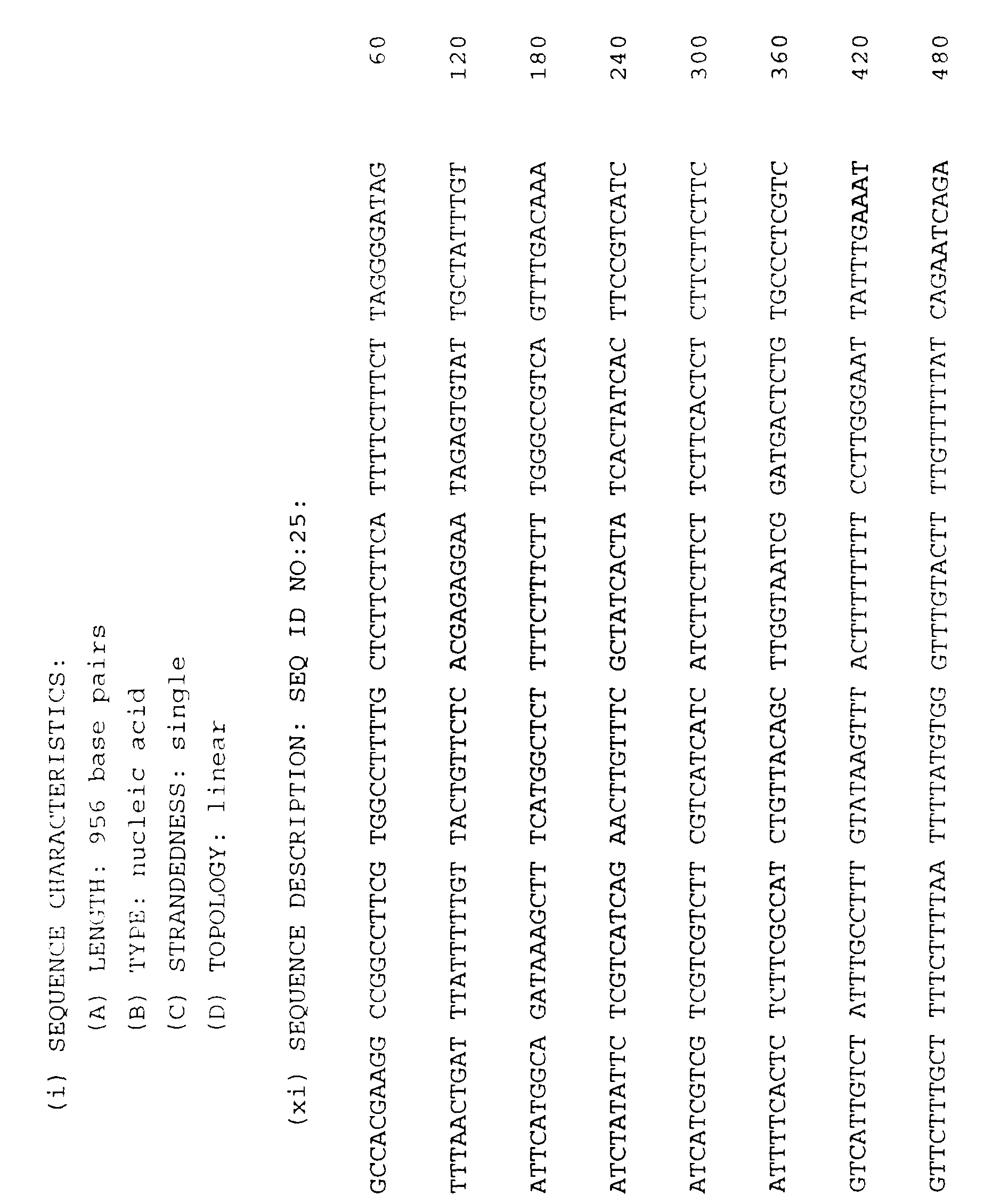


0


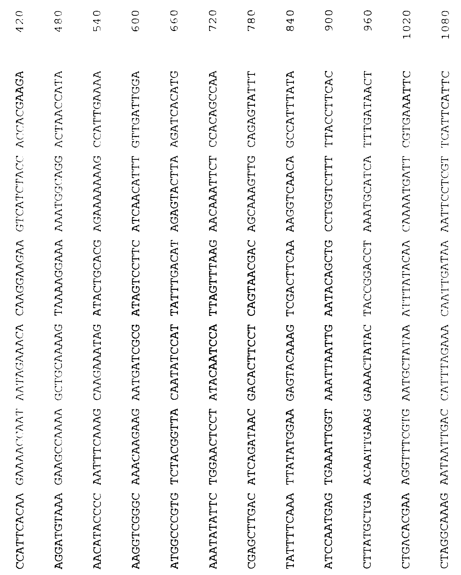


Claims
1. A nucleic acid segment characterized as: ( a) a nucleic acid segment comprising a sequence region that consists of at least 17 contiguous nucleotides that have the same sequence as, or are complementary to, 17 contiguous nucleotides of SEQ ID NO:1, SEQ ID NO: 29, SEQ ID NO : 30, SEQ ID NO: 19, SEQ ID NO: 31 or SEQ ID NO: 23; or (b) a nucleic acid segment of from 17 to about
10,000 nucleotides in length that hybridizes to the nucleic acid segment of SEQ ID NO : 1 , SEQ ID NO: 29, SEQ ID NO : 30 , SEQ ID NO: 19, SEQ ID NO: 31 or SEQ ID NO: 23, or the complement thereof, under standard hybridization conditions.
2. The nucleic acid segment of claim 1, wherein the segment comprises a sequence region of at least 17 contiguous nucleotides from SEQ ID NO : 1 , SEQ ID NO: 29, SEQ ID NO: 30, SEQ ID NO: 19, SEQ ID NO: 31 or SEQ ID NO: 23, cr the complement thereof.
3. Tne nucleic acid segment of claim 1, wherein the segment hybridizes to the nucleic acid segment of SEQ ID NO:1, SEQ ID NO: 29, SEQ ID NO: 30, SEQ ID NO: 19, SEQ ID NO: 31 or SEQ ID NO: 23, or the complement thereof.
4. The nucleic acid segment of claim 1, wherein the segment comprises a sequence region of at least 17 contiguous nucleotides from SEQ ID NO : 1 , or the
complement thereof; or wherein the segment hybridizes to the nucleic acid segment of SEQ ID NO : 1 , or the
complement thereof.
5. The nucleic acid segment of claim 1, wherein the segment comprises a sequence region of at least 17 contiguous nucleotides from SEQ ID NO:29, or the
complement thereof; or wherein the segment hybridizes to the nucleic acid segment of SEQ ID NO: 29, or the
complement thereof.
6. The nucleic acid segment of claim 1, wherein the segment comprises a sequence region of at least 17 contiguous nucleotides from SEQ ID NO: 30, or the
complement thereof; or wherein the segment hybridizes to the nucleic acid segment of SEQ ID NO: 30, or the
complement thereof.
7. The nucleic acid segment of claim 1, wherein the segment comprises a sequence region of at least 17 contiguous nucleotides from SEQ ID NO: 19, or the
complement thereof; or wherein the segment hybridizes to the nucleic acid segment of SEQ ID NO: 19, or the
complement thereof.
8. The nucleic acid segment of claim 1, wherein the segment comprises a sequence region of at least 17 contiguous nucleotides from SEQ ID NO: 31, or the
complement thereof; or wherein the segment hybridizes to the nucleic acid segment of SEQ ID NO: 31, or the
complement thereof.
9. The nucleic acid segment of claim 1, wherein the segment comprises a sequence region of at least 17 contiguous nucleotides from SEQ ID NO: 23, or the
complement thereof; or wherein the segment hybridizes to the nucleic acid segment of SEQ ID NO: 23, or the
complement thereof.
10. The nucleic acid segment of claim 1, wherein the segment comprises a sequence region of at least about 25 nucleotides; or wherein the segment is about 25
nucleotides in length.
11. The nucleic acid segment of claim 10, wherein the segment comprises a sequence region of at least about 50 nucleotides; or wherein the segment is about 50
nucleotides in length.
12. The nucleic acid segment of claim 11, wherein the segment comprises a sequence region of at least about 100 nucleotides; or wherein the segment is about 100
nucleotides in length.
13. The nucleic acid segment of claim 12, wherein the segment comprises a sequence region of at least' about 200 nucleotides; or wherein the segment is about 2C0
nucleotides in length.
14. The nucleic acid segment of claim 13, wherein the segment comprises a sequence region of at least about 500 nucleotides; or wherein the segment is about 500
nucleotides in length.
15. The nucleic acid segment of claim 14, wherein the segment comprises a sequence region that consists of the 1301 contiguous nucleotides of SEQ ID NO:1, or the complement thereof.
16. The nucleic acid segment of claim 14, wherein the segment comprises a sequence region that consists of at least about a 1000 nucleotide long contiguous sequence from SEQ ID NO: 29, or the complement thereof.
17. The nucleic acid segment of claim 16, wherein the segment comprises a sequence region that consists of the 1882 contiguous nucleotides of SEQ ID NO:29, or the complement thereof.
18. The nucleic acid segment of claim 14, wherein the segment comprises a sequence region that consists of the 1094 contiguous nucleotides of SEQ ID NO: 30, or the complement thereof.
19. The nucleic acid segment of claim 14, wherein the segment comprises a sequence region that consists of at least about a 1000 nucleotide long contiguous sequence from SEQ ID NO: 29, or the complement thereof.
20. The nucleic acid segment of claim 19, wherein the segment comprises a sequence region that consists of at least about a 2000 nucleotide long contiguous sequence from SEQ ID NO: 29, or the complement thereof.
21. The nucleic acid segment of claim 20, wherein the segment comprises a sequence region that consists of the 2434 contiguous nucleotides of SEQ ID NO: 19, or the complement thereof.
22. The nucleic acid segment of claim 14, wherein the segment comprises a sequence region that consists of the 807 contiguous nucleotides of SEQ ID NO:31, or the complement thereof.
23. The nucleic acid segment of claim 14, wherein the segment comprises a sequence region that consists of at least about a 1000 nucleotide long contiguous sequence from SEQ ID NO: 23, or the complement thereof.
24. The nucleic acid segment of claim 23, wherein the segment comprises a sequence region that consists of the 2117 contiguous nucleotides of SEQ ID NO: 23, or the complement thereof.
25. The nucleic acid segment of claim 1, wherein the segment is up to about 10,000 basepairs in length.
26. The nucleic acid segment of claim 25, wherein the segment is up to about 5,000 basepairs in length.
27. The nucleic acid segment of claim 26, wherein the segment is up to about 1,000 basepairs in length.
28. The nucleic acid segment of claim 27, wherein the segment is up to about 500 basepairs in length.
29. The nucleic acid segment of claim 28, wherein the segment is up to about 100 basepairs in length.
30. The nucleic acid segment of claim 1, further defined as a DNA segment.
31. The nucleic acid segment of claim 1, further defined as a RNA segment.
32. An isolated RNA segment of from 17 to about 1,500 nucleotides in length that comprises a non-ciliate telomerase RNA template.
33. The isolated RNA segment of claim 32, comprising a yeast telomerase RNA template.
34. An isolated RNA segment having the secondary
structure of the RNA segment encoded by the sequence of
SE; ID NO: 1.
35. An affinity column comprising a deoxyoligonucleotide attached to a solid support, wherein the
deoxyoligonucleotide includes a GT-rich sequence
complementary to a non-ciliate telomerase RNA template sequence and binds to a non-ciliate telomerase complex.
36. A DNA segment comprising an isolated gene associated with non-ciliate telomerase.
37. The DNA segment of claim 36, comprising an isolated gene associated with yeast telomerase.
38. The DNA segment of claim 37, comprising an isolated gene that encodes a yeast telomerase RNA template.
39. The DNA segment of claim 38, comprising an isolated gene that encodes the yeast telomerase RNA template sequence CACCACACCCACACAC (SEQ ID NO : 3 ).
40. The DNA segment of claim 39, comprising an isolated gene that includes the contiguous DNA sequence from position 468 to position 483 of SEQ ID NO : 1 or the contiguous DNA sequence from position 819 to position 834 of SEQ ID NO: 4.
41. The DNA segment of claim 40, comprising an isolated gene that includes a contiguous DNA sequence from
position 400 to position 500 of SEQ ID NO : 1.
42. The DNA segment of claim 41, comprising an isolated gene that includes a contiguous DNA sequence from
position 200 to position 900 of SEQ ID NO:1.
43. The DNA segment of claim 42, comprising an isolated gene that includes the DNA sequence of SEQ ID NO:1.
44. The DNA segment of claim 37, comprising an isolated gene that encodes a polypeptide associated with yeast telomerase.
45. The DNA segment of claim 44, comprising an isolated gene that encodes a polypeptide that includes a
contiguous amino acid sequence from SEQ ID NO: 16, SEQ ID NO:18, SEQ ID NO:20, SEQ ID NO:22 or SEQ ID NO:24.
46. The DNA segment of claim 45, comprising an isolated gene that encodes a polypeptide that includes a
contiguous amino acid sequence from SEQ ID NO: 16.
47. The DNA segment of claim 46, comprising an isolated gene that encodes a polypeptide having the sequence of SEQ ID NO:16.
48. The DNA segment of claim 47, comprising an isolated gene that includes the contiguous DNA sequence from position 54 to position 1799 of SEQ ID NO:29.
49. The DNA segment of claim 45, comprising an isolated gene that encodes a polypeptide that includes a
contiguous amino acid sequence from SEQ ID NO: 18.
50. The DNA segment of claim 49, comprising an isolated gene that encodes a polypeptide having the sequence of SEQ ID NO: 18.
51. The DNA segment of claim 50, comprising an isolated gene that includes the contiguous DNA sequence from position 78 to position 1094 of SEQ ID NO: 30.
52. The DNA segment of claim 45, comprising an isolated gene that encodes a polypeptide that includes a
contiguous amino acid sequence from SEQ ID NO: 20.
53. The DNA segment of claim 52, comprising an isolated gene that encodes a polypeptide having the sequence of
SEQ ID NO: 20.
54. The DNA segment of claim 53, comprising an isolated gene that includes the contiguous DNA sequence from position 2 to position 2368 of SEQ ID NO: 19.
55. The DNA segment of claim 45, comprising an isolated gene that encodes a polypeptide that includes a
contiguous amino acid sequence from SEQ ID NO: 22.
56. The DNA segment of claim 55, comprising an isolated gene that encodes a polypeptide having the sequence of
SEQ ID NO: 22.
57. The DNA segment of claim 56, comprising an isolated gene that includes the contiguous DNA sequence from position 55 to position 699 of SEQ ID NO:31.
58. The DNA segment of claim 45, comprising an isolated gene that encodes a polypeptide that includes a
contiguous amino acid sequence from SEQ ID NO:24.
59. The DNA segment of claim 58, comprising an isolated gene that encodes a polypeptide having the sequence of SEQ ID NO: 24.
60. The DNA segment of claim 59, comprising an isolated gene that includes the contiguous DNA sequence from position 3 to position 1955 of SEQ ID NO:23.
61. The DNA segment of claim 37, wherein the isolated gene is positioned under the control of a promoter.
62. The DNA segment of claim 61, positioned under the control of a recombinant promoter.
63. The DNA segment of claim 62, further defined as a recombinant vector.
64. A recombinant host cell incorporating a DNA segment that comprises an isolated gene associated with non-ciliate telomerase.
65. The recombinant host cell of claim 64, further defined as a prokaryotic host cell.
66. The recombinant host cell of claim 64, further defined as a eukaryotic host cell.
67. The recombinant host cell of claim 66, further defined as a veast cell.
68. The recombinant host cell of claim 66, further defined as a mammalian cell.
69. The recombinant host cell of claim 64, wherein the host cell expresses the DNA segment to produce a
telomerase RNA template or a polypeptide associated with telomerase.
70. A method of using a DNA segment that comprises an isolated gene associated with non-ciliate telomerase, the method comprising the steps of: (a) preparing a recombinant vector in which a non- ciliate telomerase-associated gene is positioned under the control of a promoter; (b) introducing said recombinant vector into a
recombinant host cell; (c) culturing the recombinant host cell under
conditions effective to allow expression of the telomerase-associated gene; and (d) collecting the expressed gene product
71. A recombinant gene product prepared by expressing a non-ciliate telomerase-associated gene in a recombinant host cell and purifying the expressed gene product away from total recombinant host cell components.
72. The gene product of claim 71, wherein the gene product is a telomerase RNA template.
73. The gene product of claim 71, wherein the gene product is a polypeptide associated with telomerase
74. A method for detecting a non-ciliate telomerase- associated gene in a sample, the method comprising the steps of: (a) obtaining sample nucleic acids from a sample suspected of containing a non-ciliate telomerase-associated gene; (b) contacting said sample nucleic acids with a nucleic acid segment that includes at least a 17 nucleotide long contiguous sequence from SEQ
ID NO-1, SEQ ID NO: 29, SEQ ID NO : 30 , SEQ ID NO: 19, SEQ ID NO: 31, SEQ ID NO: 23, or the complement thereof, under conditions effective to allow hybridization of substantially
complementary nucleic acids; and (c) detecting the hybridized complementary nucleic acids thus formed.
75. The method of claim 74, wherein the sample nucleic acids are obtained from a sample suspected of containing a turner cell.
76. The method of claim 74, wherein the sample nucleic acids are obtained from a sample suspected of containing a pathogen.
77. The method of claim 74 , wherein the sample nucleic acids are obtained from a sample suspected of containing a sperm cell or an egg cell.
78. A polypeptide composition, free from cell
components, comprising a purified non-ciliate telomerase- associated polypeptide that includes a contiguous amino acid sequence from SEQ ID NO: 16, SEQ ID NO: 18, SEQ ID NO: 20, SEQ ID NO: 22 or SEQ ID NO: 24.
79. An antibody that binds to a non-ciliate telomerase- associated polypeptide that includes a contiguous amino acid sequence from SEQ ID NO:16, SEQ ID NO:18, SEQ ID NC :2C, SEQ ID NO: 22 or SEQ ID NO: 24.
80. A method for identifying a gene associated with a non-ciliate telomerase, the method comprising the steps of: (a) preparing a cell containing a chromosome that contains a genetic marker located proximal to a telomere, the telomere repressing the expression of the marker;
(b) contacting said cell with a composition comprising a candidate gene; and (c) identifying a gene that allows expression of the marker.
81. The method of claim 80, wherein said cell is a Drosophila melanogaster cell.
82. The method of claim 80, wherein said cell is a human
83. The method of claim 82, wherein said cell is a human sperm cell or a human egg cell.
84. The method of claim 82, wherein said cell is a human cancer cell.
85. The method of claim 80, wherein said cell is a yeast cell.
86. The method of claim 85, wherein said genetic marker HIS3 , TRP1 , LYS2 , LEU2 , CAN1 , ADE2 or URA3.
87. The method of claim 86, wherein said genetic markers is ADE2 or URA3 .
86. The method of claim 85, wherein said ceil contains two distinct genetic markers, each located on a distinct chromosome.
89. The method of claim 88, wherein said genetic markers are ADE2 and URA3 .
90. The method of claim 80, wherein said candidate gene is a mutant or truncated gene.
91. The method of claim 80, wherein said candidate gene is a wild type gene.
92. A gene identified by the method of claim 80.
93. The gene of claim 92, the gene having the physical and functional characteristics of TLC1 .
94. The gene of claim 92, the gene having the physical and functional characteristics of STR1 .
95. The gene of claim 92, the gene having the physical and functional characteristics of STR3 .
96. The gene of claim 92, the gene having the physical and functional characteristics of STR4 .
97. The gene of claim 92, the gene having the physical and functional characteristics of STR5.
98. The gene of claim 92, the gene having the physical and functional characteristics of STR6.
99. A method for identifying a human telomerase- associated gene, the method comprising the steps of: (a) preparing a yeast cell containing a chromosome that contains a genetic marker located proximal to a telomere, the telomere repressing the expression of the marker; (b) contacting said cell with a composition
comprising a candidate human gene; and (c) identifying a human gene that allows expression of the marker.
100. A method for identifying a candidate substance that binds to a non-ciliate telomerase component, the method comprising the steps of: (a) preparing an isolated non-ciliate telomerase component; (b) contacting said isolated telomerase component with a composition comprising a candidate substance under conditions effective to allow binding; and
(c) detecting the presence of a telomerase
component-candidate substance bound complex.
101. The method of claim 100, wherein said isolated telomerase component is an RNA segment comprising a non- ciliate telomerase RNA template.
102. The method of claim 100, wherein said isolated telomerase component is a DNA segment encoding a non- ciliate telomerase-associated polypeptide.
103. The method of claim 100, wherein said isolated telomerase component is a polypeptide associated with non-ciliate telomerase.
104. A component that binds to a non-ciliate telomerase component.
105. The component of claim 104, identified by the method of claim 100.
106. The component of claim 104, wherein the component is a nucleic acid segment.
107. The component of claim 106, wherein the nucleic acid segment is a human nucleic acid segment.
108. The component of claim 106, wherein the nucleic acid segment hybridizes to a yeast telomerase nucleic acid segment under standard low stringency hybridization conditions.
109. The component of claim 106, wherein the nucleic acid segment hybridizes to a yeast telomerase nucleic acid segment under standard high stringency hybridization conditions.
110. The component of claim 104, wherein the component is a protein or polypeptide.
111. A method for identifying a candidate substance that modifies telomerase activity, the method comprising the steps of: (a) preparing a cell containing a chromosome that contains a genetic marker located near a telomere, the telomere capable of repressing the expression of the marker; (b) contacting said cell with a composition
comprising a candidate substance; and (c) identifying a candidate substance that allows expression of the marker or that further represses the expression of the marker.
112. The method of claim 111, further defined as a method for identifying a candidate substance that inhibits telomerase activity, the method comprising the steps of:
(a) preparing a cell containing a chromosome that contains a genetic marker located proximal to a telomere, the telomere significantly repressing the expression of the marker; (b) contacting said cell with a composition
comprising a candidate inhibitory substance; and (c) identifying a candidate inhibitory substance that allows expression of the marker.
113. The method of claim 111, further defined as a method for identifying a candidate substance that stimulates telomerase activity, the method comprising the steps of: (a) preparing a cell containing a chromosome that contains a genetic marker located in the vicinity of a telomere, the telomere not significantly repressing the expression of the marker; (b) contacting said cell with a composition
comprising a candidate substance; and
( c ) identifying a candidate stimulatory substance that represses or further represses the
expression of the marker.
114. A method for modifying the replicative capacity of a cell, comprising contacting a telomerase-containing cell with an amount of a candidate substance effective to modify telomerase activity.
115. The method of claim 114, further defined as a method for inhibiting the replication of a cell, comprising contacting a telomerase-containing cell with an amount of an inhibitory substance effective to inhibit telomerase activity.
116. The method of claim 115, wherein said telomerase-containing cell is a tumor cell.
117. The method of claim 115, wherein said telomerase-containing cell is a pathogenic cell.
118. The method of claim 114, further defined as a method for promoting the replication of a cell, comprising contacting a telomerase-containing cell with an amount of a stimulatory substance effective to promote telomerase activity.
119. The method of claim 118, wherein said telomerase- containing cell is a sperm cell.
120. The method of claim 118, wherein said telomerase- containing cell is an egg cell.
121. The use of a substance that inhibits telomerase activity in the preparation of a medicament for use in killing pathogenic or tumor cells, said substance being identif ied by a method comprising the steps of :
(a) preparing a cell containing a chromosome that contains a genetic marker located proximal to a telomere, the telomere significantly repressing the expression of the marker; (b) contacting said cell with a composition
comprising a candidate inhibitory substance; and (c) identifying a candidate inhibitory substance that allows expression of the marker.
122. The use of a substance that stimulates telomerase activity in the preparation of a medicament for use in promoting the replication of a sperm or egg cell, said substance being identified by a method comprising the steps of: (a) preparing a cell containing a chromosome that contains a genetic marker located in the vicinity of a telomere, the telomere not significantly repressing the expression of the marker; (b) contacting said cell with a composition
comprising a candidate substance; and (c) identifying a candidate stimulatory substance that represses or further represses the
expression of the marker.
Priority Applications (1)
| Application Number | Priority Date | Filing Date | Title |
|---|---|---|---|
| AU42786/96A AU4278696A (en) | 1994-10-20 | 1995-10-20 | Yeast telomerase components and methods using them |
Applications Claiming Priority (4)
| Application Number | Priority Date | Filing Date | Title |
|---|---|---|---|
| US32678194A | 1994-10-20 | 1994-10-20 | |
| US08/326,781 | 1994-10-20 | ||
| US08/431,080 US5698686A (en) | 1994-10-20 | 1995-04-28 | Yeast telomerase compositions |
| US08/431,080 | 1995-04-28 |
Publications (2)
| Publication Number | Publication Date |
|---|---|
| WO1996012811A2 true WO1996012811A2 (en) | 1996-05-02 |
| WO1996012811A3 WO1996012811A3 (en) | 1996-08-01 |
Family
ID=26985570
Family Applications (1)
| Application Number | Title | Priority Date | Filing Date |
|---|---|---|---|
| PCT/US1995/013801 WO1996012811A2 (en) | 1994-10-20 | 1995-10-20 | Yeast telomerase components and methods using them |
Country Status (3)
| Country | Link |
|---|---|
| US (3) | US5698686A (en) |
| AU (1) | AU4278696A (en) |
| WO (1) | WO1996012811A2 (en) |
Cited By (17)
| Publication number | Priority date | Publication date | Assignee | Title |
|---|---|---|---|---|
| WO1998023759A2 (en) * | 1996-11-26 | 1998-06-04 | Baylor College Of Medicine | Identification of inhibitors that prevent access of telomerase to chromosomal terminus |
| WO1998037181A2 (en) * | 1997-02-20 | 1998-08-27 | Whitehead Institute For Biomedical Research | Telomerase catalytic subunit gene and encoded protein |
| WO1998045450A1 (en) * | 1997-04-04 | 1998-10-15 | Geron Corporation | Purified telomerase |
| US5968506A (en) * | 1995-08-04 | 1999-10-19 | Geron Corporation | Purified telomerase |
| US6093809A (en) * | 1996-10-01 | 2000-07-25 | University Technology Corporation | Telomerase |
| US6261836B1 (en) | 1996-10-01 | 2001-07-17 | Geron Corporation | Telomerase |
| US6475789B1 (en) | 1996-10-01 | 2002-11-05 | University Technology Corporation | Human telomerase catalytic subunit: diagnostic and therapeutic methods |
| US6517834B1 (en) | 1995-08-04 | 2003-02-11 | Geron Corporation | Purified telomerase |
| US6545133B1 (en) | 1995-08-04 | 2003-04-08 | Geron Corporation | Methods for purifying telomerase |
| US6610839B1 (en) | 1997-08-14 | 2003-08-26 | Geron Corporation | Promoter for telomerase reverse transcriptase |
| US6808880B2 (en) | 1996-10-01 | 2004-10-26 | Geron Corporation | Method for detecting polynucleotides encoding telomerase |
| US6878817B2 (en) | 1996-11-27 | 2005-04-12 | Boston Heart Foundation, Inc. | Low density lipoprotein binding proteins and their use in diagnosing and treating atherosclerosis |
| US7262288B1 (en) | 1997-04-18 | 2007-08-28 | Geron Corporation | Nucleic acids encoding human telomerase reverse transcriptase and related homologs |
| US7378244B2 (en) | 1997-10-01 | 2008-05-27 | Geron Corporation | Telomerase promoters sequences for screening telomerase modulators |
| US7413864B2 (en) | 1997-04-18 | 2008-08-19 | Geron Corporation | Treating cancer using a telomerase vaccine |
| US7622549B2 (en) | 1997-04-18 | 2009-11-24 | Geron Corporation | Human telomerase reverse transcriptase polypeptides |
| US8222392B2 (en) | 1996-10-01 | 2012-07-17 | Geron Corporation | Kit for detection of telomerase reverse transcriptase nucleic acids |
Families Citing this family (11)
| Publication number | Priority date | Publication date | Assignee | Title |
|---|---|---|---|---|
| US5698686A (en) | 1994-10-20 | 1997-12-16 | Arch Development Corporation | Yeast telomerase compositions |
| AU2892199A (en) * | 1998-03-03 | 1999-09-20 | Affymetrix, Inc. | Cell cycle regulated genes |
| US6337200B1 (en) * | 1998-03-31 | 2002-01-08 | Geron Corporation | Human telomerase catalytic subunit variants |
| US20030147863A1 (en) * | 2001-12-17 | 2003-08-07 | Isis Pharmaceuticals Inc. | Antisense modulation of TFAP2C expression |
| US7294364B2 (en) * | 1999-09-24 | 2007-11-13 | Cao Group, Inc. | Method for curing composite materials |
| US20050059077A1 (en) * | 1999-09-30 | 2005-03-17 | Fred Hutchinson Cancer Research Center | Interfering with telomere maintenance in treatment of diseases |
| US6541202B1 (en) | 1999-10-13 | 2003-04-01 | Research & Development Institute, Inc. | Telomerase reverse transcriptase (TERT) genes from Candida albicans |
| AU7753600A (en) * | 2000-10-03 | 2002-11-18 | Fred Hutchinson Cancer Research Center | Interfering with telomere maintenance in treatment of diseases |
| US7309783B2 (en) * | 2001-05-09 | 2007-12-18 | The University Of Connecticut | Mammalian early developmental regulator gene |
| US20050053942A1 (en) * | 2002-06-24 | 2005-03-10 | Sakari Kauppinen | Methods and systems for detection and isolation of a nucleotide sequence |
| EP1702062A2 (en) * | 2003-06-11 | 2006-09-20 | Jan Remmereit | Differentiation of stem cells for therapeutic use |
Citations (1)
| Publication number | Priority date | Publication date | Assignee | Title |
|---|---|---|---|---|
| WO1993023572A1 (en) * | 1992-05-13 | 1993-11-25 | University Of Texas Southwestern Medical Center At Dallas | Therapy and diagnosis of conditions related to telomere length and/or telomerase activity |
Family Cites Families (36)
| Publication number | Priority date | Publication date | Assignee | Title |
|---|---|---|---|---|
| NL154600B (en) * | 1971-02-10 | 1977-09-15 | Organon Nv | METHOD FOR THE DETERMINATION AND DETERMINATION OF SPECIFIC BINDING PROTEINS AND THEIR CORRESPONDING BINDABLE SUBSTANCES. |
| US3949064A (en) * | 1973-10-26 | 1976-04-06 | Baxter Laboratories, Inc. | Method of detecting antigens or antibodies |
| CH653970A5 (en) | 1981-05-14 | 1986-01-31 | Schweiz Kaese Milch Verband | CONTAINER FOR GAS DRIVING LIQUIDS OR MASS. |
| CA1295560C (en) * | 1986-10-24 | 1992-02-11 | Howard J. Cooke | Telomeric dna probe |
| CA1301671C (en) * | 1986-12-19 | 1992-05-26 | Peter N. Ray | Dna from duchenne muscular dystrophy gene |
| US4889806A (en) * | 1987-04-15 | 1989-12-26 | Washington University | Large DNA cloning system based on yeast artificial chromosomes |
| AU620574B2 (en) * | 1987-11-25 | 1992-02-20 | City Of Hope National Medical Center | Tourette syndrome, autism and associated behavior |
| US5270201A (en) * | 1988-03-24 | 1993-12-14 | The General Hospital Corporation | Artificial chromosome vector |
| WO1989009219A1 (en) * | 1988-03-24 | 1989-10-05 | The General Hospital Corporation | Artificial chromosome vector |
| IT1230152B (en) * | 1988-06-10 | 1991-10-14 | Ici Plc | NUCLEOTID SEQUENCES. |
| IL94906A (en) * | 1989-07-19 | 1995-10-31 | Univ California | Methods and compositions for chromosome-specific staining |
| US5227293A (en) * | 1989-08-29 | 1993-07-13 | The General Hospital Corporation | Fusion proteins, their preparation and use |
| GB8921791D0 (en) * | 1989-09-27 | 1989-11-08 | Imp Cancer Res Tech | Disease gene |
| GB8928029D0 (en) * | 1989-12-12 | 1990-02-14 | Medical Res Council | Probe |
| US5025164A (en) * | 1990-03-07 | 1991-06-18 | E. I. Du Pont De Nemours And Company | Antistatic lead screens for use with x-ray films |
| FR2665908A1 (en) * | 1990-08-14 | 1992-02-21 | Agronomique Inst Nat Rech | Centromeric sequence and DNA comprising such a sequence |
| WO1992007080A1 (en) * | 1990-10-17 | 1992-04-30 | Board Of Regents, The University Of Texas System | A eukaryotic episomal dna cloning and expression vector |
| NL9100132A (en) * | 1991-01-25 | 1992-08-17 | Ingeny Bv | METHOD FOR DETECTING DNA SEQUENCE VARIATION. |
| CA2108666A1 (en) * | 1992-01-16 | 1993-07-17 | Geoffrey Frank Smart | Genetically modified wheat plants and progeny and method for production of hybrid wheat |
| US5629154A (en) * | 1993-11-12 | 1997-05-13 | Geron Corporation | Telomerase activity assays |
| US5648215A (en) * | 1992-05-13 | 1997-07-15 | Board Of Regents, The University Of Texas System | Telomerase diagnostic methods |
| US5645986A (en) * | 1992-05-13 | 1997-07-08 | Board Of Reagents, The University Of Texas System | Therapy and diagnosis of conditions related to telomere length and/or telomerase activity |
| US5686306A (en) * | 1992-05-13 | 1997-11-11 | Board Of Regents, The University Of Texas System | Methods and reagents for lengthening telomeres |
| US5639613A (en) * | 1992-05-13 | 1997-06-17 | Board Of Regents, University Of Texas System | Methods for cancer diagnosis and prognosis |
| US5693474A (en) * | 1992-05-13 | 1997-12-02 | Board Of Regents, University Of Texas System | Methods for cancer diagnosis and prognosis |
| CA2078377A1 (en) * | 1992-09-16 | 1994-03-17 | Henry H. Q. Heng | Gene mapping by in situ hybridisation to free chromatin |
| US5523389A (en) * | 1992-09-29 | 1996-06-04 | Isis Pharmaceuticals, Inc. | Inhibitors of human immunodeficiency virus |
| US5366889A (en) * | 1992-11-30 | 1994-11-22 | The General Hospital Corporation | DNA encoding a protein-coupled receptor kinase |
| US5466576A (en) * | 1993-07-02 | 1995-11-14 | Fred Hutchinson Cancer Research Center | Modulation of PIF-1-type helicases |
| WO1995013382A1 (en) * | 1993-11-12 | 1995-05-18 | Geron Corporation | Therapy and diagnosis of conditions related to telomere length and/or telomerase activity |
| WO1995013383A1 (en) * | 1993-11-12 | 1995-05-18 | Geron Corporation | Methods and reagents for lengthening telomeres |
| EP0666313A3 (en) * | 1994-01-27 | 1996-03-06 | Univ Iowa State Res Found Inc | Targeting of telomerase in cancer gene therapy. |
| US5583016A (en) * | 1994-07-07 | 1996-12-10 | Geron Corporation | Mammalian telomerase |
| US5698686A (en) | 1994-10-20 | 1997-12-16 | Arch Development Corporation | Yeast telomerase compositions |
| US5733730A (en) * | 1995-08-25 | 1998-03-31 | The Rockefeller University | Telomere repeat binding factor and diagnostic and therapeutic use thereof |
| US5747317A (en) * | 1996-07-08 | 1998-05-05 | Tularik Inc. | Human telomerase RNA interacting protein gene |
-
1995
- 1995-04-28 US US08/431,080 patent/US5698686A/en not_active Expired - Fee Related
- 1995-10-20 AU AU42786/96A patent/AU4278696A/en not_active Abandoned
- 1995-10-20 WO PCT/US1995/013801 patent/WO1996012811A2/en active Search and Examination
-
1997
- 1997-09-26 US US08/938,534 patent/US5916752A/en not_active Expired - Fee Related
-
1999
- 1999-06-30 US US09/345,294 patent/US6387619B1/en not_active Expired - Fee Related
Patent Citations (1)
| Publication number | Priority date | Publication date | Assignee | Title |
|---|---|---|---|---|
| WO1993023572A1 (en) * | 1992-05-13 | 1993-11-25 | University Of Texas Southwestern Medical Center At Dallas | Therapy and diagnosis of conditions related to telomere length and/or telomerase activity |
Non-Patent Citations (9)
| Title |
|---|
| CELL, vol. 63, 16 November 1990, NA US, pages 751-762, XP002000589 GOTTSCHLING, D. ET AL.: "Position effect at S.cerevisiae telomeres: reversible repression of pol II transcription" cited in the application * |
| EMBL DATABASE ENTRY SC4554; ACCESSION NUMBER Z38113 (VERSION 1), 15 October 1994, XP002000585 "S.cerevisiae chromosome IX lambda clone 4554" * |
| EMBL DATABASE ENTRY SCGAL1; ACCESSION NUMBER X76078 (VERSION 6), 16 June 1994, XP002000584 SMITS, P. ET AL.: "The complete sequence of a 33 kb fragment on the right arm of chromosome II from Saccharomyces cerevisiae reveals 16 open reading frames, five previously identified genes and a homologue of the SCO1 gene" * |
| EMBL DATABASE ENTRY SCH9986; ACCESSION NUMBER U00027 (VERSION 2), 21 November 1994, XP002000592 * |
| EMBL DATABASE ENTRY YER8_YEAST; ACCESSION NUMBER P40059 , 1 February 1995, XP002000591 * |
| GENES & DEVELOPMENT, vol. 5, no. 1, 1991, pages 49-59, XP002000586 LIU, Z. & TYE, B.-K.: "A yeast protein that binds to vertebrate telomeres and conserved yeast telomeric junctions" * |
| GENES & DEVELOPMENT, vol. 7, July 1993, pages 1146-1159, XP002000587 KYRION, G. ET AL.: "RAP1 and telomere structure regulate telomere position effects in Saccharomyces cerevisiae" cited in the application * |
| GENES AND DEVELOPMENT, (1994 MAY 15) 8 (10) 1133-46, vol. 8, XP002000588 APARICIO, O. & GOTTSCHLING, D.: "Overcoming telomeric silencing: a trans-activator competes to establish gene expression in a cell cycle-dependent way." cited in the application * |
| SCIENCE, (1994 OCT 21) 266 (5184) 404-9, XP002000590 SINGER, M. ET AL.: "TLC1: template RNA component of Saccharomyces cerevisiae telomerase" cited in the application * |
Cited By (35)
| Publication number | Priority date | Publication date | Assignee | Title |
|---|---|---|---|---|
| US6261556B1 (en) | 1995-08-04 | 2001-07-17 | Geron Corporation | Purified telomerose |
| US7067283B2 (en) | 1995-08-04 | 2006-06-27 | Geron Corporation | Producing telomerase activators and inhibitors by screening with purified telomerase |
| US6787133B2 (en) | 1995-08-04 | 2004-09-07 | Geron Corporation | Using purified telomerase to identify telomerase activators and inhibitors |
| US6545133B1 (en) | 1995-08-04 | 2003-04-08 | Geron Corporation | Methods for purifying telomerase |
| US6517834B1 (en) | 1995-08-04 | 2003-02-11 | Geron Corporation | Purified telomerase |
| US5968506A (en) * | 1995-08-04 | 1999-10-19 | Geron Corporation | Purified telomerase |
| US6927285B2 (en) | 1996-10-01 | 2005-08-09 | Geron Corporation | Genes for human telomerase reverse transcriptase and telomerase variants |
| US7585622B1 (en) | 1996-10-01 | 2009-09-08 | Geron Corporation | Increasing the proliferative capacity of cells using telomerase reverse transcriptase |
| US6261836B1 (en) | 1996-10-01 | 2001-07-17 | Geron Corporation | Telomerase |
| US6475789B1 (en) | 1996-10-01 | 2002-11-05 | University Technology Corporation | Human telomerase catalytic subunit: diagnostic and therapeutic methods |
| US8222392B2 (en) | 1996-10-01 | 2012-07-17 | Geron Corporation | Kit for detection of telomerase reverse transcriptase nucleic acids |
| US6093809A (en) * | 1996-10-01 | 2000-07-25 | University Technology Corporation | Telomerase |
| US7285639B2 (en) | 1996-10-01 | 2007-10-23 | Geron Corporation | Antibody to telomerase reverse transcriptase |
| US7195911B2 (en) | 1996-10-01 | 2007-03-27 | Geron Corporation | Mammalian cells that have increased proliferative capacity |
| US6808880B2 (en) | 1996-10-01 | 2004-10-26 | Geron Corporation | Method for detecting polynucleotides encoding telomerase |
| US7056513B2 (en) | 1996-10-01 | 2006-06-06 | Geron Corporation | Telomerase |
| US7005262B2 (en) | 1996-10-01 | 2006-02-28 | Geron Corporation | Methods for detecting nucleic acids encoding human telomerase reverse transcriptase |
| WO1998023759A2 (en) * | 1996-11-26 | 1998-06-04 | Baylor College Of Medicine | Identification of inhibitors that prevent access of telomerase to chromosomal terminus |
| WO1998023759A3 (en) * | 1996-11-26 | 1998-12-17 | Baylor College Medicine | Identification of inhibitors that prevent access of telomerase to chromosomal terminus |
| US7402395B2 (en) | 1996-11-27 | 2008-07-22 | Boston Heart Foundation, Inc. | Low density lipoprotein binding proteins and their use in diagnosing and treating atherosclerosis |
| US6878817B2 (en) | 1996-11-27 | 2005-04-12 | Boston Heart Foundation, Inc. | Low density lipoprotein binding proteins and their use in diagnosing and treating atherosclerosis |
| US7244410B2 (en) | 1996-11-27 | 2007-07-17 | Boston Heart Foundation, Inc. | Low density lipoprotein binding proteins and their use in diagnosing and treating atherosclerosis |
| US7807384B2 (en) | 1996-11-27 | 2010-10-05 | Boston Heart Foundation, Inc. | Low density lipoprotein binding proteins and their use in diagnosing and treating atherosclerosis |
| WO1998037181A2 (en) * | 1997-02-20 | 1998-08-27 | Whitehead Institute For Biomedical Research | Telomerase catalytic subunit gene and encoded protein |
| WO1998037181A3 (en) * | 1997-02-20 | 1999-02-18 | Whitehead Biomedical Inst | Telomerase catalytic subunit gene and encoded protein |
| WO1998045450A1 (en) * | 1997-04-04 | 1998-10-15 | Geron Corporation | Purified telomerase |
| US8236774B2 (en) | 1997-04-18 | 2012-08-07 | Geron Corporation | Human telomerase catalytic subunit |
| US7413864B2 (en) | 1997-04-18 | 2008-08-19 | Geron Corporation | Treating cancer using a telomerase vaccine |
| US8709995B2 (en) | 1997-04-18 | 2014-04-29 | Geron Corporation | Method for eliciting an immune response to human telomerase reverse transcriptase |
| US7622549B2 (en) | 1997-04-18 | 2009-11-24 | Geron Corporation | Human telomerase reverse transcriptase polypeptides |
| US7750121B2 (en) | 1997-04-18 | 2010-07-06 | Geron Corporation | Antibody to telomerase reverse transcriptive |
| US7262288B1 (en) | 1997-04-18 | 2007-08-28 | Geron Corporation | Nucleic acids encoding human telomerase reverse transcriptase and related homologs |
| US7199234B2 (en) | 1997-08-14 | 2007-04-03 | Geron Corporation | Regulatory segments of the human gene for telomerase reverse transcriptase |
| US6610839B1 (en) | 1997-08-14 | 2003-08-26 | Geron Corporation | Promoter for telomerase reverse transcriptase |
| US7378244B2 (en) | 1997-10-01 | 2008-05-27 | Geron Corporation | Telomerase promoters sequences for screening telomerase modulators |
Also Published As
| Publication number | Publication date |
|---|---|
| AU4278696A (en) | 1996-05-15 |
| US5698686A (en) | 1997-12-16 |
| US5916752A (en) | 1999-06-29 |
| WO1996012811A3 (en) | 1996-08-01 |
| US6387619B1 (en) | 2002-05-14 |
Similar Documents
| Publication | Publication Date | Title |
|---|---|---|
| US5698686A (en) | Yeast telomerase compositions | |
| Gallo et al. | Heat shock factor is required for growth at normal temperatures in the fission yeast Schizosaccharomyces pombe | |
| Runge et al. | TEL2, an essential gene required for telomere length regulation and telomere position effect in Saccharomyces cerevisiae | |
| Luke et al. | The SAPs, a new family of proteins, associate and function positively with the SIT4 phosphatase | |
| Palladino et al. | SIR3 and SIR4 proteins are required for the positioning and integrity of yeast telomeres | |
| Valay et al. | TheKIN28Gene is Required both for RNA Polymerase II Mediated Transcription and Phosphorylation of the Rpb1p CTD | |
| Gottschling et al. | Position effect at S. cerevisiae telomeres: reversible repression of Pol II transcription | |
| Gibson et al. | The phenotype of the minichromosome maintenance mutant mcm3 is characteristic of mutants defective in DNA replication | |
| Hochstrasser et al. | In vivo degradation of a transcriptional regulator: the yeast α2 repressor | |
| Yan et al. | Mcm2 and Mcm3, two proteins important for ARS activity, are related in structure and function. | |
| Dowzer et al. | Analysis of the creA gene, a regulator of carbon catabolite repression in Aspergillus nidulans | |
| Kyrion et al. | C-terminal truncation of RAP1 results in the deregulation of telomere size, stability, and function in Saccharomyces cerevisiae | |
| Cooper et al. | Regulation of telomere length and function by a Myb-domain protein in fission yeast | |
| Smith et al. | A novel histone H4 mutant defective in nuclear division and mitotic chromosome transmission | |
| Spingola et al. | A yeast intronic splicing enhancer and Nam8p are required for Mer1p-activated splicing | |
| Shei et al. | Yeast silencers can act as orientation-dependent gene inactivation centers that respond to environmental signals | |
| Xhemalce et al. | Role of the fission yeast SUMO E3 ligase Pli1p in centromere and telomere maintenance | |
| Ansari et al. | The yeast silent information regulator Sir4p anchors and partitions plasmids | |
| Ayté et al. | The Schizosaccharomyces pombe MBF complex requires heterodimerization for entry into S phase | |
| Chi et al. | SUM1-1, a dominant suppressor of SIR mutations in Saccharomyces cerevisiae, increases transcriptional silencing at telomeres and HM mating-type loci and decreases chromosome stability | |
| Longtine et al. | Telomere-mediated plasmid segregation in Saccharomyces cerevisiae involves gene products required for transcriptional repression at silencers and telomeres. | |
| US5817782A (en) | Lag 1:gene for increasing the longevity of eukaryotes | |
| Hartzog et al. | Identification and analysis of a functional human homolog of the SPT4 gene of Saccharomyces cerevisiae | |
| Clever et al. | Specific negative effects resulting from elevated levels of the recombinational repair protein Rad54p in Saccharomyces cerevisiae | |
| US6300067B1 (en) | TFIIB transcription factor from Candida albicans and methods of screening for inhibitors of Candida albicans growth |
Legal Events
| Date | Code | Title | Description |
|---|---|---|---|
| AK | Designated states |
Kind code of ref document: A2 Designated state(s): AM AT AU BB BG BR BY CA CH CN CZ DE DK EE ES FI GB GE HU IS JP KE KG KR KZ LK LR LT LU LV MD MG MN MW MX NO NZ PL PT RO RU SD SE SG SI SK TJ TM TT UA UG UZ VN |
|
| AL | Designated countries for regional patents |
Kind code of ref document: A2 Designated state(s): KE MW SD SZ UG AT BE CH DE DK ES FR GB GR IE IT LU MC NL PT SE BF BJ CF CG CI CM GA GN ML MR NE SN TD TG |
|
| DFPE | Request for preliminary examination filed prior to expiration of 19th month from priority date (pct application filed before 20040101) | ||
| 121 | Ep: the epo has been informed by wipo that ep was designated in this application | ||
| REG | Reference to national code |
Ref country code: DE Ref legal event code: 8642 |
|
| 122 | Ep: pct application non-entry in european phase | ||
| NENP | Non-entry into the national phase |
Ref country code: CA |
|
| DPE2 | Request for preliminary examination filed before expiration of 19th month from priority date (pct application filed from 20040101) |