WO1997022698A2 - Chemokine receptors 88-2b[ckr-3] and 88c and their antibodies - Google Patents
Chemokine receptors 88-2b[ckr-3] and 88c and their antibodies Download PDFInfo
- Publication number
- WO1997022698A2 WO1997022698A2 PCT/US1996/020759 US9620759W WO9722698A2 WO 1997022698 A2 WO1997022698 A2 WO 1997022698A2 US 9620759 W US9620759 W US 9620759W WO 9722698 A2 WO9722698 A2 WO 9722698A2
- Authority
- WO
- WIPO (PCT)
- Prior art keywords
- polynucleotide
- dna
- seq
- cells
- receptor
- Prior art date
Links
Classifications
-
- C—CHEMISTRY; METALLURGY
- C07—ORGANIC CHEMISTRY
- C07K—PEPTIDES
- C07K16/00—Immunoglobulins [IGs], e.g. monoclonal or polyclonal antibodies
- C07K16/18—Immunoglobulins [IGs], e.g. monoclonal or polyclonal antibodies against material from animals or humans
-
- A—HUMAN NECESSITIES
- A61—MEDICAL OR VETERINARY SCIENCE; HYGIENE
- A61P—SPECIFIC THERAPEUTIC ACTIVITY OF CHEMICAL COMPOUNDS OR MEDICINAL PREPARATIONS
- A61P17/00—Drugs for dermatological disorders
- A61P17/06—Antipsoriatics
-
- A—HUMAN NECESSITIES
- A61—MEDICAL OR VETERINARY SCIENCE; HYGIENE
- A61P—SPECIFIC THERAPEUTIC ACTIVITY OF CHEMICAL COMPOUNDS OR MEDICINAL PREPARATIONS
- A61P19/00—Drugs for skeletal disorders
- A61P19/02—Drugs for skeletal disorders for joint disorders, e.g. arthritis, arthrosis
-
- A—HUMAN NECESSITIES
- A61—MEDICAL OR VETERINARY SCIENCE; HYGIENE
- A61P—SPECIFIC THERAPEUTIC ACTIVITY OF CHEMICAL COMPOUNDS OR MEDICINAL PREPARATIONS
- A61P29/00—Non-central analgesic, antipyretic or antiinflammatory agents, e.g. antirheumatic agents; Non-steroidal antiinflammatory drugs [NSAID]
-
- A—HUMAN NECESSITIES
- A61—MEDICAL OR VETERINARY SCIENCE; HYGIENE
- A61P—SPECIFIC THERAPEUTIC ACTIVITY OF CHEMICAL COMPOUNDS OR MEDICINAL PREPARATIONS
- A61P31/00—Antiinfectives, i.e. antibiotics, antiseptics, chemotherapeutics
- A61P31/12—Antivirals
-
- A—HUMAN NECESSITIES
- A61—MEDICAL OR VETERINARY SCIENCE; HYGIENE
- A61P—SPECIFIC THERAPEUTIC ACTIVITY OF CHEMICAL COMPOUNDS OR MEDICINAL PREPARATIONS
- A61P31/00—Antiinfectives, i.e. antibiotics, antiseptics, chemotherapeutics
- A61P31/12—Antivirals
- A61P31/14—Antivirals for RNA viruses
- A61P31/18—Antivirals for RNA viruses for HIV
-
- A—HUMAN NECESSITIES
- A61—MEDICAL OR VETERINARY SCIENCE; HYGIENE
- A61P—SPECIFIC THERAPEUTIC ACTIVITY OF CHEMICAL COMPOUNDS OR MEDICINAL PREPARATIONS
- A61P7/00—Drugs for disorders of the blood or the extracellular fluid
- A61P7/02—Antithrombotic agents; Anticoagulants; Platelet aggregation inhibitors
-
- C—CHEMISTRY; METALLURGY
- C07—ORGANIC CHEMISTRY
- C07K—PEPTIDES
- C07K14/00—Peptides having more than 20 amino acids; Gastrins; Somatostatins; Melanotropins; Derivatives thereof
- C07K14/435—Peptides having more than 20 amino acids; Gastrins; Somatostatins; Melanotropins; Derivatives thereof from animals; from humans
- C07K14/705—Receptors; Cell surface antigens; Cell surface determinants
- C07K14/715—Receptors; Cell surface antigens; Cell surface determinants for cytokines; for lymphokines; for interferons
- C07K14/7158—Receptors; Cell surface antigens; Cell surface determinants for cytokines; for lymphokines; for interferons for chemokines
-
- C—CHEMISTRY; METALLURGY
- C07—ORGANIC CHEMISTRY
- C07K—PEPTIDES
- C07K16/00—Immunoglobulins [IGs], e.g. monoclonal or polyclonal antibodies
- C07K16/18—Immunoglobulins [IGs], e.g. monoclonal or polyclonal antibodies against material from animals or humans
- C07K16/28—Immunoglobulins [IGs], e.g. monoclonal or polyclonal antibodies against material from animals or humans against receptors, cell surface antigens or cell surface determinants
- C07K16/2866—Immunoglobulins [IGs], e.g. monoclonal or polyclonal antibodies against material from animals or humans against receptors, cell surface antigens or cell surface determinants against receptors for cytokines, lymphokines, interferons
-
- C—CHEMISTRY; METALLURGY
- C07—ORGANIC CHEMISTRY
- C07K—PEPTIDES
- C07K2319/00—Fusion polypeptide
- C07K2319/40—Fusion polypeptide containing a tag for immunodetection, or an epitope for immunisation
- C07K2319/43—Fusion polypeptide containing a tag for immunodetection, or an epitope for immunisation containing a FLAG-tag
Definitions
- the present invention relates generally to signal transduction pathways. More particularly, the present invention relates to chemokine receptors, nucleic acids encoding chemokine receptors, chemokine receptor ligands. modulators of chemokine receptor activity, antibodies recognizing chemokines and chemokine receptors, methods for identifying chemokine receptor ligands and modulators, methods for producing chemokine receptors, and methods for producing antibodies recognizing chemokine receptors. BACKGROUND OF THE INVENTION
- G-proteins guanine nucleotide binding proteins
- G-protein signal transduction pathways which involve, at a minimum, the following components: an extracellular signal (e.g. , neurotransmitters, peptide hormones, organic molecules, light, or odorants), a signal-recognizing receptor (G-protein-coupled receptor, reviewed in Probst et al. , DNA and Cell Biology 11 : 1-20 [1992] and also known as GPR or GPCR), and an intracellular. heterotrimeric GTP-binding protein, or G protein. In particular, these pathways have attracted interest because of their role in regulating while blood cell or leukocyte trafficking.
- an extracellular signal e.g. , neurotransmitters, peptide hormones, organic molecules, light, or odorants
- G-protein-coupled receptor reviewed in Probst et al. , DNA and Cell Biology 11 : 1-20 [1992] and also known as GPR or GPCR
- G protein-coupled receptor reviewed in Probst et al. , DNA and Cell Biology 11 : 1-20 [1992] and also
- Leukocytes comprise a group of mobile blood cell types including granulocytes (i. e. , neutrophils, basophils, and eosinophils), lymphocytes, and monocytes. When mobilized and activated, these cells are primarily involved in the body's defense against foreign matter. This task is complicated by the diversity of normal and pathological processes in which leukocytes participate. For example, leukocytes function in the normal inflammatory response to infection. Leukocytes are also involved in a variety of pathological inflammations. For a summary, see Scliall et al. , Curr. Opin. Immunol. 6:865-873 ( 1994) . Moreover, each of these processes can involve unique contributions, in degree, kind, and duration, from each of the leukocyte cell types.
- granulocytes i. e. , neutrophils, basophils, and eosinophils
- lymphocytes i. e. , lymphocytes, and monocytes.
- chemokines c/zem ⁇ attractant cytokines
- RANTES a CC chemokine. functions as a chemoattractant for monocytes.
- lymphocytes i.e. , memory T cells
- basophils i.e. , basophils
- eosinophils but not for neutrophils, while inducing the release of histamine from basophils.
- Chemokines were recently shown by Cocchi, et. al. . Science,
- the GPCR superfamily of receptors includes the chemokine receptor family.
- the typical chemokine receptor structure includes an extracellular chemokine-binding domain located near the N-terminus, followed by seven spaced regions of predominantly hydrophobic amino acids capable of forming membrane-spanning ⁇ -helices. Between each of the ⁇ -helical domains are hydrophilic domains localized, alternately, in the intra- or extra-cellular spaces. These features impart a serpentine conformation to the membrane-embedded chemokine receptor.
- the third intracellular loop typically interacts with G-proteins. In addition.
- the first chemokine receptors to be analyzed by molecular cloning techniques were the two neutrophil receptors for human IL8, a CXC chemokine. Holmes et al. , Science 253: 178-1280 (1991) and Murphy et al. , Science 253: 1280-1283 (1991), reported the cloning of these two receptors for IL8. Lee et al. , J. Biol. Chem. 267: 16283-16287 (1992), analyzed the cDNAs encoding these receptors and found 77 % amino acid identity between the encoded receptors, with each receptor exhibiting features of the G protein coupled receptor family.
- IL-8 receptors are specific for IL-8, w ⁇ ile the other binds and signals in response to IL-8, gro/MGSA, and NAP-2. Genetic manipulation of the genes encoding IL-8 receptors has contributed to our understanding of the biological roles occupied by these receptors. For example. Cacalano et al. , Science 265:682-684 (1994) reported that celetion of the IL-8 receptor homolog in the mouse resulted in a pleiotropic phenotype involving lymphadenopathy and splenomegaly. In addition, a study of missense mutations described in Leong et al. , J. Biol. Chem. 269: 19343- 19348 (1994) revealed amino acids in the IL-8 receptor that were critical for IL-8 binding. Domain swapping experiments discussed in Murphy, supra, implicated the amino terminal extracellular domain as a determinant of binding specificity.
- CCCKR1 CCCKR1 binds both MlP- 1 ⁇ and RANTES and causes intracellular calcium ion flux in response to both ligands.
- Chaw et al Proc Natl. Acad. Sci. (USA) 97:2752-2756 (1994) reported that another CC chemokine receptor.
- MCP-R1 CCCKR2
- CCCKR2 is encoded by a single gene that produces two splice variants which differ in their carboxyl terminal domains. This receptor binds and responds to MCP-3 in addition to MCP-1.
- a promiscuous receptor that binds both CXC and CC chemokines has also been identified. This receptor was originally identified on red blood cells and Horuk et al. , Science 261: 1 182-1 184 (1993) reports that it binds IL-8. NAP-2, GRO ⁇ , RANTES, and MCP-1. The erythrocyte chemokine receptor shares about 25 % identity with other chemokine receptors and may help to regulate circulating levels of chemokines or aid in the presentation of chemokines to their targets. In addition to binding chemokines.
- the erythrocyte chemokine receptor has also been shown to be the receptor for plasmodium vivax, a major cause of malaria (id.)
- Another G-protein coupled receptor which is closely related to chemokine receptors, the platelet activating factor receptor has also been shown to be the receptor for a human pathogen, the bacterium Streptococcus pneumoniae (Cundell et al. , Nature 377:435-438 (1995)).
- the mammalian chemokine receptors two viral chemokine receptor homologs have been identified. Ahuja et al. , J. Biol. Chem.
- Neote et al. Cell, 72:415-425 (1993) reports that human cytomegalovirus contains a gene encoding a receptor sharing about 30% identity with the CC chemokine receptors which binds MlP-1 ⁇ . MlP-1 ⁇ . MCP- 1 . and RANTES. These viral receptors may affect the normal role of chemokines and provide a selective pathological advantage for the vims.
- chemokines Because of the broad diversity of chemokines and their activities, there are numerous receptors for the chemokines. The receptors which have been characterized represent only a fraction of the total complement of chemokine receptors. There thus remains a need in the art for the identification of additional chemokine receptors. The availability of these novel receptors will provide tools for the development of therapeutic modulators of chemokine or chemokine receptor function. It is contemplated by the present invention that such modulators are useful as therapeutics for the treatment of atherosclerosis, rheumatoid arthritis, tumor growth suppression, asthma, viral infections, and other inflammatory conditions. Alternatively, fragments or variants of the chemokine receptors, or antibodies recognizing those receptors, are contemplated as therapeutics.
- the present invention provides purified and isolated nucleic acids encoding chemokine receptors involved in leukocyte trafficking.
- Polynucleotides of the invention include genomic DNAs, cDNAs, and RNAs, as well as completely or partially synthetic nucleic acids.
- Preferred polynucleotides of the invention include the DNA encoding the chemokine receptor 88-2B that is set out in SEQ ID NO: 3, the DNA encoding the chemokine receptor 88C that is set out in SEQ ID NO: 1 , and DNAs which hybridize to those DNAs under standard stringent hybridization conditions, or which would hybridize but for the redundancy of the genetic code.
- Exemplary stringent hybridization conditions are as follows: hybridization at 42°C in 50% formamide, 5X SSC, 20 mM sodium phosphate, pH 6.8 and washing in 0.2X SSC at 55 C. It is understood by those of skill in the art that variation in these conditions occurs based on the length and GC nucleotide content of the sequences to be hybridized. Formulas standard in the art are appropriate for determining exact hybridization conditions. See Sambrook et al. , ⁇ 9.47-9.51 in Molecular Cloning: A Laboratory Manual, Cold Spring Harbor Laboratory Press, Cold Spring Harbor, New York (1989).
- polynucleotides encoding domains of 88-2B or 88C for example, polynucleotides encoding one or more extracellular domains of either protein or other biologically active fragments thereof.
- 88-2B extracellular domains correspond to SEQ ID NO:3 and SEQ ID NO:4 at amino acid residues 1-36, 93-107. 171-196, and 263-284.
- the extracellular domains of 88-2B are encoded by polynucleotide sequences corresponding to SEQ ID NO:3 at nucleotides 362-469, 638-682, 872-949, and 1 148-1213.
- Extracellular domains of 88C correspond to SEQ ID NO: 1 and SEQ ID NO:2 at amino acid residues 1-32, 89-1 12, 166-191. and 259-280.
- the 88C extracellular domains are encoded by polynucleotide sequences that correspond to SEQ ID NO: 1 at nucleotides 55-150. 319-390. 550-627, and 829-894.
- the invention also comprehends polynucleotides encoding intracellular domains of these chemokine receptors.
- the intracellular domains of 88-2B include amino acids 60-71 , 131-151 , 219-240, and 306-355 of SEQ ID NO:3 and SEQ ID NO:4.
- Those domains are encoded by polynucleotide sequences corresponding to SEQ ID NO:3 at nucleotides 539-574, 752-814, 1016- 1081 , and 1277-1426, respectively.
- the 88C intracellular domains include amino acid residues 56-67. 125-145, 213-235, and 301-352 of SEQ ID NO: 1 and SEQ ID NO:2.
- the intracellular domains of 88C are encoded by polynucleotide sequences corresponding to SEQ ID NO: 1 at nucleotides 220-255, 427-489, 691-759, and 955-1 1 10.
- Peptides corresponding to one or more of the extracellular or intracellular domains, or antibodies raised against those peptides are contemplated as modulators of receptor activities, especially ligand and G protein binding activities of the receptors.
- the nucleotide sequences of the invention may also be used to design oligonucleotides for use as labeled probes to isolate genomic DNAs encoding 88-2B or 88C under stringent hybridization conditions (i.e. , by Southern analyses and Polymerase Chain Reaction methodologies). Moreover. these oligonucleotide probes can be used to detect particular alleles of the genes encoding 88-2B or 88C, facilitating both diagnosis and gene therapy treatments of disease states associated with particular alleles.
- these oligonucleotides can be used to alter chemokine receptor genetics to facilitate identification of chemokine receptor modulators.
- the nucleotide sequences can be used to design antisense genetic elements of use in exploring or altering the genetics and expression of 88-2B or 88C.
- the invention also comprehends biological replicas (i.e. . copies of isolated DNAs made in vivo or in vitro) and RNA transcripts of DNAs of the invention. Autonomously replicating recombinant constructions such as plasmid, viral, and chromosomal (e.g. , YAC) nucleic acid vectors effectively incorporating
- 88-2B or 88C polynucleotides and, particularly, vectors wherein DNA effectively encoding 88-2B or 88C is operatively linked to one or more endogenous or heterologous expression control sequences are also provided.
- the 88-2B and 88C receptors may be produced naturally, recombinantly or synthetically.
- Host cells prokaryotic or eukaryotic transformed or transfected with polynucleotides of the invention by standard methods may be used to express the 88-2B and 88C chemokine receptors Beyond the intact 88-2B or 88C gene products, biologically active fragments of 88-2B or 88C, analogs of 88-2B or 88C, and synthetic peptides derived from the amino acid sequences of 88-2B, set out in SEQ ID NO:4, or 88C, set out in SEQ ID NO:2, are contemplated by the invention.
- the 88-2B or 88C gene product, or a biologically active fragment of either gene product, when produced in a eukaryotic cell may be post-translationally modified (e.g. , via disulfide bond formation, glycosylation, phosphorylation, myristoylation, palmitoylation, acetylation, etc.)
- the invention further contemplates the 88-2B and 88C gene products, or biologically active fragments thereof, in monomeric. homomultimeric, or heteromultimeric conformations.
- one aspect of the invention involves antibody products capable of specifically binding to the 88-2B or 88C chemokine receptors.
- the antibody products are generated by methods standard in the art using recombinant 88-2B or 88C receptors, synthetic peptides or peptide fragments of 88-2B or 88C receptors, host cells expressing 88-2B or 88C on their surfaces, or 88-2B or 88C receptors purified from natural sources as immunogens.
- the antibody products may include monoclonal antibodies or polyclonal antibodies of any source or sub-type.
- monomeric, homomultimeric. and heteromultimeric antibodies, and fragments thereof are contemplated by the invention.
- the invention comprehends CDR-grafted antibodies, "humanized” antibodies, and other modified antibody products retaining the ability to specifically bind a chemokine receptor.
- the invention also contemplates the use of antibody products for detection of the 88-2B or 88C gene products, their analogs, or biologically active fragments thereof.
- antibody products may be used in diagnostic procedures designed to reveal correlations between the expression of 88-2B. or 88C, and various normal or pathological states.
- antibody products can be used to diagnose tissue-specific variations in expression of 88-2B or 88C, their analogs, or biologically active fragments thereof.
- Antibody products specific for the 88-2B and 88C chemokine receptors may also act as modulators of receptor activities.
- antibodies to 88-2B or 88C receptors are useful for therapeutic purposes.
- Assays for ligands capable of interacting with the chemokine receptors of the invention are also provided. These assays may involve direct detection of chemokine receptor activity, for example, by monitoring the binding of a labeled ligand to the receptor. In addition, these assays may be used to indirectly assess ligand interaction with the chemokine receptor.
- ligand comprises molecules which are agonists and antagonists of 88-2B or 88C, and other molecules which bind to the receptors. Direct detection of ligand binding to a chemokine receptor may be achieved using the following assay. Test compounds (i.e. , putative ligands) are detectably labeled (e.g. , radioiodinated).
- the detectably labeled test compounds are then contacted with membrane preparations containing a chemokine receptor of the invention.
- the membranes are prepared from host cells expressing chemokine receptors of the invention from recombinant vectors.
- the membrane material is collected on filters using vacuum filtration.
- the detectable label associated with the filters is then quantitated. For example, radiolabels are quantitated using liquid scintillation spectrophotometry. Using this technique, ligands binding to chemokine receptors are identified.
- a detectably labeled test compound is exposed to a membrane preparation displaying a chemokine receptor in the presence of increasing quantities of the test compound in an unlabeled state.
- a progressive reduction in the level of filter-associated label as one adds increasing quantities of unlabeled test compound confirms the identification of that ligand.
- Agonists are ligands which bind to the receptor and elicit intracellular signal transduction and antagonists are ligands which bind to the receptor but do not elicit intracellular signal transduction.
- the determination of whether a particular ligand is an agonist or an antagonist can be determined, for example, by assaying G protein-coupled signal transduction pathways. Activation of these pathways can be determined by measuring intracellular ca + + flux, phospholipase C activity or adenylyl cyclase activity, in addition to other assays (see examples 5 and 6).
- chemokines that bind to the 88C receptor include RANTES, MlP-1 ⁇ , and MTP-1 ⁇
- chemokines that bind to the 88-2B receptor include RANTES.
- 88C and 88-2B receptors and their ligands are specifically contemplated by the invention.
- Modulators of chemokine receptor function may be identified using assays similar to those used for identifying ligands.
- the membrane preparation displaying a chemokine receptor is exposed to a constant and known quantity of a detectably labeled functional ligand.
- the membrane-bound chemokine receptor is also exposed to an increasing quantity of a test compound suspected of modulating the activity of that chemokine receptor. If the levels of filter-associated label correlate with the quantity of test compound, that compound is a modulator of the activity of the chemokine receptor. If the level of filter-associated label increases with increasing quantities of the test compound, an activator has been identified.
- the indirect assays for receptor binding involve measurements of the concentration or level of activity of any of the components found in the relevant signal transduction pathway.
- Chemokine receptor activation often is associated with an intracellular Ca + + flux.
- Cells expressing chemokine receptors may be loaded with a calcium-sensitive dye.
- a Ca + + flux would be rendered spectrophotometrically detectable by the dye.
- the Ca ++ flux could be detected microscopically.
- Parallel assays, using either technique may be performed in the presence and absence of putative ligands. For example, using the microscopic assay for Ca * + flux, RANTES, a CC chemokine, was identified as a ligand of the 88-2B chemokine receptor.
- chemokine receptors affords the opportunity of assessing receptor activity by monitoring G protein activities.
- a characteristic activity of G proteins. GTP hydrolysis may be monitored using, for example, 32 P-labeled GTP.
- G proteins also affect a variety of other molecules through their participation in signal transduction pathways.
- G protein effector molecules include adenylyl cyclase, phospholipase C, ion channels, and phosphodiesterases. Assays focused on any of these effectors may be used to monitor chemokine receptor activity induced by ligand binding in a host cell that is both expressing the chemokine receptor of interest and contacted with an appropriate ligand.
- one method by which the activity of chemokine receptors may be detected involves measuring phospholipase C activity. In this assay, the production of radiolabeled inositol phosphates by host cells expressing a chemokine receptor in the presence of an agonist is detected.
- the detection of phospholipase activity may require cotransfection with DNA encoding an exogenous G protein.
- this assay can be performed by cotransfection of chimeric G protein DNA, for example, Gqi5 (Conklin, et al. , Nature 363:214-216 (1993), with 88-2B or 88C DNA and detecting phosphoinositol production when the cotransfected cell is exposed to an agonist of the 88-2B or 88C receptor.
- Gqi5 Conklin, et al. , Nature 363:214-216 (1993
- 88-2B or 88C DNA detecting phosphoinositol production when the cotransfected cell is exposed to an agonist of the 88-2B or 88C receptor.
- Receptor activators and inhibitors will activate or inhibit, respectively, the interaction of the receptors with their ligands in these assays.
- Chemokines have been linked to many inflammatory diseases, such as psoriasis, arthritis, pulmonary fibrosis and atherosclerosis. See Baggiolini et al. , supra. Inhibitors of chemokine action may be useful in treating these conditions.
- Broaddus et al. J. of Immunol. 152:2960-2967 (1994), describes an antibody to IL-8 which can inhibit neutrophil recruitment in endotoxin-induced pleurisy, a model of acute inflammation in rabbit lung.
- ligand or modulator binding to, or the activation of, the 88C receptor may be useful in treatment of HIV infection and HIV related disease states.
- Modulators of chemokine binding to specific receptors contemplated by the invention may include antibodies directed toward a chemokine or a receptor, biological or chemical small molecules, or synthetic peptides corresponding to fragments of the chemokine or receptor.
- compositions containing 88-2B or 88C modulators to mammalian subjects, for the purpose of monitoring or remediating normal or pathological immune reactions And viral infections including infection by retroviruses such as HIV- 1.
- HIV-2 and SIV is contemplated by the invention.
- the invention comprehends the mitigation of inflammatory responses, abnormal hematopoietic processes, and viral infections by delivery of a pharmaceutically acceptable quantity of 88-2B or 88C chemokine receptor modulators.
- the invention further comprehends delivery of these active substances in pharmaceutically acceptable compositions comprising carriers, diluents, or medicaments.
- the invention also contemplates a variety of administration routes.
- the active substances may be administered by the following routes: intravenous, subcutaneous, intraperitoneal, intramuscular, oral, anal (i.e. , via suppository formulations), or pulmonary (i.e. via inhalers, atomizers, nebulizers, etc.)
- the DNA sequence information provided by the present invention makes possible the development, by homologous recombination or "knockout” strategies [see, e.g. Kapecchi. Science, 244:1288-1292 (1989)], of rodents that fail to express a functional 88C or 88-2B chemokine receptor or that express a variant of the receptor.
- transgenic mice which express a cloned 88-2B or 88C receptor can be prepared by well known laboratory techniques (Manipulating the Mouse Embryo: A Laboratory Manual, Brigid Hohan, Frank Costantini and Elizabeth Lacy. eds. (1986) Cold Spring Harbor Laboratory ISBN 0-87969-175-1).
- Such rodents are useful as models for studying the activities of 88C or 88-2B receptors in vivo.
- Example 1 describes the isolation of genomic DNAs encoding the 88-2B and 88C chemokine receptors.
- Example 2 presents the isolation and sequencing of cDNAs encoding human 88-2B and 88C and macaque 88C.
- Example 3 provides a description of Northern analyses revealing the expression patterns of the 88-2B and 88C receptors in a variety of tissues.
- Example 4 details the recombinant expression of the 88-2B and 88C receptors.
- Example 5 describes Ca + + flux assays, phosphoinositol hydrolysis assays, and binding assays for 88-2B and 88C receptor activity in response to a variety of potential ligands.
- Example 6 Experiments describing the role of 88C and 882B as co-receptors for HIV is presented in Examples 6 and 7. The preparation and characterization of monoclonal and polyclonal antibodies immunoreactive with 88C is described in Example 8. Example 9 describes additional assays designed to identify 88- 2B or 88C ligands or modulators.
- Partial genomic clones encoding the novel chemokine receptor genes of this invention were isolated by PCR based on conserved sequences found in previously identified genes and based on a clustering of these chemokine receptor genes within the human genome.
- the genomic DNA was amplified by standard PCR methods using degenerate oligonucleotide primers.
- YACs Yeast Artificial Chromosomes
- Research Genetics, Inc. Huntsville, AL, YAC Library Pools, catalog no. 95011 B.
- a YAC vector can accommodate inserts of 500-1000 kilobase pairs.
- pools of YAC clone DNAs were screened by PCR using primers specific for the gene encoding CCCKR1.
- CCCKR(2)-5' the sense strand primer (corresponding to the sense strand of CCCKR1), is presented in SEQ ID NO: 15.
- Primer CCCKR(2)-5 ' consisted of the sequence 5'- CGTAAGCTTAGAGAAGCCGGGATGGGAA-3' , wherein the underlined nucleotides are the translation start codon for CCCKR1.
- the anti-sense strand primer was CCCKR-3' (corresponding to the anti-sense strand of CCCKR1) and its sequence is presented in SEQ ID NO: 16.
- the sequence of CCCKR-3' . 5'-GCCTCTAGAGTCAGAGACCAGCAGA-3' . contains the reverse complement of the CCCKR1 translation stop codon (underlined). Pools of YAC clone DNAs yielding detectable PCR products (i. e.
- DNA bands upon gel electrophoresis identified appropriate sub-pools of YAC clones, based on a proprietary identification scheme. (Research Genetics, Inc. , Huntsville, AL). PCR reactions were initiated with an incubation at 94 oC for four minutes. Sequence amplifications were achieved using 33 cycles of denaturation at 94oC for one minute, annealing at 55oC for one minute, and extension at 72oC for two minutes.
- YAC clone DNAs were then subjected to a second round of PCR reactions using the conditions, and primers, that were used in the first round of PCR.
- Results from sub-pool screenings identified individual clones capable of supporting PCR reactions with the CCCKR-specific primers.
- One clone, 881F10 contained 640 kb of human genomic DNA from chromosome 3p21 including the genes for CCCKR 1 and CCCKR2, as determined by PCR and hybridization.
- An overlapping YAC clone, 941A7 contained 700 kb of human genomic DNA and also contained the genes for CCCKR1 and CCCKR2. Consequently, further mapping studies were undertaken using these two YAC clones. Southern analyses revealed that CCCKR1 and CCCKR2 were located within approximately 100 kb of one another.
- CCCKR1 and CCCKR2 genes The close proximity of the CCCKR1 and CCCKR2 genes suggested that novel related genes might be linked to CCCKR1 and CCCKR2.
- PCR reactions were performed to amplify any linked receptor genes.
- Degenerate oligodeoxyribonucleotides were designed as PCR primers. These oligonucleotides corresponded to regions encoding the second intracellular loop and the sixth transmembrane domain of CC chemokine receptors, as deduced from aligned sequence comparisons of CCCKR1 , CCCKR2, and V28.
- V28 was used because it is an orphan receptor that exhibits the characteristics of a chemokine receptor; V28 has also been mapped to human chromosome 3. Raport et al. . Gene 163:295-299 (1995).
- the 5' primer, designated V28degf2 contains an internal BamHI site (see below): its sequence is presented in SEQ ID NO:5.
- the sequence of primer V28degf2 corresponds to DNA encoding the second intracellular loop region of the canonical receptor structure. See Probst et al. , supra.
- the 3' primer designated V28degr2 contains an internal HindUL site (see below); its sequence is presented in SEQ ID NO: 6.
- the sequence of primer V28degr2 corresponds to DNA encoding the sixth transmembrane domain of the canonical receptor structure.
- Amplified PCR DNA was subsequently digested with BamHI and HindIII to generate fragments of approximately 390 bp, consistent with the fragment size predicted from inspection of the canonical sequence. Following endonuclease digestion, these PCR fragments were cloned into pBluescript (Stratagene Inc.. LaJolla, CA). A total of 54 cloned fragments were subjected to automated nucleotide sequence analyses. In addition to sequences from CCCKR1 and CCCKR2, sequences from the two novel chemokine receptor genes of the invention were identified. These two novel chemokine receptor genes were designated 88-2B and 88C.
- 88-2B and 88C cDNAs were isolated from a macrophage cDNA library by the following procedure. Initially, a cDNA library, described in Tjoelker et al , Nature 374:549-553 (1995), was constructed in pRc/CMV (Invitrogen Corp. , San Diego, CA) from human macrophage mRNA. The cDNA library was screened for the presence of 88-2B and 88C cDNA clones by PCR using unique primer pairs corresponding to 88-2B or 88C. The PCR protocol involved an initial denaturation at 94oC for four minutes.
- Polynucleotides were then amplified using 33 cycles of PCR under the following conditions: Denaturation at 94°C for one minute, annealing at 55o C for one minute, and extension at 72°C for two minutes.
- the first primer specific for 88-2B was primer 88-2B-fl . presented in SEQ ID NO: 1 1 . It corresponds to the sense strand of SEQ ID NO: 3 at nucleotides 844-863.
- the second PCR primer specific for the gene encoding 88-2B was primer 88-2B-rl , presented in SEQ ID NO: 12; the 88-2B-r1 sequence corresponds to the anti-sense strand of SEQ ID NO: 3 at nucleotides 1023-1042.
- primer 88C-fl the sequence of the first primer specific for the gene encoding 88C.
- primer 88C-fl is presented in SEQ ID NO: 13 and corresponds to the sense strand of SEQ ID NO: 1 at nucleotides 453-471 .
- the second primer specific for the gene encoding 88C is primer 88C-r3, presented in SEQ ID NO: 14; the sequence of 88C-r3 corresponds to the anti-sense strand of SEQ ID NO: 1 at nucleotides 744-763.
- Clone 777 contained a DNA insert of 1915 bp including the full length coding sequence of 88-2B as determined by the following criteria: the clone contained a long open reading frame beginning with an ATG codon, exhibited a Kozak sequence, and had an in-frame stop codon upstream.
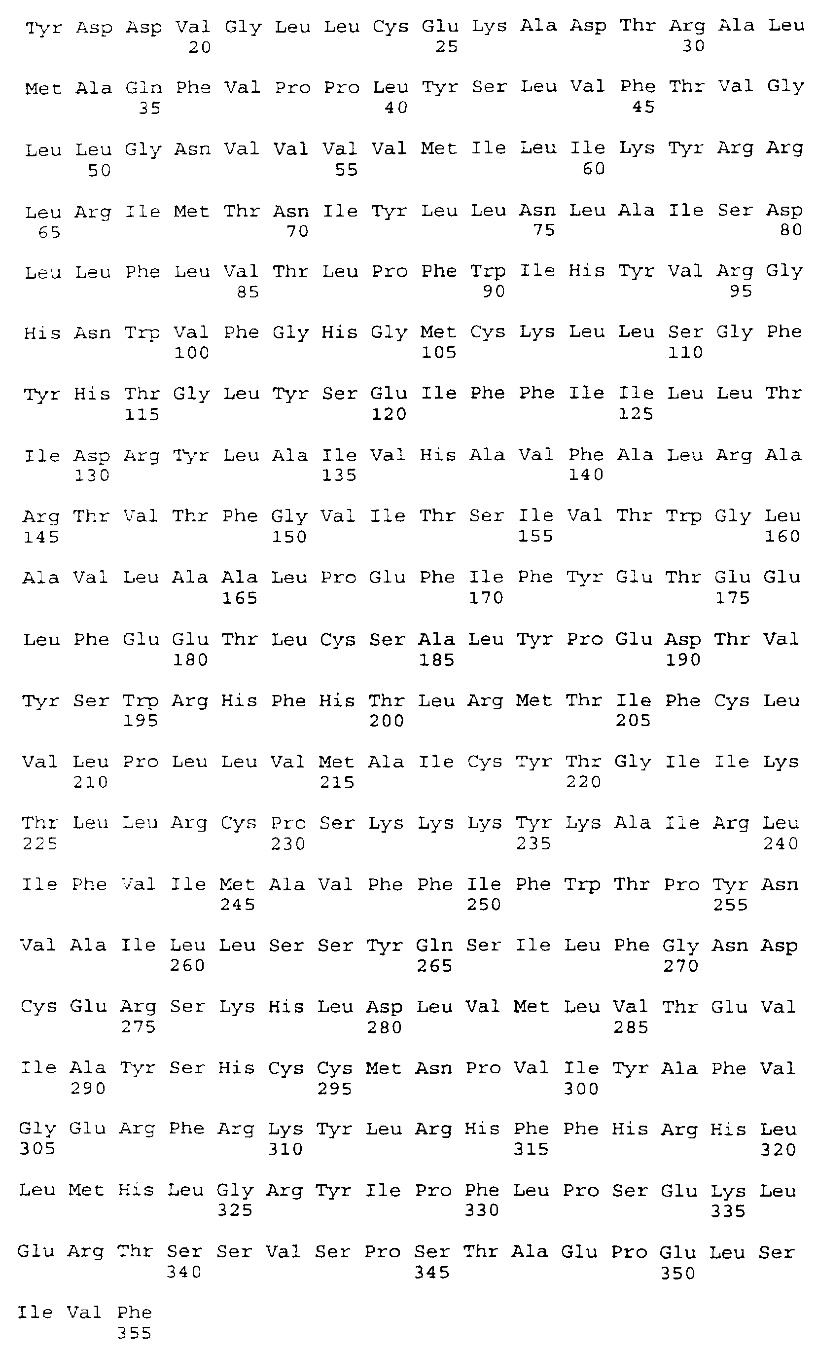
- the DNA and deduced amino acid sequences of the insert of clone 777 are presented in SEQ ID NO: 3 and SEQ ID NO:4, respectively.
- the 88-2B transcript was relatively rare in the macrophage cDNA library. During the library screen, only three 88-2B clones were identified from an estimated total of three million clones.
- cDNA clones encoding the 88C chemokine receptor identified clones 101 and 134 which appeared to contain the entire 88C coding region, including a putative initiation codon. However, these clones lacked the additional 5 ' sequence needed to confirm the identity of the initiation codon.
- the 88C transcript was relatively abundant in the macrophage cDNA Library. During the library screen, it was estimated that 88C was present at one per 3000 transcripts (in a total of approximately three million clones in the library).
- RACE PCR Rapid Amplification of cDNA Ends
- Human spleen 5'-RACE- ready cDNA was purchased from Clontech Laboratories. Inc. , Palo Alto, CA. and used according to the manufacturer's recommendations.
- the cDNA had been made "5 '-RACE- ready” by ligating an anchor sequence to the 5' ends of the cDNA fragments.
- the anchor sequence is complementary to an anchor primer supplied by Clontech Laboratories, Inc. , Palo Alto, CA.
- the anchor sequence-anchor primer duplex polynucleotide contains an EcoRI site.
- Human spleen cDNA was chosen as template DNA because Northern blots had revealed that 88C was expressed in this tissue.
- the PCR reactions were initiated by denaturing samples at 94°C for four minutes. Subsequently, sequences were amplified using 35 cycles involving denaturation at 94oC for one minute, annealing at 60°C for 45 seconds, and extension at 72oC for two minutes. The first round of PCR was performed on reaction mixtures containing 2 ⁇ l of the 5'-RAC ⁇ -ready spleen cDNA, 1 ⁇ l of the anchor primer, and 1 ⁇ l of primer 88c-r4 (100 ng/ ⁇ l) in a total reaction volume of 50 ⁇ l.
- the 88C-specific primer, primer 88c-r4 (5'-GATAAGCCTCACAG-CCCTGTG-3'), is presented in SEQ ID NO:7.
- the sequence of primer 88c-r4 corresponds to the anti-sense strand of SEQ ID NO: 1 at nucleotides 745-765.
- a second round of PCR was performed on reaction mixtures including 1 ⁇ l of the first PCR reaction with 1 ⁇ l of anchor primer and 1 ⁇ l of primer 88C-rlb (100 ng/ ⁇ l) containing the following sequence (5'-GCTAAGCTTGATGACTATCTTTAATGTC-3') and presented in SEQ ID NO: 8.
- the sequence of primer 88C-rlb contains an intemal HmdIII cloning site (underlined).
- the sequence 3' of the HmdIII site corresponds to the anti-sense strand of SEQ ID NO: 1 at nucleotides 636-654.
- the resulting PCR product was digested with EcoRI and HindIII and fractionated on a 1 % agarose gel. The approximately 700 bp fragment was isolated and cloned into pBluescript. Clones with the largest inserts were sequenced.
- the intact PCR product was ligated into vector pCR using a commercial TA cloning kit (Invitrogen Corp. , San Diego, CA) for subsequent nucleotide sequence determinations.
- the 88-2B and 88C cDNAs were sequenced using the PRISM TM Ready Reaction DyeDeoxy TM Terminator Cycle Sequencing Kit (Perkin Elmer Corp. , Foster City, CA) and an Applied Biosystems 373A DNA Sequencer.
- the insert of clone 777 provided the double-stranded template for sequencing reactions used to determine the 88-2B cDNA sequence.
- the sequence of the entire insert of clone 777 was determined and is presented as the 88-2B cDNA sequence and deduced amino acid sequence in SEQ ID NO:3.
- the sequence is 1915 bp in length, including 361 bp of 5' untranslated DNA (corresponding to SEQ ID NO:3 at nucleotides 1-361), a coding region of 1065 bp (corresponding to SEQ ID NO:3 at nucleotides 362-1426), and 489 bp of 3' untranslated DNA (corresponding to SEQ ID NO:3 at nucleotides 1427-1915).
- the 88-2B genomic DNA described in Example 1 above, corresponds to SEQ ID NO:3 at nucleotides 746-1128.
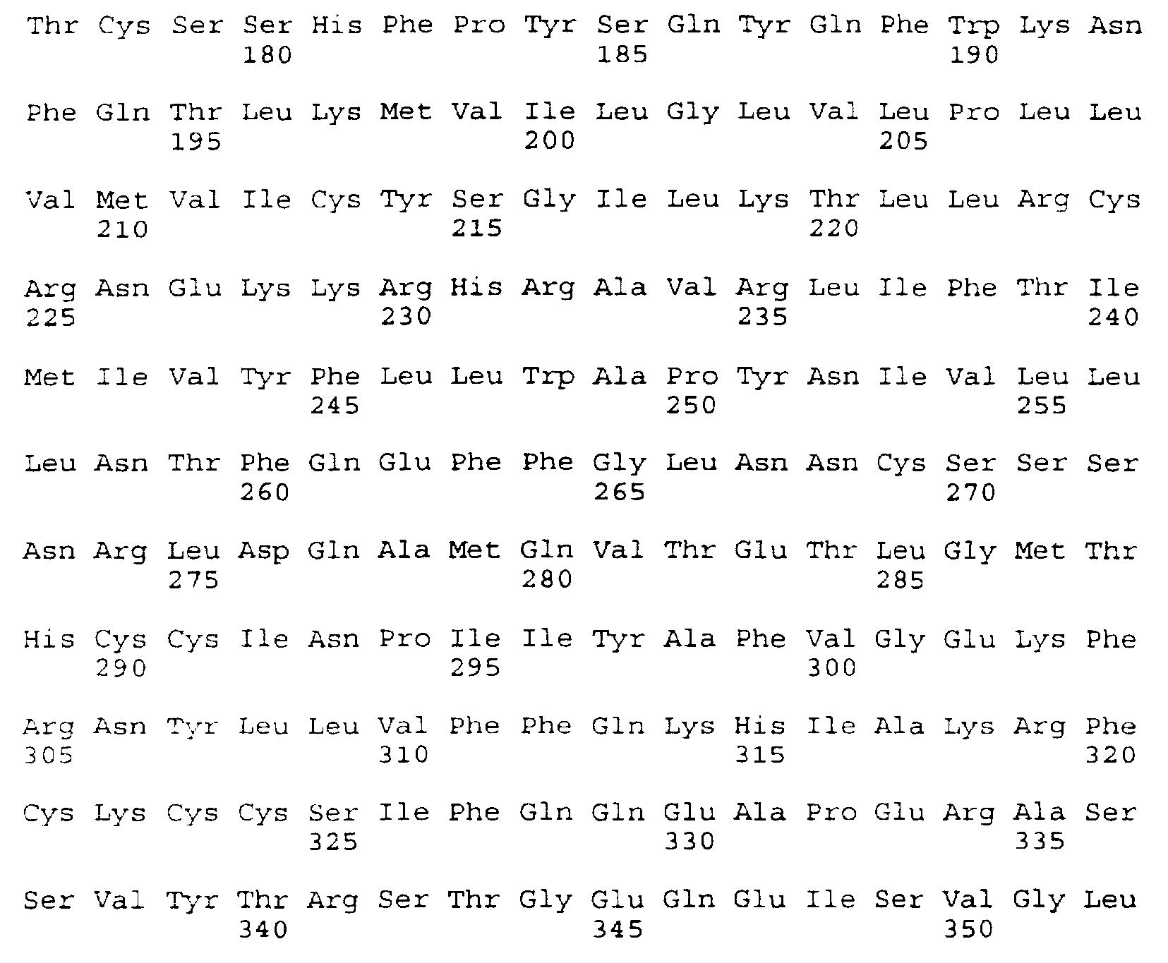
- the 88C cDNA sequence, and deduced amino acid sequence, is presented in SEQ ID NO: 1.
- the 88C cDNA sequence is a composite of sequences obtained from RACE-PCR cDNA, clone 134. and clone 101 .
- the RACE-PCR cDNA was used as a sequencing template to determine nucleotides 1 -654 in SEQ ID NO: 1 , including the unique identification of 9 bp of 5' untranslated cDNA sequence in SEQ ID NO: 1 at nucleotides 1 -9.
- the sequence obtained from the RACE PCR cDNA confirmed the position of the first methionine codon at nucleotides 55-57 in SEQ ID NO: 1 , and supported the conclusion that clone 134 and clone 101 contained full-length copies of the 88C coding region.
- Clone 134 contained 45 bp of 5' untranslated cDNA (corresponding to SEQ ID NO: 1 at nucleotides 10-54), the 1056 bp 88C coding region (corresponding to SEQ ID NO: l at nucleotides 55-1 110), and 492 bp of 3' untranslated cDNA (corresponding to SEQ ID NO: 1 at nucleotides 1 1 1 1-1602).
- Clone 101 contained 25 bp of 5' untranslated cDNA (corresponding to SEQ ID NO: 1 at nucleotides 30-54), the 1056 bp 88C coding region (corresponding to SEQ ID NO: 1 at nucleotides 55-1 1 10).
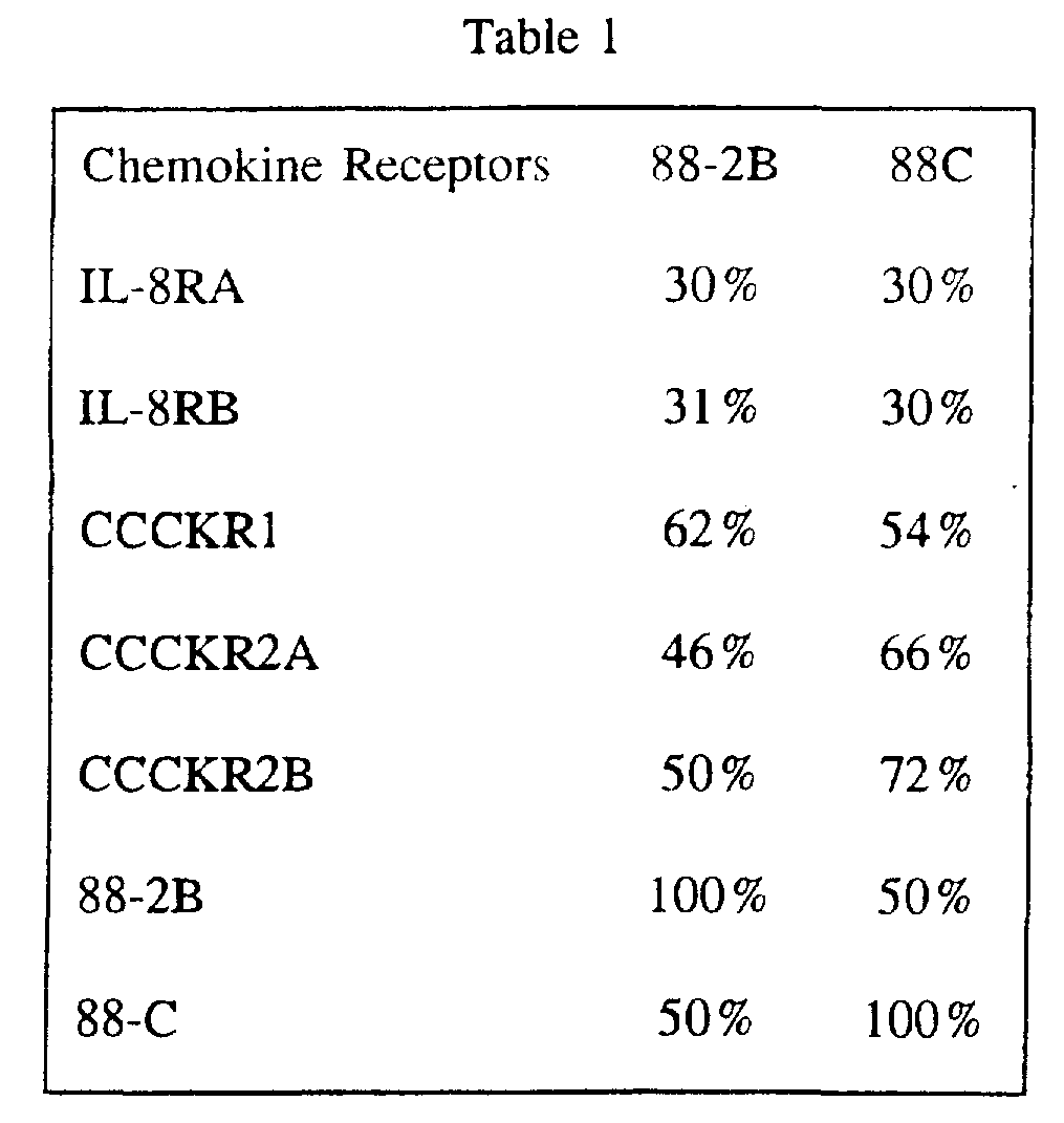
- the deduced amino acid sequences of 88-2B and 88C revealed hydrophobicity profiles characteristic of GPCRs, including seven hydrophobic domains corresponding to GPCR transmembrane domains. Sequence comparisons with other GPCRs also revealed a degree of identity. Significantly, the deduced amino acid sequences of both 88-2B and 88C had highest identity with the sequences of the chemokine receptors. Table 1 presents the results of these amino acid sequence comparisons.
- Table 1 shows that 88-2B is most similar to CCCKR1 (62 % identical at the amino acid level) and 88C is most similar to CCCKR2 (72 % identical at the amino acid level).
- the deduced amino acid sequences of 88-2B and 88C also reveal the intracellular and extracellular domains characteristic of GPCRs.
- the 88-2B extracellular domains correspond to the amino acid sequence provided in SEQ ID NO:3, and SEQ ID NO:4, at amino acid residues 1-36, 93-107, 171-196, and 263-284.
- the extracellular domains of 88-2B are encoded by polynucleotide sequences corresponding to SEQ ID NO:3 at nucleotides 362-469, 638-682, 872-949, and 1148-1213.
- Extracellular domains of 88C include amino acid residues 1-32, 89-112, 166-191 . and 259-280 in SEQ ID NO: 1 and SEQ ID NO:2.
- the 88C extracellular domains are encoded by polynucleotide sequences that correspond to SEQ ID NO: 1 at nucleotides 55-150, 319-390, 550-627, and 829-894.
- the intracellular domains of 88-2B include amino acids 60-71 , 131-151, 219-240. and 306-355 of SEQ ID NO: 3 and SEQ ID NO:4.
- Those domains are encoded by polynucleotide sequences corresponding to SEQ ID NO: 3 at nucleotides 539-574, 752-814, 1016-1081 , and 1277-1426. respectively.
- the 88C intracellular domains include amino acid residues 56-67, 125-145, 213-235.
- the intracellular domains of 88C are encoded by polynucleotide sequences corresponding to SEQ ID NO: 1 at nucleotides 220-255, 427-489, 691-759, and 955-1110.
- a macaque 88C DNA was amplified by PCR from macaque genomic DNA using primers corresponding to 5' and 3' flanking regions of the human 88C cDNA.
- the 5' primer corresponded to the region immediately upstream of and including the initiating Met codon.
- the 3' primer was complementary to the region immediately downstream of the termination codon.
- the primers included restriction sites for cloning into expression vectors.
- the sequence of the 5' primer was GACAAGCTTCACAGGGTGGAACAAGATG (With the HindIII site underlined) (SEQ ID NO: 17) and the sequence of the 3' primer was GTCTCTAGACCACTTGAGTCCGTGTCA (with the XbaI site underlined) (SEQ ID NO: 18).
- the conditions of the PCR amplification were 94°C for eight minutes, then 40 cycles of 94°C for one minute, 55°C for forty-five seconds, and 72°C one minute.
- the amplified products were cloned into the HindIII and XbaI sites of pcDNA3 and a clone was obtained and sequenced.
- the full length macaque cDNA and deduced amino acid sequences are presented in SEQ ID NOs: 19 and 20, respectively.
- the nucleotide sequence of macaque 88C is 98 % identical to the human 88C sequence.
- the deduced amino acid sequences are 97 % identical.
- the mRNA expression patterns of 88-2B and 88C were determined by Northern blot analyses.
- Northern blots containing immobilized poly A + RNA from a variety of human tissues were purchased from Clontech Laboratories, Inc..
- a probe specific for 88-2B nucleotide sequences was generated from cDNA clone 478.
- the cDNA insert in clone 478 contains sequence corresponding to SEQ ID NO: 3 at nucleotides 641-1915.
- clone 478 was digested and the insert DNA fragment was isolated following gel electrophoresis. The isolated insert fragment was then radiolabeled with 32 P-labeled nucleotides, using techniques known in the art.
- a probe specific for 88C nucleotide sequences was generated by isolating and radiolabeling the insert DNA fragment found in clone 493.
- the insert fragment from clone 493 contains sequence corresponding to SEQ
- the 88C Northerns showed an approximately 4 kb mRNA in several human tissues, including a strong signal when probing spleen or thymus tissue and less intense signals when analyzing mRNA from peripheral blood leukocytes and small intestine. A relatively weak signal for 88C was detected in lung tissue and in ovarian tissue.
- the expression of 88C in human T-cells and in hematopoietic cell lines was also determined by Northern blot analysis. Levels of 88C in CD4 + and CD8 + T-cells were very high.
- the transcript was present at relatively high levels in myeloid cell lines THP1 and HL-60 and also found in the B cell line Jijoye.
- the cDNA was a relatively abundant transcript in a human macrophage cDNA library based on PCR amplification of library subfractions.
- the 88-2B and 88C cDNAs were expressed by recombinant methods in mammalian cells.
- 88C was subcloned into the mammalian cell expression vector pBJ1 (Ishi. K. et. al. , J. Biol. Chem 270: 16435-16440 (1995).
- the construct included sequences encoding a prolactin signal sequence for efficient cell surface expression and a FLAG epitope at the amino terminus of 88C to facilitate detection of the expressed protein.
- the FLAG epitope consists of the sequence "DYKDDDD. " COS-7 cells were transiently transfected with the 88C expression plasmid using Lipofectamine (Life Technology. Inc. , Grand Island, NY) following the manufacturer's instructions.
- the FLAG-tagged 88C receptor was also stably transfected into HEK-293 cells, a human embryonic kidney cell line, using transfection reagent DOTAP (N-[1 -[(2 ,3-Dioleoyloxy)propyl]-N,N,N-trimethyl- ammoniummethylsulfate, Boehringer-Mannheim, Inc., Indianapolis. IN) according to the manufacturer's recommendations. Stable lines were selected in the presence of the drug G418.
- the transfected HEK-293 cells were evaluated for expression of 88C at the cell surface by ELISA. using the Ml antibody to the FLAG epitope. ELISA showed that 88C tagged with the FLAG epitope was expressed at the cell surface of stably transformed HEK- 293 Cells.
- the 88-2B and 88C cDNAs were used to make stable HEK-293 transfectants.
- the 88-2B receptor cDNA was cloned behind the cytomegalovirus promoter in pRc/CMV (Invitrogen Corp., San Diego. CA) using a PCR-based strategy.
- the template for the PCR reaction was the cDNA insert in clone 777.
- the PCR primers were 88-2B-3 (containing an internal XbaI site) and 88-2B-5 (containing an internal HindIII site).
- the nucleotide sequence of primer 88-2B-3 is presented in SEQ ID NO:9; the nucleotide sequence of primer 88-2B-5 is presented in SEQ ID NO:10.
- the pRc/CMV recombinant clones were transfected using transfection reagent DOTAP (N-[1-[( 2 , 3 -Dioleoyloxy)propyl]-N, N, N-trimethylammoniummethylsulfate, Boehringer-Mannheim, Inc., Indianapolis, IN) according to the manufacturer's recommendations, into HEK-293 cells, a human embryonic kidney cell line. Stable lines were selected in the presence of the drug G418. Standard screening procedures (i.e., Northern blot analyses) were performed to identify stable cell lines expressing the highest levels of 88-2B and 88C mRNA.
- DOTAP transfection reagent DOTAP
- chemokine receptor activity was employed.
- a common feature of signalling through the known chemokine receptors is that signal transduction is associated with the release of intracellular calcium cations. Therefore, intracellular Ca + + concentration in the transfected HEK-293 cells was assayed to determine whether the 88-2B or 88C receptors responded to any of the known chemokines.
- HEK-293 cells stably transfected with 88-2B, 88C (without the
- FLAG epitope sequence FLAG epitope sequence
- a control coding region encoding IL8R or CCCKR2, see below
- MEM + 10% serum were then washed, harvested with versene (0.6 mM EDTA, 10 mM Na 2 HPO 4 , 0. 14 M NaCl, 3 mM KCI, and 1 mM glucose), and incubated in MEM + 10% serum + 1 ⁇ M Fura-2 AM (Molecular Probes, Inc. , Eugene, OR) for 30 minutes at room temperature.
- Fura-2 AM is a Ca + + -sensitive dye.
- the cells were resuspended in Dulbecco's phosphate-buffered saline containing 0.9 mM CaCl 2 and 0.5 mM MgCl 2 (D-PBS) to a concentration of approximately 10 7 cells/ml and changes in fluorescence were monitored using a fluorescence spectrophotometer (Hitachi Model F-4010). Approximately 10° cells were suspended in 1.8 ml D-PBS in a cuvette maintained at 37 C. Excitation wavelengths alternated between 340 and 380 nm at 4 second intervals; the emission wavelength was 510 nm. Test compositions were added to the cuvette via an injection port; maximal Ca + + flux was measured upon the addition of ionomycin.
- HEK-293 cells expressing either 88-2B or 88C failed to show a flux in intracellular Ca ++ concentration when exposed to any of the following chemokines: MCP-1 , MCP-2, MCP-3, MlP-1 ⁇ , MIP- 10, IL8, NAP-2, gro/MGSA, IP-10, ENA-78, or PF-4. (Peprotech, Inc. , Rocky Hill, NJ).
- RANTES (Regulated on Activation, Normal T Expressed and Secreted) is a CC chemokine that has been identified as a chemoattractant and activator of eosinophils. See Neote et al. , supra. This chemokine also mediates the release of histamine by basophils and has been shown to function as a chemoattractant for memory T cells in vitro. Modulation of 88-2B receptor activities is therefore contemplated to be useful in modulating leukocyte activation.
- FLAG tagged 88C receptor was expressed in HEK-293 cells and tested for chemokine interactions in the CA + + flux assay.
- Cell surface expression of 88C was confirmed by ELISA and by FACScan analysis using the M1 antibody.
- the chemokines RANTES, MlP-1 ⁇ , and MIP-1 ⁇ all induced a Ca + + flux in 88C-transfected cells when added at a concentration of 100 nM.
- Ca + + flux assays can also be designed to identify modulators of chemokine receptor binding. The preceding fluorimetric or microscopic assays are carried out in the presence of test compounds. If Ca + + flux is increased in the presence of a test compound, that compound is an activator of chemokine receptor binding. In contrast, a diminished Ca + + flux identifies the test compound as an inhibitor of chemokine receptor binding.
- Another assay for ligands or modulators involves monitoring phospholipase C activity, as described in Hung et al. , J. Biol. Chem. 116:827-832 (1992). Initially, host cells expressing a chemokine receptor are loaded with 3 H-inositol for 24 hours. Test compounds (i.e. , potential ligands) are then added to the cells and incubated at 37oC for 15 minutes. The cells are then exposed to 20 mM formic acid to solubilize and extract hydrolyzed metabolites of phosphoinositol metabolism (i.e. , the products of phospholipase C-mediated hydrolysis).
- Test compounds i.e. , potential ligands
- the extract is subjected to anion exchange chromatography using an AG1X8 anion exchange column (formate form). Inositol phosphates are eluted with 2 M ammonium formate/0.1 M formic acid and the 3 H associated with the compounds is determined using liquid scintillation spectrophotometry.
- the phospholipase C assay can also be exploited to identify modulators of chemokine receptor activity. The aforementioned assay is performed as described, but with the addition of a potential modulator. Elevated levels of detectable label would indicate the modulator is an activator; depressed levels of the label would indicate the modulator is an inhibitor of chemokine receptor activity.
- the phospholipase C assay was performed to identify chemokine ligands of the FLAG-tagged 88C receptor. Approximately 24 hours after transfection, COS-7 cells expressing 88C were labeled for 20-24 hours with myo-[2- 3 H]inositol (1 ⁇ Ci/ml) in inositol-free medium containing 10% dialyzed FCS.
- Gqi5 a G protein which has the carboxyl terminal five amino acids of Gi (which bind to the receptor) spliced onto G ⁇ q. Co-transfection with Gqi5 significantly potentiates signaling by CCCKR1 and CCKR2B.
- Co- transfection with Gqi5 revealed that 88C signaled well in response to RANTES, MlP- 1 ⁇ . and MIP-1 ⁇ , but not in response to MCP- 1 .
- Dose-response curves revealed EC 50 values of InM for RANTES, 6nM for MlP-1 ⁇ , and 22nM for MlP-1 ⁇ .
- MlP- 1 ⁇ is the first cloned human receptor with a signaling response to MIP-1 ⁇ .
- MlP- 1 ⁇ clearly has a unique cellular activation pattern. It appears to activate T cells but not monocytes (Baggiolini et al. , Supra) which is consistent with receptor stimulation studies.
- MIP-1 ⁇ binds to CCCKR1. It does not induce calcium flux (Neote et al. , Supra).
- MlP-l ⁇ and RANTES bind to and causes signalling in CCCKR1 and CCCKR5 (RANTES also causes activation of CCCKR3).
- MTP-1 ⁇ thus appears to be much more selective than other chemokines of the CC chemokine family.
- selectivity is of therapeutic significance because a specific beneficial activity can be stimulated (such as suppression of HIV infection) without stimulating multiple leukocyte populations which results in general pro-inflammatory activities.
- MIP-1 ⁇ as labeled using the Bolton and Hunter reagent (di-iodide, NEN, Wilmington, DE), according to the manufacturer's instructions. Unconjugated iodide was separated from labeled protein by elution using a PD-10 column (Pharmacia) equilibrated with PBS and BSA (1 % w/v). The specific activity was typically 2200 Ci/mmole.
- Equilibrium binding was performed by adding 125 I-labeled ligand with or without a 100-fold excess of unlabeled ligand, to 5 X 10 5 HEK-293 cells transfected with 88C tagged with the FLAG epitope in polypropylene tubes in a total volume of 300 ⁇ l (50 mM HEPES pH 7.4, 1 mM CaCl 2 , MgCl 2 . 0.5 % BSA) and incubating for 90 minutes at 27°C with shaking at 150 rpm. The cells were collected, using a Skatron cell harvester (Skatron Instruments Inc.. Sterling, VA), on glass fiber filters presoaked in 0.3 % polyethyleneimine and 0.2 % BSA.
- Skatron cell harvester Skatron Instruments Inc.. Sterling, VA
- Ligand binding by competition with unlabeled ligand was determined by incubation of 5 X 10 5 transfected cells (as above) with 1.5 nM of radiolabeled ligand and the indicated concentrations of unlabeled ligand. The samples were collected, washed and counted as above. The data was analyzed using the curve-fitting program Prism (GraphPad Inc. , San Diego, CA) and the iterative non-linear regression program, LIGAND (PM220).
- the chemokines MlP-1 ⁇ , MlP- 1 ⁇ and RANTES have been shown to inhibit replication of HIV-1 and HIV-2 in human peripheral blood mononuclear cells and PM1 cells (Cocchi, et. al. , supra).
- the present invention contemplates that activation of or ligand binding to the 88C receptor may provide a protective role in HIV infection.
- fusin can act as a co-receptor for HIV entry.
- Fusin/CXCR4 in combination with CD4 the primary HIV receptor, apparently facilitates HIV infection of cultured T cells (Feng, et al. , Science 272:872-%11 (1996).
- 88C is constitutively expressed in T cells and abundantly expressed in macrophages, 88C is likely to be involved in viral and HIV infection.
- the function of 88C and 88-2B as co-receptors for HIV was determined by transfecting cells which express CD4 with 88C or 88-2B and challenging the co-transfected cells with HIV. Only cells expressing both CD4 and a functional co-receptor for HIV become infected. HIV infection can be determined by several methods. ELISAs which test for expression of HIV antigens are commercially available, for example Coulter HIV- 1 p 24 antigen assay (US Patent Nos. 4,886,742), Coulter Corp. , 1 1800 SW 147th Ave. , Miami, FL 33196. Alternatively, the test cells can be engineered to express a reporter gene such as LACZ attached to the HIV LTR promoter [Kimpton et al. . J. Virol. 66:2232-2239 (1992)]. In this method, cells that are infected with HIV are detected by a colorimetric assay.
- CCC-CD4 human CD4
- 88C was transiently transfected into a cat cell line, CCC [Clapham, et al. , 181:703-715 (1991)], which had been stably tranformed to express human CD4 (CCC-CD4). These cells are normally resistant to infection by any strain of HIV- 1 because they do not endogenously express 88C.
- CCC/CD4 cells were transiently transfected with 88C cloned into the expression vector pcDNA3. 1 (Invitrogen Corp. , San Diego, CA) using lipofectamine (Gibco BRL, Gaithersburg, MD). Two days after transfection, cells were challenged with HIV.
- Cat CCC cells which do not endogeneously express CD4 were transfected with 88-2B.
- cells were transfected with pcDNA3.1 containing 88-2B using lipofectamine and infected with HIV-2 48 hours later.
- Three days after infection cells were immunostained for the presence of HIV-2 envelope glycoproteins.
- the presence of sCD4 during HIV-2 ROD/B challenge increased the infection of these cells by by 10-fold.
- the entry of HIV-2 into the 88-2B transfected cells could be blocked by the presence of 400-800 ng/ml eotaxin, one of the ligands for 88-2B.
- the baseline infectivity levels of CCC/88-2B (with no soluble CD4) were equivalent to CCC cells which were not transfected with 88-2B.
- the co-receptor role of 88C and 88-2B can be demonstrated by an experimental method which does not require the use of live virus.
- CD4 and a LACL reporter gene are mixed with a cell line co-expressing the HIV envelope glycoprotein (ENV) and a transcription factor for the reporter gene construct (Nussbaum, et al. , 1994 J. Virol. 68:5411).
- Cells expressing a functional co-receptor for HIV will fuse with the ENV expressing cells and thereby allow expression of the reporter gene.
- detection of reporter gene product by colorimetric assay indicates that 88C or 88-2B function as a co-receptor for HIV.
- chemokines inhibit viral infection has not yet been elucidated.
- One possible mechanism involves activation of the receptor by binding of a chemokine.
- the binding of the chemokine leads to signal transduction events in the cell that renders the cell resistant to viral infection and/or prevents replication of the virus in the cell. Similar to interferon induction, the cell may differentiate such that it is resistant to viral infection, or an antiviral state is established.
- a second mechanism involves direct interference with viral entry into cells by blocking access of viral envelope glycoproteins to the co-receptor by chemokine binding. In this mechanism, G-protein signalling is not required for chemokine suppression of HIV infection.
- chemokine binding to the receptor is uncoupled from signal transduction and the effect of the chemokine on suppression of viral infection is determined.
- Ligand binding can be uncoupled from signal transduction by the addition of compounds which inhibit G-protein mediated signaling. These compounds include, for example, pertussis toxin and cholera toxin. In addition, downstream effector polypeptides can be inhibited by other compounds such as wortmannin. If G-protein signalling is involved in suppression of viral infection, the addition of such compounds would prevent suppression of viral infection by the chemokine. Alternatively, key residues or receptor domains of 88C or 88-2B receptor required for G-protein coupling can be altered or deleted such that G-protein coupling is altered or destroyed but chemokine binding is not affected.
- chemokines are unable to suppress viral or HIV infection, then signaling through a G-protein is required for suppression of viral or HIV infection. If however, chemokines are able to suppress viral infection, then G-protein signaling is not required for chemokine suppression of viral infection and the protective effects of chemokines may be due to the chemokine blocking the availability of the receptor for the virus.
- Another approach involves the use of antibodies directed against
- Antibodies which bind to 88C or 88-2B which can be shown not to elicit G-protein signaling may block access to the chemokine or viral binding site of the receptor. If in the presence of antibodies to 88C or 88-2B. viral infection is suppressed, then the mechanism of the protective effects of chemokines is blocking viral access to its receptor. Feng, et al. Reported that antibodies to the amino terminus of the fusin receptor suppressed HIV infection (Feng, et al , 1996).
- HeLa-MAGI cells are HeLa cells that have been stably transformed to express CD4 as well as integrated HIV-1 LTR which drives expression of a nuclear localized 0-galactosidase gene.
- HeLa-MAGI cells can detect lab-adapted isolates of HIV- 1 but only a minority of primary isolates [Kimpton and Emerman, supra], and cannot detect most SIV isolates [Chackerian et al. , "Characterization of a CD4-Expressing Macaque Cell Line that can Detect Virus After A Single Replication Cycle and can be infected by Diverse Simian Immunodeficiency Vims Isolates. " Virology, 213(2): 6499-6505 (1995)].
- Macrophage or T cell tropic viruses epitope-tagged 88C or 88-2B encoding DNA was transfected into HeLa-MAGI or U373-MAGI cells by infection with a retroviral vector to generate HeLA-MAGI-88C or U373-MAGI-88C cell lines, respectively. Expression of the co-receptors on the cell surface was demonstrated by immunostaining live cells using the anti-FLAG Ml antibody and by RT-PCR.
- the 88C and 88-2B genes utilized to construct HeLa-MAGI- 88C and U373-MAGI-88C included sequences encoding the prolactin signal peptide followed by a FLAG epitope as described in Example 4. This gene was inserted into the retroviral vector pBabe-Puro [Morgenstern and Land
- High titer retroviral vector stocks pseudotyped with the VSV-G protein were made by transient transfection as described in Bartx et al. . J. Virol. 70:2324-2331 (1996), and used to infect HeLa-MAGI and U373-MAGI cells.
- Cells resistant to 0.6 ⁇ gJml puromycin (HeLa) or 1 ⁇ g/ml puromycin (U373) were pooled. Each pool contained at least 1000 independent transduction events.
- Emerman, supra was used to create HeLa-MAGI-88C cells.
- U373-MAGI-88C cells and U373-MAGI cells were infected with limiting dilutions of a T-tropic strain of HIV-1 (mv ⁇ ), an M-tropic strain (HIV YU-2 ), and an SIV isolate.
- HeLa-MAGI and HeLa-MAGI-88C cells were infected with limiting dilutions of various HIV strains.
- the two cloned M-tropic viruses, HIV JR-CSF and HIV YU-2 both infected HeLa-MAGI-88C, but not HeLa-MAGI celis, showing that both strains use 88C as a co-receptor (Table 3. See note c).
- a great disparity in the ability of each of these two viral strains to infect HeLa-MAGI-88C cells was observed. 6.2 ⁇ 10 5 IU/ml for HIV YU-2 and 1.2 ⁇ 10 4 for HIV JR-CSF .
- the infectivity of virus stock (Table 3) is the number of infectious units per physical particle (represented here by the amount of viral core protein).
- the infectivity of these two cloned viral strains differed by over 50-fold in viral stocks that were independently prepared.
- HIV-2 Rod has been reported to use fusin as a receptor even in the absence of CD4 [Endres et al. , Cell, 87(4):745-756 (1996)]. HIV-2 Rod is able to infect HeLa-MAGI cells, however its infectivity is enhanced at least 10-fold in HeLa-MagI-88C (Table 4). HeLa cells endogenously express fusin. Thus, the molecular clone of HIV-2 Rod is dual tropic, and is able to use 88C as one of its co-receptors in addition to CXCR4. Similarly, a primary strain of HIV-2 7312A infected HeLa-MAGI-88C cells and not the HeLa-MAGI cells, indicating that like primary strain of HIV- 1 , it uses 88C as a receptor.
- N-syncytium inducing N-syncytium inducing
- SI syncytium inducing
- Mouse monoclonal antibodies which specifically recognize 88C were prepared.
- the antibodies were produced by immunizing mice with a peptide corresponding to the amino terminal twenty amino acids of 88C.
- the peptide was conjugated to Keyhole Limpet Cyanin (KLH) according to the manufacturer's directions (Pierce. Imject maleimide activated KLH), emulsified in complete Freund's adjuvant and injected into five mice. Two additional injections of conjugated peptide in incomplete Freund's adjuvant occurred at three week intervals. Ten days after the final injection, serum from each of the five mice was tested for immunoreactivity with the twenty amino acid peptide by ELISA.
- KLH Keyhole Limpet Cyanin
- 227M, 227N, 227P, 227R were established which produced antibodies that recognized the peptide by ELISA and the 88C protein on 293 cells by FACS. Each antibody was shown to react only with 88C-expressing 293 cells, but not with 293 cells expressing the closely related MCP receptor (CCCKR-2). Each antibody was also shown to recognize 88C expressed transiently in COS cells.
- Rabbit polyclonal antibodies were also generated against 88C.
- the five anti-88C monoclonal antibodies were tested for their ability to block infection of cells by SIV, the simian immunodeficiency virus closely related to HIV [Lehner, et al. , Nature Medicine, 2:767 (1996)].
- Simian CD4 + T cells which are normally susceptible to infection by SIV, were incubated with the SIV, nac 32HJ5 clone in the presence of the anti-88C monoclonal antibody supernatants diluted 1 :5.
- SIV infection was measured by determining reverse transcriptase (RT) activity on day nine using the RT detection and quantification method (Quan-T-RT assay kit, Amersham,
- Antibody 227P by 81 % .
- Antibody 227R did not block SIV infection.
- Additional methods may be used to identify ligands and modulators of the chemokine receptors of the invention.
- the invention comprehends a direct assay for ligands.
- Detectably labeled test compounds are exposed to membrane preparations presenting chemokine receptors in a functional conformation.
- membrane preparations presenting chemokine receptors in a functional conformation.
- HEK-293 cells, or tissue culture cells are transfected with an expression vehicle encoding a chemokine receptor.
- a membrane preparation is then made from the transfected cells expressing the chemokine receptor.
- the membrane preparation is exposed to 125 I- labeled test compounds (e.g. , chemokines) and incubated under suitable conditions (e.g. , 10 minutes at 37oC).
- suitable conditions e.g. 10 minutes at 37oC
- the radioactivity associated with the bound test compound is then quantitated by subjecting the filters to liquid scintillation spectrophotometry.
- the specificity of test compound binding may be confirmed by repeating the assay in the presence of increasing quantities of unlabeled test compound and noting the level of competition for binding to the receptor.
- These binding assays can also identify modulators of chemokine receptor binding.
- the previously described binding assay may be performed with the following modifications.
- a potential modulator is exposed to the membrane preparation. An increased level of membrane-associated label indicates the potential modulator is an activator: a decreased level of membrane-associated label indicates the potential modulator is an inhibitor of chemokine receptor binding.
- the invention comprehends indirect assays for identifying receptor ligands that exploit the coupling of chemokine receptors to G proteins.
- an activated receptor interacts with a G protein, in turn activating the G protein.
- the G protein is activated by exchanging GDP for GTP.
- Subsequent hydrolysis of the G protein-bound GTP deactivates the G protein.
- One assay for G protein activity therefore monitors the release of 32 P i from [ ⁇ - 32 P]-GTP. For example, approximately 5 ⁇ 10 7 HEK-293 cells harboring plasmids of the invention are grown in MEM + 10% FCS.
- the growth medium is supplemented with 5 mCi/ml [ 32 P]-sodium phosphate for 2 hours to uniformly label nucleotide pools.
- the cells are subsequently washed in a low -phosphate isotonic buffer.
- One aliquot of washed cells is then exposed to a test compound while a second aliquot of cells is treated similarly, but without exposure to the test compound.
- an incubation period e.g. 10 minutes
- cells are pelleted, lysed and nucleotide compounds fractionated using thin layer chromatography developed with 1 M LiCl. Labeled GTP and GDP are identified by co- developing known standards.
- the labeled GTP and GDP are then quantitated by autoradiographic techniques that are standard in the art.
- Relatively high levels of 32 P-labeled GDP identify test compounds as ligands.
- This type of GTP hydrolysis assay is also useful for the identification of modulators of chemokine receptor binding.
- the aforementioned assay is performed in the presence of a potential modulator.
- An intensified signal resulting from a relative increase in GTP hydrolysis, producing 32 P-labeIed GDP indicates a relative increase in receptor activity.
- the intensified signal therefore identifies the potential modulator as an activator.
- a diminished relative signal for 32 P-labeled GDP indicative of decreased receptor activity, identifies the potential modulator as an inhibitor of chemokine receptor binding.
- G protein effector molecules e.g. , adenylyl cyclase, phospholipase C, ion channels, and phosphodiesterases
- Assays for the activities of these effector molecules have been previously described.
- adenylyl cyclase which catalyzes the synthesis of cyclic adenosine monophosphate (cAMP)
- cAMP cyclic adenosine monophosphate
- an elevated level of intracellular cAMP can be attributed to a ligand-induced increase in receptor activity, thereby identifying a ligand. Again using controls understood in the art. a relative reduction in the concentration of cAMP would indirectly identify an inhibitor of receptor activity.
- the concentration of cAMP can be measured by a commercial enzyme immunoassay.
- the BioTrak Kit provides reagents for a competitive immunoassay. (Amersham, Inc. , Arlington Heights, IL). Using this kit according to the manufacturer's recommendations, a reaction is designed that involves competing unlabeled cAMP with cAMP conjugated to horseradish peroxidase.
- the unlabeled cAMP may be obtained, for example, from activated cells expressing the chemokine receptors of the invention.
- the two compounds compete for binding to an immobilized anti-cAMP antibody.
- the immobilized horseradish peroxidase-cAMP conjugate is quantitated by enzyme assay using a tetramethylbenzidine/H 2 O 2 single-pot substrate with detection of colored reaction products occurring at 450 nm.
- the results provide a basis for calculating the level of unlabeled cAMP. using techniques that are standard in the art.
- the cAMP assay can also be used to identify modulators of chemokine receptor binding.
- the assay is performed as previously described, with the addition of a potential modulator of chemokine receptor activity.
- a relative increase or decrease in intracellular cAMP levels reflects the activation or inhibition of adenylyl cyclase activity.
- the level of adenylyl cyclase activity in turn, reflects the relative activity of the chemokine receptor of interest.
- a relatively elevated level of chemokine receptor activity identifies an activator; a relatively reduced level of receptor activity identifies an inhibitor of chemokine receptor activity.
Landscapes
- Health & Medical Sciences (AREA)
- Chemical & Material Sciences (AREA)
- Life Sciences & Earth Sciences (AREA)
- Organic Chemistry (AREA)
- Medicinal Chemistry (AREA)
- General Health & Medical Sciences (AREA)
- Immunology (AREA)
- Molecular Biology (AREA)
- Proteomics, Peptides & Aminoacids (AREA)
- Veterinary Medicine (AREA)
- General Chemical & Material Sciences (AREA)
- Pharmacology & Pharmacy (AREA)
- Animal Behavior & Ethology (AREA)
- Chemical Kinetics & Catalysis (AREA)
- Public Health (AREA)
- Nuclear Medicine, Radiotherapy & Molecular Imaging (AREA)
- Biochemistry (AREA)
- Genetics & Genomics (AREA)
- Biophysics (AREA)
- Engineering & Computer Science (AREA)
- Bioinformatics & Cheminformatics (AREA)
- Virology (AREA)
- Cell Biology (AREA)
- Toxicology (AREA)
- Rheumatology (AREA)
- Communicable Diseases (AREA)
- Oncology (AREA)
- Gastroenterology & Hepatology (AREA)
- Zoology (AREA)
- Tropical Medicine & Parasitology (AREA)
- Dermatology (AREA)
- Physical Education & Sports Medicine (AREA)
- Diabetes (AREA)
- Orthopedic Medicine & Surgery (AREA)
- Pain & Pain Management (AREA)
- AIDS & HIV (AREA)
- Hematology (AREA)
- Peptides Or Proteins (AREA)
- Preparation Of Compounds By Using Micro-Organisms (AREA)
- Pharmaceuticals Containing Other Organic And Inorganic Compounds (AREA)
Abstract
Description


Claims
Priority Applications (14)
| Application Number | Priority Date | Filing Date | Title |
|---|---|---|---|
| AT96945669T ATE309349T1 (en) | 1995-12-20 | 1996-12-20 | CHEMOKINE RECEPTOR 88C AND ITS ANTIBODIES |
| AU16892/97A AU730463B2 (en) | 1995-12-20 | 1996-12-20 | Chemokine receptors 88-2B(CKR-3) and 88C and their antibodies |
| PL321937A PL192071B1 (en) | 1995-12-20 | 1996-12-20 | Purified and separated polynucleotide encoding the amino acidic sequence of the receptor chemokin 88 C, transkrypt, biologically active RNA vector, cell of the host, manner of producing of polypeptide 88 C, encoding polynucleotide polypeptide 88 C, product |
| CA2213331A CA2213331C (en) | 1995-12-20 | 1996-12-20 | Chemokine receptors 88-2b [ckr-3] and 88c and their antibodies |
| JP52309297A JP3288384B2 (en) | 1995-12-20 | 1996-12-20 | Chemokine receptors 88-2B [CKR-3] and 88C and their antibodies |
| PL373641A PL192294B1 (en) | 1995-12-20 | 1996-12-20 | 88-2B chemokine receptor amino acid sequence coding purified and insulated polynucleotide, RNA transcript, biologically active vector, host cell, the method for manufacture of 88-2B polypeptide, 88-2B polypeptide coding polynucleotide, purified and insula |
| ES96945669.8T ES2255716T5 (en) | 1995-12-20 | 1996-12-20 | 88C chemokine receptor and its antibodies |
| EP96945669.8A EP0811063B9 (en) | 1995-12-20 | 1996-12-20 | Chemokine receptor 88c |
| DE69635406.3T DE69635406T3 (en) | 1995-12-20 | 1996-12-20 | CHEMOKIN RECEPTOR 88C AND ITS ANTIBODIES |
| SK1128-97A SK112897A3 (en) | 1995-12-20 | 1996-12-20 | Chemokine receptors 88-2b(ckr-3) and 88c and their antibodies |
| BR9607300A BR9607300A (en) | 1995-12-20 | 1996-12-20 | Polynucleotide transcribed from rna cdna biologically functional dna vector host cell process to produce a polypeptide polypeptide hybridoma antibody product and hybridoma cell lineage |
| NO973800A NO973800L (en) | 1995-12-20 | 1997-08-19 | Chemokine Receptors 88-2B £ CKR-3 | and 88C and their antibodies |
| MXPA/A/1997/006316A MXPA97006316A (en) | 1995-12-20 | 1997-08-19 | Chemical receptors 88-2b [ckr-3] and 88c and their antibody |
| NO20052202A NO327506B1 (en) | 1995-12-20 | 2005-05-04 | Purified and Isolated Polynucleotide, RNA Transcript, Biological Functional DNA Vector, Host Cell, Method for Preparation of an 88C Polypeptide, Polynucleotide Encoding an 88C Polypeptide, Purified and Isolated Polypeptide, Antibody Product, Hybridoma, Hybridoma Cell Line, Biological Functional DNA vector, method for producing macaque 88C polypeptide, antibody product, method for screening for a modulator of HIV infection, method for proving HIV infection in cells, and method for identifying a ligand capable of affecting the chemokine receptor 88C. |
Applications Claiming Priority (4)
| Application Number | Priority Date | Filing Date | Title |
|---|---|---|---|
| US08/575,967 US6265184B1 (en) | 1995-12-20 | 1995-12-20 | Polynucleotides encoding chemokine receptor 88C |
| US08/661,393 | 1996-06-07 | ||
| US08/575,967 | 1996-06-07 | ||
| US08/661,393 US6268477B1 (en) | 1995-12-20 | 1996-06-07 | Chemokine receptor 88-C |
Publications (2)
| Publication Number | Publication Date |
|---|---|
| WO1997022698A2 true WO1997022698A2 (en) | 1997-06-26 |
| WO1997022698A3 WO1997022698A3 (en) | 1997-09-12 |
Family
ID=24302427
Family Applications (1)
| Application Number | Title | Priority Date | Filing Date |
|---|---|---|---|
| PCT/US1996/020759 WO1997022698A2 (en) | 1995-12-20 | 1996-12-20 | Chemokine receptors 88-2b[ckr-3] and 88c and their antibodies |
Country Status (17)
| Country | Link |
|---|---|
| US (7) | US6265184B1 (en) |
| EP (3) | EP1870465B1 (en) |
| JP (4) | JP3288384B2 (en) |
| CN (2) | CN1827646A (en) |
| AT (2) | ATE309349T1 (en) |
| AU (1) | AU730463B2 (en) |
| BR (1) | BR9607300A (en) |
| CA (1) | CA2213331C (en) |
| CZ (1) | CZ261097A3 (en) |
| DE (2) | DE69635406T3 (en) |
| DK (2) | DK0811063T3 (en) |
| ES (2) | ES2341371T3 (en) |
| HU (1) | HUP9801127A3 (en) |
| NO (2) | NO973800L (en) |
| PL (2) | PL192071B1 (en) |
| SK (1) | SK112897A3 (en) |
| WO (1) | WO1997022698A2 (en) |
Cited By (33)
| Publication number | Priority date | Publication date | Assignee | Title |
|---|---|---|---|---|
| WO1997032019A2 (en) * | 1996-03-01 | 1997-09-04 | Euroscreen S.A. | C-c ckr-5, cc-chemikines receptor, derivatives thereof and their uses |
| WO1997041225A2 (en) * | 1996-04-26 | 1997-11-06 | Incyte Pharmaceuticals, Inc. | Mammalian mixed lymphocyte receptors, chemokine receptors [mmlr-ccr] |
| WO1997044055A1 (en) * | 1996-05-20 | 1997-11-27 | New York University | Methods of identifying g-coupled receptors associated with macrophage-trophic hiv, and diagnostic and therapeutic uses thereof |
| WO1997045543A2 (en) * | 1996-05-28 | 1997-12-04 | The Government Of The United States Of America, As Represented By The Secretary Of Health And Human Services, National Institutes Of Health | Cc chemokine receptor 5, antibodies thereto, transgenic animals |
| EP0834564A2 (en) * | 1996-10-03 | 1998-04-08 | Smithkline Beecham Corporation | A mouse genomic clone of the CC-CKR5 receptor |
| WO1998054317A1 (en) * | 1997-05-30 | 1998-12-03 | Fondation Mondiale Recherche Et Prevention Sida | Human immunodeficiency virus co-receptor variants associated with resistance to virus infection |
| WO1999013112A1 (en) * | 1997-09-12 | 1999-03-18 | Akzo Nobel N.V. | Ccr5 rna transcription based amplification assay |
| EP0915969A1 (en) | 1996-04-02 | 1999-05-19 | Progenics Pharmaceuticals, Inc. | Method for preventing hiv-1 infection of cd4+ cells |
| WO1999066037A2 (en) * | 1998-06-17 | 1999-12-23 | Recherches Expertises Et Developpement Medicaux Parenz Inc. | Antisense oligonucleotides for treating or preventing atopic diseases and neoplastic cell proliferation |
| US6057102A (en) * | 1996-08-08 | 2000-05-02 | The Aaron Diamond Aids Research Center | HIV coreceptor mutants |
| EP1012190A1 (en) * | 1996-04-26 | 2000-06-28 | Merck & Co., Inc. | Eosinophil eotaxin receptor |
| US6258527B1 (en) | 1996-05-20 | 2001-07-10 | The Aaron Diamond Aids Research Center | Methods of identifying g-coupled receptors associated with macrophage-trophic HIV, and diagnostic and therapeutic uses thereof |
| US6265184B1 (en) | 1995-12-20 | 2001-07-24 | Icos Corporation | Polynucleotides encoding chemokine receptor 88C |
| US6287805B1 (en) | 1998-03-20 | 2001-09-11 | Millennium Pharmaceuticals, Inc. | Nucleic acid molecules of the protein-coupled heptahelical receptor superfamily and uses therefor |
| EP1263791A2 (en) * | 2000-02-09 | 2002-12-11 | Human Genome Sciences, Inc. | Human g-protein chemokine receptor (ccr5) hdgnr10 |
| US6511826B2 (en) | 1995-06-06 | 2003-01-28 | Human Genome Sciences, Inc. | Polynucleotides encoding human G-protein chemokine receptor (CCR5) HDGNR10 |
| US6528625B1 (en) | 1996-10-28 | 2003-03-04 | Millennium Pharmaceuticals, Inc. | Anti-CCR5 antibodies and kits comprising same |
| US6537764B1 (en) | 1995-01-19 | 2003-03-25 | Children's Medical Center Corporation | Method of identifying inhibitors of C—C chemokine receptor 3 |
| US6692922B2 (en) | 2000-08-15 | 2004-02-17 | The Board Of Trustees Of The Leland Stanford Junior University | Method for identifying agents which modulate chemokine “MEC”-induced functions of CCR3 |
| US6743594B1 (en) | 1995-06-06 | 2004-06-01 | Human Genome Sciences, Inc. | Methods of screening using human G-protein chemokine receptor HDGNR10 (CCR5) |
| US6806061B1 (en) | 1995-01-19 | 2004-10-19 | Children's Medical Center Corporation | G protein-coupled receptor gene and methods of use therefor |
| US6989435B2 (en) | 1997-09-11 | 2006-01-24 | Cambridge University Technical Services Ltd. | Compounds and methods to inhibit or augment an inflammatory response |
| US7067117B1 (en) | 1997-09-11 | 2006-06-27 | Cambridge University Technical Services, Ltd. | Compounds and methods to inhibit or augment an inflammatory response |
| US7122185B2 (en) | 2002-02-22 | 2006-10-17 | Progenics Pharmaceuticals, Inc. | Anti-CCR5 antibody |
| US7175988B2 (en) | 2001-02-09 | 2007-02-13 | Human Genome Sciences, Inc. | Human G-protein Chemokine Receptor (CCR5) HDGNR10 |
| US7238711B1 (en) | 1999-03-17 | 2007-07-03 | Cambridge University Technical Services Ltd. | Compounds and methods to inhibit or augment an inflammatory response |
| US7345153B2 (en) | 1996-01-17 | 2008-03-18 | Progenics Pharmaceuticals, Inc. | Compounds capable of inhibiting HIV-1 infection |
| US7393934B2 (en) | 2001-12-21 | 2008-07-01 | Human Genome Sciences, Inc. | Human G-protein chemokine receptor (CCR5) HDGNR10 |
| US7501123B2 (en) | 2004-03-12 | 2009-03-10 | Human Genome Sciences, Inc. | Human G-protein chemokine receptor (CCR5) HDGNR10 |
| US7935797B2 (en) | 1996-04-01 | 2011-05-03 | Progenics Pharmaceuticals Inc. | CCR5 chemokine receptor-specific monoclonal antibodies capable of inhibiting HIV-1 cell fusion |
| EP2386565A2 (en) | 1999-01-12 | 2011-11-16 | Cambridge Enterprise Ltd. | Compounds and methods to inhibit or augment an inflammatory response |
| US8119790B2 (en) | 2004-10-29 | 2012-02-21 | Topigen Pharmaceuticals Inc. | Antisense oligonucleotides for treating allergy and neoplastic cell proliferation |
| US8821877B2 (en) | 2005-07-22 | 2014-09-02 | Cytodyn Inc. | Methods for inhibiting HIV-1 replication involving the administration of an anti-CCR5 receptor monoclonal antibody and small molecule CCR5 receptor antagonist |
Families Citing this family (34)
| Publication number | Priority date | Publication date | Assignee | Title |
|---|---|---|---|---|
| US20040151719A1 (en) * | 1995-06-06 | 2004-08-05 | Human Genome Sciences, Inc. | Human G-protein chemokine receptor (CCR5) HDGNR10 |
| US7018627B1 (en) | 1995-06-07 | 2006-03-28 | Icos Corporation | Macrophage derived chemokine (MDC), MDC analogs, MDC inhibitor substances, and uses thereof |
| DE69637890D1 (en) * | 1995-06-07 | 2009-05-14 | Progenics Pharm Inc | MONOCLONAL ANTIBODY FOR THE INHIBITION OF HIV-1-HÜLLGYKOPROTEIN-MEDIATED MEMBRANE FUSION |
| US6197578B1 (en) * | 1996-01-30 | 2001-03-06 | The United States Of America As Represented By The Department Of Health And Human Services | Cells expressing both human CD4 and a human fusion accessory factor associated with HIV infection |
| US6344545B1 (en) * | 1996-06-14 | 2002-02-05 | Progenics Pharmaceuticals, Inc. | Method for preventing HIV-1 infection of CD4+ cells |
| US6271347B1 (en) * | 1996-04-26 | 2001-08-07 | Merck & Co., Inc. | Eosinophil eotaxin receptor |
| US20040086528A1 (en) * | 1996-06-14 | 2004-05-06 | Progenics Pharmaceuticals, Inc. | Uses of a chemokine receptor for inhibiting HIV-1 infection |
| US6465237B1 (en) * | 1997-07-01 | 2002-10-15 | Millennium Pharmaceuticals, Inc. | Cloning and characterization of a human adenylyl cyclase |
| US20080015348A1 (en) * | 1998-12-16 | 2008-01-17 | Progenics Pharmaceuticals, Inc. | Nucleic acids encoding polypeptides of anti-CCR5 antibodies |
| US20040228869A1 (en) * | 1998-12-16 | 2004-11-18 | Progenics Pharmaceuticals, Inc. | Synergistic inhibition of HIV-1 fusion and attachment, compositions and antibodies thereto |
| US7138119B2 (en) * | 2000-09-15 | 2006-11-21 | Progenics Pharmaceuticals, Inc. | Compositions and methods for inhibition of HIV-1 infection |
| US7060273B2 (en) * | 2001-04-06 | 2006-06-13 | Progenics Pharmaceuticals, Inc. | Methods for inhibiting HIV-1 infection |
| US20030104496A1 (en) * | 2001-11-30 | 2003-06-05 | Cytokinetics, Inc. | Novel motor protein of P. falciparum and methods for its use |
| US20050069549A1 (en) | 2002-01-14 | 2005-03-31 | William Herman | Targeted ligands |
| ATE401390T1 (en) * | 2002-01-18 | 2008-08-15 | Zymogenetics Inc | CYTOKINE (ZCYTOR17 LIGAND) |
| EP2272946B9 (en) | 2002-02-25 | 2015-06-24 | Vaxiion Therapeutics, LLC | Minicell compositions and methods |
| US7094848B2 (en) * | 2003-05-13 | 2006-08-22 | Exxonmobil Chemical Patents Inc. | Olefin polymerization catalyst system |
| WO2005106492A2 (en) * | 2004-04-30 | 2005-11-10 | Bayer Healthcare Ag | Diagnostics and therapeutics for diseases associated with c-c chemokine receptor 3 (ccr3) |
| EP2025762A3 (en) | 2006-01-17 | 2009-09-30 | Health Research Inc. | Heteroduplex tracking assay |
| US20080283557A1 (en) * | 2007-05-17 | 2008-11-20 | Julianne Desautels | Spout for food stuff container |
| US8200440B2 (en) | 2007-05-18 | 2012-06-12 | Affymetrix, Inc. | System, method, and computer software product for genotype determination using probe array data |
| US20090074766A1 (en) * | 2007-09-14 | 2009-03-19 | Ketas Thomas J | Methods of inhibiting HIV-2 infection |
| CA2793959C (en) | 2010-03-25 | 2019-06-04 | Oregon Health & Science University | Cmv glycoproteins and recombinant vectors |
| SI2691530T1 (en) | 2011-06-10 | 2018-08-31 | Oregon Health & Science University | Cmv glycoproteins and recombinant vectors |
| US20130189754A1 (en) | 2011-09-12 | 2013-07-25 | International Aids Vaccine Initiative | Immunoselection of recombinant vesicular stomatitis virus expressing hiv-1 proteins by broadly neutralizing antibodies |
| EP2586461A1 (en) | 2011-10-27 | 2013-05-01 | Christopher L. Parks | Viral particles derived from an enveloped virus |
| ES2631608T3 (en) | 2012-06-27 | 2017-09-01 | International Aids Vaccine Initiative | Env-glycoprotein variant of HIV-1 |
| DE102013200909A1 (en) * | 2013-01-22 | 2014-07-24 | Robert Bosch Gmbh | Fuel injection system with a fuel-carrying component, a fuel injection valve and a connecting element |
| EP2848937A1 (en) | 2013-09-05 | 2015-03-18 | International Aids Vaccine Initiative | Methods of identifying novel HIV-1 immunogens |
| US10058604B2 (en) | 2013-10-07 | 2018-08-28 | International Aids Vaccine Initiative | Soluble HIV-1 envelope glycoprotein trimers |
| US10174292B2 (en) | 2015-03-20 | 2019-01-08 | International Aids Vaccine Initiative | Soluble HIV-1 envelope glycoprotein trimers |
| EP3072901A1 (en) | 2015-03-23 | 2016-09-28 | International Aids Vaccine Initiative | Soluble hiv-1 envelope glycoprotein trimers |
| CN106405067B (en) * | 2016-08-31 | 2018-03-30 | 徐州医科大学 | A kind of detection method of the drug target residence time based on full cell |
| JP6378735B2 (en) * | 2016-11-09 | 2018-08-22 | 本田技研工業株式会社 | Switch unit |
Citations (2)
| Publication number | Priority date | Publication date | Assignee | Title |
|---|---|---|---|---|
| WO1996022371A2 (en) * | 1995-01-19 | 1996-07-25 | Leukosite, Inc. | C-C CHEMOKINE RECEPTOR 3: CKP-3 OR Eos-L2 |
| WO1996039437A1 (en) * | 1995-06-06 | 1996-12-12 | Human Genome Sciences, Inc. | Human g-protein chemokine receptor hdgnr10 |
Family Cites Families (13)
| Publication number | Priority date | Publication date | Assignee | Title |
|---|---|---|---|---|
| US4886742A (en) | 1987-06-15 | 1989-12-12 | Coulter Corporation | Enzyme immunoassay for detecting HIV antigens in human sera |
| US4888920A (en) | 1988-08-24 | 1989-12-26 | Marulic Walter J | Gutter anti-clogging device |
| US5215915A (en) | 1991-04-16 | 1993-06-01 | Duke University | Cloned gene encoding rat d1b dopamine receptor |
| US6025154A (en) | 1995-06-06 | 2000-02-15 | Human Genome Sciences, Inc. | Polynucleotides encoding human G-protein chemokine receptor HDGNR10 |
| EP1146055A3 (en) | 1995-06-06 | 2003-12-03 | Human Genome Sciences, Inc. | Antibody against Human G-protein chemokine receptor HDGNR10 (CCR5 receptor) |
| US6512103B1 (en) | 1995-12-08 | 2003-01-28 | Schering Corporation | Mammalian chemokine reagents |
| US6265184B1 (en) | 1995-12-20 | 2001-07-24 | Icos Corporation | Polynucleotides encoding chemokine receptor 88C |
| WO1997026009A1 (en) | 1996-01-17 | 1997-07-24 | Progenics Pharmaceuticals, Inc. | Compounds capable of inhibiting hiv-1 infection |
| ATE428785T1 (en) | 1996-03-01 | 2009-05-15 | Euroscreen Sa | ACTIVE AND INACTIVE CC CHEMOKINE RECEPTORS AND NUCLEIC ACID MOLECULES THAT CODE FOR THIS RECEPTOR |
| US5939320A (en) | 1996-05-20 | 1999-08-17 | New York University | G-coupled receptors associated with macrophage-trophic HIV, and diagnostic and therapeutic uses thereof |
| WO1997045543A2 (en) | 1996-05-28 | 1997-12-04 | The Government Of The United States Of America, As Represented By The Secretary Of Health And Human Services, National Institutes Of Health | Cc chemokine receptor 5, antibodies thereto, transgenic animals |
| AU3390297A (en) | 1996-06-14 | 1998-01-07 | Aaron Diamond Aids Research Center, The | Uses of a chemokine receptor for inhibiting hiv-1 infection |
| US5994515A (en) * | 1996-06-25 | 1999-11-30 | Trustees Of The University Of Pennsylvania | Antibodies directed against cellular coreceptors for human immunodeficiency virus and methods of using the same |
-
1995
- 1995-12-20 US US08/575,967 patent/US6265184B1/en not_active Expired - Lifetime
-
1996
- 1996-06-07 US US08/661,393 patent/US6268477B1/en not_active Expired - Lifetime
- 1996-12-20 EP EP07013395A patent/EP1870465B1/en not_active Expired - Lifetime
- 1996-12-20 US US08/771,276 patent/US6797811B1/en not_active Expired - Fee Related
- 1996-12-20 PL PL321937A patent/PL192071B1/en not_active IP Right Cessation
- 1996-12-20 BR BR9607300A patent/BR9607300A/en not_active Application Discontinuation
- 1996-12-20 CZ CZ972610A patent/CZ261097A3/en unknown
- 1996-12-20 EP EP05024209A patent/EP1642975A3/en not_active Withdrawn
- 1996-12-20 AT AT96945669T patent/ATE309349T1/en active
- 1996-12-20 SK SK1128-97A patent/SK112897A3/en unknown
- 1996-12-20 AT AT07013395T patent/ATE458052T1/en active
- 1996-12-20 HU HU9801127A patent/HUP9801127A3/en unknown
- 1996-12-20 ES ES07013395T patent/ES2341371T3/en not_active Expired - Lifetime
- 1996-12-20 DE DE69635406.3T patent/DE69635406T3/en not_active Expired - Lifetime
- 1996-12-20 CN CNA2005100927193A patent/CN1827646A/en active Pending
- 1996-12-20 CN CNB96193333XA patent/CN1222617C/en not_active Expired - Fee Related
- 1996-12-20 JP JP52309297A patent/JP3288384B2/en not_active Expired - Lifetime
- 1996-12-20 DK DK96945669T patent/DK0811063T3/en active
- 1996-12-20 WO PCT/US1996/020759 patent/WO1997022698A2/en active IP Right Grant
- 1996-12-20 AU AU16892/97A patent/AU730463B2/en not_active Ceased
- 1996-12-20 DK DK07013395.4T patent/DK1870465T3/en active
- 1996-12-20 DE DE69638128T patent/DE69638128D1/en not_active Expired - Lifetime
- 1996-12-20 ES ES96945669.8T patent/ES2255716T5/en not_active Expired - Lifetime
- 1996-12-20 PL PL373641A patent/PL192294B1/en not_active IP Right Cessation
- 1996-12-20 CA CA2213331A patent/CA2213331C/en not_active Expired - Lifetime
- 1996-12-20 EP EP96945669.8A patent/EP0811063B9/en not_active Expired - Lifetime
-
1997
- 1997-08-19 NO NO973800A patent/NO973800L/en not_active Application Discontinuation
-
2000
- 2000-05-16 JP JP2000143832A patent/JP2001029089A/en active Pending
- 2000-12-28 JP JP2000401708A patent/JP2001264324A/en active Pending
-
2002
- 2002-03-26 US US10/106,623 patent/US20020150888A1/en not_active Abandoned
-
2004
- 2004-02-04 US US10/772,037 patent/US20040230037A1/en not_active Abandoned
-
2005
- 2005-02-28 US US11/068,686 patent/US7662548B2/en not_active Expired - Fee Related
- 2005-05-04 NO NO20052202A patent/NO327506B1/en not_active IP Right Cessation
-
2007
- 2007-03-29 US US11/731,026 patent/US20080032336A1/en not_active Abandoned
-
2008
- 2008-11-06 JP JP2008285973A patent/JP2009065976A/en active Pending
Patent Citations (2)
| Publication number | Priority date | Publication date | Assignee | Title |
|---|---|---|---|---|
| WO1996022371A2 (en) * | 1995-01-19 | 1996-07-25 | Leukosite, Inc. | C-C CHEMOKINE RECEPTOR 3: CKP-3 OR Eos-L2 |
| WO1996039437A1 (en) * | 1995-06-06 | 1996-12-12 | Human Genome Sciences, Inc. | Human g-protein chemokine receptor hdgnr10 |
Non-Patent Citations (4)
| Title |
|---|
| BIOCHEMISTRY, MAR 19 1996, 35 (11) P3362-7, UNITED STATES, XP002035224 SAMSON M ET AL: "Molecular cloning and functional expression of a new human CC-chemokine receptor gene." * |
| JOURNAL OF BIOLOGICAL CHEMISTRY, vol. 270, 14 July 1995, MD US, pages 16491-16494, XP002029908 COMBADIERE, C. ET AL. : "Cloning and functional expression of a human eosinophil CC Chemokine Receptor" * |
| NATURE, JUN 20 1996, 381 (6584) P661-6, ENGLAND, XP002035226 DENG H ET AL: "Identification of a major co-receptor for primary isolates of HIV-1 Äsee commentsÜ" * |
| NATURE, JUN 20 1996, 381 (6584) P667-73, ENGLAND, XP002035225 DRAGIC T ET AL: "HIV-1 entry into CD4+ cells is mediated by the chemokine receptor CC-CKR-5 Äsee commentsÜ" * |
Cited By (71)
| Publication number | Priority date | Publication date | Assignee | Title |
|---|---|---|---|---|
| US6994977B2 (en) | 1995-01-19 | 2006-02-07 | Children's Medical Center Corporation | Method of identifying inhibitors of C—C chemokine receptor 3 |
| US7012133B1 (en) | 1995-01-19 | 2006-03-14 | Children's Medical Center Corp. | Antibodies to C-C chemokine Receptor 3 Protein |
| US6806061B1 (en) | 1995-01-19 | 2004-10-19 | Children's Medical Center Corporation | G protein-coupled receptor gene and methods of use therefor |
| US6537764B1 (en) | 1995-01-19 | 2003-03-25 | Children's Medical Center Corporation | Method of identifying inhibitors of C—C chemokine receptor 3 |
| US7402657B2 (en) | 1995-01-19 | 2008-07-22 | Millennium Pharmaceuticals, Inc. | C-C chemokine receptor 3 proteins |
| US6511826B2 (en) | 1995-06-06 | 2003-01-28 | Human Genome Sciences, Inc. | Polynucleotides encoding human G-protein chemokine receptor (CCR5) HDGNR10 |
| US6743594B1 (en) | 1995-06-06 | 2004-06-01 | Human Genome Sciences, Inc. | Methods of screening using human G-protein chemokine receptor HDGNR10 (CCR5) |
| US6800729B2 (en) | 1995-06-06 | 2004-10-05 | Human Genome Sciences, Inc. | Human G-Protein chemokine receptor HDGNR10 (CCR5 receptor) |
| US7160546B2 (en) | 1995-06-06 | 2007-01-09 | Human Genome Sciences, Inc. | Human G-protein chemokine receptor (CCR5) HDGNR10 |
| US6759519B2 (en) | 1995-06-06 | 2004-07-06 | Human Genome Sciences, Inc. | Antibodies to human G-protein chemokine receptor HDGNR10 (CCR5receptor) |
| US6797811B1 (en) | 1995-12-20 | 2004-09-28 | Icos Corporation | Antibodies to chemokine receptor 88C |
| US7662548B2 (en) | 1995-12-20 | 2010-02-16 | Euroscreen S.A. ICOS Corporation | Method of screening for modulators of HIV infection |
| US6268477B1 (en) | 1995-12-20 | 2001-07-31 | Icos Corporation | Chemokine receptor 88-C |
| US6265184B1 (en) | 1995-12-20 | 2001-07-24 | Icos Corporation | Polynucleotides encoding chemokine receptor 88C |
| US7345153B2 (en) | 1996-01-17 | 2008-03-18 | Progenics Pharmaceuticals, Inc. | Compounds capable of inhibiting HIV-1 infection |
| US6930174B2 (en) | 1996-03-01 | 2005-08-16 | Euroscreen, S.A. | CCR5 chemokine receptor-specific antibodies |
| EP1199360A3 (en) * | 1996-03-01 | 2002-05-02 | Euroscreen S.A. | Active and inactive CC-chemokine receptors and nucleic acid molecules encoding said receptor |
| EP2034023A1 (en) * | 1996-03-01 | 2009-03-11 | Euroscreen S.A. | C-C CKR-5 ,CC-Chemikines receptor, derivatives thereof and their uses |
| WO1997032019A3 (en) * | 1996-03-01 | 1997-10-23 | Euroscreen Sa | C-c ckr-5, cc-chemikines receptor, derivatives thereof and their uses |
| US6448375B1 (en) * | 1996-03-01 | 2002-09-10 | Euroscreen S.A. | Active and inactive CC-chemokine receptor |
| WO1997032019A2 (en) * | 1996-03-01 | 1997-09-04 | Euroscreen S.A. | C-c ckr-5, cc-chemikines receptor, derivatives thereof and their uses |
| US6800447B2 (en) | 1996-03-01 | 2004-10-05 | Euroscreen S.A. | Methods for identifying compounds which bind the active CCR5 chemokine receptor |
| EP1482042A1 (en) * | 1996-03-01 | 2004-12-01 | Euroscreen S.A. | Active and inactive CC-chemokine receptors and nucleic acid molecules encoding said receptor |
| EP1199360A2 (en) * | 1996-03-01 | 2002-04-24 | Euroscreen S.A. | Active and inactive CC-chemokine receptors and nucleic acid molecules encoding said receptor |
| US7935797B2 (en) | 1996-04-01 | 2011-05-03 | Progenics Pharmaceuticals Inc. | CCR5 chemokine receptor-specific monoclonal antibodies capable of inhibiting HIV-1 cell fusion |
| EP0915969A1 (en) | 1996-04-02 | 1999-05-19 | Progenics Pharmaceuticals, Inc. | Method for preventing hiv-1 infection of cd4+ cells |
| EP1012190A1 (en) * | 1996-04-26 | 2000-06-28 | Merck & Co., Inc. | Eosinophil eotaxin receptor |
| WO1997041225A3 (en) * | 1996-04-26 | 1998-04-23 | Incyte Pharma Inc | Mammalian mixed lymphocyte receptors, chemokine receptors [mmlr-ccr] |
| WO1997041225A2 (en) * | 1996-04-26 | 1997-11-06 | Incyte Pharmaceuticals, Inc. | Mammalian mixed lymphocyte receptors, chemokine receptors [mmlr-ccr] |
| EP1012190A4 (en) * | 1996-04-26 | 2004-04-28 | Merck & Co Inc | Eosinophil eotaxin receptor |
| US5939320A (en) * | 1996-05-20 | 1999-08-17 | New York University | G-coupled receptors associated with macrophage-trophic HIV, and diagnostic and therapeutic uses thereof |
| US6258527B1 (en) | 1996-05-20 | 2001-07-10 | The Aaron Diamond Aids Research Center | Methods of identifying g-coupled receptors associated with macrophage-trophic HIV, and diagnostic and therapeutic uses thereof |
| US7129055B2 (en) | 1996-05-20 | 2006-10-31 | New York University | Methods of identifying g-couple receptors associated with macrophage-trophic HIV, and diagnostic and therapeutic uses thereof |
| WO1997044055A1 (en) * | 1996-05-20 | 1997-11-27 | New York University | Methods of identifying g-coupled receptors associated with macrophage-trophic hiv, and diagnostic and therapeutic uses thereof |
| US7151087B2 (en) | 1996-05-28 | 2006-12-19 | The United States Of America As Represented By The Department Of Health And Human Resources | CC chemokine receptor 5 DNA, new animal models and therapeutic agents for HIV infection |
| US8198042B2 (en) | 1996-05-28 | 2012-06-12 | The United States Of America, Represented By The Secretary, Department Of Health And Human Services | CC chemokine receptor 5 DNA, new animal models and therapeutic agents for HIV infection |
| US7374872B2 (en) | 1996-05-28 | 2008-05-20 | The United States Of America As Represented By The Department Of Health And Human Services | CC chemokine receptor 5 DNA, new animal models and therapeutic agents for HIV infection |
| EP0975749A2 (en) | 1996-05-28 | 2000-02-02 | THE GOVERNMENT OF THE UNITED STATES OF AMERICA, as represented by THE SECRETARY, DEPARTMENT OF HEALTH AND HUMAN SERVICES | Cc chemokine receptor 5, antibodies thereto, transgenic animals |
| WO1997045543A2 (en) * | 1996-05-28 | 1997-12-04 | The Government Of The United States Of America, As Represented By The Secretary Of Health And Human Services, National Institutes Of Health | Cc chemokine receptor 5, antibodies thereto, transgenic animals |
| WO1997045543A3 (en) * | 1996-05-28 | 1998-05-07 | Us Health | Cc chemokine receptor 5, antibodies thereto, transgenic animals |
| US6057102A (en) * | 1996-08-08 | 2000-05-02 | The Aaron Diamond Aids Research Center | HIV coreceptor mutants |
| US6388055B1 (en) | 1996-10-03 | 2002-05-14 | Smithkline Beecham Corporation | Mouse CC-CKR5 receptor polypeptide |
| EP0834564A2 (en) * | 1996-10-03 | 1998-04-08 | Smithkline Beecham Corporation | A mouse genomic clone of the CC-CKR5 receptor |
| EP0834564A3 (en) * | 1996-10-03 | 1998-05-13 | Smithkline Beecham Corporation | A mouse genomic clone of the CC-CKR5 receptor |
| US6528625B1 (en) | 1996-10-28 | 2003-03-04 | Millennium Pharmaceuticals, Inc. | Anti-CCR5 antibodies and kits comprising same |
| WO1998054317A1 (en) * | 1997-05-30 | 1998-12-03 | Fondation Mondiale Recherche Et Prevention Sida | Human immunodeficiency virus co-receptor variants associated with resistance to virus infection |
| US6989435B2 (en) | 1997-09-11 | 2006-01-24 | Cambridge University Technical Services Ltd. | Compounds and methods to inhibit or augment an inflammatory response |
| US7067117B1 (en) | 1997-09-11 | 2006-06-27 | Cambridge University Technical Services, Ltd. | Compounds and methods to inhibit or augment an inflammatory response |
| US7700087B2 (en) | 1997-09-11 | 2010-04-20 | Cambridge Enterprise Limited | Compounds and methods to inhibit or augment an inflammatory response |
| US6204024B1 (en) | 1997-09-12 | 2001-03-20 | Akzo Nobel N.V. | CCR5 RNA transcription based amplification assay |
| WO1999013112A1 (en) * | 1997-09-12 | 1999-03-18 | Akzo Nobel N.V. | Ccr5 rna transcription based amplification assay |
| US6287805B1 (en) | 1998-03-20 | 2001-09-11 | Millennium Pharmaceuticals, Inc. | Nucleic acid molecules of the protein-coupled heptahelical receptor superfamily and uses therefor |
| US6403767B1 (en) | 1998-03-20 | 2002-06-11 | Millenium Pharmaceuticals, Inc. | Polypeptide molecules of the G protein-coupled heptahelical receptor superfamily and uses therefor |
| WO1999066037A3 (en) * | 1998-06-17 | 2000-04-20 | Rech Expertises Et Dev Medicau | Antisense oligonucleotides for treating or preventing atopic diseases and neoplastic cell proliferation |
| WO1999066037A2 (en) * | 1998-06-17 | 1999-12-23 | Recherches Expertises Et Developpement Medicaux Parenz Inc. | Antisense oligonucleotides for treating or preventing atopic diseases and neoplastic cell proliferation |
| US7629324B2 (en) | 1998-06-17 | 2009-12-08 | Topigen Pharmaceutiques Inc. | Antisense oligonucleotides for treating atopic diseases and neoplastic cell proliferation |
| US6822087B1 (en) | 1998-06-17 | 2004-11-23 | Topigen Pharmaceutiques Inc. | Antisense oligonucleotides for treating or preventing atopic diseases and neoplastic cell proliferation |
| EP2386565A2 (en) | 1999-01-12 | 2011-11-16 | Cambridge Enterprise Ltd. | Compounds and methods to inhibit or augment an inflammatory response |
| US7238711B1 (en) | 1999-03-17 | 2007-07-03 | Cambridge University Technical Services Ltd. | Compounds and methods to inhibit or augment an inflammatory response |
| EP1263791A2 (en) * | 2000-02-09 | 2002-12-11 | Human Genome Sciences, Inc. | Human g-protein chemokine receptor (ccr5) hdgnr10 |
| EP1263791A4 (en) * | 2000-02-09 | 2004-07-07 | Human Genome Sciences Inc | Human g-protein chemokine receptor (ccr5) hdgnr10 |
| US6692922B2 (en) | 2000-08-15 | 2004-02-17 | The Board Of Trustees Of The Leland Stanford Junior University | Method for identifying agents which modulate chemokine “MEC”-induced functions of CCR3 |
| US7175988B2 (en) | 2001-02-09 | 2007-02-13 | Human Genome Sciences, Inc. | Human G-protein Chemokine Receptor (CCR5) HDGNR10 |
| US7862818B2 (en) | 2001-02-09 | 2011-01-04 | Human Genome Sciences, Inc. | Method of inhibiting human G-protein chemokine receptor (CCR5) HDGNR10 |
| US7393934B2 (en) | 2001-12-21 | 2008-07-01 | Human Genome Sciences, Inc. | Human G-protein chemokine receptor (CCR5) HDGNR10 |
| US7666419B2 (en) | 2002-02-22 | 2010-02-23 | Progenics Pharmaceuticals Inc. | Anti-CCR5 antibody |
| US7122185B2 (en) | 2002-02-22 | 2006-10-17 | Progenics Pharmaceuticals, Inc. | Anti-CCR5 antibody |
| US7501123B2 (en) | 2004-03-12 | 2009-03-10 | Human Genome Sciences, Inc. | Human G-protein chemokine receptor (CCR5) HDGNR10 |
| US8119790B2 (en) | 2004-10-29 | 2012-02-21 | Topigen Pharmaceuticals Inc. | Antisense oligonucleotides for treating allergy and neoplastic cell proliferation |
| US8202851B2 (en) | 2004-10-29 | 2012-06-19 | Topigen Pharmaceuticals Inc. | Antisense oligonucleotides for treating allergy and neoplastic cell proliferation |
| US8821877B2 (en) | 2005-07-22 | 2014-09-02 | Cytodyn Inc. | Methods for inhibiting HIV-1 replication involving the administration of an anti-CCR5 receptor monoclonal antibody and small molecule CCR5 receptor antagonist |
Also Published As
Similar Documents
| Publication | Publication Date | Title |
|---|---|---|
| US7662548B2 (en) | Method of screening for modulators of HIV infection | |
| CA2247989C (en) | C-c ckr-5,cc-chemokines receptor, derivatives thereof and their uses | |
| US8198042B2 (en) | CC chemokine receptor 5 DNA, new animal models and therapeutic agents for HIV infection | |
| EP0956044A1 (en) | Uses of a chemokine receptor for inhibiting hiv-1 infection | |
| US20040086528A1 (en) | Uses of a chemokine receptor for inhibiting HIV-1 infection | |
| JP2002503950A (en) | Eosinophil eotaxin receptor | |
| US20070065849A1 (en) | Nucleic acid encoding eosinophil eotaxin receptor | |
| MXPA97006316A (en) | Chemical receptors 88-2b [ckr-3] and 88c and their antibody | |
| JP4116087B2 (en) | Novel mouse CXC chemokine receptor |
Legal Events
| Date | Code | Title | Description |
|---|---|---|---|
| WWE | Wipo information: entry into national phase |
Ref document number: 96193333.X Country of ref document: CN |
|
| AK | Designated states |
Kind code of ref document: A2 Designated state(s): AU BR CA CN CZ FI HU JP MX NO PL RU SK |
|
| AL | Designated countries for regional patents |
Kind code of ref document: A2 Designated state(s): AT BE CH DE DK ES FI FR GB GR IE IT LU MC NL PT SE |
|
| WWE | Wipo information: entry into national phase |
Ref document number: PV1997-2610 Country of ref document: CZ |
|
| ENP | Entry into the national phase |
Ref document number: 2213331 Country of ref document: CA Ref document number: 2213331 Country of ref document: CA Kind code of ref document: A |
|
| WWE | Wipo information: entry into national phase |
Ref document number: PA/a/1997/006316 Country of ref document: MX |
|
| ENP | Entry into the national phase |
Ref document number: 1997 523092 Country of ref document: JP Kind code of ref document: A |
|
| WWE | Wipo information: entry into national phase |
Ref document number: 112897 Country of ref document: SK |
|
| WWE | Wipo information: entry into national phase |
Ref document number: 1996945669 Country of ref document: EP |
|
| 121 | Ep: the epo has been informed by wipo that ep was designated in this application | ||
| WWP | Wipo information: published in national office |
Ref document number: 1996945669 Country of ref document: EP |
|
| WWP | Wipo information: published in national office |
Ref document number: PV1997-2610 Country of ref document: CZ |
|
| WWR | Wipo information: refused in national office |
Ref document number: PV1997-2610 Country of ref document: CZ |
|
| WWG | Wipo information: grant in national office |
Ref document number: 1996945669 Country of ref document: EP |