WO1999005319A2 - Methods and compounds for analyzing nucleic acids by mass spectrometry - Google Patents
Methods and compounds for analyzing nucleic acids by mass spectrometry Download PDFInfo
- Publication number
- WO1999005319A2 WO1999005319A2 PCT/US1998/015008 US9815008W WO9905319A2 WO 1999005319 A2 WO1999005319 A2 WO 1999005319A2 US 9815008 W US9815008 W US 9815008W WO 9905319 A2 WO9905319 A2 WO 9905319A2
- Authority
- WO
- WIPO (PCT)
- Prior art keywords
- acid
- nucleic acid
- tag
- substituted
- substituted alkyl
- Prior art date
Links
- 0 O=C(c(cc1ccc2ccc3)cc4c1c2c3cn4)NN1**1 Chemical compound O=C(c(cc1ccc2ccc3)cc4c1c2c3cn4)NN1**1 0.000 description 12
- PXHHIBMOFPCBJQ-UHFFFAOYSA-N CC1N(C)CCC1 Chemical compound CC1N(C)CCC1 PXHHIBMOFPCBJQ-UHFFFAOYSA-N 0.000 description 1
- PAMIQIKDUOTOBW-UHFFFAOYSA-N CN1CCCCC1 Chemical compound CN1CCCCC1 PAMIQIKDUOTOBW-UHFFFAOYSA-N 0.000 description 1
- UXRYHANGIUMFDO-UHFFFAOYSA-N Cc1cc(nc2CC=C3)c4c5c2c3ccc5cc2c4c1C=CC2 Chemical compound Cc1cc(nc2CC=C3)c4c5c2c3ccc5cc2c4c1C=CC2 UXRYHANGIUMFDO-UHFFFAOYSA-N 0.000 description 1
- BURQCJGJPXHJEG-UHFFFAOYSA-N NC(c1cc2ccc(cccc3cn4)c3c2c4c1)=O Chemical compound NC(c1cc2ccc(cccc3cn4)c3c2c4c1)=O BURQCJGJPXHJEG-UHFFFAOYSA-N 0.000 description 1
Classifications
-
- C—CHEMISTRY; METALLURGY
- C40—COMBINATORIAL TECHNOLOGY
- C40B—COMBINATORIAL CHEMISTRY; LIBRARIES, e.g. CHEMICAL LIBRARIES
- C40B70/00—Tags or labels specially adapted for combinatorial chemistry or libraries, e.g. fluorescent tags or bar codes
-
- C—CHEMISTRY; METALLURGY
- C07—ORGANIC CHEMISTRY
- C07F—ACYCLIC, CARBOCYCLIC OR HETEROCYCLIC COMPOUNDS CONTAINING ELEMENTS OTHER THAN CARBON, HYDROGEN, HALOGEN, OXYGEN, NITROGEN, SULFUR, SELENIUM OR TELLURIUM
- C07F9/00—Compounds containing elements of Groups 5 or 15 of the Periodic System
- C07F9/02—Phosphorus compounds
- C07F9/547—Heterocyclic compounds, e.g. containing phosphorus as a ring hetero atom
- C07F9/553—Heterocyclic compounds, e.g. containing phosphorus as a ring hetero atom having one nitrogen atom as the only ring hetero atom
- C07F9/576—Six-membered rings
- C07F9/59—Hydrogenated pyridine rings
-
- C—CHEMISTRY; METALLURGY
- C07—ORGANIC CHEMISTRY
- C07F—ACYCLIC, CARBOCYCLIC OR HETEROCYCLIC COMPOUNDS CONTAINING ELEMENTS OTHER THAN CARBON, HYDROGEN, HALOGEN, OXYGEN, NITROGEN, SULFUR, SELENIUM OR TELLURIUM
- C07F9/00—Compounds containing elements of Groups 5 or 15 of the Periodic System
- C07F9/02—Phosphorus compounds
- C07F9/547—Heterocyclic compounds, e.g. containing phosphorus as a ring hetero atom
- C07F9/6558—Heterocyclic compounds, e.g. containing phosphorus as a ring hetero atom containing at least two different or differently substituted hetero rings neither condensed among themselves nor condensed with a common carbocyclic ring or ring system
- C07F9/65586—Heterocyclic compounds, e.g. containing phosphorus as a ring hetero atom containing at least two different or differently substituted hetero rings neither condensed among themselves nor condensed with a common carbocyclic ring or ring system at least one of the hetero rings does not contain nitrogen as ring hetero atom
-
- C—CHEMISTRY; METALLURGY
- C07—ORGANIC CHEMISTRY
- C07F—ACYCLIC, CARBOCYCLIC OR HETEROCYCLIC COMPOUNDS CONTAINING ELEMENTS OTHER THAN CARBON, HYDROGEN, HALOGEN, OXYGEN, NITROGEN, SULFUR, SELENIUM OR TELLURIUM
- C07F9/00—Compounds containing elements of Groups 5 or 15 of the Periodic System
- C07F9/02—Phosphorus compounds
- C07F9/547—Heterocyclic compounds, e.g. containing phosphorus as a ring hetero atom
- C07F9/6561—Heterocyclic compounds, e.g. containing phosphorus as a ring hetero atom containing systems of two or more relevant hetero rings condensed among themselves or condensed with a common carbocyclic ring or ring system, with or without other non-condensed hetero rings
-
- C—CHEMISTRY; METALLURGY
- C40—COMBINATORIAL TECHNOLOGY
- C40B—COMBINATORIAL CHEMISTRY; LIBRARIES, e.g. CHEMICAL LIBRARIES
- C40B40/00—Libraries per se, e.g. arrays, mixtures
- C40B40/04—Libraries containing only organic compounds
-
- C—CHEMISTRY; METALLURGY
- C40—COMBINATORIAL TECHNOLOGY
- C40B—COMBINATORIAL CHEMISTRY; LIBRARIES, e.g. CHEMICAL LIBRARIES
- C40B50/00—Methods of creating libraries, e.g. combinatorial synthesis
- C40B50/14—Solid phase synthesis, i.e. wherein one or more library building blocks are bound to a solid support during library creation; Particular methods of cleavage from the solid support
- C40B50/16—Solid phase synthesis, i.e. wherein one or more library building blocks are bound to a solid support during library creation; Particular methods of cleavage from the solid support involving encoding steps
-
- C—CHEMISTRY; METALLURGY
- C40—COMBINATORIAL TECHNOLOGY
- C40B—COMBINATORIAL CHEMISTRY; LIBRARIES, e.g. CHEMICAL LIBRARIES
- C40B80/00—Linkers or spacers specially adapted for combinatorial chemistry or libraries, e.g. traceless linkers or safety-catch linkers
-
- B—PERFORMING OPERATIONS; TRANSPORTING
- B01—PHYSICAL OR CHEMICAL PROCESSES OR APPARATUS IN GENERAL
- B01J—CHEMICAL OR PHYSICAL PROCESSES, e.g. CATALYSIS OR COLLOID CHEMISTRY; THEIR RELEVANT APPARATUS
- B01J2219/00—Chemical, physical or physico-chemical processes in general; Their relevant apparatus
- B01J2219/00274—Sequential or parallel reactions; Apparatus and devices for combinatorial chemistry or for making arrays; Chemical library technology
- B01J2219/00277—Apparatus
- B01J2219/0054—Means for coding or tagging the apparatus or the reagents
- B01J2219/00572—Chemical means
- B01J2219/00581—Mass
-
- B—PERFORMING OPERATIONS; TRANSPORTING
- B01—PHYSICAL OR CHEMICAL PROCESSES OR APPARATUS IN GENERAL
- B01J—CHEMICAL OR PHYSICAL PROCESSES, e.g. CATALYSIS OR COLLOID CHEMISTRY; THEIR RELEVANT APPARATUS
- B01J2219/00—Chemical, physical or physico-chemical processes in general; Their relevant apparatus
- B01J2219/00274—Sequential or parallel reactions; Apparatus and devices for combinatorial chemistry or for making arrays; Chemical library technology
- B01J2219/00583—Features relative to the processes being carried out
- B01J2219/00603—Making arrays on substantially continuous surfaces
- B01J2219/00605—Making arrays on substantially continuous surfaces the compounds being directly bound or immobilised to solid supports
- B01J2219/00608—DNA chips
-
- B—PERFORMING OPERATIONS; TRANSPORTING
- B01—PHYSICAL OR CHEMICAL PROCESSES OR APPARATUS IN GENERAL
- B01J—CHEMICAL OR PHYSICAL PROCESSES, e.g. CATALYSIS OR COLLOID CHEMISTRY; THEIR RELEVANT APPARATUS
- B01J2219/00—Chemical, physical or physico-chemical processes in general; Their relevant apparatus
- B01J2219/00274—Sequential or parallel reactions; Apparatus and devices for combinatorial chemistry or for making arrays; Chemical library technology
- B01J2219/00583—Features relative to the processes being carried out
- B01J2219/00603—Making arrays on substantially continuous surfaces
- B01J2219/00605—Making arrays on substantially continuous surfaces the compounds being directly bound or immobilised to solid supports
- B01J2219/00623—Immobilisation or binding
- B01J2219/00626—Covalent
-
- B—PERFORMING OPERATIONS; TRANSPORTING
- B01—PHYSICAL OR CHEMICAL PROCESSES OR APPARATUS IN GENERAL
- B01J—CHEMICAL OR PHYSICAL PROCESSES, e.g. CATALYSIS OR COLLOID CHEMISTRY; THEIR RELEVANT APPARATUS
- B01J2219/00—Chemical, physical or physico-chemical processes in general; Their relevant apparatus
- B01J2219/00274—Sequential or parallel reactions; Apparatus and devices for combinatorial chemistry or for making arrays; Chemical library technology
- B01J2219/00583—Features relative to the processes being carried out
- B01J2219/00603—Making arrays on substantially continuous surfaces
- B01J2219/00605—Making arrays on substantially continuous surfaces the compounds being directly bound or immobilised to solid supports
- B01J2219/00632—Introduction of reactive groups to the surface
- B01J2219/00637—Introduction of reactive groups to the surface by coating it with another layer
-
- B—PERFORMING OPERATIONS; TRANSPORTING
- B01—PHYSICAL OR CHEMICAL PROCESSES OR APPARATUS IN GENERAL
- B01J—CHEMICAL OR PHYSICAL PROCESSES, e.g. CATALYSIS OR COLLOID CHEMISTRY; THEIR RELEVANT APPARATUS
- B01J2219/00—Chemical, physical or physico-chemical processes in general; Their relevant apparatus
- B01J2219/00274—Sequential or parallel reactions; Apparatus and devices for combinatorial chemistry or for making arrays; Chemical library technology
- B01J2219/00718—Type of compounds synthesised
- B01J2219/0072—Organic compounds
- B01J2219/00722—Nucleotides
-
- C—CHEMISTRY; METALLURGY
- C07—ORGANIC CHEMISTRY
- C07B—GENERAL METHODS OF ORGANIC CHEMISTRY; APPARATUS THEREFOR
- C07B2200/00—Indexing scheme relating to specific properties of organic compounds
- C07B2200/11—Compounds covalently bound to a solid support
Definitions
- the present invention relates generally to methods and compositions for analyzing nucleic acid molecules, and more specifically to tags which may be utilized in a wide variety of nucleic acid reactions, wherein separation of nucleic acid molecules based on size is required.
- nucleic acid molecules are among the most important techniques in biology. Such techniques are at the heart of molecular biology and play a rapidly expanding role in the rest of biology.
- PCR polymerase chain reaction
- U.S. Patent Nos. 4,683,195, 4,683,202. and 4,800,159 has become a widely utilized technique to both identify sequences present in a sample and to synthesize DNA molecules for further manipulation.
- DNA sequences are amplified by enzymatic reaction that synthesizes new DNA strands in either a geometric or linear fashion. Following amplification, the DNA sequences must be detected and identified. Because of nonspecific amplifications, which would otherwise confuse analysis, or the need for purity, the PCR reaction products are generally subjected to separation prior to detection.
- the method giving the highest resolution of nucleic acid molecules is electrophoretic separation.
- each individual PCR reaction is applied to an appropriate gel and subjected to a voltage potential.
- the number of samples that can be processed is limited by the number of wells in the gel. On most gel apparatus, from approximately 10 to 64 samples can be separated in a single gel. Thus, processing large numbers of samples is both labor and material intensive.
- Electrophoretic separation must be coupled with some detection system in order to obtain data.
- Detection systems of nucleic acids commonly, and almost exclusively, utilize an intercalating dye or radioactive label, and less frequently, a non- radioactive label.
- Intercalating dyes such as ethidium bromide
- the dye is included in the gel matrix during electrophoresis or, following electrophoresis, the gel is soaked in a dye-containing solution.
- the dye can be directly visualized in some cases, but more often, and for ethidium bromide in particular, is excited by light (e.g., UV) to fluoresce.
- light e.g., UV
- a more sensitive detection technique than dyes uses a radioactive (or nonradioactive) label.
- a radiolabeled nucleotide or a radiolabeled primer is included in the PCR reaction.
- the radiolabel is "visualized" by autoradiography.
- film limitations such as reciprocity failure and non-linearity.
- the present invention provides novel compositions and methods which may be utilized in a wide variety of nucleic acid reactions, and further provides other related advantages.
- the present invention provides compositions and methods which may be utilized in a wide variety of ligand pair reactions wherein separation of molecules of interest, such as nucleic acid molecules, based on size is required.
- Representative examples of methods which may be enhanced given the disclosure provided herein include PCR. differential display, RNA fingerprinting, PCR-SSCP, oligo litations assays, nuclease digestion methods (e.g.. exo- and endo- nuclease based assays), and dideoxy fingerprinting.
- the methods described herein may be utilized in a wide array of fields, including, for example, in the development of clinical or research- based diagnostics, the determination of polymorphisms, and the development of genetic maps.
- T ms is an organic group detectable by mass spectrometry. comprising carbon, at least one of hydrogen and fluoride, and optional atoms selected from oxygen, nitrogen, sulfur, phosphorus and iodine;
- L is an organic group which allows a unique T m, -containing moiety to be cleaved from the remainder of the compound, wherein the T ms -containing moiety comprises a functional group which supports a single ionized charge state when the compound is subjected to mass spectrometry and is tertiary amine. quaternary amine or organic acid; and
- X is a functional group selected from phosphoramidite and H- phosphonate.
- the present invention provides a method for determining the presence of a single nucleotide polymo ⁇ hism in a nucleic acid target comprising: a) amplifying a sequence of a nucleic acid target containing a single nucleotide polymo ⁇ hism; b) generating a single strand form of the target; c) combining a tagged nucleic acid probe with the amplified target nucleic acid molecules under conditions and for a time sufficient to permit hybridization of said tagged nucleic acid probe to complementary amplified selected target nucleic acid molecules, wherein said tag is correlative with a particular single nucleotide polymo ⁇ hism and is detectable by spectrometry or potentiometry; d) separating unhybridized tagged probe from hybridized tagged probe by a sizing methodology; e) cleaving said tag from said probe; and
- the present invention provides a method for determining the presence of a single nucleotide polymo ⁇ hism in a nucleic acid target comprising: a) amplifying a sequence of a nucleic acid target containing a single nucleotide polymo ⁇ hism; b) combining a tagged nucleic acid primer with the amplified target nucleic acid molecules under conditions and for a time sufficient to permit annealing of said tagged nucleic acid primer to complementary amplified selected target nucleic acid molecules, wherein the oligonucleotide primer has a 3 " -most base complementary to the wildtype sequence or the single nucleotide polymo ⁇ hism, wherein said tag is correlative with a particular single nucleotide polymo ⁇ hism and is detectable by spectrometry or potentiometry; c) extending the primer wherein a complementary strand to the target is synthesized when the 3' -most baseof the primer is complementary to
- the present invention provides a method for determining the quantity of a specific mRNA molecule in a nucleic acid population comprising: a) converting an RNA population into a cDNA population; b) adding a single strand nucleic acid (internal standard) containing a plurality of single nucleotide polymo ⁇ hisms, that is otherwise identical to said cDNA target; c) amplifying a specific sequence of said cDN A target; d) coamplifying the internal standard, wherein said internal standard is the same length as the cDNA amplicon; e) generating a single strand form of the target; f) combining a set of tagged nucleic acid probes with the amplified target cDNA and amplified internal standard under conditions and for a time sufficient to permit hybridization of said tagged nucleic acid probe to complementary selected target cDNA and internal standard sequences, where
- the present invention provides a method for determining the quantity of a single nucleotide polymo ⁇ hism in a nucleic acid target comprising: a) amplifying a sequence of a nucleic acid target containing a single nucleotide polymo ⁇ hism; b) generating a single strand form of the target; c) combining a tagged nucleic acid probe with the amplified target nucleic acid molecules under conditions and for a time sufficient to permit hybridization of said tagged nucleic acid probe to complementary amplified selected target nucleic acid molecules, wherein said tag is correlative with a particular single nucleotide polymo ⁇ hism and is detectable by spectrometry or potentiometry; d) separating unhybridized tagged probe from hybridized tagged probe by a sizing methodology; e) cleaving said tag from said probes; f) detecting said tags by spectrometry or potentiometry; and j
- the tagged nucleic acid preferably has the structure T-L-X. where X is the nucleic acid, and T and L are as defined above.
- methods for determining the identity of a nucleic acid molecule, comprising the steps of (a) generating tagged nucleic acid molecules from one or more selected target nucleic acid molecules, wherein a tag is correlative with a particular nucleic acid fragment and detectable by non-fluorescent spectrometry or potentiometry, (b) separating the tagged fragments by size, (c) cleaving the tags from the tagged fragments, and (d) detecting tags by non-fluorescent spectrometry or potentiometry, and therefrom determining the identity of the nucleic acid molecules.
- methods for detecting a selected nucleic acid molecule comprising the steps of (a) combining tagged nucleic acid probes with target nucleic acid molecules under conditions and for a time sufficient to permit hybridization of a tagged nucleic acid probe to a complementary selected target nucleic acid sequence, wherein a tagged nucleic acid probe is detectable by non-fluorescent spectrometry or potentiometry, (b) altering the size of hybridized tagged probes, unhybridized probes or target molecules, or the probe:target hybrids, (c) separating the tagged probes by size, (d) cleaving tags from the tagged probes, and (e) detecting the tags by non-fluorescent spectrometry or potentiometry. and therefrom detecting the selected nucleic acid molecule.
- methods for genotyping a selected organism comprising the steps of (a) generating tagged nucleic acid molecules from a selected target molecule, wherein a tag is correlative with a particular fragment and may be detected by non-fluorescent spectrometry or potentiometry. (b) separating the tagged molecules by sequential length, (c) cleaving the tag from the tagged molecule, and (d) detecting the tag by non-fluorescent spectrometry or potentiometry, and therefrom determining the genotype of the organism.
- methods for genotyping a selected organism comprising the steps of (a) combining a tagged nucleic acid molecule with a selected target molecule under conditions and for a time sufficient to permit hybridization of the tagged molecule to the target molecule, wherein a tag is correlative with a particular fragment and may be detected by non-fluorescent spectrometry or potentiometry, (b) separating the tagged fragments by sequential length, (c) cleaving the tag from the tagged fragment, and (d) detecting the tag by non-fluorescent spectrometry or potentiometry. and therefrom determining the genotype of the organism.
- biological samples include not only samples obtained from living organisms (e.g., mammals, fish, bacteria, parasites, viruses, fungi and the like) or from the environment (e.g., air, water or solid samples), but biological materials which may be artificially or synthetically produced (e.g., phage libraries, organic molecule libraries, pools of genomic clones. cDNA clones, RNA clones, or the like).
- biological samples include biological fluids (e.g.. blood, semen, cerebral spinal fluid, urine), biological cells (e.g., stem cells, B or T cells, liver cells, fibroblasts and the like), and biological tissues.
- nucleic acid probes and or molecules of the present invention may be generated by, for example, a ligation, cleavage or extension (e.g., PCR) reaction.
- nucleic acid probes or molecules may be tagged by non-3' tagged oligonucleotide primers (e.g., 5'-tagged oligonucleotide primers) or dideoxynucleotide terminators.
- each tag is unique for a selected nucleic acid molecule or fragment, or probe, and may be separately identified.
- the tag(s) may be detected by fluorometry. mass spectrometry. infrared spectrometry, ultraviolet spectrometry, or, potentiostatic amperometry (e.g., utilizing coulometric or amperometric detectors).
- suitable spectrometric techniques include time-of-flight mass spectrometry, quadrupole mass spectrometry, magnetic sector mass spectrometry and electric sector mass spectrometry.
- Such techniques include ion-trap mass spectrometry, electrospray ionization mass spectrometry, ion- spray mass spectrometry, liquid ionization mass spectrometry, atmospheric pressure ionization mass spectrometry, electron ionization mass spectrometry, fast atom bombard ionization mass spectrometry, MALDI mass spectrometry, photo-ionization time-of-flight mass spectrometry.
- laser droplet mass spectrometry MALDI-TOF mass spectrometry, APCI mass spectrometry, nano-spray mass spectrometry, nebulised spray ionization mass spectrometry, chemical ionization mass spectrometry, resonance ionization mass spectrometry, secondary ionization mass spectrometry and thermospray mass spectrometry.
- the target molecules, hybridized tagged probes, unhybridized probes or target molecules, probe:target hybrids, or tagged nucleic acid probes or molecules may be separated from other molecules utilizing methods which discriminate between the size of molecules (either actual linear size, or three-dimensional size).
- Representative examples of such methods include gel electrophoresis, capillary electrophoresis, micro-channel electrophoresis, HPLC, size exclusion chromatography, filtration, polyacrylamide gel electrophoresis, liquid chromatography, reverse size exclusion chromatography, ion-exchange chromatography.
- the target molecules, hybridized tagged probes, unhybridized probes or target molecules, probe :target hybrids, or tagged nucleic acid probes or molecules may be bound to a solid support (e.g., hollow fibers (Amicon Co ⁇ oration.
- the methods disclosed herein may further comprise the step of washing the solid support of unbound material.
- the tagged nucleic acid molecules or probes may be cleaved by a methods such as chemical, oxidation, reduction, acid-labile, base labile, enzymatic, electrochemical, heat and photolabile methods.
- the steps of separating, cleaving and detecting may be performed in a continuous manner, for example, on a single device which may be automated.
- the size of the hybridized tagged probes, unhybridized probes or target molecules, or probe:target hybrids are altered by a method selected from the group consisting of polymerase extension, ligation, exonuclease digestion, endonuclease digestion, restriction enzyme digestion, site-specific recombinase digestion, ligation, mismatch specific nuclease digestion, methylation-specific nuclease digestion, covalent attachment of probe to target and hybridization.
- compositions described herein may be utilized in a wide variety of applications, including for example, identifying PCR amplicons, RNA finge ⁇ rinting, differential display, single-strand conformation polymo ⁇ hism detection, dideoxyfinge ⁇ rinting, restriction maps and restriction fragment length polymo ⁇ hisms, DNA finge ⁇ rinting, genotyping. mutation detection, oligonucleotide ligation assay, sequence specific amplifications, for diagnostics, forensics, identification, developmental biology, biology, molecular medicine, toxicology, animal breeding,
- Figure 1 depicts the flowchart for the synthesis of pentafluorophenyl esters of chemically cleavable mass spectroscopy tags, to liberate tags with carboxyl amide termini.
- Figure 2 depicts the flowchart for the synthesis of pentafluorophenyl esters of chemically cleavable mass spectroscopy tags, to liberate tags with carboxyl acid termini.
- Figures 3-6 and 8 depict the flowchart for the synthesis of tetrafluorophenyl esters of a set of 36 photochemically cleavable mass spectroscopy tags.
- Figure 7 depicts the flowchart for the synthesis of a set of 36 amine- terminated photochemically cleavable mass spectroscopy tags.
- Figure 9 depicts the synthesis of 36 photochemically cleavable mass spectroscopy tagged oligonucleotides made from the corresponding set of 36 tetrafluorophenyl esters of photochemically cleavable mass spectroscopy tag acids.
- Figure 10 depicts the synthesis of 36 photochemically cleavable mass spectroscopy tagged oligonucleotides made from the corresponding set of 36 amine- terminated photochemically cleavable mass spectroscopy tags.
- Figure 1 1 illustrates the simultaneous detection of multiple tags by mass spectrometry.
- Figure 12 shows the mass spectrogram of the alpha-cyano matrix alone.
- Figure 13 depicts a modularly-constructed tagged nucleic acid fragment.
- Figures 14A-14I show the separation of DNA fragments by HPLC using a variety of different buffer solutions.
- Figure 15 is a schematic representation of genetic finge ⁇ rinting and differential display systems in accordance with an exemplary embodiment of the present invention.
- Figure 16 is a schematic representation of genetic finge ⁇ rinting and differential display systems in accordance with an exemplary embodiment of the present invention.
- Figure 17 is a schematic representation of assay systems in accordance with an exemplary embodiment of the present invention.
- Figure 18 is a schematic representation of assay systems in accordance with an exemplary embodiment of the present invention.
- Figures 19A and 19B illustrate the preparation of a cleavable tag of the present invention.
- Figures 20A and 20B illustrate the preparation of a cleavable tag of the present invention.
- Figure 21 illustrates the preparation of an intermediate compound useful in the preparation of a cleavable tag of the invention.
- Figures 22A, 22B and 22C illustrate synthetic methodology for preparing a photocleavable mass spectrometry-detectable tag according to the present invention.
- Figure 23 shows the results from a an assay which monitored gene expression with CMST-Tagged ODNs.
- FIGS 24-28 illustrate phosphoramidite chemistry more completely described in an Example herein.
- the present invention provides compositions and methods for analyzing nucleic acid molecules, wherein separation of nucleic acid molecules based on size is required.
- the present methods permit the simultaneous detection of molecules of interest, which include nucleic acids and fragments, proteins, peptides, etc.
- the present invention provides a new class of tags for genomics measurements that provide an assay platform compatible with the scale of measurements required to analyse complex genomes.
- This new tagging technology is preferably composed of mass spectrometry tags that are detected with a standard quadrapole mass spectrometer detector (MSD) using atmospheric pressure chemical ionization (positive mode).
- MSD quadrapole mass spectrometer detector
- the technology platform uses a MSD for detection of known molecular weight mass spectrometer tags much like a diode-array detector.
- the tags may be synthesized by combinatorial chemistry approaches using a primary scaffold upon which specific mass adjusters are appended.
- the tags are designed to be reversibly attached to oligonucleotides which can be employed either as primers in the PCR setting or used as probes in hybridization assays.
- the tag/probe or tag/primer is subject to a cleavage reaction, preferably photocleavage, and when the tags are mass spectrometry-detectable, the tags are ionized by APCI and the mass identity of the tag is determined by mass spectrometry.
- the tags may be used to map the identity of a sequence and sample identification.
- the present invention provides compounds wherein a molecule of interest, or precursor thereto, is linked via a labile bond (or labile bonds) to a tag.
- a molecule of interest or precursor thereto
- labile bonds or labile bonds
- T is the tag component.
- L is the linker component that either is. or contains, a labile bond
- X is either the molecule of interest (MOI) component or a functional group component (L h ) through which the MOI may be joined to T-L.
- MOI molecule of interest
- L h functional group component
- sets of T-L-MOI compounds may be pu ⁇ osely subjected to conditions that cause the labile bond(s) to break, thus releasing a tag moiety from the remainder of the compound.
- the tag moiety is then characterized by one or more analytical techniques, to thereby provide direct information about the structure of the tag moiety, and (most importantly) indirect information about the identity of the corresponding MOI.
- T is a nitrogen-containing polycyclic aromatic moiety bonded to a carbonyl group.
- X is a MOI (and specifically a nucleic acid fragment terminating in an amine group), and L is the bond which forms an amide group.
- the amide bond is labile relative to the bonds in T because, as recognized in the art, an amide bond may be chemically cleaved (broken) by acid or base conditions which leave the bonds within the tag component unchanged.
- a tag moiety i.e.. the cleavage product that contains T
- linker L may be more than merely a direct bond, as shown in the following illustrative example, where reference is made to another representative compound of the invention having the structure (ii) shown below:
- structure (ii) has the same T and MOI groups as structure (i), however the linker group contains multiple atoms and bonds within which there is a particularly labile bond. Photolysis of structure (ii) thus releases a tag moiety (T-containing moiety) from the remainder of the compound, as shown below.
- the invention thus provides compounds which, upon exposure to appropriate cleavage conditions, undergo a cleavage reaction so as to release a tag moiety from the remainder of the compound.
- Compounds of the invention may be described in terms of the tag moiety, the MOI (or precursor thereto, L h ), and the labile bond(s) which join the two groups together.
- the compounds of the invention may be described in terms of the components from which they are formed.
- the compounds may be described as the reaction product of a tag reactant. a linker reactant and a MOI reactant. as follows.
- the tag reactant consists of a chemical handle (T h ) and a variable component (T vc ), so that the tag reactant is seen to have the general structure:
- structure (iii) which shows a tag reactant that may be used to prepare the compound of structure (ii).
- the tag reactant having structure (iii) contains a tag variable component and a tag handle, as shown below: Structure (iii)
- the group “A” in structure (iii) indicates that the carboxyl group is in a chemically active state, so it is ready for coupling with other handles.
- "A” may be. for example, a hydroxyl group or pentafluorophenoxy, among many other possibilities.
- the invention provides for a large number of possible tag handles which may be bonded to a tag variable component, as discussed in detail below.
- the tag variable component is thus a part of "T” in the formula T-L-X. and will also be part of the tag moiety that forms from the reaction that cleaves L.
- the tag variable component is so- named because, in preparing sets of compounds according to the invention, it is desired that members of a set have unique variable components, so that the individual members may be distinguished from one another by an analytical technique.
- the tag variable component of structure (iii) may be one member of the following set, where members of the set may be distinguished by their UV or mass spectra:
- the linker reactant may be described in terms of its chemical handles (there are necessarily at least two. each of which may be designated as L h ) which flank a linker labile component, where the linker labile component consists of the required labile moiety (L 2 ) and optional labile moieties (L 1 and L 3 ), where the optional labile moieties effectively serve to separate L 2 from the handles L h , and the required labile moiety serves to provide a labile bond within the linker labile component.
- the linker reactant may be seen to have the general formula:
- linker reactant may be illustrated in view of structure (iv). which again draws from the compound of structure (ii):
- atoms may serve in more than one functional role.
- the benzyl nitrogen functions as a chemical handle in allowing the linker reactant to join to the tag reactant via an amide-forming reaction, and subsequently also serves as a necessary part of the structure of the labile moiety L in that the benzylic carbon-nitrogen bond is particularly susceptible to photolytic cleavage.
- Structure (iv) also illustrates that a linker reactant may have an L 3 group (in this case, a methylene group), although not have an L' group.
- linker reactants may have an L' group but not an L 3 group, or may have L 1 and L 3 groups, or may have neither of L' nor L 3 groups.
- the presence of the group * 'P " next to the carbonyl group indicates that the carbonyl group is protected from reaction.
- the activated carboxyl group of the tag reactant (iii) may cleanly react with the amine group of the linker reactant (iv) to form an amide bond and give a compound of the formula T-L-L h .
- the MOI reactant is a suitably reactive form of a molecule of interest.
- a suitable MOI reactant is a nucleic acid fragment bonded through its 5' hydroxyl group to a phosphodiester group and then to an alkylene chain that terminates in an amino group. This amino group may then react with the carbonyl group of structure (iv), (after, of course, deprotecting the carbonyl group, and preferably after subsequently activating the carbonyl group toward reaction with the amine group) to thereby join the MOI to the linker.
- the invention When viewed in a chronological order, the invention is seen to take a tag reactant (having a chemical tag handle and a tag variable component), a linker reactant (having two chemical linker handles, a required labile moiety and 0-2 optional labile moieties) and a MOI reactant (having a molecule of interest component and a chemical molecule of interest handle) to form T-L-MOI.
- a tag reactant having a chemical tag handle and a tag variable component
- a linker reactant having two chemical linker handles, a required labile moiety and 0-2 optional labile moieties
- MOI reactant having a molecule of interest component and a chemical molecule of interest handle
- the invention provides that a T-L-MOI compound be subjected to cleavage conditions, such that a tag moiety is released from the remainder of the compound.
- the tag moiety will comprise at least the tag variable component, and will typically additionally comprise some or all of the atoms from the tag handle, some or all of the atoms from the linker handle that was used to join the tag reactant to the linker reactant, the optional labile moiety L 1 if this group was present in T-L-MOI, and will perhaps contain some part of the required labile moiety L 2 depending on the precise structure of L 2 and the nature of the cleavage chemistry.
- the tag moiety may be referred to as the T-containing moiety because T will typically constitute the major portion (in terms of mass) of the tag moiety.
- T, L and X will be described in detail. This description begins with the following definitions of certain terms, which will be used hereinafter in describing T. L and X.
- nucleic acid fragment means a molecule which is complementary to a selected target nucleic acid molecule (i.e.. complementary to all or a portion thereof), and may be derived from nature or synthetically or recombinantly produced, including non-naturally occurring molecules, and may be in double or single stranded form where appropriate; and includes an oligonucleotide (e.g., DNA or RNA). a primer, a probe, a nucleic acid analog (e.g., PNA). an oligonucleotide which is extended in a 5' to 3' direction by a polymerase. a nucleic acid which is cleaved chemically or enzymatically.
- nucleic acid that is terminated with a dideoxy terminator or capped at the 3' or 5' end with a compound that prevents polymerization at the 5' or 3' end, and combinations thereof.
- the complementarity of a nucleic acid fragment to a selected target nucleic acid molecule generally means the exhibition of at least about 70% specific base pairing throughout the length of the fragment.
- the nucleic acid fragment exhibits at least about 80% specific base pairing: and most preferably at least about 90%.
- Assays for determining the percent mismatch are well known in the art and are based upon the percent mismatch as a function of the Tm when referenced to the fully base paired control.
- alkyl refers to a saturated, straight-chain or branched-chain hydrocarbon radical containing from 1 to 10, preferably from 1 to 6 and more preferably from 1 to 4, carbon atoms.
- examples of such radicals include, but are not limited to, methyl, ethyl, n-propyl, iso-propyl, n-butyl, iso-butyl, sec-butyl, tert-butyl, pentyl, iso-amyl, hexyl, decyl and the like.
- alkylene refers to a saturated, straight-chain or branched chain hydrocarbon diradical containing from 1 to 10, preferably from 1 to 6 and more preferably from 1 to 4, carbon atoms. Examples of such diradicals include, but are not limited to. methylene. ethylene (-CH 2 -CH-,-). propylene, and the like.
- alkenyl refers to a straight-chain or branched-chain hydrocarbon radical having at least one carbon-carbon double bond in a total of from 2 to 10, preferably from 2 to 6 and more preferably from 2 to 4, carbon atoms.
- examples of such radicals include, but are not limited to. ethenyl, E- and Z-propenyl, isopropenyl, E- and Z-butenyl, E- and Z-isobutenyl, E- and Z-pentenyl, decenyl and the like.
- alkenylene refers to a straight-chain or branched-chain hydrocarbon diradical having at least one carbon-carbon double bond in a total of from 2 to 10, preferably from 2 to 6 and more preferably from 2 to 4. carbon atoms.
- alkynyl alone or in combination, refers to a straight-chain or branched-chain hydrocarbon radical having at least one carbon-carbon triple bond in a total of from 2 to 10. preferably from 2 to 6 and more preferably from 2 to 4. carbon atoms.
- examples of such radicals include, but are not limited to, ethynyl (acetylenyl), propynyl (propargyl), butynyl. hexynyl, decynyl and the like.
- alkynylene refers to a straight-chain or branched-chain hydrocarbon diradical having at least one carbon-carbon triple bond in a total of from 2 to 10, preferably from 2 to 6 and more preferably from 2 to 4, carbon atoms.
- examples of such radicals include, but are not limited, ethynylene (-C ⁇ C-), propynylene (-CH 2 - C ⁇ C-) and the like.
- cycloalkyl alone or in combination, refers to a saturated, cyclic arrangement of carbon atoms which number from 3 to 8 and preferably from 3 to 6, carbon atoms.
- examples of such cycloalkyl radicals include, but are not limited to, cyclopropvl. cyclobutyl. cyclopentyl, cyclohexyl and the like.
- cycloalkylene refers to a diradical form of a cycloalkyl.
- cycloalkenyl refers to a cyclic carbocycle containing from 4 to 8, preferably 5 or 6, carbon atoms and one or more double bonds.
- examples of such cycloalkenyl radicals include, but are not limited to, cyclopentenyl, cyclohexenyl, cyclopentadienyl and the like.
- cycloalkenylene refers to a diradical form of a cycloalkenyl.
- aryl refers to a carbocyclic (consisting entirely of carbon and hydrogen) aromatic group selected from the group consisting of phenyl, naphthyl, indenyl. indanyl. azulenyl, fluorenyl. and anthracenyl; or a heterocyclic aromatic group selected from the group consisting of furyl. thienyl. pyridyl, pyrrolyl. oxazolyly, thiazolyl. imidazolyl, pyrazolyl, 2-pyrazolinyl, pyrazolidinyl, isoxazolyl. isothiazolyl. 1 , 2, 3-oxadiazolyl, 1, 2, 3-triazolyl. 1, 3.
- cinnolinyl phthalazinyl, quinazolinyl, quinoxalinyl, 1, 8-naphthyridinyl, pteridinyl, carbazolyl. acridinyl, phenazinyl, phenothiazinyl. and phenoxazinyl.
- Aryl groups as defined in this application may independently contain one to four substituents which are independently selected from the group consisting of hydrogen, halogen, hydroxyl. amino. nitro. trifluoromethyl, trifluoromethoxy, alkyl, alkenyl. alkynyl. cyano. carboxy. carboalkoxy, 1,2-dioxyethylene. alkoxy.
- Ar' is a carbocyclic or heterocyclic aryl group as defined above having one to three substituents selected from the group consisting of hydrogen, halogen, hydroxyl. amino. nitro. trifluoromethyl. trifluoromethoxy. alkyl, alkenyl. alkynyl, 1 ,2-dioxymethylene, 1 ,2-dioxyethylene, alkoxy. alkenoxy. alkynoxy. alkylamino, alkenylamino or alkynylamino. alky carbonyloxy. aliphatic or aromatic acyl, alkylcarbonylamino, alkoxycarbonylamino, alkylsulfonylamino. N-alkyl or N,N-dialkyl urea.
- alkoxy refers to an alkyl ether radical, wherein the term “alkyl” is as defined above.
- suitable alkyl ether radicals include, but are not limited to, methoxy. ethoxy. n-propoxy. iso-propoxy, n-butoxy. iso-butoxy, sec-butoxy, tert-butoxy and the like.
- alkenoxy alone or in combination, refers to a radical of formula alkenyl-O-, wherein the term “alkenyl” is as defined above provided that the radical is not an enol ether.
- suitable alkenoxy radicals include, but are not limited to. allyloxy, E- and Z-3-methyl-2-propenoxy and the like.
- alkynyloxy refers to a radical of formula alkynyl-O-, wherein the term “alkynyl” is as defined above provided that the radical is not an ynol ether.
- suitable alkynoxy radicals include, but are not limited to, propargyloxy, 2-butynyloxy and the like.
- alkylamino refers to a mono- or di-alkyl-substituted amino radical (i.e.. a radical of formula alkyl-NH- or (alkyl) : -N-), wherein the term “alkyl” is as defined above.
- suitable alkylamino radicals include, but are not limited to. methylamino. ethylamino. propylamino. isopropylamino, t-butylamino, N.N-diethylamino and the like.
- alkenylamino refers to a radical of formula alkenyl-NH- or (alkenyl) 2 N-, wherein the term “alkenyl " is as defined above, provided that the radical is not an enamine.
- alkenylamino radicals is the allylamino radical.
- alkynylamino alone or in combination, refers to a radical of formula alkynyl-NH- or (alkynyl) 2 N-, wherein the term “alkynyl” is as defined above, provided that the radical is not an ynamine.
- alkynylamino radicals is the propargyl amino radical.
- substituted amide refers to the situation where R 1 is not hydrogen, while the term “unsubstituted amide " refers to the situation where R' is hydrogen.
- aryloxy refers to a radical of formula aryl-O-, wherein aryl is as defined above.
- aryloxy radicals include, but are not limited to, phenoxy, naphthoxy, pyridyloxy and the like.
- arylamino alone or in combination, refers to a radical of formula aryl-NH-, wherein aryl is as defined above.
- arylamino radicals include, but are not limited to, phenylamino (anilido), naphthylamino, 2-, 3- and 4-pyridylamino and the like.
- aryl-fused cycloalkyl alone or in combination, refers to a cycloalkyl radical which shares two adjacent atoms with an aryl radical, wherein the terms “cycloalkyl” and “aryl” are as defined above.
- An example of an aryl-fused cycloalkyl radical is the benzo fused cyclobutyl radical.
- alkylcarbonylamino refers to a radical of formula alkyl-CONH, wherein the term “alkyl” is as defined above.
- alkoxycarbonylamino refers to a radical of formula alkyl-OCONH-. wherein -the term “alkyl” is as defined above.
- alkylsulfonylamino refers to a radical of formula alkyl-SO 2 NH-. wherein the term “alkyl” is as defined above.
- arylsulfonylamino alone or in combination, refers to a radical of formula aryl-SO 2 NH-, wherein the term “aryl "” is as defined above.
- N-alkylurea refers to a radical of formula alkyl-NH-CO-NH-. wherein the term “alkyl "” is as defined above.
- N-arylurea refers to a radical of formula aryl-NH-CO-NH-. wherein the term “aryl' * is as defined above.
- halogen means fluorine, chlorine, bromine and iodine.
- hydrocarbon radical refers to an arrangement of carbon and hydrogen atoms which need only a single hydrogen atom to be an independent stable molecule. Thus, a hydrocarbon radical has one open valence site on a carbon atom, through which the hydrocarbon radical may be bonded to other atom(s).
- Alkyl. alkenyl, cycloalkyl. etc. are examples of hydrocarbon radicals.
- hydrocarbon diradical refers to an arrangement of carbon and hydrogen atoms which need two hydrogen atoms in order to be an independent stable molecule.
- a hydrocarbon radical has two open valence sites on one or two carbon atoms, through which the hydrocarbon radical may be bonded to other atom(s).
- Alkylene. alkenylene. alkynylene. cycloalkylene, etc. are examples of hydrocarbon diradicals.
- hydrocarbyl refers to any stable arrangement consisting entirely of carbon and hydrogen having a single valence site to which it is bonded to another moiety, and thus includes radicals known as alkyl. alkenyl, alkynyl. cycloalkyl, cycloalkenyl, aryl (without heteroatom inco ⁇ oration into the aryl ring), arylalkyl, alkylaryl and the like. Hydrocarbon radical is another name for hydrocarbyl.
- hydrocarbylene refers to any stable arrangement consisting entirely of carbon and hydrogen having two valence sites to which it is bonded to other moieties, and thus includes alkylene, alkenylene. alkynylene. cycloalkylene, cycloalkenylene, arylene (without heteroatom inco ⁇ oration into the arylene ring), arylalkylene. alkylarylene and the like. Hydrocarbon diradical is another name for hydrocarbylene.
- hydrocarbyl-O-hydrocarbylene refers to a hydrocarbyl group bonded to an oxygen atom, where the oxygen atom is likewise bonded to a hydrocarbylene group at one of the two valence sites at which the hydrocarbylene group is bonded to other moieties.
- hydrocarbyl-S-hydrocarbylene refers to a hydrocarbyl group bonded to an oxygen atom, where the oxygen atom is likewise bonded to a hydrocarbylene group at one of the two valence sites at which the hydrocarbylene group is bonded to other moieties.
- hydrocarbyl-S-hydrocarbylene hydrocarbyl- NH-hydrocarbylene
- hydrocarbyl-amide-hydrocarbylene have equivalent meanings, where oxygen has been replaced with sulfur, -NH- or an amide group, respectively.
- N-(hydrocarbyl)hydrocarbylene refers to a hydrocarbylene group wherein one of the two valence sites is bonded to a nitrogen atom, and that nitrogen atom is simultaneously bonded to a hydrogen and a hydrocarbyl group.
- N.N-di(hydrocarbyl)hydrocarbylene refers to a hydrocarbylene group wherein one of the two valence sites is bonded to a nitrogen atom, and that nitrogen atom is simultaneously bonded to two hydrocarbyl groups.
- heterocyclylhydrocarbyl and “heterocylyl” refer to a stable, cyclic arrangement of atoms which include carbon atoms and up to four atoms (referred to as heteroatoms) selected from oxygen, nitrogen, phosphorus and sulfur.
- the cyclic arrangement may be in the form of a monocyclic ring of 3-7 atoms, or a bicyclic ring of 8-1 1 atoms.
- the rings may be saturated or unsaturated (including aromatic rings), and may optionally be benzofused. Nitrogen and sulfur atoms in the ring may be in any oxidized form, including the quaternized form of nitrogen.
- a heterocyclylhydrocarbyl may be attached at any endocyclic carbon or heteroatom which results in the creation of a stable structure.
- Preferred heterocyclylhydrocarbyls include 5-7 membered monocyclic heterocycles containing one or two nitrogen heteroatoms.
- a substituted heterocyclylhydrocarbyl refers to a heterocyclylhydrocarbyl as defined above, wherein at least one ring atom thereof is bonded to an indicated substituent which extends off of the ring.
- hydrocarbyl and hydrocarbylene groups the term “derivatives of any of the foregoing wherein one or more hydrogens is replaced with an equal number of fluorides” refers to molecules that contain carbon, hydrogen and fluoride atoms, but no other atoms.
- activated ester is an ester that contains a "leaving group” which is readily displaceable by a nucleophile, such as an amine, and alcohol or a thiol nucleophile.
- a nucleophile such as an amine, and alcohol or a thiol nucleophile.
- leaving groups are well known and include, without limitation, N-hydroxysuccinimide, N-hydroxybenzotriazole, halogen (halides), alkoxy including tetrafluorophenolates, thioalkoxy and the like.
- protected ester " ' refers to an ester group that is masked or otherwise unreactive.
- one aspect of the present invention provides a general scheme for DNA sequencing which allows the use of more than 16 tags in each lane; with continuous detection, the tags can be detected and the sequence read as the size separation is occurring, just as with conventional fluorescence-based sequencing.
- This scheme is applicable to any of the DNA sequencing techniques based on size separation of tagged molecules. Suitable tags and linkers for use within the present invention, as well as methods for sequencing nucleic acids, are discussed in more detail below.
- Tag generally refers to a chemical moiety which is used to uniquely identify a "molecule of interest”, and more specifically refers to the tag variable component as well as whatever may be bonded most closely to it in any of the tag reactant, tag component and tag moiety.
- the tagged molcule upon cleavage, generates essentially a single cleavage product, which is the tag to be analyzed.
- a tag which is useful in the present invention possesses several attributes:
- tags It is capable of being distinguished from all other tags. This discrimination from other chemical moieties can be based on the chromatographic behavior of the tag (particularly after the cleavage reaction), its spectroscopic or potentiometric properties, or some combination thereof. Spectroscopic methods by which tags are usefully distinguished include mass spectroscopy (MS), infrared (IR), ultraviolet (UV), and fluorescence, where MS, IR and UV are preferred, and MS most preferred spectroscopic methods. Potentiometric amperometry is a preferred potentiometric method.
- the tag is capable of being detected when present at 10 "22 to 10 "6 mole.
- the tag possesses a chemical handle through which it can be attached to the MOI which the tag is intended to uniquely identify.
- the attachment may be made directly to the MOI, or indirectly through a "linker" group.
- the tag is chemically stable toward all manipulations to which it is subjected, including attachment and cleavage from the MOI. and any manipulations of the MOI while the tag is attached to it.
- the tag does not significantly interfere with the manipulations performed on the MOI while the tag is attached to it. For instance, if the tag is attached to an oligonucleotide, the tag must not significantly interfere with any hybridization or enzymatic reactions (e.g., PCR sequencing reactions) performed on the oligonucleotide. Similarly, if the tag is attached to an antibody, it must not significantly interfere with antigen recognition by the antibody.
- a tag moiety which is intended to be detected by a certain spectroscopic or potentiometric method should possess properties which enhance the sensitivity and specificity of detection by that method. Typically, the tag moiety will have those properties because they have been designed into the tag variable component, which will typically constitute the major portion of the tag moiety.
- the use of the word "tag” typically refers to the tag moiety (i.e.. the cleavage product that contains the tag variable component), however can also be considered to refer to the tag variable component itself because that is the portion of the tag moiety which is typically responsible for providing the uniquely detectable properties.
- the "T” portion will contain the tag variable component.
- the tag variable component has been designed to be characterized by, e.g.. mass spectrometry
- the "T" portion of T-L-X may be referred to as T ms .
- the cleavage product from T-L-X that contains T may be referred to as the T s -containing moiety.
- the following spectroscopic and potentiometric methods may be used to characterize T ms - containing moieties.
- a tag is analyzable by mass spectrometry (i.e., is a MS-readable tag, also referred to herein as a MS tag or "T ms -containing moiety")
- the essential feature of the tag is that it is able to be ionized. It is thus a preferred element in the design of MS-readable tags to inco ⁇ orate therein a chemical functionality which can carry a positive or negative charge under conditions of ionization in the MS. This feature confers improved efficiency of ion formation and greater overall sensitivity of detection, particularly in electrospray ionization.
- the chemical functionality that supports an ionized charge may derive from T s or L or both. Factors that can increase the relative sensitivity of an analyte being detected by mass spectrometry are discussed in, e.g.. Sunner, J., et al., Anal Chem. 60:1300-1307 (1988).
- a preferred functionality to facilitate the carrying of a negative charge is an organic acid, such as phenolic hydroxyl, carboxylic acid, phosphonate. phosphate, tetrazole, sulfonyl urea, perfluoro alcohol and sulfonic acid.
- Preferred functionality to facilitate the carrying of a positive charge under ionization conditions are aliphatic or aromatic amines. Examples of amine functional groups which give enhanced detectability of MS tags include quaternary amines (i.e.. amines that have four bonds, each to carbon atoms, see Aebersold, U.S. Patent No.
- Hindered tertiary amines are particularly preferred.
- Tertian' and quaternary amines may be alkyl or aryl.
- a T ms -containing moiety must bear at least one ionizable species, but may possess more than one ionizable species.
- the preferred charge state is a single ionized species per tag. Accordingly, it is preferred that each T ms -containing moiety (and each tag variable component) contain only a single hindered amine or organic acid group.
- Suitable amine-containing radicals that may form part of the T ms - containing moiety include the following:
- the identification of a tag by mass spectrometry is preferably based upon its molecular mass to charge ratio (m/z).
- the preferred molecular mass range of MS tags is from about 100 to 2.000 daltons. and preferably the T ms -containing moiety has a mass of at least about 250 daltons. more preferably at least about 300 daltons, and still more preferably at least about 350 daltons. It is generally difficult for mass spectrometers to distinguish among moieties having parent ions below about 200-250 daltons (depending on the precise instrument), and thus preferred T ms -containing moieties of the invention have masses above that range.
- the T m -containing moiety may contain atoms other than those present in the tag variable component, and indeed other than present in T ms itself. Accordingly, the mass of T ms itself may be less than about 250 daltons, so long as the T ms -containing moiety has a mass of at least about 250 daltons. Thus, the mass of T ms may range from 15 (i.e.. a methyl radical) to about 10.000 daltons, and preferably ranges from 100 to about 5.000 daltons, and more preferably ranges from about 200 to about 1.000 daltons.
- T ms groups which are intended for mass spectroscopic identification (T ms groups), contain carbon, at least one of hydrogen and fluoride, and optional atoms selected from oxygen, nitrogen, sulfur, phosphorus and iodine. While other atoms may be present in the T ms , their presence can render analysis of the mass spectral data somewhat more difficult.
- the T ms groups have only carbon, nitrogen and oxygen atoms, in addition to hydrogen and/or fluoride.
- Fluoride is an optional yet preferred atom to have in a T ms group.
- fluoride is. of course, much heavier.
- the presence of fluoride atoms rather than hydrogen atoms leads to T ms groups of higher mass, thereby allowing the T ms group to reach and exceed a mass of greater than 250 daltons, which is desirable as explained above.
- the replacement of hydrogen with fluoride confers greater volatility on the T m5 -containing moiety, and greater volatility of the analyte enhances sensitivity when mass spectrometry is being used as the detection method.
- the molecular formula of T ms falls within the scope of C ⁇ . 500 N 0 . 100 O 0 . ⁇ 00 S 0 .
- T ms contains at least one, and may contain any number from 1 to 500 carbon atoms, in addition to optionally containing as many as 100 nitrogen atoms ("N 0 _" means that T ms need not contain any nitrogen atoms), and as many as 100 oxygen atoms, and as many as 10 sulfur atoms and as many as 10 phosphorus atoms.
- N 0 _ means that T ms need not contain any nitrogen atoms
- the symbols ⁇ . ⁇ and ⁇ represent the number of hydrogen, fluoride and iodide atoms in T ms , where any two of these numbers may be zero, and where the sum of these numbers equals the total of the otherwise unsatisfied valencies of the C, N, O, S and P atoms.
- T ms has a molecular formula that falls within the scope of C
- Raman scattering IR and abso ⁇ tion IR are complementary spectroscopic methods. In general. Raman excitation depends on bond polarizability changes whereas IR abso ⁇ tion depends on bond dipole moment changes. Weak IR abso ⁇ tion lines become strong Raman lines and vice versa. Wavenumber is the characteristic unit for IR spectra. There are 3 spectral regions for IR tags which have separate applications: near IR at 12500 to 4000 cm '1 . mid IR at 4000 to 600 cm '1 , far IR at 600 to 30 cm "1 .
- the mid spectral regions would be preferred.
- the carbonyl stretch (1850 to 1750 cm '1 ) would be measured for carboxylic acids, carboxylic esters and amides, and alkyl and aryl carbonates, carbamates and ketones.
- N-H bending (1750 to 160 cm “1 ) would be used to identify amines, ammonium ions, and amides.
- R-OH bending is detected as well as the C-N stretch in amides.
- Aromatic substitution patterns are detected at 900 to 690 cm " 1 (C-H bending. N-H bending for ArNH 2 ).
- N-0 compounds such as oximes. nitro. N-oxides, and nitrates, azo, hydrazones. quinones. carboxylic acids, amides, and lactams all possess vibrational infrared correlation data (see Pretsch et al., Spectral Data for Structure Determination of Organic Compounds, Springer- Verlag, New York, 1989).
- Preferred compounds would include an aromatic nitrile which exhibits a very strong nitrile stretching vibration at 2230 to 2210 cm '1 .
- aromatic alkynes which have a strong stretching vibration that gives rise to a sha ⁇ abso ⁇ tion band between 2140 and 2100 cm “1 .
- a third compound type is the aromatic azides which exhibit an intense abso ⁇ tion band in the 2160 to 2120 cm “1 region.
- Thiocyanates are representative of compounds that have a strong abso ⁇ tion at 2275 to 2263 cm “1 .
- a compilation of organic chromophore types and their respective UV- visible properties is given in Scott (Interpretation of the UV Spectra of Natural Products. Permagon Press, New York, 1962).
- a chromophore is an atom or group of atoms or electrons that are responsible for the particular light abso ⁇ tion.
- Empirical rules exist for the ⁇ to ⁇ * maxima in conjugated systems (see Pretsch et al.. Spectral Data for Structure Determination of Organic Compounds, p. B65 and B70, Springer- Verlag. New York. 1989).
- Preferred compounds (with conjugated systems) would possess n to ⁇ * and ⁇ to ⁇ * transitions. Such compounds are exemplified by Acid Violet 7.
- Fluorescent probes are identified and quantitated most directly by their abso ⁇ tion and fluorescence emission wavelengths and intensities. Emission spectra (fluorescence and phosphorescence) are much more sensitive and permit more specific measurements than abso ⁇ tion spectra. Other photophysical characteristics such as excited-state lifetime and fluorescence anisotropy are less widely used.
- the most generally useful intensity parameters are the molar extinction coefficient ( ⁇ ) for abso ⁇ tion and the quantum yield (QY) for fluorescence.
- ⁇ molar extinction coefficient
- QY quantum yield
- a narrow optical bandwidth ( ⁇ 20 nm) is usually used for fluorescence excitation (via abso ⁇ tion). whereas the fluorescence detection bandwidth is much more variable, ranging from full spectrum for maximal sensitivity to narrow band (-20 nm) for maximal resolution. Fluorescence intensity per probe molecule is proportional to the product of ⁇ and QY. The range of these parameters among fluorophores of current practical importance is approximately 10.000 to 100,000 crrf'M "1 for ⁇ and 0.1 to 1.0 for QY. Compounds that can serve as fluorescent tags are as follows: fluorescein, rhodamine.
- lambda blue 470, lambda green, lambda red 664, lambda red 665, acridine orange, and propidium iodide which are commercially available from Lambda Fluorescence Co. (Pleasant Gap, PA). Fluorescent compounds such as nile red, Texas Red, lissamineTM, BODIPYTM s are available from Molecular Probes (Eugene, OR).
- ECD electrochemical detection
- the amount of current generated depends on both the concentration of the analyte and the voltage applied, with each compound having a specific voltage at which it begins to oxidize or reduce.
- the currently most popular electrochemical detector is the amperometric detector in which the potential is kept constant and the current produced from the electrochemical reaction is then measured. This type of spectrometry is currently called “potentiostatic amperometry". Commercial amperometers are available from ESA. Inc.. Chelmford. MA.
- coulometric detectors are sensitive which have a number of practical advantages with regard to selectivity and sensitivity which make these types of detectors useful in an array.
- the signal current is plotted as a function of the applied potential (voltage) to the working electrode.
- the resultant sigmoidal graph is called the current-voltage curve or hydrodynamic voltammagram (HDV).
- the HDV allows the best choice of applied potential to the working electrode that permits one to maximize the observed signal.
- a major advantage of ECD is its inherent sensitivity with current levels of detection in the subfemtomole range.
- Chromatographically coeluting compounds can be effectively resolved even if their half-wave potentials (the potential at half signal maximum) differ by only 30-60 mV.
- Compounds which have been successfully detected using coulometric detectors include 5-hydroxytryptamine, 3-methoxy-4-hydroxyphenyl-glycol, homogentisic acid, dopamine. metanephrine, 3-hydroxykynureninr. acetominophen, 3- hydroxytryptophol, 5-hydroxyindoleacetic acid, octanesulfonic acid, phenol, o-cresol, pyrogallol. 2-nitrophenol, 4-nitrophenol, 2,4-dinitrophenol, 4.6-dinitrocresol, 3-methyl- 2-nitrophenol.
- ferulic acid fisetin, galangin. gallic acid, gardenin, genistein, gentisic acid, hesperidin, irigenin. kaemferol. leucoyanidin, luteolin, mangostin. morin, myricetin. naringin. narirutin.
- peonidin peonidin, phloretin, pratensein, protocatechuic acid, rhamnetin, quercetin, sakuranetin, scutellarein. scopoletin, syringaldehyde, syringic acid, tangeritin, troxerutin, umbelliferone, vanillic acid, 1,3-dimethyl tetrahydroisoquinoline. 6-hydroxydopamine, r-salsolinol, N-methyl-r-salsolinol, tetrahydroisoquinoline, amitriptyline, apomo ⁇ hine, capsaicin.
- chlordiazepoxide chlordiazepoxide, chlo ⁇ romazine. daunorubicin. desipramine.
- doxepin fluoxetine, flurazepam, imipramine, isoproterenol, methoxamine. mo ⁇ hine. mo ⁇ hine- 3-glucuronide, nortriptyline, oxazepam, phenylephrine, trimipramine, ascorbic acid, N- acetyl serotonin.
- 3,4-dihydroxybenzylamine 3,4-dihydroxymandelic acid (DOMA), 3,4-dihydroxyphenylacetic acid (DOPAC), 3,4-dihydroxyphenylalanine (L-DOPA), 3,4-dihydroxyphenylglycol (DHPG).
- 3-hydroxyanthranilic acid 2-hydroxyphenylacetic acid (2HPAC), 4-hydroxybenzoic acid (4HBAC), 5-hydroxyindole-3-acetic acid (5HIAA), 3 -hydroxy kynurenine, 3-hydroxymandelic acid, 3-hydroxy-4- methoxyphenylethylamine, 4-hydroxyphenylacetic acid (4HPAC),
- 4-hydroxyphenyllactic acid (4HPLA), 5 -hydroxy tryptophan (5HTP), 5- hydroxytryptophol (5HTOL).
- 5-hydroxytryptamine (5HT), 5-hydroxytryptamine sulfate, 3-methoxy-4-hydroxyphenylglycol (MHPG), 5-methoxytryptamine, 5- methoxytryptophan, 5-methoxytryptophol, 3-methoxytyramine (3MT), 3- methoxytyrosine (3-OM-DOPA).
- 5-methylcysteine 3-methylguanine. bufotenin, dopamine dopamine-3-glucuronide, dopamine-3-sulfate.
- dopamine-4-sulfate dopamine-4-sulfate, epinephrine, epinine, folic acid, glutathione (reduced), guanine. guanosine, homogentisic acid (HGA). homovanillic acid (HVA). homovanillyl alcohol (HVOL). homoveratic acid, hva sulfate, hypoxanthine, indole, indole-3-acetic acid, indole-3- lactic acid, kynurenine.
- melatonin metanephrine, N-methyltryptamine, N- methyltyramine, N,N-dimethyltryptamine, N,N-dimethyltyramine, norepinephrine, normetanephrine, octopamine, pyridoxal, pyridoxal phosphate, pyridoxamine, synephrine. tryptophol, tryptamine, tyramine, uric acid, vanillylmandelic acid (vma), xanthine and xanthosine.
- Other suitable compounds are set forth in, e.g., Jane, I., et al. J Chrom.
- the tag have a modular chemical structure. This aids in the construction of large numbers of structurally related tags using the techniques of combinatorial chemistry.
- the T ms group desirably has several properties. It desirably contains a functional group which supports a single ionized charge state when the T ms -containing moiety is subjected to mass spectrometry (more simply referred to as a "mass spec sensitivity enhancer" group, or MSSE).
- T ms can serve as one member in a family of T ms -containing moieties, where members of the family each have a different mass/charge ratio, however have approximately the same sensitivity in the mass spectrometer.
- the members of the family desirably have the same MSSE.
- T ms has the formula
- T 2 is an organic moiety formed from carbon and one or more of hydrogen, fluoride, iodide, oxygen, nitrogen, sulfur and phosphorus, having a mass range of 15 to 500 daltons
- T 3 is an organic moiety formed from carbon and one or more of hydrogen, fluoride, iodide, oxygen, nitrogen, sulfur and phosphorus, having a mass range of 50 to 1000 daltons
- J is a direct bond or a functional group such as amide, ester, amine, sulfide, ether, thioester, disulfide. thioether, urea, thiourea. carbamate.
- each T 3 and J is independently selected.
- the modular structure T 2 -(J-T 3 ) n - provides a convenient entry to families of T-L-X compounds, where each member of the family has a different T group. For instance, when T is T ms , and each family member desirably has the same MSSE, one of the T 3 groups can provide that MSSE structure.
- a T 3 group may be designed which adds significant (e.g.. one or several hundreds) of mass units to T-L-X.
- Such a T' group may be referred to as a molecular weight range adjuster groupC'WRA").
- a WRA is quite useful if one is working with a single set of T 2 groups, which will have masses extending over a limited range.
- a single set of T 2 groups may be used to create T ms groups having a wide range of mass simply by incorporating one or more WRA T 3 groups into the T m ⁇
- WRA T 3 groups affords a mass range of 250-340 daltons for the T ms
- the addition of a single WRA, having, as an exemplary number 100 dalton. as a T 3 group provides access to the mass range of 350-440 daltons while using the same set of T 2 groups.
- T 2 and T 3 are preferably selected from hydrocarbyl, hydrocarbyl-O-hydrocarbylene, hydrocarbyl-S-hydrocarbylene, hydrocarbyl-NH-hydrocarbylene, hydrocarbyl-amide-hydrocarbylene.
- heterocyclylhydrocarbyl wherein the heteroatom(s) are selected from oxygen, nitrogen, sulfur and phosphorus
- substituted heterocyclylhydrocarbyl wherein the heteroatom(s) are selected from oxygen, nitrogen, sulfur and phosphorus and the substituents are selected from hydrocarbyl, hydrocarbyl-O-hydrocarbylene, hydrocarbyl -NH-hydrocarbylene, hydrocarbyl-S-hydrocarbylene, N-
- T 2 and/or T 3 may be a derivative of any of the previously listed potential T 2 / T 1 groups, such that one or more hydrogens are replaced fluorides.
- a preferred T 3 has the formula -G(R 2 )-, wherein G is C,. 6 alkylene chain having a single R 2 substituent.
- G is ethylene (-CH 2 -CH 2 -) either one of the two ethylene carbons may have a R 2 substituent.
- R 2 is selected from alkyl, alkenyl, alkynyl, cycloalkyl, aryl-fused cycloalkyl, cycloalkenyl, aryl, aralkyl, aryl-substituted alkenyl or alkynyl, cycloalkyl-substituted alkyl.
- cycloalkenyl-substituted cycloalkyl biaryl. alkoxy, alkenoxy. alkynoxy, aralkoxy. aryl-substituted alkenoxy or alkynoxy, alkylamino. alkenylamino or alkynylamino, aryl-substituted alkylamino, aryl-substituted alkenylamino or alkynylamino, aryloxy, arylamino.
- aralkoxy- substituted alkyl amino-substituted alkyl, (aryl-substituted alkyloxycarbonylamino)-substituted alkyl. thiol-substituted alkyl. alkylsulfonyl- substituted alkyl.
- mercapto mono- or dialkylamino. mono- or diarylamino. alkylarylamino. diarylamino, mono- or diacylamino. alkoxy, alkenoxy, aryloxy. thioalkoxy, thioalkenoxy. thioalkynoxy, thioaryloxy and heterocyclyl.
- a preferred compound of the formula T 2 -(J-T 3 -) n -L-X has the structure:
- G is (CH 2 ),. 6 such that a hydrogen on one and only one of the CH 2 groups represented by a single "G" is replaced with-(CH 2 ) c -Amide-T 4 ;
- T 2 and T 4 are organic moieties of the formula C ⁇ _ 25 N 0 . 9 O 0 . 9 H ⁇ F p such that the sum of ⁇ and ⁇ is sufficient to satisfy the otherwise unsatisfied valencies of the C, N, and O atoms; amide is
- R 1 is hydrogen or C,., 0 alkyl; c is an integer ranging from 0 to 4; and n is an integer ranging from 1 to 50 such that when n is greater than 1, G, c, Amide, R 1 and T 4 are independently selected.
- a compound of the formula T -(J-T'- ) compassion-L-X has the structure:
- T " is an organic moiety of the fo ⁇ nula such that the sum of ⁇ and ⁇ is sufficient to satisfy the otherwise unsatisfied valencies of the C. N, and O atoms; and T 5 includes a tertiary or quaternary amine or an organic acid; m is an integer ranging from 0-49, and T 2 , T 4 , R 1 , L and X have been previously defined.
- T 5 is an organic moiety of the formula C, .23 N 0.9 O 0 . 9 H ⁇ F p such that the sum of ⁇ and ⁇ is sufficient to satisfy the otherwise unsatisfied valencies of the C, N, and O atoms; and V includes a tertiary or quaternary amine or an organic acid; m is an integer ranging from 0-49, and T 2 , T 4 , c, R 1 , "Amide", L and X have been previously defined.
- -Amide-T 5 is preferably one of the following, which are conveniently made by reacting organic acids with free amino groups extending from "G":
- T-L-MOI has the structure: or the structure:
- T 2 and T 4 are organic moieties of the formula C
- ⁇ 1 i i R is hydrogen or C M0 alkyl; c is an integer ranging from 0 to 4; "C 2 -C ⁇ 0 " represents a hydrocarbylene group having from 2 to 10 carbon atoms, “ODN-3'-OH” represents a nucleic acid fragment having a terminal 3' hydroxyl group (i.e., a nucleic acid fragment joined to (C,-C l0 ) at other than the 3' end of the nucleic acid fragment); and n is an integer ranging from 1 to 50 such that when n is greater than 1 , then G, c, Amide, R 1 and T 4 are independently selected. Preferably there are not three heteroatoms bonded to a single carbon atom.
- this group may be formed by reacting an amine of the formula HN(R')- with an organic acid selected from the following, which are exemplary only and do not constitute an exhaustive list of potential organic acids: Formic acid, Acetic acid, Propiolic acid, Propionic acid, Fluoroacetic acid, 2-Butynoic acid, Cyclopropanecarboxylic acid, Butyric acid, Methoxyacetic acid, Difluoroacetic acid, 4-Pentynoic acid, Cyclobutanecarboxylic acid, 3,3-Dimethylacrylic acid, Valeric acid, N,N- Dimethylglycine, N-Formyl-Gly-OH, Ethoxyacetic acid, (Methylthio)acetic acid, Pyrrole-2-carboxylic acid, 3-Furoic acid, Isoxazole-5-carboxylic acid, trans-3-Hexenoic acid, Tri
- Fluorophenyl)glutaramic acid 4'-Ethyl-4-biphenylcarboxylic acid, 1,2,3,4- Tetrahydroacridinecarboxylic acid, 3-Phenoxyphenylacetic acid, N-(2,4- Difluorophenyl)succinamic acid, N-Decanoyl-Gly-OH, (+)-6-Methoxy-a-methyl-2- naphthaleneacetic acid, 3-(Trifluoromethoxy)cinnamic acid, N-Formyl-DL-T ⁇ -OH, (R)-(+)-a-Methoxy-a-(trifluoromethyl)phenylacetic acid, Bz-DL-Leu-OH, 4- (Trifluoromethoxy)phenoxyacetic acid, 4-Heptyloxybenzoic acid, 2,3,4- Trimethoxycinnamic acid, 2,6-Dimethoxybenzoyl-Gly-OH, 3-(3,4,5-
- Octylbenzoyl)propionic acid N-Octanoyl-L-Phe-OH
- 4-Undecyloxybenzoic acid 3- (3,4,5-Trimethoxyphenyl)propionyl-Gly-OH, 8-Iodonaphthoic acid, N-Pentadecanoyl- Gly-OH, 4-Dodecyloxybenzoic acid, N-Palmitoyl-Gly-OH, and N-Stearoyl-Gly-OH.
- Combinatorial chemistry is a type of synthetic strategy which leads to the production of large chemical libraries (see, for example, PCT Application Publication No. WO 94/08051 ). These combinatorial libraries can be used as tags for the identification of molecules of interest (MOIs).
- Combinatorial chemistry may be defined as the systematic and repetitive, covalent connection of a set of different "building blocks" of varying structures to each other to yield a large array of diverse molecular entities. Building blocks can take many forms, both naturally occurring and synthetic, such as nucleophiles, electrophiles, dienes, alkylating or acylating agents, diamines, nucleotides, amino acids, sugars, lipids, organic monomers, synthons, and combinations of the above.
- Chemical reactions used to connect the building blocks may involve alkylation, acylation, oxidation, reduction, hydrolysis, substitution, elimination, addition, cyclization, condensation, and the like. This process can produce libraries of compounds which are oligomeric, non-oligomeric, or combinations thereof. If oligomeric, the compounds can be branched, unbranched, or cyclic.
- oligomeric structures which can be prepared by combinatorial methods include oligopeptides, oligonucleotides, oligosaccharides, polylipids, polyesters, polyamides, polyurethanes, polyureas, polyethers, poly(phosphorus derivatives), e.g., phosphates, phosphonates, phosphoramides, phosphonamides, phosphites, phosphinamides, etc., and poly(sulfur derivatives), e.g., sulfones, sulfonates, sulfites, sulfonamides, sulfenamides, etc.
- phosphorus derivatives e.g., phosphates, phosphonates, phosphoramides, phosphonamides, phosphites, phosphinamides, etc.
- poly(sulfur derivatives) e.g., sulfones, sulfonates, sulfites,
- oligomeric combinatorial library is the peptide combinatorial library.
- peptide combinatorial library has been developed by chemical synthesis of soluble non- support-bound peptide libraries (e.g., Houghten et al., Nature 354:84, 1991).
- a second category involves the chemical synthesis of support-bound peptide libraries, presented on solid supports such as plastic pins, resin beads, or cotton (Geysen et al., Mol. Immunol.
- the building blocks are typically L-amino acids, D-amino acids, unnatural amino acids, or some mixture or combination thereof.
- a third category uses molecular biology approaches to prepare peptides or proteins on the surface of filamentous phage particles or plasmids (Scott and Craig, Curr. Opinion Biotech. 5:40, 1994). Soluble, nonsupport-bound peptide libraries appear to be suitable for a number of applications, including use as tags.
- oligomeric combinatorial library Another common type of oligomeric combinatorial library is the oligonucleotide combinatorial library, where the building blocks are some form of naturally occurring or unnatural nucleotide or polysaccharide derivatives, including where various organic and inorganic groups may substitute for the phosphate linkage, and nitrogen or sulfur may substitute for oxygen in an ether linkage (Schneider et al., Biochem. 34:9599, 1995; Freier et al., J. Med Chem. 38:344, 1995; Frank, J. Biotechnology 41:259, 1995; Schneider et al., Published PCT WO 942052; Ecker et al., Nucleic Acids Res. 27:1853, 1993).
- HBTU O-benzotriazol-l-yl-N,N.N',N'-tetramethyluronium hexafluorophosphate
- sequencing reaction i separate different length fragments from sequencing reaction (e.g., via HPLC or CE) 4- cleave tags from linkers with 25-100% TFA
- N,N- dimethylaminomethoxyphosphine (Beaucage and Caruthers 1981) resolved not only the problem of the formation of 3 '-3' dimers during phosphitylation, but also resulted in generating deoxyribonucleoside phosphite derivatives which are to a certain extent stable towards oxygen and atmospheric moisture at room temperature.
- the most useful compound proved to be N,N-diisopropylamine (Adams et.al. 1983, McBride and Caruthers 1983) which can be purified easily on silica gel column and are stable as dry powders at room temperature.
- the 5'- ⁇ rotecting group used by this method is the dimethoxytrityl (DMT). This group is completely cleaved by treatment with trichloroacetic acid (1-3 % w/v) in dichloromethane in less than 1 minute. Once the protecting group is removed, the free 5'-hydroxyl is available for coupling to the next nucleoside building block.
- DMT dimethoxytrityl
- the phosphoramidites cannot react directly with a free 5'-hydroxyl function on a growing chain. They must first be activated by treatment with a weak acid such as tetrazole. Tetrazole has been shown (Berner et al.,1990; Dahl et al.,1990) to play a dual role: it protonates the dialkylamino group of the phophoramidite function; and next, acts as a nucleophile, generating a very reactive tetrazolophosphane intermediate. Coupling reactions with these deoxynucleoside-phosphoramidite reagents are very fast (less than 2 min) and almost quantitative.
- the newly formed phosphite internucleotide linkage is unstable and susceptible to both acidic and basic cleavage. Therefore, after capping, the trivalent phosphite triester is oxidized to a stable pentavalent phosphate triester.
- Iodine is used as a mild oxidant in basic tetrahydrofuran solution with water as the oxygen donor. The reaction is extremely fast, being quantitative in 30 seconds. Oxidation completes the nucleotide addition cycle. Chain extension can continue on removing the dimethoxytrityl group at the 5 '-end of the growing chain and repeating another cycle of nucleotide addition. Completion of the cleavage from support and simultaneous base and phosphate deprotection are achieved by treatment with concentrated ammonium hydroxide.
- the activable monomer is a 5'-DMT-base-protected- nucleoside 3'-hydrogen-phosphonate.
- the presence of the H- phosphonate moiety makes phosphate protection unnecessary.
- the same base protecting groups are used as in the phosphite triester approach. Since the protection strategy is the same for the hydroxyl and the exocyclic amines on the heterocycles the deprotection
- the coupling process in H-phosphonate synthesis is activated by a hindered acyl chloride, and the anhydride formed is used to react with a free oligonucleotide 5'-hydroxyl end, forming an H-phosphonate analog of the intemucleotidic linkage. Yields are about 96-99%. Pivaloyl chloride and 1-adamantane carbonyl chloride (Andrus et al, 1988) were reported to be the best activators. However, some side reactions between the condensing reagent and the starting material are observed during condensation, and it leads to decreasing yield of the desired compound.
- H-phosphonate bonds are simultaneously oxidized to phosphodiester linkages.
- iodine H-phosphonate-deoxyribonucleotides are able to be converted into DNA-analogues as phosphorothioates, phosphoroamidates or phosphotriesters (Froehler, 1986c; Froehler et al.,1988).
- the advantages of this method are the increased monomer stability, the preparation of 35S-labeled oligonucleotides (Stein et al,1990) and the possibility of reusing the excess of activated nucleoside that did not react (Seliger and R ⁇ sch, 1990).
- H-phosphonates are commercially available from Glen Research (Herndon, VA).
- the present invention provides tags that are readily inco ⁇ orated into the standard oligonucleotide syntheses described above.
- the present invention provides a CMST-label for attachment to a nucleic acid during solid phase synthesis.
- the CMST- label is phosphoramidite of a CMST.
- One method according to the present invention involves attaching a CMST-label to a nucleic acid during solid phase synthesis. Specifically, a phosphoramidite of a CMST is condensed to a support-bound oligodeoxynucleotide. Adaptation of solid phase phosphoramidite chemistry to CMST technology simplifies purification and facilitate automation of the procedure thus increasing throughput. This allows for easy preparationof the tagged molecules.
- a phosphoramidite of a CMST tag provides a convenient method of tagging oligonucleotides with CMST tags.
- a phosphoramidite of a CMST Tag is condensed to a oligonucleotide chain bound to a solid support.
- the support may be of any sort useful for the solid phase synthesis of nucleic acids.
- the polynucleoside may be a ribonucleoside or a deoxyribonucleoside phosphodiesters. Also, polynucleoside analogs including but not limited to phosphorothioates, methylphosphonates, etc. may be employed.
- a CMST phosphoramidite according to the present invention may be of the general structure T MS -L-X where T MS is the detected by a mass spec, L is a photochemical linker and X is a moeity allowing coupling to a polynucleoside on a solid support.
- Automated synthesis of oligonucleotides utilises phosphoramidite chemistry 1 and a variety of phosphoramidite reagents (e.g., biotin 2 ) have been developed which take advantage of this chemistry to label oligonucleotides (see Figure 24) 3 .
- Adaptation of this chemistry to allow mass tagging of solid supported oligonucleotides simplifies the purification and facilitates automation of the procedure, thus increasing throughput.
- Linkers A "linker” component (or L), as used herein, means either a direct covalent bond or an organic chemical group which is used to connect a "tag” (or T) to a "molecule of interest” (or MOI) through covalent chemical bonds.
- the direct bond itself, or one or more bonds within the linker component is cleavable under conditions which allows T to be released (in other words, cleaved) from the remainder of the T-L-X compound (including the MOI component).
- the tag variable component which is present within T should be stable to the cleavage conditions. Preferably, the cleavage can be accomplished rapidly; within a few minutes and preferably within about 15 seconds or less.
- a linker is used to connect each of a large set of tags to each of a similarly large set of MOIs.
- a single tag-linker combination is attached to each MOI (to give various T-L-MOI), but in some cases, more than one tag-linker combination may be attached to each individual MOI (to give various (T-L)n-MOI).
- two or more tags are bonded to a single linker through multiple, independent sites on the linker, and this multiple tag-linker combination is then bonded to an individual MOI (to give various (T)n-L-MOI).
- linker After various manipulations of the set of tagged MOIs, special chemical and/or physical conditions are used to cleave one or more covalent bonds in the linker, resulting in the liberation of the tags from the MOIs.
- the cleavable bond(s) may or may not be some of the same bonds that were formed when the tag, linker, and MOI were connected together.
- the design of the linker will, in large part, determine the conditions under which cleavage may be accomplished. Accordingly, linkers may be identified by the cleavage conditions they are particularly susceptible too. When a linker is photolabile (i.e., prone to cleavage by exposure to actinic radiation), the linker may be given the designation L h ⁇ .
- L ac ⁇ d L base , L [0] , L [R] , L enz , L e c , L ⁇ and L ss may be used to refer to linkers that are particularly susceptible to cleavage by acid, base, chemical oxidation, chemical reduction, the catalytic activity of an enzyme (more simply "enzyme”), electrochemical oxidation or reduction, elevated temperature (“thermal”) and thiol exchange, respectively.
- linker Certain types of linker are labile to a single type of cleavage condition, whereas others are labile to several types of cleavage conditions.
- each of the tag-bonding sites may be labile to different cleavage conditions.
- one of the tags may be labile only to base, and the other labile only to photolysis.
- a linker which is useful in the present invention possesses several attributes:
- the linker possesses a chemical handle (L h ) through which it can be attached to an MOI. 2) The linker possesses a second, separate chemical handle (L h ) through which the tag is attached to the linker. If multiple tags are attached to a single linker ((T)n-L-MOI type structures), then a separate handle exists for each tag.
- the linker is stable toward all manipulations to which it is subjected, with the exception of the conditions which allow cleavage such that a T-containing moiety is released from the remainder of the compound, including the MOI.
- the linker is stable during attachment of the tag to the linker, attachment of the linker to the MOI, and any manipulations of the MOI while the tag and linker (T-L) are attached to it.
- the linker does not significantly interfere with the manipulations performed on the MOI while the T-L is attached to it.
- the T-L if the T-L is attached to an oligonucleotide, the T-L must not significantly interfere with any hybridization or enzymatic reactions (e.g., PCR) performed on the oligonucleotide. Similarly, if the T-L is attached to an antibody, it must not significantly interfere with antigen recognition by the antibody.
- any hybridization or enzymatic reactions e.g., PCR
- the linker be attachable to a wide variety of MOIs, and that a wide variety of tags be attachable to the linker.
- Such flexibility is advantageous because it allows a library of T-L conjugates, once prepared, to be used with several different sets of MOIs.
- a preferred linker has the formula
- each L h is a reactive handle that can be used to link the linker to a tag reactant and a molecule of interest reactant.
- L 2 is an essential part of the linker, because L imparts lability to the linker.
- L 1 and L 3 are optional groups which effectively serve to separate L 2 from the handles L h .
- L 1 (which, by definition, is nearer to T than is L ), serves to separate T from the required labile moiety L " .
- This separation may be useful when the cleavage reaction generates particularly reactive species (e.g., free radicals) which may cause random changes in the structure of the T-containing moiety.
- reactive species e.g., free radicals
- these L atoms may impart a desirable quality to the T-containing moiety.
- the T-containing moiety is a T ms -containing moiety
- a hindered amine is desirably present as part of the structure of the T ms -containing moiety (to serve, e.g., as a MSSE)
- the hindered amine may be present in L 1 labile moiety.
- L and/or L J may be present in a linker component merely because the commercial supplier of a linker chooses to sell the linker in a form
- L 1 ⁇ having such a L and/or L group.
- linkers having L and/or L groups there is no harm in using linkers having L and/or L groups, (so long as these group do not inhibit the cleavage reaction) even though they may not contribute any particular performance advantage to the compounds that inco ⁇ orate them.
- the present invention allows for L and/or L groups to be present in the linker component.
- L 1 and/or L 3 groups may be a direct bond (in which case the group is effectively not present), a hydrocarbylene group (e.g.
- alkylene, arylene, cycloalkylene, etc. alkylene, arylene, cycloalkylene, etc.
- -O-hydrocarbylene e.g., -O-CH 2 -, O-CH 2 CH(CH 3 )-, etc.
- a solid support is bonded through a labile linker to a reactive site, and a molecule to be synthesized is generated at the reactive site.
- the solid support-linker-molecule construct is subjected to cleavage conditions which releases the molecule from the solid support.
- the labile linkers which have been developed for use in this context (or which may be used in this context) may also be readily used as the linker reactant in the present invention.
- EGS ethylene glycobis(succinimidylsuccinate)
- an amine reactive cross-linking reagent which is cleavable by hydroxy lamine (1 M at 37°C for 3-6 hours);
- DST disuccinimidyl tartarate
- sulfo-DST which are amine reactive cross-linking reagents, cleavable by 0.015 M sodium periodate
- BSOCOES bis[2-(succinimidyloxycarbonyloxy)ethyl]sulfone
- sulfo-BSOCOES which are amine reactive cross-linking reagents, cleavable by base (pH 11.6);
- SADP N-succinimidyl-(4-azidophenyl)-l,3'dithiopropionate
- cleavable linkers and the cleavage conditions that can be used to release tags are as follows.
- a silyl linking group can be cleaved by fluoride or under acidic conditions.
- a 3-, 4-, 5-, or 6-substituted-2-nitrobenzyloxy or 2-, 3-, 5-, or 6-substituted-4-nitrobenzyloxy linking group can be cleaved by a photon source (photolysis).
- a 3-, 4-, 5-, or 6-substituted-2-alkoxyphenoxy or 2-, 3-, 5-, or 6- substituted-4-alkoxyphenoxy linking group can be cleaved by Ce(NH 4 ) 2 (NO 3 ) 6 (oxidation).
- a NCO 2 (urethane) linker can be cleaved by hydroxide (base), acid, or LiAlH 4 (reduction).
- a 3-pentenyl, 2-butenyl, or 1-butenyl linking group can be cleaved by O 3 , O s O 4 /IO 4 " , or KMnO 4 (oxidation).
- a 2-[3-, 4-, or 5-substituted-furyl]oxy linking group can be cleaved by O 2 , Br 2 , MeOH, or acid.
- Preferred linkers are those that are cleaved by acid or photolysis.
- acid-labile linkers that have been developed for solid phase peptide synthesis are useful for linking tags to MOIs. Some of these linkers are described in a recent review by Lloyd- Williams et al. (Tetrahedron 49:11065-11 133, 1993).
- One useful type of linker is based upon p-alkoxybenzyl alcohols, of which two, 4- hydroxymethylphenoxyacetic acid and 4-(4-hydroxymethyl-3-methoxyphenoxy)butyric acid, are commercially available from Advanced ChemTech (Louisville, KY).
- Both linkers can be attached to a tag via an ester linkage to the benzylalcohol, and to an amine-containing MOI via an amide linkage to the carboxylic acid.
- Tags linked by these molecules are released from the MOI with varying concentrations of trifluoroacetic acid. The cleavage of these linkers results in the liberation of a carboxylic acid on the tag.
- photolabile linkers useful for this application have also been for the most part developed for solid phase peptide synthesis (see Lloyd- Williams review). These linkers are usually based on 2-nitrobenzylesters or 2-nitrobenzylamides.
- Two examples of photolabile linkers that have recently been reported in the literature are 4- (4-(l-Fmoc-amino)ethyl)-2-methoxy-5-nitrophenoxy)butanoic acid (Holmes and Jones, J. Org. Chem. 60:2318-2319, 1995) and 3-(Fmoc-amino)-3-(2-nitrophenyl)propionic acid (Brown et al., Molecular Diversity 7:4-12, 1995).
- Both linkers can be attached via the carboxylic acid to an amine on the MOI.
- the attachment of the tag to the linker is made by forming an amide between a carboxylic acid on the tag and the amine on the linker.
- Cleavage of photolabile linkers is usually performed with UV light of 350 nm wavelength at intensities and times known to those in the art. Cleavage of the linkers results in liberation of a primary amide on the tag.
- photocleavable linkers include nitrophenyl glycine esters, exo- and endo-2-benzonorborneyl chlorides and methane sulfonates, and 3-amino-3(2-nitrophenyl) propionic acid.
- Examples of enzymatic cleavage include esterases which will cleave ester bonds, nucleases which will cleave phosphodiester bonds, proteases which cleave peptide bonds, etc.
- a preferred linker component has an ortho-nitrobenzyl structure as shown below:
- positions b, c, d or e may optionally be substituted with alkyl, alkoxy, fluoride, chloride, hydroxyl, carboxylate or amide, where these substituents are independently selected at each occurrence.
- a further preferred linker component with a chemical handle L h has the following structure:
- positions b, c, d or e wherein one or more of positions b, c, d or e is substituted with hydrogen, alkyl, alkoxy, fluoride, chloride, hydroxyl, carboxylate or amide, R 1 is hydrogen or hydrocarbyl, and R is -OH or a group that either protects or activates a carboxylic acid for coupling with another moiety.
- Fluorocarbon and hydrofluorocarbon groups are preferred groups that activate a carboxylic acid toward coupling with another moiety.
- MOIs include nucleic acids or nucleic acid analogues (e.g., PNA), fragments of nucleic acids (i.e., nucleic acid fragments), synthetic nucleic acids or fragments, oligonucleotides (e.g., DNA or RNA), proteins, peptides, antibodies or antibody fragments, receptors, receptor ligands, members of a ligand pair, cytokines, hormones, oligosaccharides, synthetic organic molecules, drugs, and combinations thereof.
- nucleic acids or nucleic acid analogues e.g., PNA
- fragments of nucleic acids i.e., nucleic acid fragments
- synthetic nucleic acids or fragments e.g., DNA or RNA
- proteins e.g., DNA or RNA
- proteins e.g., peptides, antibodies or antibody fragments
- receptors e.g., receptor ligands
- members of a ligand pair e
- Preferred MOIs include nucleic acid fragments.
- Preferred nucleic acid fragments are primer sequences that are complementary to sequences present in vectors, where the vectors are used for base sequencing.
- a nucleic acid fragment is attached directly or indirectly to a tag at other than the 3' end of the fragment; and most preferably at the 5' end of the fragment.
- Nucleic acid fragments may be purchased or prepared based upon genetic databases (e.g., Dib et al., Nature 380: ⁇ 52- ⁇ 54, 1996 and CEPH Genotype Database, http://www.cephb.fr) and commercial vendors (e.g., Promega, Madison, WI).
- MOI includes derivatives of an MOI that contain functionality useful in joining the MOI to a T-L-L h compound.
- a nucleic acid fragment that has a phosphodiester at the 5' end, where the phosphodiester is also bonded to an alkyleneamine is an MOI.
- Such an MOI is described in, e.g.. U.S. Patent 4,762,779 which is inco ⁇ orated herein by reference.
- a nucleic acid fragment with an internal modification is also an MOI.
- An exemplary internal modification of a nucleic acid fragment is where the base (e.g., adenine, guanine, cytosine, thymidine, uracil) has been modified to add a reactive functional group.
- the base e.g., adenine, guanine, cytosine, thymidine, uracil
- Such internally modified nucleic acid fragments are commercially available from, e.g., Glen Research, Herndon, VA.
- Another exemplary internal modification of a nucleic acid fragment is where an abasic phosphoramidate is used to synthesize a modified phosphodiester which is inte ⁇ osed between a sugar and phosphate group of a nucleic acid fragment.
- the abasic phosphoramidate contains a reactive group which allows a nucleic acid fragment that contains this phosphoramidate-derived moiety to be joined to another moiety, e.g., a T- L-L h compound.
- abasic phosphoramidates are commercially available from, e.g., Clonetech Laboratories, Inc., Palo Alto, CA.
- a chemical handle is a stable yet reactive atomic arrangement present as part of a first molecule, where the handle can undergo chemical reaction with a complementary chemical handle present as part of a second molecule, so as to form a covalent bond between the two molecules.
- the chemical handle may be a hydroxyl group
- the complementary chemical handle may be a carboxylic acid group (or an activated derivative thereof, e.g., a hydrofluroaryl ester), whereupon reaction between these two handles forms a covalent bond (specifically, an ester group) that joins the two molecules together.
- Chemical handles may be used in a large number of covalent bond- forming reactions that are suitable for attaching tags to linkers, and linkers to MOIs.
- Such reactions include alkylation (e.g., to form ethers, thioethers), acylation (e.g., to form esters, amides, carbamates, ureas, thioureas), phosphorylation (e.g., to form phosphates, phosphonates, phosphoramides, phosphonamides), sulfonylation (e.g., to form sulfonates, sulfonamides), condensation (e.g., to form imines, oximes, hydrazones), silylation, disulfide formation, and generation of reactive intermediates, such as nitrenes or carbenes, by photolysis.
- alkylation e.g., to form ethers, thioethers
- acylation e.g., to form esters,
- handles and bond-forming reactions which are suitable for attaching tags to linkers are also suitable for attaching linkers to MOIs, and vice-versa.
- the MOI may undergo prior modification or derivitization to provide the handle needed for attaching the linker.
- One type of bond especially useful for attaching linkers to MOIs is the disulfide bond. Its formation requires the presence of a thiol group ("handle") on the linker, and another thiol group on the MOI. Mild oxidizing conditions then suffice to bond the two thiols together as a disulfide.
- Disulfide formation can also be induced by using an excess of an appropriate disulfide exchange reagent, e.g., pyridyl disulfides. Because disulfide formation is readily reversible, the disulfide may also be used as the cleavable bond for liberating the tag, if desired. This is typically accomplished under similarly mild conditions, using an excess of an appropriate thiol exchange reagent, e.g., dithiothreitol.
- an appropriate thiol exchange reagent e.g., dithiothreitol.
- Of particular interest for linking tags (or tags with linkers) to oligonucleotides is the formation of amide bonds.
- Primary aliphatic amine handles can be readily introduced onto synthetic oligonucleotides with phosphoramidites such as 6- monomethoxytritylhexylcyanoethyl-N.N-diisopropyl phosphoramidite (available from Glenn Research, Sterling, VA).
- phosphoramidites such as 6- monomethoxytritylhexylcyanoethyl-N.N-diisopropyl phosphoramidite (available from Glenn Research, Sterling, VA).
- the amines found on natural nucleotides such as adenosine and guanosine are virtually unreactive when compared to the introduced primary amine. This difference in reactivity forms the basis of the ability to selectively form amides and related bonding groups (e.g., ureas, thioureas, sulfonamides) with the introduced primary amine, and not the nucleotide amines.
- amine-reactive functional groups include activated carboxylic esters, isocyanates, isothiocyanates, sulfonyl halides, and dichlorotriazenes.
- Active esters are excellent reagents for amine modification since the amide products formed are very stable. Also, these reagents have good reactivity with aliphatic amines and low reactivity with the nucleotide amines of oligonucleotides.
- active esters examples include N-hydroxysuccinimide esters, pentafluorophenyl esters, tetrafluorophenyl esters, and p-nitrophenyl esters. Active esters are useful because they can be made from virtually any molecule that contains a carboxylic acid. Methods to make active esters are listed in Bodansky (Principles of Peptide Chemistry (2d ed.), Springer Verlag, London, 1993). The "X" group in molecules designated by T-L-X may serve as a chemical handle which allows the molecule to be joined to a biomolecule, e.g., a nucleic acid molecule.
- the X group is one of a phosphoramidite, phosphite-triester, and H-phosphonate.
- the T-L-X molecule may be added to the end of an oligonucleotide that has been synthesized by any of the well-known phosphoramidite (also known as phosphite-triester), phosphodiester, or H-phosphonate synthetic methodolgies for oligonucleotide synthesis.
- the T-L-X molecule may have the structure
- R is typically an alkyl group, such as a C1-C6 alkyl group, or an alkyl group having a substituent in place of a hydrogen of the alkyl group, where suitable substituents include cyano (CN) group.
- "OR" in the phosphoramidite may be OCH 2 CH 2 CN, and NR 2 may be N(isopropyl) 2 , which are two groups commonly employed in preparing oligonucleotides using phosphoramidite chemistry.
- NR 2 may alternatively be, for example, a mo ⁇ holine group.
- R is an alkyl group or a substituted alkyl group having one or more substituents selected from halogen and cyano, and the two R groups of NR, may be bonded together to form a cycloalkyl group.
- the T-L-X molecule may have a phosphodiester group as "X”, and thus have the structure
- T-L-X molecules having a phosphodiester group are less preferred according to the present invention.
- a third approach to synthesizing oligonucleotides takes advantage of H- phosphonate chemistry.
- a T-L-X molecule of the invention may have a chemical handle / X group that is an H-phosphonate group, and thus have a structure as follows:
- R3 represents three alkyl groups, typically having 1 - 6 carbon atoms in each alkyl group.
- Ethyl is a common R group in H-phosphonate reagents used in oligonucleotide synthesis, and therefore is a preferred R group in T-L- X molecules of the invention wherein X is an H-phosphonate group.
- a single type of linker is used to connect a particular set or family of tags to a particular set or family of MOIs.
- a single, uniform procedure may be followed to create all the various T-L- MOI structures. This is especially advantageous when the set of T-L-MOI structures is large, because it allows the set to be prepared using the methods of combinatorial chemistry or other parallel processing technology.
- the use of a single type of linker allows a single, uniform procedure to be employed for cleaving all the various T-L-MOI structures. Again, this is advantageous for a large set of T-L-MOI structures, because the set may be processed in a parallel, repetitive, and/or automated manner.
- a large number of covalent bond-forming reactions are suitable for attaching tags to linkers, and linkers to MOIs.
- Such reactions include alkylation (e.g., to form ethers, thioethers), acylation (e.g., to form esters, amides, carbamates, ureas, thioureas), phosphorylation (e.g., to form phosphates, phosphonates, phosphoramides, phosphonamides), sulfonylation (e.g., to form sulfonates.
- handles and bond-forming reactions which are suitable for attaching tags to linkers are also suitable for attaching linkers to MOIs, and vice-versa.
- the MOI may undergo prior modification or derivitization to provide the handle needed for attaching the linker.
- One type of bond especially useful for attaching linkers to MOIs is the disulfide bond. Its formation requires the presence of a thiol group ("handle") on the linker, and another thiol group on the MOI.
- Disulfide formation can also be induced by using an excess of an appropriate disulfide exchange reagent, e.g., pyridyl disulfides. Because disulfide formation is readily reversible, the disulfide may also be used as the cleavable bond for liberating the tag, if desired. This is typically accomplished under similarly mild conditions, using an excess of an appropriate thiol exchange reagent, e.g., dithiothreitol.
- an appropriate disulfide exchange reagent e.g., dithiothreitol.
- amide bonds Of particular interest for linking tags to oligonucleotides is the formation of amide bonds.
- Primary aliphatic amine handles can be readily introduced onto synthetic oligonucleotides with phosphoramidites such as 6- monomethoxytritylhexylcyanoethyl-N,N-diisopropyl phosphoramidite (available from Glenn Research, Sterling, VA).
- phosphoramidites such as 6- monomethoxytritylhexylcyanoethyl-N,N-diisopropyl phosphoramidite (available from Glenn Research, Sterling, VA).
- the amines found on natural nucleotides such as adenosine and guanosine are virtually unreactive when compared to the introduced primary amine.
- This difference in reactivity forms the basis of the ability to selectively form amides and related bonding groups (e.g., ureas, thioureas, sulfonamides) with the introduced primary amine, and not the nucleotide amines.
- amides and related bonding groups e.g., ureas, thioureas, sulfonamides
- amine-reactive functional groups include activated carboxylic esters, isocyanates, isothiocyanates, sulfonyl halides, and dichlorotriazenes.
- Active esters are excellent reagents for amine modification since the amide products formed are very stable. Also, these reagents have good reactivity with aliphatic amines and low reactivity with the nucleotide amines of oligonucleotides.
- active esters examples include N-hydroxysuccinimide esters, pentafluorophenyl esters, tetrafluorophenyl esters, and p-nitrophenyl esters. Active esters are useful because they can be made from virtually any molecule that contains a carboxylic acid. Methods to make active esters are listed in Bodansky (Principles of Peptide Chemistry (2d ed.), Springer Verlag, London, 1993).
- cross-linking reagents which can serve as linkers (e.g., see Pierce Cross-linkers, Pierce Chemical Co., Rockford, IL).
- linkers e.g., see Pierce Cross-linkers, Pierce Chemical Co., Rockford, IL.
- homobifunctional amine-reactive cross-linking reagents which are exemplified by homobifunctional imidoesters and N-hydroxysuccinimidyl (NHS) esters.
- heterobifunctional cross-linking reagents possess two or more different reactive groups that allows for sequential reactions. Imidoesters react rapidly with amines at alkaline pH. NHS-esters give stable products when reacted with primary or secondary amines.
- Maleimides, alkyl and aryl halides, alpha-haloacyls and pyridyl disulfides are thiol reactive.
- Maleimides are specific for thiol (sulfhydryl) groups in the pH range of 6.5 to 7.5, and at alkaline pH can become amine reactive.
- the thioether linkage is stable under physiological conditions.
- Alpha-haloacetyl cross-linking reagents contain the iodoacetyl group and are reactive towards sulfhydryls. Imidazoles can react with the iodoacetyl moiety, but the reaction is very slow.
- Pyridyl disulfides react with thiol groups to form a disulfide bond.
- Carbodiimides couple carboxyls to primary amines of hydrazides which give rises to the formation of an acyl-hydrazine bond.
- the arylazides are photoaffinity reagents which are chemically inert until exposed to UV or visible light. When such compounds are photolyzed at 250-460 nm, a reactive aryl nitrene is formed.
- the reactive aryl nitrene is relatively non-specific. Glyoxals are reactive towards guanidinyl portion of arginine.
- a tag is first bonded to a linker, then the combination of tag and linker is bonded to a MOI, to create the structure T-L-MOI.
- the same structure is formed by first bonding a linker to a MOI, and then bonding the combination of linker and MOI to a tag.
- the MOI is a DNA primer or oligonucleotide.
- the tag is typically first bonded to a linker, then the T-L is bonded to a DNA primer or oligonucleotide, which is then used, for example, in a sequencing reaction.
- TFA trifluoroacetic acid
- composition which is useful in the genotyping method comprises a purality of compounds of the formula:
- T ms is an organic group detectable by mass spectrometry, comprising carbon, at least one of hydrogen and fluoride, and optional atoms selected from oxygen, nitrogen, sulfur, phosphorus and iodine.
- L is an organic group which allows a T ms -containing moiety to be cleaved from the remainder of the compound, wherein the T ms -containing moiety comprises a functional group which supports a single ionized charge state when the compound is subjected to mass spectrometry and is selected from tertiary amine, quaternary amine and organic acid.
- MOI is a nucleic acid fragment wherein L is conjugated to the MOI at a location other than the 3' end of the MOI.
- at least two compounds have the same T ms but the MOI groups of those molecules have non-identical nucleotide lengths.
- composition that is useful in the genotyping method comprises a plurality of compounds of the formula:
- T ms is an organic group detectable by mass spectrometry, comprising carbon, at least one of hydrogen and fluoride, and optional atoms selected from oxygen, nitrogen, sulfur, phosphorus and iodine.
- L is an organic group which allows a T ms -containing moiety to be cleaved from the remainder of the compound, wherein the T ms -containing moiety comprises a functional group which supports a single ionized charge state when the compound is subjected to mass spectrometry and is selected from tertiary amine, quaternary amine and organic acid.
- MOI is a nucleic acid fragment wherein L is conjugated to the MOI at a location other than the 3' end of the MOI.
- at least two compounds have the same T ms but those compounds have non-identical elution times by column chromatography.
- T ms is an organic group detectable by mass spectrometry, comprising carbon, at least one of hydrogen and fluoride, and optional atoms selected from oxygen, nitrogen, sulfur, phosphorus and iodine.
- L is an organic group which allows a T ms -containing moiety to be cleaved from the remainder of the compound, wherein the T ms -containing moiety comprises a functional group which supports a single ionized charge state when the compound is subjected to mass spectrometry and is selected from tertiary amine, quaternary amine and organic acid.
- MOI is a nucleic acid fragment wherein L is conjugated to the MOI at a location other than the 3' end of the MOI.
- L is conjugated to the MOI at a location other than the 3' end of the MOI.
- the plurality is preferably greater than 2, and preferably greater than 4.
- the nucleic acid fragment in the MOI have a sequence complementary to a portion of a vector, wherein the fragment is capable of priming polynucleotide synthesis.
- the T ms groups of members of the plurality differ by at least 2 amu, and may differ by at least 4 amu.
- the invention also provides for a composition
- a composition comprising a plurality of sets of compounds, each set of compounds having the formula: T ms -L-MOI wherein T ms is an organic group detectable by mass spectrometry, comprising carbon, at least one of hydrogen and fluoride, and optional atoms selected from oxygen, nitrogen, sulfur, phosphorus and iodine.
- L is an organic group which allows a T ms -containing moiety to be cleaved from the remainder of the compound, wherein the T s -containing moiety comprises a functional group which supports a single ionized charge state when the compound is subjected to mass spectrometry and is selected from tertiary amine, quaternary amine and organic acid.
- MOI is a nucleic acid fragment wherein L is conjugated to the MOI at a location other than the 3' end of the MOI.
- members within a first set of compounds have identical Tms groups, however have non-identical MOI groups with differing numbers of nucleotides in the MOI and there are at least ten members within the first set, wherein between sets, the T ms groups differ by at least 2 amu.
- the plurality is preferably at least 3, and more preferably at least 5.
- the invention also provides for a composition comprising a plurality of sets of compounds, each set of compounds having the formula
- T ms is an organic group detectable by mass spectrometry, comprising carbon, at least one of hydrogen and fluoride, and optional atoms selected from oxygen, nitrogen, sulfur, phosphorus and iodine.
- L is an organic group which allows a T ms -containing moiety to be cleaved from the remainder of the compound, wherein the T ms -containing moiety comprises a functional group which supports a single ionized charge state when the compound is subjected to mass spectrometry and is selected from tertiary amine, quaternary amine and organic acid.
- MOI is a nucleic acid fragment wherein L is conjugated to the MOI at a location other than the 3' end of the MOI.
- the compounds within a set have the same elution time but non-identical T ms groups.
- the invention provides a kit for genotyping.
- the kit comprises a plurality of amplification primer pairs, wherein at least one of the primers has the formula:
- T ms is an organic group detectable by mass spectrometry, comprising carbon, at least one of hydrogen and fluoride, and optional atoms selected from oxygen, nitrogen, sulfur, phosphorus and iodine.
- L is an organic group which allows a T ms -containing moiety to be cleaved from the remainder of the compound, wherein the T ms -containing moiety comprises a functional group which supports a single ionized charge state when the compound is subjected to mass spectrometry and is selected from tertiary amine, quaternary amine and organic acid.
- MOI is a nucleic acid fragment wherein L is conjugated to the MOI at a location other than the 3' end of the MOI; and each primer pair associates with a different loci.
- the pluality is preferably at least 3, and more preferably at least 5.
- compositions and methods for determining the sequence of nucleic acid molecules generally comprise the steps of (a) generating tagged nucleic acid fragments which are complementary to a selected nucleic acid molecule (e.g., tagged fragments) from a first terminus to a second terminus of a nucleic acid molecule), wherein a tag is correlative with a particular or selected nucleotide, and may be detected by any of a variety of methods, (b) separating the tagged fragments by sequential length, (c) cleaving a tag from a tagged fragment, and (d) detecting the tags, and thereby determining the sequence of the nucleic acid molecule.
- a selected nucleic acid molecule e.g., tagged fragments
- a tag correlative with a particular or selected nucleotide
- the present invention also provides a wide variety of methods wherein the above-described tags and/or linkers may be utilized in place of traditional labels (e.g., radioactive or enzymatic), in order to enhance the specificity, sensitivity, or number of samples that may be simultaneously analyzed, within a given method.
- tags and/or linkers may be utilized in place of traditional labels (e.g., radioactive or enzymatic), in order to enhance the specificity, sensitivity, or number of samples that may be simultaneously analyzed, within a given method.
- Representative examples of such methods which may be enhanced include, for example, RNA amplification (see Lizardi et al., Bio/Technology (5:1197-1202, 1988; Kramer et al., Nature 339:401-402, 1989; Lomeli et al., Clinical Chem. 35(9):1826- 1831 , 1989; U.S. Patent No.
- the CMST technology platform can be utilized in a number of applications in which nucleic acid measurements are made on a large scale. This technology platform can be used with or without a separation or sizing methodology.
- An example of a non-sizing assay would include single nucleotide polymo ⁇ hisms (SNP) assays in which oligonucleotides are used to detect the presence or absence of a base change in a target nucleic acid.
- SNP single nucleotide polymo ⁇ hisms
- an HPLC or separation system can be appended to the mass spectrometry detector (MSD) in which nucleic acid fragments can be sorted by size and thus a mass spectrometer tag is combined with a retention time to identify a sequence.
- MSD mass spectrometry detector
- HPLC of nucleic acids (Huber et al, 1993, Anal. Biochem., 212, p351 ; Huber et al., 1993, Nuc. Acids Res., 21, pl061 ; Huber et al., 1993, Biotechniques, 16, p898) or Denaturing HPLC (DHPLC) is a method generally successful at separating DNA duplexes that differ in the identity of one or more base pair and is thus useful for scanning for mutations.
- a general method has been developed using 100 mM triethylammonium acetate as ion-pairing reagent in which oligonucleotides could be successfully separated on alkylated non-porous 2.3 ⁇ M poly(styrene-divinylbenzene) via HPLC (Oefner et al., 1994, Anal. Biochem., 223, p39).
- the technique also allowed the separation of PCR products differing only 4 to 8 base pairs in length within a size range of 50 to 200 nucleotides.
- DHPLC has significantly accelerated the search for SNPs (Oefner and Underhill, Am. J. Hum. Genetics, 57, A266, 1995). Numerous applications of the DHPLC technique include the identification of polymo ⁇ hisms on the human Y chromosome to facilitate evolutionary studies (PNAS USA, 93, 196-200, 1996), and the rapid identification of disease causing mutations on chromosome 19 that causes ataxia (Cell, 87, 543-552, 1996).
- Sensitive mutation detection techniques offer extraordinary possibilities for mutation screening. Efficient genetic tests may also enable screening for oncogenic mutations in cells exfoliated from the respiratory tract or the bladder in connection with health checkups (Sidransky et al., Science 252:706, 1991). Also, when an unknown gene causes a genetic disease, methods to monitor DNA sequence variants are useful to study the inheritance of disease through genetic linkage analysis. Several different approaches have been pursued, but none are both efficient and inexpensive enough for truly widescale application (Cotton, RGH, (1997), Mutation Detection, Oxford University Press, New York). Mutations involving a single nucleotide can be identified in a sample by physical, chemical, or enzymatic means.
- methods for mutation detection may be divided into scanning techniques (Fearon, 1997, Science, 278, pi 043-1050) which are suitable to identify previously unknown mutations, and techniques designed to detect, distinguish, or quantify known sequence variants (Holtzman et. AL, 1997, Science, 278, 602-605).
- Mismatched bases in a duplex are also susceptible to chemical modification. Such modification can render the strands susceptible to cleavage at the site of the mismatch or cause a polymerase to stop in a subsequent extension reaction.
- the chemical cleavage technique allows identification of a mutation in target sequences of up to 2 kb and it provides information on the approximate location of mismatched nucleotide(s) (Cotton et al., PNAS USA 85:4397, 1988; Ganguly et al., Nucl. Acids Res. 18:3933, 1991).
- An alternative strategy for detecting a mutation in a DNA strand is by substituting (during synthesis) one of the normal nucleotides with a modified nucleotide, altering the molecular weight or other physical parameter of the product.
- a strand with an increased or decreased number of this modified nucleotide relative to the wild-type sequence exhibits altered electrophoretic mobility (Naylor et al., Lancet 337:635, 1991). Again, this technique detects the presence of a mutation, but does not provide the location. All of the above-mentioned techniques indicate the presence of a mutation in a limited segment of DNA and some of them allow approximate localization within the segment. However, sequence analysis is still required to locate the precise position of the base change.
- Oligonucleotide-ligation assay is an extension of PCR-based screening that uses an ELISA-based assay (OLA, Nickerson et al., Proc. Natl. Acad. Sci. USA 87:8923, 1990) to detect the PCR products that contain the target sequence. Thus, both gel electrophoresis and colony hybridization are eliminated.
- OLA ELISA-based assay
- the CMST technology also provides a wide variety of methods where the cleavable tags and/or linkers may be utilized in place of traditional labels (e.g., radioactive, fluorescent, or enzymatic), in order enhance the specificity, sensitivity, or number of samples that may be simultaneously analyzed, within a given method.
- cleavable tags and/or linkers may be utilized in place of traditional labels (e.g., radioactive, fluorescent, or enzymatic), in order enhance the specificity, sensitivity, or number of samples that may be simultaneously analyzed, within a given method.
- CPT Cycling Probe Technology
- OVA Oligonucleotide-Ligation Assay
- the CMST technology combined with hybridization can be applied to forensics.
- DNA analysis readily permits the deduction of relatedness between individuals such as is required in paternity testing.
- Genetic analysis has proven highly useful in bone marrow transplantation, where it is necessary to distinguish between closely related donor and recipient cells.
- Two types of probes are now in use for DNA finge ⁇ rinting and genotyping.
- Polymo ⁇ hic minisatellite DNA probes identify multiple DNA sequences, each present in variable forms in different individuals, thus generating patterns that are complex and highly variable between individuals.
- VNTR probes identify single sequences in the genome, but these sequences may be present in up to 30 different forms in the human population as distinguished by the size of the identified fragments (Bennett and Todd, 1996, Ann. Rev. Genetics, 30, p343-70).
- Tumor diagnostics and staging (Goodfellow and Wells, 1995, J. Natl. Cancer Inst., 87, pi 515-23) is another application of the CMST technology platform.
- the detection of oncogenes and their respective polymo ⁇ hisms is an important field of nucleic acid diagnostics.
- the cellular oncogenes can be activated by specific modifications such as point mutations (as in the c-K-ras oncogene in bladder carcinoma and in colorectal tumors), promoter induction, gene amplification (as in the N-myc oncogene in the case of neuroblastoma) or the rearrangement of chromosomes (as in the translocation of the c-abl oncogene from chromosome 9 to chromosome 22 in the case of chronic myeloid leukemia).
- the CMST technology can also be applied to transplantation analysis, genome diagnostics (four percent of all newborns are born with genetic defects).
- DNA probes with cleavable tags can be used to detect the presence or absence of micro-organisms in any type of sample or specimen.
- the CMST technology platform can be coupled with different sizing techniques.
- Capillary electrophoresis (CE) in its various manifestations (free solution, isotachophoresis, isoelectric focusing, polyacrylamide gel, micellar electrokinetic "chromatography") is developing as a method for rapid high resolution separations of very small sample volumes of complex mixtures.
- CE-MS is a potential powerful technique for bioanalysis.
- the interfacing of these two methods could lead to superior DNA sequencing methods that eclipse the current rate methods of sequencing by several orders of magnitude.
- CE and electrospray ionization (ESI) flow rates and the fact that both are facilitated by (and primarily used for) ionic species in solution provide the basis for an extremely attractive combination.
- CZE capillary zone electrophoresis
- capillary isotachophoresis with quadrapole mass spectrometers based upon ESI have been described (Olivares et al., Anal. Chem. 59:1230, 1987; Smith et al., Anal. Chem. 60:436, 1988; Loo et al., Anal. Chem. 179:404, 1989; Edmonds et al., J. Chroma.
- Mathies and Huang have introduced capillary electrophoresis in which separations are performed on a parallel array of capillaries and demonstrated high through-put sequencing (Huang et al., Anal. Chem. 64:967, 1992, Huang et al., Anal. Chem. 64:2149, 1992). Since there is no reason to run parallel lanes, there is no reason to use a slab gel. Therefore, one can employ a tube gel format for the electrophoretic separation method. Grossman (Grossman et al., Genet. Anal. Tech. Appl. 9:9, 1992) have shown that considerable advantage is gained when a tube gel format is used in place of a slab gel format.
- microfabricated devices The underlying concept behind the use of microfabricated devices is the ability to increase the information density in electrophoresis by miniaturizing the lane dimension to about 100 micrometers.
- the electronics industry routinely uses micro fabrication to make circuits with features of less than one micron in size.
- the current density of capillary arrays is limited the outside diameter of the capillary tube.
- Micro fabrication of channels produces a higher density of arrays.
- Microfabrication also permits physical assemblies not possible with glass fibers and links the channels directly to other devices on a chip. Few devices have been constructed on microchips for separation technologies.
- a gas chromatograph (Terry et al., 7EEE Trans. Electron Device, ED-26A880, 1979) and a liquid chromatograph (Manz et al., Sens.
- Actuators B 7:249, 1990 have been fabricated on silicon chips, but these devices have not been widely used.
- Several groups have reported separating fluorescent dyes and amino acids on microfabricated devices (Manz et al., J. Chromatography 593:253, 1992, Effenhauser et al., Anal. Chem. 65:2637, 1993).
- Recently Woolley and Mathies (Woolley and Mathies, Proc. Natl. Acad. Sci. 91: 1 1348, 1994) have shown that photolithography and chemical etching can be used to make large numbers of separation channels on glass substrates.
- the channels are filled with hydroxyethyl cellulose (HEC) separation matrices.
- HEC hydroxyethyl cellulose
- Hybotropes are advantageously employed in assays and other methods wherein a tagged oligonucleotide is hybridized to a complemenary or semi- complementary (i.e., almost, but not exactly the same sequence as the tagged ODN) nucleic acid fragment.
- Hybotropes are more fully described in, for example, U.S. Patent Application Nos. 60/026,621 (filed September 24, 1996); 08/719,132 (filed September 24, 1996); 08/933,924 (filed September 23, 1997); 09/002,051 (filed December 31, 1997); and PCT International Publication No. WO 98/13527, all of which are inco ⁇ orated herein in their entireties.
- ⁇ -T m does not change as a function of concentration of hybotrope has substantial utility for use in DNA, RNA or nucleic acid amplifications based on primer extension by a polymerase (e.g., polymerase chain reaction, see U.S. Patent Nos. 4,683,195; 4,683,202; and 4,800,159, cycling probe technology, NASBA), ligation (LCR, ligation chain reaction), and RNA amplification (see Lizardi et al., Bio/Technology 6:1 197, 1988; Kramer et al., Nature 339:401, 1989; Lomeli et al., Clin. Chem. 35:1826, 1989; U.S. Patent No. 3,786,600).
- a polymerase e.g., polymerase chain reaction, see U.S. Patent Nos. 4,683,195; 4,683,202; and 4,800,159, cycling probe technology, NASBA
- LCR ligation chain reaction
- PCR buffers are examined with respect to the melting behavior of 24-mer oligonucleotides in both the wild-type (wt) and mutant (mt) forms.
- priming is performed in a hybotrope solution and chain extension is performed in a separate buffer that supports the polymerase.
- a solid phase PCR could be employed where the solid phase is moved through two solutions. Priming would occur in some appropriate concentration of LiTCA or TMATCA and then the polymerase chain reaction would take place in a different PCR buffer containing the polymerase.
- One abasic substitution inco ⁇ orated into an oligonucleotide reduces the HCT by 2.5°C.
- Two oligonucleotides probes containing 3 abasic sites per 24-mer have a HCT decrease to 8°C relative to the unsubstituted control.
- This decrease in the HCT dramatically increases the level of specificity of priming in the PCR reaction. This is due to the reduction of false or mis-priming during the first 10 cycles of the PCR. That is, the enthalpy of the abasic substituted oligonucleotide increases relative to the unsubstituted primer, thus increasing the specificity of priming.
- the primer is preferably 6 to 36 bases in length and contains 1 to 6 abasic sites.
- the abasic sites are preferably separated by 4, 5, 6, 7 or 8 nucleotides and may be separated by up to 12 to 24 nucleotides.
- the substitutions are also preferably clustered at the 3 ' end of the primer to ensure specificity of primer extension by nucleic acid polymerases.
- the combination of an abasic site in a PCR primer and the use of a hybotrope salt solution which promotes a high enthalpy value for the primer duplex significantly lowers the ⁇ -HCT of the primer duplex.
- the stringency factor increases and high-discrimination priming of the polymerase chain reaction can take place.
- the use of the hybotrope tetramethylammonium trichloroacetate is of particular utility because the dependence of G+C content on T m (stability) is neutralized.
- the hybrotropic solutions described are used to increase the specificity of priming in the PCR. There are several options in terms of a mechanism in which the specificity of the priming step can be improved. The first is a through the use of a solid support to which one of the PCR primers is (covalently) attached.
- the solid support can take many forms such as beads, membranes, etc.
- the priming step can take place in the hybotrope and then the solid support can be washed and moved into a solution that supports the polymerase chain extension.
- the solid support is then moved back into the nesstrope for the priming reaction and the cycle is repeated.
- the cycling of the solid support between the two different solutions only has to occur to a limited number of times (1-15 cycles) after which time the traditional amplification cycle in a standardized PCR buffer can be allowed proceed.
- the target nucleic acids of interest are moved between the priming solution and the polymerase extension reaction solution using electric fields (i.e., electrophoresis).
- hybotropes and/or abasic or anucleosidic oligonucleotide probes can be used increase the specificity and efficiency of isothermal applications of polymerases to the amplification of nucleic acid sequences.
- Applications of isothermal conditions for using nucleic acid polymerases include nucleic acid sequencing, genotyping, mutation detection, oligonucleotide ligation assays, mutation detection, and the like.
- Another method used to enhance specificity in hybridization reactions creates base mismatches using base analogs to replace any of the A, G, C, or T nucleotides.
- the use of more than one base pair mismatch per hybridization employing at least one nucleotide analog has been evaluated (see Guo et al., Nature Biotechnology 75:331, 1997).
- the analog compound consists of 3- nitropyrrole replacement of the purine or pyrimidine bases.
- 3-Nitropyrrole has the ability to minimally hydrogen bond with all four bases (see Nichols et al., Nature 369:492, 1994; Bergstorm et al., Journal of the American Chemical Society 117: 1201, 1995).
- By introducing an artificial mismatch large differences in the duplex melting temperatures occur ranging from approximately 5°C to 15°C with the largest difference occurring when the mismatch is located at the center of the 15-mer hybridizing oligo.
- ⁇ Tm ⁇ Tm-mismatch duplex
- the degree of destabihzation depends upon the type of base mismatch (e.g., G/T) and the separation between the two mismatches.
- the base analog nucleotide ranged from 1 to 7 bases to the 3' side of the base mismatch, which was held in the center of the 15-mer.
- the proximity of the artificial bases greatly influences the degree of destabihzation.
- the two artificial mismatches were centered on the middle of a 21-mer duplex beginning with a separation of 6 bp.
- the destabihzation, or ⁇ Tm is minimally 12°C when compared to the perfectly matched duplex.
- the greatest difference of over 20°C occurs when the two artificial mismatches are 10 base pairs apart. This difference corresponds to one helical turn and indicates that some kind of interaction occurs between the two artificial bases that decreases the stability of the duplex.
- Hybridizations that result in a nick are subject to istacking hybridization ⁇ where another DNA strand hybridizes across the nick site.
- Stacking hybridization does not occur where gaps are present in the non-contiguous oligomers.
- the stacking has the effect of increased discrimination as evidenced by decreased dissociation rates and greater thermodynamic stability than the non-contiguous counte ⁇ arts (see Lane et al. Nucleic Acids Res. 25:611, 1997).
- Thermodynamic measurements show differences between the hybridization stacked duplexes standard free energy change ( ⁇ G) and the gapped duplexes is 1.4 to 2.4 kcal/mol. Therefore, discrimination in hybridization can be afforded through the use of multiple short probes.
- thermodynamic measurements were also made on the deoxyribonucleosides of hypoxanthine and pyrazole as well an abasic spacer, 1,2-dideoxyribose.
- 4-nitroimidazole showed a high selectivity for pairing to G (as was evidenced by its high Tm value) due to the ability of the imidazole N3 to hydrogen bond with the deoxyguanosine Nl . It should be noted, however, that the above values are dependent upon the nearest base neighbors to the mimic. Further studies altered the nearest neighbors and found that 3-nitropyrrole and 5-nitroindole are quite non-discriminating base pairing partners.
- the enthalpy and entropy changes were found to track one another (i.e. a large enthalpy change correlates to a large entropy change) regardless of the base mimic utilized implying that the correlation between ⁇ S and ⁇ H is independent of the mode of association of the bases.
- small enthalpy and entropy changes were found in the non-hydrogen bonding base mimics.
- the low values for entropy change reflect the greater degree of freedom of movement possible for bases that are not locked into the duplex by hydrogen bonding interactions.
- the small enthalpy changes reflect alterations in hydrogen bonding interactions as a result of the loss of hydrogen bonding interactions for the base opposite the base mimic. If a natural base remains stacked in the helix without an opposing hydrogen bonding partner then it has lost hydrogen bonding interactions with water without regaining a new donor/acceptor partner.
- deoxyinosine had a ⁇ Tm of 5.6°C
- 5-nitroindoles ⁇ Tm was 1.0°C
- l-(2(-deoxy-(-D-ribofuranosyl)-3-nitropyrrole had a ⁇ Tm of 5.1°C
- the ⁇ Tm of acyclic hypoxanthine was 4.8°C.
- all base mimics showed about the same destabihzation ( ⁇ Tm 4-5°C) when placed in an oligo consisting almost exclusively of adenosines with exception of 4-nitroimidazole and acyclic deoxyinosine that had ⁇ Tms of 7.0°C and 8.9°C, respectively.
- Aerschot and co-workers also examined the effect of inco ⁇ oration of multiple base mimics into an oligo (see Aerschot et al. Nucleic Acids Res. 23:4363, 1995). Overall, melting temperatures dropped but most markedly with the inco ⁇ oration of three base mimics. The nitroindoles, however, showed the least amount of temperature differential.
- nucleoside 1 Another base mimic, l-(2(-deoxy-(-D-ribofuranosyl) imidazole-4- carboxamide (Nucleoside 1), mimics preferentially dA as well as dC nucleosides (see Johnson et al. Nucleic Acids Res. 25:559, 1997).
- the ability to substitute for both dA and dC results from rotation about the carboxamide/imidazole bond as well as the bond between the imidazole and furanose ring.
- the lone pair on the oxygen and one of the amide NH hydrogens is in a position that mimics the NH 2 and N-l of adenosine.
- Imidazole rotation about the glycosidic bond to the syn orientation places the amide group in a position that approximately matches the positions of the NH 2 and N-3 of cytosine.
- Tm significantly decreases from 65.7°C and 70.5°C for the A-T and C-G couples, respectively, to 46.6°C for the 1-T pairing, 43.4°C for 1-G, 27.6°C for 1-A, and 14.6°C for 1-C.
- Nucleoside 1 and its N-propyl derivative are preferentially inco ⁇ orated as dATP analogues (see Sala et al. Nucleic Acids Res. 24:3302, 1996).
- their ambiguous hydrogen bonding potential gave rise to misinco ⁇ oration of any of the naturally occurring bases at frequencies of (3 x 10⁻² per base per amplification.
- methods for determining the identity of a nucleic acid molecule or fragment (or for detecting the presence of a selected nucleic acid molecule or fragment), comprising the steps of (a) generating tagged nucleic acid molecules from one or more selected target nucleic acid molecules, wherein a tag is correlative with a particular nucleic acid molecule and detectable by non-fluorescent spectrometry or potentiometry, (b) separating the tagged molecules by size, (c) cleaving the tags from the tagged molecules, and (d) detecting the tags by non-fluorescent spectrometry or potentiometry, and therefrom determining the identity of the nucleic acid molecules.
- methods for detecting a selected nucleic acid molecule comprising the steps of (a) combining tagged nucleic acid probes with target nucleic acid molecules under conditions and for a time sufficient to permit hybridization of a tagged nucleic acid probe to a complementary selected target nucleic acid sequence, wherein a tagged nucleic acid probe is detectable by non-fluroescent spectrometry or potentiometry, (b) altering the size of hybridized tagged probes, unhybridized probes or target molecules, or the probe :target hybrids, (c) separating the tagged probes by size, (d) cleaving tags from the tagged probes, and (e) detecting tags by non-fluorescent spectrometry or potentiometry, and therefrom detecting the selected nucleic acid molecule.
- PCR can amplify a desired DNA sequence of any origin (virus, bacteria, plant, or human) hundreds of millions of times in a matter of hours. PCR is especially valuable because the reaction is highly specific, easily automated, and capable of amplifying minute amounts of sample. For these reasons, PCR has had a major impact on clinical medicine, genetic disease diagnostics, forensic science and evolutionary biology.
- PCR is a process based on a specialized polymerase, which can synthesize a complementary strand to a given DNA strand in a mixture containing the 4 DNA bases and 2 DNA fragments (primers, each about 20 bases long) flanking the target sequence.
- the mixture is heated to separate the strands of double- stranded DNA containing the target sequence and then cooled to allow (1) the primers to find and bind to their complementary sequences on the separated strands and (2) the polymerase to extend the primers into new complementary strands.
- Repeated heating and cooling cycles multiply the target DNA exponentially, since each new double strand separates to become two templates for further synthesis. In about 1 hour, 20 PCR cycles can amplify the target by a millionfold.
- methods are provided for determining the identity of a nucleic acid molecule, or for detecting the selected nucleic acid molecule in, for example, a biological sample, utilizing the technique of PCR.
- such methods comprise the steps of generating a series of tagged nucleic acid fragments or molecules during the PCR and separating the resulting fragments are by size.
- the size separation step can be accomplished utilizing any of the techniques described herein, including for example gel electrophoresis (e.g., polyacrylamide gel electrophoresis) or preferably HPLC.
- the tags are then cleaved from the separated fragments and detected by the respective detection technology. Examples of such technologies have been described herein, and include for example mass spectrometry, infra-red spectrometry, potentiostatic amperometry or UV spectrometry.
- RNA finge ⁇ rinting protocol using a primer for reverse transcription based on oligo
- an arbitrary primer can be used in the first step of reverse transcription, selecting those regions internal to the RNA that have 6-8 base matches with the 3' end of the primer. This is followed by arbitrary priming of the resulting first strand of cDNA with the same or a different arbitrary primer and then PCR.
- This particular protocol samples anywhere in the RNA, including open reading frames (Welsh et al., Nuc. Acids. Res. 20:4965, 1992).
- it can be used on RNAs that are not polyadenylated, such as many bacterial RNAs. This variant of RNA finge ⁇ rinting by arbitrarily primed PCR has been called RAP-PCR.
- PCR finge ⁇ rinting is different from conventional PCR of a single product in which the ratio of starting materials between samples is not preserved unless products are sampled in the exponential phase of amplification.
- methods for determining the identity of a nucleic acid molecule, or for detecting a selecting nucleic acid molecule, in, for example a biological sample, utilizing the technique of RNA finge ⁇ rinting.
- such methods generally comprise the steps of generating a series of tagged nucleic acid fragments.
- the fragments generated by PCR or similar amplification schemes and are then subsequently separated by size.
- the size separation step can be, for example, any of the techniques described herein, including for example gel electrophoresis (e.g., polyacrylamide gel electrophoresis) or preferably HPLC.
- the tags are then cleaved from the separated fragments, and then the tags are detected by the respective detection technology.
- Suitable technologies include mass spectrometry, infra-red spectrometry, potentiostatic amperometry or UV spectrometry.
- the relative quantities of any given nucleic acid fragments are not important, but the size of the band is informative when referenced to a control sample.
- methods for determining the identity of a nucleic acid molecule, or for detecting a selecting nucleic acid molecule, in, for example a biological sample, utilizing the technique of PCR-SSP.
- such methods generally comprise the steps of generating a series of tagged nucleic acid fragments.
- the fragments generated by PCR are then separated by size.
- the size separation step is non-denaturing and the nucleic acid fragments are denatured prior to the separation methodology.
- the size separation step can be accomplished, for example gel electrophoresis (e.g. , polyacrylamide gel electrophoresis) or preferably HPLC.
- the tags are then cleaved from the separated fragments, and then the tags are detected by the respective detection technology (e.g., mass spectrometry, infra-red spectrometry, potentiostatic amperometry or UV spectrometry).
- detection technology e.g., mass spectrometry, infra-red spectrometry, potentiostatic amperometry or UV spectrometry.
- one method that can be used is dideoxy-finge ⁇ rinting.
- This method utilizes a dideoxy terminator in a Sanger sequencing reation.
- the principle of the method is as follows: a target nucleic acid that is to be sequenced is placed in a reaction which possesses a dideoxy-terminator complementary to the base known to be mutated in the target nucleic acid. For example, if the mutation results in a A->G change, the reaction would be carried out in a C dideoxy-terminator reaction.
- PCR primers are used to locate and amplify the target sequence of interest.
- the size of a population of sequences is changed due to the inco ⁇ oration of a dideoxy-terminator in the amplified sequences.
- tags a fragment would be generated which would possess a predictable size in the case of a mutation.
- the tags would be attached to the 5 '-end of the PCR primers and provide a "map" to sample type and dideoxy-terminator type.
- a PCR amplification reaction would take place, the resulting fragments would be separated by size by for example HPLC or PAGE.
- the DNA fragments are collected in a temporal reference frame, the tags are cleaved and the presence or absence of mutation is determined by the chain length due to premature chain terminator by the inco ⁇ oration of a given dideoxy-terminator.
- ddf results in the gain or loss of a dideoxy- termination segment and or a shift in the mobility of at least one of the termination segments or products. Therefore, in this method, a search is made of the shift of one fragment mobility in a high background of other molecular weight fragments.
- One advantage is the foreknowledge of the length of fragment associated with a given mutation.
- methods are provided for determining the identity of a nucleic acid molecule, or for detecting a selecting nucleic acid molecule, in, for example a biological sample, utilizing the technique of ddF. Briefly, such methods generally comprise the steps of generating a series of tagged nucleic acid fragments, followed by separation based upon size.
- the size separation step is non-denaturing and the nucleic acid fragments are denatured prior to the separation methodology.
- the size separation step can be accomplished, for example gel electrophoresis (e.g., polyacrylamide gel electrophoresis) or preferably HPLC.
- the tags are then cleaved from the separated fragments, and then the tags are detected by the respective detection technology (e.g., mass spectrometry, infra-red spectrometry, potentiostatic amperometry or UV spectrometry).
- Restriction endonucleases recognize short DNA sequences and cut DNA molecules at those specific sites.
- Some restriction enzymes (rare-cutters) cut DNA very infrequently, generating a small number of very large fragments (several thousand to a million bp). Most enzymes cut DNA more frequently, thus generating a large number of small fragments (less than a hundred to more than a thousand bp).
- restriction enzymes with 4-base recognition sites will yield pieces 256 bases long, 6- base recognition sites will yield pieces 4000 bases long, and 8-base recognition sites will yield pieces 64,000 bases long. Since hundreds of different restriction enzymes have been characterized, DNA can be cut into many different small fragments.
- restriction fragment length polymo ⁇ hism combines restriction enzyme digestion, gel electrophoresis, blotting to a membrane and hybridization to a cloned DNA probe. Polymo ⁇ hisms are detected as variations in the lengths of the labeled fragments on the blots.
- the RFLP approach can be used to analyze base substitutions when the sequence change falls within a restriction enzyme site or to analyze minisatellites/VNTRs by choosing restriction enzymes that cut outside the repeat units.
- the agarose gels do not usually afford the resolution necessary to distinguish minisatellite/VNTR alleles differing by a single repeat unit, but many of the minisatellites/VNTRs are so variable that highly informative markers can still be obtained.
- methods for determining the identity of a nucleic acid molecule, or for detecting a selecting nucleic acid molecule, in, for example a biological sample, utilizing the technique of restriction mapping or RFLPs.
- such methods generally comprise the steps of generating a series of tagged nucleic acid fragments in which the fragments generated are digested with restriction enzymes.
- the tagged fragments are generated by conducting a hybridization step of the tagged probes with the digested target nucleic acid.
- the hybridization step can take place prior to or after the restriction nuclease digestion.
- the resulting digested nucleic acid fragments are then separated by size.
- the size separation step can be accomplished, for example gel electrophoresis (e.g., polyacrylamide gel electrophoresis) or preferably HPLC.
- the tags are then cleaved from the separated fragments, and then the tags are detected by the respective detection technology (e.g., mass spectrometry, infra-red spectrometry, potentiostatic amperometry or UV spectrometry).
- DNA Fingerprinting involves the display of a set of DNA fragments from a specific DNA sample.
- a variety of DNA finge ⁇ rinting techniques are presently available (Jeffreys et al., Nature 314:67-73, 1985; Zabeau and Vos, 1992); "Selective Restriction Fragment Amplification: A General Method for DNA Finge ⁇ rinting," European Patent Application 92402629.7.; Vos et al., "DNA FINGERPRINTING: A New Technique for DNA Finge ⁇ rinting.” Nucl. Acids Res. 23: 4407-4414, 1996; Bates,
- one embodiment of the invention methods are provided for determining the identity of a nucleic acid molecule, or for detecting a selecting nucleic acid molecule, in, for example a biological sample, utilizing the technique of DNA finge ⁇ rinting.
- such methods generally comprise the steps of generating a series of tagged nucleic acid fragments, followed by separation of the fragments by size.
- the size separation step can be accomplished, for example gel electrophoresis (e.g., polyacrylamide gel electrophoresis) or preferably HPLC.
- the tags are then cleaved from the separated fragments, and then the tags are detected by the respective detection technology (e.g., mass spectrometry, infra-red spectrometry, potentiostatic amperometry or UV spectrometry).
- detection technology e.g., mass spectrometry, infra-red spectrometry, potentiostatic amperometry or UV spectrometry.
- DNA finge ⁇ rinting is based on the selective PCR amplification of restriction fragments from a total digest of genomic DNA.
- the technique involves three steps: 1 ) restriction of the DNA fragments and subsequent ligation of oligonucleotide adaptors, 2) selective amplification of sets of restriction fragments, 3) gel analysis of the amplified fragments.
- PCR amplification of the restriction fragments is achieved by using the adaptor and restriction site sequence as target sites for primer annealing.
- the selective amplification is achieved by the use of primers that extend into the restriction fragments, amplifying only those fragments in which the primer extensions match the nucleotides flanking the restriction sites.
- This method therefore yields sets of restriction fragments which may be visualized by a variety of methods (i.e., PAGE, HPLC, or other types of spectrometry) without prior knowledge of the nucleotide sequence.
- the method also allows the co-amplification of large numbers of restriction fragments. The number of fragments however is dependent on the resolution of the detection system. Typically, 50-100 restriction fragments are amplified and detected on denaturing polyacrylamide gels. In the application described herein, the separation will be performed by HPLC.
- the DNA finge ⁇ rinting technique is based on the amplification of subsets of genomic restriction fragments using PCR.
- DNA is cut with restriction enzymes and double strand adapters and the are ligated to the ends of the DNA fragments to generate template DNA for the amplification reactions.
- the sequence of the ligated adapters and the adjacent restriction enzymes (sites) serve as binding sites for the DNA finge ⁇ rinting of primers for PCR-based amplification.
- Selective nucleotides are included at the 3' end of the of the PCR primers which therefore can only prime DNA synthesis from a subset of the restriction sites. Only restriction fragments in which the nucleotides flanking the restriction site can match the selective nucleotide will be amplified.
- the DNA finge ⁇ rinting process produces "finge ⁇ rint" patterns of different fragment lengths that are characteristic and reproducible for an individual organism.
- finge ⁇ rints can be use to distinguish even very closely related organisms, including near-isogenic lines.
- the differences in fragment lengths can be traced to base changes in the restriction site or the primer extension site, or to insertions or deletions in the body of the DNA fragment.
- Restriction fragments of genomic DNA are generated by using two different restriction enzymes: a rare cutter (the six-base recognition enzyme EcoRI) and a frequent cutter (the four-base recognition enzyme Msel). Three different types of fragments are produced: ones with EcoRI cuts at both ends, ones with Msel cuts at both ends, and ones with an EcoRI cut at one end and an Msel cut at the other end.
- EcoRI the six-base recognition enzyme
- Msel the four-base recognition enzyme
- Double-stranded adaptors are then ligated to the sticky ends of the DNA fragments, generating template DNA for amplification.
- the adaptors are specific for either the EcoRI site or the Msel site. Restriction and ligation take place in a single reaction. Ligation of the adaptor to the restricted DNA alters the restriction site so as to prevent a second restriction from taking place after ligation has occurred.
- Preselective Amplification The sequences of the adaptors and restriction sites serve as primer binding sites for the "preselective PCR amplification.”
- the preselective primers each have a "selective" nucleotide that will recognize the subset of restriction fragments having the matching nucleotide downstream from the restriction site.
- the primary products of the preselective PCR are those fragments having one Msel cut and one EcoRI cut, and also having the matching internal nucleotide.
- the preselective amplification achieves a 16-fold reduction of the complexity of the fragment mixture.
- Tandem repeat polymo ⁇ hisms can be divided into two major groups which consist of minisatellites/variable number of tandem repeats (VNTRs), with typical repeat lengths of tens of base pairs and with tens to thousands of total repeat units, and microsatellites, with repeat lengths of up to 6 bp and with maximum total lengths of about 70 bp. Most of the microsatellite polymo ⁇ hisms identified to date have been based on (dC-dA) n or (dG-dT) n dinucleotide repeat sequences.
- PCR polymerase chain reaction
- methods for genotyping a selected organism comprising the steps of (a) generating tagged nucleic acid molecules from a selected target molecule, wherein a tag is correlative with a particular fragment and may be detected by non-fluorescent spectrometry or potentiometry, (b) separating the tagged molecules by sequential length, (c) cleaving the tag from the tagged molecule, and (d) detecting the tag by non-fluorescent spectrometry or potentiometry, and therefrom determining the genotype of the organism.
- methods for genotyping a selected organism comprising the steps of (a) combining a tagged nucleic acid molecule with a selected target molecule under conditions and for a time sufficient to permit hybridization of the tagged molecule to the target molecule, wherein a tag is correlative with a particular fragment and may be detected by non-fluorescent spectrometry or potentiometry, (b) separating the tagged fragments by sequential length, (c) cleaving the tag from the tagged fragment, and (d) detecting the tag by non-fluorescent spectrometry or potentiometry, and therefrom determining the genotype of the organism.
- a tag is correlative with a particular fragment and may be detected by non-fluorescent spectrometry or potentiometry
- a PCR approach to identify restriction fragment length polymo ⁇ hism combines gel electrophoresis and detection of tags assoicated with specific PCR primers.
- one PCR primer will possess one specific tag.
- the tag will therefore represent one set of PCR primers and therefore a pre-determined DNA fragment length.
- Polymo ⁇ hisms are detected as variations in the lengths of the labeled fragments in a gel or eluting from a gel.
- Polyacrylamide gel electrophoresis will usually afford the resolution necessary to distinguish minisatellite/VNTR alleles differing by a single repeat unit.
- PCR polymerase chain reaction
- the amplified DNA will be labeled using primers that have cleavable tags at the 5' end of the primer.
- the primers are inco ⁇ orated into the newly synthesized strands by chain extension.
- the PCR primers are complementary to unique sequences that flank the blocks of repeats.
- Minisatellite/VNTR polymo ⁇ hisms can also be amplified, much as with the microsatellites described above.
- Genomic dinucleotide repeats are highly polymo ⁇ hic (Weber, 1990, Genomic Analysis, Vol 1, pp 159-181, Cold Spring Laboratory Press, Cold Spring Harbor, NY; Weber and Wong, 1993, Hum. Mol. Genetics, 2, pi 123) and may possess up to 24 alleles.
- Microsatellite dinucleotide repeats can be amplified using primers complementary to the unique regions surrounding the dinucleotide repeat by PCR. Following amplification, several amplified loci and be combined (multiplexed) prior to a size separation step. The process of applying the amplified microsatellite fragments to a size separation step and then identifying the size and therefore the allele is known as genotyping. Chromosome specific markers which permit a high level of multiplexing have been reported for performing whole genome scans for linkage analysis (Davies et al., 1994, Nature, 371, pl30).
- Tags can be used to great effect in genotyping with microsatellites. Briefly, the PCR primers are constructed to carry tags and used in a carefully chosen PC reaction to amplify di-, tri-, or tetra- nucleotide repeats. The amplification products are then separated according to size by methods such as HPLC or PAGE. The DNA fragments are then collected in a temporal fashion, the tags cleaved from their respective DNA fragments and length deduced from comparison to internal standards in the size separation step. Allele identification is made from reference to size of the amplified products.
- the first general method for high through-put screening is the detection of a single polymo ⁇ hism in a large group of individuals.
- a single or nested set of PCR primers is used and each amplification is done with one DNA sample type per reaction.
- the number of samples that can be combined in the separation step is proportional to the number of cleavable tags that can be generated per detection technology (i.e., 400-600 for mass spectrometer tags). It is therefore possible to identify 1 to several polymo ⁇ hisms in a large group of individuals simultaneously.
- the second approach is to use multiple sets of PCR primers which can identify numerous polymo ⁇ hisms on a single DNA sample (genotyping an individual for example).
- PCR primers are combined in a single amplification reaction which generate PCR products of different length.
- Each primer pair or nested set is encoded by a specific cleavable Tag which implies each PCR fragment will be encoded with a specific tag.
- the reaction is run on a single separation step (see below).
- the number of samples that can be combined in the separation step is proportional to the number of cleavable tags that can be generated per detection technology ( . e. , 400- 600 for mass spectrometer tags).
- Genotyping may also be applied to agricultural samples.
- Amplified Polymo ⁇ hism Length Polymo ⁇ hism (AFLP) analysis allows for agronomically meaningful grouping of the germplasm.
- the summary of QTL effects can reveal several regions of the genome that are consistently important determinants of multiple quantitative traits.
- the association of AFLP and QTL polymo ⁇ hism should be of predictive utility in designing matings.
- knowledge regarding the genomic architecture of related and unrelated germplasm at key regions of the genome should allow for a systematic dissection of the basis of selection responses in related germplasm. It should also facilitate the systematic introgression of exotic germplasm.
- a comprehensive description of germplasm at the molecular level, coupled with extensive QTL information should allow breeders to simultaneously maintain mean performance and achieve genetic diversity.
- QTL are the consequence of alleles at loci where the effects of other alleles can beinte ⁇ reted in a simple Mendelian fashion. If this were the case, then QTL alleles should behave in an additive manner and they should be transferable from one set of germplasm to another.
- complex phenotypes may be the end results of complex pathways (see, e.g., Dawkins, R. "The extended phenotype” Oxford University Press, Oxford, UK, 1982). If so, QTL represent the effects of alleles that perturb the complex pathways that culminate in quantitative phenotypes.
- candidate genes can be put forth as QTL determinants: shattering resistance as a determinant of yield (Hayes et al., 1993, above), and hydrolytic enzymes as determinants of malt extract (the amount of soluble carbohydrate available as a substrate for fermentation) (see Hayes, P.M. 1996m above).
- QTL determinants shattering resistance as a determinant of yield (Hayes et al., 1993, above), and hydrolytic enzymes as determinants of malt extract (the amount of soluble carbohydrate available as a substrate for fermentation) (see Hayes, P.M. 1996m above).
- Barley genome mapping new insights into the malting quality of the world's oldest crop, (see, e.g., MBAA 33:223-225).
- no candidate genes can as yet be put forth as determinants of complex phenotypes.
- An example is quantitative adult plant resistance to stripe rust, where resistance QTL do not coincide with the reported map locations of resistance genes showing patterns of Mendelian inheritance (see Hayes, P.M., et al. "Multiple disease resistance loci and their relationship to agronomic and quality loci in a spring barley population", JQTL, 1996 and http://probe.nalusda.gov:8000/otherdocs/jqtl/- index.htm). DNA-level polymo ⁇ hisms can also be used to explore issues of genetic diversity.
- Linkage map construction is the first step in a systematic QTL analysis.
- Tools such as the CMST technology platform disclosed herein as used in mapping should also be useful in enabling breeders to expand genetic diversity without reducing the value of the working germplasm pool.
- the key to such an integrative strategy is a marker technology that is quick, cost-effective, and provides abundant polymo ⁇ hism throughout the genome. The present invention provides this key.
- mismatches in heteroduplexes are detected by enzymatic cleavage of mismatched base pairs in a given nucleic acid duplex.
- DNA sequences to be tested for the presence of a mutation are amplified by PCR using a specific set of primers, the amplified products are denatured and mixed with denatured reference fragments and hybridized which result in the formation of heteroduplexes.
- the heteroduplexes are then treated with enzymes which recognize and cleave the duplex if a mismatch is present.
- enzymes are nuclease SI, Mung bean nuclease, "resolvases”, T4 endonuclease IV, etc.
- any enzyme can be used which recognizes mismatches in vitro and cleave the resulting mismatch.
- the treatment with the appropriate enzyme the DNA duplexes are separated by size, by, for example HPLC or PAGE.
- the DNA fragments are collected temporally. Tags are cleaved and detected. The presence of a mutation is detected by the shift in mobility of a fragments relative to a wild-type reference fragment.
- OLA oligonucleotide ligation assay
- thermostable ligase to discriminate potential single base-pair differences when positioned at the 3' end of the "upstream" probe provides the opportunity for single base-pair resolution (Barony, PNAS USA 88: ⁇ 89, 1991).
- the tags can be attached to a probe which is ligated to the amplified product. After completion of the OLR, the fragments are separated on the basis of size, the tags cleaved and detected by mass spectrometry.
- PCR primers with a 3 ' end complementary either to a mutant or normal oligonucleotide sequence can be used to selectively amplify one or the other allele (Newton et al., Nuc. Acids Res., 17, p2503; et al., 1989, Genomics, 5, p535; Okayama et al., 1989, J. Lab. Clin. Med., 1 14, pl05; Sommer et al., 1989, Mayo Clin.Proc, 64, 1361 ; Wu et al., PNAS USA, 86, p2757).
- PCR products are visualized after amplification by PAGE, but the principle of sequence specific amplification can be applied to solid phase formats.
- Genotyping of viruses One application of tags is the genotyping or identification of viruses by hybridization with tagged probes. For example, F+ RNA coliphages may be useful candidates as indicators for enteric virus contamination. Genotyping by nucleic acid hybridization methods is a reliable, rapid, simple, and inexpensive alternative to serotyping (Kafatos et. al., Nucleic Acids Res. 7:1541 , 1979). Amplification techniques and nucleic aid hybridization techniques have been successfully used to classify a variety of microorganisms including E. coli (Feng, Mol. Cell Probes 7:151, 1993). Representative examples of viruses that may be detected utilizing the present invention include rotavirus (Sethabutr et. al., J. Med Virol.
- hepatitis viruses such as hepatitis C virus (Stuyver et. al., J. Gen Virol. 7 :1093, 1993), he ⁇ es simplex virus (Matsumoto et. al., J. Virol. Methods 40:1 19, 1992).
- Prognostic applications of mutational analysis in cancers Genetic alterations have been described in a variety of experimental mammalian and human neoplasms and represent the mo ⁇ hological basis for the sequence of mo ⁇ hological alterations observed in carcinogenesis (Vogelstein et al., NEJM 379:525, 1988).
- the primed genomic DNA is then subjected to conditions in which Taq polymerase will add a P32 labelled dNTP if the site acrosss from the site of interest is complementary to the to the alpha labelled in the reaction mixture.
- Taq polymerase will add a P32 labelled dNTP if the site acrosss from the site of interest is complementary to the to the alpha labelled in the reaction mixture.
- RNA served as a template for the PCR amplification of a sequence of interest containing a single-base difference between two alleles.
- Each PCR-generated template is analyzed for the presence, absence, or relative amounts of each allele by annealing a primer that is 1 base 5' to the polymo ⁇ hism and extending by 1 labelled base (or using an labelled base). Only when the correct base is available in the reaction will inco ⁇ oration occur at the 3 '-end of the primer. Extension products are then analyzed (traditionally by PAGE).
- this strategy is based on the fidelity of the DNA polymerase to add only the correctly paired nucleotide onto the 3' end of the template hybridized primer. Since only one dideoxy-terminator nucleotide is added per reaction, it is a simple matter to sort out which primer has been extended in all four types of dNTPs.
- methods for the detection of a single selected nucleic acid within a nucleic acid molecule, comprising the steps of (a) hybridizing in at least two separate reactions a tagged primer and a target nucleic acid molecule under conditions and for a time sufficient to permit hybridization of the primer to the target nucleic acid molecule, wherein each reaction contains an enzyme which will add a nucleotide chain terminator, and, a nucleotide chain terminator complementary to adenosine, cytosine, guanosine, thymidine or uracil, and wherein each reaction contains a different nucleotide chain terminator, (b) separating tagged primers by size, (c) cleaving the tag from the tagged primer, and (d) detecting the tag by non-fluorescent spectrometry or potentiometry, and therefrom determining the presence of the selected nucleotide within the nucleic acid molecule.
- a wide variety of separation methods may be utilized, including for example liquid chromatographic means such as HPLC.
- detection methodologies may be utilized, including for example, mass spectrometry, infrared spectrometry, ultraviolet spectrometry, or, potentiostatic amperometry.
- enzymes e.g., a polymerase
- each primer which is utilized within a reaction has a different unique tag. In this manner, multiple samples (or multiple sites) may be simulataneously probed for the presence of selected nucleotides.
- Single nucleotide assays such as those described herein may be utilized to detect polymo ⁇ hic variants, or to interrogate a biological sample for the presence a specific nucleotide within or near a known sequence.
- Target nucleic acid molecules include not only DNA (e.g., genomic DNA), but RNA as well.
- this method involves hybridizing a primer to the target DNA sequence such that the 3' end of the primer is immediately adjacent to the mutation to be detected and identified. The procedure is similar to the Sanger sequencing reaction except that only the dideoxynucleotide of a given nucleotide is added to the reaction mixture. Each dideoxynucleotide is labeled with a unique tag.
- methods for analyzing single nucleotide mutations from a selected biological sample, comprising the steps of exposing nucleic acids from a biological sample and combining the exposed nucleic acids with one or more selected nucleic acid probes, which may or may not be tagged, under conditions and for a time sufficient for said probes to hybridize to said nucleic acids, wherein the tag, if used, is correlative with a particular nucleic acid probe and detectable by non-fluorescent spectrometry, or potentiometry.
- the DNA fragments are reacted in four separate reactions each including a different tagged dideoxyterminator, wherein the tag is correlative with a particular dideoxynucleotide and detectable by non-fluorescent spectrometry, or potentiometry.
- the DNA fragments are separated according to size by, for example, gel electrophoresis (e.g., polyacrylamide gel electrophoresis) or preferably HPLC.
- the tags are cleaved from the separated fragments and detected by the respective detection technology (e.g., mass spectrometry, infrared spectrometry, potentiostatic amperometry or UV/visible spectrophotometry).
- the tags detected can be correlated to the particular DNA fragment under investigation as well as the identity of the mutant nucleotide. 10.
- AFLP was designed as a highly sensitive method for DNA finge ⁇ rinting to be used in a variety of fields, including plant and animal breeding, medical diagnostics, forensic analysis and microbial typing, to name a few. (Vos et al., Nucleic Acids Res. 23:4407-4414, 1995.)
- the power of AFLP is based upon the molecular genetic variations that exist between closely related species, varieties or cultivars. These variations in DNA sequence are exploited by the genetic finge ⁇ rinting technology such that "finge ⁇ rints" of particular genotypes can be routinely generated. These "finge ⁇ rints" are simply RFLPs visualized by selective PCR amplification of DNA restriction fragments.
- genomic DNA is digested to completion by two different restriction enzymes.
- Specific double strand oligonucleotide adapters ( ⁇ 25-30 bp) are ligated to the restricted DNA fragments.
- Oligonucleotide primers homologous to the adapters, but having extensions at the 3'-end are used to amplify a subset of the DNA fragments.
- a pre-amplification step can also be performed where the extension is only 1 bp in length. Amplification with the primer having a 3 base-pair extension would follow.
- These extensions can vary in length from 1 to 3 base-pair, but are of defined length for a given primer.
- the sequence of the extension can also vary from one primer to another but is of a single, defined sequence within a given primer.
- the selective nature of AFLP-PCR is based on the 3' extensions on the oligonucleotide primers. Since these extensions are not homologous to adapter sequence, only DNA fragments complementary to the extensions will be amplified due to the inability of Taq DNA polymerase, unlike some other DNA polymerases, to extend DNAs if mismatches occur at the 3'-end of a molecule that is being synthesized. Therefore only a subset of the entire genome is amplified in any reaction. For example, if 2 base-pair (bp) extensions are used, only one in 256 molecules is amplified.
- bp base-pair
- the amplified DNAs are separated on a polyacrylamide gel (sequencing type) and an autoradiograph or phosphor image is generated.
- methods for determining the identity of a nucleic acid molecule, or for detecting a selecting nucleic acid molecule, in, for example a biological sample, utilizing the technique of genetic finge ⁇ rinting.
- such methods generally comprise the steps of digesting (e.g., genomic DNA) to completion by two different restriction enzymes.
- Specific double- strand oligonucleotide adapters ⁇ 25-30 bp are ligated to the restricted DNA fragments.
- Optional pre-amplification utilizing primers with a 1 bp extension may be performed.
- the PCR product is then diluted, and tagged primers homologous to the adapters, but having extensions at the 3'-end are used to amplify by PCR a subset of the DNA fragments.
- the resulting PCR products are then separated by size.
- the size separation step can be accomplished by a variety of methods, including for example, HPLC.
- the tags are then cleaved from the separated fragments, and detected by the respective detection technology (e.g., mass spectrometry, infrared spectrometry, potentiostatic amperometry or UV/visible spectrophotometry).
- One of the inventions disclosed herein is a high throughput method for measuring the expression of numerous genes (1-2000) in a single measurement.
- the method also has the ability to be done in parallel with greater than one hundred samples per process.
- the method is applicable to drug screening, developmental biology, molecular medicine studies and the like.
- methods for analyzing the pattern of gene expression from a selected biological sample, comprising the steps of (a) exposing nucleic acids from a biological sample, (b) combining the exposed nucleic acids with one or more selected tagged nucleic acid probes, under conditions and for a time sufficient for the probes to hybridize to the nucleic acids, wherein the tag is correlative with a particular nucleic acid probe and detectable by non-fluorescent spectrometry, or potentiometry, (c) separating hybridized probes from unhybridized probes, (d) cleaving the tag from the tagged fragment, and (e) detecting the tag by non-fluorescent spectrometry, or potentiometry, and therefrom determining the pattern of gene expression of the biological sample.
- RNA from a target source is bound to a solid support through a specific hybridization step ( . e. , capture of poly(A) mRNA by a tethered oligo(dT) capture probe).
- the solid support is then washed and cDNA is synthesized on the solid support using standard methods (i.e., reverse transcriptase).
- the RNA strand is then removed via hydrolysis. The result is the generation of a DNA population which is covalently immobilized to the solid support which reflects the diversity, abundance, and complexity of the RNA from which the cDNA was synthesized.
- the solid support then interrogated (hybridized) with 1 to several thousand probes that are complementary to a gene sequence of interest. Each probe type is labeled with a cleavable mass spectrometry tag or other type of cleavable tag. After the interrogation step, excess or unhybridized probe is washed away, the solid support is placed, for example, in the well of a microtiter plate and the mass spectrometry tag is cleaved from the solid support. The solid support is removed from the well of sample container, and the contents of the well are measured with a mass spectrometer. The appearance of specific mass spectrometer tags indicates the presence of RNA in the sample and evidence that a specific gene is expressed in a given biological sample. The method can also be quantifiable.
- tissue liver, muscle, etc.
- primary or transformed cell lines isolated or purified cell types or any other source of biological material in which determining genetic expression is useful can be used as a source of RNA.
- the biological source material is lysed in the presence of a chaotrope in order to suppress nucleases and proteases and support stringent hybridization of target nucleic acid to the solid support.
- Tissues, cells and biological sources can be effectively lysed in 1 to 6 molar chaotropic salts (guanidine hydrochloride, guanidine thiocyanate, sodium perchlorate, etc.).
- RNA is captured using a tethered oligo(dT) capture probe.
- Solid supports can include nylon beads, polystyrene microbeads, glass beads and glass surfaces or any other type of solid support to which oligonucleotides can be covalently attached.
- the solid supports are preferentially coated with an amine-polymer such as polyethylene(imine), acrylamide, amine-dendrimers, etc. The amines on the polymers are used to covalently immobilize oligonucleotides.
- Oligonucleotides are preferentially synthesized with a 5'-amine (generally a hexylamine that includes a six carbon spacer-arm and a distal amine). Oligonucleotides can be 15 to 50 nucleotides in length. Oligonucleotides are activated with homo-bifunctional or hetero-bifunctional cross-linking reagents such as cyanuric chloride. The activated oligonucleotides are purified from excess cross-linking reagent (i.e., cyanuric chloride) by exclusion chromatography. The activated oligonucleotide are then mixed with the solid supports to effect covalent attachment.
- a 5'-amine generally a hexylamine that includes a six carbon spacer-arm and a distal amine. Oligonucleotides can be 15 to 50 nucleotides in length. Oligonucleotides are activated with homo-bifunctional or hetero-bifunctional cross
- the unreacted amines of the solid support are capped (i.e. , with succinic anhydride) to eliminate the positive charge of the solid support.
- the solid supports can be used in parallel and are preferentially configured in a 96-well or 384- well format.
- the solid supports can be attached to pegs, stems, or rods in a 96-well or 384-well configuration, the solid supports either being detachable or alternatively integral to the particular configuration.
- the particular configuration of the solid supports is not of critical importance to the functioning of the assay, but rather, affects the ability of the assay to be adapted to automation.
- the solid supports are mixed with the lysate for 15 minutes to several hours to effect capture of the target nucleic acid onto the solid support.
- the "capture" of the target nucleic acid is through complementary base pairing of target RNA and the capture probe immobilized on the solid support.
- One permutation utilizes the 3' poly(A) stretch found on most eucaryotic messengers RNAs to hybridize to a tethered oligo(dT) on the solid support.
- Another permutation is to utilize a specific oligonucleotide or long probes (greater than 50 bases) to capture an RNA containing a defined sequence.
- RNA population is a group consisting of oligonucleotides that would effect the capture of numerous related sequences in the target RNA population.
- sequence complexity of the RNA population and the type of capture probe employed guide hybridization times. Hybridization temperatures are dictated by the type of chaotrope employed and the final concentration of chaotrope (see Van Ness and Chen, Nuc. Acids Res. 1991, for general guidelines).
- the lysate is preferentially agitated continually with the solid support to effect diffusion of the target RNA. Once the step of capturing the target nucleic acid is accomplished, the lysate is washed from the solid support and all chaotrope or hybridization solution is removed.
- the solid support is preferentially washed with solutions containing ionic or non-ionic detergents, buffers and salts.
- the next step is the synthesis of DNA complementary to the captured RNA.
- the tethered capture oligonucleotide serves as the extension primer for reverse transcriptase.
- the reaction is generally performed at 25 to 37°C and preferably agitated during the polymerization reaction.
- the cDNA is synthesized, it becomes covalently attached to the solid support since the capture oligonucleotide serves as the extension primer.
- the RNA is then hydrolyzed from the cDNA/RNA duplex.
- the step can be effected by the use of heat that denatures the duplex or the use of base (i.e., 0.1 N NaOH) to chemically hydrolyze the RNA.
- the key result at this step is to make the cDNA available for subsequent hybridization with defined probes.
- the solid support or set of solid supports is then further washed to remove RNA or RNA fragments. At this point, the solid support contains a approximate representative population of cDNA molecules that represents the RNA population in terms of sequence abundance, complexity, and diversity.
- the next step is to hybridize selected probes to the solid support to identify the presence or absence and the relative abundance of specific cDNA sequences. Probes are preferentially oligonucleotides in length of 15 to 50 nucleotides.
- the sequence of the probes is dictated by the end-user of the assay. For example, if the end-user intended to study gene expression in an inflammatory response in a tissue, probes would be selected to be complementary to numerous cytokine mRNAs, RNAs that encode enzymes that modulate lipids, RNAs that encode factors that regulate cells involved in an inflammatory response, etc.
- each sequence is made into an oligonucleotide probe and each probe is assigned a specific cleavable tag. The tag(s) is then attached to the respective oligonucleotide(s). The oligonucleotide(s) are hybridized to the cDNA on the solid support under appropriate hybridization conditions.
- the solid support is washed to remove any unhybridized probe.
- the solid support or array of supports is then heated to cleave the covalent bond between the cDNA and the solid support.
- the tagged cDNA fragments are then separated according to size by gel electrophoresis (e.g., polyacrylamide gel electrophoresis) or preferably HPLC.
- the tags are then cleaved from the DNA probe molecules, and detected by the respective detection technology (e.g., mass spectrometry, infrared spectrometry, potentiostatic amperometry or UV/visible spectrophotometry). Each tag present is identified, and the presence (and abundance) or absence of an expressed mRNA is determined.
- RNA is then hydrolyzed from the DNA probe/RNA duplex.
- the step can be effected by the use of heat which denatures the duplex or the use of base (i.e., 0.1 N NaOH) to chemically hydrolyze the RNA.
- This step will leave free mRNA and their corresponding DNA probes that can then be isolated through a size separation step generally consisting of gel electrophoresis (e.g., polyacrylamide gel electrophoresis) or preferably HPLC.
- the tags are then cleaved from the DNA probe molecules, and detected by the respective detection technology (e.g., mass spectrometry, infrared spectrometry, potentiostatic amperometry or UV/visible spectrophotometry). Each tag present is identified, and the presence (and abundance) or absence of an expressed mRNA is determined.
- detection technology e.g., mass spectrometry, infrared spectrometry, potentiostatic amperometry or UV/visible spectrophotometry.
- a preferred gene expression assay of the present invention utilizes tagged oligonucleotides in conjunction with PCR (or other equally effective technique), lambda exonuclease, ultrafiltration, and an internal standard to afford quantitative information about gene expression. This preferred gene expression assay, and the sub- methods thereof, are describd next.
- the preferred internal standard template for a PCR assay will amplify with identical efficiency to the target template.
- Identical primer sites are built into internal standard template to assure co-amplification.
- the region between the primer sites in the internal standard template are typically altered from the target template (e.g. deletion or addition of 10 to 20 base pairs) to make the templates distinguishable by gel electrophoresis or by restriction enzymes. Modifications of this type however cause differences in the amplification efficiency of the templates.
- a number of internal standard templates are built and tested until nearly identical amplification is found.
- Hybotropes are described in, e.g., U. S. Application Nos. 60/026,621 (filed September 24, 1996); 08/719,132 (filed September 24, 1996); 08/933,924 (filed September 23, 1997); 09/002,051 (filed December 31, 1997); and International Publication No. WO 98/13527, all of which are inco ⁇ orated herein in their entireties.
- hybotrope refers to any chemical that can increase the enthalpy of a nucleic acid duplex by 20% or more when referenced to a standard salt solution (i.e., 0.165 M NaCl).
- a chemical exhibits hybotropic properties when, as a solution an 18 bp oligonucleotide duplex that is 50% G+C has a helical-coil transition (HCT) of 15°C or less.
- HCT is the difference between the temperatures at which 80% and 20% of the duplex is single-stranded.
- the temperature for annealing is then chosen to be the discrimination temperature, which is a temperature at which a hybridization reaction is performed that allows detectable discrimination between a mismatched duplex and a perfectly matched duplex.
- a range of temperatures satisfy criteria of a discrimination temperature.
- a preferred internal standard template can be used that is the same length and only 1 base pair different from the target template. This is a preferred internal standard since a priori it will co-amplify identically with the target template.
- a preferred method of detection of this type of standard and target amplicon mixture is to use hybotrope buffers to get specific hybridization of amplicon-specific tagged- oligonucleotides (preferably being CMST tagged, i.e., having tags detectable by mass spectrometry) to their respective amplicons. To detect the amount of amplicon, each amplicon-specific oligonucleotide is tagged with a unique mass tag.
- Amplicon quantititation is derived by measuring the mass spec signal which is associated with the internal standard and the target amplicons. A known amount of internal standard is spiked into a sample and the amount of target amplicon is determined using the following equation, and using the data obtained following the lambda exonuclease and ultrafiltrations methods described next:
- lambda exonuclease Upon incubation with a DNA duplex, lambda exonuclease selectively digests one strand from a 5' phosphorylated end, leaving a single-stranded template suitable for DNA sequencing. Lambda exonuclease prepares single-stranded sequencing templates without the effort of traditional biological methods or the tedium of optimizing asymmetric PCR.
- amplifying DNA in the presence of a primer that contains a 5'-terminal phosphate one of skill in the art can generate a DNA duplex with a 5' phosphorylated end.
- the PCR product is purified either by precipitation or gel filtration to remove residual primers and other reaction components.
- the phosphorylated strand of the DNA duplex is then selectively degraded by lambda exonuclease, leaving behind a single-stranded, nonphosphorylated template suitable for sequencing.
- the concentrated, single-stranded DNA can be added directly to a hybridization reaction employing conventional techniques.
- Genomic DNA as collected from white blood cells may be purified according to standard methods of Rnase treatment, proteinase K digestion, and phenol xhloroform extraction followed by precipitation with sodium acetate and ethanol. Concentrations are determined by spectrophotometry and working dilutions of O.Ol ⁇ g/ ⁇ l are prepared. 50 ⁇ ls of the DNA samples are laid out into 96 well "mother” plates. "Daughter" plates used for the amplification reaction are prepared by transferring 1.5 ⁇ l of DNA from the mother plate into the wells of either a 96 well plate or a 192 well plate in an identical configuration using 8 channel pipettors.
- the modification is the addition of 17 nucleotides to the 5' end of the forward primer.
- the 17 nucleotide sequence is complementary to the Ml 3 sequencing primer and possesses the sequence: 5'-(NH 2 -C 6 )-AGG GTT TTC CCA GTC ACG AC-3'.
- the modification permits the use of a third oligonucleotide primer in a PCR reaction.
- the third primer is typically tagged according to methods decribed herein.
- Ultrafiltration is the traditional method for concentrating and desalting proteins; it is also an efficient alternative to ethanol precipitation of nucleic acids, especially for small amounts, and especially whenever nucleic acids are precipitated solely to change solvents.
- ultrafiltration can be a considerable time-saving methodology.
- samples containing phosphate or 10 mM EDTA ultrafiltration can be a considerable time-saving methodology.
- such samples required preliminary dialysis to avoid coprecipitation of salts with nucleic acids during ethanol precipitation.
- Centrifugal microconcentrators desalt and concentrate oligonucleotide probes or singlle-stranded amplicons (and nucleic acids) in one simple step.
- Microcon® Microconcentrators are ideal for concentrating 50-500 ⁇ l samples. In centrifugal ultrafiltration, DNA is retained by the membrane. Solvent and salts pass and are removed. A second, inverted spin of Microcon assures maximal DNA recovery of the probe or nucleic acid of interest. In concentrating the oligomer samples, it is important to avoid high salt concentrations, which promote binding of single- stranded nucleic acids to cellulose-based ultrafiltration membranes.
- Typical ultrafiltration conditions are as follows: a DNA solution (500 ⁇ l) is spun in a Microcon-30 concentrator for 10 minutes at 12,000 x g; a 500 ⁇ l solution of oligonucleotides in TE buffer is spun in a Microcon-3 unit for 45 minutes at 12,000 x g. Retentates may be recovered by inverting the devices and centrifuging at 500-1000 x g for 2 minutes.
- gel electrophoresis requires fractionation, elution and desalting of the fragment from the gel slice. It also requires enough material to visualize, which is sometimes difficult to obtain, especially in the case of cDNA. Both methods (gel electrophoresis and dialysis) are time-consuming and involve many sample processing steps.
- probes are added in 50- to 100-fold molar excess of DNA fragment concentration. For this reason, it is necessary to remove the excess unhybridized probe.
- Conventional methods for probe removal include gel filtration chromatography or gel electrophoresis. Gel filtration requires that multiple fractions be collected analyzed, pooled and precipitated which is not amenable to high throughput assays.
- ultrafiltration is an effective alternative for rapid removal of excess probe or PCR primers.
- Amicon's Centricon disposable concentrator the reaction mixture is filtered through an ultrafiltration membrane, resulting in the removal of buffer and non-hybridized probe or non-extended primers. The concentrated fragments are retained by the membrane.
- the successful cloning and sequencing of a gene leads to the investigation of its structure and expression by making it possible to detect the gene or its mRNA in a large pool of unrelated DNA or RNA molecules.
- the amount of mRNA encoding a specific protein in a tissue is an important parameter for the activity of a gene and may be significantly related to the activity of function systems. Its regulation is dependent upon the interaction between sequences within the gene (cis-acting elements) and sequence-specific DNA binding proteins (trans-acting factors), which are activated tissue-specifically or by hormones and second messenger systems.
- Several techniques are available for analysis of a particular gene, its regulatory sequences, its specific mRNA and the regulation of its expression; these include Southern or Northern blot analysis and ribonuclease (RNase) protection assay.
- Variations in the nucleotide composition of a certain gene may be of great pathophysiological relevance.
- they When localized in the non-coding regions (5', 3'-flanking regions and intron), they can affect the regulation of gene expression, causing abnormal activation or inhibition.
- they When localized in the coding regions of the gene (exons), they may result in alteration of the protein function or dysfunctional proteins.
- a certain sequence within a gene can correlate to a specific disease and can be useful as a marker of the disease.
- One primary goal of research in the medical field is, therefore, to detect those genetic variations as diagnostic tools, and to gain important information for the understanding of pathophysiological phenomena.
- the basic method for the analysis of a population regarding the variations within a certain gene is DNA analysis using the Southern blot technique.
- RFLP restriction fragment length polymo ⁇ hism
- the fixed, single-stranded DNA is hybridized to a tagged DNA that is complementary to the DNA to be detected. After removing non-specific hybridizations, the DNA fragment of interest can be visualized according to the probes characteristics (autoradiography or phosphor image analysis).
- methods for determining the identity of a nucleic acid molecule, or for detecting a selecting nucleic acid molecule, in, for example a biological sample, utilizing the techniques similar to Southern blotting.
- such methods generally comprise the steps of generating a series of tagged nucleic acid fragments in which the fragments generated are digested with restriction enzymes.
- the tagged fragments are generated by conducting a hybridization step of the tagged probes with the digested target nucleic acid.
- the hybridization step can take place prior to or after the restriction nuclease digestion.
- the resulting digested nucleic acid fragments are then separated by size.
- the size separation step can be accomplished, for example, by gel electrophoresis (e.g., polyacrylamide gel electrophoresis) or preferably HPLC.
- the tags are then cleaved from the separated fragments, and then the tags are detected by the respective detection technology (e.g., mass spectrometry, infrared spectrometry, potentiostatic amperometry or UV/visible spectrophotometry) .
- the presence and quantification of a specific gene transcript and its regulation by physiological parameters can be analyzed by means of Northern blot analysis and RNase protection assay.
- the principle basis of these methods is hybridization of a pool of total cellular RNA to a specific probe.
- total RNA of a tissue is fractionated using an HPLC or LC method, hybridized to a labeled antisense RNA (cRNA), complementary to the RNA to be detected.
- cRNA labeled antisense RNA
- By applying stringent washing conditions non-specifically bound molecules are eliminated. Specifically bound molecules, would subsequently be detected according to the type of probe utilized (mass spectrometry, or with a electrochemical detector).
- specificity can be controlled by comparing the size of the detected mRNA with the predicted length of the mRNA of interest.
- methods for determining the identity of a ribonucleic acid molecule, or for detecting a selecting ribonucleic acid molecule, in, for example a biological sample, utilizing the techniques similar to Northern blotting.
- such methods generally comprise the steps of generating a series of tagged RNA molecules by conducting a hybridization step of the tagged probes with the target RNA. The tagged RNA molecules are then separated by size. The size separation step can be accomplished, for example by preferably HPLC.
- the tags are cleaved from the separated RNA molecules, and then the tags are detected by the respective detection technology (e.g., mass spectrometry, infrared spectrometry, potentiostatic amperometry or UV/visible spectrophotometry).
- detection technology e.g., mass spectrometry, infrared spectrometry, potentiostatic amperometry or UV/visible spectrophotometry.
- RNA from a tissue or cell culture is hybridized to a tagged specific cRNA of complete homology. Specificity is accomplished by subsequent RNase digestion. Non-hybridized, single-stranded RNA and non- specifically hybridized fragments with even small mismatches will be recognized and cleaved, while double-stranded RNA of complete homology is not accessible to the enzyme and will be protected. After removing RNase by proteinase K digestion and phenol extraction, the specific protected fragment can be separated from degradation products, usually by HPLC.
- methods for determining the identity of a ribonucleic acid molecule, or for detecting a selecting ribonucleic acid molecule, in, for example a biological sample, utilizing the technique of RNase protection assay.
- such methods generally comprise the steps of total RNA from a tissue or cell culture being hybridized to a tagged specific cRNA of complete homology, a RNase digestion, treatment with proteinase K and a phenol extraction.
- the tagged, protected RNA fragment is isolated from the degradation products.
- the size separation step can be accomplished, for example by LC or HPLC.
- the tag is cleaved from the separated RNA molecules, and then is detected by the respective detection technology (e.g., mass spectrometry, infrared spectrometry. potentiostatic amperometry or UV/visible spectrophotometry).
- detection technology e.g., mass spectrometry, infrared spectrometry. potentiostatic amperometry or UV/visible spectrophotometry.
- Mutation Detection Techniques The detection of diseases is increasingly important in prevention and treatments. While multifactorial diseases are difficult to devise genetic tests for, more than 200 known human disorders are caused by a defect in a single gene, often a change of a single amino acid residue (Olsen, Biotechnology: An industry comes of age, National Academic Press, 1986). Many of these mutations result in an altered amino acid that causes a disease state.
- Sensitive mutation detection techniques offer extraordinary possibilities for mutation screening. For example, analyses may be performed even before the implantation of a fertilized egg (Holding and Monk, Lancet 3:532, 1989). Increasingly efficient genetic tests may also enable screening for oncogenic mutations in cells exfoliated from the respiratory tract or the bladder in connection with health checkups (Sidransky et al., Science 252:706, 1991). Also, when an unknown gene causes a genetic disease, methods to monitor DNA sequence variants are useful to study the inheritance of disease through genetic linkage analysis. However, detecting and diagnosing mutations in individual genes poses technological and economic challenges. Several different approaches have been pursued, but none are both efficient and inexpensive enough for truly wide-scale application.
- Mutations involving a single nucleotide can be identified in a sample by physical, chemical, or enzymatic means.
- methods for mutation detection may be divided into scanning techniques, which are suitable to identify previously unknown mutations, and techniques designed to detect, distinguish, or quantitate known sequence variants.
- scanning techniques for mutation detection have been developed in heteroduplexes of mismatched complementary DNA strands, derived from wild type and mutant sequences, exhibit an abnormal behavior especially when denatured. This phenomenon is exploited in denaturing and temperature gradient gel electrophoresis (DGGE and TGGE, respectively) methods.
- Duplexes mismatched in even a single nucleotide position can partially denature, resulting in retarded migration, when electrophoresed in an increasingly denaturing gradient gel (Myers et al., Nature 313:495, 1985; Abrades et al, Genomics 7:463, 1990; Henco et al., Nucl. Acids Res. 18:6733, 1990). Although mutations may be detected, no information is obtained regarding the precise location of a mutation. Mutant forms must be further isolated and subjected to DNA sequence analysis. Alternatively, RNase A may cleave a heteroduplex of an RNA probe and a target strand at a position where the two strands are not properly paired.
- the site of cleavage can then be determined by electrophoresis of the denatured probe.
- some mutations may escape detection because not all mismatches are efficiently cleaved by RNase A.
- Mismatched bases in a duplex are also susceptible to chemical modification. Such modifications can render the strands susceptible to cleavage at the site of the mismatch or cause a polymerase to stop in a subsequent extension reaction.
- the chemical cleavage technique allows identification of a mutation in target sequences of up to 2 kb and it provides information on the approximate location of mismatched nucleotide(s) (Cotton et al., PNAS USA 85:4397, 1988; Ganguly et al., Nucl. Acids Res. 18:3933, 1991).
- this technique is labor intensive and may not identify the precise location of the mutation.
- An alternative strategy for detecting a mutation in a DNA strand is by substituting (during synthesis) one of the normal nucleotides with a modified nucleotide, altering the molecular weight or other physical parameter of the product.
- a strand with an increased or decreased number of this modified nucleotide relative to the wild-type sequence exhibits altered electrophoretic mobility (Naylor et al., Lancet 337:635, 1991). This technique detects the presence of a mutation, but does not provide the location.
- Two other strategies visualize mutations in a DNA segment by altered gel migration. In the single-strand conformation polymo ⁇ hism technique (SSCP), mutations cause denatured strands to adopt different secondary structures, thereby influencing mobility during native gel electrophoresis.
- SSCP single-strand conformation polymo ⁇ hism technique
- Heteroduplex DNA molecules containing internal mismatches, can also be separated from correctly matched molecules by electrophoresis (Orita, Genomics 5:874, 1989; Keen, Trends Genet. 7:5, 1991).
- electrophoresis Orita, Genomics 5:874, 1989; Keen, Trends Genet. 7:5, 1991.
- the presence of a mutation may be determined but not the location.
- many of these techniques do not distinguish between a single and multiple mutations. All of the above-mentioned techniques indicate the presence of a mutation in a limited segment of DNA and some of them allow approximate localization within the segment. However, sequence analysis is still required to unravel the effect of the mutation on the coding potential of the segment.
- Sequence analysis is very powerful, allowing, for example, screening for the same mutation in other individuals of an affected family, monitoring disease progression in the case of malignant disease or for detecting residual malignant cells in the bone marrow before autologous transplantation. Despite these advantages, the procedure is unlikely to be adopted as a routine diagnostic method because of the high expense involved.
- Mutations may be identified via their destabilizing effects on the hybridization of short oligonucleotide probes to a target sequence (see Wetmur, Crit. Rev. Biochem. Mol. Biol. 26:227, 1991).
- this technique allele-specific oligonucleotide hybridization, involves amplification of target sequences and subsequent hybridization with short oligonucleotide probes. An amplified product can thus be scanned for many possible sequence variants by determining its hybridization pattern to an array of immobilized oligonucleotide probes.
- restriction enzymes recognize sequences of about 4-8 nucleotides. Based on an average G+C content, approximately half of the nucleotide positions in a DNA segment can be monitored with a panel of 100 restriction enzymes.
- artificial restriction enzyme recognition sequences may be created around a variable position by using partially mismatched PCR primers. With this technique, either the mutant or the wild-type sequence alone may be recognized and cleaved by a restriction enzyme after amplification (Chen et al., Anal. Biochem. 795:51, 1991 ; Levi et al., Cancer Res. 57:3497, 1991).
- Another method exploits the property that an oligonucleotide primer that is mismatched to a target sequence at the 3' penultimate position exhibits a reduced capacity to serve as a primer in PCR.
- some 3' mismatches notably G-T, are less inhibitory than others limiting its usefulness are.
- additional mismatches are inco ⁇ orated into the primer at the third position from the 3' end. This results in two mismatched positions in the three 3' nucleotides of the primer hybridizing with one allelic variant, and one mismatch in the third position in from the 3' end when the primer hybridizes to the other allelic variant (Newton et al., Nucl. Acids Res. 77:2503, 1989). It is necessary to define amplification conditions that significantly favor amplification of a 1 bp mismatch.
- DNA polymerases have also been used to distinguish allelic sequence variants by determining which nucleotide is added to an oligonucleotide primer immediately upstream of a variable position in the target strand.
- a ligation assay has been developed.
- two oligonucleotide probes hybridizing in immediate juxtaposition on a target strand are joined by a DNA ligase. Ligation is inhibited if there is a mismatch where the two oligonucleotide probes abut.
- Assays for Mutation Detection Mutations are a single-base pair change in genomic DNA. Within the context of this invention, most such changes are readily detected by hybridization with oligonucleotides that are complementary to the sequence in question.
- two oligonucleotides are employed to detect a mutation.
- One oligonucleotide possesses the wild-type sequence and the other oligonucleotide possesses the mutant sequence.
- the two oligonucleotides are used as probes on a wild-type target genomic sequence, the wild-type oligonucleotide will form a perfectly based paired structure and the mutant oligonucleotide sequence will form a duplex with a single base pair mismatch.
- a 6 to 7°C difference in the Tm of a wild type versus mismatched duplex permits the ready identification or discrimination of the two types of duplexes.
- hybridization is performed at the Tm of the mismatched duplex in the respective hybotropic solution (see, e.g., U.S. Application Nos. 60/026,621 (filed September 24, 1996); 08/719,132 (filed September 24, 1996); 08/933,924 (filed September 23, 1997); 09/002,051 (filed December 31, 1997); and International Publication No. WO 98/13527, all of which are inco ⁇ orated herein in their entireties, for a description of hybotropic solutions).
- one assay format for mutation detection utilizes target nucleic acid (e.g., genomic DNA) and oligonucleotide probes that span the area of interest.
- the oligonucleotide probes are greater or equal to 24 nt in length (with a maximum of about 36 nt) and labeled with a fluorochrome at the 3' or 5' end of the oligonucleotide probe.
- the target nucleic acid is obtained via the lysis of tissue culture cells, tissues, organisms, etc., in the respective hybridization solution.
- the lysed solution is then heated to a temperature that denatures the target nucleic acid (15-25°C above the Tm of the target nucleic acid duplex).
- the oligonucleotide probes are added at the denaturation temperature, and hybridization is conducted at the Tm of the mismatched duplex for 0.5 to 24 hours.
- the genomic DNA is then collected and by passage through a GF/C (GF/B, and the like) glass fiber filter.
- the filter is then washed with the respective hybridization solution to remove any non-hybridized oligonucleotide probes (RNA, short oligos and nucleic acid does not bind to glass fiber filters under these conditions).
- the hybridization oligo probe can then be thermally eluted from the target DNA and measured (by fluorescence for example). For assays requiring very high levels of sensitivity, the probes are concentrated and measured.
- the methods of the present invention enable one to readily assay for a nucleic acid containing a mutation suspected of being present in cells, samples, etc.. i.e., a target nucleic acid.
- the target nucleic acid contains the nucleotide sequence of deoxyribonucleic acid (DNA) or ribonucleic acid (RNA) whose presence is of interest, and whose presence or absence is to be detected for in the hybridization assay.
- the hybridization methods of the present invention may also be applied to a complex biological mixture of nucleic acid (RNA and/or DNA).
- Such a complex biological mixture includes a wide range of eucaryotic and procaryotic cells, including protoplasts; and/or other biological materials which harbor polynucleotide nucleic acid.
- the method is thus applicable to tissue culture cells, animal cells, animal tissue, blood cells (e.g., reticulocytes, lymphocytes), plant cells, bacteria, yeasts, viruses, mycoplasmas, protozoa, fungi and the like.
- tissue culture cells e.g., animal cells, animal tissue, blood cells (e.g., reticulocytes, lymphocytes), plant cells, bacteria, yeasts, viruses, mycoplasmas, protozoa, fungi and the like.
- a typical hybridization assay protocol for detecting a target nucleic acid in a complex population of nucleic acids is described as follows: Target nucleic acids are separated by size on an LC or HPLC, cloned and isolated, sub-divided into pools, or left as a complex population.
- methods are provided for determining the identity of a nucleic acid molecule, or for detecting a selecting nucleic acid molecule, in, for example a biological sample, utilizing the general techniques of hybridization assays. Briefly, such methods generally comprise the steps of target nucleic acids being cloned and isolated, sub-divided into pools, or left as a complex population.
- the target nucleic acids are hybridized with tagged oligonucleotide probes under conditions described above.
- the target nucleic acids are separated according to size by LC or HPLC.
- the tags are cleaved from the separated fragments, and then the tags are detected by the respective detection technology (e.g., mass spectrometry, infrared spectrometry, potentiostatic amperometry or UV/visible spectrophotometry).
- DNA sequence analysis is conventionally performed by hybridizing a primer to target DNA and performing chain extensions using a polymerase. Specific stops are controlled by the inclusion of a dideoxynucleotide.
- the specificity of priming in this type of analysis can be increased by including a hybotrope in the annealing buffer and/or inco ⁇ orating an abasic residue in the primer and annealing at a discriminating temperature.
- methods for determining the identity of a nucleic acid molecule, or for detecting a selecting nucleic acid molecule, in, for example a biological sample, utilizing the general techniques of sequencing by hybridization using the Sanger method.
- such methods generally comprise the steps of hybridizing a tagged primer to target DNA and performing chain extensions using a polymerase. Specific stops are controlled by the inclusion of a dideoxynucleotide that may also be tagged.
- the target nucleic acids are separated according to size by HPLC.
- the tags are cleaved from the separated fragments, and are detected by the respective detection technology (e.g., mass spectrometry, infrared spectrometry, potentiostatic amperometry or UV/visible spectrophotometry).
- detection technology e.g., mass spectrometry, infrared spectrometry, potentiostatic amperometry or UV/visible spectrophotometry.
- Other sequence analysis methods involve hybridization of the target with an assortment of random, short oligonucleotides. The sequence is constructed by overlap hybridization analysis. In this technique, precise hybridization is essential. Use of hybotropes or abasic residues and annealing at a discriminating temperature is beneficial for this technique to reduce or eliminate mismatched hybridization.
- the goal is to develop automated hybridization methods in order to probe large arrays of oligonucleotide probes or large arrays of nucleic acid samples.
- probes such as sequencing by hybridization (SBH), or testing highly polymo ⁇ hic loci such as the cystic fibrosis trans-membrane protein locus require a more stringent level of control of multiplexed probes.
- methods are provided for determining the identity of a nucleic acid molecule, or for detecting a selecting nucleic acid molecule, in, for example a biological sample, utilizing the general techniques of sequencing by hybridization. Briefly, such methods generally comprise of hybridizing a series of tagged primers to a DNA target or a series of target DNA fragments under carefully controlled conditions. The target nucleic acids are separated according to size by HPLC.
- the tags are then cleaved from the separated fragments, and detected by the respective detection technology (e.g., mass spectrometry, infrared spectrometry, potentiostatic amperometry or UV/visible spectrophotometry).
- detection technology e.g., mass spectrometry, infrared spectrometry, potentiostatic amperometry or UV/visible spectrophotometry.
- Oligonucleotide-ligation assay is an extension of PCR-based screening that uses an ELISA-based assay (OLA, Nickerson et al., Proc. Natl. Acad. Sci. USA 87:8923, 1990) to detect the PCR products that contain the target sequence. Thus, both gel electrophoresis and colony hybridization are eliminated. Briefly, the OLA employs two adjacent oligonucleotides: a "reporter" probe (tagged at the 5' end) and a 5'-phosphorylated/3'-biotinylated “anchor” probe.
- OLA ELISA-based assay
- the two oligonucleotides which are complementary to sequences internal to the PCR primers, are annealed to target DNA and, if there is perfect complementarity, the two probes are ligated by T4 DNA ligase. Capture of the biotinylated anchor probe on immobilized streptavidin and analysis for the covalently linked reporter probe test for the presence or absence of the target sequences among the PCR products.
- methods are provided for determining the identity of a nucleic acid molecule, or for detecting a selecting nucleic acid molecule, in, for example a biological sample, utilizing the technique of oligonucleotide ligation assay.
- such methods generally comprise the steps of performing PCR on the target DNA followed by hybridization with the 5' tagged ireporteri DNA probe and a 5' phosphorylated/non-biotinylated probe.
- the sample is incubated with T4 DNA ligase.
- the DNA strands with ligated probes can be separated from the DNA with non-ligated probes by, for example, preferably by LC or HPLC.
- the tags are cleaved from the separated fragments, and then the tags are detected by the respective detection technology (e.g., mass spectrometry, infrared spectrophotometry, potentiostatic amperometry or UV/visible spectrophotometry.
- the method consists of amplifying the gene fragment containing the mutation of interest with PCR.
- the PCR product is then hybridized with a common and two allele- specific oligonucleotide probes (one containing the mutation while the other does not) such that the 3' ends of the allele-specific probes are immediately adjacent to the 5' end of the common probe. This sets up a competitive hybridization-ligation process between the two allelic probes and the common probe at each locus.
- thermostable DNA ligase then discriminates between single-base mismatches at the junction site, thereby producing allele-specific ligation products.
- the common probe is labeled with one of four fluorophores and the allele-specific probes are each labeled with one or more pentaethyleneoxide mobility modifying tails which provide a sizing difference between the different allele-specific probes.
- the samples are then separated by gel electrophoresis based upon the length of the modifying tails and detected by the fluorescent tag on the common probe. Through the use in sizing differences on the allele-specific probes and four fluorophores available for the common probe, many samples can be analyzed on one lane of the electrophoretic gel.
- methods for determining the identity of a nucleic acid molecule, or for detecting a selecting nucleic acid molecule, in, for example a biological sample, utilizing the technique of oligonucleotide ligation assay for concurrent multiple sample detection.
- methods generally comprise the steps of performing PCR on the target DNA followed by hybridization with the common probe (untagged) and two allele-specific probes tagged according to the specifications of the invention.
- the sample is incubated with DNA ligase and fragments separated by, for example, preferably by LC or HPLC.
- the tags are cleaved from the separated fragments, and then the tags are detected by the respective detection technology (e.g., mass spectrometry, infrared spectrophotometry, potentiostatic amperometry or UV/visible spectrophotometry.
- detection technology e.g., mass spectrometry, infrared spectrophotometry, potentiostatic amperometry or UV/visible spectrophotometry.
- Mammals such as human beings, have about 100,000 different genes in their genome, of which only a small fraction, perhaps 15%, are expressed in any individual cell.
- the choice of genes expressed determine the biochemical character of any given cell or tissue.
- the process of normal cellular growth and differentiation, as well as the pathological changes that arise in diseases like cancer, are all driven by changes in gene expression.
- Differential display methods permits the identification of genes specifically expressed in individual cell types.
- the differential display technique amplifies the 3' terminal portions of corresponding cDNAs by using a primer designed to bind to the 5' boundary of a poly(A) tail and primers of arbitrary sequence that bind upstream. Amplified populations with each primer pair are visualized by a size separation method (PAGE, HPLC, etc.), allowing direct comparison of the mRNAs between two biological samples of interest.
- the differential display method has the potential to visualize all the expressed genes (about 10,000 to 15,000 mRNA species) in a mammalian cell and enables sequence analysis.
- Differential display is also used for the identification of up- and down-regulated genes, known or unknown, after a variety of stimuli. Differential display PCR fragments can be used as probes for cDNA cloning (discovering an unknown gene from a cDNA or genomic library). Briefly, the steps in differential display are as follows: 1) RNA is isolated from biological sample of interest. Total RNA, cytoplasmic RNA or mRNA can be used.
- first strand cDNAs are generated using an anchored oligodT (oligodTdN, where N is A, C, or G).
- oligodTdN oligodTdN
- Amplification of cDNA using oligodTdN and short primers with arbitrary sequence For a complete differential display analysis of two cell populations or two samples of interest, 9 different primers are required. The detection limit of differential display for a specific mRNA is less than 0.001% of the total mRNA population.
- the CMST differential display method is a significant advance over traditional gel based systems.
- the CMST-based differential display analysis of two cell types including 64 x 24 PCR runs can be completed rapidly as opposed to a labor intensive, lengthy time by the traditional method.
- sequence heterogeneity of bands isolated from differential display gels has been found to be a contributing factor to the high failure rate of this technique. This is completely avoided with the CMST-based differential display methology described here.
- the starting material for differential display is RNA isolated from two different populations of cells. Generally, the cells are of similar origin, and differ with respect to their treatment with drugs, their being "normal” versus “transformed”, or their expression of various introduced genes.
- RNA extraction buffer 100 mM Tris-HCl (pH 8.0), 100 mM LiCl, 10 mM EDTA, 1.0% LiDS
- a volume of chloroform is added (4 ml) and again mixed and spun for 20 mins in a centrifuge at 5000 to 10,000 x g.
- the aqueous phase is transferred to a new 12 ml tube and 1/3 volume of cold 8M LiCl is added to precipitate 19
- RNA 3 h at 0°C.
- the RNA is centrifuged at 0°C for 20 mins and resuspended in 1 ml H 2 O. Residual genomic DNA is removed by treating the RNA sample with DNase I.
- Reverse transcription is performed on each RNA sample using 500 ng of DNA free RNA in lx reverse transcription buffer, 10 mM DTT, 20 ⁇ M dNTPs, 0.2 ⁇ M 5'RS H-T11C (one base anchored primer with 5' restriction site), 200 U MoMuLV reverse transcriptase and 1.5U RNA Guard per 20 ⁇ l reaction volume.
- RNA samples are chilled on ice, added to the reverse transcription mix and incubated for 60 minutes at 37°C followed by 95°C for 5 minutes.
- Duplicate cDNA samples are then amplified using the same 5'- primer in combination with a series of 13mers of arbitrary but defined sequence: H-AP: AAGCTTCGACTGT, H-AP: AAGCTTTGGTCAG, H-AP4: AAGCTTCTCAACG, H-AP5: AAGCTTAGTAGGC.
- Amplification of the cDNA is performed under the following conditions: 94°C (1 minute) followed by 40 cycles of 94°C (30 seconds), 40°C (2 minutes), 72°C (30 seconds) and finished with 72°C (5 minutes).
- Amplification for each gene is performed with gene specific primers spanning a known intron/exon boundry (see below). All amplifications are done in 20 ⁇ l volumes containing 10 mM Tris HCl pH 8.3, 1 mM NH4C1, 1.5 M MgC12, 100 mM KC1, 0.125 mM NTPs, 10 ng/ml of the respective oligonucleotide primers and 0.75 units of Taq DNA polymerase (Gibco-BRL).
- Amplification cycles are generally 30-45 in number.
- Amplification products are gel purified (Zhen and Swank, BioTechniques 7-7. :894-898, 1993) on 1% agarose gels run in 0.04 M Tris-acetate, 0.001 M EDTA (lx TEA) buffer and stained with ethidium bromide. A trough is cut just in front of the band of interest and filled with 50-200 ⁇ l of 10% PEG in lx TAE buffer.
- Electrophoresis is continued until the band has completely entered the trough.
- the contents are then removed and extracted with phenol, cholorform extracted, and precipitated in 0.1 volume of 7.5 M ammonium acetate and 2.5 volumes of 100% EtOH.
- Samples are washed with 75% EtOH and briefly dried at ambient temperature. Quantitation of yield is done by electrophoresis of a small aliquot on 1 % agarose gel in lx TBE buffer with ethidium bromide staining and comparison to a known standard.
- HPLC HPLC ias carried out using automated HPLC instrumentation (Rainin, Emeryville, CA., or Hewlett Packard, Palo Alto, CA). Unpurified DNA finge ⁇ rinting products which are denatured for 3 minutes at 95 prior into injection into an HPLC are eluted with linear acetonitrile (ACN, J.T. Baker, NJ) gradient of 1.8%/minute at a flow rate of 0.9 ml/minute. The start and end points are adjusted according to the size of the amplified products. The temperature required for the successful resolution of the molecules generated during the DNA finge ⁇ rinting technique is 50°C. The effluent from the HPLC is then directed into a mass spectrometer (Hewlett Packard, Palo Alto, CA) for the detection of tags.
- ACN linear acetonitrile
- a sample that requires analysis is often a mixture of many components in a complex matrix.
- the components For samples containing unknown compounds, the components must be separated from each other so that each individual component can be identified by other analytical methods.
- the separation properties of the components in a mixture are constant under constant conditions, and therefore once determined they can be used to identify and quantify each of the components. Such procedures are typical in chromatographic and electrophoretic analytical separations.
- HPLC High-Performance liquid chromatography
- HPLC instruments consist of a reservoir of mobile phase, a pump, an injector, a separation column, and a detector. Compounds are separated by injecting an aliquot of the sample mixture onto the column. The different components in the mixture pass through the column at different rates due to differences in their partitioning behavior between the mobile liquid phase and the stationary phase.
- IP-RO-HPLC on non-porous PS/DVB particles with chemically bonded alkyl chains have been shown to be rapid alternatives to capillary electrophoresis in the analysis of both single and double-strand nucleic acids providing similair degrees of resolution (Huber et al, Anal. Biochem. 272:351, 1993; Huber et al., 1993, Nuc. Acids Res. 27:1061 ; Huber et al., Biotechniques 16:898, 1993).
- IP-RP-HPLC In contrast to ion-excahnge chromoatrography, which does not always retain double-strand DNA as a function of strand length (Since AT base pairs intereact with the positively charged stationary phase, more strongly than GC base-pairs), IP-RP-HPLC enables a strictly size-dependent separation.
- Electrophoresis is a separations technique that is based on the mobility of ions (or DNA as is the case described herein) in an electric field. Negatively charged DNA charged migrate towards a positive electrode and positively-charged ions migrate toward a negative electrode. For safety reasons one electrode is usually at ground and the other is biased positively or negatively. Charged species have different migration rates depending on their total charge, size, and shape, and can therefore be separated.
- An electrode apparatus consists of a high-voltage power supply, electrodes, buffer, and a support for the buffer such as a polyacrylamide gel, or a capillary tube. Open capillary tubes are used for many types of samples and the other gel supports are usually used for biological samples such as protein mixtures or DNA fragments.
- CE Capillary electrophoresis
- MS isotachophoresis
- isoelectric focusing polyacrylamide gel
- micellar electrokinetic "chromatography” is developing as a method for rapid high resolution separations of very small sample volumes of complex mixtures.
- CE-MS is a potential powerful technique for bioanalysis.
- the interfacing of these two methods will lead to superior DNA sequencing methods that eclipse the current rate methods of sequencing by several orders of magnitude.
- CE and electrospray ionization (ESI) flow rates and the fact that both are facilitated by (and primarily used for) ionic species in solution provide the basis for an extremely attractive combination.
- CZE capillary zone electrophoresis
- capillary isotachophoresis with quadrapole mass spectrometers based upon ESI have been described (Olivares et al., Anal. Chem. 59: 1230, 1987; Smith et al., Anal. Chem. 60:436, 1988; Loo et al., Anal. Chem. 779:404, 1989; Edmonds et al., J. Chroma.
- the most powerful separation method for DNA fragments is polyacrylamide gel electrophoresis (PAGE), generally in a slab gel format.
- PAGE polyacrylamide gel electrophoresis
- the major limitation of the current technology is the relatively long time required to perform the gel electrophoresis of DNA fragments produced in the sequencing reactions.
- An increase magnitude (10-fold) can be achieved with the use of capillary electrophoresis which utilize ultrathin gels.
- free solution to a first approximation all DNA migrate with the same mobility as the addition of a base results in the compensation of mass and charge.
- polyacrylamide gels DNA fragments sieve and migrate as a function of length and this approach has now been applied to CE.
- CE columns as described can be employed for DNA sequencing.
- the method of CE is in principle 25 times faster than slab gel electrophoresis in a standard sequencer. For example, about 300 bases can be read per hour.
- the separation speed is limited in slab gel electrophoresis by the magnitude of the electric field which can be applied to the gel without excessive heat production. Therefore, the greater speed of CE is achieved through the use of higher field strengths (300 V/cm in CE versus 10 V/cm in slab gel electrophoresis).
- the capillary format reduces the amperage and thus power and the resultant heat generation.
- sample stacking has recently been reviewed (Chien and Burgi, Anal. Chem. 64:489A, 1992). Sample stacking depends of the matrix difference, (pH, ionic strength) between the sample buffer and the capillary buffer, so that the electric field across the sample zone is more than in the capillary region.
- pH ionic strength
- sample stacking a large volume of sample in a low concentration buffer is introduced for preconcentration at the head of the capillary column.
- the capillary is filled with a buffer of the same composition, but at higher concentration.
- ITP isotachophoresis
- the heart of the DNA sequencing process is the remarkably selective electrophoretic separation of DNA or oligonucleotide fragments. It is remarkable because each fragment is resolved and differs by only nucleotide. Separations of up to 1000 fragments (1000 bp) have been obtained.
- a further advantage of sequencing with cleavable tags is as follows. There is no requirement to use a slab gel format when DNA fragments are separated by polyacrylamide gel electrophoresis when cleavable tags are employed. Since numerous samples are combined (4 to 2000) there is no need to run samples in parallel as is the case with current dye-primer or dye-terminator methods (i.e., ABI373 sequencer). Since there is no reason to run parallel lanes, there is no reason to use a slab gel.
- CE Capillary electrophoresis
- CE is a powerful method for DNA sequencing, forensic analysis, PCR product analysis and restriction fragment sizing.
- CE is far faster than traditional slab PAGE since with capillary gels a far higher potential field can be applied.
- the method combines the faster separations times of CE with the ability to analyze multiple samples in parallel.
- the underlying concept behind the use of microfabricated devices is the ability to increase the information density in electrophoresis by miniaturizing the lane dimension to about 100 micrometers.
- the electronics industry routinely uses microfabrication to make circuits with features of less than one micron in size.
- the current density of capillary arrays is limited the outside diameter of the capillary tube.
- Microfabrication of channels produces a higher density of arrays. Microfabrication also permits physical assemblies not possible with glass fibers and links the channels directly to other devices on a chip. Few devices have been constructed on microchips for separation technologies. A gas chromatograph (Terry et al., 7EEE Trans. Electron Device, ED-26: X 880, 1979) and a liquid chromatograph (Manz et al., Sens. Actuators 57:249, 1990) have been fabricated on silicon chips, but these devices have not been widely used. Several groups have reported separating fluorescent dyes and amino acids on microfabricated devices (Manz et al., J. Chromatography 593:253, 1992, ⁇ ffenhauser et al., Anal.
- linker designs will confer cleavability ("lability") under different specific physical or chemical conditions.
- conditions which serve to cleave various designs of linker include acid, base, oxidation, reduction, fluoride, thiol exchange, photolysis, and enzymatic conditions.
- cleavable linkers that satisfy the general criteria for linkers listed above will be well known to those in the art and include those found in the catalog available from Pierce (Rockford, IL). Examples include:
- EGS ethylene glycobis(succinimidylsuccinate)
- an amine reactive cross-linking reagent which is cleavable by hydroxylamine (1 M at 37°C for 3-6 hours);
- DST disuccinimidyl tartarate
- sulfo-DST which are amine reactive cross-linking reagents, cleavable by 0.015 M sodium periodate
- BSOCOES bis[2-(succinimidyloxycarbonyloxy)ethyl]sulfone
- BSOCOES bis[2-(succinimidyloxycarbonyloxy)ethyl]sulfone
- BSOCOES bis[2-(succinimidyloxycarbonyloxy)ethyl]sulfone
- BSOCOES bis[2-(succinimidyloxycarbonyloxy)ethyl]sulfone
- BSOCOES bis[2-(succinimidyloxycarbonyloxy)ethyl]sulfone
- BSOCOES bis[2-(succinimidyloxycarbonyloxy)ethyl]sulfone
- BSOCOES bis[2-(succin
- SADP N-succinimidyl-(4-azidophenyl)-l,3'dithiopropionate
- SAED sulfosuccinimidyl-2-(7-azido-4-methylcoumarin-3-acetamide)ethyl-l ,3'- dithiopropionate
- SAED photoreactive crosslinker which is cleavable by thiol exchange or reduction
- cleavable linkers and the cleavage conditions that can be used to release tags are as follows.
- a silyl linking group can be cleaved by fluoride or under acidic conditions.
- a 3-, 4-, 5-, or 6-substituted-2-nitrobenzyloxy or 2-, 3-, 5-, or 6-substituted-4-nitrobenzyloxy linking group can be cleaved by a photon source (photolysis).
- a 3-, 4-, 5-, or 6-substituted-2-alkoxyphenoxy or 2-, 3-, 5-, or 6- substituted-4-alkoxyphenoxy linking group can be cleaved by Ce(NH 4 ) 2 (NO 3 ) 6 (oxidation).
- a NCO 2 (urethane) linker can be cleaved by hydroxide (base), acid, or LiAlH 4 (reduction).
- a 3-pentenyl, 2-butenyl, or 1-butenyl linking group can be cleaved by O 3 , O s 0 4 /IO 4 " , or KMnO 4 (oxidation).
- a 2-[3-, 4-, or 5-substituted-furyl]oxy linking group can be cleaved by O 2 , Br 2 , MeOH. or acid.
- Preferred linkers are those that are cleaved by acid or photolysis.
- acid-labile linkers that have been developed for solid phase peptide synthesis are useful for linking tags to MOIs. Some of these linkers are described in a recent review by Lloyd-Williams et al. (Tetrahedron 49:X 1065-1 1 133, 1993).
- One useful type of linker is based upon p-alkoxybenzyl alcohols, of which two, 4- hydroxymethylphenoxyacetic acid and 4-(4-hydroxymethyl-3-methoxyphenoxy)butyric acid, are commercially available from Advanced ChemTech (Louisville, KY).
- Both linkers can be attached to a tag via an ester linkage to the benzylalcohol, and to an amine-containing MOI via an amide linkage to the carboxylic acid.
- Tags linked by these molecules are released from the MOI with varying concentrations of trifluoroacetic acid. The cleavage of these linkers results in the liberation of a carboxylic acid on the tag.
- photolabile linkers useful for this application have also been for the most part developed for solid phase peptide synthesis (see Lloyd-Williams review). These linkers are usually based on 2-nitrobenzylesters or 2-nitrobenzylamides.
- Two examples of photolabile linkers that have recently been reported in the literature are 4- (4-(l-Fmoc-amino)ethyl)-2-methoxy-5-nitrophenoxy)butanoic acid (Holmes and Jones, J. Org. Chem. 60:2318-2319, 1995) and 3-(Fmoc-amino)-3-(2-nitrophenyl)propionic acid (Brown et al., Molecular Diversity 7:4-12, 1995).
- Both linkers can be attached via the carboxylic acid to an amine on the MOI.
- the attachment of the tag to the linker is made by forming an amide between a carboxylic acid on the tag and the amine on the linker.
- Cleavage of photolabile linkers is usually performed with UV light of 350 nm wavelength at intensities and times known to those in the art. Examples of commercial sources of instruments for photochemical cleavage are Aura Industries Inc. (Staten Island, NY) and Agrenetics (Wilmington, MA). Cleavage of the linkers results in liberation of a primary amide on the tag.
- photocleavable linkers include nitrophenyl glycine esters, exo- and endo-2-benzonorborneyl chlorides and methane sulfonates, and 3-amino-3(2-nitrophenyl) propionic acid.
- enzymatic cleavage include esterases which will cleave ester bonds, nucleases which will cleave phosphodiester bonds, proteases which cleave peptide bonds, etc.
- Suitable devices which may be used to perform photocleavage of a tagged molecules include the device known by the acronym "PHRED”, which stands for Photochemical Reactor for Enhanced Detection, and is available from Aura Industries, Staten Island, NY (available with both a 254 nm and 366 nm bulb), and PhotoBlaster System - 1 with LuxTube assembly, available from Agrenetics, 81 Salem Street Wilmington, MA USA 01887 (available with a 366 nm bulb, but the tubing contains a photocatalyst that is activated by 366 nm light, resulting in emmision of a range of wavelengths including 254nm).
- PRED Photochemical Reactor for Enhanced Detection
- Detection methods typically rely on the abso ⁇ tion and emission in some type of spectral field.
- the incoming energy excites a quantized structure to a higher energy level.
- the type of excitation depends on the wavelength of the light. Electrons are promoted to higher orbitals by ultraviolet or visible light, molecular vibrations are excited by infrared light, and rotations are excited by microwaves.
- An abso ⁇ tion spectrum is the abso ⁇ tion of light as a function of wavelength. The spectrum of an atom or molecule depends on its energy level structure. Abso ⁇ tion spectra are useful for identification of compounds. Specific abso ⁇ tion spectroscopic methods include atomic abso ⁇ tion spectroscopy (AA), infrared spectroscopy (IR), and UV-vis spectroscopy (uv-vis).
- Atoms or molecules that are excited to high energy levels can decay to lower levels by emitting radiation. This light emission is called fluorescence if the transition is between states of the same spin, and phosphorescence if the transition occurs between states of different spin.
- the emission intensity of an analyte is linearly proportional to concentration (at low concentrations), and is useful for quantifying the emitting species.
- Specific emission spectroscopic methods include atomic emission spectroscopy (AES), atomic fluorescence spectroscopy (AFS), molecular laser-induced fluorescence (LIF), and X-ray fluorescence (XRF).
- IR spectroscopy is the measurement of the wavelength and intensity of the abso ⁇ tion of mid-infrared light by a sample.
- Mid-infrared light 2.5 - 50 ⁇ m, 4000 - 200 cm “1 ) is energetic enough to excite molecular vibrations to higher energy levels.
- the wavelength of IR abso ⁇ tion bands are characteristic of specific types of chemical bonds and IR spectroscopy is generally most useful for identification of organic and organometallic molecules.
- Sample holders can be glass or quartz and typical solvents are CC1 4 and CS 2 .
- the convenient instrumentation of NIR spectroscopy makes it suitable for on-line monitoring and process control.
- Ultraviolet and Visible Abso ⁇ tion Spectroscopy (uv-vis) spectroscopy is the measurement of the wavelength and intensity of abso ⁇ tion of near-ultraviolet and visible light by a sample. Abso ⁇ tion in the vacuum UV occurs at 100-200 nm; (10 5 - 50,000 cm “1 ) quartz UV at 200-350 nm; (50,000-28,570 cm “1 ) and visible at 350-800 nm; (28,570-12,500 cm “1 ) and is described by the Beer-Lambert-Bouguet law.
- UV-vis spectroscopy can be usually applied to molecules and inorganic ions or complexes in solution.
- the uv-vis spectra are limited by the broad features of the spectra.
- the light source is usually a hydrogen or deuterium lamp for uv measurements and a tungsten lamp for visible measurements.
- the wavelengths of these continuous light sources are selected with a wavelength separator such as a prism or grating monochromator. Spectra are obtained by scanning the wavelength separator and quantitative measurements can be made from a spectrum or at a single wavelength.
- Mass spectrometers use the difference in the mass-to-charge ratio (m/z) of ionized atoms or molecules to separate them from each other. Mass spectrometry is therefore useful for quantitation of atoms or molecules and also for determining chemical and structural information about molecules. Molecules have distinctive fragmentation patterns that provide structural information to identify compounds.
- the general operations of a mass spectrometer are as follows. Gas-phase ions are created, the ions are separated in space or time based on their mass-to-charge ratio, and the quantity of ions of each mass-to-charge ratio is measured.
- R m / delta m
- m the ion mass
- delta m the difference in mass between two resolvable peaks in a mass spectrum.
- a mass spectrometer with a resolution of 1000 can resolve an ion with a m/z of 100.0 from an ion with a m/z of 100.1.
- a mass spectrometer consists of an ion source, a mass- selective analyzer, and an ion detector.
- the magnetic-sector, quadrupole, and time-of- flight designs also require extraction and acceleration ion optics to transfer ions from the source region into the mass analyzer.
- the details of several mass analyzer designs are discussed below.
- Single Focusing analyzers for magnetic-sector MS utilize a particle beam path of 180, 90, or 60 degrees. The various forces influencing the particle separate ions with different mass-to-charge ratios. With double-focusing analyzers, an electrostatic analyzer is added in this type of instrument to separate particles with difference in kinetic energies.
- a quadrupole mass filter for quadrupole MS consists of four metal rods arranged in parallel.
- the applied voltages affect the trajectory of ions traveling down the flight path centered between the four rods. For given DC and AC voltages, only ions of a certain mass-to-charge ratio pass through the quadrupole filter and all other ions are thrown out of their original path. A mass spectrum is obtained by monitoring the ions passing through the quadrupole filter as the voltages on the rods are varied.
- a time-of-flight mass spectrometer uses the differences in transit time through a "drift region" to separate ions of different masses. It operates in a pulsed mode so ions must be produced in pulses and/or extracted in pulses.
- a pulsed electric field accelerates all ions into a field-free drift region with a kinetic energy of qV, where q is the ion charge and V is the applied voltage. Since the ion kinetic energy is 0.5 mV 2 , lighter ions have a higher velocity than heavier ions and reach the detector at the end of the drift region sooner.
- the output of an ion detector is displayed on an oscilloscope as a function of time to produce the mass spectrum.
- Chemical ionization is a method that employs a reagent ion to react with the analyte molecules (tags) to form ions by either a proton or hydride transfer.
- the reagent ions are produced by introducing a large excess of methane (relative to the tag) into an electron impact (El) ion source. Electron collisions produce CH 4 ⁇ and CH 3 + which further react with methane to form CH 5 ⁇ and C 2 H 5 * .
- Another method to ionize tags is by plasma and glow discharge. Plasma is a hot, partially-ionized gas that effectively excites and ionizes atoms.
- a glow discharge is a low-pressure plasma maintained between two electrodes.
- Electron impact ionization employs an electron beam, usually generated from a tungsten filament, to ionize gas-phase atoms or molecules. An electron from the beam knocks an electron off analyte atoms or molecules to create ions.
- Electrospray ionization utilizes a very fine needle and a series of skimmers. A sample solution is sprayed into the source chamber to form droplets. The droplets carry charge when the exit the capillary and as the solvent vaporizes the droplets disappear leaving highly charged analyte molecules. ESI is particularly useful for large biological molecules that are difficult to vaporize or ionize.
- FAB Fast-atom bombardment
- the atomic beam is produced by accelerating ions from an ion source though a charge- exchange cell. The ions pick up an electron in collisions with neutral atoms to form a beam of high energy atoms.
- Laser ionization is a method in which a laser pulse ablates material from the surface of a sample and creates a microplasma that ionizes some of the sample constituents.
- Matrix-assisted laser deso ⁇ tion ionization is a LIMS method of vaporizing and ionizing large biological molecules such as proteins or DNA fragments. The biological molecules are dispersed in a solid matrix such as nicotinic acid.
- a UV laser pulse ablates the matrix which carries some of the large molecules into the gas phase in an ionized form so they can be extracted into a mass spectrometer.
- Plasma-deso ⁇ tion ionization utilizes the decay of 252 Cf which produces two fission fragments that travel in opposite directions. One fragment strikes the sample knocking out 1-10 analyte ions. The other fragment strikes a detector and triggers the start of data acquisition. This ionization method is especially useful for large biological molecules.
- Resonance ionization is a method in which one or more laser beams are tuned in resonance to transitions of a gas-phase atom or molecule to promote it in a stepwise fashion above its ionization potential to create an ion.
- SIMS Secondary ionization
- an ion beam such as 3 He + ,' 6 O ' , or 40 Ar " ; is focused onto the surface of a sample and sputters material into the gas phase.
- Spark source is a method which ionizes analytes in solid samples by pulsing an electric current across two electrodes.
- a tag may become charged prior to, during or after cleavage from the molecule to which it is attached.
- Ionization methods based on ion "deso ⁇ tion", the direct formation or emission of ions from solid or liquid surfaces have allowed increasing application to nonvolatile and thermally labile compounds. These methods eliminate the need for neutral molecule volatilization prior to ionization and generally minimize thermal degradation of the molecular species. These methods include field deso ⁇ tion (Becky. Principles of Field Ionization and Field Desorption Mass Spectrometry, Pergamon, Oxford, 1977), plasma deso ⁇ tion (Sundqvist and Macfarlane, Mass Spectrom. Rev. 4:421, 1985), laser deso ⁇ tion (Karas and Hillenkamp, Anal. Chem.
- a more complete listing of ionization/mass spectrometry combinations is ion-trap mass spectrometry, electrospray ionization mass spectrometry, ion-spray mass spectrometry, liquid ionization mass spectrometry, atmospheric pressure ionization mass spectrometry, electron ionization mass spectrometry, metastable atom bombardment ionization mass spectrometry, fast atom bombard ionization mass spectrometry, MALDI mass spectrometry, photo-ionization time-of-flight mass spectrometry, laser droplet mass spectrometry, MALDI-TOF mass spectrometry, APCI mass spectrometry, nano-spray mass spectrometry, nebulised spray ionization mass spectrometry, chemical ionization mass spectrometry, resonance ionization mass spectrometry, secondary ionization mass spectrometry, thermospray mass spectrometry.
- MALDI matrix-assisted laser deso ⁇ tion
- ESI electrospray ionization
- Both methodologies have very high ionization efficiency (i.e., very high [molecular ions produced]/[molecules consumed]).
- Sensitivity which defines the ultimate potential of the technique, is dependent on sample size, quantity of ions, flow rate, detection efficiency and actual ionization efficiency.
- Electrospray-MS is based on an idea first proposed in the 1960s (Dole et al., J. Chem. Phys. 49:2240, 1968).
- Electrospray ionization is one means to produce charged molecules for analysis by mass spectroscopy. Briefly, electrospray ionization produces highly charged droplets by nebulizing liquids in a strong electrostatic field. The highly charged droplets, generally formed in a dry bath gas at atmospheric pressure, shrink by evaporation of neutral solvent until the charge repulsion overcomes the cohesive forces, leading to a "Coulombic explosion". The exact mechanism of ionization is controversial and several groups have put forth hypotheses (Blades et al., Anal. Chem.
- the ability to obtain useful mass spectral data on small amounts of an organic molecule relies on the efficient production of ions.
- the efficiency of ionization for ESI is related to the extent of positive charge associated with the molecule. Improving ionization experimentally has usually involved using acidic conditions. Another method to improve ionization has been to use quaternary amines when possible (see Aebersold et al., Protein Science 7:494-503, 1992; Smith et al., Anal. Chem. 60:436-41, 1988).
- Electrospray ionization is described in more detail as follows. Electrospray ion production requires two steps: dispersal of highly charged droplets at near atmospheric pressure, followed by conditions to induce evaporation. A solution of analyte molecules is passed through a needle that is kept at high electric potential. At the end of the needle, the solution disperses into a mist of small highly charged droplets containing the analyte molecules. The small droplets evaporate quickly and by a process of field deso ⁇ tion or residual evaporation, protonated protein molecules are released into the gas phase. An electrospray is generally produced by application of a high electric field to a small flow of liquid (generally 1-10 uL/min) from a capillary tube.
- a potential difference of 3-6 kV is typically applied between the capillary and counter electrode located 0.2-2 cm away (where ions, charged clusters, and even charged droplets, depending on the extent of desolvation, may be sampled by the MS through a small orifice).
- the electric field results in charge accumulation on the liquid surface at the capillary terminus; thus the liquid flow rate, resistivity, and surface tension are important factors in droplet production.
- the high electric field results in disruption of the liquid surface and formation of highly charged liquid droplets. Positively or negatively charged droplets can be produced depending upon the capillary bias.
- the negative ion mode requires the presence of an electron scavenger such as oxygen to inhibit electrical discharge.
- a wide range of liquids can be sprayed electrostatically into a vacuum, or with the aid of a nebulizing agent.
- the use of only electric fields for nebulization leads to some practical restrictions on the range of liquid conductivity and dielectric constant.
- Solution conductivity of less than 10 "5 ohms is required at room temperature for a stable electrospray at useful liquid flow rates corresponding to an aqueous electrolyte solution of ⁇ 10 "4 M.
- an appropriate liquid flow rate results in dispersion of the liquid as a fine mist.
- a short distance from the capillary the droplet diameter is often quite uniform and on the order of 1 ⁇ m.
- the electrospray "source” is typically a metal or glass capillary inco ⁇ orating a method for electrically biasing the liquid solution relative to a counter electrode.
- Solutions typically water-methanol mixtures containing the analyte and often other additives such as acetic acid, flow to the capillary terminus.
- An ESI source has been described (Smith et al., Anal. Chem. 62:885, 1990) which can accommodate essentially any solvent system. Typical flow rates for ESI are 1-10 uL/min.
- the principal requirement of an ESI-MS interface is to sample and transport ions from the high pressure region into the MS as efficiently as possible.
- the efficiency of ESI can be very high, providing the basis for extremely sensitive measurements, which is useful for the invention described herein.
- Current instrumental performance can provide a total ion current at the detector of about 2 x 10 ' 12 A or about 10 7 counts/s for singly charged species.
- concentrations of as low as 10 " '° M or about 10 "18 mol/s of a singly charged species will give detectable ion current (about 10 counts/s) if the analyte is completely ionized.
- low attomole detection limits have been obtained for quaternary ammonium ions using an ESI interface with capillary zone electrophoresis (Smith et al., Anal. Chem.
- An interface is preferably placed between the separation instrumentation (e.g., gel)and the detector (e.g., mass spectrometer).
- the interface preferably has the following properties: (l) the ability to collect the DNA fragments at discreet time intervals, (2) concentrate the DNA fragments, (3) remove the DNA fragments from the electrophoresis buffers and milieu, (4) cleave the tag from the DNA fragment, (5) separate the tag from the DNA fragment, (6) dispose of the DNA fragment, (7) place the tag in a volatile solution, (8) volatilize and ionize the tag, and (9) place or transport the tag to an electrospray device that introduces the tag into mass spectrometer.
- the interface also has the capability of "collecting" DNA fragments as they elute from the bottom of a gel.
- the gel may be composed of a slab gel, a tubular gel, a capillary, etc.
- the DNA fragments can be collected by several methods. The first method is that of use of an electric field wherein DNA fragments are collected onto or near an electrode. A second method is that wherein the DNA fragments are collected by flowing a stream of liquid past the bottom of a gel. Aspects of both methods can be combined wherein DNA collected into a flowing stream which can be later concentrated by use of an electric field. The end result is that DNA fragments are removed from the milieu under which the separation method was performed. That is, DNA fragments can be "dragged" from one solution type to another by use of an electric field.
- the tag can be cleaved from the DNA fragment.
- the DNA fragment (or remnants thereof) can then be separated from the tag by the application of an electric field (preferably, the tag is of opposite charge of that of the DNA tag).
- the tag is then introduced into the electrospray device by the use of an electric field or a flowing liquid.
- the detection device is preferably a mass spectrometer. Because mass spectrometers use the difference in the mass-to-charge ratio (m/e) of ionized species to speciflcally identify molecules, this detection technique is useful for quantitation of small molecules and also for determining chemical and structural information about molecules. Some molecules have distinctive fragmentation patterns that can provide information to identify structural components. However, for the use described herein, the MSD is essentially used as a "array-detector" for the detection, measurement and quantitation of tags of known molecular weight. Thus, a mass spectrometer may be employed much as scientists currently use a diode-array detector in a UV/VIS spectrometer to measure small molecules with known extinction coefficients. In the application described herein, the tags are used to identify the presence or absence of specific nucleic acid sequence and map sample identification.
- m/e mass-to-charge ratio
- a quadrupole mass detector consists of four parallel metal rods. Two opposite rods have an applied potential of (U+Vcos(wt)) and the other two rods have a potential of -(U+Vcos(wt)), where U is a dc voltage and Vcos(wt) is an ac voltage.
- the applied voltages affect the trajectory of ions traveling down the flight path centered between the four rods. For given dc and ac voltages, only ions of a certain mass-to- charge ratio pass through the quadrupole filter and all other ions are thrown out of their original path.
- a mass spectrum is obtained by monitoring the ions passing through the quadrupole filter as the voltages on the rods are varied.
- the ion separation power of a mass spectrometer is described by the resolution which is the difference in mass between two resolvable peaks in a mass spectrum. That is, a mass spectrometer with a resolution of 1000 can resolve an ion with a m/e of 100.0 from an ion with an m/e of 100.1.
- the general operation of a mass spectrometer is to first create gas-phase ions, second, separate the ions in space or time based on their mass-to-charge ratio, and third, measure the quantity of ions of each mass-to-charge ratio.
- the mass spectrometer is ideally suited as a spectrometer for applications in genomics as it permits the simultaneous measurement of hundreds of tags.
- the current number of tags used in genomics applications is about 4, which results from the overlapping emission spectrum of fluorescent tags which can be placed between 300 nm and 700 nm.
- current quadrapole instruments such as the Micromass MS, the Hewlett Packard LC/MSD 1 100, the PE Sciex API 165 LC/MS, or the Finnigan Navigator
- about 400 tags can be placed in the spectra of 50-3000 amu.
- the MS insruments have at least 0.1 amu resolution.
- the new measurement system we describe i.e., tagged biomolecules
- a software package will be apended to the "driver" software to elaborate the molecular biological, genetic or genomic applications.
- Suitable softward packages are described in, e.g., U.S. Provisional Patent Application No. 60/053,429, filed July 22, 1997.
- Atmospheric pressure chemical ionization can applied to a variety of tag types and is generally used to enhance the abundance of the molecular ion (molecules rarely form adducts during APCI).
- the usable molecular weight range of APCI is 50 to 3000 amu (in general). Accuracy of the mass measurement at low resolving power is 0.1 amu and in the high resolution mode, 5 ppm.
- APCI uses a reagent ion to react with the analyte molecules to form ions by either a proton or hydride transfer. Currently, only about 10 " to 10° of all ions formed by APCI are detected. This important parameter (which limits instrument performance) may provide a basis for improved sensitivity.
- This ionization technique is a continuous method that is suitable for using as an interface with HPLC or capillary electrophoresis.
- Electrospray ionization allows production of molecular ions directly from samples in solution. Electrospray ionization is one method that is compatible with single quadrapole instruments. Very little sample is consumed in obtaining an ESI mass spectrum (Smith et al., Anal. Chem. 60: 1948, 1988) but the overall efficiency of ion introduction into the MS vacuum system remains relatively inefficient. ESI also suffers from the effects of denaturants and detergents which adversely effect the ionization step. It can also be used for small and large molecular- weight biopolymers (peptides, proteins, carbohydrates, and DNA fragments), and lipids. Unlike MALDI, which is pulsed, ESI is a continuous ionization method. With ESI as the ionization method, multiply charged ions are usually produced (molecules are more prone to adduct formation).
- MALDI matrix-assisted laser deso ⁇ tion
- tags described here peptides, proteins, oligonucleotides, and other compounds of biological origin as well as of small synthetic polymers can also be measured.
- the amount of sample needed is very low (pmoles or less).
- the analysis can be performed in the linear mode (high mass, low resolution) up to a molecular weight of m/z 300,000 (in rare cases) or reflectron mode (lower mass, higher resolution) up to a molecular weight of 10,000.
- APCI and ESI should in general be considered a complement to MALDI.
- MALDI has the severe disadvange of being a non-quantifable ionization method.
- Electrospray ionization has been installed on HP quadapole, the Finnigan LCQ, the Finnigan TSQ 7000 (Medicine), the PE/Sciex instrument, and the Micromass instrument.
- Both APCI and ESI have the advantage over MALDI of being capable of making real time measurements of tags during any type of separation methodology (i.e., HPLC and electrophoresis).
- Fluorescent tags can be identified and quantitated most directly by their abso ⁇ tion and fluorescence emission wavelengths and intensities. While a conventional spectrofluorometer is extremely flexible, providing continuous ranges of excitation and emission wavelengths (1 EX , 1 S1 , 1 S2 ), more specialized instruments such as flow cytometers and laser-scanning microscopes require probes that are excitable at a single fixed wavelength. In contemporary instruments, this is usually the 488-nm line of the argon laser.
- Fluorescence intensity per probe molecule is proportional to the product of e and QY.
- the range of these parameters among fluorophores of current practical importance is approximately 10,000 to 100,000 cm “ 'M " ' for ⁇ and 0.1 to 1.0 for QY.
- photobleaching the irreversible destruction of the excited fiuorophore (photobleaching) becomes the factor limiting fluorescence detectability.
- the practical impact of photobleaching depends on the fluorescent detection technique in question.
- a device may be inte ⁇ osed between the separation and detection steps to permit the continuous operation of size separation and tag detection (in real time). This unites the separation methodology and instrumentation with the detection methodology and instrumentation forming a single device.
- an interface is inte ⁇ osed between a separation technique and detection by mass spectrometry or potentiostatic amperometry.
- the function of the interface is primarily the release of the (e.g., mass spectrometry) tag from analyte.
- the design of the interface is dependent on the choice of cleavable linkers. In the case of light or photo-cleavable linkers, an energy or photon source is required. In the case of an acid-labile linker, a base-labile linker, or a disulfide linker, reagent addition is required within the interface. In the case of heat-labile linkers, an energy heat source is required.
- Enzyme addition is required for an enzyme-sensitive linker such as a specific protease and a peptide linker, a nuclease and a DNA or RNA linker, a glycosylase, HRP or phosphatase and a linker which is unstable after cleavage (e.g., similar to chemiluminescent substrates).
- an enzyme-sensitive linker such as a specific protease and a peptide linker, a nuclease and a DNA or RNA linker, a glycosylase, HRP or phosphatase and a linker which is unstable after cleavage (e.g., similar to chemiluminescent substrates).
- Other characteristics of the interface include minimal band broadening, separation of DNA from tags before injection into a mass spectrometer. Separation techniques include those based on electrophoretic methods and techniques, affinity techniques, size retention (dialysis), filtration and the like.
- the interface may also be capable of capturing the tags (or nucleic acid-linker-tag construct) on microbeads, shooting the bead(s) into chamber and then preforming laser deso ⁇ tion/vaporization. Also it is possible to extract in flow into alternate buffer (e.g., from capillary electrophoresis buffer into hydrophobic buffer across a permeable membrane). It may also be desirable in some uses to deliver tags into the mass spectrometer intermittently which would comprise a further function of the interface.
- Another function of the interface is to deliver tags from multiple columns into a mass spectrometer, with a rotating time slot for each column. Also, it is possible to deliver tags from a single column into multiple MS detectors, separated by time, collect each set of tags for a few milliseconds, and then deliver to a mass spectrometer.
- Hoefer Scientific Instruments (San Francisco, CA) manufactures electrophoresis equipment (Two StepTM, Poker FaceTM II) for sequencing applications.
- Pharmacia Biotech (Piscataway, NJ) manufactures electrophoresis equipment for DNA separations and sequencing (PhastSystem for PCR-SSCP analysis, MacroPhor System for DNA sequencing).
- Perkin Elmer/Applied Biosystems Division manufactures semi- automated sequencers based on fluorescent-dyes (ABI373 and ABI377).
- Analytical Spectral Devices manufactures UV spectrometers.
- Hitachi Instruments manufactures Atomic Abso ⁇ tion spectrometers, Fluorescence spectrometers, LC and GC Mass Spectrometers, NMR spectrometers, and UV-VIS Spectrometers.
- PerSeptive Biosystems (Framingham, MA) produces Mass Spectrometers (VoyagerTM Elite). Bruker Instruments Inc.
- Perkin Elmer/ Applied Biosystems Division manufactures a Sciex Mass Spectrometer (triple quadrupole LC/MS/MS, the API 100/300) which is compatible with electrospray.
- Hewlett-Packard produces Mass Selective Detectors (HP 5972A), MALDI-TOF Mass Spectrometers (HP G2025A), Diode Array Detectors, CE units, HPLC units (HP1090) as well as UV Spectrometers.
- Finnigan Co ⁇ oration manufactures mass spectrometers (magnetic sector (MAT 95 STM), quadrapole spectrometers (MAT 95 SQTM) and four other related mass spectrometers).
- Rainin manufactures HPLC instruments.
- cleaved tags serve as maps to particular sample type and nucleotide identity.
- a particular (selected) primer is assigned a particular unique tag.
- the tags map to either a sample type, a dideoxy terminator type (in the case of a Sanger sequencing reaction) or preferably both.
- the tag maps to a primer type which in turn maps to a vector type which in turn maps to a sample identity.
- the tag may also may map to a dideoxy terminator type (ddTTP, ddCTP, ddGTP, ddATP) by reference into which dideoxynucleotide reaction the tagged primer is placed.
- the sequencing reaction is then performed and the resulting fragments are sequentially separated by size in time.
- the tags are cleaved from the fragments in a temporal frame and measured and recorded in a temporal frame.
- the sequence is constructed by comparing the tag map to the temporal frame. That is, all tag identities are recorded in time after the sizing step and related become related to one another in a temporal frame.
- the sizing step separates the nucleic acid fragments by a one nucleotide increment and hence the related tag identities are separated by a one nucleotide increment.
- a genetic finge ⁇ rinting system of the present invention consists of, in general, a sample introduction device, a device to separate the tagged samples of interest, a splitting device to deviate a variable amount of the sample to a fraction collector, a device to cleave the tags from the samples of interest, a device for detecting the tag, and a software program to analyze the data collected and display it in a differential display mode. It will be evident to one of ordinary skill in the art when in possession of the present disclosure that this general description may have many variances for each of the components listed.
- an exemplary genetic finge ⁇ rinting system 10 of the present invention consists of a sample introduction device 12, a separation device 14 that separates the samples by high- performance liquid chromatography (HPLC), a splitting device 13, a fraction collector 15, a photocleavage device 16 to cleave the tags from the samples of interest, a detection device 18 that detects the tags by means of an electrochemical detector, and a data processing device 20 with a data analysis software program that analyzes the results from the detection device.
- HPLC high- performance liquid chromatography
- the sample introduction device 12 automatically takes a measured aliquot 22 of the PCR products generated in the genetic finge ⁇ rinting procedure and delivers it through a conventional tube 24 to the separation device 14 (generally an HPLC).
- the sample introduction device 12 of the exemplary embodiment consists of a temperature-controlled autosampler 26 that can accommodate micro-titer plates.
- the autosampler 26 is temperature controlled to maintain the integrity of the nucleic acid samples generated and is able to inject 25 ⁇ l or less of sample.
- Manufacturers of this type of sample introduction device 12 are, for example, Gilson (Middleton, WI).
- the sample introduction device is operatively connected in series to the separation device 14 by the conventional tube 24.
- the PCR products in the measured aliquot 22 received in the separation device 14 are separated temporally by high performance liquid chromatography to provide separated DNA fragments.
- the high- performance liquid chromatograph may have an isocratic, binary, or quaternary pump(s) 27 and can be purchased from multiple manufacturers (e.g., Hewlett Packard (Palo Alto, CA) HP 1 100 or 1090 series, Beckman Instruments Inc. (800-742-2345), Bioanalytical Systems, Inc. (800-845-4246), ESA, Inc. (508) 250-700), Perkin-Elmer Co ⁇ . (800-762- 4000), Varian Instruments (800-926-3000), Waters Co ⁇ . (800-254-4752)).
- the separation device 14 includes an analytical HPLC column 28 suitable for use to separate the oligonucleotides.
- the column 28 is an analytical HPLC, for example, non-porous polystyrene divinylbenzene (2.2 ⁇ m particle size) solid support modified which can operate within a pH range of 2 to 12, pressures of up to 3000 psi and a temperature range of 10 to 70°C.
- a temperature-control device e.g. a column oven
- Such temperature-control devices are known in the art, and may be obtained from, for example, Rainin Instruments (subsidiary of Varian Instrument, Palo Alto, CA).
- a suitable column 28 is available under the commercial name of DNAsep® and is available from Serasep (San Jose, CA).
- Other suitable analytical HPLC columns are available from other manufacturers (e.g., Hewlett Packard (Palo Alto, CA), Beckman Industries (Brea, CA), Waters Co ⁇ . (Milford, MA), and Supelco (Bellefonte, PA).
- the separation device 14 in the illustrated embodiment inco ⁇ orates the sample splitter 13, and the sample splitter is connected to the flowing stream of the sample.
- the sample splitter 13 is adapted to divert in a conventional manner variable amounts of sample to the fraction collector 15 either for further analysis or storage.
- the fraction collector 15 must be able to accommodate small volumes, have temperature control to low temperatures, and have adjustable sampling times. Manufacturers of in-line splitters include Upchurch (Oak Harbor, WA).
- the fraction collector 15 is attached to the HPLC/LC device via a stream-splitter line 29.
- Fraction collectors 15 permit the collection of specific peaks, DNAs, RNAs, and nucleic acid fragments or molecules of interest into tubes, wells of microtiter plates, or containers. Additionally, fraction collectors 15 can collect all or part of a set of nucleic acid fragments separated by HPLC or LC. Manufacturers of fraction collectors include Gilson (Middleton, WI), and Isco (Lincoln, NE).
- Gilson Gilson
- Isco Licoln, NE
- the fraction collector 15 is an individual component of the genetic finge ⁇ rinting system 10. In an alternate embodiment (not shown), the fraction collector 15 is inco ⁇ orated in the sample introduction device 12. Accordingly, the stream-splitter line 32 directs the diverted sample from the sample splitter 13 back to the sample introduction device 12.
- a stream of the separated DNA fragments flows through a conventional tube 30 from the separation device 14 downstream of the sample splitter 13 to the cleavage device 16.
- Each of the DNA fragments is labeled with a unique cleavable (e.g., photocleavable) tag.
- the flowing stream of separated DNA fragments pass through or past the cleaving device 16, where the tag is removed for detection (e.g., by mass spectrometry or with an electrochemical detector).
- the cleaving device 16 is a photocleaving unit such that flowing stream of sample is exposed to selected light energy and wave length.
- the sample enters the photocleaving unit 16 and is exposed to the selected light source for a selected duration of time.
- the flowing stream of sample is carried adjacent to the light source along a path that provides a sufficient exposure to the light energy to cleave the tags from the separated DNA fragments.
- a photocleaving unit is available from Supelco (Bellefonte, PA). Photocleaving can be performed at multiple wavelengths with a mercury/xenon arc lamp. The wavelength accuracy is about 2 nm with a bandwidth of 10 nm. The area irradiated is circular and typically of an area of 10-100 square centimeters. In alternate embodiments, other cleaving devices, which cleave by acid, base, oxidation, reduction, fluoride, thiol exchange, photolysis, or enzymatic conditions, can be used to remove the tags from the separated DNA fragments.
- the tags flow through a conventional tube 32 to the detection device 18 for detection of each tag. Detection of the tags can be based upon the difference in electrochemical potential between each of the tags used to label each kind of DNA generated in the PCR step.
- the electrochemical detector 18 can operate on either coulometric or amperometric principles.
- the preferred electromechanical detector 18 is the coulometric detector, which consists of a flow-through or porous-carbon graphite amperometric detector where the column eland passes through the electrode resulting in 100% detection efficiency.
- an array of 16 coulometric detectors each held at a different potential (generally at 60 mV increments) is utilized. Examples of manufacturers of this type of detector are ESA (Bedford, MA) and Bioanalytical Systems Inc. (800-845-4246).
- the sample introduction device 12, the separating device 14, and the cleavage device 16 are serially connected as discussed above for maintaining the flow of sample.
- the cleavage device 16 is connected to a detection device 18 which is a mass spectrometer 40 or the like that detects the tags based upon the difference in molecular weight between each of the tags used to label each kind of DNA generated in the PCR step.
- the best detector based upon differences in mass is the mass spectrometer.
- the mass spectrometer 40 will typically have an atmospheric pressure ionization (API) interface with either electrospray or chemical ionization, a quadrupole mass analyzer, and a mass range of at least 50 to 2600 m/z.
- API atmospheric pressure ionization
- Examples of manufacturers of a suitable mass spectrometer are: Hewlett Packard (Palo Alto, CA) HP 1100 LC/MSD, Hitachi Instruments (San Jose, CA), M-1200H LC/MS, Perkin Elmer Co ⁇ oration, Applied Biosystems Division (Foster City, CA) API 100 LC/MS or API 300 LC/MS/MS, Finnigan Co ⁇ oration (San Jose, CA) LCQ, MAT 95 S, Bruker Analytical Systems, Inc. (Billerica, MA) APEX, BioAPEX, and ESQUIRE and Micromass (U.K.).
- the detection device 18 is electrically connected to a data processor and analyzer 20 that receives data from the detection device.
- the data processor and analyzer 20 includes a software program that identifies the detected tag.
- the data processor and analyzer 230 in alternate embodiments is operatively connected to the injection device 12, the separation device 14, the fraction collector 15, and/or the cleaving device 16 to control the different components of the genetic finge ⁇ rinting system 10.
- the software package maps the electrochemical signature of a given tag to a specific primer, and a retention time. Software generated nucleic acid profiles are then compared (length to length, fragment to fragment) and the results are reported to the user. The software will highlight both similarities and differences in the nucleic acid fragment profiles. The software will also be able to direct the collection of specific nucleic acid fragments by the fraction collector 15.
- the software package maps the m/z signature of a given tag to a specific primer, and a retention time.
- Software generated nucleic acid profiles are then compared (length to length, fragment to fragment) and the results are reported to the user.
- the software will highlight both similarities and differences in the nucleic acid fragment profiles.
- the software will also be able to direct the collection of specific nucleic acid fragments by the fraction collector.
- the system 18 in accordance with the present invention is provided by operatively interconnecting the system's multiple components. Accordingly, one or more system components, such as the sample introducing device 12 and the detecting device 18 that are in operation in a lab can be combined with the system's other components, (e.g., the separating device 14, cleaving device 16, and the data processor and analyzer 20 in order to equip the lab with the DNA sequencing system 10 of the present invention.
- system components such as the sample introducing device 12 and the detecting device 18 that are in operation in a lab can be combined with the system's other components, (e.g., the separating device 14, cleaving device 16, and the data processor and analyzer 20 in order to equip the lab with the DNA sequencing system 10 of the present invention.
- Another embodiment of the present invention provides a differential display system which consists of, in general, a sample introduction device, a device to separate the tagged samples of interest, a splitting device to deviate a variable amount of the sample to a fraction collector, a unit to cleave the tags from the samples of interest, a device for detecting the tag, and a software program to analyze the data collected and display it in a differential display mode.
- a sample introduction device a device to separate the tagged samples of interest
- a splitting device to deviate a variable amount of the sample to a fraction collector
- a unit to cleave the tags from the samples of interest
- a device for detecting the tag a software program to analyze the data collected and display it in a differential display mode.
- the differential display system of an exemplary embodiment of the present invention consists of similar components illustrated in Figure 15, including the sample introduction device 12, the separation device 14 for separating the samples by high-performance liquid chromatography (HPLC), the splitting device 13, the fraction collector 15, the photocleavage device 16 to cleave the tags from the samples of interest, the detection device 18 for detection of the tags by electrochemistry, and the data processor and analyzer 20 with a software program.
- HPLC high-performance liquid chromatography
- the sample introduction device 12 automatically takes a measured aliquot 22 of the PCR product generated in the differential display procedure and delivers it through a conventional tube 24 to the separation device 14 (generally an HPLC).
- the sample introduction device 12 of the exemplary embodiment consists of a temperature-controlled autosampler 26 that can accommodate micro-titer plates.
- the autosampler 26 must be temperature controlled to maintain the integrity of the nucleic acid samples generated and be able to inject 25 ⁇ l or less of sample. Manufacturers of this type of product are represented, for example, by Gilson (Middleton, WI).
- the sample introduction device is operatively connected in series to the separation device by the conventional tube 24.
- the PCR products in the measured aliquot 22 received in the separation device 14 are separated temporally by high performance liquid chromatography to provide separated DNA fragments.
- the high- performance liquid chromatograph may have an isocratic, binary, or quaternary pump(s) 27 and can be purchased from multiple manufacturers (e.g., Hewlett Packard (Palo Alto, CA) HP 1 100 or 1090 series, Analytical Technology Inc. (Madison, WI), Perkin Elmer, Waters, etc.).
- the separation device 14 includes an analytical HPLC column 28 suitable for use to separate the oligonucleotides.
- the column 28 is an analytical HPLC, for example, non-porous polystyrene divinylbenzene (2.2 ⁇ m particle size) solid support modified which can operate within a pH range of 2 to 12, pressures of up to 3000 psi and a temperature range of 10 to 70°C.
- a temperature-control device e.g., a column oven (not shown) may be used to control the temperature of the column.
- Such temperature-control devices are known in the art, and may be obtained from, for example, Rainin Instruments (subsidiary of Varian Instrument, Palo Alto, CA).
- a suitable column 28 is available under the commercial name of DNAsep® and is available from Serasep (San Jose, CA).
- the fraction collector 15 is an individual component of the differential display system 10 that is coupled to the system's other components. In an alternate embodiment, the fraction collector 15 is inco ⁇ orated in the sample introduction device 12. Accordingly, the stream-splitter line 32 directs the diverted sample from the sample splitter 13 back to the sample introduction device 12.
- the separation device 14 in the illustrated embodiment inco ⁇ orates the sample splitter 13 that is connected to the flowing stream of the sample.
- the sample splitter 13 is adapted to divert in a conventional manner variable amounts of sample to the fraction collector 15 either for further analysis or storage.
- the fraction collector 15 must be able to accommodate small volumes, have temperature control to low temperatures, and have adjustable sampling times. Manufacturers of in-line splitters include Upchurch (Oak Harbor, WA).
- a stream of the separated DNA fragments flow through a conventional tube 30 from the separation device 14 downstream of the sample splitter 1 13 to the cleavage device 16.
- Each of the PCR products is labeled with a unique cleavable (e.g., photocleavable) tag.
- the flowing stream of separated DNA fragments pass through or past the cleaving device 16 where the tag is removed for detection by electrochemical detection.
- the cleaving device 16 is a photocleaving unit such that flowing stream of sample is exposed to selected light energy.
- the sample enters the photocleaving unit 16 and is exposed to the selected light source for a selected duration of time.
- the flowing stream of sample is carried in a suitable tube portion or the like adjacent to the light source along a path that provides a sufficient exposure to the light source to cleave the tags from the separated DNA fragments.
- a photocleaving unit is available from Supelco (Bellefonte, PA). Photocleaving can be performed at multiple wavelengths with a mercury/xenon arc lamp. The wavelength accuracy is about 2 nm with a bandwidth of 10 nm. The area irradiated is circular and typically of an area of 10-100 square centimeters. In alternate embodiments, other cleaving devices, which cleave by acid, base, oxidation, reduction, fluoride, thil exchange, photolysis, or enzymatic conditions, can be used to remove the tags from the separated DNA fragments.
- the tags flow through a conventional tube 32 to the detection device 18 for detection of each tag. Detection of the tags is based upon the difference in electrochemical potential between each of the tags used to label each kind of DNA generated in the PCR step.
- the electrochemical detector 18 can operate on either coulometric or amperometric principles.
- the preferred electromechanical detector 18 is the coulometric detector, which consists of a flow-through or porous-carbon graphite amperometric detector where the column eluent passes through the electrode resulting in 100% detection efficiency.
- an array of 16 coulometric detectors each held at a different potential (generally at 60 mV increments) is utilized. The manufacturers of this type of detector include ESA (Bedford, MA) and Bioanalytical Systems Inc. (800-845-4246).
- the sample introduction device 12, the separating device 14, and the cleavage device 16 are serially connected as discussed above for maintaining the flow of sample.
- the cleavage device 16 is connected to a detection device 18 that detects the tags based upon the difference in molecular weight between each of the tags used to label each kind of DNA generated in the PCR step.
- the best detector based upon differences in mass is the mass spectrometer 40.
- the mass spectrometer will typically have an atmospheric pressure ionization (API) interface with either electrospray or chemical ionization, a quadrupole mass analyzer, and a mass range of at least 50 to 2600 m/z.
- API atmospheric pressure ionization
- Examples of manufacturers of a suitable mass spectrometer are: Hewlett Packard (Palo Alto, CA) HP 1 100 LC/MSD, Hitachi Instruments (San Jose, CA), M-1200H LC/MS, JEOL USA, Inc. (Peabody, MA), Perkin Elmer Co ⁇ oration, Applied Biosystems Division (Foster City, CA) API 100 LC/MS or API 300 LC/MS/MS, Finnigan Co ⁇ oration (San Jose, CA) LCQ, MAT 95 S, MAT 95 S Q, MAT 900 S, MAT 900 S Q, and SSQ 7000, Bruker Analytical Systems, Inc. (Billerica, MA) APEX, BioAPEX, and ESQUIRE.
- the detection device 18 is electrically connected to a data processor and analyzer 20 that receives data from the detection device.
- the data processor and analyzer 20 includes a software program that identifies the detected tag and its position in the DNA sequence.
- the data processor and analyzer 20 in alternate embodiments is operatively connected to the injection device 12, the separation device 14, the fraction collector 15, and/or the cleaving device 16 to control the different components of the differential display system.
- the software package maps the signature of a given tag to a specific primer, and a retention time.
- Software generated nucleic acid profiles are then compared (length to length, fragment to fragment) and the results are reported to the user.
- the software will highlight both similarities and differences in the nucleic acid fragment profiles.
- the software will also be able to direct the collection of specific nucleic acid fragments by the fraction collector 15.
- the software package maps the m/z signature of a given tag to a specific primer, and a retention time.
- Software generated nucleic acid profiles are then compared (length to length, fragment to fragment) and the results are reported to the user.
- the software highlights both similarities and differences in the nucleic acid fragment profiles.
- the software is also able to direct the collection of specific nucleic acid fragments by the fraction collector.
- the differential display system is provided by operatively interconnecting the system's multiple components. Accordingly, one or more system components, such as the sample introducing device 12 and the detecting device 18 that are in operation in a lab can be combined with the system's other components, e.g., the separating device 14, cleaving device 16, and the data processor and analyzer 20, in order to equip the lab with a system in accordance with the present invention.
- system components such as the sample introducing device 12 and the detecting device 18 that are in operation in a lab can be combined with the system's other components, e.g., the separating device 14, cleaving device 16, and the data processor and analyzer 20, in order to equip the lab with a system in accordance with the present invention.
- Single nucleotide extension assay, oligo-ligation assay or oligonucleotide probe based assay systems of the present invention consist of, in general, a sample introduction device, a device to separate the tagged samples of interest, a device to cleave the tags from the samples of interest, a device for detecting the tag, and a software program to analyze the data collected. It will be evident to one of ordinary skill in the art when in possession of the present disclosure that the general description may have many variances for each of the components listed.
- a preferred single-nucleotide extension assay, oligo-ligation assay or oligonucleotide-probe based assay system 200 consists of a sample introduction device 212, a separation device 214 that separates the samples by high- performance liquid chromatography, a cleaving device 216 to cleave the tags from the samples of interest, a detection device 218 of the tags by mass spectrometry, and a data processor and analyzer 220 which includes a software program.
- a sample introduction device 212 consists of a sample introduction device 212, a separation device 214 that separates the samples by high- performance liquid chromatography, a cleaving device 216 to cleave the tags from the samples of interest, a detection device 218 of the tags by mass spectrometry, and a data processor and analyzer 220 which includes a software program.
- the sample introduction device 212 automatically takes a measured aliquot 222 of the nucleic acid fragment generated by a variety of methods (PCR, ligations, digestion, nucleases, etc.) and delivers it through a conventional tube 224 to the separation device 214 (generally an HPLC).
- the sample introduction device 212 of the exemplary embodiment consists of a temperature-controlled autosampler 226 that can accommodate micro-titer plates.
- the autosampler 226 must be temperature controlled to maintain the integrity of the nucleic acid samples generated and be able to inject 25 ⁇ l or less of sample. Manufacturers of this product are represented, for example, by Gilson (Middleton WI).
- the sample introduction device is operatively connected in series to the separation device by the conventional tube 224.
- the nucleic acid products (which may be produced by PCR, ligation reactions, digestion, nucleases, etc.) in the measured aliquot 222 receive in the separation device 214 are separated temporally by high performance liquid chromatography.
- the high-performance liquid chromatograph may have an isocratic, binary, or quaternary pump(s) 227 and can be purchased from multiple manufacturers (e.g., Hewlett Packard (Palo Alto, CA) HP 1 100 or 1090 series, Beckman Instruments Inc. (800-742-2345), Bioanalytical Systems, Inc. (800-845-4246), ESA, Inc. (508) 250-700), Perkin-Elmer Co ⁇ . (800-762-4000), Varian Instruments (800-926-3000), Waters Co ⁇ . (800-254-4752)).
- the separation device 214 includes an analytical HPLC column 228 suitable for use to separate the nucleic acid fragments.
- the column 228 is an analytical HPLC, for example, non-porous polystyrene divinylbenzene (2.2 ⁇ m particle size) solid support which can operate within a pH range of 2 to 12, pressures of up to 3000 psi and a temperature range of 10 to 70°C.
- a temperature-control device e.g., a column oven
- Such temperature- control devices are known in the art, and may be obtained from, for example, Rainin Instruments (subsidiary of Varian Instrument, Palo Alto, CA).
- a suitable column 228 is available under the commercial name of DNAsep® and is available from Serasep (San Jose, CA).
- a wide variety of HPLC columns 228 can be used for this particular technological unit since single-base pair resolution is not necessarily required.
- Other suitable analytical HPLC columns are available from other manufacturers (e.g., Hewlett Packard (Palo Alto. CA), Beckman Instruments. Inc. (Brea, CA), and Waters Co ⁇ . (Milford, MA)).
- a stream of the separated DNA fragments (e.g., sequencing reaction product) flows through a conventional tube 230 from the separation device 214 to the cleavage device 216.
- Each of the DNA fragments is labeled with a unique cleavable (e.g., photocleavable) tag.
- the flowing stream of separated DNA fragments pass through or past the cleaving device 216 where the tag is removed for detection by mass spectrometry or with a electrochemical detector.
- the photocleaving unit is available from Supelco (Bellefonte, PA). Photocleaving can be performed at multiple wavelengths with a mercury/xenon arc lamp. The wavelength accuracy is about 2 nm with a bandwidth of 10 nm.
- the area irradiated is circular and typically of an area of 10-100 square centimeters.
- other cleaving devices which cleave by acid, base, oxidation, reduction, fluoride, thiol exchange, photolysis, or 15 ]
- enzymatic conditions can be used to remove the tags from the separated DNA fragments.
- the tags flow through a conventional tube 232 to the detection device 218 for detection of each tag.
- Detection of the tags can be based upon the difference in molecular weight between each of the tags used to label each kind of DNA generated in the various assay steps.
- the best detector based upon differences in mass is the mass spectrometer.
- the mass spectrometer will typically have an atmospheric pressure ionization (API) interface with either electrospray or chemical ionization, a quadrupole mass analyzer, and a mass range of at least 50 to 2600 m/z.
- API atmospheric pressure ionization
- Examples of manufacturers of a suitable mass spectrometer are: Hewlett Packard (Palo Alto, CA) HP 1 100 LC/MSD. Hitachi Instruments (San Jose, CA) M-1200H LC/MS, Perkin Elmer Co ⁇ oration, Applied Biosystems Division (Foster City, CA) API 100 LC/MS or API 300 LC/MS/MS, Finnigan Co ⁇ oration (San Jose, CA) LCQ, Bruker Analytical Systems, Inc. (Billerica, MA), ESQUIRE, and Micromers (U.K).
- the sample introduction device 212, the separating device 214, and the cleavage device 216 are serially connected as discussed above for maintaining the flow of sample.
- the cleavage device 216 is connected to a detection device 218, which is an electrochemical detector 240 that detects the tags based upon the difference in electrochemical potential between each of the tags used to label each kind of DNA generated in the sequencing reaction step.
- the electrochemical detector 240 of the exemplary embodiment can operate on either coulometric or amperometric principles.
- the preferred electrochemical detector 240 is the coulometric detector, which consists of a flow- through or porous-carbon graphite amperometric detector where the column eluent passes through the electrode resulting in 100% detection efficiency.
- an array of 16 coulometric detectors each held at a different potential (generally at 60 mV increments) is utilized. Examples of manufacturers of this type of detector are ESA (Bedford, MA) and Bioanalytical Systems Inc. (800-845-4246). Additional manufacturers of electrochemical detectors can be found in the list of other manufacturers found below.
- the electrochemical detector 240 is electrically connected to the data processor and analyzer 220 with the software package discussed above.
- the software package maps the detected property (e.g., the mass or electrochemical signature) of a given tag to a specific sample ID.
- the software will be able to identify the nucleic acid fragment of interest and load the ID information into respective databases.
- the DNA analysis systems described herein have numerous advantages over the traditional gel based systems.
- One of the principal advantages is that these systems may be fully automated.
- samples can be automatically injected into the HPLC where as gel based systems require manual loading.
- there is a sample throughput advantage By utilizing the tags described in this invention, many samples can be analyzed in one batch (potentially 384 samples/lane) whereas the gel-based analyses are limited to the 4 fluorophores available or one sample/lane.
- the gels used are inherently delicate and can easily break or contain an air bubble or other flaw rendering the whole gel or several lanes useless.
- HPLC columns are rugged and, when purchased pre-packed, are free of packing defects creating a consistent, generally uniform separation path.
- the HPLC systems also lend towards better quality assurance in that internal standards can be utilized due to the reproducibility of the HPLC columns. Gel quality is inconsistent both between gels as well as within a gel making use of standards nearly impossible.
- both the mass spectrometry and electrochemical detectors are more sensitive than the detectors utilized in the gel based systems allowing for lower limits of detection and analysis of less sample which would be useful for non-PCR based analyses.
- Arrays with covalently attached oligonucleotides have been made used to perform DNA sequence analysis by hybridization (Southern et al., Genomics 13: 1008, 1992; Drmanac et al., Science 260: 1649, 1993), determine expression profiles, screen for mutations and the like.
- detection for these assays uses fluorescent or radioactive labels. Fluorescent labels can be identified and quantitated most directly by their abso ⁇ tion and fluorescence emission wavelengths and intensity. A microscope/camera setup using a fluorescent light source is a convenient means for detecting fluorescent label. Radioactive labels may be visualized by standard autoradiography, phosphor image analysis or CCD detector. For such labels the number of different reactions that can be detected at a single time is limited. For example, the [ 53
- a more advantageous method of detection allows pooling of the sample reactions on at least one array and simultaneous detection of the products.
- a tag such as the ones described herein, having a different molecular weight or other physical attribute in each reaction, the entire set of reaction products can be harvested together and analyzed.
- the methods described herein are applicable for a variety of pu ⁇ oses.
- the arrays of oligonucleotides may be used to control for quality of making arrays, for quantitation or qualitative analysis of nucleic acid molecules, for detecting mutations, for determining expression profiles, for toxicology testing, and the like. 7. Probe quantitation or typing
- oligonucleotides are immobilized per element in an array where each oligonucleotide in the element is a different or related sequence.
- each element possesses a known or related set of sequences. The hybridization of a labeled probe to such an array permits the characterization of a probe and the identification and quantification of the sequences contained in a probe population.
- a generalized assay format that may be used in the particular applications discussed below is a sandwich assay format.
- a plurality of oligonucleotides of known sequence are immobilized on a solid substrate.
- the immobilized oligonucleotide is used to capture a nucleic acid (e.g., RNA, rRNA, a PCR product, fragmented DNA) and then a signal probe is hybridized to a different portion of the captured target nucleic acid.
- a nucleic acid e.g., RNA, rRNA, a PCR product, fragmented DNA
- Another generalized assay format is a secondary detection system.
- the arrays are used to identify and quantify labeled nucleic acids that have been used in a primary binding assay. For example, if an assay results in a labeled nucleic acid, the identity of that nucleic acid can be determined by hybridization to an array.
- These assay formats are particularly useful when combined with cleavable mass spectometry tags.
- Mutations involving a single nucleotide can be identified in a sample by scanning techniques, which are suitable to identify previously unknown mutations, or by techniques designed to detect, distinguish, or quantitate known sequence variants.
- scanning techniques for mutation detection have been developed based on the observation that heteroduplexes of mismatched complementary DNA strands, derived from wild type and mutant sequences, exhibit an abnormal migratory behavior.
- the methods described herein may be used for mutation screening.
- One strategy for detecting a mutation in a DNA strand is by hybridization of the test sequence to target sequences that are wild-type or mutant sequences.
- a mismatched sequence has a destabilizing effect on the hybridization of short oligonucleotide probes to a target sequence (see Wetmur, Crit. Rev. Biochem. Mol. Biol, 26:227, 1991).
- the test nucleic acid source can be genomic DNA, RNA, cDNA, or amplification of any of these nucleic acids.
- amplification of test sequences is first performed, followed by hybridization with short oligonucleotide probes immobilized on an array.
- An amplified product can be scanned for many possible sequence variants by determining its hybridization pattern to an array of immobilized oligonucleotide probes.
- a label such as described herein, is generally inco ⁇ orated into the final amplification product by using a labeled nucleotide or by using a labeled primer.
- the amplification product is denatured and hybridized to the array. Unbound product is washed off and label bound to the array is detected by one of the methods herein. For example, when cleavable mass spectrometry tags are used, multiple products can be simultaneously detected.
- Expression profiles / differential display Mammals such as human beings, have about 100,000 different genes in their genome, of which only a small fraction, perhaps 15%), are expressed in any individual cell.
- Differential display techniques permit the identification of genes specific for individual cell types. Briefly, in differential display, the 3' terminal portions of mRNAs are amplified and identified on the basis of size. Using a primer designed to bind to the 5' boundary of a poly(A) tail for reverse transcription, followed by amplification of the cDNA using upstream arbitrary sequence primers, mRNA sub-populations are obtained.
- a high throughput method for measuring the expression of numerous genes is provided.
- methods for analyzing the pattern of gene expression from a selected biological sample comprising the steps of (a) amplifying cDNA from a biological sample using one or more tagged primers, wherein the tag is correlative with a particular nucleic acid probe and detectable by non-fluorescent spectrometry or potentiometry, (b) hybridizing amplified fragments to an array of oligonucleotides as described herein, (c) washing away non-hybridized material, and (d) detecting the tag by non-fluorescent spectrometry or potentiometry, and therefrom determining the pattern of gene expression of the biological sample.
- Tag-based differential display especially using cleavable mass spectometry tags, on solid substrates allows characterization of differentially expressed genes.
- the primer extension technique may be used for the detection of single nucleotide changes in a nucleic acid template (Sokolov, Nucleic Acids Res., 18:3671, 1989).
- the technique is generally applicable to detection of any single base mutation (Kuppuswamy et al., Proc. Natl, Acad. Sci. USA, 88:1 143-1 147, 1991). Briefly, this method first hybridizes a primer to a sequence adjacent to a known single nucleotide polymo ⁇ hism.
- cDNA is first amplified for a sequence of interest containing a single-base difference between two alleles. Each amplified product is then analyzed for the presence, absence, or relative amounts of each allele by annealing a primer that is 1 base 5' to the polymo ⁇ hism and extending by one labeled base (generally a dideoxynucleotide). Only when the correct base is available in the reaction will a base to incorporated at the 3'-end of the primer. Extension products are then analyzed by hybridization to an array of oligonucleotides such that a non-extended product will not hybridize.
- each dideoxynucleotide is labeled with a unique tag.
- the four reaction mixtures only one will add a dideoxy-terminator on to the primer sequence. If the mutation is present, it will be detected through the unique tag on the dideoxynucleotide after hybridization to the array. Multiple mutations can be simultaneously determined by tagging the DNA primer with a unique tag as well.
- the DNA fragments are reacted in four separate reactions each including a different tagged dideoxyterminator, wherein the tag is correlative with a particular dideoxynucleotide and detectable by non-fluorescent spectrometry, or potentiometry.
- the DNA fragments are hybridized to an array and non-hybridized material is washed away.
- the tags are cleaved from the hybridized fragments and detected by the respective detection technology (e.g., mass spectrometry, infrared spectrometry, potentiostatic amperometry or UV/visible spectrophotometry).
- the tags detected can be correlated to the particular DNA fragment under investigation as well as the identity of the mutant nucleotide.
- OLA oligonucleotide ligation assay
- methods for determining the identity of a nucleic acid molecule, or for detecting a selecting nucleic acid molecule, in, for example a biological sample, utilizing the technique of oligonucleotide ligation assay.
- such methods generally comprise the steps of performing amplification on the target DNA followed by hybridization with the 5' tagged reporter DNA probe and a 5' phosphorylated probe. The sample is incubated with T4 DNA ligase. The DNA strands with ligated probes are captured on the array by hybridization to an array, wherein non-ligated products do not hybridize.
- the tags are cleaved from the separated fragments, and then the tags are detected by the respective detection technology (e.g., mass spectrometry, infrared spectrophotometry, potentiostatic amperometry or UV/visible spectrophotometry.
- detection technology e.g., mass spectrometry, infrared spectrophotometry, potentiostatic amperometry or UV/visible spectrophotometry.
- the methods described herein may also be used to genotype or identification of viruses or microbes.
- F+ RNA coliphages may be useful candidates as indicators for enteric virus contamination.
- Genotyping by nucleic acid amplification and hybridization methods are reliable, rapid, simple, and inexpensive alternatives to serotyping (Kafatos et. al., Nucleic Acids Res. 7:1541, 1979).
- Amplification techniques and nucleic aid hybridization techniques have been successfully used to classify a variety of microorganisms including E. coli (Feng, Mol. Cell Probes 7:151, 1993), rotavirus (Sethabutr et. al, J. Med Virol. 37:192, 1992), hepatitis C virus (Stuyver et. al., J. Gen Virol. 7-7: 1093, 1993), and he ⁇ es simplex virus (Matsumoto et. al., J. Virol. Methods 40:1 19, 1992).
- Tagged Probes in Array-Based Assays The tagged biomolecules as disclosed herein may be used to interrogate
- Preferred arrays of biomolcules contain a solid substrate comprising a surface, where the surface is at least partially covered with a layer of poly(ethylenimine) (PEI).
- PEI poly(ethylenimine)
- the PEI layer comprises a plurality of discrete first regions abutted and surrounded by a contiguous second region. The first regions are defined by the presence of a biomolecule and PEI, while the second region is defined by the presence of PEI and the substantial absence of the biomolecule.
- the substrate is a glass plate or a silicon wafer.
- the substrate may be, for example, quartz, gold, nylon-6,6, nylon or polystyrene, as well as composites thereof, as described above.
- the PEI coating preferably contains PEI having a molecular weight ranging from 100 to 100,000.
- the PEI coating may be directly bonded to the substrate using, for example, silylated PEI.
- a reaction product of a bifunctional coupling agent may be disposed between the substrate surface and the PEI coating, where the reaction product is covalently bonded to both the surface and the PEI coating, and secures the PEI coating to the surface.
- the bifunctional coupling agent contains a first and a second reactive functional group, where the first reactive functional group is, for example, a tri(O-C ⁇ -C 5 alkyl)silane, and the second reactive functional group is, for example, an epoxide, isocyanate, isothiocyanate and anhydride group.
- Preferred bifunctional coupling agents include 2-(3,4-epoxycyclohexyl)efhyltrimethoxysilane; 3,4-epoxybutyltrimethoxysilane; 3-isocyanatopropyltriethoxysilane, 3-(triethoxysilyl)- 2-methylpropylsuccinic anhydride and 3-(2,3-epoxypropoxy)propyltrimethoxysilane.
- the array of the invention contains first, biomolecule-containing regions, where each region has an area within the range of about 1 ,000 square microns to about 100,000 square microns.
- the first regions have areas that range from about 5.000 square microns to about 25,000 square microns.
- the first regions are preferably substantially circular, where the circles have an average diameter of about 10 microns to 200 microns. Whether circular or not, the boundaries of the first regions are preferably separated from one another (by the second region) by an average distance of at least about 25 microns, however by not more than about 1 cm (and preferably by no more than about 1 ,000 microns). In a preferred array, the boundaries of neighboring first regions are separated by an average distance of about 25 microns to 100 microns, where that distance is preferably constant throughout the array, and the first regions are preferably positioned in a repeating geometric pattern as shown in the Figures attached hereto. In a preferred repeating geometric pattern, all neighboring first regions are separated by approximately the same distance (about 25 microns to about 100 microns).
- the biomolecule located in the first regions is preferably a nucleic acid polymer.
- a preferred nucleic acid polymer is an oligonucleotide having from about 15 to about 50 nucleotides.
- the biomolecule may be amplification reaction products having from about 50 to about 1,000 nucleotides.
- the biomolecule is preferably present at an average concentration ranging from 10 5 to 10 9 biomolecules per 2,000 square microns of a first region. More preferably, the average concentration of biomolecule ranges from 10 7 to 10 9 biomolecules per 2,000 square microns.
- the biomolecule is preferably present at an average concentration of less than 10 3 biomolecules per 2,000 square microns of said second region, and more preferably at an average concentration of less than 10 2 biomolecules per 2,000 square microns. Most preferably, the second regions does not contain any biomolecule.
- the chemistry used to adhere the layer of PEI to the substrate depends, in substantial part, upon the chemical identity of the substrate.
- the prior art provides numerous examples of suitable chemistries that may adhere PEI to a solid support.
- the PEI coating may be applied by the methods disclosed in Van Ness, J. et al. Nucleic Acids Res. 79:3345-3350, 1991 and PCT International Publication WO 94/00600, both of which are incorporated herein by reference.
- the solid support is glass or silicon
- suitable methods of applying a layer of PEI are found in, e.g., Wasserman, B.P. Biotechnology and Bioengineering XXIL27X -287, 1980; and D'Souza, S.F. Biotechnology Letters 5:643-648, 1986.
- the PEI coating is covalently attached to the solid substrate.
- the PEI coating may be covalently bound to the substrate using silylating chemistry.
- silylating chemistry For example, PEI having reactive siloxy endgroups is commercially available from Gelest, Inc. (Tullytown, PA). Such reactive PEI may be contacted with a glass slide or silicon wafer, and after gentle agitation, the PEI will adhere to the substrate.
- a bifunctional silylating reagent may be employed. According to this process, the glass or silicon substrate is treated with the bifunctional silylating reagent to provide the substrate with a reactive surface. PEI is then contacted with the reactive surface, and covalently binds to the surface through the bifunctional reagent.
- the biomolecules being placed into the array format are originally present in a so-called "arraying solution".
- the arraying solution preferably contains a thickening agent at a concentration of about 35 vol% to about 80 vol% based on the total volume of the composition, a biomolecule which is preferably an oligonucleotide at a concentration ranging from 0.001 ⁇ g/mL to 10 ⁇ g/mL, and water.
- the concentration of the thickening agent is 35% V/V to 80% V/V for liquid thickening agents such as glycerol.
- the preferred concentration of thickening agent in the composition depends, to some extent, on the temperature at which the arraying is performed. The lower the arraying temperature, the lower the concentration of thickening agent that needs to be used.
- the combination of temperature and liquid thickening agent concentration control permits arrays to be made on most types of solid supports (e.g., glass, wafers, nylon 6/6, nylon membranes, etc.).
- the presence of a thickening agent has the additional benefit of allowing the concurrent presence of low concentrations of various other materials to be present in combination with the biomolecule.
- 0.001% V/V to 1% V/V of detergents may be present in the arraying solution.
- This is useful because PCR buffer contains a small amount of Tween-20 or NP-40, and it is frequently desirable to array sample nucleic acids directly from a PCR vial without prior purification of the amplicons.
- the use of a thickening agent permits the presence of salts (for example NaCl, KC1, or MgCl 2 ), buffers (for example Tris), and/or chelating reagents (for example EDTA) to also be present in the arraying solution.
- salts for example NaCl, KC1, or MgCl 2
- buffers for example Tris
- chelating reagents for example EDTA
- the use of a thickening agent also has the additional benefit of permitting the use of cross-linking reagents and/or organic solvents to be present in the arraying solution.
- cross-linking reagents are commonly dissolved in organic solvent such as DMSO, DMF, NMP, methanol, ethanol and the like.
- organic solvents can be used in arraying solutions of the invention at levels of 0.05% to 20% (V/V) when thickening agents are used.
- the thickening agents impart increased viscosity to the arraying solution.
- the first drop is the substantially the same size as, for example, the 100th drop deposited.
- the first drops deposited are significantly larger than latter drops which are deposited.
- the desired viscosity is between those of pure water and pure glycerin.
- the biomolecule in the array may be a nucleic acid polymer or analog thereof, such as PNA, phosphorothioates and methylphosphonates.
- Nucleic acid refers to both ribonucleic acid and deoxyribonucleic acid.
- the biomolecule may comprise unnatural and/or synthetic bases.
- the biomolecule may be single or double stranded nucleic acid polymer.
- a preferred biomolecule is an nucleic acid polymer, which includes oligonucleotides (up to about 100 nucleotide bases) and polynucleotides (over about 100 bases).
- a preferred nucleic acid polymer is formed from 15 to 50 nucleotide bases.
- Another preferred nucleic acid polymer has 50 to 1,000 nucleotide bases.
- the nucleic acid polymer may be a PCR product, PCR primer, or nucleic acid duplex, to list a few examples. However, essentially any nucleic acid type can be covalently attached to a PEI-coated surface when the nucleic acid contains a primary amine, as disclosed below.
- the typical concentration of nucleic acid polymer in the arraying solution is 0.001-10 ⁇ g/mL, preferably 0.01-1 ⁇ g/mL, and more preferably 0.05-0.5 ⁇ g/mL.
- Preferred nucleic acid polymers are "amine-modified" in that they have been modified to contain a primary amine at the 5 '-end of the nucleic acid polymer, preferably with one or more methylene (-CH 2 -) groups disposed between the primary amine and the nucleic acid portion of the nucleic acid polymer. Six is a preferred number of methylene groups. Amine-modified nucleic acid polymers are preferred because they can be covalently coupled to a solid support through the 5 '-amine group.
- PCR products can be arrayed using 5 '-hexylamine modified PCR primers.
- Nucleic acid duplexes can be arrayed after the introduction of amines by nick translation using aminoallyl-dUTP (Sigma, St. Louis, MO).
- Amines can be introduced into nucleic acids by polymerases such as terminal transferase with amino allyl-dUTP or by ligation of short amine-containing nucleic acid polymers onto nucleic acids by ligases.
- the nucleic acid polymer is activated prior to be contacted with the PEI coating.
- This can be conveniently accomplished by combining amine- functionalized nucleic acid polymer with a multi-functional amine-reactive chemical such as trichlorotriazine.
- a multi-functional amine-reactive chemical such as trichlorotriazine.
- trichlorotriazine also known as cyanuric chloride (Van Ness et al, Nucleic Acids Res.
- an excess of cyanuric chloride is added to the nucleic acid polymer solution, where a 10- to 1000-fold molar excess of cyanuric chloride over the number of amines in the nucleic acid polymer in the arraying solution is preferred.
- a 10- to 1000-fold molar excess of cyanuric chloride over the number of amines in the nucleic acid polymer in the arraying solution is preferred.
- the majority of amine- terminated nucleic acid polymers have reacted with one molecule of trichlorotriazine, so that the nucleic acid polymer becomes terminated with dichlorotriazine.
- the arraying solution is buffered using a common buffer such as sodium phosphate, sodium borate, sodium carbonate, or Tris HCl.
- a preferred pH range for the arraying solution is 7 to 9, with a preferred buffer being freshly prepared sodium borate at pH 8.3 to pH 8.5.
- hexylamine- modified nucleic acid polymer is placed in 0.2 M sodium borate, pH 8.3, at 0.1 ⁇ g/mL, to a total volume of 50 ⁇ l.
- Ten ⁇ l of a 15 mg/mL solution of cyanuric chloride is then added, and the reaction is allowed to proceed for 1 hour at 25 C with constant agitation.
- Glycerol Gibco Brl®, Grand Island, NY
- the biomolecular arraying solutions may be applied to the PEI coating by any of the number of techniques currently used in microfabrication.
- the solutions may be placed into an ink jet print head, and ejected from such a head onto the coating.
- a preferred approach to delivering biomolecular solution onto the PEI coating employs a modified spring probe.
- Spring probes are available from several vendors including Everett Charles (Pomona, CA), Interconnect Devices Inc. (Kansas City, Kansas) and Test Connections Inc., (Upland, CA). In order for the commercially available spring probes as described above to satisfactorily function as liquid deposition devices according to the present invention, approximately 1/1000th to 5/1000th of an inch of metal material must be removed from the tip of the probe.
- the arraying solutions as described above may be used directly in an arraying process. That is, the activated nucleic acid polymers need not be purified away from unreacted cyanuric chloride prior to the printing step.
- the reaction which attaches the activated nucleic acid to the solid support is allowed to proceed for 1 to 20 hours at 20 to 50 C.
- the reaction time is 1 hour at 25 C.
- the arrays as described herein are particularly useful in conducting hybridization assays, for example, using CMST labeled probes.
- the amines on the solid support must be capped prior to conducting the hybridization step. This may be accomplished by reacting the solid support with 0.1-2.0 M succinic anhydride.
- the preferred reaction conditions are 1.0 M succinic anhydride in 70% w-pyrol and 0.1 M sodium borate. The reaction typically is allowed to occur for 15 minutes to 4 hours with a preferred reaction time of 30 minutes at 25 C. Residual succinic anhydride is removed with a 3x water wash.
- the solid support is then incubated with a solution containing 0.1-5 M glycine in 0.1-10.0 M sodium borate at pH 7- 9.
- This step "caps" any dichloro-triazine which may be covalently bound to the PEI surface by conversion into monochlorotriazine.
- the preferred conditions are 0.2 M glycine in 0.1 M sodium borate at pH 8.3.
- the solid support may then be washed with detergent-containing solutions to remove unbound materials, for example, trace NMP.
- the solid support is heated to 95 C in 0.01 M NaCl, 0.05 M EDTA and 01 M Tris pH 8.0 for 5 minutes. This heating step removes non-covalently attached nucleic acid polymers, such as PCR products. In the case where double strand nucleic acid are arrayed, this step also has the effect of converting the double strand to single strand form (denaturation).
- the arrays are may be interrogated by probes (e.g., oligonucleotides, nucleic acid fragments, PCR products, etc.) which may be tagged with, for example CMST tags as described herein, radioisotopes, fluorophores or biotin.
- probes e.g., oligonucleotides, nucleic acid fragments, PCR products, etc.
- CMST tags as described herein, radioisotopes, fluorophores or biotin.
- the methods for biotinylating nucleic acids are well known in the art and are adequately described by Pierce (Avidin-Biotin Chemistry: A Handbook, Pierce Chemical Company, 1992, Rockford Illinois). Probes are generally used at 0.1 ng/mL to 10/ ⁇ g/mL in standard hybridization solutions that include GuSCN, GuHCl, formamide, etc. (see Van Ness and Chen, Nucleic Acids Res., 79:5143-5151, 1991).
- the solid support is incubated with streptavidin/horseradish peroxidase conjugate.
- streptavidin/horseradish peroxidase conjugate Such enzyme conjugates are commercially available from, for example, Vector Laboratories (Burlingham, CA).
- the streptavidin binds with high affinity to the biotin molecule bringing the horseradish peroxidase into proximity to the hybridized probe. Unbound streptavidin/horseradish peroxidase conjugate is washed away in a simple washing step. The presence of horseradish peroxidase enzyme is then detected using a precipitating substrate in the presence of peroxide and the appropriate buffers.
- a blue enzyme product deposited on a reflective surface such as a wafer has a many-fold lower level of detection (LLD) compared to that expected for a colorimetric substrate.
- LLD level of detection
- the LLD for 4-methoxynapthol (which produces a precipitated blue product) per 50 ⁇ M diameter spot is approximately 1000 molecules, whereas a red precipitated substrate gives an LLD about 1000-fold higher at 1,000,000 molecules per 50 ⁇ M diameter spot.
- the LLD is determined by interrogating the surface with a microscope (such as the Axiotech microscope commercially available from Zeiss) equipped with a visible light source and a CCD camera (Princeton Instruments, Princeton, NJ). An image of approximately 10,000 ⁇ M x 10,000 ⁇ M can be scanned at one time.
- the surface In order to use the blue colorimetric detection scheme, the surface must be very clean after the enzymatic reaction and the wafer or slide must be scanned in a dry state. In addition, the enzymatic reaction must be stopped prior to saturation of the reference spots. For horseradish peroxidase this is approximately 2-5 minutes. It is also possible to use chemiluminescent substrates for alkaline phosphatase or horesradish peroxidase (HRP), or fiuoroescence substrates for HRP or alkaline phosphatase. Examples include the dioxetane substrates for alkaline phosphatase available from Perkin Elmer or Attophos HRP substrate from JBL Scientific (San Luis Obispo, CA).
- ANP 3-(Fmoc-amino)-3-(2-nitrophenyl)propionic acid
- HATU O-7-azabenzotriazol-l-yl-N,N,N',N'-tetramethyluronium hexafluoro- phosphate
- ACT357 ACT357 peptide synthesizer from Advanced ChemTech, Inc., Louisville,
- DIAEA 2-(Diisopropylamino)ethylamine
- MCT monochlorotriazene
- Step A TentaGel S AC resin (compound II; available from ACT; 1 eq.) is suspended with DMF in the collection vessel of the ACT357 peptide synthesizer (ACT). Compound I (3 eq.), HATU (3 eq.) and DIEA (7.5 eq.) in DMF are added and the collection vessel shaken for 1 hr. The solvent is removed and the resin washed with NMP (2X). MeOH (2X), and DMF (2X). The coupling of I to the resin and the wash steps are repeated, to give compound III.
- Step B The resin (compound III) is mixed with 25% piperidine in DMF and shaken for 5 min. The resin is filtered, then mixed with 25% piperidine in DMF and shaken for 10 min. The solvent is removed, the resin washed with NMP (2X), MeOH (2X), and DMF (2X), and used directly in step C.
- Step C The deprotected resin from step B is suspended in DMF and to it is added an FMOC-protected amino acid, containing amine functionality in its side chain (compound IV, e.g., alpha-N-FMOC-3-(3-pyridyl)-alanine, available from Synthetech, Albany, OR; 3 eq.), HATU (3 eq.), and DIEA (7.5 eq.) in DMF.
- compound IV e.g., alpha-N-FMOC-3-(3-pyridyl)-alanine, available from Synthetech, Albany, OR; 3 eq.), HATU (3 eq.), and DIEA (7.5 eq.) in DMF.
- the vessel is shaken for 1 hr.
- the solvent is removed and the resin washed with NMP (2X), MeOH (2X), and DMF (2X).
- the coupling of IV to the resin and the wash steps are repeated, to give compound V.
- Step P The resin (compound V) is treated with piperidine as described in step B to remove the FMOC group.
- the deprotected resin is then divided equally by the ACT357 from the collection vessel into 16 reaction vessels.
- Step E The 16 aliquots of deprotected resin from step D are suspended in DMF.
- To each reaction vessel is added the appropriate carboxylic acid VI,_, 6 (R M6 CO 2 H; 3 eq.), HATU (3 eq.), and DIEA (7.5 eq.) in DMF.
- the vessels are shaken for 1 hr.
- the solvent is removed and the aliquots of resin washed with NMP (2X), MeOH (2X), and DMF (2X).
- the coupling of VI,. 16 to the aliquots of resin and the wash steps are repeated, to give compounds VII M6 .
- Step F The aliquots of resin (compounds VII M6 ) are washed with CH 2 C1 2 (3X). To each of the reaction vessels is added 1% TFA in CH 2 C1 2 and the vessels shaken for 30 min. The solvent is filtered from the reaction vessels into individual tubes. The aliquots of resin are washed with CH,C1 2 (2X) and MeOH (2X) and the filtrates combined into the individual tubes. The individual tubes are evaporated in vacuo, providing compounds VIII M6 .
- Step G Each of the free carboxylic acids VIII,. 16 is dissolved in DMF. To each solution is added pyridine (1.05 eq.), followed by pentafluorophenyl trifluoroacetate (1.1 eq.). The mixtures are stirred for 45 min. at room temperature. The solutions are diluted with EtOAc, washed with 1 M aq. citric acid (3X) and 5% aq. NaHCO 3 (3X), dried over Na 2 SO 4 . filtered, and evaporated in vacuo, providing compounds IX,. , 6 .
- Step A 4-(Hydroxymethyl)phenoxybutyric acid (compound I; 1 eq.) is combined with DIEA (2.1 eq.) and allyl bromide (2.1 eq.) in CHC1 3 and heated to reflux for 2 hr. The mixture is diluted with EtOAc, washed with 1 N HCl (2X), pH 9.5 carbonate buffer (2X), and brine (IX), dried over Na 2 SO 4 , and evaporated in vacuo to give the allyl ester of compound I.
- Step B The allyl ester of compound I from step A (1.75 eq.) is combined in CH 2 C1 2 with an FMOC-protected amino acid containing amine functionality in its side chain (compound II, e.g., alpha-N-FMOC-3-(3-pyridyl)-alanine, available from Synthetech, Albany, OR; 1 eq.), N-methylmo ⁇ holine (2.5 eq.), and HATU (1.1 eq.), and stirred at room temperature for 4 hr.
- the mixture is diluted with CH 2 C1 2 , washed with 1 M aq. citric acid (2X), water (IX), and 5% aq. NaHCO 3 (2X), dried over Na 2 S0 4 , and evaporated in vacuo.
- Compound III is isolated by flash chromatography (CH 2 C1 2 ⁇ > EtOAc).
- Step C Compound III is dissolved in CH 2 C1 2 , Pd(PPh 3 ) 4 (0.07 eq.) and N-methylaniline (2 eq.) are added, and the mixture stirred at room temperature for 4 hr. The mixture is diluted with CH 2 C1 2 , washed with 1 M aq. citric acid (2X) and water (IX), dried over Na 2 S0 4 , and evaporated in vacuo. Compound IV is isolated by flash chromatography (CH 2 Cl 2 -> EtOAc + HOAc).
- Step P TentaGel S AC resin (compound V; 1 eq.) is suspended with DMF in the collection vessel of the ACT357 peptide synthesizer (Advanced ChemTech Inc. (ACT), Louisville, KY). Compound IV (3 eq.), HATU (3 eq.) and DIEA (7.5 eq.) in DMF are added and the collection vessel shaken for 1 hr. The solvent is removed and the resin washed with NMP (2X), MeOH (2X), and DMF (2X). The coupling of IV to the resin and the wash steps are repeated, to give compound VI.
- Step E The resin (compound VI) is mixed with 25% piperidine in DMF and shaken for 5 min. The resin is filtered, then mixed with 25% piperidine in DMF and shaken for 10 min. The solvent is removed and the resin washed with NMP (2X), MeOH (2X), and DMF (2X). The deprotected resin is then divided equally by the ACT357 from the collection vessel into 16 reaction vessels.
- Step F The 16 aliquots of deprotected resin from step E are suspended in DMF. To each reaction vessel is added the appropriate carboxylic acid VII,., 6 (R,., 6 CO 2 H; 3 eq.), HATU (3 eq.), and DIEA (7.5 eq.) in DMF. The vessels are shaken for 1 hr. The solvent is removed and the aliquots of resin washed with NMP (2X), MeOH (2X), and DMF (2X). The coupling of VII,., 6 to the aliquots of resin and the wash steps are repeated, to give compounds VIII ,., 6 .
- Step G The aliquots of resin (compounds VIII, _ l6 ) are washed with CH 2 C1 2 (3X). To each of the reaction vessels is added 1% TFA in CH 2 C1 2 and the vessels shaken for 30 min. The solvent is filtered from the reaction vessels into individual tubes. The aliquots of resin are washed with CH 2 C1 2 (2X) and MeOH (2X) and the filtrates combined into the individual tubes. The individual tubes are evaporated in vacuo, providing compounds IX,. , 6 . Step H. Each of the free carboxylic acids IX,. I6 is dissolved in DMF.
- a T-L-X compound as prepared in Example 1 1 was irradiated with near- UV light for 7 min at room temperature.
- a Rayonett fluorescence UV lamp (Southern New England Ultraviolet Co., Middletown, CT) with an emission peak at 350 nm is used as a source of UV light. The lamp is placed at a 15-cm distance from the Petri dishes with samples. SDS gel electrophoresis shows that >85% of the conjugate is cleaved under these conditions.
- Oligonucleotides are prepared on automated DNA synthesizers using the standard phosphoramidite chemistry supplied by the vendor, or the H-phosphonate chemistry (Glenn Research Sterling, VA). Appropriately blocked dA, dG, dC, and T phosphoramidites are commercially available in these forms, and synthetic nucleosides may readily be converted to the appropriate form.
- the oligonucleotides are prepared using the standard phosphoramidite supplied by the vendor, or the H-phosphonate chemistry. Oligonucleotides are purified by adaptations of standard methods.
- Oligonucleotides with 5'-trityl groups are chromatographed on HPLC using a 12 micrometer, 300 # Rainin (Emeryville, CA) Dynamax C-8 4.2x250 mm reverse phase column using a gradient of 15% to 55% MeCN in 0.1 N Et 3 NH + OAc " , pH 7.0, over 20 min.
- the oligonucleotides are further purified by gel exclusion chromatography. Analytical checks for the quality of the oligonucleotides are conducted with a PRP-column (Alltech, Deerfield, IL) at alkaline pH and by PAGE.
- the activated purified oligonucleotide is then reacted with a 100-fold molar excess of cystamine in 0.15 M sodium borate at pH 8.3 for 1 hour at room temperature.
- the unreacted cystamine is removed by size exclusion chromatography on a G-50 Sephadex column.
- the derived ODNs are then reacted with amine-reactive fluorochromes.
- the derived ODN preparation is divided into 3 portions and each portion is reacted with either (a) 20-fold molar excess of Texas Red sulfonyl chloride (Molecular Probes, Eugene, OR), with (b) 20-fold molar excess of Lissamine sulfonyl chloride (Molecular Probes, Eugene, OR), (c) 20-fold molar excess of fluorescein isothiocyanate.
- the final reaction conditions consist of 0.15 M sodium borate at pH 8.3 for 1 hour at room temperature.
- the unreacted fluorochromes are removed by size exclusion chromatography on a G-50 Sephadex column.
- the ODNs are adjusted to 1 x 10° molar and then dilutions are made (12, 3-fold dilutions) in TE (TE is 0.01 M Tris, pH 7.0, 5 mM EDTA).
- TE 0.01 M Tris, pH 7.0, 5 mM EDTA.
- DTT dithiothreitol
- the data indicate that there is about a 200-fold increase in relative fluorescence when the fluorochrome is cleaved from the ODN.
- TGTAAAACGACGGCCAGT-3 TGTAAAACGACGGCCAGT-3
- n-methyl-pyrrolidone alkaline pH 8.3 to 8.5 preferably
- the final reaction conditions consist of 0.15 M sodium borate at pH 8.3, 2 mg/ml recrystallized cyanuric chloride and 500 ug/ml respective oligonucleotide.
- the unreacted cyanuric chloride is removed by size exclusion chromatography on a G-50 Sephadex column.
- the activated purified oligonucleotide is then reacted with a 100-fold molar excess of cystamine in 0.15 M sodium borate at pH 8.3 for 1 hour at room temperature.
- the unreacted cystamine is removed by size exclusion chromatography on a G-50 Sephadex column.
- the derived ODNs are then reacted with a variety of amides.
- the derived ODN preparation is divided into 12 portions and each portion is reacted (25 molar excess) with the pentafluorophenyl-esters of either: (1) 4-methoxybenzoic acid, (2) 4-fluorobenzoic acid, (3) toluic acid, (4) benzoic acid, (5) indole-3-acetic acid, (6) 2,6-difluorobenzoic acid, (7) nicotinic acid N-oxide, (8) 2-nitrobenzoic acid, (9) 5- acetylsalicylic acid, (10) 4-ethoxybenzoic acid, (l l) cinnamic acid, (12) 3- aminonicotinic acid.
- the reaction is for 2 hours at 37°C in 0.2 M NaBorate pH 8.3.
- the derived ODNs are purified by gel exclusion chromatography on G-50 Sephadex.
- the ODNs are adjusted to 1 x 10 "5 molar and then dilutions are made (12, 3-fold dilutions) in TE (TE is 0.01 M Tris, pH 7.0, 5 mM EDTA) with 50% EtOH (V/V).
- TE 0.01 M Tris, pH 7.0, 5 mM EDTA
- EtOH 50% EtOH
- To 100 ⁇ l volumes of ODNs 25 ⁇ l of 0.01 M dithiothreitol (DTT) is added. To an identical set of controls no DDT is added. Incubation is for 30 minutes at room temperature. NaCl is then added to 0.1 M and 2 volumes of EtOH is added to precipitate the ODNs.
- the ODNs are removed from solution by centrifugation at 14,000 x G at 4°C for 15 minutes. The supernatants are reserved, dried to completeness. The pellet is then dissolved in 25 ⁇ l MeOH. The pellet is then tested by mass spectrometry for the presence of tags.
- the mass spectrometer used in this work is an external ion source
- FTMS Fourier-transform mass spectrometer
- the spectra yield the following information. Peaks varying in intensity from 25 to 100 relative intensity units at the following molecular weights: (1) 212.1 amu indicating 4-methoxybenzoic acid derivative, (2) 200.1 indicating 4-fluorobenzoic acid derivative, (3) 196.1 amu indicating toluic acid derivative, (4) 182.1 amu indicating benzoic acid derivative, (5) 235.2 amu indicating indole-3 -acetic acid derivative, (6) 218.1 amu indicating 2,6-difluorobenzoic derivative, (7) 199.1 amu indicating nicotinic acid N-oxide derivative, (8) 227.1 amu indicating 2-nitrobenzamide, (9) 179.18 amu indicating 5-acetylsalicylic acid derivative, (10) 226.1 amu indicating 4- ethoxybenzoic acid derivative, (1 1) 209.1 amu indicating cinnamic acid derivative, (12) 198.1 amu indicating 3-aminonicotinic acid derivative.
- FIG. 3 illustrates the parallel synthesis of a set of 36 T-L-X compounds
- L h is an activated ester (specifically, tetrafluorophenyl ester)
- L 2 is an ortho-nitrobenzylamine group with L 3 being a methylene group that links L h and L 2
- T has a modular structure wherein the carboxylic acid group of lysine has been joined to the nitrogen atom of the L 2 benzylamine group to form an amide bond, and a variable weight component R,.
- Step A NovaSyn HMP Resin (available from NovaBiochem; 1 eq.) is suspended with DMF in the collection vessel of the ACT357.
- Compound I (ANP available from ACT; 3 eq.), HATU (3 eq.) and NMM (7.5 eq.) in DMF are added and the collection vessel shaken for 1 hr. The solvent is removed and the resin washed with NMP (2X), MeOH (2X), and DMF (2X). The coupling of I to the resin and the wash steps are repeated, to give compound II.
- Step B The resin (compound II) is mixed with 25% piperidine in DMF and shaken for 5 min. The resin is filtered, then mixed with 25% piperidine in DMF and shaken for 10 min. The solvent is removed, the resin washed with NMP (2X), MeOH (2X), and DMF (2X), and used directly in step C.
- Step C The deprotected resin from step B is suspended in DMF and to it is added an FMOC-protected amino acid, containing a protected amine functionality in its side chain (Fmoc-Lysine(Aloc)-OH, available from PerSeptive Biosystems; 3 eq.), HATU (3 eq.), and NMM (7.5 eq.) in DMF.
- the vessel is shaken for 1 hr.
- the solvent is removed and the resin washed with NMP (2X), MeOH (2X), and DMF (2X).
- the coupling of Fmoc-Lys(Aloc)-OH to the resin and the wash steps are repeated, to give compound IV. 172
- Step D The resin (compound IV) is washed with CH 2 C1 2 (2X), and then suspended in a solution of (PPh 3 ) 4 Pd (0) (0.3 eq.) and PhSiH, (10 eq.) in CH 2 C1 2 . The mixture is shaken for 1 hr. The solvent is removed and the resin is washed with CH 2 C1 2 (2X). The palladium step is repeated. The solvent is removed and the resin is washed with CH 2 C1 2 (2X), N,N-diisopropylethylammonium diethyldithiocarbamate in DMF (2X), DMF (2X) to give compound V.
- Step E The deprotected resin from step D is coupled with N-methylisonipecotic acid as described in step C to give compound VI.
- Step F The Fmoc protected resin VI is divided equally by the ACT357 from the collection vessel into 36 reaction vessels to give compounds VI,. 36 .
- Step G The resin (compounds VI,. 36 ) is treated with piperidine as described in step B to remove the FMOC group.
- Step H The 36 aliquots of deprotected resin from step G are suspended in DMF. To each reaction vessel is added the appropriate carboxylic acid (R,. 36 CO 2 H; 3 eq.), HATU (3 eq.), and NMM (7.5 eq.) in DMF. The vessels are shaken for 1 hr. The solvent is removed and the aliquots of resin washed with NMP (2X), MeOH (2X), and DMF (2X). The coupling of R,. 36 CO 2 H to the aliquots of resin and the wash steps are repeated, to give compounds VIII ,. 36 .
- Step I The aliquots of resin (compounds VIII, , 36 ) are washed with CH 2 C1 2 (3X). To each of the reaction vessels is added 90:5:5 TFA:H20:CH 2 C1, and the vessels shaken for 120 min. The solvent is filtered from the reaction vessels into individual tubes. The aliquots of resin are washed with CH 2 C1 2 (2X) and MeOH (2X) and the filtrates combined into the individual tubes. The individual tubes are evaporated in vacuo, providing compounds IX,. 36 .
- Step J Each of the free carboxylic acids IX,_ 36 is dissolved in DMF. To each solution is added pyridine (1.05 eq.), followed by tetrafluorophenyl trifluoroacetate (1.1 eq.). The mixtures are stirred for 45 min. at room temperature. The solutions are diluted with EtOAc, washed with 5% aq. NaHC0 3 (3X), dried over Na 2 S0 4 , filtered, and evaporated in vacuo, providing compounds X,_ 36 .
- EXAMPLE 6 PREPARATION OF A SET OF COMPOUNDS OF THE FORMULA R,. 36 -LYS( ⁇ - ⁇ NIP)-NBA-TFP
- X L h
- L 2 is an activated ester (specifically, tetrafluorophenyl ester)
- L 2 is an ortho-nitrobenzylamine group with L 3 being a direct bond between L h and L 2
- L h is joined directly to the aromatic ring of the L 2 group
- T has a modular structure wherein the carboxylic acid group of
- Steps B-J The resin (compound II) is treated as described in steps B-J of Example 5 to give compounds X,. 36 .
- L h an activated ester (specifically, tetrafluorophenyl ester)
- L 2 is an ortho-nitrobenzylamine group with L J being a methylene group that links L h and L 2
- T has a modular structure wherein the carboxylic acid group of lysine has been joined to the nitrogen atom of the L 2 benzylamine group to form an amide bond, and a variable weight component R,
- R groups correspond to T 2 as defined herein, and may be introduced via any of the specific carboxylic acids listed herein
- a mass spec sensitivity enhancer group is bonded through the ⁇ -amino group of the lysine.
- Step D The resin (compound IV) is treated with piperidine as described in step B of Example 5 to remove the FMOC group.
- Step E The deprotected ⁇ -amine on the resin in step D is coupled with N- methylisonipecotic acid as described in step C of Example 5 to give compound V.
- Step G The resin (compounds VI,. 36 ) are treated with palladium as described in step D of Example 5 to remove the Aloe group.
- the compounds X,_ 36 are prepared in the same manner as in Example 5.
- Steps A-B Same as in Example 5.
- Step C The deprotected resin (compound III) is coupled to Fmoc-Glu-(OAl)-OH using the coupling method described in step C of Example 5 to give compound IV.
- Step D The allyl ester on the resin (compound IV) is washed with CH 2 C1 2 (2X) and mixed with a solution of (PPh 3 ) 4 Pd (0) (0.3 eq.) and N-methylaniline (3 eq.) in CH 2 C1 2 . The mixture is shaken for 1 hr. The solvent is removed and the resin is washed with CH 2 C1 2 (2X). The palladium step is repeated. The solvent is removed and the resin is washed with CH 2 C1 2 (2X), N.N-diisopropylethylammonium diethyldithiocarbamate in DMF (2X), DMF (2X) to give compound V.
- Step E The deprotected resin from step D is suspended in DMF and activated by mixing HATU (3 eq.). and NMM (7.5 eq.). The vessels are shaken for 15 minutes. The solvent is removed and the resin washed with NMP (IX). The resin is mixed with 2- (diisopropylamino)ethylamine (3 eq.) and NMM (7.5 eq.). The vessels are shaken for 1 hour. The coupling of 2-(diisopropylamino)ethylamine to the resin and the wash steps are repeated, to give compound VI.
- R groups correspond to T 2 as defined herein, and may be introduced via any of the specific carboxylic acids listed herein
- a mass spec sensitivity enhancer group is bonded through the ⁇ -amino group of the lysine.
- Step A Fmoc-Lys(Boc)-SRAM Resin (available from ACT; compound I) is mixed with 25% piperidine in DMF and shaken for 5 min. The resin is filtered, then mixed with 25% piperidine in DMF and shaken for 10 min. The solvent is removed, the resin washed with NMP (2X), MeOH (2X), and DMF (2X). and used directly in step B.
- Fmoc-Lys(Boc)-SRAM Resin available from ACT; compound I
- the resin is filtered, then mixed with 25% piperidine in DMF and shaken for 10 min.
- the solvent is removed, the resin washed with NMP (2X), MeOH (2X), and DMF (2X). and used directly in step B.
- Step B The resin (compound II), ANP (available from ACT; 3 eq.), HATU (3 eq.) and NMM (7.5 eq.) in DMF are added and the collection vessel shaken for 1 hr. The solvent is removed and the resin washed with NMP (2X), MeOH (2X), and DMF (2X). The coupling of I to the resin and the wash steps are repeated, to give compound III.
- Steps C-J The resin (compound III) is treated as in steps B-I in Example 5 to give compounds X,_ 36 .
- Steps A-E These steps are identical to steps A-E in Example 5.
- Step F The resin (compound VI) is treated with piperidine as described in step B in Example 5 to remove the FMOC group.
- Step G The deprotected resin (compound VII) is coupled to Fmoc-Lys(Tfa)-OH using the coupling method described in step C of Example 5 to give compound VIII.
- Steps H-K The resin (compound VIII) is treated as in steps F-J in Example 5 to give compounds XI,. 36 .
- EXAMPLE 1 1 PREPARATION OF A SET OF COMPOUNDS OF THE FORMULA R, .36 -LYS( ⁇ - ⁇ NIP)-ANP-5'-AH-ODN
- the MOI is conjugated to T-L through the 5' end of the MOI, via a phosphodiester - alkyleneamine group.
- Step A Compounds XII,_ 36 are prepared according to a modified biotinylation procedure in Van Ness et al., Nucleic Acids Res., 19, 3345 (1991).
- the MOI is conjugated to T-L through the 5" end of the MOI, via a phosphodiester - alkyleneamine group.
- Step A The 5'-[6-(4,6-dichloro-l,3,5-triazin-2-ylamino)hexyl]oligonucleotides XII,.. 36 are prepared as described in Van Ness et al., Nucleic Acids Res., 19, 3345 (1991).
- Step B To a solution of one of the 5'-[6-(4,6-dichloro-l ,3,5-triazin-2- ylamino)hexyl]oligonucleotides (compounds XII 6 ) at a concentration of 1 mg/ml in 100 mM sodium borate (pH 8.3) was added a 100-fold molar excess of a primary amine selected from R,. 36 -Lys(e-iNIP)-ANP-Lys(e-NH 2 )-NH 2 (compounds X,. 36 from Example 1 1). The solution is mixed overnight at ambient temperature.
- the unreacted amine is removed by ultrafiltration through a 3000 MW cutoff membrane (Amicon, Beverly, MA) using H 2 0 as the wash solution (3 X).
- the compounds X ⁇ II,. 36 are isolated by reduction of the volume to 100 mL.
- This example provides a description of the ability to simultaneously detect multiple compounds (tags) by mass spectrometry.
- 31 compounds are mixed with a matrix, deposited and dried on to a solid support and then desorbed with a laser. The resultant ions are then introduced in a mass spectrometer.
- benzamide (121.14) nicotinamide (122.13), pyrazinamide (123.12), 3-amino-4-pyrazolecarboxylic acid (127.10), 2-thiophenecarboxamide (127.17), 4- aminobenzamide (135.15), tolumide (135.17), 6-methylnicotinamide (136.15), 3- aminonicotinamide (137.14), nicotinamide N-oxide (138.12), 3-hydropicolinamide (138.13), 4-fluorobenzamide (139.13), cinnamamide (147.18), 4-methoxybenzamide (151.17), 2,6-difluorbenzamide (157.12), 4-amino-5-imidazole-carboxyamide (162.58), 3,4-pyridine-dicarboxyamide (165.16), 4-ethoxybenzamide (165.1
- napthlenedicarboxylic acid (212.22).
- the compounds are placed in DMSO at the concentration described above.
- One ⁇ l of the material is then mixed with alpha-cyano- 4-hydroxy cinnamic acid matrix (after a 1 : 10,000 dilution) and deposited on to a solid stainless steel support.
- the material is then desorbed by a laser using the Protein TOF Mass
Abstract
Description


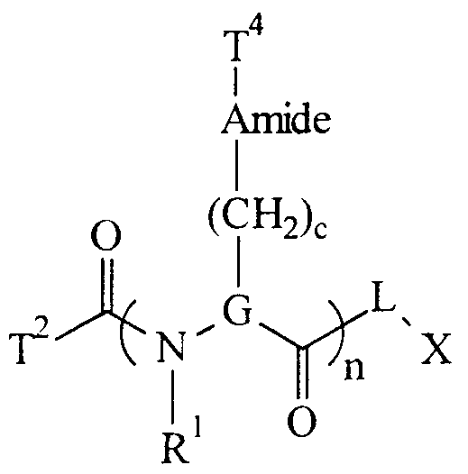


Claims


Priority Applications (8)
| Application Number | Priority Date | Filing Date | Title |
|---|---|---|---|
| DE69814629T DE69814629T2 (en) | 1997-07-22 | 1998-07-22 | METHOD AND CONNECTIONS FOR ANALYZING NUCLEIC ACIDS BY MASS SPECTROMETRY |
| AU85765/98A AU738237B2 (en) | 1997-07-22 | 1998-07-22 | Methods and compositions for analyzing nucleic acid molecules utilizing sizing techniques |
| DK98936928T DK0990047T3 (en) | 1997-07-22 | 1998-07-22 | Methods and compositions for analyzing nucleic acids by mass spectrometry |
| NZ501919A NZ501919A (en) | 1997-07-22 | 1998-07-22 | Methods for analyzing nucleic acid molecules utilizing mass spectroscopy readable tags |
| AT98936928T ATE240408T1 (en) | 1997-07-22 | 1998-07-22 | METHOD AND COMPOUNDS FOR ANALYZING NUCLEIC ACIDS BY MASS SPECTROMETRY |
| JP2000504286A JP4414086B2 (en) | 1997-07-22 | 1998-07-22 | Methods and compounds for analyzing nucleic acids by mass spectrometry |
| EP98936928A EP0990047B1 (en) | 1997-07-22 | 1998-07-22 | Methods and compositions for analyzing nucleic acids by mass spectrometry |
| CA002297158A CA2297158C (en) | 1997-07-22 | 1998-07-22 | Methods and compounds for analyzing nucleic acids by mass spectrometry |
Applications Claiming Priority (6)
| Application Number | Priority Date | Filing Date | Title |
|---|---|---|---|
| US08/898,501 US6027890A (en) | 1996-01-23 | 1997-07-22 | Methods and compositions for enhancing sensitivity in the analysis of biological-based assays |
| US08/898,501 | 1997-07-22 | ||
| US08/898,564 US6613508B1 (en) | 1996-01-23 | 1997-07-22 | Methods and compositions for analyzing nucleic acid molecules utilizing sizing techniques |
| US08/898,564 | 1997-07-22 | ||
| US08/898,180 | 1997-07-22 | ||
| US08/898,180 US6312893B1 (en) | 1996-01-23 | 1997-07-22 | Methods and compositions for determining the sequence of nucleic acid molecules |
Publications (3)
| Publication Number | Publication Date |
|---|---|
| WO1999005319A2 true WO1999005319A2 (en) | 1999-02-04 |
| WO1999005319A3 WO1999005319A3 (en) | 1999-05-14 |
| WO1999005319A9 WO1999005319A9 (en) | 1999-06-17 |
Family
ID=27420575
Family Applications (1)
| Application Number | Title | Priority Date | Filing Date |
|---|---|---|---|
| PCT/US1998/015008 WO1999005319A2 (en) | 1997-07-22 | 1998-07-22 | Methods and compounds for analyzing nucleic acids by mass spectrometry |
Country Status (12)
| Country | Link |
|---|---|
| EP (1) | EP0990047B1 (en) |
| JP (2) | JP4414086B2 (en) |
| CN (1) | CN1270598A (en) |
| AT (1) | ATE240408T1 (en) |
| AU (1) | AU738237B2 (en) |
| CA (1) | CA2297158C (en) |
| DE (1) | DE69814629T2 (en) |
| DK (1) | DK0990047T3 (en) |
| ES (1) | ES2200355T3 (en) |
| NZ (1) | NZ501919A (en) |
| PT (1) | PT990047E (en) |
| WO (1) | WO1999005319A2 (en) |
Cited By (74)
| Publication number | Priority date | Publication date | Assignee | Title |
|---|---|---|---|---|
| US6024878A (en) * | 1997-10-30 | 2000-02-15 | Transgenomic, Inc. | Method for high resolution liquid chromatographic separation of polynucleotides |
| GB2340237A (en) * | 1998-07-13 | 2000-02-16 | Brax Group Ltd | Compounds for mass spectrometry |
| WO2001016090A1 (en) * | 1999-08-31 | 2001-03-08 | Qiagen Genomics, Inc. | Enantiomerically-enriched compounds having photocleavable bond(s) and methods related thereto |
| WO2001046687A2 (en) * | 1999-12-22 | 2001-06-28 | Transgenomic, Inc. | System and method for automated matched ion polynucleotide chromatography |
| JP2001218598A (en) * | 2000-02-09 | 2001-08-14 | Asahi Breweries Ltd | Method for specifically and quantitatively detecting bacterium belonging to pectinatus |
| WO2001058926A2 (en) * | 2000-02-08 | 2001-08-16 | The Regents Of The University Of Michigan | Protein mapping |
| WO2002014867A2 (en) * | 2000-08-11 | 2002-02-21 | Agilix Corporation | Ultra-sensitive detection systems |
| WO2003033741A1 (en) * | 2001-10-16 | 2003-04-24 | Aclara Biosciences, Inc. | Universal e-tag primer and probe compositions and methods |
| US6713258B2 (en) | 2000-03-29 | 2004-03-30 | Center For The Application Of Molecular Biology To International Agriculture (Cambia) | Methods for genotyping by hybridization analysis |
| US6997031B2 (en) | 2003-05-01 | 2006-02-14 | Waters Corporation | Fraction collector for composition analysis |
| US7041459B2 (en) | 2001-05-21 | 2006-05-09 | Monogram Biosciences, Inc. | Analyzing phosphorylated proteins |
| US7160735B2 (en) | 2000-04-28 | 2007-01-09 | Monogram Biosciences, Inc. | Tagged microparticle compositions and methods |
| US7195751B2 (en) | 2003-01-30 | 2007-03-27 | Applera Corporation | Compositions and kits pertaining to analyte determination |
| US7225079B2 (en) | 1998-08-04 | 2007-05-29 | Transgenomic, Inc. | System and method for automated matched ion polynucleotide chromatography |
| US7255999B2 (en) | 2001-05-21 | 2007-08-14 | Monogram Biosciences, Inc. | Methods and compositions for analyzing proteins |
| US7307169B2 (en) | 2004-01-05 | 2007-12-11 | Applera Corporation | Isotopically enriched N-substituted piperazines and methods for the preparation thereof |
| US7355045B2 (en) | 2004-01-05 | 2008-04-08 | Applera Corporation | Isotopically enriched N-substituted piperazine acetic acids and methods for the preparation thereof |
| US7358052B2 (en) | 2001-05-26 | 2008-04-15 | Monogram Biosciences, Inc. | Catalytic amplification of multiplexed assay signals |
| EP1950305A1 (en) | 2001-05-09 | 2008-07-30 | Monsanto Technology, LLC | Tyr a genes and uses thereof |
| WO2009091841A3 (en) * | 2008-01-15 | 2009-10-08 | Sequenom, Inc. | Compositions and processes for improved mass spectrometry analysis |
| US7771929B2 (en) | 2000-04-28 | 2010-08-10 | Monogram Biosciences, Inc. | Tag library compounds, compositions, kits and methods of use |
| WO2011029034A2 (en) | 2009-09-04 | 2011-03-10 | Intelligent Medical Devices, Inc. | Optimized probes and primers and methods of using same for the detection, screening, isolation and sequencing of vancomycin resistance genes and vancomycin resistant enterococci |
| US7956175B2 (en) | 2003-09-11 | 2011-06-07 | Ibis Biosciences, Inc. | Compositions for use in identification of bacteria |
| US7964343B2 (en) | 2003-05-13 | 2011-06-21 | Ibis Biosciences, Inc. | Method for rapid purification of nucleic acids for subsequent analysis by mass spectrometry by solution capture |
| US20110236982A1 (en) * | 2003-11-26 | 2011-09-29 | Dh Technologies Development Pte, Ltd. | Analysis of mass spectral data in the quiet zones |
| US8073627B2 (en) | 2001-06-26 | 2011-12-06 | Ibis Biosciences, Inc. | System for indentification of pathogens |
| US8084207B2 (en) | 2005-03-03 | 2011-12-27 | Ibis Bioscience, Inc. | Compositions for use in identification of papillomavirus |
| US8088582B2 (en) | 2006-04-06 | 2012-01-03 | Ibis Biosciences, Inc. | Compositions for the use in identification of fungi |
| US8097416B2 (en) | 2003-09-11 | 2012-01-17 | Ibis Biosciences, Inc. | Methods for identification of sepsis-causing bacteria |
| US8148163B2 (en) | 2008-09-16 | 2012-04-03 | Ibis Biosciences, Inc. | Sample processing units, systems, and related methods |
| US8158936B2 (en) | 2009-02-12 | 2012-04-17 | Ibis Biosciences, Inc. | Ionization probe assemblies |
| US8158354B2 (en) | 2003-05-13 | 2012-04-17 | Ibis Biosciences, Inc. | Methods for rapid purification of nucleic acids for subsequent analysis by mass spectrometry by solution capture |
| US8163895B2 (en) | 2003-12-05 | 2012-04-24 | Ibis Biosciences, Inc. | Compositions for use in identification of orthopoxviruses |
| US8173957B2 (en) | 2004-05-24 | 2012-05-08 | Ibis Biosciences, Inc. | Mass spectrometry with selective ion filtration by digital thresholding |
| US8182992B2 (en) | 2005-03-03 | 2012-05-22 | Ibis Biosciences, Inc. | Compositions for use in identification of adventitious viruses |
| US8187814B2 (en) | 2004-02-18 | 2012-05-29 | Ibis Biosciences, Inc. | Methods for concurrent identification and quantification of an unknown bioagent |
| US8214154B2 (en) | 2001-03-02 | 2012-07-03 | Ibis Biosciences, Inc. | Systems for rapid identification of pathogens in humans and animals |
| US8265878B2 (en) | 2001-03-02 | 2012-09-11 | Ibis Bioscience, Inc. | Method for rapid detection and identification of bioagents |
| US8268565B2 (en) | 2001-03-02 | 2012-09-18 | Ibis Biosciences, Inc. | Methods for identifying bioagents |
| WO2012138470A2 (en) | 2011-04-04 | 2012-10-11 | Intelligent Medical Devices, Inc. | Optimized oligonucleotides and methods of using same for the detection, isolation, amplification, quantitation, monitoring, screening, and sequencing of group b streptococcus |
| US8298760B2 (en) | 2001-06-26 | 2012-10-30 | Ibis Bioscience, Inc. | Secondary structure defining database and methods for determining identity and geographic origin of an unknown bioagent thereby |
| US8407010B2 (en) | 2004-05-25 | 2013-03-26 | Ibis Biosciences, Inc. | Methods for rapid forensic analysis of mitochondrial DNA |
| US8534447B2 (en) | 2008-09-16 | 2013-09-17 | Ibis Biosciences, Inc. | Microplate handling systems and related computer program products and methods |
| US8546082B2 (en) | 2003-09-11 | 2013-10-01 | Ibis Biosciences, Inc. | Methods for identification of sepsis-causing bacteria |
| US8550694B2 (en) | 2008-09-16 | 2013-10-08 | Ibis Biosciences, Inc. | Mixing cartridges, mixing stations, and related kits, systems, and methods |
| US8551738B2 (en) | 2005-07-21 | 2013-10-08 | Ibis Biosciences, Inc. | Systems and methods for rapid identification of nucleic acid variants |
| US8563250B2 (en) | 2001-03-02 | 2013-10-22 | Ibis Biosciences, Inc. | Methods for identifying bioagents |
| WO2014058508A2 (en) * | 2012-07-24 | 2014-04-17 | Massachusetts Institute Of Technology | Reagents for oxidizer-based chemical detection |
| WO2014066749A2 (en) | 2012-10-25 | 2014-05-01 | Intelligent Medical Devices, Inc. | Optimized probes and primers and methods of using same for the binding, detection, differentiation, isolation and sequencing of influenza a; influenza b and respiratory syncytial virus |
| US8822156B2 (en) | 2002-12-06 | 2014-09-02 | Ibis Biosciences, Inc. | Methods for rapid identification of pathogens in humans and animals |
| WO2014137906A1 (en) | 2013-03-05 | 2014-09-12 | Intelligent Medical Devices, Inc. | Optimized probes and primers and methods of using same for the detection, screening, isolation and sequencing of mrsa, mssa, staphylococcus markers, and the antibiotic resistance gene mec a |
| US8835179B2 (en) | 2010-10-29 | 2014-09-16 | Scinopharm Taiwan, Ltd. | Real-time monitor solid phase peptide synthesis by mass spectrometry |
| US8871471B2 (en) | 2007-02-23 | 2014-10-28 | Ibis Biosciences, Inc. | Methods for rapid forensic DNA analysis |
| US8950604B2 (en) | 2009-07-17 | 2015-02-10 | Ibis Biosciences, Inc. | Lift and mount apparatus |
| US9080209B2 (en) | 2009-08-06 | 2015-07-14 | Ibis Biosciences, Inc. | Non-mass determined base compositions for nucleic acid detection |
| US9127308B2 (en) | 2002-03-07 | 2015-09-08 | Atlas Genetics Limited | Nucleic acid probes, their synthesis and use |
| US9149473B2 (en) | 2006-09-14 | 2015-10-06 | Ibis Biosciences, Inc. | Targeted whole genome amplification method for identification of pathogens |
| US9194877B2 (en) | 2009-07-17 | 2015-11-24 | Ibis Biosciences, Inc. | Systems for bioagent indentification |
| WO2016049531A1 (en) | 2014-09-26 | 2016-03-31 | Purecircle Usa Inc. | Single nucleotide polymorphism (snp) markers for stevia |
| US9305756B2 (en) | 2013-03-13 | 2016-04-05 | Agena Bioscience, Inc. | Preparation enhancements and methods of use for MALDI mass spectrometry |
| US9393564B2 (en) | 2009-03-30 | 2016-07-19 | Ibis Biosciences, Inc. | Bioagent detection systems, devices, and methods |
| US9416409B2 (en) | 2009-07-31 | 2016-08-16 | Ibis Biosciences, Inc. | Capture primers and capture sequence linked solid supports for molecular diagnostic tests |
| US9588095B2 (en) | 2012-07-24 | 2017-03-07 | Massachusetts Institute Of Technology | Reagents for oxidizer-based chemical detection |
| US9598724B2 (en) | 2007-06-01 | 2017-03-21 | Ibis Biosciences, Inc. | Methods and compositions for multiple displacement amplification of nucleic acids |
| US9719083B2 (en) | 2009-03-08 | 2017-08-01 | Ibis Biosciences, Inc. | Bioagent detection methods |
| US9758840B2 (en) | 2010-03-14 | 2017-09-12 | Ibis Biosciences, Inc. | Parasite detection via endosymbiont detection |
| US9873906B2 (en) | 2004-07-14 | 2018-01-23 | Ibis Biosciences, Inc. | Methods for repairing degraded DNA |
| US9890408B2 (en) | 2009-10-15 | 2018-02-13 | Ibis Biosciences, Inc. | Multiple displacement amplification |
| US9891193B2 (en) | 2012-07-24 | 2018-02-13 | Massachusetts Institute Of Technology | Reagent impregnated swipe for chemical detection |
| WO2018170436A1 (en) | 2017-03-16 | 2018-09-20 | Jacobs Farm Del Cabo | Basil with high tolerance to downy mildew |
| US10345281B2 (en) | 2014-04-04 | 2019-07-09 | Massachusetts Institute Of Technology | Reagents for enhanced detection of low volatility analytes |
| WO2019226990A1 (en) * | 2018-05-25 | 2019-11-28 | New York Institute Of Technology | Direct nucleic acid sequencing method |
| US10816530B2 (en) | 2013-07-23 | 2020-10-27 | Massachusetts Institute Of Technology | Substrate containing latent vaporization reagents |
| CN115356411A (en) * | 2022-08-09 | 2022-11-18 | 河北一品生物医药有限公司 | Method for detecting related substances of L-piperidinecarboxylic acid hydrochloride by high performance liquid chromatography |
Families Citing this family (15)
| Publication number | Priority date | Publication date | Assignee | Title |
|---|---|---|---|---|
| US20040229294A1 (en) | 2002-05-21 | 2004-11-18 | Po-Ying Chan-Hui | ErbB surface receptor complexes as biomarkers |
| DE10350484B4 (en) * | 2003-12-20 | 2006-02-02 | Ehrfeld Mikrotechnik Bts Gmbh | Method and device for labeling proteins |
| US20050148087A1 (en) | 2004-01-05 | 2005-07-07 | Applera Corporation | Isobarically labeled analytes and fragment ions derived therefrom |
| KR101322624B1 (en) * | 2007-06-13 | 2013-10-29 | 삼성전자주식회사 | Field effect transistor-based detection device for biomolecules containing adsorptive media and detection method using the same |
| CN101251511B (en) * | 2008-03-14 | 2012-06-06 | 毅新兴业(北京)科技有限公司 | Method for testing SNP using restriction enzymes double zyme cutting |
| CN101251510B (en) * | 2008-03-14 | 2012-06-06 | 毅新兴业(北京)科技有限公司 | Method for testing SNP combining restrictive zyme cutting method and mass spectrometry |
| CN101246142B (en) * | 2008-04-03 | 2012-06-20 | 毅新兴业(北京)科技有限公司 | Method for detecting mononucleotide polymorphism |
| KR101701998B1 (en) * | 2014-10-23 | 2017-02-03 | 한국과학기술연구원 | Gas chromatography-electrospray mass spectrometry with high resolution and a method ionizing organic compounds of gas phase using it |
| JP6560336B2 (en) * | 2015-02-26 | 2019-08-14 | 株式会社日立ハイテクノロジーズ | Method for constructing nucleic acid molecule |
| CN106979942B (en) * | 2017-05-04 | 2020-07-07 | 华中师范大学 | Raman spectrum analysis method for individual quantification of solid-phase synthesized compound combinatorial library and application thereof |
| CN111208300B (en) * | 2018-11-21 | 2021-10-29 | 中国科学院大连化学物理研究所 | Method for enriching and identifying lysine nitrogen-linked phosphorylation post-translational modification |
| CN111239402B (en) * | 2018-11-29 | 2021-07-09 | 北京大学 | Mass spectrum immunoassay method for detecting disease marker and application |
| CN113075114B (en) * | 2019-12-17 | 2022-07-01 | 北京大学 | Organic mass spectrometry flow analysis method for single cell analysis |
| US20230219889A1 (en) | 2020-05-19 | 2023-07-13 | Cybin Irl Limited | Deuterated tryptamine derivatives and methods of use |
| CN114984032B (en) * | 2022-06-27 | 2023-07-07 | 四川大学 | DNA tetrahedron framework nucleic acid-chlorogenic acid complex and application thereof in preparation of medicines for treating hepatic fibrosis |
Citations (9)
| Publication number | Priority date | Publication date | Assignee | Title |
|---|---|---|---|---|
| US5118605A (en) * | 1984-10-16 | 1992-06-02 | Chiron Corporation | Polynucleotide determination with selectable cleavage sites |
| WO1994016101A2 (en) * | 1993-01-07 | 1994-07-21 | Koester Hubert | Dna sequencing by mass spectrometry |
| WO1995004160A1 (en) * | 1993-07-30 | 1995-02-09 | Isis Innovation Limited | Tag reagent and assay method |
| WO1995014108A1 (en) * | 1993-11-17 | 1995-05-26 | Amersham International Plc | Primer extension mass spectroscopy nucleic acid sequencing method |
| WO1995025737A1 (en) * | 1994-03-23 | 1995-09-28 | The Penn State Research Foundation | Method for identifying members of combinatorial libraries |
| WO1995028640A1 (en) * | 1994-04-13 | 1995-10-26 | The Trustees Of Columbia University In The City Of New York | Complex combinatorial chemical libraries encoded with tags |
| WO1997027331A2 (en) * | 1996-01-23 | 1997-07-31 | Rapigene, Inc. | Methods and compositions for determining the sequence of nucleic acid molecules |
| WO1997027325A2 (en) * | 1996-01-23 | 1997-07-31 | Rapigene, Inc. | Methods and compositions for analyzing nucleic acid molecules utilizing sizing techniques |
| WO1997027327A2 (en) * | 1996-01-23 | 1997-07-31 | Rapigene, Inc. | Methods and compositions for detecting binding of ligand pair using non-fluorescent label |
Family Cites Families (3)
| Publication number | Priority date | Publication date | Assignee | Title |
|---|---|---|---|---|
| WO1998026095A1 (en) * | 1996-12-10 | 1998-06-18 | Genetrace Systems Inc. | Releasable nonvolatile mass-label molecules |
| JP3884087B2 (en) * | 1997-01-15 | 2007-02-21 | イクスツィリオン ゲゼルシャフト ミット ベシュレンクテル ハフツング ウント コンパニー コマンディトゲゼルシャフト | Mass label binding hybridization probe |
| CA2295955A1 (en) * | 1997-07-11 | 1999-01-21 | Brax Group Limited | Characterising nucleic acids |
-
1998
- 1998-07-22 ES ES98936928T patent/ES2200355T3/en not_active Expired - Lifetime
- 1998-07-22 AU AU85765/98A patent/AU738237B2/en not_active Ceased
- 1998-07-22 JP JP2000504286A patent/JP4414086B2/en not_active Expired - Lifetime
- 1998-07-22 PT PT98936928T patent/PT990047E/en unknown
- 1998-07-22 AT AT98936928T patent/ATE240408T1/en not_active IP Right Cessation
- 1998-07-22 EP EP98936928A patent/EP0990047B1/en not_active Expired - Lifetime
- 1998-07-22 CN CN98809047A patent/CN1270598A/en active Pending
- 1998-07-22 WO PCT/US1998/015008 patent/WO1999005319A2/en active IP Right Grant
- 1998-07-22 DE DE69814629T patent/DE69814629T2/en not_active Expired - Lifetime
- 1998-07-22 CA CA002297158A patent/CA2297158C/en not_active Expired - Fee Related
- 1998-07-22 NZ NZ501919A patent/NZ501919A/en unknown
- 1998-07-22 DK DK98936928T patent/DK0990047T3/en active
-
2009
- 2009-04-14 JP JP2009098483A patent/JP4870791B2/en not_active Expired - Lifetime
Patent Citations (9)
| Publication number | Priority date | Publication date | Assignee | Title |
|---|---|---|---|---|
| US5118605A (en) * | 1984-10-16 | 1992-06-02 | Chiron Corporation | Polynucleotide determination with selectable cleavage sites |
| WO1994016101A2 (en) * | 1993-01-07 | 1994-07-21 | Koester Hubert | Dna sequencing by mass spectrometry |
| WO1995004160A1 (en) * | 1993-07-30 | 1995-02-09 | Isis Innovation Limited | Tag reagent and assay method |
| WO1995014108A1 (en) * | 1993-11-17 | 1995-05-26 | Amersham International Plc | Primer extension mass spectroscopy nucleic acid sequencing method |
| WO1995025737A1 (en) * | 1994-03-23 | 1995-09-28 | The Penn State Research Foundation | Method for identifying members of combinatorial libraries |
| WO1995028640A1 (en) * | 1994-04-13 | 1995-10-26 | The Trustees Of Columbia University In The City Of New York | Complex combinatorial chemical libraries encoded with tags |
| WO1997027331A2 (en) * | 1996-01-23 | 1997-07-31 | Rapigene, Inc. | Methods and compositions for determining the sequence of nucleic acid molecules |
| WO1997027325A2 (en) * | 1996-01-23 | 1997-07-31 | Rapigene, Inc. | Methods and compositions for analyzing nucleic acid molecules utilizing sizing techniques |
| WO1997027327A2 (en) * | 1996-01-23 | 1997-07-31 | Rapigene, Inc. | Methods and compositions for detecting binding of ligand pair using non-fluorescent label |
Non-Patent Citations (3)
| Title |
|---|
| B.B.BROWN ET AL.: "A Single-Bead Decode Strategy Using Electrospray Ionization Mass Spectometry and a New Photolabile Linker : 3-Amino-3-(2-nitrophenyl)Propionic Acid." MOLECULAR DIVERSITY, vol. 1, 1995, pages 4-12, XP002094265 * |
| JACOBSON K B ET AL: "APPLICATIONS OF MASS SPECTROMETRY TO DNA SEQUENCING" GENETIC ANALYSIS TECHNIQUES AND APPLICATIONS, vol. 8, no. 8, 1 December 1991, pages 223-229, XP000271820 * |
| P.C.TOREN ET AL.: "Determination of Impurities in Nucleoside 3'-Phosphoramidites by Fast Atom Bombardment Mass Spectrometry" ANALYTICAL BIOCHEMISTRY, vol. 152, no. 2, 1986, pages 291-294, XP002094264 * |
Cited By (122)
| Publication number | Priority date | Publication date | Assignee | Title |
|---|---|---|---|---|
| US6342161B1 (en) | 1997-10-30 | 2002-01-29 | Transgenomic, Inc. | Method for high resolution liquid chromatographic separation of double-stranded DNA |
| US6024878A (en) * | 1997-10-30 | 2000-02-15 | Transgenomic, Inc. | Method for high resolution liquid chromatographic separation of polynucleotides |
| GB2340237A (en) * | 1998-07-13 | 2000-02-16 | Brax Group Ltd | Compounds for mass spectrometry |
| GB2340237B (en) * | 1998-07-13 | 2003-10-08 | Brax Group Ltd | Compounds for mass spectrometry |
| US7225079B2 (en) | 1998-08-04 | 2007-05-29 | Transgenomic, Inc. | System and method for automated matched ion polynucleotide chromatography |
| WO2001016090A1 (en) * | 1999-08-31 | 2001-03-08 | Qiagen Genomics, Inc. | Enantiomerically-enriched compounds having photocleavable bond(s) and methods related thereto |
| WO2001046687A2 (en) * | 1999-12-22 | 2001-06-28 | Transgenomic, Inc. | System and method for automated matched ion polynucleotide chromatography |
| WO2001046687A3 (en) * | 1999-12-22 | 2003-04-17 | Transgenomic Inc | System and method for automated matched ion polynucleotide chromatography |
| WO2001058926A3 (en) * | 2000-02-08 | 2003-11-27 | Univ Michigan | Protein mapping |
| WO2001058926A2 (en) * | 2000-02-08 | 2001-08-16 | The Regents Of The University Of Michigan | Protein mapping |
| JP2001218598A (en) * | 2000-02-09 | 2001-08-14 | Asahi Breweries Ltd | Method for specifically and quantitatively detecting bacterium belonging to pectinatus |
| US6713258B2 (en) | 2000-03-29 | 2004-03-30 | Center For The Application Of Molecular Biology To International Agriculture (Cambia) | Methods for genotyping by hybridization analysis |
| US7771929B2 (en) | 2000-04-28 | 2010-08-10 | Monogram Biosciences, Inc. | Tag library compounds, compositions, kits and methods of use |
| US7160735B2 (en) | 2000-04-28 | 2007-01-09 | Monogram Biosciences, Inc. | Tagged microparticle compositions and methods |
| WO2002014867A3 (en) * | 2000-08-11 | 2003-07-03 | Agilix Corp | Ultra-sensitive detection systems |
| US6824981B2 (en) | 2000-08-11 | 2004-11-30 | Agilix Corporation | Ultra-sensitive detection systems using alterable peptide tags |
| WO2002014867A2 (en) * | 2000-08-11 | 2002-02-21 | Agilix Corporation | Ultra-sensitive detection systems |
| AU2001283562B2 (en) * | 2000-08-11 | 2006-10-26 | Perkin-Elmer Las, Inc. | Ultra-sensitive detection systems |
| US8214154B2 (en) | 2001-03-02 | 2012-07-03 | Ibis Biosciences, Inc. | Systems for rapid identification of pathogens in humans and animals |
| US8802372B2 (en) | 2001-03-02 | 2014-08-12 | Ibis Biosciences, Inc. | Methods for rapid forensic analysis of mitochondrial DNA and characterization of mitochondrial DNA heteroplasmy |
| US8815513B2 (en) | 2001-03-02 | 2014-08-26 | Ibis Biosciences, Inc. | Method for rapid detection and identification of bioagents in epidemiological and forensic investigations |
| US9416424B2 (en) | 2001-03-02 | 2016-08-16 | Ibis Biosciences, Inc. | Methods for rapid identification of pathogens in humans and animals |
| US8563250B2 (en) | 2001-03-02 | 2013-10-22 | Ibis Biosciences, Inc. | Methods for identifying bioagents |
| US8268565B2 (en) | 2001-03-02 | 2012-09-18 | Ibis Biosciences, Inc. | Methods for identifying bioagents |
| US8265878B2 (en) | 2001-03-02 | 2012-09-11 | Ibis Bioscience, Inc. | Method for rapid detection and identification of bioagents |
| US9752184B2 (en) | 2001-03-02 | 2017-09-05 | Ibis Biosciences, Inc. | Methods for rapid forensic analysis of mitochondrial DNA and characterization of mitochondrial DNA heteroplasmy |
| EP1950305A1 (en) | 2001-05-09 | 2008-07-30 | Monsanto Technology, LLC | Tyr a genes and uses thereof |
| US9939447B2 (en) | 2001-05-21 | 2018-04-10 | Monogram Biosciences, Inc. | Methods and compositions for analyzing proteins |
| US7255999B2 (en) | 2001-05-21 | 2007-08-14 | Monogram Biosciences, Inc. | Methods and compositions for analyzing proteins |
| US7041459B2 (en) | 2001-05-21 | 2006-05-09 | Monogram Biosciences, Inc. | Analyzing phosphorylated proteins |
| US7358052B2 (en) | 2001-05-26 | 2008-04-15 | Monogram Biosciences, Inc. | Catalytic amplification of multiplexed assay signals |
| US8921047B2 (en) | 2001-06-26 | 2014-12-30 | Ibis Biosciences, Inc. | Secondary structure defining database and methods for determining identity and geographic origin of an unknown bioagent thereby |
| US8298760B2 (en) | 2001-06-26 | 2012-10-30 | Ibis Bioscience, Inc. | Secondary structure defining database and methods for determining identity and geographic origin of an unknown bioagent thereby |
| US8073627B2 (en) | 2001-06-26 | 2011-12-06 | Ibis Biosciences, Inc. | System for indentification of pathogens |
| US8380442B2 (en) | 2001-06-26 | 2013-02-19 | Ibis Bioscience, Inc. | Secondary structure defining database and methods for determining identity and geographic origin of an unknown bioagent thereby |
| US7312034B2 (en) | 2001-10-16 | 2007-12-25 | Monogram Biosciences, Inc. | Universal e-tag primer and probe compositions and methods |
| WO2003033741A1 (en) * | 2001-10-16 | 2003-04-24 | Aclara Biosciences, Inc. | Universal e-tag primer and probe compositions and methods |
| US10094800B2 (en) | 2002-03-07 | 2018-10-09 | Atlas Genetics Limited | Assays and apparatus for detecting electrochemical active markers in an electric field |
| US9127308B2 (en) | 2002-03-07 | 2015-09-08 | Atlas Genetics Limited | Nucleic acid probes, their synthesis and use |
| US8822156B2 (en) | 2002-12-06 | 2014-09-02 | Ibis Biosciences, Inc. | Methods for rapid identification of pathogens in humans and animals |
| US9725771B2 (en) | 2002-12-06 | 2017-08-08 | Ibis Biosciences, Inc. | Methods for rapid identification of pathogens in humans and animals |
| US7799576B2 (en) | 2003-01-30 | 2010-09-21 | Dh Technologies Development Pte. Ltd. | Isobaric labels for mass spectrometric analysis of peptides and method thereof |
| US8679773B2 (en) | 2003-01-30 | 2014-03-25 | Dh Technologies Development Pte. Ltd. | Kits pertaining to analyte determination |
| US7195751B2 (en) | 2003-01-30 | 2007-03-27 | Applera Corporation | Compositions and kits pertaining to analyte determination |
| US7947513B2 (en) | 2003-01-30 | 2011-05-24 | DH Technologies Ptd. Ltd. | Sets and compositions pertaining to analyte determination |
| US7086279B2 (en) | 2003-05-01 | 2006-08-08 | Waters Investments Limited | Fraction collector for composition analysis |
| US6997031B2 (en) | 2003-05-01 | 2006-02-14 | Waters Corporation | Fraction collector for composition analysis |
| US7964343B2 (en) | 2003-05-13 | 2011-06-21 | Ibis Biosciences, Inc. | Method for rapid purification of nucleic acids for subsequent analysis by mass spectrometry by solution capture |
| US8476415B2 (en) | 2003-05-13 | 2013-07-02 | Ibis Biosciences, Inc. | Methods for rapid purification of nucleic acids for subsequent analysis by mass spectrometry by solution capture |
| US8158354B2 (en) | 2003-05-13 | 2012-04-17 | Ibis Biosciences, Inc. | Methods for rapid purification of nucleic acids for subsequent analysis by mass spectrometry by solution capture |
| US8394945B2 (en) | 2003-09-11 | 2013-03-12 | Ibis Biosciences, Inc. | Compositions for use in identification of bacteria |
| US8242254B2 (en) | 2003-09-11 | 2012-08-14 | Ibis Biosciences, Inc. | Compositions for use in identification of bacteria |
| US8546082B2 (en) | 2003-09-11 | 2013-10-01 | Ibis Biosciences, Inc. | Methods for identification of sepsis-causing bacteria |
| US8097416B2 (en) | 2003-09-11 | 2012-01-17 | Ibis Biosciences, Inc. | Methods for identification of sepsis-causing bacteria |
| US7956175B2 (en) | 2003-09-11 | 2011-06-07 | Ibis Biosciences, Inc. | Compositions for use in identification of bacteria |
| US8013142B2 (en) | 2003-09-11 | 2011-09-06 | Ibis Biosciences, Inc. | Compositions for use in identification of bacteria |
| US8288523B2 (en) | 2003-09-11 | 2012-10-16 | Ibis Biosciences, Inc. | Compositions for use in identification of bacteria |
| US20110236982A1 (en) * | 2003-11-26 | 2011-09-29 | Dh Technologies Development Pte, Ltd. | Analysis of mass spectral data in the quiet zones |
| US8163895B2 (en) | 2003-12-05 | 2012-04-24 | Ibis Biosciences, Inc. | Compositions for use in identification of orthopoxviruses |
| US7355045B2 (en) | 2004-01-05 | 2008-04-08 | Applera Corporation | Isotopically enriched N-substituted piperazine acetic acids and methods for the preparation thereof |
| US7932388B2 (en) | 2004-01-05 | 2011-04-26 | Dh Technologies Development Pte. Ltd. | Isotopically enriched N-substituted piperazines and methods for the preparation thereof |
| US8569304B2 (en) | 2004-01-05 | 2013-10-29 | Dh Technologies Development Pte. Ltd. | Active esters of N-substituted piperazine acetic acids, including isotopically enriched versions thereof |
| US7307169B2 (en) | 2004-01-05 | 2007-12-11 | Applera Corporation | Isotopically enriched N-substituted piperazines and methods for the preparation thereof |
| US8187814B2 (en) | 2004-02-18 | 2012-05-29 | Ibis Biosciences, Inc. | Methods for concurrent identification and quantification of an unknown bioagent |
| US9447462B2 (en) | 2004-02-18 | 2016-09-20 | Ibis Biosciences, Inc. | Methods for concurrent identification and quantification of an unknown bioagent |
| US8173957B2 (en) | 2004-05-24 | 2012-05-08 | Ibis Biosciences, Inc. | Mass spectrometry with selective ion filtration by digital thresholding |
| US9449802B2 (en) | 2004-05-24 | 2016-09-20 | Ibis Biosciences, Inc. | Mass spectrometry with selective ion filtration by digital thresholding |
| US8987660B2 (en) | 2004-05-24 | 2015-03-24 | Ibis Biosciences, Inc. | Mass spectrometry with selective ion filtration by digital thresholding |
| US8407010B2 (en) | 2004-05-25 | 2013-03-26 | Ibis Biosciences, Inc. | Methods for rapid forensic analysis of mitochondrial DNA |
| US9873906B2 (en) | 2004-07-14 | 2018-01-23 | Ibis Biosciences, Inc. | Methods for repairing degraded DNA |
| US8084207B2 (en) | 2005-03-03 | 2011-12-27 | Ibis Bioscience, Inc. | Compositions for use in identification of papillomavirus |
| US8182992B2 (en) | 2005-03-03 | 2012-05-22 | Ibis Biosciences, Inc. | Compositions for use in identification of adventitious viruses |
| US8551738B2 (en) | 2005-07-21 | 2013-10-08 | Ibis Biosciences, Inc. | Systems and methods for rapid identification of nucleic acid variants |
| US8088582B2 (en) | 2006-04-06 | 2012-01-03 | Ibis Biosciences, Inc. | Compositions for the use in identification of fungi |
| US9149473B2 (en) | 2006-09-14 | 2015-10-06 | Ibis Biosciences, Inc. | Targeted whole genome amplification method for identification of pathogens |
| US8871471B2 (en) | 2007-02-23 | 2014-10-28 | Ibis Biosciences, Inc. | Methods for rapid forensic DNA analysis |
| US9598724B2 (en) | 2007-06-01 | 2017-03-21 | Ibis Biosciences, Inc. | Methods and compositions for multiple displacement amplification of nucleic acids |
| WO2009091841A3 (en) * | 2008-01-15 | 2009-10-08 | Sequenom, Inc. | Compositions and processes for improved mass spectrometry analysis |
| US9310378B2 (en) | 2008-01-15 | 2016-04-12 | Agena Bioscience, Inc. | Compositions and processes for improved mass spectrometry analysis |
| US10329612B2 (en) | 2008-01-15 | 2019-06-25 | Agena Bioscience, Inc. | Compositions and processes for improved mass spectrometry analysis |
| EA017342B1 (en) * | 2008-01-15 | 2012-11-30 | Секвеном, Инк. | Process for mass spectrometry analysis |
| US9873912B2 (en) | 2008-01-15 | 2018-01-23 | Agena Bioscience, Inc. | Compositions and processes for improved mass spectrometry analysis |
| US7888127B2 (en) | 2008-01-15 | 2011-02-15 | Sequenom, Inc. | Methods for reducing adduct formation for mass spectrometry analysis |
| US8252599B2 (en) | 2008-09-16 | 2012-08-28 | Ibis Biosciences, Inc. | Sample processing units, systems, and related methods |
| US8550694B2 (en) | 2008-09-16 | 2013-10-08 | Ibis Biosciences, Inc. | Mixing cartridges, mixing stations, and related kits, systems, and methods |
| US9027730B2 (en) | 2008-09-16 | 2015-05-12 | Ibis Biosciences, Inc. | Microplate handling systems and related computer program products and methods |
| US8609430B2 (en) | 2008-09-16 | 2013-12-17 | Ibis Biosciences, Inc. | Sample processing units, systems, and related methods |
| US8148163B2 (en) | 2008-09-16 | 2012-04-03 | Ibis Biosciences, Inc. | Sample processing units, systems, and related methods |
| US8534447B2 (en) | 2008-09-16 | 2013-09-17 | Ibis Biosciences, Inc. | Microplate handling systems and related computer program products and methods |
| US9023655B2 (en) | 2008-09-16 | 2015-05-05 | Ibis Biosciences, Inc. | Sample processing units, systems, and related methods |
| US8796617B2 (en) | 2009-02-12 | 2014-08-05 | Ibis Biosciences, Inc. | Ionization probe assemblies |
| US8158936B2 (en) | 2009-02-12 | 2012-04-17 | Ibis Biosciences, Inc. | Ionization probe assemblies |
| US9165740B2 (en) | 2009-02-12 | 2015-10-20 | Ibis Biosciences, Inc. | Ionization probe assemblies |
| US9719083B2 (en) | 2009-03-08 | 2017-08-01 | Ibis Biosciences, Inc. | Bioagent detection methods |
| US9393564B2 (en) | 2009-03-30 | 2016-07-19 | Ibis Biosciences, Inc. | Bioagent detection systems, devices, and methods |
| US8950604B2 (en) | 2009-07-17 | 2015-02-10 | Ibis Biosciences, Inc. | Lift and mount apparatus |
| US9194877B2 (en) | 2009-07-17 | 2015-11-24 | Ibis Biosciences, Inc. | Systems for bioagent indentification |
| US9416409B2 (en) | 2009-07-31 | 2016-08-16 | Ibis Biosciences, Inc. | Capture primers and capture sequence linked solid supports for molecular diagnostic tests |
| US10119164B2 (en) | 2009-07-31 | 2018-11-06 | Ibis Biosciences, Inc. | Capture primers and capture sequence linked solid supports for molecular diagnostic tests |
| US9080209B2 (en) | 2009-08-06 | 2015-07-14 | Ibis Biosciences, Inc. | Non-mass determined base compositions for nucleic acid detection |
| WO2011029034A2 (en) | 2009-09-04 | 2011-03-10 | Intelligent Medical Devices, Inc. | Optimized probes and primers and methods of using same for the detection, screening, isolation and sequencing of vancomycin resistance genes and vancomycin resistant enterococci |
| US9890408B2 (en) | 2009-10-15 | 2018-02-13 | Ibis Biosciences, Inc. | Multiple displacement amplification |
| US9758840B2 (en) | 2010-03-14 | 2017-09-12 | Ibis Biosciences, Inc. | Parasite detection via endosymbiont detection |
| US8835179B2 (en) | 2010-10-29 | 2014-09-16 | Scinopharm Taiwan, Ltd. | Real-time monitor solid phase peptide synthesis by mass spectrometry |
| WO2012138470A2 (en) | 2011-04-04 | 2012-10-11 | Intelligent Medical Devices, Inc. | Optimized oligonucleotides and methods of using same for the detection, isolation, amplification, quantitation, monitoring, screening, and sequencing of group b streptococcus |
| US9588095B2 (en) | 2012-07-24 | 2017-03-07 | Massachusetts Institute Of Technology | Reagents for oxidizer-based chemical detection |
| WO2014058508A3 (en) * | 2012-07-24 | 2014-07-31 | Massachusetts Institute Of Technology | Reagents for oxidizer-based chemical detection |
| WO2014058508A2 (en) * | 2012-07-24 | 2014-04-17 | Massachusetts Institute Of Technology | Reagents for oxidizer-based chemical detection |
| US9891193B2 (en) | 2012-07-24 | 2018-02-13 | Massachusetts Institute Of Technology | Reagent impregnated swipe for chemical detection |
| US11543399B2 (en) | 2012-07-24 | 2023-01-03 | Massachusetts Institute Of Technology | Reagents for enhanced detection of low volatility analytes |
| US11237143B2 (en) | 2012-07-24 | 2022-02-01 | Massachusetts Institute Of Technology | Reagents for oxidizer-based chemical detection |
| WO2014066749A2 (en) | 2012-10-25 | 2014-05-01 | Intelligent Medical Devices, Inc. | Optimized probes and primers and methods of using same for the binding, detection, differentiation, isolation and sequencing of influenza a; influenza b and respiratory syncytial virus |
| WO2014137906A1 (en) | 2013-03-05 | 2014-09-12 | Intelligent Medical Devices, Inc. | Optimized probes and primers and methods of using same for the detection, screening, isolation and sequencing of mrsa, mssa, staphylococcus markers, and the antibiotic resistance gene mec a |
| US10204771B2 (en) | 2013-03-13 | 2019-02-12 | Agena Bioscience, Inc. | Preparation enhancements and methods of use for MALDI mass spectrometry |
| US9305756B2 (en) | 2013-03-13 | 2016-04-05 | Agena Bioscience, Inc. | Preparation enhancements and methods of use for MALDI mass spectrometry |
| US10816530B2 (en) | 2013-07-23 | 2020-10-27 | Massachusetts Institute Of Technology | Substrate containing latent vaporization reagents |
| US10345281B2 (en) | 2014-04-04 | 2019-07-09 | Massachusetts Institute Of Technology | Reagents for enhanced detection of low volatility analytes |
| WO2016049531A1 (en) | 2014-09-26 | 2016-03-31 | Purecircle Usa Inc. | Single nucleotide polymorphism (snp) markers for stevia |
| WO2018170436A1 (en) | 2017-03-16 | 2018-09-20 | Jacobs Farm Del Cabo | Basil with high tolerance to downy mildew |
| WO2019226990A1 (en) * | 2018-05-25 | 2019-11-28 | New York Institute Of Technology | Direct nucleic acid sequencing method |
| CN115356411A (en) * | 2022-08-09 | 2022-11-18 | 河北一品生物医药有限公司 | Method for detecting related substances of L-piperidinecarboxylic acid hydrochloride by high performance liquid chromatography |
| CN115356411B (en) * | 2022-08-09 | 2023-07-28 | 河北一品生物医药有限公司 | Method for detecting related substances of L-piperidine hydrochloride by high performance liquid chromatography |
Also Published As
| Publication number | Publication date |
|---|---|
| EP0990047B1 (en) | 2003-05-14 |
| JP4414086B2 (en) | 2010-02-10 |
| AU738237B2 (en) | 2001-09-13 |
| CA2297158C (en) | 2009-12-22 |
| CA2297158A1 (en) | 1999-02-04 |
| AU8576598A (en) | 1999-02-16 |
| ES2200355T3 (en) | 2004-03-01 |
| EP0990047A2 (en) | 2000-04-05 |
| ATE240408T1 (en) | 2003-05-15 |
| WO1999005319A9 (en) | 1999-06-17 |
| DK0990047T3 (en) | 2003-09-15 |
| JP4870791B2 (en) | 2012-02-08 |
| JP2001511359A (en) | 2001-08-14 |
| PT990047E (en) | 2003-10-31 |
| DE69814629D1 (en) | 2003-06-18 |
| CN1270598A (en) | 2000-10-18 |
| DE69814629T2 (en) | 2004-03-25 |
| JP2009159985A (en) | 2009-07-23 |
| WO1999005319A3 (en) | 1999-05-14 |
| NZ501919A (en) | 2001-11-30 |
Similar Documents
| Publication | Publication Date | Title |
|---|---|---|
| CA2297158C (en) | Methods and compounds for analyzing nucleic acids by mass spectrometry | |
| US6613508B1 (en) | Methods and compositions for analyzing nucleic acid molecules utilizing sizing techniques | |
| AU717945B2 (en) | Methods and compositions for analyzing nucleic acid molecules utilizing sizing techniques | |
| US6623928B2 (en) | Methods and compositions for determining the sequence of nucleic acid molecules | |
| EP0868535B9 (en) | Methods and compositions for determining the sequence of nucleic acid molecules | |
| US6027890A (en) | Methods and compositions for enhancing sensitivity in the analysis of biological-based assays | |
| AU717330B2 (en) | Methods and compositions for detecting binding of ligand pair using non-fluorescent label | |
| WO1997027331A9 (en) | Methods and compositions for determining the sequence of nucleic acid molecules | |
| EP0992511B1 (en) | Methods and compositions for determining the sequence of nucleic acid molecules | |
| EP0962464A2 (en) | Methods and compositions for detecting binding of ligand pair using non-fluorescent label |
Legal Events
| Date | Code | Title | Description |
|---|---|---|---|
| WWE | Wipo information: entry into national phase |
Ref document number: 98809047.3 Country of ref document: CN |
|
| AK | Designated states |
Kind code of ref document: A2 Designated state(s): AL AM AT AU BA BB BG BR BY CA CH CN CU CZ DE DK EE ES FI GB GE GH GM HU ID IL IS JP KE KG KP KR KZ LC LK LR LS LT LU LV MD MG MK MN MW MX NO NZ PL PT RO RU SD SE SG SI SK SL TJ TM TR TT UA UG US UZ VN YU ZW |
|
| AL | Designated countries for regional patents |
Kind code of ref document: A2 Designated state(s): GH GM KE LS MW SD SZ UG ZW AM AZ BY KG KZ MD RU TJ TM AT BE CH CY DE DK ES FI FR GB GR IE IT LU MC NL PT SE BF BJ CF CG CI CM GA GN GW ML MR NE SN TD TG |
|
| DFPE | Request for preliminary examination filed prior to expiration of 19th month from priority date (pct application filed before 20040101) | ||
| 121 | Ep: the epo has been informed by wipo that ep was designated in this application | ||
| AK | Designated states |
Kind code of ref document: A3 Designated state(s): AL AM AT AU BA BB BG BR BY CA CH CN CU CZ DE DK EE ES FI GB GE GH GM HU ID IL IS JP KE KG KP KR KZ LC LK LR LS LT LU LV MD MG MK MN MW MX NO NZ PL PT RO RU SD SE SG SI SK SL TJ TM TR TT UA UG US UZ VN YU ZW |
|
| AL | Designated countries for regional patents |
Kind code of ref document: A3 Designated state(s): GH GM KE LS MW SD SZ UG ZW AM AZ BY KG KZ MD RU TJ TM AT BE CH CY DE DK ES FI FR GB GR IE IT LU MC NL PT SE BF BJ CF CG CI CM GA GN GW ML MR NE SN TD TG |
|
| AK | Designated states |
Kind code of ref document: C2 Designated state(s): AL AM AT AU BA BB BG BR BY CA CH CN CU CZ DE DK EE ES FI GB GE GH GM HU ID IL IS JP KE KG KP KR KZ LC LK LR LS LT LU LV MD MG MK MN MW MX NO NZ PL PT RO RU SD SE SG SI SK SL TJ TM TR TT UA UG US UZ VN YU ZW |
|
| AL | Designated countries for regional patents |
Kind code of ref document: C2 Designated state(s): GH GM KE LS MW SD SZ UG ZW AM AZ BY KG KZ MD RU TJ TM AT BE CH CY DE DK ES FI FR GB GR IE IT LU MC NL PT SE BF BJ CF CG CI CM GA GN GW ML MR NE SN TD TG |
|
| COP | Corrected version of pamphlet |
Free format text: PAGES 1/36-36/36, DRAWINGS, REPLACED BY NEW PAGES 1/36-36/36; DUE TO LATE TRANSMITTAL BY THE RECEIVING OFFICE |
|
| WWE | Wipo information: entry into national phase |
Ref document number: 501919 Country of ref document: NZ |
|
| WWE | Wipo information: entry into national phase |
Ref document number: 85765/98 Country of ref document: AU |
|
| WWE | Wipo information: entry into national phase |
Ref document number: 1998936928 Country of ref document: EP |
|
| ENP | Entry into the national phase |
Ref document number: 2297158 Country of ref document: CA Ref document number: 2297158 Country of ref document: CA Kind code of ref document: A |
|
| NENP | Non-entry into the national phase |
Ref country code: KR |
|
| WWP | Wipo information: published in national office |
Ref document number: 1998936928 Country of ref document: EP |
|
| REG | Reference to national code |
Ref country code: DE Ref legal event code: 8642 |
|
| WWG | Wipo information: grant in national office |
Ref document number: 85765/98 Country of ref document: AU |
|
| WWG | Wipo information: grant in national office |
Ref document number: 1998936928 Country of ref document: EP |