WO2000011208A1 - Rapid quantitative analysis of proteins or protein function in complex mixtures - Google Patents
Rapid quantitative analysis of proteins or protein function in complex mixtures Download PDFInfo
- Publication number
- WO2000011208A1 WO2000011208A1 PCT/US1999/019415 US9919415W WO0011208A1 WO 2000011208 A1 WO2000011208 A1 WO 2000011208A1 US 9919415 W US9919415 W US 9919415W WO 0011208 A1 WO0011208 A1 WO 0011208A1
- Authority
- WO
- WIPO (PCT)
- Prior art keywords
- proteins
- reagent
- protein
- affinity
- group
- Prior art date
Links
- 0 *CCC(Nc(cc1)ccc1OC(C(C1O)O)OC(CO)C1O)=O Chemical compound *CCC(Nc(cc1)ccc1OC(C(C1O)O)OC(CO)C1O)=O 0.000 description 2
Classifications
-
- G—PHYSICS
- G01—MEASURING; TESTING
- G01N—INVESTIGATING OR ANALYSING MATERIALS BY DETERMINING THEIR CHEMICAL OR PHYSICAL PROPERTIES
- G01N33/00—Investigating or analysing materials by specific methods not covered by groups G01N1/00 - G01N31/00
- G01N33/48—Biological material, e.g. blood, urine; Haemocytometers
- G01N33/50—Chemical analysis of biological material, e.g. blood, urine; Testing involving biospecific ligand binding methods; Immunological testing
- G01N33/68—Chemical analysis of biological material, e.g. blood, urine; Testing involving biospecific ligand binding methods; Immunological testing involving proteins, peptides or amino acids
- G01N33/6803—General methods of protein analysis not limited to specific proteins or families of proteins
- G01N33/6848—Methods of protein analysis involving mass spectrometry
-
- C—CHEMISTRY; METALLURGY
- C12—BIOCHEMISTRY; BEER; SPIRITS; WINE; VINEGAR; MICROBIOLOGY; ENZYMOLOGY; MUTATION OR GENETIC ENGINEERING
- C12Q—MEASURING OR TESTING PROCESSES INVOLVING ENZYMES, NUCLEIC ACIDS OR MICROORGANISMS; COMPOSITIONS OR TEST PAPERS THEREFOR; PROCESSES OF PREPARING SUCH COMPOSITIONS; CONDITION-RESPONSIVE CONTROL IN MICROBIOLOGICAL OR ENZYMOLOGICAL PROCESSES
- C12Q1/00—Measuring or testing processes involving enzymes, nucleic acids or microorganisms; Compositions therefor; Processes of preparing such compositions
- C12Q1/25—Measuring or testing processes involving enzymes, nucleic acids or microorganisms; Compositions therefor; Processes of preparing such compositions involving enzymes not classifiable in groups C12Q1/26 - C12Q1/66
-
- G—PHYSICS
- G01—MEASURING; TESTING
- G01N—INVESTIGATING OR ANALYSING MATERIALS BY DETERMINING THEIR CHEMICAL OR PHYSICAL PROPERTIES
- G01N33/00—Investigating or analysing materials by specific methods not covered by groups G01N1/00 - G01N31/00
- G01N33/48—Biological material, e.g. blood, urine; Haemocytometers
- G01N33/50—Chemical analysis of biological material, e.g. blood, urine; Testing involving biospecific ligand binding methods; Immunological testing
- G01N33/58—Chemical analysis of biological material, e.g. blood, urine; Testing involving biospecific ligand binding methods; Immunological testing involving labelled substances
-
- G—PHYSICS
- G01—MEASURING; TESTING
- G01N—INVESTIGATING OR ANALYSING MATERIALS BY DETERMINING THEIR CHEMICAL OR PHYSICAL PROPERTIES
- G01N33/00—Investigating or analysing materials by specific methods not covered by groups G01N1/00 - G01N31/00
- G01N33/48—Biological material, e.g. blood, urine; Haemocytometers
- G01N33/50—Chemical analysis of biological material, e.g. blood, urine; Testing involving biospecific ligand binding methods; Immunological testing
- G01N33/68—Chemical analysis of biological material, e.g. blood, urine; Testing involving biospecific ligand binding methods; Immunological testing involving proteins, peptides or amino acids
- G01N33/6803—General methods of protein analysis not limited to specific proteins or families of proteins
- G01N33/6842—Proteomic analysis of subsets of protein mixtures with reduced complexity, e.g. membrane proteins, phosphoproteins, organelle proteins
-
- Y—GENERAL TAGGING OF NEW TECHNOLOGICAL DEVELOPMENTS; GENERAL TAGGING OF CROSS-SECTIONAL TECHNOLOGIES SPANNING OVER SEVERAL SECTIONS OF THE IPC; TECHNICAL SUBJECTS COVERED BY FORMER USPC CROSS-REFERENCE ART COLLECTIONS [XRACs] AND DIGESTS
- Y10—TECHNICAL SUBJECTS COVERED BY FORMER USPC
- Y10S—TECHNICAL SUBJECTS COVERED BY FORMER USPC CROSS-REFERENCE ART COLLECTIONS [XRACs] AND DIGESTS
- Y10S530/00—Chemistry: natural resins or derivatives; peptides or proteins; lignins or reaction products thereof
- Y10S530/81—Carrier - bound or immobilized peptides or proteins and the preparation thereof, e.g. biological cell or cell fragment as carrier
- Y10S530/812—Peptides or proteins is immobilized on, or in, an organic carrier
-
- Y—GENERAL TAGGING OF NEW TECHNOLOGICAL DEVELOPMENTS; GENERAL TAGGING OF CROSS-SECTIONAL TECHNOLOGIES SPANNING OVER SEVERAL SECTIONS OF THE IPC; TECHNICAL SUBJECTS COVERED BY FORMER USPC CROSS-REFERENCE ART COLLECTIONS [XRACs] AND DIGESTS
- Y10—TECHNICAL SUBJECTS COVERED BY FORMER USPC
- Y10T—TECHNICAL SUBJECTS COVERED BY FORMER US CLASSIFICATION
- Y10T436/00—Chemistry: analytical and immunological testing
- Y10T436/18—Sulfur containing
- Y10T436/182—Organic or sulfhydryl containing [e.g., mercaptan, hydrogen, sulfide, etc.]
-
- Y—GENERAL TAGGING OF NEW TECHNOLOGICAL DEVELOPMENTS; GENERAL TAGGING OF CROSS-SECTIONAL TECHNOLOGIES SPANNING OVER SEVERAL SECTIONS OF THE IPC; TECHNICAL SUBJECTS COVERED BY FORMER USPC CROSS-REFERENCE ART COLLECTIONS [XRACs] AND DIGESTS
- Y10—TECHNICAL SUBJECTS COVERED BY FORMER USPC
- Y10T—TECHNICAL SUBJECTS COVERED BY FORMER US CLASSIFICATION
- Y10T436/00—Chemistry: analytical and immunological testing
- Y10T436/24—Nuclear magnetic resonance, electron spin resonance or other spin effects or mass spectrometry
-
- Y—GENERAL TAGGING OF NEW TECHNOLOGICAL DEVELOPMENTS; GENERAL TAGGING OF CROSS-SECTIONAL TECHNOLOGIES SPANNING OVER SEVERAL SECTIONS OF THE IPC; TECHNICAL SUBJECTS COVERED BY FORMER USPC CROSS-REFERENCE ART COLLECTIONS [XRACs] AND DIGESTS
- Y10—TECHNICAL SUBJECTS COVERED BY FORMER USPC
- Y10T—TECHNICAL SUBJECTS COVERED BY FORMER US CLASSIFICATION
- Y10T436/00—Chemistry: analytical and immunological testing
- Y10T436/25—Chemistry: analytical and immunological testing including sample preparation
- Y10T436/25125—Digestion or removing interfering materials
-
- Y—GENERAL TAGGING OF NEW TECHNOLOGICAL DEVELOPMENTS; GENERAL TAGGING OF CROSS-SECTIONAL TECHNOLOGIES SPANNING OVER SEVERAL SECTIONS OF THE IPC; TECHNICAL SUBJECTS COVERED BY FORMER USPC CROSS-REFERENCE ART COLLECTIONS [XRACs] AND DIGESTS
- Y10—TECHNICAL SUBJECTS COVERED BY FORMER USPC
- Y10T—TECHNICAL SUBJECTS COVERED BY FORMER US CLASSIFICATION
- Y10T436/00—Chemistry: analytical and immunological testing
- Y10T436/25—Chemistry: analytical and immunological testing including sample preparation
- Y10T436/25375—Liberation or purification of sample or separation of material from a sample [e.g., filtering, centrifuging, etc.]
Definitions
- Genomic technology has advanced to a point at which, in principle, it has become possible to determine complete genomic sequences and to quantitatively measure the mRNA levels for each gene expressed in a cell.
- the complete genomic sequence has now been determined, and for one strain of the yeast Saccharomyces cervisiae, the mRNA levels for each expressed gene have been precisely quantified under different growth conditions (Velculescu et al., 1997).
- Comparative cDNA array analysis and related technologies have been used to determine induced changes in gene expression at the mRNA level by concurrently monitoring the expression level of a large number of genes (in some cases all the genes) expressed by the investigated cell or tissue (Shalon et al., 1996).
- proteome analysis The large-scale (ultimately global) analysis of proteins expressed in a cell or tissue has been termed proteome analysis (Pennington et al., 1997).
- proteome analysis is based on the separation of complex protein samples most commonly by two- dimensional gel electrophoresis (2DE) and the subsequent sequential identification of the separated protein species (Ducret et al., 1998; Garrels et al., 1997; Link et al., 1997; Shevchenko et al., 1996; Gygi et al. 1999; Boucherie et al., 1996).
- 2DE two- dimensional gel electrophoresis
- This invention provides methods and reagents that can be employed in proteome analysis which overcome the limitations inherent in traditional techniques.
- the basic approach described can be employed for the quantitative analysis of protein expression in complex samples (such as cells, tissues, and fractions thereof), the detection and quantitation of specific proteins in complex samples, and the quantitative measurement of specific enzymatic activities in complex samples.
- This invention provides analytical reagents and mass spectrometry-based methods using these reagents for the rapid, and quantitative analysis of proteins or protein function in mixtures of proteins.
- the analytical method can be used for qualitative and particularly for quantitative analysis of global protein expression profiles in cells and tissues, i.e. the quantitative analysis of proteomes.
- the method can also be employed to screen for and identify proteins whose expression level in cells, tissue or biological fluids is affected by a stimulus (e.g., administration of a drug or contact with a potentially toxic material), by a change in environment (e.g., nutrient level, temperature, passage of time) or by a change in condition or cell state (e.g., disease state, malignancy, site-directed mutation, gene knockouts) of the cell, tissue or organism from which the sample originated.
- a stimulus e.g., administration of a drug or contact with a potentially toxic material
- a change in environment e.g., nutrient level, temperature, passage of time
- a change in condition or cell state e.g., disease state, malignancy, site-directed mutation, gene knockouts
- the proteins identified in such a screen can function as markers for the changed state. For example, comparisons of protein expression profiles of normal and malignant cells can result in the identification of proteins whose presence or absence is characteristic and diagnostic of the malignancy.
- the methods herein can be employed to screen for changes in the expression or state of enzymatic activity of specific proteins. These changes may be induced by a variety of chemicals, including pharmaceutical agonists or antagonists, or potentially harmful or toxic materials. The knowledge of such changes may be useful for diagnosing enzyme-based diseases and for investigating complex regulatory networks in cells.
- the methods herein can also be used to implement a variety of clinical and diagnostic analyses to detect the presence, absence, deficiency or excess of a given protein or protein function in a biological fluid (e.g., blood), or in cells or tissue.
- a biological fluid e.g., blood
- the method is particularly useful in the analysis of complex mixtures of proteins, i.e., those containing 5 or more distinct proteins or protein functions.
- the inventive method employs affinity-labeled protein reactive reagents that allow for the selective isolation of peptide fragments or the products of reaction with a given protein (e.g., products of enzymatic reaction) from complex mixtures.
- the isolated peptide fragments or reaction products are characteristic of the presence of a protein or the presence of a protein function, e.g., an enzymatic activity, respectively, in those mixtures.
- Isolated peptides or reaction products are characterized by mass spectrometric (MS) techniques.
- MS mass spectrometric
- sequence of isolated peptides can be determined using tandem MS (MS ⁇ ) techniques, and by application of sequence database searching techniques, the protein from which the sequenced peptide originated can be identified.
- the reagents also provide for differential isotopic labeling of the isolated peptides or reaction products which facilitates quantitative determination by mass spectrometry of the relative amounts of proteins in different samples. Also, the use of differentially isotopically- labeled reagents as internal standards facilitates quantitative determination of the absolute amounts of one or more proteins or reaction products present in the sample.
- affinity labeled protein reactive reagents of this invention have three portions: an affinity label (A) covalently linked to a protein reactive group (PRG) through a linker group (L):
- the linker may be differentially isotopically labeled, e.g., by substitution of one or more atoms in the linker with a stable isotope thereof.
- hydrogens can be substituted with deuteriums or C 12 with C 13 .
- the affinity label A functions as a molecular handle that selectively binds covalently or non-covalently, to a capture reagent (CR). Binding to CR facilitates isolation of peptides, substrates or reaction products tagged or labeled with A.
- A is a strepavidin or avidinn. After affinity isolation of affinity tagged materials, some of which may be isotopically labeled, the interaction between A and the capture reagent is disrupted or broken to allow MS analysis of the isolated materials.
- the affinity label may be displaced from the capture reagent by addition of displacing ligand, which may be free A or a derivative of A, or by changing solvent (e.g., solvent type or pH) or temperature conditions or the linker may be cleaved chemically, enzymatically, thermally or photochemically to release the isolated materials for MS analysis.
- displacing ligand which may be free A or a derivative of A, or by changing solvent (e.g., solvent type or pH) or temperature conditions
- solvent e.g., solvent type or pH
- PRG groups Two types are specifically provided herein: (a) those groups that selectively react with a protein functional group to form a covalent or non-covalent bond tagging the protein at specific sites, and (b) those that are transformed by action of the protein, e.g., that are substrates for an enzyme.
- PRG is a group having specific reactivity for certain protein groups, such as specificity for sulfhydryl groups, and is useful in general for selectively tagging proteins in complex mixtures.
- a sulfhydryl specific reagent tags proteins containing cysteine.
- PRG is an enzyme substrate that is selectively cleaved (leaving A-L) or modified (giving A-L-PRG') by the action of an enzyme of interest.
- A is the affinity label
- PRG is the protein reactive group
- X 1 , X 2 , X 3 and X 4 independently of one another, and X 2 independently of other X 2 in the linker group, can be selected from O, S, NH, NR, NRR' + , CO, COO, COS, S-S,
- X 1 -X 4 may be absent, but preferably at least one of X 1 -X 4 is present;
- B 1 and B 2 independently of one another, are optional moieties that can faciiiate bonding of the A or PRG group to the linker or prevent undesired cleavage of those groups from the linker and can be selected, for example, from COO, CO, CO-NR',
- CS-NR' and may contain one or more CH 2 groups alone or in combination with other groups, e.g.( CH 2 ) q -CONR ⁇ (CH 2 ) q -CS-NR ⁇ or (CH 2 ) q ;
- n, m, p and q are whole numbers that can have values from 0 to about 100, preferably one of n, m, p or q is not 0 and x is also a whole number that can range from 0 to about 100 where the sum of n+xm+p+q is preferably less than about 100 and more preferably less than about 20;
- R is an alkyl, alkenyl, alkynyl, alkoxy or aryl group
- R' is a hydrogen, an alkyl, alkenyl, alkynyl, alkoxy or aryl group.
- One or more of the CH 2 groups of the linker can be optionally substituted with small (C1-C6) alkyl, alkenyl, or alkoxy groups, an aryl group or can be substituted with functional groups that promote ionization, such as acidic or basic groups or groups carrying permanent positive or negative charge.
- One or more single bonds connecting CH 2 groups in the linker can be replaced with a double or a triple bond.
- Preferred R and R' alkyl, alkenyl, alkynyl or alkoxy groups are small having 1 to about 6 carbon atoms.
- One or more of the atoms in the linker can be substituted with a stable isotope to generate one or more substantially chemically identical, but isotopically distinguishable reagents.
- one or more hydrogens in the linker can be substituted with deuterium to generate isotopically heavy reagents.
- the linker contains groups that can be cleaved to remove the affinity tag. If a cleavable linker group is employed, it is typically cleaved after affinity tagged peptides, substrates or reaction products have been isolated using the affinity label together with the CR. In this case, any isotopic labeling in the linker preferably remains bound to the protein, peptide, substrate or reaction product.
- Linker groups include among others: ethers, polyethers, ether diamines, polyether diamines, diamines, amides, poiyamides, polythioethers, disulfides, siiyl ethers, alkyl or alkenyl chains (straight chain or branched and portions of which may be cyclic), aryl, diaryl or alkyl-aryl groups.
- Aryl groups in linkers can contain one or more heteroatoms (e.g., N, O or S atoms).
- the invention provides a mass spectrometric method for identification and quantitation of one or more proteins in a complex mixture which employs affinity labeled reagents in which the PRG is a group that selectively reacts with certain groups that are typically found in peptides (e.g..sulfhydryl, amino, carboxy, homoserine lactone groups).
- affinity labeled reagents with different PRG groups are introduced into a mixture containing proteins and the reagents react with certain proteins to tag them with the affinity label. It may be necessary to pretreat the protein mixture to reduce disulfide bonds or otherwise facilitate affinity labeling.
- proteins in the complex mixture are cleaved, e.g., enzymatically, into a number of peptides. This digestion step may not be necessary, if the proteins are relatively small.
- Peptides that remain tagged with the affinity label are isolated by an affinity isolation method, e.g., affinity chromatography, via their selective binding to the CR. Isolated peptides are released from the CR by displacement of A or cleavage of the linker, and released materials are analyzed by liquid chromatography/mass spectrometry (LC/MS). The sequence of one or more tagged peptides is then determined by MS" techniques. At least one peptide sequence derived from a protein will be characteristic of that protein and be indicative of its presence in the mixture. Thus, the sequences of the peptides typically provide sufficient information to identify one or more proteins present in a mixture.
- Quantitative relative amounts of proteins in one or more different samples containing protein mixtures can be determined using chemically identical, affinity tagged and differentially isotopically labeled reagents to affinity tag and differentially isotopically label proteins in the different samples.
- each sample to be compared is treated with a different isotopically labeled reagent to tag certain proteins therein with the affinity label.
- the treated samples are then combined, preferably in equal amounts, and the proteins in the combined sample are enzymatically digested, if necessary, to generate peptides.
- peptides are affinity tagged and in addition tagged peptides originating from different samples are differentially isotopically labeled.
- affinity labeled peptides are isolated, released from the capture reagent and analyzed by (LC/MS). Peptides characteristic of their protein origin are sequenced using MS ⁇ techniques allowing identification of proteins in the samples. The relative amounts of a given protein in each sample is determined by comparing relative abundance of the ions generated from any differentially labeled peptides originating from that protein. The method can be used to assess relative amounts of known proteins in different samples. Further, since the method does not require any prior knowledge of the type of proteins that may be present in the samples, it can be used to identify proteins which are present at different levels in the samples examined.
- the method can be applied to screen for and identify proteins which exhibit differential express in cells, tissue or biological fluids. It is also possible to determine the absolute amounts of specific proteins in a complex mixture.
- a known amount of internal standard one for each specific protein in the mixture to be quantified, is added to the sample to be analyzed.
- the internal standard is an affinity tagged peptide that is identical in chemical structure to the affinity tagged peptide to be quantified except that the internal standard is differentially isotopically labeled, either in the peptide or in the affinity tag portion, to distinguish it from the affinity tagged peptide to be quantified.
- the internal standard can be provided in the sample to be analyzed in other ways.
- a specific protein or set of proteins can be chemically tagged with an isotopically-labeled affinity tagging reagent. A known amount of this material can be added to the sample to be analyzed.
- a specific protein or set of proteins may be labeled with heavy atom isotopes and then derivatized with an affinity tagging reagent.
- affinity tagging reagent s used to derivatize proteins present in different affinity tagged peptides from different samples can be selectively quantified by mass spectrometry.
- the method provides for quantitative measurement of specific proteins in biological fluids, cells or tissues and can be applied to determine global protein expression profiles in different cells and tissues.
- the same general strategy can be broadened to achieve the proteome-wide, qualitative and quantitative analysis of the state of modification of proteins, by employing affinity reagents with differing specificity for reaction with proteins.
- the method and reagents of this invention can be used to identify low abundance proteins in complex mixtures and can be used to selectively analyze specific groups or classes of proteins such as membrane or cell surface proteins, or proteins contained within organelles, sub-cellular fractions, or biochemical fractions such as immunoprecipitates. Further, these methods can be applied to analyze differences in expressed proteins in different cell states.
- the methods and reagents herein can be employed in diagnostic assays for the detection of the presence or the absence of one or more proteins indicative of a disease state, such as cancer.
- the invention provides a MS method for detection of the presence or absence of a protein function, e.g., an enzyme activity, in a sample.
- the method can also be employed to detect a deficiency or excess (over normal levels) of protein function in a sample.
- Samples that can be analyzed include various biological fluids and materials, including tissue and cells.
- the PRG of the affinity labeled reagent is a substrate for the enzyme of interest. Affinity labeled substrates are provided for each enzyme of interest and are introduced into a sample where they react to generate affinity labeled products, if the enzyme of interest is present in the sample.
- Products or unreacted substrate that are tagged with the affinity label are isolated by an affinity isolation method, e.g., affinity chromatography, via their selective binding to the CR.
- the isolated tagged substrates and products are analyzed by mass spectrometry.
- Affinity labeled products include those in which the substrate is entirely cleaved from the linker or in which the substrate is modified by reaction with a protein of interest. Detection of the affinity-labeled product indicates the protein function is present in the sample. Detection of little or no affinity labeled product indicates deficiency or absence, respectively, of the protein function in the sample.
- the amount of selected protein, e.g., measured in terms of enzyme activity, present in a sample can be measured by introducing a known amount of an internal standard which is an isotopically labeled analog of the expected product of the enzymatic reaction of the reagent substrate.
- the internal standard is substantially chemically identical to the expected enzymatic reaction product, but is isotopically distinguishable therefrom.
- the level of protein function (e.g., enzymatic activity) in a given sample can be compared with activity levels in other samples or controls (either negative or positive controls). The procedure therefore can detect the presence, absence, deficiency or excess of a protein function in a sample.
- the method is capable of quantifying the velocity of an enzymatic reaction since it enables the amount of product formed over a known time period to be measured.
- This method can be multiplexed, by simultaneous use of a plurality of affinity labeled substrates selective for different protein functions and if quantitation is desired by inclusion of the corresponding internal standards for expected products, to analyze for a plurality of protein functions in a single sample.
- the methods of this invention employ affinity tagged protein reactive reagents in which the affinity tag is covalently attached to a protein reactive group by a linker.
- the linker can be isotopically labeled to generate pairs or sets of reagents that are substantially chemically identical, but which are distinguishable by mass.
- a pair of reagents, one of which is isotopically heavy and the other of which is isotopically light can be employed for the comparison of two samples one of which may be a reference sample containing one or more known proteins in known amounts.
- any one or more of the hydrogen, nitrogen, oxygen or sulfur atoms in the linker may be replaced with their isotopically stable isotopes: 2 H, 13 C, 15 N, 1/ O, 18 O or 34 S.
- Suitable affinity tags bind selectively either covalently or non-covalently and with high affinity to a capture reagent (CR).
- the CR-A interaction or bond should remain intact after extensive and multiple washings with a variety of solutions to remove non-specificaliy bound components.
- the affinity tag binds minimally or preferably not at all to components in the assay system, except CR, and does not siginficantiy bind to surfaces of reaction vessels. Any non-specific interaction of the affinity tag with other components or surfaces should be disrupted by multiple washes that leave CR-A intact. Further, it must be possible to disrupt the interaction of A and CR to release peptides, substrates or reaction products, for example, by addition of a displacing ligand or by changing the temperature or solvent conditions.
- neither CR or A react chemically with other components in the assay system and both groups should be chemically stable over the time period of an assay or experiment.
- the affinity tag preferably does not undergo peptide-like fragmentation during (MS) n analysis.
- the affinity label is preferably soluble in the sample liquid to be analyzed and the CR should remain soluble in the sample liquid even though attached to an insoluble resin such as Agarose.
- soluble means that CR is sufficiently hydrated or otherwise solvated such that it functions properly for binding to A.
- CR or CR-containing conjugates should not be present in the sample to be analyzed, except when added to capture A. Examples of A and CR pairs include:
- d-biotin or structurally modified biotin-based reagents including d-iminobiotin, which bind to proteins of the avidin/streptavidin, which may, for example, be used in the forms of strepavidin-Agarose, oligome c-avidin-Agarose, or monomeric-avidin- Agarose;
- any 1 ,2-diol such as 1 ,2-dihydroxyethane (HO-CH 2 -CH 2 -OH), and other 1 ,2- dihyroxyalkanes including those of cyclic alkanes, e.g., 1 ,2-dihydroxycyclohexane which bind to an alkyl or aryl boronic acid or boronic acid esters , such as phenyl- B(OH) 2 or hexyl-B(OEthyl) 2 which may be attached via the alkyl or aryl group to a solid support material, such as Agarose;
- 1 ,2-diol such as 1 ,2-dihydroxyethane (HO-CH 2 -CH 2 -OH)
- other 1 ,2- dihyroxyalkanes including those of cyclic alkanes, e.g., 1 ,2-dihydroxycyclohexane which bind to an alkyl or aryl boronic
- maltose which binds to maltose binding protein (as well as any other sugar/sugar binding protein pair or more generally to any ligand/ligand binding protein pairs that has properties discussed above);
- a hapten such as dinitrophenyl group
- the hapten binds to an anti-hapten antibody that recognizes the hapten, for example the dinitrophenyl group will bind to an anti-dinitrophenyl-lgG;
- the transition metal CR may be used in the form of a resin bound chelated transition metal, such as nitrilotriacetic acid-chelated Ni(ll) or iminodiacetic acid- chelated Ni(ll);
- glutathione which binds to glutathione-S-transferase.
- Biotin and biotin-based affinity tags are preferred.
- biotins such as d-iminobiotin, which will elute from avidin or strepavidin columns under solvent conditions compatible with ESl-MS analysis, such as dilute acids containing 10-20% organic solvent. It is expected that d-iminobiotin tagged compounds will elute in solvents below pH 4.
- d-lminobiotin tagged protein reactive reagents can be synthesized by methods described herein for the corresponding biotin tagged reagents.
- a displacement ligand, DL is optionally used to displace A from CR.
- Suitable DLs are not typically present in samples unless added.
- DL should be chemically and enzymatically stable in the sample to be analyzed and should not react with or bind to components (other than CR) in samples or bind non-specifically to reaction vessel walls.
- DL preferably does not undergo peptide-like fragmentation during MS analysis, and its presence in sample should not significantly suppress the ionization of tagged peptide, substrate or reaction product conjugates.
- DL itself preferably is minimally ionized during mass spectrometric analysis and the formation of ions composed of DL clusters is preferably minimal.
- the selection of DL depends upon the A and CR groups that are employed. In general, DL is selected to displace A from CR in a reasonable time scale, at most within a week of its addition, but more preferably within a few minutes or up to an hour.
- the affinity of DL for CR should be comparable or stronger than the affinity of the tagged compounds containing A for CR.
- DL should be soluble in the solvent used during the elution of tagged compounds containing A from CR.
- DL preferably is free A or a derivative or structural modification of A. Examples of DL include, d-biotin or d-biotin derivatives, particularly those containing groups that suppress cluster formation or suppress ionization in MS.
- the linker group (L) should be soluble in the sample liquid to be analyzed and it should be stable with respect to chemical reaction, e.g., substantially chemically inert, with components of the sample as well as A and CR groups.
- the linker when bound to A should not interfere with the specific interaction of A with CR or interfere with the displacement of A from CR by a displacing ligand or by a change in temperature or solvent.
- the linker should bind minimally or preferably not at all to other components in the system, to reaction vessel surfaces or CR. Any non-specific interactions of the linker should be broken after multiple washes which leave the A-CR complex intact. Linkers preferably do not undergo peptide-like fragmentation during (MS) n analysis.
- linker groups should be readily replaceable with stable heavy-atom isotopes.
- the linker preferably contains groups or moieties that facilitate ionization of the affinity tagged reagents, peptides, substrates or reaction products.
- the linker may contain acidic or basic groups, e.g., COOH, SO 3 H, primary, secondary or tertiary amino groups, nitrogen-heterocycles, ethers, or combinations of these groups.
- the linker may also contain groups having a permanent charge, e.g., phosphonium groups, quaternary ammonium groups, sulfonium groups, chelated metal ions, tetralky or tetraryl borate or stable carbanions.
- the covalent bond of the linker to A or PRG should typically not be unintentionally cleaved by chemical or enzymatic reactions during the assay. In some cases it may be desirable to cleave the linker from the affinity tag A or from the PRG, for example to facilitate release from an affinity column.
- the linker can be cleavable, for example, by chemical, thermal or photochemical reaction. Photocleavable groups in the linker may include the 1-(2- nitrophenyl)-ethyl group.
- Thermally labile linkers may, for example, be a double-stranded duplex formed from two complementary strands of nucleic acid, a strand of a nucleic acid with a complementary strand of a peptide nucelic acid, or two complementary peptide nucelic acid strands which will dissociate upon heating.
- Cleavable linkers also include those having disulfide bonds, acid or base labile groups, including among others, diarylmethyl or trimethylarylmethyl groups, silyl ethers, carbamates, oxyesters, thiesters, thionoesters, and ⁇ -fluohnated amides and esters.
- Enzymatically cleavable linkers can contain, for example, protease-sensitive amides or esters, ⁇ -lactamase-sensitive ⁇ -lactam analogs and linkers that are nuclease-cleavable, or glycosidase-cleavable.
- the protein reactive group can be a group that selectively reacts with certain protein functional groups or is a substrate of an enzyme of interest. Any selectively reactive protein reactive group should react with a functional group of interest that is present in at least a portion of the proteins in a sample. Reaction of PRG with functional groups on the protein should occur under conditions that do not lead to substantial degradation of the compounds in the sample to be analyzed.
- selectively reactive PRGs suitable for use in the affinity tagged reagents of this invention include those which react with sulfhydryl groups to tag proteins containing cysteine, those that react with amino groups, carboxylate groups, ester groups, phosphate reactive groups, and aldehyde and/or ketone reactive groups or, after fragmentation with CNBr, with homoserine lactone.
- Thiol reactive groups include epoxides, ⁇ -haloacyl group, nitriles, sulfonated alkyl or aryl thiols and maleimides.
- Amino reactive groups tag amino groups in proteins and include sulfonyl halides, isocyanates, isothiocyanantes, active esters, including tetrafluorophenyl esters, and N-hydroxysuccinimidyl esters, acid halides, and acid anyhyd des.
- amino reactive groups include aldehydes or ketones in the presence or absence of NaBH 4 or NaCNBH 3 .
- Carboxylic acid reactive groups include amines or alcohols in the presence of a coupling agent such as dicyclohexylcarbodiimide, or 2,3,5,6-tetrafluorophenyl thfluoroacetate and in the presence or absence of a coupling catalyst such as 4-dimethylaminopyridine; and transition metal-diamine complexes including Cu(ll)phenanthroline
- Ester reactive groups include amines which, for example, react with homoserine lactone.
- Phosphate reactive groups include chelated metal where the metal is, for example Fe(lll) or Ga(lll), chelated to, for example, nitrilot acetiac acid or iminodiacetic acid.
- Aldehyde or ketone reactive groups include amine plus NaBH 4 or NaCNBH 3 , or these reagents after first treating a carbohydrate with periodate to generate an aldehyde or ketone.
- PRG groups can also be substrates for a selected enzyme of interest.
- the enzyme of interest may, for example, be one that is associated with a disease state or birth defect or one that is routinely assayed for medical purposes.
- Enzyme substrates of interest for use with the methods of this invention include, acid phosphatase, alkaline phosphatase, alanine aminotransferase, amylase, angiotensin converting enzyme, aspartate aminotransferase, creatine kinase, gamma-glutamyltransferase, lipase, lactate dehydrogenase, and glucose-6-phosphate dehydrogenase which are currently routinely assayed by other methods.
- Internal standards which are appropriately isotopically labelled, may be employed in the methods of this invention to measure absolute quantitative amounts of proteins in samples. Internal standards are of particular use in assays intended to quantitate affinity tagged products of enzymatic reactions. In this application, the internal standard is chemically identical to the tagged enzymatic product generated by the action of the enzyme on the affinity tagged enzyme substrate, but carries isotope labels which may include 2 H, 13 C, 15 N, 17 O, 18 O, or 34 S, that allow it to be independently detected by MS techniques.
- Internal standards for use in method herein to quantitative one or several proteins in a sample are prepared by reaction of affinity labeled protein reactive reagents with a known protein to generate the affinity tagged peptides generated from digestion of the tagged protein. Affinity tagged peptides internal standards are substantially chemically identical to the corresponding affinity tagged peptides generated from digestion of affinity tagged protein, except that they are differentially isotopically labeled to allow their independent detection by MS techniques.
- the method of this invention can also be applied to determine the relative quantities of one or more proteins in two or more protein samples, the proteins in each sample are reacted with affinity tagging reagents which are substantially chemically identical but differentially isotopically labeled. The samples are combined and processed as one. The relative quantity of each taggged peptide which reflects the relative quantity of the protein from which the peptide originates is determined by the measurement of the respective isotope peaks by mass spectrometry.
- Samples that can be analyzed by methods of this invention include cell homogenates; cell fractions; biological fluids including urine, blood, and cerebrospinal fluid; tissue homogenates; tears; feces; saliva; lavage fluids such as lung or peritoneal lavages; mixtures of biological molecules including proteins, lipids, carbohydrates and nucleic acids generated by partial or complete fractionation of cell or tissue homogenates.
- the methods of this invention employ MS and (MS) n methods. While a variety of MS and (MS) ⁇ are availabel and may be used in these methods, Matrix Assisted Laser Desorption Ionization MS (MALDI/MS) and Electrospray Ionization MS (ESI/MS) methods are preferred.
- MS and (MS) n are availabel and may be used in these methods.
- MALDI/MS Matrix Assisted Laser Desorption Ionization MS
- ESI/MS Electrospray Ionization MS
- Disulfide bonds of proteins in the sample and reference mixtures are reduced to free SH groups.
- the preferred reducing agent is tri- ⁇ -butylphosphine which is used under standard conditions.
- Alternative reducing agents include mercaptoethylamine and dithiothreitol. If required, this reaction can be performed in the presence of solubilizing agents including high concentrations of urea and detergents to maintain protein solubility.
- the reference and sample protein mixtures to be compared are processed separately, applying identical reaction conditions;
- SH groups with an affinity tag are dehvatized with the biotinylating reagent biotinyl-iodoacetylamidyi-4,7,10 trioxatridecanediamine the synthesis of which is described below.
- the reagent is prepared in different isotopically labeled forms by substitution of linker atoms with stable isotopes and each sample is dehvatized with a different isotopically labeled form of the reagent.
- Derivatization of SH groups is preferably performed under slightly basic conditions (pH 8.5) for 90 min at RT.
- one sample each (termed reference sample and sample) are dehvatized as illustrated in Scheme 1 with the isotopically light and the isotopically heavy form of the reagent, respectively.
- one sampie is designated a reference to which the other samples are related to.
- the reference sample is labeled with the isotopically heavy reagent and the experimental samples are labeled with the isotopically light form of the reagent, although this choice of reagents is arbitrary.
- Combination of labeled samples After completion of the affinity tagging reaction defined aliquots of the samples labeled with the isotopically different reagents (e.g., heavy and light reagents) are combined and all the subsequent steps are performed on the pooled samples. Combination of the differentially labeled samples at this early stage of the procedure eliminates variability due to subsequent reactions and manipulations. Preferably equal amounts of each sample are combined;
- Excess reagent is adsorbed, for example, by adding an excess of SH-containing beads to the reaction mixture after protein SH groups are completely dehvatized. Beads are added to the solution to achieve about a 5-fold molar excess of SH groups over the reagent added and incubated for 30 min at RT. After the reaction the beads are be removed by centrifugation;
- the proteins in the sample mixture are digested, typically with trypsin.
- Alternative proteases are also compatible with the procedure as in fact are chemical fragmentation procedures.
- the sample mixture are diluted until the denaturant concentration is compatible with the activity of the proteases used. This step may be omitting in the analysis of small proteins;
- Affinity isolation of the affinity tagged peptides by interaction with a capture reagent The biotinylated peptides are isolated on avidin-agarose. After digestion the pH of the peptide samples is lowered to 6.5 and the biotinylated peptides are immobilized on beads coated with monomehc avidin (Pierce). The beads are extensively washed. The last washing solvent includes 10% methanol to remove residual SDS. Biotinylated peptides are eluted from avidin-agarose, for example, with 0.3% formic acid at pH 2; Analysis of the isolated, dehvatized peptides by ⁇ LC-MS ⁇ or CE-MS ⁇ with data dependent fragmentation.
- both the quantity and sequence identity of the proteins from which the tagged peptides originated can be determined by automated multistage MS. This is achieved by the operation of the mass spectrometer in a dual mode in which it alternates in successive scans between measuring the relative quantities of peptides eluting from the capillary column and recording the sequence information of selected peptides. Peptides are quantified by measuring in the MS mode the relative signal intensities for pairs of peptide ions of identical sequence that are tagged with the isotopically light or heavy forms of the reagent, respectively, and which therefore differ in mass by the mass differential encoded within the affinity tagged reagent.
- Peptide sequence information is automatically generated by selecting peptide ions of a particular mass-to-charge (m/z) ratio for collision-induced dissociation (CID) in the mass spectrometer operating in the MS n mode.
- CID collision-induced dissociation
- the resulting CID spectra are then automatically correlated with sequence databases to identify the protein from which the sequenced peptide originated. Combination of the results generated by MS and MS" analyses of affinity tagged and differentially labeled peptide samples therefore determines the relative quantities as well as the sequence identities of the components of protein mixtures in a single, automated operation.
- This method can also be practiced using other affinity tags and other protein reactive groups, including amino reactive groups, carboxyl reactive groups, or groups that react with homoserine lactones.
- the approach employed herein for quanitative proteome analysis is based on two principles. First, a short sequence of contiguous amino acids from a protein (5-25 residues) contains sufficient information to uniquely identify that protein. Protein identification by MS" is accomplished by correlating the sequence information contained in the CID mass spectrum with sequence databases, using sophisticated computer searching algorithms (Eng, J. et al. (1994); Mann, M. et al. (1994); Qin, J. et al. (1997); Clauser, K.R. et al. (1995)).
- pairs of identical peptides tagged with the light and heavy affinity tagged reagents, respectively, are chemically identical and therefore serve as mutual internal standards for accurate quantitation.
- the MS measurement readily differentiates between peptides originating from different samples, representing for example different cell states, because of the difference between isotopically distinct reagents attached to the peptides.
- the ratios between the intensities of the differing weight components of these pairs or sets of peaks provide an accurate measure of the relative abundance of the peptides (and hence the proteins) in the original cell pools because the MS intensity response to a given peptide is independent of the isotopic composition of the reagents (De Leenheer, A.P. et al (1992).
- the use of isotopically labeled internal standards is standard practice in quantitative mass spectrometry and has been exploited to great advantage in, for example, the precise quantitation of drugs and metabolites in bodily fluids (De Leenheer, A.P. et al. (1992).
- Figs. 3A-C The process is further illustrated for a single peptide pair in Figs. 3A-C.
- a single scan of the mass spectrometer operated in MS mode is shown in Fig. 3A.
- Four pairs of peptide ions characterized by the mass differential encoded in the affinity tagged reagent are detected in this scan and indicated with their respective m/z values.
- the scan shown was acquired in 1.3 s. Over the course of the one-hour chromatographic elution gradient, more than 1200 such scans were automatically recorded.
- Fig. 3B shows an expanded view of the mass spectrum around the ion pair with m/z ratios of 993.8 and 977.7, respectively.
- Co- elution and a detected mass differential of four units potentially identifies the ions as a pair of doubly charged affinity tagged peptides of identical sequence (mass difference of eight and a charge state of two).
- Fig. 3C shows the reconstructed ion chromatograms for these two species. The relative quantities were determined by integrating the contour of the respective peaks. The ratio (light/heavy) was determined as 0.54 (Table 1 ). The peaks in the reconstructed ion chromatograms appear serrated because in every second scan the mass spectrometer switched between the MS and the MS" modes to collect sequence information (CID mass spectrum) of a selected peptide ion. These CID spectra were used to identify the protein from which the tagged peptides originated.
- Database searching with this CID spectrum identified the protein as glyceraldehyde-3-phosphate dehydrogenase (Fig. 4B) which was a member of the protein mixture.
- the protein reactive affinity reagent strategy was applied to study differences in steady-state protein expression in the yeast, S. cerevisiae, in two non-glucose repressed states (Table 3).
- Cells were harvested from yeast growing in log-phase utilizing either 2% galactose or 2% ethanol as the carbon source.
- One-hundred ⁇ g of soluble yeast protein from each cell state were labeled independently with the isotopically different affinity tagged reagents.
- the labeled samples were combined and subjected to the strategy described in Fig. 1.
- One fiftieth (the equivalent of approximately 2 ⁇ g of protein from each cell state) of the sample was analyzed.
- Glucose repression causes large numbers of proteins with metabolic functions significant to growth on other carbon sources to be minimally expressed (Ronne, H. (1995; Hodges, P.E. et al. (1999)). Growth on galactose or ethanol with no glucose present results in the expression of glucose repressed genes. Table 3 presents a selection of 34 yeast genes encountered in the analysis, but it contains every known glucose-repressed genes that was identified (Mann, M. et al. (1994). Each of these genes would have been minimally expressed in yeast grown on glucose. Genes specific to both growth on galactose (GAL1 , GAL10) as well as growth on ethanol (ADH2, ACH1 ) were detected and quantitated.
- the alcohol dehydrogenase family of isozymes in yeast facilitates growth on either hexose sugars (ADH1 ) and ethanol (ADH2).
- ADH2 encodes an enzyme that is both glucose- and galactose-repressed and permits a yeast cell to grow entirely on ethanol by converting it into acetaldehyde which enters the TCA cycle (Fig. 5A).
- ADH1 performs the reverse reaction converting acetaldehyde into ethanol.
- the regulation of these isozymes is key to carbon utilization in yeast (Ronne, H. (1995)).
- Fig.1 The ability to accurately measure differences in gene expression across families of isozymes is sometimes difficult using cDNA array techniques because of cross hybridization (DeRisi, J.L. et al. (1997)).
- the method of this invention applied as illustrated in Fig.1 succeeded in measuring gene expression for each isozyme even though ADH1 and ADH2 share 93% amino acid (88% nucleotide) sequence similarity. This was because the affinity tagged peptides from each isozyme differed by a single amino acid residue (valine to threonine) which shifted the retention time by more than 2 min and the mass by 2 daltons for the ADH2 peptides (Fig. 5B).
- ADH1 was expressed at approximately 2-fold high levels when galactose was the carbon source compared with ethanol. Ethanol-induction of ADH2 expression resulted in more than 200-fold increases compared with galactose-induction.
- the method as applied using a sulfhydryl reactive reagent significantly reduces the complexity of the peptide mixtures because affinity tagged cysteine-containing peptides are selectively isolated. For example, a theoretical tryptic digest of the entire yeast proteome (6113 proteins) produces 344,855 peptides, but only 30,619 of these peptides contain a cysteinyl residue. Thus, the complexity of the mixture is reduced, while protein quantitation and identification are still achieved.
- the chemical reaction in of the sulfhydryl reagent with protein can be performed in the presence of urea, sodium dodecyl sulfate (SDS), salts and other chemicals that do not contain a reactive thiol group.
- SDS sodium dodecyl sulfate
- proteins can be kept in solution with powerful stabilizing agents until they are enzymatically digested.
- the sensitivity of the ⁇ LC-MS" system is dependent of the sample quality.
- commonly used protein solubilizing agents are poorly compatible or incompatible with MS.
- Affinity purification of the tagged peptides completely eliminates contaminants incompatible with MS.
- the quantitation and identification of low abundance proteins by conventional methods requires large amounts (milligrams) of starting protein lysate and involves some type of enrichment for these low abundance proteins. Assays described above, start with about 100 ⁇ g of protein and used no fractionation techniques. Of this, approximately 1/50 of the protein was analyzed in a single ⁇ LC-MS" experiment.
- this dynamic range will be different for each type of mass spectrometer used.
- the ion trap was employed in assays described herein because of its ability to collect impressive amounts of sequencing information (thousands of proteins can potentially be identified) in a data-dependent fashion even though it offers a more limited dynamic quantitation range.
- the dynamic range of the ion trap (based on signal-to-noise ratios) varied depending on the signal intensity of the peptide pair and complexity of the mixture, but differences of up to 100-fold were generally detectable and even larger differences could be determined for more abundant peptides.
- protein expression level changes of more than 100-200-fold still identify those proteins as major potential contributors to the phenotypic differences between the two original cell states.
- the method can be extended to include reactivity toward other functional groups.
- a small percentage of proteins (8% for S. cerevisiae) contain no cysteinyl residues and are therefore missed by analysis using reagents with sulfhydryl group specificity (i.e.,thiol group specificity).
- Affinity tagged reagents with specificities toward functional groups other than sulfhydryl groups will also make cysteine-free proteins susceptible to analysis.
- the methods of this invention can be applied to analysis of low abundance proteins and classes of proteins with particular physico-chemical properties including poor solubility, large or small size and extreme p/ values.
- the prototypical application of the chemistry and method is the establishment of quantitative profiles of complex protein samples and ultimately total lysates of cells and tissues following the preferred method described above.
- the reagents and methods of this invetion have applications which go beyond the determination of protein expression profiles. Such applications include the following:
- affinity tagged reagents for the quantitative analysis of proteins in immuno precipitated complexes.
- protein complexes from cells representing different states e.g., different states of activation, different disease states, different states of differentiation
- a specific reagent preferably an antibody.
- the proteins in the precipitated complex are then dehvatized and analyzed as above.
- affinity tagged reagents to determine the sites of induced protein phosphorylation.
- purified proteins e.g., immunoprecipitated from cells under different stimulatory conditions
- Phosphopeptides are identified in the resulting peptide mixture by fragmentation in the ion source of the ESl-MS instrument and their relative abundances are determined by comparing the ion signal intensities of the experimental sample with the intensity of an included, isotopically labeled standard.
- Amino-reactive, differentially isotopically labeled affinity tagged reagents are used to identify the V-terminal ion series in MS" spectra.
- the peptides to be analyzed are derivatized with a 50:50 mixture of an isotopically light and heavy reagent which is specific for amino groups. Fragmentation of the peptides by CID therefore produce two ⁇ /-terminal ion series which differ in mass precisely by the mass differential of the reagent species used. This application dramatically reduces the difficulty in determining the amino acid sequence of the derivatized peptide. Quantitative Analysis of Surface Proteins in Cells and Tissue
- the cell exterior membrane and its associated proteins participate in sensing external signals and responding to environmental cues. Changes in the abundance of cell surface proteins can reflect a specific cellular state or the ability of a cell to respond to its changing environment.
- the comprehensive, quantitative characterization of the protein components of the cell surface can identify marker proteins or constellations of marker proteins characteristic for a particular cellular state, or explain the molecular basis for cellular responses to external stimuli.
- Cell surface proteins are also experimentally accessible. Diagnostic assays for cell classification and preparative isolation of specific cells by methods such as cell sorting or panning are based on cell surface proteins. Thus, differential analysis of cell surface proteins between normal and diseased (e.g., cancer) cells can identify important diagnostic or therapeutic targets. While the importance of cell surface proteins for diagnosis and therapy of cancer has been recognized, membrane proteins have been difficult to analyze. Due to their generally poor solubility they tend to be under-represented in standard 2D gel electrophoresis patterns and attempts to adapt 2D electrophoresis conditions to the separation of membrane proteins have met limited success. The method of this invention can overcome the limitations inherent in the traditional techniques.
- the analysis of membrane proteins is challenging because they generally are difficult to maintain in solution under conditions that are compatible with high sensitivity analytical instruments such as mass spectrometers.
- the application of the methods of the present invention to the analysis of membrane proteins is exemplifed using human T cell lymphoma cell line Jurkat for membrane protein labeling and extraction and the well characterized human prostate epithelial cell line P69SV40T and two P69SV40T sublines which differ in IGF-1 receptor expression by factor of 10 to exemplify quantitative, differential analysis of membrane proteins.
- Jurkat cells are an appropriate model system because the cells are easy to grow in large numbers and because the modulation of cell surface proteins in response to different stimuli and experimental conditions has been well characterized in T lymphocytes.
- biotinylating reagents or more generally affinity tagging reagents are employed to derivatize lysine residues and the free N-termini.
- Water soluble biotinylating reagents such as Sulfo-NHS (N-hydroxy succinimide) biotin and analogs (Sulfosuccinimidyl- 6-(biotinamido)-hexanoate, Pierce, Rockford, IL) which have been used extensively for labeling cell surface proteins can be employed.
- Sulfo-NHS N-hydroxy succinimide
- Sulfosuccinimidyl- 6-(biotinamido)-hexanoate Pierce, Rockford, IL
- the reaction of NHS esters with primary amines is best at neutral pH values and above and is compatible with the presence of organic solvent such as DMSO or DMF.
- Biotinylation of cell surface proteins from the Jurkat cells is carried out in PBS buffer at pH 7.2.
- Cells (1 x 10 7 ) are washed with PBS buffer to remove contaminating serum and other proteins from the culture medium.
- the cells are resuspended at 25 x 10 6 cell/ml and reacted with 0.5 mg/ml of Sulfo-NHS-Biotin (Pierce, Rockford, IL) for 30 min at RT.
- the labeled cells are washed twice with cold PBS to remove unreacted biotinylating reagent.
- Biotinylated cells are solubilized at 5 x 10 7 ceils/ml in lysis buffer containing 1% Triton X-114.
- Triton X-114 has the property of phase-partitioning into detergent phase and aqueous phase at 30°C. Following the phase partitioning, detergent phase is removed from the aqueous phase by centrifugation at 300xg. Phase partitioning has previously been successfully used to enrich cell membrane. Also, this technique was found to enrich membrane proteins from Jurkat cell lysates . Triton phase is diluted 1 :5 (v/v) using 50 mM ammonium bicarbonate buffer, pH 8.5, and high-purity, modified porcine-trypsin is added to digest the proteins at a concentration of 12.5 ng/ml for overnight at 37°C.
- Trypsin is neutralized by the addition of a cocktail of serine protease inhibitors and tryptic peptides are isolated by the avidin affinity chromatography techniques. Eluted peptides are separated e.g., by ⁇ LC methods and identified by searching peptide sequenc databases, using for example, the Sequest program.
- the human prostate epithelial cell line P69SV40T which was immortalized with SV 40 T antigen has been well characterized .
- This cell line is immortal but not tumorigenic and expresses type 1 insulin like growth factor receptor (IGF-1 R) at 2 x 10 4 receptors per cell.
- a subline, called M12, was derived from P69SV40T by sequential passage in male athymic nude mice .
- This cell line is highly tumorigenic and metastatic and expresses 1.1 x 10 3 IGF- 1 R per cell.
- the relative difference in the abundance of IGF-1 R in the cell lines P69SV40T and M12 can be quantitatively determined using methods of this invention adapted for application to membrane proteins. Since the number of IGF-1 R for these cell lines has already been determined, this well characterized system can provide a reference to validate the efficiency of the quantitative methods of this invention
- P69SV40T cells ( 1 x 10 7 ) are biotinylated with an isotopically heavy biotin tagged amino reactive reagent and the M12 cells (1 x 10 7 ) are biotinylated with a corresponding isotopically light amine reactive biotin tagged amino reactive reagent.
- IGF-1 R is then immunoprecipitated from the combined lysate of both cell lines using an antibody against human IGF-1 R and the total mass of immunoprecipitated proteins is digested with trypsin. Trypsin is then neutralized, e.g., by the addition of inhibitors and tagged peptides are purified by biotin-avidin affinity chromatography.
- the eluted peptides are analyzed by LC-MS and LC-MS N for peptide quantitation and identification, respectively, as has been described above. Quantitation in this experiment is facilitated by the option to use selective ion monitoring in the MS. In this mode only the masses of tagged peptide ions expected to derive from IGF-1 R need be monitored.
- the described technique can be applied to compare the differences in relative abundance of cell surface proteins between parental prostate cell line (P69SV40T) and M12 cells to detect and identify those cell surface proteins whose expression level is different in the two cell lines and which may be characteristic of the different cell states.
- P69SV40T parental prostate cell line
- M12 cells M12 cells
- relative quantitation of the cell surface proteins in any two or more cell lines can be analyzed to detect and identify those cell surface proteins characteristic of the different cell states.
- Results can be independent confirmed using procedure such as 1 D or 2D gels, if applicable, or quantitative western blotting to confirm quantitation results.
- the method can also be used to reveal the orientation of the protein in the membrane, based on the presumption that intact, alive cells will exclude the biotinylating reagent.
- tagged cell surface proteins can be trysinized directly on the intact cells to generate tagged peptides, purified and analyzed as discussed.
- traditional cell membrane preparations may be used as an initital step to enrich cell surface proteins. These methods can include gentle cell lysis with a dounce homogenizer and series of density gradient centrifugations to isolate membrane proteins prior to proteolysis.. This method can provide highly enriched preparations of cell surface proteins. Affinity tagged proteins may also be isolated by affinity chromatography prior to proteolysis as well as after proteolysis.
- This chromatography can be performed in the presence of surfactants such as TX-100, NP-40 or Tween-20 to maintain protein solubility.
- surfactants such as TX-100, NP-40 or Tween-20 to maintain protein solubility.
- affinity chromatography steps one for the intact protein and one for the tagged peptide fragments
- These altrernative methods are easily scalable for the detection of low abundance membrane proteins and the relative quantity of tagged peptides tagged is maintained through the selective enrichment steps.
- the tagged proteins behave no differently from the peptides generated from more soluble samples.
- Biotinyl-iodoacetylamidyl-4,7,10 trioxatridecanediamine 4 (Scheme 3) consists of a biotin group, a chemically inert spacer of capable of being isotopically labelled with stable isotopes and a iodoacetamidyl group, respectively.
- the biotin group is used for affinity enrichment of peptides derivatized with the reagent, the ethylene glycol linker is differentially isotopically labeled for mass spectral analysis and the iodoacetamidyl group provides specificity of the reagent for sulfhydryl-containing peptides.
- the reagent an be synthesized in an all hydrogen form (isotopically light form) with and with 1-20, and preferably 4-8 deuterium atoms in the linker (isotopically heavy forms).
- a feature of the method of this invention as applied to enzyme assays is the use of electrospray ionization mass spectrometry (ESl-MS) (Cole et al., 1997) for the simultaneous detection of enzymatic products and chemically identical internal standards, which are distinguished by stable isotope (deuterium) labeling.
- ESl-MS electrospray ionization mass spectrometry
- a second feature is the use of affinity tagged reagents containing an enzyme substrate which when combined with affinity purification provide for facile capture of enzymatic products from crude biological fluids.
- the affinity tagged reagents are designed to contain a target substrate for an enzyme of interest that is covalently attached to an affinity tag via a linker.
- Action of the enzyme of interest on the substrate conjugate causes cleavage or other modification that changes its molecular mass (Scheme 4).
- the change of mass is detected by ESl-MS.
- the linker and and affinity tag used preferably facilitate ionization by ESI, block action of other enzymes in the biological fluid, and allow highly selective capture from the complex matrix for facile purification.
- An example of this approach is the design and synthesis of affinity tagged enzyme substrate reageants 1 and 2 (Scheme 5) to simultaneously assay lysosomal ⁇ -galactosidase and N-acetyl- ⁇ -D-glucosaminidase, respectively.
- Deficiency of the former enzyme results in one of the lysosomal storage diseases, GM gangliosidosis, a condition that occurs in the population with a frequency of about 1 in 50,000 and leads to early death of affected children.
- GM gangliosidosis a condition that occurs in the population with a frequency of about 1 in 50,000 and leads to early death of affected children.
- N-acetyl-R-D-glucosaminidase results in the rare lysosomal storage disorder Sanfilippo syndrome type B. This example has been described in Gerber et al. (1999) J. Amer. Chem. Soc. 121 : 1102-1103 which is incorporated by reference herein in its entirety.
- Conjugates 1 and 2 consist of biotin as an affinity tag, which is coupled to sarcosine.
- Biotin allows highly specific capture of the substrate conjugate through ⁇ on-covalent binding to streptavidin immobilized on agarose beads (Bayer et al., 1990).
- Sarcosine provides an N- methylated amide linkage to biotin to block the enzyme biotinidase, which is often present in the cellular fluids and could cause cleavage of the conjugate molecule during the assay (Wilbur et al., 1997).
- biotinyl-sarcosine conjugates can be displaced from streptavidin by addition of biotin.
- the N-biotinylsarcosine block is linked to a polyether diamine, the length of which can be varied to avoid mass/charge overlaps of products and internal standards.
- the linker also allows facile introduction of multiple deuterium atoms (i.e., 8 deuteriums in 5 and 4 in 6, Scheme 5) to permit the synthesis of internal standards.
- the d8-linker was made by reacting DOCH2CH2OCH2CH2OD with CD2 dCDCN in benzene with catalytic NaOD (Ashikaga, K.; Ito, S.;Yamamoto, M.; Nishijima, Y. Bull. Chem. Soc. Jpn.
- the d4-linker was made in the same way using ethylene glycol and CD2 dCDCN in CH3CN and catalytic NaOH.
- the linker is hydrophilic to ensure good water solubility of the substrate conjugate, and it has basic groups which are efficiently protonated by ESI and thus ensure sensitive detection by mass spectrometry.
- the target carbohydrate substrates are attached to the polyether linker by a ⁇ -alanine unit (Scheme 5).
- the enzymatic product conjugates 3 and 4 are also shown Scheme 5.
- Conjugates 1 and 2 were prepared as shown in Scheme 5. All reagents were purified to homogeneity by reverse-phase HPLC and characterized by high-field 1 H NMR and ESl-MS.
- the substrate was linked to the diamine spacer by Michael addition of the latter onto the p-acryloylamidophenyl glycoside, (Romanowskaet al., 1994) and the intermediate was coupled with the tetrafluorophenyl ester of N-biotinylsarcosine (Wilbur et al., 1997).
- the ESl-MS assay of ⁇ -galactosidase and N-acetyl-R-D-glucosaminidase is based on enzymatic cleavage of the glycosidic bond to release monosaccharide and conjugates 3 and 4 (mass differences are 162 and 203 Da, respectively).
- 0.2 mM 1 and 0.3 mM 2 were incubated with sonicated cultured fibroblasts from individual patients with ⁇ - galactosidase deficiency and with fibroblasts cultured from unaffected people. After incubation, labeled internal standards 5 and 6 were added, and the biotinylated components were captured on streptavidin-agarose beads.
- Quantitative strepavidin capture efficiency from a cell homogenate was observed with model reagents. After purification by multiple washings to remove nonspecifically bound components, the biotinylated products were released by free biotin, and the eluant was analyzed by ESl-MS. About 85% release of the biotinylated products was observed after incubation with excess biotin for 90 min. A blank was obtained by quenching the assay with all components present at time zero.
- cell protein (75 ⁇ g) in 15 ⁇ L of water was added to 15 ⁇ L buffer (0.1 M Na citrate, pH 4.25) containing 2 (0.3 mM) and 1 (0.2 mM, added 5 h after addition of cell protein). After incubation for 5.5 h at 37 °C, the reaction was quenched by addition of 200 ⁇ L of 0.2 M glycine carbonate buffer, pH 10.3, and 5 and 6 (1 nmol each) were added. After centrifugation to remove cell debris, the supernatant was loaded onto a bed of streptavidin-agarose (7 nmol biotin binding capacity, Pierce) in a small filtration device (micro BioSpin, Bio-Rad).
- the ESl-MS spectrum of the blank ( Figure XA) is remarkably simple, showing peaks of the (M + H) + ions from reagents 1 and 2 (m/z 843 and 840), internal standards 5 and 6 (m/z 689 and 641 ), and trace amounts of products 3 and 4 (m/z 681 and 637). Ions due to clusters of biotin also appear in the spectrum but did not interfere with the analysis.
- the presence of nondeuterated products in the blank may be due to nonenzymatic substrate reagent hydrolysis during sample work up or to collision-induced dissociation of the substrate ion in the gas phase.
- ESl-MS was carried out on a Finnigan LCQ ion trap instrument. Data were collected in full scan mode from m/z 625 to 875 by direct infusion at 1.5 ⁇ L/min. Specific activities were obtained from the ratio of product to internal standard ion peak areas (averaged over 30 scans).
- the approach described for assaying enzymes using substrate reagents and ESl-MS can be broadly applied.
- the multiplex technique can be expanded to assay dozens or more enzymes simultaneously in a single reaction, obviating the need for multiple assays to assist in confirming diagnoses of rare disorders.
- the method can be used to measure several enzymes simultaneously when evaluating the rate of chemical flux through a specific biochemical pathway or for monitoring biochemical signaling pathways.
- the affinty tag- capture reagent method for isolation of affinty tagged reaction products and substrates from complex mixtures is technically simple and can be readily automated, particular when biotin- strepavidin is employed.
- the methods can also be applied of the diagnosis of Niemann-Pick Type A and B disease by assaying for acid sphingomyelinase and to the diagnosis of Krabbe disease by assaying for galactocerebroside beta- glacatosidase.
- These enzymes are currently assayed employing fluorphore-derivatived reagents as indicated in Scheme 7.
- Enzyme substrate reagents for assay of these enzymes in the methods herein can be readily prepared by replacement of the fluorophore with an A-L group herein. This approach to preparation of affinity tagged enzyme substrates is generally applicable to any known fluorophore -derivatized enzyme subtrate or substrate analog.
- Table 4 provides exemplary enzymes that are associates with certain birth defects or disease states. These enzymes can be assayed by the methods described herein. Assaying Enzymatic Pathways for Carbohydrate-Deficient Glycoprotein Syndromes (CDGS)
- the methods and reagents of this invention can be employed to quantify the velocities of multiple enzymes pertinent to diagnosis of CDGS diseases.
- CDGS Type la and lb are caused by the deficiency or absence of the enzymes phosphomannoisomerase (PMIb) (Type lb) and phosphomannomutase (PMM2) (Type la) which are part of a multistep pathway (Scheme 8) for conversion of glucose to mannose-1 - phosphate (Freeze, 1998).
- the monosaccharide substrates involved in the pathway are fructose-6-phosphate, mannose-6-phosphate, and mannose-1 -phosphate. These monosaccharides can be somewhat difficult to convert to substrate conjugates because it is not a priori clear which atom on the sugar should be conjugated with the linker without impairing enzyme activity.
- PMIb and PMM2 can, however, be assayed indirectly.
- Mammalian cell microsomes contain dolichol-P-mannose synthase which catalyzes the reaction of dolichol-phosphate with GDP-mannose to form dolichol-P-mannose and GDP (Scheme 8, Chapman et al. 1980).
- This synthase can be assayed using the methods of this invention, specifically with a biotin-linker substrate.
- Microbial PMM and the enzyme which makes GDP-mannose from GTP and mannose-1 -P, GDP-mannose pyrophosphorylase, are readily purified from bacteria and yeast (Glaser, 1966, Preiss, 1966), and these enzymes can be supplied exogeneously to the enzyme assay.
- the carrier dolichol is a -60- to 105-carbon isoprenoid.
- Evidence is accumulating that many enzymes that operate on carbohydrates attached to dolichol chains are tolerant to significant shortening of the dolichol chain; even 10- and 15-carbon dolichols are tolerated (Rush and Wachter, 1995). It appears that such enzymes act on the water-soluble carbohydrate portion of the dolichol conjugate and thus have little or no requirement to bind the dolichol anchor.
- an affinity labeled substrate for the direct assay of dolichol-P-mannose synthase and the indirect assay of PMIb and PMM2 is prepared by attaching an affinity labeled linker to the non-polar end of a short dolichol, such as the 10- carbon dolichol analog citroneilol.
- d 5 -sarcosine CD 3 NHCD 2 COOH
- isotopically labelled (heavy) reagent for use as an internal standard.
- d 5 - Sarcosine is readily prepares form commerically available materials (BrCD 2 COOD and CD 3 NH 2 ) using standard synthetic techniques.
- the deuterated internal standard, B-d 5 -S-Dol 10 -P-Mannose is synthesized enzymatically by incubating hen oviduct microsomes with GDP-mannose and the synthetic B-d 5 -S-Dol 10 -P substrate conjugate (Rush and Waechler, 1995).
- An added advantage of the B-S-conjugate is that it allows for a facile affinity purification of the microsomal mannosylated product by specific capture on agarose-streptavidin beads followed by elution with free biotin.
- This method employing affinity tagged short dolichol analogues is generally applicable for assaying other enzymes that operated on dolichol anchored carbohydrates. Such an approach is useful for the subsequent identification of enzyme deficiencies present in other types of CDGS that have not been yet identified.
- CDGS Type II results from defective GlcNAc transferase II (GlcNAc-T II) which transfers GlcNAc from UDP-GlcNAc to the 2-position of a mannose residue in the intermediate branched oligosaccharide (the Core Region) in the process of building up the disialo-biantennary chain (Scheme 10) (Schachter, 1986, Brockhausen et al, 1989).
- GlcNAc transferase II is one of the six known enzymes that mediate highly regiospecific glycosylation of the mannose residues in the Core Region.
- the Core Region is anchored at the reducing end to chitobiosyiasparagine, where the asparagine residue is part of the peptide chain of the glycosylated protein.
- the latter structure unit in the substrate can be replaced by a hydrophobic chain without loss of enzyme activity (Kaur et al, 1991 ).
- the substrate conjugate for CDGS Type II is assembled by linking a affinity-labelled linker group to the reducing end to chitobiosyiasparagine.
- the latter structure unit in the substrate can be replaced by a hydrophobic chain without loss of enzyme activity (Kaur et al, 1991 ).
- the primary 6-OH is coupled with a second equivalent of per-O-acetylmannosyl-1-trichioroacetamidate to yield the Core Region conjugate.
- Deprotection of the O-acetyl groups yields the substrate conjugate for GlcNAc transferase I which can be converted to the GlcNAc-T II substrate by enzymatic glycosyl transfer using a Triton X-100 rabbit liver extract, a reaction that has been carried out on a preparative scale (Kaur et al. 1981 ).
- the synthesis of the deuterium labeled derivative needed for the internal standard is performed in parallel by using a labeled PEG-diamine building block (Gerber et al, 1999).
- the biotinylated trisaccharide is converted to the tetrasaccharide (product of GlcNAc-T II) by incubation with UDP-GlcNAc and transferase II ( (Kaur and Hindsgaul, 1991 , Tan et al, 1996) and utilizing the B-S handle for affinity purification of the enzymatic products.
- the lipid-linked oligosaccharide (LLO) that is transferred to the Asn residue of the glycosylated protein is composed of 2 GlcNAc, 9 mannoses, and 3 glucoses. It has recently been shown that microsomes from CDGS type V patients are greatly deficient in the enzyme that transfers one or more glucose residues during LLO biosynthesis (Korner et al, 1998). Since the transferase that attaches the carbohydrate unit of LLO to the Asn residue discriminates against the glucose-deficient LLO, CDGS Type V patients have fewer numbers of carbohydrate units atached to glycoproteins, such as transferrin (Korner et al, 1998).
- Synthetic peptides with 3-7 amino acid residues containing the Asn-Xaa-Ser/Thr sequence have been shown to be good substrates for glycosylation (Ronin et al., 1981 ).
- the strategy for the ESl-MS assay of the oligosaccharide transferase relies on a B-S conjugate of an appropriate peptide containing the Asn-Xaa-Ser/Thr sequence (Scheme 13).
- a heptapeptide, NH 2 -Tyr-Gln-Ser-Asn-Ser-Thr-Met-NH 2 has shown high activity in a previous study (Ronin et al, 1981).
- the peptide is readily available by standard peptide synthesis using an in-house automatic synthesizer.
- the heptapeptide and its glycoconjugates can be ionized by ESI to provide stable singly-charged ions.
- Coupling of BS-tetrafluorophenyl ester with NH 2 -Tyr-Gln-Ser-Asn-Ser-Thr-Met-NH 2 will directly yield the substrate for the transferase.
- Several products are expected from the enzymatic glycosylation and subsequent modifications of the oligosaccharide antenna.
- the products can be prepared enzymatically by incubating thyroid rough microsomes with BS-Tyr-Gln- Ser-Asn-Ser-Thr-Met-NH 2 and Dol-P-Glu (Ronin et al, 1981a), followed by affinity purification of the biotinylated products.
- Product distribution due to different degrees of glycosylation can be monitored by ESl-MS, and the major components can be purified by HPLC.
- An analogous procedure using a B-N(CD 3 )CD 2 CO- conjugate is used to prepare deuterated internal standards.
- All three of the targeted enzymes can be analyzed simultaneously in a single biological sample, such as a cell lysate.
- the PMM2 and PMIb cannot be assayed simultaneously because they require the addition of different exogeneous substrates. Nevertheless, two assays using identical ESl-MS techniques can be used for diagnosing the various CDGS types instead of relying on a battery of different methods.
- a fibroblast cell pellet is thawed on ice. Sufficient 0.9% NaCI is added to give a final protein concentration in the lysate of ⁇ 5mg/ mL (typically 100 mcL), and the cell pellet is sonicated in ice water 5 times for 2 seconds each at moderate power. Total protein is determined spectrophotmetrically using the BCA reagent (BCA Protein Assay kit, Pierce).
- the total enzyme reaction volume is 20 ⁇ 30 mcL.
- the substrate stock solutions are maintained at concentrations of 3mM (SFB) and 2mM (GM1 ). These concentrations were measured by 1 H-NMR signal integratin versus an internal standard (formamide proton of DMF). Final concentration of substrates is 0.3 and 0.2 mM, respectively.
- a volume of reaction buffer e.g. 200mM sodium citrate, pH 4.5
- sufficient cell sample volume equal to equal 50 ⁇ 75mcg total protein from 20-30 mcL is added to a 0.5 mL Eppendorf tube, followed by substrate.
- the sample is cooled on ice, and patient cell sample is added.
- the reaction is initiated by incubation at 37 deg C.
- the reaction is allowed to proceed for 4.5 ⁇ 6 hours, after which GM1 substrate can be added or the reaction can be quenched with 100 mcL of 200mM glycine- carbonate buffer, pH 10.5.
- GM1 The reaction is allowed to proceed for 0.5 hours. Quenching is as for SFB.
- the sample is spun at -3,000 rpm to remove excess supernatant, then washed once with 0.01 % Triton X-100 and at least five times with purified water, spinning the tube in-between to remove solution. For each wash, sufficient wash solution is added to fill the bio-spin tube.
- the purified beads are then treated with 30 mcL purified water, followed by 10 mcL of a 4mM biotin solution.
- the tubes are capped at the bottom to prevent leakage and allowed to incubate at 2-8 * C for 2-12 hours.
- the samples are spun at -3,000 rpm to elute the sample into a clean Eppendorf tube.
- the sample is then diluted with 60 mcL of 50% methanol/water and infused into the ion-trap mass spectrometer.
- the ESl-MS spectrum is tuned to reduce non-specific cleavage of the samples by first analyzing a blank sample (cell lysate added after reaction quench).
- the infused sample is analyzed by ion chromatogram integration of a 1amu- wide window about the (M + H+)+ ions of product and internal standard.
- Patient skin fibroblasts were obtained as frozen pellets, and stored at ⁇ 20 deg C until use. Two GM1 affected samples and six normal controls were analyzed.
- Reagent A and B were mixed in 50:1 ratio as described.
- a protein standard curve was prepared using bovine serum albumin as a standard at concentrations of 2, 1 , 0.5, 0.2, and 0.05 mg/ mL.
- a portion of the patient sonicates were diluted 1 :15 in water, and 5 mcL of each diluted patient sample and standard curve point was added to separate glass culture tubes containing 200 mcL water, in duplicate.
- the samples were then diluted with 1 mL of the mixed BCA reagent, vortexed to mix, and incubated at 37 deg C for 60 minutes.
- the samples were allowed to cool to room temperature, and analyzed against a blank containing only 200 mcL water.
- the samples were analyzed by monitoring absorbance at 562nm in polystyrene cuvettes. Average patient absorbance values were blank corrected and compared to standards via linear regression.
- the patient protein concentrations were determined to be:
- reaction buffer 200mM sodium citrate, pH 4.25
- the amount of reaction buffer (200mM sodium citrate, pH 4.25) added to blank Eppendorf tubes was the difference of the substrate volume (3 mcL of each substrate stock solution, 2mM for GM1 and 3mM for SFB, for a total of 6 mcL) plus the volume of cell lysate required to equal 75mcg total protein, from 30 mcL.
- patient 1. incubation mixture initially contained 3 mcL of SFB substrate solution, 6.14 mcL patient cell lysate, and 17.86 mcL reaction buffer.
- the GM1 substrate was added later in the incubation (see below).
- Each patient sample was analyzed in triplicate.
- the reaction mixtures were kept on ice during preparation, and the reaction was initiated by transfer to a 37 deg C water bath. 5.5 hours into the incubation, 3 mcL GM1 substrate was added to each reaction, and after an additional 0.5 hours the reactions were placed on ice and quenched with 200 mcL of a 200mM glycine-carbonate buffer, pH 10.25.
- Biotin-2,3,5,6-tetrafluorophenyi ester (2) A 2.5g (10.3mmol) quantity of d-biotin in 20 mL anhydrous DMF under argon atmosphere was warmed to 60?C with stirring to effect dissolution. 1.7 mL (12.5mmol) triethylamine was added, followed by 3.4g (12.5mmol) 1. The mixture was stirred for 2 hours, after which the solvent was removed by rotary evaporation. The resultant semi-solid was triturated with 15 mL ether twice to produce a white solid (2.6g, 65%). 1 H-NMR. (Ref. Wilbur, D.
- N-methylglycylbiotinamide-methyl ester (3) A 2.5g (6.4mmol) quantity of biotin tetrafluorophenyl ester in 30 mL anhydrous DMF under argon atmosphere was added to a mixture of 1.1g (7.7mmol) N-methylglycine methyl ester hydrochloride dissolved in 10 mL anhydrous DMF and 1.25 mL (9.0 mmol) triethylamine. The reaction mixture was stirred at room temperature for 2 hours, then the solvent was removed by rotary evaporation. The residue was extracted with choroform (2x100 mL), washed with water (2x20 mL), and dried with anhydrous sodium sulfate.
- GM1 internal standard conjugate (12) 1.8mg 11 was added to 2 mL 100mM Tris/10mM MgCI2, pH 7.3 buffer with stirring. 15 units recombinant ?-D-galactosidase (Sigma) was added, and after 12 hours the mixure was purified by reverse-phase HPLC (Vydac C-18 prep- scale column, 6 mL/min. Mobile phase: H2O (0.08%TFA)/ACN (0.08%TFA)). Yield 1.5 mg.
- Deuterium has been incorporated into the diamine linker by reduction of 14 and 17 using lithium aluminum deuteride (98% D) to achieve a d-8 deuterated diamine. No other aspects of the synthesis were changed for this procedure. These diols are used in the construction of the SFD conjugates as described later.
- the cap was screwed on tightly and the entire assembly was charged to 40psi H2 and placed in a hot water bath (80 C) for 4 hours, after which the solid catalyst was removed by filtration and the methanol evaporated.
- the final product was purified by reverse-phase HPLC (Vydac C-18 prep-scale column, 6 mL/min. Mobile phase: H2O (0.08%TFA)/ ACN (0.08%TFA)).
- the reaction progress was monitored by 1 H-NMR shift of the anomeric (C1 ) proton chemical shift from 5.29 to 5.24 ppm by removal of 15 mcL of solution 1 hour after addition of each amount of sulfating reagent.
- the removed mixture was dried by vacuum centrifugation and redissolved in d-6 DMSO and analyzed. Upon the appearance of more than two forms (starting material and C- 6 sulfate) of the C1 anomeric proton, the reaction was removed to - 20?C and stored.
- the product was purified by vacuum centrifugation to remove solvent, followed by reverse-phase HPLC (Vydac C-18 prep- scale column, 6 mL/min. Mobile phase: H2O (0.08%TFA)/ ACN (0.08%TFA)). Yield 72%.
- SFD internal standard conjugate (35) A 4mg (12.6mcmol) quantity of 4 was dissolved in 1 mL anhydrous DMF with stirring, under argon atmosphere. 20 mcL triethylamine was added, followed by 4mg (14mcmol) 1. The formation of active ester was monitored by silica TLC (5:1 CHCI3/CH3OH, Rf 0.5, UV) by briefly drying the spotted TLC plate with a stream of air. After 20 minutes, the mixture was added to 7mg (11mcmol) 34 in 1 mL anhydrous DMF.
- N-(d-Biotinyl-sarcosinyl)-12-aminododecanoic acid 36.
- Compound 4 (32.2 mg, 0.102 mmole) was dried overnight in vacuo (with P2O5). Dry DMF (2 mL) was added and the mixture was stirred with warming to affect dissolution under nitrogen. Triethylamine (34 mcL) was added followed by 1 (20.4 mcL, 0.115 mmole) added in two 10.2 mcL portions, 5 min apart. The mixture was stired for 1 hr at room temperature under nitrogen.
- N-hydroxysuccinimidyl ester of 36 (37).
- Compound 36 (9.8 mg, 19 mcmole) is dissolved in 100 mcL of dry DMF under nitrogen.
- N-hydroxysuccinimide (2.2 mg, 19 mcmole) was added followed by dicyclohexylcarbodiimide (3.9 mg, 19 mcmole).
- the mixture was stirred at room temperature for 60 h in the dark. Solvent was removed by vacuum centrifugation, and the residue was submitted to flash chromatography on silica gel using a gradient of CHCI3/CH3OH (15/1 ) to CHCI3/CH3OH (12/1 ). Yield 9.8 mg. 1 H-NMR.
- ESl-MS calculated 610.8, observed 609.7 (M + H)+
- N-(N-(d-Biotinyl-sarcosinyl)-12-aminododecanoyl)-pyschosine 38.
- Compound 37 6.2 mg, 10 mcmole
- pyschosine 4.7 mg, 10 mcmole, Sigma
- Diisoproylethylamine 5 mcL was added, and the mixture was stirred under nitrogen for 2 days in the dark.
- N-(N-(d-Biotinyl-sarcosinyl)-12-aminododecanoyl)-sphingosylphosphorylcholine 39.
- Sphingosylphosphorylcholine 4.0 mg, Sigma was mixed with 1 mL dry DMF and solvent was removed by vacuum centrifugation. This was repeated two more times. The final dried residue weighed 2.5 mg (5.4 mcmole). To this residue was added 3.3 mg of 37 (5.4 mcmole), 150 mcL of dry DMF, and 2.5 mcL of diisoproylethylamine. The mixture was stirred under nitrogen in the dark for 3 days.
- lodoacetylated 40 (41 ).
- Compound 40 was treated with 5 equivalents of iodoacetic anhydride (Aldrich) in dry DMF with stirring under nitrogen for 4 h at room temperature.
- the product was purified on HPLC as for 40. The structure was confirmed by ESl-MS.
- Octadeuterated 41 (42).
- the title compound was prepared as for the 40 using 9 instead of 1 ,13-diamino-4,7,10-trioxatridecane.
- Octadeuterated 42 (43).
- the title compound was prepared from 42 as for 41.
- the structure was confirmed by ESl-MS.
- FIG. 7 An automated LC-MS/MS system for the identification of proteins by their amino acid sequence has been developed.
- a schematic representation is shown in Fig. 7
- the system which consists of an autosample, a capillary HPLC system connected on-line to an ESI triple quadrupole MS/MS instrument and a data system is operated in the following way: Proteins (typically separated by 1 D or 2D gel electrophoresis) are cleaved with a specific protease, usually trypsin. the resulting cleavage fragments are placed in an autosampler. Every 37 minutes the autosampler injects one sample into the HPLC system and the peptides are separated by capillary reverse-phase chromatography.
- peptides As separated peptides elute from the chromatography column, they are ionized by the ESI process, enter the MS and the mass to charge ratio (m/z) is measured. Any peptide ion whose intensity exceeds a predetermined intensity threshold is automatically selected by the instrument and collided in the collision cell with inert gas. These collisions result in peptide fragmentation, primarily at the bonds of the peptide backbone (collision induced dissociation, CID). The masses of the CID fragments are measured and recorded in the data system. The CID spectrum of a peptide contains sufficient information to identify the protein by searching sequence databases with the uninterpreted MS/MS spectra. This is accomplished with the Sequent program.
- the program identifies each peptide in a sequence database which has the same mass as the peptide that was selected in the MS for CID and predicts the MS/MS spectrum for each one of the isobaric peptides.
- the system is capable of analyzing protein samples in a fully automated fashion at a pace of less than 40 min. per sample. Since each peptide represents an independent protein identification and usually multiple peptides are derived from one protein, protein identification by this method is redundant and tolerant to proteins co- migrating in a gel.
- the system is well suited for the detection and characterization of modified residues within polypeptide chains.
- the LC-MS/MS technique and automated analysis of the generated CID spectra can be used for the methods of this invention.
- Protein identification by this method is based on the same principle as described above, except that peptide separation and ionization are performed at significantly higher sensitivity.
- Fig.8 shows a schematic representation of the key design elements. The design of the system and its mode of operation have been published. Peptides derived from protein digests are concentrated by SPE, separated by CE and analyzed by ESI-MS/MS. The resulting uninterpreted CID spectra are used to search sequence databases with the Sequest software system.
- the SPE extraction device is a small reversed-phase chromatography column of the dimensions 0.18 x 1 mm which is directly packed in a fused silica separation capillary.
- Peptides contained in a sample solution are adsorbed and concentrated on the SPE device, eluted in an estimated 100 - 300 nl of organic solvent and further concentrated by electrophoretic stacking and/or isotachophoresis to an estimated volume of 5-30 nl.
- the peptides are then separated by CE in a 20 ⁇ m or 50 ⁇ m i.d. capillary and directly ionized by ESI as they leave the capillary (see reference 13 for design of the microspray ionization source). With this system, peptide masses can be determined at a sensitivity of 660 attomoles (approx.
- OVAL_CHICK ADHPFLFC#IK 1 88 YPILPEYLQC#VK 1 96 1 92 ⁇ 0 06 2 00 4 0
- ICL1 GGTQC#SIMR >100 : 1 /
- Glutathione synthetase Glutathione synthetase Deficient enzyme activity deficiency hemolytic anemia y-Glutamylcysteine synthetase Y-Glutamylcysteine Deficient enzyme activity deficiency hemolytic anemia synthetase
- Adenosine deaminase Adenosine deaminase Overproduction of hyperactivity hemolytic anemia structurally normal enzyme protein mediated at mRNA translation level
- Myeloperoxidase deficiency Myeloperoxidase Absent or deficient enzyme activity
- Type 1126 1148 2362 a 2384 a 2367 a 2389 a a Calculated for the GlcNAc-T II product and internal standard containin a GlcNAc-GlcNAc-Mannose- (Mannose-GlcNAc) 2 residue
- Linker can be made either isotopically heavy (d8) or light(d ⁇ ).
- the reagent is synthesized in both isotopically heavy (d8) or light (dO) form.
- YPD Yeast Proteome Database
Abstract
Description


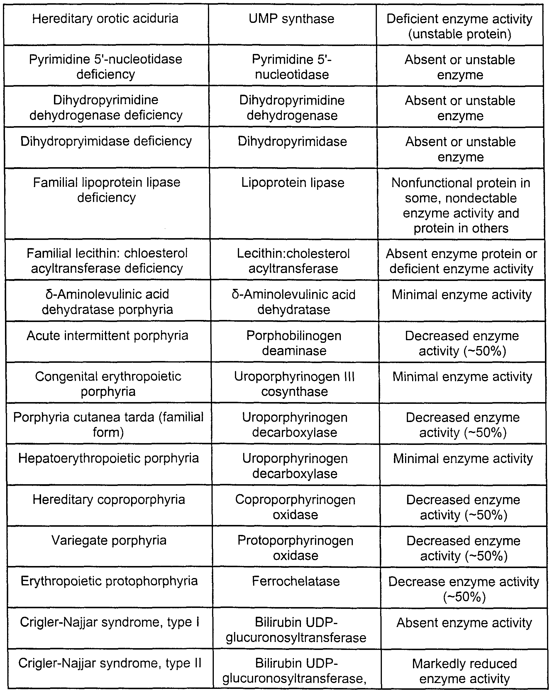


Claims
Priority Applications (5)
| Application Number | Priority Date | Filing Date | Title |
|---|---|---|---|
| AT99943915T ATE253126T1 (en) | 1998-08-25 | 1999-08-25 | RAPID QUANTITATIVE ANALYSIS OF PROTEINS OR PROTEIN FUNCTIONS IN COMPLEX MIXTURES |
| JP2000566460A JP3345401B2 (en) | 1998-08-25 | 1999-08-25 | Rapid quantitative analysis of proteins or protein functions in complex mixtures |
| DE69912444T DE69912444T3 (en) | 1998-08-25 | 1999-08-25 | FAST QUANTITATIVE ANALYSIS OF PROTEINS OR PROTEIN FUNCTIONS IN COMPLEX MIXTURES |
| AU56913/99A AU755334C (en) | 1998-08-25 | 1999-08-25 | Rapid quantitative analysis of proteins or protein function in complex mixtures |
| EP99943915A EP1105517B2 (en) | 1998-08-25 | 1999-08-25 | Rapid quantitative analysis of proteins or protein function in complex mixtures |
Applications Claiming Priority (4)
| Application Number | Priority Date | Filing Date | Title |
|---|---|---|---|
| US9778898P | 1998-08-25 | 1998-08-25 | |
| US60/097,788 | 1998-08-25 | ||
| US9911398P | 1998-09-03 | 1998-09-03 | |
| US60/099,113 | 1998-09-03 |
Publications (1)
| Publication Number | Publication Date |
|---|---|
| WO2000011208A1 true WO2000011208A1 (en) | 2000-03-02 |
Family
ID=26793647
Family Applications (1)
| Application Number | Title | Priority Date | Filing Date |
|---|---|---|---|
| PCT/US1999/019415 WO2000011208A1 (en) | 1998-08-25 | 1999-08-25 | Rapid quantitative analysis of proteins or protein function in complex mixtures |
Country Status (7)
| Country | Link |
|---|---|
| US (3) | US6670194B1 (en) |
| EP (2) | EP1105517B2 (en) |
| JP (3) | JP3345401B2 (en) |
| AT (2) | ATE536422T1 (en) |
| AU (1) | AU755334C (en) |
| DE (1) | DE69912444T3 (en) |
| WO (1) | WO2000011208A1 (en) |
Cited By (118)
| Publication number | Priority date | Publication date | Assignee | Title |
|---|---|---|---|---|
| WO2000070340A2 (en) * | 1999-05-14 | 2000-11-23 | Karolinska Innovations Ab | Materials and methods relating to disease diagnosis |
| WO2001077684A2 (en) * | 2000-04-10 | 2001-10-18 | The Scripps Research Institute | Proteomic analysis using activity-based probe libraries |
| WO2001086306A2 (en) * | 2000-05-05 | 2001-11-15 | Purdue Research Foundation | Affinity selected signature peptides for protein identification and quantification |
| WO2001094910A2 (en) * | 2000-06-07 | 2001-12-13 | Basf Aktiengesellschaft | Method for the qualitative and quantitative analysis of complex mixtures of chemical compounds, using maldi-tof mass spectrometry |
| WO2001096869A1 (en) * | 2000-06-12 | 2001-12-20 | University Of Washington | Selective labeling and isolation of phosphopeptides and applications to proteome analysis |
| WO2002014867A2 (en) * | 2000-08-11 | 2002-02-21 | Agilix Corporation | Ultra-sensitive detection systems |
| WO2002029032A2 (en) * | 2000-09-30 | 2002-04-11 | Diversa Corporation | Whole cell engineering by mutagenizing a substantial portion of a starting genome, combining mutations, and optionally repeating |
| WO2002029399A2 (en) * | 2000-10-03 | 2002-04-11 | Dionex Corporation | Method and system for peak parking in liquid chromatography-mass spectrometer (lc-ms) analysis |
| WO2002029414A2 (en) * | 2000-10-02 | 2002-04-11 | Amersham Biosciences Ab | A method for the quantitative determination of one or more compounds |
| WO2002039110A2 (en) * | 2000-11-09 | 2002-05-16 | Amersham Pharmacia Biotech Ab | A method for the quantification of carbohydrates |
| WO2002042427A2 (en) * | 2000-10-25 | 2002-05-30 | Surromed, Inc. | Mass tags for quantitative analysis |
| WO2002059144A2 (en) * | 2001-01-26 | 2002-08-01 | Syngenta Participation Ag | Differential labeling for quantitative analysis of complex protein mixtures |
| WO2002066988A2 (en) * | 2001-02-16 | 2002-08-29 | Battelle Memorial Institute | Phosphoprotein binding agents and methods of their use |
| WO2002071066A1 (en) * | 2001-03-02 | 2002-09-12 | Activx Biosciences, Inc. | Protein profiling platform |
| WO2002090929A2 (en) * | 2001-05-08 | 2002-11-14 | Perseptive Biosystems, Inc. | Process for analyzing protein samples |
| WO2002093131A2 (en) | 2001-05-14 | 2002-11-21 | The Institute For Systems Biology | Methods for isolating and labeling sample molecules |
| WO2003027682A2 (en) * | 2001-09-27 | 2003-04-03 | Purdue Research Foundation | Controlling isotope effects during fractionation of analytes |
| WO2003040093A2 (en) * | 2001-11-09 | 2003-05-15 | Bayer Healthcare Ag | Isotopically coded affinity marker 2 |
| WO2003040288A2 (en) * | 2001-11-09 | 2003-05-15 | Bayer Healthcare Ag | Isotopically coded affinity markers 3 |
| WO2003040287A2 (en) * | 2001-11-09 | 2003-05-15 | Bayer Healthcare Ag | Isotope-coded affinity tag |
| WO2003050544A2 (en) * | 2001-12-12 | 2003-06-19 | Consortium National De Recherche En Genomique (Cnrg) | Methods for protein analysis using protein capture arrays |
| WO2003069328A1 (en) * | 2002-02-14 | 2003-08-21 | Ajinomoto Co., Inc. | Method of analyzing aminofunctional compound and analytical reagent |
| US6629040B1 (en) | 1999-03-19 | 2003-09-30 | University Of Washington | Isotope distribution encoded tags for protein identification |
| WO2003081229A1 (en) * | 2002-03-25 | 2003-10-02 | Teijin Limited | Method of assaying coenzymes a in biological sample |
| WO2003087834A2 (en) * | 2002-04-08 | 2003-10-23 | Affinium Pharmaceuticals, Inc. | High throughput analysis of recombinant proteins in multi-well plates |
| JP2003532115A (en) * | 2000-05-04 | 2003-10-28 | モゼイクス ダイアグノスティックス アンド セラピューティックス エージー | Methods and devices for qualitatively and / or quantitatively analyzing protein and / or peptide patterns of liquid samples from human or animal bodies |
| EP1364209A2 (en) * | 2001-02-05 | 2003-11-26 | Activx Biosciences, Inc. | Activity based probe analysis |
| JP2003536070A (en) * | 2000-06-07 | 2003-12-02 | ビーエーエスエフ アクチェンゲゼルシャフト | Analysis of enzyme-catalyzed conversion by MALDI-TOF mass spectrometry |
| WO2003102018A2 (en) | 2002-06-03 | 2003-12-11 | The Institute For Systems Biology | Methods for quantitative proteome analysis of glycoproteins |
| WO2003102592A2 (en) * | 2002-05-31 | 2003-12-11 | Shimadzu Research Laboratory (Europe) Limited | Selective binding and analysis of macromolecules: quantitative analysis of proteins in complex mixtures |
| WO2004002950A1 (en) * | 2002-06-28 | 2004-01-08 | Shimadzu Corporation | Sulfenyl compound, labeling reagent, and method of analyzing peptide |
| WO2004008480A2 (en) * | 2002-07-16 | 2004-01-22 | National Research Council Of Canada | Quantitative analysis via isotopically differentitated derivatization |
| EP1383920A2 (en) * | 2001-04-03 | 2004-01-28 | Thermo Finnigan LLC | Methods and kits useful for the simplification of complex peptide mixtures |
| WO2004013636A2 (en) * | 2002-08-01 | 2004-02-12 | Syngenta Participations Ag | Differential labeling for quantitative analysis of complex protein mixtures by trifunctional synthetic peptide based reagents with iodoacetamido groups |
| US6692916B2 (en) | 1999-06-28 | 2004-02-17 | Source Precision Medicine, Inc. | Systems and methods for characterizing a biological condition or agent using precision gene expression profiles |
| US6706529B2 (en) | 1998-02-24 | 2004-03-16 | Target Discovery, Inc. | Methods for sequencing proteins |
| JP2004508554A (en) * | 2000-09-04 | 2004-03-18 | メルテック・マルチ−エピトープ−リガンド−テクノロジーズ・ゲーエムベーハー | Methods for identifying cell-specific proteins |
| US6716636B1 (en) | 1999-04-20 | 2004-04-06 | Target Discovery, Inc. | Methods for sequencing proteins |
| EP1404707A1 (en) * | 2001-05-29 | 2004-04-07 | The Regents Of The University Of Michigan | Systems and methods for the analysis of proteins |
| WO2002056025A3 (en) * | 2000-11-30 | 2004-05-13 | Affinium Pharmaceuticals, Inc. | Methods for systematic identification of protein - protein interactions |
| EP1428019A2 (en) * | 2001-07-13 | 2004-06-16 | Syngenta Participations AG | System and method of determining proteomic differences |
| EP1429146A1 (en) * | 2002-12-13 | 2004-06-16 | Agilent Technologies, Inc. | Proteomic analysis |
| US6756586B2 (en) | 2001-10-15 | 2004-06-29 | Vanderbilt University | Methods and apparatus for analyzing biological samples by mass spectrometry |
| AU774776B2 (en) * | 1999-04-15 | 2004-07-08 | University Of Virginia Patent Foundation | Proteome mining |
| EP1437596A1 (en) * | 2003-01-13 | 2004-07-14 | Agilent Technologies, Inc. | N- or C-terminal peptide selection method for proteomics |
| US6764817B1 (en) | 1999-04-20 | 2004-07-20 | Target Discovery, Inc. | Methods for conducting metabolic analyses |
| US6783929B1 (en) | 1999-11-02 | 2004-08-31 | Chiron Corporation | Biological sample component purification and differential display |
| EP1456227A2 (en) * | 2001-07-16 | 2004-09-15 | President And Fellows Of Harvard College | Non-affinity based isotope tagged peptides and methods for using the same |
| WO2004079358A1 (en) * | 2003-03-07 | 2004-09-16 | Kabushiki Kaisha Nard Kenkyusho | Method of measuring the molecular weight of phosphoric acid monoester compound and additive for mass spectrum measurement |
| WO2004079370A1 (en) * | 2003-03-03 | 2004-09-16 | Universiteit Leiden | Analysis of the proteome of a living cell by labelling the proteins in the intact cell |
| US6800449B1 (en) | 2001-07-13 | 2004-10-05 | Syngenta Participations Ag | High throughput functional proteomics |
| JP2004531694A (en) * | 2000-10-23 | 2004-10-14 | ジェネティックス インスティテュート エルエルシー | Acid labile isotope-coded extractant (ALICE) and its use in quantitative mass spectrometric analysis of protein mixtures |
| WO2004088271A2 (en) | 2002-10-29 | 2004-10-14 | Target Discovery, Inc. | Method for increasing ionization efficiency in mass spectroscopy |
| EP1485707A2 (en) * | 2001-07-16 | 2004-12-15 | HK Pharmaceuticals, Inc. | Capture compounds, collections thereof and methods for analyzing the proteome and complex compositions |
| WO2004111646A1 (en) * | 2003-06-19 | 2004-12-23 | Amersham Biosciences Ab | Novel ms reagents |
| US6864089B2 (en) | 2000-06-09 | 2005-03-08 | Mds Proteomics, Inc. | Labeling of proteomic samples during proteolysis for quantitation and sample multiplexing |
| DE10227599B4 (en) * | 2002-06-20 | 2005-04-28 | Proteome Factory Ag | Method and reagent for the specific identification and quantification of one or more proteins in a sample |
| US6905879B2 (en) | 2000-10-23 | 2005-06-14 | Genetics Institute, Inc. | Isotope-coded ionization-enhancing reagents (ICIER) for high-throughput protein identification and quantitation using matrix-assisted laser desorption ionization mass spectrometry |
| WO2005094187A2 (en) | 2004-03-31 | 2005-10-13 | Kazuo Shinya | Labeling substance and chimera substance, process for preparing these substances, and method of biosubstance trapping, structural analysis or/and identification with use of the labeling substance |
| US6960439B2 (en) | 1999-06-28 | 2005-11-01 | Source Precision Medicine, Inc. | Identification, monitoring and treatment of disease and characterization of biological condition using gene expression profiles |
| US6962818B2 (en) | 2000-10-19 | 2005-11-08 | Target Discovery | Mass defect labeling for the determination of oligomer sequences |
| US6964850B2 (en) | 2001-11-09 | 2005-11-15 | Source Precision Medicine, Inc. | Identification, monitoring and treatment of disease and characterization of biological condition using gene expression profiles |
| WO2006017208A1 (en) * | 2004-07-12 | 2006-02-16 | Applera Corporation | Mass tags for quantitative analyses |
| EP1631553A2 (en) * | 2003-06-06 | 2006-03-08 | President And Fellows Of Harvard College | Capture and release based isotope tagged peptides and methods for using the same |
| EP1472539A4 (en) * | 2001-08-14 | 2006-03-29 | Harvard College | Absolute quantification of proteins and modified forms thereof by multistage mass spectrometry |
| DE10153631B4 (en) * | 2001-10-31 | 2006-04-13 | Lottspeich, Friedrich, Dr. | Method for comparing two protein states |
| US7070949B2 (en) | 2001-11-30 | 2006-07-04 | Bruker Daltonik Gmbh | Protein mixture analysis by mass spectrometry |
| US7081623B2 (en) | 2003-09-05 | 2006-07-25 | Lucent Technologies Inc. | Wafer-based ion traps |
| US7112552B2 (en) | 2002-10-18 | 2006-09-26 | Promega Corporation | Compositions for separating molecules |
| US7148058B2 (en) | 2000-06-05 | 2006-12-12 | Chiron Corporation | Protein microarrays on mirrored surfaces for performing proteomic analyses |
| US7153682B2 (en) | 2000-06-05 | 2006-12-26 | Chiron Corporation | Microarrays on mirrored substrates for performing proteomic analyses |
| EP1750126A1 (en) * | 2004-05-26 | 2007-02-07 | Ajinomoto Co., Inc. | Method and apparatus for analyzing aminofunctional compound |
| US7195751B2 (en) | 2003-01-30 | 2007-03-27 | Applera Corporation | Compositions and kits pertaining to analyte determination |
| US7214545B2 (en) | 2003-04-28 | 2007-05-08 | The Regents Of The University Of California | Element-coded affinity tags |
| WO2007078229A1 (en) * | 2006-01-05 | 2007-07-12 | Ge Healthcare Bio-Sciences Ab | Kit and method for mass labelling |
| US7267994B2 (en) | 2003-04-28 | 2007-09-11 | Regents Of The University Of California | Element-coded affinity tags |
| US7288382B2 (en) | 2002-03-14 | 2007-10-30 | The Board Of Trustees Od The Leland Stanford Junior University | Methods for structural analysis of proteins |
| US7307169B2 (en) | 2004-01-05 | 2007-12-11 | Applera Corporation | Isotopically enriched N-substituted piperazines and methods for the preparation thereof |
| US7343247B2 (en) | 2001-07-30 | 2008-03-11 | The Institute For Systems Biology | Methods of classifying drug responsiveness using multiparameter analysis |
| US7355045B2 (en) | 2004-01-05 | 2008-04-08 | Applera Corporation | Isotopically enriched N-substituted piperazine acetic acids and methods for the preparation thereof |
| US7365178B2 (en) | 2003-04-01 | 2008-04-29 | Activx Biosciences, Inc. | Acyl-nucleotide probes and methods of their synthesis and use in proteomic analysis |
| EP1916526A1 (en) * | 2006-10-26 | 2008-04-30 | Koninklijke Philips Electronics N.V. | Method for diagnostic and therapeutic target discovery by combining isotopic and isobaric labels |
| US7425451B2 (en) | 2002-12-13 | 2008-09-16 | Agilent Technologies, Inc. | Triazine derivatives as universal peptide isotope tag reagents (U-PIT) |
| US7473535B2 (en) | 2002-08-20 | 2009-01-06 | The Institute For Systems Biology | Chemical reagents and methods for detection and quantification of proteins in complex mixtures |
| US7501286B2 (en) | 2002-08-14 | 2009-03-10 | President And Fellows Of Harvard College | Absolute quantification of proteins and modified forms thereof by multistage mass spectrometry |
| US7567870B1 (en) | 2000-07-31 | 2009-07-28 | Institute For Systems Biology | Multiparameter analysis for predictive medicine |
| EP2093569A2 (en) | 2004-05-21 | 2009-08-26 | The Institute for Systems Biology | Compositions and methods for quantification of serum glycoproteins |
| EP2108957A1 (en) * | 2008-04-07 | 2009-10-14 | Koninklijke Philips Electronics N.V. | Compounds and methods for the labelling and affinity-selection of proteins |
| WO2009114763A3 (en) * | 2008-03-13 | 2009-11-19 | Perkinelmer Las, Inc. | Enzymatic substrates for multiple detection systems |
| US7623969B2 (en) | 2002-01-31 | 2009-11-24 | The Institute For Systems Biology | Gene discovery for the system assignment of gene function |
| US7655433B2 (en) | 2002-06-04 | 2010-02-02 | The Institute For Systems Biology | Methods for high-throughput and quantitative proteome analysis |
| US7670796B2 (en) | 2003-05-23 | 2010-03-02 | Health Protection Agency | Metalloprotease assay |
| US20100190183A1 (en) * | 2007-07-11 | 2010-07-29 | Koninklijke Philips Electronics N.V. | Protein labelling with tags comprising isotope-coded sub-tags and isobaric sub-tags |
| US7794947B2 (en) | 2003-07-10 | 2010-09-14 | Institute For Systems Biology | Affinity capture of peptides by microarray and related methods |
| US7846748B2 (en) | 2002-12-02 | 2010-12-07 | The University Of North Carolina At Chapel Hill | Methods of quantitation and identification of peptides and proteins |
| EP2309262A1 (en) | 2009-10-06 | 2011-04-13 | Max-Planck-Gesellschaft zur Förderung der Wissenschaften e.V. | Method for quantifying biomolecules |
| US8071392B2 (en) | 2007-06-21 | 2011-12-06 | National University Corporation University Of Fukui | Mass spectrometric analysis of proteins using stable isotopes of pyrylium derivates as labels |
| US8097425B2 (en) | 2006-03-10 | 2012-01-17 | Tethys Bioscience, Inc. | Multiplex protein fractionation |
| EP2426143A2 (en) | 2007-01-05 | 2012-03-07 | University of Zurich | Method of providing disease-specific binding molecules and targets |
| US8288139B2 (en) | 2002-07-10 | 2012-10-16 | Seikagaku Corporation | Sulfotransferase inhibitors |
| US8492163B2 (en) | 2011-01-31 | 2013-07-23 | Dh Technologies Development Pte. Ltd. | Methods, mixtures, kits and compositions pertaining to analyte determination |
| US8501498B2 (en) | 2004-07-12 | 2013-08-06 | Dh Technologies Development Pte. Ltd. | Mass tags for quantitative analyses |
| US8569071B2 (en) | 2005-02-09 | 2013-10-29 | Dh Technologies Development Pte. Ltd. | Amine-containing compound analysis methods |
| US8604692B2 (en) | 2007-11-06 | 2013-12-10 | Translational Therapeutics, Inc. | Mass spectrometry assay for eIF4E and eIF4E regulon activity |
| US8633031B2 (en) | 2004-05-19 | 2014-01-21 | Dh Technologies Development Pte. Ltd. | Expression quantification using mass spectrometry |
| DE102013212179A1 (en) * | 2012-07-31 | 2014-02-06 | Agilent Technologies, Inc. | Derivatization of PNGase F-released glycans on an HPLC chip |
| US8729245B2 (en) | 2009-12-21 | 2014-05-20 | Pharmathene, Inc. | Recombinant butyrylcholinesterases and truncates thereof |
| WO2014190230A1 (en) | 2013-05-23 | 2014-11-27 | Iphenotype Llc | Phenotypic integrated social search database and method |
| US8909481B2 (en) | 2000-12-26 | 2014-12-09 | The Institute Of Systems Biology | Method of mass spectrometry for identifying polypeptides |
| US9034798B2 (en) | 2003-01-16 | 2015-05-19 | Caprotec Bioanalytics Gmbh | Capture compounds, collections thereof and methods for analyzing the proteome and complex compositions |
| US9110074B2 (en) | 2007-11-06 | 2015-08-18 | Translational Therapeutics, Inc. | Mass spectrometry assay for eIF4E and eIF4E regulon activity |
| EP2928914A1 (en) * | 2012-12-10 | 2015-10-14 | Fred Hutchinson Cancer Research Center | Methods for screening |
| CN105384785A (en) * | 2015-11-24 | 2016-03-09 | 中国人民解放军第二军医大学 | Preparation method of galactose-containing fatty acid derivative and application of preparation method in field of medicine |
| US9291597B2 (en) | 2010-07-02 | 2016-03-22 | Ventana Medical Systems, Inc. | Detecting targets using mass tags and mass spectrometry |
| WO2017127670A1 (en) | 2016-01-22 | 2017-07-27 | Purdue Research Foundation | Charged mass labeling system |
| US9733253B2 (en) | 2009-12-22 | 2017-08-15 | Expression Pathology, Inc. | Secreted protein acidic and rich in cysteine (SPARC) protein SRM assay |
| CN107325154A (en) * | 2017-06-23 | 2017-11-07 | 广东华肽生物科技有限公司 | It is a kind of that there is the polypeptide and its method for separating and preparing and purposes for improving memory effect |
| CN113156002A (en) * | 2021-04-01 | 2021-07-23 | 河北利德检测技术有限公司 | Method for detecting 5 related substances in calcium gluconate |
Families Citing this family (142)
| Publication number | Priority date | Publication date | Assignee | Title |
|---|---|---|---|---|
| DE69912444T3 (en) * | 1998-08-25 | 2010-05-06 | University Of Washington, Seattle | FAST QUANTITATIVE ANALYSIS OF PROTEINS OR PROTEIN FUNCTIONS IN COMPLEX MIXTURES |
| IL144657A0 (en) * | 1999-02-11 | 2002-06-30 | Maxygen Inc | High throughput mass spectrometry |
| US6687395B1 (en) * | 1999-07-21 | 2004-02-03 | Surromed, Inc. | System for microvolume laser scanning cytometry |
| WO2001049414A2 (en) * | 2000-01-06 | 2001-07-12 | Caliper Technologies Corp. | Ultra high throughput sampling and analysis systems and methods |
| US20020072064A1 (en) * | 2000-08-25 | 2002-06-13 | Naki Donald P. | Detecting polymers and polymer fragments |
| US6787761B2 (en) * | 2000-11-27 | 2004-09-07 | Surromed, Inc. | Median filter for liquid chromatography-mass spectrometry data |
| WO2002044715A1 (en) * | 2000-11-28 | 2002-06-06 | Surromed, Inc. | Methods for efficiently minig broad data sets for biological markers |
| US20020090652A1 (en) * | 2000-12-22 | 2002-07-11 | Fu Emil Wei-Ming | Inverse labeling method for the rapid identification of marker/target proteins |
| WO2002055989A2 (en) * | 2001-01-12 | 2002-07-18 | The Regents Of The University Of California | Stable isotope, site-specific mass tagging for protein identification |
| ES2242864T3 (en) * | 2001-03-22 | 2005-11-16 | Vlaams Interuniversitair Instituut Voor Biotechnologie Vzw. | PROCEDURES AND APPLIANCES FOR QUALITATIVE AND QUANTITATIVE ANALYSIS WITHOUT PROTEOM GEL, AND ITS USES. |
| KR20030031911A (en) * | 2001-04-19 | 2003-04-23 | 싸이퍼젠 바이오시스템즈, 인코포레이티드 | Biomolecule characterization using mass spectrometry and affinity tags |
| CA2349265A1 (en) * | 2001-05-30 | 2002-11-30 | Andrew Emili | Protein expression profile database |
| US6873915B2 (en) * | 2001-08-24 | 2005-03-29 | Surromed, Inc. | Peak selection in multidimensional data |
| US20030078739A1 (en) * | 2001-10-05 | 2003-04-24 | Surromed, Inc. | Feature list extraction from data sets such as spectra |
| CA2466837A1 (en) * | 2001-11-13 | 2003-05-22 | Caprion Pharmaceuticals Inc. | Mass intensity profiling system and uses thereof |
| US20030091976A1 (en) * | 2001-11-14 | 2003-05-15 | Ciphergen Biosystems, Inc. | Methods for monitoring polypeptide production and purification using surface enhanced laser desorption/ionization mass spectrometry |
| WO2003068919A2 (en) * | 2002-02-12 | 2003-08-21 | The Regents Of The University Of California | Non-invasive method for measuring rates of biosynthesis of biological molecules by label incorporation |
| AU2003224674A1 (en) * | 2002-03-11 | 2003-09-29 | Hk Pharmaceuticals, Inc. | Compounds and methods for analyzing the proteome |
| CA2484625A1 (en) * | 2002-05-09 | 2003-11-20 | Surromed, Inc. | Methods for time-alignment of liquid chromatography-mass spectrometry data |
| US20050221500A1 (en) * | 2002-05-20 | 2005-10-06 | Purdue Research Foundation | Protein identification from protein product ion spectra |
| US20040229294A1 (en) | 2002-05-21 | 2004-11-18 | Po-Ying Chan-Hui | ErbB surface receptor complexes as biomarkers |
| US7052916B2 (en) * | 2002-05-24 | 2006-05-30 | Immunex Corporation | Polypeptide analyses using stable isotope labeling |
| US20040126890A1 (en) * | 2002-06-10 | 2004-07-01 | Gjerde Douglas T. | Biomolecule open channel solid phase extraction systems and methods |
| GB0305796D0 (en) | 2002-07-24 | 2003-04-16 | Micromass Ltd | Method of mass spectrometry and a mass spectrometer |
| US7409296B2 (en) * | 2002-07-29 | 2008-08-05 | Geneva Bioinformatics (Genebio), S.A. | System and method for scoring peptide matches |
| EP1546364A4 (en) | 2002-07-30 | 2006-09-06 | Univ California | Method for automated, large-scale measurement of the molecular flux rates of the proteome or the organeome using mass spectrometry |
| WO2004025243A2 (en) * | 2002-09-12 | 2004-03-25 | Vlaams Interuniversitair Instituut Voor Biotechnologie Vzw | A method for the identification of drug targets |
| EP1546359A4 (en) * | 2002-09-12 | 2007-05-09 | Molecular Probes Inc | Site-specific labeling of affinity tags in fusion proteins |
| US6944549B2 (en) * | 2002-10-25 | 2005-09-13 | Syngenta Participations Ag | Method and apparatus for automated detection of peaks in spectroscopic data |
| US20060105343A1 (en) * | 2003-01-09 | 2006-05-18 | Children's Medical Center Corporation | Methods for diagnosis and prognosis of cancer |
| JP2007527503A (en) * | 2003-06-17 | 2007-09-27 | プロノタ・エヌブイ | Peptide combos and their use |
| FR2857451B1 (en) * | 2003-07-11 | 2005-09-30 | Commissariat Energie Atomique | METHOD AND DEVICE FOR ANALYSIS OF LIVE REACTION ENVIRONMENTS |
| AU2004267420A1 (en) * | 2003-08-11 | 2005-03-03 | Monogram Biosciences, Inc. | Detecting and profiling molecular complexes |
| US7485435B2 (en) * | 2004-08-30 | 2009-02-03 | Perkinelmer Las, Inc. | Simultaneous detection of metabolic enzyme activity and metabolite levels |
| US7754497B2 (en) * | 2003-08-29 | 2010-07-13 | Reverse Proteomics Research Institute Co., Ltd. | Method for immobilizing proteins |
| US20050048499A1 (en) * | 2003-08-29 | 2005-03-03 | Perkin Elmer Life Sciences, Inc. | Tandem mass spectrometry method for the genetic screening of inborn errors of metabolism in newborns |
| US20050074895A1 (en) * | 2003-09-05 | 2005-04-07 | Bio-Rad Laboratories, Inc. | Alkyl-substituted vinyl pyridines for mass-coded tagging of proteins |
| WO2005031304A2 (en) * | 2003-09-22 | 2005-04-07 | Becton, Dickinson And Company | Quantification of analytes using internal standards |
| US7145019B2 (en) * | 2003-10-16 | 2006-12-05 | Ambergen, Inc. | Photocleavable isotope-coded affinity tags |
| EP2208992A1 (en) * | 2003-11-21 | 2010-07-21 | Eisai R&D Management Co., Ltd. | Quantitation method using isotope labeled internal standard substance, analysis system for executing the quantitation method, and program for the analysis |
| US7348416B2 (en) * | 2003-11-21 | 2008-03-25 | Applera Corporation | Selective capture and enrichment of proteins expressed on the cell surface |
| US20050202406A1 (en) * | 2003-11-25 | 2005-09-15 | The Regents Of The University Of California | Method for high-throughput screening of compounds and combinations of compounds for discovery and quantification of actions, particularly unanticipated therapeutic or toxic actions, in biological systems |
| WO2005069891A2 (en) * | 2004-01-15 | 2005-08-04 | Cedars-Sinai Medical Center | System and method for expression proteomics based on isotope ratio modification |
| US20070087448A1 (en) * | 2004-02-16 | 2007-04-19 | Nelsestuen Gary L | Biological profiles and methods of use |
| TW200538738A (en) | 2004-02-20 | 2005-12-01 | Univ California | Molecular flux rates through critical pathways measured by stable isotope labeling in vivo, as biomarkers of drug action and disease activity |
| US7248360B2 (en) * | 2004-04-02 | 2007-07-24 | Ppd Biomarker Discovery Sciences, Llc | Polychronic laser scanning system and method of use |
| ES2549077T3 (en) * | 2004-04-07 | 2015-10-22 | Genentech, Inc. | Mass spectrometry of antibody conjugates |
| US20060078960A1 (en) * | 2004-05-19 | 2006-04-13 | Hunter Christie L | Expression quantification using mass spectrometry |
| US20080206737A1 (en) * | 2004-05-19 | 2008-08-28 | Hunter Christie L | Expression quantification using mass spectrometry |
| US20080044857A1 (en) * | 2004-05-25 | 2008-02-21 | The Gov Of Usa As Represented By The Secretary Of | Methods For Making And Using Mass Tag Standards For Quantitative Proteomics |
| JP4317083B2 (en) | 2004-06-04 | 2009-08-19 | 株式会社日立ハイテクノロジーズ | Mass spectrometry method and mass spectrometry system |
| US20060063269A1 (en) * | 2004-06-18 | 2006-03-23 | Brian Agnew | Fluorescent isotope tags and their method of use |
| WO2006026020A2 (en) | 2004-07-30 | 2006-03-09 | Adeza Biomedical Corporation | Oncofetal fibronectin as a marker for disease and other conditions and methods for detection of oncofetal fibronectin |
| US7592139B2 (en) * | 2004-09-24 | 2009-09-22 | Sandia National Laboratories | High temperature flow-through device for rapid solubilization and analysis |
| JP4563764B2 (en) * | 2004-10-01 | 2010-10-13 | 丸善製薬株式会社 | Peptide quantification method |
| WO2006054310A1 (en) * | 2004-11-22 | 2006-05-26 | Biosight Ltd. | Activated labeling reagents and methods for preparing and using the same |
| US20070037286A1 (en) * | 2005-02-09 | 2007-02-15 | Subhasish Purkayastha | Thyroxine-containing compound analysis methods |
| WO2006096704A2 (en) | 2005-03-07 | 2006-09-14 | Invitrogen Corporation | Isotopically-labeled proteome standards |
| US20060251576A1 (en) * | 2005-05-03 | 2006-11-09 | The Regents Of The University Of California | Methods for measuring cholesterol metabolism and transport |
| US10687785B2 (en) | 2005-05-12 | 2020-06-23 | The Trustees Of Columbia Univeristy In The City Of New York | System and method for electromechanical activation of arrhythmias |
| JP2006337176A (en) * | 2005-06-02 | 2006-12-14 | Eisai R & D Management Co Ltd | Determination method of metabolite |
| DE602005004902T2 (en) * | 2005-06-02 | 2008-10-16 | Entelechon Gmbh | Artificial protein, method for absolute quantification of proteins and its use |
| TW200711660A (en) * | 2005-06-10 | 2007-04-01 | Univ California | Monitoring two dimensions of diabetes pathogenesis separately or concurrently (insulin sensitivity and beta-cell sufficiency): uses in diagnosis, prognosis, assessment of disease risk, and drug development |
| EP1754972A1 (en) | 2005-08-17 | 2007-02-21 | Agilent Technologies, Inc. | Folded peptides containing metal binding pockets for protein tagging |
| CA2625781C (en) * | 2005-10-13 | 2014-09-02 | Applera Corporation | Methods for the development of a biomolecule assay |
| US7998717B2 (en) * | 2005-12-02 | 2011-08-16 | Pacific Biosciences Of California, Inc. | Mitigation of photodamage in analytical reactions |
| US8105788B2 (en) * | 2005-12-08 | 2012-01-31 | The University Of British Columbia | Mucopolysaccharidosis (MPS) diagnostic methods, systems, kits and assays associated therewith |
| US20070140967A1 (en) * | 2005-12-19 | 2007-06-21 | Wood Nichole L | Agents and methods for analyzing protein interactions |
| US20070166810A1 (en) * | 2005-12-23 | 2007-07-19 | Perkinelmer Las, Inc. | Methods and compositions for detecting enzymatic activity |
| US20070148721A1 (en) * | 2005-12-28 | 2007-06-28 | Agilent Technologies, Inc. | Compositions, methods, systems, and kits for affinity purification |
| US20080026480A1 (en) * | 2006-02-14 | 2008-01-31 | Perkinelmer Las, Inc. | Detection systems for mass labels |
| EP1999270A4 (en) * | 2006-03-13 | 2010-04-28 | Perkinelmer Las Inc | Substrates and internal standards for mass spectroscopy detection |
| WO2007127767A2 (en) * | 2006-04-28 | 2007-11-08 | Ludwig Institute For Cancer Research | Quantification of enzyme activity by mass spectrometry |
| US20100216657A1 (en) * | 2006-05-16 | 2010-08-26 | Arcxis Biotechnologies, Inc. | Pcr-free sample preparation and detection systems for high speed biologic analysis and identification |
| US8314215B2 (en) * | 2006-08-04 | 2012-11-20 | The Board Of Trustees Of The Leland Stanford Junior University | Mild chemically cleavable linker system |
| US7879799B2 (en) * | 2006-08-10 | 2011-02-01 | Institute For Systems Biology | Methods for characterizing glycoproteins and generating antibodies for same |
| EP1921081A1 (en) * | 2006-11-06 | 2008-05-14 | Koninklijke Philips Electronics N.V. | Use of arylboronic acids in protein labelling |
| KR100805777B1 (en) * | 2007-02-22 | 2008-02-21 | 한국기초과학지원연구원 | A system of analyzing protein modification with its band position of one-dimensional gel by the mass spectral data analysis and the method of analyzing protein modification using thereof |
| CA2690612C (en) | 2007-05-14 | 2016-07-05 | Historx, Inc. | Compartment segregation by pixel characterization using image data clustering |
| JP5003274B2 (en) * | 2007-05-16 | 2012-08-15 | 株式会社日立製作所 | Mass spectrometry system and mass spectrometry method |
| WO2008150488A1 (en) * | 2007-06-01 | 2008-12-11 | Laboratory Corporation Of America Holdings | Methods and systems for quantification of peptides and other analytes |
| US7907271B2 (en) | 2007-06-15 | 2011-03-15 | Historx, Inc. | Method and system for standardizing microscope instruments |
| CA2604317C (en) | 2007-08-06 | 2017-02-28 | Historx, Inc. | Methods and system for validating sample images for quantitative immunoassays |
| CA2596204C (en) | 2007-08-07 | 2019-02-26 | Historx, Inc. | Method and system for determining an optimal dilution of a reagent |
| ES2396797T3 (en) | 2007-08-17 | 2013-02-27 | University Of Washington | Test procedures for alpha-L-iduronidase enzyme activity |
| US7978258B2 (en) | 2007-08-31 | 2011-07-12 | Historx, Inc. | Automatic exposure time selection for imaging tissue |
| JP4921302B2 (en) * | 2007-09-26 | 2012-04-25 | 株式会社日立ハイテクノロジーズ | Mass spectrometry system |
| US8334506B2 (en) | 2007-12-10 | 2012-12-18 | 1St Detect Corporation | End cap voltage control of ion traps |
| US7973277B2 (en) | 2008-05-27 | 2011-07-05 | 1St Detect Corporation | Driving a mass spectrometer ion trap or mass filter |
| CA2725599A1 (en) | 2008-05-30 | 2009-12-03 | The University Of British Columbia | Methods of diagnosing rejection of a kidney allograft using genomic or proteomic expression profiling |
| WO2010002911A2 (en) | 2008-06-30 | 2010-01-07 | H. Lee Moffitt Cancer Center And Research Institute, Inc. | Methods and materials for monitoring myeloma using quantitative mass spetrometry |
| AU2009293380B2 (en) | 2008-09-16 | 2016-05-19 | Novartis Ag | Reproducible quantification of biomarker expression |
| US20100081924A1 (en) * | 2008-09-29 | 2010-04-01 | Searete Llc, A Limited Liability Corporation Of The State Of Delaware | Histological facilitation systems and methods |
| US20100081916A1 (en) * | 2008-09-29 | 2010-04-01 | Searete Llc, A Limited Liability Corporation Of The State Of Delaware. | Histological facilitation systems and methods |
| US20100081926A1 (en) * | 2008-09-29 | 2010-04-01 | Searete Llc, A Limited Liability Corporation Of The State Of Delaware | Histological facilitation systems and methods |
| US20100081927A1 (en) * | 2008-09-29 | 2010-04-01 | Searete Llc, A Limited Liability Corporation Of The State Of Delaware | Histological facilitation systems and methods |
| US20100081915A1 (en) * | 2008-09-29 | 2010-04-01 | Searete Llc, Alimited Liability Corporation Of The State Of Delaware | Histological facilitation systems and methods |
| US20100081928A1 (en) * | 2008-09-29 | 2010-04-01 | Searete Llc, A Limited Liability Corporation Of The State Of Delaware | Histological Facilitation systems and methods |
| EP4032538A3 (en) | 2009-03-02 | 2022-10-26 | Massachusetts Institute of Technology | Methods and products for in vivo enzyme profiling |
| WO2010120946A1 (en) * | 2009-04-14 | 2010-10-21 | Institute For Systems Biology | Improvements in mass spectrum-based identification and quantitation of proteins and peptides |
| CN102460176B (en) * | 2009-05-21 | 2014-09-24 | 李荣秀 | Process for differential polypeptides detection and uses thereof |
| GB0916881D0 (en) * | 2009-09-25 | 2009-11-11 | Electrophoretics Ltd | Mass labels |
| JP2013514537A (en) * | 2009-12-15 | 2013-04-25 | ディーエイチ テクノロジーズ デベロップメント プライベート リミテッド | Analysis of amino acid and amine-containing compounds using tagging reagents and LC-MS workflow |
| JP5731539B2 (en) * | 2009-12-22 | 2015-06-10 | エクスプレッション、パソロジー、インコーポレイテッドExpression Pathology, Inc. | Insulin-like growth factor 1 receptor (IGF-1R) protein SRM / MRM assay |
| US9453845B2 (en) | 2010-02-01 | 2016-09-27 | Cell Signaling Technology, Inc. | Mass spectroscopy analysis of mutant polypeptides in biological samples |
| DE102011017084B4 (en) | 2010-04-14 | 2020-07-09 | Wisconsin Alumni Research Foundation | Mass spectrometry data acquisition mode for more reliable protein quantification |
| US8834847B2 (en) | 2010-08-12 | 2014-09-16 | Pacific Biosciences Of California, Inc. | Photodamage mitigation compounds and systems |
| US8742333B2 (en) | 2010-09-17 | 2014-06-03 | Wisconsin Alumni Research Foundation | Method to perform beam-type collision-activated dissociation in the pre-existing ion injection pathway of a mass spectrometer |
| EP2643474A4 (en) * | 2010-11-24 | 2014-05-21 | Dh Technologies Dev Pte Ltd | High-throughput, sensitive detection of glucose-6-phosphate dehydrogenase |
| ES2881535T3 (en) | 2011-03-15 | 2021-11-29 | Massachusetts Inst Technology | Multiplexed detection with isotope-coded flags |
| US9040903B2 (en) | 2011-04-04 | 2015-05-26 | Wisconsin Alumni Research Foundation | Precursor selection using an artificial intelligence algorithm increases proteomic sample coverage and reproducibility |
| US9659146B2 (en) | 2011-05-02 | 2017-05-23 | Tyler Stuart Bray | Method for quantitative analysis of complex proteomic data |
| WO2012162664A1 (en) | 2011-05-26 | 2012-11-29 | The Trustees Of Columbia University In The City Of New York | Systems and methods for opening of a tissue barrier in primates |
| EP2753707A4 (en) | 2011-09-08 | 2015-03-04 | Univ California | Metabolic flux measurement, imaging and microscopy |
| EP2802670B1 (en) | 2011-11-08 | 2019-12-25 | University of Washington | Lysosomal enzyme assay methods and compositions |
| WO2013086070A1 (en) | 2011-12-07 | 2013-06-13 | Glaxosmithkline Llc | Methods for determining total body skeletal muscle mass |
| US9134319B2 (en) | 2013-03-15 | 2015-09-15 | The Regents Of The University Of California | Method for replacing biomarkers of protein kinetics from tissue samples by biomarkers of protein kinetics from body fluids after isotopic labeling in vivo |
| US20140274798A1 (en) | 2013-03-15 | 2014-09-18 | University Of Washington Through Its Center For Commercialization | Compounds and methods relating to lysosomal storage disorders |
| WO2014197840A1 (en) | 2013-06-07 | 2014-12-11 | Massachusetts Institute Of Technology | Affinity-based detection of ligand-encoded synthetic biomarkers |
| CN105143872B (en) * | 2013-06-07 | 2019-03-01 | 皮尔斯生物科技有限公司 | By mass spectral analysis with multiplexing internal standard compound to the absolute quantitation of protein and protein modification |
| US20170343540A1 (en) * | 2014-12-29 | 2017-11-30 | Hitachi High-Technologies Corporation | Analysis method and analysis device |
| CA2984589A1 (en) | 2015-05-01 | 2016-11-10 | The University Of British Columbia | Biomarkers for the detection of acute rejection in heart transplantation |
| WO2017040976A1 (en) * | 2015-09-02 | 2017-03-09 | Erkki Ruoslahti | Compounds and compositions for targeting brain injuries and methods of use thereof |
| AU2016380362B2 (en) * | 2015-12-31 | 2021-07-01 | University Of Notre Dame Du Lac | ESI-MS via an electrokinetically pumped interface |
| CN105911101B (en) * | 2016-04-08 | 2018-09-11 | 中南大学 | A kind of film properties detection method of simple and sensitive |
| WO2017177115A1 (en) | 2016-04-08 | 2017-10-12 | Massachusetts Institute Of Technology | Methods to specifically profile protease activity at lymph nodes |
| CA3022928A1 (en) | 2016-05-05 | 2017-11-09 | Massachusetts Institute Of Technology | Methods and uses for remotely triggered protease activity measurements |
| DE112017002818T5 (en) * | 2016-06-29 | 2019-02-21 | Hitachi High-Technologies Corporation | Compound for use in an enzymatic reaction and mass spectrometry methods |
| US11339434B2 (en) * | 2016-07-29 | 2022-05-24 | The Regents Of The University Of California | Methods for determining gene functions |
| DE102017103600A1 (en) * | 2017-02-22 | 2018-08-23 | Eberhard Karls Universität Tübingen Medizinische Fakultät | Radioactively labeled beta-galactosidase substrate for PET imaging of senescence |
| WO2018187688A1 (en) | 2017-04-07 | 2018-10-11 | Massachusetts Institute Of Technology | Methods to spatially profile protease activity in tissue and sections |
| US20200200760A1 (en) * | 2017-05-16 | 2020-06-25 | Lankenau Institute Of Medical Research | Compositions comprising ligands to rhob protein and the uses thereof |
| CN108169397B (en) * | 2017-11-30 | 2019-11-15 | 中国农业科学院北京畜牧兽医研究所 | A kind of method of relative quantitative assay broiler chicken glutathione s-transferase GSTA2 |
| JP7028258B2 (en) | 2018-01-09 | 2022-03-02 | 株式会社島津製作所 | Protein identification method |
| US11054428B2 (en) | 2018-03-05 | 2021-07-06 | Massachusetts Institute Of Technology | Inhalable nanosensors with volatile reporters and uses thereof |
| US10656059B2 (en) | 2018-03-07 | 2020-05-19 | Alcala Pharmaceutical, Inc. | Method for qualitative and quantitative multiplexing of drug analytes from biological samples |
| RU2678090C1 (en) * | 2018-05-28 | 2019-01-23 | Федеральное государственное бюджетное научное учреждение "Федеральный научный центр пищевых систем им. В.М. Горбатова" РАН | Boiled sausage products authenticity confirmation method |
| WO2020150560A1 (en) | 2019-01-17 | 2020-07-23 | Massachusetts Institute Of Technology | Sensors for detecting and imaging of cancer metastasis |
| WO2020154541A1 (en) * | 2019-01-24 | 2020-07-30 | The Board Of Trustees Of The Leland Stanford Junior University | Compact hydroxamate-based affinity tags for artificially tagging biological macromolecules |
| CN110280229B (en) * | 2019-06-28 | 2022-03-11 | 东南大学 | Preparation and application method of pterin compound selective separation enrichment material |
| EP4229074A1 (en) | 2020-10-16 | 2023-08-23 | Stichting Het Nederlands Kanker Instituut- Antoni van Leeuwenhoek Ziekenhuis | Anti-tumor immunity induces the presentation of aberrant peptides |
| WO2023080788A1 (en) | 2021-11-05 | 2023-05-11 | Stichting Het Nederlands Kanker Instituut-Antoni van Leeuwenhoek Ziekenhuis | Tryptophan depletion induces production and presentation of tryptophan to phenylalanine substitutions |
Citations (13)
| Publication number | Priority date | Publication date | Assignee | Title |
|---|---|---|---|---|
| US4798795A (en) * | 1987-02-17 | 1989-01-17 | Hoechst Celanese Corporation | Biotinylating agents |
| US5514559A (en) * | 1993-03-29 | 1996-05-07 | Boehringer Mannheim Gmbh | Immunologically active conjugates and method for their preparation |
| US5527711A (en) * | 1993-12-13 | 1996-06-18 | Hewlett Packard Company | Method and reagents for binding chemical analytes to a substrate surface, and related analytical devices and diagnostic techniques |
| US5534132A (en) * | 1995-05-04 | 1996-07-09 | Vreeke; Mark | Electrode and method for the detection of an affinity reaction |
| US5614368A (en) * | 1990-02-09 | 1997-03-25 | Molecular Devices Corporation | Chromophoric reagents for incorporation of biotin or other haptens into macromolecules |
| US5658725A (en) * | 1993-12-21 | 1997-08-19 | Boehringer Mannheim Gmbh | Acylated protein aggregates and their use in suppressing interference in immunoassays |
| US5738984A (en) * | 1993-04-14 | 1998-04-14 | Yissum Research Development Company Of The Hebrew University Of Jerusalem | Kits and methods of detection using cellulose binding domain fusion proteins |
| US5851781A (en) * | 1993-08-03 | 1998-12-22 | Abbott Laboratories | Reagents and methods for the detection and quantification of testosterone in fluid samples |
| US5863740A (en) * | 1994-03-05 | 1999-01-26 | Boehringer Mannheim Gmbh | Interference eliminating agent for application in immunoassays |
| US5880270A (en) * | 1995-06-07 | 1999-03-09 | Cellpro, Incorporated | Aminooxy-containing linker compounds for formation of stably-linked conjugates and methods related thereto |
| US5958703A (en) * | 1996-12-03 | 1999-09-28 | Glaxo Group Limited | Use of modified tethers in screening compound libraries |
| US5965131A (en) * | 1995-06-07 | 1999-10-12 | Immunomedics, Inc. | Delivery of diagnostic and therapeutic agents to a target site |
| US5965457A (en) * | 1995-06-06 | 1999-10-12 | Magnani; John L. | Methods of screening for a candidate compound able to bind to CD34+ cells |
Family Cites Families (16)
| Publication number | Priority date | Publication date | Assignee | Title |
|---|---|---|---|---|
| US5650270A (en) * | 1982-02-01 | 1997-07-22 | Northeastern University | Molecular analytical release tags and their use in chemical analysis |
| WO1990004786A1 (en) * | 1988-10-17 | 1990-05-03 | Molecular Devices Corporation | Hapten derivatized capture membrane and diagnostic assays using such membrane |
| US5438017A (en) * | 1989-05-01 | 1995-08-01 | The University Of Colorado Foundation, Inc. | Assays for sulfhydryl amino acids and methylmalonic acid and their application to diagnosis of cobalamin deficiency |
| CA1340966C (en) | 1989-05-19 | 2000-04-18 | Thomas R. Covey | Method of protein analysis |
| US5534440A (en) | 1991-02-22 | 1996-07-09 | Biomedical Research Centre Limited | Compounds and methods for sequencing amino acids |
| US5240859A (en) | 1991-02-22 | 1993-08-31 | B.R. Centre Limited | Methods for amino acid sequencing of a polypeptide |
| US5854084A (en) | 1996-07-12 | 1998-12-29 | Biotraces, Inc. | Enhanced chromatography using multiphoton detection |
| US5538897A (en) | 1994-03-14 | 1996-07-23 | University Of Washington | Use of mass spectrometry fragmentation patterns of peptides to identify amino acid sequences in databases |
| US5986076A (en) * | 1994-05-11 | 1999-11-16 | Trustees Of Boston University | Photocleavable agents and conjugates for the detection and isolation of biomolecules |
| US5686310A (en) | 1995-06-06 | 1997-11-11 | University Of Virginia Patent Foundation | Method for determination of the amount of either phosphotyrosine or phosphoserine in a protein |
| CA2274587A1 (en) | 1996-12-10 | 1998-06-18 | Genetrace Systems Inc. | Releasable nonvolatile mass-label molecules |
| CN1118580C (en) | 1997-01-23 | 2003-08-20 | 布拉克斯集团有限公司 | Characterising polypeptides |
| EP0994968A1 (en) | 1997-07-11 | 2000-04-26 | Brax Group Limited | Characterising nucleic acid |
| CA2295955A1 (en) | 1997-07-11 | 1999-01-21 | Brax Group Limited | Characterising nucleic acids |
| DE69912444T3 (en) * | 1998-08-25 | 2010-05-06 | University Of Washington, Seattle | FAST QUANTITATIVE ANALYSIS OF PROTEINS OR PROTEIN FUNCTIONS IN COMPLEX MIXTURES |
| AU6689401A (en) | 2000-06-12 | 2001-12-24 | Univ Washington | Selective labeling and isolation of phosphopeptides and applications to proteomeanalysis |
-
1999
- 1999-08-25 DE DE69912444T patent/DE69912444T3/en not_active Expired - Lifetime
- 1999-08-25 EP EP99943915A patent/EP1105517B2/en not_active Expired - Lifetime
- 1999-08-25 AU AU56913/99A patent/AU755334C/en not_active Expired
- 1999-08-25 AT AT03075828T patent/ATE536422T1/en active
- 1999-08-25 US US09/383,062 patent/US6670194B1/en not_active Expired - Lifetime
- 1999-08-25 JP JP2000566460A patent/JP3345401B2/en not_active Expired - Lifetime
- 1999-08-25 AT AT99943915T patent/ATE253126T1/en not_active IP Right Cessation
- 1999-08-25 EP EP03075828A patent/EP1329513B1/en not_active Expired - Lifetime
- 1999-08-25 WO PCT/US1999/019415 patent/WO2000011208A1/en active IP Right Grant
-
2001
- 2001-04-20 US US09/839,884 patent/US6852544B2/en not_active Expired - Lifetime
-
2002
- 2002-07-17 JP JP2002208687A patent/JP3793124B2/en not_active Expired - Lifetime
-
2004
- 2004-11-23 US US10/994,815 patent/US7544518B2/en not_active Expired - Fee Related
-
2006
- 2006-02-20 JP JP2006043216A patent/JP4117320B2/en not_active Expired - Lifetime
Patent Citations (13)
| Publication number | Priority date | Publication date | Assignee | Title |
|---|---|---|---|---|
| US4798795A (en) * | 1987-02-17 | 1989-01-17 | Hoechst Celanese Corporation | Biotinylating agents |
| US5614368A (en) * | 1990-02-09 | 1997-03-25 | Molecular Devices Corporation | Chromophoric reagents for incorporation of biotin or other haptens into macromolecules |
| US5514559A (en) * | 1993-03-29 | 1996-05-07 | Boehringer Mannheim Gmbh | Immunologically active conjugates and method for their preparation |
| US5738984A (en) * | 1993-04-14 | 1998-04-14 | Yissum Research Development Company Of The Hebrew University Of Jerusalem | Kits and methods of detection using cellulose binding domain fusion proteins |
| US5851781A (en) * | 1993-08-03 | 1998-12-22 | Abbott Laboratories | Reagents and methods for the detection and quantification of testosterone in fluid samples |
| US5527711A (en) * | 1993-12-13 | 1996-06-18 | Hewlett Packard Company | Method and reagents for binding chemical analytes to a substrate surface, and related analytical devices and diagnostic techniques |
| US5658725A (en) * | 1993-12-21 | 1997-08-19 | Boehringer Mannheim Gmbh | Acylated protein aggregates and their use in suppressing interference in immunoassays |
| US5863740A (en) * | 1994-03-05 | 1999-01-26 | Boehringer Mannheim Gmbh | Interference eliminating agent for application in immunoassays |
| US5534132A (en) * | 1995-05-04 | 1996-07-09 | Vreeke; Mark | Electrode and method for the detection of an affinity reaction |
| US5965457A (en) * | 1995-06-06 | 1999-10-12 | Magnani; John L. | Methods of screening for a candidate compound able to bind to CD34+ cells |
| US5880270A (en) * | 1995-06-07 | 1999-03-09 | Cellpro, Incorporated | Aminooxy-containing linker compounds for formation of stably-linked conjugates and methods related thereto |
| US5965131A (en) * | 1995-06-07 | 1999-10-12 | Immunomedics, Inc. | Delivery of diagnostic and therapeutic agents to a target site |
| US5958703A (en) * | 1996-12-03 | 1999-09-28 | Glaxo Group Limited | Use of modified tethers in screening compound libraries |
Cited By (249)
| Publication number | Priority date | Publication date | Assignee | Title |
|---|---|---|---|---|
| US6706529B2 (en) | 1998-02-24 | 2004-03-16 | Target Discovery, Inc. | Methods for sequencing proteins |
| US6629040B1 (en) | 1999-03-19 | 2003-09-30 | University Of Washington | Isotope distribution encoded tags for protein identification |
| AU774776B2 (en) * | 1999-04-15 | 2004-07-08 | University Of Virginia Patent Foundation | Proteome mining |
| US8003076B2 (en) | 1999-04-20 | 2011-08-23 | Target Discovery, Inc. | Methods for conducting metabolic analyses |
| US6716636B1 (en) | 1999-04-20 | 2004-04-06 | Target Discovery, Inc. | Methods for sequencing proteins |
| US7435406B2 (en) | 1999-04-20 | 2008-10-14 | Target Discovery, Inc. | Methods for conducting metabolic analyses |
| US6764817B1 (en) | 1999-04-20 | 2004-07-20 | Target Discovery, Inc. | Methods for conducting metabolic analyses |
| US6849396B2 (en) | 1999-04-20 | 2005-02-01 | Target Discovery, Inc. | Methods for conducting metabolic analyses |
| WO2000070340A2 (en) * | 1999-05-14 | 2000-11-23 | Karolinska Innovations Ab | Materials and methods relating to disease diagnosis |
| WO2000070340A3 (en) * | 1999-05-14 | 2001-02-08 | Karolinska Innovations Ab | Materials and methods relating to disease diagnosis |
| US6960439B2 (en) | 1999-06-28 | 2005-11-01 | Source Precision Medicine, Inc. | Identification, monitoring and treatment of disease and characterization of biological condition using gene expression profiles |
| US7957909B2 (en) | 1999-06-28 | 2011-06-07 | Source Precision Medicine, Inc. | Identification, monitoring and treatment of disease and characterization of biological condition using gene expression profiles |
| US6692916B2 (en) | 1999-06-28 | 2004-02-17 | Source Precision Medicine, Inc. | Systems and methods for characterizing a biological condition or agent using precision gene expression profiles |
| US6783929B1 (en) | 1999-11-02 | 2004-08-31 | Chiron Corporation | Biological sample component purification and differential display |
| WO2001077668A3 (en) * | 2000-04-10 | 2002-06-06 | Scripps Research Inst | Proteomic analysis using active-site directed probes |
| US6872574B2 (en) | 2000-04-10 | 2005-03-29 | The Scripps Research Institute | Proteomic analysis |
| WO2001077668A2 (en) * | 2000-04-10 | 2001-10-18 | The Scripps Research Institute | Proteomic analysis using active-site directed probes |
| WO2001077684A3 (en) * | 2000-04-10 | 2002-09-12 | Scripps Research Inst | Proteomic analysis using activity-based probe libraries |
| WO2001077684A2 (en) * | 2000-04-10 | 2001-10-18 | The Scripps Research Institute | Proteomic analysis using activity-based probe libraries |
| JP2003532115A (en) * | 2000-05-04 | 2003-10-28 | モゼイクス ダイアグノスティックス アンド セラピューティックス エージー | Methods and devices for qualitatively and / or quantitatively analyzing protein and / or peptide patterns of liquid samples from human or animal bodies |
| WO2001086306A2 (en) * | 2000-05-05 | 2001-11-15 | Purdue Research Foundation | Affinity selected signature peptides for protein identification and quantification |
| JP2003532900A (en) * | 2000-05-05 | 2003-11-05 | パーデュー・リサーチ・ファウンデーション | Signature peptides selected by affinity for protein identification and quantification |
| US6864099B2 (en) | 2000-05-05 | 2005-03-08 | Purdue Research Foundation | Affinity selected signature peptides for protein identification and quantification |
| WO2001086306A3 (en) * | 2000-05-05 | 2003-03-13 | Purdue Research Foundation | Affinity selected signature peptides for protein identification and quantification |
| US6872575B2 (en) | 2000-05-05 | 2005-03-29 | Purdue Research Foundation | Affinity selected signature peptides for protein identification and quantification |
| US7148058B2 (en) | 2000-06-05 | 2006-12-12 | Chiron Corporation | Protein microarrays on mirrored surfaces for performing proteomic analyses |
| US7153682B2 (en) | 2000-06-05 | 2006-12-26 | Chiron Corporation | Microarrays on mirrored substrates for performing proteomic analyses |
| WO2001094910A3 (en) * | 2000-06-07 | 2003-10-02 | Basf Ag | Method for the qualitative and quantitative analysis of complex mixtures of chemical compounds, using maldi-tof mass spectrometry |
| JP2003536070A (en) * | 2000-06-07 | 2003-12-02 | ビーエーエスエフ アクチェンゲゼルシャフト | Analysis of enzyme-catalyzed conversion by MALDI-TOF mass spectrometry |
| WO2001094910A2 (en) * | 2000-06-07 | 2001-12-13 | Basf Aktiengesellschaft | Method for the qualitative and quantitative analysis of complex mixtures of chemical compounds, using maldi-tof mass spectrometry |
| US6864089B2 (en) | 2000-06-09 | 2005-03-08 | Mds Proteomics, Inc. | Labeling of proteomic samples during proteolysis for quantitation and sample multiplexing |
| WO2001096869A1 (en) * | 2000-06-12 | 2001-12-20 | University Of Washington | Selective labeling and isolation of phosphopeptides and applications to proteome analysis |
| US7052915B2 (en) | 2000-06-12 | 2006-05-30 | University Of Washington | Selective labeling and isolation of phosphopeptides and applications to proteome analysis |
| US8036834B2 (en) | 2000-07-31 | 2011-10-11 | Institute For Systems Biology | Multiparameter analysis for predictive medicine |
| US7567870B1 (en) | 2000-07-31 | 2009-07-28 | Institute For Systems Biology | Multiparameter analysis for predictive medicine |
| US6824981B2 (en) | 2000-08-11 | 2004-11-30 | Agilix Corporation | Ultra-sensitive detection systems using alterable peptide tags |
| JP2004506900A (en) * | 2000-08-11 | 2004-03-04 | アジリックス・コーポレイション | Ultra-sensitive detection system |
| WO2002014867A2 (en) * | 2000-08-11 | 2002-02-21 | Agilix Corporation | Ultra-sensitive detection systems |
| WO2002014867A3 (en) * | 2000-08-11 | 2003-07-03 | Agilix Corp | Ultra-sensitive detection systems |
| JP2004508554A (en) * | 2000-09-04 | 2004-03-18 | メルテック・マルチ−エピトープ−リガンド−テクノロジーズ・ゲーエムベーハー | Methods for identifying cell-specific proteins |
| WO2002029032A3 (en) * | 2000-09-30 | 2004-03-04 | Diversa Corp | Whole cell engineering by mutagenizing a substantial portion of a starting genome, combining mutations, and optionally repeating |
| WO2002029032A2 (en) * | 2000-09-30 | 2002-04-11 | Diversa Corporation | Whole cell engineering by mutagenizing a substantial portion of a starting genome, combining mutations, and optionally repeating |
| WO2002029414A3 (en) * | 2000-10-02 | 2003-01-30 | Amersham Biosciences Ab | A method for the quantitative determination of one or more compounds |
| WO2002029414A2 (en) * | 2000-10-02 | 2002-04-11 | Amersham Biosciences Ab | A method for the quantitative determination of one or more compounds |
| WO2002029399A3 (en) * | 2000-10-03 | 2003-01-30 | Dionex Corp | Method and system for peak parking in liquid chromatography-mass spectrometer (lc-ms) analysis |
| WO2002029399A2 (en) * | 2000-10-03 | 2002-04-11 | Dionex Corporation | Method and system for peak parking in liquid chromatography-mass spectrometer (lc-ms) analysis |
| EP2479578A1 (en) | 2000-10-19 | 2012-07-25 | Target Discovery, Inc. | Mass defect labeling for the determination of oligomer sequences |
| US6962818B2 (en) | 2000-10-19 | 2005-11-08 | Target Discovery | Mass defect labeling for the determination of oligomer sequences |
| US8352193B2 (en) | 2000-10-19 | 2013-01-08 | Target Discovery, Inc. | Methods for deriving a cumulative ranking |
| US6905879B2 (en) | 2000-10-23 | 2005-06-14 | Genetics Institute, Inc. | Isotope-coded ionization-enhancing reagents (ICIER) for high-throughput protein identification and quantitation using matrix-assisted laser desorption ionization mass spectrometry |
| JP2004531694A (en) * | 2000-10-23 | 2004-10-14 | ジェネティックス インスティテュート エルエルシー | Acid labile isotope-coded extractant (ALICE) and its use in quantitative mass spectrometric analysis of protein mixtures |
| US7122377B2 (en) | 2000-10-23 | 2006-10-17 | Genetics Institute, Llc | Acid-labile isotope-coded extractant (ALICE) and its use in quantitative mass spectrometric analysis of protein mixtures |
| US6902936B2 (en) | 2000-10-23 | 2005-06-07 | Genentics Institute, Inc. | Acid-labile isotope-coded extractant (ALICE) and its use in quantitative mass spectrometric analysis of protein mixtures |
| WO2002042427A2 (en) * | 2000-10-25 | 2002-05-30 | Surromed, Inc. | Mass tags for quantitative analysis |
| WO2002042427A3 (en) * | 2000-10-25 | 2003-03-06 | Surromed Inc | Mass tags for quantitative analysis |
| JP2004520571A (en) * | 2000-11-09 | 2004-07-08 | アメルシャム・バイオサイエンシーズ・アクチボラグ | Determination of carbohydrates |
| WO2002039110A2 (en) * | 2000-11-09 | 2002-05-16 | Amersham Pharmacia Biotech Ab | A method for the quantification of carbohydrates |
| WO2002039110A3 (en) * | 2000-11-09 | 2003-03-13 | Amersham Pharm Biotech Ab | A method for the quantification of carbohydrates |
| WO2002056025A3 (en) * | 2000-11-30 | 2004-05-13 | Affinium Pharmaceuticals, Inc. | Methods for systematic identification of protein - protein interactions |
| US6797523B2 (en) | 2000-11-30 | 2004-09-28 | Affinium Pharmaceuticals, Inc. | Methods for systematic identification of protein—protein interactions |
| US8909481B2 (en) | 2000-12-26 | 2014-12-09 | The Institute Of Systems Biology | Method of mass spectrometry for identifying polypeptides |
| WO2002059144A2 (en) * | 2001-01-26 | 2002-08-01 | Syngenta Participation Ag | Differential labeling for quantitative analysis of complex protein mixtures |
| US6969757B2 (en) | 2001-01-26 | 2005-11-29 | Syngenta Participations Ag | Differential labeling for quantitative analysis of complex protein mixtures |
| US7166436B2 (en) | 2001-01-26 | 2007-01-23 | Syngenta Participations, Ag | Differential labeling for quantitative analysis of complex protein mixtures |
| WO2002059144A3 (en) * | 2001-01-26 | 2003-12-18 | Syngenta Participations Ag | Differential labeling for quantitative analysis of complex protein mixtures |
| EP1364209A4 (en) * | 2001-02-05 | 2004-04-14 | Activx Biosciences Inc | Activity based probe analysis |
| US7083987B2 (en) | 2001-02-05 | 2006-08-01 | Activx Biosciences, Inc. | Activity based probe analysis |
| EP1364209A2 (en) * | 2001-02-05 | 2003-11-26 | Activx Biosciences, Inc. | Activity based probe analysis |
| WO2002066988A2 (en) * | 2001-02-16 | 2002-08-29 | Battelle Memorial Institute | Phosphoprotein binding agents and methods of their use |
| WO2002066988A3 (en) * | 2001-02-16 | 2003-08-14 | Battelle Memorial Institute | Phosphoprotein binding agents and methods of their use |
| US6818454B2 (en) | 2001-02-16 | 2004-11-16 | Battelle Memorial Institute | Phosphoprotein binding agents and methods of their use |
| WO2002071066A1 (en) * | 2001-03-02 | 2002-09-12 | Activx Biosciences, Inc. | Protein profiling platform |
| US7179655B2 (en) | 2001-03-02 | 2007-02-20 | Activx Biosciences, Inc. | Protein profiling platform |
| EP1383920A2 (en) * | 2001-04-03 | 2004-01-28 | Thermo Finnigan LLC | Methods and kits useful for the simplification of complex peptide mixtures |
| US7108985B2 (en) | 2001-04-03 | 2006-09-19 | Thermo Finnigan, Llc | Methods and kits useful for the simplification of complex peptide mixtures |
| EP1383920A4 (en) * | 2001-04-03 | 2008-01-23 | Thermo Finnigan Llc | Methods and kits useful for the simplification of complex peptide mixtures |
| JP2005509134A (en) * | 2001-04-03 | 2005-04-07 | サーモ フィニガン エルエルシー | Simplified methods and kits for complex peptide mixtures |
| EP1392848A4 (en) * | 2001-05-08 | 2005-06-22 | Perseptive Biosystems Inc | Process for analyzing protein samples |
| WO2002090929A2 (en) * | 2001-05-08 | 2002-11-14 | Perseptive Biosystems, Inc. | Process for analyzing protein samples |
| WO2002090929A3 (en) * | 2001-05-08 | 2003-09-25 | Perseptive Biosystems Inc | Process for analyzing protein samples |
| JP2004533610A (en) * | 2001-05-08 | 2004-11-04 | パーセプティブ バイオシステムズ,インコーポレーテッド | Process for analyzing protein samples |
| EP1392848A2 (en) * | 2001-05-08 | 2004-03-03 | PerSeptive Biosystems, Inc. | Process for analyzing protein samples |
| US7045296B2 (en) * | 2001-05-08 | 2006-05-16 | Applera Corporation | Process for analyzing protein samples |
| US7183116B2 (en) | 2001-05-14 | 2007-02-27 | The Institute For Systems Biology | Methods for isolation and labeling of sample molecules |
| WO2002093131A2 (en) | 2001-05-14 | 2002-11-21 | The Institute For Systems Biology | Methods for isolating and labeling sample molecules |
| EP1456632A2 (en) * | 2001-05-14 | 2004-09-15 | The Institute for Systems Biology | Methods for isolating and labeling sample molecules |
| EP1456632A4 (en) * | 2001-05-14 | 2005-04-20 | Inst Systems Biology | Methods for isolating and labeling sample molecules |
| CN1602422B (en) * | 2001-05-14 | 2011-06-08 | 系统生物学研究所 | Methods for isolating and labeling sample molecules and composition |
| US7364911B2 (en) | 2001-05-14 | 2008-04-29 | Aebersold Rudolf H | Methods for isolating and labeling sample molecules |
| EP1404707A1 (en) * | 2001-05-29 | 2004-04-07 | The Regents Of The University Of Michigan | Systems and methods for the analysis of proteins |
| EP1404707A4 (en) * | 2001-05-29 | 2005-02-02 | Univ Michigan | Systems and methods for the analysis of proteins |
| EP1428019A4 (en) * | 2001-07-13 | 2006-04-26 | Syngenta Participations Ag | System and method of determining proteomic differences |
| US6800449B1 (en) | 2001-07-13 | 2004-10-05 | Syngenta Participations Ag | High throughput functional proteomics |
| EP1428019A2 (en) * | 2001-07-13 | 2004-06-16 | Syngenta Participations AG | System and method of determining proteomic differences |
| EP1456227A2 (en) * | 2001-07-16 | 2004-09-15 | President And Fellows Of Harvard College | Non-affinity based isotope tagged peptides and methods for using the same |
| EP2053406A3 (en) * | 2001-07-16 | 2009-06-24 | caprotec bioanalytics GmbH | Capture compounds, collections thereof and methods for analyzing the proteome and complex compositions |
| EP1485707A4 (en) * | 2001-07-16 | 2005-11-16 | Hk Pharmaceuticals Inc | Capture compounds, collections thereof and methods for analyzing the proteome and complex compositions |
| EP1456227A4 (en) * | 2001-07-16 | 2004-12-15 | Harvard College | Non-affinity based isotope tagged peptides and methods for using the same |
| EP1485707A2 (en) * | 2001-07-16 | 2004-12-15 | HK Pharmaceuticals, Inc. | Capture compounds, collections thereof and methods for analyzing the proteome and complex compositions |
| US7343247B2 (en) | 2001-07-30 | 2008-03-11 | The Institute For Systems Biology | Methods of classifying drug responsiveness using multiparameter analysis |
| EP1472539A4 (en) * | 2001-08-14 | 2006-03-29 | Harvard College | Absolute quantification of proteins and modified forms thereof by multistage mass spectrometry |
| US7901943B2 (en) | 2001-09-27 | 2011-03-08 | Purdue Research Foundation | Materials and methods for controlling isotope effects during fractionation of analytes |
| WO2003027682A3 (en) * | 2001-09-27 | 2004-02-26 | Purdue Research Foundation | Controlling isotope effects during fractionation of analytes |
| AU2002341862B8 (en) * | 2001-09-27 | 2008-09-18 | Purdue Research Foundation | Controlling isotope effects during fractionation of analytes |
| AU2002341862C1 (en) * | 2001-09-27 | 2009-01-29 | Purdue Research Foundation | Controlling isotope effects during fractionation of analytes |
| AU2002341862B2 (en) * | 2001-09-27 | 2008-08-07 | Purdue Research Foundation | Controlling isotope effects during fractionation of analytes |
| WO2003027682A2 (en) * | 2001-09-27 | 2003-04-03 | Purdue Research Foundation | Controlling isotope effects during fractionation of analytes |
| US7449170B2 (en) | 2001-09-27 | 2008-11-11 | Purdue Research Foundation | Materials and methods for controlling isotope effects during fractionation of analytes |
| EP2277615A2 (en) | 2001-10-15 | 2011-01-26 | Vanderbilt University | Methods and apparatuses for analyzing biological samples by mass spectrometry |
| US8778695B2 (en) | 2001-10-15 | 2014-07-15 | Vanderbilt University | Methods and apparatuses for analyzing biological samples by mass spectrometry |
| US6756586B2 (en) | 2001-10-15 | 2004-06-29 | Vanderbilt University | Methods and apparatus for analyzing biological samples by mass spectrometry |
| DE10153631B4 (en) * | 2001-10-31 | 2006-04-13 | Lottspeich, Friedrich, Dr. | Method for comparing two protein states |
| US7132295B2 (en) | 2001-11-09 | 2006-11-07 | Bayer Aktiengesellschaft | Isotopically coded affinity markers 3 |
| WO2003040288A2 (en) * | 2001-11-09 | 2003-05-15 | Bayer Healthcare Ag | Isotopically coded affinity markers 3 |
| US6964850B2 (en) | 2001-11-09 | 2005-11-15 | Source Precision Medicine, Inc. | Identification, monitoring and treatment of disease and characterization of biological condition using gene expression profiles |
| US8055452B2 (en) | 2001-11-09 | 2011-11-08 | Life Technologies Corporation | Identification, monitoring and treatment of disease and characterization of biological condition using gene expression profiles |
| WO2003040288A3 (en) * | 2001-11-09 | 2003-12-11 | Bayer Ag | Isotopically coded affinity markers 3 |
| US8718946B2 (en) | 2001-11-09 | 2014-05-06 | Life Technologies Corporation | Identification, monitoring and treatment of disease and characterization of biological condition using gene expression profiles |
| WO2003040093A3 (en) * | 2001-11-09 | 2003-09-18 | Bayer Ag | Isotopically coded affinity marker 2 |
| WO2003040287A3 (en) * | 2001-11-09 | 2003-10-23 | Bayer Ag | Isotope-coded affinity tag |
| WO2003040287A2 (en) * | 2001-11-09 | 2003-05-15 | Bayer Healthcare Ag | Isotope-coded affinity tag |
| WO2003040093A2 (en) * | 2001-11-09 | 2003-05-15 | Bayer Healthcare Ag | Isotopically coded affinity marker 2 |
| US7070949B2 (en) | 2001-11-30 | 2006-07-04 | Bruker Daltonik Gmbh | Protein mixture analysis by mass spectrometry |
| WO2003050544A2 (en) * | 2001-12-12 | 2003-06-19 | Consortium National De Recherche En Genomique (Cnrg) | Methods for protein analysis using protein capture arrays |
| WO2003050544A3 (en) * | 2001-12-12 | 2003-11-20 | Consortium Nat De Rech En Geno | Methods for protein analysis using protein capture arrays |
| US7623969B2 (en) | 2002-01-31 | 2009-11-24 | The Institute For Systems Biology | Gene discovery for the system assignment of gene function |
| US7148069B2 (en) | 2002-02-14 | 2006-12-12 | Ajinomoto Co., Inc. | Method for analysis of compounds with amino group and analytical reagent therefor |
| US9658234B2 (en) | 2002-02-14 | 2017-05-23 | Ajinomoto Co., Inc. | Method for analysis of compounds with amino group and analytical reagent therefor |
| JPWO2003069328A1 (en) * | 2002-02-14 | 2005-06-09 | 味の素株式会社 | Amino-functional compound analysis method and analysis reagent |
| JP2018052972A (en) * | 2002-02-14 | 2018-04-05 | 味の素株式会社 | Method and reagent for analysis of amino functional compound |
| JP2016029068A (en) * | 2002-02-14 | 2016-03-03 | 味の素株式会社 | Method and reagent for analysis of amino functional compound |
| US9274123B2 (en) | 2002-02-14 | 2016-03-01 | Ajinomoto Co., Inc. | Method for analysis of compounds with amino group and analytical reagent therefor |
| WO2003069328A1 (en) * | 2002-02-14 | 2003-08-21 | Ajinomoto Co., Inc. | Method of analyzing aminofunctional compound and analytical reagent |
| JP2020073916A (en) * | 2002-02-14 | 2020-05-14 | 味の素株式会社 | Method and reagent for analysis of amino functional compound |
| JP7055267B2 (en) | 2002-02-14 | 2022-04-18 | 味の素株式会社 | Analytical methods and reagents for amino functional compounds |
| US7288382B2 (en) | 2002-03-14 | 2007-10-30 | The Board Of Trustees Od The Leland Stanford Junior University | Methods for structural analysis of proteins |
| JPWO2003081229A1 (en) * | 2002-03-25 | 2005-07-28 | 帝人株式会社 | Method for measuring coenzyme A in biological samples |
| WO2003081229A1 (en) * | 2002-03-25 | 2003-10-02 | Teijin Limited | Method of assaying coenzymes a in biological sample |
| CN1329730C (en) * | 2002-03-25 | 2007-08-01 | 帝人株式会社 | Method of assaying coenzymes A in biological sample |
| AU2003221008B2 (en) * | 2002-03-25 | 2007-08-16 | Teijin Limited | Method of assaying coenzymes A in biological sample |
| WO2003087834A3 (en) * | 2002-04-08 | 2005-03-10 | Affinium Pharm Inc | High throughput analysis of recombinant proteins in multi-well plates |
| WO2003087834A2 (en) * | 2002-04-08 | 2003-10-23 | Affinium Pharmaceuticals, Inc. | High throughput analysis of recombinant proteins in multi-well plates |
| WO2003102592A2 (en) * | 2002-05-31 | 2003-12-11 | Shimadzu Research Laboratory (Europe) Limited | Selective binding and analysis of macromolecules: quantitative analysis of proteins in complex mixtures |
| WO2003102592A3 (en) * | 2002-05-31 | 2005-01-13 | Shimadzu Res Lab Europe Ltd | Selective binding and analysis of macromolecules: quantitative analysis of proteins in complex mixtures |
| WO2003102018A2 (en) | 2002-06-03 | 2003-12-11 | The Institute For Systems Biology | Methods for quantitative proteome analysis of glycoproteins |
| US7183118B2 (en) | 2002-06-03 | 2007-02-27 | The Institute For Systems Biology | Methods for quantitative proteome analysis of glycoproteins |
| US7655433B2 (en) | 2002-06-04 | 2010-02-02 | The Institute For Systems Biology | Methods for high-throughput and quantitative proteome analysis |
| AU2003242721B2 (en) * | 2002-06-20 | 2009-12-03 | Humboldt-Universitat Zu Berlin | Method and reagent for specifically identifying and quantifying one or more proteins in a sample |
| DE10227599B4 (en) * | 2002-06-20 | 2005-04-28 | Proteome Factory Ag | Method and reagent for the specific identification and quantification of one or more proteins in a sample |
| WO2004002950A1 (en) * | 2002-06-28 | 2004-01-08 | Shimadzu Corporation | Sulfenyl compound, labeling reagent, and method of analyzing peptide |
| JPWO2004002950A1 (en) * | 2002-06-28 | 2005-10-27 | 株式会社島津製作所 | Sulphenyl compound, labeling reagent, and peptide analysis method |
| US8288139B2 (en) | 2002-07-10 | 2012-10-16 | Seikagaku Corporation | Sulfotransferase inhibitors |
| WO2004008480A3 (en) * | 2002-07-16 | 2004-12-29 | Canada Nat Res Council | Quantitative analysis via isotopically differentitated derivatization |
| WO2004008480A2 (en) * | 2002-07-16 | 2004-01-22 | National Research Council Of Canada | Quantitative analysis via isotopically differentitated derivatization |
| WO2004013636A2 (en) * | 2002-08-01 | 2004-02-12 | Syngenta Participations Ag | Differential labeling for quantitative analysis of complex protein mixtures by trifunctional synthetic peptide based reagents with iodoacetamido groups |
| WO2004013636A3 (en) * | 2002-08-01 | 2005-01-20 | Syngenta Participations Ag | Differential labeling for quantitative analysis of complex protein mixtures by trifunctional synthetic peptide based reagents with iodoacetamido groups |
| US7501286B2 (en) | 2002-08-14 | 2009-03-10 | President And Fellows Of Harvard College | Absolute quantification of proteins and modified forms thereof by multistage mass spectrometry |
| US7473535B2 (en) | 2002-08-20 | 2009-01-06 | The Institute For Systems Biology | Chemical reagents and methods for detection and quantification of proteins in complex mixtures |
| US7304015B2 (en) | 2002-10-18 | 2007-12-04 | Promega Corporation | Compositions for separating molecules |
| US7304016B2 (en) | 2002-10-18 | 2007-12-04 | Promega Corporation | Compositions for separating molecules |
| US7112552B2 (en) | 2002-10-18 | 2006-09-26 | Promega Corporation | Compositions for separating molecules |
| US7354750B2 (en) | 2002-10-18 | 2008-04-08 | Promega Corporation | Methods for separating molecules |
| EP2722869A1 (en) | 2002-10-29 | 2014-04-23 | Target Discovery, Inc. | Method for increasing ionization efficiency in mass spectroscopy |
| EP2595173A1 (en) | 2002-10-29 | 2013-05-22 | Target Discovery, Inc. | Method for increasing ionization efficiency in mass spectroscopy |
| WO2004088271A2 (en) | 2002-10-29 | 2004-10-14 | Target Discovery, Inc. | Method for increasing ionization efficiency in mass spectroscopy |
| US7846748B2 (en) | 2002-12-02 | 2010-12-07 | The University Of North Carolina At Chapel Hill | Methods of quantitation and identification of peptides and proteins |
| US7425451B2 (en) | 2002-12-13 | 2008-09-16 | Agilent Technologies, Inc. | Triazine derivatives as universal peptide isotope tag reagents (U-PIT) |
| EP1429146A1 (en) * | 2002-12-13 | 2004-06-16 | Agilent Technologies, Inc. | Proteomic analysis |
| US7109040B2 (en) | 2002-12-13 | 2006-09-19 | Agilent Technologies, Inc. | Modular isotope coding approach to proteomic analysis |
| US7422865B2 (en) | 2003-01-13 | 2008-09-09 | Agilent Technologies, Inc. | Method of identifying peptides in a proteomic sample |
| EP1437596A1 (en) * | 2003-01-13 | 2004-07-14 | Agilent Technologies, Inc. | N- or C-terminal peptide selection method for proteomics |
| US7635573B2 (en) | 2003-01-13 | 2009-12-22 | Agilent Technologies, Inc. | Mass spectroscopic method for comparing protein levels in two or more samples |
| US9034798B2 (en) | 2003-01-16 | 2015-05-19 | Caprotec Bioanalytics Gmbh | Capture compounds, collections thereof and methods for analyzing the proteome and complex compositions |
| US7947513B2 (en) | 2003-01-30 | 2011-05-24 | DH Technologies Ptd. Ltd. | Sets and compositions pertaining to analyte determination |
| US7910059B2 (en) * | 2003-01-30 | 2011-03-22 | Dh Technologies Pte. Ltd. | Methods compositions and kits pertaining to analyte determination |
| US8679773B2 (en) | 2003-01-30 | 2014-03-25 | Dh Technologies Development Pte. Ltd. | Kits pertaining to analyte determination |
| US7195751B2 (en) | 2003-01-30 | 2007-03-27 | Applera Corporation | Compositions and kits pertaining to analyte determination |
| US7799576B2 (en) | 2003-01-30 | 2010-09-21 | Dh Technologies Development Pte. Ltd. | Isobaric labels for mass spectrometric analysis of peptides and method thereof |
| WO2004079370A1 (en) * | 2003-03-03 | 2004-09-16 | Universiteit Leiden | Analysis of the proteome of a living cell by labelling the proteins in the intact cell |
| WO2004079358A1 (en) * | 2003-03-07 | 2004-09-16 | Kabushiki Kaisha Nard Kenkyusho | Method of measuring the molecular weight of phosphoric acid monoester compound and additive for mass spectrum measurement |
| US7476738B2 (en) | 2003-03-07 | 2009-01-13 | Nard Institute Ltd. | Method for identifying molecular weight of a phosphoric acid monoester compound and an additive for mass spectrometry |
| AU2003296059B2 (en) * | 2003-03-07 | 2009-02-19 | Nard Institute, Ltd. | Method for identifying molecular weight of a phosphoric acid monoester compound and an additive for mass spectrometry |
| US7365178B2 (en) | 2003-04-01 | 2008-04-29 | Activx Biosciences, Inc. | Acyl-nucleotide probes and methods of their synthesis and use in proteomic analysis |
| US7214545B2 (en) | 2003-04-28 | 2007-05-08 | The Regents Of The University Of California | Element-coded affinity tags |
| US7267994B2 (en) | 2003-04-28 | 2007-09-11 | Regents Of The University Of California | Element-coded affinity tags |
| US7670796B2 (en) | 2003-05-23 | 2010-03-02 | Health Protection Agency | Metalloprotease assay |
| US7332613B2 (en) | 2003-06-06 | 2008-02-19 | President And Fellows Of Harvard College | Capture and release based isotope tagged peptides and methods for using the same |
| EP1631553A2 (en) * | 2003-06-06 | 2006-03-08 | President And Fellows Of Harvard College | Capture and release based isotope tagged peptides and methods for using the same |
| EP1631553A4 (en) * | 2003-06-06 | 2008-09-10 | Harvard College | Capture and release based isotope tagged peptides and methods for using the same |
| WO2004111646A1 (en) * | 2003-06-19 | 2004-12-23 | Amersham Biosciences Ab | Novel ms reagents |
| US7794947B2 (en) | 2003-07-10 | 2010-09-14 | Institute For Systems Biology | Affinity capture of peptides by microarray and related methods |
| US7081623B2 (en) | 2003-09-05 | 2006-07-25 | Lucent Technologies Inc. | Wafer-based ion traps |
| US7307169B2 (en) | 2004-01-05 | 2007-12-11 | Applera Corporation | Isotopically enriched N-substituted piperazines and methods for the preparation thereof |
| US7932388B2 (en) | 2004-01-05 | 2011-04-26 | Dh Technologies Development Pte. Ltd. | Isotopically enriched N-substituted piperazines and methods for the preparation thereof |
| US8569304B2 (en) | 2004-01-05 | 2013-10-29 | Dh Technologies Development Pte. Ltd. | Active esters of N-substituted piperazine acetic acids, including isotopically enriched versions thereof |
| US7355045B2 (en) | 2004-01-05 | 2008-04-08 | Applera Corporation | Isotopically enriched N-substituted piperazine acetic acids and methods for the preparation thereof |
| EP2251334A1 (en) | 2004-01-05 | 2010-11-17 | Life Technologies Corporation | Labeling reagents and labeled analytes |
| US8053194B2 (en) | 2004-03-31 | 2011-11-08 | Kazuo Shinya | Labeling substance and chimeric substance and method for preparing the same and method for capturing, structurally analyzing and/or identifying biological substance by means of the labeling substance |
| WO2005094187A2 (en) | 2004-03-31 | 2005-10-13 | Kazuo Shinya | Labeling substance and chimera substance, process for preparing these substances, and method of biosubstance trapping, structural analysis or/and identification with use of the labeling substance |
| EP1731908A2 (en) * | 2004-03-31 | 2006-12-13 | Kazuo Shinya | Labeling substance and chimera substance, process for preparing these substances, and method of biosubstance trapping, structural analysis or/and identification with use of the labeling substance |
| EP1731908B1 (en) * | 2004-03-31 | 2010-09-01 | Kazuo Shinya | Method of biosubstance trapping, structural analysis or/and identification with use of a labeling substance |
| US8633031B2 (en) | 2004-05-19 | 2014-01-21 | Dh Technologies Development Pte. Ltd. | Expression quantification using mass spectrometry |
| EP2093569A2 (en) | 2004-05-21 | 2009-08-26 | The Institute for Systems Biology | Compositions and methods for quantification of serum glycoproteins |
| EP2302393A1 (en) | 2004-05-21 | 2011-03-30 | The Institute for Systems Biology | Compositions and methods for quantification of serum glycoproteins |
| EP1750126A4 (en) * | 2004-05-26 | 2010-04-21 | Ajinomoto Kk | Method and apparatus for analyzing aminofunctional compound |
| EP1750126A1 (en) * | 2004-05-26 | 2007-02-07 | Ajinomoto Co., Inc. | Method and apparatus for analyzing aminofunctional compound |
| WO2006017208A1 (en) * | 2004-07-12 | 2006-02-16 | Applera Corporation | Mass tags for quantitative analyses |
| US8501498B2 (en) | 2004-07-12 | 2013-08-06 | Dh Technologies Development Pte. Ltd. | Mass tags for quantitative analyses |
| US8569071B2 (en) | 2005-02-09 | 2013-10-29 | Dh Technologies Development Pte. Ltd. | Amine-containing compound analysis methods |
| WO2007078229A1 (en) * | 2006-01-05 | 2007-07-12 | Ge Healthcare Bio-Sciences Ab | Kit and method for mass labelling |
| US8097425B2 (en) | 2006-03-10 | 2012-01-17 | Tethys Bioscience, Inc. | Multiplex protein fractionation |
| EP1916526A1 (en) * | 2006-10-26 | 2008-04-30 | Koninklijke Philips Electronics N.V. | Method for diagnostic and therapeutic target discovery by combining isotopic and isobaric labels |
| EP2436696A1 (en) | 2007-01-05 | 2012-04-04 | University of Zurich | Method of providing disease-specific binding molecules and targets |
| EP2426143A2 (en) | 2007-01-05 | 2012-03-07 | University of Zurich | Method of providing disease-specific binding molecules and targets |
| EP3239174A1 (en) | 2007-01-05 | 2017-11-01 | University of Zurich | Anti-beta-amyloid antibody and uses thereof |
| EP3239175A1 (en) | 2007-01-05 | 2017-11-01 | University of Zurich | Method of providing disease-specific binding molecules and targets |
| US8071392B2 (en) | 2007-06-21 | 2011-12-06 | National University Corporation University Of Fukui | Mass spectrometric analysis of proteins using stable isotopes of pyrylium derivates as labels |
| US20100190183A1 (en) * | 2007-07-11 | 2010-07-29 | Koninklijke Philips Electronics N.V. | Protein labelling with tags comprising isotope-coded sub-tags and isobaric sub-tags |
| US8604692B2 (en) | 2007-11-06 | 2013-12-10 | Translational Therapeutics, Inc. | Mass spectrometry assay for eIF4E and eIF4E regulon activity |
| US9110074B2 (en) | 2007-11-06 | 2015-08-18 | Translational Therapeutics, Inc. | Mass spectrometry assay for eIF4E and eIF4E regulon activity |
| US8791246B2 (en) | 2008-03-13 | 2014-07-29 | Perkinelmer Health Sciences, Inc. | Enzymatic substrates for multiple detection systems |
| US8173784B2 (en) | 2008-03-13 | 2012-05-08 | Perkinelmer Health Sciences, Inc. | Enzymatic substrates for multiple detection systems |
| WO2009114763A3 (en) * | 2008-03-13 | 2009-11-19 | Perkinelmer Las, Inc. | Enzymatic substrates for multiple detection systems |
| EP2108957A1 (en) * | 2008-04-07 | 2009-10-14 | Koninklijke Philips Electronics N.V. | Compounds and methods for the labelling and affinity-selection of proteins |
| WO2009125316A2 (en) * | 2008-04-07 | 2009-10-15 | Koninklijke Philips Electronics N. V. | Compounds and methods for the labeling and affinity-selection of proteinaceous molecules |
| WO2009125316A3 (en) * | 2008-04-07 | 2010-06-03 | Koninklijke Philips Electronics N. V. | Compounds and methods for the labelling and affinity-selection of proteins |
| EP2309262A1 (en) | 2009-10-06 | 2011-04-13 | Max-Planck-Gesellschaft zur Förderung der Wissenschaften e.V. | Method for quantifying biomolecules |
| WO2011042467A1 (en) | 2009-10-06 | 2011-04-14 | MAX-PLANCK-Gesellschaft zur Förderung der Wissenschaften e.V. | Method for quantifying biomolecules |
| US8729245B2 (en) | 2009-12-21 | 2014-05-20 | Pharmathene, Inc. | Recombinant butyrylcholinesterases and truncates thereof |
| US8952143B2 (en) | 2009-12-21 | 2015-02-10 | Pharmathene, Inc. | Recombinant butyrylcholinesterases and truncates thereof |
| US9733253B2 (en) | 2009-12-22 | 2017-08-15 | Expression Pathology, Inc. | Secreted protein acidic and rich in cysteine (SPARC) protein SRM assay |
| US10883999B2 (en) | 2010-07-02 | 2021-01-05 | Ventana Medical Systems, Inc. | Detecting targets using mass tags and mass spectrometry |
| US9291597B2 (en) | 2010-07-02 | 2016-03-22 | Ventana Medical Systems, Inc. | Detecting targets using mass tags and mass spectrometry |
| US10078083B2 (en) | 2010-07-02 | 2018-09-18 | Ventana Medical Systems, Inc. | Detecting targets using mass tags and mass spectrometry |
| US8492163B2 (en) | 2011-01-31 | 2013-07-23 | Dh Technologies Development Pte. Ltd. | Methods, mixtures, kits and compositions pertaining to analyte determination |
| DE102013212179A1 (en) * | 2012-07-31 | 2014-02-06 | Agilent Technologies, Inc. | Derivatization of PNGase F-released glycans on an HPLC chip |
| EP2928914A1 (en) * | 2012-12-10 | 2015-10-14 | Fred Hutchinson Cancer Research Center | Methods for screening |
| US10156559B2 (en) | 2012-12-10 | 2018-12-18 | Fred Hutchinson Cancer Research Center | Lipocalin fusion partners |
| US9839380B2 (en) | 2013-05-23 | 2017-12-12 | Iphenotype Llc | Phenotypic integrated social search database and method |
| WO2014190230A1 (en) | 2013-05-23 | 2014-11-27 | Iphenotype Llc | Phenotypic integrated social search database and method |
| CN105384785B (en) * | 2015-11-24 | 2018-06-19 | 中国人民解放军第二军医大学 | Preparation method containing galactolipin class derivative of fatty acid and its application in field of medicaments |
| CN105384785A (en) * | 2015-11-24 | 2016-03-09 | 中国人民解放军第二军医大学 | Preparation method of galactose-containing fatty acid derivative and application of preparation method in field of medicine |
| WO2017127670A1 (en) | 2016-01-22 | 2017-07-27 | Purdue Research Foundation | Charged mass labeling system |
| EP3405783A4 (en) * | 2016-01-22 | 2019-09-18 | Purdue Research Foundation | Charged mass labeling system |
| US11061035B2 (en) | 2016-01-22 | 2021-07-13 | Purdue Research Foundation | Charged mass labeling system |
| EP4012416A1 (en) * | 2016-01-22 | 2022-06-15 | Purdue Research Foundation | Use of a charged mass labeling system for the detection of target analytes |
| CN107325154B (en) * | 2017-06-23 | 2020-11-20 | 广东华肽生物科技有限公司 | Polypeptide with memory improving effect and separation and preparation method and application thereof |
| CN107325154A (en) * | 2017-06-23 | 2017-11-07 | 广东华肽生物科技有限公司 | It is a kind of that there is the polypeptide and its method for separating and preparing and purposes for improving memory effect |
| CN113156002A (en) * | 2021-04-01 | 2021-07-23 | 河北利德检测技术有限公司 | Method for detecting 5 related substances in calcium gluconate |
Also Published As
| Publication number | Publication date |
|---|---|
| US20050233399A1 (en) | 2005-10-20 |
| EP1105517A4 (en) | 2001-09-19 |
| EP1105517B2 (en) | 2009-09-30 |
| JP2006184290A (en) | 2006-07-13 |
| US7544518B2 (en) | 2009-06-09 |
| JP3345401B2 (en) | 2002-11-18 |
| JP2002523058A (en) | 2002-07-30 |
| EP1329513B1 (en) | 2011-12-07 |
| EP1329513A1 (en) | 2003-07-23 |
| DE69912444T3 (en) | 2010-05-06 |
| US20030087322A9 (en) | 2003-05-08 |
| EP1105517B1 (en) | 2003-10-29 |
| EP1105517A1 (en) | 2001-06-13 |
| JP3793124B2 (en) | 2006-07-05 |
| US6670194B1 (en) | 2003-12-30 |
| DE69912444D1 (en) | 2003-12-04 |
| ATE536422T1 (en) | 2011-12-15 |
| ATE253126T1 (en) | 2003-11-15 |
| DE69912444T2 (en) | 2004-07-22 |
| JP2003107066A (en) | 2003-04-09 |
| JP4117320B2 (en) | 2008-07-16 |
| AU755334B2 (en) | 2002-12-12 |
| AU5691399A (en) | 2000-03-14 |
| US6852544B2 (en) | 2005-02-08 |
| AU755334C (en) | 2004-02-26 |
| US20020076739A1 (en) | 2002-06-20 |
Similar Documents
| Publication | Publication Date | Title |
|---|---|---|
| AU755334C (en) | Rapid quantitative analysis of proteins or protein function in complex mixtures | |
| US7052915B2 (en) | Selective labeling and isolation of phosphopeptides and applications to proteome analysis | |
| US20090053742A1 (en) | Capture and release based isotope tagged peptides and methods for using the same | |
| EP1392848B1 (en) | Process for analyzing protein samples | |
| EP1311858B1 (en) | Affinity selected signature peptides for protein identification and quantification | |
| AU2001266894A1 (en) | Selective labeling and isolation of phosphopeptides and applications to proteomeanalysis | |
| US20030219838A1 (en) | Polypeptide analyses using stable isotope labeling | |
| EP1456227B1 (en) | Non-affinity based isotope tagged peptides and methods for using the same | |
| WO2004097427A1 (en) | Methods for peptide analysis using mass spectrometry | |
| US20040235186A1 (en) | Non-affinity based isotope tagged peptides and methods for using the same |
Legal Events
| Date | Code | Title | Description |
|---|---|---|---|
| AK | Designated states |
Kind code of ref document: A1 Designated state(s): AU JP |
|
| AL | Designated countries for regional patents |
Kind code of ref document: A1 Designated state(s): AT BE CH CY DE DK ES FI FR GB GR IE IT LU MC NL PT SE |
|
| 121 | Ep: the epo has been informed by wipo that ep was designated in this application | ||
| DFPE | Request for preliminary examination filed prior to expiration of 19th month from priority date (pct application filed before 20040101) | ||
| WWE | Wipo information: entry into national phase |
Ref document number: 56913/99 Country of ref document: AU |
|
| WWE | Wipo information: entry into national phase |
Ref document number: 1999943915 Country of ref document: EP |
|
| WWP | Wipo information: published in national office |
Ref document number: 1999943915 Country of ref document: EP |
|
| WWG | Wipo information: grant in national office |
Ref document number: 56913/99 Country of ref document: AU |
|
| WWG | Wipo information: grant in national office |
Ref document number: 1999943915 Country of ref document: EP |