WO2004042346A2 - Methods and compositions for diagnosing and monitoring transplant rejection - Google Patents
Methods and compositions for diagnosing and monitoring transplant rejection Download PDFInfo
- Publication number
- WO2004042346A2 WO2004042346A2 PCT/US2003/012946 US0312946W WO2004042346A2 WO 2004042346 A2 WO2004042346 A2 WO 2004042346A2 US 0312946 W US0312946 W US 0312946W WO 2004042346 A2 WO2004042346 A2 WO 2004042346A2
- Authority
- WO
- WIPO (PCT)
- Prior art keywords
- seq
- transplant rejection
- genes
- expression
- seqid
- Prior art date
Links
Classifications
-
- C—CHEMISTRY; METALLURGY
- C12—BIOCHEMISTRY; BEER; SPIRITS; WINE; VINEGAR; MICROBIOLOGY; ENZYMOLOGY; MUTATION OR GENETIC ENGINEERING
- C12Q—MEASURING OR TESTING PROCESSES INVOLVING ENZYMES, NUCLEIC ACIDS OR MICROORGANISMS; COMPOSITIONS OR TEST PAPERS THEREFOR; PROCESSES OF PREPARING SUCH COMPOSITIONS; CONDITION-RESPONSIVE CONTROL IN MICROBIOLOGICAL OR ENZYMOLOGICAL PROCESSES
- C12Q1/00—Measuring or testing processes involving enzymes, nucleic acids or microorganisms; Compositions therefor; Processes of preparing such compositions
- C12Q1/68—Measuring or testing processes involving enzymes, nucleic acids or microorganisms; Compositions therefor; Processes of preparing such compositions involving nucleic acids
- C12Q1/6876—Nucleic acid products used in the analysis of nucleic acids, e.g. primers or probes
- C12Q1/6883—Nucleic acid products used in the analysis of nucleic acids, e.g. primers or probes for diseases caused by alterations of genetic material
-
- C—CHEMISTRY; METALLURGY
- C12—BIOCHEMISTRY; BEER; SPIRITS; WINE; VINEGAR; MICROBIOLOGY; ENZYMOLOGY; MUTATION OR GENETIC ENGINEERING
- C12Q—MEASURING OR TESTING PROCESSES INVOLVING ENZYMES, NUCLEIC ACIDS OR MICROORGANISMS; COMPOSITIONS OR TEST PAPERS THEREFOR; PROCESSES OF PREPARING SUCH COMPOSITIONS; CONDITION-RESPONSIVE CONTROL IN MICROBIOLOGICAL OR ENZYMOLOGICAL PROCESSES
- C12Q1/00—Measuring or testing processes involving enzymes, nucleic acids or microorganisms; Compositions therefor; Processes of preparing such compositions
- C12Q1/68—Measuring or testing processes involving enzymes, nucleic acids or microorganisms; Compositions therefor; Processes of preparing such compositions involving nucleic acids
- C12Q1/6876—Nucleic acid products used in the analysis of nucleic acids, e.g. primers or probes
- C12Q1/6881—Nucleic acid products used in the analysis of nucleic acids, e.g. primers or probes for tissue or cell typing, e.g. human leukocyte antigen [HLA] probes
-
- C—CHEMISTRY; METALLURGY
- C12—BIOCHEMISTRY; BEER; SPIRITS; WINE; VINEGAR; MICROBIOLOGY; ENZYMOLOGY; MUTATION OR GENETIC ENGINEERING
- C12Q—MEASURING OR TESTING PROCESSES INVOLVING ENZYMES, NUCLEIC ACIDS OR MICROORGANISMS; COMPOSITIONS OR TEST PAPERS THEREFOR; PROCESSES OF PREPARING SUCH COMPOSITIONS; CONDITION-RESPONSIVE CONTROL IN MICROBIOLOGICAL OR ENZYMOLOGICAL PROCESSES
- C12Q1/00—Measuring or testing processes involving enzymes, nucleic acids or microorganisms; Compositions therefor; Processes of preparing such compositions
- C12Q1/68—Measuring or testing processes involving enzymes, nucleic acids or microorganisms; Compositions therefor; Processes of preparing such compositions involving nucleic acids
- C12Q1/6876—Nucleic acid products used in the analysis of nucleic acids, e.g. primers or probes
- C12Q1/6888—Nucleic acid products used in the analysis of nucleic acids, e.g. primers or probes for detection or identification of organisms
-
- G—PHYSICS
- G01—MEASURING; TESTING
- G01N—INVESTIGATING OR ANALYSING MATERIALS BY DETERMINING THEIR CHEMICAL OR PHYSICAL PROPERTIES
- G01N33/00—Investigating or analysing materials by specific methods not covered by groups G01N1/00 - G01N31/00
- G01N33/48—Biological material, e.g. blood, urine; Haemocytometers
- G01N33/50—Chemical analysis of biological material, e.g. blood, urine; Testing involving biospecific ligand binding methods; Immunological testing
- G01N33/53—Immunoassay; Biospecific binding assay; Materials therefor
- G01N33/564—Immunoassay; Biospecific binding assay; Materials therefor for pre-existing immune complex or autoimmune disease, i.e. systemic lupus erythematosus, rheumatoid arthritis, multiple sclerosis, rheumatoid factors or complement components C1-C9
-
- G—PHYSICS
- G01—MEASURING; TESTING
- G01N—INVESTIGATING OR ANALYSING MATERIALS BY DETERMINING THEIR CHEMICAL OR PHYSICAL PROPERTIES
- G01N33/00—Investigating or analysing materials by specific methods not covered by groups G01N1/00 - G01N31/00
- G01N33/48—Biological material, e.g. blood, urine; Haemocytometers
- G01N33/50—Chemical analysis of biological material, e.g. blood, urine; Testing involving biospecific ligand binding methods; Immunological testing
- G01N33/68—Chemical analysis of biological material, e.g. blood, urine; Testing involving biospecific ligand binding methods; Immunological testing involving proteins, peptides or amino acids
- G01N33/6863—Cytokines, i.e. immune system proteins modifying a biological response such as cell growth proliferation or differentiation, e.g. TNF, CNF, GM-CSF, lymphotoxin, MIF or their receptors
-
- C—CHEMISTRY; METALLURGY
- C12—BIOCHEMISTRY; BEER; SPIRITS; WINE; VINEGAR; MICROBIOLOGY; ENZYMOLOGY; MUTATION OR GENETIC ENGINEERING
- C12Q—MEASURING OR TESTING PROCESSES INVOLVING ENZYMES, NUCLEIC ACIDS OR MICROORGANISMS; COMPOSITIONS OR TEST PAPERS THEREFOR; PROCESSES OF PREPARING SUCH COMPOSITIONS; CONDITION-RESPONSIVE CONTROL IN MICROBIOLOGICAL OR ENZYMOLOGICAL PROCESSES
- C12Q2600/00—Oligonucleotides characterized by their use
- C12Q2600/158—Expression markers
-
- G—PHYSICS
- G01—MEASURING; TESTING
- G01N—INVESTIGATING OR ANALYSING MATERIALS BY DETERMINING THEIR CHEMICAL OR PHYSICAL PROPERTIES
- G01N2800/00—Detection or diagnosis of diseases
- G01N2800/24—Immunology or allergic disorders
- G01N2800/245—Transplantation related diseases, e.g. graft versus host disease
Definitions
- Hematopoiesis is the development and maturation of all cell types of the blood These include erythrocytes, platelets and leukocytes Leukocytes are further subdivided into granulocytes (neutrophils, eosinophils, basophils) and mononuclear cells (monocytes, lymphocytes) These cells develop and mature from precursor cells to replenish the circulating pool and to respond to insults and challenges to the system This occurs in the bone marrow, spleen, thymus, liver, lymph nodes, mucosal associated lymphoid tissue (MALT) and peripheral blood
- MALT mucosal associated lymphoid tissue
- Precursor cells differentiate into immature forms of each lineage and these immature cells develop further into mature cells This process occurs under the influence and direction of hematopoietic growth factors
- hematopoiesis When hematopoiesis is stimulated, there is an increase in the number of immature cells in the peripheral blood and in some cases, precursor cells are found at increased frequency
- CD34+ cells hematopoietic stem cells
- band forms are increased, for erythrocytes, reticulocytes or nucleated red cells are seen Lymphocytes are preceeded by lymphoblasts (immature lymphocytes)
- anemia low red blood cells
- erythropoietin a hematopoietic growth factor
- neutrophil production rates Low neutrophils counts can be treated by administration of G-CSF and this therapy may be monitored by measuring neutrophil production rates
- diagnosis of blood cell disorders is greatly facilitated by determination of lineage specific production rates
- anemia low RBCs
- An alternative approach is to count the number of immature cells in the penpheral blood by counting them under the microscope This may allow a more rapid assessment of cellular production rates, but is limited by the need for assessment by a skilled hematologist, observer variability and the inability to distinguish all precursor cells on the basis of morphology alone
- Bone marrow biopsy is the gold standard for assessment of cellular production rates
- the technique is also limited by the expense, discomfort to the patient and need for a prolonged visit to a medical center
- leukocytes e g , T-and B-lymphocytes, monocytes and granulocytes, including neutrophils
- cardiovascular diseases such commonly occurring diseases as atherosclerosis, restenosis, transplant vasculopathy and acute coronary syndromes all demonstrate significant T cell involvement (Smith- Norowitz
- the sets of nucleotide sequences once identified need to be validated to identify those differentially expressed nucleotides withm a given set that are most useful for diagnosis, prognosis, and monitonng of disease
- the present invention addresses these and other needs, and applies to transplant rejection and detection of the rate of hematopoeisis for which differential regulation of genes, or other nucleotide sequences, of peripheral blood can be demonstrated Summary of the Invention
- the present invention is thus directed to a system for detecting differential gene expression
- method are provided for assessing the immune status of an individual by detecting the expression level of one or more genes expressed at different levels depending upon the rate of hematopoiesis or the distribution of hematopoietic cells along their maturation pathway in the individual.
- the one or more genes may include a nucleotide selected from a nucleotide sequence selected from SEQ ID N0 2, SEQ ID NO:3, SEQ ID NO:4, SEQ ID NO:5, SEQ ID NO:6, SEQ ID NO:7, SEQ ID NO:8, SEQ ID NO:9, SEQ ID NO: 10, SEQ ID NO:l 1, SEQ ID NO 12, SEQ ID NO:13, SEQ ID NO:14, SEQ ID NO:15, SEQ ID N0 16, SEQ ID NO:17, SEQ ID NO: 18, SEQ ID NO:19, SEQ ID NO:20, SEQ ID NO:21, SEQ ID NO:22, SEQ ID NO:23, SEQ ID NO:24, SEQ ID NO:25, SEQ ID NO:26, SEQ ID NO:27, SEQ ID NO:28, SEQ ID NO:29, SEQ ID NO.30, SEQ ID NO:31, SEQ ID NO:32, SEQ ID NO:33, SEQ ID NO:34, SEQ ID NO:35, S
- the expression level may be detected by measuring the RNA level expressed by the one or more genes. In one variation, the RNA level is detected by PCR. In another variation, the RNA level is detected by hybridization. The expression level may also be detected by measuring one or more proteins expressed by the one or more genes.
- the present invention is further directed to methods of diagnosing or monitoring transplant rejection in an individual by detecting a rate of hematopoiesis.
- the detection may be applied directly to the individual, or to a sample isolated from the individual. Detection may be accomplished by RNA profiling assay, immunoassay, fluorescent activated cell sorting, protein assay, peripheral blood cytology assay, MRI imaging, bone marrow aspiration, and/or nuclear imaging.
- the RNA profile assay is a PCR based assay.
- the RNA profile assay is a hybridization based assay.
- the RNA profile assay may further include detecting the expression level of one or more genes in the individual where the one or more genes include a nucleotide sequence selected from SEQ ID NO:2, SEQ ID NO:3, SEQ ID NO:4, SEQ ID NO:5, SEQ ID NO:6, SEQ ID NO:7, SEQ ID NO:8, SEQ ID NO:9, SEQ ID NO: 10, SEQ ID NO:l 1, SEQ ID NO: 12, SEQ ID NO: 13, SEQ ID NO: 14, SEQ ID NO:15, SEQ ID NO:16, SEQ ID NO:17, SEQ ID NO:18, SEQ ID NO:19, SEQ ID NO:20, SEQ ID NO:21, SEQ ID NO:22, SEQ ID NO:23, SEQ ID NO:24, SEQ ID NO:25, SEQ ID NO:26, SEQ ID NO:27, SEQ ID NO:28, SEQ ID NO:29, SEQ ID NO:30, SEQ ID NO:31, SEQ ID NO:32, SEQ ID NO:33, S
- Transplant rejection may include one or more of heart transplant rejection, kidney transplant rejection, liver transplant rejection, pancreas transplant rejection, pancreatic islet transplant rejection, lung transplant rejection, bone marrow transplant rejection, stem cell transplant rejection, xenotransplant rejection, and mechanical organ replacement rejection.
- the invention is directed to a method of diagnosing or monitoring transplant rejection in a patient by detecting the expression level of one or more genes in the patient to diagnose or monitor transplant rejection in the patient, wherein the one or more genes include a nucleotide sequence selected from SEQ ID NO:2, SEQ ID NO:3, SEQ ID NO:4, SEQ ID NO:5, SEQ ID NO:6, SEQ ID NO:7, SEQ ID NO:9, SEQ ID NO:10, SEQ ID NO:l 1, SEQ ID NO:12, SEQ ID NO:13, SEQ ID NO:14, SEQ ID NO: 15, SEQ ID NO: 16, SEQ ID NO:17, SEQ ID NO:18, SEQ ID NO:19, SEQ ID NO:20, SEQ ID NO:21, SEQ ID NO:22, SEQ ID NO:23, SEQ ID NO:24, SEQ ID NO:25, SEQ ID NO:26, SEQ ID NO:27, SEQ ID NO:28, SEQ ID NO:29, SEQ ID NO:30, SEQ ID NO
- the invention is further directed to detecting the expression level of one or more additional genes in the patient to diagnose or monitor transplant rejection in the patient, wherein the one or more additional genes include a nucleotide sequence selected from SEQ ID NO:8, SEQ ID NO:75, SEQ ID NO:76, SEQ ID NO:77, SEQ ID NO:78, SEQ ID NO:79, SEQ ID NO:80, SEQ ID NO:81, SEQ ID NO:89, SEQ ID NO:97, SEQ ID NO:99, SEQ ID NO:100, SEQ ID NO:110, SEQ ID NO: l 1 1, SEQ ID NO:112, SEQ ID NO:113, SEQ ID NO:140, SEQ ID NO:141, SEQ ID NO: 142, SEQ ID NO: 143, SEQ ID NO:144, SEQ ID NO:145, SEQ ID NO: 146, SEQ ID NO: 147, SEQ ID NO: 148, SEQ ID NO: 149, SEQ ID NO: 150, SEQ ID NO: 15.
- the invention is directed to a method of diagnosing or monitoring cardiac transplant rejection in a patient by detecting the expression level of one or more genes in the patient to diagnose or monitor cardiac transplant rejection in the patient wherein the one or more genes include a nucleotide sequence selected from SEQ ID NO 2, SEQ ID NO 3, SEQ ID NO 4, SEQ ID NO 5, SEQ ID NO 6, SEQ ID NO 7, SEQ ID NO 9, SEQ ID NO 10, SEQ ID NO 11, SEQ ID NO 12, SEQ ID NO 13, SEQ ID NO 14, SEQ ID NO 15, SEQ ID NO 16, SEQ ID NO 17, SEQ ID NO 18, SEQ ID NO 19, SEQ ID NO 20, SEQ ID NO 21 , SEQ ID NO 22, SEQ ID NO 23, SEQ ID NO 24, SEQ ID NO 25, SEQ ID NO 26, SEQ ID NO 27, SEQ ID NO 28, SEQ ID NO 29, SEQ ID NO 30, SEQ ID NO 31, SEQ ID NO 32, SEQ ID NO 33, SEQ ID NO 34, SEQ ID NO 35, SEQ ID NO 36, SEQ ID NO 37, SEQ ID NO 31, SEQ ID NO
- SEQ ID NO 250 SEQ ID NO 251 , SEQ ID NO 252, SEQ ID NO 253, SEQ ID NO 254, SEQ ID NO
- SEQ ID NO 284 SEQ ID NO 285, SEQ ID NO 286, SEQ ID NO 287, SEQ ID NO 288, SEQ ID NO
- SEQ ID NO 301 SEQ ID NO 302, SEQ ID NO 303, SEQ ID NO 304, SEQ ID NO 305, SEQ ID NO
- the method includes detectmg the expression level of one or more additional genes in the patient to diagnose or monitor cardiac transplant rejection m the patient, wherein the one or more additional genes include a nucleotide sequence selected from SEQ ID NO 8, SEQ ID NO 76, SEQ ID NO 77, SEQ ID NO 78,
- the invention is also directed to a method of diagnosing or momto ⁇ ng kidney transplant rejection in a patient by detectmg the expression level of one or more genes in the patient to diagnose or monitor kidney transplant rejection m the patient wherein the one or more genes include a nucleotide sequence selected from SEQ ID NO 2, SEQ ID NO 3, SEQ ID NO 4, SEQ ID NO 5, SEQ ID NO 6, SEQ ID NO 7, SEQ ID NO 8, SEQ ID NO 9, SEQ ID NO 10, SEQ ID NO 11, SEQ ID NO 12, SEQ ID NO 13, SEQ ID NO 14, SEQ ID NO 15, SEQ ID NO 16, SEQ ID NO 17, SEQ ID NO 18, SEQ ID NO 19, SEQ ID NO 20, SEQ ID NO 21, SEQ ID NO 22, SEQ ID NO 23, SEQ ID NO 24, SEQ ID NO 25, SEQ ID NO 26, SEQ ID NO 27, SEQ ID NO 28, SEQ ID NO 29, SEQ ID NO 30, SEQ ID NO 31, SEQ ID NO 32, SEQ ID NO 33, SEQ ID NO 34, SEQ ID NO 35, SEQ ID NO 36, SEQ ID NO 37,
- the method further includes detecting the expression level of one or more additional genes in the patient to diagnose or monitor kidney transplant rejection in a patient, wherein the one or more additional genes includes a nucleotide sequence selected from SEQ ID NO: 75, SEQ ID NO:76, SEQ ID NO:77, SEQ ID NO:79, SEQ ID NO:80, SEQ ID NO:81, SEQ ID NO:89, SEQ ID NO:99, SEQ ID NO: 100, SEQ ID NO:l 10, SEQ ID NO: 111, SEQ ID NO: l 12, SEQ ID NO:l 13, SEQ ID NO: 140, SEQ ID NO:141, SEQ ID NO:142, SEQ ID NO:143, SEQ ID NO: 144, SEQ ID NO:145, SEQ ID NO:146, SEQ ID NO: 147, SEQ ID NO: 148, SEQ ID NO: 149, SEQ ID NO: 150, SEQ ID NO: 151.
- the methods of diagnosing or monitoring transplant rejection include detecting the expression level of at least two of the genes. In another variation, methods of diagnosing or monitoring transplant rejection include detecting the expression level of at least ten of the genes. In a further variation, the methods of diagnosing or monitoring transplant rejection include detecting the expression level of at least one hundred of the genes. In still a further variation, the methods of diagnosing or monitoring transplant rejection include detecting the expression level of all the listed genes.
- transplant rejection may be selected from heart transplant rejection, kidney transplant rejection, liver transplant rejection, pancreas transplant rejection, pancreatic islet transplant rejection, lung transplant rejection, bone marrow transplant rejection, stem cell transplant rejection, xenotransplant rejection, and mechanical organ replacement rejection
- the methods of detecting transplant rejection include detecting the expression level by measuring the RNA level expressed by one or more genes The method may further including isolating RNA from the patient prior to detecting the RNA level expressed by the one or more genes
- the RNA level is detected by PCR
- the PCR uses primers consisting of nucleotide sequences selected from the group consisting of SEQ ID NO 665, SEQ ID NO 666, SEQ ID NO 667, SEQ ID NO 668, SEQ ID NO 669, SEQ ID NO 670, SEQ ID NO 671, SEQ ID NO 672, SEQ ID NO 673, SEQ ID NO 674, SEQ ID NO 675, SEQ ID NO 676, SEQ ID NO 677, SEQ ID NO 678, SEQ ID NO 679, SEQ ID NO 680, SEQ ID NO 681, SEQ ID NO 682, SEQ ID NO 683, SEQ ID NO 684, SEQ ID NO 685, SEQ ID NO 686, SEQ ID NO 687, SEQ ID NO 688, SEQ ID NO 689, SEQ ID NO 690, SEQ ID NO 691, SEQ ID NO 692, SEQ ID NO 693, SEQ ID NO 694, SEQ ID NO 690, SEQ ID
- the PCR uses corresponding probes consisting of nucleotide sequences selected from the group consisting of SEQ ID NO: 1327, SEQ ID NO: 1328, SEQ ID NO: 1329, SEQ ID NO:1330, SEQ ID NO:1331, SEQ ID N0.1332, SEQ ID NO:1333, SEQ ID N0.1334, SEQ ID NO.1335, SEQ ID NO:1336, SEQ ID NO:1337, SEQ ID NO: 1338, SEQ ID NO: 1339, SEQ ID NO: 1340, SEQ ID NO: 1341, SEQ ID NO: 1342, SEQ ID NO: 1343, SEQ ID NO: 1344, SEQ ID NO: 1345, SEQ ID NO: 1346, SEQ ID NO: 1347, SEQ ID NO: 1348, SEQ ID NO: 1349, SEQ ID NO: 1350, SEQ ID NO: 1351, SEQ ID NO: 1352, SEQ ID NO: 1353, SEQ ID NO: 1354, SEQ ID NO.1355,
- SEQ ID NO:1371 SEQ ID NO 1372, SEQ ID NO: 1373, SEQ ID NO: 1374, SEQ ID NO: 1375 SEQ ID NO: 1376.
- SEQ ID NO 1377 SEQ ID NO: 1378, SEQ ID NO:1379, SEQ ID NO: 1380.
- SEQ ID NO: 1381 SEQ ID NO 1382, SEQ ID NO: 1383, SEQ ID NO: 1384, SEQ ID NO: 1385.
- RNA level may be detected by hybridization to the probes.
- the RNA level is detected by hybridization to an oligonucleotide.
- oligonucleotide include oligonucleotides having a nucleotide sequence selected from SEQ ID NO:2, SEQ ID NO:3, SEQ ID NO:4, SEQ ID NO:5, SEQ ID NO:6, SEQ ID NO:7, SEQ ID NO:8, SEQ ID NO:9, SEQ ID NO: 10, SEQ ID NO:l 1, SEQ ID NO: 12, SEQ ID NO: 13, SEQ ID NO: 14, SEQ ID NO: 15, SEQ ID NO: 16, SEQ ID NO: 17, SEQ ID NO: 18, SEQ ID NO: 19, SEQ ID NO:20, SEQ ID NO:21, SEQ ID NO:22, SEQ ID NO:23, SEQ ID NO:24, SEQ ID NO:25, SEQ ID NO:26, SEQ ID NO:27, SEQ ID NO:28, SEQ ID NO:29, SEQ ID NO:20,
- the oligonucleotide has the nucleotide sequence SEQ ID NO: 36. In still a further variation, the oligonucleotide has the nucleotide sequence SEQ ID NO: 87. In yet a further variation, the oligonucleotide has the nucleotide sequence SEQ ID NO: 94. In an additional variation, the oligonucleotide has a nucleotide sequence consisting of SEQ ID NO: 91. In another variation, the oligonucleotide has a nucleotide sequence consisting of SEQ ID NO: 107.
- the ohgonucleotide may be DNA, RNA, cDNA, PNA, genomic DNA, or synthetic oligonucleotides.
- the methods of detecting transplant rejection include detecting the expression level by measuring one or more proteins expressed by the one or more genes.
- the one or more proteins include an amino acid sequence selected from SEQ ID NO.2400, SEQ ID NO:2401, SEQ ID NO:2402, SEQ ID NO:2403, SEQ ID NO:2404, SEQ ID NO-2405, SEQ ID NO:2407, SEQ ID NO:2408, SEQ ID NO:2409, SEQ ID NO:2410, SEQ ID NO:241 1, SEQ ID NO:2412, SEQ ID NO:2413, SEQ ID NO:2414, SEQ ID NO:2415, SEQ ID NO:2416, SEQ ID NO.-2417, SEQ ID NO:2418, SEQ ID NO:2419, SEQ ID NO.2420, SEQ ID NO.2421 , SEQ ID NO-.2422, SEQ ID NO:2423, SEQ ID NO:2424, SEQ ID NO:2425, SEQ ID NO:2426, SEQ ID NO
- the method includes detecting one or more additional proteins expressed by SEQ ID NO:2406, SEQ ID NO:2431, SEQ ID NO:2471, SEQ ID NO:2472, SEQ ID NO:2473, SEQ ID NO:2474, SEQ ID NO:2475, SEQ ID NO:2476, SEQ ID NO:2477, SEQ ID NO:2484, SEQ ID NO:2487, SEQ ID NO:2489, SEQ ID NO:2490, SEQ ID NO:2498, SEQ ID NO:2499, SEQ ID NO:2500, SEQ ID NO:2501, SEQ ID NO:2522, SEQ ID NO:2523, SEQ ID NO:2524, SEQ ID NO:2525, SEQ ID NO:2526, SEQ ID NO:2527.
- one or more proteins may be selected from SEQ ID NO:2400, SEQ ID NO:2401, SEQ ID NO:2402, SEQ ID NO:2403, SEQ ID NO:2404, SEQ ID NO:2405, SEQ ID NO:2407, SEQ ID NO:2408, SEQ ID NO:2409, SEQ ID NO:2410, SEQ ID NO:2411, SEQ ID NO:2412, SEQ ID NO:2413, SEQ ID NO:2414, SEQ ID NO:2415, SEQ ID NO:2416, SEQ ID NO:2417, SEQ ID NO:2418, SEQ ID NO:2419, SEQ ID NO:2420, SEQ ID NO:2421, SEQ ID NO:2422, SEQ ID NO:2423, SEQ ID NO:2424, SEQ ID NO:2425, SEQ ID NO:2426, SEQ ID NO:2427, SEQ ID NO:2428, SEQ ID NO:2429, SEQ ID NO:2430, SEQ ID NO:2432, SEQ ID NO:
- the method of diagnosing or monitoring cardiac transplant rejection in a patient includes detecting the expression level of one or more genes in the patient to diagnose or monitor cardiac transplant rejection in the patient by measuring one or more proteins expressed by the one or more genes.
- the one or more proteins may include an amino acid sequence selected from SEQ ID NO:2400, SEQ ID NO:2401, SEQ ID NO:2402, SEQ ID NO.2403, SEQ ID NO:2404, SEQ ID NO-.2405, SEQ ID NO.2407, SEQ ID NO:2408, SEQ ID NO:2409, SEQ ID NO:2410, SEQ ID NO:2411, SEQ ID NO:2412, SEQ ID NO:2413, SEQ ID NO:2414, SEQ ID NO:2415, SEQ ID NO:2416, SEQ ID NO:2417, SEQ ID N0.2418, SEQ ID NO:2419, SEQ ID NO:2420, SEQ ID NO:2421, SEQ ID N0.2422, SEQ ID NO:2423, SEQ ID N0.2424, SEQ
- the expression level of the one or more genes may be detected by measuring one or more proteins expressed by one or more genes, and one or more proteins expressed by one or more additional genes.
- the one or more proteins expressed by the one or more genes include an amino acid sequence selected from SEQ ID NO:2400, SEQ ID NO:2401, SEQ ID NO:2402, SEQ ID NO:2403, SEQ ID NO:2404, SEQ ID NO:2405, SEQ ID NO:2407, SEQ ID NO:2408, SEQ ID NO:2409, SEQ ID NO:2410, SEQ ID NO:2411, SEQ ID NO:2412, SEQ ID NO:2413, SEQ ID NO:2414, SEQ ID NO:2415, SEQ ID NO:2416, SEQ ID NO:2417, SEQ ID NO:2418, SEQ ID N0.2419, SEQ ID NO:2420, SEQ ID NO:2421, SEQ ID NO:2422, SEQ ID NO:2423, SEQ ID NO:2424, SEQ ID NO:
- the method of diagnosing or monitoring kidney transplant rejection in a patient includes detecting the expression level of one or more genes in the patient to diagnose or monitor kidney transplant rejection in the patient by measuring one or more proteins encoded by the one or more genes.
- the one or more proteins include an amino acid sequence selected from SEQ ID NO.-2400, SEQ ID NO:2401, SEQ ID NO.2402, SEQ ID NO.2403, SEQ ID NO.2404, SEQ ID NO.2405, SEQ ID NO.2406, SEQ ID NO.2407, SEQ ID NO:2408, SEQ ID NO:2409, SEQ ID NO:2410, SEQ ID NO:241 1, SEQ ID NO:2412, SEQ ID N0.2413, SEQ ID NO:2414, SEQ ID NO:2415, SEQ ID NO:2416, SEQ ID NO:2417, SEQ ID NO.-2418, SEQ ID NO:2419, SEQ ID NO:2420, SEQ ID NO:2421, SEQ ID NO:2422, SEQ ID NO:24
- the method includes detectin g the expression level of one or more additional genes by measuring one or more proteins expressed by the one or more additional genes.
- the one or more proteins expressed by the one or more genes comprises an amino acid sequence selected from SEQ ID NO:2400, SEQ ID NO:2401, SEQ ID NO:2402, SEQ ID NO:2403, SEQ ID NO:2404, SEQ ID NO:2405, SEQ ID NO.2406, SEQ ID NO:2407, SEQ ID NO:2408, SEQ ID NO:2409, SEQ ID NO:2410, SEQ ID N0.2411, SEQ ID NO:2412, SEQ ID NO:2413, SEQ ID N0.2414, SEQ ID NO.2415, SEQ ID NO:2416, SEQ ID NO-.2417, SEQ ID NO:2418, SEQ ID NO:2419, SEQ ID NO:2420, SEQ ID N0.2421, SEQ ID NO:2422, SEQ ID N0.2423, SEQ ID NO:2424, SEQ ID NO:2425, SEQ ID NO
- the invention is directed to a substantially purified oligonucleotide having the nucleotide sequence selected from SEQ ID NO:2, SEQ ID N0:3, SEQ ID NO:4, SEQ ID NO:5, SEQ ID NO:6, SEQ ID NO:7, SEQ ID NO:8, SEQ ID NO:9, SEQ ID NO: 10, SEQ ID NO: 11 , SEQ ID NO:12, SEQ ID NO:13, SEQ ID NO: 14,_SEQ ID NO: 15, SEQ ID NO: 16, SEQ ID NO: 17, SEQ ID NO: 18, SEQ ID NO: 19, SEQ ID NO:20, SEQ ID NO:21, SEQ ID NO:22, SEQ ID NO:23, SEQ ID NO:24, SEQ ID NO:25, SEQ ID NO:26, SEQ ID NO:27, SEQ ID NO:28, SEQ ID NO:29, SEQ ID NO-.30, SEQ ID NO:31, SEQ ID NO:32, SEQ ID NO:33, SEQ ID NO:34, SEQ ID NO
- SEQ ID NO:214 SEQ ID NO: 2 : 15. SEQ ID NO:216, SEQ ID NO:217, SEQ ID NO:218, SEQ ID NO:219, SEQ ID NO:220, SEQ ID NO:221, SEQ ID NO-.222, SEQ ID NO:223, SEQ ID NO:224, SEQ ID N0.225, SEQ ID N0.226, SEQ ID NO:227, SEQ ID NO:228, SEQ ID NO-.229, SEQ ID NO.230, SEQ ID NO:231, SEQ ID NO:232, SEQ ID NO:233, SEQ ID NO:234, SEQ ID NO:235, SEQ ID NO:236, SEQ ID NO:237, SEQ ID NO:238, SEQ ID NO:239, SEQ ID NO:240, SEQ ID NO:241, SEQ ID NO:242, SEQ ID NO:243, SEQ ID NO:244, SEQ ID NO:245, SEQ ID NO:246, SEQ ID NO:247, SEQ ID NO:
- the invention is directed to a substantially purified oligonucleotide that hybridizes at high stringency to an oligonucleotide having the nucleotide sequence selected from SEQ ID NO:2, SEQ ID NO:3, SEQ ID NO:4, SEQ ID NO:5, SEQ ID NO:6, SEQ ID NO:7, SEQ ID NO:8, SEQ ID NO:9, SEQ ID NO:10, SEQ ID NO:l 1, SEQ ID N0.12, SEQ ID N0-.13, SEQ ID NO:14, SEQ ID NO:15, SEQ ID NO:16, SEQ ID NO:17, SEQ ID NO:18, SEQ ID NO: 19, SEQ ID NO:20, SEQ ID NO:21, SEQ ID NO:22, SEQ ID NO:23, SEQ ID NO:24, SEQ ID NO:25, SEQ ID NO:26, SEQ ID NO:27, SEQ ID NO:28, SEQ ID NO:29, SEQ ID NO:30, SEQ ID NO:31,
- SEQ ID NO: l 16 SEQ ID NO: 117, SEQ ID NO: l 18, SEQ ID NO: l 19, SEQ ID NO: 120, SEQ ID NO: 121, SEQ ID NO: 122, SEQ ID NO: 123, SEQ ID N0.124, SEQ ID NO:125, SEQ ID NO:126, SEQ ID N0.127, SEQ ID NO:128, SEQ ID N0.129, SEQ ID NO:130, SEQ ID NO-.131, SEQ ID NO: 132, SEQ ID NO: 133, SEQ ID NO: 134, SEQ ID NO: 135, SEQ ID NO: 136, SEQ ID NO:137, SEQ ID NO: 138, SEQ ID NO: 139, SEQ ID NO: 140, SEQ ID NO: 141, SEQ ID NO:142, SEQ ID NO:143, SEQ ID NO:144, SEQ ID NO: 145, SEQ ID NO:146, SEQ ID NO: 147, SEQ ID NO:148, SEQ ID NO:
- the sequences may be used as diagnostic oligonucleotides for transplant rejection and/or cardiac transplant rejection.
- the oligonucleotide may have nucleotide sequence including DNA, cDNA, PNA, genomic DNA, or synthetic oligonucleotides.
- the invention is directed to a method of diagnosing or monitoring transplant rejection in a patient wherein the expression level of one or more genes in a patient's bodily fluid is detected.
- the bodily fluid is peripheral blood.
- the invention is directed to a method of diagnosing or monitoring transplant rejection in a patient, comprising detecting the expression level of four or more genes in the patient to diagnose or monitor transplant rejection in the patient wherein the four or more genes include a nucleotide sequence selected from SEQ ID NO:2, SEQ ID NO:3, SEQ ID NO:4, SEQ ID NO:5, SEQ ID NO:6, SEQ ID NO:7, SEQ ID NO:8, SEQ ID NO:9, SEQ ID NO: 10, SEQ ID NO:l 1, SEQ ID NO:12, SEQ ID NO:13, SEQ ID N0.14, SEQ ID NO:15, SEQ ID NO:16, SEQ ID NO:17, SEQ ID NO: 18, SEQ ID NO: 19, SEQ ID NO:20, SEQ ID NO:21, SEQ ID NO:22, SEQ ID NO:23, SEQ ID NO:24, SEQ ID NO:25, SEQ ID NO:26, SEQ ID NO:27, SEQ ID NO:28, SEQ ID NO:29, SEQ ID NO:2,
- the invention is directed to a method of diagnosing or monitoring kidney transplant rejection in a patient by detecting one or more proteins in a bodily fluid of the patient to diagnose or monitor transplant rejection in the patient wherein the one or more proteins have a protein sequence selected from SEQ ID NO:76, SEQ ID NO:2663, SEQ ID NO:98, SEQ ID NO:2696, SEQ ID NO.-2736, SEQ ID N0.2751, SEQ ID NO:2631, SEQ ID N0.2675, SEQ ID NO:2700, and SEQ ID NO:2693.
- the invention is also directed to a system for detecting gene expression in body fluid including at least two isolated polynucleotides wherein the isolated polynucleotides detect expression of a gene wherein the gene includes a nucleotide sequence selected from SEQ ID NO:2, SEQ ID NO:3, SEQ ID NO:4, SEQ ID N0:5, SEQ ID N0:6, SEQ ID NO:7, SEQ ID NO:9, SEQ ID NO- 10, SEQ ID NO: 11, SEQ ID NO:12, SEQ ID NO: 13, SEQ ID NO: 14, SEQ ID NO: 15, SEQ ID NO: 16, SEQ ID NO: 17, SEQ ID NO: 18, SEQ ID NO: 19, SEQ ID NO:20, SEQ ID NO:21 , SEQ ID NO:22, SEQ ID NO:23, SEQ ID NO:24, SEQ ID NO:25, SEQ ID NO:26, SEQ ID NO:27, SEQ ID NO:28, SEQ ID NO:29, SEQ ID NO:30, SEQ ID NO:
- the invention is directed to a system for detecting gene expression in body fluid including at least two isolated polynucleotides wherein the isolated polynucleotides detect expression of a gene wherein the gene includes a nucleotide sequence selected from SEQ ID N0 2, SEQ ID NO-3, SEQ ID NO:4, SEQ ID NO:5, SEQ ID NO:6, SEQ ID NO:7, SEQ ID NO:8, SEQ ID NO:9, SEQ ID NO: 10, SEQ ID NO: l 1..SEQ ID NO: 12, , SEQ ID NO: 13, SEQ ID NO:14, SEQ ID NO: 15, SEQ ID NO: 16, SEQ ID NO: 17, SEQ ID NO: 18, SEQ ID NO: 19, SEQ ID NO:20, SEQ ID NO.-21, SEQ ID NO:22, SEQ ID NO:23, SEQ ID NO:24, SEQ ID NO:25, SEQ ID NO:26, SEQ ID NO-.27, SEQ ID NO:28, SEQ ID NO:29, SEQ ID NO
- SEQ ID NO: l 10 SEQ ID NO: l l 10, SEQ ID NO: l l l, SEQ ID NO:112, SEQ ID NO: 113, SEQ ID NO: 114, SEQ ID NO: 115, SEQ ID NO. l 16, SEQ ID NO:l 17, SEQ ID NO: l 18, SEQ ID NO:l 19, SEQ ID NO.120, SEQ ID NO:121, SEQ ID NO-.122, SEQ ID NO:123, SEQ ID NO:124, SEQ ID NO:125, SEQ ID NO:126, SEQ ID NO:127, SEQ ID NO:128, SEQ ID NO:129, SEQ ID NO.130, SEQ ID NO:131, SEQ ID NO: 132, SEQ ID NO: 133, SEQ ID NO: 134, SEQ ID NO: 135, SEQ ID NO: 136, SEQ ID NO: 137, SEQ ID NO: 138, SEQ ID NO: 139, SEQ ID NO: 140, SEQ ID NO: 141,
- the invention is also directed to methods of diagnosing or monitoring transplant rejection in a patient by detecting the expression level of one or more genes including a nucleotide sequence selected from SEQ ID NOS: 3016-3117.
- SEQ ID NOS:3108-3117 are useful in detecting CMV infection.
- SEQ ID's 1-332 are 50mer oligonucleotides corresponding to gene expression markers for diagnosis and monitoring of allograft rejection and other disorders.
- SEQ ID's 333-664 are Reference mRNA sequences for genes identified by probes 1-332.
- SEQ ID's 665-995 are a first set of Left PCR primers for genes 1-332.
- SEQ ID's 996-1326 are a first set of Right PCR primers for genes 1-332.
- SEQ ID's 1327-1657 are Taqman probes for the first set PCR primers for genes 1-332.
- SEQ ID's 1658-1903 are a second alternative set of left PCR primers for selected genes 1-332
- SEQ ID's 1904-2151 are a second alternative set of right PCR primers for selected genes 1-332
- SEQ ID's 2152-2399 are Taqman probes for the second alternative set of PCR primers for selected genes 1-332.
- SEQ ID's 2400-2626 are Proteins encoded by mRNA's from genes identified in 1-332.
- SEQ ID's 2627-2795 are 50mer oligonucleotide array probes used to identify genes in Figure 7 and
- SEQ ID's 2796-2924 are reference mRNA sequences for genes in Table 8 which show altered expression in renal transplantation and rejection.
- SEQ ID's 2925-3015 are proteins coded by genes which show altered expression in Table 8.
- SEQ ID's 3016-3081 are 50mer oligonucleotide array probes and used to identify genes in the
- SEQ ID's 3082-3107 are genes and primers discussed in the Examples.
- SEQ ID's 3108-3117 are mRNAs from human genes in which regulation is altered upon CMV infection.
- Figure 1 is a schematic flow chart illustrating a schematic instruction set for characterization of the nucleotide sequence and/or the predicted protein sequence of novel nucleotide sequences.
- Figure 2 depicts the components of an automated RNA preparation machine.
- Figure 4 shows the average background subtracted signal for each of nine leukocyte-specific genes on a mini array. This average is for 3-6 of the above-described hybridizations for each gene. The error bars are the SEM.
- Figure 5 shows the ratio of Cy3 to Cy5 signal for a number of genes. After normalization, this ratio corrects for variability among hybridizations and allows comparison between experiments done at different times. The ratio is calculated as the Cy3 background subtracted signal divided by the Cy5 background subtracted signal. Each bar is the average for 3-6 hybridizations. The error bars are SEM.
- Figure 6 shows data median Cy3 background subtracted signals for control RNAs using mini arrays.
- Figure 7 Cardiac Allograft rejection diagnostic genes.
- B. CART classification model Decision tree for a 3 gene classification model for diagnosis of cardiac rejection.
- expression of gene 223 is used to divide the patients to 2 branches. The remaining samples in each branch are then further divided by one remaining gene. The samples are classified as either rejection or no rejection. 1 no rejection sample is misclassified as a rejection sample.
- Figure 8 Validation of differential expression of a gene discovered using microarrays using real-time
- FIG. 8A The Ct for each patient sample on multiple assays is shown along with the Ct in the R50 control RNA. Triangles represent -RT (reverse transcriptase) controls.
- FIG. 8B The fold difference between the expression of Granzyme B and an Actin reference is shown for 3 samples from patients with and without CMV disease.
- Electrophoresis and micro fluidics are used to assess the product of gene specific PCR primers.
- ⁇ -GUS gel image Lane 3 is the image for primers F178 and R242. Lanes 2 and 1 correspond to the no-template control and -RT control, respectively.
- 11 candidate control genes were tested using real-time PCR on 6 whole blood samples (PAX) paired with 6 mononuclear samples (CPT) from the same patient. Each sample was tested twice. For each gene, the variability of the gene across the samples is shown on the vertical axis (top graph). The average Ct value for each gene is also shown (bottom graph). 2ug RNA was used for PAX samples and 0.5 ug total RNA was used for the mononuclear samples (CPT).
- Microarrays were used to measure expression of genes SEQ ID 85 and 302 in samples derived from
- FIG. 13 ROC (receiver operator characteristics) curve for a 3-gene PCR assay for diagnosis of rejection (see example 17). The Sensitivity and False Positive Rate for each test cutoff is shown.
- Table 1 lists diseases or conditions amenable to study by leukocyte profiling.
- SEQ ID 50mer is the sequence ID of a 50mer oligonucleotide that is specific for the gene.
- the NCBI Unigene number (HS) from (Build 160, 16 Feb 2003) is given as is an accession number (ACC) from (Genbank Release 135, 15 April 2003) for an RNA or cDNA is Genbank that corresponds to the gene.
- the sequence identified by the ACC number is in the sequence listing (SEQ ID RNA/cDNA).
- A Microarray Data: SEQ ID 50mer, Gene, Gene Name, ACC and SEQ ID RNA/cDNA are given for each gene as in A (above). Each identified gene has a Non-Parametric Score and Median Rank in NR given from the non-parametric analysis of the data. The genes are ranked from highest to lowest scoring. Down Regulated genes are noted with a 1 in this column.
- PCR Primers Primers and probes for real-time PCR assays for each gene are given along with their SEQ ID #s. Each gene has 1 or 2 sets of a forward and reverse PCR primer and a hybridization probe for detection in TaqMan or similar assays.
- PCR Data Real-time PCR data was generated on a set of transplant samples using sybr green technology as described in the text. For each gene the number of samples (n) used in the analysis is given. An odds ratio and the p- values for a Fisher test and t-test are given for the comparison of acute rejection samples is given (see text).
- Table 3 Viral gene for arrays. Viral genomes were used to design oligonucleotides for the microarrays. The accession numbers for the viral genomes used are given, along with the gene name and location of the region used for oligonucleotide design. Table 4. Dependent variables for discovery of gene expression markers of cardiac allograft rejection.
- a stable Grade 0 is a Grade 0 biopsy in a patient who does not experience rejection with the subsequent biopsy.
- HG or highest grade means that the higher of the biopsy grades from the centralized and local pathologists was used for a definition of the dependent variable.
- Table 5 Real-time PCR assay reporter and quencher dyes. Various combinations of reporter and quencher dyes are useful for real-time PCR assays. Reporter and quencher dyes work optimally in specific combinations defined by their spectra. For each reporter, appropriate choices for quencher dyes are given.
- results of real-time PCR assays are listed for the comparison of rejection samples to no rejection samples.
- the fold change is given for expression of each gene in rejection no rejection samples.
- the p-value for the t-test comparing the rejection and no rejection classes is given.
- Table 7 Summary results of array rejection significance analysis. Summary results are given for correlation analysis of leukocyte gene expression to acute rejection using significance analysis for microarrays (SAM). Five analyses are described. The ISHLT grades used to define the rejection and no rejection classes are given. In each case the highest grade from three pathology reading was taken for analysis. All samples are used for two analyses. The other analyses reduce redundancy of patients used in the analysis by using only one sample per patient (“Non-redundant") or using only one sample per patient within a given class (“Non-redundant within class”). The number of samples used in the analysis is given and the lowest false detection rate (FDR) achieved is noted.
- SAM significance analysis for microarrays
- Table 8 Renal tissue rejection array significance analysis. Genes are listed that were identified as upregulated using microarrays on renal tissue with acute rejection versus controls. Significance analysis for microarrays (SAM) was used to determine the false detection rate for each gene (FDR).
- SAM Significance analysis for microarrays
- Table 10 Gene expression markers for immature cells of a variety of lineages are given in Table 10 by way of example
- Table 11 Changes in the rate of hematopoiesis have been correlated to a number of disease states and other pathologies. Examples of such conditions are listed in Table 11.
- Table 12 This table lists the oligonucleotides and associated genes identified as having value for the diagnosis and monitoring of CMV infection.
- the first column gives the SEQ ID that corresponds to the oligonuclotide in the sequence listing.
- the unigene number, genebank accession and GI number are also given for each sequence when known.
- the name of the gene associated with the accession number is noted.
- the strand is noted as -1 or 1, meaning that the probe was designed from the complement of the sequence (-1) or directly from the sequence (1).
- the nucleotide sequence of each probe is also given.
- the false detection rate (FDR) from the significance analsysis described in example 7 is given if applicable.
- WBC is the white blood cell count.
- WPT is the number of weeks past transplant.
- gene expression system refers to any system, device or means to detect gene expression and includes diagnostic agents, candidate libraries, oligonucleotide sets or probe sets.
- monitoring is used herein to describe the use of gene sets to provide useful information about an individual or an individual's health or disease status.
- Monitoring can include, determination of prognosis, risk-stratification, selection of drug therapy, assessment of ongoing drug therapy, prediction of outcomes, determining response to therapy, diagnosis of a disease or disease complication, following progression of a disease or providing any information relating to a patients health status over time, selecting patients most likely to benefit from experimental therapies with known molecular mechanisms of action, selecting patients most likely to benefit from approved drugs with known molecular mechanisms where that mechanism may be important in a small subset of a disease for which the medication may not have a label, screening a patient population to help decide on a more invasive/expensive test, for example a cascade of tests from a non-invasive blood test to a more invasive option such as biopsy, or testing to assess side effects of drugs used to treat another indication.
- diagnostic oligonucleotide set generally refers to a set of two or more oligonucleotides that, when evaluated for differential expression of their products, collectively yields predictive data. Such predictive data typically relates to diagnosis, prognosis, monitoring of therapeutic outcomes, and the like.
- the components of a diagnostic oligonucleotide set are distinguished from nucleotide sequences that are evaluated by analysis of the DNA to directly determine the genotype of an individual as it correlates with a specified trait or phenotype, such as a disease, in that it is the pattern of expression of the components of the diagnostic nucleotide set, rather than mutation or polymorphism of the DNA sequence that provides predictive value.
- a particular component (or member) of a diagnostic nucleotide set can, in some cases, also present one or more mutations, or polymorphisms that are amenable to direct genotyping by any of a variety of well known analysis methods, e.g., Southern blotting, RFLP, AFLP, SSCP, SNP, and the like.
- a "disease specific target oligonucleotide sequence” is a gene or other oligonucleotide that encodes a polypeptide, most typically a protein, or a subunit of a multi-subunit protein, that is a therapeutic target for a disease, or group of diseases.
- a “candidate library” or a “candidate oligonucleotide library” refers to a collection of oligonucleotide sequences (or gene sequences) that by one or more criteria have an increased probability of being associated with a particular disease or group of diseases.
- the criteria can be, for example, a differential expression pattern in a disease state or in activated or resting leukocytes in vitro as reported in the scientific or technical literature, tissue specific expression as reported in a sequence database, differential expression in a tissue or cell type of interest, or the like.
- a candidate library has at least 2 members or components; more typically, the library has in excess of about 10, or about 100, or about 1000, or even more, members or components.
- disease criterion is used herein to designate an indicator of a disease, such as a diagnostic factor, a prognostic factor, a factor indicated by a medical or family history, a genetic factor, or a symptom, as well as an overt or confirmed diagnosis of a disease associated with several indicators such as those selected from the above list.
- a disease criterian includes data describing a patient's health status, including retrospective or prospective health data, e.g. in the form of the patient's medical history, laboratory test results, diagnostic test result, clinical events, medications, lists, response(s) to treatment and risk factors, etc.
- molecular signature or “expression profile” refers to the collection of expression values for a plurality (e.g., at least 2, but frequently about 10, about 100, about 1000, or more) of members of a candidate library. In many cases, the molecular signature represents the expression pattern for all of the nucleotide sequences in a library or array of candidate or diagnostic nucleotide sequences or genes. Alternatively, the molecular signature represents the expression pattern for one or more subsets of the candidate library.
- oligonucleotide refers to two or more nucleotides. Nucleotides may be DNA or RNA, naturally occurring or synthetic.
- the term "healthy individual,” as used herein, is relative to a specified disease or disease criterion. That is, the individual does not exhibit the specified disease criterion or is not diagnosed with the specified disease. It will be understood, that the individual in question, can, of course, exhibit symptoms, or possess various indicator factors for another disease.
- an "individual diagnosed with a disease” refers to an individual diagnosed with a specified disease (or disease criterion). Such an individual may, or may not, also exhibit a disease criterion associated with, or be diagnosed with another (related or unrelated) disease.
- an “array” is a spatially or logically organized collection, e.g., of oligonucleotide sequences or nucleotide sequence products such as RNA or proteins encoded by an oligonucleotide sequence.
- an array includes antibodies or other binding reagents specific for products of a candidate library.
- a “qualitative" difference in gene expression refers to a difference that is not assigned a relative value. That is, such a difference is designated by an "all or nothing" valuation.
- Such an all or nothing variation can be, for example, expression above or below a threshold of detection (an on/off pattern of expression).
- a qualitative difference can refer to expression of different types of expression products, e.g., different alleles (e.g., a mutant or polymorphic allele), variants (including sequence variants as well as post-translationally modified variants), etc.
- a “quantitative" difference when referring to a pattern of gene expression, refers to a difference in expression that can be assigned a value on a graduated scale, (e.g., a 0-5 or 1-10 scale, a + - +++ scale, a grade 1- grade 5 scale, or the like; it will be understood that the numbers selected for illustration are entirely arbitrary and in no-way are meant to be interpreted to limit the invention).
- a graduated scale e.g., a 0-5 or 1-10 scale, a + - +++ scale, a grade 1- grade 5 scale, or the like; it will be understood that the numbers selected for illustration are entirely arbitrary and in no-way are meant to be interpreted to limit the invention).
- the invention is directed to a gene expression system having one or more DNA molecules wherein the one or more DNA molecules has a nucleotide sequence which detects expression of a gene corresponding to the oligonucleotides depicted in the Sequence Listing.
- the oligonucleotide detects expression of a gene that is differentially expressed in leukocytes.
- the gene expression system may be a candidate library, a diagnostic agent, a diagnostic oligonucleotide set or a diagnostic probe set.
- the DNA molecules may be genomic DNA, protein nucleic acid (PNA), cDNA or synthetic oligonucleotides. Following the procedures taught herein, one can identity sequences of interest for analyzing gene expression in leukocytes. Such sequences may be predictive of a disease state.
- the invention relates to diagnostic nucleotide set(s) comprising members of the leukocyte candidate library listed in Table 2, Table 8, and in the Sequence Listing, for which a correlation exists between the health status of an individual, the individual's expression of RNA or protein products corresponding to the nucleotide sequence, and the diagnosis and prognosis of transplant rejection. In some instances, only one oligonucleotide is necessary for such detection.
- RNA or protein products may be identified by any means capable of detecting expression of RNA or protein products, including but not limited to differential expression screening, PCR, RT-PCR, SAGE analysis, high-throughput sequencing, microarrays, liquid or other arrays, protein-based methods (e.g., western blotting, proteomics, and other methods described herein), and data mining methods, as further described herein.
- a diagnostic oligonucleotide set comprises at least two oligonucleotide sequences listed in Table 2, Table 8, or the Sequence Listing which are differentially expressed in leukocytes in an individual with at least one disease criterion for at least one leukocyte-implicated disease relative to the expression in individual without the at least one disease criterion, wherein expression of the two or more nucleotide sequences is correlated with at least one disease criterion, as described below.
- a diagnostic nucleotide set comprises at least one oligonucleotide having an oligonucleotide sequence listed in Table 2, Table 8, or the Sequence Listing which is differentially expressed, and further wherein the differential expression/correlation has not previously been described.
- the diagnostic nucleotide set is immobilized on an array.
- diagnostic nucleotides are related to the members of the leukocyte candidate library listed in Table 2, Table 8, or in the Sequence Listing, for which a correlation exists between the health status, diagnosis and prognosis of transplant rejection (or disease criterion) of an individual.
- the diagnostic nucleotides are partially or totally contained in (or derived from) full-length gene sequences (or predicted full-length gene sequences) for the members of the candidate library listed in Table 2, Table 8, and the sequence listing.
- oligonucleotide sequences are designed from EST or Chromosomal sequences from a public database. In these cases the full-length gene sequences may not be known.
- Full-length sequences in these cases can be predicted using gene prediction algorithms.
- the full-length can be determined by cloning and sequencing the full-length gene or genes that contain the sequence of interest using standard molecular biology approaches described here. The same is true for olignonucleotides designed from our sequencing of cDNA libraries where the cDNA does not match any sequence in the public databases.
- the diagnostic nucleotides may also be derived from other genes that are coexpressed with the correlated sequence or full-length gene. Genes may share expression patterns because they are regulated in the same molecular pathway. Because of the similarity of expression behavior genes are identified as surrogates in that they can substitute for a diagnostic gene in a diagnostic gene set.
- Example 4 demonstrates the discovery of surrogates from the data and the sequence listing identifies and gives the sequence for surrogates for cardiac diagnostic genes.
- the term “gene cluster” or “cluster” refers to a group of genes related by expression pattern.
- a cluster of genes is a group of genes with similar regulation across different conditions, such as graft non-rejection verus graft rejection.
- the expression profile for each gene in a cluster should be correlated with the expression profile of at least one other gene in that cluster. Correlation may be evaluated using a variety of statistical methods.
- surrogate refers to a gene with an expression profile such that it can substitute for a diagnostic gene in a diagnostic assay. Such genes are often members of the same gene cluster as the diagnostic gene. For each member of a diagnostic gene set, a set of potential surrogates can be identified through identification of genes with similar expression patterns as described below.
- Patterns may be considered correlated if the correlation coefficient is greater than or equal to 0.8. In preferred embodiments, the correlation coefficient should be greater than 0.85, 0.9 or 0.95. Other statistical methods produce a measure of mutual information to describe the relatedness between two gene expression patterns. Patterns may be considered correlated if the normalized mutual information value is greater than or equal to 0.7. In preferred embodiments, the normalized mutual information value should be greater than 0.8, 0.9 or 0.95. Patterns may also be considered similar if they cluster closely upon hierarchical clustering of gene expression data (Eisen et al. 1998).
- Similar patterns may be those genes that are among the 1, 2, 5, 10, 20, 50 or 100 nearest neighbors in a hierarchical clustering or have a similarity score (Eisen et al. 1998) of > 0.5, 0.7, 0.8, 0.9, 0.95 or 0.99. Similar patterns may also be identified as those genes found to be surrogates in a classification tree by CART (Breiman et al. 1994). Often, but not always, members of a gene cluster have similar biological functions in addition to similar gene expression patterns.
- Correlated genes, clusters and surrogates are identified for the diagnostic genes of the invention. These surrogates may be used as diagnostic genes in an assay instead of, or in addition to, the diagnostic genes for which they are surrogates.
- the invention also provides diagnostic probe sets. It is understood that a probe includes any reagent capable of specifically identifying a nucleotide sequence of the diagnostic nucleotide set, including but not limited to amplified DNA, amplified RNA, cDNA, synthetic oligonucleotide, partial or full-length nucleic acid sequences. In addition, the probe may identify the protein product of a diagnostic nucleotide sequence, including, for example, antibodies and other affinity reagents.
- each probe can correspond to one gene, or multiple probes can correspond to one gene, or both, or one probe can correspond to more than one gene.
- Homologs and variants of the disclosed nucleic acid molecules may be used in the present invention. Homologs and variants of these nucleic acid molecules will possess a relatively high degree of sequence identity when aligned using standard methods.
- the sequences encompassed by the invention have at least 40-50, 50-60, 70-80, 80-85, 85-90, 90-95 or 95-100% sequence identity to the sequences disclosed herein.
- sequences of the present invention may contain sequencing errors. That is, there may be incorrect nucleotides, frameshifts, unknown nucleotides, or other types of sequencing errors in any of the sequences; however, the correct sequences will fall within the homology and stringency definitions herein.
- the minimum length of an oligonucleotide probe necessary for specific hybridization in the human genome can be estimated using two approaches.
- the first method uses a statistical argument that the probe will be unique in the human genome by chance.
- the number of independent perfect matches (Po) expected for an oligonucleotide of length L in a genome of complexity C can be calculated from the equation (Laird CD, Chromosoma 32:378 (1971):
- oligonucleotides may be preferred in order to in increase the specificity of hybridization. In practical terms, this works out to probes that are 19-40 nucleotides long (Sambrook J et al., infra).
- the second method for estimating the length of a specific probe is to use a probe long enough to hybridize under the chosen conditions and use a computer to search for that sequence or close matches to the sequence in the human genome and choose a unique match. Probe sequences are chosen based on the desired hybridization properties as described in Chapter 11 of Sambrook et al, infra. The PRIMER3 program is useful for designing these probes (S. Rozen and H. Skaletsky
- Probe sequences are chosen that are unique to the desired target sequence.
- a diagnostic probe set is immobilized on an array.
- the array is optionally comprises one or more of: a chip array, a plate array, a bead array, a pin array, a membrane array, a solid surface array, a liquid array, an oligonucleotide array, a polynucleotide array or a cDNA array, a microtiter plate, a pin array, a bead array, a membrane or a chip.
- the leukocyte-implicated disease is selected from the diseases listed in Table 1. In other embodiments, the disease is atherosclerosis or cardiac allograft rejection. In other embodiments, the disease is congestive heart failure, angina, and myocardial infarction.
- diagnostic nucleotides of the invention are used as a diagnostic gene set in combination with genes that are know to be associated with a disease state ("known markers").
- known markers genes that are know to be associated with a disease state
- the use of the diagnostic nucleotides in combination with the known markers can provide information that is not obtainable through the known markers alone.
- the known markers include those identified by the prior art listing provided. Hematopoeisis
- the present invention is also directed to methods of measurement of the rate of hematopoiesis using the diagnostic oligonucleotides of the invention and measurement of the rates of hematopoesis by any technique as a method for the monitoring and diagnosis of transplant rejection.
- Precursor and immature cells often have cell specific phenotypic markers. These are genes and/or proteins that expressed in a restricted manner in immature or precursor cells. This expression decreases with maturation. Gene expression markers for immature cells of a variety of lineages are given in Table 10 below by way of example. Table 10:
- RNA quantification in erythrocytes and platelets RNA quantification in erythrocytes and platelets. These cells are anucleated in their mature forms. During development, platelets pinch off of a megakaryocyte and take a compliment of RNA without a nucleus. This RNA is quickly consumed by the platelet. Erythrocytes start as nucleated cells, but the nucleus extrudes toward the end of the maturation process. These cells have RNA which is rapidly consumed within the first 2 days of the cells 120 day life span.
- RNA is specific to immature forms in these cases.
- hemoglobin is specific to erythrocytes
- hemoglobin RNA is specific to newly produced erythrocytes. Therefore, if the rate of production of erythrocytes increases, so will the level of a lineage specific RNA (e.g., hemoglobin).
- Hematopoietic growth factors and cytokines have incomplete lineage specificity. G-CSF is administered to patient with low granulocyte counts and the effect is a stimulation of all lineages (granulocytes, erythrocytes, platelets, etc). Hemolytic anemia leads to increased production of multiple cell lineages although the only lineage in increased demand is the erythrocyte. Because of this lack of specificity of hematopoietic responses, erythrocyte and platelet production rates may serve as surrogates of increased production of lymphocyte lineages. Using RBCs and platelets production rates as surrogates for lymphocyte lineages may be useful because of the lack of a nucleus in these cells and the ease of measuring cellular production rates by simply measuring lineage specific RNA levels.
- Hematopoieis rates can be measured using gene expression profiling of peripheral blood.
- RBC and platelet specific genes provide unique opportunity for this because of their lack of a nucleus and kinetics.
- New cells new / much more RNA from these cell types in peripheral blood.
- Immature lymphocytes may be even more specific for immune activation and rejection.
- Cell specific markers of lymphocyte precursors were identified (aka lymphoblasts) see below.
- Granulocyte precursors and markers of megakaryocytes or premature forms of any blood cells may be useful in this regard.
- Changes in the rate of hematopoiesis have been correlated to a number of disease states and other pathologies. Examples of such conditions are listed in Table 11. One of skill in the art would be aware of other such conditions.
- one aspect of the present invention is the identification of the linkage between changes in the rate of hematopoiesis.
- the methods of the present invention directed to measuring the rates of hematopoiesis can therefore be applied to the diagnosis and monitoring of a number of disease states and other pathologies. In addition, these methods can be beneficial in determining appropriate therapies for patients.
- the methods of the present invention are also useful for monitoring treatment regimens of diseases or other pathologies which are correlated with changes in the rate of hematopoiesis. Furthermore, the methods may be used to monitor treatment with agents that affect the rate of hematopoiesis.
- agents that affect the rate of hematopoiesis One of skill in the art is aware of many such agents. The following agents are examples of such.
- Erythropoietin is a growth factor that is used to treat a variety of anemias that are due to decreased red cell production. Monitoring of red cell production by gene expression or other means may improve dosing and provide a means for earlier assessment of response to therapy for this expensive drug.
- G-CSF Neupogen
- neutrophil counts usually related to immunosuppression or chemotherapy.
- Monitoring neutrophil production by gene expression testing or another means may improve dosing, patient selection, and shorten duration of therapy.
- Prednisone / Immunosuppression One of most common side effects of immunosuppression is suppression of hematopoiesis. This may occur in any cell lineage. Gene expression monitoring or other measures of hematopoietic rates could be used to monitor regularly for cytopenias in a particular cell line and the information could be used to modify dosing, modify therapy or add a specific hematologic growth factor. Following cell counts themselves is less sensitive and results in the need for prolonged trials of therapies at a given dose before efficacy and toxicity can be assessed. Monitoring of chemotherapeutic agents -Most chemotherapy agents suppress the bone marrow for some or all lineages.
- Gene expression testing or other means of assessing hematopoietic rates could be used to monitor regularly for cytopenias in a particular cell line and use information to modify dosing, modify therapy or add a specific hematologic growth factor.
- nucleic acids and/or proteins are manipulated according to well known molecular biology techniques. Detailed protocols for numerous such procedures are described in, e.g., in Ausubel et al. Current Protocols in Molecular Biology (supplemented through 2000) John Wiley & Sons, New York (“Ausubel”); Sambrook et al. Molecular Cloning - A Laboratory Manual (2nd Ed.), Vol. 1-3, Cold Spring Harbor Laboratory, Cold Spring Harbor, New York, 1989 (“Sambrook”), and Berger and Kimmel Guide to Molecular Cloning Techniques, Methods in Enzymology volume 152 Academic Press, Inc., San Diego, CA (“Berger”).
- RNA polymerase mediated techniques e.g., NASBA
- PCR polymerase chain reaction
- LCR ligase chain reaction
- NASBA RNA polymerase mediated techniques
- oligonucleotides of the invention can be synthesized utilizing various solid-phase strategies involving mononucleotide- and or trinucleotide-based phosphoramidite coupling chemistry.
- nucleic acid sequences can be synthesized by the sequential addition of activated monomers and/or trimers to an elongating polynucleotide chain. See e.g., Camthers, M.H. et al. (1992) Meth Enzymol 211:3.
- any nucleic acid can be custom ordered from any of a variety of commercial sources, such as The Midland Certified Reagent Company, The Great American Gene Company ExpressGen, Inc., Operon Technologies, Inc. and many others.
- nucleic acid and protein microarrays include, e.g., Agilent Technologies, Palo Alto, CA Affymetrix, Santa Clara,CA ; and others.
- hybridization of oligonucleotides One area of relevance to the present invention is hybridization of oligonucleotides.
- Those of skill in the art differentiate hybridization conditions based upon the stringency of hybridization.
- highly stringent conditions could include hybridization to filter-bound DNA in 0.5 M NaHP0 4 , 7% sodium dodecyl sulfate (SDS), 1 mM EDTA at 65° C, and washing in O. lXSSC/0.1% SDS at 68° C.
- Moderate stringency conditions could include, e.g., washing in 0.2XSSC/0.1 % SDS at 42°C. (Ausubel et al., 1989, supra).
- the invention also includes nucleic acid molecules, preferably DNA molecules, that hybridize to, and are therefore the complements of, the DNA sequences of the present invention.
- Such hybridization conditions may be highly stringent or less highly stringent, as described above.
- highly stringent conditions may refer, e.g., to washing in 6xSSC/0.05% sodium pyrophosphate at 37°C. (for 14-base oligos), 48°C. (for 17- base oligos), 55°C. (for 20-base oligos), and 60°C. (for 23-base oligos).
- nucleic acid molecules may act as target nucleotide sequence antisense molecules, useful, for example, in target nucleotide sequence regulation and/or as antisense primers in amplification reactions of target nucleotide sequence nucleic acid sequences. Further, such sequences may be used as part of ribozyme and/or triple helix sequences, also useful for target nucleotide sequence regulation. Still further, such molecules may be used as components of diagnostic methods whereby the presence of a disease-causing allele, may be detected.
- Libraries of candidates that are differentially expressed in leukocytes are substrates for the identification and evaluation of diagnostic oligonucleotide sets and disease specific target nucleotide sequences.
- leukocyte is used generically to refer to any nucleated blood cell that is not a nucleated erythrocyte. More specifically, leukocytes can be subdivided into two broad classes. The first class includes granulocytes, including, most prevalently, neutrophils, as well as eosinophils and basophils at low frequency. The second class, the non-granular or mononuclear leukocytes, includes monocytes and lymphocytes (e.g., T cells and B cells).
- monocytes and lymphocytes e.g., T cells and B cells.
- leukocytes e.g., neutrophils, monocytes and lymphocytes
- inflammatory and rheumatic diseases e.g., neutrophils, monocytes and lymphocytes
- cardiovascular disease e.g., hematoma, hematoma, hematoma, hematoma, hematoma, hematoma, hematoma, hematoma, hematoma, hematoma, hematoma, hematoma, hematoma, hematoma, hematoma, hematoma, hematoma, hematoma, hematoma, hematoma, hematoma, hematoma, hematoma, hematoma, hematoma, hematoma, hematoma, hematoma, hematoma, hematoma, hematoma, hematoma, hematoma, hematoma, hematoma, hematoma, hematoma, hematoma, hematoma,
- leukocytes are particularly attractive substrates for clinical and experimental evaluation for a variety of reasons. Most importantly, they are readily accessible at low cost from essentially every potential subject. Collection is minimally invasive and associated with little pain, disability or recovery time. Collection can be performed by minimally trained personnel (e.g., phlebotomists, medical technicians, etc.) in a variety of clinical and non-clinical settings without significant technological expenditure. Additionally, leukocytes are renewable, and thus available at multiple time points for a single subject.
- Candidate oligonucleotide sequences in the library may be represented by a full-length or partial nucleic acid sequence, deoxyribonucleic acid (DNA) sequence, cDNA sequence, RNA sequence, synthetic oligonucleotides, etc.
- the nucleic acid sequence can be at least 19 nucleotides in length, at least 25 nucleotides, at least 40 nucleotides, at least 100 nucleotides, or larger.
- the protein product of a candidate nucleotide sequence may be represented in a candidate library using standard methods, as further described below.
- the invention provides the candidate leukocyte nucleotide library comprising the nucleotide sequences listed in Table 2, Table 8, and in the sequence listing.
- the invention provides an candidate library comprising at least one nucleotide sequence listed in Tables 2 and 8 and the sequence listing.
- the invention provides an candidate library comprising at least two nucleotide sequences listed in Tables 2 and 8 and the sequence listing.
- the at least two nucleotide sequence are at least 19 nucleotides in length, at least 35 nucleotides, at least 40 nucleotides or at least 100 nucleotides.
- the nucleotide sequences comprises deoxyribonucleic acid (DNA) sequence, ribonucleic acid (RNA) sequence, synthetic oligonucleotide sequence, or genomic DNA sequence. It is understood that the nucleotide sequences may each correspond to one gene, or that several nucleotide sequences may correspond to one gene, or both.
- the invention also provides probes to the candidate nucleotide library.
- the probes comprise at least two nucleotide sequences listed in Table 2, Table 8, or the sequence listing which are differentially expressed in leukocytes in an individual with a least one disease criterion for at least one leukocyte-related disease and in leukocytes in an individual without the at least one disease criterion, wherein expression of the two or more nucleotide sequences is correlated with at least one disease criterion.
- a probe may detect either the RNA expression or protein product expression of the candidate nucleotide library.
- a probe can detect a genotype associated with a candidate nucleotide sequence, as further described below.
- the probes for the candidaten ucleotide library are immobilized on an array.
- the candidate nucleotide library of the invention is useful in identifying diagnostic nucleotide sets of the invention and is itself a diagnostic nucleotide set of the invention, as described below.
- the candidate nucleotide sequences may be further characterized, and may be identified as a disease target nucleotide sequence and/or a novel nucleotide sequence, as described below.
- the candidate nucleotide sequences may also be suitable for use as imaging reagents, as described below. Detection of non-leukocyte expressed genes
- RNAs When measuring gene expression levels in a blood sample, RNAs may be measured that are not derived from leukocytes. Examples are viral genes, free RNAs that have been released from damaged non-leukocyte cell types or RNA from circulating non-leukocyte cell types. For example, in the process of acute allograft rejection, tissue damage may result in release of allograft cells or RNAs derived from allograft cells into the circulation. In the case of cardiac allografts, such transcripts may be specific to muscle (myoglobin) or to cardiac muscle (Troponin I, Toponin T, CK-MB). Presence of cardiac specific mRNAs in peripheral blood may indicate ongoing or recent cardiac cellular damage (resulting from acute rejection). Therefore, such genes may be excellent diagnostic markers for allograft rejection.
- viral genes free RNAs that have been released from damaged non-leukocyte cell types or RNA from circulating non-leukocyte cell types.
- tissue damage may result in release of allograft cells or RNAs derived from
- leukocyte expression profiles corresponding to multiple members of the candidate library are obtained.
- Leukocyte samples from one or more subjects are obtained by standard methods. Most typically, these methods involve trans-cutaneous venous sampling of peripheral blood. While sampling of circulating leukocytes from whole blood from the peripheral vasculature is generally the simplest, least invasive, and lowest cost alternative, it will be appreciated that numerous alternative sampling procedures exist, and are favorably employed in some circumstances.
- leukocytes sampled from the peripheral vasculature and those obtained, e.g., from a central line, from a central artery, or indeed from a cardiac catheter, or during a surgical procedure which accesses the central vasculature.
- other body fluids and tissues that are, at least in part, composed of leukocytes are also desirable leukocyte samples.
- fluid samples obtained from the lung during bronchoscopy may be rich in leukocytes, and amenable to expression profiling in the context of the invention, e.g., for the diagnosis, prognosis, or monitoring of lung transplant rejection, inflammatory lung diseases or infectious lung disease.
- Fluid samples from other tissues e.g., obtained by endoscopy of the colon, sinuses, esophagus, stomach, small bowel, pancreatic duct, biliary tree, bladder, ureter, vagina, cervix or uterus, etc.
- Samples may also be obtained other sources containing leukocytes, e.g., from urine, bile, cerebrospinal fluid, feces, gastric or intestinal secretions, semen, or solid organ or joint biopsies.
- a crade separation e.g., of mixed leukocytes from red blood cells, and/or concentration, e.g., over a sucrose, percoll or ficoll gradient, or by other methods known in the art, can be employed to facilitate the recovery of RNA or protein expression products at sufficient concentrations, and to reduce non-specific background.
- it can be desirable to purify sub-populations of leukocytes, and methods for doing so, such as density or affinity gradients, flow cytometry, fluorescence Activated Cell Sorting (FACS), immuno-magnetic separation, "panning," and the like, are described in the available literature and below.
- Expression patterns can be evaluated at the level of DNA, or RNA or protein products.
- a variety of techniques are available for the isolation of RNA from whole blood. Any technique that allows isolation of mRNA from cells (in the presence or absence of rRNA and tRNA) can be utilized.
- one method that allows reliable isolation of total RNA suitable for subsequent gene expression analysis is described as follows. Peripheral blood (either venous or arterial) is drawn from a subject, into one or more sterile, endotoxin free, tubes containing an anticoagulant (e.g., EDTA, citrate, heparin, etc.). Typically, the sample is divided into at least two portions.
- an anticoagulant e.g., EDTA, citrate, heparin, etc.
- One portion e.g., of 5-8 ml of whole blood is frozen and stored for future analysis, e.g., of DNA or protein.
- a second portion e.g., of approximately 8 ml whole blood is processed for isolation of total RNA by any of a variety of techniques as described in, e.g, Sambook, Ausubel, below, as well as U.S. Patent Numbers: 5,728,822 and 4,843,155.
- a subject sample of mononuclear leukocytes obtained from about 8 ml of whole blood a quantity readily available from an adult human subject under most circumstances, yields 5-20 ⁇ g of total RNA.
- This amount is ample, e.g., for labeling and hybridization to at least two probe arrays.
- Labeled probes for analysis of expression patterns of nucleotides of the candidate libraries are prepared from the subject's sample of RNA using standard methods.
- cDNA is synthesized from total RNA using a poIyT primer and labeled, e.g., radioactive or fluorescent, nucleotides.
- RNA isolated from subject samples e.g., peripheral blood leukocytes, or leukocytes obtained from other biological fluids and samples
- subject samples e.g., peripheral blood leukocytes, or leukocytes obtained from other biological fluids and samples
- RNA that is extracted from the leukocyte sample is limiting, and amplification of the RNA is desirable. Amplification may be accomplished by increasing the efficiency of probe labeling, or by amplifying the RNA sample prior to labeling. It is appreciated that care must be taken to select an amplification procedure that does not introduce any bias (with respect to gene expression levels) during the amplification process.
- RNA is synthesized from RNA using a T7- polyT primer, in the absence of label, and DNA dendrimers from Genisphere (3 DNA Submicro) are hybridized to the poly T sequence on the primer, or to a different "capture sequence" which is complementary to a fluorescently labeled sequence.
- Genisphere 3 DNA Submicro
- the RNA sample is amplified prior to labeling.
- linear amplification may be performed, as described in U.S. Patent No. 6,132,997.
- a T7-polyT primer is used to generate the cDNA copy of the RNA.
- a second DNA strand is then made to complete the substrate for amplification.
- the T7 promoter incorporated into the primer is used by a T7 polymerase to produce numerous antisense copies of the original RNA.
- Fluorescent dye labeled nucleotides are directly incorporated into the RNA.
- amino allyl labeled nucleotides are incorporated into the RNA, and then fluorescent dyes are chemically coupled to the amino allyl groups, as described in Hughes. Other exemplary methods for amplification are described below.
- RNA isolated must contain RNA derived from leukocytes, but may also contain RNA from other cell types to a variable degree. Additionally, the isolated RNA may come from subsets of leukocytes, e.g. monocytes and/or T-lymphocytes, as described above Such consideration of cell type used for the derivation of RNA depend on the method of expression profiling used. Subsets of leukocytes can be obtained by fluorescence activated cell sorting (FACS), microfluidics cell seperation systems or a variety of other methods. Cell sorting may be necessary for the discovery of diagnostic gene sets, for the implementation of gene sets as products or both. Cell sorting can be achieved with a variety of technologies (See Galbraith et al. 1999, Cantor et al. 1975, see also the technology of Guava Technologies, Hayward, CA).
- FACS fluorescence activated cell sorting
- DNA samples may be obtained for analysis of the presence of DNA mutations, single nucleotide polymorphisms (SNPs), or other polymorphisms.
- SNPs single nucleotide polymorphisms
- DNA is isolated using standard techniques, e.g. Maniatus, supra.
- Protein(s) are detected in samples of patient serum or from leukocyte cellular protein. Serum is prepared by centrifugation of whole blood, using standard methods. Proteins present in the serum may have been produced from any of a variety of leukocytes and non-leukocyte cells, and include secreted proteins from leukocytes. Alternatively, leukocytes or a desired sub-population of leukocytes are prepared as described above. Cellular protein is prepared from leukocyte samples using methods well known in the art, e.g., Trizol (Invitrogen Life Technologies, cat # 15596108; Chomczynski, P. and Sacchi, N. (1987) Anal. Biochem.
- the assay itself may be a cell sorting assay in which cells are sorted and/or counted based on cell surface expression of a protein marker. (See Cantor et al. 1975, Galbraith et al. 1999) Obtaining expression patterns
- Expression patterns, or profiles, of a plurality of nucleotides corresponding to members of the candidate library are then evaluated in one or more samples of leukocytes.
- the leukocytes are derived from patient peripheral blood samples, although, as indicated above, many other sample sources are also suitable.
- These expression patterns constitute a set of relative or absolute expression values for a some number of RNAs or protein products corresponding to the plurality of nucleotide sequences evaluated, which is referred to herein as the subject's "expression profile" for those nucleotide sequences.
- expression patterns for as few as one independent member of the candidate library can be obtained, it is generally preferable to obtain expression patterns corresponding to a larger number of nucleotide sequences, e.g., about 2, about 5, about 10, about 20, about 50, about 100, about 200, about 500, or about 1000, or more.
- the expression pattern for each differentially expressed component member of the library provides a finite specificity and sensitivity with respect to predictive value, e.g., for diagnosis, prognosis, monitoring, and the like.
- the term subject, or subject sample of leukocytes refers to an individual regardless of health and/or disease status.
- a subject can be a patient, a study participant, a control subject, a screening subject, or any other class of individual from whom a leukocyte sample is obtained and assessed in the context of the invention.
- a subject can be diagnosed with a disease, can present with one or more symptom of a disease, or a predisposing factor, such as a family (genetic) or medical history (medical) factor, for a disease, or the like.
- a subject can be healthy with respect to any of the aforementioned factors or criteria.
- the term “healthy” as used herein is relative to a specified disease, or disease factor, or disease criterion, as the term “healthy” cannot be defined to correspond to any absolute evaluation or status.
- an individual defined as healthy with reference to any specified disease or disease criterion can in fact be diagnosed with any other one or more disease, or exhibit any other one or more disease criterion.
- non-human animals such as laboratory animals, e.g., mice, rats, guinea pigs, rabbits; domesticated livestock, e.g., cows, horses, goats, sheep, chicken, etc.; and companion animals, e.g., dogs, cats, etc.
- expression patterns can be evaluated by northern analysis, PCR, RT-PCR, Taq Man analysis, FRET detection, monitoring one or more molecular beacon, hybridization to an oligonucleotide array, hybridization to a cDNA array, hybridization to a polynucleotide array, hybridization to a liquid microarray, hybridization to a microelectric array, molecular beacons, cDNA sequencing, clone hybridization, cDNA fragment fingerprinting, serial analysis of gene expression (SAGE), subtractive hybridization, differential display and or differential screening (see, e.g., Lockhart and Winzeler (2000) Nature 405:827-836, and references cited therein).
- SAGE serial analysis of gene expression
- PCR primers are designed to a member(s) of an candidate nucleotide library.
- cDNA is prepared from subject sample RNA by reverse transcription from a poly-dT oligonucleotide primer, and subjected to PCR.
- Double stranded cDNA may be prepared using primers suitable for reverse transcription of the PCR product, followed by amplification of the cDNA using in vitro transcription.
- the product of in vitro transcription is a sense-RNA corresponding to the original member(s) of the candidate library.
- PCR product may be also be evaluated in a number of ways known in the art, including real-time assessment using detection of labeled primers, e.g. TaqMan or molecular beacon probes.
- RNA or cDNA samples include the ABI 7700, 5700, or 7000 Sequence Detection Systems (Applied Biosystems, Foster City, CA), the MJ Research Opticon (MJ Research, Waltham, MA), the Roche Light Cycler (Roche Diagnositics, Indianapolis, IN), the Stratagene MX4000 (Stratagene, La Jolla, CA), and the Bio-Rad iCycler (Bio-Rad Laboratories, Hercules, CA).
- molecular beacons are used to detect presence of a nucleic acid sequence in an unamplified RNA or cDNA sample, or following amplification of the sequence using any method, e.g.
- Molecular beacons are designed with sequences complementary to member(s) of an candidate nucleotide library, and are linked to fluorescent labels. Each probe has a different fluorescent label with non-overlapping emission wavelengths. For example, expression of ten genes may be assessed using ten different sequence-specific molecular beacons.
- molecular beacons are used to assess expression of multiple nucleotide sequences at once.
- Molecular beacons with sequence complimentary to the members of a diagnostic nucleotide set are designed and linked to fluorescent labels. Each fluorescent label used must have a non-overlapping emission wavelength.
- 10 nucleotide sequences can be assessed by hybridizing 10 sequence specific molecular beacons (each labeled with a different fluorescent molecule) to an amplified or un-amplified RNA or cDNA sample. Such an assay bypasses the need for sample labeling procedures.
- bead arrays can be used to assess expression of multiple sequences at once. See, e.g, LabMAP 100, Luminex Corp, Austin, Texas). Alternatively, or in addition electric arrays are used to assess expression of multiple sequences, as exemplified by the e- Sensor technology of Motorola (Chicago, 111.) or Nanochip technology of Nanogen (San Diego, CA.)
- the particular method elected will be dependent on such factors as quantity of RNA recovered, practitioner preference, available reagents and equipment, detectors, and the like. Typically, however, the elected method(s) will be appropriate for processing the number of samples and probes of interest. Methods for high-throughput expression analysis are discussed below.
- protein expression in a sample of leukocytes, can be evaluated by one or more method selected from among: western analysis, two-dimensional gel analysis, chromatographic separation, mass spectrometric detection, protein-fusion reporter constructs, colorimetric assays, binding to a protein array and characterization of polysomal mRNA.
- One particularly favorable approach involves binding of labeled protein expression products to an array of antibodies specific for members of the candidate library. Methods for producing and evaluating antibodies are widespread in the art, see, e.g., Coligan, supra; and Harlow and Lane (1989) Antibodies: A Laboratory Manual. Cold Spring Harbor Press, NY (“Harlow and Lane”).
- affinity reagents e.g., antibodies, small molecules, etc.
- affinity reagents are developed that recognize epitopes of the protein product.
- Affinity assays are used in protein array assays, e.g. to detect the presence or absence of particular proteins.
- affinity reagents are used to detect expression using the methods described above.
- labeled affinity reagents are bound to populations of leukocytes, and leukocytes expressing the protein are identified and counted using fluorescent activated cell sorting (FACS).
- FACS fluorescent activated cell sorting
- high throughput formats for evaluating gene expression.
- the term high throughput refers to a format that performs at least about 100 assays, or at least about 500 assays, or at least about 1000 assays, or at least about 5000 assays, or at least about 10,000 assays, or more per day.
- the number of samples or the number of candidate nucleotide sequences evaluated can be considered.
- a northern analysis of, e.g., about 100 samples performed in a gridded array, e.g., a dot blot, using a single probe corresponding to an candidate nucleotide sequence can be considered a high throughput assay.
- such an assay is performed as a series of duplicate blots, each evaluated with a distinct probe corresponding to a different member of the candidate library.
- methods that simultaneously evaluate expression of about 100 or more candidate nucleotide sequences in one or more samples, or in multiple samples, are considered high throughput.
- Such methods involve a logical or physical array of either the subject samples, or the candidate library, or both.
- Common array formats include both liquid and solid phase arrays.
- assays employing liquid phase arrays e.g., for hybridization of nucleic acids, binding of antibodies or other receptors to ligand, etc.
- Microtiter plates with 96, 384 or 1536 wells are widely available, and even higher numbers of wells, e.g, 3456 and 9600 can be used.
- the choice of microtiter plates is determined by the methods and equipment, e.g., robotic handling and loading systems, used for sample preparation and analysis.
- Exemplary systems include, e.g., the ORCATM system from Beckman-Coulter, Inc. (Fullerton, CA) and the Zymate systems from Zymark Corporation (Hopkinton, MA).
- solid phase arrays can favorably be employed in to determine expression patterns in the context of the invention.
- Exemplary formats include membrane or filter arrays (e.g, nitrocellulose, nylon), pin arrays, and bead arrays (e.g., in a liquid "slurry").
- probes corresponding to nucleic acid or protein reagents that specifically interact with (e.g., hybridize to or bind to) an expression product corresponding to a member of the candidate library are immobilized, for example by direct or indirect cross-linking, to the solid support.
- any solid support capable of withstanding the reagents and conditions necessary for performing the particular expression assay can be utilized.
- functionalized glass silicon, silicon dioxide, modified silicon, any of a variety of polymers, such as (poly)tetrafluoroethylene, (poly)vinylidenedifluoride, polystyrene, polycarbonate, or combinations thereof can all serve as the substrate for a solid phase array.
- polymers such as (poly)tetrafluoroethylene, (poly)vinylidenedifluoride, polystyrene, polycarbonate, or combinations thereof can all serve as the substrate for a solid phase array.
- the array is a "chip" composed, e.g., of one of the above specified materials.
- Polynucleotide probes e.g., RNA or DNA, such as cDNA, synthetic oligonucleotides, and the like, or binding proteins such as antibodies, that specifically interact with expression products of individual components of the candidate library are affixed to the chip in a logically ordered manner, i.e., in an array.
- any molecule with a specific affinity for either the sense or anti-sense sequence of the marker nucleotide sequence can be fixed to the array surface without loss of specific affinity for the marker and can be obtained and produced for array production, for example, proteins that specifically recognize the specific nucleic acid sequence of the marker, ribozymes, peptide nucleic acids (PNA), or other chemicals or molecules with specific affinity.
- proteins that specifically recognize the specific nucleic acid sequence of the marker ribozymes, peptide nucleic acids (PNA), or other chemicals or molecules with specific affinity.
- PNA peptide nucleic acids
- cDNA inserts corresponding to candidate nucleotide sequences, in a standard TA cloning vector are amplified by a polymerase chain reaction for approximately 30-40 cycles.
- the amplified PCR products are then arrayed onto a glass support by any of a variety of well known techniques, e.g., the VSLIPSTM technology described in US Patent No. 5,143,854.
- RNA, or cDNA corresponding to RNA, isolated from a subject sample of leukocytes is labeled, e.g., with a fluorescent tag, and a solution containing the RNA (or cDNA) is incubated under conditions favorable for hybridization, with the "probe" chip.
- the labeled nucleic acid bound to the chip is detected qualitatively or quantitatively, and the resulting expression profile for the corresponding candidate nucleotide sequences is recorded.
- the probe used for diagnostic purposes may be identical to the probe used during diagnostic nucleotide sequence discovery and validation.
- the probe sequence may be different than the sequence used in diagnostic nucleotide sequence discovery and validation. Multiple cDNAs from a nucleotide sequence that are non-overlapping or partially overlapping may also be used.
- oligonucleotides corresponding to members of an candidate nucleotide library are synthesized and spotted onto an array.
- oligonucleotides are synthesized onto the array using methods known in the art, e.g. Hughes, et al. supra.
- the oligonucleotide is designed to be complementary to any portion of the candidate nucleotide sequence.
- an oligonucleotide in the context of expression analysis for, e.g. diagnostic use of diagnostic nucleotide sets, an oligonucleotide can be designed to exhibit particular hybridization characteristics, or to exhibit a particular specificity and/or sensitivity, as further described below.
- Hybridization signal may be amplified using methods known in the art, and as described herein, for example use of the Clontech kit (Glass Fluorescent Labeling Kit), Stratagene kit (Fairplay Microarray Labeling Kit), the Micromax kit (New England Nuclear, Inc.), the Genisphere kit (3DNA Submicro), linear amplification, e.g. as described in U.S. Patent No. 6,132,997 or described in Hughes, TR, et al., Nature Biotechnology, 19:343-347 (2001) and/or Westin et al. Nat Biotech. 18:199-204.
- fluorescently labeled cDNA are hybridized directly to the microarray using methods known in the art.
- labeled cDNA are generated by reverse transcription using Cy3- and Cy5-conjugated deoxynucleotides, and the reaction products purified using standard methods. It is appreciated that the methods for signal amplification of expression data useful for identifying diagnostic nucleotide sets are also useful for amplification of expression data for diagnostic purposes.
- Microarray expression may be detected by scanning the microarray with a variety of laser or CCD-based scanners, and extracting features with numerous software packages, for example, Imagene (Biodiscovery), Feature Extraction (Agilent), Scanalyze (Eisen, M. 1999. SCANALYZE User Manual; Stanford Univ., Stanford, CA. Ver 2.32.), GenePix (Axon Instruments).
- Imagene Biodiscovery
- Feature Extraction Agilent
- Scanalyze Eisen, M. 1999. SCANALYZE User Manual; Stanford Univ., Stanford, CA. Ver 2.32.
- GenePix GenePix
- hybridization to microelectric arrays is performed, e.g. as described in Umek et al (2001) J Mol Diagn. 3:74-84.
- An affinity probe e.g. DNA
- Unlabelled RNA or cDNA is hybridized to the array, or alternatively, RNA or cDNA sample is amplified before hybridization, e.g. by PCR.
- Specific hybridization of sample RNA or cDNA results in generation of an electrical signal, which is transmitted to a detector. See Westin (2000) Nat Biotech. 18:199-204 (describing anchored multiplex amplification of a microelectronic chip array); Edman (1997) NAR 25:4907-14; Vignali (2000) J Immunol Methods 243:243-55.
- a microfluidics chip is used for RNA sample preparation and analysis. This approach increases efficiency because sample preparation and analysis are streamlined. Briefly, microfluidics may be used to sort specific leukocyte sub-populations prior to RNA preparation and analysis. Microfluidics chips are also useful for, e.g., RNA preparation, and reactions involving RNA (reverse transcription, RT-PCR) Briefly, a small volume of whole, anti-coagulated blood is loaded onto a microfluidics chip, for example chips available from Caliper (Mountain View, CA) or Nanogen (San Diego, CA.) A microfluidics chip may contain channels and reservoirs in which cells are moved and reactions are performed.
- RNA isolation in the chip or is then removed for further processing, for example, RNA extraction by standard methods.
- the microfluidics chip is a circular disk containing ficoll or another density reagent. The blood sample is injected into the center of the disc, the disc is rotated at a speed that generates a centrifugal force appropriate for density gradient separation of mononuclear cells, and the separated mononuclear cells are then harvested for further analysis or processing.
- Expression patterns can be evaluated by qualitative and/or quantitative measures. Certain of the above described techniques for evaluating gene expression (as RNA or protein products) yield data that are predominantly qualitative in nature. That is, the methods detect differences in expression that classify expression into distinct modes without providing significant information regarding quantitative aspects of expression. For example, a technique can be described as a qualitative technique if it detects the presence or absence of expression of an candidate nucleotide sequence, i.e., an on/off pattern of expression. Alternatively, a qualitative technique measures the presence (and/or absence) of different alleles, or variants, of a gene product.
- some methods provide data that characterizes expression in a quantitative manner. That is, the methods relate expression on a numerical scale, e.g., a scale of 0-5, a scale of 1-10, a scale of + - +++, from grade 1 to grade 5, a grade from a to z, or the like.
- a numerical scale e.g., a scale of 0-5, a scale of 1-10, a scale of + - +++, from grade 1 to grade 5, a grade from a to z, or the like.
- the numerical, and symbolic examples provided are arbitrary, and that any graduated scale (or any symbolic representation of a graduated scale) can be employed in the context of the present invention to describe quantitative differences in nucleotide sequence expression.
- such methods yield information corresponding to a relative increase or decrease in expression.
- any method that yields either quantitative or qualitative expression data is suitable for evaluating expression of candidate nucleotide sequence in a subject sample of leukocytes.
- the recovered data e.g., the expression profile
- expression of the plurality of candidate nucleotide sequences is evaluated sequentially. This is typically the case for methods that can be characterized as low- to moderate-throughput.
- the selected loci can be, for example, chromosomal loci corresponding to one or more member of the candidate library, polymorphic alleles for marker loci, or alternative disease related loci (not contributing to the candidate library) known to be, or putatively associated with, a disease (or disease criterion).
- chromosomal loci corresponding to one or more member of the candidate library
- polymorphic alleles for marker loci or alternative disease related loci (not contributing to the candidate library) known to be, or putatively associated with, a disease (or disease criterion).
- RFLP restriction fragment length polymorphism
- PCR polymerase chain reaction
- AFLP amplification length polymorphism
- SSCP single stranded conformation polymorphism
- SNP single nucleotide polymorphism
- Many such procedures are readily adaptable to high throughput and/or automated (or semi-automated) sample preparation and analysis methods. Most, can be performed on nucleic acid samples recovered via simple procedures from the same sample of leukocytes as yielded the material for expression profiling. Exemplary techniques are described in, e.g., Sambrook, and Ausubel, supra.
- Identification of diagnostic nucleotide sets and disease specific target nucleotide sequence proceeds by correlating the leukocyte expression profiles with data regarding the subject's health status to produce a data set designated a "molecular signature.”
- Examples of data regarding a patient's health status also termed “disease criteria(ion)", is described below and in the Section titled “selected diseases,” below. Methods useful for correlation analysis are further described elsewhere in the specification.
- relevant data regarding the subject's health status includes retrospective or prospective health data, e.g., in the form of the subject's medical history, as provided by the subject, physician or third party, such as, medical diagnoses, laboratory test results, diagnostic test results, clinical events, or medication lists, as further described below.
- Such data may include information regarding a patient's response to treatment and/or a particular medication and data regarding the presence of previously characterized "risk factors.” For example, cigarette smoking and obesity are previously identified ⁇ sk factors for heart disease. Further examples of health status information, including diseases and disease criteria, is described in the section titled Selected diseases, below.
- the data describes prior events and evaluations (i.e., retrospective data).
- data collected subsequent to the sampling i.e., prospective data
- the tissue sampled e.g., peripheral blood, bronchial lavage, etc., can be obtained at one or more multiple time points and subject data is considered retrospective or prospective with respect to the time of sample procurement.
- paired samples such as samples from a patient at different time
- differences that are specifically related to the disease state since the genetic variability specific to the patient is controlled for by the comparison.
- other variables that exist between patients may be controlled for in this way, for example, the presence or absence of inflammatory diseases (e.g., rheumatoid arthritis) the use of medications that may effect leukocyte gene expression, the presence or absence of co-morbid conditions, etc.
- the analysis of a pattern of expression profiles provides information relating to changes in expression level over time, and may permit the determination of a rate of change, a trajectory, or an expression curve.
- Two longitudinal samples may provide information on the change in expression of a gene over time, while three longitudinal samples may be necessary to determine the "trajectory" of expression of a gene.
- Such information may be relevant to the diagnosis of a disease.
- the expression of a gene may vary from individual to individual, but a clinical event, for example , a heart attack, may cause the level of expression to double in each patient.
- clinically interesting information is gleaned from the change in expression level, as opposed to the absolute level of expression in each individual.
- a reference expression profile can be determined for all patients without the disease criterion in question who have similar characteristics, such as age, sex, race, diagnoses etc.
- Expression profiles derived from a patient are compared to a control or standard expression RNA to facilitate comparison of expression profiles (e.g. of a set of candidate nucleotide sequences) from a group of patients relative to each other (i.e., from one patient in the group to other patients in the group, or to patients m another group).
- the reference RNA used should have desirable features of low cost and simplicity of production on a large scale. Additionally, the reference RNA should contain measurable amounts of as many of the genes of the candidate library as possible.
- Standard expression reference can be, for example, RNA derived from resting cultured leukocytes or commercially available reference RNA, such as Universal reference RNA from Stratagene. See Nature, V406, 8-17- 00, p. 747-752.
- Use of an expression reference standard is particularly useful when the expression of large numbers of nucleotide sequences is assayed, e.g. in an array, and in certain other applications, e.g. qualitative PCR, RT-PCR, etc., where it is desirable to compare a sample profile to a standard profile, and/or when large numbers of expression profiles, e.g.
- an expression reference standard should be available in large quantities, should be a good substrate for amplification and labeling reactions, and should be capable of detecting a large percentage of candidate nucleic acids using suitable expression profiling technology.
- the expression profile derived from a patient sample is compared with the expression of an internal reference control gene, for example, ⁇ -actin or CD4.
- the relative expression of the profiled genes and the internal reference control gene (from the same individual) is obtained.
- An internal reference control may also be used with a reference RNA.
- an expression profile for "gene 1 " and the gene encoding CD4 can be determined in a patient sample and in a reference RNA.
- the expression of each gene can be expressed as the "relative" ratio of expression the gene in the patient sample compared with expression of the gene in the reference RNA.
- the expression ratio (sample/reference) for gene 1 may be divided by the expression ration for CD4 (sample/reference) and thus the relative expression of gene 1 to CD4 is obtained.
- the invention also provides a buffy coat control RNA useful for expression profiling, and a method of using control RNA produced from a population of buffy coat cells, the white blood cell layer derived from the centrifugation of whole blood.
- Buffy coat contains all white blood cells, including granulocytes, mononuclear cells and platelets.
- the invention also provides a method of preparing control RNA from buffy coat cells for use in expression profile analysis of leukocytes. Buffy coat fractions are obtained, e.g. from a blood bank or directly from individuals, preferably from a large number of individuals such that bias from individual samples is avoided and so that the RNA sample represents an average expression of a healthy population. Buffy coat fractions from about 50 or about 100, or more individuals are preferred.
- the buffy cell sample can be further enriched for a particular leukocyte sub-populations, e.g. mononuclear cells, T- lymphocytes, etc.
- the buffy cell pellet is diluted in PBS (phosphate buffered saline) and loaded onto a non-polystyrene tube containing a polysucrose and sodium diatrizoate solution adjusted to a density of 1.077+/-0.001 g ml.
- PBS phosphate buffered saline
- To enrich for T-lymphocytes 45 ml of whole blood is treated with RosetteSep (Stem Cell Technologies), and incubated at room temperature for 20 minutes. The mixture is diluted with an equal volume of PBS plus 2% FBS and mixed by inversion. 30 ml of diluted mixture is layered on top of 15 ml DML medium (Stem Cell Technologies).
- the tube is centrifuged at 1200 x g, and the enriched cell layer at the plasma : medium interface is removed, washed with PBS + 2% FBS, and cells collected by centrifugation at 1200 x g.
- the cell pellet is treated with 5 ml of erythrocyte lysis buffer (EL buffer, Qiagen) for 10 minutes on ice, and enriched T-lymphoctes are collected by centrifugation.
- EL buffer erythrocyte lysis buffer
- Qiagen erythrocyte lysis buffer
- the buffy cells (whole buffy coat or sub-population, e.g. mononuclear fraction) can be cultured in vitro and subjected to stimulation with cytokines or activating chemicals such as phorbol esters or ionomycin.
- cytokines or activating chemicals such as phorbol esters or ionomycin.
- Such stimuli may increase expression of nucleotide sequences that are expressed in activated immune cells and might be of interest for leukocyte expression profiling experiments.
- RNA is prepared using standard methods. For example, cells are pelleted and lysed with a phenol/guanidinium thiocyanate and RNA is prepared. RNA can also be isolated using a silica gel- based purification column or the column method can be used on RNA isolated by the phenol/guanidinium thiocyanate method. RNA from individual buffy coat samples can be pooled during this process, so that the resulting reference RNA represents the RNA of many individuals and individual bias is minimized or eliminated.
- RNA derived from one or more of the following sources can be used as controls for an experiment: stimulated or unstimulated whole buffy coat, stimulated or unstimulated peripheral mononuclear cells, or stimulated or unstimulated T- lymphocytes.
- the expression reference standard can be derived from any subject or class of subjects including healthy subjects or subjects diagnosed with the same or a different disease or disease criterion. Expression profiles from subjects in two distinct classes are compared to determine which subset of nucleotide sequences in the candidate library best distinguish between the two subject classes, as further discussed below. It will be appreciated that in the present context, the term "distinct classes" is relevant to at least one distinguishable criterion relevant to a disease of interest, a "disease criterion.” The classes can, of course, demonstrate significant overlap (or identity) with respect to other disease criteria, or with respect to disease diagnoses, prognoses, or the like.
- the mode of discovery involves, e.g., comparing the molecular signature of different subject classes to each other (such as patient to control, patients with a first diagnosis to patients with a second diagnosis, etc.) or by comparing the molecular signatures of a single individual taken at different time points.
- the invention can be applied to a broad range of diseases, disease criteria, conditions and other clinical and or epidemiological questions, as further discussed above/below.
- expression reference controls are also useful during use of diagnostic nucleotide sets, e.g. use of a diagnostic nucleotide set for diagnosis of a disease, as further described below. Analysis of expression profiles
- the molecular signatures/expression profiles are typically recorded in a database.
- the database is a relational database accessible by a computational device, although other formats, e.g., manually accessible indexed files of expression profiles as photographs, analogue or digital imaging readouts, spreadsheets, etc. can be used. Further details regarding preferred embodiments are provided below.
- the expression patterns initially recorded are analog or digital in nature and/or whether they represent quantitative or qualitative differences in expression
- the expression patterns, expression profiles (collective expression patterns), and molecular signatures (correlated expression patterns) are stored digitally and accessed via a database.
- the database is compiled and maintained at a central facility, with access being available locally and/or remotely.
- the ensuing molecular signatures are likewise recorded in the database.
- the algorithms optionally additionally query additional samples against the existing database to further refine the association between a molecular signature and disease criterion.
- the data set comprising the one (or more) molecular signatures is optionally queried against an expanding set of additional or other disease criteria.
- Expression data is analyzed using methods well known in the art, including the software packages Imagene (Biodiscovery, Marina del Rey, CA), Feature Extraction Software (Agilent, Palo Alto, CA), and Scanalyze (Stanford University).
- a "feature" refers to an individual spot of DNA on an array. Each gene may be represented by more than one feature.
- hybridized microarrays are scanned and analyzed on an Axon Instruments scanner using GenePix 3.0 software (Axon Instruments, Union City, CA).
- GenePix 3.0 software Axon Instruments, Union City, CA.
- the data extracted by GenePix is used for all downstream quality control and expression evaluation. The data is derived as follows.
- the data for all features flagged as “not found” by the software is removed from the dataset for individual hybridizations.
- the "not found” flag by GenePix indicates that the software was unable to discriminate the feature from the background.
- Each feature is examined to determine the value of its signal.
- the median pixel intensity of the background (B n ) is subtracted from the median pixel intensity of the feature (F n ) to produce the background-subtracted signal (hereinafter, "BGSS").
- the BGSS is divided by the standard deviation of the background pixels to provide the signal-to-noise ratio (hereinafter, "S/N").
- S/N features with a S/N of three or greater in both the Cy3 channel (corresponding to the sample RNA) and Cy5 channel (corresponding to the reference RNA) are used for further analysis (hereinafter denoted "useable features").
- different S/Ns are used for selecting expression data for an analysis. For example, only expression data with signal to noise ratios > 3 might be used in an analysis.
- features with S N values ⁇ 3 may be flagged as such and included in the analysis. Such flagged data sets include more values and may allow one to discover expression markers that would be missed otherwise. However, such data sets may have a higher variablilty than filtered data, which may decrease significance of findings or performance of correlation statistics.
- the expression level (e) is expressed as the logarithm of the ratio (R) of the Background Subtracted Signal (hereinafter "BGSS") for the Cy3 (sample RNA) channel divided by the BGSS for the Cy5 channel (reference RNA). This "log ratio" value is used for comparison to other experiments.
- Variation in signal across hybridizations may be caused by a number of factors affecting hybridization, DNA spotting, wash conditions, and labeling efficiency.
- a single reference RNA may be used with all of the experimental RNAs, permitting multiple comparisons in addition to individual comparisons.
- the gene expression levels from each sample are compared across arrays, permitting the use of a consistent denominator for our experimental ratios.
- Alternative methods of analyzing the data may involve 1) using the sample channel without normalization by the reference channel, 2) using an intensity-dependent normalization based on the reference which provides a greater correction when the signal in the reference channel is large, 3) using the data without background subtraction or subtracting an empirically derived function of the background intensity rather than the background itself.
- the data may be scaled (normalized) to control for labeling and hybridization variability within the experiment, using methods known in the art. Scaling is desirable because it facilitates the comparison of data between different experiments, patients, etc.
- the BGSS are scaled to a factor such as the median, the mean, the trimmed mean, and percentile. Additional methods of scaling include: to scale between 0 and 1, to subtract the mean, or to subtract the median.
- Scaling is also performed by comparison to expression patterns obtained using a common reference RNA, as described in greater detail above.
- the reference RNA facilitates multiple comparisons of the expression data, e.g., between patients, between samples, etc. Use of a reference RNA provides a consistent denominator for experimental ratios.
- a scaling factor (a) may be used to adjust individual expression levels as follows.
- the median of the scaling factor (a), for example, BGSS is determined for the set of all features with a S/N greater than three
- the BGSS (the BGSS for each feature "1") is divided by the median for all features (a), generating a scaled ratio
- the scaled ration is used to determine the expression value for the feature (e,), or the log ratio
- control features are used to normalize the data for labeling and hyb ⁇ dization va ⁇ abihty withm the experiment
- Control feature may be cDNA for genes from the plant, Arabidopsis thahana, that are included when spotting the mini-array Equal amounts of RNA complementary to control cDNAs are added to each of the samples before they were labeled Using the signal from these control genes, a normalization constant (L) is determined accordmg to the following formula
- BGSS is the signal for a specific feature
- At is the number of A thahana control features
- K is the number of hyb ⁇ dizations
- L j is the normalization constant for each individual hybridization
- the average ratio of Cy3 BGSS / Cy5 BGSS is determined for all features on an array This ratio is then scaled to some arbitrary number, such as 1 or some other number The ratio for each probe is then multiplied by the scaling factor required to bring the average ratio to the chosen level This is performed for each array in an analysis. Alternatively, the ratios are normalized to the average ratio across all arrays in an analysis.
- Other methods of normalization include forcing the distribution of signal strengths of the various arrays into greater agreement by transforming them to match certain points (quartiles, or deciles, etc.) in a standard distribution, or in the most extreme case using the rank of the signal of each oligonucleotide relative to the other oligonucleotides on the array.
- these repeats can be used to derive an average expression value for each gene. If some of the replicate features are of poor qualitay and don't meet requirements for analysis, the remaining features can be used to represent the gene or gene sequence.
- Correlation analysis is performed to determine which array probes have expression behavior that best distinguishes or serves as markers for relevant groups of samples representing a particular clinical condition. Correlation analysis, or comparison among samples representing different disease criteria (e.g., clinical conditions), is performed using standard statistical methods. Numerous algorithms are useful for correlation analysis of expression data, and the selection of algorithms depends in part on the data analysis to be performed. For example, algorithms can be used to identify the single most informative gene with expression behavior that reliably classifies samples, or to identify all the genes useful to classify samples. Alternatively, algorithms can be applied that determine which set of 2 or more genes have collective expression behavior that accurately classifies samples. The use of multiple expression markers for diagnostics may overcome the variability in expression of a gene between individuals, or overcome the variability intrinsic to the assay.
- Multiple expression markers may include redundant markers (surrogates), in that two or more genes or probes may provide the same information with respect to diagnosis. This may occur, for example, when two or more genes or gene probes are coordinately expressed.
- surrogates may be appropriate to utilize a gene and one or more of its surrogates in the assay. This redundancy may overcome failures (technical or biological) of a single marker to distinguish samples.
- one or more surrogates may have properties that make them more suitable for assay development, such as a higher baseline level of expression, better cell specificity, a higher fold change between sample groups or more specific sequence for the design of PCR primers or complimentary probes. It will be appreciated that while the discussion above pertains to the analysis of RNA expression profiles the discussion is equally applicable to the analysis of profiles of proteins or other molecular markers.
- expression profile data may be formatted or prepared for analysis using methods known in the art. For example, often the log ratio of scaled expression data for every array probe is calculated using the following formula: log (Cy 3 BGSS/ Cy5 BGSS), where Cy 3 signal corresponds to the expression of the gene in the clinical sample, and Cy5 signal corresponds to expression of the gene in the reference RNA.
- Data may be further filtered depending on the specific analysis to be done as noted below. For example, filtering may be aimed at selecting only samples with expression above a certain level, or probes with variability above a certain level between sample sets.
- filtering may be aimed at selecting only samples with expression above a certain level, or probes with variability above a certain level between sample sets.
- the following non-limiting discussion consider several statistical methods known in the art. Briefly, the t-test and ANOVA are used to identify single genes with expression differences between or among populations, respectively. Multivariate methods are used to identify a set of two or more genes for which expression discriminates between two disease states more specifically than expression of any single gene. t-test
- the simpjest measure of a difference between two groups is the Student's t test. See, e.g., Welsh et al. (2001) Proc Natl Acad Sci USA 98: 1176-81 (demonstrating the use of an unpaired Student's t-test for the discovery of differential gene expression in ovarian cancer samples and control tissue samples).
- the t- test assumes equal variance and normally distributed data. This test identifies the probability that there is a difference in expression of a single gene between two groups of samples. The number of samples within each group that is required to achieve statistical significance is dependent upon the variation among the samples within each group.
- the standard formula for a t-test is:
- e,- is the difference between the mean expression level of gene i in groups c and t
- s ⁇ c is the variance of gene x in group c
- s,- is the variance of gene x in group t
- n c and n are the numbers of samples in groups c and t.
- the combination of the t statistic and the degrees of freedom [min(/.don n c )-l] provides a p value, the probability of rejecting the null hypothesis.
- a correction factor When performing tests on a large scale, for example, on a large dataset of about 8000 genes, a correction factor must be included to adjust for the number of individual tests being performed.
- the most common and simplest correction is the Bonferroni correction for multiple tests, which divides the p-value by the number of tests run. Using this test on an 8000 member dataset indicates that a p value of ⁇ 0.00000125 is required to identify genes that are likely to be truly different between the two test conditions.
- SAM Significance analysis for microarrays
- FDR False Discovery Rate
- Wilcoxon's signed ranks method is one example of a non-parametric test and is utilized for paired comparisons. See e.g., Sokal and Rohlf (1987) Introduction to Biostatistics 2 nd edition, WH Freeman, New York. At least 6 pairs are necessary to apply this statistic. This test is useful for analysis of paired expression data (for example, a set of patients who have cardiac transplant biopsy on 2 occasions and have a grade 0 on one occasion and a grade 3A on another). The Fisher Exact Test with a threshold and the Mann- Whitney Test are other non-parametric tests that may be used. ANOVA
- a set of two or more genes for which expression discriminates between two disease states more specifically than expression of any single gene is identified by searching through the possible combinations of genes using a criterion for discrimination, for example the expression of gene X must increase from normal 300 percent, while the expression of genes Y and Z must decrease from normal by 75 percent.
- a criterion for discrimination for example the expression of gene X must increase from normal 300 percent, while the expression of genes Y and Z must decrease from normal by 75 percent.
- the search starts with a single gene, then adds the next best fit at each step of the search.
- the search starts with all of the genes and genes that do not aid in the discrimination are eliminated step-wise.
- Paired samples or samples collected at different time-points from the same patient, are often useful, as described above.
- use of paired samples permits the reduction of variation due to genetic variation among individuals.
- use of paired samples has a statistical significance, in that data derived from paired samples can be calculated in a different manner that recognizes the reduced variability.
- the formula for a t-test for paired samples is:
- D is the difference between each set of paired samples and b is the number of sample pairs.
- D is the mean of the differences between the members of the pairs. In this test, only the differences between the paired samples are considered, then grouped together (as opposed to taking all possible differences between groups, as would be the case with an ordinary t-test)'. Additional statistical tests useful with paired data, e.g., ANOVA and Wilcoxon's signed rank test, are discussed above. Diagnostic classification
- the diagnostic classifier (a mathematical function that assigns samples to diagnostic categories based on expression data) is applied to unknown sample expression levels.
- CLEAVER is an algorithm used for classification of useful expression profile data. See Raychaudhuri et al. (2001) Trends Biotechnol 19:189-193. CLEAVER uses positive training samples (e.g., expression profiles from samples known to be derived from a particular patient or sample diagnostic category, disease or disease criteria), negative training samples (e.g., expression profiles from samples known not to be derived from a particular patient or sample diagnostic category, disease or disease criteria) and test samples (e.g., expression profiles obtained from a patient), and determines whether the test sample correlates with the particular disease or disease criteria, or does not correlate with a particular disease or disease criteria. CLEAVER also generates a list of the 20 most predictive genes for classification.
- positive training samples e.g., expression profiles from samples known to be derived from a particular patient or sample diagnostic category, disease or disease criteria
- negative training samples e.g., expression profiles from samples known not to be derived from a particular patient or sample diagnostic category, disease or disease criteria
- test samples
- ANN Artificial neural networks
- ANN Artificial neural networks
- the use of artificial neural networks for discovery of gene expression diagnostics for cancers using expression data generated by oligonucleotide expression microarrays is demonstrated by Khan et al. (2001) Nature Med. 7:673-9. Khan found that 96 genes provided 0% error rate in classification of the tumors. The most important of these genes for classification was then determined by measuring the sensitivity of the classification to a change in expression of each gene. Hierarchical clustering using the 96 genes results in correct grouping of the cancers into diagnostic categories.
- Golub uses cDNA microarrays and a distinction calculation to identify genes with expression behavior that distinguishes myeloid and lymphoid leukemias. See Golub et al. (1999) Science 286:531-7. Self organizing maps were used for new class discovery. Cross validation was done with a "leave one out” analysis. 50 genes were identified as useful markers. This was reduced to as few as 10 genes with equivalent diagnostic accuracy.
- Hierarchical and non-hierarchical clustering methods are also useful for identifying groups of genes that correlate with a subset of clinical samples such as with transplant rejection grade.
- Alizadeh used hierarchical clustering as the primary tool to distinguish different types of diffuse B-cell lymphomas based on gene expression profile data. See Alizadeh et al. (2000) Nature 403:503-11.
- Alizadeh used hierarchical clustering as the primary tool to distinguish different types of diffuse B-cell lymphomas based on gene expression profile data. A cDNA array carrying 17856 probes was used for these experiments, 96 samples were assessed on 128 arrays, and a set of 380 genes was identified as being useful for sample classification.
- Perou demonstrates the use of hierarchical clustering for the molecular classification of breast tumor samples based on expression profile data. See Perou el al. (2000) Nature 406:747-52. In this work, a cDNA array carrying 8102 gene probes was used. 1753 of these genes were found to have high variation between breast tumors and were used for the analysis.
- Hastie describes the use of gene shaving for discovery of expression markers. Hastie et al. (2000) Genome Biol. 1(2):RESEARCH 0003.1-0003.21. The gene shaving algorithm identifies sets of genes with similar or coherent expression patterns, but large variation across conditions (RNA samples, sample classes, patient classes).
- genes with a tight expression pattern within a transplant rejection grade, but also with high variability across rejection grades are grouped together.
- the algorithm takes advantage of both characteristics in one grouping step. For example, gene shaving can identify useful marker genes with co-regulated expression. Sets of useful marker genes can be reduced to a smaller set, with each gene providing some non-redundant value in classification. This algorithm was used on the data set described in Alizadeh et al., supra, and the set of 380 informative gene markers was reduced to 234.
- Supervised harvesting of expression trees identifies genes or clusters that best distinguish one class from all the others on the data set.
- the method is used to identify the genes/clusters that can best separate one class versus all the others for datasets that include two or more classes or all classes from each other. This algorithm can be used for discovery or testing of a diagnostic gene set.
- CART is a decision tree classification algorithm (Breiman 1984). From gene expression and or other data, CART can develop a decision tree for the classification of samples. Each node on the decision tree involves a query about the expression level of one or more genes or variables. Samples that are above the threshold go down one branch of the decision tree and samples that are not go down the other branch. See example 4 for further description of its use in classification analysis and examples of its usefulness in discovering and implementing a diagnostic gene set. CART identifies surrogates for each splitter (genes that are the next best substitute for a useful gene in classification.
- MART Multiple Additive Regression Trees
- CART CART
- MART Multiple Additive Regression Trees
- MART builds numerous trees for any classification problem and the resulting model involves a combination of the multiple trees.
- MART can select variables as it build models and thus can be used on large data sets, such as those derived from an 8000 gene microarray. Because MART uses a combination of many trees and does not take too much information from any one tree, it resists over training. MART identifies a set of genes and an algorithm for their use as a classifier.
- a Nearest Shrunken Centroids Classifier can be applied to microarray or other data sets by the methods described by Tibshirani et al. 2002.
- This algorithms also identified gene sets for classification and determines their 10 fold cross validation error rates for each class of samples.
- the algorithm determines the error rates for models of any size, from one gene to all genes in the set. The error rates for either or both sample classes can are minimized when a particular number of genes are used. When this gene number is determined, the algorithm associated with the selected genes can be identified and employed as a classifier on prospective sample.
- cross validation is done. There are many approaches, including a 10 fold cross validation analysis in which 10% of the training samples are left out of the analysis and the classification algorithm is built with the remaining 90%. The 10% are then used as a test set for the algorithm. The process is repeated 10 times with 10% of the samples being left out as a test set each time. Through this analysis, one can derive a cross validation error which helps estimate the robustness of the algorithm for use on prospective (test) samples.
- Clinical data are gathered for every patient sample used for expression analysis.
- Clinical variables can be quantitative or non-quantitative.
- a clinical variable that is quantitiative can be used as a variable for significance or classification analysis.
- Non-quantitative clinical variables such as the sex of the patient, can also be used in a significance analysis or classification analysis with some statistical tool. It is appreciated that the most useful diagnostic gene set for a condition may be optimal when considered along with one or more predictive clinical variables.
- Clinical data can also be used as supervising vectors for a correlation analysis. That is to say that the clinical data associated with each sample can be used to divide the samples into meaningful diagnostic categories for analysis. For example, samples can be divided into 2 or more groups based on the presence or absence of some diagnostic criterion (a).
- clinical data can be utilized to select patients for a correlation analysis or to exclude them based on some undesirable characteristic, such as an ongoing infection, a medicine or some other issue.
- Clincial data can also be used to assess the pre-test probability of an outcome. For example, patients who are female are much more likely to be diagnosed as having systemic lupus erythematosis than patients who are male.
- Leukocyte gene expression can be used to monitor the immune system. Immune monitoring examines both the level of gene expression for a set of genes in a given cell type and for genes which are expressed in a cell type selective manner gene expression monitoring will also detect the presence or absence of new cell types, progenitor cells, differentiation of cells and the like. Gene expression patterns may be associated with activation or the resting state of cells of the immune system that are responsible for or responsive to a disease state. For example, in the process of transplant rejection, cells of the immune system are activated by the presence of the foreign tissue. Genes and gene sets that monitor and diagnose this process are providing a measure of the level and type of activation of the immune system.
- Genes and gene sets that are useful in monitoring the immune system may be useful for diagnosis and monitoring of all diseases that involve the immune system. Some examples are transplant rejection, rheumatoid arthritis, lupus, inflammatory bowel diseases, multiple sclerosis, HIV/ AIDS, and viral, bacterial and fungal infection. All disorders and diseases disclosed herein are contemplated. Genes and gene sets that monitor immune activation are useful for monitoring response to immunosuppressive drug therapy, which is used to decrease immune activation. Genes are found to correlate with immune activation by correlation of expression patterns to the known presence of immune activation or quiescence in a sample as determined by some other test. Selected Diseases
- diagnostic nucleotide sets of the invention may be developed and applied to essentially any disease, or disease criterion, as long as at least one subset of nucleotide sequences is differentially expressed in samples derived from one or more individuals with a disease criteria or disease and one or more individuals without the disease criteria or disease, wherein the individual may be the same individual sampled at different points in time, or the individuals may be different individuals (or populations of individuals)
- the subset of nucleotide sequences may be differentially expressed in the sampled tissues of subjects with the disease or disease c ⁇ tenon (e g , a patient with a disease or disease criteria) as compared to subjects without the disease or disease cntenon (e g , patients without a disease (control patients))
- the subset of nucleotide sequence(s) may be differentially expressed in different samples taken from the same patient, e g at different points m time, at different disease stages, before and after a treatment, m the presence
- disease cntenon can relate to a subject's medical or family history, e g , childhood illness, cause of death of a parent or other relative, prior surgery or other intervention, medications, symptoms (including onset and/or duration of symptoms), etc
- the disease cntenon can relate to a diagnosis, e g , hypertension, diabetes, atherosclerosis, or prognosis (e g , prediction of future diagnoses, events or complications), e g , acute myocardial infarction, restenosis following angioplasty, reperfusion injury, allograft rejection, rheumatoid arthritis or systemic lupus erythematosis disease activity or the like
- the disease cntenon can relate to a diagnosis, e g , hypertension, diabetes, atherosclerosis, or prognosis (e g , prediction of future diagnoses, events or complications), e g , acute myocardial infarction, restenosis following angioplasty, rep
- diagnostic nucleotide sets may be developed for use in diagnosing conditions for which there is no present means of diagnosis For example, in rheumatoid arthritis, joint destruction is often well under way before a patient experience symptoms of the condition
- a diagnostic nucleotide set may be developed that diagnoses rheumatic joint destruction at an earlier stage than would be possible using present means of diagnosis, which rely in part on the presentation of symptoms by a patient.
- Diagnostic nucleotide sets may also be developed to replace or augment current diagnostic procedures. For example, the use of a diagnostic nucleotide set to diagnose cardiac allograft rejection may replace the current diagnostic test, a graft biopsy.
- a frequent complication of organ transplantation is recognition of the transplanted organ as foreign by the immune system resulting in rejection.
- Diagnostic nucleotide sets can be identified and validated for monitoring organ transplant success, rejection and treatment.
- Medications currently exist that suppress the immune system, and thereby decrease the rate of and severity of rejection.
- these drugs also suppress the physiologic immune responses, leaving the patient susceptible to a wide variety of opportunistic infections and cancers.
- Organ biopsy is the preferred method, but this is expensive, painful and associated with significant risk and has inadequate sensitivity for focal rejection.
- Diagnostic nucleotide sets of the present invention can be developed and validated for use as diagnostic tests for transplant rejection and success. It is appreciated that the methods of identifying diagnostic nucleotide sets are applicable to any organ transplant population. For example, diagnostic nucleotide sets are developed for cardiac allograft rejection and success.
- disease criteria correspond to acute stage rejection diagnosis based on organ biopsy and graded using the International Society for Heart and Lung Transplantation ("ISHLT") criteria.
- ISHLT International Society for Heart and Lung Transplantation
- This grading system classifies endomyocardial biopsies on the histological level as Grade 0, 1 A, IB, 2, 3A, 3B, or 4.
- Grade 0 biopies have no evidence of rejection, while each successive grade has increased severity of leukocyte infiltration and or damage to the graft myocardial cells. It is appreciated that there is variability in the Grading systems between medical centers and pathologists and between repeated readings of the same pathologist at different times.
- the biopsy grade When using the biopsy grade as a disease criterion for leukocyte gene expression correlation analysis, it may be desirable to have a single pathologist read all biopsy slides or have multiple pathologists read all slides to determine the variablility in this disease criterion. It is also appreciated that cardiac biopsy, in part due to variability, is not 100% sensitive or 100% specific for diagnosing acute rejection.
- cardiac biopsy grade When using the cardiac biopsy grade as a disease criterion for the discovery of diagnostic gene sets, it may be desirable to divide patient samples into diagnostic categories based on the grades. Examples of such classes are those patients with: Grade 0 vs. Grades 1 A-4, Grade 0 vs. Grades 1B-4, Grade 0 vs. Grades 2-4, Grade 0-1 vs. Grade 2-4, Grade 0-1 vs.
- Grade 3 A-4 or Grade 0 vs. Grade 3A-4.
- Other disease criteria correspond to the cardiac biopsy results and other criteria, such as the results of cardiac function testing by echocardiography, hemodynamics assessment by cardiac catheterization, CMV infection, weeks post transplant, medication regimen, demographics and/or results of other diagnostic tests.
- disease criteria conespond to information from the patient's medical history and information regarding the organ donor.
- disease criteria include the presence or absence of cytomegalovirus (CMV) infection, Epstein-Barr virus (EBV) infection, allograft dysfunction measured by physiological tests of cardiac function (e.g., hemodynamic measurements from catheterization or echocardiograph data), and symptoms of other infections.
- CMV cytomegalovirus
- EBV Epstein-Barr virus
- allograft dysfunction measured by physiological tests of cardiac function (e.g., hemodynamic measurements from catheterization or echocardiograph data)
- symptoms of other infections e.g., chronic myet syndrome, etc.
- disease criteria correspond to therapeutic outcome, e.g. graft failure, re-transplantation, death, hospitalization, need for intravenous immunosuppression, transplant vasculopathy, response to immunosuppressive medications, etc.
- Disease criteria may further correspond to a rejection episode of at least moderate histologic grade, which results in treatment of the patient with additional corticosteroids, anti-T cell antibodies, or total lymphoid irradiation; a rejection with histologic grade 2 or higher; a rejection with histologic grade ⁇ 2; the absence of histologic rejection and normal or unchanged allograft function (based on hemodynamic measurements from catheterization or on echocardiographic data); the presence of severe allograft dysfunction or worsening allograft dysfunction during the study period (based on hemodynamic measurements from catheterization or on echocardiographic data).; documented CMV infection by culture, histology, or PCR, and at least one clinical sign or symptom of infection; specific graft biopsy rejection grades; rejection of mild to moderate histologic severity prompting augmentation of the patient's chronic immunosuppressive regimen; rejection of mild to moderate severity with allograft dysfunction prompting plasmaphoresis or a diagnosis of "humoral" rejection; infections other than CMV, especially infection with Epstein Barr virus (EBV);
- diagnostic nucleotide sets are developed and validated for use in diagnosis and monitoring of kidney allograft recipients.
- Disease criteria correspond to, e.g., results of biopsy analysis for kidney allograft rejection, serum creatine level, creatinine clearance, radiological imaging results for the kidney and urinalysis results.
- Another disease criterion corresponds to the need for hemodialysis, retransplantation, death or other renal replacement therapy.
- Diagnostic nucleotide sets are developed and validated for use in diagnosis and treatment of bone marrow transplant and liver transplantation pateints, respectively.
- Disease criteria for bone marrow transplant correspond to the diagnosis and monitoring of graft rejection and/or graft versus host disease, the recurrence of cancer, complications due to immunosuppression, hematologic abnormalities, infection, hospitalization and/or death.
- Disease criteria for liver transplant rejection include levels of serum markers for liver damage and liver function such as AST (aspartate aminotransferase), ALT (alanine aminotransferase), Alkaline phosphatase, GGT, (gamma-glutamyl transpeptidase) Bilirubin, Albumin and Prothrombin time.
- Further disease criteria correspond to hepatic encephalopathy, medication usage, ascites, graft failure, retransplantation, hospitalization, complications of immunosuppression, results of diagnostic tests, results of radiological testing, death and histological rejection on graft biopsy.
- urine can be utilized for at the target tissue for profiling in renal transplant, while biliary and intestinal secretions and feces may be used favorably for hepatic or intestinal organ allograft rejection. Diagnostic nuclotide sets can also be discovered and developed for the diagnosis and monitoring of chronic renal allograft rejection .
- gene expression markers may be identified that are secreted proteins. These proteins may be detected in the urine of allograft recipients using standard immunoassays. Proteins are more likely to be present in the urine if they are of low molecular weight. Lower molecular weight proteins are more likely to pass through the glomerular membrane and into the urine.
- diagnostic nucleotide sets are developed and validated for use in diagnosis and treatment of xenograft recipients. This can include the transplantation of any organ from a non-human animal to a human or between non-human animals. Considerations for discovery and application of diagnostics and therapeutics and for disease criterion are substantially similar to those for allograft transplantation between humans.
- diagnostic nucleotide sets are developed and validated for use in diagnosis and treatment of artificial organ recipients.
- Disease criteria are thrombosis (blood clots), infection, death, hospitalization, and worsening measures of organ function (e.g., hemodynamics, creatinine, liver function testing, renal function testing, functional capacity).
- diagnostic nucleotide sets are developed and validated for use in matching donor organs to appropriate recipients. Diagnostic gene set can be discovered that correlate with successful matching of donor organ to recipient. Disease criteria include graft failure, acute and chronic rejection, death, hospitalization, immunosuppressive drug use, and complications of immunosuppression. Gene sets may be assayed from the donor or recipient's peripheral blood, organ tissue or some other tissue.
- diagnostic nucleotide sets are developed and validated for use in diagnosis and induction of patient immune tolerance (decrease rejection of an allograft by the host immune system).
- Disease criteria include rejection, assays of immune activation, need for immunosupression and all disease criteria noted above for transplantation of each organ.
- Viral diseases include rejection, assays of immune activation, need for immunosupression and all disease criteria noted above for transplantation of each organ.
- Diagnostic leukocyte nucleotide sets may be developed and validated for use in diagnosing viral disease, as well as diagnosing and monitoring transplant rejection.
- viral nucleotide sequences may be added to a leukocyte nucleotide set for use in diagnosis of viral diseases, as well as diagnosing and monitoring transplant rejection.
- viral nucleotide sets and leukocyte nucleotides sets may be used sequentially.
- Epstein-Barr virus (EBV) Epstein-Barr virus
- EBV causes a variety of diseases such as mononucleosis, B-cell lymphoma, and pharyngeal carcinoma. It infects mononuclear cells and circulating atypical lymphocytes are a common manifestation of infection. Peripheral leukocyte gene expression is altered by infection. Transplant recipients and patients who are immunosuppressed are at increased risk for EBV-associated lymphoma.
- Diagnostic nucleotide sets may be developed and validated for use in diagnosis and monitoring of EBV, as well as diagnosing and monitoring transplant rejection.
- the diagnostic nucleotide set is a leukocyte nucleotide set.
- EBV nucleotide sequences are added to a leukocyte nucleotide set, for use in diagnosing EBV.
- Diagnostic nucleotide sets are useful for diagnosis of EBV, and prediction of occurrence of EBV-related illnesses.
- Cvtomegaloviras (CMV) Cvtomegaloviras
- Diagnostic nucleotide sets are developed for use in diagnosis and monitoring of CMV infection or re-activation of CMV infection.
- the diagnostic nucleotide set is a leukocyte nucleotide set.
- CMV nucleotide sequences are added to a leukocyte nucleotide set, for use in diagnosing CMV.
- Disease criteria correspond to diagnosis of CMV (e.g., sero-positive state) and presence of clinically active CMV.
- Disease criteria may also correspond to prospective data, e.g. the likelihood that CMV will become clinically active or impending clinical CMV infection.
- Antiviral medications are available and diagnostic nucleotide sets can be used to select patients for early treatment, chronic suppression or prophylaxis of CMV activity. Hepatitis B and C
- Diagnostic nucleotide sets are developed for use in diagnosis and monitoring of HBV or HCV infection.
- the diagnostic nucleotide set is a leukocyte nucleotide set.
- viral nucleotide sequences are added to a leukocyte nucleotide set, for use in diagnosing the virus and monitoring progression of liver disease.
- Disease criteria conespond to diagnosis of the virus e.g., sero-positive state or other disease symptoms.
- disease criteria conespond to liver damage e.g., elevated alkaline phosphatase, ALT, AST or evidence of ongoing hepatic damage on liver biopsy.
- disease criteria conespond to serum liver tests (AST, ALT, Alkaline Phosphatase, GGT, PT, bilirubin), liver biopsy, liver ultrasound, viral load by serum PCR, cinhosis, hepatic cancer, need for hospitalization or listing for liver transplant.
- Diagnostic nucleotide sets are used to diagnose HB V and HCV, and to predict likelihood of disease progression.
- Antiviral therapeutic usage such as Interferon gamma and Ribavirin, can also be disease criteria.
- diagnostic nucleotide sets are developed for diagnosis and monitoring of HIV.
- the diagnostic nucleotide set is a leukocyte nucleotide set.
- viral nucleotide sequences are added to a leukocyte nucleotide set, for use in diagnosing the viras.
- Disease criteria conespond to diagnosis of the virus e.g., sero-positive state.
- Pharmocogenomics is the study of the individual propensity to respond to a particular drug therapy (combination of therapies).
- response can mean whether a particular drag will work on a particular patient, e.g. some patients respond to one drag but not to another drug.
- Response can also refer to the likelihood of successful treatment or the assessment of progress in treatment. Titration of drug therapy to a particular patient is also included in this description, e.g. different patients can respond to different doses of a given medication. This aspect may be important when drugs with side-effects or interactions with other drug therapies are contemplated.
- Diagnostic nucleotide sets are developed and validated for use in assessing whether a patient will respond to a particular therapy and/or monitoring response of a patient to drug therapy(therapies). Disease criteria conespond to presence or absence of clinical symptoms or clinical endpoints, presence of side-effects or interaction with other drug(s).
- the diagnostic nucleotide set may further comprise nucleotide sequences that are targets of drug treatment or markers of active disease. Validation and accuracy of diagnostic nucleotide sets
- the predictive value of the probe set is validated.
- the differential expression of the member genes may be validated by a less variable and more quantitive and accurate technology such as real time PCR.
- the amplification product is measured during the PCR reaction. This enables the researcher to observe the amplification before any reagent becomes rate limiting for amplification.
- C T C P
- the threshold is set to a point within the area where all of the reactions were in their linear phase of amplification.
- the oligonucleotide sequence of each probe is confirmed, e g by DNA sequencing using an ohgonucleotide-specific primer Partial sequence obtained is generally sufficient to confirm the identity of the ohgonucleotide probe
- a complementary polynucleotide is fluorescently labeled and hyb ⁇ dized to the array, or to a different anay containmg a resynthesized version of the ohgo nucleotide probe, and detection of the correct probe is confirmed
- validation is performed by statistically evaluating the accuracy of the conespondence between the molecular signature for a diagnostic probe set and a selected indicator
- the expression differential for a nucleotide sequence between two subject classes can be expressed as a simple ratio of relative expression
- the expression of the nucleotide sequence in subjects with selected indicator can be compared to the expression of that nucleotide sequence in subjects without the indicator, as descnbed m the following equations
- nucleotide sequences e g , nucleotide sequence X and nucleotide sequence Y are measured relative to a standard in at least one subject of group A (e g , with a disease) and group B (e g , without the disease)
- group A e g , with a disease
- group B e g , without the disease
- the indicator is independent from (l e , not assigned based upon) the expression pattern
- a minimum threshold of gene expression for nucleotide sequences X and Y, relative to the standard are designated for assignment to group A
- this threshold is designated ⁇ Ex
- nucleotide sequence y the threshold is designated ⁇ Ey
- nucleotide sequence x above a threshold: x > ⁇ Ex, is observed for 80/100 subjects in group A and for 10/100 subjects in group B, the sensitivity of nucleotide sequence x for the assignment to group A, at the given expression threshold ⁇ Ex, is 80%, and the specificity is 90%.
- nucleotide sequence y is > ⁇ Ey in 80/100 subjects in group A, and in 10/100 subjects in group B, then, similarly the sensitivity of nucleotide sequence y for the assignment to group A at the given threshold ⁇ Ey is 80% and the specificity is 90%. If in addition, 60 of the 80 subjects in group A that meet the expression threshold for nucleotide sequence y also meet the expression threshold ⁇ Ex and that 5 of the 10 subjects in group B that meet the expression threshold for nucleotide sequence y also meet the expression threshold ⁇ Ex, the sensitivity of the test (x> ⁇ Ex and y> ⁇ Ey)for assignment of subjects to group A is 60% and the specificity is 95%.
- nucleotide sequence X (relative to a standard) is measured in subjects of groups A (with disease) and B (without disease).
- the minimum threshold of nucleotide sequence expression for x, required for assignment to group A is designated ⁇ Ex 1.
- nucleotide sequence x > ⁇ Ex 1
- 20/100 patients in group B have expression of nucleotide sequence x > ⁇ Ex 1
- the sensitivity of the expression of nucleotide sequence x (using ⁇ Ex 1 as a minimum expression threshold) for assignment of patients to group A will be 90% and the specificity will be 80%.
- the minimum expression threshold results in an alteration in the specificity and sensitivity of the nucleotide sequences in question. For example, if the minimum expression threshold of nucleotide sequence x for assignment of subjects to group A is lowered to ⁇ Ex 2, such that 100/100 subjects in group A and 40/100 subjects in group B meet the threshold, then the sensitivity of the test for assignment of subjects to group A will be 100% and the specificity will be 60%.
- nucleotide sequence x and nucleotide sequence y are measured in subjects belonging to groups A (with disease) and B (without disease).
- Minimum thresholds of nucleotide sequence expression for nucleotide sequences X and Y are designated for assignment to group A.
- this threshold is designated ⁇ Exl and for nucleotide sequence y, this threshold is designated ⁇ Ey 1.
- group A 90/100 patients meet the minimum requirements of expression ⁇ Exl and ⁇ Eyl
- group B 10/100 subjects meet the minimum requirements of expression ⁇ Exl and ⁇ Eyl
- the sensitivity of the test for assignment of subjects to group A is 90% and the specificity is 90%.
- the criteria for assignment to group A is that the subject in question meets either threshold, ⁇ Ex2 or ⁇ Ey2, and it is found that 100/100 subjects in group A meet the criteria and 20/100 subjects in group B meet the criteria, then the sensitivity of the test for assignment to group A is 100% and the specificity is 80%.
- a diagnostic probe set each have a defined sensitivity and specificity for distinguishing between subject groups.
- Such individual nucleotide sequences can be employed in concert as a diagnostic probe set to increase the sensitivity and specificity of the evaluation.
- the database of molecular signatures is queried by algorithms to identify the set of nucleotide sequences (i.e., conesponding to members of the probe set) with the highest average differential expression between subject groups.
- the predictive value that is, the sensitivity and specificity of the probe set.
- the diagnostic sensitivity and specificity of the probe sets for the defined use can be determined for a given probe set with specified expression levels as demonstrated above.
- the sensitivity and specificity of the probe set can be altered by the practitioner. For example, by lowering the magnitude of the expression differential threshold for each nucleotide sequence in the set, the sensitivity of the test will increase, but the specificity will decrease.
- sensitivity and specificity are inversely related and the predictive accuracy of the probe set is continuous and dependent on the expression threshold set for each nucleotide sequence.
- both parameters can be increased as nucleotide sequences with predictive value are added to the diagnostic nucleotide set.
- a single or a few markers may not be reliable expression markers across a population of patients. This is because of the variability in expression and measurement of expression that exists between measurements, individuals and individuals over time. Inclusion of a large number of candidate nucleotide sequences or large numbers of nucleotide sequences in a diagnostic nucleotide set allows for this variability as not all nucleotide sequences need to meet a threshold for diagnosis. Generally, more markers are better than a single marker. If many markers are used to make a diagnosis, the likelihood that all expression markers will not meet some thresholds based upon random variability is low and thus the test will give fewer false negatives.
- the desired diagnostic sensitivity and specificity of the diagnostic nucleotide set may vary depending on the intended use of the set. For example, in certain uses, high specificity and high sensitivity are desired. For example, a diagnostic nucleotide set for predicting which patient population may experience side effects may require high sensitivity so as to avoid treating such patients. In other settings, high sensitivity is desired, while reduced specificity may be tolerated. For example, in the case of a beneficial treatment with few side effects, it may be important to identify as many patients as possible (high sensitivity) who will respond to the drug, and treatment of some patients who will not respond is tolerated.
- the invention also provide methods of using the diagnostic nucleotide sets to diagnose disease, assess severity of disease, predict future occurrence of disease, predict future complications of disease, determine disease prognosis, evaluate the patient's risk, or "stratify" a group of patients, assess response to cunent drag therapy, assess response to cunent non-pharmacological therapy, determine the most appropriate medication or treatment for the patient, predict whether a patient is likely to respond to a particular drag, and determine most appropriate additional diagnostic testing for the patient, among other clinically and epidemiologically relevant applications
- the nucleotide sets of the invention can be utilized for a variety of purposes by physicians, healthcare workers, hospitals, laboratories, patients, compames and other institutions As indicated previously, essentially any disease, condition, or status for which at least one nucleotide sequence is differentially expressed in leukocyte populations (or sub-populations) can be evaluated, e g , diagnosed, monitored, etc using the diagnostic nucleotide sets and methods of the mvention In addition to assessing health status at an individual level, the diagnostic nucleotide sets of the present invention are suitable for evaluating subjects at a "population level," e g , for epidemiological studies, or for population screening for a condition or disease Collection and preparation of sample
- RNA, protein and or DNA is prepared using methods well-known in the art, as further described herein
- subject samples collected for use in the methods of the mvention are generally collected m a clinical setting, where delays may be introduced before RNA samples are prepared from the subject samples of whole blood, e g the blood sample may not be promptly delivered to the clinical lab for further processing Further delay may be introduced in the clinical lab setting where multiple samples are generally being processed at any given time For this reason, methods which feature lengthy incubations of intact leukocytes at room temperature are not prefened, because the expression profile of the leukocytes may change during this extended time period
- RNA can be isolated from whole blood using a phenol/guanidine isothiocyanate reagent or another direct whole-blood lysis method, as described in, e g , U S Patent Nos 5,346,994 and 4,843,155 This method may be less prefened under certain circumstances because the large majority of the RNA recovered from whole blood RNA extraction comes from erythrocytes since these cells outnum
- intact leukocytes may be collected from whole blood using a lysis buffer that selectively lyses erythrocytes, but not leukocytes, as described, e g , in (U S Patent Nos 5,973,137, and 6,020, 186) Intact leukocytes are then collected by centrifugation, and leukocyte RNA is isolated using standard protocols, as described herein
- this method does not allow isolation of sub- populations of leukocytes, e.g. mononuclear cells, which may be desired.
- the expression profile may change during the lengthy incubation in lysis buffer, especially in a busy clinical lab where large numbers of samples are being prepared at any given time.
- specific leukocyte cell types can be separated using density gradient reagents (Boyum, A, 1968.).
- mononuclear cells may be separated from whole blood using density gradient centrifugation, as described, e.g., in U.S. Patents Nos. 4190535, 4350593, 4751001, 4818418, and 5053134. Blood is drawn directly into a tube containing an anticoagulant and a density reagent (such as Ficoll or Percoll). Centrifugation of this tube results in separation of blood into an erythrocyte and granulocyte layer, a mononuclear cell suspension, and a plasma layer.
- a density reagent such as Ficoll or Percoll
- the mononuclear cell layer is easily removed and the cells can be collected by centrifugation, lysed, and frozen. Frozen samples are stable until RNA can be isolated. Density centrifugation, however, must be conducted at room temperature, and if processing is unduly lengthy, such as in a busy clinical lab, the expression profile may change.
- cells can be separated using fluorescence activated cell sorting (FACS) or some other technique, which divides cells into subsets based on gene or protein expression. This may be desirable to enrich the sample for cells of interest, but it may also introduce cell manipulations and time delays, which result in alteration of gene expression profiles (Cantor et al. 1975; Galbraith et al. 1999).
- FACS fluorescence activated cell sorting
- each clinical RNA sample is desirably checked before amplification and labeling for anay hybridization, using methods known in the art. For example, one microliter of each sample may be analyzed on a Bioanalyzer (Agilent 2100 Palo Alto, CA. USA) using an RNA 6000 nano LabChip (Caliper, Mountain View, CA. USA). Degraded RNA is identified by the reduction of the 28S to 18S ribosomal RNA ratio and or the presence of large quantities of RNA in the 25-100 nucleotide range.
- RNA sample for use with a diagnostic nucleotide set may be produced from the same or a different cell population, sub-population and/or cell type as used to identify the diagnostic nucleotide set.
- a diagnostic nucleotide set identified using RNA extracted from mononuclear cells may be suitable for analysis of RNA extracted from whole blood or mononuclear cells, depending on the particular characteristics of the members of the diagnostic nucleotide set.
- diagnostic nucleotide sets must be tested and validated when used with RNA derived from a different cell population, sub-population or cell type than that used when obtaining the diagnostic gene set.
- Factors such as the cell-specific gene expression of diagnostic nucleotide set members, redundancy of the information provided by members of the diagnostic nucleotide set, expression level of the member of the diagnostic nucleotide set, and cell-specific alteration of expression of a member of the diagnostic nucleotide set will contribute to the usefullness of using a different RNA source than that used when identifying the members of the diagnostic nucleotide set. It is appreciated that it may be desirable to assay RNA derived from whole blood, obviating the need to isolate particular cell types from the blood. Rapid method of RNA extraction suitable for production in a clinical setting of high quality RNA for expi ession profiling
- RNA quality e g by a permitting the expression profile of certain nucleotide sequences to shift
- use of toxic and expensive reagents, such as phenol may be disfavored in the clinical setting due to the added expense associated with shipping and handling such reagents
- the invention provides a method of adding ⁇ nh ⁇ b ⁇ tor(s) of RNA transcription and/or ⁇ nh ⁇ b ⁇ tor(s) of protein synthesis, such that the expression profile is "frozen” and RNA degradation is reduced
- ⁇ nh ⁇ b ⁇ tor(s) of RNA transcription and/or ⁇ nh ⁇ b ⁇ tor(s) of protein synthesis such that the expression profile is "frozen” and RNA degradation is reduced
- a desired leukocyte population or sub-population is then isolated, and the sample may be frozen or lysed before further processing to extract the RNA Blood is drawn from subject population and exposed to ActinomycinD (to a final concentration of 10 ug/ml) to inhibit transcription, and cycloheximide (to a final concentration of 10 ug/ml) to inhibit protein synthesis
- the ⁇ nh ⁇ b ⁇ tor(s) can be injected into the blood collection tube in liquid form as soon as the blood is drawn, or the tube can be manufactured to contain either
- the mvention also provides a method of using a low-temperature density gradient for separation of a desired leukocyte sample
- the invention provides the combination of use of a low-temperature density gradient and the use of transcriptional and/or protem synthesis mh ⁇ b ⁇ tor(s)
- a desired leukocyte population is separated using a density gradient solution for cell separation that maintains the required density and viscosity for cell separation at 0-4°C Blood is drawn into a tube containing this solution and may be refrigerated before and during processmg as the low temperatures slow cellular processes and minimize expression profile changes
- Leukocytes are separated, and RNA is isolated using standard methods Alternately, a cell pellet or extract is frozen until further processing of RNA is convenient Care must be taken to avoid rewarming the sample during further processing steps
- the invention provides a method of using low-temperature density gradient separation, combined with the use of actinomycin A and cyclohexamide, as descnbed above Assessing expression for diagnostics
- Expression profiles for the set of diagnostic nucleotide sequences in a subject sample can be evaluated by any technique that determines the expression of each component nucleotide sequence
- Methods suitable for expression analysis are known in the art, and numerous examples are discussed in the Sections titled “Methods of obtaining expression data” and “high throughput expression Assays", above
- the proteins encoded by each component of the diagnostic nucleotide set are detected for diagnostic purposes by any technique capable of determining protein expression, e g , as described above
- Expression profiles can be assessed in subject leukocyte sample using the same or different techniques as those used to identify and validate the diagnostic nucleotide set
- a diagnostic nucleotide set identified as a subset of sequences on a cDNA microanay can be utilized for diagnostic (or prognostic, or monitormg, etc ) purposes on the same anay from which they were identified
- the diagnostic nucleotide sets for a given disease or condition can be organized onto a dedicated sub-anay for the indicated purpose It is important to note that if diagnostic nucleotide sets are discovered using one technology, e g RNA expression profiling, but applied as a diagnostic using another technology, e g protein expression profiling, the nucleotide sets must generally be
- Protein products of the nucleotide sequences of the invention may include proteins that represent functionally equivalent gene products
- Such an equivalent gene product may contain deletions, additions or substitutions of am o acid residues within the amino acid sequence encoded by the nucleotide sequences described, above, but which result in a silent change, thus producing a functionally equivalent nucleotide sequence product
- Am o acid substitutions may be made on the basis of similarity in polarity, charge, solubility, hydrophobicity, hydrophilicity, and or the amphipathic nature of the residues involved
- nonpolar (hydrophobic) amino acids include alanine, leucme, isoleucine, vahne, proline, phenylalamne, tryptophan, and methionme
- polar neutral amino acids include glycine, serine, threonine, cysteine, tyrosine, asparagine, and glutamine
- positively charged (basic) amino acids include argimne, lysine, and histidine
- negatively charged (acidic) ammo acids include aspartic acid and glutamic acid
- “Functionally equivalent" refers to a protein capable of exhibiting a substantially similar in vivo activity as the endogenous gene products encoded by the nucleotide described, above
- the gene products may be produced by recombinant DNA technology using techniques well known in the art
- methods for prepanng the gene polypeptides and peptides of the mvention by expressing nucleic acid encoding nucleotide sequences are descnbed herein
- Methods which are well known to those skilled in the art can be used to construct expression vectors containing nucleotide sequence protein coding sequences and appropriate transc ⁇ ptional/translational control signals
- These methods include, for example, in vitro recombinant DNA techniques, synthetic techniques and in vivo recombination/genetic recombination See, for example, the techniques described in Sambrook et al , 1989, supra, and Ausubel et al , 1989, supra
- RNA capable of encoding nucleotide sequence protein sequences may be chemically synthesized using, for example, synthesizers See, for example, the techniques described in "Oligonucleotide Syn
- host-expression vector systems may be utilized to express the nucleotide sequence coding sequences of the invention.
- Such host-expression systems represent vehicles by which the coding sequences of interest may be produced and subsequently purified, but also represent cells which may, when transformed or transfected with the appropriate nucleotide coding sequences, exhibit the protein encoded by the nucleotide sequence of the invention in situ
- microorganisms such as bacteria (e g , E coli, B subtihs) transformed with recombinant bacteriophage DNA, plasmid DNA or cosmid DNA expression vectors containing nucleotide sequence protem coding sequences, yeast (e g Saccharomyces, Pichia) transformed with recombinant yeast expression vectors containing the nucleotide sequence protein coding sequences, insect cell systems infected with recombinant virus expression vectors (e g , baculovirus) containing the nucleotide sequence protein coding sequences
- a number of expression vectors may be advantageously selected depending upon the use intended for the nucleotide sequence protein bemg expressed
- vectors which direct the expression of high levels of fusion protein products that are readily purified may be desirable
- Such vectors include, but are not limited, to the E coli expression vector pUR278 (Ruther et al , 1983, EMBO J 2 1791), in which the nucleotide sequence protein coding sequence may be ligated individually into the vector in frame with the lac Z coding region so that a fusion protein is produced, pIN vectors (Inouye & Inouye, 1985, Nucleic Acids Res 13 3101-3109, Van Heeke & Schuster, 1989, J Biol Chem 264 5503-5509), and the likes of pGEX vectors may also be used to express foreign polypeptides as fusion proteins
- nucleotide sequence coding sequence of interest may be ligated to an adenovirus transcription/translation control complex, e g , the late promoter and tripartite leader sequence
- This chimeric nucleotide sequence may then be inserted in the adenovirus genome by in vitro or in vivo recombination Insertion in a non-essential region of the viral genome (e g , region El or E3) will result in a recombinant virus that is viable and capable of expressing nucleotide sequence encoded protein m infected hosts (E g , See Logan & Shenk, 1984, Proc Natl Acad Sci USA 81 3655-3659)
- Specific initiation signals may also be required for efficient translation of inserted nucleotide sequence coding sequences These signals include the ATG initiation codon and adjacent sequences In cases where an adenovirus transcription/translation control complex, e g , the late promoter and tripartite leader sequence
- a host cell strain may be chosen which modulates the expression of the inserted sequences, or modifies and processes the product of the nucleotide sequence in the specific fashion desired Such modifications (e g , glycosylation) and processing (e g , cleavage) of protein products may be important for the function of the protein
- Different host cells have characteristic and specific mechanisms for the post-translational processing and modification of protems Appropnate cell lines or host systems can be chosen to ensure the co ⁇ ect modification and processing of the foreign protein expressed
- eukaryotic host cells which possess the cellular machinery for proper processing of the primary transcnpt, glycosylation, and phosphorylation of the gene product may be used
- mammalian host cells include but are not limited to CHO, VERO, BHK, HeLa, COS, MDCK, 293, 3T3, WI38, etc
- cell lines which stably express the nucleotide sequence encoded protein may be engineered Rather than using expression vectors which contain viral origins of replication, host cells can be transformed with DNA controlled by appropriate expression control elements (e g , promoter, enhancer, sequences, transcription terminators, polyadenylation sites, etc ), and a selectable marker Following the introduction of the foreign DNA, engineered cells may be allowed to grow for 1-2 days in an enriched media, and then are switched to a selective media
- the selectable marker in the recombinant plasmid confers resistance to the selection and allows cells to stably integrate the plasmid into their chromosomes and grow to form foci which in turn can be cloned and expanded into cell lines
- This method may advantageously be used to engineer cell lines which express nucleotide sequence encoded protem
- Such engineered cell lines may be particularly useful in screening and evaluation of compounds that affect the endogenous activity of
- a number of selection systems may be used, including but not limited to the herpes simplex virus thymidine kinase (Wigler, et al , 1977, Cell 11 223), hypoxanthme-guamne phosphoribosyltransferase (Szybalska & Szybalski, 1962, Proc Natl Acad Sci USA 48 2026), and ademne phosphonbosyltransferase (Lowy, et al , 1980, Cell 22 817) genes can be employed m tk-, hgprt- or aprt- cells, respectively Also, antimetabolite resistance can be used as the basis of selection for dhfr, which confers resistance to methotrexate (Wigler, et al , 1980, Natl Acad Sci USA 77 3567, O'Hare, et al , 1981, Proc Natl Acad Sci USA 78 1527), gpt, which confer
- nucleotide sequence of interest is subcloned into a vaccinia recombmation plasmid such that the nucleotide sequence's open reading frame is translationally fused to an amino- terminal tag consisting of six histidine residues Extracts from cells infected with recombmant vaccmia virus are loaded onto Ni sup 2 +-n ⁇ tnloacet ⁇ c acid-agarose columns and histidine-tagged proteins are selectively eluted with lmidazole-contarning buffers
- Indirect labeling involves the use of a protein, such as a labeled antibody, which specifically binds to the protein encoded by the nucleotide sequence
- a protein such as a labeled antibody
- Such antibodies include but are not limited to polyclonal, monoclonal, chimeric, single chain, Fab fragments and fragments produced by an Fab expression library
- the invention also provides for antibodies to the protein encoded by the nucleotide sequences Descnbed herein are methods for the production of antibodies capable of specifically recognizing one or more nucleotide sequence epitopes
- Such antibodies may include, but are not limited to polyclonal antibodies, monoclonal antibodies (mAbs), humanized or chimeric antibodies, single chain antibodies, Fab fragments, F(ab')2 fragments, fragments produced by a Fab expression library, anti-idiotypic (anti- Id) antibodies, and epitope-binding fragments of any of the above.
- Such antibodies may be used, for example, in the detection of a nucleotide sequence in a biological sample, or, alternatively, as a method for the inhibition of abnormal gene activity, for example, the inhibition of a disease target nucleotide sequence, as further described below.
- Such antibodies may be utilized as part of cardiovascular or other disease treatment method, and or may be used as part of diagnostic techniques whereby patients may be tested for abnormal levels of nucleotide sequence encoded proteins, or for the presence of abnormal forms of the such proteins.
- various host animals may be immunized by injection with a protein encoded by the nucleotide sequence, or a portion thereof.
- host animals may include but are not limited to rabbits, mice, and rats, to name but a few.
- adjuvants may be used to increase the immunological response, depending on the host species, including but not limited to Freund's (complete and incomplete), mineral gels such as aluminum hydroxide, surface active substances such as lysolecithin, pluronic polyols, polyanions, peptides, oil emulsions, keyhole limpet hemocyanin, dinitrophenol, and potentially useful human adjuvants such as BCG (bacille Calmette-Guerin) and Corynebacterium parvum.
- BCG Bacille Calmette-Guerin
- Corynebacterium parvum bacille Calmette-Guerin
- Polyclonal antibodies are heterogeneous populations of antibody molecules derived from the sera of animals immunized with an antigen, such as gene product, or an antigenic functional derivative thereof.
- an antigen such as gene product, or an antigenic functional derivative thereof.
- host animals such as those described above, may be immunized by injection with gene product supplemented with adjuvants as also described above.
- Monoclonal antibodies which are homogeneous populations of antibodies to a particular antigen, may be obtained by any technique which provides for the production of antibody molecules by continuous cell lines in culture. These include, but are not limited to the hybridoma technique of Kohler and Milstein, (1975, Nature 256:495-497; and U.S. Pat. No. 4,376,110), the human B-cell hybridoma technique (Kosbor et al., 1983, Immunology Today 4:72; Cole et al., 1983, Proc. Natl. Acad. Sci. USA 80:2026-2030), and the EBV-hybridoma technique (Cole et al., 1985, Monoclonal Antibodies And Cancer Therapy, Alan R.
- Such antibodies may be of any immunoglobulin class including IgG, IgM, IgE, IgA, IgD and any subclass thereof.
- the hybridoma producing the mAb of this invention may be cultivated in vitro or in vivo.
- a chimeric antibody is a molecule in which different portions are derived from different animal species, such as those having a variable region derived from a murine mAb and a human immunoglobulin constant region.
- single chain antibodies can be adapted to produce nucleotide sequence-single chain antibodies.
- Single chain antibodies are formed by linking the heavy and light chain fragments of the Fv region via an amino acid bridge, resulting in a single chain polypeptide.
- Antibody fragments which recognize specific epitopes may be generated by known techniques
- such fragments include but are not limited to- the F(ab')2 fragments which can be produced by pepsin digestion of the antibody molecule and the Fab fragments which can be generated by reducing the disulfide bridges of the F(ab')2 fragments.
- Fab expression libraries may be constructed (Huse et al., 1989, Science, 246:1275-1281) to allow rapid and easy identification of monoclonal Fab fragments with the desired specificity.
- Disease specific target nucleotide sequences include but are not limited to- the F(ab')2 fragments which can be produced by pepsin digestion of the antibody molecule and the Fab fragments which can be generated by reducing the disulfide bridges of the F(ab')2 fragments.
- Fab expression libraries may be constructed (Huse et al., 1989, Science, 246:1275-1281) to allow rapid and easy identification of monoclonal
- the invention also provides disease specific target nucleotide sequences, and sets of disease specific target nucleotide sequences.
- the diagnostic nucleotide sets, subsets thereof, novel nucleotide sequences, and individual members of the diagnostic nucleotide sets identified as described above are also disease specific target nucleotide sequences.
- individual nucleotide sequences that are differentially regulated or have predictive value that is strongly correlated with a disease or disease criterion are especially favorable as disease specific target nucleotide sequences.
- Sets of genes that are co-regulated may also be identified as disease specific target nucleotide sets.
- Such nucleotide sequences and/or nucleotide sequence products are targets for modulation by a variety of agents and techniques.
- disease specific target nucleotide sequences can be inhibited or activated by, e.g., target specific monoclonal antibodies or small molecule inhibitors, or delivery of the nucleotide sequence or gene product of the nucleotide sequence to patients.
- sets of genes can be inhibited or activated by a variety of agents and techniques. The specific usefulness of the target nucleotide sequence(s) depends on the subject groups from which they were discovered, and the disease or disease criterion with which they conelate. Imaging
- the invention also provides for imaging reagents.
- the differentially expressed leukocyte nucleotide sequences, diagnostic nucleotide sets, or portions thereof, and novel nucleotide sequences of the invention are nucleotide sequences expressed in cells with or without disease.
- Leukocytes expressing a nucleotide sequence(s) that is differentially expressed in a disease condition may localize within the body to sites that are of interest for imaging purposes.
- a leukocyte expressing a nucleotide sequence(s) that are differentially expressed in an individual having atherosclerosis may localize or accumulate at the site of an atherosclerotic placque.
- Such leukocytes when labeled, may provide a detection reagent for use in imaging regions of the body where labeled leukocyte accumulate or localize, for example, at the atherosclerotic plaque in the case of atherosclerosis.
- leukocytes are collected from a subject, labeled in vitro, and reintroduced into a subject.
- the labeled reagent is introduced into the subject individual, and leukocyte labeling occurs within the patient.
- Imaging agents that detect the imaging targets of the invention are produced by well-known molecular and immunological methods (for exemplary protocols, see, e.g., Ausubel, Berger, and Sambrook, as well as Harlow and Lane, supra).
- a full-length nucleic acid sequence or alternatively, a gene fragment encoding an immunogenic peptide or polypeptide fragments, is cloned into a convenient expression vector, for example, a vector including an in-frame epitope or substrate binding tag to facilitate subsequent purification. Protein is then expressed from the cloned cDNA sequence and used to generate antibodies, or other specific binding molecules, to one or more antigens of the imaging target protein.
- a natural or synthetic polypeptide (or peptide) or small molecule that specifically binds ( or is specifically bound to) the expressed imaging target can be identified through well established techniques ⁇ see, e.g., Mendel et al.
- the binding molecule e.g., antibody, small molecule ligand, etc.
- a contrast agent or other detectable label e.g., gadolinium, iodine, or a gamma- emitting source.
- the labeled antibody is infused into a subject, e.g., a human patient or animal subject, and a sufficient period of time is passed to permit binding of the antibody to target cells.
- a subject e.g., a human patient or animal subject
- appropriate technology such as MRI (when the label is gadolinium) or with a gamma counter (when the label is a gamma emitter).
- the invention also encompasses a method of identifying nucleotide sequences involved in leukocyte adhesion.
- the interaction between the endothelial cell and leukocyte is a fundamental mechanism of all inflammatory disorders, including the diagnosis and prognosis of allograft rejection the diseases listed in Table 1.
- the first visible abnormality in atherosclerosis is the adhesion to the endothelium and diapedesis of mononuclear cells (e.g., T-cell and monocyte). Insults to the endothelium (for example, cytokines, tobacco, diabetes, hypertension and many more) lead to endothelial cell activation.
- the endothelium then expresses adhesion molecules, which have counter receptors on mononuclear cells. Once the leukocyte receptors have bound the endothelial adhesion molecules, they stick to the endothelium, roll a short distance, stop and transmigrate across the endothelium. A similar set of events occurs in both acute and chronic inflammation.
- the leukocyte binds the endothelial adhesion molecule, or to soluble cytokines secreted by endothelial or other cells, a program of gene expression is activated in the leukocyte. This program of expression leads to leukocyte rolling, firm adhesion and transmigration into the vessel wall or tissue parenchyma.
- leukocyte nucleotide sequences and epithelial cell nucleotide sequences, that are differentially expressed during this process may be disease-specific target nucleotide sequences.
- Human endothelial cells e.g. derived from human coronary arteries, human aorta, human pulmonary artery, human umbilical vein or microvascular endothelial cells, are cultured as a confluent monolayer, using standard methods. Some of the endothelial cells are then exposed to cytokines or another activating stimuli such as oxidized LDL, hyperglycemia, shear stress, or hypoxia (Moser et al. 1992). Some endothelial cells are not exposed to such stimuli and serve as controls.
- the endothelial cell monolayer is incubated with culture medium containing 5 U/ml of human recombinant IL-1 alpha or 10 ng ml TNF (tumor necrosis factor), for a period of minutes to overnight.
- the culture medium composition is changed or the flask is sealed to induce hypoxia.
- tissue culture plate is rotated to induce sheer stress.
- Human T-cells and/or monocytes are cultured in tissue culture flasks or plates, with LGM-3 media from Clonetics. Cells are incubated at 37 degree C, 5% C02 and 95% humidity. These leukocytes are exposed to the activated or control endothelial layer by adding a suspension of leukocytes on to the endothelial cell monolayer.
- the endothelial cell monolayer is cultured on a tissue culture treated plate/ flask or on a microporous membrane. After a variable duration of exposures, the endothelial cells and leukocytes are harvested separately by treating all cells with trypsin and then sorting the endothelial cells from the leukocytes by magnetic affinity reagents to an endothelial cell specific marker such as PECAM-1 (Stem Cell Technologies).
- RNA is extracted from the isolated cells by standard techniques. Leukocyte RNA is labeled as described above, and hybridized to leukocyte candidate nucleotide library. Epithelial cell RNA is also labeled and hybridized to the leukocyte candidate nucleotide library. Alternatively, the epithelial cell RNA is hybridized to a epithelial cell candidate nucleotide library, prepared according to the methods described for leukocyte candidate libraries, above.
- Hybridization to candidate nucleotide libraries will reveal nucleotide sequences that are up- regulated or down-regulated in leukocyte and/or epithelial cells undergoing adhesion.
- the differentially regulated nucleotide sequences are further characterized, e.g. by isolating and sequencing the full-length sequence, analysis of the DNA and predicted protein sequence, and functional characterization of the protein product of the nucleotide sequence, as described above. Further characterization may result in the identification of leukocyte adhesion specific target nucleotide sequences, which may be candidate targets for regulation of the inflammatory process.
- Small molecule or antibody inhibitors can be developed to inhibit the target nucleotide sequence function. Such inhibitors are tested for their ability to inhibit leukocyte adhesion in the in vitro test described above. Integrated systems
- Integrated systems for the collection and analysis of expression profiles, and molecular signatures, as well as for the compilation, storage and access of the databases of the invention typically include a digital computer with software including an instruction set for sequence searching and analysis, and, optionally, high-throughput liquid control software, image analysis software, data interpretation software, a robotic control armature for transfe ⁇ ing solutions from a source to a destination (such as a detection device) operably linked to the digital computer, an input device (e.g., a computer keyboard) for entering subject data to the digital computer, or to control analysis operations or high throughput sample transfer by the robotic control armature.
- a digital computer with software including an instruction set for sequence searching and analysis, and, optionally, high-throughput liquid control software, image analysis software, data interpretation software, a robotic control armature for transfe ⁇ ing solutions from a source to a destination (such as a detection device) operably linked to the digital computer, an input device (e.g., a computer keyboard) for entering subject data to the digital computer,
- the integrated system further comprises an image scanner for digitizing label signals from labeled assay components, e.g., labeled nucleic acid hybridized to a candidate library microa ⁇ ay.
- the image scanner can interface with image analysis software to provide a measurement of the presence or intensity of the hybridized label, i.e., indicative of an on/off expression pattern or an increase or decrease in expression.
- Cunent art in software technology is similarly adequate (i.e., there are a multitude of mature programming languages and source code suppliers) for design, e g , of an upgradeable open-architecture object-oriented heuristic algorithm, or instruction set for expression analysis, as described herein
- software for aligning or otherwise manipulating .molecular signatures can be constructed by one of skill using a standard programming language such as Visual basic, Fortran, Basic, Java, or the like, according to the methods herein
- standard desktop applications such as word processing software (e g , Corel WordPerfectTM or Microsoft WordTM) and database software (e g , spreadsheet software such as Corel Quattro ProTM, Microsoft ExcelTM, or database programs such as Microsoft AccessTM or ParadoxTM)
- word processing software e g , Corel WordPerfectTM or Microsoft WordTM
- database software e g , spreadsheet software such as Corel Quattro ProTM, Microsoft ExcelTM, or database programs such as Microsoft AccessTM or ParadoxTM
- systems can include the foregoing software having the appropriate character string information, e g , used in conjunction with a user interface in conjunction with a standard operatmg system such as a Wmdows, Macintosh or LINUX system
- an instruction set for manipulating strings of characters either by programming the required operations into the applications or with the required operations performed manually
- Any controller or computer optionally includes a monitor which can include, e g , a flat panel display (e g , active matrix liquid crystal display, liquid crystal display), a cathode ray tube (“CRT") display, or another display system which serves as a user interface, e g , to output predictive data
- Computer circuitry including numerous integrated circuit chips, such as a microprocessor, memory, interface circuits, and the like, is often placed in a casing or box which optionally also includes a hard disk drive, a floppy disk drive, a high capacity removable drive such as a w ⁇ teable CD-ROM, and other common peripheral elements
- Inputting devices such as a keyboard, mouse, or touch sensitive screen, optionally provide for input from a user and for user selection, e g , of sequences or data sets to be compared or otherwise manipulated in the relevant computer system.
- the computer typically includes appropriate software for receiving user instructions, either in the form of user input into a set parameter or data fields (e.g., to input relevant subject data), or in the form of preprogrammed instructions, e.g., preprogrammed for a variety of different specific operations.
- the software then converts these instructions to appropriate language for instructing the system to cany out any desired operation.
- the integrated system may also be embodied within the circuitry of an application specific integrated circuit (ASIC) or programmable logic device (PLD).
- ASIC application specific integrated circuit
- PLD programmable logic device
- the invention is embodied in a computer readable descriptor language that can be used to create an ASIC or PLD.
- the integrated system can also be embodied within the circuitry or logic processors of a variety of other digital apparatus, such as PDAs, laptop computer systems, displays, image editing equipment, etc.
- the digital system can comprise a learning component where expression profiles, and relevant subject data are compiled and monitored in conjunction with physical assays, and where co ⁇ elations, e.g., molecular signatures with predictive value for a disease, are established or refined. Successful and unsuccessful combinations are optionally documented in a database to provide justification/preferences for user-base or digital system based selection of diagnostic nucleotide sets with high predictive accuracy for a specified disease or condition.
- the integrated systems can also include an automated workstation.
- a workstation can prepare and analyze leukocyte RNA samples by performing a sequence of events including: preparing RNA from a human blood sample; labeling the RNA with an isotopic or non- isotopic label; hybridizing the labeled RNA to at least one anay comprising all or part of the candidate library; and detecting the hybridization pattern.
- the hybridization pattern is digitized and recorded in the appropriate database.
- the invention also includes an automated RNA preparation tool for the preparation of mononuclear cells from whole blood samples, and preparation of RNA from the mononuclear cells.
- an automated RNA preparation tool for the preparation of mononuclear cells from whole blood samples, and preparation of RNA from the mononuclear cells.
- the use of the RNA preparation tool is fully automated, so that the cell separation and RNA isolation would require no human manipulations.
- Full automation is advantageous because it minimizes delay, and standardizes sample preparation across different laboratories. This standardization increases the reproducibility of the results.
- FIG. 2 depicts the processes performed by the RNA preparation tool of the invention.
- a primary component of the device is a centrifuge (A). Tubes of whole blood containing a density gradient solution, transcription translation inhibitors, and a gel banier that separates erythrocytes from mononuclear cells and serum after centrifugation are placed in the centrifuge (B). The barrier is permeable to erythrocytes and granulocytes during centrifugation, but does not allow mononuclear cells to pass through (or the banier substance has a density such that mononuclear cells remain above the level of the banier during the centrifugation ).
- the erythrocytes and granulocytes are trapped beneath the banier, facilitating isolation of the mononuclear cell and serum layers.
- a mechanical arm removes the tube and inverts it to mix the mononuclear cell layer and the serum (C). The arm next pours the supernatant into a fresh tube (D), while the erythrocytes and granulocytes remained below the banier.
- RNA isolation is performed using a column or filter method
- the mvention includes an on-board homogemzer for use in cell lysis Other automated systems
- robotic devices for preparation of nucleic acids from bacterial colonies, e g , to facilitate production and characterization of the candidate library include, for example, an automated colony picker (e g , the Q-bot, Genetix, U K ) capable of identifying, sampling, and inoculating up to 10,000/4 hrs different clones into 96 well microtiter dishes
- robotic systems for liquid handling are available from a variety of sources, e g , automated workstations like the automated synthesis apparatus developed by Takeda Chemical Industries, LTD (Osaka, Japan) and many robotic systems utilizing robotic arms (Zymate II, Zymark Corporation, Hopkmton, Mass , Orca, Beckman Coulter, Inc (Fullerton, CA)) which mimic the manual operations performed by a scientist
- Any of the above devices are suitable for use with the present invention, e g
- High throughput screening systems that automate entire procedures, e g , sample and reagent pipetting, liquid dispensing, timed incubations, and final readings of the microplate in detector(s) appropnate for the relevant assay are commercially available (see, e g , Zymark Corp , Hopkmton, MA, Air Technical Industries, Mentor, OH, Beckman Instruments, Inc Fullerton, CA, Precision Systems, Inc , Natick, MA, etc ) These configurable systems provide high throughput and rapid start up as well as a high degree of flexibility and customization Similarly, anays and anay readers are available, e g , from Affymet ⁇ x, PE Biosystems, and others
- Zymark Corp provides technical bulletins desc ⁇ bing screening systems for detecting the modulation of gene transcription, ligand binding, and the like
- peripheral equipment including, e g , optical and fluorescent detectors, optical and fluorescent microscopes, plate readers, CCD anays, phospho ⁇ magers, scintillation counters, phototubes, photodiodes, and the like, and software is available for digitizing, storing and analyzing a digitized video or digitized optical or other assay results, e g , using PC (Intel x86 or pentium chip- compatible DOSTM, OS2TM WINDOWSTM, WINDOWS NTTM or WINDOWS95TM based machines), MACINTOSHTM, or UNIX based (e g , SUNTM work station) computers Embodiment in a web site
- an anay comprising a candidate nucleotide library, or diagnostic nucleotide set, is configured in proximity to a detector, which is, in turn, linked to a computational device equipped with user input and output features
- the methods can be implemented on a single computer with multiple processors or, alternatively, on multiple computers
- the computers can be linked, e g through a shared bus, but more commonly, the computer(s) are nodes on a network
- the network can be generalized or dedicated, at a local level or distributed over a wide geographic area
- the computers are components of an lntra-net or an internet
- the predictive data co ⁇ esponding to subject molecular signatures can be shared by a vanety of parties
- such information can be utilized by the subject, the subject's health care practitioner or provider, a company or other institution, or a scientist
- An individual subject's data, a subset of the database or the entire database recorded m a computer readable medium can be accessed directly by a user by any method of communication, including, but not limited to, the internet
- the database can access the recorded information
- access to the database can be controlled using umque alphanume ⁇ c passwords that provide access to a subset of the data
- Such provisions can be used, e g , to ensure privacy, anonymity, etc.
- a client executes a Web browser and is linked to a server computer executing a Web server
- the Web browser is, for example, a program such as IBM's Web Explorer, Internet explorer, NetScape or Mosaic, or the like
- the Web server is typically, but not necessanly, a program such as IBM's HTTP Daemon or other WWW daemon (e g , LINUX-based forms of the program)
- the client computer is bi-directionally coupled with the server computer over a line or via a wireless system
- the server computer is bi- directionally coupled with a website (server hosting the website) providing access to software implementing the methods of this invention
- a user of a client connected to the Intranet or Internet may cause the client to request resources that are part of the web s ⁇ te(s) hosting the apphcat ⁇ on(s) providing an implementation of the methods described herein Server ⁇ rogram(s) then process the request to return the specified resources (assuming they are ciurently available)
- a standard naming convention has been adopted, known as a Uniform Resource Locator ("URL") This convention encompasses several types of location names, presently including subclasses such as Hypertext Transport Protocol (“http"), File Transport Protocol (“ftp”), gopher, and Wide Area Information Service (“WAIS”)
- http Hypertext Transport Protocol
- ftp File Transport Protocol
- WAIS Wide Area Information Service
- users of the reagents, methods and database as discovery or diagnostic tools can query a centrally located database with expression and subject data
- Each submission of data adds to the sum of expression and subject information in the database
- a new conelation statistical analysis is automatically ran that mcorporates the added climcal and expression data Accordingly, the predictive accuracy and the types of correlations of the recorded molecular signatures increases as the database grows
- subjects can access the results of the expression analysis of their leukocyte samples and any accrued knowledge regarding the likelihood of the patient's belonging to any specified diagnostic (or prognostic, or monitoring, or risk group), l e , their expression profiles, and/or molecular signatures
- subjects can add to the predictive accuracy of the database by providing additional information to the database regarding diagnoses, test results, clinical or other related events that have occurred since the time of the expression profiling
- Such information can be provided to the database via any form of communication, including, but not limited to, the internet
- Such data can be used to continually define (and redefine) diagnostic groups For example, if 1000 patients submit data regarding the occurrence of myocardial infarction over the 5 years smce their expression profiling, and 300 of these patients report that they have experienced a myocardial infarction and 700 report that they have not, then the 300 patients define a new "group A " As the algonthm is used to continually query and revise the database, a new diagnostic nucleotide set
- transmission of information via the internet is optionally bi-directional That is, for example, data regarding expression profiles, subject data, and the like are transmitted via a communication system to the database, while information regarding molecular signatures, predictive analysis, and the like, are transmitted from the database to the user
- a detector linked to a computational device can directly transmit (locally or from a remote workstation at great distance, e g , hundreds or thousands of miles distant from the database) expression profiles and a conesponding individual identifier to a central database for analysis according to the methods of the invention
- the individual identifier is assigned to one or more diagnostic (or prognostic, or monitoring, etc ) categories
- the results of this classification are then relayed back, via, e g , the same mode of communication, to a recipient at the same or different internet
- kits contain one or more diagnostic nucleotide sets of the invention
- the kit contains the candidate nucleotide library of the invention
- the kit contains a diagnostic nucleotide probe set, or other subset of a candidate library, e g , as a cDNA or antibody microanay packaged in a suitable container
- the kit may further comprise, one or more additional reagents, e g , substrates, labels, primers, for labeling expression products, tubes and/or other accessories, reagents for collecting blood samples, buffers, e g , erythrocyte lysis buffer, leukocyte lysis buffer, hybridization chambers, cover slips, etc , as well as a software package, e g , including the statistical methods of the invention, e g , as described above, and a password and/or account number for accessing the compiled database
- the kit optionally further comp ⁇ ses an instruction set or user manual
- the kit may include contents useful for the application of diagnostic nucleotide sets using microanays
- the kit may include stenle, endotoxin and/or RNAse free blood collection tubes
- the kit may also mclude, alcohol swabs, tourniquet, and/or a blood collection set
- the kit may further include PBS (phosphate buffer saline, needed when method of example 2 is used to derived mononuclear RNA), cell lysis buffer, and or an RNA isolation kit
- the kit may include substrates for labeling of RNA (may vary for vanous expression profiling techniques)
- components may include reverse transcriptase and lOx RT buffer, T7(dT)24 primer (primer with T7 promoter at 5' end), DTT, deoxynucleotides (optionally lOOmM each), RNAse inhibitor, second strand cDNA buffer, DNA polymerase, Rnase H, T7 RNA polymerase,
- Example 1 Preparation of a leukocyte cDNA array comprising a candidate gene library
- Example 7 Identification of diagnostic nucleotide sets for diagnosis of cytomegalovirus
- Example 9 Production of an array of 8 000 spotted 50 mer oligonucleotides
- Example 10 Identification of diagnostic nucleotide sets for diagnosis ofCaidiac Allograft Rejection using microari ays
- Example I I Amplification labeling and hybi idizatwn of total RNA to an ohgonucleotide miaoariay
- Example 12 Real-time PCR validation of anay expression results
- Example 13 Real-time PCR expi ession markers of acute allograft rejection
- Example 16 Acute allograft rejection biopsy tissue gene expression profiling
- Example 17 Microarray and PCR gene expression panels for diagnosis and monitoring of acute allograft rejection
- Example 19 Allograft rejection diagnostic gene sequence analysis
- Example 20 Detection of proteins expressed by diagnostic gene sequences
- Example 21 Detecting changes in the rate of hematopoiesis
- Example 1 Preparation of a leukocyte cDNA array comprising a candidate gene library
- Candidate genes and gene sequences for leukocyte expression profiling are identified through methods descnbed elsewhere m this document Candidate genes are used to obtain or design probes for peripheral leukocyte expression profiling in a vanety of ways
- a cDNA microanay carrying 384 probes was constructed using sequences selected from the initial candidate library cDNAs is selected from T-cell libraries, PBMC libraries and buffy coat libraries
- Plasmids are isolated in 96-well format and PCR was performed in 96-well format A master mix is made that contain the reaction buffer, dNTPs, forward and reverse pnmer and DNA polymerase was made 99 ul of the master mix was ahquoted into 96-well plate 1 ul of plasmid (1-2 ng/ul) of plasmid was added to the plate The final reaction concentration was 10 mM Tns pH 8 3, 3 5 mM MgC12, 25 mM KCl, 04 mM dNTPs, 04 uM M13 forward primer, 0 4 Ml 3 reverse primer, and 10 U of Taq Gold
- Step 5 go to Step 2 39 times
- PCR purification is done in a 96-well format
- the Arraylt (Telechem International, Inc ) PCR purification kit is used and the provided protocol was followed without modification Before the sample is evaporated to dryness, the concentration of PCR products was determined using a spectrophotometer. After evaporation, the samples are re-suspended in lx Micro Spotting Solution (Arraylt) so that the majority of the samples were between 0 2-1.0 ug/ul.
- Candidate genes and gene sequences for leukocyte expression profiling are identified through methods described elsewhere in this document. Those candidate genes are used for peripheral leukocyte expression profiling.
- the candidate libraries can used to obtain or design probes for expression profiling in a variety of ways.
- Ohgonucleotide probes are prepared using the gene sequences of Table 2, Table 8, and the sequence listing. O go probes are designed on a contract basis by various companies (for example, Compugen, Mergen, Affymetrix, Telechem), or designed from the candidate sequences using a variety of parameters and algorithms as indicated at located at the MIT web site. Briefly, the length of the oligonucleotide to be synthesized is determined, preferably greater than 18 nucleotides, generally 18-24 nucleotides, 24-70 nucleotides and, in some circumstances, more than 70 nucleotides. The sequence analysis algorithms and tools described above are applied to the sequences to mask repetitive elements, vector sequences and low complexity sequences.
- Oligonucleotides are selected that are specific to the candidate nucleotide sequence (based on a Blast n search of the oligonucleotide sequence in question against gene sequences databases, such as the Human Genome Sequence, UniGene, dbEST or the non- redundant database at NCBI), and have ⁇ 50% G content and 25-70% G+C content. Desired oligonucleotides are synthesized using well-known methods and apparatus, or ordered from a company (for example Sigma). Oligonucleotides are spotted onto microanays. Alternatively, oligonucleotides are synthesized directly on the anay surface, using a variety of techniques (Hughes et al. 2001, Yershov et al. 1996, Lockhart et al 1996).
- RNA from mononuclear cells for expression profiling Blood was isolated from the subject for leukocyte expression profiling using the following methods: Two tubes were drawn per patient. Blood was drawn from either a standard peripheral venous blood draw or directly from a large-bore intra-arterial or intravenous catheter inserted in the femoral artery, femoral vein, subclavian vein or internal jugular vein. Care was taken to avoid sample contamination with heparin from the intravascular catheters, as heparin can interfere with subsequent RNA reactions.
- a tube 8 ml of whole blood was drawn into a tube (CPT, Becton-Dickinson order #362753) containing the anticoagulant Citrate, 25°C density gradient solution (e.g. Ficoll, Percoll) and a polyester gel banier that upon centrifugation was permeable to RBCs and granulocytes but not to mononuclear cells.
- the tube was inverted several times to mix the blood with the anticoagulant.
- the tubes were centrifuged at 1750xg in a swing-out rotor at room temperature for 20 minutes. The tubes were removed from the centrifuge and inverted 5-10 times to mix the plasma with the mononuclear cells, while trapping the RBCs and the granulocytes beneath the gel barrier.
- the plasma/mononuclear cell mix was decanted into a 15ml tube and 5ml of phosphate-buffered saline (PBS) is added.
- PBS phosphate-buffered saline
- the 15ml tubes were spun for 5 minutes at 1750xg to pellet the cells.
- the supernatant was discarded and 1.8 ml of RLT lysis buffer is added to the mononuclear cell pellet.
- the buffer and cells were pipetted up and down to ensure complete lysis of the pellet.
- the cell lysate was frozen and stored until it is convenient to proceed with isolation of total RNA.
- RNA was purified from the lysed mononuclear cells using the Qiagen Rneasy Miniprep kit, as directed by the manufacturer (10/99 version) for total RNA isolation, including homogenization (Qiashredder columns) and on-column DNase treatment. The purified RNA was eluted in 50ul of water. The further use of RNA prepared by this method is described in Examples 10 and 1 1. Some samples were prepared by a different protocol, as follows:
- Two 8 ml blood samples were drawn from a peripheral vein into a tube (CPT, Becton-Dickinson order #362753) containing anticoagulant (Citrate), 25°C density gradient solution (Ficoll) and a polyester gel barrier that upon centrifugation is permeable to RBCs and granulocytes but not to mononuclear cells.
- the mononuclear cells and plasma remained above the banier while the RBCs and granulocytes were trapped below.
- the tube was inverted several times to mix the blood with the anticoagulant, and the tubes were subjected to centrifugation at 1750xg in a swing-out rotor at room temperature for 20 min.
- the tubes were removed from the centrifuge, and the clear plasma layer above the cloudy mononuclear cell layer was aspirated and discarded.
- the cloudy mononuclear cell layer was aspirated, with care taken to rinse all of the mononuclear cells from the surface of the gel banier with PBS (phosphate buffered saline). Approximately 2 mis of mononuclear cell suspension was transferred to a 2ml microcentrifuge tube, and centrifuged for 3min. at 16,000 rpm in a microcentrifuge to pellet the cells.
- RLT lysis buffer Qiagen
- RNA samples were isolated from 8 mL of whole blood. Yields ranged from 2 ug to 20ug total RNA for 8mL blood. A260/A280 spectrophotometric ratios were between 1.6 and 2.0, indicating purity of sample. 2ul of each sample were run on an agarose gel in the presence of ethidium bromide. No degradation of the RNA sample and no DNA contamination was visible.
- Control RNA was prepared using total RNA from Buffy coats and or total RNA from enriched mononuclear cells isolated from Buffy coats, both with and without stimulation with iono ⁇ rycin and PMA. The following control RNAs were prepared: Control 1 : Buffy Coat Total RNA Control 2: Mononuclear cell Total RNA Control 3: Stimulated buffy coat Total RNA Control 4: Stimulated mononuclear Total RNA
- Control 5 50% Buffy coat Total RNA / 50% Stimulated buffy coat Total RNA
- Control 6 50% Mononuclear cell Total RNA / 50% Stimulated Mononuclear Total RNA
- the supernatant was decanted and the cell pellet was re-suspended in 10 ml of Qiagen Buffer EL. The tube was then centrifuged at 2000xg for 10 minutes at 4°C. The cell pellet was then re-suspended in 20 ml TRIZOL (GibcoBRL) per Buffy coat sample, the mixture was shredded using a rotary homogenizer, and the lysate was then frozen at -80°C prior to proceeding to RNA isolation.
- TRIZOL GibcoBRL
- RNAs were prepared from enriched mononuclear cells prepared from Buffy coats. Buffy coats from Stanford Blood Center were obtained, as described above. 10 ml buffy coat was added to a 50 ml polypropylene tube, and 10 ml of phosphate buffer saline (PBS) was added to each tube. A polysucrose (5.7 g/dL) and sodium diatrizoate (9.0 g/dL) solution at a 1.077 +/-0.0001 g/ml density solution of equal volume to diluted sample was prepared (Histopaque 1077, Sigma cat. no 1077-1). This and all subsequent steps were performed at room temperature.
- PBS phosphate buffer saline
- 15 ml of diluted buffy coat PBS was layered on top of 15 ml of the histopaque solution in a 50 ml tube.
- the tube was centrifuged at 400xg for 30 minutes at room temperature. After centrifugation, the upper layer of the solution to within 0.5 cm of the opaque interface containing the mononuclear cells was discarded. The opaque interface was transfened into a clean centrifuge tube. An equal volume of PBS was added to each tube and centrifuged at 350xg for 10 minutes at room temperature. The supernatant was discarded. 5 ml of Buffer EL (Qiagen) was used to resuspend the remaining cell pellet and the tube was centrifuged at 2000xg for 10 minutes at room temperature.
- Buffer EL Qiagen
- the tube was spun at 800 g for 10 minutes at room temperature.
- the plasma layer was removed to the
- the cell pellet was resuspended in 10 ml of Buffer EL (Qiagen) by vortexing and incubated on ice for
- the mononuclear cells were spun at 2000 g for 10 minutes at 4 degrees Celsius.
- the cell pellet was lysed in 25 ml of a phenol/guanidinium thiocyanate solution (TRIZOL Reagent, Invitrogen).
- the sample was homogenized using a PowerGene 5 rotary homogenizer (Fisher Scientific) and Omini disposable generator probes (Fisher Scientific).
- the Trizol lysate was frozen at -80 degrees C until the next step.
- RNA samples were thawed out and incubated at room temperature for 5 minutes. 5 ml chloroform was added to each sample, mixed by vortexing, and incubated at room temperature for 3 minutes. The aqueous layers were transfened to new 50 ml tubes. The aqueous layer containing total RNA was further purified using the Qiagen RNeasy Maxi kit (PN 75162), per the manufacturer's protocol
- RNA Quality of the RNA was tested using the Agilent 2100 Bioanalyzer using RNA 6000 microfluidics chips. Analysis of the electrophorgrams from the Bioanalyzer for five different batches demonstrated the reproducibility in quality between the batches.
- RNA from all five batches were combined and mixed in a 50 ml tube, then ahquoted as follows:
- RNA was stored at -80°C.
- the R50 When compared with BC38 and Stimulated mononuclear reference samples, the R50 performed as well, if not better than the other reference samples as shown in Figure 3.
- the R50 targets were fluorescently labeled with Cy-5 using methods described herein and the amplified and labeled aRNA was hybridized (as in example 11) to the olignoucleotide anay described in example 9.
- the R50 detected 97.3% of probes with a Signal to Noise ratio (S/N) of greater than three and 99.9 % of probes with S/N greater than one.
- S/N Signal to Noise ratio
- Example 4 RNA Labeling and hybridization to a leukocyte cDNA array of candidate nucleotide sequences.
- RNA methods were guanidine- silica (GS, Qiagen) and acid-phenol (AP, Trizol, Gibco BRL). Ten tubes of blood were drawn from each of four people. Two were used for the AP prep, the other eight were used for the GS prep. The protocols for the leukocyte RNA preps using the AP and GS techniques were completed as described here:
- the plasma mononuclear cell mix was decanted into a 15ml tube and 5ml of phosphate-buffered saline (PBS) was added. The 15ml tubes are spun for 5 minutes at 1750xg to pellet the cells. The supernatant was discarded and 1.8 ml of RLT lysis buffer (guanidine isothyocyanate) was added to the mononuclear cell pellet. The buffer and cells were pipetted up and down to ensure complete lysis of the pellet.
- PBS phosphate-buffered saline
- the cell lysate was then processed exactly as described in the Qiagen Rneasy Miniprep kit protocol (10/99 version) for total RNA isolation (including steps for homogenization (Qiashredder columns) and on-column DNase treatment.
- the purified RNA was eluted in 50ul of water.
- Acid-phenol (AP) method :
- RNA isolation proceeded using the manufacturers protocol.
- RNA from each person was labeled with either Cy3 or Cy5, and then hybridized in pairs to the mini- anay. For instance, the first anay was hybridized with GS RNA from one person (Cy3) and GS RNA from a second person (Cy5).
- RNA isolated from subject samples, or control Buffy coat RNA were labeled for hybridization to a cDNA anay.
- Total RNA up to 100 ⁇ g was combined with 2 ⁇ l of 100 ⁇ M solution of an Oligo (dT)12-l 8 (GibcoBRL) and heated to 70°C for 10 minutes and place on ice.
- Reaction buffer was added to the tube, to a final concentration of lxRT buffer (GibcoBRL), 10 mM DTT (GibcoBRL), 0.1 mM unlabeled dATP, dTTP, and dGTP, and 0.025 mM unlabeled dCTP, 200 pg of CAB (A.
- thahana photosystem I chlorophyll a/b binding protein 200 pg of RCA (A. thahana RUBISCO activase), 0.25 mM of Cy-3 or Cy-5 dCTP, and 400 U Superscript II RT (GibcoBRL).
- the volumes of each component of the labeling reaction were as follows: 20 ⁇ l of 5xRT buffer; 10 ⁇ l of 100 mM DTT;. l ⁇ l of 10 mM dNTPs without dCTP; 0.5 ⁇ l of 5 mM CTP; 13 ⁇ l of H20; 0.02 ⁇ l of 10 ng/ ⁇ l CAB and RCA; 1 ⁇ l of 40 Units/ ⁇ l RNAseOUT Recombinatnt Ribonuclease Inhibitor (GibcoBRL); 2.5 ⁇ l of 1.0 mM Cy-3 or Cy-5 dCTP; and 2.0 ⁇ l of 200 Units/ ⁇ l of Superscript II RT. The sample was vortexed and centrifuged.
- the sample was incubated at 4°C for 1 hour for first strand cDNA synthesis, then heated at 70°C for 10 minutes to quench enzymatic activity. 1 ⁇ l of 10 mg/ml of Rnase A was added to degrade the RNA strand, and the sample was incubated at 37°C for 30 minutes. Next, the Cy-3 and Cy-5 cDNA samples were combined into one tube. Unincorporated nucleotides were removed using QIAquick RCR purification protocol (Qiagen), as directed by the manufacturer. The sample was evaporated to dryness and resuspended in 5 ⁇ l of water.
- the sample was mixed with hybridization buffer containing 5xSSC, 0.2% SDS, 2 mg/ml Cot-1 DNA (GibcoBRL), 1 mg/ml yeast tRNA (GibcoBRL), and 1.6 ng/ ⁇ l poly dA40-60 (Pharmacia).
- This mixture was placed on the microanay surface and a glass cover slip was placed on the anay (Corning).
- the microanay glass slide was placed into a hybridization chamber (A ⁇ aylt). The chamber was then submerged in a water bath overnight at 62° C. The microanay was removed from the cassette and the cover slip was removed by repeatedly submerging it to a wash buffer containing IxSSC, and 0.1% SDS.
- microanay slide was washed in lxSSC/0.1% SDS for 5 minutes. The slide was then washed in 0.1%SSC/0.1% SDS for 5 minutes. The slide was finally washed in 0. IxSSC for 2 minutes. The slide was spun at 1000 rpm for 2 minutes to dry out the slide, then scanned on a microanay scanner (Axon Instruments, Union City, CA.).
- RNA preparation GS or AP. Since both the Cy3 and the Cy5 labeled RNA are from test preparations, there are six data points for each GS prepped, Cy3-labeled RNA and six for each GS-prepped, Cy5-labeled RNA.
- the mini anay hybridizations were scanned on and Axon Instruments scanner using GenPix 3.0 software. The data presented were derived as follows. First, all features flagged as "not found" by the software were removed from the dataset for individual hybridizations. These features are usually due to high local background or other processing artifacts.
- RNA purified from human blood leukocytes using the protocols given above.
- Four of the six were prepared using the GS method and 2 were prepared using the AP method.
- Each preparation of leukocyte RNA was labeled with Cy3 and 10 ⁇ g hybridized to the mini-anay.
- a control RNA was batch labeled with Cy5 and 10 ⁇ g hybridized to each mini-anay together with the Cy3-labeled experimental RNA.
- Control 1 Buffy Coat RNA, as described above.
- the protocol for the preparation of that RNA is reproduced here: Buffy Coat RNA Isolation:
- Buffy coats were obtained from Stanford Blood Center (in total 38 individual buffy coats were used. Each buffy coat is derived from ⁇ 350 mL whole blood from one individual. 10 ml buffy coat was taken and placed into a 50 ml tube and 40 ml of a hypoclorous acid (HOC1) solution (Buffer EL from Qiagen) was added. The tube was mixed and placed on ice for 15 minutes. The tube was then centrifuged at 2000xg for 10 minutes at 4°C. The supernatant was decanted and the cell pellet was resuspended in 10 ml of hypochlorous acid solution (Qiagen Buffer EL). The tube was then centrifuged at 2000xg for 10 minutes at 4°C.
- HOC1 hypoclorous acid
- the cell pellet was then re-suspended in 20 ml phenol/guanidine thiocyanate solution (TRIZOL from GibcoBRL) for each individual buffy coat that was processed. The mixture was then shredded using a rotary homogenizer. The lysate was then frozen at -80°C prior to proceeding to RNA isolation.
- TRIZOL phenol/guanidine thiocyanate solution
- BGSSi is the signal for a specific feature as identified in the GenePix software as the median background subtracted signal for that feature
- N is the number of A. thaliana control features
- K is the number of hybridizations
- L is the normalization constant for each individual hybridization.
- Figure 4 shows the average background subtracted signal for each of nine leukocyte-specific genes on the mini array. This average is for 3-6 of the above-described hybridizations for each gene. The enor bars are the SEM.
- the ratio of Cy3 to Cy5 signal is shown for a number of genes. This ratio conects for variability among hybridizations and allows comparison between experiments done at different times. The ratio is calculated as the Cy3 background subtracted signal divided by the Cy5 background subtracted signal. Each bar is the average for 3-6 hybridizations. The enor bars are SEM. Together, these results show that we can measure expression levels for genes that are expressed specifically in sub-populations of leukocytes. These expression measurements were made with only 10 ⁇ g of leukocyte total RNA that was labeled directly by reverse transcription. The signal strength can be increased by improved labeling techniques that amplify either the starting RNA or the signal fluorescence. In addition, scanning techniques with higher sensitivity can be used.
- Example 5 Clinical study to identify diagnostic gene sets useful in diagnosis and treatment of cardiac allograft recipients
- Biopsy results were all interpreted and recorded using the international society for heart and lung transplantation (ISHLT) criteria, described below. Biopsy pathological grades were determined by experienced pathologists at each center.
- ISHLT grade Because variability exists in the assignment of ISHLT grades, it was important to have a centralized and blinded reading of the biopsy slides by a single pathologist. This was ananged for all biopsy slides associated with samples in the analysis. Slides were obtained and assigned an encoded number. A single pathologist then read all slides from all centers and assigned an ISHLT grade. Grades from the single pathologist were then compared to the original grades derived from the pathologists at the study centers. For the purposes of conelation analysis of leukocyte gene expression to biopsy grades, the centralized reading information was used in a variety of ways (see Example 10 for more detail). In some analyses, only the original reading was used as an outcome. In other analyses, the result from the centralized reader was used as an outcome. In other analyses, the highest of the 2 grades was used.
- the collected clinical data (disease criteria) is used to define patient or sample groups for conelation of expression data
- Patient groups are identified for comparison, for example, a patient group that possesses a useful or interesting clinical distinction, versus a patient group that does not possess the distinction Examples of useful and interesting patient distinctions that can be made on the basis of collected clinical data are listed here
- Leukocyte set to monitor therapeutic response Examine successful vs. unsuccessful drug treatment.
- Leukocyte set to predict subsequent acute rejection is a measure of Leukocyte set to predict subsequent acute rejection.
- Biopsy may show no rejection, but the patient may develop rejection shortly thereafter. Look at profiles of patients who subsequently do and do not develop rejection.
- Focal rejection may be missed by biopsy. When this occurs the patient may have a Grade 0, but actually has rejection. These patients may go on to have damage to the graft etc.
- Non-rejectors no rejection over some period of time
- Rejectors an episode of rejection over same period
- Leukocyte set to diagnose subsequent or cunent graft failure Leukocyte set to diagnose subsequent or cunent graft failure:
- Echocardiographic or catheterization data to define worsening function over time and conelate to profiles.
- nucleotide sequence or set of nucleotide sequences that distinguish patient groups with a high degree of accuracy, that nucleotide sequence or set of nucleotide sequences is validated, and implemented as a diagnostic test.
- the use of the test depends on the patient groups that are used to discover the nucleotide set. For example, if a set of nucleotide sequences is discovered that have collective expression behavior that reliably distinguishes patients with no histological rejection or graft dysfunction from all others, a diagnostic is developed that is used to screen patients for the need for biopsy. Patients identified as having no rejection do not need biopsy, while others are subjected to a biopsy to further define the extent of disease.
- a diagnostic nucleotide set that determines continuing graft rejection associated with myocyte necrosis is used to determine that a patient is not receiving adequate treatment under the cunent treatment regimen. After increased or altered immunosuppressive therapy, diagnostic profiling is conducted to determine whether continuing graft rejection is progressing.
- a diagnostic nucleotide set(s) that determine a patient's rejection status and diagnose cytomegalovirus infection is used to balance immunosuppressive and anti-viral therapy The methods of this example are also applicable to cardiac xenograft monitoring
- Example 6 Identification of diagnostic nucleotide sets for kidney and liver allogiaft rejection Diagnostic tests for rejection are identified using patient leukocyte expression profiles to identify a molecular signature conelated with rejection of a transplanted kidney or liver Blood, or other leukocyte source, samples are obtained from patients undergoing kidney or liver biopsy following liver or kidney transplantation, respectively Such results reveal the histological grade, 1 e , the state and severity of allograft rejection Expression profiles are obtained from the samples as described above, and the expression profile is conelated with biopsy results
- Climcal data collected for monitoring liver transplant rejection includes, biochemical characterization of serum markers of liver damage and function such as SGOT, SGPT, Alkaline phosphatase, GGT, Bihrabin, Albumin and Prothrombin time
- Leukocyte nucleotide sequence expression profiles are collected and conelated with important climcal states and outcomes m renal or hepatic transplantation Examples of useful clinical conelates are given here
- Subsets of the candidate library are identified, according to the above procedures, that have predictive and/or diagnostic value for kidney or liver allograft rejection
- Example 7 Identification of a diagnostic nucleotide set for diagnosis of cytomegalovirus Cytomegalovirus is a very important cause of disease in lmmunocompromised patients, for example, transplant patients, cancer patients, and AIDS patients
- the virus can cause inflammation and disease in almost any tissue (particularly the colon, lung, bone marrow and retina) It is increasingly important to identify patients with cunent or impending clinical CMV disease, particularly when immunosuppressive drugs are to be used in a patient, e g.
- Leukocytes are profiled in patients with active CMV, impending CMV, or no CMV Expression profiles conelating with diagnosis of active or impending CMV are identified.
- Subsets of the candidate library (or a previously identified diagnostic nucleotide set) are identified, according to the above procedures that have predictive value for the diagnosis of active or impending CMV Diagnostic nucleotide set(s) identified with predictive value for the diagnosis of active or impending CMV may be combined, or used in conjunction with, cardiac, liver and/or kidney allograft-related diagnostic gene set(s) (descnbed m Examples 6 and 10).
- CMV nucleotide sequences are obtained, and a diagnostic nucleotide set is designed using CMV nucleotide sequence
- the entire sequence of the orgamsm is known and all CMV nucleotide sequences can be isolated and added to the library using the sequence information and the approach described below.
- Known expressed genes are preferred.
- nucleotide sequences are selected to represent groups of CMV genes that are coordmately expressed (immediate early genes, early genes, and late genes) (Spector et al 1990, Stamminger et al. 1990).
- Oligonucleotides were designed for CMV genes using the ohgo design procedures of Example 8. Probes were designed using the 14 gene sequences shown here and were included on the anay described in example 9-
- Diagnostic nucleotide set(s) for expression of CMV genes is used in combination with diagnostic leukocyte nucleotide sets for diagnosis of other conditions, e g. organ allograft rejection Using the techniques described in example 2 mononuclear samples from 180 cardiac transplant recipients (em oiled in the study described in Example 5) were used for expression profiling with the leukocyte arrays Of these samples 15 were associated with patients who had a diagnosis of primary or reactivation CMV made by culture, PCR or any specific diagnostic test
- RNA, amplification, labeling, hybridization, scanning, feature extraction and data processing were done as described in Example 1 1 using the ohgonucleotide microanays described in
- genes and vanables can be used alone or m association with other genes or variables or with other genes to build a diagnostic gene set or a classification algorithm using the approaches descnbed herein
- Diagnostic nucleotide sets can also be identified for a vanety of other viral diseases (Table 1) using this same approach cDNA microanays may be used to monitor viral expression In addition, these methods may be used to monitor other viruses, such as Epstein-Barr virus, Herpes Simplex 1 and vesicular stomatitis virus
- this section describes the design of four ohgonucleotide probes using Array
- Use probe design software such as Anay Designer, version 1.1, to select a sequence of 50 residues with specified physical and chemical properties.
- the 50 residues nearest the 3' end constitute a search frame.
- the residues it contains are tested for suitability. If they don't meet the specified criteria, the search frame is moved one residue closer to the 5' end, and the 50 residues it now contains are tested.
- BLASTn search tool at NCBI to obtain the pertinent identifying information and to verify that the probe does not have significant similarity to more than one known gene.
- Clone 40H12 was sequenced and compared to the nr, dbEST, and UniGene databases at NCBI using the BLAST search tool. The sequence matched accession number NM 002310, a 'curated RefSeq project' sequence, see Praitt et al. (2000) Trends Genet. 16:44-47, encoding leukemia inhibitory factor receptor (LIFR) mRNA with a reported E value of zero. An E value of zero indicates there is, for all practical purposes, no chance that the similarity was random based on the length of the sequence and the composition and size of the database.
- LIFR leukemia inhibitory factor receptor
- accession number NM 002310 This sequence, cataloged by accession number NM 002310, is much longer than the sequence of clone 40H12 and has a poly-A tail. This indicated that the sequence cataloged by accession number NM 002310 is the sense strand and a more complete representation of the mRNA than the sequence of clone 40H12, especially at the 3' end. Accession number "NM 002310" was included in a text file of accession numbers representing sense strand mRNAs, and sequences for the sense strand mRNAs were obtained by uploading a text file containing desired accession numbers as an Entrez search query using the Batch Entrez web interface and saving the results locally as a FASTA file. The following sequence was obtained, and the region of alignment of clone 40H12 is outlined:
- the FASTA file including the sequence of NM 002310, was masked using the RepeatMasker web interface (Smit, AFA & Green, P RepeatMasker at http //ftp genome Washington edu/RM/RepeatMasker html, S it and Green) Specifically, during masking, the following types of sequences were replaced with "N's" SINE/MIR & LINE/L2, LINE/LI
- the length of this sequence was determined using batch, automated computational methods and the sequence, as sense strand, its length, and the desired location of the probe sequence near the 3' end of the mRNA was submitted to Anay Designer Ver 1.1 (Premier Biosoft International, Palo Alto, CA). Search quality was set at 100%, number of best probes set at 1 , length range set at 50 base pairs, Target Tm set at 75 C. degrees plus or minus 5 degrees, Hairpin max deltaG at 6.0 -kcal/mol., Self dimmer max deltaG at 6.0 -kcal mol, Run repeat (dinucleotide) max length set at 5, and Probe site minimum overlap set at 1.
- the probe site When none of the 49 possible probes met the criteria, the probe site would be moved 50 base pairs closer to the 5' end of the sequence and resubmitted to Anay Designer for analysis. When no possible probes met the criteria, the variation on melting temperature was raised to plus and minus 8 degrees and the number of identical basepairs in a run increased to 6 so that a probe sequence was produced.
- Anay Designer Ver 1.1 designed a probe conesponding to oligonucleotide number 3037 and is indicated by underlining in the sequence above. It has a melting temperature of 68.4 degrees Celsius and a max run of 6 nucleotides and represents one of the cases where the criteria for probe design in Anay Designer Ver 1.1 were relaxed in order to obtain an oligonucleotide near the 3' end of the mRNA (Low melting temperature was allowed).
- Clone 463D12 was sequenced and compared to the nr, dbEST, and UniGene databases at NCBI using the BLAST search tool. The sequence matched accession number All 84553, an EST sequence with the definition line "qd60a05.xl Soares testis NHT Homo sapiens cDNA clone IMAGE: 1733840 3' similar to gb:M29550 PROTEIN PHOSPHATASE 2B CATALYTIC SUBUNIT 1 (HUMAN);, mRNA sequence.” The E value of the alignment was 1.00 x 10 " ⁇ s . The GenBank sequence begins with a poly-T region, suggesting that it is the antisense strand, read 5' to 3'.
- the FASTA file including the sequence of AA184553, was then masked using the RepeatMasker web interface, as shown below.
- the region of alignment of clone 463D12 is outlined.
- the oligonucleotide designed by Anay Designer conesponds to oligonucleotide number 3054 and is complementary to the underlined sequence above.
- the probe has a melting temperature of 72.7 degrees centigrade and a max run of 4 nucleotides.
- Clone 72D4 was sequenced and compared to the nr, dbEST, and UniGene databases at NCBI using the BLAST search tool. No significant matches were found in any of these databases. When compared to the human genome draft, significant alignments were found to three consecutive regions of the reference sequence NT_008060, as depicted below, suggesting that the insert contains three spliced exons of an unidentified gene.
- BioCardia's own sequence file was used to design the oligonucleotide. Two complementary probes were designed to ensure that the sense strand was represented. The sequence of the insert in clone 72D4 is shown below, with the three putative exons outlined.
- AAAAAC SEQ ID NO: 3106
- the sequence was submitted to RepeatMasker, but no repetitive sequences were found.
- the sequence shown above was used to design the two 50-mer probes using Anay Designer as described above.
- the probes are shown in bold typeface in the sequence depicted below.
- the probe in the sequence is oligonucleotide number 3020 (SEQ ID NO: 3020) and the complementary probe is oligonucleotide number 318 (SEQ ID NO:318).
- a portion of the target sequence is listed below (SEQ ID: 3106).
- each probe sequence was confirmed by comparing the sequence against dbEST, the UniGene cluster set, and the assembled human genome using BLASTn at NCBI. Alignments, accession numbers, gi numbers, UniGene cluster numbers and names were examined and the most common sequence used for the probe.
- Example 8 exemplifies the design and selection of probes for this anay.
- tetrazole and a phosphoramidite derivative of the next nucleotide were added.
- the tetrazole protonates the nitrogen of the phosphoramidite, making it susceptible to nucleophilic attack.
- the DMT group at the 5 '-end of the hydroxyl group blocks further addition of nucleotides in excess.
- the inter-nucleotide linkage was converted to a phosphotriester bond in an oxidation step using an oxidizing agent and water as the oxygen donor. Excess nucleotides were filtered out and the cycle for the next nucleotide was started by the removal of the DMT protecting group. Following the synthesis, the oligo was cleaved from the solid support.
- the oligonucleotides were desalted, resuspended in water at a concentration of 100 or 200 ⁇ M, and placed in 96-deep well format. The oligonucleotides were re-anayed into Whatman Uniplate 384-well polyproylene V bottom plates. The oligonucleotides were diluted to a final concentration 30 ⁇ M in IX Micro Spotting Solution Plus (Telechem/anayit.com, Sunnyvale, CA) in a total volume of 15 ⁇ l. In total, 8,031 oligonucleotides were anayed into twenty-one 384-well plates.
- Anays were produced on Telechem/anayit.com Super amine glass substrates (Telechem/anayit.com), which were manufactured in 0.1 mm filtered clean room with exact dimensions of 25x76x0.96 mm.
- the anays were printed using the Virtek Chipwriter with a Telechem 48 pin Micro Spotting Printhead.
- the Printhead was loaded with 48 Stealth SMP3B TeleChem Micro Spotting Pins, which were used to print oligonucleotides onto the slide with the spot size being 110-115 microns in diameter.
- Example 10 Identification of diagnostic nucleotide sets for diagnosis of Cardiac Allograft Rejection
- Expression profiles conelatmg with occurrence of allograft rejection are identified, including expression profiles conesponding to end-organ damage and progression of end-organ damage
- Expression profiles are identified predicting allograft rejection, and response to treatment or likelihood of response to treatment
- Subsets of the candidate library (or a previously identified diagnostic nucleotide set) are identified, that have predictive value for the presence of allograft rejection or prediction of allograft rejection or end organ damage
- RNA RNA
- amplification, labeling, hybridization, scanning, feature extraction and data processing were done as described m
- Example 11 using the ohgonucleotide microanays described in Example 9
- the resulting log ratio of expression of Cy3 (patient sample)/ Cy5 (R50 reference RNA) was used for analysis This dataset is called the "static" data
- a second type of dataset, referenced, was derived from the first These datasets compared the gene expression log ratio in each sample to a baseline sample from the same patient using the formula
- the baseline for 0 HG was a Grade 0 sample from the same patient as the sample, using the highest grade between the centralized and local pathologists
- the baseline for Best 0 was a Grade 0 sample from the same patient as the sample, using both the local and centralized reader biopsy grade data When possible a Grade 0 pnor to the sample was used as the baseline in both referenced datasets
- the datasets were also divided into subsets to compare analysis between two subsets of roughly half of the data
- the types of subsets constructed were as follows First half/second half subsets were the first half of the samples and the second half of the samples from a dataset ordered by sample number Odd/even subsets used the same source, a dataset ordered by sample number, but the odd subset consisted of every 2 nd sample starting with the first and the even subset consisted of every 2 nd sample starting with the second sample, Center 14/other subsets were the same datasets, divided by transplant hospital The center 14 subset consisted of all samples from patients at center 14, while the other subset consisted of all samples from the other three centers (12,13, and 15)
- Hierarchical clustering sunogates are genes co-expressed m these and were chosen from the nearest branches of the dendrogram CART sunogates were identified by CART as the surrogates for those genes chosen as pnmary splitters at decision nodes
- CART was used to build a decision tree for classification of samples as rejection or no-rejection using the gene expression data from the anays
- the analysis identified sets of genes that can be used together to accurately identify samples derived from cardiac allograft transplant patients
- the set of genes and the identified threshold expression levels for the decision tree are refened to as a "models"
- This model can be used to predict the rejection state of an unknown sample
- the input data were the static expression data (log ratio) and the referenced expression data (log ratio referenced to the best available grade 0 from either the centralized reader or the local reader) for 139 of our top marker genes
- Samples were elimmated from consideration in the training set if they were from patients with either bacterial or viral infection or were from patients who were less than two weeks post-transplant
- the method used was Symmetric Gini, allowing linear combinations of mdependent vanables
- the costs were set to 1
- the model shown m Figure 7B is based on decisions about expression values at three nodes, each a different marker gene
- the cost assigned to this model is 0 292, based on the priors being equal, the costs set to 1 for each type of enor, and the results from the 10-fold cross validation
- the CART software assigns surrogate markers for each decision node
- These genes can be used alone or in association with other genes or variables to build a diagnostic gene set or a classification algorithm
- These genes can be used in association with known gene markers for rejection (such as those identified in the prior art) to provide a diagnostic algorithm
- RNA samples consisting of at least 0 5 to 2 ⁇ g of intact total RNA were further processed for anay hybridization When available, 2 ⁇ g of intact total RNA is used for amplification
- Amplification and labeling of total RNA samples was performed in three successive enzymatic reactions
- ss-cDNA a single- stranded DNA copy of the RNA was made
- ds- cDNA double-stranded cDNA
- linear amplification was performed by in vitro transcnption from a bactenal T 7 promoter Durmg this step, fluorescent-conjugated nucleotides were incorporated into the amplified RNA (hereinafter, "aRNA”)
- the first strand cDNA was produced using the Invitrogen kit (Superscript II)
- the first strand cDNA was produced in a reaction composed of 50 mM Tris-HCl (pH 8 3), 75 mM KCl, and 3 mM MgCL. (lx First Strand Buffer, Invitrogen), 0 5 mM dGTP, 0 5 mM dATP, 0 5 mM dTTP, 0 5 mM dCTP, 10 mM DTT, 200 U reverse transcriptase (Superscript II, Invitrogen, #18064014), 15 U RNase inhibitor (RNAGuard, Amersham Pharmacia, #27-0815-01), 5 ⁇ M T7T24 primer
- control RNAs (5 ⁇ GGCCAGTGAATTGTAATACGACTCACTATAGGGAGGCGGTTTTTITTTTTTTTT ⁇ TTT-3'), (SEQ ID NO 3105) and 0 5 to 2 ⁇ g of selected sample total RNA
- control mRNAs from the plant Arabidopsis thahana were added to the reaction mixture 2- 20 pg of the following genes CAB, RCA, LTP4, NACl, RCP1,XCP2, RBCL, LTP6, TIM, and PRKase (Stratagene, #252201, #252202, #252204, #252208, #252207, #252206, #252203, #252205, #252209, #252210 respectively)
- the control RNAs allow the estimate of copy numbers for individual mRNAs in the clinical sample because conesponding sense ohgonucleotide probes for each of these plant genes are present on the microanay
- the final reaction volume of 20 ⁇ l was incubated at 42°C for 90
- the ds-cDNA was purified from the enzymes, dNTPs, and buffers before proceedmg to amplification, using phenol-chloroform extraction followed by ethanol precipitation of the cDNA in the presence of glycogen
- a sihca-gel column is used to purify the cDNA (e g Qiaquick PCR cleanup from Qiagen, #28104)
- the volume of the column purified cDNA was reduced by ethanol precipitation m the presence of glycogen in which the cDNA was collected by centnfugation at > 10,000 xg for 30 minutes, the supernatant is aspirated, and 150 ⁇ l of 70% ethanol, 30% water was added to wash the DNA pellet Following centrifugation, the supernatant was removed, and residual ethanol was evaporated at room temperature
- the volume of the column purified cDNA is reduce in a vacuum evaporator where the supernatant is reduce to a final volume of 7 4 ⁇ l
- Linear amplification of the cDNA was performed by in vitro transcription of the cDNA
- the cDNA pellet from the step described above was resuspended in 7 4 ⁇ l of water, and in vitro transcription reaction buffer was added to a final volume of 20 ⁇ l containing 7 5 mM GTP, 7 5 mM ATP, 7 5 mM TTP, 2 25 mM CTP, 1 025 mM Cy3 -conjugated CTP (Perkin Elmer, Boston, MA, #NEL-580), lx reaction buffer (Ambion, Megasc ⁇ pt Kit, Austin, TX and #1334) and 1 % T 7 polymerase enzyme mix (Ambion, Megasc ⁇ pt Kit, Austin, TX and #1334) This reaction was incubated at 37°C overnight Following in vitro transcription, the RNA was purified from the enzyme, buffers, and excess NTPs using the RNeasy kit from Qiagen (Valencia, CA, # 74106) as desc
- RNA was prepared for hybridization as follows for an 18mmx55mm anay, 20 ⁇ g of amplified RNA (aRNA) was combined with 20 ⁇ g of reference aRNA The combined sample and reference aRNA was concentrated by evaporating the water to 10 ⁇ l in a vacuum evaporator The sample was fragmented by heating the sample at 95°C for 30 minutes to fragment the RNA into 50-200 bp pieces Alternatively, the combined sample and reference aRNA was concentrated by evaporating the water to 5 ⁇ l in a vacuum evaporator Five ⁇ l of 20 mM zinc acetate was added to the aRNA and the mix incubated at 60°C for 10 minutes Following fragmentation, 40 ⁇ l of hybridization buffer was added to achieve final concentrations of 5xSSC and 0 20 %SDS with 0 1 ⁇ g/ul of Cot- 1 DNA (Invitrogen) as a competitor DNA The final hybridization mix was heated to 98°C, and then reduced to 50°C at 0 1
- formamide is included m the hybridization mixture to lower the hybndization temperature
- the hybndization mixture was applied to a pre-heated 65°C microanay, surface, covered with a glass covershp (Corning, #2935-246), and placed on a pre-heated 65°C hybridization chamber (Telechem, AHC-10) 15 ul of 5xSSC was placed in each of the reservoir in the hybridization chamber and the chamber was sealed and placed in a water bath at 62°C for overnight (16-20 hrs) Following incubation, the slides were washed in 2xSSC, 0 1% SDS for five minutes at 30°C, then in 2xSSC for five minutes at 30°C, then in 2xSSC for another five minutes at 30°C, then in 0 2 ⁇ SSC for two minutes at room temperature The anays were spun at lOOOxg for 2 minutes to dry them The dry microanays are then scanned by methods described above
- the microanays were imaged on the Agilent (Palo Alto, CA) scanner G2565AA
- the scan settings using the Agilent software were as follows for the PMT Sensitivity ( 100% Red and 100% Green), Scan Resolution (10 microns), red and green dye channels, used the default scan region for all slides in the carousel, using the largest scan region, scan date for Instrument ID, and barcode for Slide ID
- the full image produced by the Agilent scanner was flipped, rotated, and split into two images (one for each signal channel) using TIFFSplitter (Agilent, Palo Alto, CA)
- the two channels are the output at 532 nm (Cy3-labeled sample) and 633 nm (Cy5-labeled R50)
- the individual images were loaded into GenePix 3 0 (Axon Instruments, Union City, CA) for feature extraction, each image was assigned an excitation wavelength conesponding the file opened; Red equals 633 nm and Green equals
- the setting file (gal) was opened and the grid was laid onto the image so that each spot in the grid overlaped with >50% of the feature Then the GenePix software was used to find the features without setting minimum threshold value for a feature For features with low signal intensity, GenePix reports "not found” For all features, the diameter setting was adjusted to mclude only the feature if necessary
- the GenePix software determined the median pixel intensity for each feature (F,) and the median pixel intensity of the local background for each feature (B,) in both channels
- the standard deviation (SDF, an SDB,) for each is also determined Features for which GenePix could not discriminate the feature from the background were "flagged" as described below
- the data from each hybridization was pre- processed to extract data that was entered into the database and subsequently used for analysis
- the complete GPR file produced by the feature extraction in GenePix was imported into an excel file preprocessing template or processed using a AWK script Both programs used the same processing logic and produce identical results
- the same excel template or AWK sc ⁇ pt was used to process each GPR file
- the template performs a senes of calculations on the data to differentiate poor features from others and to combine duplicate or triplicate feature data into a single data point for each probe
- the data columns used in the pre-processing were: Oligo ID, F633 Median (median value from all the pixels in the feature for the Cy5 dye), B633 Median (the median value of all the pixels in the local background of the selected feature for Cy5), B633 SD (the standard deviation of the values for the pixels in the local background of the selected feature for Cy5), F532 Median (median value from all the pixels in the feature for the Cy3 dye), B532 Median
- the GenePix Flags column contains the flags set during feature extraction. "-75" indicates there were no features printed on the anay in that position, "-50” indicates that GenePix could not differentiate the feature signal from the local background, and "-100" indicates that the user marked the feature as bad. Once imported, the data associated with features with -75 flags was not used. Then the median of B633 SD and B532 SD were calculated over all features with a flag value of "0". The minimum values of B633 Median and B532 Median were identified, considering only those values associated with a flag value of "0". For each feature, the signal to noise ratio (S N) was calculated for both dyes by taking the fluorescence signal minus the local background (BGSS) and dividing it by the standard deviation of the local background:
- an adjusted background-subtracted signal was calculated as the fluorescence minus the minimum local background on the slide.
- An adjusted S/N was then calculated as the adjusted background subtracted signal divided by the median noise over all features for that channel. If the adjusted S/N was greater than three and the original S/N were less than three, a flag of 25 was set for the Cy5 channel, a flag of 23 was set for the Cy3 channel, and if both met these criteria, then a flag of 28 was set. If both the adjusted S/N and the original S/N were less than three, then a flag of 65 was set for Cy5, 63 set for Cy3, and 68 set if both dye channels had an adjusted S/N less than three. All signal to noise calculations, adjusted background-subtracted signal, and adjusted S/N were calculated for each dye channel. If the BGSS value was greater than or equal to 64000, a flag was set to indicate saturation; 55 for Cy5, 53 for Cy3, 58 for both.
- the BGSS used for further calculations was the original BGSS if the original S/N was greater than or equal to three. If the original S/N ratio was less than three and the adjusted S/N ratio was greater than or equal to three, then the adjusted BGSS was used. If the adjusted S/N ratio was less than three, then the adjusted BGSS was used, but with knowledge of the flag status.
- the log of the scaled, averaged ratios was taken as described below and stored for use in analyzing the data. Whichever features were used to calculate the probe value, the flag from those features was canied forward and stored as the flag value for that probe. 2 different data sets can be used for analysis. Flagged data uses all values, including those with flags. Filtered data sets are created by removing flagged data from the set before analysis.
- Leukocyte microanay gene expression was used to discover expression markers and diagnostic gene sets for clinical outcomes. It is desirable to validate the gene expression results for each gene using a more sensitive and quantitative technology such as real-time PCR. Further, it is possible for the diagnostic nucleotide sets to be implemented as a diagnostic test as a real-time PCR panel. Alternatively, the quantitative information provided by real-time PCR validation can be used to design a diagnostic test using any alternative quantitative or semi-quantitative gene expression technology.
- real-time, or kinetic, PCR To validate the results of the microanay experiments we used real-time, or kinetic, PCR. In this type of experiment the amplification product is measured during the PCR reaction. This enables the researcher to observe the amplification before any reagent becomes rate limiting for amplification.
- C ⁇ threshold cycle
- C P crossing point
- Taqman uses fluorescence resonance energy transfer (FRET) to inhibit signal from a probe until the probe is degraded by the sequence specific binding and Taq 3' exonuclease activity.
- FRET fluorescence resonance energy transfer
- Molecular Beacons (Stratagene, La Jolla, CA) also use FRET technology, whereby the fluorescence is measured when a hairpin stracture is relaxed by the specific probe binding to the amplified DNA.
- the third commonly used chemistry is Sybr Green, a DNA-binding dye (Molecular Probes, Eugene, OR). The more amplified product that is produced, the higher the signal.
- the Sybr Green method is sensitive to non-specific amplification products, increasing the importance of primer design and selection.
- Other detection chemistries can also been used, such as ethedium bromide or other DNA-binding dyes and many modifications of the fluorescent dye/quencher dye Taqman chemistry.
- the inputs for real time PCR reaction are gene-specific primers, cDNA from specific patient samples, and standard reagents
- the cDNA was produced from mononuclear RNA (prepared as in example 2) or whole blood RNA by reverse transcription using Ohgo dT primers (Invitrogen, 18418-012) and random hexamers (Invitrogen, 48190-01 1) at a final concentration of 0 5ng/ ⁇ l and 3ng/ ⁇ l respectively
- For the first strand reaction mix 0 5 ⁇ g of mononuclear total RNA or 2 ⁇ g of whole blood RNA and 1 ⁇ l of the Ohgo dT/ Random Hexamer Mix, were added to water to a final volume of 11 5 ⁇ l
- the sample mix was then placed at 70°C for 10 minutes Following the 70°C incubation, the samples were chilled on ice, spun down, and 88 5 ⁇ l of first strand buffer mix dispensed into the reaction tube
- the final first strand buffer mix produced final concentrations
- Primer Express 011 Two methods were used to design pnmers The first was to use the software, Primer Express 011 and recommendations for primer design that are provided with the GeneAmp® 7700 Sequence Detection System supplied by Applied BioSystems (Foster City, CA) The second method used to design primers was the PRIMER3 ver 0 9 program that is available from the Whitehead Research Institute, Cambridge, Massachusetts at the Whitehead Research web site The program can also be accessed on the World Wide Web at the web site at the Massechusetts Institute of Technology website Primers and Taqman/hyb ⁇ dization probes were designed as descnbed below using both programs The Primer Express literature explains that primers should be designed with a melting temperature between 58 and 60 degrees C while the Taqman probes should have a melting temperature of 68 to 70 under the salt conditions of the supplied reagents The salt concentration is fixed in the software Primers should be between 15 and 30 basepairs long The primers should produce and amplicon in size between 50 and 150 base pairs, have
- P ⁇ mer3 has a large number of parameters The defaults were used for all except for melting temperature and the optimal size of the amplicon was set at 100 bases One of the most critical is salt concentration as it affects the melting temperature of the probes and primers
- salt concentration is a number of primers and probes designed by Primer Express. Using a salt concentration of 50 mM these primers had an average melting temperature of 3 7 degrees higher than predicted by Primer Express
- a melting temperature of 62 7 plus/minus 1 0 degree was used in PRIMER3 for primers and 72 7 plus/minus 1 0 degrees for probes with a salt concentration of 50 mM
- the first step was to mask out repetitive sequences found in the mRNA sequences using RepeatMasker program that can be accessed at the web site University of Washington Genome Repeatmasker website (Smit, A F A & Green, P )
- the F75 and R178 pair worked best For GUSB the following primers were chosen Forward 59 AAGTGCAGCGTTCCTTTTGC(SEQID 3089 ), Forward 65 AGCGTTCCTTTTGCGAGAGA (SEQID 3090 ), Reverse 158 CGGGCTGTTTTCCAAACATT (SEQID 3091 ), and Reverse 197 GAAGGGACACGCAGGTGGTA (SEQID 3092)
- Primer Express predicted the following primers for GUSB Forward 178 TACCACCTGCGTGTCCCTTC (SEQID 3093) and Reverse 242 GAGGCACTTGTTCTGCTGCTG (SEQID 3094) This pair of pnmers worked to amplify the GUSB mRNA.
- Probe Tm 10 degrees > primers, do not begin with a G on 5' end
- Granzyme B is a marker of transplant rejection.
- pnmers were designed for all the genes that were shown to have expression patterns that conelated with allograft rejection. These primer pairs are shown in Table 2, Table 8, and are added to the sequence listing. Primers can be designed from any region of a target gene using this approach.
- PCR reaction consisted of IX RealTime PCR Buffer (Ambion, Austin, TX), 2mM MgC12 (Applied BioSystems, B02953), 0 2mM dATP (NEB), 0 2mM dTTP (NEB), 0 2mM dCTP (NEB), 0 2mM dGTP (NEB), 625U AmpliTaq Gold (Applied BioSystems, Foster City, CA), 0 3 ⁇ M of each primer to be used (Sigma Genosys, The Woodlands, TX), 5 ⁇ l of the R50 reverse-transc ⁇ ption reaction and water to a final volume
- the Sybr Green real-time PCR reaction was performed using the Taqman PCR Reagent kit (Applied BioSystems, Foster City, CA, N808-0228) A master mix was made that consisted of all reagents except the p ⁇ mes and template The final concentration of all ingredients in the reaction was IX Taqman Buffer A (Applied BioSystems), 2mM MgC12 (Applied BioSystems), 200 ⁇ M dATP (Applied BioSystems), 200 ⁇ M dCTP (Applied BioSystems), 200 ⁇ M dGTP (Applied BioSystems), 400 ⁇ M dUTP (Applied BioSystems), 1 400,000 diluted Sybr Green dye (Molecular Probes), 1 25U AmpliTaq Gold (Applied BioSystems) The PCR master mix was dispensed into two, light-tight tubes Each ⁇ - Actin pnmer F75 and R178 (Sigma-Genosys, The Woodlands, TX), was added to one tube of PCR master mix and Each ⁇ -GUS primer F178 and R
- RNA samples were reverse-transcnbed to cDNA (as descnbed above) and 1 10 dilutions made in water
- NTC no template control
- pooled reference RNA see example 3
- the Sybr Green real-time PCR reaction was performed using the Taqman Core PCR Reagent kit (Applied BioSystems, Foster City, CA, N808-0228) A master mix was made that consisted of all reagents except the primers and template
- the final concentration of all ingredients in the reaction was IX Taqman Buffer A (Applied BioSystems), 2mM MgC12 (Applied BioSystems), 200 ⁇ M dATP (Applied BioSystems), 200 ⁇ M dCTP (Applied BioSystems), 200 ⁇ M dGTP (Applied BioSystems), 400 ⁇ M dUTP (Applied BioSystems), 1
- the Sequence Detection System v2 0 softwaie (Applied BioSystems) was used to analyze the fluorescent signal from each well The high end of the baseline was adjusted to between 8 and 20 cycles to reduce the impact on any data curves, yet be as high as possible to reduce baseline drift A threshold value was selected that allowed the majority of the amplification curves to cross the threshold during the linear phase of amplification
- the disassociation curve for each well was compared to other wells for that marker This compa ⁇ son allowed identification of "bad" wells, those that did not amplify, that amplified the wrong size product, or that amplified multiple products
- the cycle number at which each amplification curve crossed the threshold (C ⁇ ) was recorded and the file transfened to MS Excel for further analysis
- the C value representing any well identified as bad by analysis of disassociation curves was deleted
- the C ⁇ values for triplicate wells were averaged
- a standard deviation (Stdev) and a coefficient of variation (CV) were calculated for the tnplicate wells
- the Taqman real-time PCR reaction was performed using the Taqman Universal PCR Master Mix (Applied BioSystems, Foster City, CA, #4324018) The master mix was ahquoted into eight, light- tight tubes, one for each marker The optimized primer pair for each marker was then added to the conectly labeled tube of PCR master mix A FAM/TAMRA dual-labeled Taqman probe (Biosearch Technologies, Navoto, CA, DLO-FT-2) was then added to the conectly labeled tube of PCR master mix Alternatively, different combinations of fluorescent reporter dyes and quenchers can be used such that the absorption wavelength for the quencher matches the emission wavelength for the reporter, as shown m Table 5 18 ⁇ l of the each marker master mix was dispensed into a 384 well plate (Applied BioSystems) 2 ⁇ l of the template sample was dispensed into triplicate wells for each pnmer pair The final concentration of each reagent was IX TaqMan Universal PCR Master Mix, 300nM each primer, 0 25
- the Sequence Detector v2 0 software (Applied BioSystems) was used to analyze the fluorescent signal from each well The high end of the baseline was adjusted to between 8 and 20 cycles to reduce the impact on any data curves, yet be as high as possible to reduce baseline drift A threshold value was selected that allowed most of the amplification curves to cross the threshold during the linear phase of amplification The cycle number at which each amplification curve crossed the threshold (C ⁇ ) was recorded and the file transfened to MS Excel for further analysis The C ⁇ values for triplicate wells were averaged The C ⁇ values for tnphcate wells were averaged A standard deviation (Stdev) and a coefficient of variation (CV) were calculated for the triplicate wells If the CV was greater than 2, an outlier among the three wells was identified and deleted Then the average was re-calculated In each plate, ⁇ C T was calculated for each marker-control combination by subtracting the average C ⁇ of the target marker from the average C ⁇ of the control (/J-Actm or ⁇ -
- Variation of real- time PCR assays can a ⁇ se from unequal amounts of RNA starting material between reactions
- the control gene amplification was included in the same reaction well as the target gene
- different fluorescent dyes were used for the control gene ⁇ -Achn was used as the control gene and the TaqMan probe used was labeled with the fluorescent dye VIC and the quencher TAMRA (Biosearch Technologies, Navoto, CA, DLO-FT-2)
- other combinations of fluorescent reporter dyes and quenchers (Table 5) can be used as long as the emission wavelength of the reporter for the control gene is sufficiently different from the wavelength of the reporter dye used for the target
- the control gene primers and probe were used at limiting concentrations m the reaction (150 nM primers and 0 125 nM probe) to ensure that there were enough reagents to amplify the target marker
- the plates were ran under the same protocol and the data are analyzed in the same way, but with a separate baseline and
- an absolute quantitation can be performed using real-time PCR
- a standard curve is constructed using serial dilutions from a known amount of template for each marker on the plate
- the standard curve may be made using cloned genes purified from bacteria or using synthetic complimentary oligonucleotides In either case, a dilution series that covers the expected range of expression is used as template in a series of wells in the plate From the average C T values for these known amounts of template a standard curve can be plotted From this curve the C T values for the unknowns are used to identify the starting concentration of cDNA T hese absolute quantities can be compared between disease classes (1 e rejection vs no-rejection) or can be taken as expression relative to a control gene to conect for variation among samples in sample collection, RNA purification and quantification, cDNA synthesis, and the PCR amplification
- markers are expressed only in specific types of cells These markers may be useful markers for differentiation of rejection samples from no-rejection samples or may be used to identify differential expression of other markers in a single cell type
- a specific marker for cytotoxic T-lymphocytes (such as CD8) can be used to identify differences in cell proportions m the sample
- Other markers that are known to be expressed in this cell type can be compared to the level of CD8 to mdicate differential gene expression within CD8 T-cells
- PCR expression measurements can be made as either absolute quantification of gene expression using a standard curve or relative expression of a gene of interest compared to a control gene
- the gene of interest and the control gene are measured in the same sample This can be done in separate reactions or in the same reaction (biplex format, see above)
- the final measurement for expression of a gene is expressed as a ratio of gene expression to control gene expression It is important for a control gene to be constitutively expressed m the target tissue of interest and have minimal variation in expression on a per cell basis between individuals or between samples denved from an individual If the gene has this type of expression behavior, the relative expression ratio will help conect for variability in the amount of sample RNA used in an assay
- an ideal control gene has a high level of expression in the sample of interest compared to the genes being assayed This is important if the gene of interest and control gene are used in a biplex format The assay is set up so that the control gene reaches its threshold Ct value early and its amplification
- Variation can also enter in data extraction and analysis.
- Real-time PCR results are measured as the time (measured in PCR cycles) at which the fluorescence intensity (GRn in Applied Biosystems SDS v2.1 software) crosses a user-determined threshold (CT).
- CT fluorescence intensity
- ⁇ CT is the value compared among experiments to determine whether there is a difference between samples.
- Variation in setting the threshold can introduce additional enor. This is especially true in the duplexed experimental format, where both the target gene and the control gene are measured in the same reaction tube. Duplexing is performed using dyes specific to each of the two genes.
- the software sets the baseline separately for each well on the plate using the ⁇ Rn at cycles preceding detectable levels. Variability among plates is dependent on reproducible threshold setting. This requires a mathematical or experimental data driven threshold setting protocol. Reproducibly setting the threshold according to a standard formula will minimize variation that might be introduced in the threshold setting process. Additionally, there may be experimental variation among plates that can be reduced by setting the threshold to a component of the data.
- TCb threshold calibrator
- the TCb wells contain a template, primers, and probes that are common among all plates within an experiment.
- the threshold is set within the minimum threshold and maximum threshold determined above.
- the threshold is set to a value in this range that results in the average CT value for the TCb wells to be the same on all plates.
- genes were identified as useful markers of cardiac and renal allograft re j ection using microarrays Some genes identified through these studies are listed in Table 2 In order to validate these findings, obtain a more precise measurement of expression levels and develop PCR reagents for diagnostic testing, real-time PCR assays were performed on samples from allograft recipients using primers to the identified genes Some gene specific PCR primers were developed and tested for all genes in Table 2 A as described in example 12 Some primers are listed in Table 2C and the sequence listing These primers were used to measure expression of the genes relative to ⁇ -actin or ⁇ -gus in 69 mononuclear RNA samples obtained from cardiac allograft recipients using Sybr green real-time PCR assays as descnbed in example 12 Each sample was associated with an ISHLT cardiac rejection biopsy grade The samples were tested in 2 phases In phase I, 14 Grade 0, 1 Grade 1 A, 3 Grade 2 and 9 Grade 3 A samples were tested In phase II, 19 Grade 2, 4 Grade IB, 4 Grade 2 and 15
- Non-parametric tests such as the Fisher Exact Test and Mann- Whitney U test are useful for choosing useful markers They assess the ability of markers to disc ⁇ nmate between different classes as well as their significance For example, one could use the median of all samples (including both non-rejector and rejector samples) as a threshold and apply the Fisher Exact test to the numbers of rejectors and non-rejectors above and below the threshold These methods were used to generate the data m Table 2D
- Acute allograft rejection is a process that occurs in all solid organ transplantation including, heart, lung, liver, kidney, pancreas, pancreatic islet cell, intestine and others Gene expression markers of acute cardiac rejection may be useful for diagnosis and monitoring of all allograft recipients Patients, patient clinical data and patient samples used in the discovery of markers below were derived from a clinical study described in example 5
- the collected clinical data was used to define patient or sample groups for correlation of expression data
- Patient groups were identified for comparison For example, a patient group that possesses a useful or interesting clinical distinction, verses a patient group that does not possess the distinction
- Measures of cardiac allograft rejection were derived from the clinical data to divide patients (and patient samples) into groups with higher and lower rejection activity over some period of time or at any one point in time
- Such data were rejection grades as determined from histological reading of the cardiac biopsy specimens by a pathologist and data measunng progression of end-organ damage, including depressed left ventricular dysfunction (decreased cardiac output, decreased ejection fraction, clinical signs of low cardiac output) and usage of lnotropic agents (Kobashigawa 1998)
- Mononuclear RNA samples were collected and prepared from patients who had recently undergone a cardiac allograft transplantation using the protocol described in example 2
- the allograft rejection status at the time of sample collection was determined by examination of cardiac biopsies as described m example 5 and
- SAM SAM chooses genes as significantly different based on the magnitude of the difference between the groups and the vanation among the samples within each group It is important to note that a gene which is not identified by SAM as differentially expressed between rejection and no rejection may still be a useful rejection marker because 1 The microanay technology is not adequately sensitive to detect all genes expressed at low levels 2 A gene might be a useful member of a gene expression panel in that it is a useful rejection marker only in a subset of patients This gene may not be significantly differentially expressed between all rejection and no rejection samples
- the datasets were also divided into subsets to compare analysis between two subsets of roughly half of the data
- the types of subsets constructed were as follows First half/second half subsets were the first half of the samples and the second half of the samples from a dataset ordered by sample number.
- Odd/even subsets used the same source, a dataset ordered by sample number, but the odd subset consisted of every 2 nd sample starting with the first and the even subset consisted of every 2 nd sample starting with the second sample, Center 14/other subsets were the same datasets, divided by transplant hospital The center 14 subset consisted of all samples from patients at center 14, while the other subset consisted of all samples from the other three centers (12,13, and 15) When a gene was found to be significantly differentially expressed in both sets of data, a higher pnonty was put on that gene for development of a diagnostic test This was reflected in a "Array Score" value (Table 2B) that also considered the false detection rate for the gene and the importance of the gene in classification models (see example 17).
- Microarray data was also used to generate classification models for diagnosis of rejection as described in example 17. Genes identified through classification models as useful in the diagnosis of rejection are noted in in Table 2B in the column "models”.
- SEQ ID NO:85 was shown to be significantly differentially expressed between rejection and no rejection using both microanays and PCR.
- Gene SEQ ID NO:3020 was identified by hierarchical clustering as closely co-expressed with SEQ ID NO:85.
- genes identified as co-expressed with established markers are identified as such by listing the SEQ ID that they are co- expressed with in the column labeled "clusters".
- primers for real-time PCR validation were designed for each of the marker genes as described in Example 12 and are listed in Table 2C and the sequence listing. PCR expression measurements using these primers were used to validate array findings, more accurately measure differential gene expression and create PCR gene expression panels for diagnosis of rejection as described in example 17.
- Alternative methods of analyzing the data may involve 1) using the sample channel without normalization by the reference channel, 2) using an intensity-dependent normalization based on the reference which provides a greater conection when the signal in the reference channel is large, 3) using the data without background subtraction or subtracting an empirically derived function of the background intensity rather than the background itself. These methods were used to identify genes listed in Table 2B.
- Example 15 Correlation and Classification Analysis
- a log ratio value is used for most subsequent analysis. This is the logarithm of the expression ratio for each gene between sample and universal reference.
- the processing algorithm assigns a number of flags to data that are of low signal to noise, saturated signal or are in some other way of low or uncertain quality. Conelation analysis can proceed with all the data (including the flagged data) or can be done on filtered data sets where the flagged data is removed from the set. Filtered data should have less variability and noise and may result in more significant or predictive results.
- Flagged data contains all information available and may allow discovery of genes that are missed with the filtered data set After filtering the data for quality as described above and in example 1 1 , missing data are common in microanay data sets. Some algorithms don't require complete data sets and can thus tolerate missing values. Other algorithms are optimal with or require imputed values for missing data. Analysis of data sets with missing values can proceed by filtering all genes from the analysis that have more than 5%, 10%, 20%, 40%, 50%, 60% or other % of values missing across all samples in the analysis. Imputation of data for missing values can be done by a variety of methods such as using the row mean, the column mean, the nearest neighbor or some other calculated number. Except when noted, default settings for filtering and imputation were used to prepare the data for all analytical software packages.
- Continuous variables such as the ejection fraction of the heart measured by echocardiography or the white blood cell count can be used for conelation analysis.
- Any piece of clinical data collected on study subjects can be used in a conelation or classification analysis. In some cases, it may be desirable to take the logarithm of the values before analysis.
- These vanables can be included in an analysis along with gene expression values, m which case they are treated as another "gene”. Sets of markers can be discovered that work to diagnose a patient condition and these can include both genes and clinical parameters.
- Categorical vanables such as male or female can also be used as variables for conelation analysis. For example, the sex of a patient may be an important splitter for a classification tree.
- Clinical data are used as supervising vectors (dependent variables) for the significance or classification analysis of expression data.
- clinical data associated with the samples are used to divide samples in to clinically meaningful diagnostic categories for conelation or classification analysis.
- pathologic specimens from kidney biopsies can be used to divide lupus patients into groups with and without kidney disease.
- a third or more categories can also be included (for example "unknown” or “not reported”).
- conelation, significance and classification analysis are used to determine which set of genes and set of genes are most appropriate for diagnosis and classification of patients and patient samples.
- Two main types of expression data analyses are commonly performed on the expression data with differing results and purposes. The first is significance analyses or analyses of difference.
- the goal of the analysis is to identify genes that are differentially expressed between sample groups and to assign a statistical confidence to those genes that are identified. These genes may be markers of the disease process in question and are further studied and developed as diagnostic tools for the indication.
- the second major type of analysis is classification analysis. While significance analysis identifies individual genes that are differentially expressed between sample groups, classification analysis identifies gene sets and an algorithm for their gene expression values that best distinguish sample (patient) groups. The resulting gene expression panel and algorithm can be used to create and implement a diagnostic test. The set of genes and the algorithm for their use as a diagnostic tool are often refened to herein as a "model". Individual markers can also be used to create a gene expression diagnostic model. However, multiple genes (or gene sets) are often more useful and accurate diagnostic tools. Significance analysis for microarrays (SAM)
- SAM Significance analysis for microarrays
- SAM Significance analysis fo microanays
- SAM Significance analysis fo microanays
- FDR False Discovery Rate
- the algorithm may not identify genes that are useful in classification, but are not differentially expressed in many of the samples.
- a gene that is a useful marker for disease in women and not men may not be a highly significant marker in a SAM analysis, but may be useful as part of a gene set for diagnosis of a multi-gene algorithm.
- SAM is used to detect genes that are likely to be differentially expressed between the groupings. Those genes with the highest significance can be validated by real-time PCR (Example 13) or can be used to build a classification algorithm as described here.
- Classification algorithms are used to identify sets of genes and formulas for the expression levels of those genes that can be applied as diagnostic and disease monitoring tests.
- the same classification algorithms can be applied to all types of expression and proteomic data, including microanay and PCR based expression data. Examples of classification models are given in example 17. The discussion below describes the algorithms that were used and how they were used.
- CART Classification and Regression Trees
- SAM analysis or other significance test
- Identification by supervised tree-harvesting analysis high fold change between sample groups, or known relevance to classification of the target diseases.
- clinical data can be used as independent variables for CART that are of known importance to the clinical question or are found to be significant predictors by multivariate analysis or some other technique.
- CART identifies predictive variables and their associated decision rales for classification (diagnosis).
- CART also identifies sunogates for each splitter (genes that are the next best substitute for a useful gene in classification). Analysis is performed in CART by weighting misclassification costs to optimize desired performance of the assay.
- CART sensitivity of a test for a given diagnosis
- CART works best with a smaller number of variables (5-50)
- Multiple Additive Regression Trees (Friedman, JH 1999, MART) is similar to CART in that it is a classification algorithm that builds decision trees to distinguish groups MART builds numerous trees for any classification problem and the resulting model involves a combination of the multiple trees MART can select variables as it build models and thus can be used on large data sets, such as those derived from an 8000 gene microanay Because MART uses a combination of many trees and does not take too much information from any one tree, it resists over training MART identifies a set of genes and an algorithm for their use as a classifier
- a Nearest Shrunken Centtoids Classifier can be applied to microarray or other data sets by the methods described by Tibshirani et al 2002 This algorithms also identified gene sets for classification and determines their 10 fold cross validation error rates for each class of samples The algorithm determines the enor rates for models of any size, from one gene to all genes m the set The enor rates for either or both sample classes can are minimized when a particular number of genes are used When this gene number is determined, the algorithm associated with the selected genes can be identified and employed as a classifier on prospective sample
- Acute allograft rejection involves activation of recipient leukocytes and infiltration into the rejecting organ
- CD8 T-cells are activated by CD4 T-cells and enter the allograft where they destroy graft tissue
- These activated, graft-associated leukocytes may reside in the graft, die or exit the graft
- the cells can find their way into the urine or blood (in the case of renal allografts), bile or blood (liver allografts) or blood (cardiac allografts)
- These activated cells have specific gene expression patterns that can be measured using microanays, PCR or other methods These gene expression patterns can be measured in the graft tissue (graft associated leukocytes), blood leukocytes, urine leukocytes or stool/biliary leukocytes
- graft associated leukocyte gene expression patterns are used to discover markers of activated leukocytes that can be measured outside the graft for diagnostic testing Renal biopsy and cardiac biopsy tissue specimens were obtained
- RNA yield was 1 3 ug Samples were subjected to on column DNAse digestion 18 samples were derived from patients with ongoing acute allograft rejection and 10 were from controls with chrome rejection or acute renal failure
- RNA from the samples was used for amplification, labeling and hybndization to leukocyte anays (example 11).
- Significance analysis for microanays (SAM, Tusher 2001, Example 15) was used to identify genes that were differentially expressed between the acute rejection samples and controls
- Leukocyte markers of acute rejection that are associated with the graft should be genes that are expressed at some level in activated leukocytes Since leukocytes appear m graft tissue with some frequency with acute rejection, leukocyte genes associate with rejection are identified by SAM as upregulated m acute rejection in this expenment 35 genes were identified as upregulated m acute rejection by SAM with less than a 5% false detection rate and 139 were detected with ⁇ 10 0% FDR Results of this analysis are shown in Table 8
- genes that are leukocyte specific or leukocyte expressed were selected for evaluation by PCR in urine and blood samples from patients with and without acute allograft rejection (cardiac and renal) These genes are useful expression markers of acute rejection m allograft tissue specimens and may also be useful gene expression markers for the process in circulating leukocytes, or unne leukocytes Genes with known leukocyte expression are noted m Table 8 In addition, some of the leukocyte expressed genes from this analysis were selected for PCR validation and development for diagnosis of acute cardiac rejection and are noted in Table 2
- Markers of renal rejection that are secreted from cells may be measured in the urine or serum of patients as a diagnostic or screening assay for rejection. Genes with lower molecular weight are most likely to be filtered into the urine to be measured in this way. Standard immunoassays may be used to measure these proteins. In table 8, genes that are known to be secreted are noted.
- Example 17 Microarray and PCR gene expression panels for diagnosis and monitoring of acute allograft rejection
- Gene expression panels were discovered for screening and diagnosis of acute allograft rejection. Gene expression panels can be implemented for diagnostic testing using any one of a variety of technologies, including, but not limited to, microanays and realtime PCR.
- leukocyte gene expression profiles were generated and analyzed using microarrays as described in examples 11, 13, and 15. 300 samples were analyzed.
- ISHLT rejection grades were used to divide patients into classes of rejection and no rejection.
- Multiple Additive Regression Trees (MART, Friedman, JH 1999, example 15) was used to build a gene expression panel and algorithm for the diagnosis of rejection with high sensitivity. Default settings for the implementaion of MART called TreeNet 1.0 (Salford Systems, San Diego, CA) were used except where noted.
- the 37 genes were: SEQ IDs: 3058, 3030, 3034, 3069, 3081, 3072, 3041, 3052, 3048, 3045, 3059, 3075, 3024, 279, 3023, 3053, 3022, 3067, 3020, 3047, 3033, 3068, 3060, 3063, 3028, 3032, 3025, 3046, 3065, 3080, 3039, 3055, 49, 3080, 3038, 3071.
- PCR pnmers were developed for top rejection markers and used m real-time PCR assays on transplant patient samples as descnbed m examples 12 and 13 This data was used to build PCR gene expression panels for diagnosis of rejection Using MART (example 15) a 10-fold cross validated model was created to diagnose rejection using 12 no rejection samples (grade 0) and 10 rejection samples (grade 3 A) Default settings were used with the exception of assigning a 1 02 1 cost for misclassification of rejection as no rejection and requirement that all decision trees be limited to 2 nodes (“stump model”) 20 genes were used in the model, including SEQ IDs 101, 3021, 102, 2781, 78, 87, 86, 36, 77, 2766, 3018, 80, 3019, 2752, 79, 99, 3016, 2790, 3020, 3056, 88 The 10-fold cross- validated sensitivity for rejection was 100% and the specificity was 85% Some PCR primers for the genes are listed in Table 2C and the sequence listing
- RNA was prepared from 14 mononuclear samples (CPT, BD) paired with 14 whole blood samples (PAXgene, PreAnalytix) from transplant recipients cDNA was prepared from each sample using 2ug total RNA as starting material Resulting cDNA was diluted 1 10 and Sybr green real-time PCR assays were performed
- Ct values of 15-30 are desired for each gene If a gene's Ct value is much above 30, the result may be vanable and non-lmear.
- target RNA will be more dilute than in CPT samples cDNA dilutions must be appropriate to bring Ct values to less than 30.
- Ct values for the first 5 genes tested in this way are shown m the table below for both whole blood RNA (PAX) and mononuclear RNA (CPT)
- the genes have higher Ct values in whole blood Usmg this protocol, all genes can be detected with Cts ⁇ 35 For genes found to have Ct values above 30 m target samples, less diluted cDNA may be needed
- Gene products that are secreted from cells or expressed as surface protems have special diagnostic utility in that an assay may be developed to detect relative quantities of protems m blood plasma or serum. Secreted protems may also be detectable m urine, which may be a useful sample for the detection of rejection m renal allograft recipients. Cell surface markers may be detected using antigen specific antibodies m ELISA assays or using flow srting techniques such as FACS.
- Each gene that is found to be differentially regulated in one population of patients has several potential applications It may be a target for new pharmaceuticals, a diagnostic marker for a condition, a benchmark for titrating drug delivery and clearance, or used in screening small molecules for new therapeutics.
- the designated coding region of the sequence is amplified by PCR with adapter sequences at either end for subclomng
- An epitope or other affinity "tag” such as a "His-tag” may be added to facilitate purification and/or detection of the protem
- the amplified sequence is inserted into an appropriate expression vector, most typically a shuttle vector which can replicate in either bactena, most typically E coli, and the organism/cell of choice for expression such as a yeast or mammalian cell
- shuttle vectors typically contain ongms of replication for bactena and an antibiotic resistance marker for selection in bactena, as well as the relevant replication and selection sequences for transformation/transfection into the ultimate expression cell type
- the sequence of mterest is inserted into the vector so that the signals necessary for transcnption (a promoter) and translation operably linked to the coding region Said expression could be accomplished in bacteria, fungi, or mammalian cells, or by in vitro translation
- the expression vector would then typically be used to transform bacteria and clones analyzed to ensure that the proper sequence had been inserted into the expression vector in the productive onentation for expression
- Said ve ⁇ fied expression vector is then transfected into a host cell and transformants selected by a vanety of methods including antibiotic resistance or nutritional complementation of an auxotrophic marker
- Said transformed cells are then grown under conditions conducive to expression of the protem of interest, the cells and conditioned media harvested, and the protein of interest isolated from the most ennched source, either the cell pellet or media
- the protein is then be isolated by standard of chromatographic or other methods, including immunoaffinity chromatography using the affinity "tag" sequence or other methods, including cell fractionation, ion exchange, size exclusion chromatography, or selective precipitation
- the isolated and purified protein is then be used as an antigen to generate specific antibodies This is accomplished by standard methods including injection mto heterologous species with an adjuvant, isolation of monoclonal antibodies from mice, or m vitro selection of antibodies from bactenophage display antibody libraries These antibodies are then used to detect the presence of the indicated protein of interest in a complex bodily fluid usmg standard methods such as ELISA or RIA
- Example 21 Detecting changes in the rate of hematopoiesis
- CD34 is a marker of hematopoietic stem cells and is seen in the pe ⁇ pheral blood with increased hematopoisis.
- CD34, platelet RNA and erythrocyte RNA all mark immature or progenitor blood cells and it is clear that theses marker of acute rejection are part of a coordmated hematopoietic response.
- a small increase in the rate of production of RBCs and platelets may result in large fold changes in RNA levels.
- Immune activation from acute rejection may lead to mcreased hamatopoiesis in the bone manow and non-manow sites. This leads to an increase m many lineages because of the lack of complete specificity of the manow response.
- increased hematopoiesis may occur in a transplant recipient due to an infection (viral or other), allergy or other stimulus to the system. This results in production of cells or a critical mass of immune cells that can cause rejection. In this concludeno, monitoring for markers of immune activation would provide an opportunity for early diagnosis.
- Table 1 Table 1
- Table 3 Viral genomes were used to design oligonucleotides for the microarrays. The accession numbers for the viral genomes used are given, along with the gene name and location of the re ion used for oligonucleotide design.
- Table 4 Dependent variables for discovery of gene expression markers of cardiac allograft rejection.
- Table 5 Real-time PCR assay chemistries. Various combinations of reporter and quencher dyes are useful for real-time PCR assays.
- Table 7 Significance analysis for microarrays for identification of markers of acute rejection. In each case the highest grade from the 3 pathologists was taken for analysis. No rejection and rejection classes are defined. Samples are either used regardless of redundancy with respect to patients or a requirement is made that only one sample is used per patient or per patient per class. The number of samples used in the analysis is given and the lowest FDR achieved is noted.
Abstract
Description


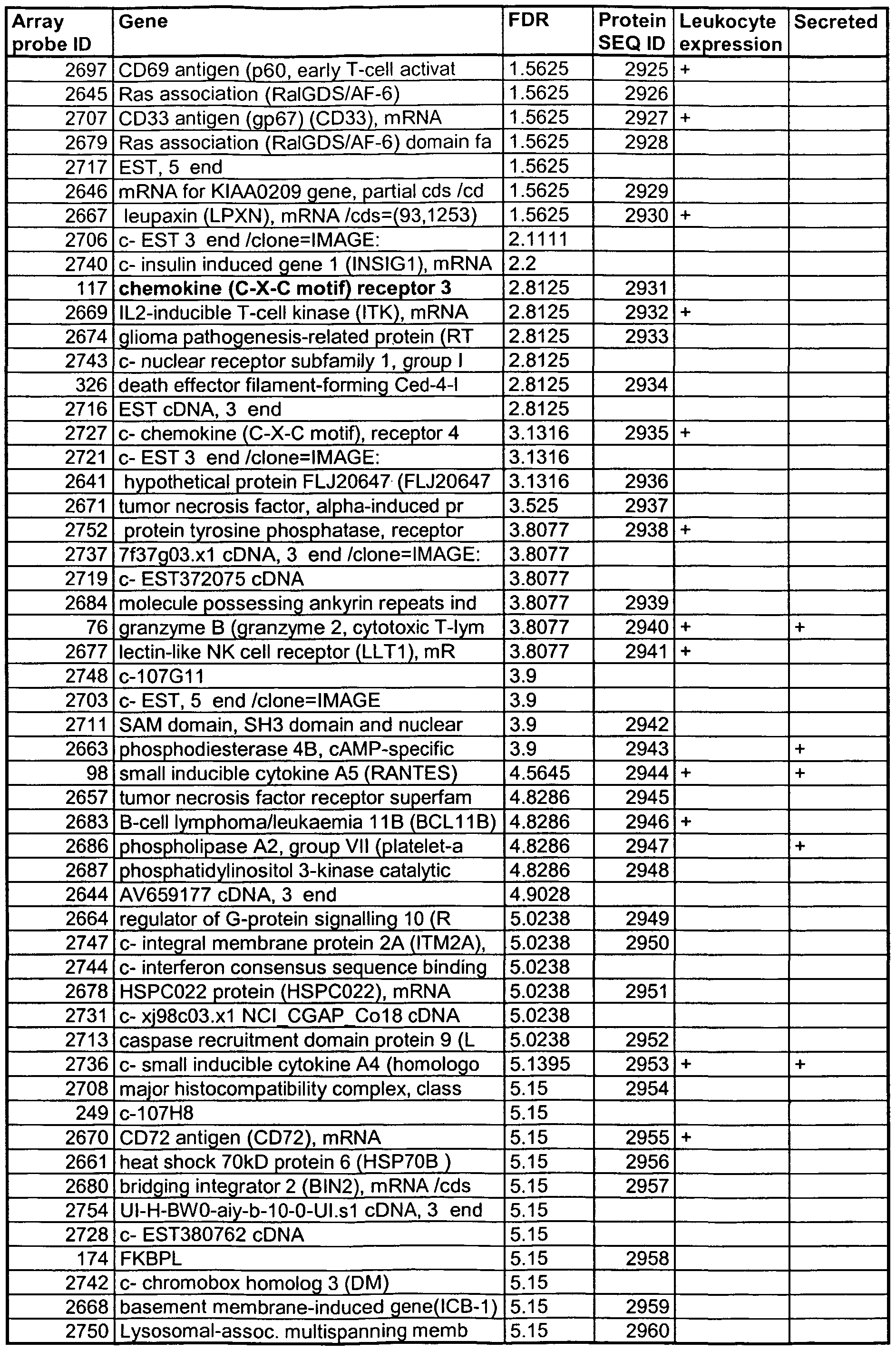


Claims
Priority Applications (11)
| Application Number | Priority Date | Filing Date | Title |
|---|---|---|---|
| IL16480403A IL164804A0 (en) | 2002-04-24 | 2003-04-24 | Methods and compositions for diagnosing and monitoring transplant rejection |
| AU2003299465A AU2003299465B2 (en) | 2002-04-24 | 2003-04-24 | Methods and compositions for diagnosing and monitoring transplant rejection |
| US10/511,937 US7691569B2 (en) | 2002-04-24 | 2003-04-24 | Methods and compositions for diagnosing and monitoring transplant rejection |
| CA2483481A CA2483481C (en) | 2002-04-24 | 2003-04-24 | Method for diagnosing and monitoring cardiac transplant rejection |
| EP03799755A EP1585972B1 (en) | 2002-04-24 | 2003-04-24 | Use of gene expression markers for assessing cardiac allograft rejection |
| JP2004549874A JP2005536230A (en) | 2002-04-24 | 2003-04-24 | Methods and compositions for diagnosing and monitoring transplant rejection |
| US12/544,182 US7785797B2 (en) | 2002-04-24 | 2009-08-19 | Methods and compositions for diagnosing and monitoring transplant rejection |
| US12/584,615 US7892745B2 (en) | 2003-04-24 | 2009-09-08 | Methods and compositions for diagnosing and monitoring transplant rejection |
| US12/855,178 US7879556B2 (en) | 2003-04-24 | 2010-08-12 | Methods and compositions for diagnosing and monitoring transplant rejection |
| US12/985,314 US20110104697A1 (en) | 2003-04-24 | 2011-01-05 | Methods and compositions for diagnosing and monitoring transplant rejection |
| US13/215,139 US20110306518A1 (en) | 2003-04-24 | 2011-08-22 | Methods and compositions for diagnosing and monitoring transplant rejection |
Applications Claiming Priority (4)
| Application Number | Priority Date | Filing Date | Title |
|---|---|---|---|
| US10/131,831 US7026121B1 (en) | 2001-06-08 | 2002-04-24 | Methods and compositions for diagnosing and monitoring transplant rejection |
| US10/131,831 | 2002-04-24 | ||
| US10/325,899 | 2002-12-20 | ||
| US10/325,899 US7235358B2 (en) | 2001-06-08 | 2002-12-20 | Methods and compositions for diagnosing and monitoring transplant rejection |
Related Parent Applications (1)
| Application Number | Title | Priority Date | Filing Date |
|---|---|---|---|
| US11/433,191 Continuation US20060263813A1 (en) | 2003-04-24 | 2006-05-11 | Methods of monitoring functional status of transplants using gene panels |
Related Child Applications (4)
| Application Number | Title | Priority Date | Filing Date |
|---|---|---|---|
| US10511937 A-371-Of-International | 2003-04-24 | ||
| US10/511,937 A-371-Of-International US7691569B2 (en) | 2002-04-24 | 2003-04-24 | Methods and compositions for diagnosing and monitoring transplant rejection |
| US12/544,182 Continuation US7785797B2 (en) | 2002-04-24 | 2009-08-19 | Methods and compositions for diagnosing and monitoring transplant rejection |
| US12/584,615 Continuation-In-Part US7892745B2 (en) | 2003-04-24 | 2009-09-08 | Methods and compositions for diagnosing and monitoring transplant rejection |
Publications (2)
| Publication Number | Publication Date |
|---|---|
| WO2004042346A2 true WO2004042346A2 (en) | 2004-05-21 |
| WO2004042346A3 WO2004042346A3 (en) | 2005-11-24 |
Family
ID=32314283
Family Applications (1)
| Application Number | Title | Priority Date | Filing Date |
|---|---|---|---|
| PCT/US2003/012946 WO2004042346A2 (en) | 2002-04-24 | 2003-04-24 | Methods and compositions for diagnosing and monitoring transplant rejection |
Country Status (7)
| Country | Link |
|---|---|
| US (3) | US7235358B2 (en) |
| EP (4) | EP2194145B1 (en) |
| JP (2) | JP2005536230A (en) |
| AU (1) | AU2003299465B2 (en) |
| CA (1) | CA2483481C (en) |
| IL (1) | IL164804A0 (en) |
| WO (1) | WO2004042346A2 (en) |
Cited By (91)
| Publication number | Priority date | Publication date | Assignee | Title |
|---|---|---|---|---|
| WO2006122791A2 (en) * | 2005-05-17 | 2006-11-23 | Genpat77 Pharmacogenetics Ag | Tirc7 as marker for detecting early immune activation |
| US7198912B2 (en) | 2001-09-07 | 2007-04-03 | Bristol-Myers Squibb Company | Polynucleotides encoding a human G-protein coupled receptor, HGPRBMY39 |
| WO2007124578A1 (en) * | 2006-04-27 | 2007-11-08 | Universite De Montreal | Assessment and reduction of risk of graft-versus-host disease |
| EP1885889A2 (en) * | 2005-05-11 | 2008-02-13 | Expression Diagnostics, Inc. | Methods of monitoring functional status of transplants using gene panels |
| JP2008507961A (en) * | 2004-07-29 | 2008-03-21 | ドムペ・ファ.ル.マ・ソチエタ・ペル・アツィオーニ | Tumor homing cells genetically engineered to produce tumor necrosis factor-related apoptosis-inducing ligand (TRAIL) |
| US7645575B2 (en) | 2004-09-08 | 2010-01-12 | Xdx, Inc. | Genes useful for diagnosing and monitoring inflammation related disorders |
| US7691569B2 (en) | 2002-04-24 | 2010-04-06 | Xdx, Inc. | Methods and compositions for diagnosing and monitoring transplant rejection |
| US7771950B2 (en) | 2002-04-24 | 2010-08-10 | Xdx, Inc. | Methods and compositions for diagnosing and monitoring auto immune and chronic inflammatory diseases |
| US7829286B2 (en) | 2001-06-08 | 2010-11-09 | Xdx, Inc. | Methods and compositions for diagnosing and monitoring transplant rejection |
| EP2260858A2 (en) | 2003-11-06 | 2010-12-15 | Seattle Genetics, Inc. | Monomethylvaline compounds capable of conjugation to ligands |
| WO2011000054A1 (en) | 2009-07-03 | 2011-01-06 | Avipep Pty Ltd | Immuno-conjugates and methods for producing them |
| US7879556B2 (en) | 2003-04-24 | 2011-02-01 | Xdx, Inc. | Methods and compositions for diagnosing and monitoring transplant rejection |
| WO2011017685A1 (en) | 2009-08-07 | 2011-02-10 | Rules-Based Medicine, Inc. | Methods and devices for detecting kidney transplant rejection |
| EP2286844A2 (en) | 2004-06-01 | 2011-02-23 | Genentech, Inc. | Antibody-drug conjugates and methods |
| WO2011031870A1 (en) | 2009-09-09 | 2011-03-17 | Centrose, Llc | Extracellular targeted drug conjugates |
| WO2011056983A1 (en) | 2009-11-05 | 2011-05-12 | Genentech, Inc. | Zirconium-radiolabeled, cysteine engineered antibody conjugates |
| WO2011075786A1 (en) | 2009-12-23 | 2011-06-30 | Avipep Pty Ltd | Immuno-conjugates and methods for producing them 2 |
| US7993832B2 (en) | 2006-08-14 | 2011-08-09 | Xdx, Inc. | Methods and compositions for diagnosing and monitoring the status of transplant rejection and immune disorders |
| WO2011130598A1 (en) | 2010-04-15 | 2011-10-20 | Spirogen Limited | Pyrrolobenzodiazepines and conjugates thereof |
| WO2011156328A1 (en) | 2010-06-08 | 2011-12-15 | Genentech, Inc. | Cysteine engineered antibodies and conjugates |
| US8148067B2 (en) | 2006-11-09 | 2012-04-03 | Xdx, Inc. | Methods for diagnosing and monitoring the status of systemic lupus erythematosus |
| WO2012074757A1 (en) | 2010-11-17 | 2012-06-07 | Genentech, Inc. | Alaninyl maytansinol antibody conjugates |
| US20120167240A1 (en) * | 2004-09-03 | 2012-06-28 | Dana-Farber Cancer Institute, Inc. | Compositions and Methods for Modulating PGC-1Alpha to Treat Neurological Diseases and Disorders |
| WO2012142662A1 (en) | 2011-04-21 | 2012-10-26 | Garvan Institute Of Medical Research | Modified variable domain molecules and methods for producing and using them b |
| WO2012155019A1 (en) | 2011-05-12 | 2012-11-15 | Genentech, Inc. | Multiple reaction monitoring lc-ms/ms method to detect therapeutic antibodies in animal samples using framework signature pepides |
| WO2013130093A1 (en) | 2012-03-02 | 2013-09-06 | Genentech, Inc. | Biomarkers for treatment with anti-tubulin chemotherapeutic compounds |
| WO2013177422A1 (en) * | 2012-05-24 | 2013-11-28 | Allegheny Singer Research Institute | System for ordering and arranging a data set |
| WO2014057074A1 (en) | 2012-10-12 | 2014-04-17 | Spirogen Sàrl | Pyrrolobenzodiazepines and conjugates thereof |
| WO2014140862A2 (en) | 2013-03-13 | 2014-09-18 | Spirogen Sarl | Pyrrolobenzodiazepines and conjugates thereof |
| WO2014140174A1 (en) | 2013-03-13 | 2014-09-18 | Spirogen Sàrl | Pyrrolobenzodiazepines and conjugates thereof |
| WO2014159981A2 (en) | 2013-03-13 | 2014-10-02 | Spirogen Sarl | Pyrrolobenzodiazepines and conjugates thereof |
| US8871515B2 (en) | 2008-09-17 | 2014-10-28 | Isogenis, Inc. | Construction of fully-deleted adenovirus-based gene delivery vectors and uses thereof |
| WO2015023355A1 (en) | 2013-08-12 | 2015-02-19 | Genentech, Inc. | 1-(chloromethyl)-2,3-dihydro-1h-benzo[e]indole dimer antibody-drug conjugate compounds, and methods of use and treatment |
| WO2015095212A1 (en) | 2013-12-16 | 2015-06-25 | Genentech, Inc. | 1-(chloromethyl)-2,3-dihydro-1h-benzo[e]indole dimer antibody-drug conjugate compounds, and methods of use and treatment |
| WO2015095223A2 (en) | 2013-12-16 | 2015-06-25 | Genentech, Inc. | Peptidomimetic compounds and antibody-drug conjugates thereof |
| WO2015095227A2 (en) | 2013-12-16 | 2015-06-25 | Genentech, Inc. | Peptidomimetic compounds and antibody-drug conjugates thereof |
| WO2016040825A1 (en) | 2014-09-12 | 2016-03-17 | Genentech, Inc. | Anthracycline disulfide intermediates, antibody-drug conjugates and methods |
| WO2016040856A2 (en) | 2014-09-12 | 2016-03-17 | Genentech, Inc. | Cysteine engineered antibodies and conjugates |
| WO2016037644A1 (en) | 2014-09-10 | 2016-03-17 | Medimmune Limited | Pyrrolobenzodiazepines and conjugates thereof |
| WO2016090050A1 (en) | 2014-12-03 | 2016-06-09 | Genentech, Inc. | Quaternary amine compounds and antibody-drug conjugates thereof |
| EP3088004A1 (en) | 2004-09-23 | 2016-11-02 | Genentech, Inc. | Cysteine engineered antibodies and conjugates |
| US20160340401A1 (en) * | 2006-06-09 | 2016-11-24 | Almac Discovery Limited | Fkbp-l and uses thereof |
| WO2017059289A1 (en) | 2015-10-02 | 2017-04-06 | Genentech, Inc. | Pyrrolobenzodiazepine antibody drug conjugates and methods of use |
| WO2017064675A1 (en) | 2015-10-16 | 2017-04-20 | Genentech, Inc. | Hindered disulfide drug conjugates |
| WO2017068511A1 (en) | 2015-10-20 | 2017-04-27 | Genentech, Inc. | Calicheamicin-antibody-drug conjugates and methods of use |
| WO2017165734A1 (en) | 2016-03-25 | 2017-09-28 | Genentech, Inc. | Multiplexed total antibody and antibody-conjugated drug quantification assay |
| EP3235820A1 (en) | 2014-09-17 | 2017-10-25 | Genentech, Inc. | Pyrrolobenzodiazepines and antibody disulfide conjugates thereof |
| WO2017201449A1 (en) | 2016-05-20 | 2017-11-23 | Genentech, Inc. | Protac antibody conjugates and methods of use |
| WO2017205741A1 (en) | 2016-05-27 | 2017-11-30 | Genentech, Inc. | Bioanalytical method for the characterization of site-specific antibody-drug conjugates |
| WO2017214024A1 (en) | 2016-06-06 | 2017-12-14 | Genentech, Inc. | Silvestrol antibody-drug conjugates and methods of use |
| WO2018031662A1 (en) | 2016-08-11 | 2018-02-15 | Genentech, Inc. | Pyrrolobenzodiazepine prodrugs and antibody conjugates thereof |
| US9919056B2 (en) | 2012-10-12 | 2018-03-20 | Adc Therapeutics S.A. | Pyrrolobenzodiazepine-anti-CD22 antibody conjugates |
| US9931415B2 (en) | 2012-10-12 | 2018-04-03 | Medimmune Limited | Pyrrolobenzodiazepine-antibody conjugates |
| US9931414B2 (en) | 2012-10-12 | 2018-04-03 | Medimmune Limited | Pyrrolobenzodiazepine-antibody conjugates |
| WO2018065501A1 (en) | 2016-10-05 | 2018-04-12 | F. Hoffmann-La Roche Ag | Methods for preparing antibody drug conjugates |
| US9950078B2 (en) | 2013-10-11 | 2018-04-24 | Medimmune Limited | Pyrrolobenzodiazepine-antibody conjugates |
| US9956299B2 (en) | 2013-10-11 | 2018-05-01 | Medimmune Limited | Pyrrolobenzodiazepine—antibody conjugates |
| US10010624B2 (en) | 2013-10-11 | 2018-07-03 | Medimmune Limited | Pyrrolobenzodiazepine-antibody conjugates |
| US10029018B2 (en) | 2013-10-11 | 2018-07-24 | Medimmune Limited | Pyrrolobenzodiazepines and conjugates thereof |
| WO2019060398A1 (en) | 2017-09-20 | 2019-03-28 | Ph Pharma Co., Ltd. | Thailanstatin analogs |
| US10392393B2 (en) | 2016-01-26 | 2019-08-27 | Medimmune Limited | Pyrrolobenzodiazepines |
| US10420777B2 (en) | 2014-09-12 | 2019-09-24 | Medimmune Limited | Pyrrolobenzodiazepines and conjugates thereof |
| US10451636B2 (en) | 2014-04-09 | 2019-10-22 | The Regents Of The University Of California | Protein biomarkers for immune assessment and prediction of transplant rejection |
| WO2020002602A1 (en) * | 2018-06-29 | 2020-01-02 | bioMérieux | Method for determining in vitro or ex vivo the immune status of an individual |
| US10544223B2 (en) | 2017-04-20 | 2020-01-28 | Adc Therapeutics Sa | Combination therapy with an anti-axl antibody-drug conjugate |
| US10543279B2 (en) | 2016-04-29 | 2020-01-28 | Medimmune Limited | Pyrrolobenzodiazepine conjugates and their use for the treatment of cancer |
| WO2020049286A1 (en) | 2018-09-03 | 2020-03-12 | Femtogenix Limited | Polycyclic amides as cytotoxic agents |
| FR3085689A1 (en) * | 2018-09-12 | 2020-03-13 | Biomerieux | PROCESS FOR DETERMINING IN VITRO OR EX VIVO THE IMMUNE STATUS OF AN INDIVIDUAL |
| WO2020086858A1 (en) | 2018-10-24 | 2020-04-30 | Genentech, Inc. | Conjugated chemical inducers of degradation and methods of use |
| WO2020123275A1 (en) | 2018-12-10 | 2020-06-18 | Genentech, Inc. | Photocrosslinking peptides for site specific conjugation to fc-containing proteins |
| US10695439B2 (en) | 2016-02-10 | 2020-06-30 | Medimmune Limited | Pyrrolobenzodiazepine conjugates |
| US10695433B2 (en) | 2012-10-12 | 2020-06-30 | Medimmune Limited | Pyrrolobenzodiazepine-antibody conjugates |
| WO2020157491A1 (en) | 2019-01-29 | 2020-08-06 | Femtogenix Limited | G-a crosslinking cytotoxic agents |
| US10736903B2 (en) | 2012-10-12 | 2020-08-11 | Medimmune Limited | Pyrrolobenzodiazepine-anti-PSMA antibody conjugates |
| US10751346B2 (en) | 2012-10-12 | 2020-08-25 | Medimmune Limited | Pyrrolobenzodiazepine—anti-PSMA antibody conjugates |
| US10780096B2 (en) | 2014-11-25 | 2020-09-22 | Adc Therapeutics Sa | Pyrrolobenzodiazepine-antibody conjugates |
| US10799595B2 (en) | 2016-10-14 | 2020-10-13 | Medimmune Limited | Pyrrolobenzodiazepine conjugates |
| WO2021076427A1 (en) * | 2019-10-15 | 2021-04-22 | The Regents Of The University Of California | TRANSPLANTED CELL PROTECTION VIA Fc SEQUESTRATION |
| US11059893B2 (en) | 2015-04-15 | 2021-07-13 | Bergenbio Asa | Humanized anti-AXL antibodies |
| US11135303B2 (en) | 2011-10-14 | 2021-10-05 | Medimmune Limited | Pyrrolobenzodiazepines and conjugates thereof |
| US11162079B2 (en) | 2019-05-10 | 2021-11-02 | The Regents Of The University Of California | Blood type O Rh-hypo-immunogenic pluripotent cells |
| US11160872B2 (en) | 2017-02-08 | 2021-11-02 | Adc Therapeutics Sa | Pyrrolobenzodiazepine-antibody conjugates |
| WO2022023735A1 (en) | 2020-07-28 | 2022-02-03 | Femtogenix Limited | Cytotoxic agents |
| US11318211B2 (en) | 2017-06-14 | 2022-05-03 | Adc Therapeutics Sa | Dosage regimes for the administration of an anti-CD19 ADC |
| US11352324B2 (en) | 2018-03-01 | 2022-06-07 | Medimmune Limited | Methods |
| US11370801B2 (en) | 2017-04-18 | 2022-06-28 | Medimmune Limited | Pyrrolobenzodiazepine conjugates |
| US11517626B2 (en) | 2016-02-10 | 2022-12-06 | Medimmune Limited | Pyrrolobenzodiazepine antibody conjugates |
| US11524969B2 (en) | 2018-04-12 | 2022-12-13 | Medimmune Limited | Pyrrolobenzodiazepines and conjugates thereof as antitumour agents |
| US11612665B2 (en) | 2017-02-08 | 2023-03-28 | Medimmune Limited | Pyrrolobenzodiazepine-antibody conjugates |
| US11649250B2 (en) | 2017-08-18 | 2023-05-16 | Medimmune Limited | Pyrrolobenzodiazepine conjugates |
| US11702473B2 (en) | 2015-04-15 | 2023-07-18 | Medimmune Limited | Site-specific antibody-drug conjugates |
Families Citing this family (129)
| Publication number | Priority date | Publication date | Assignee | Title |
|---|---|---|---|---|
| US7405292B2 (en) * | 2002-02-19 | 2008-07-29 | The Children's Hospital Of Philadelphia | Cellular genes regulated by HIV-1 infection and methods of use thereof |
| FR2836573A1 (en) * | 2002-02-27 | 2003-08-29 | France Telecom | Computer representation of a data tree structure, which is representative of the organization of a data set or data dictionary, has first and second total order relations representative of tree nodes and stored data items |
| AT412678B (en) * | 2002-09-30 | 2005-05-25 | Gerhard Dr Kranner | METHOD FOR COMPUTER-ASSISTED PREPARATION OF PROGNOSES FOR OPERATIONAL SYSTEMS AND SYSTEM FOR CREATING PROGNOSES FOR OPERATIONAL SYSTEMS |
| US20060057147A1 (en) * | 2002-10-04 | 2006-03-16 | Mitsubishi Pharma Corporation | Antibody recognizing antibody |
| US20070248978A1 (en) * | 2006-04-07 | 2007-10-25 | Expression Diagnostics, Inc. | Steroid responsive nucleic acid expression and prediction of disease activity |
| JP4316617B2 (en) * | 2003-12-03 | 2009-08-19 | ノバルティス アクチエンゲゼルシャフト | Biomarkers of graft rejection |
| US8932808B1 (en) | 2004-01-21 | 2015-01-13 | The Board Of Trustees Of The Leland Stanford Junior University | Methods and compositions for determining a graft tolerant phenotype in a subject |
| EP2026071B1 (en) * | 2004-02-19 | 2013-07-31 | Yale University | Identification of cancer protein biomarkers using proteomic techniques |
| WO2006012416A2 (en) * | 2004-07-20 | 2006-02-02 | Isogenis, Inc. | Specific inhibition of autoimmunity and diseases associated with autoantigens |
| ITFI20040243A1 (en) * | 2004-11-25 | 2005-02-25 | Paola Romagnani | DIAGNOSTIC METHOD FOR REJECTION OF TRANSPLANTED ORGAN |
| US20060194237A1 (en) * | 2005-02-28 | 2006-08-31 | Norman Latov | Method for diagnosing peripheral neuropathy |
| US20060194238A1 (en) * | 2005-02-28 | 2006-08-31 | Norman Latov | Peripheral neuropathy diagnosis |
| JP2008537875A (en) | 2005-03-14 | 2008-10-02 | ザ ボード オブ トラスティーズ オブ ザ リーランド スタンフォード ジュニア ユニバーシティ | Methods and compositions for assessing graft survival in solid organ transplant recipients |
| USRE46843E1 (en) | 2005-03-14 | 2018-05-15 | The Board Of Trustees Of The Leland Stanford Junior University | Methods and compositions for evaluating graft survival in a solid organ transplant recipient |
| US20060269948A1 (en) * | 2005-05-16 | 2006-11-30 | Halloran Philip F | Tissue rejection |
| PE20071227A1 (en) * | 2006-04-20 | 2008-02-18 | Glaxo Group Ltd | INDAZOLE-DERIVED COMPOUNDS AS GLUCOCORTICOID RECEPTOR MODULATORS |
| ES2503621T3 (en) * | 2006-04-24 | 2014-10-07 | Genentech, Inc. | Methods to detect autoimmune disorders |
| US20070254305A1 (en) * | 2006-04-28 | 2007-11-01 | Nsabp Foundation, Inc. | Methods of whole genome or microarray expression profiling using nucleic acids prepared from formalin fixed paraffin embedded tissue |
| EP1872787A1 (en) * | 2006-06-27 | 2008-01-02 | Gentium S.p.A. | Use of defibrotide for the inhibition of heparanase |
| EP1947463A1 (en) * | 2007-01-16 | 2008-07-23 | Roche Diagnostics GmbH | Collection of liquid analytical samples for clinical analytical purpose |
| WO2008137500A2 (en) * | 2007-05-01 | 2008-11-13 | The Brigham And Women's Hospital | Immunosuppression with antibody against itm2a |
| US8600135B2 (en) * | 2007-06-28 | 2013-12-03 | Mayo Foundation For Medical Education And Research | System and method for automatically generating sample points from a series of medical images and identifying a significant region |
| WO2009003198A1 (en) * | 2007-06-28 | 2008-12-31 | Mayo Foundation For Medical Education And Research | System and method for automatically detecting change in a series of medical images of a subject over time |
| EP2190379B1 (en) | 2007-08-23 | 2016-06-15 | Direct Flow Medical, Inc. | Translumenally implantable heart valve with formed in place support |
| KR20200091961A (en) * | 2008-01-18 | 2020-07-31 | 프레지던트 앤드 펠로우즈 오브 하바드 칼리지 | Methods of detecting signatures of disease or conditions in bodily fluids |
| US8215329B2 (en) * | 2008-02-01 | 2012-07-10 | Woodward, Inc. | Digital closed loop proportional hydraulic pressure controller |
| CN101960021A (en) * | 2008-02-28 | 2011-01-26 | 希森美康株式会社 | Marker for detection of IL-17-producing helper T-cell, and method for detection of IL-17-producing helper T-cell |
| WO2009114085A2 (en) | 2008-03-03 | 2009-09-17 | The University Of Miami | Allogeneic cancer cell-based immunotherapy |
| EP2103689A1 (en) * | 2008-03-19 | 2009-09-23 | Gentium S.p.A. | Synthetic phosphodiester oligonucleotides and therapeutical uses thereof |
| EP2283153A4 (en) * | 2008-04-09 | 2011-09-14 | Univ British Columbia | Methods of diagnosing acute cardiac allograft rejection |
| EP2285961A4 (en) * | 2008-05-15 | 2012-10-17 | Topigen Pharmaceuticals Inc | Oligonucleotides for treating inflammation and neoplastic cell proliferation |
| WO2010000320A1 (en) * | 2008-07-03 | 2010-01-07 | Institut D'investigations Biomediques August Pi I Sunyer (Idibaps) | In vitro diagnosis/prognosis method and kit for assessment of tolerance in liver transplantation |
| WO2010010951A1 (en) * | 2008-07-24 | 2010-01-28 | Kuroda Masahiko | Method for detection of expression of indicator mrna acting as indicator for endometriosis-related diseases, and probe for use in the method |
| US8285719B1 (en) | 2008-08-08 | 2012-10-09 | The Research Foundation Of State University Of New York | System and method for probabilistic relational clustering |
| EP2324129A4 (en) | 2008-08-18 | 2012-06-20 | Univ Leland Stanford Junior | Methods and compositions for determining a graft tolerant phenotype in a subject |
| EP2324355B1 (en) * | 2008-08-28 | 2014-01-22 | Astute Medical, Inc. | Methods and compositions for diagnosis and prognosis of renal injury and renal failure |
| EP2324354B1 (en) * | 2008-08-29 | 2014-07-16 | Astute Medical, Inc. | Methods for prognosis of acute renal failure |
| WO2010048347A2 (en) * | 2008-10-21 | 2010-04-29 | Astute Medical, Inc. | Methods and compositions for diagnosis and prognosis of renal injury and renal failure |
| EP3974836A1 (en) | 2008-10-21 | 2022-03-30 | Astute Medical, Inc. | Methods and compositions for diagnosis and prognosis of renal injury and renal failure |
| EP2913676A1 (en) | 2008-11-10 | 2015-09-02 | Astute Medical, Inc. | Methods and compositions for diagnosis and prognosis of renal injury and renal failure |
| CA2743253A1 (en) * | 2008-11-22 | 2010-05-27 | Astute Medical, Inc. | Methods and compositions for diagnosis and prognosis of renal injury and renal failure |
| US9938579B2 (en) * | 2009-01-15 | 2018-04-10 | The Board Of Trustees Of The Leland Stanford Junior University | Biomarker panel for diagnosis and prediction of graft rejection |
| US9229010B2 (en) | 2009-02-06 | 2016-01-05 | Astute Medical, Inc. | Methods and compositions for diagnosis and prognosis of renal injury and renal failure |
| TWI387750B (en) * | 2009-02-10 | 2013-03-01 | Nat Defense Medical Ct | Biomarker and detection method for detecting kidney disease |
| US20110045468A1 (en) * | 2009-03-02 | 2011-02-24 | Richard Robison | Polynucleotides for the identification and quantification of group a streptococcus nucleic acids |
| US9868988B2 (en) | 2009-03-13 | 2018-01-16 | Cornell University | Method to assess human allograft status from microrna expression levels |
| BRPI1010693A2 (en) | 2009-06-15 | 2019-07-16 | Cardiodx Inc | computer-implemented methods for marking and / or scoring a first sample obtained from a subject, computer readable storage means for storing executable program code, and cad prediction methods, system and kits on a subject |
| WO2011006119A2 (en) * | 2009-07-09 | 2011-01-13 | The Scripps Research Institute | Gene expression profiles associated with chronic allograft nephropathy |
| ES2356217B1 (en) * | 2009-08-04 | 2012-02-13 | Universidad De Salamanca | METHOD FOR DETECTION OF RENAL DAMAGE. |
| EA201290056A1 (en) | 2009-08-07 | 2012-08-30 | Астьют Медикал, Инк. | METHODS AND COMPOSITIONS FOR DIAGNOSIS AND PREDICTION OF KIDNEY DAMAGE AND RENAL FAILURE |
| US9238831B2 (en) * | 2009-09-02 | 2016-01-19 | Accugen Pty Ltd | Nucleic acid quantitation method |
| CA2779750C (en) * | 2009-11-06 | 2019-03-19 | The Board Of Trustees Of The Leland Stanford Junior University | Non-invasive diagnosis of graft rejection in organ transplant patients |
| WO2011057147A1 (en) | 2009-11-07 | 2011-05-12 | Astute Medical, Inc. | Methods and compositions for diagnosis and prognosis of renal injury and renal failure |
| US20110125791A1 (en) * | 2009-11-25 | 2011-05-26 | Microsoft Corporation | Query classification using search result tag ratios |
| EP3185013B1 (en) | 2009-12-02 | 2019-10-09 | The Board of Trustees of the Leland Stanford Junior University | Biomarkers for determining an allograft tolerant phenotype |
| ES2592386T3 (en) | 2009-12-20 | 2016-11-29 | Astute Medical, Inc. | Methods and compositions for the diagnosis and prognosis of renal injury and renal insufficiency |
| WO2011097540A1 (en) * | 2010-02-05 | 2011-08-11 | Astute Medical, Inc. | Methods and compositions for diagnosis and prognosis of renal injury and renal failure |
| CN105158481A (en) * | 2010-02-05 | 2015-12-16 | 阿斯图特医药公司 | Methods and compositions for diagnosis and prognosis of renal injury and renal failure |
| WO2011106746A1 (en) | 2010-02-26 | 2011-09-01 | Astute Medical, Inc. | Methods and compositions for diagnosis and prognosis of renal injury and renal failure |
| AU2011269847A1 (en) | 2010-02-26 | 2013-01-31 | Astute Medical, Inc. | Methods and compositions for diagnosis and prognosis of renal injury and renal failure |
| WO2011112719A1 (en) * | 2010-03-09 | 2011-09-15 | Cornell University | Methods and compositions to predict and detect acute rejection |
| CA2794255C (en) | 2010-03-25 | 2020-01-14 | Minnie M. Sarwal | Protein and gene biomarkers for rejection of organ transplants |
| US9603708B2 (en) | 2010-05-19 | 2017-03-28 | Dfm, Llc | Low crossing profile delivery catheter for cardiovascular prosthetic implant |
| WO2011158667A1 (en) * | 2010-06-16 | 2011-12-22 | 国立大学法人浜松医科大学 | Method for detection of colorectal tumor |
| AU2011269775B2 (en) | 2010-06-23 | 2015-01-15 | Astute Medical, Inc. | Methods and compositions for diagnosis and prognosis of renal injury and renal failure |
| US20130203624A1 (en) | 2010-07-23 | 2013-08-08 | President And Fellows Of Harvard College | Methods of Detecting Prenatal or Pregnancy-Related Diseases or Conditions |
| KR20130041962A (en) * | 2010-07-23 | 2013-04-25 | 프레지던트 앤드 펠로우즈 오브 하바드 칼리지 | Methods of detecting diseases or conditions using phagocytic cells |
| US20130184178A1 (en) | 2010-07-23 | 2013-07-18 | President And Fellows Of Harvard College | Methods of Detecting Autoimmune or Immune-Related Diseases or Conditions |
| EP2437192A1 (en) * | 2010-09-16 | 2012-04-04 | Roche Diagnostics GmbH | Relative quantification analysis of multi-parametric PCR experiments |
| CN103260627A (en) | 2010-11-12 | 2013-08-21 | 真蒂奥姆有限公司 | Defibrotide for use in prophylaxis and/or treatment of graft versus host disease (gvhd). |
| US9487838B2 (en) | 2010-12-28 | 2016-11-08 | Qiagen Hamburg Gmbh | Oligonucleotide probe for the detection of adenovirus |
| AU2012236099A1 (en) | 2011-03-31 | 2013-10-03 | Moderna Therapeutics, Inc. | Delivery and formulation of engineered nucleic acids |
| WO2012139051A2 (en) | 2011-04-06 | 2012-10-11 | The Board Of Trustees Of The Leland Stanford Junior University | Autoantibody biomarkers for iga nephropathy |
| ES2734494T3 (en) | 2011-12-08 | 2019-12-10 | Astute Medical Inc | Methods and uses for the diagnosis of kidney injury and kidney failure |
| AU2013243948A1 (en) | 2012-04-02 | 2014-10-30 | Moderna Therapeutics, Inc. | Modified polynucleotides for the production of proteins associated with human disease |
| US9283287B2 (en) | 2012-04-02 | 2016-03-15 | Moderna Therapeutics, Inc. | Modified polynucleotides for the production of nuclear proteins |
| US9445897B2 (en) | 2012-05-01 | 2016-09-20 | Direct Flow Medical, Inc. | Prosthetic implant delivery device with introducer catheter |
| IN2014DN10584A (en) | 2012-06-22 | 2015-08-28 | Gentium Spa | |
| EP2893343B1 (en) | 2012-09-07 | 2019-11-20 | The Governors of the University of Alberta | Methods for diagnosis of inflammatory liver disease |
| RU2506592C1 (en) * | 2012-10-11 | 2014-02-10 | Государственное бюджетное учреждение здравоохранения Московской области "Московский областной научно-исследовательский клинический институт им. М.Ф. Владимирского" (ГБУЗ МО МОНИКИ им. М.Ф. Владимирского) | Method for choosing surgical approach to patients with peri-implantation inflammation of greater joints |
| WO2014096368A1 (en) | 2012-12-21 | 2014-06-26 | Spirogen Sàrl | Pyrrolobenzodiazepines and conjugates thereof |
| CA2894959C (en) | 2012-12-21 | 2022-01-11 | Spirogen Sarl | Unsymmetrical pyrrolobenzodiazepines-dimers for use in the treatment of proliferative and autoimmune diseases |
| WO2014113558A1 (en) | 2013-01-17 | 2014-07-24 | Astute Medical, Inc. | Methods and compositions for diagnosis and prognosis of renal injury and renal failure |
| US9758828B2 (en) | 2013-01-31 | 2017-09-12 | Cornell University | Methods to detect, treat and prevent acute cellular rejection in kidney allografts |
| US10210312B2 (en) * | 2013-02-03 | 2019-02-19 | Youscript Inc. | Systems and methods for quantification and presentation of medical risk arising from unknown factors |
| NZ771629A (en) | 2013-03-09 | 2022-12-23 | Harry Stylli | Methods of detecting cancer |
| WO2014152027A1 (en) | 2013-03-15 | 2014-09-25 | Moderna Therapeutics, Inc. | Manufacturing methods for production of rna transcripts |
| US20160040236A1 (en) * | 2013-03-15 | 2016-02-11 | Biogen Ma Inc. | Systems and methods for characterization of multiple sclerosis |
| WO2014144767A1 (en) | 2013-03-15 | 2014-09-18 | Moderna Therapeutics, Inc. | Ion exchange purification of mrna |
| EP2971161B1 (en) | 2013-03-15 | 2018-12-26 | ModernaTX, Inc. | Ribonucleic acid purification |
| EP2971165A4 (en) | 2013-03-15 | 2016-11-23 | Moderna Therapeutics Inc | Removal of dna fragments in mrna production process |
| JP6521956B2 (en) * | 2013-06-17 | 2019-05-29 | ベリナタ ヘルス インコーポレイテッド | Method for determining copy number mutations in sexual chromosomes |
| HUE056760T2 (en) | 2013-07-11 | 2022-03-28 | Modernatx Inc | Compositions comprising synthetic polynucleotides encoding crispr related proteins and synthetic sgrnas and methods of use |
| WO2015051169A2 (en) | 2013-10-02 | 2015-04-09 | Moderna Therapeutics, Inc. | Polynucleotide molecules and uses thereof |
| CN110570906A (en) * | 2013-11-13 | 2019-12-13 | 凡弗3基因组有限公司 | System and method for transmitting and preprocessing sequencing data |
| US9982301B2 (en) | 2014-01-07 | 2018-05-29 | Cornell University | Urine mRNA profile and acute dysfunction of kidney allograft |
| US10394828B1 (en) * | 2014-04-25 | 2019-08-27 | Emory University | Methods, systems and computer readable storage media for generating quantifiable genomic information and results |
| US10443100B2 (en) | 2014-05-22 | 2019-10-15 | The Scripps Research Institute | Gene expression profiles associated with sub-clinical kidney transplant rejection |
| US11104951B2 (en) | 2014-05-22 | 2021-08-31 | The Scripps Research Institute | Molecular signatures for distinguishing liver transplant rejections or injuries |
| EP3157573A4 (en) | 2014-06-19 | 2018-02-21 | Moderna Therapeutics, Inc. | Alternative nucleic acid molecules and uses thereof |
| AU2015289656A1 (en) | 2014-07-16 | 2017-02-16 | Modernatx, Inc. | Circular polynucleotides |
| EP3174985A4 (en) * | 2014-07-31 | 2018-04-04 | Academia Sinica | An antagonistic pd-1 aptamer and its applications in cancer therapy related applications |
| WO2016025733A1 (en) | 2014-08-15 | 2016-02-18 | Direct Flow Medical, Inc. | Prosthetic implant delivery device |
| US10417225B2 (en) * | 2015-09-18 | 2019-09-17 | Splunk Inc. | Entity detail monitoring console |
| EP3026122A1 (en) | 2014-11-27 | 2016-06-01 | Gentium S.p.A. | Cellular-based method for determining the potency of defibrotide |
| WO2016118852A1 (en) * | 2015-01-23 | 2016-07-28 | University Of Pittsburgh-Of The Commonwealth System Of Higher Education | Use of eomesodermin to determine risk of allograft rejection |
| CA2984589A1 (en) | 2015-05-01 | 2016-11-10 | The University Of British Columbia | Biomarkers for the detection of acute rejection in heart transplantation |
| WO2017049286A1 (en) | 2015-09-17 | 2017-03-23 | Moderna Therapeutics, Inc. | Polynucleotides containing a morpholino linker |
| CA3023491A1 (en) * | 2016-06-02 | 2017-12-07 | Riken | Adjuvant composition and use thereof |
| JP2019523889A (en) | 2016-06-06 | 2019-08-29 | アスチュート メディカル,インコーポレイテッド | Management of acute kidney injury using tissue inhibitors of insulin-like growth factor binding protein 7 and metalloprotease 2 |
| US20180166170A1 (en) * | 2016-12-12 | 2018-06-14 | Konstantinos Theofilatos | Generalized computational framework and system for integrative prediction of biomarkers |
| JP6842195B2 (en) | 2016-12-19 | 2021-03-17 | 国立大学法人大阪大学 | Early diagnosis of antibody-related rejection after organ transplantation using an IgM memory B cell differentiation culture system in vitro |
| CA3054733A1 (en) | 2017-02-28 | 2018-09-07 | Sanofi | Therapeutic rna |
| US11106442B1 (en) | 2017-09-23 | 2021-08-31 | Splunk Inc. | Information technology networked entity monitoring with metric selection prior to deployment |
| US11093518B1 (en) | 2017-09-23 | 2021-08-17 | Splunk Inc. | Information technology networked entity monitoring with dynamic metric and threshold selection |
| US11159397B2 (en) | 2017-09-25 | 2021-10-26 | Splunk Inc. | Lower-tier application deployment for higher-tier system data monitoring |
| CN107967938A (en) * | 2017-11-21 | 2018-04-27 | 成都天奥电子股份有限公司 | A kind of cardiac positions monitoring administration system |
| WO2019107673A1 (en) * | 2017-11-29 | 2019-06-06 | 서울대학교병원 | Bio-marker for monitoring antibody-mediated rejection in abo blood type-incompatible transplantation |
| US11099975B2 (en) | 2019-01-24 | 2021-08-24 | International Business Machines Corporation | Test space analysis across multiple combinatoric models |
| US11263116B2 (en) | 2019-01-24 | 2022-03-01 | International Business Machines Corporation | Champion test case generation |
| US11106567B2 (en) | 2019-01-24 | 2021-08-31 | International Business Machines Corporation | Combinatoric set completion through unique test case generation |
| EP3736570A1 (en) * | 2019-05-09 | 2020-11-11 | Fundación Instituto de Estudios de Ciencias de la Salud de Castilla y León | Method for the diagnosis, sub-classification and prognosis of acute kidney injury by detecting cct7 |
| US11422924B2 (en) | 2019-06-13 | 2022-08-23 | International Business Machines Corporation | Customizable test set selection using code flow trees |
| US10970195B2 (en) * | 2019-06-13 | 2021-04-06 | International Business Machines Corporation | Reduction of test infrastructure |
| US11232020B2 (en) | 2019-06-13 | 2022-01-25 | International Business Machines Corporation | Fault detection using breakpoint value-based fingerprints of failing regression test cases |
| CN110923356B (en) * | 2019-12-30 | 2022-08-12 | 广西大学 | Molecular marker primer of rice gall midge-resistant major gene Gm5, and marking method and application thereof |
| DE102020205364B4 (en) * | 2020-04-28 | 2022-01-20 | Universität Rostock | Method of monitoring the progress of cardiomyocyte transplantation and method of determining whether a subject is eligible for cardiomyocyte transplantation |
| US11676072B1 (en) | 2021-01-29 | 2023-06-13 | Splunk Inc. | Interface for incorporating user feedback into training of clustering model |
| CN113435168B (en) * | 2021-06-10 | 2024-03-22 | 上海美吉生物医药科技有限公司 | Automatic editing method, system, terminal and medium for glue pattern |
Family Cites Families (184)
| Publication number | Priority date | Publication date | Assignee | Title |
|---|---|---|---|---|
| US5981481A (en) | 1974-12-06 | 1999-11-09 | The Johns Hopkins University | Human C3b/C4b receptor (CR1) |
| US5212071A (en) | 1988-04-01 | 1993-05-18 | The Johns Hopkins University | Nucleic acids encoding a human C3b/C4b receptor (CR1) |
| US4350593A (en) | 1977-12-19 | 1982-09-21 | Becton, Dickinson And Company | Assembly, compositions and method for separating blood |
| US4190535A (en) | 1978-02-27 | 1980-02-26 | Corning Glass Works | Means for separating lymphocytes and monocytes from anticoagulated blood |
| US4215051A (en) * | 1979-08-29 | 1980-07-29 | Standard Oil Company (Indiana) | Formation, purification and recovery of phthalic anhydride |
| US4376110A (en) | 1980-08-04 | 1983-03-08 | Hybritech, Incorporated | Immunometric assays using monoclonal antibodies |
| US4358535A (en) | 1980-12-08 | 1982-11-09 | Board Of Regents Of The University Of Washington | Specific DNA probes in diagnostic microbiology |
| US4762780A (en) | 1984-04-17 | 1988-08-09 | The Regents Of The University Of California | Method and composition for screening and diagnosing "HCMV" |
| US4582789A (en) | 1984-03-21 | 1986-04-15 | Cetus Corporation | Process for labeling nucleic acids using psoralen derivatives |
| US4818418A (en) | 1984-09-24 | 1989-04-04 | Becton Dickinson And Company | Blood partitioning method |
| US4751001A (en) | 1984-09-24 | 1988-06-14 | Becton Dickinson And Company | Blood partitioning apparatus |
| US5264351A (en) | 1984-11-05 | 1993-11-23 | The Board Of Regents For The University Of Oklahoma | Monoclonal antibodies against autoimmune RNA proteins |
| US5053134A (en) | 1984-12-04 | 1991-10-01 | Becton Dickinson And Company | Lymphocyte collection tube |
| US4683195A (en) | 1986-01-30 | 1987-07-28 | Cetus Corporation | Process for amplifying, detecting, and/or-cloning nucleic acid sequences |
| US6040166A (en) | 1985-03-28 | 2000-03-21 | Roche Molecular Systems, Inc. | Kits for amplifying and detecting nucleic acid sequences, including a probe |
| US6197563B1 (en) | 1985-03-28 | 2001-03-06 | Roche Molecular Systems, Inc. | Kits for amplifying and detecting nucleic acid sequences |
| US4965188A (en) | 1986-08-22 | 1990-10-23 | Cetus Corporation | Process for amplifying, detecting, and/or cloning nucleic acid sequences using a thermostable enzyme |
| US4683202A (en) | 1985-03-28 | 1987-07-28 | Cetus Corporation | Process for amplifying nucleic acid sequences |
| DE217102T1 (en) | 1985-08-28 | 1987-10-15 | Yeda Research And Development Co., Ltd., Rehovot, Il | INTERFERON INDUCED (2'-5 ') OLIGO-A SYNTHETASE GENE, MRNA, CDNA AND ENZYME WITH (2'-5') OLIGO-A SYNTHETASE ACTIVITY. |
| US5011684A (en) | 1985-09-05 | 1991-04-30 | Beth Israel Hospital Association | Lysing or blocking unwanted cells with IL-2 receptor-specific binding substance |
| US4789630A (en) | 1985-10-04 | 1988-12-06 | Cetus Corporation | Ionic compounds containing the cationic meriquinone of a benzidine |
| US4800159A (en) | 1986-02-07 | 1989-01-24 | Cetus Corporation | Process for amplifying, detecting, and/or cloning nucleic acid sequences |
| CA1284931C (en) | 1986-03-13 | 1991-06-18 | Henry A. Erlich | Process for detecting specific nucleotide variations and genetic polymorphisms present in nucleic acids |
| US5604099A (en) | 1986-03-13 | 1997-02-18 | Hoffmann-La Roche Inc. | Process for detecting specific nucleotide variations and genetic polymorphisms present in nucleic acids |
| US5693517A (en) | 1987-06-17 | 1997-12-02 | Roche Molecular Systems, Inc. | Reagents and methods for coupled high temperature reverse transcription and polymerase chain reactions |
| US5407800A (en) | 1986-08-22 | 1995-04-18 | Hoffmann-La Roche Inc. | Reverse transcription with Thermus thermophilus polymerase |
| US5310652A (en) | 1986-08-22 | 1994-05-10 | Hoffman-La Roche Inc. | Reverse transcription with thermostable DNA polymerase-high temperature reverse transcription |
| US5374553A (en) | 1986-08-22 | 1994-12-20 | Hoffmann-La Roche Inc. | DNA encoding a thermostable nucleic acid polymerase enzyme from thermotoga maritima |
| US5466591A (en) | 1986-08-22 | 1995-11-14 | Hoffmann-La Roche Inc. | 5' to 3' exonuclease mutations of thermostable DNA polymerases |
| US5352600A (en) | 1986-08-22 | 1994-10-04 | Hoffmann-La Roche Inc. | Purified thermostable enzyme |
| US5795762A (en) | 1986-08-22 | 1998-08-18 | Roche Molecular Systems, Inc. | 5' to 3' exonuclease mutations of thermostable DNA polymerases |
| US6127155A (en) | 1986-08-22 | 2000-10-03 | Roche Molecular Systems, Inc. | Stabilized thermostable nucleic acid polymerase compositions containing non-ionic polymeric detergents |
| US4889818A (en) | 1986-08-22 | 1989-12-26 | Cetus Corporation | Purified thermostable enzyme |
| US5322770A (en) | 1989-12-22 | 1994-06-21 | Hoffman-Laroche Inc. | Reverse transcription with thermostable DNA polymerases - high temperature reverse transcription |
| US5079352A (en) | 1986-08-22 | 1992-01-07 | Cetus Corporation | Purified thermostable enzyme |
| US5618711A (en) | 1986-08-22 | 1997-04-08 | Hoffmann-La Roche Inc. | Recombinant expression vectors and purification methods for Thermus thermophilus DNA polymerase |
| US5561058A (en) | 1986-08-22 | 1996-10-01 | Hoffmann-La Roche Inc. | Methods for coupled high temperatures reverse transcription and polymerase chain reactions |
| US5405774A (en) | 1986-08-22 | 1995-04-11 | Hoffmann-La Roche Inc. | DNA encoding a mutated thermostable nucleic acid polymerase enzyme from thermus species sps17 |
| US5455170A (en) | 1986-08-22 | 1995-10-03 | Hoffmann-La Roche Inc. | Mutated thermostable nucleic acid polymerase enzyme from Thermus species Z05 |
| US4946778A (en) | 1987-09-21 | 1990-08-07 | Genex Corporation | Single polypeptide chain binding molecules |
| CA1297431C (en) | 1987-04-24 | 1992-03-17 | F. Hoffmann-La Roche Ag | Process for the isolation of nucleic acids |
| US5063162A (en) | 1987-04-24 | 1991-11-05 | Hoffmann-La Roche Inc. | Process for isolating nucleic acids utilizing protease digestion |
| US4908318A (en) | 1987-09-04 | 1990-03-13 | Integrated Genetics, Inc. | Nucleic acid extraction methods |
| US4843155A (en) | 1987-11-19 | 1989-06-27 | Piotr Chomczynski | Product and process for isolating RNA |
| US5120525A (en) * | 1988-03-29 | 1992-06-09 | Immunomedics, Inc. | Radiolabeled antibody cytotoxic therapy of cancer |
| US5693760A (en) | 1988-04-14 | 1997-12-02 | Incyte Pharmaceuticals, Inc. | Method of causing selective immunosuppression using HL-60 related lectins |
| US5142033A (en) | 1988-09-23 | 1992-08-25 | Hoffmann-La Roche Inc. | Structure-independent DNA amplification by the polymerase chain reaction |
| US5066584A (en) | 1988-09-23 | 1991-11-19 | Cetus Corporation | Methods for generating single stranded dna by the polymerase chain reaction |
| US5091310A (en) | 1988-09-23 | 1992-02-25 | Cetus Corporation | Structure-independent dna amplification by the polymerase chain reaction |
| US5075216A (en) | 1988-09-23 | 1991-12-24 | Cetus Corporation | Methods for dna sequencing with thermus aquaticus dna polymerase |
| US5389512A (en) | 1988-10-07 | 1995-02-14 | Hoffman-La Roche Inc. | Method for determining the relative amount of a viral nucleic acid segment in a sample by the polymerase chain reaction |
| US6040138A (en) | 1995-09-15 | 2000-03-21 | Affymetrix, Inc. | Expression monitoring by hybridization to high density oligonucleotide arrays |
| US5143854A (en) | 1989-06-07 | 1992-09-01 | Affymax Technologies N.V. | Large scale photolithographic solid phase synthesis of polypeptides and receptor binding screening thereof |
| US5219727A (en) | 1989-08-21 | 1993-06-15 | Hoffmann-Laroche Inc. | Quantitation of nucleic acids using the polymerase chain reaction |
| US5340720A (en) | 1989-11-29 | 1994-08-23 | University Of Kansas | Methods of diagnosing and monitoring rheumatic diseases |
| US5215882A (en) | 1989-11-30 | 1993-06-01 | Ortho Diagnostic Systems, Inc. | Method of immobilizing nucleic acid on a solid surface for use in nucleic acid hybridization assays |
| CA2033692A1 (en) | 1990-01-25 | 1991-07-26 | Wilhelm Bannwarth | Energy transfer systems |
| JP3087907B2 (en) | 1990-02-16 | 2000-09-18 | エフ.ホフマン ― ラ ロシュ アーゲー | Improvement of specificity and simplicity of polymerase chain reaction |
| US5344755A (en) | 1990-04-21 | 1994-09-06 | The United States Of America As Represented By The Department Of Health And Human Services | Method for detecting immune system dysfunction in asymptomatic, HIV-scropositive individuals |
| US5350683A (en) * | 1990-06-05 | 1994-09-27 | Immunex Corporation | DNA encoding type II interleukin-1 receptors |
| US5130423A (en) | 1990-07-13 | 1992-07-14 | Microprobe Corporation | Non-corrosive compositions and methods useful for the extraction of nucleic acids |
| JP3392863B2 (en) | 1990-07-24 | 2003-03-31 | エフ.ホフマン ― ラ ロシュ アーゲー | Reduction of non-specific amplification in in vitro nucleic acid amplification using modified nucleobases |
| US5210015A (en) | 1990-08-06 | 1993-05-11 | Hoffman-La Roche Inc. | Homogeneous assay system using the nuclease activity of a nucleic acid polymerase |
| US5968799A (en) | 1990-09-28 | 1999-10-19 | Roche Molecular Systems, Inc. | Purified thermostable nucleic acid polymerase enzyme from thermosipho africanus |
| DE4034036C2 (en) | 1990-10-26 | 1994-03-03 | Diagen Inst Molekularbio | Device and method for isolating nucleic acids from cell suspensions |
| ES2078733T3 (en) | 1991-01-22 | 1995-12-16 | Akzo Nobel Nv | METHOD FOR THE DETECTION OF ANTI-RNA ANTIBODIES. |
| EP0585257A4 (en) | 1991-03-28 | 1995-02-22 | Univ Minnesota | Dna and amino acid sequence specific for natural killer cells. |
| US5994056A (en) | 1991-05-02 | 1999-11-30 | Roche Molecular Systems, Inc. | Homogeneous methods for nucleic acid amplification and detection |
| EP0519338B1 (en) | 1991-06-20 | 1996-08-28 | F. Hoffmann-La Roche Ag | Improved methods for nucleic acid amplification |
| US5445940A (en) | 1991-08-28 | 1995-08-29 | Brigham & Women's Hospital | Methods and compositions for detecting and treating a subset of human patients having an autoimmune disease |
| JPH07502727A (en) * | 1991-10-01 | 1995-03-23 | ザ・ジエネラル・ホスピタル・コーポレーシヨン | Prevention of allograft rejection using antibodies against adhesion molecules |
| US5346994A (en) | 1992-01-28 | 1994-09-13 | Piotr Chomczynski | Shelf-stable product and process for isolating RNA, DNA and proteins |
| DK0566751T3 (en) | 1992-03-23 | 1996-03-04 | Hoffmann La Roche | DNA detection method |
| US5487970A (en) | 1992-06-17 | 1996-01-30 | Arch Development Corp. | Compositions and methods for detecting gene rearrangements and translocations |
| GB2268935B (en) | 1992-06-24 | 1996-10-23 | Nat Heart & Lung Inst | Diagnosis of rejection of transplanted organs |
| EP0590327B1 (en) | 1992-09-11 | 2003-04-09 | F. Hoffmann-La Roche Ag | Detection of nucleic acids in blood |
| US5565339A (en) | 1992-10-08 | 1996-10-15 | Hoffmann-La Roche Inc. | Compositions and methods for inhibiting dimerization of primers during storage of polymerase chain reaction reagents |
| US5420039A (en) * | 1992-12-31 | 1995-05-30 | Cem Corporation | Control of continuous microwave digestion process |
| WO1994018156A1 (en) | 1993-02-01 | 1994-08-18 | University Of Iowa Research Foundation | Quartenary amine surfactants and methods of using same in isolation of rna |
| US6403304B1 (en) | 1993-04-06 | 2002-06-11 | Forsyth Dental Infirmary For Children | Human osteoclast-specific and -related DNA sequences |
| US5491086A (en) | 1993-05-14 | 1996-02-13 | Hoffmann-La Roche Inc. | Purified thermostable nucleic acid polymerase and DNA coding sequences from pyrodictium species |
| US5837832A (en) | 1993-06-25 | 1998-11-17 | Affymetrix, Inc. | Arrays of nucleic acid probes on biological chips |
| US5426039A (en) * | 1993-09-08 | 1995-06-20 | Bio-Rad Laboratories, Inc. | Direct molecular cloning of primer extended DNA containing an alkane diol |
| US6045996A (en) | 1993-10-26 | 2000-04-04 | Affymetrix, Inc. | Hybridization assays on oligonucleotide arrays |
| US5459037A (en) | 1993-11-12 | 1995-10-17 | The Scripps Research Institute | Method for simultaneous identification of differentially expressed mRNAs and measurement of relative concentrations |
| US5538848A (en) | 1994-11-16 | 1996-07-23 | Applied Biosystems Division, Perkin-Elmer Corp. | Method for detecting nucleic acid amplification using self-quenching fluorescence probe |
| EP0690673A4 (en) | 1993-12-14 | 1996-05-29 | Univ Pittsburgh | Systemic gene treatment of connective tissue diseases |
| DK145393D0 (en) | 1993-12-23 | 1993-12-23 | Stig Haunsoe | PROTEIN MARKETS FOR THE PREVENTION OF ACUTE ALLOTRANGE PLANTS |
| US5811284A (en) | 1993-12-29 | 1998-09-22 | Schering Corporation | Nucleic acids encoding kp43 protein and antigenic fragments thereof |
| US5512462A (en) | 1994-02-25 | 1996-04-30 | Hoffmann-La Roche Inc. | Methods and reagents for the polymerase chain reaction amplification of long DNA sequences |
| US6190872B1 (en) | 1994-05-06 | 2001-02-20 | Gus J. Slotman | Method for identifying and monitoring patients at risk for systemic inflammatory conditions and apparatus for use in this method |
| US5807522A (en) | 1994-06-17 | 1998-09-15 | The Board Of Trustees Of The Leland Stanford Junior University | Methods for fabricating microarrays of biological samples |
| US5658744A (en) | 1994-07-22 | 1997-08-19 | The United States Of America As Represented By The Department Of Health And Human Services | Methods of identifying patients having an altered immune status |
| US6010847A (en) | 1994-08-18 | 2000-01-04 | Akzo Nobel N.V. | Oligonucleotides that can be used in the amplification and detection of CMV nucleic acid |
| US5491063A (en) | 1994-09-01 | 1996-02-13 | Hoffmann-La Roche Inc. | Methods for in-solution quenching of fluorescently labeled oligonucleotide probes |
| US5571673A (en) | 1994-11-23 | 1996-11-05 | Hoffmann-La Roche Inc. | Methods for in-solution quenching of fluorescently labeled oligonucleotide probes |
| US5506145A (en) * | 1994-12-02 | 1996-04-09 | Bull; Brian S. | Determination of an individual's inflammation index from whole blood fibrinogen and hematocrit or hemoglobin measurements |
| US5834248A (en) | 1995-02-10 | 1998-11-10 | Millennium Pharmaceuticals Inc. | Compositions and methods using rchd534, a gene uregulated by shear stress |
| US6066322A (en) | 1995-03-03 | 2000-05-23 | Millennium Pharmaceuticals, Inc. | Methods for the treatment of immune disorders |
| DE69621507T2 (en) | 1995-03-28 | 2003-01-09 | Japan Science & Tech Corp | Method for molecular indexing of genes using restriction enzymes |
| US6251597B1 (en) | 1996-03-29 | 2001-06-26 | Millennium Pharmaceuticals, Inc. | Methods for detecting fohy030 |
| US5569583A (en) | 1995-04-25 | 1996-10-29 | Health Research Inc. | Rapid and sensitive detection of cytomegalovirus |
| US5635365A (en) | 1995-08-07 | 1997-06-03 | Emory University | Noninvasive diagnosis for allograft rejection |
| US5773258A (en) | 1995-08-25 | 1998-06-30 | Roche Molecular Systems, Inc. | Nucleic acid amplification using a reversibly inactivated thermostable enzyme |
| US5665551A (en) | 1995-09-13 | 1997-09-09 | Roche Molecular Systems, Inc. | Purified nucleic acid encoding a thermostable pyrophosphatase |
| US5750345A (en) * | 1995-10-31 | 1998-05-12 | Evanston Hospital Corporation | Detection of human α-thalassemia mutations and their use as predictors of blood-related disorders |
| US5973137A (en) | 1996-02-13 | 1999-10-26 | Gentra Systems, Inc. | Low pH RNA isolation reagents, method, and kit |
| US6099823A (en) | 1996-02-16 | 2000-08-08 | Millennium Pharmaceuticals, Inc. | Compositions and methods for the treatment and diagnosis of cardiovascular disease |
| JP2001521365A (en) | 1996-02-16 | 2001-11-06 | ミレニウム ファーマシューティカルズ,インコーポレーテッド | Compositions and methods for treatment and diagnosis of cardiovascular disease |
| US5958342A (en) | 1996-05-17 | 1999-09-28 | Incyte Pharmaceuticals, Inc. | Jet droplet device |
| CZ293215B6 (en) | 1996-08-06 | 2004-03-17 | F. Hoffmann-La Roche Ag | Enzyme of thermally stable DNA polymerase, process of its preparation and a pharmaceutical composition and a kit containing thereof |
| US6146828A (en) | 1996-08-14 | 2000-11-14 | Exact Laboratories, Inc. | Methods for detecting differences in RNA expression levels and uses therefor |
| US8212004B2 (en) | 1999-03-02 | 2012-07-03 | Human Genome Sciences, Inc. | Neutrokine-alpha fusion proteins |
| US6274312B1 (en) * | 1996-12-11 | 2001-08-14 | Schering Corporation | Intracellular regulatory molecules; related reagents |
| US6060240A (en) | 1996-12-13 | 2000-05-09 | Arcaris, Inc. | Methods for measuring relative amounts of nucleic acids in a complex mixture and retrieval of specific sequences therefrom |
| ATE280177T1 (en) | 1997-03-20 | 2004-11-15 | Hoffmann La Roche | MODIFIED PRIMERS |
| US6190857B1 (en) | 1997-03-24 | 2001-02-20 | Urocor, Inc. | Diagnosis of disease state using MRNA profiles in peripheral leukocytes |
| US6090556A (en) | 1997-04-07 | 2000-07-18 | Japan Science & Technology Corporation | Method for quantitatively determining the expression of a gene |
| US5958688A (en) | 1997-04-28 | 1999-09-28 | The Trustees Of The University Of Pennsylvania | Characterization of mRNA patterns in neurites and single cells for medical diagnosis and therapeutics |
| US5994076A (en) | 1997-05-21 | 1999-11-30 | Clontech Laboratories, Inc. | Methods of assaying differential expression |
| US6010853A (en) | 1997-05-29 | 2000-01-04 | Dana-Farber Cancer Institute | Siva genes, novel genes involved in CD27-mediated apoptosis |
| US6228628B1 (en) | 1997-07-09 | 2001-05-08 | Roche Molecular Systems | Mutant chimeric DNA polymerase |
| WO1999010536A1 (en) | 1997-08-22 | 1999-03-04 | Yale University | A process to study changes in gene expression in granulocytic cells |
| US6187534B1 (en) | 1997-09-24 | 2001-02-13 | Cornell Research Foundation, Inc. | Methods of evaluating transplant rejection |
| US20030104371A1 (en) * | 1997-09-24 | 2003-06-05 | Strom Terry B. | Methods of evaluating transplant rejection |
| DE69829402T2 (en) | 1997-10-31 | 2006-04-13 | Affymetrix, Inc. (a Delaware Corp.), Santa Clara | EXPRESSION PROFILES IN ADULTS AND FOLDS ORGANS |
| US6248528B1 (en) | 1998-04-06 | 2001-06-19 | Millennium Pharmaceuticals, Inc. | Methods and compositions for the diagnosis and treatment of neuropsychiatric disorders |
| US6004755A (en) | 1998-04-07 | 1999-12-21 | Incyte Pharmaceuticals, Inc. | Quantitative microarray hybridizaton assays |
| US6048695A (en) | 1998-05-04 | 2000-04-11 | Baylor College Of Medicine | Chemically modified nucleic acids and methods for coupling nucleic acids to solid support |
| US6218122B1 (en) | 1998-06-19 | 2001-04-17 | Rosetta Inpharmatics, Inc. | Methods of monitoring disease states and therapies using gene expression profiles |
| AU5583799A (en) * | 1998-08-25 | 2000-03-14 | Human Genome Sciences, Inc. | 49 human secreted proteins |
| US6410319B1 (en) | 1998-10-20 | 2002-06-25 | City Of Hope | CD20-specific redirected T cells and their use in cellular immunotherapy of CD20+ malignancies |
| US6248527B1 (en) | 1998-10-21 | 2001-06-19 | Millennium Pharmaceuticals, Inc. | Method of detecting risk of type II diabetes based on mutations found in carboxypeptidase E |
| US6203987B1 (en) * | 1998-10-27 | 2001-03-20 | Rosetta Inpharmatics, Inc. | Methods for using co-regulated genesets to enhance detection and classification of gene expression patterns |
| US6194158B1 (en) | 1998-11-12 | 2001-02-27 | Nyxis Neurotherapies, Inc. | Diagnostic assay for cancer |
| US6177254B1 (en) | 1998-12-15 | 2001-01-23 | Jerome Bernard Rattner | Nucleolus autoantigenic marker for systemic lupus erthyematosus |
| US6222093B1 (en) * | 1998-12-28 | 2001-04-24 | Rosetta Inpharmatics, Inc. | Methods for determining therapeutic index from gene expression profiles |
| US6087112A (en) | 1998-12-30 | 2000-07-11 | Oligos Etc. Inc. | Arrays with modified oligonucleotide and polynucleotide compositions |
| US6303321B1 (en) | 1999-02-11 | 2001-10-16 | North Shore-Long Island Jewish Research Institute | Methods for diagnosing sepsis |
| US6150121A (en) | 1999-02-16 | 2000-11-21 | Wisconsin Alumni Research Foundation | Assessing immunological state of transplant recipients |
| US6280941B1 (en) | 1999-03-29 | 2001-08-28 | Cedars-Sinai Medical Center | Genetic marker test for lupus |
| AU3774500A (en) * | 1999-03-31 | 2000-10-16 | Curagen Corporation | Nucleic acids including open reading frames encoding polypeptides; "orfx" |
| US6162604A (en) | 1999-04-01 | 2000-12-19 | Jacob; Chaim O. | Methods for determining genetic predisposition to autoimmune diseases by genotyping apoptotic genes |
| US6242185B1 (en) | 1999-04-01 | 2001-06-05 | Incyte Genomics, Inc. | Purified nucleic acid encoding transcription factor regulatory protein |
| US6245526B1 (en) | 1999-05-26 | 2001-06-12 | Incyte Pharmaceuticals, Inc. | Lipid metabolism transcription factor |
| US6132997A (en) | 1999-05-28 | 2000-10-17 | Agilent Technologies | Method for linear mRNA amplification |
| US6168933B1 (en) | 1999-06-08 | 2001-01-02 | Incyte Pharmaceuticals, Inc. | Phospholipid transfer protein |
| EP1198585B1 (en) | 1999-06-28 | 2007-06-13 | Source Precision Medicine, Inc. | Method for characterizing a biological condition using calibrated gene expression profiles |
| US6245527B1 (en) | 1999-06-30 | 2001-06-12 | Millennium Pharmaceuticals, Inc. | Nucleic acid molecules encoding glycoprotein VI and recombinant uses thereof |
| US6555374B1 (en) | 1999-08-19 | 2003-04-29 | Artecel Sciences, Inc. | Multiple mesodermal lineage differentiation potentials for adipose tissue-derived stromal cells and uses thereof |
| HU228477B1 (en) * | 1999-08-23 | 2013-03-28 | Dana Farber Cancer Inst Inc | Pd-1, a receptor for b7-4, and uses therefor |
| US6225093B1 (en) | 1999-09-07 | 2001-05-01 | Decode Genetics Ehf. | Detection of C4A deletion by long range PCR |
| WO2001038585A2 (en) * | 1999-11-24 | 2001-05-31 | The Regents Of The University Of California | Polymer arrays and methods of using labeled probe molecules to identify and quantify target molecule expression |
| GB9928136D0 (en) | 1999-11-29 | 2000-01-26 | Rose Marlene L | Organ rejection and associated conditions |
| AU2914501A (en) | 1999-12-28 | 2001-07-09 | Curagen Corporation | Nucleic acids containing single nucleotide polymorphisms and methods of use thereof |
| AU3095801A (en) | 2000-01-31 | 2001-08-07 | Human Genome Sciences Inc | Nucleic acids, proteins, and antibodies |
| US20020168711A1 (en) | 2000-01-31 | 2002-11-14 | Rosen Craig A. | Nucleic acids, proteins, and antibodies |
| WO2001055319A2 (en) | 2000-01-31 | 2001-08-02 | Human Genome Sciences, Inc. | Endocrine related nucleic acids, proteins and antibodies |
| GB2373500B (en) | 2000-02-04 | 2004-12-15 | Aeomica Inc | Methods and apparatus for predicting, confirming, and displaying functional information derived from genomic sequence |
| AU2001241541A1 (en) | 2000-02-17 | 2001-08-27 | Millennium Predictive Medicine, Inc. | Novel genes, compositions, kits, and methods for identification, assessment, prevention, and therapy of human prostate cancer |
| CN1423700A (en) | 2000-03-24 | 2003-06-11 | 麦克美特股份公司 | Multifunctional polypeptides comprising a binding site to and epitope of the NKG2D receptor complex |
| US6900015B2 (en) | 2000-04-24 | 2005-05-31 | Beth Israel Deaconess Medical Center, Inc. | Measurement of protective genes in allograft rejection |
| DE10021834A1 (en) | 2000-05-06 | 2001-11-15 | Lynx Therapeutics Gmbh | New mRNA indicative of T cell activation and functional status, useful for diagnosis and therapy e.g. of autoimmunity or transplant rejection |
| AU2001261585B2 (en) | 2000-05-12 | 2006-08-31 | Beth Israel Deaconess Medical Center, Inc. | Compositions and methods for achieving immune suppression |
| AU2001266787A1 (en) | 2000-06-07 | 2002-01-08 | Human Genome Sciences, Inc. | Nucleic acids, proteins, and antibodies |
| AU2001277521A1 (en) | 2000-06-30 | 2002-01-14 | Epigenomics Ag | Diagnosis of diseases associated with cell signalling |
| WO2002028999A2 (en) | 2000-10-03 | 2002-04-11 | Gene Logic, Inc. | Gene expression profiles in granulocytic cells |
| ZA200303132B (en) | 2000-10-20 | 2004-09-23 | Expression Diagnostics Inc | Leukocyte expression profiling. |
| US7026121B1 (en) * | 2001-06-08 | 2006-04-11 | Expression Diagnostics, Inc. | Methods and compositions for diagnosing and monitoring transplant rejection |
| US7235358B2 (en) | 2001-06-08 | 2007-06-26 | Expression Diagnostics, Inc. | Methods and compositions for diagnosing and monitoring transplant rejection |
| US6905827B2 (en) * | 2001-06-08 | 2005-06-14 | Expression Diagnostics, Inc. | Methods and compositions for diagnosing or monitoring auto immune and chronic inflammatory diseases |
| CA2451074C (en) * | 2001-06-18 | 2014-02-11 | Rosetta Inpharmatics, Inc. | Diagnosis and prognosis of breast cancer patients |
| WO2003004612A2 (en) * | 2001-07-02 | 2003-01-16 | Yale University | Inhibitor of t cell activation |
| KR20040064275A (en) * | 2001-11-09 | 2004-07-16 | 소스 프리시전 메디슨, 인코포레이티드 | Identification, monitoring and treatment of disease and characterization of biological condition using gene expression profiles |
| US20030157073A1 (en) * | 2001-11-29 | 2003-08-21 | Peritt David L. | Methods for pretreating a subject with apoptotic cells |
| US20040072181A1 (en) * | 2002-01-22 | 2004-04-15 | Whitehead Alexander Steven | Methods for determining drug responsiveness |
| DE10234524A1 (en) * | 2002-07-24 | 2004-02-12 | Oligene Gmbh | Array of probes derived from monocyte-macrophage genes, useful e.g. for diagnosis, prognosis and therapeutic monitoring of rheumatoid arthritis and other inflammatory diseases |
| US7118865B2 (en) * | 2002-08-16 | 2006-10-10 | Regents Of The University Of Minnesota | Methods for diagnosing severe systemic lupus erythematosus |
| US20070248978A1 (en) * | 2006-04-07 | 2007-10-25 | Expression Diagnostics, Inc. | Steroid responsive nucleic acid expression and prediction of disease activity |
| AU2004245998A1 (en) | 2003-06-04 | 2004-12-16 | The Government of the United States as represented by The Secretary of the Department of Health and Human Services, Centres for Disease Control and Prevention | PNI microarray and uses |
| WO2005108428A2 (en) * | 2004-02-26 | 2005-11-17 | Compugen Ltd. | Cd40 splice variants and their uses |
| WO2006029184A2 (en) * | 2004-09-08 | 2006-03-16 | Expression Diagnostics, Inc. | Genes useful for diagnosing and monitoring inflammation related disorders |
| EP1885889A4 (en) * | 2005-05-11 | 2010-01-20 | Expression Diagnostics Inc | Methods of monitoring functional status of transplants using gene panels |
| WO2008021431A2 (en) * | 2006-08-14 | 2008-02-21 | Xdx, Inc. | Methods and compositions for diagnosing and monitoring the status of transplant rejection and immune disorders |
-
2002
- 2002-12-20 US US10/325,899 patent/US7235358B2/en not_active Expired - Lifetime
-
2003
- 2003-04-24 AU AU2003299465A patent/AU2003299465B2/en not_active Ceased
- 2003-04-24 JP JP2004549874A patent/JP2005536230A/en active Pending
- 2003-04-24 EP EP08016970.9A patent/EP2194145B1/en not_active Expired - Lifetime
- 2003-04-24 EP EP03799755A patent/EP1585972B1/en not_active Expired - Lifetime
- 2003-04-24 US US10/511,937 patent/US7691569B2/en active Active
- 2003-04-24 EP EP10183179A patent/EP2292786B1/en not_active Expired - Lifetime
- 2003-04-24 WO PCT/US2003/012946 patent/WO2004042346A2/en active Application Filing
- 2003-04-24 IL IL16480403A patent/IL164804A0/en unknown
- 2003-04-24 CA CA2483481A patent/CA2483481C/en not_active Expired - Fee Related
- 2003-04-24 EP EP10157687A patent/EP2253719B1/en not_active Expired - Lifetime
-
2009
- 2009-08-19 US US12/544,182 patent/US7785797B2/en not_active Expired - Fee Related
- 2009-10-26 JP JP2009245982A patent/JP2010022375A/en not_active Withdrawn
Non-Patent Citations (6)
| Title |
|---|
| HAMBY; SHOWWALTER, PHARMACOL THER, vol. 82, 1999, pages 169 - 93 |
| MENDEL ET AL., ANTICANCER DRUG DES, vol. 15, 2000, pages 29 - 41 |
| NATURE, vol. 406, pages 747 - 752 |
| See also references of EP1585972A4 |
| SHIMAZAWA, CURR OPIN STRUCT BIOL, vol. 8, 1998, pages 451 - 8 |
| WILSON, CURR MED CHEM, vol. 7, 2000, pages 73 - 98 |
Cited By (133)
| Publication number | Priority date | Publication date | Assignee | Title |
|---|---|---|---|---|
| US7960121B2 (en) | 2001-06-08 | 2011-06-14 | Xdx, Inc. | Methods and compositions for diagnosing and monitoring transplant rejection |
| US8110364B2 (en) | 2001-06-08 | 2012-02-07 | Xdx, Inc. | Methods and compositions for diagnosing or monitoring autoimmune and chronic inflammatory diseases |
| US7829286B2 (en) | 2001-06-08 | 2010-11-09 | Xdx, Inc. | Methods and compositions for diagnosing and monitoring transplant rejection |
| US7198912B2 (en) | 2001-09-07 | 2007-04-03 | Bristol-Myers Squibb Company | Polynucleotides encoding a human G-protein coupled receptor, HGPRBMY39 |
| US7417121B2 (en) | 2001-09-07 | 2008-08-26 | Bristol-Myers Squibb Company | Human G-protein coupled receptor, HGPRBMY39 |
| US7785797B2 (en) | 2002-04-24 | 2010-08-31 | Xdx, Inc. | Methods and compositions for diagnosing and monitoring transplant rejection |
| US7771950B2 (en) | 2002-04-24 | 2010-08-10 | Xdx, Inc. | Methods and compositions for diagnosing and monitoring auto immune and chronic inflammatory diseases |
| US7691569B2 (en) | 2002-04-24 | 2010-04-06 | Xdx, Inc. | Methods and compositions for diagnosing and monitoring transplant rejection |
| US7879556B2 (en) | 2003-04-24 | 2011-02-01 | Xdx, Inc. | Methods and compositions for diagnosing and monitoring transplant rejection |
| US7892745B2 (en) | 2003-04-24 | 2011-02-22 | Xdx, Inc. | Methods and compositions for diagnosing and monitoring transplant rejection |
| EP3120861A1 (en) | 2003-11-06 | 2017-01-25 | Seattle Genetics, Inc. | Intermediate for conjugate preparation comprising auristatin derivatives and a linker |
| EP2478912A1 (en) | 2003-11-06 | 2012-07-25 | Seattle Genetics, Inc. | Auristatin conjugates with anti-HER2 or anti-CD22 antibodies and their use in therapy |
| EP3434275A1 (en) | 2003-11-06 | 2019-01-30 | Seattle Genetics, Inc. | Assay for cancer cells based on the use of auristatin conjugates with antibodies |
| EP2486933A1 (en) | 2003-11-06 | 2012-08-15 | Seattle Genetics, Inc. | Monomethylvaline compounds conjugated with antibodies |
| EP2489364A1 (en) | 2003-11-06 | 2012-08-22 | Seattle Genetics, Inc. | Monomethylvaline compounds onjugated to antibodies |
| EP2260858A2 (en) | 2003-11-06 | 2010-12-15 | Seattle Genetics, Inc. | Monomethylvaline compounds capable of conjugation to ligands |
| EP3858387A1 (en) | 2003-11-06 | 2021-08-04 | Seagen Inc. | Monomethylvaline compounds capable of conjugation to ligands |
| EP2286844A2 (en) | 2004-06-01 | 2011-02-23 | Genentech, Inc. | Antibody-drug conjugates and methods |
| JP2008507961A (en) * | 2004-07-29 | 2008-03-21 | ドムペ・ファ.ル.マ・ソチエタ・ペル・アツィオーニ | Tumor homing cells genetically engineered to produce tumor necrosis factor-related apoptosis-inducing ligand (TRAIL) |
| US20120167240A1 (en) * | 2004-09-03 | 2012-06-28 | Dana-Farber Cancer Institute, Inc. | Compositions and Methods for Modulating PGC-1Alpha to Treat Neurological Diseases and Disorders |
| US7645575B2 (en) | 2004-09-08 | 2010-01-12 | Xdx, Inc. | Genes useful for diagnosing and monitoring inflammation related disorders |
| EP3088004A1 (en) | 2004-09-23 | 2016-11-02 | Genentech, Inc. | Cysteine engineered antibodies and conjugates |
| EP1885889A2 (en) * | 2005-05-11 | 2008-02-13 | Expression Diagnostics, Inc. | Methods of monitoring functional status of transplants using gene panels |
| EP1885889A4 (en) * | 2005-05-11 | 2010-01-20 | Expression Diagnostics Inc | Methods of monitoring functional status of transplants using gene panels |
| US8642512B2 (en) | 2005-05-17 | 2014-02-04 | Cellact Pharma Gmbh | Method of detecting early immune activation |
| WO2006122791A2 (en) * | 2005-05-17 | 2006-11-23 | Genpat77 Pharmacogenetics Ag | Tirc7 as marker for detecting early immune activation |
| WO2006122791A3 (en) * | 2005-05-17 | 2007-02-15 | Genpat77 Pharmacogenetics Ag | Tirc7 as marker for detecting early immune activation |
| EP2021534A4 (en) * | 2006-04-27 | 2009-11-11 | Univ Montreal | Assessment and reduction of risk of graft-versus-host disease |
| EP2021534A1 (en) * | 2006-04-27 | 2009-02-11 | Université de Montréal | Assessment and reduction of risk of graft-versus-host disease |
| US7763425B2 (en) | 2006-04-27 | 2010-07-27 | Biosystemix Ltd. | Assessment and reduction of risk of graft-versus-host disease |
| WO2007124578A1 (en) * | 2006-04-27 | 2007-11-08 | Universite De Montreal | Assessment and reduction of risk of graft-versus-host disease |
| US9102981B2 (en) | 2006-04-27 | 2015-08-11 | Valorisation-Recherche, Limited Partnership | Assessment and reduction of risk of graft-versus-host disease |
| US20160340401A1 (en) * | 2006-06-09 | 2016-11-24 | Almac Discovery Limited | Fkbp-l and uses thereof |
| US10577406B2 (en) * | 2006-06-09 | 2020-03-03 | Almac Discovery Limited | FKBP-L polypeptides and uses in angiogenesis-mediated disorders |
| US7993832B2 (en) | 2006-08-14 | 2011-08-09 | Xdx, Inc. | Methods and compositions for diagnosing and monitoring the status of transplant rejection and immune disorders |
| US8148067B2 (en) | 2006-11-09 | 2012-04-03 | Xdx, Inc. | Methods for diagnosing and monitoring the status of systemic lupus erythematosus |
| US8871515B2 (en) | 2008-09-17 | 2014-10-28 | Isogenis, Inc. | Construction of fully-deleted adenovirus-based gene delivery vectors and uses thereof |
| US9719107B2 (en) | 2008-09-17 | 2017-08-01 | Isogenis, Inc. | Construction of fully-deleted adenovirus-based gene delivery vectors and uses thereof |
| US9169493B2 (en) | 2008-09-17 | 2015-10-27 | Isogenis, Inc. | Construction of fully-deleted adenovirus-based gene delivery vectors and uses thereof |
| WO2011000054A1 (en) | 2009-07-03 | 2011-01-06 | Avipep Pty Ltd | Immuno-conjugates and methods for producing them |
| EP2462438A1 (en) * | 2009-08-07 | 2012-06-13 | Rules-Based Medicine, Inc. | Methods and devices for detecting kidney transplant rejection |
| EP2462438A4 (en) * | 2009-08-07 | 2013-01-23 | Rules Based Medicine Inc | Methods and devices for detecting kidney transplant rejection |
| WO2011017685A1 (en) | 2009-08-07 | 2011-02-10 | Rules-Based Medicine, Inc. | Methods and devices for detecting kidney transplant rejection |
| US8735080B2 (en) | 2009-08-07 | 2014-05-27 | Rules-Based Medicine, Inc. | Methods and devices for detecting obstructive uropathy and associated disorders |
| EP2806272A1 (en) * | 2009-08-07 | 2014-11-26 | Myriad RBM, Inc. | Methods and devices for detecting kidney transplant rejection |
| WO2011031870A1 (en) | 2009-09-09 | 2011-03-17 | Centrose, Llc | Extracellular targeted drug conjugates |
| WO2011056983A1 (en) | 2009-11-05 | 2011-05-12 | Genentech, Inc. | Zirconium-radiolabeled, cysteine engineered antibody conjugates |
| WO2011075786A1 (en) | 2009-12-23 | 2011-06-30 | Avipep Pty Ltd | Immuno-conjugates and methods for producing them 2 |
| WO2011130598A1 (en) | 2010-04-15 | 2011-10-20 | Spirogen Limited | Pyrrolobenzodiazepines and conjugates thereof |
| WO2011156328A1 (en) | 2010-06-08 | 2011-12-15 | Genentech, Inc. | Cysteine engineered antibodies and conjugates |
| WO2012074757A1 (en) | 2010-11-17 | 2012-06-07 | Genentech, Inc. | Alaninyl maytansinol antibody conjugates |
| WO2012142662A1 (en) | 2011-04-21 | 2012-10-26 | Garvan Institute Of Medical Research | Modified variable domain molecules and methods for producing and using them b |
| EP3103810A2 (en) | 2011-04-21 | 2016-12-14 | Garvan Institute of Medical Research | Modified variable domain molecules and methods for producing and using them |
| WO2012155019A1 (en) | 2011-05-12 | 2012-11-15 | Genentech, Inc. | Multiple reaction monitoring lc-ms/ms method to detect therapeutic antibodies in animal samples using framework signature pepides |
| US11135303B2 (en) | 2011-10-14 | 2021-10-05 | Medimmune Limited | Pyrrolobenzodiazepines and conjugates thereof |
| WO2013130093A1 (en) | 2012-03-02 | 2013-09-06 | Genentech, Inc. | Biomarkers for treatment with anti-tubulin chemotherapeutic compounds |
| WO2013177422A1 (en) * | 2012-05-24 | 2013-11-28 | Allegheny Singer Research Institute | System for ordering and arranging a data set |
| US10335497B2 (en) | 2012-10-12 | 2019-07-02 | Medimmune Limited | Pyrrolobenzodiazepines and conjugates thereof |
| US9931414B2 (en) | 2012-10-12 | 2018-04-03 | Medimmune Limited | Pyrrolobenzodiazepine-antibody conjugates |
| US10646584B2 (en) | 2012-10-12 | 2020-05-12 | Medimmune Limited | Pyrrolobenzodiazepines and conjugates thereof |
| US10722594B2 (en) | 2012-10-12 | 2020-07-28 | Adc Therapeutics S.A. | Pyrrolobenzodiazepine-anti-CD22 antibody conjugates |
| US10736903B2 (en) | 2012-10-12 | 2020-08-11 | Medimmune Limited | Pyrrolobenzodiazepine-anti-PSMA antibody conjugates |
| WO2014057074A1 (en) | 2012-10-12 | 2014-04-17 | Spirogen Sàrl | Pyrrolobenzodiazepines and conjugates thereof |
| US10751346B2 (en) | 2012-10-12 | 2020-08-25 | Medimmune Limited | Pyrrolobenzodiazepine—anti-PSMA antibody conjugates |
| US10695433B2 (en) | 2012-10-12 | 2020-06-30 | Medimmune Limited | Pyrrolobenzodiazepine-antibody conjugates |
| US10780181B2 (en) | 2012-10-12 | 2020-09-22 | Medimmune Limited | Pyrrolobenzodiazepine-antibody conjugates |
| US10799596B2 (en) | 2012-10-12 | 2020-10-13 | Adc Therapeutics S.A. | Pyrrolobenzodiazepine-antibody conjugates |
| US11779650B2 (en) | 2012-10-12 | 2023-10-10 | Medimmune Limited | Pyrrolobenzodiazepine-antibody conjugates |
| EP2839860A1 (en) | 2012-10-12 | 2015-02-25 | Spirogen Sàrl | Pyrrolobenzodiazepines and conjugates thereof |
| US11771775B2 (en) | 2012-10-12 | 2023-10-03 | Medimmune Limited | Pyrrolobenzodiazepine-antibody conjugates |
| US10994023B2 (en) | 2012-10-12 | 2021-05-04 | Medimmune Limited | Pyrrolobenzodiazepines and conjugates thereof |
| US11701430B2 (en) | 2012-10-12 | 2023-07-18 | Medimmune Limited | Pyrrolobenzodiazepines and conjugates thereof |
| US9931415B2 (en) | 2012-10-12 | 2018-04-03 | Medimmune Limited | Pyrrolobenzodiazepine-antibody conjugates |
| US11690918B2 (en) | 2012-10-12 | 2023-07-04 | Medimmune Limited | Pyrrolobenzodiazepine-anti-CD22 antibody conjugates |
| US9889207B2 (en) | 2012-10-12 | 2018-02-13 | Medimmune Limited | Pyrrolobenzodiazepines and conjugates thereof |
| US9919056B2 (en) | 2012-10-12 | 2018-03-20 | Adc Therapeutics S.A. | Pyrrolobenzodiazepine-anti-CD22 antibody conjugates |
| WO2014140174A1 (en) | 2013-03-13 | 2014-09-18 | Spirogen Sàrl | Pyrrolobenzodiazepines and conjugates thereof |
| WO2014159981A2 (en) | 2013-03-13 | 2014-10-02 | Spirogen Sarl | Pyrrolobenzodiazepines and conjugates thereof |
| WO2014140862A2 (en) | 2013-03-13 | 2014-09-18 | Spirogen Sarl | Pyrrolobenzodiazepines and conjugates thereof |
| WO2015023355A1 (en) | 2013-08-12 | 2015-02-19 | Genentech, Inc. | 1-(chloromethyl)-2,3-dihydro-1h-benzo[e]indole dimer antibody-drug conjugate compounds, and methods of use and treatment |
| US9950078B2 (en) | 2013-10-11 | 2018-04-24 | Medimmune Limited | Pyrrolobenzodiazepine-antibody conjugates |
| US9956299B2 (en) | 2013-10-11 | 2018-05-01 | Medimmune Limited | Pyrrolobenzodiazepine—antibody conjugates |
| US10010624B2 (en) | 2013-10-11 | 2018-07-03 | Medimmune Limited | Pyrrolobenzodiazepine-antibody conjugates |
| US10029018B2 (en) | 2013-10-11 | 2018-07-24 | Medimmune Limited | Pyrrolobenzodiazepines and conjugates thereof |
| WO2015095212A1 (en) | 2013-12-16 | 2015-06-25 | Genentech, Inc. | 1-(chloromethyl)-2,3-dihydro-1h-benzo[e]indole dimer antibody-drug conjugate compounds, and methods of use and treatment |
| WO2015095223A2 (en) | 2013-12-16 | 2015-06-25 | Genentech, Inc. | Peptidomimetic compounds and antibody-drug conjugates thereof |
| WO2015095227A2 (en) | 2013-12-16 | 2015-06-25 | Genentech, Inc. | Peptidomimetic compounds and antibody-drug conjugates thereof |
| US10451636B2 (en) | 2014-04-09 | 2019-10-22 | The Regents Of The University Of California | Protein biomarkers for immune assessment and prediction of transplant rejection |
| WO2016037644A1 (en) | 2014-09-10 | 2016-03-17 | Medimmune Limited | Pyrrolobenzodiazepines and conjugates thereof |
| US10188746B2 (en) | 2014-09-10 | 2019-01-29 | Medimmune Limited | Pyrrolobenzodiazepines and conjugates thereof |
| WO2016040856A2 (en) | 2014-09-12 | 2016-03-17 | Genentech, Inc. | Cysteine engineered antibodies and conjugates |
| US10420777B2 (en) | 2014-09-12 | 2019-09-24 | Medimmune Limited | Pyrrolobenzodiazepines and conjugates thereof |
| WO2016040825A1 (en) | 2014-09-12 | 2016-03-17 | Genentech, Inc. | Anthracycline disulfide intermediates, antibody-drug conjugates and methods |
| EP3235820A1 (en) | 2014-09-17 | 2017-10-25 | Genentech, Inc. | Pyrrolobenzodiazepines and antibody disulfide conjugates thereof |
| US10780096B2 (en) | 2014-11-25 | 2020-09-22 | Adc Therapeutics Sa | Pyrrolobenzodiazepine-antibody conjugates |
| WO2016090050A1 (en) | 2014-12-03 | 2016-06-09 | Genentech, Inc. | Quaternary amine compounds and antibody-drug conjugates thereof |
| US11702473B2 (en) | 2015-04-15 | 2023-07-18 | Medimmune Limited | Site-specific antibody-drug conjugates |
| US11059893B2 (en) | 2015-04-15 | 2021-07-13 | Bergenbio Asa | Humanized anti-AXL antibodies |
| WO2017059289A1 (en) | 2015-10-02 | 2017-04-06 | Genentech, Inc. | Pyrrolobenzodiazepine antibody drug conjugates and methods of use |
| WO2017064675A1 (en) | 2015-10-16 | 2017-04-20 | Genentech, Inc. | Hindered disulfide drug conjugates |
| WO2017068511A1 (en) | 2015-10-20 | 2017-04-27 | Genentech, Inc. | Calicheamicin-antibody-drug conjugates and methods of use |
| US10392393B2 (en) | 2016-01-26 | 2019-08-27 | Medimmune Limited | Pyrrolobenzodiazepines |
| US11517626B2 (en) | 2016-02-10 | 2022-12-06 | Medimmune Limited | Pyrrolobenzodiazepine antibody conjugates |
| US10695439B2 (en) | 2016-02-10 | 2020-06-30 | Medimmune Limited | Pyrrolobenzodiazepine conjugates |
| WO2017165734A1 (en) | 2016-03-25 | 2017-09-28 | Genentech, Inc. | Multiplexed total antibody and antibody-conjugated drug quantification assay |
| EP4273551A2 (en) | 2016-03-25 | 2023-11-08 | F. Hoffmann-La Roche AG | Multiplexed total antibody and antibody-conjugated drug quantification assay |
| US10543279B2 (en) | 2016-04-29 | 2020-01-28 | Medimmune Limited | Pyrrolobenzodiazepine conjugates and their use for the treatment of cancer |
| WO2017201449A1 (en) | 2016-05-20 | 2017-11-23 | Genentech, Inc. | Protac antibody conjugates and methods of use |
| WO2017205741A1 (en) | 2016-05-27 | 2017-11-30 | Genentech, Inc. | Bioanalytical method for the characterization of site-specific antibody-drug conjugates |
| WO2017214024A1 (en) | 2016-06-06 | 2017-12-14 | Genentech, Inc. | Silvestrol antibody-drug conjugates and methods of use |
| WO2018031662A1 (en) | 2016-08-11 | 2018-02-15 | Genentech, Inc. | Pyrrolobenzodiazepine prodrugs and antibody conjugates thereof |
| WO2018065501A1 (en) | 2016-10-05 | 2018-04-12 | F. Hoffmann-La Roche Ag | Methods for preparing antibody drug conjugates |
| US10799595B2 (en) | 2016-10-14 | 2020-10-13 | Medimmune Limited | Pyrrolobenzodiazepine conjugates |
| US11160872B2 (en) | 2017-02-08 | 2021-11-02 | Adc Therapeutics Sa | Pyrrolobenzodiazepine-antibody conjugates |
| US11612665B2 (en) | 2017-02-08 | 2023-03-28 | Medimmune Limited | Pyrrolobenzodiazepine-antibody conjugates |
| US11813335B2 (en) | 2017-02-08 | 2023-11-14 | Medimmune Limited | Pyrrolobenzodiazepine-antibody conjugates |
| US11370801B2 (en) | 2017-04-18 | 2022-06-28 | Medimmune Limited | Pyrrolobenzodiazepine conjugates |
| US10544223B2 (en) | 2017-04-20 | 2020-01-28 | Adc Therapeutics Sa | Combination therapy with an anti-axl antibody-drug conjugate |
| US11938192B2 (en) | 2017-06-14 | 2024-03-26 | Medimmune Limited | Dosage regimes for the administration of an anti-CD19 ADC |
| US11318211B2 (en) | 2017-06-14 | 2022-05-03 | Adc Therapeutics Sa | Dosage regimes for the administration of an anti-CD19 ADC |
| US11649250B2 (en) | 2017-08-18 | 2023-05-16 | Medimmune Limited | Pyrrolobenzodiazepine conjugates |
| WO2019060398A1 (en) | 2017-09-20 | 2019-03-28 | Ph Pharma Co., Ltd. | Thailanstatin analogs |
| US11352324B2 (en) | 2018-03-01 | 2022-06-07 | Medimmune Limited | Methods |
| US11524969B2 (en) | 2018-04-12 | 2022-12-13 | Medimmune Limited | Pyrrolobenzodiazepines and conjugates thereof as antitumour agents |
| WO2020002602A1 (en) * | 2018-06-29 | 2020-01-02 | bioMérieux | Method for determining in vitro or ex vivo the immune status of an individual |
| WO2020049286A1 (en) | 2018-09-03 | 2020-03-12 | Femtogenix Limited | Polycyclic amides as cytotoxic agents |
| FR3085689A1 (en) * | 2018-09-12 | 2020-03-13 | Biomerieux | PROCESS FOR DETERMINING IN VITRO OR EX VIVO THE IMMUNE STATUS OF AN INDIVIDUAL |
| WO2020086858A1 (en) | 2018-10-24 | 2020-04-30 | Genentech, Inc. | Conjugated chemical inducers of degradation and methods of use |
| WO2020123275A1 (en) | 2018-12-10 | 2020-06-18 | Genentech, Inc. | Photocrosslinking peptides for site specific conjugation to fc-containing proteins |
| WO2020157491A1 (en) | 2019-01-29 | 2020-08-06 | Femtogenix Limited | G-a crosslinking cytotoxic agents |
| US11162079B2 (en) | 2019-05-10 | 2021-11-02 | The Regents Of The University Of California | Blood type O Rh-hypo-immunogenic pluripotent cells |
| WO2021076427A1 (en) * | 2019-10-15 | 2021-04-22 | The Regents Of The University Of California | TRANSPLANTED CELL PROTECTION VIA Fc SEQUESTRATION |
| WO2022023735A1 (en) | 2020-07-28 | 2022-02-03 | Femtogenix Limited | Cytotoxic agents |
Also Published As
| Publication number | Publication date |
|---|---|
| EP2292786A2 (en) | 2011-03-09 |
| EP2253719B1 (en) | 2013-03-20 |
| EP2292786B1 (en) | 2012-10-03 |
| EP2194145A1 (en) | 2010-06-09 |
| EP1585972B1 (en) | 2013-03-20 |
| JP2005536230A (en) | 2005-12-02 |
| US7691569B2 (en) | 2010-04-06 |
| EP1585972A2 (en) | 2005-10-19 |
| EP2194145B1 (en) | 2013-06-05 |
| US7235358B2 (en) | 2007-06-26 |
| US20070031890A1 (en) | 2007-02-08 |
| US7785797B2 (en) | 2010-08-31 |
| US20060088836A1 (en) | 2006-04-27 |
| US20100099098A1 (en) | 2010-04-22 |
| AU2003299465B9 (en) | 2004-06-07 |
| EP1585972A4 (en) | 2007-11-21 |
| WO2004042346A3 (en) | 2005-11-24 |
| EP2253719A2 (en) | 2010-11-24 |
| JP2010022375A (en) | 2010-02-04 |
| EP2292786A3 (en) | 2011-04-13 |
| EP2253719A3 (en) | 2010-12-01 |
| CA2483481C (en) | 2016-06-28 |
| CA2483481A1 (en) | 2004-05-21 |
| AU2003299465A1 (en) | 2004-06-07 |
| AU2003299465B2 (en) | 2007-02-15 |
| IL164804A0 (en) | 2005-12-18 |
Similar Documents
| Publication | Publication Date | Title |
|---|---|---|
| US7785797B2 (en) | Methods and compositions for diagnosing and monitoring transplant rejection | |
| US7879556B2 (en) | Methods and compositions for diagnosing and monitoring transplant rejection | |
| US7829286B2 (en) | Methods and compositions for diagnosing and monitoring transplant rejection | |
| US8110364B2 (en) | Methods and compositions for diagnosing or monitoring autoimmune and chronic inflammatory diseases | |
| US7645575B2 (en) | Genes useful for diagnosing and monitoring inflammation related disorders | |
| US20070037144A1 (en) | Leukocyte expression profiling |
Legal Events
| Date | Code | Title | Description |
|---|---|---|---|
| AK | Designated states |
Kind code of ref document: A2 Designated state(s): AE AG AL AM AT AU AZ BA BB BG BR BY BZ CA CH CN CO CR CU CZ DE DK DM DZ EC EE ES FI GB GD GE GH GM HR HU ID IL IN IS JP KE KG KP KR KZ LC LK LR LS LT LU LV MA MD MG MK MN MW MX MZ NI NO NZ OM PH PL PT RO RU SC SD SE SG SK SL TJ TM TN TR TT TZ UA UG US UZ VC VN YU ZA ZM ZW |
|
| AL | Designated countries for regional patents |
Kind code of ref document: A2 Designated state(s): GH GM KE LS MW MZ SD SL SZ TZ UG ZM ZW AM AZ BY KG KZ MD RU TJ TM AT BE BG CH CY CZ DE DK EE ES FI FR GB GR HU IE IT LU MC NL PT RO SE SI SK TR BF BJ CF CG CI CM GA GN GQ GW ML MR NE SN TD TG |
|
| 121 | Ep: the epo has been informed by wipo that ep was designated in this application | ||
| WWE | Wipo information: entry into national phase |
Ref document number: 2483481 Country of ref document: CA |
|
| WWE | Wipo information: entry into national phase |
Ref document number: 164804 Country of ref document: IL |
|
| WWE | Wipo information: entry into national phase |
Ref document number: 2004549874 Country of ref document: JP |
|
| WWE | Wipo information: entry into national phase |
Ref document number: 1741/KOLNP/2004 Country of ref document: IN Ref document number: 01741/KOLNP/2004 Country of ref document: IN |
|
| WWE | Wipo information: entry into national phase |
Ref document number: 2003799755 Country of ref document: EP Ref document number: 2004/09420 Country of ref document: ZA Ref document number: 200409420 Country of ref document: ZA Ref document number: 536751 Country of ref document: NZ Ref document number: 2003299465 Country of ref document: AU |
|
| ENP | Entry into the national phase |
Ref document number: 2006088836 Country of ref document: US Kind code of ref document: A1 |
|
| WWE | Wipo information: entry into national phase |
Ref document number: 10511937 Country of ref document: US |
|
| WWP | Wipo information: published in national office |
Ref document number: 2003799755 Country of ref document: EP |
|
| WWP | Wipo information: published in national office |
Ref document number: 10511937 Country of ref document: US |