WO2005009369A2 - A ca6 antigen-specific cytotoxic conjugate and methods of using the same - Google Patents
A ca6 antigen-specific cytotoxic conjugate and methods of using the same Download PDFInfo
- Publication number
- WO2005009369A2 WO2005009369A2 PCT/US2004/023340 US2004023340W WO2005009369A2 WO 2005009369 A2 WO2005009369 A2 WO 2005009369A2 US 2004023340 W US2004023340 W US 2004023340W WO 2005009369 A2 WO2005009369 A2 WO 2005009369A2
- Authority
- WO
- WIPO (PCT)
- Prior art keywords
- antibody
- seq
- epitope
- cytotoxic
- fragment
- Prior art date
Links
- 0 CC(C)(CCCC(*COC)C([C@](*)(C(*)C(C1*O)=[N+][O-])OC)OO)*1(*)C=* Chemical compound CC(C)(CCCC(*COC)C([C@](*)(C(*)C(C1*O)=[N+][O-])OC)OO)*1(*)C=* 0.000 description 3
- SVVGCFZPFZGWRG-OAHJFGPKSA-N CC(C(OC(CC(N(C)c(cc(C/C(/C)=C/C=C/C(C(C1)(N2)O)OC)cc3OC)c3Cl)=O)C3(C)OC3C(C)(C)C1OC2=O)=O)N(C)C(CCC(C)(C)S)=O Chemical compound CC(C(OC(CC(N(C)c(cc(C/C(/C)=C/C=C/C(C(C1)(N2)O)OC)cc3OC)c3Cl)=O)C3(C)OC3C(C)(C)C1OC2=O)=O)N(C)C(CCC(C)(C)S)=O SVVGCFZPFZGWRG-OAHJFGPKSA-N 0.000 description 1
Classifications
-
- C—CHEMISTRY; METALLURGY
- C07—ORGANIC CHEMISTRY
- C07K—PEPTIDES
- C07K14/00—Peptides having more than 20 amino acids; Gastrins; Somatostatins; Melanotropins; Derivatives thereof
- C07K14/435—Peptides having more than 20 amino acids; Gastrins; Somatostatins; Melanotropins; Derivatives thereof from animals; from humans
- C07K14/46—Peptides having more than 20 amino acids; Gastrins; Somatostatins; Melanotropins; Derivatives thereof from animals; from humans from vertebrates
- C07K14/47—Peptides having more than 20 amino acids; Gastrins; Somatostatins; Melanotropins; Derivatives thereof from animals; from humans from vertebrates from mammals
- C07K14/4701—Peptides having more than 20 amino acids; Gastrins; Somatostatins; Melanotropins; Derivatives thereof from animals; from humans from vertebrates from mammals not used
- C07K14/4748—Tumour specific antigens; Tumour rejection antigen precursors [TRAP], e.g. MAGE
-
- C—CHEMISTRY; METALLURGY
- C07—ORGANIC CHEMISTRY
- C07K—PEPTIDES
- C07K14/00—Peptides having more than 20 amino acids; Gastrins; Somatostatins; Melanotropins; Derivatives thereof
- C07K14/435—Peptides having more than 20 amino acids; Gastrins; Somatostatins; Melanotropins; Derivatives thereof from animals; from humans
- C07K14/46—Peptides having more than 20 amino acids; Gastrins; Somatostatins; Melanotropins; Derivatives thereof from animals; from humans from vertebrates
- C07K14/47—Peptides having more than 20 amino acids; Gastrins; Somatostatins; Melanotropins; Derivatives thereof from animals; from humans from vertebrates from mammals
- C07K14/4701—Peptides having more than 20 amino acids; Gastrins; Somatostatins; Melanotropins; Derivatives thereof from animals; from humans from vertebrates from mammals not used
- C07K14/4727—Mucins, e.g. human intestinal mucin
-
- C—CHEMISTRY; METALLURGY
- C07—ORGANIC CHEMISTRY
- C07K—PEPTIDES
- C07K16/00—Immunoglobulins [IGs], e.g. monoclonal or polyclonal antibodies
-
- A—HUMAN NECESSITIES
- A61—MEDICAL OR VETERINARY SCIENCE; HYGIENE
- A61K—PREPARATIONS FOR MEDICAL, DENTAL OR TOILETRY PURPOSES
- A61K47/00—Medicinal preparations characterised by the non-active ingredients used, e.g. carriers or inert additives; Targeting or modifying agents chemically bound to the active ingredient
- A61K47/50—Medicinal preparations characterised by the non-active ingredients used, e.g. carriers or inert additives; Targeting or modifying agents chemically bound to the active ingredient the non-active ingredient being chemically bound to the active ingredient, e.g. polymer-drug conjugates
- A61K47/51—Medicinal preparations characterised by the non-active ingredients used, e.g. carriers or inert additives; Targeting or modifying agents chemically bound to the active ingredient the non-active ingredient being chemically bound to the active ingredient, e.g. polymer-drug conjugates the non-active ingredient being a modifying agent
- A61K47/68—Medicinal preparations characterised by the non-active ingredients used, e.g. carriers or inert additives; Targeting or modifying agents chemically bound to the active ingredient the non-active ingredient being chemically bound to the active ingredient, e.g. polymer-drug conjugates the non-active ingredient being a modifying agent the modifying agent being an antibody, an immunoglobulin or a fragment thereof, e.g. an Fc-fragment
- A61K47/6801—Drug-antibody or immunoglobulin conjugates defined by the pharmacologically or therapeutically active agent
- A61K47/6803—Drugs conjugated to an antibody or immunoglobulin, e.g. cisplatin-antibody conjugates
- A61K47/6807—Drugs conjugated to an antibody or immunoglobulin, e.g. cisplatin-antibody conjugates the drug or compound being a sugar, nucleoside, nucleotide, nucleic acid, e.g. RNA antisense
- A61K47/6809—Antibiotics, e.g. antitumor antibiotics anthracyclins, adriamycin, doxorubicin or daunomycin
-
- A—HUMAN NECESSITIES
- A61—MEDICAL OR VETERINARY SCIENCE; HYGIENE
- A61K—PREPARATIONS FOR MEDICAL, DENTAL OR TOILETRY PURPOSES
- A61K47/00—Medicinal preparations characterised by the non-active ingredients used, e.g. carriers or inert additives; Targeting or modifying agents chemically bound to the active ingredient
- A61K47/50—Medicinal preparations characterised by the non-active ingredients used, e.g. carriers or inert additives; Targeting or modifying agents chemically bound to the active ingredient the non-active ingredient being chemically bound to the active ingredient, e.g. polymer-drug conjugates
- A61K47/51—Medicinal preparations characterised by the non-active ingredients used, e.g. carriers or inert additives; Targeting or modifying agents chemically bound to the active ingredient the non-active ingredient being chemically bound to the active ingredient, e.g. polymer-drug conjugates the non-active ingredient being a modifying agent
- A61K47/68—Medicinal preparations characterised by the non-active ingredients used, e.g. carriers or inert additives; Targeting or modifying agents chemically bound to the active ingredient the non-active ingredient being chemically bound to the active ingredient, e.g. polymer-drug conjugates the non-active ingredient being a modifying agent the modifying agent being an antibody, an immunoglobulin or a fragment thereof, e.g. an Fc-fragment
- A61K47/6835—Medicinal preparations characterised by the non-active ingredients used, e.g. carriers or inert additives; Targeting or modifying agents chemically bound to the active ingredient the non-active ingredient being chemically bound to the active ingredient, e.g. polymer-drug conjugates the non-active ingredient being a modifying agent the modifying agent being an antibody, an immunoglobulin or a fragment thereof, e.g. an Fc-fragment the modifying agent being an antibody or an immunoglobulin bearing at least one antigen-binding site
- A61K47/6851—Medicinal preparations characterised by the non-active ingredients used, e.g. carriers or inert additives; Targeting or modifying agents chemically bound to the active ingredient the non-active ingredient being chemically bound to the active ingredient, e.g. polymer-drug conjugates the non-active ingredient being a modifying agent the modifying agent being an antibody, an immunoglobulin or a fragment thereof, e.g. an Fc-fragment the modifying agent being an antibody or an immunoglobulin bearing at least one antigen-binding site the antibody targeting a determinant of a tumour cell
-
- A—HUMAN NECESSITIES
- A61—MEDICAL OR VETERINARY SCIENCE; HYGIENE
- A61P—SPECIFIC THERAPEUTIC ACTIVITY OF CHEMICAL COMPOUNDS OR MEDICINAL PREPARATIONS
- A61P1/00—Drugs for disorders of the alimentary tract or the digestive system
- A61P1/18—Drugs for disorders of the alimentary tract or the digestive system for pancreatic disorders, e.g. pancreatic enzymes
-
- A—HUMAN NECESSITIES
- A61—MEDICAL OR VETERINARY SCIENCE; HYGIENE
- A61P—SPECIFIC THERAPEUTIC ACTIVITY OF CHEMICAL COMPOUNDS OR MEDICINAL PREPARATIONS
- A61P13/00—Drugs for disorders of the urinary system
- A61P13/02—Drugs for disorders of the urinary system of urine or of the urinary tract, e.g. urine acidifiers
-
- A—HUMAN NECESSITIES
- A61—MEDICAL OR VETERINARY SCIENCE; HYGIENE
- A61P—SPECIFIC THERAPEUTIC ACTIVITY OF CHEMICAL COMPOUNDS OR MEDICINAL PREPARATIONS
- A61P15/00—Drugs for genital or sexual disorders; Contraceptives
-
- A—HUMAN NECESSITIES
- A61—MEDICAL OR VETERINARY SCIENCE; HYGIENE
- A61P—SPECIFIC THERAPEUTIC ACTIVITY OF CHEMICAL COMPOUNDS OR MEDICINAL PREPARATIONS
- A61P35/00—Antineoplastic agents
-
- A—HUMAN NECESSITIES
- A61—MEDICAL OR VETERINARY SCIENCE; HYGIENE
- A61P—SPECIFIC THERAPEUTIC ACTIVITY OF CHEMICAL COMPOUNDS OR MEDICINAL PREPARATIONS
- A61P43/00—Drugs for specific purposes, not provided for in groups A61P1/00-A61P41/00
-
- C—CHEMISTRY; METALLURGY
- C07—ORGANIC CHEMISTRY
- C07K—PEPTIDES
- C07K16/00—Immunoglobulins [IGs], e.g. monoclonal or polyclonal antibodies
- C07K16/18—Immunoglobulins [IGs], e.g. monoclonal or polyclonal antibodies against material from animals or humans
- C07K16/28—Immunoglobulins [IGs], e.g. monoclonal or polyclonal antibodies against material from animals or humans against receptors, cell surface antigens or cell surface determinants
- C07K16/30—Immunoglobulins [IGs], e.g. monoclonal or polyclonal antibodies against material from animals or humans against receptors, cell surface antigens or cell surface determinants from tumour cells
- C07K16/3076—Immunoglobulins [IGs], e.g. monoclonal or polyclonal antibodies against material from animals or humans against receptors, cell surface antigens or cell surface determinants from tumour cells against structure-related tumour-associated moieties
- C07K16/3092—Immunoglobulins [IGs], e.g. monoclonal or polyclonal antibodies against material from animals or humans against receptors, cell surface antigens or cell surface determinants from tumour cells against structure-related tumour-associated moieties against tumour-associated mucins
-
- C—CHEMISTRY; METALLURGY
- C07—ORGANIC CHEMISTRY
- C07K—PEPTIDES
- C07K7/00—Peptides having 5 to 20 amino acids in a fully defined sequence; Derivatives thereof
-
- A—HUMAN NECESSITIES
- A61—MEDICAL OR VETERINARY SCIENCE; HYGIENE
- A61K—PREPARATIONS FOR MEDICAL, DENTAL OR TOILETRY PURPOSES
- A61K39/00—Medicinal preparations containing antigens or antibodies
- A61K2039/505—Medicinal preparations containing antigens or antibodies comprising antibodies
-
- C—CHEMISTRY; METALLURGY
- C07—ORGANIC CHEMISTRY
- C07K—PEPTIDES
- C07K2317/00—Immunoglobulins specific features
- C07K2317/20—Immunoglobulins specific features characterized by taxonomic origin
- C07K2317/24—Immunoglobulins specific features characterized by taxonomic origin containing regions, domains or residues from different species, e.g. chimeric, humanized or veneered
-
- C—CHEMISTRY; METALLURGY
- C07—ORGANIC CHEMISTRY
- C07K—PEPTIDES
- C07K2317/00—Immunoglobulins specific features
- C07K2317/50—Immunoglobulins specific features characterized by immunoglobulin fragments
- C07K2317/56—Immunoglobulins specific features characterized by immunoglobulin fragments variable (Fv) region, i.e. VH and/or VL
- C07K2317/565—Complementarity determining region [CDR]
-
- C—CHEMISTRY; METALLURGY
- C07—ORGANIC CHEMISTRY
- C07K—PEPTIDES
- C07K2317/00—Immunoglobulins specific features
- C07K2317/50—Immunoglobulins specific features characterized by immunoglobulin fragments
- C07K2317/56—Immunoglobulins specific features characterized by immunoglobulin fragments variable (Fv) region, i.e. VH and/or VL
- C07K2317/567—Framework region [FR]
-
- C—CHEMISTRY; METALLURGY
- C07—ORGANIC CHEMISTRY
- C07K—PEPTIDES
- C07K2317/00—Immunoglobulins specific features
- C07K2317/90—Immunoglobulins specific features characterized by (pharmaco)kinetic aspects or by stability of the immunoglobulin
- C07K2317/92—Affinity (KD), association rate (Ka), dissociation rate (Kd) or EC50 value
Definitions
- the present invention is directed to a murine anti-CA6 glycotope monoclonal antibody, and humanized or resurfaced versions thereof.
- the present invention is also directed to epitope-binding fragments of the anti-CA6 glycotope monoclonal antibody, as well as to epitope-binding fragments of humanized or resurfaced versions of the anti-CA6 glycotope monoclonal antibody.
- the present invention is further directed to cytotoxic conjugates comprising a cell binding agent and a cytotoxic agent, therapeutic compositions comprising the conjugate, methods for using the conjugates in the inhibition of cell growth and the treatment of disease, and a kit comprising the cytotoxic conjugate.
- the cell binding agent is a monoclonal antibody, or epitope-binding fragment thereof, that recognizes and binds the CA6 glycotope or a humanized or resurfaced version thereof.
- these cytotoxic conjugates can be designed to recognize and bind only specific types of cancerous cells, based on the expression profile of molecules expressed on the surface of such cells.
- Cytotoxic drugs such as methotrexate, daunorubicin, doxorubicin, vincristine, vinblastine, melphalan, mitomycin C, and chlorambucil have been used in such cytotoxic conjugates, linked to a variety of murine monoclonal antibodies.
- the drug molecules were linked to the antibody molecules through an intermediary carrier molecule such as serum albumin (Garnett et al, 46 Cancer Res. 2407-2412 (1986); Ohkawa et al 23 Cancer Immunol. Immunother. 81-86 (1986); Endo et al, 47 Cancer Res. 1076-1080 (1980)), dextran (Hurwitz et al, 2 Appl. Biochem.
- conjugate of the C242 antibody directed against CanAg, an antigen expressed on colorectal and pancreatic tumors, and the maytansine derivative DM1 (Liu et al, Proc Natl Acad Sci USA, 93: 8618-8623 (1996)).
- DM1 maytansine derivative
- antibody-DMl conjugates with both high affinity towards respective target cells and high antigen-selective cytotoxicity include those of huN901, a humanized version of antibody against human CD56; huMy9-6, a humanized version of antibody against human CD33; huC242, a humanized version of antibody against the CanAg Mucl epitope; huJ591, a deimmunized antibody against PSMA; trastuzumab, a humanized antibody against Her2/neu; and bivatuzumab, a humanized antibody against CD44v6.
- the present invention is directed to the development of antibodies that recognize and bind molecules/receptors expressed on the surface of cancerous cells, and to the development of novel cytotoxic conjugates comprising cell binding agents, such as antibodies, and cytotoxic agents that specifically target the molecules/receptors expressed on the surface of cancerous cells.
- the present invention is directed to the characterization of a novel CA6 sialoglycotope on the Mucl mucin receptor expressed by cancerous cells, and to the provision of antibodies, preferably humanized antibodies, that recognize the novel CA6 sialoglycotope of the Mucl mucin and that maybe used to inhibit the growth of a cell expressing the CA6 glycotope in the context of a cytotoxic agent.
- the present invention includes antibodies that specifically recognize and bind a novel CA6 sialoglycotope of the Mucl mucin receptor, or an epitope-binding fragment thereof.
- the present invention includes a humanized antibody, or an epitope-binding fragment thereof, that recognizes the novel CA6 sialoglycotope ("the CA6 glycotope") of the Mucl mucin receptor.
- the present invention includes the murine anti-CA6 monoclonal antibody DS6 ("the DS6 antibody”), and resurfaced or humanized versions of the DS6 antibody wherein surface-exposed residues of the antibody, or its epitope-binding fragments, are replaced in both light and heavy chains to more closely resemble known human antibody surfaces.
- the humanized antibodies and epitope-binding fragments thereof of the present invention have improved properties in that they are much less immunogenic (or completely non-immunogenic) in human subjects to which they are administered than fully murine versions.
- the humanized DS6 antibodies and epitope-binding fragments thereof of the present invention specifically recognize a novel sialoglycotope on the Mucl mucin receptor, i.e., the CA6 glycotope, while not being immunogenic to a human.
- the humanized antibodies and epitope-binding fragments thereof can be conjugated to a drug, such as a maytansinoid, to form a prodrug having specific cytotoxicity towards antigen- expressing cells by targeting the drug to the Mucl CA6 sialoglycotope.
- Cytotoxic conjugates comprising such antibodies and small, highly toxic drugs (e.g., maytansinoids, taxanes, and CC-1065 analogs) can thus be used as a therapeutic for treatment of tumors, such as breast and ovarian tumors.
- the humanized versions of the DS6 antibody of the present invention are fully characterized herein with respect to their respective amino acid sequences of both light and heavy chain variable regions, the DNA sequences of the genes for the light and heavy chain variable regions, the identification of the CDRs, the identification of their surface amino acids, and disclosure of a means for their expression in recombinant form.
- humanized DS6 antibodies and epitope-binding fragments thereof having a humanized or resurfaced heavy chain variable region having an amino acid sequence corresponding to SEQ ID NO: 10 or SEQ ID NO: 11, respectively: QAQLQNSGAEVNKPGASNKMSCKASGYTFTSYNMHWVKQTPGQGLE WIGYIYPGNGATNYNQKFQGKATLTADTSSSTAYMQISSLTSEDSANY FCARGDSNPFAYWGQGTLNTNSA.
- the humanized DS6 antibodies and epitope-binding fragments thereof of the present invention can also include substitution in light and/or heavy chain amino acid residues at one or more positions defined by the starred residues in Table 1 which represent the murine surface framework residues found within 5 Angstroms of a CDR requiring change to a human residue.
- the first amino acid residue Q in the murine sequence (SEQ ID NO:7) has been replaced by E (SEQ ID NO:8) to humanize the antibody.
- E SEQ ID NO:8
- a back mutation to the murine residue Q may be required to maintain antibody affinity.
- the present invention further provides cytotoxic conjugates comprising (1) a cell binding agent that recognizes and binds the CA6 glycotope, and (2) a cytotoxic agent.
- the cell binding agent has a high affinity for the CA6 glycotope and the cytotoxic agent has a high degree of cytotoxicity for cells expressing the CA6 glycotope, such that the cytotoxic conjugates of the present invention form effective killing agents.
- the cell binding agent is an anti-CA6 antibody or an epitope-binding fragment thereof, more preferably a humanized anti-CA6 antibody or an epitope-binding fragment thereof, wherem a cytotoxic agent is covalently attached, directly or via a cleavable or non-cleavable linker, to the antibody or epitope-binding fragment thereof.
- the cell binding agent is the humanized DS6 antibody or an epitope-binding fragment thereof, and the cytotoxic agent is a taxol, a maytansinoid, CC-1065 or a CC-1065 analog.
- the cell binding agent is a humanized anti-CA6 antibody and the cytotoxic agent is a cytotoxic drug such as a maytansinoid or a taxane.
- the cell binding agent is the humanized anti-CA6 antibody DS6 and the cytotoxic agent is a maytansine compound, such as DM1 or DM4.
- the present invention also includes a method for inhibiting the growth of a cell expressing the CA6 glycotope.
- the method for inhibiting growth of the cell expressing the CA6 glycotope takes place in vivo and results in the death of the cell, although in vitro and ex vivo applications are also included.
- the present invention also provides a therapeutic composition comprising the cytotoxic conjugate, and a pharmaceutically acceptable carrier or excipient.
- the present invention further includes a method of treating a subject having cancer using the therapeutic composition.
- the cytotoxic conjugate comprises an anti-CA6 antibody and a cytotoxic agent.
- the cytotoxic conjugate comprises a humanized DS6 antibody-DMl conjugate, humanized DS6 antibody-DM4 or a humanized DS6 antibody-taxane conjugate, and the conjugate is administered along with a pharmaceutically acceptable carrier or excipient.
- the present invention also includes a kit comprising an anti-CA6 antibody- cytotoxic agent conjugate and instructions for use.
- the anti-CA6 antibody is the humanized DS6 antibody
- the cytotoxic agent is a maytansine compound, such as DM1 or DM4, or a taxane
- the instructions are for using the conjugates in the treatment of a subject having cancer.
- the kit may also include components necessary for the preparation of a pharmaceutically acceptable fonnulation, such a diluent if the conjugate is in a lyophilized state or concentrated form, and for the administration of the formulation.
- the present invention also includes derivatives of antibodies that specifically bind and recognize the CA6 glycotope.
- the antibody derivatives are prepared by resurfacing or humanizing antibodies that bind the CA6 glycotope, wherein the derivatives have decreased immunogenicity toward the host.
- the present invention further provides for humanized antibodies or fragments thereof that are further labeled for use in research or diagnostic applications.
- the label is a radiolabel, a fluorophore, a chromophore, an imaging agent or a metal ion.
- a method for diagnosis is also provided in which said labeled humanized antibodies or epitope-binding fragments thereof are administered to a subject suspected of having a cancer, and the distribution of the label within the body of the subject is measured or monitored.
- the present invention also provides methods for the treatment of a subject having a cancer by administering a humanized antibody conjugate of the present invention, either alone or in combination with other cytotoxic or therapeutic agents.
- the cancer can be one or more of, for example, breast cancer, colon cancer, ovarian carcinoma, endometrial cancer, osteosarcoma, cervical cancer, prostate cancer, lung cancer, synovial carcinoma, pancreatic cancer, a sarcoma or a carcinoma in which CA6 is expressed or other cancer yet to be determined in which CA6 glycotope is expressed predominantly.
- Figure 1 shows the results of studies performed to determine the ability of the DS6 antibody to bind the surface of selected cancer cell lines.
- the fluorescence of cell lines incubated with the DS6 primary antibody and FITC conjugated anti-mouse IgG(H+L) secondary antibodies was measured by flow cytometry.
- Antigen negative cell lines, SK-OV-3 ( Figure 1C) and Colo205 ( Figure ID) demonstrated no antigen specific binding.
- Figure 2 shows the results of dot blot analysis of epitope expression.
- FIG. 3 shows the results of a dot blot analysis of DS6 antigen expression.
- Caov-3 cell lysates were individually spotted onto PVDF membranes and then incubated in the presence of trifluoromethanesulfonic acid (TFMSA). The membranes were then immunoblotted with the CM1 antibody (1 & 2) or the DS6 antibody (3 & 4).
- TFMSA trifluoromethanesulfonic acid
- FIG. 4 shows the results of glycotope analysis of the DS6 antigen.
- Caov-3 lysates pretreated with N-glycanase (“N-gly”), O-glycanase ("O-gly”), and/or sialidase ("S") were spotted onto nitrocellulose and then immunoblotted with the DS6 antibody or the CM1 antibody (Muc-1 VNTR).
- Figure 5 shows the results of western blot analysis of the DS6 antigen. Cell lysates were immunoprecipitated (“IP”) and immunoblotted with the DS6 antibody.
- IP immunoprecipitated
- the antigen corresponds to a >250 kDa protein band observed in antigen-positive Caov-3 ( Figure 5A and Figure 5B) and T47D (Figure 5C) cells.
- Antigen negative SK-OV-3 ( Figure 5D) and Colo205 ( Figure 5E) cell lines do not exhibit this band.
- the Protein G beads of the Caov-3 cell lysates were incubated with ( Figure 5A) neuraminidase ("N”) or ( Figure 5B) periodic acid ("PA").
- FIG. 6 shows the results of immunoprecipitations and/or immunoblots of the DS6 antibody and the CM1 antibody on Caov-3 ( Figure 6 A) and HeLa ( Figure 6B) cell lysates. Overlapping CM1 and DS6 western blot signals signify that the DS6 antigen is on the Mucl protein.
- FIG. 7 shows a DS6 antibody sandwich ELISA design (Figure 7A) and a standard curve ( Figure 7B).
- Figure 8 shows quantitative ELISA standard curves.
- the standard curves of the detection antibody (streptavidin-HRP / biotin-DS6) signal (Figure 8C) were determined using known concentrations of biotin-DS6 either captured by plated goat anti-mouse IgG ( Figure 8 A) or bound directly onto the ELISA plate ( Figure 8B).
- Figure 9 shows the cDNA and amino acid sequences of the light chain ( Figure
- Figure 10 shows the light (Figure 10 A) and heavy chain (Figure 10B) CDRs of the murine DS6 antibody determined by Kabat definitions.
- the AbM modeling software produces a slightly different definition for the heavy chain CDRs ( Figure 10
- Figure 11 shows the light chain (“muDS6LC”) (residues 1 -95 of SEQ ID NO:
- muDS6HC heavy chain amino acid sequences for the murine DS6 antibody aligned with the germline sequences for the
- IgV?ap4 SEQ ID NO:23
- IgVh J558.41 SEQ ID NO:24
- Figure 12 shows the ten light chain and heavy chain antibody sequences most homologous to the murine DS6 (muDS6) light chain (“muDS6LC”) and heavy chain
- Sequences are aligned in order of most to least homologous.
- Figure 13 shows surface accessibility data and calculations to predict which framework residues of the murine DS6 antibody light chain variable region are surface accessible. The positions with 25-35% average surface accessibility are marked (*??*) and were subjected to the second round analysis.
- DS6 antibody light chain variable region Figure 13 A
- heavy chain variable region Figure 13B
- Figure 14 shows the prDS6 vl .0 mammalian expression plasmid map. This plasmid was used to build and express the recombinant chimeric and humanized DS6 antibodies.
- Figure 15 shows amino acid sequences of murine ("muDS6") and humanized (“huDS6") (vl .0 & vl .2) DS6 antibody light chain ( Figure 15 A) and heavy chain ( Figure 15B) variable domains.
- Figure 16 shows the cDNA and amino acid sequences of the light chain variable region for the humanized DS6 antibody ("huDS6") (vl.O and vl.2).
- Figure 17 shows the cDNA and amino acid sequences of the heavy chain variable region for the humanized DS6 antibody ("huDS6") vl.O ( Figure 17A) and vl.2 ( Figure 17B).
- Figure 18 shows flow cytometry binding curves of muDS6 and huDS6 clones from an assay performed on WISH cells.
- Figure 19 shows the results of a competition binding assay of huDS6 antibodies with muDS6.
- Figure 19 A WISH cells were incubated with biotin- muDS6 and streptavidin-DTAF producing a binding curve with an apparent Kd of 6.76 nM.
- Figure 19B Varying concentrations of naked muDS6, huDS6 v 1.0 and v 1.2 were combined with 2 nM of biotin-muDS6 and the strepavidin-DTAF secondary.
- Figure 20 shows the results of a determination of the binding affinity of un- conjugated DS6 antibody versus a DS6 antibody-DMl conjugate. The results demonstrated that DM1 conjugation does not adversely affect the binding affinity of the antibody.
- Figure 22 shows the results of a complement-dependent cytotoxicity (CDC) assay of the DS6 antibody and humanized DS6 antibody. The results demonstrated that there was no CDC mediated effect of the DS6 antibody or the humanized DS6 antibody (vl.O and vl.2) on HP AC ( Figure 22A) and ZR-75-1 ( Figure 22B) cells.
- Figure 23 shows the results of an in vitro cytotoxicity assay of a DS6 antibody-DMl conjugate versus free maytansine.
- DS6 antigen-positive ovarian ( Figure 23 A), breast (Figure 23B), cervical ( Figure 23 C), and pancreatic (Figure 23D) cancer cell lines were tested for cytotoxicity of continuous exposure to a DS6 antibody-DMl conjugate (left panels). These cell lines were similarly tested for maytansine sensitivity by a 72h exposure to free maytansine (right panels).
- the ovarian cancer cell lines tested were OVCAR5, TOV-21G, Caov-4 and Caov-3.
- the breast cancer cell lines tested were T47D, BT-20 and BT-483.
- the cervical cancer cell lines tested were KB, HeLa and WISH.
- the pancreatic cancer cell lines tested were HP AC, Hs766T and HPAF-II.
- Figure 24 shows the results of an in vitro cytotoxicity assay of a DS6 antibody-DMl conjugate.
- human ovarian ( Figure 24A, Figure 24B & Figure 24C), breast ( Figure 24D & Figure 24E), cervical ( Figure 24F & Figure 24G), and pancreatic ( Figure 24H & Figure 241) cancer cells were killed in a DS6 antibody-DMl conjugate-dependent manner. Naked DS6 did not adversely affect the growth of these cells, indicating that DM1 conjugation is required for the cytotoxicity.
- Figure 25 A shows the results of an in vivo anti-tumor efficacy study of a DS6 antibody-DMl conjugate on established subcutaneous KB tumor xenografts. Tumor cells were inoculated on day 0, and the first treatment was given on day 6. Immunoconjugate treatments continued daily for a total of 5 doses. PBS control animals were euthanized once tumor volumes exceeded 1500 mm 3 . The conjugate
- mice were monitored during the course of the study.
- Figure 26 shows the results of an antitumor efficacy study of a DS6 antibody- DMl conjugate on established subcutaneous tumor xenografts.
- OVCAR5 Figure 26 A and Figure 26B
- TOV-21G Figure 26C and Figure 26D
- HP AC Figure 26E and Figure 26F
- HeLa Figure 26G and Figure 26H
- mice Tumor volume ( Figure 26A, Figure 26C, Figure 26E, and Figure 26G) and body weight (Figure 26B, Figure 26D, Figure 26F, and Figure 26H) of the mice were monitored during the course of the study.
- Figure 27 shows the results of an in vivo efficacy study of a DS6 antibody- DMl conjugate on intraperitoneal OVCAR5 tumors. Tumor cells were injected intraperitoneally on day 0, and immunoconjugate treatments were given on day 6 and 13. Animals were euthanized once body weight loss exceeded 20%.
- Figure 28 shows the flow cytometry binding curve from a study of the binding affinity of naked and taxane-conjugated DS6 antibody on HeLa cells. Taxane (MM1- 202)-conjugation does not adversely affect the binding affinity of the antibody. The apparent Kd of the DS6-MM 1-202 conjugate (1.24 nM) was slightly greater than the naked DS6 antibody (620 pM). DETAILED DESCRIPTION OF THE INVENTION
- the present invention provides, among other features, anti-CA6 monoclonal antibodies, anti-CA6 humanized antibodies, and fragments of the anti-CA6 antibodies.
- Each of the antibodies and antibody fragments of the present invention are designed to specifically recognize and bind the CA6 glycotope on the surface of a cell.
- CA6 is known to be expressed by many human tumors: 95% of serous ovarian carcinomas, 50% of endometrioid ovarian carcinomas, 50% of the neoplasms of the uterine cervix, 69% of the neoplasms of the endometrius, 80%) of neoplasms of the vulva, 60% of breast carcinomas, 67% pancreatic tumors, and 48% of tumors of the urothelium, but is rarely expressed by normal human tissue. [63] A report by Kearse et al., Int. J.
- CA6 is a carbohydrate epitope "glycotope.”
- the additional susceptibility of CA6 immunoreactivity to treatment with neuraminidase from Vibrio cholerae indicates that the CA6 epitope is a sialic acid dependent glycotope, thus a "sialoglycotope.”
- Details of the characterization of CA6 can be found in the Example 2 (see below).
- CA6 Additional details on CA6 may be found in WO 02/16401 ; Wennerberg et si., Am. J. Pathol. 143(4):1050-1054 (1993); Smith et al, Human Antibodies 9:61-65 (1999); Kearse et al., Int. J. Cancer 88(6):866-872 (2000); Smith et al., Int. J. Gynecol. Pathol. 20(3):260-6 (2001); and Smith et a ⁇ ., Appl. Immunohistochem. Mol. Morphol. 10(2): 152-8 (2002).
- the present invention also includes cytotoxic conjugates comprising two primary components.
- the first component is a cell binding agent that recognizes and binds the CA6 glycotope.
- the cell binding agent should recognize the CA6 sialoglycotope on Muc 1 with a high degree of specificity so that the cytotoxic conjugates recognize and bind only the cells for which they are intended. A high degree of specificity will allow the conjugates to act in a targeted fashion with little side-effects resulting from non-specific binding.
- the cell binding agent of the present invention also recognizes the CA6 glycotope with a high degree of affinity so that the conjugates will be in contact with the target cell for a sufficient period of time to allow the cytotoxic drug portion of the conjugate to act on the cell, and/or to allow the conjugates sufficient time in which to be internalized by the cell.
- the cytotoxic conjugates comprise an anti-CA6 antibody as the cell binding agent, more preferably the murine DS6 anti-CA6 monoclonal antibody.
- the cytotoxic conjugates comprises a humanized DS6 antibody or an epitope-binding fragment thereof.
- DS6 antibody is able to recognize CA6 with a high degree of specificity and directs the cytotoxic agent to an abnormal cell or a tissue, such as cancer cells, in a targeted fashion.
- the second component of the cytotoxic conjugates of the present invention is a cytotoxic agent.
- the cytotoxic agent is a taxol, a maytansinoid such as DM1 or DM4, CC-1065 or a CC-1065 analog.
- the cell binding agents of the present invention are covalently attached, directly or via a cleavable or non-cleavable linker, to the cytotoxic agent.
- Cell binding agents may be of any kind presently known, or that become known and includes peptides and non-peptides.
- the cell binding agent may be any compound that can bind a cell, either in a specific or non-specific manner. Generally, these can be antibodies (especially monoclonal antibodies), lymphokines, hormones, growth factors, vitamins, nutrient-transport molecules (such as transferrin), or any other cell binding molecule or substance.
- cell binding agents include: (a) polyclonal antibodies; (b) monoclonal antibodies; (c) fragments of antibodies such as Fab, Fab 1 , and F(ab') 2 , Fv (Parham, J. Immunol. 131:2895-2902 (1983); Spring et al. J. Immunol. 113:470-478 (1974); Nisonoff et al. Arch. Biochem. Biophys. 89:230-244 (I960)); (d) interferons (e.g.
- lymphokines such as IL-2, IL-3, IL-4, IL-6;
- hormones such as insulin, TRH (thyrotropin releasing hormone), MSH (melanocyte-stimulating hormone), steroid hormones, such as androgens and estrogens;
- growth factors and colony-stimulating factors such as EGF, TGF-alpha, FGF, VEGF, G-CSF, M-CSF and GM-CSF (Burgess, Immunology Today 5:155-158 (1984));
- transferrin O'Keefe et al. J. Biol. Chem. 260:932-937 (1985)
- vitamins such as folate.
- Selection of the appropriate cell binding agent is a matter of choice that depends upon the particular cell population that is to be targeted, but in general, antibodies are preferred if an appropriate one is available or can be prepared, more preferably a monoclonal antibody.
- a typical antibody is comprised of two identical heavy chains and two identical light chains that are joined by disulfide bonds.
- the variable region is a portion of the antibody heavy chains and light chains that differs in sequence among antibodies and that cooperates in the binding and specificity of each particular antibody for its antigen. Variability is not usually evenly distributed throughout antibody variable regions. It is typically concentrated within three segments of a variable region called complementarity-determining regions (CDRs) or hypervariable regions, both in the light chain and the heavy chain variable regions. The more highly conserved portions of the variable regions are called the framework regions.
- variable regions of heavy and light chains comprise four framework regions, largely adopting a beta-sheet configuration, with each framework region connected by the three CDRs, which form loops connecting the beta-sheet structure, and in some cases forming part of the beta-sheet structure.
- the CDRs in each chain are held in close proximity by the framework regions and, with the CDRs from the other chain, contribute to the formation of the antigen binding site of antibodies (E. A. Kabat et al. Sequences of Proteins of Immunological Interest, Fifth Edition, 1991, NIH).
- the constant region is a portion of the heavy chain. While not involved directly in binding an antibody to an antigen, it does exhibit various effector functions, such as participation of the antibody in antibody-dependent cellular toxicity.
- a suitable monoclonal antibody for use in the present invention includes the murine DS6 monoclonal antibody (U.S. Patent No. 6,596,503; ATCC deposit number PTA-4449).
- a humanized anti-CA6 antibody is used as the cell binding agent of the present invention.
- a preferred embodiment of such a humanized antibody is a humanized DS6 antibody, or an epitope-binding fragment thereof.
- the goal of humanization is a reduction in the immunogenicity of a xenogenic antibody, such as a murine antibody, for introduction into a human, while maintaining the full antigen binding affinity and specificity of the antibody.
- Humanized antibodies may be produced using several technologies such as resurfacing and CDR grafting.
- the resurfacing technology uses a combination of molecular modeling, statistical analysis and mutagenesis to alter the non-CDR surfaces of antibody variable regions to resemble the surfaces of known antibodies of the target host.
- Patent 5,639,641 (Pedersen et al.), which is hereby incorporated in its entirety by reference. Briefly, in a preferred method, (1) position alignments of a pool of antibody heavy and light chain variable regions is generated to give a set of heavy and light chain variable region framework surface exposed positions wherein the alignment positions for all variable regions are at least about 98% identical; (2) a set of heavy and light chain variable region framework surface exposed amino acid residues is defined for a rodent antibody (or fragment thereof); (3) a set of heavy and light chain variable region framework surface exposed amino acid residues that is most closely identical to the set of rodent surface exposed amino acid residues is identified; (4) the set of heavy and light chain variable region framework surface exposed amino acid residues defined in step (2) is substituted with the set of heavy and light chain variable region framework surface exposed amino acid residues identified in step (3), except for those amino acid residues that are within 5 A of any atom of any residue of the complementarity-determining regions of the rodent antibody; and (5) the humanized rodent antibody having binding specificity is produced.
- Antibodies can be humanized using a variety of other techniques including CDR-grafting (EP 0 239 400; WO 91/09967; U.S. Pat. Nos. 5,530,101; and 5,585,089), veneering or resurfacing (EP 0 592 106; EP 0 519 596; Padlan E. A., 1991, Molecular Immunology 28(4/5):489-498; Studnicka G. M. et al, 1994, Protein Engineering 7(6):805-814; Roguska M.A. et al., 1994, PNAS 91:969-973), and chain shuffling (U.S. Pat. No. 5,565,332).
- Human antibodies can be made by a variety of methods known in the art including phage display methods. See also U.S. Pat. Nos. 4,444,887, 4,716,111, 5,545,806, and 5,814,318; and international patent application publication numbers WO 98/46645, WO 98/50433, WO 98/24893, WO 98/16654, WO 96/34096, WO 96/33735, and WO 91/10741 (said references incorporated by reference in their entireties).
- the present invention provides humanized antibodies or fragments thereof that recognizes a novel sialoglycotope (the CA6 glycotope) on the Mucl mucin.
- the humanized antibodies or epitope- binding fragments thereof have the additional ability to inhibit growth of a cell expressing the CA6 glycotope.
- the humanized DS6 antibodies or epitope-binding fragments thereof of the present invention have improved properties.
- humanized DS6 antibodies or epitope-binding fragments thereof specifically recognize a novel sialoglycotope (the CA6 glycotope) on the Mucl mucin. More preferably, the humanized DS6 antibodies or epitope-bindmg fragments thereof have the additional ability to inhibit growth of a cell expressing the CA6 glycotope.
- the humanized antibody or an epitope-binding fragment thereof can be conjugated to a drug, such as a maytansinoid, to form a prodrug having specific cytotoxicity towards antigen-expressing cells by targeting the drug to the novel Mucl sialoglycotope, CA6.
- Cytotoxic conjugates comprising such antibodies and a small, highly toxic drug (e.g., maytansinoids, taxanes, and CC-1065 analogs) can be used as a therapeutic for treatment of tumors, such as breast and ovarian tumors.
- a small, highly toxic drug e.g., maytansinoids, taxanes, and CC-1065 analogs
- the humanized versions of the DS6 antibody are also fully characterized herein with respect to their respective amino acid sequences of both light and heavy chain variable regions, the DNA sequences of the genes for the light and heavy chain variable regions, the identification of the CDRs, the identification of their surface amino acids, and disclosure of a means for their expression in recombinant form.
- a humanized antibody or epitope- binding fragment thereof having a heavy chain including CDRs having amino acid sequences represented by SEQ ID NOs:l-3: S YN M H (SEQ ID NO:l) Y I Y P G N G A T N Y N Q K F K G (SEQ ID NO:2) GDSVPFAY (SEQ ID NO:3) [87]
- the heavy chain CDRs are determined by the AbM modeling software they are represented by SEQ ID NOs:20-22: GYTFTSYNMH (SEQIDNO:20) YIYPGNGATN(SEQIDNO:21) GDSVPFAY (SEQ ID NO:22)
- the humanized antibody or epitope-binding fragment thereof has a light chain that comprises CDRs having amino acid sequences represented by SEQ ID NOS:4-6: SAHSSVSFMH (SEQ ID NO:4) STSSLAS (SEQIDNO:5) QQRSSFPLT (SEQIDNO
- humanized antibodies and epitope-binding fragments thereof having a heavy chain variable region that has an amino acid sequence that shares at least 90% sequence identity with an amino acid sequence represented by SEQ ID NO:9, SEQ ID NO: 10, or SEQ ID NO: 11 : QAYLQQSGAELVRSGASVKMSCKASGYTFTSYNMHWVKQTPGQGLE WIGYIYPGNGATNYNQKFKGKATLTADPSSSTAYMQISSLTSEDSAVY FCARGDSVPFAYWGQGTLVTVSA.
- humanized antibodies and epitope-bindmg fragments thereof having a humanized or resurfaced light chain variable region having an amino acid sequence corresponding to SEQ ID NO: 8 EIVLTQSPATMSASPGERVTITCSAHSSVSFMHWFQQKPGTSPKLWIYS TSSLASGVPARFGGSGSGTSYSLTISSMEAEDAATYYCQQRSSFPLTFG AGTKLELKR.
- humanized antibodies and epitope-binding fragments thereof having a humanized or resurfaced heavy chain variable region having an amino acid sequence corresponding to SEQ ID NO: 10 or SEQ ID NO:l 1: QAQLQVSGAEWKPGASVKMSCKASGYTFTSYNMHWVKQTPGQGLE WIGYIYPGNGATNYNQKFQGKATLTADTSSSTAYMQISSLTSEDSAVY FCARGDSVPFAYWGQGTLVTVSA.
- the humanized antibodies and epitope-binding fragments thereof of the present invention can also include versions of light and/or heavy chain variable regions in which human surface amino acid residues in proximity to the CDRs are replaced by the corresponding muDS6 surface residues at one or more positions defined by the residues in Table 1 (Kabat numbering) marked with an asterisk in order to retain the binding affinity and specificity of muDS6.
- Table 1 Kabat numbering
- the primary amino acid and DNA sequences of the DS6 antibody light and heavy chains, and of humanized versions thereof, are disclosed herein.
- the scope of the present invention is not limited to antibodies and fragments comprising these sequences. Instead, all antibodies and fragments that specifically bind to CA6 as a unique tumor-specific glycotope on the Muc 1 receptor are included in the present invention.
- the antibodies and fragments that specifically bind to CA6 also antagonize the biological activity of the receptor. More preferably, such antibodies further are substantially devoid of agonist activity.
- antibodies and antibody fragments of the present invention may differ from the DS6 antibody or the humanized derivatives thereof, in the amino acid sequences of their scaffold, CDRs, and/or light chain and heavy chain, and still fall within the scope of the present invention.
- the CDRs of the DS6 antibody are identified by modeling and their molecular structures have been predicted. Again, while the CDRs are important for epitope recognition, they are not essential to the antibodies and fragments of the invention. Accordingly, antibodies and fragments are provided that have improved properties produced by, for example, affinity maturation of an antibody of the present invention.
- the mouse light chain IgV? ap4 germline gene and heavy chain IgVh J558.41 germline gene from which DS6 was likely derived are shown in FIG. 11 aligned with the sequence of the DS6 antibody. The comparison identifies probable somatic mutations in the DS6 antibody, including several in the CDRs.
- the sequence of the heavy chain and light chain variable region of the DS6 antibody, and the sequences of the CDRs of the DS6 antibody were not previously known and are set forth in Figures 9A and 9B. Such information can be used to
- antibody fragments include any portion of an antibody that retains the ability to bind to the epitope recognized by the full length antibody, generally termed “epitope-binding fragments.”
- antibody fragments include, but are not limited to, Fab, Fab' and F(ab') 2 , Fd, single-chain Fvs (scFv), single-chain antibodies, disulfide- linked Fvs (dsFv) and fragments comprising either a V L or V H region.
- Epitope- binding fragments, including single-chain antibodies may comprise the variable region(s) alone or in combination with the entirety or a portion of the following: hinge region, C H I, C H 2, and C H 3 domains.
- Such fragments may contain one or both Fab fragments or the F(ab') 2 fragment.
- the antibody fragments contain all six CDRs of the whole antibody, although fragments containing fewer than all of such regions, such as three, four or five CDRs, are also functional.
- the fragments may be or may combine members of any one of the following immunoglobulin classes: IgG, IgM, IgA, IgD, or IgE, and the subclasses thereof.
- Fab and F(ab') 2 fragments may be produced by proteolytic cleavage, using enzymes such as papain (Fab fragments) or pepsin (F(ab') 2 fragments).
- the single-chain FVs (scFvs) fragments are epitope-binding fragments that contain at least one fragment of an antibody heavy chain variable region (V H ) linked to at least one fragment of an antibody light chain variable region (V L ).
- the linker may be a short, flexible peptide selected to assure that the proper three-dimensional folding of the (V L ) and (V H ) regions occurs once they are linked so as to maintain the target molecule binding-specificity of the whole antibody from which the single-chain antibody fragment is derived.
- the carboxyl terminus of the (V L ) or (V H ) sequence may be covalently linked by a linker to the amino acid terminus of a complementary (V L ) or (VH) sequence.
- Single-chain antibody fragments of the present invention contain amino acid sequences having at least one of the variable or complementarity determining regions (CDRs) of the whole antibodies described in this specification, but are lacking some or all of the constant domains of those antibodies. These constant domains are not necessary for antigen binding, but constitute a major portion of the structure of whole antibodies. Single-chain antibody fragments may therefore overcome some of the problems associated with the use of antibodies containing a part or all of a constant domain. For example, single-chain antibody fragments tend to be free of undesired interactions between biological molecules and the heavy-chain constant region, or other unwanted biological activity.
- single-chain antibody fragments are considerably smaller than whole antibodies and may therefore have greater capillary permeability than whole antibodies, allowing single-chain antibody fragments to localize and bind to target antigen-binding sites more efficiently. Also, antibody fragments can be produced on a relatively large scale in prokaryotic cells, thus facilitating their production. Furthermore, the relatively small size of single-chain antibody fragments makes them less likely to provoke an immune response in a recipient than whole antibodies.
- Single-chain antibody fragments may be generated by molecular cloning, antibody phage display library or similar techniques well known to the skilled artisan. These proteins may be produced, for example, in eukaryotic cells or prokaryotic cells, including bacteria.
- the epitope-binding fragments of the present invention can also be generated using various phage display methods known in the art. In phage display methods, functional antibody domains are displayed on the surface of phage particles which carry the polynucleotide sequences encoding them. In particular, such phage can be utilized to display epitope-binding domains expressed from a repertoire or combinatorial antibody library (e.g., human or murine).
- Phage expressing an epitope- binding domain that binds the antigen of interest can be selected or identified with antigen, e.g., using labeled antigen bound or captured to a solid surface or bead.
- Phage used in these methods are typically filamentous phage including fd and Ml 3 binding domains expressed from phage with Fab, Fv or disulfide-stabilized Fv antibody domains recombinantly fused to either the phage gene III or gene VIII protein.
- phage display methods that can be used to make the epitope- binding fragments of the present invention include those disclosed in Brinkman et al., 1995, J. Immunol. Methods 182:41-50; Ames et al, 1995, J. Immunol. Methods 184:177-186; Kettleborough et al., 1994, Eur. J. Immunol. 24:952-958; Persic et al., 1997, Gene 187:9-18; Burton et al., 1994, Advances in Immunology 57:191-280; PCT application No.
- the regions of the phage encoding the fragments can be isolated and used to generate the epitope-bindmg fragments through expression in a chosen host, including mammalian cells, insect cells, plant cells, yeast, and bacteria, using recombinant DNA technology, e.g., as described in detail below.
- Antibodies with homologous sequences are those antibodies with amino acid sequences that have sequence homology with amino acid sequence of an anti-CA6 antibody and a humanized anti-CA6 antibody of the present invention. Preferably homology is with the amino acid sequence of the variable regions of the anti-CA6 antibody and humanized anti-CA6 antibody of the present invention.
- Sequence homology as applied to an amino acid sequence herein is defined as a sequence with at least about 90%, 91%, 92%, 93%, or 94% sequence homology, and more preferably at least about 95%, 96%, 97%, 98%, or 99% sequence homology to another amino acid sequence, as determined, for example, by the FASTA search method in accordance with Pearson and Lipman, Proc. Natl. Acad. Sci. USA 85, 2444-2448 (1988).
- a chimeric antibody is one in which different portions of an antibody are derived from different animal species.
- an antibody having a variable region derived from a murine monoclonal antibody paired with a human immunoglobulin constant region are known in the art. See, e.g., Morrison, 1985, Science 229:1202; Oi et al., 1986, BioTechniques 4:214; Gillies et al., 1989, J. Immunol. Methods 125:191-202; U.S. Pat. Nos. 5,807,715; 4,816,567; and 4,816,397, which are incorporated herein by reference in their entireties.
- Humanized forms of chimeric antibodies are made by substituting the complementarity determining regions of, for example, a mouse antibody, into a human framework domain, e.g., see PCT Pub. No. W092/22653.
- Humanized chimeric antibodies preferably have constant regions and variable regions other than the complementarity determining regions (CDRs) derived substantially or exclusively from the corresponding human antibody regions and CDRs derived substantially or exclusively from a mammal other than a human.
- CDRs complementarity determining regions
- linker is reduced to less than three amino acid residues, trimeric and tetrameric structures are formed that are called triabodies and tetrabodies.
- the smallest binding unit of an antibody is a CDR, typically the CDR2 of the heavy chain which has sufficient specific recognition and binding that it can be used separately.
- Such a fragment is called a molecular recognition unit or mru.
- mru molecular recognition unit
- modified antibodies include antibodies that have been modified, e.g., by glycosylation, acetylation, pegylation, phosphorylation, amidation, derivatization by known protecting/blocking groups, proteolytic cleavage, linkage to a cellular ligand or other protein, etc.
- the covalent attachment does not prevent the antibody from generating an anti-idiotypic response.
- modifications may be carried out by known techniques, including, but not limited to, specific chemical cleavage, acetylation, formylation, metabolic synthesis of tunicamycin, etc.
- the modified antibodies may contain one or more non-classical amino acids.
- Functional equivalents may be produced by interchanging different CDRs on different chains within different frameworks.
- different classes of antibody are possible for a given set of CDRs by substitution of different heavy chains, whereby, for example, IgG 1-4, IgM, IgAl-2, IgD, IgE antibody types and isotypes may be produced.
- artificial antibodies within the scope of the invention may be produced by embedding a given set of CDRs within an entirely synthetic framework.
- Functional equivalents may be readily produced by mutation, deletion and/or insertion within the variable and/or constant region sequences that flank a particular set of CDRs, using a wide variety of methods known in the art.
- the antibody fragments and functional equivalents of the present invention encompass those molecules with a detectable degree of binding to CA6, when compared to the DS6 antibody.
- a detectable degree of binding includes all values in the range of at least 10-100%, preferably at least 50%, 60% or 70%, more preferably at least 75%, 80%, 85%, 90%, 95% or 99% the binding ability of the murine DS6 antibody to CA6.
- the CDRs are of primary importance for epitope recognition and antibody binding. However, changes may be made to the residues that comprise the CDRs without interfering with the ability of the antibody to recognize and bind its cognate epitope. For example, changes that do not affect epitope recognition, yet increase the binding affinity of the antibody for the epitope may be made.
- the antibody sequences described in this invention can be used to develop anti-CA6 antibodies with improved functions, including improved affinity for CA6.
- Improved antibodies also include those antibodies having improved characteristics that are prepared by the standard techniques of animal immunization, hybridoma formation and selection for antibodies with specific characteristics
- the cytotoxic agent used in the cytotoxic conjugate of the present invention may be any compound that results in the death of a cell, or induces cell death, or in some manner decreases cell viability.
- Preferred cytotoxic agents include, for example, maytansinoids and maytansinoid analogs, taxoids, CC-1065 and CC-1065 analogs, dolastatin and dolastatin analogs, defined below. These cytotoxic agents are conjugated to the antibodies, antibodies fragments, functional equivalents, improved antibodies and their analogs as disclosed herein
- the cytotoxic conjugates may be prepared by in vitro methods.
- a linking group is used.
- Suitable linking groups are well known in the art and include disulfide groups, thioether groups, acid labile groups, photolabile groups, peptidase labile groups and esterase labile groups.
- Preferred linking groups are disulfide groups and thioether groups.
- conjugates can be constructed using a disulfide exchange reaction or by forming a thioether bond between the antibody and the drug or prodrug.
- cytotoxic agents that may be used in the present invention to form a cytotoxic conjugate
- maytansinoids include maytansinol and maytansinol analogs.
- Maytansinoids are drugs that inhibit microtubule formation and that are highly toxic to mammalian cells.
- Suitable maytansinol analogues include those having a modified aromatic ring and those having modifications at other positions. Such suitable maytansinoids are disclosed in U.S. Patent Nos. 4,424,219; 4,256,746; 4,294,757;
- the cytotoxic conjugates of the present invention utilize the thiol-containing maytansinoid (DM1), formally termed N 2 -deacetyl-N 2 -(3- mercapto-l-oxopropyl)-maytansine, as the cytotoxic agent.
- DM1 is represented by the following structural formula (I):
- the cytotoxic conjugates of the present invention utilize the thiol-containing maytansinoid N 2 -deacetyl-N- 2 (4-methyl-4- mercapto-l-oxopentyl)-maytansine as the cytotoxic agent.
- DM4 is represented by the following structural formula (II):
- maytansines including thiol and disulfide-containing maytansinoids bearing a mono or di-alkyl substitution on the carbon atom bearing the sulfur atom
- maytansines including thiol and disulfide-containing maytansinoids bearing a mono or di-alkyl substitution on the carbon atom bearing the sulfur atom
- maytansines including thiol and disulfide-containing maytansinoids bearing a mono or di-alkyl substitution on the carbon atom bearing the sulfur atom
- These include a maytansinoid having, at C-3, C-14 hydroxymethyl, C-15 hydroxy, or C-20 desmethyl, an acylated amino acid side chain with an acyl group bearing a hindered sulfhydryl group, wherein the carbon atom of the acyl group bearing the thiol functionality has one or two substituents, said substituents being CH3, C2H5, linear or branched alkyl or alkenyl having from 1 to 10 carbon atom
- Such additional maytansines include compounds represented by formula (III):
- Ri and R 2 are each independently CH 3s C 2 H 5 , linear alkyl or alkenyl having from 1 to 10 carbon atoms, branched or cyclic alkyl or alkenyl having from 3 to 10 carbon atoms, phenyl, substituted phenyl or heterocyclic aromatic or heterocycloalkyl radical, and in addition R 2 can be H;
- A, B, D are cycloalkyl or cycloalkenyl having 3 -10 carbon atoms, simple or substituted aryl or heterocyclic aromatic or heterocycloalkyl radical;
- R 3 , R t , R 5 , R 6 , R , R 8 , R , R ⁇ , and R 12 are each independently H, CH 3 , C 2 H 5 , linear alkyl or alkenyl having from 1 to 10 carbon atoms, branched or cyclic alkyl or alkenyl having from 3 to 10 carbon atoms, phenyl, substituted phenyl or hetero
- Preferred embodiments of formula (III) include compounds of formula (III) wherein: Ri is H, R 2 is methyl and Z is H. Ri and R 2 are methyl and Z is H. Ri is H, R 2 is methyl, and Z is -SCH 3. Ri and R 2 are methyl, and Z is -SCH 3.
- Such additional maytansines also mclude compounds represented by formula (IN-L), (IN-D), or (IN-D,L):
- Y represents (CR 7 CR 8 ) ⁇ (CR 5 CR 6 ) m (CR 3 CR 4 ) n CR ⁇ R 2 SZ, wherein: Ri and R 2 are each independently CH 3 , C 2 H 5 , linear alkyl or alkenyl having from 1 to 10 carbon atoms, branched or cyclic alkyl or alkenyl having from 3 to 10 carbon atoms, phenyl, substituted phenyl, or heterocyclic aromatic or heterocycloalkyl radical, and in addition R 2 can be H; R 3 , t , R 5 , R 6 , R 7 and R 8 are each independently H, CH 3 , C 2 H 5 , linear alkyl or alkenyl having from 1 to 10 carbon atoms, branched or cyclic alkyl or alkenyl having from 3 to 10 carbon atoms, phenyl, substituted phenyl, or heterocyclic aromatic or heterocycloalkyl radical;
- Preferred embodiments of formulas (IV-L), (IV-D) and (IV-D,L) include compounds of formulas (IV-L), (IV-D) and (IV-D,L) wherein: Ri is H, R 2 is methyl, R 5 , R 6 , R , and R 8 are each H, 1 and m are each 1, n is 0, and Z is H. Ri and R 2 are methyl, R 5 , R 6 , R 7 , R 8 are each H, 1 and m are 1, n is 0, and Z is H. Ri is H, R 2 is methyl, R 5 , R 6 , R , and R 8 are each H, 1 and m are each 1, n is 0, and Z is -SCH 3 .
- cytotoxic agent is represented by formula (IN-L).
- additional maytansines also include compounds represented by formula (V):
- Y represents (CR 7 CR 8 ) ⁇ (CR 5 CR 6 ) m (CR 3 CR 4 ) n CR 1 R 2 SZ, wherein: R] and R 2 are each independently CH 3 , C H 5 , linear alkyl or alkenyl having from 1 to 10 carbon atoms, branched or cyclic alkyl or alkenyl having from 3 to 10 carbon atoms, phenyl, substituted phenyl or heterocyclic aromatic or heterocycloalkyl radical, and in addition R 2 can be H; R 3 , J , R 5 , R 6 , R 7 and R 8 are each independently H, CH 3 , C 2 H 5 , linear alkyl or alkenyl having from 1 to 10 carbon atoms, branched or cyclic alkyl or alkenyl having from 3 to 10 carbon atoms, phenyl, substituted phenyl, or heterocyclic aromatic or heterocycloalkyl radical; 1, m and n are each independently CH 3
- Preferred embodiments of formula (V) include compounds of formula (V) wherein: Rl is H, R2 is methyl, R5, R6, R7, and R8 are each H; 1 and m are each 1 ; n is 0; and Z is H.
- Ri and R 2 are methyl; R 5 , R 6 , R 7 , R 8 are each H, 1 and m are 1 ; n is 0; and Z is H.
- Ri is H, R is methyl, R 5 , R 6 , R 7 , and R 8 are each H, 1 and m are each 1, n is 0, and Z is -SCH 3 .
- Ri and R 2 are methyl, R 5 , R 6 , R 7 , R 8 are each H, 1 and m are 1, n is 0, and Z is -SCH 3 .
- Such additional maytansines further include compounds represented by formula (VI-L), (VI-D), or (VI-D,L):
- Y 2 represents (CR 7 CR 8 ) 1 (CR 5 CR 6 ) m (CR 3 CR 4 ) utilizatCR 1 R 2 SZ 2 , wherein: Rj and R 2 are each independently CH 3 , C 2 H 5 , linear alkyl or alkenyl having from 1 to 10 carbon atoms, branched or cyclic alkyl or alkenyl having from 3 to 10 carbon atoms, phenyl, substituted phenyl or heterocyclic aromatic or heterocycloalkyl radical, and in addition R 2 can be H; R 3 , t , R 5 , R 6 , R and R 8 are each independently H, CH 3 , C 2 H 5 , linear cyclic alkyl or alkenyl having from 1 to 10 carbon atoms, branched or cyclic alkyl or alkenyl having from 3 to 10 carbon atoms, phenyl, substituted phenyl or heterocyclic aromatic or heterocycloalkyl radical; 1, m and n are
- Ri and R 2 are each independently CH 3 , C 2 H 5 , linear branched or alkyl or alkenyl having from 1 to 10 carbon atoms, cyclic alkyl or alkenyl having from 3 to 10 carbon atoms, phenyl, substituted phenyl or heterocyclic aromatic or heterocycloalkyl radical, and in addition R 2 can be H;
- A, B, and D each independently is cycloalkyl or cycloalkenyl having 3 to 10 carbon atoms, simple or substituted aryl, or heterocyclic aromatic or heterocycloalkyl radical;
- R 3 , R , R 5 , R 6 , R 7 , R 8 , R , R 11 , and R 12 are each independently H, CH 3 , C 2 H 5 , linear alkyl or alkenyl having from 1 to 10 carbon atoms, branched or cyclic alkyl or alkenyl having from 3 to 10 carbon atoms, phenyl
- the above-mentioned maytansinoids can be conjugated to anti-CA6 antibody DS6, or a homologue or fragment thereof, wherein the antibody is linked to the maytansinoid using the thiol or disulfide functionality that is present on the acyl group of an acylated amino acid side chain found at C-3, C-14 hydroxymethyl, C-15 hydroxy or C-20 desmethyl of the maytansinoid, and wherein the acyl group of the acylated amino acid side chain has its thiol or disulfide functionality located at a carbon atom that has one or two substituents, said substituents being CH 3 , C 2 H 5 , linear alkyl or alkenyl having from 1 to 10 carbon atoms, branched or cyclic alkyl or alkenyl having from 3 to 10 carbon atoms, phenyl, substituted phenyl or heterocyclic aromatic or heterocycloalkyl radical, and in addition one of the substituents can be H, and
- a preferred conjugate of the present invention is the one that comprises the anti- anti-CA6 antibody DS6, or a homologue or fragment thereof, conjugated to a maytansinoid of formula (VIII):
- A, B, and D each independently is cycloalkyl or cycloalkenyl having 3 -10 carbon atoms, simple or substituted aryl, or heterocyclic aromatic or heterocycloalkyl radical;
- R 3 , J , R 5 , R 6 , R 7 , R 8 , R 9 , R ⁇ , and R ⁇ 2 are each independently H, CH 3 , C 2 H 5 , linear alkyl or alkenyl having from 1 to 10 carbon atoms, branched or cyclic alkyl or alkenyl having from 3 to 10 carbon atoms, phenyl, substituted phenyl or heterocyclic aromatic or heterocycloalkyl radical; and
- 1, m, n, o, p, q, r, s, and t are each independently 0 or an integer of from 1 to 5, provided that at least two of 1, m, n, o, p, q, r, s and t are non-not zero at any
- R t is H and R 2 is methyl or Ri and R 2 are methyl.
- An even more preferred conjugate of the present invention is the one that comprises the anti-CA6 antibody DS6, or a homologue or fragment thereof, conjugated to a maytansinoid of formula (IX-L), (IX-D), or (IX-D,L):
- Ri and R 2 are each independently CH 3 , C 2 H 5 , linear alkyl or alkenyl having from 1 to 10 carbon atoms, branched or cyclic alkyl or alkenyl having from 3 to 10 carbon atoms, phenyl, substituted phenyl, heterocyclic aromatic or heterocycloalkyl radical, and in addition R 2 can be H;
- R 3 , R4, R 5 , R 6 , R 7 and R 8 are each independently H, CH 3 , C 2 H 5 , linear alkyl or alkenyl having from 1 to 10 carbon atoms, branched or cyclic alkyl or alkenyl having from 3 to 10 carbon atoms, phenyl, substituted phenyl or heterocyclic aromatic or heterocycloalkyl radical;
- 1, m and n are each independently an integer of from 1 to
- Preferred embodiments of formulas (IX-L), (IX-D) and (IX-D,L) include compounds of formulas (IX-L), (IX-D) and (IX-D,L) wherein: Ri is H and R 2 is methyl or Ri and R 2 are methyl, Ri is H, R 2 is methyl, R 5 , R 6 , R 7 and R 8 are each H; 1 and m are each 1 ; n is 0, Ri and R 2 are methyl; R 5 , R 6 , R 7 and R 8 are each H; 1 and m are 1 ; n is 0.
- the cytotoxic agent is represented by formula (IX-L).
- An further preferred conjugate of the present invention is the one that comprises the anti-CA6 antibody DS6, or a homologue or fragment thereof, conjugated to a maytansinoid of formula (X):
- R 2 is methyl
- R 5 , R 6 , R 7 and R 8 are each H
- 1 and m are each 1, and n is 0.
- Ri and R 2 are methyl
- R 5 , R 6 , R 7 , R 8 are each H
- 1 and m are 1, and n is 0
- the maytansinoid comprises a linking moiety.
- the linking moiety contains a chemical bond that allows for the release of fully active maytansinoids at a particular site. Suitable chemical bonds are well known in the art and include disulfide bonds, acid labile bonds, photolabile bonds, peptidase labile bonds and esterase labile bonds. Preferred are disulfide bonds.
- the linking moiety also comprises a reactive chemical group.
- the reactive chemical group can be covalently bound to the maytansinoid via a disulfide bond linking moiety.
- Particularly preferred reactive chemical groups are N-succinimidyl esters and
- Particularly preferred maytansinoids comprising a linking moiety that contains a reactive chemical group are C-3 esters of maytansinol and its analogs where the linking moiety contains a disulfide bond and the chemical reactive group comprises a N-succinimidyl or N-sulfosuccinimidyl ester.
- maytansinoids can serve as the position to chemically link the linking moiety.
- the C-3 position having a hydroxyl group, the C-14 position modified with hydroxymethyl, the C-15 position modified with hydroxy and the C-20 position having a hydroxy group are all expected to be useful.
- the C-3 position is preferred and the C-3 position of maytansinol is especially preferred.
- the synthesis of esters of maytansinol having a linking moiety is described in terms of disulfide bond-containing linking moieties, one of skill in the art will understand that linking moieties with other chemical bonds (as described above) can also be used with the present invention, as can other maytansinoids.
- the reactive group-containing maytansinoids such as DM1
- an antibody such as the DS6 antibody
- conjugates may be purified by HPLC or by gel-filtration.
- Several excellent schemes for producing such antibody-maytansinoid conjugates are provided in U.S. Patent No. 6,333,410, and U.S. Application Nos. 09/867,598, 10/161,651 and 10/024,290, each ofwhich is incorporated herein in its entirety.
- a solution of an antibody in aqueous buffer may be incubated with a molar excess of maytansinoids having a disulfide moiety that bears a reactive group.
- the reaction mixture can be quenched by addition of excess amine (such as ethanolamine, taurine, etc.).
- excess amine such as ethanolamine, taurine, etc.
- the maytansinoid-antibody conjugate may then be purified by gel-filtration.
- the number of maytansinoid molecules bound per antibody molecule can be determined by measuring spectrophotometrically the ratio of the absorbance at 252 nm and 280 nm. An average of 1-10 maytansinoid molecules/antibody molecule is preferred.
- Conjugates of antibodies with maytansinoid drugs can be evaluated for their ability to suppress proliferation of various unwanted cell lines in vitro.
- cell lines such as the human epidermoid carcinoma line A-431, the human small cell lung cancer cell line SW2, the human breast tumor line SKBR3 and the Burkitt's lymphoma line Namalwa can easily be used for the assessment of cytotoxicity of these compounds.
- Cells to be evaluated can be exposed to the compounds for 24 hours and the surviving fractions of cells measured in direct assays by known methods. IC 5 o values can then be calculated from the results of the assays.
- Maytansinoids may also be linked to cell binding agents using PEG linking groups, as set forth in U.S. Application No. 10/024,290. These PEG linking groups are soluble both in water and in non-aqueous solvents, and can be used to join one or more cytotoxic agents to a cell binding agent. Exemplary PEG linking groups include hetero-bifunctional PEG linkers that bind to cytotoxic agents and cell binding agents at opposite ends of the linkers through a functional sulfhydryl or disulfide group at one end, and an active ester at the other end.
- Synthesis begins with the reaction of one or more cytotoxic agents bearing a reactive PEG moiety with a cell-binding agent, resulting in displacement of the terminal active ester of each reactive PEG moiety by an amino acid residue of the cell binding agent, to yield a cytotoxic conjugate comprising one or more cytotoxic agents covalently bonded to a cell binding agent through a PEG linking group.
- the cytotoxic agent used in the cytotoxic conjugates according to the present invention may also be a taxane or derivative thereof.
- Taxanes are a family of compounds that includes paclitaxel (Taxol), a cytotoxic natural product, and docetaxel (Taxotere), a semi-synthetic derivative, two compounds that are widely used in the treatment of cancer. Taxanes are mitotic spindle poisons that inhibit the depolymerization of tubulin, resulting in cell death.
- docetaxel and paclitaxel are useful agents in the treatment of cancer, their antitumor activity is limited because of their non-specific toxicity towards normal cells. Further, compounds like paclitaxel and docetaxel themselves are not sufficiently potent to be used in conjugates of cell binding agents.
- a preferred taxane for use in the preparation of cytotoxic conjugates is the taxane of formula (XI):
- the cytotoxic agent used in the cytotoxic conjugates according to the present invention may also be CC-1065 or a derivative thereof.
- CC- 1065 is a potent anti-tumor antibiotic isolated from the culture broth of Streptomyces zelensis. CC-1065 is about 1000-fold more potent in vitro than are commonly used anti-cancer drugs, such as doxorubicin, methotrexate and vincristine (B.K. Bhuyan et al., Cancer Res., 42, 3532-3537 (1982)). CC-1065 and its analogs are disclosed in U.S. Patent Nos. 6,372,738, 6,340,701, 5,846,545 and 5,585,499. [181] The cytotoxic potency of CC- 1065 has been correlated with its alkylating activity and its DNA-binding or DNA-intercalating activity.
- CC-1065 has certain attractive features as a cytotoxic agent, it has limitations in therapeutic use. Administration of CC-1065 to mice caused a delayed hepatotoxicity leading to mortality on day 50 after a single intravenous dose of 12.5 ⁇ g/kg ⁇ V. L. Reynolds et al., J. Antibiotics, XXIX, 319-334 (1986) ⁇ .
- Drugs such as methotrexate, daunorubicin, doxorubicin, vincristine, vinblastine, melphalan, mitomycin C, chlorambucil, calicheamicin, tubulysin and tubulysin analogs, duocarmycin and duocarmycin analogs, dolastatin and dolastatin analogs are also suitable for the preparation of conjugates of the present invention.
- the drug molecules can also be linked to the antibody molecules through an intermediary carrier molecule such as serum albumin.
- Doxarubicin and Danorubicin compounds as described, for example, in U.S. Serial No. 09/740991, may also be useful cytotoxic agents.
- the present invention also provides a therapeutic composition
- a therapeutic composition comprising: (a) an effective amount of one or more cytotoxic conjugate, and (b) a pharmaceutically acceptable carrier.
- the present invention provides a method for inhibiting the growth of selected cell populations comprising contacting target cells, or tissue containing target cells, with an effective amount of a cytotoxic conjugate, or therapeutic agent comprising a cytotoxic conjugate, either alone or in combination with other cytotoxic or therapeutic agents.
- the present invention also comprises a method for treating a subject having cancer using the therapeutic composition of the present invention.
- Cytotoxic conjugates can be evaluated for in vitro potency and specificity by methods previously described (see, e.g., R.V.J. Chari et al, Cancer Res. 55:4079-4084 (1995)).
- Anti-tumor activity can be evaluated in human tumor xenograft models in mice by methods also previously described (see, e.g., Liu et al, Proc. Natl. Acad. Sci. 93:8618-8623 (1996)).
- Suitable pharmaceutically-acceptable carriers are well known and can be deteraiined by those of ordinary skill in the art as the clinical situation warrants.
- carriers include diluents and excipients.
- suitable carriers, diluents and/or excipients include: (1) Dulbecco's phosphate buffered saline, pH ⁇ 7.4, containing or not containing about 1 mg/ml to 25 mg/ml human serum albumin, (2) 0.9% saline (0.9% w/v sodium chloride (NaCl)), and (3) 5% (w/v) dextrose; and may also contain an antioxidant such as tryptamine and a stabilizing agent such as Tween 20.
- inhibiting growth means slowing the growth of a cell, decreasing cell viability, causing the death of a cell, lysing a cell and inducing cell death, whether over a short or long period of time.
- Examples of in vitro uses include treatments of autologous bone marrow prior to their transplant into the same patient in order to kill diseased or malignant cells; treatments of bone marrow prior to its transplantation in order to kill competent T cells and prevent graft- versus-host-disease (GVHD); treatments of cell cultures in order to kill all cells except for desired variants that do not express the target antigen; or to kill variants that express undesired antigen.
- GVHD graft- versus-host-disease
- Examples of clinical ex vivo use are to remove tumor cells or lymphoid cells from bone marrow prior to autologous transplantation in cancer treatment or in treatment of autoimmune disease, or to remove T cells and other lymphoid cells from autologous or allogeneic bone marrow or tissue prior to transplant in order to prevent graft versus host disease (GVHD).
- Treatment can be carried out as follows. Bone marrow is harvested from the patient or other individual and then incubated in medium containing serum to which is added the cytotoxic agent of the invention. Concentrations range from about 10 ⁇ M to 1 pM, for about 30 minutes to about 48 hours at about 37°C.
- the exact conditions of concentration and time of incubation i.e., the dose, are readily determined by one of ordinary skill in the art.
- the bone marrow cells are washed with medium containing serum and returned to the patient by i.v. infusion according to known methods.
- the treated marrow cells are stored frozen in liquid nitrogen using standard medical equipment.
- the cytotoxic conjugate of the invention will be supplied as solutions that are tested for sterility and for endotoxin levels.
- suitable protocols of cytotoxic conjugate administration are as follows. Conjugates are given weekly for 4 weeks as an i.v. bolus each week. Bolus doses are given in 50 to 100 ml of normal saline to which 5 to 10 ml of human serum albumin can be added. Dosages will be 10 ⁇ g to 100 mg per administration, i.v. (range of 100 ng to 1 mg/kg per day). More preferably, dosages will range from 50 ⁇ g to 30 mg. Most preferably, dosages will range from 1 mg to 20 mg.
- Examples of medical conditions that can be treated according to the in vivo or ex vivo methods of killing selected cell populations include malignancy of any type including, for example, cancer of the lung, breast, colon, prostate, kidney, pancreas, ovary, cervix and lymphatic organs, osteosarcoma, synovial carcinoma, a sarcoma or a carcinoma in which CA6 is expressed, and other cancers yet to be determined in which CA6 glycotope is expressed predominantly; autoimmune diseases, such as systemic lupus, rheumatoid arthritis, and multiple sclerosis; graft rejections, such as renal transplant rejection, liver transplant rejection, lung transplant rejection, cardiac transplant rejection, and bone marrow transplant rejection; graft versus host disease; viral infections, such as mV infection, HIV infection, AIDS, etc.; and parasite infections, such as giardiasis, amoebiasis, schistosomiasis, and others as determined by one of ordinary skill in the art.
- kits e.g., comprising a described cytotoxic conjugate and instructions for the use of the cytotoxic conjugate for killing of particular cell types.
- the instructions may include directions for using the cytotoxic conjugates in vitro, in vivo or ex vivo.
- the kit will have a compartment containing the cytotoxic conjugate.
- the cytotoxic conjugate may be in a lyophilized form, liquid form, or other form amendable to being included in a kit.
- the kit may also contain additional elements needed to practice the method described on the instructions in the kit, such a sterilized solution for reconstituting a lyophilized powder, additional agents for combining with the cytotoxic conjugate prior to administering to a patient, and tools that aid in administering the conjugate to a patient.
- the present invention further provides for monoclonal antibodies, humanized antibodies and epitope-binding fragments thereof that are further labeled for use in research or diagnostic applications.
- the label is a radiolabel, a fluorophore, a chromophore, an imaging agent or a metal ion.
- a method for diagnosis is also provided in which said labeled antibodies or epitope-binding fragments thereof are administered to a subject suspected of having a cancer, and the distribution of the label within the body of the subject is measured or monitored.
- Example 1 Identification of antigen positive and negative cell lines by flow cytometry binding assays
- Flow cytometric analysis was used to localize the DS6 epitope, CA6, to the cell surface.
- Human cell lines were obtained from the American Type Culture Collection (ATCC) with the exception of OVCAR5 (Kearse et al., Int. J. Cancer 88(6):866-872 (2000)), OVCAR8 and IGROV1 cells (M. Seiden, Massachusetts General Hospital). All cells were grown in RPMI 1640 supplemented with 4 mM L-
- diluted concentrations of the DS6 antibody prepared in FACS buffer (2% goat serum, RPMI) into 96-well plates.
- FACS buffer 2% goat serum, RPMI
- the cells were spun down in a table top centrifuge at 1500 rpm for 5 min at 4°C. After removing the media, the wells were then refilled
- CA6 epitope was found in cell lines of ovarian, breast, cervical, and pancreatic origin (Table 3) as predicted from the tumor immunohistochemistry. However, some cell lines of other tumor types exhibited limited CA6 expression.
- the DS6 antibody binds with an apparent K D of 135.6 pM (in PC-3 cells, Table 3).
- the maximum mean fluorescence (Table 3) of the binding curves ( Figure 1) in the antigen positive cell lines are suggestive of the relative antigen density.
- Table 3 Cell Line Apparent Cell Line Apparent Tissue Antigen MMF * Kd (M) Tissue Antigen MMF * Kd (M)
- CA6 The properties of the DS6 antigen, CA6, were analyzed by immunoblotting the dot blots of CA6-positive cell lysates (Caov-3) that were digested with proteolytic (pronase and proteinase K) and/or glycolytic (neuraminidase and periodic acid) treatments.
- proteolytic proteolytic
- proteinase K proteinase K
- glycolytic glycolytic
- periodic acid glycolytic
- CMl is an antibody recognizing a protein epitope of the variable number tandem repeat domain (VNTR) of Muc-1 and thus, provides a control for a protein epitope.
- VNTR variable number tandem repeat domain
- C242 binds to a novel colorectal cancer specific sialic acid-dependent glycotope on Muc-1 (CanAg) which provides a control for a glycotope on a protein.
- R24 binds to the GD3 ganglioside that is specific for melanoma and thus provides a control for a glycotope on a non-protein scaffold.
- Caov-3, Colo205, and SKMEL28 cells were plated in 15 cm tissue culture plates. Culture media (30 mL/plate) was refreshed the day before lysis.
- a modified RIPA buffer 50 mM Tris-HCl pH 7.6, 150 mM NaCl, 5 mM EDTA, 1% NP40, 0.25% sodium deoxycholate), protease inhibitors (PMSF, Pepstatin A, Leupeptin, and Aprotinin), and PBS were pre-chilled on ice. After the culture media was aspirated from the plates, the cells were washed twice with 10 ml of chilled PBS. All of the subsequent steps were conducted on ice and/or in a 4°C cold room. After the last wash of PBS was aspirated, the cells were lysed in 1-2 mL of lysis buffer (RIPA buffer with freshly added protease inhibitors to a final concentration of 1 mM PMSF,
- spots were allowed to air dry for approximately 30 min. The membrane was sectioned into pieces that each contained a single spot. Spots were incubated in the presence of pronase (1 mg/ml enzyme, 50 mM Tris pH 7.5, 5 mM CaCl 2 ), proteinase K (1 mg/ml enzyme, 50 mM Tris pH 7.5, 5 mM CaCl 2 ), neuraminidase (20 mU/ml enzyme, 50 mM sodium acetate pH 5, 5 mM CaCl 2 , 100
- Membranes were washed (5 min) in T-TBS wash buffer (0.1% Tween 20, IX TBS), blocked in blocking buffer (3% BSA, T-TBS) for 2 h at room temperature, and
- the immunoblots ( Figure 2) of the digested control lysates showed that the CMl signal was destroyed by the proteolytic treatments while the signals of the glycolytic digests were unaffected as would be expected for an antibody recognizing a protein epitope.
- the C242 signal was destroyed by either the proteolytic or glycolytic treatments as would be expected for an antibody recognizing a glycotope found on a protein.
- the R24 signal, unaffected by the proteolytic treatments, was abolished with neuraminidase or periodate treatments as expected for an antibody recognizing a ganglioside.
- the DS6 immunoblot of the digested Caov-3 lysate dot blots showed signal loss upon treatment with either the proteolytic and glycolytic compounds.
- DS6 binds to a carbohydrate epitope on a proteinaceous core. Furthermore, the signal in the DS6 immunoblot was sensitive to neuraminidase treatment. Therefore, CA6, like CanAg, is a sialic acid-dependent glycotope.
- CA6 like CanAg, is a sialic acid-dependent glycotope.
- TFMSA trifluoromethane sulfonic acid
- the DS6 signal was destroyed upon the acid treatment providing further evidence that CA6 is a glycotope.
- the enhancement of the CMl signal upon TFSMA treatment indicates that the acid treatment did not affect the protein on the filter and suggests that the glycolytic treatment unmasked the protein epitope recognized by CMl .
- Caov-3 cell lysates (100 ⁇ g, 30 ⁇ l) were incubated at 100°C for 5 min with 2.5 ⁇ l of
- Denaturation buffer (Glyko) containing SDS and ⁇ -mercaptoethanol. The denatured
- lysates were then digested with 1 ⁇ l of N-glycanase, O-glycanase, and/or Sialidase A
- N-glycanase had no apparent effect on the DS6 immunoblot signals. However, samples digested with sialidase produced no signal. Because O-glycanase cannot digest sialyated O-linked carbohydrates without pretreatment with sialidase, the DS6 signal of samples processed with O-glycanase alone would not be affected. N-glycanase, in contrast, does not require pretreatment with any glycosidic enzymes for activity. The fact that N-glycanase treatment does not affect the DS6 signal suggests that the CA6 epitope is most likely present on sialyated O-linked carbohydrate chains.
- Example 3 Elucidation of the antigen on which the CA6 epitope is found [214] To identify the antigen on which the CA6 sialoglycotope is found, DS6 immunoprecipitates were analyzed by SDS-PAGE and Western blotting. Cell lysate supernatants (1 mL/sample; 3-5 mg protein) were pre-cleared with Protein G beads
- neuraminidase (20 mU neuraminidase (Roche), 50 mM sodium acetate pH 5, 5 mM
- sample loading buffer (containing ⁇ -mercaptoethanol). The beads were boiled for 5
- Tris- Glycine gels (Invitrogen). The gels were run in Laemmli electrophoresis running buffer at 125 V for 1.5 h. The gel samples were transferred, overnight at 20 mA, onto
- the immunoprecipitated beads were first denatured and then enzymatically digested with N-glycanase, O-glycanase and/or sialidase A (Glyko).
- the beads were resuspended in 27 ⁇ l incubation buffer and 2 ⁇ l denaturation solution
- CA6 antigen was Mucl. Because of the high molecular weight and the sensitivity to O-linked carbohydrate-specific glycolytic enzymes, it seemed likely that the CA6 antigen was a mucin. Mucin overexpression is well characterized in tumors particularly of the breast and ovary, consistent with the major tumor reactivities of DS6. Furthermore, CA6, like CanAg (a sialoglycotope on Mucl), is not susceptible to perchloric acid precipitation suggesting the CA6 antigen is heavily O-glycosylated. The observation that in some DS6 expressing cell lines, DS6 immunoprecipitated a doublet of >250 kDa suggested that the CA6 was Mucl . A hallmark of Mucl in humans is the presence of two distinct Mucl alleles differing in number of tandem repeats resulting in the expression of two Mucl proteins of different molecular weights.
- DS6 immunoprecipitates from Caov-3 lystate were subjected to SDS-PAGE and immunoblotted with either DS6 or a Mucl VNTR antibody, CMl .
- CMl reacts strongly with the >250 kDa band immunoprecipitated by DS6.
- DS6 and CMl immunoprecipitates from HeLa cell lysate show the same >250 kDa doublet when immunoblotted with either DS6 or CMl.
- CMl and DS6 bind to the same Muc-1 protein, they are distinct epitopes.
- Chemical deglycosylation of Caov-3 lysate dot blots by trifluoromethane sulfonic acid (TFMSA) abolished the DS6 signal ( Figure 3).
- TFMSA trifluoromethane sulfonic acid
- this same treatment enhanced the CMl signal.
- Deglycosylation may have revealed hidden epitopes for the CMl antibody.
- a comparison of the flow cytometry binding results of DS6 and CMl (Table 4) demonstrates that the CA6 epitope does not exist on every cell expressing Mucl.
- CA6 epitope is not expressed on Colo205 (Table 3), a cell line known to express high levels of the Mucl CanAg sialoglycotope.
- Table 4 DS6 CMl Apparent Apparent Cell Line MMF * Kd (M) MMF * Kd (M) BT54 71.39 1.046 x lO "09 187.90 6.056 x 10 "09 CaOV3 465.20 5.478 x 10 "09 1031.00 7.479 x 10 "09 DS6 positive & CMl positive HeLa 242.50 6.938 x 10 "10 334.80 2.907 x 10 -09 KB 119.56 1.110 x lO "09 338.00 5.345 x 10 "09 MCF7 81.41 2.890 x 10 -09 1023.00 8.694 x 10 "09 KLE 27.48 - 561.70 8.156 x 10 "09 DS6 negative & CMl positive OVCAR3 21
- trastuzumab Herceptin
- trastuzumab an antibody used for the treatment of her2/neu-expressing metastatic breast cancer
- the pharmacokinetics of trastuzumab clearance was shown to be unaltered when the shed Her2/neu level was less than 500 ng/mL (Pegram et al., J. Clin. Oncol. 16(8):2659-71 (1998).
- a molecular weight of shed Her2/neu of 110,000 Daltons a molar concentration shed Her2/neu below 4.5 nM appears to have little influence on the pharmacokinetics.
- a clinical trial with cantuzumab mertansine indicated that there was no correlation with pretreatment shed CanAg (C242 epitope) levels and pharmacokinetics of antibody clearance (Tolcher et al., J. Clin. Oncol. 21(2):211-22 (2003).
- the CanAg epitope similar to the CA6 epitope recognized by DS6, is a unique tumor-specific O-linked sialoglycotope on Mucl.
- the heterogeneous nature of the CanAg epitope makes it difficult to quantify in molar temis.
- Mucl alleles vary in length depending upon the number of tandem repeats in the variable number tandem repeat (VNTR) domain. Several sites for O-linked glycosylation occur in each tandem repeat. Adding to the complexity of CanAg expression is the cell-to-cell variation in inherent glycosyl transferase activity. Thus a wide range of CanAg epitopes per Mucl molecule are possible even in a single patient. Moreover, the ratio of CanAg epitope per Mucl molecule will be different across a population of patients.
- shed CanAg in serum samples is measured by sandwich ELISA where shed Mucl with CanAg epitope is captured by C242 and detected by a biotinylated C242/Streptavidin HRP system.
- the shed CanAg is quantified in standardized units (U) proportional to the number of epitopes per ml of serum rather than by a molar concentration of Mucl.
- U standardized units
- trastuzumab there is only one epitope per shed her2/neu molecule vastly simplifying the quantification of shed antigen.
- CA6 epitope was captured from ovarian cancer patient serum or from standards which come from a commercially available Mucl test kit (CA15-3) used to monitor shed Mucl in breast cancer patients.
- DS6 units/ml were arbitrarily set equal to CA15-3 standards units/ml.
- Figure 7B is shown the results of the DS6 sandwich ELISA in which CA15- 3 standards were used. The curve generated is very similar to that obtained with CA15-3 standards in the CA15-3 assay, h order to convert DS6 unit/ml to a molar concentration of CA6 a standard curve for biotinylated DS6 which converts signal to picograms of DS6 is required.
- FIG 8 A and B are representations of two alternative means of generating a standard curve for biotinylated DS6.
- Goat anti-mouse IgG polyclonal antibody is used to capture biotinylated DS6 which is in turn detected in a manner identical to that used in the sandwich ELISA assay shown in Figure 7.
- biotinylated DS6 is plated directly onto the ELISA plate and detected as in Figure 8A.
- Figure 8C the biotinylated DS6 standard curves generated by each method are in good agreement.
- CA125 ELISA is generally used to monitor the treatment of ovarian cancer patients by measuring shed CA125 units/ml. The CA125 status was provided with the serum samples.
- CA15-3 ELISA is generally used to monitor the treatment of breast cancer patients by measuring the units/ml of shed Mucl using capture and detections antibodies recognizing epitopes distinct from that recognized by DS6.
- CA15-3 is measured in ovarian cancer patients serum samples.
- CanAg serum levels were those reported for patients participating in a cantuzumab mertansine clinical trial prior to treatment (Tolcher et al., J. Clin. Oncol. 21(2):211-22 (2003).
- An ELISA assay analogous to the one described for DS6 was used to make a CanAg standard curve using CanAg standards.
- C242 was used to capture the CanAg standards. Detection of captured CanAg was achieved using biotinylated C242 tracer followed by [ development with streptavidin-HRP using ABTS as substrate.
- a biotinylated-C242 standard curve was constructed as done for biotinylated DS6 allowing for the conversion of units/ml to a molar concentration of circulating CanAg epitopes.
- CanAg levels from cantuzumab mertansine clinical trial patients are reported along with the corresponding calculated molar concentrations of circulating CanAg.
- Murine monoclonal antibodies such as DS6 have limited utility in a clinical setting because they are recognized as foreign by the human immune system. Patients quickly develop human anti-mouse antibodies (HAMA) resulting in rapid clearance of murine antibodies. For this reason, the variable region of murine DS6 (muDS6) was resurfaced to produce humanized DS6 (huDS6) antibodies.
- HAMA human anti-mouse antibodies
- RNA concentrations were determined by UV spectrophotometry and RT reactions were done with 4-5 ⁇ g total RNA using the
- RT reaction mix was used directly for degenerate PCR reactions.
- CTCCGGAATTCSARGTNMAGCTGSAGSAGTC SEQ ID NO:28
- EcoRlMH2 CTCCGGAATTCSARGTNMAGCTGSAGSAGTCWGG
- H A+T+C
- S G+C
- Y C+T
- K G+T
- M A+C
- R A+G
- W A+T
- V A+C+G
- N A+T+G+C
- thermocycler using a program adapted from Wang Z et al., (J Immunol Methods. Jan 13;233(l-2):167-77 (2000)): 1) 94 °C, 3 min; 2) 94 °C, 15 sec; 3) 45 °C, 1 min; 4) 72 °C, 2 min; 5) cycle back to step #2 29 times; 6) finish with a final extension step at 72 °C for 10 min.
- the PCR products were cloned into pBluescript II SK+ (Stratagene) using restriction enzymes created by the PCR primers. Seqwright sequencing services sequenced the heavy and light chain clones.
- Example 7 Determination of the Variable Region Surface Residues of DS6 Antibody [237] The antibody resurfacing techniques described by Pedersen et al. (1994) and Roguska et al. (1996) begin by predicting the surface residues of the murine antibody variable sequences. A surface residue is defined as an amino acid that has at least 30% of its total surface area accessible to a water molecule.
- the surface positions of the murine DS6 variable region were compared to the corresponding positions in human antibody sequences in the Kabat database (Johnson G, Wu TT. Nucleic Acids Res. Jan l;29(l):205-6 ( 2001)).
- the antibody database management software SR (Searle, 1998) was used to extract and align the surface residues from natural heavy and light chain human antibody pairs.
- Example 9 Expression vector for chimeric and humanized antibodies
- the light and heavy chain paired sequences were cloned into a single mammalian expression vector.
- the PCR primers for the human variable sequences created restriction sites that allowed the human signal sequence to be added in the pBluescriptll cloning vector.
- the variable sequences could then be cloned into the mammalian expression plasmid with EcoRI and BsiWI or Hindlll and Apal for the light chain or heavy chain respectively ( Figure 14).
- the light chain variable sequences were cloned in-frame onto the human IgKappa constant region and the heavy chain variable sequences were cloned into the human IgGammal constant region sequence.
- human CMV promoters drive the expression of both the light and heavy chain cDNA sequences.
- Example 10 Identification of Residues That May Negatively Affect DS6 Activity
- Example 11 Selection of the Most Homologous Human Surface [243]
- Candidate human antibody surfaces for resurfacing muDS6 were pulled from the Kabat antibody sequence database using SR software. This software provides an interface to search only specified residue positions against the antibody database. To preserve the natural pairs, the surface residues of both the light and heavy chains were compared together. The most homologous human surfaces from the Kabat database were aligned in order of rank of sequence identity. The top 3 surfaces as aligned by the SR Kabat database software are given in Table 2. The surfaces were then compared to identify which human surfaces would require the least changes to the residues identified in Table 1.
- the anti-Rh(D) antibody, 28E4 (Boucher et al, 1997), requires the least number of surface residue changes (11 total) and only 3 of these residues are included in the list of potential problem residues. Since the 28E4 antibody provides the most homologous human surface, it is the best candidate to resurface muDS6.
- Example 12 Construction of the DNA Sequences for Humanized DS6 Antibodies [244] The 11 surface residue changes for DS6 were made using PCR mutagenesis. PCR mutagenesis was performed on the murine DS6 variable region cDNA clones to build the resurfaced, human DS6 gene. Humanization primer sets were designed to make the amino acid changes required for resurfaced DS6, shown below in Table 8.
- ROCHE 2 mM each dNTP, 1 mM each primer, 100 ng template, 5 ⁇ l DMSO, and
- CHO cells were transfected with the respective antibody expression plasmids. Because transient expression levels of huDS6 were very low, stable cell lines were selected.
- CHODG44 cells (4.32 x 10 6 cells/plate) were seeded in 15 cm plates in non- selective media (Alpha MEM + nucleotides (Gibco), supplemented with 4 mM L- glutamine, 50 U/ml penicillin, 50 ⁇ g/ml streptomycin, and 10% v/v FBS) and placed in a 37°C, 5% CO 2 humidified incubator.
- the cells were transfected with the huDS6 vl .0 and vl .2 expression plasmids using a modified version of the Qiagen recommended protocol for Polyfect Transfection.
- the non- selective media was aspirated from the cells.
- the plates were washed with 7 ml of pre-warmed (37°C) PBS and replenished with 20 ml of non-selective media.
- the plasmid DNA (11 ⁇ g) was diluted into 800 ⁇ l of Hybridoma SFM (Gibco). Then, 70
- the transfection mixture/media was removed from the plates and the cells were then trypsinized and counted. The cells were then plated in selective media (Alpha MEM -nucleotides, supplemented with 4 mM L-glutamine, 50 U/ml
- PNPP development reagent 10 mg/ml PNPP (p-Nitrophenyl
- Culture supernatants were collected into 250 ml conical tubes, spun down in a tabletop centrifuge (2000 rpm, 5 min, 4°C), and
- Example 14 Preparation of DS6-DM1 cytotoxic conjugate [253]
- the DS6 antibody (8 mg/ml) was modified using 8-fold molar excess of N- succinimidyl-4-(2-pyridyldithio) pentanoate (SPP) to introduce dithiopyridyl groups.
- SPP N- succinimidyl-4-(2-pyridyldithio) pentanoate
- the reaction was carried out in 95% v/v Buffer A (50 mM KPi , 50 mM NaCl, 2 mM EDTA, pH 6.5) and 5% v/v DMA for 2 h at room temperature.
- the slightly turgid reaction mixture was gel-filtered through a NAP or Sephadex G25 column (equilibrated in Buffer A).
- the degree of modification was determined by measuring the absorbance of the antibody at 280 nm and the DTT released 2-mercaptopyridine (Spy) at 280 and 343 mn.
- Modified DS6 was then conjugated at 2.5 mg Ab/mL using a 1.7-fold molar excess ofN 2 -deacetyl-N- 2 (3-mercapto-l-oxopropyl)-maytansine (L- DM1) over SPy.
- the reaction was carried out in Buffer A (97% v/v) with DMA (3% v/v). The reaction was incubated at room temperature overnight for ⁇ 20 h.
- the conjugate was sterile-filtered using a 0.22 ⁇ m Millex-GV filter and
- DS6 As a naked antibody, DS6 has shown no proliferative or growth inhibitive activity in cell cultures ( Figure 21) However, when DS6 is incubated with cells in the presence of a DM1 conjugate to Goat anti-mouse IgG heavy and light chain, DS6 is very effective at targeting and delivering this conjugate to the cell resulting in indirect cytotoxicity ( Figure 21).
- CDC complement-dependent cytotoxicity
- Plating efficiency was calculated as the number of colonies/number of cells plated. Surviving fraction was calculated as PE of treated cells/PE of non- treated cells. The IC 5 o concentration was determined by graphing the surviving fraction of cells vs.
- Example 16 In vivo Conjugate Anti-tumor Activity.
- mice per group intravenously every day for 5 days. Toxic responses were monitored daily during the treatment. Tumor volumes (Figure 25A) and corresponding body weights (Figure 25B) were monitored throughout the study. [260] The KB tumors treated with PBS control grew rapidly with a doubling time of about 4 days. In contrast, both groups of mice treated with conjugate exhibit complete tumor regression 14 days and 18 days after treatment initiation for the 225
- mice in the 150 ⁇ g/kg group showed no weight loss indicating
- DS6-DM1 activity was further tested on a panel of subcutaneous xenograft models (see Figure 26). The tumor cell-lines used to make xenografts displayed a range of in vitro maytansine sensitivities and CA6 epitope densities (Table 9 below).
- OVCAR5 cells and TOV-21G are ovarian tumor cell lines; HP AC is a pancreatic tumor cell line; HeLa is a cervical tumor cell line. OVCAR5 and TOV-21G cells have low surface CA6 expression; HeLa cells have an intermediate level of surface CA6 expression; HP AC cells have a high CA6 density of surface expression. TOV- 21 G and HP AC cells are maytansine sensitive; OVCAR5 and HeLa cells are 2-7-fold less maytansine sensitive. Table 9
- DS6-DM1 conjugate treatment resulted in a complete tumor regression in all mice.
- the mice remain tumor-free at day 61.
- tumors recurred at about day 45 after tumor inoculation.
- DS6-DM1 treatment in this model results in a tumor growth delay of approximately 34 days.
- the growth delay is significant as OVCAR5 cells are less maytansine-sensitive and have low CA6 epitope expression.
- the tumor regression is more robust. It is important to note that only 2 doses were administered. Clearly the dosing schedule used in this study was not toxic to the mice as no weight loss was observed. It is likely that cures could be achieved with additional or higher conjugate doses.
- DM1 and monitored for weight loss resulting from peritoneal disease.
- the PBS group of mice had lost greater than 20% body weight and were euthanized.
- the treated group was sacrificed at day 45 after exceeding 20% body weight loss.
- This study demonstrates that DS6-DM1 is able to delay tumor growth in the aggressive OVCAR5 IP model despite the fact that OVCAR5 cells are less sensitive to maytansine and have few CA6 epitopes per cell. Because the dosing schedule used elicited no visible signs of toxicity, it is likely that additional and higher doses could be used to achieve further tumor growth delay or cures.
- DS6 was modified with the N-sulfosuccinimidyl 4-nitro-2-pyridyl-pentanoate (SS ⁇ PP) linker.
- SS ⁇ PP N-sulfosuccinimidyl 4-nitro-2-pyridyl-pentanoate
- Conjugation of DS6-nitroSPP was conjugated with Taxoid MM1-202 (1812 P.16). Conjugation was carried out on a 42 mg scale in 90% Buffer A, 10% DM1. The taxoid was added in 4 aliquots of 0.43 eq/Linker (each aliquot) over a period of about 20 hours. By this time the reaction had turned noticeably cloudy. After G25 purification the resulting conjugate, recovered in about 64% yield had about 4.3 taxoids/Ab and about 1 equivalent of unreacted linker left. To quench unreacted linker, 1 equivalent of cysteine/unreacted linker was added to the conjugate with stirring overnight.
Abstract
Description


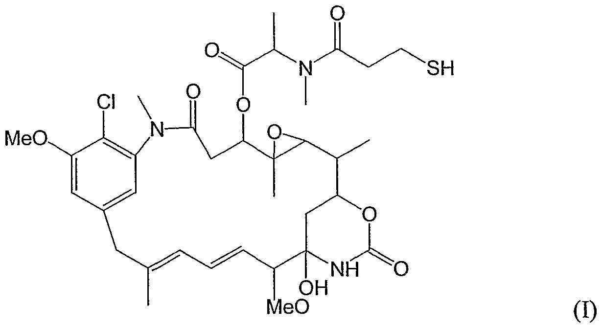


Claims


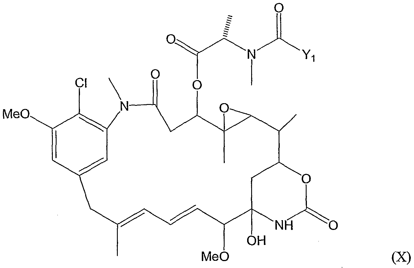

Priority Applications (22)
| Application Number | Priority Date | Filing Date | Title |
|---|---|---|---|
| JP2006521193A JP2007503202A (en) | 2003-07-21 | 2004-07-21 | CA6 antigen-specific cytotoxic conjugates and methods of use thereof |
| SI200431646T SI1660513T1 (en) | 2003-07-21 | 2004-07-21 | A ca6 antigen-specific cytotoxic conjugate and methods of using the same |
| EA200600275A EA014640B1 (en) | 2003-07-21 | 2004-07-21 | Antibody or an epitope-binding fragment thereof that binds the ca6 glycotope and methods of using the same |
| DK04778714.8T DK1660513T5 (en) | 2003-07-21 | 2004-07-21 | CA6 antigen-specific, cytotoxic conjugate and methods for its use |
| CA2532430A CA2532430C (en) | 2003-07-21 | 2004-07-21 | Humanized versions of monoclonal antibody ds6 that bind tumour antigen ca6 |
| KR1020147008527A KR101565721B1 (en) | 2003-07-21 | 2004-07-21 | A ca6 antigen-specific cytotoxic conjugate and methods of using the same |
| AT04778714T ATE496944T1 (en) | 2003-07-21 | 2004-07-21 | CA6-ANTIGEN-SPECIFIC CYTOTOXIC CONJUGATE AND METHOD FOR USE THEREOF |
| AU2004258955A AU2004258955C1 (en) | 2003-07-21 | 2004-07-21 | A CA6 antigen-specific cytotoxic conjugate and methods of using the same |
| BRPI0412879A BRPI0412879A8 (en) | 2003-07-21 | 2004-07-21 | CA6 ANTIGEN SPECIFIC CYTOTOXIC CONJUGATE AND METHODS OF ITS USE |
| DE602004031239T DE602004031239D1 (en) | 2003-07-21 | 2004-07-21 | METHOD FOR THE APPLICATION THEREOF |
| KR1020127013238A KR20120059657A (en) | 2003-07-21 | 2004-07-21 | A ca6 antigen-specific cytotoxic conjugate and methods of using the same |
| MXPA06000830A MXPA06000830A (en) | 2003-07-21 | 2004-07-21 | A ca6 antigen-specific cytotoxic conjugate and methods of using the same. |
| PL04778714T PL1660513T3 (en) | 2003-07-21 | 2004-07-21 | A ca6 antigen-specific cytotoxic conjugate and methods of using the same |
| KR1020157031117A KR101672664B1 (en) | 2003-07-21 | 2004-07-21 | A humanized antibody or epitope-binding fragment thereof |
| CN2004800243313A CN1922199B (en) | 2003-07-21 | 2004-07-21 | CA6 antigen-specific cytotoxic conjugate and methods of using same |
| EP04778714A EP1660513B9 (en) | 2003-07-21 | 2004-07-21 | A ca6 antigen-specific cytotoxic conjugate and methods of using the same |
| IL172982A IL172982A (en) | 2003-07-21 | 2006-01-05 | Antibodies and ca6 antigen specific cytotoxic conjugates and pharmaceutical compositions containing the same |
| ZA2006/00375A ZA200600375B (en) | 2003-07-21 | 2006-01-13 | A ca6 antigen-specific cytotoxicconjugate and methods of using the same |
| NO20060812A NO339324B1 (en) | 2003-07-21 | 2006-02-20 | Humanized antibody or epitope binding fragment thereof which binds the CA6 glycotope; polynucleotide and expression vector encoding the antibody or fragments; cytotoxic conjugate thereof; using the conjugate for the manufacture of a drug; seen from there; an in vitro method for inhibiting the growth of a cell expressing the CA6 glycotope and a method for detecting the CA6 glycotope. |
| HK07104440.1A HK1098160A1 (en) | 2003-07-21 | 2007-04-26 | A ca6 antigen-specific cytotoxic conjugate and methods of using the same ca6 |
| HR20110302T HRP20110302T1 (en) | 2003-07-21 | 2011-04-22 | A ca6 antigen-specific cytotoxic conjugate and methods of using the same |
| IL242336A IL242336A (en) | 2003-07-21 | 2015-10-29 | Method of detecting ca6 glycotope and use of a cytotoxic conjugate in the preparation of a medicament for treating cancer in a subject diagnosed with said cancer in accordance with said method |
Applications Claiming Priority (2)
| Application Number | Priority Date | Filing Date | Title |
|---|---|---|---|
| US48844703P | 2003-07-21 | 2003-07-21 | |
| US60/488,447 | 2003-07-21 |
Publications (2)
| Publication Number | Publication Date |
|---|---|
| WO2005009369A2 true WO2005009369A2 (en) | 2005-02-03 |
| WO2005009369A3 WO2005009369A3 (en) | 2005-06-30 |
Family
ID=34102763
Family Applications (1)
| Application Number | Title | Priority Date | Filing Date |
|---|---|---|---|
| PCT/US2004/023340 WO2005009369A2 (en) | 2003-07-21 | 2004-07-21 | A ca6 antigen-specific cytotoxic conjugate and methods of using the same |
Country Status (27)
| Country | Link |
|---|---|
| US (1) | US20050123549A1 (en) |
| EP (1) | EP1660513B9 (en) |
| JP (5) | JP2007503202A (en) |
| KR (5) | KR101672664B1 (en) |
| CN (3) | CN103554261B (en) |
| AT (1) | ATE496944T1 (en) |
| AU (1) | AU2004258955C1 (en) |
| BR (1) | BRPI0412879A8 (en) |
| CA (1) | CA2532430C (en) |
| CR (1) | CR8207A (en) |
| CY (1) | CY1111938T1 (en) |
| DE (1) | DE602004031239D1 (en) |
| DK (1) | DK1660513T5 (en) |
| EA (1) | EA014640B1 (en) |
| EC (1) | ECSP066294A (en) |
| ES (1) | ES2360403T3 (en) |
| HK (2) | HK1098160A1 (en) |
| HR (1) | HRP20110302T1 (en) |
| IL (2) | IL172982A (en) |
| MX (2) | MXPA06000830A (en) |
| NO (1) | NO339324B1 (en) |
| NZ (3) | NZ580855A (en) |
| PL (1) | PL1660513T3 (en) |
| PT (1) | PT1660513E (en) |
| SI (1) | SI1660513T1 (en) |
| WO (1) | WO2005009369A2 (en) |
| ZA (1) | ZA200600375B (en) |
Cited By (41)
| Publication number | Priority date | Publication date | Assignee | Title |
|---|---|---|---|---|
| EP1651162A2 (en) * | 2003-05-20 | 2006-05-03 | Immunogen, Inc. | Improved cytotoxic agents comprising new maytansinoids |
| WO2007024222A1 (en) * | 2005-08-22 | 2007-03-01 | Immunogen, Inc. | A ca6 antigen-specific cytotoxic conjugate and methods of using the same |
| US7507410B2 (en) | 2000-08-18 | 2009-03-24 | East Carolina University | Monoclonal antibody DS6, tumor-associated antigen CA6, and methods of use thereof |
| WO2011001052A1 (en) | 2009-06-29 | 2011-01-06 | Sanofi-Aventis | Novel conjugates, preparation thereof, and therapeutic use thereof |
| WO2011023883A1 (en) | 2009-08-25 | 2011-03-03 | Sanofi-Aventis | Conjugates of pyrrolo[1,4]benzodiazepine dimers as anticancer agents |
| WO2011039721A1 (en) | 2009-10-02 | 2011-04-07 | Sanofi-Aventis | New maytansinoids and the use of said maytansinoids to prepare conjugates with an antibody |
| WO2012014147A1 (en) | 2010-07-26 | 2012-02-02 | Sanofi | Anticancer derivatives, preparation thereof and therapeutic use thereof |
| US8163888B2 (en) | 2003-10-10 | 2012-04-24 | Immunogen, Inc. | Method of targeting specific cell populations using cell-binding agent maytansinoid conjugates linked via a non-cleavable linker, said conjugates and methods of making said conjugates |
| JP2012161321A (en) * | 2012-03-30 | 2012-08-30 | Immunogen Inc | Ca6 antigen-specific cytotoxic conjugate, and method using the conjugate |
| WO2012143499A2 (en) | 2011-04-21 | 2012-10-26 | Bayer Intellectual Property Gmbh | Novel binder-drug conjugates (adcs) and their use |
| US8404678B2 (en) | 2007-07-19 | 2013-03-26 | Sanofi | Cytotoxic agents comprising new tomaymycin derivatives and their therapeutic use |
| US8435528B2 (en) | 2003-05-20 | 2013-05-07 | Immunogen, Inc. | Cytotoxic agents comprising new maytansinoids |
| US8613930B2 (en) | 2008-04-30 | 2013-12-24 | Immunogen, Inc. | Cross-linkers and their uses |
| WO2014122529A1 (en) | 2013-02-05 | 2014-08-14 | Sanofi | Immuno imaging agent for use with antibody-drug conjugate therapy |
| WO2015014879A1 (en) * | 2013-08-02 | 2015-02-05 | Sanofi | Use of anti-muc1 maytansinoid immunoconjugate antibody for the treatment of solid tumors |
| US8987424B2 (en) | 2003-07-21 | 2015-03-24 | Immunogen, Inc. | CA6 antigen-specific cytotoxic conjugate and methods of using the same |
| CN104804094A (en) * | 2005-08-22 | 2015-07-29 | 伊缪诺金公司 | CA6 antigen-specificity cytotoxicity conjugate and application method thereof |
| WO2016036804A1 (en) | 2014-09-03 | 2016-03-10 | Immunogen, Inc. | Cytotoxic benzodiazepine derivatives |
| WO2016207089A1 (en) | 2015-06-22 | 2016-12-29 | Bayer Pharma Aktiengesellschaft | Antibody drug conjugates (adcs) and antibody prodrug conjugates (apdcs) with enzymatically cleavable groups |
| US9539348B2 (en) | 2000-08-18 | 2017-01-10 | East Carolina University | Monoclonal antibody DS6, tumor-associated antigen CA6, and methods of use thereof |
| WO2017060322A2 (en) | 2015-10-10 | 2017-04-13 | Bayer Pharma Aktiengesellschaft | Ptefb-inhibitor-adc |
| WO2017091745A1 (en) | 2015-11-25 | 2017-06-01 | Immunogen, Inc. | Pharmaceutical formulations and methods of use thereof |
| WO2017162663A1 (en) | 2016-03-24 | 2017-09-28 | Bayer Pharma Aktiengesellschaft | Prodrugs of cytotoxic active agents having enzymatically cleavable groups |
| US9844607B2 (en) | 2013-02-05 | 2017-12-19 | Sanofi | Immuno imaging agent for use with antibody-drug conjugate therapy |
| EP3311846A1 (en) | 2014-09-02 | 2018-04-25 | ImmunoGen, Inc. | Methods for formulating antibody drug conjugate compositions |
| US9989524B2 (en) | 2013-02-05 | 2018-06-05 | Sanofi | Immuno imaging agent for use with antibody-drug conjugate therapy |
| WO2018114798A1 (en) | 2016-12-21 | 2018-06-28 | Bayer Aktiengesellschaft | Prodrugs of cytotoxic active agents having enzymatically cleavable groups |
| WO2018114578A1 (en) | 2016-12-21 | 2018-06-28 | Bayer Pharma Aktiengesellschaft | Antibody drug conjugates (adcs) having enzymatically cleavable groups |
| US10022453B2 (en) | 2013-12-23 | 2018-07-17 | Bayer Pharma Aktiengesellschaft | Antibody drug conjugates (ADCs) with kinesin spindel protein (KSP) |
| WO2018160539A1 (en) | 2017-02-28 | 2018-09-07 | Immunogen, Inc. | Maytansinoid derivatives with self-immolative peptide linkers and conjugates thereof |
| WO2018195243A1 (en) | 2017-04-20 | 2018-10-25 | Immunogen, Inc. | Cytotoxic benzodiazepine derivatives and conjugates thereof |
| WO2019133652A1 (en) | 2017-12-28 | 2019-07-04 | Immunogen, Inc. | Benzodiazepine derivatives |
| US10640508B2 (en) | 2017-10-13 | 2020-05-05 | Massachusetts Institute Of Technology | Diazene directed modular synthesis of compounds with quaternary carbon centers |
| WO2020187998A1 (en) | 2019-03-19 | 2020-09-24 | Fundació Privada Institut D'investigació Oncològica De Vall Hebron | Combination therapy with omomyc and an antibody binding pd-1 or ctla-4 for the treatment of cancer |
| US10918735B2 (en) | 2012-12-04 | 2021-02-16 | Massachusetts Institute Of Technology | Substituted pyrazino[1′,2′:1,5]pyrrolo[2,3-b]indole-1,4-diones for cancer treatment |
| US10918627B2 (en) | 2016-05-11 | 2021-02-16 | Massachusetts Institute Of Technology | Convergent and enantioselective total synthesis of Communesin analogs |
| EP3778601A1 (en) | 2014-09-03 | 2021-02-17 | ImmunoGen, Inc. | Cytotoxic benzodiazepine derivatives |
| US11001636B2 (en) | 2016-06-15 | 2021-05-11 | Bayer Pharma Aktiengesellschaft | Specific antibody-drug-conjugates (ADCs) with KSP inhibitors and anti-CD123-antibodies |
| US11433140B2 (en) | 2016-12-21 | 2022-09-06 | Bayer Pharma Aktiengesellschaft | Specific antibody drug conjugates (ADCs) having KSP inhibitors |
| US11535634B2 (en) | 2019-06-05 | 2022-12-27 | Massachusetts Institute Of Technology | Compounds, conjugates, and compositions of epipolythiodiketopiperazines and polythiodiketopiperazines and uses thereof |
| US11932650B2 (en) | 2017-05-11 | 2024-03-19 | Massachusetts Institute Of Technology | Potent agelastatin derivatives as modulators for cancer invasion and metastasis |
Families Citing this family (14)
| Publication number | Priority date | Publication date | Assignee | Title |
|---|---|---|---|---|
| US7097840B2 (en) | 2000-03-16 | 2006-08-29 | Genentech, Inc. | Methods of treatment using anti-ErbB antibody-maytansinoid conjugates |
| CA2486285C (en) * | 2004-08-30 | 2017-03-07 | Viktor S. Goldmakher | Immunoconjugates targeting syndecan-1 expressing cells and use thereof |
| CA2710471C (en) * | 2007-12-26 | 2018-06-05 | Biotest Ag | Immunoconjugates targeting cd138 and uses thereof |
| CN101945892B (en) * | 2007-12-26 | 2017-11-24 | 生物测试股份公司 | For the method and reagent of the targeting for improving the tumour cell to expressing CD138 |
| WO2009080831A1 (en) * | 2007-12-26 | 2009-07-02 | Biotest Ag | Method of decreasing cytotoxic side-effects and improving efficacy of immunoconjugates |
| CA2710453C (en) * | 2007-12-26 | 2019-07-02 | Biotest Ag | Agents targeting cd138 and uses thereof |
| CN102573916B (en) * | 2009-05-06 | 2017-05-17 | 生物测试股份公司 | Uses of immunoconjugates targeting cd138 |
| CA2789629A1 (en) | 2010-02-10 | 2011-08-18 | Immunogen, Inc. | Cd20 antibodies and uses thereof |
| CA2824143C (en) * | 2011-01-14 | 2018-12-18 | Redwood Bioscience, Inc. | Aldehyde-tagged immunoglobulin polypeptides and method of use thereof |
| CA2843771A1 (en) * | 2011-09-20 | 2013-03-28 | Eli Lilly And Company | Anti-c-met antibodies |
| MX358680B (en) | 2011-12-08 | 2018-08-31 | Biotest Ag | Uses of immunoconjugates targeting cd138. |
| ES2859398T3 (en) | 2013-03-15 | 2021-10-01 | Beckman Coulter Inc | Methods for panel design in flow cytometry |
| KR20160125515A (en) * | 2014-03-12 | 2016-10-31 | 노파르티스 아게 | Specific sites for modifying antibodies to make immunoconjugates |
| WO2023150677A2 (en) * | 2022-02-03 | 2023-08-10 | Igm Biosciences, Inc. | Anti-cd38 binding molecules and uses thereof |
Family Cites Families (18)
| Publication number | Priority date | Publication date | Assignee | Title |
|---|---|---|---|---|
| US5618920A (en) * | 1985-11-01 | 1997-04-08 | Xoma Corporation | Modular assembly of antibody genes, antibodies prepared thereby and use |
| CA2026147C (en) * | 1989-10-25 | 2006-02-07 | Ravi J. Chari | Cytotoxic agents comprising maytansinoids and their therapeutic use |
| US5208020A (en) * | 1989-10-25 | 1993-05-04 | Immunogen Inc. | Cytotoxic agents comprising maytansinoids and their therapeutic use |
| GB8928874D0 (en) * | 1989-12-21 | 1990-02-28 | Celltech Ltd | Humanised antibodies |
| WO1993006213A1 (en) * | 1991-09-23 | 1993-04-01 | Medical Research Council | Production of chimeric antibodies - a combinatorial approach |
| US5639641A (en) * | 1992-09-09 | 1997-06-17 | Immunogen Inc. | Resurfacing of rodent antibodies |
| DE122004000036I1 (en) * | 1992-11-13 | 2005-07-07 | Biogen Idec Inc | Therapeutic use of chimeric and labeled antibodies to human B lymphocyte limited differentiation antigen for the treatment of B-cell lymphoma. |
| US20030143233A1 (en) * | 1999-06-07 | 2003-07-31 | Neorx Corporation | Streptavidin expressed gene fusions and methods of use thereof |
| NZ517772A (en) * | 1999-11-24 | 2004-03-26 | Immunogen Inc | Cytotoxic agents comprising taxanes and their therapeutic use |
| US6596503B1 (en) * | 2000-08-18 | 2003-07-22 | East Carolina University | Monoclonal antibody DS6, tumor-associated antigen CA6, and methods of use thereof |
| US6333410B1 (en) * | 2000-08-18 | 2001-12-25 | Immunogen, Inc. | Process for the preparation and purification of thiol-containing maytansinoids |
| EP1258255A1 (en) * | 2001-05-18 | 2002-11-20 | Boehringer Ingelheim International GmbH | Conjugates of an antibody to CD44 and a maytansinoid |
| US6441163B1 (en) * | 2001-05-31 | 2002-08-27 | Immunogen, Inc. | Methods for preparation of cytotoxic conjugates of maytansinoids and cell binding agents |
| AU2002340118B2 (en) * | 2001-10-04 | 2007-09-13 | Immunex Corporation | UL16 Binding protein 4 |
| CA2467242A1 (en) * | 2001-11-20 | 2003-05-30 | Seattle Genetics, Inc. | Treatment of immunological disorders using anti-cd30 antibodies |
| US6716821B2 (en) * | 2001-12-21 | 2004-04-06 | Immunogen Inc. | Cytotoxic agents bearing a reactive polyethylene glycol moiety, cytotoxic conjugates comprising polyethylene glycol linking groups, and methods of making and using the same |
| WO2003057160A2 (en) * | 2002-01-02 | 2003-07-17 | Genentech, Inc. | Compositions and methods for the diagnosis and treatment of tumor |
| CA2737127C (en) | 2003-05-14 | 2016-07-26 | Immunogen, Inc. | Maytansinoid-antibody conjugate compositions |
-
2004
- 2004-07-21 US US10/895,135 patent/US20050123549A1/en not_active Abandoned
- 2004-07-21 KR KR1020157031117A patent/KR101672664B1/en active IP Right Grant
- 2004-07-21 EP EP04778714A patent/EP1660513B9/en active Active
- 2004-07-21 BR BRPI0412879A patent/BRPI0412879A8/en not_active Application Discontinuation
- 2004-07-21 DK DK04778714.8T patent/DK1660513T5/en active
- 2004-07-21 KR KR1020127024607A patent/KR20120113811A/en active Search and Examination
- 2004-07-21 CN CN201310444419.1A patent/CN103554261B/en active Active
- 2004-07-21 MX MXPA06000830A patent/MXPA06000830A/en active IP Right Grant
- 2004-07-21 NZ NZ580855A patent/NZ580855A/en not_active IP Right Cessation
- 2004-07-21 PL PL04778714T patent/PL1660513T3/en unknown
- 2004-07-21 CA CA2532430A patent/CA2532430C/en not_active Expired - Fee Related
- 2004-07-21 KR KR1020067001429A patent/KR101263950B1/en not_active IP Right Cessation
- 2004-07-21 NZ NZ612796A patent/NZ612796A/en not_active IP Right Cessation
- 2004-07-21 CN CN201310020230.XA patent/CN103145844B/en active Active
- 2004-07-21 SI SI200431646T patent/SI1660513T1/en unknown
- 2004-07-21 JP JP2006521193A patent/JP2007503202A/en active Pending
- 2004-07-21 ES ES04778714T patent/ES2360403T3/en active Active
- 2004-07-21 AT AT04778714T patent/ATE496944T1/en active
- 2004-07-21 DE DE602004031239T patent/DE602004031239D1/en active Active
- 2004-07-21 AU AU2004258955A patent/AU2004258955C1/en not_active Ceased
- 2004-07-21 WO PCT/US2004/023340 patent/WO2005009369A2/en active Application Filing
- 2004-07-21 CN CN2004800243313A patent/CN1922199B/en active Active
- 2004-07-21 EA EA200600275A patent/EA014640B1/en not_active IP Right Cessation
- 2004-07-21 KR KR1020127013238A patent/KR20120059657A/en not_active Application Discontinuation
- 2004-07-21 PT PT04778714T patent/PT1660513E/en unknown
- 2004-07-21 NZ NZ598504A patent/NZ598504A/en not_active IP Right Cessation
- 2004-07-21 KR KR1020147008527A patent/KR101565721B1/en active IP Right Grant
-
2006
- 2006-01-05 IL IL172982A patent/IL172982A/en active IP Right Grant
- 2006-01-13 ZA ZA2006/00375A patent/ZA200600375B/en unknown
- 2006-01-19 EC EC2006006294A patent/ECSP066294A/en unknown
- 2006-01-20 MX MX2011000245A patent/MX336469B/en unknown
- 2006-01-20 CR CR8207A patent/CR8207A/en unknown
- 2006-02-20 NO NO20060812A patent/NO339324B1/en not_active IP Right Cessation
-
2007
- 2007-04-26 HK HK07104440.1A patent/HK1098160A1/en not_active IP Right Cessation
- 2007-04-26 HK HK13113291.4A patent/HK1185891A1/en not_active IP Right Cessation
-
2010
- 2010-11-25 JP JP2010262151A patent/JP5643619B2/en not_active Expired - Fee Related
-
2011
- 2011-04-21 CY CY20111100408T patent/CY1111938T1/en unknown
- 2011-04-22 HR HR20110302T patent/HRP20110302T1/en unknown
-
2014
- 2014-06-30 JP JP2014133794A patent/JP5997727B2/en not_active Expired - Fee Related
-
2015
- 2015-10-29 IL IL242336A patent/IL242336A/en active IP Right Grant
- 2015-12-04 JP JP2015237469A patent/JP6144749B2/en not_active Expired - Fee Related
-
2017
- 2017-02-24 JP JP2017033672A patent/JP2017123857A/en active Pending
Non-Patent Citations (1)
| Title |
|---|
| See references of EP1660513A4 * |
Cited By (67)
| Publication number | Priority date | Publication date | Assignee | Title |
|---|---|---|---|---|
| US8067186B2 (en) | 2000-08-18 | 2011-11-29 | East Carolina University | Monoclonal antibody DS6, tumor-associated antigen CA6, and methods of use thereof |
| US8728741B2 (en) | 2000-08-18 | 2014-05-20 | East Carolina Unversity | Monoclonal antibody DS6, tumor-associated antigen CA6, and methods of use thereof |
| US9539348B2 (en) | 2000-08-18 | 2017-01-10 | East Carolina University | Monoclonal antibody DS6, tumor-associated antigen CA6, and methods of use thereof |
| US7507410B2 (en) | 2000-08-18 | 2009-03-24 | East Carolina University | Monoclonal antibody DS6, tumor-associated antigen CA6, and methods of use thereof |
| NO339597B1 (en) * | 2003-05-20 | 2017-01-09 | Immunogen Inc | Enhanced cytotoxic agents which include novel maytansinoids; process for their preparation; intermediates thereof and process for their preparation; conjugate between the maytansinoids and a cell binding agent; process for its preparation and its use in a therapeutic method or for the manufacture of a medicament, and a pharmaceutical preparation thereof. |
| US8841425B2 (en) | 2003-05-20 | 2014-09-23 | Immunogen, Inc. | Cytotoxic agents comprising new maytansinoids |
| EP1651162A2 (en) * | 2003-05-20 | 2006-05-03 | Immunogen, Inc. | Improved cytotoxic agents comprising new maytansinoids |
| US8435528B2 (en) | 2003-05-20 | 2013-05-07 | Immunogen, Inc. | Cytotoxic agents comprising new maytansinoids |
| EP1651162B1 (en) * | 2003-05-20 | 2015-10-21 | ImmunoGen, Inc. | Improved cytotoxic agents comprising new maytansinoids |
| EP3031810B1 (en) * | 2003-05-20 | 2019-03-06 | ImmunoGen, Inc. | Maytansinoid-cell-binding agent conjugates |
| US9370584B2 (en) | 2003-07-21 | 2016-06-21 | Immunogen Inc. | CA6 antigen-specific cytotoxic conjugate and methods of using the same |
| US9822183B2 (en) | 2003-07-21 | 2017-11-21 | Sanofi | CA6 antigen-specific cytotoxic conjugate and methods of using the same |
| US8987424B2 (en) | 2003-07-21 | 2015-03-24 | Immunogen, Inc. | CA6 antigen-specific cytotoxic conjugate and methods of using the same |
| US8163888B2 (en) | 2003-10-10 | 2012-04-24 | Immunogen, Inc. | Method of targeting specific cell populations using cell-binding agent maytansinoid conjugates linked via a non-cleavable linker, said conjugates and methods of making said conjugates |
| US10844135B2 (en) | 2003-10-10 | 2020-11-24 | Immunogen, Inc. | Method of targeting specific cell populations using cell-binding agent maytansinoid conjugates linked via a non-cleavable linker, said conjugates and methods of making said |
| US8685920B2 (en) | 2003-10-10 | 2014-04-01 | Immunogen Inc. | Method of targeting specific cell populations using cell-binding agent maytansinoid conjugates linked via a non-cleavable linker, said conjugates and methods of making said conjugates |
| JP2009504193A (en) * | 2005-08-22 | 2009-02-05 | イミュノジェン・インコーポレーテッド | CA6 antigen-specific cytotoxic conjugate and method using the conjugate |
| CN104804094A (en) * | 2005-08-22 | 2015-07-29 | 伊缪诺金公司 | CA6 antigen-specificity cytotoxicity conjugate and application method thereof |
| WO2007024222A1 (en) * | 2005-08-22 | 2007-03-01 | Immunogen, Inc. | A ca6 antigen-specific cytotoxic conjugate and methods of using the same |
| US8404678B2 (en) | 2007-07-19 | 2013-03-26 | Sanofi | Cytotoxic agents comprising new tomaymycin derivatives and their therapeutic use |
| US8613930B2 (en) | 2008-04-30 | 2013-12-24 | Immunogen, Inc. | Cross-linkers and their uses |
| US9498541B2 (en) | 2008-04-30 | 2016-11-22 | Immunogen, Inc. | Cross-linkers and their uses |
| US9061995B2 (en) | 2008-04-30 | 2015-06-23 | Immunogen, Inc. | Cross-linkers and their uses |
| US10494431B2 (en) | 2008-04-30 | 2019-12-03 | Immunogen, Inc. | Cross-linkers and their uses |
| US11046762B2 (en) | 2008-04-30 | 2021-06-29 | Immunogen, Inc. | Cross-linkers and their uses |
| US8952147B2 (en) | 2009-06-29 | 2015-02-10 | Sanofi | Conjugates, preparation thereof, and therapeutic use thereof |
| WO2011001052A1 (en) | 2009-06-29 | 2011-01-06 | Sanofi-Aventis | Novel conjugates, preparation thereof, and therapeutic use thereof |
| WO2011023883A1 (en) | 2009-08-25 | 2011-03-03 | Sanofi-Aventis | Conjugates of pyrrolo[1,4]benzodiazepine dimers as anticancer agents |
| US8481042B2 (en) | 2009-08-25 | 2013-07-09 | Sanofi | Conjugates of pyrrolo[1,4]benzodiazepine dimers as anticancer agents |
| WO2011039721A1 (en) | 2009-10-02 | 2011-04-07 | Sanofi-Aventis | New maytansinoids and the use of said maytansinoids to prepare conjugates with an antibody |
| WO2012014147A1 (en) | 2010-07-26 | 2012-02-02 | Sanofi | Anticancer derivatives, preparation thereof and therapeutic use thereof |
| US9056914B2 (en) | 2010-07-26 | 2015-06-16 | Sanofi | Anticancer derivatives, preparation thereof and therapeutic use thereof |
| WO2012143499A2 (en) | 2011-04-21 | 2012-10-26 | Bayer Intellectual Property Gmbh | Novel binder-drug conjugates (adcs) and their use |
| WO2012143496A2 (en) | 2011-04-21 | 2012-10-26 | Bayer Intellectual Property Gmbh | Novel binder-drug conjugates (adcs) and their use |
| JP2012161321A (en) * | 2012-03-30 | 2012-08-30 | Immunogen Inc | Ca6 antigen-specific cytotoxic conjugate, and method using the conjugate |
| US10918735B2 (en) | 2012-12-04 | 2021-02-16 | Massachusetts Institute Of Technology | Substituted pyrazino[1′,2′:1,5]pyrrolo[2,3-b]indole-1,4-diones for cancer treatment |
| US9989524B2 (en) | 2013-02-05 | 2018-06-05 | Sanofi | Immuno imaging agent for use with antibody-drug conjugate therapy |
| WO2014122529A1 (en) | 2013-02-05 | 2014-08-14 | Sanofi | Immuno imaging agent for use with antibody-drug conjugate therapy |
| US9844607B2 (en) | 2013-02-05 | 2017-12-19 | Sanofi | Immuno imaging agent for use with antibody-drug conjugate therapy |
| WO2015014879A1 (en) * | 2013-08-02 | 2015-02-05 | Sanofi | Use of anti-muc1 maytansinoid immunoconjugate antibody for the treatment of solid tumors |
| US10022453B2 (en) | 2013-12-23 | 2018-07-17 | Bayer Pharma Aktiengesellschaft | Antibody drug conjugates (ADCs) with kinesin spindel protein (KSP) |
| EP3311846A1 (en) | 2014-09-02 | 2018-04-25 | ImmunoGen, Inc. | Methods for formulating antibody drug conjugate compositions |
| US10603388B2 (en) | 2014-09-02 | 2020-03-31 | Immunogen, Inc. | Methods for formulating antibody drug conjugate compositions |
| EP3778601A1 (en) | 2014-09-03 | 2021-02-17 | ImmunoGen, Inc. | Cytotoxic benzodiazepine derivatives |
| WO2016036804A1 (en) | 2014-09-03 | 2016-03-10 | Immunogen, Inc. | Cytotoxic benzodiazepine derivatives |
| US11123439B2 (en) | 2015-06-22 | 2021-09-21 | Bayer Pharma Aktiengesellschaft | Antibody drug conjugates (ADCS) and antibody prodrug conjugates (APDCS) with enzymatically cleavable groups |
| WO2016207089A1 (en) | 2015-06-22 | 2016-12-29 | Bayer Pharma Aktiengesellschaft | Antibody drug conjugates (adcs) and antibody prodrug conjugates (apdcs) with enzymatically cleavable groups |
| WO2017060322A2 (en) | 2015-10-10 | 2017-04-13 | Bayer Pharma Aktiengesellschaft | Ptefb-inhibitor-adc |
| WO2017091745A1 (en) | 2015-11-25 | 2017-06-01 | Immunogen, Inc. | Pharmaceutical formulations and methods of use thereof |
| EP4335851A2 (en) | 2015-11-25 | 2024-03-13 | ImmunoGen, Inc. | Pharmaceutical formulations and methods of use thereof |
| WO2017162663A1 (en) | 2016-03-24 | 2017-09-28 | Bayer Pharma Aktiengesellschaft | Prodrugs of cytotoxic active agents having enzymatically cleavable groups |
| US11685714B2 (en) | 2016-03-24 | 2023-06-27 | Bayer Pharma Aktiengesellschaft | Prodrugs of cytotoxic active agents having enzymatically cleavable groups |
| US10918627B2 (en) | 2016-05-11 | 2021-02-16 | Massachusetts Institute Of Technology | Convergent and enantioselective total synthesis of Communesin analogs |
| US11643469B2 (en) | 2016-06-15 | 2023-05-09 | Bayer Pharma Aktiengesellschaft | Specific antibody-drug-conjugates (ADCs) with KSP inhibitors and anti-CD123-antibodies |
| US11001636B2 (en) | 2016-06-15 | 2021-05-11 | Bayer Pharma Aktiengesellschaft | Specific antibody-drug-conjugates (ADCs) with KSP inhibitors and anti-CD123-antibodies |
| US11433140B2 (en) | 2016-12-21 | 2022-09-06 | Bayer Pharma Aktiengesellschaft | Specific antibody drug conjugates (ADCs) having KSP inhibitors |
| WO2018114798A1 (en) | 2016-12-21 | 2018-06-28 | Bayer Aktiengesellschaft | Prodrugs of cytotoxic active agents having enzymatically cleavable groups |
| US11478554B2 (en) | 2016-12-21 | 2022-10-25 | Bayer Pharma Aktiengesellschaft | Antibody drug conjugates (ADCS) having enzymatically cleavable groups |
| US11660351B2 (en) | 2016-12-21 | 2023-05-30 | Bayer Aktiengesellschaft | Antibody drug conjugates (ADCs) having enzymatically cleavable groups |
| WO2018114578A1 (en) | 2016-12-21 | 2018-06-28 | Bayer Pharma Aktiengesellschaft | Antibody drug conjugates (adcs) having enzymatically cleavable groups |
| WO2018160539A1 (en) | 2017-02-28 | 2018-09-07 | Immunogen, Inc. | Maytansinoid derivatives with self-immolative peptide linkers and conjugates thereof |
| WO2018195243A1 (en) | 2017-04-20 | 2018-10-25 | Immunogen, Inc. | Cytotoxic benzodiazepine derivatives and conjugates thereof |
| US11932650B2 (en) | 2017-05-11 | 2024-03-19 | Massachusetts Institute Of Technology | Potent agelastatin derivatives as modulators for cancer invasion and metastasis |
| US10640508B2 (en) | 2017-10-13 | 2020-05-05 | Massachusetts Institute Of Technology | Diazene directed modular synthesis of compounds with quaternary carbon centers |
| WO2019133652A1 (en) | 2017-12-28 | 2019-07-04 | Immunogen, Inc. | Benzodiazepine derivatives |
| WO2020187998A1 (en) | 2019-03-19 | 2020-09-24 | Fundació Privada Institut D'investigació Oncològica De Vall Hebron | Combination therapy with omomyc and an antibody binding pd-1 or ctla-4 for the treatment of cancer |
| US11535634B2 (en) | 2019-06-05 | 2022-12-27 | Massachusetts Institute Of Technology | Compounds, conjugates, and compositions of epipolythiodiketopiperazines and polythiodiketopiperazines and uses thereof |
Also Published As
Similar Documents
| Publication | Publication Date | Title |
|---|---|---|
| EP1660513B9 (en) | A ca6 antigen-specific cytotoxic conjugate and methods of using the same | |
| US9822183B2 (en) | CA6 antigen-specific cytotoxic conjugate and methods of using the same | |
| EP1917034A1 (en) | A ca6 antigen-specific cytotoxic conjugate and methods of using the same | |
| AU2012201260B2 (en) | A CA6 antigen-specific cytotoxic conjugate and methods of using the same | |
| PAYNE et al. | Patent 2615761 Summary | |
| KR20080039917A (en) | A ca6 antigen-specific cytotoxic conjugate and methods of using the same | |
| JP2012161321A (en) | Ca6 antigen-specific cytotoxic conjugate, and method using the conjugate |
Legal Events
| Date | Code | Title | Description |
|---|---|---|---|
| WWE | Wipo information: entry into national phase |
Ref document number: 200480024331.3 Country of ref document: CN |
|
| AK | Designated states |
Kind code of ref document: A2 Designated state(s): AE AG AL AM AT AU AZ BA BB BG BR BW BY BZ CA CH CN CO CR CU CZ DE DK DM DZ EC EE EG ES FI GB GD GE GH GM HR HU ID IL IN IS JP KE KG KP KR KZ LC LK LR LS LT LU LV MA MD MG MK MN MW MX MZ NA NI NO NZ OM PG PH PL PT RO RU SC SD SE SG SK SL SY TJ TM TN TR TT TZ UA UG US UZ VC VN YU ZA ZM ZW |
|
| AL | Designated countries for regional patents |
Kind code of ref document: A2 Designated state(s): BW GH GM KE LS MW MZ NA SD SL SZ TZ UG ZM ZW AM AZ BY KG KZ MD RU TJ TM AT BE BG CH CY CZ DE DK EE ES FI FR GB GR HU IE IT LU MC NL PL PT RO SE SI SK TR BF BJ CF CG CI CM GA GN GQ GW ML MR NE SN TD TG |
|
| 121 | Ep: the epo has been informed by wipo that ep was designated in this application | ||
| WWE | Wipo information: entry into national phase |
Ref document number: 2004258955 Country of ref document: AU |
|
| WWE | Wipo information: entry into national phase |
Ref document number: 544528 Country of ref document: NZ |
|
| WWE | Wipo information: entry into national phase |
Ref document number: 2532430 Country of ref document: CA |
|
| WWE | Wipo information: entry into national phase |
Ref document number: 2006/00375 Country of ref document: ZA Ref document number: NC2017/0011766 Country of ref document: CO Ref document number: 06002587 Country of ref document: CO Ref document number: 200600375 Country of ref document: ZA |
|
| WWE | Wipo information: entry into national phase |
Ref document number: 1020067001429 Country of ref document: KR Ref document number: PA/a/2006/000830 Country of ref document: MX Ref document number: CR2006-008207 Country of ref document: CR |
|
| WWE | Wipo information: entry into national phase |
Ref document number: 2006521193 Country of ref document: JP |
|
| ENP | Entry into the national phase |
Ref document number: 2004258955 Country of ref document: AU Date of ref document: 20040721 Kind code of ref document: A |
|
| WWP | Wipo information: published in national office |
Ref document number: 2004258955 Country of ref document: AU |
|
| WWE | Wipo information: entry into national phase |
Ref document number: 770/DELNP/2006 Country of ref document: IN |
|
| WWE | Wipo information: entry into national phase |
Ref document number: 200600275 Country of ref document: EA |
|
| WWE | Wipo information: entry into national phase |
Ref document number: 2004778714 Country of ref document: EP |
|
| DPEN | Request for preliminary examination filed prior to expiration of 19th month from priority date (pct application filed from 20040101) | ||
| WWP | Wipo information: published in national office |
Ref document number: 1020067001429 Country of ref document: KR |
|
| WWP | Wipo information: published in national office |
Ref document number: 2004778714 Country of ref document: EP |
|
| ENP | Entry into the national phase |
Ref document number: PI0412879 Country of ref document: BR |
|
| WWE | Wipo information: entry into national phase |
Ref document number: 242336 Country of ref document: IL |