WO2007070870A1 - Compositions and methods for inhibiting adverse immune response in histocompatibility-mismatched transplantation - Google Patents
Compositions and methods for inhibiting adverse immune response in histocompatibility-mismatched transplantation Download PDFInfo
- Publication number
- WO2007070870A1 WO2007070870A1 PCT/US2006/062157 US2006062157W WO2007070870A1 WO 2007070870 A1 WO2007070870 A1 WO 2007070870A1 US 2006062157 W US2006062157 W US 2006062157W WO 2007070870 A1 WO2007070870 A1 WO 2007070870A1
- Authority
- WO
- WIPO (PCT)
- Prior art keywords
- cells
- cell
- protein
- derived cells
- postpartum
- Prior art date
Links
Classifications
-
- C—CHEMISTRY; METALLURGY
- C12—BIOCHEMISTRY; BEER; SPIRITS; WINE; VINEGAR; MICROBIOLOGY; ENZYMOLOGY; MUTATION OR GENETIC ENGINEERING
- C12N—MICROORGANISMS OR ENZYMES; COMPOSITIONS THEREOF; PROPAGATING, PRESERVING, OR MAINTAINING MICROORGANISMS; MUTATION OR GENETIC ENGINEERING; CULTURE MEDIA
- C12N5/00—Undifferentiated human, animal or plant cells, e.g. cell lines; Tissues; Cultivation or maintenance thereof; Culture media therefor
- C12N5/06—Animal cells or tissues; Human cells or tissues
- C12N5/0602—Vertebrate cells
- C12N5/0603—Embryonic cells ; Embryoid bodies
- C12N5/0605—Cells from extra-embryonic tissues, e.g. placenta, amnion, yolk sac, Wharton's jelly
-
- A—HUMAN NECESSITIES
- A61—MEDICAL OR VETERINARY SCIENCE; HYGIENE
- A61K—PREPARATIONS FOR MEDICAL, DENTAL OR TOILETRY PURPOSES
- A61K35/00—Medicinal preparations containing materials or reaction products thereof with undetermined constitution
- A61K35/12—Materials from mammals; Compositions comprising non-specified tissues or cells; Compositions comprising non-embryonic stem cells; Genetically modified cells
- A61K35/48—Reproductive organs
- A61K35/51—Umbilical cord; Umbilical cord blood; Umbilical stem cells
-
- A—HUMAN NECESSITIES
- A61—MEDICAL OR VETERINARY SCIENCE; HYGIENE
- A61P—SPECIFIC THERAPEUTIC ACTIVITY OF CHEMICAL COMPOUNDS OR MEDICINAL PREPARATIONS
- A61P29/00—Non-central analgesic, antipyretic or antiinflammatory agents, e.g. antirheumatic agents; Non-steroidal antiinflammatory drugs [NSAID]
-
- A—HUMAN NECESSITIES
- A61—MEDICAL OR VETERINARY SCIENCE; HYGIENE
- A61P—SPECIFIC THERAPEUTIC ACTIVITY OF CHEMICAL COMPOUNDS OR MEDICINAL PREPARATIONS
- A61P37/00—Drugs for immunological or allergic disorders
- A61P37/02—Immunomodulators
-
- A—HUMAN NECESSITIES
- A61—MEDICAL OR VETERINARY SCIENCE; HYGIENE
- A61P—SPECIFIC THERAPEUTIC ACTIVITY OF CHEMICAL COMPOUNDS OR MEDICINAL PREPARATIONS
- A61P37/00—Drugs for immunological or allergic disorders
- A61P37/02—Immunomodulators
- A61P37/06—Immunosuppressants, e.g. drugs for graft rejection
-
- A—HUMAN NECESSITIES
- A61—MEDICAL OR VETERINARY SCIENCE; HYGIENE
- A61P—SPECIFIC THERAPEUTIC ACTIVITY OF CHEMICAL COMPOUNDS OR MEDICINAL PREPARATIONS
- A61P37/00—Drugs for immunological or allergic disorders
- A61P37/08—Antiallergic agents
-
- A—HUMAN NECESSITIES
- A61—MEDICAL OR VETERINARY SCIENCE; HYGIENE
- A61P—SPECIFIC THERAPEUTIC ACTIVITY OF CHEMICAL COMPOUNDS OR MEDICINAL PREPARATIONS
- A61P43/00—Drugs for specific purposes, not provided for in groups A61P1/00-A61P41/00
-
- A—HUMAN NECESSITIES
- A61—MEDICAL OR VETERINARY SCIENCE; HYGIENE
- A61P—SPECIFIC THERAPEUTIC ACTIVITY OF CHEMICAL COMPOUNDS OR MEDICINAL PREPARATIONS
- A61P7/00—Drugs for disorders of the blood or the extracellular fluid
- A61P7/02—Antithrombotic agents; Anticoagulants; Platelet aggregation inhibitors
-
- A—HUMAN NECESSITIES
- A61—MEDICAL OR VETERINARY SCIENCE; HYGIENE
- A61K—PREPARATIONS FOR MEDICAL, DENTAL OR TOILETRY PURPOSES
- A61K35/00—Medicinal preparations containing materials or reaction products thereof with undetermined constitution
- A61K35/12—Materials from mammals; Compositions comprising non-specified tissues or cells; Compositions comprising non-embryonic stem cells; Genetically modified cells
- A61K2035/122—Materials from mammals; Compositions comprising non-specified tissues or cells; Compositions comprising non-embryonic stem cells; Genetically modified cells for inducing tolerance or supression of immune responses
Definitions
- This invention relates to the field of transplantation immunology.
- the invention relates to cells derived from postpartum tissue having the capability to inhibit an adverse immune response in tissue transplantation between a histocompatibility-mismatched donor and recipient, and methods for using such postpartum tissue-derived cells to stave off rejection and prevent graft versus host disease in a transplant recipient.
- Tissue transplantation is indicated for the treatment of various diseases, pathologies, and traumatic injuries. Although thousands of transplants arc performed each year, nearly 90,000 individuals in the United States alone find themselves on various transplant wait lists for tissues and organs. Contributing to the shortage of available transplant tissue is the overarching need for compatibility between the transplant donor and the transplant recipient.
- HLA human leukocyte antigens
- GVHD graft versus host disease
- BMT Bone marrow transplantation
- HSCT hematopoietic stem cell transplantation
- BMT and HSCT are indicated for various blood diseases and immune disorders, although HSCT is increasingly the preferred method.
- GVHD one of the impediments to the survival and overall health of HSCT patients.
- HLA compatibility plays an important role in graft failure or survival, and whether the transplant recipient is at risk to develop GVHD.
- transplant recipients must endure a lifetime of immunosuppressive treatment regimens. Many anti- rejection medications cause severe side effects in the transplant patient, and their immunosuppressive effects can leave the patient vulnerable to opportunistic infections and certain cancers.
- a significant advance in tissue transplantation would be to provide a means to inhibit adverse immune responses in the transplant recipient such as GVHD and rejection of the transplanted tissue with less severe side effects and complications than, and without the systemic immunosuppression of traditional methods.
- One aspect of the invention features method for inhibiting an adverse immune response in a transplant recipient that is histocompatibility-mismatched to the transplant donor.
- the method comprises administering a cell composition to the transplant recipient in an amount effective for inhibiting the adverse immune response, wherein the cell composition comprises a pharmaceutically acceptable carrier and postpartum-derived cells derived from human postpartum tissue substantially free of blood, said cells being capable of self-renewal and expansion in culture, wherein the cells require L- Valine for growth and are capable of growth in an atmosphere containing oxygen from about 5% to at least about 20%, and wherein the cells comprise at least one of the following characteristics: (a) potential for at least about 40 doublings in culture; (b) attachment and expansion on a coated or uncoated tissue culture vessel, wherein a coated tissue culture vessel comprises a coating of gelatin, laminin, collagen, polyornithine, vitronectin, or fibronectin; (c) production of at least one of tissue factor, vimentin, and alpha- smooth muscle actin;
- the adverse immune response is graft versus host disease, while in other embodiments the adverse immune response is rejection of the transplanted tissue.
- the postpartum derived cells may be placenta-derived cells, umbilicus-derived cells, or a combination of placenta-derived cells and umbilicus-derived cells.
- the cell composition is administered by injection or infusion, or it may be administered by implantation of a device, scaffold or matrix implanted in the transplant recipient.
- the cell composition comprises at least about 50% postpartum-derived cells. In other embodiments, the cell composition comprises a substantially homogeneous population of postpartum-derived cells. In certain embodiments, the cells are administered with at least one other cell type. The other cell type may be administered simultaneously with, or before, or after, the postpartum-derived cells. In certain embodiments, the cells are administered with at least one other agent for treating the adverse immune response. The other agent may be administered simultaneously with, or before, or after, the postpartum- derived cells. The other agent may include or more of an antithrombogenic agent, an anti- inflammatory agent, an immunosuppressive agent, an immunomodulatory agent, or an antiapoptotic agent.
- Another aspect of the invention features a pharmaceutical composition for inhibiting an adverse immune response in a transplant recipient that is histocompatibility- mismatched to the transplant donor, comprising a pharmaceutically acceptable carrier and an amount of the above-described postpartum-derived cells effective to inhibit the adverse immune response in a transplant recipient that is histocompatibility-misn ⁇ atched to the transplant donor.
- the adverse immune response is graft versus host disease, while in other embodiments the adverse immune response is rejection of the transplanted tissue.
- the postpartum derived cells may be placenta-derived cells, umbilicus-derived cells, or a combination of placenta-derived cells and umbilicus-derived cells.
- the pharmaceutical composition is formulated for administration by injection or infusion, or it may be formulated for administration by implantation of a device, scaffold or matrix implanted in the transplant recipient.
- the pharmaceutical composition comprises at least about 50% postpartum-derived cells. In other embodiments, it comprises a substantially homogeneous population of postpartum-derived cells. In certain embodiments, the pharmaceutical composition comprises at least one other cell type or at least one other agent for treating the adverse immune response.
- the other agent may include or more of an antithrombogenic agent, an antiinflammatory agent, an immunosuppressive agent, an immunomodulatory agent, or an antiapoptotic agent.
- kits for inhibiting an adverse immune response in a transplant recipient comprising a pharmaceutically acceptable carrier, a population of the postpartum-derived cells described above, and instructions for using the kit in a method for inhibiting the adverse immune response.
- the kit may also include one or more of at least one reagent and instructions for culturing the postpartum-derived cells, or at least one other cell type, or at least one other agent for inhibiting the adverse immune response.
- Another aspect of the invention features a method for inhibiting an adverse immune response in a transplant recipient that is histocompatibility-mismatched to the transplant donor comprising administering to the patient a composition comprising one or more of a conditioned medium generated by the above-described postpartum-derived cells, a cell lysate generated from the above-described postpartum-derived cells, a soluble cell fraction generated from the above-described postpartum-derived cells, or an extracellular matrix containing the above-described postpartum-derived cells.
- compositions for inhibiting an adverse immune response in a transplant recipient that is histocompatibility- mismatched to the transplant donor comprising a pharmaceutically acceptable carrier and an amount of the aforementioned conditioned medium, cell lysate, soluble cell fraction or extracellular matrix effective to inhibit the adverse immune response in a transplant recipient that is histocompatibility-mismatched to the transplant donor.
- Still another aspect of the invention features a kit for inhibiting an adverse immune response in a transplant recipient that is histocompatibility-mismatched to the transplant donor, comprising a pharmaceutically acceptable carrier, above-described composition comprising a conditioned medium, cell lysate, soluble cell fraction or extracellular matrix of postpartum-derived cells, and instructions for using the kit in a method for inhibiting the adverse immune response.
- Figure 1 Bar graph showing results of two-way mixed lymphocyte reaction (MLR) with third party IFN ⁇ -treated placenta-derived cells in round-bottom plates.
- MLR mixed lymphocyte reaction
- FIG. Bar graph showing results of two-way mixed lymphocyte reaction (MLR) with untreated placcnta-dcrivcd cells in round-bottom plates.
- FIG. Bar graph showing results of two-way mixed lymphocyte reaction (MLR) with third party IFN ⁇ -treated umbilicus-derived cells in round-bottom plates.
- FIG. Bar graph showing results of two-way mixed lymphocyte reaction (MLR) with untreated umbilicus-derived cells in round-bottom plates.
- PPDC postpartum-derived cells
- UDC umbilicus-derived cells
- PDC placental-derived cells
- MHC major histocompatibility complex
- HLA human leukocyte antigen
- BMT bone marrow transplantation
- HSCT hematopoietic stem cell transplantation
- GVHD graft versus host disease
- Stem cells are undifferentiated cells defined by the ability of a single cell both to self-renew and to differentiate to produce progeny cells, including self-renewing progenitors, non-renewing progenitors, and terminally differentiated cells. Stem cells are also characterized by their ability to differentiate in vitro into functional cells of various cell lineages from multiple germ layers (endoderm, mesoderm and ectoderm), as well as to give rise to tissues of multiple germ layers following transplantation, and to contribute substantially to most, if not all, tissues following injection into blastocysts.
- Stem cells are classified according to their developmental potential as: (1) totipotent; (2) pluripotent; (3) multipotent; (4) oligopotent; and (5) imipotent.
- Totipotent cells are able to give rise to all embryonic and extraembryonic cell types.
- Pluripotent cells are able to give rise to all embryonic cell types.
- Multipotent cells include those able to give rise to a subset of cell lineages, but all within a particular tissue, organ, or physiological system (for example, hematopoietic stem cells (HSC) can produce progeny that include HSC (self-renewal), blood cell-restricted oligopotent progenitors, and all cell types and elements ⁇ e.g., platelets) that are normal components of the blood).
- HSC hematopoietic stem cells
- Cells that are oligopotent can give rise to a more restricted subset of cell lineages than multipotent stem cells; and cells that are unipotent are able to give rise to a single cell lineage (e.g., spermatogenic stem cells).
- Stem cells are also categorized on the basis of the source from which they may be obtained.
- An adult stem cell is generally a multipotent undifferentiated cell found in tissue comprising multiple differentiated cell types. The adult stem cell can renew itself. Under normal circumstances, it can also differentiate to yield the specialized cell types of the tissue from which it originated, and possibly other tissue types.
- An embryonic stem cell is a pluripotent cell from the inner cell mass of a blasto cyst-stage embryo.
- a fetal stem cell is one that originates from fetal tissues or membranes.
- a postpartum stem cell is a multipotent or pluripotent cell that originates substantially from extraembryonic tissue available after birth, namely, the placenta and the umbilical cord.
- Postpartum stem cells may be blood-derived (e.g., as are those obtained from umbilical cord blood) or non-blood-dcrivcd (e.g., as obtained from the non-blood tissues of the umbilical cord and placenta).
- Embryonic tissue is typically defined as tissue originating from the embryo (which in humans refers to the period from fertilization to about six weeks of development. Fetal tissue refers to tissue originating from the fetus, which in humans refers to the period from about six weeks of development to parturition. Extraembryonic tissue is tissue associated with, but not originating from, the embryo or fetus. Extraembryonic tissues include extraembryonic membranes (chorion, amnion, yolk sac and allantois), umbilical cord and placenta (which itself forms from the chorion and the maternal decidua basalis).
- Differentiation is the process by which an unspecialized ("uncommitted") or less specialized cell acquires the features of a specialized cell, such as a nerve cell or a muscle cell, for example.
- a differentiated cell is one that has taken on a more specialized ("committed") position within the lineage of a cell.
- the term committed, when applied to the process of differentiation, refers to a cell that has proceeded in the differentiation pathway to a point where, under normal circumstances, it will continue to differentiate into a specific cell type or subset of cell types, and cannot, under normal circumstances, differentiate into a different cell type or revert to a less differentiated cell type.
- De-differentiation refers to the process by which a cell reverts to a less specialized (or committed) position within the lineage of a cell.
- the lineage of a cell defines the heredity of the cell, i.e., which cells it came from and what cells it can give rise to.
- the lineage of a cell places the cell within a hereditary scheme of development and differentiation.
- a progenitor cell is a cell that has the capacity to create progeny that are more differentiated than itself, and yet retains the capacity to replenish the pool of progenitors.
- stem cells themselves are also progenitor cells, as are the more immediate precursors to terminally differentiated cells.
- this broad definition of progenitor cell may be used.
- a progenitor cell is often defined as a cell that is intermediate in the differentiation pathway, i.e., it arises from a stem cell and is intermediate in the production of a mature cell type or subset of cell types.
- progenitor cell is generally not able to sclf-rcncw. Accordingly, if this type of cell is referred to herein, it will be referred to as a non- renewing progenitor cell or as an intermediate progenitor or precursor cell.
- inhibition refers to a reduction in adverse immunological events observed in the adaptive immune response of transplanted donor tissue against the transplant recipient, graft versus host disease, as well as the adaptive immune response of the transplant recipient against the transplanted donor tissue.
- the cells of the present invention are generally referred to as postpartum cells or postpartum-derived cells (PPDCs). They also may sometimes be referred to more specifically as umbilicus-derived cells (UDCs) ox placenta-derived cells (PDCs).
- PPDCs postpartum cells
- UDCs umbilicus-derived cells
- PDCs ox placenta-derived cells
- the cells may be described as being stem or progenitor cells, the latter term being used in the broad sense.
- the term derived is used to indicate that the cells have been obtained from their biological source and grown or otherwise manipulated in vitro ⁇ e.g., cultured in a Growth Medium to expand the population and/or to produce a cell line).
- the in vitro manipulations of umbilical stem cells and the unique features of the umbilicus-derived cells of the present invention are described in detail below.
- Cell culture refers generally to cells taken from a living organism and grown under controlled condition ("in culture” or "cultured”).
- A. primary cell culture is a culture of cells, tissues, or organs taken directly from an organism(s) before the first subculture.
- Cells are expanded in culture when they are placed in a Growth Medium under conditions that facilitate cell growth and/or division, resulting in a larger population of the cells.
- the rate of cell proliferation is sometimes measured by the amount of time needed for the cells to double in number. This is referred to as doubling time.
- a cell line is a population of cells formed by one or more subcultivations of a primary cell culture. Each round of subculturing is referred to as a passage. When cells are subcultured, they are referred to as having been passaged. A specific population of cells, or a cell line, is sometimes referred to or characterized by the number of times it has been passaged. For example, a cultured cell population that has been passaged ten times may be referred to as a PlO culture.
- the primary culture i.e., the first culture following the isolation of cells from tissue, is designated PO. Following the first subculture, the cells are described as a secondary culture (Pl or passage 1).
- the cells After the second subculture, the cells become a tertiary culture (P2 or passage 2), and so on. It will be understood by those of skill in the art that there may be many population doublings during the period of passaging; therefore the number of population doublings of a culture is greater than the passage number.
- the expansion of cells (i.e., the number of population doublings) during the period between passaging depends on many factors, including but not limited to the seeding density, substrate, medium, growth conditions, and time between passaging.
- a conditioned medium is a medium in which a specific cell or population of cells has been cultured, and then removed. When cells are cultured in a medium, they may secrete cellular factors that can provide trophic support to other cells. Such trophic factors include, but are not limited to hormones, cytokines, extracellular matrix (ECM), proteins, vesicles, antibodies, and granules.
- the medium containing the cellular factors is the conditioned medium.
- Growth Medium generally refers to a medium sufficient for the culturing of PPDCs.
- one presently preferred medium for the culturing of the cells of the invention in comprises Dulbecco's Modified Eagle Media (also known as Dulbecco's Minimal Essential Media )(DM£M).
- Dulbecco's Modified Eagle Media also known as Dulbecco's Minimal Essential Media
- DM£M Dulbecco's Minimal Essential Media
- DMEM-low glucose also DMEM-LG herein
- the DMEM-low glucose is preferably supplemented with 15% (v/v) fetal bovine serum (e.g., defined fetal bovine serum, Hyclone, Logan UT), antibiotics and antimycotics (preferably, 50-100 Units/milliliter penicillin, 50-100 microgram/milliliter streptomycin, and 0-0.25 microgram/milliliter amphotericin B; Invitrogen, Carlsbad, CA), and 0.001% (v/v) 2-mercaptoethanol (Sigma, St. Louis MO).
- fetal bovine serum e.g., defined fetal bovine serum, Hyclone, Logan UT
- antibiotics and antimycotics preferably, 50-100 Units/milliliter penicillin, 50-100 microgram/milliliter streptomycin, and 0-0.25 microgram/milliliter amphotericin B; Invitrogen, Carlsbad, CA
- 0.001% (v/v) 2-mercaptoethanol Sigma, St. Louis MO
- standard growth conditions refers to culturing of cells at 37°C, in a standard humidified atmosphere comprising 5% CO 2 . While such conditions are useful for culturing, it is to be understood that such conditions are capable of being varied by the skilled artisan who will appreciate the options available in the art for culturing cells.
- a trophic factor is defined as a substance that promotes survival, growth, proliferation and /or maturation of a cell, or stimulates increased activity of a cell.
- senescence also replicative senescence or cellular senescence refers to a property attributable to finite cell cultures; namely, their inability to grow beyond a finite number of population doublings (sometimes referred to as Hay ⁇ ick's limit).
- cellular senescence was first described using fibroblast-like cells, most normal human cell types that can be grown successfully in culture undergo cellular senescence.
- the in vitro lifespan of different cell types varies, but the maximum lifespan is typically fewer than 100 population doublings (this is the number of doublings for all the cells in the culture to become senescent and thus render the culture unable to divide).
- Senescence does not depend on chronological time, but rather is measured by the number of cell divisions, or population doublings, the culture has undergone. Thus, cells made quiescent by removing essential growth factors are able to resume growth and division when the growth factors are reintroduced, and thereafter carry out the same number of doublings as equivalent cells grown continuously. Similarly, when cells are frozen in liquid nitrogen after various numbers of population doublings and then thawed and cultured, they undergo substantially the same number of doublings as cells maintained unfrozen in culture. Senescent cells are not dead or dying cells; they are actually resistant to programmed cell death (apoptosis), and have been maintained in their nondividing state for as long as three years. These cells are very much alive and metabolically active, but they do not divide. The nondividing state of senescent cells has not yet been found to be reversible by any biological, chemical, or viral agent.
- the term effective amount refers to a concentration or amount of a compound, material, or composition, as described herein that is effective to achieve a particular biological result. Such results include, but are not limited to, inhibiting the adaptive immune response of transplanted donor tissue against the transplant recipient, as well as the adaptive immune response of the transplant recipient against the transplanted donor tissue, inhibition of graft versus host disease, and inhibition of transplant rejection. Such effective activity may be achieved, for example, by administering the cells and/or compositions of the present invention to the recipient. With respect to PPDCs as administered to a patient in vivo, an effective amount may range from as few as several hundred or fewer to as many as several million or more.
- an effective amount may range from 10 3 -10 u , more specifically at least about 10 4 cells. It will be appreciated that the number of cells to be administered will vary depending on the specifics of the disorder to be treated, including but not limited to size or total volume/surface area to be treated, as well as proximity of the site of administration to the location of the region to be treated, among other factors familiar to the skilled artisan.
- effective period and effective conditions refer to a period of time or other controllable conditions (e.g., temperature, humidity for in vitro methods), necessary or preferred for an agent or pharmaceutical composition to achieve its intended result.
- controllable conditions e.g., temperature, humidity for in vitro methods
- Adaptive immunity or adaptive immune response are used interchangeably and in a broad sense herein, and refer to the immune response to antigen challenge, including the development of immunological memory.
- the adaptive immune response includes, without limitation, humoral and cellular immunity.
- Humoral immunity or humoral immune response are used interchangeably herein, and refer to the production of immunoglobulin molecules in response to an antigen challenge.
- Cellular immunity or cellular immune response or cell mediated immunity arc used interchangeably herein, and refer to the production, activation, and/or proliferation of cytotoxic or helper T-lymphocytes, mononuclear cells, and cytokines in response to an antigen challenge.
- the term encompasses all adaptive immunity that cannot be transferred to a na ⁇ ve recipient with antibodies.
- Innate immunity refers to the body's non-specific mechanisms for resistance to antigen challenge that are not enhanced upon subsequent challenge with a particular antigen.
- patient or subject are used interchangeably herein, and refer to animals, including mammals, and preferably humans, who are treated with the pharmaceutical compositions or in accordance with the methods described herein.
- donor or transplant donor are used interchangeably herein, and refer to an any organism that is the source of cells or tissue to be transplanted into another organism.
- recipient or transplant recipient or transplant patient are used interchangeably herein, and refer to any organism that receives transplanted tissue from a donor.
- Histocompatibility-mismatch refers to any differences in histocompatibility antigens between a transplant donor and transplant recipient that can elicit an immune response by the transplanted cells or tissue against the tissues of the transplant recipient, can elicit an immune response by the transplant recipient against the transplanted cells or tissues, and/or can result in rejection of transplanted tissue or graft versus host disease.
- Rejection refers to any immune response against transplanted cells or tissue that can result in decreased growth or vitality of the transplant, or failure of the transplant to survive.
- GVHD Graft versus host disease
- pharmaceutically acceptable carrier which may be used interchangeably with the term biologically compatible carrier or medium, refers to reagents, cells, compounds, materials, compositions, and/or dosage forms that are not only compatible with the cells and other agents to be administered therapeutically, but also are, within the scope of sound medical judgment, suitable for use in contact with the tissues of human beings and animals without excessive toxicity, irritation, allergic response, or other complication commensurate with a reasonable benefit/risk ratio.
- pharmaceutically acceptable carriers suitable for use in the present invention include liquids, semi-solid ⁇ e.g., gels) and solid materials (e.g., cell scaffolds and matrices, tubes sheets and other such materials as known in the art and described in greater detail herein).
- semi-solid and solid materials may be designed to resist degradation within the body (non-biodegradable) or they may be designed to degrade within the body (biodegradable, bioerod ⁇ ble).
- a biodegradable material may further be bioresorbable or bioabsorbable, i.e., it may be dissolved and absorbed into bodily fluids (water-soluble implants arc one example), or degraded and ultimately eliminated from the body, either by conversion into other materials or breakdown and elimination through natural pathways.
- the terms autologous transfer, autologous transplantation, autograft and the like refer to transplantation wherein the transplant donor is also the transplant recipient.
- the terms allogeneic transfer, allogeneic transplantation, allograft and the like refer to transplantation wherein the transplant donor is of the same species as the transplant recipient, but is not the same individual.
- a cell transplant in which the donor's cells and have been histocompatibility matched with a recipient is sometimes referred to as a syngeneic transfer.
- the terms xenogeneic transfer, xenogeneic transplantation, xenograft and the like refer to transplantation wherein the transplant donor is of a different species than the transplant recipient.
- a mammalian placenta and umbilical cord are recovered upon or shortly after termination of either a full-term or pre-term pregnancy, for example, after expulsion after birth.
- the postpartum tissue may be transported from the birth site to a laboratory in a sterile container such as a flask, beaker, culture dish, or bag.
- the container may have a solution or medium, including but not limited to a salt solution, such as, for example, Dulbecco's Modified Eagle's Medium (DMEM, also referred to as Dulbecco's Minimal Essential Medium) or phosphate buffered saline (PBS), or any solution used for transportation of organs used for transplantation, such as University of Wisconsin solution or perfluorochemical solution.
- DMEM Dulbecco's Modified Eagle's Medium
- PBS phosphate buffered saline
- antibiotic and/or antimycotic agents such as but not limited to penicillin, streptomycin, amphotericin B, gentamicin, and nystatin, may be added to the medium or buffer.
- the postpartum tissue may be rinsed with an anticoagulant solution such as heparin-containing solution. It is preferable to keep the tissue at about 4- 10 0 C prior to extraction of PPDCs. It is even more preferable that the tissue not be frozen prior to
- Isolation of PPDCs preferably occurs in an aseptic environment.
- the umbilical cord may be separated from the placenta by means known in the art. Alternatively, the umbilical cord and placenta are used without separation. Blood and debris are preferably removed from the postpartum tissue prior to isolation of PPDCs.
- the postpartum tissue may be washed with buffer solution, such as but not limited to phosphate buffered saline.
- the wash buffer also may comprise one or more antimycotic and/or antibiotic agents, such as but not limited to penicillin, streptomycin, amphotericin B, gentamicin, and nystatin.
- Postpartum tissue comprising a whole placenta or a fragment or section thereof is disaggregated by mechanical force (mincing or shear forces).
- the isolation procedure also utilizes an enzymatic digestion process.
- Many enzymes are known in the art to be useful for the isolation of individual cells from complex tissue matrices to facilitate growth in culture. Ranging from weakly digestive (e.g., deoxyribonucleases and the neutral protease, dispase) to strongly digestive (e.g. , papain and trypsin), such enzymes are available commercially.
- a non -exhaustive list of enzymes compatible herewith includes mucolytic enzyme activities, metalloproteases, neutral proteases, serine proteases (such as trypsin, chymotrypsin, or elastase), and deoxyribonucleases.
- enzyme activities selected from metalloproteases, neutral proteases and mucolytic activities.
- collagenases are known to be useful for isolating various cells from tissues.
- Deoxyribonucleases can digest single-stranded DNA and can minimize cell-clumping during isolation.
- Preferred methods involve enzymatic treatment with, for example, collagenase and dispase, or collagenase, dispase, and hyaluronidase, and such methods are provided wherein in certain preferred embodiments, a mixture of collagenase and the neutral protease dispase are used in the dissociating step. More preferred are those methods which employ digestion in the presence of at least one collagenase from Clostridium histolyticum, and either of the protease activities, dispase and thermolysin. Still more preferred are methods employing digestion with both collagenase and dispase enzyme activities. Also preferred are methods which include digestion with a hyaluronidase activity in addition to collagenase and dispase activities.
- enzyme treatments are known in the art for isolating cells from various tissue sources.
- the LIBERASE Blendzyme (Roche) series of enzyme combinations are suitable for use in the instant methods.
- Other sources of enzymes are known, and the skilled artisan may also obtain such enzymes directly from their natural sources.
- the skilled artisan is also well-equipped to assess new, or additional enzymes or enzyme combinations for their utility in isolating the cells of the invention.
- Preferred enzyme treatments are 0.5, 1, 1.5, or 2 hours long or longer.
- the tissue is incubated at 37°C during the enzyme treatment of the dissociation step.
- postpartum tissue is separated into sections comprising various aspects of the tissue, such as neonatal, neonatal/maternal, and maternal aspects of the placenta, for instance.
- the separated sections then are dissociated by mechanical and/or enzymatic dissociation according to the methods described herein.
- Cells of neonatal or maternal lineage may be identified by any means known in the art, for example, by karyotype analysis or in situ hybridization for a Y chromosome.
- Isolated cells or postpartum tissue from which PPDCs grow out may be used to initiate, or seed, cell cultures. Isolated cells arc transferred to sterile tissue culture vessels cither uncoated or coated with extracellular matrix or ligands such as laminin, collagen (native, denatured or crosslinked), gelatin, fibronectin, and other extracellular matrix proteins.
- extracellular matrix or ligands such as laminin, collagen (native, denatured or crosslinked), gelatin, fibronectin, and other extracellular matrix proteins.
- PPDCs are cultured in any culture medium capable of sustaining growth of the cells such as, but not limited to, DMEM (high or low glucose), advanced DMEM, DMEM/MCDB 201, Eagle's basal medium, Ham's FlO medium (FlO), Ham's F-12 medium (Fl 2), Hayflick's Medium, Tscove's modified Dulbecco's medium, Mesenchymal Stem Cell Growth Medium (MSCGM), DMEM/F12, RPMI 1640, and CELL-GRO-FREE.
- the culture medium may be supplemented with one or more components including, for example, fetal bovine serum (FBS), preferably about 2-15% (v/v); equine serum (ES); human serum(HS); fetal calf serum; beta-mercaptoethanol (BME or 2-ME), preferably about 0.001% (v/v); one or more growth factors, for example, platelet-derived growth factor (PDGF), epidermal growth factor (EGF), fibroblast growth factor (FGF), vascular endothelial growth factor (VEGF), insulin-like growth factor- 1 (IGF-I), leukocyte inhibitory factor (LIF) and erythropoietin; amino acids, including L-valine; and one or more antibiotic and/or antimycotic agents to control microbial contamination, such as, for example, penicillin G, streptomycin sulfate, amphotericin B, gentamicin, and nystatin, either alone or in combination.
- the culture medium preferably comprises Growth Medium as
- the cells are seeded i ⁇ culture vessels at a density to allow cell growth.
- the cells are cultured at about 0 to about 5 percent by volume CO 2 in air.
- the cells are cultured at about 2 to about 25 percent O 2 in air, preferably about 5 to about 20 percent O 2 in air.
- the cells preferably are cultured at about 25 to about 40°C and more preferably are cultured at 37 0 C.
- the cells are preferably cultured in an incubator.
- the medium in the culture vessel can be static or agitated, for example, using a bioreactor.
- PPDCs preferably are grown under low oxidative stress ⁇ e.g., with addition of glutathione, Vitamin C, Catalase, Vitamin E, N- Acetylcysteine).
- Low oxidative stress refers to conditions of no or minimal free radical damage to the cultured cells.
- PPDCs After culturing the isolated cells or tissue fragments for a sufficient period of time, PPDCs will have grown out, either as a result of migration from the postpartum tissue or cell division, or both.
- PPDCs arc passaged, or removed to a separate culture vessel containing fresh medium of the same or a different type as that used initially, where the population of cells can be mitotically expanded.
- the cells of the invention may be used at any point between passage 0 and senescence.
- the cells preferably are passaged between about 3 and about 25 times, more preferably are passaged about 4 to about 12 times, and preferably are passaged 10 or 11 times. Cloning and/or subcloning may be performed to confirm that a clonal population of cells has been isolated.
- the different cell types present in postpartum tissue are fractionated into subpopulations from which the PPDCs can be isolated. This may be accomplished using standard techniques for cell separation including, but not limited to, enzymatic treatment to dissociate postpartum tissue into its component cells, followed by cloning and selection of specific cell types, for example but not limited to selection based on morphological and/or biochemical markers; selective growth of desired cells (positive selection), selective destruction of unwanted cells (negative selection); separation based upon differential cell agglutinability in the mixed population as, for example, with soybean agglutinin; freeze- thaw procedures; differential adherence properties of the cells in the mixed population; filtration; conventional and zonal centrifugation; centrifugal elutriation (counter-streaming centrifugation); unit gravity separation; countercurrent distribution; electrophoresis; and fluorescence activated cell sorting (FACS).
- FACS fluorescence activated cell sorting
- the culture medium is changed as necessary, for example, by carefully aspirating the medium from the dish, for example, with a pipette, and replenishing with fresh medium. Incubation is continued until a sufficient number or density of cells accumulate in the dish.
- the original explanted tissue sections may be removed and the remaining cells trypsinized using standard techniques or using a cell scraper. After trypsinization, the cells are collected, removed to fresh medium and incubated as above.
- the medium is changed at least once at approximately 24 hours post-trypsinization to remove any floating cells. The cells remaining in culture are considered to be PPDCs.
- PPDCs may be cryopreserved. Accordingly, in a preferred embodiment described in greater detail below, PPDCs for autologous transfer (for cither the mother or child) may be derived from appropriate postpartum tissues following the birth of a child, then cryopreserved so as to be available in the event they are later needed for transplantation.
- PPDCs may be characterized, for example, by growth characteristics (e.g., population doubling capability, doubling time, passages to senescence), karyotype analysis (e.g., normal karyotype; maternal or neonatal lineage), flow cytometry (e.g., FACS analysis), immunohistochemistry and/or immunocytochemistry (e.g., for detection of epitopes), gene expression profiling (e.g., gene chip arrays; polymerase chain reaction (for example, reverse transcriptase PCR, real time PCR, and conventional PCR)), protein arrays, protein secretion (e.g., by plasma clotting assay or analysis of PDC-conditioned medium, for example, by Enzyme Linked Immunosorbent Assay (ELISA)), mixed lymphocyte reaction (e.g., as measure of stimulation of PBMCs), and/or other methods known in the art.
- growth characteristics e.g., population doubling capability, doubling time, passages to senescence
- Examples of PPDCs derived from placental tissue were deposited with the American Type Culture Collection (ATCC, Manassas, VA) and assigned ATCC Accession Numbers as follows: (1) strain designation PLA 071003 (P8) was deposited June 15, 2004 and assigned Accession No. PTA-6074; (2) strain designation PLA 071003 (Pl 1) was deposited June 15 , 2004 and assigned Accession No. PTA-6075; and (3) strain designation PLA 071003 (P16) was deposited June 16, 2004 and assigned Accession No. PTA-6079.
- Examples of PPDCs derived from umbilicus tissue were deposited with the American Type Culture Collection on June 10, 2004, and assigned ATCC Accession Numbers as follows: (1) strain designation UMB 022803 (P7) was assigned Accession No. PTA-6067; and (2) strain designation UMB 022803 (P17) was assigned Accession No. PTA-6068.
- the PPDCs possess one or more of the following growth features (1) they require L-valine for growth in culture; (T) they are capable of growth in atmospheres containing oxygen from about 5% to at least about 20% (3) they have the potential for at least about 40 doublings in culture before reaching senescence; and (4) they attach and expand on a coated or uncoated tissue culture vessel, wherein the coated tissue culture vessel comprises a coating of gelatin, laminin, collagen, polyornithine, vitronectin or fibronectin.
- the PPDCs possess a normal karyotype, which is maintained as the cells are passaged.
- Karyotyping is particularly useful for identifying and distinguishing neonatal from maternal cells derived from placenta. Methods for karyotyping are available and known to those of skill in the art.
- the PPDCs may be characterized by production of certain proteins, including (1) production of at least one of tissue factor, vimentin, and alpha-smooth muscle actin; and (2) production of at least one of CDlO, CD13, CD44, CD73, CD90, PDGFr- alpha, PD-L2 and HLA-A,B,C cell surface markers, as detected by flow cytometry.
- the PPDCs may be characterized by lack of production of at least one of CD31, CD34, CD45, CD80, CD86, CDl 17, CD141, CD178, B7-H2, HLA-G, and HLA-DR,DP,DQ cell surface markers, as detected by flow cytometry.
- Particularly preferred arc cells that produce at least two of tissue factor, vimentin, and alpha-smooth muscle actin. More preferred are those cells producing all three of the proteins tissue factor, vimentin, and alpha-smooth muscle actin.
- the PPDCs may be characterized by gene expression, which relative to a human cell that is a fibroblast, a mesenchymal stem cell, or an ileac crest bone marrow cell, is increased for a gene encoding at least one of interleukin 8; reticulon 1 ; chemokine (C-X-C motif) ligand 1 (melonoma growth stimulating activity, alpha); chemokine (C-X-C motif) ligand 6 (granulocyte chemotactic protein 2); chemokine (C-X-C motif) ligand 3; tumor necrosis factor, alpha-induced protein 3; C-type lectin superfamily member 2; Wilms tumor 1; aldehyde dehydrogenase 1 family member A2; renin; oxidized low density lipoprotein receptor 1; Homo sapiens clone IMAGE :4179671; protein kinase C zeta; hypothetical protein
- the PPDCs may be characterized by gene expression, which relative to a human cell that is a fibroblast, a mesenchymal stem cell, or an ileac crest bone marrow cell, is reduced for a gene encoding at least one of: short stature homeobox 2; heat shock 27 kDa protein 2; chemokine (C-X-C motif) ligand 12 (stromal cell-derived factor 1); elastin (supravalvular aortic stenosis, Williams-Beuren syndrome); Homo sapiens mRNA; cDNA DKFZp586M2022 (from clone DKFZp586M2022); mesenchyme homeo box 2 (growth arrest- specific homeo box); sine oculis homeobox homolog 1 (Drosophila); crystallin, alpha B; disheveled associated activator of morphogenesis 2; DKFZP586B2420 protein;
- the PPDCs may be characterized by secretion of at least one of MCP-I, IL-6, IL-8, GCP-2, HGF 5 KGF, FGF, HB-EGF, BDNF, TPO, MIPIa, RANTES, and TlMPl .
- the PPDCs may be characterized by lack of secretion of at least one of TGF-beta2, ANG2, PDGFbb, MIPIb, 1309, MDC, and VEGF, as detected by ELISA.
- the cell comprises two or more of the above-listed growth, protein/surface marker production, gene expression or substance-secretion characteristics. More preferred are those cells comprising, three, four, or five or more of the characteristics. Still more preferred are PPDCs comprising six, seven, or eight or more of the characteristics. Still more preferred presently are those cells comprising all of above characteristics.
- the PPDCs are derived from umbilical cord tissue substantially free of blood, are capable of self-renewal and expansion in culture, have the potential to differentiate into cells of at least a neural phenotype, require L-valine for growth, can grow in at least about 5% oxygen, and comprise at least one of the following characteristics: potential for at least about 40 doublings in culture; attachment and expansion on a coated or uncoated tissue culture vessel that comprises a coating of gelatin, laminin, collagen, polyornithine, vitronectin, or fibronectin; production of v ⁇ mentin and alpha-smooth muscle actin; production of CDlO, CD13, CD44, CD73, and CD90; and, expression of a gene, which relative to a human cell that is a fibroblast, a mesenchymal stem cell, or an ileac crest bone marrow cell, is increased for a gene encoding interleukin 8 and reticulon 1.
- such PPDCs do not produce CD45 and CDl 17.
- the PPDCs as described in this paragraph can be used in methods for inhibiting an adverse immune response in a transplant recipient that is histocompatibility-mismatchcd to the transplant donor, can be used in pharmaceutical compositions for inhibiting an adverse immune response in a transplant recipient that is histocompatibility-mismatched to the transplant donor, for example, wherein such compositions comprise the cells having these characteristics and a pharmaceutically acceptable carrier, and can be used in kits for making, using, and practicing such methods and pharmaceutical compositions as described and exemplified herein.
- the PPDCs as described in this paragraph can be used to generate conditioned cell culture media that can be used for making, using, and practicing such methods and pharmaceutical compositions as described and exemplified herein.
- the invention provides populations of cells comprising the cells described above.
- Cell populations are useful in connection with the methods of the invention, as well as in connection with making the pharmaceutical cell compositions and cell lysates in larger amounts than isolated cells can provide.
- Preferred populations comprise from about 1% postpartum-derived cells to about 10% postpartum cells. More preferred populations comprise at least about 10% postpartum-derived cells. More preferred populations comprise at least about 25% postpartum- derived cells. Also, some preferred populations comprise about 50% postpartum-derived cells. Such populations may be useful for coculture or other cultures wherein the cells are equally populous and divide at the same rate, or where the population is adjusted to about 50% of each culture after expansion of the cultures in coculture or separately. More preferred for some applications are populations comprising at least about 65% postpartum-derived cells. Populations that comprising at least 90% postpartum-derived cells are highly preferred for certain aspects of the invention. More preferred populations comprise substantially only postpartum-derived cells.
- the populations may comprise a clonal cell line of postpartum-derived cells. Such populations are particularly useful wherein a cell clone with highly desirable functionality is isolated. Both neotal and maternal clones are useful and are provided herein. Methods of isolating clonal cell lines from cultured cells arc known in the art.
- the methods of the invention may utilize cell lysates, soluble cell fractions and membrane-enriched cell fractions prepared from the populations of the postpartum cells.
- Such lysates and fractions have many utilities.
- Use of cell lysates, and more particularly soluble cell fractions, in vivo allows the beneficial intracellular milieu to be used in a transplant recipient that is histocompatibility-mismatched to the transplant donor without stimulating lymphocytes or generating other adverse immunological responses, without facilitating rejection of the transplanted tissue, and without triggering rejection.
- Methods of lysing cells are well-known in the art and include various means of mechanical disruption, enzymatic disruption, or chemical disruption, or combinations thereof.
- Such cell lysates may be prepared from cells directly in their Growth Medium and thus containing secreted growth factors and the like, or may be prepared from cells washed free of medium in, for example, PBS or another solution.
- cells are grown in serum from the species in which the lysates are to be used, in some embodiments, washed cells may be preferred. Washed cells may be resuspended at concentrations greater than the original population density if preferred.
- Cell lysates prepared from populations of postpartum-derived cells may be used as is, further concentrated, by for example, ultrafiltration or lyophilization, or even dried, enriched, partially purified, combined with pharmaceutically-acceptable carriers or diluents as are known in the art, or combined with other compounds such as biologicals, for example pharmaceutically useful protein compositions.
- Cell lysates may be used in vitro or in vivo, alone or, for example, with syngeneic or autologous live cells.
- the lysates, if introduced in vivo may be introduced locally at a site of treatment, or remotely to provide, for example, needed cellular growth factors to a patient.
- the lysates are not immunogenic, and more preferably they are immunologically tolerated in a broad population of syngeneic and allogeneic recipients without adverse immunological consequences or reaction.
- Cell lysates of the invention are useful from cells at any stage or age which have been grown under conditions for growth and expansion, for example on Growth Medium. Even senescent cells are useful for the preparation of lysate and can provide certain factors that are biologically useful. Nonviable or even dead or killed cells have utility for preparing lysates, and cellular fractions.
- compositions comprising a postpartum-derived cell and another therapeutic agent, factor, or bioactive agent, such as a pharmaceutical compound.
- bioactive agents include, but are not limited to, IGF, LIF, PDGF, EGF, FGF, as well as antithrombogenic, anti-apoptotic agents, anti-inflammatory agents, immunosuppressive or immunomodulatory agents, and antioxidants.
- Such compositions can further comprise one or more additional cell types in addition to the PPDCs and the bioactive component.
- antithrombogenic agents such as antithrombogenic agents, anti-apoptotic agents, and anti-inflammatory agents may be useful and may be administered in sequence with, or coadministered with the cells, individually or in combinations or two or more such compounds or agents.
- anti- apoptotic agents may be useful to minimize programmed cell death.
- agents include but are not limited to EPO, EPO derivatives and analogs, and their salts, TPO, IGF-I, IGF-II, hepatocyte growth factor (HGF), and caspase inhibitors.
- Anti-infiammatory agents include but are not limited to P38 MAP kinase inhibitors, statins, IL-6 and IL-I inhibitors, Pemirolast, Tranilast, Remicade, Sirolimus, nonsteroidal anti-inflammatory compounds, for example, Tepoxalin, Tolmetin, and Suprofen.
- Other bioactive factors or therapeutic agents which can be coadministered with the postpartum-derived cells include, for example, antithrombogenic factors, immunosuppressive or immunomodulatory agents, and antioxidants.
- One purpose for co-administration or combination therapy of postpartum-derived cells with immunosuppressives is to supplement immunosuppressant effectiveness.
- combination therapy of postpartum- derived cells with immunosuppressives may achieve the same level of biological efficacy while allowing for a reduction in the dosage of the immunosuppressive administered and thereby alleviate undesirable side effects as encountered with use of the immunosuppressive alone.
- the combination therapy of postpartum-derived cells with immunosuppressives may allow for an increase in overall biological efficacy without an increase in the dosage of the immunosuppressive administered as opposed to therapy with the immunosuppressive alone.
- immunosuppressive and immunomodulatory agents include calcineurin inhibitors, for example cyclospotine, Tacrolimus, mTOR inhibitors such as Sirolimus or Everolimus; anti-proliferatives such as azathioprine and mycophenolate mofetil; corticosteroids for example prednisolone or hydrocortisone; antibodies such as monoclonal anti- IL-2R ⁇ receptor antibodies, Basiliximab, Daclizumab; polyclonal anti-T-cell antibodies such as anti-thymocyte globulin (ATG), anti-lymphocyte globulin (ALG), and the monoclonal anti-T cell antibody OKT3.
- calcineurin inhibitors for example cyclospotine, Tacrolimus, mTOR inhibitors such as Sirolimus or Everolimus
- anti-proliferatives such as azathioprine and mycophenolate mofetil
- corticosteroids for example prednisolone or hydrocortisone
- Antithrombogenic compounds which can be therapeutically provided in conjunction with the cells of the invention include, for example, heparin, heparin derivatives, urokinase, and PPack (dextrophenylalanine proline arginine chloromethylketone); antithrombin compounds, platelet receptor antagonists, anti-thrombin antibodies, anti-platclct receptor antibodies, aspirin, dipyridamole, protamine, hirudin, prostaglandin inhibitors, and platelet inhibitors.
- Antioxidants are well known in the art of course and any pharmaceutically acceptable antioxidant may be administered in conjunction with the cells of the invention including probucol; vitamins A, C, and E, coenzyme Q-10, glutathione, L cysteine, N-acctylcystcinc, or antioxidant derivative, analogs or salts of the foregoing.
- compositions derived from the cells may be used in accordance with the methods of the invention.
- Cell lysates, soluble cell fractions and membrane- enriched cell fractions are provided herein, as described above in detail.
- Extracellular matrices derived from the cells, for example, comprising basement membranes are also useful and are provided herein.
- Cell lysates, soluble cell fractions, membrane-enriched cell fractions and extracellular matrix derived from the cells can all be administered to patients as appropriate, or coadministered with the cells of the invention, with or without additional cells or cell types.
- Methods of the invention may also include the use of conditioned culture media as provided herein.
- Such media have first been used to grow the cells or cultures of the invention, which during growth secrete one or more useful products into the medium.
- Conditioned medium from these novel cells are useful for many purposes, including for example, supporting the growth of other mammalian cells in need of growth factors or trophic factors secreted into the media by the cells and cultures of the invention, and promoting, for example, angiogenesis.
- Methods of preparing and storing conditioned media are known in the art and primarily involve removal of the cells, for example by centrifugation.
- the invention provides in another of its aspects cell compositions for use in transplantation, comprising a pharmaceutically-acceptable carrier and postpartum-derived cells derived from mammalian postpartum tissue substantially free of blood.
- the cells are capable of self-renewal and expansion in culture and have the potential to inhibit any immune response by the grafted cells or tissue against the recipient and/or to inhibit any immune response by the recipient against the transplanted tissue in order to inhibit rejection.
- the postpartum-derived cells are capable of growth in an atmosphere containing oxygen from about 5% to at least about 20%.
- the cells also require L-valine for growth, have the potential for at least about 40 doublings in culture, attach and expand on a coated or uncoated tissue culture vessel, wherein a coated tissue culture vessel is coated with gelatin, laminin, or fibronectin; produce tissue factor, vimentin, and alpha-smooth muscle actin; produce each of CDlO, CD13, CD44, CD73, CD90, PDGFr-alpha, and HLA-A 1 B 5 C; and do not produce any of CD31 5 CD34, CD45, CDl 17, CD141, or HLA-DR,DP,DQ, as detected by flow cytometry.
- the cells are derived from human tissue.
- the cell compositions can be administered therapeutically to a transplant recipient.
- the cell compositions can comprise cells or cell products that inhibit an adverse immune response in the transplant recipient that is histocompatibility-mismatchcd to the transplant donor such as graft versus host disease and/or rejection of the transplanted cells or tissue.
- the cell compositions can be administered to the transplant recipient, for example, by injection.
- the cell compositions are injected at or near the situs of the transplant. Tn other embodiments, the injection may be onto the surface of the transplanted tissue, into an adjacent area, or even to a more remote area.
- the cells can home to the area of the transplant. Particularly preferred are cells that can be injected intravenously and locate appropriately to the desired site of action.
- the cell compositions can also be provided in the form of a matrix-cell complex. Matrices include biocompatible scaffolds, lattices, self-assembling structures and the like, whether bioabsorbable or not, liquid, gel, or solid.
- Such matrices are known in the arts of therapeutic cell treatment, surgical repair, tissue engineering, and wound healing.
- the matrices are pretreated with the cells. More preferably the matrices are populated with cells in close association to the matrix or its spaces.
- the cells can adhere to the matrix in some embodiments; in others, the cells are entrapped or contained within the matrix spaces.
- Most preferred are those matrix-cell complexes were the cells are growing in close association with the matrix and when used therapeutically, the growth and survival of the transplanted tissue is stimulated and supported, and proper angiogenesis is similarly stimulated or supported, and any immune response by the transplant recipient against the transplanted tissue or by the grafted tissue against the recipient is inhibited.
- the matrix-cell compositions can be introduced into a patient's body in any way known in the art, including but not limited to implantation, injection, surgical attachment, transplantation with other tissue, injection, and the like.
- the matrices form in vivo, or even more preferably in situ, for example in situ polymerizable gels can be used in accordance with the invention. Examples of such gels are known in the art.
- the postpartum-derived cells, or co-cultures thereof may be seeded onto such three-dimensional matrices, such as scaffolds and implanted in vivo, where the seeded cells may proliferate on or in the framework or help establish transplanted tissue and inhibit a graft versus host response in vivo with or without cooperation of other cells.
- the three-dimensional scaffolds can be prepared as tubular structures, for example, for use in transplants of blood vessels or ducts.
- PPDCs or co-cultures thereof are inoculated, or seeded on a three-dimensional framework or matrix, such as a scaffold, a foam or hydrogel.
- the framework may be configured into various shapes such as generally flat, generally cylindrical or tubular, or can be completely free-form as may be required or desired for the corrective structure under consideration.
- the PPDCs grow on the three dimensional structure, while in other embodiments, the cells only survive, or even die, however in doing so they inhibit any immune response by the grafted cells or tissue against the recipient, and/or to inhibit rejection of the grafted cells or tissue.
- the PPDCs facilitate the vascularization, growth, and vitality of the transplanted cells or tissue.
- the matrix can be designed such that the matrix structure supports the PPDCs or co-cultures thereof without subsequent degradation or allows the transplant to vascularize and support itself, at which point, the matrix is degraded.
- a review of matrix design is provided by Hutraum, J. Biomat. Sci. Polymer Edn., 12(l):107-124 (2001).
- the matrices, scaffolds, foams and self-assembling systems contemplated for use herein can be implanted in combination with any one or more cells, growth factors, drugs, or other components, such as bioactive agents that promote healing, or in growth of tissue, or stimulate vascularization or innervation thereof or otherwise enhance or improve the therapeutic outcome or the practice of the invention, in addition to the cells of the invention.
- the cells of the invention can be grown freely in culture or removed from the culture and inoculated onto a three-dimensional framework. Inoculation of the three- dimensional framework with a concentration of cells, e.g., approximately 10 c to 5 x 10 7 cells per milliliter, preferably results in the establishment of the three-dimensional support in relatively shorter periods of time. Moreover in some application it may be preferably to use a greater or lesser number of cells depending on the result desired.
- a concentration of cells e.g., approximately 10 c to 5 x 10 7 cells per milliliter
- PPDCs or co-cultures thereof may be inoculated onto the framework before or after implantation.
- the framework is preferably incubated in an appropriate growth medium. During the incubation period, the inoculated cells will grow and envelop the framework and may, for example, bridge or partially bridge any interstitial spaces therein.
- Examples of matrices for example scaffolds which may be used for aspects of the invention include mats (woven, knitted, and more preferably nonwoven) porous or semiporous foams, self assembling peptides and the like.
- Nonwoven mats may, for example, be formed using fibers comprised of natural or synthetic polymers.
- absorbable copolymers of glycolic and lactic acids (PGA/PLA), sold under the tradename VICRYL (Ethicon, Tn ⁇ , Somerville, NJ) are used to form a mat.
- Foams composed of, for example, poly(epsilon-caprolactone)/poly(glycolic acid) (PCL/PGA) copolymer, formed by processes such as freeze-drying, or lyophilization, as discussed in U.S. Patent No. 6,355,699, can also serve as scaffolds.
- Gels also form suitable matrices, as used herein. Examples include in situ polymerizable gels, and hydrogels, for example composed of self-assembling peptides. These materials are frequently used as supports for growth of tissue.
- szYw-forming degradable networks are also suitable for use in the invention (see, e.g., Anseth, K.S. et al, 2002, J.
- Controlled Release 78 199-209; Wang, D. et al, 2003, Biomaterials 24: 3969-3980; U.S. Patent Publication 2002/0022676 to He et al.). These materials are formulated as fluids suitable for injection, then may be induced by a variety of means (e.g., change in temperature, pH, exposure to light) to form degradable hydrogel networks in situ or in vivo.
- means e.g., change in temperature, pH, exposure to light
- the framework is a felt, which can be composed of a multifilament yarn made from a bioabsorbable material, e.g., PGA, PLA, PCL copolymers or blends, or hyaluronic acid.
- the yarn is made into a felt using standard textile processing techniques consisting of crimping, cutting, carding and needling.
- the cells of the invention are seeded onto foam scaffolds that may be composite structures.
- the three-dimensional framework may be molded into a useful shape, such as a specific structure in the body to be repaired, replaced, or augmented through transplantation.
- the framework may be treated prior to inoculation of the cells of the invention in order to enhance cell attachment.
- nylon matrices could be treated with 0.1 molar acetic acid and incubated in polylysine, PBS, and/or collagen to coat the nylon.
- Polystyrene could be similarly treated using sulfuric acid.
- the external surfaces of the three-dimensional framework may be modified to improve the attachment or growth of cells and differentiation of tissue, such as by plasma-coating the framework or addition of one or more proteins (e.g., collagcns, clastic fibers, reticular fibers), glycoproteins, glycosarninoglycans (e.g., heparin sulfate, chondroitin-4-sulfate, chondroitin-6-sulfate, dermatan sulfate, keratin sulfate), a cellular matrix, and/or other materials such as, but not limited to, gelatin, alginates, agar, agarose, and plant gums, among others.
- proteins e.g., collagcns, clastic fibers, reticular fibers
- glycoproteins e.g., glycoproteins, glycosarninoglycans (e.g., heparin sulfate, chondroitin-4-sulf
- the scaffold is comprised of or is treated with materials that render it non-thrombogenic.
- These treatments and materials may also promote and sustain endothelial growth, migration, and extracellular matrix deposition.
- these materials and treatments include but are not limited to natural materials such as basement membrane proteins such as laminin and Type IV collagen, synthetic materials such as ePTFE, and segmented polyurethaneurea silicones, such as PURSPAN (The Polymer Technology Group, Inc., Berkeley, CA). These materials can be further treated to render the scaffold non- thrombogenic.
- Such treatments include anti-thrombotic agents such as heparin, and treatments which alter the surface charge of the material such as plasma coating.
- Different proportions of the various types of collagen, for example, deposited on the framework can affect the growth of tissue-specific or other cells which may be later inoculated onto the framework or which may grow onto the structure in vivo.
- collagen types I and III are preferably deposited in the initial matrix.
- the framework can be inoculated with a mixture of cells which synthesize the appropriate collagen types desired.
- the appropriate collagen type to be inoculated on the framework or produced by the cells seeded thereon may be selected.
- the relative amounts of collagenic and elastic fibers present in the framework can be modulated by controlling the ratio of collagen-producing cells to elastin-producing cells in the initial inoculum.
- an arterial scaffold should contain a co-culture of smooth muscle cells which secrete elastin.
- the seeded or inoculated three-dimensional framework of the invention can be used in a variety of applications. These include but are not limited to transplantation or implantation of either the cultured cells obtained from the matrix or the cultured matrix itself in vivo.
- PPDCs can be inoculated onto a flat scaffold.
- the scaffold is preferably incubated in culture medium prior to implantation.
- Two or more flat frameworks can be laid atop another and sutured together to generate a multilayer framework.
- a scaffold can be cut into a strip ⁇ e.g., rectangular in shape) of which the width is approximately equal to the inner circumference of a tubular organ, for example, a duct such as the hepatic duct, into which it will ultimately be inserted.
- the cells can be inoculated onto the scaffold and incubated by floating or suspending in liquid media.
- the scaffold can be rolled up into a tube by joining the long edges together.
- the scam can be closed by suturing the two edges together using fibers of a suitable material of an appropriate diameter.
- a scaffold can be formed as a tube, inoculated with PPDCs, and suspended in media in an incubation chamber.
- one of the open ends of the tubular framework can be affixed to a nozzle. Liquid media can be forced through this nozzle from a source chamber connected to the incubation chamber to create a current through the interior of the tubular framework.
- the other open end can be affixed to an outflow aperture which leads into a collection chamber from which the media can be recirculated through the source chamber.
- the tube can be detached from the nozzle and outflow aperture when incubation is complete.
- two three-dimensional frameworks can be combined into a tube in accordance with the invention using any of the following methods.
- Two or more flat frameworks can be laid atop another and sutured together. This two-layer sheet can then be rolled up, and, as described above, joined together and secured.
- One tubular scaffold that is to serve as the inner layer can be inoculated with PPDCs and incubated.
- a second scaffold can be grown as a flat strip with width slightly larger than the outer circumference of the tubular framework. After appropriate growth is attained, the flat framework can be wrapped around the outside of the tubular scaffold followed by closure of the seam of the two edges of the flat framework and, preferably, securing the flat framework to the inner tube.
- tubular meshes of slightly differing diameters can be grown separately.
- the framework with the smaller diameter can be inserted inside the larger one and secured.
- the scaffolds can be combined at any stage of growth of the PPDCs, and incubation of the combined scaffolds can be continued when desirable.
- the lumenal aspect of the tubular construct can be comprised of or treated with materials that render the lumenal surface of the tubular scaffold non-thrombogenic.
- These treatments and materials may also promote and sustain endothelial growth, migration, and extracellular matrix deposition.
- these materials and treatments include but are not limited to natural materials such as basement membrane proteins such as laminin and Type TV collagen, synthetic materials such as ePTFE, and segmented polyurethaneurea silicones, such as PURSPAN (The Polymer Technology Group, Inc., Berkeley, CA).
- PURSPAN The Polymer Technology Group, Inc., Berkeley, CA.
- Such treatments include anti-thrombotic agents such as heparin, and treatments which alter the surface charge of the material such as plasma coating.
- the therapeutic cell compositions also comprise cells that express at least one of interleukin 8, reticulon 1 , chemokine (C-X-C motif) ligand 1 (melanoma growth stimulating activity, alpha), chemokine (C-X-C motif) ligand 6 (granulocyte chemotactic protein 2), chemokine (C-X-C motif) ligand 3, and tumor necrosis factor, alpha- induced protein 3, or which have reduced expression, relative to a human cell that is a fibroblast, a mesenchymal stem cell, or an ileac crest bone marrow cell, for at least one of short stature homeobox 2, heat shock 27kDa protein 2, chemokine (C-X-C motif) ligand 12 (stromal cell- derived factor 1), elastin (supravalvular aortic stenosis, Williams-Beuren syndrome), Homo sapiens rnRNA, cDNA DKF
- Preferred cell compositions also comprise cells which secrete at least one of MCP-I, IL-6, IL-8, GCP-2, HGF, KGF, FGF, HB-EGF, BDNF 5 TPO, MIPIa, RANTES, and TIMPl, and do not secrete at least one of TGF-beta2, ANG2, PDGFbb, MIPIb, 1309, MDC, and VEGF, as detected by ELISA.
- the invention provides methods for inhibiting an adverse immune response in a transplant recipient that is histocornpatibility-rnismatched to the transplant donor, comprising administering a postpartum-derived cell composition to the transplant recipient in an amount effective for inhibiting the adverse immune response.
- the adverse immune response is an adaptive immune response.
- the adverse immune response is an immune response by the grafted tissue against the transplant patient and may result in graft versus host disease.
- the adverse immune response is rejection of the transplanted tissue.
- the methods are effective to inhibit both graft versus host disease and rejection of the transplanted tissue.
- the postpartum derived cells that comprise the composition are umbilicus-derived cells, placenta- derived cells, or a combination of umbilicus- and placenta-derived cells.
- the postpartum derived cell compositions can provide support for growth, stimulation, or vitality of the transplanted tissue.
- the postpartum derived cells can be coadministered with cell parts, cell lysates, or with other allogeneic, syngeneic or autologous cells, although successful inhibition of an adverse immune response could comprise administering PPDCs to the transplant patient in the absence of other cells, cell parts, or cell lysates.
- the cells need not integrate into the transplanted tissue, although it is preferred that the PPDCs at least partially integrate, multiply, or survive in the patient.
- the patient experiences additional benefits from the administration of the PPDCs, for example, the ability of the PPDCs to support the growth of other cells, including stem cells or progenitor cells present in or around the transplanted tissue, growth or vascularization of the transplanted tissue, and the production of beneficial cellular factors, chemokines, cytokines and the like.
- the transplant recipient benefits from the therapeutic treatment with the PPDCs, but the PPDCs do not survive for a prolonged period in the patient.
- the cells gradually decline in number, viability or biochemical activity, in other embodiments, the decline in cells may be preceded by a period of activity, for example growth, division, or biochemical activity.
- senescent, nonviable or even dead cells arc able to have a beneficial effect.
- the methods may further comprise administering one or more agents in addition to the cell composition.
- agents can be administered before, after, or at the same time the cell composition is administered.
- suitable agents include antithrombogenic agents, anti-inflammatory agents, immunosuppressive agents, immunomodulatory agents, and antiapoptotic agents or other agents suitable in the art.
- the choice of appropriate agent is within the skill of the art, and may vary depending on the physical characteristics or overall health or wellness of the transplant recipient.
- the administering of PPDCs is preferably carried out in vivo by transplanting, implanting, injecting, fusing, delivering via catheter, delivering via device implanted in the transplant recipient, or providing as a matrix-cell complex, or any other means known in the art for providing cell therapy.
- the cell composition comprises about 50% postpartum-derived cells. In preferred embodiments, the cell composition comprises substantially only postpartum-derived cells. In more preferred embodiments, the therapeutic cell composition comprises a substantially homogeneous population of postpartum-derived cells.
- Also featured in accordance with the present invention are methods for inhibiting an adverse immune response in a transplant recipient that is histocompatibility- mismatched to the transplant donor, wherein the method comprises administering to the patient a composition comprising a conditioned medium generated by postpartum-derived cells, a cell lysate generated from postpartum-derived cells, a soluble cell fraction generated from postpartum-derived cells, or an extracellular matrix of postpartum-derived cells.
- the adverse immune response can be graft versus host disease or rejection of the transplanted tissue.
- the postpartum derived cells that are used to make the compositions can be umbilicus-derived cells, placenta-derived cells or a combination of both.
- additional cell types or additional agents such as antithrombogenic agents, antiinflammatory agents, immunosuppressive agents, immunomodulatory agents, antiapoptotic agents can be administered to the transplant recipient.
- the matrix is a scaffold, preferably bioabsorbable, comprising at least the postpartum-derived cells.
- kits for practicing the methods of the invention include a PPDC composition, with or without a matrix, and with or without a co-culture present.
- the kits also optionally include a means of administering the cells, for example by injection, and a pharmaceutically-acceptable carrier for the cells, if required.
- the kits include instructions for use of the cells.
- the invention also provides for banking of tissues, cells, populations and cell compositions of the invention.
- the PPDCs are readily cryopreserved.
- the invention therefore provides methods of cryopreserving the cells in a bank, wherein the cells are stored frozen and associated with a complete characterization of the cells based on immunological, biochemical and genetic properties of the cells.
- the cells so frozen can be used therapeutically for autologous, syngeneic, or allogeneic transplants, depending on the requirements of the procedure and the needs of the patient.
- the information on each cryopreserved sample is stored in a computer which is searchable based on the requirements of the surgeon, procedure and patient with suitable matches being made based on the characterization of the cells or populations.
- the cells of the invention are grown and expanded to the desired quantity of cells and cell compositions are prepared either separately or as co-cultures, in the presence or absence of a matrix or support. While for some applications it may be preferable to use cells freshly prepared, the remainder can be cryopreserved and banked by freezing the cells and entering the information in the computer to associate the computer entry with the samples. Even where it is not necessary to match a transplant source or donor with a recipient of such cells for immunological purposes, the bank system makes it easy to match, for example, desirable biochemical or genetic properties of the banked cells to the therapeutic needs. Upon matching of the desired properties with a particle banked sample, the sample is retrieved and readied for therapeutic use. Cell lysates prepared as described herein may also be cryopreserved and banked in accordance with the present invention.
- kits for isolation, growth and maintenance, and use of PPDCs are provided.
- the cells, cell lysates, soluble cell fractions, membrane fractions and matrices can conveniently be employed as parts of kits, for example, for a kit for culture or implantation.
- the invention provides a kit including the postpartum-derived cells and additional components, including instructions for isolation, growth or maintenance, or use of the cells or cell fractions, together with for example, matrix (e.g.
- Kits for growth can for example include all of the components of the Growth Medium as used herein, including serum, for example fetal bovine serum. While the kit can include any such components, preferably it includes all ingredients necessary for its intended use. If desired, the kit also can include cells (typically cryopreserved), which can be seeded into the lattice as described herein.
- hydrating agents e.g., physiologically-compatible saline solutions, prepared cell culture media
- cell culture substrates e.g., culture dishes, plates, vials, etc.
- cell culture media whether in liquid or dehydrated form
- antibiotic compounds hormones, and the like.
- Kits for growth can for example include all of the components of the Growth Medium as used herein, including serum, for example fetal bovine serum. While the kit can include any such components, preferably it includes all ingredients necessary for its intended use. If desired, the kit also can include cells (typically cryopreserved), which can be seeded into the lattice as described herein.
- Kits for isolation will contain everything required to practice the isolation methods as provided herein, except for the umbilicus tissue which should be obtained fresh or frozen from a tissue bank at the time of isolation.
- the surgical equipment for dissociating the tissue, preferred enzymes, or choices of enzymes in stable form are provided, as are the buffers and medium, cell strainers and the like, as required or preferred for the method as disclosed above.
- Detailed instructions with optional steps and lists of suppliers of optional or alternative materials are also conveniently provided.
- Control cells can be included for comparison of the cells isolated to, for example the UDC cultures deposited with the ATCC.
- Kits for utilizing the PPDCs preferably contain populations of the cells, or compositions comprising the cells, components and products, or fractions or conditioned media derived from the cells as described above.
- the kits may include one or more cell populations, including at least PDCs and/or UDCs and a pharmaceutically acceptable carrier (liquid, semi-solid or solid).
- the populations in some embodiments are homogenous or even clonal cell lines of UDCs or PDCs.
- the kits include other cell lines for use in coculture.
- Kits preferably include additional bioactive agents as desired for example anti-thrombogenic agents, anti-inflammatory agents, anti-apoptotic agents, and immunosuppressive or immunomodulatory compounds.
- kits also optionally may include a means of administering the cells, for example by injection.
- the kits further may include instructions for use of the cells.
- Kits prepared for field hospital use such as for military use, may include full-procedure supplies including tissue scaffolds, surgical sutures, and the like, where the cells are to be used in conjunction with repair of acute injuries.
- Kits for assays and in vitro methods as described herein may contain one or more of (1) UDCs or fractions, components or products of UDCs, (2) reagents for practicing the in vitro method, (3) other cells or cell populations, as appropriate, for example for cocultures and (4) instructions for conducting the in vitro method.
- Kits for the preparation of cell-derived components can include both the components required for growth of the cells and the components required for preparing the cell fraction of interest, along with instructions for obtaining the desired fraction from the cells.
- Kits for production of and collection of conditioned media are also provided herein and include cells, medium, collection vessels, instructions, standards for assaying the secreted molecules of interest and the like.
- This example describes the preparation of postpartum-derived cells from placental and umbilical cord tissues.
- Postpartum umbilical cords and placentae were obtained upon birth of either a full term or pre-term pregnancy.
- Cells were harvested from 5 separate donors of umbilicus and placental tissue. Different methods of cell isolation were tested for their ability to yield cells with: 1) the potential to differentiate into cells with different phenotypes, a characteristic common to stem cells, or 2) the potential to provide trophic factors useful for other cells and tissues.
- Umbilical cell isolation Umbilical cords were obtained from National Disease Research Interchange (NDRI , Philadelphia, PA). The tissues were obtained following normal deliveries. The cell isolation protocol was performed aseptically in a laminar flow hood. To remove blood and debris, the cord was washed in phosphate buffered saline (PBS; Invitrogen, Carlsbad, CA) in the presence of antimycotic and antibiotic (100 units/milliliter penicillin, 100 micrograms/milliliter streptomycin, 0.25 micrograms/milliliter amphotericin B).
- PBS phosphate buffered saline
- the tissues were then mechanically dissociated in 150 cm 2 tissue culture plates in the presence of 50 milliliters of medium (DMEM-Low glucose or DMEM -High glucose; Invitrogen), until the tissue was minced into a fine pulp.
- the chopped tissues were transferred to 50 milliliter conical tubes (approximately 5 grams of tissue per tube).
- the tissue was then digested in either DMEM- Low glucose medium or DMEM -High glucose medium, each containing antimycotic and antibiotic as described above.
- C:D collagenase (Sigma, St Louis, MO), 500 Units/milliliter; and dispase (Invitrogen), 50 Units/milliliter in DMEM:-Low glucose medium).
- C:D:H collagenase, dispase and hyaluronidasc
- the conical tubes containing the tissue, medium and digestion enzymes were incubated at 37°C in an orbital shaker (Environ, Brooklyn, NY) at 225 rpm for 2 hrs.
- the tissues were centrifuged at 150 x g for 5 minutes, the supernatant was aspirated.
- the pellet was resuspended in 20 milliliters of Growth Medium (DMEM:Low glucose (Invitrogen), 15 percent (v/v) fetal bovine serum (FBS; defined bovine serum; Lot#AND18475; Hyclone, Logan, UT), 0.001% (v/v) 2-mercaptoethanol (Sigma), 1 milliliter per 100 milliliters of antibiotic/antimycotic as described above.
- the cell suspension was filtered through a 70-micrometer nylon cell strainer (BD Biosciences). An additional 5 milliliters rinse comprising Growth Medium was passed through the strainer.
- the cell suspension was then passed through a 40-micrometer nylon cell strainer (BD Biosciences) and chased with a rinse of an additional 5 milliliters of Growth Medium. [0134] The filtrate was restispended in Growth Medium (total volume 50 milliliters) and centrifuged at 150 x g for 5 minutes. The supernatant was aspirated and the cells were resuspended in 50 milliliters of fresh Growth Medium. This process was repeated twice more.
- BD Biosciences 40-micrometer nylon cell strainer
- the cells isolated from umbilical cords were seeded at 5,000 cells/cm 2 onto gelatin-coated T-75 cm 2 flasks (Corning Inc., Corning, NY) in Growth Medium with antibiotics/antimycotics as described above. After 2 days (in various experiments, cells were incubated from 2-4 days), spent medium was aspirated from the flasks. Cells were washed with PBS three times to remove debris and blood-derived cells. Cells were then replenished with Growth Medium and allowed to grow to confluence (about 10 days from passage 0) to passage 1. On subsequent passages (from passage 1 to 2 and so on), cells reached sub-confluence (75-85 percent confluence) in 4-5 days. For these subsequent passages, cells were seeded at 5000 cells/cm 2 . Cells were grown in a humidified incubator with 5 percent carbon dioxide and atmospheric oxygen, at 37°C.
- Placental Cell Isolation Placental tissue was obtained from NDRl (Philadelphia, PA). The tissues were from a pregnancy and were obtained at the time of a normal surgical delivery. Placental cells were isolated as described for umbilical cell isolation.
- the cell isolation protocol was performed aseptically in a laminar flow hood.
- the placental tissue was washed in phosphate buffered saline (PBS; Invitrogen, Carlsbad, CA) in the presence of antimycotic and antibiotic (as described above) to remove blood and debris.
- PBS phosphate buffered saline
- the placental tissue was then dissected into three sections: top-line (neonatal side or aspect), mid-line (mixed cell isolation neonatal and maternal) and bottom line (maternal side or aspect).
- the separated sections were individually washed several times in PBS with antibiotic/antimycotic to further remove blood and debris. Each section was then mechanically dissociated in 150 cm 2 tissue culture plates in the presence of 50 milliliters of DMEM/Low glucose, to a fine pulp. The pulp was transferred to 50 milliliter conical tubes. Each tube contained approximately 5 grams of tissue. The tissue was digested in either DMEM-Low glucose or DMEM-High glucose medium containing antimycotic and antibiotic (100 U/milliliter penicillin, 100 micrograms/milliliter streptomycin, 0.25 micrograms/milliliter amphotericin B) and digestion enzymes.
- DMEM-Low glucose DMEM-High glucose medium containing antimycotic and antibiotic (100 U/milliliter penicillin, 100 micrograms/milliliter streptomycin, 0.25 micrograms/milliliter amphotericin B) and digestion enzymes.
- C:D collagenase and dispase
- collagenase Sigma, St Louis, MO
- dispase Invitrogen
- DMEM-Low glucose medium a mixture of collagenase, dispase and hyaluronidase (C:D:H) was used (collagenase, 500 Units/milliliter; dispase, 50 Units/milliliter; and hyaluronidase (Sigma), 5 Units/milliliter in DMEM-Low glucose).
- the conical tubes containing the tissue, medium, and digestion enzymes were incubated for 2 h at 37°C in an orbital shaker (Environ, Brooklyn, NY) at 225 rpm.
- the tissues were centrifuged at 150 x g for 5 minutes, the resultant supernatant was aspirated off.
- the pellet was resuspended in 20 milliliters of Growth Medium with penicillin/streptomycin/amphotericin B.
- the cell suspension was filtered through a 70 micometer nylon cell strainer (BD Biosciences), chased by a rinse with an additional 5 milliliters of Growth Medium.
- the total cell suspension was passed through a 40 micometer nylon cell strainer (BD Biosciences) followed with an additional 5 milliliters of Growth Medium as a rinse.
- the filtrate was resuspended in Growth Medium (total volume 50 milliliters) and centrifuged at 150 x g for 5 minutes. The supernatant was aspirated and the cell pellet was resuspended in 50 milliliters of fresh Growth Medium. This process was repeated twice more. After the final centrifugation, supernatant was aspirated and the cell pellet was resuspended in 5 milliliters of fresh Growth Medium. A cell count was determined using the Trypan Blue Exclusion test. Cells were then cultured at standard conditions.
- LIBERASE Cell Isolation Cells were isolated from umbilicus tissues in DMEM-Low glucose medium with LIBERASE (Bochringcr Mannheim Corp., Indianapolis, IN) (2.5 milligrams per milliliter, Blendzyme 3; Roche Applied Sciences, Indianapolis, IN) and hyaluronidase (5 Units/milliliter, Sigma). Digestion of the tissue and isolation of the cells was as described for other protease digestions above, using the LIBERASE/hyaluronidase mixture in place of the C:D or C:D:H enzyme mixture. Tissue digestion with LIBERASE resulted in the isolation of cell populations from postpartum tissues that expanded readily.
- the resulting cell suspension was vortexed for 5 seconds, and incubated for 2 minutes at ambient temperature.
- the lysate was centrifuged (10 minutes at 200 x g).
- the cell pellet was resuspended in complete minimal essential medium (Gibco, Carlsbad CA) containing 10 percent fetal bovine serum (Hyclonc, Logan UT) 5 4 millimolar glutaminc (Mcdiatcch Hcrndon, VA ), 100 Units penicillin per 100 milliliters and 100 micrograms streptomycin per 100 milliliters (Gibco, Carlsbad, CA).
- Table 1-2 Isolation and culture expansion of postpartum cells under varying conditions:
- PPDCs postpartum-derived cells
- the process of cell expansion to senescence is referred to as Hayflick's limit (Hayfiick L. 1974a, 1974b).
- Postpartum-derived cells are highly suited for therapeutic use because they can be readily expanded to sufficient cell numbers.
- Tissue culture plastic flasks were coated by adding 20 milliliters 2% (w/v) porcine gelatin (Type B: 225 Bloom; Sigma, St Louis, MO) to a T75 flask (Corning, Corning, NY) for 20 minutes at room temperature. After removing the gelatin solution, 10 milliliters phosphate-buffered saline (PBS) (Invitrogen, Carlsbad, CA) was added and then aspirated.
- PBS phosphate-buffered saline
- Cells were initially seeded at 5,000 cells/cm 2 on gelatin-coated T75 flasks in Growth Medium with penicillin/streptomycin/amphotericin B. For subsequent passages, cell cultures were treated as follows. After trypsinization, viable cells were counted after Trypan Blue staining. Cell suspension (50 microliters) was combined with Trypan Blue (50 milliliters, Sigma, St. Louis MO). Viable cell numbers were estimated using a hemocytometer.
- DMEM:Low glucose Growth Medium with antibiotic/antimycotic as described above.
- fetal-aspect cells were karyotype positive for neonatal phenotpye
- mid-layer cells were karyotype positive for both neonatal and maternal phenotypes
- maternal-aspect cells were karyotype positive for maternal cells.
- Table 2-1 Growth characteristics for different cell populations grown to senescence
- Table2- 2 Growth characteristics for different cell populations using low density growth expansion from passage 10 till senescence
- a clonal neonatal or maternal cell population can be expanded from placenta-derived cells isolated from the neonatal aspect or the maternal aspect, respectively, of the placenta.
- Cells are serially diluted and then seeded onto gelatin-coated plates in Growth medium for expansion at 1 cell/well in 96- well gelatin coated plates. From this initial cloning, expansive clones are identified, trypsinized, and reseeded in 12-well gelatin-coated plates in Growth medium and then subsequently passaged into T25 gc latin-coated flasks at 5,000 cells/cm 2 in Growth medium.
- Subcloning is performed to ensure that a clonal population of cells has been identified.
- cells are trypsinized and reseeded at 0.5 cells/well.
- the subclones that grow well are expanded in gelatin- coated T25 flasks at 5,000 cells cm 2 /flask .
- Cells are passaged at 5,000 cells cm 2 /T75 flask.
- the growth characteristics of a clone may be plotted to demonstrate cell expansion.
- Karyotyping analysis can confirm that the clone is either neonatal or maternal.
- This strategy may be used to enhance the proliferative and differentiative capacity of these cell populations. Under the conditions utilized, while the expansion potential of MSC and adipose-derived cells is limited, postpartum- derived cells expand readily to large numbers. References for Example 2 l) Hayflick L. 1974a. JAm Geriatr Soc. 22:1-12.
- Placenta-derived cells at passage 8 were seeded at 1 x 10 3 cells/well in 96 well plates in Growth Medium with penicillin/streptomycin. After 8 hours the medium was changed as described below and cells were incubated in normal (atmospheric) or low (5%, v/v) oxygen at 37°C, 5% CO 2 for 48 hours. MTS was added to the culture medium (CELLTITER 96 AQueous One Solution Cell Proliferation Assay, Promega, Madison, WI) for 3 hours and the absorbance measured at 490 nanometers (Molecular Devices, Sunnyvale CA).
- Standard curves for the MTS assay established a linear correlation between an increase in absorbance and an increase in cell number.
- the absorbance values obtained were converted into estimated cell numbers and the change (%) relative to the initial seeding was calculated.
- Placenta-derived cells may be grown in a variety of culture media in normal or low oxygen. Short term growth of placenta-derived cells was determined in twelve basal media with 0, 2 and 10% (v/v) serum in 5% or atmospheric oxygen. In general, placenta- derived cells did not grow as well in serum-free conditions with the exception of Ham's FlO and CELLGRO-Free, which are also protein-free. Growth in these serum-free media was about 25- 33% of the maximal growth observed with media containing 15% serum.
- Placcnta-dcrivcd cells (P3), fibroblasts (P9) and umbilical-derived cells (P5) were seeded at 5 x 10 3 cells/cm 2 in gelatin-coated T75 flasks (Corning, Corning, NY). After 24 hours the medium was removed and the cells were washed with phosphate buffered saline (PBS) (Gibco, Carlsbad, CA) to remove residual medium.
- PBS phosphate buffered saline
- the medium was replaced with a Modified Growth Medium (DMEM with D-valinc (special order Gibco), 15% (v/v) dialyzcd fetal bovine serum (Hyclone, Logan, UT), 0.001% (v/v) betamercaptoethanol (Sigma), penicillin/streptomycin (Gibco)).
- DMEM Modified Growth Medium
- D-valinc special order Gibco
- fetal bovine serum Hyclone, Logan, UT
- betamercaptoethanol Sigma
- penicillin/streptomycin Gabco
- Placenta-derived cells grown in Growth Medium in a gelatin-coated T75 flask were washed with PBS and trypsinized using 1 milliliter Trypsin/EDTA (Gibco). The trypsinization was stopped by adding 10 milliliters Growth Medium. The ceils were centrifuged at 150 x g, supernatant removed, and the cell pellet was resuspended in 1 milliliter Growth Medium. An aliquot of cell suspension, 60 microliters, was removed and added to 60 microliters trypan blue (Sigma). The viable cell number was estimated using a hemocytometer. The cell suspension was divided into four equal aliquots each containing 88 x 10 4 cells each. The cell suspension was centrifuged and resuspended in 1 milliliter of each media below and transferred into Cryo vials (Nalgene).
- results [0183] The initial viability of the cells to be cryopreserved was assessed by trypan blue staining to be 100%. The initial viability of the cells to be cryopreserved was assessed by trypan blue staining to be 100%.
- cryopreservation of cells is one procedure available for preparation of a cell bank or a cell product.
- Four cryopreservation mixtures were compared for their ability to protect human placenta-derived cells from freezing damage.
- Dulbecco's modified Eagle's medium (DMEM) and 10% (v/v) dimethylsulfoxide (DMSO) is the preferred medium of those compared for cryopreservation of placenta-derived cells.
- Cell lines used in cell therapy are preferably homogeneous and free from any contaminating cell type.
- Cells used in cell therapy should have a normal chromosome number (46) and structure.
- chromosome number 466 and structure.
- placenta- and umbilicus-derived cell lines that are homogeneous and free from cells of non-postpartum tissue origin, karyotypes of cell samples were analyzed.
- PPDCs from postpartum tissue of a male neonate were cultured in Growth Medium containing penicillin/streptomycin.
- Postpartum tissue from a male neonate (X 5 Y) was selected to allow distinction between nconatal-dcrivcd cells and maternal derived cells (X 5 X).
- Cells were seeded at 5,000 cells per square centimeter in Growth Medium in a T25 flask (Corning, Corning, NY) and expanded to 80% confluence. A T25 flask containing cells was filled to the neck with Growth Medium. Samples were delivered to a clinical cytogenetics laboratory by courier (estimated lab to lab transport time is one hour). Cells were analyzed during metaphase when the chromosomes are best visualized.
- N- Neonatal aspect V- villous region
- M- maternal aspect M- maternal aspect
- Chromosome analysis identified placenta- and umbilicus-derived cellswhose karyotypes appeared normal as interpreted by a clinical cytogenetic laboratory.
- Karyotype analysis also identified cell lines free from maternal cells, as determined by homogeneous karyotype.
- EXAMPLE 7 Evaluation of Human Postpartum-Derived Cell Surface Markers by Flow Cytometry [0190] Characterization of cell surface proteins or "markers" by flow cytometry can be used to determine a cell line's identity. The consistency of expression can be determined from multiple donors, and in cells exposed to different processing and culturing conditions. Postpartum-derived cell (PPDC) lines isolated from the placenta and umbilicus were characterized (by flow cytometry), providing a profile for the identification of these cell lines.
- PPDC Postpartum-derived cell
- Donor to donor comparison To compare differences among donors, placenta- derived cells from different donors were compared to each other, and umbilicus-derived cells from different donors were compared to each other.
- Placental layer comparison Cells derived from the maternal aspect of placental tissue were compared to cells derived from the villous region of placental tissue and cells derived from the neonatal fetal aspect of placenta.
- Placenta vs. umbilicus comparison Placenta- and umbilicus-derived cells analyzed by flow cytometry showed positive expression of CDlO 5 CD13, CD44, CD73, CD 90, PDGFr-alpha and HLA-A, B, C, indicated by the increased values of fluorescence relative to the IgG control. These cells were negative for detectable expression of CD31, CD34, CD45, CDl 17, CD 141, and HLA-DR, DP, DQ, indicated by fluorescence values comparable to the IgG control. Variations in fluorescence values of positive curves were accounted for. The mean (i.e. CD 13) and range (i.e.
- Umbilicus-derived cells at passage 8, 15, and 20 analyzed by flow cytometry all expressed CDlO, CD 13, CD44, CD73, CD 90, PDGFr-alpha and HLA-A, B 5 C 5 indicated by increased fluorescence relative to the IgG control. These cells were negative for CD31, CD34, CD45, CD117, CD141, and HLA-DR, DP, DQ, indicated by fluorescence values consistent with the IgG control.
- Placenta-derived cells isolated from separate donors analyzed by flow cytometry each expressed CDlO 5 CDl 3, CD44, CD73, CD 90, PDGFr-alpha and HLA-A, B, C 5 with increased values of fluorescence relative to the IgG control.
- the cells were negative for expression of CD31, CD34, CD45, CDl 17, CD141, and HLA-DR, DP, DQ as indicated by fluorescence value consistent with the IgG control.
- Donor to donor comparison umbilicus derived cells.
- Umbilicus-derived cells isolated from separate donors analyzed by flow cytometry each showed positive expression of CDlO, CD13, CD44, CD73, CD 90, PDGFr-alpha and HLA-A, B, C 5 reflected in the increased values of fluorescence relative to the IgG control. These cells were negative for expression of CD31, CD34, CD45, CD117, CD141, and HLA-DR 5 DP, DQ with fluorescence values consistent with the IgG control.
- Placenta-derived cells expanded on either gelatin-coated or uncoated flasks analyzed by flow cytometry all expressed of CDlO 5 CD 13, CD44, CD73, CD 90, PDGFr-alpha and HLA-A 5 B, C, reflected in the increased values of fluorescence relative to the IgG control. These cells were negative for expression of CD31, CD34, CD45, CDl 17, CD 141, and HLA-DR 5 DP 5 DQ indicated by fluorescence values consistent with the IgG control.
- Placenta-derived cells isolated using various digestion enzymes analyzed by flow cytometry all expressed CDlO, CD 13, CD44, CD73, CD 90, PDGFr- alpha and HLA-A, B, C, as indicated by the increased values of fluorescence relative to the IgG control. These cells were negative for expression of CD31, CD34, CD45, CD 117, CD 141, and HLA-DR, DP, DQ as indicated by fluorescence values consistent with the IgG controlX
- Placental layer comparison Cells isolated from the maternal, villous, and neonatal layers of the placenta, respectively, analyzed by flow cytometry showed positive expression of CDlO, CD13, CD44, CD73, CD 90, PDGFr-alpha and HLA-A, B, C, as indicated by the increased value of fluorescence relative to the IgG control. These cells were negative for expression of CD31, CD34, CD45, CD117, CD141, and HLA-DR, DP, DQ as indicated by fluorescence values consistent with the IgG control.
- Placenta- and umbilicus-derived cells are positive for CDlO 5 CD13, CD44, CD73, CD90, PDGFr-alpha, HLA-A,B,C and negative for CD31, CD34, CD45, CDl 17, CD141and HLA-DR, DP, DQ. This identity was consistent between variations in variables including the donor, passage, culture vessel surface coating, digestion enzymes, and placental layer.
- anti-human GROalpha - PE (1 :100; Becton Dickinson, Franklin Lakes, NJ
- anti-human GCP-2 (1:100; Santa Cruz Biotech, Santa Cruz, CA)
- anti-human oxidized LDL receptor 1 ox-LDL Rl; 1:100; Santa Cruz Biotech
- anti-human NOGO-A (1:100; Santa Cruz Biotech).
- Fixed specimens were trimmed with a scalpel and placed within OCT embedding compound (Tissue-Tek OCT; Sakura, Torrance, CA) on a dry ice bath containing ethanol. Frozen blocks were then sectioned (10 micrometers thick) using a standard cryostat (Leica Microsystems) and mounted onto glass slides for staining.
- Immunohistochemistry was performed similar to previous studies (e.g., Messina, et al., 2003, Exper. Neurol. 184: 816-829). Tissue sections were washed with phosphate-buffered saline (PBS) and exposed to a protein blocking solution containing PBS, 4% (v/v) goat serum (Chemicon, Temecula, CA), and 0.3% (v/v) Triton (Triton X-IOO; Sigma) for 1 hour to access intracellular antigens.
- PBS phosphate-buffered saline
- Triton Triton X-IOO
- Triton was omitted in all steps of the procedure in order to prevent epitope loss. Furthermore, in instances where the primary antibody was raised against goat (GCP-2, ox-LDL Rl, NOGO-A), 3% (v/v) donkey serum was used in place of goat serum throughout the procedure. Primary antibodies, diluted in blocking solution, were then applied to the sections for a period of 4 hours at room temperature.
- Umbilical cord characterization Vimentin, desmin, SMA, CKl 8, vWF, and CD34 markers were expressed in a subset of the cells found within umbilical cord. In particular, vWF and CD34 expression were restricted to blood vessels contained within the cord. CD34+ cells were on the innermost layer (lumen side). Vimentin expression was found throughout the matrix and blood vessels of the cord. SMA was limited to the matrix and outer walls of the artery & vein, but not contained with the vessels themselves. CKl 8 and desmin were observed within the vessels only, desmin being restricted to the middle and outer layers.
- GROalpha, GCP-2, ox-LDL Rl, and NOGO-A Tissue Expression None of these markers were observed within umbilical cord or placental tissue.
- Vimentin, desmin, alpha-smooth muscle actin, cytokeratin 18, von Willebrand Factor, and CD 34 are expressed in cells within human umbilical cord and placenta.
- Affymetrix GENECHIP arrays were used to compare gene expression profiles of umbilicus- and placenta-derived cells with fibroblasts, human mesenchymal stem cells, and another cell line derived from human bone marrow. This analysis provided a characterization of the postpartum-derived cells and identified unique molecular markers for these cells.
- Human dermal fibroblasts were purchased from Cambrex Incorporated (Walkersville, MD; Lot number 9F0844) and ATCC CRL-1501 (CCD39SK). Both lines were cultured in DMEM/F12 medium (Tnvitrogen, Carlsbad, CA) with 10% (v/v) fetal bovine serum (Hyclone) and penicillin/streptomycin (Invitrogen). The cells were grown on standard tissue- treated plastic.
- hMSC Human mesenchymal stem cells
- Human ileac crest bone marrow was received from NDRI with patient consent.
- the marrow was processed according to the method outlined by Ho, et al. (WO03/025149).
- the marrow was mixed with lysis buffer (155 mM NH 4 Cl, 10 mM KHCO 3 , and 0.1 mM EDTA, pH 7.2) at a ratio of 1 part bone marrow to 20 parts lysis buffer.
- the cell suspension was vortexed, incubated for 2 minutes at ambient temperature, and centrifuged for 10 minutes at 500 x g.
- the supernatant was discarded and the cell pellet was resuspended in Minimal Essential Medium- alpha (Tnvitrogen) supplemented with 10 % (v/v) fetal bovine serum and 4 mM glutamine.
- the cells were centrifuged again and the cell pellet was resuspended in fresh medium.
- the viable mononuclear cells were counted using trypan-blue exclusion (Sigma, St. Louis, MO).
- the mononuclear cells were seeded in tissue-cultured plastic flasks at 5 x 10⁴ cells/cm 2 .
- the cells were incubated at 37 0 C with 5% CO 2 at either standard atmospheric O 2 or at 5% O 2 .
- Cells were cultured for 5 days without a media change. Media and non-adherent cells were removed after 5 days of culture. The adherent cells were maintained in culture.
- the biotin-labclcd cRNA was hybridized with HG-U133A GENECHIP oligonucleotide array (Affymetrix, Santa Clara CA). The hybridization and data collection was performed according to the manufacturer's specifications. Analyses were performed using "Significance Analysis of Microarrays” (SAM) version 1.21 computer software (Stanford University, www-stat.stanford.edu/ ⁇ tibs/SAM; Tusher, V.G. et al, 2001 , Proc. Natl. Acad. Sci. USA 98: 51 16-5121).
- SAM Signal Analysis of Microarrays
- Table 9-1 Cells analyzed by the microarray study. Cell lines are listed by identification code along with passage at time of analysis, cell growth substrate and growth medium.
- Tables 9-3, 9-4, and 9-5 show the expression of genes increased in placenta-derived cells (Table 9-3), increased in umbilicus-derived cells (Table 9-4), and reduced in umbilicus- and placenta- derived cells (Table 9-5).
- the column entitled “Probe Set ID” refers to the manufacturer's identification code for the sets of several oligonucleotide probes located on a particular site on the chip, which hybridize to the named gene (column "Gene Name”), comprising a sequence that can be found within the NCBl (GenBank) database at the specified accession number (column "NCBI Accession Number").
- Tables 9-6, 9-7, and 9-8 show the expression of genes increased in human fibroblasts (Table 9-6), ICBM cells (Table 9-7), and MSCs (Table 9-8).
- LIM protein (similar to rat protein kinase C-binding enigma) inhibitor of kappa light polypeptide gene enhancer in B-cells, kinase complex-associated protein hypothetical protein FLJ22004
- cytokine receptor CRL2 precursor [Homo sapiens] transforming growth factor, beta 2 hypothetical protein MGC29643 antigen identified by monoclonal antibody MRC OX-2 putative X-linked retinopathy protein Table 9-7. Genes that were shown to have increased expression in the ICBM-derived cells as compared to the other cell lines assayed. a) Genes Increased Tn TCBM Cells
- the present examination was performed to provide a molecular characterization of the postpartum cells derived from umbilical cord and placenta. This analysis included cells derived from three different umbilical cords and three different placentas. The examination also included two different lines of dermal fibroblasts, three lines of mesenchymal stem cells, and three lines of ileac crest bone marrow cells. The rnRNA that was expressed by these cells was analyzed using an oligonucleotide array that contained probes for 22,000 genes. Results showed that 290 genes are differentially expressed in these five different cell types. These genes include ten genes that are specifically increased in the placenta-derived cells and seven genes specifically increased in the umbilical cord-derived cells.
- Fifty-four genes were found to have specifically lower expression levels in placenta and umbilical cord, as compared with the other cell types. The expression of selected genes has been confirmed by PCR (see the example that follows). These results demonstrate that the postpartum-derived cells have a distinct gene expression profile, for example, as compared to bone marrow-derived cells and fibroblasts.
- Placenta-derived cells three isolates, including one isolate predominately neonatal as identified by karyotyping analysis), umbilicus-derived cells (four isolates), and Normal Human Dermal Fibroblasts (NHDF; neonatal and adult) grown in Growth Medium with penicillin/streptomycin in a gelatin-coated T75 flask.
- Mesechymal Stem Cells were grown in Mesenchymal Stem Cell Growth Medium Bullet kit (MSCGM; Cambrex, Walkerville, MD).
- IL-8 cells were thawed from liquid nitrogen and plated in gelatin-coated flasks at 5,000 cells/cm 2 , grown for 48 hours in Growth Medium and then grown for further 8 hours in 10 milliliters of serum starvation medium [DMEM —low glucose (Gibco, Carlsbad, CA), penicillin/streptomycin (Gibco, Carlsbad, CA) and 0.1% (w/v) Bovine Serum Albumin (BSA; Sigma, St. Louis, MO)]. After this treatment RNA was extracted and the supernatants were centrifuged at 150 x g for 5 minutes to remove cellular debris. Supernatants were then frozen at -80 0 C for ELISA analysis.
- DMEM low glucose
- penicillin/streptomycin Gabco, Carlsbad, CA
- BSA Bovine Serum Albumin
- ELISA assay The amount of IL-8 secreted by the cells into serum starvation medium was analyzed using ELISA assays (R&D Systems, Minneapolis, MN). All assays were tested according to the instructions provided by the manufacturer.
- RNA isolation was extracted from confluent postpartum-derived cells and fibroblasts or for IL-8 expression from cells treated as described above. Cells were lyscd with 350 microliters buffer RLT containing bcta-mcrcaptocthanol (Sigma, St. Louis, MO) according to the manufacturer's instructions (RNeasy Mini Kit; Qiagen, Valencia, CA). RNA was extracted according to the manufacturer's instructions (RNeasy Mini Kit; Qiagen, Valencia, CA) and subjected to DNase treatment (2.7 U/sample) (Sigma St. Louis, MO). RNA was eluted with 50 microliters DEPC-treated water and stored at -80 0 C.
- PCR was performed on cDNA samples using Assays-on- DemandTM gene expression products: oxidized LDL receptor (Hs00234028); rennin (Hs00166915); reticulon (Hs00382515); CXC ligand 3 (Hs00171061); GCP-2 (Hs00605742); IL-8 (HsOO 174103); and GAPDH (Applied Biosystems, Foster City, CA) were mixed with cDNA and TaqMan Universal PCR master mix according to the manufacturer's instructions (Applied Biosystems, Foster City, CA) using a 7000 sequence detection system with ABI Prism 7000 SDS software (Applied Biosystems, Foster City, CA).
- Thermal cycle conditions were initially 50 0 C for 2 min and 95°C for 10 min, followed by 40 cycles of 95°C for 15 sec and 60 0 C for 1 min.
- PCR data was analyzed according to manufacturer's specifications (User Bulletin #2 from Applied Biosystems for ABI Prism 7700 Sequence Detection System).
- IL-8 For IL-8, CXC ligand 3, and reticulon (94°C for 15 seconds, 55°C for 15 seconds and 72°C for 30 seconds for 30 cycles); for rennin (94°C for 15 seconds, 53°C for 15 seconds and 72°C for 30 seconds for 38 cycles); for oxidized LDL receptor and GAPDH (94°C for 15 seconds, 55°C for 15 seconds and 72°C for 30 seconds for 33 cycles).
- Primers used for amplification are listed in Table 1. Primer concentration in the final PCR reaction was 1 micromolar except for GAPDH, which was 0.5 micromolar. GAPDH primers were the same as real-time PCR, except that the manufacturer's TaqMan probe was not added to the final PCR reaction.
- Oxidized LDL receptor S 5 ' -GAGAAATCCAAAGAGCAAATGG-S ' (SEQ ID NO : 1 )
- Renin S 5'-TCTTCGATGCTTCGGATTCC-S' (SEQ ID NO: 3)
- Reticulon S 5'- T TACAAGCAGTGC AGAAAACC- 3 ' (SEQ ID NO: 5)
- Antibody was added to aliquots as per manufactures specifications and the cells were incubated for in the dark for 30 minutes at 4°C. After incubation, cells were washed with PBS and centrifuged to remove excess antibody. Cells requiring a secondary antibody were resuspended in 100 microliters of 3% FBS. Secondary antibody was added as per manufactures specification and the cells were incubated in the dark for 30 minutes at 4 0 C. After incubation, cells were washed with PBS and centrifuged to remove excess secondary antibody. Washed cells were resuspended in 0.5 milliliters PBS and analyzed by flow cytometry.
- oxidized LDL receptor 1 (sc-5813; Santa Cruz, Biotech), GROa (555042; BD Pharmingen, Bedford, MA), Mouse IgGl kappa, (P- 4685 and M-5284; Sigma), Donkey against Goat IgG (sc-3743; Santa Cruz, Biotech.).
- FACScalibur Becton Dickinson San Jose, CA.
- results of real-time PCR for selected "signature" genes performed on cDNA from cells derived from human placentae, adult and neonatal fibroblasts and Mesenchymal Stem Cells (MSCs) indicate that both oxidized LDL receptor and rennin were expressed at higher level in the placenta-derived cells as compared to other cells.
- the data obtained from real-time PCR were analyzed by the ⁇ CT method and expressed on a logarithmic scale. Levels of reticulon and oxidized LDL receptor expression were higher in umbilicus-derived cells as compared to other cells. No significant difference in the expression levels of CXC ligand 3 and GCP-2 were found between postpartum-derived cells and controls.
- Table 10-1 TL-8 protein amount measured by ELTSA
- Placenta-derived cells were also examined for the production of oxidized LDL receptor, GCP-2 and GROalpha by FACS analysis. Cells tested positive for GCP-2. Oxidized LDL receptor and GRO were not detected by this method.
- Placenta-derived cells were also tested for the production of selected proteins by imrnunocytochernical analysis. Immediately after isolation (passage 0), cells derived from the human placenta were fixed with 4% paraformaldehyde and exposed to antibodies for six proteins: von Willebrand Factor, CD34, cytokeratin 18, desmin, alpha-smooth muscle actin, and vimentin. Cells stained positive for both alpha-smooth muscle actin and vimentin. This pattern was preserved through passage 11. Only a few cells ( ⁇ 5%) at passage 0 stained positive for cytokeratin 18. [0247] Cells derived from the human umbilical cord at passage 0 were probed for the production of selected proteins by immunocytochemical analysis.
- HGF hepatocyte growth factor
- MCP-I monocyte chemotactic protein 1
- TL-8 interleukin-8
- keratinocyte growth factor KGF
- basic fibroblast growth factor bFGF
- VEGF vascular endothelial growth factor
- T matrix metalloproteinase 1
- ANG2 angiopoietin 2
- PDGF-bb platelet derived growth factor
- TPO thrombopoietin
- HB-EGF stromal-derived factor 1 alpha
- SDF- 1 alpha brain-derived neurotrophic factor
- chemokine activity such as macrophage inflammatory protein 1 alpha (MIPIa), macrophage inflammatory protein lbeta (MIPIb), monocyte chemoattractant-1 (MCP- 1), Rantes (regulated on activation, normal T cell expressed and secreted), 1309, thymus and activation-regulated chemokine (TARC), Eotaxin, macrophage-derived chemokine (MDC), IL- 8).
- MIPIa macrophage inflammatory protein 1 alpha
- MIPIb macrophage inflammatory protein lbeta
- MCP-1 monocyte chemoattractant-1
- Rantes regulated on activation, normal T cell expressed and secreted
- TARC thymus and activation-regulated chemokine
- Eotaxin Eotaxin
- MDC macrophage-derived chemokine
- Cells were seeded at 375,000 cells/75 cm 2 flask containing 15 milliliters of Growth Medium and cultured for 24 hours. The medium was changed to a serum-free medium (DMEM- low glucose (Gibco), 0.1% (w/v) bovine serum albumin (Sigma), penicillin/streptomycin (Gibco)) for 8 hours. Conditioned serum-free medium was collected at the end of incubation by centrifugation at 14,000 x g for 5 minutes and stored at — 20 0 C. To estimate the number of cells in each flask, cells were washed with PBS and detached using 2 milliliters trypsin/EDTA. Trypsin activity was inhibited by addition of 8 milliliters Growth Medium. Cells were centrifuged at 150 x g for 5 minutes. Supernatant was removed, and cells were resuspended in 1 milliliter Growth Medium. Cell number was estimated using a hemocytometer.
- ELISA assay Cells were grown at 37°C in 5% carbon dioxide and atmospheric oxygen. Placenta-derived cells (batch 101503) also were grown in 5% oxygen or beta- mercaptoethanol (BME). The amount of MCP-I, IL-6, VEGF, SDF-lalpha , GCP-2 , IL-8, and TGF-beta 2 produced by each cell sample was measured by an ELTSA assay (R&D Systems, Minneapolis, MN). AU assays were performed according to the manufacturer's instructions.
- the arrays are produced by spotting a 2 x 2, 3 x 3, or 4 x 4 pattern of four to 16 different capture antibodies into each well of a 96- well plate. Following a sandwich ELISA procedure, the entire plate is imaged to capture chemiluminescent signal generated at each spot within each well of the plate. The amount of signal generated in each spot is proportional to the amount of target protein in the original standard or sample.
- MCP-I and IL-6 were secreted by placenta- and umbilicus- derived cells and dermal fibroblasts (Table 11-1). SDF-I alpha was secreted by placenta-derived cells cultured in 5% O 2 and by fibroblasts. GCP-2 and IL-8 were secreted by umbilicus-derived cells and by placenta-derived cells cultured in the presence of BME or 5% O 2 . GCP-2 also was secreted by human fibroblasts. TGF-beta2 was not detectable by ELISA assay.
- TIMP 1 , TPO, KGF, HGF, FGF, HBEGF, BDNF, MIPIb, MCPl, RANTES, 1309, TARC, MDC, and IL-8 were secreted from umbilicus-derived cells (Tables 11-2 and 11-3).
- TIMPl, TPO, KGF, HGF, HBEGF, BDNF, MiPIa, MCP-I, RANTES, TARC, Eotaxin, and IL-8 were secreted from placenta-derived cells (Tables 12-2 and 12-3). No Ang2, VEGF, or PDGF-bb were detected.
- KFB human fibroblasts
- Pl placenta-derived cells (042303)), Ul umbilicus-derived cells (022803)
- P3 placenta-derived cells(071003)
- U3 umbilicus-derived cells (071003)).
- ND Not Detected.
- hFB human fibroblasts
- P 1 placenta-derived PPDC (042303)
- Ul umbilicus-derived PPDC (022803)
- P3 placenta-derived PPDC (071003)
- U3 umbilicus-derived PPDC (071003)).
- ND Not Detected.
- Umbilicus- and placcnta-dcrivcd cells secreted a number of trophic factors. Some of these trophic factors, such as HGF, bFGF, MCP-I and IL-8, play important roles in angiogenesis. Other trophic factors, such as BDNF and IL-6, have important roles in neural regeneration.
- PPDCs Postpartum-derived cells
- APC antigen-presenting cells
- the cell lines were also analyzed by flow cytometry for the expression of HLA-G (Abbas & Lichtman, 2003, supra), CD 178 (Coumans, et al, (1999) Journal of Immunological Methods 224, 185-196), and PD-L2 (Abbas & Lichtman, 2003, supra; Brown, et. al. (2003) The Journal of Immunology 170, 1257-1266).
- the expression of these proteins by cells residing in placental tissues is thought to mediate the immuno-privilcgcd status of placental tissues in utero.
- the cell lines were tested in a one-way mixed lymphocyte reaction (MLR).
- MLR mixed lymphocyte reaction
- CD80 BD Pharmingen (San Diego, CA) 557227
- CD86 BD Pharmingen (San Diego, CA) 555665
- PBMCs Peripheral blood mononuclear cells
- Stimulator (donor) allogeneic PBMC, autologous PBMC, and postpartum cell lines were treated with mitomycin C.
- Autologous and mitomycin C-treated stimulator cells were added to responder (recipient) PBMCs and cultured for 4 days.
- the stimulation index for the allogeneic donor was calculated as the mean proliferation of the receiver plus mitomycin C-treated allogeneic donor divided by the baseline proliferation of the receiver.
- the stimulation index of the PPDCs was calculated as the mean proliferation of the receiver plus mitomycin C-treated postpartum cell line divided by the baseline proliferation of the receiver.
- Table 12-5 Average stimulation index of umbilicus-derived cells and an allogeneic donor in a mixed lymphocyte reaction with five individual allogeneic receivers.
- Antigen presenting cell markers placenta-derived cells. Histograms of placenta-derived cells analyzed by flow cytometry show negative expression of HLA-DR, DP, DQ, CD80, CD86, and B7-H2, as noted by fluorescence value consistent with the IgG control, indicating that placental-derived cell lines lack the cell surface molecules required to directly stimulate CD4 + T cells.
- Antigen presenting cell markers umbilicus-derived cells. Histograms of umbilicus-derived cells analyzed by flow cytometry show negative expression of HLA-DR, DP, DQ, CD80, CD86, and B7-H2, as noted by fluorescence value consistent with the IgG control, indicating that umbilical-derived cell lines lack the cell surface molecules required to directly stimulate CD4 + T cells.
- Placenta- and umbilicus-derived cell lines were negative for the expression of immuno-modulating proteins HLA-G and CD178 and positive for the expression of PD-L2, as measured by flow cytometry.
- Allogeneic donor PBMCs contain antigen-presenting cells expressing HLA-DR, DQ, CD8, CD86, and B7-H2, thereby allowing for the stimulation of na ⁇ ve CD4 + T cells.
- the absence of antigen-presenting cell surface molecules on placenta- and umbilicus-derived cells required for the direct stimulation of na ⁇ ve CD4 T cells and the presence of PD-L2, an immunomodulating protein, may account for the low stimulation index exhibited by these cells in a MLR as compared to allogeneic controls.
- Placenta- and umbilicus-derived cells were evaluated in vitro for their ability as a third party to suppress an immunological response between two haplotype mismatched peripheral blood mononuclear cell (PBMC) populations.
- PBMC peripheral blood mononuclear cell
- Recent reports have suggested that various cell types maybe useful for the suppression, of an immunological response between graft and donor cell populations (1,2).
- a mixed lymphocyte reaction was used to investigate the ability of PDCs or UDCs to act as a third party to suppress an immune response between two haplotype mismatched peripheral blood mononuclear cell (PBMC) populations (3).
- PBMC Isolation and Culture PBMC were immediately isolated from whole blood obtained from healthy consenting donors by gradient centrifugation and re-suspended in RPMI 1640 with 10% FBS.
- Placenta- or umbilicus-derived cells were seeded at 5.0E+03 cells per cm/sq., expanded to approximately 70% confluence in growth media (DMEM- low glucose, 15% FBS, BME, and P/S), and harvested with trypsin. Cells were treated with 500 U/ml IFN- ⁇ during expansion to induce HLA-DR, DP, DQ expression.
- PDC or UDC and stimulator PBMC were treated with 25 ⁇ g/rnl mitomycin C for 30 minutes at standard conditions to inhibit cell proliferation. Cells were then washed with fresh RPMI 1640.
- the responder PBMCs showed increased [ 3 H] thymidine incorporation as compared to responder PBMC only and stimulator PBMC only. This suggests proliferation of these cells when co-cultured with mitomycin C treated stimulator PBMCs. This level of responder PBMC proliferation suggests an immunological response of the responder PBMC to the mitomycin C treated stimulator PBMC.
Abstract
Description


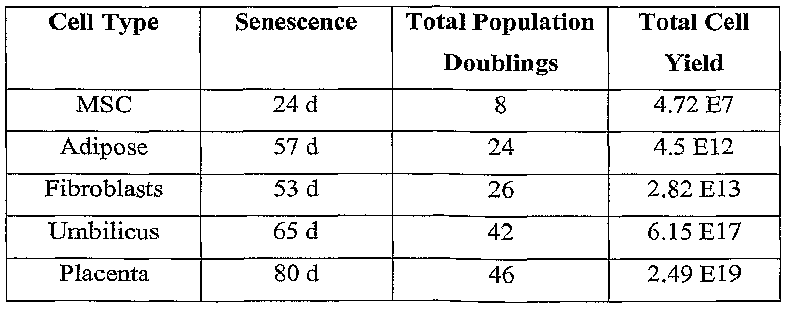


Claims
Priority Applications (6)
| Application Number | Priority Date | Filing Date | Title |
|---|---|---|---|
| AU2006325710A AU2006325710B2 (en) | 2005-12-16 | 2006-12-15 | Compositions and methods for inhibiting adverse immune response in histocompatibility-mismatched transplantation |
| EP06840281.7A EP1971681B1 (en) | 2005-12-16 | 2006-12-15 | Compositions and methods for inhibiting adverse immune response in histocompatibility-mismatched transplantation |
| PL06840281T PL1971681T3 (en) | 2005-12-16 | 2006-12-15 | Compositions and methods for inhibiting adverse immune response in histocompatibility-mismatched transplantation |
| ES06840281.7T ES2642844T3 (en) | 2005-12-16 | 2006-12-15 | Compositions and methods to inhibit an adverse immune response in histocompatibility transplantation that do not match |
| JP2008545989A JP5289970B2 (en) | 2005-12-16 | 2006-12-15 | Compositions and methods for inhibiting reverse immune responses in histocompatible incompatible transplants |
| CN200680052941.3A CN101374946B (en) | 2005-12-16 | 2006-12-15 | Composition and method for suppressing harmful immune response in the unmatched transplanting of histocompatbility |
Applications Claiming Priority (2)
| Application Number | Priority Date | Filing Date | Title |
|---|---|---|---|
| US75139505P | 2005-12-16 | 2005-12-16 | |
| US60/751,395 | 2005-12-16 |
Publications (1)
| Publication Number | Publication Date |
|---|---|
| WO2007070870A1 true WO2007070870A1 (en) | 2007-06-21 |
Family
ID=37806734
Family Applications (1)
| Application Number | Title | Priority Date | Filing Date |
|---|---|---|---|
| PCT/US2006/062157 WO2007070870A1 (en) | 2005-12-16 | 2006-12-15 | Compositions and methods for inhibiting adverse immune response in histocompatibility-mismatched transplantation |
Country Status (8)
| Country | Link |
|---|---|
| US (1) | US9175261B2 (en) |
| EP (1) | EP1971681B1 (en) |
| JP (1) | JP5289970B2 (en) |
| CN (1) | CN101374946B (en) |
| AU (1) | AU2006325710B2 (en) |
| ES (1) | ES2642844T3 (en) |
| PL (1) | PL1971681T3 (en) |
| WO (1) | WO2007070870A1 (en) |
Cited By (11)
| Publication number | Priority date | Publication date | Assignee | Title |
|---|---|---|---|---|
| WO2008088738A2 (en) | 2007-01-17 | 2008-07-24 | Stemnion, Inc. | Novel methods for modulating inflammatory and/or immune responses |
| WO2009062160A2 (en) * | 2007-11-08 | 2009-05-14 | Children's Medical Center Corporation | Rantes levels as a diagnostic and therapeutic for acute graft versus host disease |
| US8658152B2 (en) | 2003-06-27 | 2014-02-25 | DePuy Synthes Products, LLC | Regeneration and repair of neural tissue using postpartum-derived cells |
| US9125906B2 (en) | 2005-12-28 | 2015-09-08 | DePuy Synthes Products, Inc. | Treatment of peripheral vascular disease using umbilical cord tissue-derived cells |
| US9175261B2 (en) | 2005-12-16 | 2015-11-03 | DePuy Synthes Products, Inc. | Human umbilical cord tissue cells for inhibiting adverse immune response in histocompatibility-mismatched transplantation |
| US9572840B2 (en) | 2003-06-27 | 2017-02-21 | DePuy Synthes Products, Inc. | Regeneration and repair of neural tissue using postpartum-derived cells |
| US9592258B2 (en) | 2003-06-27 | 2017-03-14 | DePuy Synthes Products, Inc. | Treatment of neurological injury by administration of human umbilical cord tissue-derived cells |
| US9943552B2 (en) | 2009-03-26 | 2018-04-17 | DePuy Synthes Products, Inc. | hUTC as therapy for Alzheimer's disease |
| US10179900B2 (en) | 2008-12-19 | 2019-01-15 | DePuy Synthes Products, Inc. | Conditioned media and methods of making a conditioned media |
| WO2019165319A1 (en) * | 2018-02-23 | 2019-08-29 | The Board Of Trustees Of The Leland Stanford Junior University | The use of 4-methylumbelliferone to prevent immune rejection in cases of tissue transplantation |
| US10557116B2 (en) | 2008-12-19 | 2020-02-11 | DePuy Synthes Products, Inc. | Treatment of lung and pulmonary diseases and disorders |
Families Citing this family (36)
| Publication number | Priority date | Publication date | Assignee | Title |
|---|---|---|---|---|
| US7311905B2 (en) * | 2002-02-13 | 2007-12-25 | Anthrogenesis Corporation | Embryonic-like stem cells derived from post-partum mammalian placenta, and uses and methods of treatment using said cells |
| US8790637B2 (en) | 2003-06-27 | 2014-07-29 | DePuy Synthes Products, LLC | Repair and regeneration of ocular tissue using postpartum-derived cells |
| US20060223177A1 (en) * | 2003-06-27 | 2006-10-05 | Ethicon Inc. | Postpartum cells derived from umbilical cord tissue, and methods of making and using the same |
| US8518390B2 (en) | 2003-06-27 | 2013-08-27 | Advanced Technologies And Regenerative Medicine, Llc | Treatment of stroke and other acute neural degenerative disorders via intranasal administration of umbilical cord-derived cells |
| US8491883B2 (en) | 2003-06-27 | 2013-07-23 | Advanced Technologies And Regenerative Medicine, Llc | Treatment of amyotrophic lateral sclerosis using umbilical derived cells |
| US7875273B2 (en) | 2004-12-23 | 2011-01-25 | Ethicon, Incorporated | Treatment of Parkinson's disease and related disorders using postpartum derived cells |
| US7875272B2 (en) | 2003-06-27 | 2011-01-25 | Ethicon, Incorporated | Treatment of stroke and other acute neuraldegenerative disorders using postpartum derived cells |
| NZ597304A (en) | 2005-10-13 | 2013-06-28 | Anthrogenesis Corp | Immunomodulation using placental stem cells |
| JP5179376B2 (en) | 2005-12-19 | 2013-04-10 | エシコン・インコーポレイテッド | In vitro growth of postpartum-extracted cells in roller bottles |
| JP5550235B2 (en) | 2005-12-29 | 2014-07-16 | アントフロゲネシス コーポレーション | Placental stem cell population |
| US7993918B2 (en) | 2006-08-04 | 2011-08-09 | Anthrogenesis Corporation | Tumor suppression using placental stem cells |
| JP5450072B2 (en) | 2006-10-12 | 2014-03-26 | エシコン・インコーポレイテッド | Kidney-derived cells and their use in tissue repair and regeneration |
| CN104099290A (en) | 2006-10-23 | 2014-10-15 | 人类起源公司 | Methods and compositions for treatment of bone defects with placental cell populations |
| JP5670053B2 (en) * | 2006-11-13 | 2015-02-18 | エシコン・インコーポレイテッドEthicon, Incorporated | In vitro expansion of postpartum-derived cells using microcarriers |
| CN103356711A (en) | 2007-02-12 | 2013-10-23 | 人类起源公司 | Immunomodulation using placental stem cells |
| AU2008307633C1 (en) | 2007-09-28 | 2015-04-30 | Celularity Inc. | Tumor suppression using human placental perfusate and human placenta-derived intermediate natural killer cells |
| JP5323845B2 (en) | 2007-10-05 | 2013-10-23 | エシコン・インコーポレイテッド | Repair and reconstruction of kidney tissue using human umbilical cord tissue-derived cells |
| US8236538B2 (en) | 2007-12-20 | 2012-08-07 | Advanced Technologies And Regenerative Medicine, Llc | Methods for sterilizing materials containing biologically active agents |
| JP6169316B2 (en) | 2008-08-20 | 2017-07-26 | アンスロジェネシス コーポレーション | Improved cell composition and method for producing the same |
| KR20180108887A (en) | 2008-08-20 | 2018-10-04 | 안트로제네시스 코포레이션 | Treatment of stroke using isolated placental cells |
| MX2011001992A (en) | 2008-08-22 | 2011-03-29 | Anthrogenesis Corp | Methods and compositions for treatment of bone defects with placental cell populations. |
| EP2767833A3 (en) * | 2008-10-21 | 2014-12-17 | Astute Medical, Inc. | Methods and compositions for diagnosis and prognosis of renal injury and renal failure |
| CA2746124A1 (en) | 2008-12-11 | 2010-06-17 | The Governors Of The University Of Alberta | Methods and systems for inducing immunologic tolerance to non-self antigens |
| ES2646750T3 (en) | 2010-01-26 | 2017-12-15 | Anthrogenesis Corporation | Treatment of bone-related cancers using placental stem cells |
| KR20190076060A (en) | 2010-04-07 | 2019-07-01 | 안트로제네시스 코포레이션 | Angiogenesis using placental stem cells |
| EP2555783A1 (en) | 2010-04-08 | 2013-02-13 | Anthrogenesis Corporation | Treatment of sarcoidosis using placental stem cells |
| WO2012009422A1 (en) | 2010-07-13 | 2012-01-19 | Anthrogenesis Corporation | Methods of generating natural killer cells |
| US8969315B2 (en) | 2010-12-31 | 2015-03-03 | Anthrogenesis Corporation | Enhancement of placental stem cell potency using modulatory RNA molecules |
| ES2707579T3 (en) | 2011-06-01 | 2019-04-04 | Celularity Inc | Pain treatment using placental cytoblast |
| CA2880808C (en) * | 2012-08-01 | 2023-01-24 | United Therapeutics Corporation | Treatment of pulmonary arterial hypertension with mesenchymal stem cells |
| AU2014215458A1 (en) | 2013-02-05 | 2015-08-13 | Anthrogenesis Corporation | Natural killer cells from placenta |
| JP6998768B2 (en) | 2015-08-28 | 2022-02-10 | ロート製薬株式会社 | ROR1-positive mesenchymal stem cells and methods for preparing them, pharmaceutical compositions containing ROR1-positive mesenchymal stem cells and methods for preparing them, and methods for preventing or treating diseases using ROR1-positive mesenchymal stem cells. |
| JP7108537B2 (en) | 2016-04-27 | 2022-07-28 | ロート製薬株式会社 | Mesenchymal stem cells expressing at least one cell surface marker selected from the group consisting of CD201, CD46, CD56, CD147 and CD165, a method for preparing the same, and a pharmaceutical composition containing the mesenchymal stem cells and a method for preparing the same |
| CN110418645A (en) | 2017-03-08 | 2019-11-05 | 日本乐敦制药株式会社 | Mescenchymal stem cell containing the ROR1 positive, for prevent or dispose with fibrosis disease pharmaceutical composition, and preparation method thereof and using the ROR1 positive mescenchymal stem cell adjoint fibrosis disease prevention or method of disposal |
| US10701054B2 (en) | 2018-01-31 | 2020-06-30 | Salesforce.Com, Inc. | Systems, methods, and apparatuses for implementing super community and community sidechains with consent management for distributed ledger technologies in a cloud based computing environment |
| CN113164046A (en) | 2018-07-06 | 2021-07-23 | 恩真万特诊疗公司 | Determination of tissue efficacy by quantitative tissue morphology analysis |
Citations (1)
| Publication number | Priority date | Publication date | Assignee | Title |
|---|---|---|---|---|
| WO2005001078A2 (en) * | 2003-06-27 | 2005-01-06 | Ethicon, Incorporated | Regeneration and repair of neural tissue using postpartum-derived cells |
Family Cites Families (225)
| Publication number | Priority date | Publication date | Assignee | Title |
|---|---|---|---|---|
| DE1220832B (en) | 1964-09-22 | 1966-07-14 | Siemens Ag | Drawing nozzle for pulling semiconductor crystals from a melt |
| US3665061A (en) | 1969-07-16 | 1972-05-23 | United States Banknote Corp | Process for producing collagen sponges |
| JPS5651747B2 (en) | 1973-05-31 | 1981-12-08 | ||
| US4216144A (en) | 1977-10-20 | 1980-08-05 | Ashmead H H | Soluble iron proteinates |
| JPS6040439B2 (en) | 1978-03-29 | 1985-09-11 | 大正製薬株式会社 | hydrocortisone derivatives |
| US4352883A (en) | 1979-03-28 | 1982-10-05 | Damon Corporation | Encapsulation of biological material |
| US4487865A (en) | 1983-12-15 | 1984-12-11 | Biomatrix, Inc. | Polymeric articles modified with hyaluronate |
| US5863531A (en) | 1986-04-18 | 1999-01-26 | Advanced Tissue Sciences, Inc. | In vitro preparation of tubular tissue structures by stromal cell culture on a three-dimensional framework |
| US5902741A (en) | 1986-04-18 | 1999-05-11 | Advanced Tissue Sciences, Inc. | Three-dimensional cartilage cultures |
| US5266480A (en) | 1986-04-18 | 1993-11-30 | Advanced Tissue Sciences, Inc. | Three-dimensional skin culture system |
| US4963489A (en) | 1987-04-14 | 1990-10-16 | Marrow-Tech, Inc. | Three-dimensional cell and tissue culture system |
| JPS642643A (en) | 1987-06-26 | 1989-01-06 | Bio Material Yunibaasu:Kk | Artificial skin |
| NZ226750A (en) | 1987-10-29 | 1990-09-26 | Amrad Corp Ltd | Immortalisation of neural precursor cells by introducing a retrovirus vector containing a myc-oncogene |
| US5192553A (en) | 1987-11-12 | 1993-03-09 | Biocyte Corporation | Isolation and preservation of fetal and neonatal hematopoietic stem and progenitor cells of the blood and methods of therapeutic use |
| US5004681B1 (en) | 1987-11-12 | 2000-04-11 | Biocyte Corp | Preservation of fetal and neonatal hematopoietic stem and progenitor cells of the blood |
| GB8803697D0 (en) | 1988-02-17 | 1988-03-16 | Deltanine Research Ltd | Clinical developments using amniotic membrane cells |
| US20030032178A1 (en) | 1988-08-04 | 2003-02-13 | Williams Robert Lindsay | In vitro propagation of embryonic stem cells |
| US4925677A (en) | 1988-08-31 | 1990-05-15 | Theratech, Inc. | Biodegradable hydrogel matrices for the controlled release of pharmacologically active agents |
| FR2646438B1 (en) | 1989-03-20 | 2007-11-02 | Pasteur Institut | A METHOD FOR SPECIFIC REPLACEMENT OF A COPY OF A GENE PRESENT IN THE RECEIVER GENOME BY INTEGRATION OF A GENE DIFFERENT FROM THAT OR INTEGRATION |
| US5437994A (en) | 1989-06-15 | 1995-08-01 | Regents Of The University Of Michigan | Method for the ex vivo replication of stem cells, for the optimization of hematopoietic progenitor cell cultures, and for increasing the metabolism, GM-CSF secretion and/or IL-6 secretion of human stromal cells |
| US5574205A (en) | 1989-07-25 | 1996-11-12 | Cell Genesys | Homologous recombination for universal donor cells and chimeric mammalian hosts |
| US5840580A (en) | 1990-05-01 | 1998-11-24 | Becton Dickinson And Company | Phenotypic characterization of the hematopoietic stem cell |
| WO1992003917A1 (en) | 1990-08-29 | 1992-03-19 | Genpharm International | Homologous recombination in mammalian cells |
| US5342761A (en) | 1990-10-01 | 1994-08-30 | Research Development Foundation | Oncofetal gene, gene product and uses therefor |
| US5811094A (en) | 1990-11-16 | 1998-09-22 | Osiris Therapeutics, Inc. | Connective tissue regeneration using human mesenchymal stem cell preparations |
| US5486359A (en) | 1990-11-16 | 1996-01-23 | Osiris Therapeutics, Inc. | Human mesenchymal stem cells |
| US5286632A (en) | 1991-01-09 | 1994-02-15 | Jones Douglas H | Method for in vivo recombination and mutagenesis |
| NL9100038A (en) | 1991-01-11 | 1992-08-03 | Stamicarbon | ENZYME-CATALYZED PREPARATION OF OPTICALLY ACTIVE CARBONIC ACIDS. |
| US6399369B1 (en) | 1991-07-08 | 2002-06-04 | Neurospheres Holdings Ltd. | Multipotent neural stem cell cDNA libraries |
| AU2515992A (en) | 1991-08-20 | 1993-03-16 | Genpharm International, Inc. | Gene targeting in animal cells using isogenic dna constructs |
| WO1994000484A1 (en) | 1992-06-22 | 1994-01-06 | Young Henry E | Scar inhibitory factor and use thereof |
| US5320962A (en) | 1992-07-22 | 1994-06-14 | Duke University | DNA encoding the human A1 adenosine receptor |
| US5589376A (en) | 1992-07-27 | 1996-12-31 | California Institute Of Technology | Mammalian neural crest stem cells |
| EP0669988B2 (en) | 1992-10-29 | 2009-07-08 | The Australian National University | Angiogenesis inhibitory antibodies |
| US5955343A (en) | 1992-12-28 | 1999-09-21 | Massachusetts Institute Of Technology | Stable macroscopic membranes formed by self-assembly of amphiphilic peptides and uses therefor |
| US5670483A (en) | 1992-12-28 | 1997-09-23 | Massachusetts Insititute Of Technology | Stable macroscopic membranes formed by self-assembly of amphiphilic peptides and uses therefor |
| US5707643A (en) | 1993-02-26 | 1998-01-13 | Santen Pharmaceutical Co., Ltd. | Biodegradable scleral plug |
| WO1994025584A1 (en) | 1993-04-28 | 1994-11-10 | Johns Hopkins University School Of Medicine | Chronic endothelial cell culture under flow |
| IL110589A0 (en) | 1993-08-10 | 1994-11-11 | Bioph Biotech Entw Pharm Gmbh | Growth/differentiation factor of the TGF- beta family |
| US6686198B1 (en) | 1993-10-14 | 2004-02-03 | President And Fellows Of Harvard College | Method of inducing and maintaining neuronal cells |
| US6432711B1 (en) | 1993-11-03 | 2002-08-13 | Diacrin, Inc. | Embryonic stem cells capable of differentiating into desired cell lines |
| DE4406073A1 (en) | 1994-02-24 | 1995-08-31 | Univ Ludwigs Albert | Process for the production of human, clonogenic fibroblasts, process for gene transfection of fibroblasts and fibroblasts obtained in this way |
| US5698518A (en) | 1994-03-30 | 1997-12-16 | Oklahoma Medical Research Foundation | Method for regulating inflammation and tumor growth with calmodulin, calmodulin analogues or calmodulin antagonists |
| US5466233A (en) | 1994-04-25 | 1995-11-14 | Escalon Ophthalmics, Inc. | Tack for intraocular drug delivery and method for inserting and removing same |
| US6703017B1 (en) | 1994-04-28 | 2004-03-09 | Ixion Biotechnology, Inc. | Reversal of insulin-dependent diabetes by islet-producing stem cells, islet progenitor cells and islet-like structures |
| US5834308A (en) | 1994-04-28 | 1998-11-10 | University Of Florida Research Foundation, Inc. | In vitro growth of functional islets of Langerhans |
| US6001647A (en) | 1994-04-28 | 1999-12-14 | Ixion Biotechnology, Inc. | In vitro growth of functional islets of Langerhans and in vivo uses thereof |
| WO1995033421A1 (en) | 1994-06-06 | 1995-12-14 | Case Western Reserve University | Biomatrix for tissue regeneration |
| IL114397A0 (en) | 1994-07-01 | 1995-10-31 | Bioph Biotech Entw Pharm Gmbh | Growth/differentiation factor of the TGF-beta-family |
| US6309853B1 (en) | 1994-08-17 | 2001-10-30 | The Rockfeller University | Modulators of body weight, corresponding nucleic acids and proteins, and diagnostic and therapeutic uses thereof |
| US5725493A (en) | 1994-12-12 | 1998-03-10 | Avery; Robert Logan | Intravitreal medicine delivery |
| US5843780A (en) | 1995-01-20 | 1998-12-01 | Wisconsin Alumni Research Foundation | Primate embryonic stem cells |
| US5736396A (en) | 1995-01-24 | 1998-04-07 | Case Western Reserve University | Lineage-directed induction of human mesenchymal stem cell differentiation |
| US5906934A (en) | 1995-03-14 | 1999-05-25 | Morphogen Pharmaceuticals, Inc. | Mesenchymal stem cells for cartilage repair |
| US5718922A (en) | 1995-05-31 | 1998-02-17 | Schepens Eye Research Institute, Inc. | Intravitreal microsphere drug delivery and method of preparation |
| US5869079A (en) | 1995-06-02 | 1999-02-09 | Oculex Pharmaceuticals, Inc. | Formulation for controlled release of drugs by combining hydrophilic and hydrophobic agents |
| US5693332C1 (en) | 1995-08-11 | 2001-01-09 | Univ California | Human keratinocytes supported on a hydrophilic membrane and methods of using same to effect wound closure |
| US5641750A (en) | 1995-11-29 | 1997-06-24 | Amgen Inc. | Methods for treating photoreceptors using glial cell line-derived neurotrophic factor (GDNF) protein product |
| US6200606B1 (en) | 1996-01-16 | 2001-03-13 | Depuy Orthopaedics, Inc. | Isolation of precursor cells from hematopoietic and nonhematopoietic tissues and their use in vivo bone and cartilage regeneration |
| US5842477A (en) | 1996-02-21 | 1998-12-01 | Advanced Tissue Sciences, Inc. | Method for repairing cartilage |
| EP0888452B1 (en) | 1996-03-15 | 2004-02-11 | Munin Corporation | Human cyr61, an extracellular matrix signalling molecule |
| JP2000508922A (en) | 1996-04-26 | 2000-07-18 | ケース ウエスターン リザーブ ユニバーシティ | Skin regeneration using mesenchymal stem cells |
| US6358737B1 (en) | 1996-07-31 | 2002-03-19 | Board Of Regents, The University Of Texas System | Osteocyte cell lines |
| US6787355B1 (en) | 1996-08-26 | 2004-09-07 | Mcgill University | Multipotent neural stem cells from peripheral tissues and uses thereof |
| US5919702A (en) | 1996-10-23 | 1999-07-06 | Advanced Tissue Science, Inc. | Production of cartilage tissue using cells isolated from Wharton's jelly |
| US6541028B1 (en) | 1997-01-17 | 2003-04-01 | Celadon Science, Llc | Methods for promoting healing of corneal resurfacing wounds |
| EP0989855A4 (en) | 1997-05-13 | 2002-06-12 | Osiris Therapeutics Inc | Osteoarthritis cartilage regeneration using human mesenchymal stem cells |
| WO1998052560A1 (en) | 1997-05-21 | 1998-11-26 | Sloan-Kettering Institute For Cancer Research | Method for increasing the concentration of ascorbic acid in brain tissues of a subject |
| CA2296704C (en) | 1997-07-14 | 2010-10-19 | Osiris Therapeutics, Inc. | Cardiac muscle regeneration using mesenchymal stem cells |
| US5902598A (en) | 1997-08-28 | 1999-05-11 | Control Delivery Systems, Inc. | Sustained release drug delivery devices |
| AU1615599A (en) | 1997-12-02 | 1999-06-16 | Zen Bio, Inc. | Differentiation of adipose stromal cells into osteoblasts and uses thereof |
| US6291240B1 (en) | 1998-01-29 | 2001-09-18 | Advanced Tissue Sciences, Inc. | Cells or tissues with increased protein factors and methods of making and using same |
| AU749675B2 (en) | 1998-03-13 | 2002-07-04 | Mesoblast International Sarl | Uses for human non-autologous mesenchymal stem cells |
| AU755888B2 (en) * | 1998-03-18 | 2003-01-02 | Mesoblast International Sarl | Mesenchymal stem cells for prevention and treatment of immune responses in transplantation |
| US6171610B1 (en) | 1998-04-24 | 2001-01-09 | University Of Massachusetts | Guided development and support of hydrogel-cell compositions |
| ES2246085T3 (en) | 1998-05-07 | 2006-02-01 | University Of South Florida | OSEA MEDULA CELLS AS A SOURCE OF NEURONS USEFUL TO REPAIR THE SPINAL MEDULA AND THE BRAIN. |
| DE69936720T2 (en) | 1998-05-29 | 2008-04-30 | Osiris Therapeutics, Inc. | HUMAN CD45 + AND / OR FIBROBLASTES + MESENCHYMAL STEM CELLS |
| US6323188B1 (en) | 1998-07-01 | 2001-11-27 | Donald L. Weissman | Treatment and prevention of cardiovascular diseases, heart attack, and stroke, primary and subsequent, with help of aspirin and certain vitamins |
| US20040037818A1 (en) | 1998-07-30 | 2004-02-26 | Brand Stephen J. | Treatment for diabetes |
| AU776514B2 (en) | 1998-08-10 | 2004-09-09 | General Hospital Corporation, The | Differentiation of non-insulin producing cells into insulin producing cells by GLP-1 or exendin-4 and uses thereof |
| US5958767A (en) | 1998-08-14 | 1999-09-28 | The Children's Medical Center Corp. | Engraftable human neural stem cells |
| US6284245B1 (en) | 1998-08-25 | 2001-09-04 | Diacrin, Inc. | Neural retinal cells and retinal pigment epithelium cells and their use in treatment of retinal disorders |
| US6610540B1 (en) | 1998-11-18 | 2003-08-26 | California Institute Of Technology | Low oxygen culturing of central nervous system progenitor cells |
| ES2224726T3 (en) | 1998-12-24 | 2005-03-01 | Biosafe S.A. | BLOOD SEPARATION SYSTEM INDICATED IN PARTICULAR FOR THE CONCENTRATION OF HEMATOPOYETIC MOTHER CELLS. |
| EP1144595A2 (en) | 1999-02-04 | 2001-10-17 | McGILL UNIVERSITY | Platform for the differentiation of cells |
| EP1175487A2 (en) | 1999-02-10 | 2002-01-30 | Curis, Inc. | Pancreatic progenitor cells, methods and uses related thereto |
| KR100968164B1 (en) | 1999-03-10 | 2010-07-06 | 더 리전츠 오브 더 유니버시티 오브 캘리포니아 | Adipose-derived stem cells and lattices |
| US20030007954A1 (en) | 1999-04-12 | 2003-01-09 | Gail K. Naughton | Methods for using a three-dimensional stromal tissue to promote angiogenesis |
| WO2000063268A1 (en) | 1999-04-16 | 2000-10-26 | Wm. Marsh Rice University | Poly(propylene fumarate) cross linked with poly(ethylene glycol) |
| US6372494B1 (en) | 1999-05-14 | 2002-04-16 | Advanced Tissue Sciences, Inc. | Methods of making conditioned cell culture medium compositions |
| US6287340B1 (en) | 1999-05-14 | 2001-09-11 | Trustees Of Tufts College | Bioengineered anterior cruciate ligament |
| AU4860900A (en) | 1999-06-02 | 2000-12-18 | Lifebank Services, L.L.C. | Methods of isolation, cryopreservation, and therapeutic use of human amniotic epithelial cells |
| US6333029B1 (en) | 1999-06-30 | 2001-12-25 | Ethicon, Inc. | Porous tissue scaffoldings for the repair of regeneration of tissue |
| US7621606B2 (en) | 2001-08-27 | 2009-11-24 | Advanced Cell Technology, Inc. | Trans-differentiation and re-differentiation of somatic cells and production of cells for cell therapies |
| US6355699B1 (en) | 1999-06-30 | 2002-03-12 | Ethicon, Inc. | Process for manufacturing biomedical foams |
| EP2348104A1 (en) | 1999-08-05 | 2011-07-27 | Mcl Llc | Multipotent adult stem cells and methods for isolation |
| US6429013B1 (en) | 1999-08-19 | 2002-08-06 | Artecel Science, Inc. | Use of adipose tissue-derived stromal cells for chondrocyte differentiation and cartilage repair |
| US6555374B1 (en) | 1999-08-19 | 2003-04-29 | Artecel Sciences, Inc. | Multiple mesodermal lineage differentiation potentials for adipose tissue-derived stromal cells and uses thereof |
| WO2001019379A2 (en) | 1999-09-14 | 2001-03-22 | Children's Medical Center Corporation, The | Methods for treating muscular dystrophy with bone marrow cells |
| US6331313B1 (en) | 1999-10-22 | 2001-12-18 | Oculex Pharmaceticals, Inc. | Controlled-release biocompatible ocular drug delivery implant devices and methods |
| US20030129745A1 (en) | 1999-10-28 | 2003-07-10 | Robl James M. | Gynogenetic or androgenetic production of pluripotent cells and cell lines, and use thereof to produce differentiated cells and tissues |
| EP1099754A1 (en) | 1999-11-10 | 2001-05-16 | Universiteit Leiden | Mesenchymal stem cells and/or progenitor cells, their isolation and use |
| US20020164307A1 (en) | 1999-12-06 | 2002-11-07 | Habener Joel F. | Stem cells of the islets of langerhans and their use in treating diabetes mellitus |
| IL149933A0 (en) | 1999-12-06 | 2002-11-10 | Gen Hospital Corp | Pancreatic stem cells and their use in transplantation |
| US20030082155A1 (en) | 1999-12-06 | 2003-05-01 | Habener Joel F. | Stem cells of the islets of langerhans and their use in treating diabetes mellitus |
| US7785882B2 (en) | 2000-01-18 | 2010-08-31 | Cornell Research Foundation, Inc. | Neuronal progenitor cells from hippocampal tissue and a method for isolating and purifying them |
| US7544509B2 (en) | 2000-01-24 | 2009-06-09 | Mcgill University | Method for preparing stem cell preparations |
| US6610535B1 (en) | 2000-02-10 | 2003-08-26 | Es Cell International Pte Ltd. | Progenitor cells and methods and uses related thereto |
| CA2399434C (en) | 2000-02-11 | 2010-10-19 | The Schepens Eye Research Institute, Inc. | Isolation and transplantation of retinal stem cells |
| AU3685401A (en) | 2000-02-11 | 2001-08-20 | Philadelphia Health | Differentiation of bone marrow cells into neuronal cells and uses therefor |
| US20010053362A1 (en) | 2000-03-09 | 2001-12-20 | Lee Walters | Applications of immune system tolerance to treatment of various diseases |
| US7160724B2 (en) | 2000-03-09 | 2007-01-09 | University Of South Florida | Human cord blood as a source of neural tissue for repair of the brain and spinal cord |
| US6436704B1 (en) | 2000-04-10 | 2002-08-20 | Raven Biotechnologies, Inc. | Human pancreatic epithelial progenitor cells and methods of isolation and use thereof |
| US6673606B1 (en) | 2000-04-12 | 2004-01-06 | The Children's Hospital Of Philadelphia | Therapeutic uses for mesenchymal stromal cells |
| US6375972B1 (en) | 2000-04-26 | 2002-04-23 | Control Delivery Systems, Inc. | Sustained release drug delivery devices, methods of use, and methods of manufacturing thereof |
| WO2001088103A2 (en) | 2000-05-12 | 2001-11-22 | University Of Utah Research Foundation | Compositions and methods for tissue dedifferentiation and regeneration |
| US8273570B2 (en) | 2000-05-16 | 2012-09-25 | Riken | Process of inducing differentiation of embryonic cell to cell expressing neural surface marker using OP9 or PA6 cells |
| US7049072B2 (en) | 2000-06-05 | 2006-05-23 | University Of South Florida | Gene expression analysis of pluri-differentiated mesenchymal progenitor cells and methods for diagnosing a leukemic disease state |
| US6759039B2 (en) | 2000-06-30 | 2004-07-06 | Amcyte, Inc. | Culturing pancreatic stem cells having a specified, intermediate stage of development |
| US6984522B2 (en) | 2000-08-03 | 2006-01-10 | Regents Of The University Of Michigan | Isolation and use of solid tumor stem cells |
| CA2424062A1 (en) | 2000-09-29 | 2002-04-04 | Derek Van Der Kooy | Primitive neural stem cells and method for differentiation of stem cells to neural cells |
| US6639470B1 (en) | 2000-10-06 | 2003-10-28 | Skyworks Solutions, Inc. | Constant current biasing circuit for linear power amplifiers |
| US7560280B2 (en) | 2000-11-03 | 2009-07-14 | Kourion Therapeutics Gmbh | Human cord blood derived unrestricted somatic stem cells (USSC) |
| AU2002230669A1 (en) | 2000-11-06 | 2002-05-15 | The Salk Institute For Biological Studies | Postmortem stem cells |
| RU2322497C2 (en) | 2000-11-30 | 2008-04-20 | Стемрон Инк. | Isolated homozygous stem cell (variants), method for its preparing (variants), method for preparing required cell-ancestor, differentiated cell, group of differentiated cells or tissue type |
| CA2430989A1 (en) | 2000-12-06 | 2002-06-13 | Robert J. Hariri | Method of collecting placental stem cells |
| US7311905B2 (en) | 2002-02-13 | 2007-12-25 | Anthrogenesis Corporation | Embryonic-like stem cells derived from post-partum mammalian placenta, and uses and methods of treatment using said cells |
| US6599323B2 (en) | 2000-12-21 | 2003-07-29 | Ethicon, Inc. | Reinforced tissue implants and methods of manufacture and use |
| CA2365376C (en) | 2000-12-21 | 2006-03-28 | Ethicon, Inc. | Use of reinforced foam implants with enhanced integrity for soft tissue repair and regeneration |
| US20040121460A1 (en) | 2001-01-24 | 2004-06-24 | Lumelsky Nadya L | Differentiation of stem cells to pancreatic endocrine cells |
| WO2002061053A1 (en) | 2001-01-31 | 2002-08-08 | The General Hospital Corporation | Renal stem cells and uses thereof |
| ATE419333T1 (en) | 2001-02-06 | 2009-01-15 | Massachusetts Inst Technology | PEPTIDE STRUCTURE ENCAPSULATION OF TISSUE CELLS AND USES THEREOF |
| US7449180B2 (en) | 2001-02-06 | 2008-11-11 | John Kisiday | Macroscopic scaffold containing amphiphilic peptides encapsulating cells |
| EP2316919B1 (en) | 2001-02-14 | 2015-10-07 | Anthrogenesis Corporation | Post-partum mammalian placenta, its use and placental stem cells therefrom |
| EP1367899A4 (en) | 2001-02-14 | 2004-07-28 | Leo T Furcht | Multipotent adult stem cells, sources thereof, methods of obtaining and maintaining same, methods of differentiation thereof, methods of use thereof and cells derived thereof |
| WO2002063962A1 (en) | 2001-02-14 | 2002-08-22 | Hariri Robert J | Renovation and repopulation of decellularized tissues and cadaveric organs by stem cells |
| US7838292B1 (en) | 2001-03-29 | 2010-11-23 | University Of Louisville Research Foundation, Inc. | Methods for obtaining adult human olfactory progenitor cells |
| CA2442177A1 (en) | 2001-03-29 | 2002-10-10 | Ixion Biotechnology, Inc. | Method for transdifferentiation of non-pancreatic stem cells to the pancreatic differentiation pathway |
| DE10290025T1 (en) | 2001-04-19 | 2003-10-09 | Develogen Ag | Procedure for differentiating stem cells into insulin-producing cells |
| US20030211605A1 (en) | 2001-05-01 | 2003-11-13 | Lee Sang-Hun | Derivation of midbrain dopaminergic neurons from embryonic stem cells |
| US20030022369A1 (en) | 2001-05-18 | 2003-01-30 | Helen Fillmore | Differentiation of specialized dermal and epidermal cells into neuronal cells |
| EP1401282A4 (en) | 2001-05-25 | 2005-03-30 | Cythera Inc | Stem cell differentiation |
| US6402263B1 (en) | 2001-07-24 | 2002-06-11 | Robert Bosch Corporation | Dual actuation master cylinder |
| ATE443758T1 (en) | 2001-08-08 | 2009-10-15 | Levesque Biosciences Inc | COMPOSITIONS AND METHODS FOR ISOLATION, PROPAGATION AND DIFFERENTIATION OF NON-EMBRYONAL HUMAN STEM CELLS AND USES THEREOF |
| US20030211603A1 (en) | 2001-08-14 | 2003-11-13 | Earp David J. | Reprogramming cells for enhanced differentiation capacity using pluripotent stem cells |
| US20030104997A1 (en) | 2001-09-05 | 2003-06-05 | Black Ira B. | Multi-lineage directed induction of bone marrow stromal cell differentiation |
| US20050064587A1 (en) | 2001-09-07 | 2005-03-24 | Lawrence Rosenberg | Pancreatic small cells and uses thereof |
| US9969980B2 (en) | 2001-09-21 | 2018-05-15 | Garnet Biotherapeutics | Cell populations which co-express CD49c and CD90 |
| EP1298201A1 (en) | 2001-09-27 | 2003-04-02 | Cardion AG | Process for the production of cells exhibiting an islet-beta-cell-like state |
| AU2002333022C1 (en) | 2001-09-28 | 2011-06-16 | Es Cell International Pte Ltd | Methods of derivation and propagation of undifferentiated human embryonic stem (HES) cells on feeder-free matrices and human feeder layers |
| US20030138951A1 (en) | 2001-10-18 | 2003-07-24 | Li Yin | Conversion of liver stem and progenitor cells to pancreatic functional cells |
| US7129034B2 (en) | 2001-10-25 | 2006-10-31 | Cedars-Sinai Medical Center | Differentiation of whole bone marrow |
| KR20050044393A (en) | 2001-11-09 | 2005-05-12 | 아르테셀 사이언스, 인크. | Endocrine pancreas differentiation of adipose tissue-derived stromal cells and uses thereof |
| DE60233248D1 (en) | 2001-11-15 | 2009-09-17 | Childrens Medical Center | PROCESS FOR THE ISOLATION, EXPANSION AND DIFFERENTIATION OF FEDERAL STRAIN CELLS FROM CHORION ZOTTE, FRUIT WATER AND PLAZENTA AND THERAPEUTIC USES THEREOF |
| JP3728750B2 (en) | 2001-11-22 | 2005-12-21 | ニプロ株式会社 | Cultured skin and method for producing the same |
| US6712850B2 (en) | 2001-11-30 | 2004-03-30 | Ethicon, Inc. | Porous tissue scaffolds for the repair and regeneration of dermal tissue |
| WO2003048336A2 (en) | 2001-12-04 | 2003-06-12 | Organogenesis Inc. | Cultured cells from pancreatic islets |
| WO2003054171A1 (en) | 2001-12-06 | 2003-07-03 | The Regents Of The University Of California | Method for differentiating islet precursor cells into beta cells |
| IL162131A0 (en) | 2001-12-07 | 2005-11-20 | Geron Corp | Islet cells from human embryonic stem cells |
| JP3934539B2 (en) | 2001-12-12 | 2007-06-20 | 独立行政法人科学技術振興機構 | Adult or postnatal tissue progenitor cells derived from placenta |
| US20030113910A1 (en) | 2001-12-18 | 2003-06-19 | Mike Levanduski | Pluripotent stem cells derived without the use of embryos or fetal tissue |
| US7101546B2 (en) | 2001-12-21 | 2006-09-05 | Amcyte, Inc. | In situ maturation of cultured pancreatic stem cells having a specified, intermediate stage of development |
| CA2470707C (en) | 2001-12-21 | 2014-07-08 | Mount Sinai Hospital | Cellular compositions and methods of making and using them |
| IL162663A0 (en) | 2001-12-28 | 2005-11-20 | Cellartis Ab | A method for the establishment of apluripotent human blastocyst-derived stem cell line |
| US7687505B2 (en) | 2002-01-14 | 2010-03-30 | Board Of Trustees Of The University Of Illinois | Use of modified pyrimidine compounds to promote stem cell migration and proliferation |
| US20030162290A1 (en) | 2002-01-25 | 2003-08-28 | Kazutomo Inoue | Method for inducing differentiation of embryonic stem cells into functioning cells |
| AU2003217357A1 (en) | 2002-02-07 | 2003-09-02 | The Research Foundation Of The State University Of New York | Generation of new insulin cells from progenitor cells present in adult pancreatic islets |
| EP1482787A4 (en) | 2002-02-13 | 2006-02-15 | Anthrogenesis Corp | Embryonic-like stem cells derived from post-partum mammalian placenta and uses and methods of treatment using said cells |
| US7576065B2 (en) | 2002-02-15 | 2009-08-18 | Cornell Research Foundation, Inc. | Enhancing neurotrophin-induced neurogenesis by endogenous neural progenitor cells by concurrent overexpression of brain derived neurotrophic factor and an inhibitor of a pro-gliogenic bone morphogenetic protein |
| CN1281739C (en) | 2002-02-19 | 2006-10-25 | 美迪宝斯特有限公司 | Isolation and culture-expansion methods of mesenchymal stem/progenitor cells from umbilical cord blood and differentation method of umbilical cord blood-derived mesenchymal stem/progenitor cells into |
| US7029915B2 (en) | 2002-02-22 | 2006-04-18 | University Of Florida Research Foundation, Inc. | Method for differentiating rat hepatic stem cells to insulin-producing cells |
| US20030161818A1 (en) | 2002-02-25 | 2003-08-28 | Kansas State University Research Foundation | Cultures, products and methods using stem cells |
| US7736892B2 (en) | 2002-02-25 | 2010-06-15 | Kansas State University Research Foundation | Cultures, products and methods using umbilical cord matrix cells |
| US7150990B2 (en) | 2002-03-06 | 2006-12-19 | Reprocell, Inc. | Self-renewing pluripotent hepatic stem cells |
| JPWO2003080822A1 (en) | 2002-03-27 | 2005-07-28 | ニプロ株式会社 | Placenta-derived mesenchymal cells and their medical uses |
| CA2481385A1 (en) | 2002-04-12 | 2003-10-23 | Celgene Corporation | Modulation of stem and progenitor cell differentiation, assays, and uses thereof |
| US7498171B2 (en) | 2002-04-12 | 2009-03-03 | Anthrogenesis Corporation | Modulation of stem and progenitor cell differentiation, assays, and uses thereof |
| JP4136434B2 (en) | 2002-04-17 | 2008-08-20 | 進 清野 | Induction of insulin-producing cells |
| JP2005523328A (en) | 2002-04-19 | 2005-08-04 | ユニバーシティ オブ ピッツバーグ オブ ザ コモンウェルス システム オブ ハイヤー エデュケーション | Placenta-derived stem cells and uses thereof |
| US20040161419A1 (en) | 2002-04-19 | 2004-08-19 | Strom Stephen C. | Placental stem cells and uses thereof |
| WO2003090689A2 (en) | 2002-04-25 | 2003-11-06 | Wisconsin Alumni Research Foundation | Neurodegenerative disorder treatment using gdnf secreting neural cells |
| US20040029269A1 (en) | 2002-05-07 | 2004-02-12 | Goldman Steven A | Promoter-based isolation, purification, expansion, and transplantation of neuronal progenitor cells, oligodendrocyte progenitor cells, or neural stem cells from a population of embryonic stem cells |
| US20040014662A1 (en) | 2002-05-08 | 2004-01-22 | Per Lindquist | Modulation of neural stem cells and neural progenitor cells |
| AU2003273573A1 (en) | 2002-05-28 | 2003-12-19 | Becton, Dickinson And Company | Expansion and transdifferentiation of human acinar cells |
| MXPA04011851A (en) | 2002-05-30 | 2005-03-31 | Celgene Corp | Methods of using jnk or mkk inhibitors to modulate cell differentiation and to treat myeloproliferative disorders and myelodysplastic syndromes. |
| JP2006505248A (en) | 2002-06-07 | 2006-02-16 | イーエス・セル・インターナショナル・プライヴェート・リミテッド | Methods for controlling differentiation in stem cells |
| AU2003247514A1 (en) | 2002-06-11 | 2003-12-22 | Roy Ogle | Meningeal-derived stem cells |
| US7285415B2 (en) | 2002-07-11 | 2007-10-23 | The Regents Of The University Of California | Oligodendrocytes derived from human embryonic stem cells for remyelination and treatment of spinal cord injury |
| US7390659B2 (en) | 2002-07-16 | 2008-06-24 | The Trustees Of Columbia University In The City Of New York | Methods for inducing differentiation of embryonic stem cells and uses thereof |
| CA2493871A1 (en) | 2002-07-16 | 2004-01-22 | Hadi Aslan | Methods of implanting mesenchymal stem cells for tissue repair and formation |
| CA2493363A1 (en) | 2002-07-29 | 2004-02-05 | Asahi Kasei Kabushiki Kaisha | Stem cells for treating pancreatic damage |
| EP1539930A4 (en) | 2002-07-29 | 2006-08-09 | Es Cell Int Pte Ltd | Multi-step method for the differentiation of insulin positive, glucose |
| WO2004016747A2 (en) | 2002-08-14 | 2004-02-26 | University Of Florida | Bone marrow cell differentiation |
| US20060128014A1 (en) | 2002-08-26 | 2006-06-15 | Johan Haggblad | Compositions and methods for culturing stem cells |
| AU2003265856A1 (en) | 2002-08-28 | 2004-03-19 | University Of Florida | Neurogenesis from hepatic stem cells |
| WO2004023100A2 (en) | 2002-09-06 | 2004-03-18 | Amcyte Inc. | Cd56 positive human adult pancreatic endocrine progenitor cells |
| US9969977B2 (en) | 2002-09-20 | 2018-05-15 | Garnet Biotherapeutics | Cell populations which co-express CD49c and CD90 |
| US20040062753A1 (en) | 2002-09-27 | 2004-04-01 | Alireza Rezania | Composite scaffolds seeded with mammalian cells |
| JP2004210713A (en) | 2002-12-27 | 2004-07-29 | Asahi Kasei Corp | Medical cell preparation derived from placenta |
| BRPI0407221A (en) | 2003-02-11 | 2006-01-31 | John E Davies | Wharton Jelly Extract, Method for Obtaining a Human Parent Cell, Methods for Producing a Cell Population, Cell Populations, and Method for Producing Bone Tissue |
| CA2535034A1 (en) * | 2003-06-27 | 2005-01-27 | Renomedix Institute Inc | Internallly administered therapeutic agents for diseases in central and peripheral nervous system comprising mesenchymal cells as an active ingredient |
| US8790637B2 (en) | 2003-06-27 | 2014-07-29 | DePuy Synthes Products, LLC | Repair and regeneration of ocular tissue using postpartum-derived cells |
| US7875272B2 (en) | 2003-06-27 | 2011-01-25 | Ethicon, Incorporated | Treatment of stroke and other acute neuraldegenerative disorders using postpartum derived cells |
| US7875273B2 (en) | 2004-12-23 | 2011-01-25 | Ethicon, Incorporated | Treatment of Parkinson's disease and related disorders using postpartum derived cells |
| US9572840B2 (en) | 2003-06-27 | 2017-02-21 | DePuy Synthes Products, Inc. | Regeneration and repair of neural tissue using postpartum-derived cells |
| US20060223177A1 (en) | 2003-06-27 | 2006-10-05 | Ethicon Inc. | Postpartum cells derived from umbilical cord tissue, and methods of making and using the same |
| CA2536909A1 (en) | 2003-08-29 | 2005-03-10 | Regents Of The University Of Minnesota | Kidney derived stem cells and methods for their isolation, differentiation and use |
| DE102004043256B4 (en) | 2004-09-07 | 2013-09-19 | Rheinische Friedrich-Wilhelms-Universität Bonn | Scalable process for culturing undifferentiated stem cells in suspension |
| US8039258B2 (en) | 2004-09-28 | 2011-10-18 | Ethicon, Inc. | Tissue-engineering scaffolds containing self-assembled-peptide hydrogels |
| US7655678B2 (en) | 2004-11-30 | 2010-02-02 | Council of Scientfic & Industrial Research | Pharmaceutical composition for the management of tumors |
| WO2006083394A2 (en) | 2004-12-21 | 2006-08-10 | Ethicon, Inc. | Postpartum cells derived from placental tissue, and methods of making, culturing, and using the same |
| US20060153815A1 (en) | 2004-12-21 | 2006-07-13 | Agnieszka Seyda | Tissue engineering devices for the repair and regeneration of tissue |
| US20060171930A1 (en) | 2004-12-21 | 2006-08-03 | Agnieszka Seyda | Postpartum cells derived from umbilical cord tissue, and methods of making, culturing, and using the same |
| WO2006071777A2 (en) | 2004-12-23 | 2006-07-06 | Ethicon Incorporated | Soft tissue repair and regeneration using postpartum-derived cells and cell products |
| WO2006071773A2 (en) | 2004-12-23 | 2006-07-06 | Ethicon Incoporated | Treatment of osteochondral diseases using postpartum-derived cells and products thereof |
| AU2006208944A1 (en) | 2005-01-28 | 2006-08-03 | Imperial College Innovations Limited | Methods for embryonic stem cell culture |
| CN103361303B (en) | 2005-03-31 | 2015-09-30 | 斯丹姆涅恩有限公司 | Prepare a kind of method of the highly purified cell mass from amnion |
| NZ597304A (en) | 2005-10-13 | 2013-06-28 | Anthrogenesis Corp | Immunomodulation using placental stem cells |
| US9175261B2 (en) | 2005-12-16 | 2015-11-03 | DePuy Synthes Products, Inc. | Human umbilical cord tissue cells for inhibiting adverse immune response in histocompatibility-mismatched transplantation |
| JP5179376B2 (en) | 2005-12-19 | 2013-04-10 | エシコン・インコーポレイテッド | In vitro growth of postpartum-extracted cells in roller bottles |
| JP5550235B2 (en) | 2005-12-29 | 2014-07-16 | アントフロゲネシス コーポレーション | Placental stem cell population |
| SG170761A1 (en) | 2006-03-23 | 2011-05-30 | Pluristem Ltd | Methods for cell expansion and uses of cells and conditioned media produced thereby for therapy |
| JP5450072B2 (en) | 2006-10-12 | 2014-03-26 | エシコン・インコーポレイテッド | Kidney-derived cells and their use in tissue repair and regeneration |
| JP5670053B2 (en) | 2006-11-13 | 2015-02-18 | エシコン・インコーポレイテッドEthicon, Incorporated | In vitro expansion of postpartum-derived cells using microcarriers |
| CN103356711A (en) | 2007-02-12 | 2013-10-23 | 人类起源公司 | Immunomodulation using placental stem cells |
-
2006
- 2006-12-15 US US11/611,602 patent/US9175261B2/en not_active Expired - Fee Related
- 2006-12-15 EP EP06840281.7A patent/EP1971681B1/en active Active
- 2006-12-15 CN CN200680052941.3A patent/CN101374946B/en not_active Expired - Fee Related
- 2006-12-15 ES ES06840281.7T patent/ES2642844T3/en active Active
- 2006-12-15 AU AU2006325710A patent/AU2006325710B2/en not_active Ceased
- 2006-12-15 WO PCT/US2006/062157 patent/WO2007070870A1/en active Application Filing
- 2006-12-15 JP JP2008545989A patent/JP5289970B2/en not_active Expired - Fee Related
- 2006-12-15 PL PL06840281T patent/PL1971681T3/en unknown
Patent Citations (1)
| Publication number | Priority date | Publication date | Assignee | Title |
|---|---|---|---|---|
| WO2005001078A2 (en) * | 2003-06-27 | 2005-01-06 | Ethicon, Incorporated | Regeneration and repair of neural tissue using postpartum-derived cells |
Non-Patent Citations (6)
| Title |
|---|
| CARTER DIANE ET AL: "CHARACTERIZATION OF MSC POTENTIAL TO TREAT GVHD USING MOLECULAR MARKERS LINKED TO MSC-MEDIATED IMMUNOSUPPRESSION IN VITRO", BLOOD, W.B.SAUNDERS COMPANY, ORLANDO, FL, US, vol. 106, no. 11 PART 2, November 2005 (2005-11-01), pages 160B, XP008075535, ISSN: 0006-4971 * |
| LE BLANC K ET AL: "Treatment of severe acute graft-versus-host disease with third party haploidentical mesenchymal stem cells", LANCET THE, LANCET LIMITED. LONDON, GB, vol. 363, no. 9419, 1 May 2004 (2004-05-01), pages 1439 - 1441, XP004778108, ISSN: 0140-6736 * |
| LI CHANG DONG ET AL: "Mesenchymal stem cells derived from human placenta suppress allogeneic umbilical cord blood lymphocyte proliferation", CELL RESEARCH, vol. 15, no. 7, July 2005 (2005-07-01), pages 539 - 547, XP009080356, ISSN: 1001-0602 * |
| PANEPUCCI RODRIGO A ET AL: "Comparison of gene expression of umbilical cord vein and bone marrow-derived mesenchymal stem cells", STEM CELLS (MIAMISBURG), vol. 22, no. 7, 2004, pages 1263 - 1278, XP002424029, ISSN: 1066-5099 * |
| ROMANOV Y A ET AL: "Searching for alternative sources of postnatal human mesenchymal stem cells: Candidate MSC-like cells from umbilical cord", STEM CELLS, ALPHAMED PRESS, DAYTON, OH, US, vol. 21, no. 1, 2003, pages 105 - 110, XP002331940, ISSN: 1066-5099 * |
| ZHANG Y ET AL: "COMPARISON OF MESENCHYMAL STEM CELLS FROM HUMAN PLACENTA AND BONE MARROW", CHINESE MEDICAL JOURNAL / ZHONGHUA YIXUE ZAZHI YINGWEN BAN, CHINESE MEDICAL ASSOCIATION, BEIJING, CN, vol. 117, no. 6, June 2004 (2004-06-01), pages 882 - 887, XP009042592, ISSN: 0366-6999 * |
Cited By (15)
| Publication number | Priority date | Publication date | Assignee | Title |
|---|---|---|---|---|
| US8658152B2 (en) | 2003-06-27 | 2014-02-25 | DePuy Synthes Products, LLC | Regeneration and repair of neural tissue using postpartum-derived cells |
| US9592258B2 (en) | 2003-06-27 | 2017-03-14 | DePuy Synthes Products, Inc. | Treatment of neurological injury by administration of human umbilical cord tissue-derived cells |
| US9572840B2 (en) | 2003-06-27 | 2017-02-21 | DePuy Synthes Products, Inc. | Regeneration and repair of neural tissue using postpartum-derived cells |
| US9175261B2 (en) | 2005-12-16 | 2015-11-03 | DePuy Synthes Products, Inc. | Human umbilical cord tissue cells for inhibiting adverse immune response in histocompatibility-mismatched transplantation |
| US9585918B2 (en) | 2005-12-28 | 2017-03-07 | DePuy Synthes Products, Inc. | Treatment of peripheral vascular disease using umbilical cord tissue-derived cells |
| US9125906B2 (en) | 2005-12-28 | 2015-09-08 | DePuy Synthes Products, Inc. | Treatment of peripheral vascular disease using umbilical cord tissue-derived cells |
| EP2118267A4 (en) * | 2007-01-17 | 2010-04-07 | Stemnion Inc | Novel methods for modulating inflammatory and/or immune responses |
| EP2118267A2 (en) * | 2007-01-17 | 2009-11-18 | Stemnion, Inc. | Novel methods for modulating inflammatory and/or immune responses |
| WO2008088738A2 (en) | 2007-01-17 | 2008-07-24 | Stemnion, Inc. | Novel methods for modulating inflammatory and/or immune responses |
| WO2009062160A3 (en) * | 2007-11-08 | 2009-09-11 | Children's Medical Center Corporation | Rantes levels as a diagnostic and therapeutic for acute graft versus host disease |
| WO2009062160A2 (en) * | 2007-11-08 | 2009-05-14 | Children's Medical Center Corporation | Rantes levels as a diagnostic and therapeutic for acute graft versus host disease |
| US10179900B2 (en) | 2008-12-19 | 2019-01-15 | DePuy Synthes Products, Inc. | Conditioned media and methods of making a conditioned media |
| US10557116B2 (en) | 2008-12-19 | 2020-02-11 | DePuy Synthes Products, Inc. | Treatment of lung and pulmonary diseases and disorders |
| US9943552B2 (en) | 2009-03-26 | 2018-04-17 | DePuy Synthes Products, Inc. | hUTC as therapy for Alzheimer's disease |
| WO2019165319A1 (en) * | 2018-02-23 | 2019-08-29 | The Board Of Trustees Of The Leland Stanford Junior University | The use of 4-methylumbelliferone to prevent immune rejection in cases of tissue transplantation |
Also Published As
| Publication number | Publication date |
|---|---|
| EP1971681B1 (en) | 2017-08-23 |
| JP2009519978A (en) | 2009-05-21 |
| PL1971681T3 (en) | 2018-01-31 |
| EP1971681A1 (en) | 2008-09-24 |
| AU2006325710B2 (en) | 2012-05-17 |
| US9175261B2 (en) | 2015-11-03 |
| ES2642844T3 (en) | 2017-11-20 |
| JP5289970B2 (en) | 2013-09-11 |
| AU2006325710A1 (en) | 2007-06-21 |
| CN101374946B (en) | 2017-07-18 |
| CN101374946A (en) | 2009-02-25 |
| US20070264269A1 (en) | 2007-11-15 |
Similar Documents
| Publication | Publication Date | Title |
|---|---|---|
| EP1971681B1 (en) | Compositions and methods for inhibiting adverse immune response in histocompatibility-mismatched transplantation | |
| US10500234B2 (en) | Postpartum cells derived from umbilical cord tissue, and methods of making and using the same | |
| EP1831356B1 (en) | Postpartum cells derived from umbilical cord tissue, and methods of making and using the same | |
| US10557116B2 (en) | Treatment of lung and pulmonary diseases and disorders | |
| AU2009335962B2 (en) | Conditioned medium and methods of making the same |
Legal Events
| Date | Code | Title | Description |
|---|---|---|---|
| 121 | Ep: the epo has been informed by wipo that ep was designated in this application | ||
| WWE | Wipo information: entry into national phase |
Ref document number: 2008545989 Country of ref document: JP |
|
| NENP | Non-entry into the national phase |
Ref country code: DE |
|
| WWE | Wipo information: entry into national phase |
Ref document number: 2006325710 Country of ref document: AU |
|
| REEP | Request for entry into the european phase |
Ref document number: 2006840281 Country of ref document: EP |
|
| WWE | Wipo information: entry into national phase |
Ref document number: 2887/KOLNP/2008 Country of ref document: IN Ref document number: 2006840281 Country of ref document: EP |
|
| ENP | Entry into the national phase |
Ref document number: 2006325710 Country of ref document: AU Date of ref document: 20061215 Kind code of ref document: A |
|
| WWE | Wipo information: entry into national phase |
Ref document number: 200680052941.3 Country of ref document: CN |