WO2009099961A2 - Engineered antibody constant domain molecules - Google Patents
Engineered antibody constant domain molecules Download PDFInfo
- Publication number
- WO2009099961A2 WO2009099961A2 PCT/US2009/032692 US2009032692W WO2009099961A2 WO 2009099961 A2 WO2009099961 A2 WO 2009099961A2 US 2009032692 W US2009032692 W US 2009032692W WO 2009099961 A2 WO2009099961 A2 WO 2009099961A2
- Authority
- WO
- WIPO (PCT)
- Prior art keywords
- domain
- polypeptide
- loop
- antigen
- amino acid
- Prior art date
Links
- 239000000427 antigen Substances 0.000 claims abstract description 139
- 108091007433 antigens Proteins 0.000 claims abstract description 139
- 102000036639 antigens Human genes 0.000 claims abstract description 139
- 108010047041 Complementarity Determining Regions Proteins 0.000 claims abstract description 83
- 239000012634 fragment Substances 0.000 claims abstract description 79
- 230000027455 binding Effects 0.000 claims abstract description 66
- 230000035772 mutation Effects 0.000 claims abstract description 43
- 108090000765 processed proteins & peptides Proteins 0.000 claims description 137
- 102000004196 processed proteins & peptides Human genes 0.000 claims description 124
- 229920001184 polypeptide Polymers 0.000 claims description 122
- 235000001014 amino acid Nutrition 0.000 claims description 109
- 210000004027 cell Anatomy 0.000 claims description 103
- 108090000623 proteins and genes Proteins 0.000 claims description 100
- 238000000034 method Methods 0.000 claims description 98
- 150000007523 nucleic acids Chemical class 0.000 claims description 87
- 102000004169 proteins and genes Human genes 0.000 claims description 86
- 125000003275 alpha amino acid group Chemical group 0.000 claims description 78
- 235000018102 proteins Nutrition 0.000 claims description 77
- 150000001413 amino acids Chemical class 0.000 claims description 74
- 108060003951 Immunoglobulin Proteins 0.000 claims description 51
- 102000018358 immunoglobulin Human genes 0.000 claims description 51
- 102000039446 nucleic acids Human genes 0.000 claims description 51
- 108020004707 nucleic acids Proteins 0.000 claims description 51
- 206010028980 Neoplasm Diseases 0.000 claims description 46
- 239000012636 effector Substances 0.000 claims description 46
- 239000000203 mixture Substances 0.000 claims description 45
- 238000006467 substitution reaction Methods 0.000 claims description 42
- 239000013598 vector Substances 0.000 claims description 31
- 241000700605 Viruses Species 0.000 claims description 28
- 239000002245 particle Substances 0.000 claims description 28
- 201000011510 cancer Diseases 0.000 claims description 23
- 241000725303 Human immunodeficiency virus Species 0.000 claims description 22
- 108010087819 Fc receptors Proteins 0.000 claims description 21
- 102000009109 Fc receptors Human genes 0.000 claims description 21
- 239000003814 drug Substances 0.000 claims description 19
- 210000004899 c-terminal region Anatomy 0.000 claims description 15
- 244000052769 pathogen Species 0.000 claims description 15
- 125000000151 cysteine group Chemical group N[C@@H](CS)C(=O)* 0.000 claims description 14
- 230000001717 pathogenic effect Effects 0.000 claims description 14
- 238000012217 deletion Methods 0.000 claims description 12
- 230000037430 deletion Effects 0.000 claims description 12
- 238000011282 treatment Methods 0.000 claims description 12
- 241000894006 Bacteria Species 0.000 claims description 11
- 238000010367 cloning Methods 0.000 claims description 10
- 208000023275 Autoimmune disease Diseases 0.000 claims description 9
- 239000003937 drug carrier Substances 0.000 claims description 8
- 238000002360 preparation method Methods 0.000 claims description 7
- 230000001363 autoimmune Effects 0.000 claims description 6
- 208000032839 leukemia Diseases 0.000 claims description 6
- 238000004519 manufacturing process Methods 0.000 claims description 5
- 206010006187 Breast cancer Diseases 0.000 claims description 4
- 208000026310 Breast neoplasm Diseases 0.000 claims description 4
- 206010009944 Colon cancer Diseases 0.000 claims description 4
- 206010025323 Lymphomas Diseases 0.000 claims description 4
- 206010035226 Plasma cell myeloma Diseases 0.000 claims description 4
- 206010060862 Prostate cancer Diseases 0.000 claims description 4
- 208000000236 Prostatic Neoplasms Diseases 0.000 claims description 4
- 208000027866 inflammatory disease Diseases 0.000 claims description 4
- 208000020816 lung neoplasm Diseases 0.000 claims description 4
- 201000001441 melanoma Diseases 0.000 claims description 4
- 208000031886 HIV Infections Diseases 0.000 claims description 3
- 208000037357 HIV infectious disease Diseases 0.000 claims description 3
- 208000034578 Multiple myelomas Diseases 0.000 claims description 3
- 208000006265 Renal cell carcinoma Diseases 0.000 claims description 3
- 208000033519 human immunodeficiency virus infectious disease Diseases 0.000 claims description 3
- 102100026120 IgG receptor FcRn large subunit p51 Human genes 0.000 claims description 2
- 206010058467 Lung neoplasm malignant Diseases 0.000 claims description 2
- 201000005202 lung cancer Diseases 0.000 claims description 2
- 108010068617 neonatal Fc receptor Proteins 0.000 claims description 2
- 102000000989 Complement System Proteins Human genes 0.000 claims 2
- 108010069112 Complement System Proteins Proteins 0.000 claims 2
- 208000029742 colonic neoplasm Diseases 0.000 claims 1
- 208000035250 cutaneous malignant susceptibility to 1 melanoma Diseases 0.000 claims 1
- 231100000956 nontoxicity Toxicity 0.000 abstract description 3
- 125000002496 methyl group Chemical group [H]C([H])([H])* 0.000 description 211
- 230000014509 gene expression Effects 0.000 description 35
- 108091028043 Nucleic acid sequence Proteins 0.000 description 33
- 108020004414 DNA Proteins 0.000 description 28
- 238000003752 polymerase chain reaction Methods 0.000 description 25
- 239000011230 binding agent Substances 0.000 description 23
- FAPWRFPIFSIZLT-UHFFFAOYSA-M Sodium chloride Chemical compound [Na+].[Cl-] FAPWRFPIFSIZLT-UHFFFAOYSA-M 0.000 description 22
- 239000000872 buffer Substances 0.000 description 21
- -1 radioisotopes Chemical class 0.000 description 21
- 238000002983 circular dichroism Methods 0.000 description 20
- 239000002773 nucleotide Substances 0.000 description 20
- 125000003729 nucleotide group Chemical group 0.000 description 19
- 230000002441 reversible effect Effects 0.000 description 19
- 210000001519 tissue Anatomy 0.000 description 19
- 230000006870 function Effects 0.000 description 18
- 239000000126 substance Substances 0.000 description 18
- 230000001225 therapeutic effect Effects 0.000 description 18
- 108010053187 Diphtheria Toxin Proteins 0.000 description 17
- 102000016607 Diphtheria Toxin Human genes 0.000 description 17
- 238000002965 ELISA Methods 0.000 description 17
- 101000762949 Pseudomonas aeruginosa (strain ATCC 15692 / DSM 22644 / CIP 104116 / JCM 14847 / LMG 12228 / 1C / PRS 101 / PAO1) Exotoxin A Proteins 0.000 description 15
- 239000003795 chemical substances by application Substances 0.000 description 15
- 150000001875 compounds Chemical class 0.000 description 15
- 230000009870 specific binding Effects 0.000 description 15
- XSQUKJJJFZCRTK-UHFFFAOYSA-N Urea Chemical compound NC(N)=O XSQUKJJJFZCRTK-UHFFFAOYSA-N 0.000 description 14
- 239000003446 ligand Substances 0.000 description 14
- 239000003053 toxin Substances 0.000 description 14
- 231100000765 toxin Toxicity 0.000 description 14
- 108700012359 toxins Proteins 0.000 description 14
- 230000002637 immunotoxin Effects 0.000 description 13
- 239000002596 immunotoxin Substances 0.000 description 13
- 229940051026 immunotoxin Drugs 0.000 description 13
- 231100000608 immunotoxin Toxicity 0.000 description 13
- 238000013518 transcription Methods 0.000 description 13
- 230000035897 transcription Effects 0.000 description 13
- 239000011324 bead Substances 0.000 description 12
- 102000004190 Enzymes Human genes 0.000 description 11
- 108090000790 Enzymes Proteins 0.000 description 11
- 229940088598 enzyme Drugs 0.000 description 11
- 238000003199 nucleic acid amplification method Methods 0.000 description 11
- YBJHBAHKTGYVGT-ZKWXMUAHSA-N (+)-Biotin Chemical compound N1C(=O)N[C@@H]2[C@H](CCCCC(=O)O)SC[C@@H]21 YBJHBAHKTGYVGT-ZKWXMUAHSA-N 0.000 description 10
- 230000003321 amplification Effects 0.000 description 10
- 230000001580 bacterial effect Effects 0.000 description 10
- 238000001514 detection method Methods 0.000 description 10
- 201000010099 disease Diseases 0.000 description 10
- 208000037265 diseases, disorders, signs and symptoms Diseases 0.000 description 10
- 238000009472 formulation Methods 0.000 description 10
- 230000003993 interaction Effects 0.000 description 10
- 102000040430 polynucleotide Human genes 0.000 description 10
- 108091033319 polynucleotide Proteins 0.000 description 10
- 239000002157 polynucleotide Substances 0.000 description 10
- 239000011780 sodium chloride Substances 0.000 description 10
- 102000053602 DNA Human genes 0.000 description 9
- 230000000890 antigenic effect Effects 0.000 description 9
- 238000012512 characterization method Methods 0.000 description 9
- 238000000113 differential scanning calorimetry Methods 0.000 description 9
- 230000028993 immune response Effects 0.000 description 9
- 239000000463 material Substances 0.000 description 9
- 108020004999 messenger RNA Proteins 0.000 description 9
- 238000000746 purification Methods 0.000 description 9
- 239000000523 sample Substances 0.000 description 9
- 108010021625 Immunoglobulin Fragments Proteins 0.000 description 8
- 230000010056 antibody-dependent cellular cytotoxicity Effects 0.000 description 8
- 238000003556 assay Methods 0.000 description 8
- 239000002299 complementary DNA Substances 0.000 description 8
- 238000013461 design Methods 0.000 description 8
- 238000004091 panning Methods 0.000 description 8
- 239000000047 product Substances 0.000 description 8
- 239000007790 solid phase Substances 0.000 description 8
- 238000003786 synthesis reaction Methods 0.000 description 8
- 230000009466 transformation Effects 0.000 description 8
- XLYOFNOQVPJJNP-UHFFFAOYSA-N water Substances O XLYOFNOQVPJJNP-UHFFFAOYSA-N 0.000 description 8
- 108091032973 (ribonucleotides)n+m Proteins 0.000 description 7
- 108010083359 Antigen Receptors Proteins 0.000 description 7
- 102000006306 Antigen Receptors Human genes 0.000 description 7
- 241000588724 Escherichia coli Species 0.000 description 7
- 102000008394 Immunoglobulin Fragments Human genes 0.000 description 7
- 238000005481 NMR spectroscopy Methods 0.000 description 7
- 108010039491 Ricin Proteins 0.000 description 7
- 230000015572 biosynthetic process Effects 0.000 description 7
- 239000004202 carbamide Substances 0.000 description 7
- 238000003776 cleavage reaction Methods 0.000 description 7
- 229940079593 drug Drugs 0.000 description 7
- 230000000694 effects Effects 0.000 description 7
- 230000002255 enzymatic effect Effects 0.000 description 7
- 239000013604 expression vector Substances 0.000 description 7
- 208000015181 infectious disease Diseases 0.000 description 7
- 238000006386 neutralization reaction Methods 0.000 description 7
- 230000002829 reductive effect Effects 0.000 description 7
- 230000007017 scission Effects 0.000 description 7
- 238000000926 separation method Methods 0.000 description 7
- 241000894007 species Species 0.000 description 7
- 238000001228 spectrum Methods 0.000 description 7
- 230000008685 targeting Effects 0.000 description 7
- 229940124597 therapeutic agent Drugs 0.000 description 7
- 230000014616 translation Effects 0.000 description 7
- 241000701022 Cytomegalovirus Species 0.000 description 6
- WQZGKKKJIJFFOK-GASJEMHNSA-N Glucose Natural products OC[C@H]1OC(O)[C@H](O)[C@@H](O)[C@@H]1O WQZGKKKJIJFFOK-GASJEMHNSA-N 0.000 description 6
- PEDCQBHIVMGVHV-UHFFFAOYSA-N Glycerine Chemical compound OCC(O)CO PEDCQBHIVMGVHV-UHFFFAOYSA-N 0.000 description 6
- 241000701044 Human gammaherpesvirus 4 Species 0.000 description 6
- XUJNEKJLAYXESH-REOHCLBHSA-N L-Cysteine Chemical compound SC[C@H](N)C(O)=O XUJNEKJLAYXESH-REOHCLBHSA-N 0.000 description 6
- 241001465754 Metazoa Species 0.000 description 6
- 241001529936 Murinae Species 0.000 description 6
- 241001112090 Pseudovirus Species 0.000 description 6
- 241000725643 Respiratory syncytial virus Species 0.000 description 6
- 102100036011 T-cell surface glycoprotein CD4 Human genes 0.000 description 6
- 238000004458 analytical method Methods 0.000 description 6
- 230000000295 complement effect Effects 0.000 description 6
- 238000004520 electroporation Methods 0.000 description 6
- 238000001943 fluorescence-activated cell sorting Methods 0.000 description 6
- 239000000499 gel Substances 0.000 description 6
- RAXXELZNTBOGNW-UHFFFAOYSA-N imidazole Natural products C1=CNC=N1 RAXXELZNTBOGNW-UHFFFAOYSA-N 0.000 description 6
- 238000003018 immunoassay Methods 0.000 description 6
- 210000004962 mammalian cell Anatomy 0.000 description 6
- 239000002609 medium Substances 0.000 description 6
- 230000003472 neutralizing effect Effects 0.000 description 6
- 230000009467 reduction Effects 0.000 description 6
- 230000010076 replication Effects 0.000 description 6
- 239000007787 solid Substances 0.000 description 6
- 239000000243 solution Substances 0.000 description 6
- 238000012360 testing method Methods 0.000 description 6
- 239000003981 vehicle Substances 0.000 description 6
- 230000003612 virological effect Effects 0.000 description 6
- 108010066676 Abrin Proteins 0.000 description 5
- 108020004705 Codon Proteins 0.000 description 5
- 208000035473 Communicable disease Diseases 0.000 description 5
- 241000124008 Mammalia Species 0.000 description 5
- 108091034117 Oligonucleotide Proteins 0.000 description 5
- 241000194017 Streptococcus Species 0.000 description 5
- 125000000539 amino acid group Chemical group 0.000 description 5
- 239000012472 biological sample Substances 0.000 description 5
- 229960002685 biotin Drugs 0.000 description 5
- 235000020958 biotin Nutrition 0.000 description 5
- 239000011616 biotin Substances 0.000 description 5
- 229910052799 carbon Inorganic materials 0.000 description 5
- 125000003178 carboxy group Chemical group [H]OC(*)=O 0.000 description 5
- 239000000969 carrier Substances 0.000 description 5
- 238000006243 chemical reaction Methods 0.000 description 5
- 238000010276 construction Methods 0.000 description 5
- 235000018417 cysteine Nutrition 0.000 description 5
- 239000008121 dextrose Substances 0.000 description 5
- 210000000987 immune system Anatomy 0.000 description 5
- 229940127121 immunoconjugate Drugs 0.000 description 5
- 210000003000 inclusion body Anatomy 0.000 description 5
- 239000002502 liposome Substances 0.000 description 5
- 238000004949 mass spectrometry Methods 0.000 description 5
- 230000035515 penetration Effects 0.000 description 5
- 238000002823 phage display Methods 0.000 description 5
- 239000013612 plasmid Substances 0.000 description 5
- 239000000843 powder Substances 0.000 description 5
- 230000002285 radioactive effect Effects 0.000 description 5
- 238000012163 sequencing technique Methods 0.000 description 5
- 238000002415 sodium dodecyl sulfate polyacrylamide gel electrophoresis Methods 0.000 description 5
- 239000006228 supernatant Substances 0.000 description 5
- 239000000725 suspension Substances 0.000 description 5
- 238000013519 translation Methods 0.000 description 5
- 238000004461 1H-15N HSQC Methods 0.000 description 4
- 208000031261 Acute myeloid leukaemia Diseases 0.000 description 4
- 108091026890 Coding region Proteins 0.000 description 4
- 241000711573 Coronaviridae Species 0.000 description 4
- 241001115402 Ebolavirus Species 0.000 description 4
- 241000709661 Enterovirus Species 0.000 description 4
- 208000009386 Experimental Arthritis Diseases 0.000 description 4
- 241000711950 Filoviridae Species 0.000 description 4
- 108090000288 Glycoproteins Proteins 0.000 description 4
- 102000003886 Glycoproteins Human genes 0.000 description 4
- 241000700721 Hepatitis B virus Species 0.000 description 4
- 241000701085 Human alphaherpesvirus 3 Species 0.000 description 4
- 201000003793 Myelodysplastic syndrome Diseases 0.000 description 4
- 208000033776 Myeloid Acute Leukemia Diseases 0.000 description 4
- 238000012565 NMR experiment Methods 0.000 description 4
- 108091005461 Nucleic proteins Proteins 0.000 description 4
- 206010039491 Sarcoma Diseases 0.000 description 4
- WQZGKKKJIJFFOK-VFUOTHLCSA-N beta-D-glucose Chemical compound OC[C@H]1O[C@@H](O)[C@H](O)[C@@H](O)[C@@H]1O WQZGKKKJIJFFOK-VFUOTHLCSA-N 0.000 description 4
- 239000003153 chemical reaction reagent Substances 0.000 description 4
- 230000004540 complement-dependent cytotoxicity Effects 0.000 description 4
- XUJNEKJLAYXESH-UHFFFAOYSA-N cysteine Natural products SCC(N)C(O)=O XUJNEKJLAYXESH-UHFFFAOYSA-N 0.000 description 4
- 231100000433 cytotoxic Toxicity 0.000 description 4
- 230000001472 cytotoxic effect Effects 0.000 description 4
- 230000029087 digestion Effects 0.000 description 4
- 239000000539 dimer Substances 0.000 description 4
- 239000012530 fluid Substances 0.000 description 4
- 230000002538 fungal effect Effects 0.000 description 4
- 238000002523 gelfiltration Methods 0.000 description 4
- 230000002068 genetic effect Effects 0.000 description 4
- 206010073071 hepatocellular carcinoma Diseases 0.000 description 4
- 230000001900 immune effect Effects 0.000 description 4
- 230000001939 inductive effect Effects 0.000 description 4
- 239000004615 ingredient Substances 0.000 description 4
- 230000000670 limiting effect Effects 0.000 description 4
- HQKMJHAJHXVSDF-UHFFFAOYSA-L magnesium stearate Chemical compound [Mg+2].CCCCCCCCCCCCCCCCCC([O-])=O.CCCCCCCCCCCCCCCCCC([O-])=O HQKMJHAJHXVSDF-UHFFFAOYSA-L 0.000 description 4
- 230000005291 magnetic effect Effects 0.000 description 4
- 230000004048 modification Effects 0.000 description 4
- 238000012986 modification Methods 0.000 description 4
- 239000000178 monomer Substances 0.000 description 4
- 229910052757 nitrogen Inorganic materials 0.000 description 4
- 244000045947 parasite Species 0.000 description 4
- 239000008194 pharmaceutical composition Substances 0.000 description 4
- 229920000642 polymer Polymers 0.000 description 4
- 239000003755 preservative agent Substances 0.000 description 4
- 238000011160 research Methods 0.000 description 4
- 238000001542 size-exclusion chromatography Methods 0.000 description 4
- 231100000331 toxic Toxicity 0.000 description 4
- 230000002588 toxic effect Effects 0.000 description 4
- 230000001988 toxicity Effects 0.000 description 4
- 231100000419 toxicity Toxicity 0.000 description 4
- 241000701161 unidentified adenovirus Species 0.000 description 4
- QTBSBXVTEAMEQO-UHFFFAOYSA-N Acetic acid Chemical compound CC(O)=O QTBSBXVTEAMEQO-UHFFFAOYSA-N 0.000 description 3
- 229920000936 Agarose Polymers 0.000 description 3
- 102100023635 Alpha-fetoprotein Human genes 0.000 description 3
- 206010005003 Bladder cancer Diseases 0.000 description 3
- 241000228405 Blastomyces dermatitidis Species 0.000 description 3
- 108030001720 Bontoxilysin Proteins 0.000 description 3
- 241000222122 Candida albicans Species 0.000 description 3
- 108010022366 Carcinoembryonic Antigen Proteins 0.000 description 3
- 102100025475 Carcinoembryonic antigen-related cell adhesion molecule 5 Human genes 0.000 description 3
- 108091033380 Coding strand Proteins 0.000 description 3
- 241000186227 Corynebacterium diphtheriae Species 0.000 description 3
- 241000221204 Cryptococcus neoformans Species 0.000 description 3
- LFQSCWFLJHTTHZ-UHFFFAOYSA-N Ethanol Chemical compound CCO LFQSCWFLJHTTHZ-UHFFFAOYSA-N 0.000 description 3
- 208000006168 Ewing Sarcoma Diseases 0.000 description 3
- 241000233866 Fungi Species 0.000 description 3
- 241000228404 Histoplasma capsulatum Species 0.000 description 3
- 108700005091 Immunoglobulin Genes Proteins 0.000 description 3
- 108091092195 Intron Proteins 0.000 description 3
- OFOBLEOULBTSOW-UHFFFAOYSA-N Malonic acid Chemical compound OC(=O)CC(O)=O OFOBLEOULBTSOW-UHFFFAOYSA-N 0.000 description 3
- 108010085220 Multiprotein Complexes Proteins 0.000 description 3
- 102000007474 Multiprotein Complexes Human genes 0.000 description 3
- 241000699666 Mus <mouse, genus> Species 0.000 description 3
- 206010029260 Neuroblastoma Diseases 0.000 description 3
- MUBZPKHOEPUJKR-UHFFFAOYSA-N Oxalic acid Chemical compound OC(=O)C(O)=O MUBZPKHOEPUJKR-UHFFFAOYSA-N 0.000 description 3
- 206010061902 Pancreatic neoplasm Diseases 0.000 description 3
- 208000002606 Paramyxoviridae Infections Diseases 0.000 description 3
- 108010093965 Polymyxin B Proteins 0.000 description 3
- KWYUFKZDYYNOTN-UHFFFAOYSA-M Potassium hydroxide Chemical compound [OH-].[K+] KWYUFKZDYYNOTN-UHFFFAOYSA-M 0.000 description 3
- DNIAPMSPPWPWGF-UHFFFAOYSA-N Propylene glycol Chemical compound CC(O)CO DNIAPMSPPWPWGF-UHFFFAOYSA-N 0.000 description 3
- 240000004808 Saccharomyces cerevisiae Species 0.000 description 3
- 241000700584 Simplexvirus Species 0.000 description 3
- HEMHJVSKTPXQMS-UHFFFAOYSA-M Sodium hydroxide Chemical compound [OH-].[Na+] HEMHJVSKTPXQMS-UHFFFAOYSA-M 0.000 description 3
- 230000005867 T cell response Effects 0.000 description 3
- 241000700618 Vaccinia virus Species 0.000 description 3
- JLCPHMBAVCMARE-UHFFFAOYSA-N [3-[[3-[[3-[[3-[[3-[[3-[[3-[[3-[[3-[[3-[[3-[[5-(2-amino-6-oxo-1H-purin-9-yl)-3-[[3-[[3-[[3-[[3-[[3-[[5-(2-amino-6-oxo-1H-purin-9-yl)-3-[[5-(2-amino-6-oxo-1H-purin-9-yl)-3-hydroxyoxolan-2-yl]methoxy-hydroxyphosphoryl]oxyoxolan-2-yl]methoxy-hydroxyphosphoryl]oxy-5-(5-methyl-2,4-dioxopyrimidin-1-yl)oxolan-2-yl]methoxy-hydroxyphosphoryl]oxy-5-(6-aminopurin-9-yl)oxolan-2-yl]methoxy-hydroxyphosphoryl]oxy-5-(6-aminopurin-9-yl)oxolan-2-yl]methoxy-hydroxyphosphoryl]oxy-5-(6-aminopurin-9-yl)oxolan-2-yl]methoxy-hydroxyphosphoryl]oxy-5-(6-aminopurin-9-yl)oxolan-2-yl]methoxy-hydroxyphosphoryl]oxyoxolan-2-yl]methoxy-hydroxyphosphoryl]oxy-5-(5-methyl-2,4-dioxopyrimidin-1-yl)oxolan-2-yl]methoxy-hydroxyphosphoryl]oxy-5-(4-amino-2-oxopyrimidin-1-yl)oxolan-2-yl]methoxy-hydroxyphosphoryl]oxy-5-(5-methyl-2,4-dioxopyrimidin-1-yl)oxolan-2-yl]methoxy-hydroxyphosphoryl]oxy-5-(5-methyl-2,4-dioxopyrimidin-1-yl)oxolan-2-yl]methoxy-hydroxyphosphoryl]oxy-5-(6-aminopurin-9-yl)oxolan-2-yl]methoxy-hydroxyphosphoryl]oxy-5-(6-aminopurin-9-yl)oxolan-2-yl]methoxy-hydroxyphosphoryl]oxy-5-(4-amino-2-oxopyrimidin-1-yl)oxolan-2-yl]methoxy-hydroxyphosphoryl]oxy-5-(4-amino-2-oxopyrimidin-1-yl)oxolan-2-yl]methoxy-hydroxyphosphoryl]oxy-5-(4-amino-2-oxopyrimidin-1-yl)oxolan-2-yl]methoxy-hydroxyphosphoryl]oxy-5-(6-aminopurin-9-yl)oxolan-2-yl]methoxy-hydroxyphosphoryl]oxy-5-(4-amino-2-oxopyrimidin-1-yl)oxolan-2-yl]methyl [5-(6-aminopurin-9-yl)-2-(hydroxymethyl)oxolan-3-yl] hydrogen phosphate Polymers Cc1cn(C2CC(OP(O)(=O)OCC3OC(CC3OP(O)(=O)OCC3OC(CC3O)n3cnc4c3nc(N)[nH]c4=O)n3cnc4c3nc(N)[nH]c4=O)C(COP(O)(=O)OC3CC(OC3COP(O)(=O)OC3CC(OC3COP(O)(=O)OC3CC(OC3COP(O)(=O)OC3CC(OC3COP(O)(=O)OC3CC(OC3COP(O)(=O)OC3CC(OC3COP(O)(=O)OC3CC(OC3COP(O)(=O)OC3CC(OC3COP(O)(=O)OC3CC(OC3COP(O)(=O)OC3CC(OC3COP(O)(=O)OC3CC(OC3COP(O)(=O)OC3CC(OC3COP(O)(=O)OC3CC(OC3COP(O)(=O)OC3CC(OC3COP(O)(=O)OC3CC(OC3COP(O)(=O)OC3CC(OC3COP(O)(=O)OC3CC(OC3CO)n3cnc4c(N)ncnc34)n3ccc(N)nc3=O)n3cnc4c(N)ncnc34)n3ccc(N)nc3=O)n3ccc(N)nc3=O)n3ccc(N)nc3=O)n3cnc4c(N)ncnc34)n3cnc4c(N)ncnc34)n3cc(C)c(=O)[nH]c3=O)n3cc(C)c(=O)[nH]c3=O)n3ccc(N)nc3=O)n3cc(C)c(=O)[nH]c3=O)n3cnc4c3nc(N)[nH]c4=O)n3cnc4c(N)ncnc34)n3cnc4c(N)ncnc34)n3cnc4c(N)ncnc34)n3cnc4c(N)ncnc34)O2)c(=O)[nH]c1=O JLCPHMBAVCMARE-UHFFFAOYSA-N 0.000 description 3
- 230000029936 alkylation Effects 0.000 description 3
- 238000005804 alkylation reaction Methods 0.000 description 3
- 108010026331 alpha-Fetoproteins Proteins 0.000 description 3
- 210000003719 b-lymphocyte Anatomy 0.000 description 3
- 230000008901 benefit Effects 0.000 description 3
- 210000004369 blood Anatomy 0.000 description 3
- 239000008280 blood Substances 0.000 description 3
- 229940095731 candida albicans Drugs 0.000 description 3
- 239000002775 capsule Substances 0.000 description 3
- 239000013078 crystal Substances 0.000 description 3
- 231100000599 cytotoxic agent Toxicity 0.000 description 3
- 230000034994 death Effects 0.000 description 3
- 230000001419 dependent effect Effects 0.000 description 3
- 238000003745 diagnosis Methods 0.000 description 3
- 239000003995 emulsifying agent Substances 0.000 description 3
- 239000000839 emulsion Substances 0.000 description 3
- 238000005516 engineering process Methods 0.000 description 3
- 239000003623 enhancer Substances 0.000 description 3
- 210000003527 eukaryotic cell Anatomy 0.000 description 3
- 125000000524 functional group Chemical group 0.000 description 3
- 238000010353 genetic engineering Methods 0.000 description 3
- 239000008187 granular material Substances 0.000 description 3
- 238000010438 heat treatment Methods 0.000 description 3
- 229940072221 immunoglobulins Drugs 0.000 description 3
- 238000003780 insertion Methods 0.000 description 3
- 230000037431 insertion Effects 0.000 description 3
- BPHPUYQFMNQIOC-NXRLNHOXSA-N isopropyl beta-D-thiogalactopyranoside Chemical compound CC(C)S[C@@H]1O[C@H](CO)[C@H](O)[C@H](O)[C@H]1O BPHPUYQFMNQIOC-NXRLNHOXSA-N 0.000 description 3
- 210000002540 macrophage Anatomy 0.000 description 3
- 208000015486 malignant pancreatic neoplasm Diseases 0.000 description 3
- 150000002736 metal compounds Chemical class 0.000 description 3
- 238000002703 mutagenesis Methods 0.000 description 3
- 231100000350 mutagenesis Toxicity 0.000 description 3
- 230000009826 neoplastic cell growth Effects 0.000 description 3
- 231100001160 nonlethal Toxicity 0.000 description 3
- 201000002528 pancreatic cancer Diseases 0.000 description 3
- 208000008443 pancreatic carcinoma Diseases 0.000 description 3
- 238000010647 peptide synthesis reaction Methods 0.000 description 3
- 238000003127 radioimmunoassay Methods 0.000 description 3
- 238000010188 recombinant method Methods 0.000 description 3
- 238000012216 screening Methods 0.000 description 3
- 231100000167 toxic agent Toxicity 0.000 description 3
- 238000001890 transfection Methods 0.000 description 3
- 239000013603 viral vector Substances 0.000 description 3
- 241000186041 Actinomyces israelii Species 0.000 description 2
- 206010000830 Acute leukaemia Diseases 0.000 description 2
- 208000024893 Acute lymphoblastic leukemia Diseases 0.000 description 2
- 208000014697 Acute lymphocytic leukaemia Diseases 0.000 description 2
- 208000026872 Addison Disease Diseases 0.000 description 2
- 241000701242 Adenoviridae Species 0.000 description 2
- 241000701386 African swine fever virus Species 0.000 description 2
- 239000004229 Alkannin Substances 0.000 description 2
- GUBGYTABKSRVRQ-XLOQQCSPSA-N Alpha-Lactose Chemical compound O[C@@H]1[C@@H](O)[C@@H](O)[C@@H](CO)O[C@H]1O[C@@H]1[C@@H](CO)O[C@H](O)[C@H](O)[C@H]1O GUBGYTABKSRVRQ-XLOQQCSPSA-N 0.000 description 2
- 108010032595 Antibody Binding Sites Proteins 0.000 description 2
- 241000712892 Arenaviridae Species 0.000 description 2
- 101000669426 Aspergillus restrictus Ribonuclease mitogillin Proteins 0.000 description 2
- 206010003571 Astrocytoma Diseases 0.000 description 2
- 208000004300 Atrophic Gastritis Diseases 0.000 description 2
- 208000032116 Autoimmune Experimental Encephalomyelitis Diseases 0.000 description 2
- 206010050245 Autoimmune thrombocytopenia Diseases 0.000 description 2
- 208000010839 B-cell chronic lymphocytic leukemia Diseases 0.000 description 2
- 208000032791 BCR-ABL1 positive chronic myelogenous leukemia Diseases 0.000 description 2
- 241000193738 Bacillus anthracis Species 0.000 description 2
- 241001148536 Bacteroides sp. Species 0.000 description 2
- 206010004146 Basal cell carcinoma Diseases 0.000 description 2
- 206010004593 Bile duct cancer Diseases 0.000 description 2
- 241000702628 Birnaviridae Species 0.000 description 2
- 101100275473 Caenorhabditis elegans ctc-3 gene Proteins 0.000 description 2
- 241000714198 Caliciviridae Species 0.000 description 2
- 241000589994 Campylobacter sp. Species 0.000 description 2
- 201000009030 Carcinoma Diseases 0.000 description 2
- 206010008342 Cervix carcinoma Diseases 0.000 description 2
- 241000606153 Chlamydia trachomatis Species 0.000 description 2
- 206010008609 Cholangitis sclerosing Diseases 0.000 description 2
- 208000005243 Chondrosarcoma Diseases 0.000 description 2
- 208000006332 Choriocarcinoma Diseases 0.000 description 2
- 208000010833 Chronic myeloid leukaemia Diseases 0.000 description 2
- 241000193403 Clostridium Species 0.000 description 2
- 241000193449 Clostridium tetani Species 0.000 description 2
- 241000223205 Coccidioides immitis Species 0.000 description 2
- 206010009900 Colitis ulcerative Diseases 0.000 description 2
- 108091035707 Consensus sequence Proteins 0.000 description 2
- 241000186249 Corynebacterium sp. Species 0.000 description 2
- 241000709687 Coxsackievirus Species 0.000 description 2
- 208000011231 Crohn disease Diseases 0.000 description 2
- 201000007336 Cryptococcosis Diseases 0.000 description 2
- FBPFZTCFMRRESA-KVTDHHQDSA-N D-Mannitol Chemical compound OC[C@@H](O)[C@@H](O)[C@H](O)[C@H](O)CO FBPFZTCFMRRESA-KVTDHHQDSA-N 0.000 description 2
- 108010014303 DNA-directed DNA polymerase Proteins 0.000 description 2
- 102000016928 DNA-directed DNA polymerase Human genes 0.000 description 2
- 241000710829 Dengue virus group Species 0.000 description 2
- BWGNESOTFCXPMA-UHFFFAOYSA-N Dihydrogen disulfide Chemical compound SS BWGNESOTFCXPMA-UHFFFAOYSA-N 0.000 description 2
- KCXVZYZYPLLWCC-UHFFFAOYSA-N EDTA Chemical compound OC(=O)CN(CC(O)=O)CCN(CC(O)=O)CC(O)=O KCXVZYZYPLLWCC-UHFFFAOYSA-N 0.000 description 2
- 238000012286 ELISA Assay Methods 0.000 description 2
- 241001466953 Echovirus Species 0.000 description 2
- 241000196324 Embryophyta Species 0.000 description 2
- 102100038132 Endogenous retrovirus group K member 6 Pro protein Human genes 0.000 description 2
- 241000194032 Enterococcus faecalis Species 0.000 description 2
- 241001495410 Enterococcus sp. Species 0.000 description 2
- 241000991587 Enterovirus C Species 0.000 description 2
- 101710121417 Envelope glycoprotein Proteins 0.000 description 2
- 206010014967 Ependymoma Diseases 0.000 description 2
- 208000000832 Equine Encephalomyelitis Diseases 0.000 description 2
- 241000186810 Erysipelothrix rhusiopathiae Species 0.000 description 2
- 208000031637 Erythroblastic Acute Leukemia Diseases 0.000 description 2
- 208000036566 Erythroleukaemia Diseases 0.000 description 2
- 241000206602 Eukaryota Species 0.000 description 2
- 201000008808 Fibrosarcoma Diseases 0.000 description 2
- 241000710198 Foot-and-mouth disease virus Species 0.000 description 2
- WSFSSNUMVMOOMR-UHFFFAOYSA-N Formaldehyde Chemical compound O=C WSFSSNUMVMOOMR-UHFFFAOYSA-N 0.000 description 2
- VZCYOOQTPOCHFL-OWOJBTEDSA-N Fumaric acid Chemical compound OC(=O)\C=C\C(O)=O VZCYOOQTPOCHFL-OWOJBTEDSA-N 0.000 description 2
- 241000605986 Fusobacterium nucleatum Species 0.000 description 2
- 102100039554 Galectin-8 Human genes 0.000 description 2
- 208000005577 Gastroenteritis Diseases 0.000 description 2
- 208000032612 Glial tumor Diseases 0.000 description 2
- 206010018338 Glioma Diseases 0.000 description 2
- 108010053070 Glutathione Disulfide Proteins 0.000 description 2
- DHMQDGOQFOQNFH-UHFFFAOYSA-N Glycine Chemical compound NCC(O)=O DHMQDGOQFOQNFH-UHFFFAOYSA-N 0.000 description 2
- AEMRFAOFKBGASW-UHFFFAOYSA-N Glycolic acid Chemical compound OCC(O)=O AEMRFAOFKBGASW-UHFFFAOYSA-N 0.000 description 2
- 208000024869 Goodpasture syndrome Diseases 0.000 description 2
- 208000003807 Graves Disease Diseases 0.000 description 2
- 208000015023 Graves' disease Diseases 0.000 description 2
- ZRALSGWEFCBTJO-UHFFFAOYSA-N Guanidine Chemical compound NC(N)=N ZRALSGWEFCBTJO-UHFFFAOYSA-N 0.000 description 2
- 241000606768 Haemophilus influenzae Species 0.000 description 2
- 206010061192 Haemorrhagic fever Diseases 0.000 description 2
- 241000150562 Hantaan orthohantavirus Species 0.000 description 2
- 208000030836 Hashimoto thyroiditis Diseases 0.000 description 2
- 208000035186 Hemolytic Autoimmune Anemia Diseases 0.000 description 2
- 241000711549 Hepacivirus C Species 0.000 description 2
- 241000700739 Hepadnaviridae Species 0.000 description 2
- 208000005331 Hepatitis D Diseases 0.000 description 2
- 241000709721 Hepatovirus A Species 0.000 description 2
- 241000700586 Herpesviridae Species 0.000 description 2
- 241000238631 Hexapoda Species 0.000 description 2
- 208000017604 Hodgkin disease Diseases 0.000 description 2
- 208000010747 Hodgkins lymphoma Diseases 0.000 description 2
- 101000608769 Homo sapiens Galectin-8 Proteins 0.000 description 2
- 101000655352 Homo sapiens Telomerase reverse transcriptase Proteins 0.000 description 2
- 241000701074 Human alphaherpesvirus 2 Species 0.000 description 2
- 241000829111 Human polyomavirus 1 Species 0.000 description 2
- VEXZGXHMUGYJMC-UHFFFAOYSA-N Hydrochloric acid Chemical compound Cl VEXZGXHMUGYJMC-UHFFFAOYSA-N 0.000 description 2
- 108010073816 IgE Receptors Proteins 0.000 description 2
- 102000009438 IgE Receptors Human genes 0.000 description 2
- 108010073807 IgG Receptors Proteins 0.000 description 2
- 102000009490 IgG Receptors Human genes 0.000 description 2
- 208000022559 Inflammatory bowel disease Diseases 0.000 description 2
- 102100034349 Integrase Human genes 0.000 description 2
- 241000701377 Iridoviridae Species 0.000 description 2
- 241000710842 Japanese encephalitis virus Species 0.000 description 2
- 241000712890 Junin mammarenavirus Species 0.000 description 2
- 208000003456 Juvenile Arthritis Diseases 0.000 description 2
- 241000588915 Klebsiella aerogenes Species 0.000 description 2
- 241000588747 Klebsiella pneumoniae Species 0.000 description 2
- COLNVLDHVKWLRT-QMMMGPOBSA-N L-phenylalanine Chemical compound OC(=O)[C@@H](N)CC1=CC=CC=C1 COLNVLDHVKWLRT-QMMMGPOBSA-N 0.000 description 2
- GUBGYTABKSRVRQ-QKKXKWKRSA-N Lactose Natural products OC[C@H]1O[C@@H](O[C@H]2[C@H](O)[C@@H](O)C(O)O[C@@H]2CO)[C@H](O)[C@@H](O)[C@H]1O GUBGYTABKSRVRQ-QKKXKWKRSA-N 0.000 description 2
- 241000589248 Legionella Species 0.000 description 2
- 208000007764 Legionnaires' Disease Diseases 0.000 description 2
- 208000018142 Leiomyosarcoma Diseases 0.000 description 2
- 241000589902 Leptospira Species 0.000 description 2
- 206010024305 Leukaemia monocytic Diseases 0.000 description 2
- 241000186779 Listeria monocytogenes Species 0.000 description 2
- 208000031422 Lymphocytic Chronic B-Cell Leukemia Diseases 0.000 description 2
- 241000829100 Macaca mulatta polyomavirus 1 Species 0.000 description 2
- 241000712898 Machupo mammarenavirus Species 0.000 description 2
- TWRXJAOTZQYOKJ-UHFFFAOYSA-L Magnesium chloride Chemical compound [Mg+2].[Cl-].[Cl-] TWRXJAOTZQYOKJ-UHFFFAOYSA-L 0.000 description 2
- 229930195725 Mannitol Natural products 0.000 description 2
- 241001115401 Marburgvirus Species 0.000 description 2
- 241000712079 Measles morbillivirus Species 0.000 description 2
- 208000007054 Medullary Carcinoma Diseases 0.000 description 2
- 208000000172 Medulloblastoma Diseases 0.000 description 2
- 102000000440 Melanoma-associated antigen Human genes 0.000 description 2
- 108050008953 Melanoma-associated antigen Proteins 0.000 description 2
- 206010027406 Mesothelioma Diseases 0.000 description 2
- 241000711386 Mumps virus Species 0.000 description 2
- 241000186367 Mycobacterium avium Species 0.000 description 2
- 241000187484 Mycobacterium gordonae Species 0.000 description 2
- 241000186363 Mycobacterium kansasii Species 0.000 description 2
- 208000033761 Myelogenous Chronic BCR-ABL Positive Leukemia Diseases 0.000 description 2
- SBKRTALNRRAOJP-BWSIXKJUSA-N N-[(2S)-4-amino-1-[[(2S,3R)-1-[[(2S)-4-amino-1-oxo-1-[[(3S,6S,9S,12S,15R,18R,21S)-6,9,18-tris(2-aminoethyl)-15-benzyl-3-[(1R)-1-hydroxyethyl]-12-(2-methylpropyl)-2,5,8,11,14,17,20-heptaoxo-1,4,7,10,13,16,19-heptazacyclotricos-21-yl]amino]butan-2-yl]amino]-3-hydroxy-1-oxobutan-2-yl]amino]-1-oxobutan-2-yl]-6-methylheptanamide (6S)-N-[(2S)-4-amino-1-[[(2S,3R)-1-[[(2S)-4-amino-1-oxo-1-[[(3S,6S,9S,12S,15R,18R,21S)-6,9,18-tris(2-aminoethyl)-15-benzyl-3-[(1R)-1-hydroxyethyl]-12-(2-methylpropyl)-2,5,8,11,14,17,20-heptaoxo-1,4,7,10,13,16,19-heptazacyclotricos-21-yl]amino]butan-2-yl]amino]-3-hydroxy-1-oxobutan-2-yl]amino]-1-oxobutan-2-yl]-6-methyloctanamide sulfuric acid Polymers OS(O)(=O)=O.CC(C)CCCCC(=O)N[C@@H](CCN)C(=O)N[C@@H]([C@@H](C)O)C(=O)N[C@@H](CCN)C(=O)N[C@H]1CCNC(=O)[C@@H](NC(=O)[C@H](CCN)NC(=O)[C@H](CCN)NC(=O)[C@H](CC(C)C)NC(=O)[C@@H](Cc2ccccc2)NC(=O)[C@@H](CCN)NC1=O)[C@@H](C)O.CC[C@H](C)CCCCC(=O)N[C@@H](CCN)C(=O)N[C@@H]([C@@H](C)O)C(=O)N[C@@H](CCN)C(=O)N[C@H]1CCNC(=O)[C@@H](NC(=O)[C@H](CCN)NC(=O)[C@H](CCN)NC(=O)[C@H](CC(C)C)NC(=O)[C@@H](Cc2ccccc2)NC(=O)[C@@H](CCN)NC1=O)[C@@H](C)O SBKRTALNRRAOJP-BWSIXKJUSA-N 0.000 description 2
- 125000000729 N-terminal amino-acid group Chemical group 0.000 description 2
- JOCBASBOOFNAJA-UHFFFAOYSA-N N-tris(hydroxymethyl)methyl-2-aminoethanesulfonic acid Chemical compound OCC(CO)(CO)NCCS(O)(=O)=O JOCBASBOOFNAJA-UHFFFAOYSA-N 0.000 description 2
- 241000588652 Neisseria gonorrhoeae Species 0.000 description 2
- 241000588650 Neisseria meningitidis Species 0.000 description 2
- 208000015914 Non-Hodgkin lymphomas Diseases 0.000 description 2
- 241000714209 Norwalk virus Species 0.000 description 2
- 201000010133 Oligodendroglioma Diseases 0.000 description 2
- 241000713112 Orthobunyavirus Species 0.000 description 2
- 241000712464 Orthomyxoviridae Species 0.000 description 2
- 241000150218 Orthonairovirus Species 0.000 description 2
- 241000702244 Orthoreovirus Species 0.000 description 2
- 206010033128 Ovarian cancer Diseases 0.000 description 2
- 206010061535 Ovarian neoplasm Diseases 0.000 description 2
- 241001631646 Papillomaviridae Species 0.000 description 2
- 241000711504 Paramyxoviridae Species 0.000 description 2
- 208000030852 Parasitic disease Diseases 0.000 description 2
- 241000701945 Parvoviridae Species 0.000 description 2
- 201000011152 Pemphigus Diseases 0.000 description 2
- 102000057297 Pepsin A Human genes 0.000 description 2
- 108090000284 Pepsin A Proteins 0.000 description 2
- 108091005804 Peptidases Proteins 0.000 description 2
- 108091093037 Peptide nucleic acid Proteins 0.000 description 2
- 241000150350 Peribunyaviridae Species 0.000 description 2
- 208000031845 Pernicious anaemia Diseases 0.000 description 2
- 206010057249 Phagocytosis Diseases 0.000 description 2
- 241000713137 Phlebovirus Species 0.000 description 2
- NBIIXXVUZAFLBC-UHFFFAOYSA-N Phosphoric acid Chemical compound OP(O)(O)=O NBIIXXVUZAFLBC-UHFFFAOYSA-N 0.000 description 2
- 241000709664 Picornaviridae Species 0.000 description 2
- 208000007641 Pinealoma Diseases 0.000 description 2
- 241000223960 Plasmodium falciparum Species 0.000 description 2
- 241001505332 Polyomavirus sp. Species 0.000 description 2
- 241000700625 Poxviridae Species 0.000 description 2
- 208000006664 Precursor Cell Lymphoblastic Leukemia-Lymphoma Diseases 0.000 description 2
- 241000288906 Primates Species 0.000 description 2
- 208000024777 Prion disease Diseases 0.000 description 2
- 108010072866 Prostate-Specific Antigen Proteins 0.000 description 2
- 102100038358 Prostate-specific antigen Human genes 0.000 description 2
- 239000004365 Protease Substances 0.000 description 2
- 241000125945 Protoparvovirus Species 0.000 description 2
- 201000004681 Psoriasis Diseases 0.000 description 2
- 206010037549 Purpura Diseases 0.000 description 2
- 241001672981 Purpura Species 0.000 description 2
- LCTONWCANYUPML-UHFFFAOYSA-N Pyruvic acid Chemical compound CC(=O)C(O)=O LCTONWCANYUPML-UHFFFAOYSA-N 0.000 description 2
- 241000711798 Rabies lyssavirus Species 0.000 description 2
- 108020004511 Recombinant DNA Proteins 0.000 description 2
- 241000702247 Reoviridae Species 0.000 description 2
- 201000000582 Retinoblastoma Diseases 0.000 description 2
- 241000712907 Retroviridae Species 0.000 description 2
- 241000711931 Rhabdoviridae Species 0.000 description 2
- 108010083644 Ribonucleases Proteins 0.000 description 2
- 102000006382 Ribonucleases Human genes 0.000 description 2
- 108091028664 Ribonucleotide Proteins 0.000 description 2
- 235000004443 Ricinus communis Nutrition 0.000 description 2
- 240000000528 Ricinus communis Species 0.000 description 2
- 108010017507 Ricinus communis agglutinin-1 Proteins 0.000 description 2
- 241000713124 Rift Valley fever virus Species 0.000 description 2
- 241000702670 Rotavirus Species 0.000 description 2
- 241000710799 Rubella virus Species 0.000 description 2
- 241000315672 SARS coronavirus Species 0.000 description 2
- 108010084592 Saporins Proteins 0.000 description 2
- 238000012300 Sequence Analysis Methods 0.000 description 2
- 206010040628 Sialoadenitis Diseases 0.000 description 2
- 241000150288 Sin Nombre orthohantavirus Species 0.000 description 2
- 208000021386 Sjogren Syndrome Diseases 0.000 description 2
- 241000710888 St. Louis encephalitis virus Species 0.000 description 2
- 241000191967 Staphylococcus aureus Species 0.000 description 2
- 229920002472 Starch Polymers 0.000 description 2
- 108091081024 Start codon Proteins 0.000 description 2
- 241001478880 Streptobacillus moniliformis Species 0.000 description 2
- 241000193985 Streptococcus agalactiae Species 0.000 description 2
- 241000194049 Streptococcus equinus Species 0.000 description 2
- 241000193998 Streptococcus pneumoniae Species 0.000 description 2
- 241000193996 Streptococcus pyogenes Species 0.000 description 2
- 241000193990 Streptococcus sp. 'group B' Species 0.000 description 2
- QAOWNCQODCNURD-UHFFFAOYSA-N Sulfuric acid Chemical compound OS(O)(=O)=O QAOWNCQODCNURD-UHFFFAOYSA-N 0.000 description 2
- 201000009594 Systemic Scleroderma Diseases 0.000 description 2
- 206010042953 Systemic sclerosis Diseases 0.000 description 2
- 208000000389 T-cell leukemia Diseases 0.000 description 2
- 208000028530 T-cell lymphoblastic leukemia/lymphoma Diseases 0.000 description 2
- 210000001744 T-lymphocyte Anatomy 0.000 description 2
- 239000007994 TES buffer Substances 0.000 description 2
- 208000024313 Testicular Neoplasms Diseases 0.000 description 2
- 208000031981 Thrombocytopenic Idiopathic Purpura Diseases 0.000 description 2
- 241000710924 Togaviridae Species 0.000 description 2
- 241000223997 Toxoplasma gondii Species 0.000 description 2
- 241000589886 Treponema Species 0.000 description 2
- 241000589904 Treponema pallidum subsp. pertenue Species 0.000 description 2
- 239000007983 Tris buffer Substances 0.000 description 2
- 206010067584 Type 1 diabetes mellitus Diseases 0.000 description 2
- 201000006704 Ulcerative Colitis Diseases 0.000 description 2
- 208000006105 Uterine Cervical Neoplasms Diseases 0.000 description 2
- 241000700647 Variola virus Species 0.000 description 2
- 241000251539 Vertebrata <Metazoa> Species 0.000 description 2
- 241000711975 Vesicular stomatitis virus Species 0.000 description 2
- 208000014070 Vestibular schwannoma Diseases 0.000 description 2
- 206010047642 Vitiligo Diseases 0.000 description 2
- 208000033559 Waldenström macroglobulinemia Diseases 0.000 description 2
- 241000710886 West Nile virus Species 0.000 description 2
- 208000008383 Wilms tumor Diseases 0.000 description 2
- 241000120645 Yellow fever virus group Species 0.000 description 2
- 208000004064 acoustic neuroma Diseases 0.000 description 2
- 208000017733 acquired polycythemia vera Diseases 0.000 description 2
- 230000004913 activation Effects 0.000 description 2
- 208000021841 acute erythroid leukemia Diseases 0.000 description 2
- 208000009956 adenocarcinoma Diseases 0.000 description 2
- 108700010877 adenoviridae proteins Proteins 0.000 description 2
- 125000003295 alanine group Chemical group N[C@@H](C)C(=O)* 0.000 description 2
- 150000001412 amines Chemical class 0.000 description 2
- 230000003698 anagen phase Effects 0.000 description 2
- 230000000692 anti-sense effect Effects 0.000 description 2
- 239000007864 aqueous solution Substances 0.000 description 2
- 244000309743 astrovirus Species 0.000 description 2
- 201000000448 autoimmune hemolytic anemia Diseases 0.000 description 2
- 229940065181 bacillus anthracis Drugs 0.000 description 2
- 201000007180 bile duct carcinoma Diseases 0.000 description 2
- 230000004071 biological effect Effects 0.000 description 2
- 201000001531 bladder carcinoma Diseases 0.000 description 2
- 210000001124 body fluid Anatomy 0.000 description 2
- 229940053031 botulinum toxin Drugs 0.000 description 2
- 208000003362 bronchogenic carcinoma Diseases 0.000 description 2
- 239000001506 calcium phosphate Substances 0.000 description 2
- 229910000389 calcium phosphate Inorganic materials 0.000 description 2
- 235000011010 calcium phosphates Nutrition 0.000 description 2
- 229930195731 calicheamicin Natural products 0.000 description 2
- 239000006285 cell suspension Substances 0.000 description 2
- 208000025997 central nervous system neoplasm Diseases 0.000 description 2
- 238000005119 centrifugation Methods 0.000 description 2
- 201000010881 cervical cancer Diseases 0.000 description 2
- 239000013043 chemical agent Substances 0.000 description 2
- 239000003638 chemical reducing agent Substances 0.000 description 2
- 229940038705 chlamydia trachomatis Drugs 0.000 description 2
- 208000016644 chronic atrophic gastritis Diseases 0.000 description 2
- 230000001684 chronic effect Effects 0.000 description 2
- 208000024207 chronic leukemia Diseases 0.000 description 2
- 208000032852 chronic lymphocytic leukemia Diseases 0.000 description 2
- 208000025302 chronic primary adrenal insufficiency Diseases 0.000 description 2
- 238000001142 circular dichroism spectrum Methods 0.000 description 2
- 238000012411 cloning technique Methods 0.000 description 2
- 230000024203 complement activation Effects 0.000 description 2
- 238000004132 cross linking Methods 0.000 description 2
- 239000002619 cytotoxin Substances 0.000 description 2
- 230000002950 deficient Effects 0.000 description 2
- 239000003398 denaturant Substances 0.000 description 2
- 201000001981 dermatomyositis Diseases 0.000 description 2
- 238000011161 development Methods 0.000 description 2
- 230000018109 developmental process Effects 0.000 description 2
- 206010012601 diabetes mellitus Diseases 0.000 description 2
- 239000000032 diagnostic agent Substances 0.000 description 2
- 229940039227 diagnostic agent Drugs 0.000 description 2
- 238000000502 dialysis Methods 0.000 description 2
- XBDQKXXYIPTUBI-UHFFFAOYSA-N dimethylselenoniopropionate Natural products CCC(O)=O XBDQKXXYIPTUBI-UHFFFAOYSA-N 0.000 description 2
- 238000009826 distribution Methods 0.000 description 2
- VHJLVAABSRFDPM-ZXZARUISSA-N dithioerythritol Chemical compound SC[C@H](O)[C@H](O)CS VHJLVAABSRFDPM-ZXZARUISSA-N 0.000 description 2
- 239000012154 double-distilled water Substances 0.000 description 2
- 206010014599 encephalitis Diseases 0.000 description 2
- 229940092559 enterobacter aerogenes Drugs 0.000 description 2
- 238000011156 evaluation Methods 0.000 description 2
- 208000012997 experimental autoimmune encephalomyelitis Diseases 0.000 description 2
- 238000002189 fluorescence spectrum Methods 0.000 description 2
- 230000004927 fusion Effects 0.000 description 2
- 108020001507 fusion proteins Proteins 0.000 description 2
- 102000037865 fusion proteins Human genes 0.000 description 2
- YPZRWBKMTBYPTK-BJDJZHNGSA-N glutathione disulfide Chemical compound OC(=O)[C@@H](N)CCC(=O)N[C@H](C(=O)NCC(O)=O)CSSC[C@@H](C(=O)NCC(O)=O)NC(=O)CC[C@H](N)C(O)=O YPZRWBKMTBYPTK-BJDJZHNGSA-N 0.000 description 2
- 230000013595 glycosylation Effects 0.000 description 2
- 238000006206 glycosylation reaction Methods 0.000 description 2
- 210000003714 granulocyte Anatomy 0.000 description 2
- 230000012010 growth Effects 0.000 description 2
- 229940047650 haemophilus influenzae Drugs 0.000 description 2
- 201000009277 hairy cell leukemia Diseases 0.000 description 2
- 230000036541 health Effects 0.000 description 2
- 208000025750 heavy chain disease Diseases 0.000 description 2
- 201000002222 hemangioblastoma Diseases 0.000 description 2
- 208000013210 hematogenous Diseases 0.000 description 2
- 208000029570 hepatitis D virus infection Diseases 0.000 description 2
- 231100000844 hepatocellular carcinoma Toxicity 0.000 description 2
- 231100000304 hepatotoxicity Toxicity 0.000 description 2
- 230000005847 immunogenicity Effects 0.000 description 2
- 238000000338 in vitro Methods 0.000 description 2
- 230000003834 intracellular effect Effects 0.000 description 2
- 238000001990 intravenous administration Methods 0.000 description 2
- 238000002955 isolation Methods 0.000 description 2
- 238000005304 joining Methods 0.000 description 2
- 230000002147 killing effect Effects 0.000 description 2
- JVTAAEKCZFNVCJ-UHFFFAOYSA-N lactic acid Chemical compound CC(O)C(O)=O JVTAAEKCZFNVCJ-UHFFFAOYSA-N 0.000 description 2
- 239000008101 lactose Substances 0.000 description 2
- 238000007834 ligase chain reaction Methods 0.000 description 2
- 150000002632 lipids Chemical class 0.000 description 2
- 206010024627 liposarcoma Diseases 0.000 description 2
- 239000007788 liquid Substances 0.000 description 2
- 230000007056 liver toxicity Effects 0.000 description 2
- 235000019359 magnesium stearate Nutrition 0.000 description 2
- 238000012423 maintenance Methods 0.000 description 2
- 230000036210 malignancy Effects 0.000 description 2
- 239000000594 mannitol Substances 0.000 description 2
- 235000010355 mannitol Nutrition 0.000 description 2
- 238000005259 measurement Methods 0.000 description 2
- 208000023356 medullary thyroid gland carcinoma Diseases 0.000 description 2
- 239000012528 membrane Substances 0.000 description 2
- MYWUZJCMWCOHBA-VIFPVBQESA-N methamphetamine Chemical compound CN[C@@H](C)CC1=CC=CC=C1 MYWUZJCMWCOHBA-VIFPVBQESA-N 0.000 description 2
- BDAGIHXWWSANSR-UHFFFAOYSA-N methanoic acid Natural products OC=O BDAGIHXWWSANSR-UHFFFAOYSA-N 0.000 description 2
- 238000000520 microinjection Methods 0.000 description 2
- 235000013336 milk Nutrition 0.000 description 2
- 239000008267 milk Substances 0.000 description 2
- 210000004080 milk Anatomy 0.000 description 2
- 201000006894 monocytic leukemia Diseases 0.000 description 2
- 201000006417 multiple sclerosis Diseases 0.000 description 2
- 206010028417 myasthenia gravis Diseases 0.000 description 2
- 208000001611 myxosarcoma Diseases 0.000 description 2
- 210000000822 natural killer cell Anatomy 0.000 description 2
- 208000025189 neoplasm of testis Diseases 0.000 description 2
- 239000012457 nonaqueous media Substances 0.000 description 2
- 231100000252 nontoxic Toxicity 0.000 description 2
- 230000003000 nontoxic effect Effects 0.000 description 2
- 238000012587 nuclear overhauser effect experiment Methods 0.000 description 2
- 230000014207 opsonization Effects 0.000 description 2
- 230000003204 osmotic effect Effects 0.000 description 2
- 201000008968 osteosarcoma Diseases 0.000 description 2
- 239000006179 pH buffering agent Substances 0.000 description 2
- 208000004019 papillary adenocarcinoma Diseases 0.000 description 2
- 201000010198 papillary carcinoma Diseases 0.000 description 2
- 230000005298 paramagnetic effect Effects 0.000 description 2
- 239000008188 pellet Substances 0.000 description 2
- 201000001976 pemphigus vulgaris Diseases 0.000 description 2
- VLTRZXGMWDSKGL-UHFFFAOYSA-N perchloric acid Chemical compound OCl(=O)(=O)=O VLTRZXGMWDSKGL-UHFFFAOYSA-N 0.000 description 2
- 230000008782 phagocytosis Effects 0.000 description 2
- 239000002831 pharmacologic agent Substances 0.000 description 2
- 239000006187 pill Substances 0.000 description 2
- 208000024724 pineal body neoplasm Diseases 0.000 description 2
- 201000004123 pineal gland cancer Diseases 0.000 description 2
- 208000037244 polycythemia vera Diseases 0.000 description 2
- 208000005987 polymyositis Diseases 0.000 description 2
- 229960003548 polymyxin b sulfate Drugs 0.000 description 2
- 239000013641 positive control Substances 0.000 description 2
- 230000003389 potentiating effect Effects 0.000 description 2
- 125000002924 primary amino group Chemical group [H]N([H])* 0.000 description 2
- 235000019419 proteases Nutrition 0.000 description 2
- 238000001243 protein synthesis Methods 0.000 description 2
- 210000001938 protoplast Anatomy 0.000 description 2
- 230000004044 response Effects 0.000 description 2
- 108091008146 restriction endonucleases Proteins 0.000 description 2
- 201000009410 rhabdomyosarcoma Diseases 0.000 description 2
- 206010039073 rheumatoid arthritis Diseases 0.000 description 2
- 239000002336 ribonucleotide Substances 0.000 description 2
- FGDZQCVHDSGLHJ-UHFFFAOYSA-M rubidium chloride Chemical compound [Cl-].[Rb+] FGDZQCVHDSGLHJ-UHFFFAOYSA-M 0.000 description 2
- 150000003839 salts Chemical class 0.000 description 2
- 208000010157 sclerosing cholangitis Diseases 0.000 description 2
- 201000008407 sebaceous adenocarcinoma Diseases 0.000 description 2
- 210000002966 serum Anatomy 0.000 description 2
- 239000008159 sesame oil Substances 0.000 description 2
- 235000011803 sesame oil Nutrition 0.000 description 2
- 208000001050 sialadenitis Diseases 0.000 description 2
- 238000010532 solid phase synthesis reaction Methods 0.000 description 2
- 230000007928 solubilization Effects 0.000 description 2
- 238000005063 solubilization Methods 0.000 description 2
- 238000004611 spectroscopical analysis Methods 0.000 description 2
- 206010041823 squamous cell carcinoma Diseases 0.000 description 2
- 238000010561 standard procedure Methods 0.000 description 2
- 239000008107 starch Substances 0.000 description 2
- 235000019698 starch Nutrition 0.000 description 2
- 229940031000 streptococcus pneumoniae Drugs 0.000 description 2
- 239000000829 suppository Substances 0.000 description 2
- 201000010965 sweat gland carcinoma Diseases 0.000 description 2
- 208000024891 symptom Diseases 0.000 description 2
- 206010042863 synovial sarcoma Diseases 0.000 description 2
- 201000000596 systemic lupus erythematosus Diseases 0.000 description 2
- 239000003826 tablet Substances 0.000 description 2
- 201000003120 testicular cancer Diseases 0.000 description 2
- 239000002562 thickening agent Substances 0.000 description 2
- ZMZDMBWJUHKJPS-UHFFFAOYSA-N thiocyanic acid Chemical compound SC#N ZMZDMBWJUHKJPS-UHFFFAOYSA-N 0.000 description 2
- 125000003396 thiol group Chemical group [H]S* 0.000 description 2
- VZCYOOQTPOCHFL-UHFFFAOYSA-N trans-butenedioic acid Natural products OC(=O)C=CC(O)=O VZCYOOQTPOCHFL-UHFFFAOYSA-N 0.000 description 2
- 238000012546 transfer Methods 0.000 description 2
- QORWJWZARLRLPR-UHFFFAOYSA-H tricalcium bis(phosphate) Chemical compound [Ca+2].[Ca+2].[Ca+2].[O-]P([O-])([O-])=O.[O-]P([O-])([O-])=O QORWJWZARLRLPR-UHFFFAOYSA-H 0.000 description 2
- LENZDBCJOHFCAS-UHFFFAOYSA-N tris Chemical compound OCC(N)(CO)CO LENZDBCJOHFCAS-UHFFFAOYSA-N 0.000 description 2
- 201000008827 tuberculosis Diseases 0.000 description 2
- 238000010396 two-hybrid screening Methods 0.000 description 2
- 241000724775 unclassified viruses Species 0.000 description 2
- 241001529453 unidentified herpesvirus Species 0.000 description 2
- 241000712461 unidentified influenza virus Species 0.000 description 2
- 208000010570 urinary bladder carcinoma Diseases 0.000 description 2
- 238000005406 washing Methods 0.000 description 2
- 238000001262 western blot Methods 0.000 description 2
- 238000009736 wetting Methods 0.000 description 2
- 239000000080 wetting agent Substances 0.000 description 2
- DIGQNXIGRZPYDK-WKSCXVIASA-N (2R)-6-amino-2-[[2-[[(2S)-2-[[2-[[(2R)-2-[[(2S)-2-[[(2R,3S)-2-[[2-[[(2S)-2-[[2-[[(2S)-2-[[(2S)-2-[[(2R)-2-[[(2S,3S)-2-[[(2R)-2-[[(2S)-2-[[(2S)-2-[[(2S)-2-[[2-[[(2S)-2-[[(2R)-2-[[2-[[2-[[2-[(2-amino-1-hydroxyethylidene)amino]-3-carboxy-1-hydroxypropylidene]amino]-1-hydroxy-3-sulfanylpropylidene]amino]-1-hydroxyethylidene]amino]-1-hydroxy-3-sulfanylpropylidene]amino]-1,3-dihydroxypropylidene]amino]-1-hydroxyethylidene]amino]-1-hydroxypropylidene]amino]-1,3-dihydroxypropylidene]amino]-1,3-dihydroxypropylidene]amino]-1-hydroxy-3-sulfanylpropylidene]amino]-1,3-dihydroxybutylidene]amino]-1-hydroxy-3-sulfanylpropylidene]amino]-1-hydroxypropylidene]amino]-1,3-dihydroxypropylidene]amino]-1-hydroxyethylidene]amino]-1,5-dihydroxy-5-iminopentylidene]amino]-1-hydroxy-3-sulfanylpropylidene]amino]-1,3-dihydroxybutylidene]amino]-1-hydroxy-3-sulfanylpropylidene]amino]-1,3-dihydroxypropylidene]amino]-1-hydroxyethylidene]amino]-1-hydroxy-3-sulfanylpropylidene]amino]-1-hydroxyethylidene]amino]hexanoic acid Chemical compound C[C@@H]([C@@H](C(=N[C@@H](CS)C(=N[C@@H](C)C(=N[C@@H](CO)C(=NCC(=N[C@@H](CCC(=N)O)C(=NC(CS)C(=N[C@H]([C@H](C)O)C(=N[C@H](CS)C(=N[C@H](CO)C(=NCC(=N[C@H](CS)C(=NCC(=N[C@H](CCCCN)C(=O)O)O)O)O)O)O)O)O)O)O)O)O)O)O)N=C([C@H](CS)N=C([C@H](CO)N=C([C@H](CO)N=C([C@H](C)N=C(CN=C([C@H](CO)N=C([C@H](CS)N=C(CN=C(C(CS)N=C(C(CC(=O)O)N=C(CN)O)O)O)O)O)O)O)O)O)O)O)O DIGQNXIGRZPYDK-WKSCXVIASA-N 0.000 description 1
- NFGXHKASABOEEW-UHFFFAOYSA-N 1-methylethyl 11-methoxy-3,7,11-trimethyl-2,4-dodecadienoate Chemical compound COC(C)(C)CCCC(C)CC=CC(C)=CC(=O)OC(C)C NFGXHKASABOEEW-UHFFFAOYSA-N 0.000 description 1
- OBYNJKLOYWCXEP-UHFFFAOYSA-N 2-[3-(dimethylamino)-6-dimethylazaniumylidenexanthen-9-yl]-4-isothiocyanatobenzoate Chemical compound C=12C=CC(=[N+](C)C)C=C2OC2=CC(N(C)C)=CC=C2C=1C1=CC(N=C=S)=CC=C1C([O-])=O OBYNJKLOYWCXEP-UHFFFAOYSA-N 0.000 description 1
- FBUTXZSKZCQABC-UHFFFAOYSA-N 2-amino-1-methyl-7h-purine-6-thione Chemical compound S=C1N(C)C(N)=NC2=C1NC=N2 FBUTXZSKZCQABC-UHFFFAOYSA-N 0.000 description 1
- QKNYBSVHEMOAJP-UHFFFAOYSA-N 2-amino-2-(hydroxymethyl)propane-1,3-diol;hydron;chloride Chemical compound Cl.OCC(N)(CO)CO QKNYBSVHEMOAJP-UHFFFAOYSA-N 0.000 description 1
- GOJUJUVQIVIZAV-UHFFFAOYSA-N 2-amino-4,6-dichloropyrimidine-5-carbaldehyde Chemical group NC1=NC(Cl)=C(C=O)C(Cl)=N1 GOJUJUVQIVIZAV-UHFFFAOYSA-N 0.000 description 1
- IQUPABOKLQSFBK-UHFFFAOYSA-N 2-nitrophenol Chemical compound OC1=CC=CC=C1[N+]([O-])=O IQUPABOKLQSFBK-UHFFFAOYSA-N 0.000 description 1
- BMYNFMYTOJXKLE-UHFFFAOYSA-N 3-azaniumyl-2-hydroxypropanoate Chemical compound NCC(O)C(O)=O BMYNFMYTOJXKLE-UHFFFAOYSA-N 0.000 description 1
- OSWFIVFLDKOXQC-UHFFFAOYSA-N 4-(3-methoxyphenyl)aniline Chemical compound COC1=CC=CC(C=2C=CC(N)=CC=2)=C1 OSWFIVFLDKOXQC-UHFFFAOYSA-N 0.000 description 1
- GOZMBJCYMQQACI-UHFFFAOYSA-N 6,7-dimethyl-3-[[methyl-[2-[methyl-[[1-[3-(trifluoromethyl)phenyl]indol-3-yl]methyl]amino]ethyl]amino]methyl]chromen-4-one;dihydrochloride Chemical compound Cl.Cl.C=1OC2=CC(C)=C(C)C=C2C(=O)C=1CN(C)CCN(C)CC(C1=CC=CC=C11)=CN1C1=CC=CC(C(F)(F)F)=C1 GOZMBJCYMQQACI-UHFFFAOYSA-N 0.000 description 1
- KAUQJMHLAFIZDU-UHFFFAOYSA-N 6-Hydroxy-2-naphthoic acid Chemical compound C1=C(O)C=CC2=CC(C(=O)O)=CC=C21 KAUQJMHLAFIZDU-UHFFFAOYSA-N 0.000 description 1
- XZIIFPSPUDAGJM-UHFFFAOYSA-N 6-chloro-2-n,2-n-diethylpyrimidine-2,4-diamine Chemical compound CCN(CC)C1=NC(N)=CC(Cl)=N1 XZIIFPSPUDAGJM-UHFFFAOYSA-N 0.000 description 1
- STQGQHZAVUOBTE-UHFFFAOYSA-N 7-Cyan-hept-2t-en-4,6-diinsaeure Natural products C1=2C(O)=C3C(=O)C=4C(OC)=CC=CC=4C(=O)C3=C(O)C=2CC(O)(C(C)=O)CC1OC1CC(N)C(O)C(C)O1 STQGQHZAVUOBTE-UHFFFAOYSA-N 0.000 description 1
- 208000030507 AIDS Diseases 0.000 description 1
- 101710171728 Abrin-b Proteins 0.000 description 1
- 244000144619 Abrus precatorius Species 0.000 description 1
- 101000854353 Agrocybe aegerita Ribonuclease ageritin Proteins 0.000 description 1
- IPWKGIFRRBGCJO-IMJSIDKUSA-N Ala-Ser Chemical compound C[C@H]([NH3+])C(=O)N[C@@H](CO)C([O-])=O IPWKGIFRRBGCJO-IMJSIDKUSA-N 0.000 description 1
- 108020004774 Alkaline Phosphatase Proteins 0.000 description 1
- 102000002260 Alkaline Phosphatase Human genes 0.000 description 1
- NLXLAEXVIDQMFP-UHFFFAOYSA-N Ammonia chloride Chemical compound [NH4+].[Cl-] NLXLAEXVIDQMFP-UHFFFAOYSA-N 0.000 description 1
- VHUUQVKOLVNVRT-UHFFFAOYSA-N Ammonium hydroxide Chemical compound [NH4+].[OH-] VHUUQVKOLVNVRT-UHFFFAOYSA-N 0.000 description 1
- JQFZHHSQMKZLRU-IUCAKERBSA-N Arg-Lys Chemical compound NCCCC[C@@H](C(O)=O)NC(=O)[C@@H](N)CCCN=C(N)N JQFZHHSQMKZLRU-IUCAKERBSA-N 0.000 description 1
- 108090001008 Avidin Proteins 0.000 description 1
- 102100038080 B-cell receptor CD22 Human genes 0.000 description 1
- 208000035143 Bacterial infection Diseases 0.000 description 1
- 102100021663 Baculoviral IAP repeat-containing protein 5 Human genes 0.000 description 1
- 102000015735 Beta-catenin Human genes 0.000 description 1
- 108060000903 Beta-catenin Proteins 0.000 description 1
- 102100026189 Beta-galactosidase Human genes 0.000 description 1
- 241000335423 Blastomyces Species 0.000 description 1
- 241000283690 Bos taurus Species 0.000 description 1
- 108091003079 Bovine Serum Albumin Proteins 0.000 description 1
- 241000701822 Bovine papillomavirus Species 0.000 description 1
- 241000208199 Buxus sempervirens Species 0.000 description 1
- 125000001433 C-terminal amino-acid group Chemical group 0.000 description 1
- 101150088481 CDA2 gene Proteins 0.000 description 1
- QCMYYKRYFNMIEC-UHFFFAOYSA-N COP(O)=O Chemical class COP(O)=O QCMYYKRYFNMIEC-UHFFFAOYSA-N 0.000 description 1
- 108010053406 CRM 107 Proteins 0.000 description 1
- UXVMQQNJUSDDNG-UHFFFAOYSA-L Calcium chloride Chemical compound [Cl-].[Cl-].[Ca+2] UXVMQQNJUSDDNG-UHFFFAOYSA-L 0.000 description 1
- 108010051152 Carboxylesterase Proteins 0.000 description 1
- 102000013392 Carboxylesterase Human genes 0.000 description 1
- 241000700199 Cavia porcellus Species 0.000 description 1
- 102000000844 Cell Surface Receptors Human genes 0.000 description 1
- 108010001857 Cell Surface Receptors Proteins 0.000 description 1
- 102100025064 Cellular tumor antigen p53 Human genes 0.000 description 1
- 108010038061 Chymotrypsinogen Proteins 0.000 description 1
- 108700010070 Codon Usage Proteins 0.000 description 1
- 208000001333 Colorectal Neoplasms Diseases 0.000 description 1
- 241001337994 Cryptococcus <scale insect> Species 0.000 description 1
- YXQDRIRSAHTJKM-IMJSIDKUSA-N Cys-Ser Chemical compound SC[C@H](N)C(=O)N[C@@H](CO)C(O)=O YXQDRIRSAHTJKM-IMJSIDKUSA-N 0.000 description 1
- 108090000695 Cytokines Proteins 0.000 description 1
- 102000004127 Cytokines Human genes 0.000 description 1
- 101710112752 Cytotoxin Proteins 0.000 description 1
- WEAHRLBPCANXCN-UHFFFAOYSA-N Daunomycin Natural products CCC1(O)CC(OC2CC(N)C(O)C(C)O2)c3cc4C(=O)c5c(OC)cccc5C(=O)c4c(O)c3C1 WEAHRLBPCANXCN-UHFFFAOYSA-N 0.000 description 1
- 238000009007 Diagnostic Kit Methods 0.000 description 1
- SHIBSTMRCDJXLN-UHFFFAOYSA-N Digoxigenin Natural products C1CC(C2C(C3(C)CCC(O)CC3CC2)CC2O)(O)C2(C)C1C1=CC(=O)OC1 SHIBSTMRCDJXLN-UHFFFAOYSA-N 0.000 description 1
- 101150029707 ERBB2 gene Proteins 0.000 description 1
- LVGKNOAMLMIIKO-UHFFFAOYSA-N Elaidinsaeure-aethylester Natural products CCCCCCCCC=CCCCCCCCC(=O)OCC LVGKNOAMLMIIKO-UHFFFAOYSA-N 0.000 description 1
- 241000701959 Escherichia virus Lambda Species 0.000 description 1
- 102000010834 Extracellular Matrix Proteins Human genes 0.000 description 1
- 108010037362 Extracellular Matrix Proteins Proteins 0.000 description 1
- 108091092566 Extrachromosomal DNA Proteins 0.000 description 1
- 102000008857 Ferritin Human genes 0.000 description 1
- 108050000784 Ferritin Proteins 0.000 description 1
- 238000008416 Ferritin Methods 0.000 description 1
- 102000001390 Fructose-Bisphosphate Aldolase Human genes 0.000 description 1
- 108010068561 Fructose-Bisphosphate Aldolase Proteins 0.000 description 1
- 206010017533 Fungal infection Diseases 0.000 description 1
- 108700004714 Gelonium multiflorum GEL Proteins 0.000 description 1
- 102100041003 Glutamate carboxypeptidase 2 Human genes 0.000 description 1
- 239000004471 Glycine Substances 0.000 description 1
- 238000001535 HNCA Methods 0.000 description 1
- 241000589989 Helicobacter Species 0.000 description 1
- 241000590002 Helicobacter pylori Species 0.000 description 1
- 208000009889 Herpes Simplex Diseases 0.000 description 1
- WSDOHRLQDGAOGU-BQBZGAKWSA-N His-Asn Chemical compound NC(=O)C[C@@H](C(O)=O)NC(=O)[C@@H](N)CC1=CN=CN1 WSDOHRLQDGAOGU-BQBZGAKWSA-N 0.000 description 1
- MDCTVRUPVLZSPG-BQBZGAKWSA-N His-Asp Chemical compound OC(=O)C[C@@H](C(O)=O)NC(=O)[C@@H](N)CC1=CNC=N1 MDCTVRUPVLZSPG-BQBZGAKWSA-N 0.000 description 1
- 102100024023 Histone PARylation factor 1 Human genes 0.000 description 1
- 241000228402 Histoplasma Species 0.000 description 1
- 241000282412 Homo Species 0.000 description 1
- 101000884305 Homo sapiens B-cell receptor CD22 Proteins 0.000 description 1
- 101000721661 Homo sapiens Cellular tumor antigen p53 Proteins 0.000 description 1
- 101000892862 Homo sapiens Glutamate carboxypeptidase 2 Proteins 0.000 description 1
- 101001047783 Homo sapiens Histone PARylation factor 1 Proteins 0.000 description 1
- 101000578784 Homo sapiens Melanoma antigen recognized by T-cells 1 Proteins 0.000 description 1
- 101000639987 Homo sapiens Stearoyl-CoA desaturase 5 Proteins 0.000 description 1
- 206010020751 Hypersensitivity Diseases 0.000 description 1
- 108010067060 Immunoglobulin Variable Region Proteins 0.000 description 1
- 206010061218 Inflammation Diseases 0.000 description 1
- 108090000723 Insulin-Like Growth Factor I Proteins 0.000 description 1
- 102000004218 Insulin-Like Growth Factor I Human genes 0.000 description 1
- 102000048143 Insulin-Like Growth Factor II Human genes 0.000 description 1
- 108090001117 Insulin-Like Growth Factor II Proteins 0.000 description 1
- 102000014150 Interferons Human genes 0.000 description 1
- 108010050904 Interferons Proteins 0.000 description 1
- 241000701460 JC polyomavirus Species 0.000 description 1
- 239000007836 KH2PO4 Substances 0.000 description 1
- 102100034872 Kallikrein-4 Human genes 0.000 description 1
- 208000008839 Kidney Neoplasms Diseases 0.000 description 1
- ONIBWKKTOPOVIA-BYPYZUCNSA-N L-Proline Chemical compound OC(=O)[C@@H]1CCCN1 ONIBWKKTOPOVIA-BYPYZUCNSA-N 0.000 description 1
- ODKSFYDXXFIFQN-BYPYZUCNSA-N L-arginine Chemical compound OC(=O)[C@@H](N)CCCN=C(N)N ODKSFYDXXFIFQN-BYPYZUCNSA-N 0.000 description 1
- 235000014852 L-arginine Nutrition 0.000 description 1
- 229930064664 L-arginine Natural products 0.000 description 1
- TYYLDKGBCJGJGW-UHFFFAOYSA-N L-tryptophan-L-tyrosine Natural products C=1NC2=CC=CC=C2C=1CC(N)C(=O)NC(C(O)=O)CC1=CC=C(O)C=C1 TYYLDKGBCJGJGW-UHFFFAOYSA-N 0.000 description 1
- 108090001090 Lectins Proteins 0.000 description 1
- 102000004856 Lectins Human genes 0.000 description 1
- 108010028275 Leukocyte Elastase Proteins 0.000 description 1
- NPBGTPKLVJEOBE-IUCAKERBSA-N Lys-Arg Chemical compound NCCCC[C@H](N)C(=O)N[C@H](C(O)=O)CCCNC(N)=N NPBGTPKLVJEOBE-IUCAKERBSA-N 0.000 description 1
- 241000282553 Macaca Species 0.000 description 1
- 108010046938 Macrophage Colony-Stimulating Factor Proteins 0.000 description 1
- 102100028123 Macrophage colony-stimulating factor 1 Human genes 0.000 description 1
- 102100028389 Melanoma antigen recognized by T-cells 1 Human genes 0.000 description 1
- 102000003735 Mesothelin Human genes 0.000 description 1
- 108090000015 Mesothelin Proteins 0.000 description 1
- 102000003792 Metallothionein Human genes 0.000 description 1
- 108090000157 Metallothionein Proteins 0.000 description 1
- 241000187722 Micromonospora echinospora Species 0.000 description 1
- 241000699670 Mus sp. Species 0.000 description 1
- 241000186364 Mycobacterium intracellulare Species 0.000 description 1
- 208000031888 Mycoses Diseases 0.000 description 1
- CHJJGSNFBQVOTG-UHFFFAOYSA-N N-methyl-guanidine Natural products CNC(N)=N CHJJGSNFBQVOTG-UHFFFAOYSA-N 0.000 description 1
- 108091007491 NSP3 Papain-like protease domains Proteins 0.000 description 1
- 108091061960 Naked DNA Proteins 0.000 description 1
- 101100330292 Neurospora crassa (strain ATCC 24698 / 74-OR23-1A / CBS 708.71 / DSM 1257 / FGSC 987) cys-12 gene Proteins 0.000 description 1
- 102100033174 Neutrophil elastase Human genes 0.000 description 1
- GRYLNZFGIOXLOG-UHFFFAOYSA-N Nitric acid Chemical compound O[N+]([O-])=O GRYLNZFGIOXLOG-UHFFFAOYSA-N 0.000 description 1
- 108700026244 Open Reading Frames Proteins 0.000 description 1
- 241000283973 Oryctolagus cuniculus Species 0.000 description 1
- 108010058846 Ovalbumin Proteins 0.000 description 1
- 238000012408 PCR amplification Methods 0.000 description 1
- 101150112800 PE35 gene Proteins 0.000 description 1
- 101150030083 PE38 gene Proteins 0.000 description 1
- 102000036673 PRAME Human genes 0.000 description 1
- 108060006580 PRAME Proteins 0.000 description 1
- 241000282577 Pan troglodytes Species 0.000 description 1
- 108020002230 Pancreatic Ribonuclease Proteins 0.000 description 1
- 102000005891 Pancreatic ribonuclease Human genes 0.000 description 1
- 108090000526 Papain Proteins 0.000 description 1
- 108010090127 Periplasmic Proteins Proteins 0.000 description 1
- 241000009328 Perro Species 0.000 description 1
- 108090000608 Phosphoric Monoester Hydrolases Proteins 0.000 description 1
- 102000004160 Phosphoric Monoester Hydrolases Human genes 0.000 description 1
- 108010004729 Phycoerythrin Proteins 0.000 description 1
- 241000276498 Pollachius virens Species 0.000 description 1
- 239000002202 Polyethylene glycol Substances 0.000 description 1
- 239000004793 Polystyrene Substances 0.000 description 1
- 206010036790 Productive cough Diseases 0.000 description 1
- ONIBWKKTOPOVIA-UHFFFAOYSA-N Proline Natural products OC(=O)C1CCCN1 ONIBWKKTOPOVIA-UHFFFAOYSA-N 0.000 description 1
- 241000700159 Rattus Species 0.000 description 1
- 206010038389 Renal cancer Diseases 0.000 description 1
- 239000012506 Sephacryl® Substances 0.000 description 1
- 108091027568 Single-stranded nucleotide Proteins 0.000 description 1
- 208000000453 Skin Neoplasms Diseases 0.000 description 1
- VMHLLURERBWHNL-UHFFFAOYSA-M Sodium acetate Chemical compound [Na+].CC([O-])=O VMHLLURERBWHNL-UHFFFAOYSA-M 0.000 description 1
- 102100037253 Solute carrier family 45 member 3 Human genes 0.000 description 1
- 102100033930 Stearoyl-CoA desaturase 5 Human genes 0.000 description 1
- 108010090804 Streptavidin Proteins 0.000 description 1
- 241001505901 Streptococcus sp. 'group A' Species 0.000 description 1
- KDYFGRWQOYBRFD-UHFFFAOYSA-N Succinic acid Natural products OC(=O)CCC(O)=O KDYFGRWQOYBRFD-UHFFFAOYSA-N 0.000 description 1
- 239000012505 Superdex™ Substances 0.000 description 1
- 108010002687 Survivin Proteins 0.000 description 1
- 108091008874 T cell receptors Proteins 0.000 description 1
- 102000016266 T-Cell Antigen Receptors Human genes 0.000 description 1
- PZBFGYYEXUXCOF-UHFFFAOYSA-N TCEP Chemical compound OC(=O)CCP(CCC(O)=O)CCC(O)=O PZBFGYYEXUXCOF-UHFFFAOYSA-N 0.000 description 1
- 108010017842 Telomerase Proteins 0.000 description 1
- RYYWUUFWQRZTIU-UHFFFAOYSA-N Thiophosphoric acid Chemical class OP(O)(S)=O RYYWUUFWQRZTIU-UHFFFAOYSA-N 0.000 description 1
- 108020004440 Thymidine kinase Proteins 0.000 description 1
- 108010034949 Thyroglobulin Proteins 0.000 description 1
- 102000009843 Thyroglobulin Human genes 0.000 description 1
- 208000024770 Thyroid neoplasm Diseases 0.000 description 1
- TYYLDKGBCJGJGW-WMZOPIPTSA-N Trp-Tyr Chemical compound C([C@H](NC(=O)[C@H](CC=1C2=CC=CC=C2NC=1)N)C(O)=O)C1=CC=C(O)C=C1 TYYLDKGBCJGJGW-WMZOPIPTSA-N 0.000 description 1
- BMPPMAOOKQJYIP-WMZOPIPTSA-N Tyr-Trp Chemical compound C([C@H]([NH3+])C(=O)N[C@@H](CC=1C2=CC=CC=C2NC=1)C([O-])=O)C1=CC=C(O)C=C1 BMPPMAOOKQJYIP-WMZOPIPTSA-N 0.000 description 1
- 102100039094 Tyrosinase Human genes 0.000 description 1
- 108060008724 Tyrosinase Proteins 0.000 description 1
- 108010046334 Urease Proteins 0.000 description 1
- 208000007097 Urinary Bladder Neoplasms Diseases 0.000 description 1
- JXLYSJRDGCGARV-WWYNWVTFSA-N Vinblastine Natural products O=C(O[C@H]1[C@](O)(C(=O)OC)[C@@H]2N(C)c3c(cc(c(OC)c3)[C@]3(C(=O)OC)c4[nH]c5c(c4CCN4C[C@](O)(CC)C[C@H](C3)C4)cccc5)[C@@]32[C@H]2[C@@]1(CC)C=CCN2CC3)C JXLYSJRDGCGARV-WWYNWVTFSA-N 0.000 description 1
- 208000036142 Viral infection Diseases 0.000 description 1
- 208000027418 Wounds and injury Diseases 0.000 description 1
- SWPYNTWPIAZGLT-UHFFFAOYSA-N [amino(ethoxy)phosphanyl]oxyethane Chemical compound CCOP(N)OCC SWPYNTWPIAZGLT-UHFFFAOYSA-N 0.000 description 1
- 230000002159 abnormal effect Effects 0.000 description 1
- 238000002835 absorbance Methods 0.000 description 1
- 238000010521 absorption reaction Methods 0.000 description 1
- 230000001133 acceleration Effects 0.000 description 1
- 235000011054 acetic acid Nutrition 0.000 description 1
- 239000002253 acid Substances 0.000 description 1
- 150000007513 acids Chemical class 0.000 description 1
- 239000000654 additive Substances 0.000 description 1
- 239000002671 adjuvant Substances 0.000 description 1
- 238000001042 affinity chromatography Methods 0.000 description 1
- 230000001476 alcoholic effect Effects 0.000 description 1
- GZCGUPFRVQAUEE-KCDKBNATSA-N aldehydo-D-galactose Chemical group OC[C@@H](O)[C@H](O)[C@H](O)[C@@H](O)C=O GZCGUPFRVQAUEE-KCDKBNATSA-N 0.000 description 1
- 230000000172 allergic effect Effects 0.000 description 1
- 150000001371 alpha-amino acids Chemical class 0.000 description 1
- 235000008206 alpha-amino acids Nutrition 0.000 description 1
- 108010001818 alpha-sarcin Proteins 0.000 description 1
- 230000004075 alteration Effects 0.000 description 1
- 229910000147 aluminium phosphate Inorganic materials 0.000 description 1
- 150000001408 amides Chemical group 0.000 description 1
- 239000000908 ammonium hydroxide Substances 0.000 description 1
- 238000012870 ammonium sulfate precipitation Methods 0.000 description 1
- 239000004599 antimicrobial Substances 0.000 description 1
- 239000003972 antineoplastic antibiotic Substances 0.000 description 1
- 239000003963 antioxidant agent Substances 0.000 description 1
- 235000006708 antioxidants Nutrition 0.000 description 1
- 230000006907 apoptotic process Effects 0.000 description 1
- 238000013459 approach Methods 0.000 description 1
- 239000008365 aqueous carrier Substances 0.000 description 1
- 239000003125 aqueous solvent Substances 0.000 description 1
- 125000000637 arginyl group Chemical group N[C@@H](CCCNC(N)=N)C(=O)* 0.000 description 1
- 108010062796 arginyllysine Proteins 0.000 description 1
- 238000000149 argon plasma sintering Methods 0.000 description 1
- 150000004982 aromatic amines Chemical class 0.000 description 1
- 208000010668 atopic eczema Diseases 0.000 description 1
- 208000037979 autoimmune inflammatory disease Diseases 0.000 description 1
- 238000011888 autopsy Methods 0.000 description 1
- OHDRQQURAXLVGJ-HLVWOLMTSA-N azane;(2e)-3-ethyl-2-[(e)-(3-ethyl-6-sulfo-1,3-benzothiazol-2-ylidene)hydrazinylidene]-1,3-benzothiazole-6-sulfonic acid Chemical compound [NH4+].[NH4+].S/1C2=CC(S([O-])(=O)=O)=CC=C2N(CC)C\1=N/N=C1/SC2=CC(S([O-])(=O)=O)=CC=C2N1CC OHDRQQURAXLVGJ-HLVWOLMTSA-N 0.000 description 1
- 208000022362 bacterial infectious disease Diseases 0.000 description 1
- 244000052616 bacterial pathogen Species 0.000 description 1
- 239000003855 balanced salt solution Substances 0.000 description 1
- 238000002869 basic local alignment search tool Methods 0.000 description 1
- 108010005774 beta-Galactosidase Proteins 0.000 description 1
- 239000003124 biologic agent Substances 0.000 description 1
- 239000013060 biological fluid Substances 0.000 description 1
- 230000031018 biological processes and functions Effects 0.000 description 1
- 230000008512 biological response Effects 0.000 description 1
- 238000001574 biopsy Methods 0.000 description 1
- 230000000903 blocking effect Effects 0.000 description 1
- 229940098773 bovine serum albumin Drugs 0.000 description 1
- 239000007975 buffered saline Substances 0.000 description 1
- KDYFGRWQOYBRFD-NUQCWPJISA-N butanedioic acid Chemical compound O[14C](=O)CC[14C](O)=O KDYFGRWQOYBRFD-NUQCWPJISA-N 0.000 description 1
- 239000011575 calcium Substances 0.000 description 1
- 239000001110 calcium chloride Substances 0.000 description 1
- 229910001628 calcium chloride Inorganic materials 0.000 description 1
- BPKIGYQJPYCAOW-FFJTTWKXSA-I calcium;potassium;disodium;(2s)-2-hydroxypropanoate;dichloride;dihydroxide;hydrate Chemical compound O.[OH-].[OH-].[Na+].[Na+].[Cl-].[Cl-].[K+].[Ca+2].C[C@H](O)C([O-])=O BPKIGYQJPYCAOW-FFJTTWKXSA-I 0.000 description 1
- HXCHCVDVKSCDHU-LULTVBGHSA-N calicheamicin Chemical compound C1[C@H](OC)[C@@H](NCC)CO[C@H]1O[C@H]1[C@H](O[C@@H]2C\3=C(NC(=O)OC)C(=O)C[C@](C/3=C/CSSSC)(O)C#C\C=C/C#C2)O[C@H](C)[C@@H](NO[C@@H]2O[C@H](C)[C@@H](SC(=O)C=3C(=C(OC)C(O[C@H]4[C@@H]([C@H](OC)[C@@H](O)[C@H](C)O4)O)=C(I)C=3C)OC)[C@@H](O)C2)[C@@H]1O HXCHCVDVKSCDHU-LULTVBGHSA-N 0.000 description 1
- 208000035269 cancer or benign tumor Diseases 0.000 description 1
- 150000001720 carbohydrates Chemical class 0.000 description 1
- 235000014633 carbohydrates Nutrition 0.000 description 1
- 150000001732 carboxylic acid derivatives Chemical class 0.000 description 1
- 238000004113 cell culture Methods 0.000 description 1
- 230000032823 cell division Effects 0.000 description 1
- 230000010261 cell growth Effects 0.000 description 1
- 239000013592 cell lysate Substances 0.000 description 1
- 230000007969 cellular immunity Effects 0.000 description 1
- 239000001913 cellulose Substances 0.000 description 1
- 229920002678 cellulose Polymers 0.000 description 1
- 239000002738 chelating agent Substances 0.000 description 1
- 125000003636 chemical group Chemical group 0.000 description 1
- 239000005081 chemiluminescent agent Substances 0.000 description 1
- 238000004587 chromatography analysis Methods 0.000 description 1
- 230000002759 chromosomal effect Effects 0.000 description 1
- 239000011248 coating agent Substances 0.000 description 1
- 238000000576 coating method Methods 0.000 description 1
- 238000004440 column chromatography Methods 0.000 description 1
- 238000004590 computer program Methods 0.000 description 1
- 238000009833 condensation Methods 0.000 description 1
- 230000005494 condensation Effects 0.000 description 1
- 230000001268 conjugating effect Effects 0.000 description 1
- 230000021615 conjugation Effects 0.000 description 1
- 239000000470 constituent Substances 0.000 description 1
- 239000000356 contaminant Substances 0.000 description 1
- 239000012059 conventional drug carrier Substances 0.000 description 1
- 238000007796 conventional method Methods 0.000 description 1
- 230000008878 coupling Effects 0.000 description 1
- 238000010168 coupling process Methods 0.000 description 1
- 238000005859 coupling reaction Methods 0.000 description 1
- 239000006071 cream Substances 0.000 description 1
- 230000009260 cross reactivity Effects 0.000 description 1
- 210000000172 cytosol Anatomy 0.000 description 1
- 239000002254 cytotoxic agent Substances 0.000 description 1
- 229940127089 cytotoxic agent Drugs 0.000 description 1
- 230000003013 cytotoxicity Effects 0.000 description 1
- 231100000135 cytotoxicity Toxicity 0.000 description 1
- 230000006378 damage Effects 0.000 description 1
- STQGQHZAVUOBTE-VGBVRHCVSA-N daunorubicin Chemical compound O([C@H]1C[C@@](O)(CC=2C(O)=C3C(=O)C=4C=CC=C(C=4C(=O)C3=C(O)C=21)OC)C(C)=O)[C@H]1C[C@H](N)[C@H](O)[C@H](C)O1 STQGQHZAVUOBTE-VGBVRHCVSA-N 0.000 description 1
- 230000003247 decreasing effect Effects 0.000 description 1
- 230000007123 defense Effects 0.000 description 1
- 230000004665 defense response Effects 0.000 description 1
- 230000001934 delay Effects 0.000 description 1
- 210000004443 dendritic cell Anatomy 0.000 description 1
- 239000005547 deoxyribonucleotide Substances 0.000 description 1
- 125000002637 deoxyribonucleotide group Chemical group 0.000 description 1
- 238000001212 derivatisation Methods 0.000 description 1
- 238000002405 diagnostic procedure Methods 0.000 description 1
- QONQRTHLHBTMGP-UHFFFAOYSA-N digitoxigenin Natural products CC12CCC(C3(CCC(O)CC3CC3)C)C3C11OC1CC2C1=CC(=O)OC1 QONQRTHLHBTMGP-UHFFFAOYSA-N 0.000 description 1
- SHIBSTMRCDJXLN-KCZCNTNESA-N digoxigenin Chemical compound C1([C@@H]2[C@@]3([C@@](CC2)(O)[C@H]2[C@@H]([C@@]4(C)CC[C@H](O)C[C@H]4CC2)C[C@H]3O)C)=CC(=O)OC1 SHIBSTMRCDJXLN-KCZCNTNESA-N 0.000 description 1
- 239000003085 diluting agent Substances 0.000 description 1
- 238000010790 dilution Methods 0.000 description 1
- 239000012895 dilution Substances 0.000 description 1
- SWSQBOPZIKWTGO-UHFFFAOYSA-N dimethylaminoamidine Natural products CN(C)C(N)=N SWSQBOPZIKWTGO-UHFFFAOYSA-N 0.000 description 1
- BNIILDVGGAEEIG-UHFFFAOYSA-L disodium hydrogen phosphate Chemical compound [Na+].[Na+].OP([O-])([O-])=O BNIILDVGGAEEIG-UHFFFAOYSA-L 0.000 description 1
- 229910000397 disodium phosphate Inorganic materials 0.000 description 1
- 238000010494 dissociation reaction Methods 0.000 description 1
- 230000005593 dissociations Effects 0.000 description 1
- 230000005782 double-strand break Effects 0.000 description 1
- 239000006196 drop Substances 0.000 description 1
- 239000003792 electrolyte Substances 0.000 description 1
- 238000010828 elution Methods 0.000 description 1
- 238000000295 emission spectrum Methods 0.000 description 1
- 239000008393 encapsulating agent Substances 0.000 description 1
- 238000005538 encapsulation Methods 0.000 description 1
- 239000002158 endotoxin Substances 0.000 description 1
- 230000002708 enhancing effect Effects 0.000 description 1
- 210000003979 eosinophil Anatomy 0.000 description 1
- 210000002919 epithelial cell Anatomy 0.000 description 1
- 150000002169 ethanolamines Chemical class 0.000 description 1
- LVGKNOAMLMIIKO-QXMHVHEDSA-N ethyl oleate Chemical compound CCCCCCCC\C=C/CCCCCCCC(=O)OCC LVGKNOAMLMIIKO-QXMHVHEDSA-N 0.000 description 1
- 229940093471 ethyl oleate Drugs 0.000 description 1
- 230000000763 evoking effect Effects 0.000 description 1
- 230000005284 excitation Effects 0.000 description 1
- 230000001747 exhibiting effect Effects 0.000 description 1
- 238000002474 experimental method Methods 0.000 description 1
- 210000002744 extracellular matrix Anatomy 0.000 description 1
- 239000012467 final product Substances 0.000 description 1
- MHMNJMPURVTYEJ-UHFFFAOYSA-N fluorescein-5-isothiocyanate Chemical compound O1C(=O)C2=CC(N=C=S)=CC=C2C21C1=CC=C(O)C=C1OC1=CC(O)=CC=C21 MHMNJMPURVTYEJ-UHFFFAOYSA-N 0.000 description 1
- 238000001506 fluorescence spectroscopy Methods 0.000 description 1
- 239000007850 fluorescent dye Substances 0.000 description 1
- 235000019256 formaldehyde Nutrition 0.000 description 1
- 235000019253 formic acid Nutrition 0.000 description 1
- 239000001530 fumaric acid Substances 0.000 description 1
- 244000053095 fungal pathogen Species 0.000 description 1
- 239000008103 glucose Substances 0.000 description 1
- 125000000291 glutamic acid group Chemical group N[C@@H](CCC(O)=O)C(=O)* 0.000 description 1
- PCHJSUWPFVWCPO-UHFFFAOYSA-N gold Chemical compound [Au] PCHJSUWPFVWCPO-UHFFFAOYSA-N 0.000 description 1
- 201000005787 hematologic cancer Diseases 0.000 description 1
- 208000024200 hematopoietic and lymphoid system neoplasm Diseases 0.000 description 1
- 208000031169 hemorrhagic disease Diseases 0.000 description 1
- 238000013090 high-throughput technology Methods 0.000 description 1
- 210000003630 histaminocyte Anatomy 0.000 description 1
- 239000012510 hollow fiber Substances 0.000 description 1
- 229940084986 human chorionic gonadotropin Drugs 0.000 description 1
- 230000004727 humoral immunity Effects 0.000 description 1
- 238000009396 hybridization Methods 0.000 description 1
- 210000004408 hybridoma Anatomy 0.000 description 1
- 125000001165 hydrophobic group Chemical group 0.000 description 1
- 230000002519 immonomodulatory effect Effects 0.000 description 1
- 210000002865 immune cell Anatomy 0.000 description 1
- 230000002163 immunogen Effects 0.000 description 1
- 230000016784 immunoglobulin production Effects 0.000 description 1
- 230000002055 immunohistochemical effect Effects 0.000 description 1
- 238000001114 immunoprecipitation Methods 0.000 description 1
- 238000001727 in vivo Methods 0.000 description 1
- 230000000415 inactivating effect Effects 0.000 description 1
- 238000010348 incorporation Methods 0.000 description 1
- 238000011534 incubation Methods 0.000 description 1
- 239000011261 inert gas Substances 0.000 description 1
- 239000012678 infectious agent Substances 0.000 description 1
- 230000001524 infective effect Effects 0.000 description 1
- 210000004969 inflammatory cell Anatomy 0.000 description 1
- 230000004054 inflammatory process Effects 0.000 description 1
- 208000037798 influenza B Diseases 0.000 description 1
- 239000003112 inhibitor Substances 0.000 description 1
- 230000002401 inhibitory effect Effects 0.000 description 1
- 208000014674 injury Diseases 0.000 description 1
- 230000015788 innate immune response Effects 0.000 description 1
- 150000007529 inorganic bases Chemical class 0.000 description 1
- 229940079322 interferon Drugs 0.000 description 1
- 230000000968 intestinal effect Effects 0.000 description 1
- PGLTVOMIXTUURA-UHFFFAOYSA-N iodoacetamide Chemical compound NC(=O)CI PGLTVOMIXTUURA-UHFFFAOYSA-N 0.000 description 1
- 125000000741 isoleucyl group Chemical group [H]N([H])C(C(C([H])([H])[H])C([H])([H])C([H])([H])[H])C(=O)O* 0.000 description 1
- 229950003188 isovaleryl diethylamide Drugs 0.000 description 1
- 108010024383 kallikrein 4 Proteins 0.000 description 1
- 201000010982 kidney cancer Diseases 0.000 description 1
- 239000004310 lactic acid Substances 0.000 description 1
- 235000014655 lactic acid Nutrition 0.000 description 1
- 239000002523 lectin Substances 0.000 description 1
- 125000001909 leucine group Chemical group [H]N(*)C(C(*)=O)C([H])([H])C(C([H])([H])[H])C([H])([H])[H] 0.000 description 1
- 238000001638 lipofection Methods 0.000 description 1
- 239000006193 liquid solution Substances 0.000 description 1
- 201000007270 liver cancer Diseases 0.000 description 1
- 208000014018 liver neoplasm Diseases 0.000 description 1
- 239000006210 lotion Substances 0.000 description 1
- 210000004698 lymphocyte Anatomy 0.000 description 1
- 210000003563 lymphoid tissue Anatomy 0.000 description 1
- 125000003588 lysine group Chemical group [H]N([H])C([H])([H])C([H])([H])C([H])([H])C([H])([H])C([H])(N([H])[H])C(*)=O 0.000 description 1
- 229920002521 macromolecule Polymers 0.000 description 1
- ZLNQQNXFFQJAID-UHFFFAOYSA-L magnesium carbonate Chemical compound [Mg+2].[O-]C([O-])=O ZLNQQNXFFQJAID-UHFFFAOYSA-L 0.000 description 1
- 239000001095 magnesium carbonate Substances 0.000 description 1
- 229910000021 magnesium carbonate Inorganic materials 0.000 description 1
- 229910001629 magnesium chloride Inorganic materials 0.000 description 1
- 238000007885 magnetic separation Methods 0.000 description 1
- 230000014759 maintenance of location Effects 0.000 description 1
- VZCYOOQTPOCHFL-UPHRSURJSA-N maleic acid Chemical compound OC(=O)\C=C/C(O)=O VZCYOOQTPOCHFL-UPHRSURJSA-N 0.000 description 1
- 239000011976 maleic acid Substances 0.000 description 1
- 230000003211 malignant effect Effects 0.000 description 1
- 239000003550 marker Substances 0.000 description 1
- 239000011159 matrix material Substances 0.000 description 1
- 238000000074 matrix-assisted laser desorption--ionisation tandem time-of-flight detection Methods 0.000 description 1
- 230000001404 mediated effect Effects 0.000 description 1
- 238000002844 melting Methods 0.000 description 1
- 230000008018 melting Effects 0.000 description 1
- HNJJXZKZRAWDPF-UHFFFAOYSA-N methapyrilene Chemical group C=1C=CC=NC=1N(CCN(C)C)CC1=CC=CS1 HNJJXZKZRAWDPF-UHFFFAOYSA-N 0.000 description 1
- 239000000693 micelle Substances 0.000 description 1
- 230000000813 microbial effect Effects 0.000 description 1
- 244000000010 microbial pathogen Species 0.000 description 1
- 244000005700 microbiome Species 0.000 description 1
- 238000000386 microscopy Methods 0.000 description 1
- 150000007522 mineralic acids Chemical class 0.000 description 1
- 238000001823 molecular biology technique Methods 0.000 description 1
- 239000003068 molecular probe Substances 0.000 description 1
- 210000001616 monocyte Anatomy 0.000 description 1
- 229910000402 monopotassium phosphate Inorganic materials 0.000 description 1
- 230000000869 mutational effect Effects 0.000 description 1
- 210000001167 myeloblast Anatomy 0.000 description 1
- 201000000050 myeloid neoplasm Diseases 0.000 description 1
- 210000000440 neutrophil Anatomy 0.000 description 1
- 229910017604 nitric acid Inorganic materials 0.000 description 1
- 125000004433 nitrogen atom Chemical group N* 0.000 description 1
- 230000009871 nonspecific binding Effects 0.000 description 1
- 239000000346 nonvolatile oil Substances 0.000 description 1
- 238000007899 nucleic acid hybridization Methods 0.000 description 1
- 235000015097 nutrients Nutrition 0.000 description 1
- 239000002674 ointment Substances 0.000 description 1
- 238000002515 oligonucleotide synthesis Methods 0.000 description 1
- 239000004006 olive oil Substances 0.000 description 1
- 235000008390 olive oil Nutrition 0.000 description 1
- 230000003287 optical effect Effects 0.000 description 1
- 150000007524 organic acids Chemical class 0.000 description 1
- 235000005985 organic acids Nutrition 0.000 description 1
- 150000007530 organic bases Chemical class 0.000 description 1
- 150000002895 organic esters Chemical class 0.000 description 1
- 229940092253 ovalbumin Drugs 0.000 description 1
- 235000006408 oxalic acid Nutrition 0.000 description 1
- YPZRWBKMTBYPTK-UHFFFAOYSA-N oxidized gamma-L-glutamyl-L-cysteinylglycine Natural products OC(=O)C(N)CCC(=O)NC(C(=O)NCC(O)=O)CSSCC(C(=O)NCC(O)=O)NC(=O)CCC(N)C(O)=O YPZRWBKMTBYPTK-UHFFFAOYSA-N 0.000 description 1
- 239000012188 paraffin wax Substances 0.000 description 1
- 238000007911 parenteral administration Methods 0.000 description 1
- 230000001575 pathological effect Effects 0.000 description 1
- 230000007170 pathology Effects 0.000 description 1
- 230000037361 pathway Effects 0.000 description 1
- 229940111202 pepsin Drugs 0.000 description 1
- 108010038279 peptide C34 Proteins 0.000 description 1
- 108040007629 peroxidase activity proteins Proteins 0.000 description 1
- 102000013415 peroxidase activity proteins Human genes 0.000 description 1
- 210000001539 phagocyte Anatomy 0.000 description 1
- 239000008177 pharmaceutical agent Substances 0.000 description 1
- 239000000546 pharmaceutical excipient Substances 0.000 description 1
- 239000008196 pharmacological composition Substances 0.000 description 1
- 239000012071 phase Substances 0.000 description 1
- COLNVLDHVKWLRT-UHFFFAOYSA-N phenylalanine Natural products OC(=O)C(N)CC1=CC=CC=C1 COLNVLDHVKWLRT-UHFFFAOYSA-N 0.000 description 1
- 125000000405 phenylalanyl group Chemical group 0.000 description 1
- 150000004713 phosphodiesters Chemical class 0.000 description 1
- 150000008298 phosphoramidates Chemical class 0.000 description 1
- 229910052698 phosphorus Inorganic materials 0.000 description 1
- 239000002504 physiological saline solution Substances 0.000 description 1
- 238000013492 plasmid preparation Methods 0.000 description 1
- 239000013600 plasmid vector Substances 0.000 description 1
- 239000004033 plastic Substances 0.000 description 1
- 229920003023 plastic Polymers 0.000 description 1
- 229920001481 poly(stearyl methacrylate) Polymers 0.000 description 1
- 229920001223 polyethylene glycol Polymers 0.000 description 1
- 238000006116 polymerization reaction Methods 0.000 description 1
- 229920000024 polymyxin B Polymers 0.000 description 1
- 229960005266 polymyxin b Drugs 0.000 description 1
- 210000002729 polyribosome Anatomy 0.000 description 1
- 229920002223 polystyrene Polymers 0.000 description 1
- 230000004481 post-translational protein modification Effects 0.000 description 1
- GNSKLFRGEWLPPA-UHFFFAOYSA-M potassium dihydrogen phosphate Chemical compound [K+].OP(O)([O-])=O GNSKLFRGEWLPPA-UHFFFAOYSA-M 0.000 description 1
- 238000001556 precipitation Methods 0.000 description 1
- 238000004237 preparative chromatography Methods 0.000 description 1
- 230000002335 preservative effect Effects 0.000 description 1
- 230000008569 process Effects 0.000 description 1
- 235000019260 propionic acid Nutrition 0.000 description 1
- 108010079891 prostein Proteins 0.000 description 1
- 238000001742 protein purification Methods 0.000 description 1
- 230000005180 public health Effects 0.000 description 1
- 239000012264 purified product Substances 0.000 description 1
- 229940107700 pyruvic acid Drugs 0.000 description 1
- IUVKMZGDUIUOCP-BTNSXGMBSA-N quinbolone Chemical compound O([C@H]1CC[C@H]2[C@H]3[C@@H]([C@]4(C=CC(=O)C=C4CC3)C)CC[C@@]21C)C1=CCCC1 IUVKMZGDUIUOCP-BTNSXGMBSA-N 0.000 description 1
- 230000003439 radiotherapeutic effect Effects 0.000 description 1
- 102000005962 receptors Human genes 0.000 description 1
- 108020003175 receptors Proteins 0.000 description 1
- 238000003259 recombinant expression Methods 0.000 description 1
- 108010043277 recombinant soluble CD4 Proteins 0.000 description 1
- 230000007115 recruitment Effects 0.000 description 1
- 238000004153 renaturation Methods 0.000 description 1
- 125000002652 ribonucleotide group Chemical group 0.000 description 1
- 229920002477 rna polymer Polymers 0.000 description 1
- CVHZOJJKTDOEJC-UHFFFAOYSA-N saccharin Chemical compound C1=CC=C2C(=O)NS(=O)(=O)C2=C1 CVHZOJJKTDOEJC-UHFFFAOYSA-N 0.000 description 1
- 210000003296 saliva Anatomy 0.000 description 1
- 238000007423 screening assay Methods 0.000 description 1
- 238000002864 sequence alignment Methods 0.000 description 1
- 125000003607 serino group Chemical group [H]N([H])[C@]([H])(C(=O)[*])C(O[H])([H])[H] 0.000 description 1
- 230000035939 shock Effects 0.000 description 1
- 238000002741 site-directed mutagenesis Methods 0.000 description 1
- 201000000849 skin cancer Diseases 0.000 description 1
- 150000003384 small molecules Chemical class 0.000 description 1
- 239000001632 sodium acetate Substances 0.000 description 1
- 235000017281 sodium acetate Nutrition 0.000 description 1
- 239000008247 solid mixture Substances 0.000 description 1
- 238000000527 sonication Methods 0.000 description 1
- 229940035044 sorbitan monolaurate Drugs 0.000 description 1
- 230000003595 spectral effect Effects 0.000 description 1
- 239000007921 spray Substances 0.000 description 1
- 210000003802 sputum Anatomy 0.000 description 1
- 208000024794 sputum Diseases 0.000 description 1
- 239000003381 stabilizer Substances 0.000 description 1
- 239000007858 starting material Substances 0.000 description 1
- 230000004936 stimulating effect Effects 0.000 description 1
- 239000000758 substrate Substances 0.000 description 1
- 230000002459 sustained effect Effects 0.000 description 1
- 238000013268 sustained release Methods 0.000 description 1
- 239000012730 sustained-release form Substances 0.000 description 1
- MPLHNVLQVRSVEE-UHFFFAOYSA-N texas red Chemical compound [O-]S(=O)(=O)C1=CC(S(Cl)(=O)=O)=CC=C1C(C1=CC=2CCCN3CCCC(C=23)=C1O1)=C2C1=C(CCC1)C3=[N+]1CCCC3=C2 MPLHNVLQVRSVEE-UHFFFAOYSA-N 0.000 description 1
- 125000000341 threoninyl group Chemical group [H]OC([H])(C([H])([H])[H])C([H])(N([H])[H])C(*)=O 0.000 description 1
- 229960002175 thyroglobulin Drugs 0.000 description 1
- 201000002510 thyroid cancer Diseases 0.000 description 1
- 238000004448 titration Methods 0.000 description 1
- 238000011200 topical administration Methods 0.000 description 1
- 239000003440 toxic substance Substances 0.000 description 1
- 238000010361 transduction Methods 0.000 description 1
- 230000026683 transduction Effects 0.000 description 1
- 230000032258 transport Effects 0.000 description 1
- 150000003626 triacylglycerols Chemical class 0.000 description 1
- 125000005270 trialkylamine group Chemical group 0.000 description 1
- 108010044292 tryptophyltyrosine Proteins 0.000 description 1
- 241001430294 unidentified retrovirus Species 0.000 description 1
- 238000011144 upstream manufacturing Methods 0.000 description 1
- 201000005112 urinary bladder cancer Diseases 0.000 description 1
- 210000002700 urine Anatomy 0.000 description 1
- 125000002987 valine group Chemical group [H]N([H])C([H])(C(*)=O)C([H])(C([H])([H])[H])C([H])([H])[H] 0.000 description 1
- 235000015112 vegetable and seed oil Nutrition 0.000 description 1
- 239000008158 vegetable oil Substances 0.000 description 1
- 210000003462 vein Anatomy 0.000 description 1
- 230000035899 viability Effects 0.000 description 1
- 108700026220 vif Genes Proteins 0.000 description 1
- 229960003048 vinblastine Drugs 0.000 description 1
- JXLYSJRDGCGARV-XQKSVPLYSA-N vincaleukoblastine Chemical compound C([C@@H](C[C@]1(C(=O)OC)C=2C(=CC3=C([C@]45[C@H]([C@@]([C@H](OC(C)=O)[C@]6(CC)C=CCN([C@H]56)CC4)(O)C(=O)OC)N3C)C=2)OC)C[C@@](C2)(O)CC)N2CCC2=C1NC1=CC=CC=C21 JXLYSJRDGCGARV-XQKSVPLYSA-N 0.000 description 1
- 230000009385 viral infection Effects 0.000 description 1
- 230000000007 visual effect Effects 0.000 description 1
- 210000005253 yeast cell Anatomy 0.000 description 1
Classifications
-
- C—CHEMISTRY; METALLURGY
- C07—ORGANIC CHEMISTRY
- C07K—PEPTIDES
- C07K16/00—Immunoglobulins [IGs], e.g. monoclonal or polyclonal antibodies
- C07K16/08—Immunoglobulins [IGs], e.g. monoclonal or polyclonal antibodies against material from viruses
- C07K16/10—Immunoglobulins [IGs], e.g. monoclonal or polyclonal antibodies against material from viruses from RNA viruses
- C07K16/1036—Retroviridae, e.g. leukemia viruses
- C07K16/1045—Lentiviridae, e.g. HIV, FIV, SIV
- C07K16/1063—Lentiviridae, e.g. HIV, FIV, SIV env, e.g. gp41, gp110/120, gp160, V3, PND, CD4 binding site
-
- A—HUMAN NECESSITIES
- A61—MEDICAL OR VETERINARY SCIENCE; HYGIENE
- A61P—SPECIFIC THERAPEUTIC ACTIVITY OF CHEMICAL COMPOUNDS OR MEDICINAL PREPARATIONS
- A61P29/00—Non-central analgesic, antipyretic or antiinflammatory agents, e.g. antirheumatic agents; Non-steroidal antiinflammatory drugs [NSAID]
-
- A—HUMAN NECESSITIES
- A61—MEDICAL OR VETERINARY SCIENCE; HYGIENE
- A61P—SPECIFIC THERAPEUTIC ACTIVITY OF CHEMICAL COMPOUNDS OR MEDICINAL PREPARATIONS
- A61P31/00—Antiinfectives, i.e. antibiotics, antiseptics, chemotherapeutics
- A61P31/12—Antivirals
- A61P31/14—Antivirals for RNA viruses
- A61P31/16—Antivirals for RNA viruses for influenza or rhinoviruses
-
- A—HUMAN NECESSITIES
- A61—MEDICAL OR VETERINARY SCIENCE; HYGIENE
- A61P—SPECIFIC THERAPEUTIC ACTIVITY OF CHEMICAL COMPOUNDS OR MEDICINAL PREPARATIONS
- A61P31/00—Antiinfectives, i.e. antibiotics, antiseptics, chemotherapeutics
- A61P31/12—Antivirals
- A61P31/14—Antivirals for RNA viruses
- A61P31/18—Antivirals for RNA viruses for HIV
-
- A—HUMAN NECESSITIES
- A61—MEDICAL OR VETERINARY SCIENCE; HYGIENE
- A61P—SPECIFIC THERAPEUTIC ACTIVITY OF CHEMICAL COMPOUNDS OR MEDICINAL PREPARATIONS
- A61P35/00—Antineoplastic agents
-
- A—HUMAN NECESSITIES
- A61—MEDICAL OR VETERINARY SCIENCE; HYGIENE
- A61P—SPECIFIC THERAPEUTIC ACTIVITY OF CHEMICAL COMPOUNDS OR MEDICINAL PREPARATIONS
- A61P37/00—Drugs for immunological or allergic disorders
- A61P37/02—Immunomodulators
-
- A—HUMAN NECESSITIES
- A61—MEDICAL OR VETERINARY SCIENCE; HYGIENE
- A61P—SPECIFIC THERAPEUTIC ACTIVITY OF CHEMICAL COMPOUNDS OR MEDICINAL PREPARATIONS
- A61P37/00—Drugs for immunological or allergic disorders
- A61P37/02—Immunomodulators
- A61P37/04—Immunostimulants
-
- A—HUMAN NECESSITIES
- A61—MEDICAL OR VETERINARY SCIENCE; HYGIENE
- A61P—SPECIFIC THERAPEUTIC ACTIVITY OF CHEMICAL COMPOUNDS OR MEDICINAL PREPARATIONS
- A61P37/00—Drugs for immunological or allergic disorders
- A61P37/02—Immunomodulators
- A61P37/06—Immunosuppressants, e.g. drugs for graft rejection
-
- C—CHEMISTRY; METALLURGY
- C07—ORGANIC CHEMISTRY
- C07K—PEPTIDES
- C07K16/00—Immunoglobulins [IGs], e.g. monoclonal or polyclonal antibodies
- C07K16/005—Immunoglobulins [IGs], e.g. monoclonal or polyclonal antibodies constructed by phage libraries
-
- C—CHEMISTRY; METALLURGY
- C07—ORGANIC CHEMISTRY
- C07K—PEPTIDES
- C07K16/00—Immunoglobulins [IGs], e.g. monoclonal or polyclonal antibodies
- C07K16/08—Immunoglobulins [IGs], e.g. monoclonal or polyclonal antibodies against material from viruses
- C07K16/10—Immunoglobulins [IGs], e.g. monoclonal or polyclonal antibodies against material from viruses from RNA viruses
- C07K16/1036—Retroviridae, e.g. leukemia viruses
- C07K16/1045—Lentiviridae, e.g. HIV, FIV, SIV
-
- C—CHEMISTRY; METALLURGY
- C07—ORGANIC CHEMISTRY
- C07K—PEPTIDES
- C07K16/00—Immunoglobulins [IGs], e.g. monoclonal or polyclonal antibodies
- C07K16/18—Immunoglobulins [IGs], e.g. monoclonal or polyclonal antibodies against material from animals or humans
-
- C—CHEMISTRY; METALLURGY
- C07—ORGANIC CHEMISTRY
- C07K—PEPTIDES
- C07K2317/00—Immunoglobulins specific features
- C07K2317/20—Immunoglobulins specific features characterized by taxonomic origin
- C07K2317/21—Immunoglobulins specific features characterized by taxonomic origin from primates, e.g. man
-
- C—CHEMISTRY; METALLURGY
- C07—ORGANIC CHEMISTRY
- C07K—PEPTIDES
- C07K2317/00—Immunoglobulins specific features
- C07K2317/30—Immunoglobulins specific features characterized by aspects of specificity or valency
- C07K2317/33—Crossreactivity, e.g. for species or epitope, or lack of said crossreactivity
-
- C—CHEMISTRY; METALLURGY
- C07—ORGANIC CHEMISTRY
- C07K—PEPTIDES
- C07K2317/00—Immunoglobulins specific features
- C07K2317/50—Immunoglobulins specific features characterized by immunoglobulin fragments
- C07K2317/52—Constant or Fc region; Isotype
- C07K2317/524—CH2 domain
-
- C—CHEMISTRY; METALLURGY
- C07—ORGANIC CHEMISTRY
- C07K—PEPTIDES
- C07K2317/00—Immunoglobulins specific features
- C07K2317/50—Immunoglobulins specific features characterized by immunoglobulin fragments
- C07K2317/52—Constant or Fc region; Isotype
- C07K2317/526—CH3 domain
-
- C—CHEMISTRY; METALLURGY
- C07—ORGANIC CHEMISTRY
- C07K—PEPTIDES
- C07K2317/00—Immunoglobulins specific features
- C07K2317/50—Immunoglobulins specific features characterized by immunoglobulin fragments
- C07K2317/56—Immunoglobulins specific features characterized by immunoglobulin fragments variable (Fv) region, i.e. VH and/or VL
- C07K2317/565—Complementarity determining region [CDR]
-
- C—CHEMISTRY; METALLURGY
- C07—ORGANIC CHEMISTRY
- C07K—PEPTIDES
- C07K2317/00—Immunoglobulins specific features
- C07K2317/70—Immunoglobulins specific features characterized by effect upon binding to a cell or to an antigen
- C07K2317/76—Antagonist effect on antigen, e.g. neutralization or inhibition of binding
-
- C—CHEMISTRY; METALLURGY
- C07—ORGANIC CHEMISTRY
- C07K—PEPTIDES
- C07K2317/00—Immunoglobulins specific features
- C07K2317/90—Immunoglobulins specific features characterized by (pharmaco)kinetic aspects or by stability of the immunoglobulin
- C07K2317/94—Stability, e.g. half-life, pH, temperature or enzyme-resistance
-
- C—CHEMISTRY; METALLURGY
- C07—ORGANIC CHEMISTRY
- C07K—PEPTIDES
- C07K2318/00—Antibody mimetics or scaffolds
- C07K2318/10—Immunoglobulin or domain(s) thereof as scaffolds for inserted non-Ig peptide sequences, e.g. for vaccination purposes
-
- C—CHEMISTRY; METALLURGY
- C07—ORGANIC CHEMISTRY
- C07K—PEPTIDES
- C07K2318/00—Antibody mimetics or scaffolds
- C07K2318/20—Antigen-binding scaffold molecules wherein the scaffold is not an immunoglobulin variable region or antibody mimetics
Definitions
- FIELD This relates to antibodies, specifically to antibody constant domains mutated at specific positions and/or engrafted with one or more variable chain loops from a heterologous antibody that specifically bind an antigen of interest.
- V H variable variable domain
- V L light chain variable domain
- Complementarity determining regions short amino acid sequences in the variable domains of an antibody, provide antigen specificity.
- the heavy and light chains of an antibody molecule each provide three CDRs (CDRl, CDR2 and CDR3), therefore there are six CDRs for each antibody that can come into contact with the antigen, resulting in the antigen specificity.
- a typical antibody such as an IgG molecule, has a molecular weight of approximately 150 kD. Therapeutic use can be limited due to the relatively large size of an antibody, which can restrict tissue penetration or epitope access.
- a number of smaller antigen binding fragments of naturally occurring antibodies have been identified following protease digestion (for example, Fab, Fab', and F(ab') 2 ). These antibody fragments have a molecular weight ranging from approximately 50 to 100 kD.
- Recombinant methods have been used to generate alternative antigen-binding fragments, termed single chain variable fragments (scFv), which consist of V L and V H joined by a synthetic peptide linker.
- scFv single chain variable fragments
- a scFv molecule has a molecular weight of approximately 25-30 kD.
- camelid species express a large proportion of fully functional, highly specific antibodies that are devoid of light chain sequences.
- the camelid heavy chain antibodies exist as homodimers of a single heavy chain, dimerized via their constant regions (U.S. Patent Nos. 5,840,526 and 6,838,254; and U.S. Patent Application Publication No. 2003-0088074).
- the variable domains of these camelid heavy chain antibodies referred to as V H H domains, retain the ability, when isolated as fragments of the V H chain, to bind antigen with high specificity (Hamers- Casterman et al.
- Antigen binding single V H domains called domain antibodies (dAb)
- dAb domain antibodies
- Human single immunoglobulin variable domain polypeptides capable of binding antigen with high affinity have also been described (see, for example, PCT Publication Nos. WO 2005/035572 and WO 2003/002609).
- the antibody constant domain is a CH2 domain from IgG, IgA or IgD.
- the antibody constant domain is a CH3 domain from IgE or IgM.
- the CH2 or CH3 domain molecules are small, stable, soluble, have minimal to no toxicity and effectively bind antigen.
- polypeptides comprising an immunoglobulin CH2 or CH3
- CH2 and CH3 domain molecules described herein have a molecular weight of less than about 15 kD. Also provided herein are compositions, libraries and kits comprising the CH2 or CH3 domain molecules, and methods of use.
- recombinant constant domains exhibiting increased stability that can be used as scaffolds for the construction of antigen binding CH2 or CH3 domains.
- Methods of identifying recombinant CH2 or CH3 domains that specifically bind antigen and methods of generating libraries comprising recombinant CH2 or CH3 domains are also provided.
- FIG. IA is a schematic drawing of an immunoglobulin molecule.
- Conventional antibodies are large multi-subunit protein complexes comprising at least four polypeptide chains, including two light (L) chains and two heavy (H) chains.
- the heavy and light chains of antibodies contain variable (V) regions, which bind antigen, and constant (C) regions (such as CHl, CH2 and CH3 domains), which provide structural support and effector functions.
- the antigen binding region comprises two separate domains, a heavy chain variable domain (V H ) and a light chain variable domain (V L ).
- FIG. IB shows the consensus amino acid sequence of a human heavy chain variable domain (SEQ ID NO: 1). The locations of CDRl, CDR2, CDR3 (denoted Hl, H2 and H3) are indicated. Also shown are the amino acid sequences of the heavy chain of three different antigen-specific human antibodies (SEQ ID NOs: 2- 4). The numbers shown are based on the Kabat numbering system (Wu and Kabat, J. Exp. Med. 132(2):2l 1-250, 1970).
- FIG. 2 shows the amino acid sequence of the human ⁇ l CH2 domain (SEQ ID NO: 5). Residues in regions of ⁇ -sheet leading and ⁇ -helices (***) are indicated.
- Loop B-C (here denoted as Loop 1)
- Loop D-E (here denoted as Loop 2)
- Loop F-G (here denoted as Loop 3)
- Loop A-B Loop C-D and Loop E-F are also shown. Residues in each loop are shown in bold.
- FIGS. 3A-3C are schematic drawings illustrating potential strategies for grafting CDRs (or hypervariable loops) on CH2 domains.
- FIG. 4 shows an image of a gel demonstrating protein expression of engineered CH2 domains, which is indicated by the arrow.
- FIG. 5A shows an amino acid sequence alignment of human CH2 (NCB
- FIG. 5B is a graph showing size exclusion chromatography of human CH2. The inset figure shows the standard curve.
- FIG. 5C is an image of an SDS- PAGE gel showing the molecular weight of a CH2 domain molecule (at concentrations of 1-10 ⁇ g or 2-5 ⁇ g per lane), a single chain variable fragment (scFv), an antibody fragment (Fab) and an intact antibody molecule (IgG).
- FIGS. 6A-6B are graphs showing stability of human CH2 measured by circular dichroism (CD) and differential scanning calorimetry (DSC).
- A Folding curves at 25°C (— ), unfolding at 90 0 C (DDD) and refolding (— ) at 25°C measured by CD.
- FIG. 7 is a schematic drawing showing design of mOl and mO2 based on the CH2 structure.
- the distance between two C ⁇ s in two native Cys is 6.53 A. These two Cys residues formed a native disulfide bond (indicated by black arrow).
- Engineered disulfide bond were introduced between VlO and K104 (mOl) or L12 and K104 (mO2) replaced by cysteines.
- FIG. 8 is an image of an SDS-PAGE gel showing high level of expression of mOl and mO2. Soluble expression of mOl and mO2 was compared with that of CH2. Expression is indicated by the arrows.
- FIG. 1OA shows size exclusion chromatography of mOl and mO2.
- CH2 N-terminally truncated CH2
- mOl truncated mOl
- the first seven N-terminal residues were deleted (residues 1-7 of SEQ ID NO: 5).
- T m s measured by CD (62°C and 79°C, respectively) were significantly higher (8°C and 5°C, respectively) than those of the corresponding CH2 and mOl (54°C and 74°C, respectively).
- FIG. 11 is a schematic showing the design of the CH2 library. Shown is a schematic representation of the CH2 fragment, with filled rectangles representing the loops (L1-L3). Shaded rectangles represent the Loop (L) and Helixes (Hl, H2) facing the opposite direction from loops 1 to 3. Empty rectangles labeled with letters A-G represent the seven ⁇ -strands forming the ⁇ sandwich structure. Numbers 231 and 341 represent the starting and ending residues of the CH2 fragment in the context of the IgGl. Sequences of CH2 loop 1 (SEQ ID NO: 93) and loop 3 (SEQ ID NO: 95) are shown below and underlined. The mutations introduced are shown in brackets (SEQ ID NOs: 94 and 96).
- FIGS. 12A-12B show characterization of the CH2 binders.
- the four BaI gpl20-CD4 specific CH2 clones were expressed and purified as described in the Examples below. The purified product was analyzed by western blot. Samples 1-4 represent clones mlal to mla3' from the soluble fraction and 5-8 renatured from the inclusion body.
- Samples 1-4 represent clones mlal to mla3' from the soluble fraction and 5-8 renatured from the inclusion body.
- FIGS. 13A-13B are graphs and images of electrophoretic gels showing determinants of CH2 specific binding.
- Loop 1 determines the binding ability.
- FIG. 14 is a graph showing broad neutralization of HIV Env pseudo-typed virus infection by CH2 binders.
- FIG. 15 shows the design of the second CH2 library based on mlal.
- Loop 2 SEQ ID NO: 97
- Loop 3 SEQ ID NO: 99 sequences (underlined) from the CH2 clone mlal were replaced by those shown in parentheses (SEQ ID NOs: 98 and 100).
- FIGS. 16A-16D show characterization of CH2 clones selected from the second CH2 library.
- A Expression and purification of CH2 clones selected from the second library.
- B Gel filtration analysis of mlb3.
- C ELSIA analysis of the CH2 clones.
- D The loop 2 and loop 3 sequences of the clone mlb3, which had predominantly monomeric form, in comparison to the original CH2 sequences (SEQ ID NOS: 97-100).
- FIG. 17 is a graph showing pseudovirus neutralization by clones from the second CH2 library. Three clones isolated from the second library were analyzed for their neutralizing ability against the same panel of HIV pseudoviruses at a concentration of 100 ⁇ g/ml. ScFv X5 purified in parallel was used as a control at a concentration of 20 ⁇ g/ml.
- FIG. 18 is a graph showing CH2 binder recognized a conserved epitope.
- the predominantly monomeric CH2 clone mlb3 was biotin labeled and used in a competition ELISA assay.
- ELISA antigen BaI gpl20-CD4 was coated at the bottom of the ELISA plate. Fixed amount of biotinylated mlb3 at 1.7 ⁇ M was mixed with indicated amount of unlabeled mlb3, scFv X5 or m36-Fc and added to each well. The bound mlb3 was detected with streptavidin-HRP.
- nucleic and amino acid sequences listed in the accompanying sequence listing are shown using standard letter abbreviations for nucleotide bases, and three letter code for amino acids, as defined in 37 C.F.R. 1.822. Only one strand of each nucleic acid sequence is shown, but the complementary strand is understood as included by any reference to the displayed strand.
- SEQ ID NO: 1 is the amino acid sequence of a human V H domain.
- SEQ ID Nos: 2-4 are the amino acid sequences of the V H domains of three human antibodies.
- SEQ ID NO: 5 is the amino acid sequence of the human ⁇ l CH2 domain.
- SEQ ID NOs: 6-10 are nucleotide sequences of PCR primers for generation of a library of mutant CH2 domains.
- SEQ ID Nos: 11-30 are the amino acid sequences of fragments of mutant CH2 domains with randomized Loop 1.
- SEQ ID NOs: 31-50 are the amino acid sequences of fragments of mutant
- SEQ ID NOs: 51-68 are nucleotide sequences of PCR primers for engraftment of CDR3s from human antibodies into the CH2 scaffold.
- SEQ ID NOs: 69-87 are amino acid sequences of fragments of engineered CH2 domains with grafted H3s.
- SEQ ID NOs: 88 and 89 are amino acid sequences of fragments of the CH2 domain mutant mOl.
- SEQ ID NOs: 90 and 91 are amino acid sequences of fragments of the CH2 domain mutant mO2.
- SEQ ID NO: 92 is the amino acid sequence of murine CH2.
- SEQ ID NO: 93 is the amino acid sequence of CH2 loop 1.
- SEQ ID NO: 94 is the consensus amino acid sequence of mutant CH2 loop
- SEQ ID NO: 95 is the amino acid sequence of CH2 loop 3.
- SEQ ID NO: 96 is the consensus amino acid sequence of mutant CH2 loop 3.
- SEQ ID NO: 97 is the amino acid sequence of CH2 loop 2 from clone mlal.
- SEQ ID NO: 98 is the consensus amino acid sequence of mutant CH2 loop 2 derived from clone mlal.
- SEQ ID NO: 99 is the amino acid sequence of CH2 loop 3 from clone mlal.
- SEQ ID NO: 100 is the consensus amino acid sequence of mutant CH2 loop 3 derived from clone m 1 a 1.
- SEQ ID NOs: 101-105 are the nucleotide sequences of PCR primers for amplification of the first CH2 library.
- SEQ ID NO: 106 is the amino acid sequence of an mlal synthetic peptide.
- SEQ ID NO: 107 is the amino acid sequence of mlal loop 1.
- SEQ ID NO: 108 is the amino acid sequence of mla2 loop 1.
- ADCC Antibody-dependent cell-mediated cytotoxicity CDC: Complement-dependent cytotoxicity
- DNA Deoxyribonucleic acid
- HIV Human immunodeficiency virus Ig: Immunoglobulin
- RNA Ribonucleic acid
- SDR Specificity determining residue
- Administration The introduction of a composition into a subject by a chosen route.
- the chosen route is intravenous
- the composition is administered by introducing the composition into a vein of the subject.
- Antibody A protein (or protein complex) that includes one or more polypeptides substantially encoded by immunoglobulin genes or fragments of immunoglobulin genes.
- the recognized immunoglobulin genes include the kappa, lambda, alpha, gamma, delta, epsilon, and mu constant region genes, as well as the myriad of immunoglobulin variable region genes.
- Light chains are classified as either kappa or lambda.
- Heavy chains are classified as gamma, mu, alpha, delta, or epsilon, which in turn define the immunoglobulin classes, IgG, IgM, IgA, IgD and IgE, respectively.
- the basic immunoglobulin (antibody) structural unit is generally a tetramer.
- Each tetramer is composed of two identical pairs of polypeptide chains, each pair having one "light” (about 25 kDa) and one "heavy” (about 50-70 kDa) chain.
- the N-terminus of each chain defines a variable region of about 100 to 110 or more amino acids primarily responsible for antigen recognition.
- the terms "variable light chain” (V L ) and “variable heavy chain” (V H ) refer, respectively, to these light and heavy chains.
- Each light chain contains a single constant domain (CL), while each heavy chain contains three constant domains, CHl, CH2 and CH3 (or four constant domains for IgE and IgM). See FIG. IA for a schematic drawing of a conventional immunoglobulin molecule.
- antibodies includes intact immunoglobulins as well as a number of well-characterized fragments having a molecular weight of about 25 to 100 kD.
- Fabs, Fvs, and single-chain Fvs (scFvs) that bind to target protein (or an epitope within a protein or fusion protein) would also be specific binding agents for that protein (or epitope).
- antibody fragments are defined as follows: (1) Fab, the fragment which contains a monovalent antigen- binding fragment of an antibody molecule produced by digestion of whole antibody with the enzyme papain to yield an intact light chain and a portion of one heavy chain; (2) Fab', the fragment of an antibody molecule obtained by treating whole antibody with pepsin, followed by reduction, to yield an intact light chain and a portion of the heavy chain; two Fab' fragments are obtained per antibody molecule; (3) (Fab') 2 , the fragment of the antibody obtained by treating whole antibody with the enzyme pepsin without subsequent reduction; (4) F(ab') 2 , a dimer of two Fab' fragments held together by two disulfide bonds; (5) Fv, a genetically engineered fragment containing the variable region of the light chain and the variable region of the heavy chain expressed as two chains; and (6) scFv, single chain antibody, a genetically engineered molecule containing the variable region of the light chain, the variable region of the heavy chain, linked by a suitable polypeptide link
- Antibodies can be monoclonal or polyclonal. Merely by way of example, monoclonal antibodies can be prepared from murine hybridomas according to the classical method of Kohler and Milstein (Nature 256:495-97, 1975) or derivative methods thereof. Detailed procedures for monoclonal antibody production are described, for example, by Harlow and Lane (Using Antibodies: A Laboratory Manual, CSHL, New York, 1999).
- a "humanized” immunoglobulin, such as a humanized antibody is an immunoglobulin including a human framework region and one or more CDRs from a non-human (such as a mouse, rat, or synthetic) immunoglobulin.
- the non-human immunoglobulin providing the CDRs is termed a "donor,” and the human immunoglobulin providing the framework is termed an "acceptor.”
- all the CDRs are from the donor immunoglobulin in a humanized immunoglobulin.
- a "humanized antibody” is an antibody, such as a humanized monoclonal antibody, comprising a humanized light chain and a humanized heavy chain immunoglobulin.
- a humanized antibody binds to the same or similar antigen as the donor antibody that provides the CDRs.
- the acceptor framework of a humanized immunoglobulin may have a limited number of substitutions by amino acids taken from the donor framework. Humanized molecules can have additional conservative amino acid substitutions which have substantially no effect on antigen binding or other immunoglobulin functions. These molecules can be constructed by means of genetic engineering (for example, see U.S. Patent No. 5,585,089).
- Antigen A compound, composition, or substance that can stimulate the production of antibodies or a T-cell response in an animal, including compositions that are injected or absorbed into an animal.
- An antigen reacts with the products of specific humoral or cellular immunity.
- Autoimmune disease A disease in which the immune system produces an immune response (for example, a B cell or a T cell response) against an antigen that is part of the normal host (that is, an autoantigen), with consequent injury to tissues.
- An autoantigen may be derived from a host cell, or may be derived from a commensal organism such as the micro-organisms (known as commensal organisms) that normally colonize mucosal surfaces.
- Exemplary autoimmune diseases affecting mammals include rheumatoid arthritis, juvenile oligoarthritis, collagen-induced arthritis, adjuvant-induced arthritis, Sjogren's syndrome, multiple sclerosis, experimental autoimmune encephalomyelitis, inflammatory bowel disease (for example, Crohn's disease, ulcerative colitis), autoimmune gastric atrophy, pemphigus vulgaris, psoriasis, vitiligo, type 1 diabetes, non-obese diabetes, myasthenia gravis, Grave's disease, Hashimoto's thyroiditis, sclerosing cholangitis, sclerosing sialadenitis, systemic lupus erythematosis, autoimmune thrombocytopenia purpura, Goodpasture's syndrome, Addison's disease, systemic sclerosis, polymyositis, dermatomyositis, autoimmune hemolytic anemia, pernicious anemia, and the like.
- Binding affinity The strength of binding between a binding site and a ligand (for example, between an antibody, CH2 domain or CH3 domain and an antigen or epitope).
- the affinity of a binding site X for a ligand Y is represented by the dissociation constant (K d ), which is the concentration of Y that is required to occupy half of the binding sites of X present in a solution.
- K d dissociation constant
- binding affinity can be affected by the alteration, modification and/or substitution of one or more amino acids in the epitope recognized by the paratope (portion of the molecule that recognizes the epitope).
- Binding affinity can be the affinity of antibody binding an antigen.
- binding affinity is measured by end-point titration in an Ag- ELISA assay. Binding affinity is substantially lowered (or measurably reduced) by the modification and/or substitution of one or more amino acids in the epitope recognized by the antibody paratope if the end-point titer of a specific antibody for the modified/substituted epitope differs by at least 4-fold, such as at least 10-fold, at least 100-fold or greater, as compared to the unaltered epitope.
- CH2 or CH3 domain molecule A polypeptide (or nucleic acid encoding a polypeptide) derived from an immunoglobulin CH2 or CH3 domain.
- the immunoglobulin can be IgG, IgA, IgD, IgE or IgM.
- the CH2 or CH3 domain molecule comprises at least one CDR, or functional fragment thereof.
- the CH2 or CH3 domain molecule can further comprise additional amino acid sequence, such as a complete hypervariable loop.
- the CH2 or CH3 domain molecules have at least a portion of one or more loop regions replaced with a CDR, or functional fragment thereof.
- the CH2 or CH3 domains comprise one or more mutations in a loop region of the molecule.
- a "loop region" of a CH2 or CH3 domain refers to the portion of the protein located between regions of ⁇ -sheet (for example, each CH2 domain comprises seven ⁇ -sheets, A to G, oriented from the N- to C-terminus).
- a CH2 domain comprises six loop regions: Loop 1, Loop 2, Loop 3, Loop A-B, Loop C-D and Loop E-F. Loops A-B, C-D and E-F are located between ⁇ -sheets A and B, C and D, and E and F, respectively. Loops 1, 2 and 3 are located between ⁇ -sheets B and C, D and E, and F and G, respectively. See Table 1 for the amino acid ranges of the loops in a CH2 domain.
- the CH2 and CH3 domain molecules disclosed herein can also comprise an N-terminal deletion, such as a deletion of about 1 to about 7 amino acids.
- the N-terminal deletion is 1, 2, 3, 4, 5, 6 or 7 amino acids in length.
- the CH2 and CH3 domain molecules disclosed herein can also comprise a C-terminal deletion, such as a deletion of about 1 to about 4 amino acids.
- the C-terminal deletion is 1, 2, 3 or 4 amino acids in length.
- CH2 and CH3 domain molecules are small in size, usually less than 15 kD.
- the CH2 and CH3 domain molecules can vary in size depending on the length of CDR/hypervariable amino acid sequence inserted in the loops regions, how many CDRs are inserted and whether another molecule (such as an effector molecule or label) is conjugated to the CH2 or CH3 domain.
- the CH2 or CH3 domain molecules do not comprise additional constant domains (i.e. CHl or another CH2 or CH3 domain) or variable domains.
- the CH2 domain is from IgG, IgA or IgD.
- the constant domain is a CH3 domain from IgE or IgM, which is homologous to the CH2 domains of IgG, IgA or IgD.
- CH2 and CH3 domain molecules provided herein can be glycosylated or unglycosylated.
- a recombinant CH2 or CH3 domain can be expressed in an appropriate mammalian cell to allow glycosylation of the molecule.
- Complementarity determining region A short amino acid sequence found in the variable domains of antigen receptor (such as immunoglobulin and T cell receptor) proteins that provides the receptor with contact sites for antigen and its specificity for a particular antigen.
- antigen receptor such as immunoglobulin and T cell receptor
- Each polypeptide chain of an antigen receptor contains three CDRs (CDRl, CDR2 and CDR3).
- Antigen receptors are typically composed of two polypeptide chains (a heavy chain and a light chain), therefore there are six CDRs for each antigen receptor that can come into contact with the antigen. Since most sequence variation associated with antigen receptors are found in the CDRs, these regions are sometimes referred to as hypervariable domains.
- CDRs are found within loop regions of an antigen receptor (usually between regions of ⁇ -sheet structure; see FIGS. 3A-3C). These loop regions are typically referred to as hypervariable loops.
- Each antigen receptor comprises six hypervariable loops: Hl, H2, H3, Ll, L2 and L3.
- the Hl loop comprises CDRl of the heavy chain
- the L3 loop comprises CDR3 of the light chain.
- the CH2 and CH3 domain molecules described herein comprise engrafted amino acids from a variable domain of an antibody.
- the engrafted amino acids comprise at least a portion of a CDR.
- the engrafted amino acids can also include additional amino acid sequence, such as a complete hypervariable loop.
- a "functional fragment" of a CDR is at least a portion of a CDR that retains the capacity to bind a specific antigen.
- CDRs A numbering convention for the location of CDRs is described by Kabat et al., (1991) Sequences of Proteins of Immunological Interest, 5 Edition, U.S. Department of Health and Human Services, Public Health Service, National Institutes of Health, Bethesda, MD (NIH Publication No. 91-3242).
- Degenerate variant As used herein, a "degenerate variant" of a CH2 or
- CH3 domain molecule is a polynucleotide encoding a CH2 or CH3 domain molecule that includes a sequence that is degenerate as a result of the genetic code. There are 20 natural amino acids, most of which are specified by more than one codon. Therefore, all degenerate nucleotide sequences are included as long as the amino acid sequence of the CH2 or CH3 domain molecule encoded by the nucleotide sequence is unchanged.
- Domain A protein structure which retains its tertiary structure independently of the remainder of the protein. In some cases, domains have discrete functional properties and can be added, removed or transferred to another protein without a loss of function.
- Effector molecule A molecule, or the portion of a chimeric molecule, that is intended to have a desired effect on a cell to which the molecule or chimeric molecule is targeted. Effector molecule is also known as an effector moiety (EM), therapeutic agent, or diagnostic agent, or similar terms.
- EM effector moiety
- Therapeutic agents include such compounds as nucleic acids, proteins, peptides, amino acids or derivatives, glycoproteins, radioisotopes, lipids, carbohydrates, or recombinant viruses.
- Nucleic acid therapeutic and diagnostic moieties include antisense nucleic acids, derivatized oligonucleotides for covalent cross-linking with single or duplex DNA, and triplex forming oligonucleotides.
- the molecule linked to a targeting moiety may be an encapsulation system, such as a liposome or micelle that contains a therapeutic composition such as a drug, a nucleic acid (such as an antisense nucleic acid), or another therapeutic moiety that can be shielded from direct exposure to the circulatory system.
- a therapeutic composition such as a drug, a nucleic acid (such as an antisense nucleic acid), or another therapeutic moiety that can be shielded from direct exposure to the circulatory system.
- Means of preparing liposomes attached to antibodies are well known to those of skill in the art. See, for example, U.S. Patent No. 4,957,735; and Connor et al., Pharm. Ther. 28:341-365, 1985.
- Diagnostic agents or moieties include radioisotopes and other detectable labels. Detectable labels useful for such purposes are also well known in the art, and include radioactive isotopes such as P, I, and I, fluoroph
- Epitope An antigenic determinant. These are particular chemical groups or contiguous or non-contiguous peptide sequences on a molecule that are antigenic, that is, that elicit a specific immune response. An antibody binds a particular antigenic epitope based on the three dimensional structure of the antibody and the matching (or cognate) epitope.
- Proteins may be expressed and remain intracellular, become a component of the cell surface membrane, or be secreted into the extracellular matrix or medium
- Expression control sequences Nucleic acid sequences that regulate the expression of a heterologous nucleic acid sequence to which it is operatively linked. Expression control sequences are operatively linked to a nucleic acid sequence when the expression control sequences control and regulate the transcription and, as appropriate, translation of the nucleic acid sequence.
- expression control sequences can include appropriate promoters, enhancers, transcription terminators, a start codon (i.e., ATG) in front of a protein-encoding gene, splicing signal for introns, maintenance of the correct reading frame of that gene to permit proper translation of mRNA, and stop codons.
- control sequences is intended to include, at a minimum, components whose presence can influence expression, and can also include additional components whose presence is advantageous, for example, leader sequences and fusion partner sequences. Expression control sequences can include a promoter.
- a promoter is an array of nucleic acid control sequences that directs transcription of a nucleic acid.
- a promoter includes necessary nucleic acid sequences near the start site of transcription, such as, in the case of a polymerase II type promoter, a TATA element.
- a promoter also optionally includes distal enhancer or repressor elements which can be located as much as several thousand base pairs from the start site of transcription. Both constitutive and inducible promoters are included (see e.g., Bitter et ah, Methods in Enzymology 153:516-544, 1987).
- promoter elements which are sufficient to render promoter-dependent gene expression controllable for cell-type specific, tissue- specific, or inducible by external signals or agents; such elements may be located in the 5' or 3' regions of the gene.
- constitutive and inducible promoters are included (see for example, Bitter et al., Methods in Enzymology 153:516-544, 1987).
- inducible promoters such as pL of bacteriophage lambda, plac, ptrp, ptac (ptrp-lac hybrid promoter) and the like may be used.
- promoters derived from the genome of mammalian cells such as the metallothionein promoter
- mammalian viruses such as the retrovirus long terminal repeat; the adenovirus late promoter; the vaccinia virus 7.5 K promoter
- Promoters produced by recombinant DNA or synthetic techniques may also be used to provide for transcription of the nucleic acid sequences.
- a polynucleotide can be inserted into an expression vector that contains a promoter sequence which facilitates the efficient transcription of the inserted genetic sequence of the host.
- the expression vector typically contains an origin of replication, a promoter, as well as specific nucleic acid sequences that allow phenotypic selection of the transformed cells.
- Framework region Amino acid sequences interposed between CDRs (or hypervariable regions). Framework regions include variable light and variable heavy framework regions. Each variable domain comprises four framework regions, often referred to as FRl, FR2, FR3 and FR4. The framework regions serve to hold the CDRs in an appropriate orientation for antigen binding. Framework regions typically form ⁇ -sheet structures.
- FAAs Fungal-associated antigen
- Exemplary FAAs include, but are not limited to, an antigen from Candida albicans, Cryptococcus (such as d25, or the MP98 or MP88 mannoprotein from C. neoformans, or an immunological fragment thereof), Blastomyces (such as B. dermatitidis, for example WI-I or an immunological fragment thereof), and Histoplasma (such as H. capsulatum).
- Heterologous A heterologous polypeptide or polynucleotide refers to a polypeptide or polynucleotide derived from a different source or species.
- Hypervariable region Regions of particularly high sequence variability within an antibody variable domain.
- the hypervariable regions form loop structures between the ⁇ -sheets of the framework regions.
- hypervariable regions are also referred to as "hypervariable loops.”
- Each variable domain comprises three hypervariable regions, often referred to as Hl, H2 and H3 in the heavy chain, and Ll, L2 and L3 in the light chain.
- the loop structures of the hypervariable loops are depicted in FIGS. 3A-5C.
- Immune response A response of a cell of the immune system, such as a B- cell, T-cell, macrophage or polymorphonucleocyte, to a stimulus such as an antigen.
- An immune response can include any cell of the body involved in a host defense response for example, an epithelial cell that secretes an interferon or a cytokine.
- An immune response includes, but is not limited to, an innate immune response or inflammation.
- Immunoconjugate A covalent linkage of an effector molecule to an antibody or a CH2 or CH3 domain molecule. The effector molecule can be a detectable label or an immunotoxin.
- toxins include, but are not limited to, abrin, ricin, Pseudomonas exotoxin (PE, such as PE35, PE37, PE38, and PE40), diphtheria toxin (DT), botulinum toxin, or modified toxins thereof, or other toxic agents that directly or indirectly inhibit cell growth or kill cells.
- PE and DT are highly toxic compounds that typically bring about death through liver toxicity.
- PE and DT can be modified into a form for use as an immunotoxin by removing the native targeting component of the toxin (such as domain Ia of PE and the B chain of DT) and replacing it with a different targeting moiety, such as a CH2 or CH3 domain molecule.
- a CH2 or CH3 domain molecule is joined to an effector molecule (EM).
- EM effector molecule
- a CH2 or CH3 domain molecule joined to an effector molecule is further joined to a lipid or other molecule to a protein or peptide to increase its half- life in the body.
- the linkage can be either by chemical or recombinant means.
- “Chemical means” refers to a reaction between the CH2 or CH3 domain molecule and the effector molecule such that there is a covalent bond formed between the two molecules to form one molecule.
- a peptide linker short peptide sequence
- chimeric molecules refers to a targeting moiety, such as a ligand, antibody or CH2 or CH3 domain molecule, conjugated (coupled) to an effector molecule.
- conjugating refers to making two polypeptides into one contiguous polypeptide molecule, or to covalently attaching a radionucleotide or other molecule to a polypeptide, such as a CH2 or CH3 domain molecule.
- the terms include reference to joining a ligand, such as an antibody moiety, to an effector molecule ("EM").
- Immunogen A compound, composition, or substance which is capable, under appropriate conditions, of stimulating an immune response, such as the production of antibodies or a T-cell response in an animal, including compositions that are injected or absorbed into an animal.
- Isolated An "isolated" biological component (such as a nucleic acid molecule or protein) has been substantially separated or purified away from other biological components from which the component naturally occurs (for example, other biological components of a cell), such as other chromosomal and extra- chromosomal DNA and RNA and proteins, including other antibodies.
- Nucleic acids and proteins that have been "isolated” include nucleic acids and proteins purified by standard purification methods.
- isolated antibody is an antibody that has been substantially separated or purified away from other proteins or biological components such that its antigen specificity is maintained.
- the term also embraces nucleic acids and proteins (including CH2 and CH3 domain molecules) prepared by recombinant expression in a host cell, as well as chemically synthesized nucleic acids or proteins, or fragments thereof.
- Label A detectable compound or composition that is conjugated directly or indirectly to another molecule, such as an antibody or CH2 or CH3 domain molecule, to facilitate detection of that molecule.
- molecule such as an antibody or CH2 or CH3 domain molecule
- labels include fluorescent tags, enzymatic linkages, and radioactive isotopes.
- Ligand contact residue or Specificity Determining Residue A residue within a CDR that is involved in contact with a ligand or antigen.
- a ligand contact residue is also known as a specificity determining residue (SDR).
- a non- ligand contact residue is a residue in a CDR that does not contact a ligand.
- a non- ligand contact residue can also be a framework residue.
- Nanoantibody (nAb) A CH2 or CH3 domain molecule engineered such that the molecule specifically binds antigen.
- the CH2 and CH3 domain molecules engineered to bind antigen are the smallest known antigen- specific binding antibody domain-based molecules.
- Neoplasia and Tumor The product of neoplasia is a neoplasm (a tumor), which is an abnormal growth of tissue that results from excessive cell division. Neoplasias are also referred to as “cancer.” A tumor that does not metastasize is referred to as “benign.” A tumor that invades the surrounding tissue and/or can metastasize is referred to as "malignant.”
- solid tumors such as sarcomas and carcinomas
- solid tumors include fibrosarcoma, myxosarcoma, liposarcoma, chondrosarcoma, osteogenic sarcoma, and other sarcomas, synovioma, mesothelioma, Ewing's tumor, leiomyosarcoma, rhabdomyosarcoma, colon carcinoma, lymphoid malignancy, pancreatic cancer, breast cancer, lung cancers, ovarian cancer, prostate cancer, hepatocellular carcinoma, squamous cell carcinoma, basal cell carcinoma, adenocarcinoma, sweat gland carcinoma, sebaceous gland carcinoma, papillary carcinoma, papillary adenocarcinomas, medullary carcinoma, bronchogenic carcinoma, renal cell carcinoma, hepatoma, bile duct carcinoma, choriocarcinoma, Wilms' tumor, cervical cancer, testicular tumor, bladder carcinoma, and CNS tumors
- hematological tumors include leukemias, including acute leukemias (such as acute lymphocytic leukemia, acute myelocytic leukemia, acute myelogenous leukemia and myeloblastic, promyelocytic, myelomonocytic, monocytic and erythroleukemia), chronic leukemias (such as chronic myelocytic (granulocytic) leukemia, chronic myelogenous leukemia, and chronic lymphocytic leukemia), polycythemia vera, lymphoma, Hodgkin's disease, non-Hodgkin's lymphoma (indolent and high grade forms), multiple myeloma, Waldenstrom's macroglobulinemia, heavy chain disease, myelodysplastic syndrome, hairy cell leukemia and myelodysplasia.
- acute leukemias such as acute lymphocytic leukemia, acute myelocytic leukemia, acute mye
- Nucleic acid A polymer composed of nucleotide units (ribonucleotides, deoxyribonucleotides, related naturally occurring structural variants, and synthetic non- naturally occurring analogs thereof) linked via phosphodiester bonds, related naturally occurring structural variants, and synthetic non- naturally occurring analogs thereof.
- nucleotide polymers in which the nucleotides and the linkages between them include non-naturally occurring synthetic analogs, such as, for example and without limitation, phosphorothioates, phosphoramidates, methyl phosphonates, chiral-methyl phosphonates, 2'-O-methyl ribonucleotides, peptide-nucleic acids (PNAs), and the like.
- Such polynucleotides can be synthesized, for example, using an automated DNA synthesizer.
- oligonucleotide typically refers to short polynucleotides, generally no greater than about 50 nucleotides. It will be understood that when a nucleotide sequence is represented by a DNA sequence (i.e., A, T, G, C), this also includes an RNA sequence (i.e., A, U, G, C) in which "U” replaces "T. "
- nucleotide sequences the left-hand end of a single-stranded nucleotide sequence is the 5'-end; the left-hand direction of a double-stranded nucleotide sequence is referred to as the 5'-direction.
- the direction of 5' to 3' addition of nucleotides to nascent RNA transcripts is referred to as the transcription direction.
- the DNA strand having the same sequence as an mRNA is referred to as the "coding strand;" sequences on the DNA strand having the same sequence as an mRNA transcribed from that DNA and which are located 5' to the 5'-end of the RNA transcript are referred to as "upstream sequences;” sequences on the DNA strand having the same sequence as the RNA and which are 3' to the 3' end of the coding RNA transcript are referred to as "downstream sequences.”
- cDNA refers to a DNA that is complementary or identical to an mRNA, in either single stranded or double stranded form.
- Encoding refers to the inherent property of specific sequences of nucleotides in a polynucleotide, such as a gene, a cDNA, or an mRNA, to serve as templates for synthesis of other polymers and macromolecules in biological processes having either a defined sequence of nucleotides (i.e., rRNA, tRNA and mRNA) or a defined sequence of amino acids and the biological properties resulting therefrom.
- a gene encodes a protein if transcription and translation of mRNA produced by that gene produces the protein in a cell or other biological system.
- coding strand the nucleotide sequence of which is identical to the mRNA sequence and is usually provided in sequence listings
- non-coding strand used as the template for transcription
- a "nucleotide sequence encoding an amino acid sequence" includes all nucleotide sequences that are degenerate versions of each other and that encode the same amino acid sequence. Nucleotide sequences that encode proteins and RNA may include introns.
- Recombinant nucleic acid refers to a nucleic acid having nucleotide sequences that are not naturally joined together and can be made by artificially combining two otherwise separated segments of sequence. This artificial combination is often accomplished by chemical synthesis or, more commonly, by the artificial manipulation of isolated segments of nucleic acids, for example, by genetic engineering techniques. Recombinant nucleic acids include nucleic acid vectors comprising an amplified or assembled nucleic acid which can be used to transform a suitable host cell.
- a host cell that comprises the recombinant nucleic acid is referred to as a "recombinant host cell.”
- the gene is then expressed in the recombinant host cell to produce a "recombinant polypeptide.”
- a recombinant nucleic acid can also serve a non-coding function (for example, promoter, origin of replication, ribosome-binding site and the like).
- a first nucleic acid sequence is operably linked with a second nucleic acid sequence when the first nucleic acid sequence is placed in a functional relationship with the second nucleic acid sequence.
- a promoter is operably linked to a coding sequence if the promoter affects the transcription or expression of the coding sequence.
- operably linked DNA sequences are contiguous and, where necessary to join two protein-coding regions, in the same reading frame.
- Pathogen A biological agent that causes disease or illness to its host. Pathogens include, for example, bacteria, viruses, fungi, protozoa and parasites. Pathogens are also referred to as infectious agents.
- Retroviridae for example, human immunodeficiency virus (HIV); human T-cell leukemia viruses (HTLV); Picornaviridae (for example, polio virus, hepatitis A virus; hepatitis C virus; enteroviruses, human coxsackie viruses, rhinoviruses, echoviruses; foot-and-mouth disease virus); Calciviridae (such as strains that cause gastroenteritis); Togaviridae (for example, equine encephalitis viruses, rubella viruses); Flaviridae (for example, dengue viruses; yellow fever viruses; West Nile virus; St.
- Retroviridae for example, human immunodeficiency virus (HIV); human T-cell leukemia viruses (HTLV); Picornaviridae (for example, polio virus, hepatitis A virus; hepatitis C virus; enteroviruses, human coxsackie viruses, rhinoviruses, echoviruses; foot-and-mouth disease
- Coronaviridae for example, coronaviruses; severe acute respiratory syndrome (SARS) virus; Rhabdoviridae (for example, vesicular stomatitis viruses, rabies viruses); Filoviridae (for example, Ebola viruses); Paramyxoviridae (for example, parainfluenza viruses, mumps virus, measles virus, respiratory syncytial virus (RSV)); Orthomyxoviridae (for example, influenza viruses); Bunyaviridae (for example, Hantaan viruses; Sin Nombre virus, Rift Valley fever virus; bunya viruses, phleboviruses and Nairo viruses); Arena viridae (hemorrhagic fever viruses; Machupo virus; Junin virus); Reoviridae (e.g., reo viruses, orbiviurses and rotaviruses); Birnaviridae; He
- fungal pathogens include, but are not limited to: Cryptococcus neoformans, Histoplasma capsulatum, Coccidioides immitis, Blastomyces dermatitidis, Chlamydia trachomatis, Candida albicans.
- bacterial pathogens include, but are not limited to: Helicobacter pyloris, Borelia burgdorferi, Legionella pneumophilia, Mycobacteria sps (such as. M. tuberculosis, M. avium, M. intracellular, M. kansaii, M. gordonae),
- Staphylococcus aureus Neisseria gonorrhoeae, Neisseria meningitidis, Listeria monocytogenes, Streptococcus pyogenes (Group A Streptococcus), Streptococcus agalactiae (Group B Streptococcus), Streptococcus (viridans group), Streptococcus faecalis, Streptococcus bovis, Streptococcus (anaerobic sps.), Streptococcus pneumoniae, pathogenic Campylobacter sp., Enterococcus sp., Haemophilus influenzae, Bacillus anthracis, corynebacterium diphtheriae, corynebacterium sp., Erysipelothrix rhusiopathiae, Clostridium perfringers, Clostridium tetani, Enterobacter aerogene
- pathogens include: Plasmodium falciparum and Toxoplasma gondii.
- Pharmaceutically acceptable vehicles The pharmaceutically acceptable carriers (vehicles) useful in this disclosure are conventional. Remington 's Pharmaceutical Sciences, by E. W. Martin, Mack Publishing Co., Easton, PA, 15th Edition (1975), describes compositions and formulations suitable for pharmaceutical delivery of one or more therapeutic compounds or molecules, such as one or more antibodies, and additional pharmaceutical agents.
- parenteral formulations usually comprise injectable fluids that include pharmaceutically and physiologically acceptable fluids such as water, physiological saline, balanced salt solutions, aqueous dextrose, glycerol or the like as a vehicle.
- pharmaceutically and physiologically acceptable fluids such as water, physiological saline, balanced salt solutions, aqueous dextrose, glycerol or the like as a vehicle.
- physiologically acceptable fluids such as water, physiological saline, balanced salt solutions, aqueous dextrose, glycerol or the like
- solid compositions for example, powder, pill, tablet, or capsule forms
- conventional non- toxic solid carriers can include, for example, pharmaceutical grades of mannitol, lactose, starch, or magnesium stearate.
- compositions to be administered can contain minor amounts of non-toxic auxiliary substances, such as wetting or emulsifying agents, preservatives, and pH buffering agents and the like, for example sodium acetate or sorbitan monolaurate.
- non-toxic auxiliary substances such as wetting or emulsifying agents, preservatives, and pH buffering agents and the like, for example sodium acetate or sorbitan monolaurate.
- Polypeptide A polymer in which the monomers are amino acid residues which are joined together through amide bonds. When the amino acids are alpha- amino acids, either the L-optical isomer or the D-optical isomer can be used.
- polypeptide or protein as used herein are intended to encompass any amino acid sequence and include modified sequences such as glycoproteins.
- polypeptide is specifically intended to cover naturally occurring proteins, as well as those which are recombinantly or synthetically produced.
- the term “residue” or “amino acid residue” includes reference to an amino acid that is incorporated into a protein, polypeptide, or peptide.
- Constant amino acid substitutions are those substitutions that do not substantially affect or decrease an activity or antigenicity of a polypeptide.
- a polypeptide can include at most about 1, at most about 2, at most about 5, at most about 10, or at most about 15 conservative substitutions and specifically bind an antibody that binds the original polypeptide.
- conservative variation also includes the use of a substituted amino acid in place of an unsubstituted parent amino acid, provided that antibodies raised antibodies raised to the substituted polypeptide also immunoreact with the unsubstituted polypeptide. Examples of conservative substitutions are shown below.
- Conservative substitutions generally maintain (a) the structure of the polypeptide backbone in the area of the substitution, for example, as a sheet or helical conformation, (b) the charge or hydrophobicity of the molecule at the target site, and/or (c) the bulk of the side chain.
- substitutions which in general are expected to produce the greatest changes in protein properties will be non-conservative, for instance changes in which (a) a hydrophilic residue, for example, seryl or threonyl, is substituted for (or by) a hydrophobic residue, for example, leucyl, isoleucyl, phenylalanyl, valyl or alanyl; (b) a cysteine or proline is substituted for (or by) any other residue; (c) a residue having an electropositive side chain, for example, lysyl, arginyl, or histadyl, is substituted for (or by) an electronegative residue, for example, glutamyl or aspartyl; or (d) a residue having a bulky side chain, for example, phenylalanine, is substituted for (or by) one not having a side chain, for example, glycine.
- a hydrophilic residue for example, seryl or threonyl
- Preventing a disease refers to inhibiting the full development of a disease.
- Treating refers to a therapeutic intervention that ameliorates a sign or symptom of a disease or pathological condition after it has begun to develop.
- “Ameliorating” refers to the reduction in the number or severity of signs or symptoms of a disease.
- a probe comprises an isolated nucleic acid attached to a detectable label or reporter molecule.
- Primers are short nucleic acids, and can be DNA oligonucleotides 15 nucleotides or more in length. Primers may be annealed to a complementary target DNA strand by nucleic acid hybridization to form a hybrid between the primer and the target DNA strand, and then extended along the target DNA strand by a DNA polymerase enzyme. Primer pairs can be used for amplification of a nucleic acid sequence, for example, by the polymerase chain reaction (PCR) or other nucleic-acid amplification methods known in the art.
- PCR polymerase chain reaction
- a primer comprising 20 consecutive nucleotides will anneal to a target with a higher specificity than a corresponding primer of only 15 nucleotides.
- probes and primers may be selected that comprise 20, 25, 30, 35, 40, 50 or more consecutive nucleotides.
- Purified does not require absolute purity; rather, it is intended as a relative term.
- a purified CH2 or CH3 domain molecule is one that is isolated in whole or in part from naturally associated proteins and other contaminants in which the molecule is purified to a measurable degree relative to its naturally occurring state, for example, relative to its purity within a cell extract or biological fluid.
- purified includes such desired products as analogs or mimetics or other biologically active compounds wherein additional compounds or moieties are bound to the CH2 or CH3 domain molecule in order to allow for the attachment of other compounds and/or provide for formulations useful in therapeutic treatment or diagnostic procedures.
- substantially purified CH2 or CH3 domain molecules include more than 80% of all macromolecular species present in a preparation prior to admixture or formulation of the respective compound with additional ingredients in a complete pharmaceutical formulation for therapeutic administration. Additional ingredients can include a pharmaceutical carrier, excipient, buffer, absorption enhancing agent, stabilizer, preservative, adjuvant or other like co-ingredients. More typically, the CH2 or CH3 domain molecule is purified to represent greater than 90%, often greater than 95% of all macromolecular species present in a purified preparation prior to admixture with other formulation ingredients. In other cases, the purified preparation may be essentially homogeneous, wherein other macromolecular species are less than 1%.
- a recombinant nucleic acid or polypeptide is one that has a sequence that is not naturally occurring or has a sequence that is made by an artificial combination of two otherwise separated segments of sequence. This artificial combination is often accomplished by chemical synthesis or, more commonly, by the artificial manipulation of isolated segments of nucleic acids, for example, by genetic engineering techniques.
- Sample A portion, piece, or segment that is representative of a whole. This term encompasses any material, including for instance samples obtained from a subject.
- a “biological sample” is a sample obtained from a subject including, but not limited to, cells, tissues and bodily fluids.
- Bodily fluids include, for example, saliva, sputum, spinal fluid, urine, blood and derivatives and fractions of blood, including serum and lymphocytes (such as B cells, T cells and subfractions thereof).
- Tissues include those from biopsies, autopsies and pathology specimens, as well as biopsied or surgically removed tissue, including tissues that are, for example, unfixed, frozen, fixed in formalin and/or embedded in paraffin.
- the biological sample is obtained from a subject, such as blood or serum.
- a biological sample is typically obtained from a mammal, such as a rat, mouse, cow, dog, guinea pig, rabbit, or primate.
- the primate is macaque, chimpanzee, or a human.
- Scaffold As used herein, a CH2 or CH3 domain scaffold is a recombinant
- the scaffold that can be used as a platform to introduce mutations (such as into the loop regions; see FIG. 2 and FIGS. 3A-3C) in order to confer antigen binding to the CH2 or CH3 domain.
- the scaffold is altered to exhibit increased stability compared with the native CH2 or CH3 domain.
- the scaffold is mutated to introduce pairs of cysteine residues to allow formation of one or more non-native disulfide bonds.
- the scaffold is a CH2 or CH3 domain having an N-terminal deletion, such as a deletion of about 1 to about 7 amino acids.
- Sequence identity The similarity between nucleotide or amino acid sequences is expressed in terms of the similarity between the sequences, otherwise referred to as sequence identity. Sequence identity is frequently measured in terms of percentage identity (or similarity or homology); the higher the percentage, the more similar the two sequences are. Homologs or variants will possess a relatively high degree of sequence identity when aligned using standard methods.
- an antigen specific binding agent is an agent that binds substantially to an antigenic polypeptide or antigenic fragment thereof.
- the specific binding agent is a monoclonal or polyclonal antibody or a CH2 or CH3 domain molecule that specifically binds the antigenic polypeptide or antigenic fragment thereof.
- the term "specifically binds" refers, with respect to an antigen, to the preferential association of an antibody or other ligand, in whole or part, with a cell or tissue bearing that antigen and not to cells or tissues lacking a detectable amount of that antigen. It is, of course, recognized that a certain degree of non-specific interaction may occur between a molecule and a non-target cell or tissue. Nevertheless, specific binding may be distinguished as mediated through specific recognition of the antigen. Specific binding results in a much stronger association between the antibody (or CH2 or CH3 domain molecule) and cells bearing the antigen than between the bound antibody (or CH2 or CH3 domain molecule) and cells lacking the antigen.
- Specific binding typically results in greater than 2-fold, such as greater than 5-fold, greater than 10-fold, or greater than 100-fold increase in amount of bound antibody or CH2 or CH3 domain molecule (per unit time) to a cell or tissue bearing the antigenic polypeptide as compared to a cell or tissue lacking the antigenic polypeptide respectively.
- Specific binding to a protein under such conditions requires an antibody or CH2 or CH3 domain molecule that is selected for its specificity for a particular protein.
- a variety of immunoassay formats are appropriate for selecting antibodies or CH2 or CH3 domain molecules specifically immunoreactive with a particular protein. For example, solid-phase ELISA immunoassays are routinely used.
- Therapeutically effective amount A quantity of a specified agent sufficient to achieve a desired effect in a subject being treated with that agent.
- agents include the CH2 or CH3 domain molecules described herein. For example, this may be the amount of an HIV- specific CH2 domain molecule useful in preventing, treating or ameliorating infection by HIV.
- a therapeutically effective amount of an antibody is an amount sufficient to prevent, treat or ameliorate infection or disease, such as is caused by HIV infection in a subject without causing a substantial cytotoxic effect in the subject.
- the therapeutically effective amount of an agent useful for preventing, ameliorating, and/or treating a subject will be dependent on the subject being treated, the type and severity of the affliction, and the manner of administration of the therapeutic composition.
- Toxin A molecule that is cytotoxic for a cell.
- Toxins include, but are not limited to, abrin, ricin, Pseudomonas exotoxin (PE), diphtheria toxin (DT), botulinum toxin, saporin, restrictocin or gelonin, or modified toxins thereof.
- PE and DT are highly toxic compounds that typically bring about death through liver toxicity.
- PE and DT can be modified into a form for use as an immunotoxin by removing the native targeting component of the toxin (for example, domain Ia of PE or the B chain of DT) and replacing it with a different targeting moiety, such as a CH2 or CH3 domain molecule.
- transduced A transduced cell is a cell into which has been introduced a nucleic acid molecule by molecular biology techniques.
- transduction encompasses all techniques by which a nucleic acid molecule might be introduced into such a cell, including transfection with viral vectors, transformation with plasmid vectors, and introduction of naked DNA by electroporation, lipofection, and particle gun acceleration.
- TAAs Tumor-associated antigens
- TAAs Tumor-associated antigens
- Exemplary TAAs include, but are not limited to, RAGE-I, tyrosinase, MAGE-I, MAGE-2, NY-ESO-I, Melan-
- Vector A nucleic acid molecule as introduced into a host cell, thereby producing a transformed host cell.
- a vector may include nucleic acid sequences that permit it to replicate in a host cell, such as an origin of replication.
- a vector may also include one or more selectable marker genes and other genetic elements known in the art.
- Viral-associated antigen A viral antigen which can stimulate viral- specific T-cell-defined immune responses.
- exemplary VAAs include, but are not limited to, an antigen from human immunodeficiency virus (HIV), BK virus, JC virus, Epstein-Barr virus (EBV), cytomegalovirus (CMV), adenovirus, respiratory syncytial virus (RSV), herpes simplex virus 6 (HSV-6), parainfluenza 3, or influenza B.
- Conventional antibodies are large multi-subunit protein complexes comprising at least four polypeptide chains, including two light chains and two heavy chains (see FIG. IA for a schematic drawing of a conventional immunoglobulin molecule).
- the heavy and light chains of antibodies contain variable regions, which bind antigen, and constant regions (such as CHl, CH2 and CH3 domains), which provide structural support and effector functions.
- the antigen binding region comprises two separate domains, a heavy chain variable domain (V H ) and a light chain variable domain (V L ).
- a typical antibody such as an IgG molecule, has a molecular weight of approximately 150 kD.
- a number of smaller antigen binding fragments of naturally occurring antibodies have been identified following protease digestion (for example, Fab, Fab', and F(ab') 2 ). These antibody fragments have a molecular weight ranging from approximately 50 to 100 kD.
- Recombinant methods have been used to generate alternative antigen-binding fragments, termed single chain variable fragments (scFv), which consist of V L and V H joined by a synthetic peptide linker.
- scFv single chain variable fragments
- a scFv molecule has a molecular weight of approximately 25-30 kD.
- therapeutic use of antibodies or antibody fragments can be limited due to the size of the antibody. For example, if an antibody or antibody fragment is too large, tissue penetration and epitope access may be restricted.
- therapeutic antibodies are of non-human origin, which can result in toxicity in a human subject. Given these limitations, small, human antibodies that can specifically bind antigen are desirable for diagnostic or therapeutic applications that utilize antibodies or their fragments. Described herein are engineered antibody constant domain molecules.
- the antibody constant domain is a CH2 domain from IgG, IgA or IgD.
- the antibody constant domain is a CH3 domain from IgE or IgM.
- the disclosed CH2 and CH3 domain molecules are small, stable, soluble, have minimal to no toxicity and in some cases, are capable of binding antigen.
- the CH2 and CH3 domain molecules described herein do not comprise more than one constant domain and do not comprise immunoglobulin variable domains.
- polypeptides comprising an immunoglobulin CH2 or CH3 domain, wherein the CH2 or CH3 domain comprises at least one complementarity determining region (CDR), or a functional fragment thereof (such as a SDR), from a heterologous immunoglobulin variable domain.
- CH2 or CH3 domain molecules comprising at least one mutation, such as 1, 2, 3, 4, 5, 6, 7, 8, 9, 10 or more mutations in one or more loops of the CH2 or CH3 domain.
- the CH2 or CH3 domain molecules described herein have a molecular weight of less than about 15 kD. In some embodiments, the CH2 or CH3 domain molecules have a molecular weight of about 12 to about 14 kD.
- the CH2 or CH3 domains comprise an N-terminal truncation of about 1 to about 7 amino acids, such as 1, 2, 3, 4, 5, 6 or 7 amino acids. In some embodiments, the CH2 or CH3 domain molecules comprise a C-terminal truncation of about 1 to about 4 amino acids, such as 1, 2, 3 or 4 amino acids.
- the engrafted portion from the heterologous immunoglobulin comprises only a CDR, or functional fragment thereof.
- the engrafted portion comprises additional sequence, such as all or a portion of the hypervariable loop.
- the length of the engrafted portion can vary, but is typically between 5 and 21 amino acids, including 5, 6, 7, 8, 9, 10, 11, 12, 13, 14, 15, 16, 17, 18, 19, 20 or 21 amino acids. In one embodiment, the engrafted portion is between 8 and 15 amino acids.
- the resulting CH2 or CH3 domain molecule specifically binds antigen.
- the CH2 or CH3 domain molecules specifically binds an antigen with a K d of about 10 "6 , about 10 "7 or about 10 "8 M.
- the polypeptide comprises more than one CDR, or functional fragment thereof, such as two or three CDRs.
- a portion of a loop region of the CH2 or CH3 domain is replaced by the CDR or functional fragment thereof.
- the number of amino acids removed from the loop region can vary. In some embodiments, the number of amino acids removed from the loop region is between 1 and 10 amino acids, including 1, 2, 3, 4, 5, 6, 7, 8, 9 or 10 amino acids.
- the CDR is engrafted without removing amino acids from the loop region.
- the number of amino acids removed from the CH2 or CH3 domain loop or loops can vary. One of skill in the art is capable of determining the appropriate sequence to remove empirically, such as by testing the CH2 or CH3 domain molecules for stability, solubility and the capacity to bind an antigen of interest.
- the particular CDR engrafted can be any CDR from any immunoglobulin variable domain, such as a V H domain or a V L domain.
- the CDR is CDRl.
- the CDR is CDR2.
- the CDR is CDR3.
- two or three or more CDRs are engrafted in the loops of the CH2 or CH3 domain molecule.
- the CH2 or CH3 domain loop replaced by the CDR (or the CH2 domain into which the CDR is engrafted, without removal of loop sequence) can be any loop of the CH2 or CH3 domain.
- the loop region is selected from Loop 1, Loop 2, Loop 3, Loop A-B, Loop C-D or Loop E-F.
- Loop 1 is replaced by CDRl or CDR3.
- Loop 3 is replaced by CDRl or CDR3.
- Loop 1 and Loop 3 are replaced by CDRl and CDR3, respectively.
- Loop 1 and Loop 3 are replaced by CDR3 and CDRl, respectively.
- polypeptides provided herein do not comprise a variable domain, such as a V H domain or a V L domain.
- the antibody constant domain can be derived from any type of immunoglobulin.
- the immunoglobulin is an IgG.
- the immunoglobulin is an IgA, IgD, IgM or IgE.
- the constant domain is a CH2 domain from IgG.
- the CH2 or CH3 domains that bind antigen have additional mutations that increase stability of the molecule.
- the molecules can comprise mutations that allow for the formation of non-native disulfide bonds, such as by introducing a pair of amino acid substitutions to replace original residues with cysteine residues.
- a first amino acid substitution is introduced in the N-terminal A strand and the second amino acid substitution is introduced in the C-terminal G strand of the constant domain.
- the antigen binding CH2 and CH3 domain molecules can be either glycosylated or unglycosylated.
- polypeptides comprising an immunoglobulin CH2 domain of IgG, Ig or IgD, or a CH3 domain of IgE and IgM, wherein the CH2 domain or CH3 domain comprises a first amino acid substitution and a second amino acid substitution, wherein the first and second amino acid substitutions each replace the original residue with a cysteine residue, wherein the cysteine residues form a disulfide bond, and wherein the polypeptide has a molecular weight of less than about 15 kD.
- Such CH2 and CH3 domains exhibit increased stability relative to unmodified CH2 and CH3 domains, and thus are useful as scaffolds for introducing mutations to confer antigen binding to the CH2 or CH3 domain.
- the first amino acid substitution is in the N-terminal A strand and the second amino acid substitution is in the C-terminal G strand, which allows formation of a disulfide bond between the A and G strands (see FIGS. 3A-3C for a schematic of the loop regions).
- the constant domain is a CH2 domain of IgG.
- the first amino acid substitution is L12 to C12 and the second amino acid substitution is K104 to C104 (numbered with reference to SEQ ID NO: 5).
- the first amino acid substitution is VlO to ClO and the second amino acid substitution is K104 to C104 (numbered with reference to SEQ ID NO: 5).
- the CH2 and CH3 domain scaffold can comprise additional mutations, such as to increase stability or enhance solubility and expression.
- the CH2 or CH3 domain comprises an N-terminal truncation of about 1 to about 7 amino acids.
- the N-terminal truncation is 1, 2, 3, 4, 5, 6 or 7 amino acids in length.
- the CH2 or CH3 domain scaffold comprises a C-terminal truncation of about 1 to about 4 amino acids.
- the C-terminal truncation is 1, 2, 3 or 4 amino acids.
- the CH2 or CH3 domain scaffold is further mutated to confer antigen binding.
- at least one of the loops of the CH2 or CH3 domain is mutated;
- at least a portion of a loop region of the CH2 or CH3 domain is replaced by a CDR or fragment thereof from a heterologous immunoglobulin variable domain; or (iii) both.
- the CH2 domain or CH3 domain can either be unglycosylated or glycosylated.
- a recombinant CH2 or CH3 domain can be expressed in a mammalian cell to allow for post-translational modifications, such as glycosylation.
- the antigen is from a pathogen, such as a virus or bacterium.
- the pathogen is HIV.
- the antigen is a cancer-specific antigen or a cancer-related protein.
- the antigen is related to an autoimmune disease (for example, TNF- ⁇ ).
- the CH2 or CH3 domain molecule binds a tumor antigen.
- the tumor antigen can be any tumor-associated antigen, which are well known in the art.
- compositions comprising the CH2 or CH3 domain molecules described herein.
- the composition comprises a CH2 domain or CH3 domain and a pharmaceutically acceptable carrier.
- Nucleic acid molecules encoding the disclosed CH2 or CH3 domain molecules, vectors comprising the nucleic acid sequences, and cells comprising the vectors are also provided herein.
- engineered CH2 or CH3 domain molecules comprise Fc receptor binding sites and are capable of binding at least one Fc receptor.
- the Fc receptor is the neonatal Fc receptor.
- the ability to bind an Fc receptor confers effector functions to the CH2 or CH3 domain molecule, such as, for example, ADCC.
- engineered CH2 or CH3 domains bind complement-related molecules, such as CIq, which can activate the compliment system.
- the CH2 or CH3 domain molecules are conjugated to an effector molecule, which include, but are not limited to, therapeutic, diagnostic, or detection moieties.
- the medicament is for the treatment of HIV infection.
- the medicament is for the treatment of cancer.
- the medicament is for the treatment of an autoimmune or inflammatory disorder.
- CH2 and CH3 domain molecules described herein can be engineered to specifically bind any desired antigen.
- Methods of identifying and selecting antigen- specific CH2 or CH3 domain molecules can be achieved using any suitable technique known in the art, such as by using a phage display library.
- Provided herein is a method of identifying a recombinant CH2 domain or
- the method includes (a) providing a library of particles displaying on their surface a recombinant CH2 or CH3 domain, wherein the CH2 or CH3 domain has a molecular weight less than about 15 kD; (b)contacting the library of particles with the target antigen to select particles that specifically bind the target antigen; and (c)cloning the CH2 or CH3 domain nucleic acid molecules from the particles expressing the CH2 or CH3 domains that specifically bind the target antigen, thereby identifying a CH2 or CH3 domain that specifically binds the target antigen.
- the library is generated by (i) providing a library of nucleic acid molecules encoding a genetically diverse population of CH2 or CH3 domains, wherein the genetically diverse population is provided by introducing mutations into one or more loop regions of the CH2 or CH3 domain; and (ii) expressing the library of nucleic acid molecules in recombinant host cells, whereby the CH2 domains or CH3 domains are expressed on the surface of the particles and the CH2 or CH3 domain nucleic acid molecules are encoded by the genetic material of the particles.
- the CH2 or CH3 domain comprises an N-terminal deletion of about 1 to about 7 amino acids.
- the particles are phage particles.
- the phage library expresses recombinant CH2 domains, such as IgG CH2 domains.
- the CH2 domain or CH3 domains comprise at least one mutation in Loop 1, or at least one mutation in Loop 2, or at least one mutation in Loop 3, or at least one mutation in Loop A-b, or at least one mutation in Loop C-D, or at least one mutation in Loop E-F, or any combination thereof.
- any suitable recombinant host cell can be used to generate phage particles.
- Such host cells are well known in the art.
- the recombinant host cells are TGl cells.
- a method of making a library of recombinant CH2 or CH3 domains comprising (i) introducing mutations into one or more loop regions of a CH2 domain or CH3 domain scaffold, or (ii) replacing a portion of a loop region of the CH2 domain or CH3 domain scaffold with a CDR or functional fragment thereof from a heterologous immunoglobulin variable domain, or (iii) both, wherein the scaffold comprises an isolated immunoglobulin CH2 domain of IgG, IgA or IgD or CH3 domain of IgE or IgM.
- the CH2 or CH3 domain scaffold further comprises an N-terminal truncation of about 1 to about 7 amino acids, such as about 1, 2, 3, 4, 5, 6 or 7 amino acids. In some embodiments, the CH2 or CH3 domain scaffold further comprises a C-terminal truncation of about 1 to about 4 amino acids, such as about 1, 2, 3 or 4 amino acids.
- the CH2 or CH3 domain scaffold further comprises additional mutations to stabilize the molecule.
- the CH2 or CH3 domain scaffold further comprises a first amino acid substitution and a second amino acid substitution, wherein the first and second amino acid substitutions each replace the original residue with a cysteine residue, wherein the cysteine residues form a disulfide bond.
- a method of identifying a recombinant CH2 domain or CH3 domain that specifically binds a target antigen comprising contacting the library produced by the methods disclosed herein with the target antigen to select recombinant CH2 or CH3 domains that specifically bind the target antigen.
- libraries such as phage-displayed libraries, of CH2 or CH3 domain molecules.
- the libraries comprise CH2 or CH3 domain molecules having one or more mutations, engrafted CDRs, hypervariable loops, or functional fragments thereof.
- the libraries comprising mutated residues can be used to identify CH2 or CH3 domain molecules having a desired antigen binding affinity and/or to identify CH2 or CH3 domain molecules with reduced immunogenicity.
- kits comprising the CH2 or CH3 domain molecules disclosed herein.
- the CH2 or CH3 domain molecule is labeled (such as with a fluorescent, radioactive, or an enzymatic label).
- a kit includes instructional materials disclosing means of use of a CH2 or CH3 domain molecule.
- the instructional materials may be written, in an electronic form (for example computer diskette or compact disk) or may be visual (such as video files).
- the kits can also include additional components to facilitate the particular application for which the kit is designed.
- the kit can additionally contain means of detecting a label (such as enzyme substrates for enzymatic labels, filter sets to detect fluorescent labels, appropriate secondary labels such as a secondary antibody, or the like).
- the kits can additionally include buffers and other reagents routinely used for the practice of a particular method. Such kits and appropriate contents are well known to those of skill in the art.
- the engineered antibody constant domain molecules described herein are small in size (typically less than 15 kD), which offers significant advantages for detection, diagnosis and treatment. For example, the small size of the molecules allows for greater epitope access and better tissue penetration. As shown in FIG. 5C, the CH2 domain antibodies provided herein have a lower molecular weight than other types of antibodies and antibody fragments, such as scFv, Fab and IgG molecules. They are also smaller than V H domain antibodies.
- the CH2 or CH3 domain molecules can effectively bind antigen in the absence of other immunoglobulin domains, including variable domains or other constant domains.
- the CH2 or CH3 domain molecules can specifically bind an antigen with a kD of about 10 " , about 10 "7 , about 10 "8 or about 10 "9 or less.
- the CH2 or CH3 domains described herein that specifically bind an antigen comprise at least one heterologous amino acid sequence from an immunoglobulin variable domain, and/or comprise at least one mutation.
- the heterologous amino acid sequence engrafted in the CH2 or CH3 domain comprises at least one CDR, or functional fragment thereof (such as an SDR from an antibody that specifically binds an antigen of interest).
- the engrafted amino acid sequence can also contain additional amino acid sequence extending from the CDR toward the N-terminus and/or toward the C-terminus, such as other amino acids comprising the hypervariable loop.
- the engineered CH2 or CH3 domain molecules comprise a complete hypervariable loop from a heterologous immunoglobulin variable domain.
- the engineered CH2 and CH3 domains can further comprise second or third CDRs or hypervariable loops.
- the length of the engrafted CDR or hypervariable loop can vary. Appropriate lengths can be determined empirically, such as by expressing the engineered CH2 or CH3 domains and assessing stability and solubility of the protein, as well as by determining binding affinity. Methods of protein expression, determining protein solubility and evaluating antigen binding affinity are well known in the art. As described herein, it has been determined that sequences up to 21 amino acids in length can be successfully engrafted in the CH2 domain.
- a human CH2 domain comprises six loop regions: Loop 1, Loop 2, Loop 3, Loop A-B, Loop C-D and Loop E-F.
- CDRs and/or hypervariable loops from a heterologous immunoglobulin variable domain can be engrafted in one or more of any of these loops, in any combination (see FIGS. 5A-5C for examples).
- amino acid sequence of the human ⁇ l CH2 domain is set forth as SEQ ID NO: 5.
- amino acid residues comprising each of the loop regions is shown below in Table 1. The amino acid positions are numbered starting with number 1 for the first residue of the CH2.
- the amino acid sequence of the human V H domain is shown in FIG. IB, and set forth as SEQ ID NO: 1.
- the amino acid residues comprising each CDR and hypervariable loop is shown below in Table 2.
- nine amino acids from Loop 1 of the CH2 domain are replaced with 10 amino acids from hypervariable loop Hl /CDRl from the V H domain of a human antibody.
- six amino acids from Loop 3 of the CH2 domain are replaced with twelve or thirteen amino acids from hypervariable loop H3/CDR3 of the V H domain of a human antibody.
- six amino acids from Loop 3 of the CH2 domain are replaced with 10 amino acids from hypervariable loop H1/CDR1 from the V H domain of a human antibody.
- nine amino acids from Loop 1 of the CH2 domain are replaced with twelve or thirteen amino acids from hypervariable loop H3/CDR3 of the V H domain of a human antibody.
- loops 1, 2, 3, A-B, C-D or E-F are replaced with 5, 6, 7, 8, 9, 10, 11, 12, 13, 14, 15, 16, 17, 18, 19, 20 or 21 amino acids of one or more CDRs or hypervariable loops from a heterologous antibody, in any combination.
- the CDR or hypervariable loops can be from a V L or a V H domain (see FIGS. 3A-3C).
- the engrafted hypervariable loop(s) or CDR(s) can be from any antibody of interest.
- Such antibodies include, but are not limited to, pathogen-specific antibodies and cancer- specific antibodies.
- Pathogen-specific antibodies include for example, antibodies that specifically bind an antigen from a pathogen such as viruses, bacteria or fungi, protozoa or parasites.
- the antibody specifically binds HIV-I.
- Cancer-specific antibodies include antibodies that specifically recognize antigen expressed (such as on the cell surface) by the cancer cell, but not by other non-cancer cells.
- cancer-specific antibodies include, but are not limited to, antibodies that recognize lung cancer, breast cancer, prostate cancer, liver cancer, bladder cancer, thyroid cancer, kidney cancer, pancreatic cancer, colorectal cancer, skin cancer, melanoma, neuroblastoma, Ewing's sarcoma, leukemia or lymphoma cells or tissue.
- the engineered CH2 or CH3 domain molecules comprise CDR/hypervariable sequence with a known specificity.
- the engineered CH2 domain molecules can comprise randomized CDR peptide sequence or sequences. Mutational analysis of the CDRs can be performed to identify CH2 domain molecules having increased binding affinity and/or decreased immunogenicity.
- libraries of CH2 or CH3 domain molecules comprising randomized or mutated CDR peptide sequences can be generated to identify CH2 or CH3 domain molecules that bind with high affinity to a particular antigen of interest, such as described below.
- the CH2 and CH3 domain molecules provided herein can further comprise effector molecules, such as for therapeutic, diagnostic or detection purposes.
- effector molecules can include toxins and detectable labels, such as radiolabels, enzymes or fluorescent markers. Additional details on the types of effector molecules that can be used with CH2 and CH3 domain molecules is described below (see “Effector Functions of Antibody Constant Domain Molecules").
- libraries of engineered CH2 or CH3 domain molecules comprising randomly inserted or mutated CDR amino acid sequences.
- the libraries can be used to screen for CH2 or CH3 domain molecules having high affinity for a particular antigen of interest.
- the libraries are phage display libraries.
- Antibody phage display libraries, and methods of generating such libraries, are well known in the art (see, for example, U.S. Patent Nos. 6,828,422 and 7,195,866, incorporated herein by reference).
- nucleic acid sequences suitable for the creation of the libraries must first be generated.
- a library of polypeptides such as a library of CH2 or CH3 domain molecules
- nucleic acid sequences suitable for the creation of the libraries must first be generated.
- randomized nucleic acid sequences typically error-prone PCR is used. Mutations are introduced randomly in at least one of the loops. For example, a collection (such as two or three or more) of homologous proteins is identified, a database of the protein sequences is established and the protein sequences are aligned to each other.
- a collection of human CH2 domain sequences are identified and used to create the database.
- the database is used to define subgroups of protein sequences which demonstrate a high degree of similarity in the sequence and/or structural arrangement.
- a polypeptide sequence comprising at least one consensus sequence is deduced which represents the members of this subgroup (such as a subgroup of CH2 domains).
- the complete collection of polypeptide sequences represents the complete structural repertoire of the collection of homologous proteins (the CH2 domains).
- These artificial polypeptide sequences can be analyzed according to their structural properties to identify unfavorable interactions between amino acids within the polypeptide sequences or between the polypeptide sequences and other polypeptide sequences. Such interactions can be removed by changing the consensus sequence accordingly.
- polypeptide sequences are analyzed to identify sub-elements, including domains, loops, ⁇ -sheets, ⁇ -helices and/or CDRs.
- the amino acid sequence is back translated into a corresponding coding nucleic acid sequence which is adapted to the codon usage of the host planned for expressing the described nucleic acid sequences.
- a set of cleavage sites is set up such that each of the subsequences encoding the sub-elements identified as described above, is flanked by two sites which do not occur a second time within the nucleic acid sequence.
- cleavage sites should be common to all corresponding sub-elements or sub- sequences, which allows for the creation of a fully modular arrangement of the sub-sequences in the nucleic acid sequence and of the sub-elements in the corresponding polypeptide.
- nucleic acid sequences described above are synthesized using any one of several methods well known in the art, such as, for example, by total gene synthesis or by PCR-based approaches.
- the nucleic acid sequences are cloned into a vector.
- the vector can be a sequencing vector, an expression vector or a display vector (such as a phage display), which are well known in the art.
- Vectors can comprise one nucleic acid sequence, or two or more nucleic sequences, either in a different or the same operon. If in the same operon, the nucleic acid sequences can be cloned separately or as contiguous sequences.
- one or more sub-sequences (such as a loop) of the nucleic acid sequences are replaced by different sequences. This can be achieved by excising the sub-sequences using the cleavage sites adjacent to or at the end of the sub-sequence, such as by an appropriate restriction enzyme, and replacing the subsequence by a different sequence compatible with the cleaved nucleic acid sequence.
- the different sequences replacing the initial sub- sequence(s) are genomic or rearranged genomic sequences, for example CDRs, SDRs or hypervariable loops from a heterologous antibody.
- the heterologous sequences are random sequences.
- random sequences introduces variability into the polypeptides (or CH2 domain molecules) to create a library.
- the random sequences can be generated using any of a number of methods well known in the art, such as by using a mixture of mono- or tri-nucleotides during automated oligonucleotide synthesis or by error-prone PCR.
- the random sequences can be completely randomized or biased toward or against certain codons according to the amino acid distribution at certain positions in known protein sequences. Additionally, the collection of random sub-sequences can comprise different numbers of codons, giving rise to a collection of sub-elements having different lengths.
- the nucleic acid sequences can be expressed from a suitable vector under appropriate conditions well known in the art.
- the polypeptides expressed from the nucleic acid sequences are screened.
- the polypeptides can further be optimized. Screening can be performed by using any method well known in the art, such as phage-display, selectively infective phage, polysome technology to screen for binding, assay systems for enzymatic activity or protein stability.
- Polypeptides (such as CH2 domain molecules) having the desired property can be identified by sequencing the nucleic acid sequence or amino acid sequence, or by mass spectrometry.
- the desired property the polypeptides are screened for can be, for example, optimized affinity or specificity for a target molecule.
- phagemid vectors can be used to simultaneously express a large number of nucleic acid sequences, such as those encoding a library of CH2 or CH3 domain molecules (see, for example, U.S. Patent Application
- the libraries of phage particles expressing CH2 and CH3 domains can be screened using any screening assay known to be applicable with phage.
- the phage can be exposed to a purified antigen, soluble or immobilized (e.g. on a plate or on beads) or exposed to whole cells, tissues, or animals, in order to identify phage that adhere to targets present in complex structures, and in particular in physiologically or therapeutically relevant locations (e.g. binding to cancer cells or to an antigen on a viral particle).
- the selected phagemid vectors in which a heterologous sequence has been cloned, expressed, and specifically isolated on the basis of its binding for a specific ligand can be extracted from the bacterial cells, and sequenced, PCR- amplified, and/or recloned into another appropriate vector, for example for the large scale recombinant production in bacterial, plant, yeast, or mammalian cells.
- the detection of the interaction with the specific target antigen can be performed by applying standard panning methods, or by applying more sophisticated biophysical technologies for assessment of interactions between the displayed CH2 or CH3 binding molecule and its target antigen, such as fluorescence-based spectroscopy or microscopy, phosphatase reaction, or other high- throughput technologies.
- the recombinant phage and the relevant DNA sequence can be isolated and characterized according to the methods known in the art (e.g. separated from the phagemid vector using restriction enzymes, directly sequenced, and/or amplified by PCR). These sequences can be then transferred into more appropriate vectors for further modification and/or expression into prokaryotic or eukaryotic host cells.
- the DNA sequence coding for the CH2 or CH3 domain once inserted into a suitable vector, can be introduced into appropriate host cells by any suitable means (transformation, transfection, conjugation, protoplast fusion, electroporation, calcium phosphate precipitation, direct microinjection, etc.) to transform the cells.
- This collection of DNA molecules can then be used to create libraries of CH2 or CH3 domain molecules.
- the affinity of the CH2 or CH3 domain molecules can be optimized using the methods described above.
- one or more of the genetic subunits for example, one or more CH2 or CH3 domain loop regions
- the library for example, one or more CH2 or CH3 domain loop regions
- the resulting library is then screened against any chosen antigen.
- CH2 or CH3 domain molecules with the desired properties are selected, collected and can be used as starting material for the next library.
- fusion proteins can be generated by providing a DNA sequence which encodes both the polypeptide, as described above, and an additional moiety.
- moieties include immunotoxins, enzymes, effector molecules, therapeutic molecules, labels or tags (such as for detection and/or purification).
- nucleic acid sequences Also provided herein are the nucleic acid sequences, vectors containing the nucleic acid sequences, host cells containing the vectors, and polypeptides, generated according to the methods described above.
- kits comprising one or more of the nucleic acid sequences, recombinant vectors, polypeptides, and/or vectors according to the methods described above, and suitable host cells for producing the polypeptides.
- Nucleic acid sequences encoding the CH2 or CH3 domain molecules and/or immunotoxins can be prepared by any suitable method including, for example, cloning of appropriate sequences or by direct chemical synthesis by methods such as the phosphotriester method of Narang et ah, Meth. Enzymol. 68:90-99, 1979; the phosphodiester method of Brown et al., Meth. Enzymol. 68:109-151, 1979; the diethylphosphoramidite method of Beaucage et ah, Tetra. Lett. 22:1859-1862, 1981; the solid phase phosphoramidite triester method described by Beaucage & Caruthers, Tetra. Letts.
- Exemplary nucleic acids encoding sequences encoding a CH2 or CH3 domain molecule, or an immunotoxin including a CH2 or CH3 domain molecule can be prepared by cloning techniques. Examples of appropriate cloning and sequencing techniques, and instructions sufficient to direct persons of skill through many cloning exercises are found in Sambrook et al., supra, Berger and Kimmel (eds.), supra, and Ausubel, supra. Product information from manufacturers of biological reagents and experimental equipment also provide useful information. Such manufacturers include the SIGMA Chemical Company (Saint Louis, MO), R&D Systems (Minneapolis, MN), Pharmacia Amersham (Piscataway, NJ), CLONTECH Laboratories, Inc.
- Nucleic acids can also be prepared by amplification methods.
- Amplification methods include polymerase chain reaction (PCR), the ligase chain reaction (LCR), the transcription-based amplification system (TAS), the self- sustained sequence replication system (3SR).
- PCR polymerase chain reaction
- LCR ligase chain reaction
- TAS transcription-based amplification system
- 3SR self- sustained sequence replication system
- a CH2 domain molecule of use is prepared by inserting the cDNA which encodes the CH2 domain molecule into a vector which comprises the cDNA encoding an effector molecule (EM). The insertion is made so that the variable region and the EM are read in frame so that one continuous polypeptide is produced. Thus, the encoded polypeptide contains a functional CH2 domain region and a functional EM region.
- cDNA encoding an effector molecule such as, but not limited to a cytotoxin, is ligated to a CH2 domain molecule so that the EM is located at the carboxyl terminus of the CH2 domain molecule.
- cDNA encoding a Pseudomonas exotoxin ("PE"), mutated to eliminate or to reduce non-specific binding is ligated to a CH2 domain molecule so that the EM is located at the amino terminus of the CH2 domain molecule
- the protein can be expressed in recombinantly engineered cells such as bacteria, plant, yeast, insect or mammalian cells.
- one or more DNA sequences encoding the CH2 domain molecule can be expressed in vivo by DNA transfer into a suitable host cell.
- the cell may be prokaryotic or eukaryotic.
- the term also includes any progeny of the subject host cell. It is understood that all progeny may not be identical to the parental cell since there may be mutations that occur during replication. Methods of stable transfer, meaning that the foreign DNA is continuously maintained in the host, are known in the art.
- the DNA sequences encoding the immunotoxin, antibody, or fragment thereof can be expressed in vitro.
- Polynucleotide sequences encoding the CH2 or CH3 domain molecules can be operatively linked to expression control sequences.
- An expression control sequence operatively linked to a coding sequence is ligated such that expression of the coding sequence is achieved under conditions compatible with the expression control sequences.
- the expression control sequences include, but are not limited to appropriate promoters, enhancers, transcription terminators, a start codon (such as ATG) in front of a protein-encoding gene, splicing signal for introns, maintenance of the correct reading frame of that gene to permit proper translation of mRNA, and stop codons.
- the polynucleotide sequences encoding the CH2 or CH3 domain molecules can be inserted into an expression vector including, but not limited to a plasmid, virus or other vehicle that can be manipulated to allow insertion or incorporation of sequences and can be expressed in either prokaryotes or eukaryotes.
- Hosts can include microbial, yeast, insect and mammalian organisms. Methods of expressing DNA sequences having eukaryotic or viral sequences in prokaryotes are well known in the art. Biologically functional viral and plasmid DNA vectors capable of expression and replication in a host are known in the art.
- Transformation of a host cell with recombinant DNA may be carried out by conventional techniques known to those skilled in the art.
- the host is prokaryotic, such as E. coli
- competent cells which are capable of DNA uptake can be prepared from cells harvested after exponential growth phase and subsequently treated by the CaCl 2 method using procedures well known in the art.
- MgCl 2 or RbCl can be used. Transformation can also be performed after forming a protoplast of the host cell if desired, or by electroporation.
- Eukaryotic cells can also be cotransformed with polynucleotide sequences encoding the immunotoxin, antibody, or fragment thereof, and a second foreign DNA molecule encoding a selectable phenotype, such as the herpes simplex thymidine kinase gene.
- Another method is to use a eukaryotic viral vector, such as simian virus 40 (SV40) or bovine papilloma virus, to transiently infect or transform eukaryotic cells and express the protein (see for example, Eukaryotic Viral Vectors, Cold Spring Harbor Laboratory, Gluzman ed., 1982).
- a eukaryotic viral vector such as simian virus 40 (SV40) or bovine papilloma virus
- SV40 simian virus 40
- bovine papilloma virus bovine papilloma virus
- Isolation and purification of recombinantly expressed polypeptide can be carried out by conventional means including preparative chromatography and immunological separations.
- the recombinantly expressed polypeptide can be purified according to standard procedures of the art, including ammonium sulfate precipitation, affinity columns, column chromatography, and the like (see, generally, R. Scopes, Protein Purification, Springer- Verlag, N. Y., 1982).
- Substantially pure compositions of at least about 90 to 95% homogeneity are disclosed herein, and 98 to 99% or more homogeneity can be used for pharmaceutical purposes.
- the polypeptides should be substantially free of endotoxin.
- An exemplary buffer with a reducing agent is: 0.1 M Tris pH 8, 6 M guanidine, 2 mM EDTA, 0.3 M DTE (dithioerythritol). Renaturation can be accomplished by dilution (e.g., 100-fold) of the denatured and reduced protein into refolding buffer.
- An exemplary buffer is 0.1 M Tris, pH 8.0, 0.5 M L-arginine, 8 mM oxidized glutathione (GSSG), and 2 mM EDTA.
- the CH2 and CH3 domain molecule disclosed herein can also be constructed in whole or in part using standard peptide synthesis.
- Solid phase synthesis of the polypeptides of less than about 50 amino acids in length can be accomplished by attaching the C-terminal amino acid of the sequence to an insoluble support followed by sequential addition of the remaining amino acids in the sequence. Techniques for solid phase synthesis are described by Barany & Merrifield, The Peptides: Analysis, Synthesis, Biology. Vol. 2: Special Methods in Peptide Synthesis, Part A. pp. 3-284; Merrifield et al, J. Am. Chem. Soc.
- Proteins of greater length may be synthesized by condensation of the amino and carboxyl termini of shorter fragments. Methods of forming peptide bonds by activation of a carboxyl terminal end ⁇ e.g., by the use of the coupling reagent N, N'-dicycylohexylcarbodiimide) are well known in the art.
- CH2 and CH3 domain molecules have enormous potential for diagnosis and/or treatment of any of a number of diseases or conditions for which an antibody is of use.
- CH2 or CH3 domain molecules can be used for the treatment of cancer, infectious disease (such as viral, bacterial, fungal or parasitic infections), autoimmune disease, inflammatory disorders, or any other disease or condition for which antibodies or their fragments can be used as therapeutic agents.
- Retroviridae for example, human immunodeficiency virus (HIV); human T-cell leukemia viruses (HTLV); Picornaviridae (for example, polio virus, hepatitis A virus; hepatitis C virus; enteroviruses, human coxsackie viruses, rhinoviruses, echoviruses; foot-and-mouth disease virus); Calciviridae (such as strains that cause gastroenteritis); Togaviridae (for example, equine encephalitis viruses, rubella viruses); Flaviridae (for example, dengue viruses; yellow fever viruses; West Nile virus; St. Louis encephalitis virus; Japanese encephalitis virus; and other encephalitis viruses); Coronaviridae (for example, coronaviruses; severe acute respiratory syndrome (SARS) virus;
- HIV human immunodeficiency virus
- HTLV human T-cell leukemia viruses
- Picornaviridae for example, polio virus, hepatitis A virus
- Rhabdoviridae for example, vesicular stomatitis viruses, rabies viruses
- Filoviridae for example, Ebola viruses
- Paramyxoviridae for example, parainfluenza viruses, mumps virus, measles virus, respiratory syncytial virus (RSV)
- Orthomyxoviridae for example, influenza viruses
- Bunyaviridae for example, Hantaan viruses; Sin Nombre virus, Rift Valley fever virus; bunya viruses, phleboviruses and Nairo viruses
- Arena viridae hemorrhagic fever viruses; Machupo virus; Junin virus
- Reoviridae e.g., reoviruses, orbiviurses and rotaviruses
- Birnaviridae Hepadnaviridae (Hepatitis B virus); Parvoviridae (parvoviruses); Papovaviridae (papilloma viruses, polyom
- the infectious disease is caused by a type of bacteria, such as Helicobacter pylons, Borelia burgdorferi, Legionella pneumophilia, Mycobacteria sps (such as. M. tuberculosis, M. avium, M. intracellulare, M. kansaii, M. gordonae), Staphylococcus aureus, Neisseria gonorrhoeae, Neisseria meningitidis, Listeria monocytogenes, Streptococcus pyogenes (Group A)
- Streptococcus Streptococcus
- Streptococcus agalactiae Group B Streptococcus
- Streptococcus (yiridans group)
- Streptococcus faecalis Streptococcus bovis
- Streptococcus anaerobic sps.
- Streptococcus pneumoniae pathogenic Campylobacter sp.
- Enterococcus sp. Haemophilus influenzae
- Bacillus anthracis corynebacterium diphtheriae, corynebacterium sp., Erysipelothrix rhusiopathiae
- Clostridium perfringers Clostridium tetani
- Enterobacter aerogenes Klebsiella pneumoniae, Pasturella multocida
- Bacteroides sp. Fusobacterium nucleatum
- Streptobacillus moniliformis
- the infectious disease is caused by a parasite, such as Plasmodium falciparum or Toxoplasma gondii.
- the cancer is a solid tumor or a hematogenous cancer.
- the solid tumor is a sarcoma or a carcinoma, such as fibrosarcoma, myxosarcoma, liposarcoma, chondrosarcoma, osteogenic sarcoma, or another sarcoma, synovioma, mesothelioma, Ewing's tumor, leiomyosarcoma, rhabdomyosarcoma, colon carcinoma, lymphoid malignancy, pancreatic cancer, breast cancer, lung cancers, ovarian cancer, prostate cancer, hepatocellular carcinoma, squamous cell carcinoma, basal cell carcinoma, adenocarcinoma, sweat gland carcinoma, sebaceous gland carcinoma, papillary carcinoma, papillary adenocarcinomas, medullary carcinoma, bronchogenic carcinoma, renal cell carcinoma, hepatoma, bile duct carcinoma, choriocarcinoma, Wilms' tumor, cervical cancer, testicular tumor, bladder carcinoma, or a CNS
- the hematogenous cancer is a leukemia, such as an acute leukemia (such as acute lymphocytic leukemia, acute myelocytic leukemia, acute myelogenous leukemia and myeloblasts, promyelocytic, myelomonocytic, monocytic and erythroleukemia); a chronic leukemia (such as chronic myelocytic (granulocytic) leukemia, chronic myelogenous leukemia, and chronic lymphocytic leukemia), polycythemia vera, lymphoma, Hodgkin's disease, non-Hodgkin's lymphoma (indolent and high grade forms), multiple myeloma, Waldenstrom's macroglobulinemia, heavy chain disease, myelodysplastic syndrome, hairy cell leukemia or myelodysplasia.
- an acute leukemia such as acute lymphocytic leukemia, acute myelocytic leukemia
- the CH2 or CH3 domain molecule specifically binds a tumor antigen.
- Tumor antigens are well known in the art and include, for example, carcinoembryonic antigen (CEA), ⁇ -human chorionic gonadotropin ( ⁇ -HCG), alpha-fetoprotein (AFP), lectin-reactive AFP, (AFP-L3), thyroglobulin, RAGE-I, MN-CA IX, human telomerase reverse transcriptase (hTERT), RUl , RU2 (AS), intestinal carboxyl esterase, mut hsp70-2, M-CSF, prostase, prostate-specific antigen (PSA), PAP, NY-ESO-I, LAGE-Ia, p53, prostein, PSMA, Her2/neu, survivin and telomerase, prostate-carcinoma tumor antigen-1 (PCTA-I), melanoma-associated antigen (MAGE), ELF2M, neutrophil elast
- the CH2 or CH3 domain molecules can also bind any cancer-related proteins, such IGF-I, IGF- II, IGR-IR or mesothelin. Additional tumor associated antigens are provided below in Table 3. Table 3: Exemplary tumors and their tumor antigens
- the autoimmune disease is rheumatoid arthritis, juvenile oligoarthritis, collagen-induced arthritis, adjuvant-induced arthritis, Sjogren's syndrome, multiple sclerosis, experimental autoimmune encephalomyelitis, inflammatory bowel disease (for example, Crohn's disease, ulcerative colitis), autoimmune gastric atrophy, pemphigus vulgaris, psoriasis, vitiligo, type 1 diabetes, non-obese diabetes, myasthenia gravis, Grave's disease, Hashimoto's thyroiditis, sclerosing cholangitis, sclerosing sialadenitis, systemic lupus erythematosis, autoimmune thrombocytopenia purpura, Goodpasture's syndrome, Addison's disease, systemic sclerosis, polymyositis, dermatomyositis, autoimmune hemolytic anemia or pernicious anemia.
- inflammatory bowel disease for example, Crohn's disease
- CH2 and CH3 domain molecules are due at least in part to their small size, which allows for efficient penetration in tissues, including solid tumors and lymphoid tissue where HIV replicates, and also permits efficient neutralization of viruses (for example, HIV) that rapidly evolve to avoid neutralization by immunoglobulins generated by the host immune system.
- Engineered CH2 or CH3 domain molecules are also useful for treatment due to their amenability for creating high-affinity binding antibodies to any antigen of interest.
- the CH2 or CH3 domain molecules can further comprise an effector molecule with therapeutic properties (such as, for example, a drug, enzyme or toxin).
- CH2 or CH3 domain molecules can be engineered to comprise one or more CDRs from an antibody specific for a pathogen, such as HIV.
- X5 is a neutralizing antibody specific for HIV- 1 (Moulard et al. Proc. Natl. Acad. ScL U.S.A. 99:6913-6918, 2002). The neutralizing activity of X5 has been shown to significantly increase when converted from a complete immunoglobulin (IgGl) or a Fab to a scFv antibody, which contains only the variable domains of the heavy and light chains (Labrijn et al. J. Virol. 77:10557-10565, 2003).
- CH2 and CH3 domain molecules are smaller than scFv antibodies, leading to the hypothesis that an engineered CH2 domain molecule (comprising one or more X5 CDRs) would have enhanced neutralizing activity due to its ability to access the epitope.
- CH2 and CH3 domain molecules are usually administered to a subject as compositions comprising one or more pharmaceutically acceptable carriers.
- Such carriers are determined in part by the particular composition being administered, as well as by the particular method used to administer the composition. Accordingly, there is a wide variety of suitable formulations of pharmaceutical compositions of the present disclosure.
- Preparations for parenteral administration include sterile aqueous or nonaqueous solutions, suspensions, and emulsions.
- non-aqueous solvents are propylene glycol, polyethylene glycol, vegetable oils such as olive oil, and injectable organic esters such as ethyl oleate.
- Aqueous carriers include water, alcoholic/aqueous solutions, emulsions or suspensions, including saline and buffered media.
- Parenteral vehicles include sodium chloride solution, Ringer's dextrose, dextrose and sodium chloride, lactated Ringer's, or fixed oils.
- Intravenous vehicles include fluid and nutrient replenishers, electrolyte replenishers (such as those based on Ringer's dextrose), and the like.
- Preservatives and other additives can also be present such as, for example, antimicrobials, anti-oxidants, chelating agents, and inert gases and the like.
- Formulations for topical administration can include ointments, lotions, creams, gels, drops, suppositories, sprays, liquids and powders.
- Conventional pharmaceutical carriers, aqueous, powder or oily bases, thickeners and the like may be necessary or desirable.
- compositions for oral administration include powders or granules, suspensions or solutions in water or non-aqueous media, capsules, sachets, or tablets. Thickeners, flavorings, diluents, emulsifiers, dispersing aids or binders may be desirable.
- compositions may potentially be administered as a pharmaceutically acceptable acid- or base-addition salt, formed by reaction with inorganic acids such as hydrochloric acid, hydrobromic acid, perchloric acid, nitric acid, thiocyanic acid, sulfuric acid, and phosphoric acid, and organic acids such as formic acid, acetic acid, propionic acid, glycolic acid, lactic acid, pyruvic acid, oxalic acid, malonic acid, succinic acid, maleic acid, and fumaric acid, or by reaction with an inorganic base such as sodium hydroxide, ammonium hydroxide, potassium hydroxide, and organic bases such as mono-, di-, trialkyl and aryl amines and substituted ethanolamines.
- inorganic acids such as hydrochloric acid, hydrobromic acid, perchloric acid, nitric acid, thiocyanic acid, sulfuric acid, and phosphoric acid
- organic acids such as formic acid, acetic acid, propionic acid
- Administration can be accomplished by single or multiple doses.
- the dose required will vary from subject to subject depending on the species, age, weight, general condition of the subject, the particular bleeding disorder or episode being treated, the particular CH2 or CH3 domain molecule being used and its mode of administration.
- An appropriate dose can be determined by one of ordinary skill in the art using only routine experimentation.
- compositions which include a therapeutically effective amount of an engineered CH2 or CH3 domain molecule alone or in combination with a pharmaceutically acceptable carrier.
- Pharmaceutically acceptable carriers include, but are not limited to, saline, buffered saline, dextrose, water, glycerol, ethanol, and combinations thereof.
- the carrier and composition can be sterile, and the formulation suits the mode of administration.
- the composition can also contain minor amounts of wetting or emulsifying agents, or pH buffering agents.
- the composition can be a liquid solution, suspension, emulsion, tablet, pill, capsule, sustained release formulation, or powder.
- the composition can be formulated as a suppository, with traditional binders and carriers such as triglycerides.
- Oral formulations can include standard carriers such as pharmaceutical grades of mannitol, lactose, starch, magnesium stearate, sodium saccharine, cellulose, and magnesium carbonate. Any of the common pharmaceutical carriers, such as sterile saline solution or sesame oil, can be used.
- the medium can also contain conventional pharmaceutical adjunct materials such as, for example, pharmaceutically acceptable salts to adjust the osmotic pressure, buffers, preservatives and the like.
- Other media that can be used with the compositions and methods provided herein are normal saline and sesame oil.
- the specific binding agents such as a CH2 domain molecule can be conjugated to other compounds including, but not limited to, enzymes, magnetic beads, colloidal magnetic beads, haptens, fluorochromes, metal compounds, radioactive compounds or drugs.
- the CH2 or CH3 domain molecules can also be utilized in immunoassays such as but not limited to radioimmunoassays (RIAs), enzyme linked immunosorbent assays (ELISA), immunohistochemical assays, Western blot or immunoprecipitation assays.
- a diagnostic kit comprising an immunoassay.
- the method for detecting an antigen in a biological sample generally includes the steps of contacting the biological sample with a CH2 or CH3 domain molecule which specifically reacts, under immunologically reactive conditions, to the antigen of interest.
- the CH2 or CH3 domain molecule is allowed to specifically bind under immunologically reactive conditions to form an immune complex, and the presence of the immune complex (bound antigen) is detected directly or indirectly.
- the CH2 or CH3 domain molecules disclosed herein can also be used for fluorescence activated cell sorting (FACS).
- FACS fluorescence activated cell sorting
- a FACS assay employs a plurality of color channels, low angle and obtuse light-scattering detection channels, and impedance channels, among other more sophisticated levels of detection, to separate or sort cells (see U.S. Patent No. 5, 061,620).
- FACS can be used to sort cells that are antigen positive, by contacting the cells with an appropriately labeled CH2 or CH3 domain molecule.
- other techniques of differing efficacy may be employed to purify and isolate desired populations of cells.
- the separation techniques employed should maximize the retention of viability of the fraction of the cells to be collected. The particular technique employed will, of course, depend upon the efficiency of separation, cytotoxicity of the method, the ease and speed of separation, and what equipment and/or technical skill is required.
- Additional separation procedures may include magnetic separation, using CH2 or CH3 domain molecule-coated magnetic beads, affinity chromatography, cytotoxic agents, either joined to a CH2 or CH3 domain molecule or used in conjunction with complement, and "panning," which utilizes an antibody, or CH2 or CH3 domain molecule, attached to a solid matrix, or another convenient technique.
- Cells that are bound by the specific binding agent, such as a CH2 or CH3 domain molecule can be removed from the cell suspension by simply physically separating the solid support from the cell suspension.
- the exact conditions and duration of incubation of the cells with the solid phase-linked antibodies, or CH2 or CH3 domain molecules will depend upon several factors specific to the system employed. The selection of appropriate conditions, however, is well known in the art.
- Unbound cells then can be eluted or washed away with physiologic buffer after sufficient time has been allowed for the cells expressing an antigen of interest to bind to the solid-phase linked binding agent.
- the bound cells are then separated from the solid phase by any appropriate method, depending mainly upon the nature of the solid phase and the antibody or CH2 or CH3 domain molecule employed, and quantified using methods well known in the art.
- bound cells separated from the solid phase are quantified by FACS.
- CH2 or CH3 domain molecules may be conjugated to biotin, which then can be removed with avidin or strep tavidin bound to a support, or fluorochromes, which can be used with FACS to enable cell separation and quantitation, as known in the art.
- CH2 or CH3 domain molecules can be conjugated to other compounds including, but not limited to, enzymes, paramagnetic beads, colloidal paramagnetic beads, haptens, fluorochromes, metal compounds, radioactive compounds or drugs.
- the enzymes that can be conjugated to the CH2 or CH3 domain molecules include, but are not limited to, alkaline phosphatase, peroxidase, urease and ⁇ -galactosidase.
- the fluorochromes that can be conjugated to the CH2 domain molecules include, but are not limited to, fluorescein isothiocyanate, tetramethylrhodamine isothiocyanate, phycoerythrin, allophycocyanins and Texas Red.
- the metal compounds that can be conjugated to the CH2 or CH3 domain molecules include, but are not limited to, ferritin, colloidal gold, and particularly, colloidal superparamagnetic beads.
- the haptens that can be conjugated to the CH2 or CH3 domain molecules include, but are not limited to, biotin, digoxigenin, oxazalone, and nitrophenol. Additional reagents are well known in the art.
- Engineered CH2 or CH3 domains are capable of binding Fc receptors and/or compliment-related molecules such as CIq, which allows for a variety of effector functions, including antibody-dependent cell-mediated cytotoxicity (ADCC), complement dependent cytotoxicity (CDC), phagocytosis, opsonization and opsonophagocytosis.
- the CH2 or CH3 domain molecules described herein comprise a binding site for one or more Fc receptors, thus enabling these molecules to mediate various effector functions (see Table 4 below). If effector functions are not desirable, the Fc binding site(s) can be mutated to prevent these functions.
- Fc receptors specialized cell surface receptors
- Fc receptors Each member of the Fc receptor family recognizes immunoglobulins of one or more isotypes through a recognition domain on the Fc domain.
- Fc receptors are defined by their specificity for immunoglobulin subtypes (for example, Fc receptors for IgG are referred to as Fc ⁇ R) (U.S. Pre-Grant Publication No. 2006-0134709).
- Fc receptors are glycoproteins found on the surface of some cells of the immune system, including monocytes, macrophages, neutrophils, eosinophils, mast cells, natural killer cells, B cells and dendritic cells. Fc receptors exhibit a variety of cell expression patterns and effector functions (see Table 4). Fc receptors allow immune cells to bind to antibodies that are attached to the surface of microbes or microbe infected cells, helping these cells to identify and eliminate microbial pathogens. The Fc receptors bind antibodies at their Fc region, an interaction that activates the cell that possesses the Fc receptor.
- Fc receptors Activation of phagocytes is the most common function attributed to Fc receptors.
- macrophages begin to ingest and kill an IgG coated pathogen by phagocytosis following engagement of their Fc ⁇ receptors.
- Another process involving Fc receptors is called antibody-dependent cell-mediated cytotoxicity (ADCC).
- ADCC antibody-dependent cell-mediated cytotoxicity
- Fc ⁇ RIII receptors on the surface of natural killer (NK) cells stimulate the NK cells to release cytotoxic molecules from their granules to kill antibody covered target cells.
- NK natural killer
- Fc ⁇ RI has a different function.
- Fc ⁇ RI is the Fc receptor on granulocytes that is involved in allergic reactions and defense against parasitic infections.
- the cross-linking of a least two of IgE molecules and their Fc receptors on the surface of a granulocyte will trigger the cell to rapidly release preformed mediators from its granules.
- the Fc domains of IgG and IgM antibodies are capable of binding CIq, a component of the classical pathway of complement activation.
- CIq is capable of binding their Fc regions, which initiates the complement cascade, ultimately resulting in the recruitment of inflammatory cells and the opsonization and killing of pathogens.
- effector molecules for example, therapeutic, diagnostic, or detection moieties
- exemplary effector molecules include, but are not limited to, radiolabels, fluorescent markers, or toxins. Both covalent and noncovalent attachment means can be used. The procedure for attaching an effector molecule to an antibody varies according to the chemical structure of the effector.
- Polypeptides typically contain a variety of functional groups; for example, carboxylic acid (COOH), free amine (-NH 2 ) or sulfhydryl (-SH) groups, which are available for reaction with a suitable functional group on an antibody to result in the binding of the effector molecule.
- the antibody is derivatized to expose or attach additional reactive functional groups.
- the derivatization may involve attachment of any of a number of linker molecules such as those available from Pierce Chemical Company, Rockford, IL.
- the linker can be any molecule used to join the antibody to the effector molecule.
- the linker is capable of forming covalent bonds to both the antibody and to the effector molecule.
- Suitable linkers are well known to those of skill in the art and include, but are not limited to, straight or branched-chain carbon linkers, heterocyclic carbon linkers, or peptide linkers. Where the antibody and the effector molecule are polypeptides, the linkers may be joined to the constituent amino acids through their side groups (such as through a disulfide linkage to cysteine) or to the alpha carbon amino and carboxyl groups of the terminal amino acids.
- immunoconjugates will comprise linkages that are cleavable in the vicinity of the target site. Cleavage of the linker to release the effector molecule from the antibody may be prompted by enzymatic activity or conditions to which the immunoconjugate is subjected either inside the target cell or in the vicinity of the target site.
- Therapeutic agents include various drugs such as vinblastine, daunomycin and the like, and effector molecules such as cytotoxins such as native or modified Pseudomonas exotoxin or Diphtheria toxin, encapsulating agents, (such as, liposomes) which themselves contain pharmacological compositions, target moieties and ligands.
- cytotoxins such as native or modified Pseudomonas exotoxin or Diphtheria toxin
- encapsulating agents such as, liposomes
- the therapeutic agent may be an effector molecule that is cytotoxic which is used to bring about the death of a particular target cell.
- a therapeutic agent can be conjugated to a non-lethal pharmacological agent or a liposome containing a non- lethal pharmacological agent.
- Toxins can be employed with a CH2 or CH3 domain molecule which is of use as an immunotoxin.
- exemplary toxins include Pseudomonas exotoxin (PE), ricin, abrin, diphtheria toxin and subunits thereof, ribotoxin, ribonuclease, saporin, and calicheamicin, as well as botulinum toxins A through F. These toxins are well known in the art and many are readily available from commercial sources (for example, Sigma Chemical Company, St. Louis, MO).
- Diphtheria toxin is isolated from Corynebacterium diphtheriae. Typically, diphtheria toxin for use in immunotoxins is mutated to reduce or to eliminate nonspecific toxicity.
- a mutant known as CRM 107 which has full enzymatic activity but markedly reduced non-specific toxicity, has been known since the 1970's (Laird and Groman, J. Virol. 19:220, 1976), and has been used in human clinical trials.
- diphtheria toxin refers as appropriate to native diphtheria toxin or to diphtheria toxin that retains enzymatic activity but which has been modified to reduce non-specific toxicity.
- Ricin is the lectin RCA60 from Ricinus communis (Castor bean).
- the term "ricin” also references toxic variants thereof.
- Ricinus communis agglutinin (RCA) occurs in two forms designated RCA 6 O and RCA 12 0 according to their molecular weights of approximately 65 and 120 kD, respectively (Nicholson & Blaustein, J. Biochim. Biophys. Acta 266:543, 1972). The A chain is responsible for inactivating protein synthesis and killing cells.
- the B chain binds ricin to cell-surface galactose residues and facilitates transport of the A chain into the cytosol (Olsnes et al, Nature 249:627-631, 1974 and U.S. Patent No. 3,060,165).
- Ribonucleases have also been conjugated to targeting molecules for use as immunotoxins (see Suzuki et al., Nat. Biotech. 17:265-70, 1999).
- Exemplary ribotoxins such as ⁇ -sarcin and restrictocin are discussed in, for example, Rathore et al., Gene 190:31-5, 1997; and Goyal and Batra, Biochem 345 Pt 2:247-54, 2000.
- Calicheamicins were first isolated from Micromonospora echinospora and are members of the enediyne antitumor antibiotic family that cause double strand breaks in DNA that lead to apoptosis (see, e.g., Lee et al., J. Antibiot 42:1070-87. 1989). The drug is the toxic moiety of an immunotoxin in clinical trials (see, for example, Gillespie et al, Ann Oncol 11:735-41, 2000).
- Abrin includes toxic lectins from Abrus precatorius.
- the toxic principles, abrin a, b, c, and d have a molecular weight of from about 63 and 67 kD and are composed of two disulfide-linked polypeptide chains A and B.
- the A chain inhibits protein synthesis; the B chain (abrin-b) binds to D-galactose residues (see, Funatsu et al, Agr. Biol. Chem. 52:1095, 1988; and Olsnes, Methods Enzymol. 50:330-335, 1978).
- Example 1 Generation of a library of antibody CH2 domains with loops containing amino acid residues randomly mutated to any of the four residues, Y, S, A or D
- mutated CH2 domains were constructed in which loop 1 was replaced with 10 randomly arranged Y, S, A or D residues, plus an additional G at the C-terminal end of the loop.
- loop 3 was replaced with 6 randomly arranged Y, S, A or D residues, plus an additional G at the C-terminal end of the loop.
- the DNA library is generated in three stages.
- Fragment 2 is generated by using a loop 1 forward primer (5' AAG TTC AAC TGG TAC GTG 3'; SEQ ID NO: 8) and a loop 3 reverse primer (5' GAT GGT TTT CTC GAT GGG GCC AKM AKM AKM AKM AKM AKM GTT GGA GAC CTT GCA CTT G 3'; SEQ ID NO: 9).
- the two fragments are joined by the use of splicing by overlapping extension (SOE) PCR.
- SOE overlapping extension
- a C-terminal primer (5' GGT GCA GAA GAT GGT GGT GGCC GGCCT GGCC TTT GGC TTT GGA GAT GGT TTT CTC GAT G 3' ; SEQ ID NO: 10) is used in addition to the N- terminal primer to introduce the restriction site Sfil on both ends of the DNA which is needed for the next stage of cloning.
- the amplified mutated CH2 fragments are digested with Sfil and ligated into a phagemid vector digested with the same enzyme.
- the product of ligation is desalted by washing three times with double distilled water using Amicon Ultra-4 centricon before transformation of TGl cells by electroporation. Sequences of 20 randomly selected clones from transformed TGl cells are shown below (Table 5), demonstrating successful generation of CH2 mutants with randomized loops 1 and 3 by four residues, Y, S, D and A.
- a K (SEQ ID NO: 48) x23 VVSVLTVLHQ DWLNGKEYKC KVSN ADDAYA GPIEKTISKA K (SEQ ID NO: 49) x3 VVSVLTVLHH YWMNGEDYKC EVSN DSYSDD GPIKKTISKA K (SEQ ID NO: 50)
- human VH CDR3s (H3s) from an antibody library are engrafted into CH2 by replacing loops A-B and E-F.
- the loop A-B is replaced by H3s using five PCRs.
- the first two PCRs generate two CH2 fragments without the loop A-B by using the following primers: for fragment 1 - forward 1 primer (5' TAG CGA TTC GCT ACC GTG GCC CAG GCG GCC CCT GAA CTC CTG GGG GGA CC 3'; SEQ ID NO; 51) and reverse 1 primer (5' TCC CCC CAG GAG TTC AGG TGC 3' ; SEQ ID NO; 52), for fragment 2 - forward 2 primer (5' TGC GTG GTG GTG GAC GTG AGC 3' ; SEQ ID NO: 53) and reverse 2 primer (5' TAG GCA TGC ATC TGC ATG GTG GCC GGC CTG GCC TTT GGC TTT GGA GAT GGT TTT CTC GAT GG 3'; S
- the forward 1 and the reverse 2 primers contain the restriction site for SfH which is required at the N- and C- termini in the final product.
- the reverse 1 and forward 2 primers contain end sequences needed for a subsequent SOE PCR.
- the third PCR uses as a template an antibody VH library and two mixtures of three primers each, designed to amplify diverse H3s.
- the mixture of forward primers contains H3 forward primer 1: 5' GAA CTC CTG GGG GGA CCG GCY AYR TAT TAC TGT GYG 3' (SEQ ID NO: 55), H3 forward primer 2: 5' GAA CTC CTG GGG GGA CCG GCY TTR
- H3 forward primer 3 5' GAA CTC CTG GGG GGA CCG GCY GTR TAT TAC TGT GYG 3' (SEQ ID NO: 57).
- the mixture of reverse primers contains H3 reverse primer 1 : 5 ' GCT CAC GTC CAC CAC CAC GCA GGT GCC CTG GCC CCA 3' (SEQ ID NO: 58), H3 reverse primer 2: 5' GCT CAC GTC CAC CAC CAC GCA GGT GCC ACG GCC CCA 3' (SEQ ID NO: 59), and H3 reverse primer 3: 5' GCT CAC GTC CAC CAC CAC GCA GGT GCC AYG GCC CCA 3' (SEQ ID NO: 60). It generates a mixture of fragments containing H3s with end sequences designed to overlap with the respective end sequences of the reverse 1 and forward 2 primers.
- the two CH2 fragments and the H3 containing fragments are used as primers and templates in a SOE PCR to generate a fragment where loop AB is replaced by H3s.
- This mixture of fragments is amplified by using the forward 1 primer and the reverse 2 primers.
- the amplified fragments are digested with Sfil and ligated into a phagemid vector (pComb3X or pZUD) digested with the same enzyme.
- the product of ligation is desalted by washing three times with double distilled water using Amicon Ultra-4 centricon before transformation of TGl cells by electroporation.
- a similar procedure can be used for replacement of loop E-F, except that for amplification of fragment 1 , instead of reverse primer 1 another primer - reverse primer 12 (5' GTA CGT GCT GTT GTA CTG CTC 3'; SEQ ID NO: 61) is used; for amplification of fragment 2 - instead of forward primer 2 another primer - forward primer 22 (5 ' AAG GTC TCC AAC AAA GCC CTC 3 ' ; SEQ ID NO: 62) is used; and for amplification of the H3s, the H3 primers are different.
- the mixture of forward primers contains H3 forward primer 12: 5' GAG CAG TAC AAC AGC ACG TAC GCA GCY AYR TAT TAC TGT GYG 3' (SEQ ID NO: 63), H3 forward primer 22: 5' GAG CAG TAC AAC AGC ACG TAC GCA GCY TTR TAT TAC TGT GYG 3' (SEQ ID NO: 64), and H3 forward primer 32: 5' GAG
- the mixture of reverse primers in this case contains H3 reverse primer 12: 5' GAG GGC TTT GTT GGA GAC CTT GGT TCC CTG GCC CCA 3' (SEQ ID NO: 66), H3 reverse primer 22: GAG GGC TTT GTT GGA GAC CTT GGT GCC ACG GCC CCA 3' (SEQ ID NO: 67), and H3 reverse primer 32: 5' GAG
- both loops, A-B and E-F can be replaced with VH H3s.
- loop E-F is replaced in the resulting fragments by H3s which are randomly recombined.
- FIG. 4 shows protein expression for several of these clones. The positions of the bands of the mutant molecules are indicated with an arrow.
- Fragments with grafted H3s #2-38 denote names of clones
- AMYYCA AMYYCA.
- IHSFDY .WGQGT S AVYYCA.
- Polymyxin B sulfate (Sigma-Aldrich, MO) (0.5 mu/ml) was added to the suspension (1:1000 volume of polymyxin B sulfate: culture volume). The cell lysate was subsequently clarified by centrifugation at 15,000 rpm for 45 min at 4 0 C and tested for expression by SDS-PAGE and Western. The clarified supernatant was purified by using 1 ml HiTrap Chelating HP Ni-NTA column (GE Healthcare, NJ).
- Buffer B 50 mM Tris-Cl, 450 mM NaCl, 200 mM Imidazole, pH 8.0
- the Imidazole was removed by Amicon Ultra - 15 Centrifugal Filter Devices (MILLIPORE, MA) and the purified proteins were kept in Buffer A or PBS (9.0 g/L NaCl, 144 mg/L KH 2 PO 4 , 795 mg/L Na 2 HPO 4 , pH 7.4).
- the proteins were checked for purity by SDS-PAGE and their concentrations were determined by measuring the UV absorbance.
- CH2 mutant design and plasmid construction To design the CH2 mutants the Fc crystal structure was used. Five mutants, V10/E103 to C10/C103, F11/K104 to C11/C104, L12/T105 to C11/C105, L12/K104 to C12/C104, and V10/K104 to C10/C104, were selected for characterization by analyzing the structure with the computer program VMD 18.6 (Humphrey et al. , JMol. Graph. 14:33-38, 1996). They were made by PCR-based site-directed mutagenesis and cloned into bacterial expression vectors. The clones were verified by direct sequencing and used for transformation of the Escherichia coli strain HB2151. The mutants were expressed and purified similarly to the wild type CH2.
- Determination of disulfide bonds by mass spectrometry The total number of disulfide bonds in purified CH2, CH2 mOl and CH2 mO2 was determined through Voyager 4700 MALDI-TOF/TOF mass spectrometry ) (Applied Biosystems, CA) by comparing the molecular masses after (A) reduction and alkylation of all SH groups and (B) alkylation of the original free SH groups without reduction of disulfide bonds. Reduction was carried out with TCEP, and alkylation was performed with iodoacetamide.
- Circular dichroism The secondary structure of CH2, CH2 mOl and CH2 mO2 were determined by circular dichroism (CD) spectroscopy.
- the purified proteins were dissolved in PBS at the final concentration of 0.49 mg/ml, and the CD spectra were recorded on AVIV Model 202 CD Spectrometer (Aviv Biomedical, NJ). Wavelength spectra were recorded at 25°C using a 0.1 cm path- length cuvette for native structure measurements. Thermodynamic stability was measured at 216 nm by recording the CD signal in the temperature range of 25-90 0 C with heating rate l°C/min. After heating, wavelength spectra were recorded at 90 0 C.
- DSC Differential scanning calorimetry
- Spectrofluorometry The intrinsic fluorescence of CH2, mOl and mO2 were recorded on a Fluorometer Fluoromax-3 (HORIBA Jobin Yvon, NJ). Intrinsic fluorescence measurements were performed using a protein concentration of 10 ⁇ g/ml with excitation wavelength at 280 nm, and emission spectra recorded from 320 to 370 nm at 25 0 C. Buffer A in the presence of urea from 0 to 8 mM was used. With all samples, fluorescence spectra were corrected for the background fluorescence of the solution (buffer + denaturant). Fluorescence intensity at 340 nm was used for unfolding evaluation.
- NMR Nuclear Magnetic Resonance
- NMR experiments were performed in 40 mM Tris-Cl buffer at pH 7.8 containing 64 mM NaCl in 95% H 2 O/5%D 2 O and a sample volume of approximately 300 ⁇ l in a 5-mm Shigemi tube (Shigemi Inc, PA) with a protein concentration of 0.5 - 0.8 mM at 25°C.
- NMR experiments were conducted using a Bruker Avance 600 MHz instrument which is equipped with a cryogenic probe (Bruker Instruments, MA). Water-flip back sequences were used for 1 H- 15 N HSQC and ( 1 H)- 15 N NOE experiments to minimize exchange between amide protons and water protons (Grzesiek and Bax, J. Am. Chem. Soc.
- ⁇ norm -y( ⁇ Ca ) 2 + (Y Ca I Y N ) 1 ⁇ N ) 1 > i ts average, and standard deviation (s.d.) were calculated, and are grouped to four classes: ⁇ n ⁇ rm > 3.0 (red), 3.0 > ⁇ norm > 2.0 (orange), 2.0 > ⁇ norm > 1.0 (yellow), and (4) ⁇ norm ⁇ 1 (blue).
- Human ⁇ l heavy chain CH2 (FIG. 5A) was cloned in a bacterial expression vector, expressed and purified as described in above. Human ⁇ l CH2 expresses at high levels as soluble protein (more than 10 mg per liter of bacterial culture) and is highly soluble (more than 10 mg/ml). It is monomeric in PBS at pH 7.4 as determined by size exclusion chromatography (FIG. 5B) (Prabakaran et al., Acta Crystallogr. B. 64: 1062-1067, 2008).
- Protein Intact (Da) Denatured Reduced Reduced/ Alkylate Alkylated N cys NSH Number
- N cys (M R/A )/57
- N SH (M A -M D )/57
- MOl and m02 are significantly more stable than CH2.
- the thermodynamic stability of mOl, mO2 and CH2 was measured by CD and DSC, and their stability against chemical agents was determined by using urea and spectrofluorimetry. In all cases, the two mutants were much more stable than CH2 (FIG. 9).
- the CD spectra of CH2, mOl and mO2 showed that they had high ⁇ -sheet content at 25°C (FIG. 9A and 9B). The ⁇ -sheet structure was gradually disrupted as the temperature increased (FIG. 9C). At 90 0 C, the structure was in an unfolded state (FIG. 9A and 9B).
- This example describes the construction of a synthetic phage library, based on the loops of the CH2 domain of human IgGl, to identify CH2 molecules that specifically bind HIV envelope.
- Overlapping PCR was used to introduce mutations to loops 1 and 3 to generate the first CH2 based library.
- N terminus primer ACGT GGCC CAGGC GGCC GCA CCT GAA CTC CTG (SEQ ID NO: 101) and loop 1 primer CAC GTA CCA GTT GAA CTT GCC AKM AKM AKM AKM AKM AKM AKM AKM CAC CAC GCA TGT GAC (SEQ ID NO: 7) were used to generate the N terminal half of the CH2 containing mutations in loop 1.
- Loop 1 linkage primer AAG TTC AAC TGG TAC GTG (SEQ ID NO: 8) and loop 3 primer GAT GGT TTT CTC GAT GGG GCC AKM AKM AKM AKM AKM AKM GTT GGA GAC CTT GCA CTT G (SEQ ID NO: 9) were used to generate the rest of CH2 with mutations in loop 3.
- the two fragments were then combined by a overlapping PCR step and amplified with the N terminus primer and C terminus primer ACGT GGCC GGCCT GGCC TTT GGC TTT GGA GAT GGT TTT CTC GAT G (SEQ ID NO: 102) with a SfU site (underlined) being introduced into both ends of the CH2 fragment.
- loop 2 primer GCT GAC CAC ACG GTA ADH ADH ADH GTA CTG CTC CTC CCG (SEQ ID NO: 103) and above described N terminus primer were used to introduce mutations to loop 2 to the primary binder.
- Loop 2 linkage primer TAC CGT GTG GTC AGC (SEQ ID NO: 104) and loop 3 primer (2) GGA GAT GGT TTT CTC GAT GGG ADH TGG ADH ADH ADH GTT GGA GAC CTT GCA (SEQ ID NO: 105) were used to introduce mutations to the primary binder.
- the two fragments were joined by an overlapping PCR step and amplified using the same pair of N and C terminus primers described above for amplification.
- PCR fragments were subject to SfH digestion and ligated to the vector.
- the ligated product was desalted and transformed to the electro-competent TGl cells suing an electroporator (Bio-Rad, Hercules, CA).
- a phage library was prepared from the resulted transformants.
- Monoclonal ELISA was then used to select for positive clones. Two hundred clones were screened for each antigen. Only clones displaying an OD 405> 2.0 in the monoclonal ELISA were selected for plasmid preparation and sequencing.
- the pellet was then re-suspended in buffer containing 25 mM Tris.HCl, pH 8.0, 6 M Urea, 0.5 M NaCl, and subjected to brief sonication.
- the supernatant was collected by centrifugation and subjected to Ni-NTA agarose bead (Qiagen) purification.
- CH2 obtained through the pellet was subjected to overnight dialysis against two changes of PBS and then filtered through a 0.2 ⁇ m low protein binding filter (Pal, Ann Arbor, MI).
- HIV Env pseudotyped virus preparation and neutralization was performed essentially as previously described (Choudhry et al, Virology 363:79-90, 2007). Results
- binders Identification and sequence analysis of binders.
- 200 clones were screened by phage ELISA and 15 clones with the highest signal were isolated for further characterization.
- Three clones, mlal, mla2 and mla3 dominated represented by 7, 5 and 2 (out of 15) sequences, respectively, suggesting a specific enrichment. They have similar Ll sequences, composed mostly of D and Y but their L3s are very different.
- the mla3' clone has the same loop 1 sequence as mla3 but has a different loop 3 sequence
- nAbs Most of the expressed CH2 domain molecules (referred to as "nAbs") were found in inclusion bodies (FIG. 12A) and were refolded as described above, yielding on average 10-30 mg per L of bacterial culture.
- HIV-I envelope glycoproteins were used alone and in complex with soluble CD4.
- mlal binds to various degrees to all proteins. While mlal binds to BaI gpl20 in complex with CD4, but very weakly to gpl20 alone as expected for a CD4 induced (CD4i) antibody, its binding to the other proteins was not affected significantly by the presence of CD4. The decrease in signal for the Env alone is not significant and could be due to the slightly reduced coating by gpl20 when mixed with sCD4. Similar results were obtained for mla2. These data suggest that the epitope recognized by these antibodies is CD4i for one isolate (BaI) but not for the others.
- the two antibodies differentially inhibited two isolates (89.6 and IIIB) and to about the same degree five other isolates (FIG. 14A). As expected from their relatively modest binding affinity, their potency was relatively modest compared to the highly potent inhibitor C34 used here as positive control. These results provide proof of concept that functional binders can be selected from libraries based on the CH2 scaffold.
- the antibodies were further improved by mutagenesis of the second and third loop (FIG. 15). They ran mostly monomeric on SDS gels (FIG. 16A). One of the mutants, mlb3, was mostly monomeric in gel filtration (FIG. 16B). They bound specifically (FIG. 16C) and neutralize to various extent HIV-I (FIG. 17). They also competed with scFv X5 and m36 suggesting that they target a highly conserved region on the HIV-I gpl20 (FIG. 18).
- This disclosure provides antibody constant domain molecules comprising at least one mutation, or at least one CDR, or functional fragment thereof.
- the disclosure further provides compositions comprising the antibody constant domain molecules and their use. It will be apparent that the precise details of the methods described may be varied or modified without departing from the spirit of the described invention. We claim all such modifications and variations that fall within the scope and spirit of the claims below.
Abstract
Description

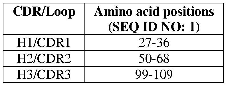


Claims
Priority Applications (8)
| Application Number | Priority Date | Filing Date | Title |
|---|---|---|---|
| EP09709399A EP2250196B1 (en) | 2008-01-31 | 2009-01-30 | Engineered constant domain molecule of an antibody |
| JP2010545219A JP5889533B2 (en) | 2008-01-31 | 2009-01-30 | Modified antibody constant domain molecule |
| CN200980110221.1A CN101977932B (en) | 2008-01-31 | 2009-01-30 | Engineered antibody constant domain molecules |
| CA2713281A CA2713281C (en) | 2008-01-31 | 2009-01-30 | Engineered antibody constant domain molecules |
| AU2009212747A AU2009212747B2 (en) | 2008-01-31 | 2009-01-30 | Engineered antibody constant domain molecules |
| US12/864,758 US8580927B2 (en) | 2008-01-31 | 2009-01-30 | Engineered antibody constant domain molecules |
| US14/043,366 US9527903B2 (en) | 2008-01-31 | 2013-10-01 | Engineered antibody constant domain molecules |
| US15/367,488 US10358481B2 (en) | 2008-01-31 | 2016-12-02 | Engineered antibody constant domain molecules |
Applications Claiming Priority (2)
| Application Number | Priority Date | Filing Date | Title |
|---|---|---|---|
| US6324508P | 2008-01-31 | 2008-01-31 | |
| US61/063,245 | 2008-01-31 |
Related Child Applications (2)
| Application Number | Title | Priority Date | Filing Date |
|---|---|---|---|
| US12/864,758 A-371-Of-International US8580927B2 (en) | 2008-01-31 | 2009-01-30 | Engineered antibody constant domain molecules |
| US14/043,366 Division US9527903B2 (en) | 2008-01-31 | 2013-10-01 | Engineered antibody constant domain molecules |
Publications (2)
| Publication Number | Publication Date |
|---|---|
| WO2009099961A2 true WO2009099961A2 (en) | 2009-08-13 |
| WO2009099961A3 WO2009099961A3 (en) | 2009-10-01 |
Family
ID=40613065
Family Applications (1)
| Application Number | Title | Priority Date | Filing Date |
|---|---|---|---|
| PCT/US2009/032692 WO2009099961A2 (en) | 2008-01-31 | 2009-01-30 | Engineered antibody constant domain molecules |
Country Status (7)
| Country | Link |
|---|---|
| US (3) | US8580927B2 (en) |
| EP (2) | EP2250196B1 (en) |
| JP (2) | JP5889533B2 (en) |
| CN (2) | CN104250301A (en) |
| AU (1) | AU2009212747B2 (en) |
| CA (2) | CA3052615A1 (en) |
| WO (1) | WO2009099961A2 (en) |
Cited By (15)
| Publication number | Priority date | Publication date | Assignee | Title |
|---|---|---|---|---|
| WO2011003811A1 (en) * | 2009-07-09 | 2011-01-13 | F-Star Biotechnologische Forschungs- Und Entwicklungsges.M.B.H | Stabilized immunoglobulin constant domains |
| EP2307452A1 (en) * | 2008-06-30 | 2011-04-13 | Boehringer Ingelheim International GmbH | Method for optimizing proteins having the folding pattern of immunoglobulin |
| WO2011059684A1 (en) * | 2009-10-29 | 2011-05-19 | Centocor Ortho Biotech Inc. | Antibody glycosylation variants |
| EP2533811A2 (en) * | 2010-02-12 | 2012-12-19 | Research Corporation Technologies | Multimeric proteins comprising immunoglobulin constant domains |
| WO2013119903A1 (en) | 2012-02-10 | 2013-08-15 | Research Corporation Technologies, Inc. | Fusion proteins comprising immunoglobulin constant domain-derived scaffolds |
| WO2013138643A1 (en) | 2012-03-16 | 2013-09-19 | The United States Of America, As Represented By The Secretary, Department Of Health & Human Services | Soluble engineered monomeric fc |
| US9156917B2 (en) | 2011-02-11 | 2015-10-13 | Research Corporation Technologies, Inc. | CH2 domain template molecules derived from rational grafting of donor loops onto CH2 scaffolds |
| US9255149B2 (en) | 2008-05-02 | 2016-02-09 | F-Star Biotechnologische Forschungs- Und Entwicklungsges.M.B.H | Cytotoxic immunoglobulin |
| WO2016065258A1 (en) | 2014-10-24 | 2016-04-28 | Research Corporation Technologies, Inc. | Small antibody-like polypeptides that bind to epha2 receptor |
| US9651559B2 (en) | 2007-06-26 | 2017-05-16 | F-star Biotechnologische Forschungs— und Entwicklungsges.m.b.H | Display of binding agents |
| US9856311B2 (en) | 2005-01-05 | 2018-01-02 | F-Star Biotechnologische Forschungs- Und Entwicklungsges.M.B.H | Synthetic immunoglobulin domains with binding properties engineered in regions of the molecule different from the complementarity determining regions |
| WO2021062012A1 (en) | 2019-09-25 | 2021-04-01 | Emory University | Use of klk10 and engineered derivatizations thereof |
| EP3600394A4 (en) * | 2017-03-22 | 2021-04-14 | Research Corporation Technologies, Inc. | Engineered stable ch2 polypeptides |
| US11795232B2 (en) | 2017-02-17 | 2023-10-24 | Denali Therapeutics Inc. | Engineered transferrin receptor binding polypeptides |
| US11827720B2 (en) | 2006-07-05 | 2023-11-28 | F-Star Therapeutics Limited | Multivalent immunoglobulins |
Families Citing this family (19)
| Publication number | Priority date | Publication date | Assignee | Title |
|---|---|---|---|---|
| EP2238165B1 (en) * | 2008-01-07 | 2017-07-05 | Government of the United States of America, as represented by the Secretary, Department of Health and Human Services | Anti-hiv domain antibodies and method of making and using same |
| EP2250196B1 (en) * | 2008-01-31 | 2012-12-26 | The Government of the United States of America as represented by the Secretary of the Department of Health and Human Services | Engineered constant domain molecule of an antibody |
| KR20130140010A (en) * | 2010-10-20 | 2013-12-23 | 메디뮨 엘엘씨 | Methods for processing inclusion bodies |
| US9688731B2 (en) * | 2011-12-22 | 2017-06-27 | Wisconsin Alumni Research Foundation | Isolation and application of BAD-1 for diagnosing infections with Blastomyces dermatitidis |
| CN104334576B (en) * | 2012-06-04 | 2019-02-19 | 诺华股份有限公司 | Site-specific labeling's method and resulting molecule |
| JP6628966B2 (en) * | 2012-06-14 | 2020-01-15 | 中外製薬株式会社 | Antigen binding molecule containing an altered Fc region |
| WO2014030750A1 (en) | 2012-08-24 | 2014-02-27 | 中外製薬株式会社 | MOUSE FcγRII-SPECIFIC Fc ANTIBODY |
| JP6144487B2 (en) | 2012-12-26 | 2017-06-07 | 三菱航空機株式会社 | Flap deployment device and aircraft |
| JPWO2015068847A1 (en) | 2013-11-11 | 2017-03-09 | 中外製薬株式会社 | Antigen-binding molecules comprising modified antibody variable regions |
| TW202313697A (en) | 2014-11-11 | 2023-04-01 | 日商中外製藥股份有限公司 | Library of antigen-binding molecules including modified antibody variable region |
| JP6872797B2 (en) * | 2015-12-29 | 2021-05-19 | 国立大学法人東京工業大学 | Method for stabilizing target-binding peptide |
| SG11201901557XA (en) * | 2016-08-26 | 2019-03-28 | Sanofi Sa | Multispecific antibodies facilitating selective light chain pairing |
| JP6706394B1 (en) * | 2017-01-09 | 2020-06-03 | レンティジェン・テクノロジー・インコーポレイテッドLentigen Technology, Inc. | Compositions and methods for treating cancer by anti-mesothelin immunotherapy |
| EP3720963A4 (en) | 2017-12-05 | 2021-12-08 | Chugai Seiyaku Kabushiki Kaisha | Antigen-binding molecule comprising altered antibody variable region binding cd3 and cd137 |
| AU2018387741A1 (en) * | 2017-12-19 | 2020-07-23 | F-Star Therapeutics Limited | FC binding fragments comprising a PD-L1 antigen-binding site |
| CN108191974B (en) * | 2017-12-31 | 2021-04-09 | 武汉班科生物技术股份有限公司 | Anti-aggregation humanized IgG antibody CH2 structural domain mutant and application thereof |
| TW202325741A (en) | 2020-03-31 | 2023-07-01 | 日商中外製藥股份有限公司 | Dll3-targeting multispecific antigen-binding molecules and uses thereof |
| US20230303666A1 (en) * | 2020-09-24 | 2023-09-28 | The Broad Institute, Inc. | Cell-free antibody engineering platform and neutralizing antibodies for sars-cov-2 |
| WO2023087017A1 (en) * | 2021-11-15 | 2023-05-19 | Alector Llc | Proteins comprising blood brain barrier (bbb)-binding domains within constant domains |
Citations (15)
| Publication number | Priority date | Publication date | Assignee | Title |
|---|---|---|---|---|
| US3060165A (en) | 1962-10-23 | Preparation of toxic ricin | ||
| US4458066A (en) | 1980-02-29 | 1984-07-03 | University Patents, Inc. | Process for preparing polynucleotides |
| US4689401A (en) | 1986-03-06 | 1987-08-25 | Cetus Corporation | Method of recovering microbially produced recombinant ricin toxin a chain |
| US4957735A (en) | 1984-06-12 | 1990-09-18 | The University Of Tennessee Research Corporation | Target-sensitive immunoliposomes- preparation and characterization |
| US5079163A (en) | 1985-03-29 | 1992-01-07 | Cetus Corporation | Recombinant ricin toxin fragments |
| US5208021A (en) | 1987-10-05 | 1993-05-04 | The United States Of America As Represented By The Secretary Of The Department Of Health And Human Services | Method of preparing diphtheria immunotoxins |
| US5792458A (en) | 1987-10-05 | 1998-08-11 | The United States Of America As Represented By The Department Of Health And Human Services | Mutant diphtheria toxin conjugates |
| US5840526A (en) | 1992-08-21 | 1998-11-24 | Vrije Universiteit Brussel | Immunoglobulins devoid of light chains |
| US6277375B1 (en) | 1997-03-03 | 2001-08-21 | Board Of Regents, The University Of Texas System | Immunoglobulin-like domains with increased half-lives |
| WO2003002609A2 (en) | 2001-06-28 | 2003-01-09 | Domantis Limited | Dual-specific ligand and its use |
| US20030088074A1 (en) | 1995-04-25 | 2003-05-08 | Vrije Universiteit Brussel | Recombinant bivalent monospecific immunoglobulin having at least two variable fragments of heavy chains of an immunoglobulin devoid of light chains |
| US6838254B1 (en) | 1993-04-29 | 2005-01-04 | Conopco, Inc. | Production of antibodies or (functionalized) fragments thereof derived from heavy chain immunoglobulins of camelidae |
| WO2005035572A2 (en) | 2003-10-08 | 2005-04-21 | Domantis Limited | Antibody compositions and methods |
| WO2006036834A2 (en) | 2004-09-24 | 2006-04-06 | Amgen Inc. | MODIFIED Fc MOLECULES |
| WO2006072620A1 (en) | 2005-01-05 | 2006-07-13 | F-Star Biotechnologische Forschungs- Und Entwicklungsges.M.B.H. | Synthetic immunoglobulin domains with binding properties engineered in regions of the molecule different from the complementarity determining regions |
Family Cites Families (19)
| Publication number | Priority date | Publication date | Assignee | Title |
|---|---|---|---|---|
| US5530101A (en) | 1988-12-28 | 1996-06-25 | Protein Design Labs, Inc. | Humanized immunoglobulins |
| US5061620A (en) | 1990-03-30 | 1991-10-29 | Systemix, Inc. | Human hematopoietic stem cell |
| US5965709A (en) | 1991-08-14 | 1999-10-12 | Genentech, Inc. | IgE antagonists |
| PT1696031E (en) | 1991-12-02 | 2010-06-25 | Medical Res Council | Production of anti-self antibodies from antibody segment repertoires and displayed on phage |
| US6828422B1 (en) | 1995-08-18 | 2004-12-07 | Morphosys Ag | Protein/(poly)peptide libraries |
| US6083715A (en) | 1997-06-09 | 2000-07-04 | Board Of Regents, The University Of Texas System | Methods for producing heterologous disulfide bond-containing polypeptides in bacterial cells |
| US7829084B2 (en) * | 2001-01-17 | 2010-11-09 | Trubion Pharmaceuticals, Inc. | Binding constructs and methods for use thereof |
| EP2364996B1 (en) | 2002-09-27 | 2016-11-09 | Xencor Inc. | Optimized FC variants and methods for their generation |
| ITRM20020588A1 (en) | 2002-11-21 | 2004-05-22 | Lay Line Genomics Spa | METHOD FOR ISOLATING NEUTRALIZING INTRA-CELL ANTIBODIES OF PROTEIN INTERACTIONS. |
| WO2007024249A2 (en) | 2004-11-10 | 2007-03-01 | Macrogenics, Inc. | Engineering fc antibody regions to confer effector function |
| CA2605697A1 (en) | 2005-04-26 | 2006-11-02 | Bioren, Inc. | Method of producing human igg antibodies with enhanced effector functions |
| CA2624189A1 (en) * | 2005-10-03 | 2007-04-12 | Xencor, Inc. | Fc variants with optimized fc receptor binding properties |
| WO2007076319A2 (en) * | 2005-12-22 | 2007-07-05 | Centocor, Inc. | Human glp-1 mimetibodies and compositions for treating obesity and related disorders, methods and uses |
| AT503889B1 (en) * | 2006-07-05 | 2011-12-15 | Star Biotechnologische Forschungs Und Entwicklungsges M B H F | MULTIVALENT IMMUNE LOBULINE |
| US20080199467A1 (en) * | 2007-02-15 | 2008-08-21 | Mjalli Adnan M M | Immunoglobulin fusion proteins and methods of making |
| WO2008153745A2 (en) * | 2007-05-22 | 2008-12-18 | Amgen Inc. | Compositions and methods for producing bioactive fusion proteins |
| EP2250196B1 (en) * | 2008-01-31 | 2012-12-26 | The Government of the United States of America as represented by the Secretary of the Department of Health and Human Services | Engineered constant domain molecule of an antibody |
| AU2011215684A1 (en) * | 2010-02-12 | 2012-08-30 | Research Corporation Technologies | Multimeric proteins comprising immunoglobulin constant domains |
| CA2827007A1 (en) * | 2011-02-11 | 2012-08-16 | Research Corporation Technologies, Inc. | Ch2 domain template molecules derived from rational grafting of donor loops onto ch2 scaffolds |
-
2009
- 2009-01-30 EP EP09709399A patent/EP2250196B1/en active Active
- 2009-01-30 WO PCT/US2009/032692 patent/WO2009099961A2/en active Application Filing
- 2009-01-30 JP JP2010545219A patent/JP5889533B2/en active Active
- 2009-01-30 US US12/864,758 patent/US8580927B2/en active Active
- 2009-01-30 EP EP12198382.9A patent/EP2594584B1/en active Active
- 2009-01-30 CA CA3052615A patent/CA3052615A1/en not_active Abandoned
- 2009-01-30 CN CN201410422052.8A patent/CN104250301A/en active Pending
- 2009-01-30 AU AU2009212747A patent/AU2009212747B2/en active Active
- 2009-01-30 CN CN200980110221.1A patent/CN101977932B/en active Active
- 2009-01-30 CA CA2713281A patent/CA2713281C/en active Active
-
2013
- 2013-10-01 US US14/043,366 patent/US9527903B2/en active Active
-
2014
- 2014-05-30 JP JP2014112059A patent/JP2014155503A/en active Pending
-
2016
- 2016-12-02 US US15/367,488 patent/US10358481B2/en active Active
Patent Citations (15)
| Publication number | Priority date | Publication date | Assignee | Title |
|---|---|---|---|---|
| US3060165A (en) | 1962-10-23 | Preparation of toxic ricin | ||
| US4458066A (en) | 1980-02-29 | 1984-07-03 | University Patents, Inc. | Process for preparing polynucleotides |
| US4957735A (en) | 1984-06-12 | 1990-09-18 | The University Of Tennessee Research Corporation | Target-sensitive immunoliposomes- preparation and characterization |
| US5079163A (en) | 1985-03-29 | 1992-01-07 | Cetus Corporation | Recombinant ricin toxin fragments |
| US4689401A (en) | 1986-03-06 | 1987-08-25 | Cetus Corporation | Method of recovering microbially produced recombinant ricin toxin a chain |
| US5792458A (en) | 1987-10-05 | 1998-08-11 | The United States Of America As Represented By The Department Of Health And Human Services | Mutant diphtheria toxin conjugates |
| US5208021A (en) | 1987-10-05 | 1993-05-04 | The United States Of America As Represented By The Secretary Of The Department Of Health And Human Services | Method of preparing diphtheria immunotoxins |
| US5840526A (en) | 1992-08-21 | 1998-11-24 | Vrije Universiteit Brussel | Immunoglobulins devoid of light chains |
| US6838254B1 (en) | 1993-04-29 | 2005-01-04 | Conopco, Inc. | Production of antibodies or (functionalized) fragments thereof derived from heavy chain immunoglobulins of camelidae |
| US20030088074A1 (en) | 1995-04-25 | 2003-05-08 | Vrije Universiteit Brussel | Recombinant bivalent monospecific immunoglobulin having at least two variable fragments of heavy chains of an immunoglobulin devoid of light chains |
| US6277375B1 (en) | 1997-03-03 | 2001-08-21 | Board Of Regents, The University Of Texas System | Immunoglobulin-like domains with increased half-lives |
| WO2003002609A2 (en) | 2001-06-28 | 2003-01-09 | Domantis Limited | Dual-specific ligand and its use |
| WO2005035572A2 (en) | 2003-10-08 | 2005-04-21 | Domantis Limited | Antibody compositions and methods |
| WO2006036834A2 (en) | 2004-09-24 | 2006-04-06 | Amgen Inc. | MODIFIED Fc MOLECULES |
| WO2006072620A1 (en) | 2005-01-05 | 2006-07-13 | F-Star Biotechnologische Forschungs- Und Entwicklungsges.M.B.H. | Synthetic immunoglobulin domains with binding properties engineered in regions of the molecule different from the complementarity determining regions |
Non-Patent Citations (34)
| Title |
|---|
| "Purification", 1982, SPRINGER-VERLAG |
| ALTSCHUL ET AL., J. MOL. BIOL., vol. 215, 1990, pages 403 - 410 |
| ALTSCHUL ET AL., NATURE GENET., vol. 6, 1994, pages 119 - 129 |
| BARANY; MERRIFIELD, THE PEPTIDES: ANALYSIS, SYNTHESIS, BIOLOGY. VOL. 2: SPECIAL METHODS IN PEPTIDE SYNTHESIS, PART A., vol. 2, pages 3 - 284 |
| BEAUCAGE; CARUTHERS, TETRA. LETTS., vol. 22, no. 20, 1981, pages 1859 - 1862 |
| BUCHNER ET AL., ANAL. BIOCHEM., vol. 205, 1992, pages 263 - 270 |
| CONNOR ET AL., PHARM. THER., vol. 28, 1985, pages 341 - 365 |
| CORPET ET AL., NUCLEIC ACIDS RESEARCH, vol. 16, 1988, pages 10881 - 10890 |
| FEIGE ET AL., J MOL BIOL, vol. 344, 2004, pages 107 - 118 |
| FUNATSU ET AL., AGR. BIOL. CHEM., vol. 52, 1988, pages 1095 |
| GILLESPIE ET AL., ANN ONCOL, vol. 11, 2000, pages 735 - 41 |
| GOYAL; BATRA, BIOCHEM, vol. 345, 2000, pages 247 - 54 |
| HIGGINS; SHARP, CABIOS, vol. 5, 1989, pages 151 - 153 |
| HIGGINS; SHARP, GENE, vol. 73, 1988, pages 237 - 244 |
| HUSE ET AL., SCIENCE, vol. 246, 1989, pages 1275 |
| KABAT ET AL.: "Sequences of Proteins of Immunological Interest, 5th Edition,", 1991, NIH PUBLICATION NO. 91-3242 |
| LEE ET AL., J. ANTIBIOT, vol. 42, 1989, pages 1070 - 87 |
| LIPOVSEK ET AL., J MOL BIOL, vol. 368, 2007, pages 1024 - 1041 |
| MERRIFIELD ET AL., J. AM. CHEM. SOC., vol. 85, 1963, pages 2149 - 2156 |
| NEEDHAM-VANDEVANTER ET AL., NUCL. ACIDS RES., vol. 12, 1984, pages 6159 - 6168 |
| NEEDLEMAN; WUNSCH, J. MOL. BIOL., vol. 48, 1970, pages 443 |
| NICHOLSON; BLAUSTEIN, J. BIOCHIM. BIOPHYS. ACTA, vol. 266, 1972, pages 543 |
| NOVELLINO ET AL., CANCER IMMUNOL. IMMUNOTHER., vol. 54, no. 3, 2005, pages 187 - 207 |
| OLSNES ET AL., NATURE, vol. 249, 1974, pages 627 - 631 |
| OLSNES, METHODS ENZYMOL., vol. 50, 1978, pages 330 - 335 |
| PEARSON; LIPMAN, PROC. NATL. ACAD. SCI. U.S.A., vol. 85, 1988, pages 2444 |
| PLUCKTHUN, BIOTECHNOLOGY, vol. 9, 1991, pages 545 |
| RATHORE ET AL., GENE, vol. 190, 1997, pages 31 - 5 |
| SMITH; WATERMAN, ADV. APPL. MATH., vol. 2, 1981, pages 482 |
| STEWART ET AL.: "Solid Phase Peptide Synthesis, 2nd ed.,", 1984, PIERCE CHEM. CO. |
| SUZUKI ET AL., NAT. BIOTECH., vol. 17, 1999, pages 265 - 70 |
| THIES ET AL., J MOL BIOL, vol. 309, 2001, pages 1077 - 1085 |
| THIES ET AL., J MOL BIOL, vol. 319, 2002, pages 1267 - 1277 |
| WARD ET AL., NATURE, vol. 341, 1989, pages 544 |
Cited By (30)
| Publication number | Priority date | Publication date | Assignee | Title |
|---|---|---|---|---|
| US10385118B2 (en) | 2005-01-05 | 2019-08-20 | F-Star Biotechnologische Forschungs- Und Entwicklungsges.M.B.H | Synthetic immunoglobulin domains with binding properties engineered in regions of the molecule different from the complementarity determining regions |
| US9856311B2 (en) | 2005-01-05 | 2018-01-02 | F-Star Biotechnologische Forschungs- Und Entwicklungsges.M.B.H | Synthetic immunoglobulin domains with binding properties engineered in regions of the molecule different from the complementarity determining regions |
| US11827720B2 (en) | 2006-07-05 | 2023-11-28 | F-Star Therapeutics Limited | Multivalent immunoglobulins |
| US9651559B2 (en) | 2007-06-26 | 2017-05-16 | F-star Biotechnologische Forschungs— und Entwicklungsges.m.b.H | Display of binding agents |
| US10125197B2 (en) | 2008-05-02 | 2018-11-13 | F-Star Biotechnologische Forschungs-Und Entwicklungsges.M.B.H | Cytotoxic immunoglobulin |
| US9255149B2 (en) | 2008-05-02 | 2016-02-09 | F-Star Biotechnologische Forschungs- Und Entwicklungsges.M.B.H | Cytotoxic immunoglobulin |
| EP2307452A1 (en) * | 2008-06-30 | 2011-04-13 | Boehringer Ingelheim International GmbH | Method for optimizing proteins having the folding pattern of immunoglobulin |
| WO2011003811A1 (en) * | 2009-07-09 | 2011-01-13 | F-Star Biotechnologische Forschungs- Und Entwicklungsges.M.B.H | Stabilized immunoglobulin constant domains |
| WO2011059684A1 (en) * | 2009-10-29 | 2011-05-19 | Centocor Ortho Biotech Inc. | Antibody glycosylation variants |
| CN102770554A (en) * | 2009-10-29 | 2012-11-07 | 詹森生物科技公司 | Antibody glycosylation variants |
| CN102770554B (en) * | 2009-10-29 | 2016-02-17 | 詹森生物科技公司 | Antibody glycosylation variant |
| EP2533811A2 (en) * | 2010-02-12 | 2012-12-19 | Research Corporation Technologies | Multimeric proteins comprising immunoglobulin constant domains |
| EP2533811A4 (en) * | 2010-02-12 | 2013-12-25 | Res Corp Technologies Inc | Multimeric proteins comprising immunoglobulin constant domains |
| US9156917B2 (en) | 2011-02-11 | 2015-10-13 | Research Corporation Technologies, Inc. | CH2 domain template molecules derived from rational grafting of donor loops onto CH2 scaffolds |
| US9803210B2 (en) | 2011-02-11 | 2017-10-31 | Research Corporation Technologies, Inc. | Fusion proteins comprising immunoglobulin constant domain-derived scaffolds |
| EP2812432A4 (en) * | 2012-02-10 | 2016-02-17 | Res Corp Technologies Inc | Fusion proteins comprising immunoglobulin constant domain-derived scaffolds |
| WO2013119903A1 (en) | 2012-02-10 | 2013-08-15 | Research Corporation Technologies, Inc. | Fusion proteins comprising immunoglobulin constant domain-derived scaffolds |
| AU2013216943B2 (en) * | 2012-02-10 | 2018-04-19 | Research Corporation Technologies, Inc. | Fusion proteins comprising immunoglobulin constant domain-derived scaffolds |
| US9676857B2 (en) | 2012-03-16 | 2017-06-13 | The United States Of America, As Represented By The Secretary, Department Of Health And Human Services | Soluble engineered monomeric Fc |
| US10633447B2 (en) | 2012-03-16 | 2020-04-28 | The United States Of America, As Represented By The Secretary, Department Of Health And Human Services | Soluble engineered monomeric Fc |
| WO2013138643A1 (en) | 2012-03-16 | 2013-09-19 | The United States Of America, As Represented By The Secretary, Department Of Health & Human Services | Soluble engineered monomeric fc |
| EP3209329A4 (en) * | 2014-10-24 | 2018-03-28 | Research Corporation Technologies, Inc. | Small antibody-like polypeptides that bind to epha2 receptor |
| WO2016065258A1 (en) | 2014-10-24 | 2016-04-28 | Research Corporation Technologies, Inc. | Small antibody-like polypeptides that bind to epha2 receptor |
| US10829558B2 (en) | 2014-10-24 | 2020-11-10 | Research Corporation Technologies, Inc. | Small antibody-like polypeptides that bind to EPHA2 receptor |
| AU2015335725B2 (en) * | 2014-10-24 | 2021-06-03 | Research Corporation Technologies, Inc. | Small antibody-like polypeptides that bind to EphA2 receptor |
| US11795232B2 (en) | 2017-02-17 | 2023-10-24 | Denali Therapeutics Inc. | Engineered transferrin receptor binding polypeptides |
| US11912778B2 (en) | 2017-02-17 | 2024-02-27 | Denali Therapeutics Inc. | Methods of engineering transferrin receptor binding polypeptides |
| EP3600394A4 (en) * | 2017-03-22 | 2021-04-14 | Research Corporation Technologies, Inc. | Engineered stable ch2 polypeptides |
| US11542321B2 (en) | 2017-03-22 | 2023-01-03 | Research Corporation Technologies, Inc. | Engineered stable CH2 polypeptides |
| WO2021062012A1 (en) | 2019-09-25 | 2021-04-01 | Emory University | Use of klk10 and engineered derivatizations thereof |
Also Published As
| Publication number | Publication date |
|---|---|
| CN104250301A (en) | 2014-12-31 |
| AU2009212747B2 (en) | 2013-11-07 |
| JP2011514149A (en) | 2011-05-06 |
| US10358481B2 (en) | 2019-07-23 |
| WO2009099961A3 (en) | 2009-10-01 |
| CA3052615A1 (en) | 2009-08-13 |
| JP2014155503A (en) | 2014-08-28 |
| EP2250196A2 (en) | 2010-11-17 |
| CA2713281A1 (en) | 2009-08-13 |
| CA2713281C (en) | 2019-10-15 |
| EP2594584B1 (en) | 2015-07-15 |
| CN101977932B (en) | 2014-09-03 |
| JP5889533B2 (en) | 2016-03-22 |
| US9527903B2 (en) | 2016-12-27 |
| US20140023645A1 (en) | 2014-01-23 |
| US20100316641A1 (en) | 2010-12-16 |
| US8580927B2 (en) | 2013-11-12 |
| AU2009212747A1 (en) | 2009-08-13 |
| EP2250196B1 (en) | 2012-12-26 |
| EP2594584A1 (en) | 2013-05-22 |
| US20170081393A1 (en) | 2017-03-23 |
| CN101977932A (en) | 2011-02-16 |
Similar Documents
| Publication | Publication Date | Title |
|---|---|---|
| US10358481B2 (en) | Engineered antibody constant domain molecules | |
| US10633447B2 (en) | Soluble engineered monomeric Fc | |
| EP2812432B1 (en) | Fusion proteins comprising immunoglobulin constant domain-derived scaffolds | |
| CN113307865A (en) | Fully human single domain antibody of novel coronavirus and application | |
| US20130189247A1 (en) | Multimeric Proteins Comprising Immunoglobulin Constant Domains | |
| AU2012214274B2 (en) | CH2 domain template molecules derived from rational grafting of donor loops onto CH2 scaffolds | |
| AU2014200215B2 (en) | Engineered antibody constant domain molecules |
Legal Events
| Date | Code | Title | Description |
|---|---|---|---|
| WWE | Wipo information: entry into national phase |
Ref document number: 200980110221.1 Country of ref document: CN |
|
| 121 | Ep: the epo has been informed by wipo that ep was designated in this application |
Ref document number: 09709399 Country of ref document: EP Kind code of ref document: A2 |
|
| WWE | Wipo information: entry into national phase |
Ref document number: 2009212747 Country of ref document: AU |
|
| WWE | Wipo information: entry into national phase |
Ref document number: 2713281 Country of ref document: CA |
|
| WWE | Wipo information: entry into national phase |
Ref document number: 12864758 Country of ref document: US |
|
| WWE | Wipo information: entry into national phase |
Ref document number: 2010545219 Country of ref document: JP |
|
| WWE | Wipo information: entry into national phase |
Ref document number: 2818/KOLNP/2010 Country of ref document: IN |
|
| NENP | Non-entry into the national phase |
Ref country code: DE |
|
| ENP | Entry into the national phase |
Ref document number: 2009212747 Country of ref document: AU Date of ref document: 20090130 Kind code of ref document: A |
|
| WWE | Wipo information: entry into national phase |
Ref document number: 2009709399 Country of ref document: EP |