WO2009138922A1 - Cardiac contraction supporting system for people with depressed cardiac function - Google Patents
Cardiac contraction supporting system for people with depressed cardiac function Download PDFInfo
- Publication number
- WO2009138922A1 WO2009138922A1 PCT/IB2009/051883 IB2009051883W WO2009138922A1 WO 2009138922 A1 WO2009138922 A1 WO 2009138922A1 IB 2009051883 W IB2009051883 W IB 2009051883W WO 2009138922 A1 WO2009138922 A1 WO 2009138922A1
- Authority
- WO
- WIPO (PCT)
- Prior art keywords
- contraction
- organ
- spiral
- general
- supporting device
- Prior art date
Links
Classifications
-
- A—HUMAN NECESSITIES
- A61—MEDICAL OR VETERINARY SCIENCE; HYGIENE
- A61M—DEVICES FOR INTRODUCING MEDIA INTO, OR ONTO, THE BODY; DEVICES FOR TRANSDUCING BODY MEDIA OR FOR TAKING MEDIA FROM THE BODY; DEVICES FOR PRODUCING OR ENDING SLEEP OR STUPOR
- A61M60/00—Blood pumps; Devices for mechanical circulatory actuation; Balloon pumps for circulatory assistance
- A61M60/10—Location thereof with respect to the patient's body
- A61M60/122—Implantable pumps or pumping devices, i.e. the blood being pumped inside the patient's body
- A61M60/165—Implantable pumps or pumping devices, i.e. the blood being pumped inside the patient's body implantable in, on, or around the heart
- A61M60/191—Implantable pumps or pumping devices, i.e. the blood being pumped inside the patient's body implantable in, on, or around the heart mechanically acting upon the outside of the patient's native heart, e.g. compressive structures placed around the heart
-
- A—HUMAN NECESSITIES
- A61—MEDICAL OR VETERINARY SCIENCE; HYGIENE
- A61M—DEVICES FOR INTRODUCING MEDIA INTO, OR ONTO, THE BODY; DEVICES FOR TRANSDUCING BODY MEDIA OR FOR TAKING MEDIA FROM THE BODY; DEVICES FOR PRODUCING OR ENDING SLEEP OR STUPOR
- A61M60/00—Blood pumps; Devices for mechanical circulatory actuation; Balloon pumps for circulatory assistance
- A61M60/20—Type thereof
- A61M60/289—Devices for mechanical circulatory actuation assisting the residual heart function by means mechanically acting upon the patient's native heart or blood vessel structure, e.g. direct cardiac compression [DCC] devices
-
- A—HUMAN NECESSITIES
- A61—MEDICAL OR VETERINARY SCIENCE; HYGIENE
- A61M—DEVICES FOR INTRODUCING MEDIA INTO, OR ONTO, THE BODY; DEVICES FOR TRANSDUCING BODY MEDIA OR FOR TAKING MEDIA FROM THE BODY; DEVICES FOR PRODUCING OR ENDING SLEEP OR STUPOR
- A61M60/00—Blood pumps; Devices for mechanical circulatory actuation; Balloon pumps for circulatory assistance
- A61M60/40—Details relating to driving
- A61M60/465—Details relating to driving for devices for mechanical circulatory actuation
- A61M60/47—Details relating to driving for devices for mechanical circulatory actuation the force acting on the actuation means being mechanical, e.g. mechanically driven members clamping a blood vessel
-
- A—HUMAN NECESSITIES
- A61—MEDICAL OR VETERINARY SCIENCE; HYGIENE
- A61M—DEVICES FOR INTRODUCING MEDIA INTO, OR ONTO, THE BODY; DEVICES FOR TRANSDUCING BODY MEDIA OR FOR TAKING MEDIA FROM THE BODY; DEVICES FOR PRODUCING OR ENDING SLEEP OR STUPOR
- A61M60/00—Blood pumps; Devices for mechanical circulatory actuation; Balloon pumps for circulatory assistance
- A61M60/50—Details relating to control
- A61M60/508—Electronic control means, e.g. for feedback regulation
-
- A—HUMAN NECESSITIES
- A61—MEDICAL OR VETERINARY SCIENCE; HYGIENE
- A61M—DEVICES FOR INTRODUCING MEDIA INTO, OR ONTO, THE BODY; DEVICES FOR TRANSDUCING BODY MEDIA OR FOR TAKING MEDIA FROM THE BODY; DEVICES FOR PRODUCING OR ENDING SLEEP OR STUPOR
- A61M60/00—Blood pumps; Devices for mechanical circulatory actuation; Balloon pumps for circulatory assistance
- A61M60/80—Constructional details other than related to driving
- A61M60/839—Constructional details other than related to driving of devices for mechanical circulatory actuation
-
- A—HUMAN NECESSITIES
- A61—MEDICAL OR VETERINARY SCIENCE; HYGIENE
- A61M—DEVICES FOR INTRODUCING MEDIA INTO, OR ONTO, THE BODY; DEVICES FOR TRANSDUCING BODY MEDIA OR FOR TAKING MEDIA FROM THE BODY; DEVICES FOR PRODUCING OR ENDING SLEEP OR STUPOR
- A61M60/00—Blood pumps; Devices for mechanical circulatory actuation; Balloon pumps for circulatory assistance
- A61M60/80—Constructional details other than related to driving
- A61M60/855—Constructional details other than related to driving of implantable pumps or pumping devices
- A61M60/869—Compliance chambers containing a gas or liquid other than blood to compensate volume variations of a blood chamber
Definitions
- the present invention refers to a device able to operate or make easier the contraction of organs in general, and in particular for the treatment of congestive heart failure.
- Background Art The congestive heart failure is a progressive and invalidating disease that follows to an ischemic or degenerative disease of the cardiac muscle.
- a measure that effectively describes the contractive function of the heart is the ejection fraction, intended as the percentage of blood that is introduced in the great vessels during the systole in relation to the quantity of blood accumulated at the end of the diastole.
- the final consequence of the disease is thus the incapacity of the heart to effectively contract during the systole to reduce the dimensions of the ventricular cavity and allow thus to send in the vascular circle an adequate quantity of blood.
- a first remedy consist in the use of adequate drugs and useful to control the disease, allowing in a certain measure to increase the strength and the entity of the contraction.
- the drugs with the time can lose their efficiency, can result not sufficient and not adequate or present contraindication.
- different devices permanent or non
- the available technological devices can be essentially subdivided into electrical devices and mechanical devices, including in this last type systems with movable components (engines, fluids) and an electric control .
- the implantation of the mentioned devices requires a demanding and invasive type of surgery that often leads, according to the type of device used, the opening of the cardiac cavities . Moreover the weight of the same device are not insignificant for the heart, making its situation worst. The nature of the surgery is thus such that the heart is altered in its features (extended incisions, holes, removals of parts) or even explanted with the result that the methodical do not result reversible and there is not a possibility of reinstatement of the quo- ante status.
- At least an elastic element (1) configured to surround at least partially the organ in such a way to result operable between a contraction condition where induces an elastic compulsion on the organ and a release condition where it elastically releases said organ;
- At least a control unit (2) configured to operate in an intermitting manner said elastic element between said contraction condition and said release condition and vice-versa .
- the elastic element that surrounds the organ is opportunely controlled, and thus activated, by the controlling unit in such a way to get elastically contracted and consequently press the organ with an opportunely calibrated strength for then releasing it. It is thus realized an artificial pulsation.
- the operation is obviously controlled by the unit (2) which controls that such pulsation happens in an intermitting manner, or rather at specific intervals of time that are externally programmable.
- the elastic element (1) comprises a spiral (3) and a contraction wire (4) located inside the spiral (3) for its entire length.
- the spiral can then have different shapes as for example a circular section, such to obtain a cylindrical spiral .
- the spiral comprises a first contractive part (8) with coils spaced at a distance one to each other, a second non-contractive part (9) with coils closed one to each other and a joint (10) interposed between said first and said second part.
- the contraction wire (4) comprises a distal end (5) widen such that said wire become blocked with the spiral in correspondence of the reaching of said distal end (5) against the end of the spiral, said wire getting out partially from the opposed end in correspondence of the second non-contractive part (9) .
- spiral of the second non- contractive part (9) can be realized in two or more elements gathered in a common connection end.
- control unit (2) acts with a traction on the wire creating this way an elastic contraction of the spiral while a release of the wire implies a return to the initial condition.
- sheath (6) can comprise fitting appendixes (7) . These facilitate the application of the device to the organ.
- the sheath can be filled up with a physiologic liquid.
- control unit (2) comprises a rotary stepping engine (B) to drive into rotation a spool
- the spiral will thus be configured in such a way to allow to bind the wire to the spool.
- the spool (C; Cl) comprises at least an indentation (22) or similar within which bind the wire (4) or anyway the end of the elastic element in general.
- control unit (2) comprises also a power supply battery (A) .
- the battery comprises also an electric wire (29) for the eventual external power supply.
- control unit (2) can comprise also a pacemaker (E) configured to pick up the cardiac frequency and control thus the rotation of the engine (B) in an intermitting manner according to the taken specific cardiac frequency. In such manner the contraction and the release operated by the present device is in perfect synchrony with the normal heartbeat .
- the spool Cl comprises at least two, preferably three, section with different diameters able to produce a different contraction on the corresponding spirals applied in correspondence of the above mentioned section.
- FIG. 1 shows a front view A and a top view B of the present device when it is implanted on a human heart.
- FIG. 2 shows four following assembling phases from A to D of the components (spiral and wire) constituting the elastic element 1 according to a possible embodiment of the invention
- FIG. 4 shows with three successive views from A to C a contraction of the spiral by means of a traction of the wire 4; The further views from D to E highlight different sections of the spiral .
- FIG. 5 shows in view the elastic element in a straight shape and looped to surround when in use the organ both a in releasing phase and in a contraction phase of the wire 4;
- FIG. 6 shows two views (A and B) of the device and a detail (C) of a plate 20 to climb over eventual arteries;
- FIG. 7 shows an exploded view of the control unit with its main components indicated from A to E;
- FIG. 9 shows an assembly drawing of the compacted device with opportune external boxing. Description of a preferred pattern realization
- the device consists in its most essential aspects in at least an elastic element 1, apparently similar to a catheter, destined in use to be applied in correspondence of the external surface of the organ to be contracted, in particular the heart.
- An opportune control unit 2 through an engine, assures a particular intermitting movement of the mentioned element 1 as better detailed in the following description. Even if particularly indicated for cardiac applications the present device, as better clarified from the following description, is right for applications to other organs of different type. Only for descriptive simplicity, and thus not in a limitative manner, it is made reference in the attached figures to the sole application to the heart.
- FIG. 1 With particular reference to figure 1 VIEW-A, are described two elastic elements 1 connected to the control unit 2 which is also to be placed inside the chest.
- the front view A highlights how the two elastic elements 1 are connected to the control unit 2 and surround the two ventricles, left and right, at two different levels.
- a view from the top shows also the elastic element 1 that surrounds the right ventricle and the left ventricle getting connected to the control unit 2.
- each element 1 comprises a cylindrical spiral 3 and a wire 4, generally of metallic type but anyway realizable also in another material.
- the cylindrical spiral with diameter of about 3 mm, is constituted by metallic wire with section of about 0.5 mm.
- the coils of the spiral are closed enough to obtain between one and the other a space that is about double the thickness of the wire.
- the spiral is constituted by a metal that guarantees the possibility of deformation of the spiral along its axis with shortening of the length and the immediate return to its predefined length since the applied pressure force ceases. This feature is obtained by calibrating opportunely the diameter of the spiral, the thickness of the wire and the type of metal.
- the force applied on the spiral along its axis which allows to compress and approach the coils to each other will result reasonable according on the constitution of the spiral and the reduced section of the wire.
- the termination placed on top of the spiral is called distal end of the spiral, the terminal part placed below is called proximal end of the spiral.
- the spirals can have different lengths which range between 30 and 45 centimetres .
- the wire 4 called contraction wire 4, with a diameter that is less than the internal diameter of the spiral is thus inserted inside the spiral for its entire length, as described (PHASE B) .
- the wire comprises an end, called distal end, widen into a button 5.
- Phase C it is thus highlighted the insertion of the wire within the spiral and the blocking in correspondence of the reaching of the button 5 against the termination of the spiral.
- Phase D the technical effect of the button is found in the fact that the traction of the wire implies a compression of the spiral or rather an approaching of its proximal end to the distal one.
- An external sheath 6 (look at figure 3) opportunely covers the spiral.
- the sheath results to be particularly light, resistant, non permeable, flexible, and comprises opportune hooking appendixes 7 that are more sturdy and eventually comprise slots to allow to sew the spiral directly on the myocardiac ventricular or to the organ in general or by means of a specific netting structure.
- This sheath is such to separate the metallic materials from the surrounding mean and impede, once the system has been implanted, that the organic elements get deposited on the spiral or on the internal wire altering the mechanical features .
- the only metallic portion that is not covered by this sheath is the element of connection to the control unit 2.
- the sheath is glued on the spiral in such a way that it does not remain stretched between the coils but it actually gets loose in a way to not hinder the folding movement of the spiral in the different directions.
- the sheath when in use is then filled up with a physiologic liquid. Imagining that the spiral is completely filled up with water the appearance will be such that the sheath protrude outward from each spiral .
- This disposition of the sheath is such that when the spiral is in a resting position the sheath is interposed between the coils, when the spiral is compressed the liquid inside the spiral pushes on the sheath causing the protrusion between each spiral in order to receive the liquid.
- the spiral is essentially constituted by two different structural parts.
- a first part 8 longer, with the coils spaced at a distance one to each other, realizes this way the called “contractile part” of the spiral, while a second part 9 with approached coils realizes the "non contractile” part.
- the passage point between a portion and the other of the spiral is the joint 10. Both parts are covered by the contraction wire 4 which is still hooked at the distal end 5 and gets out for few centimetres from the proximal end.
- figure 4 (detail B) it is specified the mechanical structural behaviour of the contraction spiral which shows to be flexible in the contractile portion 8 and more rigid in the non contractile portion 9.
- the contractile portion when resting, shows to be suitable to support the curves of the organ and to be fixed to it.
- Figure 4 (Detail C) shows the effect that is obtained by slightly pulling the contraction wire of the same spiral shown in figure 4-B. It is observed how the portion of the contraction wire 4 that gets out from the spiral is now longer with a consequent contraction of the spiral which is shortened in its contractile part for an equivalent piece. The non contractile part did not substantially changed and the relative relations between joint and proximal end of the contraction spiral have not changed.
- Figure 4 shows a possible variant of the contraction spiral where the spiral is not cylindrical but of rectangular section.
- the contraction wire in this case can opportunely be substituted with a thin foil 11 of rectangular section.
- the contraction spiral will show good flexibility only longitudinally, but can be usefully applied to the ventricle. With such solution it can be given to the traction foil a curved shape when resting, this shape will be retaken by the foil (and by the entire spiral) in correspondence of the traction releasing.
- a contraction spiral has a further semi-elliptical shape
- the flat part can be the one destined in use to lean on the organ.
- the internal wire can have an opportune elliptical section 12.
- Figure 5 shows thus in view the functionality of the single elastic element.
- Figure 5 (Detail A) shows, for such aim, the spiral in its configuration before the implant with its constituting parts. Are distinguished in it the above essential parts or rather the contractile part 8 and the non contractile 9 separated respectively by the joint 10 comprised between them. Are also highlighted at different levels the fitting appendixes 7.
- Figure 5 (Detail B) zoomed in only to clarify, highlights the contractile part 8 of the spiral, surrounded by the coating sheath 6 and closed in a circle in such a way that the distal end is placed near the joint.
- the fitting appendixes 7 of the two portions are linked together to constitute the fulcrum 16 of the device (fulcrum point 16) .
- Figure 6-A and figure 6-B show in two different views the device implanted to a heart. Are thus illustrated in such case three different contraction spirals that have in this case a different length of the contractile part.
- the contractile parts are thus opportunely folded to make the circle in such a way that the terminal end of each contraction spiral is secured to the myocardium at a predetermined level in correspondence of the joint of the same spiral.
- the non contractile parts of each of the spirals converge in the control unit 2 where are engaged the contraction wires.
- FIG 6 (Detail C) is also highlighted a possible additional component of the device. It is an element 20 with a plate shape realized in a bio- compatible textile, for example sturdy Teflon, with elevated smoothness.
- the element 20 comprises a guide on the upper back suitable to receive the contraction spiral.
- This element is configured with an opportune shape such to realize a bridge on the myocardium able to climb over the epicardic vessels 19, avoiding that these undergo to the contraction of the spirals. In such manner, fitted by points to the ventricular wall 18 or to the net fitted to the organ, they impede during the contraction that the spiral compresses the vessels.
- Figure 7 described in detail the control unit. The mechanism seems simplified in its essential parts and illustrated in an exploded manner.
- the battery of the device In its most essential elements with A is indicated the battery of the device. If the device is not entirely implantable it communicates to the outside through an electric wire 29 which assures the power supply of the system. From this module the electrical power is supplied to the engine and to a pacemaker unit.
- the detail B indicates the electric engine of the device, which gets fitted and connected electrically to the battery.
- the engine is a stepping type one and has in its end opposite to the battery an shaft 21.
- the engine provides a good control of the contraction spool C by means of the shaft 21 which gets blocked in a precise angular position. This position can also be easily changed and monitored through a digital circuit.
- the possibility to program the blocking point of the shaft according to the specific needs (in particular the cardiac frequency) allows a good versatility for such type of solution. The engine can then quickly bring back the contraction spiral in its resting condition.
- the spool rotates thus around the shaft 21 according to the predefined intermitting settings, and receives on its self pre-packed indentations 22 within which bind the contraction wire 4 opportunely fitted with some screws 23.
- a servomechanism that allows in the absence of power to bring the spiral in the resting position.
- an helicoidal spring, integral with the contraction spool can be comprised to make the releasing movement of the spiral automatic and prompt when the movement of the engine is interrupted or the mechanism is spiral ed off.
- Opportune openings 24 assure in an hermetical manner the proximal end 25 of the contraction spirals.
- the contraction wires 4 pass in the capsule and are secured to the contraction spiral through screws 23 and can be eventually inspected through a window 26, hermetically closed by a lid 27.
- the pacemaker unit of the device fed by the battery A.
- Such unit contains the logic to pick up the electric signal of the heart and to consequently contract the contraction spiral with opportune movement of the spool C.
- the unit can pick up the signal through autonomous catheters installed in an epycardic position and can eventually stimulate opportunely the cardiac chambers.
- the control unit In use thus, imagining for example a spiral opportunely placed to surround the two ventricles, the control unit, by means of the engine that rotates the spiral, will provide to put in an intermitting manner the internal wire into a contraction and resting condition. It will be therefore obtained an elastic contraction of the spiral (a reduction of the entire length of the spiral) that surrounds the ventricles. Such contraction causes thus a pressure on the ventricular walls surrounded as a ring by the spiral, inducing thus a reduction of the ventricular cavities. This is translated into an ejection of the blood from the ventricles that is function of the reduction of the cavity.
- FIG 8-A With reference to figure 8-A is shown a contraction spiral placed on a specific portion of myocardium of the left ventricle without surrounding as a circle the two ventricles.
- the activation of the contraction of the spiral opportunely secured in different points to the myocardium or to the net will refer to the contraction of a specific portion of the heart.
- Figure 8-B illustrates a solution that comprises the sole left ventricle without referring to the right ventricle. Practically the entire free wall of the left ventricle is surrounded, at the level of the base of the heart, by a contraction spiral constituted by a single non contractile part (that gets connected to the control unit) and from two contractile parts placed at "Y" respect to the non contractile part. This method allows to not involve in the contraction the right ventricle.
- figure 8-C it is reproduced the same situation of figure 8-B, where two branches of spirals surround the free wall of the left ventricle and their ends are hooked also at the end of an elongated element that passes through the interventricular septum 31.
- FIG 8-D is on the other hand shown a tripod solution where the fulcrum of the device is on the tip of the heart and from this start three contractile parts which terminal ends arrive to get fixed to the base of the left ventricle.
- This solution presents a conceptual advantage favouring the expulsion of the blood acting on the axis along the left ventricle.
- FIG 8-E is shown a contraction spiral that presents a contractile part that surrounds the left ventricle and a non contractile part that surrounds the right ventricle. In this way without changing the circular implant configuration and exploiting the function of the fulcrum given by the connection of the terminal end of the spiral to the joint, it is meant to act only on the left ventricle. Opportune fastening of the fitting elements in correspondence of the end and of the start of the non contractile part, will impede that on the left ventricle is unloaded the contractile strength of the spiral.
- Another hooking position of the spiral has to be comprised also at the distal end of the spiral to fit to the heart the terminal end of the spiral as also another fitting element has to be placed at the level of the joint.
- These fitting elements can have different shapes according to the position and should be required to hook to the heart, in predefined points, a specific portion of the spiral; that portion of the spiral therefore cannot move respect to a precise point of the ventricle. It is clear that the heart surgeon will choose according to his ⁇ her knowledge, experience, the features of the undertaken heart the most opportune points on which fix these elements and therefore the contraction spiral. The fitting through these elements will cause the contraction spiral to remain integral to the parts of the heart on which is fixed directly or through the fitting net, without the possibility of translation or sliding movement.
- the contraction spiral has been chosen of the opportune measure it will be possible that the distal end of the spiral and the joint, once the spiral has been applied around the two ventricles, coincide and can be opportunely fitted together through the fitting elements in the chosen point.
- the device as described can be opportunely suitable to intervene also in other situation and on other organs, for example in the case of atonic bladder.
- the need of contraction would be only 5-6 times a day and an internal battery could last for a long time for the activation needs of the contraction spiral without the need to comprise means through the skin to feed the device.
- an activation speed reduced that is easily obtainable with the controller of the stepping engine.
- radiograph scintigraph the heart after the surgery.
- the contractility of the ventricular walls and therefore the result of the surgery can be judged in a simple and immediate manner and monitored during the time. According to the verified effect it can also be regulated the functioning of the device.
- the functionality of the contraction spirals in their movements can on the other hand be checked easily through radioscopy.
- Through the methodic radiography it is also possible to verify the integrity inside the system and the conditions of the implant. Moreober there is the possibility to comprise in the same device the pacemaker function that can be ensured in an epicardiac manner in the application phase of the device .
Abstract
The present invention refers to a supporting device to the contraction of an organ in general, in particular to the cardiac contraction. The device comprises at least an elastic element (1) configured to surround at least partially the organ in such a way to result operable between a contraction condition where induces an elastic compulsion on the organ and a release condition where it elastically releases said organ. A control unit (2) is thus configured to operate in an intermitting manner the elastic element between said contraction condition and said release condition and vice-versa.
Description
TITLE
CARDIAC CONTRACTION SUPPORTING SYSTEM FOR PEOPLE WITH DEPRESSED CARDIAC FUNCTION
Technical Field The present invention refers to a device able to operate or make easier the contraction of organs in general, and in particular for the treatment of congestive heart failure. Background Art The congestive heart failure is a progressive and invalidating disease that follows to an ischemic or degenerative disease of the cardiac muscle. A measure that effectively describes the contractive function of the heart is the ejection fraction, intended as the percentage of blood that is introduced in the great vessels during the systole in relation to the quantity of blood accumulated at the end of the diastole. The final consequence of the disease is thus the incapacity of the heart to effectively contract during the systole to reduce the dimensions of the ventricular cavity and allow thus to send in the vascular circle an adequate quantity of blood.
A first remedy consist in the use of adequate drugs and useful to control the disease, allowing in a certain measure to increase the strength and the entity of the contraction. However, as well known, the drugs with the time can lose their efficiency, can result not sufficient and not adequate or present contraindication. For such aim have been then proposed different devices (permanent or non) to help in particular the left ventricle to carry out its activity. The available technological devices can be essentially subdivided into electrical devices and mechanical devices, including in this last type systems
with movable components (engines, fluids) and an electric control .
There are thus equipments with different dimensions, encumbrance and weight, that can be used for a limited time, as a support to the flow of blood in transitory or permanent moments of need. In the case of permanent implants these add their function to the one of the heart or they completely substitute it (artificial heart) .
However, the implantation of the mentioned devices requires a demanding and invasive type of surgery that often leads, according to the type of device used, the opening of the cardiac cavities . Moreover the weight of the same device are not insignificant for the heart, making its situation worst. The nature of the surgery is thus such that the heart is altered in its features (extended incisions, holes, removals of parts) or even explanted with the result that the methodical do not result reversible and there is not a possibility of reinstatement of the quo- ante status.
Another feature of these devices, particularly of the permanents ones, is the need of a link with the outside for the passage of fluids (in the case of hydraulic functioning given that these function as pumps) or only of electric power. The links that have to be included for the passage of cables or tubes are circumstances of complications. The patient will have to be permanently connected to source of energy and sometimes to other machines that are functional to the implanted device. In the prior art an entirely implantable artificial heart has to have at least a link with the outside that assures the entering of an electric
cable connected to an internal engine and relative external battery that assures the power supply.
The problems are the same even in the case of applications of such devices on organs other than the heart and on which it is required an intermittent contraction.
Disclosure of Invention
It is therefore the aim of the present invention to give a supporting device to the contraction of an organ in general, in particular to the cardiac contraction, which overcomes the above mentioned inconvenient.
It is particularly the aim of the present invention to give a supporting device to the contraction, in particular to the cardiac contraction, which do not affect the anatomy and the original function of the treated organ.
It is also the aim of the present invention to give a supporting device to the contraction that results easily applicable, without requiring for this reason, in case for example of specific application to the heart, the opening of the cardiac cavities.
It is also the aim of the present invention to give a device that results to be of small dimensions and with a limited weight such to not weigh on the applied organ. It is also the aim of the present invention to give a supporting device to the contraction that requires only an electrical power supply.
It is also the aim of the present invention to give a supporting device to the heart contraction which functioning can be regulated depending on the spontaneous heart activity of the patient.
These and other aims are obtained with the present supporting device to the contraction of an organ in
general, in particular to the heart contraction, characterized by the fact to comprise:
- At least an elastic element (1) configured to surround at least partially the organ in such a way to result operable between a contraction condition where induces an elastic compulsion on the organ and a release condition where it elastically releases said organ;
— At least a control unit (2) configured to operate in an intermitting manner said elastic element between said contraction condition and said release condition and vice-versa .
According thus to such solution, the elastic element that surrounds the organ is opportunely controlled, and thus activated, by the controlling unit in such a way to get elastically contracted and consequently press the organ with an opportunely calibrated strength for then releasing it. It is thus realized an artificial pulsation. The operation is obviously controlled by the unit (2) which controls that such pulsation happens in an intermitting manner, or rather at specific intervals of time that are externally programmable.
Advantageously the elastic element (1) comprises a spiral (3) and a contraction wire (4) located inside the spiral (3) for its entire length. The spiral can then have different shapes as for example a circular section, such to obtain a cylindrical spiral .
Alternatively there can be provided a rectangular, semi-elliptical, elliptical section or other advantageous shapes.
Advantageous the spiral comprises a first contractive part (8) with coils spaced at a distance one to each other, a second non-contractive part (9) with coils
closed one to each other and a joint (10) interposed between said first and said second part.
Moreover the contraction wire (4) comprises a distal end (5) widen such that said wire become blocked with the spiral in correspondence of the reaching of said distal end (5) against the end of the spiral, said wire getting out partially from the opposed end in correspondence of the second non-contractive part (9) .
Advantageously the spiral of the second non- contractive part (9) can be realized in two or more elements gathered in a common connection end.
According to such specific solution, thus, the control unit (2) acts with a traction on the wire creating this way an elastic contraction of the spiral while a release of the wire implies a return to the initial condition.
Advantageously it is comprised an external sheath (6) of coating for the elastic element (1) .
In particular the sheath (6) can comprise fitting appendixes (7) . These facilitate the application of the device to the organ.
Advantageously the sheath can be filled up with a physiologic liquid.
Advantageously the control unit (2) comprises a rotary stepping engine (B) to drive into rotation a spool
(C; Cl) in an intermitting manner, said spool being configured to get bounded in use to the elastic element
(D •
In the solution where the elastic element is constituted by the spiral and by the wire 4 the spiral will thus be configured in such a way to allow to bind the wire to the spool.
Solutions of elastic elements different from the
spiral -wire coupling mentioned above will imply equally a similar connection to the spool, without having to move from the present inventive concept.
In particular the spool (C; Cl) comprises at least an indentation (22) or similar within which bind the wire (4) or anyway the end of the elastic element in general.
It is further comprised a screw (23) to block the elastic element within the above mentioned indentation.
Advantageously the control unit (2) comprises also a power supply battery (A) .
Moreover the battery comprises also an electric wire (29) for the eventual external power supply.
In the case of specific use for the heart, the control unit (2) can comprise also a pacemaker (E) configured to pick up the cardiac frequency and control thus the rotation of the engine (B) in an intermitting manner according to the taken specific cardiac frequency. In such manner the contraction and the release operated by the present device is in perfect synchrony with the normal heartbeat .
In an alternative realization solution, the spool Cl comprises at least two, preferably three, section with different diameters able to produce a different contraction on the corresponding spirals applied in correspondence of the above mentioned section. Brief description of the drawings
Further features and advantages according to the present supporting device for the contraction, according to the invention, will be clearer with the description of one of its embodiment that follows, made to illustrate but not limit, with reference to the annexed drawings, in which: - Figure 1 shows a front view A and a top view B of
the present device when it is implanted on a human heart.
- Figure 2 shows four following assembling phases from A to D of the components (spiral and wire) constituting the elastic element 1 according to a possible embodiment of the invention;
- Figure 3 shows the coating sheath of the assembled elastic element;
- Figure 4 shows with three successive views from A to C a contraction of the spiral by means of a traction of the wire 4; The further views from D to E highlight different sections of the spiral .
- Figure 5 shows in view the elastic element in a straight shape and looped to surround when in use the organ both a in releasing phase and in a contraction phase of the wire 4;
- Figure 6 shows two views (A and B) of the device and a detail (C) of a plate 20 to climb over eventual arteries;
- Figure 7 shows an exploded view of the control unit with its main components indicated from A to E;
- Figure 8 shows the different possibilities of implant of the device;
- Figure 9 shows an assembly drawing of the compacted device with opportune external boxing. Description of a preferred pattern realization
The device consists in its most essential aspects in at least an elastic element 1, apparently similar to a catheter, destined in use to be applied in correspondence of the external surface of the organ to be contracted, in particular the heart. An opportune control unit 2, through an engine, assures a particular intermitting movement of the mentioned element 1 as better detailed in the following description.
Even if particularly indicated for cardiac applications the present device, as better clarified from the following description, is right for applications to other organs of different type. Only for descriptive simplicity, and thus not in a limitative manner, it is made reference in the attached figures to the sole application to the heart.
With particular reference to figure 1 VIEW-A, are described two elastic elements 1 connected to the control unit 2 which is also to be placed inside the chest. The front view A highlights how the two elastic elements 1 are connected to the control unit 2 and surround the two ventricles, left and right, at two different levels. A view from the top (VIEW B) shows also the elastic element 1 that surrounds the right ventricle and the left ventricle getting connected to the control unit 2.
With reference to figure 2, are described in technical detail the above mentioned elastic elements 1 in a possible embodiment. In particular each element 1 comprises a cylindrical spiral 3 and a wire 4, generally of metallic type but anyway realizable also in another material. The cylindrical spiral, with diameter of about 3 mm, is constituted by metallic wire with section of about 0.5 mm. The coils of the spiral are closed enough to obtain between one and the other a space that is about double the thickness of the wire. The spiral is constituted by a metal that guarantees the possibility of deformation of the spiral along its axis with shortening of the length and the immediate return to its predefined length since the applied pressure force ceases. This feature is obtained by calibrating opportunely the diameter of the spiral, the thickness of the wire and the type of metal. The force applied on the spiral along its
axis which allows to compress and approach the coils to each other will result reasonable according on the constitution of the spiral and the reduced section of the wire. The termination placed on top of the spiral is called distal end of the spiral, the terminal part placed below is called proximal end of the spiral. The spirals can have different lengths which range between 30 and 45 centimetres .
The wire 4, called contraction wire 4, with a diameter that is less than the internal diameter of the spiral is thus inserted inside the spiral for its entire length, as described (PHASE B) . The wire comprises an end, called distal end, widen into a button 5. In Phase C it is thus highlighted the insertion of the wire within the spiral and the blocking in correspondence of the reaching of the button 5 against the termination of the spiral. As described in Phase D, the technical effect of the button is found in the fact that the traction of the wire implies a compression of the spiral or rather an approaching of its proximal end to the distal one.
An external sheath 6 (look at figure 3) opportunely covers the spiral. The sheath results to be particularly light, resistant, non permeable, flexible, and comprises opportune hooking appendixes 7 that are more sturdy and eventually comprise slots to allow to sew the spiral directly on the myocardiac ventricular or to the organ in general or by means of a specific netting structure. This sheath is such to separate the metallic materials from the surrounding mean and impede, once the system has been implanted, that the organic elements get deposited on the spiral or on the internal wire altering the mechanical features .
The only metallic portion that is not covered by this sheath is the element of connection to the control unit 2. The sheath is glued on the spiral in such a way that it does not remain stretched between the coils but it actually gets loose in a way to not hinder the folding movement of the spiral in the different directions. The sheath, when in use is then filled up with a physiologic liquid. Imagining that the spiral is completely filled up with water the appearance will be such that the sheath protrude outward from each spiral . This disposition of the sheath is such that when the spiral is in a resting position the sheath is interposed between the coils, when the spiral is compressed the liquid inside the spiral pushes on the sheath causing the protrusion between each spiral in order to receive the liquid.
As specified in figure 4 (detail A) the spiral is essentially constituted by two different structural parts. A first part 8, longer, with the coils spaced at a distance one to each other, realizes this way the called "contractile part" of the spiral, while a second part 9 with approached coils realizes the "non contractile" part. The passage point between a portion and the other of the spiral is the joint 10. Both parts are covered by the contraction wire 4 which is still hooked at the distal end 5 and gets out for few centimetres from the proximal end.
In figure 4 (detail B) it is specified the mechanical structural behaviour of the contraction spiral which shows to be flexible in the contractile portion 8 and more rigid in the non contractile portion 9. The contractile portion, when resting, shows to be suitable to support the curves of the organ and to be fixed to it.
Figure 4 (Detail C) shows the effect that is obtained by slightly pulling the contraction wire of the same spiral shown in figure 4-B. It is observed how the portion of the contraction wire 4 that gets out from the spiral is now longer with a consequent contraction of the spiral which is shortened in its contractile part for an equivalent piece. The non contractile part did not substantially changed and the relative relations between joint and proximal end of the contraction spiral have not changed.
Figure 4 (Detail D) shows a possible variant of the contraction spiral where the spiral is not cylindrical but of rectangular section. The contraction wire in this case can opportunely be substituted with a thin foil 11 of rectangular section. In such case, the contraction spiral will show good flexibility only longitudinally, but can be usefully applied to the ventricle. With such solution it can be given to the traction foil a curved shape when resting, this shape will be retaken by the foil (and by the entire spiral) in correspondence of the traction releasing.
In figure 4 (Detail E) a contraction spiral has a further semi-elliptical shape, the flat part can be the one destined in use to lean on the organ. Also the internal wire can have an opportune elliptical section 12.
With the numbers 13, 14, and 15 are indicated the sections of the spiral with different shape, with the darkened centres that are the contraction wires that also have a shape adequate to the section of the spiral.
Figure 5 shows thus in view the functionality of the single elastic element. Figure 5 (Detail A) shows, for such aim, the spiral in its configuration before the
implant with its constituting parts. Are distinguished in it the above essential parts or rather the contractile part 8 and the non contractile 9 separated respectively by the joint 10 comprised between them. Are also highlighted at different levels the fitting appendixes 7. Figure 5 (Detail B) , zoomed in only to clarify, highlights the contractile part 8 of the spiral, surrounded by the coating sheath 6 and closed in a circle in such a way that the distal end is placed near the joint. The fitting appendixes 7 of the two portions are linked together to constitute the fulcrum 16 of the device (fulcrum point 16) . The spiral is in a resting condition with the contraction wire 4 that gets out a little from the proximal end of the device. Figure 5 (Detail C) highlights the device in the contraction phase. The contraction wire 4 gets out now for a few centimetres from the proximal end of the device while the non contractile part of the spiral has remained substantially unchanged also in the position. From the undertaken picture it is highlighted how the length of the circumference of the contractile part has strongly reduced proportionally to the quantity of draught of the contraction wire from the proximal end. Such fulcrum 16 is the point where the distal end of the spiral and the joint of the same spiral are connected to each other and to the myocardium, constituting thus an almost motionless point in the space both in the resting and contraction phase.
Figure 6-A and figure 6-B show in two different views the device implanted to a heart. Are thus illustrated in such case three different contraction spirals that have in this case a different length of the contractile part. The contractile parts are thus
opportunely folded to make the circle in such a way that the terminal end of each contraction spiral is secured to the myocardium at a predetermined level in correspondence of the joint of the same spiral. The non contractile parts of each of the spirals converge in the control unit 2 where are engaged the contraction wires.
In figure 6 (Detail C) is also highlighted a possible additional component of the device. It is an element 20 with a plate shape realized in a bio- compatible textile, for example sturdy Teflon, with elevated smoothness. The element 20 comprises a guide on the upper back suitable to receive the contraction spiral. This element is configured with an opportune shape such to realize a bridge on the myocardium able to climb over the epicardic vessels 19, avoiding that these undergo to the contraction of the spirals. In such manner, fitted by points to the ventricular wall 18 or to the net fitted to the organ, they impede during the contraction that the spiral compresses the vessels. Figure 7 described in detail the control unit. The mechanism seems simplified in its essential parts and illustrated in an exploded manner. In its most essential elements with A is indicated the battery of the device. If the device is not entirely implantable it communicates to the outside through an electric wire 29 which assures the power supply of the system. From this module the electrical power is supplied to the engine and to a pacemaker unit. The detail B indicates the electric engine of the device, which gets fitted and connected electrically to the battery. The engine is a stepping type one and has in its end opposite to the battery an shaft 21. The engine provides a good control of the contraction spool C by means of the shaft 21 which gets
blocked in a precise angular position. This position can also be easily changed and monitored through a digital circuit. The possibility to program the blocking point of the shaft according to the specific needs (in particular the cardiac frequency) allows a good versatility for such type of solution. The engine can then quickly bring back the contraction spiral in its resting condition.
The spool rotates thus around the shaft 21 according to the predefined intermitting settings, and receives on its self pre-packed indentations 22 within which bind the contraction wire 4 opportunely fitted with some screws 23. There can also be comprised a servomechanism that allows in the absence of power to bring the spiral in the resting position. Alternatively, an helicoidal spring, integral with the contraction spool, can be comprised to make the releasing movement of the spiral automatic and prompt when the movement of the engine is interrupted or the mechanism is spiral ed off.
According to a possible different solution it is represented still in figure 7 the spool Cl divided into three section of different diameter suitable to produce a different contraction on the spirals opportunely applied on specific points of the ventricle.
A fitting capsule 30, assured in an integral manner on the engine body B, contains the contraction spiral and isolates it hermetically from the outside. Opportune openings 24 assure in an hermetical manner the proximal end 25 of the contraction spirals. The contraction wires 4 pass in the capsule and are secured to the contraction spiral through screws 23 and can be eventually inspected through a window 26, hermetically closed by a lid 27. At last the detail E indicates the pacemaker unit of the device, fed by the battery A. Such unit contains the
logic to pick up the electric signal of the heart and to consequently contract the contraction spiral with opportune movement of the spool C. The unit can pick up the signal through autonomous catheters installed in an epycardic position and can eventually stimulate opportunely the cardiac chambers. Moreover can, eventually, use as catheters the same contraction spiral exploiting opportunely the connections that these have with the ventricular myocardium. In more points of the control unit are found some metallic fitting appendixes 28 of the control unit to the rib cage.
With reference to figure 9, it is proposed the aspect that can have the device assembled and optimized. All the components of the control unit are placed in a box with a shape that minimizes the encumbrance and makes the product easily applicable to the chest wall. From this start the contraction spirals. In figure 9-B two contraction spirals are detached from the central unit and surround the heart which surfaces are opportunely fitted to a net which meshes hold in fixed sites the different points of the spirals.
Having described in its most essential structural aspects the invention, it is now given a description of the functionality and possibility of implant in case of a human body.
In use thus, imagining for example a spiral opportunely placed to surround the two ventricles, the control unit, by means of the engine that rotates the spiral, will provide to put in an intermitting manner the internal wire into a contraction and resting condition. It will be therefore obtained an elastic contraction of the spiral (a reduction of the entire length of the spiral) that surrounds the ventricles. Such contraction
causes thus a pressure on the ventricular walls surrounded as a ring by the spiral, inducing thus a reduction of the ventricular cavities. This is translated into an ejection of the blood from the ventricles that is function of the reduction of the cavity. If this costrainting operation of the spiral is synchronous to the spontaneous contraction of the ventricles it will result that the heart will favour of the help of the contraction spiral without having a further request of strain by it or better still with a reduction of the energy request. The opposite rotation, thus, will release still in a synchronous manner the metallic wire allowing to the spiral s to expand (resting configuration) .
With reference to figure 8-A is shown a contraction spiral placed on a specific portion of myocardium of the left ventricle without surrounding as a circle the two ventricles. The activation of the contraction of the spiral opportunely secured in different points to the myocardium or to the net will refer to the contraction of a specific portion of the heart.
Figure 8-B illustrates a solution that comprises the sole left ventricle without referring to the right ventricle. Practically the entire free wall of the left ventricle is surrounded, at the level of the base of the heart, by a contraction spiral constituted by a single non contractile part (that gets connected to the control unit) and from two contractile parts placed at "Y" respect to the non contractile part. This method allows to not involve in the contraction the right ventricle. In figure 8-C it is reproduced the same situation of figure 8-B, where two branches of spirals surround the free wall of the left ventricle and their ends are hooked also at
the end of an elongated element that passes through the interventricular septum 31.
In figure 8-D is on the other hand shown a tripod solution where the fulcrum of the device is on the tip of the heart and from this start three contractile parts which terminal ends arrive to get fixed to the base of the left ventricle. This solution presents a conceptual advantage favouring the expulsion of the blood acting on the axis along the left ventricle. At last in figure 8-E is shown a contraction spiral that presents a contractile part that surrounds the left ventricle and a non contractile part that surrounds the right ventricle. In this way without changing the circular implant configuration and exploiting the function of the fulcrum given by the connection of the terminal end of the spiral to the joint, it is meant to act only on the left ventricle. Opportune fastening of the fitting elements in correspondence of the end and of the start of the non contractile part, will impede that on the left ventricle is unloaded the contractile strength of the spiral.
As said, in the implant phase, rather than applying stitches directly on the ventricles, cause of possible tearing of the tissue, it is possible to think that those can be hooked on the net (to the ones already in use to contain the dilation of the heart or to plan appositely - look at figure 9B) opportunely fitted around the heart. The net, hooked itself too, will keep the spirals in the position avoiding the sliding movement of the same spirals.
Another hooking position of the spiral has to be comprised also at the distal end of the spiral to fit to the heart the terminal end of the spiral as also another
fitting element has to be placed at the level of the joint. These fitting elements can have different shapes according to the position and should be required to hook to the heart, in predefined points, a specific portion of the spiral; that portion of the spiral therefore cannot move respect to a precise point of the ventricle. It is clear that the heart surgeon will choose according to his\her knowledge, experience, the features of the undertaken heart the most opportune points on which fix these elements and therefore the contraction spiral. The fitting through these elements will cause the contraction spiral to remain integral to the parts of the heart on which is fixed directly or through the fitting net, without the possibility of translation or sliding movement.
If the contraction spiral has been chosen of the opportune measure it will be possible that the distal end of the spiral and the joint, once the spiral has been applied around the two ventricles, coincide and can be opportunely fitted together through the fitting elements in the chosen point. In particularly expanded hearts it is possible to chose some smaller contraction spirals respect to the dimension of the heart measured in telediastole to impose to the heart a limited diastolic expansion assured by the fact that the spiral presents a resistance to the expansion and when the stretching comes to stretch also the external sheath the spiral becomes unstretchable.
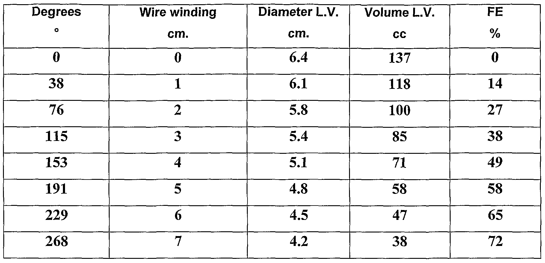
In the following table are shown the possibilities to incede on the external dimension of the left ventricle with telediastolic diameter of 6,4 cm and with a wall thickness of 0,8 cm using a contraction spiral of 3 cm
of diameter with different degrees of rotation imposed to the engine .
As said, the device as described can be opportunely suitable to intervene also in other situation and on other organs, for example in the case of atonic bladder. For this case the need of contraction would be only 5-6 times a day and an internal battery could last for a long time for the activation needs of the contraction spiral without the need to comprise means through the skin to feed the device. In a situation like this would also be required an activation speed reduced that is easily obtainable with the controller of the stepping engine. Still in this situation it can be possible to use a remote control to control the action of the engine or a sensor that verifies the voltage state of the bladder's wall or of the same contraction spiral.
It is thus clear how the prefixed aims have been reached by the present invention. In particular is it clear how such device is particularly light and compact and does not requires, in the case of cardiac use, the opening of the cardiac cavities, having this way a lower
traumatic impact. In particular none of the parts of the device is inserted inside the vascular bed or in the cardiac chambers. The patient does not need therefore an anti-coagulating therapy if not prescribed because of other conditions and the patient does not present the endocarditic risk caused by artificial elements found in the vascular bed. Moreover the pulsatile functioning reproduces the normal fisiologic condition unlike the continuous innatural flow obtainable with other devices. Are entirely eliminated the risks of injury of the corpuscolated elements of the blood and therefore absence of risk of haemolysis mechanically induced.
Moreover there is a normal possibility to ecocardiograph, radiograph scintigraph the heart after the surgery. The contractility of the ventricular walls and therefore the result of the surgery can be judged in a simple and immediate manner and monitored during the time. According to the verified effect it can also be regulated the functioning of the device. The functionality of the contraction spirals in their movements can on the other hand be checked easily through radioscopy. Through the methodic radiography it is also possible to verify the integrity inside the system and the conditions of the implant. Moreober there is the possibility to comprise in the same device the pacemaker function that can be ensured in an epicardiac manner in the application phase of the device .
Claims
1. Supporting device to the contraction of an organ in general, in particular to the heart contraction, characterized by the fact to comprise:
- At least an elastic element (1) configured to surround at least partially the organ in such a way to result operable between a contraction condition where induces an elastic compulsion on the organ and a release condition where it elastically releases said organ;
- At least a control unit (2) configured to operate in an intermitting manner said elastic element between said contraction condition and said release condition.
2. Supporting device to the contraction of an organ in general, in particular to the heart contraction, according to claim 1, where said elastic element (1) comprises :
- A spiral (3) ;
- a contraction wire (4) located inside the spiral (3) for its entire length.
3. Supporting device to the contraction of an organ in general, in particular to the heart contraction, according to claim 2, where said spiral (3) can indifferently have one of the following section shapes:
- Circular, in order to obtain a cylindric spiral;
- Rectangular;
- Semi-elliptical; - Elliptical;
4. Supporting device to the contraction of an organ in general, in particular to the heart contraction, according to claim 2 or 3, where said spiral comprises a first contractive part (8) with coils spaced at a distance one to each other, a second non- contractive part (9) with coils closed one to each other and a joint (10) interposed between said first and said second part.
5. Supporting device to the contraction of an organ in general, in particular to the heart contraction, according to claim from 2 to 4, where said contraction wire (4) comprises a distal end (5) widen such that said wire become blocked with the spiral in correspondence of the reaching of said distal end (5) against the end of the spiral, said wire getting out partially from the opposed end in correspondence of the second non-contractive part (9) .
6. Supporting device to the contraction of an organ in general, in particular to the heart contraction, according to claim from 4, where the spiral of the second non-contractive part (9) can be realized in two or more elements gathered in a common connection end.
7. Supporting device to the contraction of an organ in general, in particular to the heart contraction, according to one or more claim from 1 to 6, where it is comprised an external coating sheath (6) for said elastic element (1) .
8. Supporting device to the contraction of an organ in general, in particular to the heart contraction, according to claim 7, where said sheath (6) comprises fitting appendixes (7) .
9. Supporting device to the contraction of an organ in general, in particular to the heart contraction, according to claim 7 or 8, where the sheath is filled up with physiologically compatible liquid.
10. Supporting device to the contraction of an organ in general, in particular to the heart contraction, according to claim 1, where said control unit (2) comprises a rotary stepping engine (B) to drive into rotation a spool (C; Cl) in said intermitting manner, said spool being configured to get bounded in use to said elastic element (1) .
11. Supporting device to the contraction of an organ in general, in particular to the heart contraction, according to claim 10, where said spool (C;C1) comprises at least an indentation (22) or similar within which bind said elastic element.
12. Supporting device to the contraction of an organ in general, in particular to the heart contraction, according to claim 11, where it is further comprised a screw (23) to block said elastic element within the above mentioned indentation.
13. Supporting device to the contraction of an organ in general, in particular to the heart contraction, according to one or more of the previous claims, where said control unit (2) comprises also a power supply battery (A) .
14. Supporting device to the contraction of an organ in general, in particular to the heart contraction, according to claim 13, where said battery comprises also an electric wire (29) for the power supply made from the outside.
15. Supporting device to the contraction of an organ in general, in particular to the heart contraction, according to one or more of the previous claims, where said control unit (2) comprises also a pacemaker (E) set to pick up the cardiac frequency and control said rotation of the engine (B) in an intermitting manner according to said cardiac frequency.
16. Supporting device to the contraction of an organ in general, in particular to the heart contraction, according to one or more of claims from 1 to 11, where said spool (Cl) comprises three sections of different diameter suitable to produce different contractions on three spirals applied in correspondence of said three sections .
Priority Applications (1)
| Application Number | Priority Date | Filing Date | Title |
|---|---|---|---|
| EP09746217A EP2346547A1 (en) | 2008-05-12 | 2009-05-07 | Cardiac contraction supporting system for people with depressed cardiac function |
Applications Claiming Priority (2)
| Application Number | Priority Date | Filing Date | Title |
|---|---|---|---|
| ITBA2008A000018 | 2008-05-12 | ||
| IT000018A ITBA20080018A1 (en) | 2008-05-12 | 2008-05-12 | SUPPORT FOR CARDIAC CONTRACTION CONSTITUTED BY CONTRIBUTION SPIRALS FOR SUBJECTS WITH DEPRESSED CARDIAC FUNCTION |
Publications (1)
| Publication Number | Publication Date |
|---|---|
| WO2009138922A1 true WO2009138922A1 (en) | 2009-11-19 |
Family
ID=40302482
Family Applications (1)
| Application Number | Title | Priority Date | Filing Date |
|---|---|---|---|
| PCT/IB2009/051883 WO2009138922A1 (en) | 2008-05-12 | 2009-05-07 | Cardiac contraction supporting system for people with depressed cardiac function |
Country Status (3)
| Country | Link |
|---|---|
| EP (1) | EP2346547A1 (en) |
| IT (1) | ITBA20080018A1 (en) |
| WO (1) | WO2009138922A1 (en) |
Cited By (1)
| Publication number | Priority date | Publication date | Assignee | Title |
|---|---|---|---|---|
| JP7438414B2 (en) | 2018-12-03 | 2024-02-26 | 朝日インテック株式会社 | heart model |
Citations (4)
| Publication number | Priority date | Publication date | Assignee | Title |
|---|---|---|---|---|
| US5383840A (en) * | 1992-07-28 | 1995-01-24 | Vascor, Inc. | Biocompatible ventricular assist and arrhythmia control device including cardiac compression band-stay-pad assembly |
| US20040024286A1 (en) * | 1996-01-02 | 2004-02-05 | The University Of Cincinnati | Heart wall actuation device for the natural heart |
| WO2004110257A2 (en) * | 2003-06-09 | 2004-12-23 | The University Of Cincinnati | Power system for a heart actuation device |
| WO2004110553A1 (en) * | 2003-06-09 | 2004-12-23 | The University Of Cincinnati | Actuation mechanisms for a heart actuation device |
-
2008
- 2008-05-12 IT IT000018A patent/ITBA20080018A1/en unknown
-
2009
- 2009-05-07 EP EP09746217A patent/EP2346547A1/en not_active Withdrawn
- 2009-05-07 WO PCT/IB2009/051883 patent/WO2009138922A1/en active Application Filing
Patent Citations (4)
| Publication number | Priority date | Publication date | Assignee | Title |
|---|---|---|---|---|
| US5383840A (en) * | 1992-07-28 | 1995-01-24 | Vascor, Inc. | Biocompatible ventricular assist and arrhythmia control device including cardiac compression band-stay-pad assembly |
| US20040024286A1 (en) * | 1996-01-02 | 2004-02-05 | The University Of Cincinnati | Heart wall actuation device for the natural heart |
| WO2004110257A2 (en) * | 2003-06-09 | 2004-12-23 | The University Of Cincinnati | Power system for a heart actuation device |
| WO2004110553A1 (en) * | 2003-06-09 | 2004-12-23 | The University Of Cincinnati | Actuation mechanisms for a heart actuation device |
Cited By (1)
| Publication number | Priority date | Publication date | Assignee | Title |
|---|---|---|---|---|
| JP7438414B2 (en) | 2018-12-03 | 2024-02-26 | 朝日インテック株式会社 | heart model |
Also Published As
| Publication number | Publication date |
|---|---|
| EP2346547A1 (en) | 2011-07-27 |
| ITBA20080018A1 (en) | 2009-11-13 |
Similar Documents
| Publication | Publication Date | Title |
|---|---|---|
| US7081086B2 (en) | Expandable cardiac harness for treating congestive heart failure | |
| US6702732B1 (en) | Expandable cardiac harness for treating congestive heart failure | |
| EP1033142B1 (en) | Apparatus aiding physiologic systolic and diastolic dynamics of cardiac cavities | |
| US20040010180A1 (en) | Cardiac assist system | |
| JP2008207018A (en) | Heart assisting device | |
| CN108348753A (en) | Medical Devices in heart with pressure-sensing | |
| JP5298132B2 (en) | Heart compression system | |
| US8372145B2 (en) | Implantable artificial ventricle having low energy requirement | |
| WO2009138922A1 (en) | Cardiac contraction supporting system for people with depressed cardiac function | |
| US6063115A (en) | Cardiac assistance system | |
| JP2016123593A (en) | Heart supporting auxiliary apparatus |
Legal Events
| Date | Code | Title | Description |
|---|---|---|---|
| 121 | Ep: the epo has been informed by wipo that ep was designated in this application |
Ref document number: 09746217 Country of ref document: EP Kind code of ref document: A1 |
|
| NENP | Non-entry into the national phase |
Ref country code: DE |
|
| WWE | Wipo information: entry into national phase |
Ref document number: 2009746217 Country of ref document: EP |