WO2010001969A1 - Method for amplification of target nucleic acid sequence, method for detection of mutation by using the method, and reagents for use in the methods - Google Patents
Method for amplification of target nucleic acid sequence, method for detection of mutation by using the method, and reagents for use in the methods Download PDFInfo
- Publication number
- WO2010001969A1 WO2010001969A1 PCT/JP2009/062143 JP2009062143W WO2010001969A1 WO 2010001969 A1 WO2010001969 A1 WO 2010001969A1 JP 2009062143 W JP2009062143 W JP 2009062143W WO 2010001969 A1 WO2010001969 A1 WO 2010001969A1
- Authority
- WO
- WIPO (PCT)
- Prior art keywords
- primer
- nucleic acid
- base
- complementary
- template nucleic
- Prior art date
Links
Images


Classifications
-
- C—CHEMISTRY; METALLURGY
- C12—BIOCHEMISTRY; BEER; SPIRITS; WINE; VINEGAR; MICROBIOLOGY; ENZYMOLOGY; MUTATION OR GENETIC ENGINEERING
- C12Q—MEASURING OR TESTING PROCESSES INVOLVING ENZYMES, NUCLEIC ACIDS OR MICROORGANISMS; COMPOSITIONS OR TEST PAPERS THEREFOR; PROCESSES OF PREPARING SUCH COMPOSITIONS; CONDITION-RESPONSIVE CONTROL IN MICROBIOLOGICAL OR ENZYMOLOGICAL PROCESSES
- C12Q1/00—Measuring or testing processes involving enzymes, nucleic acids or microorganisms; Compositions therefor; Processes of preparing such compositions
- C12Q1/68—Measuring or testing processes involving enzymes, nucleic acids or microorganisms; Compositions therefor; Processes of preparing such compositions involving nucleic acids
- C12Q1/6813—Hybridisation assays
- C12Q1/6827—Hybridisation assays for detection of mutation or polymorphism
-
- C—CHEMISTRY; METALLURGY
- C12—BIOCHEMISTRY; BEER; SPIRITS; WINE; VINEGAR; MICROBIOLOGY; ENZYMOLOGY; MUTATION OR GENETIC ENGINEERING
- C12Q—MEASURING OR TESTING PROCESSES INVOLVING ENZYMES, NUCLEIC ACIDS OR MICROORGANISMS; COMPOSITIONS OR TEST PAPERS THEREFOR; PROCESSES OF PREPARING SUCH COMPOSITIONS; CONDITION-RESPONSIVE CONTROL IN MICROBIOLOGICAL OR ENZYMOLOGICAL PROCESSES
- C12Q1/00—Measuring or testing processes involving enzymes, nucleic acids or microorganisms; Compositions therefor; Processes of preparing such compositions
- C12Q1/68—Measuring or testing processes involving enzymes, nucleic acids or microorganisms; Compositions therefor; Processes of preparing such compositions involving nucleic acids
- C12Q1/6844—Nucleic acid amplification reactions
- C12Q1/6858—Allele-specific amplification
-
- C—CHEMISTRY; METALLURGY
- C12—BIOCHEMISTRY; BEER; SPIRITS; WINE; VINEGAR; MICROBIOLOGY; ENZYMOLOGY; MUTATION OR GENETIC ENGINEERING
- C12Q—MEASURING OR TESTING PROCESSES INVOLVING ENZYMES, NUCLEIC ACIDS OR MICROORGANISMS; COMPOSITIONS OR TEST PAPERS THEREFOR; PROCESSES OF PREPARING SUCH COMPOSITIONS; CONDITION-RESPONSIVE CONTROL IN MICROBIOLOGICAL OR ENZYMOLOGICAL PROCESSES
- C12Q2527/00—Reactions demanding special reaction conditions
- C12Q2527/107—Temperature of melting, i.e. Tm
Definitions
- the present invention relates to a method for amplifying a target nucleic acid sequence containing a base site where a mutation targeted for detection occurs, a method for detecting a mutation using the same, and a reagent used therefor.
- Detecting gene mutations including single nucleotide polymorphisms is widely used in the prevention and treatment of all diseases. For example, many mutations are found in genes of cancer cells, and these are known to be involved in cell carcinogenesis. For this reason, it is possible to confirm the possibility of canceration and the degree of progression by detecting gene mutations in cells, which is considered to be very useful information in treatment. In addition, mutations that show drug resistance in cancer cell genes during medication have been reported. By detecting this mutation, the effectiveness of the drug for each patient can be determined, so that more appropriate treatment is possible.
- SNPs single nucleotide polymorphisms
- CML chronic myelogenous leukemia
- imatinib imatinib
- a mutation on the bcr-abl gene eg, T315I
- detection of a mutation in a gene is useful in early detection and treatment in the clinical field, and thus high reliability is required.
- Non-Patent Document 1 As a method for detecting a mutation in a gene, generally, a direct sequencing method, an ASP (Allele Specific Primer) -PCR (Polymerase Chain Reaction) method (Patent Document 1), a Tm (Melting temperature) analysis method (Non-Patent Document 1), It has been known.
- the direct sequencing method is a method of amplifying a region including a base site to be detected and analyzing the base sequence of the obtained amplification product.
- ASP-PCR method PCR is performed using a primer that is complementary to the region including the base site for detection and has a base complementary to the base of the base site for detection in the 3 ′ end region, This is a method for judging mutations based on the presence or absence of amplification.
- mutant sequence when a primer complementary to a sequence in which the base site for detection is set to a mutant base (hereinafter also referred to as “mutant sequence”) is used, mutation can be confirmed by detection of amplification.
- a primer complementary to a sequence in which the base site to be detected is set to a normal base (hereinafter also referred to as “normal sequence”) is used, non-mutation (ie, normal) can be confirmed by detection of amplification.
- normal sequence a primer complementary to a sequence in which the base site to be detected is set to a normal base
- non-mutation ie, normal
- Tm analysis first, a region including the base site to be detected is amplified, and a hybrid (double-stranded DNA) of the obtained amplification product and a probe complementary to the mutant sequence is formed.
- the hybrid formed body is subjected to a heat treatment, and the dissociation (melting) of the hybrid accompanying a temperature rise is detected by signal measurement such as absorbance to determine the Tm value.
- the Tm value is higher as each strand constituting the hybrid is matched, and lower as it is mismatched. Therefore, a Tm value (evaluation reference value) is obtained in advance for the hybridized product of the mutant sequence and a probe complementary thereto, and this evaluation reference value and the previously determined Tm value (measurement value) Can be determined as follows. If the measured value is the same as the evaluation reference value, it can be determined that there is a perfect match, that is, there is a mutation in the base site for detection. On the other hand, if the measured value is lower than the evaluation reference value, it can be determined that there is a mismatch, that is, the detection-target base site is normal (no mutation is present).
- the direct sequencing method has low sensitivity and takes a lot of time and effort to operate.
- the ASP-PCR method has a problem that it lacks specificity although it is excellent in sensitivity. That is, when a primer complementary to the mutated sequence is used, amplification is confirmed even though there is no mutation, and it may be judged as a false positive.
- the ASP-PCR method only one of a primer complementary to the mutant sequence and a primer complementary to the normal sequence can be used in one reaction system. For this reason, in order to confirm whether the base site for detection is normal or mutation, there are a total of two types, a reaction system using a primer complementary to the mutant sequence and a reaction system using a primer complementary to the normal sequence. It is necessary to perform PCR.
- a sample collected from a patient contains a mixture of cells in which the target gene is mutated and normal cells. For this reason, for example, it is required that a biological sample containing a large amount of normal genes and a small amount of mutant genes can accurately detect the presence or absence of mutation.
- the present invention provides a target nucleic acid sequence amplification method, a mutation detection method, and a reagent used therefor that enable detection of a mutation with excellent sensitivity and reliability in a single reaction system. Objective.
- the method for amplifying a target nucleic acid sequence of the present invention is a method for amplifying a target nucleic acid sequence in a template nucleic acid,
- the target nucleic acid sequence is a sequence containing a base site where a mutation for detection in the template nucleic acid occurs,
- an amplification step of preferentially amplifying a target nucleic acid sequence whose base site is a mutated base over a target nucleic acid sequence whose base site is a normal base is characterized.
- the mutation detection method of the present invention is a method for detecting the presence or absence of a mutation at a base site to be detected in a template nucleic acid, and comprises the following steps (a) to (c). (A) a step of amplifying a target nucleic acid sequence containing the base site in the template nucleic acid in a reaction system by the method for amplifying a target nucleic acid sequence of the present invention; and (b) hybridizing to a sequence containing the base site in the template nucleic acid.
- the present invention provides, for example, the case where a template nucleic acid in which the base site is a mutant type and a template nucleic acid in which the base site is a normal type coexist as a template nucleic acid in a sample.
- the target nucleic acid sequence is amplified preferentially over the normal target nucleic acid sequence.
- the mutant target nucleic acid sequence for example, even when the ratio of the mutant template nucleic acid is lower than that of the normal template nucleic acid.
- Tm analysis By performing Tm analysis using a probe, the presence or absence of mutation can be detected with excellent sensitivity and reliability. For this reason, as described above, it is particularly useful for a sample containing both a normal gene and a mutant gene. From the above points, the present invention can be said to be extremely useful in the recent clinical field in which treatment and diagnosis are performed by detecting gene mutation, for example.
- FIG. 1 is a graph showing the results of Tm analysis in Example 1 of the present invention.
- FIG. 2 is a graph showing the results of Tm analysis in Comparative Example 1.
- FIG. 3 is a graph showing the results of Tm analysis in Example 2 of the present invention.
- FIG. 4 is a graph showing the results of Tm analysis in Example 3 of the present invention.
- FIG. 5 is a graph showing the results of Tm analysis in Comparative Example 3.
- FIG. 6 is a schematic diagram showing the relationship between a primer and a template nucleic acid in one embodiment of the present invention.
- FIG. 7 is a schematic diagram showing the relationship between a primer and a template nucleic acid in another embodiment of the present invention.
- the method for amplifying a target nucleic acid sequence of the present invention is a method for amplifying a target nucleic acid sequence in a template nucleic acid, wherein the target nucleic acid sequence includes a base site where a mutation for detection in the template nucleic acid occurs.
- a template nucleic acid in which the base at the base site is mutated is referred to as “mutant template nucleic acid”
- a target nucleic acid sequence in which the base at the base site is mutated is referred to as “mutant target nucleic acid sequence”
- a template nucleic acid having a normal base at the base site is also referred to as a “normal template nucleic acid”
- a target nucleic acid sequence having a normal base at the base site is also referred to as a “normal target nucleic acid sequence”.
- a normal template nucleic acid is used.
- a mutant template nucleic acid is used.
- both of the template nucleic acid and the mutant template nucleic acid can be amplified.
- preferential amplification may mean, for example, the promotion of amplification of a mutant target nucleic acid sequence, or the meaning of suppressing or inhibiting the amplification of a normal target nucleic acid sequence. Good.
- amplification may be performed using a single-stranded nucleic acid as a template nucleic acid, or amplification of a complementary double-stranded nucleic acid using each of the two single strands constituting the template as a template. You may go.
- two single strands constituting a double-stranded nucleic acid are each used as a template, for convenience, one is also referred to as a (+) strand and the other as a ( ⁇ ) strand.
- either the (+) strand or the ( ⁇ ) strand may be a sense strand or an antisense strand.
- the base site to be detected may be, for example, a base site in the (+) chain, or a base site in the ( ⁇ ) chain corresponding to the base site. Also good.
- the probe used in the mutation detection method of the present invention described later may be designed to be hybridizable to the (+) strand, or may be designed to be hybridizable to the ( ⁇ ) strand, for example. .
- the amplification method of the present invention can also be referred to as, for example, a method for amplifying a mutant target nucleic acid sequence.
- the amplification method of the present invention is not particularly limited as long as it can preferentially amplify a target nucleic acid sequence which is a mutant type over a normal target nucleic acid sequence. Specific examples of the present invention are given below, but the present invention is not limited thereto.
- Examples of the first amplification method of the present invention include a form in which the first amplification reagent including the primer (Xmt) and the primer (Xwt) is used.
- the amplification step is a step of amplifying the target nucleic acid sequence using the primer (Xmt) and the primer (Xwt) in the same reaction system.
- Primer (Xmt) Primer primer (Xwt) that is complementary to the region containing the mutant base site in the template nucleic acid and has a base complementary to the base of the base site in the 3 ′ region
- a region in the template nucleic acid where various primers are annealed is also referred to as an “annealing region”.
- the various primers may be, for example, a sequence that is capable of specifically binding to the annealing region in the template nucleic acid, and may be a sequence that is completely complementary to the annealing region, or a partial sequence. It may be a sequence complementary to the sequence, or a sequence having partially mismatched bases.
- the primer (Xmt) is a primer that can be annealed to a region where the base site is mutated, and is hereinafter also referred to as “mutant primer”.
- the primer (Xwt) is a primer that can be annealed to a region where the base site is a normal type, and is hereinafter also referred to as a “normal primer”.
- the template nucleic acid may be single-stranded or double-stranded as described above. When the template nucleic acid is double-stranded, for example, a mutant primer (Xmt) and a normal primer (Xwt) are designed to be complementary to one of the single strands constituting the double strand. Is preferred.
- a primer (Y2) described later is preferably designed to be complementary.
- An example of the relationship between the template nucleic acid and the primer is shown in the schematic diagram of FIG. In FIG. 6, (+) and ( ⁇ ) are each a single strand that constitutes a double-stranded template nucleic acid, and the hatched portion indicates the base site for detection.
- both the mutant primer (Xmt) and the normal primer (Xwt) are designed as primers complementary to the (+) strand.
- the primer (Y2) is preferably designed as a primer complementary to the ( ⁇ ) strand.
- FIG. 6 is a schematic diagram showing an example to the last.
- each primer, the annealing region in the template nucleic acid, and the like are not limited, and the primer (Y2) is also arbitrary.
- the mutant primer (Xmt) and the normal primer (Xwt) are both designed as primers complementary to the ( ⁇ ) strand, and the primer (Y2) is designed as a primer complementary to the (+) strand. May be.
- the primer (Xmt) and the primer (Xwt) have amplification efficiency by the primer (Xmt) with respect to the template nucleic acid whose base site is a mutant type, and a template nucleic acid whose base site is a normal type. It is preferable to satisfy the relationship that the amplification efficiency is higher than that of the primer (Xwt). As a result, for example, the following becomes possible. That is, even if the template nucleic acid includes a normal template nucleic acid and a mutant template nucleic acid, and the content of the mutant template nucleic acid is low, the mutant template is more effective than the amplification efficiency of the primer (Xwt) for the normal template nucleic acid.
- the amplification efficiency with the primer (Xmt) for the nucleic acid is high, a low content of the mutant template nucleic acid can be efficiently amplified. For this reason, even if it is a low content mutant type
- the template nucleic acid when the template nucleic acid is only a mutant template nucleic acid or only a normal template nucleic acid, it can be amplified by a mutant primer (Xmt) or a normal primer (Xwt), respectively. Therefore, as described above, not only the case where both the mutant template nucleic acid and the normal template nucleic acid are included, but either one of the template nucleic acids can be detected by Tm analysis described later.
- the template nucleic acid is only a normal template nucleic acid, for example, not only a normal primer but also a mutant primer may anneal to the template nucleic acid and be extended.
- the mutant primer (Xmt) is set such that the amplification efficiency for the “mutant” template nucleic acid is higher than the amplification efficiency for the “normal” template nucleic acid of the normal primer (Xwt). Therefore, the amplification efficiency of the mutant primer (Xmt) for the “normal” template nucleic acid is lower than the amplification efficiency of the normal primer (Xwt) for the “normal” template nucleic acid, for example, and the normal template nucleic acid On the other hand, the normal primer shows better amplification efficiency at each stage.
- the mutant primer (Xmt) and the normal primer (Xwt) are not particularly limited, and those satisfying the above relationship can be preferably used.
- Such a relationship makes the affinity of the mutant primer (Xmt) to the mutant template nucleic acid, that is, the ease of annealing higher than the affinity of the normal primer (Xwt) to the normal template nucleic acid. This can be achieved.
- the extension reaction of the mutant primer (Xmt) annealed to the mutant template nucleic acid can be realized more easily than the extension reaction of the normal primer (Xmt) annealed to the normal template nucleic acid.
- the method for adjusting the affinity of the primer is not particularly limited, and can be performed, for example, by setting a Tm value.
- the Tm value of the mutant primer (Xmt) for the complementary sequence is relatively higher than the Tm value of the normal primer (Xwt) for the complementary sequence. In this way, by setting the Tm value of the mutant primer (Xmt) higher than that of the normal primer (Xwt), the binding property of the mutant primer (Xmt) to the template nucleic acid is changed to the normal primer (Xwt). Therefore, the amplification efficiency with the mutant primer (Xmt) can be improved.
- the difference between the Tm value of the mutant primer (Xmt) and the Tm value of the normal primer (Xwt) is not particularly limited. For example, it is more than 0 and preferably 20 ° C or less, more preferably more than 0 and 10 ° C or less. Yes, particularly preferably more than 0 and 5 ° C. or less.
- the Tm value in the primer design here refers to, for example, the Tm value for a hybrid formed of a normal primer and a 100% complementary base sequence, and a hybrid of a mutant primer and a 100% complementary base sequence. Means the Tm value for the formed body.
- the method of setting the Tm value of the mutant primer (Xmt) and the Tm value of the normal primer (Xwt) is not particularly limited, but can be adjusted by, for example, the length of each primer and the GC content.
- length generally, the longer the primer, the higher the Tm value can be set.
- the length of the mutant primer (Xmt) is preferably set longer than that of the normal primer (Xwt).
- the Tm value of the mutant primer (Xmt) can be set to a relatively higher value than the Tm value of the normal primer (Xwt).
- Tm value can be set relatively high, so that GC content is relatively high.
- the GC content of the mutant primer (Xmt) is preferably set higher than that of the normal primer (Xwt).
- Tm value can also be set with both the length of a primer, and GC content.
- the difference in length between the two is not particularly limited, but is, for example, more than 0 and 20 bases or less, preferably more than 0 and 10 It is less than the base, more preferably more than 0 and less than 5 bases.
- the reactivity of the extension reaction from the primer can be adjusted, for example, and the method is not particularly limited and can be performed by a conventionally known method. Specific examples include a method of adding a substance such as a fluorescent substance and biotin, or an additional sequence to the 5 'region of the mutant primer (Xmt). These methods can be performed, for example, based on the description in JP-A-2004-337124.
- the mutant primer (Xmt) may have a base complementary to the base of the base site to be detected (mutant base) in the 3 ′ region.
- the mutant primer (Xmt) Can be designed into the sequence “5′-... Aa C- 3 ′” in which the first base at the 3 ′ end is a complementary base ( C ) of the mutant base (G).
- the mutant primer (Xmt) is, for example, a sequence “5′-...
- Aa C g-3 ′ in which the second base at the 3 ′ end is a complementary base ( C ) of the mutant base (G). May be designed.
- the first base at the 3 ′ end is the base at the 3 ′ end
- the second base at the 3 ′ end is the second base toward the 5 ′ direction with the base at the 3 ′ end as the first. (Hereinafter the same).
- the first base at the 3 ′ end is a base complementary to the mutant base, and at least one base from the second to the 5 ′ end at the 3 ′ end is added to the template nucleic acid.
- the base it is preferable to set the base to be a mismatch (mismatch base).
- mismatch base it is preferable to set at least one of the second and third bases at the 3 ′ end as the mismatch base, and it is more preferable to set the second base at the 3 ′ end as a mismatch base.
- the sequence in the template nucleic acid is “5′-... Ac Gt t...
- the mutant primer (Xmt) has a first base at the 3 ′ end as a complementary base ( C ) of the mutant base (G) and a second base as a base complementary to the base ( t ) of the template nucleic acid ( t ).
- it may be designed as a sequence “5′ -... a tC- 3 ′” as a mismatch base ( t ).
- the second base at the 3 ′ end is a base complementary to the mutant base, and the first at the 3 ′ end and / or at least one from the third to the 5 ′ end.
- the base is preferably set to a base (mismatch base) that is mismatched with the template nucleic acid.
- a base mismatch base
- at least one of the first and third bases at the 3 ′ end is preferably set as the mismatch base
- the third base at the 3 ′ end is more preferably set as a mismatch base.
- the sequence in the template nucleic acid is “5′-... Ac G tt...
- the mutant primer (Xmt) has a second base at the 3 ′ end as a complementary base ( C ) of the mutant base (G) and a third base as a base complementary to the base (t) of the template nucleic acid (T).
- the sequence may be designed as a mismatch base ( t ) “5′ -... a tC g-3 ′”.
- the specificity of the mutant primer (Xmt) for the mutant template nucleic acid can be further improved by adding a mismatch base to the mutant primer.
- the normal primer (Xwt) may have a base complementary to the base of the base site for detection (normal base) in the 3 ′ region. It is preferable that the second base is a base complementary to the normal base.
- the normal primer (Xwt) Can be designed into the sequence “5′-... Aa T- 3 ′” in which the first base at the 3 ′ end is a complementary base ( T ) of the normal base (A).
- the normal primer (Xwt) has a sequence “5′-... Aa T g-3 ′” in which the second base from the 3 ′ end is a complementary base ( T ) of the normal base (A). You may design.
- the first base at the 3 ′ end is a base complementary to a normal base, and at least one base from the second to the 5 ′ end at the 3 ′ end is added to the template nucleic acid.
- the base it is preferable to set the base to be a mismatch (mismatch base).
- mismatch base it is preferable to set at least one of the second and third bases at the 3 ′ end as the mismatch base, and it is more preferable to set the second base at the 3 ′ end as a mismatch base.
- the sequence in the template nucleic acid is “5′-... Ac At t...
- the normal primer (Xwt) is a base complementary to the base ( T ) of the normal base ( A ) as the first base at the 3 'end and a base complementary to the base ( t ) of the template nucleic acid ( t ) ( Instead of a), it may be designed as a sequence “5′ -... a tA ⁇ 3 ′” as a mismatch base ( t ).
- the second base at the 3 ′ end is a base complementary to a normal base, and at least one from the first at the 3 ′ end and / or from the third to the 5 ′ end.
- the base is preferably set to a base (mismatch base) that is mismatched with the template nucleic acid.
- a base mismatch base
- the sequence in the template nucleic acid is “5′-... Ac A tt...
- the normal primer (Xwt) is a base complementary to the base ( T ) of the normal base (A) as the second base at the 3 ′ end and a base complementary to the base (t) of the template nucleic acid ( T ).
- the sequence may be designed as a mismatch base ( t ) “5′ -... a tA g-3 ′”.
- the lengths of the mutant primer (Xmt) and the normal primer (Xwt) are not particularly limited, and the general length is, for example, 10 to 50 bases, preferably 15 to 40 bases, more preferably Includes 16 to 35 bases.
- primer (Y2) As described above, together with the mutant primer (Xmt) and the normal primer (Xwt).
- Primer (Y2) Primer complementary to the complementary sequence to the region 5 'from the base site in the template nucleic acid
- the primer (Y2) is, for example, a primer complementary to a complementary strand of a template nucleic acid that can be annealed by a mutant primer (Xmt) and a normal primer (Xwt) (see FIG. 6). Therefore, for example, the mutant primer (Xmt) and primer (Y2), the normal primer (Xwt) and primer (Y2) can each amplify the template nucleic acid and its complementary strand as a pair of primers. Since this primer (Y2) is a primer that anneals to a region different from the base site to be detected, the target nucleic acid sequence is amplified regardless of whether the base site is a mutant type or a normal type. it can.
- the mutant primer (Xmt) anneals to the normal base
- amplification occurs when the normal primer (Xwt) anneals to the mutant base
- the resulting amplification product has a sequence that depends on each primer.
- coexistence of the primer (Y2) makes it possible to obtain an amplification product that maintains the original sequence of the template nucleic acid. Thereby, the reliability of mutation detection can be further improved.
- the length of the primer (Y2) is not particularly limited, but is usually preferably 10 to 50 bases, more preferably 15 to 40 bases, and particularly preferably 16 to 35 bases.
- the primer (Y2) is not particularly limited as long as it can anneal to a complementary sequence to the region 5 'from the base site in the template nucleic acid.
- the sequence is not particularly limited, and is designed according to a conventionally known general primer design method. it can.
- the template nucleic acid may be single-stranded or double-stranded as described above.
- Examples of the template nucleic acid include DNA and RNA such as total RNA and mRNA.
- Examples of the template nucleic acid include nucleic acids contained in a sample such as a biological sample.
- the nucleic acid in the sample may be, for example, a nucleic acid originally contained in the biological sample.
- the nucleic acid in the biological sample is amplified by a nucleic acid amplification method as a template. Amplification products.
- a reverse transcription-PCR reaction from an amplification product amplified by a nucleic acid amplification method using DNA originally contained in the biological sample as a template, or RNA originally contained in the biological sample.
- examples include amplification products amplified by a nucleic acid amplification method using cDNA generated by (RT-PCR: Reverse Transcription PCR) as a template. These amplification products may be used as the template nucleic acid in the present invention.
- the length of the amplification product is not particularly limited, but is, for example, 50 to 1000 bases, preferably 80 to 200 bases.
- amplification in the same reaction system includes, for example, amplifying a target nucleic acid sequence in one reaction solution.
- the sample is not particularly limited as long as it includes a nucleic acid serving as a template, and examples thereof include a sample containing a nucleic acid derived from a biological sample.
- the biological sample include whole blood, oral cells such as oral mucosa, somatic cells such as nails and hair, germ cells, sputum, amniotic fluid, paraffin-embedded tissue, urine, gastric juice, gastric lavage fluid, and the like.
- a reaction solution obtained by performing a nucleic acid amplification method using a nucleic acid derived from a biological sample as a template may be used as a nucleic acid sample in the present invention, and an amplification product contained in the reaction solution may be used as a template nucleic acid.
- the sample is not particularly limited.
- the target base site shows either a mutant type or a normal type. It is very useful for a sample containing a nucleic acid that is unknown, a sample containing a nucleic acid having a mutant type and a nucleic acid having a normal type, a sample that may contain these, and the like.
- the origin of the nucleic acid such as DNA or RNA is not limited, and examples thereof include cells such as various cancer cells, viruses, mitochondria and the like.
- the cells such as blood cells that have become cancerous as described above are likely to have the above-mentioned problems because they include cells having nucleic acids in which mutant types have been generated and cells having nucleic acids exhibiting normal types. Therefore, the mutation detection method of the present invention is particularly preferably applied to a sample having a nucleic acid showing a mutant type and a nucleic acid showing a normal type.
- biological samples such as various cancer cells such as leukemia, It is preferable to apply to blood samples and white blood cells.
- the sample collection method, nucleic acid preparation method, and the like are not limited, and conventionally known methods can be employed.
- the nucleic acid derived from a biological sample as described above can be isolated from the biological sample by, for example, a conventionally known method.
- a commercially available genomic DNA isolation kit (trade name GFX Genomic Blood DNA Purification kit; manufactured by GE Healthcare Biosciences) can be used for isolation of genomic DNA from whole blood.
- the method for amplifying a target nucleic acid sequence of the present invention is characterized in that the aforementioned primer is used in the amplification step, and other steps and conditions are not limited at all.
- the nucleic acid amplification method in the amplification step is not particularly limited.
- PCR Polymerase Chain Reaction
- NASBA Nucleic acid sequence based amplification
- TMA Transcription-mediated amplification
- SDA String Displacement Amplification
- the PCR method is preferable.
- the conditions for the nucleic acid amplification method are not particularly limited, and can be performed by a conventionally known method.
- the addition ratio of the nucleic acid sample in the reaction system (for example, reaction solution) of the amplification reaction is not particularly limited.
- the lower limit of the addition ratio in the reaction system is preferably, for example, 0.01% by volume or more, more preferably 0.05. Volume% or more, more preferably 0.1 volume% or more.
- the upper limit of the addition ratio is not particularly limited, but is preferably 2% by volume or less, more preferably 1% by volume or less, and still more preferably 0.5% by volume or less.
- the addition ratio of a biological sample such as a whole blood sample in the reaction system is, for example, 0.1 to 0.5 volume. % Is preferably set.
- heat treatment is usually performed for DNA denaturation (dissociation into single-stranded DNA), but this heat treatment denatures sugars, proteins, etc. contained in the sample, resulting in insoluble precipitates or turbidity. Etc. may occur. For this reason, when the presence or absence of a mutation is confirmed by an optical method, the occurrence of such a precipitate or turbidity may affect the measurement accuracy.
- the addition ratio of the whole blood sample in the reaction system is set within the above-mentioned range, the mechanism is unknown. However, for example, it is possible to sufficiently prevent the influence of the generation of precipitates and the like due to denaturation. Accuracy can be improved. In addition, since PCR inhibition due to contaminants in the whole blood sample is sufficiently suppressed, the amplification efficiency can be further improved. Therefore, by setting the addition ratio of a biological sample such as a whole blood sample within the above range, for example, it is possible to eliminate the necessity of pretreatment of the sample in order to prevent or remove the occurrence of precipitates or turbidity. .
- the ratio of the whole blood sample in the reaction system is expressed not by the volume ratio as described above (for example, 0.1 to 0.5% by volume) but by the weight ratio of hemoglobin (hereinafter referred to as “Hb”).
- Hb weight ratio of hemoglobin
- the ratio of the whole blood sample in the reaction system is preferably in the range of 0.565 to 113 g / L, more preferably in the range of 2.825 to 56.5 g / L, in terms of Hb amount, More preferably, it is in the range of 5.65 to 28.25 ⁇ g / L.
- the addition ratio of the whole blood sample in the reaction system may satisfy both the volume ratio and the Hb weight ratio, or may satisfy either one.
- the whole blood may be any of hemolyzed whole blood, unhemolyzed whole blood, anticoagulated whole blood, whole blood containing a coagulated fraction, and the like.
- albumin Prior to the start of the amplification reaction in the amplification step, it is preferable to further add albumin to the reaction system.
- albumin for example, the influence due to the occurrence of precipitates or turbidity as described above can be further reduced, and the amplification efficiency can be further improved.
- the addition ratio of albumin in the reaction system is, for example, in the range of 0.01 to 2% by weight, preferably 0.1 to 1% by weight, more preferably 0.2 to 0.8% by weight.
- the albumin is not particularly limited, and examples thereof include bovine serum albumin (BSA), human serum albumin, rat serum albumin, horse serum albumin and the like. Any one of these may be used, or two or more may be used in combination. May be.
- the present invention will be described taking the PCR method as an example of the amplification method in the amplification step. Note that the present invention is not limited to this.
- the PCR conditions are not particularly limited, and can be performed by a conventionally known method.
- a PCR reaction solution containing a template nucleic acid and the various primers described above is prepared.
- the addition ratio of various primers in the PCR reaction solution is not particularly limited, but the mutant primer (Xmt) is preferably added so as to be, for example, 0.01 to 10 ⁇ mol / L, more preferably 0.05. ⁇ 5 ⁇ mol / L, particularly preferably 0.1 to 1 ⁇ mol / L.
- the normal primer (Xwt) is preferably added, for example, at 0.01 to 10 ⁇ mol / L, more preferably 0.05 to 5 ⁇ mol / L, and particularly preferably 0.1 to 0.5 ⁇ mol. / L.
- the molar ratio (Xmt: Xwt) between the mutant primer (Xmt) and the normal primer (Xwt) is, for example, preferably 1: 0.001 to 1:10, more preferably 1: 0.01. To 1: 2 and particularly preferably from 1: 0.1 to 1: 1.
- the primer (Y2) when used in combination with the mutant primer (Xmt) and the normal primer (Xwt), the primer (Y2) should be added at, for example, 0.01 to 10 ⁇ mol / L. Is more preferably 0.05 to 5 ⁇ mol / L, and particularly preferably 0.1 to 1 ⁇ mol / L.
- the molar ratio (Xmt: Y2) between the mutant primer (Xmt) and the primer (Y2) is, for example, preferably 1: 0.001 to 1:10, more preferably 1: 0.01 to 1 : 2 and particularly preferably 1: 0.1 to 1: 1.
- composition components in the reaction solution are not particularly limited, and examples thereof include conventionally known components, and their ratios are not particularly limited.
- examples of the composition component include nucleotides such as DNA polymerase and nucleoside triphosphate (dNTP), solvents, and the like.
- the order of adding each composition component is not limited.
- the DNA polymerase is not particularly limited, and for example, a conventionally known heat-resistant bacterium-derived polymerase can be used. Specific examples include DNA polymerase derived from Thermus aquaticus (US Pat. Nos. 4,889,818 and 5079,352) (trade name Taq polymerase), DNA derived from Thermus thermophilus. Polymerase (WO 91/09950) (rTth DNA polymerase), DNA polymerase derived from Pyrococcus furiosus (WO 92/9689) (Pfu DNA polymerase: manufactured by Stratagene), DNA polymerase derived from Thermococcus litoralis (EP 0455430) (Trademark Vent: New England Biolabs) and the like are commercially available. Among them, thermostable DNA polymerase derived from Thermus aquaticus is preferable.
- the addition rate of DNA polymerase in the reaction solution is not particularly limited, but is, for example, 1 to 100 U / mL, preferably 5 to 50 U / mL, and more preferably 20 to 30 U / mL.
- the activity unit (U) of DNA polymerase is generally an activity that incorporates 10 nmol of all nucleotides into an acid-insoluble precipitate in an activity measurement reaction solution at 74 ° C. for 30 minutes using activated salmon sperm DNA as a template primer. Is 1U.
- the composition of the reaction liquid for activity measurement is, for example, 25 mmol / L TAPS buffer (pH 9.3, 25 ° C.), 50 mmol / L KCl, 2 mmol / L MgCl 2 , 1 mmol / L mercaptoethanol, 200 ⁇ mol / L dATP, 200 ⁇ mol / L dGTP, 200 ⁇ mol / L dTTP, 100 ⁇ mol / L [ ⁇ - 32 P] dCTP, 0.25 mg / mL activated salmon sperm DNA.
- nucleoside triphosphate usually include dNTP (for example, dATP, dGTP, dCTP, dTTP, dUTP, etc.).
- dNTP for example, dATP, dGTP, dCTP, dTTP, dUTP, etc.
- the addition rate of dNTP in the reaction solution is not particularly limited, but is, for example, 0.01 to 1 mmol / L, preferably 0.05 to 0.5 mmol / L, more preferably 0.1 to 0. .3 mmol / L.
- solvent examples include buffer solutions such as Tris-HCl, Tricine, MES, MOPS, HEPES, and CAPS, and commercially available buffer solutions for PCR and commercially available PCR kits can be used.
- the PCR reaction solution may further contain glycerol, heparin, betaine, KCl, MgCl 2 , MgSO 4 , glycerol, etc., and the addition ratio thereof may be set within a range that does not inhibit the PCR reaction, for example. That's fine.
- the total volume of the reaction solution is not particularly limited, and can be appropriately determined depending on, for example, the equipment (thermal cycler) used, but is usually 1 to 500 ⁇ L, preferably 10 to 100 ⁇ L.
- PCR includes, for example, three steps: (1) dissociation of double-stranded nucleic acid into single-stranded nucleic acid, (2) primer annealing, and (3) primer extension (polymerase reaction).
- the conditions for each step are not particularly limited, but the step (1) is preferably, for example, 90 to 99 ° C. and 1 to 120 seconds, more preferably 92 to 95 ° C. and 1 to 60 seconds.
- the step 2) is preferably, for example, 40 to 70 ° C. and 1 to 300 seconds, more preferably 50 to 70 ° C. and 5 to 60 seconds
- the step (3) is, for example, 50 to 80 ° C., 1 It is preferably ⁇ 300 seconds, more preferably 50 to 75 ° C.
- the number of cycles is not particularly limited, and the above three steps (1) to (3) are defined as one cycle, for example, preferably 30 cycles or more.
- the upper limit is not particularly limited, but is, for example, a total of 100 cycles or less, preferably 70 cycles or less, and more preferably 50 cycles or less. What is necessary is just to control the temperature change of each step automatically using a thermal cycler etc., for example.
- a target nucleic acid sequence containing a base site for detection can be produced.
- two or more types of target nucleic acid sequences can be amplified simultaneously in one reaction solution.
- Xmt mutant primer
- Xwt normal primer
- Y2 optionally primer
- the method for amplifying a target nucleic acid sequence of the present invention may further include a step of detecting an amplification product obtained by the amplification reaction described above.
- a step of detecting an amplification product obtained by the amplification reaction described above thereby, for example, it is possible to detect the presence or absence of a mutation in the target base site in the target nucleic acid sequence.
- the detection of the mutation can be confirmed by, for example, Tm analysis as described later.
- a probe capable of hybridizing to a sequence containing the base site for detection (hereinafter also referred to as “detection target sequence”) is added to the reaction system of the amplification reaction in the amplification step. .
- the timing of adding the probe is not particularly limited.
- the probe may be added to the reaction system at any stage before the amplification reaction, in the middle of the amplification reaction, or after the amplification reaction. It is preferable.
- the detection of mutation will be specifically described in the mutation detection method of the present invention described later.
- the probe and the like are as described later.
- Examples of the second amplification method of the present invention include a form in which the second amplification reagent including the primer (Xmt) and the primer (Y1) is used.
- the amplification step may be a step of amplifying the target nucleic acid sequence using the primer (Xmt) and the primer (Y1) in the same reaction system.
- Primer (Y1) Primer complementary to the region 3 ′ of the template nucleic acid from the base site
- the primer (Xmt) in this embodiment is the same as that in the first embodiment.
- this embodiment can be performed in the same manner as the first embodiment unless otherwise specified.
- the template nucleic acid may be single-stranded or double-stranded as described above.
- the mutant primer (Xmt) and the primer (Y1) it is preferable to design the mutant primer (Xmt) and the primer (Y1) to be complementary to any one of the single strands constituting the double stranded.
- the above-described primer (Y2) in a complementary manner An example of the relationship between the template nucleic acid and the primer is shown in the schematic diagram of FIG. In FIG.
- (+) and ( ⁇ ) are each a single strand constituting a double-stranded template nucleic acid, and the shaded portion indicates the base site for detection.
- both the mutant primer (Xmt) and the primer (Y1) are designed as primers complementary to the (+) strand.
- the primer (Y2) is preferably designed as a primer complementary to the ( ⁇ ) strand.
- FIG. 7 is a schematic diagram illustrating an example to the last.
- the length of each primer, the annealing region in the template nucleic acid, and the like are not limited, and the primer (Y2) is also arbitrary.
- the mutant primer (Xmt) and the primer (Y1) are both designed as primers complementary to the ( ⁇ ) strand
- the primer (Y2) is designed as a primer complementary to the (+) strand. Also good.
- the following can be performed by using the mutant primer (Xmt) and the primer (Y1). That is, even if the template nucleic acid includes a normal template nucleic acid and a mutant template nucleic acid, and the content of the mutant template nucleic acid is low, the mutant primer (Xmt) is more mutated than the normal template nucleic acid. Since the affinity for is high, it is possible to efficiently amplify a low content of the mutant template nucleic acid. On the other hand, since the primer (Y1) is a primer that anneals to a region different from the base site to be detected, the target nucleic acid sequence can be amplified regardless of whether the base site is a mutant type or a normal type. .
- the mutant template nucleic acid is further amplified by the primer (Y1). For this reason, even if it is a low content mutant type
- the normal template nucleic acid is also amplified by the primer (Y1), and the normal type can be detected by Tm analysis.
- the primer (Y1) can amplify a target nucleic acid sequence in which the base site for detection is a normal base. Normal type analysis is possible by Tm analysis.
- the primer (Y1) is used, an amplification product maintaining the original sequence of the template nucleic acid can be obtained. That is, for example, when amplification occurs when the mutant primer (Xmt) anneals to the normal template nucleic acid, the resulting amplification product has a sequence that depends on the mutant primer (Xmt).
- coexistence of the primer (Y1) makes it possible to obtain an amplification product that maintains the original sequence of the template nucleic acid. As a result, it is possible to prevent only an amplification product obtained by mistake from increasing, and to improve the reliability of mutation detection.
- the length of the primer (Y1) is not particularly limited, but is usually preferably 10 to 50 bases, more preferably 15 to 40 bases, and particularly preferably 16 to 35 bases.
- the primer (Y1) may be annealed to a region 3 'from the base site in the template nucleic acid, and its sequence is not particularly limited, and can be designed according to a conventionally known general primer design method.
- the addition ratio of various primers in the reaction solution of the amplification reaction is not particularly limited, but the mutant primer (Xmt) is preferably added so as to be, for example, 0.01 to 10 ⁇ mol / L, and more preferably 0.8. 05 to 5 ⁇ mol / L, particularly preferably 0.1 to 1 ⁇ mol / L.
- the primer (Y1) is preferably added so as to be 0.01 to 10 ⁇ mol / L, more preferably 0.05 to 5 ⁇ mol / L, and particularly preferably 0.1 to 1 ⁇ mol / L. .
- the molar ratio (Xmt: Y1) between the mutant primer (Xmt) and the primer (Y1) is preferably, for example, 1: 0.001-1: 10, and more preferably 1: 0.01-1 : 2 and particularly preferably 1: 0.1 to 1: 1. Further, as in the first embodiment, it is preferable to further use a primer (Y2) as a pair.
- the amplification step is a step of amplifying the target nucleic acid sequence using the following primer (Xmt) and 3 ′ ⁇ 5 ′ exonuclease in the reaction system. Is given.
- the primer (Xmt) in the present embodiment is the same as that in the first embodiment, and this embodiment can be performed in the same manner as the above-described embodiments unless otherwise specified.
- the mutant primer (Xmt) by performing an amplification reaction using a mutant primer (Xmt) in the presence of 3 ′ ⁇ 5 ′ exonuclease, for example, the following can be performed. That is, when the template nucleic acid includes a normal template nucleic acid and a mutant template nucleic acid, the mutant primer (Xmt) is annealed not only to the mutant template nucleic acid but also to the normal template nucleic acid, and the extended strand is synthesized. Can be considered. However, since the mutant primer (Xmt) is not a normal template nucleic acid but a primer complementary to the mutant template nucleic acid, even if annealed to the normal template nucleic acid, at least at the base site for detection.
- the mutant primer (Xmt) annealed to the normal template nucleic acid has a mismatch in the 3 ′ region (that is, the region containing the corresponding base of the mutant base) with respect to the normal template nucleic acid. Is in a single-stranded state without annealing. This single-stranded region is cleaved by 3 ' ⁇ 5' exonuclease, whereby a sequence complementary to the normal template sequence is amplified. Since the amplification product thus obtained is a normal target nucleic acid sequence, when the template nucleic acid contains a normal type, the normal product can also be detected by the amplification product from the mutant primer (Xmt). It becomes possible.
- the addition ratio of the mutant primer (Xmt) in the reaction solution of the amplification reaction is not particularly limited, but for example, it is preferably added so as to be 0.001 to 10 ⁇ mol / L, more preferably 0.01 to 5 ⁇ mol / L. L, particularly preferably 0.1 to 1 ⁇ mol / L.
- the 3 ' ⁇ 5' exonuclease is not particularly limited, but for example, it is preferable that a polymerase used for the amplification reaction has this catalytic reaction. Examples of the polymerase having 3 ' ⁇ 5' exonuclease activity include Pfu polymerase, Tli polymerase, KOD polymerase, Vent polymerase, Tgo polymerase and the like.
- any one or more of the above-described primer (Y1), primer (Y2) and primer (Xwt) may be used in combination. Good.
- the amplification reagent of the present invention is an amplification reagent used in the method for amplifying a target nucleic acid sequence of the present invention.
- the first amplification reagent of the present invention is characterized by including a primer (Xmt) and a primer (Xwt), and the second amplification reagent is characterized by including a primer (Xmt) and a primer (Y1).
- the first amplification reagent of the present invention can be used, for example, in the first embodiment of the target nucleic acid sequence amplification method of the present invention
- the second amplification reagent of the present invention can be used, for example, of the target nucleic acid sequence of the present invention. It can be used in the second embodiment of the amplification method.
- the 1st and 2nd amplification reagent of this invention contains a primer (Y2) further.
- each primer is as described above.
- the third amplification reagent of the present invention is characterized by including a primer (Xmt) and 3 ' ⁇ 5' exonuclease.
- the third amplification reagent of the present invention can be used, for example, in the third embodiment of the target nucleic acid sequence amplification method of the present invention.
- the third amplification reagent of the present invention may further include any one or two or less of the primer (Y1), the primer (Y2), and the primer (Xwt).
- the primers are the same as described above.
- the amplification reagent of the present invention may further contain, for example, various components used in the amplification reaction described in the method for amplifying a target nucleic acid sequence of the present invention.
- the amplification reagent of the present invention is preferably used in one reaction system.
- the amplification reagent of the present invention may be an amplification kit used in the method for amplifying a target nucleic acid sequence of the present invention, and each component may be contained in a separate container, or may be appropriately combined to be the same. It may be contained in a container.
- the amplification kit preferably includes, for example, instructions for use.
- the method for detecting a mutation of the present invention is a method for detecting the presence or absence of a mutation at a base site to be detected in a template nucleic acid, comprising the following steps (a) to (c): .
- the present invention is characterized in that the target nucleic acid sequence is amplified by the above-described method and so-called Tm analysis is performed, and other processes and conditions are not limited at all.
- “mutation” includes, for example, SNP.
- the present invention is preferably applied to a sample containing nucleic acid, and the sample is not particularly limited, and examples thereof include the same samples as described above.
- the type of template nucleic acid is not particularly limited, and examples thereof include the same nucleic acids as described above.
- the probe for detecting mutation (hereinafter also referred to as “detection probe”) is not particularly limited, and can be set by a conventionally known method.
- the template nucleic acid when it is double-stranded, it may be designed to hybridize to the sense strand detection target sequence (sense strand detection probe) or to hybridize to the antisense strand detection target sequence. (Antisense strand detection probe).
- the base site for detection in the detection target sequence may be set to a normal base or a mutant base. That is, when the detection probe is annealed to the detection target sequence, the base corresponding to the detection target base site in the detection target sequence may be, for example, complementary to a normal base, or a mutant base.
- the base corresponding to the base site to be detected is complementary to the mutant base and non-complementary to the normal base.
- the base corresponding to the base site to be detected is complementary to the mutant base and non-complementary to the normal base.
- the mutant base for example, in the probe, the
- the detection probe may be any sequence that can hybridize to the detection target sequence including the target base site, as described above.
- the sequence of the probe is not particularly limited, but, for example, 90% to 100% except for a site (base) that forms a pair with a base site to be detected (site where the target mutation occurs) during hybridization.
- the same sequence is preferable, and 100% is particularly preferable.
- the addition ratio of the probe in the reaction system is not particularly limited, but for example, the probe is preferably added in a range of 10 to 400 nmol / L, and more preferably 20 to 200 nmol / L.
- the probe is a labeled probe labeled with a labeling substance such as a fluorescent dye, for example, in order to adjust signal intensity such as fluorescence intensity to be detected
- the probe has the same sequence as the labeled probe.
- An unlabeled probe may be used in combination.
- phosphoric acid may be added to the 3 'end of the unlabeled probe.
- the molar ratio of the labeled probe to the non-labeled probe is preferably, for example, 1:10 to 10: 1.
- the length of the probe is not particularly limited, and is, for example, 5 to 50 mer, preferably 10 to 30 mer.
- the probe can be added to the reaction system of the amplification reaction after the step (a), that is, after carrying out the amplification reaction of the target nucleic acid sequence. It is preferable to add to the reaction system in advance prior to the amplification reaction in step a).
- the addition ratio of various primers in the reaction system is as described above.
- a phosphate group may be further added to the 3 ′ end thereof, The 3 ′ end may be labeled with a fluorescent dye as described above.
- the Tm value will be described.
- the absorbance at 260 nm increases. This is because hydrogen bonds between both strands in double-stranded DNA are unwound by heating and dissociated into single-stranded DNA (DNA melting).
- DNA melting single-stranded DNA
- the absorbance is about 1.5 times the absorbance at the start of heating (absorbance of only double-stranded DNA), thereby melting. It can be judged that it has been completed. Based on this phenomenon, the melting temperature Tm is generally defined as the temperature at which the absorbance reaches 50% of the total increase in absorbance.
- the measurement of the signal value indicating the melting state of the hybrid formed product of the amplification product and the probe may be 260 nm absorbance measurement as described above. Also good.
- a labeled probe labeled with a labeling substance as the probe and measure the signal of the labeling substance.
- the labeled probe include a labeled probe that shows a signal alone and does not show a signal by hybridization, or a labeled probe that does not show a signal alone and shows a signal by hybridization. In the case of the former probe, no signal is shown when a hybrid (for example, double-stranded DNA) is formed with the detection target sequence, and a signal is shown when the probe is released by heating.
- a signal is shown by forming a hybrid (for example, double-stranded DNA) with the target sequence, and when the probe is released by heating, the signal decreases (disappears). Therefore, by detecting the signal by this label under signal-specific conditions (absorption wavelength, etc.), the progress of melting of the hybrid and determination of the Tm value can be performed as in the absorbance measurement at 260 nm.
- a hybrid for example, double-stranded DNA
- two or more types of target nucleic acid sequences can be amplified simultaneously in the same reaction system. Then, the target mutation can be confirmed for each amplification product.
- a probe that hybridizes may be prepared for each detection target sequence including a base site where the target mutation occurs.
- each probe it is preferable to use labeled probes labeled with different labeling substances detected under different conditions. If such a probe is used, each mutation can be detected by changing detection conditions even in the same reaction system.
- the labeling substance in the labeling probe include a fluorescent dye and a fluorophore.
- a probe that is labeled with a fluorescent dye exhibits fluorescence alone, and fluorescence decreases (for example, quenches) by hybridization is preferable.
- a probe using such a fluorescence quenching phenomenon is generally called a fluorescence quenching probe.
- the probe is preferably labeled with a fluorescent dye at the base of the 3 ′ region (for example, 3 ′ end) or 5 ′ region (for example, 5 ′ end) of the oligonucleotide.
- the base is preferably cytosine (C).
- the base paired with the terminal base C of the labeled probe or the base separated by 1 to 3 bases from the paired base is guanine (G).
- G guanine
- the base sequence of the labeled probe is generally called a guanine quenching probe and is known as a so-called QProbe (registered trademark).
- QProbe registered trademark
- the terminal C labeled with the fluorescent dye approaches G in the detection target sequence, so that the emission of the fluorescent dye becomes weak (fluorescence intensity decreases). Decrease).
- the labeling substance can be usually bound to a phosphate group of a nucleotide, for example.
- the fluorescent dye is not particularly limited, and examples thereof include fluorescein, phosphor, rhodamine, polymethine dye derivatives, and the like, and examples of commercially available fluorescent dyes include BODIPY FL (trademark, manufactured by Molecular Probe), FluorePrime (trade name, manufactured by Amersham Pharmacia), Fluoredite (trade name, manufactured by Millipore), FAM (manufactured by ABI), Cy3 and Cy5 (manufactured by Amersham Pharmacia), TAMRA (manufactured by Molecular Probes), and the like.
- the combination of fluorescent dyes used for a plurality of probes is not particularly limited as long as it can be detected under different conditions. For example, Pacific Blue (detection wavelength 450 to 480 nm), TAMRA (detection wavelength 585 to 700 nm) and BODIPY FL ( A combination of detection wavelengths of 515 to 555 nm).
- the mutation detection method of the present invention will be described with an example in which a nucleic acid is amplified by PCR and a labeled probe is used as a detection probe. Note that the present invention is not limited to this.
- PCR is performed as described above using a sample solution containing a template nucleic acid, the above-described various primers in the present invention, and a reaction solution to which a labeled probe that hybridizes to the detection target sequence is added.
- the reaction solution may contain, for example, DNA polymerase, dNTP, and other various additives that can be used for nucleic acid amplification.
- the timing of adding the labeled probe is not particularly limited, and may be any of, for example, before the amplification reaction, in the middle of the amplification reaction, and after the amplification reaction. ) Step can be carried out continuously, so that it is preferably added before the amplification reaction.
- the amplification product obtained is dissociated, and the single-stranded DNA obtained by the dissociation and the labeled probe are hybridized. This can be performed, for example, by changing the temperature of the reaction solution.
- the heating temperature in the dissociation step is not particularly limited as long as the amplification product can be dissociated, and is, for example, 85 to 95 ° C.
- the heating time is not particularly limited, but is usually 1 second to 10 minutes, preferably 1 second to 5 minutes.
- Hybridization of the dissociated single-stranded DNA and the labeled probe can be performed, for example, by lowering the heating temperature in the dissociation step after the dissociation step.
- the temperature condition is, for example, 40 to 50 ° C.
- the temperature of the reaction solution is changed, and a signal value indicating the melting state of the hybridized product of the amplification product and the labeled probe is measured.
- the reaction solution is heated, that is, the hybrid formed of the single-stranded DNA and the labeled probe is heated, and the fluctuation of the signal value accompanying the temperature rise is measured.
- fluorescence is decreased (or quenched) and dissociated in a state where it is hybridized with single-stranded DNA. Then, it emits fluorescence.
- the hybrid formed body in which the fluorescence is decreased (or quenched) may be gradually heated, and the increase in the fluorescence intensity accompanying the temperature increase may be measured.
- the said signal value can be measured on the conditions according to the labeling substance of the said labeled probe, for example.
- the temperature range for measuring the fluctuation of the fluorescence intensity is not particularly limited.
- the start temperature is room temperature to 85 ° C., preferably 25 to 70 ° C.
- the end temperature is 40 to 105 ° C., for example. is there.
- the rate of temperature increase is not particularly limited, but is, for example, 0.1 to 20 ° C./second, preferably 0.3 to 5 ° C./second.
- the Tm value is determined by analyzing the fluctuation of the signal. Specifically, for example, the amount of change in fluorescence intensity per unit time at each temperature is calculated from the obtained fluorescence intensity.
- the amount of change is ( ⁇ d fluorescence intensity increase / dt)
- the temperature showing the lowest value can be determined as the Tm value.
- the amount of change is (d fluorescence intensity increase / t)
- the highest point can be determined as the Tm value.
- the decrease in fluorescence intensity may be measured.
- the Tm value can be calculated by, for example, conventionally known MELTCALC software (http://www.meltcalc.com/) or the like, and can also be determined by a neighbor method (Nearest Neighbor Method).
- the type of base at the target base site that is, the genotype such as a mutant type or normal type is determined.
- a hybrid (match) that is completely complementary has a higher Tm value indicating dissociation than a hybrid (mismatch) that differs in one base. Therefore, the genotype at the target base site can be determined by determining in advance the Tm value of a hybrid that is completely complementary to the probe and the Tm value of a hybrid that differs in one base. For example, when the base at the target base site is assumed to be a mutant type and a probe complementary to the detection target sequence containing the mutant base is used, the Tm value of the hybrid formed is the Tm value of the completely complementary hybrid.
- the target base can be determined as a mutant type. Further, if the Tm value of the formed hybrid is the same as the Tm value of a hybrid different by one base (a value lower than the Tm value of a completely complementary hybrid), the target base can be determined to be a normal type. Further, when both Tm values are detected, for example, it can be determined that a nucleic acid showing a mutant type and a nucleic acid showing a normal type coexist.
- the method of measuring the signal fluctuation accompanying the temperature increase by increasing the temperature of the reaction solution containing the probe, that is, heating the hybrid former
- signal fluctuations during hybridization may be measured. That is, when the hybrid is formed by lowering the temperature of the reaction solution containing the probe, the signal fluctuation accompanying the temperature drop may be measured.
- a labeled probe for example, a guanine quenching probe
- a labeled probe that shows a signal alone and does not show a signal due to hybridization
- it emits fluorescence when the single-stranded DNA and the probe are dissociated.
- the fluorescence is reduced (or quenched). Therefore, for example, the temperature of the reaction solution may be gradually decreased to measure the decrease in fluorescence intensity accompanying the temperature decrease.
- a labeled probe that does not show a signal alone and shows a signal by hybridization, it does not emit fluorescence when the single-stranded DNA and the probe are dissociated, but the hybrid is not released due to a decrease in temperature. Once formed, it will fluoresce. Therefore, for example, the temperature of the reaction solution may be gradually lowered and the increase in fluorescence intensity accompanying the temperature drop may be measured.
- the nucleic acid in the sample may be single-stranded or double-stranded.
- the nucleic acid is double-stranded, it is preferable to include a step of dissociating the double-stranded nucleic acid in the sample by heating, for example, prior to the hybridization in the step (b).
- the mutation detection reagent of the present invention is a detection reagent used in the mutation detection method of the present invention, and includes the amplification reagent of the present invention and a probe capable of hybridizing to a sequence containing a base site to be detected in a template nucleic acid. It is characterized by.
- the amplification reagent of the present invention is preferably used in one reaction system.
- the mutation detection reagent of the present invention may further contain various components used in the amplification reaction described in the mutation detection method of the present invention, for example. Further, the mutation detection reagent of the present invention may be, for example, the mutation detection kit of the present invention, and preferably includes, for example, an instruction manual.
- Example 1 Based on the first embodiment, a point mutation (C ⁇ T) at the 944th base of the bcr-abl gene was detected by Tm analysis.
- a normal plasmid in which a normal bcr-abl gene sequence having no mutation at the 944th base C is inserted (hereinafter referred to as “wt”), and a mutant bcr ⁇ in which the 944th base C is mutated to T
- An abnormal plasmid into which the abl gene was inserted (hereinafter referred to as “mt”) was prepared. Both were mixed at a predetermined ratio to prepare a plurality of nucleic acid samples.
- the mt content ratios in the plurality of nucleic acid samples were 100%, 10%, 5%, 3%, 1%, 0.5%, 0.3%, and 0%, respectively.
- PCR is carried out using a thermal cycler (trade name Mastercycler gradient G, manufactured by Eppendorf). went. PCR was carried out at 95 ° C. for 60 seconds, and then repeated 50 cycles, with 99 ° C. for 4 seconds and 66 ° C. for 30 seconds as one cycle. Further, the tube containing the PCR reaction solution was transferred to iCycler (trade name, manufactured by Bio-Rad Laboratories), treated at 95 ° C. for 5 seconds and at 40 ° C. for 60 seconds, and then the temperature was raised by 1 ° C.
- iCycler trade name, manufactured by Bio-Rad Laboratories
- the step of incubating for 2 seconds was repeated 55 cycles, and the PCR reaction solution was heated from 40 ° C to 95 ° C. During the 55 cycles, the change in fluorescence intensity (detection wavelength 515 to 545 nm) at each temperature from 40 ° C. to 75 ° C. was measured, and Tm analysis was performed.
- the primer (Y2) is a sense primer in PCR
- the normal primer (Xwt) and the mutant primer (Xmt) are antisense primers in PCR.
- the sequences of these primers are shown below.
- the normal primer (Xwt) is a complementary sequence that is 100% matched to the region containing the 944th base (C) in the normal bcr-abl gene
- the mutant primer (Xmt) is the 944th base.
- C is a complementary sequence that is 100% matched to the region containing the 944th base (T) in the mutant bcr-abl gene in which C is mutated to T.
- the base indicated by capital letters at the 3 ′ end is the 944th base of the normal bcr-abl gene and the mutant bcr-abl gene, respectively.
- the positional relationship between the normal primer (Xwt) and the mutant primer (Xmt) and the sense strand that they anneal, and the positional relationship between the sense primer (Y2) and the antisense strand that it anneals are, for example, Although the schematic diagram of FIG. 6 described above can be referred to, it is merely a schematic diagram and does not limit the present invention.
- Sense primer SEQ ID NO: 1 5'-ggacggacggaccgtcctcgttgtcttgttggc-3 ' Normal primer (Xwt) SEQ ID NO: 2 5'-ttcccgtaggtcatgaactcaG-3 ' Mutant primer (Xmt) SEQ ID NO: 3 5'-aggttcccgtaggtcatgaactcaA-3 '
- the sequence of the detection probe used for Tm analysis is shown below.
- the detection probe is a complementary sequence that is 100% matched to the region containing the 944th base in the mutant bcr-abl gene (sense strand) in which the 944th base C is mutated to T.
- the base A shown in capital letters corresponds to the 944th mutated base T.
- P at the 3 ′ end represents a phosphate group.
- FIG. 1 is a graph of Tm analysis showing changes in fluorescence intensity with increasing temperature.
- the horizontal axis represents the temperature (° C.) at the time of measurement, the vertical axis represents the change in fluorescence intensity (hereinafter also referred to as “fluorescence change amount”), and the unit was “ ⁇ d fluorescence intensity increase / dt”.
- fluorescence change amount the change in fluorescence intensity increase / dt.
- the 944th is either a normal base (C) or a mutated base (T) Regardless, the region containing the 944th base is amplified.
- Antisense primer SEQ ID NO: 5 5'-ggacggacggaccgcactccctcaggtagtccag-3 '
- FIG. 2 is a graph of Tm analysis showing changes in fluorescence intensity with increasing temperature.
- the horizontal axis represents the temperature (° C.) at the time of measurement, the vertical axis represents the change in fluorescence intensity (hereinafter also referred to as “fluorescence change amount”), and the unit was “ ⁇ d fluorescence intensity increase / dt”.
- fluorescence change amount the change in fluorescence intensity increase / dt.
- a wt reaction solution for amplifying wt having a normal 944th base and an mt reaction solution for amplifying mt in which the base was mutated were prepared.
- the composition of these reaction solutions is shown below.
- the wt reaction solution used the normal primer (Xwt) of Example 1 as an antisense primer
- the mt reaction solution used the mutant primer (Xmt) of Example 1 as an antisense primer.
- the primer (Y2) was used for the sense primer as in Example 1.
- Example 1 detection sensitivity superior to the method of Comparative Example 2 can be achieved. Further, in the conventional ASP-PCR method in Comparative Example 2, it was necessary to prepare separate reaction systems for detection of the normal type and the mutant type. According to Example 1, in one reaction system, Normal and mutant types can be determined. For this reason, it can be said that the labor and cost of mutation detection can be reduced.
- Example 2 Based on the first embodiment, a point mutation (C ⁇ T) at the 944th base of the bcr-abl gene (sense strand) was detected by Tm analysis.
- an oligonucleotide complementary to a partial sequence of a normal bcr-abl gene having no mutation at the 944th base C (antisense strand, SEQ ID NO: 6, hereinafter referred to as “wt”) and ,
- An oligonucleotide complementary to the partial sequence of the mutant bcr-abl gene in which the 944th base C was mutated to T (antisense strand, SEQ ID NO: 7, hereinafter referred to as “mt”) was prepared.
- the two oligonucleotides were mixed at a predetermined ratio to prepare a plurality of nucleic acid samples.
- the mt content ratios in the plurality of nucleic acid samples were 100%, 3%, and 0%, respectively.
- 1 ⁇ L of a 10 ⁇ mol / L nucleic acid reagent and 19 ⁇ L of the primer reagent shown in Table 4 below were added and heated at 95 ° C. for 1 minute. After heating, 5 ⁇ L of the enzyme reagent shown in Table 5 below was further added to the tube, and PCR was performed using a thermal cycler (trade name Mastercycler ep gradient S, manufactured by Eppendorf). PCR was repeated 5 cycles, with 95 ° C. for 5 seconds and 62 ° C.
- the tube was heated to 95 ° C., and 2.5 ⁇ L of 10 wt% SDS solution was added to stop the reaction. Further, the tube containing the PCR reaction solution was transferred to i-densy (trade name, manufactured by Arkray), treated at 95 ° C. for 1 second and 40 ° C. for 60 seconds, and then the temperature was set at a rate of 1 ° C./3 seconds. And heated from 40 ° C to 75 ° C. During this heating, changes in fluorescence intensity (excitation wavelength: 420 to 485 nm, detection wavelength: 520 to 555 nm) at each temperature from 40 ° C. to 60 ° C. were measured, and Tm analysis was performed.
- i-densy trade name, manufactured by Arkray
- the normal primer (Xwt) and the mutant primer (Xmt) are sense primers in PCR.
- the sequences of these primers are shown below.
- the normal primer (Xwt) is 100% identical to the region containing the 944th base (C) in the sense strand of the normal bcr-abl gene
- the mutant primer (Xmt) is the 944th base.
- the sequence is 100% identical to the region containing the 944th base (T).
- the base indicated by capital letters at the 3 ′ end is the 944th base of the normal bcr-abl gene and the mutant bcr-abl gene, respectively. Equivalent to.
- the positional relationship between the normal primer (Xwt) and the mutant primer (Xmt) and the antisense strands they anneal can be referred to, for example, the schematic diagram of FIG. 6 described above. It is not intended to limit the invention.
- the same detection probe as in Example 1 was used as the detection probe.
- FIG. 3 is a graph of Tm analysis showing changes in fluorescence intensity with increasing temperature.
- the horizontal axis indicates the temperature (° C.) at the time of measurement, the vertical axis indicates the change in fluorescence intensity (hereinafter also referred to as “fluorescence change amount”), and the unit is “d fluorescence intensity increase / dt”.
- fluorescence change amount the change in fluorescence intensity increase / dt.
- the peaks at the mt Tm value and the wt Tm value were confirmed for each sample.
- the mt Tm value is as strong as the peak in the Tm value of wt, even though the mt is very small, 3%. A peak was confirmed.
- Example 3 Based on the third embodiment, a point mutation (C ⁇ T) at the 944th base of the bcr-abl gene (sense strand) was detected by Tm analysis.
- Example 2 In the same manner as in Example 1, the normal plasmid (wt) and the mutant plasmid (mt) were mixed at a predetermined ratio to prepare a plurality of nucleic acid samples.
- the mt content ratios in the plurality of nucleic acid samples were 100%, 10%, 5%, 3%, and 0%, respectively.
- 1 ⁇ L of the nucleic acid sample (2 ⁇ 10 4 copies / test) and 24 ⁇ L of the PCR reaction solution shown in Table 6 below were added, and a thermal cycler (trade name: Mastercycler gradient S, manufactured by Eppendorf) was used. PCR was performed as in Example 1.
- Tm analysis was performed in the same manner as in Example 1 except that the change in fluorescence intensity (detection wavelength 515 to 545 nm) at each temperature from 40 ° C. to 75 ° C. was measured during 35 cycles. .
- the primer (Y2) is a sense primer in PCR, and the primer (Y1) and the mutant primer (Xmt) are antisense primers in PCR.
- the sense primer (Y2) and the mutant primer (Xmt) were the same as those in Example 1, respectively.
- the sequences of these primers are shown below.
- the mutant primer (Xmt) is a complementary sequence that is 100% matched to the region containing the 944th base (T) in the mutant bcr-abl gene in which the 944th base C is mutated to T.
- the base indicated by the capital letter at the 3 'end corresponds to the 944th base of the mutant bcr-abl gene.
- the positional relationship between the sense primer (Y2) and the antisense strand that it anneals, and the positional relationship between the normal primer (Xwt) and the antisense primer (Y1) and the sense strand that they anneal are as follows:
- the schematic diagram of FIG. 7 described above can be referred to, it is a schematic diagram only and does not limit the present invention.
- Sense primer SEQ ID NO: 1 5'-ggacggacggaccgtcctcgttgtcttgttggc-3 '
- Antisense primer SEQ ID NO: 10 5'-gaccgaccgaccccaggaggttcccgtaggtc-3 '
- Mutant primer SEQ ID NO: 3 5'-aggttcccgtaggtcatgaactcaA-3 '
- the sequence of the detection probe is shown below.
- the detection probe is a complementary sequence that is 100% matched to the region containing the 944th base in the antisense strand of the mutant bcr-abl gene in which the 944th base C is mutated to T.
- the base T shown in capital letters corresponds to the 944th mutated base A in the antisense strand.
- P at the 3 ′ end represents a phosphate group.
- Probe for detection SEQ ID NO: 11 5 '-(BODIPY FL) -cccgttctatatcatcaTtgag-P-3'
- Example 3 The results of Example 3 are shown in FIG. 4, and the results of Comparative Example 3 are shown in FIG. Both figures are graphs of Tm analysis showing changes in fluorescence intensity with increasing temperature.
- the horizontal axis represents the temperature (° C.) at the time of measurement, the vertical axis represents the change in fluorescence intensity (hereinafter also referred to as “fluorescence change amount”), and the unit was “ ⁇ d fluorescence intensity increase / dt”.
- fluorescence change amount the change in fluorescence intensity increase / dt.
- Example 3 shown in FIG. 4 with respect to the sample in which mt and wt coexist (mt 3% to 10%), a peak was confirmed in the Tm value of mt even though the amount of mt was small.
- Comparative Example 3 shown in FIG. 5 with respect to the sample in which mt and wt coexist (mt 3% to 10%), only a peak was confirmed at the Tm value of wt. A peak in the Tm value of mt could not be confirmed. From this result, according to the present Example, it can be said that a mutation can be detected with higher sensitivity.
- the present invention for example, when a template nucleic acid in which the base site is a mutant type and a template nucleic acid in which the base site is a normal type coexist in a sample as a template nucleic acid.
- the mutant target nucleic acid sequence is preferentially amplified over the normal target nucleic acid sequence.
- the mutant target nucleic acid sequence is preferentially amplified over the normal target nucleic acid sequence.
Abstract
Description
前記標的核酸配列が、前記鋳型核酸における検出目的の変異が発生する塩基部位を含む配列であり、
同一の反応系において、前記塩基部位が変異型塩基である標的核酸配列を、前記塩基部位が正常型塩基である標的核酸配列よりも優先的に増幅させる増幅工程を含むことを特徴とする。 To achieve the above object, the method for amplifying a target nucleic acid sequence of the present invention is a method for amplifying a target nucleic acid sequence in a template nucleic acid,
The target nucleic acid sequence is a sequence containing a base site where a mutation for detection in the template nucleic acid occurs,
In the same reaction system, an amplification step of preferentially amplifying a target nucleic acid sequence whose base site is a mutated base over a target nucleic acid sequence whose base site is a normal base is characterized.
(a)本発明の標的核酸配列の増幅方法により、前記鋳型核酸における前記塩基部位を含む標的核酸配列を反応系で増幅させる工程
(b)前記鋳型核酸における前記塩基部位を含む配列にハイブリダイズ可能なプローブの存在下、前記(a)工程により得られた増幅産物を含む前記反応系の温度を変化させ、前記増幅産物と前記プローブとのハイブリッド形成体の融解状態を示すシグナル値を測定する工程
(c)温度変化に伴う前記シグナル値の変動から、前記目的の塩基部位の変異の有無を決定する工程 The mutation detection method of the present invention is a method for detecting the presence or absence of a mutation at a base site to be detected in a template nucleic acid, and comprises the following steps (a) to (c).
(A) a step of amplifying a target nucleic acid sequence containing the base site in the template nucleic acid in a reaction system by the method for amplifying a target nucleic acid sequence of the present invention; and (b) hybridizing to a sequence containing the base site in the template nucleic acid. Changing the temperature of the reaction system containing the amplification product obtained in the step (a) in the presence of a probe and measuring the signal value indicating the melting state of the hybrid product of the amplification product and the probe (C) A step of determining the presence or absence of a mutation in the target base site from the change in the signal value accompanying a temperature change
本発明の標的核酸配列の増幅方法は、前述のように、鋳型核酸における標的核酸配列の増幅方法であって、前記標的核酸配列が、前記鋳型核酸における検出目的の変異が発生する塩基部位を含む配列であり、同一の反応系において、前記塩基部位が変異型塩基である標的核酸配列を、前記塩基部位が正常型塩基である標的核酸配列よりも優先的に増幅させる増幅工程を含むことを特徴とする。 <Amplification method of target nucleic acid sequence>
As described above, the method for amplifying a target nucleic acid sequence of the present invention is a method for amplifying a target nucleic acid sequence in a template nucleic acid, wherein the target nucleic acid sequence includes a base site where a mutation for detection in the template nucleic acid occurs. An amplification step of preferentially amplifying a target nucleic acid sequence in which the base site is a mutant base in a same reaction system over a target nucleic acid sequence in which the base site is a normal base. And
本発明の第1の増幅方法としては、例えば、プライマー(Xmt)およびプライマー(Xwt)を含む前記第1の増幅試薬を使用する形態があげられる。具体的には、前記増幅工程が、同一の反応系において、前記プライマー(Xmt)および前記プライマー(Xwt)を用いて前記標的核酸配列を増幅する工程である形態である。
プライマー(Xmt)
前記鋳型核酸における変異型の前記塩基部位を含む領域に相補的であり、3’領域に前記塩基部位の塩基に相補的な塩基を有するプライマー
プライマー(Xwt)
前記鋳型核酸における正常型の前記塩基部位を含む領域に相補的であり、3’領域に前記塩基部位の塩基に相補的な塩基を有するプライマー (First embodiment)
Examples of the first amplification method of the present invention include a form in which the first amplification reagent including the primer (Xmt) and the primer (Xwt) is used. Specifically, the amplification step is a step of amplifying the target nucleic acid sequence using the primer (Xmt) and the primer (Xwt) in the same reaction system.
Primer (Xmt)
Primer primer (Xwt) that is complementary to the region containing the mutant base site in the template nucleic acid and has a base complementary to the base of the base site in the 3 ′ region
Primer that is complementary to the region containing the normal type base site in the template nucleic acid and has a base complementary to the base of the base site in the 3 ′ region
プライマー(Y2)
前記鋳型核酸における前記塩基部位よりも5’側の領域に対する相補配列に相補的なプライマー In the amplification step of the present invention, it is preferable to use the following primer (Y2) as described above, together with the mutant primer (Xmt) and the normal primer (Xwt).
Primer (Y2)
Primer complementary to the complementary sequence to the region 5 'from the base site in the template nucleic acid
本発明の第2の増幅方法としては、例えば、前記プライマー(Xmt)およびプライマー(Y1)を含む前記第2の増幅試薬を使用する形態があげられる。具体的には、前記増幅工程が、同一の反応系において、前記プライマー(Xmt)およびプライマー(Y1)を用いて前記標的核酸配列を増幅する工程である形態があげられる。
プライマー(Y1)
前記鋳型核酸における前記塩基部位よりも3’側の領域に相補的なプライマー (Second Embodiment)
Examples of the second amplification method of the present invention include a form in which the second amplification reagent including the primer (Xmt) and the primer (Y1) is used. Specifically, the amplification step may be a step of amplifying the target nucleic acid sequence using the primer (Xmt) and the primer (Y1) in the same reaction system.
Primer (Y1)
Primer complementary to the
本発明の第3の増幅方法としては、例えば、前記増幅工程が、前記反応系において、下記プライマー(Xmt)および3’→5’エキソヌクレアーゼを用いて前記標的核酸配列を増幅する工程である形態があげられる。 (Third embodiment)
In the third amplification method of the present invention, for example, the amplification step is a step of amplifying the target nucleic acid sequence using the following primer (Xmt) and 3 ′ → 5 ′ exonuclease in the reaction system. Is given.
本発明の増幅試薬は、本発明の標的核酸配列の増幅方法に使用する増幅試薬である。本発明の第1の増幅試薬は、プライマー(Xmt)およびプライマー(Xwt)を含むことを特徴とし、第2の増幅試薬は、プライマー(Xmt)およびプライマー(Y1)を含むことを特徴とする。 <Amplification reagent>
The amplification reagent of the present invention is an amplification reagent used in the method for amplifying a target nucleic acid sequence of the present invention. The first amplification reagent of the present invention is characterized by including a primer (Xmt) and a primer (Xwt), and the second amplification reagent is characterized by including a primer (Xmt) and a primer (Y1).
本発明の変異の検出方法は、前述のように、鋳型核酸における検出目的の塩基部位の変異の有無を検出する方法であって、下記(a)~(c)工程を含むことを特徴とする。
(a)本発明の標的核酸配列の増幅方法により、前記鋳型核酸における前記塩基部位を含む標的核酸配列を反応系で増幅させる工程
(b)前記鋳型核酸における前記塩基部位を含む配列にハイブリダイズ可能なプローブの存在下、前記(a)工程により得られた増幅産物を含む前記反応系の温度を変化させ、前記増幅産物と前記プローブとのハイブリッド形成体の融解状態を示すシグナル値を測定する工程
(c)温度変化に伴う前記シグナル値の変動から、前記目的の塩基部位の変異の有無を決定する工程 <Mutation detection method>
As described above, the method for detecting a mutation of the present invention is a method for detecting the presence or absence of a mutation at a base site to be detected in a template nucleic acid, comprising the following steps (a) to (c): .
(A) a step of amplifying a target nucleic acid sequence containing the base site in the template nucleic acid in a reaction system by the method for amplifying a target nucleic acid sequence of the present invention; and (b) hybridizing to a sequence containing the base site in the template nucleic acid. Changing the temperature of the reaction system containing the amplification product obtained in the step (a) in the presence of a probe and measuring the signal value indicating the melting state of the hybrid product of the amplification product and the probe (C) A step of determining the presence or absence of a mutation in the target base site from the change in the signal value accompanying a temperature change
本発明の変異検出試薬は、本発明の変異の検出方法に使用する検出試薬であり、本発明の増幅試薬、および鋳型核酸における検出目的の塩基部位を含む配列にハイブリダイズ可能なプローブを含むことを特徴とする。本発明の増幅試薬は、一つの反応系内で使用することが好ましい。 <Mutation detection reagent>
The mutation detection reagent of the present invention is a detection reagent used in the mutation detection method of the present invention, and includes the amplification reagent of the present invention and a probe capable of hybridizing to a sequence containing a base site to be detected in a template nucleic acid. It is characterized by. The amplification reagent of the present invention is preferably used in one reaction system.
前記第1の実施形態に基づいて、bcr-abl遺伝子の944番目の塩基の点突然変異(C→T)をTm解析により検出した。 [Example 1]
Based on the first embodiment, a point mutation (C → T) at the 944th base of the bcr-abl gene was detected by Tm analysis.
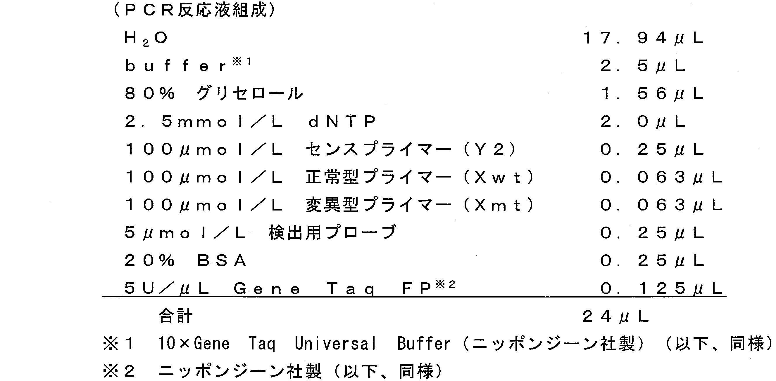
5’-ggacggacggaccgtcctcgttgtcttgttggc-3’
正常型プライマー(Xwt) 配列番号2
5’-ttcccgtaggtcatgaactcaG-3’
変異型プライマー(Xmt) 配列番号3
5’-aggttcccgtaggtcatgaactcaA-3’ Sense primer (Y2) SEQ ID NO: 1
5'-ggacggacggaccgtcctcgttgtcttgttggc-3 '
Normal primer (Xwt) SEQ ID NO: 2
5'-ttcccgtaggtcatgaactcaG-3 '
Mutant primer (Xmt) SEQ ID NO: 3
5'-aggttcccgtaggtcatgaactcaA-3 '
検出用プローブ 配列番号4
5’-(BODIPY FL)-ctcaAtgatgatatagaacg-P-3’ The sequence of the detection probe used for Tm analysis is shown below. The detection probe is a complementary sequence that is 100% matched to the region containing the 944th base in the mutant bcr-abl gene (sense strand) in which the 944th base C is mutated to T. In the following sequence, the base A shown in capital letters corresponds to the 944th mutated base T. P at the 3 ′ end represents a phosphate group.
Detection probe SEQ ID NO: 4
5 '-(BODIPY FL) -ctcaAtgatgatatagaacg-P-3'
アンチセンスプライマーとして、正常型プライマー(Xwt)および変異型(Xmt)に代えて、100μmol/Lの下記プライマー0.126μLを使用し、PCRの条件を、95℃で60秒処理した後、99℃4秒および62℃30秒を1サイクルとして50サイクルとした以外は、前記実施例1と同様にして、Tm解析を行った。前述のセンスプライマー(Y2)と下記アンチセンスプライマーは、bcr-abl遺伝子の944番目の塩基とは異なる領域にアニーリングするため、944番目が正常塩基(C)または変異塩基(T)のいずれであるかにかかわらず、前記944番目の塩基を含む領域が増幅される。
アンチセンスプライマー 配列番号5
5’-ggacggacggaccgcactccctcaggtagtccag-3’ [Comparative Example 1]
As an antisense primer, 0.126 μL of the following primer of 100 μmol / L was used instead of the normal primer (Xwt) and the mutant type (Xmt), and the PCR conditions were treated at 95 ° C. for 60 seconds, followed by 99 ° C. Tm analysis was performed in the same manner as in Example 1 except that 4 cycles and 62 ° C. and 30 seconds were set to 50 cycles and 50 cycles were made. Since the above-mentioned sense primer (Y2) and the following antisense primer anneal to a region different from the 944th base of the bcr-abl gene, the 944th is either a normal base (C) or a mutated base (T) Regardless, the region containing the 944th base is amplified.
Antisense primer SEQ ID NO: 5
5'-ggacggacggaccgcactccctcaggtagtccag-3 '
従来のASP-PCR法により、bcr-abl遺伝子の944番目の塩基の点突然変異(C→T)を検出した。 [Comparative Example 2]
A point mutation (C → T) at the 944th base of the bcr-abl gene was detected by a conventional ASP-PCR method.

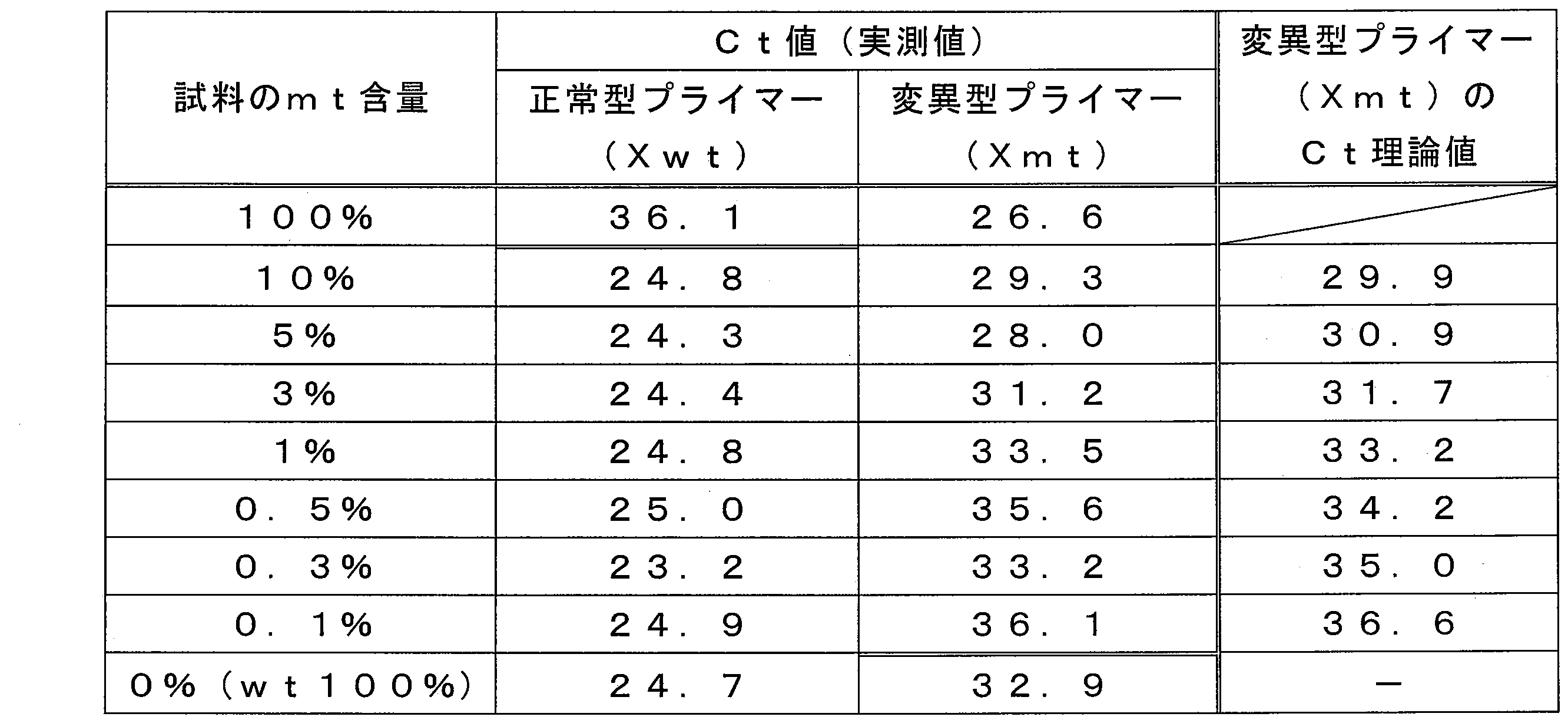
前記第1の実施形態に基づいて、bcr-abl遺伝子(センス鎖)の944番目の塩基の点突然変異(C→T)をTm解析により検出した。 [Example 2]
Based on the first embodiment, a point mutation (C → T) at the 944th base of the bcr-abl gene (sense strand) was detected by Tm analysis.
5’-gtaggtcatgaactcaGtgatgatatagaacgggggctcccgggtgcaga-3’
変異型オリゴヌクレオチド(mt) 配列番号7
5’-gtaggtcatgaactcaAtgatgatatagaacgggggctcccgggtgcaga-3’ Normal oligonucleotide (wt) SEQ ID NO: 6
5'-gtaggtcatgaactcaGtgatgatatagaacgggggctcccgggtgcaga-3 '
Mutant oligonucleotide (mt) SEQ ID NO: 7
5'-gtaggtcatgaactcaAtgatgatatagaacgggggctcccgggtgcaga-3 '
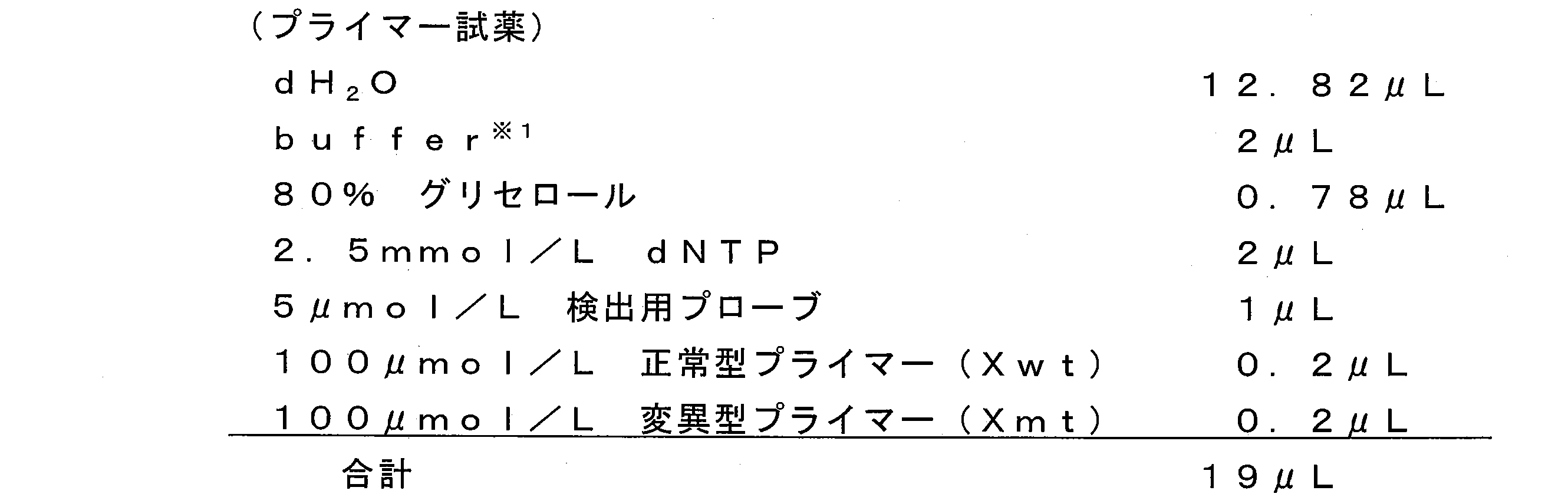
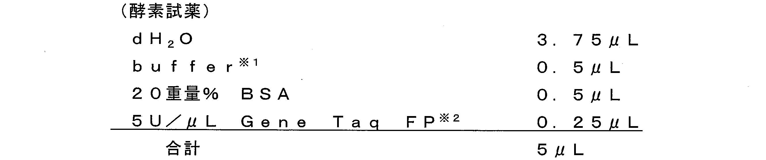
5’-cccccgttctatatcatcaC-3’
変異型プライマー(Xmt) 配列番号9
5’-ggagcccccgttctatatcatcaT-3’ Normal primer (Xwt) SEQ ID NO: 8
5'-cccccgttctatatcatcaC-3 '
Mutant primer (Xmt) SEQ ID NO: 9
5'-ggagcccccgttctatatcatcaT-3 '
前記第3の実施形態に基づいて、bcr-abl遺伝子(センス鎖)の944番目の塩基の点突然変異(C→T)をTm解析により検出した。 [Example 3]
Based on the third embodiment, a point mutation (C → T) at the 944th base of the bcr-abl gene (sense strand) was detected by Tm analysis.
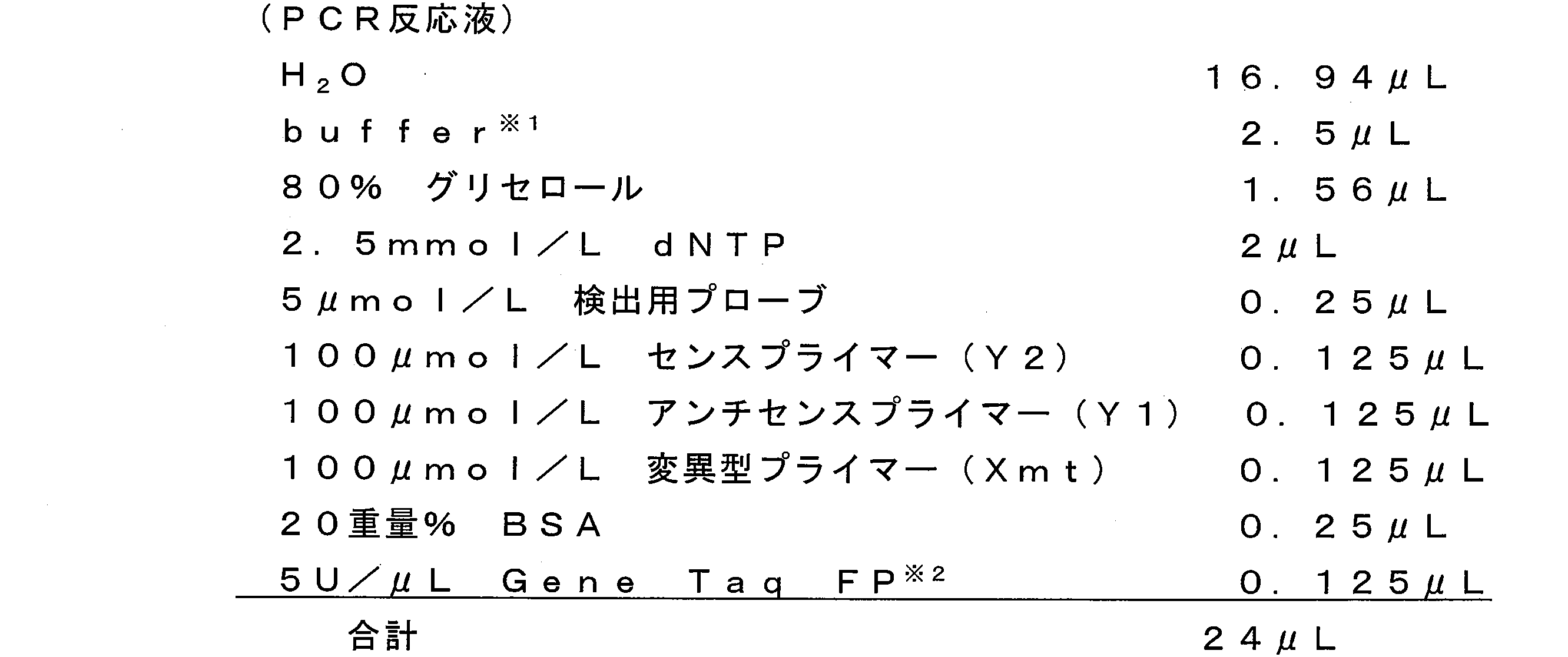
5’-ggacggacggaccgtcctcgttgtcttgttggc-3’
アンチセンスプライマー(Y1) 配列番号10
5’-gaccgaccgaccccaggaggttcccgtaggtc-3’
変異型プライマー(Xmt) 配列番号3
5’-aggttcccgtaggtcatgaactcaA-3’ Sense primer (Y2) SEQ ID NO: 1
5'-ggacggacggaccgtcctcgttgtcttgttggc-3 '
Antisense primer (Y1) SEQ ID NO: 10
5'-gaccgaccgaccccaggaggttcccgtaggtc-3 '
Mutant primer (Xmt) SEQ ID NO: 3
5'-aggttcccgtaggtcatgaactcaA-3 '
検出用プローブ 配列番号11
5’-(BODIPY FL)-cccgttctatatcatcaTtgag-P-3’ The sequence of the detection probe is shown below. The detection probe is a complementary sequence that is 100% matched to the region containing the 944th base in the antisense strand of the mutant bcr-abl gene in which the 944th base C is mutated to T. In the following sequence, the base T shown in capital letters corresponds to the 944th mutated base A in the antisense strand. P at the 3 ′ end represents a phosphate group.
Probe for detection SEQ ID NO: 11
5 '-(BODIPY FL) -cccgttctatatcatcaTtgag-P-3'
前記表6のPCR反応液において、変異型プライマー(Xmt)0.125μLに代えて、アンチセンスプライマー(Y1)を倍量である0.25μL添加した以外は、前記実施例3と同様にして、Tm解析を行った。 [Comparative Example 3]
In the PCR reaction solution of Table 6, in place of the mutated primer (Xmt) 0.125 μL, the same amount as in Example 3 except that 0.25 μL of the antisense primer (Y1) was added in double amount. Tm analysis was performed.
Claims (22)
- 鋳型核酸における標的核酸配列の増幅方法であって、
前記標的核酸配列が、前記鋳型核酸における検出目的の変異が発生する塩基部位を含む配列であり、
同一の反応系において、前記塩基部位が変異型塩基である標的核酸配列を、前記塩基部位が正常型塩基である標的核酸配列よりも優先的に増幅させる増幅工程を含むことを特徴とする標的核酸配列の増幅方法。 A method for amplifying a target nucleic acid sequence in a template nucleic acid, comprising:
The target nucleic acid sequence is a sequence containing a base site where a mutation for detection in the template nucleic acid occurs,
In the same reaction system, a target nucleic acid comprising an amplification step for preferentially amplifying a target nucleic acid sequence in which the base site is a mutated base over a target nucleic acid sequence in which the base site is a normal base Sequence amplification method. - 前記増幅工程が、前記同一の反応系において、下記プライマー(Xmt)およびプライマー(Xwt)を用いて前記標的核酸配列を増幅する工程であり、
前記塩基部位が変異型である鋳型核酸に対する下記プライマー(Xmt)による増幅効率が、前記塩基部位が正常型である鋳型核酸に対する下記プライマー(Xwt)による増幅効率よりも高い、請求の範囲1記載の標的核酸配列の増幅方法。
プライマー(Xmt)
前記鋳型核酸における変異型の前記塩基部位を含む領域に相補的であり、3’領域に前記塩基部位の塩基に相補的な塩基を有するプライマー
プライマー(Xwt)
前記鋳型核酸における正常型の前記塩基部位を含む領域に相補的であり、3’領域に前記塩基部位の塩基に相補的な塩基を有するプライマー The amplification step is a step of amplifying the target nucleic acid sequence using the following primer (Xmt) and primer (Xwt) in the same reaction system;
The amplification efficiency by the following primer (Xmt) with respect to the template nucleic acid whose said base part is a mutant type is higher than the amplification efficiency by the following primer (Xwt) with respect to the template nucleic acid whose said base part is a normal type. A method for amplifying a target nucleic acid sequence.
Primer (Xmt)
Primer primer (Xwt) that is complementary to the region containing the mutant base site in the template nucleic acid and has a base complementary to the base of the base site in the 3 ′ region
Primer that is complementary to the region containing the normal type base site in the template nucleic acid and has a base complementary to the base of the base site in the 3 ′ region - 前記プライマー(Xmt)およびプライマー(Xwt)において、3’末端の1番目または2番目の塩基が、前記塩基部位の塩基に相補的な塩基である、請求の範囲2記載の標的核酸配列の増幅方法。 The method for amplifying a target nucleic acid sequence according to claim 2, wherein in the primer (Xmt) and the primer (Xwt), the first or second base at the 3 'end is a base complementary to the base at the base site. .
- 相補配列に対する前記プライマー(Xmt)のTm値は、相補配列に対する前記プライマー(Xwt)のTm値よりも高い値である、請求の範囲2記載の標的核酸配列の増幅方法。 The method for amplifying a target nucleic acid sequence according to claim 2, wherein the Tm value of the primer (Xmt) for the complementary sequence is higher than the Tm value of the primer (Xwt) for the complementary sequence.
- 前記増幅工程が、前記同一の反応系において、下記プライマー(Xmt)およびプライマー(Y1)を用いて前記標的核酸配列を増幅する工程である、請求の範囲1記載の標的核酸配列の増幅方法。
プライマー(Xmt)
前記鋳型核酸における変異型の前記塩基部位を含む領域に相補的であり、3’領域に前記塩基部位の塩基に相補的な塩基を有するプライマー
プライマー(Y1)
前記鋳型核酸における前記塩基部位よりも3’側の領域に相補的なプライマー The method for amplifying a target nucleic acid sequence according to claim 1, wherein the amplification step is a step of amplifying the target nucleic acid sequence using the following primer (Xmt) and primer (Y1) in the same reaction system.
Primer (Xmt)
Primer primer (Y1) that is complementary to the region containing the mutant base site in the template nucleic acid and has a base complementary to the base of the base site in the 3 ′ region
Primer complementary to the region 3 ′ of the template nucleic acid from the base site - 前記増幅工程において、あわせて、下記プライマー(Y2)を使用する、請求の範囲2または5記載の標的核酸配列の増幅方法。
プライマー(Y2)
前記鋳型核酸における前記塩基部位よりも5’側の領域に対する相補配列に相補的なプライマー The method for amplifying a target nucleic acid sequence according to claim 2 or 5, wherein the following primer (Y2) is also used in the amplification step.
Primer (Y2)
Primer complementary to the complementary sequence to the region 5 'from the base site in the template nucleic acid - 前記増幅工程において、前記反応系に、さらに、前記鋳型配列における前記塩基部位を含む配列にハイブリダイズ可能なプローブを添加する、請求の範囲1記載の標的核酸配列の増幅方法。 The method for amplifying a target nucleic acid sequence according to claim 1, wherein in the amplification step, a probe capable of hybridizing to the sequence containing the base site in the template sequence is further added to the reaction system.
- 前記プローブが、標識化プローブである、請求の範囲7記載の標的核酸配列の増幅方法。 The method for amplifying a target nucleic acid sequence according to claim 7, wherein the probe is a labeled probe.
- 鋳型核酸における検出目的の塩基部位の変異の有無を検出する方法であって、下記(a)~(c)工程を含むことを特徴とする変異の検出方法。
(a)請求の範囲1記載の標的核酸配列の増幅方法により、前記鋳型核酸における前記塩基部位を含む標的核酸配列を反応系で増幅させる工程
(b)前記鋳型核酸における前記塩基部位を含む配列にハイブリダイズ可能なプローブの存在下、前記(a)工程により得られた増幅産物を含む前記反応系の温度を変化させ、前記増幅産物と前記プローブとのハイブリッド形成体の融解状態を示すシグナル値を測定する工程
(c)温度変化に伴う前記シグナル値の変動から、前記目的の塩基部位の変異の有無を決定する工程 A method for detecting the presence or absence of a mutation at a target base site in a template nucleic acid, comprising the following steps (a) to (c):
(A) a step of amplifying a target nucleic acid sequence containing the base site in the template nucleic acid in a reaction system by the method for amplifying a target nucleic acid sequence according to claim 1; (b) a sequence containing the base site in the template nucleic acid; In the presence of a hybridizable probe, the temperature of the reaction system containing the amplification product obtained in the step (a) is changed, and a signal value indicating the melting state of the hybrid formed product of the amplification product and the probe is obtained. A step of measuring (c) a step of determining the presence or absence of a mutation in the target base site from the fluctuation of the signal value accompanying a temperature change - 前記増幅工程が、前記同一の反応系において、下記プライマー(Xmt)およびプライマー(Xwt)を用いて前記標的核酸配列を増幅する工程であり、
前記塩基部位が変異型である鋳型核酸に対する下記プライマー(Xmt)による増幅効率が、前記塩基部位が正常型である鋳型核酸に対する下記プライマー(Xwt)による増幅効率よりも高い、請求の範囲9記載の変異の検出方法。
プライマー(Xmt)
前記鋳型核酸における変異型の前記塩基部位を含む領域に相補的であり、3’領域に前記塩基部位の塩基に相補的な塩基を有するプライマー
プライマー(Xwt)
前記鋳型核酸における正常型の前記塩基部位を含む領域に相補的であり、3’領域に前記塩基部位の塩基に相補的な塩基を有するプライマー The amplification step is a step of amplifying the target nucleic acid sequence using the following primer (Xmt) and primer (Xwt) in the same reaction system;
The amplification efficiency by the following primer (Xmt) with respect to the template nucleic acid whose said base part is a mutant type is higher than the amplification efficiency by the following primer (Xwt) with respect to the template nucleic acid whose said base part is a normal type. Mutation detection method.
Primer (Xmt)
Primer primer (Xwt) that is complementary to the region containing the mutant base site in the template nucleic acid and has a base complementary to the base of the base site in the 3 ′ region
Primer that is complementary to the region containing the normal type base site in the template nucleic acid and has a base complementary to the base of the base site in the 3 ′ region - 前記プライマー(Xmt)およびプライマー(Xwt)において、3’末端の1番目または2番目の塩基が、前記塩基部位の塩基に相補的な塩基である、請求の範囲10記載の変異の検出方法。 The method for detecting a mutation according to claim 10, wherein in the primer (Xmt) and the primer (Xwt), the first or second base at the 3 'end is a base complementary to the base at the base site.
- 相補配列に対する前記プライマー(Xmt)のTm値は、相補配列に対する前記プライマー(Xwt)のTm値よりも高い値である、請求の範囲10記載の変異の検出方法。 The mutation detection method according to claim 10, wherein the Tm value of the primer (Xmt) with respect to the complementary sequence is higher than the Tm value of the primer (Xwt) with respect to the complementary sequence.
- 前記増幅工程が、前記同一の反応系において、下記プライマー(Xmt)およびプライマー(Y1)を用いて前記標的核酸配列を増幅する工程である、請求の範囲9記載の変異の検出方法。
プライマー(Xmt)
前記鋳型核酸における変異型の前記塩基部位を含む領域に相補的であり、3’領域に前記塩基部位の塩基に相補的な塩基を有するプライマー
プライマー(Y1)
前記鋳型核酸における前記塩基部位よりも3’側の領域に相補的なプライマー The method for detecting a mutation according to claim 9, wherein the amplification step is a step of amplifying the target nucleic acid sequence using the following primer (Xmt) and primer (Y1) in the same reaction system.
Primer (Xmt)
Primer primer (Y1) that is complementary to the region containing the mutant base site in the template nucleic acid and has a base complementary to the base of the base site in the 3 ′ region
Primer complementary to the region 3 ′ of the template nucleic acid from the base site - 前記増幅工程において、あわせて、下記プライマー(Y2)を使用する、請求の範囲10または13記載の変異の検出方法。
プライマー(Y2)
前記鋳型核酸における前記塩基部位よりも5’側の領域に対する相補配列に相補的なプライマー The method for detecting a mutation according to claim 10 or 13, wherein, in the amplification step, the following primer (Y2) is also used.
Primer (Y2)
Primer complementary to the complementary sequence to the region 5 'from the base site in the template nucleic acid - 前記(a)工程において、前記標的核酸配列の増幅に先立って、前記反応系に、前記鋳型核酸における前記塩基部位を含む配列にハイブリダイズ可能なプローブを添加する、請求の範囲9記載の変異の検出方法。 In the step (a), prior to amplification of the target nucleic acid sequence, a probe capable of hybridizing to the sequence containing the base site in the template nucleic acid is added to the reaction system. Detection method.
- 前記プローブが、標識化プローブである、請求の範囲15記載の変異の検出方法。 The method for detecting a mutation according to claim 15, wherein the probe is a labeled probe.
- 前記標識化プローブが、単独でシグナルを示し且つハイブリッド形成によりシグナルを示さない標識化プローブ、または、単独でシグナルを示さず且つハイブリッド形成によりシグナルを示す標識化プローブである、請求の範囲16記載の変異の検出方法。 17. The labeled probe according to claim 16, wherein the labeled probe is a labeled probe which shows a signal alone and does not show a signal by hybridization, or a labeled probe which shows no signal alone and shows a signal by hybridization. Mutation detection method.
- 請求の範囲1記載の標的核酸配列の増幅方法に使用する増幅試薬であって、
下記プライマー(Xmt)およびプライマー(Xwt)を含み、
前記塩基部位が変異型である鋳型核酸に対する下記プライマー(Xmt)による増幅効率が、前記塩基部位が正常型である鋳型核酸に対する下記プライマー(Xwt)による増幅効率よりも高いことを特徴とする増幅試薬。
プライマー(Xmt)
前記鋳型核酸における変異型の前記塩基部位を含む領域に相補的であり、3’領域に前記塩基部位の塩基に相補的な塩基を有するプライマー
プライマー(Xwt)
前記鋳型核酸における正常型の前記塩基部位を含む領域に相補的であり、3’領域に前記塩基部位の塩基に相補的な塩基を有するプライマー An amplification reagent used in the method for amplifying a target nucleic acid sequence according to claim 1, comprising:
The following primer (Xmt) and primer (Xwt) are included,
An amplification reagent characterized in that the amplification efficiency by the following primer (Xmt) for the template nucleic acid whose base site is a mutant type is higher than the amplification efficiency by the following primer (Xwt) for a template nucleic acid whose base site is a normal type .
Primer (Xmt)
Primer primer (Xwt) that is complementary to the region containing the mutant base site in the template nucleic acid and has a base complementary to the base of the base site in the 3 ′ region
Primer that is complementary to the region containing the normal type base site in the template nucleic acid and has a base complementary to the base of the base site in the 3 ′ region - 相補配列に対する前記プライマー(Xmt)のTm値は、相補配列に対する前記プライマー(Xwt)のTm値よりも高い値である、請求の範囲18記載の増幅試薬。 The amplification reagent according to claim 18, wherein a Tm value of the primer (Xmt) with respect to a complementary sequence is higher than a Tm value of the primer (Xwt) with respect to a complementary sequence.
- 請求の範囲1記載の標的核酸配列の増幅方法に使用する増幅試薬であって、
下記プライマー(Xmt)およびプライマー(Y1)を含むことを特徴とする増幅試薬。
プライマー(Xmt)
前記鋳型核酸における変異型の前記塩基部位を含む領域に相補的であり、3’領域に前記塩基部位の塩基に相補的な塩基を有するプライマー
プライマー(Y1)
前記鋳型核酸における前記塩基部位よりも3’側の領域に相補的なプライマー An amplification reagent used in the method for amplifying a target nucleic acid sequence according to claim 1, comprising:
An amplification reagent comprising the following primer (Xmt) and primer (Y1).
Primer (Xmt)
Primer primer (Y1) which is complementary to the region containing the mutant base site in the template nucleic acid and has a base complementary to the base of the base site in the 3 ′ region
Primer complementary to the region 3 ′ of the template nucleic acid from the base site - さらに、下記プライマー(Y2)を含む、請求の範囲18または20記載の増幅試薬。
プライマー(Y2)
前記鋳型核酸における前記塩基部位よりも5’側の領域に対して相補的な配列に相補的なプライマー The amplification reagent according to claim 18 or 20, further comprising the following primer (Y2).
Primer (Y2)
Primer complementary to a sequence complementary to a region 5 'from the base site in the template nucleic acid - 請求の範囲9記載の変異の検出方法に使用する検出試薬であって、
請求の範囲18または20記載の増幅試薬、および、
鋳型核酸における検出目的の塩基部位を含む配列にハイブリダイズ可能なプローブを含むことを特徴とする検出試薬。 A detection reagent used in the method for detecting a mutation according to claim 9,
The amplification reagent according to claim 18 or 20, and
A detection reagent comprising a probe capable of hybridizing to a sequence containing a base site for detection in a template nucleic acid.
Priority Applications (4)
| Application Number | Priority Date | Filing Date | Title |
|---|---|---|---|
| JP2010519111A JP5637850B2 (en) | 2008-07-02 | 2009-07-02 | Amplification method of target nucleic acid sequence, detection method of mutation using the same, and reagent used therefor |
| EP20090773547 EP2314680B1 (en) | 2008-07-02 | 2009-07-02 | Method for amplification of target nucleic acid sequence, method for detection of mutation by using the method, and reagents for use in the methods |
| US13/002,194 US9115391B2 (en) | 2008-07-02 | 2009-07-02 | Method of detecting a polymorphism at a polymorphism site |
| CN200980125546.7A CN102076850B (en) | 2008-07-02 | 2009-07-02 | Method for amplification of target nucleic acid sequence, method for detection of mutation by using the method, and reagents for use in the methods |
Applications Claiming Priority (2)
| Application Number | Priority Date | Filing Date | Title |
|---|---|---|---|
| JP2008-173986 | 2008-07-02 | ||
| JP2008173986 | 2008-07-02 |
Publications (1)
| Publication Number | Publication Date |
|---|---|
| WO2010001969A1 true WO2010001969A1 (en) | 2010-01-07 |
Family
ID=41466060
Family Applications (1)
| Application Number | Title | Priority Date | Filing Date |
|---|---|---|---|
| PCT/JP2009/062143 WO2010001969A1 (en) | 2008-07-02 | 2009-07-02 | Method for amplification of target nucleic acid sequence, method for detection of mutation by using the method, and reagents for use in the methods |
Country Status (6)
| Country | Link |
|---|---|
| US (1) | US9115391B2 (en) |
| EP (1) | EP2314680B1 (en) |
| JP (1) | JP5637850B2 (en) |
| KR (1) | KR20100080621A (en) |
| CN (1) | CN102076850B (en) |
| WO (1) | WO2010001969A1 (en) |
Cited By (8)
| Publication number | Priority date | Publication date | Assignee | Title |
|---|---|---|---|---|
| EP2447377A1 (en) | 2010-10-29 | 2012-05-02 | Arkray, Inc. | Primer set for detecting EGFR exon 21 polymorphism and application thereof |
| EP2450443A1 (en) * | 2010-01-21 | 2012-05-09 | Arkray, Inc. | Target sequence amplification method, polymorphism detection method, and reagents for use in the methods |
| EP2520666A1 (en) | 2011-05-06 | 2012-11-07 | ARKRAY, Inc. | Oligonucleotide for detection test of polymorphism of EGFR exon 19 and use thereof |
| JP2013063062A (en) * | 2011-09-02 | 2013-04-11 | Arkray Inc | Nucleic acid detection apparatus, method and program |
| WO2013065646A1 (en) | 2011-10-31 | 2013-05-10 | アークレイ株式会社 | Method for measuring abundance of genes |
| JP2014528721A (en) * | 2011-09-30 | 2014-10-30 | エピスタム リミテッド | Mutation analysis of JAK2 |
| US9115391B2 (en) | 2008-07-02 | 2015-08-25 | Arkray, Inc. | Method of detecting a polymorphism at a polymorphism site |
| KR101747090B1 (en) | 2010-04-30 | 2017-06-14 | 아크레이 가부시키가이샤 | Method for adjusting amplification efficiency of target polynucleotide |
Families Citing this family (5)
| Publication number | Priority date | Publication date | Assignee | Title |
|---|---|---|---|---|
| AU2012206487A1 (en) * | 2011-01-14 | 2013-07-18 | Genefirst Limited | Methods, compositions, and kits for determining the presence/absence of a variant nucleic acid sequence |
| WO2012146251A1 (en) * | 2011-04-29 | 2012-11-01 | Aarhus Universitet | Method for detecting mutations using a three primer system and differently melting amplicons |
| GB201121869D0 (en) * | 2011-11-17 | 2012-02-01 | Clarient Inc | Method of allele-specific amplification |
| JP5997407B1 (en) * | 2016-04-21 | 2016-09-28 | 日鉄住金環境株式会社 | Multi-item amplification method |
| CN105861678B (en) * | 2016-04-29 | 2019-12-13 | 广州市康立明生物科技有限责任公司 | Design method of primer and probe for amplifying low-concentration mutation target sequence |
Citations (12)
| Publication number | Priority date | Publication date | Assignee | Title |
|---|---|---|---|---|
| US4889818A (en) | 1986-08-22 | 1989-12-26 | Cetus Corporation | Purified thermostable enzyme |
| WO1991009950A1 (en) | 1989-12-22 | 1991-07-11 | F. Hoffmann-La Roche Ag | Recombinant expression vectors and purification methods for thermus thermophilus dna polymerase |
| EP0455430A2 (en) | 1990-04-26 | 1991-11-06 | New England Biolabs, Inc. | Purified thermostable DNA polymerase obtainable from Thermococcus litoralis |
| US5079352A (en) | 1986-08-22 | 1992-01-07 | Cetus Corporation | Purified thermostable enzyme |
| WO1992009689A1 (en) | 1990-12-03 | 1992-06-11 | Stratagene | PURIFIED THERMOSTABLE $i(PYROCOCCUS FURIOSUS) |
| JP2853864B2 (en) | 1988-03-10 | 1999-02-03 | ゼネカ リミテッド | Methods for detecting nucleotide sequences |
| WO2008066136A1 (en) * | 2006-11-30 | 2008-06-05 | Arkray, Inc. | Primer set for amplification of ugt1a1 gene, reagent for amplification of ugt1a1 gene comprising the same, and use of the same |
| WO2008066163A1 (en) * | 2006-11-30 | 2008-06-05 | Arkray, Inc. | Primer set for amplification of cyp2c9 gene, reagent for amplification of cyp2c9 gene comprising the same, and use of the same |
| WO2008066164A1 (en) * | 2006-11-30 | 2008-06-05 | Arkray, Inc. | Primer set for amplification of obesity gene, reagent for amplification of obesity gene comprising the primer set, and use of the primer set |
| WO2008066161A1 (en) * | 2006-11-30 | 2008-06-05 | Arkray, Inc. | Primer set for amplification of nat2 gene, reagent for amplification of nat2 gene comprising the same, and use of the same |
| WO2008066162A1 (en) * | 2006-11-30 | 2008-06-05 | Arkray, Inc. | Primer set for amplification of cyp2c19 gene, reagent for amplification of cyp2c19 gene comprising the same, and use of the same |
| WO2008066165A1 (en) * | 2006-11-30 | 2008-06-05 | Arkray, Inc. | Primer set for amplification of sult1a1 gene, reagent for amplification of sult1a1 gene comprising the same, and use of the same |
Family Cites Families (12)
| Publication number | Priority date | Publication date | Assignee | Title |
|---|---|---|---|---|
| WO1995006137A1 (en) * | 1993-08-27 | 1995-03-02 | Australian Red Cross Society | Detection of genes |
| US6472156B1 (en) * | 1999-08-30 | 2002-10-29 | The University Of Utah | Homogeneous multiplex hybridization analysis by color and Tm |
| AUPS076902A0 (en) | 2002-02-26 | 2002-03-21 | Commonwealth Scientific And Industrial Research Organisation | Novel selective polymerase chain reaction |
| US7785776B2 (en) * | 2002-05-13 | 2010-08-31 | Idaho Technology, Inc. | Genotyping by amplicon melting curve analysis |
| WO2004092415A1 (en) | 2003-04-16 | 2004-10-28 | Arkray, Inc. | Method of detecting or quantitatively determining mitochondrial dna 3243 variation, and kit therefor |
| JP4322554B2 (en) | 2003-05-19 | 2009-09-02 | 株式会社ニチレイフーズ | Method for improving efficiency of DNA amplification reaction |
| US7563572B2 (en) * | 2004-08-02 | 2009-07-21 | University Of Utah Research Foundation | Method for long range allele-specific PCR |
| US20060172324A1 (en) | 2005-01-28 | 2006-08-03 | Roche Molecular Systems, Inc. | Methods of genotyping using differences in melting temperature |
| EP1893777B1 (en) | 2005-06-07 | 2014-10-01 | Luminex Corporation | Methods for detection and typing of nucleic acids |
| US20070154892A1 (en) * | 2005-12-30 | 2007-07-05 | Simon Wain-Hobson | Differential amplification of mutant nucleic acids by PCR in a mixure of nucleic acids |
| JP4942508B2 (en) | 2007-02-20 | 2012-05-30 | アークレイ株式会社 | Probe for detecting abl gene mutation and use thereof |
| EP2314680B1 (en) | 2008-07-02 | 2014-10-01 | ARKRAY, Inc. | Method for amplification of target nucleic acid sequence, method for detection of mutation by using the method, and reagents for use in the methods |
-
2009
- 2009-07-02 EP EP20090773547 patent/EP2314680B1/en active Active
- 2009-07-02 KR KR1020107012069A patent/KR20100080621A/en active Search and Examination
- 2009-07-02 US US13/002,194 patent/US9115391B2/en active Active
- 2009-07-02 CN CN200980125546.7A patent/CN102076850B/en active Active
- 2009-07-02 JP JP2010519111A patent/JP5637850B2/en active Active
- 2009-07-02 WO PCT/JP2009/062143 patent/WO2010001969A1/en active Application Filing
Patent Citations (12)
| Publication number | Priority date | Publication date | Assignee | Title |
|---|---|---|---|---|
| US4889818A (en) | 1986-08-22 | 1989-12-26 | Cetus Corporation | Purified thermostable enzyme |
| US5079352A (en) | 1986-08-22 | 1992-01-07 | Cetus Corporation | Purified thermostable enzyme |
| JP2853864B2 (en) | 1988-03-10 | 1999-02-03 | ゼネカ リミテッド | Methods for detecting nucleotide sequences |
| WO1991009950A1 (en) | 1989-12-22 | 1991-07-11 | F. Hoffmann-La Roche Ag | Recombinant expression vectors and purification methods for thermus thermophilus dna polymerase |
| EP0455430A2 (en) | 1990-04-26 | 1991-11-06 | New England Biolabs, Inc. | Purified thermostable DNA polymerase obtainable from Thermococcus litoralis |
| WO1992009689A1 (en) | 1990-12-03 | 1992-06-11 | Stratagene | PURIFIED THERMOSTABLE $i(PYROCOCCUS FURIOSUS) |
| WO2008066136A1 (en) * | 2006-11-30 | 2008-06-05 | Arkray, Inc. | Primer set for amplification of ugt1a1 gene, reagent for amplification of ugt1a1 gene comprising the same, and use of the same |
| WO2008066163A1 (en) * | 2006-11-30 | 2008-06-05 | Arkray, Inc. | Primer set for amplification of cyp2c9 gene, reagent for amplification of cyp2c9 gene comprising the same, and use of the same |
| WO2008066164A1 (en) * | 2006-11-30 | 2008-06-05 | Arkray, Inc. | Primer set for amplification of obesity gene, reagent for amplification of obesity gene comprising the primer set, and use of the primer set |
| WO2008066161A1 (en) * | 2006-11-30 | 2008-06-05 | Arkray, Inc. | Primer set for amplification of nat2 gene, reagent for amplification of nat2 gene comprising the same, and use of the same |
| WO2008066162A1 (en) * | 2006-11-30 | 2008-06-05 | Arkray, Inc. | Primer set for amplification of cyp2c19 gene, reagent for amplification of cyp2c19 gene comprising the same, and use of the same |
| WO2008066165A1 (en) * | 2006-11-30 | 2008-06-05 | Arkray, Inc. | Primer set for amplification of sult1a1 gene, reagent for amplification of sult1a1 gene comprising the same, and use of the same |
Non-Patent Citations (2)
| Title |
|---|
| ANALYTICAL BIOCHEMISTRY, vol. 290, 2001, pages 89 - 97 |
| See also references of EP2314680A4 * |
Cited By (17)
| Publication number | Priority date | Publication date | Assignee | Title |
|---|---|---|---|---|
| US9115391B2 (en) | 2008-07-02 | 2015-08-25 | Arkray, Inc. | Method of detecting a polymorphism at a polymorphism site |
| EP2450443A4 (en) * | 2010-01-21 | 2014-01-29 | Arkray Inc | Target sequence amplification method, polymorphism detection method, and reagents for use in the methods |
| JP5684737B2 (en) * | 2010-01-21 | 2015-03-18 | アークレイ株式会社 | Target sequence amplification method, polymorphism detection method and reagent used therefor |
| US9284603B2 (en) | 2010-01-21 | 2016-03-15 | Arkray, Inc. | Target sequence amplification method, polymorphism detection method, and reagents for use in the methods |
| CN102471771A (en) * | 2010-01-21 | 2012-05-23 | 爱科来株式会社 | Target sequence amplification method, polymorphism detection method, and reagents for use in the methods |
| CN102471771B (en) * | 2010-01-21 | 2016-01-20 | 爱科来株式会社 | The amplification method of target sequence, pleiomorphism detecting method and the reagent wherein used |
| EP2450443A1 (en) * | 2010-01-21 | 2012-05-09 | Arkray, Inc. | Target sequence amplification method, polymorphism detection method, and reagents for use in the methods |
| KR101747090B1 (en) | 2010-04-30 | 2017-06-14 | 아크레이 가부시키가이샤 | Method for adjusting amplification efficiency of target polynucleotide |
| JP2012105645A (en) * | 2010-10-29 | 2012-06-07 | Arkray Inc | Primer for detecting egfr exon 21 l858r gene polymorphism and application thereof |
| EP2447377A1 (en) | 2010-10-29 | 2012-05-02 | Arkray, Inc. | Primer set for detecting EGFR exon 21 polymorphism and application thereof |
| US9169518B2 (en) | 2010-10-29 | 2015-10-27 | Arkray, Inc. | Primer set for detecting EGFR exon 21 polymorphism and application thereof |
| US8748590B2 (en) | 2011-05-06 | 2014-06-10 | Arkray, Inc. | Oligonucleotides for detection test of polymorphism of EGFR exon 19 and use therof |
| JP2012249630A (en) * | 2011-05-06 | 2012-12-20 | Arkray Inc | Oligonucleotide for detection test for polymorphism of egfr exon 19 and use thereof |
| EP2520666A1 (en) | 2011-05-06 | 2012-11-07 | ARKRAY, Inc. | Oligonucleotide for detection test of polymorphism of EGFR exon 19 and use thereof |
| JP2013063062A (en) * | 2011-09-02 | 2013-04-11 | Arkray Inc | Nucleic acid detection apparatus, method and program |
| JP2014528721A (en) * | 2011-09-30 | 2014-10-30 | エピスタム リミテッド | Mutation analysis of JAK2 |
| WO2013065646A1 (en) | 2011-10-31 | 2013-05-10 | アークレイ株式会社 | Method for measuring abundance of genes |
Also Published As
| Publication number | Publication date |
|---|---|
| EP2314680B1 (en) | 2014-10-01 |
| JPWO2010001969A1 (en) | 2011-12-22 |
| JP5637850B2 (en) | 2014-12-10 |
| KR20100080621A (en) | 2010-07-09 |
| US20110117568A1 (en) | 2011-05-19 |
| EP2314680A4 (en) | 2011-12-14 |
| CN102076850B (en) | 2014-05-07 |
| US9115391B2 (en) | 2015-08-25 |
| EP2314680A1 (en) | 2011-04-27 |
| CN102076850A (en) | 2011-05-25 |
Similar Documents
| Publication | Publication Date | Title |
|---|---|---|
| JP5637850B2 (en) | Amplification method of target nucleic acid sequence, detection method of mutation using the same, and reagent used therefor | |
| JP5509075B2 (en) | Nucleic acid amplification control detection method and use thereof | |
| WO2009081967A1 (en) | Method for amplifying target nucleic acid sequence and probe used for the same | |
| EP3219814B1 (en) | Probe for detection of polymorphism in egfr gene, amplification primer, and use thereof | |
| JP5224526B2 (en) | Gene amplification primer set, gene amplification reagent containing the same, and use thereof | |
| KR20080100429A (en) | Primer set for amplification of cyp2c19 gene, reagent for amplification of cyp2c19 gene comprising the same, and use of the same | |
| WO2011062258A1 (en) | Primer set for amplification of mthfr gene, mthfr gene amplification reagent comprising same, and use of same | |
| JP5917144B2 (en) | Probes for detecting polymorphisms in disease-related genes and uses thereof | |
| JP5684737B2 (en) | Target sequence amplification method, polymorphism detection method and reagent used therefor | |
| WO2011052755A1 (en) | Probe for detecting mpl gene polymorphism and use of the same | |
| KR101119417B1 (en) | Primer set for amplification of obesity gene, reagent for amplification of obesity gene comprising the primer set, and use of the primer set | |
| JPWO2008066163A1 (en) | CYP2C9 gene amplification primer set, CYP2C9 gene amplification reagent containing the same, and use thereof | |
| JP5367365B2 (en) | SULT1A1 gene amplification primer set, SULT1A1 gene amplification reagent containing the same, and use thereof | |
| WO2011077990A1 (en) | PROBES FOR DETECTION OF POLYMORPHISMS OF c-kit GENE, AND USE THEREOF | |
| KR101110425B1 (en) | Primer set for amplification of nat2 gene, reagent for amplification of nat2 gene comprising the same, and use of the same | |
| JP5635496B2 (en) | EGFR gene polymorphism detection probe and use thereof | |
| JP5860667B2 (en) | Primer set for detecting EGFR exon 21L858R gene polymorphism and use thereof |
Legal Events
| Date | Code | Title | Description |
|---|---|---|---|
| WWE | Wipo information: entry into national phase |
Ref document number: 200980125546.7 Country of ref document: CN |
|
| WWE | Wipo information: entry into national phase |
Ref document number: 2010519111 Country of ref document: JP |
|
| 121 | Ep: the epo has been informed by wipo that ep was designated in this application |
Ref document number: 09773547 Country of ref document: EP Kind code of ref document: A1 |
|
| ENP | Entry into the national phase |
Ref document number: 20107012069 Country of ref document: KR Kind code of ref document: A |
|
| NENP | Non-entry into the national phase |
Ref country code: DE |
|
| WWE | Wipo information: entry into national phase |
Ref document number: 13002194 Country of ref document: US |
|
| WWE | Wipo information: entry into national phase |
Ref document number: 2009773547 Country of ref document: EP |