TITLE
Targeting tissue factor to activated platelets
FIELD OF THE INVENTION
The current invention relates to procoagulant fusion proteins, a polynucleotides that encode said procoagulant fusion proteins, cells that expresses said procoagulant fusion protein and uses thereof.
BACKGROUND OF THE INVENTION
Tissue factor (TF), a transmembrane glycoprotein, is the primary cellular initiator of blood coagulation. It is predominantly expressed on the surface of sub-endothelial cells, such as smooth muscle cells and fibroblasts, and binds both zymogen coagulation Factor VII (FVII) and the activated form, Factor Vila (FVIIa) when the integrity of the endothelium is interrupted, such as when blood vessels are severed. When TF binds FVII, it promotes FVII to FVIIa activation. TF also greatly enhances the proteolytic activity of Factor Vila towards its physiologic substrates, Factors IX and X. FVIIa retains a zymo- gen-like state in solution, acting as a relatively poor enzyme. TF provides a scaffold for optimal macromolecular exosite interaction, induces conformational changes in the protease domain of FVIIa leading to maturation of its active site and orientates FVIIa on a cell surface for optimal substrate interaction. Together these effects results in an enhanced catalytic capability of FVIIa of several orders of magnitude. Hence, TF is a co- factor for FVIIa in the initiation complex of what is traditionally referred to as the extrinsic pathway of blood coagulation. Subsequent steps of the coagulation cascade finally result in the formation of a fibrin polymer which is bound by activated platelets and cross- linked with FXIIIa.
Platelets - also known as thrombocytes - derive from their cellular predecessor, megakaryocytes. Normal resting platelets freely flow throughout the blood circulation when the endothelium is intact. When the single-layered endothelial barrier is damaged, resting platelets adhere to subendothelial structures by means of glycoprotein (GP) receptors. For example, GPIaIIa and GPVI bind collagen; GPIcIIa binds fibronectin; GPIc*IIa binds laminin and GPIb-V-IX binds von Willebrand Factor (vWF) polymers. Adhesion of platelets in this manner causes them to change shape and release their alpha and dense granules. In turn, this results in the exposure of a plethora of other glycoprotein platelet receptors, such as GPIIbIIIa (which binds fibrinogen/fibrin) and TREM-like transcript 1 (TLT-I); as well as the release of coagulation factors I (fibrinogen), V and XI; other procoagulants such as ADP, Ca2+, serotonin and Platelet Factors 3/4; anti-coagulants such as tissue factor pathway inhibitor (TFPI); and
compounds such as platelet derived growth factor (PDGF), essential to platelet replenishment and healing. Activated platelets bind to one other and cross-link fibrin in a rapid reaction for example via the GPIIbIIIa receptor complex.
Hence, activated platelets and the fibrin polymer product of the coagulation cascade together form the blood clot. Platelet aggregation of coagulation is known as the primary hemostatic response, while the coagulation cascade response is known as the secondary hemostatic response. Although fibrin is produced during the primary hemostatic response, via the so-called coagulation initiation (independent of FIX/FVIIIa), the amount produced at this point is insufficient for a strong coagel. Initial fibrin serves as an aggregater of activated platelets at site of injury, which again provides an optimal cell surface for the function of activated coagulation factors.
In subjects with a coagulopathy, such as in human beings with haemophilia A and B, various steps of the coagulation cascade are rendered dysfunctional due to, for example, the absence or insufficient presence of a coagulation factor. Such dysfunction of one part of coagulation results in insufficient blood coagulation and potentially life- threatening bleeding.
An object of the current invention is to provide a compound that is suitable for use as a procoagulant drug in such subjects. A second object of the current invention is to provide a compound that enables a physical point of initiation of blood coagulation to be mobilised, such that extrinsic coagulation is not solely dependent on subendothelial, cell-bound tissue factor. A third object of the current invention is to provide a compound that up-regulates blood coagulation in a physiologically suitable microenvironment. A further object of the current invention is to direct a soluble tissue factor, or a biologically functional fragment or variant thereof, to the surface of activated platelets. A further object of the invention is to enhance the proteolytic activity of endogenous Factor Vila towards its physiological substrates, Factors IX and X. Thus, the object is to enable the initiation of blood coagulation on the surface of activated platelets that are located intravascularly or extravascularly. This is in addition to the normal and exclusively subendothelial - typically extravascular - initiation of blood coagulation.
WO06/096828 discloses chimeric proteins that comprise soluble tissue factor
(sTF) and a phosphatidyl serine (PS) binding domain, such as Annexin V. PS is exposed on the surface of activated cells, such as monocytes, endothelial cells and cells undergoing apoptosis, as well as on activated and resting platelets. The chimeric proteins are both pro-coagulant and anti-coagulant; the latter due to the fact that, in higher doses, constructs compete with coagulation factors in binding to PS on activated platelets. Thus, the chimeric proteins of WO06/096828 have a different set of properties than the fusion proteins described herein.
SUMMARY OF THE INVENTION
The current invention provides a fusion protein comprising (i) at least one tissue factor polypeptide, or biologically functional variant(s) or fragment(s) thereof, which is/are covalently attached to (ii) a ligand that is capable of binding (iii) a receptor, and/or a fragment thereof, wherein the receptor is only expressed on the surface of activated platelets. The fusion protein/construct may comprise (i) tissue factor, or a biologically functional variant or fragment thereof, and (ii) a ligand that binds (iii) TLT-I. According to the current invention, (ii) may be a monoclonal antibody, or an antigen-binding portion of a monoclonal antibody. For example, (ii) may be selected from the group consisting of: a Fab fragment, a F(ab')2 fragment, a Fab' fragment, a Fd fragment, a Fv fragment, a dAb fragment or an isolated complementarity determining region (CDR). According to the current invention, (ii) may comprise the variable domain of 0012 (2F105) LC. According to the current invention, (ii) may comprise the variable domain of 0012 (otherwise referred to as 2F105) LC together with the constant region of human LC, kappa and a HPC4 tag (pTT-0012LC.HPC4, also referred to as pTT-2F105LC.HPC4).
The fusion proteins/constructs of the present invention target the initial stages of platelet-clot growth through the specific targeting of activated platelets, while concurrently recruiting resting or basal platelets directly to the site of injury, thereby activating said resting or basal platelets systemically. The compositions of the present invention are based upon the identification of particular receptors and component epitopes, that appear on platelet membranes when platelets are no longer resting but in the process of being activated.
The current invention also provides the following : an isolated nucleotide sequence that encodes any fusion protein/construct according to the current invention; a vector that comprises an isolated nucleotide sequence that, in turn, encodes any fusion protein/construct according to the current invention; an isolated cell that comprises a nucleotide sequence that encodes any fusion protein/construct of the current invention. Said nucleotide sequence may, in turn, be expressed by an intracellular vector. Said isolated cell may be a eukaryotic cell, such as a mammalian cell, such as a BHK or a CHO or a HEK cell.
Furthermore, the current invention provides a method of targeting tissue factor to the surface of activated platelets, said method comprising contacting activated platelets with any construct comprising (i) tissue factor, or a functional variant thereof, and (ii) a ligand that is capable of binding (iii) a receptor present on an activated platelet, such as TLT-I. In this way, the invention also relates to a method of up-regulating FX
activation on the surface of activated platelets, wherein said method comprises the contacting of activated platelets with said construct in the simultaneous presence of FX.
Similarly, the invention relates to fusion proteins for use as a medicament and for use in the treatment of a coagulopathy. In one embodiment, a therapeutically effective amount of said construct is parenterally administered, such as intravenously or subcutaneously administered, to an individual in need thereof. Such individual in need may have any congenital, acquired and/or iatrogenic coagulopathy.
DESCRIPTION OF THE DRAWINGS
Figure 1: Human TLT-I nucleotide and amino acid sequences. In figure 1, the nucleotide- and amino acid sequences representing hTLT-1 are shown. Here, nucleotide sequence position 1-45 encode the predicted signal peptide, the nucleotide sequence at position 46-486 encode the extracellular domain of hTLT-1, the nucleotide sequence position 487-555 encode the transmembrane region and the nucleotide sequence at position 556-933 encode the intracellular domain of hTLT-1.
Figure 2: Nucleotide and amino acid sequences representing the extracellular domain of human TLT-I containing a C-terminal His-6 tag. In figure 2, the nucleotide- and amino acid sequences representing the extracellular domain of hTLT-1 with a C- terminal His tag are shown. Here, the underlined sequence (7-15) indicates the positions of a kozak sequence, the nucleotide sequence at position 16-60 encodes the predicted signal peptide, the nucleotide sequence at position 61-501 encode the extracellular domain of hTLT-1 and the nucleotide sequence at position 502-519 encodes the 6xHis tag (in bold). The restriction enzyme sites HindIII (nucleotide sequence at position 1-6) and EcoRI (nucleotide sequence at position 523-528) are also shown and the stop codon is marked with an asterisk (520-522).
Figure 3: The variable domain of 0012LC and 0012HC including Kabat numbering:
A) The variable domain of 0012LC
B) The variable domain of 0012LC - Kabat numbering
C) The variable domain of 0012HC
D) The variable domain of 0012HC - Kabat numbering.
In figure 3A, the nucleotide sequence at position 1-57 encodes the LC signal peptide sequence; the nucleotide sequences at position 58-396 encodes the variable domain of 0012LC. In figure 3B, the sequences in bold and grey represent positions of the
0012LC CDRs according to Kabat numbering. In figure 3C, the nucleotide sequence at position 1-54 encodes the 0012HC signal peptide, the nucleotide sequence at position 55-396 encodes the variable domain of 0012HC. In figure 3D, the sequences in bold and grey represent positions of the 0012HC CDRs according to Kabat numbering.
Figure 4: The variable domain of 0012LC together with constant region of human LC,kappa and a HPC4 tag (encoded by pTT-0012LC.HPC4). In figure 4, the nucleotide sequences at position 1-57 encodes the LC signal peptide, the nucleotide sequence 58-396 encodes the variable domain of 0012LC, the nucleotide sequence 397-714 en- codes the constant region of human 0012LC, kappa and the nucleotide sequence 715- 750 encodes a HPC4 tag.
Figure S: The variable domain of 0012HC together with the constant region of human IgG4 (pTT-0012HC). In figure 5 the nucleotide sequence 1-54 encodes the HC signal peptide, the nucleotide sequence 55-396 encodes the variable domain of 0012HC and the nucleotide sequence 397-1377 encodes the constant region of human IgG4 (in bold).
Figure 6: The nucleotide- and amino acid sequences of 0012VH-CHl-L4a-hTF. l- 219, as encoded by pTT-0012VH-CHl-L4a-hTF. l-219. The pTT-0012VH-CHl-L4a-hTF. l- 219 construct encoding the Gly-Ser linker L4a and the extracellular domain AA 1-219 of human tissue factor. Here, the nucleotide sequence at position 1-54 encodes the predicted signal peptide, the nucleotide sequence at position 55-396 encode the variable domain of 0012HC, the nucleotide sequence at position 397-699 (in bold) encode the human IgG4 CHi constant region, the nucleotide sequence at position 700-750 (underlined) encodes the Gly-Ser linker designated L4a and the nucleotide sequence at position 701-1407 encodes the extracellular domain of human tissue factor (amino acid 1-219, dotted sequence). The 0012VH-CHl-L4a-hTF.1-219 amino acid sequence is composed of SEQ ID no. 51 (0012VH-VH1 ), SEQ ID no. 61 (L4a) and SEQ ID no. 14 (hTF.1-219).
Figure 7: Stimulation of factor Vila activity. The data in figure 7 demonstrate that a similar concentration-dependent stimulation of FVIIa amidolytic activity was obtained with Fab-hTF.1-219 and mAb-hTF.1-219 fusion proteins as with non-fused hTF.1-219. This indicates that binding of FVIIa to the TF component of TF-fusion proteins was not affected by the linking of TF to Fab or mAb fragments.
Figure 8: Binding of protein ID no. 0010 and 0011 to activated platelets by FACS analysis. Detection via an anti-HPC4 tag (Figure 8A) or an anti-LC antibody (Figure 8B). Figure 9: Figure 9A shows the effect of i) 0.03 nM Innovin®, ii) 10 nM sTF, iii)
10 nM 0011 Fab-hTF.1-219, iv) 10 nM 0010 Fab anti-TLT-1 antibody or v) 10 nM 0012 mAb anti-TLT-1 antibody on FVIIa-mediated FX activation in the absence or presence of platelets. Figure 9B shows the effect of replacing rFVIIa in the assay with the zymogen, rFVII.
Figure 10: The data in Figure 1OA shows that binding of 10 nM 0011 Fab-hTF.1- 219 to the TLT-I receptor specifically stimulates FVIIa-mediated FX activation on activated and not on resting platelets. Stimulation increases with the number of activated platelets and is saturated with an EC50 ~ 12.000 plts/μl. Figure 1OB compares the results in Figure 1OA with the results obtained with 0.1 nM Annexin V - hTF.1-219 fusion protein (AV- hTF.1-219). Figure 1OB shows that binding of AV-hTF.1-219 to platelet phospholipid stimulates FX activation on both activated and resting platelets. A marked stimulation of FX activation is observed with resting platelets which at the maximal platelet number tested exceeds FXa generation on the surface of an equivalent number of activated plate- lets. Figure 1OB clearly shows that AV-TF cannot be used to selectively enhance FXa generation on activated platelets.
Figure 11: The data in Figure 11 shows the FVIIa/FVII concentration dependency of the stimulation of FX activation on activated platelets by 10 nM of Fab-hTF.1-219 fusion proteins. The effect with activated platelets of 0020 Fab-hTF.1-219 is compared to the effect of a 0094 Fab-hTF.1-219 isotype fusion protein which do not bind TLT-I and to non-fused hTF.1-219. Also shown for comparison is the effect of 0020 Fab-hTF.1-219 with resting platelets. A similar stimulation is obtained at all concentrations with FVIIa and FVII showing that TF-fusion proteins mediate an efficient feed-back activation of FVII on activated platelets and that stimulation is optimal at physiological concentrations of FVII/FVIIa. A marked stimulation is induced upon TLT-I targeting and not with non- targeting hTF.1-219 derivatives.
Figure 12: The data in Figure 12 shows the stimulation of FX activation medi- ated by 10 nM FVIIa/FVII on activated platelets at various concentrations of Fab-hTF.1- 219 fusion proteins. The effect with activated platelets of 0070 Fab-hTF.1-219 is compared to the effect of a 0094 Fab-hTF.1-219 isotype fusion protein which do not bind
TLT-I and to non-fused hTF.1-219. The 0070 Fab-hTF.1-219 markedly stimulates FX activation over a wide concentration range with a mechanism which requires targeting to TLT-I as indicated by comparison with non-binding controls. Figure 13: The data in Figure 13 shows the stimulation of FX activation mediated by o. l nM FVIIa/FVII on TLT-I enriched phospholipids at various concentrations of Fab-hTF.1-219 fusion proteins. The effect of 0070 Fab-hTF.1-219 is compared to the effect of a 0094 Fab-hTF.1-219 isotype fusion protein which do not bind TLT-I and to non- fused hTF.1-219. The 0070 Fab-hTF.1-219 markedly stimulates FX activation over a wide concentration range with a mechanism which requires targeting to TLT-I as indicated by comparison with non-binding controls.
Figure 14: Figure 14 shows the stimulation of fibrin clot formation in "hemophilia like" human whole blood (HWB) mediated by 0070 Fab-hTF.1-219 fusion protein. Figure 14A shows TEG traces of clot formation measured at various concentrations (0, 0.1, 0.2, 1.0, and 10 nM) of 0070 Fab-hTF.1-219 fusion protein added to HWB made "hemophilia like" by addition of an antibody against human FVIII. The clotting time is markedly reduced by the FVIII antibody and increasing concentrations of 0070 Fab- hTF.1-219 dramatically revert the effect of the FVIII antibody. Figure 14B shows the R times for the TEG traces in Figure 14A and compares the R time for 0070 Fab-hTF.1-219 to the R times obtained for the 0094 Fab-hTF.1-219 isotype fusion protein and hTF.1-219 run in parallel with the same donor. The 0070 Fab-hTF.1-219 markedly stimulates clot formation in "hemophilia like" HWB with a mechanism which requires targeting to TLT-I as indicated by comparison with non-binding controls.
Figure IS: shows that TLTl-FAb-TF reduces the tail bleeding blood loss in hae- mophilic mice transfused with human platelets 2 min before induction of bleeding, when compared to an irrelevant FAb-TF-construct. Figure 16: Sequence coverage of HX analyzed peptides of TLT-I in the presence and absence of 0023. The primary sequence (using mature numbering) is displayed above the HX analyzed peptides (shown as horizontal bars). Peptides showing similar exchange patterns both in the presence and absence of 0023 are displayed in white whereas peptides showing reduced deuterium incorporation upon 0023 binding are col- oured black.
Figure 17: Sequence coverage of HX analyzed peptides of TLT-I in the presence and absence of 0051. The primary sequence (using mature numbering) is displayed
above the HX analyzed peptides (shown as horizontal bars). Peptides showing similar exchange patterns both in the presence and absence of 0051 are displayed in white whereas peptides showing reduced deuterium incorporation upon 0051 binding are coloured black.
Figure 18: Sequence coverage of HX analyzed peptides of TLT-I in the presence and absence of 0062. The primary sequence (using mature numbering) is displayed above the HX analyzed peptides (shown as horizontal bars). Peptides showing similar exchange patterns both in the presence and absence of 0062 are displayed in white whereas peptides showing reduced deuterium incorporation upon 0062 binding are coloured black.
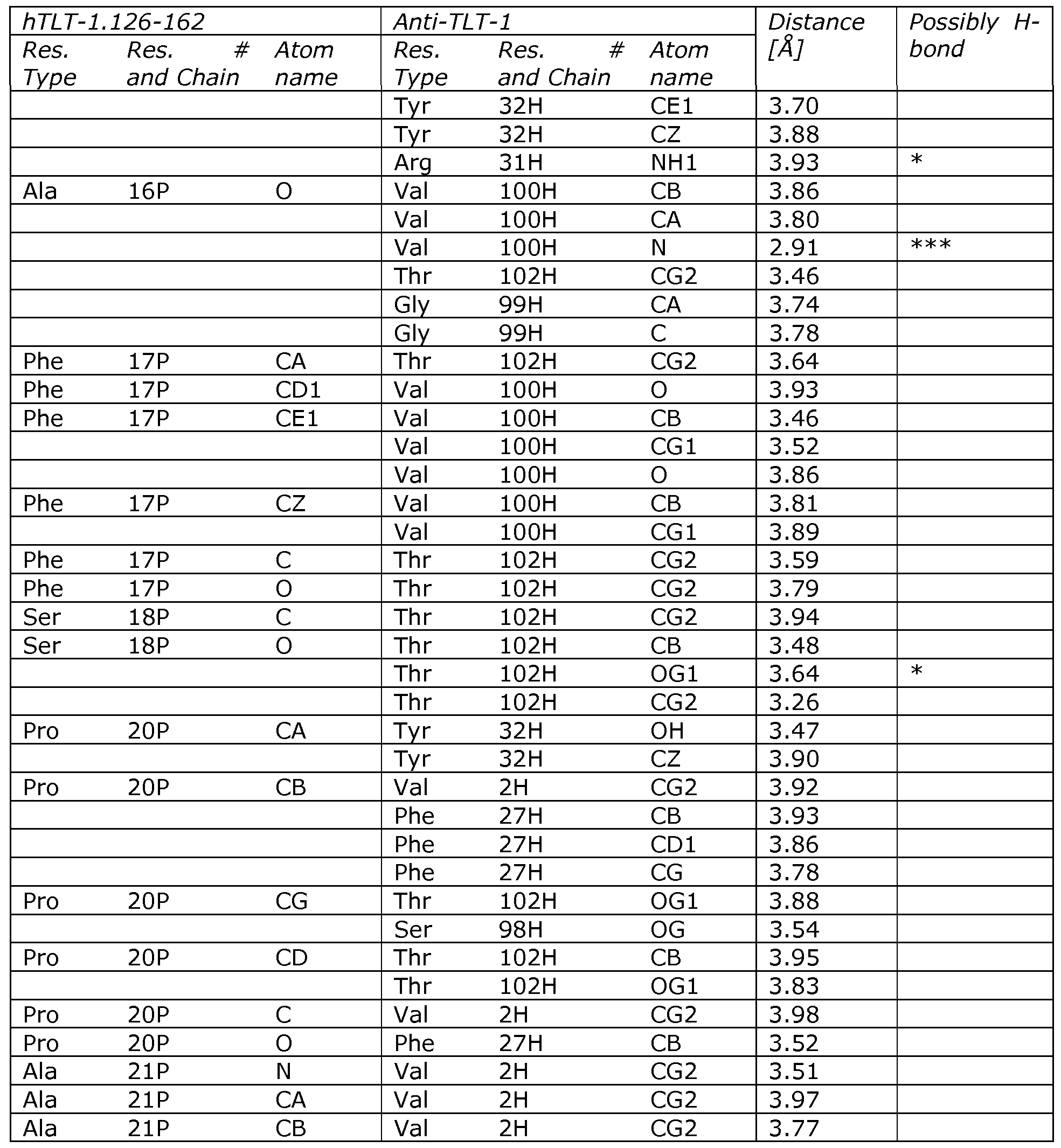
Figure 19: Sequence coverage of HX analyzed peptides of TLT-I in the presence and absence of 0061. The primary sequence (using mature numbering) is displayed above the HX analyzed peptides (shown as horizontal bars). Peptides showing similar exchange patterns both in the presence and absence of 0061 are displayed in white whereas peptides showing reduced deuterium incorporation upon 0061 binding are coloured black. Figure 20: Sequence coverage of HX analyzed peptides of TLT-I region 126-
162. The primary sequence (using mature numbering) is displayed above the HX analyzed peptides (shown as horizontal bars). All peptides showed reduced deuterium incorporation upon 0061 binding. Figure 21: Plasmid map for the expression vector pTT. DNA fragments can be inserted into the HindIII and EcoRI restriction enzyme sites.
Figure 22: Plasmid map for the expression vector pTT-hIgG4. The expresison vector contains the human IgG4 CHl-hinge-CH2-CH3 DNA sequences. VH encoding DNA sequences can be inserted into the HinDIII and Nhel restriction enzyme sites resulting in a complete HC encoding plasmid.
Figure 23: Plasmid map for the expression vector pTT-hCL,kappa. The expression vector contains the DNA sequences encoding the constant region of human LC, kappa designated hCL,kappa. VL encoding DNA sequences can be inserted into the HinDIII and BsiWI restriction enzyme sites resulting in a complete LC encoding plasmid.
Figure 24: Plasmid map for the expression vector pTT-L4a-hTF.1-219. The expression vector contains DNA sequences encoding a 17 amino acid long Gly-Ser linker and human TF.1-219. HC, LC or VH-CH1 encoding DNA sequences can be inserted into the HinDIII and BamHI restriction enzyme sites resulting in HC-L4a-hTF.1-219, LC-L4a- hTF.1-219 or VH-CH1 -L4a-hTF.1-219 encoding plasmids.
Figure 25: Plasmid map for the expression vector pTT-hTF. l-219-L4b. The es- pression vector contains DNA sequences encoding human TF.1-219 and a 17 amino acid long Gly-Ser linker. HC, LC or VN-CH I encoding DNA sequences can be inserted into the BamHI and EcoRI restriction enzyme sites resulting in hTF. l-219-L4b-HC, hTF.1-219-
L4b-LC or hTF. l-219-L4b-VH-CH l encoding plasmid.
Figure 26: Scheme of mAb-LC-hTF.1-219 and Fab-LC-hTF.1-219 constructs. Standard denotations for antibody components are shown : VL, CL, VH, CHl, CH2, and CH3. Tissue factor's fibronectin III domains are denoted Fbn-III.
Figure 27: Screening of TF-fusion protein pro-coagulant activity by measuring FVII/FVIIa-mediated activation of FX on i) activated platelets and on ii) TLT-I enriched phospholipids. Stimulation of FX activation on activated platelets was performed as de- scribed in Example 37 on platelets from individual human donors. The stimulation obtained with 0020 Fab-hTF.1-219 was always included and set to 100 %. Stimulation of FX activation TLT-I enriched phospholipids was performed as described in Example 40 with 0.1 nM FVII/FVIIa and 10 nM Fab fusion protein or 1.0 nM mAB fusion protein. The stimulation obtained with 0020 Fab-hTF.1-219 was always included and set to 100 %. Figure 27A lists data on mAb-TF fusion proteins and figure 27B lists data on Fab-TF fusion proteins. Also show are data with 0020 Fab-hTF.1-219 on resting platelets and results with non-binding isotype antibodies.
SEQUENCES
The sequences are as follows:
SEQ ID NO: 1 provides the nucleotide sequence of human (h)TLT-l.
SEQ ID NO: 2 provides the amino acid sequence of hTLT-1.
SEQ ID NO: 3 provides the nucleotide sequence of the extracellular domain of hTLT-l-His6.
SEQ ID NO: 4 provides the amino acid sequence of the extracellular domain of hTLT-l-His6.
SEQ ID NOs: 5 to 8 provide the amino acid sequences of hTLT-1 fragments: hTLT-1.20-125, hTLT-1.16-162, hTLT-1.126-162 and hTLT-1.129-142.
SEQ ID NO: 9 provides the nucleotide sequence of the variable domain of mAb 0012 (2F105), LC.
SEQ ID NO: 10 provides the amino acid sequence of the variable domain of mAb 0012 (2F105), LC.
SEQ ID NO: 11 provides the nucleotide sequence of the variable domain of 0012 (2F105) HC.
SEQ ID NO: 12 provides the amino acid sequence of the variable domain of 0012 (2F105) HC.
SEQ ID NO: 13 provides the nucleic acid sequence of hTF.1-219.
SEQ ID NO: 14 provides the amino acid sequence of hTF.1-219.
SEQ ID NO: 15 provides the nucleic acid sequence of human tissue factor.
SEQ ID NO: 16 provides the amino acid sequence of human tissue factor.
SEQ ID NO: 17 provides the nucleotide sequence of the heavy chain of mAb 0012.
SEQ ID NO: 18 provides the nucleotide sequence of the light chain of mAb 0012 and Fab 0012.
SEQ ID NO: 19 provides the nucleotide sequence of the heavy chain of mAb 0023.
SEQ ID NO: 20 provides the nucleotide sequence of the light chain of mAb 0023 and Fab 0023.
SEQ ID NO: 21 provides the nucleotide sequence of the heavy chain of mAb 0051.
SEQ ID NO: 22 provides the nucleotide sequence of the light chain of mAb 0051 and Fab 0051.
SEQ ID NO: 23 provides the nucleotide sequence of the heavy chain of mAb 0052.
SEQ ID NO: 24 provides the nucleotide sequence of the heavy chain of mAb 0062.
SEQ ID NO: 25 provides the nucleotide sequence of the light chain of mAb 0052, Fab 0052 and mAb 0062.
SEQ ID NO: 26 provides the nucleotide sequence of the heavy chain of mAb
0061.
SEQ ID NO: 27 provides the nucleotide sequence of the heavy chain of mAb 0082.
SEQ ID NO: 28 provides the nucleotide sequence of the light chain of mAb 0061, Fab 0061, mAb 0082 and Fab 0082.
SEQ ID NO: 29 provides the nucleotide sequence of Fab 0012 VH-CHl.
SEQ ID NO: 30 provides the nucleotide sequence of Fab 0023 VH-CHl.
SEQ ID NO: 31 provides the nucleotide sequence of Fab 0051 VH-CHl.
SEQ ID NO: 32 provides the nucleotide sequence of Fab 0052 VH-CHl.
SEQ ID NO: 33 provides the nucleotide sequence of Fab 0061 VH-CHl.
SEQ ID NO: 34 provides the nucleotide sequence of Fab 0082 VH-CHl.
SEQ ID NO: 35 provides the nucleotide sequence of Fab AP-3 VH-VHl.
SEQ ID NO: 36 provides the nucleotide sequence of Fab AP-3 LC.
SEQ ID NO: 37 provides the nucleotide sequence of Fab-AP-3 LC.C34S.
SEQ ID NO: 38 provides the nucleotide sequence of hIgG4 hinge-CH2-CH3.
SEQ ID NO: 39 provides the amino acid sequence of mAb 0012, HC (mouse VH- human IgG4 CH 1-CH2-CH3).
SEQ ID NO: 40 provides the amino acid sequence of mAb 0012, LC (mouse VL - human Kappa CL) and Fab 0012, LC (mouse VL - human Kappa CL).
SEQ ID NO: 41 provides the amino acid sequence of mAb 0023, HC (mouse VH- human IgG4 CH 1-CH2-CH3).
SEQ ID NO: 42 provides the amino acid sequence of mAb 0023, LC (mouse VL - human Kappa CL) and Fab 0023, LC (mouse VL - human Kappa CL).
SEQ ID NO: 43 provides the amino acid sequence of mAb 0051, HC (mouse VH- human IgG4 CH 1-CH2-CH3).
SEQ ID NO: 44 provides the amino acid sequence of mAb 0051, LC (mouse VL - human Kappa CL) and Fab 0051, LC (mouse VL - human Kappa CL).
SEQ ID NO: 45 provides the amino acid sequence of mAb 0052, HC (mouse VH- human IgG4 CH 1-CH2-CH3).
SEQ ID NO: 46 provides the amino acid sequence of mAb 0052, LC (mouse VL - human Kappa CL); Fab 0052, LC (mouse VL - human Kappa CL); mAb 0062, LC (mouse VL - human Kappa CL).
SEQ ID NO: 47 provides the amino acid sequence of mAb 0061, HC (mouse VH- human IgG4 CH 1-CH2-CH3).
SEQ ID NO: 48 provides the amino acid sequence of mAb 0061, LC (mouse VL - human Kappa CL); Fab 0061, LC (mouse VL - human Kappa CL) and mAb 0082, LC (mouse VL - human Kappa CL); Fab 0082, LC (mouse VL - human Kappa CL).
SEQ ID NO: 49 provides the amino acid sequence of mAb 0062, HC (mouse VH- human IgG4 CH 1-CH2-CH3).
SEQ ID NO: 50 provides the amino acid sequence of mAb 0082, HC (mouse VH- human IgG4 CH1-CH2-CH3).
SEQ ID NO: 51 provides the amino acid sequence of Fab 0012, mouse VH - human IgG4 CH l.
SEQ ID NO: 52 provides the amino acid sequence of Fab 0023, mouse VH - human IgG4 CH l.
SEQ ID NO: 53 provides the amino acid sequence of Fab 0051, mouse VH - hu- man IgG4 CH l.
SEQ ID NO: 54 provides the amino acid sequence of Fab 0052, mouse VH - human IgG4 CH l.
SEQ ID NO: 55 provides the amino acid sequence of Fab 0082, mouse VH - human IgG4 CH l.
SEQ ID NO: 56 provides the amino acid sequence of Fab AP-3, mouse VH - human IgG4 CH l.
SEQ ID NO: 57 provides the amino acid sequence of Fab AP-3, LC (mouse VL - human Kappa CL).
SEQ ID NO: 58 provides the amino acid sequence of Fab AP-3.LC.C34S, LC (mouse VL - human Kappa CL).
SEQ ID NOs: 59-68 provide the amino acid sequences of optional linkers L2-L10. Optional linkers are numbered and listed in Table 1.
SEQ ID NO: 69 provides the amino acid sequence of purification tag HPC4.
SEQ ID NOs: 70-155 provide the nucleic acid sequences of the primers used dur- ing the development of the expression constructs described in examples 4, 5, 6, 7, 8, 9, 10, 11, 12, 13, 14, 15, 17, 18, 19, 20, 24, 25.
SEQ ID NO: 156 provides the amino acid sequence of Fab 0061 VH-CHl.
SEQ ID NO: 157 provides the amino acid sequence of hIgG4-hinge-CH2-CH3.
SEQ ID NO: 158 provides the amino acid sequence of a His6 tag.
SEQ ID NO: 159 provides the amino acid sequence of hTLT-1.18-188.
SEQ ID NO: 160 provides the nucleic acid sequence of primer no. 1004.
SEQ ID NO: 161 provides the nucleic acid sequence of primer no. 1005.
SEQ ID NO: 162 provides the amino acid sequence of Fab 0100 HC. SEQ ID NO: 163 provides the amino acid sequence of Fab 0100 LC.
DESCRIPTION OF THE INVENTION
The invention relates to fusion proteins: proteins expressed from two or more genes that have been joined artificially, for example via recombinant technology or chemical coupling, and which originally encoded separate proteins. Fusion proteins of the invention are capable of binding to a receptor that is only (in the sense non-ubiquitous) present on a platelet undergoing the conformational and functional changes associated with activation. Examples of such receptors might originate from the alpha- or dense granules of resting platelets. One particular example of such a receptor is TREM-like transcript 1 (TLT-I).
Triggering receptors expressed on myeloid cells (TREMs) have a well-established role in the biology of various myeloid lineages, playing important roles in the regulation of innate and adaptive immunity. TLT-I belongs to the same family of proteins, though the TLT-I gene is expressed only in a single lineage, namely megakaryocytes and thrombocytes (platelets) and is exclusively found in the alpha-granules of megakaryocytes and platelets. TLT-I is a transmembrane protein that is exposed on the surface of activated platelets upon alpha-granule release. To date, TLT-I has not been found on the surface of resting platelets or on the surface of any other cell types.
The extracellular portion of TLT-I is known to consist of a single, immunoglobu- lin-like (Ig-like) domain connected to the platelet cell membrane by a linker region called the stalk (Gattis et al., Jour Biol Chem, 2006, Vol. 281, No. 19, pp. 13396-13403). The Ig-like domain of human TLT-I (hTLT-1) consists of 105 residues and is attached to the membrane by a 37-amino acid stalk. Thus, the Ig-like domain of TLT-I is expected to have considerable freedom of movement.
. The putative transmembrane segment of hTLT-1 is 20 amino acids long. TLT-I also has a cytoplasmic Immune-receptor Tyrosine-based Inhibitory-Motif, which may function as an intracellular signal transduction domain.
The role of TLT-I in platelet biology has not yet been fully elucidated; it is believed that TLT-I plays a role in regulating coagulation and inflammation at the site of an injury. A soluble form of TLT-I containing the Ig-like domain has been reported (Gattis et al., Jour Biol Chem, 2006, Vol. 281, No. 19, pp. 13396-13403). The specific functions of soluble versus platelet-bound TLT-I remain to be established.
A receptor such as TLT-I comprises epitopes that are useful targets for the fusion proteins/constructs of the current invention. Fusion proteins may bind any part of
TLT-I that would be available for binding in vivo, such as surface accessible residues of the Ig-like domain, or part of the stalk. Hence, fusion proteins may bind one or more residues within TLT-I (20-125), TLT-I (16-162), TLT-I (126-162) and/or TLT-I (129- 142).
In a preferred embodiment, fusion proteins bind the stalk of TLT-I, such as one or more residues of TLT-I (126-162) or TLT-I (129-142). Fusion proteins that bind to the stalk of TLT-I are unlikely to interfere with the function of the Ig-like domain and will probably not separate from the platelet surface if the Ig-like domain is shed. Furthermore, fusion proteins that bind the stalk of TLT-I place their TF portion in a favorable position and orientation on the cell surface of activated platelets, relative to that of FVII and FVIIa. In another preferred embodiment, fusing TF to the C-terminal of an antibody, or fragment thereof, will position TF even more favourably on the cell surface of activated platelets, relative to that of FVII and FVIIa.
In terms of the current invention, TLT-I may be from any vertebrate, such as any mammal, such as a rodent (such as a mouse, rat or guinea pig), a lagomorph (such as a rabbit), an artiodactyl (such as a pig, cow, sheep or camel) or a primate (such as a monkey or human being). TLT-I is, preferably, human TLT-I. TLT-I may be translated from any naturally occurring genotype or allele that gives rise to a functional TLT-I protein. A non-limiting example of one human TLT-I is the polypeptide sequence of SEQ ID NO. 2.
Fusion proteins of the invention comprise a tissue factor-like component. Tissue Factor is a 263 amino acid long, integral membrane glycoprotein receptor. It consists of an extracellular part folded into two compact fibronectin type Ill-like domains (1-209) that are each stabilized by a single disulfide bond, a short linker (210-219), a transmem- brane segment (220-242), and a short cytoplasmic tail (243-263). It forms a tight Ca2+- dependent complex with Factor VII/FVIIa.
In terms of the current invention, "tissue factor, or any biologically functional variant or fragment thereof", may be any tissue factor-like polypeptide that is able to bind Factor VII/VIIa, such that blood coagulation is stimulated. "Tissue factor" may be derived from any vertebrate animal, such as any mammal, such as a rodent (such as a mouse, rat or guinea pig), a lagomorph (such as a rabbit), an artiodactyl (such as a pig, cow, sheep or camel) or a primate (such as a monkey or a human being). "Tissue factor, or any biologically functional variant or fragment thereof" may be the extracellular domain of human tissue factor. "Tissue factor, or any biologically functional variant or fragment thereof" may be any polypeptide that is at least 90%, such as at least 91%, such as at least 92%, such as at least 93%, such as at least 94%, such as at least 95%, such as at least 96%, such as at least 97%, such as at least 98%, such as at least 99%
identical to the polypeptide sequence of tissue factor. "Tissue factor, or any biologically functional variant or fragment thereof" may be any polypeptide that is at least 90%, such as at least 91%, such as at least 92%, such as at least 93%, such as at least 94%, such as at least 95%, such as at least 96%, such as at least 97%, such as at least 98%, such as at least 99% identical to the polypeptide sequence of the extracellular domain of tissue factor or a variant thereof. "Tissue factor, or any biologically functional variant or fragment thereof" may be any polypeptide that is able to function as co-factor for FVII and FVIIa. Hence, "tissue factor or any biologically functional variant or fragment thereof" may be any polypeptide that is able to stimulate the amidolytic activity of FVIIa. Said "tissue factor, or any biologically functional variant or fragment thereof" may be the extracellular domain of TF (1-219). "Tissue Factor polypeptide" may be a polypeptide comprising the soluble extracellular domain of Tissue Factor, i.e. amino acids 1-219 (in the following referred to as sTF or sTF(l-219)), or a functional variant or truncated form thereof. Preferably, the Tissue Factor polypeptide at least comprises a fragment corre- sponding to the amino acid sequence 6-209 of Tissue Factor. Examples hereof are sTF(6- 209), sTF(l-209), sTF (1-210), sTF (1-211), sTF (1-212), sTF (1-213), sTF (1-214), sTF (1-215), sTF (1-216), sTF (1-217), sTF (1-218), sTF(l-219), sTF(2-219), sTF(3-219), sTF(4-219), sTF(5-219).
In accordance with the current invention, "tissue factor, or any biologically func- tional variant or fragment thereof" may have any one or more of the features listed above.
Fusion proteins of the invention also comprise a "ligand". The term "ligand" refers to any substance that is able to bind to and form a complex with a biomolecule, in order to serve a biological purpose. In one sense of the term, it is a signal triggering molecule binding to a site on a target protein by means of intermolecular forces such as ionic bonds, hydrogen bonds and Van der Waals forces. The association of a ligand with said biomolecule is usually reversible. Binding of a naturally occurring ligand to its counterpart receptor may or may not alter the conformation of the receptor protein. In terms of the current invention, one object of said ligand is to target the TF-like component to the surface of a platelet that is activated or in the process of being activated.
The ligand may be any naturally occurring or synthetic ligand that binds a receptor that is, preferably, only present on platelets undergoing activation. The ligand of the current invention may be any naturally occurring or synthetic ligand that binds TLT-I, or the ligand may be an antibody, or fragment thereof, that has been raised against TLT- 1. The ligand of the current invention may or may not result in a change in the conformational structure of TLT-I. Furthermore, the ligand of the current invention may or may not result in intracellular signalling, as a result of binding to TLT-I. In a preferred
embodiment, the ligand of the invention is capable of binding to the stalk of TLT-I. Hence, the ligand of the current invention utilises a naturally occurring receptor, or portion thereof, in order to achieve the effect that is unique to and provided by the current invention.
As mentioned above, the ligand component of the invented fusion proteins may be an antibody or a fragment thereof. The term "antibody" as referred to herein includes whole antibodies and any antigen binding fragment (i.e., "antigen-binding portion") or single chains thereof. An antibody refers to a glycoprotein comprising at least two heavy (H) chains and two light (L) chains inter-connected by disulfide bonds, or an antigen binding portion thereof. Each heavy chain is comprised of a heavy chain variable region (abbreviated herein as VH) and a heavy chain constant region. Each light chain is comprised of a light chain variable region (abbreviated herein as VL) and a light chain constant region. The variable regions of the heavy and light chains contain a binding domain that interacts with an antigen. The VH and VL regions can be further subdivided into regions of hypervariability, termed complementarity determining regions (CDR), interspersed with regions that are more conserved, termed framework regions (FR). The constant regions of the antibodies may mediate the binding of the immunoglobulin to host tissues or factors, including various cells of the immune system (e.g., effector cells) and the first component (CIq) of the classical complement system.
An antibody of the invention may be a monoclonal antibody or a polyclonal antibody. In one embodiment, an antibody of the invention is a monoclonal antibody. An antibody of the invention may be a chimeric antibody, a CDR-grafted antibody, a human or humanised antibody or an antigen binding portion of any thereof. For the production of both monoclonal and polyclonal antibodies, the experimental animal is a suitable mammal such as a goat, rabbit, rat or mouse.
A monoclonal antibody is, in structural terms, represented by a single molecular species having a single binding specificity and affinity for a particular epitope. Monoclonal antibodies (mAbs) of the present invention can be produced by a variety of well-known techniques, including conventional monoclonal antibody methodology e.g., the standard somatic cell hybridization technique of Kohler and Milstein (1975) Nature 256: 495, or viral or oncogenic transformation of B lymphocytes. The preferred animal system for preparing hybridomas is the murine system. Hybridoma production in the mouse is a very well-established procedure. Immunization protocols and techniques for isolation of immunized splenocytes for fusion are known in the art. Fusion partners (e.g., murine mye- loma cells) and fusion procedures are also known.
To generate hybridomas producing monoclonal antibodies of the invention, splenocytes and/or lymph node cells from immunized mice can be isolated and fused to
an appropriate immortalized cell line, such as a mouse myeloma cell line. The resulting hybridomas can be screened for the production of antigen-specific antibodies. The antibody secreting hybridomas can be replated, screened again, and if still positive for suitable IgG, the monoclonal antibodies can be subcloned at least twice by limiting dilution. The stable subclones can then be cultured in vitro to generate small amounts of antibody in tissue culture medium for characterization.
An antibody of the invention may be prepared, expressed, created or isolated by recombinant means, such as (a) antibodies isolated from an animal (e.g., a mouse) that is transgenic or transchromosomal for the immunoglobulin genes of interest or a hybridoma prepared therefrom, (b) antibodies isolated from a host cell transformed to express the antibody of interest, e.g., from a transfectoma, (c) antibodies isolated from a recombinant, combinatorial antibody library, and (d) antibodies prepared, expressed, created or isolated by any other means that involve splicing of immunoglobulin gene sequences to other DNA sequences.
Suitable monoclonal antibodies, shown in table 1, are herein identified by means of the prefix "mAb" together with a 4-digit number. Hence, the monoclonal antibody may be mAb 0012 or a variant thereof. (Note that the variable domain of the mAb referred to as "2F105" is identical to that of mAb 0012.) The monoclonal antibody may be mAb 0023 or a variant thereof. The monoclonal antibody may be mAb 0051 or a variant thereof. The monoclonal antibody may be mAb 0061 or a variant thereof. The monoclonal antibody may be mAb 0062 or a variant thereof. The monoclonal antibody may be mAb 0082 or a variant thereof.
Table 1 : Non-limiting examples of suitable monoclonal antibodies
The term "antigen-binding portion" of an antibody refers to one or more fragments of an antibody that retain the ability to specifically bind to an antigen, such as TLT-I or another target receptor as described herein. It has been shown that the antigen-binding function of an antibody can be performed by fragments of a full-length
antibody. Examples of binding fragments encompassed within the term "antigen-binding portion" of an antibody include a Fab fragment, a F(ab')2 fragment, a Fab' fragment, a Fd fragment, a Fv fragment, a ScFv fragment, a dAb fragment and an isolated complementarity determining region (CDR). Single chain antibodies such as scFv and heavy chain antibodies such as VHH and camel antibodies are also intended to be encompassed within the term "antigen-binding portion" of an antibody. These antibody fragments may be obtained using conventional techniques known to those of skill in the art, and the fragments may be screened for utility in the same manner as intact antibodies.
Suitable Fab fragments, shown in table 2, are herein identified by means of the prefix "Fab" together with a 4-digit number. Said Fab fragment may be Fab 0012 or a variant thereof. Said Fab fragment may be Fab 0023 or a variant thereof. Said Fab fragment may be Fab 0051 or a variant thereof. Said Fab fragment may be 0052 or a variant thereof. Said Fab fragment may be Fab 0061 or a variant thereof. Said Fab fragment may be Fab 0062 or a variant thereof. Said Fab fragment may be Fab 0082 or a variant thereof.
Table 2: Non-limiting examples of suitable Fab fragments
An antibody of the invention may be a human antibody or a humanised antibody. The term "human antibody", as used herein, is intended to include antibodies having variable regions in which both the framework and CDR regions are derived from human germline immunoglobulin sequences. Furthermore, if the antibody contains a constant region, the constant region also is derived from human germline immunoglobulin sequences. The human antibodies of the invention may include amino acid residues not encoded by human germline immunoglobulin sequences (e.g., mutations introduced by random or site-specific mutagenesis in vitro or by somatic mutation in vivo). However,
the term "human antibody", as used herein, is not intended to include antibodies in which CDR sequences derived from the germline of another mammalian species, such as a mouse, have been grafted onto human framework sequences.
Such a human antibody may be a human monoclonal antibody. Such a human monoclonal antibody may be produced by a hybridoma which includes a B cell obtained from a transgenic nonhuman animal, e.g., a transgenic mouse, having a genome comprising a human heavy chain transgene and a light chain transgene fused to an immortalized cell.
Human antibodies may be prepared by in vitro immunisation of human lymphocytes followed by transformation of the lymphocytes with Epstein-Barr virus.
The term "human antibody derivatives" refers to any modified form of the human antibody, e.g., a conjugate of the antibody and another agent or antibody.
The term "humanized antibody" is intended to refer to antibodies in which CDR sequences derived from the germline of another mammalian species, such as a mouse, have been grafted onto human framework sequences. Additional framework region modifications may be made within the human framework sequences.
Antibodies of the invention can be tested for binding to the target protein by, for example, standard ELISA or Western blotting. An ELISA assay can also be used to screen for hybridomas that show positive reactivity with the target protein. The binding specificity of an antibody may also be determined by monitoring binding of the antibody to cells expressing the target protein, for example, by flow cytometry.
The specificity of an antibody of the invention for the target protein may be further studied by determining whether or not the antibody binds to other proteins. For example, where it is desired to produce an antibody that specifically binds TLT-I or a particular part, e.g. epitope, of TLT-I, the specificity of the antibody may be assessed by determining whether or not the antibody also binds to other molecules or modified forms of TLT-I that lack the part of interest.
Polypeptide or antibody "fragments" according to the invention may be made by truncation, e.g. by removal of one or more amino acids from the N and/or C-terminal ends of a polypeptide. Up to 10, up to 20, up to 30, up to 40 or more amino acids may be removed from the N and/or C terminal in this way. Fragments may also be generated by one or more internal deletions.
An antibody of the invention may be, or may comprise, a fragment of the anti-
TLT-I antibody or a variant thereof. The antibody of the invention may be or may com- prise an antigen binding portion of this antibody or a variant thereof, as discussed further above. For example, the antibody of the invention may be a Fab fragment of this anti-
body, or a variant thereof, or may be a single chain antibody derived from this antibody, or a variant thereof.
Antibodies, as well as fusion proteins that comprise an antibody, or fragment thereof, may be defined in terms of their epitopes and/or paratopes. The term "epitope" includes any protein determinant capable of specific binding to an immunoglobulin (or T- cell receptor). Epitopic determinants usually consist of chemically active surface groupings of molecules such as amino acids or sugar side chains and usually have specific three dimensional structural characteristics, as well as specific charge characteristics. An epitope having antigenic activity is a portion of a polypeptide to which an antibody im- munospecifically binds, as determined by any method well known in the art, for example, by immunoassays. Antigenic epitopes need not necessarily be immunogenic.
In terms of the current invention, "epitope" refers to the area or region on an antigen (Ag), which is a receptor on an activated platelet, to which the antibody (Ab) portion of the fusion protein is capable of specifically binding, i.e. the area or region that is in physical contact with the Ab. An antigen's epitope may comprise amino acid residues in the Ag that are directly involved in binding to a Ab (the immunodominant component of the epitope) and other amino acid residues, which are not directly involved in the binding, such as amino acid residues of the Ag which are effectively blocked by the Ab (in other words, the amino acid residue is within the "solvent-excluded surface" and/or the "footprint" of the Ab). The term epitope herein includes both types of binding sites in any particular region of a receptor such as TLT-I that specifically binds to an anti-TLT-1 antibody, or another TLT-1-specific agent according to the invention, unless otherwise stated (e.g., in some contexts the invention relates to antibodies that bind directly to particular amino acid residues). Receptors such as TLT-I may comprise a number of different epi- topes, which may include, without limitation, (1) linear peptide antigenic determinants, (2) conformational antigenic determinants which consist of one or more non-contiguous amino acids located near each other in the mature receptor conformation; and (3) post- translational antigenic determinants which consist, either in whole or part, of molecular structures covalently attached to TLT-I, such as carbohydrate groups.
The epitope for a given antibody (Ab)/antigen (Ag) pair can be defined and characterized at different levels of detail using a variety of experimental and computational epitope mapping methods. The experimental methods include mutagenesis, X-ray crystallography, Nuclear Magnetic Resonance (NMR) spectroscopy, Hydrogen deuterium eX- change Mass Spectrometry (HX-MS) and various competition binding methods. As each method relies on a unique principle, the description of an epitope is intimately linked to the method by which it has been determined. Thus, the epitope for a given Ab/ Ag pair will be defined differently depending on the epitope mapping method employed.
At its most detailed level, the epitope for the interaction between the Ag and the Ab can be defined by the spatial coordinates defining the atomic contacts present in the Ag-Ab interaction, as well as information about their relative contributions to the binding thermodynamics. At a less detailed level the epitope can be characterized by the spatial coordinates defining the atomic contacts between the Ag and Ab. At a further less detailed level the epitope can be characterized by the amino acid residues that it comprises as defined by a specific criterium, e.g. distance between atoms in the Ab and the Ag. At a further less detailed level the epitope can be characterized through function, e.g. by competition binding with other Abs. The epitope can also be defined more generically as comprising amino acid residues for which substitution by another amino acid will alter the characteristics of the interaction between the Ab and Ag.
In the context of an X-ray derived crystal structure defined by spatial coordinates of a complex between an Ab, e.g. a Fab fragment, and its Ag, the term epitope is herein, unless otherwise specified or contradicted by context, specifically defined as platelet re- ceptor residues characterized by having a heavy atom (i.e. a non-hydrogen atom) within a distance of 4 A from a heavy atom in the Ab.
From the fact that descriptions and definitions of epitopes, dependent on the epitope mapping method used, are obtained at different levels of detail, it follows that comparison of epitopes for different Abs on the same Ag can similarly be conducted at differ- ent levels of detail.
Epitopes described on the amino acid level, e.g. determined from an X-ray structure, are said to be identical if they contain the same set of amino acid residues. Epitopes are said to overlap if at least one amino acid is shared by the epitopes. Epitopes are said to be separate (unique) if no amino acid residue is shared by the epitopes.
Epitopes characterized by competition binding are said to be overlapping if the binding of the corresponding Ab's are mutually exclusive, i.e. if binding of one Ab excludes simultaneous binding of the other Ab. The epitopes are said to be separate (unique) if the Ag is able to accommodate binding of both corresponding Ab's simultaneously. Thus, fusion proteins of the invention may be capable of binding to the same epitope as mAb 0012. Fusion proteins may be capable of binding to the same epitope as mAb 0023. Fusion proteins may be capable of binding to the same epitope as mAb 0051. Fusion proteins may be capable of binding to the same epitope as mAb 0061. Fusion proteins may be capable of binding to the same epitope as mAb 0062.
The epitope may comprise one or more residues selected from the group consist- ing of K133, 1134, G135, S136, L137, A138, N140, A141, F142, S143, D144, P145 and A146 of SEQ ID NO: 4.
The epitope may comprise one or more residues selected from the group consisting of V17, Q18, C19, H20, Y21, R22, L23, Q24, D25, V26, K27, A28, L63, G64, G65, G66, L67, L68, G89, A90, R91, G92, P93, Q94, 195 and L96 of SEQ ID NO: 5.
The epitope may comprise one or more residues selected from the group consist- ing of L36, P37, E38, G39, C40, Q41, P42, L43, V44, S45, S46, A47, V73, T74, L75, Q76, E77, E78, D79, A80, G81, E82, Y83, G84, C85, M86, R91, G92, P93, Q94, 195, L96, H97, R98, V99, SI lO and LlI l of SEQ ID NO: 5.
The epitope may comprise one or more residues selected from the group consisting of V17, Q18, C19, H20, Y21, R22, L23, Q24, D25, V26, K27, A28, R91, G92, P93, Q94, 195, L96, H97, R98, V99, SlOO and LlOl of SEQ ID NO: 5.
The epitope may comprise one or more residues selected from the group consisting of E5, T6, H7, K8, 19, GlO, SIl, L12, A13, E14, N15, A16, F17, S18, D19, P20 and A21 of SEQ ID NO: 7.
The epitope may comprise one or more residues selected from the group consist- ing of K133, 1134, G135, S136, L137, A138, N140, A141, F142, S143, D144, P145 and A146 of SEQ ID NO: 7.
The definition of the term "paratope" is derived from the above definition of "epitope" by reversing the perspective. Thus, the term "paratope" refers to the area or region on the Ab to which an Ag specifically binds, i.e. to which it makes physical contact to the Ag.
The paratope may comprise one or more residues selected from the group consisting of H50, N52, Y56, H58, Y73, F79, S115, T116, V118 and Y120 of the anti-TLT-1 light (L) chain (SEQ ID NO: 40), and residues V20, F45, R49, Y50, W51, E68, T75, N77, S116, G117, V118 and T120 of the anti-TLT-1 heavy (H) chain (SEQ ID NO: 39)
In the context of an X-ray derived crystal structure defined by spatial coordinates of a complex between an Ab, e.g. a Fab fragment, and its Ag, the term paratope is herein, unless otherwise specified or contradicted by context, specifically defined as Ag residues characterized by having a heavy atom (i.e. a non-hydrogen atom) within a distance of 4 A from a heavy atom in the platelet receptor.
The epitope and paratope for a given antibody (Ab)/antigen (Ag) pair may be identified by routine methods. For example, the general location of an epitope may be determined by assessing the ability of an antibody to bind to different fragments or variants of TLT-I. The specific amino acids within TLT-I that make contact with an antibody (epitope) and the specific amino acids in an antibody that make contact with TLT-I (paratope) may also be determined using routine methods, such as those described in the examples. For example, the antibody and target molecule may be combined and the Ab/ Ag complex may be crystallised. The crystal structure of the complex may be deter-
mined and used to identify specific sites of interaction between the antibody and its target.
Fusion proteins comprising a ligand that is an antibody or fragment thereof may also be defined in terms of their complementarity-determining regions (CDRs). The term "complementarity-determining region" or "hypervariable region" when used herein refers to the amino acid residues of an antibody that are responsible for antigen binding. The complementarity-determining regions or "CDRs" are generally comprised of amino acid residues 24-34 (Ll), 50-56 (L2) and 89-97 (L3) in the light-chain variable domain and 31-35 (H l), 50-65 (H2) and 95-102 (H3) in the heavy-chain variable domain; (Kabat et a/. (1991) Sequences of Proteins of Immunological Interest, Fifth Edition, U.S. Department of Health and Human Services, NIH Publication No. 91-3242) and/or those residues from a "hypervariable loop" (residues 26-32 (Ll), 50-52 (L2) and 91-96 (L3) in the light- chain variable domain and 26-32 (Hl), 53-55 (H2) and 96-101 (H3) in the heavy-chain variable domain; Chothia and Lesk, J. MoI. Biol 1987; 196:901-917). Typically, the num- bering of amino acid residues in this region is performed by the method described in Kabat et a/., supra. Phrases such as "Kabat position", "Kabat residue", and "according to Kabat" herein refer to this numbering system for heavy chain variable domains or light chain variable domains. Using the Kabat numbering system, the actual linear amino acid sequence of a peptide may contain fewer or additional amino acids corresponding to a shortening of, or insertion into, a FR or CDR of the variable domain. For example, a heavy chain variable domain may include amino acid insertions (residue 52a, 52b and 52c according to Kabat) after residue 52 of CDR H2 and inserted residues (e.g. residues 82a, 82b, and 82c, etc. according to Kabat) after heavy chain FR residue 82. The Kabat numbering of residues may be determined for a given antibody by alignment at regions of homology of the sequence of the antibody with a "standard" Kabat numbered sequence.
The term "framework region" or "FR" residues refer to those VH or VL amino acid residues that are not within the CDRs, as defined herein.
In one embodiment, the heavy chain comprises:
• a CDRl sequence of amino acids 50 to 54 (DYFMY) of SEQ ID NO: 41, wherein one of these amino acids may be substituted by a different amino acid; and/or
• a CDR2 sequence of amino acids 69 to 85 (YISNGGDSSSYPDTVKG) of SEQ ID NO: 41, wherein one, two, three or four of these amino acids may be substituted by a different amino acid; and/or
• a CDR3 sequence of amino acids 118 to 129 (NKNWDDYYDMDY) of SEQ ID NO: 41, wherein one, two or three of these amino acids may be substituted by a different amino acid.
In another embodiment, the light chain comprises:
• a CDRl sequence of amino acids 44 to 60 (KSSQSLLNSRTRKNYLA) of SEQ ID NO: 42, wherein one, two, three or four of these amino acids may be substituted with a different amino acid; and/or
• a CDR2 sequence of amino acids 76 to 82 (WASTRES) of SEQ ID NO: 42, wherein one or two of these amino acids may be substituted with a different amino acid; and/or
• a CDR3 sequence of amino acids 115 to 122 (KQSYNLLT) of SEQ ID NO: 42, wherein one or two of these amino acids may be substituted with a different amino acid.
In another embodiment, the heavy chain comprises:
• a CDRl sequence of amino acids 50 to 54 (DYSMH) of SEQ ID NO: 43, wherein one of these amino acids may be substituted by a different amino acid; and/or
• a CDR2 sequence of amino acids 69 to 85 (VISTYYGDVRYNQKFKG) of SEQ ID NO: 43, wherein one, two, three or four of these amino acids may be substituted by a different amino acid; and/or
• a CDR3 sequence of amino acids 118 to 129 (APMITTGAWFAY) of SEQ ID NO: 43, wherein one, two or three of these amino acids may be substituted by a different amino acid.
In another embodiment, the light chain comprises:
• a CDRl sequence of amino acids 44 to 54 (KASQSVSNDVA) of SEQ ID NO: 44, wherein one, two or three of these amino acids may be substituted with a different amino acid; and/or
• a CDR2 sequence of amino acids 70 to 76 (YASSRYT) of SEQ ID NO: 44, wherein one or two of these amino acids may be substituted with a different amino acid; and/or
• a CDR3 sequence of amino acids 109 to 117 (QQDYSSPYT) of SEQ ID NO: 44, wherein one or two of these amino acids may be substituted with a different amino acid.
In another embodiment, the heavy chain comprises:
• a CDRl sequence of amino acids 50 to 54 (SHWIE) of SEQ ID NO: 49, wherein one of these amino acids may be substituted by a different amino acid; and/or
• a CDR2 sequence of amino acids 69 to 85 (EILPGSGNTNYNEKFKG) of SEQ ID NO: 49, wherein one, two, three or four of these amino acids may be substituted by a different amino acid; and/or
• a CDR3 sequence of amino acids 118 to 130 (GYYGLNYDWYFDV) of SEQ ID NO: 49, wherein one, two or three of these amino acids may be substituted by a different amino acid.
In another embodiment, the light chain comprises:
• a CDRl sequence of amino acids 44 to 54 (RASQDISNYLN) of SEQ ID NO: 46, wherein one, two or three of these amino acids may be substituted with a different amino acid; and/or
• a CDR2 sequence of amino acids 70 to 76 (YTSRLHS) of SEQ ID NO: 46, wherein one or two of these amino acids may be substituted with a different amino acid; and/or
• a CDR3 sequence of amino acids 109 to 117 (QQDTKLPYT) of SEQ ID NO: 46, wherein one or two of these amino acids may be substituted with a different amino acid.
In another embodiment, the heavy chain comprises:
• a CDRl sequence of amino acids 49 to 53 (RYWMT) of SEQ ID NO: 47, wherein one of these amino acids may be substituted by a different amino acid; and/or
• a CDR2 sequence of amino acids 68 to 84 (EINPDSSTINYNPSLKD) of SEQ ID NO: 47, wherein one, two, three or four of these amino acids may be substituted by a different amino acid; and/or
• a CDR3 sequence of amino acids 117 to 121 (GVFTS) of SEQ ID NO: 47, wherein one, two or three of these amino acids may be substituted by a different amino acid.
In another embodiment, the light chain comprises:
• a CDRl sequence of amino acids 43 to 58 (RSSQSLVHRNGNTYFH) of SEQ ID NO: 48, wherein one, two, three or four of these amino acids may be substituted with a dif- ferent amino acid; and/or
• a CDR2 sequence of amino acids 74 to 80 (KVSNRFS) of SEQ ID NO: 48, wherein one or two of these amino acids may be substituted with a different amino acid; and/or
• a CDR3 sequence of amino acids 113 to 121 (SQSTHVPYT) of SEQ ID NO: 48, wherein one or two of these amino acids may be substituted with a different amino acid.
In another embodiment, the heavy chain comprises:
• a CDRl sequence of amino acids 49 to 53 (RYWMT) of SEQ ID NO: 39, wherein one of these amino acids may be substituted by a different amino acid; and/or
• a CDR2 sequence of amino acids 68 to 84 (EINPDSSTINYTPSLKD) of SEQ ID NO: 39, wherein one, two, three or four of these amino acids may be substituted by a differ- ent amino acid; and/or
• a CDR3 sequence of amino acids 117 to 121 (GVFTS) of SEQ ID NO: 39, wherein one, two or three of these amino acids may be substituted by a different amino acid.
In another embodiment, the light chain comprises:
• a CDRl sequence of amino acids 43 to 58 (RSSQSLVHRNGNTYFH) of SEQ ID NO: 40, wherein one, two, three or four of these amino acids may be substituted with a different amino acid; and/or
• a CDR2 sequence of amino acids 74 to 80 (KVSNRFS) of SEQ ID NO: 40, wherein one or two of these amino acids may be substituted with a different amino acid; and/or
• a CDR3 sequence of amino acids 113 to 121 (SQSTHVPYT) of SEQ ID NO: 40, wherein one or two of these amino acids may be substituted with a different amino acid.
In yet another embodiment, the heavy chain of (ii) comprises:
• a CDRl sequence of amino acids 50 to 54 (NYWLG) of SEQ ID NO: 56 , wherein one of these amino acids may be substituted by a different amino acid; and/or
• a CDR2 sequence of amino acids 69 to 85 (DIYPGGGYNKYNENFKG) of SEQ ID NO: 56, wherein one, two, three or four of these amino acids may be substituted by a differ- ent amino acid; and/or
• a CDR3 sequence of amino acids 118 to 128 (EYGNYDYAMDS) of SEQ ID NO: 56, wherein one, two or three of these amino acids may be substituted by a different amino acid.
In a further embodiment, the light chain of (ii) comprises:
• a CDRl sequence of amino acids 44 to 59 (RSSRSLLHSNGNTYLC) of SEQ ID NO: 57, wherein one, two, three or four of these amino acids may be substituted with a different amino acid; and/or
• a CDR2 sequence of amino acids 75 to 81 (RMSNLAS) of SEQ ID NO: 57, wherein one or two of these amino acids may be substituted with a different amino acid; and/or a CDR3 sequence of amino acids 114 to 122 (MQHLEYPFT) of SEQ ID NO: 57, wherein one or two of these amino acids may be substituted with a different amino acid.
Hence, the construct of the current invention is any fusion protein or chimer that comprises (i) at least one TF, or biologically functional variant(s) or fragment(s) thereof, and (ii) a ligand that is capable of binding (iii) a receptor, and/or a fragment thereof, wherein the receptor is present only (in the sense of non-ubiquitous) on the surface of activated platelets. In one preferred embodiment, (iii) is TLT-I. The fusion protein (construct) of the current invention is preferably engineered such that its constituent parts may function independently of one another. For example, said "tissue factor..." component of the current invention is able to bind FVII and FVIIa, as opposed to being sterically hindered from doing so due to the presence of said "ligand" component of the invention. Likewise, said "ligand" component of the invention is preferably able to bind a receptor such as TLT-I, unhindered by the presence of said "tissue factor..." component. The carboxy terminus of the "TF polypeptide" component may be covalently attached to the amino terminus of the ligand component of the construct, or vice versa. Said ligand component of the construct will preferably not bind any other TREM. The construct of the current invention may or may not comprise a linker between said TF and said ligand constituents. Said optional linker may be any one of the linkers described in Table 3 or
may be any other linker that binds both TF and ligand constituent parts of the construct, such that both are functional.
The fusion protein/construct of the present invention may comprise (i) tissue factor and (ii) any ligand that is capable of binding (iii) TLT-I. Said fusion protein/construct may further comprise a linker, which may be any one of linkers Ll-LlO provided in Table 3.
The construct of the present invention may comprise (i) tissue factor and (ii) an antibody capable of binding (iii) TLT-I. Said antibody may be a monoclonal antibody. The construct may further comprise a linker. Said linker may be any one of linkers Ll-LlO provided in Table 3.
The construct of the present invention may comprise (i) tissue factor and (ii) a Fab fragment capable of binding (iii) TLT-I.
The construct may further comprise a linker, which may be any one of linkers (Ll- LlO) provided in Table 3.
The construct of the present invention may comprise (i) tissue factor and (ii) a
F(ab')2 fragment capable of binding (iii) TLT-I. The construct may further comprise a linker, which may be any one of the linkers (Ll-LlO) provided in Table 3.
The construct of the present invention may comprise (i) tissue factor and (ii) a Fab' fragment capable of binding (iii) TLT-I. Said construct may further comprise a linker, which may be any one of the linkers (Ll-LlO) provided in Table 3.
The construct of the present invention may comprise (i) tissue factor and (ii) a Fd fragment capable of binding (iii) TLT-I. Said construct may further comprise a linker, which may be any one of the linkers (Ll-LlO) provided in Table 3.
The construct of the present invention may comprise (i) tissue factor and (ii) a Fv fragment capable of binding (iii) TLT-I. Said construct may further comprise a linker, which may be any one of linkers (Ll-LlO) provided in Table 3.
The construct of the present invention may comprise (i) tissue factor and (ii) a dAb fragment capable of binding (iii) TLT-I. Said construct may further comprise a linker, which may be any one of linkers (Ll-LlO) provided in Table 3.
The construct of the present invention may comprise (i) tissue factor and (ii) an isolated complementarity determining region (CDR) capable of binding (iii) TLT-I. Said construct may further comprise a linker, which may be any one of the linkers (Ll-LlO) provided in Table 3.
The construct of the present invention may comprise (i) any biologically functional variant or fragment of tissue factor and (ii) any ligand capable of binding (iii) TLT-I. Said construct may further comprise a linker, which may be any one of linkers (Ll-LlO) provided in Table 3.
The construct of the present invention may comprise (i) any biologically functional variant or fragment of tissue factor and (ii) an antibody capable of binding (iii) TLT-I. Said construct may further comprise a linker, which may be any one of linkers (Ll-LlO) provided in Table 3.
The construct of the present invention may comprise (i) any biologically functional variant or fragment of tissue factor and (ii) a Fab fragment capable of binding (iii) TLT-I. Said construct may further comprise a linker, which may be any one of linkers (Ll-LlO) provided in Table 3.
The construct of the present invention may comprise (i) any biologically functional variant or fragment of tissue factor and (ii) a F(ab')2 fragment capable of binding (iii) TLT-I. Said construct may further comprise a linker, which may be any one of the linkers (Ll-LlO) provided in Table 3.
The construct of the present invention may comprise (i) any biologically functional variant or fragment of tissue factor and (ii) a Fab' fragment capable of binding (iii) TLT-I. Said construct may further comprise a linker, which may be any one of the linkers (Ll- LlO) provided in Table 3.
The construct of the present invention may comprise (i) any biologically functional variant or fragment of tissue factor and (ii) a Fd fragment capable of binding (iii) TLT-I. Said construct may further comprise a linker, which may be any one of the linkers (Ll- LlO) provided in Table 3.
The construct of the present invention may comprise (i) any biologically functional variant or fragment of tissue factor and (ii) a Fv fragment capable of binding (iii) TLT-I. Said construct may further comprise a linker, which may be any one of the linkers (Ll- LlO) provided in Table 3.
The construct of the present invention may comprise (i) any biologically functional variant or fragment of tissue factor and and (ii) a dAb fragment capable of binding (iii) TLT-I. Said construct may further comprise a linker, which may be any one of the linkers (Ll-LlO) provided in Table 3.
The construct of the present invention may comprise (i) any biologically functional variant or fragment of tissue factor and (ii) an isolated complementarity determining region (CDR) capable of binding (iii) TLT-I. Said construct may further comprise a linker, which may be any one of the linkers (Ll-LlO) provided in Table 3.
The construct of the present invention may comprise (i) the extracellular domain of tissue factor and (ii) any ligand capable of binding (iii) TLT-I. Said construct may further comprise a linker. Said linker may be any one of linkers Ll-LlO provided in Table 3.
The construct of the present invention may comprise (i) the extracellular domain of tissue factor and (ii) an antibody capable of binding (iii) TLT-I. Said construct may further comprise a linker. Said linker may be any one of linkers Ll-LlO provided in Table 3.
The construct of the present invention may comprise (i) the extracellular domain of tissue factor and (ii) a Fab fragment capable of binding (iii) TLT-I. Said construct may further comprise a linker. Said linker may be any one of linkers Ll-LlO provided in Table 3.
The construct of the present invention may comprise (i) the extracellular domain of tissue factor and (ii) a F(ab')2 fragment capable of binding (iii) TLT-I. Said construct may further comprise a linker. Said linker may be any one of the linkers (Ll-LlO) provided in Table 3.
The construct of the present invention may comprise (i) the extracellular domain of tissue factor and (ii) a Fab' fragment capable of binding (iii) TLT-I. Said construct may further comprise a linker. Said linker may be any one of the linkers (Ll-LlO) provided in Table 3.
The construct of the present invention may comprise (i) the extracellular domain of tissue factor and (ii) a Fd fragment capable of binding (iii) TLT-I. Said construct may further comprise a linker. Said linker may be any one of the linkers (Ll-LlO) provided in Table 3.
The construct of the present invention may comprise (i) the extracellular domain of tissue factor and (ii) a Fv fragment capable of binding (iii) TLT-I. Said construct may further comprise a linker. Said linker may be any one of the linkers (Ll-LlO) provided in Table 3.
The construct of the present invention may comprise (i) the extracellular domain of tissue factor and and (ii) a dAb fragment capable of binding (iii) TLT-I. Said construct may further comprise a linker. Said linker may be any one of the linkers (Ll-LlO) provided in Table 3.
The construct of the present invention may comprise (i) the extracellular domain of tissue factor and (ii) an isolated complementarity determining region (CDR) capable of binding (iii) TLT-I. Said construct may further comprise a linker. Said linker may be any one of the linkers (Ll-LlO) provided in Table 3.
The construct of the current invention may be a fusion protein comprising the variable domain of mAb 0012 (2F105) HC, the human IgG4 CHl constant region, the glycine-serine linker and the extracellular domain of human Tissue Factor (2F105HC-V- CHl-linker-hTF ECD).
The construct of the current invention may be a fusion protein consisting of the variable domain of mAb 0012 (2F105) HC, the human IgG4 CH l constant region, the glycine-serine linker and the extracellular domain of human Tissue Factor (2F105HC-V- CH l-linker-hTF ECD).
As mentioned above, the fusion protein of the current invention may comprise a linker. Non-limiting examples of linker amino acid sequences are shown in Table 3. Hence, said linker may be Ll . The linker may be L2. The linker may be L3. The linker may be L4. The linker may be L5. The linker may be L6. The linker may be L7. The linker may be L8. The linker may be L9. The linker may be LlO.
Table 3 ; Non-limiting examples of optional linkers
Linker ΪD Length (AA) Linker sequence
LO 0 no linker
Ll 2 GS
L2 7 GSGGGGS
L3 12 GSGGGGSGGGGS
L4a 17 GSGGGGSGGGGSGGGGS
L4b 17 GGGGSGGGGSGGGGSGS
L5 22 GGGGSGSGGGGSGGGGSGGGGS
L6 27 GGGGSGGGGSGSGGGGSGGGGSGGGGS
L7 32 GGGGSGGGGSGGGGSGSGGGGSGGGGSGGGGS
L8 37 GGGGSGGGGSGGGGSGGGGSGSGGGGSGGGGSGGGGS
L9 42 GGGGSGGGGSGGGGSGGGGSGGGGSGSGGGGSGGGGSGGGGS
LlO 16 YGPPSPSSPAPEFLGG
As mentioned above, the extracellular part of TLT-I consists of an immunoglobulin-like domain and a stalk. Fusion proteins of the invention may be capable of binding either of these. When part (ii) of the fusion protein is capable of binding the immunoglobulin-like domain, a longer linker may allow part (i) of said fusion protein to adapt a functionally relevant position and orientation on the surface of the activated platelet, thereby facilitating its function. This is because part (i), i.e. the TF polypeptide, must be in the special vicinity of and properly oriented relative to FVII/FVIIa : TF acts as co-factor to FVII/FVIIa, which binds Ca2+ and phospholipid on the surface of activated platelets.
A fusion protein that is capable of binding the stalk of TLT-I is adjacent to the platelet membrane. Hence, a fusion protein that is capable of binding the stalk may
comprise a linker; however, the inclusion of a linker does not necessarily affect the function of the TF part of the fusion protein.
Examples of suitable fusion proteins, wherein (ii) is a monoclonal antibody, are provided in table 4. As each mAb has two identical heavy chains (HC) and two identical light chains (LC), fusion proteins wherein part (ii) is a mAb may comprise two TF polypeptides (part (i)). TF may be fused to a HC of the mAb; TF may be fused to a LC of the mAb. TF may be fused to a ligand which, in turn, is fused to a HC of the mAb or a LC of the mAb. Following are examples of how to interpret the names of the fusion proteins provided in table 4:
In fusion protein "mAb 0012-(HC-L0-hTF. l-219)2;LC2 ":
• "mAb 0012": monoclonal antibody 0012.
• "(HC-L0-hTF. l-219)2": one hTF.1-219 is fused to each HC; the N-terminal of hTF.1-219 is fused to the C-terminal of the heavy chain.
• "LO": no linker is present.
• LC2: there are two light chains, to which nothing is fused.
In fusion protein "mAb 0023-(HC-L4a-hTF. l-219)2;(LC-HPC4)2":
• "mAb 0023" is monoclonal antibody 0023.
• "(HC-L4a-hTF. l-219)2" indicates that one hTF.1-219 is fused to each HC via a linker; the N-terminal of hTF.1-219 is fused to the C-terminal of linker L4a, whose N-terminal is fused to the C-terminal of the heavy chain of the mAb.
• "(LC-HPC4)2" indicates that an HPC4 tag is fused to the C-terminal of each LC. In fusion protein "mAb 0012-(LC-L5-hTF. l-219)2;HC2":
• (LC-L5-hTF. l-219)2 indicates that the N-terminal of hTF.1-219 is fused to the C- terminal of linker L5 whose N-terminal is fused to the C-terminal of the light chain.
• HC2 indicates that there are two heavy chains, to which nothing is fused.
In fusion protein "mAb 0012-(hTF. l-219-L4b-LC)2;HC2":
• (hTF. l-219-L4b-LC)2 indicates that the C-terminal of hTF.1-219 is fused to the linker L4b which is fused to the N-terminal end of the light chain.
Table 4: Non-limiting examples of mAb-TF fusion proteins
Examples of suitable fusion proteins wherein (ii) is a Fab fragment are provided in table 5. TF may be fused to a HC of the mAb; TF may be fused to a LC of the mAb. TF may be fused to a ligand which, in turn, is fused to a HC of the mAb or a LC of the mAb. Following are examples of how to interpret the names of the fusion proteins provided in table 5:
In fusion protein "Fab 0012-VH-CH l-L0-hTF. l-219;LC-HPC4":
• "Fab 0012": Fab fragment of mAb 0012.
• "VH-CH 1-LO-hTF.1-219": the N-terminal of hTF.1-219 is directly fused to the C- terminal of the VN-CH I domain of the Fab fragment.
• "HPC4": a purification tag is at the N-terminal of the light chain.
In fusion protein "Fab 0012-hTF. l-219-L4b-VH-CHl;LC-HPC4":
• "hTF. l-219-L4b- VH-CH1": indicates that the C-terminal of tissue factor is fused to the N-terminal of linker 4b which is fused to the VH-CH1 domain of the Fab fragment. Table 5: Non-limiting examples of Fab-TF fusion proteins
As described above, fusion proteins of the invention are capable of binding a receptor that is present on platelets undergoing activation, such as TLT-I. The term "binding affinity" is intended to refer to the property of fusion proteins, or the antibody component of fusion proteins to bind or not to bind to their target. Binding affinity may be quantified by determining the binding constant (K
D) for an antibody component and its target. Similarly, the specificity of binding of an antibody component to its target may be defined in terms of the comparative binding constants (K
D) of the antibody for its target as compared to the binding constant with respect to the antibody and another, non-target molecule.
Typically, the KD for the antibody with respect to the target will be 2-fold, preferably 5-fold, more preferably 10-fold less than KD with respect to the other, non- target molecule such as unrelated material or accompanying material in the environment. More preferably, the KD will be 50-fold less, even more preferably 100-fold less, and yet more preferably 200-fold less.
The value of this binding constant can be determined directly by well-known methods, and can be computed even for complex mixtures by methods such as those, for example, set forth in Caceci et al. (Byte 9:340-362, 1984). For example, the KD may be established using a double-filter nitrocellulose filter binding assay such as that disclosed by Wong & Lohman (Proc. Natl. Acad. Sci. USA 90, 5428-5432, 1993). Other standard assays to evaluate the binding ability of ligands such as antibodies towards targets are known in the art, including for example, ELISAs, Western blots, RIAs, and flow cytometry analysis. The binding kinetics (e.g., binding affinity) of the antibody can also be assessed by standard assays known in the art, such as by surface plasmon resonce (SPR) analysis.
A competitive binding assay can be conducted in which the binding of the antibody to the target is compared to the binding of the target by another, known ligand of that target, such as another antibody.
KD values for the ligand, such as an antibody or fragment thereof, of the invention may also be at least 1 x 10"15M, such as at least 1 x 10"14M, such as at least 1 x 10"13M, such as at least 1 x 10"12M, such as at least 1 x 10"11M, such as at least 1 x 10"10M, such as approximately 3 x 10"9M, such as at least 1 x 10"9M, or at least 1 x 10"8M. An antibody of the invention may have a Kd (or Ki) for its target of 1 x 10"7M or less, 1 x 10"8M or less or 1 x 10"9M or less.
Preferred KD values for the ligand of the invention, such as an antibody or fragment thereof, may be 1 x 10"15M to 1 x 10"14M, such as 1 x 10"14M to 1 x 10"13M 1 x 10"13M to 1 x 10"12M, such as 1 x 10"12M to 1 x 10"11M, such as 1 x 10"11M to 1 x 10"10M,
such as 1 x 10"10M to 1 x ICT9M such as approximately 3 x ICT9M, such as 1 x ICT9M to 2 x ICT8M.
An antibody that specifically binds its target may bind its target with a high affinity, such as a KD as discussed above, and may bind to other, non-target molecules with a lower affinity. For example, the antibody may bind to a non-target molecules with a KD of 1 x ICT6M or more, more preferably 1 x ICT5 M or more, more preferably 1 x ICT4 M or more, more preferably 1 x ICT3 M or more, even more preferably 1 x ICT2 M or more. An antibody of the invention is preferably capable of binding to its target with an affinity that is at least two-fold, 10-fold, 50-fold, 100-fold, 200-fold, 500-fold, 1, 000-fold or 10, 000-fold or greater than its affinity for binding to another non-target molecule, such as other TREMs than TLT-I.
As mentioned above, fusion proteins may comprise a tissue factor-like component that is at least 90% identical to the extracellular domain of tissue factor or a variant thereof. The term "identity", as known in the art, refers to a relationship between the sequences of two or more polypeptides, as determined by comparing the sequences. In the art, "identity" also means the degree of sequence relatedness between polypeptides, as determined by the number of matches between strings of two or more amino acid residues. "Identity" measures the percent of identical matches between the smaller of two or more sequences with gap alignments (if any) addressed by a particular mathematical model or computer program (i.e., "algorithms"). Identity of related polypeptides can be readily calculated by known methods. Such methods include, but are not limited to, those described in Computational Molecular Biology, Lesk, A. M., ed., Oxford University Press, New York, 1988; Biocomputing : Informatics and Genome Projects, Smith, D. W., ed., Academic Press, New York, 1993; Computer Analysis of Sequence Data, Part 1, Griffin, A. M., and Griffin, H. G., eds., Humana Press, New Jersey, 1994; Sequence Analysis in Molecular Biology, von Heinje, G., Academic Press, 1987; Sequence Analysis Primer, Gribskov, M. and Devereux, J., eds., M. Stockton Press, New York, 1991; and Carillo et al., SIAM J. Applied Math. 48, 1073 (1988).
Preferred methods for determining identity are designed to give the largest match between the sequences tested. Methods of determining identity are described in publicly available computer programs. Preferred computer program methods for determining identity between two sequences include the GCG program package, including GAP (Devereux et al., Nucl. Acid. Res. 12, 387 (1984); Genetics Computer Group, University of Wisconsin, Madison, Wis.), BLASTP, BLASTN, and FASTA (Altschul et al., J. MoI. Biol. 215, 403-410 (1990)). The BLASTX program is publicly available from the National Center for Biotechnology Information (NCBI) and other sources (BLAST
Manual, Altschul et al. NCB/NLM/NIH Bethesda, Md. 20894; Altschul et al., supra). The well known Smith Waterman algorithm may also be used to determine identity.
For example, using the computer algorithm GAP (Genetics Computer Group, University of Wisconsin, Madison, Wis.), two polypeptides for which the percent sequence identity is to be determined are aligned for optimal matching of their respective amino acids (the "matched span", as determined by the algorithm). A gap opening penalty (which is calculated as 3. times, the average diagonal; the "average diagonal" is the average of the diagonal of the comparison matrix being used; the "diagonal" is the score or number assigned to each perfect amino acid match by the particular comparison matrix) and a gap extension penalty (which is usually {fraction (1/10)} times the gap opening penalty), as well as a comparison matrix such as PAM 250 or BLOSUM 62 are used in conjunction with the algorithm. A standard comparison matrix (see Dayhoff et al., Atlas of Protein Sequence and Structure, vol. 5, supp.3 (1978) for the PAM 250 comparison matrix; Henikoff et al., Proc. Natl. Acad. Sci USA 89, 10915-10919 (1992) for the BLOSUM 62 comparison matrix) is also used by the algorithm.
Preferred parameters for a peptide sequence comparison include the following : Algorithm : Needleman et al., J. MoI. Biol. 48, 443-453 (1970); Comparison matrix: BLOSUM 62 from Henikoff et al., PNAS USA 89, 10915-10919 (1992); Gap Penalty: 12, Gap Length Penalty: 4, Threshold of Similarity: 0.
The GAP program is useful with the above parameters. The aforementioned parameters are the default parameters for peptide comparisons (along with no penalty for end gaps) using the GAP algorithm.
The functional effects of the invented fusion proteins may be assessed by means of various in vitro and in vivo experiments. In vitro experiments may be designed to as- sess the function of the fusion proteins as a whole, as well as their component (i) TF and (ii) ligand parts. Such experiments are described in detail in the examples. These include methods of testing the ability of:
• fusion proteins to bind FVII/FVIIa;
• fusion proteins' TF component to selectively enhance FVIIa-mediated FX activation on activated platelets;
• fusion proteins' TF component to enhance FVIIa-mediated FX activation on TLT-I- enriched phospholipid vesicles;
• fusion proteins to promote fibrin clot formation in hemophilia-like platelet-rich plasma
• fusion proteins to promote fibrin clot formation in hemophilia-like whole blood;
In vivo, fusion proteins may be tested in a tail-bleeding model in haemophilic mice that are transfused with human platelets. Furthermore in vivo, fusion proteins may be tested
in a tail-bleeding model in haemophilic mice with the human TLT-I gene inserted ("humanized" with respect to TLT-I).
The invention also relates to polynucleotides that encode antibodies of the invention. Thus, a polynucleotide of the invention may encode any antibody as described herein. The terms "nucleic acid molecule" and "polynucleotide" are used interchangeably herein and refer to a polymeric form of nucleotides of any length, either deoxyribonucleo- tides or ribonucleotides, or analogs thereof. Non-limiting examples of polynucleotides include a gene, a gene fragment, messenger RNA (mRNA), cDNA, recombinant polynucleotides, plasmids, vectors, isolated DNA of any sequence, isolated RNA of any se- quence, nucleic acid probes, and primers. A polynucleotide of the invention may be provided in isolated or purified form.
A nucleic acid sequence which "encodes" a selected polypeptide is a nucleic acid molecule which is transcribed (in the case of DNA) and translated (in the case of mRNA) into a polypeptide in vivo when placed under the control of appropriate regulatory se- quences. The boundaries of the coding sequence are determined by a start codon at the 5' (amino) terminus and a translation stop codon at the 3' (carboxy) terminus. For the purposes of the invention, such nucleic acid sequences can include, but are not limited to, cDNA from viral, prokaryotic or eukaryotic mRNA, genomic sequences from viral or prokaryotic DNA or RNA, and even synthetic DNA sequences. A transcription termination sequence may be located 3' to the coding sequence.
Polynucleotides of the invention can be synthesised according to methods well known in the art, as described by way of example in Sambrook et al (1989, Molecular Cloning - a laboratory manual; Cold Spring Harbor Press).
The nucleic acid molecules of the present invention may be provided in the form of an expression cassette which includes control sequences operably linked to the inserted sequence, thus allowing for expression of the antibody of the invention in vivo. These expression cassettes, in turn, are typically provided within vectors (e.g., plasmids or recombinant viral vectors). Such an expression cassette may be administered directly to a host subject. Alternatively, a vector comprising a polynucleotide of the invention may be administered to a host subject. Preferably the polynucleotide is prepared and/or administered using a genetic vector. A suitable vector may be any vector which is capable of carrying a sufficient amount of genetic information, and allowing expression of a polypeptide of the invention.
The present invention thus includes expression vectors that comprise such polynucleotide sequences. Such expression vectors are routinely constructed in the art of molecular biology and may for example involve the use of plasmid DNA and appropriate initiators, promoters, enhancers and other elements, such as for example polyadenyla-
tion signals which may be necessary, and which are positioned in the correct orientation, in order to allow for expression of a peptide of the invention. Other suitable vectors would be apparent to persons skilled in the art. By way of further example in this regard we refer to Sambrook et al.
The invention also includes isolated cells that have been modified to express a construct according to the invention. Such cells include transient, or - preferably - stable higher eukaryotic cell lines, such as mammalian cells or insect cells; lower eukaryotic cells, such as yeast; or prokaryotic cells such as bacterial cells. Particular examples of cells which may be modified by insertion of vectors or expression cassettes encoding for a construct of the invention include mammalian HEK293T, CHO, HeLa and COS cells. Preferably the cell line selected will be one which is not only stable, but also allows for mature glycosylation and cell surface expression of a polypeptide.
Such cell lines of the invention may be cultured using routine methods to produce a fusion protein, antibody or construct according to the invention. Alternatively, polynu- cleotides, expression cassettes or vectors of the invention may be administered to a cell from a subject ex vivo and the cell then returned to the body of the subject.
In another aspect, the present invention provides compositions and formulations comprising molecules of the invention, such as the fusion proteins, polynucleotides, vectors and cells described herein. For example, the invention provides a pharmaceutical composition that comprises one or more fusion proteins of the invention, formulated together with a pharmaceutically acceptable carrier.
Accordingly, one object of the invention is to provide a pharmaceutical formulation comprising such an antibody which is present in a concentration from 0.25 mg/ml to 250 mg/ml, and wherein said formulation has a pH from 2.0 to 10.0. The formulation may further comprise a buffer system, preservative(s), tonicity agent(s), chelating agent(s), stabilizers and surfactants. The use of preservatives, isotonic agents, chelating agents, stabilizers and surfactants in pharmaceutical compositions is well- known to the skilled person. Reference may be made to Remington : The Science and Practice of Pharmacy, 19th edition, 1995.
In one embodiment, the pharmaceutical formulation is an aqueous formulation.
Such a formulation is typically a solution or a suspension. The terms "aqueous formulation" is defined as a formulation comprising at least 50% w/w water. Likewise, the term "aqueous solution" is defined as a solution comprising at least 50 %w/w water, and the term "aqueous suspension" is defined as a suspension comprising at least 50 %w/w water.
In another embodiment, the pharmaceutical formulation is a freeze-dried formulation, to which the physician or the patient adds solvents and/or diluents prior to use.
In a further aspect, the pharmaceutical formulation comprises an aqueous solution of such an antibody, and a buffer, wherein the antibody is present in a concentration from 1 mg/ml or above, and wherein said formulation has a pH from about 2.0 to about 10.0.
The term "treatment", as used herein, refers to the medical therapy of any human or other animal subject in need thereof. Said subject is expected to have undergone physical examination by a [veterinary] medical practitioner, who has given a tentative or definitive diagnosis which would indicate that the use of said specific treatment is beneficial to the health of said human or other animal subject. The timing and purpose of said treatment may vary from one individual to another, according to the status quo of the subject's health. Thus, said treatment may be prophylactic, palliative, symptomatic and/or curative.
In terms of the present invention, prophylactic, palliative, symptomatic and/or curative treatments may represent separate aspects of the invention.
A coagulopathy that results in an increased haemorrhagic tendency may be caused by any qualitative or quantitative deficiency of any pro-coagulative component of the normal coagulation cascade or any upregulation of fibrinolysis. Such coagulopathies may be congenital and/or acquired and/or iatrogenic and are identified by a person skilled in the art.
Non-limiting examples of congenital hypocoagulopathies are haemophilia A, haemophilia B, Factor VII deficiency, Factor X deficiency, Factor XI deficiency, von Willebrand's disease and thrombocytopenias such as Glanzmann's thombasthenia and Bernard-Soulier syndrome.
A non-limiting example of an acquired coagulopathy is serine protease deficiency caused by vitamin K deficiency; such vitamin K-deficiency may be caused by administration of a vitamin K antagonist, such as warfarin. Acquired coagulopathy may also occur following extensive trauma. In this case otherwise known as the "bloody vicious cycle", it is characterised by haemodilution (dilutional thrombocytopaenia and dilution of clotting factors), hypothermia, consumption of clotting factors and metabolic derangements (acidosis). Fluid therapy and increased fibrinolysis may exaserbate this situation. Said haemorrhage may be from any part of the body.
Haemophilia A with "inhibitors" (that is, allo-antibodies against factor VIII) and haemophilia B with "inhibitors" (that is, allo-antibodies against factor IX) are non-limiting examples of coagulopathies that are partly congenital and partly acquired.
A non-limiting example of an iatrogenic coagulopathy is an overdosage of anticoagulant medication - such as heparin, aspirin, warfarin and other platelet aggregation inhibitors - that may be prescribed to treat thromboembolic disease. A second, non- limiting example of iatrogenic coagulopathy is that which is induced by excessive and/or inappropriate fluid therapy, such as that which may be induced by a blood transfusion.
In one embodiment of the current invention, haemorrhage is associated with haemophilia A or B. In another embodiment, haemorrhage is associated with haemophilia A or B with acquired inhibitors. In another embodiment, haemorrhage is associated with von Willebrand's disease. In another embodiment, haemorrhage is associated with severe tissue damage. In another embodiment, haemorrhage is associated with severe trauma. In another embodiment, haemorrhage is associated with surgery. In another embodiment, haemorrhage is associated with haemorrhagic gastritis and/or enteritis. In another embodiment, the haemorrhage is profuse uterine bleeding, such as in placental abruption. In another embodiment, haemorrhage occurs in organs with a limited possibility for mechanical haemostasis, such as intracranial^, intraaurally or intraocularly. In another embodiment, haemorrhage is associated with anticoagulant therapy.
In a further embodiment, haemorrhage may be associated with thrombocytopaenia. In individuals with thrombocytopaenia, constructs of the current invention may be co-administered with platelets.
EMBODIMENTS
The following is a non-limiting list of embodiments of the present invention :
Embodiment 1: A fusion protein comprising (i) at least one tissue factor poly- peptide, or biologically functional variant(s) or fragment(s) thereof, and (ii) a ligand that is capable of binding (iii) a receptor, and/or a fragment or variant thereof, wherein the receptor is present only on the surface of activated platelets.
Embodiment 2: The fusion protein according to embodiment 1, wherein (iii) is TLT-I or a fragment or variant thereof.
Embodiment 3: The fusion protein according to embodiment 2, wherein (iii) is TLT-I (16-162). Embodiment 4: The fusion protein according to embodiment 2, wherein (iii) is
TLT-I (20-125).
Embodiment S: The fusion protein according to embodiment 2, wherein (iii) is TLT-I (126-162).
Embodiment 6: The fusion protein according to any one of embodiments 1-5, wherein (i) is a single tissue factor polypeptide, or a biologically functional variant or fragment thereof. Embodiment 7: The fusion protein according to any one of embodiments 1-5, wherein (i) is two tissue factor polypeptides, or biologically functional variant(s) or fragments) thereof.
Embodiment 8: The fusion protein according to any one of embodiments 6-7, wherein (i) is TF (1-219).
Embodiment 9: The fusion protein according to any one of embodiments 6-7, wherein (i) is sTF(6-209). Embodiment 10: The fusion protein according to any one of embodiments 6-7, wherein (i) is sTF(l-209).
Embodiment 11: The fusion protein according to any one of embodiments 6-7, wherein (i) is sTF (1-210).
Embodiment 12: The fusion protein according to any one of embodiments 6-7, wherein (i) is sTF (1-211).
Embodiment 13: The fusion protein according to any one of embodiments 6-7, wherein (i) is sTF (1-212). Embodiment 14: The fusion protein according to any one of embodiments 6-7, wherein (i) is sTF (1-213).
Embodiment IS: The fusion protein according to any one of embodiments 6-7, wherein (i) is sTF (1-214).
Embodiment 16: The fusion protein according to any one of embodiments 6-7, wherein (i) is sTF (1-215).
Embodiment 17: The fusion protein according to any one of embodiments 6-7, wherein (i) is sTF (1-216).
Embodiment 18: The fusion protein according to any one of embodiments 6-7, wherein (i) is sTF (1-217). Embodiment 19: The fusion protein according to any one of embodiments 6-7, wherein (i) is sTF (1-218).
Embodiment 20: The fusion protein according to any one of embodiments 6-7, wherein (i) is sTF (1-219).
Embodiment 21: The fusion protein according to any one of embodiments 1- 20, wherein (ii) is a monoclonal antibody or a fragment thereof.
Embodiment 22: The fusion protein according to embodiment 21, wherein (ii) is a Fab fragment, a F(ab')2 fragment, a Fab' fragment, a Fd fragment, a Fv fragment, a ScFv fragment, a dAb fragment or an isolated complementarity determining region (CDR).
Embodiment 23: The fusion protein according to embodiment 22, wherein (ii) is a Fab fragment. Embodiment 24: The fusion protein according to any one of embodiments 21-
23, wherein the epitope of (ii) comprises one or more residues selected from the group consisting of V17, Q18, C19, H20, Y21, R22, L23, Q24, D25, V26, K27, A28, L63, G64, G65, G66, L67, L68, G89, A90, R91, G92, P93, Q94, 195 and L96 of SEQ ID NO: 5. Embodiment 25: The fusion protein according to any one of embodiments 21-
23, wherein (ii) is an antibody, or a fragment thereof, which is capable of binding to the same epitope as mAb 0023.
Embodiment 26: A fusion protein according to any of embodiments 24-25, wherein the heavy chain of (ii) comprises:
• a CDRl sequence of amino acids 50 to 54 (DYFMY) of SEQ ID NO: 41, wherein one of these amino acids may be substituted by a different amino acid; and/or
• a CDR2 sequence of amino acids 69 to 85 (YISNGGDSSSYPDTVKG) of SEQ ID NO: 41, wherein one, two, three or four of these amino acids may be substituted by a differ- ent amino acid; and/or
• a CDR3 sequence of amino acids 118 to 129 (NKNWDDYYDMDY) of SEQ ID NO: 41, wherein one, two or three of these amino acids may be substituted by a different amino acid. Embodiment 27: A fusion protein according to any of embodiments 24-26, wherein the light chain of (ii) comprises:
• a CDRl sequence of amino acids 44 to 60 (KSSQSLLNSRTRKNYI-A) of SEQ ID NO: 42, wherein one, two, three or four of these amino acids may be substituted with a different amino acid; and/or
• a CDR2 sequence of amino acids 76 to 82 (WASTRES) of SEQ ID NO: 42, wherein one or two of these amino acids may be substituted with a different amino acid; and/or
• a CDR3 sequence of amino acids 115 to 122 (KQSYNLLT) of SEQ ID NO: 42, wherein one or two of these amino acids may be substituted with a different amino acid. Embodiment 28: A fusion protein according to any of embodiments 24-25, wherein the heavy chain of (ii) comprises:
• a CDRl sequence of amino acids 50 to 54 (DYFMY) of SEQ ID NO: 41, wherein one of these amino acids may be substituted by a different amino acid; and/or
• a CDR2 sequence of amino acids 69 to 85 (YISNGGDSSSYPDTVKG) of SEQ ID NO: 41, wherein one, two, three or four of these amino acids may be substituted by a differ- ent amino acid; and/or
• a CDR3 sequence of amino acids 118 to 129 (NKNWDDYYDMDY) of SEQ ID NO: 41, wherein one, two or three of these amino acids may be substituted by a different amino acid.
and wherein the light chain of (ii) comprises:
• a CDRl sequence of amino acids 44 to 60 (KSSQSLLNSRTRKNYLA) of SEQ ID NO: 42, wherein one, two, three or four of these amino acids may be substituted with a different amino acid; and/or
• a CDR2 sequence of amino acids 76 to 82 (WASTRES) of SEQ ID NO: 42, wherein one or two of these amino acids may be substituted with a different amino acid; and/or • a CDR3 sequence of amino acids 115 to 122 (KQSYNLLT) of SEQ ID NO: 42, wherein one or two of these amino acids may be substituted with a different amino acid.
Embodiment 29: A fusion protein according to embodiment 28, wherein the heavy chain of (ii) comprises:
• a CDRl sequence of amino acids 50 to 54 (DYFMY) of SEQ ID NO: 41; and
• a CDR2 sequence of amino acids 69 to 85 (YISNGGDSSSYPDTVKG) of SEQ ID NO:
41; and
• a CDR3 sequence of amino acids 118 to 129 (NKNWDDYYDMDY) of SEQ ID NO: 41, and wherein the light chain of (ii) comprises:
• a CDRl sequence of amino acids 44 to 60 (KSSQSLLNSRTRKNYLA) of SEQ ID NO: 42; and
• a CDR2 sequence of amino acids 76 to 82 (WASTRES) of SEQ ID NO: 42; and
• a CDR3 sequence of amino acids 115 to 122 (KQSYNLLT) of SEQ ID NO: 42.
Embodiment 30: The fusion protein according to any one of embodiments 21- 23, wherein the epitope of (ii) comprises one or more residues selected from the group consisting of L36, P37, E38, G39, C40, Q41, P42, L43, V44, S45, S46, A47, V73, T74,
L75, Q76, E77, E78, D79, A80, G81, E82, Y83, G84, C85, M86, R91, G92, P93, Q94, 195,
L96, H97, R98, V99, SIlO and Ll Il of SEQ ID NO: 5. Embodiment 31: The fusion protein according to any one of embodiments 21-
23, wherein (ii) is an antibody, or a fragment thereof, which is capable of binding to the same epitope as mAb 0051.
Embodiment 32: A fusion protein according to any one of embodiments 30-31, wherein the heavy chain of (ii) comprises:
• a CDRl sequence of amino acids 50 to 54 (DYSMH) of SEQ ID NO: 43, wherein one of these amino acids may be substituted by a different amino acid; and/or
• a CDR2 sequence of amino acids 69 to 85 (VISTYYGDVRYNQKFKG) of SEQ ID NO: 43, wherein one, two, three or four of these amino acids may be substituted by a different amino acid; and/or
• a CDR3 sequence of amino acids 118 to 129 (APMITTGAWFAY) of SEQ ID NO: 43, wherein one, two or three of these amino acids may be substituted by a different amino acid.
Embodiment 33: A fusion protein according to any of embodiments 30-32, wherein the light chain of (ii) comprises:
• a CDRl sequence of amino acids 44 to 54 (KASQSVSNDVA) of SEQ ID NO: 44, wherein one, two or three of these amino acids may be substituted with a different amino acid; and/or
• a CDR2 sequence of amino acids 70 to 76 (YASSRYT) of SEQ ID NO: 44, wherein one or two of these amino acids may be substituted with a different amino acid; and/or • a CDR3 sequence of amino acids 109 to 117 (QQDYSSPYT) of SEQ ID NO: 44, wherein one or two of these amino acids may be substituted with a different amino acid.
Embodiment 34: A fusion protein according to any one of embodiments 30-31, wherein the heavy chain of (ii) comprises:
• a CDRl sequence of amino acids 50 to 54 (DYSMH) of SEQ ID NO: 43, wherein one of these amino acids may be substituted by a different amino acid; and/or
• a CDR2 sequence of amino acids 69 to 85 (VISTYYGDVRYNQKFKG) of SEQ ID NO: 43, wherein one, two, three or four of these amino acids may be substituted by a different amino acid; and/or
• a CDR3 sequence of amino acids 118 to 129 (APMITTGAWFAY) of SEQ ID NO: 43, wherein one, two or three of these amino acids may be substituted by a different amino acid.
and wherein the light chain of (ii) comprises:
• a CDRl sequence of amino acids 44 to 54 (KASQSVSNDVA) of SEQ ID NO: 44, wherein one, two or three of these amino acids may be substituted with a different amino acid; and/or
• a CDR2 sequence of amino acids 70 to 76 (YASSRYT) of SEQ ID NO: 44, wherein one or two of these amino acids may be substituted with a different amino acid; and/or
• a CDR3 sequence of amino acids 109 to 117 (QQDYSSPYT) of SEQ ID NO: 44, wherein one or two of these amino acids may be substituted with a different amino acid.
Embodiment 35: A fusion protein according to embodiment 34, wherein the heavy chain of (ii) comprises:
• a CDRl sequence of amino acids 50 to 54 (DYSMH) of SEQ ID NO: 43; and
• a CDR2 sequence of amino acids 69 to 85 (VISTYYGDVRYNQKFKG) of SEQ ID NO: 43; and
• a CDR3 sequence of amino acids 118 to 129 (APM ITTGAW FAY) of SEQ ID NO: 43; and wherein the light chain of (ii) comprises:
• a CDRl sequence of amino acids 44 to 54 (KASQSVSNDVA) of SEQ ID NO: 44; and
• a CDR2 sequence of amino acids 70 to 76 (YASSRYT) of SEQ ID NO: 44; and
• a CDR3 sequence of amino acids 109 to 117 (QQDYSSPYT) of SEQ ID NO: 44.
Embodiment 36: The fusion protein according to any one of embodiments 21- 23, wherein the epitope of (ii) comprises one or more residues selected from the group consisting of V17, Q18, C19, H20, Y21, R22, L23, Q24, D25, V26, K27, A28, R91, G92, P93, Q94, 195, L96, H97, R98, V99, SlOO and LlOl of SEQ ID NO: 5.
Embodiment 37: The fusion protein according to any one of embodiments 21- 23, wherein (ii) is an antibody, or a fragment thereof, which is capable of binding to the same epitope as mAb 0062.
Embodiment 38: A fusion protein according to any one of embodiments 36-37, wherein the heavy chain of (ii) comprises:
• a CDRl sequence of amino acids 50 to 54 (SHWIE) of SEQ ID NO: 49, wherein one of these amino acids may be substituted by a different amino acid; and/or
• a CDR2 sequence of amino acids 69 to 85 (EILPGSGNTNYNEKFKG) of SEQ ID NO: 49, wherein one, two, three or four of these amino acids may be substituted by a different amino acid; and/or
• a CDR3 sequence of amino acids 118 to 130 (GYYGLNYDWYFDV) of SEQ ID NO: 49, wherein one, two or three of these amino acids may be substituted by a different amino acid.
Embodiment 39: A fusion protein according to any of embodiments 36-38, wherein the light chain of (ii) comprises:
• a CDRl sequence of amino acids 44 to 54 (RASQDISNYLN) of SEQ ID NO: 46, wherein one, two or three of these amino acids may be substituted with a different amino acid; and/or
• a CDR2 sequence of amino acids 70 to 76 (YTSRLHS) of SEQ ID NO: 46, wherein one or two of these amino acids may be substituted with a different amino acid; and/or
• a CDR3 sequence of amino acids 109 to 117 (QQDTKLPYT) of SEQ ID NO: 46, wherein one or two of these amino acids may be substituted with a different amino acid.
Embodiment 40: A fusion protein according to any of embodiments 36-39, wherein the heavy chain of (ii) comprises:
• a CDRl sequence of amino acids 50 to 54 (SHWIE) of SEQ ID NO: 49, wherein one of these amino acids may be substituted by a different amino acid; and/or
• a CDR2 sequence of amino acids 69 to 85 (EILPGSGNTNYNEKFKG) of SEQ ID NO: 49, wherein one, two, three or four of these amino acids may be substituted by a different amino acid; and/or
• a CDR3 sequence of amino acids 118 to 130 (GYYGLNYDWYFDV) of SEQ ID NO: 49, wherein one, two or three of these amino acids may be substituted by a different amino acid.
and wherein the light chain of (ii) comprises:
• a CDRl sequence of amino acids 44 to 54 (RASQDISNYLN) of SEQ ID NO: 46, wherein one, two or three of these amino acids may be substituted with a different amino acid; and/or
• a CDR2 sequence of amino acids 70 to 76 (YTSRLHS) of SEQ ID NO: 46, wherein one or two of these amino acids may be substituted with a different amino acid; and/or
• a CDR3 sequence of amino acids 109 to 117 (QQDTKLPYT) of SEQ ID NO: 46, wherein one or two of these amino acids may be substituted with a different amino acid.
Embodiment 41: A fusion protein according to embodiment 40, wherein the heavy chain of (ii) comprises:
• a CDRl sequence of amino acids 50 to 54 (SHWIE) of SEQ ID NO: 49; and
• a CDR2 sequence of amino acids 69 to 85 (EILPGSGNTNYNEKFKG) of SEQ ID NO: 49; and
• a CDR3 sequence of amino acids 118 to 130 (GYYGLNYDWYFDV) of SEQ ID NO: 49;
and wherein the light chain of (ii) comprises:
• a CDRl sequence of amino acids 44 to 54 (RASQDISNYLN) of SEQ ID NO: 46; and
• a CDR2 sequence of amino acids 70 to 76 (YTSRLHS) of SEQ ID NO: 46; and
• a CDR3 sequence of amino acids 109 to 117 (QQDTKLPYT) of SEQ ID NO: 46.
Embodiment 42: The fusion protein according to any one of embodiments 21- 23, wherein the epitope of (ii) comprises one or more residues selected from the group consisting of E5, T6, H7, K8, 19, GlO, SI l, L12, A13, E14, N15, A16, F17, S18, D19, P20 and A21 of SEQ ID NO: 7.
Embodiment 43: The fusion protein according to embodiment 42 wherein said residues are K8, 19, GlO, SI l, L12, A13, N15, A16, F17, S18, D19, P20 and A21.
Embodiment 44: The fusion protein according to any one of embodiments 21- 23, wherein the epitope of (ii) comprises one or more residues selected from the group consisting of K118, 1119, G120, S121, L122, A123, E124, N125, A126, F127 of SEQ ID NO: 6.
Embodiment 45: The fusion protein according to any one of embodiments 21- 23, wherein (ii) is an antibody, or a fragment thereof, which is capable of binding to the same epitope as mAb 0061 or mAb 0082.
Embodiment 46: A fusion protein according to any one of embodiments 42-45, wherein the heavy chain of (ii) comprises:
• a CDRl sequence of amino acids 49 to 53 (RYWMT) of SEQ ID NO: 47, wherein one of these amino acids may be substituted by a different amino acid; and/or
• a CDR2 sequence of amino acids 68 to 84 (EINPDSSTINYNPSLKD) of SEQ ID NO: 47, wherein one, two, three or four of these amino acids may be substituted by a different amino acid; and/or
• a CDR3 sequence of amino acids 117 to 121 (GVFTS) of SEQ ID NO: 47, wherein one, two or three of these amino acids may be substituted by a different amino acid.
Embodiment 47: A fusion protein according to any of embodiments 42-46, wherein the light chain of (ii) comprises:
• a CDRl sequence of amino acids 43 to 58 (RSSQSLVHRNGNTYFH) of SEQ ID NO: 48, wherein one, two, three or four of these amino acids may be substituted with a different amino acid; and/or
• a CDR2 sequence of amino acids 74 to 80 (KVSNRFS) of SEQ ID NO: 48, wherein one or two of these amino acids may be substituted with a different amino acid; and/or
• a CDR3 sequence of amino acids 113 to 121 (SQSTHVPYT) of SEQ ID NO: 48, wherein one or two of these amino acids may be substituted with a different amino acid.
Embodiment 48: A fusion protein according to any of embodiments 42-47, wherein the heavy chain of (ii) comprises:
• a CDRl sequence of amino acids 49 to 53 (RYWMT) of SEQ ID NO: 47, wherein one of these amino acids may be substituted by a different amino acid; and/or
• a CDR2 sequence of amino acids 68 to 84 (EINPDSSTINYNPSLKD) of SEQ ID NO: 47, wherein one, two, three or four of these amino acids may be substituted by a different amino acid; and/or
• a CDR3 sequence of amino acids 117 to 121 (GVFTS) of SEQ ID NO: 47, wherein one, two or three of these amino acids may be substituted by a different amino acid.
and wherein the light chain of (ii) comprises:
• a CDRl sequence of amino acids 43 to 58 (RSSQSLVHRNGNTYFH) of SEQ ID NO: 48, wherein one, two, three or four of these amino acids may be substituted with a different amino acid; and/or
• a CDR2 sequence of amino acids 74 to 80 (KVSNRFS) of SEQ ID NO: 48, wherein one or two of these amino acids may be substituted with a different amino acid; and/or
• a CDR3 sequence of amino acids 113 to 121 (SQSTHVPYT) of SEQ ID NO: 48, wherein one or two of these amino acids may be substituted with a different amino acid.
Embodiment 49: A fusion protein according to embodiment 48, wherein the heavy chain of (ii) comprises:
• a CDRl sequence of amino acids 49 to 53 (RYWMT) of SEQ ID NO: 47; and
• a CDR2 sequence of amino acids 68 to 84 (EINPDSSTINYNPSLKD) of SEQ ID NO: 47; and
• a CDR3 sequence of amino acids 117 to 121 (GVFTS) of SEQ ID NO: 47;
and wherein the light chain of (ii) comprises:
• a CDRl sequence of amino acids 43 to 58 (RSSQSLVHRNGNTYFH) of SEQ ID NO: 48; and
• a CDR2 sequence of amino acids 74 to 80 (KVSNRFS) of SEQ ID NO: 48; and
• a CDR3 sequence of amino acids 113 to 121 (SQSTHVPYT) of SEQ ID NO: 48.
Embodiment SO: A fusion protein according to any of embodiments 42-45, wherein the heavy chain of (ii) comprises:
• a CDRl sequence of amino acids 49 to 53 (RYWMT) of SEQ ID NO: 50, wherein one of these amino acids may be substituted by a different amino acid; and/or
• a CDR2 sequence of amino acids 68 to 84 (EINPDSSTINYAPSLKD) of SEQ ID NO: 50, wherein one, two, three or four of these amino acids may be substituted by a different amino acid; and/or
• a CDR3 sequence of amino acids 117 to 121 (GVFTS) of SEQ ID NO: 50, wherein one of these amino acids may be substituted by a different amino acid;
and wherein the light chain of (ii) comprises:
• a CDRl sequence of amino acids 43 to 58 (RSSQSLVHRNGNTYFH) of SEQ ID NO: 48, wherein one, two, three or four of these amino acids may be substituted with a different amino acid; and/or
• a CDR2 sequence of amino acids 74 to 80 (KVSNRFS) of SEQ ID NO: 48, wherein one or two of these amino acids may be substituted with a different amino acid; and/or
• a CDR3 sequence of amino acids 113 to 121 (SQSTHVPYT) of SEQ ID NO: 48, wherein one or two of these amino acids may be substituted with a different amino acid. Embodiment Sl: A fusion protein according to embodiment 50, wherein the heavy chain of (ii) comprises:
• a CDRl sequence of amino acids 49 to 53 (RYWMT) of SEQ ID NO: 50; and
• a CDR2 sequence of amino acids 68 to 84 (EINPDSSTINYAPSLKD) of SEQ ID NO: 50; and
• a CDR3 sequence of amino acids 117 to 121 (GVFTS) of SEQ ID NO: 50;
and wherein the light chain of (ii) comprises:
• a CDRl sequence of amino acids 43 to 58 (RSSQSLVHRNGNTYFH) of SEQ ID NO: 48; and
• a CDR2 sequence of amino acids 74 to 80 (KVSNRFS) of SEQ ID NO: 48; and
• a CDR3 sequence of amino acids 113 to 121 (SQSTHVPYT) of SEQ ID NO: 48. Embodiment 52: The fusion protein according to any one of embodiments 21-
23, wherein the paratope of (ii) comprises one or more residues selected from the group consisting of H50, N52, Y56, H58, Y73, F79, S115, T116, V118 and Y120 of the anti-TLT- 1 light (L) chain (SEQ ID NO: 40), and residues V20, F45, R49, Y50, W51, E68, T75, N77, S116, G117, V118 and T120 of the anti-TLT-1 heavy (H) chain (SEQ ID NO: 39)
Embodiment S3: The fusion protein according to any one of embodiments 21- 23 and 52, wherein the epitope of (ii) comprises one or more residues selected from the group consisting of K133, 1134, G135, S136, L137, A138, N140, A141, F142, S143, D144, P145 and A146 of SEQ ID NO: 4.
Embodiment 54: The fusion protein according to any one of embodiments 21- 23, wherein (ii) is an antibody, or a fragment thereof, which is capable of binding to the same epitope as mAb 0012. Embodiment SS: A fusion protein according to any of embodiments 52-54, wherein the heavy chain of (ii) comprises:
• a CDRl sequence of amino acids 49 to 53 (RYWMT) of SEQ ID NO: 39, wherein one of these amino acids may be substituted by a different amino acid; and/or
• a CDR2 sequence of amino acids 68 to 84 (EINPDSSTINYTPSLKD) of SEQ ID NO: 39, wherein one, two, three or four of these amino acids may be substituted by a different amino acid; and/or
• a CDR3 sequence of amino acids 117 to 121 (Gvπs) of SEQ ID NO: 39, wherein one, two or three of these amino acids may be substituted by a different amino acid. Embodiment 56: A fusion protein according to any of embodiments 52-55, wherein the light chain of (ii) comprises:
• a CDRl sequence of amino acids 43 to 58 (RSSQSLVHRNGNTYFH) of SEQ ID NO: 40, wherein one, two, three or four of these amino acids may be substituted with a different amino acid; and/or
• a CDR2 sequence of amino acids 74 to 80 (KVSNRFS) of SEQ ID NO: 40, wherein one or two of these amino acids may be substituted with a different amino acid; and/or
• a CDR3 sequence of amino acids 113 to 121 (SQSTHVPYT) of SEQ ID NO: 40, wherein one or two of these amino acids may be substituted with a different amino acid. Embodiment 57: A fusion protein according to any of embodiments 52-56, wherein the heavy chain of (ii) comprises:
• a CDRl sequence of amino acids 49 to 53 (RYWMT) of SEQ ID NO: 39, wherein one of these amino acids may be substituted by a different amino acid; and/or
• a CDR2 sequence of amino acids 68 to 84 (EINPDSSTINYTPSLKD) of SEQ ID NO: 39, wherein one, two, three or four of these amino acids may be substituted by a different amino acid; and/or
• a CDR3 sequence of amino acids 117 to 121 (Gvπs) of SEQ ID NO: 39, wherein one, two or three of these amino acids may be substituted by a different amino acid.
and wherein the light chain of (ii) comprises:
• a CDRl sequence of amino acids 43 to 58 (RSSQSLVHRNGNTYFH) of SEQ ID NO: 40, wherein one, two, three or four of these amino acids may be substituted with a different amino acid; and/or
• a CDR2 sequence of amino acids 74 to 80 (KVSNRFS) of SEQ ID NO: 40, wherein one or two of these amino acids may be substituted with a different amino acid; and/or
• a CDR3 sequence of amino acids 113 to 121 (SQSTHVPYT) of SEQ ID NO: 40, wherein one or two of these amino acids may be substituted with a different amino acid.
Embodiment 58: A fusion protein according to embodiment 57, wherein the heavy chain of (ii) comprises:
• a CDRl sequence of amino acids 49 to 53 (RYWMT) of SEQ ID NO: 39; and
• a CDR2 sequence of amino acids 68 to 84 (EINPDSSTINYTPSLKD) of SEQ ID NO: 39; and • a CDR3 sequence of amino acids 117 to 121 (GvFTS) of SEQ ID NO: 39;
and wherein the light chain of (ii) comprises:
• a CDRl sequence of amino acids 43 to 58 (RSSQSLVHRNGNTYFH) of SEQ ID NO: 40; and
• a CDR2 sequence of amino acids 74 to 80 (KVSNRFS) of SEQ ID NO: 40; and
• a CDR3 sequence of amino acids 113 to 121 (SQSTHVPYT) of SEQ ID NO: 40.
Embodiment 59: A fusion protein according to any of embodiments 21-23, wherein the heavy chain of (ii) comprises:
• a CDRl sequence of amino acids 50 to 54 (NYWLG) of SEQ ID NO: 56 , wherein one of these amino acids may be substituted by a different amino acid; and/or
• a CDR2 sequence of amino acids 69 to 85 (DIYPGGGYNKYNENFKG) of SEQ ID NO: 56, wherein one, two, three or four of these amino acids may be substituted by a different amino acid; and/or
• a CDR3 sequence of amino acids 118 to 128 (EYGNYDYAMDS) of SEQ ID NO: 56, wherein one, two or three of these amino acids may be substituted by a different amino acid.
Embodiment 60: A fusion protein according to any of embodiments 21-23 and 59, wherein the light chain of (ii) comprises:
• a CDRl sequence of amino acids 44 to 59 (RSSRSLLHSNGNTYLC) of SEQ ID NO: 57, wherein one, two, three or four of these amino acids may be substituted with a different amino acid; and/or
• a CDR2 sequence of amino acids 75 to 81 (RMSNLAS) of SEQ ID NO: 57, wherein one or two of these amino acids may be substituted with a different amino acid; and/or
• a CDR3 sequence of amino acids 114 to 122 (MQHLEYPFT) of SEQ ID NO: 57, wherein one or two of these amino acids may be substituted with a different amino acid.
Embodiment 61: The fusion protein according to any one of embodiments 21- 25, 30-31, 36-37, 42-45 and 52-54, wherein (ii) is a human monoclonal antibody or a fragment thereof. Embodiment 62: The fusion protein according to any one of embodiments 1-
60, wherein (ii) is a chimeric antibody or a fragment thereof.
Embodiment 63: The fusion protein according to any one of embodiments 1- 60, wherein (ii) is a humanised antibody or a fragment thereof.
Embodiment 64: The fusion protein according to any one of embodiments 1- 63, wherein the isotype of (ii) is IgG.
Embodiment 65: The fusion protein according to embodiment 64, wherein the isotype is IgGl, IgG2 or IgG4.
Embodiment 66: The fusion protein according to embodiment 65, wherein the isotype is IgG4. Embodiment 67: The fusion protein according to any one of embodiments 1-
66, further comprising a linker between (i) and (ii).
Embodiment 68: The fusion protein according to any one of embodiments 1-
67, in which (ii) has a KD of less than 100 nM, such as less than 10 nM.
Embodiment 69: The fusion protein according to any one of embodiments 1-
68, which stimulates FVIIa-mediated FX activation by at least 10%.
Embodiment 70: A method of targeting tissue factor, or a functional fragment thereof, to the surface of activated platelets, said method comprising the contacting of activated platelets with a fusion protein according to any one of embodiments 1-69.
Embodiment 71: A fusion protein according to any one of embodiments 1-67 for use as a medicament.
Embodiment 72: The fusion protein of embodiment 71 for use as a procoagu- lant.
Embodiment 73: A pharmaceutical formulation comprising the fusion protein according to any one of embodiments 1-69. Embodiment 74: The fusion protein according to any one of embodiments 1-69 or the pharmaceutical formulation according to embodiment 73 for use in the treatment of a coagulopathy.
Embodiment 75: The fusion protein according to embodiment 72, wherein said coagulopathy is haemophilia A, with or without inhibitors, and haemophilia B, with or without inhibitors.
Embodiment 76: A method of treating coagulopathy, comprising administering an effective amount of the fusion protein according to any one of embodiments 1-67 to an individual in need thereof.
Embodiment 77: The method according to embodiment 76, wherein said coagulopathy is haemophilia A, with or without inhibitors, and haemophilia B, with or without inhibitors.
Embodiment 78: A polynucleotide that encodes the fusion protein according to any one of embodiments 1-69.
Embodiment 79: An isolated cell that comprises the fusion protein according to any one of embodiments 1-67 and/or the polynucleotide according to embodiment 76.
Embodiment 80: A monoclonal antibody, or fragment thereof, that is capable of binding TLT-I, or a fragment thereof, wherein the paratope of (ii) comprises one or more residues selected from the group consisting of H50, N52, Y56, H58, Y73, F79, Sl 15, T116, V118 and Y120 of the anti-TLT-1 light (L) chain (SEQ ID NO: 40), and residues V20, F45, R49, Y50, W51, E68, T75, N77, S116, G117, V118 and T120 of the anti-TLT-1 heavy (H) chain (SEQ ID NO: 39).
Embodiment 81: A monoclonal antibody, or fragment thereof, that is capable of binding TLT-I, or a fragment thereof, wherein the epitope of (ii) comprises one or more residues selected from the group consisting of K133, 1134, G135, S136, L137, A138, N140, A141, F142, S143, D144, P145 and A146 of SEQ ID NO: 4.
Embodiment 82: A monoclonal antibody, or fragment thereof, that is capable of binding TLT-I, or a fragment or variant thereof,, wherein the epitope of said monoclonal antibody comprises one or more residues selected from the group consisting of V17, Q18, C19, H20, Y21, R22, L23, Q24, D25, V26, K27, A28, L63, G64, G65, G66, L67, L68, G89,
A90, R91, G92, P93, Q94, 195 and L96 of SEQ ID NO: 5.
Embodiment 83: A monoclonal antibody, or fragment thereof, that is capable of binding TLT-I, or a fragment or variant thereof, wherein the epitope of said monoclonal antibody comprises one or more residues selected from the group consisting of L36, P37, E38, G39, C40, Q41, P42, L43, V44, S45, S46, A47, V73, T74, L75, Q76, E77, E78, D79, A80, G81, E82, Y83, G84, C85, M86, R91, G92, P93, Q94, 195, L96, H97, R98, V99, SI lO and LlI l of SEQ ID NO: 5. Embodiment 84: A fusion protein comprising a monoclonal antibody, or fragment thereof, that is capable of binding TLT-I, or a fragment or variant thereof,, wherein the epitope of said monoclonal antibody comprises one or more residues selected from the group consisting of V17, Q18, C19, H20, Y21, R22, L23, Q24, D25, V26, K27, A28, R91, G92, P93, Q94, 195, L96, H97, R98, V99, SlOO and LlOl of SEQ ID NO: 5.
Embodiment 85: A monoclonal antibody, or fragment or variant thereof, that is capable of binding TLT-I, or a fragment thereof,, wherein the epitope of said antibody comprises one or more residues selected from the group consisting of E5, T6, H7, K8, 19, GlO, SIl, L12, A13, E14, N15, A16, F17, S18, D19, P20 and A21 of SEQ ID NO: 7.
Embodiment 86: A monoclonal antibody, or fragment or variant thereof, that is capable of binding TLT-I, or a fragment thereof, wherein said residues are K133, 1134, G135, S136, L137, A138, N140, A141, F142, S143, D144, P145 and A146 of SEQ ID NO: 7.
Embodiment 87: A monoclonal antibody, or fragment or variant thereof, that is capable of binding TLT-I, or a fragment thereof, wherein the paratope of said antibody comprises one or more residues selected from the group consisting of H50, N52, Y56, H58, Y73, F79, S115, T116, V118 and Y120 of the anti-TLT-1 light (L) chain (SEQ ID NO: 40), and residues V20, F45, R49, Y50, W51, E68, T75, N77, S116, G117, V118 and T120 of the anti-TLT-1 heavy (H) chain (SEQ ID NO: 39)
Embodiment 88: A monoclonal antibody, or fragment or variant thereof, that is capable of binding TLT-I, or a fragment thereof, wherein the epitope comprises one or more residues selected from the group consisting of K133, 1134, G135, S136, L137, A138, N140, A141, F142, S143, D144, P145 and A146 of SEQ ID NO: 4.
The present invention is further illustrated by the following examples which should not be construed as further limiting. The contents of all figures and all references cited throughout this application are expressly incorporated herein by reference.
EXAMPLES
Example 1
Cloning and expression of hTLT-1 ECD-His antigen. Nucleotide sequences encoding the extracellular domain of human TLT-I (hTLT-1) (figure 1) together with a C- terminal His-6 tag were PCR amplified with a forward primer containing a HindIII recognition site together with a kozak sequence, and a reverse primer containing a stop codon and an EcoRI recognition site (figure 2). The HindIII- and EcoRI digested PCR fragment was inserted into the HindIII- and EcoRI sites of a pTT-based expression vector. The pTT vector is essentially described in Durocher, Y. et al., (2002) Nucleic Acid Res, 30: E9. The resulting expression plasmid was designated pTT-hTLT-1 ECD-His. pTT-hTLT-1 ECD-His was transfected into HEK293-6E suspension cells in order to transiently express hTLT-1 ECD-His. HEK293-6E cells were grown in Freestyle HEK293 medium (GIBCO, cat. no. 12338-018) supplemented with 1% P/S (GIBCO cat. no. 15140-122), 0.1 % pluronic (GIBCO, cat. no. 24040-032) and 25 ug/mL Geneticin (GIBCO, cat. no. 10131-019 ) and cells were transfected at a cell density of 1 mill/mL using 293fectin (Invitrogen, cat. no. 12347-019). For each liter of HEK293-6E cells, the transfection was performed by diluting 1 mg of pTT-hTLT-1 ECD-His DNA into 30 mL Optimem (dilution A) and by diluting 1 ml. 293fectin into 30 mL Optimem (GIBCO, cat. no. 51985-026, dilution B). Dilution A and B were mixed and incubated at room temperature for 30 minutes. The transfection mix was hereafter added to the HEK293-6E cells and cells were incubated at 37°C in a humified incubator with orbital rotation (125 rpm). Seven days post-tranfection, cells were removed by centrifugation and the resulting hTLT-1 ECD-His containing super- natants were sterile-filtrated prior to purification. Example 2
Purification and characterisation of hTLTl ECD-His protein. Purification of the hTLTl ECD-His protein was conducted as a 2-step process composed of 1) His-affinity chromatography using the Cobalt-loaded resin TALON (Clontech, cat. no. 635506) and 2) anion-exchange chromatography using the fine-particle resin Source 15Q (GE Healthcare, cat. no. 17-0947). The purifications were conducted using an AktaExplorer chromatography system (GE Healthcare, cat. no. 18-1112-41). The buffer systems used for the first purification step was an equilibration buffer composed of 20 mM Hepes, pH 7.0, 150 mM NaCI, a wash buffer composed of 20 mM Hepes, pH 7.0, 0.5 M NaCI and an elution buffer composed of 20 mM Hepes, pH 7.0, 150 mM Imidazole. The cell supernatant was applied directly without any adjustments onto a pre-equilibrated TALON column. The column was washed with 20 column volumes of equilibration buffer, 20 column volumes of wash
buffer and last with 20 column volumes of equilibration buffer. The protein was eluted isocratically in approx. 5 column volumes of elution buffer. The molecular mass of the eluted protein was ana-lysed using SDS-PAGE/Coomassie NuPage 4-12 % Bis-Tris gels (Invitrogen, cat. no. NP0321BOX) and Matrix Assisted Laser Desorption Ionisation Time- of-Flight Mass Spectrometry (MALDI-TOF MS) setup on a Micro-flex system (Bruker DaI- tonics). Here, two distinct protein masses were observed of approx. 16.7 and 33.4 kDa of almost equal amounts. The observed masses corresponded to monomer and dimer forms of hTLTl ECD-His. Reducing the protein resulted in complete abolishment of the 33.4 kDa protein, while intensifying the 16.7 kDa protein as judged from a SDS-PAGE/Coomassie analysis. Thus, the hTLT-1 ECD-His protein contained an interlinked C-C dimer. To segregate monomer from dimer, a second purification step was employed. The buffer systems used for this purification step was an equilibration buffer composed of 50 mM Hepes, pH 8.0 and an elution buffer composed of 50 mM Hepes, pH 8.0, 1 M NaCI. The sample was adjusted to a pH of 8.0 using 1 M NaOH and then diluted to a conductivity of approx. 10 mS/cm. The protein was applied to a pre-equilibrated Source 15Q column, washed with 5 column volumes of equilibration and eluted using 0 - 100 % elution buffer over 20 column volumes. Based on UV280 monitoring, two peaks were apparent with almost baseline separa-tion. Analyzing fractions over the two peaks using SDS-PAGE/Coomassie, MALDI-TOF MS and Dynamic Light-Scattering (DLS) using a Dynapro instrument (Wyatt Technology) analyses showed the presence of monomer hTLT-1 ECD-His protein in the peak eluting first and Cys-Cys dimer in the peak eluting second. A pool was prepared containing the monomer hTLT-1 ECD-His protein only. The final protein integrity was analyzed based on a Size-Exclusion High-Performance Liquid Chromatographic (SEC- HPLC) method setup on an Agilent LC 1100/1200 system and using a BIOSep-SEC-S3000 300x7.8mm column (Phenomenex, cat. no. 00H-2146-K0) and a running buffer composed of 200 mM NaPhosphate pH 6.9, 300 mM NaCI and 10 % isopropanol. The protein eluted as a single symmetric peak at a retention time of approx. 9.9 min at a flow rate of 1 ml/min.
A batch of hTLT-1 ECD-His was prepared for an immunization study for produc- tion of monoclonal anti-TLTl antibodies. Thus, the protein was dialyzed into an isotonic PBS buffer using a Slide-A-Lyzer Dialysis Cassette 1OkDa MWCO (Pierce, cat. no. 66453). To measure the final protein concentration, a NanoDrop spectropho-tometer (Thermo Scientific) was used together with an extinction coefficient of 0.55. Example 3
Preparation of monoclonal TLT-I antibodies. RBF mice were immunized by injecting 50 μg of hTLT-1 ECD-His. FCA subcutaneously followed by two injections with
20μg of hTLT-1 ECD-His in FIA. High responder mice were boosted intravenously with 25μg of hTLT-1 ECD-His and the spleens were harvested after 3 days. Spleen cells were fused with the myeloma Fox cell line. Supernatants were screened for hTLT-1 specific antibody production in a specific ELISA and in a FACS assay utilizing hTLT-1- or Mock- transfected CHO cells as positive and negative target cells, respectively. A secondary screen was done on resting versus dual agonistic activated platelets of human, cynomolgous monkey, dog, rabbit or mouse origin.
Example 4
Cloning and Sequencing of antiTLT-1 mAb LC and HC cDNAs from hybridoma.
Total RNA was extracted from four different anti-TLT-1 mAb expressing hybridoma designated : 0012Hyb (a.k.a. 2F105/2F105A3), 0023Hyb, 0051Hyb and 0052Hyb. The RNA was extracted from hybridoma cells using RNeasy mini kit (Qiagen, cat. no. 74106) and an aliquot of the resulting RNA was used as template for first-stranded cDNA synthesis using SMART RACE cDNA Amplification kit (Clontech, cat. no. 634914) following the instruction of the manufacturer for 5' RACE and using 5' RACE CDS primer A together with SMART II A RNA oligonucleotide. The light chain (LC) and heavy chain (HC) coding region cDNAs from each of the four antiTLT-1 hybridomas were hereafter PCR amplified using UPM forward primer mix together with either a mouse LC,kappa specific reverse primer (reverse primer number 339, 348, or 610) or together with a reverse primer recognizing mouse IgGl, IgG2a, IgG2b or IgG3 seguences (reverse primer number 341, 347, 613, 614, 615, or 616, primer seguences are shown in seg no 70-155). The PCR reactions were performed using phusion PCR mix (FinnZymes, cat no. : F-531L). The resulting PCR fragments were cloned using Zero blunt Topo PCR cloning kit for seguencing (Invitrogen, cat. no. K287540) and seguenced. The variable domain seguences for 0012LC and HC are shown in figure 3.
Example 5
Development of PTT-0012HC, PTT-0023HC, PTT-0051HC and PTT-0052HC ex- pression constructs. The HC variable domain (VH) encoding DNA seguences isolated from each of the four different antiTLT-1 hybridomas were PCR amplified with forward primers containing a HinDIII restriction enzyme site and reverse primers containing a Nhel restriction enzyme site for cloning purposes. The 0012VH, 0023VH, 0051VH and 0052VH DNA seguences were PCR amplified using phusion PCR mix (FinnZymes, cat No. F-531L) with the following primer number pairs: 490 (forward) + 491 (reverse), 546 (forward) + 547 (reverse), 627 (forward) + 628 (reverse), and 617 (forward) + 618 (reverse, primer seguences are shown in seg no 70-155), respectively, and inserted into the HinDIII and
Nhel restriction enzyme sites of a pTT based vector (figure 22+23) designated, pTT- hIgG4, containing the constant region encoding sequences for human IgG4 HC (ie CHl- hinge-CH2-CH3). The pTT vector is essentially described in Durocher, Y. et al., (2002) Nucleic Acid Res, 30: E9 (figure 22). The resulting vectors were designated pTT-0012HC (figure 5), pTT-0023HC, pTT-0051HC, and pTT-0052HC. The antiTLT-1 HC amino acid sequences encoded by the expression vectors are shown (Seq. ID no. : 0012HC: 39, 0023HC: 41, 0051HC: 43, 0052HC: 45).
Example 6
Development of pTT-0012LC, pTT-0023LC, pTT-0051LC and pTT-0052LC expression constructs. The LC variable domain (VL) encoding DNA sequences isolated from each of the four different antiTLT-1 hybridomas were PCR amplified with forward primers containing a HinDIII restriction enzyme site and reverse primers containing a BsiWI restriction enzyme site for cloning purposes. The 0012VL, 0023VL, 0051VL and 0052VL DNA se- quences were PCR amplified with the following primers number pairs: 493 (forward) + 495 (reverse), 548 (forward) + 549 (reverse), 492 (forward) + 494 (reverse), and 619 (forward) + 620 (reverse primer sequences are shown in seq no 70-155), respectively, and inserted into the HinDIII and BsiWI restriction enzyme sites of a pTT-based vector designated, pTT-h LC, Kappa, containing the constant region encoding sequences for hu- man LC, kappa (figure 24). The resulting vectors were designated pTT-0012LC, pTT- 0023LC, pTT-0051LC, and pTT-0052LC. The antiTLT-1 LC amino acid sequence encoded by the expression vectors are shown in (Seq. ID no. : 0012LC: 40, 0023LC: 42, 0051LC: 44, 0052LC: 46). Example 7
Development of PTT-0012HC.T60N, PTT-0012HC.T60A, PTT-0012LC.C36A and pTT-0052HC.C91Y. The 0012VH amino acid sequence contains a potential N-linked glyco- sylation site (T60, kabat numbering) and the 0012VL and the 0052VH amino acid sequences each contain an unpaired Cys (C36 and C91, respectively, kabat numbering). Expression vectors encoding 0012HC.T60N or 0012HC.T60A or 0012LC.C36A or 0052HC.C91Y were developed using site directed mutagenesis (Quickchange II, Stratagene, Catalog number 20523-5) following the instructions of the manufacturer. The site-directed mutagenesis reactions were performed using a) pTT-0012HC DNA as template and primer numbers 682 (forward) + 683 (reverse) for pTT-0012HC.T60N, b) pTT- 0012HC DNA as template and primer numbers 688 (forward) + 689 (reverse) for pTT- 0012HC.T60A, c) pTT-0012LC DNA as template and primer numbers 598 (forward) + 599 (reverse) for pTT-0012LC.C36A, d) pTT-0052HC DNA as template and the following
primer numbers 684 (forward) + 685 (reverse, primer sequences are shown in seq no 70-155) for pTT-0052HC.C91Y .The resulting expression vectors were sequenced in order to verify DNA sequences. The antiTLT-1 HC and LC amino acid sequence encoded by the pTT-0012HC.T60N, pTT-0012HC.T60A and pTT-0012LC.C36A expression vectors are shown in (Seq. ID no. : 0012HC.T60N : 47, 0012HC.T60A: 50, 0012LC.C36A: 48).
Example 8
Development of pTT-0012LC-HPC4, pTT-0012LC.C36A-HPC4, PTT-0023LC-HPC4, PTT-0051LC-HPC4 and pTT-0052LC-HPC4 expression constructs. Vi encoding DNA se- quences isolated from each of the four different antiTLT-1 hybridomas were PCR amplified with forward primers containing a HinDIII restriction enzyme site and reverse primers containing a BsiWI restriction enzyme site for cloning purposes. 0012VL, 0012VL.C36A, 0023VL, 0051VL and 0052VL DNA sequences were PCR amplified with the following primer numberss: 493 (forward) + 495 (reverse), 548 (forward) + 549 (re- verse), 492 (forward) + 494 (reverse), and 619 (forward) + 620 (reverse, primer sequences are shown in Seq. ID no. 70-155) respectively, using phusion PCR mix (Finn- Zymes, cat No. F-531L). The human CL,kappa encoding sequence was PCR amplified with forward primer number 486 and reverse primer number 485. Forward primer number 486 contains a BsiWI restriction enzyme site and reverse primer 485 encodes a HPC4 tag (Seq. ID no. : 69) followed by a stop codon and contains a 3' flanking EcoRI site for cloning purposes. The PCR reaction was performed using phusion PCR mix (FinnZymes, cat No. F-531L). Hindlll+BsiWI digested 0012VL PCR fragment was mixed with BsiWI+EcoRI digested human CL,kappa-HPC4 PCR fragment and inserted into the HinDIII + EcoRI sites of a pTT-based expression vector resulting in pTT-0012LC-HPC4 (figure 4). In order to develop corresponding expression vectors encoding the LC-HPC4 version of the remaining three antiTLT-1 LC sequences, the 0012VL sequence in pTT-0012LC-HPC4 was excised with HinDIII+ BsiWI and replaced with HinDIII+BsiWI digested 0023VL, 0051VL, 0052V|_ and 0012VL.C36A PCR fragments. The resulting four expression vectors were designated : pTT-0023LC-HPC4, pTT-0051LC-HPC4, pTT-0052LC-HPC4 and pTT- 0012LC.C36A.HPC4.
Example 9
Development of pTT-0012VH-CH I , PTT-0012VH-CH 1-HPC4, PTT-0023VH-CH 1 ,
PTT-0023VH-CH 1-HPC4, PTT-0051VH-CH 1 , PTT-005 IVH-CH 1-HPC4, PTT-0052VH-CH 1 and PTT-0052VH-CH 1-HPC4 expression constructs. The 0012VH, 0023VH, 0051VH, and 0052VH sequences isolated from 0012Hyb, 0023Hyb, 0051Hyb, 0052Hyb were PCR amplified with primer numbers: 490 (forward) + 491 (reverse), 546 (forward) + 547 (reverse), 627
(forward) +628 (reverse), 617 (forward) + 618 (reverse, primer sequences are shown in seq no 70-155), respectively, using phusion PCR mix (FinnZymes, cat No. F-531L). All forward primers (490, 546, 627, and 617) contained a HinDIII site and all reverse primers (491, 547, 628, and 618) contained a Nhel site for cloning purposes. The human IgG4 CH l region was PCR amplified either with primer numbers: 489 (forward) + 488 (reverse), or primer numbers 489 (forward) + 487 (reverse). Forward primer number 489 contained a Nhel site, the 488 reverse primer number contained a stop codon and an EcoRI site, and the 487 reverse primer number contained an HPC4 tag encoding sequence, a stop codon followed by an EcoRI site for cloning purposes. HinDIII+Nhel di- gested 0012VH PCR fragment was combined with either Nhel+EcoRI digested human IgG4 CHl PCR fragment or with Nhel+EcoRI digested human IgG4 CH1-HPC4 PCR fragment and cloned into the Hindlll+EcoRI sites for a pTT based vector. The resulting vectors were designated pTT-0012VH-CHl and pTT-0012VH-CHl-HPC4, respectively. Subsequently, the VH domains of pTT-0012VH-CH l and of pTT-0012VH-CHl-HPC4 were excised by Hindlll+Nhel digestion and HinDIII+Nhel digested 0197-0000-0023VH, 0197-0000- 0051VH and 0197-0000-0052VH PCR fragments were inserted. The resulting expression vectors were designated : pTT-0023VH-CH l, pTT-0023VH-CHl-HPC4, pTT-0051VH-CHl, pTT-0051VH-CHl-HPC4, pTT-0052VH-CH l, and pTT-0052VH-CHl-HPC4. Example 10
Development of PTT-0012VH.T60N-CH 1 and PTT-0012VH.T60N-CH 1-HPC4 expression constructs. The 0012VH.T60N-CH l sequence (including the signal peptide encoding sequence) was PCR amplified from pTT-0012HC.T60N using phusion PCR mix (FinnZymes, cat No. F-531L) and using forward primer number 572 containing a HinDIII restriction enzyme site and reverse primer number 488 containing a EcoRI site for cloning purposes or reverse primer 487 containing a HPC4 tag encoding sequence together with a EcoRI site for cloning purposes (primer sequences are shown in seq no 70-155). The resulting PCR fragments were digested with HinDIII + EcoRI and inserted into the HinDIII + EcoRI sites of a pTT based vector. The resulting expression vectors were des- ignated pTT-0012VH.T60N-CH l and pTT-0012VH.T60N-CHl-HPC4.
Example 11
Development of pTT-L4a-hTF.1-219 and pTT-hTF. l-219-L4b expression constructs. An expression construct was made encoding an N-terminal 17 amino acid GIy- Ser linker (L4a: GSGGGGSGGGGS GGGGS, Seq. ID no. 61) and the extracellular domain of human tissue factor excluding the signal peptide encoding sequence (hTF.1-219, Seq. ID no. 14). At first, the hTF.1-219 cDNA sequence was PCR amplified using phusion PCR
mix (FinnZymes, cat No. F-531L) and using primer number 466 (forward) containing L4a encoding DNA sequence and reverse primer 449 (primer sequences are shown in seq no 70-155) resulting in the L4a-hTF.1-219 PCR fragment. A second PCR amplification step was performed with primer number 483 (forward) and 449 (reverse) using the first PCR fragment as template. The second PCR step was done in order to incorporate both Hin- DIII and EcoRI sites into the PCR fragment for cloning purposes. The resulting PCR fragment encoded L4a-hTF.1-219 and contained a BamHI site as part of the Gly-Ser linker (underlined sequence in L4a) for future cloning purposes. The DNA fragment was inserted into the HinDIII and EcoRI sites of a pTT-based vector and the resulting vector was designated pTT-L4a-hTF. 1-219 (figure 25).
An expression construct was made encoding the extracellular domain of human tissue factor including the signal peptide encoding sequence and a C-terminal 17 amino acid Gly-Ser linker (L4b: GGGGSGGGGSGGGGS GS, Seq. ID no. 62). At first, hTF.1-219 was PCR amplified using phusion PCR mix (FinnZymes, cat No. F-531L) and using primer number 448 (forward) and 467 (reverse, primer sequences are shown in seq. ID no 70- 155) resulting in a DNA fragment encoding hTF. l-219-L4b (Seq. ID no. 16(AA1-251) + 62). A second PCR amplification step was performed with primer number 448 (forward) and 484 (reverse) using the first PCR fragment as template. The second PCR step was done in order to incorporate both HinDIII and EcoRI sites into the PCR fragment for clon- ing purposes. The resulting hTF. l-219-L4b encoding PCR fragment contained a BamHI site as part of the Gly-Ser linker (underlined sequence in L4b) for future cloning purposes. The DNA fragment was inserted into the HinDIII and EcoRI sites of a pTT-based vector and the resulting vector was designated pTT-hTF.1-219- L4b (figure 26). Example 12
Development of pTT-0012LC-L4a-hTF.1-219, pTT-hTF. l-219-L4b-0012LC expression and pTT-0012LC.C36A-L4a-hTF.1-219 constructs. The 0012LC cDNA (including the signal peptide encoding sequence) was PCR amplified from pTT-0012LC using phusion PCR mix (FinnZymes, cat No. F-531L) and using forward primer number 493 and reverse primer number 552. The forward primer 493 inserted a 5'end HinDIII restriction enzyme site and the reverse primer 552 inserted a 3'end BamHI restriction enzyme site for cloning purposes (primer sequences are shown in seq no 70-155). The resulting 0012LC PCR fragment was inserted into the HinDIII + BamHI sites of pTT-L4a-hTF.1-219 resulting in the 0012LC-L4a-hTF.1-219 encoding expression vector designated pTT- 0012LC-L4a-hTF.1-219.
The 0012LC cDNA (excluding the signal peptide encoding sequence) was PCR amplified from pTT-0012LC using phusion PCR mix (FinnZymes, cat No. F-531L) and using forward
primer number 551 and reverse primer number 98. The forward primer 551 inserted a 5'end BamHI restriction enzyme site and the reverse primer 98 inserted a 3'end EcoRI restriction enzyme site for cloning purposes (primer sequences are shown in seq no 70- 155). The resulting 0012LC PCR fragment was inserted into the BamHI + EcoRI sites of pTT-hTF. l-219-L4b resulting in the hTF. l-219-L4b-0012LC encoding expression vector designated pTT-hTF. l-219-L4b-0012LC.
The 0012VL.C36A cDNA sequence was excised from pTT-0012LC.C36A using the HinDIII and BsiWI restriction enzymes and the resulting DNA fragment was inserted into the HinDIII+BsiWI restriction enzyme sites of the pTT-0012LC-L4a-hTF.1-219 plasmid i.e. replacing the 0012VL sequence. The resulting expression plasmid was designated pTT- 0012LC.C36A-L4a-hTF.1-219 (also designated 0061LC-L4a-hTF.1-219).
Example 13
Development of pTT-0012HC-L4a-hTF.1-219 and pTT-hTF. l-219-L4b-0012HC expression constructs. The 0012HC sequence (including the signal peptide encoding sequence) was PCR amplified from pTT-0012HC using phusion PCR mix (FinnZymes, cat No. F-531L) and using forward primer number 490 and reverse primer number 513. The forward primer 490 inserted a 5'end HinDIII restriction enzyme site and reverse primer 513 inserted a 3'end BamHI restriction enzyme site for cloning purposes. The 0012HC PCR fragment was inserted into the HinDIII + BamHI sites of pTT-L4a-hTF.1-219 resulting in the 0012HC-L4a-hTF.1-219 expression construct designated pTT-0012HC-L4a- hTF.1-219.
The 0012HC sequence (excluding the signal peptide encoding sequence) was PCR amplified from pTT-0012HC using phusion PCR mix (FinnZymes, cat No. F-531L) and using forward primer number 512 and reverse primer number 100. The forward primer 512 inserted a 5'end BamHI restriction enzyme site and reverse primer 100 inserted a 3'end EcoRI restriction enzyme site for cloning purposes (primer sequences are shown in seq no 70-155). The 0012HC PCR fragment was inserted into the BamHI + EcoRI sites of pTT-hTF. l-219-L4b resulting in the hTF. l-219-L4b-0012HC expression construct desig- nated pTT-hTF. l-219-L4b-0012HC.
Example 14
Development of pTT-0012VH-CHl-L4a-hTF.1-219 and pTT-hTF. l-219-L4b-
0012VH-CH I constructs. The 0012VH-CHl encoding DNA sequence (including the signal peptide encoding sequence) was PCR amplified from pTT-0012HC using phusion PCR mix
(FinnZymes, cat No. F-531L) and using forward primer number 490 and reverse primer number 514 (primer sequences are shown in Seq. ID no 70-155). The forward primer
490 inserted a 5'end HinDIII restriction enzyme site and the reverse primer 514 inserted a 3'end BamHI restriction enzyme site for cloning purposes. The 0012VH-CHl PCR fragment was inserted into the HinDIII + BamHI sites of pTT-L4a-hTF.1-219 resulting in the 0012VH-CHl-L4a-hTF.1-219 expression construct designated pTT-0012VH-CHl-L4a- hTF.1-219 (figure 6).
The 0012VN-CH I encoding DNA sequence (excluding the signal peptide encoding sequence) was PCR amplified from pTT-0012HC using forward primer number 512 and reverse primer number 488. The forward primer 512 inserted a 5'end BamHI restriction enzyme site and the reverse primer 488 inserted a stop codon and a 3'end EcoRI restric- tion enzyme site for cloning purposes (primer sequences are shown in seq no 70-155). The 0012VH-CHl PCR fragment was inserted into the BamHI + EcoRI sites of pTT-hTF. l- 219-L4b resulting in the hTF. l-219-L4b-0012VH-CHl expression construct designated pTT-hTF. l-219-L4b-0012VH-CH l. Example 15
Development of pTT-0012LC-TF with different linker length : LO (no linker), Ll f2GS), L2 f7GS), L3 C12GS1), L5 f22GS), L6 f27GS), L7 f32GS), L8 f37GS), L9 f42GS). The 0012LC cDNA sequence (including the signal peptide encoding sequence) was PCR amplified using phusion PCR mix (FinnZymes, cat No. F-531L) and using forward primer number 574 containing a 5'end HinDIII site and reverse primers containing 3'end sequences encoding either GS linker length of 5 GS + a BamHI site (reverse primer number 590), 10GS+ a BamHI site (reverse primer number 585), 15GS (reverse primer number 583) + a BamHI site, 20GS + a BamHI site (reverse primer number 584), 25GS + a BamHI site (reverse primer number 591, primer sequences are shown in seq no 70-155). The resulting 0012LC PCR fragments were digested with HinDIII+BamHI and inserted into pTT-L4a-hTF.1-219 resulting in expression vectors designated pTT-0012LC-L5- hTF.1-219 (22GS linker), pTT-0012LC-L6-hTF.1-219 (27GS linker), pTT-0012LC-L7- hTF.1-219 (32GS linker), pTT-0012LC-L8-hTF.1-219 (37GS linker) and pTT-0012LC-L9- hTF.1-219 (42GS linker).
The hTF.1-219 cDNA sequence (excluding the signal peptide encoding sequence) was PCR amplified using phusion PCR mix (FinnZymes, cat No. F-531L) and using forward primer number 586, containing a BamHI site (2GS linker) following by a IOGS linker encoding sequence, or forward primer number 699 containing a BamHI site (2GS linker) followed by a 5GS linker encoding sequence or forward primer number 700 containing a BamHI site (2GS linker) together with reverse primer number 449 containing a EcoRI restriction enzyme site for cloning purposes (primer sequences are shown in seq no 70- 155). The resulting PCR fragments were digested with BamHI+EcoRI and inserted into
the BamHI+EcoRI sites of pTT-0012LC-L4a-hTF.1-219 i.e. replacing the L4a-hTF.1-219 sequence. The resulting expression vectors were designated : pTT-0012LC-L3-hTF.1-219 (12GS linker), pTT-0012LC-L2-hTF.1-219 (7GS linker) or pTT-0012LC-Ll-hTF.1-219 (2GS linker).
The 0012LC cDNA sequence (including the signal peptide encoding sequence) was PCR amplified using phusion PCR mix (FinnZymes, cat No. F-531L) and using forward primer number 574 containing a 5'end HinDIII site and the reverse primer number 704 containing sequences from the 5'end of hTF.1-219. The hTF.1-219 cDNA sequence (excluding the signal peptide encoding sequence) was PCR amplified using phusion PCR mix (Finn- Zymes, cat No. F-531L) and using forward primer number 703 containing sequences from the 3'end of 0012LC and reverse primer number 449 containing a 3'end EcoRI site. The 0012LC and hTF.1-219 PCR fragments were combined and a second PCR step (overlap PCR) was performed using forward primer number 574 and reverse primer number 449 (primer sequences are shown in seq no 70-155). The resulting PCR fragment was inserted into the HinDIII+EcoRI sites of a pTT-based expression vector resulting in pTT- 0012LC-L0-hTF.1-219 encoding a 0012LC-hTF.1-219 fusion protein without any linker sequences.
Example 16
Development of PTT-0023LC-TF with different linker length : L3 C12GS), L4a
C17GS), L5 f22GS), L6 f27GS), L7 f32GS), L8 f37GS), L9 f42GS). The 0023VL cDNA sequence (including the signal peptide encoding sequence) was excised from pTT-0023LC using the HinDIII+BsiWI restriction enzymes and inserted into the HinDIII+BsiWI restriction enzyme sites of: pTT-0012LC-L3-hTF.1-219 (12GS linker), pTT-0012LC-L4a-hTF. l- 219 (17GS linker), pTT-0012LC-L5-hTF.1-219 (22GS linker), pTT-0012LC-L6-hTF.1-219 (27GS linker), pTT-0012LC-L7-hTF.1-219 (32GS linker), pTT-0012LC-L8-hTF.1-219 (37GS linker) and pTT-0012LC-L9-hTF.1-219 (42GS linker) i.e. replacing the 0012VL cDNA sequence with 0023VL. The resulting expression vectors were designated : pTT- 0023LC-L3-hTF. l-219 (12GS linker), pTT-0023LC-L4a-hTF.1-219 (17GS linker), pTT- 0023LC-L5-hTF.1-219 (22GS linker), pTT-0023LC-L6-hTF.1-219 (27GS linker), pTT- 0023LC-L7-hTF.1-219 (32GS linker), pTT-0023LC-L8-hTF.1-219 (37GS linker) and pTT- 0023LC-L9-hTF.1-219 (42GS linker).
Example 17
Development of PTT-0023HC-TF with different linker length: Ll (2GS), L2 (7GS),
L3 (12GS), L4a (17GS), L5 (22GS), L6 (27GS), L7 (32GS), L8 (37GS), L9 (42GS). The 0023HC cDNA sequence (including the signal peptide encoding sequence) was PCR ampli-
fied using phusion PCR mix (FinnZymes, cat No. F-531L) and using forward primer number 546 containing a 5'-end HindIII restriction enzyme site and 1) reverse primer number 592 containing a 5GS encoding sequence together with a BamHI site or 2) reverse primer number 589 containing a IOGS encoding sequence together with a BamHI site or 3) re- verse primer number 587 containing a 15GS encoding sequence together with a BamHI site or 4) reverse primer number 588 containing a 20GS encoding sequence together with a BamHI site or 5) reverse primer number 593 containing a 25GS encoding sequence together with a BamHI site (primer sequences are shown in seq no 70-155). The resulting PCR fragments were digested with HinDIII+BamHI and inserted into the Hin- DIII+BamHI restriction enzyme sites of pTT-0012LC-L4a-hTF.1-219 i.e. replacing the 0012LC encoding sequence. The resulting expression vectors were designated : pTT- 0023HC-L5-hTF. l-219 (22GS linker), pTT-0023HC-L6-hTF.1-219 (27GS linker), pTT- 0023HC-L7-hTF. l-219 (32GS linker), pTT-0023HC-L8-hTF.1-219 (37GS linker), pTT- 0023HC-L9-hTF. l-219 (42GS linker).
The hTF.1-219 cDNA sequence (excluding the hTF signal peptide sequence) was PCR amplified using phusion PCR mix (FinnZymes, cat No. F-531L) and using a) forward primer number 586 containing a BamHI restriction enzyme site and a IOGS encoding sequence or b) forward primer number 699 containing a BamHI restriction enzyme site and a 5GS encoding sequence or forward primer number 700 containing a BamHI restriction enzyme site together with reverse primer number 449 containing an EcoRI restriction enzyme site for cloning purposes (primer sequences are shown in seq no 70-155). The resulting PCR fragments were digested with BamHI+EcoRI and inserted into the BamHI+EcoRI sites of pTT-0023HC-L4a-hTF.1-219 i.e. replacing the L4a-hTF.1-219 sequence with L3-hTF. l- 219 or L2-hTF.1-219 or Ll-hTF.1-219 cDNA sequence. The resulting expression vectors were designated pTT-0023HC-L3-hTF.1-219 (12GS linker), pTT-0023HC-L2-hTF.1-219 (7GS linker) and pTT-0023HC-Ll-hTF.1-219 (2GS linker), respectively.
The 0023VH DNA sequence was excised from pTT-0023HC using HinDIII + Nhel and inserted into the HinDIII + Nhel sites of pTT-0012HC-L4a-hTF.1-219 i.e. replacing the 0012VH DNA sequence. The resulting expression vector was designated pTT-0023HC-L4a- hTF.1-219.
Example 18
Development of pTT-0012HC-TF with different linker length: LO (no linker), Ll
(2GS), L2 (7GS), L3 (12GS), L5 (22GS), L6 (27GS), L7 (32GS), L8 (37GS), L9 (42GS). The 0012VH encoding cDNA sequence was excised from pTT-0012HC using HinDIII+Nhel and the resulting cDNA fragment was inserted into pTT-0023HC-Ll-hTF.1-219, pTT-
0023HC-L2-hTF.1-219, pTT-0023HC-L3-hTF.1-219, pTT-0023HC-L5-hTF.1-219, pTT-
0023HC-L6-hTF.1-219, pTT-0023HC-L7-hTF.1-219, pTT-0023HC-L8-hTF.1-219 or pTT- 0023HC-L9-hTF.1-219 i.e. replacing 0023VH. The resulting expression vectors were designated : 1) pTT-0012HC-Ll-hTF.1-219, pTT-0012HC-L2-hTF.1-219, pTT-0012HC-L3- hTF.1-219, pTT-0012HC-L5-hTF.1-219, pTT-0012HC-L6-hTF.1-219, pTT-0012HC-L7- hTF.1-219, pTT-0012HC-L8-hTF.1-219 or pTT-0012HC-L9-hTF.1-219.
0012HC encoding cDNA (including the signal peptide sequence) was PCR amplified using phusion PCR mix (FinnZymes, cat No. F-531L) and using forward primer number 490 containing a 5'end HinDIII restriction enzyme site and reverse primer number 801 containing a part of the 5'end cDNA sequence encoding hTF.1-219. The hTF.1-219 cDNA se- quence (excluding the signal peptide encoding sequence) was PCR amplified using forward primer number 800 containing a 5'end sequence encoding the 3'end part of 0012HC cDNA and reverse primer number 449 containing an EcoRI site for cloning purposes (primer sequences are shown in seq no 70-155). The resulting two PCR fragments were combined and used as template in a second PCR step (overlapping PCR) using forward primer number 490 and reverse primer number 449. The PCR fragment obtained was digested with HinDIII+EcoRI and inserted into a pTT based expression vector. The resulting expression vector was designated pTT-0012-LO-hTF.1-219 and encoded a 0012HC-TF fusion protein without linker sequence. Example 19
Development of pTT-0012VH-CHl-L10-hTF. l-219, PTT-0012VH.T60N-CH 1-L10- hTF.1-219 and pTT-0012VH.T60A-CH l-L10-hTF.1-219. The 0012VH-CH l-hinge cDNA sequence (including the signal peptide encoding sequence) was PCR amplified from the pTT-0012HC plasmid using phusion PCR mix (FinnZymes, cat No. F-531L) and using for- ward primer number 572 containing a 5'-end HindIII restriction enzyme site and reverse primer number 686 containing part of hTF.1-219 5'end sequences. The reverse primer 686 was annealed to the hIgG4 hinge region of the pTT-0012HC plasmid and incorporated two point mutations, C239S and C242S (Kabat numbering) in order to remove two free cysteines. The human TF.1-219 cDNA sequence was PCR amplified using forward primer number 687 containing sequence from the 3'end of the 0012VH-CHl-hinge cDNA and reverse primer number 449 containing an EcoRI restriction enzyme site for cloning purposes (primer sequences are shown in seq no 70-155). The resulting 0012VH-CHl- hinge cDNA and hTF.1-219 PCR fragments were combined and used as template in a second PCR reaction (overlap PCR) using forward primer number 572 and reverse primer number 449. The resulting PCR fragment encoding 0012VH-CHl-hinge-hTF.1-219 was digested with Hindlll+EcoRI and inserted into a pTT-based expression vector resulting in the expression vector designated pTT-0012VH-CHl-L10-hTF.1-219.
The 0012VH.T60N and 0012VH.T60A cDNAs (including the signal peptide encoding sequence) were excised from pTT-0012HC.T60N or from pTT-0012HC.T60A, respectively using the HinDIII and Nhel restriction enzymes. The resulting variable domains were inserted into the HinDIII + Nhel sites of pTT-0012VH-CHl-L10-hTF.1-219 i.e. replacing 0012VH sequence. The resulting expression vectors were designated : pTT-0012VH.T60N- CHl-LlO-hTF.1-219 (also known as pTT-0061VH-CHl-L10-hTF.1-219) and pTT- 0012VH.T60A-CHl-L10-hTF.1-219 (also known as pTT-0082VH-CH l-L10-hTF.1-219), respectively. Example 20
Development of pTT-0012VH-CHl-L0-hTF. l-219. The 0012VH-CHl encoding DNA sequence was PCR amplified from the pTT-0012HC plasmid using phusion PCR mix (Finn- Zymes, cat No. F-531L) and using HinDIII containing forward primer number 572 and a reverse primer number 702 containing DNA sequences overlapping with the 5'end of hTF.1-219 cDNA sequence. The hTF.1-219 cDNA sequence was PCR amplified using forward primer number 701 containing cDNA sequences overlapping with the 3'end cDNA sequence for 0012VH-CH l and reverse primer number 449 containing an EcoRI restriction enzyme site for cloning purposes. The resulting PCR fragments were combined and used in a second PCR reaction (overlapping PCR) using forward primer number 572 and re- verse primer number 449. The resulting PCR fragment was digested with HinDIII+EcoRI and inserted into the HinDIII+EcoRI restriction sites of a pTT-based expression vector resulting in an expression vector designated pTT-0012 VH-CHl-no Nnker-hTF.1-219.
Example 21
Development of pTT-023VM-CHl-L4a-hTF.1-219, pTT-0051HC-L4a-hTF.1-219, pTT-0051VM-CHl-L4a-hTF. l-219, pTT-0052HC-L4a-hTF.1-219 and pTT-0052VM-CHl-L4a- hTF.1-219. The 0023VH, 0051VH and 0052VH cDNA sequences were excised from pTT- 023HC, pTT-0051HC or from pTT-0052HC using HinDIII+Nhel restriction enzymes. The resulting DNA fragments were inserted into the HinDIII+Nhel restriction enzyme sites of pTT-0012HC-L4a-hTF.1-219 or of pTT-0012VH-CHl-L4a-hTF. l-219 i.e. replacing the 0012VH DNA sequence. The resulting expression vectors were designated pTT-0023VH- CHl-L4a-hTF.1-219, pTT-0051HC-L4a-hTF.1-219, pTT-0051VH-CHl-L4a-hTF.1-219, pTT- 0052HC-L4a-hTF.1-219 and pTT-0052VH-CH l-L4a-hTF.1-219. Example 22
Development of pTT-0051LC-L4a-hTF.1-219 and pTT-0052LC-L4a-hTF.1-219. The 0051V|_ and 0052VL cDNA sequences were excised from pTT-0051LC or from pTT-
0052LC using HinDIII + BsiWI restriction enzymes. The resulting DNA fragments were inserted into the HinDIII+BsiWI restriction enzyme sites of pTT-0012LC-L4a-hTF.1-219 i.e. replacing the 0012VL DNA sequence. The resulting expression vectors were designated pTT-0051LC-L4a-hTF.1-219 or pTT-0052LC-L4a-hTF.1-219.
Example 23
Development of pTT-isotype control LC-L4a-hTF.1-219, pTT-isotype control LC- HPC4, pTT-isotype control HC-L4a-hTF.1-219, pTT-isotype control HC, pTT-isotype control VH-CH 1-H PC4, and pTT-isotype control VH-CH 1-L4a-TF. In order to express a hTF. l- 219 fusion proteins based on an isotype control Fab or mAb sequence, VH and VL cDNA sequences were retrieved based on anti-triNitroPhenyl (ATNP) CDR sequences. The ATNP VL sequence was inserted into the HinDIII+BsiWI restriction enzyme sites of the following plasmids: pTT-0012LC, pTT-0012LC-HPC4 and pTT-0012LC-L4a-hTF.1-219 i.e. replacing the 0012V|_ sequence and resulting in the following expression plasmids: pTT-isotype control-LC, pTT-isotype control-LC-HPC4 and pTT-isotype control-LC-L4a-hTF.1-219.
The ATNP VH sequence was inserted into the HinDIII+Nhel restriction enzyme sites of the following plasmids: pTT-0012HC, pTT-0012VH-CHl-HPC4, pTT-0012VH-CHl-L4a-hTF. l- 219 and pTT-0012HC-L4a-hTF.1-219 i.e. replacing the 0012VH sequence and resulting in the following expression plasmids: pTT-isotype control-HC, pTT-isotype control-VH-CHl- HPC4, pTT-isotype control-VH-CHl-L4a-hTF.1-219 and pTT-isotype control-HC-L4a-hTF. l- 219.
Example 24
Development of PTT-AP-3LC-17GS-TF.1-219, PTT-AP-3LC.C34S-17GS-TF.1-219 and PTT-AP-3VH-CH 1-HPC4. The AP-3 hybridoma expressing an antiGPIIbIIIa mAb was purchased from ATCC (ATCC Number: HB-242) and the variable domain encoding sequences from AP3 LC and HC were determined. Total RNA was isolated from AP-3 hybridoma cells using RNeasy mini kit (Qiagen, cat. no. 74106) and an first stranded cDNA was made using SMART RACE (clontech, cat no. PT3269-1), Primescript reverse poly- merase (Takara Bio Inc, code no. 2680A) and employing the 5-CDS primer and SMART IIA oligonucleotide (both included in the SMART RACE kit). The LC and HC variable domain sequences were PCR amplified using UPM primer mix (included in the SMART RACE kit) together with primer number 69 for LC and UPM primer mix together with primer number 312 (primer sequences are shown in seq no 70-155) for HC. The PCR fragments were cloned into a sequencing vector using Zeroblunt Topo PCR cloning kit for Sequencing (Invitrogen, cat no K287520) following the instructions of the manufacturer.
A potential free Cys at position 34 (according to the Kabat numbering system) was identified in the VL cDNA sequence. The Cys residue was mutated to a Ser by employing site- directed mutagenesis using the QuikChange Site Directed Mutagenesis Kit (Cat no 200518, Stratagene) and primer number 50 and 51 (primer sequences are shown in seq no 70-155). The resulting VL cDNA sequence was sequenced in order to verify mutated cDNA sequence.
The AP-3VH, AP-3VL and AP-3VL.C34S cDNA sequenced was PCR amplified in order to generate expression vectors encoding AP-3VH-CH 1-HPC4, AP-3LC-L4a-hTF.1-219 and AP- 3LC. C34S-L4a-hTF.1-219. AP-3VH was PCR amplified using phusion PCR mix (FinnZymes, cat No. F-531L) and using forward primer number 842 containing a HinDIII restriction enzyme site and reverse primer number 843 containing a Nhel restriction enzyme site for cloning purposes. The AP-3VL and AP-3VL.C34S cDNA sequenced were PCR amplified using phusion PCR mix (FinnZymes, cat No. F-531L) and using forward primer number 844 containing a HinDIII restriction enzyme site and reverse primer number 845 (primer se- quences are shown in seq no 70-155) containing a BsiWI restriction enzyme site for cloning purposes. The HinDIII + Nhel digested AP-3VH PCR fragment was inserted into the HinDIII and Nhel restriction enzyme sites of pTT-0012VH-CH l-HPC4 i.e. replacing the 0012VH DNA sequence resulting in an expression vector designated pTT-AP3VH-CH l- HPC4. The HinDIII + BsiWI digested AP-3VL and AP-3VL.C34S PCR fragments were in- serted into the HinDIII and BsiWI restriction enzyme sites of pTT-0012LC-L4a-hTF.1-219 i.e. replacing the 0012VL DNA sequence and resulting in an expression vector designated pTT-AP-3LC-L4a-hTF.1-219 and pTT-AP-3LC.C34S-L4a-hTF.1-219.
Example 25
Development of pTT-0012HC.T60N-His6, pTT-hIαG4-hinαe-CH2-CH3-His6 and pTT-hIqG4-hinqe-CH2-CH3-L4a-hTF.1-219. In order to develop an expression vector encoding 0012HC.T60N with a C-terminal His-6 tag, site-directed mutagenesis was performed using Quickchange lightning kit (GenStar Biosolutions, cat No. Tl 13-01). In brief, the site-directed mutagenesis reaction was performed using pTT-0012HC.T60N as tem- plate, forward and reverse primer number 1000 and 1001 (primer sequences are shown in seq no 70-155). The primer number 1000 + 1001 annealed to the 3'end of the 0012HC.T60N cDNA sequence and contained His-6 tag encoding sequences followed by a stop codon. The resulting plasmid was designated pTT-0012HC.T60N-His6.
In order to develop an expression vector encoding hIgG4-hinge-CH2-CH3-L4a-hTF.1-219 site-directed mutagenesis was performed using Quickchange lightning kit (GenStar Biosolutions, cat No. Tl 13-01) and using pTT-0012HC-L4a-hTF.1-219 as template and forward and reverse primers number 1002 and 1003 (primer sequences are shown in seq
no 70-155). The primer numbers 1002+1003 annealed to part of the 0012HC signal peptide and part of the hIgG4 hinge region and deleted the 0012VH-CHl DNA sequences from pTT-0012HC-L4a-hTF.1-219 plasmid. The resulting expression vector was designated pTT-hIgG4-hinge-CH2-CH3-L4a-hTF. l-219.In order to develop an expression vec- tor encoding hIgG4-hinge-CH2-CH3-His6, site-directed mutagenesis was performed using Quichange Lightning kit (Stratagene, cat. no. 200518) and using pTT-hIgG4-hinge-CH2- CH3-L4a-hTF.1-219 as template and forward and reverse primers number 1000 and 1001. Primer number 1000+1001 contained His-6 encoding DNA sequences followed by a stop codon and they annealed to the 3'-end of the hIgG4 CH3 DNA sequence. The result- ing expression vector was designated pTT-hIgG4-hinge-CH2-CH3-His6.
Example 26
Transient transfection of HEK293-6E cells. All mAb, Fab, and hTF.1-219 fusion proteins were expressed in HEK293-6E suspension cells by transient transfecting expres- sion plasmids into cells. The individual plasmids combinations underlying the resulting specific protein compounds are shown in Table 6. HEK293-6E cells were grown in Freestyle HEK293 medium (GIBCO, cat. no. 12338-018) supplemented with 1% P/S (GIBCO cat. no. 15140-122), 0.1 % pluronic (GIBCO, cat. no. 24040-032) and 25 ug/mL Ge- neticin (GIBCO, cat. no. 10131-019 ) and cells were transfected at a cell density of ap- proximately 1 mill/mL using 293fectin (Invitrogen, cat. no. 12347-019 ) according to the instructions of the manufacturer. In brief, for each liter of HEK293-6E cells, the transfection was performed by diluting a total of 1 mg of DNA into 30 ml_ Optimem (dilution A) and by diluting 1 mL 293fectin into 30 mL Optimem (GIBCO, cat. no. 51985-026, dilution B). Dilution A and B were mixed and incubated at room temperature for 30 minutes. The transfection mix was hereafter added to the HEK293-6E cells and cells were incubated at 37°C in a humified incubator with orbital rotation (125 rpm). Five to seven days post- tranfection, cells were removed by centrifugation and the resulting cell culture super- natants were sterile-filtrated prior to purification. For all transient transfection experiments using co-transfection of 2 expression plasmids, the plasmids were cotransfected in a 1 : 1 (ug : ug) plasmid ratio using a total DNA amount of lmg for each liter of HEK293-6E cells to be transfected. For the expression of protein 0120 and 0121 (Table 6), 3 expression plasmids were co-transfected into HEK293-6E cells in a 1 : 1 : 1 (ug : ug : ug) plasmid ratio.
Table 6
Ul
Note#l : heterodimers desiqnated 0120 and 0121 was produced usinq the 3 plasmid constructs as indicated.
Note#2: fusion proteins designated in column four can be compiled from sequences listed in sequence appendix. For example can compound 0011 (Fab 0012-VH-CHl-L4a-hTF. l-219;LC-HPC4) be prepared by fusing the ID sequences 51, 61 and 14 for the chain
0012-VH-CH l-L4a-hTF.1-219 and 40 and 69 for the chain 0012-LC-HPC4. The resulting protein 0011 is a heterodimer of the 0012-VH-
CHl-L4a-hTF.1-219 and the LC-HPC4 polypeptide chains.
Example 27
PCDNA3. If +VhTLT-I ECD-HPC4 ala mutant plasmids. Forty hTLT-1 ECD-HPC4 Ala mutant expression constructs were designed according to table 7. The expression constructs were developed by external contractor GENEART AG (Im Gewerbepark B35, 93059 Regensburg, Germany) and all 40 expression constructs were made based on the expression vector designated pcDNA3.1(+). Aliquots of DNA for each of the 40 hTLT-1 ECD-HPC4 pcDNA3.1(+) expression construct were transfected into HEK293-6E suspension cells in order to transiently express each hTLT-1 ECD-HPC4 Ala mutant protein (Table 7). Transient transfection and culturing of HEK293-6E cells were performed as de- scribed in example Z.
Table 7
Wt Wild-type
1 L22A
2 V25A
3 Q27A
4 V30A
5 L35A
6 H39A
7 R41A
8 L42A
9 Q43A
10 K46A
11 Q48A
12 F54A
13 L55A
14 P56A
15 E57A
16 Q60A
17 D68A
18 R69A
19 R70A
20 R75A
21 L82A
22 L86A
23 E90A
24 M91A
25 T93A
26 Q95A
27 E96A
28 E97A
29 D107A
30 RIlOA
31 H116A
32 R117A
33 S119A
34 P125A
35 E126A
36 E128A
37 E130A
38 S136A
39 N140A
40 K159A
Example 28
Purification and characterisation of monoclonal anti-TLT-1 antibodies. Purification of the seven recombinantly expressed monoclonal anti-TLT-1 antibodies described in table 1 was conducted by a 2-step process composed of affinity chromatography using a Protein A MabSelect SuRe resin (GE Healthcare, cat. no. 17-5438-01) and gel filtration chromatography using a 26/60 Superdex 200 PrepGrade col-umn (GE Healthcare, cat no. 17-1071-01). Purifications were conducted using an AktaExplorer chromatography sys- tern (GE Healthcare, cat. no. 18-1112-41). The buffer systems used for the affinity purification step was an equilibration buffer composed of 20 mM NaPhosphate pH 7.2, 150 mM NaCI, an elution buffer composed of 10 mM Formic acid pH 3.5 and an pH- adjustment buffer composed of 0.5 M NaPhosphate pH 9.0. Cell supernatants were applied directly without any adjustments onto a pre-equilibrated MabSelect SuRe column. The column was washed with 15 column volumes of equilibration buffer and the monoclonal antibodies were eluted isocratically in approx. 2-5 column volume of elution buffer. The pooled fractions were adjusted to neutral pH using the described pH-adjustment buffer immediately after elution. The protein was further purified and buffer exchanged using said gel filtration column. The running buffer used for size exclusion chromatogra-
phy was a 25 mM His pH 6.5, 135 mM NaCI. The flow rate used was 2.5 ml/min and the monoclonal anti-TLTl antibodies eluted as single peaks at approx. 0.4 column volumes. Based on analyses of fractions over the entire peak using the previously described SEC- HPLC method (as described in example 2), pools were prepared which contained pure antibody protein eluting as symmetric peaks at approx. 8.5 min. and with a minimum content of earlier eluting high-molecular weight protein.
The purified antibodies were characterized using the previously described SDS- PAGE/Coomassie (as described in example 2) and SEC-HPLC methods, showing that all antibody protein preparations produced were highly homogenous. All antibodies dis- played expected heavy chain components of approx. 50 kDa and light chain components of approx. 25 kDa when using reducing conditions prior to running the SDS- PAGE/Coomassie analyses. Intact molecular mass determinations were performed using a Liquid Chromatography Electrospray Ionisation Time-of-Flight Mass Spectrometry method setup on an Agilent 6210 instrument and a desalting column MassPREP (Waters, cat. no. USRM10008656). The buffer system used was an equilibration buffer composed of 0.1 % Formic acid in LC-MS graded-H2O and an elution buffer composed of 0.1 % Formic acid in LC-MS graded-ACN. All antibodies displayed intact molecular masses of 147.2 - 148.6 kDa, which is approx. 2.7 - 3.1 kDa above the theoretical masses of the amino acid sequences for each of the antibodies. Thus, all the recombinantly expressed anti-TLT-1 antibodies displayed post-translational modifications corresponding to expected HC N-glycosylations. Final purities of 95 - 99 % were obtained for the six antibodies. To verify the N-terminal sequence of the cloned and purified anti-TLT-1 antibodies, EDMAN degradations were performed using an automated sequenator system (Applied Biosystems 494 Protein Sequencer). 10-20 degradation cycles were conducted for each antibody. Here, expected light and heavy chain sequences were confirmed for the six cloned anti-TLT-1 antibodies. To measure the final protein concentrations, a Nano- Drop spectrophotometer (Thermo Scientific) was used together with specific extinction coefficients for each of the six antibodies ranging from 1.34-1.51. Example 29
Purification and characterization of recombinantly expressed Fab-hTF.1-219 proteins. Purification of the Fab-hTF.1-219 fusion proteins outlined in table 6 was conducted using a 2-step process composed of affinity chromatography using an anti-HPC4 resin (Roche, cat. no. 11815024001) and a final buffer shift. The purification was conducted using an AktaExplorer chromatography system (GE Healthcare, cat. no. 18-1112-41). The buffer systems used for the purification step was an equilibration buffer composed of
20 imM Hepes, pH 7.5, 1.0 mM CaCI2, 100 mM NaCI and 0.005 % (v/v) Tween-80, a wash buffer composed of 20 mM Hepes, pH 7.5, 1.0 mM CaCI2, 1.0 M NaCI and 0.005 % (v/v) Tween-80, and an elution buffer composed of 20 mM Hepes, pH 7.5, 5.0 mM EDTA and 100 mM NaCI. Cell supernatants were adjusted with 1 mM CaCI2 final concentration and a pH of 7.5 and applied onto a pre-equilibrated anti-HPC4 column. The column was washed with 5 column volumes of equilibration buffer, 5 column volumes of wash buffer and last with 5 column volumes of equilibration buffer. The Fab-hTFl-219 proteins were eluted isocratically in approx. 4 column volumes of elution buffer. The Fab-hTFl-219 proteins were analyzed using SDS-PAGE/Coomassie, SEC-HPLC and MALDI-TOF MS analyses as described previously (as described in examples 1 and 28), showing that pure and homogenous proteins with molecular masses of 78-86 kDa were obtained. Since the theoretical masses of the amino acid sequence for the Fab-hTFl-219 constructs were 73-77 kDa, all the expressed proteins contained post-translational modifications. The proteins were prepared for assay analyses by either dialyzing into PBS buffer or into a buffer composed of 25 mM His, 135 mM NaCI, pH 6.5 using a Slide-A-Lyzer Dialysis Cassette 1OkDa MWCO (Pierce, cat. no. 66453) or by using the desalting resin Sephadex G-25 (GE, cat. no. 17-0033) packed in an appropriate column. To measure final protein concentrations, a NanoDrop spec-trophotometer (Thermo Scientific) was used together with extinction coefficients of 1.31-1.47.
Example 30
Purification and characterization of recombinantlv expressed mAb-hTF.1-219 proteins. Purification of mAb-hTF.1-219 fusion proteins described in table 6 was conducted by a 2-step process composed of affinity chromatography based either on a Pro- tein A MabSelect SuRe resin (GE Healthcare, cat. no. 17-5438-01) or an anti-HPC4 resin (Roche, cat. no. 11815024001). The anti-HPC4 resin resin was used for purification of mAb-hTF.1-219 constructs, in which hTF.1-219 was fused C-terminally to the heavy chain. These included compounds 0197-0000-0013, 0197-0000-0018, 0197-0000-0086, 0197-0000-0087, 0197-0000-0088, 0197-0000-0089, 0197-0000-0090, 0197-0000- 0091, 0197-0000-0092, 0197-0000-0093, 0197-0000-0034, 0197-0000-0056, 0197- 0000-0060, 0197-0000-0096, and 0197-0000-0116. The remaining mAb-hTF.1-219 fusion proteins described in table 6 were purified using Protein A MabSelect SuRe resin. A gel filtration chromatography method was used as the final polish purification. Here a 26/60 Superdex 200 PrepGrade column (GE Healthcare, cat no. 17-1071-01) was used. All purifications were conducted using an AktaExplorer chromatography system (GE Healthcare, cat. no. 18-1112-41) and based essentially using the chromatographic pro-
cedures described previously. The mAb-hiTF.1-219 proteins eluted as single peaks at approx. 0.4 column volumes. Based on analyses of fractions over the entire peak using the previously described analytical SEC-HPLC method, pools were prepared which pure protein which eluted as symmetric peaks at approx. 9 min. with a minimum content of earlier eluting high-molecular weight protein.
The purified mAb-hTF.1-219 proteins were characterized using the previously described SDS-PAGE/Coomassie and SEC-HPLC methods (as described in example 2), showing that all mAb-hTF.1-219 proteins were highly pure, i.e. above 90 % of non-product related impurities. Intact molecular mass determinations were performed using the previously de- scribed MALDI-TOF MS method (as described in example 28). All mAb-hTF.1-219 proteins displayed intact molecular weights of 200-206 kDa, which is approx. 8-12 kDa above the theoretical masses of the amino acid sequence for each of the antibodies. Thus, all mAb-hTF.1-219 proteins displayed post-translational modifications. To measure the final protein concentrations, a NanoDrop spectrophotometer (Thermo Scientific) was used together with specific extinction coefficients for each of the six antibodies ranging from 1.34 - 1.51.
Example 31
Purification and characterisation of heterodimer protein designated 0120. Purifi- cation of the heterodimer protein designated number 0120 in table 6 was conducted as a 4-step process composed of 1) His-affinity chromatography using the Ni-NTA resin (QIAGEN, cat. no. 30430), 2) Buffer change using HiPrep 26/10 Desalting column (GE Healthcare, cat. no. 17-5087-01), 3) anion-exchange chromatography using the Q Sepharose_HP (GE Healthcare, cat. no. 17-1014-03), and 4) size-exclusion chromatogra- phy using HiLoad 16/60 Superdex 200 (GE Healthcare, cat. no. 17-1069-01). The purifications were conducted using an AktaExplorer chromatography system (GE Healthcare, cat. no. 18-1112-41). The buffer systems used for the first purification step was an equilibration buffer composed of 5OmM Tris, pH7.5, 30OmM NaCI, 1OmM Imidazole, and an elution buffer composed of 5OmM Tris, pH7.5, 30OmM NaCI, 50OmM Imidazole. The cell supernatant was adjusted with 1OmM Imidazole final concentration and a pH of 7.5 and applied onto a pre-equilibrated Ni-NTA column. The column was washed with 4 column volumes of 2% elution buffer. The protein was eluted isocratically in approx. 4 column volumes of 60% elution buffer. The main peak based on UV280 monitoring was collected and pooled. In the second step, the protein was prepared for anion-exchange chromatograph by shifting into 5OmM Tris, pH7.5 buffer using a desalting column. The buffer system used for the third purification step was an equilibration buffer composed of
5OmM Tris, pH7.5, and an elution buffer composed of 5OmM Tris, pH7.5, IM NaCI. The pool from the second step was directed applied to the pre-equilibrated Q Sepharose HP column, washed with 2 column volumes of equilibration buffer and eluted in a gradient of 0-100% elution buffer over 10 column volumes followed by 3 column volume of 100% elution buffer. The main peak was collected and pooled. The buffer used in the fourth step was PBS. The pool of protein from step 3 was directly applied to HiLoad 16/60 Su- perdex 200 column. The main peak was collected and stored at -80°C. SDS- PAGE/Coomassie 8-15% analysis, SEC-HPLC and LC-MS showed that a pure protein was obtained. One dense protein band was observed from the SDS-PAGE/Coomassie analysis which corresponded to said heterodimer protein complex. Reducing the protein resulted in complete abolishment of the protein complex band, while appearance of three bands indicating three subunits of the protein complex. The final protein integrity was analyzed based on a SEC-HPLC method set up on a Waters LC 2795/2996 system and using a BIOSep-SEC-S3000 300x7.8mm column (Phenomenex, cat. no. 00H-2146-K0) and a running buffer composed of PBS. The protein was eluted as a single symmetric peak at a retention time of approx. 8.2 min at a flow rate of 1 ml/min. To measure the final protein concentration, a NanoDrop spectrophotometer (Thermo Scientific) was used together with an extinction coefficient of 1.34. The molecular weights of each subunit were determined by LC-MS. Mass deconvolution of the LC subunit indicated a mass equal to the ex- pected value. Mass deconvolution of the HC-His subunit indicated a mass equal to the expected value with GOF, GIF and G2F N-glycans. The mass spectrum signal of Fc-sTF was too low to be deconvoluted due to heavy glycosylation of tissue factor.
Example 32
Purification and characterisation of heterodimer protein designated 0121. Purification of the heterodimer protein designated number 121 was essentially the same as described for the heterodimer number 120. Here, the protein was washed in 3 column volumes of equilibration buffer and eluted in 0-40% elution buffer over 8 column volumes followed by 3 column volumes of 100% elution buffer. This gradient elution ensured the complete separation of the heterodimer with Fc-His homodimer. SDS-PAGE/Coomassie 8- 15%, SEC-HPLC and LC-MS showed that a pure protein was obtained. One dense protein band was observed on SDS-PAGE/Coomassie which corresponded to said heterodimer protein complex. Reducing the protein resulted in complete abolishment of the protein complex band, while appearance of three bands indicating three subunits of the protein complex. The final protein integrity was analyzed based on a SEC-HPLC method set up on a Waters LC 2795/2996 system and using a BIOSep-SEC-S3000 300x7.8mm column
(Phenomenex, cat. no. 00H-2146-K0) and a running buffer composed of PBS. The protein was eluted as a single symmetric peak at a retention time of approx. 8.2 min at a flow rate of 1 ml/min. To measure the final protein concentration, a NanoDrop spectrophotometer (Thermo Scientific) was used together with an extinction coefficient of 1.34. The molecular weight of each subunit was determined by LC-MS. Mass deconvolution of the FC-His subunit indicated a mass equal to the expected value with GOF, GIF and G2F N- glycans. Mass deconvolution of the HC subunit indicated that the observed mass correlated with GOF, GIF and G2F N-glycans but with a Lys truncation the C-terminal. The mass spectrum signal of LC-sTF was too low to be deconvoluted due to heavy glycosyla- tion of tissue factor.
Example 33
Binding of TF-fusion proteins to FVIIa. Binding of TF-fusion proteins_to FVIIa was tested by its ability to stimulate FVIIa activity using an amidolytic assay. The effect was compared with the stimulation induced by soluble TF (sTF), identical to hTF.1-219. Binding of TF to FVIIa results in a marked increase in FVIIa catalytic activity; and binding of sTF and the TF-construct was conveniently measured using FVIIa's amidolytic activity with the chromogenic substrate S2288 (Ile-Pro-Arg-pNA). Binding of hTF.1-219 to FVIIa increases the FVIIa catalytic activity. The TF-fusion proteins were designed to locate FVII/FVIIa to the surface of activated platelets. Fusion of a protein to TF should therefore not significantly affect its binding to FVIIa, and one will expect the concentration- dependent stimulation of FVIIa activity induced by TF-fusion proteins under optimal conditions to be identical to that obtained with the non-fused TF (hTF.1-219) . In figure 7 TF-fusion proteins were tested in an assay with 50 nM FVIIa, 50 mM Hepes, 0.1 M NaCI, 5 mM CaCI2, 1 mg/ml BSA pH 7.4 and various concentrations (0 - 100 nM) of sTF (open square) or Fab-TF-fusion proteins (open symbols) or mAb-TF-fusion proteins (closed symbols). FVIIa amidolytic activity was measured with 1 mM of the chromogenic substrate S2288. The FVIIa activity was measured by the increase in absorbance at 405 nm at room temperature and the reaction was started by addition of 1 mM S2288. Fab- hTF. l-219-fusion proteins stimulate FVIIa amidolytic activity in a concentration dependent manner indistinguishable from that induced by hTF.1-219 showing that these construct bind to FVIIa with a 1 : 1 stoichiometry and with a similar affinity as hTF.1-219. The mAb- hTF. l-219-fusion proteins similarly stimulate FVIIa activity in a concentration dependent manner identical to that of hTF.1-219. However, in this case the stoichiometry indicates that these construct bind two FVIIa per mAb as expected with free access to both TF moieties on the mAb-TF-fusion proteins.
Example 34
mAb binding and competition of different mABs for binding to TLT-I.
Materials:
Table 8: Reagents
Method :
The mAbs of interest were either immobilized directly to a CM5 chip or by capture via a human Fc capture mAb immobilized to a CM5 chip. Reagents that were used are shown in table 8.
Direct capture: The TLT-I mAbs were immobilised to a level of approx 500-1000 RU on a CM5 chip (50 μg/ml diluted in Na-acetate, pH 4.0) using the standard procedure recommended by the supplier. Two-fold dilutions of TLTl from 200 nM to 0.2 nM were tested for binding to the mABs. Running and dilution buffer: 10 mM HEPES, 150 mM, 0.005% p20, pH 7.4. Regeneration was obtained by 10 mM Glycine, pH 1.7.
Capture via human Fc mAb: Human Fc mAb was immobilised to approx 10.000 RU. The mAb of interest was added (approx 100 nM). Two-fold dilutions of TLTl from 200 nM to 0.2 nM were tested. Running and dilution buffer: 10 mM HEPES, 150 mM, 0.005% p20, pH 7.4. Regeneration was obtained in 3 M MgCI2
Determination of kinetic and binding constants (kon, koff, KD) was obtained assuming a 1 : 1 interaction of TLTl and fibrinogen using the Biacore TlOO evaluation software.
Competition: Competitional binding interaction analysis was obtained by Surface Plasmon Resonance in a Biacore T-100 analysing binding of various TLTl mAbs to TLTl when bound to immobilised mAb0012 (or an alternative mAb). Direct immobilization to a CM5 chip of the mAbs to a level of 5000-10000 RU was achieved in 10 mM sodium acetate pH 4.5-5.0. This was followed by binding of 50 nM TLTl and after 2 min of dissociation fol-
lowed by binding of the three other mAbs to be tested for competition. Running and dilution buffer: 10 mM HEPES, 150 mM, 0.005% p20, pH 7.4. Regeneration was obtained by 10 mM Glycine, pH 1.7.
Results:
Table 9
Table 10: SPR analysis. Binding constant for binding to TLTl . Competition with mAB0012
Conclusion :
Binding constants for mAb 0061, 0023, 0051 and 0062 were estimated by Biacore analysis (see table 9).
mAb 0061 and mAB 0051 do not compete with any of the other mAbs for binding (see table 10). mAb 0023 and mAb 0062 do compete with each other (see table 10).
Example 35
Binding to activated platelets by FACS analysis. Binding to activated platelets by
FACS analysis was shown to bind to both TLT-I transfected cells and specifically activated platelets as described below.
Staining with 2F105FabHC-TF fusion protein on platelets using flow cytometry was done by adding platelet preparations (resting versus activated platelets) in 96 well
plates together with 50 μl of diluted 2F105FabHC-TF (0011 Fab hTF.1-219) fusion protein or isotype control 2F105Fab (0010 Fab) in titration giving final concentrations from 5 μl /ml to 0,001 μl /ml. Cell preparations were then incubated at 4 degrees Celsius for 1 hour. After incubation and wash (PBS buffer with 5 % Fetal calf serum, centrifuge for 5 minutes at 20Og) the secondary RPE-labelled anti-human L+H chain specific antibody (diluted in PBS buffer 1 : 100) or a HPC4 specific antibody (specific for tag on 2F105Fab HC-TF) was added and incubated for another 1 hour at 4 degrees' Celsius. Finally, cells were washed and fixed by 1 % w/v paraformaldehyde) and analysed in the flow cytometer within 36 hours.
Platelets preparations were produced by making a standard Platelet Rich Plasma
(PRP). In short, anti-coagulated whole blood was centrifuged (200 g for 15 minutes) without brake. The upper layer containing platelets (Platelet rich plasma) were harvested and prostaglandin (Final cone. 5 μl /ml) was added for inhibition of platelet activation. Platelets were washed and used for staining as described above. For production of activated platelets a dual agonistic activation was performed, for 10 minutes using (62.5 μg/ml Par 1 and Convulxin 100 ng/ml). 50-100.000 cells were used pr well.
Figure 8 shows with two staining procedures that both the isotype Fab and the 2F105FabHC-TF (0011 Fab hTF.1-219) fusion protein bind specifically and with high affinity to activated platelets and not to resting platelets.
Example 36
Enhancement of FVIIa-mediated of FX activation by localization of the FVHa/TF complex to the surface of pre-activated platelets by binding of TF-fusion proteins to the TLT-I receptor. The capability of TF-fusion proteins to specifically stimulate FVIIa- mediated activation on activated platelets was tested in a two-step assay in figure 9A. The capability of TF-fusion proteins to also mediate activation of FVII to FVIIa was tested in figure 9B. For this purpose we used lyophilized (activated) platelets from Biopal (REF 50710, lot R088001). FX activation was measured in a two step assay in the absence or presence of platelets at a final concentration of 67.000 plt/μl. 100 pM rFVIIa (LASa 15860-008) was mixed with 175 nM FX (Enzyme Research, Human factor X, HFX 3170 PAL) in 50 mM Hepes, 0.1 M NaCI, 5 mM CaCI2, 1 mg/ml BSA pH 7.4 at room temperature. FX activation was arrested after 10 min when an aliquot was removed from each well and added to an equal volume of ice-cold stopping buffer (50 mM Hepes, 0.1 M NaCI, 20 mM EDTA, 1 mg/ml BSA pH 7.5). The amount of FXa generated in the samples was then determined in a chromogenic assay by transferring 50 μl of the mixture to a microtiter plate well and adding 25 μl Chromozyme X (final 0.42 mg/ml) to the well. The
absorbance at 405 nm was measured continuously in a microplate reader (Molecular Devices). Figure 9A shows the effect of i) 0.03 nM Innovin®, ii) 10 nM hTF.1-219, iii) 10 nM 0011 Fab-hTF.1-219, iv) 10 nM 0010 Fab antibody or v) 0012 mAB antibody on FVIIa-mediated FX activation in the absence or presence of platelets. Fig. 9B shows the effect of replacing rFVIIa in the assay with the zymogen, rFVII.
Blockage of FX activation (> 95%) was obtained in control experiments by pre- incubating with the goat polyclonal anti-human TF antibody (0.5 mg/ml) for 15 min prior to the addition of FVIIa.
The data in figure 9A show that fusion of TF with the Fab fragment of an antibody against TLT-I provides a construct which stimulates FVIIa-catalyzed activation of FX only in the presence of activated platelets. Stimulation of FX activation by the 0011 TF-fusion protein in the presence of platelets was markedly stronger than stimulation by hTF.1-219. The specificity towards activated platelets is a preferred property of the TF- fusion protein, which is not obtained with lipidated TF (Innovin®) that stimulates of FVIIa mediated FX activation also in the absence of activated platelets.
Figure 9B shows the effect of replacing rFVIIa in the assay with the zymogen, rFVII. It is well known that Innovin® stimulates FVIIa-mediated FX activation (figure 9A); and also, as indicated by the data in figure 9B, that Innovin® is capable of stimulating efficient feed-back activation of FVII. This reaction is essentially independent of the presence of platelets. The TF construct, 0011 Fab-hTF.1-219, likewise stimulates FVIIa- mediated activation of FX and is capable of mediating a platelet-dependent feed-back activation of FVII (figure 9B), however, in a noticeable platelet-dependent manner, and markedly stronger than hTF.1-219. A significant stimulation of FX activation with the 0010 Fab antibody or 0012 mAb antibody per se was not observed.
Example 37
Enhancement of FVIIa-mediated of FX activation by localization of the FVHa/TF complex to the surface of activated platelets by binding to the TLT-I receptor.
In figure 1OA, 0011 hTF. l-219-fusion protein and hTF.1-219 were tested in a two step assay (see Example 36) in the presence of either resting or activated platelets at various concentrations. Activated (but not resting) platelets exposed the TLT-I receptor on their surface. Targeting of a TF fusion protein to the TLT-I receptor is thought to result in assembly of the TF/FVIIa complex in a favourable position for stimulation of FX activation on the phospholipid surface of activated platelets. Figure 1OB
shows the results when the 0011 hTF. l-219-fusion protein is replaced by 0.1 nM of an AV-hTF. l-219-fusion protein in which the TLT-I antibody is replaced by Annexin V (AV).
Freshly purified, washed resting platelets were prepared from citrate stabilized whole blood which was centrifuged 200 X g for 10 min. Platelet rich plasma was transferred to 15 ml tubes and supplemented with 1/10 volume 2.5 % tri-sodium citrate, 1.5 % citric acid, 2 %. D-glucose. Centrifugation at 500 x g / 15 min, brake 5. The supernatant was discarded and the platelet pellet dissolved in 10 ml Hepes/Tyrodes buffer (100 mM Hepes, 137 mM NaCI, 2.7 mM KCI, 1.7 mM MgCI2, 5.0 mM D-Glucose, 0.4 mM NaH2PO4 pH = 6,5) plus 5 μl 10 mg/ml prostaglandin El. The platelets were carefully suspended with a plastic pipette and centrifuged at 500 x g for 15 min, brake 5. The supernatant was discarded and the pellet re-dissolved in 10 ml Hepes/Tyrodes buffer plus 5 μl 10 mg/ml prostaglandin El. Platelet number was determined by Medonic. The platelets rested 30 min or more at room temperature before use. Activation of platelets was obtained by treatment with 5 nM thrombin and 100 ng/ml Convulxin.
100 pM rFVIIa was mixed with 10 nM hTF.1-219 or 10 nM 0011 Fab-hTF.1-219 fusion protein or 0.1 nM of a AV-hTF. l-219-fusion protein and with 175 nM FX (Enzyme Research, Human factor X, HFX 3170 PAL) in 50 mM Hepes, 0.1 M NaCI, 5 mM CaCI2, 1 mg/ml BSA pH 7.4 at room temperature, in the presence of either resting or activated platelets at various concentrations (0-100,000 plt/μl). FX activation was arrested after 10 min when an aliquot was removed from each well and added to an equal volume of ice-cold stopping buffer (50 mM Hepes, 0.1 M NaCI, 20 mM EDTA, 1 mg/ml BSA pH 7.5). The amount of FXa generated in the samples was then determined in a chromogenic assay by transferring 50 μl of the mixture to a microtiter plate well and adding 25 μl Chromozyme X (final 0.42 mg/ml) to the well. The absorbance at 405 nm was measured continuously in a microplate reader (Molecular Devices).
The data in figure 1OA illustrates that binding of the 0011 Fab-hTF.1-219 fusion protein to the TLT-I receptor specifically stimulated FVIIa-mediated FX activation on activated and not on resting platelets. Stimulation increased with the platelet number and is saturable with an EC50 of about 12,000 activated plts/μl. Targeting to the platelet surface obtained e.g. by fusion of hTF.1-219 to the TLT-1-binding Fab fragment results in a marked stimulation of FX activation whereas only a minor stimulation is obtained with non-fused hTF.1-219.
Figure 1OB shows the results obtained when 10 nM 0011 Fab-hTF.1-219 fusion protein is replaced by 0.1 nM of AV-hTF.l-219-fusion protein. AV-hTF. l-219-fusion protein is a more efficient stimulator than 0011 Fab-hTF.1-219 of FVIIa-mediated activation of FX on platelets. However, the of AV-hTF. l-219-fusion protein is shown to be
less specific towards activated platelets than 0011 Fab-hTF.1-219 and a considerable stimulation is induced by the AV-hTF.1-219-fusion protein with resting platelets. At higher platelet numbers, the stimulation with resting platelets even exceeds that obtained with activated platelets at equivalent numbers.
Example 38
Effect of FVII/FVHa and TF-tarqetinq constructs concentrations on the stimulation of FX activation on activated platelets. A TLT-I -targeting TF-fusion protein with a supposed pro-coagulant effect in haemophilia patients should be able to function under the conditions prevailing at physiological conditions and to rely on the FVII/FVIIa composition in the blood. With the assay described in Example 36 it was possible to examine the stimulatory effect of TF-fusion proteins on the FVIIa-mediated activation of FX on activated platelets at various concentrations of FVII/FVIIa (Figure 11) and at various concentrations of TF-fusion protein (Figure 12). Stimulation was obtained in the presence of added rFVIIa or rFVII as indicated in the figures.
The results of Figure 11 show that of FX activation at a fixed concentration of TF- fusion protein depends on FVII/FVIIa and is stimulated in a concentration dependent manner with maximal stimulation in the physiological relevant range (FVII ~ 10 nM). The results also confirm that the marked stimulation obtained with a TF-fusion protein re- quires platelet activation and that FVII as well as FVIIa induce stimulation. Furthermore, the results demonstrate the requirement for targeting by showing that marked stimulation was obtained with fusion of hTF.1-219 to an anti TLT-I Fab fragment but not when TF was fused to an equivalent non-targeting Fab fragment.
At a fixed concentration of FVII/FVIIa (10 nM) figure 12 examines the stimula- tion obtained with the 0070 Fab-hTF,l-219 fusion protein as a function of its concentration. Furthermore Figure 12 compares this effect with the stimulation induced by various concentrations of hTF.1-219 or a control 0094 isotype Fab-hTF.1-219 fusion protein. The data demonstrate that a marked stimulation was obtained over a wide concentration range of the 0070 Fab hTF.1-219 fusion protein, and again demonstrate the superiority of a targeting TF-fusion protein relative to the non-targeting hTF.1-219 and control 0094 isotype Fab-hTF.1-219 fusion proteins. The 0070 Fab hTF.1-219 fusion protein induced a pro-coagulant stimulation over a large concentration interval and was not anti-coagulant at high concentrations. This was in contrast to AV-TF fusion proteins which were observed to be anti-coagulant at high concentrations (Huang X et al. 2006. Blood 107, 980- 986.).
Example 39
Preparation of 20:80 PS: PC vesicles and cloning, expression, refolding and relipi- dation of TLT-I. Relipidated TLT-I in 20:80 PS: PC vesicles were prepared using Triton X- 100 as detergent as described in Smith and Morrissey (2005) J. Thromb. Haemost., 2,
1155-1162 except that TLT-I was used instead of TF.
Materials
LB medium from Sub. Lab. Ba. Kanamycin (50 mg/ml). Kanamycin Sigma K-0254
1000 mM IPTG (IPTG Sigma 1-6758)
Lysis buffer: Ix Bugbuster (Novagen) in 5OmM Tris-HCI, 10OmM NaCI, 2mM EDTA, pH
8.0. Add 0.5mg/ml lysozyme + DNAsel. Add Ix Complete Inhibitor Cocktail (Roche)
IB-Wash buffer 1 : 1 : 10 bugbuster in IB-buffer. Add 50μg/ml lysozyme + 0.5x Complete
Inhibitor Coctail (Roche)
IB-Wash buffer 2: 1 : 10 Bugbuster in IB-buffer
IB-Buffer: 5OmM Tris-HCI, 10OmM NaCI, 2mM EDTA, pH 8.0
GndHCI buffer: 6M Guanidinium HCI, 5OmM Tris-HCI, 50 mM NaCI, 0.1% Triton X-100 red., pH 8.0
Refolding buffer: 5OmM Tris-HCI, 80OmM Arginine, 0.1% Triton X-100 red., 5mM reduced glutathione, 0.5mM oxidized glutathione pH 8.5
Dialysis buffer: 2OmM Tris-HCI, 0.1% Triton X-100 Red., pH 8.0
DTT: Reduced glutathione (Sigma G4251) Oxidized glutathione Sigma G4376
PC: 10mg/ml L-α-phosphatidylcholine (Egg, chicken) in chloroform (Avanti Polar Lipids
Inc.) Catalog No. 840051C. Mw 760.09
PS: 10mg/ml L-α-phosphatidylserine sodium salt (Brain, porcine) in chloroform. (Avanti Polar Lipids Inc.) Catalog No. 840032C. Mw 812.05
Triton X-100: 10% Triton X-100, hydrogenated, protein grade detergent, sterile filtered.
Calbiochem. Catalog No. 648464 Concentration 159 mM (Mw 628)
HBS buffer: 5OmM HEPES, 10OmM NaCI, pH 7.4
Bio-Beads: Bio-Beads SM2 Adsorbent, 20-50 mesh BioRad Laboratories, Catalog No. 152-3920.
Method
Expression: TLT-I (TLT-I 18-188; SEQ ID NO 182) including extracellular domain, linker and transmembrane domain was cloned into pET24a using primers 1004 (SEQ ID No 183) and 1005 (SEQ ID No 184) and pTT-hTLT-1 as template. Standard techniques for DNA preparation were employed.
Transformation was performed into BL21 (DE3). Overnight Culture: 1 x 50 ml LB medium in 250 ml flasks (plastic) and 50 μl of 50 mg/ml Kanamycin_+ 1 coloni (transformation) from BL21 plate were mixed. The culture was incubated ON at 37oC, 220 rpm. Starter-culture: 2 x 500 ml LB medium in 2L flasks (Plastic) with 300 μl of 50 mg/ml Kan was added. 10 ml ON culture TLT-I Np/pET24a in BL21 (DE3) was added and OD600 followed- Incubated at 37oC, 220 rpm
Induction: 2 x 500 ml with TLT-I Np/pET24a ~ BL21 (DE3) in LB. 25°C ~ 0.2 mM IPTG was added (100 μl of IM) to the cell culture when OD600 reached between 0.6 - 0.8. This was incubated for 3h at 25°C, 220 rpm. The culture was harvested after 3h and cen- trifuged for 30 min at 4600 rpm. The supernatant was discarded. The pellet was stored at -20°C.
Lysis of Inclusion bodies: The E.coli pellet was resuspended in 5ml lysis buffer/g pellet. MgSO4 was added to 5mM to support DNAseI activity. Cell suspension was incubated on shaking platform for 20 min at room temperature. The lysate was cleared by centrifuga- tion 2000Og (8500 rpm) for 20 min at 4°C. The pellet was resuspended in 100 ml IB- Wash buffer. Suspension was mixed by gentle vortexing and incubated at RT for 5 min. Suspension was centrifuged at 2000Og for 20 min at 4°C to collect inclusion bodies. Inclusion bodies was resuspended in 100 ml IB-Wash buffer 2. Sample was centrifuged at 2000Og for 20 min at 4°C to collect inclusion bodies. The pellet was resuspended in 100 ml water and centrifuged at 2000Og for 20 min at 4°C to collect inclusion bodies.
Refolding: The pellet was resolubilised in x ml GdnHCI buffer (20 ml). The final concentration of TLT-I (A280 was measured) was 1-2 mg/ml. DTT (400 μl) was added to final concentration of 20 mM. Complete solubilization was ensured by magnetic stirring for ~l-2.5 hrs (1.5 h) at RT. Insolube material was removed by centrifugation at 2000Og for 20 min. A peristaltic pump was used slowly (overnight) to transfer the GdnHCI/protein solution (20 ml) to >20x Refolding buffer (400 ml) at 4°C. The refolding buffer was stirred fast to ensure rapid dilution . Pump run was obtained at Flow rate Ix, speed 2.5, 4°C and left overnight at 4°C. Precipitated protein was removed by centrifugation at 2000Og (8500 rpm) in 50 ml tubes for 30 min. The TLTl lip was concentrated from 400 ml to 120 ml in Amico-filter 76mm dia., 10.000 MWCO at 4.5 bar. The protein was checked on an SDS-Page by EtOH-precipitation because of the GdnHCI in the sample. 2x 500 μl and 2x 25 μl was concentrated in 0.5 ml tubes with 10.000 MWCO. 50 μl sample + 9 vol. ice-cold 99% EtOH (450 μl) was mixed and placed at -20°C for 10 min. The sample wa centrifuged at full speed 13.000 rpm for 5 min. The supernatant was dis- carded. The pellet was washed with 450 μl ice-cold 96% EtOH + 50 μl MQ. Centrifuge again. Let dry (EtOH must be totally eliminated before SDS-PAGE). 100 μl was resus-
pended Ix sample buffer
PS: PC preparation and relipidation : The exact protocol described in Smith SA & Morrissey JH (2004) "Rapid and efficient incorporation of tissue factor into liposomes". J. Thromb. Haemost. 2: 1155-1162 was followed for relipidation of TLT-I.
Example 40
Establishment of a FX activation screening assay based on TLT-I enriched phospholipid vesicles. The use of freshly purified platelets as described in Example 37 is well suited to demonstrate proof of principle. However, it is not optimal with activated plate- lets from individual donors to screen and rank larger series of TLT-1-targeting TF-fusion proteins. Each platelet preparation allows a limited number of tests to be performed and is also subject to donor to donor variations. An alternative FX activation screening assay is established using TLT-I enriched phospholipid vesicles in stead of activated purified platelets. The surface of the vesicles is composed to mimic the phospholipid composition of activated platelets.
The feasibility of the FX activation assay to measure stimulation by TLT-I targeting TF-fusion proteins was tested as shown in Figure 13 and provided the basis for the conditions used for screening of the constructs. 100 pM rFVIIa/rFVII was mixed with various concentrations (0 - 200 nM) of hTF.1-219 or the 0070 Fab-hTF,l-219 fusion pro- tein or a control 0094 isotype Fab-hTF.1-219 fusion protein and with 175 nM FX (Enzyme Research, Human factor X, HFX 3170 PAL) in 50 mM Hepes, 0.1 M NaCI, 5 mM CaCI2, 1 mg/ml BSA pH 7.4 at room temperature in the presence of the TLT-I enriched phospdol- ipid vesicle preparation at 1 :2,000 dilution. The FX activation was stopped after 10 min when an aliquot was removed from each well and added to an equal volume of ice-cold stopping buffer (50 mM Hepes, 0.1 M NaCI, 20 mM EDTA, 1 mg/ml BSA pH 7.5). The amount of FXa generated in the samples was then determined in a chromogenic assay by transferring 50 μl of the mixture to a microtiter plate well and adding 25 μl Chromozyme X (final 0.42 mg/ml) to the well. The absorbance at 405 nm was measured continuously in a microplate reader (Molecular Devices).
The data in Figure 13 show an essential mimic of the pattern obtained with activated platelets, only we observed a TLT-I unspecific FX activation with mAb hTF.1-219 fusion proteins at high concentrations of FVII/FVIIa (data not shown). FVII/FVIIa at 0.1 nM with 10 nM Fab TF. l-219-fusion protein and 0.1 nM FVII/FVIIa with 1.0 nM mAb TF. l-219-fusion protein was therefore applied in the screening assay.
Example 41
Screening of TF-fusion proteins to determine optimal conditions for fusion with anti TLT-1-mAbs or fractions thereof is useful for selection of drug candidates. The assay with TLT-I enriched phospholipid vesicles allowed for a comprehensive screening of a large series of TF-fusion proteins. The liability the data obtained by this screening was also tested by comparison to data obtained with the FX activation method applied in example 41. With each preparation of activated platelets the stimulation obtained was compared to the stimulation obtained with the 0020 Fab-hTF.1-219 fusion protein set to 100 %. The same relative scale was used for the assay with TLT-I enriched phospholipid vesicles. The results of such screening are compared in Figure 27A (mAB fusion proteins) and 27B (Fab fusion proteins).
The data shows that stimulation of FVII/FVIIa-mediated activation targeted to proteins exposed on activated platelets was obtained with a variety of hTF.1-219 fusion proteins.
The relative efficacy of four different epitopes on TLT-I to mediate TF targeting was tested with TF-fusion proteins in which different mAb or Fab fragments were fused with identical Linker-hTF.1-219 moieties. Comparison of mAb/Fab 0012, 0023, 0051 and 0052 fusion proteins shows the following ranking of pro-coagulant potency: 0012 > 0051 > 0052 = 0023.
Additionally figure 27A/B allows a ranking of the pro-coagulant potency of TF fused to a full mAb or to various parts or compositions this mAb while still retaining its affinity for the TLT-I epitope intact. Comparison of identical Nnker-hTF,l-219 constructs fused to either a mAb or a Fab fragment shows that Fab-linker-hTF. l-219-fusion proteins are superior to mAb-linker-hTF. l-219-fusion proteins. Likewise, Fab-hTF.1-219-fusion proteins are also superior to hetero-dimers in which hTF.1-219-fusion proteins are obtained by fusion of hTF.1-219 to the C-terminal of one of the two HCs or one of the two LCs of the mAb.
Fusion of hTF.1-219 to either the N-terminal or the C-terminal of antibody heavy- or light chains is also tested by comparison of otherwise equivalent TF-fusion proteins. The data suggest that C-terminal fusion is superior to N-terminal fusion.
Comparison of equivalent TF-fusion proteins in which only the linker between TF and the mAb/Fab fragment is varied also provides a ranking of linkers useful for drug candidate selection.
Platelet receptors other than TLT-I can also be targeted with TF-fusion proteins to obtain FVII/FVIIa-mediated activation of FX on activated platelets. This was shown with the TF-fusion proteins 0128 and 0129 produced by fusion of TF with the AP3 Fab frag-
merit The AP3-Fab was directed towards the GPIIbIIIa receptor which is present on the surface of both activated and resting platelets (Phillips, D. R., and Agin, P. P. (1977) J. Biol. Chem. 252, 2121-226). The 0128 Fab-hTF.1-219 and 0129 Fab-hTF.1-219 TF- fusion proteins strongly stimulated the response on activated platelets. However, the re- sponse with resting platelets is about 20 % of the response with activated platelets under conditions where, in comparison, the response induced by the TLT-I targeting TF-fusion protein, 0020 Fab-hTF.1-219, with resting platelets is 8% of the response with activated platelets. It is conceivable that the stimulation pattern obtained with TF-fusion proteins is a result of three major factors: i) platelet receptor density and relative surface exposure on resting and activated platelets, ii) surface exposure of acidic phospholipids and iii) a minimum of spontaneous platelet activation during the preparation of washed, resting platelets. The presence of GPIIbIIIa receptor on the surface of resting platelets might account for a relatively high activity of the AP3 TF-fusion proteins on resting platelets. Fusion proteins based on antibodies to platelet receptors of crucial importance to haemo- stasis like GPIIbIIIa are also likely to possess antagonistic properties and serve as anticoagulants at high concentrations.
Example 42
TF-fusion proteins promotes fibrin clot formation in hemophilia-like whole blood. Hemophilia-like conditions were obtained by incubation of normal citrate-stabilized human whole blood (HWB) with 10 μg/ml anti-FVIII antibody (Sheep anti-Human Factor VIII; Hematologic Technologies Inc) for 30 min at room temp. Spontaneous clotting upon re-calcification of citrated HWB was strongly inhibited by the presence of anti-FVIII antibody. TF-fusion protein was added to HWB to demonstrate the capability of these pro- teins to revert haemophilia-like conditions induced by addition of anti-FVIII antibody to HWB.
Clot formation was measured by thrombelastography (5000 series TEG analyzer, Haemoscope Corporation, Niles, IL, USA). Various concentrations (0; 0.02; 0.1; 0.2; 1.0; 10 nM) of 0070 TF-fusion protein, 0094 TF-fusion protein, or hTF.1-219 were added to haemophilia-like citrated HWB. Clotting was initiated when 340 μl of normal or premixed HWB was transferred to a thrombelastograph cup containing 20 μl 0.2 M CaCI2. The TEG trace was followed continuously for up to 120 min. The following TEG variables were recorded : R time (clotting time i.e. the time from initiation of coagulation until an amplitude of 2 mm was obtained), α-angle (clot development measured as the angle between the R value and the inflection point of the TEG trace), K (speed of clot kinetics to reach a certain level of clot strength, amplitude = 20 mm), and MA (maximal amplitude of the
TEG trace reflecting the maximal mechanical strength of the clot). The TEG traces obtained with normal HWB (NWB), "haemophilia" blood, and "haemophilia" blood supplemented with (0; 0.02; 0.1; 0.2; 1.0; 10 nM) of 0070 Fab-hTF. l-219-fusion protein is shown in Figure 14A. Shown in Figure 14B are the R-time values obtained for the de- picted TEG traces. Figure 14B, in addition to R-time values for the 0070 Fab-hTF.1-219- fusion protein, also includes R-time values for equivalent concentrations of hTF.1-219 and the non-targeting 0094 Fab isotype hTF. l-219-fusion control protein. All data were obtained from one representative donor.
The 0070 Fab-hTF. l-219-fusion protein was observed to efficiently normalize clotting of haemophilia-like HWB. The pro-coagulant effect in haemophilia-like HWB of targeting of TF to TLT-I is demonstrated with the 0070 Fab-hTF. l-219-fusion protein by comparison to the effect obtained with the 0094 Fab isotype hTF.1-219 fusion protein in which TF is fused to a non-binding control Fab. Example 43
TLTl-Fab0043-TF reduced tail-bleeding in FVIH-KO mice transfused with human platelets. The effect of a TLT-l-Fab0043-TF construct was tested in a tail-bleeding model in haemophilic mice (FVIII-KO mice) transfused with human platelets.
Venous human blood was drawn into acid citrate dextrose (ACD; 1.7 ml/10 ml). The blood was incubated with 50 ng/ml PGEi for 10 min at room temperature (RT), followed by centrifugation (20Og; 10 min). The platelet rich plasma (PRP) was collected, and incubated with 50 ng/ml PGEi for 10 min at RT, followed by centrifugation at 45Og for 10 min at RT. The plasma was removed and the platelet pellet was resuspended in plasma to a concentration of 1.1-2.8 109 pits/ml.
The mice were anaesthetized with pentobarbital and catheterisized. The mice were pre-treated with 1 nmol/kg TLTl-Fab0043-TF (5 ml/kg; n = 7) or ATNP-FAb-TF (control; irrelevant FAb-TF0095-construct; n = 8) through the catheter. After 3 minutes human platelets (as PRP) were transfused to the mice (1.1-2.8 x 10⁸ platelets/mouse; 5 ml/kg), and after another 2 minutes tail bleeding was induced followed by a 30 minutes observa- tion period. Blood loss was measured as the amount of lost haemoglobin. One min after platelet transfusion, 6.5 and 7.5% of the total platelet population was human in the control and TLTl-FAb-TF treated group, respectively.
TLTl-FAb-TF reduced blood loss significantly from 3956±447 to 1180±489 nmol haemoglobin (p<0.01).
Example 44
Analysis of fibrinogen binding to TLTl and binding competition between TLTl mAbs and fibrinogen. TLT-I binds fibrinogen as tested by SPR analysis. Furthermore, simultaneous binding of fibrinogen and each of the four mAbs: mAb 0012, mAb 0023, mAb 0051 and mAb 0062 was tested by SPR analysis in a Biacore TlOO instrument.
Materials used are shown in table 11.
Table 11
Method :
Human TLTl was immobilised to a level of approx 1000 RU on a CM5 chip (50 μg/ml diluted in Na-acetate, pH 4.0) using the standard procedure recommended by the supplier. Four-fold dilutions of human fibrinogen from 200 nM to 0.2 nM were tested for binding to the immobilized TLTl. Running and dilution buffer: 10 mM HEPES, 150 mM, 0.005% p20, pH 7.4. Regeneration was obtained by 10 mM Glycine, pH 1.7. Determination of kinetic and binding constants (kon, koff, KD) was obtained assuming a 1 : 1 interaction of TLTl and fibrinogen using the Biacore TlOO evaluation software.
Competition of the different mAbs for binding to TLTl and fibrinogen simultaneously was tested by immobilisation of each of the mAbs to approximately 10000-15000 RU at a CM5 chip followed by binding of 50 nM TLTl followed after 2-3 min dissociation by varying concentrations of the mAbs to be tested for competition. Regeneration of the chip was obtained by 10 mM Glycine, pH 1.7.
Results:
Table 12. TLTl binding to fibrinogen
Table 13. Competition with fibrinogen. The mAb of interest was immobilised to a chip. Addition of TLTl was followed by fibrinogen (a sandwich).
Conclusion:
Fibrinogen (HCI-0150R) binds fibrinogen. mAb 0023 and mAb 0062 compete with this binding site. mAb 0012 and mAb 0051 do not compete.
Example 45
Epitope mapping by hydrogen exchange mass spectrometry (HX-MS). The HX- MS technigue has been employed to identify the TLT-I binding epitopes covered by the four monoclonal antibodies mAb 0023, mAb 0051, mAb 0062 and mAb 0061.
For the mapping experiments hTLT-1.20-125, hTLT-1.16-162 and hTLT-1.126- 162 corresponding to SEQ ID NO 5, 6 and 7, respectively, were used. All proteins were buffer exchanged into PBS pH 7.4 before experiments.
Method : HX-MS experiments.
Instrumentation and data recording
The HX experiments were automated by a Leap robot (H/D-x PAL; Leap Technologies Inc.) operated by the LeapShell software (Leap Technologies Inc.), which performed initiation of the deuterium exchange reaction, reaction time control, quench reaction, injection onto the UPLC system and digestion time control. The Leap robot was equipped with two temperature controlled stacks maintained at 20 °C for buffer storage
and HX reactions and maintained at 2 0C for storage of protein and quench solution, respectively. The Leap robot furthermore contained a cooled Trio VS unit (Leap Technologies Inc.) holding the pepsin-, pre- and analytical columns, and the LC tubing and switching valves at 1 0C. The switching valves have been upgraded from HPLC to Microbore UHPLC switch valves (Cheminert, VICI AG). For the inline pepsin digestion, 100 μL quenched sample containing 200 pmol TLT-I was loaded and passed over a Poroszyme® Immobilized Pepsin Cartridge (2.1 x 30 mm (Applied Biosystems)) using a isocratic flow rate of 200 μL/min (0.1% formic acid :CH3CN 95: 5). The resulting peptides were trapped and desalted on a VanGuard pre-column BEH C18 1.7 μm (2.1 x 5 mm (Waters Inc.)). Subsequently, the valves were switched to place the pre-column inline with the analytical column, UPLC-BEH C18 1.7 μm (2.1 x 100 mm (Waters Inc.)), and the peptides separated using a 9 min gradient of 15-40% B delivered at 150 μL/min from an AQUITY UPLC system (Waters Inc.). The mobile phases consisted of A: 0.1% formic acid and B: 0.1% formic acid in CH3CN. The ESI MS data, and the separate data dependent MS/MS acquisitions (CID) and elevated energy (MSE) experiments were acquired in positive ion mode using a Q-Tof Premier MS (Waters Inc.). Leucine-enkephalin was used as the lock mass ([M + H]+ ion at m/z 556.2771) and data was collected in continuum mode.
Data analysis
Peptic peptides were identified in separate experiments using standard CID
MS/MS or MSE methods (Waters Inc.). MSE data were processed using BiopharmaLynx 1.2 (version 017). CID data-dependent MS/MS acquisition was analyzed using the Mass- Lynx software and in-house MASCOT database.
HX-MS raw data files were subjected to continuous lockmass-correction. Data analysis, i.e., centroid determination of deuterated peptides and plotting of in-exchange curves, was performed using HX-Express ((Version Beta); Weis et al., J. Am. Soc. Mass Spectrom. 17, 1700 (2006)).
Epitope mapping of mAb 0023:
Amide hydrogen/deuterium exchange (HX) was initiated by a 30-fold dilution of hTLT-1.20-125 in the presence or absence of mAb 0023 into the corresponding deuterated buffer (i.e. PBS prepared in D2O, 96% D2O final, pH 7.4 (uncorrected value)). All HX reactions were carried out at 20°C and contained 4 μM hTLT-1.20-125 in the absence or presence of 2.4 μM mAb 0023 thus giving a 1.2 fold molar excess of mAb binding sites. At appropriate time intervals ranging from 10 sec to 8 hours, aliquots of the HX reaction were quenched by an equal volume of ice-cold quenching buffer (1.35M TCEP) resulting
in a final pH of 2.6 (uncorrected value).
Epitope mapping of mAbs 0051 and 0062:
Epitope mapping of mAb 0051 and mAb 0062 were performed in a separate ex- periment using hTLT-1.20-125 and carried out similarly to the mapping of mAb 0023 as described above.
Epitope mapping of mAb 0061 :
Epitope mapping of mAb 0061 was performed in two separate experiments using either the hTLT-1.16-162 protein or the hTLT-1.126-162 peptide.
Experiments were performed similarly as described above for mAb 0023. However, the pepsin column was placed at room temperature for experiments using hTLT- 1.126-162. This results in an increased pepsin digestion efficacy with minimal additional exchange loss.
Results
Epitope Mapping of mAb 0023
The HX time-course of 20 peptides, covering 100% of the primary sequence of TLT-I, were monitored in the presence and absence mAb 0023 for 10 sec to 8 hours.
The observed exchange pattern in the presence or absence of mAb 0023 can be divided into two different groups: One group of TLT-I peptides display an exchange pattern that is unaffected by the binding of mAb 0023 and another group of TLT-I peptides that show protection from exchange upon mAb 0023 binding. The regions displaying protection upon mAb 0023 binding encompass peptides covering TLT-I residues 36-51, 79- 91 and 105-120. By comparing the relative amounts of exchange protection within each peptide the epitope for mAb 0023 can be narrowed to residues 36-47, VQCHYRLQDVKA (50%), 82-87, LGGGLL (30%), 108-115, GARGPQIL (20%) with the relative exchange protection for each segment noted in parenthesis. An overview of the peptide map for the 0023 epitope is shown in figure 16.
Epitope Mapping of mAb 0051
The HX time-course of 22 peptides, covering 100% of the primary sequence of TLT-I, were monitored in the presence and absence mAb 0051 for 10 sec to 1000 sec.
The observed exchange pattern in the presence or absence of mAb 0051 can be divided into two different groups: One group of TLT-I peptides display an exchange pattern that is unaffected by the binding of mAb 0051 and a group that is affected. The re-
gions displaying protection upon mAb 0051 binding encompass peptides covering residues 52-66, 92-120. By comparing the relative amounts of exchange protection within each peptide the epitope for mAb 0051 can be narrowed to residues 55-66, LPEGCQPLVSSA (75%) and 110-120, RGPQILHRVSL (25%) as well as a weak interaction in the 92-105 stretch. An overview of the peptide map for the 0051 epitope is shown in figure 17.
Epitope Mapping of mAb 0062
The HX time-course of 22 peptides, covering 100% of the primary sequence of TLT-I, were monitored in the presence and absence mAb 0062 for 10 sec to 1000 sec.
The observed exchange pattern in the presence or absence of mAb 0062 can be divided into two different groups: One group of TLT-I peptides display an exchange pattern that is unaffected by the binding of mAb 0062 and another group of TLT-I peptides that show protection. The regions displaying protection upon mAb 0062 binding encom- pass peptides covering residues 36-51 and 105-120. By comparing the relative amounts of exchange protection within each peptide the epitope for mAb 0062 can be narrowed to 36-47, VQCHYRLQDVKA (60%) and 110-120, RGPQILHRVSL (40%). An overview of the peptide map for the 0062 epitope is shown in figure 18. Epitope Mapping of mAb 0061
The epitope for mAb 0061 was mapped in two separate experiments using either the hTLT-1.16-162 protein or the hTLT-1.126-162.
For hTLT-1.16-162 the HX time-course of 19 peptides, covering 85% of the primary sequence of TLT-I, were monitored in the presence and absence mAb 0061 for 10 sec to 8 hours. Due to an O-glycosylation at residue S148, no information could be recorded beyond residue 141.
The observed exchange pattern in the presence or absence of mAb 0061 can be divided into two different groups: One group of TLT-I peptides display an exchange pattern that is unaffected by the binding of mAb 0061 and another group of TLT-I peptides that show protection from exchange upon mAb 0061 binding. The regions displaying protection upon mAb 0061 binding encompass peptides covering residues 121-141. However, it is important to note that no information is given in this experiment for residue 142 and beyond. By comparing the relative amounts of exchange protection within each peptide the epitope for mAb 0061 can be narrowed to begin at residue 130.
In order to gain full information on the mAb 0061 epitope, the mapping experiment was repeated using the peptide hTLT-1.126-162. This peptide binds mAb 0061 with
high affinity and it is not modified by glycosylation. Thus it should be able to give HX-MS information for the entire region.
The HX time-course of 12 peptides, covering the entire 126-162 TLT-I region were monitored in the presence and absence mAb 0061 for 10 sec to 3000 sec.
All the peptides in this 126-162 region display protection from exchange upon mAb 0061 binding. By comparing the relative amounts of exchange protection within each peptide the epitope for mAb 0061 can be narrowed to be within residues 130-145, ETHKIGSLAENA. An overview of the peptide map for the 0061 epitope is shown in figure 19.
Example 46
Production, characterization and binding analyses of hTLTl ECD-HPC4 Ala mutants. hTLT-1 ECD-HPC4 Alanine mutant constructs were designed according to table 7. The expression constructs were developed by external contractor GENEART AG (Im Gewerbepark B35, 93059 Regensburg, Germany) and all expression constructs were made based on the expression vector designated pcDNA3.1(+). Aliquots of DNA for each of the 40 hTLT-1 ECD-HPC4 pcDNA3.1(+) expression contruct were transfected into HEK293-6E suspension cells in order to transiently express each hTLT-1 ECD-HPC4 Ala mutant protein (Table 7). Transient transfection and culturing of HEK293 6e cells were performed as described in example A.
Seven days post-tranfection, cells were removed by centrifugation and the resulting hTLT-1 ECD-HPC4 Ala mutant protein containing supernatants were sterile- filtrated prior to analyses. The concentration of expressed hTLT-1 ECD-HPC4 Ala mutant protein in the cleared cell supernatant was determined using a combination of RP-HPLC and SDS-PAGE/Coomassie analyses. These ranged from 4 - 40 μg/mL containing variable degree of dimer formation. As described previously for production of hTLT protein used for immunization experiments, monomer/dimer forms of the expressed protein were observed for all hTLT ECD-HPC4 Ala mutant constructs. The relative concentration of monomer/dimer hTLTl ECD-HPC4 protein was estimated by SDS-PAGE/Coomassie and an average Mw for each mutant preparation was calculated.
All binding studies were run at 25 0C, and the samples were stored at 15 0C in the sample compartment on a ProteOn Analyzer (BioRad) that measures molecular interactions in real time through surface plasmon resonance. The signal (RU, response units) reported by the ProteOn is directly correlated to the mass on the individual sensor chip surface spots.
Anti-hFc Polyclonal antibody was immobilized onto separate flow cells of a GLM sensor chip using a 1 : 1 mixture of 0.4 M EDAC [l-ethyl-3-(3-dimethylaminopropyl) car- bodiimide hydrochloride] and 0.1 M Sulfo-NHS [N-hydroxysulfosuccinimide]. Each antibody was diluted in 10 mM sodium acetate pH 5.0 to a concentration of 50 μg/ml, and was immobilized to an individual flow cell at 30 μl/min for 240s. The antibodies were immobilized to flow cells A1-A6 (horizontal direction). After immobilization, the active sites on the flow cell were blocked with 1 M ethanolamine. The final immobilization level of capture antibody typically ranged from approximately 9,000 to 10,000 RU in one experiment. Capture of the anti-TLTl antibodies 0197-0000-0023, 0197-0000-0051, 0197- 0000-0061 and 0197-0000-0062 was conducted by diluting to 0.5 μg/ml into HBS-EP buffer (10 mM HEPES, 150 mM NaCI, 3mM EDTA, 0.05% surfactant P20, pH 7.4) and injected at 30 μl/min for 60s in vertical direction, creating interspot reference points with only anti-human Fc antibodies. The final capture level of test antibodies typically ranged from approximately 200 to 300 RU in one experiment. Binding of wt or Ala mutant hTLT- 1 ECD-HPC4 protein was conducted by injecting over parallel flow cells in horizontal direction to allow for comparative analyses of binding to different captured anti-TLTl antibodies relative to binding to the interspot references. Each hTLT-1 ECD-HPC4 protein was diluted to 100 nM, based on the calculated average Mw, into HBS-EP buffer and injected at 30 μl/min for 240s. The GLM chip was regenerated after each injection cycle of analyte via one 18s injection of 1 M Formic acid followed by a 18s injection of 50 mM NaOH at 100 μl/min. This regeneration step removed the anti-TLTl antibody and any bound TLTl from the immobilized capture antibody surface, and allowed for the subsequent binding of the next test sample pair. The regeneration procedure did not remove the directly immobilized anti-human-Fc capture antibody from the chip surface.
Data analysis was performed using the ProteOn ManagerTM Software. No significant non-specific binding to the interspot control surfaces was observed. Binding curves were processed by double referencing (subtraction of interspot control surface signals as well as blank buffer injections over captured anti-TLTl antibodies). This allowed correction for instrument noise, bulk shift and drift during sample injections. Binding signal at 10s after stop of analyte injection was normalized to level of captured anti-TLTl antibody and presented as binding relative to wt hTLT-1 ECD-HPC4 protein.
The following Ala mutations displayed a significant decrease of binding to respective anti-TLTl compared to wt hTLT-1 ECD-HPC4 protein. 0197-0000-0051 : F54A < 0.4 wt; M91A < 0.2 wt; R117A < 0.2 wt; S119A < 0.6 wt. 0197-0000-0062: R41A < 0.2 wt; L42A < 0.6 wt; Q43A < 0.4 wt; F54A < 0.6 wt; M91A < 0.4 wt; RI lOA < 0.2 wt; H116A < 0.6 wt. 0197-0000-0023: L42A < 0.2 wt; Q43A < 0.2 wt; K46A < 0.2 wt; M91A < 0.4
wt; RI lOA < 0.2 wt. Since decreased binding could be observed for the hTLTl ECD-HPC4 mutant M91A for all 4 anti-TLTl antibodies, the residue probably has an important influence on protein stability rather than being part of an actual epitope. 0197-0000-0061 did not show a decreased binding to any of the mutated TLT-I variants tested, indicating that the epitope is not covered by the mutants introduced in the binding study.
Example 47
Crystal structures complexes between anti-TLT-1 Fab and TLT-I stalk peptides. Expression of 100 anti-TLT-1 Fab for crystallization: The anti-TLT-1 Fab frag- ment, FabOlOO, comprising the heavy chain corresponding to SEQ ID NO 162 and the light chain corresponding SEQ ID NO 163 was expressed transiently in HEK293 cells according to the generalized procedure.
Purification of 0100 anti-TLT-1 Fab for crystallization: Purification of said Fab was conducted by a two-step process composed of affinity chromatography using the kappaSelect resin (GE Healthcare, cat. no. 17-5458-01) and size-exclusion chromatography. The purification was conducted using an AktaExplorer chromatography system (GE Healthcare, cat. no. 18-1112-41). The buffer systems used for the purification step was an equilibration buffer composed of 10 mM NaPhosphate, pH 7.5 and 150 mM NaCI and an elution buffer composed of 20 mM Formic acid, pH 3.0. The supernatant was adjusted with 1 M NaOH to a pH of 7.5 and applied onto a pre-equilibrated kappaSelect column. The column was washed with 5 column volumes of equilibration buffer and the Fab protein was isocratically eluted using approximately 5 column volumes of elution buffer. The Fab protein was analyzed using SDS-PAGE/Coomassie and LC-MS analyses, showing that a pure and homogenous protein with an expected molecular weight of 46.9 kDa was ob- tained. To measure the protein concentration, a NanoDrop spectrophotometer (Thermo Scientific) was used together with an extinction coefficient of 1.31. The final polish of the Fab protein was conducted using a size-exclusion column (Superdex200).
Preparation of peptides for crystallization : The TLT-1-stalk peptide hTLT-1.126- 162 (SEQ ID NO 7) was prepared by solid phase peptide synthesis. Likewise, a shorter version hTLT- 1.129-142 of the stalk peptide corresponding to SEQ ID NO 8 was prepared.
Preparation, crystallization and structure determination of the Fab0100:TLT-l complexes. Preparation of FabO 100: hTLT- 1.126-162: The complex between FabOlOO and hTLT-1.126-162 was prepared by adding two times molar excess of hTLT- 1.126-162 to a solution of anti-TLT-1 Fab followed by isolation of the complex by separating excess hTLT-1.126-162 using preparative size exclusion chromatography. Thus, the FabOlOO:
hTLT-1.126-162 complex was prepared by mixing Fab (1100 μl, 98 μM) and hTLT-1.126- 162 (155 μl, 1391 μM), both in PBS buffer (pH 7.4). The complex was subjected to gel filtration using a Superdex 200 HighLoad 26/60 (GE Healthcare) column eluted with PBS- buffer (pH 7.4) at a flow rate of 1 ml/min. Fractions corresponding to a volume of 3 ml were collected. Fractions containing the desired FabOlOO: hTLT-1.126-162 complex were pooled and then concentrated using a centrifugal filter device (Amicon, 10 kDa cut-off) to a protein concentration of 8.6 mg/ml. This preparation was used for crystallization of the Fab0100: hTLT-l.126-162 complex.
Preparation of Fab0100:hTLT-l.129-142: The complex between the anti-TLT-1 Fab and the shorter stalk peptide (hTLT-1.129-142) was similarly prepared with the exceptions that the molar ratio between hTLT- 1.129-142 and Fab was 1.5: 1 and that the gel filtration stop was omitted due to weaker binding of hTLT- 1.129-142 compared to that of the longer stalk peptide ( hTLT-1.126-162).
Crystallization and data collection of Fab0100: hTLT-l.129-142 and Fa b0100: hTLT- 1.126-162 complexes: FabO 100: hTLT- 1.129-142 and Fab0100: hTLT- 1.126-162 complexes were at room temperature crystallized by the sitting drop method. Fa b0100: hTLT- 1.129-142 was crystallized by adding to the protein solution, in a 1 :2 volume ratio (precipitant: protein), a precipitation solution containing 0.04 M potassium di- hydrogen phosphate, 16 % w/v PEG 8,000 and 20 % glycerol, while the Fab0100: hTLT- 1.126-162 complex was crystallized by adding to the protein solution, in a 1 : 1 volume ratio (precipitant: protein), a precipitation solution containing 20 % w/v PEG 10,000 and 0.10 M Hepes pH 7.5. A crystal of the Fa b0100: hTLT- 1.129-142 complex was flash frozen in liquid N2 and during data collection kept at 100 K by a cryogenic N2 gas stream. Crys- tallographic data were subsequently collected to 2.14 A resolution using a Rigaku Micro- Max-007 HF rotating anode and a marCCD 165 X-ray detector. Space group determination, integration and scaling of the data were made by the XDS software package (Kabsch,W. (1993) J. Appl. Crystallogr. 26, 795-800). Cell parameters of the crystal were determined to be 82.10, 64.99, 107.73 A, 90°, 95.12° and 90°, for a, b, c, α, β and Y respectively, and the space group was determined to be C2. Rsym for intensities of the data set was calculated to be 6.5 %. Coordinates from a Fab model of the PDB-deposited (Berman,H.M. et al. (2000) Nucleic Acids Res. 28, 235-242) INGZ structure (Yin,J. et al. PNAS us 100, 856-861) was used for structure determination of the anti-TLT-1 Fab molecule. The INGZ Fab model was divided into two domains, the variable and the constant domains, which then were used as independent search models in a Molecular re- placement run by the PHASER software program (Mccoy,AJ. et al. Acta Crystallographica Section D Biological Crystallography 61, 458-464; Mccoy,AJ. et al. J. Appl. Crystallogr.
40, 658-674) of the CCP4 suite (Bailey,S. (1994) Acta Crystallogr. Sect. D-Biol. Crystal- logr. 50, 760-763). The ARP-wARP software package (Evrard,G.X. et al. Acta Crystal- lographica Section D 63, 108-117) was subsequently used for automated model building and phasing. Additional crystallographic refinements, using the REFMACS software pro- gram (Murshudov,G.N. et al. Acta Crystallogr. Sect. D-Biol. Crystallogr. 53, 240-255), followed by computer graphics inspection of the electron density maps, model corrections and building, using the COOT software program (Emsley,P. et al. Acta Crystallogr. Sect. D-Biol. Crystallogr. 60, 2126-2132), were applied. The procedure was cycled until no further significant improvements could be made to the model. Final calculated R- and R-free after 3 cycles of manual intervention and following refinements were 0.185 and 0.245, respectively, and the model showed a root-mean-square deviation (RMSD) from ideal bond lengths of 0.022 A.
A crystal of the Fab0100: hTLT-l.126-162 complex was transferred to a cryo- solution containing 75 % of the precipitant solution and 25 % of glycerol. The crystal was allowed to soak for about 15 seconds, then flash frozen in liquid N2 and during data collection kept at 100 K by a cryogenic N2 gas stream. Crystallographic data were subsequently collected to 1.85 A resolution at beam-line BL911-3 (Ursby,T. et al. (2004) AIP Conference Proceedings 705, 1241-1246) at MAX-lab, Lund, Sweden. Space group de- termination, integration and scaling of the data were made in the XDS software package. Cell parameters for the synchrotron data were determined to be 82.54, 65.32, 108.05 A, 90°, 95.15° and 90°, for a, b, c, α, β and Y, respectively, and space group was determined to be C2. Rsym for intensities of the data set was calculated to be 6.7 %. The crystal was isomorphous with the Fab0100: hTLT-l.129-142 crystals and therefore rigid body refinement of the Fab0100: hTLT-l.129-142 complex was used for the original phasing of the Fab0100: hTLT-l.126-162 followed by automated model building and phasing using the ARP-wARP software package. Additional crystallographic refinements, using the REFMAC5 software program, followed by computer graphics inspection of the electron density maps, model corrections and building, using the COOT software program, were applied. The procedure was cycled until no further significant improvements could be made to the model. Final calculated R- and R-free after 13 cycles of manual intervention and following refinements were 0.171 and 0.223, respectively, and the model showed a RMSD from ideal bond lengths of 0.027 A (Table 13).
Results
As shown in Tables 14 and 15, AntiTLT-1 effectively binds to the stalk of TLT-I.
Using the software program AREAIMOL, of the CCP4 program suite, the average areas excluded in pair-wise interaction between anti-TLT-1 and TLT-I were calculated to be 764 A2. The average areas excluded in pair-wise interactions gave for the FabOlOO: hTLT-
1.126-162 complexe 656 and 871 A2, for anti-TLT-1 and TLT-I respectively.
Residues in the TLT-I peptide (hTLT-1.126-162) making direct contacts to the anti-TLT-1 Fab in the FabOlOO: hTLT-1.126-162 complex is defined as the epitope and residues in the anti-TLT-1 Fab making direct contacts to hTLT- 1.126-162 in the FabOlOO: hTLT-1.126-162 complex is defined as the paratope. Epitope and paratope residues were identified by running the CONTACTS software of the CCP4 program suite using a cut-off distance of 4.0 A between the anti-TLT-1 Fab and the TLT-I molecule. The results of the contact calculations for the Fa bO 100 : hTLT- 1.126-162 complex of the crystal structures are shown in Tables 14 and 15. The resulting TLT-I epitope for anti-TLT-1 was found to comprise the following residues of SEQ ID NO 7) : Lys 8 (133), He 9 (134), GIy 10 (135), Ser 11 (136), Leu 12 (137), Ala 13 (138), Asn 15 (140), Ala 16 (141), Phe 17 (142), Ser 18 (143), Asp 19 (144), Pro 20 (145), Ala 21 (142) where numbers in parenthesis refer to the corresponding residues in SEQ ID NO 2 (Tab. 14 and 15).
The resulting paratope included residues His 31, Asn 33, Tyr 37, His 39, Tyr 54, Phe 60, Ser 96, Thr 97, VaI 99 and Tyr 101 of the FabOlOO light chain corresponding to SEQ ID NO 163 (Table 14), and residues VaI 2, Phe 27, Arg 31, Tyr 32, Trp 33, GIu 50, Thr 57, Asn 59, Ser 98, GIy 99, VaI 100 and Thr 102 of the FabOlOO heavy chain corresponding to SEQ ID NO 162 (Table 15). The TLT-I epitope residues involved in hydrogen-binding are also indicated in Tables 14 and 15.
Table 13: Results from the X-ray model refinement to the observed data of the FabOl OO: hTLT- 1.126- 162 complex by the software program refmac5.
REMARK 3 REFINEMENT.
REMARK 3 PROGRAM : REFMAC 5.5.0109
REMARK 3 AUTHORS : MURSHUDOV,VAGIN, DODSON
REMARK 3
REMARK 3 REFINEMENT TARGET : MAXIMUM LIKELIHOOD
REMARK 3
REMARK 3 DATA USED IN REFINEMENT.
REMARK 3 RESOLUTION RANGE HIGH (ANGSTROMS) : 1.85
REMARK 3 RESOLUTION RANGE LOW (ANGSTROMS) : 34.18
REMARK 3 DATA CUTOFF (SIGMA(F)) : NONE
REMARK 3 COMPLETENESS FOR RANGE (%) : 99.89
REMARK NUMBER OF REFLECTIONS 46512
REMARK REMARK FIT TO DATA USED IN REFINEMENT.
REMARK CROSS-VALIDATION METHOD : THROUGHOUT
REMARK FREE R VALUE TEST SET SELECTION : RANDOM
REMARK R VALUE (WORKING + TEST SET) : 0.17330
REMARK R VALUE (WORKING SET) : 0.17070
REMARK FREE R VALUE : 0.22260
REMARK FREE R VALUE TEST SET SIZE (%) : 5.0
REMARK FREE R VALUE TEST SET COUNT 2463
REMARK REMARK FIT IN THE HIGHEST RESOLUTION BIN REMARK TOTAL NUMBER OF BINS USED 20
REMARK BIN RESOLUTION RANGE HIGH 1.850
REMARK BIN RESOLUTION RANGE LOW 1.898
REMARK REFLECTION IN BIN (WORKING SET) 3409
REMARK BIN COMPLETENESS (WORKING+TEST) (%) : 99.81
REMARK BIN R VALUE (WORKING SET) : 0.266
REMARK BIN FREE R VALUE SET COUNT : 195
REMARK BIN FREE R VALUE : 0.309
REMARK REMARK NUMBER OF NON-HYDROGEN ATOMS USED IN REFINEMENT.
REMARK ALL ATOMS : 3993
REMARK REMARK B VALUES.
REMARK FROM WILSON PLOT (A**2) : NULL
REMARK MEAN B VALUE (OVERALL, A**2) : 14.967
REMARK OVERALL ANISOTROPIC B VALUE.
REMARK BIl (A**2) -0.06
REMARK B22 (A**2) 0.23
REMARK B33 (A**2) -0.24
REMARK B12 (A**2) 0.00
REMARK B13 (A**2) -0.36
REMARK B23 (A**2) 0.00
REMARK REMARK ESTIMATED OVERALL COORDINATE ERROR.
REMARK ESU BASED ON R VALUE (A) : 0.116
REMARK ESU BASED ON FREE R VALUE (A) : 0.123
REMARK ESU BASED ON MAXIMUM LIKELIHOOD (A) : 0.084 REMARK ESU FOR B VALUES BASED ON MAXIMUM LIKELIHOOD (A**2) : 6.165 REMARK REMARK CORRELATION COEFFICIENTS.
REMARK CORRELATION COEFFICIENT FO-FC : 0.963
REMARK CORRELATION COEFFICIENT FO-FC FREE : 0.939
REMARK REMARK RMS DEVIATIONS FROM IDEAL VALUES COUNT RMS WEIGHT REMARK BOND LENGTHS REFINED ATOMS (A) : 3538 ; 0.027 ; 0.022 REMARK BOND ANGLES REFINED ATOMS (DEGREES) : 4833 ; 2.132 ; 1.958 REMARK TORSION ANGLES, PERIOD 1 (DEGREES) : 473 ; 6.972 ; 5.000 REMARK TORSION ANGLES, PERIOD 2 (DEGREES) : 137 ;35.607 ;24.453 REMARK TORSION ANGLES, PERIOD 3 (DEGREES) : 583 ; 14.216 ; 15.000 REMARK TORSION ANGLES, PERIOD 4 (DEGREES) : 14 ;23.096 ; 15.000 REMARK CHIRAL-CENTER RESTRAINTS (A**3) : 552 ; 0.180 ; 0.200 REMARK GENERAL PLANES REFINED ATOMS (A) : 2664 ; 0.013 ; 0.021
REMARK 3
REMARK 3 ISOTROPIC THERMAL FACTOR RESTRAINTS. COUNT RMS WEIGHT REMARK 3 MAIN-CHAIN BOND REFINED ATOMS (A**2) : 2262 ; 1 .399 ; 1.500 REMARK 3 MAIN-CHAIN ANGLE REFINED ATOMS (A**2) : 3679 ; 2 .333 ; 2.000 REMARK 3 SIDE-CHAIN BOND REFINED ATOMS (A**2) : 1276 ; 3. 462 ; 3.000 REMARK 3 SIDE-CHAIN ANGLE REFINED ATOMS (A**2) : 1139 ; 5. 231 ; 4.500 REMARK 3
REMARK 3 NCS RESTRAINTS STATISTICS
REMARK 3 NUMBER OF NCS GROUPS : NULL
REMARK 3
REMARK 3 TWIN DETAILS
REMARK 3 NUMBER OF TWIN DOMAINS NULL
REMARK 3
REMARK 3
REMARK 3 TLS DETAILS
REMARK 3 NUMBER OF TLS GROUPS : 2
REMARK 3 ATOM RECORD CONTAINS RESIDUAL B FACTORS ONLY
REMARK 3
REMARK 3 TLS GROUP : 1
REMARK 3 NUMBER OF COMPONENTS GROUP
REMARK 3 COMPONENTS C SSSEQI TO C SSSEQI
REMARK 3 RESIDUE RANGE : L 1 LL 109
REMARK 3 RESIDUE RANGE : H 1 HH 113
REMARK 3 RESIDUE RANGE : P 7 PP 21
REMARK 3 ORIGIN FOR THE GROUF ' (A) : --44..11790 48.4400 34.3450
REMARK 3 T TENSOR
REMARK 3 TIl : 0.1731 T22: 0. 1937
REMARK 3 T33 : 0.1093 T12: -0 0155
REMARK 3 T13 : -0.0164 T23: -0 0192
REMARK 3 L TENSOR
REMARK 3 LI l : 1.9367 L22: 0. 4840
REMARK 3 L33: 3.8383 L12: -0. 1522
REMARK 3 L13: -1.2215 L23: -0. 1172
REMARK 3 S TENSOR
REMARK 3 SI l : 0.0447 S12: -0 2657 S13 0.0758
REMARK 3 S21 : 0.0958 S22: -0 0414 S23 -0.0674
REMARK 3 S31 : 0.0036 S32: 0 0098 S33 : -0.0032
REMARK 3
REMARK 3 TLS GROUP : 2
REMARK 3 NUMBER OF COMPONENTS GROUP : 2
REMARK 3 COMPONENTS C SSSEQI TO C SSSEQI
REMARK 3 RESIDUE RANGE : L 114 L 219
REMARK 3 RESIDUE RANGE : H 116 H 215
REMARK 3 ORIGIN FOR THE GROUP (A) : -24.4360 51.7710 5.9920
REMARK 3 T TENSOR
REMARK 3 TIl : 0.0252 T22: 0.0170
REMARK 3 T33 : 0.0735 T12: 0.0161
REMARK 3 T13 : 0.0018 T23: 0.0048
REMARK 3 L TENSOR
REMARK 3 LI l : 2.0324 L22: 1.6905
REMARK 3 L33: 0.8461 L12: 0.7328
REMARK 3 L13: 0.0695 L23: 0.3337
REMARK 3 S TENSOR
REMARK 3 SIl: -0.0068 S12: 0.0156 S13: 0.0515
REMARK 3 S21 : -0.0127 S22: -0.0101 S23 : 0.1316
REMARK 3 S31 : -0.0077 S32: -0.0763 S33 : 0.0168
REMARK 3
REMARK 3
REMARK 3 BULK SOLVENT MODELLING.
REMARK 3 METHOD USED : MASK
REMARK 3 PARAMETERS FOR MASK CALCULATION
REMARK 3 VDW PROBE RADIUS : 1.40
REMARK 3 ION PROBE RADIUS : 0.80
REMARK 3 SHRINKAGE RADIUS : 0.80
REMARK 3
REMARK 3 OTHER REFINEMENT REMARKS :
REMARK 3 HYDROGENS HAVE BEEN ADDED IN THE RIDING POSITIONS
REMARK 3 U VALUES : RESIDUAL ONLY
REMARK 3
LINKR SG CYS L 139 SG ACYS L 199 SS
LINKR SG CYS H 22 SG ACYS H 96 SS
LINKR SG ACYS H 141 SG ACYS H 197 SS
CISPEP 1 THR L 7 PRO L 8 0.00
CISPEP 2 VAL L 99 PRO L 100 0.00
CISPEP 3 TYR L 145 PRO L 146 0.00
CISPEP 4 PHE H 147 PRO H 148 0.00
CISPEP 5 GLU H 149 PRO H 150 0.00
Table 14
hTLT-1.126-162 "P" (SEQ ID NO 7) interactions with the FabOlOO light chain (SEQ ID NO 163). A cut-off of 4.0 A was used. The contacts were identified by the CONTACT computer program of the CCP4 suite. In the last column "***" indicates a strong possibility for a hydrogen bond at this contact (distance < 3.3 A) as calculated by CONTACT, " *" indicates a weak possibility (distance > 3.3 A). Blank indicates that the program considered there to be no possibility of a hydrogen bond. Hydrogen-bonds are specific between a donor and an acceptor, are typically strong, and are easily identifiable.
Table 15
hTLT-1.126-162 "P" (SEQ ID NO 7) interactions with FabOlOO heavy chain (SEQ ID NO 162). A cut-off of 4.0 A was used. The contacts were identified by the CONTACT computer program of the CCP4 suite. In the last column "***" indicates a strong possibil-
ity for a hydrogen bond at this contact (distance < 3.3 A) as calculated by CONTACT, " *" indicates a weak possibility (distance > 3.3 A). Blank indicates that the program considered there to be no possibility of a hydrogen bond. Hydrogen-bonds are specific between a donor and an acceptor, are typically strong, and are easily identifiable.
Example 48
Epitope mapping bv peptide walk. The peptide walking ELISA defined the minimal binding region of the peptide. This was established by coating biotinylated peptides with one residue frameshift in the stalk region of TLT-I in streptavidin plates followed by binding of the antibody of interest (mAb 0061). A secondary antibody was added for detection and binding was measured at 450 nm. Positive control: binding to biotinylated TLT-I .
Materials
1OX PBS: 1OX GPBS 14200 Gibco
Tween20: Aldrich Cat# 27,434-8, Lot#S30950-315
Plate: 96-well Streptavidin coated plate Nunc#466014
BSA: A7030-100g Lot# 057K0737
Blocking/Dilute Buffer: IX PBS pH = 7.4
2% BSA
0.5% Tween20
Wash Buffer: IxPBS + 0.5% Tween20
Standard : Biotinylated TLT-I 04/09-08 1 mg/ml
mAb: 0197-0000-0061-4A - 0.55 mg/ml
Detecting Ab: Goat anti-Human IgG HRP-labeled lmg/ml Prod. no.
NEF802001EA
TMB Substrate: Ready to use Cat# 4390L lot.# 70904
Stop Solution : 2M H3PO4
Dilution of Biotinylated TLT-I :
lmg/ml -> 6.3ng/ml (15850Ox Dilution)
Concentration in well: 0.63 ng
Dilution of Biotinylated peptides:
Approx cone 2-5 mg/ml (2.5 mg/ml)
2.5mg/ml -> 1000Ox Dilution (25ng/well) : lOOμl of each peptide in each well.
Dilution of mAb 0061
0.55mg/ml -> 100ng/ml (550Ox Dilution)
Concentration in well: 10 ng
Dilution of mAb Goat Anti-human IqG HRP:
lmg/ml -> 0.2μg/ml (500Ox Dilution)
Synthesis of biotinylated peptides in 96 well format.
The biotinylated peptides were synthesised using standard solid phase peptide synthesis. Solutions of 0.3M Fmoc-protected amino acids in 0.3 M 1-hydroxybenzotriazole (HOBt) in
N-methylpyrrolidinone (NMP) were coupled using diisopropylcarbodiimide (DIC) for 1-4 hours. As solid support the Rink amide LL resin (Merck) was used in a 96 microtiter fil- terplate (Nunc) and ca. 20 mg resin pr well was used. The synthesis was perfomed using the Multipep RS peptide synthesiser from Intavis, Germany and manufacture protocol was used. The removal of Fmoc was done using 25% piperidin in NMP. All peptides were coupled with biotin at the N-terminal and 8-amino-3,6-dioxaoctanoic acid was used as a spacer between biotin and the peptides. This spacer was also coupled as a Fmoc- protected building block according to the synthesis protocol (IRIS biotech, Germany) Final deprotection and workup
The final deprotection was done using 90% tfrifluoracetic acid (TFA), 5% triisopropylsilan and 5% H2O for 3 hours. A total of 1 ml TFA was used pr well. The TFA was filtered to 96 deep well (Nunc) and the TFA was reduced in volume by evaporationto ca. 100- 200 ul pr well and diethylether was added to all wells in order to precipitate the peptides. The suspension of peptide in diethylether was transferred to solvinert 96 well filter plate (0.47 um, Millipore) and the peptides were washed twice with diethylether and dried. The peptides were redissolved in 80% DMSO and 20% water giving a stock solution of ca. 1-3 mg/ml. Biotinylated 20mer peptides from stalk region of TLT-I (SEQ ID NO 6)
2-5mg/ml in 75% DMSO/H2O (biotinylated in N-terminal) :
Number of peptide shown at the left:
2 A 2 2619.8 biθ-0eg-L-N- I -L- P- P-E-E-E-E-E-T-H-K- I -G-S-L-A-E 33 AA 33 2620.7 biθ-0eg-N- I-L-P-P-E-E-E-E-E-T-H-K- I-G-S-L-A-E-N
4 A 4 2577.7 bio-Oeg- I -L- P- P-E-E-E-E-E-T-H-K- I -G-S-L-A-E-N-A
5 A 5 2611.7 biθ-Oeg-L- P- P-E-E-E-E-E-T-H-K- I -G-S-L-A-E-N-A-F
6 A 6 2585.6 bio-Oeg- P- P-E-E-E-E-E-T-H-K- I -G-S-L-A-E-N-A-F-S
7 A 7 2603.6 bio-Oeg- P-E-E-E-E-E-T-H-K- I -G-S-L-A-E-N-A-F-S-D 88 AA 88 2603.6 biθ-Oeg-E-E-E-E-E-T-H-K- I-G-S-L-A-E-N-A-F-S-D-P
9 A 9 2545.6 bio-Oeg-E-E-E-E-T-H-K- I -G-S-L-A-E-N-A-F-S-D- P-A
10 AlO 2473.6 bio-Oeg-E-E-E-T-H-K- I -G-S-L-A-E-N-A-F-S-D- P-A-G
11 All 2431.6 bio-Oeg-E-E-T-H-K- I -G-S-L-A-E-N-A-F-S-D- P-A-G-S
12 Al2 2373.6 bio-Oeg-E-T-H-K- I -G-S-L-A-E-N-A-F-S-D- P-A-G-S-A 1133 BB l1 2358.6 bio-Oeg-T-H-K- I-G-S-L-A-E-N-A-F-S-D-P-A-G-S-A-N
14 B 2 2354.6 bio-Oeg-H-K- I -G-S-L-A-E-N-A-F-S-D- P-A-G-S-A-N- P
15 B 3 2330.7 bio-Oeg-K- I -G-S-L-A-E-N-A-F-S-D- P-A-G-S-A-N- P-L
16 B 4 2331.6 bio-Oeg- I -G-S-L-A-E-N-A-F-S-D- P-A-G-S-A-N- P-L-E
17 B 5 2315.5 bio-Oeg-G-S-L-A-E-N-A-F-S-D- P-A-G-S-A-N- P-L-E- P
18 B 6 2345.5 bio-Oeg-S-L-A-E-N-A-F-S-D- P-A-G-S-A-N- P-L-E- P-S
19 B 7 2386.5 bio-Oeg-L-A-E-N-A-F-S-D- P-A-G-S-A-N- P-L-E- P-S-Q
20 B 8 2388.4 bio-Oeg-A-E-N-A-F-S-D- P-A-G-S-A-N- P-L-E- P-S-Q-D
21 B 9 2446.4 bio-Oeg-E-N-A-F-S-D- P-A-G-S-A-N- P-L-E- P-S-Q-D-E
22 BlO 2445.5 bio-Oeg-N-A-F-S-D- P-A-G-S-A-N- P-L-E- P-S-Q-D-E-K
23 BIl 2418.5 bio-Oeg-A-F-S-D- P-A-G-S-A-N- P-L-E- P-S-Q-D-E-K-S
24 B12 2460.6 bio-Oeg-F-S-D- P-A-G-S-A-N- P-L-E- P-S-Q-D-E-K-S- I
25 C 1 2410.5 bio-Oeg-S-D-P-A-G-S-A-N-P-L-E-P-S-Q-D-E-K-S-I-P
2266 CC 22 2436.6 bio-Oeg-D-P-A-G-S-A-N-P-L-E-P-S-Q-D-E-K-S- I-P-L
27 C 3 2434.7 bio-Oeg- P-A-G-S-A-N- P-L-E- P-S-Q-D-E-K-S- I -P-L- I
Biotinylated lδmer peptides from the stalk region of hTLT-1 (SEQ ID NO 6)
2-5mg/ml in 75% DMSO/H2O:
29 C 5 2219.3 bio-Oeg-L-N- I-L-P-P-E-E-E-E-E-T-H-K- I-G
30 C 6 2193.2 bio-Oeg-N- I-L-P-P-E-E-E-E-E-T-H-K- I-G-S
31 C 7 2192.3 bio-Oeg- I-L-P-P-E-E-E-E-E-T-H-K- I-G-S-L
32 C 8 2150.2 bio-Oeg-L-P-P-E-E-E-E-E-T-H-K- I-G-S-L-A
33 C 9 2166.1 bio-Oeg-P-P-E-E-E-E-E-T-H-K- I-G-S-L-A-E 34 ClO 2183.1 bio-Oeg-P-E-E-E-E-E-T-H-K- I-G-S-L-A-E-N
35 CIl 2157.1 bio-Oeg-E-E-E-E-E-T-H-K- I-G-S-L-A-E-N-A
36 C12 2175.2 bio-Oeg-E-E-E-E-T-H-K- I-G-S-L-A-E-N-A-F
37 D l 2133.2 bio-Oeg-E-E-E-T-H-K- I-G-S-L-A-E-N-A-F-S
38 D 2 2119.2 bio-Oeg-E-E-T-H-K- I-G-S-L-A-E-N-A-F-S-D 39 D 3 2087.2 bio-Oeg-E-T-H-K- I-G-S-L-A-E-N-A-F-S-D-P
40 D 4 2029.2 biθ-Oeg-T-H-K- I-G-S-L-A-E-N-A-F-S-D-P-A
41 D 5 1985.2 bio-Oeg-H-K- I-G-S-L-A-E-N-A-F-S-D-P-A-G
42 D 6 1935.2 bio-Oeg-K- I-G-S-L-A-E-N-A-F-S-D-P-A-G-S
43 D 7 1878.1 bio-Oeg- I-G-S-L-A-E-N-A-F-S-D-P-A-G-S-A 44 D 8 1879 biθ-Oeg-G-S-L-A-E-N-A-F-S-D- P-A-G-S-A-N
45 D 9 1919 bio-Oeg-S-L-A-E-N-A-F-S-D-P-A-G-S-A-N-P
46 DlO 1945.1 biθ-Oeg-L-A-E-N-A-F-S-D-P-A-G-S-A-N-P-L
47 DIl 1961 biθ-Oeg-A-E-N-A-F-S-D-P-A-G-S-A-N-P-L-E
48 D12 1987 bio-Oeg-E-N-A-F-S-D-P-A-G-S-A-N-P-L-E-P 49 E l 1945 bio-Oeg-N-A-F-S-D-P-A-G-S-A-N-P-L-E-P-S
50 E 2 1959 bio-Oeg-A-F-S-D-P-A-G-S-A-N-P-L-E-P-S-Q
51 E 3 2003 bio-Oeg-F-S-D-P-A-G-S-A-N-P-L-E-P-S-Q-D
52 E 4 1984.9 bio-Oeg-S-D-P-A-G-S-A-N-P-L-E-P-S-Q-D-E
53 E 5 2026 bio-Oeg-D-P-A-G-S-A-N-P-L-E-P-S-Q-D-E-K 54 E 6 1998 bio-Oeg-P-A-G-S-A-N-P-L-E-P-S-Q-D-E-K-S
55 E 7 2014.1 bio-Oeg-A-G-S-A-N-P-L-E-P-S-Q-D-E-K-S-I
56 E 8 2040.1 bio-Oeg-G- S- A-N-P-L- E-P- S-Q-D- E- K- S-I-P
57 E 9 2096.2 bio-Oeg- S-A-N- P-L- E- P- S-Q-D- E- K- S- I -P-L
58 ElO 2122.3 biθ-Oeg-A-N-P-L-E-P-S-Q-D-E-K-S-I-P-L-I
Method
The epitope mapping consisted of binding of mAb 0061 to two series of biotinylated peptides from the stalk region of TLT-I. The biotinylated peptides were bound to strepta- vidin plates.
Stalk peptide:
LNILPPEEEEETHKIGSLAENAFSDPAGSANPLEPSQDEKSIPL
1) 20mer peptide mapping with one residue frameshift (20,1) (see materials)
2) lβmer peptide mapping with one residue frameshift (16,1) (see materials)
1. Plate was prre-washed 3x with 250μl wash buffer
2. lOOμl Biotinylated peptide-solution was added (from Masterplate 10000 X
diluted, one peptide pr well)
3. Incubated at RT for 1 hour or +5°C over night
4. Washed 3x with wash buffer
5. lOOμl Primary antibody was added (dilution see above)
6. Incubated at RT for 1 hour
7. Washed 3x with wash buffer
8. lOOμl Secondary antibody was added (dilution see above)
9. Incubated at RT for 1 hour
10. Washed 3x with wash buffer
11. lOOμl Substrate/Develop buffer was added (Reaction time 3 min)
12. lOOμl 2M H3PO4 was added
13. Endpoint was read at 450 nm
Binding to biotinylated peptide in a well was recorded as "binding" when the absorption at 450 nm was above 3. "No binding" was recorded when signal was below 1. A signal in between was recorded as "weak binding". Results
The biotinylated peptides were put into wells in the following way:
Row A: peptide 2-12 (20mers)
Row B: peptide 13-24 (20mers)
Row C: peptide 25-27 (20mers)
Row C: peptide 29-36 (lδmers)
Row D: peptide 37-48 (lδmers)
Row E: peptide 49-58 (lδmers)
In summary, the 20mer-peptides (5-16) give rise to strong positive signals (<3) corre- sponding to amino acids: IGSLAENAF. The lδmer petides 36-42 give rise to a strong positive signals (<3) corresponding to KIGSLAENAF
Conclusion
The peptide walking ELISA has defined the minimal binding area of the epitope for bind- ing to mAb 0061 as the following stretch of amino acid residues: KIGSLAENAF.
This stretch is indeed part of the epitope defined above by the crystal structure: ETHKIGSLAENAFSDP